

XIX Sympozjum Srodowiskowe PTZE - materialy.pdf
XIX Sympozjum Srodowiskowe PTZE - materialy.pdf
XIX Sympozjum Srodowiskowe PTZE - materialy.pdf

Create successful ePaper yourself
Turn your PDF publications into a flip-book with our unique Google optimized e-Paper software.
<strong>XIX</strong> <strong>Sympozjum</strong> <strong>PTZE</strong>, Worliny 2009<br />
DYNAMIC MODEL BUILDING OF ANATOMICAL OBJECTS<br />
Iliana Marinova, Valentin Mateev<br />
Technical University of Sofia, Department of Electrical Apparatus,<br />
1756 Sofia, 8 Kliment Ohridski St., Bulgaria,<br />
e-mail: iliana@tu-sofia.bg, vmateev@tu-sofia.bg<br />
Abstract<br />
In this paper we develop a method for automatic 3D/4D model building. These models are suitable for<br />
electromagnetic field distribution investigations with Finite Element Method (FEM). Models are made by time<br />
sequence of mesh structures and specific electromagnetic material properties for each tissue type. Mesh is built<br />
according to specific FEM criteria for achieving good solution accuracy. Software tool employing the method is<br />
developed. The method is applied for part of cardiac muscle.<br />
Introduction<br />
New generation of diagnostic medical equipment can acquire rich data sets. This information<br />
must be processed for visualisation purposes and also may be very useful for forward and<br />
inverse calculations of diagnostic and therapy simulations. [1-5]<br />
In this paper we apply a method for automatic dynamic 3D/4D model building. These models<br />
are suitable for electromagnetic field distribution investigations with FEM. Models are made<br />
by time sequence of mesh structures and specific electromagnetic material properties for each<br />
tissue type. Mesh is built according to specific FEM criteria for achieving good solution<br />
accuracy. Software tool employing the method is developed.<br />
The method is applied for part of cardiac muscle.<br />
Model building<br />
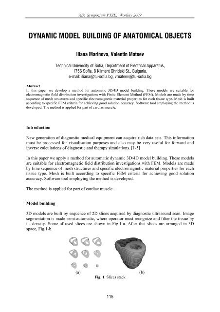
3D models are built by sequence of 2D slices acquired by diagnostic ultrasound scan. Image<br />
segmentation is made semi-automatic, where operator must recognize and filter the tissue by<br />
its density. Some of used slices are shown in Fig.1-a. After that slices are arranged in 3D<br />
space, Fig.1-b.<br />
(a) (b)<br />
Fig. 1. Slices stack<br />
115

