Scleral fixation without conjunctival dissection - Drs. Fine, Hoffman ...
Scleral fixation without conjunctival dissection - Drs. Fine, Hoffman ...
Scleral fixation without conjunctival dissection - Drs. Fine, Hoffman ...

You also want an ePaper? Increase the reach of your titles
YUMPU automatically turns print PDFs into web optimized ePapers that Google loves.
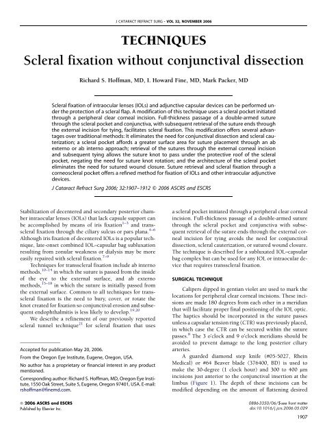
TECHNIQUES<br />
<strong>Scleral</strong> <strong>fixation</strong> <strong>without</strong> <strong>conjunctival</strong> <strong>dissection</strong><br />
Richard S. <strong>Hoffman</strong>, MD, I. Howard <strong>Fine</strong>, MD, Mark Packer, MD<br />
<strong>Scleral</strong> <strong>fixation</strong> of intraocular lenses (IOLs) and adjunctive capsular devices can be performed under<br />
the protection of a scleral flap. A modification of this technique uses a scleral pocket initiated<br />
through a peripheral clear corneal incision. Full-thickness passage of a double-armed suture<br />
through the scleral pocket and conjunctiva, with subsequent retrieval of the suture ends through<br />
the external incision for tying, facilitates scleral <strong>fixation</strong>. This modification offers several advantages<br />
over traditional methods: It eliminates the need for <strong>conjunctival</strong> <strong>dissection</strong> and scleral cauterization;<br />
a scleral pocket affords a greater surface area for suture placement through an ab<br />
externo or ab interno approach; retrieval of the sutures through the external corneal incision<br />
and subsequent tying allows the suture knot to pass under the protective roof of the scleral<br />
pocket, negating the need for suture knot rotation; and the architecture of the scleral pocket<br />
eliminates the need for sutured wound closure. Suture retrieval and scleral <strong>fixation</strong> through a<br />
corneoscleral pocket offers a refined method for <strong>fixation</strong> of IOLs and other intraocular adjunctive<br />
devices.<br />
J Cataract Refract Surg 2006; 32:1907–1912 Q 2006 ASCRS and ESCRS<br />
Stabilization of decentered and secondary posterior chamber<br />
intraocular lenses (IOLs) that lack capsule support can<br />
be accomplished by means of iris <strong>fixation</strong> 1–3 and transscleral<br />
<strong>fixation</strong> through the ciliary sulcus or pars plana. 4–6<br />
Although iris <strong>fixation</strong> of decentered IOLs is a popular technique,<br />
late-onset combined IOL–capsular bag subluxation<br />
resulting from zonular weakness or dialysis may be more<br />
easily repaired with scleral <strong>fixation</strong>. 7–9<br />
Techniques for transscleral <strong>fixation</strong> include ab interno<br />
methods, 10–14 in which the suture is passed from the inside<br />
of the eye to the external surface, and ab externo<br />
methods, 15–18 in which the suture is initially passed from<br />
the external surface. Common to all techniques for transscleral<br />
<strong>fixation</strong> is the need to bury, cover, or rotate the<br />
knot created for <strong>fixation</strong> so <strong>conjunctival</strong> erosion and subsequent<br />
endophthalmitis is less likely to develop. 19,20<br />
We describe a refinement of our previously reported<br />
scleral tunnel technique 21 for scleral <strong>fixation</strong> that uses<br />
Accepted for publication May 20, 2006.<br />
From the Oregon Eye Institute, Eugene, Oregon, USA.<br />
No author has a proprietary or financial interest in any product<br />
mentioned.<br />
Corresponding author: Richard S. <strong>Hoffman</strong>, MD, Oregon Eye Institute,<br />
1550 Oak Street, Suite 5, Eugene, Oregon 97401, USA. E-mail:<br />
rshoffman@finemd.com.<br />
Q 2006 ASCRS and ESCRS<br />
Published by Elsevier Inc.<br />
J CATARACT REFRACT SURG - VOL 32, NOVEMBER 2006<br />
a scleral pocket initiated through a peripheral clear corneal<br />
incision. Full-thickness passage of a double-armed suture<br />
through the scleral pocket and conjunctiva with subsequent<br />
retrieval of the suture ends through the external corneal<br />
incision for tying avoids the need for <strong>conjunctival</strong><br />
<strong>dissection</strong>, scleral cauterization, or sutured wound closure.<br />
The technique is described for a subluxated IOL–capsular<br />
bag complex but can be used for any IOL or intraocular device<br />
that requires transscleral <strong>fixation</strong>.<br />
SURGICAL TECHNIQUE<br />
Calipers dipped in gentian violet are used to mark the<br />
locations for peripheral clear corneal incisions. These incisions<br />
are made 180 degrees from each other in a meridian<br />
that will facilitate proper final positioning of the IOL optic.<br />
The haptics should be incorporated in the suture passes<br />
unless a capsular tension ring (CTR) was previously placed,<br />
in which case the CTR can be secured within the suture<br />
passes. 8 The 3 o’clock and 9 o’clock meridians should be<br />
avoided to prevent damage to the long posterior ciliary<br />
arteries.<br />
A guarded diamond step knife (#05-5027, Rhein<br />
Medical) or #64 Beaver blade (376400, BD) is used to<br />
make the 30-degree (1 clock hour) and 300 to 400 mm<br />
incisions just anterior to the <strong>conjunctival</strong> insertion at the<br />
limbus (Figure 1). The depth of these incisions can be<br />
modified depending on the amount of flattening desired<br />
0886-3350/06/$-see front matter<br />
doi:10.1016/j.jcrs.2006.05.029<br />
1907
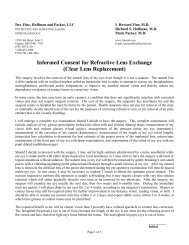
Figure 1. Subluxated IOL–capsular bag complex containing Soemmering’s<br />
ring. Two 30-degree (1 clock hour) and 300 to 400 mm clear corneal<br />
incisions are made 180 degrees apart with a diamond step knife. The incisions<br />
are placed in a meridian that will allow <strong>fixation</strong> of the IOL haptics to<br />
the sclera.<br />
in the meridian. Two scleral pockets are then dissected<br />
posteriorly from the 2 opposing incisions using a diamond<br />
crescent knife (#60505 Mastel Precision) or a metal crescent<br />
blade (990002 A-OK, Alcon Laboratories) (Figure 2).<br />
The pockets are extended approximately 3.0 mm posteriorly<br />
from the clear corneal incisions.<br />
A 1.0 mm paracentesis is created from each clear corneal<br />
incision into the anterior chamber to aid in suture<br />
placement. Initiating the paracentesis just anterior to the<br />
clear corneal incision instead of within the incision will facilitate<br />
passing the polypropylene (Prolene) sutures since<br />
the external opening of the paracentesis can be more easily<br />
identified. The paracentesis can also be placed immediately<br />
adjacent to the clear corneal incision. The 1.0 mm paracenteses<br />
can be used to place single iris hooks to expose<br />
the peripheral capsular bag or concealed IOL haptics. A<br />
small quantity of ophthalmic viscosurgical device (OVD)<br />
is placed in the anterior chamber through 1 paracentesis<br />
to stabilize the anterior chamber. An OVD can also be<br />
placed in the ciliary sulcus underlying the scleral pocket<br />
to aid the suture passes.<br />
Suture placement is initially directed toward the haptic<br />
that was exposed through the pupil secondary to the IOL<br />
decentration. A 27-gauge needle is passed through the conjunctiva<br />
and the full thickness of the scleral pocket 1.0 mm<br />
posterior to the surgical limbus. This needle is inserted into<br />
1908<br />
TECHNIQUES: SCLERAL FIXATION WITHOUT CONJUNCTIVAL DISSECTION<br />
J CATARACT REFRACT SURG - VOL 32, NOVEMBER 2006<br />
Figure 2. Posterior <strong>dissection</strong> of scleral pockets using a diamond crescent<br />
blade. Note the paracentesis originating anterior to the clear corneal<br />
incision.<br />
the eye, behind the iris and in front of the capsular bag far<br />
enough to allow visualization of the beveled tip. A doublearmed<br />
10-0 Prolene suture on a long straight needle (STC-<br />
6, Ethicon) is inserted through the opposite paracentesis,<br />
docked into the 27-gauge needle (Figure 3), and both are<br />
removed externally through the scleral pocket and the conjunctiva.<br />
(A double-armed 9-0 Prolene suture on a long<br />
curved needle [D-8229 CTC-6L, Ethicon] is preferable to<br />
postpone eventual suture degradation but may be difficult<br />
to acquire.) The 27-gauge needle is again passed through<br />
the conjunctiva and the full thickness of the scleral pocket<br />
1.0 mm posterior to the surgical limbus and 1.0 to 2.0 mm<br />
adjacent to the first pass of the needle. This 27-gauge needle<br />
is inserted into the eye but behind the capsular bag<br />
equator. The needle perforates the capsular bag central to<br />
the IOL haptic and passes completely through the posterior<br />
and anterior capsules. The second arm of the double-armed<br />
Prolene suture is passed through the opposite paracentesis<br />
and docked with the 27-gauge needle; both are again removed<br />
through the full thickness of the eye (Figure 4).<br />
At this point, all suture passes are through the full<br />
thickness of the sclera at the ciliary sulcus. By removing<br />
the needles from all suture passes, each suture end can be<br />
retrieved through the scleral pocket opening by passing<br />
a Sinskey hook into the pocket and pulling the trailing suture<br />
end through the corneal incision so the sutures now
Figure 3. Docking the Prolene suture needle into a 27-gauge hollow needle<br />
above the capsular bag. The suture needle is passed through the<br />
1.0 mm paracentesis. The 27-gauge needle is passed into the eye through<br />
the conjunctiva and the scleral pocket 1.0 mm posterior to the surgical<br />
limbus.<br />
pass through the corneal incision, through the floor of the<br />
scleral pocket (1.0 mm posterior to the surgical limbus),<br />
and into the eye through the ciliary sulcus. When the sutures<br />
are retrieved through the corneal incision, the other<br />
suture of the double-armed pass should be held with a forceps<br />
to prevent pulling the suture end out of the eye inadvertently<br />
(Figure 5).<br />
Tying the suture ends recenters the IOL and allows the<br />
knot to be concealed as it slides under the protective roof of<br />
the scleral pocket.<br />
The same technique can be performed on the opposite<br />
haptic using the second scleral pocket and the opposing<br />
paracentesis (Figure 6). An iris hook can be placed in the first<br />
paracentesis to aid visualization of the capsular bag equator<br />
and lens haptic for the second <strong>fixation</strong> site. Suturing the<br />
scleral pockets is not necessary. The OVD can be removed<br />
by injecting acetylcholine hydrochloride (Miochol-E) into<br />
the anterior chamber while depressing the posterior lip of<br />
one of the paracenteses or with bimanual irrigation and<br />
aspiration cannulas inserted into both paracenteses.<br />
DISCUSSION<br />
Numerous methods are currently used for transscleral<br />
<strong>fixation</strong> of IOLs and adjunctive surgical devices. 22 Common<br />
to these techniques is the requirement for <strong>conjunctival</strong><br />
TECHNIQUES: SCLERAL FIXATION WITHOUT CONJUNCTIVAL DISSECTION<br />
Figure 4. Second arm of the double-armed Prolene suture is inserted<br />
through the paracentesis and docked with a second 27-gauge needle<br />
that has perforated the capsular bag central to the exposed haptic.<br />
<strong>dissection</strong> and the need to prevent suture knot erosion of<br />
the overlying conjunctiva with the ensuing risk for endophthalmitis.<br />
Existing methods for knot concealment include<br />
covering the knot with a patch graft, 23 fascia lata, 24 or a triangular<br />
scleral flap, 11,15,25–28 in addition to suturing within<br />
Figure 5. Following the second pass of the double-armed suture, the needles<br />
are removed and the suture ends are retrieved through the scleral<br />
pocket incision using a Sinskey hook. Note that the left suture has been<br />
retrieved and is being held with a forceps to avoid inadvertent suture<br />
loss during retrieval of the right suture.<br />
J CATARACT REFRACT SURG - VOL 32, NOVEMBER 2006 1909
Figure 6. Prolene sutures for each haptic are tied, allowing the knot to<br />
slide under the roof of the scleral pocket.<br />
a scleral groove 29–31 and suture knot rotation into the<br />
eye. 32–34<br />
All these techniques have limitations. <strong>Scleral</strong> patch<br />
grafts and fascia lata coverings require additional procurement<br />
of tissue from eye banks or the patient’s body and add<br />
unnecessary time to the procedure. Use of a triangular<br />
scleral flap necessitates extremely accurate suture placement<br />
when using an ab interno technique to ensure the suture<br />
passes through the floor of the <strong>dissection</strong>. Similarly, the<br />
scleral groove technique can be used for ab externo suture<br />
passes but by nature of the limited groove area, it cannot be<br />
1910<br />
TECHNIQUES: SCLERAL FIXATION WITHOUT CONJUNCTIVAL DISSECTION<br />
J CATARACT REFRACT SURG - VOL 32, NOVEMBER 2006<br />
used effectively with an ab interno method. Rotation of fullthickness<br />
scleral suture knots can be impeded by short suture<br />
passes and may be more difficult with the larger knots<br />
that result from currently recommended thicker 9-0 Prolene<br />
and 8-0 Gore-Tex suture gauges. 35,36<br />
There are several advantages of the scleral pocket technique<br />
for scleral <strong>fixation</strong>. First, a larger surface area can be<br />
created for suture passes than with triangular scleral flaps<br />
or scleral grooves. This allows the suture needles to exit<br />
anywhere inside the large dissected pocket as long as they<br />
are at the appropriate distance from the surgical limbus<br />
(0.5 to 1.0 mm for ciliary sulcus <strong>fixation</strong> 37 ). This is especially<br />
useful when using an ab interno approach. Second,<br />
<strong>dissection</strong> of the scleral pocket initiated from a clear corneal<br />
incision avoids the need for <strong>conjunctival</strong> <strong>dissection</strong><br />
or scleral cautery. This should induce less discomfort in patients<br />
having procedures with topical anesthesia in which<br />
unforeseen complications may necessitate use of scleral fixated<br />
lenses or fixated capsular bag prostheses. The <strong>dissection</strong><br />
of the distal scleral pocket is also easier to perform<br />
than a triangular flap in the distal location since the <strong>dissection</strong><br />
can proceed directed away from the surgeon in<br />
a slightly ‘‘downhill’’ direction. In addition, the procedure<br />
can be expedited relative to a triangular flap technique<br />
since <strong>conjunctival</strong> <strong>dissection</strong> is avoided and sutured wound<br />
closure is unnecessary. Finally, less astigmatism may be induced<br />
than with the placement of 2 radial sutures through<br />
each of 2 opposing triangular flaps in the same meridian.<br />
Although 2 opposed 30-degree vertical clear corneal incisions<br />
have a small flattening effect in the meridian of<br />
placement (Figure 7), the small arc length and relatively superficial<br />
depth compared with traditional limbal relaxing<br />
incisions induce little astigmatic effect and this can be modified<br />
by using more superficial 300 mm incisions, depending<br />
on the desired astigmatic result.<br />
Figure 7. Two weeks postoperatively, surgically induced<br />
astigmatism of 0.75 diopter resulting from<br />
two 30-degree 400 mm clear corneal incisions and<br />
scleral pockets placed at the 105/285 axis. The patient<br />
was an 81-year-old man with pseudoexfoliation and<br />
a subluxated IOL–capsular bag complex.
<strong>Scleral</strong> <strong>fixation</strong> of secondary IOLs within a scleral<br />
pocket does require 2 suture passes through the sclera for<br />
each haptic. This has the disadvantage of creating twice<br />
as many potential adverse bleeding events compared with<br />
a suturing technique in which a single suture is passed<br />
and the suture is tied to itself in the dissected bed of a triangular<br />
flap. However, the double-pass technique has the advantage<br />
of 4-point <strong>fixation</strong>, which should improve the<br />
incidence of lens tilt. 38<br />
Use of a scleral pocket with hook retrieval of the suture<br />
ends can be performed for any procedure requiring transscleral<br />
<strong>fixation</strong>. This includes implantation of secondary<br />
IOLs, repair of dislocated IOLs, 7–9,39,40 use of adjunctive<br />
surgical devices such as Ahmed capsular tension segments<br />
and Cionni capsular tension rings, 41 and repair of iridodialyses.<br />
42–45 This modification of the traditional scleral flap allows<br />
simpler creation of a scleral covering, negating the<br />
need to rotate suture knots while facilitating needle placement<br />
for an ab interno or ab externo technique.<br />
REFERENCES<br />
1. McCannel MA. A retrievable suture idea for anterior uveal problems.<br />
Ophthalmic Surg 1976; 7(2):98–103<br />
2. Ashraf MF, Stark WJ. McCannel sutures and secondary iris-fixated<br />
intraocular lenses. In: Azar DT, ed, Intraocular Lenses In Cataract and<br />
Refractive Surgery. Philadelphia, PA, WB Saunders, 2001; 165–170<br />
3. Chang DF. Siepser slipknot for McCannel iris-suture <strong>fixation</strong> of subluxated<br />
intraocular lenses. J Cataract Refract Surg 2004; 30:1170–1176<br />
4. Nakashizuka H, Shimada H, Iwasaki Y, et al. Pars plana suture <strong>fixation</strong><br />
for intraocular lenses dislocated into the vitreous cavity using a closedeye<br />
cow-hitch technique. J Cataract Refract Surg 2004; 30:302–306<br />
5. Teichmann KD. Pars plana <strong>fixation</strong> of posterior chamber intraocular<br />
lenses. Ophthalmic Surg 1994; 25:549–553<br />
6. Girard LJ. Pars plana phacoprosthesis (aphakia intraocular implant):<br />
a preliminary report. Ophthalmic Surg 1981; 12:19–22<br />
7. Moreno-Montañés J, Heras H, Fernández-Hortelano A. Surgical treatment<br />
of a dislocated intraocular lens–capsular bag–capsular tension<br />
ring complex. J Cataract Refract Surg 2005; 31:270–273<br />
8. Gross JG, Kokame GT, Weinberg DV. In-the-bag intraocular lens dislocation;<br />
the Dislocated In-The-Bag Intraocular Lens Study Group. Am J<br />
Ophthalmol 2004; 137:630–635<br />
9. Jehan FS, Mamalis N, Crandall AS. Spontaneous late dislocation of intraocular<br />
lens within the capsular bag in pseudoexfoliation patients.<br />
Ophthalmology 2001; 108:1727–1731<br />
10. Smiddy WE, Sawusch MR, O’Brien TP, et al. Implantation of scleralfixated<br />
posterior chamber intraocular lenses. J Cataract Refract Surg<br />
1990; 16:691–696<br />
11. Grigorian R, Chang J, Zarbin M, Del Priore L. A new technique for<br />
suture <strong>fixation</strong> of posterior chamber intraocular lenses that eliminates<br />
intraocular knots. Ophthalmology 2003; 110:1349–1356<br />
12. Apple DJ, Price FW, Gwin T, et al. Sutured retropupillary posterior<br />
chamber intraocular lenses for exchange or secondary implantation;<br />
the 12th Annual Binkhorst Lecture, 1988. Ophthalmology 1989;<br />
96:1241–1247<br />
13. Kumar M, Arora R, Sanga L, Sota LD. <strong>Scleral</strong>-fixated intraocular lens<br />
implantation in unilateral aphakic children. Ophthalmology 1999; 21:<br />
2184–2189<br />
TECHNIQUES: SCLERAL FIXATION WITHOUT CONJUNCTIVAL DISSECTION<br />
14. Sharpe MR, Biglan AW, Gerontis CC. <strong>Scleral</strong> <strong>fixation</strong> of posterior chamber<br />
intraocular lenses in children. Ophthalmic Surg Lasers 1996;<br />
27:337–341<br />
15. Lewis JS. Ab externo sulcus <strong>fixation</strong>. Ophthalmic Surg 1991; 22:692–695<br />
16. Eryildirim A. Knotless scleral <strong>fixation</strong> for implanting a posterior chamber<br />
intraocular lens. Ophthalmic Surg 1995; 26:82–84<br />
17. Shapiro A, Leen MM. External transscleral posterior chamber lens<br />
<strong>fixation</strong>. Arch Ophthalmol 1991; 109:1759–1760<br />
18. Horiguchi M, Hirose H, Koura T, Satou M. Identifying the ciliary sulcus<br />
for suturing a posterior chamber intraocular lens by transillumination.<br />
Arch Ophthalmol 1993; 111:1693–1695<br />
19. Heilskov T, Joondeph BC, Olsen KR, Blankenship GW. Late endophthalmitis<br />
after transscleral <strong>fixation</strong> of a posterior chamber intraocular lens.<br />
Arch Ophthalmol 1989; 107:1427<br />
20. Schechter RJ. Suture-wick endophthalmitis with sutured posterior<br />
chamber intraocular lenses. J Cataract Refract Surg 1990; 16:755–756<br />
21. <strong>Hoffman</strong> RS, <strong>Fine</strong> IH, Packer M, Rozenberg I. <strong>Scleral</strong> <strong>fixation</strong> utilizing<br />
suture retrieval through a scleral tunnel. J Cataract Refract Surg<br />
2006; 32:1259–1263<br />
22. Por YM, Lavin MJ. Techniques of intraocular lens suspension in the absence<br />
of capsular/zonular support. Surv Ophthalmol 2005; 50:429–462<br />
23. Bucci FA Jr, Holland EJ, Lindstrom RL. Corneal autografts for external<br />
knots in transsclerally sutured posterior chamber lenses [letter]. Am J<br />
Ophthalmol 1991; 112:353–354<br />
24. Bashshur Z, Ma’luf R, Najjar D, Noureddin B. <strong>Scleral</strong> <strong>fixation</strong> of posterior<br />
chamber intraocular lenses using fascia lata to cover the knots.<br />
Ophthalmic Surg Lasers 2002; 33:445–449<br />
25. Rao SK, Gopal L, Fogla R, et al. Ab externo 4-point scleral <strong>fixation</strong><br />
[letter]. J Cataract Refract Surg 2000; 26:9–10<br />
26. Ramocki JM, Shin DH, Glover BK, et al. Foldable posterior chamber intraocular<br />
lens implantation in the absence of capsular and zonular<br />
support. Am J Ophthalmol 1999; 127:213–216<br />
27. Basti S, Tejaswi PC, Singh SK, Sekhar GC. Outside-in transscleral <strong>fixation</strong><br />
for ciliary sulcus intraocular lens placement. J Cataract Refract<br />
Surg 1994; 20:89–92<br />
28. Hu BV, Shin DH, Gibbs KA, Hong YJ. Implantation of posterior chamber<br />
lens in the absence of capsular and zonular support. Arch Ophthalmol<br />
1988; 106:416–420<br />
29. Bergren RL. Four-point <strong>fixation</strong> technique for sutured posterior chamber<br />
intraocular lenses. Arch Ophthalmol 1994; 112:1485–1487<br />
30. Friedberg MA, Berler DK. <strong>Scleral</strong> <strong>fixation</strong> of posterior chamber intraocular<br />
lens implants combined with vitrectomy. Ophthalmic Surg 1992;<br />
23:17–21<br />
31. Lin C-P, Tseng H-Y. Suture <strong>fixation</strong> technique for posterior chamber<br />
intraocular lenses. J Cataract Refract Surg 2004; 30:1401–1404<br />
32. Lewis JS. Sulcus <strong>fixation</strong> <strong>without</strong> flaps. Ophthalmology 1993; 100:<br />
1346–1350<br />
33. Buckley EG. <strong>Scleral</strong> fixated (sutured) posterior chamber intraocular<br />
lens implantation in children. J AAPOS 1999; 3:289–294<br />
34. CordovésL,Gómez A, Mesa CG, Abreu JA. Sulcus transscleral sutured<br />
posterior chamber lenses [letter]. J Cataract Refract Surg 1999;<br />
25:156–157<br />
35. Price MO, Price FW Jr, Werner L, et al. Late dislocation of scleralsutured<br />
posterior chamber intraocular lenses. J Cataract Refract<br />
Surg 2005; 31:1320–1326<br />
36. Cionni RJ, Osher RH, Marques DMV, et al. Modified capsular tension<br />
ring for patients with congenital loss of zonular support. J Cataract Refract<br />
Surg 2003; 29:1668–1673<br />
37. Duffey RJ, Holland EJ, Agapitos PJ, Lindstrom RL. Anatomic study of<br />
transsclerally sutured intraocular lens implantation. Am J Ophthalmol<br />
1989; 108:300–309<br />
38. Teichmann KD, Teichmann IAM. The torque and tilt gamble. J Cataract<br />
Refract Surg 1997; 23:413–418<br />
J CATARACT REFRACT SURG - VOL 32, NOVEMBER 2006 1911
39. Ahmed IIK, Chen SH, Kranemann C, Wong DT. Surgical repositioning<br />
of dislocated capsular tension rings. Ophthalmology 2005; 112:<br />
1725–1733<br />
40. Koh HJ, Kim CY, Lim SJ, Kwon OW. <strong>Scleral</strong> <strong>fixation</strong> technique using 2<br />
corneal tunnels for a dislocated intraocular lens. J Cataract Refract<br />
Surg 2000; 26:1439–1441<br />
41. Cionni RJ, Osher RH. Management of profound zonular dialysis or<br />
weakness with a new endocapsular ring designed for scleral <strong>fixation</strong>.<br />
J Cataract Refract Surg 1998; 24:1299–1306<br />
1912<br />
TECHNIQUES: SCLERAL FIXATION WITHOUT CONJUNCTIVAL DISSECTION<br />
J CATARACT REFRACT SURG - VOL 32, NOVEMBER 2006<br />
42. Erakgun T, Kaskaloglu M, Kayikcioglu O. A simple closed chamber<br />
technique for repair of traumatic iridodialysis in phakic eyes. Ophthalmic<br />
Surg Lasers 2001; 32:83–85<br />
43. Brown SM. A technique for repair of iridodialysis in children. J AAPOS<br />
1998; 2:380–382<br />
44. Kaufman SC, Insler MS. Surgical repair of a traumatic iridodialysis.<br />
Ophthalmic Surg Lasers 1996; 27:963–966<br />
45. Kervick GN, Johnston SS. Repair of inferior iridodialysis using a partialthickness<br />
scleral flap. Ophthalmic Surg 1991; 22:354–355