SensoLyte Green SIRT1 Assay Kit - Eurogentec
SensoLyte Green SIRT1 Assay Kit - Eurogentec
SensoLyte Green SIRT1 Assay Kit - Eurogentec

You also want an ePaper? Increase the reach of your titles
YUMPU automatically turns print PDFs into web optimized ePapers that Google loves.
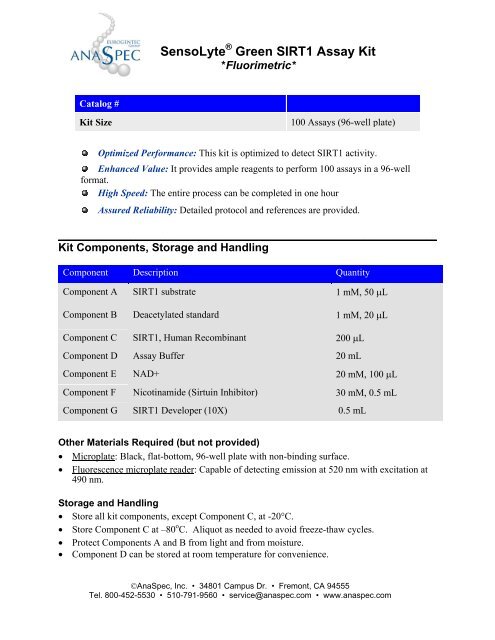
Catalog #<br />
<strong>SensoLyte</strong> ® <strong>Green</strong> <strong>SIRT1</strong> <strong>Assay</strong> <strong>Kit</strong><br />
*Fluorimetric*<br />
<strong>Kit</strong> Size 100 <strong>Assay</strong>s (96-well plate)<br />
Optimized Performance: This kit is optimized to detect <strong>SIRT1</strong> activity.<br />
Enhanced Value: It provides ample reagents to perform 100 assays in a 96-well<br />
format.<br />
High Speed: The entire process can be completed in one hour<br />
Assured Reliability: Detailed protocol and references are provided.<br />
______________________________________________________________________________<br />
<strong>Kit</strong> Components, Storage and Handling<br />
Component Description Quantity<br />
Component A <strong>SIRT1</strong> substrate 1 mM, 50 μL<br />
Component B Deacetylated standard 1 mM, 20 μL<br />
Component C <strong>SIRT1</strong>, Human Recombinant 200 μL<br />
Component D <strong>Assay</strong> Buffer 20 mL<br />
Component E NAD+ 20 mM, 100 μL<br />
Component F Nicotinamide (Sirtuin Inhibitor) 30 mΜ, 0.5 mL<br />
Component G <strong>SIRT1</strong> Developer (10X) 0.5 mL<br />
Other Materials Required (but not provided)<br />
• Microplate: Black, flat-bottom, 96-well plate with non-binding surface.<br />
• Fluorescence microplate reader: Capable of detecting emission at 520 nm with excitation at<br />
490 nm.<br />
Storage and Handling<br />
• Store all kit components, except Component C, at -20°C.<br />
• Store Component C at –80 o C. Aliquot as needed to avoid freeze-thaw cycles.<br />
• Protect Components A and B from light and from moisture.<br />
• Component D can be stored at room temperature for convenience.<br />
©AnaSpec, Inc. • 34801 Campus Dr. • Fremont, CA 94555<br />
Tel. 800-452-5530 • 510-791-9560 • service@anaspec.com • www.anaspec.com
____________________________________________________________________________<br />
Introduction<br />
Histone deacetylases (HDACs) act as transcriptional repressors of genes catalyzing the<br />
removal of acetyl groups from a ε-N-acetyl lysine of histone. 1 Sirtuins comprise a unique class of<br />
nicotinamide adenine dinucleotide (NAD+)-dependent deacetylases (class III HDACs) that target<br />
multiple protein substrates to execute diverse biological functions. Sirtuins catalyze a reaction<br />
that couples lysine deacetylation to NAD hydrolysis, yielding O-acetyl-ADP-ribose and<br />
nicotinamide. 2<br />
Sirtuin 1 (<strong>SIRT1</strong>), the human homolog of yeast Sir2 (Silent Information Regulator 2), is<br />
the most studied of the seven members of sirtuin family. <strong>SIRT1</strong> have been implicated in several<br />
important cellular processes, including genomic stability and DNA repair, 3,4 p53-mediated<br />
apoptosis, 5 adipogenesis, 6 and aging.<br />
7, 8<br />
The <strong>SensoLyte</strong> ® <strong>Green</strong> <strong>SIRT1</strong> <strong>Assay</strong> <strong>Kit</strong> provides a convenient, two-step homogeneous<br />
procedure for measuring sirtuin 1 activity and screening of enzyme inhibitors and activators. The<br />
fluorogenic peptide substrate in this kit is derived from p53 sequence. In the first step, an<br />
acetylated substrate is incubated with sirtuin-containing samples. Deacetylation of substrate<br />
sensitizes it to the sirtuin developer, which, in the second step, releases the green fluorophore.<br />
Fluorescence produced is proportional to <strong>SIRT1</strong> activity and can be detected with excitation at<br />
490 nm and emission at 520 nm. Deacetylation of the <strong>SIRT1</strong> fluorogenic substrate by other<br />
members of sirtuin family, such as sirtuin 2 and sirtuin 3, is negligible.<br />
_____________________________________________________________________________<br />
Protocol<br />
Note 1: For standard curve, please refer to Appendix.<br />
Note 2: Avoid protease inhibitors in the samples.<br />
1. Prepare working solutions.<br />
Note: Bring all kit components to room temperature before starting the experiment. Component C should<br />
be kept on ice after thawing.<br />
1.1 <strong>SIRT1</strong> substrate solution: Dilute <strong>SIRT1</strong> substrate (Component A) and NAD+<br />
(Component E) in assay buffer (Component D). Both <strong>SIRT1</strong> substrate and<br />
NAD+ should be diluted in assay buffer 100-fold. For each experiment, prepare<br />
fresh substrate solution.<br />
Table 1. <strong>SIRT1</strong> substrate solution for one 96-well plate (100 assays)<br />
Components Volume<br />
<strong>SIRT1</strong> substrate (100X, Component A) 50 μL<br />
NAD+ (Component E) 50 μL<br />
<strong>Assay</strong> buffer (Component D) 4.9 mL<br />
Total volume 5 mL<br />
1.2 <strong>SIRT1</strong> diluent: Dilute <strong>SIRT1</strong> (Component C) 20-fold in assay buffer (Component D).<br />
This amount of enzyme is enough for a full 96-well plate. If not using the entire plate,<br />
adjust the amount of enzyme to be diluted accordingly.<br />
©AnaSpec, Inc. • 34801 Campus Dr. • Fremont, CA 94555<br />
Tel. 800-452-5530 • 510-791-9560 • service@anaspec.com • www.anaspec.com
Note: Prepare enzyme diluents immediately before use. Do not vortex the enzyme solutions. Prolonged<br />
storage or vigorous agitation of the diluted enzyme will cause denaturation. Store the enzyme solution on<br />
ice.<br />
1.3 1X developer: Dilute the developer (Component G) and the nicotinamide (Component<br />
F) in assay buffer (Component D). Both developer and nicotinamide should be diluted<br />
10-fold in assay buffer. Each assay requires 50 μL of developer solution.<br />
Table 2. 1X developer solution for one 96-well plate (100 assays)<br />
Components Volume<br />
<strong>SIRT1</strong> developer (10X, Component G) 500 μL<br />
Nicotinamide (Component F) 500 μL<br />
<strong>Assay</strong> buffer (Component D) 4 mL<br />
Total volume 5 mL<br />
Note 1: The developer, containing nicotinamide, is a bi-functional buffer, which works as a stop solution<br />
for <strong>SIRT1</strong> and initiates fluorescent signal releasing fluorophore.<br />
Note 2: Prepare developer before use. Otherwise keep prepared solution on ice until use.<br />
2. Set up the enzymatic reaction.<br />
2.1 Add test compounds and <strong>SIRT1</strong> diluent to the microplate wells. For one well of a 96well<br />
plate, the suggested volume of enzyme solution is 40 μL and 10 μL of test<br />
compound.<br />
2.2 Establish the following control wells at the same time, as deemed necessary:<br />
• Positive control contains <strong>SIRT1</strong> enzyme without test compound.<br />
• Inhibitor/activator control contains <strong>SIRT1</strong> enzyme and <strong>SIRT1</strong> inhibitor/activator.<br />
• Vehicle control contains <strong>SIRT1</strong> enzyme and vehicle used in delivering test<br />
compound (e.g. DMSO, concentration not to exceed 1%).<br />
• Test compound control contains assay buffer (Component D) and test compound.<br />
Some test compounds may themselves be fluorescent and thereby give false results.<br />
Note: Test compound can be additionally tested for interference with developer solution (see Appendix).<br />
• Substrate control contains assay buffer (Component D).<br />
2.3 Using the assay buffer (Component D), bring the total volume of all controls to 50 μL.<br />
2.4 Pre-incubate the plate for 10 min at 37°C.<br />
3. Detect <strong>SIRT1</strong> activity.<br />
3.1 Add 50 μL of the prepared <strong>SIRT1</strong> substrate solution into each well, except the test<br />
compound control wells. Mix the reagents completely by shaking the plate gently for<br />
no more than 30 sec.<br />
3.2 Incubate the plate for 30-60 minutes at 37<br />
©AnaSpec, Inc. • 34801 Campus Dr. • Fremont, CA 94555<br />
Tel. 800-452-5530 • 510-791-9560 • service@anaspec.com • www.anaspec.com<br />
o C.
3.3 Add 50 μL of the prepared developer solution and mix thoroughly.<br />
3.4 Incubate the plate an additional 10 min at 37 o C.<br />
3.5 Measure fluorescence signal at Ex/Em=490 nm/520 nm.<br />
3.6 Data analysis:<br />
• The fluorescence reading from the substrate control well is used as the background<br />
fluorescence. This background reading should be subtracted from the readings of<br />
the other wells containing substrate. All fluorescence readings are expressed in<br />
relative fluorescence units (RFU).<br />
• Plot data as RFU versus concentration of test compounds.<br />
• A variety of data analyses can be done, e.g., determining inhibition %, EC50, IC50,<br />
etc.<br />
Relative activity, %<br />
100<br />
80<br />
60<br />
40<br />
20<br />
0<br />
0.001 0.01 0.1 1 10 100<br />
Sirtuin inhibitor (μM)<br />
Figure 1. Inhibition of <strong>SIRT1</strong> by Ro-<br />
31-8220.<br />
_____________________________________________________________________________<br />
Appendix: Instrument Calibration<br />
• Deacetylated standard: Dilute 1 mM of deacetylated standard (Component B) to 10 μM<br />
in assay buffer (Component D). Do 1:2 serial dilutions to get concentrations of 5, 2.5,<br />
1.25, 0.625, 0.313, and 0.156, include an assay buffer blank. Add 50 μL/well of these<br />
serially diluted standard solutions.<br />
• Add 50 μL/well of the diluted <strong>SIRT1</strong> substrate solution (refer to Protocol, step 1.1 for<br />
preparation).<br />
• Add 50 μL of developer solution (refer to Protocol, step 1.3) to each well. Mix the<br />
reagents by shaking the plate gently for 3 to 5 sec.<br />
• Incubate the plate for an additional 10 min at 37 o C.<br />
• Measure the fluorescence intensity of the reference standard and substrate control wells at<br />
Ex/Em=490 nm/520 nm. Use the same setting of sensitivity as used in the enzyme<br />
reaction.<br />
• Plot the deacetylated reference standard curve as RFU (relative fluorescent units) versus<br />
concentration as shown in Figure 2.<br />
©AnaSpec, Inc. • 34801 Campus Dr. • Fremont, CA 94555<br />
Tel. 800-452-5530 • 510-791-9560 • service@anaspec.com • www.anaspec.com
Note: The concentration of deacetylated reference standard solutions are 5, 2.5, 1.25, 0.625, 0.313, 0.156,<br />
0.078 and 0 μM. This reference standard is used to calibrate the variation of different instruments and<br />
different experiments. It is also an indicator of the amount of enzymatic reaction final product.<br />
• If testing compounds for interference with developer solution, use deacetylated standard<br />
at concentration that provides signal comparable to positive control. After incubation of<br />
deacetylated substrate with assay buffer or test compound, proceed to the addition of<br />
developer solution. This will allow discrimination between <strong>SIRT1</strong> inhibition/activation<br />
versus interference with the developer.<br />
RFU x 1000<br />
100<br />
90<br />
80<br />
70<br />
60<br />
50<br />
40<br />
30<br />
20<br />
10<br />
0<br />
0 1 2 3 4 5 6<br />
Deacetylated standard, μM<br />
Figure 2. Deacetylated reference standard.<br />
Deacetylated standard was serially diluted<br />
with assay buffer containing <strong>SIRT1</strong><br />
substrate, and after 10 min incubation with<br />
developer, fluorescence was recorded at<br />
Ex/Em=490/520 nm. (Flexstation 384II,<br />
Molecular Devices).<br />
____________________________________________________________________________<br />
References:<br />
1. Sterner, DE. et al. Microbiol. Mol. Biol. Rev. 64, 435 (2000).<br />
2. Longo, V and Kennedy, B. Cell. 126, 257 (2006).<br />
3. Yamagata, K and <strong>Kit</strong>abayashi, I. Biochem Biophys Res Commun. 390, 1355 (2009).<br />
4. Wang, RH. et al. Cancer Cell. 14, 312 (2008).<br />
5. Vaziri, H. et al. Cell. 107, 149 (2001).<br />
6. Picard, F. et al. Nature. 429, 771 (2004).<br />
7. Cohen, HY. et al. Science 305, 390 (2004).<br />
8. Trapp, J. and Jung, M. Curr. Drug Target 7, 1553 (2006).<br />
Revised February 12, 2010<br />
©AnaSpec, Inc. • 34801 Campus Dr. • Fremont, CA 94555<br />
Tel. 800-452-5530 • 510-791-9560 • service@anaspec.com • www.anaspec.com