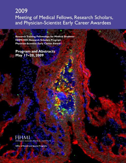
Program and Abstracts May 17–20, 2009 - Howard Hughes Medical ...
Program and Abstracts May 17–20, 2009 - Howard Hughes Medical ...
Program and Abstracts May 17–20, 2009 - Howard Hughes Medical ...

Create successful ePaper yourself
Turn your PDF publications into a flip-book with our unique Google optimized e-Paper software.
<strong>2009</strong><br />
Meeting of <strong>Medical</strong> Fellows, Research Scholars,<br />
<strong>and</strong> Physician-Scientist Early Career Awardees<br />
Research Training Fellowships for <strong>Medical</strong> Students<br />
HHMI-NIH Research Scholars <strong>Program</strong><br />
Physician-Scientist Early Career Award<br />
<strong>Program</strong> <strong>and</strong> <strong>Abstracts</strong><br />
<strong>May</strong> <strong>17–20</strong>, <strong>2009</strong><br />
Office of Grants <strong>and</strong> Special <strong>Program</strong>s
3<br />
4<br />
8<br />
9<br />
15<br />
16<br />
29<br />
103<br />
104<br />
109<br />
113<br />
Introduction<br />
<strong>Program</strong> Schedule<br />
Keynote Speaker<br />
2008 Early Career Awardees’ Biographies<br />
Physician-Scientist Career Panel<br />
Members’ Biographies<br />
Schedule of Presentations<br />
<strong>Abstracts</strong> of Presentations<br />
<strong>Howard</strong> <strong>Hughes</strong> <strong>Medical</strong> Institute<br />
Trustees<br />
Officers<br />
Grants <strong>and</strong> Special <strong>Program</strong>s<br />
Participants<br />
Index of Presentation Times<br />
HHMI Conference Center Map<br />
HHMI Home Page<br />
www.hhmi.org<br />
Grants <strong>and</strong> Special <strong>Program</strong>s<br />
www.hhmi.org/grants<br />
HHMI Scientists <strong>and</strong> Research<br />
www.hhmi.org/research<br />
HHMI News<br />
www.hhmi.org/news<br />
GrantsNet<br />
www.grantsnet.org
Cover: Tissues of the developing heart respond to a coordinated<br />
series of extracellular signals to form the many distinct anatomical<br />
features of the mature organ. Disruption of these signaling networks<br />
contributes to congenital heart disease, the most common class of<br />
birth defects. Among these signals, vascular endothelial growth<br />
factor (VEGF) family members are critically important; however,<br />
their specific temporal roles remain incompletely defined.<br />
Using chemical-induced expression of VEGF inhibitors in transgenic<br />
mouse embryos, we have characterized time windows during<br />
which VEGF has three distinct functions during heart development.<br />
One of these functions is to coordinate cell survival to support<br />
ventricle septation, as expression of an inhibitor of VEGF<br />
signaling (VEGFR2T) at embryonic day 11.5 (E11.5) prevents<br />
endothelialization of the ventricles <strong>and</strong> causes apoptosis specifically<br />
within the interventricular septum. These results provide a framework<br />
to underst<strong>and</strong> how perturbations of VEGF signaling contribute<br />
to congenital heart defects.<br />
The cover image shows immunofluorescent staining of the interventricular<br />
septum of a heart from an E13.5 embryo in which<br />
VEGF signaling has been blocked by the expression of VEGFR2T.<br />
Smooth muscle actin stains myocardial cells (red), PECAM stains<br />
endothelial cells (blue), <strong>and</strong> active caspase-3 stains apoptotic cells<br />
(green). Nuclei (grey) are stained with Hoechst. See abstract on page<br />
35. (Courtesy of Gene Kew Ma, HHMI <strong>Medical</strong> Fellow, Department<br />
of Medicine, Stanford University School of Medicine. Mentor: Ching-<br />
Pin Chang, M.D., Ph.D.)
INTRODUCTION<br />
Welcome to the <strong>2009</strong> Meeting of <strong>Medical</strong> Fellows,<br />
Research Scholars, <strong>and</strong> Physician-Scientist Early<br />
Career Awardees of the <strong>Howard</strong> <strong>Hughes</strong> <strong>Medical</strong><br />
Institute. We are very pleased that participants<br />
from both HHMI medical education programs will<br />
be sharing their research <strong>and</strong> expertise in this one<br />
meeting <strong>and</strong> will be joined by the 2008 awardees of<br />
our Physician-Scientist Early Career Award program.<br />
In 1985, HHMI launched the HHMI-NIH<br />
Research Scholars <strong>Program</strong> in partnership with the<br />
National Institutes of Health to provide outst<strong>and</strong>ing<br />
students from U.S. medical schools with the<br />
opportunity to receive a year of research training<br />
at NIH. Then, in 1989, HHMI established the<br />
Research Training Fellowships for <strong>Medical</strong> Students<br />
<strong>Program</strong> to provide a similar group of students<br />
with research training in leading academic research<br />
laboratories beyond NIH. Recent years have seen<br />
the expansion of both of the programs to include<br />
dental <strong>and</strong> veterinary students, <strong>and</strong> we welcome<br />
their participation.<br />
The Physician-Scientist Early Career Awards<br />
provide five years of research support to selected<br />
alumni of the HHMI Research Training Fellowships<br />
<strong>and</strong> HHMI-NIH Research Scholars <strong>Program</strong> as<br />
they begin their independent academic careers. The<br />
awardees will be giving oral presentations, participating<br />
in a career panel discussion on Monday<br />
evening, <strong>and</strong> co-chairing presentation sessions with<br />
the <strong>Medical</strong> Fellows <strong>and</strong> Research Scholars.<br />
Since the inception of the research training <strong>and</strong><br />
development programs, HHMI has supported more<br />
than 2,000 <strong>Medical</strong> Fellows <strong>and</strong> Research Scholars,<br />
<strong>and</strong> 52 Early Career Awardees. This year, 74 <strong>Medical</strong><br />
Fellows, 49 Research Scholars, <strong>and</strong> 18 Early<br />
Career Awardees will be presenting their research.<br />
This book contains the schedule <strong>and</strong> abstracts of<br />
their presentations.<br />
We are delighted to have Richard P. Lifton, M.D.,<br />
Ph.D., as our honored speaker this year. Dr. Lifton<br />
is an HHMI investigator, chairman of the Department<br />
of Genetics, Sterling Professor of Genetics<br />
<strong>and</strong> Internal Medicine, <strong>and</strong> director of the Yale<br />
Center for Human Genetics <strong>and</strong> Genomics at Yale<br />
School of Medicine. He will discuss his laboratory’s<br />
research using genetic approaches to identify the<br />
genes <strong>and</strong> pathways that contribute to common<br />
human diseases, including cardiovascular, renal, <strong>and</strong><br />
bone disease.<br />
We hope that the meeting will not only be a<br />
time for sharing <strong>and</strong> learning, but also a time for<br />
you to get to know your future physician-scientist<br />
colleagues better. In keeping with this objective,<br />
we have provided several informal opportunities<br />
for you to interact <strong>and</strong> network with each other.<br />
Another way for you to continue your association<br />
with HHMI <strong>and</strong> fellow trainees is through<br />
the HHMI Alumni Network, which comprises<br />
current <strong>and</strong> former awardees. Local networks have<br />
been established in Boston, Northern California,<br />
Washington, D.C./Baltimore, Southern California,<br />
North Carolina, Chicago, Michigan, the Pacific<br />
Northwest, Texas, Clevel<strong>and</strong>, <strong>and</strong> New York City.<br />
We invite you to become involved in the HHMI<br />
alumni group nearest you <strong>and</strong> affiliate with new<br />
groups as you move about the country during your<br />
training <strong>and</strong> early career.<br />
This meeting is held each spring so that you can<br />
present your research <strong>and</strong> exchange ideas. We have<br />
grown accustomed to high-quality work from our<br />
awardees, <strong>and</strong> this year’s presentations, as judged by<br />
the abstracts, will be no exception. We congratulate<br />
you on your scientific accomplishments <strong>and</strong> development,<br />
<strong>and</strong> we want to convey our appreciation<br />
to your mentors <strong>and</strong> preceptors, whose guidance is<br />
clearly evident.<br />
In speaking with numerous alumni of our<br />
medical education programs, we are impressed by<br />
the pivotal effect that this research opportunity has<br />
had on their career development. We hope that you<br />
will view your HHMI research experience similarly<br />
<strong>and</strong> that you will pursue further research <strong>and</strong>, ultimately,<br />
rewarding careers as physician-scientists.<br />
Finally, we are interested in your comments <strong>and</strong><br />
suggestions regarding both this meeting <strong>and</strong> the<br />
<strong>Medical</strong> Fellows, Research Scholars, <strong>and</strong> Physician-<br />
Scientist Early Career Award programs in general.<br />
Please direct your feedback to your respective<br />
program as follows: <strong>Medical</strong> Fellows <strong>Program</strong> to<br />
Melanie Daub at medfellows@hhmi.org; Research<br />
Scholars <strong>Program</strong> to Min Lee at research_scholars<br />
@hhmi.org; <strong>and</strong> Physician-Scientist Early Career<br />
Award to Anh-Chi Le at earlycareer@hhmi.org.<br />
We look forward to hearing about your research<br />
<strong>and</strong> to following your careers in the years ahead.<br />
Robert Tjian, Ph.D., President<br />
Peter J. Bruns, Ph.D., Vice President<br />
Grants <strong>and</strong> Special <strong>Program</strong>s<br />
William R. Galey, Ph.D., Director<br />
Graduate <strong>and</strong> <strong>Medical</strong> Education <strong>Program</strong>s<br />
3
PROGRAM SCHEDULE<br />
<strong>2009</strong> MEETING OF MEDICAL FELLOWS, RESEARCH SCHOLARS,<br />
AND PHYSICIAN-SCIENTIST EARLY CAREER AWARDEES<br />
HHMI HEADQUARTERS AND CONFERENCE CENTER, CHEVY CHASE, MARYLAND<br />
Sunday, <strong>May</strong> 17, <strong>2009</strong><br />
5:30–6:00 p.m. Welcoming Reception, Research Scholars <strong>and</strong> <strong>Medical</strong> Fellows, Great Hall<br />
5:30–7:00 p.m. Dinner, Early Career Awardees, Rathskeller<br />
6:00–7:00 p.m. Dinner, Research Scholars <strong>and</strong> <strong>Medical</strong> Fellows, Dining Room<br />
7:00 p.m. Opening Remarks, Auditorium<br />
William R. Galey, Ph.D., Director, Graduate <strong>and</strong> <strong>Medical</strong> Education <strong>Program</strong>s<br />
<strong>Howard</strong> <strong>Hughes</strong> <strong>Medical</strong> Institute<br />
4<br />
Welcoming Remarks<br />
Peter J. Bruns, Ph.D., Vice President, Grants <strong>and</strong> Special <strong>Program</strong>s<br />
<strong>Howard</strong> <strong>Hughes</strong> <strong>Medical</strong> Institute<br />
Early Career Awardees Introductions<br />
Panel Discussion: Pathway to Becoming a Physician-Scientist<br />
Moderator:<br />
William R. Galey, Ph.D.<br />
Early Career Awardee Panelists:<br />
Ari Green, M.D., University of California, San Francisco, School of Medicine<br />
Regina LaRocque, M.D., Massachusetts General Hospital<br />
Eduardo Méndez, M.D., University of Washington <strong>Medical</strong> Center<br />
Mark Onaitis, M.D., Duke University School of Medicine<br />
Rathskeller open until 10:30 p.m.
Monday, <strong>May</strong> 18, <strong>2009</strong><br />
8:00 a.m. Breakfast, Dining Room<br />
9:00–10:30 a.m. Platform Presentations<br />
Biomedical Engineering, Biochemistry, <strong>and</strong> Bioinformatics, Room D-124<br />
Molecular <strong>and</strong> Cancer Biology, Room D-125<br />
Cancer Biology I, Auditorium<br />
10:30–10:45 a.m. Break, Great Hall<br />
10:45 a.m.–<br />
12:30 p.m. Platform Presentations<br />
Immunology <strong>and</strong> Developmental Biology I, Room D-124<br />
Vascular <strong>and</strong> Cell Biology, Room D-125<br />
Cancer Biology II, Auditorium<br />
12:30 p.m. Lunch, Dining Room <strong>and</strong> Rathskeller<br />
1:30–2:45 p.m. Early Career Awardees’ Plenary Presentations, Auditorium<br />
John T. Chang, M.D., University of Pennsylvania School of Medicine<br />
Yvonne R. Chan, M.D., University of Pittsburgh School of Medicine<br />
Costi Sifri, M.D., University of Virginia Health Sciences Center<br />
Todd A. Fehniger, M.D., Ph.D., Washington University School of Medicine<br />
2:45–3:00 p.m. Break, Great Hall<br />
3:00–4:00 p.m. Poster Session A, Atrium<br />
4:00–5:00 p.m. Poster Session B, Atrium<br />
5:00–5:30 p.m. Reception, Atrium<br />
5:30–7:00 p.m. Dinner, Dining Room<br />
PROGRAM SCHEDULE<br />
7:00 p.m. Keynote Speaker, Auditorium<br />
Richard P. Lifton, M.D., Ph.D., Investigator, <strong>Howard</strong> <strong>Hughes</strong> <strong>Medical</strong> Institute; Chairman<br />
of the Department of Genetics, Sterling Professor of Genetics <strong>and</strong> Internal Medicine,<br />
Director of the Yale Center for Human Genetics <strong>and</strong> Genomics, Yale School of Medicine<br />
Rathskeller open until 10:30 p.m.<br />
<strong>Program</strong> Schedule 5
PROGRAM SCHEDULE<br />
Tuesday, <strong>May</strong> 19, <strong>2009</strong><br />
7:45 a.m. Breakfast, Dining Room<br />
8:45–10:15 a.m. Platform Presentations<br />
Infectious Disease, Room D-124<br />
Cell <strong>and</strong> Developmental Biology, Room D-125<br />
Stem Cell Biology, Auditorium<br />
10:15–10:30 a.m. Break, Great Hall<br />
10:30 a.m.–<br />
Noon Platform Presentations<br />
Immunology III, Room D-124<br />
Neuroscience I, Room D-125<br />
Genetics, Auditorium<br />
Noon Lunch, Research Scholars <strong>and</strong> <strong>Medical</strong> Fellows, Dining Room<br />
Noon Lunch <strong>and</strong> Workshop, Early Career Awardees, Rathskeller<br />
1:30–2:30 p.m. Panel Discussion: Balancing Career <strong>and</strong> Family, Auditorium<br />
Moderators:<br />
Matthew Goldstein, <strong>Medical</strong> Fellow<br />
Mari Johanna Tokita, Research Scholar<br />
Physician-Scientist Panelists:<br />
Donald L. Gilbert, M.D., M.S., Cincinnati Children’s Hospital <strong>Medical</strong> Center<br />
William Matsui, M.D., Sidney Kimmel Comprehensive Cancer Center, Johns Hopkins<br />
University School of Medicine<br />
Christine Seroogy, M.D., University of Wisconsin–Madison School of Medicine <strong>and</strong><br />
Public Health<br />
Jennifer U. Sung, M.D., M.B.A., Wilmer Eye Institute, Johns Hopkins University<br />
2:45 p.m. Depart for Social/Networking Event<br />
3:00–4:30 p.m. Social/Networking Event, National Naval <strong>Medical</strong> Center<br />
5:00 p.m. Return to HHMI Headquarters<br />
Reception, Great Hall<br />
5:30–6:30 p.m. Dinner, Dining Room<br />
6:30–7:30 p.m. Poster Session C, Atrium<br />
7:30–8:30 p.m. Poster Session D, Atrium<br />
Dessert, Atrium<br />
Rathskeller open until 10:30 p.m.<br />
6 <strong>2009</strong> Meeting of <strong>Medical</strong> Fellows, Research Scholars, <strong>and</strong> Physician-Scientist Early Career Awardees
Wednesday, <strong>May</strong> 20, <strong>2009</strong><br />
8:15 a.m. Breakfast, Dining Room<br />
9:15–10:45 a.m. Platform Presentations<br />
Epidemiology <strong>and</strong> Genetics, Room D-124<br />
Neuroscience II, Room D-125<br />
10:45 a.m. <strong>Medical</strong> Fellows’ Assembly, Auditorium<br />
10:45–11:00 a.m. Research Scholars’ <strong>and</strong> Early Career Awardees’ Break, Great Hall<br />
11:00 a.m. Recognition Ceremony, Auditorium<br />
Opening Remarks<br />
William R. Galey, Ph.D.<br />
Director, Graduate <strong>and</strong> <strong>Medical</strong> Education <strong>Program</strong>s<br />
<strong>Howard</strong> <strong>Hughes</strong> <strong>Medical</strong> Institute<br />
Remarks<br />
Peter J. Bruns, Ph.D.<br />
Vice President, Grants <strong>and</strong> Special <strong>Program</strong>s<br />
<strong>Howard</strong> <strong>Hughes</strong> <strong>Medical</strong> Institute<br />
President’s Remarks<br />
Robert Tjian, Ph.D.<br />
President<br />
<strong>Howard</strong> <strong>Hughes</strong> <strong>Medical</strong> Institute<br />
Presentation of Fellows’ Certificates<br />
Noon Lunch, Dining Room <strong>and</strong> Rathskeller<br />
1:30 p.m. Adjournment<br />
PROGRAM SCHEDULE<br />
<strong>Program</strong> Schedule 7
KEYNOTE SPEAKER<br />
Extreme Outliers as Models of<br />
Common Human Disease<br />
RICHARD P. L IFTON, M.D., PH.D.<br />
Investigator, <strong>Howard</strong> <strong>Hughes</strong> <strong>Medical</strong> Institute;<br />
Chairman of the Department of Genetics, Sterling<br />
Professor of Genetics <strong>and</strong> Internal Medicine,<br />
Director of the Yale Center for Human Genetics<br />
<strong>and</strong> Genomics, Yale School of Medicine<br />
■ We have used the investigation of extreme outliers<br />
in the human population to identify genes <strong>and</strong><br />
pathways that contribute to risk of cardiovascular,<br />
renal, <strong>and</strong> bone disease. A major focus of our work<br />
has been on hypertension, a trait that affects more<br />
than a billion people worldwide. This trait has variously<br />
been proposed to be a primary consequence<br />
of abnormalities in diverse organ systems. Our investigation<br />
of thous<strong>and</strong>s of patients from around<br />
the world has identified renal salt h<strong>and</strong>ling as a<br />
principal determinant of long-term blood pressure<br />
homeostasis in humans. Mutations that increase net<br />
renal salt reabsorption raise blood pressure, whereas<br />
mutations that reduce salt reabsorption lower blood<br />
pressure. These mutations identify the elements<br />
that mediate <strong>and</strong> regulate the salt-h<strong>and</strong>ling pathway,<br />
including a novel gene family, the Wnk kinases,<br />
which we have shown regulate the balance<br />
between salt reabsorption <strong>and</strong> potassium secretion.<br />
New sequencing technologies support identification<br />
of rare variants with large effects. These findings<br />
have provided insight into both rare <strong>and</strong> common<br />
forms of blood pressure variation, have modified<br />
therapeutic approaches, <strong>and</strong> have identified new<br />
targets with the potential of having larger beneficial<br />
<strong>and</strong> fewer adverse effects.<br />
8<br />
Paul Fetters<br />
Dr. Lifton is an HHMI<br />
investigator, Chairman<br />
of the Department of<br />
Genetics, Sterling Professor<br />
of Genetics <strong>and</strong> Internal<br />
Medicine, <strong>and</strong> Director<br />
of the Yale Center for<br />
Human Genetics <strong>and</strong><br />
Genomics at Yale School of<br />
Medicine. He received his<br />
Ph.D. in biochemistry<br />
from Stanford University<br />
<strong>and</strong> an M.D. from Stanford University School of<br />
Medicine. Dr. Lifton completed a residency <strong>and</strong> chief<br />
residency in internal medicine at the Brigham <strong>and</strong><br />
Women’s Hospital. His laboratory has used human genetics<br />
<strong>and</strong> genomics to identify causes of heart, kidney,<br />
<strong>and</strong> bone disease. By investigating thous<strong>and</strong>s of families<br />
from around the world, his group has identified more<br />
than 25 human disease genes. These include key genes<br />
<strong>and</strong> pathways that are critical to the risk of hypertension,<br />
stroke, heart attack, <strong>and</strong> osteoporosis. These<br />
studies have provided new diagnostic <strong>and</strong> therapeutic<br />
approaches to these diseases, which affect more than<br />
1 billion people worldwide. Dr. Lifton’s honors include<br />
election to the National Academy of Sciences <strong>and</strong> the<br />
Institute of Medicine, <strong>and</strong> he was awarded the 2008<br />
Wiley Prize in Biomedical Sciences.
2008 EARLY CAREER<br />
AWARDEES’ BIOGRAPHIES<br />
■ Yvonne R. Chan, M.D., is an assistant professor<br />
of medicine in the Division of Pulmonary, Allergy,<br />
<strong>and</strong> Critical Care Medicine (PACCM) at the<br />
University of Pittsburgh. She also regularly attends<br />
on the university’s <strong>Medical</strong> Intensive Care Unit <strong>and</strong><br />
Pulmonary Transplant services. Dr. Chan participated<br />
in the HHMI <strong>Medical</strong> Fellows <strong>Program</strong> in<br />
1997–1998 <strong>and</strong> is a recipient<br />
of the HHMI Continued<br />
Support Award. She received<br />
her M.D. from Harvard<br />
<strong>Medical</strong> School in the<br />
Health Sciences <strong>and</strong> Technology<br />
<strong>Program</strong> in 2001.<br />
She finished her clinical<br />
training in internal medicine<br />
at Mount Auburn Hospital<br />
in Cambridge, Massachusetts, in 2004 <strong>and</strong> went on<br />
to graduate from pulmonary <strong>and</strong> critical care fellowship<br />
at the University of Pittsburgh in 2007. Concurrently,<br />
she completed postdoctoral research<br />
training in pulmonary host defense <strong>and</strong> lung immunology<br />
under the mentorship of Dr. Jay Kolls at<br />
Children’s Hospital of Pittsburgh <strong>and</strong> Dr. Prabir<br />
Ray in the Division of PACCM. Dr. Chan’s research<br />
focuses on innate defense against bacterial pneumonias.<br />
Most recently, she has studied lipocalin 2, an<br />
antimicrobial protein, <strong>and</strong> its mechanism of induction<br />
in bacterial infection. Her current studies involve<br />
characterization of the host signaling response<br />
to lipocalin 2. In addition, her projects include<br />
characterization of host inflammatory immune responses<br />
in chronic bacterial infection <strong>and</strong> colonization<br />
in cystic fibrosis (CF). This bedside-to-bench<br />
translational study characterizes human lung T<br />
cells, isolated from lung explants obtained from<br />
CF patients undergoing transplant. Dr. Chan’s<br />
research characterizes the T cell response to chronic<br />
colonizers in CF, such as Pseudomonas <strong>and</strong><br />
Aspergillus, in an effort to identify culprit immunological<br />
mechanisms responsible for the lung damage<br />
seen in late-stage CF.<br />
■ John T. Chang, M.D., is an instructor of medicine<br />
in the Division of Gastroenterology at the University<br />
of Pennsylvania. He obtained his B.S. degree in<br />
biological sciences from Stanford University <strong>and</strong> his<br />
M.D. from Temple University. During medical<br />
school, he undertook research<br />
training as an HHMI-<br />
NIH Research Scholar from<br />
1997 to 1999 in the laboratory<br />
of Dr. Ethan Shevach.<br />
He completed a residency in<br />
internal medicine <strong>and</strong> a fellowship<br />
in gastroenterology<br />
at the University of Pennsylvania.<br />
While a postdoctoral<br />
fellow in the laboratory of Dr. Steven Reiner, he<br />
found that T cells divide asymmetrically when confronting<br />
microbial pathogens. The discovery of<br />
asymmetric T cell division was recognized as one of<br />
the journal Science’s Top 10 Breakthroughs of 2007.<br />
Dr. Chang’s research focuses on the differentiation<br />
of T lymphocytes during immune responses against<br />
microbial pathogens <strong>and</strong> during autoimmunity.<br />
■ Hyung J. Chun, M.D., is an instructor at Stanford<br />
University School of Medicine. He received his undergraduate<br />
degree in biochemical sciences from<br />
Harvard University. He subsequently received his<br />
M.D. from the Johns Hopkins University School<br />
of Medicine, during which time he participated in<br />
the HHMI-NIH Research<br />
Scholars <strong>Program</strong> from 1999<br />
to 2001, working in the<br />
laboratory of Dr. Michael<br />
Lenardo. His work led to<br />
the identification of a novel<br />
human mutation in the<br />
caspase-8 gene, which leads<br />
to an inherited immunodeficiency<br />
syndrome. His research<br />
also characterized a novel role for caspase-8<br />
in immune activation. He continued his medical<br />
training in internal medicine <strong>and</strong> cardiovascular<br />
medicine at the Stanford University School of<br />
Medicine. Dr. Chun’s current research focuses on<br />
the role of G protein-coupled receptors in the vasculature.<br />
He is interested in characterization of the<br />
apelin-APJ signaling pathway, which he has recently<br />
identified to have an important role in protecting<br />
against vascular injury in rodent models of atherosclerosis<br />
<strong>and</strong> aneurysms.<br />
■ Todd A. Fehniger, M.D., Ph.D., is an assistant professor<br />
of medicine at the Washington University in<br />
St. Louis School of Medicine. He was introduced<br />
to basic <strong>and</strong> translational research<br />
as an HHMI <strong>Medical</strong><br />
Fellow in 1996–1997 studying<br />
natural killer cell modulation<br />
in AIDS-malignancy<br />
patients receiving low-dose<br />
interleukin-2 therapy. He received<br />
his Ph.D. in 2000<br />
<strong>and</strong> his M.D. in 2002 from<br />
Ohio State University, where<br />
he studied the role of cytokine-cytokine receptor<br />
signals in natural killer cell development <strong>and</strong> function.<br />
From 2002 to 2008, he completed a clinical<br />
residency in internal medicine <strong>and</strong> fellowship training<br />
in medical oncology at the Washington University<br />
in St. Louis School of Medicine. As a<br />
postdoctoral fellow from 2005 to 2008, his studies<br />
focused on the cytotoxic effector mechanisms<br />
9
2008 EARLY CAREER<br />
AWARDEES’ BIOGRAPHIES<br />
utilized by lymphocytes to kill tumor <strong>and</strong> virally<br />
infected cells. Dr. Fehniger’s research focuses on investigating<br />
1) the role of microRNAs in regulating<br />
natural killer cell biology <strong>and</strong> 2) approaches to<br />
translate our basic underst<strong>and</strong>ing of natural killer<br />
cells into novel treatments for patients with hematologic<br />
malignancies.<br />
■ Matthew Freedman, M.D., is an assistant professor<br />
in medicine at Harvard <strong>Medical</strong> School <strong>and</strong> at<br />
the Dana-Farber Cancer Institute. He received a<br />
B.S. degree in economics <strong>and</strong> his M.D. from the<br />
University of Michigan. From 1992 to 1993, he<br />
studied human genetics in<br />
the laboratory of Dr. Francis<br />
Collins. He then spent<br />
1993–1994 as an HHMI-<br />
NIH Research Scholar in<br />
Dr. Michael Lenardo’s laboratory.<br />
He completed internship<br />
<strong>and</strong> residency training<br />
in internal medicine at the<br />
University of Michigan<br />
Hospital in 1996. From 1998 to 2002, he was a<br />
fellow in medical oncology in the Dana-Farber/<br />
Partners cancer care training program. From 2000<br />
to 2005, he studied human genetics as an HHMI<br />
Physician Postdoctoral Fellow with Dr. David<br />
Altshuler at Massachusetts General Hospital <strong>and</strong> at<br />
the Whitehead genome center (later changed to<br />
the Broad Institute of Harvard <strong>and</strong> MIT). His laboratory<br />
primarily focuses on underst<strong>and</strong>ing the<br />
functional consequences of inheriting non-protein<br />
coding risk alleles discovered through genomewide<br />
association studies.<br />
■ Timothy E. Graham, M.D., is an instructor in<br />
medicine <strong>and</strong> assistant professor of medicine at<br />
Harvard <strong>Medical</strong> School. He received his B.A.<br />
degree in liberal arts at St. John’s College in<br />
Annapolis, Maryl<strong>and</strong>, in 1990. He received additional<br />
premedical training<br />
at the University of Pennsylvania<br />
from 1991 to 1993;<br />
during that time he learned<br />
basic biochemistry <strong>and</strong> molecular<br />
biology studying heterotrimeric<br />
G proteins in<br />
the laboratory of Dr. David<br />
R. Manning. He received his<br />
M.D. from the University of<br />
New Mexico (UNM) School of Medicine in 1998.<br />
In 1996–1997, he was an HHMI <strong>Medical</strong> Fellow<br />
in the laboratory of Dr. Janet M. Oliver at the<br />
UNM Cancer Research <strong>and</strong> Treatment Center,<br />
where he studied the role of Ras family small<br />
GTPases in immune cell antigen receptor signaling.<br />
In 2002, he completed the Basic Scientist-<br />
10 <strong>2009</strong> Meeting of <strong>Medical</strong> Fellows, Research Scholars, <strong>and</strong> Physician-Scientist Early Career Awardees<br />
Clinician Training <strong>Program</strong> of the American Board<br />
of Internal Medicine at UNM; during this time he<br />
served as a postdoctoral research fellow under the<br />
mentorship of Dr. Richard I. Dorin. In 2003, he<br />
joined the laboratory of Dr. Barbara Kahn at Beth<br />
Israel Deaconess <strong>Medical</strong> Center, where he completed<br />
a clinical fellowship in endocrinology, diabetes,<br />
<strong>and</strong> metabolism <strong>and</strong> began work in the field<br />
of obesity, insulin resistance, <strong>and</strong> type 2 diabetes.<br />
In addition to the HHMI Early Career Award,<br />
he has received the NIDDK Clinical Scientist<br />
Development Award (K08) <strong>and</strong> related R03, the<br />
Doris Duke Charitable Foundation Clinical-<br />
Translational Scientist Award, <strong>and</strong> the Smith<br />
Family/<strong>Medical</strong> Foundation Award.<br />
■ Ari Green, M.D., is the Debbie <strong>and</strong> Andy<br />
Rachleff Distinguished Chair in Neurology, director<br />
of the Neurodiagnostics Center, assistant director<br />
of the <strong>Medical</strong> School Center, <strong>and</strong> assistant<br />
professor at the University of California, San<br />
Francisco, School of Medicine. He is a graduate<br />
of the Duke University School of Medicine <strong>and</strong><br />
was an HHMI <strong>Medical</strong><br />
Fellow in 1999–2000,<br />
in the laboratory of Dr.<br />
Jorge Oksenberg. Dr.<br />
Green completed clinical<br />
training in internal medicine<br />
<strong>and</strong> neurology at the<br />
University of California,<br />
San Francisco, before serving<br />
as co-chief resident in<br />
neurology. He had additional training in clinical<br />
neuroimmunology <strong>and</strong> neuroophthalmology<br />
under the supervision of Dr. Stephen Hauser <strong>and</strong><br />
Dr. William Fletcher Hoyt. He was awarded the<br />
National Multiple Sclerosis Society (NMSS) <strong>and</strong><br />
American Academy of Neurology Foundation<br />
(AANF) Career Fellowship in 2005. Dr. Green’s<br />
primary research interests involve underst<strong>and</strong>ing<br />
the visual system in multiple sclerosis (MS) <strong>and</strong><br />
improving methods for tracking the disease <strong>and</strong><br />
predicting disease course. He is interested in using<br />
advanced retinal imaging <strong>and</strong> electrophysiology to<br />
investigate the retina <strong>and</strong> optic nerve as a model<br />
pathway in MS. This work is intended to help<br />
unravel the relationships between inflammation,<br />
demyelination, <strong>and</strong> neurodegeneration in the disease.<br />
Through collaboration with colleagues at<br />
Queens University Belfast, Dr. Green has advanced<br />
the underst<strong>and</strong>ing of retinal pathology in<br />
MS. He has continued to work on projects aimed<br />
at using retinal imaging to better investigate this<br />
pathology in MS. His laboratory work centers on<br />
using retinal imaging in conjunction with molecular<br />
methods to help improve the underst<strong>and</strong>ing<br />
of axon injury in MS.
■ Fred H. Hsieh, M.D., is on the faculty as a staff<br />
physician at the Clevel<strong>and</strong> Clinic <strong>and</strong> sees allergy<br />
<strong>and</strong> immunology patients, with special emphasis<br />
on patients who suffer from mastocytosis <strong>and</strong> hypereosinophilic<br />
syndromes. He received his M.D.<br />
from the Brown University School of Medicine <strong>and</strong><br />
was an HHMI-NIH Research Scholar in 1992–<br />
1993 with Dr. Michael M.<br />
Gottesman. He subsequently<br />
trained in internal medicine<br />
at the Johns Hopkins<br />
Hospital <strong>and</strong> in allergy <strong>and</strong><br />
immunology at the Brigham<br />
<strong>and</strong> Women’s Hospital,<br />
where he was mentored in<br />
the lab by Drs. K. Frank<br />
Austen <strong>and</strong> Joshua A. Boyce.<br />
Dr. Hsieh’s work has been supported by an NIH<br />
K08 grant <strong>and</strong> has been recognized in the past by<br />
the American Academy of Allergy, Asthma, <strong>and</strong><br />
Immunology (AAAAI) Respiratory Diseases<br />
Research Award, the Glaxo-Wellcome Allergy<br />
Fellowship Award, <strong>and</strong> the AAAAI/Sepracor<br />
Research Excellence Award. He currently serves on<br />
the editorial board of the Annals of Allergy, Asthma,<br />
<strong>and</strong> Immunology; is the president of the Clevel<strong>and</strong><br />
Allergy Society; <strong>and</strong> has been an ad hoc reviewer<br />
for several organizations, including the National<br />
Heart, Blood, <strong>and</strong> Lung Institute.<br />
■ Hanlee P. Ji, M.D., is an assistant professor in the<br />
Division of Oncology, Department of Medicine,<br />
at Stanford University School of Medicine. He is<br />
also the senior associate director of the Stanford<br />
Genome Technology Center, facilitating genome<br />
center projects geared toward clinical applications<br />
<strong>and</strong> helping guide the center<br />
toward the application of<br />
novel technologies to clinical<br />
problems. In addition, Dr. Ji<br />
is an attending oncologist<br />
<strong>and</strong> clinical geneticist at the<br />
Stanford Cancer Center <strong>and</strong><br />
Palo Alto Veteran’s Administration<br />
<strong>Medical</strong> Center. He<br />
completed clinical training<br />
at the University of Washington <strong>and</strong> Stanford<br />
University, <strong>and</strong> he attended the Johns Hopkins<br />
University <strong>Medical</strong> School <strong>and</strong> Reed College. He<br />
was an HHMI <strong>Medical</strong> Fellow in 1991–1992. His<br />
research is focused on characterizing the impact of<br />
combinatorial mutations <strong>and</strong> other genetic variants<br />
on cancer clinical phenotype. The specific goals of<br />
his research program are 1) developing innovative<br />
open-access strategies of deconstructing cancer<br />
genomes through sequencing, 2) identifying critical<br />
genetic events in colorectal cancer <strong>and</strong> other gastrointestinal<br />
malignancies influencing tumor be-<br />
2008 EARLY CAREER<br />
AWARDEES’ BIOGRAPHIES<br />
havior, <strong>and</strong> 3) translating those findings into prognostic<br />
<strong>and</strong> predictive genetic biomarkers that can be<br />
used clinically. To achieve these goals, his group is<br />
pioneering new approaches <strong>and</strong> the development<br />
of genomic technologies to accomplish highthroughput<br />
<strong>and</strong> high-resolution somatic mutation<br />
analysis of cancer. This effort relies on integrating<br />
novel molecular assays, developing next-generation<br />
sequencing technology, <strong>and</strong> creating bioinformatics<br />
to h<strong>and</strong>le large-scale sequence data analysis. Dr. Ji<br />
also has an active translational research program<br />
addressing key questions in the management of<br />
colon cancer <strong>and</strong> others.<br />
■ Regina LaRocque, M.D., is an assistant in<br />
medicine at Massachusetts General Hospital. She<br />
received a B.S. degree in chemistry <strong>and</strong> B.A. degree<br />
in Spanish from Emory University, an M.P.H. (concentration<br />
in international health) from the<br />
Harvard School of Public Health, <strong>and</strong> an M.D.<br />
from Duke University. She<br />
was an HHMI-NIH<br />
Research Scholar in<br />
1995–1996 in the laboratory<br />
of Dr. Mary Ann Robinson,<br />
where she studied HLAassociated<br />
nonresponse to<br />
the hepatitis B vaccine. She<br />
completed residency training<br />
in internal medicine at<br />
Brigham <strong>and</strong> Women’s Hospital, followed by a<br />
clinical <strong>and</strong> research fellowship in infectious diseases<br />
in the combined program of Brigham <strong>and</strong><br />
Women’s Hospital <strong>and</strong> Massachusetts General<br />
Hospital. Her postdoctoral training was in the laboratory<br />
of Dr. Stephen Calderwood, where she studied<br />
host-pathogen interactions in Vibrio cholerae<br />
infection. In 2007, she joined the faculty of the<br />
Division of Infectious Diseases at Massachusetts<br />
General Hospital. Dr. LaRocque’s current research<br />
is performed in collaboration with the International<br />
Centre for Diarrheal Disease Research in Dhaka,<br />
Bangladesh, <strong>and</strong> is focused on identifying human<br />
genetic determinants of Vibrio cholerae infection<br />
in an endemic setting. Her clinical work is in the<br />
area of consultative infectious diseases <strong>and</strong> travel<br />
medicine. In addition to the HHMI Early Career<br />
Award, she has received an International Research<br />
Scientist Development Award from the NIH’s<br />
Fogarty International Center <strong>and</strong> a Claflin<br />
Distinguished Scholar Award from the Massachusetts<br />
General Hospital.<br />
■ Eduardo Méndez, M.D., is an assistant professor<br />
in the Department of Otolaryngology who specializes<br />
in head <strong>and</strong> neck surgical oncology <strong>and</strong> reconstruction<br />
at the University of Washington <strong>Medical</strong><br />
Center <strong>and</strong> a molecular epidemiologist. He was an<br />
2008 Early Career Awardees’ Biographies 11
2008 EARLY CAREER<br />
AWARDEES’ BIOGRAPHIES<br />
HHMI-NIH Research Scholar in 1996–1997. His<br />
research focuses on markers of disease progression<br />
in oral cancer. Despite advances in surgery <strong>and</strong><br />
chemotherapy, survival rates<br />
for oral cancer have not improved<br />
in the past two<br />
decades. Once the disease<br />
spreads in the body, survival<br />
rates drop. Dr. Méndez recently<br />
published the first<br />
study that has identified a<br />
“genetic signature” for poor<br />
survival rates in patients<br />
with oral cancer. The study also addresses how genetic<br />
signatures complement clinical information<br />
in predicting survival. He now wants to discover<br />
which genes are related specifically to the spread of<br />
oral cancer to other parts of the body. He will<br />
compare the genetics of tumors that have not<br />
spread with those that have. Dr. Méndez is interested<br />
in the genetics not only of tumor cells, but<br />
also of noncancerous cells that are near a tumor<br />
when it begins to spread. His results may one day<br />
allow physicians to predict which tumors are more<br />
likely to spread, information that will, in turn, affect<br />
treatment decisions.<br />
■ Goutham Narla, M.D., Ph.D., is an assistant professor<br />
in the Departments of Genetics <strong>and</strong><br />
Genomic Sciences <strong>and</strong> of Medicine at Mount Sinai<br />
Hospital. He is also director of physician-scientist<br />
training for the residency program at the hospital.<br />
He is a recent graduate of the <strong>Medical</strong> Scientist<br />
Training <strong>Program</strong> at the<br />
Mount Sinai School of<br />
Medicine, <strong>and</strong> he completed<br />
his Ph.D. training with Dr.<br />
Scott Friedman. His work<br />
involved the identification<br />
<strong>and</strong> characterization of the<br />
tumor-suppressor gene KLF6<br />
<strong>and</strong> its role in human cancer.<br />
Dr. Narla’s laboratory focuses<br />
on the identification <strong>and</strong> characterization of the<br />
genes <strong>and</strong> pathways involved in cancer metastasis.<br />
By testing the functional role of the KLF6 tumorsuppressor<br />
gene <strong>and</strong> its oncogenic splice variant<br />
KLF6-SV1, Dr. Narla has identified new signaling<br />
pathways regulated by this gene family <strong>and</strong> has provided<br />
new insight into cancer diagnosis, prognosis,<br />
<strong>and</strong> treatment. Dr. Narla was an HHMI <strong>Medical</strong><br />
Fellow in 1999–2000 <strong>and</strong> has also won numerous<br />
awards, including the Harold Lamport Biomedical<br />
Research Prize, the Graduate Research Achievement<br />
Award, <strong>and</strong> the Humanism <strong>and</strong> Excellence in<br />
Teaching Award from the Arnold P. Gold Foundation.<br />
His research has been published in 32 peer-<br />
12 <strong>2009</strong> Meeting of <strong>Medical</strong> Fellows, Research Scholars, <strong>and</strong> Physician-Scientist Early Career Awardees<br />
reviewed publications, including Science, Nature<br />
Genetics, <strong>and</strong> the Journal of Clinical Investigation.<br />
His early career award continues his work on the<br />
molecular mechanisms underlying cancer metastasis.<br />
■ Mark Onaitis, M.D., is assistant professor of surgery<br />
in the Division of Cardiothoracic Surgery at<br />
Duke University <strong>Medical</strong><br />
Center. He attended<br />
Harvard University from<br />
1989 to 1993, where he<br />
concentrated in government.<br />
He attended medical school<br />
at Duke University <strong>and</strong> graduated<br />
in 1997. Dr. Onaitis<br />
completed general surgery<br />
training at Duke in 2004<br />
<strong>and</strong> finished a cardiothoracic fellowship there in<br />
2007. He was an HHMI <strong>Medical</strong> Fellow in<br />
1995–1996. Upon completion of training, he took<br />
his present position, in which he practices thoracic<br />
oncology at both Duke University <strong>Medical</strong> Center<br />
<strong>and</strong> the Durham VA <strong>Medical</strong> Center. He has<br />
started a laboratory effort under the mentorship of<br />
Dr. Brigid Hogan <strong>and</strong> has begun to study the role<br />
of lung epithelial stem cells in cancer.<br />
■ Tipu S. Puri, M.D., Ph.D., is an assistant professor<br />
of medicine in the Section of Nephrology at The<br />
University of Chicago. He received B.A. degrees in<br />
integrated science <strong>and</strong> biochemistry, molecular biology,<br />
<strong>and</strong> cell biology from Northwestern University<br />
in 1989. He was an HHMI <strong>Medical</strong> Fellow in<br />
1991–1992 in the laboratory of Dr. M. Marlene<br />
Hosey at Northwestern University, where he<br />
worked on the molecular cloning of the gene encoding<br />
an �1 subunit from human cardiac L-type<br />
calcium channels. He continued his work in Dr.<br />
Hosey’s lab as part of the integrated graduate program<br />
in the life sciences <strong>and</strong><br />
received a Ph.D. in 1998 for<br />
his studies of the regulation<br />
of cardiac L-type calcium<br />
channel function by protein<br />
kinase-mediated phosphorylation.<br />
Dr. Puri received his<br />
M.D. from Northwestern<br />
University in 1999 <strong>and</strong> then<br />
completed residency training<br />
in internal medicine <strong>and</strong> fellowship training in<br />
nephrology at The University of Chicago. During<br />
his nephrology fellowship, he joined the lab of Dr.<br />
Richard J. Quigg, where he developed a functional<br />
murine model of chronic kidney disease (CKD)<br />
using reversible unilateral ureteral obstruction<br />
(rUUO) <strong>and</strong> identified inbred strains of mice with<br />
differential susceptibility to development of CKD
after rUUO. Dr. Puri’s research focuses on determinants<br />
<strong>and</strong> mechanisms underlying susceptibility<br />
to development <strong>and</strong> progression of CKD. He is<br />
currently investigating the differences in the<br />
inflammatory responses <strong>and</strong> the process of epithelial-to-mesenchymal<br />
transition after obstructionmediated<br />
injury in susceptible <strong>and</strong> resistant strains<br />
of mice.<br />
■ Benjamin Purow, M.D., is an assistant professor<br />
in neuro-oncology at the University of Virginia in<br />
Charlottesville. He spends about three-quarters of his<br />
time leading his laboratory <strong>and</strong> the rest is spent in<br />
clinical care of patients with<br />
brain tumors. He received his<br />
B.A. degree in chemistry <strong>and</strong><br />
physics, cum laude, from<br />
Harvard University in 1991<br />
<strong>and</strong> his M.D. from Johns<br />
Hopkins <strong>Medical</strong> School in<br />
1996. In 1994–1995, he was<br />
an HHMI <strong>Medical</strong> Fellow in<br />
the laboratory of Dr. Hyam<br />
Levitsky at Johns Hopkins. Dr. Purow completed a<br />
residency in pediatrics at Children’s National <strong>Medical</strong><br />
Center in Washington, D.C., followed by fellowship<br />
training in pediatric hematology/oncology <strong>and</strong> in<br />
neuro-oncology at NIH. He spent several years in<br />
brain tumor research in the laboratory of Dr. <strong>Howard</strong><br />
Fine at NIH, during which he was the first to show a<br />
role for the Notch pathway in gliomas. His research<br />
is focused on the Notch pathway <strong>and</strong> microRNAs in<br />
gliomas, with the ultimate goal of developing new<br />
therapies for these lethal cancers. In addition to the<br />
HHMI Early Career Award, he received two fiveyear<br />
NIH R01 awards in 2008.<br />
■ Joshua L. Roffman, M.D., is a staff psychiatrist in<br />
the Massachusetts General Hospital (MGH) Schizophrenia<br />
<strong>and</strong> Psychiatric Neuroimaging <strong>Program</strong>s <strong>and</strong><br />
an assistant professor of psychiatry at Harvard <strong>Medical</strong><br />
School. After studying neuroscience at Amherst<br />
College, he attended medical<br />
school at the University of<br />
Maryl<strong>and</strong>, where he participated<br />
in the Combined<br />
Accelerated <strong>Program</strong> in<br />
Psychiatry. He received additional<br />
training at the National<br />
Institute of Mental Health<br />
through the HHMI-NIH<br />
Research Scholars <strong>Program</strong> in<br />
1998–1999 working in the laboratory of Dr. Daniel<br />
Weinberger. Following an internship in medicine at<br />
Beth Israel Deaconess Hospital <strong>and</strong> psychiatry training<br />
at MGH <strong>and</strong> McLean Hospital, he completed a<br />
postdoctoral fellowship in neuroimaging <strong>and</strong> genetics<br />
2008 EARLY CAREER<br />
AWARDEES’ BIOGRAPHIES<br />
under the mentorship of Dr. Donald Goff at MGH.<br />
Dr. Roffman’s longst<strong>and</strong>ing interest is in the bridging<br />
of neuroimaging <strong>and</strong> molecular markers to unravel<br />
the biological complexity of schizophrenia. He previously<br />
linked altered hippocampal-prefrontal development<br />
to reductions in neuron-specific markers,<br />
measured with in vivo magnetic resonance spectroscopy,<br />
in an animal model of schizophrenia.<br />
Currently, he is using multimodal neuroimaging<br />
<strong>and</strong> epigenetic probes to determine how common,<br />
functional variants in genes that regulate folate <strong>and</strong><br />
dopamine metabolism contribute to abnormal patterns<br />
of prefrontal activation in schizophrenia patients.<br />
A longer term goal is to use individual<br />
variation in genetic <strong>and</strong> brain imaging markers to<br />
develop novel therapies <strong>and</strong> guide treatment selection<br />
for schizophrenia patients.<br />
■ Costi Sifri, M.D., is an assistant professor of medicine<br />
at the University of Virginia Health Sciences<br />
Center. He received his B.S. degree in biochemistry<br />
from the University of Oregon in 1989 <strong>and</strong><br />
his M.D. from the University of Rochester in<br />
1995. In 1992–1993, he was an HHMI-NIH<br />
Research Scholar in the Laboratory of Malaria<br />
Research with Dr. Thomas<br />
Wellems, where he helped<br />
develop DNA transfection<br />
for the human malaria parasite<br />
Plasmodium falciparum.<br />
After completing residency<br />
training in internal medicine<br />
at the University of Pennsylvania,<br />
he entered clinical<br />
<strong>and</strong> research fellowships in<br />
the Partners/Harvard Massachusetts General<br />
Hospital (MGH), Brigham <strong>and</strong> Women's Hospital,<br />
<strong>and</strong> Dana-Farber Cancer Center Combined<br />
Infectious Disease <strong>Program</strong>. In 1999, he joined the<br />
laboratories of Dr. Steve Calderwood at MGH <strong>and</strong><br />
of National Academy of Sciences member Dr. Fred<br />
Ausubel at MGH <strong>and</strong> Harvard <strong>Medical</strong> School as<br />
an HHMI Physician Postdoctoral Fellow. There,<br />
he developed novel invertebrate model systems to<br />
characterize genetic <strong>and</strong> molecular aspects of hostpathogen<br />
interactions. These simple host-pathogen<br />
model systems allow for the simultaneous investigation<br />
of microbial pathogenesis <strong>and</strong> host innate<br />
immune responses using whole-genome approaches.<br />
Dr. Sifri’s research focuses on the use of<br />
the model genetic organism Caenorhabditis elegans<br />
as a simple host to study host-pathogen interactions<br />
of Staphylococcus aureus infection. His clinical<br />
interests are in general <strong>and</strong> transplant infectious<br />
diseases. In addition to his 2007 early career award<br />
he has received an NIH Clinical Scientist K08<br />
Development Award, the Maxwell Finl<strong>and</strong> Award<br />
2008 Early Career Awardees’ Biographies 13
2008 EARLY CAREER<br />
AWARDEES’ BIOGRAPHIES<br />
for Excellence in Infectious Disease Research from<br />
the Massachusetts Infectious Disease Society, <strong>and</strong><br />
the Clinical Excellence Award from the Department<br />
of Medicine of the University of Virginia.<br />
■ Allan Tsung, M.D., is an assistant professor in<br />
the Department of Surgery at the University of<br />
Pittsburgh. He received his B.S. degree in biochemistry<br />
from Cornell University in 1995, <strong>and</strong> his<br />
M.D., summa cum laude, from SUNY Brooklyn<br />
Health Science Center in 2000. In 1997–1998, he<br />
was an HHMI-NIH Research Scholar in the laboratory<br />
of Dr. Steven A. Rosenberg at the National<br />
Cancer Institute. He completed residency training<br />
in general surgery <strong>and</strong> a hepatobiliary-pancreatic<br />
fellowship at the University<br />
of Pittsburgh <strong>Medical</strong><br />
Center from 2000 to <strong>2009</strong>.<br />
From 2003 to 2006, he<br />
joined the laboratory of<br />
Dr. Timothy Billiar at the<br />
University of Pittsburgh<br />
<strong>Medical</strong> Center as a research<br />
fellow studying how the innate<br />
immune system is activated<br />
during ischemic tissue injury. Dr. Tsung’s<br />
research focuses on how damage to cells from a<br />
noninfectious insult, such as ischemic injury, is<br />
sensed by the body. He is studying the role of endogenous<br />
danger signals released from stressed or<br />
damaged cells in activating immune cells <strong>and</strong> subsequent<br />
inflammatory signaling. In addition to the<br />
HHMI Early Career Award, he has received an<br />
American College of Surgeons C. James Carrico<br />
Faculty Research Award <strong>and</strong> a Samuel <strong>and</strong> Emma<br />
Winters Foundation Award.<br />
■ Arun Venkatesan, M.D., Ph.D., is an assistant<br />
professor in the Department of Neurology at the<br />
Johns Hopkins University School of Medicine.<br />
He received his B.S. degree in bioengineering<br />
from the University of California, Berkeley, in<br />
1994. He then attended medical school at the<br />
University of California, Los Angeles (UCLA),<br />
where he was an HHMI <strong>Medical</strong> Fellow in<br />
1996–1997 in the lab of Dr. Asim Dasgupta, in<br />
the Department of Microbiology <strong>and</strong> Immunology.<br />
He completed an M.D. <strong>and</strong> Ph.D.<br />
at UCLA in 2002 (<strong>Medical</strong> Scientist Training<br />
<strong>Program</strong>; Ph.D. in microbiology <strong>and</strong> immunol-<br />
14 <strong>2009</strong> Meeting of <strong>Medical</strong> Fellows, Research Scholars, <strong>and</strong> Physician-Scientist Early Career Awardees<br />
ogy), <strong>and</strong> he completed his residency in neurology<br />
at Johns Hopkins Hospital in 2006. He then undertook<br />
a fellowship in the Richard T. Johnson<br />
Division of Neuroimmunology <strong>and</strong> Neuroinfectious<br />
Diseases at Johns Hopkins, where he joined<br />
the lab of Dr. Avindra Nath<br />
to study the effects of HIV<br />
infection <strong>and</strong> drug abuse on<br />
hippocampal neurogenesis.<br />
Dr. Venkatesan’s current research,<br />
focusing on neuroinflammatory<br />
<strong>and</strong><br />
neuroinfectious diseases,<br />
seeks to 1) delineate mechanisms<br />
by which new neurons<br />
can replace damaged cells within the brain<br />
<strong>and</strong> spinal cord <strong>and</strong> 2) determine how to protect<br />
axons, which are the “cables” that connect neurons<br />
to each other, from inflammatory or infectious<br />
damage. In addition to the HHMI Early Career<br />
Award, he has received an NIH Clinical Scientist<br />
Development Award (KO8).<br />
■ Paul B.Yu, M.D., Ph.D., is an assistant professor<br />
of medicine in the Cardiology Division at Massachusetts<br />
General Hospital (MGH). He completed<br />
an A.B. degree in philosophy <strong>and</strong> a B.S. degree<br />
in biological sciences at Stanford University,<br />
followed by an M.D., <strong>and</strong> a Ph.D. in immunology,<br />
at Duke University, studying innate humoral<br />
immune barriers to xenotransplantation.<br />
He trained<br />
in internal medicine at the<br />
University of California,<br />
San Francisco, <strong>and</strong> completed<br />
a clinical fellowship<br />
in cardiology at MGH.<br />
Following clinical training,<br />
Dr. Yu pursued postdoctoral<br />
training in the Cardiovascular<br />
Research Center at MGH in the laboratory<br />
of Dr. Ken Bloch. He was an HHMI <strong>Medical</strong><br />
Fellow in 1995–1996. Dr. Yu’s laboratory studies<br />
the pathobiology of idiopathic pulmonary arterial<br />
hypertension <strong>and</strong> other disorders involving abnormalities<br />
of the bone morphogenetic protein signaling<br />
pathway. Dr. Yu is board certified in<br />
internal medicine <strong>and</strong> cardiovascular medicine,<br />
<strong>and</strong> he practices cardiology in the Heart Center<br />
at MGH.
PHYSICIAN-SCIENTIST CAREER PANEL<br />
MEMBERS’ BIOGRAPHIES<br />
■ Donald L. Gilbert, M.D., M.S., is an associate<br />
professor of pediatrics <strong>and</strong> neurology; director of<br />
the Movement Disorders <strong>and</strong> Tourette Syndrome<br />
Clinics; <strong>and</strong> director of the Transcranial Magnetic<br />
Stimulation (TMS) Laboratory at Cincinnati<br />
Children’s Hospital <strong>Medical</strong> Center. He received a<br />
B.A. degree in philosophy<br />
from Princeton University,<br />
<strong>and</strong> an M.D. <strong>and</strong> M.S. degree<br />
in clinical research design<br />
<strong>and</strong> statistical analysis,<br />
both from the University<br />
of Michigan. He was an<br />
HHMI–NIH Research<br />
Scholar in 1991–1992. Dr.<br />
Gilbert trained in pediatrics<br />
<strong>and</strong> neurology at Johns Hopkins University <strong>and</strong> is<br />
Board Certified in Neurology, with Special<br />
Qualification in Child Neurology. His diverse research<br />
interests include Tourette syndrome <strong>and</strong><br />
ADHD genetics <strong>and</strong> physiology. His neurophysiology<br />
research has involved motor cortex inhibitory<br />
function, assessed with TMS. His TMS laboratory<br />
also studies neuroplasticity <strong>and</strong> is embarking on treatment<br />
studies. Dr. Gilbert is an active mentor in clinical<br />
research <strong>and</strong> a preceptor for residents, <strong>and</strong> he<br />
serves on the medical advisory board for the National<br />
Tourette Syndrome Association.<br />
■ William Matsui, M.D., is an associate professor of<br />
oncology at the Sidney Kimmel Comprehensive<br />
Cancer Center, Johns Hopkins University School of<br />
Medicine. He received his undergraduate degree in<br />
biochemistry from Harvard College <strong>and</strong> his M.D.<br />
from the University of California, San Francisco.<br />
He was an HHMI <strong>Medical</strong> Fellow in 1992–1993.<br />
He completed his internship<br />
<strong>and</strong> residency in internal<br />
medicine at the University<br />
of Washington in Seattle <strong>and</strong><br />
fellowship training in medical<br />
oncology at Johns<br />
Hopkins University. Dr.<br />
Matsui clinically specializes<br />
in caring for adults with<br />
hematologic malignancies<br />
<strong>and</strong> in bone marrow transplantation. His current<br />
research focuses on studying cancer stem cells in<br />
several cancers, <strong>and</strong> he has studied the role of developmental<br />
signaling pathways in regulating both<br />
normal hematopoiesis <strong>and</strong> myeloid leukemias. His<br />
lab is also focused on translational research <strong>and</strong> has<br />
been successful in developing novel therapies that<br />
target cancer stem cells in hematologic malignancies.<br />
More than a dozen clinical trials have been<br />
initiated based on his lab’s preclinical work.<br />
■ Christine Seroogy, M.D., is an assistant professor<br />
in the Department of Pediatrics <strong>and</strong> an<br />
assistant director of the Clinical <strong>and</strong> Transla<br />
tional Research Core at the University of<br />
Wisconsin–Madison School of Medicine <strong>and</strong><br />
Public Health. She received her M.D. from the<br />
University of Minnesota. She was an HHMI<br />
<strong>Medical</strong> Fellow in 1991–1992. She completed<br />
her residency in pediatrics at Boston Children’s<br />
Hospital <strong>and</strong> an allergy/immunology fellowship at<br />
the University of California, San Francisco. She<br />
was also a clinical instructor <strong>and</strong> a postdoctoral<br />
fellow in the laboratory of Dr. Garry Fathman at<br />
Stanford University. Dr. Seroogy’s teaching responsibilities<br />
involve medical<br />
<strong>and</strong> graduate students,<br />
fellows, <strong>and</strong> pediatric<br />
physicians-in-training. She<br />
is involved in the educational<br />
curriculum for the<br />
allergy immunology fellowship,<br />
<strong>and</strong> she actively<br />
mentors several allergy/<br />
immunology fellows. Her<br />
research interest is in the biology of regulatory T<br />
cells, <strong>and</strong> she is investigating the role of these cells<br />
in varied immunologic contexts with an emphasis<br />
on allergic inflammation.<br />
■ Jennifer U. Sung, M.D., M.B.A., is an assistant<br />
professor of ophthalmology in the Retina<br />
Division of the Wilmer Eye Institute at Johns<br />
Hopkins University. She received an M.D.<br />
from the Northwestern University <strong>Medical</strong> School<br />
<strong>and</strong> completed an internship in internal medicine<br />
at Northwestern University/Evanston Hospital.<br />
She completed ophthalmology training at the<br />
Bascom Palmer Eye Institute, a vitreoretinal<br />
fellowship at the Wills Eye Hospital, <strong>and</strong> a medical<br />
retina research fellowship at Moorfields Eye<br />
Hospital in London. She<br />
was an HHMI-NIH<br />
Research Scholar in 1992–<br />
1993. Dr. Sung’s clinical<br />
focus is on diseases of the<br />
retina <strong>and</strong> vitreous, including<br />
macular degeneration,<br />
retinal detachments, diabetic<br />
retinopathy, <strong>and</strong><br />
macular puckers <strong>and</strong> holes.<br />
She is actively involved in teaching medical students,<br />
ophthalmology residents, <strong>and</strong> retina fellows.<br />
Dr. Sung’s research focus is in underst<strong>and</strong>ing<br />
neuroprotection of the retina, with the aim of<br />
translating her laboratory findings into improved<br />
prevention <strong>and</strong> treatment of retinal degenerations.<br />
15
MONDAY<br />
ROOM D-124<br />
SCHEDULE OF PRESENTATIONS<br />
Biomedical Engineering, Biochemistry, <strong>and</strong> Bioinformatics page 30<br />
Session Co-Chairs: Hanlee P. Ji <strong>and</strong> Allan S. Mabardy<br />
9:00 a.m. Prognostic <strong>and</strong> predictive genetics of colorectal cancer via cancer genome<br />
sequence deconstruction<br />
Hanlee P. Ji, M.D., Early Career Awardee, Stanford University School of Medicine<br />
9:15 a.m. Creation of custom microarrays for identifying novel gene fusions in human malignancies<br />
Craig P. Giacomini, <strong>Medical</strong> Fellow, Stanford University School of Medicine (Jonathan R. Pollack, M.D., Ph.D.)<br />
9:30 a.m. Changes in diffusion of macromolecules in human blood clots resulting from exposure to highintensity<br />
focused ultrasound<br />
Guy C. Jones, Research Scholar, University of Medicine <strong>and</strong> Dentistry of New Jersey–New Jersey <strong>Medical</strong> School<br />
(Bradford J. Wood, M.D.)<br />
9:45 a.m. Can dynamic contrast-enhanced multidetector computed tomography accurately measure fluid<br />
flow velocity?<br />
Lisa L. Chu, <strong>Medical</strong> Fellow, University of California, San Francisco, School of Medicine (Benjamin M. Yeh, M.D.)<br />
10:00 a.m. Insulin-sensitive fusion of vesicles containing glucose transporter with the plasma membrane can<br />
be detected <strong>and</strong> characterized by total internal reflection fluorescence microscopy<br />
Allan S. Mabardy, <strong>Medical</strong> Fellow, University of Massachusetts <strong>Medical</strong> School (Michael Czech, Ph.D.)<br />
10:15 a.m. Expansion of the rat inner medullary collecting duct phosphoproteome <strong>and</strong> quantitative mass<br />
spectrometry of urea transporter phosphorylation in response to vasopressin<br />
Amar D. Bansal, Research Scholar, New York University School of Medicine (Mark A. Knepper, M.D., Ph.D.)<br />
10:30– Break<br />
10:45 a.m.<br />
Immunology <strong>and</strong> Developmental Biology I page 33<br />
Session Co-Chairs: Allan Tsung <strong>and</strong> Gene Kew Ma<br />
10:45 a.m. Sensing danger within: role of an endogenous alarm molecule in mediating inflammation<br />
of the liver<br />
Allan Tsung, M.D., Early Career Awardee, University of Pittsburgh School of Medicine<br />
11:00 a.m. Zymosan-mediated inflammation impairs in vivo reverse cholesterol transport in mice<br />
Priya Malik, <strong>Medical</strong> Fellow, Clevel<strong>and</strong> Clinic Lerner College of Medicine of Case Western Reserve University<br />
(Jonathan D. Smith, Ph.D.)<br />
11:15 a.m. Molecular mechanism of �-amyloid inhibition of nitric oxide signaling<br />
Hubert Shih, Research Scholar, David Geffen School of Medicine at UCLA (David D. Roberts, Ph.D.)<br />
11:30 a.m. Immune surveillance is mediated by the antiangiogenic activity of thrombospondin-1<br />
Lior Braunstein, <strong>Medical</strong> Fellow, Harvard <strong>Medical</strong> School (S<strong>and</strong>ra W. Ryeom, Ph.D.)<br />
11:45 a.m. LFA-1-mediated, HuR-dependent stabilization of VEGF mRNA in macrophages<br />
Yasha Modi, <strong>Medical</strong> Fellow, Yale School of Medicine (Jeffrey R. Bender, M.D.)<br />
Noon Vascular endothelial growth factor has distinct roles during heart development<br />
Gene Kew Ma, <strong>Medical</strong> Fellow, Stanford University School of Medicine (Ching-Pin Chang, M.D., Ph.D.)<br />
16
Molecular <strong>and</strong> Cancer Biology page 36<br />
Session Co-Chairs: Timothy E. Graham <strong>and</strong> Lucy Le He<br />
9:00 a.m. RBP-R2: a potential RBP4 receptor <strong>and</strong> retinol transporter in liver, adipose tissue, <strong>and</strong> gut<br />
Timothy E. Graham, M.D., Early Career Awardee, Beth Israel Deaconess <strong>Medical</strong> Center<br />
9:15 a.m. An oncogenic splice variant of the Kruppel-like factor 6 (KLF6) tumor-suppressor gene promotes<br />
prostate cancer progression <strong>and</strong> metastasis<br />
Goutham Narla, M.D., Ph.D., Early Career Awardee, Mount Sinai School of Medicine<br />
9:30 a.m. HIF-2� in acute <strong>and</strong> malignancy-associated inflammation<br />
Emily P. Williams, <strong>Medical</strong> Fellow, University of Pennsylvania School of Medicine (M. Celeste Simon, Ph.D.)<br />
9:45 a.m. Leflunomide activates the Notch pathway, leads to carcinoid cancer cell cycle arrest, <strong>and</strong><br />
represents a novel potential therapeutic option<br />
MacKenzie R. Cook, <strong>Medical</strong> Fellow, Duke University School of Medicine (Herbert Chen, M.D.)<br />
10:00 a.m. High-throughput screening identification of an inducer of 15-hydroxyprostagl<strong>and</strong>in<br />
dehydrogenase, a suppressor of human colon cancer<br />
Lucy Le He, <strong>Medical</strong> Fellow, Case Western Reserve University School of Medicine (Sanford D. Markowitz, M.D., Ph.D.)<br />
10:15 a.m. Targeting the MET tyrosine kinase receptor to inhibit osteosarcoma metastasis<br />
Lillian M. Guenther, Research Scholar, State University of New York Downstate <strong>Medical</strong> Center College of Medicine<br />
(Ch<strong>and</strong> Khanna, D.V.M., Ph.D.)<br />
10:30– Break<br />
10:45 a.m.<br />
Vascular <strong>and</strong> Cell Biology page 39<br />
Session Co-Chairs: Hyung J. Chun <strong>and</strong> Justin Poling<br />
10:45 a.m. Role of apelin-APJ signaling in the vasculature<br />
Hyung J. Chun, M.D., Early Career Awardee, Stanford University School of Medicine<br />
SCHEDULE OF PRESENTATIONS<br />
11:00 a.m. Pullulan-deferoxamine delivery film for targeted ischemic preconditioning<br />
Michael G. Galvez, <strong>Medical</strong> Fellow, Stanford University School of Medicine (Geoffrey C. Gurtner, M.D., <strong>and</strong><br />
Amato J. Giaccia, Ph.D.)<br />
11:15 a.m. A novel IL-10 signaling pathway in vascular smooth muscle cells modulates the acute p21cip1- mediated arterial wound response after vascular injury<br />
Angela Catherine Lee, Research Scholar, Harvard <strong>Medical</strong> School (Manfred Boehm, M.D.)<br />
11:30 a.m. In situ regulation of choroidal blood flow by smooth muscle cells <strong>and</strong> pericytes: an ex vivo<br />
confocal time-lapse imaging approach in sclerochoroidal explants<br />
Audree B. Condren, Research Scholar, University of Oklahoma College of Medicine (Emily Y. Chew, M.D., <strong>and</strong><br />
Wai T. Wong, M.D., Ph.D.)<br />
11:45 a.m. Underst<strong>and</strong>ing energy production through the cell cycle: a synchronous yeast model system<br />
Matthew J. Reilley, Research Scholar, The Warren Alpert <strong>Medical</strong> School of Brown University<br />
(Robert S. Balaban, Ph.D.)<br />
Noon The GneM712T/M71T hereditary inclusion body myopathy mouse model displays multiple<br />
glycocalyx alterations as part of a unique glomerulopathy<br />
Justin Poling, Research Scholar, V<strong>and</strong>erbilt University School of Medicine (William A. Gahl, M.D., Ph.D., <strong>and</strong><br />
Marjan Huizing, Ph.D.)<br />
12:15 p.m. Role of galectin-3 on the development of pulmonary fibrosis in Hermansky-Pudlak syndrome<br />
type 1<br />
Caroline Yeager, Research Scholar, Duke University School of Medicine (William A. Gahl, M.D., Ph.D., <strong>and</strong><br />
Bernadette R. Gochuico, M.D.)<br />
Schedule of Presentations 17<br />
MONDAY<br />
ROOM D-125
MONDAY<br />
AUDITORIUM<br />
SCHEDULE OF PRESENTATIONS<br />
Cancer Biology I page 43<br />
Session Co-Chairs: Mark Onaitis <strong>and</strong> Kristopher Bosse<br />
9:00 a.m. Analysis of the cell of origin of lung adenocarcinoma<br />
Mark Onaitis, M.D., Early Career Awardee, Duke University School of Medicine<br />
9:15 a.m. Toward the functional validation of BRCA1-Associated RING Domain 1 as a neuroblastoma<br />
predisposition gene<br />
Kristopher Bosse, <strong>Medical</strong> Fellow, University of Pennsylvania School of Medicine (John M. Maris, M.D.)<br />
9:30 a.m. �-Interferon-mediated superinduction of B7-H1 in PTEN-deficient glioma patients: an<br />
immunoresistant phenotype that can confound response to cancer vaccine therapy<br />
Seunggu J. Han, <strong>Medical</strong> Fellow, University of California, San Francisco, School of Medicine<br />
(Andrew T. Parsa, M.D., Ph.D.)<br />
9:45 a.m. Targeting the 26S proteasome for radiotherapeutic benefit in glioblastoma multiforme<br />
Zachary Zumsteg, <strong>Medical</strong> Fellow, David Geffen School of Medicine at UCLA (William McBride, D.Sc.)<br />
10:00 a.m. The role of cell cycle in epidermal growth factor receptor-mediated radiosensitization<br />
Susan M. Hiniker, <strong>Medical</strong> Fellow, University of Michigan <strong>Medical</strong> School (Theodore S. Lawrence, M.D., Ph.D.)<br />
10:15 a.m. Effect of vascular endothelial growth factor (VEGF) <strong>and</strong> platelet-derived growth factor (PDGF)<br />
inhibition on tumor oxygenation, interstitial fluid pressure, <strong>and</strong> liposome delivery<br />
Tina D. Tailor, <strong>Medical</strong> Fellow, Duke University School of Medicine (Mark W. Dewhirst, D.V.M., Ph.D.)<br />
10:30– Break<br />
10:45 a.m.<br />
Cancer Biology II page 46<br />
Session Co-Chairs: Benjamin Purow <strong>and</strong> Mark P. Chao<br />
10:45 a.m. MicroRNA-7 is a potential tumor suppressor inhibiting oncogenic pathways in gliomas<br />
Benjamin Purow, M.D., Early Career Awardee, University of Virginia School of Medicine<br />
11:00 a.m. Adipose-derived mesenchymal stem cells represent a novel delivery vehicle for therapeutic agents<br />
in the treatment of intracranial gliomas<br />
Hasan A. Zaidi, <strong>Medical</strong> Fellow, Johns Hopkins University School of Medicine (Alfredo Quiñones-Hinojosa, M.D.)<br />
11:15 a.m. The effects of PDGFR-� stimulation on neural <strong>and</strong> brain tumor stem cell behavior <strong>and</strong><br />
molecular signaling<br />
Thomas Adam Kosztowski, <strong>Medical</strong> Fellow, Johns Hopkins University School of Medicine (Alfredo Quiñones-Hinojosa,<br />
M.D., <strong>and</strong> Hongjun Song, Ph.D.)<br />
11:30 a.m. Investigating the therapeutic value of Wnt/�-catenin activation in malignant melanoma<br />
Corinne Taraska, <strong>Medical</strong> Fellow, University of Washington School of Medicine (Andy J. Chien, M.D., Ph.D., <strong>and</strong><br />
R<strong>and</strong>all T. Moon, Ph.D.)<br />
11:45 a.m. Engineering a tumor-specific, apoptosis-resistant T cell for adoptive cell transfer therapy<br />
Anusha Kalbasi, Research Scholar, David Geffen School of Medicine at UCLA (Steven A. Rosenberg, M.D., Ph.D.)<br />
Noon Anticancer CD4 memory T cells: identification by CD44 <strong>and</strong> CD137<br />
Matthew J. Goldstein, <strong>Medical</strong> Fellow, Stanford University School of Medicine (Ron Levy, M.D.)<br />
12:15 p.m. CD47 is an independent prognostic factor <strong>and</strong> therapeutic antibody target on acute myeloid<br />
leukemia stem cells<br />
Mark P. Chao, <strong>Medical</strong> Fellow, Stanford University School of Medicine (Irving L. Weissman, M.D.)<br />
18 <strong>2009</strong> Meeting of <strong>Medical</strong> Fellows, Research Scholars, <strong>and</strong> Physician-Scientist Early Career Awardees
Plenary Session page 50<br />
1:30 p.m. Different by destruction: unequal inheritance of the transcription factor T-bet as a mechanism to<br />
diversify daughter T cell fates<br />
John T. Chang, M.D., Early Career Awardee, University of Pennsylvania School of Medicine<br />
1:45 p.m. Lipocalin 2 is required for pulmonary host defense against Klebsiella infection<br />
Yvonne R. Chan, M.D., Early Career Awardee, University of Pittsburgh School of Medicine<br />
2:00 p.m. Functional genomic analysis of Caenorhabditis elegans innate immunity<br />
Costi Sifri, M.D., Early Career Awardee, University of Virginia Health Sciences Center<br />
SCHEDULE OF PRESENTATIONS<br />
2:15 p.m. Natural killer cell microRNA transcriptome defined by massively parallel sequencing<br />
Todd A. Fehniger, M.D., Ph.D., Early Career Awardee, Washington University School of Medicine<br />
Schedule of Presentations 19<br />
MONDAY<br />
AUDITORIUM
MONDAY<br />
ATRIUM<br />
SCHEDULE OF PRESENTATIONS<br />
Poster Session A, 3:00–4:00 p.m. page 52<br />
Session Co-Chairs: Lauren Frazer <strong>and</strong> Jessica M.Valdez<br />
Poster A1 A mineralocorticoid receptor trans-repression pathway suppresses endothelial inflammation<br />
Kevin P. Blaine, Research Scholar, Clevel<strong>and</strong> Clinic Lerner College of Medicine of Case Western Reserve University<br />
(Robert L. Danner, M.D.)<br />
Poster A2 Extracellular regulated kinase 1 attenuates stem cell factor signaling in c-Kit-dependent bone<br />
marrow cells<br />
Karl William Staser, <strong>Medical</strong> Fellow, Indiana University School of Medicine (D. Wade Clapp, M.D.)<br />
Poster A3 Chlamydia-induced Toll-like receptor 2 signaling leads to increased neutrophil activation <strong>and</strong> delayed<br />
spontaneous apoptosis<br />
Lauren Frazer, <strong>Medical</strong> Fellow, University of Pittsburgh School of Medicine (Toni Darville, M.D.)<br />
Poster A4 A mouse model of hyper-IgE syndrome (HIES)<br />
Scott Steward-Tharp, Research Scholar, University of Iowa College of Dentistry, Oxford University<br />
(John J. O’Shea, M.D.)<br />
Poster A5 Role of chemerin, chemokine-like receptor 1, <strong>and</strong> natural killer cells in particle-mediated<br />
joint inflammation<br />
Ryan Steven Huss, <strong>Medical</strong> Fellow, Stanford University School of Medicine (Eugene C. Butcher, M.D., <strong>and</strong><br />
Brian A. Zabel, Ph.D.)<br />
Poster A6 Novel approach to genomic profiling in Sézary syndrome<br />
Rebecca G. Pomerantz, <strong>Medical</strong> Fellow, University of Pittsburgh School of Medicine (Louis D. Falo Jr., M.D., Ph.D.,<br />
<strong>and</strong> Larisa J. Geskin, M.D.)<br />
Poster A7 Effects of CD4 T cell precursor frequency on allospecific memory B cell differentiation<br />
J. Brett Mendel, <strong>Medical</strong> Fellow, Emory University School of Medicine (Christian P. Larsen, M.D., Ph.D.)<br />
Poster A8 HIV-specific T cell responses for select antigens in exposed, uninfected men who<br />
have sex with men<br />
Rex G. Cheng, <strong>Medical</strong> Fellow, Duke University School of Medicine (Douglas F. Nixon, M.D., Ph.D.)<br />
Poster A9 Loss of HIV-specific memory B cell response following initiation of antiretroviral therapy<br />
Jenny Chen, Research Scholar, Indiana University School of Medicine (Anthony S. Fauci, M.D., <strong>and</strong><br />
Susan Moir, Ph.D.)<br />
Poster A10 Influenza p<strong>and</strong>emic evolution: the role of the viral polymerase protein PB1<br />
Brett Jagger, Research Scholar, Indiana University School of Medicine<br />
(Jeffery K. Taubenberger, M.D., Ph.D.)<br />
Poster A11 Investigation of cellular transactivator stimulatory protein-1 on the varicella-zoster virus open<br />
reading frame 63 promoter region<br />
Makeda L. Robinson, <strong>Medical</strong> Fellow, Stanford University School of Medicine (Ann M. Arvin, M.D.)<br />
Poster A12 Magnetic resonance imaging in the detection <strong>and</strong> therapeutic monitoring of hematogenous C<strong>and</strong>ida<br />
meningoencephalitis<br />
Jessica M. Valdez, Research Scholar, University of New Mexico School of Medicine (Thomas J. Walsh, M.D.)<br />
Poster A13 Effect of oxathiazolones <strong>and</strong> their derivatives on survival of Plasmodium falciparum<br />
Shaka J.D. Bahadu, <strong>Medical</strong> Fellow, Weill Cornell <strong>Medical</strong> College (Carl Nathan, M.D.)<br />
20 <strong>2009</strong> Meeting of <strong>Medical</strong> Fellows, Research Scholars, <strong>and</strong> Physician-Scientist Early Career Awardees
Poster Session B, 4:00–5:00 p.m. page 59<br />
Session Co-Chairs: Karim Y. Helmy <strong>and</strong> Patrick W. Blake<br />
Poster B1 Survival of the perivascular niche following irradiation reveals heterogeneity of the radiation<br />
response in glioblastoma mutiforme in vivo<br />
Karim Y. Helmy, <strong>Medical</strong> Fellow, University of Medicine <strong>and</strong> Dentistry of New Jersey–New Jersey <strong>Medical</strong> School<br />
(Eric C. Holl<strong>and</strong>, M.D., Ph.D.)<br />
Poster B2 Radiosensitization of glioblastoma multiforme by modulation of Met signaling with human<br />
anti-hepatocyte growth factor monoclonal antibody<br />
Ian M. Buchanan, Research Scholar, The Warren Alpert <strong>Medical</strong> School of Brown University<br />
(Kevin Camphausen, M.D.)<br />
Poster B3 Relevance of Polycomb group-mediated epigenetic modifications in glioblastoma<br />
multiforme pathophysiology<br />
Chiba Ene, Research Scholar, Indiana University School of Medicine (<strong>Howard</strong> A. Fine, M.D.)<br />
Poster B4 Vascular endothelial growth factor C (VEGF-C) is important in the development <strong>and</strong> metastasis<br />
of head <strong>and</strong> neck squamous cell carcinoma<br />
Rachel L. Chard, Research Scholar, Oregon Health <strong>and</strong> Science University School of Medicine (J. Silvio Gutkind, Ph.D.)<br />
Poster B5 TGF-� receptor III is downregulated by �Np63 <strong>and</strong> methylation <strong>and</strong> contributes to<br />
altered TGF-� <strong>and</strong> NF-�B signaling <strong>and</strong> the malignant phenotype in head <strong>and</strong> neck squamous<br />
cell cancer<br />
Frederick Wang, Research Scholar, Yale School of Medicine (Carter Van Waes, M.D., Ph.D.)<br />
Poster B6 Genetic analyses of constitutive NF-�B activation in diffuse large B cell lymphoma<br />
Paul B. Romesser, Research Scholar, Boston University School of Medicine (Louis Staudt, M.D., Ph.D.)<br />
Poster B7 Induction of functionally competent interleukin-21 receptor in B cells from chronic lymphocytic<br />
leukemia patients<br />
Rebekah Browning, <strong>Medical</strong> Fellow, The Ohio State University College of Medicine (John C. Byrd, M.D.)<br />
Poster B8 Mutated BCR-ABL as immunologic targets in chronic myelogenous leukemia (CML)<br />
Ann Cai, <strong>Medical</strong> Fellow, Harvard-MIT Division of Health Sciences <strong>and</strong> Technology (Catherine J. Wu, M.D., <strong>and</strong><br />
Jerome Ritz, M.D.)<br />
Poster B9 Low-fat diet reduces the progression of established bone metastases in mice<br />
Timothy Van Johnson, <strong>Medical</strong> Fellow, Emory University School of Medicine (Lel<strong>and</strong> W.K. Chung, Ph.D., <strong>and</strong><br />
Viraj A. Master, M.D., Ph.D.)<br />
Poster B10 MicroRNA expression profiles associated with cancer-specific mortality in colon adenocarcinoma<br />
Jason E. Hawkes, Research Scholar, University of Utah School of Medicine (Curtis C. Harris, M.D.)<br />
Poster B11 Development of mouse tumor models for multidrug resistance<br />
Michelle Samuel, Research Scholar, University of Pennsylvania School of Veterinary Medicine<br />
(Michael Gottesman, M.D.)<br />
SCHEDULE OF PRESENTATIONS<br />
Poster B12 Clinical <strong>and</strong> molecular genetics investigations of Brooke-Spiegler syndrome <strong>and</strong> molecular<br />
modeling of CYLD mutations<br />
Patrick W. Blake, Research Scholar, Clevel<strong>and</strong> Clinic Lerner College of Medicine of Case Western Reserve University<br />
(Jorge R. Toro, M.D.)<br />
Poster B13 Induction of a mesenchymal phenotype in tumor cells <strong>and</strong> a cytokine signature associated with<br />
tumor progression<br />
Marianne D. Castillo, Research Scholar, University of Medicine <strong>and</strong> Dentistry of New Jersey–New Jersey <strong>Medical</strong> School<br />
(Jeffrey Schlom, Ph.D., <strong>and</strong> Claudia Palena, Ph.D.)<br />
Poster B14 Exploring mechanisms in epigenetic regulation of epidermal homeostasis<br />
Jennifer K. Chen, <strong>Medical</strong> Fellow, Johns Hopkins University School of Medicine (Paul A. Khavari, M.D., Ph.D.)<br />
Schedule of Presentations 21<br />
s MONDAY<br />
ATRIUM
s TUESDAY<br />
ROOM D-124<br />
SCHEDULE OF PRESENTATIONS<br />
Infectious Disease page 66<br />
Session Co-Chairs: Yvonne R. Chan <strong>and</strong> Steven Beaudry<br />
8:45 a.m. Clonal outbreak of the apicomplexan parasite Sarcocystis neurona in southern sea otters<br />
highlights key concepts in eukaryotic disease pathogenesis<br />
Jered M. Wendte, Research Scholar, Oklahoma State University Center for Veterinary Health Sciences<br />
(Michael E. Grigg, Ph.D.)<br />
9:00 a.m. Role of membrane oxidation on the functional display of Plasmodium falciparum erythrocyte<br />
membrane protein-1, the principal virulence factor on the surface of parasitized erythrocytes<br />
Steven Beaudry, Research Scholar, West Virginia School of Osteopathic Medicine (Rick M. Fairhurst, M.D., Ph.D.)<br />
9:15 a.m. Toward a paratransgenic approach against visceral leishmaniasis<br />
Heidi Hillesl<strong>and</strong>, <strong>Medical</strong> Fellow, University of New Mexico School of Medicine (Ravi Durvasula, M.D., <strong>and</strong><br />
Ivy Hurwitz, Ph.D.)<br />
9:30 a.m. HIV-1 vpr causes DNA damage in human proximal tubule cells: insights into the mechanism of<br />
HIV-associated nephropathy (HIVAN) pathogenesis<br />
Justin Chan, <strong>Medical</strong> Fellow, Mount Sinai School of Medicine (Paul E. Klotman, M.D.)<br />
9:45 a.m. The C proteins of human parainfluenza virus type 1 (HPIV1) suppress host innate immunity<br />
<strong>and</strong> apoptosis by blocking IRF <strong>and</strong> NF-�B signaling<br />
Jim B. Boonyaratanakornkit, Research Scholar, University of California, San Francisco, School of Medicine<br />
(Brian R. Murphy, M.D., <strong>and</strong> Peter L. Collins, Ph.D.)<br />
10:00 a.m. Analysis of respiratory syncytial virus epitope-specific CD8+ T cell receptor clonotypes<br />
Alex Ryder, Research Scholar, V<strong>and</strong>erbilt University School of Medicine (Barney S. Graham, M.D., Ph.D.)<br />
10:15– Break<br />
10:30 a.m.<br />
Immunology III page 69<br />
Session Co-Chairs: Tipu S. Puri <strong>and</strong> Elise M. Meoli<br />
10:30 a.m. Investigation of determinants of susceptibility or resistance to development <strong>and</strong> progression<br />
of chronic kidney disease in inbred murine strains<br />
Tipu S. Puri, M.D., Ph.D., Early Career Awardee, The University of Chicago Pritzker School of Medicine<br />
10:45 a.m. Subverting regulation: a murine model of human food allergy<br />
Suejy Hobson, <strong>Medical</strong> Fellow, David Geffen School of Medicine at UCLA (Talal A. Chatila, M.D.)<br />
11:00 a.m. Th1 effector cells convert into regulatory T cells after encountering self-antigen in vivo<br />
David Christopher Caretto, <strong>Medical</strong> Fellow, University of California, San Francisco, School of Medicine<br />
(Abul K. Abbas, M.D.)<br />
11:15 a.m. Transforming growth factor-� signaling abnormalities in central nervous system demyelinating<br />
diseases: differential SMAD7 expression in peripheral blood mononuclear cells from healthy<br />
donors <strong>and</strong> multiple sclerosis patients<br />
Elise M. Meoli, Research Scholar, University of Rochester School of Medicine <strong>and</strong> Dentistry (Steven Jacobson, Ph.D.)<br />
11:30 a.m. Tolerogenicity of skin grafts in graftectomized tolerant animals<br />
Joshua I. Weiner, <strong>Medical</strong> Fellow, Yale School of Medicine (David H. Sachs, M.D.)<br />
11:45 a.m. Functional <strong>and</strong> genetic characterization of a novel human immune disorder<br />
Jeremiah C. Davis, Research Scholar, George Washington University School of Medicine <strong>and</strong> Health Sciences<br />
(Helen Su, M.D., Ph.D.)<br />
22 <strong>2009</strong> Meeting of <strong>Medical</strong> Fellows, Research Scholars, <strong>and</strong> Physician-Scientist Early Career Awardees
SCHEDULE OF PRESENTATIONS<br />
Cell <strong>and</strong> Developmental Biology page 72<br />
Session Co-Chairs: Paul B.Yu <strong>and</strong> Joshua A. Gordon<br />
8:45 a.m. BMP type 1 receptor inhibition reduces heterotopic ossification<br />
Paul B. Yu, M.D., Ph.D., Early Career Awardee, Harvard <strong>Medical</strong> School<br />
9:00 a.m. Rescuing craniofacial development in an avian model of holoprosencephaly<br />
H. Jonathan Chong, <strong>Medical</strong> Fellow, University of California, San Francisco, School of Medicine<br />
(Ralph S. Marcucio, Ph.D.)<br />
9:15 a.m. Interaction between Wnt <strong>and</strong> BMP signaling in joint development<br />
Joshua A. Gordon, Research Scholar, David Geffen School of Medicine at UCLA (Yingzi Yang, Ph.D.)<br />
9:30 a.m. A novel role for TRPV4 as a modulator of interleukin-1 effects on chondrocytes<br />
Mohamad Halawi, <strong>Medical</strong> Fellow, Duke University School of Medicine (Farshid Guilak, Ph.D., <strong>and</strong><br />
Wolfgang Liedtke, M.D., Ph.D.)<br />
9:45 a.m. Novel inflammatory gene expression <strong>and</strong> cytokine response in the MRL/MpJ mouse following<br />
a closed knee fracture<br />
John Strudwick Lewis Jr., <strong>Medical</strong> Fellow, Duke University School of Medicine (Steven A. Olson, M.D.)<br />
10:00 a.m. Activation of liver X receptor promotes vascular cell calcification<br />
Jeffrey J. Hsu, <strong>Medical</strong> Fellow, David Geffen School of Medicine at UCLA (Linda L. Demer, M.D., Ph.D., <strong>and</strong> Peter<br />
Tontonoz, M.D., Ph.D.)<br />
10:15– Break<br />
10:30 a.m.<br />
Neuroscience I page 75<br />
Session Co-Chairs: Ari Greene <strong>and</strong> Steve Khachi<br />
10:30 a.m. Transcriptional profile of genes involved in axonal transport correlated to UHR-SD OCT from a<br />
mouse model of optic neuritis<br />
Ari Green, M.D., Early Career Awardee, University of California, San Francisco, School of Medicine<br />
10:45 a.m. Contributions of presynaptic calcium dynamics at functionally divergent release sites to targetcell-dependent<br />
plasticity within the auditory nerve-cochlear nucleus circuit<br />
Steve Khachi, Research Scholar, Des Moines University, College of Osteopathic Medicine<br />
(Stephan D. Brenowitz, Ph.D.)<br />
11:00 a.m. Reversal of the serotonin “reuptake” transporter<br />
Sean McEvoy, <strong>Medical</strong> Fellow, Yale School of Medicine (George Richerson, M.D., Ph.D.)<br />
11:15 a.m. Mechanisms of seizure suppression by low-frequency electrical stimulation of the fimbriafornix-hippocampal<br />
commissures in combined hippocampus-entorhinal cortex slices in rats<br />
Sheela Toprani, <strong>Medical</strong> Fellow, Clevel<strong>and</strong> Clinic Lerner College of Medicine of Case Western Reserve University<br />
(Dominique Dur<strong>and</strong>, Ph.D., <strong>and</strong> Imad Najm, M.D.)<br />
11:30 a.m. Testing connections between steroidogenesis <strong>and</strong> neurodegeneration in Niemann-Pick<br />
disease type C<br />
Jennifer Hong, <strong>Medical</strong> Fellow, Stanford University School of Medicine (Matthew P. Scott, Ph.D.)<br />
11:45 a.m. Characterizing blood-brain barrier disruption through fluid attenuated inversion recovery<br />
(FLAIR) imaging with matrix metalloproteinase levels in an animal model of middle cerebral<br />
artery occlusion<br />
Ayush Batra, Research Scholar, Clevel<strong>and</strong> Clinic Lerner College of Medicine of Case Western Reserve University<br />
(Steven Warach, M.D., Ph.D.)<br />
Schedule of Presentations 23<br />
s<br />
TUESDAY<br />
ROOM D-125
s TUESDAY<br />
AUDITORIUM<br />
SCHEDULE OF PRESENTATIONS<br />
Stem Cell Biology page 78<br />
Session Co-Chairs: Fred H. Hsieh <strong>and</strong> Cedar J. Fowler<br />
8:45 a.m. Effector cell development <strong>and</strong> lineage commitment in allergic inflammation<br />
Fred H. Hsieh, M.D., Early Career Awardee, Clevel<strong>and</strong> Clinic Foundation<br />
9:00 a.m. Niche recycling through division-independent egress of hematopoietic stem cells<br />
Agnieszka Czechowicz, <strong>Medical</strong> Fellow, Stanford University School of Medicine (Irving L. Weissman, M.D., <strong>and</strong><br />
Deepta Bhattacharya, Ph.D.)<br />
9:15 a.m. Assessing plasticity: the populations responsible for epithelial engraftment of marrow- derived cells<br />
Justin Brent Cohen, <strong>Medical</strong> Fellow, Yale School of Medicine (Diane S. Krause, M.D., Ph.D.)<br />
9:30 a.m. Sunitinib facilitates bone marrow engraftment through c-kit<br />
Natasha Fewkes, Research Scholar, Oregon Health <strong>and</strong> Science University School of Medicine<br />
(Crystal L. Mackall, M.D.)<br />
9:45 a.m. Lentiviral vectors with leukocyte integrin CD11b promoter leads to efficient transduction<br />
of canine leukocyte adhesion deficiency CD34� cells<br />
Cedar J. Fowler, Research Scholar, Tufts University School of Medicine (Dennis Hickstein, M.D.)<br />
10:00 a.m. The immunogenic properties of human embryonic stem cells<br />
Jeremy Pearl, <strong>Medical</strong> Fellow, Stanford University School of Medicine (Mark M. Davis, Ph.D., <strong>and</strong><br />
Joseph C. Wu, M.D., Ph.D.)<br />
10:15– Break<br />
10:30 a.m.<br />
Genetics page 81<br />
Session Co-Chairs: Eduardo Méndez <strong>and</strong> Amelia Keaton<br />
10:30 a.m. Association of genome-wide DNA copy number aberrations <strong>and</strong> gene expression in metastatic<br />
oral squamous cell carcinoma (OSCC)<br />
Eduardo Méndez, Early Career Awardee, University of Washington <strong>Medical</strong> Center<br />
10:45 a.m. The search for endogenous regulators of transient receptor potential vanilloid 1<br />
Asaff Harel, <strong>Medical</strong> Fellow, University of Pittsburgh School of Medicine (Joseph C. Glorioso, Ph.D.)<br />
11:00 a.m. Genotype-phenotype correlations in patients with holoprosencephaly <strong>and</strong> alterations in<br />
TG-Interacting Factor<br />
Amelia Keaton, Research Scholar, University of South Carolina School of Medicine (Maximilian Muenke, M.D.)<br />
11:15 a.m. Quantitative trait loci analysis of nonalcoholic steatohepatitis<br />
Sarina Pasricha, <strong>Medical</strong> Fellow, Northwestern University, The Feinberg School of Medicine (Richard M. Green, M.D.)<br />
11:30 a.m. Structural shifts in heat shock RNA-1: a mechanism for thermosensing<br />
Patrick Varley, <strong>Medical</strong> Fellow, New York University School of Medicine (Evgeny Nudler, Ph.D.)<br />
11:45 a.m. Whole-genome association study <strong>and</strong> fine mapping of a locus for malignant histiocytosis<br />
in the Bernese mountain dog<br />
Abigail Shearin, Research Scholar, University of Pennsylvania School of Veterinary Medicine (Elaine Ostr<strong>and</strong>er, Ph.D.)<br />
24 <strong>2009</strong> Meeting of <strong>Medical</strong> Fellows, Research Scholars, <strong>and</strong> Physician-Scientist Early Career Awardees
Poster Session C, 6:30–7:30 p.m. page 84<br />
Session Co-Chairs: David Henry Perlmutter <strong>and</strong> Shila Azodi<br />
Poster C1 Examination of the effects of inhibiting TrkB signaling on limbic epileptogenesis in Kv1.1<br />
null mutant mice<br />
Sima Yazdani, <strong>Medical</strong> Fellow, Duke University School of Medicine (James McNamara, M.D.)<br />
Poster C2 Tuberous sclerosis complex signaling regulates EphA-mediated axon guidance<br />
Hasani K. Baharanyi, <strong>Medical</strong> Fellow, Yale School of Medicine (Mustafa Sahin, M.D., Ph.D.)<br />
Poster C3 Glucose transport dysfunction in Alzheimer’s disease<br />
David Henry Perlmutter, <strong>Medical</strong> Fellow, University of Rochester School of Medicine <strong>and</strong> Dentistry<br />
(Berislav V. Zlokovic, M.D., Ph.D.)<br />
Poster C4 Synaptic adhesion-like molecule 5 (SALM5) expression pattern in hippocampal neurons<br />
Shila Azodi, Research Scholar, Texas Tech University Health Sciences Center School of Medicine<br />
(Robert J. Wenthold, Ph.D.)<br />
Poster C5 Loss of Parkin-induced mitophagy with disease-causing mutations<br />
Derek Paul Narendra, Research Scholar, University of Michigan <strong>Medical</strong> School (Richard Youle, Ph.D.)<br />
Poster C6 Novel interaction of hereditary spastin paraplegia protein spartin (SPG20) with the endosomal<br />
sorting complex required for transport (ESCRT) protein hIST1<br />
Rell Parker, Research Scholar, University of California, Davis, School of Veterinary Medicine<br />
(Craig Blackstone, M.D., Ph.D.)<br />
Poster C7 Functional evidence of neuroprotection through administration of an engineered anti-apoptotic<br />
fusion protein following acute spinal cord injury<br />
Jayesh P. Thawani, Research Scholar, University of Michigan <strong>Medical</strong> School (Richard J. Youle, Ph.D.)<br />
Poster C8 Genome-wide association study for intracranial aneurysm<br />
Nikhil R. Nayak, <strong>Medical</strong> Fellow, Yale School of Medicine (Murat Gunel, M.D.)<br />
Poster C9 Prestin upregulation increases outer hair cell electromotility <strong>and</strong> is associated with cell death<br />
Christopher Cheng-Yu Liu, <strong>Medical</strong> Fellow, Baylor College of Medicine (John S. Oghalai, M.D., <strong>and</strong><br />
William E. Brownell, Ph.D.)<br />
Poster C10 Molecular <strong>and</strong> functional changes associated with normal aging in the retina<br />
Jessica Chang, Research Scholar, Duke University School of Medicine (An<strong>and</strong> Swaroop, Ph.D.)<br />
SCHEDULE OF PRESENTATIONS<br />
Poster C11 Real-time analysis of EEG in the elucidation of volition<br />
Logan Schneider, Research Scholar, University of Florida College of Medicine (Mark Hallett, M.D.)<br />
Poster C12 HermesC: low-power wireless neural recording system for freely moving primates<br />
Paul Nuyujukian, <strong>Medical</strong> Fellow, Stanford University School of Medicine (Krishna Shenoy, Ph.D.)<br />
Poster C13 Behavioral <strong>and</strong> neural correlates of decision making after sleep deprivation: an fMRI study<br />
Michelle Binder Jonelis, <strong>Medical</strong> Fellow, University of California, San Francisco, School of Medicine (Sean P.A.<br />
Drummond, Ph.D.)<br />
Schedule of Presentations 25<br />
TUESDAY<br />
ATRIUM
TUESDAY<br />
ATRIUM<br />
SCHEDULE OF PRESENTATIONS<br />
Poster Session D, 7:30–8:30 p.m. page 91<br />
Session Co-Chairs: Dean Wang <strong>and</strong> <strong>May</strong>a Kasowski<br />
Poster D1 Amplifying the Wnt pathway to enhance bone regeneration<br />
Steve Minear, <strong>Medical</strong> Fellow, Stanford University School of Medicine (Jill Helms, D.D.S., Ph.D., <strong>and</strong><br />
Roel Nusse, Ph.D.)<br />
Poster D2 The mechanism <strong>and</strong> physiologic significance of osteoprotegerin repression by nuclear factor of<br />
activated T cells c1 during osteoclastogenesis<br />
Rosalyn M. Sulyanto, <strong>Medical</strong> Fellow, Harvard School of Dental Medicine (Laurie H. Glimcher, M.D., <strong>and</strong><br />
Antonios O. Aliprantis, M.D., Ph.D.)<br />
Poster D3 Characterization <strong>and</strong> expansion of human spermatogonial stem cells for the derivation of<br />
pluripotent cell lines<br />
Raul I. Clavijo, <strong>Medical</strong> Fellow, University of California, San Francisco, School of Medicine<br />
(Renee A. Reijo Pera, Ph.D., <strong>and</strong> Paul J. Turek, M.D.)<br />
Poster D4 Conformational state of heat shock protein 90 governs binding of structurally diverse<br />
small-molecule inhibitors<br />
Mari Johanna Tokita, Research Scholar, The Warren Alpert <strong>Medical</strong> School of Brown University (Len Neckers, Ph.D.)<br />
Poster D5 Role of the Pink1-Parkin in lipid-induced autophagy in skeletal muscle <strong>and</strong> liver cells<br />
Sarah Elizabeth Rusk, Research Scholar, Case Western Reserve University School of Medicine<br />
(Michael N. Sack, M.D., Ph.D.)<br />
Poster D6 Parathyroid hormone-related protein overexpression protects chondrocytes subjected to<br />
injurious cyclic tensile strain<br />
Dean Wang, Research Scholar, Clevel<strong>and</strong> Clinic Lerner College of Medicine of Case Western Reserve University<br />
(Rocky Tuan, Ph.D.)<br />
Poster D7 Metabolic stress of diabetes induces autophagy in the heart<br />
Kari A. Wellnitz, <strong>Medical</strong> Fellow, University of Texas Health Science Center at Houston<br />
(Heinrich Taegtmeyer, M.D., D. Phil.)<br />
Poster D8 Improved biocompatibility of implanted devices through seeding with adult stem cells<br />
Michael Hodavance, <strong>Medical</strong> Fellow, Duke University School of Medicine (Bruce Klitzman, Ph.D., <strong>and</strong><br />
W. Monty Reichert, Ph.D.)<br />
Poster D9 Analyzing variation in gene regulation in humans: global analysis of NF-�B binding using<br />
ChIP-Seq in different individuals<br />
<strong>May</strong>a Kasowski, <strong>Medical</strong> Fellow, Yale School of Medicine (Michael Snyder, Ph.D.)<br />
Poster D10 Biomechanical control of microRNA expression <strong>and</strong> vascular homeostasis<br />
Guadalupe Villarreal Jr., <strong>Medical</strong> Fellow, Harvard <strong>Medical</strong> School (Guillermo Garcia-Cardena, Ph.D.)<br />
Poster D11 Testing of a novel metabolite/transcript network for regulation of gluconeogenesis<br />
Divakar Gupta, <strong>Medical</strong> Fellow, Duke University School of Medicine (Christopher B. Newgard, Ph.D.)<br />
26 <strong>2009</strong> Meeting of <strong>Medical</strong> Fellows, Research Scholars, <strong>and</strong> Physician-Scientist Early Career Awardees
Epidemiology <strong>and</strong> Genetics page 97<br />
Session Co-Chairs: Regina LaRoque <strong>and</strong> Samantha Jordan<br />
9:15 a.m. A variant in long palate, lung, <strong>and</strong> nasal epithelium clone 1 is associated with cholera in a<br />
Bangladeshi population<br />
Regina LaRocque, M.D., Early Career Awardee, Massachusetts General Hospital<br />
9:30 a.m. Predicting HIV-1 RNA level <strong>and</strong> loss of virologic suppression among HIV type-1-infected<br />
children receiving antiretroviral therapy in Tanzania<br />
Susan D. Emmett, <strong>Medical</strong> Fellow, Duke University School of Medicine (Nathan M. Thielman, M.D.)<br />
9:45 a.m. A prospective study of tooth loss <strong>and</strong> cancer risk in a cohort of male smokers<br />
Samantha Jordan, Research Scholar, Tufts University School of Dental Medicine (Christian C. Abnet, Ph.D.)<br />
10:00 a.m. Molecular characterization of ductal carcinoma in situ<br />
Neil Desai, <strong>Medical</strong> Fellow, Yale School of Medicine (David F. Stern, Ph.D.)<br />
SCHEDULE OF PRESENTATIONS<br />
10:15 a.m. Patients with congenital heart disease <strong>and</strong> laterality defects exhibit ciliary dysfunction: a possible<br />
contributor to surgical outcomes?<br />
Rachel A. Giese, Research Scholar, University of Texas Health Science Center at San Antonio (Cecilia W. Lo, Ph.D.)<br />
10:30 a.m. On-statin cholesteryl ester transfer protein mass <strong>and</strong> risk of recurrent coronary events: results<br />
from the PROVE IT-TIMI 22 study<br />
Amit V. Khera, <strong>Medical</strong> Fellow, University of Pennsylvania School of Medicine (Daniel J. Rader, M.D.)<br />
Schedule of Presentations 27<br />
WEDNESDAY<br />
ROOM D-124
WEDNESDAY<br />
ROOM D-125<br />
SCHEDULE OF PRESENTATIONS<br />
Neuroscience II page 100<br />
Session Co-Chairs: Joshua L. Roffman <strong>and</strong> Kimberley S. Mak<br />
9:15 a.m. MTHFR 677C→T disrupts prefrontal <strong>and</strong> dopaminergic function in schizophrenia<br />
Joshua L. Roffman, M.D., Early Career Awardee, Harvard <strong>Medical</strong> School<br />
9:30 a.m. Why little Sally can’t pay attention in class: functional neuroimaging of interictal attention<br />
deficits in childhood absence epilepsy<br />
Matthew Vestal, <strong>Medical</strong> Fellow, Yale School of Medicine (Hal Blumenfeld, M.D., Ph.D.)<br />
9:45 a.m. Characterization of Neuregulin 3 (NRG3) <strong>and</strong> its role in schizophrenia<br />
Wee-Tin Kao, Research Scholar, School of Medicine at Stony Brook University <strong>Medical</strong> Center<br />
(Daniel R. Weinberger, M.D.)<br />
10:00 a.m. Characterization of PCDH7 <strong>and</strong> GJA-1 in the formation of brain metastases<br />
Kimberley S. Mak, <strong>Medical</strong> Fellow, Harvard <strong>Medical</strong> School (Joan Massagué, Ph.D.)<br />
10:15 a.m. Emboli extravasation is an alternative mechanism for cerebral microvascular recanalization<br />
Carson K. Lam, <strong>Medical</strong> Fellow, Northwestern University, The Feinberg School of Medicine (Jaime Grutzendler, M.D.)<br />
10:30 a.m. Downstream mediators of SOX6 control over cortical interneuron development<br />
Corinna Clio Zygourakis, <strong>Medical</strong> Fellow, Harvard <strong>Medical</strong> School (Jeffrey D. Macklis, M.D.)<br />
28 <strong>2009</strong> Meeting of <strong>Medical</strong> Fellows, Research Scholars, <strong>and</strong> Physician-Scientist Early Career Awardees
ABSTRACTS OF PRESENTATIONS
MONDAY<br />
ROOM D-124<br />
H. P. JI<br />
C. P. GIACOMINI<br />
BIOMEDICAL ENGINEERING,<br />
BIOCHEMISTRY, AND BIOINFORMATICS<br />
9:00 A.M.<br />
Prognostic <strong>and</strong> predictive genetics of<br />
colorectal cancer via cancer genome<br />
sequence deconstruction<br />
HANLEE P. J I,M.D.,Early Career Awardee, Stanford<br />
University School of Medicine<br />
■ Cancer development <strong>and</strong> clinically relevant<br />
tumor phenotype are the direct result of mutations<br />
in specific cancer genes in combination with<br />
other genomic aberrations such as copy number<br />
variations/aberrations <strong>and</strong> epigenetic changes affecting<br />
transcription. Many clinical studies have confirmed<br />
the clinical importance of mutations using a<br />
c<strong>and</strong>idate gene approach <strong>and</strong> demonstrated associations<br />
between mutations <strong>and</strong> 1) overall aggressiveness<br />
of cancer as oftentimes seen in recurrence,<br />
2) evidence of distal spread on initial presentation,<br />
<strong>and</strong> 3) response or resistance to specific therapies.<br />
Comprehensively identifying these mutations in<br />
cancer <strong>and</strong> drug target genes has enormous potential<br />
for genetic biomarker signatures that indicate<br />
prognosis <strong>and</strong>, in some cases, predict therapeutic<br />
response.<br />
The advent of “next-generation” or “ultra-highthroughput”<br />
DNA sequencing technology is revolutionizing<br />
cancer genomics. These systems generate<br />
DNA sequences from a broad range of macromolecules<br />
<strong>and</strong> biological conditions up to the gigabase<br />
range (�1,000,000,000 nucleotides). To deconstruct<br />
cancer genomes at DNA sequence resolution<br />
for genetic biomarker discovery, my group is developing<br />
methods for targeted regional resequencing,<br />
genome-wide sequencing, <strong>and</strong> novel bioinformatic<br />
approaches, with a specific focus on resolving the<br />
clinical ambiguity of prognostic genetic markers in<br />
colorectal cancer.<br />
Our strategy relies on highly scalable, sequencespecific<br />
methods of targeted DNA circularization.<br />
This approach allows resequencing of large numbers<br />
of c<strong>and</strong>idate regions <strong>and</strong> simultaneously determining<br />
1) somatic mutations, including deletions<br />
<strong>and</strong> insertions; 2) germline polymorphisms; <strong>and</strong><br />
3) copy number variations. By integrating these<br />
targeted methods with broader sequencing strategies<br />
such as transcriptome sequencing <strong>and</strong> pairedend<br />
interval analysis of cancer genomes, we can<br />
identify break points caused by intrachromosomal<br />
rearrangements such as genomic deletions, point<br />
mutations in exons <strong>and</strong> regulatory sequences <strong>and</strong><br />
recombination sites. These mutations <strong>and</strong> genomic<br />
aberrations then are used to identify critical<br />
genes of interest that influence cancer clinical phenotype<br />
<strong>and</strong> are vetted as potential prognostic <strong>and</strong><br />
predictive biomarkers.<br />
30 <strong>2009</strong> Meeting of <strong>Medical</strong> Fellows, Research Scholars, <strong>and</strong> Physician-Scientist Early Career Awardees<br />
9:15 A.M.<br />
Creation of custom microarrays<br />
for identifying novel gene fusions in<br />
human malignancies<br />
CRAIG P. G IACOMINI, <strong>Medical</strong> Fellow, Stanford<br />
University School of Medicine<br />
Mentor: Jonathan R. Pollack, M.D., Ph.D., Stanford University<br />
School of Medicine<br />
■ Chromosomal rearrangements creating pathogenic<br />
gene fusions are common <strong>and</strong> indeed often<br />
pathognomonic in hematologic <strong>and</strong> mesenchymal<br />
tumors. These fusion genes are used clinically for<br />
diagnosis <strong>and</strong> prognostication, <strong>and</strong> can be important<br />
targets for therapy, for example, imatinib<br />
(Gleevec) targeting BCR/ABL, <strong>and</strong> all-trans retinoic<br />
acid (ATRA) targeting PML/RARA. In contrast,<br />
fusion genes have been considered rare in common<br />
epithelial malignancies like breast, lung, <strong>and</strong> colon<br />
carcinomas. We hypothesize that gene fusions are<br />
present but remain unidentified in these malignancies<br />
because cytogenetic analysis of epithelial<br />
tumors is challenging <strong>and</strong> because rampant chromosome<br />
instability masks key recurrent rearrangements.<br />
New methods are, therefore, needed to<br />
identify novel gene fusions in these cancer subtypes.<br />
The goal of this study was to explore novel<br />
microarray-based techniques for gene fusion detection.<br />
Toward this goal, we designed a custom tiling<br />
microarray as well as a fusion junction microarray<br />
for the detection of these oncogenic chromosomal<br />
rearrangements. For validation, we demonstrated<br />
that these platforms could detect known gene<br />
fusions, including BCR/ABL, NPM/ALK, <strong>and</strong><br />
TMPRSS2/ERG, in a series of positive control<br />
cancer cell lines <strong>and</strong> clinical specimens. We then<br />
used the custom tiling array to screen a series of 64<br />
cancer samples ranging among various subtypes to<br />
identify novel gene fusions. Among our findings, we<br />
identified a novel breakpoint in the TMPRSS2/<br />
ETV4 gene fusion in a prostate cancer clinical specimen.<br />
We also found various gene fusions known to<br />
exist in particular cancer subtypes but not previously<br />
identified in specific cell lines. For example,<br />
we identified a potential NOTCH1 gene fusion <strong>and</strong><br />
PDGFRA gene fusion previously unidentified in<br />
two leukemia cell lines, defining new cell line<br />
models for studying these oncoproteins. Screening<br />
additional samples should identify novel pathogenic<br />
gene fusions, improving our underst<strong>and</strong>ing of the<br />
molecular basis of human cancers.
9:30 A.M.<br />
Changes in diffusion of macromolecules in<br />
human blood clots resulting from exposure<br />
to high-intensity focused ultrasound<br />
GUY C. JONES, Research Scholar, University of<br />
Medicine <strong>and</strong> Dentistry of New Jersey–New Jersey<br />
<strong>Medical</strong> School<br />
Preceptor: Bradford J. Wood, M.D., Clinical Center, National<br />
Institutes of Health<br />
■ Deep vein thrombosis (DVT) accounts for, or<br />
results from, hundreds of thous<strong>and</strong>s of hospital<br />
visits every year. DVT causes significant morbidity<br />
<strong>and</strong> mortality as it can progress to pulmonary<br />
embolism (PE) <strong>and</strong> occasionally stroke. Anticoagulation<br />
therapy, especially heparin, warfarin,<br />
<strong>and</strong> enoxaparin, is the most common treatment for<br />
DVT. This treatment is effective at slowing the<br />
advancement of the disease but often fails to eliminate<br />
the source of the problem, which is necessary<br />
to prevent permanent damage to venous valves <strong>and</strong><br />
the vascular bed.<br />
Ultrasound, by itself <strong>and</strong> adjunctly with thombolytic<br />
drugs, has been studied as a means to treat<br />
DVT. Studies have demonstrated that pulsed highintensity<br />
focused ultrasound (pulsed-HIFU) can<br />
significantly enhance tPA-mediated thrombolysis<br />
both in vivo <strong>and</strong> in vitro. It has been hypothesized<br />
that pulsed-HIFU may cause structural changes in<br />
clots leading to increased penetrance of tPA,<br />
thereby improving tPA’s thrombolytic potential.<br />
Our goal is to investigate whether pulsed-HIFU<br />
treatment of human blood clots will allow<br />
improved diffusion of macromolecules through<br />
human blood clots in vitro.<br />
Flattened human blood clots will be created from<br />
the venous blood of healthy volunteers mixed with<br />
dextrans of different molecular weights. These clots<br />
will be allowed to form in penrose tubing <strong>and</strong> then<br />
will be treated with pulsed-HIFU at exposures<br />
shown to enhance thrombolytic effects when combined<br />
with tPA. Following HIFU exposures, clots<br />
will be removed from the tubing <strong>and</strong> placed on<br />
slides. Fluorescence recovery after photobleaching<br />
(FRAP) analysis will be run on each sample using<br />
confocal microscopy to obtain diffusion coefficients<br />
for each dextran size. FRAP will be run on the same<br />
dextran sizes in water, agarose, <strong>and</strong> clots not exposed<br />
to HIFU to provide a means of comparison.<br />
BIOMEDICAL ENGINEERING,<br />
BIOCHEMISTRY, AND BIOINFORMATICS<br />
9:45 A.M.<br />
Can dynamic contrast-enhanced<br />
multidetector computed tomography<br />
accurately measure fluid flow velocity?<br />
LISA L. CHU, <strong>Medical</strong> Fellow, University of<br />
California, San Francisco, School of Medicine<br />
Mentor: Benjamin M. Yeh, M.D., University of California,<br />
San Francisco<br />
■ Multidetector computed tomography (MDCT)<br />
angiography is well-tolerated <strong>and</strong> provides high<br />
temporal <strong>and</strong> spatial resolution for the evaluation<br />
of arterial disease. However, it focuses almost exclusively<br />
on anatomy <strong>and</strong> provides little if any information<br />
on physiology, such as flow velocity <strong>and</strong><br />
turbulence. Clearly, form is related to function,<br />
<strong>and</strong> underst<strong>and</strong>ing flow velocity within a vascular<br />
segment could potentially enhance the value of<br />
MDCT angiography for patient care by further<br />
characterizing the nature <strong>and</strong> severity of arterial<br />
disease. Our research tests a novel dynamic<br />
contrast-enhanced MDCT method for measuring<br />
fluid flow velocity.<br />
We constructed a CT phantom of half-inchdiameter<br />
tubing with water flow serially set at seven<br />
increasing velocities (from 8.6 to 54.2 cm/s). For<br />
each velocity, after contrast injection, cine-MDCT<br />
images of the tubing were obtained to record the<br />
transit of the contrast bolus by using three protocols:<br />
1) 16- � 2.5-mm-thick slices every 0.25 s, 2)<br />
8- � 5.0-mm-thick slices every 1.0 s, <strong>and</strong> 3) 8- �<br />
5.0-mm-thick slices every 2.0 s. Flow velocity was<br />
calculated in two ways: 1) trendline slope of the<br />
time to peak (TTP) enhancement of each slice<br />
plotted against its distance from the first slice, <strong>and</strong><br />
2) trendline slope of the mean time (MT, mean of<br />
time weighted by attenuation) of each slice plotted<br />
against its distance from the first slice. We found<br />
that flow velocities as determined by MDCT using<br />
a single contrast bolus have excellent correlations<br />
(r�0.90) <strong>and</strong> agreement to reference st<strong>and</strong>ards.<br />
Subsequent experiments in phantoms with<br />
varied tubing diameters <strong>and</strong> with pulsatile flow<br />
gave similarly promising but more variable results.<br />
Studies to test this method in vivo are currently<br />
ongoing. Our study is the first to measure flow<br />
velocity using MDCT imaging of a single contrast<br />
bolus <strong>and</strong> provides a framework for future dynamic<br />
imaging of blood flow at CT angiography.<br />
<strong>Abstracts</strong> of Presentations 31<br />
MONDAY<br />
ROOM D-124<br />
G. C. JONES<br />
L. L. CHU
MONDAY<br />
ROOM D-124<br />
A. S. MABARDY<br />
A. D. BANSAL<br />
BIOMEDICAL ENGINEERING,<br />
BIOCHEMISTRY, AND BIOINFORMATICS<br />
10:00 A.M.<br />
Insulin-sensitive fusion of vesicles<br />
containing glucose transporter with the<br />
plasma membrane can be detected <strong>and</strong><br />
characterized by total internal reflection<br />
fluorescence microscopy<br />
ALLAN S. MABARDY, <strong>Medical</strong> Fellow, University<br />
of Massachusetts <strong>Medical</strong> School<br />
Mentor: Michael Czech, Ph.D., University of Massachusetts<br />
<strong>Medical</strong> School<br />
■ Whole-body glucose homeostasis requires<br />
insulin-stimulated glucose uptake in muscle <strong>and</strong><br />
adipose tissue, mediated by the translocation of the<br />
glucose transporter GLUT4 from intracellular<br />
endosomes to the plasma membrane (PM). Though<br />
muscle is responsible for the bulk of glucose disposal,<br />
knockout of GLUT4 in adipocytes results<br />
in a diabetic phenotype in mice. Recent studies<br />
suggest that fusion of GLUT4 vesicles with the PM<br />
may be a critical insulin-regulated step, but underlying<br />
mechanisms are unknown. To study the<br />
kinetics of fusion events in 3T3-L1 adipocytes, we<br />
expressed a surrogate GLUT4 protein, labeled<br />
cytosolically with a red fluorescent protein (RFP)<br />
variant <strong>and</strong> luminally with a pH-sensitive green<br />
fluorescent protein, ecliptic pHluorin.<br />
Fusion events can be analyzed using total internal<br />
reflection fluorescence (TIRF) microscopy,<br />
which illuminates the cell surface to a depth of 100<br />
nm. Fusion c<strong>and</strong>idates were identified by a sudden<br />
increase in pHluorin emission due to exposure to<br />
the less acidic extracellular environment. Since vesicles<br />
are smaller than the pixels of our camera, quantifying<br />
pixel intensity along a line drawn through<br />
any fusion event reveals a distinct Gaussian profile,<br />
<strong>and</strong> as the fluorophore is distributed laterally on the<br />
PM, this curve flattens, thus differentiating a fusing<br />
vesicle from a vesicle retreating into the cytosol.<br />
Our analyses reveal a stimulatory effect of insulin<br />
on the rate of fusion events, thus validating the<br />
methodology.<br />
We are in the process of devising an algorithm to<br />
quantify the kinetic parameters of presumed tethering,<br />
docking, <strong>and</strong> fusion in the presence <strong>and</strong><br />
absence of insulin, as well as events where a fusion<br />
pore forms but full fusion does not occur. These<br />
analyses will determine insulin-regulated steps in<br />
vesicle dynamics <strong>and</strong> will allow us to further investigate<br />
the role of proposed mediators of GLUT4<br />
vesicle fusion.<br />
10:15 A.M.<br />
32 <strong>2009</strong> Meeting of <strong>Medical</strong> Fellows, Research Scholars, <strong>and</strong> Physician-Scientist Early Career Awardees<br />
Expansion of the rat inner medullary<br />
collecting duct phosphoproteome <strong>and</strong><br />
quantitative mass spectrometry of urea<br />
transporter phosphorylation in response<br />
to vasopressin<br />
AMAR D. BANSAL, Research Scholar, New York<br />
University School of Medicine<br />
Preceptor: Mark A. Knepper, M.D., Ph.D., National Heart,<br />
Lung, <strong>and</strong> Blood Institute, National Institutes of Health<br />
■ Phosphorylation is a major posttranslational<br />
modification that can alter cellular processes related<br />
to protein structure, function, trafficking, or signaling.<br />
T<strong>and</strong>em mass spectrometry (LC-MS/MS)<br />
enables large-scale identification of phosphoproteomic<br />
data, which can subsequently be used to<br />
investigate the role of individual proteins.<br />
To augment the traditional LC-MS/MS<br />
approach for shotgun proteomics, we utilized a<br />
combination of strong cation exchange (SCX) chromatography,<br />
as well as multiple search algorithms<br />
(SEQUEST <strong>and</strong> InsPecT), to exp<strong>and</strong> the size of the<br />
rat inner medullary collecting duct (IMCD) phosphoproteome<br />
from 401 to 693 proteins (filtered to<br />
a 2% false discovery rate based on target-decoy<br />
analysis).<br />
Included in this new dataset were three novel<br />
phosphorylation sites (Ser-10, Ser-62/63, <strong>and</strong> Ser-<br />
84) <strong>and</strong> one previously identified phosphorylation<br />
site (Ser-486) on the urea transporter UT-A1. Next,<br />
we tested whether phosphorylation on these sites<br />
was dependent on treatment with the vasopressin<br />
analog dDAVP.<br />
Treatment with dDAVP had no significant effect<br />
(P�0.05) on the abundance of phosphorylation on<br />
Ser-10 <strong>and</strong> Ser-62/63. However, dDAVP treatment<br />
caused a 5.9-fold increase in abundance of Ser(P)-<br />
84 (P�10 –6 ) <strong>and</strong> a 5.7-fold increase in abundance<br />
of Ser(P)-486 (P�0.01). Stable isotopically labeled<br />
peptides were also used to verify the retention time<br />
of MS1 precursor ions used for quantification of<br />
relative peptide abundances.<br />
Additional studies, such as immunocytochemistry<br />
using custom-designed phosphospecific antibodies,<br />
will be performed to elucidate relationships<br />
between phosphorylation on these sites <strong>and</strong> UT-A1<br />
trafficking to the apical plasma membrane.
IMMUNOLOGY AND<br />
DEVELOPMENTAL BIOLOGY I<br />
10:45 A.M.<br />
Sensing danger within: role of an<br />
endogenous alarm molecule in mediating<br />
inflammation of the liver<br />
ALLAN TSUNG,M.D.,Early Career Awardee,<br />
University of Pittsburgh School of Medicine<br />
■ Ischemic tissues require mechanisms to notify<br />
the immune system of the impending cell damage<br />
<strong>and</strong> possible breech of tissue integrity. Cells of the<br />
innate immune system are activated following<br />
ischemic injury to initiate tissue repair processes<br />
<strong>and</strong> to provide defense against microbial invasion.<br />
Excessive activation can lead to exaggerated local<br />
<strong>and</strong> systemic inflammation, which may extend the<br />
tissue damage. We propose that a key link between<br />
the initial damage to cells <strong>and</strong> the activation of<br />
inflammatory signaling involves endogenous<br />
danger signals from ischemic cells. The purpose<br />
of our studies was to determine how a nuclear<br />
protein, high mobility group box-1 (HMGB1), is<br />
mobilized <strong>and</strong> can act as an alarm molecule in<br />
response to liver ischemic stress.<br />
We found that HMGB1 is upregulated in cultured<br />
hepatocytes by hypoxia <strong>and</strong> hepatic ischemia<br />
<strong>and</strong> reperfusion (I/R) injury in vivo. Inhibition of<br />
HMGB1 activity with neutralizing antibody significantly<br />
decreased liver damage <strong>and</strong> suppressed the<br />
activation of inflammatory cascades after I/R,<br />
whereas administration of recombinant HMGB1<br />
worsened I/R injury. To determine the receptor<br />
involved in HMGB1-mediated inflammation, we<br />
examined the role of Toll-like receptor 4 (TLR4).<br />
TLR4 knockout mice exhibited less damage in the<br />
hepatic I/R model compared to their wild-type<br />
counterparts. In addition, HMGB1 blockade failed<br />
to provide protection in TLR4 knockout mice, but<br />
successfully reduced damage in wild-type mice.<br />
Taken together, these results demonstrate that a<br />
nuclear protein, HMGB1, is an early mediator of<br />
inflammation <strong>and</strong> organ damage after ischemic<br />
liver injury <strong>and</strong> implicates TLR4 as one of the<br />
receptors involved in the process. Interaction of<br />
HMGB1 with TLR4 could provide a critical link<br />
between tissue damage <strong>and</strong> activation of the innate<br />
immune response, <strong>and</strong> underst<strong>and</strong>ing these mechanisms<br />
may lead to potential therapeutic strategies<br />
to minimize organ damage in a variety of inflammatory<br />
clinical settings.<br />
11:00 A.M.<br />
Zymosan-mediated inflammation<br />
impairs in vivo reverse cholesterol<br />
transport in mice<br />
PRIYA MALIK, <strong>Medical</strong> Fellow, Clevel<strong>and</strong> Clinic<br />
Lerner College of Medicine of Case Western<br />
Reserve University<br />
Mentor: Jonathan D. Smith, Ph.D., Lerner Research Institute,<br />
Clevel<strong>and</strong> Clinic Foundation<br />
■ Atherosclerotic coronary artery disease (CAD)<br />
remains a leading cause of death despite the advent<br />
of new lipid-lowering medications. High-density<br />
lipoprotein (HDL) is known to prevent development<br />
of CAD by facilitating transport of cholesterol<br />
from the periphery to the liver for excretion in bile,<br />
a process known as reverse cholesterol transport<br />
(RCT). However, oxidative damage was recently<br />
shown to cause HDL dysfunction by impairing its<br />
major protein apolipoprotein A1 (ApoA1).<br />
Inflammation <strong>and</strong> the leukocyte enzyme myeloperoxidase<br />
(MPO) may be sources of this oxidative<br />
damage. To determine the link among inflammation,<br />
oxidative damage, <strong>and</strong> HDL dysfunction in<br />
vivo, we investigated the effects of zymosan, a yeast<br />
glucan that induces inflammation <strong>and</strong> oxidative<br />
burst, on in vivo reverse cholesterol transport.<br />
We measured RCT by injecting bone marrow<br />
macrophages loaded with 3H-AcLDL (to resemble<br />
foam cells) subcutaneously <strong>and</strong> assessed radioactivity<br />
in the plasma <strong>and</strong> feces. Inflammation, induced by<br />
a 2-mg intraperitoneal injection of zymosan, caused<br />
a 22% (�3%) decline of plasma RCT <strong>and</strong> 21%<br />
(�4%) decline of cumulative fecal RCT over a 3day<br />
period. Zymosan also caused a 20-fold increase<br />
in serum amyloid A levels, but no change in ApoA1<br />
levels. Plasma from mice with inflammation further<br />
had a 22% (�5%) decreased ex vivo ability to<br />
accept cholesterol from cultured macrophages laden<br />
with 3H-cholesterol. However, zymosan-mediated<br />
inflammation still impaired RCT in MPO knockout<br />
mice, suggesting an MPO-independent mechanism<br />
of RCT impairment in our model.<br />
Our model of inflammation induced via the yeast<br />
glucan zymosan impairs in vivo RCT to plasma <strong>and</strong><br />
feces by reducing the ability of plasma to accept<br />
cholesterol. Further studies are being performed to<br />
determine the mechanism of this impairment by<br />
examining whether oxidative modification of<br />
ApoA1 is the culprit behind this decreased ability of<br />
plasma to accept cholesterol.<br />
<strong>Abstracts</strong> of Presentations 33<br />
MONDAY<br />
ROOM D-124<br />
A.TSUNG<br />
P. MALIK
MONDAY<br />
ROOM D-124<br />
H. SHIH<br />
L. BRAUNSTEIN<br />
IMMUNOLOGY AND<br />
DEVELOPMENTAL BIOLOGY I<br />
11:15 A.M.<br />
Molecular mechanism of �-amyloid<br />
inhibition of nitric oxide signaling<br />
HUBERT SHIH, Research Scholar, David Geffen<br />
School of Medicine at UCLA<br />
Preceptor: David D. Roberts, Ph.D., National Cancer Institute,<br />
National Institutes of Health<br />
■ Nitric oxide (NO) is a bioactive gas produced<br />
constitutively by many cell types <strong>and</strong> is involved in<br />
various functions. Among its most well known<br />
roles is as a regulator of vessel tone in the cardiovascular<br />
system. It has been previously shown that<br />
�-amyloid (A�), a peptide extensively studied in<br />
Alzheimer’s disease, is a component in human atherosclerotic<br />
plaques. As A� has been demonstrated to<br />
inhibit NO signaling <strong>and</strong> NO has been shown to<br />
have a protective role via its regulation of vascular<br />
tone, we seek to underst<strong>and</strong> the molecular mechanism<br />
of how A� affects NO signaling inhibition to<br />
contribute to the underst<strong>and</strong>ing of atherosclerosis<br />
pathophysiology.<br />
Thrombospondin-1 (TSP1) is a well-studied<br />
matrix glycoprotein that shares a number of similarities<br />
with A�. TSP1 is known to inhibit NO signaling<br />
by interacting with a cell surface receptor<br />
complex consisting of the integral membrane proteins<br />
CD36 <strong>and</strong> CD47. TSP1 can provide a basis<br />
for underst<strong>and</strong>ing the effects of A� on NO signaling.<br />
We hypothesize that A� inhibition of NO signaling<br />
requires CD47 <strong>and</strong> CD36.<br />
To investigate this, we will look at the effect that<br />
pretreatment with A� has when various cell lines,<br />
including human <strong>and</strong> porcine vascular smooth<br />
muscle cells, are treated with NO. Endpoints that<br />
will be examined as a measure of NO signaling<br />
include measuring cyclic guanosine monophosphate<br />
(cGMP) levels <strong>and</strong> vasodilator-stimulated phosphoprotein<br />
(VASP) phosphorylation, as well as examining<br />
changes in cell morphology as determined by<br />
visual examination <strong>and</strong> electrical impedance. The<br />
specific cellular mechanism by which A� inhibits<br />
NO signaling will be pursued using agents such as<br />
activators of peroxisome proliferator-activated<br />
receptor-� (PPAR�) <strong>and</strong> CD36 morpholinos, which<br />
influence the expression of CD36, <strong>and</strong> also using<br />
CD47 null cells. Preliminary data suggest that both<br />
CD47 <strong>and</strong> CD36 are involved in A� inhibition of<br />
NO signaling.<br />
11:30 A.M.<br />
34 <strong>2009</strong> Meeting of <strong>Medical</strong> Fellows, Research Scholars, <strong>and</strong> Physician-Scientist Early Career Awardees<br />
Immune surveillance is mediated<br />
by the antiangiogenic activity of<br />
thrombospondin-1<br />
LIOR BRAUNSTEIN, <strong>Medical</strong> Fellow, Harvard<br />
<strong>Medical</strong> School<br />
Mentor: S<strong>and</strong>ra W. Ryeom, Ph.D., Harvard <strong>Medical</strong> School,<br />
Children’s Hospital Boston<br />
■ Immune surveillance is thought to play a key<br />
role in the early suppression of tumor growth via<br />
mechanisms that include the cytotoxic function of<br />
lymphocytes. Data suggest, however, that other<br />
mechanisms may also contribute. Antiangiogenic<br />
surveillance is the process by which endogenous<br />
inhibitors of angiogenesis are used to counteract<br />
the increase in proangiogenic factors during the<br />
early stages of tumor progression. Numerous<br />
endogenous angiogenesis inhibitors have been discovered;<br />
of these, thrombospondin-1 (TSP-1) is<br />
among the most potent.<br />
Our studies suggest that TSP-1 is expressed most<br />
highly in the spleen among all organs <strong>and</strong> that this<br />
expression is largely confined to the CD4+ compartment.<br />
Consistent with these findings, TSP-1<br />
expression is almost undetectable in the spleens of<br />
immunocompromised SCID mice that lack T <strong>and</strong><br />
B cells. Adoptive transfer <strong>and</strong> bone marrow transplant<br />
studies with wild-type <strong>and</strong> Tsp-1 –/– splenocytes<br />
<strong>and</strong> CD4+ T cells demonstrated that suppression of<br />
tumor growth was significantly abrogated in the<br />
absence of TSP-1. The upregulation of TSP-1 after<br />
CD4� T cell activation appears critical for the<br />
attenuation of the earliest stages of tumor angiogenesis.<br />
Furthermore, tumor suppression as a consequence<br />
of immunotherapy with CD4� T cells is<br />
ineffective in the absence of TSP-1.<br />
Taken together, our data suggest that TSP-1<br />
plays an important role in T-cell-mediated immune<br />
surveillance. By elucidating how immunological<br />
mechanisms inhibit tumor angiogenesis, a novel<br />
therapeutic modality may be developed whereby<br />
the tumor-targeting potential of this immune population<br />
is exploited for its antiangiogenic <strong>and</strong> antitumor<br />
potential.
11:45 A.M.<br />
LFA-1-mediated, HuR-dependent<br />
stabilization of VEGF mRNA<br />
in macrophages<br />
YASHA MODI <strong>Medical</strong> Fellow, Yale School<br />
of Medicine<br />
Mentor: Jeffrey R. Bender, M.D., Yale School of Medicine<br />
■ Activated macrophages, via the secretion of proangiogenic<br />
factors, may modulate the angiogenic<br />
response to inflammation. Several of these factors,<br />
including vascular endothelial growth factor<br />
(VEGF), are encoded by inherently unstable mRNA<br />
transcripts containing AU-rich elements (AREs) in<br />
their 3� untranslated region. During stress events,<br />
the half-lives of these mRNAs must be prolonged to<br />
allow for significant protein production <strong>and</strong> an adequate<br />
angiogenic response. This laboratory has<br />
demonstrated that engagement of the �2-integrin<br />
receptor, LFA-1, leads to stabilization of AREbearing<br />
mRNAs in T lymphocytes through a<br />
nuclear-to-cytoplasmic translocation of the RNAbinding<br />
protein HuR. Here, we address whether �2integrin<br />
engagement stabilizes VEGF mRNA in<br />
macrophages in vitro <strong>and</strong> at sites of angiogenesis via<br />
a HuR-dependent mechanism.<br />
To determine whether �2-integrin engagement<br />
regulates VEGF mRNA stability, we adhered<br />
primary mouse bone marrow-derived macrophages<br />
to ICAM-1 (�2-integrin lig<strong>and</strong>) for 3 hours. VEGF<br />
mRNA degradation was measured by quantitative<br />
RT-PCR after transcriptional inhibition. In the cells<br />
bound to ICAM-1, the VEGF mRNA remained<br />
stable, whereas in control cells, more than half of<br />
the VEGF mRNA degraded within 60 minutes.<br />
To study inflammatory angiogenesis in vivo, we<br />
used a model of subcutaneous implantation of<br />
polyvinyl alcohol (PVA) sponges to assess differences<br />
in wild-type <strong>and</strong> “macrophage-specific” HuR knockout<br />
(HuRflox/floxLysM-Cre) mice. Flow cytometry<br />
analyses of cells extracted from PVA sponges at 1–4<br />
weeks showed essentially equal recruitment of<br />
F4/80� cells in wild-type <strong>and</strong> HuR knockout mice.<br />
Immunofluorescence staining of excised PVA<br />
sponges confirmed equal macrophage recruitment<br />
<strong>and</strong> localization but revealed a reduction of VEGF<br />
production. Preliminary results indicate that formation<br />
of CD31� microvessels in HuR knockout mice<br />
is also decreased.<br />
We conclude that macrophage �2-integrin<br />
engagement results in stabilization of VEGF <strong>and</strong><br />
that expression of HuR in macrophages is required<br />
for neovascular responses. These findings are relevant<br />
to underst<strong>and</strong>ing the posttranscriptional regulatory<br />
mechanisms of inflammatory angiogenesis.<br />
IMMUNOLOGY AND<br />
DEVELOPMENTAL BIOLOGY I<br />
NOON<br />
Vascular endothelial growth factor has<br />
distinct roles during heart development<br />
GENE KEW MA, <strong>Medical</strong> Fellow, Stanford<br />
University School of Medicine<br />
Mentor: Ching-Pin Chang, M.D., Ph.D., Stanford University<br />
School of Medicine<br />
■ Tissues of the developing heart respond to a<br />
coordinated series of extracellular signals to form<br />
the many distinct anatomical features of the mature<br />
organ. Disruption of these signaling networks<br />
contributes to congenital heart disease, the most<br />
common class of birth defects. Among these<br />
signals, vascular endothelial growth factor (VEGF)<br />
signaling is critically important; however, its specific<br />
temporal roles remain poorly defined.<br />
Using chemical-induced expression of inhibitors<br />
of VEGF in transgenic mouse embryos, we have<br />
characterized time windows during which VEGF<br />
has three distinct functions during cardiac morphogenesis.<br />
(1) Early during development, around<br />
embryonic day 9.5 (E9.5), a subset of endocardial<br />
cells undergoes an epithelial-to-mesenchymal transformation<br />
(EMT) to populate structures called<br />
endocardial cushions, which subsequently form<br />
the heart valves. Induced expression of a soluble<br />
inhibitor of VEGF signaling (sFlt) at E9.5 did not<br />
prevent initial delamination of endocardial cells<br />
undergoing EMT. However, they failed to differentiate<br />
fully into mesenchymal cells, retaining expression<br />
of endocardial markers such as PECAM-1 <strong>and</strong><br />
NFATc1. (2) Later during development, beginning<br />
at E11.5, the endocardial cushions undergo a<br />
dynamic remodeling <strong>and</strong> elongation process to<br />
form long, delicate valve leaflets. Blocking VEGF<br />
signaling by expression of a second inhibitor of<br />
VEGF (VEGFR2T) during this period prevented<br />
valve elongation. (3) Expression of VEGFR2T at<br />
E11.5 also caused cellular apoptosis specifically<br />
within the interventricular septum <strong>and</strong> prevented<br />
endothelialization of the septum.<br />
Our findings reveal varied roles for VEGF at different<br />
stages <strong>and</strong> in different tissues of the developing<br />
heart. VEGF regulates cellular differentiation<br />
during the formative stages of valve development<br />
<strong>and</strong> later morphogenetic changes required for valve<br />
maturation. Additionally, VEGF coordinates appropriate<br />
cell survival to support ventricular chamber<br />
septation. These results provide a framework to<br />
underst<strong>and</strong> how perturbations of VEGF signaling<br />
contribute to congenital heart defects.<br />
<strong>Abstracts</strong> of Presentations 35<br />
MONDAY<br />
ROOM D-124<br />
Y. MODI<br />
G. K. MA
MONDAY<br />
ROOM D-125<br />
T. E. GRAHAM<br />
G. NARLA<br />
MOLECULAR AND CANCER BIOLOGY<br />
9:00 A.M.<br />
RBP-R2: a potential RBP4 receptor <strong>and</strong><br />
retinol transporter in liver, adipose tissue,<br />
<strong>and</strong> gut<br />
TIMOTHY E. GRAHAM,M.D.,Early Career Awardee,<br />
Beth Israel Deaconess <strong>Medical</strong> Center<br />
■ Insulin resistance plays an important role in the<br />
pathogenesis of type 2 diabetes <strong>and</strong> cardiovascular<br />
disease. Obesity is the most frequent cause of<br />
insulin resistance. We previously reported<br />
increased production of serum retinol binding<br />
protein (RBP4) in visceral adipose tissue <strong>and</strong><br />
increased RBP4 levels in serum of insulin-resistant<br />
humans. In animal models, we found that elevated<br />
serum RBP4 acts systemically to cause insulin<br />
resistance in muscle <strong>and</strong> increased glucose<br />
production in liver. RBP4 is the sole specific<br />
transport protein for retinol (Vitamin A) in blood,<br />
but it is not known whether altered delivery or<br />
metabolism of retinol mediates insulin resistance<br />
caused by RBP4. A recently identified high-affinity<br />
RBP4 receptor <strong>and</strong> transmembrane retinol<br />
transporter, Stra6/RBP-R1, is expressed primarily<br />
in retina <strong>and</strong> brain. We discovered a second<br />
potential RBP4 receptor that we have named RBP-<br />
R2. We hypothesize that RBP-R2 mediates RBP4<br />
binding <strong>and</strong>/or retinol uptake in target tissues of<br />
insulin action. Quantitative RT-PCR was used to<br />
determine RBP-R2 expression in different tissues<br />
in mice. The ability of RBP-R2 to mediate retinol<br />
uptake from RBP4 was tested in cell culture. We<br />
found that RBP-R2 is most highly expressed in<br />
liver, intestine, <strong>and</strong> adipose tissue of normal mice,<br />
<strong>and</strong> expression of RBP-R2 is dramatically<br />
increased in adipose tissue of mice with obesity<br />
<strong>and</strong> insulin resistance. Overexpression of RBP-R2<br />
in rat H4IIe hepatocytes confers increased retinol<br />
uptake from holo-RBP4. These findings suggest<br />
that RBP-R2 may mediate RBP4-dependent<br />
retinol uptake in tissues involved in retinol<br />
absorption (e.g., intestine) or storage (e.g., liver<br />
<strong>and</strong> adipocytes); the findings further support a role<br />
for RBP-R2 in obesity-related changes in adipose<br />
tissue. Our laboratory is developing mice with<br />
Cre/loxP-mediated genetic deletion of RBP-R2 in<br />
different tissues to determine how tissue-specific<br />
expression of RBP-R2 may contribute to wholebody<br />
retinol homeostasis <strong>and</strong> insulin-glucose<br />
homeostasis in vivo.<br />
36 <strong>2009</strong> Meeting of <strong>Medical</strong> Fellows, Research Scholars, <strong>and</strong> Physician-Scientist Early Career Awardees<br />
9:15 A.M.<br />
An oncogenic splice variant of the<br />
Kruppel-like factor 6 (KLF6) tumorsuppressor<br />
gene promotes prostate<br />
cancer progression <strong>and</strong> metastasis<br />
GOUTHAM NARLA,M.D., PH.D., Early Career<br />
Awardee, Mount Sinai School of Medicine<br />
■ Metastatic prostate cancer (PCa) is a leading<br />
cause of cancer death in men. It is estimated that<br />
234,460 men will be diagnosed with the disease <strong>and</strong><br />
27,350 will die this year alone. Although gene loci,<br />
c<strong>and</strong>idate genes, <strong>and</strong> risk factors for subsets of<br />
prostate cancer have been identified, the molecular<br />
mechanisms underlying the transition from<br />
localized to metastatic PCa have yet to be fully<br />
elucidated. The identification of individual genes<br />
<strong>and</strong> biomarkers predictive of the clinical behavior of<br />
any given prostate tumor remains an urgent priority.<br />
Given the clinical heterogeneity in the disease,<br />
appropriate molecular classification of the disease to<br />
improve patient stratification for treatment <strong>and</strong> to<br />
develop novel targeted molecular therapies is of<br />
utmost importance. Accumulating evidence suggests<br />
that the tumor-suppressor gene Kruppel-like factor<br />
6 (KLF6) <strong>and</strong> its oncogenic splice variant (KLF6-<br />
SV1) play an important role in the development<br />
<strong>and</strong> progression of cancer. Expression array studies<br />
have identified changes in KLF6 gene expression as<br />
predictive of clinical outcome in prostate cancer.<br />
The present studies were directed toward exploring<br />
the role of KLF6-SV1 in prostate cancer<br />
development <strong>and</strong> progression. Quantitative realtime<br />
PCR analysis <strong>and</strong> immunohistochemical<br />
studies of tumor samples demonstrated that KLF6-<br />
SV1 expression was specifically upregulated in<br />
hormone-refractory metastatic PCa. Functional<br />
studies using both cell culture <strong>and</strong> three complementary<br />
mouse model systems demonstrated that<br />
KLF6-SV1 overexpression resulted in increased<br />
cellular invasion, prostate cancer progression, <strong>and</strong><br />
increased metastasis to lymph nodes, bone, <strong>and</strong><br />
brain. Targeted reduction of KLF6-SV1 using RNA<br />
interference (RNAi) resulted in the induction of<br />
apoptosis in cultured prostate cancer cell lines <strong>and</strong><br />
suppressed tumor growth in mice. Combined, these<br />
studies demonstrate that KLF6-SV1 expression<br />
levels in PCa tumors can predict the metastatic<br />
potential of localized PCa <strong>and</strong> that KLF6-SV1 may<br />
represent a novel therapeutic target in the treatment<br />
of metastatic prostate cancer.
9:30 A.M.<br />
HIF-2� in acute <strong>and</strong> malignancy-associated<br />
inflammation<br />
EMILY P. W ILLIAMS, <strong>Medical</strong> Fellow, University of<br />
Pennsylvania School of Medicine<br />
Mentor: M. Celeste Simon, Ph.D., <strong>Howard</strong> <strong>Hughes</strong> <strong>Medical</strong><br />
Institute, University of Pennsylvania School of Medicine<br />
■ Cellular adaptation to hypoxia is critical in<br />
normal development <strong>and</strong> tissue maintenance, but<br />
also plays an important role in the pathophysiology<br />
of many disease states, including wounds <strong>and</strong> solid<br />
tumors. Cells exposed to reduced oxygen tension<br />
stabilize the hypoxia-inducible factors, HIF-1 <strong>and</strong><br />
HIF-2, which regulate transcription of genes<br />
required for response to hypoxic stress. HIF-1� is<br />
required for normal macrophage function, but<br />
less is known about the role of HIF-2� in the<br />
monocyte lineage. HIF-2� is highly expressed in<br />
human tumor-associated macrophages (TAMs);<br />
additionally, the presence of TAMs has been<br />
correlated with poor clinical outcome in a variety<br />
of human cancers. We were interested in studying<br />
the role of HIF-2� in macrophages in acute<br />
inflammation <strong>and</strong> in the setting of inflammation<br />
associated with solid tumors.<br />
Our lab generated a mouse model with HIF-2�<br />
deleted specifically in the myeloid lineage. Macrophages<br />
generated from the bone marrow of these<br />
myeloid HIF-2� null mice show significantly<br />
reduced secretion of the inflammatory cytokines<br />
IL-1�, IL-12, <strong>and</strong> IL-6 <strong>and</strong> the chemokine CXCL2<br />
when activated <strong>and</strong> exposed to hypoxia. These<br />
macrophages also have significant defects in migration<br />
<strong>and</strong> invasion in vitro. Mice with HIF-2� null<br />
myeloid cells show resistance to LPS-induced sepsis<br />
<strong>and</strong> reduced ability to mount inflammatory<br />
responses to subcutaneous <strong>and</strong> intraperitoneal irritants.<br />
When induced to develop hepatocellular carcinoma,<br />
the myeloid HIF-2� null mice have<br />
significantly fewer TAMs in their hepatic tumors.<br />
A reduced number of TAMs correlates with a lower<br />
average mitotic index <strong>and</strong> tumor grade in hepatic<br />
tumors of the mutant mice compared to those of<br />
control mice.<br />
These data demonstrate the requirement of HIF-<br />
2� for normal macrophage cytokine secretion,<br />
migration, <strong>and</strong> invasion, which contribute to acute<br />
inflammatory reactions <strong>and</strong> the immune response to<br />
solid tumors. Current studies are focused on better<br />
underst<strong>and</strong>ing the mechanisms through which HIF-<br />
2� contributes to macrophage function.<br />
MOLECULAR AND CANCER BIOLOGY<br />
9:45 A.M.<br />
Leflunomide activates the Notch<br />
pathway, leads to carcinoid cancer cell<br />
cycle arrest, <strong>and</strong> represents a novel<br />
potential therapeutic option<br />
MACKENZIE R. COOK, <strong>Medical</strong> Fellow,<br />
Duke University School of Medicine<br />
Mentor: Herbert Chen, M.D., University of<br />
Wisconsin–Madison<br />
■ Carcinoid cancers are highly metastatic <strong>and</strong> often<br />
associated with debilitating endocrinopathies.<br />
Activation of the Notch signaling pathway has been<br />
shown to suppress carcinoid growth <strong>and</strong> alter the<br />
malignant phenotype. A recent quantitative highthroughput<br />
screen (qHTS) designed to identify<br />
novel Notch activators identified leflunomide<br />
(LFN), currently in clinical use for rheumatoid<br />
arthritis. As there are limited therapeutic options<br />
for metastatic disease, this study was designed<br />
to investigate the effects of LFN, <strong>and</strong> its major<br />
metabolite teriflunomide (TEF), on the growth <strong>and</strong><br />
malignant phenotype of carcinoid cancer cells.<br />
Human gastrointestinal (BON) <strong>and</strong> pulmonary<br />
(H727) carcinoid cells treated with either LFN or<br />
TEF were analyzed by Western blot for Achaete-<br />
Scute complex-like 1 (ASCL1), chromogranin-A<br />
(CgA), p21, p15, <strong>and</strong> cyclin B1. A MTT colorimetric<br />
growth assay quantified the number of<br />
viable cells after treatment for 0–6 days. Flow cytometric<br />
analysis with propidium iodide staining<br />
was utilized to investigate cell cycle progression.<br />
Transient transfection with a CBF1-luciferase construct<br />
was used to assay functional Notch pathway<br />
activation.<br />
Treatment with LFN <strong>and</strong> TEF led to an increase<br />
in luciferase activity, a measure of Notch pathway<br />
activation. This was associated, in both cases, with<br />
a dose-dependent reduction in ASCL1, a downstream<br />
effector of the Notch pathway. Significant<br />
growth suppression was observed by MTT. Western<br />
blot for markers of cell cycle progression as well as<br />
flow cytometry indicate cell cycle arrest at the G1/S<br />
checkpoint.<br />
These data validate our qHTS <strong>and</strong> demonstrate<br />
that LFN <strong>and</strong> TEF are novel Notch pathway activating<br />
compounds, capable of suppressing growth<br />
<strong>and</strong> altering the malignant carcinoid phenotype. As<br />
these effects are observed at doses that are achievable<br />
<strong>and</strong> minimally toxic in humans, LFN <strong>and</strong> TEF<br />
represent novel therapeutic options <strong>and</strong> warrant<br />
further study in vivo.<br />
<strong>Abstracts</strong> of Presentations 37<br />
MONDAY<br />
ROOM D-125<br />
E. P. WILLIAMS<br />
M. R. COOK
MONDAY<br />
ROOM D-125<br />
L. L. HE<br />
L. M. GUENTHER<br />
MOLECULAR AND CANCER BIOLOGY<br />
10:00 A.M.<br />
High-throughput screening identification<br />
of an inducer of 15-hydroxyprostagl<strong>and</strong>in<br />
dehydrogenase, a suppressor of human<br />
colon cancer<br />
LUCY LE HE, <strong>Medical</strong> Fellow, Case Western Reserve<br />
University School of Medicine<br />
Mentor: Sanford D. Markowitz, M.D., Ph.D., <strong>Howard</strong><br />
<strong>Hughes</strong> <strong>Medical</strong> Institute, Case Western Reserve University<br />
School of Medicine<br />
■ 15-Hydroxyprostagl<strong>and</strong>in dehydrogenase<br />
(15-PGDH) is an enzyme that our laboratory<br />
has hypothesized to be a suppressor of colon<br />
tumorigenesis. This enzyme catalyzes the endogenous<br />
breakdown of prostagl<strong>and</strong>ins <strong>and</strong> is highly<br />
expressed in normal colon epithelium but lost in<br />
colon cancer. Our laboratory demonstrated that 15-<br />
PGDH has marked in vivo activity as a colon<br />
cancer tumor suppressor. We concluded that downregulation<br />
of 15-PGDH is a key step in colon<br />
tumorigenesis <strong>and</strong> agents that reactivate 15-PGDH<br />
expression could provide therapeutic or preventive<br />
benefits against colon cancer. We constructed colon<br />
cancer reporter cell lines where the Renilla luciferase<br />
(hRL) fluorescent reporter is introduced into<br />
the 15-PGDH locus <strong>and</strong> whose expression is<br />
driven by the 15-PGDH promoter. Induction of<br />
15-PGDH can be easily detected via commercial<br />
Renilla luciferase assays <strong>and</strong> confirmed via Western<br />
blotting. To cover the spectrum of colon cancers, we<br />
used a microsatellite stable (MSS) cell line, Vaco9M,<br />
<strong>and</strong> a microsatellite instable (MSI) cell line, LS174T,<br />
for these 15-PGDH-hRL fusion constructs.<br />
Our laboratory hypothesized that the Wnt signaling<br />
pathway may play a role in PGDH downregulation.<br />
Using short interfering RNA (siRNA)<br />
to �-catenin reinduced PGDH expression in the<br />
LS174T-hRL constructs as seen by Renilla<br />
luciferase assay <strong>and</strong> confirmed by Western blotting.<br />
Treatment of the Vaco9M-hRL constructs with an<br />
epidermal growth factor receptor (EGFR) tyrosine<br />
kinase inhibitor, erlotinib (Tarceva), has also been<br />
able to reinduce 15-PGDH expression. These<br />
results implicate different biological signaling pathways<br />
as being involved in PGDH regulation, in different<br />
models of colon cancer. Searching for drug<br />
targets in either pathway via traditional methods is<br />
an arduous task. However, with our 15-PGDHhRL<br />
constructs, we can readily utilize highthroughput<br />
screening of various siRNA libraries,<br />
small-molecule libraries, <strong>and</strong> the tyrosine kinome.<br />
C<strong>and</strong>idate agents that reconstitute 15-PGDH<br />
expression in our constructs may be novel<br />
chemotherapeutic or chemopreventive anti-coloncancer<br />
drugs.<br />
10:15 A.M.<br />
Targeting the MET tyrosine<br />
kinase receptor to inhibit<br />
osteosarcoma metastasis<br />
38 <strong>2009</strong> Meeting of <strong>Medical</strong> Fellows, Research Scholars, <strong>and</strong> Physician-Scientist Early Career Awardees<br />
LILLIAN M. GUENTHER, Research Scholar, State<br />
University of New York Downstate <strong>Medical</strong> Center<br />
College of Medicine<br />
Preceptor: Ch<strong>and</strong> Khanna, D.V.M., Ph.D., National Cancer<br />
Institute, National Institutes of Health<br />
■ MET, a tyrosine kinase receptor for hepatocyte<br />
growth factor (HGF), is mutated <strong>and</strong> activated in<br />
many human cancers, suggesting that its dysregulation<br />
is involved in tumorigenesis <strong>and</strong> metastasis.<br />
Osteosarcoma is the most common malignant pediatric<br />
bone tumor in the United States. MET is<br />
overexpressed in most human osteosarcomas. We<br />
hypothesized that MET is constitutively activated<br />
in osteosarcoma <strong>and</strong> that this activation is central<br />
to metastatic potential. Utilizing PHA665752, a<br />
small-molecule inhibitor of MET, we investigated<br />
whether MET inhibition reduces features of the<br />
malignant phenotype in a panel of osteosarcoma<br />
cell lines, thereby establishing it as a potential<br />
therapeutic target.<br />
Quantitative electrochemiluminescence (ECL)<br />
demonstrated variable levels of total MET expression<br />
across a panel of human osteosarcoma cell<br />
lines <strong>and</strong> tumor samples. Phospho-MET expression<br />
was present in two of six patient-derived cell lines,<br />
correlated with high levels of total MET, <strong>and</strong> was<br />
downregulated with PHA665752 treatment. When<br />
treated, MNNG-HOS, a carcinogenically transformed<br />
MET-activated metastatic line, showed a<br />
44% decrease in migration (�3%), an 81%<br />
decrease in invasion (�1%), <strong>and</strong> decreased cell<br />
motility over 72 hours. Additionally, MNNG-HOS<br />
cells treated with PHA665752 exhibited decreased<br />
ability to form clusters in ex vivo lung culture.<br />
Patient-derived cell lines demonstrating phospho-<br />
MET expression by ECL showed a 38% decrease in<br />
migration (�8%) <strong>and</strong> decreased motility when<br />
treated with PHA665752 compared to untreated<br />
controls.<br />
Results demonstrate activation of MET in a<br />
subset of osteosarcoma patient-derived cell lines. In<br />
vitro assays demonstrate sensitivity of the METactivated<br />
cell line, MNNG-HOS, as well as two of<br />
six patient-derived lines, to PHA665752. Genomic<br />
sequencing is underway to identify activating mutations<br />
in MET, as well as work to evaluate HGF<br />
lig<strong>and</strong> expression. Based on these studies, inhibition<br />
of MET could be a potentially effective therapeutic<br />
intervention for the treatment of malignant<br />
osteosarcoma <strong>and</strong> should be further investigated.
VASCULAR AND CELL BIOLOGY<br />
10:45 A.M.<br />
Role of apelin-APJ signaling in<br />
the vasculature<br />
HYUNG J. CHUN,M.D.,Early Career Awardee,<br />
Stanford University School of Medicine<br />
■ Cardiovascular diseases account for over 7 million<br />
deaths worldwide each year. Study of the signaling<br />
mechanisms that are involved in the progression of<br />
the associated vascular pathology will shed insights<br />
into potential novel therapeutic targets. APJ is a G<br />
protein-coupled receptor (GPCR) with a significant<br />
homology to the angiotensin II type 1 receptor. Its<br />
lig<strong>and</strong> apelin is a potent inotrope with vasodilatory<br />
properties that are in part mediated by nitric oxide.<br />
We hypothesize that the apelin-APJ signaling<br />
pathway plays a critical role in maintaining homeostasis<br />
of both the systemic <strong>and</strong> pulmonary vasculature,<br />
<strong>and</strong> that modulation of apelin signaling may<br />
serve a therapeutic role in cardiovascular disease.<br />
Our recent findings have demonstrated inhibition<br />
of angiotensin II signaling by the apelin-APJ<br />
pathway, leading to dramatic inhibition of disease<br />
processes mediated by angiotensin II, including<br />
atherosclerosis <strong>and</strong> aortic aneurysms. This is in part<br />
mediated by direct antagonism of angiotensin II<br />
signaling by the apelin-APJ pathway. In the pulmonary<br />
vasculature, we have found that mice with<br />
perturbation of the apelin-APJ pathway develop<br />
worsening pulmonary hypertension. We have also<br />
found that apelin expression is markedly decreased<br />
in humans with <strong>and</strong> animal models of pulmonary<br />
hypertension.<br />
We have demonstrated that the apelin-APJ<br />
pathway can ameliorate disease processes involving<br />
both the systemic <strong>and</strong> the pulmonary vasculature.<br />
Better underst<strong>and</strong>ing of the apelin-APJ pathway will<br />
provide further insights into the potential therapeutic<br />
benefits of this pathway in vascular wall disease.<br />
11:00 A.M.<br />
Pullulan-deferoxamine delivery film for<br />
targeted ischemic preconditioning<br />
MICHAEL G. GALVEZ, <strong>Medical</strong> Fellow, Stanford<br />
University School of Medicine<br />
Mentors: Geoffrey C. Gurtner, M.D., <strong>and</strong> Amato J. Giaccia,<br />
Ph.D., Stanford University School of Medicine<br />
■ Ischemic preconditioning (IPC) is a phenomenon<br />
whereby a brief episode of ischemiareperfusion<br />
(I/R) triggers potent mechanisms to<br />
protect tissue from subsequent sustained I/R. The<br />
intentional creation of IPC before flap transposition<br />
results in increased ischemic tolerance <strong>and</strong><br />
improved flap survival. However, flap necrosis continues<br />
to be a challenging problem, especially in<br />
the setting of diabetes <strong>and</strong> aging, because of inadequate<br />
neovascularization <strong>and</strong> I/R-induced cell<br />
death. Deferoxamine (DFO) increases hypoxia<br />
inducible factor-1 (HIF-1), a potent transcription<br />
factor that can be effective for pharmacological<br />
IPC. Covalently conjugating DFO to pullulan, a<br />
biodegradable carbohydrate, was investigated for<br />
enhanced IPC in flap tissue before transposition.<br />
Specifically, delivery of DFO from pullulan films<br />
was compared to DFO saline injections. A peninsular<br />
skin flap was elevated dorsally on C57Bl/6<br />
mice. Then, a silicone sheet was implanted<br />
between the flap <strong>and</strong> wound bed, <strong>and</strong> before flap<br />
transposition, a pullulan film was placed directly<br />
above this sheet. Four groups were assessed: DFO<br />
film, control film, DFO saline, <strong>and</strong> saline only. At<br />
day 2 the fourth side of the flap was cut, thereby<br />
isolating the skin flap from any original vasculature.<br />
Flap survival was quantified <strong>and</strong> harvested for<br />
histology. Neovascularization, ROS, <strong>and</strong> HIF stabilization<br />
were also assessed.<br />
A novel drug delivery system was successfully<br />
created through the conjugation of DFO to pullulan.<br />
The DFO film proved to have consistent<br />
release of active drug in vivo. Flaps had decreased<br />
necrosis <strong>and</strong> increased viability <strong>and</strong> perfusion. A<br />
prolonged period of protection was evident compared<br />
with controls, which correlated strongly with<br />
HIF stabilization.<br />
IPC may help to increase the success rate of flap<br />
transposition <strong>and</strong> decrease the complications associated<br />
with I/R injury. Delivery of targeted DFO is<br />
a practical method to enhance IPC to protect<br />
against tissue injury, prevent flap necrosis, increase<br />
neovascularization, <strong>and</strong> provide enhanced cellular<br />
tolerance to hypoxic <strong>and</strong> oxidative stress.<br />
<strong>Abstracts</strong> of Presentations 39<br />
MONDAY<br />
ROOM D-125<br />
H. J. CHUN<br />
M. G. GALVEZ
MONDAY<br />
ROOM D-125<br />
A. C. LEE<br />
A. B. CONDREN<br />
VASCULAR AND CELL BIOLOGY<br />
11:15 A.M.<br />
A novel IL-10 signaling pathway in vascular<br />
smooth muscle cells modulates the acute<br />
p21cip1-mediated arterial wound response<br />
after vascular injury<br />
ANGELA CATHERINE LEE, Research Scholar, Harvard<br />
<strong>Medical</strong> School<br />
Preceptor: Manfred Boehm, M.D., National Heart, Lung, <strong>and</strong><br />
Blood Institute, National Institutes of Health<br />
■ Peripheral arterial disease (PAD) affects 8 million<br />
Americans <strong>and</strong> carries with it substantial morbidity<br />
<strong>and</strong> mortality. Invasive interventions for PAD have<br />
increasingly shifted from predominantly surgical to<br />
percutaneous endovascular procedures, including<br />
balloon angioplasty <strong>and</strong> stent placement. Despite<br />
this shift, amputation rates continue to increase <strong>and</strong><br />
outcomes following lower extremity percutaneous<br />
interventions are inferior to those achieved in the<br />
coronary circulation. A major cause of sub-acute<br />
procedural failure following vascular intervention is<br />
intimal hyperplasia <strong>and</strong> restenosis due to vessel<br />
trauma sustained during stent placement/angioplasty.<br />
Our previous findings demonstrated that<br />
p21Cip1 , a cyclin-dependent kinase inhibitor, was<br />
upregulated in the setting of acute vascular injury,<br />
inhibiting vascular smooth muscle cell (VSMC)<br />
proliferation <strong>and</strong> subsequent intimal hyperplasia.<br />
However, the mechanism by which p21Cip1 is<br />
upregulated during acute vascular remodeling is<br />
unknown, <strong>and</strong> we hypothesized that IL-10, a potent<br />
anti-inflammatory cytokine, may be involved in this<br />
regulation.<br />
To explore this, we stimulated mouse VSMCs<br />
with exogenous IL-10, resulting in increased<br />
p21Cip1 <strong>and</strong> p53 expression in a time-dependent<br />
manner, with increased protein levels of phosphorylated<br />
STAT3 also observed. This IL-10-mediated<br />
p21Cip1 upregulation was STAT3, p65, <strong>and</strong> p53<br />
dependent, as shown by siRNA p65 <strong>and</strong> STAT3<br />
knockdown <strong>and</strong> p53 knockout VSMCs. In vivo,<br />
p21Cip1 <strong>and</strong> p53 transcription peaked both at 6<br />
hours <strong>and</strong> 7 days after femoral artery wire injury in<br />
control mice. However, in IL-10 –/– mice, p21Cip1 levels were unaffected at 6 hours while p53 expression<br />
was significantly decreased. At 7 days, during<br />
the period of neointimal formation, a 10- to 20fold<br />
increase in p21Cip1 <strong>and</strong> a 3-fold increase in<br />
p53 were observed.<br />
In conclusion, IL-10 upregulates p21Cip1 in<br />
mouse VSMCs in vitro in a STAT3-, p53-, <strong>and</strong> p65dependent<br />
fashion. Furthermore, in the enhanced<br />
inflammatory physiology of IL-10 –/– mice, greater<br />
intimal hyperplasia was seen after acute vessel injury<br />
despite the significantly increased upregulation<br />
of p21Cip1 .<br />
40 <strong>2009</strong> Meeting of <strong>Medical</strong> Fellows, Research Scholars, <strong>and</strong> Physician-Scientist Early Career Awardees<br />
11:30 A.M.<br />
In situ regulation of choroidal blood<br />
flow by smooth muscle cells <strong>and</strong> pericytes:<br />
an ex vivo confocal time-lapse imaging<br />
approach in sclerochoroidal explants<br />
AUDREE B. CONDREN, Research Scholar, University<br />
of Oklahoma College of Medicine<br />
Preceptors: Emily Y. Chew, M.D., <strong>and</strong> Wai T. Wong, M.D.,<br />
Ph.D., National Eye Institute, National Institutes of Health<br />
■ Investigations of choroidal blood vessel regulation<br />
have been largely performed in dissociated <strong>and</strong><br />
fragmented tissue. Here, we aim to study the regulation<br />
of choroidal blood flow using an in situ<br />
model system of choroidal vessel behavior in<br />
intact sclerochoroidal explants using live imaging<br />
techniques.<br />
Sclerochoroidal explants, containing intact sclera,<br />
choroid, <strong>and</strong> RPE, were acutely isolated from<br />
smooth muscle type �-actin (�SMA) promoterdriven<br />
green fluorescent protein (GFP) transgenic<br />
mice. Dynamic behavior of living smooth muscle<br />
cells <strong>and</strong> pericytes labeled with GFP was followed<br />
in situ using three-dimensional time-lapse confocal<br />
imaging in response to vasoactive agonists.<br />
GFP-labeled, �SMA-positive perivascular cells<br />
had two distinct morphologies <strong>and</strong> distributions:<br />
1) a b<strong>and</strong>-like morphology that surrounds the circumference<br />
of vessels in a dense, ladder-like pattern<br />
<strong>and</strong> 2) a pericyte-like morphology with sprawling<br />
processes extending from a central soma that<br />
sparsely envelope choroidal vessels. Time-lapse<br />
imaging revealed that both cell morphologies<br />
exhibit dynamic structural changes that resulted in<br />
vessel constriction when vasoactive agonists were<br />
applied. �SMA-positive cells produced reversible<br />
vessel constriction in a dose-dependent manner to<br />
endothelin-1. Cellular behavior is also likely regulated<br />
by intracellular calcium as calcium entry,<br />
induced by A23187, a calcium-ionophore, produced<br />
vessel constriction, while calcium efflux,<br />
induced by transfer to a calcium-free medium, produced<br />
vessel dilation. The degree of vasoconstriction<br />
in imaged vessels increased as a function of<br />
increasing perivascular cell coverage, <strong>and</strong> is lower in<br />
vessels covered with low-density pericyte-like cells,<br />
compared to the vessels of similar diameter that are<br />
covered at a higher density by b<strong>and</strong>-like cells.<br />
Vital GFP-labeling of pericytes in transgenic<br />
mice presents a good in situ model (<strong>and</strong> a potential<br />
in vivo model) for studying regulatory changes in<br />
choroidal vasculature using live imaging. Perivascular<br />
�SMA-positive cells in the choroid are<br />
associated with distinct morphologies <strong>and</strong> distributions<br />
that may help confer functional diversity in<br />
the vasoregulation of the choroidal blood flow.
11:45 A.M.<br />
Underst<strong>and</strong>ing energy production<br />
through the cell cycle: a synchronous<br />
yeast model system<br />
MATTHEW J. REILLEY, Research Scholar, The Warren<br />
Alpert <strong>Medical</strong> School of Brown University<br />
Preceptor: Robert S. Balaban, Ph.D., National Heart, Lung, <strong>and</strong><br />
Blood Institute, National Institutes of Health<br />
■ Mitochondria are cell organelles that play an<br />
essential role in the production of energy using<br />
oxygen, a process known as aerobic respiration.<br />
While nearly all eukaryotic cells have mitochondria,<br />
some can exist in a state where they survive <strong>and</strong><br />
reproduce in the absence of respiration. It is well<br />
established that rapidly dividing cell populations,<br />
such as many cancers, thrive in the absence of<br />
oxygen. Indeed, in 1966, Nobel laureate Otto<br />
Warburg proposed that the loss of mitochondrial<br />
function in cancer cells is centrally important to<br />
underst<strong>and</strong>ing cancer. Yet, 40 years later, the mechanisms<br />
underlying cellular control of mitochondrial<br />
respiration are not well understood. We hypothesize<br />
that there must be protein level changes in rapidly<br />
dividing cells that exist to regulate mitochondrial<br />
respiration. My goal for this year is to create a<br />
model of mitochondrial regulation in a rapidly<br />
dividing cell population.<br />
We chose to work with the yeast Schizosacchromyces<br />
pombe because it is a well-established<br />
model organism system <strong>and</strong> commonly used in<br />
cell-cycle research. A temperature-sensitive strain is<br />
cultured in liquid media at 25°C until cells are in<br />
the exponential growth phase. Cells are then incubated<br />
at 36°C to achieve cell- cycle synchrony<br />
before returning to the growth-permissive temperature.<br />
At intervals throughout the cell cycle, respiration<br />
measurements are taken using a Clark-type<br />
oxygen electrode, under control <strong>and</strong> uncoupled<br />
conditions. Additionally, cell counts are measured<br />
<strong>and</strong> protein samples are stored for later analysis.<br />
Our initial results demonstrate that the respiration<br />
rate is variable during mitosis <strong>and</strong> stable<br />
during other parts of the cell cycle. Interestingly,<br />
coupled <strong>and</strong> uncoupled respiration rates remain<br />
proportional throughout the cell cycle, suggesting a<br />
potential coregulatory mechanism for total <strong>and</strong><br />
available respiratory capacity. The results of proteomic<br />
analysis of the yeast at each time point will<br />
help to elucidate potential regulatory systems of<br />
mitochondrial respiration during mitosis.<br />
VASCULAR AND CELL BIOLOGY<br />
NOON<br />
The GneM712T/M71T hereditary inclusion<br />
body myopathy mouse model displays<br />
multiple glycocalyx alterations as part of<br />
a unique glomerulopathy<br />
JUSTIN POLING, Research Scholar, V<strong>and</strong>erbilt<br />
University School of Medicine<br />
Preceptors: William A. Gahl, M.D., Ph.D., <strong>and</strong> Marjan<br />
Huizing, Ph.D., National Human Genome Research Institute,<br />
National Institutes of Health<br />
■ Hereditary inclusion body myopathy (HIBM) is<br />
a rare, autosomal recessive, adult-onset progressive<br />
myopathy linked to mutations in the Gne gene,<br />
which encodes the rate-limiting enzyme in sialic<br />
acid biosynthesis. We created a double knockin<br />
mouse model of HIBM using the M712T mutation,<br />
which is the most common founder mutation<br />
in HIBM patients. Over 95% of homozygous<br />
mutant pups die before P3; upon necropsy, they<br />
display grossly hemorrhagic kidneys, while<br />
microscopy shows erythrocyte accumulation,<br />
podocyte effacement, <strong>and</strong> glomerular basement<br />
membrane splitting. We hypothesize that podocyte<br />
hyposialylation <strong>and</strong> resulting loss of negative<br />
charge disrupt the glomerular filtration apparatus<br />
by altering the podocyte glycocalyx. We also<br />
hypothesize that supplementing the diet with<br />
ManNAc, an intermediate sugar in the sialic acid<br />
biosynthesis pathway, will at least partially correct<br />
this phenotype.<br />
Using the lectins HPA, WGA, PNA, LPA, <strong>and</strong><br />
LFA for immunohistochemistry <strong>and</strong> Western blotting,<br />
glomeruli from homozygous mutant mice<br />
show hyposialylation as well as increased staining<br />
for �-galactose <strong>and</strong> GalNAc, which are sugars proximal<br />
to sialic acid on O-linked glycans. Hyposialylation<br />
also appears to mislocalize the sialoprotein<br />
podocalyxin only to the apical plasma membrane;<br />
we are currently confirming this finding with<br />
immuno-EM. Supplementation with ManNAc<br />
restores mutant pup survival to �50%; using the<br />
above techniques, we are investigating whether this<br />
treatment also restores the glomerular defects.<br />
While ManNAc might be a promising therapy<br />
for human HIBM patients, our results from lectin<br />
stains indicate that glycocalyx alterations might also<br />
contribute to a number of human nephropathies.<br />
We are currently comparing renal biopsies from<br />
control patients to those of patients with renal diseases,<br />
including minimal change disease, focal segmental<br />
glomerulosclerosis, lupus nephropathy, <strong>and</strong><br />
HIV nephropathy to determine whether glycocalyx<br />
alterations are found in any of these conditions.<br />
<strong>Abstracts</strong> of Presentations 41<br />
MONDAY<br />
ROOM D-125<br />
M. J. REILLEY<br />
J. POLING
MONDAY<br />
ROOM D-125<br />
C.YEAGER<br />
VASCULAR AND CELL BIOLOGY<br />
12:15 P.M.<br />
Role of galectin-3 on the development of<br />
pulmonary fibrosis in Hermansky-Pudlak<br />
syndrome type 1<br />
CAROLINE YEAGER, Research Scholar,<br />
Duke University School of Medicine<br />
Preceptors: William A. Gahl, M.D., Ph.D., <strong>and</strong> Bernadette R.<br />
Gochuico, M.D., National Human Genome Research Institute,<br />
National Institutes of Health<br />
■ Hermansky-Pudlak syndrome (HPS) is an autosomal<br />
recessive disorder that results in the malformation<br />
<strong>and</strong> mistrafficking of lysosome-related<br />
organelles (LROs). Although rare in the general<br />
population, in northwest Puerto Rico the estimated<br />
prevalence of HPS is greater than that<br />
of cystic fibrosis in the United States. The<br />
melanosomes in melanocytes <strong>and</strong> the delta<br />
granules in platelets are two specific LROs, <strong>and</strong><br />
their malfunction leads to two of the key clinical<br />
manifestations of the disorder: oculocutaneous<br />
hypopigmentation <strong>and</strong> a bleeding diathesis. HPS-1<br />
<strong>and</strong> HPS-4, two subtypes of HPS that both affect<br />
the biogenesis of lysosome-related organelles<br />
complex-3 (BLOC-3) protein complex, also develop<br />
pulmonary fibrosis. Pulmonary fibrosis is in fact the<br />
42 <strong>2009</strong> Meeting of <strong>Medical</strong> Fellows, Research Scholars, <strong>and</strong> Physician-Scientist Early Career Awardees<br />
leading cause of mortality in HPS-1 patients, <strong>and</strong> all<br />
known patients with HPS-1 show evidence of pulmonary<br />
fibrosis by age 60 years. Currently, the only<br />
treatments available for pulmonary fibrosis are experimental<br />
drug trials <strong>and</strong> lung transplantation.<br />
How HPS-1 <strong>and</strong> HPS-4 cause interstitial lung<br />
disease is unknown. Of particular interest is the type<br />
II pneumocyte, which stores surfactant protein in<br />
lamellar bodies, a LRO. Human lung specimens<br />
from HPS-1 patients with pulmonary fibrosis do<br />
show a distinctive histology with excessive giant<br />
lamellar bodies in proliferating type II pneumocytes<br />
<strong>and</strong> ceroid lipofuscin deposition in alveolar macrophages.<br />
We hypothesize that a lectin produced by<br />
type II pneumocytes, galectin-3, plays a key role in<br />
the development of pulmonary fibrosis in patients<br />
with HPS-1. Through immunohistochemistry staining,<br />
we have shown that galectin-3 is present in the<br />
type II pneumocytes <strong>and</strong> alveolar macrophages of<br />
the HPS-1 lung at increased levels when compared<br />
to samples from patients with idiopathic pulmonary<br />
fibrosis (IPF) <strong>and</strong> normal volunteers. In addition,<br />
we have found elevated galectin-3 levels in the bronchoalveolar<br />
lavage fluid (BALF) of HPS-1 patients<br />
with pulmonary fibrosis. Currently, we are examining<br />
the role of galectin-3 stimulation on the function<br />
of fibroblasts.
CANCER BIOLOGY I<br />
9:00 A.M.<br />
Analysis of the cell of origin of<br />
lung adenocarcinoma<br />
MARK ONAITIS,M.D.,Early Career Awardee,<br />
Duke University School of Medicine<br />
■ The cell(s)-of-origin of lung adenocarcinoma are<br />
currently unknown. We have used inducible Cre<br />
recombinase mouse models to express oncogenic<br />
K-Ras in specific bronchial epithelial cells. We<br />
isolated RNA both from adenocarcinomas that<br />
arose from K-Ras induction in Clara cells <strong>and</strong> from<br />
Clara cells themselves <strong>and</strong> performed microarrays.<br />
A K-Ras-Clara cell expression signature was<br />
developed <strong>and</strong> applied to a cohort of resected<br />
human adenocarcinomas.<br />
Lox-stop-lox K-Ras G12D mice were crossed with<br />
mice expressing Clara cell antigen 10 (CC10) Creestrogen<br />
receptor (CreER) <strong>and</strong> with Keratin 5 (K5)<br />
CreER mice. After weaning, tamoxifen was injected<br />
to activate Cre in either Clara cells (CC10-CreER)<br />
or basal cells (K5-CreER). Adenomas <strong>and</strong> then adenocarcinomas<br />
formed in the K-Ras G12D -CC10-<br />
CreER mice. The tumors clustered about the<br />
bronchoalveolar duct junction despite Cre expression<br />
in Clara cells throughout the airway. By 21<br />
weeks, hyperplastic changes were seen in more<br />
proximal bronchi in several mice. No bronchial<br />
phenotype occurred in the K-Ras G12D -K5-CreER<br />
mice. RNA isolation/microarray analysis was performed<br />
on these adenocarcinomas <strong>and</strong> normal<br />
Clara cells. Probes significantly differently expressed<br />
between the Clara cells <strong>and</strong> the K-Ras-positive<br />
tumors were identified <strong>and</strong> applied to a cohort of<br />
human adenocarcinoma samples. Those human<br />
tumors with expression profiles most similar to<br />
that of the K-Ras tumors exhibited a significantly<br />
worse prognosis than those with dissimilar profiles.<br />
In conclusion, expression of oncogenic K-Ras in<br />
the Clara cells of adult mice leads to adenocarcinoma<br />
that seems to arise at the bronchoalveolar<br />
duct junction. A subset of CC10-positive cells in<br />
this location is vulnerable to K-Ras-induced transformation.<br />
Whether this phenomenon is secondary<br />
to local environment or to cell-intrinsic factors<br />
requires further investigation. These tumors may<br />
prove useful for therapeutic development in that a<br />
similar subset of human adenocarcinomas may be<br />
identified by expression analysis.<br />
9:15 A.M.<br />
Toward the functional validation of<br />
BRCA1-Associated RING Domain 1 as a<br />
neuroblastoma predisposition gene<br />
KRISTOPHER BOSSE, <strong>Medical</strong> Fellow, University of<br />
Pennsylvania School of Medicine<br />
Mentor: John M. Maris, M.D., University of Pennsylvania<br />
School of Medicine, The Children’s Hospital of Philadelphia<br />
■ Neuroblastoma is a common <strong>and</strong> often lethal<br />
pediatric malignancy. We hypothesize that sporadic<br />
neuroblastoma is a polygenic disease <strong>and</strong> that<br />
susceptibility is conferred by common variation at<br />
many genetic loci. Using a genome-wide association<br />
study approach, we identified a highly significant<br />
<strong>and</strong> robustly replicated association of the high-risk<br />
subset of neuroblastoma with six SNPs at 2q35<br />
within the BRCA1-Associated RING Domain 1<br />
(BARD1) gene (P = 8.65 � 10 –18 –8.04 � 10 –12 ,<br />
OR = 1.56–1.68; Capasso et al., Nature Genetics, in<br />
press). The goal of this study is to identify the<br />
disease causal variants at 2q35 <strong>and</strong> determine how<br />
these contribute to neuroblastoma tumorigenesis.<br />
We have fine mapped 2q35 with HapMap CEU<br />
data <strong>and</strong> found that these six SNPs are all in strong<br />
linkage with 14 genomic regions densely occupied<br />
with regulatory domains, suggesting that common<br />
variation at 2q35 may alter BARD1 expression. We<br />
next studied BARD1 expression in neuroblastoma<br />
cell lines (n = 20), all derived from high-risk cases.<br />
BARD1 mRNA expression was increased in cell<br />
lines with a homozygous risk allele genotype<br />
(P = 0.02). In contrast, expression of the major<br />
BARD1 protein isoform was decreased in neuroblastoma<br />
cell lines with a predominant risk allele<br />
haplotype. To date, 15 alternatively spliced BARD1<br />
transcripts have been identified. Our initial results<br />
suggest that a subset of these transcripts may be<br />
more abundant in cell lines with a predominant<br />
risk allele haplotype, possibly explaining the discrepancy<br />
between BARD1 mRNA <strong>and</strong> protein<br />
expression trends.<br />
We have shown that common variation at 2q35<br />
correlates with alterations in BARD1 expression.<br />
We will next identify all genetic variants at 2q35<br />
via regional next-generation resequencing <strong>and</strong><br />
compare these data with BARD1 expression patterns.<br />
In addition, to determine whether BARD1<br />
acts as a neuroblastoma suppressor gene, we are<br />
studying its overexpression in cell line models.<br />
<strong>Abstracts</strong> of Presentations 43<br />
MONDAY<br />
AUDITORIUM<br />
M. ONAITIS<br />
K. BOSSE
MONDAY<br />
AUDITORIUM<br />
S. J. HAN<br />
Z. ZUMSTEG<br />
CANCER BIOLOGY I<br />
9:30 A.M.<br />
�-Interferon-mediated superinduction<br />
of B7-H1 in PTEN-deficient glioma<br />
patients: an immunoresistant phenotype<br />
that can confound response to cancer<br />
vaccine therapy<br />
SEUNGGU J. HAN, <strong>Medical</strong> Fellow, University of<br />
California, San Francisco, School of Medicine<br />
Mentor: Andrew T. Parsa, M.D., Ph.D., University of<br />
California, San Francisco, School of Medicine<br />
■ Malignant glioma is a terminal diagnosis, <strong>and</strong><br />
st<strong>and</strong>ard therapies are associated with significant<br />
toxicities. Immunotherapy can target glioma cells<br />
without damaging healthy tissue. Challenges to<br />
successful immunotherapy include identifying an<br />
effective vaccine source <strong>and</strong> overcoming glioma<br />
immunoresistance. Here, we describe a uniquely<br />
immunoresistant phenotype within the context of<br />
an ongoing phase I/II glioma vaccine trial.<br />
Recurrent glioma patients were enrolled in an<br />
open label single arm phase I/II study with autologous<br />
heat shock protein (HSP) vaccine, containing<br />
25 �g of gp96 glycoprotein isolated from autologous<br />
glioma tissue taken at time of surgery. Immunomonitoring<br />
of trial patients consisted of isolation of<br />
peripheral blood lymphocytes (PBLs) prior to tumor<br />
resection <strong>and</strong> at each vaccination. PBLs were restimulated<br />
with autologous vaccine <strong>and</strong> evaluated for<br />
cytokine production, including �-interferon. Primary<br />
glioma cell cultures were established <strong>and</strong> characterized<br />
with respect to PTEN phenotype by immunohistochemical<br />
staining. Levels of B7-H1 protein, a<br />
potently immunosuppressive lig<strong>and</strong>, were evaluated<br />
before <strong>and</strong> after treatment with �-interferon.<br />
Phase I results of the ongoing trial confirmed<br />
that autologous HSP vaccine is safe <strong>and</strong> well tolerated<br />
in recurrent glioma patients. Immunomonitoring<br />
showed a clear natural killer cell response<br />
<strong>and</strong> tumor-specific T cell response characterized by<br />
�-interferon secretion upon restimulation in all<br />
patients. As shown previously, PTEN-deficient<br />
glioma cells have a significantly greater level of<br />
B7-H1 expression, compared to PTEN wild types.<br />
�-Interferon treatment of PTEN-deficient cells<br />
induced more than a 10-fold increase in B7-H1<br />
expression compared to PTEN wild-type controls.<br />
Consistent with these findings, interim analysis<br />
shows a trend toward greater survival after vaccination<br />
in patients who are PTEN wild type, compared<br />
to PTEN deficient.<br />
Loss of the tumor-suppressor gene PTEN in<br />
glioma results in upregulation of the immunosuppressive<br />
protein B7-H1. Exposure of tumor cells<br />
to �-interferon causes superinduction of B7-H1,<br />
defining a subset of patients who may be especially<br />
resistant to glioma vaccine therapy.<br />
9:45 A.M.<br />
Targeting the 26S proteasome<br />
for radiotherapeutic benefit in<br />
glioblastoma multiforme<br />
44 <strong>2009</strong> Meeting of <strong>Medical</strong> Fellows, Research Scholars, <strong>and</strong> Physician-Scientist Early Career Awardees<br />
ZACHARY ZUMSTEG, <strong>Medical</strong> Fellow, David Geffen<br />
School of Medicine at UCLA<br />
Mentor: William McBride, D.Sc., David Geffen School of<br />
Medicine at UCLA<br />
■ Despite increasing insight into the molecular<br />
pathogenesis of glioblastoma multiforme (GBM),<br />
this malignancy remains highly resistant to current<br />
frontline treatment modalities, including radiation.<br />
Previously, our lab has shown that irradiation of<br />
many cell lines rapidly inhibits all proteolytic activities<br />
of the 26S proteasome, <strong>and</strong> that molecular<br />
proteasome inhibitors radiosensitize certain cancer<br />
cells. Thus, we hypothesized that proteasomeinhibiting<br />
molecules may radiosensitize GBM<br />
cells expressing the epidermal growth factor receptor<br />
variant III (EGFRvIII), which are uniquely<br />
resistant to radiation-induced proteasome inhibition.<br />
Using a high-throughput screening (HTS)<br />
approach, we sought to identify novel proteasome<br />
inhibitors in GBM.<br />
We engineered U87EGFRvIII cells to stably<br />
express a fusion protein of luciferase <strong>and</strong> the<br />
carboxy-terminal degron of orinthine decarboxylase,<br />
which is recognized <strong>and</strong> degraded by the proteasome<br />
in a ubiquitin-independent manner. These<br />
cells were used for HTS of more than 60,000 compounds<br />
for proteasome-inhibiting potential. Cells<br />
were plated, treated with each compound at<br />
10 �M, <strong>and</strong> then the luminescence was measured.<br />
Several novel classes of proteasome inhibitors were<br />
identified with this approach, including the<br />
isoflavone family, numerous tyrosine kinase<br />
inhibitors, <strong>and</strong> the thiazole antibiotics. Thiostrepton,<br />
a member of this last class, was chosen for<br />
further characterization, as it caused by far the<br />
greatest increase in luminescence of any bioactive<br />
molecule. A fluorogenic assay showed that thiostrepton<br />
directly inhibited the chymotrypsin-like<br />
<strong>and</strong> caspase-like, but not trypsin-like, proteolytic<br />
activity of the 26S proteasome in GBM cell lines.<br />
In addition, thiostrepton caused a dose-dependent<br />
decrease in the viability of U87EGFRvIII cells.<br />
Thiostrepton has recently been described as an<br />
inhibitor of the forkhead box M1 (FoxM1) transcription<br />
factor. FoxM1, which is overexpressed in<br />
GBM, critically regulates cell cycle progression,<br />
mitosis, <strong>and</strong> DNA repair. We are currently investigating<br />
if other known proteasome inhibitors also<br />
inhibit FoxM1. In addition, clonogenic radiosensitization<br />
assays <strong>and</strong> animal studies with thiostrepton<br />
are underway.
10:00 A.M.<br />
The role of cell cycle in epidermal<br />
growth factor receptor-mediated<br />
radiosensitization<br />
SUSAN M. HINIKER, <strong>Medical</strong> Fellow, University of<br />
Michigan <strong>Medical</strong> School<br />
Mentor: Theodore S. Lawrence, M.D., Ph.D., University of<br />
Michigan <strong>Medical</strong> School<br />
■ Sensitivity of tumor cells to radiation therapy is a<br />
critical determinant of outcome in the treatment of<br />
many cancers. Recent advances in the field of signal<br />
transduction have suggested a key role for the epidermal<br />
growth factor receptor (EGFR) in radiation<br />
sensitivity; therefore, EGFR inhibitors are now<br />
combined with radiotherapy in the treatment of<br />
various cancers. Because both radiation <strong>and</strong> EGFR<br />
inhibition can induce cell cycle effects, the cell cycle<br />
deserves careful consideration in the optimization<br />
of treatment combining EGFR inhibitors <strong>and</strong><br />
radiation. We sought to determine the effects of<br />
radiation on both quiescent cells in G0/1 <strong>and</strong> proliferating<br />
cells in culture <strong>and</strong> found that quiescent<br />
cells are significantly more sensitive to ionizing<br />
radiation than proliferating cells. In order to underst<strong>and</strong><br />
this unexpected difference in radiosensitivity,<br />
we analyzed the effects of radiation on EGFR activation<br />
<strong>and</strong> the cell cycle. We found that upon irradiation,<br />
the EGFR <strong>and</strong> ERK pathway was activated<br />
in quiescent cells, leading to progression of cells<br />
from G1 to S, but this activation <strong>and</strong> progression<br />
did not occur in the proliferating cells. Inhibition<br />
of this activation blocked S-phase progression <strong>and</strong><br />
protected quiescent cells from radiation-induced<br />
death, with the effects particularly pronounced for<br />
EGFR inhibition. In a control system using CHO<br />
cells, which lack EGFR expression, exogenously<br />
expressed EGFR also became activated after radiation<br />
in quiescent cells, but not proliferating cells.<br />
Moreover, quiescent cells expressing EGFR underwent<br />
increased radiation-induced cell death as compared<br />
to both proliferating CHO cells expressing<br />
EGFR <strong>and</strong> quiescent wild-type CHO cells. Collectively,<br />
our data demonstrate a novel phenomenon<br />
of radiation-induced enhancement of cell<br />
death in quiescent cells involving the EGFR <strong>and</strong><br />
ERK pathways. This research may have important<br />
implications for the rational design of clinical trials<br />
optimizing treatment schedule in the combination<br />
of EGFR inhibitors with chemotherapy <strong>and</strong> radiation<br />
therapy.<br />
CANCER BIOLOGY I<br />
10:15 A.M.<br />
Effect of vascular endothelial growth<br />
factor (VEGF) <strong>and</strong> platelet-derived growth<br />
factor (PDGF) inhibition on tumor<br />
oxygenation, interstitial fluid pressure,<br />
<strong>and</strong> liposome delivery<br />
TINA D.TAILOR, <strong>Medical</strong> Fellow, Duke University<br />
School of Medicine<br />
Mentor: Mark W. Dewhirst, D.V.M., Ph.D., Duke University<br />
■ Elevated interstitial fluid pressure (IFP) <strong>and</strong><br />
hypoxia are hallmarks of solid tumors that pose an<br />
inherent barrier to chemotherapy <strong>and</strong> radiotherapy.<br />
While the relationships between these two factors<br />
<strong>and</strong> their underlying mechanisms are not fully elucidated,<br />
vascular endothelial growth factor (VEGF)<br />
<strong>and</strong> platelet-derived growth factor (PDGF) are two<br />
tyrosine kinases that are thought to play a causal<br />
role. VEGF, a principle pro-angiogenic factor,<br />
drives aberrant vessel sprouting, as well as endothelial<br />
hyperpermeability. The resultant vascular<br />
network is heterogeneously distributed <strong>and</strong> unable<br />
to maintain transvascular pressure gradients,<br />
making it inadequate for oxygen/nutrient delivery<br />
<strong>and</strong> drug transport. Intratumor transport is further<br />
limited by a dense stroma, which is regulated by<br />
PDGF-mediated recruitment of fibroblasts, pericytes,<br />
<strong>and</strong> other extracellular matrix (ECM) components.<br />
The solid stress of this ECM compromises<br />
blood flow, contributes to IFP elevation, <strong>and</strong><br />
impedes the distribution of chemotherapeutics, particularly<br />
macromolecular agents, through the tumor<br />
interstitium.<br />
We hypothesize that targeted inhibition of VEGF<br />
<strong>and</strong> PDGF will reduce IFP, enhance perfusion via<br />
normalization of vessel/stromal architecture, <strong>and</strong><br />
improve delivery of macromolecular agents.<br />
Consistent with the aforementioned theories of<br />
hypoxia <strong>and</strong> IFP, fluorescent microscopy reveals that<br />
Doxil (liposomal Doxorubicin) accumulation is<br />
limited solely to perivascular regions in nude mice<br />
bearing human non-small-cell lung cancer<br />
xenografts. After eight days of treatment with an<br />
anti-VEGF/anti-PDGF inhibitor, tumors exhibit a<br />
significant reduction in IFP. Spectroscopic measurements<br />
of hemoglobin saturation reveal a peak in<br />
oxygenation after this same treatment course.<br />
Immunohistochemical staining for CD31 (endothelial<br />
marker) in excised tumors reveals a decrease in<br />
microvessel density. These data suggest that<br />
dropout of abnormal vasculature <strong>and</strong> IFP reduction<br />
may improve perfusion.<br />
We conclude that dual inhibition of VEGF <strong>and</strong><br />
PDGF leads to a window of improved oxygenation<br />
<strong>and</strong> decreased IFP. Adjuvant radiotherapy <strong>and</strong><br />
chemotherapy delivered during this window period<br />
may exhibit enhanced antitumor effect.<br />
<strong>Abstracts</strong> of Presentations 45<br />
MONDAY<br />
AUDITORIUM<br />
S. M. HINIKER<br />
T. D. TAILOR
MONDAY<br />
AUDITORIUM<br />
B. PUROW<br />
H. A. ZAIDI<br />
CANCER BIOLOGY II<br />
10:45 A.M.<br />
MicroRNA-7 is a potential tumor<br />
suppressor inhibiting oncogenic<br />
pathways in gliomas<br />
BENJAMIN PUROW,M.D.,Early Career Awardee,<br />
University of Virginia School of Medicine<br />
■ Gliomas are the most common <strong>and</strong> lethal brain<br />
tumors <strong>and</strong> are highly resistant to st<strong>and</strong>ard radiation<br />
<strong>and</strong> chemotherapy. While ongoing research<br />
has shown the importance of pathways such as<br />
EGFR/Ras <strong>and</strong> Akt in gliomagenesis, it is essential<br />
in developing new therapies that we better underst<strong>and</strong><br />
the molecular underpinnings of these cancers.<br />
Recent reports have suggested that many cancers,<br />
including gliomas, are strongly influenced by a<br />
newly discovered class of noncoding RNAs termed<br />
microRNAs, short RNAs that each suppress expression<br />
of many target genes through binding to the 3�<br />
untranslated region (3�-UTR). Several microRNAs<br />
have been found to be up- or downregulated in<br />
various cancers, but little has been published on<br />
their roles in controlling key oncogenic pathways.<br />
Our previous work on the Notch pathway <strong>and</strong> the<br />
EGF receptor in gliomas led us to observe target<br />
sites for microRNA-7 (miR-7), a microRNA previously<br />
noted to target Notch mediators in<br />
Drosophila, in the 3�-UTR of human EGFR, an<br />
important oncogene in gliomas. We have confirmed<br />
that miR-7 potently regulates EGFR expression in<br />
human cells. Furthermore, bioinformatics searches<br />
predicted that miR-7 would target members of the<br />
Notch <strong>and</strong> Akt pathways, also important in glioma<br />
cell survival. Our preliminary experiments have supported<br />
inhibition of these pathways by miR-7 in<br />
glioma cells in vitro. We also demonstrated downregulated<br />
expression of mature miR-7 in human<br />
glioma samples versus surrounding brain through a<br />
mechanism involving decreased processing. Importantly,<br />
transfection of miR-7 caused apoptosis <strong>and</strong><br />
inhibited invasion of human glioblastoma lines in<br />
vitro. These results identify miR-7 as a potential<br />
tumor suppressor that regulates major oncogenic<br />
pathways in gliomas <strong>and</strong> suggest its delivery as a<br />
therapy for these cancers.<br />
46 <strong>2009</strong> Meeting of <strong>Medical</strong> Fellows, Research Scholars, <strong>and</strong> Physician-Scientist Early Career Awardees<br />
11:00 A.M.<br />
Adipose-derived mesenchymal stem<br />
cells represent a novel delivery vehicle for<br />
therapeutic agents in the treatment of<br />
intracranial gliomas<br />
HASAN A. ZAIDI, <strong>Medical</strong> Fellow, Johns Hopkins<br />
University School of Medicine<br />
Mentor: Alfredo Quiñones-Hinojosa, M.D., Johns Hopkins<br />
University School of Medicine<br />
■ Despite a multidisciplinary approach, the prognosis<br />
for patients with intracranial glioma remains<br />
poor. Current therapeutic modalities are nonspecific<br />
<strong>and</strong> fail to target local intraparenchymal<br />
spread of malignant brain tumor stem cells.<br />
Mesenchymal stem cells isolated from bone<br />
marrow (BM-MSCs) show promise in being able<br />
to home in <strong>and</strong> target malignant cells of various<br />
types while effectively delivering therapeutic agents<br />
to the site of pathology. However, concerns over<br />
dangers in harvesting tissue for cell expansion <strong>and</strong><br />
the potential of BM-MSCs to integrate into the<br />
tumor parenchyma have limited their clinical use.<br />
Mesenchymal stem cells from adipose tissue<br />
(AMSC) can be harvested safely from patients <strong>and</strong><br />
express many of the same cell surface markers as<br />
BM-MSCs. Here, we demonstrate that AMSCs<br />
selectively migrate toward intracranial gliomas in<br />
immunocompotent mice <strong>and</strong> reduce the growth<br />
<strong>and</strong> proliferation of tumor cells both in vitro <strong>and</strong><br />
in vivo. When genetically modified using a retroviral<br />
vector to express interleukin-12, AMSCs<br />
enhance survival of mice engrafted with malignant<br />
gliomas. Thus, adipose tissue represents a safe <strong>and</strong><br />
easily accessible source of MSCs, which can be<br />
used as a delivery vehicle in the treatment of<br />
intracranial gliomas.
11:15 A.M.<br />
The effects of PDGFR-� stimulation on<br />
neural <strong>and</strong> brain tumor stem cell behavior<br />
<strong>and</strong> molecular signaling<br />
THOMAS ADAM KOSZTOWSKI, <strong>Medical</strong> Fellow,<br />
Johns Hopkins University School of Medicine<br />
Mentors: Alfredo Quiñones-Hinojosa, M.D., <strong>and</strong> Hongjun<br />
Song, Ph.D., Johns Hopkins University School of Medicine<br />
■ A subpopulation of malignant glioma cells with<br />
increased resistance to chemo- <strong>and</strong> radiotherapy,<br />
termed brain tumor stem cells (BTSCs), offers a<br />
potential cell of origin for glioblastoma multiforme<br />
(GBM). PDGFR (platelet-derived growth factor<br />
receptor) mutation is one of the most frequently<br />
found mutations in GBM tumors, especially secondary.<br />
RT-PCR demonstrated markedly increased<br />
levels of PDGFR-� in both primary <strong>and</strong> secondary<br />
human GBMs relative to normal human astrocytes<br />
<strong>and</strong> neural stem cells (NSCs). MTT assays showed<br />
significantly increased proliferation in a subset of<br />
these GBM cell lines upon stimulation of PDGFR-<br />
� in both astrocytic <strong>and</strong> neurosphere cell populations.<br />
Clonal neurosphere assays demonstrated<br />
increased neurosphere formation <strong>and</strong> increased<br />
BTSC proliferation in PDGF-AA conditions. These<br />
clonal neurosphere assays were followed up by differentiation<br />
assays to see whether growth in PDGF-<br />
AA conditions increased differentiation of these<br />
BTSCs into the oligodendrocyte lineage; however,<br />
these BTSCs had a tendency to simultaneously<br />
express cell markers from several different cell lineages.<br />
We are using RT-PCR <strong>and</strong> Western blots to<br />
better assess whether or not PDGFR-� stimulation<br />
has any effect on coaxing these BTSCs down any<br />
specific lineage pathway. In vivo experiments have<br />
shown that pretreatment of BTSCs in PDGF-AA<br />
conditions was associated with larger tumors on<br />
MRI. Stimulation of PDGFR-� also increases the<br />
migration <strong>and</strong> invasion of BTSCs. Western blots<br />
have demonstrated that PDGFR-� is indeed activated<br />
in these BTSCs <strong>and</strong> that activation of Akt<br />
<strong>and</strong> MAPK occurs downstream. PDGFR-� has<br />
been knocked down with shRNA, <strong>and</strong> further characterization<br />
of the downstream signaling effects of<br />
the receptor in the proliferative <strong>and</strong> migratory signaling<br />
pathways is underway. Human NSCs with<br />
upregulated levels of PDGFR-� have been created<br />
to determine whether the PDGF autocrine loop is<br />
sufficient to transform human NSCs into BTSCs.<br />
All these studies will help us better underst<strong>and</strong> the<br />
origin <strong>and</strong> biology of brain tumors so that patient<br />
care may be improved.<br />
CANCER BIOLOGY II<br />
11:30 A.M.<br />
Investigating the therapeutic value<br />
of Wnt/�-catenin activation in<br />
malignant melanoma<br />
CORINNE TARASKA, <strong>Medical</strong> Fellow, University of<br />
Washington School of Medicine<br />
Mentors: Andy J. Chien, M.D., Ph.D., <strong>and</strong> R<strong>and</strong>all T. Moon,<br />
Ph.D., University of Washington School of Medicine<br />
■ Over the past 30 years, no improvement has<br />
occurred in survival rates of patients with malignant<br />
melanoma. Prior research implicates the Wnt/<br />
�-catenin signaling pathway in cancer progression.<br />
Mutations causing constitutive activation of the<br />
Wnt pathway are known to potentiate some<br />
cancers, notably colorectal cancer; however, our<br />
recent data oppose this traditional underst<strong>and</strong>ing in<br />
the context of melanoma. Specifically, melanomas<br />
with elevated levels of nuclear �-catenin, a surrogate<br />
marker for Wnt activation, correlate with improved<br />
prognosis. This supports the hypothesis that activation<br />
of the Wnt/�-catenin pathway is beneficial in<br />
malignant melanoma.<br />
A high-throughput cell-based screen of 30,000<br />
small molecules was conducted utilizing a �-catenin<br />
activating reporter (BAR) to identify compounds<br />
that activate Wnt signaling independently or in<br />
synergy with Wnt lig<strong>and</strong>. Established human<br />
melanoma lines were transduced with virus encoding<br />
BAR <strong>and</strong> then used to validate the top 21 screen<br />
hits via flow cytometry. PCR analysis of downstream<br />
Wnt targets <strong>and</strong> known melanoma-related<br />
genes (KIT, MITF, AXIN2, <strong>and</strong> TRPM1) was also<br />
conducted for further validation of these compounds<br />
as true activators of Wnt/�-catenin signaling.<br />
The proliferative <strong>and</strong> cell cycle effects of these<br />
small molecules <strong>and</strong> Wnt-conditioned media were<br />
tested via MTT assay <strong>and</strong> flow cytometry, respectively,<br />
on human melanoma lines.<br />
Wnt activation in human melanoma lines was<br />
validated for 13 small molecules, including several<br />
FDA-approved compounds. MTT assays <strong>and</strong> flow<br />
cytometry revealed marked reduction in proliferation<br />
<strong>and</strong> alteration of cell cycle in response to select<br />
compounds.<br />
Continuing PCR <strong>and</strong> Western blot analysis<br />
investigates the transcriptional <strong>and</strong> translational<br />
effects of Wnt lig<strong>and</strong> <strong>and</strong> small-molecule Wnt activators<br />
on human melanoma lines. Compounds<br />
with robust Wnt activity will undergo murine in<br />
vivo testing via B16 murine melanoma tumor grafts<br />
in immunocompetent hosts <strong>and</strong> human melanoma<br />
xenografts in immunocompromised murine hosts<br />
for effects on metastasis.<br />
<strong>Abstracts</strong> of Presentations 47<br />
MONDAY<br />
AUDITORIUM<br />
T. A. KOSZTOWSKI<br />
C.TARASKA
MONDAY<br />
AUDITORIUM<br />
A. KALBASI<br />
M. J. GOLDSTEIN<br />
CANCER BIOLOGY II<br />
11:45 A.M.<br />
Engineering a tumor-specific,<br />
apoptosis-resistant T cell for<br />
adoptive cell transfer therapy<br />
ANUSHA KALBASI, Research Scholar, David Geffen<br />
School of Medicine at UCLA<br />
Steven A. Rosenberg, M.D., Ph.D., National Cancer Institute,<br />
National Institutes of Health<br />
■ Adoptive cell transfer followed by IL-2 administration<br />
is an effective antitumor therapy in patients<br />
with advanced melanoma. Persistence of T cells after<br />
adoptive transfer correlates with antitumor response<br />
<strong>and</strong> may be important for improved tumor treatment.<br />
In vivo withdrawal of concomitant IL-2<br />
therapy, activation-induced T cell death, <strong>and</strong> other<br />
pro-apoptotic conditions created locally <strong>and</strong> systemically<br />
by the tumor may limit T cell persistence.<br />
Recently, T cell receptor (TCR) gene modified lymphocytes<br />
have been used in adoptive immunotherapy.<br />
In an attempt to improve persistence of these<br />
lymphocytes, we used retroviruses expressing Bcl-2<br />
or Bcl-xL, anti-apoptotic genes of the Bcl2 family,<br />
<strong>and</strong> the MART-1 melanoma tumor antigen specific<br />
TCR DMF5 to cotransduce human peripheral<br />
blood lymphocytes (PBLs). We tested the ability of<br />
Bcl-2 or Bcl-xL to improve the growth, survival,<br />
phenotype, <strong>and</strong> tumor-specific function of DMF5transduced<br />
lymphocytes.<br />
In contrast with DMF5 TCR alone, lymphocytes<br />
cotransduced with DMF5 TCR <strong>and</strong> Bcl-2 or Bcl-xL<br />
demonstrated 1.14 � 0.52 <strong>and</strong> 0.72 � 0.30 fold<br />
growth or survival in vitro, respectively, 16 days following<br />
IL-2 withdrawal. After four days of IL-2<br />
withdrawal, DMF5/Bcl-2 <strong>and</strong> DMF5/Bcl-xL<br />
cotransduced lymphocytes demonstrated 3.7 � 1.2<br />
<strong>and</strong> 4.9 � 2 fold increase in survival, respectively, in<br />
comparison to DMF5-transduced lymphocytes,<br />
while producing similar levels of IFN-� per cell following<br />
tumor stimulation, as measured by ELISA.<br />
Cotransduction did not alter the phenotype of lymphocytes<br />
with respect to a panel of T cell differentiation<br />
markers. Finally, whereas coculture of DMF5transduced<br />
lymphocytes with melanoma tumors<br />
expressing cognate antigen induces apoptosis of<br />
lymphocytes as measured by annexinV, cotransduction<br />
with Bcl-2 inhibits this apoptosis. Therefore, by<br />
coexpressing Bcl-2 or Bcl-xL with a tumor-specific<br />
TCR, we have engineered a lymphocyte that resists<br />
many of the putative pro-apoptotic in vivo conditions<br />
of adoptive transfer without altering its tumorspecific<br />
function or phenotype, <strong>and</strong> thus may<br />
demonstrate improved antitumor effectiveness in<br />
vivo following cell transfer.<br />
NOON<br />
48 <strong>2009</strong> Meeting of <strong>Medical</strong> Fellows, Research Scholars, <strong>and</strong> Physician-Scientist Early Career Awardees<br />
Anticancer CD4 memory T cells:<br />
identification by CD44 <strong>and</strong> CD137<br />
MATTHEW J. GOLDSTEIN, <strong>Medical</strong> Fellow, Stanford<br />
University School of Medicine<br />
Mentor: Ron Levy, M.D., Stanford University School<br />
of Medicine<br />
■ A goal of cancer immunotherapy is an adaptive<br />
immune response against cancer-specific antigens.<br />
The memory T cell is a central mediator of this<br />
response <strong>and</strong> can give rise to effector cells as well as<br />
self-renew. Traditionally, cytotoxic CD8 T cells<br />
(CTLs) have been described as the primary mediators<br />
of anticancer immune responses. In adoptive<br />
immunotherapy studies, a specific subpopulation of<br />
memory CD8� T cells—central memory (TCM ) T<br />
cells (CD44hiCD62Lhi )—has prolonged persistence<br />
(Berger, 2008) <strong>and</strong> superior anticancer efficacy<br />
(Klebanoff, 2005) over other subpopulations.<br />
Accordingly, many immunotherapy strategies have<br />
focused on enhancing this population.<br />
Here, we describe anticancer immunity mediated<br />
exclusively by CD4� T cells. We show that vaccineprimed<br />
CD4� but not CD8� T cells prevent tumor<br />
growth when adoptively transferred into irradiated,<br />
syngeneic, C57BL/6 recipient mice. Transferred<br />
CD4� cells provided long-term immunity <strong>and</strong><br />
could be recovered from recipient mice over 100<br />
days after transfer. In vitro assays 10 days after transfer<br />
confirmed that CD4� but not CD8� T cells<br />
produce IFN-� in response to co-culture with<br />
tumor <strong>and</strong> demonstrated that IFN-�� cells cluster<br />
in the CD44hi population. Further characterization<br />
of this population showed that �94% of CD44hi CD4� T cells expressed an effector phenotype<br />
(CD62Llo <strong>and</strong> CCR7lo ). Interestingly, cells recovered<br />
from 100-day-old mice have diverse CD62L<br />
<strong>and</strong> CCR7 surface marker profiles, suggesting that<br />
several “types” of anticancer CD4� T cells arise <strong>and</strong><br />
persist as memory populations.<br />
In conclusion, we found that T regulatory<br />
cells (Treg ) (CD25�FoxP3� ) are present in the<br />
CD4�CD44hi population of vaccinated donor<br />
mice. We demonstrate that these Treg cells express<br />
CD137. Adoptive transfer of
12:15 P.M.<br />
CD47 is an independent prognostic<br />
factor <strong>and</strong> therapeutic antibody target on<br />
acute myeloid leukemia stem cells<br />
MARK P. C HAO, <strong>Medical</strong> Fellow, Stanford University<br />
School of Medicine<br />
Mentor: Irving L. Weissman M.D., Stanford University School<br />
of Medicine<br />
■ Acute myeloid leukemia (AML) is a clonal malignancy<br />
with poor long-term survival. A permanent<br />
cure for AML requires elimination of leukemia stem<br />
cells (LSCs), the only cell type capable of initiating<br />
<strong>and</strong> maintaining leukemic disease. This can be<br />
achieved by targeting antigens that have unique<br />
expression profiles on LSCs compared to normal<br />
hematopoietic stem cells (HSCs).<br />
Based on cell surface expression studies, we have<br />
identified CD47 as a cell surface marker with<br />
increased expression on LSCs compared to HSCs. A<br />
major function of CD47 is to inhibit the immune<br />
CANCER BIOLOGY II<br />
phagocytic response of host phagocytes, a function<br />
that is an attractive mechanism for cancer immunoevasion.<br />
We therefore hypothesized that increased<br />
CD47 expression on AML LSCs contributes to<br />
pathogenesis by inhibiting their phagocytosis<br />
through the interaction of CD47 with an inhibitory<br />
receptor on phagocytes, Sirp-�. We found that<br />
CD47 was more highly expressed on AML LSCs<br />
than their normal counterparts, <strong>and</strong> that increased<br />
CD47 expression predicted worse overall survival in<br />
three independent cohorts of adult AML patients.<br />
Furthermore, blocking monoclonal antibodies<br />
directed against CD47 preferentially enabled phagocytosis<br />
of AML LSCs <strong>and</strong> inhibited their engraftment<br />
in vivo. Finally, treatment of human AML<br />
LSC-engrafted mice with an anti-CD47 antibody<br />
eliminated AML <strong>and</strong> targeted AML LSCs.<br />
In summary, CD47 is an independent prognostic<br />
factor in AML <strong>and</strong> can be therapeutically targeted by<br />
a monoclonal blocking antibody for elimination of<br />
leukemic disease.<br />
<strong>Abstracts</strong> of Presentations 49<br />
MONDAY<br />
AUDITORIUM<br />
M. P. CHAO
MONDAY<br />
AUDITORIUM<br />
J.T. CHANG<br />
Y. R. CHAN<br />
PLENARY SESSION<br />
1:30 P.M.<br />
Different by destruction: unequal<br />
inheritance of the transcription factor<br />
T-bet as a mechanism to diversify<br />
daughter T cell fates<br />
JOHN T. CHANG,M.D.,Early Career Awardee,<br />
University of Pennsylvania School of Medicine<br />
■ During an infection with a microbial pathogen,<br />
antigen-specific T cells must give rise to differentiated<br />
effector cells for acute defense as well as<br />
longlived memory cells capable of self-renewal.<br />
Asymmetric cell division might represent a mechanism<br />
to ensure that appropriate diversity of cell fate<br />
arises from the descendants of a single lymphocyte<br />
during an immune response. During asymmetric cell<br />
division, protein determinants are segregated to one<br />
side of the plane of division; the unequal inheritance<br />
of critical proteins results in distinct fates for the<br />
daughter cells. We have recently shown that a naïve<br />
T cell, activated by a microbial pathogen in vivo,<br />
divides asymmetrically, resulting in one daughter<br />
that appears to become an effector cell <strong>and</strong> the other<br />
that appears to become a memory cell. However, it<br />
remains unknown what fate determinants, if inherited<br />
unequally by the daughter cells, might specify<br />
these distinct lineages.<br />
We now show that the fate-determining transcription<br />
factor T-bet is asymmetrically inherited by<br />
dividing T cells recruited into an immune response.<br />
T-bet is induced in interphase T cells within hours<br />
of activation. During mitosis, T-bet undergoes<br />
proteasome-dependent degradation. Mitotic<br />
destruction is mediated by T cell receptor-induced<br />
tyrosine phosphorylation of T-bet. Unequal inheritance<br />
of T-bet is associated with asymmetric segregation<br />
of the proteasomal degradative machinery<br />
during mitosis <strong>and</strong> cytokinesis. Mutations of T-bet<br />
at the critical tyrosine <strong>and</strong> those disabling the T cell<br />
receptor-associated kinase, ITK, both result in symmetric<br />
inheritance of T-bet without affecting asymmetry<br />
of the proteasome. These results suggest that<br />
two experimentally distinct mechanisms promote<br />
the unequal inheritance of T-bet by initial daughter<br />
T cells: one signal that targets T-bet for mitotic<br />
destruction <strong>and</strong> another signal that renders inequality<br />
in the inheritance of the cellular machinery that<br />
destroys T-bet. These findings offer a new framework<br />
for underst<strong>and</strong>ing how signaling to a single T<br />
lymphocyte can result in unequal fate determination<br />
of its daughter cells.<br />
1:45 P.M.<br />
50 <strong>2009</strong> Meeting of <strong>Medical</strong> Fellows, Research Scholars, <strong>and</strong> Physician-Scientist Early Career Awardees<br />
Lipocalin 2 is required for pulmonary<br />
host defense against Klebsiella infection<br />
YVONNE R. CHAN,M.D.,Early Career Awardee,<br />
University of Pittsburgh School of Medicine<br />
■ Antimicrobial proteins comprise a significant<br />
component of the acute innate immune response to<br />
infection. They are induced by pattern recognition<br />
receptors as well as by cytokines of the innate <strong>and</strong><br />
adaptive immune pathways <strong>and</strong> play important<br />
roles in infection control <strong>and</strong> immunomodulatory<br />
homeostasis. Lipocalin 2 (siderocalin, NGAL,<br />
24p3), a siderophore-binding antimicrobial protein,<br />
is critical for control of systemic infection with<br />
Escherichia coli; however, its role in mucosal immunity<br />
in the respiratory tract is unknown.<br />
In the current study, we examined the role of<br />
lipocalin 2 in pulmonary defense against bacterial<br />
infection <strong>and</strong> the mechanism of its regulation in a<br />
mouse model of Klebsiella pneumoniae infection.<br />
We demonstrate that recombinant lipocalin 2 has<br />
in vitro activity against K. pneumoniae. In vivo, we<br />
found that this protein is robustly upregulated by<br />
TLR4- <strong>and</strong> IL-1�-dependent pathways by examining<br />
its upregulation in various cytokine knockout<br />
models.<br />
In this study, we found that lipocalin 2 is rapidly<br />
<strong>and</strong> robustly induced by K. pneumoniae infection<br />
<strong>and</strong> is TLR4 dependent. IL-1� <strong>and</strong> IL-17 also individually<br />
induce lipocalin 2. Mucosal administration<br />
of IL-1� alone could reconstitute the lipocalin 2<br />
deficiency in TLR4 knockout animals <strong>and</strong> rescue<br />
them from infection. Lipocalin 2-deficient animals<br />
have impaired lung bacterial clearance in this<br />
model, <strong>and</strong> mucosal reconstitution of lipocalin 2<br />
protein in these animals resulted in rescue of this<br />
phenotype. We conclude that lipocalin 2 is a crucial<br />
component of mucosal immune defense against pulmonary<br />
infection with K. pneumoniae.
2:00 P.M.<br />
Functional genomic analysis of<br />
Caenorhabditis elegans innate immunity<br />
COSTI SIFRI,M.D.,Early Career Awardee,<br />
University of Virginia Health Sciences Center<br />
■ Innate immune responses are evolutionarily conserved<br />
across phylogeny <strong>and</strong> are essential for the<br />
early recognition of <strong>and</strong> defense against invading<br />
microorganisms. We have shown that the nematode<br />
Caenorhabditis elegans is susceptible to lethal<br />
infection by the Gram-positive human pathogen<br />
Staphylococcus aureus. Our central hypothesis is<br />
that C. elegans can be used to identify evolutionarily<br />
conserved innate immune responses to<br />
S. aureus infection. Toward that goal, we are using<br />
systematic genome-wide RNA inhibition to screen<br />
for C. elegans determinants that alter susceptibility<br />
to S. aureus infection.<br />
After screening more than 3,200 targets (16.8%<br />
of all C. elegans genes), we have identified 290<br />
(9.0%) c<strong>and</strong>idates that increase susceptibility<br />
(enhanced susceptibility to staphylococci, ess) <strong>and</strong><br />
122 (3.8%) c<strong>and</strong>idates that reduce susceptibility<br />
(enhanced resistance to staphylococci, ers) to<br />
S. aureus infection. While few ers targets appear<br />
to play a direct role in defense responses, ess targets<br />
are enriched in genes that are or can be hypothesized<br />
to be involved in innate immunity. Examples<br />
of possible effector products include C-type lectins,<br />
CUB domain proteins, <strong>and</strong> metridin-like ShK<br />
toxins. Several c<strong>and</strong>idates may be involved in innate<br />
immune signaling, including genes of the IGF-1<br />
<strong>and</strong> mitogen-activated protein signaling pathways,<br />
genes encoding products with leucine-rich repeats,<br />
<strong>and</strong> nuclear hormone receptor genes. Finally, a<br />
number of c<strong>and</strong>idates have previously been shown<br />
to be differentially expressed when exposed to<br />
pathogens in microarray analysis, many of which<br />
encode proteins of unknown function.<br />
We are in the process of performing hit validation,<br />
epistasis analysis, <strong>and</strong> detailed morphologic<br />
assessment of the silenced nematodes. Our expectation<br />
is that these analyses will identify a large<br />
number of new c<strong>and</strong>idate immunity genes of<br />
varying biological function <strong>and</strong> that some c<strong>and</strong>idates<br />
will belong to functional classes or pathways<br />
not previously implicated in innate immunity.<br />
PLENARY SESSION<br />
2:15 P.M.<br />
Natural killer cell microRNA<br />
transcriptome defined by massively<br />
parallel sequencing<br />
TODD A. FEHNIGER,M.D., PH.D., Early<br />
Career Awardee, Washington University School<br />
of Medicine<br />
■ MicroRNAs serve as critical small RNA regulators<br />
of T <strong>and</strong> B lymphocyte development <strong>and</strong> function;<br />
however, their expression profile <strong>and</strong> role in natural<br />
killer (NK) cell biology is unknown. To ask<br />
informed questions about the role of specific<br />
microRNAs in the regulation of NK cells, elucidation<br />
of the microRNAs expressed in NK cells is<br />
required. To define the NK cell microRNA transcriptome,<br />
we adapted “next-generation” sequencing<br />
technology. Two cDNA libraries, representing<br />
18–26 bp small RNAs from resting <strong>and</strong> IL-15activated<br />
murine splenic NK cells (�<br />
�99% NK1.1�CD3� ), were sequenced on Illumina’s<br />
genome analyzer. From these libraries, 8,790,618<br />
(4,678,780 resting NK <strong>and</strong> 4,111,838 activated<br />
NK) raw reads were generated, with identification<br />
of 3,902,309 (2,038,243 resting NK <strong>and</strong> 1,864,066<br />
activated NK) high-quality reads following filtering<br />
steps for sequence accuracy <strong>and</strong> base calling quality.<br />
Known microRNA frequencies were determined by<br />
BLAST alignments to mature/mature* microRNA<br />
sequences from miRBase v12, <strong>and</strong> 1,944,830 (50%)<br />
of NK cell sequence reads represented 372 of 631<br />
known microRNAs. The five most abundant<br />
microRNAs in NK cells were miR-21, miR-16,<br />
miR-142-3p, miR-142-5p, <strong>and</strong> miR-24. Notably,<br />
multiple variants of each microRNA (isomiRs) were<br />
observed, revealing an underlying complexity to the<br />
NK microRNA transcriptome. To discover novel<br />
microRNAs, sequence reads were aligned to the<br />
mouse genome, <strong>and</strong> putative novel microRNA<br />
genes were identified by the read coverage <strong>and</strong> predicted<br />
folding characteristics of alignment “clusters.”<br />
Analysis of undefined small RNA sequences is<br />
ongoing, <strong>and</strong> so far, 11 c<strong>and</strong>idate novel microRNA<br />
genes have been identified in the top 100 expressed<br />
“clusters” in NK cells. These data identify the<br />
resting <strong>and</strong> IL-15-activated NK cell microRNA<br />
transcriptomes, which provide an important framework<br />
to investigate the role of microRNAs in NK<br />
cell development <strong>and</strong> function.<br />
<strong>Abstracts</strong> of Presentations 51<br />
MONDAY<br />
AUDITORIUM<br />
C. SIFRI<br />
T. A. FEHNIGER
MONDAY<br />
ATRIUM<br />
K. B. BLAINE<br />
K. W. STASER<br />
POSTER SESSION A, 3:00–4:00 P.M.<br />
POSTER A1<br />
A mineralocorticoid receptor<br />
trans-repression pathway suppresses<br />
endothelial inflammation<br />
KEVIN P. B LAINE, Research Scholar, Clevel<strong>and</strong><br />
Clinic Lerner College of Medicine of Case Western<br />
Reserve University<br />
Preceptor: Robert L. Danner, M.D., National Institutes of<br />
Health Clinical Center, Critical Care Medicine Department<br />
■ Septic shock is a hyperinflammatory state caused<br />
by disseminated pathogens or microbial toxins.<br />
Glucocorticoids (GCs) hasten weaning from<br />
vasopressors but have adverse effects such as<br />
secondary infections <strong>and</strong> have not consistently<br />
improved survival. The anti-inflammatory activity<br />
of GCs is mediated through the GC receptor (GR)<br />
in a tethered protein-protein interaction with<br />
NF-�B <strong>and</strong> AP-1 called trans-repression. Some<br />
related nuclear receptors also demonstrate antiinflammatory<br />
trans-repression pathways that could<br />
potentially reproduce the benefits of GCs while<br />
avoiding their adverse effects, particularly if the<br />
anti-inflammatory effect can be directly targeted to<br />
the vasculature. The closely related mineralocorticoid<br />
receptor (MR) is expressed in endothelium<br />
<strong>and</strong> may possess anti-inflammatory activity<br />
in these cells.<br />
The inflammatory response to TNF-� was<br />
studied using a human endothelial cell line<br />
(EA.Hy926) treated with deoxycorticosterone<br />
(DOC), a selective MR agonist, <strong>and</strong> dexamethasone<br />
(DEX), a selective GR agonist.<br />
In silico analysis of a microarray database<br />
demonstrated that MR is overexpressed in endothelial<br />
cells relative to leukocytes. Both DOC <strong>and</strong><br />
DEX similarly suppressed TNF-�-induced signaling<br />
in an NF-�B-luciferase reporter system. MR<br />
knockdown with siRNA eliminated the antiinflammatory<br />
effect of DOC but not DEX. Realtime<br />
PCR experiments showed that DOC suppressed<br />
TNF-�-induced transcription of IL-8,<br />
ICAM-1, TNF-�, <strong>and</strong> IL-1�. Western blotting <strong>and</strong><br />
electromobility shift assays, respectively, demonstrated<br />
that DOC <strong>and</strong> DEX trans-repressed inflammatory<br />
signaling without altering NF-�B nuclear<br />
translocation or DNA binding.<br />
These data provide evidence that MR can participate<br />
in a non-GR trans-repression pathway that<br />
blocks inflammation in the endothelium. MR<br />
lig<strong>and</strong>s that lack the adverse effect profile of GCs<br />
may be useful for treating septic shock.<br />
POSTER A2<br />
52 <strong>2009</strong> Meeting of <strong>Medical</strong> Fellows, Research Scholars, <strong>and</strong> Physician-Scientist Early Career Awardees<br />
Extracellular regulated kinase 1<br />
attenuates stem cell factor signaling in<br />
c-Kit-dependent bone marrow cells<br />
KARL WILLIAM STASER, <strong>Medical</strong> Fellow, Indiana<br />
University School of Medicine<br />
Mentor: D. Wade Clapp, M.D., Indiana University School<br />
of Medicine<br />
■ Neurofibromin (Nf1)-deficient mast cells<br />
hyperactively migrate <strong>and</strong> proliferate in response to<br />
stem cell factor (SCF) signaling. Recent data<br />
demonstrate that Nf1 +/– mast cells maintain an<br />
inflammatory microenvironment critical to<br />
plexiform neurofibroma formation in vivo (Yang,<br />
Cell, 2008). SCF signals to the mast cell at the c-Kit<br />
receptor tyrosine kinase through the Ras-Raf-Mek-<br />
Erk pathway, whereby phosphorylated extracellular<br />
regulated kinases (Erk1 <strong>and</strong> Erk2) translocate to the<br />
nucleus <strong>and</strong> promote transcription of progrowth<br />
genes. Few published reports have explored the<br />
functional differences between Erk1 <strong>and</strong> Erk2, <strong>and</strong><br />
many have assumed their redundancy. Here, we<br />
demonstrate an unexpected inhibitory role for Erk1<br />
in SCF intracellular signaling <strong>and</strong> mast cell<br />
function. Bone marrow-derived mast cells<br />
(BMMCs) treated with the Erk1/2 inhibitor<br />
PD98059 show reduced proliferation, cytokine<br />
synthesis, <strong>and</strong> protein phosphorylation. However,<br />
Erk1-deficient (Erk1 –/– ) BMMCs demonstrate a<br />
hyperactive phenotype similar to the well-published<br />
Nf1 +/– phenotype. Erk1 –/– , Nf1 +/– Nf1 +/– , <strong>and</strong><br />
Nf1 +/– ;Erk1 –/– BMMCs treated with SCF proliferate<br />
rapidly <strong>and</strong> hypersecrete the inflammatory cytokines<br />
IL-6, TNF-�, CCL-2, CCL-3, <strong>and</strong> CCL-4, all of<br />
which promote pathological inflammation <strong>and</strong><br />
fibrosis. Biochemically, Erk1 –/– <strong>and</strong> Nf1 +/– ; Erk1 –/–<br />
BMMCs demonstrate increased levels of SCFdependent<br />
phopsho-Erk2 <strong>and</strong> phospho-�-catenin.<br />
Confocal microscopy reveals heightened<br />
translocation of �-catenin from the cortical<br />
membrane to the nucleus in the unstimulated <strong>and</strong><br />
SCF-stimulated mast cell, suggesting novel<br />
noncanonical WNT pathway signaling in the mast<br />
cell. Moreover, Erk1 –/– -deficient mice show elevated<br />
circulating leukocytes of all lineages in vivo <strong>and</strong><br />
demonstrate normal to increased hematopoietic<br />
colony forming unit potential in vitro. These data<br />
suggest an attenuating role for Erk1 in Ras-Mek-Erk<br />
signaling <strong>and</strong> warrant further investigation into<br />
SCF-regulated Erk2 function in c-Kit-dependent<br />
bone marrow.
POSTER A3<br />
Chlamydia-induced Toll-like<br />
receptor 2 signaling leads to increased<br />
neutrophil activation <strong>and</strong> delayed<br />
spontaneous apoptosis<br />
LAUREN FRAZER, <strong>Medical</strong> Fellow, University of<br />
Pittsburgh School of Medicine<br />
Mentor: Toni Darville, M.D., Children’s Hospital of<br />
Pittsburgh, University of Pittsburgh <strong>Medical</strong> Center<br />
■ Toll-like receptors (TLRs) detect microbial<br />
infection <strong>and</strong> participate in the induction of innate<br />
<strong>and</strong> adaptive immune responses. These receptors are<br />
found primarily on innate immune cells but are<br />
also expressed on many epithelial cells. When<br />
intravaginally infected with chlamydia, TLR2<br />
knockout <strong>and</strong> wild-type mice display similar<br />
resolution of infection, but TLR2 knockout mice<br />
do not develop oviduct pathology. This indicates<br />
that TLR2 activation is essential for the development<br />
of oviduct pathology after chlamydial genital<br />
tract infection. TLR2 lig<strong>and</strong>s directly stimulate<br />
neutrophils, leading to their activation <strong>and</strong><br />
delayed spontaneous apoptosis. We hypothesize<br />
that chlamydia-induced activation of TLR2<br />
induces neutrophil activation <strong>and</strong> increases<br />
neutrophil longevity.<br />
Human neutrophils were infected with a wildtype<br />
Chlamydia trachomatis strain (D/UW-3/Cx)<br />
or a TLR2-signaling-deficient mutant strain<br />
(CTD153). After 3 <strong>and</strong> 6 hours, flow cytometry<br />
revealed that neutrophils incubated with D/UW-<br />
3/Cx were more activated than those incubated with<br />
CTD153 or media with significantly increased<br />
CD11� <strong>and</strong> CD66b expression at both time points<br />
(P�0.05) <strong>and</strong> significantly decreased CD62L<br />
expression at 3 hours. The amount of G-CSF,<br />
IL-1�, IL-8, IL-6, <strong>and</strong> TNF-� was measured in<br />
supernatants at 4 hours <strong>and</strong> 20 hours, <strong>and</strong> levels<br />
were always higher after infection with D/UW-<br />
3/Cx. Release of IL-1�, IL-6, <strong>and</strong> TNF-� was significantly<br />
(P�0.05) greater at 4 hours, <strong>and</strong> release<br />
of all cytokines tested was significantly elevated at<br />
20 hours. Release of the metalloprotease MMP9<br />
was significantly increased (P�0.05) after 4 hours<br />
of incubation with D/UW-3/Cx. Flow cytometric<br />
enumeration of apoptosis via TUNEL <strong>and</strong> microscopic<br />
analysis of neutrophil morphology using<br />
Giemsa staining revealed incubation with D/UW-<br />
3/Cx for 20 hours induced significant protection<br />
from spontaneous apoptosis when compared to<br />
CTD153 (Giemsa: % apoptotic = 37�2%,<br />
D/UW-3/Cx; 78�2%, CTD153; 76�3%, media;<br />
P�0.01 for D vs. CTD153 <strong>and</strong> media).<br />
Chlamydia-induced TLR2 stimulation of neutrophils<br />
may promote genital tract pathology by<br />
increasing the intensity <strong>and</strong> duration of proinflammatory<br />
molecule production.<br />
POSTER SESSION A<br />
POSTER A4<br />
A mouse model of hyper-IgE<br />
syndrome (HIES)<br />
SCOTT STEWARD-THARP, Research Scholar,<br />
University of Iowa College of Dentistry,<br />
Oxford University<br />
Preceptor: John J. O’Shea, M.D., National Institute of<br />
Arthritis <strong>and</strong> Musculoskeletal <strong>and</strong> Skin Diseases, National<br />
Institutes of Health<br />
■ Hyper-IgE syndrome (HIES) is a rare,<br />
autosomal-dominant immunodeficiency<br />
characterized by eczema, susceptibility to certain<br />
infections such as Staphylococcus aureus <strong>and</strong><br />
C<strong>and</strong>ida, <strong>and</strong> severely elevated levels of IgE.<br />
Recently, it was shown that heterozygous signal<br />
transducer <strong>and</strong> activator of transcription 3 (STAT3)<br />
mutations cause HIES <strong>and</strong> that patients with these<br />
mutations have a deficiency in interleukin 17secreting<br />
CD4� T cells. Despite these significant<br />
findings, little is understood about either the cause<br />
or effect of the altered immunoglobulin levels or<br />
why patients are susceptible to a narrow range of<br />
infections only; STAT3 has many diverse functions,<br />
<strong>and</strong> a better underst<strong>and</strong>ing of how these functions<br />
relate to HIES is necessary. Using recombineering<br />
technology, we have generated bacterial artificial<br />
chromosome (BAC) transgenic mice that express a<br />
STAT3 mutant form isolated from HIES patients.<br />
We found that this new mouse model recapitulates<br />
several HIES defects related to lymphocyte development,<br />
as determined by T cell differentiation<br />
<strong>and</strong> B cell class switching studies. We are currently<br />
determining the susceptibility of these mice to<br />
infections <strong>and</strong> their B <strong>and</strong> T cell responses in vivo.<br />
<strong>Abstracts</strong> of Presentations 53<br />
MONDAY<br />
ATRIUM<br />
L. FRAZER<br />
S. STEWARD-THARP
MONDAY<br />
ATRIUM<br />
R. S. HUSS<br />
R. G. POMERANTZ<br />
POSTER SESSION A<br />
POSTER A5<br />
Role of chemerin, chemokine-like<br />
receptor 1, <strong>and</strong> natural killer cells in<br />
particle-mediated joint inflammation<br />
RYAN STEVEN HUSS, <strong>Medical</strong> Fellow, Stanford<br />
University School of Medicine<br />
Mentors: Eugene C. Butcher, M.D., Stanford University School<br />
of Medicine, <strong>and</strong> Brian A. Zabel, Ph.D., Palo Alto Institute for<br />
Research <strong>and</strong> Education<br />
■ The inflammatory response to debris in the joint<br />
leads to peri-prosthetic bone breakdown, a problem<br />
in up to 15% of patients undergoing total joint<br />
replacement. Resident macrophages play a role in<br />
peri-prosthetic osteolysis through particle-stimulated<br />
cytokine release <strong>and</strong> differentiation into osteoclasts.<br />
Lymphocytes also infiltrate the inflamed joint, <strong>and</strong><br />
surprisingly, the majority are CD3-CD56� natural<br />
killer (NK) cells. The goal of this project is to<br />
identify mechanisms that control NK cell<br />
recruitment to the inflamed joint <strong>and</strong> to characterize<br />
functional properties <strong>and</strong> targets of joint NK cells<br />
that may contribute to osteolysis.<br />
We collected joint capsule <strong>and</strong> synovial fluid<br />
(SF) from osteoarthritis patients undergoing<br />
primary <strong>and</strong> revision joint replacement. By flow<br />
cytometry, immunohistochemistry, <strong>and</strong>/or RNA<br />
analysis, we found that joint NK cells express high<br />
levels of TNF-� receptor CD27. In comparison to<br />
blood NK cells, they lack expression of the NK<br />
inhibitory receptors NKG2A <strong>and</strong> CD158a/b, <strong>and</strong><br />
fail to produce IFN-� <strong>and</strong> TNF-� in response to Il-<br />
12. However, they do express the NK activating<br />
receptors NKp30/44 <strong>and</strong> NKG2C/D. They also<br />
express CXCR3 <strong>and</strong> CMKLR1, a receptor for the<br />
proteolytically activatable chemoattractant<br />
chemerin. Joint capsule cultures released prochemerin,<br />
<strong>and</strong> SF contained bioactive chemerin,<br />
consistent with the presence of chemerin activating<br />
proteases in the inflammatory environment.<br />
The detection of bioactive chemerin in inflamed<br />
SF, the production of pro-chemerin by<br />
joint capsule, CMKLR1 expression by infiltrating<br />
NK cells, <strong>and</strong> previous data showing CMKLR1<br />
expression on blood NK cells together suggest a<br />
role for this chemoattractant:receptor pair in osteolysis.<br />
Our analysis of joint NK cells shows they<br />
are unlikely to contribute to the inflammatory<br />
milieu via cytokine secretion, but given their<br />
expression of activating receptors, they appear<br />
primed for cytolytic killing. Future studies will<br />
examine the cytolytic activity of these NK cells<br />
<strong>and</strong> identify their cellular targets, which may<br />
include infiltrating T cells or dendritic cells.<br />
POSTER A6<br />
54 <strong>2009</strong> Meeting of <strong>Medical</strong> Fellows, Research Scholars, <strong>and</strong> Physician-Scientist Early Career Awardees<br />
Novel approach to genomic profiling in<br />
Sézary syndrome<br />
REBECCA G. POMERANTZ, <strong>Medical</strong> Fellow,<br />
University of Pittsburgh School of Medicine<br />
Mentors: Louis D. Falo, Jr., M.D., Ph.D., <strong>and</strong> Larisa J.<br />
Geskin, M.D., University of Pittsburgh School of Medicine<br />
■ The objective of this study was to identify specific<br />
genetic differences between malignant <strong>and</strong><br />
nonmalignant CD4� T cells in a patient with<br />
Sézary syndrome (SS). Previous microarray<br />
hybridization studies have attempted comparisons of<br />
T lymphocytes from SS patients to those of normal<br />
controls, yielding widely inconsistent results. Major<br />
limitations of this design are 1) significant inherent<br />
genetic variability of lymphocyte populations<br />
between individuals, which could produce differences<br />
in gene expression unrelated to disease state,<br />
<strong>and</strong> 2) heterogeneity of SS patients’ circulating<br />
T lymphocytes, which do not exclusively consist of<br />
the malignant cell population. We addressed these<br />
limitations through the use of a novel comparative<br />
schema, in which highly purified malignant cells<br />
were compared to control T cells from the same<br />
patient. Peripheral blood mononuclear cells were<br />
obtained by Ficoll-Paque density centrifugation,<br />
stained with T cell markers (CD3, CD4, CD45RO)<br />
<strong>and</strong> anti-T cell receptor-V� (TCR-V�) antibodies,<br />
<strong>and</strong> sorted by multiparameter flow cytometry.<br />
Malignant cells expressed the dominant TCR-V�, as<br />
confirmed by PCR <strong>and</strong> histopathologic evaluation;<br />
control T cells lacked the dominant TCR-V� but<br />
were otherwise phenotypically identical (CD3�<br />
CD4� CD45RO�). These cell populations were<br />
compared using the Illunina Sentrix Human-6<br />
expression BeadChip system. Preliminary analysis of<br />
microarray data showed changes in the expression of<br />
a number of genes known to be associated with<br />
carcinogenesis <strong>and</strong> immunologic dysfunction. The<br />
results of this study suggest the feasibility of this<br />
novel approach to genomic profiling in SS, in which<br />
malignant T cells are compared to control T cells<br />
from the same SS patient.
POSTER A7<br />
Effects of CD4 T cell precursor<br />
frequency on allospecific memory<br />
B cell differentiation<br />
J. BRETT MENDEL, <strong>Medical</strong> Fellow, Emory<br />
University School of Medicine<br />
Mentor: Christian P. Larsen, M.D., Ph.D., Emory University<br />
School of Medicine<br />
■ Efforts to improve the survival of engrafted<br />
organs have primarily focused on T cell-mediated<br />
rejection. Antibody-mediated rejection (AMR) has<br />
recently been recognized as an important challenge<br />
to improving graft survival. Despite the prevalence<br />
of AMR, little is known about the processes leading<br />
to the initiation <strong>and</strong> maintenance of a humoral<br />
response against donor-specific antigens. Recent<br />
work has shown that CD4� T cell precursor<br />
frequencies play an important role in the<br />
development <strong>and</strong> differentiation program of donorspecific<br />
memory CD4� T cells. Because CD4� T<br />
cells play a role in the development of memory B<br />
cell differentiation <strong>and</strong> maturation, we decided to<br />
investigate the effects of CD4� precursor<br />
frequency on donor-specific memory B cells.<br />
Class I MHC H-2kd restricted CD4� T cells<br />
were harvested from transgenic mice expressing a T<br />
cell receptor specific for chicken ovalbumin (DO11<br />
cells). These cells were adoptively transferred into<br />
Balb/c mice at high or low precursor frequencies.<br />
About twenty-four hours after the adoptive transfer,<br />
these mice were given a skin graft from a B6mOVA<br />
(H-2Kb ) mouse. Flow cytometry was used<br />
to elucidate the effects of CD4 precursor frequency.<br />
A fluorescently labeled (H-2Kb ) tetramer or ovalbumin<br />
was used to identify allospecific B cells.<br />
At high <strong>and</strong> low precursor frequencies of CD4�<br />
cells, we found that allospecific memory B cells<br />
comprised 0.0282% <strong>and</strong> 0.1238% of the cell population,<br />
respectively. These data show that a higher<br />
precursor frequency inhibits differentiation <strong>and</strong><br />
proliferation of linked antigen-specific B cells<br />
(P�0.05).<br />
CD4 precursor frequency can have a profound<br />
effect on the differentiation <strong>and</strong> proliferation of<br />
allospecific memory B cells. Exploring this relationship<br />
as well as other characteristics leading to the<br />
memory B cell response will help us gain knowledge<br />
that may lead to new therapies for this underserved<br />
transplant complication.<br />
POSTER SESSION A<br />
POSTER A8<br />
HIV-specific T cell responses for select<br />
antigens in exposed, uninfected men who<br />
have sex with men<br />
REX G. CHENG, <strong>Medical</strong> Fellow, Duke University<br />
School of Medicine<br />
Mentor: Douglas F. Nixon, M.D., Ph.D., University of<br />
California, San Francisco, School of Medicine.<br />
■ The events between initial exposure to HIV-1<br />
<strong>and</strong> acute infection have not been well characterized.<br />
It is unknown whether exposure is linked<br />
to immunologic barriers against early stages of<br />
infection. While HIV-specific T cell activity is<br />
associated with increased viral control in seropositive<br />
patients, a possible role in patients who are<br />
exposed but remain uninfected is still controversial.<br />
The Pre-Exposure Prophylaxis Initiative cohort<br />
(iPrEx) consists of 3,000 seronegative men who<br />
have sex with men (MSM). Initial assessment<br />
includes a thorough behavioral questionnaire to<br />
determine sexual risk. We hypothesize that there are<br />
increased HIV-1-specific T cell responses in<br />
exposed, uninfected patients compared to unexposed,<br />
uninfected controls <strong>and</strong> that the magnitude<br />
is related to route <strong>and</strong> frequency of exposure.<br />
We studied 32 HIV-1-negative MSM, divided<br />
evenly into low- <strong>and</strong> high-risk exposure groups.<br />
Controls are 13 healthy blood donors <strong>and</strong> 14<br />
HIV� individuals. Participants are screened for<br />
specific CD8� T cell responses against Gag clade<br />
B, Gag clade C, Nef, Vif, <strong>and</strong> Pol genes (integrase,<br />
reverse transcriptase, protease) by IFN-�<br />
ELISPOTs.<br />
Of the 32 participants, none responded to Gag<br />
B, Gag C, or Vif. One participant made a striking<br />
response against Nef. Three responses were made<br />
against integrase. Two of those three made<br />
responses to RT, <strong>and</strong> all three made strong<br />
responses to protease. The mean protease response<br />
was significantly greater than both controls, suggesting<br />
that the exposed, uninfected population recognizes<br />
a different set of HIV-1 epitopes not seen<br />
in seropositive individuals. These responses may<br />
possibly be protective against HIV infection.<br />
<strong>Abstracts</strong> of Presentations 55<br />
MONDAY<br />
ATRIUM<br />
J. B. MENDEL<br />
R. G. CHENG
MONDAY<br />
ATRIUM<br />
J. CHEN<br />
B. JAGGER<br />
POSTER SESSION A<br />
POSTER A9<br />
Loss of HIV-specific memory<br />
B cell response following initiation<br />
of antiretroviral therapy<br />
JENNY CHEN, Research Scholar, Indiana University<br />
School of Medicine<br />
Preceptors: Anthony S. Fauci, M.D., <strong>and</strong> Susan Moir, Ph.D.,<br />
National Institute of Allergy <strong>and</strong> Infectious Diseases, National<br />
Institutes of Health<br />
■ We recently described HIV-associated B cell<br />
exhaustion in HIV-viremic individuals that was<br />
accounted for by a distinct subset of tissue-like<br />
memory B cells. This subset bore signature features<br />
of exhaustion, including increased expression of<br />
multiple inhibitory receptors, a stunted replication<br />
history <strong>and</strong> immunoglobulin diversity, <strong>and</strong> poor<br />
proliferative capacity. Given that the HIV-specific<br />
memory B cell response was enriched within tissuelike<br />
memory B cells, we suggested that B cell<br />
exhaustion in HIV-viremic individuals contributes<br />
to the inefficiency of their HIV-specific antibody<br />
response. In the present study, we performed<br />
longitudinal analyses of B cell subsets following the<br />
initiation of ART in HIV-infected individuals.<br />
Tissue-like memory B cells manifesting features<br />
of exhaustion were observed in the peripheral blood<br />
of HIV-infected individuals, representing on<br />
average 20% of total mature B cells compared to<br />
�5% in uninfected individuals. Following initiation<br />
of ART, these percentages decreased within 6<br />
months to �10%, consistent with previous observations<br />
that ongoing viral replication is responsible<br />
for the induction of exhausted B cells. Furthermore,<br />
the frequency of HIV-specific ASCs<br />
decreased to undetectable levels both as a result of<br />
the decrease in percentage of tissue-like memory B<br />
cells (which contained the bulk of the HIV-specific<br />
ASCs) <strong>and</strong> a decrease in HIV-specific ASCs within<br />
the classic-memory B cell compartment. In contrast,<br />
the frequencies of nonspecific <strong>and</strong> recall<br />
antigen-specific ASCs were either stable or increased<br />
following ART in this latter compartment.<br />
Whereas ART leads to a normalization of B cell<br />
subsets in HIV-infected individuals, it does not lead<br />
to a redirection of the HIV-specific response into<br />
the classic memory B cell compartment as would be<br />
required for an effective protective response.<br />
POSTER A10<br />
56 <strong>2009</strong> Meeting of <strong>Medical</strong> Fellows, Research Scholars, <strong>and</strong> Physician-Scientist Early Career Awardees<br />
Influenza p<strong>and</strong>emic evolution: the role<br />
of the viral polymerase protein PB1<br />
BRETT JAGGER, Research Scholar, Indiana<br />
University School of Medicine<br />
Preceptor: Jeffery K. Taubenberger, M.D., Ph.D., National<br />
Institute of Allergy <strong>and</strong> Infectious Diseases, National Institutes<br />
of Health<br />
■ Influenza A virus (IAV) unpredictably causes<br />
worldwide p<strong>and</strong>emics in humans, resulting in<br />
considerable morbidity <strong>and</strong> mortality. Prior<br />
p<strong>and</strong>emic viruses have resulted from reassortment<br />
events between avian IAVs <strong>and</strong> human IAVs, a<br />
process that may yield progeny that are antigenically<br />
novel <strong>and</strong> therefore able to spread readily<br />
in the human host. However, genetic analyses<br />
of the last three p<strong>and</strong>emic viruses have also<br />
shown that in addition to the surface antigens<br />
hemagglutinin <strong>and</strong> neuraminidase, the viral<br />
polymerase basic protein-1 (PB1) has likewise<br />
been avian-derived. Further, in contrast to the<br />
evolutionary stasis observed in avian hosts, IAV<br />
PB1s in human circulation are subjected to positive<br />
selection pressure, accumulating coding mutations<br />
that deviate from the avian consensus. Given PB1’s<br />
central role in IAV polymerase function, these<br />
observations suggest that PB1 contributes to<br />
p<strong>and</strong>emic viral fitness.<br />
To address this question, we assayed the in vitro<br />
function of representative avian, interp<strong>and</strong>emic,<br />
<strong>and</strong> p<strong>and</strong>emic PB1s isolated from 1940 to 1968 by<br />
using luciferase <strong>and</strong> GFP reporters as surrogates of<br />
polymerase activity. It was hypothesized that the<br />
interp<strong>and</strong>emic human PB1s would demonstrate<br />
significantly lower activity than both avian <strong>and</strong><br />
p<strong>and</strong>emic PB1s. Indeed, when normalized to a<br />
homologous interp<strong>and</strong>emic polymerase complex,<br />
avian PB1s increased activity by 3- to 4-fold, while<br />
the p<strong>and</strong>emic virus-derived PB1s produced<br />
increases of 2-fold for the 1957 p<strong>and</strong>emic PB1 <strong>and</strong><br />
3.5-fold for the 1968 p<strong>and</strong>emic PB1. These differences<br />
were statistically significant.<br />
Avian- <strong>and</strong> p<strong>and</strong>emic-derived PB1s increased<br />
the expression of a reporter protein over<br />
interp<strong>and</strong>emic-derived PB1s in an in vitro model<br />
of influenza polymerase function. This observation<br />
supports the hypothesis of a role for PB1 in determining<br />
the fitness of reassortant p<strong>and</strong>emic viruses.<br />
Application of reverse genetics techniques to determine<br />
the relevance of these observations to live<br />
virus phenotypes is ongoing.
POSTER A11<br />
Investigation of cellular<br />
transactivator stimulatory protein-1 on<br />
the varicella-zoster virus open reading<br />
frame 63 promoter region<br />
MAKEDA L. ROBINSON, <strong>Medical</strong> Fellow, Stanford<br />
University School of Medicine<br />
Mentor: Ann M. Arvin, M.D., Stanford University School<br />
of Medicine<br />
■ Varicella-zoster virus (VZV) is a human alphaherpesvirus<br />
that causes varicella during primary<br />
infection, persists in sensory ganglia, <strong>and</strong> may<br />
reactivate from latency to cause zoster. The transcript<br />
for the open reading frame 63 gene is the<br />
most abundantly expressed in the sensory ganglia<br />
during the latent phase of the virus life cycle. The<br />
ORF63 is located between nucleotides 110581 <strong>and</strong><br />
111417 in the internal repeat region. It is duplicated<br />
in the terminal repeat region as ORF70 (nucleotides<br />
118480–119316). When these ORFs are deleted<br />
from the genome, VZV is impaired for replication<br />
in both melanoma cells <strong>and</strong> fibroblasts. A binding<br />
site for the transactivating cellular protein, stimulatory<br />
protein-1 (sp-1), is encoded in the promoter<br />
region of ORF63 <strong>and</strong> ORF70.<br />
We evaluated the role of ORF63 <strong>and</strong> ORF70<br />
in VZV replication, using recombinant VZV<br />
cosmids <strong>and</strong> PCR-based mutagenesis to make<br />
point mutations of the binding site for sp-1 in the<br />
promoter region of these ORFs. VZV was recovered<br />
when cosmids with these mutations were<br />
transfected into melanoma cells along with the<br />
three intact VZV cosmids. These mutations yielded<br />
viral plaques. The recovered virus retained infectivity<br />
in human embryonic lung fibroblasts (HELF)<br />
cells. The mutated virus was then evaluated by confocal<br />
immunofluorescence for the spatial expression<br />
of IE62, IE63, <strong>and</strong> gE in infected melanoma cells<br />
at 48 hours postinfection. The results to date<br />
indicate that point mutations of the sp-1 transactivating<br />
site in ORF63 <strong>and</strong> ORF70 slow but do not<br />
inhibit productive replication; however, the localization<br />
of the sp-1 mutant is similar to the wildtype<br />
virus. Evaluation of the mutant in skin<br />
xenografts in SCID mice is in progress.<br />
POSTER SESSION A<br />
POSTER A12<br />
Magnetic resonance imaging in<br />
the detection <strong>and</strong> therapeutic<br />
monitoring of hematogenous<br />
C<strong>and</strong>ida meningoencephalitis<br />
JESSICA M.VALDEZ, Research Scholar, University of<br />
New Mexico School of Medicine<br />
Preceptor: Thomas J. Walsh, M.D., National Cancer Institute,<br />
National Institutes of Health<br />
■ Hematogenous C<strong>and</strong>ida meningoencephalitis<br />
(HCME) is a life-threatening complication of<br />
immunocompromised children, especially those<br />
receiving chemotherapy <strong>and</strong> those in neonatal<br />
intensive care units. The infection is associated with<br />
severe morbidity <strong>and</strong> mortality. Complications<br />
include seizure, abscesses, ventricular hemorrhage,<br />
delayed developmental milestones, <strong>and</strong> impaired<br />
cognition, <strong>and</strong> death can occur. Treatment of this<br />
form of invasive c<strong>and</strong>idasisis in children with<br />
cancer or other immune impairments is difficult<br />
<strong>and</strong> fraught with multiple recurrences. Detection of<br />
infection is also difficult, as cerebrospinal fluid<br />
(CSF) <strong>and</strong> cultures are insensitive <strong>and</strong> conventional<br />
diagnostic imaging lacks adequate resolution. A<br />
pressing need exists for advances in diagnostic<br />
imaging modalities to improve diagnosis <strong>and</strong><br />
therapeutic monitoring of HCME. Using a wellcharacterized<br />
rabbit model of HCME we are<br />
developing a sensitive MRI protocol to aid in the<br />
early detection <strong>and</strong> therapeutic monitoring of the<br />
lesions of HCME. We are investigating the role of<br />
the immune response <strong>and</strong> characterization of the<br />
lesions. Using the in vivo rabbit model in parallel<br />
with in vitro MRI studies, we will elucidate the<br />
relative contributions of the cellular components of<br />
the inflammatory response to the MRI signal,<br />
including monocytes, endothelium, astrocytes, <strong>and</strong><br />
C<strong>and</strong>ida cells. Infection of the nonneutropenic<br />
rabbit model is established with C<strong>and</strong>ida albicans<br />
NIH isolate 8621. The MRI characteristics of<br />
nonneutropenic anesthetized hosts include T1<br />
precontrast; T1 postcontrast, utilizing gadolinium<br />
<strong>and</strong> superparamagnetic iron oxide particles (SPIO);<br />
T2 for fluid accumulation; magnetization transfer<br />
for macromolecule concentration; <strong>and</strong> spectroscopy<br />
for acquiring metabolic information from the<br />
rabbit CSF. Antifungal therapies, which are<br />
initiated 48 hours after inoculation, include<br />
deoxycholate amphotericin B (DAMB; 0.5 mg/kg<br />
<strong>and</strong> 1 mg/kg) <strong>and</strong> fluconazole (10 mg/kg). The<br />
combined results of these studies will provide a<br />
scientific foundation for the development of a<br />
clinical protocol designed to detect early lesions of<br />
HCME <strong>and</strong> provide a basis for therapeutic<br />
intervention <strong>and</strong> monitoring in immunocompromised<br />
children.<br />
<strong>Abstracts</strong> of Presentations 57<br />
MONDAY<br />
ATRIUM<br />
M. L. ROBINSON<br />
J. M.VALDEZ
MONDAY<br />
ATRIUM<br />
S. J. D. BAHADU<br />
POSTER SESSION A<br />
POSTER A13<br />
Effect of oxathiazolones <strong>and</strong><br />
their derivatives on survival of<br />
Plasmodium falciparum<br />
SHAKA J.D. BAHADU, <strong>Medical</strong> Fellow, Weill Cornell<br />
<strong>Medical</strong> College<br />
Mentor: Carl Nathan M.D., Weill Cornell <strong>Medical</strong> College<br />
■ Malaria is a life-threatening disease caused by the<br />
protozoan parasite genus Plasmodium. According to<br />
the World Health Organization, in 2006 alone,<br />
there were approximately 250 million reported<br />
cases. Close to 900,000 people died, mostly<br />
children. The specter of drug resistance remains<br />
present, <strong>and</strong> currently there are no viable alternative<br />
therapies to artemisinin-derived compounds. We<br />
have synthesized <strong>and</strong> tested a series of proteasome<br />
inhibitors called oxathiazolones against Plasmodium<br />
falciparum by measuring parasite viability. We<br />
utilized a transgenic line of parasites that expresses<br />
firefly luciferase, providing a rapid, convenient, <strong>and</strong><br />
accurate assay for parasite survival in the presence<br />
of c<strong>and</strong>idate compounds. P. falciparum cultures<br />
(200 µL) were used to screen compounds in 96well<br />
plates. Compounds were assayed in duplicate<br />
<strong>and</strong> later triplicate at concentrations ranging from<br />
58 <strong>2009</strong> Meeting of <strong>Medical</strong> Fellows, Research Scholars, <strong>and</strong> Physician-Scientist Early Career Awardees<br />
25 to 50 µM. Plates were incubated for 48 hours in<br />
a vacuum-sealed container with a modified<br />
atmospheric condition (CO 2 5%, O 2 5%, N 2 90%)<br />
designed to optimize parasitic growth. At 48 hours,<br />
samples were lysed, a luciferin reagent was added,<br />
<strong>and</strong> luminescence signal was measured. Using this<br />
system, we identified putative proteasome<br />
inhibitors (<strong>and</strong> one derivative that is not a<br />
proteasome inhibitor) that substantially decreased<br />
survival of P. falciparum in culture. Thirty-eight<br />
compounds were screened. Seven compounds—<br />
HT1059, HT1074, HT1213, HT1231, HT1232,<br />
HT2050, <strong>and</strong> HT2082—demonstrated �50%<br />
decrease in parasite survival compared to untreated<br />
samples <strong>and</strong> controls (DMSO). Two compounds—<br />
HT1059 <strong>and</strong> HT1074—showed virtually complete<br />
inhibition of parasite viability (HT1059 at 50 µM,<br />
0.049 relative light units [RLU]; HT1074 at 50<br />
µM, 0.29 RLU vs. DMSO control, 43.4 RLU).<br />
Oxathiazolones were designed to be mycobacteriaspecific<br />
proteasome inhibitors. Serendipitously,<br />
some of these compounds were effective against<br />
Plasmodium survival. Future work will determine<br />
the mechanism of action of these compounds<br />
within the parasites, in hopes of leading to novel<br />
chemotherapies for malaria.
POSTER SESSION B, 4:00–5:00 P.M.<br />
POSTER B1<br />
Survival of the perivascular niche<br />
following irradiation reveals heterogeneity<br />
of the radiation response in glioblastoma<br />
mutiforme in vivo<br />
KARIM Y. HELMY, <strong>Medical</strong> Fellow, University of<br />
Medicine <strong>and</strong> Dentistry of New Jersey–New Jersey<br />
<strong>Medical</strong> School<br />
Mentor: Eric C. Holl<strong>and</strong>, M.D., Ph.D., Memorial Sloan-<br />
Kettering Cancer Center<br />
■ Glioblastoma multiforme (GBM) is the most<br />
common <strong>and</strong> most aggressive primary brain tumor<br />
in adults, with a median survival of only 12–15<br />
months. Current st<strong>and</strong>ard-of-care for GBM<br />
patients includes surgical resection followed by<br />
radiation therapy combined with temozolomide<br />
chemotherapy. Despite aggressive treatment, most<br />
GBM patients suffer recurrence or progression. In<br />
this study, we sought to evaluate the heterogeneity<br />
of the radiation response among different cell types<br />
within the tumor using a mouse genetic model of<br />
GBM driven by platelet-derived growth factor<br />
(PDGF). We then characterized the transcriptional<br />
<strong>and</strong> translational response to radiation in subsets of<br />
cells within the tumor by microarray profiling of<br />
total RNA from sorted cells <strong>and</strong> polysomeassociated<br />
RNAs from these same cell populations<br />
using translating ribosome affinity purification<br />
(TRAP) technology.<br />
Mice harboring GBM tumors were exposed to 10<br />
gray (Gy) whole-body irradiation <strong>and</strong> sacrificed at<br />
various time points following treatment for analysis<br />
of the radiation response. Twenty-four hours after<br />
irradiation, olig2-positive cells that comprise the<br />
bulk of the tumor had significantly higher levels of<br />
apoptosis than nestin-positive cells residing in the<br />
pervivascular niche (PVN). Over the course of six<br />
days following radiation treatment, the percentage of<br />
olig2-positive cells in the tumor decreased significantly,<br />
<strong>and</strong> there was a concomitant expansion of the<br />
nestin-positive PVN. Microarray analysis of olig2positive<br />
cells before <strong>and</strong> after radiation revealed that<br />
the pro-apoptotic <strong>and</strong> cell cycle arrest phenotype<br />
observed histologically is regulated differentially at<br />
the transcriptional <strong>and</strong> translational levels.<br />
Ongoing studies seek to evaluate the source <strong>and</strong><br />
transformation status of the exp<strong>and</strong>ing PVN following<br />
irradiation <strong>and</strong> assess the contribution of<br />
these surviving cells to tumor repopulation following<br />
radiotherapy. Underst<strong>and</strong>ing the mechanisms of<br />
the radiation response of the different cell populations<br />
that comprise GBM tumors may facilitate the<br />
development of agents that radiosensitize these cells<br />
<strong>and</strong> lead to improved outcomes for GBM patients.<br />
POSTER B2<br />
Radiosensitization of glioblastoma<br />
multiforme by modulation of Met signaling<br />
with human anti-hepatocyte growth factor<br />
monoclonal antibody<br />
IAN M. BUCHANAN, Research Scholar, The Warren<br />
Alpert <strong>Medical</strong> School of Brown University<br />
Preceptor: Kevin Camphausen, M.D., National Cancer<br />
Institute, National Institutes of Health<br />
■ Of the �1.5 million cancers diagnosed in the<br />
United States annually, 75% receive radiation<br />
therapy (RT) in the course of treatment. Normal<br />
tissue toxicity is the limiting factor for RT. Taking<br />
advantage of biology unique to tumors,<br />
pharmaceutical radiation sensitizers increase<br />
radiation’s impact on neoplastic tissue specifically,<br />
while leaving normal tissue unaffected. The<br />
hepatocyte growth factor (HGF)/Met signaling<br />
pathway is upregulated in many cancers, with<br />
significant effects, including promitogenic,<br />
prometastatic, <strong>and</strong> anti-apoptotic signals. It is our<br />
hypothesis that modulation of the Met pathway<br />
will specifically increase the cytotoxic effects of RT<br />
in cancer cells.<br />
As RT is still the firstline therapy in glioblastoma<br />
multiforme, the U87-MG cell line was the model<br />
system selected; 24 h of pretreatment with a recombinant<br />
human anti-HGF monoclonal antibody<br />
prior to 2 Gy of ionizing radiation was the st<strong>and</strong>ard<br />
treatment protocol. Drug treatment was shown, by<br />
Western blot, to prevent activation of Met at 1, 6,<br />
<strong>and</strong> 24 h following radiation. Clonogenic survival<br />
assays showed a dose-enhancement factor of 1.2<br />
with no toxicity from drug alone. Quantitation of<br />
double-str<strong>and</strong> breaks by the phospho-histone<br />
marker �-H2AX indicated a statistically significant<br />
increase at 6 <strong>and</strong> 24 h postradiation in drug-treated<br />
cells (10.4 vs. 7.4%, P�0.001; 5.8 vs. 3.8%;<br />
P�0.001). A nominal increase in apoptosis was<br />
noted at 72 h after radiation; however, at both 48<br />
<strong>and</strong> 72 h, a statistically significant increase of cells<br />
undergoing mitotic catastrophe was appreciated<br />
(10.2 vs. 6.8%, P�0.02; 14.1 vs. 8.8%, P�0.01).<br />
Pretreatment with drug caused no cell cycle redistribution,<br />
nor was the G2-checkpoint affected following<br />
radiation.<br />
These results indicate that modulation of Met<br />
signaling with this agent holds promise as a<br />
radiation-sensitizing strategy. Our data indicate that<br />
DNA repair processes downstream of the Met signaling<br />
cascade are impaired, leading to increased<br />
cell death through mitotic catastrophe.<br />
<strong>Abstracts</strong> of Presentations 59<br />
MONDAY<br />
ATRIUM<br />
K.Y. HELMY<br />
I. M. BUCHANAN
MONDAY<br />
ATRIUM<br />
C. ENE<br />
R. L. CHARD<br />
POSTER SESSION B<br />
POSTER B3<br />
Relevance of Polycomb group-mediated<br />
epigenetic modifications in glioblastoma<br />
multiforme pathophysiology<br />
CHIBA ENE, Research Scholar, Indiana University<br />
School of Medicine<br />
Preceptor: <strong>Howard</strong> A. Fine, M.D., National Cancer Institute,<br />
National Institutes of Health<br />
■ Epigenetic modifications such as DNA<br />
methylation have been implicated in mediating<br />
tumorigenicity in numerous cancer types. Other<br />
alterations such as posttranslational modifications<br />
to histones have also been linked to cancer.<br />
Histones are no longer considered to be simple<br />
DNA-packaging proteins; they are now recognized<br />
as being dynamic regulators of gene activity that<br />
undergo many posttranslational chemical modifications,<br />
including acetylation, sumoylation,<br />
phosphorylation, ubiquitylation, <strong>and</strong> methylation.<br />
The trithorax <strong>and</strong> Polycomb group (PcG) proteins<br />
are chromatin modifiers that play an essential role<br />
in mediating heritable epigenetic repression of gene<br />
activity. PcG proteins form three different classes of<br />
complexes, including the Polycomb repressive<br />
complex 2 (PRC2). Tri- <strong>and</strong> dimethylation of<br />
histone 3 lysine 27 (H3K27) is believed to be the<br />
key mechanism by which PRC2 regulates cellular<br />
processes such as development, differentiation, <strong>and</strong><br />
proliferation. In prostate cancer, PRC2-mediated<br />
H3K27 trimethylation has been shown to be predictive<br />
of clinical outcomes independent of tumor<br />
stage, PSA levels, <strong>and</strong> capsule invasion. However,<br />
the functional relevance of this modification in<br />
tumor pathophysiology remains elusive.<br />
Glioblastoma multiforme (GBM) is the most<br />
common <strong>and</strong> most aggressive type of brain tumor<br />
in adults. Despite aggressive chemotherapy <strong>and</strong><br />
radiation, median survival upon diagnosis is 14<br />
months <strong>and</strong> the 5-year survival rate is 10%.<br />
Unfortunately, over the past 20 years, little or no<br />
improvement in the survival of patients with this<br />
condition has been seen; therefore, the development<br />
of more effective therapies is desperately<br />
needed. With the discovery of cells within cancers<br />
such as GBM with stem cell-like properties, termed<br />
tumor-initiating cells (TICs), there is a paradigm<br />
shift in the way we study these tumors. From a<br />
therapeutic st<strong>and</strong>point, several studies have shown<br />
that a stem cell-like phenotype confers resistance to<br />
conventional therapy, making these cells a great<br />
model for studying disease pathophysiology <strong>and</strong><br />
possibly a therapeutic target. Our study investigates<br />
the functional relevance of PRC2-mediated epigenetic<br />
modifications in glioblastoma TICs.<br />
POSTER B4<br />
60 <strong>2009</strong> Meeting of <strong>Medical</strong> Fellows, Research Scholars, <strong>and</strong> Physician-Scientist Early Career Awardees<br />
Vascular endothelial growth factor C<br />
(VEGF-C) is important in the development<br />
<strong>and</strong> metastasis of head <strong>and</strong> neck squamous<br />
cell carcinoma<br />
RACHEL L. CHARD, Research Scholar, Oregon<br />
Health <strong>and</strong> Science University School of Medicine<br />
Preceptor: J. Silvio Gutkind, Ph.D., National Institute of<br />
Dental <strong>and</strong> Craniofacial Research, National Institutes of Health<br />
■ Head <strong>and</strong> neck squamous cell carcinomas<br />
(HNSCCs) are the sixth most common cancers<br />
worldwide. Though advances in prevention <strong>and</strong><br />
treatment have increased survival rates in other<br />
cancers, the 5-year survival rate for HNSCC<br />
patients, approximately 50%, has remained<br />
virtually unchanged for more than 30 years. In<br />
search of molecules with potential therapeutic<br />
benefit, we have conducted a comprehensive<br />
genomic <strong>and</strong> proteomic analysis of laser capture<br />
microdissected cancer <strong>and</strong> stromal cells from a large<br />
collection of HNSCC frozen tumors from patients<br />
of distinct metastatic status. We observed that the<br />
expression of vascular endothelial growth factor C<br />
(VEGF-C) is a prominent feature in the most<br />
metastatic HNSCC lesions. VEGF-C, a member of<br />
VEGF family of growth factors, plays a key role in<br />
the process of lymphangiogenesis or the growth <strong>and</strong><br />
proliferation of the lymphatic vasculature. This<br />
prompted us to focus on the dysregulation of<br />
lymphangiogenesis pathways in HNSCC. We have<br />
recently established rapid mouse orthotopic models<br />
of HNSCC invasion in the tongue <strong>and</strong> the intravital<br />
imaging approaches to monitor the process in<br />
live animals, thereby enabling us to address the role<br />
of VEGF-C in HNSCC metastasis. Using a<br />
lentiviral delivery system, we expressed two unique<br />
sequences of shRNA against VEGF-C to knock<br />
down its release in OSCC3, a well-characterized<br />
HNSCC cell line of high metastatic potential. We<br />
then injected these cells into SCID/Nod mice using<br />
our validated model of HNSCC metastasis, observing<br />
differences in tumor volume, metastasis,<br />
<strong>and</strong> vasculature density. In parallel, we characterized<br />
the effects of VEGF-C secreted by OSCC3<br />
control <strong>and</strong> VEGF-C knockdown cells on the ability<br />
of immortalized lymphatic endothelial cells to proliferate<br />
<strong>and</strong> migrate. We aim to demonstrate that the<br />
release of VEGF-C by tumor cells is important for<br />
the process of lymphatic metastasis, <strong>and</strong>, consequently,<br />
that VEGF-C signaling should be further<br />
investigated as a therapeutic target in HNSCC.
POSTER B5<br />
TGF-� receptor III is downregulated by<br />
�Np63 <strong>and</strong> methylation <strong>and</strong> contributes to<br />
altered TGF-� <strong>and</strong> NF-�B signaling <strong>and</strong> the<br />
malignant phenotype in head <strong>and</strong> neck<br />
squamous cell cancer<br />
FREDERICK WANG, Research Scholar, Yale School<br />
of Medicine<br />
Preceptor: Carter Van Waes, M.D., Ph.D., National Institute<br />
on Deafness <strong>and</strong> Other Communication Disorders, National<br />
Institutes of Health<br />
■ Alterations in the function of the TGF-�<br />
signaling pathway have been implicated in the<br />
development <strong>and</strong> progression of many cancers,<br />
including head <strong>and</strong> neck squamous cell carcinoma<br />
(HNSCC). Microarray analysis of a panel of<br />
HNSCC cell lines revealed a distinct pattern of<br />
TGF-� receptor expression that segregated based<br />
on p53 mutation status. HNSCC cell lines<br />
deficient in wild-type p53 expression had decreased<br />
while those with mutant p53 expression had<br />
increased TGF-� receptor III (T�R3) mRNA<br />
transcripts <strong>and</strong> protein relative to human epidermal<br />
keratinocytes (HEKA), leading us to investigate the<br />
role of T�R3 in tumorigenesis. No mutations<br />
affecting amino acid sequence were observed. The<br />
cellular response to TGF-�1 <strong>and</strong> TGF-�2 was<br />
attenuated in HNSCC cell lines as assayed by cell<br />
proliferation, suggesting that the altered expression<br />
of T�R3, which has a high affinity to TGF-�2,<br />
may contribute to the malignant phenotype. TGF-<br />
�1 <strong>and</strong> TGF-�2 also differentially modulated the<br />
classical downstream genes of TGF-� pathway, such<br />
as PAI-1, MMP2, <strong>and</strong> MYC. �Np63, a TP53<br />
family member with oncogenic activity, suppressed<br />
T�R3 gene expression <strong>and</strong> the downstream gene<br />
SMAD7. Treatment with 5-azacytidine also<br />
increased T�R3 mRNA levels, suggesting that<br />
methylation may play a role in decreased T�R3<br />
levels. Overexpression of T�R3 altered NF-�B <strong>and</strong><br />
PAI-1 reporter activity, demonstrating that T�R3 is<br />
involved in signaling through both NF-�B <strong>and</strong><br />
TGF-� pathways. The expression levels of several<br />
downstream targets of both signaling pathways<br />
were also affected, including ZEB2, a transcriptional<br />
repressor of E-cadherin important in<br />
regulating epithelial-mesenchymal transition<br />
(EMT). In summary, this is the first study to our<br />
knowledge to demonstrate that altered T�R3<br />
expression in subsets of HNSCC may contribute to<br />
aberrations in the signaling of both TGF-� <strong>and</strong><br />
NF-�B pathways, leading to loss of negative control<br />
<strong>and</strong> aggressive malignancy.<br />
POSTER SESSION B<br />
POSTER B6<br />
Genetic analyses of constitutive NF-�B<br />
activation in diffuse large B cell lymphoma<br />
PAUL B. ROMESSER, Research Scholar, Boston<br />
University School of Medicine<br />
Louis Staudt, M.D., Ph.D., National Cancer Institute, National<br />
Institutes of Health<br />
■ Diffuse large B cell lymphoma (DLBCL), the<br />
most common variant of non-Hodgkin’s<br />
lymphoma, is a highly aggressive lymphoma. Onethird<br />
of DLBCL cases are classified molecularly as<br />
activated B cell-like (ABC DLBCL), which carries a<br />
particular dismal prognosis. ABC DLBCL is<br />
characterized by constitutive NF-�B activation, a<br />
regulator of B cell activation. Inducible RNA<br />
interference screens in ABC DLBCL cell lines<br />
identified CARD11 as a key upstream NF-�B<br />
signaling protein necessary for survival. Mutational<br />
variants of CARD11 identified in DLBCL patient<br />
samples were shown functionally to activate the<br />
NF-�B pathway. These CARD11 mutations<br />
clustered within the coiled-coiled domain, an<br />
evolutionary conserved domain that mediates<br />
CARD11 oligomerization <strong>and</strong> activation. Much is<br />
unknown regarding the regulation of CARD11 <strong>and</strong><br />
the role it plays in modulating NF-�B. Although it<br />
has been shown that the coiled-coiled domain has<br />
regulatory roles, detailed molecular mechanisms are<br />
missing. Here, we show a high-throughput genetic<br />
screen for the systematic identification of novel<br />
CARD11 mutations. This approach has provided<br />
additional CARD11 c<strong>and</strong>idate mutations for<br />
further functional analyses. Future biochemical<br />
analyses of these mutations are important not only<br />
for the underst<strong>and</strong>ing of CARD11’s role in the<br />
pathogenesis of ABC DLBCL, but also for<br />
underst<strong>and</strong>ing CARD11’s natural role in the<br />
regulation of B cell activation.<br />
<strong>Abstracts</strong> of Presentations 61<br />
MONDAY<br />
ATRIUM<br />
F. WANG<br />
P. B. ROMESSER
MONDAY<br />
ATRIUM<br />
R. BROWNING<br />
A. CAI<br />
POSTER SESSION B<br />
POSTER B7<br />
Induction of functionally competent<br />
interleukin-21 receptor in B cells from<br />
chronic lymphocytic leukemia patients<br />
REBEKAH BROWNING, <strong>Medical</strong> Fellow, The Ohio<br />
State University College of Medicine<br />
Mentor: John C. Byrd, M.D., The Ohio State University<br />
■ Interleukin 21 (IL-21), a member of the<br />
common gamma chain family of cytokines,<br />
mediates receptor expression-dependent apoptosis<br />
in B cells from chronic lymphocytic leukemia<br />
(CLL) patients. We hypothesize that upregulation<br />
of functionally competent IL-21 receptor<br />
expression using clinically relevant agents will<br />
improve response to IL-21. We describe here<br />
evidence for induction <strong>and</strong> functional relevance of<br />
IL-21R using a novel CpG oligodeoxynucleotide<br />
(CpG-ODN) that we are developing for clinical use<br />
in CLL.<br />
Immunoblot analysis using anti-IL-21R-� antibody<br />
showed a time-dependent induction of IL-<br />
21R in CD19� primary CLL B cells treated with<br />
CpG-ODN at as early as six hours. Quantitative<br />
real-time PCR analysis revealed that this upregulation<br />
occurs at the transcriptional level. IL-21R transcript<br />
levels showed a mean 16-fold change over<br />
media control at three hours of CpG-ODN treatment<br />
(n�5) <strong>and</strong> a 6-fold change at 24 hours<br />
(n�7). In order to establish the signaling competence<br />
of CpG-ODN-induced IL-21R, we treated<br />
CLL cells with CpG-ODN followed by IL-21 <strong>and</strong><br />
evaluated phosphorylation status of known downstream<br />
targets of IL-21R signaling, including<br />
STAT1, STAT3, <strong>and</strong> JAK1, by immunoblot analysis.<br />
Increased IL-21-induced phosphorylation of<br />
these proteins was observed in the CpG-ODN pretreated<br />
samples compared to untreated controls.<br />
The functional competence of CpG-ODN-induced<br />
IL-21R to mediate IL-21-induced apoptosis was<br />
evaluated by Annexin V-FITC/PI staining. For a<br />
subset of patient cells that showed little or no<br />
response to either CpG or IL-21 alone, concentrations<br />
of CpG-ODN that induced upregulation of<br />
the IL-21R resulted in IL-21-mediated cytotoxicity.<br />
These studies provide evidence for potential clinical<br />
application for the CpG-ODN or other IL-21R<br />
inducing agents for combination therapy with IL-<br />
21. Ongoing studies are aimed at characterizing the<br />
endogenous <strong>and</strong> induced regulation of IL-21R promoter<br />
in CLL B cells using biochemical <strong>and</strong> molecular<br />
approaches.<br />
POSTER B8<br />
62 <strong>2009</strong> Meeting of <strong>Medical</strong> Fellows, Research Scholars, <strong>and</strong> Physician-Scientist Early Career Awardees<br />
Mutated BCR-ABL as immunologic targets<br />
in chronic myelogenous leukemia (CML)<br />
ANN CAI, <strong>Medical</strong> Fellow, Harvard-MIT Division<br />
of Health Sciences <strong>and</strong> Technology<br />
Mentors: Catherine J. Wu, M.D., <strong>and</strong> Jerome Ritz, M.D.,<br />
Harvard <strong>Medical</strong> School<br />
■ Although imatinib successfully induces remission<br />
in chronic myelogenous leukemia (CML), �95%<br />
of patients demonstrate persistent molecularly<br />
detectable disease with a 2–3% yearly relapse rate.<br />
Imatinib inhibits CML-specific BCR-ABL kinase<br />
activity, but BCR-ABL mutations impeding drug<br />
binding effect imatinib resistance. Historically,<br />
CML has proven highly immunoresponsive;<br />
75–80% of relapsed patients achieve durable<br />
molecular remission following infusion of donor<br />
lymphocytes. Specific <strong>and</strong> nontoxic immunotherapy<br />
is one promising approach to eradicating<br />
persistent disease. Because imatinib-resistant<br />
patients typically harbor mutated BCR-ABL <strong>and</strong><br />
wild-type BCR-ABL-derived peptides do not<br />
consistently elicit immunoresponses, we seek to<br />
underst<strong>and</strong> whether mutated BCR-ABL-derived<br />
peptides could serve as immunogenic tumorspecific<br />
targets.<br />
Using conventional sequencing or polony assay<br />
(in situ solid-phase PCR amplifications of individual<br />
DNA molecules), we identified the three most<br />
common BCR-ABL mutations (T315I, M351T,<br />
E255K) in nine patients with increased BCR-ABL<br />
levels following imatinib treatment. By tiling<br />
9mers around each mutation <strong>and</strong> using an IEDB<br />
server for HLA-A <strong>and</strong> HLA-B haplotypes, we predicted<br />
peptide-MHCI allele binding for each<br />
patient. We identified 16 peptides with high predicted<br />
binding (IC50 �10,000 nM); two E255Kderived<br />
peptides had the best binding scores <strong>and</strong><br />
were predicted to bind HLA-A3 better than their<br />
parental peptides. Using a competitive MHC<br />
peptide binding assay, we confirmed 10-fold<br />
greater binding affinity of mutated relative to<br />
parental peptide. Three of nine patients were<br />
HLA-A3� <strong>and</strong> had the E255K mutation. HLA-<br />
A3� tetramers for these peptides have been generated<br />
(NIH Tetramer Core Facility, Emory).<br />
Ongoing studies focus on generating peptidespecific<br />
T cell lines for testing dendritic cell processing<br />
<strong>and</strong> presentation of these epitopes <strong>and</strong> on<br />
detecting peptide-specific T cells by tetramer staining<br />
in relapsed CML patients following subsequent<br />
allotransplantation. If patient T cell responses to<br />
mutated BCR-ABL-derived epitopes are detected,<br />
then this would support the development of personalized<br />
vaccination strategies for the treatment of<br />
drug-resistant CML.
POSTER B9<br />
Low-fat diet reduces the progression<br />
of established bone metastases in mice<br />
TIMOTHY VAN JOHNSON, <strong>Medical</strong> Fellow, Emory<br />
University School of Medicine<br />
Mentors: Lel<strong>and</strong> W.K. Chung, Ph.D., Winship Cancer<br />
Institute, <strong>and</strong> Viraj A. Master, M.D., Ph.D., Emory University<br />
School of Medicine<br />
■ With an incidence of �220,000 cases/year <strong>and</strong><br />
a lifetime risk of �20%, adenocarcinoma of the<br />
prostate (CaP) is the number 1 noncutaneous<br />
malignancy <strong>and</strong> the number 2 cause of cancerrelated<br />
death in American men. Almost one-third<br />
of CaP patients develop metastases (mets), most<br />
commonly to bone, which doubles both the risk<br />
of overall fractures <strong>and</strong> the cost of treatment.<br />
Currently, median survival with bone mets is 21–<br />
33 months. Compelling clinical data from recent<br />
cohort studies suggest that a low-fat diet may<br />
reduce mortality in metastatic adenocarcinoma of<br />
the breast <strong>and</strong> colon, but no such data exist for<br />
CaP bone mets. Based on previous protocols for<br />
establishing bone mets, we injected 60 athymic<br />
NCr-nu/nu male mice with 0.5–2 � 106 ARCaPE<br />
cells intratibially. Mice were r<strong>and</strong>omized to receive<br />
either a low-fat (LF: 10% fat) or Western diet<br />
(WD: 40% fat). Mice were fed a modified paired<br />
feeding protocol, <strong>and</strong> weights <strong>and</strong> caloric intake<br />
were measured biweekly to ensure equal caloric<br />
intake across all groups. Thirty mice (15 from<br />
each group) were sacrificed after 12 weeks, to<br />
determine actual tumor size at the same time point.<br />
Remaining mice were sacrificed once their tumors<br />
reached 1,100 mm3 . LF <strong>and</strong> HF mice consumed<br />
an equal number of calories throughout the experiment<br />
<strong>and</strong> consequently maintained statistically<br />
identical body weights. Twelve weeks after tumor<br />
injection, bone mets in LF mice were 33% smaller<br />
than in WD mice (P�0.05). Additionally, LF mice<br />
on average had a 20% longer survival than WD<br />
mice (P�0.05). These data suggest that a low-fat<br />
diet may reduce the progression of established bone<br />
mets, a novel finding that may provide hope <strong>and</strong><br />
opportunity to patients afflicted with this disease.<br />
POSTER SESSION B<br />
POSTER B10<br />
MicroRNA expression profiles<br />
associated with cancer-specific<br />
mortality in colon adenocarcinoma<br />
JASON E. HAWKES, Research Scholar, University of<br />
Utah School of Medicine<br />
Preceptor: Curtis C. Harris, M.D., National Cancer Institute,<br />
National Institutes of Health<br />
■ Colon cancer is the third most common cancer,<br />
accounting for approximately 10% of all cancerrelated<br />
deaths. The discovery of novel colon cancer<br />
biomarkers <strong>and</strong> therapeutic targets is essential to<br />
the management of this disease. MicroRNAs<br />
represent a group of molecules associated with<br />
tumorigenesis <strong>and</strong> have potential as diagnostic<br />
biomarkers <strong>and</strong> therapeutic targets. We recently<br />
reported that individual microRNAs are<br />
differentially expressed in colon adenocarcinomas<br />
<strong>and</strong> associated with poor survival (Schetter et al.,<br />
JAMA 2008). In that study, microRNA microarray<br />
expression profiling of tumor <strong>and</strong> paired<br />
nontumorous tissues was performed on a Maryl<strong>and</strong><br />
cohort of 84 patients with colon adenocarcinoma.<br />
Thirty-seven microRNAs were differentially<br />
expressed in colon tumors from this test cohort<br />
(P�0.001). Further analysis determined that miR-<br />
20a, miR-21, miR-106a, miR-181b, <strong>and</strong> miR-203<br />
were also associated with poor survival in the test<br />
cohort. Whereas these 5 microRNAs were also<br />
found to be overexpressed in a second, independent<br />
Hong Kong cohort of 113 patients (P�0.001),<br />
only miR-21 expression was associated with<br />
prognosis (P�0.001). Overexpression of miR-21<br />
was also associated with poor therapeutic outcome<br />
in stage II <strong>and</strong> III colon cancer (P�0.003). We are<br />
currently examining these microRNAs in a new,<br />
independent Maryl<strong>and</strong> cohort of 95 colon<br />
adenocarcinoma patients from the greater<br />
Baltimore area, using updated microRNA<br />
microarrays. We are testing the hypothesis that<br />
microRNAs are differentially expressed in colon<br />
tumors <strong>and</strong> that microRNA expression patterns are<br />
associated with cancer survival. We are attempting<br />
to validate the 37 previously identified microRNAs<br />
differentially expressed in tumors <strong>and</strong> the 5<br />
microRNAs associated with poor survival in our<br />
original Maryl<strong>and</strong> cohort. The updated microRNA<br />
microarrays also allow us to examine an additional<br />
130 previously unstudied microRNAs <strong>and</strong> any new<br />
associations between these microRNAs <strong>and</strong> colon<br />
tumors. To our knowledge, this is one of the largest<br />
studies to date analyzing microRNA profiles in<br />
colon cancer tissues.<br />
<strong>Abstracts</strong> of Presentations 63<br />
MONDAY<br />
ATRIUM<br />
T. V. JOHNSON<br />
J. E. HAWKES
MONDAY<br />
ATRIUM<br />
M. SAMUEL<br />
P. W. BLAKE<br />
POSTER SESSION B<br />
POSTER B11<br />
Development of mouse tumor models<br />
for multidrug resistance<br />
MICHELLE SAMUEL, Research Scholar, University of<br />
Pennsylvania School of Veterinary Medicine<br />
Preceptor: Michael Gottesman, M.D., National Cancer<br />
Institute, National Institute of Health<br />
■ The development of multidrug resistance (MDR)<br />
to a variety of anticancer drugs is a major obstacle<br />
for cancer drug efficacy. P-glycoprotein (P-gp,<br />
MDR1), an ATP-dependent efflux pump is<br />
responsible for reduced intracellular accumulation<br />
<strong>and</strong> reduced cytotoxicity for many anticancer drugs<br />
<strong>and</strong> is often implicated in clinical chemotherapy<br />
resistance. Preclinical drug evaluation of true<br />
efficacy against MDR tumors is optimally assessed<br />
in mouse xenograft models. Such models have been<br />
used for many years to assess the effects of<br />
multidrug resistance on cancer drug efficacy. We<br />
have observed that MDR tumor cell lines that<br />
express significant levels of P-gp in vitro typically<br />
lose P-gp expression once established as xenografts.<br />
This has important implications for a large number<br />
of both completed <strong>and</strong> ongoing xenograft studies.<br />
In order to characterize P-gp loss <strong>and</strong> establish a<br />
human cell-derived MDR mouse xenograft model,<br />
MDR cell lines KB 3-1, KB 8-5, KB 8-5-11, HCT-<br />
15, MCF7/ADR, <strong>and</strong> NCI/ADR-RES were<br />
implanted subcutaneously in nude athymic <strong>and</strong><br />
SCID mice. Tumor tissue was evaluated for P-gp<br />
expression to assess host influence on P-gp<br />
expression. Additional xenografts (using the same<br />
MDR cell lines) will be established <strong>and</strong> dosed<br />
regularly with low concentrations of doxorubicin<br />
regularly administered to determine whether P-gp<br />
expression can be induced in an analogous fashion<br />
to cell culture <strong>and</strong> clinical MDR strategies <strong>and</strong> to<br />
serve as a useful MDR mouse model.<br />
POSTER B12<br />
64 <strong>2009</strong> Meeting of <strong>Medical</strong> Fellows, Research Scholars, <strong>and</strong> Physician-Scientist Early Career Awardees<br />
Clinical <strong>and</strong> molecular genetics<br />
investigations of Brooke-Spiegler<br />
syndrome <strong>and</strong> molecular modeling of<br />
CYLD mutations<br />
PATRICK W. BLAKE, Research Scholar, Clevel<strong>and</strong><br />
Clinic Lerner College of Medicine of Case Western<br />
Reserve University<br />
Preceptor: Jorge R. Toro, M.D., National Cancer Institute,<br />
National Institutes of Health<br />
■ Brooke-Spiegler syndrome (BSS) is a rare<br />
autosomal dominant predisposition to cutaneous<br />
appendageal neoplasms associated with germline<br />
mutations in the cylindromatosis (CYLD) gene. We<br />
conducted a comprehensive evaluation of BSS <strong>and</strong><br />
the molecular effects of CYLD mutations using a<br />
multidisciplinary clinical, genetic, <strong>and</strong> molecular<br />
approach. We have examined <strong>and</strong> screened BSS<br />
families for internal malignancy, because to our<br />
knowledge, such an association has not been<br />
previously reported. We identified several families<br />
in whom BSS <strong>and</strong> colorectal cancer cosegregated<br />
over two generations, suggesting that these two<br />
conditions may be associated. Molecular studies<br />
appear to support this association. In addition, our<br />
meta-analysis of the literature revealed that the six<br />
missense CYLD mutations reported to date occur<br />
within the ubiquitin-specific protease (USP)<br />
domain of CYLD <strong>and</strong> 5/6 mutations were<br />
associated with multiple familial trichoepithelioma.<br />
Using modeling software, we inspected the<br />
structure of the CYLD USP domain to identify<br />
possible structural effects of CYLD missense<br />
mutations. Our analyses revealed that four of the<br />
mutated residues (G596, D681, P904, <strong>and</strong> D941)<br />
are arranged in a ring around the catalytic site. The<br />
G596D, D681G, <strong>and</strong> D941V mutations involve<br />
the addition or removal of an ionizable side chain.<br />
P904L may affect the mobility of the loop in which<br />
it resides, <strong>and</strong> E747G might disrupt proteinprotein<br />
interactions. To evaluate the functional<br />
consequences of these mutations, we plan to<br />
transfect mutant constructs into fibroblasts <strong>and</strong><br />
HEK-293 cells to assess deubiquitination of CYLD<br />
targets <strong>and</strong> downstream NF-�B activity. Our<br />
investigations revealed that BSS appears to be<br />
associated with colorectal cancer <strong>and</strong> exhibits a<br />
genotype-phenotype correlation. Missense<br />
mutations in the USP domain of CYLD have<br />
predicted functional consequences associated with<br />
multiple trichoepitheliomas. Determining that BSS<br />
is associated with an increased risk of internal<br />
malignancy will help us to identify at-risk patients<br />
<strong>and</strong> improve our underst<strong>and</strong>ing of cutaneous <strong>and</strong><br />
internal organ tumorigenesis.
POSTER B13<br />
Induction of a mesenchymal phenotype<br />
in tumor cells <strong>and</strong> a cytokine signature<br />
associated with tumor progression<br />
MARIANNE D. CASTILLO, Research Scholar,<br />
University of Medicine <strong>and</strong> Dentistry of New<br />
Jersey–New Jersey <strong>Medical</strong> School<br />
Preceptors: Jeffrey Schlom, Ph.D., <strong>and</strong> Claudia Palena, Ph.D.,<br />
National Cancer Institute, National Institutes of Health<br />
■ Underst<strong>and</strong>ing the molecular mechanisms of<br />
metastasis is a major step in designing effective<br />
cancer therapeutics. Our laboratory has previously<br />
demonstrated that the T-box transcription factor<br />
Brachyury is highly expressed in various human<br />
tumors <strong>and</strong> tumor cell lines of epithelial origin <strong>and</strong><br />
not in most normal human adult tissues. During<br />
embryogenesis, Brachyury is necessary for normal<br />
mesoderm formation, a typical example of an<br />
epithelial-to-mesenchymal transition (EMT). By<br />
performing Brachyury overexpression <strong>and</strong> siRNA<br />
inhibition experiments with various human tumor<br />
cell lines, we have been able to demonstrate that<br />
Brachyury imparts to tumor cells the characteristics<br />
of mesenchymal cells, including migratory <strong>and</strong><br />
invasive properties. In this study, we further<br />
investigated the mechanisms involved in<br />
Brachyury-mediated tumor progression <strong>and</strong> the<br />
impact of tumor EMT on the microenvironment<br />
<strong>and</strong> immune responses to the tumor. By using a<br />
pancreatic (PANC-1) <strong>and</strong> a breast (MCF-7)<br />
carcinoma cell line, we demonstrated that<br />
Brachyury overexpression induces a cytokine<br />
signature associated with cancer cell migration,<br />
invasion, <strong>and</strong> angiogenesis. Among the cytokines<br />
highly secreted by Brachyury-positive versus<br />
Brachyury-negative tumor cells were IL-6, IL-8,<br />
GRO, RANTES, all implicated in tumor cell<br />
migration <strong>and</strong> invasion; the bone-remodeling<br />
molecule osteoprotegerin; <strong>and</strong> the pro-angiogenic<br />
factors angiogenin, VEGF, <strong>and</strong> PlGF. Our results<br />
also demonstrated that Brachyury expression in<br />
tumor cells regulates the cell surface expression of<br />
various immune-associated molecules, which are<br />
critical for the establishment of an effective<br />
antitumor immune response, such as ICAM-1,<br />
LFA-3, <strong>and</strong> MHC-class I. In summary, we provide<br />
insight into possible mechanisms by which tumors<br />
undergoing Brachyury-mediated EMT may<br />
potentially modulate their microenvironment.<br />
POSTER SESSION B<br />
POSTER B14<br />
Exploring mechanisms in epigenetic<br />
regulation of epidermal homeostasis<br />
JENNIFER K. CHEN, <strong>Medical</strong> Fellow, Johns Hopkins<br />
University School of Medicine<br />
Mentor: Paul A. Khavari, M.D., Ph.D., Stanford University<br />
School of Medicine<br />
■ Epidermal homeostasis is disrupted in many<br />
human diseases, including psoriasis, cancer, <strong>and</strong><br />
chronic wounds. Underst<strong>and</strong>ing the control of<br />
epidermal growth <strong>and</strong> differentiation at the level of<br />
gene regulation is of vital importance both to<br />
underst<strong>and</strong>ing disease pathogenesis <strong>and</strong> as a<br />
rational foundation for the development of new<br />
treatment strategies. Regulators of epidermal<br />
homeostasis include well-known players such as<br />
p63, a member of the p53 family of proteins<br />
known to promote proliferation <strong>and</strong> differentiation.<br />
In addition, a newly discovered class of epigenetic<br />
regulatory proteins includes the Polycomb group<br />
(PcG) proteins <strong>and</strong> antagonists such as the histone<br />
demethylase JMJD3, both of which have recently<br />
been implicated in the control of epidermal<br />
proliferation <strong>and</strong> differentiation. The primary goal<br />
of this study is to characterize the mechanistic<br />
interactions between PcG proteins, PcG<br />
antagonists, <strong>and</strong> p63 in the control of epidermal<br />
proliferation <strong>and</strong> differentiation. To this end, we<br />
systematically depleted the expression of p63 by<br />
RNAi in order to assess resultant effects on the<br />
epidermal differentiation gene expression program.<br />
In parallel, we used chromatin immunoprecipitation<br />
(ChIP) analysis to examine p63 occupancy<br />
of differentiation gene promoters under growth <strong>and</strong><br />
differentiation conditions. We then used ChIP<br />
analysis to assess the impacts of p63 depletion in<br />
the context of JMJD3 overexpression on chromatin<br />
regulation at loci of importance.<br />
We found that depletion of p63 resulted in suppression<br />
of expression levels of a subset of differentiation<br />
genes. Overexpression of JMJD3 alone<br />
resulted in induction of differentiation gene expression,<br />
while concomitant depletion of p63 resulted<br />
in decreased induction of differentiation gene<br />
expression. Promoter studies of differentiation<br />
genes did not show any change in p63 promoter<br />
occupancy under proliferation <strong>and</strong> differentiation<br />
conditions. These results indicate that coordinated<br />
actions of p63 <strong>and</strong> PcG antagonists are required for<br />
proper induction of the epidermal differentiation<br />
gene program.<br />
<strong>Abstracts</strong> of Presentations 65<br />
MONDAY<br />
ATRIUM<br />
M. D. CASTILLO<br />
J. K. CHEN
TUESDAY<br />
ROOM D-124<br />
J. M. WENDTE<br />
S. BEAUDRY<br />
INFECTIOUS DISEASE<br />
8:45 A.M.<br />
Clonal outbreak of the apicomplexan<br />
parasite Sarcocystis neurona in southern<br />
sea otters highlights key concepts in<br />
eukaryotic disease pathogenesis<br />
JERED M.WENDTE, Research Scholar,<br />
Oklahoma State University Center for<br />
Veterinary Health Sciences<br />
Preceptor: Michael E. Grigg, Ph.D., National Institute of<br />
Allergy <strong>and</strong> Infectious Diseases, National Institutes of Health<br />
■ Southern sea otters (Enhydra lutris nereis) occupy<br />
a unique ecological position as near-shore-dwelling<br />
predators of filter-feeding invertebrate prey that<br />
makes them susceptible to pathogens of terrestrial<br />
origin with the ability to be concentrated in their<br />
food source. As a federally listed threatened<br />
species, much effort has been made to underst<strong>and</strong><br />
factors leading to disease in others in order to<br />
guide conservation efforts. Gaining insight into<br />
disease processes in sea otters also provides an<br />
important model system for disease flow in the<br />
natural environment. In April 2004, an epizootic<br />
resulting in the death of a high number of<br />
southern sea otters occurred in the Estero Bay<br />
region of central California. Neurologic signs <strong>and</strong><br />
pathologic findings were consistent with infection<br />
by the terrestrial apicomplexan parasite, Sarcocystis<br />
neurona. The nature of this outbreak as a sudden,<br />
dramatic increase in mortality over a short time<br />
span is suggestive of a virulent clone of S. neurona<br />
as the causative agent. Expansion of highly virulent<br />
clones in the occurrence of viral <strong>and</strong> bacterial<br />
disease epidemics is well documented, but this<br />
paradigm has not been established for eukaryotic<br />
pathogens. To examine whether the severity of this<br />
outbreak was the result of infection with a single<br />
clone of a eukaryotic pathogen, high-resolution<br />
microsatellite markers were designed to genotype<br />
parasite isolates. Microsatellite sequence diversity<br />
from outbreak isolates was compared against S.<br />
neurona isolates obtained at different locations or<br />
time periods. Size polymorphisms in the markers<br />
confirm a clonal outbreak <strong>and</strong> have important<br />
conceptual implications for eukaryotic disease<br />
pathogenesis <strong>and</strong> human health.<br />
66 <strong>2009</strong> Meeting of <strong>Medical</strong> Fellows, Research Scholars, <strong>and</strong> Physician-Scientist Early Career Awardees<br />
9:00 A.M.<br />
Role of membrane oxidation on the<br />
functional display of Plasmodium<br />
falciparum erythrocyte membrane<br />
protein-1, the principal virulence factor on<br />
the surface of parasitized erythrocytes<br />
STEVEN BEAUDRY, Research Scholar, West Virginia<br />
School of Osteopathic Medicine<br />
Preceptor: Rick M. Fairhurst, M.D., Ph.D., National Institute<br />
of Allergy <strong>and</strong> Infectious Diseases, National Institutes of Health<br />
■ Malaria, a parasitic disease transmitted by<br />
Plasmodium falciparum, afflicts an estimated 500<br />
million African children annually <strong>and</strong> kills more<br />
than 1 million of them. P. falciparum erythrocyte<br />
membrane protein-1 (PfEMP-1) plays a critical role<br />
in the pathogenesis of severe malaria. On the<br />
surface of parasitized erythrocytes (RBCs), PfEMP-<br />
1 is concentrated on protuberances (“knobs”) where<br />
it mediates adherence of parasitized RBCs to host<br />
microvessels <strong>and</strong> causes severe disease. Genetic<br />
polymorphisms that produce sickle hemoglobin S<br />
(HbS), HbC, �-thalassemia, <strong>and</strong> glucose-6phosphate<br />
dehydrogenase deficiency are common in<br />
malaria-endemic regions of Africa, where they have<br />
been associated with protection against severe<br />
malaria <strong>and</strong> death. These polymorphisms are<br />
believed to protect against severe malaria by<br />
interfering with the normal display of PfEMP-1,<br />
thereby impairing cytoadherence interactions, but<br />
the specific mechanism is not known.<br />
We hypothesized that membrane-bound<br />
hemichromes interfere with the trafficking <strong>and</strong><br />
display of PfEMP-1. To test this hypothesis, we<br />
treated normal RBCs with 4 mM diethyl maleate to<br />
deplete them of reduced glutathione <strong>and</strong> “prime“<br />
them for parasite-induced hemichrome formation.<br />
We then infected these RBCs with P. falciparum <strong>and</strong><br />
cultured to the mature trophozoite stage. Compared<br />
to untreated samples, parasitized DEM-treated<br />
RBCs showed significant reductions in surface<br />
PfEMP-1 detected by flow cytometry <strong>and</strong> in knob<br />
density observed by atomic force microscopy.<br />
Abnormal PfEMP-1 display on the surface of parasitized<br />
DEM-treated RBCs was associated with<br />
commensurate reductions in adherence to human<br />
microvascular endothelial cells (HMVECs)<br />
(P�0.0001). When parasitized DEM-treated RBCs<br />
were cultured in the presence of cyanate, which prevents<br />
hemichrome binding to the RBC membrane,<br />
adherence to HMVECs was normal. These data<br />
suggest that hemichrome-induced changes at the<br />
inner RBC membrane are involved in abnormal<br />
PfEMP-1 display <strong>and</strong> contribute to the malaria protective<br />
effects of RBC polymorphisms.
9:15 A.M.<br />
Toward a paratransgenic approach against<br />
visceral leishmaniasis<br />
HEIDI HILLESLAND, <strong>Medical</strong> Fellow, University of<br />
New Mexico School of Medicine<br />
Mentors: Ravi Durvasula, M.D., <strong>and</strong> Ivy Hurwitz, Ph.D.,<br />
University of New Mexico School of Medicine, Department of<br />
Internal Medicine<br />
■ Leishmaniasis is a global health concern with an<br />
estimated 12 million people infected <strong>and</strong> 367<br />
million at risk. Visceral leishmaniasis (VL) is the<br />
most devastating form of the disease <strong>and</strong>, if left<br />
untreated, has a mortality approaching 100%<br />
within two years. The greatest number of cases is<br />
reported in Bihar, India, where the causative agent<br />
is Leishmania donovani <strong>and</strong> the leading vector is<br />
Phlebotomus argentipes.<br />
Our group is developing a novel paratransgenic<br />
strategy to control vectorial transmission of leishmaniasis.<br />
In this strategy, commensal bacteria<br />
found within the gut of P. argentipes are isolated<br />
<strong>and</strong> genetically altered to elaborate anti-Leishmania<br />
molecules. Transgenic bacteria are delivered back to<br />
the insect vector to block pathogen transmission. A<br />
critical step to this approach is the identification of<br />
commensal microorganisms within the vector. We<br />
performed an extensive analysis of the gut flora of<br />
P. argentipes trapped from four VL-endemic sites in<br />
Bihar <strong>and</strong> identified 28 distinct gut microorganisms.<br />
Several nonpathogenic soil bacteria that could<br />
be used in the paratransgenic system were isolated,<br />
including Bacillus megaterium, Bacillus subtilis, <strong>and</strong><br />
Bacillus pumilus. These bacteria were transformed<br />
with a shuttle plasmid that expresses GFP <strong>and</strong> with<br />
three antimicrobial peptides with proven leishmaniacidal<br />
activity: mellitin, magainin, <strong>and</strong> cecropin.<br />
Protein expression from each transformant was<br />
verified by ELISA, Western analysis, <strong>and</strong> fluorescent<br />
microscopy. We are in the process of characterizing<br />
each transformant for plasmid stability <strong>and</strong><br />
horizontal gene transfer. When fed to 4th instar<br />
P. argentipes larvae, we demonstrated the transstadial<br />
passage of transformed B. pumilus to emergent<br />
s<strong>and</strong>fly. These results suggest that the addition of<br />
the transgenic bacteria to the soil at P. argentipes<br />
breeding sites could be a feasible paratransgenic<br />
approach to leishmaniasis.<br />
INFECTIOUS DISEASE<br />
9:30 A.M.<br />
HIV-1 vpr causes DNA damage in<br />
human proximal tubule cells: insights into<br />
the mechanism of HIV-associated<br />
nephropathy (HIVAN) pathogenesis<br />
JUSTIN CHAN, <strong>Medical</strong> Fellow, Mount Sinai School<br />
of Medicine<br />
Mentor: Paul E. Klotman, M.D., Mount Sinai School<br />
of Medicine<br />
■ HIV-associated nephropathy (HIVAN) is a major<br />
cause of end-stage renal disease in HIV-1<br />
seropositive patients, particularly those of African<br />
descent. Transgenic HIV-1 gene expression in the<br />
kidney results in characteristic HIVAN pathology,<br />
including collapsing FSGS, interstitial inflammation,<br />
<strong>and</strong> tubular microcystic dilation with<br />
epithelial flattening.<br />
Previous studies have shown that HIV-1 nef <strong>and</strong><br />
vpr are responsible for much of HIVAN pathology.<br />
Nef causes podocyte dedifferentiation <strong>and</strong> proliferation,<br />
<strong>and</strong> vpr impairs renal tubular epithelial cell<br />
(RTEC) division, leading to hypertrophy, hyperploidy,<br />
<strong>and</strong> apoptosis. However, the mechanism for<br />
vpr-induced pathology is unclear. Here, we hypothesize<br />
that HIV-1 vpr activates the DNA damage<br />
response pathway leading to the observed renal<br />
pathology.<br />
Human RTECs were transduced in vitro using a<br />
pseudotyped lentivirus vector carrying HIV-1 vpr<br />
<strong>and</strong> control genes. �-H2AX immunostaining was<br />
used to measure DNA damage pathway activation<br />
both in vitro <strong>and</strong> in vivo. Reactive oxygen species<br />
(ROS) were detected by staining RTECs with<br />
2',7'-dichlorofluorescin diacetate (H2DCFDA). In<br />
addition, we investigated the Vpr-DCAF1 (an E3<br />
ligase substrate specificity module) interaction in<br />
RTECs by immunoprecipitation <strong>and</strong> also shRNA<br />
knockdown.<br />
HIV-1 vpr expression in RTECs increased<br />
�-H2AX levels, a marker of the DNA damage<br />
response, as seen by immunofluorescence <strong>and</strong> flow<br />
cytometry. ROS activity was also increased in vprexpressing<br />
RTECs, perhaps contributing to this<br />
activation. DCAF1 knockdown in RTECs resulted<br />
in a decrease in G2 accumulation <strong>and</strong> levels of<br />
�-H2AX when cells were transduced with vpr.<br />
Transgenic murine HIVAN tissue <strong>and</strong> human<br />
HIVAN tissue also showed greater �-H2AXpositive<br />
nuclei compared to control.<br />
This study reveals that HIV-1 vpr triggers the<br />
DNA damage response in RTECs, possibly due<br />
to ROS accumulation. This mechanism may<br />
explain the observed HIVAN pathology in the<br />
renal tubule compartment.<br />
<strong>Abstracts</strong> of Presentations 67<br />
TUESDAY<br />
ROOM D-124<br />
H. HILLESLAND<br />
J. CHAN
TUESDAY<br />
ROOM D-124<br />
J. B.<br />
BOONYARATANAKORNKIT<br />
A. RYDER<br />
INFECTIOUS DISEASE<br />
9:45 A.M.<br />
The C proteins of human parainfluenza<br />
virus type 1 (HPIV1) suppress host innate<br />
immunity <strong>and</strong> apoptosis by blocking IRF<br />
<strong>and</strong> NF-�B signaling<br />
JIM B. BOONYARATANAKORNKIT, Research Scholar,<br />
University of California, San Francisco, School<br />
of Medicine<br />
Preceptors: Brian R. Murphy, M.D., <strong>and</strong> Peter L. Collins,<br />
Ph.D., National Institute of Allergy <strong>and</strong> Infectious Diseases,<br />
National Institutes of Health<br />
■ Human parainfluenza virus type 1 (HPIV1) is an<br />
important respiratory pathogen in children <strong>and</strong> the<br />
most common cause of viral croup. We performed<br />
a microarray-based analysis of gene expression<br />
kinetics to examine how wild-type (wt) HPIV1<br />
infection altered gene expression in human respiratory<br />
epithelial cells <strong>and</strong> what role beta interferon<br />
(IFN) played in this response. We similarly evaluated<br />
HPIV1-P(C-), a highly attenuated <strong>and</strong> apoptosis-<br />
<strong>and</strong> IFN-inducing virus that does not express<br />
any of the four C proteins, <strong>and</strong> HPIV1-CF170S ,<br />
another apoptosis- <strong>and</strong> IFN-inducing but less<br />
attenuated mutant that contains a single point<br />
mutation in C to examine the role of the C accessory<br />
proteins in controlling host gene expression.<br />
Mutation or deletion of the C proteins of HPIV1<br />
permitted the activation of over 2,000 cellular<br />
genes that otherwise would be suppressed by<br />
HPIV1 infection. Cellular pathways targeted by the<br />
HPIV1 C proteins were identified <strong>and</strong> their transcriptional<br />
control was analyzed using bioinformatics.<br />
Transcription factor binding sites for IRF <strong>and</strong><br />
NF-�B were overrepresented in the C proteintargeted<br />
pathways. Surprisingly, the host transcriptional<br />
response to the HPIV1-P(C-) <strong>and</strong> -CF170S mutants were very similar. Furthermore, both C<br />
mutant viruses, but not wt HPIV1, strongly activated<br />
IRF3 phosphorylation <strong>and</strong> I�B degradation,<br />
which are integral steps for IRF <strong>and</strong> NF-�B signaling.<br />
Thus, the HPIV1 C proteins profoundly suppress<br />
the host response, particularly innate immune<br />
defense, IFN production, <strong>and</strong> apoptosis, of human<br />
respiratory cells.<br />
10:00 A.M.<br />
68 <strong>2009</strong> Meeting of <strong>Medical</strong> Fellows, Research Scholars, <strong>and</strong> Physician-Scientist Early Career Awardees<br />
Analysis of respiratory syncytial<br />
virus epitope-specific CD8+ T cell<br />
receptor clonotypes<br />
ALEX RYDER, Research Scholar, V<strong>and</strong>erbilt<br />
University School of Medicine<br />
Preceptor: Barney S. Graham, M.D., Ph.D., National Institute<br />
of Allergy <strong>and</strong> Infectious Diseases, National Institutes of Health<br />
■ Respiratory syncytial virus (RSV) causes upper<br />
<strong>and</strong> lower respiratory tract infections in human<br />
hosts <strong>and</strong> is associated with a large disease burden<br />
in pediatric <strong>and</strong> other select populations. Natural<br />
immunity against RSV infection is not durable, but<br />
CD8� T cells are known to be integral to the<br />
clearing of both viral infections <strong>and</strong> the associated<br />
immunopathology. The nature of the CD8� T cell<br />
receptor (TCR) response to RSV is investigated utilizing<br />
an established hybrid murine model of<br />
epitope hierarchy capable of recognizing both the<br />
dominant H-2Kd-restricted CD8� T cell epitope<br />
from RSV M2 protein (SYIGSINNI) <strong>and</strong> the subdominant<br />
H-2Db-restricted CD8� T cell epitope<br />
from M protein (NAITNAKII).<br />
To define the relationship between epitope hierarchy<br />
<strong>and</strong> the clonotypic selection of responding<br />
CD8� T cell receptors in this model, we utilized a<br />
new method of single-cell multiplex PCR to identify<br />
T cell receptor sequences. Preliminary data<br />
indicate that singly-sorted CD8� tetramer-specific<br />
T cells simultaneously yield sequence information<br />
on the heterodimeric TCR-� <strong>and</strong> TCR-� chains,<br />
allowing for more in-depth analysis of TCR epitope<br />
specificities. Patterns observed between matched<br />
pairs of TCR-� <strong>and</strong> TCR-� chains can be correlated<br />
to T cell functionality <strong>and</strong> can help elucidate<br />
the phenomenon of epitope hierarchy development.<br />
Our data indicate that the dominant M2 epitope<br />
response is associated with more public clonotype<br />
usage, while the subdominant M epitope is associated<br />
with more private clonotypes. Molecular modeling<br />
of the peptide/MHC structure suggests that<br />
the M epitope has distinct features, particularly the<br />
lysine side chain that appears to point up toward<br />
the TCR, which may explain the stochastic selection<br />
of more private clonotypes. In contrast, the<br />
M2 epitope is relatively flat <strong>and</strong> featureless, perhaps<br />
explaining the canonical selection of more public<br />
clonotypes. The functional relevance of public<br />
versus private clonotypes will be further explored.
IMMUNOLOGY III<br />
10:30 A.M.<br />
Investigation of determinants of<br />
susceptibility or resistance to<br />
development <strong>and</strong> progression of chronic<br />
kidney disease in inbred murine strains<br />
TIPU S. PURI, M.D., PH.D., Early Career Awardee,<br />
The University of Chicago Pritzker School<br />
of Medicine<br />
■ In the United States, an estimated 20 million<br />
individuals have chronic kidney disease (CKD), of<br />
which more than 8 million are considered to have<br />
moderate to severe disease. Striking differences exist<br />
in the development <strong>and</strong> rates of progression for<br />
CKD among different racial groups, undoubtedly<br />
due to environmental <strong>and</strong> genetic factors. We<br />
hypothesize that the expression of particular genes<br />
controls development <strong>and</strong> progression of kidney<br />
disease <strong>and</strong> a subset of these genes specifically<br />
determines the level of susceptibility or resistance.<br />
Development of CKD <strong>and</strong> its progression to endstage<br />
renal disease are part of a continuum that<br />
begins with renal injury with subsequent repair<br />
processes that result in either return of function or<br />
fibrosis <strong>and</strong> loss of function. To investigate our<br />
hypothesis, we have developed a functional model<br />
of renal injury <strong>and</strong> development of CKD in mice<br />
using reversible unilateral ureteral obstruction<br />
(rUUO). In this rUUO model, C57BL/6 mice are<br />
susceptible to development of CKD, while BALB/c<br />
are resistant to CKD, illustrating fundamental<br />
genetic differences of susceptibility to injury <strong>and</strong><br />
fibrosis. Our preliminary findings have demonstrated<br />
differences in the inflammatory responses,<br />
intrinsic renal cellular responses, <strong>and</strong> fibrosis<br />
between strains. Specific aim 1 is focused on characterizing<br />
strain-dependent differences in the<br />
inflammatory response to rUUO <strong>and</strong> the relevance<br />
to development of CKD. Specific aim 2 investigates<br />
epithelial-to-mesenchymal transition (EMT) of<br />
renal tubular epithelial cells after rUUO <strong>and</strong> a possible<br />
role for differential induction of EMT <strong>and</strong>/or<br />
associated downstream events in susceptibility or<br />
resistance to development of fibrosis <strong>and</strong> CKD in<br />
the rUUO model. Differences in TGF-� signaling<br />
pathways will be a particular focus as these have<br />
been widely implicated in EMT <strong>and</strong> fibrosis by<br />
other studies as well as our preliminary data.<br />
Specific aim 3 will use genetic approaches to investigate<br />
determinants of susceptibility <strong>and</strong> resistance<br />
to development of CKD after rUUO.<br />
10:45 A.M.<br />
Subverting regulation: a murine model<br />
of human food allergy<br />
SUEJY HOBSON, <strong>Medical</strong> Fellow, David Geffen<br />
School of Medicine at UCLA<br />
Mentor: Talal A. Chatila, M.D., David Geffen School of<br />
Medicine at UCLA<br />
■ The maintenance of oral tolerance is contingent<br />
on the function of regulatory T cells expressing the<br />
transcription factor Foxp3, which act to prevent<br />
oral sensitization to ingested foods. Failure of regulatory<br />
T cell function due to mutations in Foxp3 is<br />
associated with the onset of severe food allergy <strong>and</strong><br />
enteropathy in affected human subjects. However,<br />
whereas Foxp3 deficiency is extremely rare, the<br />
prevalence of food allergy is rapidly increasing,<br />
affecting large numbers of individuals in the population.<br />
We hypothesized that in food-allergic subjects,<br />
the formation of antigen-specific induced<br />
regulatory T (iTR ) cells is subverted. Whereas iTR cells normally arise in situ from conventional<br />
CD4� cells by the action of TGF-� <strong>and</strong> antigenpresenting<br />
cells of the gut, the presence of allergypromoting<br />
cytokines alters TGF-� action to<br />
promote the formation of pathogenic T cells that<br />
promote reactivity to ingested foods.<br />
To test this hypothesis, we established a novel<br />
murine food allergy model using a gain-of-function<br />
IL-4 receptor � chain mutant (Y709F) mice. These<br />
mice exhibit robust oral sensitization <strong>and</strong> severe<br />
anaphylactic responses to the antigen ovalbumin,<br />
identified by a significant decrease in temperature,<br />
high symptom score, <strong>and</strong> increased vascular permeability,<br />
even in the absence of adjuvants during the<br />
sensitization process. By using adoptively transferred<br />
TCR transgenic naïve <strong>and</strong> regulatory T cells,<br />
we are currently examining the evolution of the<br />
eventual outcome of the regulatory T cell response<br />
in these mice. Our results will identify fundamental<br />
mechanisms by which oral tolerance is enforced<br />
<strong>and</strong> mechanisms by which its breakdown fosters<br />
human diseases, including food allergy <strong>and</strong> inflammatory<br />
bowel disease.<br />
<strong>Abstracts</strong> of Presentations 69<br />
TUESDAY<br />
ROOM D-124<br />
T. S. PURI<br />
S. HOBSON
TUESDAY<br />
ROOM D-124<br />
D. C. CARETTO<br />
E. M. MEOLI<br />
IMMUNOLOGY III<br />
11:00 A.M.<br />
Th1 effector cells convert into regulatory<br />
T cells after encountering self-antigen<br />
in vivo<br />
DAVID CHRISTOPHER CARETTO, <strong>Medical</strong> Fellow,<br />
University of California, San Francisco, School<br />
of Medicine<br />
Mentor: Abul K. Abbas, M.D., University of California,<br />
San Francisco<br />
■ The immune system has the remarkable ability to<br />
simultaneously recognize <strong>and</strong> eliminate an enormous<br />
diversity of foreign antigens while remaining<br />
unresponsive to self-antigens. In response to recognition<br />
of their antigens, CD4 T cells can differentiate<br />
into several subsets, including pathogenic Th1<br />
<strong>and</strong> Th17 effector cells <strong>and</strong> regulatory T cells<br />
(Tregs). Importantly, if Tregs are nonfunctional or<br />
absent, severe autoimmunity can occur. Our laboratory<br />
has developed a model of systemic inflammation<br />
where CD4 DO11 T cells specific for chicken<br />
ovalbumin (Ova) are transferred into lymphopenic<br />
recipient mice expressing soluble Ova protein as<br />
self-antigen (sOva-Tg Rag–/– mice). After recognizing<br />
their self-antigen, these self-reactive T cells<br />
develop into effector <strong>and</strong> regulatory populations,<br />
with Th17 <strong>and</strong> Th1 effector cells being detectable<br />
prior to the appearance of Tregs, but it is unclear<br />
whether Tregs arise de novo or from previously<br />
active effector subsets.<br />
To address this question, we have bred IFN�-<br />
YFP reporter mice (YETI) onto the DO11 transgenic<br />
background, <strong>and</strong> these DO11 CD4 cells were<br />
cultured under Th1 polarizing conditions to generate<br />
YFP� Th1 effector cells for transfer into sOva-<br />
Rag–/– Tg mice. At 10 days posttransfer, T cells<br />
were harvested to assess inflammatory cytokine production<br />
<strong>and</strong> the expression of FoxP3, the transcription<br />
factor for Tregs. Surprisingly, a subset of the<br />
transferred Th1 effectors became IFN�-YFP- <strong>and</strong><br />
expressed FoxP3. These data show that the phenotype<br />
of T cell subsets can have plasticity in vivo <strong>and</strong><br />
support a novel concept in which effector functions<br />
may attenuate their pathogenic function through<br />
conversion into protective regulatory T cells.<br />
70 <strong>2009</strong> Meeting of <strong>Medical</strong> Fellows, Research Scholars, <strong>and</strong> Physician-Scientist Early Career Awardees<br />
11:15 A.M.<br />
Transforming growth factor-� signaling<br />
abnormalities in central nervous system<br />
demyelinating diseases: differential<br />
SMAD7 expression in peripheral blood<br />
mononuclear cells from healthy donors<br />
<strong>and</strong> multiple sclerosis patients<br />
ELISE M. MEOLI, Research Scholar, University of<br />
Rochester School of Medicine <strong>and</strong> Dentistry<br />
Preceptor: Steven Jacobson, Ph.D., National Institute of<br />
Neurological Disorders <strong>and</strong> Stroke, National Institutes of Health<br />
■ In vivo evidence suggests transforming growth<br />
factor-� (TGF-�) plays a key role in suppressing<br />
autoimmune reactions. One of the genes induced<br />
by TGF-� is SMAD7, which modulates TGF-�<br />
signaling in a negative feedback manner. Aberrant<br />
SMAD7 expression has been implicated in autoimmune<br />
conditions, including inflammatory bowel<br />
disease <strong>and</strong> scleroderma, <strong>and</strong> microarray data<br />
suggest that SMAD7 is differentially expressed in<br />
peripheral blood mononuclear cells (PBMCs) from<br />
multiple sclerosis (MS) patients compared to those<br />
from healthy donors. Given the evidence linking<br />
defective TGF-� signaling, aberrant SMAD7<br />
expression, <strong>and</strong> autoimmunity, we sought to evaluate<br />
the TGF-� pathway in subsets of PBMCs from<br />
MS patients.<br />
Using quantitative real-time PCR, we found a<br />
robust downregulation of SMAD7 mRNA expression<br />
in isolated CD4� cells <strong>and</strong> total PBMCs from<br />
untreated MS patients compared with healthy controls.<br />
Expression of the TGF-�-induced cell-cycle<br />
regulator p21 was similarly found to be downregulated<br />
in MS PBMCs samples <strong>and</strong> correlated with<br />
SMAD7 expression, providing evidence to link the<br />
differential expression of SMAD7 to the TGF-�<br />
pathway.<br />
To determine whether PBMCs from MS patients<br />
are defective in their capacity to upregulate<br />
SMAD7 expression in vitro, cells were stimulated<br />
with TGF-� after serum starvation. There was no<br />
statistically significant difference in the phosphorylation<br />
of SMAD3 as measured by Western blot or<br />
in the induction of SMAD7 mRNA in PBMCs<br />
from healthy donors <strong>and</strong> MS patients in response<br />
to TGF-� stimulation. These data suggest an<br />
extrinsic explanation for the downregulation of<br />
TGF-�-inducible genes in MS PBMCs rather than<br />
an intrinsic defect in TGF-� signaling.<br />
These preliminary results demonstrate the differential<br />
expression of SMAD7 in MS PBMCs <strong>and</strong><br />
suggest the possibility that these cells are exposed to<br />
less TGF-� or an inhibitor of TGF-� signaling in<br />
vivo. The possibility that dysfunctional TGF-� signaling<br />
contributes to the neuroinflammation characteristic<br />
of MS is being explored.
11:30 A.M.<br />
Tolerogenicity of skin grafts in<br />
graftectomized tolerant animals<br />
JOSHUA I.WEINER, <strong>Medical</strong> Fellow, Yale School<br />
of Medicine<br />
Mentor: David H. Sachs, M.D., Harvard <strong>Medical</strong> School<br />
■ We have shown that tolerance to class I disparate<br />
renal allografts in miniature swine is induced by a<br />
12-day course of cyclosporine A. This tolerance has<br />
been shown to involve T regulatory cells (Treg) <strong>and</strong><br />
to persist for 3–4 months after graft removal. Six<br />
weeks after graftectomy, donor-type class I peptide<br />
immunization sensitized tolerant swine to future<br />
donor-type grafts, but donor skin failed to sensitize<br />
(n�3). In contrast, naïve animals are sensitized by<br />
both. In this study, we further investigated the<br />
tolerogenicity of skin grafts by challenging simultaneously<br />
with peptide <strong>and</strong> skin.<br />
Miniature swine are made tolerant to class I<br />
MHC mismatched renal grafts. Graftectomy is performed<br />
after 100 days. Six weeks later, pigs are<br />
simultaneously challenged with donor-type class I<br />
peptide <strong>and</strong> donor-type skin grafts. After six more<br />
weeks, a second donor-type kidney is transplanted<br />
without immunosuppression.<br />
Thus far, one of six animals on this protocol has<br />
been completed. In contrast to animals treated only<br />
with peptide, all of which were sensitized to second<br />
renal allografts (rejection in 4–6 days) <strong>and</strong> developed<br />
strong antidonor CTL <strong>and</strong> antidonor IgM<br />
<strong>and</strong> IgG (n�3), this recipient showed only a transient<br />
antidonor CTL response <strong>and</strong> no antidonor<br />
antibody production. A second donor-type class I<br />
mismatched renal allograft was accepted �90 days,<br />
<strong>and</strong> a second donor-type skin graft did not show<br />
sensitization (survived 17 days). In contrast, thirdparty<br />
skin was rejected in six days, indicating that<br />
hyporesponsiveness to the donor was specific.<br />
In an animal tolerant of a class I mismatched<br />
renal allograft, which is usually sensitized by class I<br />
peptide (indirect pathway of sensitization) at six<br />
weeks after graftectomy, a simultaneous donormatched<br />
skin graft appeared to prevent sensitization.<br />
These data are consistent with expansion of<br />
Treg, presumably by the direct pathway of activation,<br />
following a donor-type skin graft.<br />
IMMUNOLOGY III<br />
11:45 A.M.<br />
Functional <strong>and</strong> genetic characterization of<br />
a novel human immune disorder<br />
JEREMIAH C. DAVIS, Research Scholar, The George<br />
Washington University School of Medicine <strong>and</strong><br />
Health Sciences<br />
Preceptor: Helen Su, M.D., Ph.D., National Institute of Allergy<br />
<strong>and</strong> Infectious Diseases, National Institutes of Health<br />
■ Human immune disorders constitute a spectrum<br />
of functional abnormalities resulting in immunodeficiency<br />
or autoimmunity. By defining the genetic<br />
mutations responsible for these disorders, we hope<br />
to better underst<strong>and</strong> how immune system homeostasis<br />
is normally regulated <strong>and</strong> how disruptions<br />
lead to disease pathogenesis. This is illustrated by<br />
the autoimmune lymphoproliferative syndrome<br />
(ALPS), in which mutations in the death receptor<br />
Fas or components cause defective lymphocyte<br />
apoptosis, leading to accumulation in lymphoid<br />
organs <strong>and</strong> autoimmunity.<br />
We are studying a 10-year-old boy who has an<br />
unidentified immune disorder that resembles ALPS.<br />
He has lymphocytosis, lymphadenopathy,<br />
splenomegaly, autoimmune hemolytic anemia, <strong>and</strong><br />
idiopathic thrombocytopenic purpura. However, he<br />
also has dysgammaglobulinemia, which is not characteristic<br />
of ALPS. A WAS variant (Ala236Thr)<br />
appeared to be noncontributory, as lyonization of<br />
maternal X chromosomes was normal by<br />
microsatellite analysis. His peripheral blood lymphocytes<br />
displayed normal or increased cell death<br />
in response to st<strong>and</strong>ard apoptosis stimuli: T cell<br />
receptor (TCR) restimulation, Fas, cytokine withdrawal,<br />
staurosporine, <strong>and</strong> �- or UV irradiation.<br />
Screening of c<strong>and</strong>idate molecules by Western blot<br />
revealed normal expression of caspase-8, caspase-10,<br />
c-FLIP, FADD, WASP, <strong>and</strong> Bcl-xL. T cells failed to<br />
proliferate in response to T cell receptor stimulation,<br />
as measured by CFSE dilution, <strong>and</strong> had<br />
impaired upregulation of CD69 <strong>and</strong> CD25 activation<br />
markers. Notably, ICOS, which is an important<br />
component of T cell help for antibody<br />
responses, was not induced.<br />
Comparative genomic hybridization (CGH)<br />
microarray revealed several potential targets for<br />
further inquiry. In sum, despite his exaggerated<br />
lymphoproliferative disease in vivo, lymphocyte activation<br />
<strong>and</strong> expansion in vitro were decreased, suggesting<br />
increased production of cells with rapid<br />
turnover also contributing to immune dysregulation.<br />
<strong>Abstracts</strong> of Presentations 71<br />
TUESDAY<br />
ROOM D-124<br />
J. I. WEINER<br />
J. C. DAVIS
TUESDAY<br />
ROOM D-125<br />
P. B. YU<br />
H. J. CHONG<br />
CELL AND DEVELOPMENTAL BIOLOGY<br />
8:45 A.M.<br />
BMP type I receptor inhibition reduces<br />
heterotopic ossification<br />
PAUL B.YU, M.D., PH.D., Early Career Awardee,<br />
Harvard <strong>Medical</strong> School<br />
■ Fibrodysplasia ossificans progressiva (FOP) is a<br />
congenital disorder of progressive <strong>and</strong> widespread<br />
postnatal heterotopic ossification of soft tissues,<br />
without known effective treatments. Affected<br />
individuals harbor conserved mutations in the<br />
Acvr1 gene encoding the bone morphogenetic<br />
protein (BMP) type I receptor ALK2, which result<br />
in its constitutive activation. Here, we show that<br />
intramuscular expression of an inducible transgene<br />
encoding constitutively active ALK2 (caALK2),<br />
triggered by adenoviral gene transfer of Cre<br />
recombinase (Ad.Cre), leads to ectopic endochondral<br />
bone formation, joint fusion, <strong>and</strong> functional<br />
impairment. A selective inhibitor of BMP<br />
type I receptor kinases, LDN-193189, inhibits<br />
SMAD1/5/8 activation in tissues expressing<br />
caALK2, reducing heterotopic ossification <strong>and</strong><br />
functional impairment. In contrast to adenovirusinduced<br />
expression of caALK2, global postnatal<br />
expression of caALK2 by itself does not lead to<br />
ectopic ossification; however, overlay of infection<br />
with control adenovirus induces ectopic bone.<br />
Corticosteroid treatment inhibits ossification in<br />
Ad.Cre-injected mutant mice, suggesting caALK2<br />
expression <strong>and</strong> an inflammatory milieu are both<br />
required for the development of FOP lesions. This<br />
model confirms the role of dysregulated ALK2<br />
kinase activity in the pathogenesis of FOP <strong>and</strong><br />
suggests that small molecule inhibition of BMP<br />
type I receptor activity may be useful in treating<br />
FOP <strong>and</strong> heterotopic ossification syndromes<br />
associated with excessive BMP signaling.<br />
72 <strong>2009</strong> Meeting of <strong>Medical</strong> Fellows, Research Scholars, <strong>and</strong> Physician-Scientist Early Career Awardees<br />
9:00 A.M.<br />
Rescuing craniofacial development in<br />
an avian model of holoprosencephaly<br />
H. JONATHAN CHONG, <strong>Medical</strong> Fellow, University<br />
of California, San Francisco, School of Medicine<br />
Mentor: Ralph S. Marcucio, Ph.D., University of California,<br />
San Francisco<br />
■ Development of the face occurs via the integrated<br />
maturation of distinct primordia. The frontonasal<br />
process <strong>and</strong> paired maxillary processes give rise to<br />
the upper jaw. Outgrowth of this region is<br />
controlled by an ectodermal signaling center named<br />
the frontonasal ectodermal zone (FEZ). This zone<br />
of ectoderm is characterized by a boundary between<br />
two distinct domains of cells that express Sonic<br />
hedgehog (Shh) <strong>and</strong> Fibroblast growth factor 8 (Fgf8).<br />
Induction of the Shh expression domain requires<br />
SHH signaling within the developing brain;<br />
inhibition of SHH signaling in the forebrain<br />
abolishes Shh expression in the FEZ <strong>and</strong> leads to<br />
phenotypic changes resembling holoprosencephaly<br />
in humans. Additionally, previous studies have<br />
shown that inhibition of signaling by bone<br />
morphogenetic proteins (BMPs) also results in a<br />
downregulation of Shh expression in the FEZ as<br />
well as defects in craniofacial morphogenesis. Thus,<br />
communications from the developing brain <strong>and</strong><br />
signaling by BMPs are essential for appropriate<br />
facial development. However, the exact molecular<br />
mechanisms through which the brain regulates Shh<br />
expression in the FEZ remain unknown.<br />
The aim of this study was to determine the<br />
extent to which SHH or BMP signals mediate this<br />
molecular interaction between the brain <strong>and</strong> face.<br />
Inhibition of SHH signaling in the forebrain generated<br />
the expected holoprosencephalic phenotypes.<br />
Rescue was then attempted by exogenously adding<br />
either BMP or SHH protein into the head mesenchyme.<br />
Unexpectedly, activation of the BMP<br />
pathway led to a dramatic increase in cellular death<br />
in the head mesenchyme, which compounded the<br />
craniofacial phenotypes. In contrast, when the<br />
SHH pathway was activated following neural SHH<br />
blockade, a partial rescue of craniofacial morphology<br />
<strong>and</strong> ectodermal gene expression was observed.<br />
These results suggest that BMP signaling does not<br />
function downstream of neural SHH signals but<br />
that SHH signaling may instead directly regulate<br />
Shh expression in the FEZ.
9:15 A.M.<br />
Interaction between Wnt <strong>and</strong> BMP<br />
signaling in joint development<br />
JOSHUA A. GORDON, Research Scholar, David<br />
Geffen School of Medicine at UCLA<br />
Preceptor: Yingzi Yang, Ph.D., National Human Genome<br />
Research Institute, National Institutes of Health<br />
■ Joint formation <strong>and</strong> limb patterning require<br />
tightly regulated molecular cues to direct proper<br />
morphogenesis. BMP <strong>and</strong> Wnt signaling are central<br />
to these processes. Wnt signaling inhibits chondrogenesis<br />
<strong>and</strong> is required for joint formation, whereas<br />
BMP signaling is necessary for chondrocyte differentiation<br />
<strong>and</strong> inhibits joint formation. In this<br />
system, the Wnt <strong>and</strong> Bmp pathways are antagonistic<br />
to each other, yet little is known about the<br />
nature of this antagonism. Underst<strong>and</strong>ing the<br />
molecular mechanism of this interaction may shed<br />
light on the physiology of development <strong>and</strong><br />
pathophysiology of joint disease. In addition, these<br />
studies may lead to novel strategies for cartilage<br />
tissue engineering.<br />
Mutations in Growth Differentiation Factor 5<br />
(GDF5), a member of the BMP family, cause<br />
shortened limbs, altered phalange numbers, <strong>and</strong><br />
abnormal bone <strong>and</strong> joint morphology. Using<br />
Topgal transgenic mice, an in vivo �-glactosidase<br />
reporter for Wnt signaling, it was observed that<br />
mice that were homozygous for the mutation had<br />
excessively active Wnt signaling in the developing<br />
joint. In addition, skeletal abnormalities in these<br />
same mutants were ameliorated when �-catenin<br />
was genetically removed from the developing limb.<br />
In vitro luciferase assays supported these findings in<br />
that BMP significantly reduced Topflash (Wnt)<br />
luciferase activity in both primary <strong>and</strong> 10T1/2<br />
cells. Preliminary data also support a direct antagonistic<br />
interaction of these pathways in dorsal<br />
ventral limb patterning. We are currently examining<br />
expression patterns of joint markers in mutant<br />
mice <strong>and</strong> continue to investigate in vivo interaction<br />
of the BMP <strong>and</strong> Wnt signaling pathways.<br />
CELL AND DEVELOPMENTAL BIOLOGY<br />
9:30 A.M.<br />
A novel role for TRPV4 as a modulator<br />
of interleukin-1 effects on chondrocytes<br />
MOHAMAD HALAWI, <strong>Medical</strong> Fellow, Duke<br />
University School of Medicine<br />
Mentors: Farshid Guilak, Ph.D., <strong>and</strong> Wolfgang Liedtke, M.D.,<br />
Ph.D., Duke University <strong>Medical</strong> Center<br />
■ Chondrocytes, the sole resident cells in cartilage,<br />
experience dynamic osmotic changes secondary to<br />
the mechanical loading of joints. Aberrant regulatory<br />
responses by chondrocytes during this changing<br />
mechano-osmotic environment have been<br />
implicated in osteoarthritis (OA), the most common<br />
degenerative joint disease. This study examines the<br />
relationship between interleukin-1 (IL-1), a proinflammatory<br />
cytokine that has been implicated as a<br />
mediator of cartilage degradation in OA, <strong>and</strong><br />
transient receptor vallinoid 4 (TRPV4), an<br />
osmotically sensitive ion channel, in order to provide<br />
new insights into the pathophysiology of OA.<br />
Interactions between IL-1 <strong>and</strong> TRPV4 were<br />
investigated by examining their effects on early signaling<br />
events (intracellular Ca2� mobilization;<br />
[Ca2� ] i ) <strong>and</strong> downstream activity (specific activity<br />
of matrix metalloproteinases; MMPs) in primary<br />
porcine chondrocytes treated under either isoosmotic<br />
(380 mOsm) or hypo-osmotic (280<br />
mOsm) conditions. [Ca2� ] i was measured using a<br />
high-throughput fluorometric imaging plate reader<br />
(FLIPR). Total specific MMP activity was measured<br />
in culture media using a fluorescence-based assay.<br />
While TRPV4-mediated [Ca2� ] i was significantly<br />
higher under hypo-osmotic than iso-osmotic conditions<br />
(P�0.001), this osmotically mediated sensitization<br />
of TRPV4 was inhibited by IL-1 only under<br />
hypo-osmotic conditions (P�0.05). Furthermore,<br />
while chondrocytes cultured under iso-osmotic conditions<br />
in the presence of IL-1 showed a significant<br />
dose-dependent increase in specific MMP activity,<br />
chondrocytes cultured with IL-1 under either hypoosmotic<br />
conditions or in the presence of 4�PDD, a<br />
selective TRPV4 agonist, demonstrated markedly<br />
deceased specific MMP activity (P�0.05). The<br />
inhibition of IL-1-induced MMP activity by<br />
TRPV4 activation was evident regardless of culture<br />
media osmolarity.<br />
These findings suggest an important role for<br />
TRPV4 as a modulator of IL-1 effects in chondrocytes.<br />
Additional work is needed to elucidate the<br />
molecular events that follow the exposure of chondrocytes<br />
to IL-1 <strong>and</strong> that lead to inhibition of<br />
TRPV4. Further work is also needed to unravel the<br />
molecular cascade by which TRPV4 inhibits IL-1mediated<br />
MMP production.<br />
<strong>Abstracts</strong> of Presentations 73<br />
TUESDAY<br />
ROOM D-125<br />
J. A. GORDON<br />
M. HALAWI
TUESDAY<br />
ROOM D-125<br />
J. S. LEWIS JR.<br />
J. J. HSU<br />
CELL AND DEVELOPMENTAL BIOLOGY<br />
9:45 A.M.<br />
Novel inflammatory gene expression <strong>and</strong><br />
cytokine response in the MRL/MpJ mouse<br />
following a closed knee fracture<br />
JOHN STRUDWICK LEWIS JR., <strong>Medical</strong> Fellow, Duke<br />
University School of Medicine<br />
Mentor: Steven A. Olson, M.D., Duke University School<br />
of Medicine<br />
■ Posttraumatic arthritis (PTA) is a frequent longterm<br />
complication of articular fractures. In a mouse<br />
model of closed articular fracture, C57BL/6 mice<br />
developed PTA, whereas a second strain of mice,<br />
MRL/MpJ, did not. These healing characteristics<br />
have been attributed to decreased levels of inducible<br />
pro-inflammatory cytokines in MRL/MpJ mice.<br />
The objective of this investigation was to determine<br />
whether MRL/MpJ mice exhibit a reduced inflammatory<br />
response following closed articular fracture<br />
compared to C57BL/6 mice. Intra-articular fractures<br />
of the tibial plateau were created in C57BL/6<br />
(n�30) <strong>and</strong> MRL/MpJ (n�30) mice. Six mice<br />
from each strain were sacrificed at prefracture <strong>and</strong><br />
at 0, 1, 3, 5, <strong>and</strong> 7 days postfracture. Gene expression<br />
in synovial tissue was assessed using a mouse<br />
inflammatory cytokine RT-PCR 84-gene array.<br />
Local <strong>and</strong> systemic levels of cytokines were measured<br />
in synovial fluid <strong>and</strong> serum, <strong>and</strong> the presence<br />
of inflammatory mediators in tissue was examined<br />
by immunohistochemistry.<br />
For C57BL/6 mice, 54 of the 84 genes evaluated<br />
were upregulated following fracture versus 33 genes<br />
for MRL/MpJ mice. C57BL/6 mice showed a 700fold<br />
increase in IL-1� expression within 4 hours of<br />
fracture compared to a 70-fold peak for MRL/MpJ<br />
mice. IL-1� concentration was significantly<br />
increased in the synovial fluid following fracture for<br />
both strains. The fold change compared to control<br />
was 7-fold for C57BL/6 mice <strong>and</strong> only 2-fold for<br />
MRL/MpJ mice. C57BL/6 mice also demonstrated<br />
significantly higher systemic serum levels of IL-1�<br />
(P�0.002). Preliminary immunohistochemistry has<br />
shown significantly increased numbers of activated<br />
macrophages <strong>and</strong> increased IL-1� in the articular<br />
cartilage, menisci, <strong>and</strong> synovial tissue of C57BL/6<br />
mice as compared to MRL/MpJ mice.<br />
Our results demonstrate that MRL/MpJ mice<br />
show significantly reduced intra-articular <strong>and</strong> systemic<br />
inflammation with decreased IL-1� gene<br />
expression <strong>and</strong> protein levels following articular<br />
fracture. Identifying the role of these pro-inflammatory<br />
cytokines following fracture may help elucidate<br />
the mechanisms involved in the progression of PTA.<br />
74 <strong>2009</strong> Meeting of <strong>Medical</strong> Fellows, Research Scholars, <strong>and</strong> Physician-Scientist Early Career Awardees<br />
10:00 A.M.<br />
Activation of liver X receptor<br />
promotes vascular cell calcification<br />
JEFFREY J. HSU, <strong>Medical</strong> Fellow, David Geffen<br />
School of Medicine at UCLA<br />
Mentors: Linda L. Demer, M.D., Ph.D., David Geffen School<br />
of Medicine at UCLA, <strong>and</strong> Peter Tontonoz, M.D., Ph.D.,<br />
<strong>Howard</strong> <strong>Hughes</strong> <strong>Medical</strong> Institute, David Geffen School of<br />
Medicine at UCLA<br />
■ Vascular calcification is prevalent in atherosclerotic<br />
lesions <strong>and</strong> correlates with an increased risk of<br />
cardiovascular events, such as myocardial infarction,<br />
stroke, <strong>and</strong> death. Recent studies have demonstrated<br />
that activation of the nuclear receptor liver<br />
X receptor (LXR) reduces atherosclerotic lesion size<br />
in an animal model by promoting cholesterol efflux<br />
from macrophages <strong>and</strong> decreasing inflammatory<br />
gene expression. Given the association of atherosclerosis<br />
<strong>and</strong> vascular calcification, we examined<br />
whether LXR activation also reduces vascular cell<br />
calcification. Primary aortic cells from wild-type<br />
(WT) <strong>and</strong> Lxr� –/– mice were induced to calcify by<br />
using forskolin, an activator of the protein kinase A<br />
(PKA) pathway <strong>and</strong> known stimulator of vascular<br />
cell calcification. The effects of LXR activation on<br />
PKA-induced matrix mineralization <strong>and</strong> osteogenic<br />
markers were tested by cotreating the cells with<br />
T0901317, a synthetic LXR agonist.<br />
Results showed that T0901317 augmented<br />
forskolin-induced alkaline phosphatase (ALP) activity<br />
<strong>and</strong> matrix mineral incorporation in WT cells,<br />
but not in Lxr� –/– cells, while T0901317 alone had<br />
no effect. Treatment with GW3965 or 25-hydroxycholesterol,<br />
other known LXR agonists, produced<br />
similar results. Inhibition of ALP, the phosphate<br />
cotransporter Pit-1, or Rho-associated kinase<br />
(ROCK) attenuated the effects of T0901317 on<br />
ALP activity <strong>and</strong> matrix mineralization. However,<br />
inhibition of PKA had no significant effect on the<br />
actions of T0901317. Short-term treatment with<br />
T0901317 augmented the PKA-induced expression<br />
of Pit-1, a positive regulator of mineralization, in<br />
WT cells, but not in Lxr� –/– cells. Long-term treatment<br />
with T0901317 attenuated PKA-induced<br />
expression of two known inhibitors of matrix mineralization,<br />
osteopontin <strong>and</strong> ectonucleotide<br />
pyrophosphate/phosphodiesterase-1 (Enpp1).<br />
Contrary to our initial hypothesis, these results<br />
suggest that activation of LXR augments PKAinduced<br />
matrix mineralization in vascular cells via<br />
both direct <strong>and</strong> indirect LXR-dependent pathways.<br />
The effects of T0901317 appear to be ROCKdependent,<br />
but downstream of PKA.
NEUROSCIENCE I<br />
10:30 A.M.<br />
Transcriptional profile of genes involved<br />
in axonal transport correlated to UHR-SD<br />
OCT from a mouse model of optic neuritis<br />
ARI GREEN, M.D., Early Career Awardee, University<br />
of California, San Francisco, School of Medicine<br />
■ The objective of this work was to evaluate<br />
transcriptional regulation of genes involved in<br />
axoplasmic transport in mice with spontaneous<br />
optic neuritis <strong>and</strong> signal change on optical<br />
coherence tomography (OCT).<br />
OCT measures the backscatter of low-coherence<br />
infrared light from retinal structures to provide in<br />
vivo imaging. Swelling of the inner retina can be<br />
seen in human subjects with acute optic neuritis,<br />
but the cause of this change is currently unknown.<br />
We have performed the first OCT imaging from a<br />
rodent model of retrobulbar optic neuritis demonstrating<br />
early inner retinal injury.<br />
Six- to eight-week-old MOG-TCR transgenic<br />
mice catalyzed with pertussis toxin to develop optic<br />
neuritis have been sequentially imaged using ultrahigh<br />
resolution spectral domain OCT (UHR-SD-<br />
OCT) <strong>and</strong> compared to wild-type BL-6 mice of the<br />
same age. Following the appearance of signal<br />
change in the inner retinal b<strong>and</strong> (�10% change<br />
from baseline), animals are sacrificed. mRNA transcript<br />
levels for proteins involved in axonal transport<br />
(kinesins, stathmin, tubulin) have been<br />
evaluated from a laser capture microdissection<br />
(LCM)-enriched population of retinal ganglion<br />
cells (RGCs) using qRT-PCR.<br />
Imaging changes in the inner retinal b<strong>and</strong> have<br />
been identified that indicate early swelling of the<br />
inner retina (days 6–10) followed by subsequent<br />
cell/tissue loss (after day 12). LCM-enriched populations<br />
of cells have yielded sufficient RNA for<br />
amplification <strong>and</strong> quantification. Quantitative evaluation<br />
of transcriptional levels is ongoing. Further<br />
analysis of OCT images is still underway to evaluate<br />
whether earlier changes may be detectable.<br />
Future work will include histological preparations<br />
of retinas with immunohistochemical stains for<br />
relevant proteins.<br />
This work could help identify whether swelling<br />
of the inner retina with retrobulbar optic neuritis is<br />
a consequence of failure in axonal transport <strong>and</strong><br />
whether this process is transcriptionally regulated.<br />
10:45 A.M.<br />
Contributions of presynaptic calcium<br />
dynamics at functionally divergent release<br />
sites to target-cell-dependent plasticity<br />
within the auditory nerve-cochlear<br />
nucleus circuit<br />
STEVE KHACHI, Research Scholar, Des Moines<br />
University, College of Osteopathic Medicine<br />
Preceptor: Stephan D. Brenowitz, Ph.D., National Institute on<br />
Deafness <strong>and</strong> Other Communication Disorders, National<br />
Institutes of Health<br />
■ The auditory system provides an excellent<br />
opportunity to study how information is processed<br />
<strong>and</strong> transformed by neuronal circuits. Auditory<br />
information is transduced in the cochlea <strong>and</strong> enters<br />
the brain via the auditory nerve (AN), which then<br />
projects to the cochlear nucleus (CN). Individual<br />
axons of the AN form synapses onto different types<br />
of neurons in the CN that exhibit distinct<br />
responses to sounds. Although these cells receive<br />
the same activity pattern of synaptic inputs, their<br />
responses differ in part because of distinct<br />
specialized synaptic mechanisms. Furthermore, in<br />
response to AN input, CN neurons exhibit several<br />
forms of activity-dependent short-term plasticity<br />
such as facilitation <strong>and</strong> depression in a targetdependent<br />
manner. However, the mechanisms that<br />
enable an axon to independently regulate release<br />
properties at different presynaptic boutons are not<br />
yet well-understood.<br />
Calcium signals in presynaptic boutons regulate<br />
many forms of short-term plasticity, <strong>and</strong> some of<br />
the differences in synaptic plasticity at distinct<br />
targets are accounted for by differences in presynaptic<br />
calcium transients. Therefore, presynaptic<br />
calcium measurements are critical for studying<br />
synaptic mechanisms, but this has been done in few<br />
preparations <strong>and</strong> not yet at the AN terminals. After<br />
surgically injecting fluorescent calcium indicators<br />
into the cochlea of mice, we will use two-photon<br />
laser scanning fluorescence microscopy to measure<br />
presynaptic calcium transients in individual presynaptic<br />
AN terminals. The imaging studies, combined<br />
with electrophysiological recordings from<br />
postsynaptic neuronal targets, will reveal mechanisms<br />
that underlie target-specific properties of<br />
synaptic transmission in the CN. These studies will<br />
provide information about how individual axons<br />
regulate neurotransmitter release <strong>and</strong> short-term<br />
plasticity at individual boutons that contact functionally<br />
divergent postsynaptic targets. These<br />
findings will ultimately contribute to our underst<strong>and</strong>ing<br />
of the roles of synaptic transmission <strong>and</strong><br />
cellular mechanisms of hearing disorders such as<br />
congenital deafness, noise-induced hearing loss, tinnitus,<br />
<strong>and</strong> presbycusis.<br />
<strong>Abstracts</strong> of Presentations 75<br />
TUESDAY<br />
ROOM D-125<br />
A. GREEN<br />
S. KHACHI
TUESDAY<br />
ROOM D-125<br />
S. MCEVOY<br />
S.TOPRANI<br />
NEUROSCIENCE I<br />
11:00 A.M.<br />
Reversal of the serotonin<br />
“reuptake” transporter<br />
SEAN MCEVOY, <strong>Medical</strong> Fellow, Yale School<br />
of Medicine<br />
Mentor: George Richerson, M.D., Ph.D., Yale School<br />
of Medicine<br />
■ The serotonin transporter (SERT) has been<br />
associated with reuptake of neurotransmitter from<br />
the synaptic space. This transportation of serotonin<br />
is commonly conceptualized as a unidirectional<br />
event involving the movement of serotonin from<br />
the synaptic space into the presynaptic bouton.<br />
However, previous work in our lab has shown that<br />
the GABA transporter (GAT) is capable of<br />
reversing direction under physiological conditions<br />
<strong>and</strong> transporting GABA from the presynaptic<br />
bouton into the synaptic space. Our study<br />
investigates the possibility that SERT is capable of<br />
reversing direction as well. To assay for the reversal<br />
of SERT, we studied the respiratory bursting in<br />
brainstem slices of mice, which express tetanus<br />
toxin light chain in serotonin neurons. This toxin<br />
prevents fusion of vesicles with the plasma<br />
membrane <strong>and</strong> the release of their contents into the<br />
synapse. Though vesicular release of serotonin is<br />
blocked in these animals, they do not show the<br />
respiratory phenotype that we have observed in<br />
other animals where serotonin is absent. We suspect<br />
that serotonin is released in these transgenic<br />
animals through the reversal of SERT, thus<br />
accounting for the normal respiratory phenotype.<br />
We are testing this hypothesis by bathing the<br />
brainstem slices in artificial cerebrospinal fluid,<br />
which contains 500 nM citalopram, an inhibitor of<br />
SERT, while recording respiratory bursting from<br />
the hypoglossal nerve. We expect that if SERT is<br />
reversing its transport of serotonin, it will be unable<br />
to do so when inhibited by citalopram. This will<br />
lead to the slices showing a similar respiratory<br />
phenotype to mice lacking serotonergic stimulation.<br />
76 <strong>2009</strong> Meeting of <strong>Medical</strong> Fellows, Research Scholars, <strong>and</strong> Physician-Scientist Early Career Awardees<br />
11:15 A.M.<br />
Mechanisms of seizure suppression by<br />
low-frequency electrical stimulation of the<br />
fimbria-fornix-hippocampal commissures<br />
in combined hippocampus-entorhinal<br />
cortex slices in rats<br />
SHEELA TOPRANI, <strong>Medical</strong> Fellow, Clevel<strong>and</strong> Clinic<br />
Lerner College of Medicine of Case Western<br />
Reserve University<br />
Mentors: Dominique Dur<strong>and</strong>, Ph.D., Case Western<br />
Reserve University, <strong>and</strong> Imad Najm, M.D., Clevel<strong>and</strong><br />
Clinic Foundation<br />
■ Safe <strong>and</strong> effective treatment of mesial temporal<br />
lobe epilepsy (MTLE) is a medical challenge that<br />
may be ameliorated by low-frequency electrical<br />
stimulation (LFS). A novel LFS paradigm is<br />
presented that targets the fimbria-fornix hippocampal<br />
commissure (FFHC), a major efferent <strong>and</strong><br />
afferent pathway of the hippocampus. The objectives<br />
of this study are to evaluate the efficacy of this LFS<br />
paradigm in suppressing hippocampal seizures <strong>and</strong><br />
to underst<strong>and</strong> the mechanism of its effect.<br />
Spontaneous epileptiform activity is induced in<br />
750-µm-thick transverse rat brain slices using 4aminopyridine<br />
(4-AP). Field responses are measured<br />
in CA3 <strong>and</strong> CA1 hippocampal subfields<br />
bilaterally. Electrical stimulation (1 Hz) is applied<br />
to the FFHC <strong>and</strong> seizure activity before, during,<br />
<strong>and</strong> after LFS is compared. This procedure is<br />
repeated with the addition of GABAA <strong>and</strong> GABAB modulators in order to evaluate the contribution of<br />
GABA to the mechanism of LFS.<br />
Percentage of seizure time during LFS of the<br />
FFHC was significantly suppressed (0.86%�1.8%)<br />
compared to that preceding stimulation<br />
(28.2%�13%) (paired t (9)�6.2518, P�0.0001).<br />
Bilateral seizure suppression could be achieved as<br />
well with L&R seizure time before LFS of<br />
18.02%�3.5 <strong>and</strong> during LFS of 0.0%±0.0%<br />
(paired t (4)�14.6, P�0.0001). The LFS paradigm<br />
is effective in the presence of GABAA blockade<br />
during which seizure time drops from 35.7%�9%<br />
to 0.4%±1.3% due to LFS (paired t (8)�10.8,<br />
P�0.0001), but less effective during GABAB blockade such that seizure time drops from<br />
10.54%� 2.98% to 5.95%�1.92% due to LFS<br />
(paired t (19)�6.24, P�0.0001).<br />
These data show that LFS of the FFHC effectively<br />
suppresses hippocampal seizures in this model<br />
<strong>and</strong> that GABAB contributes to its mechanism.
11:30 A.M.<br />
Testing connections between<br />
steroidogenesis <strong>and</strong> neurodegeneration in<br />
Niemann-Pick disease type C<br />
JENNIFER HONG, <strong>Medical</strong> Fellow, Stanford<br />
University School of Medicine<br />
Mentor: Matthew P. Scott, Ph.D., <strong>Howard</strong> <strong>Hughes</strong> <strong>Medical</strong><br />
Institute, Stanford University School of Medicine<br />
■ Recent studies suggest that steroids are important<br />
in certain types of neurodegenerative disease.<br />
Niemann-Pick disease type C (NP-C) is an<br />
autosomal recessive lysosomal storage disorder<br />
characterized by cholesterol accumulation in the<br />
lungs, liver, spleen, <strong>and</strong> brain. Mutations in the<br />
gene NPC1, which encodes a highly conserved 13pass<br />
transmembrane cholesterol-binding protein,<br />
account for 95% of the cases of NP-C. NP-C is<br />
lethal by adolescence, <strong>and</strong> no effective treatment<br />
exists. A hallmark of NP-C pathology is selective<br />
death of Purkinje neurons in the cerebellum.<br />
Purkinje neurons produce neurosteroids, which<br />
appear to be reduced in the brains of Npc1 mutant<br />
mice; giving exogenous neurosteroids to these mice<br />
rescues some Purkinje death. The cellular<br />
mechanisms that underlie these changes are<br />
unknown. Steroids are synthesized from sterol<br />
precursors in multiple cellular compartments, <strong>and</strong><br />
the failure of sterol trafficking in npc1–/– cells may<br />
reduce steroid levels. We propose to test whether<br />
altered Npc1 function affects steroidogenesis using<br />
a tetracycline-regulated shRNA against endogenous<br />
Npc1. In contrast to npc1–/– mice, the shRNA<br />
system will knock down Npc1 from specific cell<br />
types, in various degrees <strong>and</strong> at specific times.<br />
In an in vitro setting, a Y-1 steroidogenic cell<br />
line stably transfected with our shRNA against<br />
Npc1 shows decreased progesterone synthesis relative<br />
to wild-type Y-1 cells. Surprisingly, pregnenolone<br />
synthesis appears to be increased relative<br />
to wild-type Y-1 cells, suggesting that there may be<br />
a trafficking defect downstream of pregnenolone<br />
synthesis in mitochondria. We are currently characterizing<br />
shRNA activity in vivo in a transgenic<br />
mouse line. In further experiments, we will determine<br />
whether Npc1 acts cell-autonomously in<br />
Purkinje neurons with respect to steroidogenesis,<br />
<strong>and</strong> if so, whether perturbations in steroidogenesis<br />
affect Purkinje survival.<br />
Our preliminary data indicate that Npc1 may play<br />
a role in trafficking sterol precursors of steroids downstream<br />
of pregnenolone synthesis in mitochondria.<br />
NEUROSCIENCE I<br />
11:45 A.M.<br />
Characterizing blood-brain barrier<br />
disruption through fluid attenuated<br />
inversion recovery (FLAIR) imaging<br />
with matrix metalloproteinase levels in<br />
an animal model of middle cerebral<br />
artery occlusion<br />
AYUSH BATRA, Research Scholar, Clevel<strong>and</strong> Clinic<br />
Lerner College of Medicine of Case Western<br />
Reserve University<br />
Preceptor: Steven Warach, M.D., Ph.D., National Institute of<br />
Neurological Disorders <strong>and</strong> Stroke, National Institutes of Health<br />
■ Matrix metalloproteinases (MMPs) have been<br />
studied extensively in ischemic injury due to their<br />
prominent role in blood-brain barrier (BBB)<br />
disruption <strong>and</strong> hemorrhage. However, poor<br />
underst<strong>and</strong>ing of MMP activity <strong>and</strong> the<br />
relationship of tissue to blood levels creates<br />
obstacles for clinically implementing MMPinhibitor<br />
therapy in the setting of acute stroke. Our<br />
goal is to underst<strong>and</strong> the relationship between<br />
tissue <strong>and</strong> blood levels of MMP-2 <strong>and</strong> MMP-9 at<br />
multiple time points following experimental stroke<br />
<strong>and</strong> to associate these levels with evidence of BBB<br />
disruption on FLAIR imaging. We hypothesize that<br />
blood <strong>and</strong> tissue levels of MMPs will predict the<br />
severity <strong>and</strong> extent of BBB disruption.<br />
Spontaneously hypertensive (SHR) rats underwent<br />
48 minutes of right middle cerebral artery<br />
(MCA) occlusion with a nylon suture followed by<br />
reperfusion. Animals were studied at 1, 24, <strong>and</strong> 48<br />
hours post reperfusion (n�6 per time point), with<br />
comparison to sham <strong>and</strong> control groups (n�6).<br />
FLAIR imaging was performed just prior to <strong>and</strong> 10<br />
minutes following gadolinium injection (0.2 ml at<br />
0.5 mol/L, i.v.) to assess BBB status. Blood <strong>and</strong><br />
tissue samples were collected following imaging.<br />
MMP activity was assessed through gelatin zymography,<br />
<strong>and</strong> total protein content was measured<br />
through Western blotting.<br />
Preliminary results indicate that MMP-9 activity<br />
levels as measured by zymography are increased<br />
acutely (P�0.05) but resolve to baseline sham<br />
control levels at 24 <strong>and</strong> 48 hours. Ventricular<br />
enhancement was observed at the acute time point<br />
but was not evident at 24 <strong>and</strong> 48 hours (P�0.01).<br />
Additionally, parenchymal enhancement in the<br />
region of the right MCA territory was present only<br />
at 48 hours (P�0.01). These results suggest a timedependent<br />
association between blood MMP levels<br />
<strong>and</strong> acute BBB disruption, supporting the clinical<br />
use of MMPs as acute biomarkers for stroke. A<br />
comprehensive comparison of tissue <strong>and</strong> blood<br />
MMP levels <strong>and</strong> evidence of BBB disruption on<br />
FLAIR is currently underway.<br />
<strong>Abstracts</strong> of Presentations 77<br />
TUESDAY<br />
ROOM D-125<br />
J. HONG<br />
A. BATRA
TUESDAY<br />
AUDITORIUM<br />
F. H. HSIEH<br />
A. CZECHOWICZ<br />
STEM CELL BIOLOGY<br />
8:45 A.M.<br />
Effector cell development <strong>and</strong> lineage<br />
commitment in allergic inflammation<br />
FRED H. HSIEH, M.D., Early Career Awardee,<br />
Clevel<strong>and</strong> Clinic Foundation<br />
■ Asthma is a chronic disease characterized by<br />
inflammation <strong>and</strong> airway remodeling with<br />
microvasculature changes. The developmental<br />
programs of hematopoietic cells <strong>and</strong> endothelium<br />
may be closely linked, though the existence of<br />
hemangioblast-like progenitors in postnatal life<br />
remains controversial <strong>and</strong> their role in human<br />
disease is not fully articulated. Mast cells (MCs)<br />
play a critical role in the pathogenesis of airway<br />
inflammation, can be derived from multipotent<br />
hematopoietic progenitors, <strong>and</strong> circulate as<br />
immature progenitors prior to completing their<br />
differentiation peripherally in the tissues. We hypothesized<br />
that human MCs <strong>and</strong> endothelial cells<br />
share a common progenitor <strong>and</strong> that this<br />
progenitor may traffic to the lung in asthmatic<br />
patients, where the lineage commitment of these<br />
cells may be directed by specific tissue microenvironmental<br />
factors.<br />
Clonal endothelial progenitor cells (EPCs)<br />
derived from cord blood after limiting dilution<br />
analysis were identified by cell-surface phenotype<br />
(CD31�/CD34�/CD117�/CD133�/CD144�/<br />
VEGR2�), positive acetylated-LDL <strong>and</strong> Ulex<br />
europaeus Agglutinin-I staining, <strong>and</strong> in vitro vascular<br />
tube assay. After transfer into stem cell factor (SCF)dependent<br />
culture, by 6 weeks, EPCs were uniformly<br />
metachromatic by toluidine blue staining<br />
<strong>and</strong> were �90% positive for both MC-specific proteases<br />
tryptase <strong>and</strong> chymase. Using an in vivo<br />
animal model, we mixed clonal single-colony<br />
human EPCs with unpolymerized matrigel <strong>and</strong><br />
injected them subcutaneously into NOD/SCID<br />
mice. After 4 weeks, MCs could be identified in the<br />
matrigel plug by double immunostaining for both<br />
toluidine blue (identifying all MCs) <strong>and</strong> humanspecific<br />
anti-tryptase or anti-chymase immunostaining,<br />
suggesting that human MCs could be derived<br />
from human EPCs in vivo. EPCs were then derived<br />
from the peripheral blood of normal <strong>and</strong> asthmatic<br />
human donors <strong>and</strong> cultured ex vivo in SCFdependent<br />
conditions. EPCs derived from asthmatic<br />
subjects had significantly increased transdifferentiation<br />
capacity to MCs compared to normals.<br />
With these experiments, we have identified a<br />
previously unrecognized connection between MCs<br />
<strong>and</strong> endothelial cells <strong>and</strong> propose a potentially<br />
novel origin of tissue of MCs. Future experiments<br />
will focus on the mechanisms by which these circulating<br />
multipotent progenitors may contribute to<br />
both the inflammation <strong>and</strong> airway remodeling seen<br />
in asthma.<br />
78 <strong>2009</strong> Meeting of <strong>Medical</strong> Fellows, Research Scholars, <strong>and</strong> Physician-Scientist Early Career Awardees<br />
9:00 A.M.<br />
Niche recycling through divisionindependent<br />
egress of hematopoietic<br />
stem cells<br />
AGNIESZKA CZECHOWICZ, <strong>Medical</strong> Fellow, Stanford<br />
University School of Medicine<br />
Mentors: Irving L. Weissman, M.D., Stanford University<br />
School of Medicine, <strong>and</strong> Deepta Bhattacharya, Ph.D.,<br />
Washington University School of Medicine<br />
■ Hematopoietic stem cells (HSCs) are used<br />
therapeutically in bone marrow/hematopoietic stem<br />
cell transplantation (BMT/HSCT) to correct<br />
hematolymphoid abnormalities, as their ability to<br />
self-renew <strong>and</strong> differentiate enables them to<br />
generate a new <strong>and</strong> complete hematolymphoid<br />
system. Currently, toxic conditioning regimens are<br />
necessary for HSC engraftment, <strong>and</strong> the risks<br />
associated with these regimens prevent widespread<br />
use of BMT. We have shown that one barrier to<br />
HSCT is the limited HSC niches that are available<br />
at any one point in time. HSCs migrate from the<br />
bone marrow under physiological conditions, as<br />
evidenced by the small number of HSCs found in<br />
the blood <strong>and</strong> lymph at any particular time in<br />
unmanipulated mice. Through bromodeoxyuridine<br />
labeling experiments, we provide evidence that<br />
HSCs egress from their supportive niches in the<br />
bone marrow to the blood independently of cell<br />
division. This division-independent migration of<br />
HSCs leads to the transient vacancy of such niches,<br />
<strong>and</strong> transplantation of an excess of donor HSCs<br />
can saturate these empty niches. Within 24 hours,<br />
niche availability resets to the levels observed prior<br />
to donor HSC transplantation, consistent with the<br />
observation that physiological HSC egress is a<br />
continuous process. Transplanted HSCs engraft at<br />
similar levels in recipients expressing a Bcl2<br />
transgene as in wild-type mice, suggesting that<br />
apoptosis of occupying HSCs is not a major<br />
process by which niches are emptied. These data,<br />
which specifically characterize the behavior of<br />
HSCs rather than that of unfractionated marrow,<br />
provide insight into how HSC replacement can<br />
occur despite the residence of endogenous HSCs in<br />
discrete physical niches. In addition, they suggest<br />
strategies for therapeutic intervention that capitalize<br />
on this process. Specifically, we show that through<br />
repeat rounds of HSC transplantation we can<br />
achieve high levels of stable HSC engraftment<br />
without any toxic conditioning.
9:15 A.M.<br />
Assessing plasticity: the populations<br />
responsible for epithelial engraftment of<br />
marrow-derived cells<br />
JUSTIN BRENT COHEN, <strong>Medical</strong> Fellow, Yale School<br />
of Medicine<br />
Mentor: Diane S. Krause, M.D., Ph.D., Yale School<br />
of Medicine<br />
■ Bone-marrow-derived cells (BMDCs) have<br />
significant plasticity, allowing them to give rise to<br />
various nonhematopoietic cell types, including<br />
epithelial cells of the lung, liver, gut, <strong>and</strong> skin.<br />
These findings offer tremendous possibilities for<br />
the use of cell therapy to treat tissue injury <strong>and</strong><br />
disease. However, the specific populations of cells<br />
responsible for this phenomenon are still unclear,<br />
with considerable controversy over the mechanism<br />
of this transformation.<br />
In this study, we sought to compare the epithelial<br />
engraftment ability of two populations enriched<br />
for hematopoietic stem cells, specifically the fractionated,<br />
lineage-depleted, <strong>and</strong> homed subset<br />
(FLH) <strong>and</strong> the c-Kit�Lineage�Sca-1� sorted population<br />
(KLS), to whole bone marrow (WBM).<br />
Plasticity capability was assessed by examining the<br />
engraftment of type II (T2) pneumocytes in the<br />
lung following sex-mismatched bone marrow transplantation<br />
in mice. The recipient mice were knockouts<br />
of surfactant protein C (SPC), a T2-specific<br />
protein, thereby allowing detection of the transplanted<br />
wild-type cells using Y-FISH <strong>and</strong> immunofluorescence<br />
for SPC on paraffin sections <strong>and</strong><br />
cytospins. Additionally, RT-PCR for SPC transcripts<br />
provided a sensitive method of detecting<br />
engraftment of these BMDCs. Our preliminary<br />
data suggest that the FLH population does not<br />
engraft significantly better than control WBM into<br />
either the hematopoietic system or the lung.<br />
Despite our effective detection techniques, only<br />
exceedingly rare donor-derived T2 cells could be<br />
found by both microscopy <strong>and</strong> RT-PCR. The<br />
results of the KLS population are pending, but I<br />
predict that this population will be similar to the<br />
FLH cells <strong>and</strong> demonstrate very low levels of<br />
engraftment <strong>and</strong> conversion to epithelial lineages of<br />
the lung.<br />
These data suggest that rare incidental entrapment<br />
of transplanted BMDCs might be responsible<br />
for the presence of rare donor-derived epithelial<br />
cells. It is likely that another resident BMDC population<br />
such as mesenchymal stem cells is primarily<br />
responsible for epithelial engraftment.<br />
STEM CELL BIOLOGY<br />
9:30 A.M.<br />
Sunitinib facilitates bone marrow<br />
engraftment through c-kit<br />
NATASHA FEWKES, Research Scholar, Oregon<br />
Health <strong>and</strong> Science University School of Medicine<br />
Preceptor: Crystal L. Mackall, M.D., National Cancer Institute,<br />
National Institutes of Health<br />
■ Bone marrow transplant (BMT) resulting in<br />
stable mixed chimerism is likely to provide clinical<br />
benefit when it is undertaken for benign diseases<br />
such as sickle cell anemia. However, BMT is often<br />
avoided in this patient population because of the<br />
morbidity associated with current ablative <strong>and</strong><br />
nonmyeloablative conditioning regimens. We<br />
sought to provide an entirely nonmyeloablative,<br />
noncytotoxic approach to achieve stable mixed<br />
chimerism in patients with benign disease.<br />
Sunitinib, an FDA-approved tyrosine kinase<br />
inhibitor with few immune effects, inhibits<br />
signaling through c-kit, Flt3, PDGFR, <strong>and</strong><br />
VEGFR. Notably, c-kit <strong>and</strong> Flt3 are important for<br />
hematopoiesis. We hypothesized that treating BMT<br />
recipients with Sunitinib would give donor cells a<br />
competitive advantage <strong>and</strong> allow for increased<br />
engraftment. Sunitinib treatment of RAG–/–<br />
recipients (60 mg/kg/day on days –3, –2, –1, <strong>and</strong> 0)<br />
followed by transfer of 5 � 10 6 wild-type BM cells<br />
on days 0 <strong>and</strong> 1 enhanced HSC BM engraftment<br />
(65.5�35�10 4 vs. 5�10�10 4 cells, P�0.03) in<br />
Sunitinib vs. vehicle-treated recipients. To<br />
investigate the mechanism by which Sunitinib<br />
allows for increased engraftment, we transplanted<br />
mice that were deficient in c-kit by using the above<br />
regimen. Although these mice engraft more easily<br />
even without any conditioning, preliminary data<br />
show that the effects of Sunitinib are diminished<br />
(17.6�9%, n�5 vs. 14.2 �4%, n�4 donor<br />
chimerism P�0.24) in Sunitinib versus vehicletreated<br />
c-kit–/– recipients. Given that c-kitindependent<br />
effects of Sunitinib do not enhance<br />
engraftment in this setting, these results suggest that<br />
the effects observed are indeed due to inhibition of<br />
c-kit signaling by Sunitinib.<br />
<strong>Abstracts</strong> of Presentations 79<br />
TUESDAY<br />
AUDITORIUM<br />
J. B. COHEN<br />
N. FEWKES
TUESDAY<br />
AUDITORIUM<br />
C. J. FOWLER<br />
J. PEARL<br />
STEM CELL BIOLOGY<br />
9:45 A.M.<br />
Lentiviral vectors with leukocyte<br />
integrin CD11b promoter leads to<br />
efficient transduction of canine leukocyte<br />
adhesion deficiency CD34� cells<br />
CEDAR J. FOWLER, Research Scholar, Tufts<br />
University School of Medicine<br />
Preceptor: Dennis Hickstein, M.D., National Cancer Institute,<br />
National Institutes of Health<br />
■ When compared to retroviral <strong>and</strong> lentiviral<br />
vectors utilizing viral promoter/enhancers, vectors<br />
with internal cellular promoters provide the<br />
potential to achieve efficient transduction of<br />
hematopoietic cells while reducing the risk of<br />
genotoxicity. The optimal approach would use<br />
promoters that are not only tightly regulated, but<br />
also specific for the target cell type. We tested the<br />
SIN lentiviral vector pRRLSIN.cPPT.cCD18 with<br />
the human leukocyte integrin CD11a or CD11b<br />
promoters to transfer canine CD18 cDNA into<br />
CD34� bone marrow cells from dogs with canine<br />
leukocyte adhesion deficiency (CLAD). In CLAD,<br />
mutations in CD18 result in the inability to express<br />
CD11/CD18 heterodimers on the cell surface of<br />
leukocytes; rescue with CD18 results in CD11/<br />
CD18 surface expression. The CD11a <strong>and</strong> CD11b<br />
subunits are expressed exclusively on leukocytes;<br />
CD11a is expressed primarily on lymphocytes,<br />
whereas CD11b is expressed primarily on<br />
neutrophils.<br />
We used a third-generation lentivirus vector<br />
system with 10 different upstream fragments from<br />
the human CD11a <strong>and</strong> CD11b promoters to<br />
express canine CD18 in CLAD CD34� cells in<br />
vitro: the human CD11a promoters ranged from<br />
356 bp to 1.7 kb, <strong>and</strong> the human CD11b promoters<br />
ranged from 388 bp to 1.6 kb. All promoters<br />
included the transcription start sites. Vectors were<br />
titered on an LAD EBV B cell line that lacks<br />
endogenous CD18. CLAD CD34� cells were<br />
transduced at two different MOIs (10 <strong>and</strong> 100) for<br />
each vector <strong>and</strong> analyzed by flow cytometry for<br />
CD18 expression 4 days following transduction.<br />
None of the five human CD11a promoters resulted<br />
in �10% CD18� cells measured 4 days after<br />
transduction. This was the highest percentage of<br />
any of the CD11b promoter inserts tested. These<br />
results support the use of SIN lentiviral vectors<br />
with the 639-bp human CD11b promoter to<br />
express canine CD18 in animals with CLAD.<br />
80 <strong>2009</strong> Meeting of <strong>Medical</strong> Fellows, Research Scholars, <strong>and</strong> Physician-Scientist Early Career Awardees<br />
10:00 A.M.<br />
The immunogenic properties of<br />
human embryonic stem cells<br />
JEREMY PEARL, <strong>Medical</strong> Fellow, Stanford University<br />
School of Medicine<br />
Mentors: Mark M. Davis, Ph.D., <strong>and</strong> Joseph C. Wu, M.D.,<br />
Ph.D., Stanford University School of Medicine<br />
■ Human embryonic stem cells (hESCs) hold<br />
promise as an unlimited source of cells for<br />
therapeutic transplantation. However, the<br />
immunological properties of hESCs in a human<br />
allogeneic transplantation setting are unknown. We<br />
investigated the immunological phenotype of<br />
hESCs <strong>and</strong> the changes that occur upon interaction<br />
with allogeneic human lymphocytes in vivo <strong>and</strong><br />
then developed an immunosuppressive regimen<br />
capable of inducing long-term acceptance of hESCs.<br />
To characterize the in vitro immunological phenotype<br />
of hESCs, we performed FACS analysis.<br />
hESCs expressed class-I MHC, �-2 microglobulin,<br />
ICAM-1, <strong>and</strong> LFA-3 antigens, but they were negative<br />
for MHC-II, CD40, B7-1, B7-2, <strong>and</strong> B7-H1<br />
antigens. IFN-� exposure stimulated hESC derivatives<br />
to express B7-H1 from differentiation day 14<br />
<strong>and</strong> MHC-II at day 28.<br />
To study the effects of human lymphocytes<br />
interacting with hESCs in vivo, we created a<br />
“humanized” mouse model by reconstituting<br />
immunosdeficient NOD-scid Il2r� null mice<br />
with human peripheral blood mononuclear cells<br />
(hPBMCs). hESCs were then injected into the<br />
spleen of reconstituted (“humanized”) <strong>and</strong> nonreconstituted<br />
mice. At 4 weeks post-hESC transplantation,<br />
hESC-derived intrasplenic teratomas were<br />
harvested <strong>and</strong> demonstrated robust upregulation of<br />
MHC-I, MHC-II, �-2 microglobulin, ICAM-1,<br />
<strong>and</strong> B7-H1 in hPBMC-reconstituted mice as compared<br />
to nonreconstituted mice. Next, we tested<br />
whether treatment with Tacrolimus, Rapamycin, or<br />
anti-CD40L/ anti-LFA-1/CTLA4Ig could induce<br />
long-term acceptance of ESCs. We found that<br />
Tacrolimus <strong>and</strong> Rapamycin only slightly prolonged<br />
ESC survival, whereas combined treatment with the<br />
anti-CD40L/ anti-LFA-1/CTLA4Ig generated longterm<br />
ESC graft acceptance.<br />
This is the first investigation of the immunological<br />
phenotype of hESCs in a clinically relevant<br />
human allogeneic transplantation model. Upon<br />
interaction with allogeneic lymphocytes in vivo,<br />
hESC derivatives upregulate MHC <strong>and</strong> cosignaling<br />
molecules. These data suggest that the immunogenicity<br />
of hESC derivatives progressively increases<br />
following allogeneic transplantation. Additionally,<br />
we identified an immunosuppressant regimen capable<br />
of permitting long-term engraftment of hESCs.
GENETICS<br />
10:30 A.M.<br />
Association of genome-wide DNA<br />
copy number aberrations <strong>and</strong> gene<br />
expression in metastatic oral squamous<br />
cell carcinoma<br />
EDUARDO MÉNDEZ, M.D., Early Career Awardee,<br />
University of Washington <strong>Medical</strong> Center<br />
■ Dysregulation of gene expression resulting from<br />
DNA copy number aberrations (CNAs) is believed<br />
to be a driving factor of tumor initiation <strong>and</strong><br />
progression. High-resolution genome-wide<br />
assessments of the association of single nucleotide<br />
polymorphism (SNP) array-based CNAs with gene<br />
expression at the same loci have recently become<br />
possible. The purpose of this study was to examine<br />
the association of CNAs <strong>and</strong> gene expression in<br />
metastatic oral squamous cell carcinoma (OSCC).<br />
Tumor cells from lymph node metastases of 20<br />
OSCC patients were harvested using laser capture<br />
microdissection (LCM). DNA <strong>and</strong> RNA were<br />
purified simultaneously from the same LCMharvested<br />
tumor cells <strong>and</strong> interrogated with<br />
Affymetrix 250K Nsp SNP arrays <strong>and</strong> U133 Plus<br />
2.0 expression arrays, respectively. DNA copy<br />
numbers were inferred from SNP using the fusedlasso<br />
algorithm. For each gene expression probe set,<br />
its DNA copy number in one sample was<br />
calculated as the average DNA copy number of the<br />
SNPs within its 250-Kb genome neighborhood.<br />
For the 62,828 probe sets on the expression array,<br />
62,264 probe sets contained at least one SNP<br />
within their 1-Mb genome neighborhoods. After<br />
filtering out probe sets that had 1) no detectable<br />
expression; 2) had an interquartile range of<br />
variation of
TUESDAY<br />
AUDITORIUM<br />
A. KEATON<br />
S. PASRICHA<br />
GENETICS<br />
11:00 A.M.<br />
Genotype-phenotype correlations in<br />
patients with holoprosencephaly <strong>and</strong><br />
alterations in TG-Interacting Factor<br />
AMELIA KEATON, Research Scholar, University of<br />
South Carolina School of Medicine<br />
Preceptor: Maximilian Muenke, M.D., National Human<br />
Genome Research Institute, National Institutes of Health<br />
■ Holoprosencephaly (HPE) is the most common<br />
malformation of the developing forebrain <strong>and</strong><br />
results from failed hemispheric cleavage. Even<br />
within the same family, clinical manifestations have<br />
a range of severity, from complete failure of brain<br />
division to almost complete division. HPE is<br />
categorized by the degree of brain division into<br />
alobar, semilobar, lobar, <strong>and</strong> middle interhemispheric<br />
variants. Individuals may have only minor<br />
clinical signs without brain abnormalities, known as<br />
microform HPE. HPE is a multifactorial condition,<br />
in which environmental factors interact with genes<br />
to produce varying phenotypes. At least 12 HPEassociated<br />
genes have been identified. One such<br />
gene, TGIF (TG-Interacting Factor), is mapped to<br />
chromosome 18p11.3, <strong>and</strong> this gene product may<br />
affect members within the TGF-� family.<br />
Previous research has suggested that genotypephenotype<br />
correlations in nonsyndromic HPE are<br />
due to alterations of other HPE-associated genes.<br />
Therefore, the goal of our study is to determine<br />
whether such correlations exist in patients with<br />
TGIF alterations<br />
Under our protocol, patients with HPE <strong>and</strong><br />
available relatives are tested via sequencing for<br />
mutations in HPE-associated genes, including<br />
TGIF. A literature search for patients with alterations<br />
in TGIF <strong>and</strong> HPE was also performed.<br />
A total of 30 patients with HPE <strong>and</strong> mutations<br />
in TGIF (or HPE-affected first-degree relatives of<br />
mutation-positive patients) were identified. From<br />
the literature, we found 31 patients with cytogenetic<br />
abnormalities affecting the TGIF locus<br />
detected by one chromosome analysis. Seventeen<br />
percent of patients had alobar HPE, 20% had semilobar<br />
HPE, 7% had lobar HPE, 40% had microform<br />
HPE, <strong>and</strong> 17% had no symptoms. Males <strong>and</strong><br />
females are equally represented in this cohort.<br />
Alterations in TGIF account for a small proportion<br />
of HPE. Preliminary analysis indicates that<br />
patients with TGIF alterations may be less severely<br />
affected, with a large percentage of patients displaying<br />
only microform HPE or no clinical signs.<br />
82 <strong>2009</strong> Meeting of <strong>Medical</strong> Fellows, Research Scholars, <strong>and</strong> Physician-Scientist Early Career Awardees<br />
11:15 A.M.<br />
Quantitative trait loci analysis of<br />
nonalcoholic steatohepatitis<br />
SARINA PASRICHA, <strong>Medical</strong> Fellow, Northwestern<br />
University, The Feinberg School of Medicine<br />
Mentor: Richard M. Green, M.D., Northwestern University,<br />
The Feinberg School of Medicine<br />
■ Nonalcoholic steatohepatitis (NASH) is one of<br />
the most common causes of liver disease in the<br />
United States. NASH is associated with the<br />
“metabolic syndrome,” which includes insulin<br />
resistance, obesity, <strong>and</strong> dyslipidemia. Recent<br />
evidence indicates that NASH is a polygenic disease<br />
with a strong genetic component for both<br />
susceptibility <strong>and</strong> progression of steatohepatitis.<br />
Unfortunately, the genetic factors responsible for the<br />
pathogenesis of NASH remain poorly understood.<br />
The purpose of this study was to identify the<br />
genetic loci associated with steatosis <strong>and</strong> hepatic<br />
inflammation using quantitative trait loci (QTL)<br />
analysis. QTL analysis is a well-established genetic<br />
technique to identify the genetic loci that are<br />
important for polygenic diseases. It has been applied<br />
to many polygenic diseases, yet not NASH.<br />
Appropriate breeding between C57BL/6J<br />
(steatohepatitis-susceptible) <strong>and</strong> A/J (steatohepatitis-resistant)<br />
mice was conducted. A high-fat, highcalorie<br />
diet was fed for eight weeks to the F2<br />
generation of these mice. QTL analysis is currently<br />
being used to identify the genetic loci associated<br />
with hepatic inflammation. Hepatic interleukin-1-�<br />
levels are being used as the quantitative trait, based<br />
on preliminary studies that have shown that hepatic<br />
interleukin-1-� protein <strong>and</strong> gene levels are highly<br />
expressed in C57BL/6J mice, but not in A/J mice.<br />
Confirmation of the pathophysiologic importance<br />
of these QTLs <strong>and</strong> identification of the<br />
relevant genes are essential to enhance our underst<strong>and</strong>ing<br />
of the pathogenesis of NASH. This<br />
project utilizes a validated model of steatohepatitis,<br />
<strong>and</strong> state-of-the-art genetic <strong>and</strong> molecular biological<br />
techniques, to enhance our underst<strong>and</strong>ing of the<br />
pathogenesis <strong>and</strong> susceptibility to steatohepatitis.<br />
This increased underst<strong>and</strong>ing of the mechanisms<br />
responsible for steatohepatitis will allow for the<br />
design of rational therapies for this hepatic disease<br />
that affects millions of Americans.
11:30 A.M.<br />
Structural shifts in heat shock RNA-1:<br />
a mechanism for thermosensing<br />
PATRICK VARLEY, <strong>Medical</strong> Fellow, New York<br />
University School of Medicine<br />
Mentor: Evgeny Nudler, Ph.D., New York University School<br />
of Medicine<br />
■ The cellular heat shock response is a highly<br />
conserved protective mechanism critical for the<br />
survival of cells under a variety of potentially<br />
harmful environmental conditions. Heat shock<br />
transcription factor 1 (HSF1) plays an important<br />
role in this pathway by inducing an array of<br />
cytoprotective proteins. Cellular stress causes<br />
trimerization of inactive HSF1 monomers, leading<br />
to activation of the heat shock pathway. Previously,<br />
we have shown that activation of HSF1 is mediated<br />
by a ribonucleoprotein complex consisting of<br />
translation elongation factor eEF1a <strong>and</strong> a novel<br />
noncoding RNA, heat shock RNA-1 (HSR1). RNA<br />
thermosensors have been described in other cellular<br />
contexts, <strong>and</strong> we hypothesize that temperaturemediated<br />
changes in the structure of HSR1 may<br />
facilitate interaction with eEF1a <strong>and</strong> subsequent<br />
activation of HSF1.<br />
MFOLD analysis of the previously cloned <strong>and</strong><br />
sequenced HSR1 predicted that significant structural<br />
shifts accompany temperature change. To<br />
investigate the validity of these predictions, we<br />
probed the structure of in vitro transcribed HSR1<br />
by using nuclease digestion under single hit conditions<br />
at temperatures from 25 to 43�C. Maps of the<br />
nuclease digests were created through primer extension<br />
of the samples with 32P-labeled primers, followed<br />
by separation of the resulting cDNA on<br />
polyacrylamide gel <strong>and</strong> visualization by autoradiography.<br />
To investigate the role that eEF1a may play<br />
in stabilizing conformational changes of HSR1, we<br />
first incubated the RNA with eEF1a at temperatures<br />
from 25 to 43�C <strong>and</strong> then subjected it to the<br />
same nuclease mapping techniques. By analyzing<br />
the digestion patterns from these experiments, we<br />
were able to develop crude maps of HSR1’s<br />
temperature-dependent secondary structure.<br />
While initial data have allowed us to better<br />
underst<strong>and</strong> the secondary structure of HSR1, the<br />
precise structural shifts that may cause activation of<br />
HSF1 as a result of temperature change remain<br />
unclear. Investigation is ongoing with regard to the<br />
critical role of HSR1 in the heat shock pathway.<br />
GENETICS<br />
11:45 A.M.<br />
Whole-genome association study<br />
<strong>and</strong> fine mapping of a locus for malignant<br />
histiocytosis in the Bernese mountain dog<br />
ABIGAIL SHEARIN, Research Scholar, University of<br />
Pennsylvania School of Veterinary Medicine<br />
Preceptor: Elaine Ostr<strong>and</strong>er, Ph.D., National Human Genome<br />
Research Institute, National Institutes of Health<br />
■ Malignant histiocytosis (MH) belongs to a group<br />
of histiocytic disorders representing a broad range of<br />
clinical presentations, from the benign cutaneous<br />
histiocytoma to the severe malignant histiocytosis.<br />
Bernese mountain dogs (BMDs) exhibit a high<br />
prevalence of malignant histiocytosis: 20% of<br />
BMDs in the United States succumb to MH. This<br />
breed-specific prevalence strongly indicates a genetic<br />
predisposition. While MH is clearly genetic in the<br />
BMD, the mode of inheritance <strong>and</strong> the number of<br />
genes involved are unknown. Our goal is to<br />
determine a gene, <strong>and</strong> the mutation within that<br />
gene, involved in the increased prevalence of MH in<br />
the BMD. To find a region associated with MH, a<br />
whole-genome association study using Illumina<br />
SNPChip technology was performed. A total of 238<br />
DNA samples was used: 115 from affected BMDs<br />
<strong>and</strong> 123 from control BMDs. A region of<br />
approximately 30 Mb was found with a high<br />
association with MH. An additional 230 SNPs have<br />
been genotyped using the high-throughput<br />
genotyping system SNPlex, increasing the density of<br />
SNPs <strong>and</strong> reducing the 30-Mb region to a<br />
manageable size for mutation detection. For<br />
SNPlex, 327 BMD samples were used: 175 from<br />
affected BMDs <strong>and</strong> 152 controls. Thus far, the 4<br />
most highly associated SNPs, with P values �1 �<br />
10 –6 , fall within a 1-Mb region. When we include<br />
multiple immune cell cancers in our analysis, these<br />
associations increase, with P values �1 � 10 –7 .<br />
Haplotype analysis is currently underway, <strong>and</strong> the<br />
smallest associated region is being sequenced with<br />
the goal of determining the gene <strong>and</strong> mutation<br />
responsible. The obvious implication of this study is<br />
the possibility of eliminating this devastating disease<br />
from the BMD. More importantly, however, we<br />
may exp<strong>and</strong> the knowledge of the inheritance<br />
patterns of several immune cell cancers that have<br />
significant impacts on the lives of both humans<br />
<strong>and</strong> canines.<br />
<strong>Abstracts</strong> of Presentations 83<br />
TUESDAY<br />
AUDITORIUM<br />
P. VARLEY<br />
A. SHEARIN
TUESDAY<br />
ATRIUM<br />
S.YAZDANI<br />
H. K. BAHARANYI<br />
POSTER SESSION C, 6:30–7:30 P.M.<br />
POSTER C1<br />
Examination of the effects of inhibiting<br />
TrkB signaling on limbic epileptogenesis in<br />
Kv1.1 null mutant mice<br />
SIMA YAZDANI, <strong>Medical</strong> Fellow, Duke University<br />
School of Medicine<br />
Mentor: James McNamara, M.D., Duke University<br />
■ Underst<strong>and</strong>ing partial epileptogenesis in cellular<br />
<strong>and</strong> molecular terms may provide targets for<br />
developing effective preventive therapies. A<br />
commonly studied animal model of epileptogenesis<br />
is kindling, in which pathological activity<br />
in the form of focal seizures induces progressively<br />
increased excitability culminating in severe<br />
epilepsy. Striking increases in expression of brainderived<br />
neurotrophic factor (BDNF) were<br />
identified in the hippocampus during epileptogenesis.<br />
Increased phosphorylation of the BDNF<br />
receptor, TrkB, a surrogate measure of activation,<br />
was identified in the mossy fiber pathway of<br />
hippocampus in multiple models. This led to the<br />
hypothesis that TrkB activation is required for<br />
limbic epileptogenesis in the kindling model. It was<br />
demonstrated that a conditional deletion of TrkB<br />
eliminated epileptogenesis in the kindling model.<br />
These findings raise a key question: does inhibition<br />
of TrkB signaling prevent limbic epileptogenesis<br />
in additional models or is it unique to the<br />
kindling model? To address this question, I will<br />
explore whether there is increased TrkB kinase activity<br />
in Kv1.1 null mutant mice by using biochemical<br />
<strong>and</strong> immunohistochemical techniques. The Kv1.1<br />
null mutant mouse has been selected because lossof-function<br />
mutations of this gene cause limbic<br />
epilepsy in humans <strong>and</strong> mice. If an increase in TrkB<br />
kinase activity is noted in these mice, the next question<br />
will focus on whether limiting TrkB kinase<br />
activity inhibits limbic epileptogenesis in Kv1.1 null<br />
mutant mice. To inhibit TrkB kinase activity, I will<br />
use a recently developed TrkBF616A mutant mouse.<br />
These experiments will provide information critical<br />
to underst<strong>and</strong>ing the potential value of TrkB as a<br />
molecular target for the development of a specific<br />
<strong>and</strong> effective antiepileptogenic.<br />
POSTER C2<br />
84 <strong>2009</strong> Meeting of <strong>Medical</strong> Fellows, Research Scholars, <strong>and</strong> Physician-Scientist Early Career Awardees<br />
Tuberous sclerosis complex signaling<br />
regulates EphA-mediated axon guidance<br />
HASANI K. BAHARANYI, <strong>Medical</strong> Fellow,<br />
Yale School of Medicine<br />
Mentor: Mustafa Sahin, M.D., Ph.D., Children’s Hospital<br />
Boston, Harvard <strong>Medical</strong> School<br />
■ Tuberous sclerosis complex (TSC) is a disease<br />
characterized by the presence of hamartomas in the<br />
brain known as cortical tubers. Up to 90% of TSC<br />
patients have epilepsy, <strong>and</strong> it has been previously<br />
hypothesized that the cortical tubers cause seizures<br />
in TSC patients. However, mounting evidence<br />
shows that TSC patients can have seizures without<br />
tubers <strong>and</strong> that nontuber regions of the cortex can<br />
be epileptogenic. Neuronal miswiring, independent<br />
of cortical tubers, may contribute to the neurological<br />
symptoms seen in this disease, as abnormal<br />
neural circuitry can cause neurocognitive defects.<br />
The establishment of precise neuronal wiring<br />
requires interactions between extending axons <strong>and</strong><br />
environmental guidance cues. Ephrins <strong>and</strong> their<br />
corresponding receptors, Ephs, have been identified<br />
as regulators of axon guidance in the central nervous<br />
system, including the optic chiasm, dorsal lateral<br />
geniculate nucleus (dLGN), <strong>and</strong> barrel cortex.<br />
Given these data, we hypothesize that the TSC1/<br />
TSC2 complex plays an important role in axon<br />
guidance by regulating the ephrin-EphA signaling<br />
pathway.<br />
To investigate the role of the TSC1/TSC2<br />
complex in axon guidance, we first demonstrated<br />
that key molecules in the TSC-Rheb-mTOR<br />
pathway are highly expressed in early postnatal<br />
purified retinal ganglion cell (RGC) axons. Using<br />
an anterograde tracer, we next showed that RGCs<br />
of Tsc2 �/� mice have abnormal axonal projections<br />
to the LGN. We then used a growth cone<br />
collapse assay to show that RGCs from these mice<br />
are less sensitive to the repulsive cues of ephrin-A1,<br />
in vitro. Lastly, we demonstrated that activation of<br />
Eph receptors leads to inactivation of the mTOR<br />
pathway.<br />
Taken together, our data suggest that the TSC-<br />
Rheb-mTOR signaling pathway interacts with the<br />
ephrin-Eph receptor system to regulate axon guidance<br />
in retinogeniculate projections. These observations<br />
provide a potential mechanism for the<br />
neurological symptoms observed in TSC patients,<br />
<strong>and</strong> they identify a prospective target for<br />
therapeutic intervention.
POSTER C3<br />
Glucose transport dysfunction in<br />
Alzheimer’s disease<br />
DAVID HENRY PERLMUTTER, <strong>Medical</strong> Fellow,<br />
University of Rochester School of Medicine<br />
<strong>and</strong> Dentistry<br />
Mentor: Berislav V. Zlokovic, M.D., Ph.D., University of<br />
Rochester School of Medicine <strong>and</strong> Dentistry<br />
■ While mutations in the production <strong>and</strong><br />
processing of the amyloid precursor protein (APP)<br />
have been identified in select cases of early-onset<br />
familial Alzheimer’s disease (AD), the vast majority<br />
of AD is sporadic <strong>and</strong> late onset, showing amyloid<br />
plaque accumulation in the brain parenchyma.<br />
Neurovascular dysfunction of amyloid-beta (A�)<br />
clearance—either due to low levels of low-density<br />
lipoprotein receptor-related protein-1, the primary<br />
receptor for A� clearance across the blood-brain<br />
barrier (BBB) from brain to blood, or due to<br />
increased levels of the receptor for advanced<br />
glycation end products (RAGE), the receptor for A�<br />
transport across the BBB from blood to brain—<br />
could cause the elevated A� <strong>and</strong> vascular lesions<br />
seen in AD.<br />
In addition to abnormal neurovascular A� clearance,<br />
there is promising evidence that cerebral<br />
glucose metabolism is altered in AD. We believe<br />
that this may be due to neurovascular dysfunction<br />
in glucose transport at the BBB. AD patients show<br />
decreased glut-1 protein expression in brain capillaries<br />
<strong>and</strong> decreased surface area available for<br />
glucose transport, suggesting that the AD brain<br />
experiences a glucose shortage due to glut-1 deficiency<br />
at the BBB.<br />
To study the effect of decreased glucose transport<br />
at the BBB on AD pathology, we have successfully<br />
developed a mouse model that is both APP overproducing<br />
<strong>and</strong> glut-1 heterozygous. When these<br />
mice age sufficiently, we plan to show increased A�<br />
plaque densities, decreased blood vessel size,<br />
decreased blood flow, increased neuronal loss <strong>and</strong><br />
apoptosis, <strong>and</strong> decreased behavioral performance<br />
relative to their APP-overproducing controls.<br />
Additionally, we are studying the link between<br />
RAGE function <strong>and</strong> glucose transport in AD mice<br />
(Swe�/�). Following two months of treatment<br />
with a RAGE antagonist, AD mice aged 11–13<br />
months show increased glut-1 levels by immunohistochemical<br />
staining <strong>and</strong> decreased blood flow by<br />
14-C iodoantipyrine uptake. We are currently finishing<br />
studies of glucose uptake <strong>and</strong> neurofilament<br />
densities in these mice.<br />
POSTER SESSION C<br />
POSTER C4<br />
Synaptic adhesion-like molecule<br />
5 (SALM5) expression pattern in<br />
hippocampal neurons<br />
SHILA AZODI, Research Scholar, Texas Tech<br />
University Health Sciences Center School<br />
of Medicine<br />
Preceptor: Robert J. Wenthold, Ph.D., National Institute on<br />
Deafness <strong>and</strong> Other Communication Disorders, National<br />
Institutes of Health<br />
■ Synaptic cell adhesion molecules (CAMs) have<br />
various roles in the central nervous system,<br />
including synaptogenesis, neuronal development,<br />
synaptic plasticity, <strong>and</strong> structural maintenance.<br />
A novel family of five CAMs called synaptic<br />
adhesion-like molecules (SALMs) was identified<br />
<strong>and</strong> found to interact with NMDA receptors.<br />
SALMs play a role in neurite outgrowth <strong>and</strong><br />
synapse formation. The goal of this investigation is<br />
to study the expression <strong>and</strong> function of SALM5.<br />
SALMs have a characteristic structure of leucinerich<br />
repeat regions, an immunoglobulin C2-like<br />
domain, a fibronectin type 3 domain, <strong>and</strong> a transmembrane<br />
region. The C-terminus of SALMs 1–3<br />
contains a PDZ-binding domain, allowing interaction<br />
with postsynaptic density protein 95 (PSD-<br />
95). The C-terminus of SALM4 <strong>and</strong> -5 does not<br />
have this motif. Initially, it was thought that all<br />
SALMs have a synaptic localization. In transfected<br />
hippocampal neurons, SALM5 shows punctuate<br />
structures, although they do not colocalize with<br />
PSD-95, indicating that it does not have a synaptic<br />
localization. Overexpression of SALM5 generates a<br />
pattern of neurite association, suggesting that<br />
SALM5 may play a role in the early development<br />
of the nervous system. It has been shown that<br />
SALM5 forms trans homomeric interactions. One<br />
possibility is that the trans homomeric interaction<br />
between SALM5 proteins maintains the SALM5<br />
peri- or extrasynaptic localization. We will test this<br />
idea by expressing a gene coding for the N-terminus<br />
of SALM5 in Sf9 insect cells to purify the truncated<br />
protein. Hippocampal neurons will be grown<br />
in culture with the protein, which should block the<br />
SALM5 trans interactions. We will look at the<br />
neurons to observe any disruption in SALM5 localization<br />
or neurite association.<br />
Further studies in transfected hippocampal<br />
neurons will examine the pattern of SALM5 localization<br />
with other scaffolding proteins <strong>and</strong> adhesion<br />
molecules. These experiments will help<br />
determine the expression pattern <strong>and</strong> possible<br />
functions of SALM5.<br />
<strong>Abstracts</strong> of Presentations 85<br />
TUESDAY<br />
ATRIUM<br />
D. H. PERLMUTTER<br />
S. AZODI
TUESDAY<br />
ATRIUM<br />
D. P. NARENDRA<br />
R. PARKER<br />
POSTER SESSION C<br />
POSTER C5<br />
Loss of Parkin-induced mitophagy<br />
with disease-causing mutations<br />
DEREK PAUL NARENDRA, Research Scholar,<br />
University of Michigan <strong>Medical</strong> School<br />
Preceptor: Richard Youle, Ph.D., National Institute of<br />
Neurological Disorders <strong>and</strong> Stroke, National Institutes of Health<br />
■ Loss-of-function mutations in Park2, the gene<br />
coding for the E3 ubiquitin ligase Parkin, are a<br />
significant cause of early-onset Parkinson’s disease.<br />
Disease-causing mutations are distributed<br />
throughout Parkin’s various domains, including the<br />
N-terminal ubiquitin-like domain, the linker region,<br />
<strong>and</strong> the C-terminal RING-between-RING domain<br />
composed of a characteristic RING domain<br />
(RING1) <strong>and</strong> two atypical RING domains (IBR<br />
<strong>and</strong> RING2). Although the role of Parkin in neuron<br />
maintenance is unknown, recent work has linked<br />
Parkin to the regulation of mitochondria. Its loss is<br />
associated with swollen mitochondria <strong>and</strong> muscle<br />
degeneration in Drosophila, as well as mitochondrial<br />
dysfunction <strong>and</strong> increased susceptibility to<br />
mitochondrial toxins in other species. We have<br />
found that Parkin is selectively recruited to<br />
dysfunctional mitochondria with low membrane<br />
potential in mammalian cells. Following<br />
recruitment, Parkin mediates the engulfment of<br />
mitochondria by autophagosomes <strong>and</strong> the selective<br />
elimination of impaired mitochondria. Diseasecausing<br />
mutations suggest a role for the ubiquitinlike<br />
domain in Parkin’s recruitment to mitochondria<br />
<strong>and</strong> suggest that the RING1, IBR, <strong>and</strong> RING2<br />
domains are necessary for Parkin recruitment <strong>and</strong><br />
mitophagy. Additionally, these data suggest that loss<br />
of Parkin-induced mitophagy resulting from these<br />
missense mutations may underlie the development<br />
of early-onset Parkinson’s disease in patients carrying<br />
Park2 mutations.<br />
POSTER C6<br />
86 <strong>2009</strong> Meeting of <strong>Medical</strong> Fellows, Research Scholars, <strong>and</strong> Physician-Scientist Early Career Awardees<br />
Novel interaction of hereditary spastin<br />
paraplegia protein spartin (SPG20) with<br />
the endosomal sorting complex required<br />
for transport (ESCRT) protein hIST1<br />
RELL PARKER, Research Scholar, University of<br />
California, Davis, School of Veterinary Medicine<br />
Preceptor: Craig Blackstone, M.D., Ph.D., National Institute of<br />
Neurological Disorders <strong>and</strong> Stroke, National Institutes of Health<br />
■ The hereditary spastic paraplegias (HSPs) are a<br />
group of inherited disorders that are characterized by<br />
spasticity <strong>and</strong> weakness in the legs of patients,<br />
though there may be additional clinical manifestations<br />
associated with this disease in some cases.<br />
From over 40 different known genetic loci, almost<br />
20 gene products have been identified. One of the<br />
proteins that is currently under investigation is called<br />
spartin (SPG20), which was first identified in an<br />
Old Order Amish family <strong>and</strong> causes an autosomal<br />
recessive form of HSP that also is accompanied by<br />
shortness of stature, mental retardation, <strong>and</strong><br />
dysarthria. Another HSP protein known as spastin<br />
(SPG4) is mutated in an autosomal dominant form<br />
of the disease. Spastin has been shown to bind<br />
CHMP1B, an endosomal sorting complex required<br />
for transport (ESCRT)-III protein. Spastin binds<br />
CHMP1B through its MIT (present in microtubuleinteracting<br />
<strong>and</strong> transport proteins) domain, but<br />
although spartin shares a very similar MIT domain,<br />
it does not bind CHMP1B. Recently, a new protein,<br />
hIST1, which is also an ESCRT-III protein, was<br />
shown to bind spastin. Interestingly, we have<br />
evidence that it is also able to bind spartin. Using a<br />
single-point mutation, which we derived from the<br />
published crystal structure of the CHMP1B-spastin<br />
complex, we were able to disrupt hIST1 binding to<br />
both spastin <strong>and</strong> spartin in a yeast two-hybrid<br />
system. We are pursuing this interaction in mammalian<br />
cells <strong>and</strong> are interested to see whether the<br />
disruption of binding between spartin <strong>and</strong> hIST1<br />
has any effect on the role that spartin has been<br />
shown to play in EGFR trafficking. Spartin <strong>and</strong><br />
hIST1 are also both recruited to the midbody<br />
during cytokinesis, so we are investigating whether<br />
a disruption of this interaction will interfere<br />
with cytokinesis.
POSTER C7<br />
Functional evidence of neuroprotection<br />
through administration of an engineered<br />
anti-apoptotic fusion protein following<br />
acute spinal cord injury<br />
JAYESH P. T HAWANI, Research Scholar, University of<br />
Michigan <strong>Medical</strong> School<br />
Preceptor: Richard J. Youle, Ph.D., National Institute of<br />
Neurological Disorders <strong>and</strong> Stroke, National Institutes of Health<br />
■ Traumatic spinal cord injury (SCI) affects<br />
approximately 10,000–20,000 people per year in<br />
the United States <strong>and</strong> contributes to significant<br />
functional <strong>and</strong> medical impairments. The psychosocial<br />
<strong>and</strong> financial burdens associated with SCI<br />
are shared by these unfortunate individuals <strong>and</strong><br />
society at large.<br />
Neuronal cell death <strong>and</strong> apoptosis are processes<br />
that occur at focal points during SCI. Factors<br />
known to have anti-apoptotic effects, such as Bcl-<br />
XL (a member of the Bcl-2 subclass of apoptotic<br />
mediators), are downregulated during this process.<br />
Bcl-XL has been shown to prevent neuronal apoptosis<br />
during development as well as in models of<br />
neurodegenerative disease <strong>and</strong> trauma. Fusion proteins<br />
(FPs) incorporating a nontoxic derivative of<br />
anthrax toxin have been shown to be effective in<br />
delivering compounds into the cytosolic compartment<br />
of mammalian cells. By engineering a Bcl-XL<br />
FP possessing this nontoxic derivative, we were able<br />
to target the intracellular delivery of Bcl-XL. In<br />
addition to preventing apoptosis as demonstrated in<br />
vitro, administration of Bcl-XL FP has been shown<br />
to increase the survival of dorsal <strong>and</strong> ventral spinal<br />
cord motor neurons following induced SCI in the<br />
rat. To further test the anti-apoptotic effects of Bcl-<br />
XL, we demonstrate its functional neuroprotective<br />
effects through a rat SCI model.<br />
Animals treated with Bcl-XL FP five minutes following<br />
SCI induced at T13 exhibited statistically<br />
significant functional improvements compared to<br />
controls through improvements noted in hind-limb<br />
locomotor function (open field locomotor scores:<br />
11.2�0.6 vs. 6.3�0.5; P�0.05). An increase in<br />
ventral motor neuron survival, reductions in the<br />
loss of oligodendroglial-associated tissue (reflected<br />
as an increase in white matter spared), <strong>and</strong><br />
decreases in gliotic scarring following SCI were<br />
observed in treatment groups using histological <strong>and</strong><br />
immunohistochemical techniques. This study<br />
demonstrates that Bcl-XL FP can be delivered<br />
locally in an animal model of acute SCI to successfully<br />
reduce functional impairment, likely through<br />
its anti-apoptotic effects.<br />
POSTER SESSION C<br />
POSTER C8<br />
Genome-wide association study for<br />
intracranial aneurysm<br />
NIKHIL R. NAYAK, <strong>Medical</strong> Fellow, Yale School<br />
of Medicine<br />
Mentor: Murat Gunel, M.D., Yale School of Medicine<br />
■ The rupture of intracranial aneurysm (IA) causes<br />
a form of hemorrhagic stroke with extremely high<br />
morbidity <strong>and</strong> mortality. While IA is a common<br />
disease, affecting up to 5% of the population, only<br />
a fraction of IA patients will develop subarachnoid<br />
hemorrhage (SAH) secondary to rupture. SAH<br />
carries the highest mortality of all types of stroke,<br />
with nearly 50% of individuals not surviving one<br />
month after the initial event. Despite clinical<br />
advances in the treatment of IA, the poor prognosis<br />
of SAH highlights the importance of early<br />
detection <strong>and</strong> treatment. While there are several<br />
known environmental risk factors associated with<br />
IA, a genetic contribution to the disease has long<br />
been suspected.<br />
We hypothesize that both rare <strong>and</strong> common variants<br />
in the human genome play a role in predisposing<br />
an individual to the formation <strong>and</strong> rupture of<br />
IA. To study the common variants, our group conducted<br />
a multistage genome-wide association study<br />
of Finnish, Dutch, <strong>and</strong> Japanese cohorts, including<br />
more than 2,100 IA cases <strong>and</strong> 8,000 controls, using<br />
Illumina370Duo platform. The discovery phase in<br />
the European cohorts <strong>and</strong> subsequent replication<br />
study in the Japanese cohort identified loci on<br />
chromosomes 2q, 8q, <strong>and</strong> 9p with significant association<br />
with IA. These regions carried odds ratios of<br />
1.24–1.36 <strong>and</strong> collectively account for 38–46% of<br />
the population-attributable risk of developing IA.<br />
The loci on 2q <strong>and</strong> 8q are novel, whereas the<br />
region on 9p has been previously associated with<br />
multiple arterial diseases, including IA. Among the<br />
three regions, SOX17 on 8q is of particular interest.<br />
SOX17 has a key role in fetal <strong>and</strong> neonatal<br />
hematopoietic stem cells <strong>and</strong> is expressed in adult<br />
endothelial cells. Functional studies on SOX17 are<br />
currently underway.<br />
<strong>Abstracts</strong> of Presentations 87<br />
TUESDAY<br />
ATRIUM<br />
J. P. THAWANI<br />
N. R. NAYAK
TUESDAY<br />
ATRIUM<br />
C. C.-Y. LIU<br />
J. CHANG<br />
POSTER SESSION C<br />
POSTER C9<br />
Prestin upregulation increases outer<br />
hair cell electromotility <strong>and</strong> is associated<br />
with cell death<br />
CHRISTOPHER CHENG-YU LIU, <strong>Medical</strong> Fellow,<br />
Baylor College of Medicine<br />
Mentors: John S. Oghalai, M.D., <strong>and</strong> William E. Brownell,<br />
Ph.D., Baylor College of Medicine<br />
■ Outer hair cells (OHCs) improve hearing<br />
sensitivity by producing force that amplifies sound<br />
vibrations within the cochlea. This phenomenon,<br />
termed electromotility, is mediated by prestin, an<br />
OHC-specific membrane motor protein. Our<br />
laboratory recently created the Tecta C1509G<br />
mouse strain, which is more susceptible to noiseinduced<br />
hearing loss <strong>and</strong> expresses 25% more<br />
prestin in its OHCs. We hypothesized that<br />
increased electromotility from prestin overexpression<br />
predisposes to OHC death.<br />
We uniformly stimulated OHCs within a freshly<br />
excised mouse cochlear preparation by applying an<br />
external electric field using a sine wave current<br />
(1–40 kHz, 10 mA p-p). A laser doppler vibrometer<br />
was used to measure the electromotile responses.<br />
The electrically evoked displacements ranged<br />
between 0.5 <strong>and</strong> 3 nm, equivalent to that of moderate<br />
noise levels (30–60 dB SPL). Tecta heterozygote<br />
<strong>and</strong> homozygote OHCs produced larger<br />
displacements than wild-type OHCs by 4.58 �<br />
0.24 dB <strong>and</strong> 4.95 � 0.15 dB, respectively<br />
(mean�SEM, n�33–34, P�0.05). We also controlled<br />
for movements not requiring prestin by<br />
testing Prestin knockout mice (n�10). As expected,<br />
no movements were detected.<br />
After the stimulus, we stained each cochlea with<br />
propidium iodide, a vital dye that enters cells with<br />
compromised membranes. The proportion of<br />
stained OHCs was increased in Tecta heterozygote<br />
<strong>and</strong> homozygote cochleae (42.3�5.5% <strong>and</strong><br />
26.5�3.8%, n�11, P�0.01) compared to wildtype<br />
or Prestin knockout cochleae (10.6�2.7%,<br />
n�12; 6.4�3.9%, n�5). Control cochleae labeled<br />
without stimulus application did not show these<br />
genotypic differences (P�0.41).<br />
Our data demonstrate that OHCs with elevated<br />
prestin density have increased electromotile<br />
responses <strong>and</strong> higher rates of cell death after stimulation.<br />
This suggests that disorders of prestin regulation<br />
may underlie the pathogenesis of noiseinduced<br />
hearing loss.<br />
POSTER C10<br />
88 <strong>2009</strong> Meeting of <strong>Medical</strong> Fellows, Research Scholars, <strong>and</strong> Physician-Scientist Early Career Awardees<br />
Molecular <strong>and</strong> functional changes<br />
associated with normal aging in the retina<br />
JESSICA CHANG, Research Scholar, Duke University<br />
School of Medicine<br />
Preceptor: An<strong>and</strong> Swaroop, Ph.D., National Eye Institute,<br />
National Institutes of Health<br />
■ Advanced age is a major risk factor for prevalent<br />
blinding diseases like diabetic retinopathy <strong>and</strong> agerelated<br />
macular degeneration, yet little is known<br />
about the normal aging of the retina. Rod<br />
photoreceptors have been shown to be particularly<br />
vulnerable to the effects of aging. This study aims<br />
to elucidate changes in gene expression <strong>and</strong><br />
metabolic function associated with normal aging of<br />
retinal neurons, particularly rod photoreceptors.<br />
Flow cytometry was used to purify GFP (green<br />
fluorescent protein)-tagged rod photoreceptors<br />
from mouse retinas at ages ranging from 1.5 to 12<br />
months, <strong>and</strong> mRNA was extracted to create gene<br />
expression profiles using Affymetrix chips. To complement<br />
these studies, mitochondrial respiratory<br />
function was compared using dissociated retinal<br />
cells from mice of different ages ranging from 1.5<br />
to 18 months. Electroretinograms (ERGs) were also<br />
performed to measure functional changes in vision<br />
with age.<br />
C<strong>and</strong>idate genes identified by microarray are<br />
being validated by quantitative polymerase chain<br />
reaction <strong>and</strong> immunohistochemistry to confirm<br />
expression level changes. Although mitochondria<br />
are widely implicated in many theories of aging,<br />
mitochondrial respiratory function showed little<br />
change across the spectrum of ages tested. ERGs,<br />
however, showed decreases in scotopic a <strong>and</strong> b<br />
waves, <strong>and</strong> in photopic b waves with increasing age.<br />
Aging is a complex process that varies between<br />
cell types within the same individual, <strong>and</strong> evaluating<br />
gene profile changes in a single postmitotic cell<br />
type has great potential for identifying pathways in<br />
the aging process. The c<strong>and</strong>idate genes found here<br />
provide a foundation for future experiments to<br />
explore potential pathways of normal aging <strong>and</strong><br />
related functional adaptation, as well as a baseline<br />
for comparison with diseased aging.
POSTER C11<br />
Real-time analysis of EEG in the<br />
elucidation of volition<br />
LOGAN SCHNEIDER, Research Scholar, University of<br />
Florida College of Medicine<br />
Preceptor: Mark Hallett, M.D., National Institute of<br />
Neurological Disorders <strong>and</strong> Stroke, National Institutes of Health<br />
■ Cortical activity preceding movement intention<br />
is well documented. Recent studies have found that<br />
the awareness of the intent to move has a more<br />
complex relationship to brain activity, with the<br />
intention being progressively more perceptible.<br />
Additionally, the study of patients with Tourette<br />
syndrome <strong>and</strong> psychogenic movement disorders<br />
highlights the discrepancy between the sense of<br />
volition <strong>and</strong> the brain’s movement-initiating<br />
activity. Subjectivity has limited the study of this<br />
phenomenon; however, better volition-brain<br />
activity discrimination lies at the core of the study<br />
of many movement disorders. Our aim is to predict<br />
the movements of healthy subjects in order to assess<br />
their movement intention <strong>and</strong> response to<br />
preemptive predictions.<br />
Using 29-channel EEG <strong>and</strong> EMG recorded<br />
during spontaneously generated, isolated right wrist<br />
extension with no external cues, data were gathered<br />
on 120 movements over two calibration sessions<br />
<strong>and</strong> processed to predict movement prior to its<br />
occurrence. A threshold was researcher chosen, after<br />
EEG power spectrum analysis, to ensure �50%<br />
true positives <strong>and</strong> �10% false positives in the predictions<br />
made by the model. A light was turned on<br />
in response to EMG activity as well as at the time<br />
of prediction. Subjects reported their thoughts<br />
(intention or not) at the time of prediction.<br />
Movement was verified with online EMG analysis.<br />
It is possible with EEG analysis to predict movement<br />
prior to its occurrence. In such situations,<br />
subjects are aware only some of the time that they<br />
are intending to move. This demonstrates objectively<br />
that movement is initiated subconsciously.<br />
When the “effect of the movement” occurred prior<br />
to movement, this produced a sense of frustration,<br />
likely due in part to a distorted sense of agency.<br />
POSTER SESSION C<br />
POSTER C12<br />
HermesC: low-power wireless<br />
neural recording system for freely<br />
moving primates<br />
PAUL NUYUJUKIAN, <strong>Medical</strong> Fellow, Stanford<br />
University School of Medicine<br />
Mentor: Krishna Shenoy, Ph.D., Stanford University<br />
■ Neural prosthetic systems have the potential to<br />
restore lost functionality to amputees or patients<br />
suffering from neurological injury or disease.<br />
Current systems have primarily been designed for<br />
immobile patients, such as tetraplegics functioning<br />
in a rather static, carefully tailored environment.<br />
However, an active patient such as an amputee in a<br />
normal dynamic, everyday environment may be<br />
quite different in terms of the neural control of<br />
movement. To study motor control in a more<br />
unconstrained natural setting, we seek to develop<br />
an animal model of freely moving humans.<br />
Therefore, we have developed <strong>and</strong> tested HermesC-<br />
INI3, a system for recording <strong>and</strong> wirelessly<br />
transmitting neural data from electrode arrays<br />
implanted in freely moving rhesus macaques. This<br />
system is based on the integrated neural interface<br />
(INI3) microchip, which amplifies, digitizes, <strong>and</strong><br />
transmits neural data across a �900-MHz wireless<br />
channel. The wireless transmission has a range of<br />
�4 m in free space. All together, this device<br />
consumes 15.8 mA <strong>and</strong> 63.2 mW. On a single 2 Ahour<br />
battery pack, the device runs contiguously for<br />
approximately six days. The smaller size <strong>and</strong> power<br />
consumption of the custom IC allows for a smaller<br />
package (51�38�38 mm3 ) than previous primate<br />
systems. The HermesC-INI3 system was used to<br />
record <strong>and</strong> telemeter one channel of broadb<strong>and</strong><br />
neural data at 15.7 kSps from a monkey performing<br />
routine daily activities in the home cage.<br />
<strong>Abstracts</strong> of Presentations 89<br />
TUESDAY<br />
ATRIUM<br />
L. SCHNEIDER<br />
P. NUYUJUKIAN
TUESDAY<br />
ATRIUM<br />
M. B. JONELIS<br />
POSTER SESSION C<br />
POSTER C13<br />
Behavioral <strong>and</strong> neural correlates of<br />
decision making after sleep deprivation:<br />
an fMRI study<br />
MICHELLE BINDER JONELIS, <strong>Medical</strong> Fellow,<br />
University of California, San Francisco, School<br />
of Medicine<br />
Mentor: Sean P.A. Drummond, Ph.D., University of California,<br />
San Diego<br />
■ Sleep deprivation (SD) is highly prevalent, <strong>and</strong><br />
many individuals are required to routinely work<br />
without adequate sleep. Previous research shows SD<br />
can impair cognition <strong>and</strong> brain function across a<br />
variety of domains. We examined how SD affects<br />
brain systems underlying three specific components<br />
of decision making: 1) risk preference, 2) information<br />
integration, <strong>and</strong> 3) probability weights.<br />
Fifteen subjects performed three decision-making<br />
tasks during fMRI while “well rested” (six nights of<br />
9 hours in bed/night) <strong>and</strong> after SD (either 24 hours<br />
of no sleep or five nights of 4 hours in bed/night).<br />
Behaviorally, we found SD to produce 1) altered<br />
risk preference, with SD subjects becoming more<br />
risk seeking when trying to win money <strong>and</strong> more<br />
90 <strong>2009</strong> Meeting of <strong>Medical</strong> Fellows, Research Scholars, <strong>and</strong> Physician-Scientist Early Career Awardees<br />
risk averse when trying to not lose money, for the<br />
highest risk decisions; 2) less integration of multiple<br />
pieces of information into a decision; <strong>and</strong> 3) no<br />
change in probability weights associated with<br />
uncertain outcomes. Regarding brain activation, we<br />
found SD to produce 1) decreased activation in<br />
right insula, right nucleus accumbens, <strong>and</strong> left cingulate<br />
body while making high-risk decisions in an<br />
attempt to win money; 2) decreased activation in<br />
the bilateral dorsolateral prefrontal cortices <strong>and</strong><br />
increased activation in the bilateral ventrolateral<br />
prefrontal cortices during information integration;<br />
<strong>and</strong> 3) increased activation in the left insula <strong>and</strong><br />
left inferior parietal regions when choosing a<br />
gamble with known probabilities over a sure win.<br />
Taken together, these results suggest sleepdeprived<br />
individuals may show impairment in their<br />
ability to evaluate risky decisions. Subjects showed<br />
blunted emotional responses to high-risk decisions,<br />
a diminished capacity to use all the provided information<br />
to make decisions, <strong>and</strong> greater anticipation<br />
of gambles paying off. In applied settings, this may<br />
mean sleep-deprived individuals make riskier<br />
choices while not engaging the typical cognitive or<br />
affective considerations used when well rested.
POSTER SESSION D, 7:30–8:30 P.M.<br />
POSTER D1<br />
Amplifying the Wnt pathway to<br />
enhance bone regeneration<br />
STEVE MINEAR, <strong>Medical</strong> Fellow, Stanford University<br />
School of Medicine<br />
Mentors: Jill Helms, D.D.S., Ph.D., Stanford University School<br />
of Medicine, <strong>and</strong> Roel Nusse, Ph.D., <strong>Howard</strong> <strong>Hughes</strong> <strong>Medical</strong><br />
Institute, Stanford University School of Medicine<br />
■ Skeletal tissue retains the remarkable ability to<br />
regenerate lost or damaged tissue into adulthood.<br />
Wnt signaling is one of the central regulators of<br />
osteogenesis during this repair process, mediating<br />
osteoblastic differentiation, expansion, <strong>and</strong> activity.<br />
After skeletal injury, Wnt signaling is locally <strong>and</strong><br />
transiently upregulated, <strong>and</strong> analyses of mutations<br />
affecting the Wnt pathway have established an<br />
important role for Wnt during the subsequent<br />
regenerative program. The dosage <strong>and</strong> duration of<br />
the Wnt signaling here is critical: repressing Wnt<br />
signaling inhibits bone regeneration; likewise,<br />
constitutively active Wnt signaling also inhibits<br />
bone regeneration.<br />
In this project, we show that deletion of Axin2,<br />
an intracellular, negative regulator of Wnt signaling,<br />
creates an amplified Wnt response following<br />
injury. This enhanced Wnt signaling induces a<br />
more robust osteogenic response in the injury site.<br />
Osteoprogenitors proliferate <strong>and</strong> differentiate more<br />
robustly in Axin2 –/– injury sites <strong>and</strong> subsequently<br />
produce a larger regenerate. This suggests a novel<br />
mechanism for improving current therapies<br />
designed to increase or retain bone mass, because<br />
many or all FDA-approved therapies focus not on<br />
increasing osteoblastic functions, but in repressing<br />
osteoclastic functions.<br />
Based on these data, we developed a biomimetic<br />
approach to transiently activate Wnt signaling<br />
during bone repair. By packaging Wnt3a into liposomes,<br />
we were able to prevent their in vivo degradation,<br />
which has impeded previous efforts to use<br />
Wnt proteins as a viable therapy. Our liposomal<br />
application resulted in the rapid healing of skeletal<br />
defects. The regenerative response matched that of<br />
Axin2 –/– mice: both proliferation <strong>and</strong> differentiation<br />
of new osteoblasts were increased, resulting in<br />
a larger amount of new bone matrix. This clinically<br />
relevant application of liposomal Wnt3a is of broad<br />
interest to the medical community, <strong>and</strong> it offers<br />
new ways to treat skeletal injuries that have proven<br />
refractive to other therapies.<br />
POSTER D2<br />
The mechanism <strong>and</strong> physiologic<br />
significance of osteoprotegerin repression<br />
by nuclear factor of activated T cells c1<br />
during osteoclastogenesis<br />
ROSALYN M. SULYANTO, <strong>Medical</strong> Fellow, Harvard<br />
School of Dental Medicine<br />
Mentors: Laurie H. Glimcher, M.D., Harvard School of Public<br />
Health <strong>and</strong> Brigham <strong>and</strong> Women’s Hospital, <strong>and</strong> Antonios O.<br />
Aliprantis, M.D., Ph.D., Brigham <strong>and</strong> Women’s Hospital<br />
■ Bone homeostasis requires a balance between<br />
bone-forming osteoblasts (OBs) <strong>and</strong> bone-resorbing<br />
osteoclasts (OCs). OC formation requires the OBderived<br />
cytokine receptor activator of nuclear factor<br />
�� lig<strong>and</strong> (RANKL). The transcription factor<br />
nuclear factor of activated T cells c1 (NFATc1) is a<br />
master regulator of OC differentiation downstream<br />
of RANKL. Recently, our laboratory confirmed the<br />
role of NFATc1 in osteoclastogenesis in vivo using<br />
a conditional knockout strategy. Gene profiling of<br />
NFATc1-deficient (NFATc1��) OC precursors<br />
(OcPs) stimulated with RANKL surprisingly<br />
revealed high levels of expression of osteoprotegrin<br />
(OPG), a soluble decoy receptor for RANKL that<br />
negatively regulates OC differentiation, previously<br />
thought to be derived from OBs in bone. This<br />
observation provides the first evidence that OcPs<br />
are capable of producing OPG when stimulated<br />
with RANKL <strong>and</strong> implies a novel negative control<br />
mechanism for OC differentiation. Cell culture<br />
experiments were performed to elucidate the<br />
mechanism of NFATc1-mediated repression of<br />
OPG during osteoclastogenesis.<br />
In vitro, RANKL induced OPG mRNA <strong>and</strong><br />
protein expression in NFATc1��, but not wildtype,<br />
OcPs. In contrast, treatment of wild-type<br />
OcPs with RANKL in the presence of cyclosporine<br />
A, an inhibitor of the NFAT activating phosphatase<br />
calcineurin, leads to OPG expression. Infection of<br />
NFATc1�� OcPs with a constitutively active form<br />
of NFATc1 (caNFATc1) decreased RANKLmediated<br />
OPG expression. A chromatin immunoprecipitation<br />
assay determined that after 3 days of<br />
RANKL treatment, NFATc1 was recruited to the<br />
OPG promoter. Lastly, in 293 cells, caNFATc1<br />
repressed the basal activity of a reporter plasmid<br />
containing the proximal 3.6 kb of the OPG promoter<br />
upstream of luciferase.<br />
Taken together, these results uncover a previously<br />
unrecognized regulatory loop downstream of<br />
RANKL: calcineurin/NFATc1-mediated repression<br />
of OPG expression. Currently, bone marrow transfer<br />
experiments using OPG-deficient hematopoietic<br />
stem cells are being performed to determine the<br />
physiologic relevance of OcP-derived OPG in vivo.<br />
<strong>Abstracts</strong> of Presentations 91<br />
TUESDAY<br />
ATRIUM<br />
S. MINEAR<br />
R. M. SULYANTO
TUESDAY<br />
ATRIUM<br />
R. I. CLAVIJO<br />
M. J.TOKITA<br />
POSTER SESSION D<br />
POSTER D3<br />
Characterization <strong>and</strong> expansion of<br />
human spermatogonial stem cells for the<br />
derivation of pluripotent cell lines<br />
RAUL I. CLAVIJO, <strong>Medical</strong> Fellow, University of<br />
California, San Francisco, School of Medicine<br />
Mentors: Renee A. Reijo Pera, Ph.D., Stanford University<br />
School of Medicine, <strong>and</strong> Paul J. Turek, M.D., The Turek Clinic<br />
■ In the past few years, embryonic stem cell (ESC)like<br />
cells have been derived from spermatogonial<br />
stem cells (SSCs) isolated from mouse <strong>and</strong>, more<br />
recently, human testicles. A protocol for the<br />
derivation of these ESC-like cells has been well<br />
established in the mouse. This has been facilitated<br />
by the availability of a system for the expansion of<br />
mouse SSCs. In humans, no system for SSC<br />
propagation exists. The importance of developing<br />
such a system is highlighted by the fact that SSCs<br />
make up about 0.03% of the testicular germ cell<br />
population. Furthermore, researchers are often<br />
limited to small amounts of human testicular tissue<br />
from biopsies. To address this issue, we sought to<br />
develop a protocol to isolate <strong>and</strong> propagate human<br />
spermatogonia. We also aimed to characterize the<br />
expression of pluripotency <strong>and</strong> germ cell markers in<br />
human SSCs to facilitate their enrichment.<br />
We have found that human spermatogonia can<br />
be enriched significantly by differential plating<br />
using gelatin <strong>and</strong> laminin/matrigel-coated tissue<br />
culture dishes. Human spermatogonia then survived<br />
in vitro for up to two weeks. However,<br />
minimal mitotic activity is seen, with most spermatogonia<br />
differentiating into spermatid-like cells<br />
or undergoing apoptosis. All human spermatogonia<br />
maintained in vitro express the germ cell marker<br />
VASA, with a subpopulation expressing the stem<br />
cell markers OCT-4, PLZF, <strong>and</strong> C-KIT. Acrosin<br />
expression can be detected in spermatid-like cells<br />
that appear after about two weeks of spermatogonial<br />
culture. To further study human spermatogonia<br />
<strong>and</strong> their potential to become pluripotent cells,<br />
we have transduced them with SV40 large T<br />
antigen <strong>and</strong> TERT (telomerase reverse transcriptase)<br />
by using a lentiviral vector in an effort to<br />
immortalize them.<br />
Overall, our studies have identified a putative<br />
stem cell population among cultured human spermatogonia<br />
that we will continue to characterize.<br />
We have also taken the first steps to derive an<br />
immortalized human spermatogonial line for<br />
future studies.<br />
POSTER D4<br />
92 <strong>2009</strong> Meeting of <strong>Medical</strong> Fellows, Research Scholars, <strong>and</strong> Physician-Scientist Early Career Awardees<br />
Conformational state of heat shock<br />
protein 90 governs binding of structurally<br />
diverse small-molecule inhibitors<br />
MARI JOHANNA TOKITA, Research Scholar, The<br />
Warren Alpert <strong>Medical</strong> School of Brown University<br />
Preceptor: Len Neckers, Ph.D., National Cancer Institute,<br />
National Institutes of Health<br />
■ Heat shock protein 90 (Hsp90) is a molecular<br />
chaperone that facilitates the conformational<br />
maturation <strong>and</strong> function of a diverse group of<br />
protein clientele, including mutated or aberrant<br />
proteins that abet cancer cell growth <strong>and</strong> survival.<br />
Recognition of Hsp90’s role in supporting these<br />
oncogenic signaling proteins has buoyed interest in<br />
the utility of Hsp90 inhibitors as anticancer drugs.<br />
Hsp90 undergoes a conformational cycle driven<br />
by ATP binding <strong>and</strong> hydrolysis <strong>and</strong> modulated by<br />
co-chaperones to convert its client proteins to their<br />
stabilized, activated forms. Hsp90 protomers are<br />
constitutively dimerized via their C-terminal<br />
domains. ATP binding induces transient Nterminal<br />
dimerization, converting Hsp90 from an<br />
“open”to a “closed” conformation. Upon ATP<br />
hydrolysis, the N-domains separate <strong>and</strong> the client<br />
protein is released, completing the chaperone cycle.<br />
Two structurally distinct Hsp90 inhibitors, geldanamycin<br />
(GA) <strong>and</strong> a purine scaffold inhibitor<br />
(PU), block Hsp90 chaperone function by interfering<br />
with this ATPase cycle. We endeavored to ascertain<br />
the conformational status of Hsp90 bound to<br />
each of these compounds as a first step in developing<br />
strategies to enhance cancer cell sensitivity to<br />
these drugs.<br />
Our preliminary results suggest that GA <strong>and</strong> PU<br />
bind different populations of Hsp90. We performed<br />
sequential pulldown studies using drugconjugated<br />
beads to show that the pool of Hsp90<br />
bound by GA is contained within a less selective<br />
pool of Hsp90 that binds to PU. We also determined<br />
that Hsp90 bound to PU co-precipitates<br />
with clients <strong>and</strong> co-chaperones, while GA-bound<br />
Hsp90 does not. These findings suggest that PU<br />
binds Hsp90 in both an open <strong>and</strong> closed configuration,<br />
whereas GA binds primarily to open Hsp90.<br />
We will further investigate this hypothesis by examining<br />
the effect of client <strong>and</strong> co-chaperone overexpression<br />
on drug binding, by determining the<br />
affinity of GA <strong>and</strong> PU for constitutively open or<br />
closed Hsp90 mutants, <strong>and</strong> by pursuing structural<br />
studies of purified GA- <strong>and</strong> PU-bound Hsp90.
POSTER D5<br />
Role of the Pink1-Parkin in lipid-induced<br />
autophagy in skeletal muscle <strong>and</strong> liver cells<br />
SARAH ELIZABETH RUSK, Research Scholar, Case<br />
Western Reserve University School of Medicine<br />
Preceptor: Michael N. Sack, M.D., Ph.D., National Heart,<br />
Lung, <strong>and</strong> Blood Institute, National Institutes of Health<br />
■ Eukaryotic cells utilize many mechanisms to<br />
promote stress-survival. Autophagy is a major<br />
degradation system to facilitate cellular homeostasis<br />
via recycling damaged or unnecessary intracellular<br />
components. Mitochondrial autophagy (mitophagy)<br />
enables basal turnover of the mitochondria population<br />
<strong>and</strong> removal of dysfunctional mitochondria.<br />
As defective mitochondria are proapoptotic proteins<br />
<strong>and</strong> generate reactive oxygen species, their cellular<br />
elimination enhances cellular survival. Accordingly,<br />
autophagy is induced by cellular stress, including<br />
starvation <strong>and</strong> free fatty acid insult. Impaired<br />
autophagy is implicated in the pathogenesis of<br />
diabetes as mice with autophagy-deficient � cells<br />
develop glucose intolerance following high-fat<br />
feeding. Interestingly, the Pink1-Parkin pathway,<br />
which modulates mitochondrial integrity <strong>and</strong><br />
function, has recently been implicated in the<br />
regulation of mitophagy. Furthermore, polymorphisms<br />
in Pink1 <strong>and</strong> Parkin genes associate with<br />
increased risk of type 2 diabetes. We hypothesize<br />
that the Pink1-Parkin program modulates the<br />
response to the diabetogenic stress of lipid accumulation<br />
via regulating mitophagy.<br />
As skeletal muscle <strong>and</strong> the liver are central in the<br />
development of type 2 diabetes, we established a<br />
model system to examine autophagy in skeletal<br />
muscle <strong>and</strong> liver cells in response to fat overload.<br />
Steatosis is induced in C2C12 myoblasts <strong>and</strong><br />
HepG2 cells following incubation with the unsaturated<br />
fatty acid oleate. In both cell lines, steatosis is<br />
associated with the activation of autophagy. We will<br />
now genetically manipulate the Pink1-Parkin<br />
program in these cells to evaluate their effects on<br />
steatosis <strong>and</strong> autophagy. Further characterization of<br />
this regulatory program may delineate mechanisms<br />
whereby Pink1 <strong>and</strong> Parkin modulate autophagy<br />
<strong>and</strong> steatosis <strong>and</strong> may advance our underst<strong>and</strong>ing<br />
of these proteins in the pathogenesis of diabetes.<br />
POSTER SESSION D<br />
POSTER D6<br />
Parathyroid hormone-related protein<br />
overexpression protects chondrocytes<br />
subjected to injurious cyclic tensile strain<br />
DEAN WANG, Research Scholar, Clevel<strong>and</strong> Clinic<br />
Lerner College of Medicine of Case Western<br />
Reserve University<br />
Preceptor: Rocky Tuan, Ph.D., National Institute of<br />
Arthritis <strong>and</strong> Musculoskeletal <strong>and</strong> Skin Diseases, National<br />
Institutes of Health<br />
■ Hyaline cartilage, the load-bearing tissue of<br />
articulating joints, has very limited repair <strong>and</strong><br />
regeneration capacities. In osteoarthritis, mechanical<br />
<strong>and</strong> inflammatory stresses cause chondrocytes<br />
to undergo a catabolic response <strong>and</strong> apoptosis,<br />
ultimately leading to matrix degradation. Current<br />
engineered cartilage constructs have failed to<br />
provide sufficient mechanical function in vivo<br />
partly due to these stressors already present in the<br />
joint. For engineered cartilage to meet its functional<br />
dem<strong>and</strong>s, either the underlying arthritis<br />
must be cured or the engineered cartilage must<br />
be made resistant to the disease.<br />
Parathyroid hormone-related protein (PTHrP) is<br />
a key mediator in suppressing hypertrophic differentiation<br />
of chondrocytes during endochondral<br />
ossification <strong>and</strong> increases the survival of chondrocytes<br />
under stressed conditions by inhibiting apoptosis.<br />
Interestingly, the cellular changes that occur<br />
during hypertrophic differentiation resemble the<br />
changes encountered in an arthritic phenotype,<br />
including increased matrix metalloproteinase<br />
(MMP) production, increased apoptosis, <strong>and</strong><br />
decreased matrix synthesis. Based on these similarities,<br />
our central hypothesis is that PTHrP gene<br />
therapy will prevent cells subjected to mechanical<br />
injury from exhibiting an arthritic phenotype, thus<br />
decreasing chondrocyte catabolism <strong>and</strong> apoptosis.<br />
Primary bovine chondrocytes were transfected<br />
with plasmids constitutively expressing two PTHrP<br />
isoforms, 1-173 <strong>and</strong> 1-141. Cells were cultured<br />
onto BioFlex plates <strong>and</strong> subjected to injurious 16%<br />
cyclic tensile strain. Gene transcripts representative<br />
of anabolic/chondrogenic differentiation, matrix<br />
remodeling, hypertrophy/injury, <strong>and</strong> apoptosis were<br />
analyzed using real-time RT-PCR. Both PTHrP isoforms<br />
significantly suppressed loading upregulation<br />
of hypertrophic markers, including collagen type X<br />
<strong>and</strong> alkaline phosphatase, during 48 hours of continuous<br />
cyclic strain. PTHrP-treated cells produced<br />
less nitric oxide <strong>and</strong> had increased cell viabilities<br />
compared to control cells after strain-induced injury.<br />
In conclusion, gene-based modification of chondrocytes<br />
using PTHrP is a promising method for protecting<br />
regenerated cartilage from the damaging<br />
stressors of osteoarthritis.<br />
<strong>Abstracts</strong> of Presentations 93<br />
TUESDAY<br />
ATRIUM<br />
S. E. RUSK<br />
D. WANG
TUESDAY<br />
ATRIUM<br />
K. A. WELLNITZ<br />
M. HODAVANCE<br />
POSTER SESSION D<br />
POSTER D7<br />
Metabolic stress of diabetes induces<br />
autophagy in the heart<br />
KARI A.WELLNITZ, <strong>Medical</strong> Fellow, University of<br />
Texas Health Science Center at Houston<br />
Mentor: Heinrich Taegtmeyer, M.D., D. Phil., University of<br />
Texas Health Science Center at Houston<br />
■ The heart in diabetes is characterized by<br />
dysregulated glucose metabolism, intracellular lipid<br />
accumulation, <strong>and</strong> increased generation of reactive<br />
oxygen species, all of which can contribute to the<br />
development of heart failure. Autophagy is an<br />
important pathway for the degradation of organelles<br />
<strong>and</strong> long-lived proteins via the lysosome. This<br />
process has been shown to promote cell survival by<br />
eliminating oxidized proteins <strong>and</strong> damaged mitochondria.<br />
We propose that autophagy is increased in<br />
the hearts of type 2 diabetics <strong>and</strong> plays a role in<br />
modulating diabetes-induced cellular damage.<br />
To test this hypothesis, we used two murine<br />
models of type 2 diabetes, NONcNZO10/LtJ<br />
(RCS10) mice <strong>and</strong> db/db mice. In db/db mice, the<br />
diabetic phenotype leads to decreased cardiac performance.<br />
In contrast, the hearts of diabetic RCS10<br />
mice exhibit generalized hypertrophy but are not<br />
functionally impaired. Although patients with type<br />
2 diabetes develop congestive heart failure at a rate<br />
2.5 times that of nondiabetics, not all diabetic<br />
patients develop cardiac dysfunction. We used these<br />
two models to elucidate the role of autophagy in<br />
adaptation <strong>and</strong> maladaptation of the heart to diabetes.<br />
The protein levels of specific autophagosome<br />
marker LC3-II <strong>and</strong> mediators of autophagy Beclin1<br />
<strong>and</strong> Atg5-12 conjugate were significantly increased<br />
in 15-week-old RCS10 mice who had been hyperglycemic<br />
(nonfasting blood glucose �250 mg/dl)<br />
for at least 3 weeks. Hearts from age-matched<br />
db/db mice had significantly increased protein levels<br />
of LC3-II, Beclin1, Atg5-12 conjugate, <strong>and</strong> Atg10.<br />
Our preliminary results suggest that autophagy is<br />
increased in the diabetic heart regardless of the<br />
underlying functional status of the myocardium.<br />
We are currently quantifying transcript levels of<br />
autophagy-related genes, evaluating flux through<br />
the autophagic pathway, <strong>and</strong> examining the molecular<br />
mechanisms driving increased autophagy.<br />
These experiments will provide novel insights into<br />
the role of autophagy in the diabetic heart.<br />
POSTER D8<br />
94 <strong>2009</strong> Meeting of <strong>Medical</strong> Fellows, Research Scholars, <strong>and</strong> Physician-Scientist Early Career Awardees<br />
Improved biocompatibility of<br />
implanted devices through seeding<br />
with adult stem cells<br />
MICHAEL HODAVANCE, <strong>Medical</strong> Fellow, Duke<br />
University School of Medicine<br />
Mentors: Bruce Klitzman, Ph.D., <strong>and</strong> W. Monty Reichert,<br />
Ph.D., Duke University School of Medicine<br />
■ Implanted devices often fail because of the<br />
foreign body response involving inflammation,<br />
biofouling, <strong>and</strong> formation of a relatively avascular<br />
fibrous capsule around the device. This reaction is<br />
seen with glucose biosensors, as well as other<br />
chronically implanted devices, <strong>and</strong> is a limiting<br />
factor for their use. Underst<strong>and</strong>ing <strong>and</strong> improving<br />
this reaction could therefore increase the applicability<br />
of various implanted devices. The foreign<br />
body reaction in adipose tissue is less aggressive<br />
than that seen in lean tissue. Recent suggestions are<br />
that subcutaneous implants coated with adiposederived<br />
stem cells (ASCs) induce a less robust<br />
foreign body response <strong>and</strong> have a different cytokine<br />
release profile compared to fibroblast-coated implants.<br />
However, the mechanisms by which this<br />
occurs are still poorly understood. Our goal was to<br />
study ASC-coated implants using a window chamber<br />
model in which the tissue environment in vivo can<br />
be directly <strong>and</strong> noninvasively assessed in real time.<br />
ASCs were isolated <strong>and</strong> seeded onto nonfunctional<br />
glucose sensors using a previously optimized<br />
protocol. Untreated <strong>and</strong> ASC-coated sensors were<br />
implanted into the dorsum of rats, <strong>and</strong> the overlying<br />
skin was excised. Then, a transparent, 1-cmdiameter<br />
window was implanted overlying the<br />
sensor, allowing for real-time direct visualization<br />
<strong>and</strong> quantification of the microvascular architecture<br />
adjacent to the sensor. Laser Doppler flowmetry<br />
was also used to quantify blood flow beneath the<br />
window. Sodium fluorescein was injected (10<br />
mg/kg) to enhance the contrast between the<br />
microvessels <strong>and</strong> the adjacent tissue. Following<br />
implantation, data were acquired on day 0, 4, 7,<br />
11, <strong>and</strong> 14, <strong>and</strong> up to 28 days if biofouling or<br />
sensor migration prevented continued acquisition.<br />
Since sodium fluorescein diffuses easily through<br />
tissue <strong>and</strong> the emitted wavelength is absorbed<br />
strongly by hemoglobin, excellent contrast was<br />
achieved within 10 minutes of injection. This technique<br />
enables us to do mechanistic studies of<br />
microvascular network remodeling in response to<br />
implanted devices.
POSTER D9<br />
Analyzing variation in gene regulation in<br />
humans: global analysis of NF-�B binding<br />
using ChIP-Seq in different individuals<br />
MAYA KASOWSKI, <strong>Medical</strong> Fellow, Yale School<br />
of Medicine<br />
Mentor: Michael Snyder, Ph.D., Yale School of Medicine<br />
■ Humans have diverse phenotypes, <strong>and</strong> it remains<br />
unclear how much diversity is due to differences in<br />
gene content (e.g., sequence differences) <strong>and</strong> how<br />
much is due to differences in gene regulation. We<br />
sought to examine the variation in transcription<br />
factor binding upon different humans using chromatin<br />
immunoprecipitation followed by DNA<br />
sequencing (ChIP-Seq). I have mapped the binding<br />
sites of a key regulator of the immune system, NF-<br />
�B, as well as Pol II in nine HapMap lymphoblastoid<br />
cell lines of African, European, <strong>and</strong> Asian<br />
ancestry. Divergence in NF-�B binding may help<br />
explain differences in the response to pathogens,<br />
inflammatory processes, <strong>and</strong> the progression of<br />
certain cancers. We have also measured gene<br />
expression in each cell line following TNF-�<br />
stimulation with Affymetrix GeneChips in order<br />
to correlate differences in binding profiles with<br />
expression.<br />
I have completed mapping Pol II <strong>and</strong> NF-�B<br />
binding sites in all nine individuals. To cover all possible<br />
targets, we generated approximately 12 million<br />
mapped reads from three biological replicates on<br />
Illumina sequencers. In the case of Pol II, inspection<br />
of the signal tracks suggests that most targets are the<br />
same, but some differences have been observed.<br />
More variation is observed in NF-�B binding sites<br />
among individuals. We are currently conducting a<br />
statistical analysis to quantify these differences in<br />
binding sites <strong>and</strong> link them to copy number variations<br />
<strong>and</strong> single nucleotide polymorphisms.<br />
This study is expected to shed insight into how<br />
much variation exists between individuals at the level<br />
of transcription factor binding. We speculate that<br />
this may be a major form of variation in humans.<br />
POSTER SESSION D<br />
POSTER D10<br />
Biomechanical control of microRNA<br />
expression <strong>and</strong> vascular homeostasis<br />
GUADALUPE VILLARREAL JR., <strong>Medical</strong> Fellow,<br />
Harvard <strong>Medical</strong> School<br />
Mentor: Guillermo Garcia-Cardena, Ph.D., Harvard<br />
<strong>Medical</strong> School<br />
■ The transcription factor Kruppel-like factor 2<br />
(KLF2) serves as a critical integrator of multiple<br />
endothelial functions, conferring antithrombotic,<br />
anti-inflammatory, <strong>and</strong> anti-adhesive properties to<br />
the vascular endothelium. The suggested importance<br />
of this transcription factor is highlighted by<br />
studies showing that its expression is selectively<br />
increased under “atheroprotective” flow conditions<br />
simulating those found in regions of the carotid<br />
artery resistant to atherosclerosis.<br />
MicroRNAs are small, noncoding RNAs that<br />
function by controlling the posttranscriptional<br />
expression of genes. The influence of biomechanical<br />
stimuli on endothelial microRNA expression <strong>and</strong> the<br />
impact that microRNAs have in vascular homeostasis<br />
remain largely unknown. To this end, we<br />
investigated the biomechanical dependency of<br />
microRNA expression in endothelial cells <strong>and</strong> the<br />
role of microRNAs in the regulation of KLF2. A<br />
real-time PCR screen of 667 human microRNAs<br />
revealed the distinct regulation of microRNAs by<br />
atheroprotective flow compared to static (no flow)<br />
conditions. Knock down of Dicer1 by siRNA<br />
resulted in a twofold increase in KLF2 mRNA<br />
expression under static conditions. Additionally,<br />
when a luciferase expression vector containing the<br />
503 base pair human KLF2 3�UTR was transfected<br />
into endothelial cells, a 54% <strong>and</strong> 72% decrease in<br />
expression, as compared to a luciferase control, was<br />
observed under static <strong>and</strong> atheroprotective flow<br />
conditions, respectively. In order to identify regions<br />
critical for the regulation of the KLF2 3�UTR, we<br />
created four luciferase reporter constructs containing<br />
a series of fragments of the KLF2 3�UTR.<br />
Using this approach, we located the region of interest<br />
to two nonoverlapping fragments, <strong>and</strong> have<br />
divided them into subfragments for further<br />
luciferase expression analysis. C<strong>and</strong>idate micro-<br />
RNAs contained within the subfragments of interest<br />
will be compared against the list of differentially<br />
regulated microRNAs identified by the real-time<br />
PCR screen.<br />
Collectively, the findings derived from our studies<br />
should lend further insights into the complex<br />
microRNA regulatory mechanisms governing endothelial<br />
function <strong>and</strong> vascular homeostasis.<br />
<strong>Abstracts</strong> of Presentations 95<br />
TUESDAY<br />
ATRIUM<br />
M. KASOWSKI<br />
G.VILLARREAL JR.
TUESDAY<br />
ATRIUM<br />
D. GUPTA<br />
POSTER SESSION D<br />
POSTER D11<br />
Testing of a novel metabolite/transcript<br />
network for regulation of gluconeogenesis<br />
DIVAKAR GUPTA, <strong>Medical</strong> Fellow, Duke University<br />
School of Medicine<br />
Mentor: Christopher B. Newgard, Ph.D., Duke University<br />
School of Medicine<br />
■ Metabolites represent the end product of cellular<br />
reactions <strong>and</strong> are capable of influencing gene<br />
expression, making them ideal molecules to study<br />
to underst<strong>and</strong> cell physiology <strong>and</strong> disease pathogenesis.<br />
Previously, our lab <strong>and</strong> its collaborators<br />
used metabolic profiling in t<strong>and</strong>em with gene<br />
expression analysis <strong>and</strong> genotyping to construct a<br />
causal metabolite/transcript network in murine liver<br />
by which the metabolites glutamine/glutamate<br />
(Glx) influenced transcripts, including that<br />
encoding the gluconeogeneic enzyme phosphoenolpyruvate<br />
carboxykinase 1 (Pck1). It was also<br />
shown that hepatic Glx concentrations <strong>and</strong> Pck1<br />
expression are both elevated in diabetic BTBR<br />
ob/ob mice at 10 weeks of age relative to normoglycemic<br />
B6 ob/ob mice. From these preliminary<br />
data, we hypothesize that transcriptional regulation<br />
96 <strong>2009</strong> Meeting of <strong>Medical</strong> Fellows, Research Scholars, <strong>and</strong> Physician-Scientist Early Career Awardees<br />
of Pck1 by Glx may be important in explaining the<br />
upregulation of hepatic gluconeogenesis seen in<br />
type 2 diabetes.<br />
To elucidate the significance <strong>and</strong> universality of<br />
this network, we have studied its role in rat hepatocytes<br />
<strong>and</strong> rat models of obesity (Zucker fa/fa) <strong>and</strong><br />
diabetes (ZDF fa/fa). Here, we demonstrate that<br />
treatment of rat hepatocytes with 10 mM glutamine<br />
causes increased expression of Pck1 <strong>and</strong> intermediate<br />
transcript nodes in the Glx/Pck1 regulatory<br />
network, arginase 1 <strong>and</strong> alanine-glyoxylate aminotransferase,<br />
in lean Wistar rats. Hepatic Glx was<br />
markedly elevated in diabetic ZDF fa/fa rats when<br />
compared to both Zucker fa/fa rats <strong>and</strong> prediabetic<br />
ZDF fa/fa rats. Pck1 expression levels showed a<br />
strong trend to increase in diabetic ZDF fa/fa rats<br />
<strong>and</strong> prediabetic ZDF fa/fa rats relative to agematched<br />
Zucker fa/fa rats. Our findings to date<br />
demonstrate that hepatic Glx levels are increased in<br />
two independent models of obesity-associated diabetes<br />
in rats <strong>and</strong> mice relative to nondiabetic obese<br />
controls. These increases in Glx may be related to<br />
increased Pck1 expression in both animal models<br />
that predisposes to poorly controlled hepatic glucose<br />
production <strong>and</strong> development of type 2 diabetes.
EPIDEMIOLOGY AND GENETICS<br />
9:15 A.M.<br />
A variant in long palate, lung, <strong>and</strong><br />
nasal epithelium clone 1 is associated with<br />
cholera in a Bangladeshi population<br />
REGINA LAROCQUE, M.D., Early Career Awardee,<br />
Massachusetts General Hospital<br />
■ Vibrio cholerae causes a dehydrating diarrheal<br />
illness that can be rapidly fatal in the absence of<br />
specific treatment. The organism is an historic<br />
scourge <strong>and</strong>, like similar infectious diseases, may<br />
have influenced the evolution of the human<br />
genome. We report here the results of the first<br />
c<strong>and</strong>idate gene association study of cholera.<br />
In a family-based study of 76 pedigrees from<br />
Dhaka, Bangladesh, we evaluated the association<br />
between cholera <strong>and</strong> five c<strong>and</strong>idate genes: cystic<br />
fibrosis transmembrane receptor; lactoferrin; long<br />
palate, lung, <strong>and</strong> nasal epithelium clone 1<br />
(LPLUNC1); estrogen-related receptor �; <strong>and</strong><br />
calcium-activated chloride channel 1. We found a<br />
significant association with a marker in the promoter<br />
region of LPLUNC1 (rs11906665), a<br />
member of a family of evolutionarily conserved<br />
innate immunity proteins. A previous microarraybased<br />
study of duodenal biopsies revealed significantly<br />
increased expression of LPLUNC1 in cholera<br />
patients compared to healthy control subjects.<br />
Our results suggest that variation in host innate<br />
immune responses may influence the outcome of<br />
exposure to V. cholerae in an endemic setting.<br />
9:30 A.M.<br />
Predicting HIV-1 RNA level <strong>and</strong> loss of<br />
virologic suppression among HIV type-1infected<br />
children receiving antiretroviral<br />
therapy in Tanzania<br />
SUSAN D. EMMETT, <strong>Medical</strong> Fellow, Duke<br />
University School of Medicine<br />
Mentor: Nathan M. Thielman, M.D., Duke University School<br />
of Medicine<br />
■ HIV RNA monitoring continues to be widely<br />
unavailable in resource-limited settings despite rapid<br />
expansion of antiretroviral therapy (ART), <strong>and</strong><br />
reliability of clinical <strong>and</strong> immunologic data to<br />
predict virologic failure (VF) remains poorly<br />
understood. We identified predictors of VF to<br />
address this gap in monitoring capacity.<br />
Children presenting for routine HIV care at a<br />
Tanzanian referral center who were 2–14 years of<br />
age <strong>and</strong> on ART for ≥6 months were r<strong>and</strong>omly<br />
selected for enrollment. Clinical staging, social<br />
information, CD4, HIV RNA, <strong>and</strong> complete blood<br />
count (CBC) were collected at enrollment, <strong>and</strong><br />
prior CD4 <strong>and</strong> CBC data were abstracted from<br />
charts. Predictors of failure (HIV RNA �400<br />
copies/ml) were determined using logistic regression.<br />
Of 128 children enrolled to date (59 male, mean<br />
age 8.4 years, mean ART duration 2.5 years,<br />
median ART 2.7 years), 37 (29%) demonstrated<br />
VF. Physician documentation of maladherence (OR<br />
4.5, P�0.005), but not family report of missed<br />
doses, was significantly associated with VF. Across 7<br />
other psychosocial variables <strong>and</strong> 13 stage-two <strong>and</strong><br />
-three conditions, there were no significant predictors<br />
of VF. Lower enrollment CD4% (OR�2.0,<br />
P�0.002, per 10-point difference [10pt]), lower<br />
minimum posttreatment CD4% (OR�2.2,<br />
P��0.001, 10pt), <strong>and</strong> lower maximum posttreatment<br />
CD4% (OR�1.6, P�0.03, 10pt) were each<br />
significantly predictive of VF. Lower enrollment<br />
platelet count (OR�1.8, P�0.01, 10pt) <strong>and</strong> lower<br />
maximum posttreatment hemoglobin (OR�1.4,<br />
P�0.03) were also significantly predictive of<br />
failure. Duration of therapy, ART regimen, <strong>and</strong><br />
gender were not associated with VF.<br />
This study demonstrates the success of long-term<br />
ART in a resource-limited setting, with �70% of<br />
children virologically suppressed after a mean of 2.5<br />
years on therapy. Preliminary analyses suggest that<br />
clinical staging is not useful in predicting VF. Our<br />
data provide laboratory parameters that may<br />
improve identification of children at risk for VF in<br />
settings where viral load monitoring is unavailable.<br />
<strong>Abstracts</strong> of Presentations 97<br />
WEDNESDAY<br />
ROOM D-124<br />
R. LAROCQUE<br />
S. D. EMMETT
WEDNESDAY<br />
ROOM D-124<br />
S. JORDAN<br />
N. DESAI<br />
EPIDEMIOLOGY AND GENETICS<br />
9:45 A.M.<br />
A prospective study of tooth loss <strong>and</strong><br />
cancer risk in a cohort of male smokers<br />
SAMANTHA JORDAN, Research Scholar, Tufts<br />
University School of Dental Medicine<br />
Preceptor: Christian C. Abnet, Ph.D., National Cancer<br />
Institute, National Institutes of Health<br />
■ Tooth loss, a sign of poor oral health, has been<br />
associated with an increased risk of oral, esophageal,<br />
gastric, <strong>and</strong> pancreatic cancers in different<br />
populations. The goal of this study was to prospectively<br />
examine the association between tooth loss<br />
<strong>and</strong> cancer risk in the �-tocopherol, �-carotene<br />
cancer prevention (ATBC) study cohort. After<br />
excluding subjects with missing data, the final analytic<br />
sample included 26,960 male smokers who<br />
contributed 382,832 person-years of follow up.<br />
Cox proportional hazards models, adjusted for<br />
age, smoking, alcohol, diet, education, marital<br />
status, denture use, serum cholesterol, body mass<br />
index, <strong>and</strong> physical activity, were used to estimate<br />
hazard ratios (HRs) <strong>and</strong> 95% confidence intervals<br />
(CIs) for the association between tooth loss <strong>and</strong><br />
cancer risk at 18 different cancer sites. Furthermore,<br />
we found significant interactions between<br />
tooth loss, denture use, <strong>and</strong> cancer risk at several<br />
sites, so we estimated cancer risk separately in strata<br />
of denture users <strong>and</strong> nonusers.<br />
We found that tooth loss was associated with a<br />
change in risk for a number of different cancers.<br />
Overall, compared to subjects who had lost 10 or<br />
fewer teeth, edentulous subjects had an elevated<br />
risk for cancer. Edentulism was associated with a<br />
higher risk for lung cancer, the most frequent<br />
incident cancer. When divided by histology, the<br />
association with lung cancer became null for adenocarcinomas<br />
while remaining elevated for the other<br />
lung cancer subtypes. Stratification on denture use<br />
revealed a qualitative interaction with lung adenocarcinomas<br />
such that edentulism was associated<br />
with an increased risk in denture users, but a<br />
decreased risk in nonusers. We found increased<br />
cancer risk at several other sites, but also found that<br />
tooth loss appeared to confer lower cancer risk for<br />
other sites. Future studies are needed to elucidate<br />
the mechanism that underlies the association of<br />
tooth loss <strong>and</strong> cancer risk.<br />
98 <strong>2009</strong> Meeting of <strong>Medical</strong> Fellows, Research Scholars, <strong>and</strong> Physician-Scientist Early Career Awardees<br />
10:00 A.M.<br />
Molecular characterization of ductal<br />
carcinoma in situ<br />
NEIL DESAI, <strong>Medical</strong> Fellow, Yale School of<br />
Medicine<br />
Mentor: David F. Stern, Ph.D., Yale School of Medicine<br />
■ Ductal carcinoma in situ (DCIS) constitutes a<br />
key step in the development of invasive ductal carcinoma<br />
(IDC), the most common form of invasive<br />
breast cancer. DCIS is thought to precede nearly<br />
all IDC lesions while also having accrued a majority<br />
of the genetic changes that will characterize its<br />
molecular phenotype if invasiveness emerges.<br />
However, little is known about the molecular<br />
factors defining risk for progression, risk for recurrence<br />
with <strong>and</strong> without treatment, or risk for a<br />
type of recurrence whether invasive or DCIS.<br />
These deficiencies are accentuated by the increased<br />
epidemiologic profile of DCIS, as screening mammography<br />
identifies earlier <strong>and</strong> smaller lesions<br />
whose natural histories are even less well defined.<br />
We are investigating the possibility of identifying<br />
molecular signatures to stratify DCIS more<br />
effectively than current histopathological criteria.<br />
First, retrospective cohorts from archival formalinfixed<br />
paraffin-embedded (FFPE) tissue with clinical<br />
follow-up are being used to compare 1) DCIS<br />
that does not recur to DCIS that does recur <strong>and</strong> 2)<br />
DCIS that recurs as local versus invasive lesions.<br />
Second, we will use a “mammosphere” cell culture<br />
model of DCIS from core biopsies prospectively to<br />
compare 1) DCIS with <strong>and</strong> without concurrent<br />
invasiveness at diagnosis <strong>and</strong> 2) DCIS component<br />
of DCIS with invasion to invasive component of<br />
the same specimen.<br />
Our ongoing analyses utilize microarray for<br />
expression analysis validated by qRT-PCR, copynumber<br />
variation (CNV) array for genomic<br />
amplification/deletion event analysis, <strong>and</strong> selective<br />
resequencing of genes of interest to establish these<br />
molecular signatures. Further, to address issues in<br />
previous studies of specificity of tissue being analyzed<br />
in the retrospective cohort FFPE specimens,<br />
we will use laser capture microdissection (LCM) to<br />
isolate DCIS epithelium selectively.<br />
It is our hope that these discovery studies will<br />
aid in the development of diagnostic <strong>and</strong> prognostic<br />
tools in DCIS as well as novel targets for therapeutic<br />
investigation.
10:15 A.M.<br />
Patients with congenital heart disease<br />
<strong>and</strong> laterality defects exhibit ciliary<br />
dysfunction: a possible contributor to<br />
surgical outcomes?<br />
RACHEL A. GIESE, Research Scholar, University of<br />
Texas Health Science Center at San Antonio<br />
Preceptor: Cecilia W. Lo, Ph.D., National Heart, Lung, <strong>and</strong><br />
Blood Institute, National Institutes of Health<br />
■ The cilia of the embryonic node play an important<br />
role in the determination of left-right patterning<br />
<strong>and</strong> heart development. In the mouse model of<br />
primary ciliary dyskinesia (PCD), congenital heart<br />
disease (CHD) is present with <strong>and</strong> without heterotaxy.<br />
In a previous retrospective human study of<br />
postoperative morbidity <strong>and</strong> mortality in patients<br />
undergoing corrective surgery for heart disease,<br />
patients with heterotaxy have worse outcomes than<br />
case-matched controls without heterotaxy. We<br />
hypothesize that PCD may be the underlying etiology<br />
for this discrepancy, perhaps due to impaired<br />
mucociliary clearance or abnormal signaling. To<br />
determine the role of ciliary dyskinesia in CHD, we<br />
screened 45 patients with laterality abnormalities<br />
involving the heart with or without left-right patterning<br />
defects in other organs. Ciliary dysfunction<br />
was assessed by the analysis of exhaled nasal nitric<br />
oxide measurements, panel review of video<br />
microscopy of ciliary beat, electron microscopy of<br />
the ciliary axoneme, immunohistochemistry, clinical<br />
history, <strong>and</strong> gene mutation analysis. In the first<br />
cohort of 34 heterotaxy patients with laterality<br />
defects of the heart <strong>and</strong> abdominal organs or the<br />
lungs, 19 (56%) patients were classified as likely<br />
having ciliary dysfunction <strong>and</strong> 15 (44%) were<br />
deemed unlikely to have ciliary dysfunction. In the<br />
second cohort of 11 heterotaxy patients with isolated<br />
CHD <strong>and</strong> laterality defects of the heart alone,<br />
3 (27%) patients were determined as likely to have<br />
ciliary dysfunction <strong>and</strong> 8 (73%) were unlikely c<strong>and</strong>idates<br />
for ciliary dysfunction. These results indicate<br />
that defects associated with multiple organs are<br />
more likely associated with ciliary dysfunction.<br />
Postoperative outcomes such as length of hospital<br />
stay, length of time on the ventilator, rate of infection,<br />
medications administered, <strong>and</strong> respiratory<br />
complications are being studied to determine<br />
whether poor outcome may be linked with ciliary<br />
dysfunction.<br />
EPIDEMIOLOGY AND GENETICS<br />
10:30 A.M.<br />
On-statin cholesteryl ester transfer<br />
protein mass <strong>and</strong> risk of recurrent<br />
coronary events: Results from the<br />
PROVE IT-TIMI 22 study<br />
AMIT V. KHERA, <strong>Medical</strong> Fellow, University of<br />
Pennsylvania School of Medicine<br />
Mentor: Daniel J. Rader, M.D., University of Pennsylvania<br />
School of Medicine<br />
■ Although cholesteryl ester transfer protein<br />
(CETP) plays an important role in human lipoprotein<br />
metabolism, its relationship to coronary artery<br />
disease remains controversial. One potent CETP<br />
inhibitor was associated with increased rates of cardiovascular<br />
events in a large clinical trial despite<br />
dramatically increasing plasma high-density<br />
lipoprotein cholesterol (HDL-C) levels. CETP<br />
facilitates the transport of HDL-derived cholesterol<br />
to the liver, potentially promoting the reverse cholesterol<br />
transport (RCT) pathway in some patients.<br />
We evaluated the relationship of plasma CETP<br />
mass, measured after four months of statin therapy,<br />
<strong>and</strong> the risk of recurrent myocardial infarction or<br />
death from coronary causes among 3,218 patients<br />
following acute coronary syndromes in the PROVE<br />
IT-TIMI 22 study.<br />
Our analysis indicated that 150 patients experienced<br />
a recurrent coronary event over a mean<br />
follow-up of 1.8 years. Increasing on-statin CETP<br />
mass was inversely related to risk of coronary events<br />
in both unadjusted (HR/SD increase 0.78; 95% CI<br />
0.66–0.91; p�0.002) <strong>and</strong> fully adjusted (HR/SD<br />
increase 0.81; 95% CI 0.68–0.96; p�0.016) analyses<br />
that included traditional cardiovascular risk<br />
factors. A similar trend was observed across increasing<br />
CETP mass quartiles (p-trend�0.07). A<br />
significant interaction between CETP mass <strong>and</strong><br />
on-treatment low-density liproprotein cholesterol<br />
(LDL-C) was noted (p-interaction�0.007). CETP<br />
mass above the median was associated with<br />
decreased risk in patients with LDL-C below the<br />
median value of 80 mg/dl (HR 0.52; 95% CI<br />
0.31–0.89; p�0.02), but not in patients with<br />
LDL-C greater than the median.<br />
The relationship between CETP <strong>and</strong> coronary<br />
outcomes in this large clinical cohort varied according<br />
to patients’ LDL-C levels. This finding is consistent<br />
with CETP facilitating RCT in the setting<br />
of robust LDL clearance <strong>and</strong> may have important<br />
implications for efforts to optimally target patients<br />
with pharmacologic CETP inhibition.<br />
<strong>Abstracts</strong> of Presentations 99<br />
WEDNESDAY<br />
ROOM D-124<br />
R. A. GIESE<br />
A.V. KHERA
WEDNESDAY<br />
ROOM D-125<br />
J. L. ROFFMAN<br />
M.VESTAL<br />
NEUROSCIENCE II<br />
9:15 A.M.<br />
MTHFR 677C�T disrupts prefrontal <strong>and</strong><br />
dopaminergic function in schizophrenia<br />
JOSHUA L. ROFFMAN, M.D., Early Career Awardee,<br />
Harvard <strong>Medical</strong> School<br />
■ Schizophrenia remains one of the most disabling<br />
psychiatric disorders, <strong>and</strong> with a heritability<br />
approaching 80%, it also ranks among the most<br />
strongly genetic. Identification of risk-conferring<br />
genetic variants could provide valuable clues to the<br />
etiopathogenesis of schizophrenia, subsequently<br />
improving treatment. However, thus far, even the<br />
most promising of schizophrenia c<strong>and</strong>idate genes<br />
have been associated with only modest effects at<br />
the level of clinical phenotypes, <strong>and</strong> pharmacogenetic<br />
advances remain equally elusive. We are<br />
using multimodal in vivo neuroimaging to amplify<br />
the signal of c<strong>and</strong>idate genes, examining allelic<br />
effects at the levels of neural activation <strong>and</strong><br />
dopamine receptor function.<br />
We have focused on the MTHFR 677C�T<br />
polymorphism, a common, functional variant in<br />
the folate metabolic pathway that has been associated<br />
with consistent but small elevations in schizophrenia<br />
risk, as well as with comorbid executive<br />
dysfunction. Using functional magnetic resonance<br />
imaging (fMRI), we have observed robust, detrimental<br />
effects of the hypofunctional 677T allele on<br />
frontal lobe activation during executive function<br />
paradigms in two cohorts of schizophrenia patients,<br />
but not in healthy participants. These findings<br />
strengthen when they are considered in concert<br />
with the COMT 158Val�Met polymorphism,<br />
pointing to dysfunctional prefrontal dopamine signaling<br />
as a mechanism for MTHFR effects in schizophrenia.<br />
Supporting this idea, preliminary data<br />
from positron emission tomography (PET) studies<br />
involving the D1-specific lig<strong>and</strong> [ 11 C] NNC 112<br />
correlate 677T allele load with reduced prefrontal<br />
dopamine tone.<br />
In the context of recent postmortem data identifying<br />
reduced COMT promoter methylation in<br />
prefrontal cortex of schizophrenia patients, these<br />
findings converge around a model implicating the<br />
677T allele in increased COMT expression <strong>and</strong><br />
subsequent deficiencies in prefrontal dopamine signaling.<br />
Ongoing work will determine whether this<br />
mechanism underlies MTHFR effects on neuroimaging<br />
<strong>and</strong> clinical phenotypes. This approach<br />
may guide the identification of schizophrenia<br />
patients who are most likely to benefit from novel<br />
dopamine <strong>and</strong> folate augmentation strategies.<br />
100 <strong>2009</strong> Meeting of <strong>Medical</strong> Fellows, Research Scholars, <strong>and</strong> Physician-Scientist Early Career Awardees<br />
9:30 A.M.<br />
Why little Sally can’t pay attention<br />
in class: functional neuroimaging of<br />
interictal attention deficits in childhood<br />
absence epilepsy<br />
MATTHEW VESTAL, <strong>Medical</strong> Fellow, Yale School<br />
of Medicine<br />
Mentor: Hal Blumenfeld, M.D., Ph.D., Yale School<br />
of Medicine<br />
■ Childhood absence epilepsy (CAE) is a significant<br />
cause of impaired attention <strong>and</strong> social dysfunction<br />
in school-age children. Absence seizures resemble<br />
staring spells <strong>and</strong> last 5–10 seconds during which<br />
children are unresponsive to external stimuli. CAE<br />
affects 10–15% of children with epilepsy, <strong>and</strong><br />
seizures can occur up to hundreds of times per<br />
day. Even if seizure control is possible through<br />
medication, many children suffer from impaired<br />
attention between absence episodes. The mechanisms<br />
of impaired attention during (ictal) <strong>and</strong><br />
between (interictal) absence seizures are not known.<br />
Since most children with CAE achieve sufficient<br />
seizure control, the majority of their deficits occur<br />
interictally, <strong>and</strong> it is imperative that the mechanisms<br />
of these deficits be elucidated. In this study, we<br />
examine interictal attention deficits in children with<br />
CAE using functional magnetic resonance imaging<br />
(fMRI) during a continuous performance task<br />
(CPT) of attentional vigilance.<br />
To evaluate interictal attentional vigilance, we<br />
use a CPT where children (ages 6–18 years) are<br />
asked to watch a string of letters <strong>and</strong> press a button<br />
whenever they see the letter “X.” During this CPT,<br />
we monitor fMRI signal changes with a 3-Tesla<br />
scanner, using these changes as an indicator of<br />
changes in focal brain activity associated with the<br />
attention task. We compare the changes in fMRI<br />
signal during the attention task in children with<br />
CAE to the changes that occur in a cohort of<br />
control children that is matched for age, sex, intelligence<br />
quotient, <strong>and</strong> socioeconomic status.<br />
Here, we show that children with CAE have significantly<br />
higher error rates during CPT than<br />
control subjects. Additionally, we observe differences<br />
in patterns of brain activity in children with<br />
CAE (compared to controls), specifically in areas<br />
important for attentional vigilance. From this, we<br />
conclude that focal dysfunction in selective corticothalamic<br />
regions, normally important for attention,<br />
may cause interictal attention deficits in CAE.
9:45 A.M.<br />
Characterization of Neuregulin 3<br />
(NRG3) <strong>and</strong> its role in schizophrenia<br />
WEE-TIN KAO, Research Scholar, School<br />
of Medicine at Stony Brook University<br />
<strong>Medical</strong> Center<br />
Preceptor: Daniel R. Weinberger, M.D., National Institute of<br />
Mental Health, National Institutes of Health<br />
■ Schizophrenia is characterized by a range of<br />
symptoms from psychosis, disorganization, <strong>and</strong><br />
mania to negative symptoms, catatonia, <strong>and</strong><br />
depression. Many etiologies for the disease have<br />
been explored, <strong>and</strong> it has become apparent that<br />
genetics plays a significant role. Current theories<br />
suggest that small variations in many genes can<br />
confer an increased total risk for schizophrenia.<br />
Genomewide linkage studies have implicated<br />
chromosome 10q22 as a susceptibility locus. Finemapping<br />
studies have identified a gene, Neuregulin<br />
3 (NRG3), in this region as a risk gene. NRG3 has<br />
an EGF domain <strong>and</strong> acts as a lig<strong>and</strong> for ErbB4,<br />
a receptor tyrosine kinase. It is involved in oligodendrocyte<br />
survival <strong>and</strong> cell fate decisions of<br />
pluripotent epidermal cell populations. Numerous<br />
association studies have identified a pool of SNPs<br />
that seem to confer increased risk for schizophrenia,<br />
<strong>and</strong> a recent study identified a fetal variant of<br />
NRG3 containing three unique exons. In this study,<br />
we have identified new variants of the NRG3 gene.<br />
Adult human brain cDNA libraries were generated<br />
using total RNA from hippocampal <strong>and</strong> reference<br />
brain samples. A series of forward <strong>and</strong> reverse<br />
primers were designed. The gene was amplified,<br />
cloned, <strong>and</strong> sequenced. We report the presence of<br />
the fetal variant in the adult brain. We were also<br />
able to identify two novel exons, E8A <strong>and</strong> E6A. An<br />
additional finding is an elongated exon1A containing<br />
an extra 75 base pairs flanked by a GT dinucleotide<br />
at the 5� end <strong>and</strong> an AG dinucleotide at<br />
the 3� end consistent with a splice donor <strong>and</strong><br />
acceptor site. The inclusion of this elongated exon<br />
disturbs the reading frame. Its presence may be<br />
required for mRNA stability, efficiency, or trafficking<br />
of the mRNA transcript within the cell. We are<br />
currently conducting qPCR studies to quantify<br />
these variants in human postmortem brain tissues<br />
collected at the Clinical Brain Disorders Branch of<br />
the National Institute of Mental Health.<br />
NEUROSCIENCE II<br />
10:00 A.M.<br />
Characterization of PCDH7 <strong>and</strong> GJA-1<br />
in the formation of brain metastases<br />
KIMBERLEY S. MAK, <strong>Medical</strong> Fellow, Harvard<br />
<strong>Medical</strong> School<br />
Mentor: Joan Massagué, Ph.D., <strong>Howard</strong> <strong>Hughes</strong> <strong>Medical</strong><br />
Institute, Memorial Sloan-Kettering Cancer Center<br />
■ Brain metastasis is the most common intracranial<br />
malignancy in adults, affecting �10% of cancer<br />
patients. Its poor prognosis <strong>and</strong> neurologic consequences<br />
render it a critically important area of<br />
research. Using in vivo selection, the Massagué<br />
laboratory has isolated brain metastatic (BrM) cell<br />
populations from breast cancer patients <strong>and</strong> identified<br />
a gene-expression profile associated with the<br />
brain metastatic phenotype.<br />
To dissect the molecular mechanisms of brain<br />
metastasis, we focused on two of the genes more<br />
highly expressed in BrM cells: protocadherin 7<br />
(PCDH7), an adhesion molecule predominantly<br />
expressed in brain <strong>and</strong> heart, <strong>and</strong> gap junction<br />
protein alpha 1, 43 kDa (GJA1, also called connexin<br />
43), which is abundantly expressed in astrocytes.<br />
We hypothesized that expression of these<br />
molecules in brain metastatic cells mediates communication<br />
with brain endothelial or glial cells,<br />
facilitating cancer cell infiltration <strong>and</strong> survival in<br />
the brain.<br />
We generated BrM cell lines with PCDH7 or<br />
GJA1 downregulation using RNAi. Initial in vivo<br />
experiments revealed that BrM cells with PCDH7<br />
knockdown, but not GJA1 knockdown, exhibited<br />
decreased brain metastatic activity compared to<br />
control BrM cells. Ongoing in vivo studies will<br />
characterize the phenotype of these metastatic<br />
lesions. We performed in vitro assays to evaluate<br />
adhesion of tumor cells to brain endothelium <strong>and</strong><br />
transmigration through the blood-brain barrier<br />
(BBB). Downregulation of PCDH7 or GJA1 did<br />
not affect the adhesive properties of BrM cells, suggesting<br />
that these genes are not singly required for<br />
adhesion to the brain endothelium under these conditions.<br />
In contrast, knock down of PCDH7 but<br />
not GJA1 resulted in decreased BBB transmigration.<br />
Our results indicate that PCDH7 mediates brain<br />
metastasis in part by collaborating in BBB extravasation.<br />
The role of GJA1 in brain metastasis<br />
remains undefined, although this gene may be<br />
acting in combination with others. Tumor cellastrocyte<br />
coculture studies are being conducted to<br />
evaluate the role of these genes in tumor cell survival<br />
<strong>and</strong> growth upon brain infiltration.<br />
<strong>Abstracts</strong> of Presentations 101<br />
WEDNESDAY<br />
ROOM D-125<br />
W. KAO<br />
K. S. MAK
WEDNESDAY<br />
ROOM D-125<br />
C. K. LAM<br />
C. C. ZYGOURAKIS<br />
NEUROSCIENCE II<br />
10:15 A.M.<br />
Emboli extravasation is an<br />
alternative mechanism for cerebral<br />
microvascular recanalization<br />
CARSON K. LAM, <strong>Medical</strong> Fellow, Northwestern<br />
University, The Feinberg School of Medicine<br />
Mentor: Jaime Grutzendler, M.D., Northwestern University,<br />
The Feinberg School of Medicine<br />
■ Cerebral microvascular occlusion is a common<br />
phenomenon that frequently remains asymptomatic<br />
<strong>and</strong> could thus be an underappreciated mechanism<br />
of brain pathology. The majority of occlusive events<br />
are rapidly cleared by hemodynamic forces <strong>and</strong> the<br />
fibrinolytic system, resulting in no apparent residual<br />
damage. However, recurring occlusions or<br />
failure to recanalize microvessels may lead to disruption<br />
of brain circuits <strong>and</strong> significant functional<br />
deficits. We investigated the natural course <strong>and</strong><br />
consequences of cerebral capillary <strong>and</strong> terminal<br />
arteriole occlusions by imaging fluorescent<br />
microemboli in fixed tissues <strong>and</strong> the living mouse<br />
brain with confocal <strong>and</strong> two-photon microscopy.<br />
Surprisingly, the majority of emboli that failed to<br />
be washed out or lysed within �24 hours were<br />
found to translocate to the extravascular space<br />
within 2–5 days, leading to reestablishment of<br />
blood flow, <strong>and</strong> were subsequently phagocytosed by<br />
microglia. The extravasation process involved a<br />
coordinated series of events, including focal<br />
microvascular constriction <strong>and</strong> extrusion of emboli<br />
via a transient breach in the blood-brain barrier,<br />
<strong>and</strong> was markedly inhibited by a matrix metalloproteinase<br />
2/9 inhibitor. Although microvascular<br />
occlusion was not associated with cell death, it led<br />
to focal synapse loss that recovered after vessel<br />
recanalization. In aged mice, however, extravasation<br />
was markedly delayed, resulting in more persistent<br />
tissue hypoxia, synaptic damage, <strong>and</strong> perivascular<br />
cell death. Thus, our study identifies a novel cellular<br />
mechanism that is critical for the recanalization<br />
of occluded microvessels, which fail to be cleared<br />
by the fibrinolytic system. Alterations in the efficiency<br />
of this protective mechanism may contribute<br />
to microvascular pathology <strong>and</strong> age-related cognitive<br />
decline.<br />
102 <strong>2009</strong> Meeting of <strong>Medical</strong> Fellows, Research Scholars, <strong>and</strong> Physician-Scientist Early Career Awardees<br />
10:30 A.M.<br />
Downstream mediators of SOX6 control<br />
over cortical interneuron development<br />
CORINNA CLIO ZYGOURAKIS, <strong>Medical</strong> Fellow,<br />
Harvard <strong>Medical</strong> School<br />
Mentor: Jeffrey D. Macklis, M.D., Harvard <strong>Medical</strong> School,<br />
Massachusetts General Hospital<br />
■ The mammalian neocortex is an extraordinarily<br />
complex <strong>and</strong> heterogeneous system composed of<br />
hundreds of distinct neuronal subtypes. To produce<br />
this heterogeneity, spatially <strong>and</strong> temporally segregated<br />
molecular signals direct the generation of<br />
diverse neuronal subtypes. Inhibitory GABAergic<br />
interneurons are a broad neuronal subclass that<br />
modulates cortical network activity, <strong>and</strong> their dysfunction<br />
has been implicated in a number of developmental<br />
disorders, including epilepsy, autism, <strong>and</strong><br />
schizophrenia. These neurons arise from (“subpallial”)<br />
telencephalic embryonic structures (MGE,<br />
CGE) <strong>and</strong> comprise �20% of neocortical neurons,<br />
exhibiting remarkable subtype diversity across morphological,<br />
physiological, <strong>and</strong> molecular classes.<br />
Recently, members of our lab showed that the SRYbox<br />
transcription factor SOX6 controls the development<br />
of cortical interneuron subtype diversity by<br />
regulating multiple aspects of neuronal differentiation<br />
during development.<br />
To identify potential downstream targets (direct<br />
or indirect) of SOX6 function during interneuron<br />
differentiation, I performed microarray differential<br />
gene expression analysis, in collaboration with graduate<br />
student Eiman Azim, of wild-type <strong>and</strong> Sox6 –/–<br />
newborn cortical interneurons (taken from the<br />
ventral telencephalon at 13.5 days postconception).<br />
Statistical analyses with multiple normalization<br />
methods (P�0.005, absolute fold change �1.5)<br />
revealed hundreds of genes that are differentially<br />
expressed between WT <strong>and</strong> Sox6 –/– interneurons. A<br />
combination of approaches (including computational<br />
transcription factor motif scanning for SOX<br />
binding sites, as well as literature <strong>and</strong> online database<br />
mining for gene function <strong>and</strong> developmental<br />
gene expression profiles) refined this gene list to<br />
approximately 30 high-priority c<strong>and</strong>idates. Of these<br />
c<strong>and</strong>idate genes, we selected four top-priority c<strong>and</strong>idate<br />
transcription factors; their expression coincides<br />
spatially <strong>and</strong> temporally with interneuron development<br />
(as confirmed via in situ hybridization <strong>and</strong>/or<br />
immunocytochemistry). We are currently performing<br />
gain- <strong>and</strong> loss-of-function analysis using in utero<br />
electroporation of overexpression constructs, as well<br />
as loss-of-function transgenic mouse lines, respectively,<br />
in order to identify any control these genes<br />
may have over cortical interneuron subtype differentiation<br />
<strong>and</strong> diversity.
HOWARD HUGHES MEDICAL INSTITUTE<br />
T RUSTEES<br />
James A. Baker, III, Esq.<br />
Senior Partner<br />
Baker & Botts<br />
Ambassador Charlene Barshefsky<br />
Senior International Partner<br />
WilmerHale<br />
Joseph L. Goldstein, M.D.<br />
Regental Professor <strong>and</strong> Chairman<br />
Department of Molecular Genetics<br />
University of Texas Southwestern<br />
<strong>Medical</strong> Center at Dallas<br />
Hanna H. Gray, Ph.D., Chairman<br />
President Emeritus <strong>and</strong><br />
Harry Pratt Judson Distinguished<br />
Service Professor of History<br />
The University of Chicago<br />
Garnett L. Keith<br />
Chairman<br />
SeaBridge Investment Advisors, L.L.C.<br />
Former Vice Chairman <strong>and</strong><br />
Chief Investment Officer<br />
The Prudential Insurance<br />
Company of America<br />
Paul Nurse, F.R.S.<br />
President<br />
The Rockefeller University<br />
Alison F. Richard, Ph.D.<br />
Vice-Chancellor<br />
The University of Cambridge<br />
Clayton S. Rose, Ph.D.<br />
Senior Lecturer<br />
Harvard Business School<br />
Former Head of Global Investment<br />
Banking J.P. Morgan & Co.<br />
Kurt L. Schmoke, Esq.<br />
Dean<br />
<strong>Howard</strong> University School of Law<br />
Anne M. Tatlock<br />
Director, Retired Chairman<br />
<strong>and</strong> CEO<br />
Fiduciary Trust Company<br />
International<br />
O FFICERS<br />
Robert Tjian, Ph.D.<br />
President<br />
Craig A. Alex<strong>and</strong>er, Esq.<br />
Vice President <strong>and</strong><br />
General Counsel<br />
Peter J. Bruns, Ph.D.<br />
Vice President for Grants<br />
<strong>and</strong> Special <strong>Program</strong>s<br />
Joseph D. Collins<br />
Vice President for<br />
Information Technology<br />
Jack E. Dixon, Ph.D.<br />
Vice President <strong>and</strong><br />
Chief Scientific Officer<br />
Avice A. Meehan<br />
Vice President for Communications<br />
<strong>and</strong> Public Affairs<br />
Edward J. Palmerino<br />
Vice President for Finance<br />
<strong>and</strong> Treasurer<br />
Gerald M. Rubin, Ph.D.<br />
Vice President <strong>and</strong> Director,<br />
Janelia Farm Research Campus<br />
L<strong>and</strong>is Zimmerman<br />
Vice President <strong>and</strong><br />
Chief Investment Officer<br />
. . . . .<br />
Joan S. Leonard<br />
Senior Counsel<br />
to the President<br />
G RANTS AND S PECIAL<br />
P R OGRAMS<br />
Peter J. Bruns, Ph.D.<br />
Vice President for Grants <strong>and</strong><br />
Special <strong>Program</strong>s<br />
Rose A. Hoage<br />
Executive Assistant<br />
Graduate <strong>and</strong> <strong>Medical</strong><br />
Education <strong>Program</strong>s<br />
William R. Galey, Ph.D.<br />
<strong>Program</strong> Director<br />
Maryrose Franko, Ph.D.<br />
Senior <strong>Program</strong> Officer<br />
Jeanne Noda<br />
Senior <strong>Program</strong> Officer<br />
Melanie Daub<br />
<strong>Program</strong> Officer<br />
Anh-Chi Le, Ph.D.<br />
<strong>Program</strong> Officer<br />
Min Lee<br />
<strong>Program</strong> Officer<br />
Brian Kroutil<br />
Information Systems Administrator<br />
Leny Bautista<br />
<strong>Program</strong> Assistant<br />
W<strong>and</strong>a P. Childs<br />
Administrative <strong>Program</strong><br />
Coordinator<br />
Melissa Jenkins<br />
<strong>Program</strong> Assistant<br />
Puller Lanigan<br />
Academic <strong>Program</strong><br />
Coordinator<br />
Galo Sanchez<br />
Facilities Assistant<br />
103
MEDICAL FELLOWS,RESEARCH SCHOLARS,<br />
AND PHYSICIAN-SCIENTIST EARLY CAREER AWARDEES<br />
M EDICAL F ELLOWS<br />
Shaka J.D. Bahadu<br />
Weill Cornell <strong>Medical</strong> College<br />
bahadu@gmail.com<br />
Hasani K. Baharanyi<br />
Yale School of Medicine<br />
(Harvard <strong>Medical</strong> School)<br />
hasani.baharanyi@yale.edu<br />
Kristopher Bosse<br />
University of Pennsylvania<br />
School of Medicine<br />
bosse@mail.med.upenn.edu<br />
Lior Braunstein<br />
Harvard <strong>Medical</strong> School<br />
lb70@hms.harvard.edu<br />
Rebekah Browning<br />
The Ohio State University<br />
College of Medicine<br />
rebekah.browning@osumc.edu<br />
Ann Cai<br />
Harvard <strong>Medical</strong> School<br />
ann_cai@hms.harvard.edu<br />
David Christopher Caretto*<br />
University of California,<br />
San Francisco, School of Medicine<br />
David.Caretto@ucsf.edu<br />
Justin Chan<br />
Mount Sinai School of Medicine<br />
Justin.Chan@mssm.edu<br />
Mark P. Chao*<br />
Stanford University<br />
School of Medicine<br />
chaomp@gmail.com<br />
Jennifer K. Chen<br />
Johns Hopkins University<br />
School of Medicine<br />
(Stanford University<br />
School of Medicine)<br />
jchen@jhmi.edu<br />
Rex G. Cheng<br />
Duke University<br />
School of Medicine<br />
(University of California,<br />
San Francisco, School of Medicine)<br />
rex.cheng@duke.edu<br />
H. Jonathan Chong<br />
University of California,<br />
San Francisco, School of Medicine<br />
haojun.chong@ucsf.edu<br />
104<br />
Lisa L. Chu<br />
University of California,<br />
San Francisco, School of Medicine<br />
lisa.chu@ucsf.edu<br />
Raul I. Clavijo<br />
University of California,<br />
San Francisco, School of Medicine<br />
(Stanford University School<br />
of Medicine)<br />
raul.clavijo@ucsf.edu<br />
Justin Brent Cohen<br />
Yale School of Medicine<br />
jbcohen@yale.edu<br />
MacKenzie R. Cook<br />
Duke University School<br />
of Medicine<br />
(University of Wisconsin School<br />
of Medicine <strong>and</strong> Public Health)<br />
Mackenzie.Cook@duke.edu<br />
Agnieszka Czechowicz<br />
Stanford University<br />
School of Medicine<br />
aneeshka@stanford.edu<br />
Neil Desai<br />
Yale School of Medicine<br />
neil.desai@yale.edu<br />
Susan D. Emmett<br />
Duke University<br />
School of Medicine<br />
susan.emmett@duke.edu<br />
Lauren Frazer<br />
University of Pittsburgh<br />
School of Medicine<br />
frazer.lauren@medstudent.pitt.edu<br />
Michael G. Galvez<br />
Stanford University<br />
School of Medicine<br />
mggalvez@stanford.edu<br />
Craig P. Giacomini*<br />
Stanford University<br />
School of Medicine<br />
cgiaco@yahoo.com<br />
Matthew J. Goldstein<br />
Stanford University<br />
School of Medicine<br />
matthew.goldstein@stanford.edu<br />
Divakar Gupta<br />
Duke University<br />
School of Medicine<br />
dg46@duke.edu<br />
Mohamad Halawi<br />
Duke University<br />
School of Medicine<br />
mohamad.halawi@duke.edu<br />
Seunggu J. Han<br />
University of California,<br />
San Francisco, School of Medicine<br />
seunggu.han@ucsf.edu<br />
Asaff Harel<br />
University of Pittsburgh<br />
School of Medicine<br />
asaffh@gmail.com<br />
Lucy Le He<br />
Case Western Reserve<br />
University School of Medicine<br />
lucy.he@case.edu<br />
Karim Y. Helmy<br />
University of Medicine <strong>and</strong><br />
Dentistry of New Jersey–<br />
New Jersey <strong>Medical</strong> School<br />
(Memorial Sloan-Kettering<br />
Cancer Center)<br />
helmyky@umdnj.edu<br />
Heidi Hillesl<strong>and</strong><br />
University of New Mexico<br />
School of Medicine<br />
hhillesl<strong>and</strong>@salud.unm.edu<br />
Susan M. Hiniker<br />
University of Michigan<br />
<strong>Medical</strong> School<br />
hiniker@umich.edu<br />
Suejy Hobson<br />
David Geffen School<br />
of Medicine at UCLA<br />
suejyhobson@ucla.edu<br />
Michael Hodavance<br />
Duke University<br />
School of Medicine<br />
michael.hodavance@duke.edu<br />
Jennifer Hong<br />
Stanford University<br />
School of Medicine<br />
jh8@stanford.edu<br />
Jeffrey J. Hsu<br />
David Geffen School<br />
of Medicine at UCLA<br />
jjhsu@ucla.edu<br />
Ryan Steven Huss<br />
Stanford University<br />
School of Medicine<br />
rhuss@stanford.edu
MEDICAL FELLOWS,RESEARCH SCHOLARS,<br />
AND PHYSICIAN-SCIENTIST EARLY CAREER AWARDEES<br />
Timothy Van Johnson<br />
Emory University<br />
School of Medicine<br />
timothyvjohnson@gmail.com<br />
Michelle Binder Jonelis<br />
University of California,<br />
San Francisco, School of Medicine<br />
(University of California,<br />
San Diego, School of Medicine)<br />
michelle.jonelis@gmail.com<br />
<strong>May</strong>a Kasowski*<br />
Yale School of Medicine<br />
maya.kasowski@gmail.com<br />
Amit V. Khera<br />
University of Pennsylvania<br />
School of Medicine<br />
khera@mail.med.upenn.edu<br />
Thomas Adam Kosztowski<br />
Johns Hopkins University<br />
School of Medicine<br />
Thomas.Kosztowski@jhmi.edu<br />
Carson K. Lam<br />
Northwestern University,<br />
The Feinberg School of Medicine<br />
c-lam@md.northwestern.edu<br />
John Strudwick Lewis Jr.<br />
Duke University<br />
School of Medicine<br />
john.lewis@duke.edu<br />
Christopher Chen-Yu Liu<br />
Baylor College of Medicine<br />
ccliu@bcm.edu<br />
Gene Kew Ma<br />
Stanford University<br />
School of Medicine<br />
gma@stanford.edu<br />
Allan S. Mabardy<br />
University of Massachusetts<br />
<strong>Medical</strong> School<br />
allan.mabardy@umassmed.edu<br />
Kimberley S. Mak<br />
Harvard <strong>Medical</strong> School<br />
(Memorial Sloan-Kettering<br />
Cancer Center)<br />
kmak@hms.harvard.edu<br />
Priya Malik<br />
Clevel<strong>and</strong> Clinic Lerner<br />
College of Medicine of Case<br />
Western Reserve University<br />
pri.malik@gmail.com<br />
Sean McEvoy<br />
Yale School of Medicine<br />
sean.mcevoy@yale.edu<br />
J. Brett Mendel<br />
Emory University<br />
School of Medicine<br />
jbmende@emory.edu<br />
Steve Minear*<br />
Stanford University<br />
School of Medicine<br />
sminear@gmail.com<br />
Yasha Modi<br />
Yale School of Medicine<br />
yasha.modi@yale.edu<br />
Nikhil R. Nayak<br />
Yale School of Medicine<br />
nikhil.nayak@yale.edu<br />
Paul Nuyujukian<br />
Stanford University<br />
School of Medicine<br />
medfellow.hhmi.stanford@herag.com<br />
Sarina Pasricha<br />
Northwestern University,<br />
The Feinberg School of Medicine<br />
sarina@md.northwestern.edu<br />
Jeremy Pearl<br />
Stanford University<br />
School of Medicine<br />
jipearl@stanford.edu<br />
David Henry Perlmutter<br />
University of Rochester School of<br />
Medicine <strong>and</strong> Dentistry<br />
david_perlmutter@<br />
urmc.rochester.edu<br />
Rebecca G. Pomerantz<br />
University of Pittsburgh<br />
School of Medicine<br />
rpomerantz@gmail.com<br />
Makeda L. Robinson<br />
Stanford University<br />
School of Medicine<br />
mrobin1@stanford.edu<br />
Karl William Staser<br />
Indiana University<br />
School of Medicine<br />
kwstaser@gmail.com<br />
Rosalyn M. Sulyanto<br />
Harvard School of<br />
Dental Medicine<br />
rosalyn_sulyanto@<br />
hsdm.harvard.edu<br />
Tina D. Tailor<br />
Duke University<br />
School of Medicine<br />
tdt@duke.edu<br />
Corinne Taraska<br />
University of Washington<br />
School of Medicine<br />
ctaraska@u.washington.edu<br />
Sheela Toprani*<br />
Clevel<strong>and</strong> Clinic Lerner College<br />
of Medicine of Case Western<br />
Reserve University<br />
(Case Western Reserve University<br />
School of Medicine)<br />
sheela.toprani@case.edu<br />
Patrick Varley<br />
New York University<br />
School of Medicine<br />
pvarley@gmail.com<br />
Matthew Vestal<br />
Yale School of Medicine<br />
matthew.vestal@yale.edu<br />
Guadalupe Villarreal Jr.<br />
Harvard <strong>Medical</strong> School<br />
guadalupe_villarreal@hms.<br />
harvard.edu<br />
Joshua I. Weiner<br />
Yale School of Medicine<br />
(Harvard <strong>Medical</strong> School)<br />
joshua.weiner@yale.edu<br />
Kari A. Wellnitz<br />
University of Texas Health<br />
Science Center at Houston<br />
kari.a.ronningen@uth.tmc.edu<br />
Emily P. Williams<br />
University of Pennsylvania<br />
School of Medicine<br />
emilypwilliams@gmail.com<br />
Sima Yazdani<br />
Duke University<br />
School of Medicine<br />
sima.yazdani@duke.edu<br />
Hasan A. Zaidi<br />
Johns Hopkins University<br />
School of Medicine<br />
hzaidi@jhmi.edu<br />
Zachary Zumsteg<br />
David Geffen School<br />
of Medicine at UCLA<br />
zzumsteg@ucla.edu<br />
Participants 105
MEDICAL FELLOWS,RESEARCH SCHOLARS,<br />
AND PHYSICIAN-SCIENTIST EARLY CAREER AWARDEES<br />
Corinna Clio Zygourakis<br />
Harvard <strong>Medical</strong> School<br />
corinna@hms.harvard.edu<br />
Note: A fellowship institution other<br />
than the medical school is indicated<br />
in parentheses.<br />
*Second year of support as an HHMI<br />
<strong>Medical</strong> Fellow<br />
R ESEARCH S CHOLARS<br />
Shila Azodi<br />
Texas Tech University Health<br />
Sciences Center School of Medicine<br />
shila.azodi@ttuhsc.edu<br />
Amar D. Bansal<br />
New York University<br />
School of Medicine<br />
amar.bansal@gmail.com<br />
Ayush Batra<br />
Clevel<strong>and</strong> Clinic Lerner<br />
College of Medicine of Case<br />
Western Reserve University<br />
batraa@ccf.org<br />
Steven Beaudry*<br />
West Virginia School of<br />
Osteopathic Medicine<br />
sbeaudry@wvsom.edu<br />
Kevin P. Blaine<br />
Clevel<strong>and</strong> Clinic Lerner<br />
College of Medicine of Case<br />
Western Reserve University<br />
blainek@ccf.org<br />
Patrick W. Blake<br />
Clevel<strong>and</strong> Clinic Lerner<br />
College of Medicine of Case<br />
Western Reserve University<br />
patrickwblake@gmail.com<br />
Jim B. Boonyaratanakornkit*<br />
University of California,<br />
San Francisco, School of Medicine<br />
Jim.Boonyaratanakornkit@<br />
ucsf.edu<br />
Ian M. Buchanan<br />
The Warren Alpert <strong>Medical</strong> School<br />
of Brown University<br />
ian_buchanan@brown.edu<br />
Marianne D. Castillo<br />
University of Medicine <strong>and</strong><br />
Dentistry of New Jersey–New Jersey<br />
<strong>Medical</strong> School<br />
castilma@umdnj.edu<br />
Jessica Chang<br />
Duke University<br />
School of Medicine<br />
jrc37@duke.edu<br />
Rachel L. Chard<br />
Oregon Health <strong>and</strong> Science<br />
University School of Medicine<br />
chardr@ohsu.edu<br />
Jenny Chen<br />
Indiana University<br />
School of Medicine<br />
jtchen@iupui.edu<br />
Audree B. Condren<br />
University of Oklahoma<br />
College of Medicine<br />
audree-condren@ouhsc.edu<br />
Jeremiah C. Davis<br />
George Washington University<br />
School of Medicine <strong>and</strong> Health<br />
Sciences<br />
prophet@gwmail.gwu.edu<br />
Chiba Ene*<br />
Indiana University<br />
School of Medicine<br />
cene@iupui.edu<br />
Natasha Fewkes<br />
Oregon Health <strong>and</strong> Science<br />
University School of Medicine<br />
nfewkes@gmail.com<br />
Cedar J. Fowler<br />
Tufts University<br />
School of Medicine<br />
cedar.fowler@tufts.edu<br />
Rachel A. Giese<br />
University of Texas Health<br />
Science Center at San Antonio<br />
giese@uthscsa.edu<br />
Joshua A. Gordon*<br />
David Geffen School<br />
of Medicine at UCLA<br />
alphanumericj@hotmail.com<br />
Lillian M. Guenther<br />
State University of New York,<br />
Downstate <strong>Medical</strong> Center<br />
College of Medicine<br />
Lillian.Guenther@downstate.edu<br />
Jason E. Hawkes<br />
University of Utah<br />
School of Medicine<br />
jason.hawkes@hsc.utah.edu<br />
106 <strong>2009</strong> Meeting of <strong>Medical</strong> Fellows, Research Scholars, <strong>and</strong> Physician-Scientist Early Career Awardees<br />
Brett Jagger*<br />
Indiana University<br />
School of Medicine<br />
bjagger@iupui.edu<br />
Guy C. Jones<br />
University of Medicine <strong>and</strong><br />
Dentistry of New Jersey–New Jersey<br />
<strong>Medical</strong> School<br />
guyjones90@hotmail.com<br />
Samantha Jordan<br />
Tufts University<br />
School of Dental Medicine<br />
samantha.jordan@tufts.edu<br />
Anusha Kalbasi<br />
David Geffen School<br />
of Medicine at UCLA<br />
akalbasi@ucla.edu<br />
Wee-Tin Kao<br />
School of Medicine<br />
at Stony Brook University<br />
<strong>Medical</strong> Center<br />
wee-tin.kao@hsc.stonybrook.edu<br />
Amelia Keaton<br />
University of South Carolina<br />
School of Medicine<br />
akeaton@gw.med.sc.edu<br />
Steve Khachi<br />
Des Moines University, College<br />
of Osteopathic Medicine<br />
khachis@anesthesia.ucsf.edu<br />
Angela Catherine Lee<br />
Harvard <strong>Medical</strong> School<br />
leeangel.05@gmail.com<br />
Elise M. Meoli*<br />
University of Rochester School<br />
of Medicine <strong>and</strong> Dentistry<br />
elisemeoli@yahoo.com<br />
Derek Paul Narendra*<br />
University of Michigan<br />
<strong>Medical</strong> School<br />
dnarendr@umich.edu<br />
Rell Parker<br />
University of California, Davis,<br />
School of Veterinary Medicine<br />
rlparker@ucdavis.edu<br />
Justin Poling<br />
V<strong>and</strong>erbilt University<br />
School of Medicine<br />
justin.s.poling@v<strong>and</strong>erbilt.edu
MEDICAL FELLOWS,RESEARCH SCHOLARS,<br />
AND PHYSICIAN-SCIENTIST EARLY CAREER AWARDEES<br />
Matthew J. Reilley<br />
The Warren Alpert <strong>Medical</strong><br />
School of Brown University<br />
Matthew_Reilley@Brown.edu<br />
Paul B. Romesser<br />
Boston University<br />
School of Medicine<br />
paulr@bu.edu<br />
Sarah Elizabeth Rusk<br />
Case Western Reserve University<br />
School of Medicine<br />
Sarah.Rusk@gmail.com<br />
Alex Ryder<br />
V<strong>and</strong>erbilt University<br />
School of Medicine<br />
alex.ryder@v<strong>and</strong>erbilt.edu<br />
Michelle Samuel<br />
University of Pennsylvania School<br />
of Veterinary Medicine<br />
samuelml@vet.upenn.edu<br />
Logan Schneider<br />
University of Florida<br />
College of Medicine<br />
log<strong>and</strong>s@gmail.com<br />
Abigail Shearin<br />
University of Pennsylvania School<br />
of Veterinary Medicine<br />
ashearin@vet.upenn.edu<br />
Hubert Shih<br />
David Geffen School<br />
of Medicine at UCLA<br />
hubertshih@ucla.edu<br />
Scott Steward-Tharp*<br />
University of Iowa<br />
College of Dentistry<br />
stewardtharp@gmail.com<br />
Jayesh P. Thawani<br />
University of Michigan<br />
<strong>Medical</strong> School<br />
jthawani@med.umich.edu<br />
Mari Johanna Tokita<br />
The Warren Alpert <strong>Medical</strong><br />
School of Brown University<br />
Mari_Tokita@Brown.edu<br />
Jessica M. Valdez<br />
University of New Mexico<br />
School of Medicine<br />
jmvaldez@salud.unm.edu<br />
Dean Wang<br />
Clevel<strong>and</strong> Clinic Lerner<br />
College of Medicine of Case<br />
Western Reserve University<br />
wangd@ccf.org<br />
Frederick Wang<br />
Yale School of Medicine<br />
frederick.wang@yale.edu<br />
Jered M. Wendte<br />
Oklahoma State University<br />
Center for Veterinary<br />
Health Sciences<br />
jered.wendte@okstate.edu<br />
Caroline Yeager<br />
Duke University<br />
School of Medicine<br />
caroline.yeager@duke.edu<br />
*Second year in program<br />
as an advanced scholar<br />
PHYSICIAN-SCIENTIST EARLY<br />
CAREER AWARDEES<br />
Yvonne R. Chan, M.D.<br />
Assistant Professor<br />
University of Pittsburgh School of<br />
Medicine, HHMI <strong>Medical</strong> Fellows<br />
<strong>Program</strong> Alumna<br />
chany3@upmc.edu<br />
John T. Chang, M.D.<br />
Instructor<br />
University of Pennsylvania School<br />
of Medicine, HHMI-NIH Research<br />
Scholars <strong>Program</strong> Alumnus<br />
john.chang@uphs.upenn.edu<br />
Hyung J. Chun, M.D.<br />
Instructor<br />
Stanford University School of<br />
Medicine, HHMI-NIH Research<br />
Scholars <strong>Program</strong> Alumnus<br />
hyungchun@gmail.com<br />
Todd A. Fehniger, M.D., Ph.D.<br />
Assistant Professor<br />
Washington University School of<br />
Medicine, HHMI <strong>Medical</strong> Fellows<br />
<strong>Program</strong> Alumnus<br />
tfehnige@wustl.edu<br />
Timothy E. Graham, M.D.<br />
Assistant Professor<br />
Beth Israel Deaconess <strong>Medical</strong><br />
Center, HHMI <strong>Medical</strong> Fellows<br />
<strong>Program</strong> Alumnus<br />
tgraham@caregroup.harvard.edu<br />
Ari Green, M.D.<br />
Assistant Professor<br />
University of California, San<br />
Francisco, School of Medicine,<br />
HHMI <strong>Medical</strong> Fellows <strong>Program</strong><br />
Alumnus<br />
AGreen@ucsf.edu<br />
Fred H. Hsieh, M.D.<br />
Staff Physician<br />
Clevel<strong>and</strong> Clinic Foundation,<br />
HHMI-NIH Research Scholars<br />
<strong>Program</strong> Alumnus<br />
hsiehf@ccf.org<br />
Hanlee P. Ji, M.D.<br />
Assistant Professor<br />
Stanford University School of<br />
Medicine, HHMI <strong>Medical</strong> Fellows<br />
<strong>Program</strong> Alumnus<br />
genomics_ji@stanford.edu<br />
Regina LaRocque, M.D.<br />
Assistant in Medicine<br />
Massachusetts General Hospital,<br />
HHMI-NIH Research Scholars<br />
<strong>Program</strong> Alumna<br />
rclarocque@partners.org<br />
Eduardo Méndez, M.D.<br />
Assistant Professor<br />
University of Washington <strong>Medical</strong><br />
Center, HHMI-NIH Research<br />
Scholars <strong>Program</strong> Alumnus<br />
edmendez@u.washington.edu<br />
Goutham Narla, M.D., Ph.D.<br />
Assistant Professor<br />
Mount Sinai School of Medicine,<br />
HHMI <strong>Medical</strong> Fellows <strong>Program</strong><br />
Alumnus<br />
goutham.narla@mssm.edu<br />
Mark Onaitis, M.D.<br />
Assistant Professor<br />
Duke University School of<br />
Medicine, HHMI <strong>Medical</strong><br />
Fellows <strong>Program</strong> Alumnus<br />
mwo@duke.edu<br />
Tipu S. Puri, M.D., Ph.D.<br />
Assistant Professor<br />
The University of Chicago,<br />
Pritzker School of Medicine,<br />
HHMI <strong>Medical</strong> Fellows<br />
<strong>Program</strong> Alumnus<br />
tpuri@medicine.bsd.uchicago.edu<br />
Participants 107
MEDICAL FELLOWS,RESEARCH SCHOLARS,<br />
AND PHYSICIAN-SCIENTIST EARLY CAREER AWARDEES<br />
Benjamin Purow, M.D.<br />
Assistant Professor<br />
University of Virginia School of<br />
Medicine, HHMI <strong>Medical</strong> Fellows<br />
<strong>Program</strong> Alumnus<br />
bwp5g@virginia.edu<br />
Joshua L. Roffman, M.D.<br />
Assistant Professor<br />
Harvard <strong>Medical</strong> School,<br />
HHMI-NIH Research Scholars<br />
<strong>Program</strong> Alumnus<br />
jroffman@partners.org<br />
Costi Sifri, M.D.<br />
Assistant Professor<br />
University of Virginia Health<br />
Sciences Center, HHMI-NIH<br />
Research Scholars <strong>Program</strong><br />
Alumnus<br />
csifri@virginia.edu<br />
Allan Tsung, M.D.<br />
Assistant Professor<br />
University of Pittsburgh School of<br />
Medicine, HHMI-NIH Research<br />
Scholars <strong>Program</strong> Alumnus<br />
tsunga@upmc.edu<br />
108 <strong>2009</strong> Meeting of <strong>Medical</strong> Fellows, Research Scholars, <strong>and</strong> Physician-Scientist Early Career Awardees<br />
Paul B. Yu, M.D., Ph.D.<br />
Assistant Professor<br />
Harvard <strong>Medical</strong> School,<br />
HHMI <strong>Medical</strong> Fellows<br />
<strong>Program</strong> Alumnus<br />
pbyu@partners.org
INDEX OF PRESENTATION TIMES<br />
Name Day Time Room Page<br />
Azodi, Shila Tuesday 6:30–7:30 p.m. Atrium 85<br />
Bahadu, Shaka J.D. Monday 3:00–4:00 p.m. Atrium 58<br />
Baharanyi, Hasani K. Tuesday 6:30–7:30 p.m. Atrium 84<br />
Bansal, Amar D. Monday 10:15 a.m. D-124 32<br />
Batra, Ayush Tuesday 11:45 a.m. D-125 77<br />
Beaudry, Steven Tuesday 9:00 a.m. D-124 66<br />
Blaine, Kevin P. Monday 3:00–4:00 p.m. Atrium 52<br />
Blake, Patrick W. Monday 4:00–5:00 p.m. Atrium 64<br />
Boonyaratanakornkit, Jim B. Tuesday 9:45 a.m. D-124 68<br />
Bosse, Kristopher Monday 9:15 a.m. Auditorium 43<br />
Braunstein, Lior Monday 11:30 a.m. D-124 34<br />
Browning, Rebekah Monday 4:00–5:00 p.m. Atrium 62<br />
Buchanan, Ian M. Monday 4:00–5:00 p.m. Atrium 59<br />
Cai, Ann Monday 4:00–5:00 p.m. Atrium 62<br />
Caretto, David Christopher Tuesday 11:00 a.m. D-124 70<br />
Castillo, Marianne D. Monday 4:00–5:00 p.m. Atrium 65<br />
Chan, Justin Tuesday 9:30 a.m. D-124 67<br />
Chan, Yvonne R. Monday 1:45 p.m. Auditorium 50<br />
Chang, John T. Monday 1:30 p.m. Auditorium 50<br />
Chang, Jessica Tuesday 6:30–7:30 p.m. Atrium 88<br />
Chao, Mark P. Monday 12:15 p.m. Auditorium 49<br />
Chard, Rachel L. Monday 4:00–5:00 p.m. Atrium 60<br />
Chen, Jennifer K. Monday 4:00–5:00 p.m. Atrium 65<br />
Chen, Jenny Monday 3:00–4:00 p.m. Atrium 56<br />
Cheng, Rex G. Monday 3:00–4:00 p.m. Atrium 55<br />
Chong, H. Jonathan Tuesday 9:00 a.m. D-125 72<br />
Chu, Lisa L. Monday 9:45 a.m. D-124 31<br />
Chun, Hyung J. Monday 10:45 a.m. D-125 39<br />
Clavijo, Raul I. Tuesday 7:30–8:30 p.m. Atrium 92<br />
Cohen, Justin Brent Tuesday 9:15 a.m. Auditorium 79<br />
Condren, Audree B. Monday 11:30 a.m. D-125 40<br />
Cook, MacKenzie R. Monday 9:45 a.m. D-125 37<br />
Czechowicz, Agnieszka Tuesday 9:00 a.m. Auditorium 78<br />
Davis, Jeremiah C. Tuesday 11:45 a.m. D-124 71<br />
Desai, Neil Wednesday 10:00 a.m. D-124 98<br />
Emmett, Susan D. Wednesday 9:30 a.m. D-124 97<br />
Ene, Chiba Monday 4:00–5:00 p.m. Atrium 60<br />
Fehniger, Todd A. Monday 2:15 p.m. Auditorium 51<br />
Fewkes, Natasha Tuesday 9:30 a.m. Auditorium 79<br />
Fowler, Cedar J. Tuesday 9:45 a.m. Auditorium 80<br />
109
INDEX OF PRESENTATION TIMES<br />
Name Day Time Room Page<br />
Frazer, Lauren Monday 3:00–4:00 p.m. Atrium 53<br />
Galvez, Michael G. Monday 11:00 a.m. D-125 39<br />
Giacomini, Craig P. Monday 9:15 a.m. D-124 30<br />
Giese, Rachel A. Wednesday 10:15 a.m. D-124 99<br />
Goldstein, Matthew J. Monday Noon Auditorium 48<br />
Gordon, Joshua A. Tuesday 9:15 a.m. D-125 73<br />
Graham, Timothy E. Monday 9:00 a.m. D-125 36<br />
Green, Ari Tuesday 10:30 a.m. D-125 75<br />
Guenther, Lillian M. Monday 10:15 a.m. D-125 38<br />
Gupta, Divakar Tuesday 7:30–8:30 p.m. Atrium 96<br />
Halawi, Mohamad Tuesday 9:30 a.m. D-125 73<br />
Han, Seunggu J. Monday 9:30 a.m. Auditorium 44<br />
Harel, Asaff Tuesday 10:45 a.m. Auditorium 81<br />
Hawkes, Jason E. Monday 4:00–5:00 p.m. Atrium 63<br />
He, Lucy Le Monday 10:00 a.m. D-125 38<br />
Helmy, Karim Y. Monday 4:00–5:00 p.m. Atrium 59<br />
Hillesl<strong>and</strong>, Heidi Tuesday 9:15 a.m. D-124 67<br />
Hiniker, Susan M. Monday 10:00 a.m. Auditorium 45<br />
Hobson, Suejy Tuesday 10:45 a.m. D-124 69<br />
Hodavance, Michael Tuesday 7:30–8:30 p.m. Atrium 94<br />
Hong, Jennifer Tuesday 11:30 a.m. D-125 77<br />
Hsieh, Fred H. Tuesday 8:45 a.m. Auditorium 78<br />
Hsu, Jeffrey J. Tuesday 10:00 a.m. D-125 74<br />
Huss, Ryan Steven Monday 3:00–4:00 p.m. Atrium 54<br />
Jagger, Brett Monday 3:00–4:00 p.m. Atrium 56<br />
Ji, Hanlee P. Monday 9:00 a.m. D-124 30<br />
Johnson, Timothy Van Monday 4:00–5:00 p.m. Atrium 63<br />
Jonelis, Michelle Binder Tuesday 6:30–7:30 p.m. Atrium 90<br />
Jones, Guy C. Monday 9:30 a.m. D-124 31<br />
Jordan, Samantha Wednesday 9:45 a.m. D-124 98<br />
Kalbasi, Anusha Monday 11:45 a.m. Auditorium 48<br />
Kao, Wee-Tin Wednesday 9:45 a.m. D-125 101<br />
Kasowski, <strong>May</strong>a Tuesday 7:30–8:30 p.m. Atrium 95<br />
Keaton, Amelia Tuesday 11:00 a.m. Auditorium 82<br />
Khachi, Steve Tuesday 10:45 a.m. D-125 75<br />
Khera, Amit V. Wednesday 10:30 a.m. D-124 99<br />
Kosztowski, Thomas Adam Monday 11:15 a.m. Auditorium 47<br />
Lam, Carson K. Wednesday 10:15 a.m. D-125 102<br />
LaRocque, Regina Wednesday 9:15 a.m. D-124 97<br />
Lee, Angela Catherine Monday 11:15 a.m. D-125 40<br />
110 <strong>2009</strong> Meeting of <strong>Medical</strong> Fellows, Research Scholars, <strong>and</strong> Physician-Scientist Early Career Awardees
INDEX OF PRESENTATION TIMES<br />
Name Day Time Room Page<br />
Lewis Jr., John Strudwick Tuesday 9:45 a.m. D-125 74<br />
Liu, Christopher Cheng-Yu Tuesday 6:30–7:30 p.m. Atrium 88<br />
Ma, Gene Kew Monday Noon D-124 35<br />
Mabardy, Allan S. Monday 10:00 a.m. D-124 32<br />
Mak, Kimberley S. Wednesday 10:00 a.m. D-125 101<br />
Malik, Priya Monday 11:00 a.m. D-124 33<br />
McEvoy, Sean Tuesday 11:00 a.m. D-125 76<br />
Mendel, J. Brett Monday 3:00–4:00 p.m. Atrium 55<br />
Méndez, Eduardo Tuesday 10:30 a.m. Auditorium 81<br />
Meoli, Elise M. Tuesday 11:15 a.m. D-124 70<br />
Minear, Steve Tuesday 7:30–8:30 p.m. Atrium 91<br />
Modi, Yasha Monday 11:45 a.m. D-124 35<br />
Narendra, Derek Paul Tuesday 6:30–7:30 p.m. Atrium 86<br />
Narla, Goutham Monday 9:15 a.m. D-125 36<br />
Nayak, Nikhil R. Tuesday 6:30–7:30 p.m. Atrium 87<br />
Nuyujukian, Paul Tuesday 6:30–7:30 p.m. Atrium 89<br />
Onaitis, Mark Monday 9:00 a.m. Auditorium 43<br />
Parker, Rell Tuesday 6:30–7:30 p.m. Atrium 86<br />
Pasricha, Sarina Tuesday 11:15 a.m. Auditorium 82<br />
Pearl, Jeremy Tuesday 10:00 a.m. Auditorium 80<br />
Perlmutter, David Henry Tuesday 6:30–7:30 p.m. Atrium 85<br />
Poling, Justin Monday Noon D-125 41<br />
Pomerantz, Rebecca G. Monday 3:00–4:00 p.m. Atrium 54<br />
Puri, Tipu S. Tuesday 10:30 a.m. D-124 69<br />
Purow, Benjamin Monday 10:45 a.m. Auditorium 46<br />
Reilley, Matthew J. Monday 11:45 a.m. D-125 41<br />
Robinson, Makeda L. Monday 3:00–4:00 p.m. Atrium 57<br />
Roffman, Joshua L. Wednesday 9:15 a.m. D-125 100<br />
Romesser, Paul B. Monday 4:00–5:00 p.m. Atrium 61<br />
Rusk, Sarah Elizabeth Tuesday 7:30–8:30 p.m. Atrium 93<br />
Ryder, Alex Tuesday 10:00 a.m. D-124 68<br />
Samuel, Michelle Monday 4:00–5:00 p.m. Atrium 64<br />
Schneider, Logan Tuesday 6:30–7:30 p.m. Atrium 89<br />
Shearin, Abigail Tuesday 11:45 a.m. Auditorium 83<br />
Shih, Hubert Monday 11:15 a.m. D-124 34<br />
Sifri, Costi Monday 2:00 p.m. Auditorium 51<br />
Staser, Karl William Monday 3:00–4:00 p.m. Atrium 52<br />
Steward-Tharp, Scott Monday 3:00–4:00 p.m. Atrium 53<br />
Sulyanto, Rosalyn M. Tuesday 7:30–8:30 p.m. Atrium 91<br />
Tailor, Tina D. Monday 10:15 a.m. Auditorium 45<br />
Index of Presentation Times 111
INDEX OF PRESENTATION TIMES<br />
Name Day Time Room Page<br />
Taraska, Corinne Monday 11:30 a.m. Auditorium 47<br />
Thawani, Jayesh P. Tuesday 6:30–7:30 p.m. Atrium 87<br />
Tokita, Mari Johanna Tuesday 7:30–8:30 p.m. Atrium 92<br />
Toprani, Sheela Tuesday 11:15 a.m. D-125 76<br />
Tsung, Allan Monday 10:45 a.m. D-124 33<br />
Valdez, Jessica M. Monday 3:00–4:00 p.m. Atrium 57<br />
Varley, Patrick Tuesday 11:30 a.m. Auditorium 83<br />
Vestal, Matthew Wednesday 9:30 a.m. D-125 100<br />
Villarreal Jr., Guadalupe Tuesday 7:30–8:30 p.m. Atrium 95<br />
Wang, Dean Tuesday 7:30–8:30 p.m. Atrium 93<br />
Wang, Frederick Monday 4:00–5:00 p.m. Atrium 61<br />
Weiner, Joshua I. Tuesday 11:30 a.m. D-124 71<br />
Wellnitz, Kari A. Tuesday 7:30–8:30 p.m. Atrium 94<br />
Wendte, Jered M. Tuesday 8:45 a.m. D-124 66<br />
Williams, Emily P. Monday 9:30 a.m. D-125 37<br />
Yazdani, Sima Tuesday 6:30–7:30 p.m. Atrium 84<br />
Yeager, Caroline Monday 12:15 p.m. D-125 42<br />
Yu, Paul B. Tuesday 8:45 a.m. D-125 72<br />
Zaidi, Hasan A. Monday 11:00 a.m. Auditorium 46<br />
Zumsteg, Zachary Monday 9:45 a.m. Auditorium 44<br />
Zygourakis, Corinna Clio Wednesday 10:30 a.m. D-125 102<br />
112 <strong>2009</strong> Meeting of <strong>Medical</strong> Fellows, Research Scholars, <strong>and</strong> Physician-Scientist Early Career Awardees
HHMI CONFERENCE CENTER MAP<br />
113
Office of Grants <strong>and</strong> Special <strong>Program</strong>s<br />
4000 Jones Bridge Road, Chevy Chase, Maryl<strong>and</strong> 20815-6789<br />
www.hhmi.org<br />
05-09-300