The Mitochondrial Free Radical Theory of Aging - Supernova: Pliki
The Mitochondrial Free Radical Theory of Aging - Supernova: Pliki The Mitochondrial Free Radical Theory of Aging - Supernova: Pliki
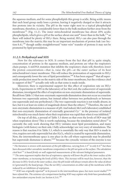
142 The Mitochondrial Free Radical Theory of Aging the aqueous medium, and for some phospholipids this group is acidic. Being acidic means that each head group easily loses a proton, leaving it negatively charged so that it attracts free protons into its vicinity. The pH in the water right next to a typical phospholipid membrane, therefore, is considerably lower than in the bulk medium further away from the membrane 14 (Fig. 11.2). The inner mitochondrial membrane has about 20% acidic phospholipids, which gives a pH at the surface about one unit* lower than in the bulk, 17a so there will indeed be plenty of HO2• there. Being neutral, HO2• can also pass through the membrane into the matrix; this may be an important mechanism of proton leak (see Section 8.4), 17b though unlike straightforward “water wire” transfer of protons it may not be promoted by lipid peroxidation. 11.2.3. Perhydroxyl and SOS Now for the relevance to SOS. It comes from the fact that pH is, quite simply, concentration of protons in the aqueous medium, and protons are what the respiratory chain pumps. A mtDNA mutation that inhibits the respiratory chain will, therefore, lower the proton concentration—that is, raise the pH—at the outer surface of its host mitochondrion’s inner membrane. This will reduce the protonation of superoxide to HO2• and consequently lower the rate of lipid peroxidation.** It has been argued 18 that all superoxide production must be on the matrix side of the inner membrane, but the evidence cited in support of this 9,19 actually only tells us that some is made inside. Moreover, there is experimental support for this effect of respiration rate on HO2• levels. Experiments in 1995 in the laboratory of Joe McCord, the codiscoverer of superoxide dismutase, investigated the effect of respiration on non-enzymatic dismutation of superoxide. Recall from Table 3.3 that non-enzymatic superoxide dismutation does not occur as a reaction between two superoxide anions, but instead either between two perhydroxyls or between one superoxide and one perhydroxyl. (The two-superoxide reaction is not totally absent, in fact, but it is at least six orders of magnitude slower than the others). 20 Therefore, the rate of non-enzymatic dismutation is a measure of pH. And indeed, McCord’s laboratory established that non-enzymatic dismutation of externally generated O2• — in the presence of isolated mitochondria was much faster when their respiration was rapid than when it was inhibited. 21 On top of all this, a perusal of Table 3.3 shows us that even the levels of HO• may fall when respiration slows! This is worth explaining, because the simulation noted above 12 is currently the only work showing that HO2• initiates most lipid peroxidation, and many biologists (with history on their side, I fully acknowledge) are very wary of simulations. The reason is that reaction 8 in Table 3.3, which is essentially the only way that HO• is made in vivo, requires not only superoxide but also H2O2, which is created by superoxide dismutation. Now, the intermembrane space is one place in the cell where superoxide may be plentiful but superoxide dismutase is absent,*** because the cytosolic version of the enzyme is too * The literature is strewn with statements of this pH difference, varying from 2.5 15 to only 0.3; 16 but these have discussed either unrealistic membrane composition or the pH not quite at the surface. The calculation cited here 17a is based firmly on well-supported measurements of the relevant biological parameters. ** The same argument implies that loss of respiratory capacity will lower the pH at the inner surface of the inner membrane, so increasing the levels of HO2• there. This increase will be much less, however, than the decrease in HO2• levels at the outer surface, since the pH inside will always remain above the value of about 6.5 generated by the head groups. Thus the net HO2• concentration on either side of the membrane will be reduced by an inhibition of respiration. *** SOD was originally reported 22 to be present in the intermembrane space, but more recent studies 23,24a have refuted this. The existence of a compartment lacking SOD may explain why birds have paradoxically low antioxidant enzyme levels (see Section 6.5.6). 24b
A Challenge from Textbook Bioenergetics and Free Radical Chemistry Fig. 11.2. Variation of pH near a phospholipid membrane. big (as are nearly all proteins) to fit through the pores in the outer membrane and the mitochondrial version is similarly trapped by the inner membrane. Therefore, the H2O2 formed in the intermembrane space is that formed by non-enzymatic dismutation, which (as noted above) slows down when respiration slows down. This has been further confirmed by the histochemical detection of singlet oxygen in the intermembrane space; 24c recall from Table 3.3 that singlet oxygen is generated by the non-enzymatic, but not the enzymatic, dismutation reaction. 24d Most analyzes have ignored non-enzymatic dismutation and therefore presumed that the H2O2 in the intermembrane space gets there by diffusion from either the cytosol or the matrix (where it was formed by superoxide dismutase); but if that is wrong, then the overall level of H2O2 may well fall when respiration slows, even if the level of superoxide rises. Yet another reason why this is so derives from the presence in the intermembrane space of cytochrome c, which (when in the ferric state) assiduously detoxifies superoxide back to oxygen by accepting its lonely electron, 25 but which also (when in the ferrous state) just as readily 26 donates an electron to HO2•, forming—yes—H2O2. This too would, therefore, happen at a slower rate if there were less HO2• present. Again it may seem as though I have comprehensively rebutted the challenge with which this chapter began. Again, the truth is very different. 11.3. Mitchell’s Oversimplification I may as well warn the reader honestly, in advance, that this section is the most arcane in the book. Bioenergetics is a challenging discipline at the best of times, and the particular topic to be discussed here is one regarding which specialists have been at loggerheads for over thirty years. I include it because MiFRA is incomplete without it, but it may be skipped without loss of continuity. 143
- Page 97 and 98: CHAPTER 8 The Search for How Mutant
- Page 99 and 100: The Search for How Mutant mtDNA is
- Page 101 and 102: The Search for How Mutant mtDNA is
- Page 103 and 104: The Search for How Mutant mtDNA is
- Page 105 and 106: The Search for How Mutant mtDNA is
- Page 107 and 108: CHAPTER 9 The Search for How So Few
- Page 109 and 110: The Search for How So Few Anaerobic
- Page 111 and 112: The Search for How So Few Anaerobic
- Page 113 and 114: The Search for How So Few Anaerobic
- Page 115 and 116: The Search for How So Few Anaerobic
- Page 117 and 118: The Search for How So Few Anaerobic
- Page 119 and 120: The Search for How So Few Anaerobic
- Page 121 and 122: CHAPTER 10 Frequently-Asked Questio
- Page 123 and 124: Frequently-Asked Questions The effe
- Page 125 and 126: Frequently-Asked Questions Table 10
- Page 127 and 128: Frequently-Asked Questions through
- Page 129 and 130: Frequently-Asked Questions has been
- Page 131 and 132: Frequently-Asked Questions SOS, sin
- Page 133 and 134: Frequently-Asked Questions for almo
- Page 135 and 136: Frequently-Asked Questions Fig. 10.
- Page 137 and 138: Frequently-Asked Questions Fig. 10.
- Page 139 and 140: Frequently-Asked Questions of elect
- Page 141 and 142: Frequently-Asked Questions Referenc
- Page 143 and 144: Frequently-Asked Questions 45. Clay
- Page 145 and 146: CHAPTER 11 A Challenge from Textboo
- Page 147: A Challenge from Textbook Bioenerge
- Page 151 and 152: A Challenge from Textbook Bioenerge
- Page 153 and 154: A Challenge from Textbook Bioenerge
- Page 155 and 156: A Challenge from Textbook Bioenerge
- Page 157 and 158: A Challenge from Textbook Bioenerge
- Page 159 and 160: A Challenge from Textbook Bioenerge
- Page 161 and 162: A Challenge from Textbook Bioenerge
- Page 163 and 164: A Challenge from Textbook Bioenerge
- Page 165 and 166: 160 The Mitochondrial Free Radical
- Page 167 and 168: 162 The Mitochondrial Free Radical
- Page 169 and 170: 164 The Mitochondrial Free Radical
- Page 171 and 172: 166 The Mitochondrial Free Radical
- Page 173 and 174: 168 The Mitochondrial Free Radical
- Page 175 and 176: 170 The Mitochondrial Free Radical
- Page 177 and 178: CHAPTER 14 Ablation of Anaerobic Ce
- Page 179 and 180: Ablation of Anaerobic Cells: Techni
- Page 181 and 182: CHAPTER 15 Transgenic Copies of mtD
- Page 183 and 184: Transgenic Copies of mtDNA: Techniq
- Page 185 and 186: Transgenic Copies of mtDNA: Techniq
- Page 187 and 188: Transgenic Copies of mtDNA: Techniq
- Page 189 and 190: Transgenic Copies of mtDNA: Techniq
- Page 191 and 192: Transgenic Copies of mtDNA: Techniq
- Page 193 and 194: CHAPTER 16 Prospective Impact on th
- Page 195 and 196: Prospective Impact on the Healthy H
- Page 197 and 198: Prospective Impact on the Healthy H
142<br />
<strong>The</strong> <strong>Mitochondrial</strong> <strong>Free</strong> <strong>Radical</strong> <strong>The</strong>ory <strong>of</strong> <strong>Aging</strong><br />
the aqueous medium, and for some phospholipids this group is acidic. Being acidic means<br />
that each head group easily loses a proton, leaving it negatively charged so that it attracts<br />
free protons into its vicinity. <strong>The</strong> pH in the water right next to a typical phospholipid<br />
membrane, therefore, is considerably lower than in the bulk medium further away from the<br />
membrane 14 (Fig. 11.2). <strong>The</strong> inner mitochondrial membrane has about 20% acidic<br />
phospholipids, which gives a pH at the surface about one unit* lower than in the bulk, 17a so<br />
there will indeed be plenty <strong>of</strong> HO2• there. Being neutral, HO2• can also pass through the<br />
membrane into the matrix; this may be an important mechanism <strong>of</strong> proton leak (see Section<br />
8.4), 17b though unlike straightforward “water wire” transfer <strong>of</strong> protons it may not be<br />
promoted by lipid peroxidation.<br />
11.2.3. Perhydroxyl and SOS<br />
Now for the relevance to SOS. It comes from the fact that pH is, quite simply,<br />
concentration <strong>of</strong> protons in the aqueous medium, and protons are what the respiratory<br />
chain pumps. A mtDNA mutation that inhibits the respiratory chain will, therefore, lower<br />
the proton concentration—that is, raise the pH—at the outer surface <strong>of</strong> its host<br />
mitochondrion’s inner membrane. This will reduce the protonation <strong>of</strong> superoxide to HO2•<br />
and consequently lower the rate <strong>of</strong> lipid peroxidation.** It has been argued 18 that all superoxide<br />
production must be on the matrix side <strong>of</strong> the inner membrane, but the evidence cited<br />
in support <strong>of</strong> this 9,19 actually only tells us that some is made inside.<br />
Moreover, there is experimental support for this effect <strong>of</strong> respiration rate on HO2•<br />
levels. Experiments in 1995 in the laboratory <strong>of</strong> Joe McCord, the codiscoverer <strong>of</strong> superoxide<br />
dismutase, investigated the effect <strong>of</strong> respiration on non-enzymatic dismutation <strong>of</strong> superoxide.<br />
Recall from Table 3.3 that non-enzymatic superoxide dismutation does not occur as a reaction<br />
between two superoxide anions, but instead either between two perhydroxyls or between<br />
one superoxide and one perhydroxyl. (<strong>The</strong> two-superoxide reaction is not totally absent, in<br />
fact, but it is at least six orders <strong>of</strong> magnitude slower than the others). 20 <strong>The</strong>refore, the rate <strong>of</strong><br />
non-enzymatic dismutation is a measure <strong>of</strong> pH. And indeed, McCord’s laboratory established<br />
that non-enzymatic dismutation <strong>of</strong> externally generated O2• — in the presence <strong>of</strong> isolated<br />
mitochondria was much faster when their respiration was rapid than when it was inhibited. 21<br />
On top <strong>of</strong> all this, a perusal <strong>of</strong> Table 3.3 shows us that even the levels <strong>of</strong> HO• may fall<br />
when respiration slows! This is worth explaining, because the simulation noted above 12 is<br />
currently the only work showing that HO2• initiates most lipid peroxidation, and many<br />
biologists (with history on their side, I fully acknowledge) are very wary <strong>of</strong> simulations. <strong>The</strong><br />
reason is that reaction 8 in Table 3.3, which is essentially the only way that HO• is made in<br />
vivo, requires not only superoxide but also H2O2, which is created by superoxide dismutation.<br />
Now, the intermembrane space is one place in the cell where superoxide may be plentiful<br />
but superoxide dismutase is absent,*** because the cytosolic version <strong>of</strong> the enzyme is too<br />
* <strong>The</strong> literature is strewn with statements <strong>of</strong> this pH difference, varying from 2.5 15 to only 0.3; 16 but these<br />
have discussed either unrealistic membrane composition or the pH not quite at the surface. <strong>The</strong> calculation<br />
cited here 17a is based firmly on well-supported measurements <strong>of</strong> the relevant biological parameters.<br />
** <strong>The</strong> same argument implies that loss <strong>of</strong> respiratory capacity will lower the pH at the inner surface <strong>of</strong> the<br />
inner membrane, so increasing the levels <strong>of</strong> HO2• there. This increase will be much less, however, than the<br />
decrease in HO2• levels at the outer surface, since the pH inside will always remain above the value <strong>of</strong> about<br />
6.5 generated by the head groups. Thus the net HO2• concentration on either side <strong>of</strong> the membrane will be<br />
reduced by an inhibition <strong>of</strong> respiration.<br />
*** SOD was originally reported 22 to be present in the intermembrane space, but more recent studies 23,24a<br />
have refuted this. <strong>The</strong> existence <strong>of</strong> a compartment lacking SOD may explain why birds have paradoxically<br />
low antioxidant enzyme levels (see Section 6.5.6). 24b


