

Analytical Chemistry Chemical Cytometry Quantitates Superoxide
Analytical Chemistry Chemical Cytometry Quantitates Superoxide
Analytical Chemistry Chemical Cytometry Quantitates Superoxide

Create successful ePaper yourself
Turn your PDF publications into a flip-book with our unique Google optimized e-Paper software.
plasma samples were analyzed using the teicoplanin FPIA kit<br />
following the instruction of the manufacturer.<br />
RESULT AND DISCUSSION<br />
Design and Synthesis of Peptide Probes. We used aminereactive<br />
fluorophores to label the acetylated peptide Ac-L-Lys-D-<br />
Ala-D-Ala-OH. We chose this peptide for two reasons. First, L-lys<br />
is naturally occurring in the peptidoglycan precursor peptide L-Ala-<br />
D-Glu-L-Lys-D-Ala-D-Ala. We expected the incorporation of the L-lys<br />
would not affect the interaction between the peptide and the<br />
antibiotics. Second, the side chain of Lys contains a primary amine<br />
group, which is distant from the binding site judged by the crystal<br />
structure of the vancomycin and diacetyl-L-Lys-D-Ala-D-Ala complex.<br />
34 As expected, we found that the binding affinities between<br />
the labeled peptide and antibiotics were similar to the values<br />
reported for the binding between the free peptide and antibiotics,<br />
indicating that the modification did not affect their interaction (see<br />
below).<br />
Effect of Fluorophores. The fluorescence polarization signal<br />
is determined by the Perrin Equation. 35<br />
( 1 1<br />
-<br />
P 3) ) ( 1<br />
-<br />
P0 1 kT<br />
1 + 3)( ηV τ)<br />
Where τ is the fluorescence lifetime of the fluorophore, P0 is the<br />
limiting polarization, k is the Boltzmann constant, T is the<br />
absolute temperature, η is the viscosity, and V is the molecular<br />
volume (molecular weight). The fluorescence polarization is<br />
negatively correlated with the lifetime of the fluorophores and<br />
positively correlated with the molecular volume (molecular<br />
weight). In order to maximize the observed change of polarization<br />
upon antibiotic binding, we tested two fluorophores with<br />
different lifetimes, FITC (4 ns lifetime) and AF680 (1 ns<br />
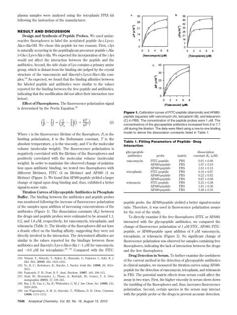
lifetime) (Figure 1). We found that AF680-peptide yielded a larger<br />
change of signal upon drug binding and, thus, exhibited a better<br />
signal-to-noise ratio.<br />
Titration Curves of Glycopeptide Antibiotics in Phosphate<br />
Buffer. The binding between the antibiotics and peptide probes<br />
was monitored following the increase of fluorescence polarization<br />
of the samples upon addition of increasing concentrations of the<br />
antibiotics (Figure 1). The dissociation constants (Kd) between<br />
the drugs and peptide probes were estimated to be around 1.1,<br />
0.2, and 1.8 µM, respectively, for vancomycin, teicoplanin, and<br />
telavancin (Table 1). The identity of the fluorophores did not have<br />
a drastic effect on the binding affinity, suggesting they were not<br />
directly involved in the interaction. The determined affinities are<br />
similar to the values reported for the bindings between these<br />
antibiotics and diacetyl-L-Lys-D-Ala-D-Ala (∼1 µM for vancomycin,<br />
and ∼0.6 µM for teicoplanin). 36-39 Compared with the FITC-<br />
(34) Nitanai, Y.; Kikuchi, T.; Kakoi, K.; Hanmaki, S.; Fujisawa, I.; Aoki, K. J.<br />
Mol. Biol. 2009, 385, 1422–1432.<br />
(35) Ye, B. C.; Ikebukuro, K.; Karube, I. Nucleic Acids Res. 1998, 26, 3614–<br />
3615.<br />
(36) Popieniek, P. H.; Pratt, R. F. Anal. Biochem. 1987, 165, 108–113.<br />
(37) Azad, M.; Hernandez, L.; Plazas, A.; Rudolph, M.; Gomez, F. A. Chromatographia<br />
2003, 57, 339–343.<br />
(38) Rao, J. H.; Yan, L.; Xu, B.; Whitesides, G. M. J. Am. Chem. Soc. 1999, 121,<br />
2629–2630.<br />
(39) van Wageningen, A. M. A.; Staroske, T.; Williams, D. H. Chem. Commun.<br />
1998, 1171–1172.<br />
7046 <strong>Analytical</strong> <strong>Chemistry</strong>, Vol. 82, No. 16, August 15, 2010<br />
(2)<br />
Figure 1. Calibration curves of FITC-peptide (diamonds) and AF680peptide<br />
(squares) with vancomycin (A), teicoplanin (B), and telavancin<br />
(C) in PBS. The concentration of the peptide probes were 1 µM. The<br />
concentrations of the glycopeptide antibiotics increased from 0 to 7.7<br />
µM during the titration. The data were fitted using a one-to-one binding<br />
model to derive the dissociation constants listed in Table 1.<br />
Table 1. Fitting Parameters of Peptide-Drug<br />
Interaction<br />
glycopeptide<br />
antibiotics probe matrix<br />
dissociation<br />
constant Kd (µM)<br />
vancomycin FITC-peptide PBS 0.91 ± 0.06<br />
AF680-peptide PBS 1.07 ± 0.19<br />
AF680-peptide FBS 1.81 ± 0.13<br />
teicoplanin FITC-peptide PBS 0.14 ± 0.07<br />
AF680-peptide PBS 0.22 ± 0.02<br />
AF680-peptide FBS 0.87 ± 0.08<br />
telavancin FITC-peptide PBS 3.22 ± 0.38<br />
AF680-peptide PBS 1.81 ± 0.36<br />
AF680-peptide FBS 5.36 ± 0.35<br />
peptide probe, the AF680-peptide yielded a better signal-to-noise<br />
ratio. Therefore, it was used in fluorescence polarization assays<br />
for the rest of the study.<br />
To directly examine if the free fluorophores (FITC or AF680)<br />
interacted with the glycopeptide antibiotics, we compared the<br />
change of fluorescence polarization of 1 µM FITC, AF680, FITCpeptide,<br />
or AF680-peptide upon addition of 8 µM vancomycin,<br />
teicoplanin, or telavancin (Figure 2). No significant change of<br />
fluorescence polarization was observed for samples containing free<br />
fluorophores, indicating the lack of interaction between the drugs<br />
and the free fluorophores.<br />
Drug Detection in Serum. To further examine the usefulness<br />
of the current method in the detection of glycopeptide antibiotics<br />
in clinical samples, we measured the titration curves using AF680peptide<br />
for the detection of vancomycin, teicoplanin, and telavancin<br />
in FBS. The potential matrix effects from serum could affect the<br />
assay in two ways. First, the higher viscosity in serum slows down<br />
the tumbling of the fluorophores and, thus, increases fluorescence<br />
polarization. Second, certain species in the serum may interact<br />
with the peptide probe or the drugs to prevent accurate detection.


