

Analytical Chemistry Chemical Cytometry Quantitates Superoxide
Analytical Chemistry Chemical Cytometry Quantitates Superoxide
Analytical Chemistry Chemical Cytometry Quantitates Superoxide

Create successful ePaper yourself
Turn your PDF publications into a flip-book with our unique Google optimized e-Paper software.
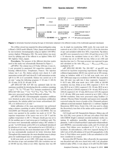
Figure 1. Schematic flowchart showing the type of information obtained in the different modes of the multimodal approach developed.<br />
The cell-line cytosol was separated by ultracentrifugation using<br />
a HimaCs 120GX model (Hitachi, Tokyo, Japan) and fractionated<br />
by size-exclusion chromatography using an Agilent 1100 HPLC<br />
system (Agilent, Wilmington, DE). The metal elution was monitored<br />
by splitting part of the effluent to an Agilent 7500ce ICP<br />
MS (Agilent, Tokyo, Japan).<br />
Procedures. The purpose of the different detection modes<br />
used in this study was schematically illustrated in Figure 1.<br />
µRP HPLC. The column was a C8 Vydac (250 mm ×1 mm i.d.,<br />
5 µm) presented in passivated 316 copper-free stainless steel<br />
housing (Alltech/Grace, Templemars, France). The injection<br />
volume was 5 µL. The elution solvents were eluent A: 5 mM<br />
ammonium acetate pH 6 and eluent B: 5 mM ammonium acetate<br />
(pH 6) in 50% (v/v) acetonitrile. Elution was carried out at 40<br />
µL · min -1 using the following program: 0-50 min: 2-20% B;<br />
50-52 min: 2% B; 52-60 min: 2% B.<br />
µRP HPLC-ICP MS. ICP MS was optimized daily for the<br />
maximum sensitivity by introducing directly a solution containing<br />
1 µg · L -1 89 Y, 7 Li, 205 Tl and 140 Ce. Isotopes monitored in µRP<br />
HPLC-ICP MS were 114 Cd, 112 Cd 63 Cu, 65 Cu, 64 Zn, 66 Zn. The<br />
data were processed using Excel Microsoft software.<br />
µRP HPLC-ESI MS. Chromatographic separation conditions<br />
were identical as given above. For the postcolumn acidification<br />
experiments, the solution added was formic acid:methanol (30/<br />
70%, v/v) delivered at 4 µL · min -1 .<br />
Initial calibration of the mass spectrometer was performed<br />
using a mixture consisting of cafein (195.08765), MRFA peptide<br />
(524.26499) and Ultramark polymer (m/z 1221.99063). The ion<br />
source was operated in the positive ion mode at 3.2 kV. The<br />
vaporizer temperature of the source was set to 120 °C and the<br />
capillary temperature to 280 °C. Nitrogen sheath gas was set to<br />
20, the auxiliary gas to 5 and sweep gas to 0 (arbitrary unit). The<br />
ion lenses were automatically optimized using a MT solution (1<br />
µg · mL -1 in 0.01% formic acid in 50% (v/v) methanol) introduced<br />
by infusion at 4 µL · min -1 and monitored at m/z 1225.843 (z )<br />
5). In all experiments, the most abundant mass of [M + 5H] 5+<br />
ion was systematically monitored for better detection limit.<br />
Throughout this manuscript, the most abundant mass is always<br />
given for multicharged ions or uncharged molecules.<br />
In full scan mode, an m/z 1200-1500 range was scanned for<br />
the detection of apo- and metalated MTs (the resolution was set<br />
at 100 000 (m/∆m, fwhm at m/z 400)). Injection time was 400<br />
ms. In single ion monitoring (SIM) mode, the scan mode was<br />
centered on m/z 1230 ± 50 and m/z 1377.5 ± 65 for the detection<br />
of apo- and metalated rabbit liver MTs, respectively. Pig kidney<br />
apo-MTs were measured at m/z 1250 ± 60 and then at m/z 1170<br />
± 70 whereas the metalated forms at m/z 1367.5 ± 87. The<br />
resolution was set at 100 000 (m/∆m, fwhm at m/z 400) and<br />
injection time was 3 s. The mass accuracy was expressed in ppm<br />
and calculated as the ratio (M theoretical - Mdetermined)/Mtheoretical<br />
multiplied by 10 6 .<br />
µRP HPLC-ESI MS/MS. The [M+5H] 5+ of apo-MT was<br />
selected for top-down sequencing experiments. High energy<br />
collision fragmentation (HCD) was carried out at 50% energy,<br />
using an isolation width of 2 in full scan mode over m/z<br />
100-2000 mass range at a resolution of 100 000. The acquisition<br />
time (60 min) was split into 6 time segments. Segment 1:<br />
0-30.75 min, SIM scan mode (m/z 1100-1300) at 100 000<br />
resolution (m/∆m, fwhm at m/z 400); segment 2: 30.75-36<br />
min, HCD at m/z 1189.4: segment 3: 36-39 min, HCD at m/z<br />
1197.63, and m/z 1194.63; segment 4: 39-44 min, HCD at m/z<br />
1203.24; segment 5: 44-47 min, HCD at m/z 1271.96; segment<br />
6: 47-60 min, SIM scan mode centered at m/z 1200 ± 100 at<br />
100 000 resolution. Data were processed using Xcalibur 2.1<br />
software (Thermo Fisher Scientific) and masses were manually<br />
corrected using the traces of peaks of the Ultramark polymer<br />
calibrant as internal standard. Analyst Q.S. 1.1 software (Applied<br />
Biosystems MDS Sciex, Foster City, CA) was used to obtain<br />
the theoretical y ions lists of the amino acids sequence of MT<br />
subisoforms.<br />
Cell Growth and CdS Nanoparticles Exposure. Kidney pig cell<br />
line (LLC-PK1) were grown in 100 mm cell culture Petri dish<br />
with EMEM (Eagle’s minimal essential medium) media containing<br />
1% antibiotics (penicillin, streptomycin), 2 mM Lglutamin,<br />
1 mM 4-(2-hydroxyethyl)-1-piperazine-ethanesulfonic<br />
acid (HEPES), non-essential amino acids, and 5% of fetal calf<br />
serum. Petri dishes were incubated at 37 °C ina5%CO2<br />
incubator. At subconfluence, cells were exposed during 24 h<br />
with5mLof120µM solution of 10 nm-CdS nanoparticles. Cells<br />
from 11 Petri dishes were pooled. Cells not submitted to CdS<br />
were considered as control.<br />
Recovery of the MT Fraction. After cell growth, the supernatant<br />
was discarded and cells were rinsed twice with 1 mL phosphate<br />
buffered saline (PBS). A 1-mL aliquot of 25 mM Tris/HCl buffer<br />
<strong>Analytical</strong> <strong>Chemistry</strong>, Vol. 82, No. 16, August 15, 2010<br />
6949


