

Analytical Chemistry Chemical Cytometry Quantitates Superoxide
Analytical Chemistry Chemical Cytometry Quantitates Superoxide
Analytical Chemistry Chemical Cytometry Quantitates Superoxide

You also want an ePaper? Increase the reach of your titles
YUMPU automatically turns print PDFs into web optimized ePapers that Google loves.
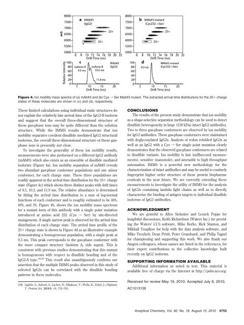
Figure 4. Ion mobility mass spectra of (a) mAb#3 and (b) Cys f Ser Mab#3 mutant. The extracted arrival time distributions for the 25+ charge<br />
states of these molecules are shown in (c) and (d), respectively.<br />
These limited calculations using individual static structures do<br />
not explain the relatively late arrival time of the IgG2-B isoform<br />
and suggest that the overall three-dimensional structure of<br />
these gas-phase ions may be quite different than the solution<br />
structure. While the IMMS results demonstrate that ion<br />
mobility separates covalent disulfide mediated IgG2 structural<br />
isoforms, the overall three-dimensional structure of these gasphase<br />
ions is presently not clear.<br />
To investigate the generality of these ion mobility results,<br />
measurements were also performed on a different IgG2 antibody<br />
(mAb#3) which also exists as an ensemble of disulfide mediated<br />
isoforms (Figure 4a). Ion mobility separation of mAb#3 reveals<br />
two abundant gas-phase conformer populations and one minor<br />
conformer, for each charge state. These three populations are<br />
readily apparent in the arrival time distribution for the 25+ charge<br />
state (Figure 4c) which shows three distinct peaks with drift times<br />
of 9.5, 10.3, and 11.9 ms. The relative abundance is determined<br />
by fitting the arrival time distribution to a sum of log-normal<br />
functions of each conformer and is roughly estimated to be 46%,<br />
49%, and 5%. Figure 4b, shows the ion mobility mass spectrum<br />
for a mutant form of this antibody with a single point mutation<br />
introduced at amino acid 232 (Cys f Ser) by site-directed<br />
mutagenesis. A single narrow peak is observed for the arrival time<br />
distribution of each charge state. The arrival time profile of the<br />
25+ charge state is shown in Figure 4d as an illustrative example<br />
demonstrating a homogeneous population, with a single peak at<br />
9.3 ms. This peak corresponds to the gas-phase conformer with<br />
the more compact structure (isoform A, vide supra). This is<br />
consistent with previous studies demonstrating that this mutant<br />
is homogeneous with respect to disulfide bonding and of the<br />
IgG2-A type. 10,38 This result also unambiguously confirms our<br />
assertion that the multiple IMMS peaks observed in this study of<br />
selected IgG2s can be correlated with the disulfide bonding<br />
patterns in these molecules.<br />
(38) Lightle, S.; Aykent, S.; Lacher, N.; Mitaksov, V.; Wells, K.; Zobel, J.; Oliphant,<br />
T. Protein Sci. 2010, 19, 753–762.<br />
CONCLUSIONS<br />
The results of the present study demonstrate that ion mobility<br />
as a shape-selective separation methodology can be used to detect<br />
disulfide heterogeneity in large (150 kDa) intact IgG2 antibodies.<br />
Two to three gas-phase conformers are observed by ion mobility<br />
for IgG2 antibodies. These gas-phase conformers were maintained<br />
with deglycosylated IgG2s. Analysis of redox refolded IgG2s as<br />
well as an IgG2 with a Cys f Ser single point mutation clearly<br />
demonstrates that the observed gas-phase conformers are related<br />
to disulfide variants. Ion mobility is fast (millisecond measurements),<br />
sensitive (nanomole), and amenable to high throughput<br />
automation. IMMS is a powerful new methodology for the<br />
characterization of intact antibodies and may be useful to routinely<br />
fingerprint higher order structure of these protein biopharmaceuticals<br />
in the near future. We are currently extending these<br />
measurements to investigate the utility of IMMS for the analysis<br />
of IgG2s containing lambda light chains as well as to directly<br />
characterize the binding of antigen targets to individual disulfide<br />
isoforms of IgG2 antibodies.<br />
ACKNOWLEDGMENT<br />
We are grateful to Allen Sickmier and Leszek Poppe for<br />
insightful discussions, Keith Richardson (Waters Inc.) for providing<br />
the Waters’ CCS software, Mike Berke, Rick Stanton, and<br />
Mikhail Toupikov for help with the data analysis software, and<br />
Mike Treuheit, Dean Pettit, Peter Grandsard, and Philip Tagari<br />
for championing and supporting this work. We also thank our<br />
Amgen colleagues, whose names are listed in the references, for<br />
their expert contributions to the collective knowledge built<br />
recently on IgG2 isoforms.<br />
SUPPORTING INFORMATION AVAILABLE<br />
Additional information as noted in text. This material is<br />
available free of charge via the Internet at http://pubs.acs.org.<br />
Received for review May 19, 2010. Accepted July 6, 2010.<br />
AC1013139<br />
<strong>Analytical</strong> <strong>Chemistry</strong>, Vol. 82, No. 16, August 15, 2010<br />
6755


