PET/CT Shielding Design Examples - Radiation Shielding for ...
PET/CT Shielding Design Examples - Radiation Shielding for ...
PET/CT Shielding Design Examples - Radiation Shielding for ...

Create successful ePaper yourself
Turn your PDF publications into a flip-book with our unique Google optimized e-Paper software.
<strong>PET</strong>/<strong>CT</strong> <strong>Shielding</strong> <strong>Design</strong><br />
<strong>Examples</strong><br />
Douglas J. Simpkin, Ph.D.<br />
Aurora St. Luke’s Medical Center<br />
Milwaukee, Wisconsin<br />
dsimpkin@wi dsimpkin@wi.rr.com rr com<br />
www. http://geocities.com/djsimpkin<br />
1
Sources of Exposure:<br />
F-18 in Patients<br />
• Task Group 108 recommends a realistic<br />
Effective Dose Rate Constant Γ <strong>for</strong> F-18 in<br />
patient:<br />
Γ = 0 092 µSv Sv m m2 Γ 0.092 µSv Sv m MBq MBq-1 h h-1<br />
• Assume all photons emitted from patient are<br />
511 511 keV<br />
2
Time determines Dose Rate<br />
• Then Dose Rate D at distance r (m) from activity A<br />
� ( ) y<br />
(MBq) is<br />
D Γ � =<br />
• The Dose, D, accumulated over time t is<br />
D<br />
=<br />
1.<br />
44<br />
×<br />
D�<br />
× T<br />
r<br />
A<br />
2<br />
1/<br />
2<br />
⎛<br />
× ⎜ ⎜1<br />
−<br />
⎜<br />
⎝<br />
0.<br />
693 ⎛ − ×t × t<br />
T<br />
• And track decay of activity from moment-to-moment<br />
0.<br />
693<br />
− × t<br />
T<br />
1 / 2<br />
A (<br />
t ) = A e<br />
0<br />
e<br />
1/<br />
2<br />
⎞<br />
⎟<br />
⎠<br />
3
The patient is the source<br />
• FDG patient is in “incubation” (in an quiet,<br />
isolated room) ) <strong>for</strong> ~45 minutes. <strong>Shielding</strong> gf <strong>for</strong><br />
this room must be considered.<br />
• Patient in scanner <strong>for</strong> 30 30-60 60 minutes minutes.<br />
<strong>Shielding</strong> <strong>for</strong> scanner room must be<br />
considered considered.<br />
4
<strong>Shielding</strong> People to P/T<br />
•In uncontrolled areas in USA<br />
– Must restrict dose in any one hour to < 0.02<br />
mSv and<br />
– Must restrict annual dose to < regulated g limit of<br />
1 mSv y-1 • Controlled areas in USA<br />
– (For pregnant workers), must restrict annual<br />
occupational dose to 5 mSv y-1 p y<br />
5
Corridor Outside Uptake Room<br />
• Presume the patient is injected with 555 MBq 18 • Presume the patient is injected with 555 MBq F 18F. The patient remains quiescent in the Uptake Room<br />
<strong>for</strong> 45 minutes.<br />
• At 3 m (to the corridor) the initial dose rate upon<br />
injection at is<br />
2<br />
µ Sv m<br />
0.<br />
092 × 555 MBq<br />
A MBq h<br />
D� Γ<br />
q<br />
D0 = =<br />
= 0 0.<br />
0057<br />
2<br />
2<br />
r<br />
3 m<br />
• So S SSo i in th the USA USA, 0.02 0 02 mSv S in i any h h is i satisfied<br />
ti fi d<br />
mSv<br />
( ) h<br />
6
D<br />
Corridor Outside Uptake Room<br />
• Assume 40 <strong>PET</strong> patients per week = 2080<br />
patients per year<br />
• The annual unshielded dose in the corridor<br />
outside the uptake room is then<br />
=<br />
2080<br />
mSv<br />
= 7<br />
. 72<br />
y<br />
pat<br />
mSv ⎛<br />
× 1.<br />
44×<br />
0.<br />
0057 × 1.<br />
83h<br />
× ⎜<br />
1 − e<br />
y<br />
h ⎝<br />
0.<br />
693<br />
− × . 75h<br />
1.<br />
83h<br />
7<br />
⎞<br />
⎟<br />
⎠
Corridor Outside Uptake Room<br />
• If the corridor is uncontrolled, P=1 mSv y-1 and T <strong>for</strong> the wall is = 1/5 so P/T = 5mSvy-1 and T <strong>for</strong> the wall is 1/5, so P/T 5 mSv y<br />
• The wall must be shielded so B = 5 / 7.2 =<br />
00.69, 69 which requires 3 mm thick Pb. Pb<br />
• The door has an occupancy factor T = 1/8, so<br />
th that t P/T = 8 mSv S y-1 1. Si Since the th unshielded hi ld d<br />
dose is 7.2 mSv y-1 , the door needs no<br />
shielding.<br />
hi ldi<br />
8
Office Above Uptake Room<br />
• Consider a fully occupied (T=1)<br />
uncontrolled (P=1 mSv y-1 ( y ) office 3.5 m<br />
above Uptake Room. The initial dose rate is<br />
2<br />
µ µ Sv m<br />
0 0.<br />
092 × 555 MBq<br />
A MBq h<br />
mSv<br />
D� Γ<br />
0 = =<br />
= 0.<br />
0042<br />
2<br />
2<br />
r 3 3.<br />
5 m<br />
( ) h<br />
• So again, 0.02 mSv in any 1 h is satisfied<br />
9
D<br />
Office Above Uptake Room<br />
• For 2080 patients per year, the annual dose<br />
in the office is then<br />
pat<br />
= 2080 × 1.<br />
44×<br />
0.<br />
0042<br />
y<br />
mSv S<br />
= 5.<br />
65<br />
y<br />
mSv<br />
h<br />
⎛<br />
× 1.<br />
83h<br />
× ⎜<br />
1 − e<br />
⎝<br />
0 0.<br />
693<br />
− × . 75h<br />
1.<br />
83h<br />
• Th The ceiling ili must t provide id shielding, hi ldi with ith<br />
B = 1 mSv y-1 / 5.65 mSv y-1 = 0.177. This<br />
requires i 15.6 15 6 cm std td density d it concrete<br />
t<br />
10<br />
⎞<br />
⎟<br />
⎠
Office Above Uptake Room<br />
•In my facility, the ceiling was only 7.6 cm<br />
thick. Thus we had to add the equivalent of<br />
15.6 - 7.6 = 8 cm of concrete = 8 cm /22 cm<br />
=0.36 TVL.<br />
• This required 0.36×1.8 cm Pb = 0.65 cm Pb<br />
to be added to the concrete ceiling.<br />
11
<strong>PET</strong>/<strong>CT</strong> Scanner Room<br />
Note: Activity in<br />
patient i will ill have h<br />
decayed in uptake<br />
room (i.e. ( from ~555<br />
MBq on injection to<br />
~420 MBq when<br />
patient enters scanner)<br />
12
Office adjacent to <strong>PET</strong>/<strong>CT</strong> Scanner<br />
• Fully occupied uncontrolled area, shield to<br />
– P/T = 1mSvy-1 /1= 1mSvy-1 P/T 1 mSv y / 1 1 mSv y<br />
• At 3.5 m distance, with initially 420 MBq in<br />
patient<br />
2<br />
µ Sv m<br />
0.<br />
092 × 420 MBqq<br />
Γ A MB MBq h<br />
D� Γ<br />
0 = =<br />
= 0.<br />
0032<br />
2<br />
2<br />
r<br />
3.<br />
5 m<br />
mSv S<br />
( ) h<br />
13
Office adjacent to <strong>PET</strong>/<strong>CT</strong> Scanner<br />
D<br />
• For 2080 patients per year, the annual dose<br />
in the office <strong>for</strong> a 45 min scan is then<br />
pat<br />
mSv ⎛<br />
= 2080 × 1.<br />
44×<br />
0.<br />
0032 × 1.<br />
83h<br />
× ⎜<br />
1 − e<br />
y<br />
h ⎝<br />
mSv S<br />
= 4.<br />
3<br />
y<br />
• Th The wall ll must t provide id shielding, hi ldi with ith<br />
B = 1 mSv y-1 / 4.3 mSv y-1 = 0.23. This<br />
requires i 1.1 1 1 cm Pb in i the th wall.<br />
ll<br />
0 0.<br />
693<br />
− × . 75h<br />
1.<br />
83h<br />
14<br />
⎞<br />
⎟<br />
⎠
<strong>PET</strong>/<strong>CT</strong> scanner<br />
• Patient in scanner 0.5 0 5 – 1h 1 h<br />
• 2D <strong>PET</strong> done: 3-4 min/bed<br />
stop, 15 cm coverage/stop, 47<br />
× 3.27mm slices/stop<br />
• <strong>CT</strong> done <strong>for</strong><br />
– Anatomical co-registration of<br />
<strong>PET</strong> images<br />
– Attenuation correction of <strong>PET</strong><br />
images<br />
• For cancer patients typically<br />
scan from “eye to thigh”<br />
15
<strong>PET</strong>/<strong>CT</strong> Scanner<br />
• TTypical i l (GE Discovery Di ST) <strong>CT</strong> scan<br />
technique:<br />
– (8 row scanner)<br />
– 100-175 mA,<br />
– 140 kVp,<br />
– 0.8 sec/rot,<br />
– 3.75mm thick slices,<br />
– pitch = 1.675:1<br />
– 2 cm beam width<br />
16
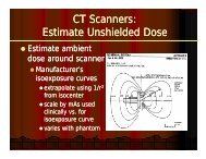
<strong>PET</strong>/<strong>CT</strong> Scanner<br />
• Methods <strong>for</strong> shielding <strong>CT</strong> scanners<br />
are iin NCRP-147 NCRP 147<br />
• Determine unshielded dose based<br />
on<br />
– Manufacturer’s isoexposure curves<br />
– Scatter factor applied to <strong>CT</strong>DI<br />
– Height of patient scanned<br />
– mA<br />
• Consider that <strong>CT</strong> may be used as a<br />
stand-alone scanner<br />
17
• Calculate B to<br />
reduce dose to P/T<br />
• Look up barrier<br />
thickness required<br />
to achieve that<br />
transmission<br />
• <strong>CT</strong> typ. t requires i<br />
– 1 to 2 mm Pb<br />
– 13 cm concrete<br />
<strong>PET</strong>/<strong>CT</strong> Scanner<br />
Traansmission<br />
1E+0<br />
8<br />
6<br />
1E-1<br />
8<br />
6<br />
6 Transmission of <strong>CT</strong> Scanner<br />
4<br />
Secondary <strong>Radiation</strong> Through Pb<br />
2<br />
4<br />
2<br />
1E-2<br />
8<br />
6<br />
4<br />
2<br />
1E-3<br />
8<br />
6<br />
1E-4<br />
4<br />
2<br />
120 kVp<br />
140 kVp<br />
Fitting parameters to Equation B.2<br />
kVp α ( (mm 1 ( 1<br />
-1 ) β (mm-1 ) γ<br />
120 2.246 5.73 0.547<br />
140 2.009 3.99 0.342<br />
0 0.5 1 1.5 2 2.5 3<br />
Lead Thickness ( (mm)<br />
)<br />
18
• <strong>PET</strong>:<br />
<strong>Shielding</strong> <strong>PET</strong> AND <strong>CT</strong><br />
– Lower unshielded dose rates<br />
– Penetrating photons require an order of<br />
magnitude thicker barriers than the <strong>CT</strong> scan<br />
•<strong>CT</strong><br />
– Much higher unshielded dose rates<br />
• All persons entering scanner room while x ray is on<br />
must twear a Pb apron<br />
– Low energy x rays require much thinner<br />
barriers barriers.<br />
19
<strong>Shielding</strong> <strong>PET</strong> AND <strong>CT</strong><br />
• <strong>Shielding</strong> requirements <strong>for</strong> F-18 in the<br />
patient predominates ( ~ 0.5 – 2 cm Pb)<br />
• However However, the <strong>CT</strong> scanner will ~always always<br />
require some shielding in all barriers ( ~1 -<br />
2mmPb) 2 mm Pb)<br />
– (For example, as with the uptake room, the<br />
door to the scanner room may not require<br />
shielding <strong>for</strong> F-18, but will require 1-2 mm Pb<br />
<strong>for</strong> <strong>CT</strong>)<br />
20
Conclusions<br />
• Th The injected i j d patient i is i the h primary i source of f<br />
radiation exposure in the <strong>PET</strong> facility<br />
• For 18 • For FDG FDG, the uptake room and scanner will<br />
probably require lead shielding<br />
• This shielding g will often be 2-10× thicker that<br />
what’s typically in a diagnostic x-ray room<br />
• <strong>Shielding</strong> <strong>for</strong> the <strong>CT</strong> scan in a <strong>PET</strong>/<strong>CT</strong> scanner<br />
must bbe considered id d<br />
21