The Radiopharmacy - European Association of Nuclear Medicine
The Radiopharmacy - European Association of Nuclear Medicine
The Radiopharmacy - European Association of Nuclear Medicine

You also want an ePaper? Increase the reach of your titles
YUMPU automatically turns print PDFs into web optimized ePapers that Google loves.
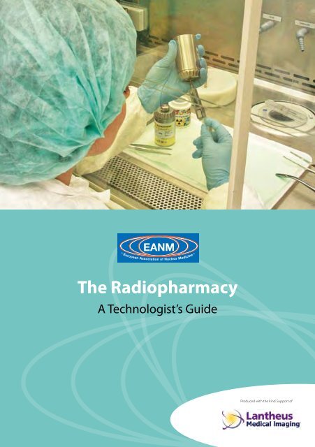
<strong>European</strong> <strong>Association</strong> <strong>of</strong> <strong>Nuclear</strong> <strong>Medicine</strong><br />
<strong>The</strong> <strong>Radiopharmacy</strong><br />
A Technologist’s Guide<br />
Produced with the kind Support <strong>of</strong>
Contributors<br />
James R Ballinger, PhD<br />
Chief Radiopharmaceutical Scientist<br />
Guy’s and St Thomas’ NHS Foundation Trust<br />
London, United Kingdom<br />
Clemens Decrist<strong>of</strong>oro, PhD (*)<br />
Chair <strong>of</strong> the EANM <strong>Radiopharmacy</strong> Committee<br />
Clinical Department <strong>of</strong> <strong>Nuclear</strong> <strong>Medicine</strong><br />
Medical University Innsbruck<br />
Innsbruck, Austria<br />
Brit Farstad<br />
Member <strong>of</strong> the EANM Radiopharamcy Committee<br />
M.Pharm/Radiopharmacist<br />
Department Head, Isotope Laboratories<br />
Institute for Energy Technology<br />
Kjeller, Norway<br />
Brendan McCoubrey<br />
Radiation Safety Officer<br />
Dept. <strong>of</strong> Diagnostic Imaging<br />
St. James’s Hospital<br />
Dublin, Ireland<br />
Geraldine O’Reilly, PhD<br />
Radiation Protection Advisor<br />
Dept. <strong>of</strong> Medical Physics and BioEngineering<br />
St. James’s Hospital<br />
Dublin, Ireland<br />
2<br />
Helen Ryder<br />
Clinical Specialist Radiographer<br />
Dept. <strong>of</strong> Diagnostic Imaging<br />
St. James’s Hospital<br />
Dublin, Ireland<br />
Tanja Gmeiner Stopar, PhD<br />
Member <strong>of</strong> the Education Board,<br />
EANM <strong>Radiopharmacy</strong> Committee<br />
<strong>Radiopharmacy</strong>, Head<br />
University Medical Centre Ljubljana<br />
Department for <strong>Nuclear</strong> <strong>Medicine</strong><br />
Ljubljana, Slovenia<br />
Wim van den Broek<br />
Chair <strong>of</strong> the EANM Technologist Committee<br />
Chief Technologist<br />
Dept <strong>of</strong> <strong>Nuclear</strong> <strong>Medicine</strong><br />
University <strong>Medicine</strong> Centre<br />
Nijmegen, <strong>The</strong> Netherlands<br />
Editors<br />
Suzanne Dennan<br />
Vice-Chair <strong>of</strong> the EANM Technologist Committee<br />
Acting Radiographic Services Manager<br />
Dept. <strong>of</strong> Diagnostic Imaging<br />
St. James’s Hospital<br />
Dublin, Ireland<br />
Clemens Decrist<strong>of</strong>oro, PhD *<br />
This booklet was sponsored by an educational grant from Lantheus Medical Imaging . <strong>The</strong> views expressed are those<br />
<strong>of</strong> the authors and not necessarily <strong>of</strong> Lantheus Medical Imaging.
Contents<br />
Foreword<br />
Wim van den Broek ...............................................................................4<br />
Introduction<br />
Clemens Decrist<strong>of</strong>oro .............................................................................5<br />
Chapter 1 - <strong>Radiopharmacy</strong> Technology<br />
Brit Farstad . . . . . . . . . . . . . . . . . . . . . . . . . . . . . . . . . . . . . . . . . . . . . . . . . . . . . . . . . . . . . . . . . . . . . . . . . . . . . . . . . . . . . . . 6<br />
Chapter 2 - <strong>Radiopharmacy</strong> Design<br />
James Ballinger ..................................................................................12<br />
Chapter 3 - <strong>Radiopharmacy</strong>: Preparing & Dispensing Radiopharmaceuticals<br />
Geraldine O’Reilly ................................................................................16<br />
Chapter 4 - <strong>Radiopharmacy</strong>: Kits & Techniques<br />
Helen Ryder . . . . . . . . . . . . . . . . . . . . . . . . . . . . . . . . . . . . . . . . . . . . . . . . . . . . . . . . . . . . . . . . . . . . . . . . . . . . . . . . . . . . .22<br />
Chapter 5 – <strong>Radiopharmacy</strong>: Blood Labelling<br />
Tanja Gmeiner Stopar ............................................................................33<br />
Chapter 6 - <strong>Radiopharmacy</strong>: Record Keeping & Administration<br />
Brendan McCoubrey .............................................................................41<br />
References ......................................................................................49<br />
Imprint ..........................................................................................51<br />
3<br />
EANM
Foreword<br />
Wim van den Broek<br />
Since it was formed, the EANM Technologist<br />
Committee has been devoted to the improvement<br />
<strong>of</strong> nuclear medicine technologists’<br />
(NMTs’) pr<strong>of</strong>essional skills. Publications that will<br />
assist in the setting <strong>of</strong> high standards for NMT’s<br />
work have been developed and since 2004<br />
a series <strong>of</strong> brochures, “Technologists Guides”,<br />
have been published yearly. This booklet<br />
about radiopharmacy is already the fifth volume.<br />
<strong>The</strong> new and stricter regulations in the<br />
field <strong>of</strong> preparation <strong>of</strong> radiopharmaceuticals<br />
changed the daily practice in the radiopharmacy<br />
in the last 5 years.<br />
<strong>Nuclear</strong> medicine is a multidisciplinary specialty<br />
in which medicine, physics and pharmacy<br />
are involved. <strong>The</strong> <strong>Radiopharmacy</strong> is an integral<br />
part <strong>of</strong> a nuclear medicine department and its<br />
prime responsibility is the preparation <strong>of</strong> high<br />
quality radiopharmaceuticals, the base for a<br />
high quality nuclear medicine examination.<br />
<strong>The</strong> majority <strong>of</strong> these radiopharmaceuticals<br />
is mainly used for diagnostic imaging, which<br />
is the main activity <strong>of</strong> nuclear medicine. Radiopharmaceuticals<br />
are medical products<br />
defined in the <strong>European</strong> directive 2004/27/<br />
EC amending the directive 2001/83/EC. As in<br />
other disciplines the complex changes driven<br />
by <strong>European</strong> legislation had their impact on<br />
everyday practice in the preparation <strong>of</strong> radiopharmaceuticals.<br />
Only trained people should be responsible for<br />
and participate in the preparation and quality<br />
control <strong>of</strong> radiopharmaceuticals. Training<br />
4<br />
should be provided for all staff working in radiopharmacy<br />
departments in the aspects <strong>of</strong><br />
quality assurance in which they are involved.<br />
This includes: preparation, release, quality control<br />
and analytical techniques, cleaning, transportation,<br />
calibration <strong>of</strong> equipment (especially<br />
for the measurement <strong>of</strong> radioactivity), working<br />
practices in the radiopharmacy, preparation <strong>of</strong><br />
the individual doses, documentation, hygiene,<br />
pharmaceutical microbiology, and microbiological<br />
monitoring. Often a <strong>Nuclear</strong> <strong>Medicine</strong><br />
Technologist is the person who is involved<br />
in the preparation and quality control <strong>of</strong> the<br />
radiopharmaceuticals.<br />
I am grateful for the effort and hard work <strong>of</strong> all<br />
the contributors, who are the key to the contents<br />
and educational value <strong>of</strong> this booklet.<br />
<strong>The</strong> most essential and relevant aspects <strong>of</strong> radiopharmacy<br />
in daily practice are emphasised<br />
here. This booklet is prepared in cooperation<br />
with the <strong>Radiopharmacy</strong> Committee <strong>of</strong> the<br />
EANM. This Committee is very active and critical<br />
in the field <strong>of</strong> regulations and guidelines for<br />
the production <strong>of</strong> radiopharmaceuticals and<br />
constantly proposes practical solutions. Many<br />
thanks to Suzanne Dennan who coordinated<br />
this project.<br />
With this new booklet, the EANM Technologist<br />
Committee <strong>of</strong>fers to the NMT community<br />
again a useful and comprehensive tool that<br />
may contribute to the advancement <strong>of</strong> their<br />
daily work.
Introduction<br />
Clemens Decrist<strong>of</strong>oro, PhD<br />
I want to congratulate the Technologists Committee<br />
<strong>of</strong> the EANM for this excellent Technologists<br />
Guide on the <strong>Radiopharmacy</strong>. Issues<br />
<strong>of</strong> quality assurance especially in the field <strong>of</strong><br />
pharmaceutical preparations are becoming<br />
increasingly important. <strong>The</strong> <strong>Radiopharmacy</strong><br />
Committee <strong>of</strong> the EANM therefore recently<br />
has issued general guidelines for “Current<br />
Good <strong>Radiopharmacy</strong> Practice” describing<br />
the quality standards in the preparation <strong>of</strong><br />
conventional and PET radiopharmaceuticals<br />
(http://www.eanm.org/scientific_info/guidelines/gl_radioph_cgrpp.php).<br />
<strong>The</strong>se serve as<br />
a general reference standard for radiopharmaceutical<br />
preparation as radiopharmacy<br />
practice still shows a great variability all over<br />
Europe.<br />
Technologists in many countries are the<br />
backbone for radiopharmacy services within<br />
nuclear medicine departments. This is especially<br />
true for the preparation and handling<br />
<strong>of</strong> conventional radiopharmaceuticals including<br />
eluting radionuclide generators, preparation<br />
<strong>of</strong> 99m Tc-radiopharmaceuticals from kits,<br />
dispensing and cell labelling. <strong>The</strong>refore the<br />
current issue <strong>of</strong> the Technologists Guides is<br />
dedicated to radiopharmacy practice.<br />
5<br />
<strong>The</strong> Technologists Committee <strong>of</strong> the EANM<br />
has been very active in promoting pr<strong>of</strong>essional<br />
skills <strong>of</strong> technologists and to support<br />
high quality standards in daily practice. <strong>The</strong><br />
series <strong>of</strong> “Technologists Guide” booklets by the<br />
Eductional Sub-Committee has been a valuable<br />
part <strong>of</strong> these initiatives. <strong>The</strong> current issue<br />
<strong>of</strong> this series intends to provide guidance for<br />
a “good radiopharmacy practice,” to describe<br />
quality standards and to bring radiopharmacy<br />
practice to equal standards throughout Europe.<br />
This booklet contains chapters <strong>of</strong> all relevant<br />
topics <strong>of</strong> daily radiopharmacy practice <strong>of</strong> technologists<br />
such as radiopharmacy design, preparation<br />
and dispensing as well as documentation<br />
written by <strong>European</strong> experts in the field,<br />
both radiopharmacists and technologists.<br />
I am very confident that this booklet will not<br />
only provide valuable information and quick<br />
reference for problems arising in daily practice,<br />
but also will help to continuously improve<br />
quality standards <strong>of</strong> radiopharmacy practices<br />
in nuclear medicine.<br />
EANM
Chapter 1 – <strong>Radiopharmacy</strong> Technology<br />
Brit Farstad<br />
<strong>Radiopharmacy</strong><br />
<strong>Radiopharmacy</strong> encompasses studies related<br />
to the pharmaceutical, chemical, physical,<br />
biochemical, and biological aspects <strong>of</strong> radiopharmaceuticals.<br />
<strong>Radiopharmacy</strong> comprises<br />
a rational understanding <strong>of</strong> the design,<br />
preparation and quality control <strong>of</strong> radiopharmaceuticals,<br />
the relationship between the<br />
physiochemical and biological properties <strong>of</strong><br />
radiopharmaceuticals and their clinical application,<br />
as well as radiopharmaceuticals chemistry<br />
and issues related to the management,<br />
selection, storage, dispensing, and proper use<br />
<strong>of</strong> radiopharmaceuticals.<br />
Characteristics <strong>of</strong> radiopharmaceuticals<br />
A radiopharmaceutical is a pharmaceutical<br />
that, when ready for use, incorporates one<br />
or more radionuclides (radioactive isotopes).<br />
Radiopharmaceuticals are used for diagnosis<br />
or therapeutic treatment <strong>of</strong> human diseases;<br />
hence nearly 95% <strong>of</strong> radiopharmaceuticals are<br />
used for diagnostic purposes, while the rest is<br />
used for therapy.<br />
Radiopharmaceuticals usually have no pharmacologic<br />
effects, as they are used in tracer<br />
quantities. <strong>The</strong>re is no dose-response relationship<br />
in this case, which thus differs significantly<br />
from conventional drugs.<br />
Radiation is an inherent characteristic <strong>of</strong> all<br />
radiopharmaceuticals, and patients always receive<br />
an unavoidable radiation dose. In the case<br />
<strong>of</strong> therapeutic radiopharmaceuticals, radiation<br />
is what produces the therapeutic effect.<br />
6<br />
A radiopharmaceutical can be as simple as a<br />
radioactive element such as 133 Xe, a simple salt<br />
such as 131 I-NaI, or a labelled compound such<br />
as 131 I-iodinated proteins and 99m Tc-labeled<br />
compounds.<br />
Usually, radiopharmaceuticals contain at least<br />
two major components:<br />
•<br />
•<br />
A radionuclide that provides the desired<br />
radiation characteristics.<br />
A chemical compound with structural or<br />
chemical properties that determine the in<br />
vivo distribution and physiological behaviour<br />
<strong>of</strong> the radiopharmaceutical.<br />
Radiopharmaceuticals should have several<br />
specific characteristics that are a combination<br />
<strong>of</strong> the properties <strong>of</strong> the radionuclide used as<br />
the label and <strong>of</strong> the final radiopharmaceutical<br />
molecule itself.<br />
Decay <strong>of</strong> radionuclides<br />
Radionuclides are unstable nuclei that are stabilised<br />
upon radioactive decay. Approximately<br />
3000 nuclides have been discovered so far;<br />
most <strong>of</strong> these are unstable, but only about 30<br />
<strong>of</strong> these are routinely used in nuclear medicine.<br />
Most <strong>of</strong> these are artificial radionuclides, which<br />
may be produced by irradiation in nuclear reactors,<br />
cyclotrons, or large linear accelerators.<br />
A radionuclide may decay by emitting different<br />
types <strong>of</strong> ionising radiation: alpha (α), beta<br />
(β - ), positron (β +) and gamma (γ) radiation.
Depending on the radiation characteristics<br />
<strong>of</strong> the radionuclide, the radiopharmaceutical<br />
is used either for diagnosis or for therapy. Diagnostic<br />
radiopharmaceuticals should decay<br />
by gamma emission or positron emission, and<br />
never emit alpha particles or even beta particles.<br />
On the other hand, therapeutic radiopharmaceuticals<br />
should decay by particulate<br />
decay (alpha or beta) since the intended effect<br />
is in fact radiation damage to specific cells.<br />
Gamma radiation is characterised as electromagnetic<br />
radiation. When used in diagnostic<br />
radiopharmaceuticals, the finally produced<br />
gamma rays should be powerful enough to<br />
be detected outside the body <strong>of</strong> the patient.<br />
<strong>The</strong> ideal energy for conventional (SPECT)<br />
nuclear medicine equipment is around 150<br />
keV. Normally, these radiopharmaceuticals are<br />
in such small quantities that the radiation dose<br />
received by the patient can be compared to<br />
that <strong>of</strong> a simple radiology investigation.<br />
Alpha decay is characterised by the emission <strong>of</strong><br />
an alpha particle from the nucleus. This particle<br />
is a helium ion containing two protons and two<br />
neutrons. In beta decay a negatively charged<br />
particle with the same charge and mass as an<br />
electron is emitted. Alpha emitters, which are<br />
monoenergetic and have a very short range<br />
in matter due to their mass, thus leaving much<br />
<strong>of</strong> its energy on a very small area (only a few<br />
cell diameters), are used only for therapeutic<br />
purposes. <strong>The</strong>ir clinical use is very limited, and<br />
they are mainly used for research purposes, or<br />
still are in early phase clinical trials.<br />
7<br />
Chapter 1 – <strong>Radiopharmacy</strong> Technology<br />
Neutron rich radionuclides disintegrate by<br />
beta (β - ) decay. Beta emitters represent different<br />
energy levels, and have different range<br />
in matter (40 – 100μm) depending on their<br />
energy. Beta emitting radionuclides are also<br />
used in radiopharmaceuticals mainly for therapeutic<br />
purposes.<br />
Positron (β + ) decay occurs in proton rich nuclei.<br />
<strong>The</strong> range <strong>of</strong> a positron is very short in matter.<br />
At the end <strong>of</strong> the path <strong>of</strong> β + - particles, positrons<br />
combine with electrons and are thus annihilated,<br />
giving rise to two photons <strong>of</strong> 511 keV that<br />
are emitted in opposite directions. Positron<br />
emitters are used to label radiopharmaceuticals<br />
for diagnostic purposes by imaging.<br />
Radioactivity units<br />
Radioactivity is expressed in Becquerels (Bq)<br />
as the SI-unit. One Becquerel is defined as one<br />
disintegration per second (dps). Normally, activities<br />
used in radiopharmacy are in the range<br />
<strong>of</strong> megabecquerels (MBq) or gigabecquerels<br />
(GBq). <strong>The</strong>re is a non-SI-unit for radioactivity<br />
called Curie (Ci), which is used in some occasions.<br />
One Ci represents the disintegration <strong>of</strong><br />
one g <strong>of</strong> radium. <strong>The</strong> equivalence between<br />
the Bq and the Ci is as follows:<br />
1 Bq = 2,7 x 10 -11 Ci<br />
1 Ci = 37 GBq<br />
Every radionuclide is characterised by a half-life,<br />
which is defined as the time required to reduce<br />
its initial activity to one half. It is usually denoted<br />
by t ½ , and is unique for a given radionuclide.<br />
EANM
Principles <strong>of</strong> radiation protection<br />
Production, transportation and use <strong>of</strong> radiopharmaceuticals,<br />
as radioactive products, are<br />
governed by regulatory agencies dealing with<br />
radiation protection and nuclear safety. Only<br />
licensed personnel in an authorised facility<br />
are authorised to handle and use radiopharmaceuticals.<br />
<strong>The</strong> general principles <strong>of</strong> radiation protection<br />
are:<br />
• Justification: All procedures involving radioactive<br />
material must be justified.<br />
• Optimisation: <strong>The</strong> radiation exposure to<br />
any individual should be as low as reasonably<br />
achievable. This principle is the widely<br />
known ALARA concept (as low as reasonable<br />
achievable).<br />
• Limitation: <strong>The</strong> radiation dose received<br />
by the personnel handling radioactive<br />
material will never exceed the legally established<br />
dose limits.<br />
When planning facilities and procedures for<br />
handling <strong>of</strong> radioactive materials according<br />
to the ALARA principle, it is important to keep<br />
in mind the basic principles for reduction <strong>of</strong><br />
radiation doses:<br />
8<br />
• Time: <strong>The</strong> shorter the time <strong>of</strong> exposure to<br />
radiation, the lower the dose to the operator.<br />
• Distance: <strong>The</strong> radiation dose decreases<br />
with a factor equal to the square root <strong>of</strong><br />
the distance from the radiation source. <strong>The</strong><br />
operator’s distance from the source can<br />
be increased by using forceps, tongs, or<br />
manipulators in handling the radioactive<br />
material.<br />
• Shielding: <strong>The</strong> radiation dose can be reduced<br />
by placing shielding material between<br />
the source and the operator. For<br />
protection against gamma radiation, walls<br />
made <strong>of</strong> heavy concrete or lead bricks are<br />
used. For transport containers, material<br />
such as tungsten may be used for higher<br />
energy gamma irradiation radionuclides,<br />
giving a higher shielding per weight unit<br />
when compared to lead.<br />
Formulation and production <strong>of</strong><br />
radiopharmaceuticals<br />
When designing a radiopharmaceutical, one<br />
should have in mind the potential hazard the<br />
product may have to the patient. <strong>The</strong> goal<br />
must be to have a maximum amount <strong>of</strong> photons<br />
with a minimum radiation exposure <strong>of</strong><br />
the patient.
<strong>The</strong> function <strong>of</strong> the carrier molecule in a radiopharmaceutical<br />
is to carry the radioactivity<br />
to the target organ, and to make sure the<br />
radioactivity stays there. <strong>The</strong> uptake <strong>of</strong> radioactivity<br />
should be as specific as possible, in<br />
order to minimise irradiation <strong>of</strong> other organs<br />
and parts <strong>of</strong> the body. This is particularly important<br />
when using radiopharmaceuticals for<br />
therapy. But also for use in diagnostics, it is<br />
desirable that the radiopharmaceutical is localised<br />
preferentially in the organ under study<br />
since the activity from non-target areas can<br />
obscure the structural details <strong>of</strong> the pictures<br />
<strong>of</strong> the target organ. It is therefore important<br />
to know the specific uptake in an organ for a<br />
potential chemical carrier, and also the rate <strong>of</strong><br />
leaking out <strong>of</strong> the organ/organ system. Thus,<br />
the target-to-background activity ratio should<br />
be large.<br />
In a radiolabelled compound, atoms or groups<br />
<strong>of</strong> atoms <strong>of</strong> a molecule are substituted by similar<br />
or different radioactive atoms or groups<br />
<strong>of</strong> atoms. When a labelled compound is to<br />
be prepared, the first criterion to consider is<br />
whether the label can be incorporated into the<br />
molecule to be labelled. This may be assessed<br />
from knowledge <strong>of</strong> the chemical properties <strong>of</strong><br />
the two partners. Furthermore, one needs to<br />
know the amount <strong>of</strong> each component to be<br />
added. This is particularly important in tracer<br />
level chemistry and in 99m Tc-chemistry.<br />
9<br />
Chapter 1 – <strong>Radiopharmacy</strong> Technology<br />
As the radiolabelled substances emerge from<br />
the laboratory to the clinics, there will be a<br />
need for scaling up the batch size <strong>of</strong> the product.<br />
This can be done either by increasing the<br />
total volume <strong>of</strong> the produced batches or by<br />
increasing the specific activity <strong>of</strong> the product,<br />
or both. When doing this, one has to consider<br />
two important aspects:<br />
•<br />
•<br />
<strong>The</strong> influence on the stability <strong>of</strong> the product<br />
itself due to possible radiolysis.<br />
<strong>The</strong> need for additional operator protection<br />
due to handling <strong>of</strong> increased amounts<br />
<strong>of</strong> radioactivity.<br />
Manufacturing <strong>of</strong> radiopharmaceuticals is<br />
potentially hazardous. Both small- and largescale<br />
production must take place on premises<br />
designed, constructed, and maintained<br />
to suit the operations to be carried out. Radiation<br />
protection regulations stipulate that<br />
radionuclides must only be used in specially<br />
designed and approved “radioisotope laboratories”.<br />
National regulations with regard to the<br />
design and classification <strong>of</strong> radioisotope laboratories<br />
must be fulfilled. Such laboratories are<br />
normally classified according to the amount<br />
<strong>of</strong> the various radionuclides to be handled at<br />
any time, and the radiotoxicity grading given<br />
to each radionuclide.<br />
EANM
Premises must be designed with two important<br />
aspects in mind:<br />
•<br />
•<br />
<strong>The</strong> product should not be contaminated<br />
by the operator.<br />
<strong>The</strong> operator and the environment should<br />
be protected from contamination by the<br />
radioactive product.<br />
This is the basic principle <strong>of</strong> GRP – Good Radiopharmaceutical<br />
Practice.<br />
Quality considerations<br />
<strong>The</strong> key elements <strong>of</strong> GRP comprise a previously<br />
defined manufacturing process known<br />
to lead to a radiopharmaceutical <strong>of</strong> the defined<br />
quality administered to the patient in<br />
the prescribed dosage and form. GRP is carried<br />
out and recorded by trained and qualified staff<br />
provided with the necessary facilities, including<br />
adequate premises, suitable equipment,<br />
correct materials and established procedures<br />
in written form. Quality must also be maintained<br />
during transportation and storage.<br />
Training and qualifications should cover general<br />
principles <strong>of</strong> GMP (Good Manufacturing<br />
Practice) and radiation protection. All training<br />
must be recorded. Premises and equipment<br />
must have a layout and design that minimise<br />
the risks <strong>of</strong> errors by avoiding cross-contamination<br />
and build up <strong>of</strong> dust and dirt, as well<br />
as permit effective cleaning and maintenance.<br />
<strong>The</strong>y must also be designed to give proper<br />
10<br />
radiation protection to personnel and the<br />
environment. Documentation is an essential<br />
part <strong>of</strong> a quality system. Its purpose is to define<br />
the control system, to reduce the risk <strong>of</strong> error<br />
that is inherent in oral communication and<br />
to ensure that detailed instructions are available<br />
to personnel. <strong>The</strong> documentation system<br />
should allow tracking <strong>of</strong> use and disposal <strong>of</strong><br />
any batch.<br />
Radiopharmaceuticals are a special form <strong>of</strong><br />
drugs that require much more handling immediately<br />
before administration to the patient,<br />
when compared to other drugs. Due<br />
to the short half life <strong>of</strong> the radionuclide, it is<br />
necessary for the final preparation <strong>of</strong> many<br />
radiopharmaceuticals to take place shortly<br />
before use. Only a minor proportion <strong>of</strong> all radiopharmaceuticals<br />
is delivered to hospitals in<br />
a ready-for-use-form. Handling <strong>of</strong> radiopharmaceuticals<br />
in hospitals is thus an integral part<br />
<strong>of</strong> the system by which the quality <strong>of</strong> these<br />
pharmaceuticals is established.<br />
Quality control <strong>of</strong> radiopharmaceuticals<br />
All quality control procedures that are applied<br />
to non-radioactive pharmaceuticals are<br />
in principle applicable to radiopharmaceuticals.<br />
In addition, tests for radionuclidic and<br />
radiochemical purity must be carried out.<br />
Furthermore, since radiopharmaceuticals are<br />
short-lived products, methods used for quality<br />
control should be fast and effective. Still,<br />
some radiopharmaceuticals with very short<br />
half-lives may have to be distributed and
used after assessment <strong>of</strong> batch documentation<br />
even though all quality control tests have<br />
not been completed.<br />
Hospital departments dealing with radiopharmaceuticals<br />
should have a programme<br />
for quality control <strong>of</strong> products before administration<br />
to the patient. <strong>The</strong> complexity <strong>of</strong><br />
the quality control depends on whether the<br />
product is a ready-for-use-form, or the product<br />
is labelled in the department prior to administration<br />
(labelling kits).<strong>The</strong> specifications and<br />
quality control testing procedures for most <strong>of</strong><br />
the currently used radiopharmaceuticals are<br />
given in the <strong>European</strong> Pharmacopeia, or other<br />
Pharmacopeia (BP, USP etc.). For labelling kits,<br />
a simple quality control procedure should be<br />
stated in the package insert for the particular<br />
product. <strong>The</strong> quality control system should<br />
include a procedure which describes measures<br />
to be taken if unsatisfactory test results<br />
are obtained.<br />
11<br />
Chapter 1 – <strong>Radiopharmacy</strong> Technology<br />
EANM
Chapter 2 – <strong>Radiopharmacy</strong> Design<br />
James R. Ballinger, PhD<br />
Facilities<br />
Although many <strong>of</strong> the principles <strong>of</strong> radiopharmacy<br />
design are universal, there may be differences<br />
between countries in how rigorously<br />
these principles are regulated. <strong>The</strong> radiation<br />
aspects are covered in the EC Euratom Directive<br />
[1] while pharmaceutical manufacturing<br />
is controlled under EudraLex [2]. <strong>The</strong> EANM<br />
<strong>Radiopharmacy</strong> Committee has issued guidance<br />
on Good <strong>Radiopharmacy</strong> Practices [3]<br />
which addresses both aspects. With respect<br />
to both facilities and operations, there can be<br />
conflicting requirements between radiation<br />
safety and aseptic processing. Another important<br />
consideration is complete segregation<br />
between radiopharmaceutical preparation<br />
(aseptic processing) and blood cell labelling,<br />
to minimise the risk <strong>of</strong> cross contamination.<br />
In general, the radiopharmacy should be at<br />
one end <strong>of</strong> a nuclear medicine/radiology department<br />
rather than in the middle. Indeed,<br />
it is best if it is on an outside wall as there<br />
is less concern about shinethrough <strong>of</strong> radiation.<br />
Although high levels <strong>of</strong> radioactivity are<br />
handled, in most cases local shielding is used<br />
(e.g. around generators and individual vials)<br />
rather than extensive shielding in walls.<br />
12<br />
<strong>The</strong> radiopharmacy should be conveniently<br />
located for deliveries and shipments (if supplying<br />
external units). <strong>The</strong>re should be arrangements<br />
for receipt <strong>of</strong> out <strong>of</strong> hours deliveries,<br />
such as a locked cupboard adjacent to the<br />
unit but not requiring direct access to the<br />
radiopharmacy by unauthorised personnel<br />
such as couriers.<br />
Layout<br />
Restricted access is important, from both a<br />
radiation security and pharmaceutical manufacturing<br />
point <strong>of</strong> view. Only persons with<br />
business in the radiopharmacy should be<br />
allowed access. Within the radiopharmacy,<br />
there will be further restriction <strong>of</strong> access to<br />
the clean areas. <strong>The</strong> principle is moving from<br />
dirty to clean areas with appropriate change<br />
<strong>of</strong> apparel and sanitation <strong>of</strong> materials at each<br />
interface. <strong>The</strong> dirty areas include delivery and<br />
dispatch, an <strong>of</strong>fice for preparation <strong>of</strong> paperwork,<br />
a supplies store, a waste store, and a<br />
QC laboratory. It is particularly important to<br />
unpack supplies in the dirty area where there<br />
is bulk storage; only minimal supplies are kept<br />
in the clean area. Some radiopharmacies may<br />
have a separate area for handling <strong>of</strong> 131 I. <strong>The</strong><br />
clean areas include a 99m Tc dispensing room,<br />
a support room, and a separate blood cell la -<br />
belling facility. <strong>The</strong> layout should enable an<br />
orderly flow <strong>of</strong> work, both within and between<br />
rooms.
Figure 1 presents a sample layout <strong>of</strong> a large<br />
radiopharmacy with ideal separation <strong>of</strong> different<br />
working areas required for handling<br />
and radiolabelling <strong>of</strong> 99m Tc and other SPECT<br />
radiopharmaceuticals.<br />
Figure 1: Sample layout <strong>of</strong> a radiopharmacy<br />
Equipment and fittings<br />
Standard equipment includes one or more<br />
dose calibrators (ionisation chambers), laboratory<br />
equipment (e.g. balance, centrifuge), and<br />
appropriate radioanalytical equipment (radiochromatogram<br />
scanner, gamma counter; more<br />
advanced laboratories may have a multichannel<br />
analyser and radio-HPLC system). This equipment<br />
should be dedicated to use in the radiopharmacy<br />
and not shared with outside users.<br />
Radiation survey meter(s) must be available to<br />
check for contamination within the unit and at<br />
the boundary <strong>of</strong> the radiation controlled area. If<br />
radioactive material is shipped, there must be<br />
a dose rate monitor (i.e. calibrated in µSv/h) to<br />
allow determination <strong>of</strong> the Transport Index.<br />
13<br />
Chapter 2 – <strong>Radiopharmacy</strong> Design<br />
Refrigerators and other areas where sensitive<br />
supplies are stored should have temperature<br />
monitoring. This can be as simple as manual<br />
recording on a daily basis <strong>of</strong> minimum and<br />
maximum temperatures on a calibrated thermometer<br />
or an electronic output to a chart<br />
recorder or monitoring s<strong>of</strong>tware.<br />
Volatile materials such as 131 I products and organic<br />
solvents should be handled in a fume<br />
cupboard with a minimum inflow <strong>of</strong> 0.5 m/s.<br />
<strong>The</strong> exhausted air is vented to the atmosphere<br />
and care must be taken in the location <strong>of</strong> the<br />
stack. Filters are not generally used as they<br />
could become radiation hazards themselves.<br />
<strong>The</strong> clean areas should be lined (floor, walls, and<br />
ceiling) with a smooth, continuous, impervious,<br />
non absorbent, cleanable material such as welded<br />
sheet vinyl. Corners should be coved (curved)<br />
to minimise dirt collection. Light fixtures should<br />
be recessed and flush with the surface. Benches<br />
must be made <strong>of</strong> impervious material (solid is<br />
preferred over laminate) and may require additional<br />
support for lead shielding.<br />
<strong>The</strong>re should be transfer hatches with interlocking<br />
doors so supplies can be sanitised and<br />
passed into the next room without allowing<br />
direct contact. Entry/change rooms should<br />
have interlocking doors and a physical barrier,<br />
or at least a line on the floor, to demarcate the<br />
two sides. Changing on entry will involve, at a<br />
minimum: clean low-lint lab coat, shoe covers,<br />
hair cover, and gloves.<br />
EANM
Aseptic manipulations should be performed<br />
either in a pharmaceutical isolator or a laminar<br />
airflow hood.<br />
<strong>The</strong> bulk <strong>of</strong> the work for the foreseeable future<br />
will continue to be reconstitution <strong>of</strong> kits with<br />
99m Tc pertechnetate. Thus, a single workstation<br />
is adequate even if there is occasional handling<br />
<strong>of</strong> other radionuclides (e.g. preparation <strong>of</strong> 111 In<br />
pentetreotide or 90 Y ibritumomab tiuxetan).<br />
However, if the usage <strong>of</strong> other radionuclides is<br />
more extensive, a separate workstation should<br />
be provided. In addition to minimising the potential<br />
<strong>of</strong> cross contamination <strong>of</strong> 99m Tc products<br />
with longer lived and/or particle emitting<br />
radionuclides, it also reduces the risk that a<br />
major spill <strong>of</strong> one <strong>of</strong> these radionuclides could<br />
impede 99m Tc dispensing for a number <strong>of</strong> days.<br />
In the future, some products such as 90 Y or<br />
177 Lu labelled peptides might be prepared by<br />
automated synthesis units located in a separate<br />
workstation.<br />
In general, radiation safety is maintained by<br />
local shielding (e.g. vial shields, syringe shields,<br />
bench top shields) rather than shielding in the<br />
walls. <strong>The</strong> waste store may require shielding, as<br />
may an area where high levels <strong>of</strong> radioactivity<br />
are handled if there is a significant radiation<br />
field in the adjacent room. <strong>The</strong> 99 Mo/ 99m Tc<br />
generator usually requires additional external<br />
shielding.<br />
14<br />
It is simplest to dispose <strong>of</strong> all sharps into rigid<br />
biohazard containers behind a lead shield.<br />
Once the radioactivity has decayed to background<br />
levels, the containers are disposed as<br />
biohazard waste. Radionuclides should be<br />
segregated by half life to minimise the build<br />
up <strong>of</strong> waste.<br />
<strong>The</strong> blood labelling suite will require a variable<br />
speed centrifuge capable <strong>of</strong> accepting a<br />
range <strong>of</strong> tube sizes. Ideally the centrifuge will<br />
be located within the workstation, to minimise<br />
the number <strong>of</strong> transfers out <strong>of</strong> the Grade A<br />
environment.<br />
Area designation<br />
A radiation controlled area is one in which a<br />
full time worker might receive an exposure <strong>of</strong><br />
6 mSv/yr whole body or 150 mSv/yr extremity.<br />
For a radiation supervised or monitored<br />
area, the exposure limits are 1 mSv/yr whole<br />
body or 50 mSv/yr extremity. Because <strong>of</strong> the<br />
presence <strong>of</strong> generators and the amount <strong>of</strong><br />
radioactivity handled, the hot lab will be designated<br />
a controlled area. Other areas may be<br />
classified as supervised.<br />
Radiation designated areas must be physically<br />
demarcated and have signs indicating the<br />
type <strong>of</strong> hazard. <strong>The</strong>re should be washing and<br />
changing facilities available at the perimeter;<br />
and radiation monitoring for contamination<br />
must be performed. Eating and drinking is<br />
prohibited in designated areas.
Conflicts between radiation and aseptic<br />
regulations<br />
As noted above, there can be conflicts between<br />
the requirements <strong>of</strong> different regulatory<br />
systems. However, there are also areas <strong>of</strong><br />
agreement. For example, segregation <strong>of</strong> activities,<br />
with change <strong>of</strong> apparel and dedicated<br />
equipment, minimises cross contamination<br />
with both radioactivity and microbes, as does<br />
a separate air handling system and the use<br />
<strong>of</strong> containment hoods/glove boxes. Specialist<br />
trained staff are required and meticulous<br />
record keeping is important.<br />
However, even within these areas, there are<br />
conflicts. For radioprotection, the production<br />
area should be at negative pressure relative<br />
to the outside world (containment <strong>of</strong> gaseous<br />
or aerosol discharge), while pharmaceutical<br />
aseptic units are at positive pressure to minimise<br />
ingress <strong>of</strong> microbes. <strong>The</strong> compromise is<br />
a negative pressure isolator within a positive<br />
pressure room. From a pharmaceutical point<br />
<strong>of</strong> view, there should be a minimum number<br />
<strong>of</strong> trained staff, whereas for radioprotection<br />
there should be a rotation <strong>of</strong> staff to share<br />
the radiation dose. Radioprotection requires<br />
handwashing facilities at the perimeter <strong>of</strong><br />
the controlled area, while the medicines inspectors<br />
don’t want a sink anywhere near a<br />
cleanroom. <strong>Radiopharmacy</strong> managers must<br />
find a delicate balance to satisfy both sets <strong>of</strong><br />
inspectors.<br />
15<br />
Chapter 2 – <strong>Radiopharmacy</strong> Design<br />
EANM
Chapter 3 – <strong>Radiopharmacy</strong><br />
Preparing & Dispensing Radiopharmaceuticals<br />
Geraldine O’Reilly, PhD<br />
Introduction<br />
<strong>The</strong> radiopharmacy would normally be designated<br />
as a controlled area; and access will be<br />
restricted. Only properly trained staff should<br />
be permitted to work in the radiopharmacy;<br />
and strict adherence to work procedures is<br />
essential. <strong>The</strong>re are three fundamental parameters<br />
that affect staff doses in the radiopharmacy:<br />
1. the distance between the staff member<br />
and the source,<br />
2. the time spent manipulating the source<br />
and<br />
3. the amount <strong>of</strong> shielding used to reduce<br />
the dose rate from the source.<br />
Sometimes there is a trade <strong>of</strong>f between these<br />
parameters as using more shielding might increase<br />
handling time. With this in mind, careful<br />
design <strong>of</strong> procedures should optimise the<br />
workflow. Skill and expertise <strong>of</strong> the staff carrying<br />
out the procedures are also important factors.<br />
Thus, it is crucial that staff are adequately<br />
trained.<br />
Work practices in the radiopharmacy should<br />
be standardised and incorporated in standard<br />
operating procedures (SOPs). <strong>The</strong>se procedures<br />
should be documented and made<br />
readily available to those working in the radiopharmacy.<br />
This will ensure harmonisation<br />
<strong>of</strong> practice and maintenance <strong>of</strong> standards. Accurate<br />
and comprehensive record keeping is<br />
an essential part <strong>of</strong> good work practice in a<br />
radiopharmacy.<br />
16<br />
Most radiopharmaceuticals are administered<br />
by IV injection; so good pharmaceutical practice<br />
is an important consideration in their<br />
preparation. All manipulation <strong>of</strong> radioactive<br />
materials should be carried out, using aseptic<br />
techniques, within the shielded contained<br />
workstation or laminar flow cabinet (LAFC)<br />
(Figure 1). No food or drink, cosmetic or smoking<br />
materials, crockery or cutlery should be<br />
brought into an area where unsealed radioactive<br />
substances are used.<br />
Figure 1: Laminar flow unit for preparation <strong>of</strong><br />
99mTc radiopharmaceuticals<br />
Personnel monitoring<br />
All staff classified as radiation workers must<br />
wear a personal dosimeter (TLD, film badge,<br />
electronic dosimeter). In addition to their regular<br />
whole body dosimeter, staff preparing and<br />
handling radioactive materials should wear a<br />
finger TLD to monitor extremity dose. Prior to<br />
each use, the TLD should be wiped, using an<br />
alcohol wipe, and worn inside the glove. Upon<br />
leaving the preparation area, the finger TLD<br />
should be removed and appropriately stored.
Protective clothing<br />
Prior to commencing work in the radiopharmacy,<br />
staff should ensure that they wash<br />
their hands thoroughly. Before a person enters<br />
an area where radioactive substances are<br />
handled, any cut or break in the skin should<br />
be covered. Dressings should incorporate a<br />
waterpro<strong>of</strong> adhesive strapping. Protective<br />
coats or gowns should be worn for preparation<br />
and dispensing <strong>of</strong> radiopharmaceuticals.<br />
Disposable gowns <strong>of</strong>fer benefits in terms <strong>of</strong><br />
maintaining sterility. Gloves worn in the LAFC<br />
or contained workstation must be powder free<br />
in order to prevent clogging <strong>of</strong> the air filters<br />
within the cabinet. Alcohol rub should be<br />
rubbed onto gloves and allowed to evaporate<br />
before entering the LAFC. After handling<br />
radioactive materials, gloves must always<br />
be removed and disposed <strong>of</strong> as radioactive<br />
waste before handling/touching any other<br />
materials/surfaces within the radiopharmacy.<br />
Hands should be washed again after removal<br />
<strong>of</strong> gloves. Upon leaving the radiopharmacy,<br />
disposable gowns should be removed. Prior to<br />
disposal, they should be stored as radioactive<br />
waste until monitoring confirms that they are<br />
at background radiation levels.<br />
Protective equipment<br />
<strong>The</strong> use <strong>of</strong> protective equipment, when handling<br />
radioactive materials, can have a significant<br />
impact on reducing staff dose. Laboratories<br />
and other work areas for manipulation <strong>of</strong><br />
unsealed radioactive substances should be provided<br />
with equipment kept specifically for this<br />
purpose, and should include the following:<br />
Chapter 3 – <strong>Radiopharmacy</strong>: Preparing & Dispensing Radiopharmaceuticals<br />
17<br />
1. tools for maximising the distance from the<br />
source, for example tongs and forceps,<br />
2. syringe shields,<br />
3. vial shields,<br />
4. drip trays for minimising the spread <strong>of</strong> contamination<br />
in the case <strong>of</strong> spillage,<br />
5. shielded syringe carriers and<br />
6. decontamination kit<br />
Unshielded syringes or vials should never be<br />
used during manipulation <strong>of</strong> radiopharmaceuticals.<br />
Equipment should be stored outside<br />
the laminar flow cabinet when not in use and<br />
should be cleaned regularly in accordance<br />
with local recommendations.<br />
Work procedures<br />
General<br />
Before starting the preparation and dispensing<br />
<strong>of</strong> radiopharmaceuticals, all <strong>of</strong> the materials<br />
required should be assembled and placed in<br />
or close to the contained workstation/LAFC<br />
(Figure 2). All vials containing radioactive<br />
materials must be shielded while handling;<br />
and vials should only be removed from their<br />
shields for assay, inspection or disposal. All syringes<br />
containing radioactive liquids must be<br />
shielded while handling, except during an assay.<br />
Unshielded vials or syringes should not be<br />
handled directly. Long handled tongs should<br />
be used to place and remove unshielded materials<br />
in the dose calibrator.<br />
EANM
Work procedures should be designed so as<br />
to minimise exposure from external radiation<br />
and contamination. Care must be taken to prevent<br />
spillage from occurring. All manipulation<br />
for dispensing radioactive materials should<br />
be carried out over a drip tray, in order to<br />
minimise the spread <strong>of</strong> contamination due<br />
to breakages or spills. Should a spill occur then<br />
it should be cleaned up before proceeding<br />
any further. All items that might be contaminated<br />
should be removed from the affected<br />
area and stored safely. Care should be taken<br />
doing this, in order to minimise the spread<br />
<strong>of</strong> contamination. As with all spills, it is more<br />
convenient to allow natural decay to take care<br />
<strong>of</strong> the contamination, if the items are not required<br />
immediately. For those items that are<br />
needed, they should be cleaned with alcohol<br />
swabs taking care not to spread the contamination.<br />
Using multiple swabs which are then<br />
disposed is the most effective way to remove<br />
contamination.<br />
Figure 2: Preparation <strong>of</strong> work area<br />
(courtesy <strong>of</strong> VirRAD)<br />
18<br />
Reconstitution <strong>of</strong> pharmaceuticals<br />
<strong>The</strong> manufacturer’s recommendations should<br />
be followed closely as many pharmaceutical<br />
kits have specific reconstitution instructions in<br />
terms <strong>of</strong> the activity and volume to be added<br />
to the kit. Recommended incubation times<br />
also vary and must be adhered to. Some radiopharmaceuticals<br />
must be refrigerated after<br />
preparation. <strong>The</strong>refore consideration should be<br />
given to the provision <strong>of</strong> suitably shielded refrigeration<br />
facilities. Most radiopharmaceuticals<br />
are reconstituted with 99m Tc; and this assumption<br />
applies to the following paragraphs.<br />
Protective caps should be removed from the<br />
pharmaceutical vials; and the vials should be<br />
placed in the appropriate labelled vial shields.<br />
<strong>The</strong> rubber septum <strong>of</strong> each pharmaceutical vial<br />
should be swabbed with alcohol; and the alcohol<br />
should be allowed to evaporate (Figure 3).<br />
•<br />
•<br />
•<br />
Shielded 10ml or 5ml syringes capped with<br />
21G needles are generally used to reconstitute<br />
the pharmaceuticals.<br />
99m <strong>The</strong> appropriate activity <strong>of</strong> Tc solution<br />
should be added to each shielded pharmaceutical<br />
vial; and the pharmaceutical<br />
should be allowed to incubate for the<br />
specified length <strong>of</strong> time.<br />
99m Having introduced Tc solution to a pharmaceutical<br />
vial, the needle should not be placed<br />
back into the shielded elution vial. In the event<br />
that additional 99mTc solution is required, a new<br />
syringe and needle must be used.
•<br />
•<br />
99m When introducing Tc solution or saline<br />
to a vial, it may be necessary to equalise<br />
pressure by withdrawing an equivalent<br />
volume <strong>of</strong> air at the same time. This can<br />
be done gradually as the solution is added.<br />
In general, breather needles are not recommended<br />
and should not be used unless<br />
specifically recommended by the manufacturer<br />
<strong>of</strong> the pharmaceutical.<br />
99m <strong>The</strong> activity and volume <strong>of</strong> Tc solution<br />
added to each pharmaceutical should be<br />
recorded in the laboratory log book.<br />
Figure 3: Disinfection <strong>of</strong> a kit before use<br />
(courtesy <strong>of</strong> VirRAD)<br />
Chapter 3 – <strong>Radiopharmacy</strong>: Preparing & Dispensing Radiopharmaceuticals<br />
19<br />
Preparation <strong>of</strong> patient injections<br />
• After the recommended incubation time<br />
has elapsed, patient activities are withdrawn<br />
using shielded 2ml syringes capped<br />
with 21G.<br />
•<br />
•<br />
•<br />
•<br />
•<br />
Each patient activity must be measured<br />
and recorded in the radiopharmacy log.<br />
<strong>The</strong> activity withdrawn for each patient<br />
must be within 10% <strong>of</strong> the required activity<br />
at the specified injection time.<br />
Patient injections are usually prepared in<br />
a volume <strong>of</strong> 1ml. <strong>The</strong>re are exceptions to<br />
this: the manufacturer’s instructions on the<br />
volume should be followed. Saline may be<br />
used to increase the volume if the volume<br />
in which the required activity is obtained<br />
is below 1ml.<br />
Once the patient injection is prepared,<br />
the green needle must be replaced with a<br />
needle <strong>of</strong> the appropriate gauge, and the<br />
air in the syringe must be expelled. When<br />
expelling the air, ensure that the needle<br />
is capped.<br />
When changing needles, withdraw the<br />
plunger sufficiently so as to pull liquid from<br />
the syringe tip.<br />
If, at any time, there is a droplet <strong>of</strong> liquid visible<br />
in the needle cap, replace the needle<br />
and cap.<br />
EANM
•<br />
Each patient injection must be labelled<br />
with an appropriate label detailing the<br />
patient name, scan type, activity to be administered,<br />
date and time <strong>of</strong> injection.<br />
Waste management procedures<br />
Non-radioactive waste should be separated<br />
from radioactive waste to minimise storage<br />
requirements; and it should be disposed <strong>of</strong><br />
as normal hospital waste. Shielded waste bins<br />
should be lined with plastic liners that can be<br />
easily removed when full.<br />
Technetium 99m is the main isotope in use<br />
in the radiopharmacy: the duration <strong>of</strong> storage<br />
will be determined by its half life <strong>of</strong> 6.02<br />
hours. Longer lived waste should be stored<br />
separately. Radioactive waste generated daily<br />
within the radiopharmacy includes syringes,<br />
elution vials, pharmaceutical vials, needles and<br />
swabs. Waste arising from the preparation and<br />
dispensing <strong>of</strong> radiopharmaceuticals should<br />
be primarily disposed in the waste bin built<br />
into the contained workstation/LAFC. Some<br />
bulky items such as paper waste and gloves<br />
may be disposed in a shielded waste bin in the<br />
pharmacy, as long as there is no risk <strong>of</strong> contaminating<br />
the room by removing them from<br />
the cabinet. Radioactive waste contaminated<br />
by blood (e.g. syringes following cell labelling<br />
procedures) should not be left in the workstation<br />
but removed to a shielded bin.<br />
20<br />
<strong>The</strong> waste container in the workstation should<br />
not be allowed to overflow and should be<br />
emptied regularly. <strong>The</strong> bin is best emptied<br />
before starting work in the cabinet, when the<br />
waste in the bin has decayed over night.<br />
Segregation <strong>of</strong> waste according to half-life<br />
is good practice and can reduce the length<br />
<strong>of</strong> time that waste arising from shorter lived<br />
isotopes has to be stored.<br />
Paper waste<br />
Any gloves used in the cabinet or used to<br />
handle blood or isotopes will be considered<br />
to be contaminated. Paper tray liners in the<br />
cabinet or paper used to clean surfaces in<br />
the cabinet are also considered to be contaminated.<br />
Contaminated gloves and paper<br />
should be disposed in a shielded bin in the<br />
pharmacy, as long as there is no biological<br />
contamination (blood or plasma). Otherwise<br />
this waste should be placed in a sharps bin, using<br />
tongs. For long term storage <strong>of</strong> waste, the<br />
waste should be removed from the shielded<br />
bin, labelled with details <strong>of</strong> the contents and<br />
stored as radioactive waste in a designated<br />
store.<br />
Sharps bins<br />
Syringes, needles, butterflies etc. should be<br />
disposed <strong>of</strong> after use to shielded sharps bins.<br />
Bins should not be allowed to overfill. Full<br />
sharps bins should be closed, marked ‘radioactive’,<br />
dated and removed to the radioactive<br />
waste store.
Disposal <strong>of</strong> waste<br />
All radioactive waste - sharps bins, paper<br />
waste, ventilation kits - should be securely<br />
stored and monitored regularly. Waste should<br />
be checked by using a suitable meter in a low<br />
background environment and should be disposed<br />
<strong>of</strong>, once it has decayed to background<br />
level. Any items above background should<br />
be retained for a further period <strong>of</strong> decay in<br />
storage. All radioactive warning labels should<br />
be removed from waste, prior to disposal in<br />
hospital waste. <strong>The</strong> hospital waste disposal<br />
policy should be adhered to.<br />
Dose calibrator quality assurance<br />
<strong>The</strong> accuracy and constancy <strong>of</strong> the dose<br />
calibrator should be checked regularly with<br />
a reference source. Isotopes such as Co-57,<br />
Ra-226 or Cs-137 with a relatively long half life<br />
are suitable. <strong>The</strong> use <strong>of</strong> more than one source<br />
will allow checking <strong>of</strong> the calibrator over a<br />
range <strong>of</strong> energies. A daily check <strong>of</strong> system<br />
operation, accuracy and constancy should<br />
be carried out. Linearity tests may be carried<br />
out less frequently.<br />
Before starting the QA, remove all radioactive<br />
sources from the vicinity <strong>of</strong> the calibrator and<br />
record the background reading. <strong>The</strong> isotope<br />
selected on the dose calibrator should be that<br />
<strong>of</strong> the reference source. <strong>The</strong> accuracy test ensures<br />
that the activity is within 10 percent <strong>of</strong><br />
a given calibrated reference source <strong>of</strong> known<br />
activity (within 5%). At least two sealed sources<br />
with different principal photon energies,<br />
Chapter 3 – <strong>Radiopharmacy</strong>: Preparing & Dispensing Radiopharmaceuticals<br />
21<br />
one <strong>of</strong> which has a principal energy between<br />
100 keV and 500 keV, should be used to determine<br />
accuracy upon installation, and at least<br />
annually thereafter. For the routine accuracy<br />
check, place the reference source in the dose<br />
calibrator and record the activity measured.<br />
<strong>The</strong> value recorded should be within the allowable<br />
tolerance (typically 10%).<br />
<strong>The</strong> constancy test looks at reproducibility in<br />
measuring the activity <strong>of</strong> a known source over a<br />
long period <strong>of</strong> time. <strong>The</strong> dose calibrator should<br />
be checked daily for constancy at the setting <strong>of</strong><br />
the most frequently used isotope. To carry out<br />
this test, the reference source is left in place; but<br />
the setting is changed to Tc-99m and the value<br />
indicated is recorded. This can also be done<br />
for other isotopes that are selectable on the<br />
dose calibrator. <strong>The</strong> ratio <strong>of</strong> the values indicated<br />
to that <strong>of</strong> the activity <strong>of</strong> the reference source<br />
should be constant over time.<br />
<strong>The</strong> linearity test ensures that the dose calibrator<br />
can indicate the correct activity over the range<br />
<strong>of</strong> use <strong>of</strong> administered or measured activities.<br />
<strong>The</strong> dose calibrator should be tested for linearity<br />
upon installation and at least quarterly thereafter.<br />
Technetium-99m is most frequently used for<br />
the linearity test because <strong>of</strong> its availability and<br />
short half-life. <strong>The</strong> variation between indicated<br />
and known activity should not exceed 10%.<br />
<strong>The</strong> results <strong>of</strong> the routine QA should be documented<br />
and stored as part <strong>of</strong> the radiopharmacy<br />
records.<br />
EANM
Chapter 4 – <strong>Radiopharmacy</strong><br />
Kits & Techniques<br />
Helen Ryder<br />
Radiopharmaceuticals have been defined as<br />
‘radioactive drugs that, when used for the<br />
purpose <strong>of</strong> diagnosis or therapy, typically elicit<br />
no physiological response from the patient.’<br />
In the main this is true, and though some<br />
radiopharmaceuticals have been known to<br />
cause minor side effects (such as urticaria,<br />
or changes in blood pressure), these are not<br />
commonly seen in practice.<br />
Ideal radionuclide<br />
Properties <strong>of</strong> the ideal diagnostic radionuclide<br />
include:<br />
1. Pure gamma emitter, with a gamma energy<br />
within the range <strong>of</strong> 100 - 250 keV, to<br />
match the optimum scanning range <strong>of</strong> a<br />
gamma camera.<br />
<strong>The</strong> radiation emitted by a radionuclide<br />
should be sufficient to be detected outside<br />
the patient for imaging purposes, thus limiting<br />
the choices to X-rays or gamma rays.<br />
However, too high an energy will result in<br />
the gamma ray penetrating the detector <strong>of</strong><br />
the imaging device without being stopped<br />
and hence recorded.<br />
2. A half-life which is suitable for diagnostic<br />
use i.e. 1.5 X test duration<br />
<strong>The</strong> half-life <strong>of</strong> a radionuclide determines<br />
the rate <strong>of</strong> radioactive decay. If the half-life<br />
is very short, then the activity may have<br />
decayed to a very low level before imaging<br />
can be started. This can result in either<br />
a long scanning time, where there may be<br />
22<br />
patient movement; or too short a time per<br />
image resulting in poor count statistics. If<br />
the half-life is too long, then there may be<br />
excessive radiation exposure to the patient<br />
over its period <strong>of</strong> decay.<br />
3. High target - background ratio<br />
<strong>The</strong> radionuclide is only <strong>of</strong> use if it accumulates<br />
within the target organ, and this<br />
is <strong>of</strong>ten enhanced by binding the radionuclide<br />
onto a tracer that will take it to the<br />
organ under study. <strong>The</strong> binding should be<br />
sufficient that little radionuclide is left free<br />
in the body tissues, thus enhancing the<br />
target-to-background ratio.<br />
4. Low dose rate to both patient and personnel<br />
To avoid excessive received radiation dose,<br />
it is necessary to avoid those radionuclides<br />
which have significant particulate emissions<br />
i.e. alpha and beta particles. <strong>The</strong> short<br />
range <strong>of</strong> emission means that these are<br />
absorbed within the patient, adding to the<br />
radiation dose with no increase in image<br />
quality. Because <strong>of</strong> the reality <strong>of</strong> radiation<br />
decay, that is the attempt to balance the<br />
particles in the nucleus, beta rays are <strong>of</strong>ten<br />
found as a product <strong>of</strong> decay. <strong>The</strong> rate <strong>of</strong><br />
emission <strong>of</strong> these rays can be significantly<br />
lower in two particular decay processes:<br />
isomeric transition and electron capture.<br />
5. Non-toxicity <strong>of</strong> radiopharmaceuticals<br />
Most radiopharmaceuticals must be injected,<br />
with a small amount being ingested or<br />
inhaled. Thus they must be non-toxic in<br />
nature, sterile and pyrogen-free.
6. Chemical stability during use<br />
Not all radioisotopes are suitable for binding<br />
to tracers. Technetium 99m has ‘the ability to<br />
readily bind to a wide variety <strong>of</strong> compounds<br />
under physiological conditions [1] without<br />
causing physiological changes in the patient.<br />
7. Inexpensive and readily available<br />
Many suitable radionuclides are obtainable<br />
from a generator, which may be delivered<br />
to a nuclear medicine facility and eluted as<br />
required. This results in economical use <strong>of</strong><br />
the radionuclide.<br />
Other radionuclides are produced in cyclotrons<br />
as specialised units and are shipped<br />
ready for use. <strong>The</strong>se are decaying from the<br />
moment they are manufactured and thus<br />
must be ordered for use on a particular<br />
day, either as the resultant product or for<br />
preparation with other tracers. This can be<br />
expensive and uneconomical for everyday<br />
requirements.<br />
8. Ease <strong>of</strong> preparation and appropriate<br />
quality control<br />
Preparation requirements <strong>of</strong> more than three<br />
steps do not usually meet the definition <strong>of</strong><br />
‘ease <strong>of</strong> preparation’. Nor should a complex<br />
variety <strong>of</strong> equipment be required. Quality<br />
control procedures should be available to<br />
check each batch <strong>of</strong> the radiopharmaceutical<br />
reconstituted in the working laboratory.<br />
This ensures that the preparation received by<br />
the patient will result in high-quality images<br />
without detrimental effect on the patient.<br />
23<br />
Chapter 4 – <strong>Radiopharmacy</strong>: Kits & Techniques<br />
Radionuclide generator<br />
A radionuclide generator is a source <strong>of</strong> radionuclides<br />
for the preparation <strong>of</strong> radiopharmaceuticals.<br />
It is based on the separation <strong>of</strong> a<br />
short lived radionuclide (daughter) from a<br />
long lived radionuclide (parent). Examples <strong>of</strong><br />
radionuclide generators are provided in Table<br />
1. <strong>The</strong> most commonly used generator in <strong>Nuclear</strong><br />
<strong>Medicine</strong> is the 99 Mo/ 99m Tc generator.<br />
Table 1: Radionuclide generators<br />
Molybdenum/Technetium ( 99Mo/ 99mTc) generator<br />
<strong>The</strong> molybdenum/technetium generator consists<br />
<strong>of</strong> an alumina-filled column onto which is<br />
absorbed 99Mo. <strong>The</strong> 99Mo is present as 99 2- MoO , 4<br />
which decays to its daughter radionuclide 99mTc as pertechnetate 99m - 99m TcO (Figure 1). Tc is re-<br />
4<br />
moved from the columns as 99m - TcO by draw-<br />
4<br />
ing over a solution <strong>of</strong> sodium chloride (NaCl)<br />
0.9% w/v across the column (Figure 2). This<br />
process is known as ‘eluting the generator’ and<br />
the resultant eluate is used to compound the<br />
radiopharmaceuticals (Figure 3).<br />
EANM
Figure 1: Decay scheme for 99 Mo and 99m Tc<br />
(courtesy <strong>of</strong> VirRAD)<br />
Figure 3: Eluting the generator (courtesy <strong>of</strong> VirRAD)<br />
Attach Saline Vial<br />
Insert Elution vial<br />
in lead shielding<br />
<strong>The</strong> time <strong>of</strong> maximum yield <strong>of</strong> 99m Technetium is<br />
23 hours, after which the 99m Tc appears to decay<br />
with the half-life <strong>of</strong> 99 Mo (66hrs). This time <strong>of</strong><br />
almost one day is therefore eminently suitable<br />
for the requirements <strong>of</strong> the <strong>Nuclear</strong> <strong>Medicine</strong><br />
department (Figure 4).<br />
24<br />
Figure 2: Molybdenum/Technetium generator<br />
Attach Elution vial and wait<br />
until elution is completed<br />
Remove elution vial and attach<br />
protecting vial/Cap
Figure 4: Plot <strong>of</strong> logarithm <strong>of</strong> 99 Mo and 99m Tc<br />
activities against time showing transient<br />
equilibrium. <strong>The</strong> generator is eluted daily and<br />
the 99m Tc activity grows again until transient<br />
equilibrium is reached (courtesy <strong>of</strong> VirRAD).<br />
Preparation <strong>of</strong> 99mTc radiopharmaceuticals from kits<br />
Categories <strong>of</strong> radiopharmaceuticals:<br />
• Ready-to-use radiopharmaceuticals<br />
99m • Instant kits for preparation <strong>of</strong> Tc products<br />
• Kits requiring heating<br />
• Products requiring significant manipulation<br />
Reconstitution <strong>of</strong> proprietary kits:<br />
<strong>The</strong> basic steps involved in the reconstitution<br />
<strong>of</strong> 99m Tc radiopharmaceuticals from generators<br />
and kits are outlined in Figure 5. Appendix 1<br />
further details the procedure for reconstitution<br />
<strong>of</strong> proprietary kits. An example <strong>of</strong> how<br />
to calculate the required activity is provided<br />
in Appendix 2.<br />
25<br />
Chapter 4 – <strong>Radiopharmacy</strong>: Kits & Techniques<br />
Figure 5: Reconstitution <strong>of</strong> 99m Tc<br />
radiopharmaceuticals from generators and kits<br />
Appendix 1: Reconstitution <strong>of</strong> proprietary<br />
kits with 99mTc • Record the following information in the<br />
injection log:<br />
• <strong>The</strong> batch number and expiry date <strong>of</strong><br />
the elution vial, pharmaceutical vials<br />
and saline bottles.<br />
• <strong>The</strong> activity and volume <strong>of</strong> the eluate.<br />
99m • <strong>The</strong> activity and volume <strong>of</strong> Tc solution<br />
introduced to each pharmaceutical<br />
vial during labelling.<br />
•<br />
<strong>The</strong> generator is eluted<br />
every morning and used<br />
for reconstitution <strong>of</strong> the<br />
kits. Some departments<br />
may have a ‘high’ generator<br />
and a ‘low’ generator.<br />
<strong>The</strong> low generator is<br />
useful for lung perfusion<br />
scanning, Meckel’s diverticulum<br />
and thyroid scintigraphy.<br />
EANM
•<br />
•<br />
•<br />
•<br />
•<br />
Remove the flip/<br />
foil cap from the<br />
pharmaceutical<br />
vial and place<br />
the vial in a labelled<br />
shielded<br />
container. Swab<br />
the rubber septum with alcohol, and allow<br />
the alcohol to evaporate.<br />
When reconstituting the pharmaceutical<br />
use a shielded 5ml syringe<br />
capped with a green needle.<br />
10ml and 2ml syringes may also<br />
be used, depending on the required<br />
volume.<br />
99m <strong>The</strong> required activity <strong>of</strong> Tc solution may<br />
need to be topped up to the required volume<br />
with saline.<br />
Patient injections are typically made<br />
up to a volume <strong>of</strong> 1ml. <strong>The</strong> required<br />
activity <strong>of</strong> radio-labelled<br />
pharmaceutical may need to<br />
be topped up to 1ml with<br />
saline.<br />
All patient injections are to be labelled with<br />
an appropriate label, detailing the patient<br />
name, date and time <strong>of</strong> procedure, procedure<br />
type and activity to be administered.<br />
99mTc-Cardiolite<br />
Act ............MBq Date ...............<br />
Pt. Name<br />
26<br />
Appendix 2: Calculation <strong>of</strong> activity<br />
Example calculation <strong>of</strong> activity to add to vial<br />
Example: What is the minimum activity <strong>of</strong> 99m<br />
Tc eluate needed to be added to a vial <strong>of</strong> 99m Tc-<br />
Cardiolite at 8a.m., to obtain five injections <strong>of</strong><br />
740Mbq each – 2 injections at 8a.m., 3 injections<br />
at 11a.m.?<br />
<strong>The</strong> equation to calculate radioactive decay is:<br />
A t = A o e -λt<br />
1. Calculating λ<br />
λ is the decay constant which for any radionuclide<br />
is defined as<br />
2. Where Ao = 740MBq, t= -3<br />
(When calculating an activity *prior* to the<br />
time <strong>of</strong> known activity, the value <strong>of</strong> t is negative.<br />
To calculate the decay <strong>of</strong> a radionuclide<br />
*from* a known activity to a later time then t<br />
has a positive value.)<br />
A t = A o e -(0.115)(t)<br />
A t = A o e -(0.115)(-3)<br />
A t = 740 x e 0.346<br />
A t = 740 x 1.413 = 1045.6MBq
<strong>The</strong>refore the required activity to be drawn up<br />
per injection at 8a.m. is 1045.6MBq.<br />
<strong>The</strong> total activity <strong>of</strong> 99m Tc to be added to the<br />
vial <strong>of</strong> Cardiolite is:<br />
a. 2 x 740MBq for two injections at 8a.m.<br />
=1480MBq<br />
b. 3 x 81045.6MBq for three injections at<br />
11a.m. = 3136.9MBq<br />
c. Add (a) to (b) to get total <strong>of</strong> 4616.9MBq to<br />
add to vial <strong>of</strong> HDP<br />
Table 2: Classification <strong>of</strong> impurities<br />
27<br />
Chapter 4 – <strong>Radiopharmacy</strong>: Kits & Techniques<br />
Quality control in the radiopharmacy<br />
Rationale for performing quality control<br />
If a radiopharmaceutical is improperly prepared<br />
it can result in a non-diagnostic study.<br />
<strong>The</strong> procedure must then be repeated resulting<br />
in increased radiation dose and patient<br />
discomfort. Many different classifications <strong>of</strong><br />
impurities affecting the imaging process can<br />
be distinguished, as outlined in Table 2. Methodologies<br />
for detecting these impurities are<br />
briefly outlined in Table 3.<br />
Type <strong>of</strong> Impurity Example Effect<br />
Radionuclidic 99 Mo Increased radiation dose; poor image quality<br />
Radiochemical Free 99m Tc-pertechnetate Poor image quality; increased radiation<br />
Chemical Al 3+ Poor image quality due to labelling problems<br />
Table 3: Detection <strong>of</strong> impurities<br />
Method Type <strong>of</strong> Impurity<br />
Dose calibrator/multichannel analyzer Radionuclidic<br />
Thin layer chromatography Radiochemical<br />
Colorimetric Chemical<br />
QC test procedures for Mo/Tc generator<br />
1. Mo-99 breakthrough:<br />
Mo-99 is assayed directly in the special lead pig supplied by the manufacturer <strong>of</strong> the dose calibrator.<br />
99m Tc is then assayed directly in the plastic sleeve in the dose calibrator. <strong>The</strong> activity <strong>of</strong> 99 Mo<br />
must not exceed 0.1% <strong>of</strong> the total 99m Tc-activity. Dose calibrators have different methods how<br />
to determine whether this amount is exceeded dependent on manufacturer and age.<br />
EANM
<strong>The</strong> timetable <strong>of</strong> required testing is shown in Table 4, and for optional testing in Table 5.<br />
Table 4: Required QC testing <strong>of</strong> a 99 Mo/ 99m Tc generator<br />
Test Frequency Specifications<br />
Mo breakthrough Every elution
Radiochemical purity, paper and thin layer<br />
chromatography<br />
<strong>The</strong> radiochemical purity (RCP) <strong>of</strong> a radiopharmaceutical<br />
is the ratio <strong>of</strong> the radionuclide<br />
present in a bound form (i.e. as a bound radiopharmaceutical)<br />
to the radionuclide present in<br />
its unbound form (i.e. ‘free’radionuclide).<br />
It can happen that, during the binding <strong>of</strong> radionuclide<br />
to pharmaceutical, the complete reduction<br />
<strong>of</strong> pertechnetate does not occur and<br />
some free or unbound technetium is present<br />
in the resulting solution. This can also happen<br />
if the reduced radionuclide becomes oxidised<br />
again. A result can be poor imaging <strong>of</strong> the<br />
radiopharmaceutical and increased radiation<br />
dose for the patient. To ensure radiochemical<br />
purity, it is important to perform regular quality<br />
control on the radiopharmaceuticals.<br />
General principles <strong>of</strong> planar chromatography<br />
for radiochemical purity testing<br />
<strong>The</strong> test strip or chromoplate may be obtained<br />
as a commercial product, and different types<br />
are produced for different pharmaceuticals.<br />
Generally a few microlitres <strong>of</strong> the product to<br />
be tested is applied near the bottom (origin)<br />
<strong>of</strong> the chromoplate. <strong>The</strong> chromoplate is then<br />
placed in a solvent, ensuring that the bottom<br />
(origin) point is not immersed. <strong>The</strong> solvent is<br />
then allowed to rise or migrate up along the<br />
plate. <strong>The</strong> different chemical species separate;<br />
the most soluble product moving the furthest<br />
distance along the chromoplate. (Figure 7)<br />
29<br />
Chapter 4 – <strong>Radiopharmacy</strong>: Kits & Techniques<br />
Figure 7: A chromatography strip with the<br />
position <strong>of</strong> the origin and solvent front<br />
shown. <strong>The</strong> sample is placed at the point<br />
marked on the origin.<br />
When the solvent has moved the required<br />
distance along the chromoplate (the solvent<br />
front), then it is removed from the solvent and<br />
allowed to dry. As a next step, this is read by<br />
scanning the radiochromatogram, either by<br />
passing it under a scintillation detector or on<br />
the surface <strong>of</strong> a gamma camera. <strong>The</strong> distance<br />
travelled along the chromoplate by each <strong>of</strong><br />
the radiochemical species is expressed as a<br />
fraction <strong>of</strong> the distance travelled by the solvent<br />
front (R f value). From these R f values can<br />
be determined the quantity <strong>of</strong> activity in each<br />
portion and thus the respective binding quality<br />
<strong>of</strong> the radiopharmaceutical.<br />
EANM
Radiochemical impurities in 99m Tc<br />
radiopharmaceuticals<br />
Common impurities in 99m Tc-kits are 99m Tcpertechnetate<br />
and 99m Tc-colloid. However a<br />
number <strong>of</strong> 99m Tc-preparations may contain<br />
different impurities that have to be covered<br />
by respective validated tests (e.g. 99m Tc-Tartrate<br />
in 99m Tc-MAG3). A number <strong>of</strong> recommended<br />
chromatography methods are shown below<br />
in Table 6 and Table 7.<br />
Separation <strong>of</strong> radiopharmaceutical and<br />
impurity<br />
Radiochemical purity testing can be based<br />
on thin layer or paper chromatography (TLC),<br />
solid phase extraction methods based on<br />
cartridges (SPE), filtration methods, HPLC or<br />
electrophoresis. Methods may derive from<br />
the <strong>European</strong> Pharmacopeia, the SPC <strong>of</strong> the<br />
labelling kit or validated literature methods. In<br />
many cases a variety <strong>of</strong> methods are available<br />
for one particular radiopharmaceutical. TLC<br />
methods and SPE are most commonly used<br />
and recommendations in this guidance are<br />
based on these methods.<br />
30<br />
Equipment and location:<br />
<strong>The</strong> determination <strong>of</strong> the radiochemical purity<br />
in the hospital with TLC can be performed with<br />
little expenditure <strong>of</strong> material and equipment.<br />
It should be performed in a dedicated area<br />
with radiation protection and proper ventilation<br />
(organic solvents).<br />
Quantification:<br />
A variety <strong>of</strong> methods may be used to quantify<br />
impurities including cut and count in the dose<br />
calibrator, using dedicated scanner, gamma<br />
cameras and others. <strong>The</strong> differences are in<br />
sensitivity, resolution, linear range, speed,<br />
availability and practicability and should<br />
be chosen dependent on available space,<br />
throughput and general organisation <strong>of</strong> the<br />
radiopharmacy. Equipment used for quality<br />
control should be regularly calibrated.<br />
Generally, the quantification <strong>of</strong> the radiochemical<br />
purity is based on the equation<br />
below.<br />
In case the radiochemical purity limit is not<br />
reached, the preparation has to be discarded<br />
and clearly labelled that it may not be used<br />
for patient application.
Table 6: Recommended methods for quality control based on TLC<br />
Solid Phase / Mobile Phase<br />
31<br />
Chapter 4 – <strong>Radiopharmacy</strong>: Kits & Techniques<br />
Rf Radiopharmaceutical<br />
Rf Impurity<br />
Pertechnetat ITLC-SG/0.9% NaCl Front Start<br />
99m Tc-DMSA ITLC-SG/ 2- Butanone Start Front<br />
99m Tc-Diphosphonates<br />
MDP, DPD, HEDP<br />
99m Tc-DTPA<br />
A) ITLC-SG/ 1M NaAcetate<br />
B) ITLC-SG/ 2-Butanone<br />
A) ITLC-SG / NaCl<br />
B) ITLC-SG / 2-Butanone<br />
Front<br />
Start<br />
Front<br />
Start<br />
Start<br />
Front<br />
Start<br />
Front<br />
99m Tc-ECD Ethylacetate / Baker Silica gel Front Start<br />
99m Tc-HMPAO<br />
99m Tc-IDA-Derivates<br />
A) ITLC-SG/ 2-Butanone<br />
B) ITLC-SG/ 0.9% NaCl<br />
A) saturated saline solution /<br />
ITLC-SG<br />
B) 50% Acetonitril / ITLC-SG<br />
Front<br />
Start<br />
Start<br />
Front<br />
Start<br />
Front<br />
Front<br />
Start<br />
99m Tc-Colloids ITLC-SG / 2-Butanone Start Front<br />
99m Tc-MAA ITLC-SG / 2-Butanone Start Front<br />
99m Tc-EC<br />
A) ITLC-SG/2-Butanone<br />
B) ITLC-SG/ 0.3M Acetic acid<br />
Start<br />
Front<br />
Front<br />
Start<br />
99m Tc-MIBI Ethanol /Baker Aluminiumoxide Front Start<br />
99m Tc-Depreotide<br />
99m Tc-Tetr<strong>of</strong>osmin<br />
99m Tc-monoclonal Antibodies;<br />
HIG, Zevalin<br />
A) ITLC-SG / saturated NaCl<br />
B) / ITLC-SG<br />
Methanol/1Ammonacetate<br />
(50/50)<br />
ITLC-SG / Aceton :<br />
Dichloromethane =35:65 /<br />
Start<br />
Front<br />
Front<br />
Start<br />
Middle Start/Front<br />
ITLC-SG/ 0.9% NaCl Start Front<br />
111 In Octreotide 0.1M Na-Citrat pH5 / ITLC-SG Start Front<br />
EANM
Table 7: Recommended methods for quality control based on SPE<br />
99m Tc-MAG3<br />
Cartridge / Solvent<br />
SEPPAK C18 light<br />
1.) 0.001N HCl,<br />
2.) Ethanol/0,9% NaCl (50% – 50%)<br />
32<br />
Radiopharmaceutocal<br />
Impurity<br />
Eluate 2 column/ eluate 1
Chapter 5 – <strong>Radiopharmacy</strong><br />
Blood Labelling<br />
Tanja Gmeiner Stopar, PhD<br />
Several blood cellular elements can be radiolabelled<br />
with different radionuclides and<br />
radiolabelling approaches for varies clinical<br />
applications (Table 1). Regardless <strong>of</strong> the nature<br />
<strong>of</strong> the blood cells, <strong>of</strong> the radionuclide<br />
used or <strong>of</strong> the clinical application, it is necessary<br />
to maintain both cell viability and sterility<br />
and to avoid the operator’s exposure to<br />
biological and radiation hazard during cell<br />
manipulation and radiolabelling. Manipulations<br />
and radiolabelling procedures <strong>of</strong> cells<br />
require strict aseptic conditions and should<br />
follow the EANM guidelines on current Good<br />
<strong>Radiopharmacy</strong> Practice (cGRPP) in the Preparation<br />
<strong>of</strong> Radiopharmaceuticals [1]. <strong>The</strong> use <strong>of</strong><br />
an open or closed vial system on a laboratory<br />
workbench is not an alternative to a controlled<br />
sterile environment. Cross contamination or<br />
mix-up <strong>of</strong> blood should be prevented at all<br />
times. Preparation <strong>of</strong> radiolabelled cells must<br />
be performed successively or by different people<br />
in different locations. All surfaces should<br />
be properly cleaned, decontaminated and disinfected<br />
prior to use, after all procedures are<br />
completed, and whenever surfaces are overtly<br />
contaminated.<br />
33<br />
During radiolabelling, approved written procedures<br />
should be followed at all times. All materials<br />
used should be identified and certified for<br />
human use. Whenever possible radiochemical<br />
purity should be checked and radiolabelling<br />
efficiency (percentage <strong>of</strong> radiolabelling <strong>of</strong><br />
cells) calculated. Before release, radiolabelled<br />
blood should be checked for aggregation <strong>of</strong><br />
clumping <strong>of</strong> cells and for presence <strong>of</strong> particulate<br />
contamination. At regular intervals, the<br />
integrity <strong>of</strong> the cells should be ascertained<br />
by use <strong>of</strong> suitable procedures i.e. using trypan<br />
blue. Prior to administration, control <strong>of</strong> patient<br />
identity should be performed [1-3].<br />
Red blood cells<br />
Red blood cells (RBCs) are the most common<br />
type <strong>of</strong> blood cell. <strong>The</strong>ir main function is delivering<br />
oxygen from lungs to body tissues.<br />
Erythrocytes are continuously being produced<br />
in the red bone marrow <strong>of</strong> large bones. <strong>The</strong>y<br />
develop from committed stem cells through<br />
reticulocytes to mature erythrocytes and<br />
live a total <strong>of</strong> about 120 days. <strong>The</strong> aging <strong>of</strong><br />
erythrocyte undergoes changes in its plasma<br />
membrane making it susceptible to recognition<br />
by phagocytes and subsequently they<br />
are destroyed in the spleen, liver and bone<br />
marrow [4].<br />
EANM
Table 1: Clinical applications <strong>of</strong> radiolabelled cells and radiolabelling approach<br />
Clinical application Blood cellular element Radionuclide<br />
/radiolabel<br />
Cardiac and vascular<br />
imaging<br />
Gastrointestinal<br />
bleeding<br />
Red cells<br />
Red cells<br />
34<br />
99m Tc-pertechnetate<br />
99m Tc-pertechnetate<br />
Spleen imaging Denaturated red cells 99m Tc-pertechnetate<br />
Blood volume and red<br />
cell volume<br />
Red cells<br />
99m Tc-pertechnetate<br />
51 Cr-chromate<br />
Radiolabelling<br />
approach<br />
In vivo<br />
In vivo/in vitro<br />
In vivo<br />
In vivo/in vitro<br />
In vitro<br />
In vivo/in vitro<br />
In vitro<br />
Red cell survival Red cells 51 Cr-chromate In vitro<br />
Site <strong>of</strong> red blood cell<br />
destruction<br />
Infection and<br />
inflammation<br />
Abnormal platelet<br />
deposition<br />
Red cells 51 Cr-chromate In vitro<br />
White blood cells<br />
Platelets<br />
111 In-oxine<br />
111 In-tropolone<br />
99m Tc-HMPAO<br />
111 In-oxine<br />
111 In-tropolone<br />
99m Tc-HMPAO<br />
In vitro<br />
In vitro
Radiolabelling with 99m Tc<br />
Radiolabelling approaches may by in vivo, in<br />
vitro and combined in vivo/in vitro. <strong>The</strong> basic<br />
mechanism <strong>of</strong> the radiolabelling <strong>of</strong> RBCs with<br />
99m Tc is in all approaches the same. <strong>The</strong> RBCs<br />
are “pre-tinned” by stannous ions for 10-30<br />
min before 99m Tc-pertechnetate is added to<br />
the cells. <strong>The</strong> stannous ions diffuse into the<br />
cell and are firmly bound to cellular components.<br />
<strong>The</strong> pertechnetate diffuses freely across<br />
the cell membrane and becomes reduced in<br />
the presence <strong>of</strong> stannous ions in the cell and<br />
subsequently binds to the beta chain <strong>of</strong> haemoglobin.<br />
At physiological pH, stannous ions<br />
are likely to hydrolyse and precipitate and are<br />
rapidly removed from the blood by the reticuloendothelial<br />
system. To prevent hydrolysis<br />
and precipitation, stannous ions are thus usually<br />
in a complex <strong>of</strong> a weak chelator, such as<br />
pyrophosphate or medronate. <strong>The</strong> amount <strong>of</strong><br />
stannous ions required for optimal radiolabelling<br />
is in the range <strong>of</strong> 10-20μg per kilogram <strong>of</strong><br />
body weight.<br />
In vitro radiolabelling<br />
In vitro radiolabelling <strong>of</strong> RBCs gives by far the<br />
highest labelling efficiency and the most stable<br />
labelling over time. It may be used for the<br />
determination <strong>of</strong> red cell and blood volume.<br />
It may also be employed in patients who are<br />
taking drugs which may interfere or inhibit<br />
stannous ion transport through the cell membrane<br />
such as heparin or hydralazine, resulting<br />
in lower labelling efficiency.<br />
35<br />
Chapter 5 – <strong>Radiopharmacy</strong>: Blood Labelling<br />
A small volume <strong>of</strong> anticoagulated blood (heparin<br />
or ACD) is incubated with an aliquot <strong>of</strong> a<br />
reconstituted stannous agent. Any excess <strong>of</strong><br />
Sn2+ is oxidised by addition <strong>of</strong> 0.1% sodium<br />
hypochlorite and may be removed by centrifugation.<br />
<strong>The</strong> cells are separated and incubated<br />
with 99mTc-pertechnetate for 5-20 minutes<br />
with occasional mixing. After incubation,<br />
unbound activity is washed away by addition<br />
<strong>of</strong> a few mls <strong>of</strong> saline and centrifugation. <strong>The</strong><br />
cells are separated and re-suspended in saline<br />
before re-injection.<br />
In vivo radiolabelling<br />
This is the simplest and least time consuming<br />
radiolabelling technique. An injection <strong>of</strong><br />
a reconstituted solution <strong>of</strong> stannous agent is<br />
followed by injection <strong>of</strong> 99m Tc-pertechnetate<br />
20-30 min later. <strong>The</strong> major disadvantage <strong>of</strong><br />
this radiolabelling approach is a generally<br />
lower and more variable labelling efficiency.<br />
This may be due to insufficient Sn 2+ incorporation<br />
into the cells which results in reduction <strong>of</strong><br />
99m Tc-pertechnetate outside the RBC. Reduced<br />
99m Tc is then not able to diffuse across the red<br />
cell membrane, resulting in a high background<br />
activity. Low labelling yields may also be a consequence<br />
<strong>of</strong> low haemoglobin concentration<br />
and/or low haematocrit.<br />
In vivo/in vitro radiolabelling<br />
More variations <strong>of</strong> the in vivo/in vitro radiolabelling<br />
approach are in use. In all approaches,<br />
the intravenous administration <strong>of</strong> a stannous<br />
agent is followed by withdrawal <strong>of</strong> an aliquot<br />
EANM
<strong>of</strong> pre-tinned blood 15-30 min after application.<br />
<strong>The</strong> excess <strong>of</strong> Sn 2+ not incorporated into<br />
cells may be removed by centrifugation before<br />
99m Tc-pertechnetate is added to the cells. Alternatively<br />
blood may be taken into a shielded<br />
syringe containing an anticoagulant and the<br />
required amount <strong>of</strong> 99m Tc-pertechnetate.<strong>The</strong><br />
blood is then mixed with the 99m Tc-pertechnetate<br />
and allowed to incubate for 5-20 min at<br />
room temperature with occasional mixing. <strong>The</strong><br />
unbound activity is washed away by centrifugation<br />
before reinjection. Radiolabelled blood<br />
may alternatively also be re-injected without<br />
removal <strong>of</strong> unbound activity. With the later approach<br />
in which no washing step is involved<br />
one can expect lower radiolabelling efficiency<br />
and higher background activity, depending<br />
on the complexity (number <strong>of</strong> washing steps<br />
avoided) <strong>of</strong> the procedure.<br />
Heat-damaged RBCs<br />
When radiolabelled RBCs are damaged by<br />
heat, they will be recognised by phagocytes<br />
and subsequently destroyed in the spleen,<br />
liver and bone marrow. This mechanism enables<br />
the use <strong>of</strong> heat-damaged 99m Tc-RBCs for<br />
spleen imaging studies. Radiolabelled RBCs<br />
are damaged by incubation in water bath at<br />
49.5 °C for 15 min before reinjection. To sufficiently<br />
denature RBCs but prevent bursting<br />
them, the temperature and incubation time<br />
are critical. Localisation in liver indicates presence<br />
<strong>of</strong> cell destruction formatting bursting<br />
fragments.<br />
36<br />
Radiolabelling <strong>of</strong> RBCs can be affected by patient<br />
medication. Drugs may interfere directly<br />
by reaction with Sn 2+ by preventing accumulation<br />
<strong>of</strong> Sn 2+ in the cells or indirectly by affecting<br />
the RBC membrane or reducing the haematocrit<br />
and/or haemoglobin concentration.<br />
For more detailed information see [2,5-7].<br />
<strong>The</strong> usual administered activity <strong>of</strong> 99m Tc-RBCs<br />
for adult patients is within the range <strong>of</strong> 500<br />
– 1050 MBq. For children the activity may be<br />
adjusted according to body weight, with a<br />
minimum activity <strong>of</strong> 80 MBq in order to obtain<br />
images <strong>of</strong> sufficient quality [8]. Only when<br />
in vivo radiolabelling approach is employed,<br />
breast feeding should be interrupted and the<br />
expressed feeds discarded due to the presence<br />
<strong>of</strong> free 99m Tc-pertechnetate which concentrates<br />
in the mammary gland. <strong>The</strong> total<br />
fractional activity ingested by the baby from in<br />
vivo radiolabelled RBCs is estimated to be 3 – 5<br />
times higher than the activity in milk from in<br />
vitro radiolabelling. After in vitro radiolabelling<br />
breast feeding can be continued [9].<br />
General recommendations for application <strong>of</strong><br />
radioactive drugs are applicable for administration<br />
<strong>of</strong> radiolabelled RBCs. Use <strong>of</strong> a teflon<br />
catheter or cannula should be avoided for administration<br />
<strong>of</strong> Sn 2+ compounds because Sn 2+<br />
can react with the catheter [10]. To prevent<br />
reaction <strong>of</strong> 99m Tc-pertechnetate with traces<br />
<strong>of</strong> Sn 2+ the stannous agent in the cannula<br />
and 99m Tc-pertechnetate should not be given<br />
through the same system.
Radiolabelling with 51 Cr<br />
Radiolabelling <strong>of</strong> RBCs with 51 Cr is carried out<br />
in vitro: 51 Cr in the form <strong>of</strong> sodium chromate<br />
is incubated with whole blood containing<br />
ACD for approximately 10 -15 min. Chromate<br />
ion freely diffuses into the RBCs, where it is<br />
reduced to chromic ion (Cr 3+ ). Cr 3+ bound<br />
to beta globin chain <strong>of</strong> the haemoglobin<br />
molecule is retained in the cell. <strong>The</strong> labelling<br />
process is stopped by adding ascorbic acid<br />
which reduces the chromate outside the cells<br />
to chromic ion. Alternatively, free chromate is<br />
washed away by centrifugation.<br />
Non imaging studies<br />
<strong>The</strong> basic principle in all non-imaging studies<br />
using radiolabelled RBCs is the same: a known<br />
quantity <strong>of</strong> radiolabelled RBCs is added to an<br />
unknown volume <strong>of</strong> blood in the body. Radiolabelled<br />
blood is allowed to mix, and a known<br />
quantity <strong>of</strong> the mixture is then removed and<br />
quantitated (counted in a gamma counter).<br />
<strong>The</strong> ratio <strong>of</strong> the quantity measured after mixing<br />
(number <strong>of</strong> counts per mass) to the quantity<br />
added is the base for calculation <strong>of</strong> the<br />
unknown volume [2,11,12].<br />
RBC mass and volume determination<br />
For RBC mass and volume determination, one<br />
part <strong>of</strong> the radiolabelled blood is injected to<br />
the patient and the other part is used as a standard<br />
for further calculations. When complete<br />
mixing <strong>of</strong> re-injected radiolabelled blood has<br />
taken place (after 30 min or longer if the pa-<br />
37<br />
Chapter 5 – <strong>Radiopharmacy</strong>: Blood Labelling<br />
tient has splenomegaly), blood samples are<br />
taken for counting in a gamma counter. <strong>The</strong><br />
red cell mass and plasma volume are calculated<br />
by using the dilution equations. <strong>The</strong> true<br />
blood volume is then obtained by summing<br />
up the red cell mass and the plasma volume.<br />
<strong>The</strong> total body haematocrit can be calculated<br />
by dividing the red cell mass by the true blood<br />
volume.<br />
RBC survival<br />
For RBC survival and sequestration studies,<br />
51Cr-RBCs are reinjected to the patient and<br />
blood samples are obtained 3 times a week<br />
for 2 to 4 weeks after injection. To eliminate<br />
the need for decay correction, the samples are<br />
counted at the end <strong>of</strong> the study. For the 51Cr survival curve, activity <strong>of</strong> the blood samples<br />
over time can be plotted on a semilog paper<br />
to linearise the curve. <strong>The</strong> patient’s RBC<br />
effective half-life is calculated from the curve,<br />
the normal being 25 to 35 days. In case <strong>of</strong> a<br />
significant splenic sequestration <strong>of</strong> RBCs, a significant<br />
and progressive increase in splenic<br />
uptake relative to the other expected site <strong>of</strong><br />
RBCs, like blood pool or liver, may be present.<br />
51Cr-RBC uptake is monitored using thyroid<br />
uptake probe positioning over spleen, liver<br />
and heart.<br />
<strong>The</strong> usual administered activity <strong>of</strong> 51 Cr-RBC for<br />
RBC mass and volume determination is 0.8<br />
– 1.5 MBq and 2.0 - 4.0 MBq for survival and<br />
sequestration studies.<br />
EANM
Leucocytes and platelets<br />
White blood cells (WBCs) or leukocytes are<br />
cells <strong>of</strong> the immune system defending the<br />
body against both infectious disease and foreign<br />
materials. Several different and diverse<br />
types <strong>of</strong> WBC exist. <strong>The</strong>y are all produced and<br />
derived from a multipotent hematopoietic<br />
stem cell in the bone marrow. WBCs are found<br />
throughout the body, including the blood and<br />
lymphatic systems. One primary technique<br />
to classify WBCs is to look for the presence<br />
<strong>of</strong> granules, which allows the differentiation<br />
<strong>of</strong> cells into two categories: granulocytes<br />
(neutrophils, basophils and eosinophils) and<br />
agranulocytes (lymphocytes, monocytes and<br />
macrophages) [4].<br />
Platelets or thrombocytes are blood cells involved<br />
in the cellular mechanisms <strong>of</strong> primary haemostasis<br />
leading to the formation <strong>of</strong> blood clots [4].<br />
Radiolabelling <strong>of</strong> WBCs and platelets<br />
Non-selective lipophilic 111 In and 99m Tc complexes,<br />
which label all cells indiscriminately are used<br />
for radiolabelling <strong>of</strong> WBCs and platelets. Generally<br />
radiolabelling efficiency is higher when<br />
111 In complexes are used (80-90%) compared<br />
to 99m Tc-HMPAO, where radiolabelling efficiency<br />
is normally 50-80%. Availability, low radiation<br />
exposure and better imaging characteristics are<br />
major advantages when 99m Tc-HMPAO is used<br />
for radiolabelling. <strong>The</strong> clinical results using 111 In<br />
or 99m Tc-HMPAO WBC are similar. However when<br />
99m Tc is used for radiolabelling, scans can be<br />
completed sooner, because imaging takes place<br />
at 1 hour and at between 4-6 hours [2,12].<br />
38<br />
Neutral lipophilic complexes rapidly diffuse<br />
across cell membranes. Once inside the cell,<br />
the complex either dissociates and allows 111 In<br />
to bind to intracellular proteins ( 111 In-oxine) or<br />
breaks down to a more hydrophilic complex<br />
which is unable to cross the cell membrane<br />
and is thus trapped in the cell ( 99m Tc-HMPAO).<br />
In the case <strong>of</strong> radiolabelling with 111 In-oxine,<br />
111 In also binds to transferrin present in the<br />
plasma. <strong>The</strong>refore the cells have to be washed<br />
thoroughly to remove plasma before radiolabelling.<br />
In the case <strong>of</strong> radiolabelling with either<br />
111 In-tropolone or 99m Tc-HMPAO, radiolabelling<br />
can take place in the presence <strong>of</strong> small<br />
amounts <strong>of</strong> plasma.<br />
Because <strong>of</strong> the non-selective approach, the<br />
cells need to be separated prior to radiolabelling.<br />
<strong>The</strong> separation <strong>of</strong> WBCs and platelets from<br />
RBC in the blood can be achieved by sedimentation<br />
<strong>of</strong> anticoagulated blood. 30-50 ml <strong>of</strong><br />
blood is taken into a 60 ml syringe containing<br />
an anti-coagulant. <strong>The</strong> anti-coagulant <strong>of</strong> choice<br />
is acid-citrate-dextrose (ACD) in concentration<br />
1.5 parts <strong>of</strong> ACD to 8.5 parts <strong>of</strong> whole blood.<br />
Erythrocyte sedimentation may be accelerated<br />
by the use <strong>of</strong> sedimentation agents such as<br />
6% hydroxyethyl starch (Hespan). Alternatively<br />
6% dextran or methylcellulose may be used.<br />
Normally 3 ml <strong>of</strong> Hespan per 30 ml <strong>of</strong> whole<br />
blood is used. Hespan may be added to ACD<br />
in a syringe before blood is taken. Sedimentation<br />
time is generally 45-60 min and may be<br />
affected by different factors such as number<br />
<strong>of</strong> cells and certain disease states. Sedimentation<br />
may be carried out in the syringe placed
upward in a suitable holder or in a universal<br />
tube. After sedimentation, the upper layer containing<br />
WBCs and platelets is transferred into a<br />
sterile universal tube. To separate WBCs from<br />
platelets, the plasma is spinned at low speed<br />
e.g. 150 g. Platelet rich plasma is removed before<br />
the radiolabelling agent is added to the<br />
pellet containing WBCs and small amounts <strong>of</strong><br />
RBCs. <strong>The</strong> mixture is incubated at room temperature.<br />
Incubation time varies depending<br />
on the radiolabelling agent used. Generally<br />
it is longer when 99m Tc-HMPAO is used. After<br />
radiolabelling, the cells are re-suspended in<br />
saline; and free fraction <strong>of</strong> radiolabel is washed<br />
away by centrifugation. Radiolabelled cells are<br />
re-suspended before re-injection with saline or<br />
diluted plasma. Plasma is prepared from plated<br />
rich plasma by centrifugation at higher speed<br />
e.g. 500-700 g.<br />
To ensure optimal viability <strong>of</strong> radiolabelled<br />
cells, the labelling should be completed as<br />
quickly as practical; and the cells should be<br />
injected within 3 hours <strong>of</strong> removal.<br />
Platelet rich plasma removed from the WBCs<br />
may also be used for separation <strong>of</strong> platelets.<br />
This is achieved by an additional centrifuga-<br />
39<br />
Chapter 5 – <strong>Radiopharmacy</strong>: Blood Labelling<br />
tion step at higher speed where the platelets<br />
are spinned in the pellet and plasma can be<br />
removed. <strong>The</strong> radiolabelling procedure <strong>of</strong> the<br />
platelets is the same as for leucocytes.<br />
<strong>The</strong> recommended dose for 111In-WBCs is 18.5<br />
MBq; and for 99mTc-HMPAO-WBCs, it is 400 MBq<br />
(range <strong>of</strong> 185-450 MBq). For children the activity<br />
may be adjusted according to body weight,<br />
with a minimum activity <strong>of</strong> 40 MBq <strong>of</strong> 99mTc- HMPAO-WBCs in order to obtain images <strong>of</strong><br />
sufficient quality [8]. Breast feeding should be<br />
interrupted and the expressed feeds discarded<br />
when 99mTc-HMPAO-WBCs is used for imaging<br />
[9]. Cell radiolabelling could be affected<br />
by a patient drug therapy. For more detailed<br />
information see [2,5].<br />
Radiolabelling efficiency<br />
<strong>The</strong> percentage <strong>of</strong> radioactivity incorporated<br />
into the cells is usually described as radiolabelling<br />
efficiency. It should be determined as<br />
the last step before radiolabelled cells are resuspended<br />
and re-injected: the radiolabelled<br />
cells are separated from the labelling medium<br />
by centrifugation. <strong>The</strong> activity <strong>of</strong> the labelled<br />
cells is measured before and after separation.<br />
Labelling efficiency is then calculated:<br />
EANM
Nevertheless a high radiolabelling efficiency is<br />
not necessarily indicative <strong>of</strong> a good labelling<br />
procedure or viable cells. Regardless <strong>of</strong> the radiolabelling<br />
efficiency, it is important to obtain<br />
a viable population <strong>of</strong> cells for reinjection.<br />
Cell viability<br />
It is essential that radiolabelled cells remain<br />
viable when they are re-injected to the body.<br />
<strong>The</strong>y may be damaged from the harvesting<br />
and/or radiolabelling procedures. Cell viability<br />
is most frequently assessed by Trypan blue<br />
exclusion assay. Trypan blue is a stain which is<br />
incorporated into dead cells, whereas live cells<br />
are not coloured. Equal volumes (0.1 – 0.2 ml)<br />
<strong>of</strong> radiolabelled cell suspension and 0.4% Trypan<br />
blue are gently mixed in an appropriate<br />
tube and incubated at room temperature for 5<br />
minutes. <strong>The</strong> mixture is applied on a haemocytometer<br />
slide and cells counted under a light<br />
microscope. <strong>The</strong> percentage <strong>of</strong> viable cells<br />
is the number <strong>of</strong> viable cells divided by the<br />
number <strong>of</strong> dead and viable cells. Usually it<br />
should be >95%.<br />
40
Chapter 6 – <strong>Radiopharmacy</strong><br />
Record Keeping and Administration<br />
Brendan McCoubrey<br />
Historical legislative framework<br />
Council Directive 65/65/EEC (1965) dealt with<br />
the use <strong>of</strong> radionuclide generators, their storage<br />
and elution and the production <strong>of</strong> radiopharmaceuticals<br />
there<strong>of</strong> for the purpose <strong>of</strong><br />
administration to patients.<br />
Council Directive 75/319/EEC (1975) dealt with<br />
the placing on the market <strong>of</strong> radiopharmaceuticals<br />
and the conditions for authorising<br />
the manufacturing and marketing <strong>of</strong> these<br />
radiopharmaceuticals.<br />
Council Directive 87/21/EEC and Council Directive<br />
83/570/EEC amended the two above Directives<br />
respectively with regard to additional provisions<br />
laid down for radiopharmaceuticals.<br />
Council Directive 89/343/EEC issued in 1989,<br />
extended the scope <strong>of</strong> both Council Directive<br />
87/21/EEC and Council Directive 83/570/EEC<br />
particularly with regard to the incorporation<br />
<strong>of</strong> the provisions <strong>of</strong> 84/466/Euratom (patients)<br />
and 84/467/Euratom (staff and public).<br />
Current legislative framework<br />
Council Directive 91/356/EEC (1991) details<br />
the principles <strong>of</strong> Good Manufacturing Practice<br />
(GMP) for Medicinal Products for human use.<br />
Directive 96/29/Euratom (Basic Safety Standard<br />
Directive) lays down provisions for the<br />
protection <strong>of</strong> personnel.<br />
Directive 97/43/Euratom (Medical Exposures<br />
Directive) lays down provisions for the protection<br />
<strong>of</strong> patients.<br />
41<br />
Legislative rationale<br />
<strong>The</strong> principal concerns <strong>of</strong> existing legislation<br />
and guidelines with regard to radiopharmacy<br />
record keeping and documentation are:<br />
Quality Assurance: <strong>The</strong> implementation <strong>of</strong><br />
standards and frequencies <strong>of</strong> quality control<br />
testing and <strong>of</strong> equipment monitoring within<br />
the radiopharmacy to ensure continued compliance<br />
with the relevant legislation regarding<br />
radiopharmaceutical standards.<br />
Training <strong>of</strong> personnel: <strong>The</strong> pr<strong>of</strong>essional competence<br />
<strong>of</strong> the staff in the implementation <strong>of</strong><br />
the working practices and the documented<br />
procedures in the facility. This has a large part<br />
to play in ongoing radiation protection within<br />
the radiopharmacy. <strong>The</strong> training should be appropriate<br />
to the tasks performed by the individual.<br />
Ability and competence <strong>of</strong> staff should<br />
be assessed initially and reviewed regularly by<br />
the person managing the facility. A detailed<br />
description <strong>of</strong> the training process and a record<br />
<strong>of</strong> completion should be maintained.<br />
Transport: <strong>The</strong> packaging, movement, and<br />
transport <strong>of</strong> radioactive material. This includes<br />
transport <strong>of</strong> radionuclides to the facility and<br />
where relevant, the dispensing service from<br />
the facility.<br />
Records <strong>of</strong> waste disposal: Waste storage <strong>of</strong><br />
all radioactive products and material, and their<br />
subsequent waste disposal. Under the terms<br />
<strong>of</strong> the holder’s licence, records must be kept <strong>of</strong><br />
each manipulation <strong>of</strong> radioactive substances<br />
as provided for under the terms <strong>of</strong> the licence.<br />
EANM
<strong>The</strong>refore, the record must identify the date<br />
<strong>of</strong> acquisition <strong>of</strong> all unsealed radioactive substances,<br />
their radioactive content then and at<br />
the subsequent time <strong>of</strong> disposal, along with<br />
the date and method <strong>of</strong> disposal. For sealed<br />
radioactive sources, a record must be kept,<br />
detailing acquisition date, source classification,<br />
results <strong>of</strong> wipe tests and the method and<br />
date <strong>of</strong> disposal. When radioactive waste has<br />
decayed and is ready for final disposal all radioactive<br />
warning signs must be removed.<br />
Environmental and microbiological monitoring:<br />
Adequate environmental and microbiological<br />
monitoring procedures are a requirement <strong>of</strong> Good<br />
Manufacturing Practice (GMP). This necessitates<br />
regular adherence to a suitable protocol <strong>of</strong> testing<br />
<strong>of</strong> environmental and microbiological standards<br />
with set warning and action limits and the keeping<br />
<strong>of</strong> documented records <strong>of</strong> compliance.<br />
Local rules<br />
<strong>The</strong>se should contain clearly defined procedures<br />
for working with radioactive materials<br />
and should include:<br />
a) Names <strong>of</strong> those on the Hospital Radiation<br />
Safety Committee.<br />
b) Specification <strong>of</strong> controlled and supervised<br />
areas.<br />
c) Systems <strong>of</strong> work for controlled areas.<br />
d) Details <strong>of</strong> storage <strong>of</strong> radionuclides.<br />
42<br />
e) Details <strong>of</strong> where and how radionuclides are<br />
handled.<br />
f ) Routine monitoring procedures.<br />
g) Records to be kept.<br />
h) Action to be taken in the event <strong>of</strong> a spill.<br />
i) Date when local rules were drawn up /<br />
superseded.<br />
Record keeping and documentation<br />
<strong>The</strong> keeping <strong>of</strong> adequate records is a standard<br />
requirement <strong>of</strong> a radiopharmacy to<br />
ensure continued good radiopharmacy<br />
practice. <strong>The</strong>se records may be classified<br />
according to the following main areas <strong>of</strong><br />
responsibility.<br />
•<br />
•<br />
•<br />
•<br />
•<br />
•<br />
•<br />
•<br />
•<br />
Patient medical examinations<br />
Calibration and testing <strong>of</strong> monitors<br />
Maintenance <strong>of</strong> equipment<br />
Inventory <strong>of</strong> sources and equipment<br />
Movement <strong>of</strong> sources and equipment<br />
Source material accounting, including receipt<br />
and disposal details<br />
Leak tests and area monitoring<br />
Staff dosimetry<br />
Accidental or emergency exposure<br />
Specifically these records pertain to the continued<br />
optimised operation <strong>of</strong> all areas within the radiopharmacy<br />
and should provide systematic retrospective<br />
evidence that each area continues to meet<br />
performance criteria laid down by both legislation<br />
and guidelines at national and <strong>European</strong> levels.
Healthcare quality management<br />
documentation<br />
<strong>The</strong> holder <strong>of</strong> a manufacturing authorisation<br />
must manufacture medicinal products so as to<br />
ensure that they are fit for their intended use, that<br />
they comply with the requirements <strong>of</strong> the marketing<br />
authorisation, and that they do not place<br />
patients at risk due to inadequate safety, quality<br />
or efficacy. To achieve this, a comprehensively<br />
designed and correctly implemented system <strong>of</strong><br />
Quality Assurance, incorporating Good Manufacturing<br />
Practice (GMP) and Quality Control (QC),<br />
must be in place. It should be fully documented<br />
and its effectiveness monitored. All aspects <strong>of</strong><br />
the Quality Assurance (QA) system should be adequately<br />
resourced with competent personnel,<br />
suitable and sufficient premises, equipment and<br />
facilities. Accurate and complete record keeping<br />
is essential so that it will be possible to trace the<br />
source, composition and activity <strong>of</strong> all radiopharmaceuticals<br />
administered to patients.<br />
Quality assurance documentation<br />
Due to short half-lives it is not possible to rigorously<br />
test before administration. QA is retrospectively<br />
achieved through strict adherence<br />
to written procedures. Accurate records must<br />
be maintained and kept up to date. <strong>The</strong> working<br />
environment must be suitably monitored<br />
to ensure that microbiological, particulate and<br />
radioactive contamination levels comply with<br />
established standards. All equipment must be<br />
subject to regular performance checks and<br />
calibrated against suitable standards where<br />
appropriate. Operator techniques must also<br />
be regularly monitored.<br />
Chapter 6 – <strong>Radiopharmacy</strong>: Record Keeping and Administration<br />
43<br />
Quality Assurance documentation should<br />
originate with the purchase <strong>of</strong> materials for<br />
use as radiopharmaceuticals; and the audit<br />
trail should extend to the administration <strong>of</strong><br />
individual patient doses. <strong>The</strong> receipt <strong>of</strong> all materials<br />
by the facility should be documented<br />
and checked for correctness. <strong>The</strong>se records<br />
should be capable <strong>of</strong> tracing all materials from<br />
source and up to final delivery. A system <strong>of</strong><br />
documentation must be in operation such<br />
that the history <strong>of</strong> each preparation can be<br />
adequately traced.<br />
EANM reporting scheme<br />
<strong>The</strong> <strong>European</strong> <strong>Association</strong> <strong>of</strong> <strong>Nuclear</strong> <strong>Medicine</strong><br />
has established two central <strong>European</strong> Databases<br />
for the reporting <strong>of</strong> possible defective<br />
products and for the reporting <strong>of</strong> adverse reactions<br />
to administered radiopharmaceuticals.<br />
Adverse incidents involving the preparation<br />
or administration <strong>of</strong> radiopharmaceuticals<br />
should be notified to the relevant local authority<br />
in the first instance; and details should<br />
also be forwarded to the EANM. A link to the<br />
EANM website is provided below.<br />
EANM Adverse Reactions Report 2002<br />
http://www.eanm.org/committees/radiopharmacy/adverse_reactions.pdf<br />
Inventory <strong>of</strong> radiopharmacy records and<br />
documentation<br />
To ensure a complete systematic recording<br />
and documentation process, it is necessary<br />
to first separate the operation <strong>of</strong> the radiopharmacy<br />
facility into its component elements.<br />
EANM
A detailed Standard Operating Policy (SOP)<br />
concerning record keeping and documentation<br />
should be drawn up for each <strong>of</strong> the following<br />
areas, specifically listing the required<br />
data fields to be completed for each separate<br />
operation within the radiopharmacy.<br />
General radiopharmacy operative records<br />
• Standard Operation Protocols (SOPs)<br />
• Records <strong>of</strong> workstation performance<br />
• Records <strong>of</strong> microbial and particulate levels<br />
in the lab and workstation<br />
• Data on starting materials and<br />
ingredients<br />
• Data on the production process<br />
• Data on distribution <strong>of</strong> the final product to<br />
allow recall or halting<br />
• Disposal <strong>of</strong> radioactive waste<br />
• Emergency procedures<br />
• Risk assessment forms<br />
• Minor / major spill incident forms<br />
<strong>Radiopharmacy</strong> generator/s records<br />
• Date<br />
• Generator lot no.<br />
• Generator expiry<br />
• Elution activity<br />
• Elution time<br />
• Elution volume<br />
• Mo breakthrough test<br />
• Radiopharmaceutical lot no.<br />
• Radiopharmaceutical expiry<br />
• Reconstitution time<br />
• Reconstitution volume<br />
• Reconstitution activity<br />
• Personnel at all stages<br />
44<br />
Radiopharmaceutical reconstitution records<br />
• Name <strong>of</strong> the preparation<br />
• Route <strong>of</strong> administration<br />
• Activity in Becquerel’s<br />
• Volume<br />
• Time / Date measured<br />
• Batch no.<br />
• Radiation warning trefoil<br />
• Applicable special storage conditions<br />
• Expiry date<br />
• Applicable special preparatory<br />
instructions<br />
• Address <strong>of</strong> radiopharmacy<br />
• Transport index <strong>of</strong> the packaged product<br />
• Personnel at all stages<br />
Gamma Camera/s Records<br />
• Date<br />
• Personnel<br />
• Camera peaking<br />
• Peaked<br />
• Window<br />
• Dead time<br />
57 • Co flood source<br />
• % UFOV result<br />
• % CFOV result<br />
• Ionisation chamber<br />
137<br />
• Constancy test Cs<br />
99m<br />
• Calibration test Tc<br />
• Ionisation chamber self test<br />
Patient Records<br />
• Patient name<br />
• Medical record no.<br />
• Patient source<br />
•<br />
Consent
•<br />
•<br />
•<br />
•<br />
•<br />
•<br />
•<br />
•<br />
•<br />
•<br />
•<br />
Examination type<br />
Examination no.<br />
Date <strong>of</strong> exam<br />
Radiographer<br />
Radiopharmaceutical kit<br />
Radiopharmaceutical expiry<br />
Activity<br />
Time <strong>of</strong> injection<br />
Administrator<br />
Administrator route<br />
Patient incident forms<br />
Workload statistics<br />
• Exam type<br />
• GA<br />
• Sedation<br />
• Demographics<br />
Personnel dosimetry records<br />
• Personnel readings<br />
• Extremity readings<br />
• Contamination levels<br />
• Background readings<br />
National authority records<br />
• Correspondence<br />
• Certificates <strong>of</strong> compliance:<br />
• Camera<br />
• Dose calibrator<br />
• Contamination monitor<br />
• Dose rate meter<br />
External Suppliers’ Records<br />
• Manuals<br />
• Repairs and logbooks<br />
• Kit ordering<br />
Chapter 6 – <strong>Radiopharmacy</strong>: Record Keeping and Administration<br />
45<br />
•<br />
•<br />
•<br />
Generator ordering and delivery<br />
Accessories<br />
S<strong>of</strong>tware upgrades<br />
Internal hospital supply records<br />
• Pharmacy<br />
• Stores<br />
• CSSD<br />
• Technical services<br />
Release or failure <strong>of</strong> preparations<br />
In accordance with GMP, procedures must be<br />
put in place whereby a final product is subject<br />
to release/failure assessment. For the holder <strong>of</strong><br />
a manufacturing authorisation, there should<br />
be a written procedure detailing all production<br />
and quality control data, which should<br />
be reviewed before the batch is dispatched.<br />
<strong>The</strong> procedure should also describe the measures<br />
to be taken by the facility if unsatisfactory<br />
test results are obtained after dispatch.<br />
Recall operations should be shown to be operable<br />
within a short space <strong>of</strong> time. Conditions<br />
for the release/failure <strong>of</strong> preparations should<br />
include:<br />
a) A formal recorded decision <strong>of</strong> approval<br />
taken by an authorised person prior to<br />
release <strong>of</strong> preparations.<br />
b) A written release procedure, effected only if:<br />
i) <strong>The</strong> product has been prepared in<br />
accordance with GMP;<br />
ii) <strong>The</strong> product complies with the release<br />
specifications.<br />
c) A written procedure which should take effect<br />
where a failure to meet the required<br />
EANM
standard is recorded. <strong>The</strong> event should be<br />
documented and investigated.<br />
d) A written procedure should exist for the<br />
recall <strong>of</strong> defective radiopharmaceuticals.<br />
Transport <strong>of</strong> Radioactive Materials<br />
All <strong>of</strong> the regulations and codes <strong>of</strong> practice<br />
covering the transport <strong>of</strong> radioactive material<br />
are based on the safety standard publications<br />
<strong>of</strong> the IAEA. Responsibility for transport must<br />
be clearly allocated; and adequate records<br />
<strong>of</strong> dispatch and receipt should be kept. All<br />
containers should be suitably labelled; and<br />
these labels should be removed from empty<br />
containers. Procedures for transport should<br />
be laid down in the Local Rules. <strong>The</strong>se should<br />
take account <strong>of</strong> any hazardous situations that<br />
may arise during transport and <strong>of</strong> detailed procedures<br />
for dealing with those. Links to the<br />
International Atomic Energy Agency (IAEA)<br />
Transport Regulations 2005 may be found on<br />
the following websites:<br />
•<br />
•<br />
<strong>The</strong> International Atomic Energy Agency<br />
(IAEA) Regulations for the Safe Transport<br />
<strong>of</strong> Radioactive Material: http://www.iaea.<br />
org/<br />
Information Sheet at World <strong>Nuclear</strong> Transport<br />
Institute http://www.wnti.co.uk/User-<br />
Files/File/public/publications/factsheets/<br />
wnti_fs-2.pdf<br />
46<br />
External Transport <strong>of</strong> Radioactive<br />
Materials<br />
Packages for external transport from the<br />
radiopharmacy must be labelled with the<br />
correct international transport labels showing<br />
the radionuclide, activity and transport<br />
index. Criteria for these labels are strictly prescribed<br />
by the IAEA. Figure 1 gives an example<br />
<strong>of</strong> the criteria pertaining to the dimensions<br />
and ratios <strong>of</strong> radiation warning labels. Figure<br />
2 demonstrates the criteria for the classification<br />
<strong>of</strong> packages. Figure 3 gives an example<br />
<strong>of</strong> a Category II package warning label. Transport<br />
documents must also be completed as<br />
required by national legislation.
Figure 1: Basic trefoil symbol with<br />
proportions based on a central circle <strong>of</strong><br />
radius X. <strong>The</strong> minimum allowable size <strong>of</strong> X<br />
shall be 4 mm (1).<br />
Figure 2: Categories <strong>of</strong> packages and<br />
overpacks (1).<br />
Chapter 6 – <strong>Radiopharmacy</strong>: Record Keeping and Administration<br />
47<br />
Figure 3: Category II Yellow Label. <strong>The</strong><br />
background colour <strong>of</strong> the upper half <strong>of</strong><br />
the label shall be yellow and the lower<br />
half white, the colour <strong>of</strong> the trefoil and the<br />
printing shall be black, and the colour <strong>of</strong> the<br />
category bars shall be red (1).<br />
Process for the creation <strong>of</strong> records<br />
All entries should be computerised where<br />
possible or in clear, legible indelible handwriting.<br />
<strong>The</strong> use <strong>of</strong> block letters should be mandatory.<br />
Record keeping should be completed at<br />
the time each action is taken by the person<br />
responsible for the action. Alterations to the<br />
record should be signed and dated with a reason<br />
for the alteration where relevant. <strong>The</strong> data<br />
in the record should be retained according<br />
to national data laws. Where computerised<br />
systems are utilised for the creation <strong>of</strong> records,<br />
authorised access by means <strong>of</strong> individual passwords<br />
or codes should be employed. Data<br />
should be backed up regularly and stored<br />
at a separate secure location. Sensitive data<br />
should be encrypted in accordance with Na-<br />
EANM
tional Data Protection legislation. Alternative<br />
arrangements should be in place in the event<br />
<strong>of</strong> a breakdown; and these temporary records<br />
should be incorporated into the permanent<br />
log as soon as possible after the re-establishment<br />
<strong>of</strong> normal service.<br />
Control <strong>of</strong> documents<br />
A Master Document should be created and<br />
the number and location <strong>of</strong> approved copies<br />
recorded. Every document should contain a<br />
version number and a review date. A system<br />
should be in place to prevent the inadvertent<br />
use <strong>of</strong> superseded documents.<br />
48
References<br />
Chapter 1<br />
1. Bremer P O. Aseptic Production <strong>of</strong> Radiopharmaceuticals.<br />
Aseptic Pharmaceutical Manufacturing II; Application<br />
for the 1990’s. Buffalo Grove, Il, USA: Interpharm Press; 1995.<br />
153-180.<br />
2. Farstad B S, Peñuelas I. Radiopharmaceutical manufacturing.<br />
Pharmaceutical Manufacturing Handbook; Production and Processes.<br />
New York, USA: John Wiley & Sons; 2008. 59-96.<br />
3. Nordic Council on <strong>Medicine</strong>s. <strong>Radiopharmacy</strong>: Preparation<br />
and control <strong>of</strong> radiopharmaceuticals in hospitals. No<br />
26. Uppsala, Sweden: NLN Publications; 1989.<br />
4. Saha G B. Fundamentals <strong>of</strong> <strong>Nuclear</strong> Pharmacy. 5 th Ed.<br />
New York, USA: Springer Verlag; 2003.<br />
Chapter 2<br />
1. EURATOM. <strong>European</strong> Council Directive 96/29/EURATO. 1996.<br />
http://ec.europa.eu/energy/nuclear/radioprotection/<br />
doc/legislation/9629_en.pdf .<br />
Accessed: 01 Aug 2008.<br />
2. Eudralex: <strong>The</strong> Rules Governing Medicinal Products in<br />
the <strong>European</strong> Union. Vol 4.<br />
EU Guidelines to Good Manufacturing Practice: Medicinal<br />
Products for Human and Veterinary Use. Annex 3: Manufacture<br />
<strong>of</strong> Radiopharmaceuticals. 2006.<br />
http://ec.europa.eu/enterprise/pharmaceuticals/eudralex/<br />
vol-4/pdfs-m/2006_10_10_annex3_consultation.pdf .<br />
Accessed: 01 Aug 2008.<br />
3. EANM <strong>Radiopharmacy</strong> Committee. Guidelines on Current<br />
Good <strong>Radiopharmacy</strong><br />
Practices (cGRPP) for Radiopharmaceuticals in <strong>Nuclear</strong><br />
<strong>Medicine</strong>. 2007.<br />
Available at: http://www.eanm.org/scientific_info/guidelines/gl_radioph_cgrpp.pdf<br />
.<br />
Accessed: 01 Aug 2008.<br />
Chapter 4<br />
1. Sharp P F, Gemmel H G, Smith F W. Practical <strong>Nuclear</strong><br />
<strong>Medicine</strong>. 3 rd Ed. Oxford, UK: University Press; 2005.<br />
49<br />
Chapter 5<br />
1. Ellis B L, Sampson C B. Radiolabelling <strong>of</strong> blood cells –<br />
theory and practice. Textbook <strong>of</strong> <strong>Radiopharmacy</strong> <strong>The</strong>ory<br />
and Practice. 3 rd Ed. Amsterdam, Netherlands: Gordon and<br />
Breach Science Publishers; 1999. p. 83-104.<br />
2. Solanki K K. Operational guidance on hospital radiopharmacy<br />
– A safe and effective approach. IAEA publication in press.<br />
3. Sampson C B. Complications and Difficulties in Radiolabelling<br />
Blood Cells: A Review. <strong>Nuclear</strong> <strong>Medicine</strong> Communications<br />
1996;17: 648-58.<br />
4. Berne R M, Levy M N, Koeppen B M, Stanton B A. Physiology.<br />
4 th Ed. St. Louis: Mosby, Inc. St. Louis: Mosby, Inc.; 1998. 319-324.<br />
5. Sampson C B. Adverse Reactions and Drug Interactions<br />
with Radiopharmaceuticals. Drug Safety 1993; 8:280–94.<br />
6. Lee H B, Wexler J P, Scharf S C, Blaufox M D. Concise Communication:<br />
Pharmacologic Alterations in Tc-99m Binding by Red<br />
Blood Cells. Journal <strong>of</strong> <strong>Nuclear</strong> <strong>Medicine</strong> 1983; 24: 397–401.<br />
7. Lassmann M, Biassoni L, Monsieurs M, Franzius C et al.<br />
“<strong>The</strong> new EANM paediatric dosage card. ” <strong>European</strong> Journal<br />
<strong>of</strong> <strong>Nuclear</strong> <strong>Medicine</strong> and Molecular Imaging 2008;35: DOI<br />
10.1007/s00259-008-0799-9.<br />
8. International Atomic Energy Agency (IAEA). Appendix IV:<br />
Cessation <strong>of</strong> Breast Feeding. Applying Radiation Safety Standards<br />
in <strong>Nuclear</strong> <strong>Medicine</strong>. Safety Reports Series. No 40. 2005: 97–9.<br />
9. Millar A M, Wathen C G, Muir A L. Failure in Labelling <strong>of</strong><br />
Red Blood Cells with 99m Tc: Interaction between Intravenous<br />
Cannulae and Stannous Pyrophosphate. <strong>European</strong> Journal<br />
<strong>of</strong> <strong>Nuclear</strong> <strong>Medicine</strong> 1983; 8: 502-4.<br />
10. Karlson K E, Senn L Y. Simultaneous Determination <strong>of</strong> Red Cell<br />
Mass and Plasma Volume with Cr 51 and I 131 Using a Pulse Height<br />
Analyzer. Annals <strong>of</strong> Surgery 1963; August; 158(2): 309–318.<br />
11. Wilson M A. Textbook <strong>of</strong> <strong>Nuclear</strong> <strong>Medicine</strong>. Philadelphia,<br />
USA: Lippincott-Raven Publishers; 1998.<br />
Chapter 6<br />
IAEA. Regulations for the Safe Transport <strong>of</strong> Radioactive<br />
Material. Safety Standards for Protecting People and the<br />
Environment. Available at: http://www-pub.iaea.org/MTCD/<br />
publications/PDF/Pub1225_web.pdf<br />
(as <strong>of</strong> August 1, 2008). 2005.<br />
EANM
Imprint<br />
Publisher:<br />
<strong>European</strong> <strong>Association</strong> <strong>of</strong> <strong>Nuclear</strong> <strong>Medicine</strong><br />
Technologist Committee and Technologist Education Subcommittee<br />
Hollandstrasse 14, 1020 Vienna, Austria<br />
Tel: +43-(0)1-212 80 30, Fax: +43-(0)1-212 80 309<br />
E-mail: info@eanm.org<br />
URL: www.eanm.org<br />
Content:<br />
No responsibility is taken for the correctness <strong>of</strong> this information.<br />
Information as per date <strong>of</strong> preparation: August 2008<br />
Layout and Design:<br />
Grafikstudio Sacher<br />
Hauptstrasse 3/3/10, 3013 Tullnerbach, Austria<br />
Tel: +43-(0)2233-64386, Fax: +43-(0)2233-56480<br />
E-mail: studio.sacher@aon.at<br />
Printing:<br />
Colordruck Helminger & Co. Ges.m.b.H.<br />
Vogelweiderstraße 116, 5020 Salzburg, Austria<br />
Tel: +43-(0)662-882393-0, Fax: +43-(0)662-882393-22<br />
E-mail: <strong>of</strong>fice@colordruck.at<br />
Acknowledgement for the cover photo:<br />
Universitätsklinik für Nuklearmedizin Innsbruck, Austria<br />
50
51<br />
EANM
<strong>European</strong> <strong>Association</strong> <strong>of</strong> <strong>Nuclear</strong> <strong>Medicine</strong><br />
Produced with the kind Support <strong>of</strong>