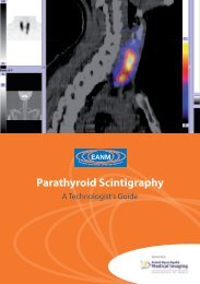
Parathyroid Scintigraphy - European Association of Nuclear Medicine
Parathyroid Scintigraphy - European Association of Nuclear Medicine
Parathyroid Scintigraphy - European Association of Nuclear Medicine

Create successful ePaper yourself
Turn your PDF publications into a flip-book with our unique Google optimized e-Paper software.
<strong>of</strong> preoperative scintigraphy is very helpful<br />
(mandatory in the opinion <strong>of</strong> the author).<br />
Technical factors<br />
All these measurements should be in counts<br />
per second. An energy window <strong>of</strong> 10% <strong>of</strong> the<br />
photo peak <strong>of</strong> 99m Tc is generally preferred.<br />
Quality controls <strong>of</strong> the probe should include<br />
sensitivity, spatial resolution and count linearity<br />
and should be performed every 3–6<br />
months. This is an important step in which<br />
the nuclear medicine technician should play<br />
a major role.<br />
The performance <strong>of</strong> additional intra-operative<br />
QPTH measurement is also recommended by<br />
some authors in order to discover possible<br />
unknown glandular hyperplasia, while some<br />
other authors judge the use <strong>of</strong> the probe sufficient<br />
for the purpose <strong>of</strong> MIRS. When using<br />
QPTH, a fall <strong>of</strong> 50% or more in PTH levels 10<br />
min after parathyroid adenoma removal in<br />
comparison with the baseline pre-excision<br />
value is usually considered indicative <strong>of</strong> successful<br />
parathyroidectomy.<br />
The single-day high 99m Tc-sestamibi dose<br />
protocol versus the low 99m Tc-sestamibi<br />
dose different-day protocol<br />
The first MIRS protocol was developed by<br />
Norman in 1997. It consists <strong>of</strong> a single-day<br />
imaging and surgery approach. The patient<br />
is injected with a 20–25 or 740–925 MBq<br />
dose <strong>of</strong> 99m Tc-sestamibi, images are obtained<br />
Chapter 6: Technical aspects <strong>of</strong> probe-guided surgery for parathyroid adenomas<br />
by the dual-phase scintigraphic technique<br />
and MIRS is performed within 2–3 h after radiotracer<br />
administration. Norman’s protocol<br />
is attractive from a cost-analysis perspective<br />
because 99mTc-sestamibi scintigraphy and MIRS<br />
are performed on the same day and a single<br />
radiotracer dose is required for both imaging<br />
and surgery. However, Norman’s protocol also<br />
presents some practical disadvantages given<br />
the uncertainty <strong>of</strong> the scintigraphic results and<br />
the differences between MIRS and BNE with<br />
respect to the need for operating theatre time<br />
(BNE > MIRS) and efficient patient scheduling.<br />
This problem would be expected to be<br />
even greater in areas with a high prevalence <strong>of</strong><br />
nodular goitre so that a different-day protocol<br />
would be preferable. The protocol developed<br />
in our centre is a different-day protocol. On<br />
the first day, localising images are obtained<br />
by means <strong>of</strong> dual-tracer 99mTc-pertechnetate/ 99mTc-sestamibi subtraction scintigraphy combined<br />
with neck ultrasound. On the day <strong>of</strong><br />
MIRS, usually within 1 week <strong>of</strong> imaging, a low<br />
37 MBq 99mTc-sestamibi dose is given directly<br />
in the operating theatre a few minutes before<br />
surgery. The low 99mTc-sestamibi dose protocol<br />
has two major advantages: (a) less radiation<br />
exposure to the patient and operating theatre<br />
personnel (Table 3) and (b) fewer false negative<br />
results in parathyroid adenoma with rapid<br />
99mTc-sestamibi washout.<br />
Nevertheless, favourable results have been<br />
reported with both Norman’s high 99m Tc-sestamibi<br />
dose protocol and our low 99m Tc-ses-<br />
EANM