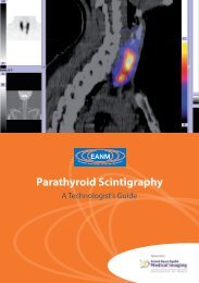
Parathyroid Scintigraphy - European Association of Nuclear Medicine
Parathyroid Scintigraphy - European Association of Nuclear Medicine
Parathyroid Scintigraphy - European Association of Nuclear Medicine

Create successful ePaper yourself
Turn your PDF publications into a flip-book with our unique Google optimized e-Paper software.
Technical aspects <strong>of</strong> probe-guided surgery for<br />
parathyroid adenomas<br />
Domenico Rubello<br />
Bilateral neck exploration (BNE) still represents<br />
the ‘gold standard’ approach in patients with<br />
primary hyperparathyroidism (pHPT). However,<br />
surgical approaches to pHPT patients have<br />
altered significantly in many surgical centres<br />
during the past decade, with the development<br />
<strong>of</strong> minimally invasive parathyroidectomy using<br />
endoscopic surgery or radio-guided surgery<br />
(MIRS). This development can be attributed to<br />
two main reasons: (a) the consciousness that<br />
pHPT is due to a single parathyroid adenoma<br />
in the majority <strong>of</strong> patients (at least 85%), and<br />
(b) the technical improvements introduced<br />
into surgical practice with the availability <strong>of</strong><br />
microsurgery instruments, endoscopes, intraoperative<br />
measurements <strong>of</strong> quick parathyroid<br />
hormone (QPTH) and gamma probes.<br />
New approaches to minimally invasive parathyroidectomy<br />
consisting in the removal <strong>of</strong> a<br />
solitary parathyroid adenoma via a small 1–2<br />
cm skin incision have been widely adopted. Of<br />
course, in contrast to BNE, minimally invasive<br />
parathyroidectomy always requires accurate<br />
preoperative imaging in order (a) to establish<br />
whether the parathyroid adenoma is effectively<br />
solitary and (b) to locate precisely the<br />
enlarged gland. The present chapter focusses<br />
mainly on technical aspects <strong>of</strong> the MIRS technique.<br />
Moreover, the MIRS technique developed<br />
in our centre is based on the injection<br />
<strong>of</strong> a very low 99m Tc-sestamibi dose – 37 MBq<br />
– compared with the traditional MIRS technique,<br />
which uses a ‘high’ 99m Tc-sestamibi<br />
dose – 740–925 MBq.<br />
Selection criteria for <strong>of</strong>fering MIRS<br />
When planning MIRS (unlike when performing<br />
BNE), strict inclusion criteria need to be<br />
followed: (a) evidence at 99mTc-sestamibi scintigraphy<br />
<strong>of</strong> a solitary parathyroid adenoma;<br />
(b) intense 99mTc-sestamibi uptake in the parathyroid<br />
adenoma; (c) absence <strong>of</strong> concomitant<br />
thyroid nodules at 99mTc-sestamibi scintigraphy<br />
and high-resolution (10 MHz) neck ultrasound;<br />
(d) no history <strong>of</strong> familial HPT or multiple endocrine<br />
neoplasia; and (e) no history <strong>of</strong> irradiation<br />
to the neck. Of note, previous thyroid or<br />
parathyroid surgery is not a contraindication<br />
to MIRS. When these inclusion criteria are<br />
adopted, approximately 60–70% <strong>of</strong> pHPT patients<br />
can be <strong>of</strong>fered MIRS. The main reason for<br />
exclusion is the presence <strong>of</strong> 99mTc-sestamibi avid thyroid nodules, which, by mimicking a<br />
parathyroid adenoma, can cause false positive<br />
results during surgery. Figure 1 shows a<br />
patient scheduled for MIRS while Fig. 2 shows<br />
a patient excluded from MIRS.<br />
Preoperative imaging protocol<br />
In our protocol, preoperative imaging procedures<br />
include single-session 99m Tc-sestamibi<br />
scintigraphy and neck ultrasound (Norman<br />
and Chheda 1997; Costello and Norman<br />
1999; Mariani et al. 2003; Rubello et al. 2000).<br />
In patients with concordant 99m Tc-sestamibi<br />
and ultrasound results (both positive or negative),<br />
no further imaging is performed, while<br />
in cases with discrepant findings ( 99m Tc-sestamibi<br />
positive and US negative) a tomographic<br />
(SPECT) examination is obtained to investi-<br />
EANM