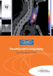
Parathyroid Scintigraphy - European Association of Nuclear Medicine
Parathyroid Scintigraphy - European Association of Nuclear Medicine
Parathyroid Scintigraphy - European Association of Nuclear Medicine

You also want an ePaper? Increase the reach of your titles
YUMPU automatically turns print PDFs into web optimized ePapers that Google loves.
pertechnetate/ 99m Tc-sestamibi combination<br />
is used then the radiation dose for the combined<br />
study is 11.6 mSv. If 123 I and 201 Tl are used,<br />
this rises to 18.3 mSv.<br />
Dual-phase agents<br />
99m Tc-sestamibi and 99m Tc-tetr<strong>of</strong>osmin are commonly<br />
used agents for dual-phase parathyroid<br />
scintigraphy. The washout technique relies on<br />
the fact that while 99m Tc-sestamibi and 99m Tc-tetr<strong>of</strong>osmin<br />
are taken up by both the thyroid gland<br />
and the parathyroid at a similar rate, there is a<br />
faster rate <strong>of</strong> washout from the thyroid gland.<br />
These tracers localise in the thyroid gland as<br />
well as in parathyroid adenomas. This makes<br />
correlation <strong>of</strong> the adenoma in relation to the<br />
thyroid gland possible on planar as well as early<br />
SPECT imaging. 99m Tc-sestamibi is released from<br />
the thyroid with a half-life <strong>of</strong> about 30 min but<br />
is usually retained by abnormal parathyroid<br />
glands (Smith and Oates 2004). 99m Tc-tetr<strong>of</strong>osmin<br />
may clear more slowly from the thyroid<br />
gland. This differential washout improves the<br />
target-to-background ratio so that abnormal<br />
parathyroid tissue should be more visible on<br />
delayed images (Smith and Oates 2004; Clark<br />
2005). However, thyroid adenomas and carcinomas<br />
can coexist and may retain 99m Tc-sestamibi<br />
or 99m Tc-tetr<strong>of</strong>osmin, resulting in false positive<br />
results (Smith and Oates 2004).<br />
99m Tc-sestamibi and 99m Tc-tetr<strong>of</strong>osmin have<br />
comparable imaging characteristics. Usually,<br />
the choice <strong>of</strong> imaging agent depends on its<br />
1<br />
Chapter 2: Radiopharmaceuticals<br />
availability and the experience <strong>of</strong> the nuclear<br />
medicine radiologist.<br />
The dual-phase subtraction method with adjunctive<br />
thyroid-selective imaging ( 99m Tc or 123 I)<br />
may be helpful, or even essential, in patients<br />
with goitres or other confounding underlying<br />
thyroid disease, after thyroid surgery or in<br />
those patients with a palpable mass (Smith<br />
and Oates 2004).<br />
PET imaging agents<br />
Use <strong>of</strong> 18F-fluorodeoxyglucose (FDG) positron<br />
emission tomography (PET) and 11C- methionine<br />
PET for parathyroid imaging has been described<br />
(Otto et al. 2004; Beggs and Hain 2005).<br />
Initial studies with PET have shown conflicting<br />
results when using FDG as a tracer to image the<br />
parathyroid glands (Beggs and Hain 2005; Otto<br />
et al. 2004). It has been shown that 11C-methio nine PET holds more promise than FDG PET imaging<br />
<strong>of</strong> the parathyroid localisation (Beggs and<br />
Hain 2005). 11C-methionine PET scanning is <strong>of</strong><br />
value in cases <strong>of</strong> primary hyperparathyroidism<br />
in which conventional imaging techniques have<br />
failed to localise the adenoma before proceeding<br />
to surgery, or in patients in whom surgery<br />
has been performed but has failed to correct the<br />
hyperparathyroidism (Beggs and Hain 2005).<br />
Adverse reactions to<br />
radiopharmaceuticals<br />
Table 2 shows side-effects and reactions to<br />
radiopharmaceuticals used for parathyroid<br />
scintigraphy.<br />
EANM