Parathyroid Scintigraphy - European Association of Nuclear Medicine
Parathyroid Scintigraphy - European Association of Nuclear Medicine
Parathyroid Scintigraphy - European Association of Nuclear Medicine

Create successful ePaper yourself
Turn your PDF publications into a flip-book with our unique Google optimized e-Paper software.
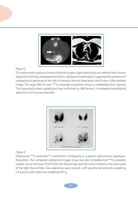
Figure 2<br />
This patient with a previous history <strong>of</strong> thyroid surgery (right lobectomy) was referred with a recent<br />
diagnosis <strong>of</strong> primary hyperparathyroidism. Ultrasound examination suggested the presence <strong>of</strong><br />
a parathyroid adenoma at the side <strong>of</strong> previous thyroid lobectomy, which was a false-positive<br />
image. The large field <strong>of</strong> view 99m Tc-sestamibi acquisition shows a mediastinal focus (arrow).<br />
The suspected ectopic parathyroid was confirmed by MRI (arrow). A mediastinal parathyroid<br />
adenoma <strong>of</strong> 0.59 g was resected.<br />
Figure 3<br />
<strong>Parathyroid</strong> 99m Tc-sestamibi/ 123 I subtraction scintigraphy in a patient with primary hyperparathyroidism.<br />
The computed subtraction images show two sites <strong>of</strong> preferential 99m Tc-sestamibi<br />
uptake: one at the lower third <strong>of</strong> the left thyroid lobe and the second lateral to the lower pole<br />
<strong>of</strong> the right thyroid lobe. Two adenomas were excised: a left parathyroid adenoma weighing<br />
2.3 g and a right adenoma weighing 0.07 g.<br />
1