Parathyroid Scintigraphy - European Association of Nuclear Medicine
Parathyroid Scintigraphy - European Association of Nuclear Medicine
Parathyroid Scintigraphy - European Association of Nuclear Medicine

Create successful ePaper yourself
Turn your PDF publications into a flip-book with our unique Google optimized e-Paper software.
patients with renal failure (Hindié et al. 1999;<br />
Perié et al. 2005)<br />
What information can be obtained?<br />
• The preoperative map may facilitate recognition<br />
<strong>of</strong> the position <strong>of</strong> aberrant parathyroid<br />
glands, also reducing the extent <strong>of</strong><br />
dissection (Hindié et al. 1999).<br />
•<br />
•<br />
<strong>Parathyroid</strong> glands with major ectopia<br />
would be missed without preoperative<br />
imaging.<br />
Although the usual number <strong>of</strong> parathyroid<br />
glands is four, some individuals (about 10%)<br />
have a supernumerary fifth gland (Akerström<br />
et al. 1984). When this information is<br />
provided by preoperative imaging, it may<br />
prevent surgical failure or late recurrence<br />
(Hindié et al. 1999).<br />
Imaging findings in patients with persistent<br />
or recurrent secondary hyperparathyroidism<br />
Immediate failure and delayed recurrence are<br />
not unusual, occurring in 10–30% <strong>of</strong> patients.<br />
Imaging is mandatory before reoperation.<br />
Knowledge <strong>of</strong> all details concerning the initial<br />
intervention is necessary for interpretation. As<br />
with primary hyperparathyroidism, we recommend<br />
that lesions seen on the 99m Tc-sestamibi<br />
scan be matched with a second radiological<br />
technique (ultrasound or MRI) for confirmation<br />
and identification <strong>of</strong> anatomical landmarks<br />
before reoperation.<br />
11<br />
Chapter 1: Applications <strong>of</strong> parathyroid imaging<br />
Some aspects specific to patients reoperated<br />
for secondary hyperparathyroidism need to<br />
be emphasised:<br />
•<br />
•<br />
Specific views <strong>of</strong> the forearm should be<br />
obtained in patients who have had a parathyroid<br />
graft.<br />
It is not unusual for imaging in these patients<br />
to show two foci <strong>of</strong> activity, one<br />
corresponding to recurrent disease at the<br />
subtotally resected gland (or grafted tissue)<br />
and the other corresponding to an ectopic<br />
or fifth parathyroid, missed at initial intervention<br />
(unpublished data).<br />
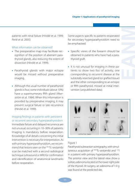
Figure 1<br />
<strong>Parathyroid</strong> subtraction scintigraphy, with simultaneous<br />
acquisition <strong>of</strong> 99m Tc-sestamibi and 123 I<br />
in a patient with primary hyperparathyroidism.<br />
The anterior view and the lateral view show a<br />
solitary adenoma located at the lower right pole<br />
<strong>of</strong> the thyroid. At surgery, an adenoma <strong>of</strong> 1.9 g<br />
was found at the predicted site.<br />
EANM