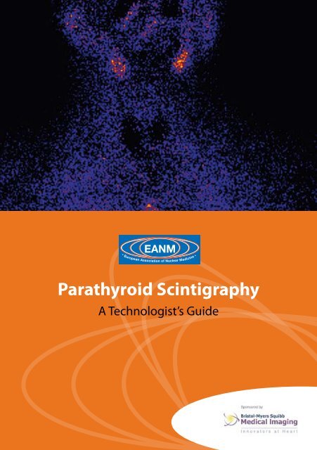
Parathyroid Scintigraphy - European Association of Nuclear Medicine
Parathyroid Scintigraphy - European Association of Nuclear Medicine
Parathyroid Scintigraphy - European Association of Nuclear Medicine

Create successful ePaper yourself
Turn your PDF publications into a flip-book with our unique Google optimized e-Paper software.
<strong>European</strong> <strong>Association</strong> <strong>of</strong> <strong>Nuclear</strong> <strong>Medicine</strong><br />
<strong>Parathyroid</strong> <strong>Scintigraphy</strong><br />
A Technologist’s Guide
Contributors<br />
Nish Fernando<br />
Chief Technologist<br />
Department <strong>of</strong> <strong>Nuclear</strong> <strong>Medicine</strong><br />
St. Bartholomew’s Hospital, London, UK<br />
Dr. Elif Hindié, MD, PhD<br />
Service de Médecine Nucléaire<br />
Hôpital Saint-Antoine, Paris, France<br />
Sue Huggett<br />
Senior University Teacher<br />
Department <strong>of</strong> Radiography<br />
City University, London, United Kingdom<br />
José Pires Jorge<br />
Pr<strong>of</strong>esseur HES-S2<br />
Ecole Cantonale Vaudoise de Techniciens en<br />
Radiologie Médicale (TRM)<br />
Lausanne, Switzerland<br />
Regis Lecoultre<br />
Pr<strong>of</strong>esseur HES-S2<br />
Ecole Cantonale Vaudoise de Techniciens en<br />
Radiologie Médicale (TRM)<br />
Lausanne, Switzerland<br />
Sylviane Prévot<br />
Chair, EANM Technologist Committee<br />
Chief Technologist<br />
Service de Médecine Nucléaire<br />
Centre Georges-François Leclerc, Dijon, France<br />
Domenico Rubello*, MD<br />
Director<br />
<strong>Nuclear</strong> <strong>Medicine</strong> Service - PET Unit<br />
‘S. Maria della Misericordia’ Hospital<br />
Istituto Oncologico Veneto (IOV), Rovigo, Italy<br />
Audrey Taylor<br />
Chief Technologist<br />
Department <strong>of</strong> <strong>Nuclear</strong> <strong>Medicine</strong><br />
Guy’s and St. Thomas’ Hospital, London, UK<br />
Linda Tutty<br />
Senior Radiographer<br />
St. James’s Hospital<br />
Dublin, Ireland<br />
*Domenico Rubello, MD<br />
is coordinator <strong>of</strong> the National Study Group on parathyroid scintigraphy <strong>of</strong> AIMN (Italian <strong>Association</strong> <strong>of</strong> <strong>Nuclear</strong><br />
<strong>Medicine</strong>) and is responsible for developing study programmes on minimally invasive radioguided surgery in patients<br />
with hyperparathyroidism for GISCRIS (Italian Study Group on Radioguided Surgery and Immunoscintigraphy)
Contents<br />
Foreword 4<br />
Sylviane Prévot<br />
Introduction 5<br />
Sue Huggett<br />
Chapter 1 – Applications <strong>of</strong> parathyroid imaging 6–12<br />
Elif Hindié<br />
Chapter 2 – Radiopharmaceuticals 13–17<br />
Linda Tutty<br />
Chapter 3 – Imaging equipment – preparation and use 18–23<br />
José Pires Jorge and Regis Lecoultre<br />
Chapter 4 – Patient preparation 24–28<br />
Audrey Taylor and Nish Fernando<br />
Chapter 5 – Imaging protocols 29–32<br />
Nish Fernando and Sue Huggett<br />
Chapter 6 – Technical aspects <strong>of</strong> probe-guided surgery for parathyroid adenomas 33–39<br />
Domenico Rubello<br />
References 40–43<br />
This booklet was sponsored by an educational grant from Bristol-Myers Squibb Medical Imaging.<br />
The views expressed are those <strong>of</strong> the authors and not necessarily <strong>of</strong> Bristol-Myers Squibb Medical<br />
Imaging.<br />
EANM
Foreword<br />
Sylviane Prévot<br />
Today the notion <strong>of</strong> competence is at the<br />
heart <strong>of</strong> pr<strong>of</strong>essional development. Technologists’<br />
specific pr<strong>of</strong>essional skills <strong>of</strong> working<br />
efficiently and knowledgeably are essential<br />
to ensure high-quality practice in nuclear<br />
medicine departments.<br />
Since they were formed, the EANM Technologist<br />
Committee and Sub-committee on<br />
Education have devoted themselves to the<br />
improvement <strong>of</strong> nuclear medicine technologists’<br />
(NMTs) pr<strong>of</strong>essional skills.<br />
Publications that will assist in the setting <strong>of</strong><br />
high standards for NMTs’ work throughout<br />
Europe have been developed. A series <strong>of</strong> brochures,<br />
“technologists’ guides”, was planned in<br />
early 2004. The first <strong>of</strong> these was dedicated to<br />
myocardial perfusion imaging and the current<br />
volume, the second in the planned series, addresses<br />
parathyroid imaging.<br />
Renowned authors with expertise in the field<br />
have been selected to provide an informative<br />
and truly comprehensive tool for technologists<br />
that will serve as a reference and improve<br />
the quality <strong>of</strong> daily practice.<br />
I am grateful for the hard work <strong>of</strong> all the contributors,<br />
who have played a key role in ensuring<br />
the high scientific content and educational<br />
value <strong>of</strong> this booklet. Many thanks are due to<br />
Sue Huggett, who coordinated the project,<br />
to the members <strong>of</strong> the EANM Technologist<br />
Sub-committee on Education and particularly<br />
to Bristol-Myers Squibb Imaging for their confidence<br />
and generous sponsorship.<br />
Efforts to image the parathyroid gland date<br />
back many years. I hope this brochure will be<br />
useful to technologists in the management<br />
<strong>of</strong> patients with hyperparathyroidism and will<br />
benefit these patients by optimising care and<br />
welfare.<br />
Sylviane Prévot<br />
Chair, EANM Technologist Committee
Introduction<br />
Sue Huggett<br />
The first publication <strong>of</strong> the EANM Technologist<br />
Committee sponsored by Bristol Myers<br />
Squibb in 2004 was a book on myocardial<br />
perfusion imaging for technologists. We<br />
are very grateful that they have sponsored<br />
us again this year to produce this book on<br />
parathyroid imaging, the second book in<br />
what we hope will be a series.<br />
We hope that we have combined the theory<br />
and rationale <strong>of</strong> imaging with the practicalities<br />
<strong>of</strong> patient care and equipment use. I think that<br />
certain things I wrote for the last book bear repeating,<br />
and so I will do so here for the benefit<br />
<strong>of</strong> those for whom this is their first book.<br />
Knowledge <strong>of</strong> imaging theory provides a deeper<br />
understanding <strong>of</strong> the techniques that is satisfying<br />
for the technologist and can form the basis<br />
for wise decision making. It also allows the technologist<br />
to communicate accurate information<br />
to patients, their carers and other staff. Patient<br />
care is always paramount, and being able to<br />
explain why certain foods must be avoided or<br />
why it is necessary to lie in awkward positions<br />
improves compliance as well as satisfaction.<br />
Awareness <strong>of</strong> the rationales for using certain<br />
strategies is needed in order to know when<br />
and how various protocol variations should be<br />
applied, in acquisition or analysis, e.g. for the<br />
patient who cannot lie flat for long enough<br />
for subtraction and may need to be imaged<br />
with another protocol or when we may need<br />
a different filter if the total counts are low.<br />
Protocols will vary between departments,<br />
even within the broader terms <strong>of</strong> the EANM<br />
Guidelines. This booklet is not meant to supplant<br />
these protocols but will hopefully supplement<br />
and explain the rationales behind<br />
them, thereby leading to more thoughtful<br />
working practices.<br />
The authors are indebted to a number <strong>of</strong><br />
sources for information, not least local protocols,<br />
and references have been given where<br />
original authors are identifiable. We apologise<br />
if we have inadvertently used material<br />
for which credit should have been, but was<br />
not, given.<br />
We hope that this booklet will provide helpful<br />
information as and when it is needed so<br />
that the integration <strong>of</strong> theory and practice is<br />
enabled and encouraged.<br />
EANM
Applications <strong>of</strong> parathyroid imaging<br />
Elif Hindié<br />
Primary hyperparathyroidism<br />
Primary hyperparathyroidism (pHPT) is a<br />
surgically correctable disease with the third<br />
highest incidence <strong>of</strong> all endocrine disorders<br />
after diabetes mellitus and hyperthyroidism<br />
(Al Zahrani and Levine 1997). Through their<br />
secretion <strong>of</strong> parathyroid hormone (PTH), the<br />
two pairs <strong>of</strong> parathyroid glands, located in the<br />
neck posterior to the thyroid gland, regulate<br />
serum calcium concentration and bone metabolism.<br />
PTH promotes the release <strong>of</strong> calcium<br />
from bone, increases absorption <strong>of</strong> calcium<br />
from the intestine and increases reabsorption<br />
<strong>of</strong> calcium in the renal tubules. In turn,<br />
the serum calcium concentration regulates<br />
PTH secretion, a mechanism mediated via a<br />
calcium-sensing receptor on the surface <strong>of</strong><br />
the parathyroid cells. pHPT is caused by the<br />
secretion <strong>of</strong> excessive amounts <strong>of</strong> PTH by<br />
one or more enlarged diseased parathyroid<br />
gland(s). Patients with pHPT may suffer from<br />
renal stones, osteoporosis, gastro-intestinal<br />
symptoms, cardiovascular disease, muscle<br />
weakness and fatigue, and neuropsychological<br />
disorders. The highest prevalence <strong>of</strong> the<br />
disease is found in post-menopausal women.<br />
A prevalence <strong>of</strong> 2% was found by screening<br />
post-menopausal women (Lundgren).<br />
In the past, pHPT was characterised by severe<br />
skeletal and renal complications and apparent<br />
mortality. This may still be the case in some<br />
developing countries. The introduction <strong>of</strong><br />
calcium auto-analysers in the early 1970s<br />
led to changes in the incidence <strong>of</strong> pHPT and<br />
deeply modified the clinical spectrum <strong>of</strong> the<br />
disease at diagnosis (Heath et al. 1980). Most<br />
new cases are now biologically mild without<br />
overt symptoms (Al Zahrani and Levine 1997).<br />
<strong>Parathyroid</strong>ectomy is the only curative treatment<br />
for pHPT. In the recent guidelines <strong>of</strong> the<br />
US National Institute <strong>of</strong> Health (NIH), surgery<br />
is recommended for all young individuals and<br />
for all patients with overt symptoms (Bilezikian<br />
et al. 2002). For patients who are asymptomatic<br />
and are 50 years old or older, surgery is<br />
recommended if any <strong>of</strong> the following signs<br />
are present: serum calcium greater than 10<br />
mg/l above the upper limits <strong>of</strong> normal; 24-h<br />
total urine calcium excretion <strong>of</strong> more than 400<br />
mg; reduction in creatinine clearance by more<br />
than 30% compared with age-matched persons;<br />
bone density more than 2.5 SDs below<br />
peak bone mass: T score < -2.5. Surgery is also<br />
recommended when medical surveillance<br />
is either not desirable or not possible. After<br />
complete baseline evaluation, patients who<br />
are not operated on need to be monitored<br />
twice yearly for serum calcium concentration<br />
and yearly for creatinine concentration; it is<br />
also recommended that bone mass measurements<br />
are obtained on a yearly basis (Bilezikian<br />
et al. 2002). Some authors recommend parathyroidectomy<br />
for all patients with a secure<br />
diagnosis <strong>of</strong> pHPT (Utiger 1999).<br />
Successful parathyroidectomy depends on<br />
recognition and excision <strong>of</strong> all hyperfunctioning<br />
parathyroid glands. pHPT is typically<br />
caused by a solitary parathyroid adenoma, less
frequently (about 15% <strong>of</strong> cases) by multiple<br />
parathyroid gland disease (MGD) and rarely<br />
(about 1% <strong>of</strong> cases) by parathyroid carcinoma.<br />
Patients with MGD have either double<br />
adenomas or hyperplasia <strong>of</strong> three or all four<br />
parathyroid glands. Most cases <strong>of</strong> MGD are<br />
sporadic, while a small number are associated<br />
with hereditary disorders such as multiple endocrine<br />
neoplasia type 1 or type 2a or familial<br />
hyperparathyroidism (Marx et al. 2002). Conventional<br />
surgery consists in routine bilateral<br />
exploration with identification <strong>of</strong> all four parathyroid<br />
glands.<br />
Imaging is mandatory before reoperation<br />
For several decades, preoperative imaging was<br />
not used before first-time surgery. Unguided<br />
bilateral exploration, dissecting all potential<br />
sites in the neck, achieved cure in 90–95% <strong>of</strong><br />
patients (Russell and Edis 1982). The two main<br />
reasons for failed surgery are ectopic glands<br />
(retro-oesophageal, mediastinal, intrathyroid,<br />
in the sheath <strong>of</strong> the carotid artery, or undescended)<br />
and undetected MGD (Levin and<br />
Clark 1989). Repeat surgery is associated with a<br />
dramatic reduction in the success rate and an<br />
increase in surgical complications. Imaging is<br />
therefore mandatory before reoperation (Sosa<br />
et al. 1998). 99m Tc-sestamibi scanning (Coakley<br />
et al. 1989) has been established as the imaging<br />
method <strong>of</strong> choice in reoperation <strong>of</strong> persistent<br />
or recurrent hyperparathyroidism (Weber<br />
et al. 1993). In these patients it is necessary to<br />
have all information concerning the first intervention,<br />
including the number and location <strong>of</strong><br />
Chapter 1: Applications <strong>of</strong> parathyroid imaging<br />
parathyroid glands that have been seen by the<br />
surgeon and the size and histology <strong>of</strong> resected<br />
glands. Whichever 99mTc-sestamibi scanning<br />
protocol is used, it is necessary to provide the<br />
surgeon with the best anatomical information<br />
by using both anterior and lateral (or oblique)<br />
views <strong>of</strong> the neck, and SPECT whenever useful,<br />
especially for a mediastinal focus. It is the<br />
author’s opinion that 99mTc-sestamibi results<br />
should be confirmed with a second imaging<br />
technique (usually ultrasound for a neck focus<br />
and CT or MRI for a mediastinal image) before<br />
proceeding to reoperation.<br />
Scanning with 99m Tc-sestamibi is increasingly<br />
ordered on a routine basis for first-time<br />
parathyroidectomy<br />
The first exploration is the best time to cure<br />
hyperparathyroidism. Most surgeons would<br />
now appreciate having information concerning<br />
whereabouts in the neck to start dissection<br />
and the possibility <strong>of</strong> ectopic parathyroid<br />
glands (Sosa et al. 1998; Liu et al. 2005). When<br />
the rare cases (2-5%) <strong>of</strong> ectopic parathyroid<br />
tumours are recognised preoperatively, the<br />
success <strong>of</strong> bilateral surgery can now reach<br />
very close to 100% (Hindié et al. 1997). In the<br />
case <strong>of</strong> a mediastinal gland, the surgeon can<br />
proceed directly with first-intention thoracoscopy,<br />
avoiding unnecessary initial extensive<br />
neck surgery in the search for the elusive<br />
gland (Liu et al. 2005). Preoperative imaging<br />
would also shorten the duration <strong>of</strong> bilateral<br />
surgery (Hindié et al. 1997). By allowing the<br />
surgeon to find the <strong>of</strong>fending gland earlier in<br />
EANM
the operation, the time necessary for frozen<br />
section examination can be used by the surgeon<br />
for inspection <strong>of</strong> the other parathyroid<br />
glands, also reducing surgeon anxiety.<br />
Important points to know when proceeding<br />
with parathyroid imaging<br />
• Imaging is not for diagnosis. The increase<br />
in plasma levels <strong>of</strong> calcium (normal value<br />
88–105 mg/l) and PTH (normal value 10–58<br />
ng/l) establishes the diagnosis.<br />
•<br />
•<br />
•<br />
•<br />
•<br />
Imaging does not identify normal parathyroid<br />
glands, which are too small (20–50 mg)<br />
to be seen.<br />
Imaging should detect abnormal parathyroid(s)<br />
and indicate the approximate size<br />
and the precise relationship to the thyroid<br />
(the level <strong>of</strong> the thyroid at which the parathyroid<br />
lesion is seen on the anterior view;<br />
and whether it is proximal to the thyroid or<br />
deeper in the neck on the lateral or oblique<br />
view or SPECT) (Fig. 1).<br />
Imaging should identify ectopic glands (add<br />
SPECT in cases <strong>of</strong> a mediastinal focus, and<br />
ask for additional CT or MRI for confirmation<br />
and anatomical landmarks) (Fig. 2).<br />
Imaging should be able to differentiate patients<br />
with a single adenoma from those<br />
with MGD (Fig. 3).<br />
Imaging should identify thyroid nodules<br />
that may require concurrent surgical resection.<br />
The choice <strong>of</strong> imaging technique<br />
The most common preoperative localisation<br />
methods are radionuclide scintigraphy and<br />
ultrasound. As stated before, the two main<br />
reasons for failed surgery are ectopic glands<br />
and undetected MGD (Levin and Clark 1989).<br />
Because high-resolution ultrasound would,<br />
even in skilled hands, fail to detect the majority<br />
<strong>of</strong> these cases, it is not optimal for preoperative<br />
imaging as a single technique. In the<br />
study by Haber et al. (2002), ultrasound missed<br />
six <strong>of</strong> eight ectopic glands and five <strong>of</strong> six cases<br />
<strong>of</strong> MGD. Ultrasound may, however, be useful<br />
in combination with 99m Tc-sestamibi imaging<br />
(Rubello et al. 2003).<br />
99m Tc-sestamibi scanning is now considered<br />
the most sensitive imaging technique in patients<br />
with pHPT (Giordano et al. 2001; Mullan<br />
2004). Whatever the protocol used, 99m Tc-sestamibi<br />
scanning will usually meet the requirement<br />
<strong>of</strong> detecting ectopic glands (all eight<br />
were detected in the study by Haber et al.).<br />
With regard to the recognition <strong>of</strong> MGD, however,<br />
the protocol in use will determine the<br />
sensitivity. When 99m Tc-sestamibi is used as a<br />
single tracer with planar imaging at two time<br />
points -- the “dual-phase” (or washout) method<br />
– the sensitivity for primary hyperplasia is very<br />
low (Taillefer et al. 1992; Martin et al. 1996). Better<br />
results can be obtained by adding SPECT.<br />
Subtraction scanning, using either 123 I (Borley
et al. 1996; Hindié et al. 2000; Mullan 2004)<br />
or 99m Tc-pertechnetate (Rubello et al. 2003)<br />
in addition to 99m Tc-sestamibi, improves the<br />
sensitivity for hyperplastic glands. One difficulty<br />
with subtraction imaging is keeping the<br />
patient still for the time necessary to scan the<br />
thyroid, to inject 99m Tc-sestamibi and to record<br />
images <strong>of</strong> this second tracer. Simultaneous<br />
recording <strong>of</strong> 123 I and 99m Tc-sestamibi can be a<br />
simple answer to these difficulties. It prevents<br />
artefacts on subtraction images due to patient<br />
motion, and shortens the imaging time<br />
(Hindié et al. 1998; Mullan 2004).<br />
Preoperative imaging has opened a new era<br />
<strong>of</strong> minimally invasive parathyroid surgery<br />
Conventional bilateral exploration is still<br />
considered the gold standard in parathyroid<br />
surgery. However, the introduction <strong>of</strong> 99m Tcsestamibi<br />
scanning, the availability <strong>of</strong> intraoperative<br />
adjuncts such as the gamma probe<br />
and intraoperative monitoring <strong>of</strong> PTH to help<br />
detect MGD have challenged the dogma <strong>of</strong><br />
routine bilateral exploration. When preoperative<br />
imaging points to a single well-defined<br />
focus, unequivocally suggesting a “solitary<br />
adenoma”, the surgeon may now choose focussed<br />
surgery instead <strong>of</strong> bilateral exploration.<br />
Focussed excision can be made by open surgery<br />
through a mini-incision, possibly under<br />
local anaesthesia, or by video-assisted endoscopic<br />
surgery under general anaesthesia (Lee<br />
and Inabnet 2005). Compared with patients<br />
who undergo bilateral surgery, those in whom<br />
focussed parathyroid surgery is successfully<br />
Chapter 1: Applications <strong>of</strong> parathyroid imaging<br />
completed enjoy a shorter operation time,<br />
the possibility <strong>of</strong> local anaesthesia, a better<br />
cosmetic scar, a less painful postoperative<br />
course, less pr<strong>of</strong>ound postoperative “transient”<br />
hypocalcaemia and an earlier return to normal<br />
activities. The fact that many clinicians now<br />
use a lower threshold for surgery is partly due<br />
to the perception that parathyroid surgery is<br />
easier than in the past (Utiger 1999).<br />
Patients at specific risk <strong>of</strong> failure <strong>of</strong> minimal<br />
surgery are those with unrecognised MGD.<br />
Therefore, when choosing minimal surgery,<br />
the surgeon is committed to distinguishing<br />
cases <strong>of</strong> MGD either preoperatively, through<br />
an appropriate imaging protocol, or by intraoperative<br />
monitoring <strong>of</strong> PTH plasma levels, or<br />
by a combination <strong>of</strong> both. The true sensitivity<br />
<strong>of</strong> intraoperative PTH for MGD is still under<br />
debate. What raises concern is that studies<br />
relying solely on intraoperative measurements<br />
report a low percentage <strong>of</strong> MGD, only 3% (Molinari<br />
et al. 1996), which is three to four times<br />
lower than is generally observed during routine<br />
bilateral surgery. Whether this will lead to<br />
higher rates <strong>of</strong> late recurrence is not known. It<br />
is thus important that imaging methods used<br />
to select patients for focussed surgery have a<br />
high sensitivity for detecting MGD.<br />
In this new era <strong>of</strong> focussed operations, the<br />
success <strong>of</strong> parathyroid surgery depends not<br />
only on an experienced surgeon but also on<br />
excellent interpretation <strong>of</strong> images. A localisation<br />
study with high accuracy is mandatory to<br />
EANM
avoid conversion <strong>of</strong> the surgery to a bilateral<br />
exploration under general anaesthesia after<br />
minimal surgery has been started. It is important<br />
to avoid confusion with a thyroid nodule,<br />
and precise anatomical description is also<br />
important. With enlargement and increased<br />
density, superior parathyroid adenomas can<br />
become pendulous and descend posteriorly.<br />
A lateral view (or an oblique view or SPECT)<br />
should indicate whether the adenoma is close<br />
to the thyroid or deeper in the neck (tracheooesophageal<br />
groove or retro-oesophageal).<br />
This information is useful, because visualisation<br />
through the small incision is restricted.<br />
Moreover, the surgeon may choose a lateral<br />
approach to excise this gland instead <strong>of</strong> an<br />
anterior approach. To achieve a high sensitivity<br />
in detecting MGD with subtraction techniques,<br />
the degree <strong>of</strong> subtraction should be<br />
monitored carefully. Progressive incremental<br />
subtraction with real-time display is a good<br />
way to choose the optimal level <strong>of</strong> subtraction<br />
(residual 99m Tc-sestamibi activity in the thyroid<br />
area should not be lower than in surrounding<br />
neck tissues). Oversubtraction could easily<br />
delete additional foci <strong>of</strong> activity and in some<br />
patients provide a false image suggestive <strong>of</strong><br />
a single adenoma.<br />
Secondary hyperparathyroidism<br />
Secondary hyperparathyroidism is a common<br />
complication in patients with chronic renal<br />
failure. Hypocalcaemia, accumulation <strong>of</strong> phosphate<br />
and a decrease in the active form <strong>of</strong><br />
vitamin D lead to increased secretion <strong>of</strong> PTH.<br />
10<br />
With chronic stimulation, hyperplasia <strong>of</strong> parathyroid<br />
glands accelerates and may develop<br />
into autonomous adenomas. The extent <strong>of</strong><br />
parathyroid growth then becomes a major<br />
determinant <strong>of</strong> PTH hypersecretion. Secondary<br />
hyperparathyroidism leads to renal bone<br />
disease, the development <strong>of</strong> s<strong>of</strong>t tissue calcifications,<br />
vascular calcifications and increased<br />
cardiovascular risk, among other complications.<br />
When medical therapy fails, surgery<br />
becomes necessary. Surgery can be either<br />
subtotal parathyroidectomy, with resection<br />
<strong>of</strong> three glands and partial resection <strong>of</strong> the<br />
fourth gland, or total resection with grafting<br />
<strong>of</strong> some parathyroid tissue into the s<strong>of</strong>t tissues<br />
<strong>of</strong> the forearm in order to avoid permanent<br />
hypoparathyroidism.<br />
Preoperative imaging<br />
Surgery <strong>of</strong> secondary hyperparathyroidism<br />
requires routine bilateral identification <strong>of</strong> all<br />
parathyroid tissue. Moreover, early studies<br />
based on single-tracer 99m Tc-sestamibi scanning<br />
have reported a very low sensitivity<br />
<strong>of</strong> about 40–50% in detecting hyperplastic<br />
glands. Inefficiency <strong>of</strong> single-tracer techniques<br />
both in secondary hyperparathyroidism and<br />
in primary hyperplasia is possibly due to more<br />
rapid washout <strong>of</strong> tracer from hyperplastic<br />
glands than from parathyroid adenomas. For<br />
those reasons, preoperative imaging has not<br />
yet gained wide acceptance among surgeons.<br />
Dual-tracer subtraction imaging, planar or<br />
SPECT, provides substantial improvement in<br />
the rate <strong>of</strong> detection <strong>of</strong> hyperplastic glands in
patients with renal failure (Hindié et al. 1999;<br />
Perié et al. 2005)<br />
What information can be obtained?<br />
• The preoperative map may facilitate recognition<br />
<strong>of</strong> the position <strong>of</strong> aberrant parathyroid<br />
glands, also reducing the extent <strong>of</strong><br />
dissection (Hindié et al. 1999).<br />
•<br />
•<br />
<strong>Parathyroid</strong> glands with major ectopia<br />
would be missed without preoperative<br />
imaging.<br />
Although the usual number <strong>of</strong> parathyroid<br />
glands is four, some individuals (about 10%)<br />
have a supernumerary fifth gland (Akerström<br />
et al. 1984). When this information is<br />
provided by preoperative imaging, it may<br />
prevent surgical failure or late recurrence<br />
(Hindié et al. 1999).<br />
Imaging findings in patients with persistent<br />
or recurrent secondary hyperparathyroidism<br />
Immediate failure and delayed recurrence are<br />
not unusual, occurring in 10–30% <strong>of</strong> patients.<br />
Imaging is mandatory before reoperation.<br />
Knowledge <strong>of</strong> all details concerning the initial<br />
intervention is necessary for interpretation. As<br />
with primary hyperparathyroidism, we recommend<br />
that lesions seen on the 99m Tc-sestamibi<br />
scan be matched with a second radiological<br />
technique (ultrasound or MRI) for confirmation<br />
and identification <strong>of</strong> anatomical landmarks<br />
before reoperation.<br />
11<br />
Chapter 1: Applications <strong>of</strong> parathyroid imaging<br />
Some aspects specific to patients reoperated<br />
for secondary hyperparathyroidism need to<br />
be emphasised:<br />
•<br />
•<br />
Specific views <strong>of</strong> the forearm should be<br />
obtained in patients who have had a parathyroid<br />
graft.<br />
It is not unusual for imaging in these patients<br />
to show two foci <strong>of</strong> activity, one<br />
corresponding to recurrent disease at the<br />
subtotally resected gland (or grafted tissue)<br />
and the other corresponding to an ectopic<br />
or fifth parathyroid, missed at initial intervention<br />
(unpublished data).<br />
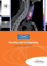
Figure 1<br />
<strong>Parathyroid</strong> subtraction scintigraphy, with simultaneous<br />
acquisition <strong>of</strong> 99m Tc-sestamibi and 123 I<br />
in a patient with primary hyperparathyroidism.<br />
The anterior view and the lateral view show a<br />
solitary adenoma located at the lower right pole<br />
<strong>of</strong> the thyroid. At surgery, an adenoma <strong>of</strong> 1.9 g<br />
was found at the predicted site.<br />
EANM
Figure 2<br />
This patient with a previous history <strong>of</strong> thyroid surgery (right lobectomy) was referred with a recent<br />
diagnosis <strong>of</strong> primary hyperparathyroidism. Ultrasound examination suggested the presence <strong>of</strong><br />
a parathyroid adenoma at the side <strong>of</strong> previous thyroid lobectomy, which was a false-positive<br />
image. The large field <strong>of</strong> view 99m Tc-sestamibi acquisition shows a mediastinal focus (arrow).<br />
The suspected ectopic parathyroid was confirmed by MRI (arrow). A mediastinal parathyroid<br />
adenoma <strong>of</strong> 0.59 g was resected.<br />
Figure 3<br />
<strong>Parathyroid</strong> 99m Tc-sestamibi/ 123 I subtraction scintigraphy in a patient with primary hyperparathyroidism.<br />
The computed subtraction images show two sites <strong>of</strong> preferential 99m Tc-sestamibi<br />
uptake: one at the lower third <strong>of</strong> the left thyroid lobe and the second lateral to the lower pole<br />
<strong>of</strong> the right thyroid lobe. Two adenomas were excised: a left parathyroid adenoma weighing<br />
2.3 g and a right adenoma weighing 0.07 g.<br />
1
Radiopharmaceuticals<br />
Linda Tutty<br />
Radiopharmaceuticals used for<br />
parathyroid scintigraphy<br />
Details <strong>of</strong> the photo peak energy, half-life, effective<br />
dose and standard dose for radiopharmaceuticals<br />
commonly used for parathyroid<br />
scintigraphy are shown in Table 1.<br />
201 Tl-chloride<br />
201 Tl-chloride has a physical half-life <strong>of</strong> 73.1 h.<br />
Its main photo peak is due to characteristic xrays<br />
<strong>of</strong> mercury, which have an energy range<br />
<strong>of</strong> 69–83 keV. In addition, gamma rays are produced<br />
at 167 keV (8%) and 135 keV (2%). The<br />
administered activity is 80 MBq and it is given<br />
intravenously. 201 Tl-chloride is taken up by abnormal<br />
parathyroid tissue and thyroid tissue in<br />
proportion to blood flow.<br />
99m Tc-pertechnetate<br />
99m Tc-pertechnetate has a half-life <strong>of</strong> 6 h and a<br />
gamma energy <strong>of</strong> 140 keV. 99m Tc-pertechnetate is<br />
used to delineate the thyroid gland because functioning<br />
thyroid parenchyma traps it. This image is<br />
then subtracted from the 201 Tl or 99m Tc-sestamibi<br />
images, and what remains is potentially a parathyroid<br />
adenoma. When utilising 201 Tl, the administered<br />
activity is usually 75–150 MBq, depending on<br />
the administered radioactivity <strong>of</strong> 201 Tl and which <strong>of</strong><br />
the two radiopharmaceuticals is administered first.<br />
If using 99m Tc-sestamibi, the amount <strong>of</strong> pertechnetate<br />
administered is usually 185–370 MBq, because<br />
99m Tc-sestamibi has a higher total activity in the<br />
thyroid tissue than 201 Tl.<br />
99m Tc-sestamibi<br />
The range <strong>of</strong> intravenously administered radio-<br />
1<br />
activity is 185-900 MBq; the typical dose is 740<br />
MBq. This radiotracer localises in both parathyroid<br />
gland and functioning thyroid tissue, and usually<br />
washes out <strong>of</strong> normal thyroid tissue more rapidly<br />
than out <strong>of</strong> abnormal parathyroid tissue. The<br />
exact mechanism <strong>of</strong> uptake remains unknown<br />
(Farley 2004). 99mTc-sestamibi uptake depends on<br />
numerous factors, including perfusion, cell cycle<br />
phase and functional activity (Beggs and Hain<br />
2005). The final cellular localisation <strong>of</strong> 99mTc-sesta mibi is within the mitochondria. It accumulates in<br />
the mitochondria <strong>of</strong> many tissues but particularly<br />
in normal cardiac and thyroid cells; it is especially<br />
prominent in overactive parathyroid glands and<br />
is held there preferentially (Farley 2004).<br />
123 I-sodium iodide<br />
123 I has a half-life <strong>of</strong> 13 h and emits a photon<br />
with an energy <strong>of</strong> 159 keV. It has been used<br />
particularly with 99m Tc-sestamibi as a thyroidimaging<br />
agent in subtraction studies. The<br />
administered dose, given orally, ranges from<br />
7.5 to 20 MBq.<br />
99m Tc-tetr<strong>of</strong>osmin<br />
99m Tc-tetr<strong>of</strong>osmin use in parathyroid imaging<br />
is described in the literature (Smith and Oates<br />
2004). Its manufacturers do not license it for<br />
use as a parathyroid scintigraphy agent. 99m Tcsestamibi<br />
and 99m Tc-tetr<strong>of</strong>osmin have similar<br />
imaging characteristics (Smith and Oates<br />
2004). The typical dose <strong>of</strong> administered activity<br />
is 740 MBq.<br />
11 C-methionine<br />
11 C-methionine has a half-life <strong>of</strong> 20 min. It is<br />
EANM
cyclotron produced. Its uptake reflects amino<br />
acid reflux into stimulated parathyroid tissue<br />
(Otto et al. 2004; Beggs and Hain 2005). Uptake<br />
in inflammatory conditions may pose<br />
a problem and should be considered when<br />
interpreting images. The typical radioactivity<br />
dose ranges between 240 and 820 MBq, with<br />
an average intravenous dose <strong>of</strong> 400 MBq.<br />
18 F-FDG<br />
18 F-FDG has a half-life <strong>of</strong> 110 min and is cyclotron<br />
produced. 18 F-FDG allows glucose<br />
metabolism to be assessed and evaluated<br />
using PET. There is differential concentration<br />
<strong>of</strong> FDG in abnormal parathyroid tissue<br />
and this difference is used to demonstrate<br />
the abnormal gland. FDG also accumulates<br />
in other malignant and benign tissues, and<br />
in inflamed or infected tissue; this potentially<br />
limits its usefulness. The typical intravenous<br />
dose is 400 MBq.<br />
Radiopharmaceutical features<br />
Multiple radiopharmaceuticals have been<br />
described for the detection <strong>of</strong> parathyroid lesions.<br />
Thallium, sestamibi and tetr<strong>of</strong>osmin are<br />
the three most commonly used (Ahuja et al.<br />
2004). All these agents were originally developed<br />
for cardiac scanning. In the 1980s, 201 Tl<br />
was the most commonly used agent, but it<br />
has a longer half-life and delivers a higher radiation<br />
dose to the patient (Kettle 2002). Consequently,<br />
201 Tl is no longer commonly used,<br />
and most recent literature refers to the use <strong>of</strong><br />
99m Tc-sestamibi. However, for the subtraction<br />
1<br />
method it is probable that each radiopharmaceutical<br />
would provide the same diagnostic<br />
information (Kettle 2002).<br />
Subtraction agents<br />
Thyroid-specific imaging with 123 I or 99m Tcpertechnetate<br />
may be employed using a subtraction<br />
method to differentiate parathyroid<br />
from thyroid activity (Clark 2005).<br />
The two main agents used for imaging the<br />
thyroid are 123 I (sodium iodide) and 99m Tcpertechnetate.<br />
There is a slight preference for<br />
the use <strong>of</strong> 123 I, as it is organified and therefore<br />
provides a stable image. The pertechnetate<br />
washes out from the thyroid gland with time,<br />
and if there is some delay in imaging there<br />
may be a reduction in the quality <strong>of</strong> the<br />
thyroid image (Kettle 2002). However, both<br />
agents may be affected if the patient is taking<br />
thyroxine or anti-thyroid medications or has<br />
recently received iodine contrast agents.<br />
Thyroid-specific radiopharmaceuticals may<br />
aid delineation <strong>of</strong> the thyroid parenchyma if<br />
required after dual-phase imaging. This may<br />
be helpful as a second-line “visual subtraction”<br />
procedure when no parathyroid adenoma is<br />
visible on dual-phase parathyroid imaging<br />
(Clark 2005).<br />
Activities given for imaging the thyroid<br />
and parathyroid glands are as follows: 99m Tc<br />
pertechnetate, 80 MBq; 123 I, 40 MBq; 201 Tl,<br />
80 MBq; 99m Tc-sestamibi, 900 MBq. If a 99m Tc-
pertechnetate/ 99m Tc-sestamibi combination<br />
is used then the radiation dose for the combined<br />
study is 11.6 mSv. If 123 I and 201 Tl are used,<br />
this rises to 18.3 mSv.<br />
Dual-phase agents<br />
99m Tc-sestamibi and 99m Tc-tetr<strong>of</strong>osmin are commonly<br />
used agents for dual-phase parathyroid<br />
scintigraphy. The washout technique relies on<br />
the fact that while 99m Tc-sestamibi and 99m Tc-tetr<strong>of</strong>osmin<br />
are taken up by both the thyroid gland<br />
and the parathyroid at a similar rate, there is a<br />
faster rate <strong>of</strong> washout from the thyroid gland.<br />
These tracers localise in the thyroid gland as<br />
well as in parathyroid adenomas. This makes<br />
correlation <strong>of</strong> the adenoma in relation to the<br />
thyroid gland possible on planar as well as early<br />
SPECT imaging. 99m Tc-sestamibi is released from<br />
the thyroid with a half-life <strong>of</strong> about 30 min but<br />
is usually retained by abnormal parathyroid<br />
glands (Smith and Oates 2004). 99m Tc-tetr<strong>of</strong>osmin<br />
may clear more slowly from the thyroid<br />
gland. This differential washout improves the<br />
target-to-background ratio so that abnormal<br />
parathyroid tissue should be more visible on<br />
delayed images (Smith and Oates 2004; Clark<br />
2005). However, thyroid adenomas and carcinomas<br />
can coexist and may retain 99m Tc-sestamibi<br />
or 99m Tc-tetr<strong>of</strong>osmin, resulting in false positive<br />
results (Smith and Oates 2004).<br />
99m Tc-sestamibi and 99m Tc-tetr<strong>of</strong>osmin have<br />
comparable imaging characteristics. Usually,<br />
the choice <strong>of</strong> imaging agent depends on its<br />
1<br />
Chapter 2: Radiopharmaceuticals<br />
availability and the experience <strong>of</strong> the nuclear<br />
medicine radiologist.<br />
The dual-phase subtraction method with adjunctive<br />
thyroid-selective imaging ( 99m Tc or 123 I)<br />
may be helpful, or even essential, in patients<br />
with goitres or other confounding underlying<br />
thyroid disease, after thyroid surgery or in<br />
those patients with a palpable mass (Smith<br />
and Oates 2004).<br />
PET imaging agents<br />
Use <strong>of</strong> 18F-fluorodeoxyglucose (FDG) positron<br />
emission tomography (PET) and 11C- methionine<br />
PET for parathyroid imaging has been described<br />
(Otto et al. 2004; Beggs and Hain 2005).<br />
Initial studies with PET have shown conflicting<br />
results when using FDG as a tracer to image the<br />
parathyroid glands (Beggs and Hain 2005; Otto<br />
et al. 2004). It has been shown that 11C-methio nine PET holds more promise than FDG PET imaging<br />
<strong>of</strong> the parathyroid localisation (Beggs and<br />
Hain 2005). 11C-methionine PET scanning is <strong>of</strong><br />
value in cases <strong>of</strong> primary hyperparathyroidism<br />
in which conventional imaging techniques have<br />
failed to localise the adenoma before proceeding<br />
to surgery, or in patients in whom surgery<br />
has been performed but has failed to correct the<br />
hyperparathyroidism (Beggs and Hain 2005).<br />
Adverse reactions to<br />
radiopharmaceuticals<br />
Table 2 shows side-effects and reactions to<br />
radiopharmaceuticals used for parathyroid<br />
scintigraphy.<br />
EANM
Table 1<br />
Radiopharmaceuticals used for parathyroid scintigraphy<br />
201 Tl- 99m Tc 99m Tc 11 C 18 F<br />
chloride sestamibi tetr<strong>of</strong>osmin methionine fluorodeoxyglucose<br />
Photo peak 69-80 (98% 140 140 511 511<br />
energy abundance)<br />
(keV) 135 (2%)<br />
167 (8%)<br />
Half-life 73.1 hours 6 hours 6 hours 20 min 110 min<br />
Effective<br />
dose adult<br />
Cyclotron Always Always Cyclotron Cyclotron<br />
product available available produced produced<br />
to be (24-month (6-month<br />
ordered shelf life at shelf life at<br />
ready for room 2-8°C<br />
use temperature)<br />
18 11 9 2 10<br />
(mSv)<br />
Standard<br />
dose*(MBq)<br />
80 900 900 400 400<br />
*Allowable upper limits <strong>of</strong> radiotracers may differ from country to country. Please refer to the<br />
Summary <strong>of</strong> Product Characteristics in each <strong>European</strong> country. Doses given here are quoted<br />
from ARSC, December 1998.<br />
1
1<br />
Chapter 2: Radiopharmaceuticals<br />
Table 2<br />
Adverse reactions to radiopharmaceuticals used for parathyroid scintigraphy (as printed in J<br />
Nucl Med 1996;37:185–192, 1064–1067)<br />
Radiopharmaceutical Side-effects, reactions<br />
201 TI-chloride Fever, erythema, flushing, diffuse rash,<br />
pruritis, hypotension<br />
99m Tc-pertechnetate Chills, nausea, vomiting, diffuse rash,<br />
pruritis, hives/urticaria, chest pain,<br />
tightness or heaviness, hypertension,<br />
dizziness, vertigo, headache, diaphoresis,<br />
anaphylaxis<br />
99m Tc-sestamibi Nausea, erythema, flushing, diffuse rash,<br />
pruritis, seizures, headache, metallic taste,<br />
tingling<br />
123 I-Sodium iodide Nausea, vomiting, diffuse rash,<br />
pruritus, hives/urticaria, chest pain, tightness<br />
or heaviness, respiratory reaction,<br />
tachycardia, syncope or faintness and<br />
headache, tachypnea, parosmia<br />
99m Tc-tetr<strong>of</strong>osmin Angina, hypertension, torsades de<br />
pointes (these three probably occurred<br />
because <strong>of</strong> underlying heart disease);<br />
vomiting, abdominal discomfort,<br />
cutaneous allergy, hypotension,<br />
dyspnoea, metallic taste, burning <strong>of</strong><br />
mouth, unusual odour, mild leucocytosis<br />
18 F-FDG None<br />
EANM
Imaging equipment – preparation and use<br />
José Pires Jorge and Regis Lecoultre<br />
Quality control procedures that must be<br />
satisfactorily performed before imaging<br />
After acceptance testing, a QC protocol must<br />
be set up in each department and followed<br />
in accordance with national guidelines. The<br />
following routine quality control test schedule<br />
is typical:<br />
a) Daily energy peaking<br />
b) Daily flood uniformity tests<br />
c) Daily gamma camera sensitivity measurement<br />
d) Weekly linearity and resolution assessment<br />
e) Weekly centre-<strong>of</strong>-rotation calibration<br />
A routine quality control programme for a<br />
SPECT gamma camera includes quality control<br />
procedures appropriate to planar scintillation<br />
cameras (a–d) and specific SPECT quality controls<br />
(e). Further, more complex tests should be<br />
undertaken on a less frequent basis.<br />
Energy peaking<br />
This quality control procedure consists in<br />
“peaking” the gamma camera for relevant<br />
energies prior to obtaining flood images. It<br />
is mandatory that the energy peaking is undertaken<br />
on a daily basis and for each radionuclide<br />
used.<br />
1<br />
Checking the peaking is needed to ascertain<br />
that:<br />
•<br />
•<br />
•<br />
•<br />
The camera automatic peaking circuitry is<br />
working properly<br />
The shape <strong>of</strong> the spectrum is correct<br />
The energy peak appears at the correct<br />
energy<br />
There is no accidental contamination <strong>of</strong> the<br />
gamma camera<br />
It is recommended that the spectra obtained<br />
during peaking tests are recorded.<br />
Daily flood uniformity tests<br />
After a successful peaking test it is recommended<br />
that a uniformity test is performed<br />
on a daily basis. Flood fields are acquired and<br />
evaluation <strong>of</strong> camera uniformity can be made<br />
on a visual assessment. Quantitative parameters<br />
should also be computed regularly and<br />
recorded in order both to demonstrate sudden<br />
variations from normal and to alert the<br />
technologist to progressive deterioration in<br />
the equipment. On cameras that have interchangeable<br />
uniformity correction maps, it<br />
is vital that one is used that is for the correct<br />
nuclide, accurate and up to date.<br />
Daily gamma camera sensitivity<br />
measurement<br />
A practical means <strong>of</strong> measuring sensitivity is
y recording the time needed to acquire the<br />
flood field using the known activity. It should<br />
not vary by more than a few percent from one<br />
day to another.<br />
Weekly linearity and resolution assessment<br />
Linearity and resolution should be assessed<br />
weekly. This may be done using transmission<br />
phantoms.<br />
Centre <strong>of</strong> rotation calibration<br />
The centre <strong>of</strong> rotation measurement determines<br />
the <strong>of</strong>fset between the axis <strong>of</strong> rotation<br />
<strong>of</strong> the camera and the centre <strong>of</strong> the matrix<br />
used for reconstruction, as these do not correspond<br />
automatically.<br />
The calibration <strong>of</strong> the centre <strong>of</strong> rotation is<br />
made from the reconstruction <strong>of</strong> a tomographic<br />
acquisition <strong>of</strong> a point source placed<br />
slightly <strong>of</strong>fset from the mechanical centre <strong>of</strong><br />
rotation <strong>of</strong> the camera. A sinogram is formed<br />
from the projections and is used to fit the<br />
maximum count locations to a sine wave. Deviations<br />
between the actual and fitted curves<br />
should not exceed 0.5 pixels.<br />
Collimator<br />
The choice <strong>of</strong> a collimator for a given study<br />
is mainly determined by the tracer activity.<br />
This will influence the statistical noise content<br />
<strong>of</strong> the projection images and the spatial<br />
resolution. The number <strong>of</strong> counts needs to be<br />
maximised, possibly at the expense <strong>of</strong> some<br />
resolution and taking into account that in<br />
1<br />
Chapter 3: Imaging equipment - preparation and use<br />
parathyroid imaging the difference in tracer<br />
activity between 99m TcO4 (thyroid only) and<br />
any 99m Tc-labelled agent (thyroid and parathyroid)<br />
must be significant.<br />
Collimators vary with respect to the relative<br />
length and width <strong>of</strong> the holes. The longer the<br />
hole length, the better the spatial resolution<br />
obtained, but at the expense <strong>of</strong> a lower count<br />
sensitivity. Conversely, a larger hole gives a<br />
better count sensitivity but with a loss <strong>of</strong> spatial<br />
resolution.<br />
When using 201 Tl, the available counts are<br />
greatly reduced owing to the long half-life<br />
<strong>of</strong> the isotope and the consequent limited<br />
dose; so traditionally a low-energy generalpurpose<br />
collimator is recommended. With<br />
99m Tc-pertechnetate and 99m Tc-labelled agents,<br />
count rate is no longer a major limitation, and<br />
furthermore, the resolution <strong>of</strong> a high-resolution<br />
collimator decreases less with distance<br />
from the source than does that <strong>of</strong> a generalpurpose<br />
collimator. Thus a high-resolution<br />
collimator is currently recommended for<br />
SPECT imaging, despite the lower sensitivity.<br />
Although the choice <strong>of</strong> collimator is crucial, it<br />
should be borne in mind that other technical<br />
aspects play an important role in determining<br />
optimal spatial resolution, such as the matrix<br />
size, the number <strong>of</strong> angles and the time per<br />
view.<br />
Matrix and zoom factor<br />
The SPECT images (or projections from the<br />
EANM
angles round the patient) create multiple raw<br />
data sets containing the representation <strong>of</strong> the<br />
data in one projection. Each <strong>of</strong> these is stored<br />
in the computer in order to process them later<br />
on and extract the information.<br />
Matrix<br />
Each projection is collected into a matrix.<br />
These are characterised by the number <strong>of</strong><br />
picture elements or pixels. Pixels are square<br />
and organised typically in arrays <strong>of</strong> 64×64,<br />
128×128 or 256×256.<br />
In fact, the choice <strong>of</strong> matrix is dependent on<br />
two factors:<br />
a) The resolution: The choice should not degrade<br />
the intrinsic resolution <strong>of</strong> the object. The<br />
commonly accepted rule for SPECT (Groch<br />
and Erwin 2000) is that the pixel size should be<br />
one-third <strong>of</strong> the full-width at half-maximum<br />
(FWHM) resolution <strong>of</strong> the organ, which will<br />
depend on its distance from the camera face.<br />
The spatial resolution <strong>of</strong> a SPECT system is <strong>of</strong><br />
the order <strong>of</strong> 18–25 mm at the centre <strong>of</strong> rotation<br />
(De Puey et al. 2001). Thus a pixel size <strong>of</strong><br />
6-8 mm is sufficient, which, for a typical large<br />
field <strong>of</strong> view camera, leads to a matrix size <strong>of</strong><br />
64×64.<br />
b) The noise: This is caused by the statistical<br />
fluctuations <strong>of</strong> radiation decay. The lower the<br />
total counts, the more noise is present and, if<br />
the matrix size is doubled (128 instead <strong>of</strong> 64),<br />
the number <strong>of</strong> counts per pixel is reduced by<br />
0<br />
a factor <strong>of</strong> 4. 128×128 matrices produce approximately<br />
three times more noise on the<br />
image after reconstruction than do 64 x 64<br />
matrices (Garcia et al. 1990).<br />
The planar images (or static projections) do<br />
not have the reconstruction problem and can<br />
be acquired over longer times so a 256× 256<br />
matrix is commonly used.<br />
Zoom factor<br />
The pixel size is dependent on the camera<br />
field <strong>of</strong> view (FOV). When a zoom factor <strong>of</strong><br />
1.0 is used, the pixel size (mm) is the useful<br />
FOV (UFOV, mm) divided by the number <strong>of</strong><br />
pixels in one line. When a zoom factor is used,<br />
the number <strong>of</strong> pixels per line should first be<br />
multiplied by this factor before dividing it into<br />
the FOV.<br />
Example:<br />
Acquisition with matrix 128, zoom 1.0 and<br />
UFOV 400 mm. Pixel size: 400/128=3.125 mm.<br />
The same acquisition with a zoom factor <strong>of</strong> 1.5.<br />
Pixel size: 400/(1.5×128)=2.08 mm.<br />
It is important to check this parameter before<br />
the acquisition, as it is very <strong>of</strong>ten used in<br />
parathyroid imaging, especially if a subtraction<br />
technique is used.<br />
Preferred orbit<br />
Either circular or elliptical orbits can be used<br />
in SPECT imaging (Fig. 1). A circular orbit (Fig.
1a) is defined by a fixed distance from the axis<br />
<strong>of</strong> rotation to the centre <strong>of</strong> the camera surface<br />
for all angles. Elliptical orbits (Fig. 1b) follow<br />
the body outline more closely.<br />
Figure 1a Figure 1b<br />
Figure 1a circular orbit<br />
Figure 1b elliptical orbit<br />
With a circular orbit, the camera is distant from<br />
the body at some angles, causing a reduction<br />
in spatial resolution in these projections. This<br />
will reduce the resolution <strong>of</strong> the reconstructed<br />
images.<br />
With an elliptical orbit, spatial resolution will be<br />
improved as the camera passes closer to the<br />
body at all angles. Nevertheless, the distance<br />
from the organ to the detector varies more<br />
significantly with an elliptical orbit than with<br />
a circular orbit. This may generate artefacts<br />
simulating small photopenic areas when reconstructing<br />
using filtered back projection.<br />
Programmes that allow the camera to learn<br />
and closely follow the contours <strong>of</strong> the body<br />
1<br />
Chapter 3: Imaging equipment - preparation and use<br />
are available and improve resolution, although<br />
at the expense <strong>of</strong> computing power to modify<br />
the data before reconstruction.<br />
The loss <strong>of</strong> spatial resolution with a circular<br />
orbit has to be <strong>of</strong>fset against the potential artefacts<br />
that may be generated by an elliptical<br />
or contoured orbit.<br />
Filtered back projection image<br />
reconstruction: some considerations<br />
The main goal <strong>of</strong> nuclear medicine parathyroid<br />
imaging procedures is to identify the<br />
site <strong>of</strong> parathyroid hormone production,<br />
usually a single parathyroid adenoma. However,<br />
parathyroid adenomas can be found in<br />
diverse locations: alongside, beside or within<br />
the thyroid, or in anatomical regions distant<br />
from thyroid, such as high or low in the neck<br />
and mediastinum. The diversity <strong>of</strong> these anatomical<br />
locations makes SPECT a useful tool<br />
in parathyroid imaging.<br />
Furthermore, parathyroid adenomas are small<br />
structures with increased uptake <strong>of</strong>ten close<br />
to normal thyroid activity. The choice <strong>of</strong> an<br />
optimum filter when using filtered back projection<br />
for reconstruction is crucial (Pires Jorge<br />
et al. 1998).<br />
A filter in SPECT is a data processing algorithm<br />
that enhances image information, without<br />
significantly altering the components <strong>of</strong> the<br />
input data, creating artefacts or losing information.<br />
It should produce results that lead to<br />
EANM
a correct diagnosis. Incorrect or over-filtering<br />
may produce adverse effects by reducing either<br />
resolution or contrast, or by increasing<br />
noise.<br />
A filter in SPECT, being a processing algorithm,<br />
operates in the frequency-amplitude domain,<br />
which is obtained from the spatial domain by<br />
the Fourier transform. In the spatial domain,<br />
the image data obtained can be expressed by<br />
pr<strong>of</strong>iles <strong>of</strong> any matrix row or column showing<br />
the activity distribution (counts) as a function<br />
<strong>of</strong> distance (pixel location). The Fourier<br />
method assumes that this pr<strong>of</strong>ile is the sum<br />
<strong>of</strong> several sine and cosine functions <strong>of</strong> different<br />
amplitudes and frequencies. The Fourier<br />
transform <strong>of</strong> the activity distribution <strong>of</strong> a given<br />
pr<strong>of</strong>ile is a function in which the amplitude <strong>of</strong><br />
the sine or cosine functions is plotted against<br />
the corresponding frequency <strong>of</strong> each. This representation<br />
is also called the image frequencyamplitude<br />
domain.<br />
In input data, the highest frequency that<br />
can be measured is named the “Nyquist frequency”,<br />
which is determined by the matrix<br />
size as well by the scintillation detector size<br />
and is expressed by the formula: fn=1/(2×d)<br />
where fn is the Nyquist frequency and d is the<br />
acquisition pixel size. For example, when using<br />
a 64×64 matrix with a 41-cm gamma camera<br />
UFOV, the pixel size (d) is 0.64 cm. Therefore<br />
the Nyquist frequency is 0.78. This means that<br />
any input data where the frequency is higher<br />
than 0.78 cannot be measured.<br />
The input data plotted in the image frequency-amplitude<br />
domain present three components<br />
that are partially superposed: the<br />
low-frequency background, useful or target<br />
data and the high-frequency noise. Here background<br />
does not mean surrounding natural<br />
radiation or surrounding non-tissue activity<br />
but rather the low-frequency waves generated<br />
by the reconstruction process, such as<br />
the well-known “star artefact” that appears in<br />
an unfiltered back projection. The high-frequency<br />
noise is related to background and<br />
scatter radiation or statistical count fluctuations<br />
during SPECT acquisition, which may<br />
induce image distortions.<br />
Usually SPECT filtered back projection couples<br />
a ramp filter with an additional filtering (e.g.<br />
Hann, Hamming, Parzen). The ramp filter is<br />
so called as its shape looks like a ramp and<br />
it will eliminate an important portion <strong>of</strong> the<br />
unwanted low-frequency background. However,<br />
the ramp filter amplifies the contribution<br />
<strong>of</strong> the high-frequency noise to the image. This<br />
is why it is recommended that an additional<br />
filter be coupled with the ramp filter in order<br />
to smooth an image where some details could<br />
appear very noisy. The degree <strong>of</strong> smoothing<br />
for each additional filter is under the control<br />
<strong>of</strong> the user, as s/he has to decide the “cut<strong>of</strong>f”<br />
frequency at which the filter will be applied.<br />
The cut-<strong>of</strong>f frequency is the frequency<br />
value that defines the maximum frequency<br />
acceptable (which may contain useful data)<br />
while ignoring the higher frequency noise.
Obviously the maximum value <strong>of</strong> the cut-<strong>of</strong>f<br />
frequency for a given additional filter is the<br />
Nyquist frequency.<br />
As parathyroid adenomas appear as small hot<br />
spots, frequently within normal thyroid activity,<br />
the optimum choice <strong>of</strong> filter is a “high-pass”<br />
type filter with a cut-<strong>of</strong>f frequency value close<br />
to the Nyquist frequency. A high-pass type<br />
filter will be applied in order to eliminate the<br />
background image components (low frequency)<br />
and conserve target data, although some<br />
noise (high frequency) will have to be tolerated<br />
because <strong>of</strong> the low image smoothing.<br />
Chapter 3: Imaging equipment - preparation and use<br />
EANM
Patient preparation<br />
Audrey Taylor and Nish Fernando<br />
Patient identification<br />
To minimise the risk <strong>of</strong> a misadministration:<br />
•<br />
•<br />
Establish the patient’s full name and other<br />
relevant details prior to administration <strong>of</strong><br />
any drug or radiopharmaceutical.<br />
Corroborate the data with information provided<br />
on the diagnostic test referral.<br />
If the information on the referral form does<br />
not match the information obtained by the<br />
identification process, then the radiopharmaceutical/drug<br />
should not be administered to<br />
the patient. This should be explained to the<br />
patient and clarification sought as soon as<br />
possible by contacting the referral source.<br />
The patient/parent/guardian/escort should<br />
be asked for the following information, which<br />
should then be checked against the request<br />
form and ward wristband in the case <strong>of</strong> an<br />
in-patient:<br />
•<br />
•<br />
•<br />
•<br />
Full name (check any spellings as appropriate,<br />
e.g. Steven vs Stephen)<br />
Date <strong>of</strong> birth<br />
Address<br />
If there are any known allergies or previous<br />
reactions to any drug, radiopharmaceutical,<br />
iodine-based contrast media or products<br />
such as micropore or Band-Aids<br />
A minimum <strong>of</strong> TWO corroborative details<br />
should be requested and confirmed as correct.<br />
The following information should be checked<br />
with the patient/parent/guardian/escort<br />
where appropriate:<br />
•<br />
•<br />
•<br />
•<br />
Referring clinician/GP/hospital<br />
Any relevant clinical details<br />
Confirmation that the patient has complied<br />
with the dietary and drug restrictions<br />
Confirmation that the results <strong>of</strong> correlative<br />
imaging (e.g. echocardiography, angiography,<br />
etc.) are available prior to the study, and<br />
noting <strong>of</strong> any recent interventions<br />
If in doubt, do not administer the radiopharmaceutical<br />
or drug and seek clarification.<br />
Specific patient groups<br />
This is a guide only. Patients who are unable to<br />
identify themselves for any <strong>of</strong> a variety <strong>of</strong> reasons<br />
should wear a wrist identification band.<br />
•<br />
•<br />
Hearing difficulties: Use written questions<br />
and ask the patient to supply the information<br />
verbally or to write their responses down.<br />
Speech difficulties: Ask the patient to write<br />
down their name, date <strong>of</strong> birth and address<br />
and other relevant details.
•<br />
•<br />
•<br />
Language difficulties: If an accompanying<br />
person is unable to interpret the questions,<br />
then the study should be rebooked when<br />
a member <strong>of</strong> staff or relative with the appropriate<br />
language skills or an interpreter<br />
is available.<br />
Unconscious patient: Check the patient’s<br />
ID wristband for the correct name and date<br />
<strong>of</strong> birth. If no wristband is attached, ask the<br />
nurse looking after the patient to positively<br />
confirm the patient’s ID.<br />
Confused patient: If the patient is an in-patient,<br />
check the patient’s ID wristband for<br />
the correct name and date <strong>of</strong> birth. If no<br />
wristband is attached, ask the nurse looking<br />
after the patient to positively confirm the<br />
patient’s ID. If the patient is an out-patient,<br />
ask the person accompanying the patient<br />
to positively confirm the patient’s ID.<br />
If a relative, friend or interpreter provides information<br />
re the patient’s name, date <strong>of</strong> birth<br />
etc., it is advisable for them to sign so as to<br />
provide written evidence confirming the relevant<br />
details.<br />
Patients can be required to send in a list <strong>of</strong><br />
medications, approximate height, weight and<br />
asthma status so that stressing drugs can be<br />
chosen in advance. They should be advised to<br />
contact the department if they are diabetic so<br />
as to ensure that the appropriate guidance is<br />
given with regard to eating, medication etc.<br />
Chapter 4: Patient preparation<br />
A full explanation <strong>of</strong> the procedure should be<br />
given, including, risks, contraindications and<br />
side-effects <strong>of</strong> stress agents used, time taken<br />
for scan, the need to remain still etc.<br />
If the patients are phoned prior to appointment,<br />
it acts as a reminder <strong>of</strong> the test and<br />
gives the patient an opportunity to discuss<br />
any concerns.<br />
EANM
Pregnancy<br />
Women <strong>of</strong> childbearing potential should have their pregnancy status checked using a form<br />
such as the example below:<br />
QUESTIONNAIRE FOR ALL FEMALE PATIENTS OF CHILD BEARING AGE<br />
(12 – 55 YEARS)<br />
We are legally obliged under The Ionising Radiation (Medical Exposure) Regulations 2000<br />
to ask females <strong>of</strong> child bearing age who are having a nuclear medicine procedure whether<br />
there is any chance they may be pregnant or breastfeeding.<br />
Prior to your test, please answer the following questions in order for us to comply with<br />
these regulations:<br />
PATIENT NAME ................................................................................................................................... D.O.B<br />
1. Have you started your periods? (please tick appropriate box)<br />
Y ❐ What is the date <strong>of</strong> your last period ...................................................................<br />
N ❐ Please sign below and we can then proceed with your test<br />
OR Have you finished your periods / had a hysterectomy (please tick appropriate box)<br />
Y ❐ Please sign below and we can then proceed with your test<br />
N ❐ What is the date <strong>of</strong> your last period<br />
2. Is there any chance you may be pregnant (please tick appropriate box)<br />
Y ❐ We will need to discuss your test with you before we proceed<br />
Not sure ❐ We will need to discuss your test with you before we proceed<br />
N ❐ Please sign below and we can then proceed with your test<br />
3. Are you breastfeeding? (please tick appropriate box)<br />
Y ❐ We will need to discuss your test with you before we proceed<br />
N ❐ Please sign below and we can then proceed with your test
Chapter 4: Patient preparation<br />
I have read and understood the questions above and confirm that I am not pregnant or<br />
breastfeeding and that I am aware that ionising radiation could damage a developing<br />
baby.<br />
Signed: ____________________________________ Date: _____________________<br />
(Patient)<br />
For all patients under 16 years <strong>of</strong> age<br />
I have read and understood the question above and confirm that the patient named is not<br />
pregnant or breastfeeding<br />
Signed: ____________________________________ Date: _____________________<br />
Parent ❐ Guardian ❐ (please tick appropriate box)<br />
THIS FORM WILL BE CHECKED / DISCUSSED PRIOR TO THE START OF THE TEST<br />
The operator administering the radiopharmaceutical<br />
should advise the patient on minimising<br />
contact with pregnant persons and children.<br />
In addition, the operator administering<br />
the radiopharmaceutical should check that<br />
any accompanying person is not pregnant<br />
(e.g. escort nurse)<br />
<strong>Parathyroid</strong> patient preparation<br />
• If possible, and under guidance from the<br />
referring clinician, the patient should be <strong>of</strong>f<br />
any thyroid medication for 4–6 weeks prior<br />
to imaging.<br />
•<br />
Establish whether the patient has had any<br />
imaging procedure using iodine contrast<br />
EANM
•<br />
•<br />
•<br />
•<br />
within the last 6 weeks (CT with contrast,<br />
IVU etc). Allow a period <strong>of</strong> 6 weeks between<br />
these procedures and thyroid imaging.<br />
Iodine-containing medications may have to<br />
be withdrawn, and the referring clinician’s<br />
advice should be sought. These medications<br />
include: propylthiouracil, meprobamate,<br />
phenylbutazone, sulphonamides,<br />
corticosteroids, ACTH, perchlorate, antihistamines,<br />
enterovi<strong>of</strong>orm, iodides, Lugol’s<br />
solution, vitamin preparations, iodine ointments<br />
and amiodarone.<br />
Before any pharmaceuticals are ordered,<br />
check whether the patient has had a total<br />
thyroidectomy. If this is the case, then the<br />
subtraction technique should not be carried<br />
out and consideration should be given<br />
to undertaking a dual-phase 99m Tc-sestamibi<br />
study.<br />
Ask the patient whether he or she has any<br />
thyroid disorders such as thyrotoxicosis,<br />
hypothyroidism, thyroid nodules or thyroid<br />
goitre. These conditions can increase<br />
instances <strong>of</strong> false-positive 99m Tc-sestamibi<br />
uptake and also affect 123 I sodium iodide<br />
uptake. In the case <strong>of</strong> hypothyroidism, do<br />
not carry out the subtraction technique and<br />
consider undertaking a dual-phase 99m Tcsestamibi<br />
study.<br />
Ask the patient whether he or she is able to<br />
lie supine for the duration <strong>of</strong> the study and<br />
•<br />
also whether he or she is claustrophobic.<br />
Consider another imaging modality if the<br />
patient cannot lie still for the duration <strong>of</strong> the<br />
study owing to discomfort or anxiety.<br />
Although 123 I sodium iodide contains little<br />
carrier-free iodide, it is important to ask<br />
the patient about any adverse reactions<br />
to iodide in the form <strong>of</strong> contrast media or<br />
medication. If positive, seek the advice <strong>of</strong><br />
the lead clinician.
Imaging protocols<br />
Nish Fernando and Sue Huggett<br />
There are many variations in the imaging protocols<br />
used. For dual-isotope studies where<br />
images are acquired sequentially with the second<br />
nuclide being injected after the first set <strong>of</strong><br />
images, consideration must be given to timing<br />
<strong>of</strong> uptake and downscatter from the higher<br />
energy nuclide when considering which<br />
nuclide to use first. Of course, simultaneous<br />
imaging, although affected by downscatter,<br />
obviates problems <strong>of</strong> image registration.<br />
One subtraction and one washout technique<br />
are described, including SPECT imaging, as examples<br />
only. Explanations have been given for<br />
the choices so that adaptations can be made<br />
with knowledge <strong>of</strong> their effects.<br />
SPECT/CT has been suggested as a suitable<br />
technique to increase the sensitivity <strong>of</strong> detection<br />
(Gayed et al. 2005) but is beyond the<br />
scope <strong>of</strong> this booklet.<br />
123 99m I sodium iodide/ Tc-sestamibi<br />
subtraction<br />
• Give a full explanation <strong>of</strong> the procedure to the<br />
patient. In particular, stress the importance<br />
<strong>of</strong> keeping still during the acquisition.<br />
•<br />
Ensure good venous access. A venflon with<br />
a three-way tap system into a vein in the<br />
patient’s arm or the back <strong>of</strong> the hand is<br />
more convenient than a butterfly needle<br />
as veins more frequently collapse around a<br />
butterfly needle than around the plastic <strong>of</strong> a<br />
venflon. Also, the patient has more freedom<br />
•<br />
•<br />
•<br />
•<br />
•<br />
<strong>of</strong> movement if a venflon is inserted.<br />
Inject 123 I sodium iodide followed by a saline<br />
flush <strong>of</strong> 10 ml. Wrapping a bandage around<br />
the arm or hand where the venflon is sited<br />
will protect it during the period <strong>of</strong> delay.<br />
At 40 min post 123I injection, ask the patient<br />
to empty the bladder. Ask the patient<br />
whether he or she understands the<br />
procedure. Again, stress the importance<br />
<strong>of</strong> keeping still.<br />
At 50 min post 123 I injection, position the patient<br />
supine on the gamma camera couch.<br />
Ensure the neck is extended by positioning<br />
his/her shoulders on a pillow. Use sandbags<br />
and a strap to immobilise the head<br />
and neck. Ensure the patient is comfortable<br />
and understands the need to keep still. A<br />
pillow placed underneath the knees can<br />
reduce back discomfort.<br />
Position the patient so that an anterior image<br />
<strong>of</strong> the thyroid and mediastinum can be obtained,<br />
allowing any ectopic tissue to be included<br />
in the image. Place the patient’s arm<br />
that has the venflon and three-way system<br />
onto an arm rest. Ensure patency <strong>of</strong> the venflon<br />
by flushing through with saline. Re-site<br />
the venflon if the vein has collapsed.<br />
Start acquisition at 60 min post 123 I sodium<br />
iodide injection using a dual-isotope dynamic<br />
acquisition with non-overlapping<br />
EANM
•<br />
•<br />
•<br />
•<br />
windows for 99m Tc (-10% to +5% about<br />
the peak at 140 keV) and 123 I (-5% to +10%<br />
about the 159-keV peak).<br />
Acquire 2-min frames for 20 min using a<br />
zoom <strong>of</strong> 4.0 and a matrix <strong>of</strong> 128×128. A<br />
dynamic acquisition is preferable to a static<br />
one as movement correction, provided it is<br />
in the x- or y-direction, can be applied to the<br />
images. The large zoom is chosen in order<br />
to increase spatial resolution. However, this<br />
will increase noise in the image and so the<br />
acquisition must be <strong>of</strong> sufficient duration<br />
to compensate for this.<br />
Pure iodide images may be acquired for 10<br />
min. As well as being critical for processing,<br />
these pure iodide images can be beneficial<br />
in reducing false-positive cases due to thyroid<br />
disease.<br />
Without any patient movement, inject 99m Tcsestamibi<br />
followed by a 10-ml saline flush,<br />
between the 11th and 12th minute and<br />
continue the acquisition for 30 min.<br />
As a large zoom has been used, it is advisable<br />
at the end <strong>of</strong> the dynamic acquisition<br />
to carry out an unzoomed image to include<br />
the salivary glands and the heart. This will<br />
ensure the detection <strong>of</strong> any ectopic parathyroid<br />
glands, which can occur in the region<br />
<strong>of</strong> the unzoomed image. Acquire this<br />
image on the same dual-isotope settings for<br />
300 s onto a matrix <strong>of</strong> 256×256.<br />
0<br />
Processing<br />
In order to detect the increased uptake <strong>of</strong><br />
99m Tc-sestamibi in the parathyroid tissue it is<br />
necessary to subtract the 123 I image from the<br />
99m Tc-sestamibi image.<br />
The precise computer protocol will vary from<br />
centre to centre and even from camera system<br />
to camera system. However, all protocols will<br />
follow the same basic steps.<br />
Movement correction<br />
As the images have been acquired simultaneously,<br />
there will be no need to match the positions<br />
by shifting either image, but the images<br />
should be checked for movement and any<br />
correction algorithms applied before commencing.<br />
This can be as simple as checking<br />
all the frames and rejecting any with blurring<br />
before the frames are summed. This will not<br />
help if the patient has moved to a different<br />
position rather than having coughed or swallowed<br />
deeply and returned to the original<br />
position.<br />
If overall movement has occurred, the situation<br />
may be rescued if the subsequent frames<br />
can be shifted by reference to some standard<br />
point (sometimes the hottest pixel) or even<br />
by eye.<br />
The subtraction<br />
Both sets <strong>of</strong> frames (corrected if necessary)<br />
are summed to make one 99m Tc and one 123 I<br />
image.
There will be more counts in the thyroid on the<br />
123 I image, so it must be matched to the 99m Tc<br />
image before subtraction and a scaling factor is<br />
used so that the counts in the subtraction image<br />
are reduced by this factor, pixel by pixel.<br />
The simplest technique reduces the image<br />
to be subtracted to, for example, 30%, 40%,<br />
50%, 60% and 70% <strong>of</strong> its original values, and<br />
these images are subtracted in turn from the<br />
99m Tc-sestamibi image, that which gives the<br />
best result for eliminating the thyroid tissue<br />
being chosen by eye.<br />
A more automated system will draw a region<br />
<strong>of</strong> interest around the normal thyroid on the<br />
123 I image by allowing the operator to choose<br />
the count contour line which best represents its<br />
edges. The counts in this region are then compared<br />
against the counts in the same region on<br />
the 99m Tc image. The scaling factor is calculated<br />
from the ratio <strong>of</strong> these two values. This adjusted<br />
image is then subtracted from the 99m Tc image<br />
and the results displayed as a new image. Again,<br />
there are usually two or three options <strong>of</strong>fered for<br />
the operator to choose the best result.<br />
SPECT imaging<br />
Additional SPECT imaging gives increased<br />
sensitivity and more precise anatomical localisation.<br />
Acquiring both early and delayed<br />
SPECT can be a useful addition to either the<br />
dual-phase 99m Tc-sestamibi method or the<br />
dual-isotope 123 I/ 99m Tc-sestamibi subtraction<br />
method.<br />
1<br />
•<br />
•<br />
•<br />
•<br />
•<br />
•<br />
•<br />
•<br />
Chapter 5: Imaging protocols<br />
Early SPECT should be acquired 10–30 min<br />
after the 99m Tc-sestamibi injection and delayed<br />
SPECT at around 3 h post injection.<br />
The camera is peaked for both 99m Tc and<br />
123 I as before.<br />
An 180 o acquisition optimises the time close<br />
to the area <strong>of</strong> interest and attenuation is<br />
not a problem with structures so close to<br />
the body surface.<br />
Acquisition should start with the camera<br />
head at 270 o ; proceed in a clockwise rotation<br />
and stop at 90 o .<br />
If the acquisition is carried out on a doubleheaded<br />
camera, a 90 o L-Mode SPECT will be<br />
useful and more counts will be collected as<br />
both heads are used for the acquisition.<br />
Contouring can be used if available, although<br />
the patient should be warned if<br />
the camera will move closer during the<br />
acquisition.<br />
It should be ensured that the zoom chosen<br />
allows adequate coverage <strong>of</strong> the mediastinum<br />
to locate any ectopic glands there.<br />
Using a zoom <strong>of</strong> 2 will ensure better spatial<br />
resolution than no zoom, and there should<br />
be enough coverage <strong>of</strong> the mediastinum.<br />
•<br />
A 64×64 matrix is sufficient for the expected<br />
EANM
•<br />
•<br />
resolution and will optimise counts per pixel<br />
and hence reduce noise.<br />
30× 60-s frames is manageable for the patient<br />
and will maximise counts in the image.<br />
For delayed SPECT, increasing the time<br />
per projection to 90 s will restore the total<br />
counts.<br />
Processing<br />
• The data may be reconstructed using the<br />
methods <strong>of</strong> filtered back projection or iterative<br />
reconstruction. Streak artefacts may be<br />
seen in data reconstructed by filtered back<br />
projection and these will not occur for the<br />
iterative method.<br />
•<br />
•<br />
•<br />
•<br />
•<br />
The raw data can be viewed as a rotating<br />
image and the limits for the region to be<br />
reconstructed chosen.<br />
The region should extend from the parotid<br />
glands to the mediastinum to locate any<br />
ectopic tissue.<br />
The reconstruction programme with chosen<br />
filters is initiated.<br />
Once the reconstruction is completed, the<br />
images are viewed in the transverse, coronal<br />
and sagittal planes.<br />
Datasets can be viewed as volumetric displays<br />
as well as tomographic slices.<br />
99m Tc-sestamibi washout technique<br />
If 99m Tc-sestamibi is used alone, the two sets<br />
<strong>of</strong> images (early and delayed) are inspected<br />
visually.<br />
740 MBq 99m Tc-sestamibi is injected using the<br />
same protocols for patient preparation, i.e.<br />
ID and LMP checks/explanation to patient as<br />
before and imaging typically at 10 min and<br />
2–3 h<br />
•<br />
•<br />
•<br />
•<br />
The camera is peaked for technetium and<br />
a low-energy high-resolution collimator<br />
can be used as images can be taken for<br />
a sufficient time to avoid statistical noise<br />
problems and pinhole collimators are used<br />
in some centres. Zoom can be used but<br />
remember the possibility <strong>of</strong> ectopic tissue.<br />
Neck and mediastinum views are taken with<br />
patient positioning as before. Again, a single<br />
view <strong>of</strong> 600 s counts can be taken or a series<br />
<strong>of</strong> 10×60-s frames acquired so that movement<br />
artefacts can be corrected.<br />
The same parameters and positioning must<br />
be used for the 10-min and late views<br />
Right and left anterior oblique views can be<br />
obtained if required.<br />
•<br />
SPECT can be used in this case also.<br />
All films should be correctly annotated with L,<br />
R and anatomical markers and labelled.
Technical aspects <strong>of</strong> probe-guided surgery for<br />
parathyroid adenomas<br />
Domenico Rubello<br />
Bilateral neck exploration (BNE) still represents<br />
the ‘gold standard’ approach in patients with<br />
primary hyperparathyroidism (pHPT). However,<br />
surgical approaches to pHPT patients have<br />
altered significantly in many surgical centres<br />
during the past decade, with the development<br />
<strong>of</strong> minimally invasive parathyroidectomy using<br />
endoscopic surgery or radio-guided surgery<br />
(MIRS). This development can be attributed to<br />
two main reasons: (a) the consciousness that<br />
pHPT is due to a single parathyroid adenoma<br />
in the majority <strong>of</strong> patients (at least 85%), and<br />
(b) the technical improvements introduced<br />
into surgical practice with the availability <strong>of</strong><br />
microsurgery instruments, endoscopes, intraoperative<br />
measurements <strong>of</strong> quick parathyroid<br />
hormone (QPTH) and gamma probes.<br />
New approaches to minimally invasive parathyroidectomy<br />
consisting in the removal <strong>of</strong> a<br />
solitary parathyroid adenoma via a small 1–2<br />
cm skin incision have been widely adopted. Of<br />
course, in contrast to BNE, minimally invasive<br />
parathyroidectomy always requires accurate<br />
preoperative imaging in order (a) to establish<br />
whether the parathyroid adenoma is effectively<br />
solitary and (b) to locate precisely the<br />
enlarged gland. The present chapter focusses<br />
mainly on technical aspects <strong>of</strong> the MIRS technique.<br />
Moreover, the MIRS technique developed<br />
in our centre is based on the injection<br />
<strong>of</strong> a very low 99m Tc-sestamibi dose – 37 MBq<br />
– compared with the traditional MIRS technique,<br />
which uses a ‘high’ 99m Tc-sestamibi<br />
dose – 740–925 MBq.<br />
Selection criteria for <strong>of</strong>fering MIRS<br />
When planning MIRS (unlike when performing<br />
BNE), strict inclusion criteria need to be<br />
followed: (a) evidence at 99mTc-sestamibi scintigraphy<br />
<strong>of</strong> a solitary parathyroid adenoma;<br />
(b) intense 99mTc-sestamibi uptake in the parathyroid<br />
adenoma; (c) absence <strong>of</strong> concomitant<br />
thyroid nodules at 99mTc-sestamibi scintigraphy<br />
and high-resolution (10 MHz) neck ultrasound;<br />
(d) no history <strong>of</strong> familial HPT or multiple endocrine<br />
neoplasia; and (e) no history <strong>of</strong> irradiation<br />
to the neck. Of note, previous thyroid or<br />
parathyroid surgery is not a contraindication<br />
to MIRS. When these inclusion criteria are<br />
adopted, approximately 60–70% <strong>of</strong> pHPT patients<br />
can be <strong>of</strong>fered MIRS. The main reason for<br />
exclusion is the presence <strong>of</strong> 99mTc-sestamibi avid thyroid nodules, which, by mimicking a<br />
parathyroid adenoma, can cause false positive<br />
results during surgery. Figure 1 shows a<br />
patient scheduled for MIRS while Fig. 2 shows<br />
a patient excluded from MIRS.<br />
Preoperative imaging protocol<br />
In our protocol, preoperative imaging procedures<br />
include single-session 99m Tc-sestamibi<br />
scintigraphy and neck ultrasound (Norman<br />
and Chheda 1997; Costello and Norman<br />
1999; Mariani et al. 2003; Rubello et al. 2000).<br />
In patients with concordant 99m Tc-sestamibi<br />
and ultrasound results (both positive or negative),<br />
no further imaging is performed, while<br />
in cases with discrepant findings ( 99m Tc-sestamibi<br />
positive and US negative) a tomographic<br />
(SPECT) examination is obtained to investi-<br />
EANM
gate a possible ectopic or deep position <strong>of</strong> the<br />
parathyroid adenoma. SPECT is obtained just<br />
after the completion <strong>of</strong> planar 99m Tc-sestamibi<br />
scintigraphy, thus using the same radiotracer<br />
dose; in this way, 99m Tc-sestamibi re-injection is<br />
not necessary, thus avoiding additional radiation<br />
exposure to the patient and personnel.<br />
In other centres, preoperative 99m Tc-sestamibi<br />
scintigraphy alone is considered a sufficient<br />
tool for the planning <strong>of</strong> MIRS.<br />
Intra-operative MIRS protocol<br />
Table 1 shows the steps in the MIRS protocol<br />
used in our centre.<br />
A collimated gamma probe is recommended<br />
with an external diameter <strong>of</strong> 11–14 mm. A<br />
non-collimated probe, which can be used for<br />
sentinel lymph node biopsy, is not ideal for<br />
parathyroid surgery owing to the relative component<br />
<strong>of</strong> diffuse and scatter radioactivity deriving<br />
from the anatomical structures located<br />
near to the parathyroid glands, mainly related<br />
to the thyroid gland. Probes utilising either a<br />
NaI scintillation detector or a semiconductor<br />
detector have proved adequate for MIRS.<br />
A learning curve <strong>of</strong> at least 20–30 MIRS operations<br />
is recommended for an endocrine<br />
surgeon. During these, the presence in the<br />
operating theatre <strong>of</strong> a nuclear medicine physician<br />
is usually considered mandatory. In<br />
the opinion <strong>of</strong> the writer, the presence <strong>of</strong> a<br />
nuclear medicine technician, with expertise<br />
in probe utilisation, is very useful in helping<br />
the surgeon to become more skilled in the<br />
use <strong>of</strong> the probe.<br />
The probe is usually handled by the surgeon,<br />
who should measure radioactivity in different<br />
regions <strong>of</strong> the thyroid bed and neck in an<br />
attempt to localise the site with the highest<br />
count rate before commencing the operation.<br />
This site is likely to correspond to the parathyroid<br />
adenoma. Then, during the operation, the<br />
surgeon should measure the relative activity<br />
levels in the parathyroid adenoma, thyroid<br />
bed and background. Moreover, a check <strong>of</strong><br />
the empty parathyroid bed after removal<br />
<strong>of</strong> the parathyroid adenoma is a very useful<br />
parameter to verify the completeness <strong>of</strong> removal<br />
<strong>of</strong> hyperfunctioning parathyroid tissue.<br />
Ex vivo measurement <strong>of</strong> any removed surgical<br />
specimen should be done to verify the total<br />
clearance <strong>of</strong> the parathyroid adenoma. The<br />
calculation <strong>of</strong> tissue ratios – parathyroid to<br />
background (P/B) ratio, thyroid to background<br />
(T/B) ratio, parathyroid to thyroid (P/T) ratio<br />
and the empty parathyroid bed to background<br />
(empty-P/B) ratio – can be useful in evaluating<br />
the efficacy <strong>of</strong> MIRS. The tissue ratios obtained<br />
in a large series <strong>of</strong> 355 pHPT patients operated<br />
on in our centre are reported in Table 2.<br />
Attention has to be given to avoidance <strong>of</strong><br />
intra-operative false negative results due to<br />
99m Tc-sestamibi-avid thyroid nodules and to<br />
stagnation <strong>of</strong> the radiotracer within vascular<br />
structures <strong>of</strong> the neck and thoracic inlet: in this<br />
regard, the careful acquisition and evaluation
<strong>of</strong> preoperative scintigraphy is very helpful<br />
(mandatory in the opinion <strong>of</strong> the author).<br />
Technical factors<br />
All these measurements should be in counts<br />
per second. An energy window <strong>of</strong> 10% <strong>of</strong> the<br />
photo peak <strong>of</strong> 99m Tc is generally preferred.<br />
Quality controls <strong>of</strong> the probe should include<br />
sensitivity, spatial resolution and count linearity<br />
and should be performed every 3–6<br />
months. This is an important step in which<br />
the nuclear medicine technician should play<br />
a major role.<br />
The performance <strong>of</strong> additional intra-operative<br />
QPTH measurement is also recommended by<br />
some authors in order to discover possible<br />
unknown glandular hyperplasia, while some<br />
other authors judge the use <strong>of</strong> the probe sufficient<br />
for the purpose <strong>of</strong> MIRS. When using<br />
QPTH, a fall <strong>of</strong> 50% or more in PTH levels 10<br />
min after parathyroid adenoma removal in<br />
comparison with the baseline pre-excision<br />
value is usually considered indicative <strong>of</strong> successful<br />
parathyroidectomy.<br />
The single-day high 99m Tc-sestamibi dose<br />
protocol versus the low 99m Tc-sestamibi<br />
dose different-day protocol<br />
The first MIRS protocol was developed by<br />
Norman in 1997. It consists <strong>of</strong> a single-day<br />
imaging and surgery approach. The patient<br />
is injected with a 20–25 or 740–925 MBq<br />
dose <strong>of</strong> 99m Tc-sestamibi, images are obtained<br />
Chapter 6: Technical aspects <strong>of</strong> probe-guided surgery for parathyroid adenomas<br />
by the dual-phase scintigraphic technique<br />
and MIRS is performed within 2–3 h after radiotracer<br />
administration. Norman’s protocol<br />
is attractive from a cost-analysis perspective<br />
because 99mTc-sestamibi scintigraphy and MIRS<br />
are performed on the same day and a single<br />
radiotracer dose is required for both imaging<br />
and surgery. However, Norman’s protocol also<br />
presents some practical disadvantages given<br />
the uncertainty <strong>of</strong> the scintigraphic results and<br />
the differences between MIRS and BNE with<br />
respect to the need for operating theatre time<br />
(BNE > MIRS) and efficient patient scheduling.<br />
This problem would be expected to be<br />
even greater in areas with a high prevalence <strong>of</strong><br />
nodular goitre so that a different-day protocol<br />
would be preferable. The protocol developed<br />
in our centre is a different-day protocol. On<br />
the first day, localising images are obtained<br />
by means <strong>of</strong> dual-tracer 99mTc-pertechnetate/ 99mTc-sestamibi subtraction scintigraphy combined<br />
with neck ultrasound. On the day <strong>of</strong><br />
MIRS, usually within 1 week <strong>of</strong> imaging, a low<br />
37 MBq 99mTc-sestamibi dose is given directly<br />
in the operating theatre a few minutes before<br />
surgery. The low 99mTc-sestamibi dose protocol<br />
has two major advantages: (a) less radiation<br />
exposure to the patient and operating theatre<br />
personnel (Table 3) and (b) fewer false negative<br />
results in parathyroid adenoma with rapid<br />
99mTc-sestamibi washout.<br />
Nevertheless, favourable results have been<br />
reported with both Norman’s high 99m Tc-sestamibi<br />
dose protocol and our low 99m Tc-ses-<br />
EANM
Table 1. Steps in minimally invasive radioguided surgery (MIRS) <strong>of</strong> parathyroid adenomas using<br />
the low 99m Tc-sestamibi dose protocol developed in our centre<br />
Blood samples are drawn from a peripheral vein both before commencing surgery and 10 min<br />
following the removal <strong>of</strong> the parathyroid adenoma to measure intra-operative QPTH levels<br />
37 MBq <strong>of</strong> 99m Tc-sestamibi is injected in the operating theatre 10 min before the start <strong>of</strong><br />
surgery<br />
Prior to surgical incision, the patient’s neck is scanned with an 11-mm collimated probe to<br />
localise the site with the highest count rate, corresponding to the cutaneous projection <strong>of</strong><br />
the parathyroid adenoma<br />
A transverse midline neck access (approximately 1 cm above the sternal notch) is preferred<br />
because conversion to BNE is easily obtained if necessary<br />
An 11-mm collimated probe is repeatedly inserted through a 2-cm skin incision, guiding the<br />
surgeon to the maximum count rate area corresponding to the parathyroid adenoma<br />
In some patients with a parathyroid adenoma located deep in the neck, ligature <strong>of</strong> the middle<br />
thyroid vein and <strong>of</strong> the inferior thyroid artery is required<br />
Radioactivity <strong>of</strong> the parathyroid adenoma, thyroid gland and background is measured with<br />
the probe<br />
Radioactivity is measured ex vivo to confirm successful removal <strong>of</strong> parathyroid tissue<br />
Radioactivity <strong>of</strong> the empty operation site is checked to evaluate the completeness <strong>of</strong> parathyroid<br />
tissue removal<br />
Tissue ratios are calculated (P/B, P/T etc.)<br />
QPTH, quick parathyroid hormone; BNE, bilateral neck exploration; P/B, parathyroid to background<br />
ratio; P/T, parathyroid to thyroid ratio
tamibi dose protocol, with a success rate in<br />
the intra-operative detection <strong>of</strong> parathyroid<br />
adenoma <strong>of</strong> approximately 96–98%, without<br />
major intra-operative surgical complications. It<br />
is likely that Norman’s single-day protocol will<br />
be preferable in patients with a low likelihood<br />
<strong>of</strong> nodular goitre whilst our different-day protocol<br />
appears preferable in areas with a higher<br />
prevalence <strong>of</strong> nodular goitre.<br />
Chapter 6: Technical aspects <strong>of</strong> probe-guided surgery for parathyroid adenomas<br />
Irrespective <strong>of</strong> the type <strong>of</strong> MIRS protocol used,<br />
the principal advantages <strong>of</strong> MIRS over traditional<br />
BNE can be summarised as: (a) a small<br />
skin incision with favourable cosmetic results,<br />
(b) a shorter operating time, (c) the possibility<br />
<strong>of</strong> performing MIRS under local anaesthesia,<br />
(d) a shorter hospital recovery time, (e) the<br />
possibility <strong>of</strong> same-day hospital discharge, (f )<br />
lower post-surgical time and (g) lower costs.<br />
Table 2. Probe tissue ratios calculated during MIRS for parathyroid adenoma removal (n=355<br />
pHPT patients)<br />
• P/B ratio = 1.6–4.8 (mean 2.6±0.5)<br />
• P/T ratio = 1.1–2.8 (mean 1.5±0.4)<br />
• T/B ratio = 1.5–1.8 (mean 1.6±0.1)<br />
• Empty-P bed/B ratio = 0.9–1.1 (mean 1.0±0.03)<br />
• TN/P ratio = 0.5–1.5 (mean 1.0±0.4)<br />
P=parathyroid<br />
T=thyroid<br />
B=background<br />
TN=thyroid nodule<br />
EANM
Table 3. Radiation dose to operating theatre personnel during MIRS with the low (37 MBq)<br />
99m Tc-sestamibi dose protocol used in our centre<br />
µGy/hour µGy/year*<br />
Surgeon’s body 1.2 120<br />
Surgeon’s hands 5.0 500<br />
Anaesthesiologist 0.7 70<br />
Instrument nurse 1.1 110<br />
Other nurses 0.1 10<br />
*Estimated for 100 interventions, each lasting 60 min<br />
Figure 1. Preoperative dual-tracer parathyroid subtraction scintigraphy. Left image: 99m Tc-pertechnetate<br />
scan showing a normal thyroid gland. Middle image: 99m Tc-sestamibi scan showing an<br />
area <strong>of</strong> radiotracer uptake juxtaposed to the lower pole <strong>of</strong> the left thyroid lobe. Right image:<br />
Subtraction ( 99m Tc-sestamibi- 99m Tc-pertechnetate) image, clearly showing a left inferior parathyroid<br />
adenoma. This patient was <strong>of</strong>fered MIRS.
Chapter 6: Technical aspects <strong>of</strong> probe-guided surgery for parathyroid adenomas<br />
Figure 2. Preoperative dual-tracer parathyroid subtraction scintigraphy. Left image: 99mTc-pertech netate scan showing a multinodular goitre with some hyperfunctioning nodules (three in the<br />
right thyroid lobe, one in the left thyroid lobe). Middle image: 99mTc-sestamibi scan showing<br />
a picture similar to the 99mTc-pertechnetate image plus a left superior area <strong>of</strong> exclusive 99mTc sestamibi uptake. Right image: subtraction ( 99mTc-sestamibi-99mTc-pertechnetate) image clearly<br />
showing a left superior parathyroid adenoma. This patient was excluded from MIRS due to the<br />
coexistence <strong>of</strong> a solitary parathyroid adenoma and multinodular goitre with multiple 99mTc sestamibi-avid thyroid nodules in both thyroid lobes<br />
EANM
References<br />
Chapter 1<br />
References<br />
1. Akerström G, Malmaeus J, Bergström R. Surgical anatomy<br />
<strong>of</strong> human parathyroid glands. Surgery 1984;95:14–21.<br />
2. Al Zahrani AA, Levine MA. Primary hyperparathyroidism.<br />
Lancet 1997; 349:1233–1238.<br />
3. Bilezikian JP, Potts JT Jr, Fuleihan Gel-H, Kleerekoper M,<br />
Neer R, Peacock M, et al. Summary statement from a workshop<br />
on asymptomatic primary hyperparathyroidism: a<br />
perspective for the 21st century [review]. J Clin Endocrinol<br />
Metab 2002;87:5353–5361.<br />
4. Borley NR, Collins REC, O’Doherty M, Coakley A. Technetium<br />
99m-sestamibi parathyroid localization is accurate<br />
enough for scan-directed unilateral neck exploration. Br J<br />
Surg 1996;83:989–991.<br />
5. Coakley AJ, Kettle AG, Wells CP, O’Doherty MJ, Collins<br />
REC. 99mTc sestamibi - a new agent for parathyroid imaging.<br />
Nucl Med Commun 1989;10:791–794.<br />
6. Giordano A, Rubello D, Casara D. New trends in parathyroid<br />
scintigraphy [review]. Eur J Nucl Med 2001;28:1409–1420.<br />
7. Haber RS, Kim CK, Inabnet WB. Ultrasonography<br />
for preoperative localization <strong>of</strong> enlarged parathyroid<br />
glands in primary hyperparathyroidism: comparison with<br />
(99m)technetium sestamibi scintigraphy. Clin Endocrinol<br />
(Oxf ) 2002;57:241–249.<br />
8. Heath H, Hodgson SF, Kennedy MA. Primary hyperparathyroidism.<br />
Incidence, morbidity, and potential economic<br />
impact in a community. N Engl J Med 1980;302:189–193.<br />
9. Hindié E, Melliere D, Perlemuter L, Jeanguillaume C,<br />
Galle P. Primary hyperparathyroidism: Higher success rate<br />
<strong>of</strong> first surgery after preoperative Tc-99m-sestamibi/I-123<br />
subtraction scanning. Radiology 1997;204:221-228.<br />
10. Hindié E, Mellière D, Jeanguillaume C, Perlemuter L,<br />
Galle P. <strong>Parathyroid</strong> imaging using simultaneous doublewindow<br />
recording <strong>of</strong> Tc-99m-sestamibi and 123I. J Nucl<br />
Med 1998;39:1100–1105.<br />
11. Hindié E, Ureña P, Jeanguillaume C, Melliere D, Berthelot<br />
JM, Menoyo-Calonge V, et al.. Preoperative imaging <strong>of</strong> parathyroid<br />
glands with technetium-99m-labelled sestamibi<br />
and iodine-123 subtraction scanning in secondary hyperparathyroidism.<br />
Lancet 1999;353:2200–2204.<br />
0<br />
12. Hindié E, Melliere D, Jeanguillaume C, Urena P, deLabriolle-Vaylet<br />
C, Perlemuter L. Unilateral surgery for primary<br />
hyperparathyroidism on the basis <strong>of</strong> technetium Tc 99m<br />
sestamibi and iodine 123 subtraction scanning. Arch Surg<br />
2000;135:1461–1468.<br />
13. Lee JA, Inabnet WB 3rd. The surgeon’s armamentarium<br />
to the surgical treatment <strong>of</strong> primary hyperparathyroidism<br />
[review]. J Surg Oncol 2005;89:130–135.<br />
14. Levin KE, Clark OH. The reasons for failure in parathyroid<br />
operations. Arch Surg 1989;124:911–915.<br />
15. Liu RC, Hill ME, Ryan JA Jr. One-gland exploration for mediastinal<br />
parathyroid adenomas: cervical and thoracoscopic<br />
approaches. Am J Surg 2005;189:601–605.<br />
16. Lundgren E, Rastad J, Thurfjell E, Akerström G, Ljunghall<br />
S. Population-based screening for primary hyperparathyroidism<br />
with serum calcium and parathyroid hormone values<br />
in menopausal women. Surgery 1997; 121:287–294.<br />
17. Martin D, Rosen IB, Ichise M. Evaluation <strong>of</strong> single isotope<br />
technetium-99m-sestamibi in localization efficiency for<br />
hyperparathyroidism. Am J Surg 1996;172:633–636.<br />
18. Marx SJ, Simonds WF, Agarwal SK, Burns AL, Weinstein<br />
LS, Cochran C, et al. Hyperparathyroidism in hereditary<br />
syndromes: special expressions and special managements<br />
[review]. J Bone Miner Res 2002;17 Suppl 2:N37–N43.<br />
19. Molinari AS, Irvin GL 3rd, Deriso GT, Bott L. Incidence<br />
<strong>of</strong> multiglandular disease in primary hyperparathyroidism<br />
determined by parathyroid hormone secretion. Surgery<br />
1996;120:934–937.<br />
20. Mullan BP. <strong>Nuclear</strong> medicine imaging <strong>of</strong> the parathyroid<br />
[review]. Otolaryngol Clin North Am 2004;37:909–939, xi–xii.<br />
21. Perié S, Fessi H, Tassart M, Younsi N, Poli I, St Guily JL, et al.<br />
Usefulness <strong>of</strong> combination <strong>of</strong> high-resolution ultrasonography<br />
and dual-phase dual-isotope iodine 123/technetium Tc<br />
99m sestamibi scintigraphy for the preoperative localization<br />
<strong>of</strong> hyperplastic parathyroid glands in renal hyperparathyroidism.<br />
Am J Kidney Dis 2005;45:344–352.<br />
22. Rubello D, Pelizzo MR, Casara D. <strong>Nuclear</strong> medicine and<br />
minimally invasive surgery <strong>of</strong> parathyroid adenomas: a fair<br />
marriage. Eur J Nucl Med Mol Imaging 2003;30:189–192.<br />
23. Russell CF, Edis AJ. Surgery for hyperparathyroidism:<br />
experience with 500 consecutive cases and evaluation <strong>of</strong>
the role <strong>of</strong> surgery in the asymptomatic patient. Br J Surg<br />
1982;69:244–247.<br />
24. Sosa JA, Powe NR, Levine MA, Udelsman R, Zeiger MA.<br />
Pr<strong>of</strong>ile <strong>of</strong> a clinical practice. Thresholds for surgery and<br />
surgical outcomes for patients with primary hyperparathyroidism:<br />
a national survey <strong>of</strong> endocrine surgeons. J Clin<br />
Endocrinol Metab 1998;83:2658–2665.<br />
25. Taillefer R, Boucher Y, Potvin C, Lambert R. Detection<br />
and localization <strong>of</strong> parathyroid adenomas in patients with<br />
hyperparathyroidism using a single radionuclide imaging<br />
procedure with technetium-99m-sestamibi (double-phase<br />
study). J Nucl Med 1992;33:1801–1807.<br />
26. Utiger RD. Treatment <strong>of</strong> primary hyperparathyroidism<br />
[editorial]. N Engl J Med 1999; 341:13101–13102.<br />
27. Weber CJ, Vansant J, Alazraki N. Value <strong>of</strong> technetium 99m<br />
sestamibi iodine 123 imaging in reoperative parathyroid<br />
surgery. Surgery 1993;114:1011–1018.<br />
Chapter 2<br />
References<br />
1. Ahuja AT, Wong KT, Ching AS, Fung MK, Lau JY, Yuen<br />
EH, et al. Imaging for primary hyperparathyroidism – what<br />
beginners should know. Clin Radiol. 2004;59:967–976.<br />
2. Beggs AD, Hain SF. Localisation <strong>of</strong> parathyroid adenomas<br />
using 11C-methionine positron emission tomography.<br />
Nucl Med Commun 2005;26:133–136.<br />
3. Clark PB. <strong>Parathyroid</strong> scintigraphy: optimizing preoperative<br />
localization. Appl Radiol 2005;34:24–28.<br />
4. Farley DR. Technetium-99m-methoxyisobutyl isonitrilescintigraphy:<br />
preoperative and intraoperative guidance for<br />
primary hyperparathyroidism. World J Surg 2004;28:1207–<br />
1211.<br />
5. Kettle A. <strong>Parathyroid</strong> localisation: utility <strong>of</strong> diagnostic imaging.<br />
Satellite symposium on diagnosis and therapy <strong>of</strong> hyperparathyroidism.<br />
Royal College <strong>of</strong> Surgeons, London, 2002.<br />
6. Otto D, Boerner AR, H<strong>of</strong>mann M, Brunkhorst T, Meyer GJ,<br />
Petrich T, et al. Pre-operative localisation <strong>of</strong> hyperfunctional<br />
parathyroid tissue with 11C-methioine PET. Eur J Nucl Med<br />
Mol Imaging 2004;31:1405–1412.<br />
7. Smith JR, Oates K. Radionuclide imaging <strong>of</strong> the para-<br />
1<br />
thyroid glands: patterns, pearls, and pitfalls. Radiographics<br />
2004;24:1101–1115.<br />
Recommended reading<br />
1. Administration <strong>of</strong> Radioactive Substances Advisory<br />
Committee. Notes for guidance on the clinical administration<br />
<strong>of</strong> radiopharmaceuticals and use <strong>of</strong> sealed radioactive<br />
sources. 1998<br />
2. Berna L, Sitges-Serra A. <strong>Parathyroid</strong> scintigraphy with<br />
99mTc sestamibi. Nucl Med Commun 2003;24:485–488.<br />
3. Cook GJ, Wong JC, Smellie WJ, Young AE, Maisey MN, Fogelman<br />
I. [11C]Methionine positron emission tomography<br />
for patients with persistent or recurrent hyperparathyroidism<br />
after surgery. Eur J Endocrinol 139:195–197.<br />
4. Frydman J, Bianco J, Drezner M, Chen H. Thallium-pertechnetate<br />
subtraction scanning in the preoperative localization<br />
<strong>of</strong> an ectopic undescended parathyroid gland. Clin<br />
Nucl Med 2004;29:542–544.<br />
5. Gayed IW, Kim EE, Broussard WF, Evans D, Lee J, Broemeling<br />
LD, et al. The value <strong>of</strong> 99mTc-sestamibi SPECT/CT<br />
over conventional SPECT in the evaluation <strong>of</strong> parathyroid<br />
adenomas or hyperplasia. J Nucl Med 2005;46:248–252.<br />
6. Hesslewood S. <strong>European</strong> system for reporting adverse<br />
reactions to and defects in radiopharmaceuticals: annual<br />
report 2001. Eur J Nucl Med Mol Imaging 2003;30:BP87–<br />
BP94.<br />
7. International Commission on Radiation Protection.<br />
ICRP publication 80. Radiation dose to patients from radiopharmaceuticals.<br />
Addendum 2 to ICRP Publication. Oxford:<br />
Pergamon Press, 1998.<br />
8. Nguyen D. <strong>Parathyroid</strong> imaging with 99mTc-sestamibi<br />
planar and SPECT scintigraphy. Radiographics 1999;19:601–<br />
614.<br />
9. Ogard CG, Vestergaard H, Thomsen JB, Jakobsen H,<br />
Almdal T, Nielsen SL. <strong>Parathyroid</strong> scintigraphy during hypocalcaemia<br />
in primary hyperparathyroidism. Clin Physiol<br />
Funct Imaging 2005;25:166–170.<br />
10. Sundin A, Johansson C, Hellman P, Bergstrom M, Ahlstrom<br />
H, Jacobson GB, et al. PET and parathyroid L-[carbon-<br />
11] methionine accumulation in hyperparathyroidism. J<br />
Nucl Med 1996;37:1766–1770.<br />
EANM
Chapter 3<br />
References<br />
1. De Puey EG, Garcia EV, Berman D. Cardiac Spect imaging.<br />
Philadelphia: Lippincott Williams & Wilkins, 2001.<br />
2. Garcia EV, Cooke CD, Van Train KF, Folks R, Peifer J, De<br />
Puey EG, et al. Technical aspects <strong>of</strong> myocardial SPECT<br />
imaging with technetium-99m sestamibi. Am J Cardiol<br />
1990;66:23E–31E.<br />
3. Groch MW, Erwin WD. SPECT in the year 2000: basic<br />
principles. J Nucl Med Technol 2000;28:233–244.<br />
4. Pires Jorge JA, Verdun FR, Chevalier E, Bisch<strong>of</strong> Delaloye<br />
A. Choice <strong>of</strong> the optimum filter in SPECT imaging. Eur J Nucl<br />
Med 1998;25:1193.<br />
Recommended reading<br />
1. Cherry SR, Sorenson JA, Phelps ME. Physics in nuclear<br />
medicine, 3rd edn. Philadelphia: Saunders, 2003.<br />
2. Christian PE, Bernier DR, Langan JK. <strong>Nuclear</strong> medicine<br />
and PET, technology and techniques, 5th edn. St. Louis:<br />
Mosby, 2004.<br />
3. Early PJ, Bruce D, Sodee MD. Principles and practice <strong>of</strong><br />
nuclear medicine, 2nd edn. St. Louis: Mosby, 1995.<br />
4. English RJ. SPECT – a primer . Society <strong>of</strong> <strong>Nuclear</strong> <strong>Medicine</strong>,<br />
1996.<br />
Chapter 5<br />
References<br />
1. Gayed IW, Kim EE, Broussard WF, Evans D, Lee J, Broemeling<br />
LD, et al. The value <strong>of</strong> 99mTc-sestamibi SPECT/CT<br />
over conventional SPECT in the evaluation <strong>of</strong> parathyroid<br />
adenomas or hyperplasia. J Nucl Med 2005;46:248–252.<br />
Imaging protocols. <strong>Nuclear</strong> <strong>Medicine</strong> Department, St<br />
Bartholomew’s Hospital, London.<br />
2. J<strong>of</strong>re J, Gonzalez P, Assardo T, Zavala A. Optimal imaging<br />
time for delayed images in the diagnosis <strong>of</strong> abnormal<br />
parathyroid tissue with Tc-99m sestamibi. Clin Nucl Med<br />
1999;24:594–596.<br />
3. O‘Doherty M J, Kettle AG. <strong>Parathyroid</strong> imaging: preoperative<br />
localization. Nucl Med Commun 2003;24:125–131.<br />
Chapter 6<br />
References<br />
1. Costello D, Norman J. Minimally invasive radioguided<br />
parathyroidectomy. Surg Oncol Clin North Am 1999;8:555–<br />
564.<br />
2. Mariani G, Gulec SA, Rubello D, Boni G, Puccini M, Pelizzo<br />
MR, et al. Preoperative localization and radioguided<br />
parathyroid surgery. J Nucl Med 2003;44:1443–1458.<br />
3. Norman J, Chheda H. Minimally invasive parathyroidectomy<br />
facilitated by intraoperative nuclear mapping. Surgery<br />
1997;122:998–1004.<br />
4. Rubello D, Saladini G, Casara D, Borsato N, Toniato A,<br />
Piotto A, et al. <strong>Parathyroid</strong> imaging with pertechnetate<br />
plus perchlorate/MIBI subtraction scintigraphy. A fast and<br />
effective technique. Clin Nucl Med 2000;25:527–531.<br />
Recommended reading<br />
1. Casara D, Rubello D, Piotto A, Pelizzo MR. 99mTc-MIBI<br />
radio-guided minimally invasive parathyroid surgery<br />
planned on the basis <strong>of</strong> a preoperative combined 99m<br />
Tc-pertechnetate/99m Tc-MIBI and ultrasound imaging<br />
protocol. Eur J Nucl Med 2000;27:1300–1304.<br />
2. Casara D, Rubello D, Pelizzo MR, Shapiro B. Clinical role <strong>of</strong><br />
99m TcO4/MIBI scan, ultrasound and intra-operative gamma<br />
probe in the performance <strong>of</strong> unilateral and minimally<br />
invasive surgery in primary hyperparathyroidism. Eur J Nucl<br />
Med 2001;28:1351–1359.<br />
3. Rubello D, Pelizzo MR, Casara D. <strong>Nuclear</strong> medicine<br />
and minimally invasive surgery <strong>of</strong> parathyroid adenomas:<br />
a fair marriage [editorial]. Eur J Nucl Med Mol Imaging<br />
2002;30:189–192.<br />
4. Rubello D, Casara D, Pelizzo MR. Symposium on parathyroid<br />
localising imaging. Optimization <strong>of</strong> peroperative<br />
procedures. Nucl Med Commun 2003;24:133–140.<br />
5. Rubello D, Casara D, Giannini S, Piotto A, De Carlo E,<br />
Muzzio PC, et al. Importance <strong>of</strong> radio-guided minimally<br />
invasive parathyroidectomy using hand-held gamma probe<br />
and low 99mTc-MIBI dose: technical considerations and<br />
long-term clinical results. Q J Nucl Med 2003;47:129–138.<br />
6. Rubello D, Piotto A, Casara D, Muzzio PC, Shapiro B,<br />
Pelizzo MR. Role <strong>of</strong> gamma probes in performing minimally<br />
invasive parathyroidectomy in patients with primary
hyperparathyroidism: optimization <strong>of</strong> preoperative and intraoperative<br />
procedures. Eur J Endocrinol 2003;149:7–15<br />
7. Rubello D, Casara D, Giannini S, Piotto A, Dalle Carbonare<br />
L, Pagetta C, et al. Minimally invasive radioguided<br />
parathyroidectomy: an attractive therapeutic option for<br />
elderly patients with primary hyperparathyroidism. Nucl<br />
Med Commun 2004;25:901–908.<br />
Imprint<br />
Publisher:<br />
<strong>European</strong> <strong>Association</strong> <strong>of</strong> <strong>Nuclear</strong> <strong>Medicine</strong><br />
Technologist Committee and Technologist Education Subcommittee<br />
Hollandstrasse 14, 1020 Vienna, Austria<br />
Tel: +43-(0)1-212 80 30, Fax: +43-(0)1-212 80 309<br />
E-mail: info@eanm.org<br />
URL: www.eanm.org<br />
Content:<br />
No responsibility is taken for the correctness <strong>of</strong> this information.<br />
Information as per date <strong>of</strong> preparation: August 2005<br />
Layout and Design:<br />
Grafikstudio Sacher<br />
Hauptstrasse 3/3/10, 3013 Tullnerbach, Austria<br />
Tel: +43-(0)2233-64386, Fax: +43-(0)2233-56480<br />
E-mail: studio.sacher@aon.at<br />
Printing:<br />
Die Druckerei Christian Schönleitner<br />
Markt 86, 5431 Kuchl, Austria<br />
Tel: +43-(0)6244-6572-0, Fax: +43-(0)6244-6572-12<br />
E-mail: gj@schoenleitnerddruck.at<br />
8. Rubello D, Pelizzo MR, Boni G, Schiavo R, Vaggelli L, Villa<br />
G, Sandrucci S, et al. Radioguided surgery <strong>of</strong> primary hyperparathyroidism<br />
using the low-dose 99mTc-sestamibi protocol:<br />
multiinstitutional experience from the Italian Study<br />
Group on Radioguided Surgery and Immunoscintigraphy<br />
(GISCRIS). J Nucl Med 2005;46:220–226.<br />
EANM
Chapter 1<br />
References<br />
1. Nishimura S, Mahmarian JJ, Boyce TM, Verani MS. Quantitative<br />
thallium-201 single-photon emission computed<br />
tomography during maximal pharmacological coronary<br />
vasodilation with adenosine for assessing coronary artery<br />
disease. J Am Coll Cardiol 1991;18:736-745.<br />
2. Varma SK, Watson DD, Beller GA. Quantitative comparison<br />
<strong>of</strong> thallium-201 scintigraphy after exercise and dipyridamole<br />
in coronary artery disease. Am J Cardiol 1989;64:871-877.<br />
3. Dilsizian V, Rocco TP, Strauss HW, Boucher CA. Technetium-<br />
99m isonitrile myocardial uptake at rest. I. Relation<br />
to severity <strong>of</strong> coronary artery stenosis. J Am Coll Cardiol<br />
1989;14:1673-1677.<br />
4. Borges-Neto S, Shaw LK. The added value <strong>of</strong> simultaneous<br />
myocardial perfusion and left ventricular function. Curr<br />
Opin Cardiol 1999;14:460-463.<br />
5. Iskandrian AS, Chae SC, Heo J, Stanberry CD, Wasserleben<br />
V, Cave V. Independent and incremental prognostic value <strong>of</strong><br />
exercise single-photon emission computed tomographic<br />
(SPECT) thallium imaging in coronary artery disease. J Am<br />
Coll Cardiol 1993;22:665-670.<br />
6. Bonow RO, Dilsizian V. Thallium-201 for assessing myocardial<br />
viability. Semin Nucl Med 1991;21:230-241.<br />
7. Holman ML, Moore SC, Shulkin PM, Kirsch CM, English RJ,<br />
Hill TC. Quantification <strong>of</strong> perfused myocardial mass through<br />
thallium-201 and emission computed tomography. Invest<br />
Radiol 1983;4:322-326.<br />
8. Udelson EJ, Coleman PS, Metheral J, et al. Predicting recovery<br />
<strong>of</strong> severe regional ventricular dysfunction. Comparison<br />
<strong>of</strong> resting scintigraphy with 201Tl and 99m Tc-sestamibi.<br />
Circulation 1994;89:2552-2561.<br />
9. Sciagrà R, Santoro GM, Bisi B, Pedenovi P, Fazzini PF, Pupi A.<br />
Rest-redistribution thallium-201 SPECT to detect myocardial<br />
viability. J Nucl Med 1998;39:385-390.<br />
10. Pace L, Perrone Filardi P, Mainenti PP, et al. Identification<br />
<strong>of</strong> viable myocardium in patients with chronic coronary<br />
artery disease using rest-redistribution thallium-201 tomography:<br />
optimal image analysis.J Nucl Med 1998;39:1869-<br />
1874.<br />
11. Cuocolo A, Acampa W, Nicolai E, et al. Quantitative<br />
thallium-201 and technetium-99m sestamibi tomography<br />
at rest in detection <strong>of</strong> myocardial viability and prediction<br />
<strong>of</strong> improvement in left ventricular function after coronary<br />
revascularization in patients with chronic ischaemic left<br />
ventricular dysfunction. J Nucl Cardiol 2000;7:8-15.<br />
<strong>European</strong> <strong>Association</strong> <strong>of</strong> <strong>Nuclear</strong> <strong>Medicine</strong>