Practice Guidelines for BPPV - Neurology Section
Practice Guidelines for BPPV - Neurology Section
Practice Guidelines for BPPV - Neurology Section

Create successful ePaper yourself
Turn your PDF publications into a flip-book with our unique Google optimized e-Paper software.
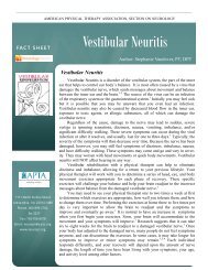
Vestibular SIG Newsletter <strong>BPPV</strong> Special Edition<br />
It’s time to consider other signs and tests to determine<br />
side of involvement in Horizontal Canal <strong>BPPV</strong>!<br />
Anne K. Galgon, PT, PhD, NCS<br />
Vestibular SIG Vice Chair<br />
The May topic of the Vestibular SIG Abstract of the Week<br />
focused on Horizontal Canal (HC) <strong>BPPV</strong>. Four of the<br />
abstracts presented addressed clinical findings and tests to<br />
enhance diagnosis of side of involvement in HC <strong>BPPV</strong> 1-4 .<br />
Over the last 10 years, many research publications have<br />
described ways to assist in the diagnosis of side of<br />
involvement in HC <strong>BPPV</strong>. However, this in<strong>for</strong>mation has<br />
not been integrated well into physical therapy practice. The<br />
standard recommendation to diagnosis HC <strong>BPPV</strong> <strong>for</strong> entrylevel<br />
physical therapists and specialists in vestibular<br />
rehabilitation continues to be a single positional test, the<br />
Supine Roll Test 5-7 . This test may not effectively<br />
diagnosis side of involvement in all patients with HC<br />
<strong>BPPV</strong>. When a therapist cannot determine side of<br />
involvement they may take longer to apply the most<br />
appropriate canalith repositioning maneuver (CRM) and<br />
patients will require more physical therapy sessions and<br />
experiences longer durations of active <strong>BPPV</strong> be<strong>for</strong>e<br />
symptoms are resolved. If physical therapists are to be<br />
recognized as clinical practitioners who effectively manage<br />
<strong>BPPV</strong>, they must be current on developing diagnostic<br />
procedures and interventions in managing HC <strong>BPPV</strong>. The<br />
purpose of this review is to describe the various signs and<br />
positional tests that have been presented in the literature<br />
and discuss how physical therapists could implement them<br />
in the examination of individuals with suspected <strong>BPPV</strong>.<br />
There may be several reasons why educators do not present<br />
this in<strong>for</strong>mation when training physical therapists in<br />
vestibular rehabilitation. One argument is that the<br />
frequency that HC <strong>BPPV</strong> will be seen in the clinic is low.<br />
The percentage of HC <strong>BPPV</strong> in all patients seen with<br />
<strong>BPPV</strong> has been reported as low as 5% 6 and as high as 27%<br />
8 but is probably more likely between 10 and 12% 9,10 .<br />
Even if only 1 in 10 patients with <strong>BPPV</strong> have horizontal<br />
canal involvement, it is very likely that physical therapy<br />
practices specializing in vestibular rehabilitation will see<br />
these patients. Other considerations may be that the<br />
Supine Roll Test can determine side of involvement in the<br />
majority of cases and adding additional diagnostic<br />
procedures will take additional time or may be too<br />
confusing <strong>for</strong> physical therapists to interpret. This<br />
4<br />
review will show that the diagnostic algorithms that have<br />
been created are consistent in the literature, and most<br />
tests do not take much additional time and are not hard to<br />
understand or apply.<br />
Table 1 presents the standard diagnostic methods used to<br />
determine the canal, the type (canalithiasis verse<br />
cupulolithiasis) and side of involvement traditionally<br />
used in physical therapy practices. In the Supine Roll<br />
Test (SRT) the patient lies in supine with neck flexed 30<br />
degrees to align the horizontal canals into the gravitational<br />
field. The head is then quickly rotated 90 degrees<br />
to the right and the eyes are observed <strong>for</strong> either geotropic<br />
(towards the ground) or<br />
apogeotropic (away from<br />
the ground) nystagmus.<br />
The head is then brought<br />
back to facing upward,<br />
and then quickly rotated<br />
to the left 90 degrees.<br />
Because of the relationship<br />
of the two HCs to<br />
gravity in supine, when otoconia are present,<br />
nystagmus will be provoked on both the right and left<br />
rotations and the direction (geotropic or<br />
apogeotropic) will be the same in each head rotation.<br />
The side of involvement is determined by the<br />
intensity of the nystagmus and symptoms, because<br />
the fluid dynamic effect of the otoconia on the cupula<br />
will be different in the right and left roll.<br />
Theoretically, the response is explained by Ewald’s<br />
Second Law that states that the system can be excited<br />
more than it can inhibited. There will be a greater<br />
response when otoconia move toward the ampulla<br />
(ampulopetal) and the system is excited than when<br />
they are displaced away from the ampulla<br />
(ampullofugal) and the system is inhibited. In the<br />
geotropic <strong>for</strong>m of HC <strong>BPPV</strong>, the otoconia will be<br />
displaced toward the ampulla when rotating to the<br />
involved side, thus more intense nystagmus. In the<br />
apogeotropic <strong>for</strong>m, otoconia will be displaced toward<br />
the ampulla when rotating away from the side of<br />
Continued on page 12