5th EuropEan MolEcular IMagIng MEEtIng - ESMI
5th EuropEan MolEcular IMagIng MEEtIng - ESMI
5th EuropEan MolEcular IMagIng MEEtIng - ESMI

Create successful ePaper yourself
Turn your PDF publications into a flip-book with our unique Google optimized e-Paper software.
<strong>5th</strong> <strong>EuropEan</strong> <strong>MolEcular</strong> <strong>IMagIng</strong> <strong>MEEtIng</strong> – EMIM2010<br />
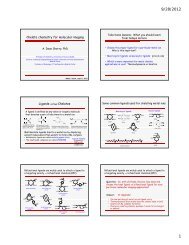
TF-Pimonidazole: an hypoxia marker suitable for in vivo 19 F MRS imaging<br />
Heerschap, A. .<br />
Centre for Molecular Life Science, University Nijmegen, The Netherlands<br />
a.heerschap@rad.umcn.nl<br />
Introduction: Hypoxia in tumours is associated<br />
with enhanced progression, increased aggressiveness<br />
and metastatic potential and poor prognosis.<br />
Moreover, hypoxic tumour cells are resistant to<br />
radiotherapy and some forms of chemotherapy.<br />
Over the years several approaches to assess hypoxia<br />
in vivo have been explored, ranging from<br />
needles measuring local pO 2 invasively to a range<br />
of non-invasive (imaging) methods using oxygen<br />
sensitive agents. Unfortunately until today none<br />
of these methods or agents has entered widespread<br />
clinical practice. Here we demonstrate a<br />
novel hypoxia marker completely analogous to<br />
the commonly used histological hypoxia marker<br />
Pimonidazole (PIMO) and labelled with fluorine<br />
for in vivo 19 F MRS imaging.<br />
Methods:1-(2-nitro-1H-imidazol-1-yl)-3-[4-<br />
(trifluoromethyl)piperidin-1-yl]propan-2-ol<br />
(TF-PIMO) was synthesized in a similar way as<br />
described in [1]. C57BL/6 mice carrying a C38<br />
colon carcinoma on the upper leg (size approx.<br />
250 mm 3 ) were given either 80 or 200 mg / kg TF-<br />
PIMO intraperitoneal (IP) at least 3 hours before<br />
MR investigations. The mice were anesthetized<br />
using a single urethane IP injection. This avoids<br />
any spectral interference by fluorinated inhalation<br />
anaesthetics. Experiments were performed<br />
on a 7 T horizontal bore MR system. A homemade<br />
14 mm solenoid coil was used for transmit/receive<br />
of 19 F and 1 H. After initial localization and basic<br />
1 H MR imaging (T2*w GRE) an unlocalized pulse<br />
acquire sequence (FID) on 19 F was used to detect<br />
TF PIMO validating correct injection of the compound.<br />
Subsequently a series of 3D 19 F chemical<br />
shift imaging (CSI) FID experiments using an<br />
ultrashort adiabatic half passage pulse was used<br />
for TF-PIMO 19 F imaging. Further settings of the<br />
MRSI sequence: FOV 32 × 32 ×32 mm, matrix<br />
8 × 8 × 8, TR 597 ms, acquisition time 58 m 02<br />
s, 256 averages, weighted phase encoding scheme.<br />
After MR, tumours were removed immediately<br />
and stored in liquid nitrogen. Frozen tumour sections<br />
of 5 μm thickness were cut for staining and<br />
further analysis. The tumor sections were subsequently<br />
stained and scanned for Hoechst and rabbit<br />
anti-pimonidazole.<br />
Results: Figure 1 shows a heat map generated from<br />
the 3D 19 F MRSI representing the in vivo TF-PIMO<br />
accumulation in a hypoxic C38 colon carcinoma as<br />
was validated by subsequent immunohistochemical<br />
analysis of the tumour tissue.<br />
Figure 1: 1H MRI of a C38 murine colon carcinoma on a mouse leg<br />
with a heat map generated from a 3D 19F MRSI representing the in<br />
vivo TF-PIMO accumulation over-layed.<br />
Conclusions: TF-PIMO was synthesized and shown<br />
to be a potential non-invasive marker to image tissue<br />
hypoxia in tumours by 19F MRSI. Background<br />
free functional images are obtained that can be coregistered<br />
with conventional MRI. In addition this<br />
marker has the advantage that it can be stained with<br />
the same anti-body as used to detect the common<br />
clinical used marker Pimonidazole and thus allows<br />
for easy matching of hypoxia by histology and by<br />
MR on the same tumour sample.<br />
Acknowledgement: This work is supported in part<br />
by EMIL (LSHC-CT-2004-503569) and NWO (VIS-<br />
TA and INV911-06-021)<br />
References:<br />
1. Raleigh, J. A. et al; Magn Reson Med 22:451-466 (1991)<br />
<strong>EuropEan</strong> SocIEty for <strong>MolEcular</strong> <strong>IMagIng</strong> – <strong>ESMI</strong><br />
day1<br />
Parallel Session 1: CANCER I - together with the ESR