5th EuropEan MolEcular IMagIng MEEtIng - ESMI
5th EuropEan MolEcular IMagIng MEEtIng - ESMI
5th EuropEan MolEcular IMagIng MEEtIng - ESMI

Create successful ePaper yourself
Turn your PDF publications into a flip-book with our unique Google optimized e-Paper software.
<strong>5th</strong> <strong>EuropEan</strong> <strong>MolEcular</strong> <strong>IMagIng</strong> <strong>MEEtIng</strong> – EMIM2010<br />
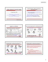
Point-of-Care Microscopy: Molecular Imaging with Cellular Resolution<br />
Contag C.H. .<br />
Division of Neonatal and Developmental Medicine, and a member of the BioX faculty at Stanford University.<br />
ccontag@stanford.edu<br />
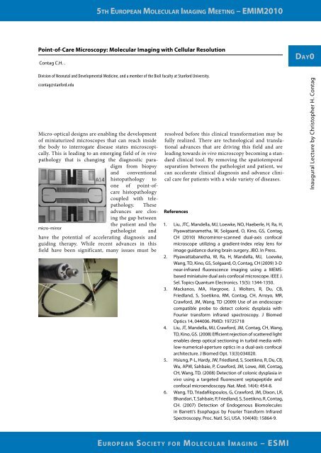
Micro-optical designs are enabling the development<br />
of miniaturized microscopes that can reach inside<br />
the body to interrogate disease states microscopically.<br />
This is leading to an emerging field of in vivo<br />
pathology that is changing the diagnostic paradigm<br />
from biopsy<br />
and conventional<br />
histopathology to<br />
one of point-ofcare<br />
histopathology<br />
coupled with telepathology.<br />
These<br />
advances are closing<br />
the gap between<br />
micro–mirror<br />
the patient and the<br />
pathologist and<br />
have the potential of accelerating diagnosis and<br />
guiding therapy. While recent advances in this<br />
field have been significant, many issues must be<br />
resolved before this clinical transformation may be<br />
fully realized. There are technological and translational<br />
advances that are driving this field and are<br />
leading towards in vivo microscopy becoming a standard<br />
clinical tool. By removing the spatiotemporal<br />
separation between the pathologist and patient, we<br />
can accelerate clinical diagnosis and advance clinical<br />
care for patients with a wide variety of diseases.<br />
References<br />
1. Liu, JTC, Mandella, MJ, Loewke, NO, Haeberle, H, Ra, H,<br />
Piyawattanametha, W, Solgaard, O, Kino, GS, Contag,<br />
CH (2010) Micromirror-scanned dual-axis confocal<br />
microscope utilizing a gradient-index relay lens for<br />
image guidance during brain surgery. JBO. In Press.<br />
2. Piyawattabanetha, W, Ra, H, Mandella, MJ, Loewke,<br />
Wang, TD, Kino, GS, Solgaard, O, Contag, CH (2009) 3-D<br />
near-infrared fluorescence imaging using a MEMSbased<br />
miniatuire dual axis confocal microscope. IEEE J.<br />
Sel. Topics Quantum Electronics. 15(5): 1344-1350.<br />
3. Mackanos, MA, Hargrove, J, Wolters, R, Du, CB,<br />
Friedland, S, Soetikno, RM, Contag, CH, Arroyo, MR,<br />
Crawford, JM, Wang, TD (2009) Use of an endoscopecompatible<br />
probe to detect colonic dysplasia with<br />
Fourier transform infrared spectroscopy. J Biomed<br />
Optics 14, 044006. PMID: 19725718<br />
4. Liu, JT, Mandella, MJ, Crawford, JM, Contag, CH, Wang,<br />
TD, Kino, GS. (2008) Efficient rejection of scattered light<br />
enables deep optical sectioning in turbid media with<br />
low-numerical-aperture optics in a dual-axis confocal<br />
architecture. J Biomed Opt. 13(3):034020.<br />
5. Hsiung, P-L, Hardy, JW, Friedland, S, Soetikno, R, Du, CB,<br />
Wu, APW, Sahbaie, P, Crawford, JM, Lowe, AW, Contag,<br />
CH, Wang, TD. (2008) Detection of colonic dysplasia in<br />
vivo using a targeted fluorescent septapeptide and<br />
confocal microendoscopy. Nat. Med. 14(4): 454-8.<br />
6. Wang, TD, Triadafilopoulos, G, Crawford, JM, Dixon, LR,<br />
Bhandari, T, Sahbaie, P, Friedland, S, Soetikno, R, Contag,<br />
CH. (2007) Detection of Endogenous Biomolecules<br />
in Barrett’s Esophagus by Fourier Transform Infrared<br />
Spectroscopy. Proc. Natl. Sci, USA. 104(40): 15864-9.<br />
<strong>EuropEan</strong> SocIEty for <strong>MolEcular</strong> <strong>IMagIng</strong> – <strong>ESMI</strong><br />
day0<br />
Inaugural Lecture by Christopher H. Contag