5th EuropEan MolEcular IMagIng MEEtIng - ESMI
5th EuropEan MolEcular IMagIng MEEtIng - ESMI
5th EuropEan MolEcular IMagIng MEEtIng - ESMI

You also want an ePaper? Increase the reach of your titles
YUMPU automatically turns print PDFs into web optimized ePapers that Google loves.
<strong>5th</strong> <strong>EuropEan</strong> <strong>MolEcular</strong> <strong>IMagIng</strong> <strong>MEEtIng</strong> – EMIM2010<br />
PET Amyloid and Tau Ligand [18F]FDDNP uptake in early Alzheimer disease<br />
Tauber C. (1) , Beaufils E. (1) , Kepe V. (2) , Vercouillie J. (1) , Venel Y. (1) , Baulieu J.L. (1) , Barrio J. (2) , Hommet C. (1) , Camus V. (1) ,<br />
Guilloteau D. (1) .<br />
(1) UMRS INSERM U930 - CNRS ERL3106 - University of Tours, France<br />
(2) Dpt of Molecular and Medical Pharmacology, D. Geffen School of Medecine, UCLA, United States<br />
clovis.tauber@univ-tours.fr<br />
Introduction: The PET tracer [18F]FDDNP specifically<br />
binds amyloid-beta plaques and tau neurofibrillary<br />
tangles, which are typical in the development<br />
of the Alzheimer disease (AD). We evaluated<br />
the feasibility of a non-invasive quantification of<br />
[18F]FDDNP to detect these proteins and discriminate<br />
early AD patients from healthy controls (HC).<br />
This abstract presents the preliminary results on the<br />
currently included cohort of 4 AD and 2 controls.<br />
Methods: All the potential subjects underwent Neuropsychological<br />
Assessment (MMSE, Trail Making<br />
Test, Semantic Fluency Task, Free and Cued<br />
Recall Test, 80 item Boston Naming Test). Patients<br />
meeting research criteria for AD and cognitively<br />
normal controls underwent PET imaging with<br />
2-(1-{6-[(2-[F-18]fluoroethyl) (methyl)amino]-2naphthyl}ethylidene)malononitrile<br />
(FDDNP) and<br />
[18F]FDG. All subjects underwent MR 3D axial<br />
T1 weighted imaging. FDDNP distribution volume<br />
ratios (DVR) parametric images were generated using<br />
Logan graphical analysis with the cerebellum<br />
grey matter as a reference region. Regions of interest<br />
(ROIs) were defined in the Posterior Cingulate,<br />
Parietal, Frontal and Temporal regions. The DVR<br />
scores were measured as the mean DVR values of<br />
each of these regions. A global score was calculated<br />
as the mean DVR scores of all these regions.<br />
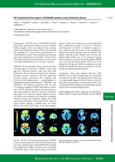
Results: All the AD patients had positive FDDNP<br />
scans by visual inspection, while all the HC had negative<br />
scans. Global values of the FDDNP-PET binding<br />
were significantly higher (p