5th EuropEan MolEcular IMagIng MEEtIng - ESMI
5th EuropEan MolEcular IMagIng MEEtIng - ESMI
5th EuropEan MolEcular IMagIng MEEtIng - ESMI

You also want an ePaper? Increase the reach of your titles
YUMPU automatically turns print PDFs into web optimized ePapers that Google loves.
<strong>5th</strong> <strong>EuropEan</strong> <strong>MolEcular</strong> <strong>IMagIng</strong> <strong>MEEtIng</strong> – EMIM2010<br />
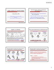
Development of dorsal skin-fold window chamber for the analysis of blood vessel<br />
modifications induced by electropermeabilization<br />
Golzio M. (1) , Bellard E. (1) , Markelc B. (2) , Cemazar M. (2) , Sersa G. (2) , Teissie J. (1) .<br />
(1) IPBS-CNRS, Toulouse, France<br />
(2) Institute of Oncology, Slovenia<br />
muriel.golzio@ipbs.fr<br />
Introduction: Recent developments in intravital microscopy<br />
(IVM) enable studies of tumour angiogenesis<br />
and microenvironment at the cellular level after<br />
different therapies. Preparation of skin fold chamber<br />
enables to follow fluorescent events on live animal.<br />
Electroporation/electropermeabilization, i.e. application<br />
of electric pulses to tissues, is a physical method<br />
for delivery of exogenous molecules. It is already used<br />
in clinical therapies of cancer, for electrochemotherapy<br />
of tumors (ECT). Its use was recently developed<br />
in electrogene therapy (EGT). In vivo, “electroporation”<br />
is associated with a blood -flow modifying effect<br />
resulting in decreased blood flow.<br />
The aim of our study was to observe directly on the<br />
living animal the effects of “electropermeabilization”<br />
on subcutaneous normal blood vessels by monitoring<br />
changes in morphology (diameter) and dynamics<br />
(vasomotricity, permeability and recovery).<br />
Methods: These parameters were measured using<br />
fluorescently labelled dextrans injected in the<br />
blood vessels observed via a dorsal skin fold window<br />
chamber, intravital digitized stereomicroscope,<br />
in vivo intravital biphoton microscopy and custom<br />
image analysis. A mathematical modelling gave access<br />
to the changes in permeability from the time<br />
lapse observation. Delivery of electric pulses was<br />
operated on the microscope stage directly on the<br />
animal under anaesthesia.<br />
Results: It resulted in immediate constriction of<br />
blood vessels that was more pronounced for arterioles<br />
(up to ~65%) compared to venules (up to ~20%).<br />
A rapid increase in vascular permeability was present<br />
that gradually decreased to basal (control) levels at<br />
1 h post-treatment. The decay of the high increase<br />
in vascular permeability was biphasic with an initial<br />
fast decrease, but was still present at 1h post-treatment.<br />
Furthermore, vasoconstriction of arterioles after<br />
“electropermeabilization” resulted in a “vascular<br />
lock” that remained for at least 6 minutes. This correlated<br />
approximately with the duration of decreased<br />
diameters of arterioles that lasted for 8 minutes.<br />
Conclusions: the results of our study provided<br />
direct in vivo monitoring of a vascular effect of<br />
electric pulses on normal vessels. The observed<br />
increase in permeability of vessels associated<br />
with delayed perfusion induced by electric pulses<br />
explains the improved delivery of molecules into<br />
tissues induced by this method after systemic<br />
delivery.<br />
Acknowledgement: CNRS, Region Midi Pyrenees,<br />
ARC, canceropole GSO, ANR “Cemirbio”, Slovenian<br />
French Proteus<br />
References:<br />
1. Cemažar M, Golzio M, et al. Current Pharmaceutical<br />
Design 12: 3817-3825. (2006).<br />
2. Marty M, et al. EJC 4(11): 3-13. (2006).<br />
3. Sersa G, et al. Br J Cancer; 98: 388-398 (2008).<br />
4. Golzio M., et al. Gene Therapy 11, S85-S91 (2004).<br />
<strong>EuropEan</strong> SocIEty for <strong>MolEcular</strong> <strong>IMagIng</strong> – <strong>ESMI</strong><br />
P-014<br />
poStEr<br />
IMAGING in DRUG DEVELOPMENT