IMMCO atlas v6 REVISED - IMMCO Diagnostics
IMMCO atlas v6 REVISED - IMMCO Diagnostics IMMCO atlas v6 REVISED - IMMCO Diagnostics
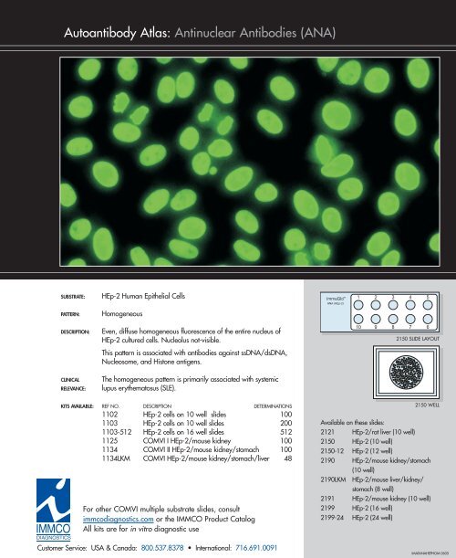
Autoantibody Atlas: Antinuclear Antibodies (ANA) SUBSTRATE: HEp-2 Human Epithelial Cells PATTERN: Homogeneous DESCRIPTION: Even, diffuse homogeneous fluorescence of the entire nucleus of HEp-2 cultured cells. Nucleolus not-visible. This pattern is associated with antibodies against ssDNA/dsDNA, Nucleosome, and Histone antigens. CLINICAL The homogeneous pattern is primarily associated with systemic RELEVANCE: lupus erythematosus (SLE). KITS AVAILABLE: REF NO. DESCRIPTION DETERMINATIONS 1102 HEp-2 cells on 10 well slides 100 1103 HEp-2 cells on 10 well slides 200 1103-512 HEp-2 cells on 16 well slides 512 1125 COMVI I HEp-2/mouse kidney 100 1134 COMVI II HEp-2/mouse kidney/stomach 100 1134LKM COMVI HEp-2/mouse kidney/stomach/liver 48 For other COMVI multiple substrate slides, consult immcodiagnostics.com or the IMMCO Product Catalog All kits are for in vitro diagnostic use Customer Service: USA & Canada: 800.537.8378 • International: 716.691.0091 2150 SLIDE LAYOUT 2150 WELL Available on these slides: 2121 HEp-2/rat liver (10 well) 2150 HEp-2 (10 well) 2150-12 HEp-2 (12 well) 2190 HEp-2/mouse kidney/stomach (10 well) 2190LKM HEp-2/mouse liver/kidney/ stomach (8 well) 2191 HEp-2/mouse kidney (10 well) 2199 HEp-2 (16 well) 2199-24 HEp-2 (24 well) IMATANAHEPHOM 0605
- Page 2 and 3: Autoantibody Atlas: Antinuclear Ant
- Page 4 and 5: Autoantibody Atlas: Antinuclear Ant
- Page 6 and 7: Autoantibody Atlas: Antinuclear Ant
- Page 8 and 9: Autoantibody Atlas: Mitochondrial A
- Page 10 and 11: Autoantibody Atlas: Anti-Neutrophil
- Page 12 and 13: Autoantibody Atlas: Anti-Neutrophil
- Page 14 and 15: Autoantibody Atlas: Endomysial Anti
- Page 16 and 17: Autoantibody Atlas: Endomysial Anti
- Page 18: Autoantibody Atlas: Native DNA Anti
Autoantibody Atlas: Antinuclear Antibodies (ANA)<br />
SUBSTRATE: HEp-2 Human Epithelial Cells<br />
PATTERN: Homogeneous<br />
DESCRIPTION: Even, diffuse homogeneous fluorescence of the entire nucleus of<br />
HEp-2 cultured cells. Nucleolus not-visible.<br />
This pattern is associated with antibodies against ssDNA/dsDNA,<br />
Nucleosome, and Histone antigens.<br />
CLINICAL The homogeneous pattern is primarily associated with systemic<br />
RELEVANCE: lupus erythematosus (SLE).<br />
KITS AVAILABLE: REF NO. DESCRIPTION DETERMINATIONS<br />
1102 HEp-2 cells on 10 well slides 100<br />
1103 HEp-2 cells on 10 well slides 200<br />
1103-512 HEp-2 cells on 16 well slides 512<br />
1125 COMVI I HEp-2/mouse kidney 100<br />
1134 COMVI II HEp-2/mouse kidney/stomach 100<br />
1134LKM COMVI HEp-2/mouse kidney/stomach/liver 48<br />
For other COMVI multiple substrate slides, consult<br />
immcodiagnostics.com or the <strong>IMMCO</strong> Product Catalog<br />
All kits are for in vitro diagnostic use<br />
Customer Service: USA & Canada: 800.537.8378 • International: 716.691.0091<br />
2150 SLIDE LAYOUT<br />
2150 WELL<br />
Available on these slides:<br />
2121 HEp-2/rat liver (10 well)<br />
2150 HEp-2 (10 well)<br />
2150-12 HEp-2 (12 well)<br />
2190 HEp-2/mouse kidney/stomach<br />
(10 well)<br />
2190LKM HEp-2/mouse liver/kidney/<br />
stomach (8 well)<br />
2191 HEp-2/mouse kidney (10 well)<br />
2199 HEp-2 (16 well)<br />
2199-24 HEp-2 (24 well)<br />
IMATANAHEPHOM 0605
Autoantibody Atlas: Antinuclear Antibodies (ANA)<br />
MOUSE KIDNEY/STOMACH MOUSE LIVER<br />
SUBSTRATE: Mouse Kidney/Stomach/Liver<br />
PATTERN: Homogeneous<br />
DESCRIPTION: Even, diffuse homogeneous fluorescence of the nucleus of<br />
renal, hepatic and parietal cells. Nucleolus not visible.<br />
This pattern is associated with antibodies against ssDNA/dsDNA,<br />
Nucleolus, and Histone antigens.<br />
CLINICAL The homogeneous pattern is primarily associated with systemic lupus<br />
RELEVANCE: erythematosus (SLE).<br />
KITS AVAILABLE: REF NO. DESCRIPTION DETERMINATIONS<br />
1107 Mouse kidney/stomach on 8 well slides 48<br />
1107-1 Mouse kidney on 8 well slides 48<br />
1107-2 Mouse kidney reagent package 160<br />
1125 COMVI I HEp-2/mouse kidney 100<br />
1134 COMVI II HEp-2/mouse kidney/stomach 100<br />
1134LKM COMVI HEp-2/mouse kidney/stomach/liver 48<br />
1136 COMVI III Mouse kidney/stomach/liver 48<br />
1136C COMVI Mouse kidney/stomach/liver 48<br />
1137 COMVI IV Mouse kidney/stomach/liver/<br />
primate thyroid 48<br />
For other COMVI multiple substrate slides, consult<br />
immcodiagnostics.com or the <strong>IMMCO</strong> Product Catalog<br />
All kits are for in vitro diagnostic use<br />
Customer Service: USA & Canada: 800.537.8378 • International: 716.691.0091<br />
2152-3 SLIDE LAYOUT<br />
2152-3 WELL<br />
Available on these slides:<br />
2152 Mouse kidney/stomach (8 well)<br />
2152-1 Mouse kidney/stomach (10 well)<br />
2152-3 Mouse kidney/stomach/liver (8 well)<br />
2152-4 Mouse kidney/stomach/liver/<br />
primate thyroid (6 well)<br />
2152-10 Mouse kidney/stomach/liver<br />
(10 well)<br />
2153 Mouse liver (8 well)<br />
2167 Mouse kidney (6 well)<br />
2167-8 Mouse kidney (8 well)<br />
2169 Mouse stomach (6 well)<br />
2176 Guinea pig kidney (6 well)<br />
2178 Guinea pig stomach (6 well)<br />
2188 Primate liver (4 well)<br />
IMATANAKSLHOM 0605
Autoantibody Atlas: Antinuclear Antibodies (ANA)<br />
SUBSTRATE: HEp-2 Human Epithelial Cells<br />
PATTERN: Centromere<br />
DESCRIPTION: Discrete nucleoplasmic speckling of interphase HEp-2 tissue culture<br />
cells. Speckles align with chromosomes in mitotic cells.<br />
This pattern is associated with antibodies against three polypeptide<br />
antigens: CENP-A (16kD), CENP-B (80kD), and CENP-C (140kD).<br />
CLINICAL Antibodies to centromere antigens are highly specific for CREST<br />
RELEVANCE: syndrome, a limited form of systemic sclerosis.<br />
KITS AVAILABLE: REF NO. DESCRIPTION DETERMINATIONS<br />
1102 HEp-2 cells on 10 well slides 100<br />
1103 HEp-2 cells on 10 well slides 200<br />
1103-512 HEp-2 cells on 16 well slides 512<br />
1125 COMVI I HEp-2/mouse kidney 100<br />
1134 COMVI II HEp-2/mouse kidney/stomach 100<br />
1134LKM COMVI HEp-2/mouse kidney/stomach/liver 48<br />
For other COMVI multiple substrate slides, consult<br />
immcodiagnostics.com or the <strong>IMMCO</strong> Product Catalog<br />
All kits are for in vitro diagnostic use<br />
Customer Service: USA & Canada: 800.537.8378 • International: 716.691.0091<br />
2199 SLIDE LAYOUT<br />
DIAGRAM OF CENTROMERE PATTERN<br />
Available on these slides:<br />
2121 HEp-2/rat liver (10 well)<br />
2150 HEp-2 (10 well)<br />
2150-12 HEp-2 (12 well)<br />
2190 HEp-2/mouse kidney/stomach<br />
(10 well)<br />
2190LKM HEp-2/mouse liver/kidney/<br />
stomach (8 well)<br />
2191 HEp-2/mouse kidney (10 well)<br />
2199 HEp-2 (16 well)<br />
2199-24 HEp-2 (24 well)<br />
IMATANAHEPCEN 0605
Autoantibody Atlas: Antinuclear Antibodies (ANA)<br />
MOUSE LIVER MOUSE KIDNEY<br />
SUBSTRATE: Mouse Kidney/Liver<br />
PATTERN: Centromere<br />
DESCRIPTION: Discrete nucleoplasmic specking of hepatocytes of liver. Discrete<br />
nucleoplasmic speckling of renal tubular cells of kidney. The<br />
centromere pattern is associated with three polypeptide antigens:<br />
CENP-A (16kD), CENP-B(80kD), and CENP-C(140kD).<br />
CLINICAL Antibodies to centromere antigens are highly specific for CREST<br />
RELEVANCE: syndrome, a limited form of systemic sclerosis. These antibodies<br />
can occasionally be found in low titers in some other autoimmune<br />
variants.<br />
KITS AVAILABLE: REF NO. DESCRIPTION DETERMINATIONS<br />
1107 Mouse kidney/stomach on 8 well slides 48<br />
1107-1 Mouse kidney on 8 well slides 48<br />
1107-2 Mouse kidney reagent package 160<br />
1125 COMVI I HEp-2/mouse kidney 100<br />
1134 COMVI II HEp-2/mouse kidney/stomach 100<br />
1134LKM COMVI HEp-2/mouse kidney/stomach/liver 48<br />
1136 COMVI III Mouse kidney/stomach/liver<br />
(ANA and AMA controls) 48<br />
For other COMVI multiple substrate slides, consult<br />
immcodiagnostics.com or the <strong>IMMCO</strong> Product Catalog<br />
All kits are for in vitro diagnostic use<br />
Customer Service: USA & Canada: 800.537.8378 • International: 716.691.0091<br />
2153 SLIDE LAYOUT<br />
Available on these slides:<br />
2153 Mouse Liver (8 well)<br />
2152 Mouse kidney/stomach (8 well)<br />
2152-1 Mouse kidney/stomach (10 well)<br />
2152-3 Mouse kidney/stomach/liver<br />
(8 well)<br />
2152-4 Mouse kidney/stomach/liver/<br />
primate thyroid (6 well)<br />
2152-10 Mouse kidney/stomach/liver<br />
(10 well)<br />
2152-R/R Rat kidney/stomach/liver (10 well)<br />
2153 Mouse liver (8 well)<br />
2161-8 Rat kidney (8 well)<br />
2167 Mouse kidney (6 well)<br />
2167-8 Mouse kidney (8 well)<br />
2190 HEp-2/mouse kidney/stomach<br />
(10 well)<br />
2190LKM HEp-2/mouse liver/kidney/<br />
stomach (8 well)<br />
2191 HEp-2/mouse kidney (10 well)<br />
IMATANAKSLCEN 0605
Autoantibody Atlas: Antinuclear Antibodies ANA<br />
1: HEp-2 3: MOUSE KIDNEY/STOMACH/LIVER<br />
SUBSTRATE: 1: HEp-2 Human Epithelial Cells<br />
2: Mouse Kidney/Stomach<br />
3: Mouse Kidney/Stomach/Liver<br />
PATTERN: Speckled<br />
DESCRIPTION: Fine to coarse speckled staining of the nucleoplasm of HEp-2, renal,<br />
parietal and hepatic cells. The nucleoli are visible on HEp-2.<br />
Several antibodies stain the nucleoplasm of these cells, resulting in<br />
different types of speckled staining. The staining may be fine, coarse<br />
or granular. These patterns are associated with antibodies to<br />
Sm, RNP, SS-A/SS-B, PM-1 and PCNA antigens. Specific patterns<br />
include:<br />
a. Large/coarse speckled: RNP, Sm<br />
b. Fine speckled: SS-A/SS-B, CENP-F, Mi-2<br />
c. Pleomorphic PCNA<br />
CLINICAL Anti-nuclear antibodies with a speckled pattern are commonly<br />
RELEVANCE: associated with systemic lupus erythematosus (SLE), although<br />
they do occur in some cases of Sjögren’s syndrome and mixed<br />
connective tissue disorders (MCTD).<br />
For other COMVI multiple substrate slides, consult<br />
immcodiagnostics.com or the <strong>IMMCO</strong> Product Catalog<br />
All kits are for in vitro diagnostic use<br />
Customer Service: USA & Canada: 800.537.8378 • International: 716.691.0091<br />
2: MOUSE KIDNEY/STOMACH<br />
2190 SLIDE LAYOUT<br />
2190 WELL<br />
DIAGRAM OF SPECKLED<br />
PATTERNS ON HEp-2<br />
Available on these slides:<br />
2152 Mouse kidney/stomach (8 well)<br />
2152-1 Mouse kidney/stomach (10 well)<br />
2152-3 Mouse kidney/stomach/liver (8 well)<br />
2190 HEp-2/mouse kid/stom (10 well)<br />
IMATANAH/KSPE 0605
Autoantibody Atlas: Antinuclear Antibodies (ANA)<br />
1: HEp-2<br />
SUBSTRATE: 1: HEp-2 Human Epithelial Cells<br />
2: Mouse Kidney/Stomach<br />
3: Mouse Kidney/Stomach/Liver<br />
PATTERN: Nucleolar<br />
DESCRIPTION: Anti-nuclear antibodies with staining of the nucleoi of HEp-2 cells.<br />
The chromatin of dividing cells fluoresces. Homogeneous staining of<br />
the nucleoi on renal tubular, parietal and hepatic cells.<br />
Several different antibodies stain the nucleoli of these cells, resulting<br />
in different types of nucleolar staining, such as homogeneous,<br />
clumpy or speckled. Several of the antibodies that stain the nucleolus<br />
also stain other areas of the nucleus. Mixed patterns are common.<br />
The following antigens present with a nucleolar pattern:<br />
a. PM-Scl d. RNA Polymerase I<br />
b. Fibrillarin e. Scl-70<br />
c. NOR-90<br />
CLINICAL Anti-nuclear antibodies with a nucleolar pattern are commonly<br />
RELEVANCE: associated with systemic sclerosis (SS), although they do occur in<br />
some cases of systemic lupus erythematosus (SLE) and overlap<br />
syndromes. Overlap syndromes include polymyositis/<br />
dermatomyositis.<br />
For other COMVI multiple substrate slides, consult<br />
immcodiagnostics.com or the <strong>IMMCO</strong> Product Catalog<br />
All kits are for in vitro diagnostic use<br />
Customer Service: USA & Canada: 800.537.8378 • International: 716.691.0091<br />
2: MOUSE KIDNEY/STOMACH<br />
3: MOUSE KIDNEY/STOMACH/LIVER<br />
2190LKM SLIDE LAYOUT<br />
2190LKM WELL<br />
DIAGRAM OF<br />
NUCLEOLAR PATTERNS ON HEp-2<br />
IMATANAH/KNUC 0605
Autoantibody Atlas: Mitochondrial Antibodies (AMA)<br />
SUBSTRATE: HEp-2 Human Epithelial Cells<br />
PATTERN: Mitochondrial<br />
DESCRIPTION: Coarse granular filamentous staining throughout the cytoplasm.<br />
A mitochondrial pattern is associated with various antibodies. Of<br />
the multiple antigenic structures within the mitochondria only M2<br />
is of clinical significance. M2 is primarily visualized on HEp-2 and<br />
distal renal tubule cells.<br />
CLINICAL Antibodies to mitochondrial antigens (AMA) occur in over 90% of<br />
RELEVANCE: primary biliary cirrhosis (PBC) cases 3-11% of chronic active<br />
hepatitis patients. AMA are absent in patients with extra-hepatic<br />
biliary obstruction and other liver diseases. The universal presence<br />
of AMA in PBC and their virtual absence in extra-hepatic jaundice<br />
makes their detection of considerable value in differential<br />
diagnosis.<br />
KITS AVAILABLE: REF NO. DESCRIPTION DETERMINATIONS<br />
1102 HEp-2 cells on 10 well slides 100<br />
1103 HEp-2 cells on 10 well slides 200<br />
1103-512 HEp-2 cells on 16 well slides 512<br />
1125 COMVI I HEp-2/mouse kidney 100<br />
For other COMVI multiple substrate slides, consult<br />
immcodiagnostics.com or the <strong>IMMCO</strong> Product Catalog<br />
All kits are for in vitro diagnostic use<br />
Customer Service: USA & Canada: 800.537.8378 • International: 716.691.0091<br />
2199-24 SLIDE LAYOUT<br />
DIAGRAM OF MITOCHONDRIAL PATTERNS<br />
Available on these slides:<br />
2121 HEp-2/rat liver (10 well)<br />
2150 HEp-2 (10 well)<br />
2150-12 HEp-2 (12 well)<br />
2190 HEp-2/mouse kidney/stomach<br />
(10 well)<br />
2190LKM HEp-2/mouse liver/kidney/<br />
stomach (8 well)<br />
2191 HEp-2/mouse kidney (10 well)<br />
2199 HEp-2 (16 well)<br />
2199-24 HEp-2 (24 well)<br />
IMATAMAHEP 0605
Autoantibody Atlas: Mitochondrial Antibodies (AMA)<br />
SUBSTRATE: Mouse Kidney/Stomach<br />
PATTERN: Mitochondrial<br />
DESCRIPTION: Mitochondrial antibodies (AMA) cause intense cytoplasmic staining<br />
of the distal renal tubular cells of the kidney and parietal cells of<br />
the stomach. This pattern is associated with multiple antigenic<br />
structures within the mitochondria. M2 is visualized on distal renal<br />
tubule cells. The antigen is pyruvate dehydrogenase enzyme<br />
complex.<br />
CLINICAL Antibodies to mitochondrial antigens occur in over 90% of primary<br />
RELEVANCE: biliary cirrhosis (PBC) cases and 3-11% of chronic active hepatitis<br />
patients. They are absent in patients with extra-hepatic biliary<br />
obstruction and other liver diseases. The universal presence of<br />
AMA in PBC antibodies in primary biliary cirrhosis and their<br />
virtual absence in extra-hepatic jaundice makes their detection of<br />
considerable value in differential diagnosis.<br />
KITS AVAILABLE: REF NO. DESCRIPTION DETERMINATIONS<br />
1107 Mouse kidney/stomach on 8 well slides 48<br />
1107-1 Mouse kidney on 8 well slides 48<br />
1107-2 Mouse kidney reagent package 160<br />
For other COMVI multiple substrate slides, consult<br />
immcodiagnostics.com or the <strong>IMMCO</strong> Product Catalog<br />
All kits are for in vitro diagnostic use<br />
Customer Service: USA & Canada: 800.537.8378 • International: 716.691.0091<br />
2152-1 SLIDE LAYOUT<br />
2152-1 WELL<br />
Available on these slides:<br />
2152 Mouse kidney/stomach (8 well)<br />
2152-1 Mouse kidney/stomach (10 well)<br />
2152-3 Mouse kidney/stomach/liver<br />
(8 well)<br />
2152-4 Mouse kidney/stomach/liver/<br />
primate thyroid (6 well)<br />
2152-10 Mouse kidney/stomach/liver<br />
(10 well)<br />
2167 Mouse kidney (6 well)<br />
2167-8 Mouse kidney (8 well)<br />
2169 Mouse stomach (6 well)<br />
IMATAMAKS 0605
Autoantibody Atlas: Anti-Neutrophil Cytoplasmic Antibodies (ANCA)<br />
SUBSTRATE: Ethanol fixed human polymorphonuclear leukocytes (hPMN cells)<br />
PATTERN: Cytoplasmic ANCA (cANCA)<br />
DESCRIPTION: ANCA reaction patterns are dependent on the method of neutrophil<br />
fixation. cANCA produce a diffuse cytoplasmic pattern on ethanol<br />
or formalin fixed substrates.<br />
Proteinase-3 (PR3) is the major antigen of cANCA reaction patterns.<br />
Others include cathepsin G and elastase.<br />
CLINICAL This pattern is suggestive of Wegener’s granulomatosis and other<br />
RELEVANCE: systemic small vessl vasculitides such as microsocopic<br />
polyarteritis/polyangiitis, idiopathic crescentic and necrotising<br />
glomerulonephritis.<br />
KITS AVAILABLE: REF NO. DESCRIPTION DETERMINATIONS<br />
1116 ANCA (ethanol fixed slides) 24<br />
1140 ANCA (ethanol fixed slides) 48<br />
1141 ANCA (formalin fixed slides) 48<br />
1142 ANCA COMVI (ethanol & formalin fixed slides) 48<br />
For other COMVI multiple substrate slides, consult<br />
immcodiagnostics.com or the <strong>IMMCO</strong> Product Catalog<br />
All kits are for in vitro diagnostic use<br />
Customer Service: USA & Canada: 800.537.8378 • International: 716.691.0091<br />
2162 SLIDE LAYOUT<br />
DIAGRAM OF cANCA<br />
Available on these slides:<br />
2152 Mouse kidney/stomach (8 well)<br />
2162 ANCA ethanol slide (6 well)<br />
2186 ANCA formalin slide (6 well)<br />
2189 ANCA COMVI Slide<br />
(6 ethanol & 6 formalin wells)<br />
IMATANCAETHC 0605
Autoantibody Atlas: Anti-Neutrophil Cytoplasmic Antibodies (ANCA)<br />
SUBSTRATE: Ethanol fixed human polymorphonuclear leukocytes (hPMN cells)<br />
PATTERN: Perinuclear ANCA (pANCA)<br />
DESCRIPTION: ANCA reaction patterns are dependent on the method of<br />
neutrophil fixation. pANCA react as follows:<br />
Ethanol: perinuclear staining pattern<br />
Formalin: reactions convert to cytoplasmic staining<br />
Myeloperoxidase (MPO) is the major antigen of pANCA reaction<br />
patterns. Others include human leukocyte elastase and lactoferrin.<br />
CLINICAL Perinuclear ANCA reaction patterns are observed in patients with<br />
RELEVANCE: systemic small vessel vasculitis but have also been described in other<br />
non-vasculitic autoimmune diseases including ulcerative colitis,<br />
Crohn’s disease and chronic hepatitis.<br />
KITS AVAILABLE: REF NO. DESCRIPTION DETERMINATIONS<br />
1116 ANCA (ethanol fixed slides) 24<br />
1140 ANCA (ethanol fixed slides) 48<br />
1141 ANCA (formalin fixed slides) 48<br />
1142 ANCA COMVI (ethanol & formalin fixed slides) 48<br />
For other COMVI multiple substrate slides, consult<br />
immcodiagnostics.com or the <strong>IMMCO</strong> Product Catalog<br />
All kits are for in vitro diagnostic use<br />
Customer Service: USA & Canada: 800.537.8378 • International: 716.691.0091<br />
2189 SLIDE LAYOUT<br />
DIAGRAM OF pANCA ON ETHANOL<br />
Available on these slides:<br />
2152 Mouse kidney/stomach (8 well)<br />
2162 ANCA ethanol slide (6 well)<br />
2186 ANCA formalin slide (6 well)<br />
2189 ANCA COMVI Slide<br />
(6 ethanol & 6 formalin wells)<br />
IMATANCAETHP 0605
Autoantibody Atlas: Anti-Neutrophil Cytoplasmic Antibodies (ANCA)<br />
SUBSTRATE: Ethanol fixed human polymorphonuclear leukocytes (hPMN cells)<br />
PATTERN: Antinuclear Antibodies (ANA)<br />
DESCRIPTION: ANCA reaction patterns are dependent on the method of neutrophil<br />
fixation. On ethanol fixed hPMNs, common antibodies against<br />
nuclear antigens (ANA) may produce a pattern resembling<br />
pANCA. ANA reactions can be differentiated on formalin fixed<br />
slides as follows:<br />
pANCA: reactions convert to cytoplasmic staining<br />
ANA: reactions become weak or negative<br />
CLINICAL ANA occur in a variety of autoimmune diseases but are not specific<br />
RELEVANCE: for Wegener’s granulomatosis or other systemic small vessl<br />
vasculitides, ulcerative colitis, Crohn’s disease or chronic hepatitis.<br />
KITS AVAILABLE: REF NO. DESCRIPTION DETERMINATIONS<br />
1116 ANCA (ethanol fixed slides) 24<br />
1140 ANCA (ethanol fixed slides) 48<br />
1141 ANCA (formalin fixed slides) 48<br />
1142 ANCA COMVI (ethanol & formalin fixed slides) 48<br />
For other COMVI multiple substrate slides, consult<br />
immcodiagnostics.com or the <strong>IMMCO</strong> Product Catalog<br />
All kits are for in vitro diagnostic use<br />
Customer Service: USA & Canada: 800.537.8378 • International: 716.691.0091<br />
2162 SLIDE LAYOUT<br />
DIAGRAM OF ANA ON ETHANOL<br />
Available on these slides:<br />
2152 Mouse kidney/stomach (8 well)<br />
2162 ANCA ethanol slide (6 well)<br />
2186 ANCA formalin slide (6 well)<br />
2189 ANCA COMVI Slide<br />
(6 ethanol & 6 formalin wells)<br />
IMATANCAETHA 0605
Autoantibody Atlas: Anti-Neutrophil Cytoplasmic Antibodies (ANCA)<br />
SUBSTRATE: Formalin fixed human polymorphonuclear leukocytes (hPMN cells)<br />
PATTERN: Cytoplasmic pattern of pANCA on formalin fixed neutrophils<br />
DESCRIPTION: ANCA reaction patterns are dependent on the method of neutrophil<br />
fixation. pANCA react as follows:<br />
Ethanol: perinuclear staining pattern<br />
Formalin: reactions convert to cytoplasmic staining<br />
Use of both ethanol and formalin fixed slides can help discriminate<br />
between ANA reactions and true pANCA reactions.<br />
Myeloperoxidase (MPO) is the major antigen of pANCA reaction<br />
patterns. Others include human leukocyte elastase and lactoferrin.<br />
CLINICAL pANCA reaction patterns are observed in patients with systemic<br />
RELEVANCE: small vessel vasculitis but have also been described in other<br />
non-vasculitic autoimmune diseases including ulcerative colitis,<br />
Crohn’s disease and chronic hepatitis.<br />
KITS AVAILABLE: REF NO. DESCRIPTION DETERMINATIONS<br />
1116 ANCA (ethanol fixed slides) 24<br />
1140 ANCA (ethanol fixed slides) 48<br />
1141 ANCA (formalin fixed slides) 48<br />
For other COMVI multiple substrate slides, consult<br />
immcodiagnostics.com or the <strong>IMMCO</strong> Product Catalog<br />
All kits are for in vitro diagnostic use<br />
Customer Service: USA & Canada: 800.537.8378 • International: 716.691.0091<br />
2186 SLIDE LAYOUT<br />
DIAGRAM OF pANCA ON FORMALIN<br />
Available on these slides:<br />
2152 Mouse kidney/stomach (8 well)<br />
2162 ANCA ethanol slide (6 well)<br />
2186 ANCA formalin slide (6 well)<br />
2189 ANCA COMVI Slide<br />
(6 ethanol & 6 formalin wells)<br />
IMATANCAFORP 0605
Autoantibody Atlas: Endomysial Antibodies (EMA)<br />
EMA POSITIVE EMA NEGATIVE<br />
SUBSTRATE: Primate Smooth Muscle<br />
PATTERN: Endomysial Antibodies (EMA)<br />
DESCRIPTION: EMA appear as an interconnected network staining the endomysial<br />
areas surrounding the sarcolemma of smooth muscle fibers. These<br />
are thin, irregular lines around individual smooth muscle fibers.<br />
Tissue transglutaminase (tTG) is the prominent endomysial antigen.<br />
CLINICAL The detection of endomysial antibodies aids in the diagnosis of<br />
RELEVANCE: gluten sensitive enteropathy, i.e. celiac disease (CD) and dermatitis<br />
herpetiformis (DH). Of the various antibody markers of CD and DH,<br />
EMA of the IgA class are the most sensitive and specific marker.<br />
Certain CD patients are IgA deficient, however. EMA of the IgG<br />
class also occur when IgA class EMA are in high titer or in<br />
individuals who are IgA deficient.<br />
KITS AVAILABLE: REF NO. DESCRIPTION DETERMINATIONS<br />
1114 EMA (Smooth Muscle)<br />
with IgA/IgG conjugate 48<br />
1114A EMA (Smooth Muscle)<br />
with IgA conjugate 48<br />
For a complete range of kits, consult<br />
immcodiagnostics.com or the <strong>IMMCO</strong> Product Catalog<br />
Customer Service: USA & Canada: 800.537.8378 • International: 716.691.0091<br />
Available on these slides:<br />
2160 Smooth Muscle (6 well)<br />
2160 SLIDE LAYOUT<br />
EMA REACTION ON SMOOTH MUSCLE<br />
IMATSMOEMA 0605
Autoantibody Atlas: Endomysial Antibodies (EMA)<br />
SUBSTRATE: Primate Smooth Muscle<br />
PATTERN: Smooth Muscle Antibodies (ASMA)<br />
DESCRIPTION: The primate smooth muscle substrate shown here is utilized for the<br />
detection of Endomysial antibodies (EMA) which are sensitive and<br />
specific markers for Gluten Sensitivite Enteropathy (GSE), i.e. Celiac<br />
Disease (CD) and Dermatitis Herpetiformis (DH). ASMA reactions,<br />
such as the reaction above, may be confused with EMA reactions.<br />
ASMA staining is in fact the reverse of typical EMA staining with the<br />
center of the smooth muscle fibers fluorescing while the endomysial<br />
areas surrounding the sarcolemma of smooth muscle fibers remains<br />
negative.<br />
CLINICAL ASMA have no clinical relevance for GSE and must be distinguished<br />
RELEVANCE: from true endomysial reactions to support a proper diagnosis.<br />
ASMA occur in certain other autoimmune diseases such as<br />
autoimmune hepatitis and primary biliary cirrhosis.<br />
KITS AVAILABLE: REF NO. DESCRIPTION DETERMINATIONS<br />
1114 EMA (Smooth Muscle)<br />
with IgA/IgG conjugate 48<br />
1114A EMA (Smooth Muscle)<br />
with IgA conjugate 48<br />
For a complete range of kits, consult<br />
immcodiagnostics.com or the <strong>IMMCO</strong> Product Catalog<br />
Customer Service: USA & Canada: 800.537.8378 • International: 716.691.0091<br />
Available on these slides:<br />
2160 Smooth Muscle (6 well)<br />
2160 SLIDE LAYOUT<br />
ASMA REACTION ON SMOOTH MUSCLE<br />
IMATSMOSMO 0605
Autoantibody Atlas: Endomysial Antibodies (EMA)<br />
SUBSTRATE: Primate Distal Esophagus<br />
PATTERN: Endomysial Antibodies (EMA)<br />
DESCRIPTION: EMA appear as an interconnected network staining the endomysial<br />
areas surrounding the sarcolemma of smooth muscle fibers. These<br />
are thin, irregular lines around individual smooth muscle fibers.<br />
Tissue transglutaminase (tTG) is the prominent endomysial antigen.<br />
CLINICAL The detection of EMA aids in the diagnosis of gluten sensitive<br />
RELEVANCE: enteropathy, ie celiac disease (CD) and dermatitis herpetiformis<br />
(DH). Of the various antibody markers of CD and DH,<br />
EMA of the IgA class are the most sensitive and specific marker.<br />
Certain CD patients are IgA deficient, however. EMA of the IgG<br />
class also occur when IgA class EMA are in high titer or in<br />
individuals who are IgA deficient.<br />
KITS AVAILABLE: REF NO. DESCRIPTION DETERMINATIONS<br />
1114A-PDE EMA (Primate Distal Esophagus) 48<br />
For a complete range of kits, consult<br />
immcodiagnostics.com or the <strong>IMMCO</strong> Product Catalog<br />
Customer Service: USA & Canada: 800.537.8378 • International: 716.691.0091<br />
2155-1 SLIDE LAYOUT<br />
EMA REACTION ON ESOPHAGUS<br />
Available on these slides:<br />
2155-1 Primate Distal Esophagus (6 well)<br />
2155-18 Primate Distal Esophagus (8 well)<br />
IMATESOEMA 0605
Autoantibody Atlas: Endomysial Antibodies (EMA)<br />
SUBSTRATE: Primate Distal Esophagus<br />
PATTERN: Smooth Muscle Antibodies (ASMA)<br />
DESCRIPTION: The primate smooth muscle substrate shown here is utilized for the<br />
detection of Endomysial antibodies (EMA) which are sensitive and<br />
specific markers for Gluten Sensitivite Enteropathy (GSE), i.e. Celiac<br />
Disease (CD) and Dermatitis Herpetiformis (DH). ASMA reactions,<br />
such as the reaction above, may be confused with EMA reactions.<br />
ASMA staining is in fact the reverse of typical EMA staining with the<br />
center of the smooth muscle fibers fluorescing while the endomysial<br />
areas surrounding the sarcolemma of smooth muscle fibers remains<br />
negative.<br />
CLINICAL ASMA have no clinical relevance for GSE and must be distinguished<br />
RELEVANCE: from true endomysial reactions to support a proper diagnosis.<br />
ASMA occur in certain other autoimmune diseases such as<br />
autoimmune hepatitis and primary biliary cirrhosis.<br />
KITS AVAILABLE: REF NO. DESCRIPTION DETERMINATIONS<br />
1114A-PDE EMA (Primate Distal Esophagus) 48<br />
For a complete range of kits, consult<br />
immcodiagnostics.com or the <strong>IMMCO</strong> Product Catalog<br />
Customer Service: USA & Canada: 800.537.8378 • International: 716.691.0091<br />
2155-18 SLIDE LAYOUT<br />
ASMA REACTION ON ESOPHAGUS<br />
Available on these slides:<br />
2155-1 Primate Distal Esophagus (6 well)<br />
2155-18 Primate Distal Esophagus (8 well)<br />
IMATESOSMO 0605
Autoantibody Atlas: Reticulin Antibodies (ARA)<br />
SUBSTRATE: Rat Kidney<br />
PATTERN: Reticulin Antibodies (ARA)<br />
DESCRIPTION: ARA reactions are marked by fluorescence of the peritubular and<br />
periglomerular reticulin fibers. GBM and kidney tubules do not stain.<br />
CLINICAL ARA have been utilized in the diagnosis of Celiac Disease (CD). The<br />
RELEVANCE: primary antigen of interest for these diseases is R1. These antibodies<br />
are also present in 20% of Crohn’s Disease patients.<br />
KITS AVAILABLE: REF NO. DESCRIPTION DETERMINATIONS<br />
1115 ARA (Rat Kidney) 48<br />
For other COMVI multiple substrate slides, consult<br />
immcodiagnostics.com or the <strong>IMMCO</strong> Product Catalog<br />
All kits are for in vitro diagnostic use<br />
Customer Service: USA & Canada: 800.537.8378 • International: 716.691.0091<br />
Available on these slides:<br />
2161 Rat Kidney (6 well)<br />
2161-8 Rat Kidney (8 well)<br />
2161 SLIDE LAYOUT<br />
DIAGRAM OF AN ARA REACTION<br />
IMATRKIDARA 0605
Autoantibody Atlas: Native DNA Antibodies (nDNA)<br />
SUBSTRATE: Crithidia luciliae<br />
PATTERN: Native DNA (nDNA)<br />
DESCRIPTION: These antibodies produce staining of the kinetoplast of Crithidia<br />
luciliae organism. The kinetoplast is a giant mitochondrion<br />
containing kinetoplast circular DNA, referred to as double-stranded<br />
DNA (dsDNA) and native DNA (nDNA), which is relatively free of<br />
other nuclear antigens. The kinetoplast is smaller and brighter than<br />
the accompanying nucleus.<br />
CLINICAL nDNA are highly specific for systemic lupus erthematosus (SLE).<br />
RELEVANCE: The frequency and titer of nDNA antibodies fluctuate with activity<br />
and tend to disappear with immunosuppression and during<br />
remission. There is good correlation between disease activity and<br />
nDNA antibody levels.<br />
KITS AVAILABLE: REF NO. DESCRIPTION DETERMINATIONS<br />
1106 nDNA (C. luciliae) 48<br />
1106-2 nDNA (C. luciliae) 98<br />
For other COMVI multiple substrate slides, consult<br />
immcodiagnostics.com or the <strong>IMMCO</strong> Product Catalog<br />
All kits are for in vitro diagnostic use<br />
Customer Service: USA & Canada: 800.537.8378 • International: 716.691.0091<br />
Available on these slides:<br />
2151 C. luciliae (8 well)<br />
2151 SLIDE LAYOUT<br />
C. luciliae<br />
IMATANAHEPHOM 0605


