PLATON-Locking-Nail-System - tantum AG
PLATON-Locking-Nail-System - tantum AG
PLATON-Locking-Nail-System - tantum AG

Create successful ePaper yourself
Turn your PDF publications into a flip-book with our unique Google optimized e-Paper software.
OR manual<br />
PLAT O N
<strong>tantum</strong> · OR manual <strong>PLATON</strong><br />
2<br />
4<br />
1<br />
3<br />
Variation I<br />
dynamic<br />
Variation II<br />
AR-Clip<br />
<strong>PLATON</strong>-L <strong>PLATON</strong>-S<br />
1<br />
Variation III<br />
static<br />
2 2<br />
2<br />
5a 5b<br />
5a<br />
6<br />
3<br />
4<br />
5b<br />
1.The proximal diameter of<br />
17.5mm for high stability,<br />
at the critical area of<br />
proximal drilling<br />
2.The self-tapping thread<br />
enables a tight fit of the<br />
screw in the femur head<br />
cancellous bone<br />
3.4°M/L-angle for an<br />
anatomical design<br />
4.The distal diameter of 11mm<br />
guarantees high stability in<br />
the distal area and requires<br />
only a minimal drilling of<br />
the diaphysis<br />
5.Greater range of indications<br />
due to the possibility of 5a.<br />
static and 5b. dynamic locking<br />
through the distal holes.<br />
6. The conical tipp of the nail<br />
reduces stress concentration<br />
on the medial side of the bone.
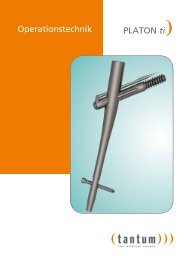
<strong>PLATON</strong>-<strong>Locking</strong>-<strong>Nail</strong>-<strong>System</strong><br />
The intramedullary treatment at the proximal femur is<br />
today´s standard therapy for stabilization of per- and<br />
subtrochanteric fractures. With more than 700,000<br />
implantations worldwide and extraordinary clinical<br />
results, this method of treatment has already proven<br />
its perfomance.<br />
The knowledge of the success of intramedullary treatment<br />
and decades of experience from our team of<br />
developers was the basis for the development of the<br />
<strong>PLATON</strong>-<strong>Locking</strong>-<strong>Nail</strong> system.<br />
The <strong>PLATON</strong>-system is distinguished by numerous<br />
improvements compared to regular systems and unites<br />
most modern technologies of development and<br />
production with simple handling.<br />
For the treatment of a thorough range of indications,<br />
the <strong>PLATON</strong>-system offers three variations: Variation I<br />
corresponds mostly to the dynamic principle, a Cap<br />
Screw protects from medial migration of the<br />
Femoral Lag Screw. Variation II is distinguished by<br />
rotation control of the head-neck fragments with the<br />
help of the patented AR-Clip. Variation III offers the<br />
possibility of total fixation of the Femoral Lag Screw<br />
by the use of a Fixation Bolt.<br />
With regard to the individual indication, all variations<br />
have the possibility of static as well as dynamic locking<br />
of the nail. The latter allowing dynamization under<br />
weight bearing conditions, with fully existent rotational<br />
stability of the osteosynthesis.<br />
Collodiaphyseal angles of 125° and 130° and length<br />
types S and L are available:<br />
The <strong>PLATON</strong>-S-<strong>Nail</strong> for the treatment of all stable fractures.<br />
The advantageous total length of 190mm ensures<br />
an excellent implant support in the diaphysis even<br />
with subtrochanteric fractures.<br />
The <strong>PLATON</strong>-L-<strong>Nail</strong> for unstable and combined fractures.<br />
Total length from 340mm to 420mm.<br />
In order to meet the highest quality demands, all nails<br />
and Lag Screws are made of high nitrogen<br />
implant steel alloy following DIN ISO 5832-9. Screws<br />
and supplements consist of implant steel following<br />
DIN ISO 5832-1.<br />
<strong>tantum</strong> · OR manual <strong>PLATON</strong><br />
Furthermore, the material used for nails and Femoral<br />
Lag Screws is characterized by a very high stability of<br />
more than 1200 MPa at a low variation of the mechanical<br />
attributes.<br />
For an acurate implantation, high precision instruments<br />
are available for all Platon variations. The Platon<br />
Targeting Device enables the sleeve guided insertion<br />
of the Femoral Lag Screw, Cap, AR-Clip and<br />
Connection Screw. The instruments show a variety of<br />
innovative, detailed-solutions that make the implantation,<br />
as well as the later removal of material, much<br />
easier for the surgeon.<br />
3
<strong>tantum</strong> · OR manual <strong>PLATON</strong><br />
The <strong>PLATON</strong>-<strong>Locking</strong>-<strong>Nail</strong><br />
Variation I dynamic<br />
4<br />
<strong>PLATON</strong>-L + S<br />
Proximal Plug<br />
Lateral Cap Screw<br />
Optional: dynamic or<br />
static positioning of<br />
the <strong>Locking</strong> Screw<br />
Characteristics of <strong>PLATON</strong> Variation I<br />
Proximal Plug<br />
– impedes tissue growth<br />
– makes later implant removal easier<br />
Femoral Lag Screw<br />
– to support the dynamic principle<br />
Lateral Cap Screw<br />
– protects from medial migration of the Femoral Lag<br />
Screw<br />
– offers best soft tissue protection in transient area<br />
to Femoral Lag Screw<br />
– impedes tissue growth<br />
Indications <strong>PLATON</strong> Variation I<br />
<strong>PLATON</strong>-S<br />
– stable per- and high subtrochanteric fractures of<br />
the femur Type A1, A2, (A3) with disrupture of the<br />
lower trochanter (dynamic locking of the nail)<br />
– stable per- and high subtrochanteric fractures of<br />
the femur Type A1, A2, (A3) without disrupture of<br />
the lower trochanter (static locking of the nail)<br />
<strong>PLATON</strong>-L<br />
– unstable per- and subtrochanteric femur fractures<br />
reaching up to the upper third of the trochanter of<br />
the Type A2, A3 (dynamic locking of the nail)<br />
– unstable and pathological subtrochanteric fractures<br />
(static locking of the nail)<br />
– stable trochanteric fractures in combination with<br />
femur shaft fractures (static locking of the nail)<br />
– Pseudarthroses following delayed bone healing<br />
(dynamic locking of the nail)
The <strong>PLATON</strong>-<strong>Locking</strong>-<strong>Nail</strong><br />
Variation II AR-Clip<br />
<strong>PLATON</strong>-L + S<br />
Proximal Plug<br />
Flattend tip<br />
of the AR-Clip<br />
AR-Clip for rotation<br />
safety of the headneck<br />
fragments<br />
Soft tissue protection<br />
by laterally angled<br />
clip construction<br />
Optional: dynamic or<br />
static positioning of<br />
the <strong>Locking</strong> Screw<br />
Characteristics of <strong>PLATON</strong> Variation II<br />
<strong>tantum</strong> · OR manual <strong>PLATON</strong><br />
Proximal Plug<br />
– impedes tissue growth<br />
– makes later implant removal easier<br />
AR-Clip for rotation safety<br />
– Rotation safety of the head-neck fragments, especially<br />
with lateral fractures and fractures extending<br />
medially<br />
– reduced „cut-out“ risk by flattened tip of the Clip<br />
and short distance towards the Femoral Lag Screw<br />
– soft tissue protection by laterally angled construction<br />
– in five lengths, tailored to the used Femoral Lag<br />
Screw length<br />
Indications <strong>PLATON</strong> Variation II<br />
<strong>PLATON</strong>-S<br />
– lateral to pertrochanteric unstable femur fractures<br />
of the type A1, A2, (A3) with rotation instability<br />
(dynamic locking of the nail)<br />
– lateral to pertrochanteric stable fractures (static<br />
locking of the nail)<br />
– pathological subtrochanteric fractures (static<br />
locking of the nail)<br />
<strong>PLATON</strong>-L<br />
– unstable femur shaft fractures combined with<br />
medial or lateral femoral neck fracture or trochanteric<br />
fractures of the type A1, B2 (dynamic locking<br />
of the nail)<br />
– per- and subtrochanteric fractures of the type A2,<br />
A3 with rotation instability (dynamic locking of the<br />
nail)<br />
– stable femur shaft fractures combined with medial<br />
or lateral femoral neck fracture<br />
– proximal femur fractures combined with supracondylar<br />
fracture<br />
– pathological subtrochanteric fractures (static<br />
locking of the nail)<br />
– Pseudarthroses and instabilities following delayed<br />
bone healing (dynamic locking of the nail)<br />
5
<strong>tantum</strong> · OR manual <strong>PLATON</strong><br />
The <strong>PLATON</strong>-<strong>Locking</strong>-<strong>Nail</strong><br />
Variation III static<br />
6<br />
<strong>PLATON</strong>-L + S<br />
Proximal<br />
<strong>Locking</strong> Screw<br />
Securing the<br />
Femoral Lag Screw<br />
Lateral Cap Screw<br />
Optional dynamic or<br />
static positioning of<br />
the <strong>Locking</strong> Screw<br />
Characteristcs of <strong>PLATON</strong> Variation IIII<br />
Proximal Fixation Bolt<br />
– for fixation of the Femoral Lag Screw against rotation<br />
and gliding<br />
– avoidance of tissue growth for easier later material<br />
removal<br />
– in two versions, tailored to the collodiaphyseal<br />
angle used<br />
Lateral Cap Screw<br />
– offers optimal soft tissue protection in the transient<br />
area towards the Femoral Lag Screw<br />
– avoids tissue growth<br />
Indications <strong>PLATON</strong> Variation III<br />
<strong>PLATON</strong>-S<br />
– unstable subtrochanteric fractures (dynamic locking<br />
of the nail)<br />
– pathological fractures (static locking of the nail)<br />
<strong>PLATON</strong>-L<br />
– high femur fractures (dynamic locking of the nail)<br />
– pathological femur fractures (static locking of the<br />
nail)
<strong>PLATON</strong> OR manual<br />
Fig.1<br />
Fig. 2<br />
Fig.3<br />
A<br />
B<br />
<strong>tantum</strong> · OR manual <strong>PLATON</strong><br />
1. Preoperative Planning<br />
In order to place the <strong>PLATON</strong>-S-<strong>Nail</strong> correctly, a preoperative<br />
determination of the neck-shaft angle is helpful.<br />
With major dislocation of the fragments, an x-ray<br />
of the unaffected extremity can be useful. The angle<br />
measured in the standard x-ray AP view is to be reduced<br />
by 5-10°due to the femur neck anteversion.<br />
2. Patient Positioning<br />
The patient is positioned supine on the extensiontable<br />
and the injured extremity is positioned in a foot<br />
extension and held in 5°inward rotation. The patella<br />
should be horizontal or rotated slightly inward.<br />
Rotating the C-arm enables a medial-lateral as well as<br />
an anterior-posterior view of the trochanteric area.<br />
Therefore, the uninjured leg should be abducted as<br />
much as possible (Fig. 1+2).<br />
3. Reduction of the fracture<br />
Prior to the operation, reduction of the fracture has to<br />
be conducted in an anatomical exact fashion. If this is<br />
not possible with instable or extremely dislocated<br />
fractures, the fracture (with slight extension of the<br />
incision distally) has to be reduced openly and eventually<br />
fixated with forceps.<br />
4. Entry portal of the <strong>PLATON</strong>-S-<strong>Nail</strong><br />
The palpable proximal end of the greater trochanter<br />
is marked on the skin. Cranially, an approx. 5cm long<br />
skin incision parallel to the axis of the gluteus medius<br />
muscle in direction of the iliac crest is made. After<br />
splitting the iliotibial tractus, the tip of the greater<br />
trochanter (Fig. 3. A) is exposed by blunt preparation<br />
of the gluteus medius muscle. Absolute care must be<br />
taken when exposing the femur that it is in line with<br />
its long axis. Only with extreme antecurvation of the<br />
femur in the proximal area should the entry portal be<br />
positioned slightly more dorsally (Fig. 3. B).<br />
7
<strong>tantum</strong> · OR manual <strong>PLATON</strong><br />
Fig.4<br />
Fig.5<br />
Fig.6<br />
8<br />
5. Opening of the femur / Inserting the Guide Pin<br />
The femoral canal is opened by using a large curved<br />
awl. The instrument is slightly rotated at the described<br />
entry point. The tip of the awl must be aimed at the<br />
canal’s center (Fig. 4).<br />
With obese patients, we recommend the use of image<br />
intensification in order to place the entry portal cor-<br />
rectly. The Reamer Guide Wire is then inserted centrally,<br />
under x-ray control, into the femoral canal (Fig. 5).<br />
6. Preparation of the femoral canal<br />
The proximal femur must be reamed to 18mm in the<br />
trochanteric area. Therefore, the Tissue Protection Sleeve<br />
(Art. No.202-107) with inserted Obturator (Art. No.<br />
203-104) is slid over the Reamer Guide Wire (Fig. 6).<br />
After exchanging the Obturator with the Cannulated<br />
Drill (Art. No. 203-110), the trochanteric area is then<br />
reamed to 18mm (Fig. 6a).<br />
Fig.6a
Fig.7<br />
Fig.8<br />
<strong>tantum</strong> · OR manual <strong>PLATON</strong><br />
<strong>PLATON</strong>-S-<strong>Nail</strong><br />
From our experience, this procedure alone enables<br />
implanting the nail without diaphyseal reaming. If the<br />
femoral canal seems to be too narrow for the 11mm<br />
<strong>PLATON</strong>-S-<strong>Nail</strong>, the femoral canal is reamed in 0.5mm<br />
increments with a flexible reamer, using the same<br />
Guide Wire, up to maximally 13mm (Fig. 7).<br />
<strong>PLATON</strong>-L-<strong>Nail</strong><br />
The diaphyseal area is reamed in 0.5mm increments<br />
with a reamer, using a Guide Wire, up to maximally<br />
13mm.<br />
If bone fragments are present, reaming should be discontinued<br />
in the fracture area and penetration should<br />
be performed without reaming until passing the fragmented<br />
area (Fig. 7).<br />
In order to avoid unneccessary complications, the<br />
bone should be reamed with the required caution.<br />
7. Preparation of the <strong>PLATON</strong>-<strong>Nail</strong><br />
and the Targeting Device<br />
The <strong>PLATON</strong>-<strong>Nail</strong> is mounted onto the Targeting<br />
Device (Art. No. 204-106) by the <strong>Nail</strong> Holding Screw<br />
(Art. No. 204-110) while using the Universal Joint<br />
Screwdriver (Art. No. 201-110) and Screwdriver Bit<br />
(Art. No. 201-115) (Fig. 8). A sound fixation of the nail<br />
onto the Targeting Device must be ensured so that false<br />
drillings at the time of later screw insertion can be<br />
avoided. The Targeting arm of the Targeting Device is<br />
always positioned laterally.<br />
The markings of the desired neck-shaft angle on the<br />
Targeting arm and Targeting head are aligned, the<br />
Targeting head engages in the hexagonal connection.<br />
The locking ring is tightened.<br />
For later adjustment of the Targeting head, the<br />
locking ring is loosened and the Targeting head is pulled<br />
and turned into the desired position.<br />
Following the engagement of the Targeting head,<br />
checking the correct position according to the inscriptions<br />
on the Targeting head and Targeting arm, the<br />
locking ring is tightened again.<br />
9
<strong>tantum</strong> · OR manual <strong>PLATON</strong><br />
Fig.9<br />
Fig.10<br />
10<br />
Fig.9a Fig.9b<br />
Fig.10a<br />
8. Implantation of the <strong>PLATON</strong>-<strong>Nail</strong><br />
Under x-ray control, the <strong>PLATON</strong>-S-<strong>Nail</strong> is inserted<br />
with slight rotating movements over the Guide Pin<br />
and into the femoral canal. The correct position of the<br />
nail can be identified by the narrowing at the opening<br />
for the Femoral Lag Screw (Fig. 9a).<br />
Caution:If it is not possible manually, to insert the nail<br />
completely, the nail must be removed and the canal<br />
over reamed until the implantation is possible by<br />
hand. Under no circumstances should the use of force<br />
be administered (i.e. hammering).<br />
With the Platon-L-<strong>Nail</strong> it must be considered that due<br />
to the higher length, a hammering of the last centimeters<br />
can be necessary, requiring the use of<br />
the final impactor (Art. No 205-100).<br />
9. Exact Positioning of the <strong>PLATON</strong>-<strong>Nail</strong><br />
After insertion is completed, the <strong>PLATON</strong>-<br />
<strong>Nail</strong> must be placed correctly. The ideal position<br />
of the Femoral Lag Screw is the lower<br />
half of the femoral head in the AP plane<br />
(Fig. 9a) and centrally in the lateral plane<br />
(Fig. 9b). With the help of a long K-wire,<br />
which is placed over the femoral neck, the correct<br />
position of the Femoral Lag Screw Guide Pin (Art. No.<br />
206-100) must be ensured in the AP view<br />
using the image intensifier (Fig. 9).<br />
10. Insertion of the Femoral Lag Screw<br />
1. Remove the Reamer Guide Wire.<br />
2. Skin incision and splitting of the fascia.<br />
Insertions of the Platon tissue protection<br />
sleeve (Art. No. 202-108) with inserted<br />
Obturator (Art. No. 203-107) at the desired<br />
position through the targeting head of the targeting<br />
Device (Art. No. 204-106) (Fig. 10).<br />
The Obturator is removed while pushing the<br />
<strong>PLATON</strong> tissue protection Sleeve slightly forward.<br />
The <strong>PLATON</strong> double sleeve (Art. No. 202-106) is<br />
Thereafter,<br />
inserted.<br />
the Guide Sleeve (Art. No. 202-112, Color<br />
Code: blue) is inserted into the Double Sleeve. <strong>Locking</strong><br />
of the Guide Sleeve with a half twist (Fig. 10a).<br />
The correct position is verified again by lengthening<br />
the axis to the future position of the Femoral Lag<br />
Screw (Fig. 9a).<br />
Check if the locking ring on the Targeting arm<br />
of the Targeting Device is tightened and thereby ensure<br />
exact drilling.
Fig.11<br />
Fig.12<br />
Fig.13<br />
<strong>tantum</strong> · OR manual <strong>PLATON</strong><br />
The lateral cortex is opened for the Guide Wire (Art. No.<br />
206-100) (Fig. 11) using a 5.5mm Ø drill (Art. No. 203-<br />
120).<br />
3. Replacement of the blue color coded Guide Sleeve<br />
(Art. No. 202-112) with the white color coded Guide<br />
Sleeve (Art. No. 202-111). Exact placement of the<br />
Guide Wire (Art. No. 206-100) into the femoral neck<br />
under x-ray control in both planes, using the chuck<br />
(Art. No.200-110) (Fig. 12). The Guide Wire´s tip should<br />
be positioned in the subchondral lamella.<br />
Attention:<br />
Corrections for the exact position of the Femoral Lag<br />
Screw can only be performed up to this point by<br />
retracting the Guide Wire and replacement.<br />
Note: In order to avoid a false orientation of the<br />
Guide Wire ventrally, it is recommended to hold the<br />
guiding arm in position during the drilling process by<br />
slight counter-pressure from below.<br />
4. The length is determined by placing the Lenght<br />
Gauge (Art. No. 208-100) onto the Guide Wire. In<br />
order to avoid incorrect measurements, precaution<br />
must be taken so the Guide Sleeve is adjacent to the<br />
bone and the Length Gauge is slid against the Guide<br />
Sleeve. The end of the Guide Wire on the scale defines<br />
the length of the Femoral Lag Screw (Fig. 13).<br />
If the measured length is between two markings, the<br />
longer version of the Femoral Lag Screw is to be chosen.<br />
Removal of the Guide Sleeve (Art. No. 202-111).<br />
5. Alternatively placement of a preoperative rotation<br />
lock for stabilization of the proximal fragment.<br />
In order to avoid a possible rotation of the proximal<br />
bone fragments during the reaming of the femoral<br />
neck canal and while screwing in the Femoral Lag<br />
Screw, a temporary pin can be inserted.<br />
Insertion of the Fixation Pin (Art. No. 206-101) into the<br />
upper opening of the <strong>PLATON</strong> Double Sleeve<br />
(Art. No. 202-106) (Fig. 14) using the Fixation Pin<br />
adaptor (Art. No. 206-102). Opening of the lateral<br />
cortex.<br />
The Fixation Pin is screwed into the femoral neck cancellous<br />
bone through the locking nail up to the ring<br />
marking.<br />
11
<strong>tantum</strong> · OR manual <strong>PLATON</strong><br />
Fig.14 Fig.14a<br />
Fig.15<br />
Fig.15a<br />
Fig.16 Fig.16a<br />
12<br />
xx<br />
Being placed correctly, there will be proper<br />
alignment of the Fixation Pin and the PLA-<br />
TON-Tissue Protection Sleeve (Fig. 14a). The<br />
ring marking serves as a means of orientation.<br />
Afterwards, removal of the adapter for<br />
the Fixation Pin.<br />
6. The previously measured length of the<br />
Femoral Lag Screw is applied to and fixated<br />
at the step drill (Art. No. 203-102). The<br />
adjustment is correct when the desired number is still<br />
legible on the side pointing towards the drill tip.<br />
Manual reaming of the femoral neck until the step<br />
drill touches the <strong>PLATON</strong>-Double Sleeve (Fig. 15/15a).<br />
Due to the self tapping thread of the Femoral Lag<br />
Screw, a further reaming and thread cutting is usually<br />
not necessary.<br />
For easier insertion with very hard bone,<br />
manual precutting using the Femoral Lag<br />
Screw Tap (Art. No. 203-103) on the<br />
Lag Screw Inserter (Art. No. 201-131) is<br />
possible.<br />
7. Mounting the Femoral Lag Screw in the<br />
previously measured length onto the<br />
Femoral Lag Screw Inserter (Art. No. 201-<br />
131). Inserting the Femoral Lag Screw over<br />
the Guide Wire under x-ray control (Fig. 16).<br />
For closure of an eventually existing reduction gap,<br />
the Femoral Lag Screw anchored in the proximal fragment<br />
may be retracted laterally by the position wheel<br />
of the Femoral Lag Screw Inserter (Art. No. 201-131)<br />
(Fig. 16a).<br />
Note:the Fixation Pin Adapter (Art. No. 206-102) may<br />
be used as a lever. The cylindrical step at the<br />
three-edge-connecting site is therefore put<br />
into one of the side drillings of the position<br />
wheel.<br />
Using the <strong>Nail</strong>-Variation II with AR-clip,<br />
the Femoral Lag Screw should protrude<br />
for approximately 1-2mm on the bone´s<br />
lateralcaudal side so that the AR-clip can<br />
be fixated.<br />
For orientation serves the ring marking medially of<br />
the position wheel, which should be on the same level<br />
as the front side of the Double Sleeve (Art. No. 202-<br />
106) (Fig. 16a).<br />
Note:<br />
In Order to be prepared to close a gap in between the<br />
fracture fragments, it is recommended to bring the<br />
handwheel in its lateral end position (towards<br />
the handle of the inserter). Closure of the gap is then<br />
possible by turning the wheel clockwise.
Fig.17<br />
Fig.18<br />
Fig.18a<br />
<strong>tantum</strong> · OR manual <strong>PLATON</strong><br />
Verification of the exact position with the image<br />
intensifier. Eventually, the position of the Femoral Lag<br />
Screw must be corrected. After correct placement of<br />
the Femoral Lag Screw, the lag screw inserter is to be<br />
removed, as well as the Double-Sleeve. Extracting the<br />
Fixation Pin with the help of the Chuck (Fig.17).<br />
8. Afterwards, securing the Femoral Lag Screw by (a)<br />
inserting the Screw Cap if <strong>PLATON</strong>-<strong>Locking</strong>-<strong>Nail</strong><br />
Variation I and IIIare used or (b) insertion of the AR-<br />
Clip if the <strong>Nail</strong>-Variation IIwith AR-Clip is used.<br />
Note:The Femoral Lag Screw is to be screwed into its<br />
final position only after the extension is relaxed.<br />
Thereby, a greater dislocation is avoided.<br />
11a. Insertion of the Screw Cap (if Variation I is used<br />
dynamically, with Variation III statically)<br />
Note:If Variation Iis used, it is strongly recommended<br />
to use the Screw Cap in order to avoid a medial migration<br />
at the femoral neck.<br />
The Cap Screw (Art. No. 100-310) is screwed with the<br />
Screwdriver SW 5 (Art. No. 201-100), over the Guide<br />
Wire (Art. No. 206-100) in place through the Double<br />
Sleeve until it reaches the lateral side of the Femoral<br />
Lag Screw. Thereby, a self-resistance in the screw is to<br />
be overcome (Fig. 18-19).<br />
Note:<br />
If the bone is in poor condition (osteoporotic), please<br />
make sure that the lag screw is not turned in any<br />
further.<br />
Fig.19<br />
13
<strong>tantum</strong> · OR manual <strong>PLATON</strong><br />
Fig.20<br />
Fig.21<br />
Fig.22<br />
14<br />
Fig.21a<br />
11b. Insertion of the AR-Clip with the Targeting<br />
Device (if <strong>Nail</strong>-Variation II with the AR-Clip is used)<br />
1. If the Fixation Pin has not been positioned beforehand<br />
and the lateral cortex thereby has not already<br />
been opened, insertion of the Awl with trocar tip (Art.<br />
No. 203-116) into the upper opening of the <strong>PLATON</strong>-<br />
Double Sleeve (Art. No. 202-106) and opening of the<br />
lateral cortex.<br />
With slight back and forth movements, the Awl is now<br />
pushed through the <strong>Locking</strong>-<strong>Nail</strong>, depending on the<br />
bone quality, up to the maximum insertion length<br />
(max. up to shortly before the end of the Femoral Lag<br />
Screw) into the cancellous bone of the femoral neck<br />
(Fig. 20). Then, removal of the Awl and <strong>PLATON</strong>-<br />
Double Sleeve. The Guide Wire remains in the Femoral<br />
Lag Screw.<br />
2. The AR-Clip is screwed onto the Femoral<br />
Lag Screw Inserter (Art. No. 201-131) until<br />
the four cones engage in the recess of the<br />
Clip. To insert the AR-Clip, the Femoral Lag<br />
Screw Inserter is guided over the Guide Wire<br />
(Fig. 21). The AR-Clip is guided through the<br />
Tissue Protection Sleeve and being pushed<br />
through the prepared proximal drilling of<br />
the cortex and the <strong>Locking</strong> <strong>Nail</strong>, until the<br />
head of the AR-Clip touches the lateral end of the<br />
Femoral Lag Screw (Fig. 21a).<br />
3. Taking off the Femoral Lag Screw Inserter and fixation<br />
of the AR-Clip with the AR-Connection Screw<br />
(Art. No.100-304) using the Screwdriver SW 5 (Art. No.<br />
201-100) (Fig. 22).<br />
The AR-Connection Screw is to be fastened to the<br />
maximum (Fig. 22a). Therefore, a self-resi-<br />
Fig.22a stance in the screw is to be overcome. This<br />
blocking device helps prevent autonomous<br />
loosening of the screw.<br />
Note:<br />
If the bone is in poor condition (osteoporotic),<br />
please make sure that the lag screw is not<br />
turned in any further.
Fig.23<br />
Fig.24<br />
Fig.25<br />
Fig. 25a<br />
<strong>tantum</strong> · OR manual <strong>PLATON</strong><br />
At this point, the head-neck fragments are rotationsafe.<br />
Afterwards, removal of the Tissue Protection<br />
Sleeve (Art. No. 202-108).<br />
Using a <strong>PLATON</strong>-S-<strong>Nail</strong>, the distallocking of the nail is<br />
done by using the Targeting Device (Art. No. 204-106).<br />
Therefore, the Targeting Device is to remain at the<br />
implant at this time (Fig. 23).<br />
<strong>PLATON</strong>-L-<strong>Nail</strong>sare locked distally using the freehand<br />
technique (Chapter 12b), thus the Targeting Device is<br />
to be removed.<br />
12a. Distal locking of the <strong>PLATON</strong>-S-<strong>Nail</strong><br />
1. Positioning of the Targeting head for the desired<br />
distal locking (dynamic or static locking of the nail):<br />
For adjustment of the Targeting head, the locking ring<br />
is loosened and the Targeting head is pulled and turned<br />
into the desired position. Following the engagement<br />
of the Targeting head and checking the correct<br />
position according to the inscriptions on the Targeting<br />
head and Targeting arm, the locking ring is tightened<br />
again.<br />
2. Insertion of the distal Tissue Protection Sleeve (Art.<br />
No. 202-103) with Obturator (Art. No. 203-100) at the<br />
desired preset position by the Targeting head of the<br />
Targeting Device. <strong>Locking</strong> of Tissue Protection Sleeve<br />
and Obturator with a half twist. After incision and<br />
splitting the fascia, the instruments are guided onto<br />
the cortex (Fig. 24). Removal of the Obturator.<br />
3. Insertion of the Guide Sleeve Ø 9.0/ 5.5mm<br />
(Art. No. 202-104, Color Code: blue) into<br />
the Tissue Protection Sleeve, fixation with a<br />
half twist and guidance towards the cortex.<br />
Check if the locking ring on the Targeting<br />
arm of the Targeting Device is tightened<br />
and thereby ensure exact drilling.<br />
Afterwards, insert the drill Ø=5.5mm<br />
(Art. No. 203-120) (Fig. 15a) with blue color code and<br />
drilling of the lateral and medial cortex (Fig. 25).<br />
To avoid soft tissue damage it is crucial that Tissue<br />
Protection Sleeve and Guide Sleeve have tight bone<br />
contact during the drilling process. After drilling,<br />
removal of the drill Guide Sleeve.<br />
15
<strong>tantum</strong> · OR manual <strong>PLATON</strong><br />
Fig.27<br />
Fig.28<br />
16<br />
4. The length of the distal locking screw is determined<br />
with the help of the scale you find on the shaft<br />
of the drill (Art. No. 203-120), and with the<br />
Guide Sleeve (Art. No. 202-104) (Fig. 25a).<br />
Note:<br />
Alternatively the length of the distal locking screw is<br />
to be determinated with the Screw Gauge (Art. No.<br />
208-110).<br />
Tipp:<br />
If the drill hits the lateral cortex noticeably, the length<br />
of the distal locking screw is equivalent to the measured<br />
length +5mm (witch is nearly equivalent to<br />
the thickniss of the medial cortex).<br />
In order to avoid incorrect measuring, precaution<br />
must be taken so the Guide Sleeve is adjacent to the<br />
bone and the guide sleeve is properly connected to<br />
the tissue protection sleeve. If the measured length is<br />
between two markings, the longer version of the<br />
distal <strong>Locking</strong> screw is to be chosen.<br />
5. Place the 6.2mm <strong>Locking</strong> Screw in its defined<br />
length onto the Screwdriver SW 5 (Art. No. 201-100).<br />
Insert the screw through the Tissue Protection Sleeve<br />
ensuring that the Tissue Protection Sleeve is adjacent<br />
to the bone (Fig. 27).<br />
The screw should not be fastened too tightly to the<br />
cortex. The marking on the shaft of the screwdriver<br />
serves as an orientation. If the screw is aligned with<br />
the sleeve´s rim, the screw head is positioned tension<br />
free at the lateral cortex. Verification of correct placement<br />
by x-ray in two planes and documentation (Fig.<br />
28).<br />
Removal of the Tissue Protection Sleeve and<br />
Targeting Device from the <strong>PLATON</strong>-<strong>Nail</strong> by loosening<br />
the <strong>Nail</strong> Holding Screw (Art. No. 204-110) under use of<br />
the Universal Joint Screwdriver (Art. No. 201-110) and<br />
the screw driver bit (Art. No. 201-115).
Fig.29<br />
Fig.30<br />
Fig.31<br />
Fig. 30a<br />
12b. Distal locking <strong>PLATON</strong>-L-<strong>Nail</strong><br />
<strong>tantum</strong> · OR manual <strong>PLATON</strong><br />
(freehand technique)<br />
1. The image intensifier is adjusted to the round screw<br />
hole of the nail until a complete circle is visible.<br />
2. Following the incision and splitting of the fascia,<br />
the freehand Targeting Device (Art. No. 204-120) is<br />
inserted immediately onto the cortex (Fig. 29).<br />
Placement of the freehand Targeting Device under xray<br />
control exactly into the center of the visible roundhole<br />
with static locking or into the lower long-hole<br />
with dynamic locking.<br />
3. Drilling, using the drill with the 5.5mm tip (Art.<br />
No. 203-121). It is important that the freehand<br />
Targeting Device has tight bone contact during the<br />
drilling process to avoid soft tissue irritation (Fig. 30).<br />
The length of the screw is read off the scale on the<br />
shaft of the drill behind the freehand targeting<br />
device.<br />
If you drill through the medial cortex, the<br />
measured length is equivalent to the length<br />
of the screw.<br />
Tipp:<br />
If the drill hits the medial cortex noticeably,<br />
the length of the distal locking screw is<br />
equivalent to the measured length +4mm (nearly to<br />
the thickness of the medial cortex).<br />
Note:<br />
Alternatively the length of the distal locking screw<br />
is to be determinated with the Screw Gauge<br />
(Art. No. 208-110).<br />
4. Thereafter, the 6.2mm locking screw is placed with<br />
the screwdriver SW 5 (Art. No. 201-100 or Art. No.<br />
201-102) (Fig. 32).<br />
Fig. 32<br />
17
<strong>tantum</strong> · OR manual <strong>PLATON</strong><br />
Fig.33<br />
Fig.34<br />
18<br />
Fig.33a<br />
Fig.34a<br />
13a. Positioning of the Proximal Plug with <strong>PLATON</strong>-<br />
<strong>Nail</strong> Variation I (Fig. 33) and Variation II with AR-Clip<br />
(Fig. 33a)<br />
Following the removal of the Targeting<br />
Device, the Proximal Plug (Art. No. 100-301)<br />
is inserted with the Screwdriver Bit SW 4 (Art.<br />
No. 201-120) for the Universal Joint<br />
Screwdriver SW 10 (Art. No. 201-110).<br />
In order to avoid tilting during the insertion,<br />
the Proximal Plug must be exactly leveled to<br />
the proximal axis of the locking-nail (Fig. 33).<br />
The Proximal Plug is positioned correctly<br />
when aligned with the <strong>PLATON</strong>-<strong>Locking</strong>-<strong>Nail</strong><br />
after screwing.<br />
13b. Positioning of the proximal Fixation<br />
Bolt with <strong>PLATON</strong>-<strong>Nail</strong> Variation III static<br />
(Fig. 34)<br />
Following the removal of the Targeting Device, the<br />
Fixation Bolt is screwed into the nail tightly with the<br />
screwdriver SW 4 (Art. No. 201-120) and the Universal<br />
Joint Screwdriver SW10 (Art. No. 201-110) (Fig. 34).<br />
Thus, turning and displacement of the Femoral Lag<br />
Screw is impossible.<br />
Precaution must be taken to ensure that there is an<br />
exact alignment in the proximal nail axis. Thus, tilting<br />
is avoided during insertion. Following insertion, the<br />
Fixation Bolt must be in line with the <strong>PLATON</strong>-<strong>Nail</strong>.<br />
Attention: The angle specified on the Fixation Bolt<br />
must match the angle of the implanted nail.<br />
Note:The insertion of Fixation Bolt and Proximal Plug<br />
can be simplified if the screws are inserted through<br />
the Targeting Device following the removal of the<br />
<strong>Nail</strong> Holding Screws while the<br />
Targeting Device is still connected to<br />
the nail. A certain stability for the<br />
Targeting Device is achieved by leaving<br />
the screwdriver SW 5 in the distal<br />
screwhead (Fig. 34a).
14. Removal of the Implants<br />
1. Removal of the distal <strong>Locking</strong> Screw<br />
Excision of the old scar,<br />
locating the screw head. Fig.35<br />
Palpation of the exact<br />
position is then followed<br />
by incision and exposure.<br />
Removal of the distal<br />
<strong>Locking</strong> Screw with the<br />
Screwdriver SW 5 (Art.<br />
No. 201-100 or Art. No 201-102) (Fig. 35).<br />
Excision of the upper scar.<br />
Splitting of the aponeurosis and<br />
blunt preparation up to the tip<br />
of the greater trochanter.<br />
Exposure of the proximal nail<br />
end. Removal of the Proximal<br />
Plug with the Universal Joint<br />
Screwdriver (Art. No. 201-110)<br />
and Screwdriver Bit SW 4 (Art.<br />
No. 201-120) (Fig. 36).<br />
2. Removal of the Proximal Plug or the Fixation Bolt<br />
Fig.36<br />
Excision of the middle<br />
scar, splitting the fascia<br />
and exposure of the<br />
lateral end of the Femoral<br />
Lag Screw (optionally<br />
with the help of the<br />
3. Removal of Cap Screw or the AR-Clip<br />
image intensifier).<br />
The insertion of a Guide<br />
Wire (Art. No. 206-100)<br />
into the Femoral Lag<br />
Screw can be helpful.<br />
Fig.37<br />
Fig.38<br />
The Cap Screw is extracted<br />
with the screwdriver<br />
(Art. No. 201-100) (Fig. 37).<br />
If an AR-Clip is implanted,<br />
it should be removed<br />
prior to the lag<br />
screw. For easier location of the screw head the Guide<br />
Guide Wire (Art. No. 206-100) or a K-wire should be<br />
inserted through the connecting screw (Art. No. 100-304).<br />
driver<br />
Next,<br />
(Art.<br />
the connecting<br />
No. 201-100)<br />
screw<br />
(Fig. 38).<br />
is removed<br />
The Clip with<br />
with<br />
the<br />
the<br />
screwed<br />
Screw-<br />
on Lag Screw Inserter<br />
(Art. No 201-131) is also<br />
removed. For removal of<br />
the Clip a Kocher clamp<br />
or similar can be used<br />
alternatively.<br />
If the Femoral Lag Screw<br />
does not take hold when<br />
loosening the connecting screw, the Femoral Lag Screw<br />
4. Removal of the Femoral Lag Screw<br />
The Lag screw inserter is<br />
connected to the Femoral<br />
Femoral Lag Screw.<br />
Lag Screw (Fig. 40)<br />
and the Femoral Lag<br />
Screw can be removed.<br />
Fig.39<br />
can be arrested temporary by using a 2mm Kirschner<br />
wire inserted through the small hole in the Clip.<br />
5. Removal of the <strong>PLATON</strong>-<strong>Nail</strong><br />
Fig.40<br />
In order to remove the <strong>Nail</strong>, the Extraction Adapter<br />
(Art. No. 205-105) is screwed into the <strong>Nail</strong>. The<br />
Extraction Rod (Art. No. 205-115) is connected to the<br />
extraction rod handle (Art.-No. 205-125) and the impaction<br />
weight (Art. No. 205-120). The Extraction Rod is then<br />
screwed onto the Extraction Adapter and tightened<br />
with the Wrench (Art.-No. 201-140). The <strong>Nail</strong> is then<br />
carefully extracted (Fig. 41).<br />
Note: If problems occur with<br />
exact insertion of the Extraction<br />
Adapter, the <strong>Nail</strong> can be removed<br />
alternatively with the Femoral Lag<br />
Screw and the screwed on Lag<br />
Screw Inserter. The screw is then<br />
driven into the proximal nail end<br />
until it is locked in the thread.<br />
Now the <strong>Nail</strong> can easily be remo-<br />
ved.<br />
Fig.41<br />
19
<strong>PLATON</strong> Instruments<br />
200-110Chuck<br />
201-100Screwdriver SW 5<br />
201-102Screwdriver SW 5 with T-Handle<br />
201-110Universal Joint Screwdriver SW 10<br />
201-115Screwdriver Bit SW 10<br />
201-120Screwdriver Bit SW 4<br />
201-131<strong>PLATON</strong> Lag Screw Inserter<br />
202-103 Tissue Protection Sleeve Ø 11/Ø 9<br />
202-104Guide Sleeve Øˇ 9 /ˇ Ø 5.5<br />
202-106<strong>PLATON</strong> Double Sleeve<br />
202-107Tissue Protection Sleeve ˇØ 21 / Øˇ 19<br />
202-108<strong>PLATON</strong> Tissue Protection Sleeve ˇØ 21<br />
202-111Guide Sleeve Øˇ 12 / Øˇ 3.2<br />
202-112Guide Sleeve ˇØ 12 / Øˇ 5.5<br />
203-100Obturator Øˇ 9<br />
203-102 <strong>PLATON</strong> Step Drill<br />
203-103<strong>PLATON</strong> Lag Screw Tap<br />
201-140Wrench SW 10<br />
205-105Extraction Rod Adapter M 12<br />
203-104Obturator ˇØ 19<br />
203-107Obturator Ø 14<br />
203-110Drill, cannulated ˇ Ø 17.5 /ˇ Ø 3.5<br />
203-116Awl<br />
203-120Drill Ø 5.5, L 300<br />
203-121Drill Ø 5.5, L 220<br />
204-106<strong>PLATON</strong>Targeting Device<br />
204-110<strong>PLATON</strong><strong>Nail</strong> Holding Screw M12<br />
204-120Freehand Targeting Device<br />
206-100Guide Wire Ø 3.2, L 450<br />
206-101<strong>PLATON</strong>Fixation Pin Ø 4, L 300<br />
206-102Fixation Pin Adapter<br />
208-100Length Gauge<br />
208-110Screw Gauge<br />
209-101 <strong>PLATON</strong>Implant box (empty)<br />
209-103 <strong>PLATON</strong>Instrument box (empty)<br />
<strong>PLATON</strong> Instruments for Explantation<br />
205-100Impactor<br />
205-115Extraction Rod<br />
205-120Impaction Weight<br />
205-125Handle for Extraction Rod<br />
<strong>tantum</strong> · OR manual <strong>PLATON</strong><br />
20
<strong>PLATON</strong>-Femoral Lag Screw, smooth<br />
Art. No. Art. No.<br />
Dimension non-sterile sterile<br />
Ø 12, L 75 100-218 100-818<br />
Ø 12, L 80 100-219 100-819<br />
Ø 12, L 85 100-220 100-820<br />
Ø 12, L 90 100-221 100-821<br />
Ø 12, L 95 100-222 100-822<br />
Ø 12, L 100 100-223 100-823<br />
Ø 12, L 105 100-224 100-824<br />
Ø 12, L 110 100-225 100-825<br />
Ø 12, L 115 100-226 100-826<br />
Ø 12, L 120 100-227 100-827<br />
<strong>PLATON</strong>-Cap Screw<br />
Art.-No. Art.-No.<br />
Dimension non-sterile sterile<br />
Ø 14 100-310 100-910<br />
<strong>Locking</strong> Screw<br />
Art.-No. Art.-No.<br />
Dimension non-sterile sterile<br />
Ø 6.2, L 25 101-100 101-120<br />
Ø 6.2, L 30 101-101 101-121<br />
Ø 6.2, L 35 101-102 101-122<br />
Ø 6.2, L 40 101-103 101-123<br />
Ø 6.2, L 45 101-104 101-124<br />
Ø 6.2, L 50 101-105 101-125<br />
Ø 6.2, L 55 101-106 101-126<br />
Ø 6.2, L 60 101-107 101-127<br />
Ø 6.2, L 65 101-108 101-128<br />
Ø 6.2, L 70 101-109 101-129<br />
Ø 6.2, L 75 101-110 101-130<br />
Ø 6.2, L 80 101-111 101-131<br />
Ø 6.2, L 85 101-112 101-132<br />
Ø 6.2, L 90 101-113 101-133<br />
Ø 6.2, L 95 101-114 101-134<br />
<strong>PLATON</strong> AR-Clip<br />
Art.-No. Art.-No.<br />
Dimension non-sterile sterile<br />
XS 100-299 100-899<br />
S 100-303 100-903<br />
M 100-305 100-905<br />
L 100-307 100-907<br />
XL 100-311 100-911<br />
<strong>PLATON</strong> AR-Connection Screw<br />
Art.-No. Art.-No.<br />
Dimension non-sterile sterile<br />
M 7 100-304 100-904<br />
<strong>PLATON</strong>-Components<br />
<strong>PLATON</strong>-S-<strong>Nail</strong><br />
Art.No. Art.No.<br />
Dimension non-sterile sterile<br />
Ø 11, L 190, 125 100-400 100-600<br />
Ø 11, L 190, 130 100-401 100-601<br />
<strong>PLATON</strong>-L-<strong>Nail</strong><br />
Art.-No. Art.-No.<br />
Dimension Side non-sterile sterile<br />
Ø 11, L 320, 125 right 100-409 100-609<br />
Ø 11, L 340, 125 right 100-410 100-610<br />
Ø 11, L 360, 125 right 100-411 100-611<br />
Ø 11, L 380, 125 right 100-412 100-612<br />
Ø 11, L 400, 125 right 100-413 100-613<br />
Ø 11, L 420, 125 right 100-414 100-614<br />
Ø 11, L 320, 130 right 100-419 100-619<br />
Ø 11, L 340, 130 right 100-420 100-620<br />
Ø 11, L 360, 130 right 100-421 100-621<br />
Ø 11, L 380, 130 right 100-422 100-622<br />
Ø 11, L 400, 130 right 100-423 100-623<br />
Ø 11, L 420, 130 right 100-424 100-624<br />
Ø 11, L 320, 125 left 100-429 100-629<br />
Ø 11, L 340, 125 left 100-430 100-630<br />
Ø 11, L 360, 125 left 100-431 100-631<br />
Ø 11, L 380, 125 left 100-432 100-632<br />
Ø 11, L 400, 125 left 100-433 100-633<br />
Ø 11, L 420, 125 left 100-434 100-634<br />
Ø 11, L 320, 130 left 100-439 100-639<br />
Ø 11, L 340, 130 left 100-440 100-640<br />
Ø 11, L 360, 130 left 100-441 100-641<br />
Ø 11, L 380, 130 left 100-442 100-642<br />
Ø 11, L 400, 130 left 100-443 100-643<br />
Ø 11, L 420, 130 left 100-444 100-644<br />
<strong>PLATON</strong>-Proximal Plug<br />
Art.-No. Art.-No.<br />
Dimension non-sterile sterile<br />
M12, L 15 100-301 100-901<br />
<strong>PLATON</strong>-Fixation Bolt<br />
Dimension<br />
Art.-Nr. Art.-Nr. non-sterile sterile<br />
125° 100-308 100-908<br />
130° 100-309 100-909<br />
21<br />
The figures Technical shown are changes schematic may occur and not without equivalent prior to notice. the original measurements.<br />
Sptember 2008
<strong>tantum</strong> <strong>AG</strong><br />
Memellandstraße 2<br />
D-24537 Neumünster<br />
Fon +49 4321-2 00 59 0<br />
Fax +49 4321-2 00 59 19<br />
info@<strong>tantum</strong>-ag.de<br />
<strong>tantum</strong> <strong>tantum</strong> · OR Manual · OR Manual <strong>PLATON</strong><br />
B 03_16.2_engl.