CELL BIOLOGY OF THE NEURON Polarity ... - Tavernarakis Lab
CELL BIOLOGY OF THE NEURON Polarity ... - Tavernarakis Lab
CELL BIOLOGY OF THE NEURON Polarity ... - Tavernarakis Lab

You also want an ePaper? Increase the reach of your titles
YUMPU automatically turns print PDFs into web optimized ePapers that Google loves.
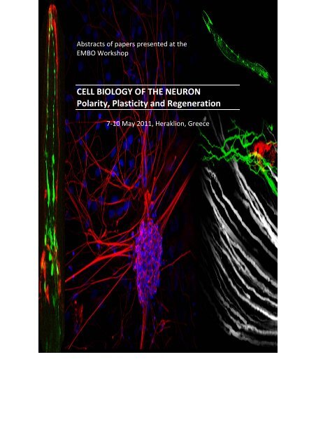
Abstracts of papers presented at the<br />
EMBO Workshop<br />
<strong>CELL</strong> <strong>BIOLOGY</strong> <strong>OF</strong> <strong>THE</strong> <strong>NEURON</strong><br />
<strong>Polarity</strong>, Plasticity and Regeneration<br />
7‐10 May 2011, Heraklion, Greece
Abstracts of papers presented at the<br />
EMBO Workshop<br />
<strong>CELL</strong> <strong>BIOLOGY</strong> <strong>OF</strong> <strong>THE</strong> <strong>NEURON</strong><br />
<strong>Polarity</strong>, Plasticity and Regeneration<br />
Organizers:<br />
7‐10 May 2011, Heraklion, Greece<br />
Frank Bradke, Max Planck Institute of Neurobiology, Germany<br />
Casper Hoogenraad, Utrecht University, The Netherlands<br />
Nektarios <strong>Tavernarakis</strong>, Institute of Molecular Biology and Biotechnology,<br />
Greece
The meeting was funded in part by EMBO (European Molecular Biology<br />
Organization)
<strong>CELL</strong> <strong>BIOLOGY</strong> <strong>OF</strong> <strong>THE</strong> <strong>NEURON</strong>: POLARITY, PLASTICITY AND<br />
REGENERATION<br />
Saturday, May 7 – Tuesday, May 10, 2011<br />
.. at a glance<br />
Saturday 14.15 1 Synaptogenesis<br />
Saturday 16.30 2 Neuronal Plasticity – Receptor Dynamics<br />
Saturday 20.00 Keynote Lecture 1<br />
Saturday 21.00 Poster Session I – Red Session<br />
Sunday 09.00 3 Neuronal Regeneration<br />
Sunday 11.15 4 Local mRNA translation<br />
Sunday 14.15 5 Neuronal Plasticity – Cytoskeleton<br />
Sunday 16.00 Poster Session II – Green Session<br />
Sunday 19.30 Keynote Lecture 2<br />
Monday 09.00 6 Neuronal <strong>Polarity</strong><br />
Monday 11.15 7 Axon Growth and Regeneration<br />
Monday 14.00 Poster Session III – Blue Session<br />
Monday 16.15 Keynote Lecture 3<br />
Monday 17.45 8 Hot‐topic Session<br />
Tuesday 09.00 9 Neuronal Trafficking<br />
Tuesday 11.15 10 Synaptogenesis<br />
Poster Sessions are located in the “Game Room”
These abstracts should not be cited in bibliographies. Material contained<br />
herein should be treated as personal communication and should be cited<br />
as such only with consent of the author.<br />
Please note that recording of oral sessions by audio, video or still<br />
photography is strictly prohibited except with the advance permission of<br />
the author(s) and the organizers.
TABLE <strong>OF</strong> CONTENTS<br />
PROGRAM ...................................................................................................................... 19<br />
AUTHOR INDEX ............................................................................................................... 25<br />
ORAL PRESENTATIONS ‐ ABSTRACTS............................................................................... 33<br />
POSTER PRESENTATIONS ‐ ABSTRACTS........................................................................... 83<br />
PARTICIPANT LIST ........................................................................................................... 209<br />
USEFUL INFORMATION ................................................................................................... 221<br />
ORAL PRESENTATIONS ABSTRACTS<br />
Molecular Mechanisms of Synapse Stabilization and Disassembly<br />
Gaeme W. Davis................................................................................................................ 33<br />
Long Term Dynamics of Inhibitory Synapses<br />
Ann Marie Craig, Frederick Dobie ..................................................................................... 34<br />
Dynamic Regulation of Synaptic Adhesion Complexes by Alternative Splicing<br />
P. Scheiffele, T.i Iijima, K.n Wu, H.d Witte......................................................................... 35<br />
Role of Leucine‐Rich Repeat Containing Proteins in Excitatory Synapse Development<br />
J. De Wit, M.w O'Sullivan, J.f Savas, E.y Sylwestrak, D. Comoletti, J. Yates, A. Ghosh....... 36<br />
A Nanoscale View into the Dynamic of AMPA Receptor Organization in Synapses<br />
D. Choquet ........................................................................................................................ 37<br />
Regulation of Neuronal Function and Dysfunction by Protein SUMOylation<br />
J. Henley ............................................................................................................................ 38<br />
Intracellular Machinery and Signaling for AMPA Receptor Trafficking at Synapses<br />
J. A. Esteban ...................................................................................................................... 39<br />
Non hyperpolarizing GABAB Receptors Regulate Neuronal Migration, and<br />
Axon/Dendrite Growth and Specification by cAMP Pathway<br />
G.e Bony, A. Contestabile, L. Cancedda............................................................................. 40<br />
Endocytic mechanisms at synapses<br />
P. De Camilli ...................................................................................................................... 41<br />
Control of Motor Neuron Generation and Regeneration in Zebrafish<br />
C. Becker, A. Norris, V. Kuscha, R.e Patani, M. Reimer, A. Scott, Z. Zhong, T. Dias,<br />
S. Chandran, T. Becker ...................................................................................................... 42<br />
Nogo‐ A is a Negative Regulator of Neurite Growth in the Developing and Adult<br />
Nervous System<br />
M. Schwab......................................................................................................................... 43<br />
Defining the Genetic Pprogram of Axon Regeneration in the CNS<br />
Z. He .................................................................................................................................. 44<br />
Regeneration of the Adult Zebrafish Brain after Traumatic Lesion: Neurogenic Radial<br />
Glia‐Type Progenitor Cells Make New Neurons<br />
M. Brand ........................................................................................................................... 45<br />
RNA‐Based Control of Visual System Wiring<br />
C. Holt................................................................................................................................ 46<br />
Identification of Localized mRNAs in Hippocampal Neurons<br />
E.n M. Schuman ................................................................................................................ 47<br />
vii
CYFIP1, a Neuronal eIF4E‐BP, Links Local Translational Regulation to Spine<br />
Remodeling: Insights into Mental Retardation and Autism<br />
C. Bagni, S, De Rubeis........................................................................................................ 48<br />
CNP/cGMP Signaling Regulates Axon Branching and Growth by Modulating<br />
Microtubule Dynamics<br />
L. Ma, C. Xia, Z. Zhao, Z. Wang.......................................................................................... 49<br />
Regulating Synaptic Strength Across the Cleft by N‐cadherins<br />
N. Vitureira, M. Letellier, I. White, Y. Goda ....................................................................... 50<br />
Presynaptic Tenacity: Insights from Live Imaging Experiments<br />
Z. Noam ............................................................................................................................ 51<br />
Class‐specific Dendrite Morphology Control by the Actin Bundling Protein Fascin<br />
J. Negele, C. Delandre, Y. Zhang, F. Förstner, A. Moore, G. Tavosanis ............................. 52<br />
EB3 Stably links Microtubules to Ankyrin G in the Axon Initial Segment<br />
C. Leterrier, H. Vacher, M.‐P. Fache, S. A. d'Ortoli, F. Castets, A. Autillo‐Touati ,<br />
B. Dargent ........................................................................................................................ 53<br />
Neurogenesis from Glial Cells – Novel Sources for New Neurons in the Adult Brain<br />
M. Götz ............................................................................................................................. 54<br />
Establishment of Neuronal <strong>Polarity</strong><br />
C. G. Dotti.......................................................................................................................... 55<br />
Molecular Mechanisms Underlying Neuronal Polarization in vivo<br />
F. Polleux........................................................................................................................... 56<br />
Neurotrophins Regulate Neuronal <strong>Polarity</strong> Acting Through Ca2+ and CaMKK<br />
K. Kaibuchi......................................................................................................................... 57<br />
ADF/Cofilin Directs Neuritogenesis in the Developing Mammalian Brain<br />
K. Flynn, D. Neukirchen, S. Jacob, S. Tahirovic, B. Garvalov,R. Wedlich‐Söldner,<br />
J. V. Small, W. Witke, F. Bradke ....................................................................................... 58<br />
Lpd Depletion Reveals a Novel SRF‐Dependent Function that Specifies Radial versus<br />
Tangential Migration of Bipolar Pyramidal Neurons<br />
E. M. Pinheiro, Z. Xie, A. L. Norovich, M. Vidaki, L.‐H. Tsai,F. B. Gertler .......................... 59<br />
Enhancing the Regenerative Ability of Axons<br />
J. Fawcett .......................................................................................................................... 60<br />
Signaling Axonal Regeneration in the CNS<br />
M. T Filbin ......................................................................................................................... 61<br />
The Oriented Emergence of Axons from Retinal Ganglion Cells is Directed by Laminin<br />
Contact in vivo<br />
O. Randlett, L. Poggi, F. R Zolessi, W. A Harris ................................................................. 62<br />
Dendrite Morphogenesis and Functional Implications<br />
Y.‐N. Jan ............................................................................................................................ 63<br />
The role of Diverse Surface receptor Complexes in Controlling Axonal Branching<br />
D. Schmucker .................................................................................................................... 64<br />
Controlling Axonal Polarization using Micropatterns<br />
M. Bisbal, S. Roth, J. Brocard, G. Bugnicourt, Y. Saoudi, A. Andrieux, S. Gory‐Fauré, C.<br />
Villard ............................................................................................................................... 65<br />
viii
Characterization of Voltage and Temperature Dependent Gating of Channelrhodopsin<br />
2, and Applications in a Dispersed Hippocampal Culture Paradigm<br />
T. Chater, J. Henley, A. Randall, J. Brown .......................................................................... 66<br />
Small Heat Shock Proteins Protect against Neurodegeneration<br />
N. Kourtis, N. <strong>Tavernarakis</strong>................................................................................................ 67<br />
Identification of the Nogo receptor Family Members NgR1 and NgR3 as CSPG<br />
Receptors: Evidence for a Mechanistic Link between CNS Myelin‐<br />
and CSPG‐Mediated Growth Inhibition<br />
T. L. Dickendesher, K. T. Baldwin, Y. A. Mironova, S. J. Raiker, Y. Duan, P. Shrager,<br />
B.Zheng, L. I. Benowitz, H. M. Geller, R. J Giger ................................................................ 68<br />
Axonal and Synaptic Degeneration is Regulated by the Hiw E3 Ubiquitin Ligase and<br />
Conditioning Lesion<br />
X. Xiong, C. Collins............................................................................................................. 69<br />
A Growth‐Inhibitory Proteoglycan Activates Atypical PKC and Modifies Par Complex<br />
Function<br />
J. Levine, S. il Lee, W. Zhang.............................................................................................. 70<br />
MyosinV‐Dependent Transport of PTEN Regulates PI3K Signalling and<br />
Neuronal Morphogenesis<br />
B. Eickholt, T. Perez, M. van Diepen, M. Parsons, N. Leslie ............................................... 71<br />
Huntington's Disease: Huntingtin and the Control of Cellular Dynamics<br />
F. Saudou........................................................................................................................... 72<br />
Regulating Mitochondrial Motility<br />
T. Schwarz ......................................................................................................................... 73<br />
Rapid Trafficking of GABAA Receptors and the Tuning of Inhibitory Transmission<br />
J. Kittler ............................................................................................................................. 74<br />
From Soma to Synapse: Sorting out Polarized Transport in Neurons<br />
L. Kapitein, M. Schlager, M. Kuijpers, P. Wulf, M. Van Spronsen, F. MacKintosh,<br />
C. Hoogenraad ................................................................................................................. 75<br />
Ion‐Flux Independent NMDA Receptor Function is Required for Aβ‐Induced Synaptic<br />
Depression<br />
H. Kessels, S. Nabavi, R. Malinow ..................................................................................... 76<br />
Homeostatic Plasticity of the Presynapse: Extensive Remodeling of the Cytomatrix<br />
at the Active Zone upon Prolonged Network Silencing<br />
E. D.Gundelfinger,V. Lazarevic,C. Schöne, M. Heine, A. Fejtova ....................................... 77<br />
Shedding Light on the Assembly of Synapse Structure and Function<br />
S. Sigrist............................................................................................................................. 78<br />
Axon Regeneration in C. elegans<br />
Y. Jin .................................................................................................................................. 79<br />
ix
POSTER ABSTRACTS<br />
Poster # [‐R(ed) / ‐G(reen) / ‐B(lue) Session]<br />
001‐R COUP‐TFI Promotes the Acquisition of Proper Final Morphology by Callosal<br />
Natalie Projection Neurons through Negative Regulation of Rnd2 Expression<br />
during Neuron Radial Migration,<br />
C. Alfano, L. Viola, J. Heng, M. Pirozzi, M. Clarkson, G. Fflore, A. Demaio,<br />
A. Schedl, F. Guillemot, M. Sstuder .................................................................... 83<br />
002‐G Activity‐Driven AMPA Receptor Remodelling in Hippocampus Mediated<br />
by RNA Editing<br />
A. Balik, A. Penn, C. Wozny, I. Greger ................................................................ 84<br />
003‐B The Function of the Zinc‐Finger Factor Insm1 in Postnatal Nneurogenesis<br />
K. Balueva, C. Boutin, H. Cremer, C. Birchmeier................................................. 85<br />
004‐R Defining Roles of PKMzeta in AMPA Receptor Trafficking<br />
E. Barker, J. Henley............................................................................................. 86<br />
005‐G A Role for STRAD Pseudokinases in Cerebral Cortex Development<br />
A. Barnes, B. Veleva‐Rotse, A. Harrold, L. Welch, G. Haddock, A. Ali, K. Thiebes. 87<br />
006‐B Reelin Regulates the Dendritic Targeting of the Ion Channel<br />
HCN1 in the Postnatal Hippocampus<br />
J. Barry, R. Piskorowski, V. Chevaleyre, S. Siegelbaum ...................................... 88<br />
007‐R The Absence of TAG‐1 Results in Perturbation of Olfactory Bulb<br />
Organization and Function<br />
G. Bastakis, M. Savvaki, D. Karagogeos............................................................. 89<br />
008‐G Analysis of Synaptic Connectivity in Neuroligin‐3 Mutant Mice<br />
St. Baudouin, S. Gerharz, H. Laetitia, K. Tanaka, K. Vogt, P. Scheiffele ............. 90<br />
009‐B Xlr Genes Control Synaptogenesis Processes<br />
B.Cubelos ........................................................................................................... 91<br />
010‐R The Potential Role of the CCT Complex in the Regulation of Long Range<br />
Axonal Transport<br />
K. Bercsenyi, G. Menendez, B. Spencer‐Dene, R. Chaleil, G. Schiavo.................. 92<br />
011‐G Otx2 Homeoprotein Transfer and Signaling in Visual Cortical Plasticity<br />
J. Spatazza, M. Beurdeley, H.‐C. Lee, S. Sugiyama, C. Bernard, A. Di Nardo,<br />
T. Hensch, A. Prochiantz .................................................................................... 93<br />
012‐B d‐IMP, the Drosophila Ortholog of the mRNA Transport Factor ZBP1<br />
Controls Axon Growth and Branching in vivo<br />
C. Medioni, A. Ephrussi, F. Besse ....................................................................... 94<br />
013‐R Identification of Novel Growth Associated Isoforms: A Deep<br />
Sequencing Approach<br />
J. Bixby, J. Lerch‐Haner, D. Motti, F. Kuo, D. Strikis, V. Lemmon ........................ 95<br />
014‐G Wnts Locally Activate CaMKII at Dendritic Spines to Promote Spine Growth<br />
and Synaptic Strength<br />
K. Boyle, L. Ciani, E. Dickins, D. Anane, M. Sahores, D. Lopes, A. Gibb, P.Salinas. 96<br />
015‐B RNA Localization and Local Translation Contribute to the Ability of cAMP<br />
to Promote Axonal Regeneration<br />
C. Cain, R. Chaudhry, J. Davila, M. Swerdel, N.‐L. Jeon, R. Hart, M. Filbin.......... 97<br />
x
016‐R Neurotrophins/p75NTR Signaling Specifies Axons during Cortical Development<br />
and Adult Neurogenesis<br />
M. Canossa, E. Zuccaro, M. Bergami, B. Vignoli, G. Bony, S. Santi, L. Cancedda.. 98<br />
017‐G Evidence for a Mechanical Coupling between N‐Cadherin Adhesions and<br />
F‐Actin in Shaping Dendritic Spines<br />
A. Chazeau, K. Czondor, M. Garcia, G. Giannone, O. Thoumine......................... 99<br />
018‐B Unraveling the Regulation of Intracellular Trafficking During in the<br />
Establishment and Maintenance of Neuronal <strong>Polarity</strong><br />
J. Chiang, A. W. Püschel ..................................................................................... 100<br />
019‐R Cellular Models to Explore Human Synaptogenesis<br />
L. Chicha, F. Knoflach, R. Haab, J. Bischofberger, R. Iacone, B. Bohrmann,<br />
M. Graf, S. Jensen, R. Jagasia............................................................................. 101<br />
020‐G The Effect of α‐Synuclein and β‐Amyloid on the Growth and Retraction of<br />
Neurites in Cultured Hippocampal Neurons<br />
H. Tsushima, F. Difato, A. Blau, E. Chieregatti ................................................... 102<br />
021‐B Protein Kinase Substrate Screen Reveals Dominant Role for JNK in Regulating<br />
Morphology Changes During Brain Development<br />
E. Coffey, J. Zdrojewska, N. Westerlund, B. Bjorkblom,E. Komulainen............... 103<br />
022‐R Roles of the AMPK‐Related Kinases NUAK1 and NUAK2 Downstream of LKB1<br />
during Cortical Development<br />
J. Courchet, M. Hirano, S. Aizawa, F. Polleux ..................................................... 104<br />
023‐G AMPA and Kainate Receptors are Differentially Regulated by CaMKII<br />
Phosphorylation<br />
F. Coussen, M. Carta, P. Opazo, D. Choquet, C. Mulle........................................ 105<br />
024‐B The srGAP Family Proteins Play Distinct Roles in Neuronal Development<br />
J. Coutinho‐Budd, T. Sassa, V. Ghukasyan, S. G., F. Polleux............................... 106<br />
025‐R A General Quantitative Model of AMPA Receptor Trafficking at Synapses<br />
K. Czondor, M. Mondin, M.l Garcia, M. Heine, R. Frischknecht, J.‐B. Sibarita,<br />
D. Choquet, O. Thoumine................................................................................... 107<br />
026‐G Mutations in Human RAB39B Gene are Responsible for X‐Linked<br />
Non‐Specific Mental Retardation<br />
P. D'Adamo, M. Giannandrea, M. L. Mignogna, M. Passafaro .......................... 108<br />
027‐B Mechanisms of Neuronal Remodeling by BDNF<br />
K. Deinhardt, B. Hempstead, M. Chao ............................................................... 109<br />
028‐R Regeneration of Axons in Injured Spinal Cord by Activation of<br />
BMP/Smad1 Signaling Pathway in Adult Neurons<br />
H. J. Zou, P. Parikh, Y. Hao, M. Hosseinkhani, S. Patil, G. Huntley,<br />
M. Tessier‐Lavigne ............................................................................................. 110<br />
029‐G Analysis of the Mechanisms by which Citron Proteins and TTC3<br />
Modulate Early Differentiation in Primary Hippocampal Neurons<br />
G. Berto, P. Camera, C. Iobbi, E.a Scarpa, Y. Bosio, F. Bianchi, C. Dotti,<br />
F. Di Cunto......................................................................................................... 111<br />
xi
030‐B Paracrine Pax6 Activity Regulates Oligodendrocyte Precursor Cell<br />
Migration in the Chick Embryonic Neural Tube<br />
Elizabeth Di Lullo, Celine Haton, Michel Volovitch, Jean‐Leaon Thomas,<br />
Alain Prochiantz................................................................................................. 112<br />
031‐R Identification of the Nogo Receptor Family Members NgR1 and NgR3<br />
as CSPG Receptors: Evidence for a Mechanistic Link between CNS<br />
Myelin‐ and CSPG‐Mediated Growth Inhibition<br />
T. L Dickendesher, K. T Baldwin, Y. A Mironova, S. J Raiker, Y. Duan, P. Shrager, B.<br />
Zheng, L. I Benowitz, H. M Geller, R. J Giger...................................................... 113<br />
032‐G Establishment and Characterization of a Human in Vitro Model System<br />
of Neurodegeneration<br />
L. Efremova, M. Leist.......................................................................................... 114<br />
033‐B Phosphorylation of SCG10/stathmin‐2 Determines Multipolar Stage<br />
Exit and Neuronal Migration Rate<br />
J. Zdrojewska, N. Westerlund, A. Padzik, E. Komulainen, B. Bjorkblom,<br />
E. Rannikko, L. Nguyen, T. Kallunki, M. Courtney, E. Coffey............................... 115<br />
034‐R Regulation of Neurotransmitter Receptor Trafficking and Degradation<br />
During Synaptic Plasticity<br />
Monica Fernandez‐Monreal, Jose A. Esteban .................................................... 116<br />
035‐G Role of GluN2B in the Synaptic Accumulation of NMDA Receptors<br />
J. Ferreira, K. She, A.a Rooyakkers, A. M. Craig, A. L. Carvalho.......................... 117<br />
036‐B Proteomics Reveals the Protein Degradation Pathways Present in the Radial<br />
Nerve Cord of the Sea Star Wound Healing Events and Early Stages of Arm<br />
Regeneration<br />
C. Franco, R. Santos, A. Varela........................................................................... 118<br />
037‐R Neuronal Mechanosensitivity in Development and Regeneration<br />
K. Franze, H. Svoboda, A. F. Christ, P. Moshayedi, L. da F. Costa, J. Fawcett,<br />
C. E. Holt, J. Guck ............................................................................................... 119<br />
038‐G JNK‐Induced Changes in Axonal Transport are mediated by the Bidirectional<br />
Co‐regulator JIP1<br />
M.‐m. Fu, E. Holzbaur ........................................................................................ 120<br />
039‐B N‐Cadjerin Specifies First Asymmetry in Developing Neurons<br />
A. Gaertner, E. Fornasiero, S. Munck, K. Vennekens, F. Valtorta, C. Dotti.......... 121<br />
040‐R Nanoscale Mechanical Coupling between N‐Cadherin Adhesion and Actin<br />
Dynamics in Growth Cone Motility<br />
M. Garcia, L. Bard, A. Argento, O. Thoumine..................................................... 122<br />
041‐G Dynamin‐Independent Cycling of AMPA Receptors Maintains<br />
Synaptic Transmission<br />
O. Glebov, C. Tigaret, J. Mellor, J. Henley........................................................... 123<br />
042‐B Mechanisms Controlling the Number and Location of Synaptic Kainate<br />
Receptors and their Implication in Synaptic Maintenance<br />
I. M González‐González, M. Petrovic, J. Antonio Esteban, J. M Henley.............. 124<br />
043‐R Investigating the Role of Nogo‐A in the Maturation of the Visual System<br />
A. Guzik‐Kornacka, V. Pernet, M. Schwab.......................................................... 125<br />
xii
044‐G Involvement of PI 3‐K/Akt and MEK/MAPK Signaling Pathways in γ‐enolase‐<br />
mediated Neurite Outgrowth and Survival<br />
A. Hafner, N. Obermajer, J. Kos.......................................................................... 126<br />
045‐B Molecular Dissection of EphA4‐Mediated Signaling<br />
C. Hassler, R. Klein.............................................................................................. 127<br />
046‐R KCNQ5, a Synaptic Member of the Voltage‐Gated KCNQ Potassium Channel<br />
Family Mediates a Component of the Afterhyperpolarization Current<br />
in Mouse Hippocampus<br />
M. Heidenreich, A. Tzingounis, T. Kharkovets, G. Spitzmaul, H. Jensen, R. Nicoll,<br />
T. Jentsch ........................................................................................................... 128<br />
047‐G Microtubule Stabilization Reduces Scarring and Causes Axon Regeneration<br />
after Spinal Cord Injury<br />
F. Hellal, A. Hurtado, J. Ruschel, K. Flynn, C. Laskowski, M. Umlauf, L. Kapitein,<br />
D. Strikis, V. Lemmon, J. Bixby, C. Hoogenraad, F. Bradke ................................. 129<br />
048‐B Scribble1 Deletion in the Principal Neurons of the Brain Alters Hippocampal<br />
LTD and AMPAR Endocytosis<br />
M. Hilal, N. Piguel, M. Moreau, Y.‐s. Abada, T. Papouin, E. Richard, D.N. Abrous130<br />
049‐R Trafficking and <strong>Polarity</strong> of Cannabinoid Receptor Type 1<br />
K. Hildick, N. Jafaari, J. Henley ........................................................................... 131<br />
050‐G RNA‐Binding Protein Hermes Regulates Axon Guidance and Branching in the<br />
Developing Visual System<br />
H. Hornberg, D. Maurus, F. Wollerton‐van Horck, M. Zwart, W. Harris, C.Holt ... 132<br />
051‐B Negative Feedback Enhances Robustness during <strong>Polarity</strong> Establishment<br />
A. Howell, C.‐F. Wu, M. Jin, T. Elston, D. Lew ..................................................... 133<br />
052‐R Development of a New Strategy to Control Protein Function in the<br />
Developing Mouse CNS<br />
R. Jackson, W. An, L. Ward, M. Van Diepen, L. Whiley, C. Legido‐Quigley,<br />
T. Wandless, K. Liu, B. Eickholt .......................................................................... 134<br />
053‐G Interplay between Polyglutamylases and Deglutamylases Regulates<br />
Polyglutamylation Levels of Neuronal Microtubules<br />
C. Janke, K. Rogowski, J. van Dijk, M. M. Magiera, C. Bosc, J.‐C. Deloulme,<br />
A. Bosson, M. Holzer, A. Andrieux, M.‐J. Moutin............................................. 135<br />
054‐B IQGAP1: Role in Spines Morphogenesis<br />
I. Jausoro, A. Cáceres ......................................................................................... 136<br />
055‐R RNA Binding Protein Vg1RBP (ZBP1) Regulates Terminal Arbor Formation<br />
but not Pathfinding in the Developing Visual System In Vivo<br />
A. Kalous, J. Yisraeli, J. Stake, C. Holt ................................................................. 137<br />
056‐G The Actin Nucleator Cobl Plays a Role in Neuronal Differentiation and<br />
Cerebellar Development and Relies on Association with Abp1<br />
N. Haag, L. Schwintzer, R. Ahuja, J. Grimm, N. Koch, H. Heuer, B. Qualmann,<br />
M. Kessels .......................................................................................................... 138<br />
057‐B Proper Synaptic Vesicle Formation and Neuronal Network Activity Critically<br />
Rely on Syndapin I<br />
D. Koch, I. Spiwoks‐Becker, V. Sabanov, T. Dugladze, A. Stellmacher, R. Ahuja,<br />
J. Grimm, S. Schüler, A. Müller, F. Angenstein................................................... 139<br />
xiii
058‐R Dynein Light Chain LC8 is a Dimerization Hub for the Memory‐Related<br />
KIBRA Protein<br />
J. Kremerskothen, K. Duning, B. Ribbrock, H. Pavenstaedt ................................ 140<br />
059‐G The Amyotrophic Lateral Sclerosis 8 Protein VAPB Disrupts the Early<br />
Secretory Pathway<br />
M. Kuijpers, K. Lou Yu, E. Teuling, D. Jaarsma, C. Hoogenraad.......................... 141<br />
060‐B Analysis of in vivo Glycine Transporter Function by Transgenic Approaches<br />
D. Lall, H. Betz, V. Eulenburg.............................................................................. 142<br />
061‐R Sphingomyelin Influences Dendritic Spine Size through the Modulation of<br />
Actin Cytoskeleton<br />
M. Dolores Ledesma, A. Arroyo, P. Camoletto, L. Morando, M. Sassoe‐Pognetto,<br />
M. Giustetto....................................................................................................... 143<br />
062‐G Regulation of Neurofibromin RasGAP Activity and Localization during Neuronal<br />
Differentiation<br />
G. Leondaritis, T. Kalpachidou, D. Mangoura .................................................... 144<br />
063‐B Development of the Corticospinal Tract: Integration into Spinal Circuitry<br />
Important for Coordinating Complex Motor Behaviors<br />
K. Lewallen, A. Levine, S. Pfaff ........................................................................... 145<br />
064‐R Decreased GSKβ Activity by Downregulation of Tyr216 Phosphorylation<br />
Underlies the Gain of Regenerative Capacity Following Conditioning Lesion<br />
M. Liz, H. Pimentel, D.Silva, A. Marques, M. Sousa............................................ 146<br />
065‐G Role of KCC2 in Morphological Plasticity of Dendritic Spines<br />
O. Llano, A. Ludwig, S. Soni, C. Rivera ................................................................ 147<br />
066‐B Structural and Biochemical Characterization of CRMPs and their Complexes<br />
Involved in the Axonal Growth and Related Disease<br />
B. Lohkamp, R. Ponnusamy................................................................................ 148<br />
067‐R Tracking Autophagosome Dynamics in Primary Neurons<br />
S. Maday, K. Wallace, E. Holzbaur ..................................................................... 149<br />
068‐G Tubulin Polyglutamylation: a Role in Neuronal <strong>Polarity</strong> and Transport<br />
M. Magiera, J. Souphron, D. Zala, F. Saudou, C. Janke ...................................... 150<br />
069‐B Identifying Injury Signals and Regeneration Enhancers in the Conditioning<br />
Lesion Model<br />
F. Mar, M. Ana, B. Pedro, A. Barbosa, V. Costa, M. Sousa ................................. 151<br />
070‐R The Nogo‐66 Receptor Restricts Both Anatomical and Functional<br />
Plasticity in Visual Cortex<br />
A. McGee ........................................................................................................... 152<br />
071‐G Mechanism of Draxin Signaling in Axonal Guidance<br />
R. Meli, F. Propst................................................................................................ 153<br />
072‐B Different requirements for Sad Kinases in Hippocampal and Cortical<br />
Neurons during the Establishment of Neuronal <strong>Polarity</strong><br />
S. Menon, D. Lutter, M. Müller, A. Püschel ........................................................ 154<br />
073‐R Proteomics Reveals Novel Sets of Proteins Involved in Axonal Growth<br />
I. Michihiro, N. Motohiro ................................................................................... 155<br />
xiv
074‐G The Role of the Drosophila Formin dDAAM in Axon Growth and<br />
Filopodia Formation<br />
J. Mihály, T. Matusek, C. Gonçalves‐Pimentel, N. Sánchez‐Soriano, A.Prokop,<br />
R. Gombos.......................................................................................................... 156<br />
075‐B Role of Neuronal Ca2+‐Sensor Proteins in Golgi‐to‐cell‐surface Membrane<br />
Ttraffic<br />
M. Mikhaylova, J. Hradsky, P. Reddy, E. D Gundelfinger, Y. Sharma, M. R Kreutz 157<br />
076‐R Diffusion/Trapping of AMPA Receptors at Neurexin‐Neuroligin Adhesions<br />
through PSD‐95<br />
M. Mondin, E. Hosy, M. Heine, D. Choquet, O. Thoumine.................................. 158<br />
077‐G The End of 35‐year Quest has Come by FRET Imaging ‐ the Molecular<br />
Mechanism of dbcAMP‐Induced Neurite Outgrowth<br />
T. Nakamura, A. Goto, M. Hoshino, M. Matsuda............................................... 159<br />
078‐B NMDA Receptor Regulates Migration of Newly Generated Neurons in the<br />
Adult Hippocampus via Disrupted‐In‐Schizophrenia 1 (DISC1)<br />
T. Namba, S. Uchino, K. Shinichi, K. Kozo ........................................................... 160<br />
079‐R Cytoplasmic Linker Proteins Regulate Neuronal Polarization through<br />
Microtubule and Growth Cone Dynamics<br />
D. Neukirchen, F. Bradke.................................................................................... 161<br />
080‐G A Genetically Encoded Dendritic Marker Sheds Light on Neuronal Connectivity<br />
in Drosophila and Reveals Conserved Features of Dendritic Development<br />
L. Nicolaï, A. Ramaekers, T. Raemaekers, A. Drozdzecki, A. Mauss, J. Yan,<br />
M. Landgraf, W. Annaert, B. Hassan.................................................................. 162<br />
081‐B Cux1 and Cux2 Regulate Dendritic Branching, Spine Morphology and<br />
Synapses of the Upper Layer Neurons of the Cortex<br />
L. Nieto............................................................................................................... 163<br />
082‐R Mek2 and Pin1 Regulate BNIP‐H (Caytaxin) Ability to Promote Axonal and<br />
Dendritic Growth by Trafficking Glutaminase KGA on Kinesin Heavy<br />
Chain KIF5B<br />
C. Q. Pan, B. C. Low ............................................................................................ 164<br />
083‐G Developmental Stabilization and Activity‐Dependent Regulation of Gephyrin<br />
Scaffolds at Hippocampal GABAergic Postsynapses<br />
A. Vlachos, S. Reddy‐Alla, T. Papadopoulos, T. Deller, H. Betz ........................... 165<br />
084‐B Tubulin Modification that Control Microtubule Stabilizers vs<br />
Microtubule Depolymerizers<br />
L. Peris, D. Job, A. Andrieux................................................................................ 166<br />
085‐R Studying the Function, Regulation and Coordination of Kinesin‐1 and<br />
Kinesin‐3 in the Drosophila Nervous System<br />
I. Peset‐Martin, L. Williams, M. Landgraf, I. Palacios......................................... 167<br />
086‐G β‐Arrestin2 Mediates Spatial Control over Cofilin Activity in NMDA‐<br />
Induced Dendritic Spine Remodeling in Hippocampal Neurons<br />
C. Pontrello, K. DeFea, I. Ethell........................................................................... 168<br />
087‐B Visualization of Activity‐Induced Local Translation Using TimeSTAMP<br />
F. Port, M. Lin, S. Bullock ................................................................................... 169<br />
xv
088‐R Spectraplakins ‐ Cytoskeletal Integrators with Key Roles in Neuronal Growth<br />
A. Prokop, J. Alves‐Silva, R. Beaven, N. Sanchez‐Soriano ................................... 170<br />
089‐G Regulation of Neuronal <strong>Polarity</strong> by Rap1 GTPases<br />
D. Lutter, A. Püschel........................................................................................... 171<br />
090‐B A Novel Stress‐Regulated Protein that Acts on Actin Bundling, Synaptic<br />
Efficacy and Cognition<br />
M. Schmidt, J. Schülke, C. Liebl, M. Stiess, F. Holsboer, M. Stewart, F. Bradke,<br />
M. Eder, M. Müller, T. Rein ................................................................................ 172<br />
091‐R p140Cap and Citron‐N: a “spiny” Cooperative Relationship<br />
D. Repetto, N. Morello, E. Calcagno, F. Bianchi, M. Giustetto, F. Di Cunto,<br />
E. Turco, P. Defilippi, P. Di Stefano, P. Camera .................................................. 173<br />
092‐G The Role of Perisynaptic Schwann Cells in Nerve Terminal Regeneration<br />
M. Rigoni, E. Tedesco, G. Carmignoto, O. Rossetto, C. Montecucco .................. 174<br />
093‐B Frequency‐Based Motor‐Driven Mechanisms in Axon Length Sensing<br />
I. Rishal, N. Kam, V. Shinder, E. Fisher, G. Schiavo, M. Fainzilber....................... 175<br />
094‐R Effects of Microtubule Stabilization on Axon Regeneration in Clinically<br />
Relevant Approaches<br />
J. Ruschel, A. Hurtado, F. Hellal, V. Lemmon, J. Bixby, F. Bradke ....................... 176<br />
095‐G Cell‐Contact Induced Eph Receptor Trans‐Endocytosis Activates an SFK<br />
Dependent Phagocytic Pathway<br />
M. Sakkou, R. Klein............................................................................................. 177<br />
096‐B Linking Synaptic Endocytosis to Retrograde Axonal Transport: the Role of<br />
the Coxsackievirus and Adenovirus Receptor in Neuronal Trafficking of<br />
Adenoviruses<br />
S. Salinas, C. Zussy, D. Henaff, N. Bayo‐Puxan, G. Schiavo, E. Kremer .............. 178<br />
097‐R Regulation of the Ubiquitin‐Proteasome System by BDNF at Hippocampal<br />
Synapses<br />
A. R. Santos, M. Mele, B. Kellermayer, D. Comprido, B. J Manadas, C. B Duarte. 179<br />
098‐G +TIPs are Regulated by MAP1B in Neuronal Cells<br />
E. Tortosa, N. Galjart, J. Avila, L. Sayas.............................................................. 180<br />
099‐B Eph Receptor Clustering is a Signaling‐Integrator to Elicit Cellular Responses<br />
A. Schaupp, O. Sabet, I. Dudanova, P. Bastiaens, R. Klein.................................. 181<br />
100‐R Deciphering the Mechanisms Underlying the Refinement of Synaptic Axonal<br />
Branches<br />
M. Schlieder, M. Langen, R. Hiesinger, B. Hassan .............................................. 182<br />
101‐G Expression of Pax6 in Retinal Ganglion Cells Mediates SFRP1 Axonal Response<br />
A. Sebastian‐Serrano, A. Sandonis, P. Esteve, P. Bovolenta, M. Nieto ............... 183<br />
102‐B An Extracellular Steric Seeding Mechanism for Eph‐Ephrin Signaling<br />
Platform Assembly<br />
E. Seiradake, K. Harlos, G. Sutton, A. R. Aricescu, E. Y. Jones............................. 184<br />
103‐R Regulation of Odorant Receptor Genes<br />
B. Shykind........................................................................................................... 185<br />
104‐G Extracellular Histones: A Novel Inhibitor of Axonal Regeneration in the CNS?<br />
M. Siddiq, M. Filbin ............................................................................................ 186<br />
xvi
105‐B A Mechanistic Model Underlying Inhibitory Synapse Assembly<br />
T. Soykan, A. Poulopoulos, T. Papadopoulos, H. Betz, N. Brose, F. Varoqueaux .. 187<br />
106‐R Otx2 Homeoprotein Transfer and Signaling in Visual Cortical Plasticity<br />
J. Spatazza, M. Beurdeley, H.‐C. Lee, S. Sugiyama, C. Bernard, A. Di Nardo,<br />
T. Hensch, A. Prochiantz..................................................................................... 188<br />
107‐G Wnt Signalling Regulates Actin Dynamic during Axonal Remodelling<br />
E. Stamatakou, M. Hoyos‐Flight, P. Salinas........................................................ 189<br />
108‐B Axon Extension Occurs Independently of Centrosomal Microtubule Nucleation<br />
M. Stiess, N. Maghelli, L. Kapitein, S. Gomis‐Rüth, M. Wilsch‐Bräuninger,<br />
C. Hoogenraad, I. Tolic‐Norrelykke, F. Bradke................................................. 190<br />
109‐R Cyclin‐Dependent Kinase 5 Regulates Calcium Influx through N‐type Calcium<br />
Channels<br />
S. Su, J. Pan, B. Samuels, K. Win, S. Zhang, D. Yue, L.‐H. Tsai.......................... 191<br />
110‐G Expression of the Natriuretic Peptide Receptor 2 (Npr2) and its Role in Axonal<br />
Branching<br />
G. Ter‐Avetisyan, A. Stonkute, R. Jüttner, H. Schmidt, F. G. Rathjen............... 192<br />
111‐B Rac1 Function in Cortical Interneuron Development<br />
S. Tivodar, M. Vidaki, K. Doulgeraki, V. Tybulevicz, N. Kessaris, V. Pachnis,<br />
D. Karagogeos................................................................................................. 193<br />
112‐R MAP1B is required for Dendritic Spine Development and Synaptic Maturation<br />
E. Tortosa, C. Montenegro‐Venegas, M. Benoist, S. Härtel,<br />
C. Gonzalez‐Billault, J. Esteban, J. Avila........................................................... 194<br />
113‐G Functions of Neuronal Nogo‐A for Neurite Growth in the Developing<br />
Nervous System<br />
F. Vajda, M. M. Petrinovic, C. S. Duncan, M. E. Schwab.................................. 195<br />
114‐B In vivo Pull Down using a Synapsin‐GFP‐Neogenin Transgenic Mouse<br />
Identifies Dock7 as a Novel Neogenin Interacting Protein Involved in<br />
RGMa Induced Axon Repulsion<br />
D. van den Heuvel, A. Hellemons, J. Pasterkamp ............................................ 196<br />
115‐R The bZIP Transcription Factors CREB, C/EBP and NFIL3 Coordinately Regulate<br />
Axon Regeneration<br />
L. van der Kallen, H. MacGillavry, J. Verhaagen, A. Smit, R. van Kesteren ...... 197<br />
116‐G Neuronal microRNA Expression Profiling Identifies Novel microRNAs with<br />
Axon Growth Promoting Effects<br />
S. van Erp, A.H.A.A. Derijck, E.Y. van Battum, R.J. Pasterkamp....................... 198<br />
117‐B Homeostatic Synaptic Scaling Contributes to the Structural Reorganization<br />
of Neurons after Denervation<br />
A. Vlachos, D. Becker, C. B. Orth, M. Helias, P. Jedlicka, M. Neuwirth, R. Winkels,<br />
J. Roeper, M. Diesmann, G. Schneider............................................................. 199<br />
118‐R Functional Analysis of Vertebrate Orthologues of Synapse‐Defective‐1 (Syd‐1)<br />
C. Wentzel, J. E. Sommer, P.r Scheiffele .......................................................... 200<br />
119‐G The Microtubule Binding Protein SCG10 Regulates Axon Extension<br />
L. Will, Y.‐H. Li, P. Bondallaz, G. Grennigloh, A. W. Püschel ............................ 201<br />
120‐B The Role of Schwann Cell Notch Signalling in Peripheral Nerve Regeneration<br />
D. Wilton, J. Shen, D. Meijer, L. Feltri, L. Wrabetz, R. Mirsky, K. Jessen .......... 202<br />
xvii
121‐R Calmyrin1 Binds to SCG10 Protein (stathmin2) to Modulate Neurite Outgrowth<br />
K. Debowska, A. Sobczak, M. Blazejczyk, M.l R. Kreutz, J. Kuznicki, U. Wojda ... 203<br />
122‐G Rassf1 and Rassf5 are required for Neuronal <strong>Polarity</strong><br />
R. Yang, E. Kong, A. W. Püschel ...................................................................... 204<br />
123‐B NMDA Receptor Activation Suppresses Microtubule Dynamics in Neurons<br />
K. W. Yau, L. C. Kapitein, S. Montenegro Gouveia, W. A. van der Zwan,<br />
P. S. Wulf, J. Demmers, J. Jaworski, A. Akhmanova, C. C. Hoogenraad........... 205<br />
124‐R Drosophila Melanogaster is an in vivo Platform for the Study of<br />
Huntingtin‐Dependent Vesicular Trafficking and htt Phosphorylation<br />
Modifiers in a Huntington's Disease Context<br />
D. Zala, Y. Elipot, F. Saudou ............................................................................ 206<br />
125‐G Motor Neuron Differentiation and Topographic Mapping Depends on<br />
β‐ and γ‐Catenin Activity<br />
N. Zampieri, E. Demireva, L. Shapiro, T. Jessell ............................................... 207<br />
xviii
PROGRAM<br />
Cell Biology of the Neuron: <strong>Polarity</strong>, Plasticity and Regeneration<br />
Crete, May 2011<br />
10.00 ‐ 13.00 Registration<br />
13.00 ‐ 14.00 Lunch<br />
14.00 ‐ 14.15 Opening Remarks<br />
SATURDAY, May 7<br />
SESSION 1 SYNAPTOGENESIS<br />
Chairperson: Eckhard Gundelfinger<br />
[Leibniz Institute of Neurobiology, Magdeburg]<br />
14.15 ‐ 14.45 Molecular<br />
Disassembly<br />
Mechanisms of Synapse Stabilization and<br />
14.45 ‐ 15.15<br />
Graeme Davis [University of California, San Francisco]<br />
Long Term Dynamics of Inhibitory Synapses<br />
Ann Marie Craig [University of British Columbia, Vancouver]<br />
15.15 ‐ 15.45 Dynamic Regulation of Synaptic Adhesion Complexes by<br />
Alternative Splicing<br />
Peter Scheiffele [Biozentrum of the University of Basel]<br />
15.45 ‐ 16.00 Role of Leucine‐Rich Repeat Containing Proteins in Excitator<br />
Synapse Development<br />
Joris de Wit [University of California San Diego]<br />
16.00 ‐ 16.30 Coffee Break<br />
SESSION 2 <strong>NEURON</strong>AL PLASTICITY – RECEPTOR DYNAMICS<br />
Chairperson: Thomas Schwarz [Harvard Medical School, Boston]<br />
16.30 ‐ 17.00 A Nanoscale View into the Dynamic of AMPA Receptor<br />
Organization in Synapses<br />
Daniel Choquet [University of Bordeaux]<br />
17.00 ‐ 17.30 Regulation of Neuronal Function and Dysfunction by Protein<br />
SUMOylation<br />
Jeremy Henley [University of Bristol]<br />
17.30 ‐ 18.00 Intrecellular Machinery and Signaling for AMPA Receptor<br />
Trafficking at Synapses<br />
Jose Esteban [Centro de Biologia Molecular, CSIC, Madrid]<br />
19
18.00 ‐ 18.15 Non Hyperpolarizing GABAB Receptors Regulate Neuronal<br />
Migration, and Axon/Dendrite Growth and Specification by<br />
cAMP Pathways<br />
Laura Cancedda [Italian Institute of Technology, Genova]<br />
18.15 ‐ 20.00 Dinner<br />
20.00 ‐ 21.00 Keynote Lecture 2:<br />
Endocytic Mechanisms of Synapses<br />
Pietro De Camilli [Yale University, New Haven]<br />
21.00 ‐ 23.00 Poster Session I – RED SESSION<br />
SUNDAY, May 8<br />
SESSION 3 <strong>NEURON</strong>AL REGENERATION<br />
Chairperson: James Fawcett [University of Cambridge]<br />
09.00 ‐ 09.30 Control of Motor Neuron Generation and Regeneration in<br />
Zebrafish<br />
Catherina Becker [University of Edinburgh]<br />
09.30 ‐ 10.00 Nogo‐A is a Negative Regulator of Neurite Growth in the<br />
Developing and Adult Nervous System<br />
Martin Schwab [University of Zurich]<br />
10.00 ‐ 10.30 Defining the Genetic Program of Axon Regeneration in the CNS<br />
Zhigang He [Harvard Medical School, Boston]<br />
10.30 ‐ 10.45 Regeneration of the Adult Zebrafish Brain after Traumatic<br />
Lesion: Neurogenic Radial Glial‐Type Progenitor Cells make<br />
New Neurons<br />
Michael Brand [Dresden University]<br />
10.45 ‐ 11.15 Coffee Break<br />
SESSION 4 LOCAL mRNA TRANSLATION<br />
Chairperson: Rüdiger Klein [Max Planck Institute of Neurobiology,<br />
11.15 ‐ 11.45<br />
Martinsried]<br />
RNA‐Based Control of Visual System Wiring<br />
Christine Holt [University of Cambridge]<br />
11.45 ‐ 12.15 Identification of Localized mRNAs in Hippocampal Neurons<br />
Eric Schuman [Max Planck Institute of Brain Research,<br />
Frankfurt]<br />
20
12.15 ‐ 12.45 CYFIP1, a Neuronal elF4E‐BP, Links Local Translational<br />
Regulation to Spine Remodeling Insights into Mental<br />
Retardation and Autism<br />
Claudia Bagni [Vesalius Research Center VIB, Leuven]<br />
12.45 ‐ 13.00 CNP/cGMP Signaling Regulates Axon Branching and growth by<br />
Modulating Microtubule Dynamics<br />
Le Ma [University of Southern California, Los Angeles]<br />
13.00 ‐ 14.00 Lunch<br />
SESSION 5 <strong>NEURON</strong>AL PLASTICITY ‐ CYTOSKELETON<br />
Chairperson: Carlos Dotti [Katholieke Universiteit Leuven]<br />
14.15 ‐ 14.45 Regulating Synaptic Strength Across the Cleft by N‐Cadherins<br />
Yukiko Goda [University College London]<br />
14.45 ‐ 15.15 Presynaptic Tenacity: Insights from Live Imaging Experiments<br />
Noam Ziv [Technion Faculty of Medicine, Haifa]<br />
15.15 ‐ 15.45 Class‐Specific Dendrite Morphology Control by the Actin<br />
Bunding Protein Fascin<br />
Gaia Tavosanis [Max Planck Institute of Neurobiology,<br />
Martinsried]<br />
15.45 ‐ 16.00 EB3 Stably Links Microtubules to Ankyrin G in the Axon Initial<br />
Segment<br />
Christophe Leterrier [INSERM U641, Marseille]<br />
16.00 ‐ 18.00 Coffee Break & Poster Session II – GREEN SESSION<br />
18.00 ‐ 19.30 Dinner<br />
19.30 ‐ 20.30 Keynote Lecture 2:<br />
Neurogenesis from Glial Cells – Novel Sources for New Neurons<br />
in the Adult Brain<br />
Magdalena Götz [Helmholz Zentrum München]<br />
20.30 Wine & Beer<br />
SESSION 6 <strong>NEURON</strong>AL POLARITY<br />
MONDAY, May 9<br />
Chairperson: Yishi Jin [University of California, San Diego]<br />
09.00 ‐ 09.30 Establishment of Neuronal <strong>Polarity</strong><br />
Carlos Dotti [Katholieke Universiteit Leuven]<br />
09.30 ‐ 10.00 Molecular Mechanisms Underlying Neuronal Polarization in<br />
vivo<br />
Franck Polleux [The Scripps Research Institute, La Jolla]<br />
21
10.00 ‐ 10.30 Neurotrophin Regulate Neuronal <strong>Polarity</strong> Acting through Ca2+<br />
and CaMKK<br />
Kozo Kaibuchi [Nagoya University]<br />
10.30 ‐ 10.45 ADF/Cofilin Directs Neuritogenesis in the Developing<br />
Mammalian Brain<br />
Kevin Flynn [Max Planck Institute of Neurobiology,<br />
Martinsried]<br />
10.45 ‐ 11.15 Coffee Break<br />
SESSION 7 AXON GROWTH AND REGENERATION<br />
Chairperson: Farida Hellal [Max Planck Institute of Neurobiology,<br />
11.15 ‐ 11.45<br />
Martinsried]<br />
Lpd Depletion Reveals a Novel SRF‐Dependent Funtion that<br />
Specifies Radial Versus Tangential Migration of Bipolar<br />
Pyramidal Neurons<br />
Frank B. Gertler [Koch Institute for Integrative Cancer<br />
Research at MIT]<br />
11.45 ‐ 12.15 Enhancing the Regenerative Ability of Axons<br />
James Fawcett [University of Cambridge]<br />
12.15 ‐ 12.45 Signaling Axonal Regeneration in the CNS<br />
Marie Filbin [Hunter College, New York]<br />
12.45 ‐ 13.00 The Oriented Emergence of Axons from Retinal Ganglion Cells<br />
is Directed by Laminin Contact in vivo<br />
Owen Randlett [University of Cambridge]<br />
13.00 ‐ 14.00 Lunch<br />
14.00 ‐ 16.15 Coffee Break & Poster Session III – BLUE SESSION<br />
16.15 ‐ 17.15 Keynote Lecture 3:<br />
Dendrite Morphogenesis and Functional Implications<br />
Yuh‐Nung Jan [University of California, San Francisco]<br />
17.15 ‐ 17.45 Coffee Break<br />
SESSION 8 HOT‐TOPIC SESSION<br />
Chairpersons: Lukas Kapitein [Utrecht University, Utrecht] &<br />
Eleanor Coffey [Turku Centre for Biotechnology]<br />
17.45 ‐ 18.00 The Role of Diverse Surface Receptor Complexes in Controlling<br />
Axonal Branching<br />
Dietmar Schmucker [Vesalius Research Center VIB, Leuven]<br />
18.00 ‐ 18.15 Controlling Axonal Polarization using Micropatterns<br />
Mariano Bisbal [Université Joseph Fourier, Grenoble]<br />
22
18.15 ‐ 18.30 Characterization of Voltage and Temperature Dependent<br />
Gating of Channelrhodopsin 2, and Application in a Dispersed<br />
Hippocampal Culture Paradigm<br />
Thomas Chater [University of Bristol]<br />
18.30 ‐ 18.45 Small Heat Shock Proteins Protect Against Neurodegeneration<br />
Nikos Kourtis [Institute of Molecular Biology and<br />
Biotechnology – FORTH, Heraklion]<br />
18.45 ‐ 19.00 Identification of the Nogo Receptor Family Members NgR1 and<br />
NgR3 as CSPG Receptors: Evidence for a Mechanistic Link<br />
between CNS Myelin‐ and CSPG‐Mediated Growth Inhibition<br />
Travis Lee Dickendesher [University of Michigan]<br />
19.00 ‐ 19.15 Axonal and Synaptic Degeneration is Regulated by the Hiw E3<br />
Ubiquitin Ligase and Conditioning Lesion<br />
Catherine Collins [University of Michigan]<br />
19.15 ‐ 19.30 A Growth‐Inhibitory Proteoglycan Activates Atypical PKC and<br />
Modifies Par Complex Function<br />
Joel Levine [Stony Brook University, New York]<br />
19.30 ‐ 19.45 MyosinV‐Dependent Transport of PTEN Regulates PI3K<br />
Signaling and Neuronal Morphogenesis<br />
Britta Eickholt [King’s College London]<br />
19.45 Dinner & Party<br />
TUESDAY, May 10<br />
SESSION 9 <strong>NEURON</strong>AL TRAFFICKING<br />
Chairperson: Jose Esteban [Universidad Autonoma Madrid]<br />
09.00 ‐ 09.30 Huntington’s Disease: Huntington and the Control of Cellular<br />
Dynamics<br />
Frederic Saudou [Centre Universitaire, Institut Curie, Orsay]<br />
09.30 ‐ 10.00 Regulating Mitochondrial Motility<br />
Thomas Schwarz [Harvard Medical School, Boston]<br />
10.00 ‐ 10.30 Rapid Trafficking of GABAA Receptors and the Tuning of<br />
Inhibitory Transmission<br />
Josef Kittler [University College of London]<br />
10.30 ‐ 10.45 From Soma to Synapse: Sorting out Polarized Transport in<br />
Neurons<br />
Lukas Kapitein [Utrecht University, Utrecht]<br />
10.45 ‐ 11.15 Coffee Break<br />
23
SESSION 10 SYNAPTOGENESIS<br />
Chairperson: Peter Scheiffele [Biozentrum of the University of Basel]<br />
11.15 ‐ 11.30 Ion‐Flux Independent NMDA Receptor Function is required for<br />
Aβ‐Induced Synaptic Depression<br />
Helmut Kessels [The Netherlands Institute of Neuroscience,<br />
Amsterdam]<br />
11.30 ‐ 12.00 Homeostatic Plasticity of the Presynapse Extensive Remodeling<br />
of the Cytomatrix at the Active Zone upon Prolonged Network<br />
Silencing<br />
Eckart Gundelfinger [Leibniz Institute of Neurobiology,<br />
12.00 ‐ 12.30<br />
Magdeburg]<br />
Shedding light on the Assembly of Synapse Structure and<br />
Function<br />
Stefan Sigrist [Freie Universität Berlin]<br />
12.30 ‐ 13.00 Axon Regeneration in C. elegans<br />
Yishi Jin [University of California, San Diego]<br />
13.00 ‐ 13.15 Closing, short feedback on conference<br />
13.15 Lunch<br />
24
Abada, Y. 130<br />
Abrous, D. 130<br />
Ahuja, R. 138, 139<br />
Aizawa, S. 104<br />
Akhmanova, A. 205<br />
Alfano, C. 83<br />
Ali, A. 87<br />
Alves‐Silva, J. 170<br />
An, W. 134<br />
Ana, M. 151<br />
Anane, D. 96<br />
Andrieux, A. 65, 135,<br />
166<br />
Angenstein, F. 139<br />
Annaert, W. 162<br />
Argento, A. 122<br />
Aricescu, R.A. 184<br />
Arroyo, A. 143<br />
Autillo‐Touati, A. 53<br />
Avila, J. 180, 194<br />
Bagni, C. 48<br />
Baldwin, K.T. 68, 113<br />
Balik, A. 84<br />
Balueva, K. 85<br />
Barbosa, A. 151<br />
Bard, L. 122<br />
Barker, E. 86<br />
Barnes, A. 87<br />
Barry, J. 88<br />
Bastakis, G. 89<br />
Bastiaens, P. 181<br />
Baudouin, S. 90<br />
Bayo‐Puxan, N. 178<br />
Beatriz, C. 91<br />
Beaven, R. 170<br />
Becker, C. 42<br />
Becker, D. 199<br />
Becker, T. 42<br />
Benoist, M. 194<br />
AUTHOR INDEX<br />
Benowitz, L.I. 68,<br />
113<br />
Bercsenyi, K. 92<br />
Bergami, M. 98<br />
Bernard, C. 93, 188<br />
Berto, G. 111<br />
Besse, F. 94<br />
Betz, H. 142, 165,<br />
187<br />
Beurdeley, M. 93,<br />
188<br />
Bianchi, F. 111, 173<br />
Birchmeier, C. 85<br />
Bisbal, M. 65<br />
Bischofberger, J. 101<br />
Bixby, J. 95, 129, 176<br />
Bjorkblom, B. 103,<br />
115<br />
Blau, A. 102<br />
Blazejczyk, M. 203<br />
Bohrmann, B. 101<br />
Bondallaz, P. 201<br />
Bony, G. 40, 98<br />
Bosc, C. 135<br />
Bosio, Y. 111<br />
Bosson, A. 135<br />
Boutin, C. 85<br />
Bovolenta, P. 183<br />
Boyle, K. 96<br />
Bradke, F. 58, 129,<br />
161, 172, 176,<br />
190<br />
Brand, M. 45<br />
Brocard, J. 65<br />
Brose, N. 187<br />
Brown, J. 66<br />
Bugnicourt, G. 65<br />
Bullock, S. 169<br />
Cáceres, A. 136<br />
25<br />
Cain, C. 97<br />
Calcagno, E. 173<br />
Camera, P. 111, 173<br />
Camoletto, P. 143<br />
Cancedda, L. 40, 98<br />
Canossa, M. 98<br />
Carmignoto, G. 174<br />
Carta, M. 105<br />
Carvalho A.‐L. 117<br />
Castets, F. 53<br />
Chaleil, R. 92<br />
Chandran, S. 42<br />
Chao, M. 109<br />
Chater, T. 66<br />
Chaudhry, R. 97<br />
Chazeau, A. 99<br />
Chevaleyre, V. 88<br />
Chiang, J. 100<br />
Chicha, L. 101<br />
Chieregatti, E. 102<br />
Choquet, D. 37, 105,<br />
107, 158<br />
Christ, A.F. 119<br />
Ciani, L. 96<br />
Clarkson, M. 83<br />
Coffey, E. 103, 115<br />
Collins, C. 69<br />
Comoletti, D. 36<br />
Comprido, D. 179<br />
Contestabile, A. 40<br />
Costa, V. 151<br />
Courchet, J. 104<br />
Courtney, M. 115<br />
Coussen, F. 105<br />
Coutinho‐Budd, J.<br />
106<br />
Craig A.‐M. 34, 117<br />
Cremer, H. 85<br />
Czondor, K. 99, 107
da F. Costa, L. 119<br />
D'Adamo, P. 108<br />
Dargent, B. 53<br />
Davila, J. 97<br />
Davis, G.W. 33<br />
De Camilli, P. 41<br />
De Rubeis, S. 48<br />
De Wit, J. 36<br />
Debowska, K. 203<br />
DeFea, K. 168<br />
Defilippi, P. 173<br />
Deinhardt, K. 109<br />
Delandre, C. 52<br />
Deller, T. 165<br />
Deloulme, J. 135<br />
Demaio, A. 83<br />
Demireva, E. 207<br />
Demmers, J. 205<br />
Derijck, A.H.A.A. 198<br />
Di Cunto, F. 111, 173<br />
Di Lullo, E. 112<br />
Di Nardo, A. 93, 188<br />
Di Stefano, P. 173<br />
Dias, T. 42<br />
Dickendesher, T.L.<br />
68, 113<br />
Dickins, E. 96<br />
Diesmann, M. 199<br />
Difato, F. 102<br />
Dobie, F. 34<br />
d'Ortoli, S.‐A. 53<br />
Dotti, C. 55, 111, 121<br />
Doulgeraki, K. 193<br />
Drozdzecki, A. 162<br />
Duan, Y. 68, 113<br />
Duarte, C.B. 179<br />
Dudanova, I. 181<br />
Dugladze, T. 139<br />
Duncan, C.S. 195<br />
Duning, K. 140<br />
Eder, M. 172<br />
Efremova, L. 114<br />
Eickholt, B. 71, 134<br />
Elipot, Y. 206<br />
Elston, T. 133<br />
Ephrussi, A. 94<br />
Esteban, J. 39, 16,<br />
124, 194<br />
Esteve, P. 183<br />
Ethell, I. 168<br />
Eulenburg, V. 142<br />
Fache, M. 53<br />
Fainzilber, M. 175<br />
Fawcett, J. 60, 119<br />
Fejtova, A. 77<br />
Feltri, L. 202<br />
Fernandez‐Monreal,<br />
M. 116<br />
Ferreira, J. 117<br />
Fflore, G. 83<br />
Filbin, M. 61, 97, 186<br />
Fisher, E. 175<br />
Flynn, K. 58, 129<br />
Fornasiero, E. 121<br />
Förstner, F. 52<br />
Franco, C. 118<br />
Franze, K. 119<br />
Frédéric, S. 206<br />
Frischknecht, R. 107<br />
Fu, M. 120<br />
Gaertner, A. 121<br />
Galjart, N. 180<br />
Garcia, M. 99, 107,<br />
122<br />
Garvalov, B. 58<br />
Geller, H.M. 68, 113<br />
Gerharz, S. 90<br />
Gertler, F.B. 59<br />
Ghosh, A. 36<br />
26<br />
Ghukasyan, V. 106<br />
Giannandrea, M.<br />
108<br />
Giannone, G. 99<br />
Gibb, A. 96<br />
Giger, R.J. 68, 113<br />
Giustetto, M. 143,<br />
173<br />
Glebov, O. 123<br />
Goda, Y. 50<br />
Gombos, R. 156<br />
Gomis‐Rüth, S. 190<br />
Gonçalves‐Pimentel,<br />
C. 156<br />
Gonzalez‐Billault, C.<br />
194<br />
González‐González,<br />
I.M. 124<br />
Gory‐Fauré, S. 65<br />
Goto, A. 159<br />
Götz, M. 54<br />
Graf, M. 101<br />
Greger, I. 84<br />
Grennigloh, G. 201<br />
Grimm, J. 138, 139<br />
Guck, J. 119<br />
Guerrier, S. 106<br />
Guillemot, F. 83<br />
Gundelfinger, E.D.<br />
77, 157<br />
Guzik‐Kornacka, A.<br />
125<br />
Haab, R. 101<br />
Haag, N. 138<br />
Haddock, G. 87<br />
Hafner, A. 126<br />
Hao, Y. 110<br />
Harlos, K. 184<br />
Harris, W. 62, 132<br />
Harrold, A. 87<br />
Hart, R. 97
Härtel, S. 194<br />
Hassan, B. 162, 182<br />
Hassler, C. 127<br />
Haton, C. 112<br />
He, Z. 44<br />
Heidenreich, M. 128<br />
Heine, M. 77, 107,<br />
158<br />
Helias, M. 199<br />
Hellal, F. 129, 176<br />
Hellemons, A. 196<br />
Hempstead, B. 109<br />
Henaff, D. 178<br />
Heng, J. 83<br />
Henley, J. 38, 66, 86,<br />
123, 124, 131<br />
Hensch, T. 93, 188<br />
Heuer, H. 138<br />
Hiesinger, R. 182<br />
Hilal, M. 130<br />
Hildick, K. 131<br />
Hirano, M. 104<br />
Holsboer, F. 172<br />
Holt, C. 46, 119, 132,<br />
137<br />
Holzbaur, E. 120,<br />
149<br />
Holzer, M. 135<br />
Hoogenraad, C. 75,<br />
129, 141, 190,<br />
205<br />
Hornberg, H. 132<br />
Hoshino, M. 159<br />
Hosseinkhani, M.<br />
110<br />
Hosy, E. 158<br />
Howell, A. 133<br />
Hoyos‐Flight, M. 189<br />
Hradsky, J. 157<br />
Huntley, G. 110<br />
Hurtado, A. 129, 176<br />
Iacone, R. 101<br />
Iijima, T. 35<br />
il Lee, S. 70<br />
Iobbi, C. 111<br />
Jaarsma, D. 141<br />
Jackson, R. 134<br />
Jacob, S. 58<br />
Jafaari, N. 131<br />
Jagasia, R. 101<br />
Jan, Y. 63<br />
Janke, C. 135, 150<br />
Jausoro, I. 136<br />
Jaworski, J. 205<br />
Jedlicka, P. 199<br />
Jensen, H. 128<br />
Jensen, S. 101<br />
Jentsch, T. 128<br />
Jeon, N. 97<br />
Jessell, T. 207<br />
Jessen, K. 202<br />
Jin, M. 133<br />
Jin, Y. 79<br />
Job, D. 166<br />
Jones, E.Y. 184<br />
Jüttner, R. 192<br />
Kaibuchi, K. 57<br />
Kallunki, T. 115<br />
Kalous, A. 137<br />
Kalpachidou, T. 144<br />
Kam, N. 175<br />
Kapitein, L. 75, 129,<br />
190, 205<br />
Karagogeos, D. 89,<br />
193<br />
Kellermayer, B. 179<br />
Kessaris, N. 193<br />
Kessels, H. 76<br />
Kessels, M. 138<br />
Kharkovets, T. 128<br />
Kittler, J. 74<br />
27<br />
Klein, R. 127, 177,<br />
181<br />
Knoflach, F. 101<br />
Koch, D. 139<br />
Koch, N. 138<br />
Komulainen, E. 103,<br />
115<br />
Kong, E. 204<br />
Kos, J. 126<br />
Kourtis, N. 67<br />
Kozo, K. 160<br />
Kremer, E. 178<br />
Kremerskothen, J.<br />
140<br />
Kreutz, M.R. 157,<br />
203<br />
Kuijpers, M. 75, 141<br />
Kuo, F. 95<br />
Kuscha, V. 42<br />
Kuznicki, J. 203<br />
Laetitia, H. 90<br />
Lall, D. 142<br />
Landgraf, M. 162,<br />
167<br />
Langen, M. 182<br />
Laskowski, C. 129<br />
Lazarevic, V. 77<br />
Ledesma, M.‐D. 143<br />
Lee, H. 93, 188<br />
Legido‐Quigley, C.<br />
134<br />
Leist, M. 114<br />
Lemmon, V. 95, 129,<br />
176<br />
Leondaritis, G. 144<br />
Lerch‐Haner, J. 95<br />
Leslie, N. 71<br />
Letellier, M. 50, 53<br />
Levine, A. 145<br />
Levine, J. 70<br />
Lew, D. 133
Lewallen, K. 145<br />
Li, Y.‐H. 201<br />
Liebl, C. 172<br />
Lin, M. 169<br />
Liu, K. 134<br />
Liz, M. 146<br />
Llano, O. 147<br />
Lohkamp, B. 148<br />
Lopes, D. 96<br />
Low, B.‐C. 164<br />
Ludwig, A. 147<br />
Lutter, D. 154, 171<br />
Ma, L. 49<br />
MacGillavry, H. 197<br />
MacKintosh, F. 75<br />
Maday, S. 149<br />
Maghelli, N. 190<br />
Magiera, M. 135,<br />
150<br />
Malinow, R. 76<br />
Manadas, B.J. 179<br />
Mangoura, D. 144<br />
Mar, F. 151<br />
Marques, A. 146<br />
Matsuda, M. 159<br />
Matusek, T. 156<br />
Maurus, D. 132<br />
Mauss, A. 162<br />
McGee, A. 152<br />
Medioni, C. 94<br />
Meijer, D. 202<br />
Mele, M. 179<br />
Meli, R. 153<br />
Mellor, J. 123<br />
Menendez, G. 92<br />
Menon, S. 154<br />
Michihiro, I. 155<br />
Mignogna, M.‐L. 108<br />
Mihály, J. 156<br />
Mikhaylova, M. 157<br />
Mironova, Y.A. 68,<br />
113<br />
Mirsky, R. 202<br />
Mondin, M. 107, 158<br />
Montecucco, C. 174<br />
Montenegro<br />
Gouveia, S. 205<br />
Montenegro‐<br />
Venegas, C. 194<br />
Moore, A. 52<br />
Morando, L. 143<br />
Moreau, M. 130<br />
Morello, N. 173<br />
Moshayedi, P. 119<br />
Motohiro, N. 155<br />
Motti, D. 95<br />
Moutin, M. 135<br />
Mulle, C. 105<br />
Müller, A. 139<br />
Müller, M. 154, 172<br />
Munck, S. 121<br />
Nabavi, S. 76<br />
Nakamura, T. 159<br />
Namba, T. 160<br />
Negele, J. 52<br />
Neukirchen, D. 58,<br />
161<br />
Neuwirth, M. 199<br />
Nguyen, L. 115<br />
Nicolaï, L. 162<br />
Nicoll, R. 128<br />
Nieto, L. 163<br />
Nieto, M. 183<br />
Noam, Z. 51<br />
Norovich, A.L. 59<br />
Norris, A. 42<br />
Obermajer, N. 126<br />
Opazo, P. 105<br />
Orth, C.B. 199<br />
O'Sullivan, M. 36<br />
28<br />
Pachnis, V. 193<br />
Padzik, A. 115<br />
Palacios, I. 167<br />
Pan, C.‐Q. 164<br />
Pan, J. 191<br />
Papadopoulos, T.<br />
165, 187<br />
Papouin, T. 130<br />
Parikh, P. 110<br />
Parsons, M. 71<br />
Passafaro, M. 108<br />
Pasterkamp, J. 196<br />
Pasterkamp, R. 198<br />
Patani, R. 42<br />
Patil, S. 110<br />
Pavenstaedt, H. 140<br />
Pedro, B. 151<br />
Penn, A. 84<br />
Perez, T. 71<br />
Peris, L. 166<br />
Pernet, V. 125<br />
Peset‐Martin, I. 167<br />
Petrinovic, M.M. 195<br />
Petrovic, M. 124<br />
Pfaff, S. 145<br />
Piguel, N. 130<br />
Pimentel, H. 146<br />
Pinheiro, E.M. 59<br />
Pirozzi, M. 83<br />
Piskorowski, R. 88<br />
Poggi, L. 62<br />
Polleux, F. 56, 104,<br />
106<br />
Ponnusamy, R. 148<br />
Pontrello, C. 168<br />
Port, F. 169<br />
Poulopoulos, A. 187<br />
Prochiantz, A. 93,<br />
112, 188<br />
Prokop, A. 156, 170<br />
Propst, F. 153
Püschel, A.W. 100,<br />
154, 171, 201,<br />
204<br />
Qualmann, B. 138<br />
Raemaekers, T. 162<br />
Raiker, S.J. 68, 113<br />
Ramaekers, A. 162<br />
Randall, A. 66<br />
Randlett, O. 62<br />
Rannikko, E. 115<br />
Rathjen, F.G. 192<br />
Reddy, P. 157<br />
Reddy‐Alla, S. 165<br />
Reimer, M. 42<br />
Rein, T. 172<br />
Repetto, D. 173<br />
Ribbrock, B. 140<br />
Richard, E. 130<br />
Rigoni, M. 174<br />
Rishal, I. 175<br />
Rivera, C. 147<br />
Roeper, J. 199<br />
Rogowski, K. 135<br />
Rooyakkers, A. 117<br />
Rossetto, O. 174<br />
Roth, S. 65<br />
Ruschel, J. 129, 176<br />
Sabanov, V. 139<br />
Sabet, O. 181<br />
Sahores, M. 96<br />
Sakkou, M. 177<br />
Salinas, P. 96, 189<br />
Salinas, S. 178<br />
Samuels, B. 191<br />
Sánchez‐Soriano, N.<br />
156, 170<br />
Sandonis, A. 183<br />
Santi, S. 98<br />
Santos, A.‐R. 179<br />
Santos, R. 118<br />
Saoudi, Y. 65<br />
Sassa, T. 106<br />
Sassoe‐Pognetto, M.<br />
143<br />
Saudou, F. 72, 150<br />
Savas, J. 36<br />
Savvaki, M. 89<br />
Sayas, L. 180<br />
Scarpa, E. 111<br />
Schaupp, A. 181<br />
Schedl, A. 83<br />
Scheiffele, P. 35, 90,<br />
200<br />
Schiavo, G. 92, 175,<br />
178<br />
Schlager, M. 75<br />
Schlieder, M. 182<br />
Schmidt, H. 192<br />
Schmidt, M. 172<br />
Schmucker, D. 64<br />
Schneider, G. 199<br />
Schöne, C. 77<br />
Schüler, S. 139<br />
Schülke, J. 172<br />
Schuman, E.M. 47<br />
Schwab, M. 43, 125,<br />
195<br />
Schwarz, T. 73<br />
Schwintzer, L. 138<br />
Scott, A. 42<br />
Sebastian‐Serrano,<br />
A. 183<br />
Seiradake, E. 184<br />
Shapiro, L. 207<br />
Sharma, Y. 157<br />
She, K. 117<br />
Shen, J. 202<br />
Shinder, V. 175<br />
Shinichi, K. 160<br />
Shrager, P. 68, 113<br />
Shykind, B. 185<br />
29<br />
Sibarita, J. 107<br />
Siddiq, M. 186<br />
Siegelbaum, S. 88<br />
Sigrist, S. 78<br />
Silva, D. 146<br />
Small, J.V. 58<br />
Smit, A. 197<br />
Sobczak, A. 203<br />
Sommer, J.E. 200<br />
Soni, S. 147<br />
Souphron, J. 150<br />
Sousa, M. 146, 151<br />
Soykan, T. 187<br />
Spatazza, J. 93, 188<br />
Spencer‐Dene, B. 92<br />
Spitzmaul, G. 128<br />
Spiwoks‐Becker, I.<br />
139<br />
Sstuder, M. 83<br />
Stake, J. 137<br />
Stamatakou, E. 189<br />
Stellmacher, A. 139<br />
Stewart, M. 172<br />
Stiess, M. 172, 190<br />
Stonkute, A. 192<br />
Strikis, D. 95, 129<br />
Su, S. 191<br />
Sugiyama, S. 93, 188<br />
Sutton, G. 184<br />
Svoboda, H. 119<br />
Swerdel, M. 97<br />
Sylwestrak, E. 36<br />
Tahirovic, S. 58<br />
Tanaka, K. 90<br />
<strong>Tavernarakis</strong>, N. 67<br />
Tavosanis, G. 52<br />
Tedesco, E. 174<br />
Ter‐Avetisyan, G.<br />
192<br />
Tessier‐Lavigne, M.<br />
110
Teuling, E. 141<br />
Thiebes, K. 87<br />
Thomas, J. 112<br />
Thoumine, O. 99,<br />
107, 122, 158<br />
Tigaret, C. 123<br />
Tivodar, S. 193<br />
Tolic‐Norrelykke, I.<br />
190<br />
Tortosa, E. 180, 194<br />
Tsai, L. 59, 191<br />
Tsushima, H. 102<br />
Turco, E. 173<br />
Tybulevicz, V. 193<br />
Tzingounis, A. 128<br />
Uchino, S. 160<br />
Umlauf, M. 129<br />
Vacher, H. 53<br />
Vajda, F. 195<br />
Valtorta, F. 121<br />
van Battum, E.Y. 198<br />
van den Heuvel, D.<br />
196<br />
van der Kallen, L.<br />
197<br />
van der Zwan, W.A.<br />
205<br />
van Diepen, M. 71,<br />
134<br />
van Dijk, J. 135<br />
van Erp, S. 198<br />
van Kesteren, R. 197<br />
van Spronsen, M. 75<br />
Varela, A. 118<br />
Varoqueaux, F. 187<br />
Veleva‐Rotse, B. 87<br />
Vennekens, K. 121<br />
Verhaagen, J. 197<br />
Vidaki, M. 59, 193<br />
Vignoli, B. 98<br />
Villard, C. 65<br />
Viola, L. 83<br />
Vitureira, N. 50<br />
Vlachos, A. 165, 199<br />
Vogt, K. 90<br />
Volovitch, M. 112<br />
Wallace, K. 149<br />
Wandless, T. 134<br />
Wang, Z. 49<br />
Ward, L. 134<br />
Wedlich‐Söldner, R.<br />
58<br />
Welch, L. 87<br />
Wentzel, C. 200<br />
Westerlund, N. 103,<br />
115<br />
Whiley, L. 134<br />
White, I. 50<br />
Will, L. 201<br />
Williams, L. 167<br />
Wilsch‐Bräuninger,<br />
M. 190<br />
Wilton, D. 202<br />
Win, K. 191<br />
Winkels, R. 199<br />
Witte, H. 35<br />
Witte, W. 58<br />
Wojda, U. 203<br />
Wollerton‐van<br />
Horck, F. 132<br />
Wozny, C. 84<br />
Wrabetz, L. 202<br />
Wu, C. 133<br />
Wu, K. 35<br />
Wulf, P. 75, 205<br />
Xia, C. 49<br />
Xie, Z. 59<br />
Xiong, X. 69<br />
Yan, J. 162<br />
Yang, R. 204<br />
30<br />
Yates, J. 36<br />
Yau, K.‐W. 205<br />
Yisraeli, J. 137<br />
Yu, K.‐L. 141<br />
Yue, D. 191<br />
Zala, D. 150, 206<br />
Zampieri, N. 207<br />
Zdrojewska, J. 103,<br />
115<br />
Zhang, S. 191<br />
Zhang, W. 70<br />
Zhang, Y. 52<br />
Zhao, Z. 49<br />
Zheng, B. 68, 113<br />
Zhong, Z. 42<br />
Zolessi, F.R. 62<br />
Zou, H.‐J. 110<br />
Zuccaro, E. 98<br />
Zussy, C. 178<br />
Zwart, M. 132
ORAL<br />
PRESENTATIONS<br />
- ABSTRACTS
Cell Biology of the Neuron: <strong>Polarity</strong>, Plasticity and Regeneration, Crete 2011<br />
Molecular Mechanisms of Synapse Stabilization and<br />
Disassembly<br />
Gaeme W. Davis<br />
Department of Biochemistry and Biophysics, University of California, San<br />
Francisco<br />
It is well established that the developing nervous system requires the combined<br />
activities of synapse formation and elimination (Goda and Davis, 2003; Katz and<br />
Shatz, 1996; Luo and O'Leary, 2005; Meyer and Smith, 2006) and there is<br />
increasing evidence that this is also true for the maintenance of mature neural<br />
circuitry throughout the life of an organism (De Paola et al., 2006; Holtmaat and<br />
Svoboda, 2009; Holtmaat et al., 2006; Holtmaat et al., 2005; Trachtenberg et al.,<br />
2002; Xu et al., 2009; Yang et al., 2009). The molecular mechanisms that control<br />
synapse formation have been studied extensively (Goda and Davis, 2003) and<br />
include modulation of the neuronal cytoskeleton (Luo, 2002), target recognition<br />
(Ackley and Jin, 2004; Davis et al., 1997; Hughes and Salinas, 1999), synapse<br />
assembly (de Wit et al., 2009; Fouquet et al., 2009; Jin and Garner, 2008;<br />
Johnson et al., 2009; Linhoff et al., 2009) and stabilization (Datwani et al., 2009;<br />
Park et al., 2006; Pielage et al., 2008; Pielage et al., 2005; Schuster et al., 1996;<br />
Stellwagen and Shatz, 2002). The opposing mechanisms that disassemble<br />
synaptic connections are beginning to emerge and include modulation of growth<br />
factor signaling (Eaton and Davis, 2005; Massaro et al., 2009); the<br />
submembranous spectrin/ankyrin skeleton (Koch et al., 2008; Pielage et al., 2008;<br />
Pielage et al., 2005), cell adhesion (Ackley and Jin, 2004; Dalva et al., 2007;<br />
Schuster et al., 1996) and cellular mechanisms that dismantle the neuronal<br />
membrane (Luo and O'Leary, 2005; Nikolaev et al., 2009; Pielage et al., 2008;<br />
Pielage et al., 2005; Stellwagen and Shatz, 2002; Watts et al., 2003; Watts et al.,<br />
2004). Recent evidence in both insect dendrites (Kuo et al., 2006; Williams et al.,<br />
2006; Schoenmann et al., 2010) and mammalian neurons (Nikolaev et al., 2009)<br />
also provides evidence for activation of effector caspases that drive the<br />
destruction of neuronal processes (Nikolaev et al., 2009). Taking advantage of<br />
the powerful forward genetic tools of Drosophila, we continue to investigate the<br />
molecular mechanisms that are necessary for the stabilization and active<br />
disassembly of synaptic connections and neuronal processes. We will present<br />
new evidence examining a central role of the spectrin/ankyrin/adducin skeleton<br />
coordinating synaptic growth and stability. Data will also be presented for a<br />
signaling system, derived from glia, that drives synaptic destabilization and<br />
elimination in the neuromuscular system with relevance to both neural<br />
development and neurodegenerative disease.<br />
Presented by: Davis, Graeme<br />
33
Cell Biology of the Neuron: <strong>Polarity</strong>, Plasticity and Regeneration, Crete 2011<br />
Long Term Dynamics of Inhibitory Synapses<br />
Ann Marie Craig , Frederick Dobie<br />
University of British Columbia<br />
Dynamics of GABAergic synaptic components have been studied previously over<br />
a time range of milliseconds to minutes, revealing mobility of postsynaptic<br />
scaffolds and receptors. Here we imaged YFP-Gephyrin inhibitory postsynaptic<br />
scaffolds, together with vesicular GABA transporter VGAT, over seconds to days<br />
in cultured hippocampal neurons, revealing modes of inhibitory synapse<br />
formation and remodeling. Entire synapses exhibited rapid mobility, generally<br />
within a confined region. Synapses could translocate >10 microns yet constantly<br />
maintain close apposition between Gephyrin and VGAT. Apparent merging and<br />
splitting of synaptic clusters was observed, resulting in flux in the density of<br />
synaptic puncta over time. New synapse formation was observed primarily on<br />
dendrite shafts but also on dendritic protrusions, without apparent<br />
interconversion. At nascent synapses, Gephyrin accumulated gradually over<br />
several hours, apparently from cytoplasmic pools. At a number of nascent<br />
synapses, VGAT accumulated beginning ~ 2 hours before the accumulation of<br />
gephyrin. Since VGAT was labeled by active uptake of a luminal domain<br />
antibody, these results indicate that recycling vesicles from pre-existing boutons<br />
significantly contribute to new synapse formation. Altogether, the long-term<br />
imaging of GABAergic synapses reveals complex dynamics and perpetual<br />
remodeling contributing to synaptic integration.<br />
Presented by: Craig, Ann Marie<br />
34
Cell Biology of the Neuron: <strong>Polarity</strong>, Plasticity and Regeneration, Crete 2011<br />
Dynamic Regulation of Synaptic Adhesion Complexes<br />
by Alternative Splicing<br />
Peter Scheiffele 1 , Takatoshi Iijima 2 , Karen Wu 3 , Harald Witte 2<br />
1 Biozentrum of the University of Basel, Basel, Switzerland and Columbia<br />
University, Department of Neuroscience, New York, NY, USA<br />
2 Biozentrum of the University of Basel, Basel, Switzerland<br />
3 Columbia University, Department of Neuroscience, New York, NY, USA<br />
The assembly of synapses and neuronal circuits relies on an array of molecular<br />
recognition events and their modification by neuronal activity. Neurexins are a<br />
highly polymorphic family of synaptic receptors diversified by extensive<br />
alternative splicing. Several Neurexin splice variants exhibit distinct isoformspecific<br />
biochemical interactions and activities in synapse formation and function<br />
but the mechanisms governing splice isoform choice are not understood. We<br />
demonstrate that Nrxn1 alternative splicing is temporally and spatially controlled<br />
in the developing mouse cerebellum. Neuronal activity triggers a calcium and<br />
calmodulin-dependent kinase IV-dependent shift in Nrxn1 splice isoform choice.<br />
Activity-dependent alternative splicing of Nrxn1 in cerebellar neurons requires<br />
the KH-domain RNA binding protein SAM68 which binds intronic splicing<br />
silencer elements in the Nrxn1 mRNA. These findings identify activity-dependent<br />
alternative splicing as a mechanism for dynamic control of Nrxn1 molecular<br />
diversity and uncover SAM68 as a key regulator of this process.<br />
Presented by: Scheiffele, Peter<br />
35
Cell Biology of the Neuron: <strong>Polarity</strong>, Plasticity and Regeneration, Crete 2011<br />
Role of Leucine-Rich Repeat Containing Proteins in<br />
Excitatory Synapse Development<br />
Joris De Wit 1 , Matthew O'Sullivan 1 , Jeff Savas 2 , Emily Sylwestrak 1 , Davide<br />
Comoletti 3 , John Yates 2 , Anirvan Ghosh 1<br />
1 Neurobiology Section, Division of Biology, University of California San Diego<br />
2 Department of Chemical Physiology, The Scripps Research Institute<br />
3 Department of Pharmacology, Skaggs School of Pharmacy and Pharmaceutical<br />
Sciences, University of California San Diego<br />
The function of the brain depends on highly specific patterns of connections<br />
between neurons. Synaptic adhesion molecules are critical players in organizing<br />
connectivity. We have identified the leucine-rich repeat (LRR) containing<br />
postsynaptic adhesion molecule LRRTM2 as a key regulator of excitatory<br />
synapse development and function. LRRTM2 localizes to excitatory synapses in<br />
transfected hippocampal neurons, and knockdown of LRRTM2 leads to a<br />
decrease in excitatory synapses without affecting inhibitory synapses. LRRTM2<br />
interacts with PSD95 and regulates surface expression of AMPA receptors, and<br />
lentivirus-mediated knockdown of LRRTM2 in vivo decreases the strength of<br />
evoked excitatory synaptic currents. Structure-function studies indicate that<br />
LRRTM2 induces presynaptic differentiation via the extracellular LRR domain.<br />
We identified the presynaptic adhesion molecule neurexin as a receptor for<br />
LRRTM2 based on affinity chromatography. LRRTM2 binds to both neurexin1α<br />
and neurexin1β, and knockdown of neurexin1 abrogates LRRTM2-induced<br />
presynaptic differentiation. These observations indicate that an LRRTM2neurexin<br />
interaction plays a critical role in regulating excitatory synapse<br />
development. We are currently investigating the role of another LRRTM family<br />
member, LRRTM4, in synapse formation. Overexpression and knockdown<br />
experiments show that LRRTM4 has similar effects on excitatory synapse<br />
formation as LRRTM2. We are using proteomics to identify the presynaptic<br />
binding partner for LRRTM4. LRRTM2 and LRRTM4 display striking<br />
differences in expression patterns, suggesting that interactions mediated by these<br />
proteins might regulate connectivity between distinct neuronal populations.<br />
Presented by: De Wit, Joris<br />
36
Cell Biology of the Neuron: <strong>Polarity</strong>, Plasticity and Regeneration, Crete 2011<br />
A Nanoscale View into the Dynamic of AMPA<br />
Receptor Organization in Synapses<br />
Daniel Choquet<br />
Institute for Interdisciplinary Neuroscience, CNRS Université Bordeaux Segalen<br />
Ionotropic AMPA glutamate receptors (AMPAR) mediate fast excitatory synaptic<br />
transmission in the central nervous system. Using a combination of high<br />
resolution single molecule imaging techniques and video-microscopy, we have<br />
previously established that AMPARs are not stable in the synapse as thought<br />
initially, but undergo continuous entry and exit to and from the post-synaptic<br />
density through lateral diffusion.<br />
We will present some recent developments in single molecule imaging<br />
technologies and their application to track single molecules in live neurons.<br />
We have recently found a new function for this fast diffusion in controlling fast<br />
synaptic transmission. Upon consecutive synaptic stimulation at high frequency,<br />
synaptic transmission is depressed. This depression shapes the frequency<br />
dependent adaptation of individual synapses. AMPAR lateral diffusion allows<br />
fast exchange of desensitised receptors with naïve functional ones within or<br />
nearby the post-synaptic density. This participates to the recovery from<br />
depression in the tens of millisecond time range, in parallel with recovery from<br />
desensitization.<br />
In addition, we now show that the Ca2+/Calmodulin-dependent protein kinase II<br />
(CaMKII), which is critically required for the synaptic recruitment of AMPAtype<br />
glutamate receptors (AMPARs) during both development and plasticity,<br />
induces the synaptic trapping of AMPARs diffusing in the membrane.<br />
Furthermore, this CaMKII dependent AMPAR immobilization regulates short<br />
term plasticity. Thus, NMDA dependent Ca2+ influx in the post-synapse trigger a<br />
CaMKII and Stargazin dependent decrease in AMPAR diffusional exchange at<br />
synapses that controls synaptic function.<br />
Presented by: Choquet, Daniel<br />
37
Cell Biology of the Neuron: <strong>Polarity</strong>, Plasticity and Regeneration, Crete 2011<br />
Regulation of Neuronal Function and Dysfunction by<br />
Protein SUMOylation<br />
Jeremy Henley<br />
University of Bristol<br />
The post-translational modification SUMOylation is a major regulator of protein<br />
function that plays an important role in a wide range of cellular processes.<br />
SUMOylation involves the covalent attachment of a member of the small<br />
ubiquitin-like modifier (SUMO) family of proteins to lysine residues in specific<br />
target proteins via an enzymatic cascade analogous to, but distinct from, the<br />
ubiquitination pathway. The functional and pathophysiological implications for<br />
synaptic protein SUMOylation are far-reaching. I will discuss aspects of our work<br />
attempting to identify and functionally characterise SUMO substrates; elucidate<br />
the molecular mechanisms regulating, and consequences of, substrate<br />
SUMOylation and deSUMOylation; determine the activity-dependence of SUMO<br />
and SUMO-specific protease trafficking to synapses; and define how<br />
SUMOylation regulates synaptic transmission under basal, stimulated and<br />
pathological conditions.<br />
Presented by: Henley, Jeremy<br />
38
Cell Biology of the Neuron: <strong>Polarity</strong>, Plasticity and Regeneration, Crete 2011<br />
Intracellular Machinery and Signaling for AMPA<br />
Receptor Trafficking at Synapses<br />
Jose A. Esteban<br />
Centro de Biologia Molecular "Severo Ochoa"<br />
Synaptic connections in the brain are continuously remodeled in response to<br />
neuronal activity. This process, known as synaptic plasticity, is widely accepted<br />
as the cellular mechanism underlying learning and memory. We now know that<br />
an important contributor to synaptic plasticity in the hippocampus and other brain<br />
regions is the regulated addition and removal of glutamate receptors at excitatory<br />
synapses. In particular, AMPA-type glutamate receptors can be transported in and<br />
out of the postsynaptic membrane in a regulated manner, resulting in long-lasting<br />
changes in synaptic strength. During this presentation, I will describe our latest<br />
results on the intracellular endosomal machinery and signaling mechanisms that<br />
control the transport and stability of AMPA receptors at the postsynaptic<br />
membrane during plasticity.<br />
Presented by: Esteban, Jose A.<br />
39
Cell Biology of the Neuron: <strong>Polarity</strong>, Plasticity and Regeneration, Crete 2011<br />
Non hyperpolarizing GABAB Receptors Regulate<br />
Neuronal Migration, and Axon/Dendrite Growth and<br />
Specification by cAMP Pathway<br />
Guillaume Bony , Andrea Contestabile , Laura Cancedda<br />
Italian Institute of Technology<br />
GABA is the principal inhibitory neurotransmitter in the adult brain, acting<br />
through both ionotropic chloride-channel GABAA receptors (GABAARs), and<br />
metabotropic GABABRs coupled to calcium and potassium channels, and cAMP<br />
signaling. Interestingly, during early development GABA is the main<br />
neurotransmitter and it is not hyperpolarizing, as GABAARs are depolarizing and<br />
mainly excitatory, and GABABRs lack coupling to potassium channels. Despite<br />
extensive knowledge on GABAARs as key players in basic processes of neuronal<br />
development such as cell migration and morphological maturation, investigation<br />
on the role of GABABR receptors in early development remains elusive. Here,<br />
we investigated GABABR function during rat cortical development by<br />
electroporating in utero pyramidal-neuron progenitors with small-interfering<br />
RNA (siRNA) against GABABR. GABABR knockdown impaired neuronal<br />
migration and morphological maturation by cAMP signaling. In vitro studies in<br />
cell culture confirmed our results in vivo and indicated GABABR signaling as a<br />
modulator of neuronal polarization. Furthermore, GABABR activation in vivo<br />
triggered cAMP-dependent phosphorylation of LKB1, a kinase involved in<br />
neuronal polarization. Finally, field-potential recordings of GABAB siRNAtransfected<br />
slices revealed increased basal and induced neuronal-network activity.<br />
Thus, non hyperpolarizing GABABRs during early development promote<br />
neuronal migration and morphological maturation by cAMP-dependent<br />
axon/dendrite specification, leading to cortical circuit rearrangement.<br />
Presented by: Cancedda, Laura<br />
40
Cell Biology of the Neuron: <strong>Polarity</strong>, Plasticity and Regeneration, Crete 2011<br />
Endocytic mechanisms at synapses<br />
Pietro De Camilli<br />
Department of Cell Biology, Howard Hughes Medical Institute, Program in<br />
Cellular Neuroscience Neurodegeneration and Repair, Kavli Institute for<br />
Neuroscience, Yale University School of Medicine, USA<br />
Endocytosis plays a fundamental role in synaptic function. In axon terminals, the<br />
efficient endocytic recycling of synaptic vesicle membranes after exocytosis<br />
makes possible the reliable function of synapses even during high frequency<br />
stimulation, in spite of their distance from the cell body, where new proteins are<br />
synthesized. Postsynaptically, endocytosis plays a key role in the regulation of the<br />
number of surface exposed neurotransmitter receptors. While much has been<br />
learned about endocytosis, our understanding of this process lags behind the field<br />
of exocytosis due in part to the multiplicity of endocytic mechanisms that operate<br />
at synapses. We study such mechanisms using a variety of complementary<br />
approaches, which include reconstitution experiments with purified endocytic<br />
proteins and lipid membranes, broken cell preparations, intact cells, model<br />
synapses and genetically modified mice. With these studies we hope not only to<br />
improve knowledge of synaptic transmission but also to advance the<br />
understanding of fundamental mechanisms in endocytosis. In my talk I will focus<br />
on studies of mechanisms underlying membrane deformation and membrane<br />
fission at early stages of the endocytic pathway, with emphasis on the role of the<br />
GTPase dynamin and its functional partnership with BAR domain containing<br />
proteins, endophilin in particular. BAR domains are protein modules that bind the<br />
lipid bilayer and have curvature sensing and curvature generating properties. I<br />
will also discuss the regulatory role of phosphoinositide metabolism in the<br />
progression of membranes along early stations of the endocytic pathway.<br />
Presented by: De Camilli, Pietro<br />
41
Cell Biology of the Neuron: <strong>Polarity</strong>, Plasticity and Regeneration, Crete 2011<br />
Control of Motor Neuron Generation and<br />
Regeneration in Zebrafish<br />
Catherina Becker 1 , Anneliese Norris 1 , Veronika Kuscha 1 , Rickie Patani 2 ,<br />
Michell Reimer 1 , Angela Scott 1 , Zhen Zhong 1 , Tatyana Dias 1 , Siddhartan<br />
Chandran 3 , Thomas Becker 1<br />
1 Centre for Neuroregeneration, University of Edinburgh<br />
2 Cambridge Centre for Brain Repair<br />
3 Euan MacDonald Centre for MND Research, University of Edinburgh<br />
In adult zebrafish, in contrast to mammals, progenitor cells readily regenerate<br />
motor neurons after injury to the spinal cord. Since embryonic signals, such as<br />
floor plate derived hedgehog, are often reactivated in regeneration, we used the<br />
zebrafish to find novel developmental signals with relevance to adult motor<br />
neuron regeneration. Several local signals are known to influence neurogenesis in<br />
the vertebrate spinal cord, but whether monoaminergic signals from the brain can<br />
regulate spinal neurogenesis is currently unclear. Here we show that signals from<br />
the brain, promotes generation of spinal motor neurons during development, and<br />
during regeneration of the lesioned adult spinal cord. Using pharmacological, cell<br />
ablation, and knock down studies in embryos we define the receptor. Stimulating<br />
the same pathway in human embryonic stem cell derived neural progenitor cells<br />
also significantly increases the number of differentiating motor neurons,<br />
suggesting direct action on progenitor cells. After a spinal lesion in adult<br />
zebrafish, the receptor is re-expressed on spinal progenitor cells and ablation of<br />
the brain signal decreases, whereas injection of an agonist increases the number<br />
of newly-generated spinal motor neurons. Thus our results demonstrate that<br />
descending axons influence the generation of spinal neurons during development<br />
and adult regeneration. We envision that this newly-discovered signaling<br />
mechanism may become relevant for research into promoting neurogenesis after<br />
injury or in disease.<br />
Presented by: Becker, Catherina<br />
42
Cell Biology of the Neuron: <strong>Polarity</strong>, Plasticity and Regeneration, Crete 2011<br />
Nogo- A is a Negative Regulator of Neurite Growth in<br />
the Developing and Adult Nervous System<br />
Martin Schwab<br />
Dept. of Neuromorphology, Brain Research Institute University of Zurich,<br />
Winterthurerstrasse 190, CH-8057 Zürich<br />
During development Nogo-A is present on growth cones and neurites during the<br />
main outgrowth faced in many projection neurons including DRG, motoneurons,<br />
retinal ganglion cells, pyramidal cells or Purkinje cells. DRG outgrowth of Nogo-<br />
A KO mice or wt neurites in the presence of antibodies against Nogo-A, NgR or<br />
lingo-1 show strongly fasciculated, long neurite bundles with an almost total<br />
absence of branching. Dissociated DRG neurons have less branches and often<br />
longer neurites. In vivo, branches of hindlimb and forelimb peripheral nerves are<br />
missing in the Nogo-A KO E12.5 – 14.5 embryo and in antibody injected chicken<br />
embryos. These results suggest a mutual repulsion between growing neurites by<br />
Nogo-A which facilitates branching and decreases fascicle formation. During the<br />
final phase of NS maturation, Nogo-A appears in oligodendorcytes while<br />
neuronal Nogo-A is down regulated. In vitro binding of active Nogo-A fragments<br />
to neurites in chamber cultures lead to rapid internalization and retrograde axonal<br />
transport in signaling endosomes; activated RHO- A is associated with the<br />
transport vesicles reaching the cell body. There, Creb phosphorylation is down<br />
regulated, counter balancing the Creb up regulation by cAMP or neurotrophic<br />
factors. The absence of Nogo-A in KO mice leads to higher constitutive levels of<br />
in particular cytoskeletal and growth associated proteins and their mRNAs.<br />
Antibody injection in intact animals induces transitory sprouting of axonal<br />
collaterals e.g. in cerebellum or spinal cord. Antibody application after stroke or<br />
spinal cord injuries enhances compensatory fiber growth and regeneration of<br />
lesioned axons associated with improved functional recovery of complex sensory<br />
motor functions. Similar results were obtained with blockers of NgR or<br />
downstream RHO/ROCK signaling and in some lines of Nogo KO mice. A<br />
clinical trial with human anti- Nogo-antibodies is currently ongoing in acute<br />
spinal cord injured patients.<br />
Presented by: Schwab, Martin<br />
43
Cell Biology of the Neuron: <strong>Polarity</strong>, Plasticity and Regeneration, Crete 2011<br />
Defining the Genetic Pprogram of Axon Regeneration<br />
in the CNS<br />
Zhigang He<br />
Harvard Medical School<br />
One of the most exciting and challenging frontiers in neuroscience and medicine<br />
is the repair of traumatic injuries to the central nervous system (CNS). A key<br />
underlying mechanism for permanent functional deficits is the failure of injured<br />
axons to regenerate. The efforts in the past to alleviate the inhibitory influences in<br />
the adult CNS environment have failed to promote significant axon regeneration,<br />
pointing to the importance of investigating the neuronal intrinsic mechanisms. By<br />
using optic nerve injury models, we have systematically analyzed the role of a<br />
variety of molecular pathways that control cellular growth in axon regeneration in<br />
vivo. As the results, we identified two critical signaling pathways (PTEN- or<br />
SOCS3-dependent) and manipulating either or both could result in robust axon<br />
regeneration. Our results led to the establishment of the first sets of genetic<br />
models with robust CNS axon regeneration and are revealing a genetic program<br />
that controls the process of axon regeneration.<br />
Presented by: He, Zhigang<br />
44
Cell Biology of the Neuron: <strong>Polarity</strong>, Plasticity and Regeneration, Crete 2011<br />
Regeneration of the Adult Zebrafish Brain after<br />
Traumatic Lesion: Neurogenic Radial Glia-Type<br />
Progenitor Cells Make New Neurons<br />
Michael Brand<br />
Dresden University, BIOTEC and CRTD<br />
Severe traumatic injury to the adult mammalian central nervous system (CNS)<br />
leads to life-long loss of function and neuronal regeneration is not occurring. In<br />
contrast, several non-mammalian vertebrate species, including adult zebrafish,<br />
have a remarkable ability to regenerate injured organs, including the CNS.<br />
However, the cellular and molecular mechanisms that enable or prevent CNS<br />
regeneration are largely unknown. To study brain regeneration mechanisms in<br />
adult zebrafish, we developed a traumatic lesion assay, analyzed cellular reactions<br />
to injury and show that adult zebrafish can efficiently regenerate brain lesions and<br />
lack permanent glial scarring. Using Cre-loxP-based genetic lineage tracing, we<br />
demonstrate that her4.1-positive ventricular radial glial progenitor cells react to<br />
injury, proliferate and generate neuroblasts that migrate to the lesion site. The<br />
newly generated neurons survive for at least 3 months, are decorated with<br />
synaptic contacts and express mature neuronal markers. Thus, regeneration after<br />
traumatic lesion of the adult zebrafish brain occurs efficiently from radial gliatype<br />
stem/progenitor cells.<br />
Presented by: Brand, Michael<br />
45
Cell Biology of the Neuron: <strong>Polarity</strong>, Plasticity and Regeneration, Crete 2011<br />
RNA-Based Control of Visual System Wiring<br />
Christine Holt<br />
University of Cambridge<br />
Axon guidance is a key step in wiring the brain. To establish neural circuits, the<br />
axons of differentiating neurons must navigate accurately to distant targets and<br />
select precise synaptic partners. We are trying to understand the mechanisms that<br />
underlie these highly selective processes in the vertebrate visual system.<br />
Emerging evidence points to an increasingly important role for posttranscriptional<br />
mechanisms such as RNA localization and local protein synthesis.<br />
Sub-cellular profiling shows that axonal growth cones contain a remarkably<br />
diverse repertoire of mRNAs that are dynamically regulated with age. In vitro<br />
functional assays reveal that directional turning responses of growth cones to<br />
some guidance cues depend on local protein synthesis. Our studies support a<br />
‘differential translation model’ for axon steering in which attractive and repulsive<br />
cues induce opposite turning behaviours through eliciting translation of distinct<br />
mRNAs. An important component of this cue-induced translation is that it can be<br />
controlled with high spatial precision enabling localized sub-cellular responses. A<br />
polarised cue, for example, triggers protein synthesis on the near-stimulus side of<br />
the 5 micron growth cone that is achieved through co-ordinately regulated<br />
asymmetries in receptor activation, mRNA trafficking and translation activation.<br />
Our functional studies of specific RNA-binding proteins are beginning to provide<br />
in vivo evidence that RNA-based mechanisms play a pre-synaptic role in the<br />
establishment and the topographic arrangement of synaptic connections in the<br />
visual system.<br />
Presented by: Holt, Christine<br />
46
Cell Biology of the Neuron: <strong>Polarity</strong>, Plasticity and Regeneration, Crete 2011<br />
Identification of Localized mRNAs in Hippocampal<br />
Neurons<br />
Erin M. Schuman<br />
Max-Planck Institute for Brain Research, Frankfurt am Main, Germany<br />
It is clear that de novo protein synthesis has an important function in synaptic<br />
transmission and plasticity. Many studies shown that mRNA translation in<br />
hippocampal neurons is spatially controlled and that dendritic protein synthesis is<br />
required for different forms of long-term synaptic plasticity. In spite of this, the<br />
populations of mRNAs that are localized to dendrites remain elusive. Recent<br />
studies of process-localized transcripts show little overlap in the mRNAs<br />
identified, suggesting that there are many more transcripts to be discovered. We<br />
have undertaken a transcriptomics approach in order to comprehensively<br />
characterize the mRNAs resident in the synaptic neuropil. Analysis of the<br />
neuropil dataset yielded a list of 7200 transcripts of which ~2600 are putative<br />
process-localized mRNAs. We confirm a subset of these mRNAs as dendritically<br />
localized using high-resolution fluorescence in situ hybridization. We also<br />
observe that the distribution of some mRNAs in dendrites is regulated by synaptic<br />
plasticity. These results demonstrate a previously unappreciated enormous<br />
potential for the local protein synthesis machinery to translate proteins in<br />
response to intrinsic, synaptic- and volume-transmitted signals.<br />
Presented by: Schuman, Erin M.<br />
47
Cell Biology of the Neuron: <strong>Polarity</strong>, Plasticity and Regeneration, Crete 2011<br />
CYFIP1, a Neuronal eIF4E-BP, Links Local<br />
Translational Regulation to Spine Remodeling:<br />
Insights into Mental Retardation and Autism<br />
Claudia Bagni, Silvia De Rubeis<br />
Vesalius Research Center VIB, Leuven<br />
Fine regulation of mRNA transport and translation at synapses underlies synaptic<br />
plasticity and brain development. One of the key molecules implicated in this<br />
process is the Fragile X Mental Retardation Protein (FMRP), the protein lost in<br />
the most frequent form of inherited mental retardation: the Fragile X Syndrome<br />
(FXS). We have demonstrated that FMRP represses translation initiation via its<br />
cytoplasmic interacting protein CYFIP1/Sra1, known also as a regulator of actin<br />
cytoskeleton. FMRP tethers a specific subset of neuronal mRNAs to CYFIP1,<br />
which can in turn bind the translation initiation factor eIF4E, and thus blocks the<br />
access of eIF4G. After neuronal stimulation, the CYFIP1-eIF4E complex is<br />
released and protein synthesis ensues.<br />
By combining CYFIP1 immunoprecipitation from different subcellular<br />
compartments of the neuron and mass spectrometry, we found new interactors of<br />
the CYFIP1-FMRP particle assembled in specific molecular complexes according<br />
to their subcellular location. Some of these factors are specifically involved in<br />
mRNP transport, others in translational regulation and a third class seems to be<br />
mainly involved in cytoskeleton remodeling.<br />
We provide novel evidence for an interplay between local translational and<br />
cytoskeleton regulation and show how neuronal activity controls this process.<br />
Presented by: Bagni, Claudia<br />
48
Cell Biology of the Neuron: <strong>Polarity</strong>, Plasticity and Regeneration, Crete 2011<br />
CNP/cGMP Signaling Regulates Axon Branching and<br />
Growth by Modulating Microtubule Dynamics<br />
Le Ma , Caihong Xia , Zhen Zhao , Zheng Wang<br />
Zilkha Neurogenetic Institute, University of Southern California<br />
The C-type natriuretic peptide (CNP) has been shown recently to positively<br />
regulate axon branching, growth, and guidance via activation of cyclic guanosine<br />
monophosphate (cGMP) signaling in embryonic dorsal root ganglion (DRG)<br />
neurons, but the cellular mechanisms mediating these developmental processes<br />
have not been established. In this study, we provide evidence linking CNP/cGMP<br />
signaling to microtubule dynamics. First, in embryonic DRG neuronal culture,<br />
low doses of the microtubule depolymerization drug nocodazole block<br />
CNP/cGMP-dependent axon branching and growth. Second, based on real time<br />
imaging of EB3-EGFP labeled growing microtubule ends, we found that global<br />
activation of cGMP signaling leads to increased assembly of dynamic<br />
microtubules in DRG growth cones and axons, while local application of CNP<br />
attracts microtubule assembly and reorients microtubule lattice in the growth<br />
cone. To establish the intracellular signaling pathway involved in this regulation,<br />
we studied the microtubule regulator CRMP2 and found that its phosphorylation<br />
can be relieved by cGMP activation and that the unphosphorylated form of<br />
CRMP2 can enhance axon branching and growth. Finally, similar to that of<br />
cGMP activation, over-expression of CRMP2 leads to increased microtubule<br />
dynamics in both COS cells and DRG neurons, and these activities correlate well<br />
with CRMP2 dephosphorylation. Taken together, our study demonstrates a<br />
critical role of microtubule dynamics in CNP/cGMP-dependent regulation of<br />
axonal development, and provides a model to understand the contribution of<br />
dynamic microtubules to axon branching, growth, and guidance.<br />
Presented by: Ma, Le<br />
49
Cell Biology of the Neuron: <strong>Polarity</strong>, Plasticity and Regeneration, Crete 2011<br />
Regulating Synaptic Strength Across the Cleft by Ncadherins<br />
Nathalia Vitureira , Mathieu Letellier , Ian White , Yukiko Goda<br />
MRC LMCB<br />
N-cadherin is a Ca2+-dependent homophilic adhesion protein that plays an<br />
important developmental role in guiding and forming synaptic connections,<br />
although it remains expressed at mature excitatory synapses. We have<br />
investigated the transsynaptic activity of N-cadherin in regulating synaptic<br />
efficacy using FM dyes to monitor vesicle turnover in cultured hippocampal<br />
neurons. Interfering with N-cadherin expression in isolated postsynaptic neurons<br />
reduces basal release probability at synaptic inputs received by the neuron.<br />
Surprisingly, this transsynaptic impairment of neurotransmitter release is<br />
accompanied by a significant slowing of vesicle endocytosis. In contrast, in<br />
neurons postsynaptically impaired for N-cadherin activity, synapses remain<br />
capable of homeostatically upregulating release probability. Our findings reveal<br />
that regulation of presynaptic efficacy is molecularly dissociable into two<br />
components by the requirement for N-cadherin: one for controlling the level of<br />
basal presynaptic strength and the other for adjusting the gain.<br />
Presented by: Goda, Yukiko<br />
50
Cell Biology of the Neuron: <strong>Polarity</strong>, Plasticity and Regeneration, Crete 2011<br />
Presynaptic Tenacity: Insights from Live Imaging<br />
Experiments<br />
Ziv, Noam (Faculty of Medicine and Network Biology Research <strong>Lab</strong>s, Technion,<br />
Haifa) noamz@netvision.net.il<br />
The human brain consists of a vast number of neurons interconnected by<br />
specialized communication devices known as synapses. It is widely believed that<br />
activity-dependent modifications to synaptic connections - synaptic plasticity -<br />
represents a fundamental mechanism for altering network function, giving rise to<br />
emergent phenomena commonly referred to as learning and memory. This belief<br />
also implies, however, that synapses, when not driven to change their properties<br />
by physiologically relevant stimuli, should retain these properties over time.<br />
Otherwise, physiologically relevant modifications would be gradually lost amidst<br />
spurious changes and spontaneous drift. We refer to the expected tendency of<br />
synapses to hold onto their properties as "synaptic tenacity".<br />
Imaging studies indicate that many synapses do maintain their location and<br />
overall organization over long durations. However, at the same time, other<br />
imaging studies reveal that synapses, and in particularly presynaptic<br />
compartments, are sites of intense molecular dynamics and membrane trafficking<br />
processes. Given the lack of obvious barriers between presynaptic compartments,<br />
the neighboring axoplasm and axolemma, and neighboring synapses, the tenacity<br />
exhibited by presynaptic sites is by no means an obvious outcome. Yet, to date,<br />
not much is known on the principles that govern synaptic tenacity and allow these<br />
minute structures to maintain their structure and function in face of the intense<br />
molecular dynamics and other “erosive forces” associated with synaptic<br />
transmission.<br />
We have begun to examine the relative importance of specific molecules and<br />
processes in determining presynaptic tenacity. Specifically we are currently<br />
applying imaging technologies combined with molecular and genetic approaches<br />
to study the stability of synaptic structures and relationships between synaptic<br />
stability and network activity. These approaches and the insights they have<br />
provided will be described.<br />
Presented by: Ziv, Noam<br />
51
Cell Biology of the Neuron: <strong>Polarity</strong>, Plasticity and Regeneration, Crete 2011<br />
Class-specific Dendrite Morphology Control by the<br />
Actin Bundling Protein Fascin<br />
Julia Negele 1 , Caroline Delandre 2 , Yun Zhang 1 , Friedrich Förstner 3 , Adrian<br />
Moore 2 , Gaia Tavosanis 1<br />
1 Dendrite differentiation group, Dept. of Molecular Neurobiology, MPI of<br />
Neurobiology, 82152 Munich- Martinsried, Germany<br />
2 Disease Mechanism Research Core, RIKEN Brain Science Institute, 2-1<br />
Hirosawa, Wako-shi, Saitama, 351-0198, Japan<br />
3 Dept. of Systems and Computational Neurobiology, MPI of Neurobiology,<br />
82152 Munich- Martinsried, Germany<br />
The branched morphology of dendrites represents a functional hallmark of<br />
distinct neuronal types. Indeed, there is a wide range of neuronal class-specific<br />
dendrite morphologies. Nonetheless, how distinct branches are generated is not<br />
yet understood. In particular, while transcription factor combinations define<br />
dendrite patterns, it remains to be elucidated how such programs are executed.<br />
We have investigated specific classes of sensory neurons of Drosophila larvae to<br />
address the role of the conserved actin bundling molecule fascin. In time-lapse<br />
recordings, we found that terminal branchlets of different classes of neurons have<br />
distinctive dynamics. Furthermore, they are formed on the basis of molecularly<br />
separable mechanisms, since certain neurons require fascin for terminal branching<br />
and others do not. Fascin defines the morphological distinction between two<br />
classes of neurons as revealed by loss and gain of function experiments. Finally,<br />
fascin is a major effector of the transcription factor Cut to define class-specific<br />
dendrite morphology. We propose that the distinction between dendrite<br />
morphologies requires dedicated molecular mechanisms.<br />
Presented by: Tavosanis, Gaia<br />
52
Cell Biology of the Neuron: <strong>Polarity</strong>, Plasticity and Regeneration, Crete 2011<br />
EB3 Stably links Microtubules to Ankyrin G in the<br />
Axon Initial Segment<br />
Christophe Leterrier , Hélène Vacher , Marie-Pierre Fache , Stéphanie Angles<br />
d'Ortoli , Francis Castets , Amapola Autillo-Touati , Bénédicte Dargent<br />
INSERM U641<br />
The Axon Initial Segment (AIS) plays a key role in maintaining the molecular<br />
and functional polarity of the neuron. The relationship between the AIS<br />
architecture and the microtubules (MTs) supporting axonal transport is unknown.<br />
Here we identify a direct and specific interaction between the AIS scaffold<br />
protein ankyrin G (ankG) and the microtubule plus-end binding (EB) protein<br />
EB3. AnkG concentrates and stabilizes EB3 along MTs in the AIS, contrasting<br />
with the role of EB3 as a dynamic MT plus-end tracking protein (+TIP). In<br />
addition, EB3 participates in AIS stability, and AIS disassembly leads to a cellwide<br />
up-regulation of EB3. Thus, EB3 coordinates a molecular and functional<br />
interplay between ankG and the AIS MTs that supports the central role of ankG in<br />
the maintenance of neuronal polarity.<br />
Presented by: Leterrier, Christophe<br />
53
Cell Biology of the Neuron: <strong>Polarity</strong>, Plasticity and Regeneration, Crete 2011<br />
Neurogenesis from Glial Cells – Novel Sources for New<br />
Neurons in the Adult Brain<br />
Magdalena Götz<br />
Institute of Stem Cell Research, Helmholtz Center Munich and Institute of<br />
Physiology, University of Munich, Germany<br />
Radial glial cells, the source of neurons in the developing brain, disappear at later<br />
stages in most brain regions of mammals, while they persist in many other<br />
vertebrates, such as the zebrafish, into adulthood. In light of this I will discuss<br />
adult neurogenesis and glial reaction to injury, lacking scar formation and<br />
reactive astrogliosis, in this excellent model of regeneration. I will then discuss<br />
glial cell reaction after injury in the mammalian brain and will address the<br />
molecular mechanisms for their failure to generate neurons by comparison to the<br />
adult neural stem cells. These also possess radial glia hallmarks and persist in few<br />
niches of the adult mammalian brain continuing to generate neurons life-long. I<br />
will present recent insights into the transcriptome comparison of these adult<br />
neural stem cells with other glial cells from the developing and adult brain with<br />
and without injury. These data reveal the intriguing concept of lineage priming<br />
for adult neural stem cells which seems to be absent in other glial cells from the<br />
adult brain. I will close by demonstrating that the neurogenic factors involved in<br />
lineage priming of the adult neural stem cells are indeed sufficient to instruct<br />
neurogenesis at high efficiency from glia outside the neurogenic niches, even<br />
from the adult human patient brain. Therefore, glial cells in the site of brain injury<br />
are a novel cell source to elicit repair by local neurogenesis.<br />
Presented by: Götz, Magdalena<br />
54
Cell Biology of the Neuron: <strong>Polarity</strong>, Plasticity and Regeneration, Crete 2011<br />
Establishment of Neuronal <strong>Polarity</strong><br />
Carlos G. Dotti<br />
Department of Neuroscience, Centro de Biología Molecular Severo Ochoa,<br />
Universidad Autónoma de Madrid, Madrid, Spain<br />
VIB Department of Molecular and Developmental Genetics.K.U.Leuven<br />
Department of Human Genetics, Leuven, Belgium<br />
In a recent work we showed that, in Drosophila neurons undergoing<br />
differentiation in vivo, the first membrane deformation occurs at 3.6 minutes after<br />
precursor division. Using conventional genetic expression analysis, we observed<br />
that adhesion complex components (Bazooka/PAR-3, cadherin-catenin) marks the<br />
place where the first deformation later occurs, appearing with a clustered<br />
distribution at 2.8 minutes after division (eg. 1 minute before morphological<br />
deformation). We also observed that the upstream molecule PIP2, known to be<br />
required for cadherin-catenin spatial restriction during epithelia polarization,<br />
becomes clustered at the site of future apical neurite formation by 0.7 minutes<br />
after division (eg. 3 minutes before morphological deformation). RhoA, whose<br />
activity is required for PIP2 generation and cadherin-catenin clustering, is<br />
clustered to the pole of future apical neurite from the time of cytokinesis, thus at<br />
time 0. To investigate to which extent these concentration differences could be<br />
sufficient to determine polarity we used a mathematical modeling approach. In<br />
addition to its intrinsic value, this was needed because i) it is not possible to<br />
knockdown any of the above molecules in the precursor cell (the consequence is<br />
division arrest) and ii) it is not possible to determine the effect of suppression in<br />
the newborn neuron due to the rapid onset of polarization. Mathematical<br />
modelling allowed to demonstrate: 1) that a polarized domain can form<br />
spontaneously in a newborn cell as consequence of a change in the equilibrium<br />
(i.e. concentration) state and 2) that occurrence of growth from this pole is<br />
enough to trigger the occurrence of a second growth domain at the exact opposite<br />
pole. Thus, the in vivo data provided insights into the molecular hierarchy<br />
accompanying polarization, revealing the existence of conservative mechanisms<br />
with the apical-basal polarization of epithelia, and the mathematical modelling<br />
proved that any polarized change in the steady-state equilibrium can suffice to<br />
determine the site where polarization will occur and the occurrence of growth at<br />
the opposite pole.<br />
Presented by: Dotti, Carlos G.<br />
55
Cell Biology of the Neuron: <strong>Polarity</strong>, Plasticity and Regeneration, Crete 2011<br />
Molecular Mechanisms Underlying Neuronal<br />
Polarization in vivo<br />
Franck Polleux<br />
The Scripps Research Institute – Dorris Neuroscience Center<br />
La Jolla, CA- 92037-1000 USA<br />
The formation of neuronal circuits during the development of the neocortex<br />
involves the coordinated migration of billions of neurons and the establishment of<br />
axon and dendrites polarity preceding synaptic formation. Impairment of these<br />
processes can lead to socially-devastating neurodevelopmental defects such as<br />
autism spectrum disorders or mental retardation. We recently identified one of the<br />
extracellular signal underlying axon specification of pyramidal cortical neurons in<br />
vivo and demonstrated that TGFβ and its receptors are required for axogenesis in<br />
vivo (Yi et al. Cell 2010). From an intracellular standpoint, our lab has<br />
demonstrated that the serine/threonine kinase LKB1 (a.k.a. STK11/Par4) plays a<br />
crucial role in neuron polarization, both in vitro and in vivo, by controlling a<br />
kinase pathway responsible for the specification of the axon (Barnes et al., Cell<br />
2007). We showed that the axogenic function of LKB1 requires its ability to<br />
activate SAD-A/B kinases which can phosphorylate microtubule-associated<br />
proteins such as Tau1. LKB1 is a master kinase phosphorylating and activating at<br />
least 11 other serine/threonine kinases including AMP-activated protein kinase<br />
(AMPK). I will present recent data showing that AMPK catalytic activity is not<br />
required for proper brain development including axon formation but that forms of<br />
metabolic stress impairs axon formation through over-activation of<br />
AMPK/mTOR signaling. I will also provide evidence showing that two poorlycharacterized<br />
members of this branch of the kinome (NUAK1/2) are required for<br />
axon formation, elongation and branching in vivo downstream of LKB1. Finally,<br />
I will review the results of our effort to identify more generally the effectors<br />
mediating the downstream function these various LKB1-dependent kinases<br />
during axogenesis and axon growth in vivo.<br />
Presented by: Polleux, Franck<br />
This work is supported by a NIH grant R01AG031524<br />
56
Cell Biology of the Neuron: <strong>Polarity</strong>, Plasticity and Regeneration, Crete 2011<br />
Neurotrophins regulate neuronal polarity acting<br />
through Ca2+ and CaMKK<br />
Kozo Kaibuchi<br />
Nagoya University, Department of Cell Pharmacology, Nagoya, Japan<br />
Neurons are highly polarized cells that possess axons and dendrites, both of<br />
which differentiate from common immature neurites in cultured hippocampal<br />
neurons. Various extracellular and intracellular signals have been implicated in<br />
axon specification. However, the causal relationship between the intracellular<br />
signals and axon specification remains elusive, because live imaging of the<br />
signals during axon specification is difficult. We found here that neurotrophins<br />
derived from the cultured neurons were required for axon specification.<br />
Stimulation of the selected neurite by a local application of neurotrophin-3 (NT-<br />
3) induced a rapid increase in Ca2+ in the growth cone followed by neurite<br />
outgrowth dependent on an inositol 1, 4, 5-trisphosphate (IP3), and this Ca2+<br />
increase was required for axon specification. Impairment of calmodulindependent<br />
protein kinase kinase (CaMKK), a Ca2+ effector, prohibited NT-3induced<br />
axon specification both in cultured hippocampal neurons and in cortical<br />
neurons in vivo. These results reveal a novel role for Ca2+ signaling in axon<br />
specification via CaMKK.<br />
Presented by: Kaibuchi, Kozo<br />
57
Cell Biology of the Neuron: <strong>Polarity</strong>, Plasticity and Regeneration, Crete 2011<br />
ADF/Cofilin Directs Neuritogenesis in the Developing<br />
Mammalian Brain<br />
Kevin Flynn 1 , Dorothee Neukirchen 1 , Sonja Jacob 2 , Sabina Tahirovic 1 , Boyan<br />
Garvalov 1 , Roland Wedlich-Söldner 3 , J. Victor Small 2 , Walter Witke 4 , Frank<br />
Bradke 1<br />
1 Max Planck Institute of Neurobiology<br />
2 Institute of Molecular Biotechnology, Austrian Academy of Sciences<br />
3 Max Planck Institute of Biochemistry<br />
4 Institute of Genetics, University of Bonn<br />
Neuritogenesis is the foremost event during neuronal morphogenesis. The<br />
underlying intracellular processes and the physiologically relevant players during<br />
neuritogenesis have remained largely unidentified. Here, we showed that rapid<br />
actin turnover is essential for transforming a spherical neuron into neurite-bearing<br />
cell. ADF and cofilin were identified as physiological regulators of neurite<br />
formation via brain-specific genetic ablation. ADF/Cofilin (AC) knockout<br />
neurons failed to form neurites and showed strong cytoskeletal aberrations<br />
including an increase in F-actin levels, disordered actin filament orientation, a<br />
reduction in filopodia, and irregular looping of microtubules. Importantly, AC<br />
knockout neurons had greatly diminished actin dynamics, including retrograde<br />
flow. The F-actin severing activity of AC proteins was pinpointed as the essential<br />
activity for neuritogenesis. We propose that AC mediated actin filament severing<br />
is the primary driving force underpinning actin turnover in neurons, which may<br />
open up intracellular space for the protrusion of bundled microtubules, the<br />
backbone of neurites.<br />
Presented by: Flynn, Kevin<br />
58
Cell Biology of the Neuron: <strong>Polarity</strong>, Plasticity and Regeneration, Crete 2011<br />
Lpd Depletion Reveals a Novel SRF-Dependent<br />
Function that Specifies Radial versus Tangential<br />
Migration of Bipolar Pyramidal Neurons<br />
Elaine M. Pinheiro 1 , Zhigang Xie 2 , Amy L. Norovich 1,3 , Marina Vidaki 1 , Li-Huei<br />
Tsai 4,5 , Frank B. Gertler 1<br />
1<br />
Koch Institute for Integrative Cancer Research at MIT, Massachusetts Institute<br />
of Technology, Cambridge, MA 02139<br />
2<br />
Department of Neurosurgery, Boston University School of Medicine, Boston, MA<br />
02118<br />
3<br />
Current address: Columbia University, New York, NY, 10027<br />
4<br />
Picower Institute for Learning and Memory. Massachusetts Institute of<br />
Technology, Cambridge, MA 02139<br />
5<br />
Howard Hughes Medical Institute, Cambridge, MA 02139<br />
The six distinct neocortical layers are a hallmark of the mammalian brain<br />
essential for many neurological processes. During corticogenesis, pyramidal<br />
neurons (which comprise ~80% of the neurons within the adult cortex) arise from<br />
a germative region, the ventricular zone (VZ), and migrate along radial glia to<br />
their proper position within the cortex. Prior to migration, pyramidal neurons pass<br />
through a multipolar stage and then become bipolar and attach to radial glia. As<br />
pyramidal neurons migrate radially from their birthplace, they must remain<br />
attached to their glial migratory substrate as they pass through the subventricular<br />
(SVZ) and intermediate (IZ) zones, regions rich in tangentially migrating<br />
interneurons and axon fiber tracts. We examined the role of Lamellipodin (Lpd),<br />
a homolog of a key regulator of neuronal migration and polarization in C.elegans,<br />
in corticogenesis. Lpd depletion caused bipolar pyramidal neurons to adopt a<br />
tangential, rather than radial-glial, migration mode without affecting cell fate.<br />
Mechanistically, Lpd depletion caused this change in migration mode by reducing<br />
the activity of SRF, a transcription factor that can be regulated by changes in the<br />
ratio of polymerized to unpolymerized actin. Therefore, Lpd depletion exposes a<br />
new role for SRF function that directs pyramidal neurons to select a radial<br />
migration pathway along glia rather than a tangential migration mode.<br />
Presented by: Gertler, Frank B.<br />
59
Cell Biology of the Neuron: <strong>Polarity</strong>, Plasticity and Regeneration, Crete 2011<br />
Enhancing the Regenerative Ability of Axons<br />
James Fawcett<br />
University of Cambridge<br />
Most CNS axons have a low intrinsic ability to regenerate, the reasons for which<br />
remain to be solved. Three possible reasons are:<br />
Local mRNA translation. Axons in the peripheral nervous system contain many<br />
mRNAs, and the machinery to translate these into proteins. This ability is<br />
important for axon regeneration, because blocking local translation inhibits axon<br />
regeneration. Comparing the mRNAs from embryonic and adult PNS axons there<br />
are many changes, including the absence of kinesin mRNAs in adult axons. We<br />
find that one of these, kif3C, plays a key role in growth cone regeneration.<br />
Integrins and axon regeneration. In order to grow through the extracellular matrix<br />
axons must express appropriate integrins. The main matrix glycoprotein in the<br />
damaged CNS is tenascin-C, but tenascin-C binding integrins are lacking. We<br />
have transfected alpha9 integrin into neurons, giving them the ability to grow<br />
long axons on tenascin in vitro. Transduction of DRG neurons in vivo enhances<br />
their ability to regenerate their axons, but only modestly. One problem is that<br />
integrin transport into axons is blocked at the axon initial segment. Integrin<br />
trafficking relies on Rab11 and Rab coupling protein. Another problem is that<br />
CNS inhibitory molecules inactivate integrins.<br />
Presented by: Fawcett, James<br />
60
Cell Biology of the Neuron: <strong>Polarity</strong>, Plasticity and Regeneration, Crete 2011<br />
Signaling Axonal Regeneration in the CNS<br />
Marie T Filbin<br />
Hunter College CUNY<br />
An impediment to axonal regeneration is inhibitors in myelin. One approach to<br />
overcome these inhibitors to encourage regeneration is to change the intrinsic<br />
state of the axon such that it no longer recognizes these molecules as inhibitory.<br />
We showed that if neuronal cAMP is elevated MAG and myelin no longer inhibit<br />
axonal growth. This cAMP effect is transcription dependent and we have<br />
identified one of the proteins that is up-regulated to be the enzyme Arginase I<br />
which is key in synthesis of polyamines. We have shown that the polyamine,<br />
putrescine must be converted to spermidine to overcome inhibition and to<br />
promote regeneration in vivo. Also, we have shown that spermidine overcomes<br />
inhibition by activating the kinase CDK5, though the up-regulation of its<br />
activator, p35. Up-regulation of p35 is transcription-independent and translationdependent<br />
and requires the spermidine-induced activation of the eukaryotic<br />
initiation factor, eIF5a, by hypusination. Another protein that is up-regulated with<br />
cAMP is secretary, leukocyte, protease inhibitor (SLPI). SLPI overcomes<br />
inhibition by MAG and myelin. Also, DRG neurons from animals that received<br />
SLPI intrathecially for 24 hours are not inhibited by myelin when subsequently<br />
cultured. SLPI also promotes optic nerve regeneration when injected intraocularly<br />
at the same time as the optic nerve is crushed. We also showed that MAG-induces<br />
the phosphorylation of Smad2, which is necessary for inhibition. Interestingly,<br />
SLPI enters the neuron and accumulates in the nucleus, where it suppresses<br />
expression of Smad2. This in turn decreases the amount of Smad2 that is<br />
available for phosphorylation by MAG and so blocks inhibition.<br />
Presented by: Filbin, Marie T<br />
61
Cell Biology of the Neuron: <strong>Polarity</strong>, Plasticity and Regeneration, Crete 2011<br />
The Oriented Emergence of Axons from Retinal<br />
Ganglion Cells is Directed by Laminin Contact in vivo<br />
Owen Randlett 1 , Lucia Poggi 2 , Flavio R Zolessi 3 , William A Harris 1<br />
1<br />
Department of Physiology, Development and Neuroscience, University of<br />
Cambridge, United Kingdom<br />
2<br />
Institute of Zoology, Heidelberg University, Germany<br />
3<br />
Seccion Biologia Celular, Departamento de Biologia Celular y Molecular,<br />
Facultad de Ciencias, Universidad de la Republica, Montevidee, Uruguay<br />
The site of axon emergence from the cell body of most differentiating neurons is<br />
precisely specified during development. For example, cortical pyramidal neurons<br />
send out axons apically. How this is accomplished, and the relative importance of<br />
intrinsic and extrinsic mechanisms, is not understood. The axons of retinal<br />
ganglion cells (RGCs) emerge basally in vivo, yet because RGCs develop from<br />
polarized neuroepithelial cells within a polarized environment, disentangling<br />
intrinsic and extrinsic influences is a challenge.<br />
We use a combination of in vitro and in vivo time-lapse imaging in zebrafish<br />
embryos to demonstrate that Laminin acting directly on RGCs is necessary and<br />
sufficient to orient axon emergence in vivo. Laminin contact with the basal<br />
processes of newborn RGCs prevents the cells from entering a stochastic Stage 2<br />
phase, directs the rapid accumulation of the early axonal marker Kif5c560-YFP,<br />
and leads to the formation of axonal growth cones. These results demonstrate that<br />
contact mediated extrinsic cues may be critical for the site of axon emergence,<br />
and account for the differences in cellular behavior observed in vitro and in vivo.<br />
Presented by: Randlett, Owen<br />
62
Cell Biology of the Neuron: <strong>Polarity</strong>, Plasticity and Regeneration, Crete 2011<br />
Dendrite Morphogenesis and Functional Implications<br />
Yuh-Nung Jan , HHMI, UCSF<br />
For the past ten years, we have been trying to uncover the rules and the<br />
mechanisms that control dendrite morphogenesis by using a group of fly larval<br />
sensory neurons known as dendritic arborization (or da) neurons. We have gained<br />
some insights about dendrite development including how axons are dendrites are<br />
made differently, how a neuron acquires its neuronal type specific morphology,<br />
how the dendrites of different neurons are organized, how the size of a dendritic<br />
arbor is controlled, and how the pruning and remodeling of dendrites are<br />
regulated during development. Of the four different classes of da neurons, the<br />
dendrites of class IV da neurons form a regular array that tiles the larval body.<br />
Recently we found those neurons actually constitute a novel photoreceptor system<br />
using a photo-transduction pathway that is distinct from all the previously known<br />
ones. This regular array of photo-sensors enable the larvae to sense light exposure<br />
over its entire body to move out of danger.<br />
Previously, we found that the Hippo pathway plays important roles in controlling<br />
the dendritic arbor size and tiling of class IV da neurons. In Drosophila, Hippo<br />
Kinase regulate Tricorner (Trc) and Warts (Wts), the the only two members of<br />
Ser/Thr kinases of the NDR family in fly. Each Kinase then regulates<br />
complementary aspects of dendritic arbors: Wts controls maintenance where as<br />
Trc controls branching and tiling.<br />
To see whether our findings could be extended to mice, we study the two Trc<br />
homologues, NDR1 and NDR2, in mice. They share high sequence homology and<br />
both are ubiquitously expressed in the mouse brain throughout development. We<br />
find that NDR1/2 function to limit the complexity of the dendritic arbor of mouse<br />
hippocampal and cortical neurons, a role analogous to that of their fly and worm<br />
homologues. Additionally, we find a new function for NDR1/2: they are required<br />
for the proper development of dendritic spines. In order to gain insights<br />
concerning how NDR1/2 exert their functions, we set out to identify the<br />
substrates of their kinase activity. In collaboration with our colleague Kevan<br />
Shokat’s lab, we were able to apply their ingenious “chemical genetics and<br />
covalent capture method” to identify five NDR1/2 substrates and their<br />
phosphorylation sites. Strikingly, four have been implicated in vesicle trafficking.<br />
We chose two of them, AAK1 and Rabin8, for further studies. We were able not<br />
only to validate both as bona fide NDR1/2 kinase substrates but also found that<br />
AAK1 is preferentially required for controlling dentritic arbor complexity<br />
whereas Rabin8 is preferentially required for dendritic spine maturation – a<br />
finding that paves the way for dissecting the NDR1/2 pathway.<br />
Presented by: Jan, Yuh-Nung<br />
63
Cell Biology of the Neuron: <strong>Polarity</strong>, Plasticity and Regeneration, Crete 2011<br />
The role of Diverse Surface receptor Complexes in<br />
Controlling Axonal Branching<br />
Dietmar Schmucker<br />
Vesalius Research Center VIB, Leuven<br />
The proper formation, growth and targeting of axons is essential to build complex<br />
and functional nervous systems. In addition to axon guidance, axon branching is<br />
an indispensable step in neuronal wiring that enables the innervation of multiple<br />
targets and significantly contributes to the exceptional complexity of neuronal<br />
networks.<br />
Using forward and reverse genetic approaches, we have identified several key<br />
factors that are essential for axonal branching and wiring of Drosophila sensory<br />
neurons. Importantly, we identified several surface receptors that either block or<br />
enhance axon branch formation (Dscam, RPTPs, Plexins) suggesting that these<br />
proteins are involved in receptor-mediated recognition events underlying spatially<br />
defined branching positions. We will present an in-depth genetic, developmental<br />
and biochemical analysis of the function of these genes in axonal branching. Our<br />
goal is identifying conserved signalling pathways controlling axonal branching as<br />
well as guidance of distinct axon branches.<br />
Presented by: Schmucker, Dietmar<br />
64
Cell Biology of the Neuron: <strong>Polarity</strong>, Plasticity and Regeneration, Crete 2011<br />
Controlling Axonal Polarization using Micropatterns<br />
Mariano Bisbal 1 , Sophie Roth 2 , Jacques Brocard 1 , Ghislain Bugnicourt 2 ,<br />
Yasmine Saoudi 1 , Annie Andrieux 1 , Sylvie Gory-Fauré 1 , Catherine Villard 2<br />
1 Institut des Neurosciences de Grenoble, INSERM, Unité 836, France<br />
2 Institut Néel, CNRS, Grenoble, France<br />
Neurons are highly polarized cells, which contain a single long axon and several<br />
dendrites, dedicated to the transmission and processing of information. Being able<br />
to trigger in vitro the axonal fate of one given undifferentiated neurite is an issue<br />
faced by neural network engineering that could also improve our knowledge of<br />
axonal polarization. Here we described a protocol of neuronal culture that allows<br />
the manipulation of axonal differentiation. This achievement resulted from our<br />
ability to constraint neuronal shape through its cell body and neurites, with the<br />
use of non-specific adhesion and original micropatterns. By controlling neuron<br />
polarity of hippocampal neurons we were able to reach more than 85% of<br />
differentiation along the predicted direction, providing a simple tool for the<br />
design of in vitro networks with a control of the inter-cellular information flow.<br />
Beyond their striking control of axonal differentiation, these patterns have proved<br />
to be apt tools to explore biological mechanisms associated with the<br />
establishment of polarity in neurons. In particular, our results indicated that the<br />
centrosome location was not predictive of axonal polarization but rather followed<br />
axonal fate. Also the possibility to tune neuritic curvature revealed a role of<br />
mechanical tension during axonal differentiation. Finally, pharmacological<br />
studies suggested that microtubules, but not actin cytoskeleton, were involved in<br />
the triggering of tension-mediated neuronal polarization.<br />
Presented by: Bisbal, Mariano<br />
65
Cell Biology of the Neuron: <strong>Polarity</strong>, Plasticity and Regeneration, Crete 2011<br />
Characterization of Voltage and Temperature<br />
Dependent Gating of Channelrhodopsin 2, and<br />
Applications in a Dispersed Hippocampal Culture<br />
Paradigm<br />
Thomas Chater , Jeremy Henley , Andy Randall , Jon Brown<br />
University of Bristol<br />
Channelrhodopsins are light-activated channels recently adopted as tools for the<br />
study of neurobiology. We have performed a detailed investigation of the gating<br />
kinetics and voltage-dependence of ChR2 transiently expressed in HEK-293 cells<br />
and neuronal culture. Currents were elicited using light pulses of defined duration<br />
and intensity generated by a blue LED. Experiments were performed at room<br />
temperature (RT~21ºC) and 37ºC. Current responses to light rose rapidly to a<br />
peak and then desensitized to a steady-state plateau. When illumination was<br />
ceased currents rapidly deactivated. The reversal potential of ChR2 responses was<br />
a few mV positive to 0 mV. The peak and plateau phases of ChR2 responses<br />
exhibited strong inward rectification with only small outward currents being<br />
observed at +55 mV. The rates of ChR2 activation, deactivation and<br />
desensitization were ~2 times faster at 37ºC. Activation and deactivation kinetics<br />
of ChR2 were significantly slowed by depolarization at both RT and 37ºC. The<br />
degree of steady state desensitization was greater at more depolarized potentials.<br />
Related to this, desensitization kinetics were not voltage-dependent but recovery<br />
from desensitization was slowed by depolarization. We then overexpressed ChR2<br />
in dispersed hippocampal cultures alongside AMPA receptor subunits, and<br />
monitored AMPA distribution following LTP and LTD-type light pulses.<br />
Presented by: Chater, Thomas<br />
66
Cell Biology of the Neuron: <strong>Polarity</strong>, Plasticity and Regeneration, Crete 2011<br />
Small Heat Shock Proteins Protect against<br />
Neurodegeneration<br />
Nikos Kourtis , Nektarios <strong>Tavernarakis</strong><br />
Institute of Molecular Biology and Biotechnology, Foundation for Research and<br />
Technology Hellas<br />
Necrotic cell death contributes to the pathogenesis of many neurodegenerative<br />
diseases. However, the molecular mechanisms underlying necrosis are not fully<br />
understood. We find that activation of the heat shock response pathway by means<br />
of heat preconditioning strongly suppresses excitotoxic neuronal death as well as<br />
necrotic cell death caused by extreme environmental conditions or hypoxia, in C.<br />
elegans. The heat shock response is a highly conserved gene expression program,<br />
which is engaged under conditions of stress and coordinates expression of<br />
specific genes that protect cells against various stressors. Removal of the heat<br />
shock factor 1 (HSF-1), the master transcription regulator which orchestrates the<br />
heat shock response, abolishes the protective effect of heat preconditioning. By<br />
contrast, overexpression of HSF-1 suppresses neurodegeneration. While<br />
screening for potential mediators of the protective effect of heat preconditioning,<br />
we found that the small heat shock protein HSP-16.1 is both necessary and<br />
sufficient for protection against neurodegeneration. HSP-16.1 exerts its protective<br />
effect by modulating calcium release from the Golgi apparatus. Interestingly, the<br />
Golgi specific Ca2+ pump pmr-1 is required for heat preconditioning to elicit its<br />
protective effect. Loss of pmr-1 function abolishes the capacity of hsp-16.1<br />
overexpression to protect against neurodegeneration. Our findings suggest that<br />
intervention strategies based on selective manipulation of the heat shock response<br />
may effectively counter excitotoxicity and neurodegeneration.<br />
Presented by: Kourtis, Nikos<br />
67
Cell Biology of the Neuron: <strong>Polarity</strong>, Plasticity and Regeneration, Crete 2011<br />
Identification of the Nogo receptor Family Members<br />
NgR1 and NgR3 as CSPG Receptors: Evidence for a<br />
Mechanistic Link between CNS Myelin- and CSPG-<br />
Mediated Growth Inhibition<br />
Travis L Dickendesher 1 , Katherine T Baldwin 1 , Yevgeniya A Mironova 1 ,<br />
Stephen J Raiker 1 , Yuntao Duan 1 , Peter Shrager 2 , Binhai Zheng 3 , Larry I<br />
Benowitz 4 , Herbert M Geller 5 , Roman J Giger 1<br />
1<br />
University of Michigan Medical School<br />
2<br />
University of Rochester School of Medicine and Dentistry<br />
3<br />
University of California San Diego School of Medicine<br />
4<br />
Harvard Medical School<br />
5<br />
National Institutes of Health<br />
Following injury to the adult mammalian CNS, severed axons do not regenerate<br />
beyond the lesion site. Growth inhibitory molecules in CNS myelin (including<br />
Nogo, MAG, OMgp) and chondroitin sulfate proteoglycans (CSPGs) associated<br />
with glial scar tissue contribute to this restrictive environment. Here we report on<br />
a novel interaction between select members of the Nogo receptor family (NgRs)<br />
and CSPGs. NgR1 and NgR3, but not NgR2, bind with high affinity and<br />
selectivity to glycosaminoglycan (GAG) chains of neural CSPGs. Soluble NgR1<br />
and NgR3 bind in a chondroitinase ABC lyase (ChaseABC)-sensitive manner to<br />
rat brain tissue. They also bind strongly to optic nerve tissue of adult mice<br />
subjected to injury and much weaker to control (uninjured) optic nerve. We have<br />
mapped the CS-GAG binding motif on NgR1 and show that it is distinct from the<br />
binding site of Nogo, MAG, and OMgp. A soluble form of the NgR1 CS-GAG<br />
binding motif promotes neurite outgrowth on a mixture of substrate-adsorbed<br />
CSPGs. Primary cerebellar granule neurons (CGNs) isolated from NgR123-/-<br />
mice grow longer neurites on substrate-adsorbed CSPGs than CGNs isolated from<br />
controls. In vivo, loss of all three NgRs leads to significant regeneration of<br />
severed retinal ganglion cell (RGC) axons following optic nerve injury compared<br />
to controls, an effect that is enhanced in the presence of Zymosan. Our studies<br />
suggest that members of the Nogo receptor family are functional receptors for<br />
two major classes of growth inhibitory molecules, myelin inhibitors and CSPGs.<br />
The identification of shared mechanisms for myelin- and CSPG-mediated<br />
inhibition is conceptually novel and provides new insights into the mechanisms<br />
that limit neuronal growth and sprouting in the healthy and injured CNS.<br />
Presented by: Dickendesher, Travis L<br />
68
Cell Biology of the Neuron: <strong>Polarity</strong>, Plasticity and Regeneration, Crete 2011<br />
Axonal and Synaptic Degeneration is Regulated by the<br />
Hiw E3 Ubiquitin Ligase and Conditioning Lesion<br />
Xin Xiong , Catherine Collins<br />
University of Michigan, Department of Molecular, Developmental and Cellular<br />
Biology<br />
Axonal degeneration is a hallmark of many neuropathies and neurodegenerative<br />
diseases. Here, using a Drosophila injury model, we find that a specific E3<br />
ubiquitin ligase, Highwire (Hiw), promotes axonal degeneration by regulating the<br />
levels of the Wallenda/DLK kinase and a downstream nuclear signaling cascade.<br />
Mutations in hiw dramatically inhibit the initiation of Wallerian degeneration<br />
after injury in multiple neuron types and developmental stages. Because Hiw has<br />
recently been found to regulate an injury response pathway in neurons, we tested<br />
whether this pathway mediates an adaptive response to injury. Using a<br />
conditioning lesion assay in Drosophila motoneurons, we’ve found that neurons<br />
which have been injured once have an increased resiliency to degeneration after a<br />
second injury, and that this requires cell autonomous activation of the Hiwregulated<br />
Wnd signaling pathway. These findings implicate conserved axonal<br />
signaling molecules in a mechanism that controls remarkable plasticity in the<br />
axonal degeneration process, allowing neurons to adapt to axonal stress.<br />
Presented by: Collins, Catherine<br />
69
Cell Biology of the Neuron: <strong>Polarity</strong>, Plasticity and Regeneration, Crete 2011<br />
A Growth-Inhibitory Proteoglycan Activates Atypical<br />
PKC and Modifies Par Complex Function<br />
Joel Levine , Seong il Lee , Weibing Zhang<br />
Dept. of Neurobiology and Behavior, SUNY, Stonybrook, NY USA<br />
Growing axons are guided to their targets by attractive and repulsive substrate<br />
bound cues. Prominent among the repulsive cues are the chondroitin sulfate<br />
proteoglycans (CSPGs), but little is known about how these molecules affect<br />
growth cone function. Here, we studied the role of atypical protein kinase Cζ<br />
(aPKC) and the polarity complex in axon growth inhibition mediated by the NG2<br />
CSPG. NG2 is a major component of the glial scars that form at CNS injury sites<br />
and inhibits axon growth and induces growth cone collapse in vitro.<br />
Pharmacological inhibition of aPKC and siRNA knock-down reversed NG2mediated<br />
axon growth inhibition of newborn rat cerebellar granule neurons<br />
(CGNs). Short-term treatment of either CGNs or HT22 neuronal cells with NG2<br />
activated aPKC as shown by in vitro kinase assays, an increase in T410<br />
phosphorylation on immunoblots and the translocation of aPKC from the<br />
cytoplasm to the membrane. aPKC is part of the polarity complex and is activated<br />
by CDC42 binding to par 6. Transfection of CGNs with either dominant-negative<br />
CDC42, mutant forms of par6 that are unable to bind to CDC42 or a fragment of<br />
aPKC that competes with the whole enzyme for par6 binding reversed NG2mediated<br />
growth inhibition and prevented the biochemical activation of aPKC by<br />
NG2. NG2 treatment also increased the association of par6 with aPKC and<br />
perturbed the normal cellular distribution of par 3.<br />
These results show that molecules associated with glial scars negatively affect<br />
axon growth, in part, by activating aPKC and perturbing the structure of the Par<br />
complex. Atypical PKC is a new target for therapeutic interventions to promote<br />
axon regeneration.<br />
Presented by: Levine, Joel<br />
70
Cell Biology of the Neuron: <strong>Polarity</strong>, Plasticity and Regeneration, Crete 2011<br />
MyosinV-Dependent Transport of PTEN Regulates<br />
PI3K Signalling and Neuronal Morphogenesis<br />
Britta Eickholt 1 , Tomas Perez 1 , Michiel van Diepen 1 , Maddy Parsons 2 , Nick<br />
Leslie 3<br />
1<br />
King's College London, MRC Centre for Developmental Neurobiology, King's<br />
College London, United Kingdom<br />
2<br />
The Randall Division of Cell and Molecular Biophysics, King's College London,<br />
United Kingdom<br />
3<br />
University of Dundee, Dundee, Scotland.<br />
We recently demonstrated a requirement for functional actin-based motor<br />
proteins in the control of PI3K signalling, a mechanism involving a previously<br />
unknown association between PTEN and class V Myosins (van Diepen et al.,<br />
2009, Nat Cell Biol 11, 1191-6). FRET measurements revealed that PTEN<br />
interacts directly with MyosinV at discreet cellular sites dependent on PTEN<br />
phosphorylation. In addition, inactivation of MyosinV-transport function in CNS<br />
neurons affects neuronal soma size in vitro and in vivo, which – in line with<br />
known attributes of PTEN-loss - required PI3K and mTor.<br />
Here we will present our current work testing the functional significance of the<br />
PTEN:MyosinV interaction in regulating PI3K signalling and neuronal<br />
morphogenesis in response to growth factors and axon guidance cues.<br />
Presented by: Eickholt, Britta<br />
71
Cell Biology of the Neuron: <strong>Polarity</strong>, Plasticity and Regeneration, Crete 2011<br />
Huntington's Disease: Huntingtin and the Control of<br />
Cellular Dynamics<br />
Frederic Saudou<br />
Institute Curie - CNRS UMR 3306 - INSERM U1005<br />
Huntington’s disease (HD) is a fatal neurodegenerative disorder characterized by<br />
neuronal dysfunction and the selective death of striatal neurons in the brain. The<br />
mutation that causes disease is an abnormal expansion of a polyglutamine<br />
(polyQ) stretch in the N-terminus of the 350 kD protein huntingtin. The<br />
mechanisms by which huntingtin induces dysfunction and death of neurons in the<br />
brain are not clearly understood but they involve in part the loss of the protective<br />
properties of wild-type huntingtin.<br />
We previously reported that huntingtin is subjected to phosphorylations such as<br />
S421 by the IGF-1/Akt pathway that modify its toxicity, suggesting that protein<br />
context, and thereby huntingtin function is a crucial regulator of the toxicity<br />
elicited by the polyQ expansion. In support, we demonstrated that huntingtin<br />
controls the microtubule-based transport of neurotrophic factors such as BDNF.<br />
This function is altered in disease, leading to a decrease in neurotrophic support<br />
and death of striatal neurons.<br />
We recently demonstrated that huntingtin phosphorylation at S421 by the IGF-<br />
1/Akt pathway restores huntingtin ability to transport vesicles along microtubules<br />
in HD. We also analyzed the function of this phosphorylation on wild type<br />
huntingtin and found an unexpected role in transport directionality. This further<br />
demonstrates the important role of htt as a key regulator of axonal transport in<br />
health and disease. Here, we will extend the function of huntingtin as a critical<br />
protein that control dynamic processes and how these processes are altered in<br />
normal and pathological conditions.<br />
Presented by: Saudou, Frederic<br />
72
Cell Biology of the Neuron: <strong>Polarity</strong>, Plasticity and Regeneration, Crete 2011<br />
Regulating Mitochondrial Motility<br />
Thomas Schwarz<br />
Children's Hospital, Boston and Harvard Medical School<br />
Mitochondrial movement makes possible their fission and fusion and also<br />
distributes them along axons and dendrites. Neurons regulate mitochondrial<br />
motility to meet changing energy needs and Ca2+-buffering requirements. This<br />
study aimed to elucidate regulatory mechanisms that govern mitochondrial<br />
motility. Video microscopic, genetic, and biochemical studies were undertaken in<br />
Drosophila and mammalian cells. The milton/Miro mediates several pathways<br />
that regulate mitochondrial motility. In one regulatory mechanism, elevated<br />
cytosolic Ca2+, which can potentially signal a local need for ATP and Ca2+<br />
buffering, arrests the movement of mitochondria. This is accomplished by Ca2+<br />
binding to the EF hands of Miro, which permits Miro to interact directly with the<br />
KHC motor domain and thereby inhibit its association with microtubules.<br />
Normally, all axonal mitochondria have bound kinesin, whether moving<br />
anterograde, retrograde, or stationary and this is not altered by Ca2+. A second<br />
regulatory pathway, however, uncouples the motor from the mitochondrion via<br />
the proteosome-based degradation of Miro. This pathway is mediated by two<br />
proteins known to be mutated in forms of Parkinson’s Disease: the kinase PINK1<br />
and the E3 ubiquitin ligase Parkin. PINK1 phosphorylation of Miro is upstream<br />
of Parkin in triggering Miro degradation. Youle and colleagues have shown that<br />
PINK1 and Parkin function in a likely quality control pathway by which damaged<br />
mitochondria are targeted for clearance by a still unknown mechanism. The arrest<br />
of their motility by Miro degradation may be a first step in isolating mitochondria<br />
for subsequent clearance.<br />
Presented by: Schwarz, Thomas<br />
73
Cell Biology of the Neuron: <strong>Polarity</strong>, Plasticity and Regeneration, Crete 2011<br />
Trafficking of GABAARs to Synapses is Mediated by a<br />
HAP1-KIF5 Protein Complex and is Disrupted by<br />
Mutant Huntingtin<br />
Josef Kittler<br />
Department of Neuroscience, Physiology, and Pharmacology, University College<br />
London, Gower Street, London, WC1E 6BT, UK<br />
The density of GABA type A receptors (GABAARs) at synapses can regulate<br />
inhibitory synapse strength with important implications for information<br />
processing and nervous system plasticity and pathology. Currently, however, the<br />
machinery that delivers and removes GABAARs from inhibitory synapses<br />
remains poorly characterised. We demonstrate that GABAARs are trafficked to<br />
synapses by the kinesin family motor protein 5 (KIF5) and identify the adaptor<br />
linking the receptors to KIF5 as the huntingtin-associated protein 1 (HAP1).<br />
Disrupting the HAP1-KIF5 complex decreases synaptic GABAAR number and<br />
reduces the amplitude of inhibitory postsynaptic currents. Altered inhibition may<br />
also contribute to Huntington's disease, which is caused by a polyglutamine<br />
repeat in the protein huntingtin, a key binding partner of HAP1. When huntingtin<br />
is mutated, as in Huntington's disease, we find that GABAAR transport and<br />
inhibitory synaptic currents are reduced. Thus, HAP1-KIF5-dependent GABAAR<br />
trafficking is a fundamental mechanism controlling the strength of synaptic<br />
inhibition in the brain and may also be a target for disrupted inhibition in<br />
Huntington's disease.<br />
Presented by: Kittler, Josef<br />
74
Cell Biology of the Neuron: <strong>Polarity</strong>, Plasticity and Regeneration, Crete 2011<br />
From Soma to Synapse: Sorting out Polarized<br />
Transport in Neurons<br />
Lukas Kapitein 1 , Max Schlager 1 , Marijn Kuijpers 1 , Phebe Wulf 1 , Myrrhe Van<br />
Spronsen 1 , Fred MacKintosh 2 , Casper Hoogenraad 1<br />
1<br />
Utrecht University<br />
2<br />
VU University Amsterdam<br />
To establish and maintain their polarized morphology, neurons employ active<br />
transport driven by molecular motors to sort cargo between axons and dendrites.<br />
However, the basic traffic rules governing polarized transport on neuronal<br />
microtubule and actin arrays are unclear. To directly examine how cytoskeletal<br />
motors contribute to polarized dendritic transport, we established a trafficking<br />
assay in hippocampal neurons to selectively probe specific motor protein activity.<br />
This revealed that, unlike kinesins, microtubule minus-end directed dynein<br />
motors drive cargo selectively into dendrites, governed by their mixed<br />
microtubule array. Furthermore, several myosin family members were found<br />
capable of short-range cargo delivery into spines, rather than driving long-range<br />
transport. These results demonstrate a powerful approach to study specific motor<br />
protein activity inside living cells and imply a key role for dynein in dendritic<br />
transport. We propose that dynein establishes the initial sorting of dendritic cargo<br />
while kinesins and myosins assist in subsequent delivery.<br />
Presented by: Kapitein, Lukas<br />
75
Cell Biology of the Neuron: <strong>Polarity</strong>, Plasticity and Regeneration, Crete 2011<br />
Ion-Flux Independent NMDA Receptor Function is<br />
Required for Aβ-Induced Synaptic Depression<br />
Helmut Kessels 1 , Sadegh Nabavi 2 , Roberto Malinow 2<br />
1 N.I.N.<br />
2 UCSD<br />
The mechanisms by which oligomeric beta amyloid (Aβ), a peptide complex<br />
believed to contribute to Alzheimer’s disease, disrupts synaptic function are not<br />
known. Here we show that Aβ-mediated synaptic depression is blocked by<br />
NMDA receptor (-R) inhibitors that target the NR2B-subunit; inhibitors targeting<br />
the NR2A-subunit, NR1-subunit or ion channel of NMDA-Rs are ineffective. Our<br />
results suggest that oligomeric Aβ employs non-ionic NMDA-R signaling<br />
requiring NR2B function to produce synaptic deficits.<br />
Presented by: Kessels, Helmut<br />
76
Cell Biology of the Neuron: <strong>Polarity</strong>, Plasticity and Regeneration, Crete 2011<br />
Homeostatic Plasticity of the Presynapse: Extensive<br />
Remodeling of the Cytomatrix at the Active Zone upon<br />
Prolonged Network Silencing<br />
Eckart D. Gundelfinger 1 , Vesna Lazarevic 2 , Cornelia Schöne 3 , Martin Heine 1 ,<br />
Anna Fejtova 1<br />
1<br />
Leibniz Institute for Neurobiology (LIN) Magdeburg, Germany<br />
2<br />
Leibniz Institute for Neurobiology (LIN) & German Center for<br />
Neurodegenerative Disorders (DZNE) Magdeburg<br />
3<br />
Leibniz Institute for Neurobiology (LIN) & Dept. Pharmacology, University of<br />
Cambridge (UK)<br />
Homeostatic synaptic scaling is a form of synaptic plasticity that warrants<br />
functioning of neuronal networks by tuning their activity to physiologically<br />
meaningful levels. While a general sketch of cellular and local postsynaptic<br />
mechanisms of homeostatic plasticity is emerging (Turrigiano, 2008, Cell 135,<br />
422ff; Pozo & Goda, 2010, Neuron 66, 338ff) very little is known about<br />
presynaptic homeostatic adaptations. Physiological studies, however, suggest a<br />
significant contribution of the presynapse, at least at later stages of network<br />
development. Using matured primary cultures of cortical neurons we explored the<br />
hypothesis that adaptive homeostatic tuning involves molecular remodeling of the<br />
presynapse including the cytomatrix at the active zone (CAZ) that spatially and<br />
temporally organizes neurotransmitter release. A significant down-regulation of<br />
various core components of the CAZ including Piccolo, Bassoon, RIM,<br />
CAST/ELKS, Munc13, liprin-alpha and synapsin was observed upon prolonged<br />
global synaptic silencing. Analyses at individual synapse levels revealed a<br />
particular regulation for RIM, which despite its global down-regulation is upregulated<br />
at a particular subset of synapses. Global silencing also induced the<br />
enrichment of potential regulators of presynaptic release probability such as<br />
synaptotagmin-1, SV2B or P/Q-type calcium channels. These mechanisms may<br />
contribute to the observed up-regulation of synaptic activity upon prolonged<br />
network silencing.<br />
Presented by: Gundelfinger, Eckart D.<br />
77
Cell Biology of the Neuron: <strong>Polarity</strong>, Plasticity and Regeneration, Crete 2011<br />
Shedding Light on the Assembly of Synapse Structure<br />
and Function<br />
Stephan Sigrist<br />
Freie Universität Berlin<br />
The majority of rapid cell-to-cell communication mechanisms and information<br />
processing within the nervous system makes use of chemical synapses. Thereby,<br />
the molecular organization of presynaptic active zones, the places where<br />
neurotransmitter filled synaptic vesicles get released, is a focus of intense<br />
investigation. We recently showed that Bruchpilot (BRP) of Drosophila<br />
melanogaster, which features homologies to the mammalian CAST/ERC family,<br />
is essential for structural organization and efficient neurotransmitter release at<br />
active zones(Kittel et al., 2006; Wagh et al., 2006).<br />
Our group established protocols to directly visualize protein dynamics during<br />
synapse assembly and plasticity in living animals using confocal<br />
microscopy(Fuger et al., 2007; Rasse et al., 2005; Schmid et al., 2008). However,<br />
light microscopic inspection of synaptic organization is often restricted by the<br />
limited resolution of conventional light microcopy due to diffraction. Thus, we<br />
adapted a recent advance in high-resolution light microscopy (stimulated<br />
emission depletion microscopy, STED) for the analysis of synapse substructures.<br />
STED breaks the diffraction barrier and allows localization of proteins well<br />
below 100 nm. Using STED, we showed that BRP shapes the presynaptic active<br />
zone architecture by adopting an extended conformation, shining first light on the<br />
underlying macromolecular organization of active zones(Fouquet et al., 2009;<br />
Kittel et al., 2006; Owald et al. 2010). Now, we are combining STED subdiffraction<br />
resolution with in vivo visualization of macromolecular organization.<br />
Fouquet, W., et al., 2009. J Cell Biol. 186:129-45.<br />
Fuger, P., L.B. et. al., 2007. Nat Protoc. 2:3285-98.<br />
Kittel, R.J., et. al., 2006. Science. 312:1051-4.<br />
Owald, D., et. al., 2010. J Cell Biol. 188:565-79.<br />
Rasse, T.M., et. al., 2005. Nat Neurosci. 8:898-905.<br />
Schmid, A., et. al., 2008. Nat Neurosci. 11:659-66.<br />
Wagh, D.A., et. al., 2006. Neuron. 49:833-44.<br />
Presented by: Sigrist, Stephan<br />
78
Cell Biology of the Neuron: <strong>Polarity</strong>, Plasticity and Regeneration, Crete 2011<br />
Axon Regeneration in C. elegans<br />
Yishi Jin<br />
Nagoya University<br />
The mechanisms underlying the ability of axons to regrow after injury remain<br />
little explored at the molecular genetic level. We use a recently established laser<br />
injury model in Caenorhabditis elegans mechanosensory neurons to screen >500<br />
conserved genes for novel regulators of axonal regrowth. We uncover several<br />
unexpected functional clusters of genes required for efficient regrowth, including<br />
genes classically known to affect membrane excitability, neurotransmission, and<br />
synaptic vesicle endocytosis. We also show that multiple extracellular factors act<br />
to inhibit the elongation of regrowing axons. We find that a conserved Guanine<br />
nucleotide Exchange Factor (GEF) acts as an intrinsic inhibitor of regrowth, in<br />
part via regulating microtubule dynamics. Extensive genetic analysis further<br />
shows that several newly identified factors act upstream of the DLK-1 MAP<br />
kinase pathway. Identification of these pathways significantly expands our<br />
understanding of the genetic basis of axonal injury responses and repair.<br />
Presented by: Jin, Yishi<br />
79
POSTER<br />
PRESENTATIONS<br />
- ABSTRACTS
Cell Biology of the Neuron: <strong>Polarity</strong>, Plasticity and Regeneration, Crete 2011<br />
COUP-TFI Promotes the Acquisition of Proper Final<br />
Morphology by Callosal Natalie Projection Neurons<br />
through Negative Regulation of Rnd2 Expression<br />
during Neuron Radial Migration<br />
Christian Alfano 1 , Luigi Viola 2 , Julian Heng 3 , Mmarinella Pirozzi 2 , Michael<br />
Clarkson 1 , Ggemma Fflore 2 , Antonia Demaio 2 , Andreas Schedl 1 , Francois<br />
Guillemot 4 , Michele Sstuder 1<br />
1 INSERM U636<br />
2 TIGEM-Telethon Institute of Genetics and Medicine<br />
3 ARMI-Australian Regenerative Medicine Institute<br />
4 NIMR-National Institute for Medical Research<br />
During late stages of corticogenesis upper layer neurons migrate to their final<br />
laminar position and send axons both to their contralateral and ispilateral targets.<br />
Although many studies suggest a correlation between correct radial migration of<br />
late-born neurons and their proper final differentiation, direct evidences of a link<br />
between these two processes is still lacking. Callosal cell population is one of the<br />
major components of upper layers and it was previously shown that, in the<br />
absence of the orphan nuclear receptor COUP-TFI, corpus callosum formation is<br />
strongly impaired. In our study we show that COUP-TFI controls migratory<br />
properties and final morphology of callosal projection neurons by modulating<br />
Rnd2 expression levels along the rostro-caudal axis of the developing neocortex.<br />
Our data clearly show that in COUP-TFI KO mice late-born neurons are correctly<br />
specified, but are delayed in their migration and show a defective transition from<br />
multipolar to bipolar shape. Interestingly, the levels of the small Rho-GTPase<br />
Rnd2, a crucial modulator of neuron radial migration, are increased in COUP-TFI<br />
KO brains. In this study we demonstrate that COUP-TFI directly represses Rnd2<br />
expression promoting neuron radial migration. Indeed, by lowering Rnd2 levels<br />
in COUP-TFI KO brains, radial migration of callosal neurons and their<br />
morphological transition are restored in a cell-autonomous manner. Moreover, we<br />
show that in the absence of COUP-TFI function both innervation of the<br />
contralateral cortex and dendritogenesis of mutant callosal neurons are strongly<br />
impaired. These defects are remarkably rescued by restoring callosal neuron<br />
migratory properties. Thus, our data demonstrate that COUP-TFI promotes the<br />
acquisition of proper final morphology by callosal projection neurons through<br />
fine regulation of their radial migration.<br />
Presented by: Alfano, Christian<br />
83<br />
Poster No 001<br />
Red Session
Cell Biology of the Neuron: <strong>Polarity</strong>, Plasticity and Regeneration, Crete 2011<br />
Activity-Driven AMPA Receptor Remodelling in<br />
Hippocampus Mediated by RNA Editing<br />
Ales Balik 1 , Andrew Penn, Christian Wozny, Ingo Greger<br />
1 MRC-LMB, Cambridge, UK<br />
Signalling at excitatory synapses is determined by the composition of the<br />
AMPAR complex, which differs during development and depends upon the<br />
history of synaptic excitation. The rules controlling AMPAR assembly in the<br />
endoplasmic reticulum are not well established. Alternative splicing (flip/flop<br />
exons) and RNA editing (R/G site) in the ligand-binding domain (LBD) are<br />
implicated in subunit assembly of recombinant AMPARs. Both alternative<br />
processed sites strategically located on the interface of LBD modulate receptor<br />
tetramer. Here we reveal a dynamic RNA reprogramming at these sites in<br />
response to altered neuronal activity within hippocampus. RNA processing is<br />
bidirectional depending on the sign of activity and selectively occurs in the CA1<br />
subfield. Observed global changes in RNA processing have been also detected at<br />
the level of individual pyramidal neurons. Level of R/G site editing evidently<br />
correlates with expression and self-editing of the editase enzyme ADAR2. Using<br />
different electrophysiology approaches we found altered functional properties of<br />
AMPARs in activity deprived cells. The subunit composition of surfaceexpressed<br />
AMPARs was unchanged. However, high-resolution recordings<br />
indicate that changes observed at the level of recoded mRNA (proportion of<br />
edited/unedited flip form) have the capacity to translate into remodelled<br />
AMPARs. We integrate our findings into an assembly model that could account<br />
for the differences between AMPAR splice variant expression and the<br />
pharmacological and functional properties of synapticaly-expressed receptors.<br />
Our results suggest that RNA editing provides a dynamic mechanism capable of<br />
adjusting response properties in neuronal circuits by AMPAR subunit<br />
remodelling.<br />
Presented by: Balik, Ales<br />
Poster No 002<br />
Green Session<br />
84
Cell Biology of the Neuron: <strong>Polarity</strong>, Plasticity and Regeneration, Crete 2011<br />
The Function of the Zinc-Finger Factor Insm1 in<br />
Postnatal Neurogenesis<br />
Kira Balueva 1 , Camille Boutin 2 , Harold Cremer 2 , Carmen Birchmeier 1<br />
1 Max-Delbrueck-Centrum for Molecular Medicine, MDC<br />
2 Developmental Biology Institute of Marseille-Luminy, IBDML<br />
The Insulinoma-associated 1 (Insm1) gene encodes a putative transcription factor.<br />
Insm1 is expressed in neuroendocrine tumors, in developing and adult endocrine<br />
cells, and in the nervous system. Interestingly, Insm1 is expressed transiently<br />
during differentiation of all neuronal cell types.<br />
In the postnatal and adult nervous system, Insm1 expression is restricted to<br />
neurogenic regions in the forebrain. Expression is detected in the subventricular<br />
zone of the lateral ventricles, in the rostral migratory stream and the dentate gyrus<br />
of the hippocampus. To define the role of Insm1 in adult neurogenesis, we used<br />
Cre-mediated conditional recombination of Insm1Floxed allele to ablate Insm1<br />
expression in the subventricular zone of the forebrain. Cre was either expressed<br />
using transgenic technology i.e. the Brn4Cre allele, or it was delivered by<br />
electroporation. We found that the lack of Insm1 cell-autonomously affected<br />
development of neuronal progenitors in the postnatal olfactory system. In<br />
particular, mutation of Insm1 caused prolonged expression of Mash1, and delayed<br />
neuronal differentiation: neuronal migration was slower, and the numbers of<br />
mature granule neurons of the olfactory bulb generated within a defined time<br />
window were reduced. Thus, the lack of Insm1 interfered with neuronal<br />
differentiation. However, it did not block differentiation completely. The delay in<br />
differentiation was accompanied by an increase in the numbers of neuronal<br />
progenitors and neuroblasts in the subventricular zone and the rostral migratory<br />
stream of Insm1 mutant mice. Our data indicate that Insm1 is essential for the exit<br />
from the amplifying progenitor state, and that its mutation leads to prolonged<br />
replication of postnatal neuronal progenitors.<br />
Presented by: Balueva, Kira<br />
85<br />
Poster No 003<br />
Blue Session
Cell Biology of the Neuron: <strong>Polarity</strong>, Plasticity and Regeneration, Crete 2011<br />
Defining Roles of PKMzeta in AMPA Receptor<br />
Trafficking<br />
Ellen Barker 1 , Jeremy Henley<br />
1 University of Bristol<br />
Synaptic plasticity, the modulation of communication efficacy at synapses shapes<br />
the neural networks thought to encode memories. Long term potentiation (LTP),<br />
the persistent enhancement in synaptic communication, is one such type of<br />
plasticity. LTP has two main mechanistically distinct phases, an induction (early)<br />
and a maintenance (late) phase (E-LTP, L-LTP) thought to be analogous to the<br />
processes of initial learning and long-term memory storage. AMPA receptors<br />
(AMPAR) mediate the majority of fast excitatory transmission in the brain, so<br />
changes in their expression at synapses underlie synaptic plasticity, with an<br />
increase in AMPAR number or conductivity leading to LTP. A brain-specific<br />
atypical Protein kinase C isoform, PKMζ has been shown to be both necessary<br />
and sufficient for L-LTP, but not E-LTP. Perfusion of ZIP, a specific inhibitor of<br />
PKMζ into hippocampi of live rats prevents memories of a task persisting beyond<br />
about 10 minutes, and completely erases pre-established memories. As well as<br />
showing PKMζ is a key factor in memory formation; these studies provide<br />
compelling evidence for links between the biphasic models of LTP and of<br />
memory formation. I am interested in the mechanisms of PKMzeta-dependent<br />
AMPAR trafficking on a molecular level using cultured neurons. I show that ZIP<br />
decreases surface expression of GluR2 but not GluR1, although rates of<br />
endocytosis of both are increased. Since GluR1 and GluR2 subunits have distinct<br />
roles and trafficking patterns in E-LTP and L-LTP, I hope to better define the<br />
roles of PKMζ in the context of these phases by characterising endo- and exocytic<br />
events, as well as identifying interactions with other proteins involved.<br />
Presented by: Barker, Ellen<br />
Poster No 004<br />
Red Session<br />
86
Cell Biology of the Neuron: <strong>Polarity</strong>, Plasticity and Regeneration, Crete 2011<br />
A Role for STRAD Pseudokinases in Cerebral Cortex<br />
Development<br />
Anthony Barnes, Biliana Veleva-Rotse, Alexandria Harrold, Lillian Welch, Gaby<br />
Haddock, Adiba Ali, Karen Thiebes<br />
Oregon Health and Science Univ<br />
Approximately 10% of the mammalian kinome encodes proteins that lack critical<br />
catalytic residues, but it remains unclear how these functionally inactive kinases<br />
contribute to cellular physiology. Ste20-related adapter proteins α and β<br />
(STRADα and STRADβ) are two such pseudokinases, and to date the only known<br />
function of STRADs are as allosteric activators of the protein kinase LKB1. It has<br />
been recently shown that STRADα can also serve to regulate the nucleocytoplasmic<br />
trafficking of LKB1 as well. We have previously demonstrated a<br />
requisite role for LKB1 in the establishment of neuronal polarity during cortical<br />
development. A recent report has identified a human pediatric syndrome resulting<br />
from a partial deletion of the STRADα gene, Polyhydramnios, Megalencephaly,<br />
and Syndromic Epilepsy (PMSE). PMSE patients exhibit several CNS symptoms<br />
including cell migration defects, severe cognitive delay, and infantile onset<br />
epilepsy. To better understand PMSE, we have characterized STRADα<br />
expression during mouse brain development at both the mRNA and protein levels<br />
in the mouse cortex and we find significant levels of STRADα and STRADβ<br />
expression in the developing and adult brain. STRADα is observed in both<br />
progenitors and post-mitotic neurons in the developing cortex. We find multiple<br />
STRAD isoforms expressed in brain, and two splice forms of STRADα and<br />
STRADβ expressed in neurons. Recent work suggests a role for STRADα in<br />
cortical migration and we are currently working to establish which isoforms of<br />
STRADα are required for this activity. Together our results reveal a complex<br />
expression pattern for STRADα and suggest critical roles for these pseudokinases<br />
during brain development.<br />
Presented by: Barnes, Anthony<br />
87<br />
Poster No 005<br />
Green Session
Cell Biology of the Neuron: <strong>Polarity</strong>, Plasticity and Regeneration, Crete 2011<br />
Reelin Regulates the Dendritic Targeting of the Ion<br />
Channel HCN1 in the Postnatal Hippocampus<br />
Justin Barry 1 , Rebecca Piskorowski 2 , Vivien Chevaleyre 2 , Steve Siegelbaum 2<br />
1 Columbia University, Dept. of Biological Sciences<br />
2 Columbia University, Neurobiology and Behavior<br />
Reelin regulates the dendritic targeting of the ion channel HCN1 in the postnatal<br />
hippocampus<br />
The HCN1 hyperpolarization-activated, cyclic nucleotide gated cation channel is highly<br />
expressed in the distal apical dendrites of hippocampal CA1 pyramidal neurons, where the<br />
channels regulate dendritic excitability and synaptic integration. This region of the<br />
dendrite, in stratum lacunosum moleculare (SLM), receives input from entorhinal cortex<br />
whereas more proximal dendrites, in stratum radiatum (SR), receive input from<br />
neighboring CA3 neurons. The specificity of inputs and composition of ion channels<br />
divides the apical dendrites into discrete compartments that play distinct roles in<br />
hippocampal function. Here we investigate the mechanisms by which HCN1 is selectively<br />
targeted to distal dendrites in SLM.<br />
One intriguing candidate is the extracellular matrix protein reelin, which is required during<br />
mammalian embryonic development for the control of radial neuronal migration and the<br />
formation of cellular layers in the cortex and the cerebellum. High levels of reelin persist<br />
after birth, but their function is less clear. Although reelin controls the surface expression<br />
of NMDA receptors and AMPA receptors via phosphorlyation by SRC family kinases<br />
(SFKs) and phosphoinositide-3’ kinase (PI3K), respectively, it is not known if reelin<br />
regulates the expression of voltage-gated channels. We hypothesized that reelin might<br />
control the expression and targeting of HCN1 as reelin is highly expressed by cells in<br />
SLM. In addition, HCN channel activity is regulated by SFKs, suggesting a mechanism by<br />
which reelin might act.<br />
To test this hypothesis, we examined the effects of application of a reelin-functionblocking<br />
antibody (CR-50) on HCN1 distribution in organotypic slice cultures from<br />
postnatal rat hippocampus. To our surprise, reelin blockade increased the HCN1 signal in<br />
the SLM of CA1 neurons, as measured with immunohistochemistry. This effect appeared<br />
to represent a specific increase in expression in the distal dendrites, as there was little<br />
change in HCN1 signal in SR. Reelin caused an even more striking increase in HCN1 in<br />
the distal dendrites of CA2 neurons, where HCN1 levels are usually quite low. Whole-cell<br />
recordings of CA1 and CA2 cells revealed that reelin blockade increased the membrane<br />
voltage sag upon hyperpolarization, indicative of increased HCN current. Changes in gross<br />
neuronal morphology after CR50 treatment were insignificant and could not account for<br />
the marked change in HCN1 density.<br />
In summary, we find that reelin is a negative regulator of the dendritic targeting of the<br />
HCN1 voltage-gated ion channel. We are currently exploring the mechanism by which<br />
reelin regulates HCN1 and examining other candidate proteins, focusing on those that<br />
interact with reelin, that may play a role in targeting HCN1 to the distal dendrite.<br />
Presented by: Barry, Justin<br />
Poster No 006<br />
Blue Session<br />
88
Cell Biology of the Neuron: <strong>Polarity</strong>, Plasticity and Regeneration, Crete 2011<br />
The Absence of TAG-1 Results in Perturbation of<br />
Olfactory Bulb Organization and Function<br />
George Bastakis, Maria Savvaki, Domna Karagogeos<br />
IMBB FORTH/Medical school University of Crete<br />
The immunoglobulin superfamily member TAG-1 plays an important role in<br />
neurite outgrowth, fasciculation, neuronal migration and axon guidance during<br />
development. The main projecting neurons of the olfactory bulb (OB) (mitral and<br />
tufted cells) express TAG-1 during development but the exact role of this<br />
molecule in olfactory system has not yet been investigated. The aim of the current<br />
project is to elucidate the role of TAG-1 in the development and organization of<br />
the olfactory system by using mice deficient for TAG-1 (Tag-1 ko) as a tool.<br />
In adult and P0 (newborn) TAG-1 deficient mice we have observed significantly<br />
decreased numbers of mitral cells in the olfactory bulb mitral cell layer (MCL)<br />
compared to control animals. These results suggest that the defect in mitral cell<br />
number can be attributed (i) to apoptosis (ii) to deficits during the development of<br />
the olfactory bulb.<br />
We examined the first case with IHC experiments in OB cryosections at various<br />
developmental stages (E13,5-E18,5). We didn’t find any difference in cell<br />
apoptosis (caspase3, TUNEL assays). These data indicate that the reduction of<br />
mitral cells in adult mice was not an outcome of cell death. Subsequently, we<br />
began searching the possible effect of TAG-1 absence that causes defects during<br />
olfactory system development. By using a specific marker for projecting neuron<br />
precursors (in OB) we found that there is no difference in their numbers between<br />
Tag-1 ko and wt at E14,5, during their migration toward the MCL.<br />
We further investigate the possible role of TAG-1 in mitral cell migration/<br />
guidance with the use of a number of specific markers for projecting neurons,<br />
guiding molecules and proliferation markers in-vitro and ex-vivo.<br />
In addition behavioral analysis on Tag-1 deficient animals have shown no<br />
olfactory learning impairments, but a probable olfactory memory deficit. We plan<br />
to subject the animals in extensive olfactory behavioral trials, to elucidate the<br />
importance of mitral cell layer organization in the olfactory system function.<br />
Presented by: Bastakis, George<br />
89<br />
Poster No 007<br />
Red Session
Cell Biology of the Neuron: <strong>Polarity</strong>, Plasticity and Regeneration, Crete 2011<br />
Analysis of Synaptic Connectivity in Neuroligin-3<br />
Mutant Mice<br />
Stephane Baudouin 1 , Stephan Gerharz 1 , Hatstatt Laetitia 1 , Kenji Tanaka 2 ,<br />
Kaspar Vogt 1 , Peter Scheiffele 1<br />
1 Biozentrum, University of Basel, Department of Cell Biology, Basel, Switzerland<br />
2 2Division of Neurobiology and Bioinformatics, National Institute for<br />
Physiological Sciences, Okazaki, Japan.<br />
Purkinje cells in the cerebellum integrate information relayed via the mossy fiber<br />
granule cell pathway and climbing fiber input received from the inferior olive.<br />
Parallel fiber formation and stability are mediated in part through the neurexin –<br />
cerebellin – GluRdelta2 complex. However, even in the absence of GluRdelta2 a<br />
substantial number of parallel fiber (PF) synapses are assembled. We examined<br />
the function of the neuroligin adhesion protein family, a second class of<br />
postsynaptic partners for the presynaptic neurexins in parallel fiber<br />
synaptogenesis. We found that in PCs, NL3 can be detected exclusively at PF<br />
synapses. Loss of NL3 results in a decrease in the staining intensity for the<br />
presynaptic vesicular glutamate transporter 1 (vGlut1) supporting an important<br />
function for NL3 in PF synapse assembly. Postsynaptically, PC spines exhibit no<br />
change in GluRdelta2 concentration but a selective increase in the synaptic<br />
concentration of the type 1 metabotropic glutamate receptor. During<br />
development, parallel fiber (PF) and climbing fiber (CF) excitatory synapses<br />
compete to acquire a specific and non-overlapping territory of innervation on<br />
PCs. In NL3 knock-out mice, the territory of CF innervation is increased and CF<br />
EPSCs are increased. Our data suggest that NL3 contributes to the trans-synaptic<br />
organization of PF synapses and is essential for the regulation of the PF-CF<br />
synaptic competition during development. Considering that mutations in NLGN<br />
genes are associated with autism-spectrum disorders these findings also provide<br />
important insights into NL3-related disease phenotypes.<br />
Presented by: Baudouin, Stephane<br />
Poster No 008<br />
Green Session<br />
90
Cell Biology of the Neuron: <strong>Polarity</strong>, Plasticity and Regeneration, Crete 2011<br />
Xlr Genes Control Synaptogenesis Processes<br />
Beatriz Cubelos<br />
Universidad Autonoma de Madrid<br />
Turner Sybdrome (TS) is caused by a chromosomal anomaly characterized by the<br />
presence of a single X chromosome but have no Y chromosome, i.e. the<br />
individuals with TS are X0 and have a female phenotype. Among other<br />
symptoms, women with TS have a number of cognitive disorders such as autism,<br />
dementia and mental retardation. A mouse model of TS (39X0) was used to<br />
identify genes with altered expression in brain, candidates to cause the cognitive<br />
defects of TS. The most likely candidate genes were the Xlr3b and Xlr4b genes,<br />
which are overexpressed 5-6 fold compared to normal mice. During my last<br />
postdoctoral stage, I identified the Xlr3b and Xlr4b genes as downstream<br />
mediators of Cux genes in synaptogenesis. As an independent researcher under<br />
the Ramón y Cajal Program, I propose now to study the function of the Xlr genes<br />
in formation and maturation of the dendritic spines and synapses. The underlying<br />
hypothesis is that the overexpression of the Xlr genes abrogates the formation of<br />
mature spines that subsequently cause a defect in the formation and stability of<br />
the excitatory synapses. My preliminary results in wild type mice electroporated<br />
in utero and which overexpress Xlr4b in a few cortical neurons show abnormal,<br />
immature dendritic spines such as those found in X0 mice. In this project I<br />
propose to begin an independent new research group dedicated to the study of the<br />
effect of Xlr gene overexpression in synaptogenesis in different areas of the brain<br />
and its final consequences in the formation of neuronal circuits and cognitive<br />
processes. To this end, I propose the generation of transgenic mice with different<br />
levels of overexpression of Xlr3b and Xlr4b in all or part of the brain. I will use<br />
knockdown experiments with shRNA constructs to try to revert the effects of<br />
overexpression. In addition, in utero electroporation will serve to back up the<br />
results generated in transgenic mice in conditions where a few neurons<br />
overexpress Xlr within a wild type cell context. The transgenic mice will be<br />
studied at a immunohistochemical, biochemical, functional (electrophysiology)<br />
level and also in behavioural tests. Finally, I will carry out comparative gene<br />
expression studies to identify possible target genes regulated by Xlr<br />
overexpression that could mediate their effect in dendrite spine formation and<br />
synaptogenesis.<br />
Presented by: Cubelos, Beatriz<br />
91<br />
Poster No 009<br />
Blue Session
Cell Biology of the Neuron: <strong>Polarity</strong>, Plasticity and Regeneration, Crete 2011<br />
The Potential Role of the CCT Complex in the<br />
Regulation of Long Range Axonal Transport<br />
Kinga Bercsenyi, Guillermo Menendez, Bradley Spencer-Dene, Raphael Chaleil,<br />
Giampietro Schiavo<br />
Cancer Research UK, London Research Institute<br />
As a motoneuron spans the whole size of an organism it must find effective ways<br />
to maintain efficient communication between its different parts, ensuring the<br />
integration of different inputs, the regulation of synaptic transmission and the<br />
maintenance of neuronal homeostasis. Fast axonal transport constitutes the<br />
backbone of long-distance communication in neurons, functionally connecting<br />
distal areas with the soma and vice versa. In spite of its relevance,<br />
characterisation of the mechanisms controlling the targeting of specific ligands,<br />
such as growth factors and their receptors, to specific axonal transport routes<br />
remains in its infancy.<br />
Several subunits of the CCT chaperonin complex were found to be associated<br />
with long-range axonal retrograde transport organelles in a proteomic screen.<br />
These organelles carry neurotrophins and their receptors from the neuromuscular<br />
junction to the cell soma.<br />
The CCT complex is a multisubunit chaperonin which binds to proteins<br />
belonging to different functional groups, including endocytosis, cytoskeleton,<br />
chromatin remodelling and the nuclear pore complex. Mutations in CCT subunits<br />
cause severe neurodegenerative disorders. Patients suffering from mutilating<br />
sensory neuropathy all shared a mutation in CCT5. CCT also interacts with<br />
polyQ-expanded huntingtin protein and inhibits its aggregation, suggesting that<br />
this complex may have a role in the clearance of protein aggregates, the<br />
formations of which have been associated with several neurodegenerative<br />
diseases.<br />
The goal of my project is to investigate the role of CCT in the regulation of the<br />
biogenesis and transport of signalling endosomes. By characterizing the<br />
function(s) of CCT, I plan to shed light on how alterations of CCT activity lead to<br />
neurodegenerative disorders connected to long-range axonal transport.<br />
Presented by: Bercsenyi, Kinga<br />
Poster No 010<br />
Red Session<br />
92
Cell Biology of the Neuron: <strong>Polarity</strong>, Plasticity and Regeneration, Crete 2011<br />
Otx2-glycosaminoglycan interactions for Otx2 transfer<br />
in the visual cortex<br />
Clémence Bernard, Julien Spatazza, Ariel Di Nardo and Alain Prochiantz<br />
CNRS UMR 7233, Collège de France, Paris, France<br />
Otx2 homeoprotein activates a critical period during which the visual cortex is<br />
plastic. This signalling requires an activity-dependent intercellular transfer, as<br />
Otx2 is not expressed in the visual cortex. We have shown that during postnatal<br />
development, Otx2 is transferred specifically into parvalbumin (PV) GABAergic<br />
interneurons and induces their maturation.<br />
The specific internalization of Otx2 by PV-cells led us to study whether<br />
glycosaminoglycans (GAG) present at the surface of PV cells, called perineuronal<br />
nets (PNNs), are involved in Otx2 recognition. We find that PNN hydrolysis by a<br />
chondroitinase, which reopens ocular dominance plasticity in adulthood, reduces<br />
the number of Otx2-positive cells in the visual cortex. A consensus GAG-binding<br />
motif containing an arginine-lysis (RK) doublet has been identified within Otx2<br />
primary sequence. This motif has a high affinity for chondroitin sulfates,<br />
antagonizes Otx2 specific internalization by PV cells in vivo, and reopens a<br />
window of plasticity in the adult visual cortex. These results are not observed<br />
when the RK doublet is replaced by 2 alanines.<br />
We introduced this RK→AA mutation in a transgenic knock-in Otx2-AA mouse.<br />
The preliminary immunohistochemical studies of these mice suggest that this<br />
mutation disrupts the interaction between Otx2 and PNNs and impacts visual<br />
circuits development. Indeed, in these mice, we observed dose-dependent changes<br />
of PV and PNN staining intensity, which suggests a defect in PV interneuron<br />
maturation. This will be confirmed by an electrophysiological study of these<br />
Otx2-AA mice.<br />
Presented by: Bernard, Clémence<br />
93<br />
Poster No 011<br />
Green Session
Cell Biology of the Neuron: <strong>Polarity</strong>, Plasticity and Regeneration, Crete 2011<br />
d-IMP, the Drosophila Ortholog of the Mrna<br />
Transport Factor ZBP1 Controls Axon Growth and<br />
Branching in vivo<br />
Caroline Medioni 1 , Anne Ephrussi 2 , Florence Besse 1<br />
1<br />
Institute of Developmental Biology and Cancer, Parc Valrose, 06108 Nice<br />
Cedex2, France<br />
2<br />
EMBL, Meyerhofstrasse 1, 69117 Heidelberg, Germany<br />
In vivo, developing neurons extend axonal processes through a complex<br />
environment to find their targets. Studies performed in cultured neurons have<br />
shown that recruitment of specific mRNAs to axon growth cones, and local<br />
axonal translation of these mRNAs is very important for the growth cone to sense<br />
extracellular cues and translate them into polarized growth. Although it has been<br />
shown that the RNA-binding protein ZBP1, a conserved factor involved in<br />
mRNA targeting, is dynamically recruited to vertebrate axon growth cones, and<br />
that its binding to b-actin mRNA is required for axon turning in vitro (Leung et<br />
al. 2006; Yao et al. 2006), the biological role of this post-transcriptional<br />
regulatory process still remains to be tested in vivo. Furthermore, the molecular<br />
mechanisms controlling axonal mRNA transport and translation largely remain to<br />
be explored.<br />
To address these questions, we are using a population of neurons located in the<br />
Drosophila brain as a genetically tractable model system. In this system, we have<br />
discovered that d-IMP, the Drosophila ortholog of ZBP1, is required for polarized<br />
axon growth and branching. Using a live-imaging protocol we have recently<br />
developed, we have further shown that d-IMP is actively transported to growing<br />
axons in vivo, consistent with a function in active transport of selected mRNAs.<br />
To identify d-IMP mRNA targets, we have combined candidate-based and<br />
genome-wide approaches, and have so far focused on a target found in axons and<br />
encoding a regulator of the actin cytoskeleton. We have shown that this regulator<br />
is essential for the polarized growth of axons, is required genetically downstream<br />
of d-IMP, and has a conserved 3’UTR bound by d-IMP. Results from<br />
experiments aiming at analyzing the distribution of this target using in vivo<br />
reporters will be presented.<br />
Presented by: Besse, Florence<br />
Poster No 012<br />
Blue Session<br />
94
Cell Biology of the Neuron: <strong>Polarity</strong>, Plasticity and Regeneration, Crete 2011<br />
Identification of Novel Growth Associated Isoforms: A<br />
Deep Sequencing Approach<br />
John Bixby, Jessica Lerch-Haner, Dario Motti, Frank Kuo, Dinara Strikis, Vance<br />
Lemmon<br />
University of Miami Miller School of Medicine<br />
Dorsal root ganglion (DRG) neurons regenerate axons after injury, unlike mature<br />
central nervous system (CNS) neurons. Additionally, extrinsic inhibitory cues<br />
limit regenerative responses in most neurons. Microarray studies on different<br />
neuronal types have identified single genes important for regenerative responses,<br />
but these are limited in gene coverage, dynamic range, and ability to distinguish<br />
gene isoforms. We used deep sequencing technology to profile the transcriptomes<br />
of dorsal root ganglion (DRG) neurons and CNS neurons grown on both<br />
permissive and inhibitory substrates. RNA from each neuronal type was<br />
sequenced, generating over 40 million sequence reads per sample. The reads were<br />
mapped to a reference genome and transcript abundance and isoform information<br />
was generated (Trapnell et al, Bioinform. 25: 1105, 2009; Biotechnol. 28: 511,<br />
2010). Comparison of transcript abundance, calculated as reads per kilobase of<br />
exon per million mapped reads, indicates high reproducibility between biological<br />
replicates (R2 ≥ 0.98). While 30% of all transcripts corresponding to the genome<br />
annotation are associated with known genes, almost 70% are associated with<br />
novel gene isoforms. Isoform abundance varies with neurons in different states.<br />
For genes known to be important for regenerative responses, such as the signal<br />
transducer and activator of transcription family, the Krüppel-like factor family,<br />
the activating transcription factor family, and phosphatase and tensin homolog,<br />
we identified multiple isoforms, including novel isoforms. By studying the<br />
patterns of isoform expression and their functional consequences we will obtain<br />
novel insights into the mechanisms governing gene regulation of neuronal<br />
growth.<br />
Presented by: Bixby, John<br />
95<br />
Poster No 013<br />
Red Session
Cell Biology of the Neuron: <strong>Polarity</strong>, Plasticity and Regeneration, Crete 2011<br />
Wnts Locally Activate CaMKII at Dendritic Spines to<br />
Promote Spine Growth and Synaptic Strength<br />
Kieran Boyle, Lorenza Ciani, Ellen Dickins, Derek Anane, Macarena Sahores,<br />
Douglas Lopes, Alasdair Gibb, Patricia Salinas<br />
University College London<br />
Wnt signalling molecules are well established as presynaptic organizers in the<br />
central nervous system, but their role at the postsynaptic side of central synapses<br />
remains unclear. Here we demonstrate that Wnt7a, signalling through<br />
Dishevelled-1 (Dvl1), promotes postsynaptic development at hippocampal<br />
excitatory synapses by increasing dendritic spine number and size, clustering of<br />
PSD-95 and AMPA receptors, and excitatory synaptic strength. Crucially, spine<br />
number and size is diminished in vivo in the hippocampus of mutant mice lacking<br />
Wnt7a and Dvl1, and excitatory synaptic transmission is also disrupted in acute<br />
slices from these mice.<br />
Dendritic expression of Dvl1 increases spine size and mEPSC frequency and<br />
amplitude, demonstrating that Wnt signalling directly regulates excitatory<br />
postsynaptic structure and function. To probe the intracellular pathway<br />
responsible for this effect, we expressed PSD-95-Vim-CFP, a synaptic reporter of<br />
Ca2+/Calmodulin-dependent protein kinase II (CaMKII) activity. Wnt7a rapidly<br />
increases CaMKII activation in spines, and CaMKII inhibition abolishes Wnt7amediated<br />
spine growth and increased synaptic strength.<br />
Our results demonstrate that Wnt7a locally activates CaMKII at dendritic spines<br />
to promote spine growth and increase synaptic strength at central excitatory<br />
synapses. This not only extends our understanding of the role of Wnt signalling in<br />
normal synaptic development, but also suggests Wnt signalling could play a role<br />
in neurological conditions in which spine structure is disrupted, such as Fragile X,<br />
Down and Rett syndromes.<br />
Presented by: Boyle, Kieran<br />
Poster No 014<br />
Green Session<br />
96
Cell Biology of the Neuron: <strong>Polarity</strong>, Plasticity and Regeneration, Crete 2011<br />
RNA Localization and Local Translation Contribute<br />
to the Ability of cAMP to Promote Axonal<br />
Regeneration<br />
Christine Cain 1 , Rathna Chaudhry 1 , Jonathan Davila 2 , Mavis Swerdel 2 , Noo-Li<br />
Jeon 3 , Ronald Hart 2 , Marie Filbin 1<br />
1<br />
Hunter College in the City of New York<br />
2<br />
Rutgers University<br />
3<br />
University of California at Irvine<br />
Inhibition by myelin is a significant obstacle in the ability of CNS neurons to<br />
regenerate. Neurons can overcome myelin inhibition if levels of cAMP are<br />
elevated. However, the global effects of cAMP make it unfeasible for use as a<br />
therapy to treat spinal cord injury. Here, we have identified novel downstream<br />
effectors of cAMP in the form of RNAs localized to axons. In studies employing<br />
translational inhibition, we asked if cAMP-dependent local translation contributes<br />
to ability of axons to grow after injury. To identify the pool of cAMP-responsive<br />
RNAs in axons, we compartmentalized DRG neurons in microfluidic chambers,<br />
treated soma with cAMP and subjected axonal RNA samples to microarray and<br />
Solid sequencing analysis. To examine the significance of local translation in the<br />
cAMP effect, we elevated cAMP in soma through the use of microfluidics or<br />
peripheral lesion and applied translation inhibitors to axons. We found 49 specific<br />
mRNAs regulated more than 1.5-fold and hundreds of microRNAs regulated<br />
more than 2-fold in axons. Many mRNAs contained CRE sites, affirming their<br />
positions in the cAMP/PKA/CREB pathway, as well as binding sites for several<br />
of the identified axonal microRNAs. FISH and qPCR experiments validated our<br />
screens. Overexpression of microRNAs and subsequent neurite outgrowth assays<br />
revealed miR-151 as a specific cAMP effector. Moreover, in vitro and in vivo<br />
studies demonstrated a clear reversal of the cAMP effect when translation<br />
inhibitors were applied to axons under inhibitory conditions. Our results link local<br />
translation to the cAMP effect and identify novel molecules for potential<br />
therapeutic development.<br />
Presented by: Cain, Christine<br />
97<br />
Poster No 015<br />
Blue Session
Cell Biology of the Neuron: <strong>Polarity</strong>, Plasticity and Regeneration, Crete 2011<br />
Neurotrophins/p75NTR Signaling Specifies Axons<br />
during Cortical Development and Adult Neurogenesis<br />
Marco Canossa, Emanuela Zuccaro, Matteo Bergami, Beatrice Vignoli,<br />
Guillaume Bony, Spartaco Santi, Laura Cancedda<br />
Italian Institute of Technology<br />
Introduction. How a newly generated neuron initiates polarizing signals that<br />
specify a single axon and multiple dendrites is critical for patterning neuronal<br />
circuits “in vivo”. Here we report that the pan-neurotrophin receptor (p75NTR) is<br />
a polarity regulator that localizes asymmetrically in developing neurons and<br />
initiates neurotrophins signals for specification of the future axon. Results. (i) In<br />
cultured neurons local exposure to neurotrophins recruits p75NTR and mPar-3<br />
into one undifferentiated neurite, determining a complex essential for axogenesis.<br />
(ii) Disruption of p75NTR sub-cellular localization, by knockdown or ectopic<br />
expression, prevents from asymmetric accumulation/activation of conserved cell<br />
polarity signalling pathways, and neurons fail to initiate axons. (iii) “In vivo”<br />
studies revealed that p75NTR governs newborn neurons polarity in the<br />
developing cortex and adult hippocampus, which overrides the pattern and<br />
assembly of neuronal circuits in these brain areas. Conclusion.<br />
Neurotrophins/p75NTR signaling initiate polarity programs resulting in stable<br />
signals for axogenesis and neuronal circuits formation.<br />
Presented by: Canossa, Marco<br />
Poster No 016<br />
Red Session<br />
98
Cell Biology of the Neuron: <strong>Polarity</strong>, Plasticity and Regeneration, Crete 2011<br />
Evidence for a Mechanical Coupling between N-<br />
Cadherin Adhesions and F-Actin in Shaping Dendritic<br />
Spines<br />
Anaël Chazeau, Katalin Czondor, Mikael Garcia, Grégory Giannone, Olivier<br />
Thoumine<br />
Interdisciplinary Institute for NeuroScience UMR 5297<br />
The dynamic morphology of dendritic spines is controlled by both the actin<br />
cytoskeleton and adhesive molecules such as cadherins. We tested here the<br />
hypothesis that actin remodeling in dendritic spines could be regulated by Ncadherin<br />
adhesion and myosin contractility.<br />
We first examined the localization of N-cadherin, actin and myosin in primary<br />
hippocampal neurons by STED microscopy. Actin-GFP and N-cadherin-DsRed<br />
co-localized mainly at the head of dendritic spines, suggesting a tight association<br />
between the two proteins. In contrast, MLC (myosin light chain)-GFP was more<br />
localized at the spine base, suggesting a role for myosin II in pulling actin<br />
filaments rearward.<br />
We then characterized, the basal motility index of dendritic spines using time<br />
lapse imaging of actin-GFP, upon treatment to increase myosin II activity, or<br />
expression of an N-cadherin mutant. N-Cadherin mutants’ dendritic spines had a<br />
higher motility index than N-Cadherin-WT spines, suggesting that the mechanical<br />
connection between post-synaptic N-cadherin and actin is stabilizing dendritic<br />
spines. Activation of myosin II induced a retraction of actin-GFP spots from the<br />
tip to the base of the spine, suggesting a role for myosin II in generating<br />
retrograde F-actin flow in dendritic spines. Furthermore, for N-Cadherin mutants<br />
the proportion of spines retracting was higher than for N-Cadherin-WT spines. To<br />
refine our understanding of actin flow and motility, we are performing single<br />
molecule experiments (actin-mEos) using PALM.<br />
Taken together, these data show that actin remodeling and flow in dendritic<br />
spines is regulated by N-cadherin adhesion and myosin contractility, suggesting a<br />
tight mechanical coupling between these three molecules.<br />
Presented by: Chazeau, Anaël<br />
99<br />
Poster No 017<br />
Green Session
Cell Biology of the Neuron: <strong>Polarity</strong>, Plasticity and Regeneration, Crete 2011<br />
Unraveling the Regulation of Intracellular Trafficking<br />
During the Establishment and Maintenance of<br />
Neuronal <strong>Polarity</strong><br />
Joanna Chiang, Andreas W. Püschel<br />
Institut für Molekulare Zellbiologie, Schlossplatz 5 Westfälishe Wilhelm-<br />
Universität Münster, Germany<br />
The establishment of a polarized cell structure is essential for the differentiation,<br />
maturation and function of a neuron. The interplay of different signaling<br />
pathways and cell structures orchestrates the initiation of polarity in a neuron. It<br />
has been shown that increased bulk cytoplasmic flow into a neurite precedes its<br />
extension as the single axon. This observation highlights the importance of the<br />
transport machinery in neuronal polarity but little is known about how polarized<br />
transport into the nascent axon is initiated.<br />
Intracellular transport is also essential for the maintenance of polarity through its<br />
role in sorting proteins to axons and dendrites. Here we investigate members of<br />
the Rab family of GTPases that function as regulators of vesicular trafficking for<br />
their role in neuronal polarity. To identify Rabs potentially involved in neuronal<br />
polarity we screened a set of Rabs by expressing constitutively active and<br />
dominant-negative mutants in rat hippocampal neurons and analyzing the<br />
resulting phenotypes. We could identify several Rab GTPases that induce the<br />
formation of multiple axons when expressed in already polarized neurons. This<br />
analysis suggest a role for vesicle trafficking in the negative feedback that blocks<br />
the formation of supernumerary axons after polarity is established. By identifying<br />
Rab GTPases that act as regulators of endocytosis and vesicle trafficking, we<br />
hope to decipher how polarized transport is established, how it contributes to the<br />
maintenance of neuronal polarity. In parallel, we are investigating effectors<br />
downstream of these GTPases to unravel pathways that are linked to them.<br />
Together these experiments will lead to a better understanding of polarized<br />
transport and its regulation in neurons.<br />
Presented by: Chiang, Joanna<br />
Poster No 018<br />
Blue Session<br />
100
Cell Biology of the Neuron: <strong>Polarity</strong>, Plasticity and Regeneration, Crete 2011<br />
Cellular Models to Explore Human Synaptogenesis<br />
Laurie Chicha 1 , Frederic Knoflach 1 , Rachel Haab 1 , Josef Bischofberger 2 ,<br />
Roberto Iacone 3 , Bernd Bohrmann 1 , Martin Graf 3 , Sannah Jensen 3 , Ravi<br />
Jagasia 1<br />
1 F. Hoffmann-La Roche Ltd - CNS Discovery<br />
2 Institute for Physiology, University Basel<br />
3 F. Hoffmann-La Roche Ltd - Discovery Technology<br />
A major challenge in CNS research is the establishment of cellular assays that<br />
accurately recapitulate normal and diseased neurophysiology in vitro. Synapses<br />
are fundamental structures in neuronal communication and the formation,<br />
remodeling and elimination of synapses are continual physiological processes.<br />
The relevance of synaptic homeostasis is highlighted by the fact that mutations in<br />
human post-synaptic density (PSD) proteins occur in over 100 neurological and<br />
psychiatric disorders. Here we aim to model human synaptogenesis in vitro using<br />
pluripotent stem cells. To this end, we characterize the physiologic and synaptic<br />
properties of two different neuronal subtypes differentiated from human<br />
embryonic stem cells, enriched in GABAergic or dopaminergic features. We<br />
describe the establishment of several assays to characterize synaptic properties<br />
including specific immunolabeling, electrophysiology and calcium imaging. We<br />
envision that such cellular models will be instrumental in the elucidation of<br />
mechanisms regulating human synaptic functions and in the future, we plan to<br />
extend our work to the modeling of pathophysiological mechanisms of synaptic<br />
dysfunction occurring in CNS diseases.<br />
Presented by: Chicha, Laurie<br />
101<br />
Poster No 019<br />
Red Session
Cell Biology of the Neuron: <strong>Polarity</strong>, Plasticity and Regeneration, Crete 2011<br />
The Effect of α-Synuclein and β-Amyloid on the<br />
Growth and Retraction of Neurites in Cultured<br />
Hippocampal Neurons<br />
Hanako Tsushima, Francesco Difato, Axel Blau, Evelina Chieregatti<br />
Dept. of Neuroscience and Brain Technologies, Italian Institute of Technology, Genoa,<br />
Italy<br />
Alpha-synuclein (α-syn) and beta–amyloid (Aβ) are the major components of fibrillary<br />
lesions such as Lewy bodies and amyloid plaques that are the hallmarks of<br />
neurodegenerative disorders, such as Parkinson’s disease (PD) and Alzheimer’s disease<br />
(AD). To date, the physiological functions of α-syn and Aβ are not well-defined.<br />
Overexpression, mutation or aggregation of these proteins can induce alterations in<br />
neuronal function that subsequently leads to neuronal degeneration.<br />
We demonstrate that incubation of hippocampal neurons at early days in culture with Aβ1-<br />
42 peptide increases lamellipodia formation. In addition, we show that Aβ increases the<br />
amount of polymerized actin in the presence of drugs that depolymerize or stabilize F-actin<br />
structure, such as cytochalasin, latrunculin and jasplakinolide.<br />
Aβ has been found to interact with α-syn, enhancing their properties to form aggregates. In<br />
addition we showed that α-syn interacts with actin and plays a major role in actin<br />
dynamics. Therefore we investigated the possible enhancement or rescue of the Aβ’s effect<br />
by α-syn, by comparing the phenotype of wild-type mice neurons against that of neurons<br />
from a mice strain carrying a deletion of α–syn gene, as well as that of neurons<br />
reconstituted by electroporation of α-syn. We show that the effect of Aβ reduces the<br />
number of neurites of developing neurons at early days in culture and that this effect is<br />
dependent on α-syn presence.<br />
Drugs that perturb actin dynamics also inhibit membrane retrieval that is indirectly<br />
compensated by Golgi-derived –constitutive exocytosis. These membrane processes have<br />
been implicated in neurite retraction and regrowth. The cytoskeletal effect and the neurite<br />
reduction we saw upon application of Aβ, points to the importance of studying the role of<br />
these proteins on cytoskeletal-driven processes. Actin has a key role in neurite<br />
regeneration after injury and in growth cone turning. We observed that actin dynamics and<br />
the formation of actin waves were altered during regeneration of neurites after lesion. Thus<br />
we are currently studying the effect of α-syn and Aβ on these processes by laser-assisted<br />
dissection of the axon of hippocampal neurons in culture and by the use of opticallytrapped<br />
microspheres coated with α-syn and with Aβ.<br />
Presented by: Tsushima, Hanako<br />
Poster No 020<br />
Green Session<br />
102
Cell Biology of the Neuron: <strong>Polarity</strong>, Plasticity and Regeneration, Crete 2011<br />
Protein Kinase Substrate Screen Reveals Dominant<br />
Role for JNK in Regulating Morphology Changes<br />
During Brain Development<br />
Eleanor Coffey 1 , Justyna Zdrojewska 1 , Nina Westerlund 1 , Benny Bjorkblom 2 ,<br />
Emilia Komulainen 3<br />
1 Turku Centre for Biotechnology, Åbo Akademi & University of Turku<br />
2 University of Stavanger, Stavanger, Norway<br />
3 benny.bjorkblom@gmail.com<br />
Protein phosphorylation is the most common post-translational modification used<br />
by cells to control function. It is no surprise therefore that protein kinases<br />
contribute to the morphological changes associated with neuronal development.<br />
c-Jun N-terminal kinases (JNKs) are known to be essential for brain development<br />
as mice lacking JNKs 1 and 2 fail to complete neural tube closure. The substrates<br />
of JNK mediating this event have remained elusive. To better understand the<br />
function of JNK in embryonic brain we developed a proteomic screen to identify<br />
kinase substrates. Interestingly, a large proportion of JNK substrates were<br />
cytoskeletal regulatory proteins, the identity of which prompted us to investigate<br />
JNK function in neuronal migration, dendrite architecture and spine morphology<br />
regulation. The data to be presented demonstrates that JNK and its targets SCG10<br />
and MAP2 play critical roles in regulating radial migration and multipolar stage<br />
exit1 during formation of the cortex and subsequent to this, JNK controls dendrite<br />
architecture and spine morphology.<br />
1Westerlund, N. Zdrojewska, J., Padzik, A., Komulainen, E., Björkblom, B.,<br />
Rannikko, E., Tararuk, T., Garcia-Frigola, C., Sandholm, J., Nguyen, L.,<br />
Kallunki, T., Courtney M., Coffey, E. Phosphorylation of SCG10/stathmin-2<br />
determines multipolar stage exit and neuronal migration rate. Nature<br />
Neuroscience, 2011 in press<br />
Presented by: Coffey, Eleanor<br />
103<br />
Poster No 021<br />
Blue Session
Cell Biology of the Neuron: <strong>Polarity</strong>, Plasticity and Regeneration, Crete 2011<br />
Roles of the AMPK-Related Kinases NUAK1 and<br />
NUAK2 Downstream of LKB1 during Cortical<br />
Development<br />
Julien Courchet 1 , Mariko Hirano 2 , Shinichi Aizawa 2 , Franck Polleux 1<br />
1 The Scripps Research Institute, La Jolla, CA<br />
2 Center for Developmental Institute, RIKEN, Kobe<br />
Cortical development involves the coordinated migration of billions of neurons<br />
and the establishment of axon/dendrite polarity underlying the the proper flow of<br />
information transfer during the formation and function of neuronal circuits.<br />
Impairment of these processes can lead to socially-devastating<br />
neurodevelopmental defects such as autism spectrum disorders or mental<br />
retardation. Our lab recently demonstrated that the activity of the polarity kinase<br />
LKB1/STK11/Par4 is required and sufficient to induce neuron polarization and<br />
axon specification, both in vitro and in vivo in part through its ability to activate<br />
two members of the AMPK kinase family called SAD-A/B kinases (also called<br />
BRSK1/2) (Barnes et al., Cell 2007). We now identified additional roles for<br />
LKB1 involving two other distinct and poorly characterized downstream targets<br />
of the AMPK-related kinase family, NUAK1 (ARK5/OMPHK1) and NUAK2<br />
(SNARK/OMPHK2). NUAK1/2 kinases are highly enriched in the brain during<br />
development and display specific, yet complimentary expression patterns in the<br />
embryonic cortex. Simultaneous knockout of Nuak genes lead to severe<br />
developmental abnormalities including exencephaly as well as impaired<br />
neurogenesis and cell survival, indicating that NUAKs function is essential for<br />
brain development. Using both in vitro and in vivo assays, we found that NUAK<br />
kinases play a limited role downstream of LKB1 in the process of axon<br />
specification, but are required later for proper axonal elongation and branching.<br />
Finally, we obtained evidence suggesting that this new LKB1/NUAK pathway<br />
mediate their function in axon growth in part by activating proteins required for<br />
endocytosis, vesicular trafficking and autophagy. Overall, our results identify a<br />
previously uncharacterized role of LKB1 in axon elongation and uncover a new<br />
signaling pathway involving multiple kinases and potentially linking polarity<br />
complexes to the regulation of axonal trafficking.<br />
Presented by: Courchet, Julien<br />
Poster No 022<br />
Red Session<br />
104
Cell Biology of the Neuron: <strong>Polarity</strong>, Plasticity and Regeneration, Crete 2011<br />
AMPA and Kainate Receptors are Differentially<br />
Regulated by CaMKII Phosphorylation<br />
Françoise Coussen 1 , Mario Carta, Patrizio Opazo, Daniel Choquet, Christophe<br />
Mulle<br />
1 CNRS<br />
Ca2+/Calmodulin-dependent protein kinase II (CaMKII) is critically required for<br />
the synaptic recruitment of AMPA-type glutamate receptors (AMPARs).<br />
However very little is known about the role of CaMKII on the trafficking of<br />
kainate-type glutamate receptors (KARs). KARs expressed in cultured<br />
hippocampal neurons can be modulated by calcineurin and CaMKII. Ca2+ influx<br />
through NMDARs resulted in a transient depression of KAR current. The<br />
recovery of the current amplitude is dependent on CaMKII activity. Postsynaptic<br />
KARs expressed at hippocampal mossy fiber synapses are likely composed of<br />
GluK2/GluK5 subunits. In these heteromers, only GluK5 contains potential<br />
phosphorylation sites for CaMKII.<br />
We have studied the role of CaMKII phosphorylation in the dynamics of<br />
GluK2/GluK5 KARs. In particular we examined how the interaction between<br />
phosphorylated K5 with PSD95 controls these processes. Finally, we compared<br />
the regulation of the mobility of AMPARs and KARs by CaMKII.<br />
Presented by: Coussen, Françoise<br />
105<br />
Poster No 023<br />
Green Session
Cell Biology of the Neuron: <strong>Polarity</strong>, Plasticity and Regeneration, Crete 2011<br />
The srGAP Family Proteins Play Distinct Roles in<br />
Neuronal Development<br />
Jaeda Coutinho-Budd 1 , Takayuki Sassa 1 , Vladimir Ghukasyan 1 , Sabrice<br />
Guerrier 1 , Franck Polleux 2<br />
1 University of North Carolina at Chapel Hill<br />
2 The Scripps Research Institute<br />
During cortical development, glutamatergic neurons migrate to the outermost<br />
layer of the cortex, and subsequently form a single axon and a complex dendritic<br />
arbor in order to establish proper connections. Both neuronal migration and<br />
morphogenesis require extensive coordination of membrane remodeling and<br />
cytoskeletal dynamics. Most studies of the mechanisms underlying neuronal<br />
migration and differentiation have focused on signaling pathways regulating the<br />
cytoskeleton. However, over the past decade, a paradigm shift has occurred that<br />
strongly suggests that the actin and microtubule cytoskeleton are not playing an<br />
instructive role with regard to membrane deformation involved in cell shape<br />
changes. Rather membrane-deforming proteins of the BAR-superfamily<br />
(subdivided into BAR/N-BAR, F-BAR, and I-BAR) have been shown to interface<br />
membrane deformation with cytoskeletal dynamics, though little is known about<br />
the importance of these proteins during brain development. We have previously<br />
shown that srGAP2 acts as a non-canonical F-BAR domain to induce filopodialike<br />
protrusions, rather than membrane invaginations. In this study, we have<br />
conducted a deeper investigation into this non-canonical family of F-BAR<br />
proteins. Our results explore the molecular mechanisms underlying the<br />
unexpected degree of functional diversity among members of the srGAP protein<br />
family.<br />
Presented by: Coutinho-Budd, Jaeda<br />
Poster No 024<br />
Blue Session<br />
106
Cell Biology of the Neuron: <strong>Polarity</strong>, Plasticity and Regeneration, Crete 2011<br />
A General Quantitative Model of AMPA Receptor<br />
Trafficking at Synapses<br />
Katalin Czondor 1 , Magali Mondin 2 , Mikael Garcia 3 , Martin Heine 4 , Renato<br />
Frischknecht 4 , Jean-Baptiste Sibarita 2 , Daniel Choquet 2 , Olivier Thoumine 2<br />
1<br />
Interdisciplinary Institute for Neuroscience (UMR 5297), Centre National de la<br />
Recherche Scientifique,Bordeaux, France<br />
2<br />
Interdisciplinary Institute for Neuroscience (UMR 5297), Centre National de la<br />
Recherche Scientifique, Bordeaux, France<br />
3<br />
1. Interdisciplinary Institute for Neuroscience (UMR 5297), Centre National de<br />
la Recherche Scientifique, Bordeaux, France<br />
4<br />
Leibniz Institute, Magdeburg, Germany<br />
Membrane trafficking of AMPA receptors (AMPARs) plays a pivotal role in<br />
controlling the strength of excitatory synaptic transmission. The number of<br />
AMPARs at synapses is regulated by at least two parallel processes: 1/ membrane<br />
trafficking by endocytosis and exocytosis, and 2/ trapping of membrane-diffusing<br />
receptors by scaffold molecules. In order to better understand the interplay of<br />
these mechanisms, we developed a quantitative framework integrating<br />
diffusion/trapping and internal recycling mechanisms. A computer algorithm was<br />
built to simulate the behavior of individual AMPARs. Single nano-particle<br />
tracking and FRAP experiments measuring AMPAR dynamics in primary<br />
hippocampal neurons were used to verify the developed framework. In agreement<br />
with the parameters measured in vitro, the results of the simulations showed<br />
correlation between increased synaptic density and the drop in diffusion<br />
coefficient. The mean and variance of AMPAR number at synapses and the<br />
influence of endocytosis/exocytosis on AMPAR distribution was also described.<br />
Importantly, the comparison between experimental data and theoretical curves<br />
allowed the predictions of the binding rate of AMPARs to their scaffold.<br />
According to the model, the increase in AMPAR number at synapses induced by<br />
potentiation protocols relies on a fine tuning between local exocytosis and<br />
increased binding affinity of AMPARs to their scaffold. Inversely, the model<br />
predicts that synaptic depression relies on the locus and rate of AMPARs<br />
internalization. This framework thus allows estimation of key parameters<br />
controlling AMPAR trafficking in the post-synaptic membrane and opens the way<br />
to a general quantitative description of synaptic strength and plasticity.<br />
Presented by: Czondor, Katalin<br />
107<br />
Poster No 025<br />
Red Session
Cell Biology of the Neuron: <strong>Polarity</strong>, Plasticity and Regeneration, Crete 2011<br />
Mutations in Human RAB39B Gene are Responsible<br />
for X-Linked Non-Specific Mental Retardation<br />
Patrizia D'Adamo 1 , Maila Giannandrea 1 , Maria Lidia Mignogna 1 , Maria<br />
Passafaro 2<br />
1<br />
Dulbecco Telethon Institute, Dibit - San Raffaele Scientific Institute, Milano,<br />
Italy<br />
2<br />
Dulbecco Telethon Institute, CNR, Milano, Italy<br />
Recently we identified two independent loss of function mutations in RAB39B<br />
gene in two MRX families. Patients of both families present a moderate to severe<br />
mental retardation and some of them present also additional features such as<br />
epileptic seizures, macrocephaly and autism spectrum disorder.<br />
RAB39B is one of the RAB proteins with unknown function and we first<br />
characterized its expression profile and its cellular localisation. By using different<br />
techniques, we determined that RAB39B is predominantly expressed in brain,<br />
particularly in neurons and the co-localization with markers of Golgi<br />
compartments, suggested an involvement in the Golgi-related traffic.<br />
In order to understand how the lack of RAB39B could be involved in the<br />
pathology of XLMR, we down-regulated Rab39b on hippocampal neurons by<br />
shRNA technology. We showed that the 70% reduction of RAB39B did not affect<br />
the dendrites and axons formation. In contrast, at 7 days in vitro (DIV), we found<br />
a reduction in SYNAPSIN I positive puncta at the pre-synaptic sites, suggesting a<br />
decrease in the mean number of synapses. This data was validated by western blot<br />
analysis, where different synaptic proteins were decreased in their total amount at<br />
7 DIV and 14 DIV, suggesting that RAB39B is important for synapses formation<br />
and/or maintenance.<br />
In order to understand the molecular mechanisms altered by RAB39B absence,<br />
two-hybrid screening allowed us to identify PICK1 (Protein interacting with C<br />
kinase 1) as the major RAB39B interacting protein, confirmed by GST-pull down<br />
and co-immunoprecipitation.<br />
The discovery of PICK1 as an interactor of RAB39B, led us to focus our attention<br />
on neuronal receptors. Actually, PICK1 is involved in the clustering of various<br />
receptors (AMPAR and mGluRs), possibly by acting both pre- and postsynaptically,<br />
at the receptor internalization level, thus affecting LTD and LTP<br />
formation.<br />
Presented by: D'Adamo, Patrizia<br />
Poster No 026<br />
Green Session<br />
108
Cell Biology of the Neuron: <strong>Polarity</strong>, Plasticity and Regeneration, Crete 2011<br />
Mechanisms of Neuronal Remodeling by BDNF<br />
Katrin Deinhardt 1 , Barbara Hempstead 2 , Moses Chao 1<br />
1 Skirball Institute, NYU SoM<br />
2 Weill Cornell Medical College<br />
The establishment and refinement of neural circuits during development depends<br />
upon a dynamic process of outgrowth and branching of axons and dendrites that<br />
leads to synapse formation and connectivity. However, the mechanisms that<br />
translate extracellular signals into axonal branch formation are incompletely<br />
understood, as are the mechanisms that negatively regulate the arborization of<br />
processes.<br />
The neurotrophin BDNF plays an essential role in the outgrowth and activitydependent<br />
remodeling of axonal arbors in vivo. We recently reported that the<br />
MAP kinase phosphatase-1 (MKP-1) controls BDNF-dependent axon branching.<br />
MKP-1 expression induced by BDNF signaling exerts spatio-temporal<br />
deactivation of JNK, which negatively regulates the phosphorylation of JNK<br />
substrates that impinge upon microtubule destabilization. Indeed, neurons from<br />
mkp-1 null mice were unable to produce axon branches in response to BDNF.<br />
On the other hand, dendritic growth is affected negatively by the BDNF<br />
precursor, proBDNF. Mice impaired in proBDNF processing display markedly<br />
reduced dendritic complexity in vivo, suggesting that proneurotrophins negatively<br />
influence neuronal morphology. Both proBDNF and proNGF acutely cause<br />
collapse of growth cones in vitro, a retraction that is initiated by a novel<br />
interaction between p75NTR and the sortilin family member, SorCS2.<br />
Downstream, two discrete signaling pathways involving the inactivation of the<br />
actin-bundling protein fascin and the dissociation of the Rac activator Trio from<br />
p75NTR/SorCS2 and subsequent inactivation of Rac, induce growth cone<br />
collapse. These results identify a bifunctional signaling mechanism through<br />
which proneurotrophins and their mature forms differentially modulate neuronal<br />
morphology in the nervous system.<br />
Presented by: Deinhardt, Katrin<br />
109<br />
Poster No 027<br />
Blue Session
Cell Biology of the Neuron: <strong>Polarity</strong>, Plasticity and Regeneration, Crete 2011<br />
Regeneration of Axons in Injured Spinal Cord by<br />
Activation of BMP/Smad1 Signaling Pathway in Adult<br />
Neurons<br />
Hongyan Jenny Zou 1 , Pranav Parikh 2 , Yuhan Hao 2 , Mohsen Hosseinkhani 2 ,<br />
Shekhar Patil 2 , George Huntley 2 , Marc Tessier-Lavigne 3<br />
1 Mount Sinai School of Medicine, Dept of Neurosciences<br />
2 Mount Sinai School of Medicine<br />
3 Genentech/Rockefeller University<br />
Axon growth potential is highest in young neurons, but diminishes with age, thus<br />
becoming a significant obstacle to axonal regeneration following injury in<br />
maturity. The mechanism for the decline is incompletely understood, and no<br />
treatment is available to rekindle innate growth capability. Here, we show that<br />
Smad1-dependent BMP signaling is developmentally regulated and governs<br />
axonal growth in dorsal root ganglion (DRG) neurons. Down-regulation of the<br />
pathway contributes to the age-related decline of the axon growth potential. Reactivating<br />
Smad1 selectively in adult DRG neurons results in robust sensory axon<br />
regeneration in a mouse model of spinal cord injury (SCI). Smad1 signaling can<br />
be effectively manipulated by an AAV vector encoding BMP4 delivered by a<br />
clinically applicable and minimally invasive technique, an approach devoid of<br />
unwanted abnormalities in mechanosensation or pain perception. Importantly,<br />
transected axons are able to regenerate even when the AAV treatment is delivered<br />
after SCI, thus mimicking a clinically relevant scenario. Together, our results<br />
identify a new therapeutic target to promote axonal regeneration after SCI.<br />
Presented by: Zou, Hongyan Jenny<br />
Poster No 028<br />
Red Session<br />
110
Cell Biology of the Neuron: <strong>Polarity</strong>, Plasticity and Regeneration, Crete 2011<br />
Analysis of the Mechanisms by which Citron Proteins<br />
and TTC3 Modulate Early Differentiation in Primary<br />
Hippocampal Neurons<br />
Gaia Berto 1 , Paola Camera 1 , Cristina Iobbi 1 , Elena Scarpa 1 , Ylenia Bosio 1 ,<br />
Federico Bianchi 1 , Carlos Dotti 2 , Ferdinando Di Cunto 1<br />
1<br />
Molecular Biotechnology Center, University of Torino, Italy<br />
2<br />
VIB Department of Molecular and Developmental Genetics, University of<br />
Leuven, Belgium<br />
Wiring of the central nervous system (CNS) during development depends on the<br />
sprouting and elongation of axons and dendrites, at the right time and in the right<br />
direction. Even subtle abnormalities affecting these events can produce<br />
devastating consequences, such as epilepsy and mental retardation. Among the<br />
cell-autonomous factors that contribute to neuronal differentiation, the dynamic<br />
crosstalk between membrane trafficking events and cytoskeletal reorganization<br />
certainly plays a major role. The proteins encoded by the Citron gene are binding<br />
partners of the small GTPase RhoA, known for locally regulating actin<br />
organization in proliferating neuronal precursors as well as in differentiating and<br />
differentiated neurons. We have recently discovered that Citron proteins are<br />
capable to interact with TTC3, a protein encoded by one of the genes of the Down<br />
syndrome critical region. Moreover, we have shown that Citron proteins and<br />
TTC3 may cooperate to regulate neuronal differentiation. In this presentation we<br />
will show the results of our most recent research about the mechanisms by which<br />
Citron proteins and TTC3 regulate neuronal differentiation in rat primary<br />
hippocampal neurons. Our results will provide new insight about how Golgi<br />
membranes and actin cytoskeleton influence axonal growth during early<br />
differentiation.<br />
Presented by: Di Cunto, Ferdinando<br />
111<br />
Poster No 29<br />
Green Session
Cell Biology of the Neuron: <strong>Polarity</strong>, Plasticity and Regeneration, Crete 2011<br />
Paracrine Pax6 Activity Regulates Oligodendrocyte<br />
Precursor Cell Migration in the Chick Embryonic<br />
Neural Tube<br />
Elizabeth Di Lullo 1 , Celine Haton 2 , Michel Volovitch 1 , Jean-Leaon Thomas 2 ,<br />
Alain Prochiantz 1<br />
1 College de France<br />
2 Brain and Spinal Cord Institute<br />
Homeoprotein transcription factors play fundamental roles in development,<br />
ranging from embryonic polarity to cell differentiation and migration. Research in<br />
recent years has underscored the physiological importance of homeoprotein<br />
intercellular transfer in eye field development, axon guidance and retino-tectal<br />
patterning and visual cortex plasticity. In this study, we investigate a possible role<br />
for homeoprotein Pax6 transfer in oligodendrocyte precursor cell migration.<br />
Using the chick neural tube as a model, we define the extracellular expression of<br />
Pax6 and the effects of gain and loss of extracellular Pax6 activity. Two models<br />
are used, open book culture of neural tubes with recombinant Pax6 protein or<br />
Pax6 blocking antibodies, and in vivo gene transfer experiments involving<br />
expression of secreted Pax6 protein or secreted Pax6 antibodies. Our data provide<br />
converging evidence showing that oligodendrocyte precursor cell migration is<br />
promoted by extracellular Pax6. The paracrine effect of Pax6 on oligodendrocyte<br />
precursor cell migration is thus a new example of non-cell autonomous<br />
homeoprotein activity<br />
Presented by: Di Lullo, Elizabeth<br />
Poster No 030<br />
Blue Session<br />
112
Cell Biology of the Neuron: <strong>Polarity</strong>, Plasticity and Regeneration, Crete 2011<br />
Identification of the Nogo Receptor Family Members<br />
NgR1 and NgR3 as CSPG Receptors: Evidence for a<br />
Mechanistic Link between CNS Myelin- and CSPG-<br />
Mediated Growth Inhibition<br />
Travis L Dickendesher 1 , Katherine T Baldwin 1 , Yevgeniya A Mironova 1 ,<br />
Stephen J Raiker 1 , Yuntao Duan 1 , Peter Shrager 2 , Binhai Zheng 3 , Larry I<br />
Benowitz 4 , Herbert M Geller 5 , Roman J Giger 1<br />
1<br />
University of Michigan Medical School<br />
2<br />
University of Rochester School of Medicine and Dentistry<br />
3<br />
University of California San Diego School of Medicine<br />
4<br />
Harvard Medical School<br />
5<br />
National Institutes of Health<br />
Following injury to the adult mammalian CNS, severed axons do not regenerate<br />
beyond the lesion site. Growth inhibitory molecules in CNS myelin (including<br />
Nogo, MAG, OMgp) and chondroitin sulfate proteoglycans (CSPGs) associated<br />
with glial scar tissue contribute to this restrictive environment. Here we report on<br />
a novel interaction between select members of the Nogo receptor family (NgRs)<br />
and CSPGs. NgR1 and NgR3, but not NgR2, bind with high affinity and<br />
selectivity to glycosaminoglycan (GAG) chains of neural CSPGs. Soluble NgR1<br />
and NgR3 bind in a chondroitinase ABC lyase (ChaseABC)-sensitive manner to<br />
rat brain tissue. They also bind strongly to optic nerve tissue of adult mice<br />
subjected to injury and much weaker to control (uninjured) optic nerve. We have<br />
mapped the CS-GAG binding motif on NgR1 and show that it is distinct from the<br />
binding site of Nogo, MAG, and OMgp. A soluble form of the NgR1 CS-GAG<br />
binding motif promotes neurite outgrowth on a mixture of substrate-adsorbed<br />
CSPGs. Primary cerebellar granule neurons (CGNs) isolated from NgR123-/-<br />
mice grow longer neurites on substrate-adsorbed CSPGs than CGNs isolated from<br />
controls. In vivo, loss of all three NgRs leads to significant regeneration of<br />
severed retinal ganglion cell (RGC) axons following optic nerve injury compared<br />
to controls, an effect that is enhanced in the presence of Zymosan. Our studies<br />
suggest that members of the Nogo receptor family are functional receptors for<br />
two major classes of growth inhibitory molecules, myelin inhibitors and CSPGs.<br />
The identification of shared mechanisms for myelin- and CSPG-mediated<br />
inhibition is conceptually novel and provides new insights into the mechanisms<br />
that limit neuronal growth and sprouting in the healthy and injured CNS.<br />
Presented by: Dickendesher, Travis L<br />
Poster No 031<br />
Red Session<br />
113
Cell Biology of the Neuron: <strong>Polarity</strong>, Plasticity and Regeneration, Crete 2011<br />
Establishment and Characterization of a Human In<br />
Vitro Model System of Neurodegeneration<br />
Liudmila Efremova 1 , Marcel Leist<br />
1 University of Konstanz<br />
Neurite degeneration is a process occurring during many circumstances, such as<br />
chemotherapeutic drug treatment or accidental poisoning, in neurodegenerative<br />
diseases and also during the development of the nervous system. It seems that this<br />
process has its own local self-destruction program which is distinct from that of<br />
mediating apoptosis. But the complete mechanisms are not well studied yet.<br />
We focus on in vitro models of neurodegeneration by using a number of different<br />
approaches. We are interested in human effects of the Wlds protein. This fusion<br />
protein arose in a mouse by gene translocation and is known to delay Wallerian<br />
degeneration of neurites by up to 2-3 weeks. After cloning of the corresponding<br />
cDNA, the effects of the protein have been tested in other models. However, all<br />
investigations have so far used to rats, mice, flies and fish, but not human cells. If<br />
the phenotype could be reproduced in man, important drug targets could be<br />
derived from it. We have expressed variants of the protein in human neurons and<br />
found a very peculiar nuclear accumulation in a defined subnuclear domain. To<br />
define the start point of degeneration in the cell body or in neurites, and to<br />
investigate which of the processes is affected by wld(s), we constructed<br />
microfluidics chambers which allow separation the somata and neurites. We<br />
optimize this device to allow physical and chemical treatments to axons and<br />
somata. As additional approach to facilitate the study of neurite toxicity in vitro,<br />
we have established techniques which allow the growth of neuronal cells in<br />
defined patterns.<br />
Data will be presented on the characterization of the human neuronal model and<br />
on approaches to trigger selective degeneration of neurites to study molecular<br />
pathways involved in this process.<br />
Presented by: Efremova, Liudmila<br />
Poster No 032<br />
Green Session<br />
114
Cell Biology of the Neuron: <strong>Polarity</strong>, Plasticity and Regeneration, Crete 2011<br />
Phosphorylation of SCG10/stathmin-2 Determines<br />
Multipolar Stage Exit and Neuronal Migration Rate<br />
Justyna Zdrojewska 1 , Nina Westerlund 1 , Artur Padzik 1 , Emilia Komulainen 1 ,<br />
Benny Bjorkblom 2 , Emmy Rannikko 1 , Laurent Nguyen 3 , Tuula Kallunki 4 ,<br />
Michael Courtney 5 , Eleanor Coffey 1<br />
1<br />
Turku Centre for Biotechnology Åbo Akademi University and University of<br />
Turku<br />
2<br />
The Norwegian Centre for Movement Disorders, Stavanger, Norway<br />
3<br />
GIGA-Neurosciences, University of Liege, Belgium<br />
4<br />
Danish Cancer Society, Copenhagen, Denmark<br />
5<br />
University of Eastern Finland Kuopio Campus<br />
Cell migration is a consequence of the sum of positive and negative regulatory<br />
mechanisms. Although appriopriate migration of neurons is a principal feature of<br />
brain development, the negative regulatory mechanisms remain obscure. We<br />
found that JNK1 was highly active in developing cortex and that selective<br />
inhibition of JNK in the cytoplasm markedly increased both the frequency of exit<br />
from the multipolar stage and radial migration rate and ultimately led to an illdefined<br />
cellular organization. Moreover, regulation of multipolar- stage exit and<br />
radial migration in Jnk1-/- (also known as Mapk8) mice, resulted from<br />
consequential changes in phosphorylation of the microtubule regulator SCG10<br />
(also called stathmin-2). Expression of an SCG10 mutant that mimics the JNK1-<br />
phopshorylated form restored normal migration in the brains of Jnk1-/- mouse<br />
embryos. These findings indicate that the phosphorylation of SCG10 by JNK1 is<br />
a fundamental mechanism that governs the transition from the multipolar stage<br />
and the rate of neuronal cell movement during cortical development.<br />
Presented by: Zdrojewska, Justyna<br />
115<br />
Poster No 033<br />
Blue Session
Cell Biology of the Neuron: <strong>Polarity</strong>, Plasticity and Regeneration, Crete 2011<br />
Regulation of Neurotransmitter Receptor Trafficking<br />
and Degradation During Synaptic Plasticity<br />
Monica Fernandez-Monreal, Jose A. Esteban<br />
CBMSO/CSIC<br />
The strength of synaptic transmission depends partly on the number of α-amino-<br />
3-hydroxy-5-methyl-4-isoxazolepropionic acid receptors (AMPAR) in the surface<br />
and, thus, can be modulated by postynaptic membrane trafficking events. These<br />
processes are critical for some forms of synaptic plasticity such as long-term<br />
potentiation (LTP) and long-term depression (LTD). In the case of LTD,<br />
AMPARs are internalized in response to synaptic activity. AMPAR endocytosis<br />
is triggered by NMDAR activation, is Ca2+-dependent and requires protein<br />
phosphatases (Mulkay et al., Science, 1993 and Nature, 1994; Ehlers, Neuron,<br />
2000) and the small GTP-ase Rab5 (Brown et al., Neuron, 2005). However, the<br />
fate of the internalized subunits upon LTD induction is still matter of debate.<br />
Here we studied the degradation pathway of the GluA1 subunit of AMPAR that<br />
follows LTD. To this aim, we used biochemical methods, electrophysiological<br />
recording and video-microscopy. We observed that GluA1 is degraded in<br />
lysosomes during LTD, but this degradation was not involved in the expression of<br />
LTD. However, trafficking of AMPAR to lysosomes is an important event for the<br />
LTD. Altogether, we contribute with more evidences to trace the fate of<br />
AMPARs during LTD.<br />
Presented by: Fernandez-Monreal, Monica<br />
Poster No 034<br />
Red Session<br />
116
Cell Biology of the Neuron: <strong>Polarity</strong>, Plasticity and Regeneration, Crete 2011<br />
Role of GluN2B in the Synaptic Accumulation of<br />
NMDA Receptors<br />
Joana Ferreira 1 , Kevin She 2 , Amanda Rooyakkers 2 , Ann Marie Craig 2 , Ana<br />
Luísa Carvalho 1<br />
1<br />
Center for Neuroscience and Cell Biology & Department of Life Sciences,<br />
University of Coimbra, 3004-517 Coimbra, Portugal<br />
2<br />
Brain Research Centre & Department of Psychiatry, University of British<br />
Columbia,Vancouver BC, Canada<br />
The N-methyl-D-aspartate receptor (NMDAR) plays a crucial role in shaping the<br />
strength of synaptic connections, which underlies some forms of learning and<br />
memory formation in the central nervous system. This type of ionotropic<br />
glutamate receptors may be formed by multiple combinations of an obligatory<br />
GluN1 subunit with GluN2 (A to D) or GluN3 (A to B) subunits. In the<br />
hippocampus and the neocortex, the major GluN2 subunits are GluN2A and<br />
GluN2B, which are differentially expressed during development. The<br />
contribution of each subunit to the synaptic traffic of NMDARs and therefore for<br />
synaptic plasticity is still controversional.<br />
To better understand the specific contribution of the GluN2 subunits to the<br />
synaptic expression of NMDARs we used neuronal cultures from GluN2A(-/-)<br />
and GluN2B(-/-) mice, and found that, whereas the synaptic expression of<br />
NMDARs is normal in GluN2A(-/-) hippocampal neurons, there is a dramatic<br />
decrease on the number, intensity and area of synaptic GluN1 clusters in<br />
GluN2B(-/-) hippocampal neurons, when compared with GluN2B (+/+) neurons.<br />
We also found that chronic activity blockade, which increases the synaptic<br />
clustering of NMDA receptors in GluN2B(+/+) neurons, does not have an effect<br />
on GluN2B (-/-) neurons. Furthermore, in cortical GluN2B (-/-) neurons we<br />
observed a significant decrease of GluN1 and GluN2A protein expression levels,<br />
although no changes on mRNA relative levels were detected by real-time PCR.<br />
We assayed the total surface expression of NMDARs by biotinylation assays and<br />
observed that the dramatic effect observed on the total levels of NMDARs on<br />
GluN2B (-/-) neurons reflects on the cell surface levels of these proteins. Taken<br />
together, these results suggest the GluN2B-containing NMDAR may be<br />
responsible for organizing a synaptic scaffold required for synaptic delivery<br />
and/or stabilization of NMDARs.<br />
This study is supported by FCT, Portugal (PTDC/BIA-BCM/71789/2006 and<br />
PTDC/SAU-NEU/099440/2008).<br />
Presented by: Ferreira, Joana Poster No 035<br />
Green Session<br />
117
Cell Biology of the Neuron: <strong>Polarity</strong>, Plasticity and Regeneration, Crete 2011<br />
Proteomics Reveals the Protein Degradation Pathways<br />
Present in the Radial Nerve Cord of the Sea Star<br />
Wound Healing Events and eEarly Stages of Arm<br />
Regeneration<br />
Catarina Franco 1 , Romana Santos 2 , Ana Varela 1<br />
1<br />
Instituto de Tecnologia Química e Biológica, Universidade Nova de Lisboa,<br />
Oeiras, Portugal<br />
2<br />
Unidade de Investigação em Ciências Orais e Biomédicas, Faculdade de<br />
Medicina Dentária, Universidade de Lisboa, Portugal<br />
Thanks to their spectacular regenerative capabilities, echinoderms were recently<br />
considered as important models to explore the basic mechanisms of the<br />
regeneration phenomenon and its molecular aspects. The common sea star species<br />
Marthasterias glacialis, a close relative of Asterias rubens, is also capable of<br />
survival and complete regeneration if up to one fifth of the central body remains<br />
attached to an arm and for this reason it was selected as a model echinoderm to<br />
study the regeneration events of the radial nerve cord. Sea stars were collected at<br />
low tide and subsequently divided in 6 experimental groups: 3 regenerating and 3<br />
control groups. After 48h; 13 days and 10 weeks post arm tip ablation, the radial<br />
nerve cords from the injured arms were colected in order to study different<br />
regeneration phases: wound healing, early regeneration and tissue re-growth.<br />
In order to find protein candidates actively involved in these two events of sea<br />
star radial nerve cord regeneration the experimental approach of 2-D<br />
Fluorescence Difference Gel Electrophoresis (2D-DIGE) was employed. Nerve<br />
cord proteins that showed a differential expression pattern between the control<br />
groups and the wounded groups were selected and identified.<br />
Several calpain-preferred substracts (i.e. α-spectrin) were identified as proteolytic<br />
products indicating that an activation of this calcium dependent protease is most<br />
likely to be occurring at the selected time point of arm regeneration. It has been<br />
described for several invertebrate/vertebrate models that protein degradation<br />
mediated by calpain activation is required for recreation of the axon growth cone<br />
and axon regeneration after injury. Therefore, to our knowledge this study<br />
presents the first evidence that this specific protein degradation has also a main<br />
role in the early events of echinoderm nerve cord regeneration.<br />
Presented by: Franco, Catarina<br />
Poster No 036<br />
Blue Session<br />
118
Cell Biology of the Neuron: <strong>Polarity</strong>, Plasticity and Regeneration, Crete 2011<br />
Neuronal Mechanosensitivity in Development and<br />
Regeneration<br />
Kristian Franze 1 , Hanno Svoboda 1 , Andreas F. Christ 1 , Pouria Moshayedi 1 ,<br />
Luciano da F. Costa 2 , James Fawcett 1 , Christine E. Holt 1 , Jochen Guck 1<br />
1 University of Cambridge, UK<br />
2 University of Sao Paulo, Brazil<br />
While our understanding of biochemical cell signaling is increasing rapidly, the<br />
current knowledge about cellular responses to physical stimuli is very limited.<br />
Here we show that neurons respond to mechanical cues in their environment.<br />
Morphology, growth rate, and fasciculation of outgrowing Xenopus retinal<br />
ganglion cell axons significantly depended on the mechanical properties of their<br />
substrate. On softer substrates, axons fasciculated more and preferentially grew in<br />
a common direction, similar as in vivo, where these axons build the optic nerve.<br />
We used traction force microscopy and scanning force microscopy in<br />
combination with calcium imaging to suggest a possible model for neuronal<br />
mechanosensing. Using culture substrates incorporating gradients of mechanical<br />
properties, we found that neuronal axons are repelled by stiff substrates while<br />
activated glial cells are attracted toward them. Applying a modified scanning<br />
force microscopy technique, we found mechanical gradients in nerve tissue along<br />
which neurons grow in vivo. Hence, our data suggest that CNS cell growth and<br />
migration are not only guided by chemical signals - as it is currently assumed -<br />
but also by the nerve tissue’s mechanical properties. Furthermore, mechanics<br />
could also be involved in the inhibition of neuronal regeneration in the<br />
mammalian CNS. After traumatic nerve tissue injury, reactive gliosis leads to the<br />
development of an inhibitory (stiff) glial scar. While the search for the initial<br />
molecular inducer of the gliosis continues, our data suggest that gliosis is<br />
triggered by mechanical cues. Interfering with CNS cell mechanosensitivity could<br />
ultimately avoid gliosis and neuronal repulsion, leading to the successful<br />
treatment of nerve tissue injuries.<br />
Presented by: Franze, Kristian<br />
119<br />
Poster No 037<br />
Red Session
Cell Biology of the Neuron: <strong>Polarity</strong>, Plasticity and Regeneration, Crete 2011<br />
JNK-Induced Changes in Axonal Transport are<br />
mediated by the Bidirectional Co-regulator JIP1<br />
Meng-meng Fu, Erika Holzbaur<br />
University of Pennsylvania<br />
The polarized morphology of the neuron requires the delivery of synaptic<br />
proteins, the clearance of damaged or misfolded proteins and the relay of<br />
neurotrophic as well as stress signals along the axon. The regulation of axonal<br />
transport, however, is poorly understood. We propose that intracellular JNK (cjun<br />
N-terminal kinase) signaling can modulate changes in organelle transport via<br />
its scaffolding protein JIP1 (JNK-interacting protein). In neuronal cultures,<br />
pharmacological inhibition of JNK reversibly arrests dense phase vesicle<br />
transport in both directions while activation of JNK increases the speed of<br />
retrograde transport. Live-cell imaging of neurons transfected with fluorescently<br />
tagged vesicular markers indicates that cargos affected by JNK inhibition include<br />
APP-positive and Rab7-positive vesicles. Targeted siRNA knockdown of JIP1<br />
also decreases the velocity of transport of APP-positive and Lysotracker-positive<br />
vesicles, implicating JIP1 as a candidate mediator of JNK-dependent changes in<br />
axonal transport. JIP1 is known to associate with JNK, with vesicular<br />
transmembrane proteins and with kinesin light chain (KLC). We now show in<br />
coimmunoprecipitations that JIP1 additionally binds to the C-terminus of<br />
p150Glued, a subunit of dynactin. KLC and p150Glued have distinct binding<br />
sites within JIP1 and truncated JIP1 lacking the KLC-binding domain retains its<br />
ability to bind to p150Glued. These observations support the role of JNK in<br />
regulating transport at the cargo level via its scaffolding protein JIP1, which acts<br />
as a bidirectional co-regulator of transport by binding to both anterograde and<br />
retrograde motors.<br />
Presented by: Fu, Meng-meng<br />
Poster No 038<br />
Green Session<br />
120
Cell Biology of the Neuron: <strong>Polarity</strong>, Plasticity and Regeneration, Crete 2011<br />
N-Cadherin Specifies First Asymmetry in Developing<br />
Neurons<br />
Annette Gaertner 1 , Eugenio Fornasiero 2 , Sebastian Munck 3 , Kristl Vennekens 3 ,<br />
Flavia Valtorta 2 , Carlos Dotti 3<br />
1 <strong>Lab</strong>oratory of Neuronal Differentiation VIB Department of Molecular and<br />
Developmental Genetics KULeuven - Center for Human Genetics, Belgium<br />
2 S. Raffaele Scientific Institute/ Vita-Salute University, Milano, Italy<br />
3 <strong>Lab</strong>oratory of Neuronal Differentiation VIB Department of Molecular and<br />
Developmental Genetics KULeuven - Center for Human Genetics<br />
Neuronal polarization starts with the breakage of membrane symmetry to entail<br />
the growth of the first neurite, which determines the position of a second neurite<br />
from the opposite pole. This bipolar architecture supports migration and<br />
subsequent axonal dendritic polarisation, and thus brain organization primarily<br />
depends on the mechanisms that regulate the earliest polarized events and thus<br />
orient the neuronal axis of growth. Here we demonstrate that the orientation of<br />
this early established polarity axis is regulated by extrinsic signals, impinging on<br />
cell-autonomous mechanisms.<br />
In detail, we show that neurite formation precedes cytoplasmatic polarisation, i.e.<br />
translocation of the Golgi and centrosome to the neurite pole. Moreover, both<br />
neurite formation and subsequent translocation of organelles is defined, in time<br />
and space, by the polarized signalling of N-cadherin but not of other adhesive<br />
ligands. Lack of functional N-cadherin led to neuronal misalignment in vivo. Our<br />
data show the fundamental role N-cadherin signals have for the establishment of a<br />
properly oriented polarity axis in the developing cortex.<br />
Presented by: Gaertner, Annette<br />
121<br />
Poster No 039<br />
Blue Session
Cell Biology of the Neuron: <strong>Polarity</strong>, Plasticity and Regeneration, Crete 2011<br />
Nanoscale Mechanical Coupling between N-Cadherin<br />
Adhesion and Actin Dynamics in Growth Cone<br />
Motility<br />
Mikael Garcia 1 , Lucie Bard 1 , Amélie Argento 2 , Olivier Thoumine 1<br />
1<br />
(1) Interdisciplinary Institute for Neuroscience UMR 5297, CNRS, University of<br />
Bordeaux, France<br />
2<br />
CYTOO SA, Minatec, Grenoble, France<br />
The adhesion molecule N-cadherin stimulates axon outgrowth during nervous<br />
system development, but the underlying mechanisms are unclear. One model, the<br />
so-called molecular clutch (Giannone et al., Trends Cell Biol, 2009), could<br />
involve a direct mechanical linkage between N-cadherin adhesion and actin-based<br />
motility in growth cones. Using live imaging of primary hippocampal neurons,<br />
we demonstrated a correlation between growth cone migration rate on N-cadherin<br />
coated substrates, and the mechanical coupling between N-cadherin coated beads<br />
and the retrograde actin flow probed by optical tweezers. This relationship holds<br />
by varying ligand density and expressing mutated N-cadherin receptors or<br />
siRNAs against α-catenin. By restraining microsphere motion using the optical<br />
trap or a micro-needle, we further show slippage of cadherin-cytoskeleton bonds<br />
at low forces, and local F-actin accumulation at higher forces. These data support<br />
a direct transmission of actin-based traction forces to N-cadherin adhesions,<br />
through catenin partners, driving growth cone advance (Bard et al., J Neurosci,<br />
2008). We now develop new methodologies to explore these mechanisms at the<br />
nanoscale level on the ventral growth cone surface. We are plating neurons on<br />
micro-patterned substrates coated with N-cadherin, and use Photo-Activation<br />
Localization Microscopy combined with single molecule tracking (SPT/PALM),<br />
to map at high resolution the flows of the molecules involved in the molecular<br />
cluch (N-cadherin, α-catenin, vinculin, actin) at N-cadherin adhesions. To probe<br />
the mechanisms of force transduction, we investigate the effect of drugs against<br />
myosin II and N-cadherin mutants on the redistribution of these molecules.<br />
Presented by: Garcia, Mikael<br />
Poster No 040<br />
Red Session<br />
122
Cell Biology of the Neuron: <strong>Polarity</strong>, Plasticity and Regeneration, Crete 2011<br />
Dynamin-Independent Cycling of AMPA Receptors<br />
Maintains Synaptic Transmission<br />
Oleg Glebov 1 , Cezar Tigaret 2 , Jack Mellor 2 , Jeremy Henley 1<br />
1<br />
MRC Centre for Synaptic Plasticity, School of Biochemistry, University of<br />
Bristol<br />
2<br />
MRC Centre for Synaptic Plasticity, School of Physiology&Pharmacology,<br />
University of Bristol<br />
AMPA-type glutamate receptors (AMPAR) carry out the majority of fast<br />
excitatory transmission in the CNS and are implicated in storage of information.<br />
While the changes in synaptic properties encoded by plasticity of AMPAR can be<br />
extremely stable, lasting for years, the surface population of synaptic AMPARs<br />
undergoes constant turnover with a half-life of circa 30 hours. The mechanisms<br />
that underlie the functional dichotomy between activity-dependent AMPAR<br />
trafficking in synaptic plasticity and basal turnover of synaptic AMPARs are<br />
poorly understood. Here, we show that local constitutive cycling of synaptic<br />
AMPARs operates through an unconventional endocytic mechanism that is<br />
independent of dynamin, clathrin, and early endosomes. In contrast, both the<br />
long-term depression (LTD) form of synaptic plasticity and agonist-induced<br />
AMPAR internalization require the function of dynamin and involve early<br />
endosomes. Thus, functional segregation between the constitutive AMPAR<br />
cycling and synaptic plasticity is realized via distinct mechanisms of membrane<br />
trafficking.<br />
Presented by: Glebov, Oleg<br />
123<br />
Poster No 041<br />
Green Session
Cell Biology of the Neuron: <strong>Polarity</strong>, Plasticity and Regeneration, Crete 2011<br />
Mechanisms Controlling the Number and Location of<br />
Synaptic Kainate Receptors and their Implication in<br />
Synaptic Maintenance<br />
Inmaculada M González-González 1 , Milos Petrovic 1 , Jose Antonio Esteban 2 ,<br />
Jeremy M Henley 1<br />
1 MRC Centre of Synaptic plasticity, University of Bristol. Bristol. UK<br />
2 Centro de Biologia Molecular Severo Ochoa (CBMSO)<br />
Kainate receptors (KAr) are ionotropic glutamate receptors made up of different<br />
subunit combinations and implicated in synaptic plasticity in the hippocampus.<br />
KAr are targeted to specific cell surface compartments (including pre and<br />
postsynaptic membranes) where they perform distinct functional roles. Thus, the<br />
processes regulating the constitutive and activity-dependent trafficking of kainate<br />
receptors KARs are highly intricate. The proteins interacting with KARs are<br />
inextricably involved in these processes because, in addition to organizing<br />
receptor expression, they also connect receptors to anchoring and signaling<br />
pathways that modulate the trafficking of synaptic receptors and motility of<br />
axonal filopodia. There are a number of post-translational modifications,<br />
interacting proteins and kinases which have been shown to be involved in KAr<br />
trafficking. Phosphorylation of KAr by PKC has been shown to enhance and<br />
depress KAr mediated EPSCs by modulating the surface delivery of KAr. In<br />
addition, surface expression of KAr at the plasma membrane seems to depend on<br />
C-terminal domain, which carries the phosphorylation site S868. Recently, we<br />
have made a significant progress towards defining and understanding the<br />
mechanisms underlying the dynamics of activity-dependent KARs trafficking into<br />
the spine and their contribution to the modulation of synaptogenesis and synapses<br />
maintenance in pathological and physiological conditions.<br />
Presented by: González-González, Inmaculada M<br />
Poster No 042<br />
Blue Session<br />
124
Cell Biology of the Neuron: <strong>Polarity</strong>, Plasticity and Regeneration, Crete 2011<br />
Investigating the Role of Nogo-A in the Maturation of<br />
the Visual System<br />
Anna Guzik-Kornacka, Vincent Pernet, Martin Schwab<br />
Brain Research Institute, ETH and University Zurich, Switzerland<br />
Nogo-A is well known as a myelin-associated protein which inhibits neuronal<br />
regeneration and growth after injury. Apart from the expression in<br />
oligodendrocytes, Nogo-A is also expressed in many central and peripheral<br />
neurons during development, and in some populations of adult neurons. Not<br />
much is currently known on the role of Nogo-A in neurons. Regulatory functions<br />
for precursor migration and fiber growth, fasciculation and branching have been<br />
shown, but neuronal Nogo-A functions at later stages are largely unclear. In the<br />
cerebellum, Nogo-A expression is down-regulated in Purkinje cells at the time of<br />
the establishment of synaptic contact with the parallel fibers and with neurons of<br />
the deep cerebellar nucleus. Retinal ganglion cells (RGCs) also express high level<br />
of Nogo-A during embryonic and postnatal development, whereas in adult RGCs<br />
the expression is strongly diminished. The developmental downregulation of<br />
Nogo-A in RGCs coincides with the maturation of the visual system. At birth,<br />
retinal ganglion cell terminals from both eyes innervate overlapping territories in<br />
the lateral geniculate nucleus (LGN), but segregate into distinct eye-specific<br />
domains by postnatal day 8 (P8). Nogo-A/B and Nogo-66 receptor (NgR) have<br />
been suggested to restrict visual cortex plasticity after the so-called critical<br />
period. This is related in time to the myelination of cortical axons, suggesting that<br />
oligodendrocyte-derived Nogo-A stabilizes these synaptic contacts. On the other<br />
hand, a role of neuronal Nogo-A has not been directly studied, neither in the<br />
visual cortex nor for the refinement of the retino-geniculate projections. We<br />
hypothesize that neuronal Nogo-A may participate in the refinement of the eyespecific<br />
retinal projections to the LGN. To test this hypothesis we investigated the<br />
influence of systemic deletion or acute blockade of Nogo-A with a functionblocking<br />
antibody on the maturation of retinogeniculate projections.<br />
Presented by: Guzik-Kornacka, Anna<br />
125<br />
Poster No 043<br />
Red Session
Cell Biology of the Neuron: <strong>Polarity</strong>, Plasticity and Regeneration, Crete 2011<br />
Involvement of PI 3-K/Akt and MEK/MAPK<br />
Signaling Pathways in γ-enolase-mediated Neurite<br />
Outgrowth and Survival<br />
Anja Hafner 1 , Nataša Obermajer 2 , Janko Kos 2<br />
1<br />
Faculty of Pharmacy, Department of Pharmaceutical Biology, University of<br />
Ljubljana, Ljubljana, Slovenia<br />
2<br />
Faculty of Pharmacy, Department of Pharmaceutical Biology, University of<br />
Ljubljana, Ljubljana, Slovenia; Jožef Stefan Institute, Department of<br />
Biotechnology, Ljubljana, Slovenia<br />
γ- Enolase, a gylcolytic enzyme, is specifically expressed in neurons. It exerts<br />
activity similar to neurotrophic factors, and was suggested to play an important<br />
role in growth, differentiation, survival and regeneration of neurons. In our<br />
previous studies (Obermajer et al., 2009, Hafner et al., 2011), we reported<br />
cathepsin X and γ1-syntrophin as regulators of γ-enolase neurotrophic activity. In<br />
the present study, we investigated the involvement of γ-enolase in two signaling<br />
pathways, PI 3-K/Akt and MEK/MAPK, which are major effectors triggered by<br />
neurotrophic factors. Both pathways were activated in SH-SY5Y cells in response<br />
to γ-enolase stimulus. However, PI 3-K/Akt rather than MEK/MAPK pathway is<br />
involved in γ-enolase-enhanced cell survival. On the other hand, for γ-enolase<br />
stimulated neurite outgrowth the activation of both pathways is required. We<br />
demonstrated the activation of ERK1/2, leading to expression of GAP-43 protein,<br />
specific for growth cones, as well as activation of PI 3-K is necessary for γenolase-induced<br />
neurite outgrowth. This was confirmed by using MEK and PI 3-<br />
K inhibitors, which blocked or attenuated this molecular event. Dynamic<br />
remodelling of the actin-based cytoskeleton is fundamental for neurite outgrowth;<br />
therefore γ-enolase might have a role in this process as well. Indeed, we found<br />
that γ-enolase through activation of PI 3-K regulates RhoA kinase, a key<br />
regulator of actin cytoskeleton organization. Moreover, an inhibition of RhoA<br />
down-stream effector ROCK by Y27632 results in enhanced γ-enolase induced<br />
neurite outgrowth, accompanied by increased β-tubulin expression, actin<br />
polymerization and its redistribution to growth cones. We propose that γ-enolase,<br />
by activating MEK/MAPK and PI 3-k/Akt signaling pathways, plays an<br />
important role in neuronal differentiation and neurite regeneration.<br />
Presented by: Hafner, Anja<br />
Poster No 044<br />
Green Session<br />
126
Cell Biology of the Neuron: <strong>Polarity</strong>, Plasticity and Regeneration, Crete 2011<br />
Molecular Dissection of EphA4-Mediated Signaling<br />
Christine Hassler, Rüdiger Klein<br />
Max-Planck-Institute for Neurobiology<br />
In the developing nervous system, Eph receptor tyrosine kinases (Ephs) and their<br />
ephrin ligands act as a contact-dependent and bidirectional signaling system,<br />
since both receptors and ligands are cell-surface tethered and can signal into their<br />
respective cells. The Eph-ephrin system plays prominent roles during axon<br />
guidance ranging from the regulation of midline crossing decisions to the<br />
establishment of topographic maps.<br />
We are currently analyzing, under physiological conditions, the signaling<br />
requirements of two non-catalytic modules in the cytoplasmic region of EphA4,<br />
the SAM domain and PDZ-binding-motif (PBM). No in vivo functions have so<br />
far been assigned to these modules, although they are highly conserved among<br />
Ephs across phyla. We have established a knock-in mouse line expressing an<br />
EphA4 isoform lacking SAM+PBM (EphA4 ∆SP), but retaining full kinase<br />
activity, and will be comparing EphA4 ∆SP mice to existing lines expressing<br />
wild-type EphA4 or kinase-inactive EphA4 containing SP. Our main questions<br />
are: Are the SP modules required for, or modulating, EphA4 signaling in some<br />
developmental contexts, but not others? What is the effect of SP depletion on the<br />
cell biology of neurons? In particular, is this region required for various EphA4dependent<br />
axon guidance decisions?<br />
So far, our analysis revealed two potentially interesting results: First, we find that<br />
EphA4 ∆SP mice do not display defects in axon guidance of the thalamocortical<br />
system. This suggests that the EphA4 ∆SP isoform can still respond to a gradient<br />
of EphrinA5 in the cortex and mediate the development of a normal topography.<br />
Second, EphA4 ∆SP mice display defects in formation of the dorsal funiculus, a<br />
white matter structure in the spinal cord containing corticospinal tract (CST)<br />
axons. CST axon guidance has previously been shown by us and others to require<br />
functional EphA4 receptors. Although the exact role of EphA4 in formation of<br />
the dorsal funiculus is unknown, it is likely that EphA4 controls wiring across the<br />
midline which expresses ephrins. We speculate that EphA4's SP region is not<br />
required for EphA4-dependent topographic mapping, but may modulate the<br />
behavior of cells and their axons towards the spinal cord midline.<br />
Presented by: Hassler, Christine<br />
127<br />
Poster No 045<br />
Blue Session
Cell Biology of the Neuron: <strong>Polarity</strong>, Plasticity and Regeneration, Crete 2011<br />
KCNQ5, a Synaptic Member of the Voltage-Gated<br />
KCNQ Potassium Channel Family Mediates a<br />
Component of the Afterhyperpolarization Current in<br />
Mouse Hippocampus<br />
Matthias Heidenreich 1 , Anastasios Tzingounis 2 , Tatjana Kharkovets 3 ,<br />
Guillermo Spitzmaul 1 , Henrik Jensen 4 , Roger Nicoll 2 , Thomas Jentsch 1<br />
1<br />
Leibniz-Institut für Molekulare Pharmakologie (FMP) and Max-Delbrück-<br />
Centrum für Molekulare Medizin (MDC), Berlin<br />
2<br />
Department of Cellular and Molecular Pharmacology and Department of<br />
Physiology, University of California San Francisco<br />
3<br />
Zentrum für Molekulare Neurobiologie (ZMNH), Unversität Hamburg<br />
4<br />
Department of Medical Physiology, The Panum Institute, University of<br />
Copenhagen<br />
Mutations in KCNQ2 and -3 voltage-gated potassium channels lead to neonatal<br />
epilepsy as a consequence of their key role in regulating neuronal excitability.<br />
Previous studies in the brain have primarily focused on these KCNQ family<br />
members, which contribute to M-currents and afterhyperpolarization<br />
conductances in multiple brain areas. In contrast, the function of KCNQ5<br />
(Kv7.5), which also displays widespread expression in the brain, is entirely<br />
unknown. Here, we developed mice that carry a dominant negative mutation in<br />
the KCNQ5 pore to probe whether it has a similar function as other KCNQ<br />
channels. This mutation renders KCNQ5dn-containing homomeric and<br />
heteromeric channels non-functional. We find that Kcnq5dn/dn mice are viable<br />
and have normal brain morphology. Furthermore, expression and neuronal<br />
localization of KCNQ2 and KCNQ3 subunits at axon initial segments is<br />
unchanged. In contrast to KCNQ2 and KCNQ3, KCNQ5 is localized to a subset<br />
of synaptic terminals throughout the brain. In the CA3 area of hippocampus, a<br />
region that highly expresses KCNQ5 channels, the medium and slow<br />
afterhyperpolarization currents are significantly reduced. In contrast, neither<br />
current is affected in the CA1 area of the hippocampus, a region with low<br />
KCNQ5 expression. Our results demonstrate that KCNQ5 is a synaptic member<br />
of the KCNQ family and contributes to the afterhyperpolarization currents in<br />
hippocampus in a cell-type specific manner.<br />
In part published: Proc Natl Acad Sci U S A. 2010 Jun 1;107(22):10232-7.<br />
Presented by: Heidenreich, Matthias<br />
Poster No 046<br />
Red Session<br />
128
Cell Biology of the Neuron: <strong>Polarity</strong>, Plasticity and Regeneration, Crete 2011<br />
Microtubule Stabilization Reduces Scarring and<br />
Causes Axon Regeneration after Spinal Cord Injury<br />
Farida Hellal, Andres Hurtado, Joerg Ruschel, Kevin Flynn, Claudia Laskowski,<br />
Martina Umlauf, Lukas Kapitein, Dinara Strikis, Vance Lemmon, John Bixby,<br />
Casper Hoogenraad, Frank Bradke<br />
Hypertrophic scarring and poor intrinsic axon growth capacity constitute major<br />
obstacles for spinal cord repair. These processes are tightly regulated by<br />
microtubule dynamics. Here, moderate microtubule stabilization decreased scar<br />
formation after spinal cord injury in rodents via various cellular mechanisms,<br />
including dampening of transforming growth factor-β signalling. It prevented<br />
accumulation of chondroitin sulfate proteoglycans and rendered the lesion site<br />
permissive for axon regeneration of growth competent sensory neurons.<br />
Microtubule stabilization also promoted growth of central nervous system axons<br />
of the Raphe-spinal tract and led to functional improvement. Thus, microtubule<br />
stabilization reduces fibrotic scarring and enhances the capacity of axons to grow.<br />
Presented by: Hellal, Farida<br />
129<br />
Poster No 047<br />
Green Session
Cell Biology of the Neuron: <strong>Polarity</strong>, Plasticity and Regeneration, Crete 2011<br />
Scribble1 Deletion in the Principal Neurons of the<br />
Brain Alters Hippocampal LTD and AMPAR<br />
Endocytosis<br />
Muna Hilal 1 , Nicolas Piguel 1 , Maité Moreau 1 , Yah-se Abada, Thomas Papouin<br />
3 4 3<br />
, Elodie Richard, D.Nora Abrous , Stéphane H.R Oliet , Mireille Montcouquiol<br />
1 1<br />
, Nathalie Sans<br />
1<br />
Planar polarity and plasticity group, INSERM Neurocentre Magendie,<br />
Bordeaux<br />
3<br />
Glia-Neuron Interactions Group, INSERM Neurocentre Magendie, Bordeaux<br />
4<br />
Neurogenesis and Pathophysiology Group, INSERM Neurocentre Magendie,<br />
Bordeaux<br />
Scaffold proteins at the post-synaptic density regulate the strength of synaptic<br />
activity by organizing neurotransmitter receptors and other associated signaling<br />
proteins, which are required for normal learning and memory. Scribble1 (Scrib1)<br />
is a scaffold protein that belongs to the LAP (leucine-rich repeats and PDZ<br />
domains) protein family, with 16 leucine rich repeats and 4 PDZ (PSD-<br />
95/Dlg/ZO-1) domains. Our previous work with a spontaneous mouse model for<br />
Scrib1 has identified an important role for the protein in synaptogenesis and actin<br />
polymerization. Here we used conditional knock-out mice presenting a specific<br />
loss of Scrib1 in the forebrain to extend and complete our understanding of<br />
Scrib1 role(s) in the central nervous sytem. Mice lacking Scrib1 in principal<br />
neurons exhibited impairment in spatial learning and memory in the Morris water<br />
maze test compared to wild type ones. We also observed a decrease in spine<br />
density and altered dendritic morphology of hippocampal CA1 pyramidal<br />
neurons. Additionally, these neurons had normal presynaptic parameters but an<br />
overall decrease in basal synaptic transmission. When tested for synaptic<br />
plasticity, CA1 pyramidal neurons revealed an altered synaptic depotentiation<br />
whereas long-term potentiation was normal. Our in vitro results show a decrease<br />
in AMPAR endocytosis in neurons in which Scrib1 was downregulated, which<br />
could explain these electrophysiological characteristics. Importantly, this puts<br />
forward a functional link between synaptic depotentiation and learning and<br />
memory processes. Finally, these results suggest an original and critical role for<br />
Scrib1 in AMPAR endocytosis that affects synaptic function and plasticity,<br />
dendritic morphology and higher cognitive functions.<br />
Presented by: Hilal, Muna<br />
Poster No 048<br />
Blue Session<br />
130
Cell Biology of the Neuron: <strong>Polarity</strong>, Plasticity and Regeneration, Crete 2011<br />
Trafficking and <strong>Polarity</strong> of Cannabinoid Receptor<br />
Type 1<br />
Keri Hildick, Nadia Jafaari, Jeremy Henley<br />
MRC Centre for Synaptic Plasticity, University of Bristol<br />
The cannabinoid receptor type 1 (CB1) is one of the most abundantly expressed<br />
G-protein coupled receptors (GPCRs) in the central nervous system (CNS).<br />
CB1Rs play a primary presynaptic role in mediating retrograde synaptic plasticity<br />
at both excitatory and inhibitory synapses. Receptor trafficking underlies<br />
desensitization and down-regulation in response to pharmacological stimulation.<br />
In addition, constitutive endocytosis of CB1 receptors has been evidenced to play<br />
a role in axonal targeting and the polarized surface distribution of CB1 in<br />
hippocampal neurons, although how the activation state of the receptor and<br />
putative regulatory proteins are involved, remains controversial. To examine the<br />
trafficking of CB1 we have engineered a CB1R clone – tagged with a pH<br />
sensitive fluorophore (SEP-CB1) – thereby enabling the trafficking of surface<br />
CB1 to be studied using real-time live cell imaging in primary hippocampal<br />
neurons. With the use of fluorescence recovery after photobleaching (FRAP), we<br />
aim to quantitatively analyze the diffusion kinetics of CB1R, complimented by<br />
characterization of the endocytotic sorting pathway by immunocytochemistry. In<br />
addition, we have conducted a proteomics screen, using GST-tagged C-terminal<br />
fragments of the CB1R as bait, to pull down novel C-terminal interactors, which<br />
may regulate CB1R trafficking. Herein, we present the validation of these<br />
approaches and discuss the progression of the CB1R trafficking pathway<br />
characterization.<br />
Presented by: Hildick, Keri<br />
131<br />
Poster No 049<br />
Red Session
Cell Biology of the Neuron: <strong>Polarity</strong>, Plasticity and Regeneration, Crete 2011<br />
RNA-Binding Protein Hermes Regulates Axon<br />
Guidance and Branching in the Developing Visual<br />
System<br />
Hanna Hornberg, Daniel Maurus, Francis Wollerton-van Horck, Maarten Zwart,<br />
William Harris, Christine Holt<br />
Department of Physiology, Development and Neuroscience, University of<br />
Cambridge, Downing St, CB2 3DY, Cambridge, UK<br />
In the developing visual system, retinal ganglion cells (RGCs) project<br />
topographically along the anterior-posterior (A-P) and dorsal-ventral (D-V) axes<br />
to their target area in the optic tectum, where they arborize and form synaptic<br />
connections. The formation of this circuit is tightly regulated both by extracellular<br />
guidance cues in the environment, and by intracellular processes in the RGC<br />
axons such as RNA localization and translation. Here, we reveal the functional<br />
role of the RNA-binding protein Hermes (also known as RBPMS) in retinal axon<br />
navigation in vivo. Hermes is expressed exclusively in RGCs in the CNS and is<br />
present in their growing axons and growth cones where it localises in granules.<br />
Antisense morpholino knockdown of Hermes in embryonic zebrafish caused<br />
axons along the D-V axis to make positional errors in the optic tract, and to enter<br />
the tectum through an indirect route . Axons also failed to map correctly along the<br />
A-P axis. Furthermore, Hermes knockdown in both zebrafish and Xenopus<br />
significantly impaired retinal axon branching in the tectum. Overexpression of a<br />
dominant negative Hermes protein defective in a domain required for granular<br />
localisation in Xenopus also impaired branching, suggesting that proper<br />
localization of Hermes is critical to its function. These results show that Hermes<br />
contributes to both mapping and arborisation and suggests an important role in<br />
several aspects of circuit formation.<br />
Presented by: Hornberg, Hanna<br />
Poster No 050<br />
Green Session<br />
132
Cell Biology of the Neuron: <strong>Polarity</strong>, Plasticity and Regeneration, Crete 2011<br />
Negative Feedback Enhances Robustness during<br />
<strong>Polarity</strong> Establishment<br />
Audrey Howell 1 , Chi-Fang Wu 1 , Meng Jin 2 , Timothy Elston 2 , Daniel Lew 1<br />
1 Duke University, Durham, NC 27710<br />
2 University of North Carolina at Chapel Hill, Chapel Hill, NC 27599<br />
Although polarized cells often follow positional cues when establishing an axis of<br />
polarity, many cells are able to establish a unique, if randomly oriented, axis in<br />
the absence of such cues. The Rho-type GTPase Cdc42p is an essential polarity<br />
factor in many organisms that becomes locally activated at the “front” of the cell.<br />
In budding yeast it has been proposed that stochastically arising clusters of active<br />
Cdc42p are amplified by a positive feedback loop utilizing a complex containing<br />
a Cdc42p effector and the Cdc42p guanine nucleotide exchange factor. Although<br />
this positive feedback mechanism can explain how a local cluster of active<br />
Cdc42p grows, it does not address why only a single cluster is present at bud<br />
emergence. It is possible that only a single Cdc42p cluster is amplified, or that<br />
multiple Cdc42p clusters compete for some limiting factor so that only a single<br />
bud site remains. In order to distinguish between these hypotheses, we performed<br />
high-speed timelapse microscopy on cells establishing polarity. We found that<br />
multiple clusters of polarity factors formed, and that they appeared to compete so<br />
that only a single site remained. We also observed unexpected oscillatory<br />
localization of polarity factors at the bud site suggesting the presence of negative<br />
feedback during polarity establishment. Mathematical modeling suggested that<br />
the addition of negative feedback to the polarity establishment circuit could<br />
produce the dampened oscillations of polarity factor concentration seen in vivo.<br />
Furthermore, the models suggested that negative feedback would increase the<br />
robustness of the polarity establishment circuit and we found that polarity<br />
establishment in yeast was in fact robust to increases in polarity factor<br />
concentration.<br />
Presented by: Howell, Audrey<br />
133<br />
Poster No 051<br />
Blue Session
Cell Biology of the Neuron: <strong>Polarity</strong>, Plasticity and Regeneration, Crete 2011<br />
Development of a New Strategy to Control Protein<br />
Function in the Developing Mouse CNS<br />
Rachel Jackson 1 , Wenlin An 1 , Laura Ward 1 , Michiel Van Diepen 1 , Luke Whiley<br />
2 2 3 4 1<br />
, Cristina Legido-Quigley , Thomas Wandless , Karen Liu , Britta Eickholt<br />
1<br />
MRC Centre for Developmental Neurobiology, King's College London<br />
2<br />
Pharmaceutical Science Division, King's College London<br />
3<br />
Department of Chemical and Systems Biology, Stanford University<br />
4<br />
Department of Craniofacial Development, King's College London<br />
To study a protein in vivo it is desirable to perturb its normal function in a<br />
spatially and temporally restricted fashion. We are exploiting a recently<br />
developed approach to achieve this, based on a strategy that allows protein<br />
function to be regulated in a rapid, reversible and tuneable manner. In this<br />
technique, a destabilising domain derived from E. coli dihydrofolate reductase<br />
(DHFR*) is fused to a protein of interest, causing its efficient degradation.<br />
Presence of a DHFR* ligand, the antimicrobial drug trimethoprim (TMP),<br />
stabilises the protein and confers biological activity. TMP is a cost-effective drug<br />
widely used in veterinary and medical applications, which passes the blood-brain<br />
barrier making it an ideal tool to manipulate proteins in the CNS.<br />
We combine in utero electroporation of DHFR* tagged constructs into the cortex<br />
of mouse embryos with subsequent stabilisation by systemic application of TMP.<br />
We have tested methods to deliver the synthetic ligand, analysing its ability to<br />
cross the blood-brain barrier, and investigated if DHFR* fusion proteins can be<br />
stabilised rapidly and reversibly. These characteristics will introduce a new layer<br />
of control to inducible gene expression systems currently used in mice.<br />
Using this technique we aim to study the interaction of two pathways with<br />
multiple roles in brain development, the Semaphorins and PTEN. We have<br />
generated DHFR*-Semaphorin ligands and DHFR*-Cre, which will be used in<br />
combination with floxed-PTEN alleles. This system allows for combinatorial<br />
control of two or more DHFR* fusion proteins, enabling the investigation of<br />
possible interactions between these two pathways in mice.<br />
Presented by: Jackson, Rachel<br />
Poster No 052<br />
Red Session<br />
134
Cell Biology of the Neuron: <strong>Polarity</strong>, Plasticity and Regeneration, Crete 2011<br />
Interplay between Polyglutamylases and<br />
Deglutamylases Regulates Polyglutamylation Levels of<br />
Neuronal Microtubules<br />
Carsten Janke 1 , Krzysztof Rogowski, Juliette van Dijk, Maria M. Magiera,<br />
Christophe Bosc, Jean-Christophe Deloulme, Anouk Bosson, Max Holzer, Annie<br />
Andrieux, Marie-Jo Moutin<br />
1 Institut Curie<br />
Polyglutamylation is a posttranslational modification that is enriched on the<br />
neuronal microtubule cytoskeleton. The modification generates glutamate side<br />
chains of variable lengths on the carboxy-terminus of tubulin, which is also an<br />
important interaction site for MAPs and molecular motors. Thus,<br />
polyglutamylation is expected to regulate the dynamics of microtubules as well as<br />
the motor traffic in neurons.<br />
We have discovered the enzymes that generate polyglutamylation<br />
(polyglutamylases) and recently also the reverse enzymes (deglutamylases).<br />
Polyglutamylases, which are members of the Tubulin Tyrosine Ligase Like<br />
protein family (TTLL), are characterized by particular substrate and reaction<br />
specificities. Deglutamylases are members of the Cytosolic CarboxyPeptidase<br />
(CCP) family, and are also subdivided into enzymes with different specificities.<br />
The specialisation of the modifying enzymes suggests multi-enzyme mechanisms<br />
in which the final pattern of polyglutamylation is determined by the types of<br />
enzymes that are involved in the generation and the removal of the modification.<br />
We have analyzed Purkinje cell degeneration (pcd) mice that lack one of the key<br />
deglutamylases and demonstrated a strong increase in microtubule<br />
polyglutamylation in brain regions that degenerate in the adult animals.<br />
Strikingly, Purkinje cells that are completely degenerated after six weeks were<br />
partially protected from degeneration after depletion of the major neuronal<br />
polyglutamylase. This demonstrates that controlling the length of the glutamate<br />
side chains on tubulin is critical for neuronal survival, and suggests an important<br />
role of polyglutamylation in the regulation of the neuronal microtubule network.<br />
Presented by: Janke, Carsten<br />
135<br />
Poster No 053<br />
Green Session
Cell Biology of the Neuron: <strong>Polarity</strong>, Plasticity and Regeneration, Crete 2011<br />
IQGAP1: Role in Spines Morphogenesis<br />
Ignacio Jausoro, Alfredo Cáceres<br />
Instituto Mercedes y Martin Ferreyra. Córdoba, Argentina.<br />
IQGAP1 is a scaffold protein that interacts with many proteins involved in<br />
cytoskeletal dynamics. In our laboratory we explored how IQGAP1 regulates<br />
cytoskeletal dynamics and its function in neurons. In this study we examined the<br />
subcellular distribution of IQGAP1 and its function in relation to cytoskeletal<br />
regulation, especially focused on actin cytoskeleton. Due to the great influence of<br />
IQGAP1 on actin cytoskeleton organization, we analyzed the involvement of this<br />
protein in spine morphogenesis. Our observations show the localization of<br />
IQGAP1 in dendritic spines, as well as an active participation in spine<br />
morphogenesis. We demostrated that the interaccion domain with microfilaments<br />
is essential for the formation of spine head and the interaction of IQGAP1 with<br />
small GTPases is necessary for the generation of spine neck. Finally, we have<br />
shown that the carboxy-terminal domain of IQGAP1 has effect on spines length.<br />
Taken together,our results demostrate the involvement of the different domains of<br />
IQGAP1 in physiological processes, and dendritic spines morphogenesis.<br />
Presented by: Jausoro, Ignacio<br />
Poster No 054<br />
Blue Session<br />
136
Cell Biology of the Neuron: <strong>Polarity</strong>, Plasticity and Regeneration, Crete 2011<br />
RNA Binding Protein Vg1RBP (ZBP1) Regulates<br />
Terminal Arbor Formation but not Pathfinding in the<br />
Developing Visual System In Vivo<br />
Adrianna Kalous 1 , Joel Yisraeli 2 , James Stake 1 , Christine Holt 1<br />
1<br />
Department of Physiology, Development and Neuroscience, University of<br />
Cambridge, Cambridge, UK<br />
2<br />
Department of Anatomy and Cell Biology, Institute for Medical Research,<br />
Hebrew University, Jerusalem, Israel<br />
The VICKZ proteins comprise a family of highly conserved RNA binding<br />
proteins that regulate mRNA localization and play roles in cellular motility and<br />
polarity (1). In embryonic axons, the Xenopus laevis (Vg1RBP) and chick<br />
(ZBP1) homologs localise β-actin mRNA to growth cones in response to specific<br />
guidance cues in vitro (2-4). Interaction of ZBP1 with β-actin mRNA, and local<br />
synthesis of β-actin in the growth cone, are required for chemotropic responses to<br />
guidance cues in vitro (2,3). These findings suggest that Vg1RBP/ZBP1 may play<br />
a critical role in axon guidance in vivo. Here we have tested the requirement for<br />
Vg1RBP in axon pathfinding (long-range guidance) and terminal arbor formation<br />
(short-range guidance) in the developing Xenopus visual system. Vg1RBP<br />
function was disrupted in retinal ganglion cells by antisense morpholino<br />
knockdown and by expression of a dominant-negative form of Vg1RBP that<br />
blocks mRNA binding. Retinal axon trajectories were analysed for long- range<br />
pathfinding errors (retina to tectum) and short-range guidance defects within the<br />
tectum, such as branching and topographic mapping defects. Disruption of<br />
Vg1RBP function did not affect long-range guidance of axons, but caused defects<br />
in within the tectum, including: 1) overshooting the tectum, 2) reduced branching,<br />
and 3) topographic mapping errors. Terminal field defects were not due to a<br />
developmental delay, as axons reached the tectum at the appropriate stage of<br />
development. The results reveal that Vg1RBP has a specific role in terminal arbor<br />
formation and indicate that mRNA localization and translation contribute to the<br />
regulation of specific aspects of guidance in vivo.<br />
Refs<br />
1. Yisraeli JK. (2005) Biol. Cell 97:87-96.<br />
2. Leung K, van Horck FPG, et al. (2006) Nat. Neurosci. 9:1247-56.<br />
3. Yao J, Sasaki Y, et al. (2006) Nat. Neurosci. 9:1265-73.<br />
4. Zhang HL, Eom T, et al. (2001) Neuron 31:261-75.<br />
Presented by: Kalous, Adrianna<br />
137<br />
Poster No 055<br />
Red Session
Cell Biology of the Neuron: <strong>Polarity</strong>, Plasticity and Regeneration, Crete 2011<br />
The Actin Nucleator Cobl Plays a Role in Neuronal<br />
Differentiation and Cerebellar Development and<br />
Relies on Association with Abp1<br />
Natja Haag 1 , Lukas Schwintzer 1 , Rashmi Ahuja 1 , Julia Grimm 1 , Nicole Koch 1 ,<br />
Heike Heuer 2 , Britta Qualmann 1 , Michael Kessels 1<br />
1<br />
Institute for Biochemistry I, University Hospital Jena, Friedrich-Schiller-<br />
University Jena<br />
2<br />
Research Group Neuroendocrinology; Leibniz Institute for Age Research<br />
Neuromorphogenesis relies on actin cytoskeletal forces to initiate, promote and<br />
maintain the underlying cell shape changes. This requires a careful control of<br />
actin nucleation at the cell cortex in time and space. Here we describe that the Factin<br />
binding protein Abp1, interacts with the novel actin nucleator Cobl. Both<br />
heterologous and endogenous coimmunoprecipitations from rat brain demonstrate<br />
that Abp1/Cobl interactions exist in vivo. Further analyses revealed that<br />
Abp1/Cobl complexes form by the thus far uncharacterized Cobl-Homology<br />
domain and are associated with the cell cortex.<br />
Immunohistochemistry revealed that Cobl is especially highly expressed in<br />
Purkinje cells – cells that constitute the sole source for all motor coordination in<br />
the cerebellar cortex. Gene gun experiments with developing cerebellar slices<br />
showed that Cobl-deficient Purkinje cells have a strongly impaired arborization.<br />
Lack of Abp1 caused a similar phenotype. Thus both Cobl and Abp1 are crucial<br />
for proper development of the elaborate dendritic arbor of Purkinje cells. To<br />
explicitly prove that Cobl and Abp1 play important roles in dendritogenesis in<br />
form of Abp1/Cobl complexes, we demonstrated that Abp1 RNAi suppressed the<br />
effects of Cobl on dendritic arborization. Likewise, interfering with Cobl/Abp1<br />
complex formation suppressed Cobl-mediated effects. Thus, Abp1 is an integral<br />
part of the Cobl-actin nucleation machinery and together Abp1 and Cobl are<br />
important for proper neuromorphogenesis.<br />
Presented by: Kessels, Michael<br />
Poster No 056<br />
Green Session<br />
138
Cell Biology of the Neuron: <strong>Polarity</strong>, Plasticity and Regeneration, Crete 2011<br />
Proper Synaptic Vesicle Formation and Neuronal<br />
Network Activity Critically Rely on Syndapin I<br />
Dennis Koch 1 , Isabella Spiwoks-Becker 2 , Victor Sabanov 3 , Tamar Dugladze 4 , Anne<br />
Stellmacher 5 , Rashmi Ahuja 6 , Julia Grimm 1 , Susann Schüler 1 , Anke Müller 7 , Frank<br />
Angenstein 8<br />
1<br />
Institute for Biochemistry I, University Hospital Jena - Friedrich Schiller University of<br />
Jena, 07743 Jena, Germany<br />
2<br />
Institute for Microanatomy and Neurobiology, Johannes Gutenberg-University of Mainz,<br />
55128 Mainz, Germany<br />
3 <strong>Lab</strong>oratory of Biological Psychology, Department of Psychology, Katholieke Universiteit<br />
Leuven, Leuven, 3000, Belgium<br />
4<br />
Cellular and Network Physiology Group, Institute of Neurophysiology, Charité<br />
Universitätsmedizin Berlin, 10117 Berlin, Germany<br />
5<br />
Dpt. of Neurochemistry, Leibniz-Institut for Neurobiology, Brenneckestr. 6, 39118<br />
Magdeburg, Germany<br />
6<br />
Massachusetts General Hospital Cancer Ctr 149-7205 149 13th Street Charlestown, MA<br />
02129<br />
7 RG Neuralomics, Leibniz-Institut for Neurobiology, Brenneckestr. 6, 39118 Magdeburg,<br />
Germany<br />
8 Non-invasive brain imaging, Leibniz Institute for Neurobiology, 39118 Magdeburg,<br />
Germany<br />
Synaptic transmission relies on effective and accurate compensatory endocytosis.<br />
F-BAR proteins are predestined to serve as membrane curvature sensors and/or<br />
inducers and may thereby support membrane remodeling processes, yet, their in<br />
vivo functions urgently await disclosure. We demonstrate that the F-BAR protein<br />
syndapin I is crucial for proper brain function. Syndapin I knock-out mice suffer<br />
from seizures, a phenotype consistent with excessive hippocampal network<br />
activity. Loss of syndapin I causes defects in presynaptic membrane trafficking<br />
processes evident by loss of synaptic vesicle size control and defects in synaptic<br />
activity. Upon high-capacity retrieval accumulation of endocytic intermediates<br />
are observed in the ribbon synapses of the retina. Detailed molecular analyses<br />
demonstrate that syndapin I plays an important role in the recruitment of all<br />
dynamin isoforms, central players in vesicle fission reactions, to the membrane.<br />
Consistently, syndapin I KO mice share phenotypes with dynamin I KO mice,<br />
whereas their seizure phenotype is very reminiscent of fitful mice expressing a<br />
mutant dynamin. Syndapin I thus acts as pivotal membrane anchoring factor for<br />
dynamins during regeneration of synaptic vesicles.<br />
Presented by: Koch, Dennis<br />
139<br />
Poster No 057<br />
Blue Session
Cell Biology of the Neuron: <strong>Polarity</strong>, Plasticity and Regeneration, Crete 2011<br />
Dynein Light Chain LC8 is a Dimerization Hub for the<br />
Memory-Related KIBRA Protein<br />
Joachim Kremerskothen, Kerstin Duning, Bernd Ribbrock, Hermann Pavenstaedt<br />
Department for Molecular Nephrology, University Hospital Muenster, Muenster,<br />
Germany<br />
Memory formation in the mammalian brain is based on a dynamic remodelling of<br />
synaptic contacts in concert with the constant turnover of neuroreceptors at<br />
postsynaptic sites. These processes are achieved through highly regulated intra-<br />
and interneuronal transport mechanisms and a dynamic cytoskeleton network in<br />
axons and dendrites. Recent studies have shown that the expression of the<br />
KIBRA scaffolding protein is crucial for memory formation and the development<br />
of neurodegenerative diseases. In neurons, KIBRA is enriched at postsynaptic<br />
sites where it interacts with the actin-binding proteins Synaptopodin and Dendrin.<br />
Additional KIBRA binding partners include protein kinase M (PKM) zeta as well<br />
as the dynein light chain LC8. LC8 was initially discovered as part of the dynein<br />
motor complex and was thought to act mainly as a cargo adapter. However,<br />
recent findings suggest that LC8 is also able to stabilize multiprotein complexes<br />
by promoting dimerization of its binding partners. Because the intracellular<br />
transport of KIBRA is mainly dynein-independent, we examined the functional<br />
role of the interaction with LC8 in more detail. Using in vitro binding assays and<br />
yeast two-hybrid experiments we were able to map two distinct LC8 binding<br />
motifs within the KIBRA molecule. Mutation analysis revealed that only dimeric<br />
LC8 is able to associate with KIBRA and that this association is controlled by<br />
LC8 phosphorylation. Interestingly, binding of LC8 facilitates KIBRA<br />
dimerization and regulates the interaction with its other binding proteins. In<br />
summary, our data indicate that LC8 is a molecular hub for KIBRA dimerization.<br />
Thus, LC8 could serve as a regulatory element in neurons mediating<br />
assembly/disassembly of structural protein networks containing the KIBRA<br />
dimer.<br />
Presented by: Kremerskothen, Joachim<br />
Poster No 058<br />
Red Session<br />
140
Cell Biology of the Neuron: <strong>Polarity</strong>, Plasticity and Regeneration, Crete 2011<br />
The Amyotrophic Lateral Sclerosis 8 Protein VAPB<br />
Disrupts the Early Secretory Pathway<br />
Marijn Kuijpers, Ka Lou Yu, Eva Teuling, Dick Jaarsma, Casper Hoogenraad<br />
Department of Neuroscience, Erasmus Medical Center, Rotterdam, The<br />
Netherlands<br />
The vesicle-associated membrane protein (VAMP) associated protein B (VAPB)<br />
is an ubiquitously expressed integral membrane protein localized to the ER. A<br />
missense mutation (P56S) in de gene encoding for VAPB has been linked to<br />
motor neuron degeneration in amyotrophic lateral scleroses (ALS) affected<br />
patients. In the central nervous system VAPB is abundant in motor neurons and<br />
when mutated VAPB-P56S proteins accumulate in ER-like subcompartments. To<br />
understand the physiological and pathological functions of VAPB in causing<br />
neurodegenerative diseases, we searched for potential in vivo binding partners of<br />
VAPB protein. Via pull-down assays and mass spectrometry we identified<br />
interacting factor of YIP1 (YIF1A) as a VAPB binding partner and found that<br />
VAPB and YIF1A interact via their transmembrane regions. By<br />
immunofluorescent labeling of cultured hippocampal neurons we demonstrate<br />
that YIF1A is localized to the endoplasmic reticulum - Golgi intermediate<br />
compartments (ERGIC), which is involved in ER-to-Golgi trafficking. We show<br />
that VAPB knockdown affects the distribution of YIF1A localization in neurons.<br />
We also found that YIF1A is recruited to VAPB-P56S structures and thereby<br />
loses its ERGIC localization. These data suggest that YIF1A missorting and<br />
disruptions in the early secretory pathway might play an important role in VAPB<br />
associated motor neuron disease.<br />
Presented by: Kuijpers, Marijn<br />
141<br />
Poster No 059<br />
Green Session
Cell Biology of the Neuron: <strong>Polarity</strong>, Plasticity and Regeneration, Crete 2011<br />
Analysis of in vivo Glycine Transporter Function by<br />
Transgenic Approaches<br />
Deepti Lall, Heinrich Betz, Volker Eulenburg<br />
Glycine is one of the major inhibitory neurotransmitters in caudal regions of the<br />
central nervous system where it binds to strychnine sensitive glycine receptors.<br />
Additionally, glycine acts as a essential co-agonist on ionotropic glutamate<br />
receptors of the NMDA receptor subtype. To ensure neurotransmission to<br />
proceed with high spatial and temporal resolution, the extracellular glycine<br />
concentration has to be tightly regulated. This is achieved by two independent<br />
transport, the predominately glial expressed glycine transporter 1 (GlyT1) and the<br />
glycine transporter 2 (GlyT2), which is exclusively expressed by glycinergic<br />
neurons [1]. They facilitate the uptake of glycine from the extracellular space into<br />
the cytosol of the presynaptic terminal or sourrounding glial cells and belong to<br />
the large family of Na+/ Cl- dependent transport proteins that includes<br />
transporters for monoamines (seratonin, dopamine etc.) and gamma-aminobutyric<br />
acid (GABA). The detailed analysis of the in vivo GlyT1 function has been<br />
hampered by the fact that GlyT1 knockout mice show early postnatal lethality due<br />
to over-inhibition of motor neurons resulting from accumulation of glycine at<br />
glycinergic synapses. Therefore we propose to investigate the role GlyT1 in<br />
neurotransmission by the generation of an transgenic mouse line, allowing time<br />
and/or tissue specific overexpression of an epitope tagged GlyT1 using the<br />
Cre/loxP system. Biochemical, immunocytochemical as well as<br />
electrophysiological and behavorial analysis of mice with altered transporter<br />
expression might extend our understanding of the specific roles of the GlyT1 at<br />
inhibitory and excitatory synapses in the CNS.<br />
Presented by: Lall, Deepti<br />
Poster No 060<br />
Blue Session<br />
142
Cell Biology of the Neuron: <strong>Polarity</strong>, Plasticity and Regeneration, Crete 2011<br />
Sphingomyelin Influences Dendritic Spine Size<br />
through the Modulation of Actin Cytoskeleton<br />
Maria Dolores Ledesma 1 , Ana Arroyo 1 , Paola Camoletto 2 , Laura Morando 3 ,<br />
Marco Sassoe-Pognetto 3 , Mauricio Giustetto 3<br />
1<br />
Centro Biologia Molecular Severo Ochoa (Madrid, Spain)<br />
2<br />
KU Leuven (Belgium)/ University of Turin (Italy)<br />
3<br />
University of Turin (Italy)<br />
Dynamic changes in dendritic spine size and morphology underlie learning and<br />
memory processes. The actin cytoskeleton drives such changes. Although the<br />
protein machinery controlling spine actin dynamics has been characterized in<br />
detail, little is known about the influence of membrane lipids despite many<br />
lipidosis lead to cognitive impairment and spine anomalies. Niemann Pick disease<br />
type A (NPA) is a severe mental retardation syndrome caused by the lack of acid<br />
sphingomyelinase (ASM) activity. ASM converts sphingomyelin (SM) into<br />
ceramide. We here report a reduction in dendritic spine size and actin<br />
polymerization in the neurons of ASMko mice, which mimic the disease. We also<br />
provide with the underlying molecular mechanism. Hence, the aberrant<br />
accumulation of SM in ASMko dendritic spines and the addition of SM to wt<br />
neuronal cultures and synaptosomes leads to reduced levels, membrane<br />
attachment and activity of the small GTPase RhoA. This in turn could be<br />
explained by the low levels of mGluR receptors in ASMko synapses, which<br />
impair RhoA membrane binding and activation.<br />
Altogether these results unveil a RhoA-mediated role for SM in dendritic spine<br />
actin polymerization through the modulation of mGluR membrane levels. These<br />
results could also explain, at least in part, the cognitive deficits of NPA patients.<br />
Presented by: Ledesma, Maria Dolores<br />
143<br />
Poster No 061<br />
Red Session
Cell Biology of the Neuron: <strong>Polarity</strong>, Plasticity and Regeneration, Crete 2011<br />
Regulation of Neurofibromin RasGAP Activity and<br />
Localization during Neuronal Differentiation<br />
George Leondaritis 1 , Theodora Kalpachidou 2 , Dimitra Mangoura 2<br />
1<br />
Department of Pharamcology, Medical School, University of Thessaly, Larissa,<br />
Greece<br />
2<br />
Neurosciences, Biomedical Research Foundation Academy of Athens, Athens,<br />
Greece<br />
Ras and ERK1/2 signalling is pivotal to differentiation along the neuronal cell<br />
lineage. One crucial protein that may play a central role in this signalling pathway<br />
is the tumor suppressor RasGAP, neurofibromin (NF1). By studying the<br />
dynamics of PKC/Ras/ERK pathway signalling during differentiation of<br />
neuroblastoma cells upon treatment with a PKC agonist, we identified a<br />
phosphorylation event at the C-tail of NF1 on Ser2808 which correlated perfectly<br />
with acute and prolonged activation of Ras and ERK1/2. Importantly, Ser2808<br />
phosphorylation resulted in a shift of NF1 localization from the nucleus to the<br />
cytosol and mutation to Ala caused reduced PKC-dependent phosphorylation and<br />
increased nuclear localization of the NF1-C-terminal domain. Thus, sustained<br />
Ser2808 phosphorylation may result in prolonged recruitment of nuclear NF1 to<br />
cytosolic membranous Ras complexes. In search for NF1 domains that may<br />
confer cytosolic, cytoskeletal or vesicular localization we focused on a Sec14homology<br />
domain adjacent to GRD, the central RasGAP domain. Overexpressed<br />
Sec14 and GRD+Sec14 had a microtubule-associated and vesicular localization<br />
pattern reminiscent of the endogenous cytosolic pool of NF1 in differentiated<br />
neurons and COS-7 cells. Most importantly however, Sec14 induced an<br />
upregulation of RasGAP activity of GRD in vivo presumably by atypical direct<br />
interactions with Ras. A detailed kinetic analysis of NF1-GRD and NF1-<br />
GRD+Sec14 activities in EGF-stimulated cells suggested that Sec14 imposed a<br />
digital-like mode on the analog GAP activity of GRD. Thus, distinct NF1<br />
domains regulated by distinct mechanisms may dictate the localization and<br />
activity of the protein during differentiation.<br />
Presented by: Leondaritis, George<br />
Poster No 062<br />
Green Session<br />
144
Cell Biology of the Neuron: <strong>Polarity</strong>, Plasticity and Regeneration, Crete 2011<br />
Development of the Corticospinal Tract: Integration<br />
into Spinal Circuitry Important for Coordinating<br />
Complex Motor Behaviors<br />
Kathryn Lewallen, Ariel Levine, Sam Pfaff<br />
Salk Institute<br />
The corticospinal tract (CST) forms the longest axon trajectory in the mammalian<br />
central nervous system, extending processes from the motor cortex to functional<br />
targets throughout the length of the spinal cord. As a graduate student in the<br />
laboratory of Dr. Sam Pfaff at the Salk Institute, I am interested in understanding<br />
how corticospinal motor neurons integrate intrinsic and extrinsic cues to carry out<br />
specified functions, such as coordinating complex motor behaviors. Specifically,<br />
my thesis project involves the identification of cellular players and molecules that<br />
direct synaptic specificity during development. I am using novel genetic tools<br />
combined with single axon fiber resolution to study the CST during axonal<br />
growth, synaptic target selection, and activity-dependent maturation. My analysis<br />
includes a comprehensive descriptive analysis of the CST in normal development<br />
and in mutants that perturb CST formation and axon guidance. By understanding<br />
the mechanisms that direct corticospinal connectivity, we can gain additional<br />
insight into the pathways that can be recapitulated in a regenerative context.<br />
Presented by: Lewallen, Kathryn<br />
145<br />
Poster No 063<br />
Blue Session
Cell Biology of the Neuron: <strong>Polarity</strong>, Plasticity and Regeneration, Crete 2011<br />
Decreased GSKβ Activity by Downregulation of<br />
Tyr216 Phosphorylation Underlies the Gain of<br />
Regenerative Capacity Following Conditioning Lesion<br />
Márcia Liz, Helena Pimentel, Daniela Silva, Ana Marques, Mónica Sousa<br />
Nerve Regeneration Group, IBMC<br />
CNS axons regenerate under certain conditions such as following a conditioning<br />
lesion (CL): the central branch of a dorsal root ganglia (DRG) neuron is capable<br />
of growing within the CNS inhibitory environment after its peripheral branch has<br />
been previously injured. We aimed at disclosing intrinsic mechanisms enabling<br />
CNS axonal regeneration using the CL as a model and used two proteomic<br />
approaches, iTRAQ and phosphoproteomic arrays. From the proteins identified in<br />
the DRG as differentially regulated following CL in comparison with spinal cord<br />
injury (SCI), the Glycogen synthase kinase 3β (GSK3β) pathway, involved in<br />
microtubule dynamics, emerged as a key player. GSK3β activity was<br />
downregulated following CL. Significantly, despite the focus on the inhibition of<br />
GSK3β through Ser9 phosphorylation, our data shows that a decreased<br />
phosphorylation of Tyr216, the activator of kinase activity, both in DRG and in<br />
the SCI site, is the regulatory event leading to gain of regenerative capacity<br />
following CL. Supporting this hypothesis, treatment of conditioned DRG neurons<br />
with lysophosphatidic acid, an inducer of Tyr216 phosphorylation, reduces<br />
neurite outgrowth. To further explore the relevance of GSK3β phosphorylation at<br />
Tyr216, we are analysing DRG neurons from animals with SCI overexpressing<br />
GSK3β Tyr216Phe, which should mimic the conditioning effect. Moreover, we<br />
are studying the mechanism responsible for the modulation of Tyr216<br />
phosphorylation, namely the regulatory role of described GSK3β Tyr216 kinases<br />
and phosphatases in the gain of axonal regenerative capacity. Finally, to confirm<br />
the role of GSK3β in axonal growth in vivo, we are studying CNS regeneration in<br />
i) transgenic mice expressing constitutively active GSK3β (GSK3βS9A knockin<br />
mice), ii) heterozygous knockout mice presenting a partial decrease of GSK3β<br />
activity and iii) mice devoid of GSK3β activity in neurons (floxed GSK3β x<br />
Thy1-cre mice). Our in vitro and in vivo work will clarify the mechanism and<br />
relevance of GSK3β in promoting CNS axonal regeneration.<br />
Presented by: Liz, Márcia<br />
Poster No 064<br />
Red Session<br />
146
Cell Biology of the Neuron: <strong>Polarity</strong>, Plasticity and Regeneration, Crete 2011<br />
Role of KCC2 in Morphological Plasticity of Dendritic<br />
Spines<br />
Olaya Llano, Anastasia Ludwig, Shetal Soni, Claudio Rivera<br />
Neuroscience Center, Helsinki<br />
Our recent findings show that the neuron-specific potassium chloride<br />
cotransporter KCC2, which is known to play a major role in the developmental<br />
maturation of GABAergic neurotransmission, also has a role in the development<br />
of dendritic spines that does not depend on the transporter’s chloride extrusion<br />
activity. Here we continue our studies and demonstrate that KCC2 is involved in<br />
activity-induced modulation of spine morphology. Using a protocol for strong<br />
patterned synaptic activation we found that the distribution of KCC2 in dendrites<br />
and spines of dissociated hippocampal neurons is significantly affected. There is<br />
an activity-dependent redistribution of KCC2 to a specific class of spines that<br />
correlates with the synaptic marker PSD-95. These changes may be related to<br />
KCC2 interaction with proteins involved in the remodeling of the actin<br />
cytoskeleton. In particular we have found that the relative level of phosphocofilin<br />
in KCC2 deficient neurons is significantly increased. A similar effect is<br />
also induced by overexpression of the dominant negative C-terminal domain of<br />
KCC2. In summary, our study indicates that KCC2 is involved in activitydependent<br />
mechanisms regulating dendritic spine morphology.<br />
Presented by: Llano, Olaya<br />
147<br />
Poster No 065<br />
Green Session
Cell Biology of the Neuron: <strong>Polarity</strong>, Plasticity and Regeneration, Crete 2011<br />
Structural and Biochemical Characterization of<br />
CRMPs and their Complexes Involved in the Axonal<br />
Growth and Related Disease<br />
Bernhard Lohkamp, Rajesh Ponnusamy<br />
Department of Medical Biochemistry and Biophysics, Karolinska Institutet, 171<br />
77 Stockholm, Sweden<br />
Alzheimer’s disease (AD) is a chronic and progressive neurodegenerative<br />
disorder. WHO has estimated that there are currently about 18 million people<br />
worldwide with AD, which is projected to nearly double by 2025. Therefore<br />
identification of new targets and understanding their intracellular mechanism is<br />
imperative.<br />
CRMPs (Collapsin Response Mediator Proteins) are cytosolic phosphoproteins<br />
strongly expressed in the developing nervous system that induce growth cone<br />
collapse. CRMPs probably play a central role in normal and pathological events<br />
in the nervous system. CRMPs have been found to be regulated in many<br />
diversified contexts, both in the developing and the mature brain. Despite their<br />
high homology, CRMPs are probably differentially regulated at the posttranslational<br />
level, which could increase the specificity. Phosphorylation of<br />
CRMP-2 is implicated in the formation of degenerating neuritis in AD. Recently,<br />
CRMP-2 and GSK-3β have been introduced as targets for the treatment of AD<br />
and nerve injury.<br />
In contrast to the rapid progress in identification and characterization of axon<br />
guidance molecules and their receptors, much remains to be explored about the<br />
intracellular mechanism by which signals are transduced into the eventual<br />
response of the growth cone. In order to understand the role of CRMPs and their<br />
interaction partner in neurodegenerative diseases and development, several<br />
variants of CRMPs and the interaction partners were cloned, the proteins<br />
expressed, and purified. Biophysical and biochemical investigations of the<br />
individual CRMPs and their complexes will be presented, together with initial<br />
structural characterization. This study may help to clarify causes of and<br />
treatments aimed at reversing AD and nerve injury.<br />
Presented by: Lohkamp, Bernhard<br />
Poster No 066<br />
Blue Session<br />
148
Cell Biology of the Neuron: <strong>Polarity</strong>, Plasticity and Regeneration, Crete 2011<br />
Tracking Autophagosome Dynamics in Primary<br />
Neurons<br />
Sandra Maday, Karen Wallace, Erika Holzbaur<br />
University of Pennsylvania School of Medicine<br />
Autophagy is a lysosomal degradation process that is particularly important in<br />
post-mitotic cells such as neurons. Inhibition of autophagy leads to<br />
neurodegeneration (Komatsu et al., 2006; Hara et al., 2006). To determine the<br />
dynamics of autophagosomes in neurons, we isolated dorsal root ganglion<br />
neurons from transgenic mice expressing the autophagosome marker GFP-LC3<br />
and performed live-cell imaging. Autophagosomes displayed robust motility<br />
along neuronal processes, strongly biased toward the retrograde direction (82 ± 2<br />
[± SEM] % of autophagosomes were retrograde). Autophagosomes moved at an<br />
average velocity of 0.45 ± 0.01 (± SEM) µm/sec, exhibited few reversals in<br />
direction, and paused ~10% of their journey. Disruption of dynein/dynactin<br />
function arrested autophagosome motility. These results suggest that<br />
autophagosomes are robustly transported along the neurite in a predominantly<br />
retrograde direction by the microtubule motor cytoplasmic dynein. This<br />
unidirectional transport of autophagosomes in the axon is distinct from the<br />
motility exhibited by other organelles such as lysosomes that predominantly<br />
move bidirectionally. To determine the maturation state of autophagosomes in the<br />
axon, we analyzed the composition of autophagosomes with regard to lysosomal<br />
markers. Our results support a model in which autophagosomes undergoing<br />
active transport along the neurite are acidified but have not yet fused with<br />
lysosomes. This model is distinct from the prevailing paradigm based on work in<br />
non-polarized cells. Our data suggest that neurons, due to their unique<br />
morphology, may have specialized pathways where the spatial and temporal<br />
dynamics of organelles may be linked to their function.<br />
Presented by: Maday, Sandra<br />
149<br />
Poster No 067<br />
Red Session
Cell Biology of the Neuron: <strong>Polarity</strong>, Plasticity and Regeneration, Crete 2011<br />
Tubulin Polyglutamylation: a Role in Neuronal<br />
<strong>Polarity</strong> and Transport<br />
Maria Magiera, Judith Souphron, Diana Zala, Frederic Saudou, Carsten Janke<br />
Institut Curie<br />
Neurons are polarized cells and their cell body, dendrites and axon differ<br />
radically in terms of morphology, function and molecular composition. The<br />
specification of the axonal and dendritic identity occurs early in neuronal<br />
development and is maintained for decades. The mechanisms of neuron polarity<br />
establishment, maintenance and loss are not fully understood, but microtubules<br />
(MTs) seem to play an important role in these processes. Many MT-associated<br />
proteins (MAPs) and motors can be found exclusively in one of the cellular<br />
compartments, and strikingly, this distribution can be perturbed in degenerating<br />
neurons. Neuronal MTs are heavily polyglutamylated, and we have shown that<br />
deregulation of the levels of this posttranslational modification of tubulin leads to<br />
neurodegeneration. We are now interested by which mechanism tubulin<br />
polyglutamylation controls neuronal survival and we want to investigate whether<br />
it could influence the establishment and maintenance of neuronal polarity. Using<br />
primary cultures of mouse hippocampal neurons we modulate tubulin<br />
glutamylation and study the consequences of these changes on cell morphology,<br />
MAP distribution, motor traffic, axon branching, growth cone function and<br />
possible degenerative processes. First experiments have shown that tubulin is<br />
differentially modified in axons vs. dendrites and that changing tubulin<br />
glutamylation pattern in developing neurons can influence MAP localization. We<br />
are now establishing an assay allowing quantifying the impact of<br />
polyglutamylation levels on axonal transport parameters. In the next step we will<br />
investigate the role of MT polyglutamylation on MAP and motor binding and<br />
behaviour in cell-free in vitro systems.<br />
Presented by: Magiera, Maria<br />
Poster No 068<br />
Green Session<br />
150
Cell Biology of the Neuron: <strong>Polarity</strong>, Plasticity and Regeneration, Crete 2011<br />
Identifying Injury Signals and Regeneration<br />
Enhancers in the Conditioning Lesion Model<br />
Fernando Mar 1 , Marques Ana 1 , Brites Pedro 1 , António Barbosa 2 , Vitor Costa<br />
2 1<br />
, Mónica Sousa<br />
1<br />
IBMC - Nerve Regeneration group<br />
2<br />
IBMC - cellular and applied microbiology<br />
Following PNS lesion, injury is signaled by retrograde transport of positive injury<br />
signals, such as p-ERK, which lead to transcription of regeneration enhancers<br />
anterogradely transported to the injury site. These changes induced by a PNS<br />
lesion are thought to enable DRG neurons to regenerate central branch axons<br />
following a subsequent spinal cord injury (SCI) - conditioning lesion effect.<br />
However, the identity of the newly synthesized enhancers remains unclear.<br />
To assess if the inability of central DRG axons to regenerate is at least partially<br />
due to lack of synthesis and transport of injury signals to the DRG, dorsal root<br />
crush was performed and transport of known injury signals was tested. Following<br />
dynein immunoprecipitation, an increase in p-ERK was detected in the injured<br />
dorsal root similarly to what is seen after sciatic nerve injury (SNI). This suggests<br />
that transport of injury signals following a lesion to the DRG central branch<br />
occurs. However it argues against the role of p-ERK in mounting a regenerative<br />
response since after a dorsal root crush, DRG neurons do not present increased<br />
neurite outgrowth as observed following SNI.<br />
To identify regeneration enhancers synthesized in the DRG and anterogradely<br />
transported to the SCI site following a conditioning injury, these were<br />
radiolabeled, tracked and compared in animals following SCI or conditioning<br />
lesion. We found an increase in radiolabeled proteins transported to the spinal<br />
cord of animals with a conditioning injury. This increase was mainly due to<br />
higher synthesis of cytoskeleton-related and glycolitic proteins although the<br />
synthesis and transport of putative novel regeneration enhancers, including<br />
RhoGDI-1 and CRMP-5, was also detected.<br />
Future work will concentrate on clarifying the role of described injury signals in<br />
the regenerative process and on determining the relevance for axonal growth of<br />
the putative novel regeneration enhancers identified.<br />
Presented by: Mar, Fernando<br />
151<br />
Poster No 069<br />
Blue Session
Cell Biology of the Neuron: <strong>Polarity</strong>, Plasticity and Regeneration, Crete 2011<br />
The Nogo-66 Receptor Restricts Both Anatomical and<br />
Functional Plasticity in Visual Cortex<br />
Aaron McGee<br />
Children's Hospital Los Angeles, Keck School of Medicine at the University of<br />
Southern California<br />
The nogo-66 receptor (NgR1) inhibits plasticity in the central nervous system. In<br />
the developing visual system, NgR1 restricts experience-dependent plasticity to a<br />
defined ‘critical period’ (P20-P32 in mice). Monocular deprivation (MD) during<br />
the critical period, but not thereafter, shifts ocular dominance and decreases<br />
visual acuity. In NgR1 mutant mice, plasticity during the critical period is normal,<br />
but continues abnormally such that ocular dominance at P60 is subject to the<br />
same plasticity as at juvenile ages. We propose that an absence of inhibition from<br />
NgR1 and the myelin-associated ligand Nogo-A permits enhanced anatomical<br />
rearrangements that mediate continued cortical plasticity and failure to close the<br />
critical period. We are testing this hypothesis with chronic multi-photon in vivo<br />
imaging to measure the stability of synaptic structures, axonal boutons and<br />
dendritic spines, in NgR1 mutant mice relative to wild-type mice. In our<br />
preliminary experiments, only a small percentage of axons (~2%/week) and<br />
axonal boutons (3-5%/week) were dynamic. The length of axon extension and<br />
retraction in wild-type mice was similar before and after MD, yielding a ratio of<br />
extension/retraction length near one. In contrast, while NgR1 mutant mice<br />
displayed a similar ratio of extension/retraction prior to MD, this ratio increased<br />
several fold as the number and length of retractions decreased after MD<br />
concomitant with an increase in the stabilization of new axonal boutons. In<br />
similar experiments, we are examining the dynamics of dendritic spines. Updated<br />
findings from these two studies will be presented.<br />
Presented by: McGee, Aaron<br />
Poster No 070<br />
Red Session<br />
152
Cell Biology of the Neuron: <strong>Polarity</strong>, Plasticity and Regeneration, Crete 2011<br />
Mechanism of Draxin Signaling in Axonal Guidance<br />
Rajeshwari Meli, Friedrich Propst<br />
Max F. Perutz <strong>Lab</strong>oratories-University of Vienna<br />
We have recently identified a novel signal transduction pathway that links nitric<br />
oxide signaling to rearrangements of the growth cone cytoskeleton leading to<br />
growth cone collapse and axon retraction. This pathway is dependent on Snitrosylation<br />
of the microtubule-associated protein MAP1B on a specific cysteine<br />
residue (Stroissnigg et al., Nature Cell Biol.Sep;9(9):1035-45; 2007). MAP1B<br />
can interact with both microtubules and F-actin and has been proposed to play a<br />
role in microtubule-actin coupling in growth cone steering. MAP1B controls<br />
directionality of growth cone migration and axonal branching. In embryonic<br />
development it is essential for the formation of the corpus callosum, a prominent<br />
fiber tract containing axons that link the left and right hemispheres of the cerebral<br />
cortex.<br />
We have since obtained evidence that MAP1B is an essential component of a<br />
general mechanism involved in repulsive axon guidance cue signaling. For<br />
example, repulsive cues such as lysophosphatidic acid and semaphorin 3a depend<br />
on MAP1B for their effects on axon outgrowth and retraction. Moreover, draxin,<br />
a recently identified repulsive axon guidance protein essential for the formation<br />
of the corpus callosum, restricts axon growth of wild-type but not MAP1B<br />
deficient neurons. Participation at the EMBO workshop would put me in a<br />
superior position to unravel further molecular details of repulsive guidance cue<br />
signaling and the role of MAP1B in microtubule-actin coupling.<br />
Presented by: Meli, Rajeshwari<br />
153<br />
Poster No 071<br />
Green Session
Cell Biology of the Neuron: <strong>Polarity</strong>, Plasticity and Regeneration, Crete 2011<br />
Different requirements for Sad Kinases in<br />
Hippocampal and Cortical Neurons during the<br />
Establishment of Neuronal <strong>Polarity</strong><br />
Sindhu Menon 1 , Daniela Lutter 1 , Myriam Müller, Andreas Püschel 1<br />
1 Institut für Molekulare Zellbiologie, Westfälische Wilhelms-Universität Münster<br />
Neurons are highly polarized cells with a single axon and several dendrites.<br />
Several intrinsic and extrinsic factors have been shown to be involved in the<br />
establishment of neuronal polarity. One of these are the Ser/Thr kinases SadA and<br />
SadB. Loss of these kinases causes a severe polarity defect in neurons.<br />
We performed a closer analysis of this phenotype, which revealed differential<br />
requirements for Sad kinase function in the hippocampus and cortex:<br />
1) Cultured hippocampal neurons from SadA/B double knockout embryos show<br />
a phenotype where all the neurites are positive for both axonal and dendritic<br />
markers while cultured cortical neurons from these brains are unpolarized.<br />
2) Analysis of brain sections from SadA/B double knockout embryos showed<br />
that hippocampal neurons extend axons as revealed by staining for the axonal<br />
marker neurofilament. By contrast, cortical neurons do not show any staining<br />
for neurofilament.<br />
3) Hippocampal and cortical neurons also show differences in the requirement<br />
for SadB.<br />
These differences could be explained by a differential requirement for Sad kinase<br />
targets in cortical and hippocampal neurons. In hippocampal neurons Sad kinases<br />
regulate polarity by targeting the cell cycle checkpoint kinase Wee1. Further<br />
analysis of Wee1 and other potential targets of Sad kinases in cortical neurons<br />
will reveal the role of different Sad kinase targets during brain development. In<br />
addition, we investigate the regulation of SadA by generating mutants of SadA<br />
and analyzing their ability to phosphorylate tau at Serine-262,which is one of the<br />
known substrates of Sad kinase. Taken together, these experiments will elucidate<br />
the mechanism underlying the cell-type specific differences in neuronal<br />
polarization.<br />
Presented by: Menon, Sindhu<br />
Poster No 072<br />
Blue Session<br />
154
Cell Biology of the Neuron: <strong>Polarity</strong>, Plasticity and Regeneration, Crete 2011<br />
Proteomics Reveals Novel Sets of Proteins Involved in<br />
Axonal Growth<br />
Igarashi Michihiro, Nozumi Motohiro<br />
Niigata Univ Sch Med Dent Sci<br />
The growth cone plays crucial roles in neural wiring, synapse formation, and<br />
axonal regeneration. Continuous rearrangement of cytoskeletal elements and<br />
targeting of transported vesicles to the plasma membrane are essential to growth<br />
cone motility; and identification of proteins in the mammalian growth cone has<br />
the potential to advance our understanding of formation of neural circuit.<br />
However, the proteins directly involved in these processes and their specific<br />
functions are not well established, and to date, only one growth cone marker<br />
protein, GAP-43, has been generally accepted. We successfully used a proteomic<br />
approach to identify approximately1,000 proteins present in developing rat<br />
forebrain growth cones, including highly abundant, membrane- and actinassociated<br />
proteins. Almost one hundred of the proteins appear to be highly<br />
enriched in the growth cone, and for 17 proteins, the results of RNAi suggest a<br />
role in axon growth. Most of the proteins we identified have not previously been<br />
implicated in axon growth and thus, their identification presents a significant first<br />
step forward, providing candidate neuronal growth-associated proteins. To<br />
determine whether these 17 proteins are growth cone markers in other neuronal<br />
cell types, we examined their expression and function in PC12D cells. We found<br />
that all 17 nGAPs were highly concentrated in the growth cones of PC12D cells,<br />
and that knockdown of all of them by RNAi reduced neurite outgrowth,<br />
indicating that all of them may be general growth cone markers, operating axonal<br />
growth. We will also discuss the mechanisms of the proteins identified by our<br />
proteomic approach, involved in axonal growth and polarity.<br />
References: 1) Nozumi M et al. PNAS 106: 17211-6 (’09); 2) Lu J et al. Neurosci<br />
Res, in press<br />
Presented by: Michihiro, Igarashi<br />
155<br />
Poster No 073<br />
Red Session
Cell Biology of the Neuron: <strong>Polarity</strong>, Plasticity and Regeneration, Crete 2011<br />
The Role of the Drosophila Formin dDAAM in Axon<br />
Growth and Filopodia Formation<br />
József Mihály 1 , Tamás Matusek 1 , Catarina Gonçalves-Pimentel 2 , Natalia<br />
Sánchez-Soriano 2 , Andreas Prokop 2 , Rita Gombos 1<br />
1<br />
BRC HAS, Institute of Genetics, Szeged<br />
2<br />
Faculty of Life Sciences, Wellcome Trust Centre for Cell-Matrix Research,<br />
Manchester<br />
In the developing nervous system growth cones have an essential role in guiding<br />
axons to their correct target sites. Directed growth cone motility in response to<br />
extracellular cues is produced by the coordinated regulation of peripheral F-actin<br />
and central microtubule networks. Key regulators of actin dynamics are the so<br />
called nucleation factors, such as the Arp2/3 complex and formins, which use<br />
different mechanisms to seed new actin filaments.<br />
We have previously examined the function of the Drosophila formin dDAAM in<br />
the embryonic CNS, where this protein shows a strong accumulation in the<br />
developing neurites. Genetic analysis suggested that this protein plays a major<br />
role in the regulation of axonal growth by promoting filopodia formation in the<br />
growth cone. Subsequently, we revealed a dDAAM requirement in the adult brain<br />
as well by detecting axonal projection defects in the mushroom body.<br />
Additionally, we identified Enabled, Profilin, Rac and the Arp2/3 complex as<br />
potential partners that work together with dDAAM during axonal growth<br />
regulation. Currently, we are investigating the mechanism as to how dDAAM<br />
induced actin assembly might contribute to filopodia formation and how dDAAM<br />
cooperates with the other cytoskeletal regulators identified as interaction partners.<br />
Our poster will provide detailed information on these studies involving primary<br />
neuronal cultures, mushroom body neurons and mouse model systems.<br />
Presented by: Mihály, József<br />
Poster No 074<br />
Green Session<br />
156
Cell Biology of the Neuron: <strong>Polarity</strong>, Plasticity and Regeneration, Crete 2011<br />
Role of Neuronal Ca2+-Sensor Proteins in Golgi-tocell-surface<br />
Membrane Ttraffic<br />
Marina Mikhaylova 1 , Johannes Hradsky 2 , Parameshwar Reddy 1 , Eckart D<br />
Gundelfinger 3 , Yogendra Sharma 4 , Michael R Kreutz 1<br />
1 PG Neurplasticity, Leibniz Institute for Neurobiology, Magdeburg Germany<br />
2 PG Neurplasticity, Leibniz Institute for Neurobiology, Magdeburg Germany and<br />
DZNE, Helmholz Association, Magdeburg, Germany<br />
3 Dept. of Neurochemistry and Molecular Biology, Leibniz Institute for<br />
Neurobiology, Magdeburg Germany<br />
4 CCMB, Hyderabad, India<br />
Recently we have identified a new subfamily of neuronal calcium sensor proteins<br />
named Calneuron-1 and -2. Calneurons are widely expressed in neuronal cells<br />
and show a tight association particularly with trans-Golgi network (TGN). The<br />
membrane localization is provided by a C-terminal transmembranal region and<br />
classifies Calneurons as tail-anchored proteins - a distinct class of membrane<br />
proteins that are characterized by a C-terminal membrane insertion sequence and<br />
a capacity for post-translational integration. For proper Golgi targeting also the<br />
EF-hands and a preferencial association with subset of membranal lipids are<br />
required. One of the functions of Calneurons at the Golgi membranes is calcium<br />
dependent regulation of TGN - to - plasma membrane (PM) trafficking. The<br />
regulated local synthesis of PI(4)P and PI(4,5)P2 is crucial for this process.<br />
Phosphatidylinositol 4-OH kinase IIIβ (PI-4Kβ) is involved in the regulated local<br />
synthesis of PI(4)P. Calneurons physically associate with PI-4Kß, inhibit the<br />
enzyme profoundly at resting and low calcium levels, and negatively interfere<br />
with TGN-to-PM trafficking. At high calcium levels this inhibition is released<br />
and PI-4Kβ is activated via a preferential association with neuronal calcium<br />
sensor-1 (NCS-1). In accord to its supposed function as a filter for subthreshold<br />
Golgi calcium transients, neuronal overexpression of Calneuron-1 enlarges the<br />
size of the TGN caused by a build-up of vesicle proteins and reduces the number<br />
of axonal Piccolo-Bassoon transport vesicles. The opposing roles of Calneurons<br />
and NCS-1 provide a molecular switch to decode local calcium transients at the<br />
Golgi and impose a calcium threshold for PI-4Kβ activity and vesicle trafficking.<br />
Presented by: Mikhaylova, Marina<br />
157<br />
Poster No 075<br />
Blue Session
Cell Biology of the Neuron: <strong>Polarity</strong>, Plasticity and Regeneration, Crete 2011<br />
Diffusion/Trapping of AMPA Receptors at Neurexin-<br />
Neuroligin Adhesions through PSD-95<br />
Magali Mondin 1 , Eric Hosy 1 , Martin Heine 2 , Daniel Choquet 1 , Olivier<br />
Thoumine 1<br />
1 IINS CNRS UMR 5297<br />
2 Leibniz institute for neurobiology<br />
Neurexin/neuroligin adhesion plays a central role in synapse formation, but the<br />
mechanisms linking initial contact to the assembly of functional pre- and postsynaptic<br />
macromolecular complexes remain unclear. Here we investigated<br />
whether the recruitment of AMPA receptors at early neurexin/neuroligin contacts<br />
could occur through a diffusion/trap mechanism. We show by single nanoparticle<br />
tracking, that surface-diffusing AMPA receptors in primary hippocampal neurons<br />
stop reversibly at PSD-95 clusters triggered by neurexin/neuroligin adhesion. In<br />
addition, the accumulation of functional AMPA receptors at new<br />
neurexin/neuroligin contacts is prevented upon blocking AMPA receptor surface<br />
diffusion by antibody cross-link. Using neuroligin-1 mutants and inhibitory<br />
RNAs, we further show that the membrane mobility of AMPA receptors inversely<br />
correlates with neuroligin-1 level, and that this effect requires the interaction<br />
between neuroligin-1 and PSD-95. The triggering of new neurexin/neuroligin<br />
adhesions causes a depletion of PSD-95 from pre-existing synapses, accompanied<br />
by a drop in AMPA miniature EPSCs, suggesting a competitive mechanism.<br />
Finally, AMPA synaptic transmission is significantly reduced in CA1<br />
hippocampal cells of slices from neuroligin-1 knock-out mice at early<br />
developmental stage. Overall, we propose that a pool of membrane-diffusing<br />
PSD-95 and AMPA receptors can be rapidly trapped at newly forming<br />
neurexin/neuroligin adhesions during post-synapse formation.<br />
Presented by: Mondin, Magali<br />
Poster No 076<br />
Red Session<br />
158
Cell Biology of the Neuron: <strong>Polarity</strong>, Plasticity and Regeneration, Crete 2011<br />
The End of 35-year Quest has Come by FRET Imaging<br />
- the Molecular Mechanism of dbcAMP-Induced<br />
Neurite Outgrowth<br />
Takeshi Nakamura 1 , Akihiro Goto 2 , Mikio Hoshino 3 , Michiyuki Matsuda 2<br />
1 Research Institute for Biological Sciences, Tokyo University of Science,<br />
2 Graduate School of Biostudies, Kyoto University<br />
3 National Institute of Neuroscience, NCNP<br />
The second messenger cAMP plays a pivotal role in neurite/axon growth and<br />
guidance, but its downstream pathways leading to the regulation of Rho GTPases,<br />
centrally implicated in neuronal morphogenesis, remain elusive. We examined<br />
spatiotemporal changes in Rac1 and Cdc42 activity and phosphatidylinositol<br />
3,4,5-triphosphate (PIP3) concentration in dibutyryl cAMP (dbcAMP)-treated<br />
PC12D cells using Förster resonance energy transfer-based biosensors. During a<br />
30-min incubation with dbcAMP, Rac1 activity gradually increased throughout<br />
the cells and remained at its maximal level. There was no change in PIP3<br />
concentration. After a 5-h incubation with dbcAMP, Rac1 and Cdc42 were<br />
activated at the protruding tips of neurites without PIP3 accumulation. dbcAMPinduced<br />
Rac1 activation was principally mediated by protein kinase A (PKA) and<br />
STEF/Tiam2. STEF depletion drastically reduced dbcAMP-induced neurite<br />
outgrowth. PKA phosphorylates STEF at three residues (Thr749, Ser782,<br />
Ser1562); T749 phosphorylation was critical for dbcAMP-induced Rac1<br />
activation and neurite extension. During dbcAMP-induced neurite outgrowth,<br />
PKA activation at the plasma membrane became localized to neurite tips; this<br />
may contribute to local Rac1 activation at the same neurite tips. Considering the<br />
critical role of Rac1 in neuronal morphogenesis, the PKA-STEF-Rac1 pathway<br />
may play a crucial role in cytoskeletal regulation during neurite/axon outgrowth<br />
and guidance which depend on cAMP signals. This work could provide a clue to<br />
how to regenerate injured axons. It is because the specific reinforcement at the<br />
appropriate point in signaling pathways in addition to cAMP administration is<br />
expected to synergistically promote regeneration.<br />
Presented by: Nakamura, Takeshi<br />
159<br />
Poster No 077<br />
Green Session
Cell Biology of the Neuron: <strong>Polarity</strong>, Plasticity and Regeneration, Crete 2011<br />
NMDA Receptor Regulates Migration of Newly<br />
Generated Neurons in the Adult Hippocampus via<br />
Disrupted-In-Schizophrenia 1 (DISC1)<br />
Takashi Namba 1 , Shigeo Uchino 2 , Kohsaka Shinichi 2 , Kaibuchi Kozo 1<br />
1<br />
Department of Cell Pharmacology, Nagoya University Graduate School of<br />
Medicine<br />
2<br />
Department of Neurochemistry, National Institute of Neuroscience<br />
In the mammalian brain, new neurons are continuously generated throughout life<br />
in the dentate gyrus (DG) of the hippocampus. Previous studies have established<br />
that newborn neurons migrate a short distance to be integrated into a pre-existing<br />
neuronal circuit in the hippocampus. How the migration of newborn neurons is<br />
governed by extracellular signals, however, is not fully understood. Here, we<br />
report that N-methyl-D-aspartate receptor (NMDA-R)-mediated signaling is<br />
essential for the proper migration and positioning of newborn neurons in the DG.<br />
An injection of the NMDA-R antagonists into adult male mice caused the<br />
aberrant positioning of newborn neurons, resulting in an overextension of their<br />
migration in the DG. Interestingly, we revealed that the administration of<br />
NMDA-R antagonists led to a decrease in the expression of Disrupted-In-<br />
Schizophrenia 1 (DISC1), a candidate susceptibility gene for major psychiatric<br />
disorders such as schizophrenia, which is also known as a critical regulator of<br />
neuronal migration in the DG. Furthermore, the overextended migration of<br />
newborn neurons induced by the NMDA-R antagonists was significantly rescued<br />
by the exogenous expression of DISC1. Collectively, these results suggest that<br />
the NMDA-R signaling pathway governs the migration of newborn neurons via<br />
the regulation of DISC1 expression in the DG.<br />
Presented by: Namba, Takashi<br />
Poster No 078<br />
Blue Session<br />
160
Cell Biology of the Neuron: <strong>Polarity</strong>, Plasticity and Regeneration, Crete 2011<br />
Cytoplasmic Linker Proteins Regulate Neuronal<br />
Polarization through Microtubule and Growth Cone<br />
Dynamics<br />
Dorothee Neukirchen, Frank Bradke<br />
Max Planck Institute for Neurobiology<br />
Axon formation is a hallmark of initial neuronal polarization. This process is<br />
thought to be regulated by enhanced microtubule stability in the subsequent axon<br />
and changes in actin dynamics in the future axonal growth cone. Here, we show<br />
that the microtubule end-binding proteins Cytoplasmic Linker Protein (CLIP) 115<br />
and CLIP 170 were enriched in the axonal growth cone and extended into the<br />
actin rich domain of the growth cone. CLIPs were necessary for axon formation<br />
and sufficient to induce an axon. The regulation of axonal microtubule<br />
stabilization by CLIPs enabled the protrusion of microtubules into the leading<br />
edge of the axonal growth cone. Moreover, CLIPs positively regulated growth<br />
cone dynamics and restrained actin arc formation, which was necessary for axon<br />
growth. In fact, in neurons without CLIP activity, axon formation was restored by<br />
actin destabilization or myosin II inhibition. Together, our data suggest that<br />
CLIPs enable neuronal polarization by controlling the stabilization of<br />
microtubules and growth cone dynamics.<br />
Presented by: Neukirchen, Dorothee<br />
161<br />
Poster No 079<br />
Red Session
Cell Biology of the Neuron: <strong>Polarity</strong>, Plasticity and Regeneration, Crete 2011<br />
A Genetically Encoded Dendritic Marker Sheds Light<br />
on Neuronal Connectivity in Drosophila and Reveals<br />
Conserved Features of Dendritic Development<br />
Laura Nicolaï 1 , Ariane Ramaekers 2 , Tim Raemaekers 1 , Andrzej Drozdzecki 3 ,<br />
Alex Mauss 4 , Jiekun Yan 3 , Matthias Landgraf 4 , Wim Annaert 3 , Bassem Hassan 5<br />
1<br />
VIB/ KULeuven School of Medicine, 3000 Leuven, Belgium<br />
2<br />
VIB<br />
3<br />
VIB, Belgium<br />
4<br />
University of Cambridge, UK<br />
5<br />
VIB/ KULeuven School of medicine, 3000 Leuven, Belgium<br />
Drosophila melanogaster has emerged as a powerful model for neuronal circuit<br />
development and function. A major impediment has been the lack of a genetically<br />
encoded specific and phenotypically neutral marker of the somatodendritic<br />
compartment. We have developed such a marker, which we named DenMark<br />
(Dendritic Marker), and showed that it is a powerful tool for revealing novel<br />
aspects of the neuroanatomy of developing dendrites, identifying previously<br />
unknown dendritic arbors and elucidating neuronal connectivity.<br />
One intriguing observation is the gradual restriction of DenMark to dendrites<br />
during development; in 1st instar larval mushroom bodies (MBs) the marker is<br />
homogenously distributed, by the 3rd instar stage DenMark is<br />
somatodendritically localized as in the adult. What intracellular mechanisms are<br />
responsible for this gradual restriction of DenMark to dendrites during<br />
development? Interestingly, DSCAM17.1::GFP also shows gradual restriction to<br />
the somatodendritic compartment suggesting that segregation of dendritic factors<br />
is acquired progressively during MB development and is a common feature of fly<br />
and mammalian neurons, supporting suggestions of a common origin of<br />
vertebrate and invertebrate dendrites. This also implies that a combination of<br />
specific targeting and selective degradation might account for dendrite-cargo<br />
specificity. The Rab protein family represents possible candidate molecules for<br />
such a mechanism. Rab GTPases are well known to regulate many steps of<br />
intracellular trafficking of cell surface proteins. We are performing a screen of all<br />
Rab GTPases to identify intracellular trafficking pathways responsible for<br />
DenMark specificity.<br />
Presented by: Nicolaï, Laura<br />
Poster No 080<br />
Green Session<br />
162
Cell Biology of the Neuron: <strong>Polarity</strong>, Plasticity and Regeneration, Crete 2011<br />
Cux1 and Cux2 Regulate Dendritic Branching, Spine<br />
Morphology and Synapses of the Upper Layer<br />
Neurons of the Cortex<br />
López Nieto<br />
Summary<br />
Dendrite branching and spine formation determines the function of<br />
morphologically distinct and specialized neuronal subclasses. However, little is<br />
known about the programs instructing specific branching patterns in vertebrate<br />
neurons and whether such programs influence dendritic spines and synapses.<br />
Using knockout and knockdown studies combined with morphological, molecular<br />
and electrophysiological analysis we show that the homeobox Cux1 and Cux2 are<br />
intrinsic and complementary regulators of dendrite branching, spine development<br />
and synapse formation in layer II-III neurons of the cerebral cortex. Cux genes<br />
control the number and maturation of dendritic spines partly through direct<br />
regulation of the expression of Xlr3b and Xlr4b, chromatin remodeling genes<br />
previously implicated in cognitive defects. Accordingly, abnormal dendrites and<br />
synapses in Cux2-/- mice correlate with reduced synaptic function and defects in<br />
working memory. These demonstrate critical roles of Cux in dendritogenesis and<br />
highlight novel subclass-specific mechanisms of synapse regulation that<br />
contribute to the establishment of cognitive circuits.<br />
Presented by: Nieto, López<br />
163<br />
Poster No 081<br />
Blue Session
Cell Biology of the Neuron: <strong>Polarity</strong>, Plasticity and Regeneration, Crete 2011<br />
Mek2 and Pin1 Regulate BNIP-H (Caytaxin) Ability to<br />
Promote Axonal and Dendritic Growth by Trafficking<br />
Glutaminase KGA on Kinesin Heavy Chain KIF5B<br />
Catherine Qiurong Pan, Boon Chuan Low<br />
National University of Singapore, Mechanobiology Institute<br />
BNIP-H (or Caytaxin) is a brain-specific BNIP-2 family member implicated in<br />
Cayman cerebellar ataxia. Its overexpression potentiates neurite outgrowth in<br />
PC12 cells by acting as an adaptor to traffic glutaminase KGA on kinesin KIF5B<br />
towards neurite termini and regulates the production of glutamate. However,<br />
nothing is known about how formation and functions of such KGA/BNIP-<br />
H/KIF5B complex and their trafficking can be regulated and their physiologic<br />
outcomes perturbed. We recently showed that nerve growth factor stimulates<br />
interaction of BNIP-H with peptidyl-prolyl isomerase Pin1 in differentiating<br />
neurons. Here by co-immunoprecipitation studies, we further showed that<br />
constitutively active Mek2, but not the kinase-dead Mek2, strongly enhanced the<br />
interaction between BNIP-H with KGA as well as BNIP-H with KIF5B.<br />
However, the presence of Pin1 could disrupt the ability of BNIP-H to act as the<br />
adaptor that linked KGA to KIF5B. Consequently, co-expressing wildtype Pin1<br />
(but not the catalytic mutant Pin1, H157A) with BNIP-H and KGA attenuates the<br />
ability of BNIP-H to potentiate PC12 cells differentiation. These results therefore<br />
reveal a novel duet role of Mek2 and Pin1 in activating and suppressing the<br />
functional ternary interaction of BNIP-H with KIF5B and KGA, and together<br />
they represent a novel feedforward and feedback determinant loop in regulating<br />
neuronal maturation and differentiation.<br />
Presented by: Pan, Catherine Qiurong<br />
Poster No 082<br />
Red Session<br />
164
Cell Biology of the Neuron: <strong>Polarity</strong>, Plasticity and Regeneration, Crete 2011<br />
Developmental Stabilization and Activity-Dependent<br />
Regulation of Gephyrin Scaffolds at Hippocampal<br />
GABAergic Postsynapses<br />
Andreas Vlachos 1 , Suneel Reddy-Alla 2 , Theofilos Papadopoulos 2 , Thomas<br />
Deller 1 , Heinrich Betz 2<br />
1<br />
Goethe-University, Neuroscience Center, Institute of Clinical Neuroanatomy,<br />
Frankfurt a.M., Germany<br />
2<br />
MPI for Brain Research, Department of Neurochemistry, Frankfurt a.M.,<br />
Germany<br />
Gephyrin is a scaffolding protein essential for synaptic clustering of inhibitory<br />
glycine and GABA(A) receptors. Here, we investigated the dynamics of gephyrin<br />
at individual synapses in organotypic entorhino-hippocampal slice cultures<br />
prepared from a newly generated mouse line, which expresses green fluorescent<br />
protein-tagged gephyrin under the control of the Thy1.2 promoter. Fluorescence<br />
recovery after photobleaching indicates that gephyrin clusters are stabilized at<br />
GABAergic postsynapses upon developmental maturation. This stabilization is<br />
accompanied by an increase in gephyrin scaffold size and inhibitory synaptic<br />
strength. Furthermore, by pharmacologically modulating GABA(A) receptor<br />
function we provide evidence for an activity-dependent regulation of gephyrin<br />
scaffold stability and size. We conclude that the maturation and plasticity of<br />
inhibitory synapses entail changes in the size of the synaptic gephyrin scaffold<br />
and its ability to undergo dynamic structural rearrangements. We are currently<br />
investigating the underlying molecular mechanisms controlling activitydependent<br />
stabilization of gephyrin scaffolds at GABAergic Synapses.<br />
Presented by: Papadopoulos, Theofilos<br />
165<br />
Poster No 083<br />
Green Session
Cell Biology of the Neuron: <strong>Polarity</strong>, Plasticity and Regeneration, Crete 2011<br />
Tubulin Modifications that Control Microtubule<br />
Stabilizers vs Microtubule Depolymerizers<br />
Leticia Peris, Didier Job, Annie Andrieux<br />
Grenoble - Institut des Neurosciences Centre de recherche Inserm U 836-UJF-<br />
CEA-CHU Equipe 1: Physiopathologie du Cytosquelette Université Joseph<br />
Fourier - Faculté de Médecine Domaine de la Merci 38706 La Tronche Cedex<br />
Tubulin is subject to a special cycle of tyrosination/detyrosination in which the Cterminal<br />
tyrosine of alpha-tubulin is cyclically removed by a carboxypeptidase<br />
and readded by a tubulin-tyrosine-ligase (TTL). This tyrosination cycle is<br />
conserved in evolution, yet its physiological importance is a matter of conjecture.<br />
TTL suppression in mice causes perinatal death due to severe brain<br />
disorganization. Spindle positioning in neuronal progenitors was perturbed with<br />
resulting imbalance of proliferation/differentiation events. In addition, mitotic<br />
spindle orientation was also altered in cultured TTL null cells grown in special<br />
restrictive micro patterns. In culture, TTL null neurons display morphogenetic<br />
anomalies including an accelerated and erratic time course of neurite outgrowth,<br />
premature axonal differentiation and increased axonal branching. We also<br />
observed an impaired microtubule disassembly in these cells. Analysis of<br />
microtubule dynamics in TTL null cells refleted a reduced catastrophe frequence<br />
and an increased time growing. We demonstrated that detyrosinated tubulin failed<br />
to interact with a family of microtubule stabilizers, CAP-Gly family proteins<br />
(CLIP-170, CLIP-115 and p150Glued) which links microtubule tips with the cell<br />
cortex. Additionally, we reported an inhibition of Kinesin-13 depolymerizing<br />
motor activity on detyrosinated microtubules.<br />
We propose that the tyrosination cycle is required for the control of microtubule<br />
interactions with the cell cortex throughout the regulation of + tips proteins, and<br />
the regulation of microtubule dynamics by the activity of depolymerizing motors.<br />
In cells, the detyrosination of transiently stabilized microtubules may give rise to<br />
a different protein composition of +Tips complex and a persistent subpopulations<br />
of disassembly-resistant polymers to sustain subcellular cytoskeletal<br />
differentiation.<br />
Presented by: Peris, Leticia<br />
Poster No 84<br />
Blue Session<br />
166
Cell Biology of the Neuron: <strong>Polarity</strong>, Plasticity and Regeneration, Crete 2011<br />
Studying the Function, Regulation and Coordination<br />
of Kinesin-1 and Kinesin-3 in the Drosophila Nervous<br />
System<br />
Isabel Peset-Martin, Lucy Williams, Matthias Landgraf, Isabel Palacios<br />
University of Cambridge<br />
Directed transport by microtubule-based motors is crucial to generate and<br />
maintain the polarity of the neuron, which is essential for neuronal activity.<br />
Kinesin-1 and Kinesin-3 are key factors in anterograde axonal transport and in<br />
synaptogenesis. This suggests that these motors transport to the axon terminus the<br />
elements required to sustain such a rapid morphogenesis and growth. However,<br />
what the cargoes responsible for nerve ending development are and how the<br />
motors are regulated is completely unknown. We are therefore studying Kinesin-<br />
1 and Kinesin-3 function using Drosophila melanogaster as a model system. In<br />
the germline Kinesin-1 is essential for localizing various cargoes that act as body<br />
axes determinants. By tracking these cargoes in oocytes that both lack<br />
endogenous motor and express mutated versions of Kinesin heavy chain (KHC)<br />
we have found that KHC has several cargo-specific domains that act<br />
independently or cooperatively (manuscript in preparation). These data introduce<br />
novel modes of motor attachment and possibly regulation of transport, since they<br />
show that various cargoes within the same cell do not share a common transport<br />
mechanism. We are currently performing similar studies in neurons with the goal<br />
of i) identifying Kinesin-1 and Kinesin-3 specific cargoes and associating them<br />
with the altered morphology of nerve endings observed in Kinesin-1 and Kinesin-<br />
3 mutant neurons; ii) characterizing the functional domains of the motors, with<br />
the goal of understanding how Kinesin-1 and Kinesin-3 achieve specificity of<br />
transport; iii) identifying and characterizing novel factors involved in Kinesin-1<br />
and Kinesin-3 function and iv) studying how these motor proteins cooperate to<br />
regulate axonal transport.<br />
Presented by: Peset-Martin, Isabel<br />
167<br />
Poster No 85<br />
Red Session
Cell Biology of the Neuron: <strong>Polarity</strong>, Plasticity and Regeneration, Crete 2011<br />
β-Arrestin2 Mediates Spatial Control over Cofilin<br />
Activity in NMDA-Iinduced Dendritic Spine<br />
Remodeling in Hippocampal Neurons<br />
Crystal Pontrello, Katie DeFea, Iryna Ethell<br />
University of California, Riverside<br />
Dendritic spines are the postsynaptic sites of most excitatory synapses in the<br />
brain, and F-actin dynamics underlie their structural plasticity. We have shown<br />
that the F-actin-severing protein cofilin, which is regulated by phosphorylation,<br />
can induce remodeling of mature spines in hippocampal neurons. Here we<br />
demonstrate that NMDA-mediated dendritic spine remodeling is dependent on<br />
cofilin activity and localization. NMDAR activation shifts the p-cofilin/cofilin<br />
equilibrium toward active cofilin through calcineurin- and PI3K-dependent<br />
signaling pathways that trigger cofilin dephosphorylation, and dominant-negative<br />
cofilinS3D prevents NMDA-induced dendritic spine remodeling in wt neurons.<br />
NMDAR activation promotes the translocation of cofilin to dendritic spines and<br />
spine remodeling, events that require cofilin dephosphorylation and are blocked<br />
by overexpression of dominant-negative cofilinS3D. Cofilin clustering in spines<br />
is also dependent on β-Arrestins. Hippocampal neurons lacking β-Arrestin2<br />
develop normal mature spines, but fail to remodel in response to NMDA. While<br />
NMDA or overexpression of constitutively-active cofilinS3A induces dendritic<br />
spine remodeling in wt neurons, this spine remodeling does not occur in β-<br />
Arrestin2-deficient neurons, and constitutively-active cofilinS3A fails to<br />
translocate to spines in response to NMDA in β-Arrestin2-deficient neurons.<br />
Impaired structural plasticity in response to NMDA may underlie spatial learning<br />
deficits that are also found in β-Arrestin2-deficient mice. Our studies demonstrate<br />
a novel function of β-Arrestin2 in NMDAR-mediated dendritic spine plasticity<br />
through spatial control over cofilin activity.<br />
Presented by: Pontrello, Crystal<br />
Poster No 086<br />
Green Session<br />
168
Cell Biology of the Neuron: <strong>Polarity</strong>, Plasticity and Regeneration, Crete 2011<br />
Visualization of Activity-Induced Local Translation<br />
Using TimeSTAMP<br />
Fillip Port 1 , Michael Lin 2 , Simon Bullock 1<br />
1<br />
MRC <strong>Lab</strong>oratory of Molecular Biology, Department of Cell Biology, Hills<br />
Road, Cambridge, UK<br />
2<br />
Department of Pediatrics and Programs in Gene Therapy and Molecular<br />
Imaging, Stanford University, Stanford, California, USA<br />
Local translation of selected mRNAs is of pivotal importance for axon guidance<br />
and synaptic plasticity. Spatial restriction of gene expression to individual growth<br />
cones and synapses enables these neuronal compartments to respond to external<br />
stimuli autonomous from the rest of the cell. How local translation in synapses is<br />
regulated in response to stimulation is a major focus of current research. Here we<br />
present a method to directly visualize newly synthesized proteins in neurons.<br />
TimeSTAMP is based on the drug-dependent preservation of epitope tags fused<br />
to the protein of interest and allows monitoring of local translation with great<br />
temporal and spatial precision. This system can be applied to any experimental<br />
system amenable to transgene expression. We will present the latest results we<br />
obtained using this method in neurons of the adult Drosophila brain. We also<br />
report on a new version of TimeSTAMP that is based on a split-fluorophore<br />
system that allows monitoring local translation in living cells.<br />
Presented by: Port, Fillip<br />
169<br />
Poster No 087<br />
Blue Session
Cell Biology of the Neuron: <strong>Polarity</strong>, Plasticity and Regeneration, Crete 2011<br />
Spectraplakins - Cytoskeletal Integrators with Key<br />
Roles in Neuronal Growth<br />
Andreas Prokop, Juliana Alves-Silva, Robin Beaven, Natalia Sanchez-Soriano<br />
Faculty of Life Sciences, The University of Manchester<br />
The molecular mechanisms that coordinate the dynamics of actin and microtubule<br />
(MT) networks in developing, mature or regenerating neurons are poorly<br />
understood. To advance our understanding, spectraplakins provide a promising<br />
avenue of research. They are actin-MT linker molecules which act as key<br />
integrators of cytoskeletal regulation and play prominent roles in axonal/dendritic<br />
growth, neuronal migration and neurodegeneration. Their roles in axonal growth<br />
are highly conserved between mammalian ACF7 and its close Drosophila<br />
homologue Short stop (Shot). We now use Shot as the genetically more amenable<br />
paradigm to explore the underlying molecular mechanisms. To this end, we<br />
systematically apply combinatorial genetics of Drosophila, using readouts<br />
provided by neurons in the embryo and by our newly established embryo-derived<br />
primary neuron culture system. We find that Shot regulates filopodia formation in<br />
pathfinding, through its EF-hand motifs and interaction with eIF5c. Furthermore,<br />
Shot regulates MT networks in axon extension, through its F-actin-binding<br />
calponin-homology domains, its MT-stabilising Gas2 domain, and its Ctail which<br />
enhances association along MTs (via high arginine content) and mediates<br />
recruitment to MT plus ends (via EB1-binding motifs). Accordingly, we find that<br />
manipulations of F-actin influence Shot function, that MTs in shot mutant<br />
neurons are sensitive to nocodazole, and that loss of EB1 causes shot-like<br />
phenotypes and displays strong genetic interactions with shot. Systematic<br />
analyses of actin and MT regulators are now being carried out to explore the<br />
regulatory networks that link to Shot function during axonal growth. Supported<br />
by: BBSRC, Wellcome Trust<br />
Presented by: Prokop, Andreas<br />
Poster No 088<br />
Red Session<br />
170
Cell Biology of the Neuron: <strong>Polarity</strong>, Plasticity and Regeneration, Crete 2011<br />
Regulation of Neuronal <strong>Polarity</strong> by Rap1 GTPases<br />
Daniela Lutter, Andreas Püschel<br />
Institut für Molekulare Zellbiologie, Schlossplatz 5, Westfälische Wilhelms-<br />
Universität Münster, Germany<br />
The establishment of neuronal polarity in hippocampal neurons is directed by a<br />
pathway that depends on the sequential activity of several GTPases. Rap1B<br />
performs a central function in this pathway and is necessary and sufficient to<br />
specify axonal identity. The analysis of conditional knockout mice for Rap1a and<br />
Rap1b shows that Rap1 GTPases are required for axon formation in the<br />
hippocampus not only in cultured neurons but also in vivo.<br />
Rap1B accumulates in the growth cone of a single neurite in stage 2 neurons<br />
before they are polarized morphologically. This restriction of Rap1B localization<br />
is an essential step in neuronal polarization and is mediated by its destruction<br />
through the proteasome. We identified the E3 ubiquitin ligase Smurf2 as the<br />
enzyme that modifies inactive, GDP-bound Rap1B to initiate its degradation. The<br />
selective degradation of inactive Rap1B mediates its removal from minor neurites<br />
that contain a low level of active GTPase while the active Rap1B in the future<br />
axon is protected from proteolysis.<br />
In addition to Smurf2, Rap1B is also regulated by Rheb and the mTOR pathway.<br />
Knockdown of Rheb by RNAi or inhibition of mTOR blocks the formation of<br />
axons while activation of mTOR induces supernumerary axons. One of the targets<br />
for mTOR is Rap1B. Activation of the mTOR pathway in neurons increases the<br />
amount of Rap1B and expression of Rheb induces supernumerary axons in an<br />
mTOR- and Rap1B-dependent manner. The mTOR-dependent translation of<br />
Rap1B may balance its degradation by the proteasome. While loss of Smurf2<br />
alone induces multiple axons and suppression of Rheb results in the loss of axons,<br />
neurons establish normal neuronal polarity with a single axon when both<br />
pathways are blocked by knockdown of Smurf2 and Rheb.<br />
Presented by: Püschel, Andreas<br />
171<br />
Poster No 089<br />
Green Session
Cell Biology of the Neuron: <strong>Polarity</strong>, Plasticity and Regeneration, Crete 2011<br />
A Novel Stress-Regulated Protein that Acts on Actin<br />
Bundling, Synaptic Efficacy and Cognition<br />
Mathias Schmidt, Jan Schülke, Claudia Liebl, Michael Stiess, Florian Holsboer,<br />
Mike Stewart, Frank Bradke, Matthias Eder, Marianne Müller, Theo Rein<br />
Stress has been identified as a major causal factor for many mental disorders.<br />
However, our knowledge about the chain of molecular and cellular events<br />
translating stress experience into altered behavior is still rather scant. Here, we<br />
have characterized a novel stress-induced protein in mouse brain. It binds to actin,<br />
promotes bundling and stabilization of actin filaments, and impacts on actindependent<br />
neurite outgrowth. The endogenous protein localizes to distinct<br />
synapses, with preference for the presynaptic region. Hippocampal virusmediated<br />
overexpression of the protein reduced spine density, diminished the<br />
probability of synaptic glutamate release and altered cognitive performance. We<br />
propose it as the first identified protein to link stress with actin dynamics, with<br />
important repercussions on synaptic function and cognition.<br />
Presented by: Rein, Theo<br />
Poster No 090<br />
Blue Session<br />
172
Cell Biology of the Neuron: <strong>Polarity</strong>, Plasticity and Regeneration, Crete 2011<br />
p140Cap and Citron-N: a “spiny” Cooperative<br />
Relationship<br />
Daniele Repetto 1 , Noemi Morello 2 , Eleonora Calcagno 2 , Federico Bianchi 1 ,<br />
Maurizio Giustetto 2 , Ferdinando Di Cunto 1 , Emilia Turco 1 , Paola Defilippi 1 ,<br />
Paola Di Stefano 1 , Paola Camera 1<br />
1 Molecular Biotechnology Center, Department of Genetic Biology and<br />
Biochemistry, University of Torino. V. Nizza 52. Torino 10126. Italy.<br />
2 National Institute of Neuroscience Italy and Department of Anatomy,<br />
Pharmacology and Forensic Medicine, University of Torino. Torino 10126. Italy.<br />
Dendritic spines are specialized protrusion with a pivotal role in memory and<br />
learning working as the major site of excitatory synaptic input. p140Cap is an<br />
adaptor protein able to directly bind and inhibit Src kinase and downstream<br />
signalling. We have already shown that p140Cap is highly expressed in brain,<br />
particularly in hippocampal neuron dendritic spines. Here we report that p140Cap<br />
deficient mice (p140Cap-/-) display an altered distribution of the spine density<br />
and a compromised dendritic spine morphology in hippocampal CA1 region.<br />
Interestingly p140Cap-/- cultured hippocampal neurons show a reduced number<br />
of mature spines and a mis-localized F-actin staining inside to the dendritic shaft.<br />
Purified p140Cap-/- synaptoneurosomes display an increased Src kinase and<br />
Rac1 activities and a reduced RhoA activation. Moreover, in sinaptosomes<br />
p140Cap and Src kinase co-immunoprecipitate with Citron-N (CIT-N), an<br />
adaptor protein responsible to recruit active RhoA into spines. Overexpression of<br />
CIT-N into rat hippocampal neurons silenced for p140Cap expression can rescue<br />
the immature filopodia phenotype due to p140Cap downregulation, whereas<br />
overexpression of CIT-N mutant unable to bind RhoA fail to rescue this<br />
phenotype. Taken together, our results suggest the importance of p140Cap, Src<br />
kinase and CIT-N cooperation in dendritic spine maintenance and synaptic<br />
plasticity.<br />
Presented by: Repetto, Daniele<br />
173<br />
Poster No 091<br />
Red Session
Cell Biology of the Neuron: <strong>Polarity</strong>, Plasticity and Regeneration, Crete 2011<br />
The Role of Perisynaptic Schwann Cells in Nerve<br />
Terminal Regeneration<br />
Michela Rigoni 1 , Erik Tedesco 1 , Giorgio Carmignoto 2 , Ornella Rossetto 1 , Cesare<br />
Montecucco 1<br />
1<br />
Dept. of Biomedical Sciences, University of Padova, Italy<br />
2<br />
Institute of Neuroscience-Consiglio Nazionale delle Ricerche (CNR), University of<br />
Padova, Padova, Italy<br />
The neuromuscular junction (NMJ) is a tripartite system composed of a presynaptic<br />
element (the nerve terminal, NT), a postsynaptic element (the muscle) and the more<br />
recently identified perisynaptic Schwann cells (PSCs). PSCs are the glia of the NMJ and<br />
they are implicated (among other functions) in its recovery after damage. Indeed, PSCs<br />
dedifferentiate upon nerve terminal (NT) damage, remove NT debris and protrude<br />
prolongations to guide reinnervation. Some PSCs activation mediators have been identified<br />
(ATP, acetylcholine), but we hypothesized that also other mediators such as arachidonic<br />
acid and its derivates could lead to PSCs activation.<br />
The nerve damage models employed till now (cut or crush model) provide a non<br />
controllable experimental system since they lead to Wallerian degeneration, a<br />
degenerative/inflammatory process that involves many cell types and inflammatory<br />
mediators. We therefore chosed another experimental model, the intramuscular injection of<br />
presynaptic neurotoxins (α-latrotoxin, from the black widow spider venom and SPANs,<br />
snake presynaptic PLAs neurotoxins) to investigate PSCs involvement in nerve terminal<br />
regeneration. These two classes of toxins cause a specific damage limited to the<br />
presynaptic element of the NMJ; their mechanism of action is different but Ca2+ overload<br />
plays a major role in NTs degeneration induced by both class of neurotoxins.<br />
First of all we monitored the kinetic of the degeneration/regeneration process by<br />
comparing the progressive loss and recovery of the staining of the presynaptic marker<br />
VAChT (that identifies the presynaptic element of the NMJ) with a functional assay (the<br />
DAS assay). Once identified this “time window “, we followed PSCs activation by staining<br />
with a set of markers whose intensity is reported to change following cell activation: S100,<br />
nestin, GFAP.<br />
Preliminary results indicate that, following α-latrotoxin poisoning, Schwann cells are<br />
activated (as indicated by a decrease in S100 staining), but no prolongations were observed<br />
(these processes should guide the reinnervation of the terminal), maybe due to the fast<br />
recovery kinetic of the terminal after toxin treatment.<br />
We are currently setting up another model, an ex-vivo model of soleus muscle from<br />
mouse, that is more controllable than the in vivo one and keeps unaltered the tripartite<br />
organization of the NMJs; by this system we are trying to follow via calcium imaging the<br />
activation of PSCs following administration of a puff of α-latrotoxin.<br />
Presented by: Rigoni, Michela<br />
Poster No 092<br />
Green Session<br />
174
Cell Biology of the Neuron: <strong>Polarity</strong>, Plasticity and Regeneration, Crete 2011<br />
Frequency-Based Motor-Driven Mechanisms in Axon<br />
Length Sensing<br />
Ida Rishal 1 , Na'aman Kam 1 , Vera Shinder 1 , Elizabeth Fisher 2 , Giampietro<br />
Schiavo 3 , Mike Fainzilber 1<br />
1<br />
Dept. of Biological Chemistry, Weizmann Institute of Science, 76100 Rehovot,<br />
Israel<br />
2<br />
, Institute of Neurology, University College London, London WC1N 3BG, U.K.<br />
3<br />
Molecular Neuropathobiology <strong>Lab</strong>oratory, Cancer Research UK London<br />
Research Institute, Lincoln's Inn Fields <strong>Lab</strong>oratories, London WC2A 3PX, U.K<br />
Size homeostasis is a fundamental characteristic of eukaryotic cells, but how cells<br />
might assess their own size remains largely unknown. In neurons, the sizesensing<br />
problem can be approximated to a one-dimensional challenge,<br />
specifically how can neurons assess the lengths of their own neurites? Here we<br />
propose that neurons use microtubule motor dependent signaling to this end.<br />
Computer simulations demonstrate that bidirectional motor dependent signaling<br />
can give rise to frequency-encoded readout of neurite lengths, similar to chirp<br />
signals used in radar or sonar technology. A counter-intuitive prediction arising<br />
from the model is that decreasing the amount of retrograde or anterograde signal<br />
carrier should result in longer axons. Indeed, RNAi-mediated partial knockdown<br />
of cytoplasmic dynein heavy chain 1 or of certain kinesin heavy chain isoforms,<br />
caused increases in process length in adult sensory neurons in culture. The same<br />
phenomenon was also observed in cultured sensory neurons from a mutant mouse<br />
with reduced levels of axonal dynein. Thus, frequency-based motor-driven<br />
mechanisms may ensure robustness in length sensing in neurons.<br />
Presented by: Rishal, Ida<br />
175<br />
Poster No 093<br />
Blue Session
Cell Biology of the Neuron: <strong>Polarity</strong>, Plasticity and Regeneration, Crete 2011<br />
Effects of Microtubule Stabilization on Axon<br />
Regeneration in Clinically Relevant Approaches<br />
Jörg Ruschel 1 , Andres Hurtado 2 , Farida Hellal 1 , Vance Lemmon 3 , John Bixby<br />
3 1<br />
, Frank Bradke<br />
1<br />
Max-Planck-Institute of Neurobiology,Martinsried,Germany<br />
2<br />
Hugo Moser Research Institute at Kennedy-Krieger, Johns-Hopkins University,<br />
Baltimore, USA<br />
3<br />
Miami Project to cure paralysis, Miller school of medicine, University of Miami<br />
Microtubule stabilization promotes regeneration of injured spinal cord axons by<br />
increasing intrinsic neuronal growth capacity and removing extrinsic inhibitory<br />
factors.<br />
Here we aimed to demonstrate the benefit of the microtubule stabilizing drug<br />
Taxol for spinal cord regeneration and functional recovery in a clinically relevant<br />
injury model. After contusion injury, Taxol treatment decreases fibrotic scarring<br />
and stimulates axonal regeneration resulting in a significant improvement of finetuned<br />
locomotion. In addition, we tested a new class of microtubule stabilizing<br />
agents, the Epothilones which are, in contrast to Taxol, crossing the Blood Brain<br />
Barrier and without pro-inflammatory side effects. In vitro, Epothilone-B<br />
increases axonal growth competence enabling cultured neurons to overcome<br />
growth inhibitory substrates. Moreover, in vivo live-imaging studies revealed that<br />
Epothilone-B treatment reduces dystrophic-endbulb formation of cut dorsal<br />
column axons. Additionally, Epothilone-B inhibits fibrotic scarring and increases<br />
axonal regeneration after spinal cord hemisection in rats.<br />
Taken together these results emphasize the clinical relevance of microtubule<br />
stabilization to stimulate axonal regeneration in the injured central nervous<br />
Presented by: Ruschel, Jörg<br />
Poster No 094<br />
Red Session<br />
176
Cell Biology of the Neuron: <strong>Polarity</strong>, Plasticity and Regeneration, Crete 2011<br />
Cell-Contact Induced Eph Receptor Trans-<br />
Endocytosis Activates an SFK Dependent Phagocytic<br />
Pathway<br />
Maria Sakkou, Rüdiger Klein<br />
Max Planck Institute of Neurobiology, Department of Molecular Neurobiology,<br />
Martinsried, Germany<br />
Cells have self-organizing features that control their behavior in complex tissues.<br />
Ephrins are membrane tethered guidance cues that bind to Eph receptor tyrosine<br />
kinases (RTKs). During development the Eph/ephrin signaling system controls a<br />
large variety of cellular responses including contact-mediated attraction or<br />
repulsion, adhesion or de-adhesion and migration. Moreover, unlike other RTKs,<br />
Eph/ephrin signaling dynamics are regulated by a large number of components<br />
like proteolytic cleavage, trans-endocytosis, bi-directional signaling, higher order<br />
clustering and cis-interactions of Ephs and ephrins. Their relative contribution<br />
though during brain development is not yet known. Ongoing work indicates that<br />
bidirectional endocytosis is a critical regulator of growth cone collapse and in cell<br />
culture is required to convert an otherwise adhesive force provided by the<br />
interaction of the receptors and ligands to a repulsive signal. My aims are to study<br />
the role of endocytosis in Eph/ephrin signaling, to characterize the endocytic<br />
pathways that are activated by Ephs and ephrins and to investigate their relative<br />
functional role. In a candidate approach we undertook we found that although Src<br />
family kinases (SFKs) are phosphorylated and activated downstream of both Eph<br />
receptor and ephrin ligand they are required for ephrin “reverse”, but not Eph<br />
“forward” trans- endocytosis. Furthermore, we show that Src requirement is<br />
exclusive for cell-cell interaction but not for soluble ligand stimulation and that<br />
trafficking of Eph/ephrin complexes require Rab5 activation, actin cytoskeleton<br />
polymerization and PI3K activation. To further assess the functional role of<br />
endocytosis we blocked SFK activation in primary neurons and observed that deattachment<br />
between ephrinB growth cones and EphB2 cells was significantly<br />
slower. Taken together these data indicate that Eph/ephrin complexes are<br />
internalized via a phagocytic –like mechanism and that downstream of ephrin<br />
SFKs activate a novel pathway independent of the phosphotyrosine and PDZ<br />
dependent signaling networks that is required for receptor internalization and cell<br />
de-attachment.<br />
Presented by: Sakkou, Maria<br />
177<br />
Poster No 095<br />
Green Session
Cell Biology of the Neuron: <strong>Polarity</strong>, Plasticity and Regeneration, Crete 2011<br />
Linking Synaptic Endocytosis to Retrograde Axonal<br />
Transport: the Role of the Coxsackievirus and<br />
Adenovirus Receptor in Neuronal Trafficking of<br />
Adenoviruses<br />
Sara Salinas 1 , Charleine Zussy 1 , Daniel Henaff 1 , Neus Bayo-Puxan 1 ,<br />
Giampietro Schiavo 2 , Eric Kremer 1<br />
1 Institut de Génétique Moléculaire de Montpellier-CNRS<br />
2 Molecular NeuroPathobiology <strong>Lab</strong>oratory, Cancer Research UK, London<br />
Research Institute, London, United Kingdom,<br />
The coxsackievirus and adenovirus receptor (CAR) is an adhesion molecule and a<br />
key component of tight junctions in epithelia. Its role as adenovirus receptor was<br />
mainly described as a docking factor in the first step of membrane binding with<br />
no subsequent role in the internalisation process. This molecule is also largely<br />
expressed in the nervous system but its function in neurons remains<br />
uncharacterised. Axonal transport is responsible for the movement of cargoes<br />
between nerve termini and cell bodies of neurons and can be diverted by<br />
pathogens to reach the central nervous system (CNS). Here, we characterised the<br />
axonal traffic of an adenovirus (CAV-2) that preferentially infects neurons. In<br />
addition to its relevance to disorders associated with adenovirus infections of the<br />
CNS, the potential use of adenoviral vectors to treat brain pathologies makes the<br />
understanding of their neuronal trafficking crucial. We show that CAV-2 displays<br />
bidirectional motility in axons of motor neurons and, in contrast to adenovirus<br />
trafficking in epithelial cells, its transport occurs in Rab7+ pH neutral vesicles.<br />
CAV-2 binding is CAR-dependent and, unexpectedly, CAR is found associated<br />
with axonal vesicles containing CAV-2 and a variety of cargoes such as tetanus<br />
toxin and neurotrophin receptors. These observations suggest that some axonal<br />
Rab7 organelles provide a protective environment for endogenous molecules and<br />
pathogens to be transported over long-distances. Finally, sub-cellular localisation<br />
studies showed that a portion of CAR is localised to the pre-synaptic part of<br />
central synapses and neuromuscular junctions, raising a potential role of this<br />
adhesion molecule during synaptogenesis or for synaptic stability<br />
Presented by: Salinas, Sara<br />
Poster No 096<br />
Blue Session<br />
178
Cell Biology of the Neuron: <strong>Polarity</strong>, Plasticity and Regeneration, Crete 2011<br />
Regulation of the Ubiquitin-Proteasome System by<br />
BDNF at Hippocampal Synapses<br />
Ana Rita Santos, Miranda Mele, Blanka Kellermayer, Diogo Comprido, Bruno J<br />
Manadas, Carlos B Duarte<br />
Center for Neuroscience and Cell Biology, Department of Life Sciences,<br />
University of Coimbra, Coimbra, Portugal<br />
The neurotrophin brain-derived neurotrophic factor (BDNF) plays important roles<br />
in synaptic plasticity, neuronal development, and in cell survival. Evidences from<br />
a proteomics study performed in our laboratory suggest that BDNF exerts a fine<br />
control of the proteome of hippocampal neurons by modulating the intricate<br />
balance of protein synthesis and degradation. In this work we investigated the<br />
effect of BDNF on the activity of the Ubiquitin-Proteasome System (UPS) in<br />
hippocampal neurons. We found that BDNF regulates several components of the<br />
UPS, as shown by gene and protein expression studies. Additionally, BDNF<br />
down-regulated the proteasome activity in both cultured hippocampal neurons<br />
and in synaptoneurosomes isolated from the hippocampus of adult rats. This<br />
subcellular fraction (synaptoneurosomes) contains the pre- and post-synaptic<br />
regions. The protein levels of Uch-L1, a deubiquitinating enzyme, are also<br />
regulated by BDNF in cultured hippocampal neurons, and experiments addressing<br />
its specific activity showed that the neurotrophin specifically activates Uch-L1 at<br />
the synapse. The maintenance of a free ubiquitin pool is determinant for the<br />
normal neuronal function, and is determined by the activity of both the<br />
proteasome and deubiquitinating enzymes. We next examined the effect of BDNF<br />
on the levels of ubiquitin using synaptoneurosomes. Incubation with BDNF for<br />
15min reduced the free ubiquitin levels, but this effect was not observed for<br />
longer stimulation periods with the neurotrophin. These changes in the activity of<br />
the UPS and the consequent alterations in the amount of free ubiquitin may<br />
modulate the ubiquitination state of some synaptic proteins. In order to identify<br />
potential targets of the UPS at the synapse we are conducting a proteomic<br />
analysis using fusion proteins that recognize poly-ubiquitinated proteins.<br />
Additional experiments will be performed to address the role of BDNF on the<br />
same UPS targets. The coordination of two opposing limbs (protein translation<br />
and degradation) may explain the multiple roles of BDNF in the central nervous<br />
system.<br />
Presented by: Santos, Ana Rita<br />
179<br />
Poster No 097<br />
Red Session
Cell Biology of the Neuron: <strong>Polarity</strong>, Plasticity and Regeneration, Crete 2011<br />
+TIPs are Regulated by MAP1B in Neuronal Cells<br />
Elena Tortosa 1 , Niels Galjart 2 , Jesus Avila 1 , Laura Sayas 1<br />
1 Centro de Biología Molecular "Severo Ochoa", CSIC-UAM, Madrid, Spain<br />
2 Department of Cell Biology and Genetics, Erasmus MC, P.O. Box 2040, 3000<br />
CA Rotterdam, The Netherlands.<br />
Classical microtubule associated proteins (MAPs) and microtubule-plus-end<br />
tracking proteins (+TIPs) regulate microtubule dynamics during neuronal<br />
morphogenesis. However, a crosstalk between MAPs and +TIPs in neuronal cells<br />
has not been described yet. Here, we show that +TIPs are regulated by MAP1B,<br />
the first structural MAP expressed in neurons. Using ectopic expression and<br />
protein depletion approaches, we find that MAP1B levels influence the binding of<br />
+TIPs to microtubule-plus ends in N1E-115 neuroblastoma cells. In MAP1B<br />
stably knocked-down cells, EB1 (and EB3)-positive comets were longer than in<br />
control cells and EB1 binds along stretches on the MT lattice. By time-lapse<br />
confocal microscopy and tracking of GFP-tagged +TIPs, we show that average<br />
microtubule growth speed and length are increased in depleted cells. Different<br />
+TIPs, like EB3 and CAP-Gly proteins (CLIP-115, CLIP-170 and p150Glued)<br />
coimmunoprecipitated with MAP1B. In correlation with a delay in axon<br />
outgrowth, primary hippocampal neurons from MAP1B deficient mice present an<br />
increased binding of different +TIPs to microtubule-distal-ends, more prominent<br />
in enlarged growth cones. Our data point to a new function of MAP1B as<br />
regulator of +TIPs during neuronal differentiation; MAP1B might facilitate axon<br />
extension by preventing and excessive accumulation of +TIPs at microtubule-plus<br />
ends in growth cones.<br />
Presented by: Sayas, Laura<br />
Poster No 098<br />
Green Session<br />
180
Cell Biology of the Neuron: <strong>Polarity</strong>, Plasticity and Regeneration, Crete 2011<br />
Eph Receptor Clustering is a Signaling-Integrator to<br />
Elicit Cellular Responses<br />
Andreas Schaupp 1 , Ola Sabet 2 , Irina Dudanova 1 , Philippe Bastiaens 2 , Rüdiger<br />
Klein 1<br />
1 Max-Planck Institute of Neurobiology<br />
2 Max-Planck Institute of Molecular Physiology<br />
Eph receptor tyrosine kinases (RTKs) and their corresponding membrane-bound<br />
ephrin ligands are essential regulators of cell movement and positioning required<br />
for establishing and stabilizing patterns of cellular organization. They have been<br />
described as ‘graded molecular tags’ that monitor the abundance or density of<br />
their binding partner on opposing cells and relay this information to elicit<br />
correspondingly graded cellular responses. In contrast to most RTKs that bind<br />
soluble ligands, Eph receptor dimerization is insufficient to induce intracellular<br />
signaling; rather higher order Eph/Ephrin complexes are required to transduce<br />
signals. To what extend Eph ectodomain interactions with ephrins (in trans) and<br />
other Ephs (in cis) regulate cluster formation is a matter of debate. Moreover,<br />
correlating the sizes of Eph clusters with functional readouts have been difficult<br />
when using soluble ephrins as inducers, because ephrin-Eph cluster size<br />
distributions remained poorly defined. Here we used chemical dimerizers to<br />
generate defined cluster size distributions of Ephs independent of ephrin contact.<br />
Dimerizer-induced Eph clustering was sufficient to activate Eph signaling and the<br />
degree of Eph clustering positively correlated with cellular responses such as Eph<br />
autophosphorylation and cell contraction. Unexpectedly, large Eph clusters<br />
produced stronger cellular responses than smaller Eph oligomers suggesting that<br />
the degree of clustering is sensed by the cell to produce graded cellular responses.<br />
Intracellular dimerization of Ephs increased the sensitivity towards ephrininduced<br />
extracellular clustering, suggesting that Eph clustering is the central<br />
integrator to elicit appropriate cellular Eph-dependent signaling responses.<br />
Presented by: Schaupp, Andreas<br />
181<br />
Poster No 099<br />
Blue Session
Cell Biology of the Neuron: <strong>Polarity</strong>, Plasticity and Regeneration, Crete 2011<br />
Deciphering the Mechanisms Underlying the<br />
Refinement of Synaptic Axonal Branches<br />
Marlen Schlieder 1 , Marion Langen 1 , Robin Hiesinger 2 , Bassem Hassan 1<br />
1 VIB/KULeuven<br />
2 UT Southwestern Medical Center<br />
Early phases of nervous system development are characterized by excessive axon<br />
outgrowth and exuberant branch formation. To refine synaptic connections and<br />
establish mature neuronal networks, redundant or inappropriate synaptic axon<br />
branches are pruned via mechanisms like degeneration, retraction and axosome<br />
shedding. Whether the majority of these mechanisms are genetically programmed<br />
or dependent on synaptic activity, or both remains controversial.<br />
The Dorsal Cluster Neurons (DCN) are a valuable model to investigate axon<br />
branch pruning in the central nervous system of Drosophila melanogaster. The<br />
DCN extend their axons towards the lobula and medulla of the optic lobe,<br />
whereby medulla innervating axons establish a highly stereotyped pattern of<br />
synaptic axon branches. We found that this pattern is achieved by regulated axon<br />
pruning after an excessive axon growth phase during pupal development. We<br />
have developed high-resolution 4D live imaging of pupal brain culture which<br />
allowed us for the first time to observe branch growth and pruning in Drosophila<br />
in real time.<br />
To identify the mechanisms involved in pruning we investigated alteration of<br />
neuronal activity. We found that neuronal inactivation of DCNs with various<br />
available genetic tools fails to alter the normal branch pattern, suggesting that the<br />
absence of spontaneous electrical activity does not modify pruning processes.<br />
Genetic analysis of the pruning event reveals that the process is developmentally<br />
regulated by EGF-receptor signaling. Phenotypes of hypomorphic, null and<br />
dominant negative receptor mutations as well as receptor knock-down results<br />
support these conclusions. Further experiments are underway to determine the<br />
precise signaling cascade and the source of the genetically determined, activity<br />
independent, pruning signal.<br />
Presented by: Schlieder, Marlen<br />
Poster No 100<br />
Red Session<br />
182
Cell Biology of the Neuron: <strong>Polarity</strong>, Plasticity and Regeneration, Crete 2011<br />
Expression of Pax6 in Retinal Ganglion Cells Mediates<br />
SFRP1 Axonal Response<br />
Alvaro Sebastian-Serrano 1 , Africa Sandonis 2 , Pilar Esteve 2 , Paola Bovolenta 2 ,<br />
Marta Nieto 1<br />
1 CNB/CSIC<br />
2 CBMSO/CSIC<br />
The selective response of axons to the guidance cues encountered along their<br />
paths allows the formation of topographic maps during development.<br />
Mechanisms that specify neuronal subclasses are coupled to those that specify<br />
their axonal response. In the eye, subpopulations of retinal ganglion cells (RGCs)<br />
stereotypically project to different areas of the primary visual centers according to<br />
the relative position of their cell bodies in the neuroretina. However, there is a<br />
limited knowledge of the molecules and signals patterning these maps. The<br />
molecular identity of RGCs is partly defined by the expression of the<br />
homedomain transcription factor Pax6. The expression of Pax6 in RGCs is<br />
graded, with higher levels in the ventral and temporal distal cells, and lowers in<br />
the proximal domains. Despite this suggestive pattern of expression, the functions<br />
of Pax6 in the postmitotic RGCs are poorly explored. Here we show that knock<br />
down of Pax6 in mouse retinal explants electroporated ex vivo abolishes RGCs<br />
axonal growth induced by SFRP1. Loss of Pax6 did not change the response of<br />
RGCs axons to Shh function neither block normal differentiation, as assessed by<br />
the expression of Islet2 or Brn3 identity markers. Axonal growth was restored<br />
when knock down was performed in the presence of silent mutant form of Pax6<br />
resistant to shRNA discarding off-target effects. We also show that while most<br />
postmitotic cortical neurons do not express Pax6 or show axonal stimulation after<br />
SFRP1 treatment, ectopic overexpression of Pax6 rendered these cells competent<br />
to respond to exogenous SFRP1. Hence, we concluded that expression of Pax6 in<br />
postmitotic neuronal populations of retinal and cortical origin is necessary and<br />
sufficient to confer neurons with response to SFRP1. These results situate Pax6<br />
and SFRP1 as pair regulators of axonal connectivity in the retina and revealed<br />
new functions of Pax6 in the postmitotic populations.<br />
Presented by: Sebastian-Serrano, Alvaro<br />
183<br />
Poster No 101<br />
Green Session
Cell Biology of the Neuron: <strong>Polarity</strong>, Plasticity and Regeneration, Crete 2011<br />
An Extracellular Steric Seeding Mechanism for Eph-<br />
Ephrin Signaling Platform Assembly<br />
Elena Seiradake 1 , Karl Harlos, Geoff Sutton, A. Radu Aricescu, E. Yvonne Jones<br />
1 University of Oxford<br />
Erythropoetin-producing hepatoma (Eph) receptors comprise the largest family of<br />
receptor protein tyrosine kinases. They are cell surface signalling receptors that<br />
mediate cell-to-cell communication and thereby control vital processes, especially<br />
during the development and repair of the nervous system. Binding to the ligand<br />
(ephrin) triggers Eph activation and the formation of extended Eph/ephrin<br />
signalling clusters at the cell surface. To understand the architecture of these<br />
assemblies, and to find out how cluster formation is triggered, we solved crystal<br />
structures of the complete extracellular part of human EphA2 comprising five<br />
domains (eEphA2), alone and in complex with the receptor-binding domain of<br />
ephrinA5 (ephrinA5RBD). Unliganded eEphA2 forms linear arrays of staggered<br />
parallel receptors involving two patches of residues conserved across A-class<br />
Ephs. eEphA2-ephrinA5RBD forms a more elaborate assembly, whose interfaces<br />
include the same conserved regions on eEphA2, but re-arranged to accommodate<br />
ephrinA5RBD. Cell surface expression of mutant EphA2s demonstrated that<br />
these interfaces are critical for localization at cell-cell contacts and activationdependent<br />
degradation. Our results suggest a ‘nucleation’ mechanism whereby a<br />
limited number of ligand-receptor interactions seed an arrangement of receptors<br />
which can propagate into extended signaling arrays.<br />
Presented by: Seiradake, Elena<br />
Poster No 102<br />
Blue Session<br />
184
Cell Biology of the Neuron: <strong>Polarity</strong>, Plasticity and Regeneration, Crete 2011<br />
Regulation of Odorant Receptor Genes<br />
Benjamin Shykind<br />
Weill Cornell Medical College in Qatar<br />
Olfaction is an essential sense in animals, conferring the ability to search out food<br />
and mates and to detect and avoid predators. While smell is considered to be<br />
aesthetic in humans, olfactory disorders may presage neurological diseases<br />
including Parkinson's, Alzheimer's, and schizophrenia. The odorant receptors<br />
(ORs), numbering more than 1,000 in the genome, endow an animal with the<br />
ability to smell. Critical to the development and function of the olfactory system<br />
is the regulation of the ORs, with each sensory neuron selecting just one OR for<br />
expression, at random, from only one allele. In one model of singular OR choice<br />
a kinetic mechanism assures that only one OR allele may be activated in a given<br />
time window. This model posits that OR loci are transcriptionaly non-permissive.<br />
To examine this possibility we have undertaken in vivo genetic experiments in<br />
mice to examine the transcriptional permissiveness of an endogenous OR locus<br />
during the development of the olfactory neuroepithelium. We placed the<br />
tetracycline-dependent transactivator responsive promoter, at the start site of<br />
transcription of the OR P2 gene by homologous gene targeting. Using this<br />
modified P2 allele we functionally “interrogate” the OR locus in vivo by<br />
attempting to activate it with the tetracycline-dependent transactivator (tTa). This<br />
strategy also lets us take advantage of the conditional activation of the tTa system<br />
to probe temporal changes in OR chromatin by staged administration of<br />
doxycycline.<br />
Presented by: Shykind, Benjamin<br />
185<br />
Poster No 103<br />
Red Session
Cell Biology of the Neuron: <strong>Polarity</strong>, Plasticity and Regeneration, Crete 2011<br />
Extracellular Histones: A Novel Inhibitor of Axonal<br />
Regeneration in the CNS?<br />
Mustafa Siddiq, Marie Filbin<br />
Hunter College, New York<br />
Axons in the injured adult CNS do not regenerate, in part due to inhibition by<br />
myelin debris. Outside the CNS, released histones are detected in response to<br />
inflammation and contributes to cellular damage. In the CNS, up-regulation of a<br />
cytoplasmic isoform of Histone H1 was reported in neurons and astrocytes in a<br />
mouse model of prion and Alzheimer’s disease. Examination of conditioned<br />
media from naïve astrocytic cultures revealed secretion of Histones H2A and H4.<br />
This suggests that histones are released extracellularly in the CNS. Here, we show<br />
that primary cortical and hippocampal neurons isolated from P1-2 rat pups<br />
extended long neurites when grown on permissive monolayers of Chinese<br />
Hamster Ovary (CHO) cells; however, when we simultaneously added exogenous<br />
histones (a mix of all isoforms from calf thymus) to the co-cultures, we observe<br />
significantly shorter neurites (up to 70% shorter) for both cortical and<br />
hippocampal neurons. This histone-induced inhibiton of neurite outgrowth is<br />
reversed with the simultaneous addition of cAMP. Using microfluidic chambers,<br />
a technique which isolates the cell bodies from the neurites, we plated cortical<br />
neurons on PLL and after one week we observed long neurites growing across the<br />
micro-grooves of the chamber. In contrast, when histones were applied to either<br />
the cell bodies or neurite-containing part of the chambers, we observed that<br />
neurites were unable to grow a significant distance past the micro-grooves. Once<br />
again, the application of cAMP to the cell bodies resulted in improved neurite<br />
outgrowth of the cortical neurons when compared to treatment with histones only.<br />
Our data suggest that extracellular histones could be a potent inhibitor of axonal<br />
regeneration following CNS injury and that the addition of cAMP can overcome<br />
this inhibition. We also have preliminary data showing that extracellular histones<br />
are up-regulated in the CSF of mice with lesions to the dorsal column when<br />
compared to the CSF from mice with laminectomy alone, which suggests that<br />
histones may inhibit regeneration in vivo as well.<br />
Presented by: Siddiq, Mustafa<br />
Poster No 104<br />
Green Session<br />
186
Cell Biology of the Neuron: <strong>Polarity</strong>, Plasticity and Regeneration, Crete 2011<br />
A Mechanistic Model Underlying Inhibitory Synapse<br />
Assembly<br />
Tolga Soykan 1 , Alexandros Poulopoulos 1 , Theofilos Papadopoulos 1 , Heinrich<br />
Betz 2 , Nils Brose 1 , Frederique Varoqueaux 1<br />
1 Max Planck Institute of Experimental Medicine<br />
2 Max Planck Institute for Brain Research<br />
Synaptic inhibition is vital to brain function. GABAergic and glycinergic<br />
synapses, mediating fast inhibition in the CNS, balance the activity of neural<br />
circuits. At the inhibitory postsynapse, GABA and glycine receptors accumulate<br />
around a scaffold composed of the protein Gephyrin, which is recruited to sites<br />
apposed to GABA or glycine releasing terminals through interaction with<br />
Neuroligin 2, a member of the Neuroligin family of synaptic adhesion molecules<br />
that function as powerful synapse organizers. Another Gephyrin-binding<br />
molecule, Collybistin, regulates this recruitment process, functioning as a switch<br />
that is activated at sites of Neuroligin 2 accumulation, leading to the tethering of<br />
the Gephyrin scaffold to the postsynaptic membrane and allowing the subsequent<br />
clustering of receptors at sites of transmitter release. Despite the well-documented<br />
studies about the critical role of Collybistin in the development of synaptic<br />
inhibition, the mechanistic events involving Collybistin that underlie the<br />
formation of the inhibitory postsynapse have not yet been determined. We<br />
performed a structure-function analysis on Collybistin followed by protein-lipid<br />
overlay assays, which led to the identification of two crucial molecular<br />
mechanisms: a conformational switch in Collybistin induced by the interaction<br />
with NL2 and subsequent membrane targeting of Gephyrin-Collybistin complex<br />
via the protein-lipid interactions. These data allows formulation of a coherent<br />
molecular model of the assembly of inhibitory synapses and covers a gap in our<br />
understanding of the development of synaptic inhibition in the mammalian CNS.<br />
Presented by: Soykan, Tolga<br />
187<br />
Poster No 105<br />
Blue Session
Cell Biology of the Neuron: <strong>Polarity</strong>, Plasticity and Regeneration, Crete 2011<br />
Otx2 Homeoprotein Ttransfer and Signaling in Visual<br />
Cortical Plasticity<br />
Julien Spatazza 1 , Marine Beurdeley 1 , Hing-Cheong Lee 2 , Sayaka Sugiyama 2 ,<br />
Clémence Bernard 1 , Ariel Di Nardo 1 , Takao Hensch 2 , Alain Prochiantz 1<br />
1 CIRB - Collège de France - Paris, France<br />
2 Harvard Medical School - Boston, USA<br />
Homeoprotein (HP) transcription factors play fundamental and various roles in<br />
development. Aside from their classical cell autonomous activity, HP also<br />
undergo intercellular transfer. In the latter context, we have previously shown that<br />
postnatal Otx2 HP internalization by parvalbumin (PV)-positive GABAergic<br />
interneurons initiates their maturation and leads to the opening of the critical<br />
period for ocular dominance in the binocular visual cortex.<br />
The specific internalization of Otx2 by PV-cells led us to study whether<br />
glycosaminoglycans (GAGs) present at the surface of PV-cells (Perineuronal<br />
nets, PNNs) play a role in Otx2 capture. We find that PNN hydrolysis by<br />
Chondroitinase ABC, which reopens ocular dominance plasticity in adulthood,<br />
reduces the amount of endogenous Otx2 in PV-cells. A short domain of Otx2<br />
necessary for GAG recognition on PV-cells was further identified. This domain<br />
has a high affinity for chondroitin sulfates, antagonizes Otx2 transfer in vivo and<br />
reactivates ocular dominance plasticity in the adult leading to changes in visual<br />
acuity beyond the critical period.<br />
Accumulation of cortical Otx2 inside PV-cells is driven by visual experience.<br />
Accordingly, Otx2 can be transported along the thalamocortical pathway,<br />
supporting the possibility that Otx2 in cortical PV-cells originates in the eye.<br />
However this eye to cortex transport does not exclude the existence of alternative<br />
sources of Otx2. Among them the choroid plexus is an interesting candidate given<br />
that it is in the vicinity of the cortex and massively expresses this HP. Our recent<br />
results strongly suggest that Otx2 from the choroid plexus may play a role in<br />
maintaining the postnatal PV-cell phenotype in the adult visual cortex.<br />
Presented by: Spatazza, Julien<br />
Poster No 106<br />
Red Session<br />
188
Cell Biology of the Neuron: <strong>Polarity</strong>, Plasticity and Regeneration, Crete 2011<br />
Wnt Signalling Regulates Actin Dynamics during<br />
Axonal Remodelling<br />
Eleanna Stamatakou, Monica Hoyos-Flight, Patricia Salinas<br />
Research Department of Cell and Developmental Biology, University College<br />
London<br />
Wnt secreted proteins play a key role in the formation of neuronal circuits by<br />
regulating axon guidance, axonal remodelling, dendritic development and<br />
synaptogenesis. In the spinal cord, we found that Wnt3 expressed by motor<br />
neurons promotes the remodelling of NT-3 responsive dorsal root ganglia<br />
neurons. Remodelling is manifested by enlargement of growth cones and axonal<br />
branching. We have previously demonstrated that axon remodelling is associated<br />
with change in the organization of microtubules. However, Wnts also induce<br />
profound changes in the actin cytoskeleton. Here we examined in detail the effect<br />
of Wnt on the actin organization and dynamics during axonal remodelling.<br />
Wnt3/Wnt3a induces F-actin accumulation in the growth cone within 15mins.<br />
Time-lapse imaging shows that Wnt increases the formation of lamellipodia.<br />
Using pharmacological inhibitors of actin dynamics we found that upon<br />
application of Wnt3a, F-actin is becoming more susceptible to Cytochalasin D<br />
and Latruculin B. Recovery experiments after actin depolymerization showed that<br />
Wnt3a also increases actin polymerisation. Taken together these data strongly<br />
suggest that Wnt3a regulates actin dynamics during axonal remodelling. Our<br />
preliminary results indicate that Wnt3a regulates the actin cytoskeleton through<br />
inhibition of Gsk3. We are currently examining in detail the molecular<br />
mechanism by which Wnt3a regulates actin during axon remodelling.<br />
This work is funded by the BBSRC and MRC.<br />
Presented by: Stamatakou, Eleanna<br />
189<br />
Poster No 107<br />
Green Session
Cell Biology of the Neuron: <strong>Polarity</strong>, Plasticity and Regeneration, Crete 2011<br />
Axon Extension Occurs Independently of Centrosomal<br />
Microtubule Nucleation<br />
Michael Stiess 1 , Nicola Maghelli 2 , Lukas Kapitein 3 , Susana Gomis-Rüth 1 ,<br />
Michaela Wilsch-Bräuninger 2 , Casper Hoogenraad 3 , Iva Tolic-Norrelykke 2 ,<br />
Frank Bradke 1<br />
1 Max Planck Institute of Neurobiology, Martinsried<br />
2 Max Planck Institute of Molecular Cell Biology and Genetics, Dresden<br />
3 Erasmus Medical Center, Department of Neuroscience, Rotterdam<br />
The centrosome is the classical site of microtubule nucleation and is thought to be<br />
essential for axon growth and neuronal differentiation; processes that require<br />
microtubule assembly. Here, we found that the centrosome loses its function as a<br />
microtubule organizing center (MTOC) during neuronal development. Axons still<br />
extended and regenerated through acentrosomal microtubule nucleation and<br />
axons continued to grow after laser ablation of the centrosome in early neuronal<br />
development. Thus, decentralized microtubule assembly enables axon extension<br />
and regeneration and, after axon initiation, acentrosomal microtubule nucleation<br />
arranges the cytoskeleton, which is the source of the sophisticated morphology of<br />
neurons.<br />
Presented by: Stiess, Michael<br />
Poster No 108<br />
Blue Session<br />
190
Cell Biology of the Neuron: <strong>Polarity</strong>, Plasticity and Regeneration, Crete 2011<br />
Cyclin-Dependent Kinase 5 Regulates Calcium Influx<br />
through N-type Calcium Channels<br />
Susan Su, Jennifer Pan, Benjamin Samuels, Khaing Win, Susan Zhang, David<br />
Yue, Li-Huei Tsai<br />
Cyclin-dependent kinase 5 (Cdk5) is a proline-directed serine/threonine kinase<br />
expressed in postmitotic neurons of the central nervous system and requires p35,<br />
its binding partner, for activity. Besides its well-known role in establishing the<br />
cytoarchitecture of the developing brain and its implication in a number of<br />
neurodegenerative diseases such as Alzheimer’s disease, amyotrophic lateral<br />
sclerosis (ALS), Neiman Pick’s Type C disease, and ischemic brain injury, recent<br />
evidence suggest that Cdk5 plays a key role in modulating synaptic functions. A<br />
growing list of characterized synaptic substrates include amphiphysin, dynamin,<br />
ephexin1, Munc-18, synapsin, and PSD-95, proteins that are involved in various<br />
functions such as vesicle trafficking, synapse formation and function or synaptic<br />
transmission. We previously showed that Cdk5 regulates synaptogenesis through<br />
phosphorylation of the presynaptic scaffolding molecule CASK. Upon<br />
phosphorylation by Cdk5, CASK dissociates from liprin-α, localizes to the<br />
synaptic compartment and associates with several presynaptic scaffolding<br />
proteins such as Mint1, Veli, and the α1B pore-forming subunit of the N-type<br />
voltage-gated calcium channels (CaV2.2), where it promotes calcium influx<br />
through CaV2.2 at presynaptic terminals. We now demonstrate that Cdk5<br />
interacts with and phosphorylates CaV2.2. Furthermore, from whole-cell patch<br />
clamp recordings in heterologous cells we observe that Cdk5/p35 causes a twofold<br />
increase in calcium influx through CaV2.2. Our results can provide novel<br />
insights into how Cdk5 modulates CaV2.2 to regulate synaptic function.<br />
Understanding Cdk5 signaling could lead to the development of new therapeutics<br />
targeting Cdk5 or CaV2.2 for neuropathic pain or neurodegenerative conditions.<br />
Presented by: Su, Susan<br />
191<br />
Poster No 109<br />
Red Session
Cell Biology of the Neuron: <strong>Polarity</strong>, Plasticity and Regeneration, Crete 2011<br />
Expression of the Natriuretic Peptide Receptor 2<br />
(Npr2) and its Role in Axonal Branching<br />
Gohar Ter-Avetisyan, Agne Stonkute, Renè Jüttner, Hannes Schmidt, Fritz G.<br />
Rathjen<br />
Max-Delbrück-Centrum für Molekulare Medizin (MDC)<br />
Axonal branching is a key mechanism to form complex circuits of the mature<br />
nervous system. Branching allows neurons to innervate multiple targets and is<br />
therefore essential for integration and distribution of information. The forms of<br />
axonal branching include growth cone splitting and interstitial branching.<br />
Signalling mechanisms conducting axonal branching during the development of<br />
nervous system are still poorly understood.<br />
Recently we revealed that a cGMP-signalling pathway including the secreted<br />
peptide CNP, its receptor Npr2 and the cGMP-dependent protein kinase I (cGKI)<br />
controls sensory axon bifurcation within the spinal cord: the binding of CNP by<br />
the particulate guanylyl cyclase Npr2 activates the intracellular guanylyl cyclase<br />
domain of Npr2, which results in the generation of cGMP from GTP. In the<br />
absence of one of these components sensory axons are unable to bifurcate at the<br />
entry zone of the spinal cord.<br />
The second messenger cGMP binds to cGKI which elicits the phosphorylation of<br />
substrates. Therefore, using phosphorylation-motif specific antibodies we search<br />
for phosphorylation targets of cGKI in dorsal root ganglions (DRGs).<br />
To investigate the role of Npr2-dependent axonal branching in other neural<br />
systems than sensory axons and to define the expression pattern of Npr2 we<br />
generated mouse models (Npr2-LacZ and Npr2-CreERT2), which allow studies<br />
on the expression pattern of Npr2 and on axonal branching of single Npr2expressing<br />
axons in the developing mouse nervous system.<br />
Presented by: Ter-Avetisyan, Gohar<br />
Poster No 110<br />
Green Session<br />
192
Cell Biology of the Neuron: <strong>Polarity</strong>, Plasticity and Regeneration, Crete 2011<br />
Rac1 Function in Cortical Interneuron Development<br />
Simona Tivodar 1 , Marina Vidaki 1 , Katerina Doulgeraki 1 , Victor Tybulevicz 2 ,<br />
Nicoletta Kessaris 3 , Vassilis Pachnis 4 , Domna Karagogeos 1<br />
1 1: Department of Basic Science, Faculty of Medicine, University of Crete,<br />
Heraklion, Greece, 2: Institute of Molecular Biology & Biotechnology (IMBB,<br />
FORTH), Heraklion, Greece.<br />
2 Division of Immune Cell Biology, Medical Research Council, National Institute<br />
for Medical Research, London, UK.<br />
3 Wolfson Institute for Biomedical Research and Department of Cell &<br />
Developmental Biology, University College London, UK<br />
4 Division of Molecular Neurobiology, Medical Research Council, National<br />
Institute for Medical Research, London, UK.<br />
Cortical GABAergic interneurons are characterized by extraordinary<br />
neurochemical and functional diversity. Although recent studies have uncovered<br />
some of the molecular components underlying interneuron development,<br />
including the cellular and molecular mechanisms guiding their migration to the<br />
cortex, the intracellular components involved are still unknown. Rac1, a member<br />
of the Rac subfamily of Rho GTPases, has been implicated in various cellular<br />
processes such as cell cycle dynamics, axonogenesis and migration. In this study<br />
we have addressed the specific role of Rac1 in interneuron progenitors originating<br />
in the medial ganglionic eminence, via Cre/loxP technology. We show that<br />
ablation of Rac1 from mitotic progenitors, results in a delayed cell cycle exit,<br />
which in turn leads to a later onset of migration towards the cortex. As a<br />
consequence, only half of GABAergic interneurons are found in the postnatal<br />
cortex. Ablation of Rac1 from postmitotic progenitors does not result in similar<br />
defects, thus underlying a novel, cell autonomous and stage-specific requirement<br />
for Rac1 activity, within proliferating progenitors of cortical interneurons. Rac1 is<br />
necessary for their transition from G1 to S phase, at least in part by regulating<br />
CyclinD levels and Retinoblastoma protein phosphorylation. In addition, MGE<br />
cells grown in vitro, show cytoskeletal alterations such as a significant reduction<br />
in lamellipodia formation in the absence of Rac1 protein.<br />
Presented by: Tivodar, Simona<br />
193<br />
Poster No 111<br />
Blue Session
Cell Biology of the Neuron: <strong>Polarity</strong>, Plasticity and Regeneration, Crete 2011<br />
MAP1B is required for Dendritic Spine Development<br />
and Synaptic Maturation<br />
Elena Tortosa 1 , Carolina Montenegro-Venegas 2 , Marion Benoist 1 , Steffen<br />
Härtel 3 , Christian Gonzalez-Billault 4 , Jose Esteban 1 , Jesus Avila 1<br />
1<br />
Centro de Biologia Molecular "Severo Ochoa"<br />
2<br />
Universidad de Chile and Institute for Cell Dynamics and Biotechnology<br />
(ICDB)<br />
3<br />
Universidad de Chile,Programa de Anatomía y Biología del Desarrollo<br />
(ICBM),Facultad de Medicina<br />
4<br />
Universidad Chile and Institute for Cell Dynamics and Biotechnology (ICDB)<br />
MAP1B is a microtubule associated protein that is expressed prominently during<br />
early stages of neuronal development and it has been implicated in axonal growth<br />
and guidance. However, MAP1B is still expressed in adult brain areas with high<br />
synaptic plasticity. Here we show that MAP1B is present in dendritic spines, and<br />
neurons obtained from MAP1B-deficient mice show a decrease in density of<br />
mature dendritic spines and an increase of immature filopodia-like protrusions.<br />
Although these neurons show normal passive membrane properties and action<br />
potential firing, their synaptic currents mediated by AMPA receptors have a<br />
significant diminution. Moreover, using post-synaptic densities from adult<br />
MAP1B +/- mice, we show a significant decrease in Rac1 activity and an increase<br />
in RhoA activity. These MAP1B +/- fractions present a decrease in<br />
phosphorylated cofilin. Taken together, these results define a new and important<br />
function of MAP1B in dendritic spine formation and maturation through the<br />
regulation of actin cytoskeleton. This eventually could contribute to the<br />
regulation of the synaptic activity and plasticity in the adult brain.<br />
Presented by: Tortosa, Elena<br />
Poster No 112<br />
Red Session<br />
194
Cell Biology of the Neuron: <strong>Polarity</strong>, Plasticity and Regeneration, Crete 2011<br />
Functions of Neuronal Nogo-A for Neurite Growth in<br />
the Developing Nervous System<br />
Flóra Vajda, Marija M. Petrinovic, Carri S. Duncan, Martin E. Schwab<br />
Brain Research Institute - ETH/UZH Zürich<br />
Nogo-A has been initially characterized as a myelin membrane protein in the<br />
central nervous system that inhibits axonal regeneration after injury. However,<br />
during development Nogo-A is expressed predominantly in neuronal cells,<br />
especially in those that project axons to distal brain or spinal cord regions. In<br />
particular neuronal populations (such as in the hippocampus, deep cerebellar<br />
nuclei and dorsal root ganglia) this expression persists in the adulthood as well.<br />
Possible neuronal functions of Nogo-A are the subject of emerging interest,<br />
especially those fulfilled during development, when the expression level of the<br />
protein suggests crucial roles in neurite outgrowth.<br />
Our studies have recently demonstrated developmental functions of neuronal<br />
Nogo-A. Neutralization of neuronal Nogo-A by a function- blocking antibody or<br />
its genetic ablation led to increased fasciculation and decreased branching of<br />
cultured dorsal root ganglion neurons. These observations suggest that Nogo-A<br />
acts as a negative regulator of axon-axon adhesion and as a facilitator of neurite<br />
branching. Localization of Nogo-A to the axon-shaft might render nonmyelinated<br />
neurite surfaces repulsive to each other, however, Nogo-A is also<br />
present in the axonal growth cones where the relation of the protein to the growth<br />
machinery needs to be further investigated. Novel underlying mechanisms are<br />
evolving on how neuronal Nogo-A could influence neurite growth, acting either<br />
intracellularly or on the neural cell surface.<br />
Presented by: Vajda, Flóra<br />
195<br />
Poster No 113<br />
Green Session
Cell Biology of the Neuron: <strong>Polarity</strong>, Plasticity and Regeneration, Crete 2011<br />
In vivo Pull Down using a Synapsin-GFP-Neogenin<br />
Transgenic Mouse Identifies Dock7 as a Novel<br />
Neogenin Interacting Protein Involved in RGMa<br />
Induced Axon Repulsion<br />
Dianne van den Heuvel, Anita Hellemons, Jeroen Pasterkamp<br />
Rudolf Magnus Institute of Neuroscience; UMC Utrecht; The Netherlands<br />
Neogenin is a transmembrane receptor that regulates several key cellular<br />
processes in the developing brain, including axon guidance, neuronal<br />
differentiation, morphogenesis and apoptosis. In the process of neuronal network<br />
formation binding of Repulsive Guidance Molecule A (RGMa) to Neogenin<br />
elicits growth cone collapse and axon repulsion.<br />
To further our understanding of the role and mechanism of Neogenin signalling in<br />
the developing nervous system, we generated Synapsin-GFP-Neogenin transgenic<br />
mice that express a GFP-Neogenin fusion construct under control of the neuronspecific<br />
Synapsin I promoter. An in vivo anti-GFP pull down of GFP-Neogenin<br />
from transgenic brain lysates followed by mass spectrometry identified Dock7 as<br />
a novel interactor of Neogenin. Dock7 is a Rac GTPase-activating protein and an<br />
important regulator of neuronal polarity with high expression levels in the<br />
developing embryonic mouse brain.<br />
In order to study the Neogenin-Dock7 interaction in more detail<br />
immunohistochemistry was used to study protein colocalization both in mouse<br />
embryonic brain slices and cultured primary cortex neurons.<br />
n addition, co-immunoprecipitation experiments were performed using Neogenin<br />
and Dock7 deletion mutants to identify the protein domains involved in the<br />
interaction. On a functional level, we were able to show that knockdown of<br />
Dock7 in mouse embryonic cortical neurons strongly reduces the axon repellent<br />
effect of RGMa.<br />
Presented by: van den Heuvel, Dianne<br />
Poster No 114<br />
Blue Session<br />
196
Cell Biology of the Neuron: <strong>Polarity</strong>, Plasticity and Regeneration, Crete 2011<br />
The bZIP Transcription Factors CREB, C/EBP and<br />
NFIL3 Coordinately Regulate Axon Regeneration<br />
Loek van der Kallen 1 , Harold MacGillavry 1 , Joost Verhaagen 2 , August Smit 1 ,<br />
Ronald van Kesteren 1<br />
1<br />
Center for Neurogenomics and Cognitive Research, Neuroscience Campus<br />
Amsterdam, VU University, Amsterdam, the Netherlands<br />
2<br />
Department of Neuroregeneration, Netherlands Institute for Neuroscience,<br />
Amsterdam, The Netherlands<br />
Regeneration of injured axons depends on the coordinated expression of<br />
regeneration-associated genes (RAGs). Insight into the transcriptional machinery<br />
that is required for appropriate RAG expression might provide new therapeutic<br />
strategies to promote axonal regeneration. We screened many transcription<br />
factors for axon growth-promoting properties, and identified NFIL3 as a potent<br />
repressor of neurite outgrowth. We further showed that NFIL3 binds to CREB<br />
binding sites. CREB and NFIL3 have opposite effects on transcription; CREB<br />
induces gene expression, whereas NFIL3 represses CREB-induced genes. To<br />
confirm this so-called inconsistent regulatory feed-forward loop, we next<br />
examined NFIL3 and CREB binding to target genes using chromatin<br />
immunoprecipitation combined with microarray technology (ChIP-chip).<br />
Although only a small subset of NFIL3 target genes also bound CREB, NFIL3<br />
target genes showed a significant enrichment of C/EBP binding motifs. C/EBP<br />
binding to NFIL3 sites was confirmed by ChIP analysis, and knockdown of<br />
C/EBPα, C/EBPβ and C/EBPδ, but not C/ EBPγ, significantly reduced neurite<br />
outgrowth in vitro. Together, our findings indicate that CREB, C/EBPs and<br />
NFIL3 together regulate regenerative axon growth by fine-tuning RAG<br />
expression.<br />
Presented by: van der Kallen, Loek<br />
197<br />
Poster No 115<br />
Red Session
Cell Biology of the Neuron: <strong>Polarity</strong>, Plasticity and Regeneration, Crete 2011<br />
Neuronal microRNA Expression Profiling Identifies<br />
Novel microRNAs with Axon Growth Promoting<br />
Effects<br />
S. van Erp, A.H.A.A. Derijck, E.Y. van Battum, R.J. Pasterkamp<br />
Rudolf Magnus Institute, Univerity Medical Centre Utrecht<br />
The role of microRNAs in the development of neuronal connectivity is still<br />
poorly understood. The presence of miRNAs and their targets in axons may<br />
enable local regulation of protein synthesis and thereby contribute to axon growth<br />
and guidance.<br />
To investigate a potential link between miRNA expression and axon guidance, we<br />
determined miRNA expression profiles in primary cortical neurons exposed to the<br />
axon guidance protein Semaphorin7A (Sema7A). LNA-based microarray<br />
technology was used to assess miRNA expression in control and treated neuron<br />
cultures. Although Sema7A had no robust effect on miRNA expression, several<br />
miRNAs displayed very high levels of expression in cortical neurons. These<br />
candidates were further analyzed by in situ hybridization to determine their<br />
spatiotemporal expression during neural development. Based on this expression<br />
pattern analysis, cortical neurons and cerebellar granule cells were selected for<br />
further functional studies. Subsequent manipulation of miRNA expression in<br />
dissociated primary cultures revealed effects of select miRNAs on axon and<br />
dendrite growth. Our study has identified novel miRNAs with potent effects on<br />
axon outgrowth. Future work will focus on examining the role of these miRNAs<br />
during the development of the cortex and cerebellum and on the identification of<br />
the mRNA targets involved.<br />
Presented by: van Erp, S.<br />
Poster No 116<br />
Green Session<br />
198
Cell Biology of the Neuron: <strong>Polarity</strong>, Plasticity and Regeneration, Crete 2011<br />
Homeostatic Synaptic Scaling Contributes to the<br />
Structural Reorganization of Neurons after<br />
Denervation<br />
Andreas Vlachos 1 , Denise Becker 1 , Carlos Bas Orth 1 , Moritz Helias 2 , Peter Jedlicka 3 ,<br />
Maike Neuwirth 3 , Raphael Winkels 3 , Jochen Roeper 4 , Markus Diesmann 2 , Gaby<br />
Schneider 5<br />
1 Institute for Clinicla Neuroanatomy, Neuroscience Center, Goethe-University Frankfurt,<br />
Germany<br />
2 RIKEN Brain Science Institute, Wako City, Japan<br />
3 nstitute for Clinicla Neuroanatomy, Neuroscience Center, Goethe-University Frankfurt,<br />
Germany<br />
4 Institute of Neurophysiology, Neuroscience Center, Goethe-University Frankfurt,<br />
Germany<br />
5 Department of Computer Science and Mathematics, Goethe-University Frankfurt,<br />
Germany<br />
Denervation-induced spine reorganization was studied following entorhinal<br />
deafferentation of hippocampal granule cells in organotypic slice cultures of<br />
Thy1-GFP mice. The highly laminar organization of entorhinal afferents to the<br />
dentate gyrus made it possible to selectively denervate distal dendritic segments<br />
of granule cells without deafferenting their proximal dendritic segments. Thus,<br />
we could study how spines located on denervated and non-denervated dendritic<br />
segments of the same neuron react to the loss of innervation. Time-lapse imaging<br />
revealed alterations in spine loss and in the stability of newly formed spines but<br />
not in spine formation rate in the denervated layer. Patch-clamp analysis revealed<br />
homeostatic scaling of excitatory synapses within the same layer, demonstrating<br />
that denervated neurons locally adapt their synapses to maintain their afferent<br />
drive. The layer-specific functional and structural adaptations observed after<br />
denervation required the layer-specific upregulation of tumor necrosis factor<br />
alpha (TNFa). Since induction of homeostatic scaling in non-denervated control<br />
cultures also resulted in a destabilization of newly formed spines, we propose that<br />
TNFa-mediated destabilization of spines could be a general mechanism by which<br />
neuronal networks homeostatically adapt spine and thus excitatory synapse<br />
numbers to the level of network activity.<br />
Presented by: Vlachos, Andreas<br />
199<br />
Poster No 117<br />
Blue Session
Cell Biology of the Neuron: <strong>Polarity</strong>, Plasticity and Regeneration, Crete 2011<br />
Functional Analysis of Vertebrate Orthologues of<br />
Synapse-Defective-1 (Syd-1)<br />
Corinna Wentzel, Julia E. Sommer, Peter Scheiffele<br />
Biozentrum, Universität Basel<br />
Synapses are central processing units for neuronal communication. Therefore,<br />
identification of the mechanisms regulating the assembly of pre- and postsynaptic<br />
structures is crucial for understanding information flow in the brain. Genetic<br />
screens in C. elegans and D. melanogaster have let to the characterisation of two<br />
critical cytoplasmic molecules, Synapse-defective-1 and -2 (Syd-1, Syd-2), that<br />
are essential for active zone assembly (Zhen and Jin, 1999; Hallam et al., 2002;<br />
Owald et al., 2010). Syd-1 is required for the concentration of Syd-2 at the<br />
presynapse where it interacts with ELKS-1/Bruchpilot. Here, we identified two<br />
candidate mammalian orthologues for syd-1: msyd-1a and msyd-1b. Sequence<br />
comparison between C. elegans Syd-1 and mSyd-1A and mSyd-1B shows an<br />
overall amino acid sequence similarity of 40 %. As the invertebrate Syd-1<br />
proteins, mSyd-1A contains C2 and rhoGAP domains, however, the N-terminal<br />
PDZ domain of invertebrate Syd-1 proteins is not conserved in mSyd-1.<br />
Using a FRET-based assay we discovered that mSyd-1A shows GAP activity<br />
towards RhoA. This activity is inhibited by the N-terminal domain of the protein<br />
and relieved by targeting mSyd-1A to the plasma membrane with an N-terminal<br />
lipid anchor. Using biochemical and Yeast-2-Hybrid assays, we identified a<br />
collection of mSyd-1A interacting proteins that associate with the N-terminal<br />
domain and might serve as endogenous activators of mSyd-1A function. Amongst<br />
these binding partners is the Syd-2 orthologue Liprin-α2.<br />
Thus, we identified mSyd-1A as a mouse orthologue of Syd-1. mSyd-1A interacts<br />
with active zone proteins and might play a role in the regulation of the<br />
presynaptic actin cytoskeleton via local regulation of rho-GTPase activity.<br />
References:<br />
Hallam, S. J., Goncharov, A., McEwen, J., Baran, R. and Jin, Y. (2002). Nature<br />
Neuroscience 5: 1137 - 1146.<br />
Owald, D., Fouquet, W., Schmidt, M., Wichmann, C., Mertel, S., Depner, H.,<br />
Christiansen, F., Zube, C., Quentin, C., Körner, J., Urlaub, H., Mechtler, K. and<br />
Sigrist, S.J. (2010). The Journal of Cell Biology 188: 565 – 579.<br />
Zhen, M. and Jin, Y. (1999). Nature 401: 371 – 375.<br />
Presented by: Wentzel, Corinna<br />
Poster No 118<br />
Red Session<br />
200
Cell Biology of the Neuron: <strong>Polarity</strong>, Plasticity and Regeneration, Crete 2011<br />
The Microtubule Binding Protein SCG10 Regulates<br />
Axon Extension<br />
Lena Will 1 , Ying-Hua Li 2 , Percy Bondallaz 3 , Gabriele Grennigloh 3 , Andreas,<br />
W. Püschel 1<br />
1 Institut für Molekulare Zellbiologie/ Westfälische Wilhelms-Universität Münster<br />
2 Institut für Molekulare Zellbiologie/ Westfälische Wilhelms-Universität Münster<br />
3 Center for Psychiatric Neuroscience/ University of Lausanne<br />
Small GTPases play an important role in the establishment of neuronal polarity.<br />
The Rho GTPase Rnd1 affects actin dynamics antagonistically to Rho and has<br />
been implicated in the regulation of neurite outgrowth, dendrite development and<br />
axon guidance. Here we show that Rnd1 is not only important for actin<br />
regulation, but is involved in microtubule dynamics as well. Rnd1 interacts with<br />
Superior Cervical Ganglion10 (SCG10), a neuron specific microtubule regulator<br />
protein. This interaction requires a central domain of SCG10 comprising about 40<br />
amino acids located within the N-terminal half of a putative α-helical domain and<br />
is independent of phosphorylation at the four identified phosphorylation sites that<br />
regulate SCG10 activity. Rnd1 enhances the microtubule destabilizing activity of<br />
SCG10 in vitro. Both proteins colocalize in the axon, but not in the minor neurites<br />
of hippocampal neurons. Knockdown of Rnd1 or SCG10 by RNAi suppresses<br />
axon extension, indicating a critical role for both proteins during neuronal<br />
differentiation. Overexpression of Rnd1 in neurons induces the formation of<br />
multiple axons. The effect of Rnd1 on axon extension depends on SCG10. These<br />
results indicate that SCG10 acts as an effector downstream of Rnd1 to promote<br />
the formation of axons. The direct interaction of SCG10 with microtubules<br />
together with its membrane-localization mediated by the N-terminus makes it an<br />
interesting candidate for the regulation of the cell morphology by coordinating<br />
microtubule dynamics and vesicular transport. Currently we are analyzing SCG10<br />
knockout mice to elucidate its role in neuronal differentiation in vivo.<br />
Presented by: Will, Lena<br />
201<br />
Poster No 119<br />
Green Session
Cell Biology of the Neuron: <strong>Polarity</strong>, Plasticity and Regeneration, Crete 2011<br />
The Role of Schwann Cell Notch Signalling in<br />
Peripheral Nerve Regeneration<br />
Daniel Wilton 1 , Jie Shen 2 , Dies Meijer 3 , Laura Feltri 4 , Lawrence Wrabetz 4 ,<br />
Rhona Mirsky 5 , Kristjan Jessen 5<br />
1<br />
University College London<br />
2<br />
Centre For Neurologic diseases, Brigham and Women’s hospital, Harvard Med.<br />
Sch, Boston, MA<br />
3<br />
Dept of cell biology and genetics, Erasmus MC, Rotterdam, Netherlands<br />
4<br />
DIBIT, San Rafaele Scientific Inst., Milan, Italy<br />
5<br />
University College London, London, UK<br />
Peripheral nerves, unlike their CNS counterparts, possess the ability to regenerate<br />
following injury and Schwann cell plasticity plays a fundamental role in this<br />
process. As a result of nerve injury and associated Wallerian degeneration,<br />
Schwann cells demyelinate and dedifferentiate to an immature phenotype broadly<br />
similar to that seen in perinatal nerves. This enables them to provide trophic<br />
support to neurons and a permissive surface for regrowing axons. One of the<br />
signals driving demyelination in Schwann cells is Notch (Woodhoo et al., Nature<br />
Neuroscience 12, 839 - 847 (2009)). Notch is down-regulated at the onset of<br />
myelination and suppressed by the pro-myelin transcription factor Krox-20.<br />
Following nerve injury Notch signalling is activated and in its absence<br />
demyelination is delayed; conversely enforced Notch signalling accelerates the<br />
breakdown of myelin.<br />
Here we examined the role of the Schwann cell Notch signalling pathway in<br />
peripheral nerve regeneration. Unexpectedly, we found that inhibition of Notch<br />
signalling prior to crush injury actually increased the rate of subsequent axonal<br />
regeneration and remyelination. This was accompanied by a more rapid return of<br />
motor function as denoted by gait analysis. We show that one of the mechanisms<br />
underlying the negative impact of Notch signalling on regeneration is likely to be<br />
a direct suppression of genes known to be beneficial to regeneration through a<br />
reduced level of c-Jun. These findings demonstrate that Notch signalling in<br />
Schwann cells plays a role in peripheral nerve regeneration and its inhibition is<br />
able to alter the neuronal response to injury and promote a more rapid return of<br />
function.<br />
Presented by: Wilton, Daniel<br />
Poster No 120<br />
Blue Session<br />
202
Cell Biology of the Neuron: <strong>Polarity</strong>, Plasticity and Regeneration, Crete 2011<br />
Calmyrin1 Binds to SCG10 Protein (stathmin2) to<br />
Modulate Neurite Outgrowth<br />
Katarzyna Debowska, Adam Sobczak, Magdalena Blazejczyk, Michael R. Kreutz,<br />
Jacek Kuznicki, Urszula Wojda<br />
Calmyrin1 (CaMy1) is an EF-hand Ca2+-binding protein expressed in several cell<br />
types, including brain neurons. Using a yeast two-hybrid screen of a human fetal<br />
brain cDNA library, we identified SCG10 protein (stathmin2) as a CaMy1<br />
partner. SCG10 is a microtubule-destabilizing factor involved in neuronal growth<br />
during brain development. We found increased mRNA and protein levels of<br />
CaMy1 during neuronal development, which paralleled the changes in SCG10<br />
levels. In developing primary rat hippocampal neurons in culture, CaMy1 and<br />
SCG10 colocalized in cell soma, neurites, and growth cones. Pull-down,<br />
coimmunoprecipitation, and proximity ligation assays demonstrated that the<br />
interaction between CaMy1 and SCG10 is direct and Ca2+-dependent in vivo and<br />
requires the C-terminal domain of CaMy1 (residues 99-192) and the N-terminal<br />
domain of SCG10 (residues 1-35). CaMy1 did not interact with stathmin1, a<br />
protein that is homologous with SCG10 but lacks the N-terminal domain<br />
characteristic of SCG10. CaMy1 interfered with SCG10 inhibitory activity in a<br />
microtubule polymerization assay. Moreover, CaMy1 overexpression inhibited<br />
SCG10-mediated neurite outgrowth in nerve growth factor (NGF)-stimulated<br />
PC12 cells. This CaMy1 activity did not occur when an N-terminally truncated<br />
SCG10 mutant unable to interact with CaMy1 was expressed. Altogether, these<br />
data suggest that CaMy1 via SCG10 couples Ca2+ signals with the dynamics of<br />
microtubules during neuronal outgrowth in the developing brain.<br />
Presented by: Wojda, Urszula<br />
203<br />
Poster No 121<br />
Red Session
Cell Biology of the Neuron: <strong>Polarity</strong>, Plasticity and Regeneration, Crete 2011<br />
Rassf1 and Rassf5 are required for Neuronal <strong>Polarity</strong><br />
Rui Yang 1 , Eryan Kong 2 , Andreas W. Püschel 2<br />
1 Institut für Molekulare Zellbiologie, Westfälische Wilhelms-Universität<br />
Münster, Germany and International Graduate Program Cell Dynamics and<br />
Disease (CEDAD)<br />
2 Institut für Molekulare Zellbiologie, Westfälische Wilhelms-Universität Münster<br />
The polarized morphology of a neuron with a single axon and several dendrites is<br />
essential for its function in the directional flow of information in the central<br />
nervous system. Cultures of dissociated hippocampal neurons have proven<br />
invaluable to dissect the signaling pathways that direct the establishment of<br />
neuronal polarity. The early development of cultured neurons can be subdivided<br />
into 5 stages. Initially, neurons form several indistinguishable neurites, which all<br />
have the potential to become the axon (stage 1-2). In late stage 2, one of the<br />
neurites is specified as the axon and grows rapidly (stage 3) GTPases of the Ras<br />
family are central components of the signaling pathway that initiates axon<br />
extension. The Rassf (Ras-association domain family) proteins are a family of<br />
Ras effectors that are characterized by the shared Ras association domain and<br />
function as tumor suppressors. Through this domain, they interact with Ras<br />
GTPases, molecular switches that regulate a diverse range of cellular functions.<br />
In our study, we used cultured E18 rat hippocampal neurons to study the function<br />
of Rassf1 and Rassf5 in neuronal polarity. Neurons formed multiple axons when<br />
the expression of Rassf1 or Rassf5 was suppressed by RNAi. To address the<br />
question how Rassf1 and Rassf5 affect axon extension, we investigated the role<br />
of Ndr1 and Ndr2. Rassf1 interacts with MST (mammalian Sterile20-like 1)<br />
kinases, which in turn can activate the NDR (nuclear Dbf-2 related) kinases. The<br />
NDR kinases belong to the serine/threonine AGC (protein kinase<br />
A(PKA)/PKG/PKC-like) class of protein kinases that control important cellular<br />
processes, such as changes in cell morphology, mitotis, cytokinesis, and<br />
apoptosis. In neurons, NDR kinases have been implicated in mediating neurite<br />
tiling and regulating neurite growth. After knockdown of both NDR1 and NDR2,<br />
neurons formed multiple axons. Expression of NDR2 can rescue the knockdown<br />
phenotype of Rassf5, confirming that Rassf5 acts through NDR kinases. These<br />
experiments reveal a novel pathway that regulates neuronal polarity.<br />
Presented by: Yang, Rui<br />
Poster No 122<br />
Green Session<br />
204
Cell Biology of the Neuron: <strong>Polarity</strong>, Plasticity and Regeneration, Crete 2011<br />
NMDA Receptor Activation Suppresses Microtubule<br />
Dynamics in Neurons<br />
Kah Wai Yau 1 , Lukas C. Kapitein 1 , Susana Montenegro Gouveia 2 , Wouter A.<br />
van der Zwan 1 , Phebe S. Wulf 1 , Jeroen Demmers 3 , Jacek Jaworski 4 , Anna<br />
Akhmanova 2 , Casper C. Hoogenraad 1<br />
1 Department of Neuroscience, Erasmus Medical Center Rotterdam, The<br />
Netherlands<br />
2 Department of Cell Biology, Erasmus Medical Center Rotterdam, The<br />
Netherlands<br />
3 Proteomics Center, Erasmus Medical Center Rotterdam, The Netherlands<br />
4 International Institute of Molecular and Cell Biology, Warsaw, Poland<br />
Dynamic microtubules are important to maintain neuronal morphology and<br />
function, but whether neuronal activity affects the organization of dynamic<br />
microtubules is unknown. Here we show that a protocol to induce NMDAdependent<br />
long-term depression (LTD) rapidly attenuates microtubule dynamics<br />
in primary hippocampal neurons, removing the microtubule binding protein EB3<br />
from the growing microtubule plus-ends in dendrites. This effect requires the<br />
entry of calcium and is mediated by activation of NR2B-containing NMDA type<br />
glutamate receptor. The rapid NMDA effect is followed by a second, more<br />
prolonged response, during which EB3 accumulates along MAP2-positive<br />
microtubule bundles in the dendritic shaft. MAP2 is both required and sufficient<br />
for this activity-dependent redistribution of EB3. Importantly, NMDA receptor<br />
activation suppresses microtubule entry in dendritic spines, while overexpression<br />
of EB3-GFP prevents NMDA-induced spine shrinkage. Therefore, short-lasting<br />
and long-lasting changes in dendritic microtubule dynamics are important<br />
determinants for NMDA-induced LTD.<br />
Presented by: Yau, Kah Wai<br />
205<br />
Poster No 123<br />
Blue Session
Cell Biology of the Neuron: <strong>Polarity</strong>, Plasticity and Regeneration, Crete 2011<br />
Drosophila Melanogaster is an in vivo Platform for the<br />
Study of Huntingtin-Dependent Vesicular Trafficking<br />
and htt Phosphorylation Modifiers in a Huntington's<br />
Disease Context<br />
Diana Zala, Yannick Elipot, Saudou Frédéric<br />
Institut Curie / CNRS UMR 3306 / INSERM U1005 Orsay, FRANCE<br />
Huntington's Disease (HD) is a genetic neurodegenerative disorder caused by a<br />
polyglutamine expansion (polyQ) in the huntingtin protein (htt). Htt is a<br />
processivity factor for vesicles containing brain-derived neurotrophic factor<br />
(BDNF). Htt-polyQ reduces intracellular transport and induces neuronal death.<br />
We have previously demonstrated that phosphorylation of htt at S421 by IGF-<br />
1/Akt restores vesicular transport and is neuroprotective.Our aim was to set up an<br />
in vivo HD model allowing the analyses of htt phosphorylation on survival and<br />
on vesicular transport. In rat primary neuronal culture we have replaced<br />
endogenous htt with Drosophila htt. We observed that fly htt localizes to rat<br />
vesicles and rescues for the loss of endogenous htt in vesicular transport. To<br />
confirm this in vivo we used Drosophila melanogaster L3 larvae expressing<br />
synaptic vesicles tagged with syt-GFP in motoneurons and recorded vesicular<br />
transport in control and htt KO. We could show that the transport function of<br />
huntingtin is conserved as KO larvae have a slower vesicular transport. Finally,<br />
we have analyzed the survival of flies expressing htt-polyQ, htt-polyQ<br />
constitutively phosphorylated at S421 (S421D), and flies co-expressing HttpolyQ<br />
and Akt. We showed that promoting htt phosphorylation increases life<br />
span. We will use survival analyses and vesicular transport as a readout for genes<br />
and compounds modulating htt phosphorylation.<br />
Presented by: Zala, Diana<br />
Poster No 124<br />
Red Session<br />
206
Cell Biology of the Neuron: <strong>Polarity</strong>, Plasticity and Regeneration, Crete 2011<br />
Motor Neuron Differentiation and Topographic<br />
Mapping Depends on β- and γ-Catenin Activity<br />
Niccolo Zampieri, Elena Demireva, Lawrence Shapiro, Thomas Jessell<br />
Columbia University<br />
Spinal reflex circuits contribute to the control of movement, and their assembly<br />
depends on the formation of specific synaptic connections between<br />
proprioceptive sensory neurons and motor neurons. Within the spinal cord, the<br />
positioning of motor neuron cell bodies and the elaboration of motor neuron<br />
dendrites are thought to be key steps in the establishment of sensory-motor reflex<br />
circuitry. Studies in chick embryos have provided evidence that classical<br />
cadherins participate in the patterning of motor neuron cell bodies, but the<br />
diversity of cadherins, and the lack of genetic approaches, has hindered analysis<br />
of the developmental role of cadherins in sensory-motor circuit assembly. Since<br />
cadherins mediate their activities through catenin proteins, we have combined<br />
mouse genetic and cellular assays to explore the relative contributions of β- and<br />
γ-catenin to the differentiation of spinal motor neurons, examining sequential<br />
phases of cell body positioning, dendritic development, axonal growth, and<br />
sensory-motor connectivity. Our findings reveal that β- and γ-catenin function in<br />
a redundant manner in spinal motor neurons, and that these two catenins are<br />
necessary for classical cadherin activity. Joint inactivation of β- and γ-catenin<br />
results in profound defects in motor neuron positioning, as well as in dendritic<br />
arborization. In contrast, the elimination of β- and γ-catenin function has no<br />
obvious impact on the growth and patterning of motor axons in the developing<br />
limb. More generally, our data show that the differentiation and topographic<br />
mapping of motor neurons relies on separable somato-dendritic and axonal<br />
programs, with the involvement of β- and γ-catenin signaling restricted to<br />
somato-dendritic differentiation.<br />
Presented by: Zampieri, Niccolo<br />
207<br />
Poster No 125<br />
Green Session
Cell Biology of the Neuron: <strong>Polarity</strong>, Plasticity and Regeneration, Crete 2011<br />
Localization and function of gamma-RIMs<br />
Katrin Michel, Elena Alvarez-Baron, Tobias Mittelstaedt, Casper Hoogenraad,<br />
Albert Becker, Susanne Schoch<br />
Institute of Neuropathology and Department of Epileptology, University Bonn,<br />
Germany<br />
RIMs (Rab interacting molecules) are active zone enriched multidomain proteins.<br />
In vertebrates, the RIM family is composed by 7 proteins (RIM1α, 1β, 2α, 2β, 2γ,<br />
3γ and 4γ) encoded by four different genes (RIM1-4). α-RIMs, contain the full set<br />
of domains (N-terminal zinc-finger; central PDZ-domain; and two C-terminal C2domains).<br />
RIM2β lacks the whole zinc finger domain whereas in RIM1β only the<br />
N-terminal Rab-3-binding sequence is absent. In contrast, the γ-isoforms are<br />
composed of a short isoform-specific N-terminal sequence and a C-terminal C2B<br />
domain. In contrast to α/β-RIMs that have been shown to play a key regulatory<br />
role in synaptic vesicle exocytosis and presynaptic forms of short- and long-term<br />
plasticity the physiological role of the γ-isoforms is still unclear. Here, we show<br />
that RIM3γ and RIM4γ are coexpressed with the full-length RIM variants, but<br />
exhibit differences in their expression patterns and subcellular localization. We<br />
found their subcellular localization to be regulated by specific targeting sequences<br />
and protein interactions. Downregulation of either, RIM3γ or RIM4γ, resulted in<br />
alterations of neuronal morphology in vitro and in vivo and this phenotype could<br />
be rescued by overexpression of resistant variants of the protein. Taken together,<br />
our results suggest that γ-RIMs mediate synaptic and non-synaptic functions.<br />
Presented by: Michel, Katrin<br />
208<br />
Poster No 126<br />
Blue Session
Dr. Christian Alfano<br />
INSERM U636, Nice, France<br />
alfano@unice.fr<br />
Prof. Claudia Bagni<br />
Vesalius Research Center VIB,<br />
Leuven, Belgium<br />
claudia.bagni@med.kuleuven.be<br />
Dr. Ales Balik<br />
MRC <strong>Lab</strong>oratory of Molecular Biology,<br />
Cambridge, UK<br />
abalik@mrc‐lmb.cam.ac.uk<br />
Ms. Kira Balueva<br />
Max‐Delbrück‐Centrum für Molekulare<br />
Medizin (MDC), Berlin, Germany<br />
kbalueva@mdc‐berlin.de<br />
Ms. Ellen Barker<br />
University of Bristol, School of Medical<br />
Sciences, Bristol, UK<br />
e.barker@bris.ac.uk<br />
Dr. Anthony Paul Barnes<br />
Oregon Health Sciences U, Portland<br />
Oregon, USA<br />
barnesan@ohsu.edu<br />
Ms. Justine Barry<br />
Columbia University, New York, USA<br />
jb2524@columbia.edu<br />
Mr. Konstantinos Barsakis<br />
University of Crete, Heraklion, Greece<br />
bio1275@edu.biology.uoc.gr<br />
Dr. Med. George Bastakis<br />
IMBB‐FORTH & University of Crete,<br />
Heraklion, Greece<br />
bastakis@imbb.forth.gr<br />
Cell Biology of the Neuron: <strong>Polarity</strong>, Plasticity and Regeneration, Crete 2011<br />
PARTICIPANT LIST<br />
Dr. Stephane Baudouin<br />
Biozentrum of the University of Basel,<br />
Switzerland<br />
stephane.baudouin@unibas.ch<br />
Dr. Catherina G. Becker<br />
University of Edinburgh, CNR, UK<br />
catherina.becker@ed.ac.uk<br />
Ms. Kinga Bercsenyi<br />
London Research Institute, London, UK<br />
kinga.bercsenyi@cancer.org.uk<br />
Ms. Clémence Bernard<br />
Collège de France, Paris, France<br />
clemence.bernard@college‐de‐france.fr<br />
Dr. Florence Besse<br />
Institute of Developmental Biology and<br />
Cancer (IBDC), UMR6543, Nice, France<br />
besse@unice.fr<br />
Mr. Pallab Bhattacharya<br />
Banaras Hindu University, School of<br />
Biomedical Engineering, Varanasi, India<br />
pallab.bhu@gmail.com<br />
Dr. Mariano Bisbal<br />
Université Joseph Fourier,<br />
Grenoble, France<br />
mariano.bisbal@ujf‐grenoble.fr<br />
Prof. John Bixby<br />
University of Miami, LPLC, Miami, USA<br />
jbixby@miami.edu<br />
Dr. Kieran Alexander Boyle<br />
University College London, London, UK<br />
k.boyle@ucl.ac.uk<br />
Dr. Frank Bradke<br />
Max Planck Institute of Neurobiology,<br />
Martinsried, Germany<br />
fbradke@neuro.mpg.de<br />
209
Cell Biology of the Neuron: <strong>Polarity</strong>, Plasticity and Regeneration, Crete 2011<br />
Prof. Michael Brand<br />
Dresden University, BIOTEC and CRTD,<br />
Dresden, Germany<br />
michael.brand@biotec.tu‐dresden.de<br />
Dr. Christine Renee Cain<br />
Hunter College, New York, USA<br />
cain@genectr.hunter.cuny.edu<br />
Dr. Laura Cancedda<br />
Italian Institute of Technology,<br />
Genova, Italy<br />
laura.cancedda@iit.it<br />
Prof. Marco Canossa<br />
Italian Institute of Technology,<br />
Genova, Italy<br />
marco.canossa@iit.it<br />
Mr. Thomas Chater<br />
University of Bristol, MRC Centre of<br />
Synaptic Plasticity, Bristol, UK<br />
antec@bris.ac.uk<br />
Mr. Anaël Chazeau<br />
University of Bordeaux, IINS, UMR<br />
CNRS 5297, Bordeaux, France<br />
anael_chazeau@hotmail.com<br />
Ms. Joanna Chiang<br />
Westfälische Wilhelms‐Universität,<br />
Münster, Germany<br />
j_chia02@uni‐muenster.de<br />
Dr. Laurie Emmanuelle Chicha<br />
F. Hoffmann‐La Roche Ltd, Basel,<br />
Switzerland<br />
laurie.chicha@roche.com<br />
Dr. Daniel Choquet<br />
University of Bordeaux, IINS, UMR<br />
CNRS 5297, Bordeaux, France<br />
dchoquet@u‐bordeaux2.fr<br />
210<br />
Dr. Eleanor Therese Coffey<br />
Turku Centre for Biotechnology, Finland<br />
ecoffey@btk.fi<br />
Dr. Catherine Collins<br />
University of Michigan, Ann Arbor,<br />
Michigan, USA<br />
collinca@umich.edu<br />
Dr. Julien Courchet<br />
The Scripps Research Institute, Dorris<br />
Neuroscience Center, La Jolla, USA<br />
courchet@scripps.edu<br />
Mrs. Françoise Coussen‐Choquet<br />
University of Bordeaux, IINS, UMR CNRS<br />
5297, Institut F. Magendie, Bordeaux,<br />
France<br />
fcoussen@u‐bordeaux2.fr<br />
Mrs. Jaeda Coutinho‐Budd<br />
University of North Carolina<br />
at Chapel Hill, , USA<br />
aplecloud@yahoo.com<br />
Dr. Ann Marie Craig<br />
University of British Columbia, Brain<br />
Research Centre, Vancouver, Canada<br />
amcraig@interchange.ubc.ca<br />
Dr. Beatriz Cubelos<br />
Universidad Autonoma Madrid, Spain<br />
bcubelos@cnb.csic.es<br />
Dr. Katalin Czondor<br />
University of Bordeaux, IINS, UMR CNRS<br />
5297, Bordeaux, France<br />
katalin.czondor@u‐bordeaux2.fr<br />
Dr. Patrizia D'Adamo<br />
DTI at San Raffaele Scientific Institute,<br />
Milan, Italy<br />
p.dadamo@hsr.it
Prof. Graeme W. Davis<br />
University of California, San Francisco,<br />
Department of Biochemistry and<br />
Biophysics, USA<br />
graeme.davis@ucsf.edu<br />
Dr. Pietro De Camilli<br />
Yale University, BCMM 236,<br />
New Haven, USA<br />
pietro.decamilli@yale.edu<br />
Dr. Joris de Wit<br />
University of California San Diego,<br />
La Jolla, USA<br />
jdewit@ucsd.edu<br />
Dr. Katrin Deinhardt<br />
NYU Medical Center, Skirball Institute,<br />
New York, USA<br />
katrin.deinhardt@med.nyu.edu<br />
Prof. Ferdinando Di Cunto<br />
University of Torino, Molecular<br />
Biotecnology Center, Torino, Italy<br />
ferdinando.dicunto@unito.it<br />
Ms. Elizabeth Alisha Di Lullo<br />
CIRB ‐Collège de France,<br />
Paris, France<br />
elizabeth.dilullo@college‐de‐france.fr<br />
Mr. Travis Lee Dickendesher<br />
University of Michigan, Neuroscience<br />
Program, Ann Arbor, Michigan, USA<br />
travlee@umich.edu<br />
Prof. Carlos Dotti<br />
Katholieke Universiteit Leuven, Belgium<br />
carlos.dotti@med.kuleuven.be<br />
Dr. Karin Dumstrei<br />
The EMBO Journal, Heidelberg,<br />
Germany<br />
karin.dumstrei@embo.org<br />
Cell Biology of the Neuron: <strong>Polarity</strong>, Plasticity and Regeneration, Crete 2011<br />
211<br />
Mrs. Liudmila V Efremova<br />
University of Konstanz, Konstanz,<br />
Germany<br />
liudmila.efremova@uni‐konstanz.de<br />
Mr. Paschalis Efstathopoulos<br />
University of Crete, Heraklion, Greece<br />
Pefstatho@gmail.com<br />
Prof. Britta Johanna Eickholt<br />
King's College London, MRC, London, UK<br />
Britta.J.Eickholt@kcl.ac.uk<br />
Dr. Jose A. Esteban<br />
Centro de Biología Molecular CSIC,<br />
Madrid, Spain<br />
jaesteban@cbm.uam.es<br />
Ms. Marta Esteves da Silva<br />
Erasmus MC, Rotterdam, Netherlands<br />
m.dasilva@erasmusmc.nl<br />
Prof. James Fawcett<br />
University of Cambridge, Centre for<br />
Brain Repair, Cambridge, UK<br />
jf108@cam.ac.uk<br />
Dr. Monica Fernandez‐Monreal<br />
Universidad Autonoma Madrid,<br />
CBMSO/CSIC, Madrid, Spain<br />
mfmonreal@cbm.uam.es<br />
Ms. Joana Ferreira<br />
University of Coimbra, Center for<br />
Neuroscience and Cell Biology, Coimbra,<br />
Portugal<br />
joana.s.ferreira@gmail.com<br />
Prof. Marie Filbin<br />
Hunter College, Biology Dept.,<br />
New York, USA<br />
filbin@genectr.hunter.cuny.edu
Cell Biology of the Neuron: <strong>Polarity</strong>, Plasticity and Regeneration, Crete 2011<br />
Dr. Kevin C Flynn<br />
Max Planck Institute of Neurobiology,<br />
Martinsried, Germany<br />
flynn@neuro.mpg.de<br />
Mrs. Catarina Ferraz Franco<br />
Instituto de Tecnologia Química e<br />
Biológica,<br />
Oeiras, Portugal<br />
cfranco@itqb.unl.pt<br />
Dr. Kristian Franze<br />
University of Cambridge, Cavendish<br />
<strong>Lab</strong>oratory, Cambridge, UK<br />
kf284@cam.ac.uk<br />
Ms. Meng‐meng Fu<br />
University of Pennsylvania, School of<br />
Medicine, Philadelphia, PA, USA<br />
mfu@mail.med.upenn.edu<br />
Dr. Annette Gaertner<br />
KULeuven ‐ Center for Human Genetics,<br />
<strong>Lab</strong>oratory of Neuronal Differentiation<br />
Leuven, Belgium<br />
Annette.Gaertner@cme.vib‐kuleuven.be<br />
Mr. Mikael Garcia<br />
University of Bordeaux, IINS, UMR<br />
CNRS 5297, Bordeaux, France<br />
mikael.garcia84@gmail.com<br />
Dr. Frank Gertler<br />
MIT Koch Institute for Cancer Research,<br />
Cambridge, MA, USA<br />
fgertler@mit.edu<br />
Dr. Oleg O. Glebov<br />
University of Bristol, MRC Centre for<br />
Synaptic Plasticity, School<br />
Biochemistry, Bristol, UK<br />
anxog@bris.ac.uk<br />
212<br />
Dr. Yukiko Goda<br />
University College London, MRC Cell<br />
Biology Unit, London, UK<br />
y.goda@ucl.ac.uk<br />
Dr. Inmaculada Maria Gonzalez<br />
University of Bristol, MRC Centre of<br />
Synaptic Pplasticity, School of Medical<br />
Sciences, Bristol, UK<br />
anmig@bristol.ac.uk<br />
Prof. Magdalena Götz<br />
Helmholz Zentrum München, Institute<br />
for Stem Cell Research, Neuherberg,<br />
Germany<br />
magdalena.goetz@helmholtz‐muenchen.de<br />
Dr. Seema Grewal<br />
Development, Company of Biologists,<br />
Cambridge, UK<br />
s.grewal@biologists.com<br />
Prof. Eckart D. Gundelfinger<br />
Leibniz Institute for Neurobiology (LIN),<br />
Magdeburg, Germany<br />
gundelfinger@ifn‐magdeburg.de<br />
Mrs. Anna Magdalena Guzik‐Kornacka<br />
University of Zurich, Brain Research<br />
Institute Zurich, Switzerland<br />
guzik@hifo.uzh.ch<br />
Mrs. Anja Hafner<br />
University of Ljubljana, Department of<br />
Pharmaceutical Biology, Ljubljana,<br />
Slovenia<br />
anja.hafner@ffa.uni‐lj.si<br />
Dr. Christine Hassler<br />
Max Planck Institute of Neurobiology,<br />
Martinsried, Germany<br />
hassler@neuro.mpg.de
Prof. Zhigang He<br />
Harvard Medical School, Children's<br />
Hospital, Boston, USA<br />
zhigang.he@childrens.harvard.edu<br />
Mr. Matthias Heidenreich<br />
Max‐Delbrück‐Centrum für Molekulare<br />
Medizin (MDC), Berlin, Germany<br />
matthias.heidenreich@mdc‐berlin.de<br />
Dr. Farida Hellal<br />
Max Planck Institute of Neurobiology,<br />
Martinsried, Germany<br />
hellal@neuro.mpg.de<br />
Prof. Jeremy Martin Henley<br />
University of Bristol, MRC Centre for<br />
Synaptic Plasticity, School<br />
Biochemistry, Bristol, UK<br />
j.m.henley@bris.ac.uk<br />
Ms. Muna Hilal<br />
University of Bordeaux, IINS, UMR<br />
CNRS 5297, Bordeaux, France<br />
muna.hilal@inserm.fr<br />
Ms. Keri Lyn Hildick<br />
University of Bristol, MRC Centre for<br />
Synaptic Plasticity, Bristol, UK<br />
keri.hildick@bristol.ac.uk<br />
Prof. Christine Holt<br />
University of Cambridge,<br />
Cambridge, UK<br />
ceh@mole.bio.cam.ac.uk<br />
Prof. Casper Hoogenraad<br />
UMC Utrecht, Utrecht,<br />
Netherlands<br />
c.hoogenraad@erasmusmc.nl<br />
Ms. Hanna Hornberg<br />
University of Cambridge, Department<br />
of Physiology, Cambridge, UK<br />
hkh27@cam.ac.uk<br />
Cell Biology of the Neuron: <strong>Polarity</strong>, Plasticity and Regeneration, Crete 2011<br />
213<br />
Dr. Audrey Howell<br />
Stanford University,<br />
Stanford, CA, USA<br />
audrey.s.howell@gmail.com<br />
Prof. Michihiro Igarashi<br />
Niigata University,<br />
Niigata, Japan<br />
tarokaja@med.niigata‐u.ac.jp<br />
Ms. Rachel Jackson<br />
King's College London,<br />
Guy's Hospital Campus, London, UK<br />
rachel.jackson@kcl.ac.uk<br />
Prof. Yuh‐Nung Jan<br />
University of California, San Francisco,<br />
California, USA<br />
yuhnung.Jan@ucsf.edu<br />
Dr. Carsten Janke<br />
Centre Universitaire, Institut Curie,<br />
Orsay, France<br />
Carsten.Janke@curie.fr<br />
Mr. Ignacio Jausoro<br />
INIMEC‐CONICET, Córdoba, Argentina<br />
ijausoro@immf.uncor.edu<br />
Dr. Yishi Jin<br />
University of California, San Diego,<br />
Howard Hughes Medical Institute, La<br />
Jolla, USA<br />
yijin@ucsd.edu<br />
Prof. Kozo Kaibuchi<br />
Nagoya University, Department of Cell<br />
Pharmacology, Nagoya, Japan<br />
kaibuchi@med.nagoya‐u.ac.jp<br />
Dr. Adrianna Kalous<br />
University of Cambridge, Department of<br />
Physiology, Cambridge, UK<br />
ak666@cam.ac.uk
Cell Biology of the Neuron: <strong>Polarity</strong>, Plasticity and Regeneration, Crete 2011<br />
Dr. Lukas Kapitein<br />
Utrecht University, Utrecht,<br />
Netherlands<br />
l.kapitein@erasmusmc.nl<br />
Dr. Helmut Kessels<br />
The Netherlands Institute for<br />
Neuroscience, Amsterdam,<br />
Netherlands<br />
h.kessels@nin.knaw.nl<br />
Dr. Michael M Kessels<br />
Friedrich Schiller University of Jena,<br />
Institute for Biochemistry I, Jena,<br />
Germany<br />
michael.kessels@mti.uni‐jena.de<br />
Dr. Josef Kittler<br />
University College of London,<br />
London, UK<br />
j.kittler@ucl.ac.uk<br />
Prof. Ruediger Klein<br />
Max Planck Institute of Neurobiology,<br />
Martinsried, Germany<br />
rklein@neuro.mpg.de<br />
Dr. Dennis Koch<br />
Friedrich Schiller Univ. of Jena, Institute<br />
for Biochemistry I, Jena, Germany<br />
dennis.koch@mti.uni‐jena.de<br />
Mr. Nikos Kourtis<br />
IMBB‐FORTH, Heraklion, Greece<br />
nkourtis@gmail.com<br />
Dr. Joachim Kremerskothen<br />
University Hospital Muenster,<br />
Muenster, Germany<br />
kremers@uni‐muenster.de<br />
Dr. Robert Kruger<br />
Cell, Cambridge, MA, USA<br />
RKruger@cell.com<br />
214<br />
Ms. Marijn Kuijpers<br />
Erasmus MC, Rotterdam, Netherlands<br />
m.kuijpers.1@erasmusmc.nl<br />
Ms. Deepti Lall<br />
Emil Fisher Zentrum, Institute for<br />
Biochemie, Erlangen, Germany<br />
deeptilall@gmail.com<br />
Dr. Maria Dolores Ledesma<br />
Universidad Autonoma Madrid, Centro<br />
de Biología Molecular "Severo Ochoa",<br />
Madrid, Spain<br />
dledesma@cbm.uam.es<br />
Dr. George Leondaritis<br />
University of Thessalia, Department of<br />
Pharmacology, Larissa, Greece<br />
leondaritis@bioacademy.gr<br />
Dr. Christophe Leterrier<br />
INSERM U641,<br />
Marseille CEDEX 15, France<br />
christophe.leterrier@univmed.fr<br />
Dr. Joel Levine<br />
Stony Brook University, Stony Brook<br />
New York, USA<br />
Joel.Levine@sunysb.edu<br />
Ms. Kathryn Lewallen<br />
Salk Institute, Salk Institute,<br />
La Jolla, USA<br />
klewallen@salk.edu<br />
Mrs. Márcia Liz<br />
Instituto de Biologia Molecular e<br />
Celular‐ IBMC, Porto, Portugal<br />
maliz@ibmc.up.pt<br />
Ms. Olaya Llano<br />
Neuroscience Center Helsinki,<br />
Helsinki, Finland<br />
olaya.llano@helsinki.fi
Dr. Bernhard Lohkamp<br />
Karolinska Institutet, Stockholm,<br />
Sweden<br />
Bernhard.Lohkamp@ki.se<br />
Dr. Le Ma<br />
University of Southern California, Zilkha<br />
Neurogenetic Institute, Los Angeles,<br />
USA<br />
le.ma@usc.edu<br />
Dr. Sandra Maday<br />
University of Pennsylvania, School of<br />
Medicine, Philadelphia, USA<br />
smaday@mail.med.upenn.edu<br />
Ms. Maria M Magiera<br />
Centre Universitaire, Institut Curie,<br />
Orsay, France<br />
maria.magiera@curie.fr<br />
Mr. Fernando Mar<br />
IBMC, Porto, Portugal<br />
fmar@ibmc.up.pt<br />
Dr. Aaron W McGee<br />
University of Southern California,<br />
Los Angeles, USA<br />
amcgee@usc.edu<br />
Mrs. Rajeshwari Siddesh Meli<br />
University of Vienna, Max F. Perutz<br />
<strong>Lab</strong>oratories, Wien, Austria<br />
rajeshwari.meli@univie.ac.at<br />
Mrs. Sindhu Menon<br />
Westfälische Wilhelms‐Universität,<br />
Institut für Molekulare Zellbiologie,<br />
Münster, Germany<br />
s4saranya@gmail.com<br />
Mrs. Llane Leslie Meyn<br />
MPI Neurobiology, Planegg, Germany<br />
meyn@neuro.mpg.de<br />
Cell Biology of the Neuron: <strong>Polarity</strong>, Plasticity and Regeneration, Crete 2011<br />
215<br />
Mrs. Katrin Michel<br />
University Bonn, Institute of<br />
Neuropathology, Bonn, Germany<br />
michel.katrin@yahoo.com<br />
Dr. Jozsef Mihaly<br />
Hungarian Academy of Sciences,<br />
Biological Research Center, Szeged,<br />
Hungary<br />
mihaly@brc.hu<br />
Dr. Marina Mikhaylova<br />
Leibniz Institute for Neurobiology,<br />
Magdeburg, Germany<br />
marina.mikhaylova@ifn‐magdeburg.de<br />
Dr. Magali Mondin<br />
University of Bordeaux, IINS, UMR CNRS<br />
5297, Bordeaux, France<br />
magali.mondin@etud.u‐bordeaux2.fr<br />
Prof. Takeshi Nakamura<br />
Tokyo University, Research Institute for<br />
Biological Sciences, Noda, Chiba, Japan<br />
tnakamr@rs.noda.tus.ac.jp<br />
Dr. Takashi Namba<br />
Nagoya University, Department of Cell<br />
Pharmacology, Nagoya, Japan<br />
nambatk@med.nagoya‐u.ac.jp<br />
Dr. Dorothee Neukirchen<br />
Max Planck Institute for Neurobiology,<br />
Martinsried/Munich, Germany<br />
neukirchen@neuro.mpg.de<br />
Ms. Laura Nicolaï<br />
Vesalius Research Center VIB,<br />
Leuven, Belgium<br />
laura.nicolai@med.kuleuven.be<br />
Dr. Marta Nieto<br />
CNB, CSIC, CNB, CSIC, Madrid, Spain<br />
mnlopez@cnb.csic.es
Cell Biology of the Neuron: <strong>Polarity</strong>, Plasticity and Regeneration, Crete 2011<br />
Dr. Catherine Qiurong Pan<br />
National University of Singapore,<br />
Mechanobiology Institute, Singapore,<br />
dbspqc@nus.edu.sg<br />
Dr. Sampson Akinleye Oludare<br />
Panneah‐Ajiteru<br />
Regional Center for Oral Health<br />
Research Initiative, Jos, Nigeria<br />
rcortijosministryofhealth@gmail.com<br />
Dr. Theofilos Papadopoulos<br />
Max‐Planck Institute of Experimental<br />
Medicine, Göttingen, Germany<br />
papadopoulos@em.mpg.de<br />
Mr. Iosif Pediaditakis<br />
University of Crete, School of Medicine,<br />
Heraklion, Greece<br />
jpediaditakis@med.uoc.gr<br />
Dr. Leticia Peris<br />
Institut of Neurosciences Grenoble<br />
U836 UJF‐CEA‐CHU, Grenoble, France<br />
leticia.peris@ujf‐grenoble.fr<br />
Dr. Isabel Peset‐Martin<br />
University of Cambridge, Department<br />
of Zoology, Cambridge, UK<br />
ip282@cam.ac.uk<br />
Prof. Franck Polleux<br />
The Scripps Research Institute,<br />
Dorris Neuroscience Center,<br />
La Jolla, USA<br />
polleux@scripps.edu<br />
Dr. Crystal Pontrello<br />
University of California,<br />
Riverside, USA<br />
cpont001@ucr.edu<br />
216<br />
Dr. Fillip Port<br />
MRC <strong>Lab</strong>oratory of Molecular Biology,<br />
Cambridge, UK<br />
fport@mrc‐lmb.cam.ac.uk<br />
Dr. Andreas Prokop<br />
The University of Manchester, UK<br />
andreas.prokop@manchester.ac.uk<br />
Dr. Andreas W Püschel<br />
Westfälische Wilhelms‐Universität,<br />
Institut für Molekulare Zellbiologie,<br />
Münster, Germany<br />
apuschel@uni‐muenster.de<br />
Mr. Owen Randlett<br />
University of Cambridge, Harris <strong>Lab</strong>,<br />
Cambridge, UK<br />
or235@cam.ac.uk<br />
Dr. Theo Rein<br />
Max Planck Institute of Psychiatry,<br />
Munich, Germany<br />
theorein@mpipsykl.mpg.de<br />
Dr. Daniele Repetto<br />
Molecular Biotechnology Center Torino,<br />
Torino, Italy<br />
daniele.repetto@unito.it<br />
Dr. Michela Rigoni<br />
University of Padova, Dept. of<br />
Biomedical Sciencesa, Padova, Italy<br />
rigonim@bio.unipd.it<br />
Dr. Ida Rishal<br />
Weizmann Institute of Science,<br />
Rehovot, Israel<br />
ida.rishal@weizmann.ac.il<br />
Mr. Jörg Ruschel<br />
Max Planck Institute of Neurobiology,<br />
Martinsried, Germany<br />
ruschel@neuro.mpg.de
Mr. Abhishek Shrikant Sahasrabudhe<br />
Indian Institute of Science Education<br />
and Research (IISER), Sai Trinity<br />
Garware Circle, Pune, India<br />
a.sahasrabudhe@iiserpune.ac.in<br />
Dr. Maria Sakkou<br />
Max Planck Institute of Neurobiology,<br />
Department of Molecular<br />
Neurobiology, Martinsried, Germany<br />
sakkou@neuro.mpg.de<br />
Dr. Sara Salinas<br />
Institute of Molecular Genetics of<br />
Montpellier, Montpellier, France<br />
sara.salinas@igmm.cnrs.fr<br />
Ms. Ana Rita Santos<br />
University of Coimbra, Center for<br />
Neuroscience and Cell Biology,<br />
Coimbra, Portugal<br />
rita.a.santos@gmail.com<br />
Dr. Frederic Saudou<br />
Centre Universitaire, Institut Curie,<br />
Orsay, France<br />
frederic.saudou@curie.fr<br />
Dr. C. Laura Sayas<br />
Universidad Autonoma Madrid, Centro<br />
de Biología Molecular "Severo Ochoa",<br />
Madrid, Spain<br />
lsayas@cbm.uam.es<br />
Mr. Andreas Schaupp<br />
Max Planck Institute of Neurobiology,<br />
Martinsried, Germany<br />
schaupp@neuro.mpg.de<br />
Mr. Peter Scheiffele<br />
Biozentrum of the University of Basel,<br />
Basel, Switzerland<br />
peter.scheiffele@unibas.ch<br />
Cell Biology of the Neuron: <strong>Polarity</strong>, Plasticity and Regeneration, Crete 2011<br />
217<br />
Ms. Marlen Schlieder<br />
Vesalius Research Center VIB,<br />
Leuven, Belgium<br />
Marlen.Schlieder@med.kuleuven.be<br />
Prof. Dietmar Schmucker<br />
Vesalius Research Center VIB,<br />
Leuven, Belgium<br />
dietmar.schmucker@vib‐kuleuven.be<br />
Dr. Erin Schuman<br />
Max Planck Institute for Brain Research,<br />
Frankfurt, Germany<br />
schumane@brain.mpg.de<br />
Prof. Martin Ernst Schwab<br />
University of Zurich, Brain Research<br />
Institute Zurich, Switzerland<br />
schwab@hifo.uzh.ch<br />
Prof. Thomas L Schwarz<br />
Harvard Medical School, Children's<br />
Hospital, Boston, MA, USA<br />
thomas.schwarz@childrens.harvard.edu<br />
Mr. Alvaro Sebastian<br />
The National Centre for Biotechnology<br />
(CNB) /CSIC, Spain<br />
asebastian@cnb.csic.es<br />
Dr. Elena Seiradake<br />
University of Oxford, Wellcome Trust<br />
Centre for Human Genetics, Oxford, UK<br />
elena@strubi.ox.ac.uk<br />
Prof. Benjamin Shykind<br />
Weill Cornell Medical College in Qatar,<br />
Education City, Doha, Qatar<br />
bes2027@qatar‐med.cornell.edu<br />
Dr. Mustafa M Siddiq<br />
Hunter College,<br />
New York, USA<br />
siddiq@genectr.hunter.cuny.edu
Cell Biology of the Neuron: <strong>Polarity</strong>, Plasticity and Regeneration, Crete 2011<br />
Prof. Stephan Sigrist<br />
Freie Universität Berlin,<br />
Berlin, Germany<br />
stephan.sigrist@fu‐berlin.de<br />
Mr. Tolga Soykan<br />
Max Planck Institute of Experimental<br />
Medicine, Göttingen, Germany<br />
soykan@em.mpg.de<br />
Mr. Julien Spatazza<br />
CIRB ‐Collège de France, Paris, France<br />
julien.spatazza@college‐de‐france.fr<br />
Ms. Eleanna Stamatakou<br />
University College of London,<br />
London, UK<br />
e.stamatakou@ucl.ac.uk<br />
Dr. Michael Stiess<br />
Max Planck Institute of Neurobiology,<br />
Martinsried, Germany<br />
stiess@neuro.mpg.de<br />
Ms. Susan Su<br />
MIT, Cambridge, USA<br />
scsu@mit.edu<br />
Prof. Nektarios <strong>Tavernarakis</strong><br />
IMBB‐FORTH, Heraklion,<br />
Crete, Greece<br />
tavernarakis@imbb.forth.gr<br />
Dr. Gaia Tavosanis<br />
Max Planck Institute of Neurobiology,<br />
Martinsried, Germany<br />
gaia@neuro.mpg.de<br />
Ms. Gohar Ter‐Avetisyan<br />
Max‐Delbrück‐Centrum für Molekulare<br />
Medizin (MDC), Berlin, Germany<br />
gohar.teravetisyan@mdc‐berlin.de<br />
218<br />
Dr. Florencia Tevy<br />
Institute for Research in Biomedicine<br />
(IRB), Barcelona, Spain<br />
florencia.tevy@irbbarcelona.org<br />
Dr. Simona Sandra Tivodar<br />
IMBB‐FORTH, Heraklion,<br />
Crete, Greece<br />
tivodar@imbb.forth.gr<br />
Dr. Elena Tortosa<br />
Universidad Autonoma Madrid, Centro<br />
de Biología Molecular "Severo Ochoa",<br />
Madrid, Spain<br />
etortosa@cbm.uam.es<br />
Dr. Hanako Tsushima<br />
Italian Institute of Technology,<br />
Genova, Italy<br />
Hanako.Tsushima@iit.it<br />
Prof. Emilia Turco<br />
University of Torino, Molecular<br />
Biotecnology Center, Torino, Italy<br />
emilia.turco@unito.it<br />
Ms. Flora Vajda<br />
University of Zurich, Brain Research<br />
Institute, Zurich, Switzerland<br />
vajda@hifo.uzh.ch<br />
Ms. Dianne van den Heuvel<br />
UMC Utrecht, Rudolf Magnus Institute<br />
for Neuroscience, Utrecht, Netherlands<br />
d.m.a.vandenheuvel@umcutrecht.nl<br />
Mr. Loek van der Kallen<br />
Vrije Universiteit Amsterdam,<br />
Amsterdam, Netherlands<br />
loek.van.der.kallen@cncr.vu.nl<br />
Ms. Susan van Erp<br />
UMC Utrecht, Rudolf Magnus Institute<br />
for Neuroscience, Utrecht, Netherlands<br />
s.vanerp‐2@umcutrecht.nl
Dr. Med. Andreas Vlachos<br />
Goethe‐University Frankfurt, Clinical<br />
euroanatomy, Neuroscience Center,<br />
Frankfurt am Main, Germany<br />
a.vlachos@med.uni‐frankfurt.de<br />
Ms. Corinna Wentzel<br />
Biozentrum of the University of Basel,<br />
Basel, Switzerland<br />
corinna.wentzel@unibas.ch<br />
Mrs. Lena Will<br />
Westfälische Wilhelms‐Universität,<br />
Münster, Germany<br />
lenawillpost@uni‐muenster.de<br />
Dr. Daniel Kenneth Wilton<br />
University College London,<br />
London, UK<br />
daniel.wilton@ucl.ac.uk<br />
Dr. Urszula Wojda<br />
International Institute of Molecular and<br />
Cell Biology, Warsaw, Poland<br />
ulawojda@iimcb.gov.pl<br />
Dr. Andrea Kuan Cie Wong<br />
University of Padova, Padova, Italy<br />
andreakuancie.wong@gmail.com<br />
Ms. Rui Yang<br />
Westfälische Wilhelms‐Universität,<br />
Institut für Molekulare Zellbiologie,<br />
Münster, Germany<br />
ryang_01@uni‐muenster.de<br />
Workshop Secretary<br />
Mrs. Georgia Houlaki<br />
Local Organization<br />
Abstract Book Editiing<br />
IMBB – FORTH, Heraklion<br />
gh@imbb.forth.gr<br />
Cell Biology of the Neuron: <strong>Polarity</strong>, Plasticity and Regeneration, Crete 2011<br />
219<br />
Mr. Kah Wai Yau<br />
Erasmus MC, Rotterdam,<br />
Netherlands<br />
k.yau@erasmusmc.nl<br />
Mrs. Diana Zala<br />
Centre Universitaire, Institut Curie,<br />
Orsay, France<br />
diana.zala@curie.fr<br />
Dr. Niccolo Zampieri<br />
Columbia University,<br />
New York, USA<br />
nz2141@columbia.edu<br />
Ms. Justyna Jolanta Zdrojewska<br />
Turku Centre for Biotechnology,<br />
Turku, Finland<br />
justyna.zdrojewska@btk.fi<br />
Prof. Noam E. Ziv<br />
Technion Faculty of Medicine, Fishbach<br />
Building (Electrical Engineering), Haifa,<br />
Israel<br />
noamz@netvision.net.il<br />
Dr. Hongyan Zou<br />
Mount Sinai School of Medicine, Dept.<br />
Neuroscience, New York, USA<br />
hongyan.zou@mountsinai.org
If you are a speaker:<br />
Cell Biology of the Neuron: <strong>Polarity</strong>, Plasticity and Regeneration, Crete 2011<br />
USEFUL INFORMATION<br />
There is a data projector connected to a PC (Windows XP – Microsoft Office XP);<br />
Mr. Matthias Rieckher and Mr. Konstantinos Palikaras are in charge at your<br />
disposal for any technical assistance. You should not forget to contact them<br />
during the break prior to your presentation’s session and hand over to them<br />
your presentation data (CD, memory stick, notebook /laptop).<br />
‐ Allocated time for oral presentations is 20 minutes with 10 minutes for<br />
discussion. For the short talks, which are selected from the abstracts,<br />
presentation time is 10 minutes with 5 minutes discussion.<br />
If you are presenting a poster:<br />
Poster Room: No 34 “Games Room” at the hotel plan – see page 221)<br />
Poster boards are ready for presentation mounting. You are not allowed to use<br />
push‐pins or any other mount material which could damage boards. Therefore,<br />
you should ask for proper mounting material (blue tack or scotch tape) at the<br />
secretariat.<br />
Posters should be up for display by Saturday May 7th at 14.00. All posters will<br />
remain mounted for the whole duration of the meeting.<br />
There are 3 Poster sessions: Saturday May 7, 21.00‐23.00 (Red Session), Sunday<br />
May 8, 16.00‐18.00 (Green Session) and Monday May 9, 14.00‐16.15 (Blue<br />
Session) There is a poster list where you can check the poster session you<br />
present in and your poster number/board. You will also be notified for your<br />
poster session during registration.<br />
221
Cell Biology of the Neuron: <strong>Polarity</strong>, Plasticity and Regeneration, Crete 2011<br />
� Frequently Asked Questions<br />
Will I have Internet access during the conference?<br />
There is an “internet corner” just outside the conference hall with 3 desktop<br />
computers connected to the hotel LAN. For wireless internet access inside the<br />
conference room and the rest of the conference venue, you will need to buy an<br />
access card from the Reception of the hotel. We have a special deal for the<br />
participants: 4 days for 15 Euros.<br />
What if I want to keep my room after 12 noon on checkout day?<br />
12.00 is the regular check‐out time. If you wish to keep your room past 12.00,<br />
you should notify the reception of the hotel in advance to check for availability.<br />
In case of extended day stay, you should arrange the additional payment directly<br />
with the hotel. Bear in mind that after 18.00 a full overnight stay charge is<br />
applicable.<br />
222
Cell Biology of the Neuron: <strong>Polarity</strong>, Plasticity and Regeneration, Crete 2011<br />
223
Cell Biology of the Neuron: <strong>Polarity</strong>, Plasticity and Regeneration, Crete 2011<br />
224
NOTES:
NOTES:
NOTES:
NOTES:
NOTES:
NOTES: