Xanthogranulomatous cholecystitis remains a challenge in medical ...
Xanthogranulomatous cholecystitis remains a challenge in medical ...
Xanthogranulomatous cholecystitis remains a challenge in medical ...

Create successful ePaper yourself
Turn your PDF publications into a flip-book with our unique Google optimized e-Paper software.
Radiol Oncol 2009; 43(2): 76-83. doi:10.2478/v10019-009-0018-8<br />
research article<br />
<strong>Xanthogranulomatous</strong> <strong>cholecystitis</strong> <strong>rema<strong>in</strong>s</strong> a <strong>challenge</strong><br />
<strong>in</strong> <strong>medical</strong> practice: experience <strong>in</strong> 24 cases<br />
Received 23 January 2009<br />
Accepted 10 April 2009<br />
Mehmet Yildirim 1 , Ozgur Oztek<strong>in</strong> 2 , Fatih Akdamar 1 ,<br />
Savas Yakan 1 , Hakan Postaci 3<br />
1 Department of Surgery, 2 Department of Radiology, 3 Department of Pathology,<br />
Izmir Bozyaka Teach<strong>in</strong>g and Research Hospital, Izmir, Turkey<br />
Background. <strong>Xanthogranulomatous</strong> <strong>cholecystitis</strong> (XGC) is a rare, benign, chronic <strong>in</strong>flammatory disease of<br />
the gallbladder. Its importance lies <strong>in</strong> the fact that imag<strong>in</strong>g studies and <strong>in</strong>traoperative appearance that may<br />
be confused with tumour of the gallbladder. This study aimed to evaluate pre-and <strong>in</strong>traoperative f<strong>in</strong>d<strong>in</strong>gs of<br />
XGC and to rem<strong>in</strong>d it <strong>in</strong> difficult cholecystectomy patients.<br />
Patients and methods. The cl<strong>in</strong>ical data of 24 patients with XGC over a period of 7 years were analyzed<br />
retrospectively (mean age, 53 years (32-68) M/F ratio 1:1.4).<br />
Results. The cl<strong>in</strong>ical symptoms were abdom<strong>in</strong>al pa<strong>in</strong>, nausea and jaundice <strong>in</strong> 79%, 62% and 12% of the<br />
patients. Preoperative ultrasonography for 24 patients revealed gallstone (95.8%) and bile sludge (8%).<br />
Pericholecystic fluid, polyp and tumour of the gallbladder was present <strong>in</strong> 20%, 4% and 4% of the patients.<br />
The gallbladder was thickened (>3mm) <strong>in</strong> 10 patients. On computed tomography, all patients showed<br />
abnormal f<strong>in</strong>d<strong>in</strong>gs. The <strong>in</strong>traoperative f<strong>in</strong>d<strong>in</strong>gs were as follows: gallstones (100%), chronic <strong>cholecystitis</strong><br />
(54%), hydropic gallbladder, emphysematous gallbladder, adhesions of the gallbladder to adjacent organs<br />
and tumoural mass of gallbladder.<br />
Conclusions. XGC is difficult to diagnose pre-or <strong>in</strong>traoperatively and <strong>rema<strong>in</strong>s</strong> a <strong>challenge</strong> <strong>in</strong> <strong>medical</strong> practice.<br />
The def<strong>in</strong>itive diagnosis depends on the histopathologic exam<strong>in</strong>ation.<br />
Key words: xanthogranulomatous; <strong>cholecystitis</strong>; gallbladder<br />
Introduction<br />
<strong>Xanthogranulomatous</strong> <strong>cholecystitis</strong> (XGC)<br />
is an uncommon <strong>in</strong>flammatory disease of<br />
the gallbladder characterized by the <strong>in</strong>fil-<br />
Correspondence to: Dr Mehmet Yildirim, Atakent<br />
Mah.Bergama 2 Apt.Giris:32 Daire: 1, Bostanli,<br />
Izmir, Turkey; Phone: +90 232 3625692; Fax: +90 232<br />
2614444; E-mail: mehmetyildi@gmail.com<br />
tration of plasma cells, lipid-laden histiocytes,<br />
and the proliferation of fibroblasts <strong>in</strong><br />
the gallbladder wall. 1<br />
The term of xanthogranulomatous <strong>cholecystitis</strong><br />
was <strong>in</strong>itially proposed by Goodman<br />
and Ishak <strong>in</strong> 1981. 2 The pathogenesis of<br />
XGC is the rupture of Rokitansky-Aschoff<br />
s<strong>in</strong>uses and extravasation of bile <strong>in</strong>to the<br />
muscular layer. The rupture of the serosa<br />
results <strong>in</strong> adhesion to the adjacent liver, duodenum,<br />
and transverse colon. Gallstones
may have an important role <strong>in</strong> the pathogenesis,<br />
s<strong>in</strong>ce they appear to be present <strong>in</strong><br />
most patients. 3<br />
The cl<strong>in</strong>ical and laboratory f<strong>in</strong>d<strong>in</strong>gs of<br />
cases with XGC are similar to those of acute<br />
or chronic <strong>cholecystitis</strong>. 4 The major <strong>in</strong>traoperative<br />
f<strong>in</strong>d<strong>in</strong>gs are frequently characterized<br />
by thicken<strong>in</strong>g of the gallbladder wall,<br />
tumour-like mass and adherence of the gallbladder<br />
to adjacent organs. 5 Patients with<br />
XGC are frequently misdiagnosed with<br />
imag<strong>in</strong>g studies and even dur<strong>in</strong>g the operation<br />
as hav<strong>in</strong>g carc<strong>in</strong>oma of the gallbladder.<br />
The aim of this study is to evaluate preand<br />
<strong>in</strong>traoperative f<strong>in</strong>d<strong>in</strong>gs of XGC and to<br />
rem<strong>in</strong>d it <strong>in</strong> difficult cholecystectomy patients.<br />
Patients and methods<br />
Twenty four histologically confirmed cases<br />
of XGC were identified from the retrospective<br />
analysis of the patient records of 749<br />
cholecystectomy operations over a period<br />
of 7 years (January 2000-April 2007). The<br />
study <strong>in</strong>cluded 14 female and 10 male<br />
(male/female ratio 1:1.4) hav<strong>in</strong>g a mean age<br />
of 53 years (range, 32- 68 years).<br />
The cl<strong>in</strong>ical presentation, laboratory and<br />
radiological f<strong>in</strong>d<strong>in</strong>gs, surgical f<strong>in</strong>d<strong>in</strong>gs, histopathological<br />
characteristics, morbidity<br />
and mortality were <strong>in</strong>vestigated from the<br />
surgeon’s registry.<br />
All patients underwent preoparative ultrasound<br />
exam<strong>in</strong>ation with 3 MHz transabdom<strong>in</strong>al<br />
probe (General Electric Logic5<br />
Pro, Milwaukee, Wiscons<strong>in</strong>, USA). CT exam<strong>in</strong>ation<br />
was performed for 6 patients<br />
with Toshiba spiral CT Asteion. A standard<br />
abdom<strong>in</strong>al MRI protocol was employed to<br />
one patient. All images were acquired us<strong>in</strong>g<br />
on 1.5 Tesla MR units. MR studies<br />
were performed on a 1.5 T unit (Achieva;<br />
Philips Medical Systems, E<strong>in</strong>dhoven, The<br />
Netherlands).<br />
Yildirim M et al. / Xantogranulomatous <strong>cholecystitis</strong> 77<br />
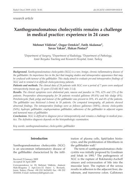
Figure 1. 59-year-old woman with xanthogranulomatous<br />
<strong>cholecystitis</strong>. Contrast-enhanced CT scan shows<br />
hydropic gallbladder.<br />
The US f<strong>in</strong>d<strong>in</strong>gs used for the diagnosis<br />
were presence of gallstone and bile sludge,<br />
pericholecystic fluid and thickness of the<br />
gallbladder wall. Thicken<strong>in</strong>g of the gallbladder<br />
wall was considered abnormal if it<br />
exceeded 3 mm to Kim et al. 6 The CT feature<br />
used for the diagnosis were presence<br />
of gallstones; <strong>in</strong>creased thickness of the<br />
gallbladder wall (>3mm), loss of <strong>in</strong>terface<br />
between the gallbladder and the liver; pericholecystic<br />
fluid; and choledocholithiasis. In<br />
one patient, a MRI study was made because<br />
of suspected gallbladder carc<strong>in</strong>oma <strong>in</strong> CT<br />
appearance.<br />
The gallbladder was fixed with 10% formal<strong>in</strong><br />
and specimens were sta<strong>in</strong>ed with<br />
H and E. Descriptive statistics were used<br />
to describe the features of the data <strong>in</strong> our<br />
study.<br />
Results<br />
The cl<strong>in</strong>ical symptoms were right hypochondrial<br />
pa<strong>in</strong> <strong>in</strong> 19 (79%) patients, nausea<br />
<strong>in</strong> 15(62%) icteric sclera <strong>in</strong> three (12%),<br />
and fever <strong>in</strong> two (8%). Ma<strong>in</strong> signs <strong>in</strong>cluded<br />
positive Murphy’s sign, palpable mass <strong>in</strong><br />
the right upper quadrant and yellow sk<strong>in</strong><br />
were found <strong>in</strong> 17 (70%), 24 (100%) and three<br />
(12%) patients.<br />
Radiol Oncol 2009; 43(2): 76-83.
78<br />
Radiol Oncol 2009; 43(2): 76-83.<br />
Yildirim M et al. / Xantogranulomatous <strong>cholecystitis</strong><br />
A B<br />
Figure 2. Abdom<strong>in</strong>al MR imag<strong>in</strong>g demonstrat<strong>in</strong>g suspected gallbladder mass <strong>in</strong>volv<strong>in</strong>g the liver <strong>in</strong> T1 weighted<br />
series(A), and a soft tissue mass <strong>in</strong> T2 weighted series (B).<br />
Laboratory tests were with<strong>in</strong> normal<br />
ranges except leucocytosis <strong>in</strong> five patients<br />
(>12.000/µl), elevated ALT-AST and bilurib<strong>in</strong><br />
levels were found <strong>in</strong> three patients<br />
<strong>in</strong> each group.<br />
Preoperative US for 24 patients revealed<br />
gallstone <strong>in</strong> 23 (96%) patients, pericholecystic<br />
fluid <strong>in</strong> five (20%), bile sludge <strong>in</strong> two<br />
(8%), polyp of gallbladder <strong>in</strong> one (4%), and<br />
tumour of gallbladder <strong>in</strong> one (4%) patient.<br />
The gallbladder was thickened (>3mm)<br />
<strong>in</strong> ten (41.6%) patients. Upper abdom<strong>in</strong>al<br />
CT was performed <strong>in</strong> six (25%) patients.<br />
CT revealed hydrops of gallbladder <strong>in</strong> two<br />
(33.3%) patients (Figure 1), carc<strong>in</strong>oma of<br />
the pancreas also <strong>in</strong> two (33.3%) patients,<br />
pericholecystic abscess <strong>in</strong> one and one carc<strong>in</strong>oma<br />
of gallbladder. Thicken<strong>in</strong>g of the<br />
gallbladder wall was seen, more than 3 mm<br />
thick, <strong>in</strong> six patients. MRI showed suspected<br />
gallbladder cancer <strong>in</strong>volv<strong>in</strong>g the liver <strong>in</strong><br />
one patient (Figure 2A-B).<br />
The <strong>in</strong>traoperative f<strong>in</strong>d<strong>in</strong>gs were as follows:<br />
chronic <strong>cholecystitis</strong> <strong>in</strong> 13 (54%),<br />
adhesions of the gallbladder to adjacent<br />
organs <strong>in</strong> four (adhesions to the transverse<br />
colon <strong>in</strong> two patients, the duodenohepatic<br />
ligament <strong>in</strong> one, and fundus of the gallbladder<br />
to abdom<strong>in</strong>al wall <strong>in</strong> one), emphysematous<br />
gallbladder <strong>in</strong> three (perforation of the<br />
gallbladder <strong>in</strong> two patient and carc<strong>in</strong>oma<br />
of the pancreas <strong>in</strong> one), hydrops of the<br />
gallbladder <strong>in</strong> two (carc<strong>in</strong>oma of the pancreas<br />
<strong>in</strong> one patient and enlarged gallbladder<br />
with a stone impacted <strong>in</strong> Hartmann’s<br />
pouch <strong>in</strong> one), and tumoural mass of the<br />
gallbladder <strong>in</strong> two patients (Figure 3). The<br />
gallbladder stones were found <strong>in</strong> all patients.<br />
Furthermore, the frozen section was<br />
performed <strong>in</strong> one patient and malignancy<br />
was not found. The surgical treatment was<br />
elective open cholecystectomy <strong>in</strong> 23 (95.8%)<br />
patients and laparoscopic cholecystectomy<br />
(LC) <strong>in</strong> one (4.2%) patient. In one patient,<br />
who could not rule out the possibility of<br />
Figure 3. Tumoular like deposits of xanthogranulomatous<br />
<strong>cholecystitis</strong> <strong>in</strong> surgical specimen.
Yildirim M et al. / Xantogranulomatous <strong>cholecystitis</strong> 79<br />
A B<br />
Figure 4. Histiocytes, lymphocytes and giant cells present <strong>in</strong> a granulomatous focus of xanthogranulomatous<br />
<strong>in</strong>flammation. A; (HE X20) B; (HE X40)<br />
carc<strong>in</strong>oma, cholecystectomy and wedge resection<br />
of adjacent liver was performed.<br />
The postoperative complications were<br />
found <strong>in</strong> 7(29%) patients who <strong>in</strong>cluded<br />
wound <strong>in</strong>fection and pleural effusion <strong>in</strong><br />
four and two patients, respectively. Acute<br />
renal failure developed <strong>in</strong> one patient with<br />
pancreas cancer. The average hospital stay<br />
time was 8 days (range, 3- 22 days). No mortality<br />
was seen.<br />
Histopathologically there were a focal<br />
or diffuse <strong>in</strong>flammatory process with<br />
xanthogranulomatous changes, histiocytosis<br />
and giant cells of foreign body <strong>in</strong> all<br />
patients (Figure 4-5). The exam<strong>in</strong>ation of<br />
specimen showed gallstones <strong>in</strong> 24 (100%)<br />
patients, presence of sludge <strong>in</strong> 13 (54.4%),<br />
thicken<strong>in</strong>g gallbladder wall (>5 mm) <strong>in</strong> 15<br />
(62%), dysplasia <strong>in</strong> four (16%) and mucosal<br />
ulcers <strong>in</strong> three (12%). In addition four (16%)<br />
patients had lymphadenopathy which<br />
showed reactive lymphadenitis.<br />
Discussion<br />
XGC was previously described as an uncommon<br />
form of chronic <strong>cholecystitis</strong>. 7<br />
Christensen AH and Ishak KG <strong>in</strong>itially<br />
described it as a pseudotumour with destructive<br />
type of gallbladder <strong>in</strong>flammation,<br />
pericholecystic <strong>in</strong>filtration, hepatic <strong>in</strong>volvement<br />
and lymphadenopathy. In 1981 the<br />
term xanthogranulomatous <strong>cholecystitis</strong><br />
was proposed <strong>in</strong> a review of 40 cases collected<br />
over a 10-year period. 2<br />
Even though the number of published<br />
cases is not large, XGC not as rare as gen-<br />
Radiol Oncol 2009; 43(2): 76-83.
80<br />
Radiol Oncol 2009; 43(2): 76-83.<br />
Yildirim M et al. / Xantogranulomatous <strong>cholecystitis</strong><br />
Figure 5. <strong>Xanthogranulomatous</strong> <strong>in</strong>flammation of the gallbladder wall, characterized by histiocytes (HEx20).<br />
erally believed. 7 In the literature, the <strong>in</strong>cidence<br />
of XGC is reported to be 0.7% to<br />
13.2%. 8 Higher <strong>in</strong>cidence was reported<br />
from the Eastern countries. 9-11 In our series,<br />
the <strong>in</strong>cidence of XGC was 4% among<br />
749 patients who were operated on gallstone.<br />
XGC mostly affects middle-aged<br />
women and old persons between 60-70<br />
years. Similar to the previous reports the<br />
mean age was 63 years <strong>in</strong> our study. 12,13<br />
This suggests that age must be one of the<br />
significant factors <strong>in</strong> the development of<br />
the XGC. The male to female ratio range<br />
is from 2:1 to 1:2 <strong>in</strong> other series. 14 A study<br />
from India 12 reported a 1:9 male to female<br />
ratio while <strong>in</strong> our report male to female ratio<br />
was found 1:1.4. The different <strong>in</strong>cidence<br />
of XGC may be due to misdiagnosis by cl<strong>in</strong>icians.<br />
Cl<strong>in</strong>ically, XGC does not have a typical<br />
presentation. Our patients were presented<br />
with right upper quadrant pa<strong>in</strong>, nausea,<br />
fever, icterus and palpable mass; they are<br />
similar to acute or chronic <strong>cholecystitis</strong>.<br />
These cl<strong>in</strong>ical features are not specific for<br />
XGC and there was no difference between<br />
the patients with <strong>cholecystitis</strong> and gallbladder<br />
carc<strong>in</strong>oma. 11,15 We noted that all of the<br />
patients with these symptoms required an<br />
elective surgical procedure at first presentation.<br />
The association of XGC with a<br />
perforated gallbladder, abscess formation,<br />
enterobiliary fistula and Mirizzi syndrome<br />
were supported by <strong>in</strong>creas<strong>in</strong>g series. 4,16 In<br />
our series, these lesions were found <strong>in</strong> 16%<br />
patients. In one case, the adhesion of fundus<br />
to abdom<strong>in</strong>al wall was considered as<br />
the potential of XGC for fistula formation.
The reported series support the existence of<br />
this comorbid factors seen <strong>in</strong> nearly 23% of<br />
patients. 4<br />
Preoperative biochemical tests or imag<strong>in</strong>g<br />
studies are not suggestive of XGC. 12<br />
Neither the liver function abnormalities<br />
nor the tumour markers (CEA, CA-19.9)<br />
are suggestive of XGC, although Adachi<br />
et al. 17 argued that serum level of CA-19.9<br />
may be elevated <strong>in</strong> both carc<strong>in</strong>oma and<br />
XGC patients. It has been reported that<br />
thicken<strong>in</strong>g of the gallbladder, adhesion to<br />
neighbour<strong>in</strong>g tissues or organs were specific<br />
f<strong>in</strong>d<strong>in</strong>gs to XGC, although there are<br />
other series that it is difficult to differentiate<br />
XGC from other lesions. 18,19 All these<br />
f<strong>in</strong>d<strong>in</strong>gs can be seen <strong>in</strong> acute <strong>cholecystitis</strong>;<br />
however, the presence of <strong>in</strong>tramural lowattenuation<br />
nodules, preservation of mucosal<br />
l<strong>in</strong><strong>in</strong>g and degree of enhancement of<br />
the gallbladder wall are suggestive of the<br />
XGC. 18 In our study none of the patients<br />
was diagnosed by radiologist as XGC with<br />
imag<strong>in</strong>g f<strong>in</strong>d<strong>in</strong>gs. US misdiagnosed one<br />
case of XGC as carc<strong>in</strong>oma of the gallbladder<br />
(misdiagnose rate 4.3%), while CT misdiagnosed<br />
two cases of XGC as carc<strong>in</strong>oma<br />
(misdiagnose rate 33.3%). In this study,<br />
high misdiagnosis rate may be related to<br />
our <strong>in</strong>sufficient experience for imag<strong>in</strong>g<br />
f<strong>in</strong>d<strong>in</strong>gs of XGC. Chun et al. 18 concluded<br />
that a def<strong>in</strong>itive diagnosis of gallbladder<br />
carc<strong>in</strong>oma is not possible with only imag<strong>in</strong>g<br />
f<strong>in</strong>d<strong>in</strong>gs. In our study, thicken<strong>in</strong>g<br />
of the gallbladder wall was found <strong>in</strong> 24%<br />
and 46% patients with US and CT respectively.<br />
While thicken<strong>in</strong>g of the gallbladder<br />
wall was found <strong>in</strong> 62% of patients pathologically.<br />
This leads to the conclusion that<br />
thicken<strong>in</strong>g of the gallbladder wall <strong>in</strong> imag<strong>in</strong>g<br />
f<strong>in</strong>d<strong>in</strong>gs itself is not a predictive factor<br />
<strong>in</strong> the diagnosis of XGC. Hatakenaka et<br />
al. 20 have demonstrated that MR imag<strong>in</strong>g<br />
may play an important role <strong>in</strong> differentiat<strong>in</strong>g<br />
XGC from carc<strong>in</strong>oma <strong>in</strong> patients with<br />
a thickened gallbladder wall. In our series,<br />
Yildirim M et al. / Xantogranulomatous <strong>cholecystitis</strong> 81<br />
MR imag<strong>in</strong>g did not differentiate XGC<br />
from gallbladder carc<strong>in</strong>oma.<br />
In our series as <strong>in</strong> others reported <strong>in</strong><br />
the literature, all patients had gallstones<br />
(100%), frequently sludge of bile (54%) or<br />
biliary obstruction (12%). 4 Its importance<br />
lies <strong>in</strong> the fact that gallstones can have an<br />
important role <strong>in</strong> the pathogenesis of XGC<br />
via extravasation of bile <strong>in</strong>to the gallbladder<br />
wall. In contrary, <strong>in</strong> a series noted the presence<br />
of gallstone <strong>in</strong> only 85% of the XGC,<br />
which leads to the conclusion that the presence<br />
of gallstone is only an associated condition<br />
and not the cause of the <strong>in</strong>flammatory<br />
process. 21 The small ulcerations <strong>in</strong> the<br />
mucosa reported to be a precipitat<strong>in</strong>g factor<br />
<strong>in</strong> other studies. 7,8 The extravasation of<br />
bile causes that fibrous reaction and scarr<strong>in</strong>g<br />
heal<strong>in</strong>g with<strong>in</strong> the gallbladder wall, response<br />
due to thicken<strong>in</strong>g of the gallbladder<br />
wall. The ulcerations <strong>in</strong> the mucosa were<br />
found pathologically <strong>in</strong> 12% of our patients.<br />
Open cholecystectomy is the first choice<br />
for XGC, either complete or partial. 14 In<br />
our series, s<strong>in</strong>gle cholecystectomy was performed<br />
on 23 patients and cholecystectomy<br />
with partial hepatic wedge resection on<br />
one patient. LC may be contra<strong>in</strong>dicated <strong>in</strong><br />
XGC because of a high <strong>in</strong>cidence of complications.<br />
Accord<strong>in</strong>g to the study carried by<br />
Guzmán-Valdivia G, LC was not completed<br />
due to difficulty <strong>in</strong> dissect<strong>in</strong>g the gallbladder<br />
and converted <strong>in</strong>to the open procedure<br />
<strong>in</strong> 80% of the patients diagnosed as<br />
XGC. 21 The necessity of radical surgery is<br />
not cleared with extra-gallbladder <strong>in</strong>volvement.<br />
4 The <strong>in</strong>traoperative frozen section or<br />
f<strong>in</strong>e needle aspiration has been suggested<br />
to confirm the diagnosis of XGC. 9 The frozen<br />
section is valuable when there is no <strong>in</strong>vasion<br />
of pericholecystic organs. In our series,<br />
the frozen section was used (negative<br />
result) <strong>in</strong> a case with an extensive <strong>in</strong>vasion<br />
of liver. We believe it was not change the<br />
approach of surgeon. Nevertheless, <strong>in</strong> the<br />
patients with negative results, the radical<br />
Radiol Oncol 2009; 43(2): 76-83.
82<br />
surgery can be performed due to the coexistence<br />
of XGC and carc<strong>in</strong>oma of the gallbladder.<br />
On the other hand, studies report<br />
that the radical surgery may be associated<br />
with a high perioperative morbidity. 22<br />
Although XGC is a benign disease, patients<br />
usually have a longer hospital stay<br />
with a postoperative complications. 4 In our<br />
study, complications occurred <strong>in</strong> 7(29%) patients<br />
<strong>in</strong>clud<strong>in</strong>g wound <strong>in</strong>fection, pleural<br />
effusion and renal failure related to hepatorenal<br />
failure. Complications were reported<br />
<strong>in</strong>clud<strong>in</strong>g leakage of bile and bile peritonitis<br />
which largely related to the difficulty<br />
<strong>in</strong> cholecystectomy. 9 The complications are<br />
thus related more to the technical difficulty<br />
<strong>in</strong> stripp<strong>in</strong>g the gallbladder, the mode of<br />
the operation and the cl<strong>in</strong>ical condition of<br />
the patient than to the disease itself.<br />
Conclusions<br />
The pre- or <strong>in</strong>traoperative differential diagnosis<br />
of XGC from other gallbladder<br />
diseases <strong>rema<strong>in</strong>s</strong> a <strong>challenge</strong> <strong>in</strong> <strong>medical</strong><br />
practice. The presence of firm adhesions<br />
of the gallbladder to neighbour<strong>in</strong>g organs<br />
and tissues, thickened gallbladder wall,<br />
together with gallstone <strong>in</strong> a patient with<br />
chronic disease, is highly suggestive of<br />
XGC. The def<strong>in</strong>itive diagnosis depends on<br />
the histopathologic exam<strong>in</strong>ation. XGC can<br />
be treated successfully with an accurate<br />
diagnosis and proper operation.<br />
References<br />
1. Houston JP, Coll<strong>in</strong>s MC, Cameron I, Reed MWR,<br />
Parsons MA, Roberts KM. <strong>Xanthogranulomatous</strong><br />
<strong>cholecystitis</strong>. Br J Surg 1994; 81: 1030-2.<br />
2. Goodman ZD, Ishak KG. <strong>Xanthogranulomatous</strong><br />
<strong>cholecystitis</strong>. Am J Surg Pathol 1981; 5: 653-9.<br />
3. Benbow EW. <strong>Xanthogranulomatous</strong> <strong>cholecystitis</strong>.<br />
Br J Surg 1990; 77: 255-6.<br />
Radiol Oncol 2009; 43(2): 76-83.<br />
Yildirim M et al. / Xantogranulomatous <strong>cholecystitis</strong><br />
4. Yang T, Zhang BH, Zhang J, Zhang YJ, Jiang XQ,<br />
Wu MC. Surgical treatment of xanthogranulomatous<br />
<strong>cholecystitis</strong>: experience <strong>in</strong> 33 cases.<br />
Hepatobiliary Pancreat Dis Int 2007; 6: 504-8.<br />
5. Sp<strong>in</strong>elli A, Schumacher G, Pascher A, Lopez-<br />
Hann<strong>in</strong>en E, Al-Abadi H, Benckert C, et al.<br />
Extended surgical resection for xanthogranulomatous<br />
<strong>cholecystitis</strong> mimick<strong>in</strong>g advanced gallbladder<br />
carc<strong>in</strong>oma: a case report and review of<br />
literature. World J Gastroenterol 2006; 12: 2293-6.<br />
6. Kim PN, Ha HK, Kim YH, Lee MG, Kim MH,<br />
Auh YH. US f<strong>in</strong>d<strong>in</strong>gs of xanthogranulomatous<br />
<strong>cholecystitis</strong>. Cl<strong>in</strong> Radiol 1998; 53: 290 -2.<br />
7. Ros PR, Goodman ZD. <strong>Xanthogranulomatous</strong><br />
<strong>cholecystitis</strong> versus gallbladder carc<strong>in</strong>oma.<br />
Radiology 1997; 203: 10-2.<br />
8. Kwon AH, Matsui Y, Uemura Y. Surgical procedures<br />
and histopathologic f<strong>in</strong>d<strong>in</strong>gs for patients<br />
with <strong>Xanthogranulomatous</strong> <strong>cholecystitis</strong>. J Am<br />
Coll Surg 2004; 199: 204-10.<br />
9. Karabulut Z, Besim H, Hamamci O, Bostano lu S,<br />
Korkmaz A. <strong>Xanthogranulomatous</strong> <strong>cholecystitis</strong>.<br />
Retrospective analysis of 12 cases. Acta Chir Belg<br />
2003; 103: 297-9.<br />
10. Dixit VK, Prakash A, Gupta A, Pandey M,<br />
Gautam A, Kumar M, et al. <strong>Xanthogranulomatous</strong><br />
<strong>cholecystitis</strong>. Dig Dis Sci 1998; 43: 940-2.<br />
11. Roberts KM, Parsons MA. <strong>Xanthogranulomatous</strong><br />
<strong>cholecystitis</strong>: cl<strong>in</strong>icopathological study of 13 cases.<br />
J Cl<strong>in</strong> Pathol 1987; 40: 412-7.<br />
12. Belagué C, Targarona EM, Sugra es G, Rey MJ,<br />
Arce Y, Viella P, et al. <strong>Xanthogranulomatous</strong><br />
<strong>cholecystitis</strong> simulat<strong>in</strong>g gallbladder neoplasm:<br />
therapeutic implications. Gastroenterol Hepatol<br />
1996; 19: 503-6.<br />
13. Eriguchi N, Aoyagi S, Tamae T, Kanazawa<br />
N, Nagashima J, Nishimura K et al.<br />
<strong>Xanthogranulomatous</strong> <strong>cholecystitis</strong>. Med J 2001;<br />
48: 219-21.<br />
14. Guzmán-Valdivia G. <strong>Xanthogranulomatous</strong><br />
<strong>cholecystitis</strong>: 15 years’ experience. World J Surg<br />
2004; 28: 254-7.<br />
15. Wang D, Yamakawa T, Fukuda N, Maruno K,<br />
Ushigome S, Ish yama J. <strong>Xanthogranulomatous</strong><br />
<strong>cholecystitis</strong>: report of a case and literature<br />
review on cl<strong>in</strong>ical differentiat<strong>in</strong>g factors from<br />
gallbladder carc<strong>in</strong>oma. Digest Endoscopy 2004;<br />
16: 143-7.<br />
16. Benbow EW. <strong>Xanthogranulomatous</strong> <strong>cholecystitis</strong>.<br />
Br J Surg 1990; 77: 255-6.
17. Adachi Y, Iso Y, Moriyama M, Kasai T,<br />
Hashimato H. Increased serum CA 19-9 <strong>in</strong> patients<br />
with xanthogranulomatous <strong>cholecystitis</strong>.<br />
Hepatogastroenterol 1998; 45: 77-80.<br />
18. Chun KA, Ha HK, Yu ES, Sh<strong>in</strong>n KS, Kim KW,<br />
Lee DH, et al. <strong>Xanthogranulomatous</strong> <strong>cholecystitis</strong>:<br />
CT features with emphasis on differentiation<br />
from gall-bladder carc<strong>in</strong>oma. Radiology 1997;<br />
203: 93-7.<br />
19. Para A, Ac<strong>in</strong>as O, Bueno J, Güezmes A, Fernandez<br />
MA, Far<strong>in</strong>as MC. <strong>Xanthogranulomatous</strong> <strong>cholecystitis</strong><br />
cl<strong>in</strong>ical, sonographic, and CT f<strong>in</strong>d<strong>in</strong>gs <strong>in</strong><br />
26 patients. AJR 2000; 174: 979-83.<br />
20. Hatakenaka M, Adachi T, Matsuyama A, Mori M,<br />
Yoshikawa Y. <strong>Xanthogranulomatous</strong> <strong>cholecystitis</strong>:<br />
importance of chemical-shift gradient-echo<br />
MR imag<strong>in</strong>g. Eur Radiol 2003; 13: 2233–5.<br />
21. Guzmán-Valdivia G. <strong>Xanthogranulomatous</strong><br />
<strong>cholecystitis</strong> <strong>in</strong> laparoscopic surgery. J Gastro<strong>in</strong>test<br />
Surg 2005; 9: 494-7.<br />
22. Sr<strong>in</strong>ivas GN, S<strong>in</strong>ha S, Ryley N, Houghton PW.<br />
Perfidious gallbladders - a diagnostic dilemma<br />
with xanthogranulomatous <strong>cholecystitis</strong>. Ann R<br />
Coll Surg Engl 2007; 89: 168-72.<br />
Yildirim M et al. / Xantogranulomatous <strong>cholecystitis</strong> 83<br />
Radiol Oncol 2009; 43(2): 76-83.