Bladder cancer - European Society for Medical Oncology
Bladder cancer - European Society for Medical Oncology
Bladder cancer - European Society for Medical Oncology

You also want an ePaper? Increase the reach of your titles
YUMPU automatically turns print PDFs into web optimized ePapers that Google loves.
BLADDER CANCER: A GUIDE FOR PATIENTS<br />
PATIENT INFORMATION BASED ON ESMO CLINICAL PRACTICE GUIDELINES<br />
This guide <strong>for</strong> patients has been prepared by Reliable Cancer Therapies (RCT) as a service to patients,<br />
to help patients and their relatives better understand the nature of bladder <strong>cancer</strong> and appreciate<br />
the best treatment choices available according to the subtype of bladder <strong>cancer</strong>. We recommend<br />
that patients ask their doctors about what tests or types of treatments are needed <strong>for</strong> their type and<br />
stage of disease. The medical in<strong>for</strong>mation described in this document is based on the clinical practice<br />
guidelines of the <strong>European</strong> <strong>Society</strong> <strong>for</strong> <strong>Medical</strong> <strong>Oncology</strong> (ESMO) <strong>for</strong> the management of bladder<br />
<strong>cancer</strong>. This guide <strong>for</strong> patients has been produced in collaboration with ESMO and is disseminated<br />
with the permission of ESMO. It has been written by a medical doctor and reviewed by two<br />
oncologists from ESMO including the lead author of the clinical practice guidelines <strong>for</strong> professionals.<br />
It has also been reviewed by patient representatives from ESMO’s Cancer Patient Working Group.<br />
More in<strong>for</strong>mation about Reliable Cancer Therapies: www.reliable<strong>cancer</strong>therapies.com<br />
More in<strong>for</strong>mation about the <strong>European</strong> <strong>Society</strong> <strong>for</strong> <strong>Medical</strong> <strong>Oncology</strong>: www.esmo.org<br />
For words marked with an asterisk, a definition is provided at the end of the document.<br />
<strong>Bladder</strong> <strong>cancer</strong>: a guide <strong>for</strong> patients‐ In<strong>for</strong>mation based on ESMO Clinical Practice Guidelines ‐v.2012.1 Page 1<br />
This document is provided by Reliable Cancer Therapies with the permission of ESMO.<br />
The in<strong>for</strong>mation in this document does not replace a medical consultation. It is <strong>for</strong> personal use only and cannot be modified,<br />
reproduced or disseminated in any way without written permission from ESMO and Reliable Cancer Therapies.
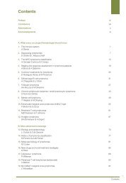
Table of contents<br />
Definition of bladder <strong>cancer</strong> .................................................................................................................... 3<br />
Is bladder <strong>cancer</strong> frequent? .................................................................................................................... 4<br />
What causes bladder <strong>cancer</strong>? ................................................................................................................. 5<br />
How is bladder <strong>cancer</strong> diagnosed? .......................................................................................................... 7<br />
What is IT important to know to define the optimal treatment? ........................................................... 9<br />
What are the treatment options? ......................................................................................................... 12<br />
What are the possible side effects of the treatments? ......................................................................... 16<br />
What happens after the treatment? ..................................................................................................... 19<br />
Definitions of difficult words ................................................................................................................. 21<br />
This text was written by Dr. An Billiau, Celsus <strong>Medical</strong> Writing, LLC (<strong>for</strong> RCT) and reviewed by Dr. Svetlana Jezdic (ESMO), Pr.<br />
Joaquim Bellmunt (ESMO), and Pr. Louis Denis (Stoma‐Ilco, Europa Uomo on behalf of the ESMO Cancer Patient Working<br />
Group).<br />
<strong>Bladder</strong> <strong>cancer</strong>: a guide <strong>for</strong> patients‐ In<strong>for</strong>mation based on ESMO Clinical Practice Guidelines ‐v.2012.1 Page 2<br />
This document is provided by Reliable Cancer Therapies with the permission of ESMO.<br />
The in<strong>for</strong>mation in this document does not replace a medical consultation. It is <strong>for</strong> personal use only and cannot be modified,<br />
reproduced or disseminated in any way without written permission from ESMO and Reliable Cancer Therapies.
DEFINITION OF BLADDER CANCER<br />
This definition is adapted from and is used with the permission of the National Cancer Institute (NCI) of the United States of America.<br />
<strong>Bladder</strong> <strong>cancer</strong> is a <strong>cancer</strong> that <strong>for</strong>ms in tissues of the bladder. The bladder is the organ that stores<br />
urine. The most frequent type of bladder <strong>cancer</strong>s is transitional cell carcinoma*. This type of <strong>cancer</strong><br />
begins in cells that normally <strong>for</strong>m the inner lining of the bladder, also called the transitional<br />
epithelium* or urothelium*. Other types include squamous cell carcinoma*, a bladder <strong>cancer</strong> that<br />
begins in thin, flat cells in the lining of the bladder, and adenocarcinoma*, a <strong>cancer</strong> that begins in<br />
cells from the bladder lining that release mucus.<br />
Anatomy of the male (left) and female (right) urinary system showing the kidneys, ureters*, bladder, and urethra*. Urine is<br />
made in the renal tubules* and collects in the renal pelvis*. The urine flows from the kidneys through the ureters to the<br />
bladder. The urine is stored in the bladder until it leaves the body through the urethra*.<br />
<strong>Bladder</strong> <strong>cancer</strong>: a guide <strong>for</strong> patients‐ In<strong>for</strong>mation based on ESMO Clinical Practice Guidelines ‐v.2012.1 Page 3<br />
This document is provided by Reliable Cancer Therapies with the permission of ESMO.<br />
The in<strong>for</strong>mation in this document does not replace a medical consultation. It is <strong>for</strong> personal use only and cannot be modified,<br />
reproduced or disseminated in any way without written permission from ESMO and Reliable Cancer Therapies.
IS BLADDER CANCER FREQUENT?<br />
It is estimated that in 2008, in Europe, approximately 110,500 patients were diagnosed with bladder<br />
<strong>cancer</strong>. <strong>Bladder</strong> <strong>cancer</strong> is, thus, the 5 th most common <strong>cancer</strong> in Europe.<br />
<strong>Bladder</strong> <strong>cancer</strong> is approximately five times more frequent in men than in women. It is estimated that<br />
in 2008, 27 out of 100,000 men and 5 out of 100,000 women developed bladder <strong>cancer</strong>. Of all<br />
<strong>cancer</strong>s, bladder <strong>cancer</strong> is the 4 th most common <strong>cancer</strong> in men, and the 13 th most common <strong>cancer</strong> in<br />
women.<br />
In the <strong>European</strong> Union, the likelihood <strong>for</strong> a man to develop bladder <strong>cancer</strong> at some point in his life<br />
lies between 1.5 and 2.5%. For men living in Flanders (Belgium), Malta, Spain and Italy this is<br />
somewhat higher: between 3.1 and 4.2%. For a woman in the <strong>European</strong> Union, the chances of<br />
developing bladder <strong>cancer</strong> at some point in her life are less than 1%.<br />
The risk of developing bladder <strong>cancer</strong> increases with age; overall, 70% of patients who develop<br />
bladder <strong>cancer</strong> present symptoms after the age of 65.<br />
<strong>Bladder</strong> <strong>cancer</strong>: a guide <strong>for</strong> patients‐ In<strong>for</strong>mation based on ESMO Clinical Practice Guidelines ‐v.2012.1 Page 4<br />
This document is provided by Reliable Cancer Therapies with the permission of ESMO.<br />
The in<strong>for</strong>mation in this document does not replace a medical consultation. It is <strong>for</strong> personal use only and cannot be modified,<br />
reproduced or disseminated in any way without written permission from ESMO and Reliable Cancer Therapies.
WHAT CAUSES BLADDER CANCER?<br />
Today, it is not entirely clear what causes bladder <strong>cancer</strong>. A number of risk factors* have been<br />
identified, but in many cases none of these seem to be present. A risk factor increases the risk that<br />
the <strong>cancer</strong> occurs, but is neither necessary nor sufficient to cause <strong>cancer</strong>. A risk factor is not a cause<br />
in itself.<br />
Some people with these risk factors will never develop bladder <strong>cancer</strong> and some people without<br />
any of these risk factors may nonetheless develop bladder <strong>cancer</strong>.<br />
The main risk factors <strong>for</strong> bladder <strong>cancer</strong> are:<br />
‐ Aging: bladder <strong>cancer</strong> occurs most frequently in elderly people; overall,<br />
70% of patients developing bladder <strong>cancer</strong> are diagnosed after the age of<br />
65 years.<br />
‐ Previous history of bladder <strong>cancer</strong>.<br />
‐ Cigarette smoking: cigarette smoking is the most important risk factor <strong>for</strong><br />
bladder <strong>cancer</strong>. Stopping cigarette smoking <strong>for</strong> more than 4 years can<br />
lower the risk.<br />
‐ A number of chemicals have been identified that may cause bladder <strong>cancer</strong>:<br />
o Aniline dyes: chemicals that may be present in color fabrics.<br />
o Cyclophosphamide: a chemotherapeutic* drug used <strong>for</strong> <strong>cancer</strong> treatment.<br />
o Aromatic amines: exposure to these chemicals can occur in various occupations such<br />
as those in the painting, leather, car, metal, paper and rubber industry, but also<br />
amongst truck drivers, dry cleaners, dental technicians and hairdressers. In these<br />
circumstances, bladder <strong>cancer</strong> does not occur until 30 to 50 years after exposure.<br />
o Arsenic: in a Taiwanese region where water contained high arsenic levels, an<br />
increased risk of bladder <strong>cancer</strong> has been found.<br />
o Aristolochia fangchi: this is a Chinese herb; an increased risk of bladder <strong>cancer</strong> was<br />
found in people that had used a dietary supplement in which this herb had been<br />
mistakenly added.<br />
‐ Irradiation: exposure to ionizing irradiation* in the region of the bladder, <strong>for</strong> example during<br />
radiotherapy <strong>for</strong> prostate* <strong>cancer</strong>, is thought to increase the risk of bladder <strong>cancer</strong>.<br />
‐ Some risk factors are particularly important <strong>for</strong> a specific type of bladder <strong>cancer</strong>, namely<br />
squamous cell carcinoma*. This tumor is caused by chronic irritation or inflammation of the<br />
bladder. In Western countries, the main risk factors <strong>for</strong> squamous cell carcinoma include a<br />
badly‐functioning bladder, prolonged presence of a catheter* in the bladder, bladder stones<br />
and chronic bladder infection. In Africa and the Middle East, an important risk factor <strong>for</strong><br />
squamous cell carcinoma is infection with Schistosoma hematobium, a microbe that is<br />
common in these regions. It can infect the bladder and lead to chronic inflammation.<br />
‐ Diabetes*: individuals with type‐2 diabetes are at an increased risk of developing bladder<br />
<strong>cancer</strong>.<br />
<strong>Bladder</strong> <strong>cancer</strong>: a guide <strong>for</strong> patients‐ In<strong>for</strong>mation based on ESMO Clinical Practice Guidelines ‐v.2012.1 Page 5<br />
This document is provided by Reliable Cancer Therapies with the permission of ESMO.<br />
The in<strong>for</strong>mation in this document does not replace a medical consultation. It is <strong>for</strong> personal use only and cannot be modified,<br />
reproduced or disseminated in any way without written permission from ESMO and Reliable Cancer Therapies.
Other factors have been suspected to be associated with an increased risk of bladder <strong>cancer</strong>, but the<br />
evidence is inconsistent:<br />
‐ Coffee, artificial sweeteners and alcohol: there is no clear evidence that consumption of<br />
these substances produces a risk <strong>for</strong> developing bladder <strong>cancer</strong>.<br />
‐ Tap water with high levels of trihalomethanes, which are the broken down products of the<br />
disinfectant chlorine: some studies show that prolonged ingestion of this kind of tap water<br />
may increase the risk <strong>for</strong> bladder <strong>cancer</strong>, but the evidence is inconsistent.<br />
‐ Genes: overall, having a family member with bladder <strong>cancer</strong> conveys a slightly increased risk<br />
of developing the disease. <strong>Bladder</strong> <strong>cancer</strong> as a result of an inheritable faulty gene* is very<br />
rare.<br />
‐ One study has shown that being overweight is associated with a higher risk of bladder<br />
<strong>cancer</strong>, but other studies do not confirm this.<br />
Some factors have been proposed to protect against the development of bladder <strong>cancer</strong>, but clear<br />
evidence <strong>for</strong> this is not available.<br />
‐ Fluid intake: it has been proposed that high fluid intake may reduce the risk of developing<br />
bladder <strong>cancer</strong> in men, but inconsistencies exist between studies.<br />
‐ Fruit and vegetables: consumption of fruit and vegetables is said to have a protective effect.<br />
<strong>Bladder</strong> <strong>cancer</strong>: a guide <strong>for</strong> patients‐ In<strong>for</strong>mation based on ESMO Clinical Practice Guidelines ‐v.2012.1 Page 6<br />
This document is provided by Reliable Cancer Therapies with the permission of ESMO.<br />
The in<strong>for</strong>mation in this document does not replace a medical consultation. It is <strong>for</strong> personal use only and cannot be modified,<br />
reproduced or disseminated in any way without written permission from ESMO and Reliable Cancer Therapies.
HOW IS BLADDER CANCER DIAGNOSED?<br />
<strong>Bladder</strong> <strong>cancer</strong> may be diagnosed during a routine physical check‐up, or can be suspected on the<br />
basis of specific symptoms.<br />
The main symptoms are:<br />
• Blood in the urine (called hematuria): this is usually painless and is experienced by 85% of<br />
bladder <strong>cancer</strong> patients.<br />
• Urinary problems: the need to urinate more frequently than usual (called frequency), the<br />
need to pass urine urgently (called urgency) or pain when passing urine (called dysuria).<br />
However, these symptoms are not specific to bladder <strong>cancer</strong> and can also occur in many conditions<br />
that are not related to <strong>cancer</strong> such as urinary infection, kidney stones or benign* prostatic<br />
hyperplasia*.<br />
<strong>Bladder</strong> <strong>cancer</strong> may block the flow of urine from the kidney. Accumulation of urine within the kidney<br />
may lead to distension of the kidney (called hydronephrosis) and pain.<br />
Besides asking about the above mentioned symptoms, the doctor will also per<strong>for</strong>m a general physical<br />
examination and ask <strong>for</strong> laboratory blood tests of the blood cell counts and kidney function.<br />
The diagnosis of bladder <strong>cancer</strong> is based on the fo llowing examinations:<br />
1. Clinical examination*<br />
2.<br />
A physical examination provides in<strong>for</strong>mation about signs of<br />
bladder <strong>cancer</strong> and other health problems. The doctor<br />
might examine the rectum and vagina (in women) to<br />
determine the size of a bladder tumor and to see if and how<br />
far it has spread.<br />
Cystoscopy*<br />
Cystoscopy is a technical examination of the bladder:<br />
the doctor inserts a lighted tube with a camera at<br />
the end into the urethra* to inspect the interior of<br />
the bladder and the urethra <strong>for</strong> the presence of<br />
tumors. Cystoscopy can be per<strong>for</strong>med in the doctor’s<br />
office; with the use of a local anesthetic* gel, this<br />
procedure is usually well tolerated. However,<br />
cystoscopy may also be per<strong>for</strong>med under general<br />
anesthesia* together with the clinical bimanual<br />
examination* (see above) of the bladder.<br />
The doctor can insert a fine surgical instrument into<br />
the cystoscope tube to remove – under direct vision<br />
‐ tissue samples from the tumor or from any other<br />
suspicious area. This specimen is called a biopsy*.<br />
For certain bladder <strong>cancer</strong>s, the doctor may<br />
immediately resect the entire tumor: this is called<br />
transurethral resection of the bladder tumor<br />
(TURB)*. In this case, the cystoscopy also constitutes the<br />
first step in the treatment.<br />
<strong>Bladder</strong> <strong>cancer</strong>: a guide <strong>for</strong> patients‐ In<strong>for</strong>mation based on ESMO Clinical Practice Guidelines ‐v.2012.1 Page 7<br />
This document is provided by Reliable Cancer Therapies with the permission of ESMO.<br />
The in<strong>for</strong>mation in this document does not replace a medical consultation. It is <strong>for</strong> personal use only and cannot be modified,<br />
reproduced or disseminated in any way without written permission from ESMO and Reliable Cancer Therapies.
3.<br />
4.<br />
In specific circumstances, the doctor will also inspect the ureters, a procedure called<br />
ureteroscopy*. In other circumstances, cystoscopy also includes biopsy sampling from the<br />
urethra*.<br />
Histopathological examination*<br />
This is the laboratory investigation of the tumor cells. It is<br />
per<strong>for</strong>med on the tissue from the tumor removed during<br />
cystoscopy*. The histopathological* in<strong>for</strong>mation will confirm<br />
the diagnosis of bladder <strong>cancer</strong> and will reveal the specific<br />
characteristics of the tumor, allowing the doctor to determine<br />
the type of bladder <strong>cancer</strong>.<br />
If surgery is indicated after the cystoscopy (usually a TURB* ), a second histopathological<br />
examination will be per<strong>for</strong>med on the tumor tissue obtained during surgery. This is very<br />
important to confirm the results of the first biopsy* and to provide more accurate<br />
in<strong>for</strong>mation on the <strong>cancer</strong> and the <strong>cancer</strong>’s stage.<br />
Radiological examination*<br />
If the histopathological examination* shows that<br />
the tumor has grown into the deeper layers (the<br />
muscle layers) of the bladder,<br />
then radiological<br />
investigation is needed to determine if the tumor<br />
has also grown into the tissues and lymph nodes*<br />
outside the bladder.<br />
The radiological investigation is part of a<br />
diagnostic process called staging* and can be<br />
per<strong>for</strong>med using computed<br />
tomography (CT)* or<br />
magnetic resonance imaging (MRI)* of the abdomen and pelvis. The staging procedure<br />
further also includes a CT of the chest, and if there are symptoms of tumor spread in the<br />
bones, also a bone scintigraphy*.<br />
<strong>Bladder</strong> <strong>cancer</strong>: a guide <strong>for</strong> patients‐ In<strong>for</strong>mation based on ESMO Clinical Practice Guidelines ‐v.2012.1 Page 8<br />
This document is provided by Reliable Cancer Therapies with the permission of ESMO.<br />
The in<strong>for</strong>mation in this document does not replace a medical consultation. It is <strong>for</strong> personal use only and cannot be modified,<br />
reproduced or disseminated in any way without written permission from ESMO and Reliable Cancer Therapies.
WHAT IS IT IMPORTANT TO KNOW TO DEFINE THE OPTIMAL<br />
TREATMENT?<br />
Doctors will need to consider many aspects of both the patient and the <strong>cancer</strong> in<br />
order to decide on the best treatment.<br />
Relevant in<strong>for</strong>mation about the patient<br />
• Gender<br />
• Personal medical history, previous illnesses and treatments<br />
• History of bladder <strong>cancer</strong> in relatives<br />
• General well being and specific physical complaints<br />
• Results from the clinical examination*<br />
• Results from laboratory tests on blood counts, kidney and liver function<br />
Relevant in<strong>for</strong>mation about the <strong>cancer</strong><br />
• Staging*<br />
Doctors use staging to assess the extension of the <strong>cancer</strong> and the prognosis of the patient. The TNM<br />
staging system is commonly used. The combination of size of the tumor and invasion* of nearby<br />
tissue (T), involvement of lymph nodes* (N), and metastasis* or spread of the <strong>cancer</strong> to other organs<br />
of the body (M), will classify the <strong>cancer</strong> as being at one of the stages described below.<br />
The stage* is fundamental in order to make the right decision about the treatment. The less<br />
advantaged the stage, the better the prognosis. Staging is per<strong>for</strong>med when the clinical and<br />
radiological investigations* and the histopathological examination* of the biopsy* are completed. If<br />
surgery is indicated, a second staging will be per<strong>for</strong>med on the basis of the laboratory examination of<br />
the surgical specimen.<br />
The table below presents the different stages of bladder <strong>cancer</strong>. The definitions are somewhat<br />
technical. It is there<strong>for</strong>e recommended to ask doctors <strong>for</strong> more detailed explanations.<br />
<strong>Bladder</strong> <strong>cancer</strong>: a guide <strong>for</strong> patients‐ In<strong>for</strong>mation based on ESMO Clinical Practice Guidelines ‐v.2012.1 Page 9<br />
This document is provided by Reliable Cancer Therapies with the permission of ESMO.<br />
The in<strong>for</strong>mation in this document does not replace a medical consultation. It is <strong>for</strong> personal use only and cannot be modified,<br />
reproduced or disseminated in any way without written permission from ESMO and Reliable Cancer Therapies.
Stage Definition (see figure of the bladder wall below) Category<br />
Stage 0a Non‐invasive papillary carcinoma: the tumor is confined to the innermost<br />
cell layers of the bladder lining (the epithelium*)<br />
Stage 0is Carcinoma in situ, also referred to as flat tumor: a high‐grade tumor that is<br />
confined to the innermost cell layers of the bladder lining (the epithelium*)<br />
Stage I The tumor invades the deeper connective tissues of the bladder lining (the<br />
lamina propria*)<br />
Non‐<br />
muscle<br />
invasive<br />
bladder<br />
<strong>cancer</strong><br />
Stage II The tumor invades the muscle of the bladder. Stage II is divided into 2<br />
stages:<br />
T2a: the tumor invades the inner half of the muscle of the bladder<br />
T2b: the tumor invades the outer half of the muscle of the bladder Muscle‐<br />
Stage III The tumor invades the tissues surrounding the bladder. Stage III is divided<br />
into 3 stages:<br />
T3a: microscopic invasion*<br />
T3b: macroscopic invasion*<br />
T4a: invasion of organs surrounding the bladder: the prostate* in men, the<br />
uterus and/or vagina in women<br />
Stage IV The tumor invades the pelvic wall and/or abdominal wall or<br />
The tumor is accompanied by metastasis* in lymph node(s) or in an organ<br />
at a distance from the bladder<br />
invasive<br />
bladder<br />
<strong>cancer</strong><br />
Advanced<br />
and<br />
metastatic<br />
disease<br />
Layers of the bladder wall showing the mucosa* (the bladder lining consisting of the epithelium* and lamina propria*) and<br />
the muscle layers.<br />
<strong>Bladder</strong> <strong>cancer</strong>: a guide <strong>for</strong> patients‐ In<strong>for</strong>mation based on ESMO Clinical Practice Guidelines ‐v.2012.1 Page 10<br />
This document is provided by Reliable Cancer Therapies with the permission of ESMO.<br />
The in<strong>for</strong>mation in this document does not replace a medical consultation. It is <strong>for</strong> personal use only and cannot be modified,<br />
reproduced or disseminated in any way without written permission from ESMO and Reliable Cancer Therapies.
• Results of the biopsy*<br />
The tissue of the tumor biopsy is examined in the laboratory by a pathologist*. This examination is<br />
called histopathology*. If surgery is per<strong>for</strong>med after cystoscopy*, histopathological examination<br />
involves the examination of the tumor and the lymph nodes* removed during surgery. This is very<br />
important to confirm the results of the initial findings and to provide more in<strong>for</strong>mation on the stage<br />
of the <strong>cancer</strong>. The results of the examination of the biopsy include:<br />
o Histological type*<br />
The histological type refers to the type of cells that compose the tumor. About 90% of<br />
bladder <strong>cancer</strong>s are transitional cell carcinomas*. The remaining 10% are predominantly<br />
squamous cell carcinomas* and adenocarcinomas*. Other histological types are very rare.<br />
� Transitional cell carcinoma*, also called urothelial carcinoma: tumor that arises from<br />
the transitional epithelium*. The transitional epithelium consists of multiple layers of<br />
cells that can change shape as the bladder stretches and that line the inner most wall<br />
of the bladder.<br />
� Squamous cell carcinoma*: tumor that arises from the transitional epithelium but that<br />
consists exclusively of thin, flat cells called squamous cells.<br />
� Adenocarcinoma*: tumor that arises from cells in the glands of the inner lining of the<br />
bladder.<br />
o Grade*<br />
The grade is determined on the basis of how different the tumor cells look from the cells<br />
normally found in a healthy bladder lining. The abnormal features indicate the rate at<br />
which the cells multiply and the degree to which they are invasive. For bladder <strong>cancer</strong><br />
there are 4 different grades:<br />
� Papilloma: a tumor composed of non‐malignant cells.<br />
� Papillary urothelial neoplasm of low malignant potential (PUNLMP): a tumor composed<br />
of non‐malignant cells typically covered with a thickened layer of transitional<br />
epithelium*.<br />
� Urothelial carcinoma low grade: a malignant tumor that grows slowly and is unlikely to<br />
spread.<br />
� Urothelial carcinoma high grade: a malignant tumor that grows faster and that is more<br />
likely to spread.<br />
<strong>Bladder</strong> <strong>cancer</strong>: a guide <strong>for</strong> patients‐ In<strong>for</strong>mation based on ESMO Clinical Practice Guidelines ‐v.2012.1 Page 11<br />
This document is provided by Reliable Cancer Therapies with the permission of ESMO.<br />
The in<strong>for</strong>mation in this document does not replace a medical consultation. It is <strong>for</strong> personal use only and cannot be modified,<br />
reproduced or disseminated in any way without written permission from ESMO and Reliable Cancer Therapies.
WHAT ARE THE TREATMENT OPTIONS?<br />
Planning of the treatment involves a team of professionals from different medical<br />
disciplines. It usually implies a meeting of the different specialists, called<br />
multidisciplinary meeting* or tumor board review*. In this meeting, the<br />
treatment planning will be discussed according to the relevant in<strong>for</strong>mation<br />
mentioned above.<br />
The treatment will usually combine therapies that<br />
• Act on the <strong>cancer</strong> locally, such as surgery, radiotherapy*, local chemotherapy* and local<br />
immunotherapy*<br />
• Act on the <strong>cancer</strong> cells all over the body using systemic chemotherapy<br />
The exact treatment will depend on the stage of the <strong>cancer</strong>, on the characteristics of the tumor and<br />
on the risks <strong>for</strong> the patient.<br />
The treatments listed below have their benefits, their risks and their contraindications*. It is<br />
recommended that patients ask their doctors about the expected benefits and risks of every<br />
treatment in order to be in<strong>for</strong>med about the consequences of the treatment. For some treatments,<br />
several possibilities are available. The choice should be discussed according to the balance between<br />
benefits and risks.<br />
Treatment plan <strong>for</strong> non‐muscle invasive disease (stage 0a, stage 0is, stage I)<br />
At these stages, the tumor is confined to the superficial layer of the bladder wall (mucosa*) and does<br />
not invade the muscle of the bladder. The main goal of the treatment is to remove the local tumor by<br />
surgery during a TURB*. However, additional treatment delivered locally in the bladder (called<br />
adjuvant* intravesical* treatment) is recommended since it lowers the risk that the tumor recurs or<br />
progresses.<br />
The type of adjuvant therapy depends on the risk of progression* and recurrence*: <strong>for</strong> each patient<br />
with a stage 0a or stage I tumor, this is calculated using a scoring system based on several tumor‐<br />
specific characteristics.<br />
Cystoscopy* and transurethral resection of the bladder tumor (TURB)*<br />
After an initial cystoscopy all patients undergo a TURB. Often, the complete tumor is resected,<br />
and the TURB in this case is the definitive treatment. However, sometimes, it is recommended to<br />
give additional treatment (called adjuvant treatment*) with drugs that are applied directly into<br />
the bladder (called intravesical* treatment). The type of additional treatment depends on the<br />
individual risk of recurrence and progression*, but also the patient’s capability to tolerate side<br />
effects*.<br />
In selected patients with high risk tumors, a second TURB is recommended either be<strong>for</strong>e or after<br />
intravesical therapy, to help detect residual disease and to provide a more accurate staging.<br />
<strong>Bladder</strong> <strong>cancer</strong>: a guide <strong>for</strong> patients‐ In<strong>for</strong>mation based on ESMO Clinical Practice Guidelines ‐v.2012.1 Page 12<br />
This document is provided by Reliable Cancer Therapies with the permission of ESMO.<br />
The in<strong>for</strong>mation in this document does not replace a medical consultation. It is <strong>for</strong> personal use only and cannot be modified,<br />
reproduced or disseminated in any way without written permission from ESMO and Reliable Cancer Therapies.
Intravesical* chemotherapy* or immunotherapy*<br />
In order to reduce the risk of recurrence and progression*, all patients that have had a TURB* are<br />
given one single intravesical instillation* with a chemotherapeutic* agent, immediately after<br />
surgery. The drugs that are used are Mitomycin C*, epirubicin* or doxorubicin*.<br />
For patients with a tumor at low risk of recurrence and progression, one single instillation<br />
completes the treatment. For patients who are considered to have an intermediate or high risk of<br />
tumor recurrence or progression, the first instillation should be followed by further intravesical<br />
chemotherapy, or by intravesical immunotherapy with bacillus Calmette Guérin (BCG)* (see<br />
further). Whether chemotherapy or immunotherapy is chosen depends on the individual risk<br />
profile. Chemotherapy is usually given <strong>for</strong> up to 1 year. Immunotherapy is given <strong>for</strong> a minimum of<br />
1 year.<br />
Intravesical* immunotherapy* with bacillus Calmette‐Guérin (BCG)*<br />
For patients with selected risk profiles, it is recommended to give intravesical treatment with<br />
bacillus Calmette‐Guérin (BCG), a vaccine used to protect against tuberculosis*. The working<br />
mechanism of intravesical BCG therapy is not exactly understood. It is thought that BCG induces<br />
an immune reaction* that kills <strong>cancer</strong> cells. Treatment with BCG is there<strong>for</strong>e considered as<br />
immunotherapy.<br />
Usually, an initial 6‐week treatment regimen is given (called induction therapy), and this is<br />
followed by so‐called maintenance therapy <strong>for</strong> a minimum of 1 year. Some maintenance regimens<br />
last two years.<br />
Cystectomy*<br />
Cystectomy is recommended <strong>for</strong> patients with stage 0is and stage I tumors that do not respond to<br />
adjuvant* intravesical* treatment.<br />
Treatment plan <strong>for</strong> muscle invasive bladder <strong>cancer</strong> (stage II, stage III)<br />
At these stages, the tumor has invaded the muscle layer of the bladder or has extended through the<br />
bladder wall into the tissues surrounding the bladder. The treatment aims to surgically remove the<br />
entire bladder as well as the lymph nodes* in the pelvis and the neighboring organs. Prior to surgery,<br />
chemotherapy* is administered and aims to reduce tumor size, to attack tumor cells in metastases*<br />
that are too small to be detected, and to reduce the risk that tumor cells will spread to other parts of<br />
the body during surgery.<br />
Radical cystectomy*<br />
The standard treatment <strong>for</strong> muscle invasive bladder <strong>cancer</strong> includes<br />
radical cystectomy. For male patients this involves the complete removal<br />
of the bladder, all visible tumor tissue, but also the urethra*, prostate*,<br />
seminal vesicles*, the lower parts of the ureters* and the lymph nodes*<br />
in the pelvis. For female patients, radical cystectomy involves removal of<br />
the bladder, all visible and resectable tumors, the entire urethra, the<br />
lower part of the ureters, the adjacent vagina*, the uterus* and the<br />
lymph nodes in the pelvis.<br />
In certain patients, this procedure may be slightly modified in order to preserve certain structures.<br />
Whether or not this is possible depends on the tumor spread and needs to be carefully evaluated<br />
in each individual patient.<br />
<strong>Bladder</strong> <strong>cancer</strong>: a guide <strong>for</strong> patients‐ In<strong>for</strong>mation based on ESMO Clinical Practice Guidelines ‐v.2012.1 Page 13<br />
This document is provided by Reliable Cancer Therapies with the permission of ESMO.<br />
The in<strong>for</strong>mation in this document does not replace a medical consultation. It is <strong>for</strong> personal use only and cannot be modified,<br />
reproduced or disseminated in any way without written permission from ESMO and Reliable Cancer Therapies.
Radical cystectomy* leads to the loss of bladder function, that is, the storage of urine. The<br />
surgeon will there<strong>for</strong>e connect the ureters* to a new outlet to allow evacuation of urine (called a<br />
urinary diversion*). This new outlet may be either the urethra*, the skin of the abdomen, or the<br />
very last part of the large bowel (called a rectosigmoid diversion). The choice of approach<br />
depends on many factors such as the tumor stage, the structures that can be preserved after<br />
radical cystectomy, the patient’s general medical condition and the patient’s preference. The<br />
different options are explained further in the text (see Side effects* of therapies).<br />
In addition, radical cystectomy may involve the removal of certain reproductive organs*. This may<br />
lead to sexual dysfunction* and/or the loss of reproductive function* (see Side effects of<br />
Therapies).<br />
Chemotherapy*<br />
For patients with stage T2 or T3 disease it is recommended to give neo‐adjuvant combination<br />
chemotherapy. This means that a combination of chemotherapeutic* drugs is given prior to<br />
cystectomy* or definitive radiotherapy*. The recommended combinations are gemcitabine* and<br />
cisplatin* (abbreviated GC), or methotrexate*, vinblastine*, doxorubicin* and cisplatin<br />
(abbreviated MVAC). The purpose of neo‐adjuvant therapy* is to eradicate micrometastases*,<br />
reduce tumor size and reduce the risk that tumor cells spread during the surgical procedure.<br />
Radiotherapy*<br />
Radiotherapy alone may be indicated <strong>for</strong> patients who are medically not fit to<br />
undergo the extensive surgery of radical cystectomy*.<br />
In selected cases where the treatment aims to preserve the bladder,<br />
radiotherapy may be given as part of a combination treatment* (see: organ<br />
preservation therapy*).<br />
Organ preservation therapy*<br />
Organ preservation therapy refers to a treatment where the bladder is<br />
preserved. This is proposed <strong>for</strong> patients who do not wish to undergo radical<br />
cystectomy*, or who are medically not fit to tolerate this kind of surgery. This treatment can be:<br />
aggressive TURB*, TURB in combination with radiotherapy* or chemotherapy*, or TURB in<br />
combination with radio‐ ánd chemotherapy. The latter is called trimodality combination<br />
treatment and is the preferred approach.<br />
Organ preservation therapy may also be considered in selected patients with early stage bladder<br />
<strong>cancer</strong>, provided they meet a number of other stringent medical criteria.<br />
Organ preservation therapy requires a stringent lifelong follow‐up* with cystoscopy* and urine<br />
cytology* to evaluate the response to treatment and to detect disease recurrence. If persistent or<br />
recurrent disease is observed, an immediate cystectomy is recommended, if possible.<br />
Treatment plan <strong>for</strong> advanced and metastatic* disease (stage IV)<br />
At this stage, the tumor has grown through the bladder wall into the wall of the pelvis or the<br />
abdomen, or beyond the abdomen to distant organs. Since it is difficult or not medically indicated to<br />
remove all tumor by surgery, the primary goal of the treatment is to target tumor cells using<br />
chemotherapy that is given through a vein and that there<strong>for</strong>e acts systemically.<br />
<strong>Bladder</strong> <strong>cancer</strong>: a guide <strong>for</strong> patients‐ In<strong>for</strong>mation based on ESMO Clinical Practice Guidelines ‐v.2012.1 Page 14<br />
This document is provided by Reliable Cancer Therapies with the permission of ESMO.<br />
The in<strong>for</strong>mation in this document does not replace a medical consultation. It is <strong>for</strong> personal use only and cannot be modified,<br />
reproduced or disseminated in any way without written permission from ESMO and Reliable Cancer Therapies.
Chemotherapy*<br />
The standard combination regimen consists of the drugs cisplatin* with gemcitabine*<br />
(abbreviated as GC) or methotrexate*, vinblastine*, doxorubicin* and cisplatin (abbreviated as<br />
MVAC). The MVAC regimen causes more toxic side effects* than GC. Patients with limited<br />
advanced disease (lymph node* involvement and no visceral*<br />
metastasis* in organs*) and those who are medically fit may be able<br />
to receive high‐dose MVAC in combination with granulocyte‐colony<br />
stimulating factor* (G‐CSF), a growth factor that can increase<br />
tolerability of the chemotherapy.<br />
Approximately half of patients are medically not fit to tolerate<br />
cisplatin due to a poor general condition, poor kidney function or<br />
the presence of other diseases. These patients are treated with carboplatin* and gemcitabine*<br />
(abbreviated CG), with methotrexate*, carboplatin and vinblastine (abbreviated M‐CAVI) or with<br />
taxane* or gemcitabine only. CG is the reference treatment. M‐CAVI causes slightly more toxic<br />
effects than CG.<br />
Surgery and radiotherapy* after systemic chemotherapy*<br />
For selected patients with locally advanced disease systemic chemotherapy followed by<br />
cystectomy and lymphadenectomy*, or radiotherapy could be considered.<br />
Radiotherapy*<br />
Radiotherapy can be useful to alleviate pain or bleeding.<br />
Treatment of complications caused by disease<br />
Blockade of urinary flow<br />
<strong>Bladder</strong> <strong>cancer</strong> may block the flow of urine and cause urine to accumulate in the kidney. This may<br />
cause pain and disturbance of the kidney function. If cystectomy* is not possible because of<br />
advanced disease or because the patient is medically not fit to undergo this procedure, it may be<br />
necessary to divert urine flow away from the bladder to the exterior. This can be done by surgically<br />
connecting the kidney or the ureter* to the skin of the abdomen. This is called nephrostomy and<br />
ureterostomy, respectively. The urine is collected in a plastic bag attached to the skin.<br />
<strong>Bladder</strong> <strong>cancer</strong>: a guide <strong>for</strong> patients‐ In<strong>for</strong>mation based on ESMO Clinical Practice Guidelines ‐v.2012.1 Page 15<br />
This document is provided by Reliable Cancer Therapies with the permission of ESMO.<br />
The in<strong>for</strong>mation in this document does not replace a medical consultation. It is <strong>for</strong> personal use only and cannot be modified,<br />
reproduced or disseminated in any way without written permission from ESMO and Reliable Cancer Therapies.
WHAT ARE THE POSSIBLE SIDE EFFECTS OF THE TREATMENTS?<br />
Surgery<br />
General risks and side effects<br />
Some risks are common <strong>for</strong> every surgical intervention per<strong>for</strong>med under general anesthesia*. These<br />
complications are unusual and include <strong>for</strong>mation of a blood clot in the veins, heart or breathing<br />
problems, bleeding, infection, or reactions to the anesthesia*. These are maximally prevented by<br />
thorough medical evaluation be<strong>for</strong>e surgery.<br />
The bladder is located in the pelvis together with the local lymph nodes*, parts of the bowel, major<br />
blood vessels, and in women the reproductive organs*. Depending on the extent of surgical<br />
resections needed to obtain the best results, some of these structures may become damaged.<br />
Accurate preoperative staging* and imaging* will help to minimize this risk.<br />
When lymph nodes* in the pelvis and the abdomen are removed, it can damage or block the lymph<br />
system* resulting in lymphedema*, a condition where lymph fluid* accumulates in the legs and<br />
makes them swell. This may occur soon after the intervention, but also later.<br />
Loss of bladder function after cystectomy<br />
The consequence of cystectomy is that the function of the bladder is lost. Several surgical options<br />
exist to divert and collect the urine, either within or at the exterior of the body. The best choice<br />
needs to be carefully evaluated and will depend on the tumor stage, the surgical treatment given,<br />
the patient’s general condition, and the patient’s preference. The different possibilities are discussed<br />
briefly below. It is recommended to ask the doctor <strong>for</strong> more in<strong>for</strong>mation.<br />
Orthotopic neobladder. A new bladder organ (called neobladder) is constructed: tissue from the<br />
bowel is used to <strong>for</strong>m a pouch that is placed between the ureters* and the urethra*. Orthotopic<br />
means that the new bladder is in the same place where the original bladder was. This pouch will<br />
store urine, and urine will be passed through the urethra.<br />
Abdominal diversion. The surgeon connects the ureters* to an artificial opening in the abdominal<br />
wall, called the stoma*. This may be a direct connection or the surgeon may use tissue from the<br />
small intestine to guide the urine to the stoma. The urine is collected in a small plastic bag attached<br />
to the skin. The surgeon may also <strong>for</strong>m a pouch on the inner side of the abdomen and a stoma that<br />
does not allow spontaneous passage of urine to the exterior: in this case the pouch can be emptied<br />
from the exterior using a catheter*. This is called a continent urinary diversion*.<br />
Rectosigmoid diversion. The surgeon connects the ureters* to the very last part of the large bowel,<br />
called the rectosigmoid. The rectosigmoid normally holds the stool and will now have the same<br />
function <strong>for</strong> urine. The surgeon may place a segment of intestine between the ureters and the<br />
rectosigmoid.<br />
The nature and frequency of the side effects* of these diversion* procedures will depend on the type<br />
of procedure. The most frequent problems are narrowing of the ureter at the stoma* and infection<br />
of the kidneys.<br />
Sexual dysfunction and/or loss of reproductive function*<br />
Radical cystectomy* in men includes the resection of the urethra*, seminal vesicles* and prostate*.<br />
In women it includes the resection of the uterus* and part of the vagina*. The loss of these<br />
<strong>Bladder</strong> <strong>cancer</strong>: a guide <strong>for</strong> patients‐ In<strong>for</strong>mation based on ESMO Clinical Practice Guidelines ‐v.2012.1 Page 16<br />
This document is provided by Reliable Cancer Therapies with the permission of ESMO.<br />
The in<strong>for</strong>mation in this document does not replace a medical consultation. It is <strong>for</strong> personal use only and cannot be modified,<br />
reproduced or disseminated in any way without written permission from ESMO and Reliable Cancer Therapies.
eproductive organs* may lead to sexual dysfunction*, the loss of the ability to conceive children,<br />
and in women it will lead to the loss of the ability to bear children. The doctor will refer such patients<br />
to specialized support providers.<br />
Radiotherapy*<br />
Side effects* of radiotherapy may occur in organs that are directly targeted, but also in healthy<br />
organs that lie close to the bladder and that cannot be avoided by the X‐rays*. For bladder <strong>cancer</strong>,<br />
modern radiation techniques are very safe and major complications occur in less than 5% of patients.<br />
Effects on the urinary system include pain while passing urine, an urgent need to urinate, blood in<br />
the urine, blockade of urinary flow, and ulceration of the inner lining of the bladder.<br />
Effects of radiation on the lower intestines include discom<strong>for</strong>t, diarrhea, mucus and blood discharge,<br />
and, rarely, per<strong>for</strong>ation of the intestines.<br />
In women, vaginal narrowing is a possible late effect of radiotherapy in the region of the pelvis.<br />
The oncologist will advise on strategies to maximally prevent and relieve these reactions.<br />
Intravesical instillation* therapy<br />
The main side effect* of intravesical Bacillus Calmette Guérin* instillation is inflammation of the<br />
bladder, called cystitis*. The most severe side effect is a generalized infection, which may result<br />
when the bacilli are taken up through the bladder wall into the blood; this therapy is there<strong>for</strong>e not<br />
indicated in patients with reduced function of the immune system*. In general, side‐effects of<br />
intravesical BCG therapy can be managed.<br />
Intravesical instillation of chemotherapy such as Mitomycin C* may have several side effects such as<br />
cystitis*, allergy and skin reactions.<br />
Chemotherapy*<br />
Side effects* of chemotherapy are frequent but nowadays well controlled using adequate supportive<br />
measures. Side effects will depend on the drug(s) administered, on the dose and on individual<br />
factors. If a patient has suffered from other medical problems in the past, some precautions should<br />
be taken and/or changes of the treatment should be made. Side effects are more severe when<br />
chemotherapy is given systemically (usually through a vein), than when it is given locally, directly into<br />
the bladder (see: intravesical* drug therapy).<br />
Listed below are the side effects that are known to occur with one or several of the chemotherapy<br />
drugs currently used <strong>for</strong> bladder <strong>cancer</strong>. The nature, frequency and severity of the side‐effects vary<br />
<strong>for</strong> every combination used.<br />
The most frequent side effects are:<br />
• Hair loss or hair thinning<br />
• Decreased blood cell counts, which may lead to anemia*, bleeding and bruising, and<br />
infections.<br />
• Tiredness<br />
• Feeling sick or being sick<br />
<strong>Bladder</strong> <strong>cancer</strong>: a guide <strong>for</strong> patients‐ In<strong>for</strong>mation based on ESMO Clinical Practice Guidelines ‐v.2012.1 Page 17<br />
This document is provided by Reliable Cancer Therapies with the permission of ESMO.<br />
The in<strong>for</strong>mation in this document does not replace a medical consultation. It is <strong>for</strong> personal use only and cannot be modified,<br />
reproduced or disseminated in any way without written permission from ESMO and Reliable Cancer Therapies.
Other side effects that may occur frequently with either one or more of the chemotherapy drugs<br />
used <strong>for</strong> bladder <strong>cancer</strong> include:<br />
• Mouth sores or ulcers<br />
• Taste changes<br />
• Diarrhea<br />
• Gritty or watery eyes<br />
• Sensitivity to sunlight<br />
• Kidney damage<br />
• Hearing loss<br />
• Damage to the fetus in the womb of a <strong>cancer</strong> patient receiving chemotherapy<br />
• Loss of fertility<br />
• Interruption of periods in women (amenorrhea), which may be temporary<br />
Occasional side effects include:<br />
• Changes in liver function<br />
• Damage to the heart muscle<br />
• Numbness or tingling in fingers and toes (peripheral neuropathy)<br />
• Constipation<br />
• Blurred vision<br />
• Skin rash or reddening of skin<br />
• Cough or shortness of breath<br />
• Liver changes<br />
• Changes in color of skin and/or nails<br />
• Allergic reaction<br />
• Inflammation around the drip/injection site<br />
• Fever and chills<br />
Rare side effects are:<br />
• Depression<br />
• Sore eyes<br />
• Headaches<br />
• Increased heart rate<br />
• Dizziness<br />
• High blood pressure<br />
Finally, it should be noted that some chemotherapy drugs are passed along with breast milk and may<br />
be harmful to the baby.<br />
<strong>Bladder</strong> <strong>cancer</strong>: a guide <strong>for</strong> patients‐ In<strong>for</strong>mation based on ESMO Clinical Practice Guidelines ‐v.2012.1 Page 18<br />
This document is provided by Reliable Cancer Therapies with the permission of ESMO.<br />
The in<strong>for</strong>mation in this document does not replace a medical consultation. It is <strong>for</strong> personal use only and cannot be modified,<br />
reproduced or disseminated in any way without written permission from ESMO and Reliable Cancer Therapies.
WHAT HAPPENS AFTER THE TREATMENT?<br />
It is not unusual <strong>for</strong> <strong>cancer</strong> patients to experience treatment‐related<br />
symptoms after the treatment has been completed.<br />
• Patients may experience anxiety, sleeping difficulty or depression, and<br />
may need psychological support.<br />
• During and after treatment, nutrition may become problematic due to<br />
reduced appetite, nausea and general malaise<br />
• Difficulties in concentration and memorization are not uncommon<br />
side effects* of systemic chemotherapy, i.e. when administered in a vein or orally.<br />
Follow‐up* with doctors<br />
After completion of treatment the doctor will propose a follow‐up aiming to:<br />
• Detect and prevent adverse effects of the treatment<br />
• Detect possible recurrence* as soon as possible and direct appropriate treatment<br />
• Provide medical in<strong>for</strong>mation, psychological support and referral to specialized support<br />
providers to optimize returning to normal daily life.<br />
The follow‐up protocol will include regularly timed office visits and investigations. The protocol<br />
depends on the grade* and staging* of the bladder tumor that was treated, and on the type of<br />
treatment given. In general, follow‐up visits may include a combination of the following<br />
investigations:<br />
• History of general physical health and bladder <strong>cancer</strong>‐related symptoms since the last visit<br />
• Cystoscopy* to detect recurrence* and to per<strong>for</strong>m a biopsy* of new lesions<br />
• Imaging of the upper urinary system<br />
• Urinary cytology*: laboratory examination of the urine <strong>for</strong> the presence of tumor cells that<br />
are shed by a potentially recurring bladder tumor.<br />
• Laboratory investigations: blood chemistry and kidney function<br />
• Repeated radiologic investigations* in case initial examinations showed abnormal findings.<br />
There are no generally accepted follow‐up protocols. The following are recommended possible<br />
regimens:<br />
When organ preservation therapy* is given, very close follow‐up is mandatory: cystoscopy, urine<br />
cytology* and/or bladder biopsy* are recommended with a frequency of 3 months <strong>for</strong> the first 2<br />
years, and every 6 months thereafter. This is also the case in patients who have been treated with<br />
radiotherapy*. After cystectomy, clinical control* should take place every 3 months during the first 2<br />
years and subsequently every 6 months <strong>for</strong> 5 years.<br />
Returning to normal life<br />
Returning to normal daily life may be difficult knowing that the <strong>cancer</strong> may come back. If any of the<br />
known risk factors* <strong>for</strong> bladder <strong>cancer</strong> are present, it is advised to maximally eliminate these.<br />
Follow‐up* visits with the doctor provide an opportunity <strong>for</strong> the patient to obtain medical<br />
in<strong>for</strong>mation, psychological support and referral to specialized support providers. Additional expert<br />
<strong>Bladder</strong> <strong>cancer</strong>: a guide <strong>for</strong> patients‐ In<strong>for</strong>mation based on ESMO Clinical Practice Guidelines ‐v.2012.1 Page 19<br />
This document is provided by Reliable Cancer Therapies with the permission of ESMO.<br />
The in<strong>for</strong>mation in this document does not replace a medical consultation. It is <strong>for</strong> personal use only and cannot be modified,<br />
reproduced or disseminated in any way without written permission from ESMO and Reliable Cancer Therapies.
psychological advice may be valuable, and some patients may find support in patient groups or<br />
patient‐targeted in<strong>for</strong>mation media. Dieticians may provide advice on adequate nutrition. Social<br />
workers may help in finding resources to ensure successful rehabilitation.<br />
What if the <strong>cancer</strong> comes back?<br />
If the <strong>cancer</strong> returns, it is called recurrence*. The extent of the recurrence will direct the treatment<br />
decision, and this should be carefully determined <strong>for</strong> each individual patient.<br />
In patients treated with organ preservation therapy*, residual tumor can be detected in 20% of cases<br />
during restaging. An additional 20‐30% of patients with initial complete responses will develop new<br />
or recurrent disease in the preserved bladder. Up to 70% of patients are free of tumors after the first<br />
cystoscopy* control. A quarter of them develop a new lesion in the later course that requires<br />
additional treatment (cystectomy when possible).<br />
For patients with metastatic* disease who experience progression* after completing a first‐line<br />
platinum‐containing regimen, a second‐line chemotherapy regimen with vinflunine* is<br />
recommended.<br />
<strong>Bladder</strong> <strong>cancer</strong>: a guide <strong>for</strong> patients‐ In<strong>for</strong>mation based on ESMO Clinical Practice Guidelines ‐v.2012.1 Page 20<br />
This document is provided by Reliable Cancer Therapies with the permission of ESMO.<br />
The in<strong>for</strong>mation in this document does not replace a medical consultation. It is <strong>for</strong> personal use only and cannot be modified,<br />
reproduced or disseminated in any way without written permission from ESMO and Reliable Cancer Therapies.
DEFINITIONS OF DIFFICULT WORDS<br />
Adenocarcinoma<br />
Cancer that begins in cells that line certain internal organs and that have gland‐like (secretory)<br />
properties.<br />
Adjuvant (treatment)<br />
Adjuvant treatment in <strong>cancer</strong> is a therapy that helps another therapy to reach its ultimate goal and<br />
rein<strong>for</strong>ces its effect. For example radio or/and chemotherapy* help a surgery to accomplish its goal<br />
of eliminating a <strong>cancer</strong>ous tumor. In a context different to the oncological it can also be an agent<br />
added to vaccines to stimulate the immune system's response to the antigen.<br />
Anesthetic (gel)/ anesthesia<br />
Reversible state of loss of awareness in which the patient feels no pain, has no normal reflexes, and<br />
responds less to stress, induced artificially by the employment of certain substances known as<br />
anaesthetics. It can be complete or partial and allows patients to undergo surgery.<br />
Bacillus Calmette Guérin (BCG)<br />
A weakened <strong>for</strong>m of the bacterium Mycobacterium bovis (bacillus Calmette‐Guérin) that does not<br />
cause disease. Bacillus Calmette‐Guérin is used in a solution to stimulate the immune system in the<br />
treatment of bladder <strong>cancer</strong> and as a vaccine to prevent tuberculosis*.<br />
Benign<br />
For a tumor, benign means not <strong>cancer</strong>ous. Benign tumors may grow larger, but do not spread to<br />
other parts of the body. Also called non‐malignant.<br />
Benign prostatic hyperplasia<br />
A benign (not <strong>cancer</strong>ous) condition in which an overgrowth of prostate tissue pushes against the<br />
urethra* and the bladder, blocking the flow of urine. Also called benign prostatic hypertrophy and<br />
BPH.<br />
Biopsy<br />
The removal of cells or tissues <strong>for</strong> examination by a pathologist*. The pathologist may study the<br />
tissue under a microscope or per<strong>for</strong>m other tests on the cells or tissue. There are many different<br />
types of biopsy procedures. The most common types include: (1) incisional biopsy, in which only a<br />
sample of tissue is removed; (2) excisional biopsy, in which an entire lump or suspicious area is<br />
removed; and (3) needle biopsy, in which a sample of tissue or fluid is removed with a needle. When<br />
a wide needle is used, the procedure is called a core biopsy. When a thin needle is used, the<br />
procedure is called a fine‐needle aspiration biopsy.<br />
Carboplatin<br />
A drug that is used to treat advanced ovarian <strong>cancer</strong> that has never been treated or symptoms of<br />
ovarian <strong>cancer</strong> that have come back after treatment with other anti<strong>cancer</strong> drugs. It is also used with<br />
other drugs to treat advanced, metastatic*, or recurrent* non‐small cell lung <strong>cancer</strong> and is being<br />
studied in the treatment of other types of <strong>cancer</strong>. Carboplatin* is a <strong>for</strong>m of the anti<strong>cancer</strong> drug<br />
<strong>Bladder</strong> <strong>cancer</strong>: a guide <strong>for</strong> patients‐ In<strong>for</strong>mation based on ESMO Clinical Practice Guidelines ‐v.2012.1 Page 21<br />
This document is provided by Reliable Cancer Therapies with the permission of ESMO.<br />
The in<strong>for</strong>mation in this document does not replace a medical consultation. It is <strong>for</strong> personal use only and cannot be modified,<br />
reproduced or disseminated in any way without written permission from ESMO and Reliable Cancer Therapies.
cisplatin* and causes fewer side effects* in patients. It attaches to DNA in cells and may kill <strong>cancer</strong><br />
cells. It is a type of platinum compound. Also called Paraplatin.<br />
Catheter<br />
A tube that can be inserted into the body. It has many uses, including draining or administering fluids<br />
or gases.<br />
Chemotherapeutic/Chemotherapy<br />
A type of <strong>cancer</strong> treatment using drugs that kill <strong>cancer</strong> cells and/or limit their growth. These drugs<br />
are usually administered to the patient by slow infusion into a vein but can also be administered<br />
orally, by direct infusion to the limb or by infusion to the liver, according to <strong>cancer</strong> location.<br />
Cisplatin<br />
A drug used to treat many types of <strong>cancer</strong>. Cisplatin contains the metal platinum. It kills <strong>cancer</strong> cells<br />
by damaging their DNA and stopping them from dividing. Cisplatin is a type of alkylating agent. Also<br />
called Platinol.<br />
Clinical examination<br />
The examination of the body to search <strong>for</strong> signs of disease.<br />
Contraindication<br />
Condition or symptom that prevents the administration of a given treatment or procedure to the<br />
patient. Contraindications are either absolute, meaning the treatment should never be given to<br />
patients with this condition or symptom, or relative, meaning that the risk can be outweighed by the<br />
benefits in some patients with this condition or symptom.<br />
Computed tomography/ CT‐scan<br />
A <strong>for</strong>m of radiography in which body organs are scanned with X‐rays* and the results are synthesized<br />
by a computer to generate images of parts of the body.<br />
Cystitis<br />
Inflammation of the bladder.<br />
Cystoscopy<br />
Examination of the bladder and urethra* using a cystoscope, inserted into the urethra. A cystoscope<br />
is a thin, tube‐like instrument with a light and a lens <strong>for</strong> viewing. It may also have a tool to remove<br />
tissue to be checked under a microscope <strong>for</strong> signs of disease.<br />
Diabetes<br />
Any of several diseases in which the kidneys make a large amount of urine. Diabetes usually refers to<br />
diabetes mellitus in which there is also a high level of glucose (a type of sugar) in the blood because<br />
the body does not make enough insulin or use it the way it should.<br />
Doxorubicin<br />
A drug that is used to treat many types of <strong>cancer</strong> and is being studied in the treatment of other types<br />
of <strong>cancer</strong>. Doxorubicin comes from the bacterium Streptomyces peucetius. It damages DNA and may<br />
kill <strong>cancer</strong> cells. It is a type of anthracycline antitumor antibiotic. Also called Adriamycin PFS,<br />
Adriamycin RDF, doxorubicin hydrochloride, hydroxydaunorubicin, and Rubex.<br />
<strong>Bladder</strong> <strong>cancer</strong>: a guide <strong>for</strong> patients‐ In<strong>for</strong>mation based on ESMO Clinical Practice Guidelines ‐v.2012.1 Page 22<br />
This document is provided by Reliable Cancer Therapies with the permission of ESMO.<br />
The in<strong>for</strong>mation in this document does not replace a medical consultation. It is <strong>for</strong> personal use only and cannot be modified,<br />
reproduced or disseminated in any way without written permission from ESMO and Reliable Cancer Therapies.
Epirubicin<br />
A drug used together with other drugs to treat early breast <strong>cancer</strong> that has spread to lymph nodes*.<br />
It is also being studied in the treatment of other types of <strong>cancer</strong>. Epirubicin is a type of anthracycline<br />
antibiotic. Also called Ellence and epirubicin hydrochloride.<br />
Epithelium<br />
The term "epithelium" refers to cells that line hollow organs and glands and those that make up the<br />
outer surface of the body. Epithelial cells help to protect or enclose organs. Most produce mucus or<br />
other secretions.<br />
Follow‐up<br />
Monitoring a person's health over time after treatment. This includes keeping track of the health of<br />
people who participate in a clinical study or clinical trial <strong>for</strong> a period of time, both during the study<br />
and after the study ends.<br />
Granulocyte‐Colony Stimulating Factor (G‐CSF)<br />
A colony‐stimulating factor that stimulates the production of neutrophils (a type of white blood cell).<br />
It is a cytokine that is a type of hematopoietic (blood‐<strong>for</strong>ming) agent. Also called filgrastim and G‐CSF.<br />
Gemcitabine<br />
The active ingredient in a drug that is used to treat pancreatic <strong>cancer</strong> that is advanced or has spread.<br />
It is also used with other drugs to treat breast <strong>cancer</strong> that has spread, advanced ovarian <strong>cancer</strong>, and<br />
non‐small cell lung <strong>cancer</strong> that is advanced or has spread. It is also being studied in the treatment of<br />
other types of <strong>cancer</strong>. Gemcitabine blocks the cell from making DNA and may kill <strong>cancer</strong> cells. It is a<br />
type of antimetabolite.<br />
General anesthesia<br />
A temporary loss of feeling and a complete loss of awareness that feels like a very deep sleep. It is<br />
caused by special drugs or other substances called anesthetics*. General anesthesia keeps patients<br />
from feeling pain during surgery or other procedures.<br />
Grade<br />
A description of a tumor based on how abnormal the <strong>cancer</strong> cells look under a microscope and how<br />
quickly the tumor is likely to grow and spread. Grading systems are different <strong>for</strong> each type of <strong>cancer</strong>.<br />
Histopathology (histopathological examination, histological type)<br />
The study of diseased cells and tissues using a microscope.<br />
Inheritable faulty gene<br />
Abnormal or mutated gene that is passed from parents to their offspring.<br />
Intravesical instillation<br />
Pouring a liquid into the bladder, slowly or drop by drop.<br />
Intravesical (treatment)<br />
An intravesical therapy is administered directly into the bladder.<br />
<strong>Bladder</strong> <strong>cancer</strong>: a guide <strong>for</strong> patients‐ In<strong>for</strong>mation based on ESMO Clinical Practice Guidelines ‐v.2012.1 Page 23<br />
This document is provided by Reliable Cancer Therapies with the permission of ESMO.<br />
The in<strong>for</strong>mation in this document does not replace a medical consultation. It is <strong>for</strong> personal use only and cannot be modified,<br />
reproduced or disseminated in any way without written permission from ESMO and Reliable Cancer Therapies.
Ionizing irradiation<br />
A type of radiation made (or given off ) by X‐ray* procedures, radioactive substances, rays that enter<br />
the Earth's atmosphere from outer space, and other sources. At high doses, ionizing radiation<br />
increases chemical activity inside cells and can lead to health risks, including <strong>cancer</strong>.<br />
Immunotherapy<br />
Treatment to boost or restore the ability of the immune system to fight <strong>cancer</strong>, infections, and other<br />
diseases. Also used to lessen certain side effects* that may be caused by some <strong>cancer</strong> treatments.<br />
Agents used in immunotherapy include monoclonal antibodies, growth factors, and vaccines. These<br />
agents may also have a direct antitumor effect. Also called biological response modifier therapy,<br />
biological therapy, biotherapy, and BRM therapy.<br />
Lamina propria<br />
The lamina propria is a thin layer of loose connective tissue which lies beneath the epithelium* and<br />
together with the epithelium constitutes the mucosa*. The term mucosa (or mucous membrane)<br />
always refers to the combination of the epithelium plus the lamina propria.<br />
Lymph node<br />
A rounded mass of lymphatic tissue that is surrounded by a capsule of connective tissue. Lymph<br />
nodes filter lymph and they store lymphocytes. They are located along lymphatic vessels. Also called<br />
lymph gland.<br />
Lymphedema<br />
A condition in which extra lymph fluid builds up in tissues and causes swelling. It may occur in an arm<br />
or leg if lymph vessels are blocked, damaged, or removed by surgery.<br />
Macroscopic invasion<br />
Extension of <strong>cancer</strong> to the adjacent tissues visible to the naked eye.<br />
Magnetic Resonance Imaging (MRI)<br />
An imaging technique that is used in medicine. It uses magnetic resonance. Sometimes, a fluid is<br />
injected that enhances the contrast between different tissues to make structures more clearly<br />
visible.<br />
Metastasis<br />
The spread of <strong>cancer</strong> from one part of the body to another. A tumor <strong>for</strong>med by cells that have spread<br />
is called a metastatic tumor or a metastasis. The metastatic tumor contains cells that are like those in<br />
the original tumor.<br />
Micrometastasis<br />
Small numbers of <strong>cancer</strong> cells that have spread from the primary tumor to other parts of the body<br />
and are too few to be picked up in a screening or diagnostic test.<br />
Microscopic invasion<br />
Extension of the <strong>cancer</strong> cells to adjacent tissues, evident only under a microscope.<br />
<strong>Bladder</strong> <strong>cancer</strong>: a guide <strong>for</strong> patients‐ In<strong>for</strong>mation based on ESMO Clinical Practice Guidelines ‐v.2012.1 Page 24<br />
This document is provided by Reliable Cancer Therapies with the permission of ESMO.<br />
The in<strong>for</strong>mation in this document does not replace a medical consultation. It is <strong>for</strong> personal use only and cannot be modified,<br />
reproduced or disseminated in any way without written permission from ESMO and Reliable Cancer Therapies.
Methotrexate<br />
A drug used to treat some types of <strong>cancer</strong>, rheumatoid arthritis, and severe skin conditions, such as<br />
psoriasis. Methotrexate stops cells from making DNA and may kill <strong>cancer</strong> cells. It is a type of<br />
antimetabolite. Also called amethopterin, MTX, and Rheumatrex.<br />
Mitomycin C<br />
An anti<strong>cancer</strong> drug that belongs to the family of drugs called antitumor antibiotics.<br />
Mucosa<br />
The moist, inner lining of some organs and body cavities. Glands in the mucosa make mucus. Also<br />
called mucous membrane.<br />
Multidisciplinary opinion<br />
A treatment planning approach in which a number of doctors who are experts in different areas of<br />
specialization (disciplines) review and discuss the medical condition and treatment options of a<br />
patient. In <strong>cancer</strong> treatment, a multidisciplinary opinion may include that of a medical oncologist<br />
(who provides <strong>cancer</strong> treatment with drugs), a surgical oncologist (who provides <strong>cancer</strong> treatment<br />
with surgery), and a radiation oncologist (who provides <strong>cancer</strong> treatment with radiation). Also called<br />
tumor board review.<br />
Organ preservation therapy/surgery<br />
Surgery in which a given organ is spared at its maximum, to keep its functionality and structure. It is<br />
offered to patients who do not wish or cannot undergo radical surgery in which the organ might be<br />
removed completely.<br />
Pathologist<br />
A doctor specialized in histopathology*; the study of diseased cells and tissues using a microscope.<br />
Prognosis<br />
The likely outcome or course of a disease; the chance of recovery or recurrence*.<br />
Progression<br />
In medicine, the course of a disease, such as <strong>cancer</strong>, as it becomes worse or spreads in the body.<br />
Prostate<br />
A gland in the male reproductive system*. The prostate surrounds the part of the urethra* (the tube<br />
that empties the bladder) just below the bladder, and produces a fluid that <strong>for</strong>ms part of the semen.<br />
Radiological investigation/examination<br />
Test that uses imaging technology (such as radiography, ultrasound*, computed tomography* and<br />
nuclear medicine) to visualize organs, structures and tissues within the body to both diagnose and<br />
treat diseases.<br />
Radiotherapy<br />
A therapy in which radiation is used in the treatment of <strong>cancer</strong> that is always oriented to the specific<br />
location of the <strong>cancer</strong>.<br />
<strong>Bladder</strong> <strong>cancer</strong>: a guide <strong>for</strong> patients‐ In<strong>for</strong>mation based on ESMO Clinical Practice Guidelines ‐v.2012.1 Page 25<br />
This document is provided by Reliable Cancer Therapies with the permission of ESMO.<br />
The in<strong>for</strong>mation in this document does not replace a medical consultation. It is <strong>for</strong> personal use only and cannot be modified,<br />
reproduced or disseminated in any way without written permission from ESMO and Reliable Cancer Therapies.
Recurrence<br />
Cancer or disease (usually auto‐immune) that has come back, usually after a period of time during<br />
which the <strong>cancer</strong> or disease was not present or could not be detected. This may happen in the same<br />
location as the original (primary) tumor or in another location in the body. Also called recurrent<br />
<strong>cancer</strong> or disease.<br />
Reproductive organs/system<br />
The organs involved in producing offspring. In women, this system includes the ovaries, the fallopian<br />
tubes, the uterus, the cervix, and the vagina. In men, it includes the prostate*, the testes, and the<br />
penis.<br />
Renal pelvis<br />
The area at the center of the kidney. Urine collects here and is funneled into the ureter*, the tube<br />
that connects the kidney to the bladder.<br />
Renal tubules<br />
Small canals within the tissue of the kidneys, they contain a filtrate that eventually becomes urine.<br />
They are part of the nephrons, which are the basic functional unit of the kidneys.<br />
Risk factor<br />
Something that increases the chance of developing a disease. Some examples of risk factors <strong>for</strong><br />
<strong>cancer</strong> are age, a family history of certain <strong>cancer</strong>s, use of tobacco products, being exposed to<br />
radiation or certain chemicals, infection with certain viruses or bacteria, and certain genetic changes.<br />
(Bone) Scintigraphy<br />
A procedure that produces pictures (scans) of structures inside the body, including areas where there<br />
are <strong>cancer</strong> cells. Scintigraphy is used to diagnose, stage, and monitor disease. A small amount of a<br />
radioactive chemical (radionuclide) is injected into a vein or swallowed. Different radionuclides travel<br />
through the blood to different organs. A machine with a special camera moves over the person as<br />
they are lying on a table and detects the type of radiation given off by the radionuclides. A computer<br />
<strong>for</strong>ms an image of the areas where the radionuclide builds up. These areas may contain <strong>cancer</strong> cells.<br />
Also called radionuclide scanning.<br />
Seminal vesicles<br />
A pair of tubular glands of about 5 cm in length, that are each curled up inside an ampullary<br />
structure. They are located above the prostate*. Each of them has a duct that connects with the<br />
prostate. They produce most of the fluid contained in the semen.<br />
Sexual dysfunction<br />
Inability to fully enjoy sexual intercourse. It includes a wide variety of problems that affect the sexual<br />
act at any stage: desire, arousal, orgasm, and resolution.<br />
Side effect<br />
A problem that occurs when treatment affects healthy tissues or organs. Some common side effects<br />
of <strong>cancer</strong> treatment are fatigue, pain, nausea, vomiting, decreased blood cell counts, hair loss, and<br />
mouth sores.<br />
<strong>Bladder</strong> <strong>cancer</strong>: a guide <strong>for</strong> patients‐ In<strong>for</strong>mation based on ESMO Clinical Practice Guidelines ‐v.2012.1 Page 26<br />
This document is provided by Reliable Cancer Therapies with the permission of ESMO.<br />
The in<strong>for</strong>mation in this document does not replace a medical consultation. It is <strong>for</strong> personal use only and cannot be modified,<br />
reproduced or disseminated in any way without written permission from ESMO and Reliable Cancer Therapies.
Squamous cell carcinoma<br />
Cancer that begins in squamous cells, which are thin, flat cells that look like fish scales. Squamous<br />
cells are found in the tissue that <strong>for</strong>ms the surface of the skin, the lining of the hollow organs of the<br />
body, and the passages of the respiratory and digestive tracts. Also called epidermoid carcinoma.<br />
Stoma<br />
A surgically created opening from an area inside the body to the outside.<br />
Taxane<br />
A type of drug that blocks cell growth by stopping mitosis (cell division). Taxanes interfere with<br />
microtubules (cellular structures that help move chromosomes during mitosis). They are used to<br />
treat <strong>cancer</strong>. A taxane is a type of mitotic inhibitor and a type of antimicrotubule agent.<br />
Transurethral resection of bladder tumor (TURB)<br />
Surgery per<strong>for</strong>med with a special instrument, called cystoscope*, inserted through the urethra*. It is<br />
used to diagnose bladder <strong>cancer</strong> and to remove unusual growths on the inside surface of the<br />
bladder, when these growths are shallow (non‐invasive). The tissue removed in the procedure will be<br />
sent to a laboratory <strong>for</strong> testing.<br />
Tuberculosis<br />
A disease caused by a specific type of bacteria that spreads from one person to another through the<br />
air. Tuberculosis can affect many parts of the body, but most often affects the lungs. A person may<br />
not have symptoms of tuberculosis <strong>for</strong> years, but they may appear when the patient becomes ill with<br />
a serious condition like diabetes*, AIDS, or <strong>cancer</strong>. Tuberculosis can usually be treated and cured<br />
with antibiotics. Also called TB.<br />
Transitional cell carcinoma<br />
Cancer that <strong>for</strong>ms in transitional cells in the lining of the bladder, ureter*, or renal pelvis* (the part of<br />
the kidney that collects, holds, and drains urine). Transitional cells are cells that can change shape<br />
and stretch without breaking apart.<br />
Transitional epithelium*<br />
Type of tissue consisting of multiple layers of cells. These cells can contract and expand so that the<br />
shape of the surface cells changes depending on the degree of stretch of the tissue.<br />
Ureteroscopy<br />
Examination of the inside of the kidney and ureter*, using a ureteroscope. A ureteroscope is a thin,<br />
tube‐like instrument with a light and a lens <strong>for</strong> viewing. It may also have a tool to remove tissue to be<br />
checked under a microscope <strong>for</strong> signs of disease. The ureteroscope is passed through the urethra*<br />
into the bladder, ureter, and renal pelvis* (part of the kidney that collects, holds, and drains urine).<br />
Ureters<br />
The tubes that carry urine from the kidneys to the bladder.<br />
Urethra<br />
The tube that connects the bladder with the outside of the body. In males, the urethra* carries urine<br />
as well as semen.<br />
<strong>Bladder</strong> <strong>cancer</strong>: a guide <strong>for</strong> patients‐ In<strong>for</strong>mation based on ESMO Clinical Practice Guidelines ‐v.2012.1 Page 27<br />
This document is provided by Reliable Cancer Therapies with the permission of ESMO.<br />
The in<strong>for</strong>mation in this document does not replace a medical consultation. It is <strong>for</strong> personal use only and cannot be modified,<br />
reproduced or disseminated in any way without written permission from ESMO and Reliable Cancer Therapies.
Urinary diversion<br />
A surgical procedure to make a new way <strong>for</strong> urine to leave the body. It may involve redirecting urine<br />
into the colon, using catheters* to drain the bladder, or making an opening in the abdomen and<br />
collecting urine in a bag outside the body.<br />
Urine cytology<br />
Tests per<strong>for</strong>med on cells in urine to detect disease.<br />
Urothelium<br />
The lining of the urinary tract, including the renal pelvis*, the area at the center of the kidney,<br />
ureters*, bladder, and urethra*.<br />
Vinblastine<br />
The active ingredient in a drug used together with other drugs to treat several types of <strong>cancer</strong>,<br />
including advanced Hodgkin lymphoma and advanced testicular germinal‐cell <strong>cancer</strong>s. It is also being<br />
studied in the treatment of other types of <strong>cancer</strong>. Vinblastine comes from the periwinkle plant Vinca<br />
rosea Linn. It blocks cell growth by stopping cell division and may kill <strong>cancer</strong> cells. It is a type of vinca<br />
alkaloid and a type of antimitotic agent.<br />
Vinflunine<br />
The active substance in Javlor®, vinflunine, belongs to the group of anti<strong>cancer</strong> medicines known as<br />
the vinca alkaloids. It attaches to a protein in cells called ‘tubulin’, which is important in the<br />
<strong>for</strong>mation of the internal ‘skeleton’ that cells need to assemble when they divide. By attaching to<br />
tubulin in <strong>cancer</strong> cells, vinflunine stops the <strong>for</strong>mation of the skeleton, preventing the division and<br />
spread of the <strong>cancer</strong> cells.<br />
Visceral<br />
Having to do with the viscera, which are the soft internal organs of the body, including the lungs, the<br />
heart, and the organs of the digestive, excretory, reproductive*, and circulatory systems.<br />
X‐rays<br />
X‐rays are a <strong>for</strong>m of radiation used to take images of the inside of objects. In medicine, X‐rays are<br />
commonly used to take images of the inside of the body.<br />
<strong>Bladder</strong> <strong>cancer</strong>: a guide <strong>for</strong> patients‐ In<strong>for</strong>mation based on ESMO Clinical Practice Guidelines ‐v.2012.1 Page 28<br />
This document is provided by Reliable Cancer Therapies with the permission of ESMO.<br />
The in<strong>for</strong>mation in this document does not replace a medical consultation. It is <strong>for</strong> personal use only and cannot be modified,<br />
reproduced or disseminated in any way without written permission from ESMO and Reliable Cancer Therapies.