JCDA - Canadian Dental Association
JCDA - Canadian Dental Association
JCDA - Canadian Dental Association

You also want an ePaper? Increase the reach of your titles
YUMPU automatically turns print PDFs into web optimized ePapers that Google loves.
Figure 1: Panoramic radiograph reveals a<br />
large, multilocular, radiolucent lesion of the<br />
right posterior mandible with extensive<br />
involvement of the ramus. Histologic<br />
examination revealed an odontogenic<br />
keratocyst. Radiograph courtesy of<br />
Dr. Henry Falk.<br />
Figure 4: This odontogenic keratocyst has a<br />
polarized, hyperchromatic basal cell layer.<br />
Hematoxylin and eosin stain. Original<br />
magnification ×40.<br />
mucosa. Some surgeons advocate treating the cavity wall<br />
with a fixative (Carnoy’s solution), with the goal of easier<br />
tissue removal, to reduce the likelihood of recurrence.<br />
Resection is reserved for more aggressive lesions.<br />
Decompression is employed when the lesion encroaches on<br />
a nerve or when primary resection would result in significant<br />
morbidity. This method involves placing an<br />
indwelling catheter into the cyst and following the patient<br />
clinically and radiographically until the size of the lesion is<br />
significantly reduced, at which time definitive surgery is<br />
performed. The effectiveness of decompression may be<br />
related to the observation that chronic inflammation alters<br />
the characteristic histologic features of the OKC, such that<br />
the cyst lining is more reminiscent of that of an inflamed<br />
dentigerous cyst (Fig. 6).<br />
When the dentist receives a histopathologic diagnosis of<br />
OKC after removal of a presumed dentigerous cyst, consultation<br />
with an oral and maxillofacial surgeon is recommended.<br />
Factors influencing the need for re-excision include the location<br />
of the tumour and the ease and completeness of cyst<br />
Journal of the <strong>Canadian</strong> <strong>Dental</strong> <strong>Association</strong><br />
Figure 2: Close-up view of an odontogenic<br />
keratocyst (right side).<br />
Figure 5: A satellite cyst is apparent in the<br />
connective tissue lining of this odontogenic<br />
keratocyst. Hematoxylin and eosin stain.<br />
Original magnification ×40.<br />
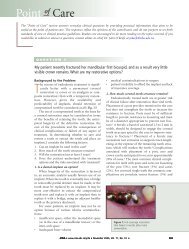
Point of Care<br />
Figure 3: The lining of this odontogenic<br />
keratocyst is composed of corrugated<br />
parakeratinized stratified squamous<br />
epithelial lining. The connective tissue wall<br />
is devoid of inflammation. Hematoxylin<br />
and eosin stain. Original magnification ×20.<br />
Figure 6: Inflamed odontogenic keratocyst.<br />
On the left side of this photomicrograph,<br />
the characteristic features of the lesion are<br />
absent as a result of the chronic inflammation.<br />
Hematoxylin and eosin stain.<br />
Original magnification ×20.<br />
removal at the time of initial surgery. Patients must be placed<br />
on long-term clinical and radiographic recall. In the case of<br />
maxillary lesions, in which recurrence can be difficult to<br />
diagnose by plain film and panoramic radiography, computed<br />
tomography is recommended. C<br />
Further Reading<br />
Blanas N, Freund B, Schwartz M, Furst IM. Systematic review of the<br />
treatment and prognosis of the odontogenic keratocyst. Oral Surg Oral<br />
Med Oral Pathol Radiol Endod 2000; 90(5):553–8.<br />
Kimonis VE, Goldstein AM, Pastakia B, Yang ML, Kase R, DiGiovanna<br />
JJ, and others. Clinical manifestations of 105 persons with nevoid basal<br />
cell carcinoma syndrome. Amer J Med Genet 1997; 69(3):299–308.<br />
LoMuzio L, Staibano S, Pannone G, Bucci P, Nocini PF, Bucci E, and<br />
other. Expression of cell cycle and apoptosis-related proteins in sporadic<br />
odontogenic keratocysts and odontogenic keratocysts associated with<br />
the nevoid basal cell carcinoma syndrome. J Dent Res 1999;<br />
78(7):1345–53.<br />
Myoung H, Hong SY, Hong SD, Lee JI, Lim CY, Choung PH and<br />
others. Odontogenic keratocyst: review of 256 cases for recurrence and<br />
clinicopathologic parameters. Oral Surg Oral Med Oral Pathol Radiol<br />
Endod 2001; 91(3):328–33.<br />
Sciubba JJ, Fantasia JE, Kahn LB. Tumors and cysts of the jaws. Atlas of<br />
tumor pathology. Rosai J and Sobin LH, editors. Washington, DC:<br />
Armed Forces Institute of Pathology (3rd series, fascicle 29); 2001.<br />
November 2003, Vol. 69, No. 10 679