JCDA - Canadian Dental Association
JCDA - Canadian Dental Association
JCDA - Canadian Dental Association

You also want an ePaper? Increase the reach of your titles
YUMPU automatically turns print PDFs into web optimized ePapers that Google loves.
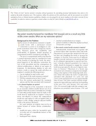
Point of Care<br />
warrant conventional scalpel biopsy. It is not intended for<br />
the examination of submucosal lesions with intact surface<br />
epithelium, such as hemangiomas or mucoceles. It can also<br />
be used in the long-term follow-up of patients with<br />
erythroplakic (Figs. 1 and 2) or leukoplakic lesions in<br />
which a baseline histologic diagnosis has been obtained and<br />
in patients with a history of oral cancer. However, a negative<br />
result on brush biopsy does not obviate the need for<br />
conventional tissue biopsy if the clinical presentation<br />
warrants.<br />
Vizilite (Zila Professional Pharmaceuticals; distributed<br />
by Patterson <strong>Dental</strong> Supply) is a recently introduced intraoral<br />
screening test similar to the acetowhite test, which is<br />
used in the detection of uterine cervical dysplasias and<br />
carcinomas. The patient rinses for 1 minute with a supplied<br />
1% acetic acid solution to disrupt the glycoprotein barrier<br />
on mucosal surfaces. The oral cavity is then examined with<br />
a single-use chemiluminescent light, the wavelength of<br />
which is absorbed by normal cells. Abnormal cells reflect<br />
the light, presumably as a result of an increased nucleus-tocytoplasmic<br />
ratio, and therefore appear white.<br />
As with Orascan and Oral CDx, a positive reading with<br />
Vizilite can be caused by minor epithelial abnormalities<br />
associated with inflammation, irritation or ulceration and<br />
does not necessarily mean that dysplasia or carcinoma is<br />
present.<br />
Question 2<br />
This scenario emphasizes the importance of submitting<br />
all surgical tissue to an oral and maxillofacial pathologist<br />
for evaluation. In this case, histologic examination revealed<br />
that what had the clinical appearance of a dentigerous cyst<br />
was in fact an odontogenic keratocyst (OKC). On radiographic<br />
examination OKCs range in size from small,<br />
unilocular, pericoronal radiolucencies indistinguishable<br />
from dentigerous cysts to large, multilocular radiolucent<br />
lesions involving the entire mandibular ramus (Fig. 1).<br />
OKCs (Fig. 2) are potentially aggressive cysts with a<br />
tendency to cause significant local destruction. They are<br />
one of the most common developmental odontogenic<br />
cysts, second in frequency only to dentigerous cysts. They<br />
most commonly involve the posterior body and ramus of<br />
the mandible and are often associated with an unerupted<br />
tooth. Clinically, OKCs tend to expand in an anteroposterior<br />
direction within the medullary bone, with little<br />
tendency to cause bony expansion.<br />
The presence of multiple OKCs may be the initial<br />
presenting sign of nevoid basal cell carcinoma syndrome<br />
678 November 2003, Vol. 69, No. 10<br />
It must be emphasized that these aids are not substitutes<br />
for good clinical judgement. The most important component<br />
in any oral cancer checkup remains a thorough visual<br />
and digital inspection of all intraoral soft tissues, as well as<br />
careful examination of the neck and cervical lymph nodes.<br />
Ultimately, diagnosis of any suspicious lesion requires<br />
histologic examination of biopsy samples. C<br />
References<br />
1. Johnson N, Warnakulasuriya S, Speight P, Epstein J. Diagnosing oral<br />
cancer: can toluidine blue mouthwash help? FDI World 1998; 7(2):22–6.<br />
2. <strong>Canadian</strong> Academy of Oral Pathology. CAOP releases toluidine blue<br />
statement [News Item]. J Can Dent Assoc 1995; 61(9):752–3.<br />
3. Sciubba JJ. Improving detection of precancerous and cancerous<br />
oral lesions. Computer-assisted analyses of the oral brush biopsy. U.S.<br />
Collaborative OralCDx Study Group. JADA 1999; 130(10):1445–57.<br />
Further Reading<br />
Epstein JB, Zhang L, Rosin M. Advances in the diagnosis of oral premalignant<br />
and malignant lesions. J Can Dent Assoc 2002; 68(10):617–21.<br />
Guo Z, Yamaguchi K, Sanchez-Cespedes M, Westra WH, Koch WM,<br />
Sidransky D. Allelic losses in OraTest-directed biopsies of patients with<br />
prior upper aerodigestive tract malignancy. Clin Cancer Res 2001;<br />
7(7):1963–8.<br />
Megevand E, Denny L, Dehaeck K, Soeters R, Bloch B. Acetic acid visualization<br />
of the cervix: an alternative to cytologic screening. Obstet<br />
Gynecol 1996; 88(3):383–6.<br />
Svirsky JA, Burns JC, Carpenter WM, Cohen DM, Bhattacharyya I,<br />
Fantasia JE, and others. Comparison of computer-assisted brush biopsy<br />
results with follow up scalpel biopsy and histology. Gen Dent 2002;<br />
50(6):500–3.<br />
I recently removed an impacted mandibular third molar with an associated 10-mm pericoronal<br />
radiolucency. Follicular tissue was submitted for biopsy. The histopathologic diagnosis was<br />
odontogenic keratocyst. What would you recommend for follow-up?<br />
(NBCCS), which is characterized by multiple OKCs,<br />
multiple basal cell carcinomas, characteristic palmar and<br />
plantar pits, and calcification of the falx cerebri. Affected<br />
patients may also develop neoplasms such as medulloblastoma,<br />
meningioma, ovarian fibroma and cardiac fibroma.<br />
Histologically, OKCs are characterized by a thin, parakeratinized,<br />
stratified squamous epithelial lining with a<br />
hyperchromatic and palisaded basal cell layer (Figs. 3 and<br />
4). The cyst lumen may contain desquamated keratin.<br />
Detachment of portions of the thin cyst lining is<br />
commonly observed at the time of surgery. The presence of<br />
satellite cysts within the cyst wall may be responsible for the<br />
reported 6% to 66% recurrence rate (Fig. 5). It has also<br />
been suggested that factors inherent to the cystic epithelium<br />
itself or influences involving regional odontogenic<br />
epithelium may be responsible for recurrence or for the<br />
formation of additional OKCs.<br />
The best method of treatment remains controversial.<br />
Current modalities include enucleation and osseous<br />
curettage with or without resection of the overlying<br />
Journal of the <strong>Canadian</strong> <strong>Dental</strong> <strong>Association</strong>