JCDA - Canadian Dental Association
JCDA - Canadian Dental Association
JCDA - Canadian Dental Association

Create successful ePaper yourself
Turn your PDF publications into a flip-book with our unique Google optimized e-Paper software.
Point of Care<br />
The Point of Care section of <strong>JCDA</strong> answers everyday clinical questions by providing practical information that aims to be useful<br />
at the point of patient care. The responses reflect the opinions of the contributors and do not purport to set forth standards of<br />
care or clinical practice guidelines. Readers are encouraged to do more reading on the topics covered. This month’s responses were<br />
provided by Dr. Paul C. Edwards, a resident in the division of oral and maxillofacial pathology, department of dental medicine,<br />
Long Island Jewish Medical Center. If you would like to submit or answer a question, contact editor-in-chief Dr. John O’Keefe at<br />
jokeefe@cda-adc.ca.<br />
Question 1<br />
Several screening tools are now available<br />
in Canada to assist in evaluating<br />
intraoral epithelial lesions.<br />
Orascan (Zila Professional<br />
Pharmaceuticals, Phoenix, Arizona;<br />
distributed by Patterson <strong>Dental</strong> Supply)<br />
is a mouth rinse containing toluidine<br />
blue (tolonium chloride), which selectively<br />
binds to free anionic groups such<br />
as the phosphate groups of DNA. In<br />
vivo, toluidine blue reportedly stains<br />
malignant epithelial lesions a blue<br />
colour. Orascan is recommended by the<br />
manufacturer for monitoring of suspicious<br />
lesions for which there has been a<br />
baseline histopathologic evaluation,<br />
screening for the presence of cancerous<br />
lesions in high-risk individuals, routine follow-up of<br />
patients previously treated for cancer of the upper aerodigestive<br />
tract and determination of the optimal site for<br />
biopsy of large, heterogeneous lesions.<br />
Areas of inflammation, irritation and ulceration, as well<br />
as the dorsum of the tongue (an uncommon site for<br />
squamous cell carcinoma), routinely pick up the dye.<br />
When used for screening in populations with a low<br />
prevalence of squamous cell carcinoma, the sensitivity and<br />
specificity appear lower than when used in high-risk populations.<br />
1 Therefore, in the absence of clinically suspicious<br />
areas in low-risk patients, areas that stain should be retested<br />
after 10 to 14 days, during which time any transient<br />
inflammatory lesions can be expected to heal.<br />
Orascan is not a diagnostic test, and the absence of<br />
staining does not rule out cancerous lesions or preclude the<br />
need for a scalpel biopsy if the clinical presentation<br />
warrants. The <strong>Canadian</strong> Academy of Oral Pathology has<br />
recommended that “lesions suspected clinically of being<br />
malignant or premalignant should be investigated by<br />
Journal of the <strong>Canadian</strong> <strong>Dental</strong> <strong>Association</strong><br />
What are the indications for using diagnostic aids such as Orascan, Oral CDx and Vizilite in<br />
the evaluation of oral lesions?<br />
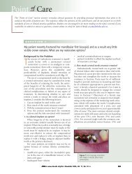
Figure 1: Patient with unilateral<br />
erythroleukoplakic lesion of the right<br />
buccal mucosa. Although tissue biopsy<br />
results were consistent with lichen planus,<br />
long-term follow-up with one of the screening<br />
tools described, coupled with periodic<br />
scalpel biopsy, would be appropriate.<br />
Photograph courtesy of Dr. John Fantasia.<br />
Figure 2: Patient with unilateral<br />
erythroleukoplakic lesion of the left buccal<br />
mucosa. Although there was clinical<br />
suspicion for carcinoma, the tissue biopsy<br />
was interpreted as nonspecific mucositis,<br />
and long-term follow-up with one of the<br />
screening tools would be appropriate.<br />
Photograph courtesy of Dr. John Fantasia.<br />
prompt incisional biopsy regardless of staining reaction to<br />
toluidine blue.” 2<br />
Oral CDx (Oralscan Laboratories, Suffern, N.Y.;<br />
distributed by Henry Schein <strong>Dental</strong>) is a computer-assisted<br />
brush biopsy analysis system. The dentist uses a brush<br />
biopsy instrument to obtain cells from all layers of the<br />
epithelium. The sample is evaluated to verify adequate<br />
representation of the basal layer. Individual cells are examined<br />
for a combination of abnormal morphology and spectral<br />
abnormalities of keratin that are characteristic of<br />
altered epithelial differentiation. Approximately 200 of the<br />
highest scoring (most abnormal) cells are selected by the<br />
computer for analysis by a cytopathologist.<br />
Oral CDx is not a substitute for traditional scalpel<br />
biopsy because the tissue obtained is disaggregated and<br />
does not contain the necessary architectural information<br />
to histologically assess and grade dysplastic lesions. 3<br />
According to the manufacturer, Oral CDx facilitates the<br />
testing of apparently innocuous epithelial lesions with no<br />
obvious cause that are not sufficiently suspicious-looking to<br />
November 2003, Vol. 69, No. 10 677