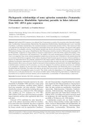
142 <strong>Ahead</strong> <strong>of</strong> <strong>print</strong> <strong>online</strong> <strong>version</strong> Fig. 2. <strong>Rhabdochona</strong> (Globochona) kurdistanensis sp. n., scanning electron micrographs. A, B – cephalic end, subdorsoventral and apical views, C – deirid, d, e – tail tip <strong>of</strong> female, dorsal and subapical views, F – posterior end <strong>of</strong> male, lateral view (arrows indicate lateral papillae), G – tail <strong>of</strong> male, lateral view (arrows indicate postanal papillae), H – ventral precloacal cuticular ornamentations <strong>of</strong> male, lateral view. Abbreviations: a – amphid; c – cloacal aperture; p – cephalic papilla; s – sublabium.
P r e v a l e n c e a n d i n t e n s i t y : Greater Zab River: 16 fish infected/43 fish examined; 3–9 nematodes per fish. Lesser Zab River: 11/48; 2–8 nematodes. E t y m o l o g y : The specific name kurdistanensis relates to the region <strong>of</strong> its origin, i.e. Kurdistan, where specimens <strong>of</strong> the new <strong>species</strong> were found. D e p o s i t i o n o f t y p e s : Holotype, allotype and paratypes in the Helminthological collection <strong>of</strong> the institute <strong>of</strong> Parasitology, Biology centre <strong>of</strong> the Academy <strong>of</strong> sciences <strong>of</strong> the Czech Republic, České Budějovice (Cat. No. N-981). Remarks: Based on the number (8) <strong>of</strong> anterior prostomal teeth and the presence <strong>of</strong> caudal processes on the tip <strong>of</strong> the tail, the specimens <strong>of</strong> this <strong>species</strong> can be assigned to the subgenus Globochona, as diagnosed by Moravec (1975). only a few <strong>species</strong> <strong>of</strong> <strong>Rhabdochona</strong> (Globochona) are known from freshwater fishes in tropical and subtropical Asia and Africa (Moravec 1975, 2010, Boomker and Petter 1993, Wang et al. 1994, Moravec and Yooyen 2011). in having a markedly short left spicule, bifurcate deirids and cuticular outgrowths on the tail tip <strong>of</strong> both sexes, R. (G.) kurdistanensis sp. n. is similar only to R. (G.) equispiculata Moravec et scholz, 1991 from Hampala spp. in laos and R. (G.) thaiensis Moravec et Yooyen, 2011 from Mystacoleucus marginatus (Valencienes) in thailand. However, in contrast to the new <strong>species</strong>, both spicules <strong>of</strong> R. (G.) equispiculata are almost equally long (length ratio <strong>of</strong> spicules 1:1.2 vs. 1:2.0–3.1) and the tail tip <strong>of</strong> both sexes bears 3–4 slender cuticular spines, whereas R. (G.) thaiensis possesses two small claw-shaped ventral projections on the tail tip <strong>of</strong> both sexes (Moravec and Scholz 1991, Moravec and Yooyen 2011). in having a markedly short left spicule (161–174), bifurcate deirids and a length ratio <strong>of</strong> spicules about 1:2, also R. (G.) brevichona Wang, Yu et Wu, 1994, a parasite <strong>of</strong> Rectoris luxiensis Wu et Yao in china, resembles the new <strong>species</strong>. However, the male tail tip <strong>of</strong> R. (G.) brevichona is truncated, without any projections, whereas the tail tip <strong>of</strong> conspecific females is with a crown-like appendage provided with processes, and deirids are situated at the level <strong>of</strong> the posterior end <strong>of</strong> prostom (Wang et al. 1994) vs. at the level <strong>of</strong> the posterior part <strong>of</strong> vestibule. there is also a certain similarity <strong>of</strong> the new <strong>species</strong> with R. (G.) chodukini osmanov, 1957, the only <strong>species</strong> <strong>of</strong> this subgenus occurring in the Aral sea basin (drainage systems <strong>of</strong> the Amu-Darya and syr-Darya rivers), the main hosts <strong>of</strong> which are barbels, Luciobarbus brachycephalus (Kessler) and L. capito (güldenstädt) (see osmanov 1957, Dzhalilov 1964, Moravec 1975). in his inadequate original description <strong>of</strong> R. chodukini, osmanov (1957) mentioned the presence <strong>of</strong> a “ring-like formation with spines on its margin” on the tail tip <strong>of</strong> both sexes, but Moravec (1975), redescribing this <strong>species</strong> on specimens from L. brachycephalus and L. capito from Tajikistan, found the male tail tip rounded, without any projections (see also Vismanis et <strong>Ahead</strong> <strong>of</strong> <strong>print</strong> <strong>online</strong> <strong>version</strong> Moravec et al.: two <strong>species</strong> <strong>of</strong> <strong>Rhabdochona</strong> from iraq al. 1987). consequently, R. chodukini differs from R. (G.) kurdistanensis mainly in having a markedly longer left spicule (352–420 vs. 180–204), simple deirids and the female tail tip provided with a crown consisting <strong>of</strong> many cuticular processes arranged in two circles, whereas the male tail tip is smooth, without any processes. rasheed (1965) considered R. chodukini as possibly identical with R. sarana Karve et Naik, 1951, a <strong>species</strong> inadequately described solely from females found in Puntius sarana (Hamilton) in central india (Poona) (Karve and Naik 1951), but because the conspecific male remains unknown, Moravec (1975), soota (1983) and sood (1989) treated it as a separate <strong>species</strong>. <strong>Rhabdochona</strong> sarana can be distinguished from the new <strong>species</strong> by the structure <strong>of</strong> the female tail tip (reported as a stumpy projection with three blunt spines), a shorter glandular oesophagus in female (2.44–3.14 vs. 5.54–7.14 mm) and smaller eggs (27–35 × 17–20 vs. 39–42 × 27–30). <strong>Rhabdochona</strong> (<strong>Rhabdochona</strong>) sp. Figs. 3, 4 Female (1 gravid specimen): Body 14.67 mm long, maximum width 218. Mouth oval, with four small submedian sublabia. Four small cephalic papillae and pair <strong>of</strong> lateral amphids present (Figs. 3c, 4A,B). Vestibule including prostom 168 long. Prostom funnel-shaped, armed with 14 anterior teeth (3 dorsal, 3 ventral and 4 lateral on each side; lateral teeth forming pairs) and distinct basal teeth, 27 long and 21 wide (Figs. 3A–c, 4A,B). Muscular oesophagus 363 long, 48 wide; glandular oesophagus 5.30 mm long, 129 wide; length ratio <strong>of</strong> both parts <strong>of</strong> oesophagus 1:14.6. Entire oesophagus and vestibule representing 40% <strong>of</strong> body length. Nerve ring and excretory pore 240 and 340, respectively, from anterior extremity. Deirids small, bifurcate, 111 from anterior end <strong>of</strong> body (Figs. 3A,B,E, 4C). Tail conical, 216 long, with sharply pointed tip (Figs. 3D, 4D). Vulva postequatorial, 8.79 mm from anterior extremity, at 60% <strong>of</strong> body length. Muscular vagina directed posteriorly from vulva. Eggs small, oval, thick-walled, embryonated, size 30–33 × 21–24; thickness <strong>of</strong> egg wall 3; each pole <strong>of</strong> egg provided with minute swelling and one long tape-like filament <strong>of</strong> fibrous structure; filaments about 150 long and 18 wide (Fig. 3F). H o s t : Luciobarbus kersin (cyprinidae, cypriniformes) (body length 18–65 cm). s i t e o f i n f e c t i o n : intestine. L o c a l i t y : Greater Zab River (36–37°N, 43–44°E) (Tigris River basin) at Gwer, about 44 km southwest <strong>of</strong> Erbil City, Kurdistan region, northern iraq (collected 13 June 2011). P r e v a l e n c e a n d i n t e n s i t y : 1 fish infected/43 fish examined; 3 nematodes. D e p o s i t i o n o f v o u c h e r s p e c i m e n : Helminthological collection <strong>of</strong> the institute <strong>of</strong> Parasitology, Biology centre <strong>of</strong> the Academy <strong>of</strong> sciences <strong>of</strong> the czech republic, České Budějovice (Cat. No. N-983). 143