Data as of November 2011 - IMBA - Österreichische Akademie der ...
Data as of November 2011 - IMBA - Österreichische Akademie der ...
Data as of November 2011 - IMBA - Österreichische Akademie der ...

Create successful ePaper yourself
Turn your PDF publications into a flip-book with our unique Google optimized e-Paper software.
Contents<br />
Introduction .................................................................................2<br />
Research Highlights ...................................................................4<br />
ReseaRCH GRoups<br />
Julius Brennecke Group............................................................6<br />
Jürgen Knoblich Group ............................................................8<br />
thom<strong>as</strong> Marlovits Group ...................................................... 10<br />
Javier Martinez Group .............................................................12<br />
Kazufumi Mochizuki Group ................................................. 14<br />
Josef penninger Group .......................................................... 16<br />
Leonie Ringrose Group .......................................................... 18<br />
Vic small Group ........................................................................ 20<br />
ReseaRCH suppoRt<br />
stem Cell Center - Mouse Gene targeting ..................... 22<br />
Fly House .................................................................................... 23<br />
CoRe FaCILItIes<br />
Biooptics .................................................................................... 24<br />
electron Microscopy ............................................................... 25<br />
Bioinformatics ........................................................................... 26<br />
Genomics .................................................................................. 27<br />
service Department ............................................................... 28<br />
protein Chemistry .................................................................. 29<br />
Histology .................................................................................... 30<br />
Comparative Medicine .......................................................... 31<br />
transgenic service .................................................................. 31<br />
Max perutz Library .................................................................. 32<br />
Campus scientific support Facility GmbH ..................... 34<br />
publications ............................................................................... 36<br />
awards ......................................................................................... 39<br />
seminars ..................................................................................... 40<br />
scientific advisory Board ...................................................... 42<br />
supervisory Board ................................................................... 42<br />
administration and other services ................................... 43<br />
Key Facts .....................................................................................44<br />
sponsors & partners ............................................................... 45<br />
Your Career at IMBa ................................................................ 46<br />
IMBa and its surroundings .................................................. 48<br />
Genetics and art ...................................................................... 49<br />
spotlight on <strong>2011</strong> .................................................................... 50<br />
Impressum.................................................................................. 52<br />
Where we are ............................................................................ 52
<strong>IMBA</strong> <strong>2011</strong> / INTRODUCTION | page 2<br />
JOSEF PENNINGER<br />
Managing Director/Science<br />
JÜRGEN KNOBLICH<br />
Deputy Director/Science<br />
MICHAEL KREBS<br />
Managing Director/Finance & Administration<br />
Introduction<br />
IMBa h<strong>as</strong> had another brilliant year in research, a year <strong>of</strong> highlights<br />
that are presented in this annual report. In particular our young<br />
scientists have come into their own and have published top notch<br />
research. successful research institutions are not known by the quantity<br />
<strong>of</strong> publications they produce but are defined by key findings that<br />
have indeed opened new fields and the international standing <strong>of</strong><br />
their researchers. successful research institutes are fragile palaces<br />
that need to be cultivated at all levels – from a great cafeteria to<br />
meet, faculty meetings and lecture series, young phD students and<br />
postdocs that challenge our paradigms, open doors for discussions,<br />
to world-cl<strong>as</strong>s administration and service infr<strong>as</strong>tructures. successful<br />
research institutes stand on multiple strong legs, nurture young<br />
talents, and live a culture <strong>of</strong> collaboration and mutual support.<br />
Many <strong>of</strong> these visions have already been implemented and the<br />
success <strong>of</strong> the young scientists is the success <strong>of</strong> everybody at<br />
IMBa – the success <strong>of</strong> a large team with a common goal.<br />
this year we hired 4 new group lea<strong>der</strong>s, the three junior pIs Fumiyo<br />
Ikeda, Kikue tachibana-Konwalski, and stefan ameres, and Daniel<br />
Gerlich <strong>as</strong> a senior Investigator. Kikue tachibana-Konwalski joined<br />
IMBa from Kim n<strong>as</strong>myth’s lab in oxford in november <strong>2011</strong>. she<br />
is working on the first cell division and meiosis in mammalian<br />
cells using gene modified mice. Fumiyo Ikeda worked with Ivan<br />
Dikic in Frankfurt and started at IMBa on December 1st, <strong>2011</strong>.
Fumiyo is a biochemist who co-discovered linear ubiquitination<br />
<strong>as</strong> a novel intracellular signaling pathway. stefan ameres did his<br />
phD with Renee schroe<strong>der</strong> at MFpL, Vienna, and continued his<br />
postdoctoral training with phil Zamore in the usa. stefan is working<br />
on the biology <strong>of</strong> non-coding Rna and will start in January 2012.<br />
Daniel Gerlich will join IMBa <strong>as</strong> a permanent senior pI on March 1st,<br />
2012. He is currently working at the etH in Zürich and will bring high<br />
throughput imaging technologies to the Campus. His research focus<br />
is in cell biology and high throughput <strong>as</strong>says on finding genes that<br />
control mitosis. We are very happy that we continue to be able to<br />
attract world-cl<strong>as</strong>s talents to IMBa and that these brilliant young<br />
minds have chosen IMBa <strong>as</strong> their future centre <strong>of</strong> gravity.<br />
With the addition <strong>of</strong> our new groups, we decided to reorganize<br />
the lab spaces and focus all faculty groups on two research floors<br />
and move our excellent infr<strong>as</strong>tructure groups to one floor at IMBa.<br />
With these changes we hope to strengthen the interactions and<br />
communication among the researchers and streamline our services.<br />
the rebuilding <strong>of</strong> the institute h<strong>as</strong> progressed well and will be<br />
finished <strong>as</strong> planned, a testament to the great efforts made by many<br />
people, especially the groups <strong>of</strong> alex Chlup and Michael Kratochwille.<br />
another key development w<strong>as</strong> the initiation <strong>of</strong> the Campus scientific<br />
support Facility (CsF) and the hiring <strong>of</strong> andre<strong>as</strong> tiran <strong>as</strong> its first<br />
director. the CsF is an initiative <strong>of</strong> all research institutes and biotech<br />
companies located at the Vienna Biocenter (VBC) that brings more<br />
than 50 Million euros in infr<strong>as</strong>tructure money to our Campus.<br />
the idea <strong>of</strong> the CsF is to establish an additional service centre and<br />
expand/improve existing infr<strong>as</strong>tructures that are accessible for<br />
all institutions at our Campus. according to the plan, we already<br />
transferred the deep sequencing unit and the unique Vienna<br />
Drosophila Rnai Library un<strong>der</strong> the wings <strong>of</strong> the CsF. the electron<br />
Microscopy Facility will follow early next year to develop a new<br />
centre for high resolution imaging. In addition, new facilities for<br />
mouse imaging/phenotyping, plant genetics, protein purification<br />
and protein structure, and bioinformatics are being developed un<strong>der</strong><br />
the capable guidance <strong>of</strong> andre<strong>as</strong> tiran and his team. We continue<br />
to maintain key services in-house in close cooperation with our<br />
partner the Research Institute <strong>of</strong> Molecular pathology (IMp) and<br />
also the Gregor Mendel Institute <strong>of</strong> Molecular plant Biology (GMI).<br />
the establishment <strong>of</strong> the CsF is a great example for a joint effort<br />
by all VBC members and we have to give a special “thank you” to<br />
all people at IMBa and IMp services and administration who went<br />
the extra mile to make the CsF a reality. our infr<strong>as</strong>tructures have<br />
always been the key to our success and we strongly adhere to the<br />
principle that these services, internally at IMBa/IMp/GMI and now<br />
at the CsF, are an absolute integral part for the future and necessary<br />
for our research. We will therefore do everything possible to make<br />
sure that all <strong>of</strong> these services continue to shine.<br />
Finally, we want to thank everybody at the IMBa family and <strong>of</strong> course<br />
our extended family at the IMp for their great work. our world-cl<strong>as</strong>s<br />
service units and the dedicated and excellent administration allow<br />
us – <strong>as</strong> always – to focus our efforts on doing innovative and excellent<br />
research. they are a key part <strong>of</strong> our success and deserve all the credit.<br />
a big thank you also to our scientific advisory Board that h<strong>as</strong> done<br />
tremendous work, to our close partner Boehringer Ingelheim, our<br />
supervisory Board, the members <strong>of</strong> the IMBa Fundraising Board,<br />
our neighbours and partners the Gregor Mendel Institute (GMI)<br />
and Max F. perutz Laboratories (MFpL), and to the many people<br />
who have helped us to develop and continue to support IMBa.<br />
If we all stand together and all work towards the same goals, to make<br />
a little difference in this world, then we will continue to be strong.<br />
Finally we would like to thank all the government institutions<br />
and private and institutional donors who provide the fuel for our<br />
work - our funding. all senior faculty and two <strong>of</strong> our Junior IMBa<br />
group lea<strong>der</strong>s have eRC grants and more than 70% <strong>of</strong> the research<br />
personnel is funded by grants, a very high rate <strong>of</strong> third party funding.<br />
a special “thank you” h<strong>as</strong> to go to all members <strong>of</strong> the austrian<br />
academy <strong>of</strong> sciences and the lea<strong>der</strong>ship <strong>of</strong> the academy, who<br />
continue to believe in the future <strong>of</strong> IMBa and have made a steadf<strong>as</strong>t<br />
commitment to support the development <strong>of</strong> IMBa in the next<br />
5 years. the commitment will allow us to make the next step in the<br />
development <strong>of</strong> the institute, the step from a promising fledgling<br />
to a strong youth that stands on strong feet. such a commitment<br />
also carries a promise from our side, a promise to create a place<br />
that attracts the top international talents and where such talents<br />
have the time and the environment to grow, and a promise that<br />
the investments in IMBa reinforce the leading role <strong>of</strong> the austrian<br />
academy <strong>of</strong> sciences <strong>as</strong> a major international research institution.<br />
IMBa h<strong>as</strong> had an amazing start, h<strong>as</strong> gained international standing<br />
b<strong>as</strong>ed on the quality <strong>of</strong> it’s research, and now h<strong>as</strong> been given the<br />
chance to become a truly international player.<br />
Jürgen Knoblich, Michael Krebs and Josef Penninger<br />
<strong>IMBA</strong> <strong>2011</strong> / INTRODUCTION | page 3
<strong>IMBA</strong> <strong>2011</strong> / RESEARCH HIGHLIGHTS | page 4<br />
ReseaRch hIghlIghts<br />
<strong>2011</strong> h<strong>as</strong> been scientifically a very successful year for <strong>IMBA</strong>. This double page shows <strong>IMBA</strong>’s “Research<br />
Highlights”, a selection <strong>of</strong> highly visible research articles <strong>IMBA</strong> scientists have published during this year.<br />
HSPC117 is the essential subunit <strong>of</strong> a human tRNA splicing lig<strong>as</strong>e<br />
complex.<br />
Popow, J., Englert, M., Weitzer, S., Schleiffer, A.,<br />
Mierzwa, B., Mechtler, K., Trowitzsch, S., Will, CL.,<br />
Lührmann, R., Söll, D., Martinez, J. (<strong>2011</strong>). HSPC117<br />
is the essential subunit <strong>of</strong> a human tRNA splicing<br />
lig<strong>as</strong>e complex. Science. 331(6018):760-4<br />
transfer Rn<strong>as</strong> (tRn<strong>as</strong>) are absolutely necessary<br />
to translate the genetic code into proteins<br />
required for life. In humans, some tRn<strong>as</strong> are<br />
synthesized in an immature form containing an<br />
intervening sequence that h<strong>as</strong> to be removed<br />
by sequential cleavage and joining reactions.<br />
although the factors carrying out the cleavage<br />
reactions are already known for several years,<br />
the so-called tRna lig<strong>as</strong>e joining the tRna<br />
exon pieces together remained unknown.<br />
We applied chromatographic techniques to<br />
purify proteins required for the joining reaction<br />
from the complex cell extracts able to process<br />
tRna precursors into their mature form. after<br />
several rounds <strong>of</strong> purification we arrived at a<br />
mixture <strong>of</strong> ca. 100 proteins which we could<br />
examine in greater detail. one <strong>of</strong> those, called<br />
HspC117, proved to be the essential factor for<br />
the described joining reaction <strong>as</strong> we were<br />
able to demonstrate by affinity purification<br />
and Rnai-mediated inactivation experiments.<br />
Identification <strong>of</strong> the tRNA lig<strong>as</strong>e HSPC117. (A) siRNAs can be used <strong>as</strong> a substrate for human<br />
RNA lig<strong>as</strong>e activity. siRNA duplexes are efficiently ligated in HeLa cell extracts only in c<strong>as</strong>e<br />
they display a phosphate group at their 3’ end and a hydroxyl group at their 5’ end (lanes c).<br />
Removal <strong>of</strong> the 3’-phosphate or phosphorylation <strong>of</strong> the 5’-hydroxyl ends interfere with siRNA<br />
ligation (lanes a, b and d). (B) Silencing <strong>of</strong> HSPC117 – but not EGFP <strong>as</strong> a control – impairs<br />
ligation <strong>of</strong> tRNA exon halves in tRNA splicing reactions. (C) Immunoprecipitates (IPs) <strong>of</strong> c-myc-<br />
HSPC117 ligate tRNA exons. The point mutant c-myc–HSPC117 C122A (Cys122 → Ala122) is<br />
inactive <strong>as</strong> a tRNA lig<strong>as</strong>e. (D) A tRNA lig<strong>as</strong>e complex is co-selected with monoclonal antibodies<br />
directed against the mRNA splicing factor SF3b. Size fractionation <strong>of</strong> the bound proteins yields<br />
a complex composed <strong>of</strong> HSPC117, DDX1, FAM98B, CGI-99 and ASW. Fractions containing the<br />
HSPC117-complex exhibit tRNA exon lig<strong>as</strong>e activity. Three-dimensional model <strong>of</strong> Salmonella’s needle complex at<br />
subnanometer resolution.<br />
A<br />
C<br />
5’ HO<br />
3'P Cp*<br />
nt 40 -<br />
nt 100 -<br />
80 -<br />
70 -<br />
Marker<br />
Substrate<br />
Control<br />
c-myc-C122A<br />
c-myc-WT<br />
Control<br />
c-myc-C122A<br />
c-myc-WT<br />
40 -<br />
20 -<br />
Marker<br />
21-merOH<br />
21-merP<br />
30 -<br />
21 OH-<br />
21 P -<br />
20 -<br />
OH<br />
OH<br />
a<br />
P<br />
OH<br />
HeLa cell<br />
extract<br />
INPUT IP α-c-myc<br />
OH<br />
P<br />
*<br />
P<br />
b c d<br />
Pre-tRNA<br />
P Cp*<br />
Mature tRNA<br />
Exon halves<br />
O<br />
Linear intron<br />
Circular intron<br />
Modification <strong>of</strong> 5’-end<br />
Modification <strong>of</strong> 3’-end<br />
Ligation product<br />
siRNA-substrate<br />
B<br />
D<br />
siEGFP<br />
Marker<br />
Substrate<br />
HeLa extract<br />
Input<br />
siHSPC117<br />
Pre-tRNA<br />
Mature tRNA<br />
Exon halves<br />
nt 100 -<br />
Pre-tRNA<br />
80 -<br />
70 -<br />
40 -<br />
20 -<br />
Superose 6 fractions<br />
HSPC117<br />
DDX1<br />
FAM98B<br />
CGI-99<br />
ASW<br />
*<br />
Mature tRNA<br />
Exon halves<br />
Linear intron<br />
Circular intron<br />
Schraidt, O., Marlovits, TC. (<strong>2011</strong>). Three-dimensional<br />
model <strong>of</strong> Salmonella’s needle complex at subnanometer<br />
resolution. Science. 331(6021):1192-5<br />
type III secretion systems (t3sss) are essential<br />
pathogenic factors used by many Gram-negative<br />
bacteria like eHeC, salmonella, shigella or Cholera<br />
to inject proteins that make eukaryotic host cells<br />
accessible to inv<strong>as</strong>ion. In this study we were<br />
able to generate a cryo electron microscopy<br />
density map <strong>of</strong> the core structure <strong>of</strong> the t3ss, the<br />
needle complex (nC), a ~3.5 megadalton-sized,<br />
membrane-embedded injection machine with<br />
subnanomter resolution. showing secondary<br />
structural elements like α-helices this highly<br />
resolved density map allowed for confident<br />
docking <strong>of</strong> atomic structures. the resulting<br />
atomic model revealed insights into the nC’s<br />
overall organization and into the structural<br />
requirements during <strong>as</strong>sembly. We thereby<br />
provide a framework that will strongly promote<br />
further structural and functional studies <strong>of</strong> the<br />
t3ss and hopefully will also <strong>as</strong>sist in generating<br />
new antibacterial strategies.<br />
Solving the 3D-structure <strong>of</strong> the NC. Top row: Average images <strong>of</strong> NC inner ring (left) and outer<br />
ring (right) substructures obtained by selective dis<strong>as</strong>sembly <strong>of</strong> wild type NC. The top views<br />
allow for a direct counting <strong>of</strong> the inner and outer ring subunits and reveal a 24- and 15-fold<br />
symmetry, respectively. Bottom row: (left) Surface views <strong>of</strong> a 3D-reconstruction <strong>of</strong> the NC. With<br />
α-helical densities visible, (right) atomic structures could be placed unambiguously into the<br />
NC.<br />
The stress kin<strong>as</strong>e MKK7 couples oncogenic stress to p53 stability and<br />
tumor suppression<br />
Schramek, D., Kotsin<strong>as</strong>, A., Meixner, A., Wada, T.,<br />
Elling, U., Pospisilik, JA., Neely, GG., Zwick, RH.,<br />
Sigl, V., Forni, G., Serrano, M., Gorgoulis, VG.,<br />
Penninger, JM. (<strong>2011</strong>). The stress kin<strong>as</strong>e MKK7<br />
couples oncogenic stress to p53 stability and<br />
tumor suppression. Nat Genet. 43(3):212-9<br />
this study seeks to define the molecular<br />
regulation <strong>of</strong> epithelial cells during the early<br />
steps <strong>of</strong> cellular transformation using primary<br />
cell culture and genetic ablation studies.<br />
We have focused on a signaling molecule<br />
with hitherto ill-defined functions during<br />
tumorigenesis: MKK7 and its downstream<br />
factors JnK1 and JnK2. this signaling c<strong>as</strong>cade<br />
serves <strong>as</strong> an intracellular messenger receiving<br />
and allocating various cellular stimuli such <strong>as</strong><br />
stress signals and inflammatory cues.<br />
We could now show that MKK7 and the JnK<br />
signaling pathway also functions to suppress<br />
tumor formation in various organs such <strong>as</strong> the<br />
lung, the bre<strong>as</strong>t and the skin. It is well know<br />
that cells generally counteract oncogenic<br />
transformation by activating a cellular fail-safe<br />
mechanism: p53-induced cell cycle arrest or<br />
cellular suicide. this mechanism is absolutely<br />
crucial consi<strong>der</strong>ing the trillions <strong>of</strong> cells in<br />
our body susceptible to sporadic mutations,<br />
which could ignite uncontrolled proliferation<br />
and tumor development. Failure <strong>of</strong> this<br />
mechanism leads to development <strong>of</strong> multiple,<br />
spontaneous tumors in humans and in mice.<br />
using knock-out mice and improved primary<br />
cell culture systems I could show that MKK7<br />
directly couples oncogenic and genotoxic<br />
stress to p53 stability required for cell cycle<br />
arrest and suppression <strong>of</strong> neut-induced bre<strong>as</strong>t<br />
and KR<strong>as</strong>G12D-induced lung tumors (Figure<br />
1). Importantly, p53 overexpression could<br />
revert lung tumorigenesis in MKK7-deficient<br />
animals unambiguously confirming this new<br />
tumor suppressive mechanism. this study<br />
identified MKK7 <strong>as</strong> a vital molecular sensor<br />
to set a cellular anti-cancer barrier by linking<br />
oncogenic stress and the Dna damage response<br />
(DDR) to the key tumor suppressor p53. this<br />
work w<strong>as</strong> recently featured <strong>as</strong> an article in<br />
nature Genetics.<br />
MKK7 controls KR<strong>as</strong>G12D-driven lung and NeuT-driven bre<strong>as</strong>t tumorigenesis through p53<br />
expression. A, Representative H&E stainings showing accelerated progression and incre<strong>as</strong>ed<br />
tumor burden in KR<strong>as</strong>;Map2k7fl/Δ mice. B, Representative histology <strong>of</strong> mammary cancers<br />
that developed in 16 week old NeuT;Map2k7Δ/+mam and NeuT;Map2k7Δmam littermate<br />
females. H&E stained sections are shown. C and D, Western blot analysis for p53 and MKK7<br />
levels in KR<strong>as</strong>G12D-driven lung (C) and NeuT-driven bre<strong>as</strong>t tumors (D). β-actin is shown <strong>as</strong><br />
loading control.
Genome-Wide Analysis <strong>of</strong> Self-Renewal in Drosophila Neural Stem<br />
Cells by Transgenic RNAi<br />
Neumüller, RA., Richter, C., Fischer, A., Novatchkova,<br />
M., Neumüller, KG., Knoblich, JA. (<strong>2011</strong>). Genome-wide<br />
analysis <strong>of</strong> self-renewal in Drosophila neural stem<br />
cells by transgenic RNAi. Cell Stem Cell. 8(5):580-93<br />
spectacular recent advances in the field <strong>of</strong> stem<br />
cell biology have raised enormous hopes for<br />
regenerative medicine. one key property <strong>of</strong><br />
stem cells is their ability to “self-renew”, that is to<br />
generate identical copies <strong>of</strong> themselves while at<br />
the same time producing more specialized cells,<br />
which then replace damaged cells in the tissue.<br />
one <strong>of</strong> the key challenges in stem cell biology<br />
is to un<strong>der</strong>stand how the balance between<br />
self-renewal and specialization is regulated - with<br />
the goal to some day manipulate this balance to<br />
incre<strong>as</strong>e or decre<strong>as</strong>e the regenerative capacity<br />
<strong>of</strong> individual tissues.<br />
the fruitfly Drosophila h<strong>as</strong> emerged <strong>as</strong> a simple<br />
key model system for stem cell biology. neural<br />
stem cells in the developing fly brain follow a<br />
simple lineage and divide reproducibly into one<br />
self-renewing and one differentiating daughter<br />
cell. so far, all the components <strong>of</strong> the cellular<br />
machinery responsible for this <strong>as</strong>ymmetric<br />
division are also present in human stem cells<br />
and many <strong>of</strong> them fulfill the same role.<br />
Researchers in the laboratory <strong>of</strong> Juergen<br />
Knoblich at IMBa have now made a huge<br />
effort to identify essentially all the components<br />
that are important in Drosophila neural stem<br />
cells. they used a transgenic Rnai library from<br />
the Vienna Drosophila Rnai Center (VDRC) to<br />
inhibit almost all genes in the fly genome in<br />
neural stem cells. one by one, they studied<br />
the effects <strong>of</strong> inhibiting individual genes and<br />
carefully described the effects on the neural<br />
stem cells. Importantly, those experiments<br />
were done in whole living flies and not in<br />
cell culture where those effects can be very<br />
different. the results provide a unique resource<br />
for the stem cell community <strong>as</strong> most <strong>of</strong> the<br />
genes they identify are also present in human<br />
stem cells. the precise quantification <strong>of</strong> the<br />
resulting effects h<strong>as</strong> allowed an unprecedented<br />
bioinformatic analysis <strong>of</strong> the data which allowed<br />
insights into stem cell biology that were not<br />
possible before. the data obtained through<br />
this study provide a unique starting point for<br />
a systems-level analysis <strong>of</strong> stem cells.<br />
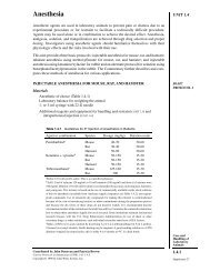
The image shows a wild type brain hemisphere on the left and a neural stem cell tumor on the<br />
right. The image resides on a background, which shows a functional network <strong>of</strong> <strong>as</strong>ymmetric<br />
cell division that regulates self-renewal and differentiation <strong>of</strong> neural stem cells identified in<br />
the study.<br />
Mouse inscuteable induces apical-B<strong>as</strong>al spindle orientation to facilitate<br />
intermediate progenitor generation in the developing neocortex.<br />
Postiglione, MP., Jüschke, C., Xie, Y., Ha<strong>as</strong>, GA., Charalambous,<br />
C., Knoblich, JA. (<strong>2011</strong>). Mouse inscuteable induces<br />
apical-B<strong>as</strong>al spindle orientation to facilitate intermediate<br />
progenitor generation in the developing neocortex.<br />
Neuron. 72(2):269-84<br />
neurons in the mammalian neocortex arise from<br />
<strong>as</strong>ymmetric divisions <strong>of</strong> progenitors residing in the<br />
ventricular zone. While in most progenitor divisions,<br />
the mitotic spindle is parallel to the ventricular<br />
surface, some progenitors reorient the spindle<br />
and divide in oblique orientations. Here, we use<br />
conditional deletion and overexpression <strong>of</strong> mouse<br />
Inscuteable (mInsc) to analyze the relevance <strong>of</strong><br />
spindle reorientation in cortical progenitors. Mutating<br />
mInsc almost abolishes oblique and vertical mitotic<br />
spindles, while mInsc overexpression h<strong>as</strong> the opposite<br />
effect. our data suggest that oblique divisions are<br />
essential for generating the correct numbers <strong>of</strong><br />
neurons in all cortical layers. using clonal analysis,<br />
we demonstrate that spindle orientation affects<br />
the rate <strong>of</strong> indirect neurogenesis, a process where<br />
progenitors give rise to b<strong>as</strong>al progenitors, which in<br />
turn divide symmetrically into two differentiating<br />
neurons. our results indicate that the orientation <strong>of</strong><br />
progenitor cell divisions is important for correct lineage<br />
specification in the developing mammalian brain.<br />
Section through the cerebral cortex <strong>of</strong> a mouse, stem cells can be seen glowing in green, mature nerve<br />
cells in red; cell nuclei for both types <strong>of</strong> cell are shown in blue.<br />
Forward and Reverse Genetics through Derivation <strong>of</strong> Haploid Mouse<br />
Embryonic Stem Cells.<br />
Elling, U., Taubenschmid, J., Wirnsberger, G., O’Malley,<br />
R., Demers, SP., Vanhaelen, Q., Shukalyuk, AI.,<br />
Schmauss, G., Schramek, D., Schnuetgen, F., von<br />
Melchner, H., Ecker, JR., Stanford, WL., Zuber, J., Stark,<br />
A. and Penninger, JM. (<strong>2011</strong>). Forward and Reverse<br />
Genetics through Derivation <strong>of</strong> Haploid Mouse<br />
Embryonic Stem Cells. Cell Stem Cell. 9(6):563-74.<br />
all somatic mammalian cells carry two copies<br />
<strong>of</strong> chromosomes (diploidy), where<strong>as</strong> organisms<br />
with a single copy <strong>of</strong> their genome, such <strong>as</strong><br />
ye<strong>as</strong>t, provide a b<strong>as</strong>is for recessive genetics.<br />
Here we report the generation <strong>of</strong> haploid mouse<br />
esC lines from parthenogenetic embryos.<br />
these cells carry 20 chromosomes, express<br />
Single (haploid) chromosome set in a mouse stem cell.<br />
stem cell markers, and develop into all germ<br />
layers in vitro and in vivo. We also developed<br />
a reversible mutagenesis protocol that allows<br />
saturated genetic recessive screens and results<br />
in homozygous alleles. this system allowed<br />
us to generate a knockout cell line for the<br />
microRna processing enzyme Drosha. In a<br />
forward genetic screen, we identified Gpr107<br />
<strong>as</strong> a molecule essential for killing by ricin, a<br />
toxin being used <strong>as</strong> a bioweapon. our results<br />
open the possibility <strong>of</strong> combining the power<br />
<strong>of</strong> a haploid genome with pluripotency <strong>of</strong><br />
embryonic stem cells to uncover fundamental<br />
biological processes in defined cell types at<br />
a genomic scale.<br />
<strong>IMBA</strong> <strong>2011</strong> / RESEARCH HIGHLIGHTS | page 5
<strong>IMBA</strong> <strong>2011</strong> / RESEARCH GROUPS | page 6<br />
JulIus BRennecke gRoup<br />
the piRna pathway – a small Rna B<strong>as</strong>ed genome Immune system<br />
www.imba.oeaw.ac.at/research/julius-brennecke<br />
Throughout the eukaryotic lineage, small RNA silencing pathways protect the genome against the deleterious influence <strong>of</strong> selfish genetic elements<br />
such <strong>as</strong> transposons. In animals a specialized pathway centered on PIWI proteins and their interacting piRNAs silences transposons within gonads.<br />
Recent experimental and bioinformatics studies have uncovered the f<strong>as</strong>cinating conceptual framework <strong>of</strong> this pathway that is conserved from<br />
invertebrates to mammals. Our group dissects the molecular and genetic makeup <strong>of</strong> this pathway and aims to un<strong>der</strong>stand its biological functions.<br />
silencing selfish genetic elements<br />
nearly all eukaryotic genomes contain selfish genetic elements<br />
such <strong>as</strong> transposons. their dev<strong>as</strong>tating impact on the host is<br />
illustrated by the phenomenon <strong>of</strong> “hybrid dysgenesis” in Drosophila<br />
melanog<strong>as</strong>ter: Intercrosses between laboratory strain females and<br />
males caught in the wild result in progeny with severe sterility.<br />
this is caused by the uncontrolled activity <strong>of</strong> a single transposon,<br />
which is present (and silenced) in wild populations but absent<br />
in stocks that have been kept in laboratories since ~100 years.<br />
the Drosophila genome, however, contains not only one, but<br />
more than one hundred transposon families, whose transposition<br />
strategies vary widely. to ensure reproductive fitness, flies (and all<br />
other organisms) have thus been un<strong>der</strong> evolutionary pressure to<br />
evolve a generic transposon silencing system. Work over the p<strong>as</strong>t<br />
decade h<strong>as</strong> demonstrated that the piRna pathway, a specialized<br />
small Rna silencing pathway is the major silencing system that<br />
keeps transposons un<strong>der</strong> control in animal gonads.<br />
the piRna pathway – a small Rna b<strong>as</strong>ed<br />
genome immune system<br />
the piRna pathway is a won<strong>der</strong>ful example <strong>of</strong> how much more<br />
sophisticated nature devises a solution to a problem compared to<br />
what we would theoretically design. In essence, the piRna pathway<br />
acts <strong>as</strong> an Rna-b<strong>as</strong>ed genome immune system. It comprises an<br />
inheritable genetic component and an acute response system, which<br />
specifically targets active transposons. Briefly, the transcription <strong>of</strong><br />
discrete heterochromatic loci (termed piRna clusters) provides a<br />
template, from which primary piRn<strong>as</strong> are produced. piRna clusters<br />
contain v<strong>as</strong>t collections <strong>of</strong> immobile and broken copies <strong>of</strong> transposons,<br />
which are or have been active in a population and therefore act <strong>as</strong><br />
a long term storage system for transposon sequence information.<br />
If a primary piRna encounters a target (active transposon), cleavage<br />
<strong>of</strong> the transposon Rna by the piRna-complex leads to the synthesis<br />
<strong>of</strong> a novel, complementary piRna. this piRna in turn guides the<br />
production <strong>of</strong> more antisense piRn<strong>as</strong> <strong>der</strong>ived from the piRna<br />
cluster transcript. thus, piRna clusters act not only <strong>as</strong> a genetically<br />
inherited memory component but also <strong>as</strong> relay stations to boost<br />
the production <strong>of</strong> silencing competent piRn<strong>as</strong>.<br />
Recent studies have elucidated the conceptual framework <strong>of</strong><br />
this pathway described above. these were mostly b<strong>as</strong>ed on the<br />
bioinformatics analysis <strong>of</strong> piRna populations in the light <strong>of</strong> decades<br />
<strong>of</strong> genetic work on transposon control in flies. at a mechanistic level,<br />
however, our un<strong>der</strong>standing <strong>of</strong> the piRna pathway is rudimentary<br />
at best. the only thing that is clear at the moment is that this<br />
pathway is by far more complex than the related microRna and<br />
siRna pathways.<br />
to further un<strong>der</strong>stand this f<strong>as</strong>cinating silencing system, we use<br />
Drosophila melang<strong>as</strong>ter <strong>as</strong> a model system. Here, we can combine<br />
genetics, biochemistry, cell biology and bioinformatics in unique<br />
ways. Moreover, roughly 35 years <strong>of</strong> genetic studies on transposons<br />
and host-strategies to silence them provide us with a wide range <strong>of</strong><br />
observations, which we can now connect to this pathway.<br />
the main are<strong>as</strong> <strong>of</strong> our interest are:<br />
1. Identifying and characterizing novel piRna pathway members:<br />
We have established very robust Rnai conditions for both, the<br />
somatic ovarian cells where a simplified piRna pathway is active,<br />
but also for germline cells, where many piRna pathway factors are<br />
acting specifically. using these in vivo Rnai systems we performed<br />
genome wide screens towards the identification <strong>of</strong> novel piRna<br />
pathway genes in Drosophila. the preliminary results from these<br />
screens promise a deeper un<strong>der</strong>standing <strong>of</strong> essentially all levels<br />
<strong>of</strong> this pathway, from piRna cluster biology to piRna biogenesis<br />
and to piRna mediated silencing.<br />
2. systems level analysis <strong>of</strong> gene/transposon expression in wildtype<br />
and piRna pathway mutants: till today, no systematic analysis on<br />
transposon activity and transposition frequency and patterns h<strong>as</strong> been<br />
conducted in flies lacking the piRna pathway. using our established<br />
Rnai conditions we will probe the genome wide consequences <strong>of</strong><br />
deficiencies in the somatic and germline piRna pathways. We are<br />
taking advantage <strong>of</strong> deep sequencing technologies coupled to<br />
bioinformatics to obtain novel insight into these questions.<br />
3. un<strong>der</strong>standing the enigmatic piRna clusters: piRna clusters are<br />
typically located at telomeres or at the bor<strong>der</strong> between euchromatin<br />
and heterochromatin. their transcripts are believed to traverse<br />
large (up to several hundreds <strong>of</strong> kb) heterochromatic regions.<br />
We are interested in the regulation and processing <strong>of</strong> piRna clusters.<br />
ultimately, we want to un<strong>der</strong>stand how the cell is able to discriminate<br />
cluster transcripts from other Rn<strong>as</strong> in the cell.<br />
Publication highlights:<br />
Brennecke J, Aravin AA, Stark A, Dus M, Kellis M, Sachidanandam R,<br />
Hannon GJ. Discrete Small RNA-Generating Loci <strong>as</strong> M<strong>as</strong>ter Regulators<br />
<strong>of</strong> Transposon Activity in Drosophila. Cell. 2007 Mar 7;<br />
Brennecke J, Malone CD, Aravin AA, Sachidanandam R, Stark A, Hannon<br />
GJ. An epigenetic role for maternally inherited piRNAs in transposon<br />
silencing. Science. 2008 Nov 28;322(5906):1387-92.<br />
Daniel Olivieri, Martina M Sykora, Ravi Sachidanandam, Karl Mechtler<br />
and Julius Brennecke (2010) An in vivo RNAi <strong>as</strong>say identifies major genetic<br />
and cellular requirements for primary piRNA biogenesis in Drosophila<br />
EMBO Journal. 2010 Oct 6;29(19):3301-17
Figure 1<br />
Figure 2<br />
piwi<br />
cut<strong>of</strong>f<br />
tej<strong>as</strong><br />
Hen1<br />
aubergine<br />
armitage<br />
capsuleen<br />
rhino<br />
spindle-E<br />
squ<strong>as</strong>h<br />
zucchini<br />
tudor<br />
valois<br />
v<strong>as</strong>a<br />
vig<br />
Figure 1: Immuno-fluorescence analysis <strong>of</strong> the three<br />
argonaute proteins acting in the piRna<br />
pathway (aubergine, aGo3, piwi). shown are<br />
developing egg chambers surrounded by the<br />
follicular epithelium (Dna in blue, argonaute<br />
proteins in green). only piwi is expressed in the<br />
follicular cells, where<strong>as</strong> aGo3 and aubergine are<br />
exclusively detected in the germline cells.<br />
Figure 2: an in vivo Rnai <strong>as</strong>say identifies piwi, armitage<br />
and Zucchini <strong>as</strong> essential components <strong>of</strong> the<br />
somatic piRna pathway. shown are beta-<br />
Galactosid<strong>as</strong>e stainings <strong>of</strong> ovarioles, in which<br />
the indicated genes were knocked down in<br />
somatic follicle cells by Rnai and which express<br />
a lacZ sensor for the somatic piRna pathway<br />
(see olivieri et al. 2010 for details).<br />
Group lea<strong>der</strong> :<br />
JulIus BRennecke<br />
postdocs: FaBIo Mohn, kIRsten sentI<br />
phd students: DeRya DoneRt<strong>as</strong>, DoMInIk hanDleR,<br />
DanIel olIvIeRI, gRzegoRz sIenskI<br />
diploma student: FRanz gRuBeR<br />
Fly technician: kathaRIna MeIxneR<br />
computer scientist: DanIel JuRczak<br />
<strong>IMBA</strong> <strong>2011</strong> / RESEARCH GROUPS | page 7
<strong>IMBA</strong> <strong>2011</strong> / RESEARCH GROUPS | page 8<br />
JüRgen knoBlIch gRoup<br />
neural stem cells in flies and mice<br />
www.imba.oeaw.ac.at/research/juergen-knoblich<br />
We use Drosophila and mouse genetics to un<strong>der</strong>stand important <strong>as</strong>pects <strong>of</strong> neural stem cell biology. In particular, we <strong>as</strong>k how stem cells decide<br />
between self-renewal and differentiation and how defects in the un<strong>der</strong>lying mechanisms can result in brain tumor formation.<br />
stem Cell tumors in Drosophila<br />
In the Drosophila brain, neural stem cells called neurobl<strong>as</strong>ts un<strong>der</strong>go<br />
repeated rounds <strong>of</strong> <strong>as</strong>ymmetric cell division (Figure 1a). one <strong>of</strong><br />
the resulting daughter cells continues to divide in a stem cell-like<br />
manner while the other cell terminally divides into two differentiating<br />
neurons. During each neurobl<strong>as</strong>t division, the cell fate determinants<br />
numb, prospero and Brat segregate into the smaller, b<strong>as</strong>al daughter<br />
cell where they prevent self-renewal and induce differentiation<br />
(Figure 1a, B). this happens, because the protein kin<strong>as</strong>e apKC<br />
localizes to the opposite, apical side and removes the determinants<br />
by phosphorylating their membrane localization domains. at the<br />
same time, apKC <strong>as</strong>sociates with microtubule binding proteins to<br />
ensure that the mitotic spindle is set up in an apical-b<strong>as</strong>al orientation.<br />
<strong>as</strong> a result, only the b<strong>as</strong>al daughter cell inherits the determinants.<br />
In the absence <strong>of</strong> Brat, numb or prospero, both daughter cells retain<br />
the ability to self-renew. <strong>as</strong> a consequence, stem cells expand<br />
exponentially and overgrow the brain to form gigantic lethal brain<br />
tumors (Figure 1C). these brain tumors can be transplanted into<br />
other flies where they become aneuploid and ultimately un<strong>der</strong>go<br />
met<strong>as</strong>t<strong>as</strong>is. the precise reproducibility <strong>of</strong> these events allows us to<br />
study tumor formation from stem cells at an unprecedented level<br />
<strong>of</strong> detail. We are using transcriptomics and genome sequencing<br />
to un<strong>der</strong>stand the precise contribution <strong>of</strong> genetic and epigenetic<br />
events. How defects in <strong>as</strong>ymmetric cell division cause the formation<br />
<strong>of</strong> stem cell <strong>der</strong>ived tumors is one <strong>of</strong> the key questions we are<br />
currently investigating.<br />
Genome-wide analysis <strong>of</strong> biological<br />
processes<br />
We have carried out genome-wide Rnai screens to identify a large<br />
number <strong>of</strong> all genes controlling <strong>as</strong>ymmetric cell division and selfrenewal<br />
in neurobl<strong>as</strong>ts (Figure 2). For this, we use the VDRC Rnai<br />
library, a collection <strong>of</strong> over twenty thousand transgenic Drosophila<br />
Rnai lines can be induced in a tissue-specific manner. our screens<br />
have identified around 600 genes regulating Drosophila neurobl<strong>as</strong>ts.<br />
among those are 18 tumor suppressors that cause neurobl<strong>as</strong>t<br />
overproliferation. these include numb, prospero and Brat and their<br />
known binding partners, but also six nuclear proteins that influence<br />
cell fate downstream <strong>of</strong> these segregating determinants. three <strong>of</strong><br />
these are part <strong>of</strong> the sWI/snF chromatin-remodeling complex, one<br />
is a known binding partner <strong>of</strong> Histone deacetyl<strong>as</strong>e and two are<br />
implicated in the control <strong>of</strong> transcriptional elongation. analysis <strong>of</strong><br />
those factors by transcriptional pr<strong>of</strong>iling and biochemical analysis<br />
allows us to determine how differences in protein composition<br />
lead to the stable and irreversible reprogramming <strong>of</strong> daughter cells<br />
towards terminal differentiation.<br />
<strong>as</strong>ymmetric cell division in mouse stem cells<br />
In the mouse brain, progenitor cells called radial glia generate<br />
neurons <strong>of</strong> the cortex through lineages that are strikingly similar<br />
to Drosophila neurobl<strong>as</strong>ts. Initially, progenitors expand through<br />
symmetric divisions but later, they divide <strong>as</strong>ymmetrically giving rise<br />
to differentiating daughter cells <strong>as</strong> well (Figure 3). In contr<strong>as</strong>t to flies,<br />
however, the mechanisms that establish this <strong>as</strong>ymmetry in mice are<br />
largely unknown. We are using our knowledge from Drosophila to<br />
un<strong>der</strong>stand, how those <strong>as</strong>ymmetric divisions are regulated.<br />
the machinery for <strong>as</strong>ymmetric cell division is conserved between<br />
flies and mice. to test its role, we mutated the gene inscuteable,<br />
a specific regulator <strong>of</strong> <strong>as</strong>ymmetric cell division and spindle orientation<br />
in Drosophila. In neurobl<strong>as</strong>ts, inscuteable is essential for apKC to<br />
orient the mitotic spindle. In mice, inscuteable is required for spindle<br />
orientation <strong>as</strong> well. In inscuteable knock-out mice, the characteristic<br />
re-orientation <strong>of</strong> cell division that is observed when progenitors<br />
switch from symmetric to <strong>as</strong>ymmetric division (Figure 3B,C) is not<br />
observed. Instead, progenitors continue to divide parallel to the<br />
surface even late in neurogenesis. <strong>as</strong> a consequence, lineages<br />
shift from indirect to direct neurogenesis, generating neurons<br />
instead <strong>of</strong> intermediate progenitors. therefore, inscuteable mutant<br />
mice have less cortical neurons while inscuteable overexpression<br />
h<strong>as</strong> the opposite effect. these results shed light on the role <strong>of</strong><br />
spindle orientation during mammalian development and provide<br />
a surprising answer to a long standing question in the field <strong>of</strong><br />
mammalian development. <strong>as</strong> the expansion <strong>of</strong> brain size from mice<br />
to humans involves intermediate progenitors that divide even more<br />
than once, inscuteable might play a key role in the evolution <strong>of</strong> the<br />
mammalian neocortex.<br />
Publication highlights:<br />
Postiglione, M. P., Juschke, C., Xie, Y., Ha<strong>as</strong>, G. A., Charalambous, C., and<br />
Knoblich, J. A. (<strong>2011</strong>). Mouse inscuteable induces apical-B<strong>as</strong>al spindle<br />
orientation to facilitate intermediate progenitor generation in the<br />
developing neocortex. Neuron 72, 269-284.<br />
Neumuller, R. A., Richter, C., Fischer, A., Novatchkova, M., Neumuller, K.<br />
G., and Knoblich, J. A. (<strong>2011</strong>). Genome-Wide Analysis <strong>of</strong> Self-Renewal in<br />
Drosophila Neural Stem Cells by Transgenic RNAi. Cell Stem Cell 8, 580-593.<br />
Schwamborn, J. C., Berezikov, E., and Knoblich, J. A. (2009). The TRIM-NHL<br />
protein TRIM32 activates microRNAs and prevents self-renewal in mouse<br />
neural progenitors. Cell 136, 913-925.<br />
Mummery-Widmer, J. L., Yamazaki, M., Stoeger, T., Novatchkova, M.,<br />
Bhalerao, S., Chen, D., Dietzl, G., Dickson, B. J., and Knoblich, J. A. (2009).<br />
Genome-wide analysis <strong>of</strong> Notch signalling in Drosophila by transgenic<br />
RNAi. Nature 458, 987-992.
Figure 1 Figure 3<br />
A<br />
B<br />
C<br />
aPKC<br />
Numb<br />
Prospero<br />
Brat<br />
Figure 2<br />
wild type brat mutant<br />
A<br />
B<br />
neurons<br />
progenitors<br />
(radial glia)<br />
Early: symmetric<br />
Later: <strong>as</strong>ymmetric<br />
G1 S G2 M<br />
Neuron<br />
progenitor<br />
CP<br />
VZ<br />
intermediate<br />
progenitor<br />
direct indirect<br />
Figure 1: How cells divide <strong>as</strong>ymmetrically.<br />
a. Drosophila neurobl<strong>as</strong>ts (white) divide<br />
<strong>as</strong>ymmetrically to generate self renewing<br />
stem cells (white) and differentiating<br />
neurons (red). During each neurobl<strong>as</strong>t<br />
division, apKC (orange) guides the<br />
<strong>as</strong>ymmetric segregation <strong>of</strong> Brat, prospero<br />
and numb (green) into the differentiating<br />
daughter cell. B. stills from a time-lapse<br />
movie <strong>of</strong> Histone-RFp (red, to visualize<br />
chromatin) and pon-GFp (green, to<br />
visualize the numb protein) sebrerating<br />
into one <strong>of</strong> the two daughter cells during<br />
<strong>as</strong>ymmetric division. C. Larval brain from<br />
a wild type (left) and brat mutant animal.<br />
neurobl<strong>as</strong>ts are green, differentiating<br />
neurons are red. brat brains show a dramatic<br />
overproliferation <strong>of</strong> neurobl<strong>as</strong>ts.<br />
Figure 2: Genome-wide analysis <strong>of</strong> biological<br />
processes in a whole organism.<br />
Functionally validated interaction network<br />
<strong>of</strong> the notch signaling pathway <strong>as</strong>sembled<br />
from genome-wide analysis <strong>of</strong> <strong>as</strong>ymmetric<br />
cell division in Drosophila external sensory<br />
organ development. the network shows<br />
genes that cause phenotypes in the screen<br />
and have previously been shown to interact<br />
biochemically or genetically. the encircled<br />
groups are protein complexes identified by<br />
a clustering algorithm.<br />
Figure 3: Analysis <strong>of</strong> progenitor cell proliferation<br />
in the mouse brain.<br />
a. Cross-section through the developing<br />
mouse neocortex (Dna in blue) on day 15<br />
<strong>of</strong> embryonic development. anti-tuJ1 labels<br />
early differentiating neurons (red) while<br />
radial glia progenitors are marked by antipax-6<br />
(green). B. Cortical progenitors (blue)<br />
in the ventricular zone (VZ, light blue) divide<br />
symmetrically during early stages <strong>of</strong> cortical<br />
development and switch to an <strong>as</strong>ymmetric<br />
division mode during neurogenesis. While<br />
symmetric divisions are strictly parallel<br />
to the epithelial surface (mitotic spindles<br />
are in red), <strong>as</strong>ymmetric divisions occur at<br />
oblique or even vertical angles. <strong>as</strong>ymmetric<br />
divisions give rise either to differentiating<br />
neurons that migrate into the cortical plate<br />
(Cp, orange) or to intermediate progenitors<br />
(green) that divide once more to generate<br />
two neurons. these two modes are called<br />
direct or indirect neurogenesis and are<br />
regulated by inscuteable (see text).<br />
senior scientist & deputy director/science:<br />
JüRgen knoBlIch<br />
postdocs: Ryan conDeR, nIna coRsInI, cataRIna De ceRtIMa F. hoMeM,<br />
chRIstopheR esk, anJa FIscheR, spyRos goul<strong>as</strong>, MaDelIne a. lanc<strong>as</strong>teR,<br />
MaRIa pIa postIglIone, Ilka ReIchaRDt, constance RIchteR, yunlI xIe, tetsuo y<strong>as</strong>ugI<br />
phd students: MonIka aBRaMczuk, elIF eRoglu, heIke haRzeR, onuR kaya,<br />
lIsa lanDskRon, FeDeRIco MauRI, MaRko RepIc, vIvIen RollanD, Jon<strong>as</strong> steInMann<br />
research <strong>as</strong>sociate / lab manaGer: elke kleIneR<br />
research <strong>as</strong>sistant: angela MaRIa peeR<br />
scientiFic proGrammer: peDRo seRRano DRozDowskyJ<br />
trainee:suzanne van DeR hoRst<br />
<strong>IMBA</strong> <strong>2011</strong> / RESEARCH GROUPS | page 9
<strong>IMBA</strong> <strong>2011</strong> / RESEARCH GROUPS | page 10<br />
thoM<strong>as</strong> MaRlovIts gRoup<br />
Molecular Machines<br />
www.imba.oeaw.ac.at/research/thom<strong>as</strong>-marlovits<br />
Membrane-<strong>as</strong>sociated processes are a fundamental characteristic <strong>of</strong> all living cells. They ensure that the cells are able to effectively communicate<br />
with, and adapt to, their environment. The cells achieve this by either physically translocating molecules to the opposite site <strong>of</strong> a membrane or<br />
by receiving, transmitting, and amplifying incoming signals. Our laboratory is interested in un<strong>der</strong>standing the molecular mechanism un<strong>der</strong>lying<br />
such processes. Specifically, we focus on machineries capable <strong>of</strong> translocating bacterial toxins into eukaryotic cells.<br />
Microbial pathogenesis<br />
Many animal and plant pathogens share the same principles <strong>of</strong><br />
infecting host cell organisms: they translocate specific bacterial<br />
toxins (collectively referred to <strong>as</strong> “effector proteins”), which originate<br />
from the bacterial cytopl<strong>as</strong>m, directly into the cytopl<strong>as</strong>m <strong>of</strong> a<br />
eukaryotic host cell. <strong>as</strong> a result, translocated effector proteins have<br />
the remarkable capacity to modulate various host-cell pathways,<br />
including endocytic trafficking, gene expression, programmed cell<br />
death, or cytoskeleton dynamics that induce membrane ruffling<br />
and subsequently ren<strong>der</strong> the host accessible to bacterial infection.<br />
at the heart <strong>of</strong> this process is the type-3 secretion system (t3ss),<br />
a protein-delivery machine that establishes intimate contact between<br />
the microorganism and the host cell, and permits safe and unidirectional<br />
p<strong>as</strong>sage <strong>of</strong> specific effectors. these systems are widespread among<br />
Gram-negative animal pathogens, including Yersinia, Pseudonom<strong>as</strong>,<br />
Shigella, enteropathogenic and enterohemorrhagic E. coli (epeC<br />
and eHeC, respectively), or Salmonella, and the plant pathogens<br />
Erwinia, Ralstonia or Xanthomon<strong>as</strong>. they are essential for the onset<br />
<strong>of</strong> a variety <strong>of</strong> dise<strong>as</strong>es ranging from diarrhea, bubonic plaque, even<br />
with fatal outcomes, to fire blight and bacterial wilt. While the t<strong>as</strong>k<br />
<strong>of</strong> translocating proteins from one compartment to the other h<strong>as</strong><br />
been b<strong>as</strong>ically solved in nature (for example the targeting and/or<br />
secretion <strong>of</strong> proteins through the sec-system or the tat-system), the<br />
contextual situation is complicated by the fact that the translocation<br />
must occur through a number <strong>of</strong> environments, which includes<br />
two bacterial membranes and one eukaryotic membrane, the<br />
peripl<strong>as</strong>mic and the extracellular space. Consequently, the nature <strong>of</strong> a<br />
t3ss system is complex in terms <strong>of</strong> specific mechanistic details <strong>as</strong><br />
well <strong>as</strong> the organization <strong>of</strong> all involved components. using Salmonella<br />
typhiumurium, we are investigating the molecular mechanisms<br />
and structural framework required to translocate effector proteins<br />
specifically and safely into eukaryotic cells.<br />
architecture <strong>of</strong> the needle complex <strong>of</strong><br />
the t3ss:<br />
the core, and probably the most prominent structure <strong>of</strong> the<br />
t3ss (spI-1), is the needle complex. It is a ‘syringe’-like multi-component<br />
system. overall, the needle complex is a large (approximately 30x80nm)<br />
cylindrical complex. In its native environment it is embedded in<br />
the inner <strong>as</strong> well <strong>as</strong> outer membranes, spans the peripl<strong>as</strong>mic space,<br />
and protrudes into the extracellular environment with a needle<br />
filament. Its overall architecture provides a structural framework<br />
for a direct connection <strong>of</strong> bacterial and host cell cytopl<strong>as</strong>m, and<br />
delineates the secretion pathway through the needle complex.<br />
although the needle complex is about 3.5 MDa in size, its overall<br />
shape is dictated by only five proteins. nevertheless, mutually<br />
exclusive models <strong>of</strong> the individual protein organization have been<br />
described in the p<strong>as</strong>t. these models were ren<strong>der</strong>ed complex by<br />
a paucity <strong>of</strong> positional information, incorrect <strong>as</strong>sumptions about<br />
the symmetry and stoichiometry <strong>of</strong> ring-forming b<strong>as</strong>e proteins,<br />
and consequent difficulties <strong>of</strong> modeling. our laboratory w<strong>as</strong> the<br />
first to provide an experimentally validated map <strong>of</strong> the topology<br />
<strong>of</strong> the proteins within the complex (schraidt et al., 2010).<br />
We subsequently determined the structure <strong>of</strong> this large organelle<br />
to sub-nanometer resolution by cryo eM and single particle analysis<br />
(schraidt & Marlovits, <strong>2011</strong>). the structure will serve <strong>as</strong> a b<strong>as</strong>is to<br />
further un<strong>der</strong>stand the structural determinants required to form<br />
ring-like structures in membrane-embedded systems, and may<br />
also be used to design small molecules that interfere with the<br />
<strong>as</strong>sembly pathway.<br />
<strong>as</strong>sembly <strong>of</strong> the t3ss:<br />
our topological analysis revealed that additional proteins must<br />
be present. these constitute the cup/socket structure which is<br />
located in the center <strong>of</strong> the needle complex (export apparatus).<br />
using m<strong>as</strong>s spectrometry, we were able to identify five additional<br />
candidate proteins that co-fractionate in marginal quantities with<br />
purified needle complexes. subsequent structural analysis revealed<br />
the absence <strong>of</strong> the cup/socket, suggesting that one or more <strong>of</strong><br />
these proteins are required to build up the cup/socket (Figure 3).<br />
We were also able to show that these proteins nucleate the coordinated<br />
<strong>as</strong>sembly <strong>of</strong> the needle complex (Wagner et al., 2010)<br />
structural pl<strong>as</strong>ticity <strong>of</strong> the needle filament<br />
efficient effector protein translocation is known to occur only after<br />
host cell contact. therefore, it is conceivable that the extracellular<br />
filament is a key player in the transmission <strong>of</strong> this information,<br />
probably due to small conformational changes throughout the<br />
filament. this hypothesis is supported by mutations found in the<br />
homologous shigella needle filament, which convert the system<br />
into a constitutively “on” state. If this is true it would be justified<br />
to presume that the filament is provided with a certain degree <strong>of</strong><br />
structural heterogeneity in or<strong>der</strong> to accommodate the required<br />
conformational pl<strong>as</strong>ticity for signal transmission. We therefore<br />
analyzed the structure <strong>of</strong> the needle filament by cryo electron<br />
microscopy (Figure 3) and discovered that the structure is, indeed,<br />
highly variable (Galkin et al., 2010).<br />
although the design <strong>of</strong> the ttss appears to be conceptually<br />
simple, many questions remain unanswered: How dynamic is the<br />
entire <strong>as</strong>sembly process? How are substrates recognized by the<br />
needle complex? What is the molecular mechanism <strong>of</strong> protein<br />
translocation? We have started to address some <strong>of</strong> these questions.<br />
By un<strong>der</strong>standing the molecular mechanism <strong>of</strong> ttss-mediated<br />
protein transport, we hope to provide a b<strong>as</strong>is for the development<br />
<strong>of</strong> novel therapeutic strategies that will either inhibit its activity or<br />
modify the system for targeted drug delivery.<br />
Publication highlights:<br />
Kosarewicz A, Königsmaier L, Marlovits TC. (<strong>2011</strong>) The blueprint <strong>of</strong> the<br />
Type-3 Injectisome. Phil Trans Royal Society B, in press.<br />
Schraidt O., Marlovits TC (<strong>2011</strong>). Three-dimensional model <strong>of</strong> Salmonella’s<br />
Needle Complex at Subnanometer Resolution. Science 331:1192-95
Figure 1<br />
Figure 3<br />
Figure 2<br />
Figure 1: three-dimensional reconstruction <strong>of</strong> the needle<br />
complex to sub-nanometer resolution and<br />
docking <strong>of</strong> atomic structures <strong>of</strong> all available<br />
protein domains.<br />
Figure 2: Formation <strong>of</strong> the socket/cup is dependent<br />
on the presence <strong>of</strong> export apparatus proteins<br />
(spapQRs, Inva). single-particle analysis <strong>of</strong> w.t.<br />
and Δspa b<strong>as</strong>es reveal marked differences in<br />
the cup and socket region.<br />
Figure 3: structure <strong>of</strong> the extracellular needle filament.<br />
Joint imp/imba Group lea<strong>der</strong>:<br />
thoM<strong>as</strong> c MaRlovIts<br />
phd students: MatthI<strong>as</strong> BRunneR, Jesus FeRnanDez-RoDRIguez, lIsa könIgsMaIeR,<br />
agata kosaRewIcz, JulIa RaDIcs<br />
diploma students: Joanna RaDek<br />
summer student: cecIlIe oest-JacoBson<br />
lab manaGer / research <strong>as</strong>sistant: wolFgang weBeR<br />
<strong>IMBA</strong> <strong>2011</strong> / RESEARCH GROUPS | page 11
<strong>IMBA</strong> <strong>2011</strong> / RESEARCH GROUPS | page 12<br />
JavIeR MaRtInez gRoup<br />
new enzymes and paradigms in Rna metabolism<br />
www.imba.oeaw.ac.at/research/javier-martinez<br />
Our laboratory follows an integrative approach to identify and characterize, in vitro and in vivo, new players in the intricate world <strong>of</strong> RNA processing.<br />
We are particularly interested in enzymes that ligate and phosphorylate RNA molecules, i.e. RNA lig<strong>as</strong>es and RNA kin<strong>as</strong>es. For this purpose we<br />
combine: i) Biochemistry and bioinformatics to reach a deep mechanistic un<strong>der</strong>standing; ii) Cross-linking and immunoprecipitation (CLIP) followed<br />
by deep sequencing to identify RNA substrates for these enzymes and <strong>as</strong>sign them to specific RNA metabolic pathways, and iii) Mouse knockout<br />
models to monitor their impact in the context <strong>of</strong> a full organism and to reveal potential connections between RNA metabolism and dise<strong>as</strong>e.<br />
We recently identified the human tRNA lig<strong>as</strong>e complex, involved in pre-tRNA splicing and probably in the splicing <strong>of</strong> pre-mRNA molecules during<br />
the unfolded protein response. In the future we aim to reveal a general role for the tRNA lig<strong>as</strong>e in mechanisms <strong>of</strong> RNA repair.<br />
the long sought human tRna lig<strong>as</strong>e finally<br />
identified!<br />
similar to other Rna molecules, precursor transfer Rna (pre-tRna)<br />
transcripts are subjected to extensive post-transcriptional processing<br />
before they are matured to fulfill their biological functions.<br />
In particular, intron-containing pre-tRn<strong>as</strong> un<strong>der</strong>go excision <strong>of</strong> the<br />
intervening sequence in two steps: first, the tRna endonucle<strong>as</strong>e<br />
generates 5’- and 3’-exons with 2’, 3’-cyclic phosphate and 5’-hydroxyl<br />
ends, respectively. In animals, the second step predominantly entails<br />
direct exon ligation in a reaction that preserves the phosphate<br />
group at the 3’ end <strong>of</strong> the 5’ exon (Figure 1). Yet, who ligates tRna<br />
exons during tRna splicing?<br />
We purified the tRna lig<strong>as</strong>e from HeLa cytopl<strong>as</strong>mic extracts using<br />
a sequence <strong>of</strong> cl<strong>as</strong>sic chromatographic steps. to monitor lig<strong>as</strong>e<br />
activity we <strong>as</strong>sayed inter-strand ligation <strong>of</strong> a particular siRna<br />
displaying 3’-phosphate and 5’-hydroxyl termini, predicting that the<br />
“siRna-lig<strong>as</strong>e” would in fact be the tRna lig<strong>as</strong>e. after purification,<br />
m<strong>as</strong>s spectrometry and bioinformatics analysis, we arrived at a<br />
very promising candidate, HspC117, a protein <strong>of</strong> unknown function.<br />
Rnai-mediated depletion <strong>of</strong> HspC117 inhibited maturation <strong>of</strong> introncontaining<br />
pre-tRn<strong>as</strong> both in vitro and in living cells. Importantly,<br />
we could ligate exon halves with immunoprecipitates from a stable<br />
cell line expressing a myc-tagged wild type, but not a mutant,<br />
inactive form <strong>of</strong> HspC117 (Figure 2). We were also able to <strong>as</strong>sess<br />
the role <strong>of</strong> HsCp117 in living cells by monitoring the maturation<br />
<strong>of</strong> an inducible, de novo synthesized reporter pre-tRna molecule.<br />
therefore, we concluded that HspC117 is the long sought catalytic<br />
component <strong>of</strong> the human tRna lig<strong>as</strong>e (popow et al. <strong>2011</strong>, see<br />
publication highlights).<br />
the human tRna lig<strong>as</strong>e is a pentameric complex containing HspC117<br />
and four extra polypeptides, including the atp-dependent Rna<br />
helic<strong>as</strong>e DDX1 (Figure 3).<br />
the identification <strong>of</strong> the human tRna lig<strong>as</strong>e is the group’s most<br />
important recent achievement at IMBa. We value the finding itself<br />
and the enormous perspectives that it generates.<br />
What is the plan for the tRna lig<strong>as</strong>e?<br />
We want to address the function <strong>of</strong> the tRna lig<strong>as</strong>e in vivo by generating<br />
a conditional-knockout mouse and a mouse encoding an inactive<br />
tRna lig<strong>as</strong>e. We are also performing paR-CLIp, i.e. photoactivatable<br />
Ribonucleoside enhanced Crosslinking and Immunoprecipitation,<br />
to identify Rna targets <strong>of</strong> the tRna lig<strong>as</strong>e in addition to tRna exon<br />
halves. among others, a potential candidate is the mRna encoding<br />
Xbp1, an essential protein during the mammalian unfolded<br />
protein Response (upR). this transcript un<strong>der</strong>goes non-canonical<br />
splicing requiring enzymatic cleavage by the endonucle<strong>as</strong>e Ire1<br />
and ligation by a yet unidentified Rna lig<strong>as</strong>e (Figure 4). splicing <strong>of</strong><br />
Xbp1 leads to a frameshift giving rise to a protein that travels to<br />
the nucleus and functions <strong>as</strong> a transcription factor to orchestrate<br />
the expression <strong>of</strong> genes involved in upR. It is known that in ye<strong>as</strong>t<br />
the tRna lig<strong>as</strong>e Rlg1 splices both tRna exon halves and the Xbp1<br />
homolog Hac-1. therefore it is tempting to speculate that HspC117<br />
could be the lig<strong>as</strong>e mediating upR in humans. We are intensively<br />
testing this hypothesis with a recently developed in vitro system<br />
using human cell extracts.<br />
taken together, investigating the functions <strong>of</strong> the tRna-lig<strong>as</strong>e<br />
<strong>as</strong>sures many years <strong>of</strong> exciting work!<br />
Publication highlights:<br />
Johannes Popow,* Markus Englert,* Stefan Weitzer, Alexan<strong>der</strong> Schleiffer,<br />
Beata Mierzwa, Karl Mechtler, Simon Trowitzsch, Cindy Will, Reinhard<br />
Lührmann, Dieter Söll † and Javier Martinez † . HSPC117 is the essential subunit<br />
<strong>of</strong> a human tRNA lig<strong>as</strong>e complex. Science. <strong>2011</strong> Feb 11;331(6018):760-4.*c<strong>of</strong>irst<br />
authors; † co-corresponding authors.
Figure 1 Figure 4<br />
5’ exon<br />
5’ 3’<br />
pre-tRNA<br />
Figure 2<br />
nt 100 -<br />
80 -<br />
70 -<br />
40 -<br />
20 -<br />
Figure 3<br />
kDa 250 -<br />
150 -<br />
100 -<br />
75 -<br />
50 -<br />
37 -<br />
25 -<br />
20 -<br />
15 -<br />
10 -<br />
3’ exon<br />
Intron<br />
Marker<br />
Substrate<br />
Control<br />
Endonucle<strong>as</strong>e<br />
complex<br />
Sen2<br />
Sen15<br />
Sen34<br />
Sen54<br />
c-myc-WT<br />
Control<br />
c-myc-C122A<br />
c-myc-WT<br />
c-myc-C122A<br />
5’ 3’<br />
2’<br />
3’ P 5’ OH<br />
INPUT IP α-c-myc<br />
- Pre-tRNA<br />
animal<br />
tRNA<br />
lig<strong>as</strong>e<br />
- Mature tRNA<br />
- Linear intron<br />
- Circular intron<br />
1 2 3 4 5 6 7 8<br />
*<br />
*<br />
*<br />
*<br />
*<br />
*<br />
DDX1<br />
HSPC117/C22ORF28<br />
FAM98B<br />
CGI-99/C14ORF166<br />
ASW/C2ORF49<br />
*<br />
- Exon halves<br />
P<br />
5’ 3’<br />
mature tRNA<br />
The human tRNA lig<strong>as</strong>e complex<br />
DDX1: DEAD-box RNA helic<strong>as</strong>e<br />
HSPC117: essential subunit<br />
FAM98B: function unknown<br />
CGI-99: ninein-interacting<br />
protein. Function unknown<br />
ASW: function unknown<br />
P<br />
ER lumen<br />
Balanced ER<br />
Cytopl<strong>as</strong>m<br />
IreI<br />
Xbp1 mRNA unspliced<br />
Xbp1u<br />
Cytopl<strong>as</strong>mic<br />
Unstable<br />
AAAAAAAAAAAAAA<br />
Xbp1 mRNA spliced<br />
Stressed ER<br />
Ire1 nucle<strong>as</strong>e<br />
Unknown lig<strong>as</strong>e<br />
Misfolded protein<br />
Xbp1s<br />
Transcription<br />
Factor<br />
Stable<br />
AAAAAAAAAAAAAA<br />
AAAAAAAAAAAAAA<br />
AAAAAAAAAAAAAA<br />
Group lea<strong>der</strong>:<br />
JavIeR MaRtInez<br />
postdocs: JennIFeR JuRkIn, anne nIelsen, steFan weItzeR<br />
phd-students: saBRIna BanDInI, Johannes popow, aRIela ReIss<br />
research <strong>as</strong>sistant: Jutta DaMMann<br />
research technician: BaRBaRa MaIR<br />
Figure 1: The animal tRNA splicing pathway. In humans, pre-tRna introns are removed by the tetrameric endonucle<strong>as</strong>e tsen, composed <strong>of</strong><br />
sen2, sen15, sen34 and sen54. exon halves display a 2’,3’-cyclic phosphate at the end <strong>of</strong> the 5’ exon and a 5’-oH at the 5’ end <strong>of</strong> the 3’<br />
exon. a tRna lig<strong>as</strong>e joins exon halves by direct ligation leading to a mature tRna, with the phosphate group at the phosphodiester<br />
bond originating from the 2’, 3’-cyclic phosphate.<br />
Figure 2: HSPC117 is the essential subunit <strong>of</strong> the human tRNA lig<strong>as</strong>e complex. affinity-purification <strong>of</strong> c-myc-HspC117 from stably<br />
transfected HeLa cell lines yields an immunoprecipitate (Ip) able to ligate tRna exon halves. Ip <strong>of</strong> wild type (Wt) or C122a mutant<br />
c-myc-HspC117 were incubated with body-labeled tRna exon halves. Mature tRna is generated exclusively by Ips <strong>of</strong> Wt c-myc-<br />
HspC117. an Ip prepared from a non-expressing clone w<strong>as</strong> used <strong>as</strong> a negative control. the <strong>as</strong>terisk denotes an unrelated band.<br />
Figure 3: The human tRNA lig<strong>as</strong>e complex. sDs-paGe analysis <strong>of</strong> Flag-tagged HspC117 purified from HeK293 cells (left, <strong>as</strong>terisks indicate<br />
contaminant bands). annotated functions <strong>of</strong> identified subunits constituting the tRna lig<strong>as</strong>e complex (right).<br />
Figure 4: The “RNA side” <strong>of</strong> the unfolded protein response. the cartoon depicts events that take place upon endopl<strong>as</strong>mic reticulum<br />
stress. the endonucle<strong>as</strong>e Ire1 cleaves Xbp1-mRna with similar chemistry <strong>as</strong> the tRna endonucle<strong>as</strong>e cleaves pre-tRn<strong>as</strong>, i.e. generating<br />
2’,3’-cyclic phosphate and 5’ oH. an unknown lig<strong>as</strong>e, which we are trying to identify, joins the two exons leading to a frameshift that<br />
results in a longer version <strong>of</strong> the Xbp1 protein that travels to the nucleus and functions <strong>as</strong> a transcription factor. Xbp1u, unspliced;<br />
Xbp1s, spliced.<br />
<strong>IMBA</strong> <strong>2011</strong> / RESEARCH GROUPS | page 13
<strong>IMBA</strong> <strong>2011</strong> / RESEARCH GROUPS | page 14<br />
kazuFuMI MochIzukI gRoup<br />
small Rna-directed programmed Dna elimination in Tetrahymena<br />
www.imba.oeaw.ac.at/research/kazufumi-mochizuki<br />
The onion’s genome is 12 times larger than the human one. Does this mean that onions are more complicated and more intelligent than we are?<br />
Perhaps yes, but it presumably means onions possess more “junk” DNA than do humans. If some DNA segments are useless, why do onions not<br />
dispense with them? Raisons d’être for junk DNA are still controversially discussed. Several creatures actually do discard such DNA during their<br />
development. In or<strong>der</strong> to determine how our cells regulate junk DNA and how junk DNA regulates our genomes, we are studying a programmed<br />
DNA elimination event <strong>of</strong> Tetrahymena, a cl<strong>as</strong>sic model eukaryote.<br />
evolutionary link between Dna elimination,<br />
heterochromatin formation, Rna<br />
interference and transposon silencing<br />
Junk Dna contains numerous transposable elements that are able<br />
to move from one genome position to another, and are therefore<br />
potentially harmful to the integrity <strong>of</strong> genomes. on the other hand,<br />
many junk Dn<strong>as</strong> are not really junk but perform important functions<br />
by way <strong>of</strong> proper chromosome segregation, recombination, and<br />
gene expression. several organisms dispense with junk Dn<strong>as</strong> during<br />
the development <strong>of</strong> their somatic lineage. this developmentally<br />
programmed Dna elimination presumably reflects two <strong>as</strong>pects<br />
<strong>of</strong> junk Dna: its harmfulness by the action <strong>of</strong> transposons, and<br />
its usefulness in maintaining genome integrity in the germline<br />
lineage. the ciliated protozoan Tetrahymena possesses a somatic<br />
macronucleus (Mac) and a germline micronucleus (Mic) in each cell<br />
(Figure 1). Mac is polyploid and transcriptionally active, where<strong>as</strong><br />
Mic is diploid and transcriptionally inert during vegetative growth.<br />
In the sexual process <strong>of</strong> conjugation, Mic gives rise to the new Mac<br />
and the new Mic, and the parental Mac is destroyed. During the<br />
development <strong>of</strong> the new Mac, ~6000 Internal eliminated sequences<br />
(Iess) are removed (Dna elimination), and the remaining Mac-destined<br />
sequences are re-ligated. Most Iess are mo<strong>der</strong>ately repeated in the<br />
Mic and many <strong>of</strong> them are related to transposable elements (Figure 1).<br />
Heterochromatin formation is involved in the Ies elimination process.<br />
In Tetrahymena, heterochromatin components, including histone<br />
H3 methylated on lysine 9 (H3K9me) and on lysine 27 (H3K27me),<br />
and the chromodomain protein pdd1p, are specifically <strong>as</strong>sociated<br />
with eliminated Ies sequences and are essential for Dna elimination.<br />
a mechanism related to Rna interference (Rnai) is also essential for<br />
Dna elimination (Figure 2). small (~28-29 nt) Rn<strong>as</strong> are produced by<br />
the Dicer protein Dcl1p and <strong>as</strong>sociate with the argonaute protein<br />
twi1p. Dcl1p and twi1p are required for accumulation <strong>of</strong> H3K9me/<br />
H3K27me/pdd1p <strong>as</strong> well <strong>as</strong> for Dna elimination. thus, the formation<br />
<strong>of</strong> heterochromatin occurs downstream <strong>of</strong> the Rnai-related<br />
mechanism in the Dna elimination pathway (Figure 2). <strong>as</strong> recent<br />
studies in animals and plants have shown that transposable elements<br />
are silenced by a heterochromatin and/or Rnai-related mechanism,<br />
transposon silencing by Rnai-directed formation <strong>of</strong> heterochromatin<br />
h<strong>as</strong> probably arisen in an ancestral eukaryote. therefore, further<br />
study <strong>of</strong> the programmed Dna elimination process in Tetrahymena<br />
should yield b<strong>as</strong>ic and important data about transposon silencing<br />
by Rnai-directed formation <strong>of</strong> heterochromatin in eukaryotes.<br />
transnuclear comparison <strong>of</strong> whole genomes<br />
identifies transposons<br />
Chemically, both transposable elements and the other parts <strong>of</strong><br />
the genome are merely stretches <strong>of</strong> Dna. How is a cell able to<br />
distinguish junk from precious Dna and induce heterochromatin<br />
only on junks? In addition, how is a new transposon, which cells have<br />
never experienced, identified? Tetrahymena resolves these problems<br />
by transnuclear comparison <strong>of</strong> whole genomes. the idea is simple.<br />
Tetrahymena h<strong>as</strong> a germline Mic which contains complete genome<br />
including transposons, and a somatic Mac in which transposons are<br />
removed during the l<strong>as</strong>t sexual reproduction. the cell is thus able<br />
to identify transposons <strong>as</strong> sequences that are in Mic but not in Mac.<br />
Tetrahymena utilizes scnRn<strong>as</strong> for this transnuclear whole genome<br />
comparison (Figure 3; ple<strong>as</strong>e see the figure legend for details).<br />
this system can perfectly sweep away not only the existing transposons<br />
but also any newly invaded transposons from the transcriptionally<br />
active Mac. the exact molecular mechanism that mediates transnuclear<br />
whole genome comparison by small Rn<strong>as</strong> is still unclear.<br />
We previously reported that n<strong>as</strong>cent non-coding transcripts mediate<br />
the interaction between chromatin and scnRna–twi1p complexes<br />
in the parental Mac, and that this interaction is dependent on the<br />
putative Rna helic<strong>as</strong>e ema1p. Moreover, eMa1-knockout strains show<br />
defects in the selective degradation <strong>of</strong> scnRn<strong>as</strong> (Figue 3d) and in<br />
Dna elimination. We therefore proposed that scnRna degradation<br />
is induced by a b<strong>as</strong>e-pairing interaction between scnRn<strong>as</strong> and<br />
n<strong>as</strong>cent non-coding transcripts from the parental Mac, and this<br />
selective scnRna degradation is a b<strong>as</strong>is <strong>of</strong> transnuclear whole<br />
genome comparison. We are further investigating this process in<br />
or<strong>der</strong> to fully un<strong>der</strong>stand how Rna is able to epigenetically regulate<br />
the behavior <strong>of</strong> genomes.<br />
Publication highlights:<br />
Noto, T., Kurth H.M., Kataoka, K. Aronica, L., Desouza, L.V. Siu, K.W.,<br />
Pearlman, R.E., Gorovsky, M.A., and Mochizuki, K. (2010) The Tetrahymena<br />
Argonaute-binding protein Giw1p directs a mature Argonaute-siRNA<br />
complex to the nucleus. Cell,140, 692-703<br />
Aronica, L., Bednenko, J., Noto, T., Desouza, L.V. Siu, K.W., Loidl, J., Pearlman,<br />
R.E., Gorovsky, M.A., and Mochizuki, K. (2008) Study <strong>of</strong> an RNA helic<strong>as</strong>e<br />
implicates small RNA-noncoding RNA interactions in programmed DNA<br />
elimination in Tetrahymena. Genes Dev. 22, 2228-2241.<br />
Mochizuki, K., Fine, N. A., Fujisawa, T., and Gorovsky, M. A. (2002) Analysis<br />
<strong>of</strong> a piwi-related gene implicates small RNAs in genome rearrangement<br />
in Tetrahymena. Cell 110, 689-699.
Figure 1<br />
Figure 3<br />
Figure 2<br />
Twi1p<br />
(Argonaute)<br />
N<strong>as</strong>cent<br />
Mac ncRNA (d)<br />
Ezl1<br />
(HMT)<br />
Pdd1p, Pdd3p<br />
(chromodomain)<br />
Early Mid Late<br />
Parental Mic<br />
IES IES IES IES IES IES<br />
a<br />
b<br />
c<br />
Parental Mac<br />
d<br />
Parental Mic<br />
Parental Mac<br />
Dcl1p (Dicer)<br />
(c)<br />
?<br />
(e)<br />
IES IES IES<br />
IES<br />
f<br />
IES IES<br />
e<br />
(a)<br />
Mic ncRNA<br />
scnRNA<br />
(~29 nt small RNA)<br />
(b)<br />
(f)<br />
(g)<br />
Tpb2p<br />
New Mic<br />
New<br />
Mac<br />
Parental Mac<br />
Ema1<br />
(RNA helic<strong>as</strong>e)<br />
H3K9/K27me<br />
Histones<br />
DNA<br />
Group lea<strong>der</strong>:<br />
kazuFuMI MochIzuIkI<br />
postdocs: kensuke kataoka, henRIette kuRth<br />
phd students: uRsula schöBeRl, alexanDeR vogt, sophIe soyka<br />
research <strong>as</strong>sociate: toMoko noto<br />
technical <strong>as</strong>sistants: claRa Jana luI Busch<br />
Figure 1: Transposons are eliminated from the macronucleus. Tetrahymena h<strong>as</strong> two different<br />
nuclei (stained purple): a small micronucleus (i) and a larger macronucleus (a). two types <strong>of</strong><br />
transposable elements, Tlr1 (top) and REP2 (bottom), are found only in the micronucleus by<br />
fluorescent in situ hybridization (FIsH, green).<br />
Figure 2: Small RNA-directed heterochromatin formation induces DNA elimination. non-coding<br />
(nc) Rn<strong>as</strong> <strong>der</strong>ived from the Mic genome, including transposons, are processed to scnRn<strong>as</strong><br />
by Dcl1p (a). scnRna forms a complex with the argonaute protein twi1p (b). ema1p facilitates<br />
interaction between the complex and n<strong>as</strong>cent Mac ncRna (c). this interaction recruits ezl1p<br />
(d), which catalyzes methylations <strong>of</strong> histone H3 at lys9 and lys27 (e). pdd1p and pdd3p bind to<br />
the methylated histone H3 and establish heterochromatin structure (f). tpb2p mediates the<br />
final Dna excision process (g).<br />
Figure 3: A model for small RNA-directed DNA rearrangement in Tetrahymena. In the early<br />
developmental stages, the Mic genome is transcribed bi-directionally and the transcripts form<br />
double-stranded Rn<strong>as</strong> (a), which are processed into scnRn<strong>as</strong> (b). scnRn<strong>as</strong> are transferred to<br />
the parental Mac (c). In the mid stages, scnRn<strong>as</strong> complementary to the parental Mac genome<br />
are degraded (d). In the late stages, the scnRn<strong>as</strong> remained are transferred to the developing<br />
new Mac (e) and target Iess to be eliminated (f).<br />
<strong>IMBA</strong> <strong>2011</strong> / RESEARCH GROUPS | page 15
<strong>IMBA</strong> <strong>2011</strong> / RESEARCH GROUPS | page 16<br />
JoseF pennIngeR gRoup<br />
genetic dissection <strong>of</strong> dise<strong>as</strong>e mechanisms<br />
www.imba.oeaw.ac.at/research/josef-penninger<br />
Novel sequencing technologies have led to the identification <strong>of</strong> multiple candidate genes for human dise<strong>as</strong>es. Gene targeting and mutagenesis using<br />
stem cell technologies have proven to be powerful tools in elucidating the essential functions <strong>of</strong> genes in normal physiology and the pathogenesis<br />
<strong>of</strong> dise<strong>as</strong>e. Using gene-targeted mice, my group attempts to model human dise<strong>as</strong>e and genetically dissect dise<strong>as</strong>e mechanisms.<br />
the stress kin<strong>as</strong>e MKK7 functions <strong>as</strong> a tumor<br />
suppressor in bre<strong>as</strong>t and lung cancer via p53<br />
stabilization<br />
Most pre-neopl<strong>as</strong>tic lesions in our body do not progress into overt<br />
tumors. oncogenic stress is sensed by the Dna damage response<br />
(DDR) machinery, which activates p53 and triggers cell cycle arrest<br />
or apoptosis <strong>of</strong> incipient tumor cells. thus, it h<strong>as</strong> been proposed<br />
that the cellular response to Dna damage and p53 induction<br />
constitute a vital anti-cancer barrier. However, the molecular<br />
pathways by which cells sense a premalignant state in vivo are largely<br />
unknown. We tested this concept using the inducible mouse model<br />
<strong>of</strong> lung cancer, a neut-driven bre<strong>as</strong>t cancer model, and mice that<br />
carry a tissue-specific mutation in MKK7, which is a specific and<br />
essential upstream regulator <strong>of</strong> the JnK signaling c<strong>as</strong>cade. activating<br />
mutations in Kr<strong>as</strong> (10–30%) and loss-<strong>of</strong>-function point mutations in<br />
p53 (50–70%) are frequently found in human non-small-cell lung<br />
cancer, where<strong>as</strong> 30% <strong>of</strong> human bre<strong>as</strong>t tumors overexpress Her2<br />
(neu) and about 40% harbor p53 mutations.<br />
Intriguingly, rather than acting <strong>as</strong> an oncogenic pathway, our results<br />
show that the stress signaling kin<strong>as</strong>e MKK7 couples oncogenic<br />
stress to tumor initiation. tissue-specific inactivation <strong>of</strong> MKK7 in<br />
KR<strong>as</strong> G12D -driven lung carcinom<strong>as</strong> (Figure 1a) <strong>as</strong> well <strong>as</strong> in neut-driven<br />
mammary tumors (Figure 1b) markedly accelerated tumor onset<br />
and reduced overall survival. Mechanistically, MKK7 regulates<br />
p53 stability in response to oncogenic stress in vivo and genotoxic<br />
stress in vitro. Importantly, p53 overexpression rescued the premature<br />
onset <strong>of</strong> lung cancer, where<strong>as</strong> loss <strong>of</strong> p53 did not further accelerate<br />
the growth <strong>of</strong> MKK7-deficient, KR<strong>as</strong> G12D -driven lung carcinom<strong>as</strong>.<br />
these results identify the stress signaling kin<strong>as</strong>e MKK7 <strong>as</strong> a novel<br />
component that senses oncogenic stress, linking the Dna damage<br />
response and p53 stability. these surprising in vivo data show<br />
that MKK7 functions <strong>as</strong> a tumor suppressor for bre<strong>as</strong>t and lung<br />
cancers downstream <strong>of</strong> oncogenes frequently mutated in humans.<br />
our data might also reveal a molecular mechanism by which<br />
common genotoxic therapies such <strong>as</strong> doxorubicin or γ-irradiation,<br />
which are used in everyday clinical practice, function <strong>as</strong> anti-cancer<br />
therapies. they also provide support for a re-evaluation <strong>of</strong> JnK<br />
inhibitors <strong>as</strong> therapeutic strategies for inflammatory and fibrotic<br />
dise<strong>as</strong>es, <strong>as</strong> inhibition <strong>of</strong> the MKK7-JnK pathway in vivo might lead<br />
to <strong>der</strong>egulation <strong>of</strong> the key tumor suppressor p53. the results were<br />
published in schramek et al., nature Genetics <strong>2011</strong>.<br />
Murine haploid embryonic stem cells <strong>as</strong> a<br />
novel tool for functional genomics<br />
some organisms such <strong>as</strong> ye<strong>as</strong>t or social insects are haploid,<br />
i.e. they carry a single set <strong>of</strong> chromosomes. Haploidy in ye<strong>as</strong>t h<strong>as</strong> been<br />
utilized to identify fundamental mechanisms <strong>of</strong> biology. However,<br />
all somatic mammalian cells carry two copies <strong>of</strong> chromosomes<br />
(diploidy) which obscures mutational screens, where<strong>as</strong> organisms<br />
with a single copy <strong>of</strong> their genome such <strong>as</strong> ye<strong>as</strong>t provide a b<strong>as</strong>is for<br />
genetic analysis. In the latter, any recessive mutation <strong>of</strong> essential<br />
genes will show a clear phenotype because <strong>of</strong> the absence <strong>of</strong> a<br />
second gene copy. Haploidy h<strong>as</strong> been achieved in fish embryonic<br />
stem cells, human KBM-7 leukemia cells, or by electr<strong>of</strong>usion, to<br />
generate hybrid cells. However, since haploidy is incompatible with<br />
mammalian development with the exception <strong>of</strong> KBM-7-<strong>der</strong>ived<br />
cells, no somatic haploid cell h<strong>as</strong> ever been described in mammals.<br />
We now report the generation <strong>of</strong> fully haploid mouse embryonic<br />
stem (es) cells.<br />
our data show that it is possible to generate mammalian haploid<br />
es cell lines from parthenogenetic mouse bl<strong>as</strong>tocysts <strong>der</strong>ived<br />
from activated oocytes. Detailed molecular characterization <strong>of</strong> our<br />
haploid es cells shows that these clones express all cl<strong>as</strong>sical markers<br />
<strong>of</strong> es cells, carry exactly 20 chromosomes, and maintain genome<br />
integrity (Figure 2). Functionally our haploid es cells can differentiate<br />
into all three germ layers and are capable <strong>of</strong> in vitro differentiation<br />
into neurons and “beating” myocytes. these haploid es cells also<br />
contribute to chimerism in adult mice and, using teratoma <strong>as</strong>says,<br />
can differentiate into multiple cell types and tissues in vivo, such<br />
<strong>as</strong> muscle, neurons, chondrocytes, adipocytes, sweat glands, or<br />
ciliated and pigmented epithelium. We also performed single es<br />
cell cloning experiments, GFp tracing, and - in cooperation with<br />
Bill stanford in ottawa - developed algorithms to visualize haploid<br />
versus diploid es cells in or<strong>der</strong> to directly demonstrate that haploid<br />
es cells can initiate differentiation.<br />
to prove the power <strong>of</strong> our approach, we developed a highly efficient<br />
mutagenesis system in cooperation with H. von Melchner and<br />
F. schnuetgen (Cologne). our experiments b<strong>as</strong>ed on millions <strong>of</strong><br />
different integrations generated in a single round <strong>of</strong> retroviral<br />
infection <strong>of</strong> haploid es cells show that it will be indeed possible to<br />
perform large-scale genetic recessive screens. Reverse genetics is<br />
fe<strong>as</strong>ible using a vector system that provides immediate confirmation<br />
<strong>of</strong> gene function in the same clones. using this system we have<br />
been able to functionally validate our approach employing clones<br />
with conditional Retinoic acid Receptor gamma and Drosha mutations.<br />
Moreover, the system w<strong>as</strong> designed to “repair” mutations for<br />
immediate functional validation <strong>of</strong> target genes. no drosha knock-out<br />
cells have been reported in the p<strong>as</strong>t. our system enabled us to<br />
generate the first mammalian knock-out cell line for Drosha, which<br />
should be <strong>of</strong> relevance for microRna biology. We also performed a<br />
forward genetic screen for ricin, one <strong>of</strong> the most dangerous toxins,<br />
being used/investigated <strong>as</strong> a biological weapon by governments<br />
or terrorist organizations. this screen identified the GpCR Gpr107<br />
<strong>as</strong> an essential molecule required for ricin-induced killing (Figure<br />
3). no antitoxins are available for treatment <strong>of</strong> ricin poisoning.<br />
thus, our screens do not only work but have already identified<br />
novel and medically relevant targets.<br />
our data create the option to combine the power <strong>of</strong> a haploid<br />
genome with the pluripotency <strong>of</strong> embryonic stem cells to uncover<br />
fundamental developmental and biological processes in specific cell<br />
types at the genomic level. the study also provides the experimental<br />
framework for a system that is not only <strong>of</strong> great interest to b<strong>as</strong>ic<br />
biologists in many fields, but will potentially revolutionize functional<br />
genomics in stem cells and differentiated mammalian cells.<br />
the results were published in elling et al., Cell stem Cell <strong>2011</strong>.
Figure 1 Figure 2<br />
a<br />
b<br />
KR<strong>as</strong>;MKK7 Δ/+ KR<strong>as</strong>;MKK7 fl/Δ<br />
Figure 3<br />
NeuT;MKK7 fl/fl NeuT;MKK7 Δmam<br />
a b<br />
HMSc2-27<br />
Ricin<br />
exposure<br />
Mutagenesis<br />
control<br />
screen<br />
Gpr107<br />
Fut9<br />
Slc35c1<br />
a b<br />
diploid, 2n each HMSc1, 1n<br />
diploid, 4n HMSc1, 2n<br />
Nanog Phalloidin Oct4 Phalloidin<br />
HMSc1<br />
senior scientist & manaGinG director science:<br />
JoseF pennIngeR<br />
postdocs: shane cRonIn, ulRIch ellIng, toshIkatsu hanaDa, ReIko hanaDa,<br />
BeRnhaRD Johannes hauBneR, Rezaul kaRIM, vanJa nagy, RoBeRto nItsch,<br />
thoM<strong>as</strong> peRlot, shuan Rao, Jean tRIcheReau, geRalD wIRnsBeRgeR<br />
phd students: MIchael oRthoFeR, MagDalena paolIno, Blanka pRanJIc,<br />
veRena sIgl<br />
research <strong>as</strong>sistant: RuBIna koglgRuBeR<br />
research <strong>as</strong>sosiate: Ivona kozIeRaDzkI, Renu saRao<br />
Figure 1: Suppression <strong>of</strong> lung and bre<strong>as</strong>t cancer by the stress kin<strong>as</strong>e MKK7. (a) Histology <strong>of</strong> whole lungs<br />
from KR<strong>as</strong>;MKK7 fl/Δ and KR<strong>as</strong>;MKK7 D/+ littermate control mice 9 weeks post-adenoCre-infection.<br />
Where<strong>as</strong> the KR<strong>as</strong>;MKK7 D/+ control lung shows hyperpl<strong>as</strong>ia and adenom<strong>as</strong> but also a substantial<br />
fraction <strong>of</strong> normal lung tissue, the KR<strong>as</strong>;MKK7 fl/Δ lung is already almost completely obstructed with<br />
adenocarcinom<strong>as</strong>. (b) Representative histology <strong>of</strong> mammary cancers that developed in 180 day-old<br />
control neut;MKK7 fl/fl (n=21) and knock-out neut;MKK7 Δmam littermate females. H&e stained sections<br />
are shown.<br />
Figure 2: Generation <strong>of</strong> haploid murine ES cell lines<br />
(a) Representative chromosome spreads <strong>of</strong> control diploid es cells and haploid HMsc1 cells.<br />
spreads from anaph<strong>as</strong>e (1n) and proph<strong>as</strong>e (2n) <strong>of</strong> mitosis are shown for haploid cells. <strong>as</strong> a control,<br />
anaph<strong>as</strong>e (2n) and proph<strong>as</strong>e (4n) spreads are shown for diploid es cells. (b) expression <strong>of</strong> oct4 and<br />
nanog, prototypical markers for murine embryonic stem cells. phalloidin staining (green) indicates<br />
the fee<strong>der</strong> cell layer.<br />
Figure 3: Forward genetic screen for ricin toxicity in haploid ES cells (a) Haploid HMsc2-27 with/without<br />
gene-trap mutagenesis were exposed to ricin from Ricinus communis for 3 weeks. Colonies<br />
only appeared in the mutagenized batch and were processed for deep sequencing. (b) top hits<br />
identified in the ricin toxicity screen. sense (green) and antisense (red) insertions in Gpr107, Fut9,<br />
and slc35c1 genomic loci. the vertical lines indicate the respective exons for each gene. Insertions<br />
in antisense might disrupt gene function; sense integrations will do so in almost all c<strong>as</strong>es.<br />
<strong>IMBA</strong> <strong>2011</strong> / RESEARCH GROUPS | page 17
<strong>IMBA</strong> <strong>2011</strong> / RESEARCH GROUPS | page 18<br />
leonIe RIngRose gRoup<br />
epigenetic Regulation by polycomb and trithorax group proteins<br />
www.imba.oeaw.ac.at/research/leonie-ringrose<br />
A single stem cell, with a single genomic DNA sequence, can give rise to an extraordinary diversity <strong>of</strong> cell identities and functions. The highly<br />
conserved Polycomb (PcG) and Trithorax (TrxG) group proteins constitute an epigenetic “cellular memory” system that is essential for maintaining<br />
the correct identity <strong>of</strong> both stem cells and differentiated cells. We aim to un<strong>der</strong>stand how this dynamic system can ensure both flexibility and<br />
stability <strong>of</strong> cell identities.<br />
Dna: polycomb/trithorax Response<br />
elements in flies and mammals<br />
the pcG and trxG proteins regulate several hundred developmentally<br />
important genes in flies and mammals. these proteins act through<br />
polycomb/trithorax response elements (pRe/tRes) (Figure 1).<br />
pRe/tRes are switchable bi-stable regulatory Dna elements that<br />
can preserve a memory <strong>of</strong> the activated or silenced state <strong>of</strong> their<br />
<strong>as</strong>sociated genes over several cell generations. this year we have<br />
further investigated the role <strong>of</strong> individual motifs that were predicted<br />
computationally. our findings point towards a structural role<br />
both at the Dna and Rna levels, <strong>of</strong> a previously uncharacterised<br />
pRe/tRe motif (okulski et al., <strong>2011</strong>). In addition, to examine the<br />
role <strong>of</strong> pRe/tRes in a dynamically developing tissue, we have<br />
studied the eyes absent (eya) gene in the developing Drosophila eye.<br />
We show that the eya pRe/tRe is essential for converting the output<br />
<strong>of</strong> the eya enhancer into spatially different expression pr<strong>of</strong>iles <strong>of</strong><br />
the two alternative promoters across the eye disc. Replacement<br />
<strong>of</strong> the eya pRe/tRe with other pRe/tRes reveals that different<br />
pRe/tRes have pr<strong>of</strong>oundly different properties in this <strong>as</strong>say.<br />
this work provides unexpected insights into the rich diversity<br />
<strong>of</strong> different pRe/tRe properties, and defines a novel function <strong>of</strong><br />
pRe/tRes in fine-tuning enhancer output during differentiation.<br />
elucidating the molecular mechanisms by which pRe/tRe sequence,<br />
Dna and Rna structure contribute to gene activation, silencing and<br />
switching will be important t<strong>as</strong>ks for the future.<br />
In contr<strong>as</strong>t to fly pRe/tRes, the corresponding mammalian elements<br />
have so far proved highly elusive. During the p<strong>as</strong>t year we have<br />
use experimental and bioinformatic analysis to identify mammalian<br />
pRe elements, and to extract sequence principles <strong>of</strong> mammalian<br />
pRe/tRes (Figure 2). our data reveal unexpected similarities between<br />
fly and vertebrate elements at the Dna sequence level. In future we<br />
will continue to combine experimental and computational analyses<br />
to tackle the question <strong>of</strong> what makes a mammalian pRe/tRe.<br />
Rna: noncoding Rn<strong>as</strong> in pRe/tRe regulation<br />
our recent work in both fly and mouse h<strong>as</strong> identified several novel<br />
long noncoding Rn<strong>as</strong> that are transcribed from polycomb regulatory<br />
sites, and suggests an essential role for these Rn<strong>as</strong> in pRe/tRe<br />
regulation during development and differentiation. In mouse, we<br />
have shown by pr<strong>of</strong>iling <strong>of</strong> purified cell populations from different<br />
stages <strong>of</strong> a defined in vitro neural differentiation system that over 50%<br />
<strong>of</strong> regulated intergenic noncoding transcripts precisely correspond<br />
to pcG target sites. We designate these pcG recruiting elements <strong>as</strong><br />
transcribed Intergenic polycomb (tIp) sites. Reporter <strong>as</strong>says show<br />
that transcribed tIp sites can repress a flanking gene. Knockdown<br />
experiments demonstrate that tIp noncoding Rn<strong>as</strong> are themselves<br />
required for repression <strong>of</strong> target genes both in cis and in trans.<br />
We propose that tIp transcription may ensure coordinated regulation<br />
<strong>of</strong> gene networks via dynamic switching and recruitment <strong>of</strong><br />
pcG proteins both in cis and in trans during lineage commitment.<br />
our future work in fly and mouse will address the molecular<br />
mechanisms by which selected transcripts recruit pcG proteins in<br />
specific cell types and at specific developmental stages.<br />
protein: Quantitative live imaging and<br />
mathematical modeling<br />
We have established an “in vivo biochemistry” approach to perform<br />
quantitative analysis <strong>of</strong> pcG and trxG protein dynamics in living<br />
Drosophila in defined cell types that un<strong>der</strong>go mitosis and differentiation.<br />
Mathematical modeling examines which parameters best distinguish<br />
stem cells from differentiated cells. We identify phosphorylation<br />
<strong>of</strong> histone H3 at serine 28 <strong>as</strong> a potential mechanism governing the<br />
extent and rate <strong>of</strong> mitotic pC dissociation in different lineages.<br />
We propose that regulation <strong>of</strong> the kinetic properties <strong>of</strong> pcG - chromatin<br />
binding is an essential factor in the choice between stability and<br />
flexibility in the establishment <strong>of</strong> cell identities. this year we have<br />
extended our analysis to several other proteins including the<br />
trxG protein <strong>as</strong>h1, which shows a remarkably robust attachment<br />
to chromatin throughout mitosis (Figure 3). Future work on <strong>as</strong>h1<br />
aims to elucidate the molecular b<strong>as</strong>is <strong>of</strong> this mitotic attachment<br />
and its role in propagating memory <strong>of</strong> cell identity through mitosis.<br />
Publication highlights:<br />
Okulski, H., Druck, B. Bhalerao, S., and Ringrose, L. (<strong>2011</strong>). Quantitative<br />
Analysis <strong>of</strong> PRE/TRE elements at identical genomic locations distinguishes<br />
contributions <strong>of</strong> DNA sequence and genomic environment. Epigenetics<br />
and Chromatin, 4:4.<br />
Strübbe, G., Popp, C., Schmidt, A., Pauli, A., Ringrose, L., Beisel, C., and<br />
Paro, R. (<strong>2011</strong>). Polycomb purification by in vivo biotinylation tagging<br />
reveals cohesin and Trithorax group proteins <strong>as</strong> interaction partners.<br />
Proc Natl Acad Sci U S A. 108:5572-7.<br />
Ringrose, L. (2010). How do RNA sequence, DNA sequence, and chromatin<br />
properties regulate splicing? F1000 Biol Rep. 12;2:74.
Figure 1<br />
Figure 2<br />
Figure 3<br />
Figure 1: polycomb/trithorax response elements (pRe/tRes). pRe/tRes are<br />
cis- regulatory elements that can recruit both the silencing polycomb<br />
group (pcG) proteins (red), and activating trithorax group (trxG) proteins<br />
(green), via a platform <strong>of</strong> sequence specific Dna binding proteins (blue).<br />
Fly pRe/tRe elements contain multiple recognition motifs for these Dna<br />
binding proteins (coloured bars). Many fly pRe/tRes are transcribed into<br />
noncoding Rna. (a) depending on the activity <strong>of</strong> the nearby enhancer<br />
and promoter, pRe/tRe elements can switch between stably active and<br />
silent states. (B) Many fly pRe/tRes can then propagate a memory <strong>of</strong> this<br />
active or silent state through several rounds <strong>of</strong> mitosis, in the absence <strong>of</strong><br />
the transcription factors that initially determined the state <strong>of</strong> expression<br />
<strong>of</strong> the gene. thus they act <strong>as</strong> epigenetic memory elements.<br />
Figure 2: Motifs enriched in mammalian tIps and pRe/tRes. (a-D) Motif logos<br />
for four motifs found by motif discovery in murine transcribed<br />
Intergenic polycomb binding sites (tIp sites). (e) occurrence per kb <strong>of</strong><br />
motifs a-D and the YY1 motif (GCCat) in selected pRe/tRes identified<br />
by experimental analysis, and in two representative tIp sites. Motifs<br />
were detected using regular expressions. negative control in (e) is an<br />
equivalent length <strong>of</strong> sequence taken from randomly chosen intergenic<br />
sites. (F) motif distributions <strong>of</strong> motifs a-D and YY1 (colour code <strong>as</strong> in<br />
e) in the en2 pRe/tRe and the nkx2-9 tIp site. Grey arrow shows tIp<br />
noncoding Rna transcript.<br />
Figure 3: the trxG protein <strong>as</strong>H1 binds to mitotic chromatin. a GFp fusion to the<br />
Drosophila trxG protein <strong>as</strong>H1 w<strong>as</strong> imaged in living pupae in the sensory<br />
organ precursor (sop) lineage, in which the sop cell (a) un<strong>der</strong>goes<br />
mitotic division (B), giving rise to two daughter cells with different<br />
identities, pIIa and pIIb (C). (a-C) Chromatin w<strong>as</strong> marked with Histone<br />
H2B:RFp specifically in the sop lineage. (D-F) <strong>as</strong>H1::GFp w<strong>as</strong> expressed<br />
in all cells. (G-H) merge <strong>of</strong> Histone::RFp and <strong>as</strong>H1::GFp, showing robust<br />
localisation <strong>of</strong> <strong>as</strong>H1::GFp to mitotic chromatin (B, e, H).<br />
Group lea<strong>der</strong>:<br />
leonIe RIngRose<br />
postdocs: RoBeRt heInen, Betül hekIMoglu-Balkan 1<br />
phd students: Joao Fonseca, veRonIka heRzog, helena okulskI, FRank Ruge,<br />
phIlIpp steFFen, Johanna tRupke<br />
technical <strong>as</strong>sistants: chRIstIna altMutteR, chRIstIne ehRhaRDt, eva DwoRschak<br />
1 post Doc untIl June<br />
<strong>IMBA</strong> <strong>2011</strong> / RESEARCH GROUPS | page 19
<strong>IMBA</strong> <strong>2011</strong> / RESEARCH GROUPS | page 20<br />
vIc sMall gRoup<br />
the actin nanomachinery <strong>of</strong> migrating cells<br />
www.imba.oeaw.ac.at/research/vic-small<br />
There is no life without movement, at all levels <strong>of</strong> metazoan organization, from individual cells to the animal form. During development, individual<br />
cells migrate from the germ layers to lay down the body plan and in the adult organism migrating cells play key roles in immune defense and tissue<br />
repair. Pathological processes, including tumor dissemination and atherosclerosis, likewise involve cell migration. Our studies focus on unraveling<br />
the structural b<strong>as</strong>is <strong>of</strong> cell movement.<br />
Cells move by recycling the protein polymers that make up the so-called<br />
cytoskeleton, primarily the actin filaments and the microtubules.<br />
the turnover <strong>of</strong> actin filaments, together with <strong>as</strong>sociated proteins<br />
provides the forces required for movement, where<strong>as</strong> microtubules<br />
serve a role in polarity determination and guidance. We currently<br />
focus on elucidating the structural reorganizations <strong>of</strong> actin filaments<br />
required for cells to move. actin filaments are used in two ways to<br />
create movement. <strong>as</strong> in muscle cells, actin filaments in non-muscle<br />
cells can slide over myosin filaments to produce contraction and<br />
this property is exploited during the later stages <strong>of</strong> cell migration,<br />
when the cell body catches up with the advancing front. to initiate<br />
movement in the first place a cell must protrude in the direction it<br />
wants to go, which it does by extending thin sheets <strong>of</strong> cytopl<strong>as</strong>m<br />
called lamellipodia. this protrusion is achieved by exploiting<br />
the other ability <strong>of</strong> actin filaments to push, by polymerization.<br />
our current studies focus on determining the structural b<strong>as</strong>is<br />
<strong>of</strong> this protrusive activity. towards this aim we have developed<br />
procedures to correlate the movement <strong>of</strong> living cells in the light<br />
microscope with the structure <strong>of</strong> the same cells in the electron<br />
microscope. an important advance in this approach h<strong>as</strong> been the<br />
introduction <strong>of</strong> electron tomography that allows visualization <strong>of</strong><br />
cellular architecture at nanometer resolution (Figure 1).<br />
a current debate in the field centers on the question <strong>of</strong> how actin<br />
networks in lamellipodia are established and maintained. to study<br />
lamellipodia initiation, we have exploited an experimental model<br />
in which a microneedle is used to produce a hole in the cytopl<strong>as</strong>m<br />
that is then repaired by lamellipodia. By capturing the earliest<br />
stages <strong>of</strong> repair we have shown by electron tomography that<br />
lamellipodia initiation involves the branching <strong>of</strong> actin filaments<br />
from the sides <strong>of</strong> filaments aligned parallel to the periphery <strong>of</strong> the<br />
membrane bor<strong>der</strong>ing the hole (Figure 2). We have also provided<br />
the first 3D maps <strong>of</strong> actin filament organization in established<br />
lamellipodia, which show that the actin networks are constructed<br />
from actin filaments <strong>of</strong> variable length, linked into subsets by branch<br />
junctions (Figure 3). the heptameric actin related protein complex,<br />
the “arp2/3 complex” promotes actin branching in vitro and the<br />
structure <strong>of</strong> the in vitro branch junction h<strong>as</strong> been determined by<br />
electron microscopy. From image analysis in collaboration with<br />
akihiro narita in nagoya we have recently obtained the first model<br />
<strong>of</strong> branch junctions in lamellipodia in vivo, at 3.5nm resolution.<br />
our data reveal a close structural homology <strong>of</strong> in vivo branches with<br />
those formed in vitro from actin and the arp2/3 complex. In ongoing<br />
studies we are probing the dependence <strong>of</strong> lamellipodia protrusion<br />
on arp2/3 complex activity and the roles <strong>of</strong> other actin regulators<br />
in the reorganization and turnover <strong>of</strong> actin networks necessary for<br />
movement and are developing mathematical models <strong>of</strong> protrusion.<br />
A Video Tour <strong>of</strong> Cell Motility<br />
For an Introduction to the cytoskeleton and cell motility see our<br />
Video Tour website: http://cellix.imba.oeaw.ac.at/
Figure 1<br />
a<br />
e<br />
Figure 2<br />
b c d a<br />
Figure 3<br />
b<br />
c e<br />
d<br />
Figure 1: structural organization <strong>of</strong> established lamellipodia. a,b<br />
Fluorescence microscope images <strong>of</strong> lamellipodium in a living<br />
nIH3t3 cell expressing lifeact-GFp to visualize actin. the cell<br />
w<strong>as</strong> fixed immediately after video frame in b, corresponding<br />
to ph<strong>as</strong>e contr<strong>as</strong>t image <strong>of</strong> cell overview in c. d, overview<br />
image <strong>of</strong> fixed and negatively stained cell in eM. e, one section<br />
(0.75nm) <strong>of</strong> electron tomogram taken from region indicated by<br />
arrows in b, c. Black circles highlight two branch junctions in the<br />
tomogram section. Bars: a, 5µm; d, 10µm; e, 100nm.<br />
Figure 2: established lamellipopdia are constructed from filament<br />
subsets linked by branch junctions. Figure shows projection <strong>of</strong><br />
a 3D model <strong>of</strong> filament trajectories (grey and colored lines) in<br />
the anterior 1µm <strong>of</strong> a protruding lamellipodium, <strong>der</strong>ived from<br />
an electron tomogram (like Fig.2). selected filament subsets<br />
are highlighted in different colors. Red spots indicate branch<br />
junctions; yellow and black spots mark filament ends. the cell<br />
front is at the bottom. Bar, 200nm.<br />
Figure 3: Initiation <strong>of</strong> lamellipodia formation. a, video sequence in ph<strong>as</strong>e<br />
contr<strong>as</strong>t showing lesion produced by a microneedle in a B16<br />
melanoma cell and the subsequent repair <strong>of</strong> the induced hole.<br />
b, video sequence <strong>of</strong> hole repair in a Lifeact-GFp transfected B16<br />
melanoma cell showing the generation in the hole <strong>of</strong> an actin<br />
rich lamellipodium like that on the cell periphery. c, negative<br />
stain electron tomogram section <strong>of</strong> the edge <strong>of</strong> a hole fixed a<br />
few seconds after induction and corresponding to region boxed<br />
in the inset (c’). d, boxed region from c: arrows indicate side<br />
branches <strong>of</strong> actin filaments from filaments parallel to the edge<br />
<strong>of</strong> the hole. Bars: a,b, 5μm ; c, 100nm; c’, 1μm.<br />
senior scientist:<br />
vIc sMall<br />
staFF scientist: MaRIa neMethova<br />
postdoc: eDIt uRBan<br />
diploma students: Jan MülleR , FloRIan schuR<br />
technical <strong>as</strong>sistants: Jan MülleR, MaRlene vInzenz<br />
<strong>IMBA</strong> <strong>2011</strong> / RESEARCH GROUPS | page 21
<strong>IMBA</strong> <strong>2011</strong> / RESEARCH SUPPORT | page 22<br />
steM cell centeR - Mouse gene taRgetIng<br />
austromouse@imp.ac.at<br />
The main objective <strong>of</strong> the Stem Cell Center – Gene Targeting Unit is to<br />
provide state-<strong>of</strong>-the-art technologies for manipulation <strong>of</strong> the mouse<br />
genome. Ideal human dise<strong>as</strong>e models can be engineered by combining<br />
homologous recombination, site-specific recombination, and transgenesis<br />
using mouse ES cells. Our most recent addition is the establishment <strong>of</strong><br />
mouse and human-induced pluripotent stem cell (iPSC) technology and<br />
efficient differentiation protocols for tissue regeneration.<br />
es cells and gene targeting<br />
the several missions <strong>of</strong> the es cell core facility include the production<br />
<strong>of</strong> quality-controlled es cell lines with mutations introduced<br />
by homologous recombination, the creation and handling <strong>of</strong><br />
quality-controlled es cell lines, and enhancing knowledge <strong>of</strong> mouse<br />
genetics, es cell culture, and manipulation. targeted es cells can be<br />
used to generate germline es cell-mouse chimer<strong>as</strong>. the latter can<br />
be bred to generate mouse lines or for in vitro <strong>as</strong>says. In addition,<br />
highly germline-competent es cells <strong>der</strong>ived from inbred strains,<br />
which are preferentially used in biomedical research, would greatly<br />
facilitate the generation <strong>of</strong> targeted mutations in appropriate<br />
genetic backgrounds, such <strong>as</strong> FVB/n and C57BL6/n es cell lines.<br />
this provides essential tools for the analysis <strong>of</strong> mutations, especially<br />
when the phenotype is embryonic and lethal in the early stages<br />
<strong>of</strong> development. We also develop in vivo inducible gene targeting<br />
systems and try to generate new transgenic recombin<strong>as</strong>e mouse<br />
models.<br />
Gene targeting tool box<br />
We maintain a shared “gene targeting tool box” in addition to providing<br />
expertise, cell lines, efficient exchange <strong>of</strong> information, and stem cell<br />
<strong>as</strong>says. the collection includes Cre and Flp recombin<strong>as</strong>e-expressing<br />
transgenic lines and activity-reporter lines, which are essential<br />
for the generation and characterization <strong>of</strong> conditional, inducible,<br />
and/or tissue-specific mutant mice. the unit also maintains a collection<br />
<strong>of</strong> “es and vectors tools”, reagents, cell lines, and pl<strong>as</strong>mid vectors.<br />
Generation and genetic repair <strong>of</strong> induced<br />
pluripotent stem cells<br />
Figure 1<br />
ipsCs have the potential to revolutionize future medicine and<br />
could be used for patient-specific tissue repair. Genetic mouse<br />
models that mimic certain <strong>as</strong>pects <strong>of</strong> a dise<strong>as</strong>e allow us to test<br />
various ‘repair’ strategies, <strong>as</strong> well <strong>as</strong> how and un<strong>der</strong> what conditions<br />
such repaired ipsCs could be used in a clinical setting. However,<br />
homologous recombination, a straightforward approach in mouse<br />
ipsCs, is very limited in human ipsCs. We urgently require alternative<br />
strategies for gene repair. Recently a new technology for<br />
modifying the human genome h<strong>as</strong> emerged using transcription<br />
activator-like effector nucle<strong>as</strong>es (taLens). B<strong>as</strong>ed on a remarkably<br />
simple and programmable Dna-binding code, taLens with new<br />
specificities can be e<strong>as</strong>ily designed and readily engineered to<br />
bind to virtually any Dna sequence. In or<strong>der</strong> to extrapolate this<br />
Figure 1: www.austromouse.at<br />
Figure 2: Human ips cells generated from adult <strong>der</strong>mal fibrobl<strong>as</strong>ts<br />
express the embryonic cell surface marker tra-1-81<br />
technology to the clinical setting, we have started to establish ipsCs<br />
employing synthetic mRn<strong>as</strong> to generate virus-free ipsCs. We are<br />
also generating in vitro differentiated repaired progenitor cells to<br />
test their therapeutic potential.<br />
Figure 2
Fly house<br />
peter Duchek<br />
peter.duchek@imba.oeaw.ac.at<br />
The Fly House provides research support for a growing number <strong>of</strong> scientists at <strong>IMBA</strong> working with the fruit fly Drosophila melanog<strong>as</strong>ter <strong>as</strong> a genetic<br />
model system. Our service includes the generation <strong>of</strong> transgenic fly lines, gene targeting via homologous recombination to generate knock-out or<br />
knock-in mutants, large-scale in vivo RNAi screens, and the maintenance <strong>of</strong> stock collections.<br />
embryo injections<br />
one <strong>of</strong> the cornerstones <strong>of</strong> the Fly House is the generation <strong>of</strong><br />
transgenic fly lines. We routinely inject Dna constructs into a range<br />
<strong>of</strong> commonly used host strains, including various landing site stocks<br />
for phiC31-mediated targeted integration, and subsequently perform<br />
all crosses to establish mapped and balanced transgenic stocks.<br />
Gene targeting<br />
although the use <strong>of</strong> homologous recombination to generate defined<br />
mutations is a well-established technique in several genetic model<br />
organisms, gene targeting in Drosophila h<strong>as</strong> been recently developed.<br />
Given the need to confirm Rnai knock-down phenotypes with<br />
cl<strong>as</strong>sical loss-<strong>of</strong>-function alleles or to tag genes at the endogenous<br />
locus, we have set up a Drosophila gene targeting service at the<br />
institute. Currently we use an ends-out strategy <strong>of</strong> homologous<br />
recombination to create defined deletions or add tag sequences<br />
to genes.<br />
Research support<br />
the first genome-wide collection <strong>of</strong> inducible Rnai lines, available<br />
through the Vienna Drosophila Rnai Center (VDRC), h<strong>as</strong> revolutionized<br />
loss-<strong>of</strong>-function genetic screens in Drosophila. It is now possible to<br />
systematically investigate the function <strong>of</strong> each gene in a tissue <strong>of</strong><br />
interest. We support scientists conducting large-scale in vivo Rnai<br />
screens and those performing subsequent follow-up experiments<br />
to validate their hits.<br />
Fly stock maintenance and pl<strong>as</strong>mid<br />
collection<br />
head oF Facility:<br />
peteR Duchek<br />
technical <strong>as</strong>sistants:<br />
saRa FaRIna lopez, IvIca soweMIMo, vIctoRIa steInMann 1 , taMaRa theIl 2 ,<br />
clauDIa valenta<br />
1 sInce July<br />
2 untIl June<br />
In addition to the care <strong>of</strong> lab stock collections, we keep various<br />
commonly used fly stocks such <strong>as</strong> balancers and virginizer lines.<br />
We also have a growing pl<strong>as</strong>mid collection consisting mainly <strong>of</strong><br />
vectors used for targeted integration or homologous recombination,<br />
and work towards improving these tools.<br />
<strong>IMBA</strong> <strong>2011</strong> / RESEARCH SUPPORT | page 23
<strong>IMBA</strong> <strong>2011</strong> / CORE FACILITIES | page 24<br />
BIooptIcs<br />
aumayr@imp.ac.at<br />
The services <strong>of</strong>fered by the BioOptics Facility to researchers at IMP, <strong>IMBA</strong><br />
and GMI encomp<strong>as</strong>s analytical flow cytometry and cell sorting, <strong>as</strong> well <strong>as</strong><br />
a large variety <strong>of</strong> microscopy techniques, image processing, and analysis.<br />
Flow Cytometry<br />
We provide instrumentation, education, and expertise for all flow<br />
cytometry requiring experiments, and <strong>of</strong>fer high-speed cell sorting<br />
<strong>as</strong> well <strong>as</strong> advanced and general flow cytometry analysis. users<br />
are given guidance and support in planning experiments and<br />
implementing new fluorophore combinations. they are trained<br />
in the use <strong>of</strong> hardware and s<strong>of</strong>tware for all <strong>of</strong> the three available<br />
state-<strong>of</strong>-the-art flow cytometers, <strong>as</strong> well <strong>as</strong> data processing and<br />
analysis. two cell sorters are operated by the staff <strong>of</strong> the facility.<br />
Four cell populations can be sorted simultaneously in bulk, or single<br />
cell sorting can be performed.<br />
Microscopy<br />
the Biooptics Facility currently manages more than twenty microcopy<br />
systems, including wide-field microscopy, confocal l<strong>as</strong>er scanning<br />
microscopy (CLsM), two-photon (2p) microscopy, total internal<br />
reflection (tIRF) microscopy techniques, and automated slide scanners<br />
for samples with or without fluorescence. Most <strong>of</strong> the systems are<br />
motorized - thus providing automation for higher throughput - and<br />
are suitable for fixed samples <strong>as</strong> well <strong>as</strong> live cell experiments.<br />
the facility provides <strong>as</strong>sisted use and training on instrumentation<br />
and consultation concerning all microscopy-related subjects,<br />
including project planning, staining, microscope selection, etc.<br />
additionally, an intensive advanced practical Microscopy Course<br />
is organized on a yearly b<strong>as</strong>is, including hands-on sessions and<br />
lectures by internal and external faculty members.<br />
Image processing and analysis<br />
Five state-<strong>of</strong>-the-art computer workstations are available at the<br />
Biooptics Facility, operating most types <strong>of</strong> common commercially<br />
available image processing and visualization s<strong>of</strong>tware. a server<br />
solution with a Web-b<strong>as</strong>ed interface h<strong>as</strong> been set up to enable<br />
deconvolution <strong>of</strong> microscopy images. the server permits efficient<br />
multi-user parallel batch deconvolution that can e<strong>as</strong>ily be started<br />
from the individual scientist’s computer. users are trained in the<br />
use <strong>of</strong> specific s<strong>of</strong>tware, depending on their demands. organized<br />
courses are also provided. several image analysis algorithms<br />
are available, such <strong>as</strong> object tracking and tracing, determination<br />
<strong>of</strong> me<strong>as</strong>urement parameters like intensity, distance,<br />
area, volume, and co-localization. Customized cl<strong>as</strong>sification<br />
and me<strong>as</strong>uring algorithms are developed at the facility for<br />
advanced image analysis and automated object recognition.<br />
For more information ple<strong>as</strong>e visit<br />
http://cores.imp.ac.at/biooptics/<br />
head oF biooptics: kaRIn auMayR<br />
microscopy: pawel p<strong>as</strong>IeRBek<br />
FloW cytometry/imaGe analysis: thoM<strong>as</strong> lenDl, geRalD schMauss<br />
microscopy/FloW cytometry: gaBRIele stengl<br />
Figure: using Fluorescence activated cell sorting (FaCs) Drosophila<br />
neural stem cells, specifically labeled with a nuclear GFp, were isolated<br />
from larval brain tissue and subsequently, live-cell imaging h<strong>as</strong> been<br />
performed on a spinning disk confocal to monitor cell-cycle time and<br />
cell growth <strong>of</strong> these neurobl<strong>as</strong>ts. Cells were segmented in 3D and the<br />
neurobl<strong>as</strong>t and its <strong>of</strong>fspring were linked over time. panel a shows a<br />
single slice <strong>of</strong> an image stack, panel B the respective segmentation<br />
and cl<strong>as</strong>sification. the stills 1 to 5 show different surface ren<strong>der</strong>ed<br />
time points <strong>of</strong> the movie; the neurobl<strong>as</strong>t is highlighted in yellow and<br />
its <strong>of</strong>fspring is labeled by different colors. Daughter cells that itself<br />
un<strong>der</strong>went a round <strong>of</strong> division show the same color. the time is<br />
indicated in hours and minutes.
electRon MIcRoscopy<br />
guenter.resch@imba.oeaw.ac.at<br />
The Electron Microscopy Facility provides a wide range <strong>of</strong> preparation<br />
techniques for tissues, cells, and purified molecules for transmission electron<br />
microscopy, <strong>as</strong> well <strong>as</strong> facilities for microscopy, data management and<br />
image processing. Access to scanning electron microscopy is provided<br />
via external collaboration.<br />
preparation <strong>of</strong> specimens<br />
Know-how, training and instrumentation for a wide variety <strong>of</strong><br />
preparation techniques for visualization <strong>of</strong> ultr<strong>as</strong>tructure in tissues<br />
or cells and <strong>of</strong> biomolecules by transmission electron microscopy<br />
(teM) are being <strong>of</strong>fered by the electron Microscopy Facility to<br />
researchers at IMBa, IMp and GMI. techniques routinely used at<br />
the facility include the production <strong>of</strong> support films, negative<br />
staining, rotary shadowing <strong>of</strong> sprayed molecules, chemical fixation,<br />
immersion freezing, high pressure freezing, freeze substitution,<br />
embedding in epoxy- and acrylic resins, and ultrathin sectioning<br />
<strong>of</strong> resin-embedded or frozen samples.<br />
Microscopy<br />
head oF electron microscopy Facility: guenteR Resch<br />
technical <strong>as</strong>sistants: MaRlene BRanDstetteR, nIcole FellneR<br />
trainee: kaRIn tRIMMel<br />
the Facility is equipped with two instruments: the FeI Morgagni is a<br />
robust and e<strong>as</strong>y-to-use 100 kV teM, equipped with an 11-megapixel<br />
CCD camera. It is tailored to meet routine requirements at the Facility’s<br />
multiuser environment. advanced applications run on the FeI tF30<br />
polara. this 300 kV teM, unique in austria and equipped with the<br />
most advanced imaging systems, w<strong>as</strong> funded by a Vienna spot <strong>of</strong><br />
excellence grant and became fully operational at the beginning<br />
<strong>of</strong> 2008. It is primarily used for cryo-teM <strong>of</strong> molecules and cells,<br />
electron tomography, and electron energy loss spectroscopy.<br />
s<strong>of</strong>tware Development and Image<br />
processing<br />
to support users with data management, a Web-b<strong>as</strong>ed project-oriented<br />
datab<strong>as</strong>e system named MIM<strong>as</strong> w<strong>as</strong> developed by, and is being<br />
run at, the Facility. electron micrographs from both microscopes,<br />
including meta data, can be stored on and accessed from this<br />
a B C D e F G<br />
datab<strong>as</strong>e on a user-restricted b<strong>as</strong>is. Furthermore, s<strong>of</strong>tware solutions<br />
for automated image acquisition and for status monitoring <strong>of</strong><br />
the microscope were developed on the polara. Workstations and<br />
training are provided for image processing <strong>of</strong> eM data, especially<br />
those obtained by electron tomography.<br />
A: negatively stained rotaviruslike<br />
particles (Cornelia Gänger,<br />
Ringrose Group)<br />
B: Glycerol sprayed and<br />
rotary shadowed a-actinin<br />
molecules.<br />
C: DppC liposomes visualized by<br />
cryo electron microscopy.<br />
D: Drosophila melanog<strong>as</strong>ter<br />
intestine also showing stem<br />
cells from a chemically fixed<br />
specimen (sample: Ryan<br />
Con<strong>der</strong>, Knoblich Group).<br />
E: Cells from high pressure<br />
frozen arabidopsis thaliana<br />
root (sample: Matzke Group,<br />
GMI).<br />
F: z-sections from a threedimensional<br />
reconstruction<br />
by electron tomography<br />
showing the endothelial<br />
lining in a blood vessel.<br />
G: scanning electron<br />
micrograph <strong>of</strong> Drosophila<br />
melanog<strong>as</strong>ter.<br />
<strong>IMBA</strong> <strong>2011</strong> / CORE FACILITIES | page 25
<strong>IMBA</strong> <strong>2011</strong> / CORE FACILITIES | page 26<br />
BIoInFoRMatIcs<br />
bioinfo.grp@imp.ac.at<br />
The Bioinformatics unit <strong>as</strong>sists research groups in molecular-biology-related<br />
fields by providing sequence analysis services, scientific data mining,<br />
hardware and s<strong>of</strong>tware infr<strong>as</strong>tructure, and training in bioinformatics.<br />
sequence analysis<br />
one <strong>of</strong> the principal are<strong>as</strong> <strong>of</strong> expertise at IMp-IMBa Bioinformatics<br />
is sequence analysis. typical t<strong>as</strong>ks include functional and structural<br />
characterization <strong>of</strong> proteins and genomic regions using methods such<br />
<strong>as</strong> pattern matching, complexity analysis, and homology searches.<br />
<strong>as</strong> conclusions in bioinformatics are achieved by synthesizing the<br />
results <strong>of</strong> multiple algorithms, we maintain and develop a set <strong>of</strong><br />
specialized s<strong>of</strong>tware tools to support this type <strong>of</strong> meta-analysis.<br />
Web access is provided for widely used scientific applications<br />
related to protein motif analysis (in-house development), similarity<br />
searching (BL<strong>as</strong>t, psI-BL<strong>as</strong>t, F<strong>as</strong>ta), whole-genome viewing<br />
(GBrowse, uCsC browser), and various sequence manipulation and<br />
exploration t<strong>as</strong>ks (eMBoss).<br />
Large-scale data analysis<br />
additional demands arise from the investigation <strong>of</strong> large functional<br />
genomics or high-throughput biological dat<strong>as</strong>ets. We engage in<br />
custom s<strong>of</strong>tware and datab<strong>as</strong>e development, and design computational<br />
and mathematical solutions that can cope with higher load and<br />
memory requirements. to perform complex sequence analysis t<strong>as</strong>ks,<br />
we also maintain the IMp annotatoR, which is a user-friendly Web<br />
application and a high-throughput protein annotation system.<br />
Local instances <strong>of</strong> integrated model organism datab<strong>as</strong>es (Wormb<strong>as</strong>e,<br />
Flymine) and genome annotation portals permit visualization and<br />
computational bioloGist: thoM<strong>as</strong> BuRkaRD, MaRIa novatchkova, alexanDeR schleIFFeR<br />
soFtWare enGineer: wolFgang lugMayR<br />
analysis <strong>of</strong> in-house data with dedicated resources<br />
and additional privacy. user-driven data exploration is<br />
supported by the Ingenuity pathway analysis system.<br />
For heterogeneous computational t<strong>as</strong>ks, the main<br />
computing cluster h<strong>as</strong> been updated to a state-<strong>of</strong>the-art<br />
processing system using batch and parallel<br />
computing environments. the cluster is managed by<br />
the sun Gridengine (sGe) s<strong>of</strong>tware, which provides<br />
policy-b<strong>as</strong>ed workload management for a large number<br />
<strong>of</strong> jobs and nodes. s<strong>of</strong>tware installed and maintained on<br />
the bioinformatics cluster includes tools for statistical<br />
computing (e.g. R, Bioconductor), motif discovery<br />
and analysis (e.g. alignace, MDscan, MeMe, Wee<strong>der</strong>),<br />
structural biology (e.g. VMD, pyMoL, HKL2000), image<br />
processing (e.g. Xmipp), a wide range <strong>of</strong> sequence<br />
analysis, <strong>as</strong>sembly, mapping and cl<strong>as</strong>sification t<strong>as</strong>ks<br />
(e.g. Rnahybrid, phylip, HMMer), and others.<br />
training<br />
We provide hands-on training courses on the annotatoR,<br />
at which attendees learn the b<strong>as</strong>ic principles and<br />
limitations <strong>of</strong> sequence analysis and data integration.<br />
to enable researchers to use our server environment<br />
in an optimal way, we also provide training in<br />
B<strong>as</strong>H and unix command line tools specific to the<br />
IMp/IMBa infr<strong>as</strong>tructure.<br />
Clients<br />
M<strong>as</strong>ter Host<br />
Submit Schedule Assign Execute<br />
Figure: the IMp/IMBa high-performance computing (HpC) cluster.<br />
users may submit jobs on dedicated clients to the sun Gridengine<br />
(sGe) m<strong>as</strong>ter, which is responsible for running the jobs on the cluster<br />
nodes.<br />
Execution Host<br />
Execution Host<br />
Execution Host
genoMIcs<br />
genomics@imp.ac.at<br />
After successful establishment <strong>of</strong> the Next Generation Sequencing Service,<br />
Andre<strong>as</strong> Sommer and his team joined the newly formed CSF. We routinely<br />
<strong>of</strong>fer hybridization and analysis <strong>of</strong> self-spotted cDNA and Agilent Arrays<br />
for gene expression analysis for various model organisms.<br />
Robotics:<br />
We established a high-throughput pl<strong>as</strong>mid Dna isolation platform<br />
using the Xiril 100 robot, which is also used to pipette 384 Rt pCR<br />
reactions and isolate genomic Dna in a 96-well format.<br />
In cooperation with the Zuber Lab from IMp we have established<br />
a pipeline for generating single-clone validated oligonucleotideb<strong>as</strong>ed<br />
libraries. our approach combines on-chip oligonucleotide<br />
synthesis, liquid handling robotics, and a barcoding strategy for<br />
deep-sequencing-b<strong>as</strong>ed single clone validation. In one application<br />
the pipeline will be used for rapid and cost-effective production<br />
<strong>of</strong> custom-designed shRna libraries. the agilent Bravo pipetting<br />
robot w<strong>as</strong> mainly used to process in situ reactions together with<br />
the stark group, pCR set-up, and purification. Like precision Xs<br />
from Biotek, the Bravo liquid handling station is used to pipette<br />
reactions for snp genotyping.<br />
snp Genotyping<br />
enGineers: MaRtIn RaDolF, haRalD scheuch<br />
together with the nordborg Group from GMI we have designed a<br />
384 snp Illumina <strong>as</strong>say that can be used <strong>as</strong> a “universal” genotyping<br />
panel for arabidopsis. We use Illumina Custom VeraCode GoldenGate<br />
Genotyping Kits to generate snp-specific pCR products that are<br />
subsequently hybridized to beads. this enables us to simultaneously<br />
genotype 384 loci in a single well <strong>of</strong> a standard 96-well microplate.<br />
Figure: three oligo-nucleotides are synthesized for each snp: two<br />
allele-specific oligos that distinguish the snp, and a locus-specific<br />
sequence just downstream <strong>of</strong> the snp.<br />
<strong>IMBA</strong> <strong>2011</strong> / CORE FACILITIES | page 27
<strong>IMBA</strong> <strong>2011</strong> / CORE FACILITIES | page 28<br />
seRvIce DepaRtMent<br />
gotthold.schaffner@imp.ac.at<br />
The Service Department <strong>of</strong>fers a variety <strong>of</strong> high-quality rapid services to IMP, <strong>IMBA</strong> and GMI scientists. The large part <strong>of</strong> our work involves DNA<br />
sequencing, fly food production and preparation <strong>of</strong> various media and solutions.<br />
Gotthold schaffner will be retiring from active service at the end <strong>of</strong><br />
this year. the Media Kitchen (which will be an independent service<br />
group in the future) and the Fly Food unit, (which will also be an<br />
independent group in the future) prepare substantial quantities <strong>of</strong><br />
reagent-quality solutions and media for cell cultures, flies (more<br />
than 1,800,000 tubes and bottles per year) and other organisms.<br />
the service Department also prepares a number <strong>of</strong> selected reagents<br />
such <strong>as</strong> Dna molecular weight markers, enzymes, a variety <strong>of</strong><br />
transformation-competent E. coli strains; and maintains a stock <strong>of</strong><br />
cloning vectors, sequencing primers and other reagents.<br />
production <strong>of</strong> antibodies<br />
In collaboration with IMp group members, Ivan Botto, a member<br />
<strong>of</strong> the service Department, produces and isolates many different<br />
monoclonal antibodies from hybridoma cell lines. this activity takes<br />
up a substantial part <strong>of</strong> the department’s recources.<br />
sequencing and Dna isolation<br />
the 48-capillary aBI 3730 Dna Genetic analyzer w<strong>as</strong> supplemented<br />
by an aBI 3730XL with 96 capillaries early this year, <strong>as</strong> the 48-capillary<br />
sequencer had clearly reached its limits. We sequenced approximately<br />
140,000 samples during the first nine months <strong>of</strong> this year.<br />
head oF Facility: gottholD schaFFneR<br />
technicians: Ivan Botto, zuzana DzupInkova, MaRkus hohl, shahRyaR taghyBeeglu<br />
technicians, media Kitchen: chRIsta Detz-JaDeRny, DagMaR FaustenhaMMeR, ulRIke wInDholz<br />
technicians, Fly Food preparation: chRIstIne gIesel, Janusz petRI, FRanzIska stRansky<br />
Freelancers, Fly Food preparation: olIveR Botto, thoM<strong>as</strong> hayDn, anna wInDholz<br />
this substantially higher demand w<strong>as</strong> mainly due to screening<br />
projects, the fly library, <strong>as</strong> well <strong>as</strong> new groups at IMBa, IMp, and GMI.<br />
the quality and concentration <strong>of</strong> Dna samples is still a problem,<br />
even when prepared by sophisticated Qiagen kits like Midi-, Maxi- or<br />
Minipreps. the same is true for incorrect primer sets or poorly<br />
documented pl<strong>as</strong>mid constructs from external sources. sequencing<br />
is performed even more rapidly now because <strong>of</strong> the tripled capacity.<br />
It h<strong>as</strong> thus become much e<strong>as</strong>ier and f<strong>as</strong>ter than analyzing samples by<br />
restriction digests and running them on an agarose gel. this concurs<br />
with the new style <strong>of</strong> working with large quantities <strong>of</strong> samples.<br />
Clean-up 96-well microtiter plates, filled automatically with a<br />
Biotek benchtop minirobot, sephadex G50 superfine slurry, and<br />
centrifugation conditions have to be optimized further. the re<strong>as</strong>on<br />
for the occ<strong>as</strong>ional, but sometimes quite obvious “dye blobs” one<br />
encounters when using Dna samples <strong>of</strong> low quality is still unclear.<br />
the larger quantity <strong>of</strong> contaminants <strong>as</strong> compared to pCR products,<br />
which yield a much stronger signal than double-stranded pl<strong>as</strong>mid<br />
Dna, obviously plays a major role.<br />
190<br />
175<br />
105<br />
90<br />
Figure: a sequencing run on an aBI 377 pRIsM and number <strong>of</strong> reactions analyzed<br />
on aBI 3100 (since 2001) and aBI 3730 (since June 2004), performed with dye deoxy<br />
terminators (v3.0 since 2001) from 2003 to <strong>2011</strong> (scale 0 to 190,000).<br />
*calculated from January <strong>2011</strong> to september <strong>2011</strong> data<br />
160<br />
145<br />
130<br />
75<br />
60<br />
45<br />
30<br />
x 1000 reactions<br />
*<br />
15<br />
0<br />
03 04 05 06 07 08 09 10 11
pRoteIn cheMIstRy<br />
karl.mechtler@imp.ac.at<br />
M<strong>as</strong>s spectrometry for systems Biology<br />
Genome-wide transcriptome analysis h<strong>as</strong> provided systems-level<br />
insights into gene regulation. However, comparable analysis <strong>of</strong><br />
post-transcriptional processes h<strong>as</strong> been hampered by the limited<br />
depth <strong>of</strong> quantitative proteomics. We use chromatographic separation<br />
and itRaQ (isobaric tag for relative and absolute quantitation)<br />
technologies to overcome these limitations. In a collaboration<br />
with the Knoblich group, we have been able to determine the<br />
brain-specific expression <strong>of</strong> more than 6000 proteins in the fruit<br />
fly Drosophila melanog<strong>as</strong>ter, corresponding to about 70% <strong>of</strong> all<br />
potentially transcribed protein-coding genes in the brain. our<br />
data demonstrate that quantitative proteomics yields important<br />
insights into gene regulation beyond transcriptomics, and serves<br />
<strong>as</strong> a rich source for achieving a systems-level un<strong>der</strong>standing <strong>of</strong><br />
post-transcriptional gene regulation.<br />
Localization <strong>of</strong> phosphorylation sites<br />
In addition to protein identification the detection and correct<br />
localisation <strong>of</strong> phosphorylation sites is an important t<strong>as</strong>k in m<strong>as</strong>s<br />
spectrometry-driven proteomics. We have developed a novel<br />
s<strong>of</strong>tware tool, phosphoRs, which permits probability-b<strong>as</strong>ed<br />
localisation <strong>of</strong> phosphorylation sites. the algorithm <strong>as</strong>signs probabilities<br />
to all potential phosphorylation sites on phospho-peptides<br />
and works for all types <strong>of</strong> peptide fragmentation modes.<br />
Functional analysis <strong>of</strong> arginine<br />
phosphorylation<br />
Recently, the first arginine kin<strong>as</strong>e w<strong>as</strong> described.<br />
this kin<strong>as</strong>e, McsB, is a major regulator <strong>of</strong> stress<br />
response in Gram-positive bacteria. so far, only one<br />
substrate <strong>of</strong> McsB, the cl<strong>as</strong>s-three transcriptional<br />
regulator (CtsR), h<strong>as</strong> been studied in detail. other<br />
targets have not been identified yet. In a collaboration<br />
with the Clausen group we develop novel<br />
methods in or<strong>der</strong> to isolate and systematically<br />
detect in vivo arginine-phosphorylated proteins<br />
by m<strong>as</strong>s spectrometry. this h<strong>as</strong> enabled us to<br />
study the pivotal role <strong>of</strong> this posttranslational<br />
modification in the bacterium Bacillus subtilis<br />
<strong>as</strong> a response to adverse growth conditions.<br />
Furthermore, we aim to investigate the occurrence<br />
<strong>of</strong> this phosphorylation in higher organisms and<br />
thus uncover novel regulatory mechanisms.<br />
head oF Facility: kaRl MechtleR<br />
deputy lab manaGer: elIsaBeth RoItIngeR<br />
postdocs: chRIstoph Jüschke 1 , thoM<strong>as</strong> köcheR, nIkolaI MIscheRIkow, Johannes staDlMann,<br />
weRneR stRauBe<br />
phd students: DeBoRa BRoch-tRentInI 2<br />
diploma student: thoM<strong>as</strong> taus<br />
technical <strong>as</strong>sistents: Johannes Fuchs, otto huDecz, RIchaRD IMRe, gaBRIela kRssakova,<br />
MathI<strong>as</strong> MaDalInskI, MIchael Mazanek, susanne opRavIl 3 , MIchael schutzBIeR, Ines steInMacheR<br />
1 In coopeRatIon wIth knoBlIch laB, 2 In coopeRatIon wIth clausen laB,<br />
3 In coopeRatIon wIth westeRMann laB<br />
Figure: Determination <strong>of</strong> in vivo McsB activity by quantitative proteomics<br />
B. subtilis arginine phosphat<strong>as</strong>e-deficient cells (Δywle) were subjected to heat and<br />
oxidative stress. phosphorylation on arginine R265 <strong>of</strong> elongation factor tu w<strong>as</strong><br />
detected from the Ms/Ms spectrum (right panel). itRaQ quantification revealed an<br />
upregulation <strong>of</strong> this phosphorylation un<strong>der</strong> heat shock conditions.<br />
<strong>IMBA</strong> <strong>2011</strong> / CORE FACILITIES | page 29
<strong>IMBA</strong> <strong>2011</strong> / CORE FACILITIES | page 30<br />
hIstology<br />
vukoslav.komnenovic@imba.oeaw.ac.at<br />
The Histology Service Department <strong>of</strong>fers state-<strong>of</strong>-the art sectioning,<br />
histology and immuno-histochemistry services to all <strong>IMBA</strong> and IMP<br />
scientists. In addition, we <strong>of</strong>fer training and support for researchers in<br />
new technologies.<br />
Histology services<br />
the histology services include the embedding <strong>of</strong> tissues, <strong>as</strong>sistance<br />
with the sectioning <strong>of</strong> paraffin and cryo-preserved tissues, and<br />
preparation <strong>of</strong> slides for standard H&e stains, <strong>as</strong> well <strong>as</strong> specialized<br />
stainings such <strong>as</strong> p<strong>as</strong>, alcian blue, Cab, Gomeri, MayGruenwald-Giemsa<br />
and van Kossa stains for human, mouse, Xenopus and Drosophila<br />
studies. With these services, we are able to <strong>of</strong>fer support to get<br />
quick results.<br />
sectioning <strong>of</strong> paraffin and Frozen tissues<br />
In our group we have developed a high throughput method to cut<br />
paraffin and frozen tissues. using this method, we could incre<strong>as</strong>e<br />
the quality and also the quantity <strong>of</strong> services.<br />
Immunohistochemistry<br />
head oF Facility: vukoslav koMnenovIc<br />
technical <strong>as</strong>sistant: MIhaela zeBa 1<br />
1 ON MATERNITY LEAVE<br />
the Histology service Department also provides automated preparation<br />
and processing facilities for standardized immuno-histochemistry,<br />
in situ hybridization, FIsH analysis, Dna microarray and tissues<br />
microarray applications. Various antibodies have been characterized<br />
for optimized in situ signaling studies. standard antibodies such<br />
<strong>as</strong> apc, cd (several markers), I-ad, gfp, gfap, c-fos, c-jun, junB, fra1,2,<br />
jun-D, ki67, smad3, brdu, egf, egfr, H3K9me tri meth, H4K20me3 tri<br />
meth, cl. c<strong>as</strong>p<strong>as</strong>e3, c<strong>as</strong>p<strong>as</strong>e7, procatepsiK are available.<br />
In addition, the Histology service Department administrates legal<br />
regulatory affairs such <strong>as</strong> the record-keeping and documentation<br />
<strong>of</strong> experiments in accordance with the austrian Histolaboratories<br />
guidelines (www.mta-labor.info).<br />
Figure 1: trichrom blue staining. the picture shows 4 different signals, light blue for Lung fibrosis, red for M<strong>as</strong>t<br />
cells and neutrophils, pink for Myeloiud cells and dark blue for the nuclei.<br />
Figure 2: Double staining with Ki67 and von Willebrand Factor. Ki67 turnes out to be brown where<strong>as</strong> vWF<br />
shows a red signal. the counter staining w<strong>as</strong> done with Hematoxylin (blue).<br />
1<br />
2
coMpaRatIve MeDIcIne<br />
animal@imp.ac.at<br />
scientific work at the IMp and IMBa depends to a high degree<br />
on the use <strong>of</strong> model organisms. IMp and IMBa acknowledge<br />
and accept responsibility for the care and use <strong>of</strong> those animals<br />
according to the highest ethical standards. the institute ensures<br />
that all employees dealing with the animals un<strong>der</strong>stand their<br />
individual and collective responsibilities for compliance with<br />
austrian laboratory animal law <strong>as</strong> well <strong>as</strong> all relevant regulations<br />
and rules concerning laboratory animal husbandries. In accordance<br />
with this institutional policy the animal house group - trained and<br />
highly qualified animal attandants - provides husbandry <strong>of</strong> animals<br />
and services for the various research groups.<br />
Husbandry:<br />
the largest area <strong>of</strong> the animal house is the mouse section, which<br />
comprises breeding colonies, stock and experimental animals<br />
including many transgenic and knock-out mouse lines. to provide<br />
a constant supply <strong>of</strong> mice for the various projects, commonly used<br />
standard strains are routinely bred in-house.<br />
1<br />
Comparative Medicine Services:<br />
Veterinary services, such <strong>as</strong> monitoring <strong>of</strong> the facility’s health-status<br />
(sentinel-program etc.), experimental procedures in animals such <strong>as</strong><br />
collection <strong>of</strong> blood, implantation <strong>of</strong> tumor cells and administration<br />
<strong>of</strong> substances. all procedures are performed to a high standard<br />
un<strong>der</strong> appropriate anaesthetic regimes and in conjunction with<br />
the necessary project licenses.<br />
animal procurement, such <strong>as</strong> or<strong>der</strong>ing <strong>of</strong> mice from external<br />
breeding companies, organizing and handling <strong>of</strong> incoming and<br />
outgoing mouse-shipments per year.<br />
administration <strong>of</strong> regulatory affairs in accordance with the austrian<br />
laboratory animal law, which includes record-keeping and updating<br />
<strong>of</strong> laboratory animal statistics, specific documentation <strong>of</strong> laboratory<br />
animal experiments.<br />
2<br />
tRansgenIc seRvIce<br />
transgenic@imp.ac.at<br />
The Transgenic Service Department w<strong>as</strong> set up in the beginning <strong>of</strong> 1998<br />
to cope with the incre<strong>as</strong>ing demand for mouse studies and generation <strong>of</strong><br />
transgenic mice. The Transgenic Service Department is shared by the<br />
IMP and <strong>IMBA</strong>.<br />
the main t<strong>as</strong>ks <strong>of</strong> this service unit are the injection <strong>of</strong> es cells into<br />
bl<strong>as</strong>tocysts [also tetraploid] and 8-cell morulae, and Dna into the<br />
pronucleus <strong>of</strong> fertilized mouse eggs. the service also provides for<br />
the transfer <strong>of</strong> ‘clean’ embryos into our animal House, freezing<br />
<strong>of</strong> embryos for the preservation <strong>of</strong> specified mouse strains, and<br />
teaching b<strong>as</strong>ic embryological techniques to the staff <strong>of</strong> IMp and IMBa.<br />
Many different es cell clones and Dna/BaC constructs are being<br />
injected every year. the activities <strong>of</strong> the department are supervised<br />
by an animal user Committee, which meets on a regular b<strong>as</strong>is to<br />
set priorities and coordinate t<strong>as</strong>ks. Currently it is chaired Meinrad<br />
Busslinger.<br />
Figure 1: Mouse bl<strong>as</strong>tocysts.<br />
Figure 2: Injection <strong>of</strong> embryonic stem cells into mouse bl<strong>as</strong>tocyst.<br />
<strong>IMBA</strong> <strong>2011</strong> / CORE FACILITIES | page 31
<strong>IMBA</strong> <strong>2011</strong> / CORE FACILITIES | page 32<br />
Max peRutz lIBRaRy<br />
library@imp.ac.at<br />
The Max Perutz Library is a specialized reference library located at the Campus Vienna Biocenter. Its mission is to develop and maintain collections<br />
and services that support research at the various institutes.<br />
the main t<strong>as</strong>k <strong>of</strong> the library is to provide comprehensive scientific<br />
literature pertaining to the are<strong>as</strong> <strong>of</strong> research pursued at the institutes.<br />
the Max perutz Library holds approximately 3500 titles in print.<br />
these are partly available in the library’s premises itself, being<br />
labeled and shelved according to a specific cl<strong>as</strong>sification system.<br />
a part <strong>of</strong> the titles are shelved in the group laboratories and <strong>of</strong>fices.<br />
Incre<strong>as</strong>ingly, online books are being licensed in or<strong>der</strong> to provide<br />
access for many rea<strong>der</strong>s simultaneously and from every workstation.<br />
all book holdings can be searched for systematically in the online<br />
catalog, where each item is described in respect <strong>of</strong> its availability<br />
and accessibility. the most heavily used kind <strong>of</strong> literature resource<br />
are the licensed online journals: approximately one full text view<br />
per scientific affiliate per day strikingly illustrates its value.<br />
several bibliographic and full-text datab<strong>as</strong>es can be searched for<br />
relevant literature on a given topic. this also applies to literature<br />
on methods and protocols, including springer protocols, Current<br />
protocols or the Journal <strong>of</strong> Visualized experiments. a document<br />
delivery option is <strong>of</strong>fered for the literature not licensed by the library.<br />
study environment<br />
the reading room serves <strong>as</strong> a quiet and well-equipped place for<br />
reading, writing or just relaxing. twenty-four study desks and<br />
a cozy lounge <strong>as</strong> well <strong>as</strong> two public computers, wireless Lan and<br />
a printing facility are provided.<br />
teaching<br />
the library <strong>of</strong>fers special individually oriented training on literature<br />
search tools such <strong>as</strong> catalogs and bibliographic datab<strong>as</strong>es for all<br />
library users. this ranges from a comprehensive hands-on course<br />
on pubmed searching, to a specific consultation concerning a single<br />
challenge in retrieving literature. <strong>as</strong>sistance is also provided for<br />
a variety <strong>of</strong> client- and web-b<strong>as</strong>ed literature management tools,<br />
such <strong>as</strong> endnote or Mendeley.<br />
users<br />
kaRlo pavlovIc / lIBRaRIan<br />
JüRgen sallachneR / tRaInee<br />
the core user group consists <strong>of</strong> affiliates <strong>of</strong> the Research Institute <strong>of</strong><br />
Molecular pathology (IMp), the Institute <strong>of</strong> Molecular Biotechnology<br />
(IMBa), and the Gregor Mendel Institute <strong>of</strong> Molecular plant Biology<br />
(GMI). external users from the Max F. perutz Laboratories (MFpL),<br />
the FH Campus Vienna and other rea<strong>der</strong>s from the Vienna Biocenter<br />
are welcome to visit the library premises.
“ple<strong>as</strong>e leave quietly” by Kay Walkowiak (see p. 49)
<strong>IMBA</strong> <strong>2011</strong> / CORE FACILITIES | page 34<br />
caMpus scIentIFIc suppoRt FacIlIty gMBh<br />
www.csf.ac.at<br />
The Campus Sciences Support Facilities GmbH (CSF) is the most recent member to join the wide range <strong>of</strong> research institutions and companies<br />
that make up the Campus Vienna Biocenter. The CSF w<strong>as</strong> founded in the beginning <strong>of</strong> <strong>2011</strong> <strong>as</strong> the first ever publicly funded provi<strong>der</strong> <strong>of</strong> scientific<br />
infr<strong>as</strong>tructure in Austria. The CSF funding w<strong>as</strong> granted jointly by the City <strong>of</strong> Vienna and the Fe<strong>der</strong>al Ministry for Science and Research in response<br />
to the Vision 2020 application, and comprises 52 million euros over 10 years, divided between eight core facilities.<br />
During the first year the CsF h<strong>as</strong> established an administrative team<br />
and incorporated the first two Core Facilities, the Vienna Drosophila<br />
RNAi Center (VDRC) and the IMp-IMBa Deep sequencing unit<br />
(now known <strong>as</strong> Next Generation Sequencing, NGS), who started<br />
providing services through CsF in March <strong>2011</strong>.<br />
additionally, the core facility concepts foreseen in Vision 2020<br />
were revised, in close cooperation with the users, and the next<br />
three units are scheduled to start their Campus-wide services at<br />
the beginning <strong>of</strong> 2012:<br />
the Structural Biology Facility (SBF) will <strong>of</strong>fer three different services:<br />
protein expression in eukaryotic systems, protein Crystallisation and<br />
Large scale screening. peggy stolt-Bergner h<strong>as</strong> been recruited <strong>as</strong><br />
the head <strong>of</strong> this service.<br />
head oF nGs:<br />
anDRe<strong>as</strong> soMMeR<br />
BenJaMIn alMeIDa, caRMen czepe, Renate lanDwehR, IDo taMIR<br />
andr<strong>as</strong> <strong>as</strong>zodi, head <strong>of</strong> the Scientific Computing Core (SCC), will<br />
support core facilities and researchers with a streamlined collection<br />
<strong>of</strong> data analysis and visualisation tools.<br />
Electron Microscopy (EM) will be integrated into the ultra-Resolution<br />
Imaging unit. Günter Resch, head <strong>of</strong> the electron Microscopy,<br />
is bringing his expertise to the start-up process.<br />
next geneRatIon sequencIng (ngs)<br />
andre<strong>as</strong>.sommer@viennabiocenter.org<br />
Next Generation Sequencing, also known <strong>as</strong> M<strong>as</strong>sive Parallel Sequencing, h<strong>as</strong> become a key analysis method for a large number <strong>of</strong> biological<br />
research are<strong>as</strong>. The capacity to expand analysis from more or less defined genomic regions to genome-wide studies h<strong>as</strong> boosted the pace <strong>of</strong><br />
research discovery and h<strong>as</strong> enabled researchers to obtain a global view on biological processes.<br />
<strong>as</strong> <strong>of</strong> March <strong>2011</strong>, sequencing resources were shifted from the<br />
joint Genomics facility to the newly founded CsF next Generation<br />
sequencing unit. a team w<strong>as</strong> built up consisting <strong>of</strong> a head <strong>of</strong> the<br />
unit, two bioinformaticians and two lab technicians. also, billing w<strong>as</strong><br />
introduced to cover reagent costs and hands-on personnel time.<br />
a Hiseq instrument, Illumina’s sequencing platform with maximal<br />
throughput, w<strong>as</strong> installed and is currently used for most long read<br />
applications, while the three GaIIx systems cover short and single<br />
read sequencing.<br />
opening the gates to the whole VBC campus brought in new users<br />
and applications <strong>as</strong> well <strong>as</strong> an incre<strong>as</strong>ed demand for sequencing.<br />
By the beginning <strong>of</strong> november, 950 samples from 39 scientific<br />
groups had been submitted and a total <strong>of</strong> 650 lanes sequenced.<br />
the nGs unit expanded its bioinformatic tool repertoire adding<br />
topHat and Cufflinks for Rna-seq analysis <strong>as</strong> well <strong>as</strong> a local installation<br />
<strong>of</strong> Galaxy, a web-b<strong>as</strong>ed analysis platform. Galaxy workshops and<br />
a sequencing symposium with invited speakers from all Viennese<br />
sequencing institutions were organized in addition to regular<br />
user Meetings.
vIenna DROsOphILA RnaI centeR (vDRc)<br />
<strong>of</strong>fice@vdrc.at<br />
genome-wide Rnai<br />
RNAi can be effectively triggered in Drosophila by spatially and temporally controlled expression <strong>of</strong> a dsRNA from a transgene that contains a long<br />
inverted repeat un<strong>der</strong> control <strong>of</strong> a genetic promoter, the GAL4-responsive element (Figure). The initial creation <strong>of</strong> a genome-wide transgenic RNAi<br />
library [1] h<strong>as</strong> revolutionized Drosophila genetics. We have generated two independent genome-wide transgenic RNAi libraries for Drosophila,<br />
allowing researchers to systematically study gene functions in specific tissues at specific developmental stages. The VDRC maintains and further<br />
develops these libraries, and distributes RNAi lines to Drosophila researchers world-wide.<br />
In addition to the original library b<strong>as</strong>ed on p-element transgenesis,<br />
we have generated a second genome-wide collection by<br />
phiC31-mediated transgenesis and made it available to the Drosophila<br />
community in april 2009. the VDRC currently h<strong>as</strong> 1821 registered<br />
users world-wide and h<strong>as</strong> delivered a total <strong>of</strong> 704,926 Rnai lines to<br />
the Drosophila community.<br />
Currently, the VDRC maintains and makes available 31,879 Drosophila<br />
lines, consisting <strong>of</strong>:<br />
• 21,152 lines in the GD RNAi collection, constructed by P element<br />
mediated transgenesis<br />
• 10,727 lines in the KK RNAi collection, constructed by phiC31mediated<br />
transgenesis into preselected single genomic locus,<br />
VIe260b, on chromosome II.<br />
• 33 miscellaneous stocks used for the construction <strong>of</strong> both collections<br />
staFF scientist/head oF the Vdrc: kRystyna keleMan<br />
stocK maintenance superVisor: ReInhaRD klug<br />
soFtWare deVeloper: thoM<strong>as</strong> MIcheleR<br />
administration: l. zeMann/v. salva 1<br />
technical <strong>as</strong>sistant: BaRBaRa MüllneR, anDRe<strong>as</strong> gansch, JuDIth utneR 1 ,<br />
alIsheR t<strong>as</strong>hpulatov, René zöRnIg 2 , IRIna kolaRov, MIchaela eckMann,<br />
kRIstIna BelogRaDova, IRene penzenaueR, sonJa lang, FloRence Maxwell,<br />
RaIneR keck 1 , yulIa BaRInova 1 , angela gRaF 1 , svetlana zoRInyants,<br />
sanDoR uRMosI-Incze, elena popowIch<br />
1 50%<br />
2 80%<br />
additionally, the VDRC provides:<br />
• 13,841 DNA constructs used for the generation <strong>of</strong> the GD collection<br />
Collectively, the GD and KK libraries cover a total 13,264 Drosophila<br />
genes (93.1%), with the GD collection covering 11,972 genes (84.6%)<br />
and the KK collection covering 9502 genes (71.49%). For most <strong>of</strong> the<br />
genes, more than one independent Rnai line is available through<br />
the VDRC.<br />
administratively, the VDRC w<strong>as</strong> initially operated jointly by the<br />
IMp and IMBa. From March 1, <strong>2011</strong>, the VDRC formally became<br />
independent from IMp and IMBa and joined the Campus science<br />
support Facilities (CsF). this administrative move does not affect<br />
any <strong>of</strong> the operating procedures <strong>of</strong> the VDRC, but will provide for<br />
more secure long-term funding.<br />
Figure: RNAi in Drosophila.<br />
[1]<br />
Dietzl, G., Chen, D., Schnorrer, F., Su, KC., Barinova, Y., Fellner, M., G<strong>as</strong>ser,<br />
B., Kinsey, K., Oppel, S., Scheiblauer, S., Couto, A., Marra, V., Keleman, K.,<br />
Dickson, BJ. (2007). A genome-wide transgenic RNAi library for conditional<br />
gene inactivation in Drosophila. Nature. 448(7150):151-156<br />
<strong>IMBA</strong> <strong>2011</strong> / CORE FACILITIES | page 35
<strong>IMBA</strong> <strong>2011</strong> / PUBLICATIONS | page 36<br />
publications<br />
BRenneCKe GRoup<br />
Handler, D., Olivieri, D., Novatchkova, M.,<br />
Gruber, FS., Meixner, K., Mechtler, K., Stark,<br />
A., Sachidanandam, R., Brennecke, J. (<strong>2011</strong>).<br />
A systematic analysis <strong>of</strong> Drosophila TUDOR<br />
domain-containing proteins identifies<br />
Vreteno and the Tdrd12 family <strong>as</strong> essential<br />
primary piRNA pathway factors. EMBO J.<br />
30(19):3977-93<br />
Ni, JQ., Zhou, R., Czech, B., Liu, LP.,<br />
Hol<strong>der</strong>baum, L., Yang-Zhou, D., Shim, HS.,<br />
Tao, R., Handler, D., Karpowicz, P., Binari,<br />
R., Booker, M., Brennecke, J., Perkins, LA.,<br />
Hannon, GJ., Perrimon, N. (<strong>2011</strong>). A genomescale<br />
shRNA resource for transgenic RNAi in<br />
Drosophila. Nat Methods. 8(5):405-7<br />
KnoBLICH GRoup<br />
Khazaei, MR., Bunk, EC., Hillje, AL., Jahn,<br />
HM., Riegler, EM., Knoblich, JA., Young,<br />
P., Schwamborn, JC. (<strong>2011</strong>). The E3ubiquitin<br />
lig<strong>as</strong>e TRIM2 regulates neuronal<br />
polarization. J Neurochem. 117(1):29-37<br />
Neumüller, RA., Richter, C., Fischer, A.,<br />
Novatchkova, M., Neumüller, KG., Knoblich,<br />
JA. (<strong>2011</strong>). Genome-wide analysis <strong>of</strong> selfrenewal<br />
in Drosophila neural stem cells by<br />
transgenic RNAi. Cell Stem Cell. 8(5):580-93<br />
Postiglione, MP., Jüschke, C., Xie, Y., Ha<strong>as</strong>,<br />
GA., Charalambous, C., Knoblich, JA. (<strong>2011</strong>).<br />
Mouse inscuteable induces apical-B<strong>as</strong>al<br />
spindle orientation to facilitate intermediate<br />
progenitor generation in the developing<br />
neocortex. Neuron. 72(2):269-84<br />
Richter, C., Oktaba, K., Steinmann, J.,<br />
Müller, J., Knoblich, JA. (<strong>2011</strong>). The tumour<br />
suppressor L(3)mbt inhibits neuroepithelial<br />
proliferation and acts on insulator elements.<br />
Nat Cell Biol. 13(9):1029-39<br />
MaRLoVIts GRoup<br />
Khan, AG., Pickl-Herk, A., Gajdzik, L.,<br />
Marlovits, TC., Fuchs, R., Bla<strong>as</strong>, D. (<strong>2011</strong>).<br />
Entry <strong>of</strong> a heparan sulphate-binding HRV8<br />
variant strictly depends on dynamin but not<br />
on clathrin, caveolin, and flotillin. Virology.<br />
412(1):55-67<br />
Kosarewicz, A., Königsmaier, L. and<br />
Marlovits, T.C. (<strong>2011</strong>). The blueprint <strong>of</strong> the<br />
Type-3 Injectisome. Phil Trans Royal Society<br />
B, in press.<br />
Resch, GP., Brandstetter, M., Pickl-Herk,<br />
AM., Königsmaier, L., Wonesch, VI., Urban,<br />
E. (<strong>2011</strong>). Immersion freezing <strong>of</strong> biological<br />
specimens: rationale, principles, and<br />
instrumentation. Cold Spring Harb Protoc.<br />
<strong>2011</strong>(7):778-82<br />
Schraidt, O., Marlovits, TC. (<strong>2011</strong>). Threedimensional<br />
model <strong>of</strong> Salmonella’s needle<br />
complex at subnanometer resolution.<br />
Science. 331(6021):1192-5<br />
MaRtIneZ GRoup<br />
Popow, J., Englert, M., Weitzer, S., Schleiffer,<br />
A., Mierzwa, B., Mechtler, K., Trowitzsch, S.,<br />
Will, CL., Lührmann, R., Söll, D., Martinez,<br />
J. (<strong>2011</strong>). HSPC117 is the essential subunit<br />
<strong>of</strong> a human tRNA splicing lig<strong>as</strong>e complex.<br />
Science. 331(6018):760-4<br />
MoCHIZuKI GRoup<br />
Mochizuki, K. (<strong>2011</strong>) Developmentally<br />
Programmed, RNA-directed Genome<br />
Rearrangement in Tetrahymena. Dev<br />
Growth Differ, in press.<br />
Kataoka, K., Mochizuki, K. (<strong>2011</strong>).<br />
Programmed DNA elimination in<br />
Tetrahymena: a small RNA-mediated<br />
genome surveillance mechanism. Adv Exp<br />
Med Biol. 722:156-73<br />
Schoeberl, UE., Mochizuki, K. (<strong>2011</strong>). Keeping<br />
the soma free <strong>of</strong> transposons: programmed<br />
DNA elimination in ciliates. J Biol Chem.<br />
286(43):37045-52<br />
pennInGeR GRoup<br />
Bodiga, S., Zhong, JC., Wang, W., B<strong>as</strong>u,<br />
R., Lo, J., Liu, GC., Guo, D., Holland, SM.,<br />
Scholey, JW., Penninger, JM., K<strong>as</strong>siri, Z.,<br />
Oudit, GY. (<strong>2011</strong>). Enhanced susceptibility to<br />
biomechanical stress in ACE2 null mice is<br />
prevented by loss <strong>of</strong> the p47(phox) NADPH<br />
oxid<strong>as</strong>e subunit. Cardiov<strong>as</strong>c Res. 91(1):151-61<br />
Douni, E., Rinot<strong>as</strong>, V., Makrinou, E., Zwerina,<br />
J., Penninger, J.M., Eliopoulos, E., Schett,<br />
G., and G. Kolli<strong>as</strong>. (<strong>2011</strong>). A RANKL G278R<br />
mutation causing osteopetrosis identifies a<br />
functional amino acid essential for trimer<br />
<strong>as</strong>sembly in RANKL and TNF. Hum. Mol.<br />
Genet., in press.<br />
Duheron, V., Hess, E., Duval, M., Decoss<strong>as</strong>,<br />
M., C<strong>as</strong>taneda, B., Klöpper, JE., Amo<strong>as</strong>ii,<br />
L., Barbaroux, JB., Williams, IR., Yagita, H.,<br />
Penninger, J., Choi, Y., Lézot, F., Groves,<br />
R., Paus, R., Mueller, CG. (<strong>2011</strong>). Receptor<br />
activator <strong>of</strong> NF-kappaB (RANK) stimulates<br />
the proliferation <strong>of</strong> epithelial cells <strong>of</strong> the<br />
epi<strong>der</strong>mo-pilosebaceous unit. Proc Natl<br />
Acad Sci U S A. 108(13):5342-7<br />
Elling, U., Taubenschmid, J., Wirnsberger,<br />
G., O’Malley, R., Demers, SP., Vanhaelen, Q.,<br />
Shukalyuk, AI., Schmauss, G., Schramek,<br />
D., Schnuetgen, F., von Melchner, H., Ecker,<br />
JR., Stanford, WL., Zuber, J., Stark, A. and<br />
Penninger, JM. (<strong>2011</strong>). Forward and Reverse<br />
Genetics through Derivation <strong>of</strong> Haploid<br />
Mouse Embryonic Stem Cells. Cell Stem Cell.<br />
9(6):563-74.<br />
Gieger, C. et al., and J. M. Penninger,<br />
Prokopenko, I., Stemple, D., Toniolo, D.,<br />
Wernisch, L., Sanna, S., Hicks, A. A., Rendon<br />
, A., Ferreira, M. A., Ouwehand W. H., and<br />
N. Soranzo. (<strong>2011</strong>). New gene functions in<br />
megakaryopoiesis and platelet formation.<br />
Nature, in press.<br />
Hanada, R., Hanada, T., Sigl, V., Schramek, D.,<br />
Penninger, JM. (<strong>2011</strong>). RANKL/RANK-beyond<br />
bones. J Mol Med (Berl). 89(7):647-56<br />
Hanada, R., Penninger, JM. (<strong>2011</strong>). Central<br />
regulation <strong>of</strong> body temperature by RANKL/<br />
RANK pathway. Clin Calcium. 21(8):1201-8<br />
Jin, R., Song, Z., Yu, S., Piazza, A., Nanda, A.,<br />
Penninger, JM., Granger, DN., Li, G. (<strong>2011</strong>).<br />
Phosphatidylinositol-3-kin<strong>as</strong>e gamma<br />
plays a central role in blood-brain barrier<br />
dysfunction in acute experimental stroke.<br />
Stroke. 42(7):2033-44<br />
Kaminski, S., Hermann-Kleiter, N., Meisel,<br />
M., Thuille, N., Cronin, S., Hara, H., Fresser, F.,<br />
Penninger, JM., Baier, G. (<strong>2011</strong>). Coronin 1A is<br />
an essential regulator <strong>of</strong> the TGFβ receptor/<br />
SMAD3 signaling pathway in Th17 CD4(+) T<br />
cells. J Autoimmun. 37(3):198-208<br />
Koletar, MM., Cheng, HY., Penninger, JM.,<br />
Ralph, MR. (<strong>2011</strong>). Loss <strong>of</strong> dexr<strong>as</strong>1 alters<br />
nonphotic circadian ph<strong>as</strong>e shifts and<br />
reveals a role for the intergeniculate leaflet<br />
(IGL) in gene-targeted mice. Chronobiol Int.<br />
28(7):553-62<br />
Kornfeld, JW., Isaacs, A., Vitart, V., Pospisilik,<br />
JA., Meitinger, T., Gyllensten, U., Wilson, JF.,<br />
Rudan, I., Campbell, H., Penninger, JM., Sexl,<br />
V., Moriggl, R., van Duijn, C., Pramstaller,<br />
PP., Hicks, AA. (<strong>2011</strong>). Variants in STAT5B<br />
<strong>as</strong>sociate with serum TC and LDL-C levels. J<br />
Clin Endocrinol Metab. 96(9):E1496-501<br />
Lee, AW., Mao, Y., Penninger, JM., Yu, S. (<strong>2011</strong>).<br />
Gab2 Promotes Colony-Stimulating Factor<br />
1-Regulated Macrophage Expansion via<br />
Alternate Effectors at Different Stages <strong>of</strong><br />
Development. Mol Cell Biol. 31(22):4563-81<br />
Leibbrandt, A., Penninger, JM. (<strong>2011</strong>). TNF<br />
conference 2009: beyond bones - RANKL/<br />
RANK in the immune system. Adv Exp Med<br />
Biol. 691:5-22<br />
Leibbrandt, A., and J. M. Penninger. RANK-<br />
RANKL Signaling. in Enzyclopedia <strong>of</strong> Cancer.<br />
Springer Verlag. <strong>2011</strong>.<br />
Lutz-Nicoladoni, C., Wallner, S., Stoitzner,<br />
P., Pircher, M., Gruber, T., Wolf, AM., G<strong>as</strong>tl,<br />
G., Penninger, JM., Baier, G., Wolf, D. (<strong>2011</strong>).<br />
Reinforcement <strong>of</strong> cancer immunotherapy by<br />
adoptive transfer <strong>of</strong> cblb-deficient CD8(+) T<br />
cells combined with a DC vaccine. Immunol<br />
Cell Biol., in press.<br />
Meng, X., Arocena, M., Penninger, J., Gage,<br />
FH., Zhao, M., Song, B. (<strong>2011</strong>). PI3K mediated<br />
electrotaxis <strong>of</strong> embryonic and adult neural<br />
progenitor cells in the presence <strong>of</strong> growth<br />
factors. Exp Neurol. 227(1):210-7<br />
Miyamoto, K., Yoshida, S., Kaw<strong>as</strong>umi, M.,<br />
H<strong>as</strong>himoto, K., Kimura, T., Sato, Y., Kobay<strong>as</strong>hi,<br />
T., Miyauchi, Y., Hoshi, H., Iw<strong>as</strong>aki, R.,<br />
Miyamoto, H., Hao, W., Morioka, H., Chiba,<br />
K., Kobay<strong>as</strong>hi, T., Y<strong>as</strong>uda, H., Penninger,<br />
JM., Toyama, Y., Suda, T., Miyamoto, T.<br />
(<strong>2011</strong>). Osteocl<strong>as</strong>ts are dispensable for<br />
hematopoietic stem cell maintenance and<br />
mobilization. J Exp Med. 208(11):2175-81<br />
Mouri, Y., Yano, M., Shinzawa, M., Shimo,<br />
Y., Hirota, F., Nishikawa, Y., Nii, T., Kiyonari,<br />
H., Abe, T., Uehara, H., Izumi, K., Tamada, K.,<br />
Chen, L., Penninger, JM., Inoue, J., Akiyama,<br />
T., Matsumoto, M. (<strong>2011</strong>). Lymphotoxin<br />
signal promotes thymic organogenesis by<br />
eliciting RANK expression in the embryonic<br />
thymic stroma. J Immunol. 186(9):5047-57<br />
Muto, A., Mizoguchi, T., Udagawa, N., Ito, S.,<br />
Kawahara, I., Abiko, Y., Arai, A., Harada, S.,<br />
Kobay<strong>as</strong>hi, Y., Nakamichi, Y., Penninger, JM.,<br />
Noguchi, T., Takah<strong>as</strong>hi, N. (<strong>2011</strong>). Lineagecommitted<br />
osteocl<strong>as</strong>t precursors circulate<br />
in blood and settle down into bone. J Bone<br />
Miner Res. 26(12):2978-90<br />
Nak<strong>as</strong>hima, T., Hay<strong>as</strong>hi, M., Fukunaga, T.,<br />
Kurata, K., Oh-Hora, M., Feng, JQ., Bonewald,<br />
LF., Kodama, T., Wutz, A., Wagner, EF.,<br />
Penninger, JM., Takayanagi, H. (<strong>2011</strong>).<br />
Evidence for osteocyte regulation <strong>of</strong> bone<br />
homeost<strong>as</strong>is through RANKL expression.<br />
Nat Med. 17(10):1231-4
Neely, GG., Keene, AC., Duchek, P., Chang,<br />
EC., Wang, QP., Aksoy, YA., Rosenzweig,<br />
M., Costigan, M., Woolf, CJ., Garrity, PA.,<br />
Penninger, JM. (<strong>2011</strong>). TrpA1 regulates<br />
thermal nociception in Drosophila. PLoS<br />
One. 6(8):e24343<br />
Oh, SY., Park, JU., Zheng, T., Kim, YK., Wu,<br />
F., Cho, SH., Barber, D., Penninger, J., Zhu,<br />
Z. (<strong>2011</strong>). Cbl-b regulates airway mucosal<br />
tolerance to aeroallergen. Clin Exp Allergy.<br />
41(3):434-42<br />
Oudit, GY., Penninger, JM. (<strong>2011</strong>).<br />
Recombinant human angiotensinconverting<br />
enzyme 2 <strong>as</strong> a new<br />
renin-angiotensin system peptid<strong>as</strong>e for<br />
heart failure therapy. Curr Heart Fail Rep.<br />
8(3):176-83<br />
Paolino, M., Thien, CB., Gruber, T.,<br />
Hinterleitner, R., Baier, G., Langdon, WY.,<br />
Penninger, JM. (<strong>2011</strong>). Essential role <strong>of</strong> E3<br />
ubiquitin lig<strong>as</strong>e activity in Cbl-b-regulated T<br />
cell functions. J Immunol. 186(4):2138-47<br />
Schramek, D., Leibbrandt, A., and J. M.<br />
Penninger. (<strong>2011</strong>). The many roles <strong>of</strong> RANKL-<br />
RANK signaling in bone, bre<strong>as</strong>t and cancer.<br />
IBMS BoneKey. 8, 237-256.<br />
Schramek, D., Kotsin<strong>as</strong>, A., Meixner, A.,<br />
Wada, T., Elling, U., Pospisilik, JA., Neely, GG.,<br />
Zwick, RH., Sigl, V., Forni, G., Serrano, M.,<br />
Gorgoulis, VG., Penninger, JM. (<strong>2011</strong>). The<br />
stress kin<strong>as</strong>e MKK7 couples oncogenic stress<br />
to p53 stability and tumor suppression.<br />
Nat Genet. 43(3):212-9<br />
Schramek, D., Sigl, V., Penninger, JM. (<strong>2011</strong>).<br />
RANKL and RANK in sex hormone-induced<br />
bre<strong>as</strong>t cancer and bre<strong>as</strong>t cancer met<strong>as</strong>t<strong>as</strong>is.<br />
Trends Endocrinol Metab. 22(5):188-94<br />
T<strong>as</strong>chler, U., Radner, FP., Heier, C., Schreiber,<br />
R., Schweiger, M., Schoiswohl, G., Preiss-<br />
Landl, K., Jaeger, D., Reiter, B., Koefeler, HC.,<br />
Wojciechowski, J., Theussl, C., Penninger,<br />
JM., L<strong>as</strong>s, A., Haemmerle, G., Zechner, R.,<br />
Zimmermann, R. (<strong>2011</strong>). Monoglyceride<br />
lip<strong>as</strong>e deficiency in mice impairs lipolysis<br />
and attenuates diet-induced insulin<br />
resistance. J Biol Chem. 286(20):17467-77<br />
Yam<strong>as</strong>aki, T., Kaw<strong>as</strong>aki, H., Arakawa, S.,<br />
Shimizu, K., Shimizu, S., Reiner, O., Okano,<br />
H., Nishina, S., Azuma, N., Penninger, J.M.,<br />
Katada, T., and H. Nishina. (<strong>2011</strong>). Stress-<br />
Activated Protein Kin<strong>as</strong>e MKK7 Regulates<br />
Axon Elongation in the Developing Cerebral<br />
Cortex. J. Neurosci. 31, 16872-16883.<br />
Zhong, JC., Ye, JY., Jin, HY., Yu, X., Yu, HM.,<br />
Zhu, DL., Gao, PJ., Huang, DY., Shuster,<br />
M., Loibner, H., Guo, JM., Yu, XY., Xiao, BX.,<br />
Gong, ZH., Penninger, JM., Oudit, GY. (<strong>2011</strong>).<br />
Telmisartan attenuates aortic hypertrophy<br />
in hypertensive rats by the modulation <strong>of</strong><br />
ACE2 and pr<strong>of</strong>ilin-1 expression. Regul Pept.<br />
166(1-3):90-7<br />
Zhong, J., Guo, D., Chen, CB., Wang, W.,<br />
Schuster, M., Loibner, H., Penninger, JM.,<br />
Scholey, JW., K<strong>as</strong>siri, Z., Oudit, GY. (<strong>2011</strong>).<br />
Prevention <strong>of</strong> angiotensin II-mediated renal<br />
oxidative stress, inflammation, and fibrosis<br />
by angiotensin-converting enzyme 2.<br />
Hypertension. 57(2):314-22<br />
RInGRose GRoup<br />
Hekimoglu- Balkan, B., Aszodi, A., Heinen,<br />
R., Jaritz, M. and Ringrose, L. Intergenic<br />
Polycomb Target sites are dynamically<br />
marked by non-coding transcription during<br />
lineage commitment. RNA Biology, in press<br />
Okulski, H., Druck, B., Bhalerao, S.,<br />
Ringrose, L. (<strong>2011</strong>). Quantitative analysis<br />
<strong>of</strong> polycomb response elements (PREs) at<br />
identical genomic locations distinguishes<br />
contributions <strong>of</strong> PRE sequence and genomic<br />
environment. Epigenetics Chromatin. 4:4<br />
Strübbe, G., Popp, C., Schmidt, A.,<br />
Pauli, A., Ringrose, L., Beisel, C., Paro, R.<br />
(<strong>2011</strong>). Polycomb purification by in vivo<br />
biotinylation tagging reveals cohesin and<br />
Trithorax group proteins <strong>as</strong> interaction<br />
partners. Proc Natl Acad Sci U S A.<br />
108(14):5572-7<br />
Ringrose, L. (2010). How do RNA sequence,<br />
DNA sequence, and chromatin properties<br />
regulate splicing? F1000 Biol Rep. 12;2:74.<br />
sMaLL GRoup<br />
Breitsprecher, D., Kiesewetter, AK., Linkner,<br />
J., Vinzenz, M., Stradal, TE., Small, JV., Curth,<br />
U., Dickinson, RB., Faix, J. (<strong>2011</strong>). Molecular<br />
mechanism <strong>of</strong> Ena/VASP-mediated actinfilament<br />
elongation. EMBO J. 30(3):456-67<br />
Breitsprecher, D., Koestler, SA., Chizhov,<br />
I., Nemethova, M., Mueller, J., Goode, BL.,<br />
Small, JV., Rottner, K., Faix, J. (<strong>2011</strong>). C<strong>of</strong>ilin<br />
cooperates with f<strong>as</strong>cin to dis<strong>as</strong>semble<br />
filopodial actin filaments.<br />
J Cell Sci. 124(Pt 19):3305-18<br />
Oelkers, JM., Vinzenz, M., Nemethova, M.,<br />
Jacob, S., Lai, FP., Block, J., Szczodrak, M.,<br />
Kerkh<strong>of</strong>f, E., Backert, S., Schlüter, K., Stradal,<br />
TE., Small, JV., Koestler, SA., Rottner, K. (<strong>2011</strong>).<br />
Microtubules <strong>as</strong> platforms for <strong>as</strong>saying actin<br />
polymerization in vivo. PLoS One. 6(5):e19931<br />
Resch, GP., Brandstetter, M., Pickl-Herk,<br />
AM., Königsmaier, L., Wonesch, VI., Urban,<br />
E. (<strong>2011</strong>). Immersion freezing <strong>of</strong> biological<br />
specimens: rationale, principles, and<br />
instrumentation. Cold Spring Harb Protoc.<br />
<strong>2011</strong>(7):778-82<br />
Small, JV., Winkler, C., Vinzenz, M. and<br />
Schmeiser, S. (<strong>2011</strong>). Reply: Visualizing<br />
branched actin filaments in lamellipodia<br />
by electron tomography. Nature Cell Biol.<br />
13(9):1012-3<br />
<strong>IMBA</strong> <strong>2011</strong> / PUBLICATIONS | page 37
<strong>IMBA</strong> <strong>2011</strong> / PUBLICATIONS | page 38<br />
steM CeLL CenteR<br />
Schramek, D., Kotsin<strong>as</strong>, A., Meixner, A.,<br />
Wada, T., Elling, U., Pospisilik, JA., Neely, GG.,<br />
Zwick, RH., Sigl, V., Forni, G., Serrano, M.,<br />
Gorgoulis, VG., Penninger, JM. (<strong>2011</strong>). The<br />
stress kin<strong>as</strong>e MKK7 couples oncogenic stress<br />
to p53 stability and tumor suppression.<br />
Nat Genet. 43(3):212-9<br />
FLY House<br />
Neely, GG., Keene, AC., Duchek, P., Chang,<br />
EC., Wang, QP., Aksoy, YA., Rosenzweig,<br />
M., Costigan, M., Woolf, CJ., Garrity, PA.,<br />
Penninger, JM. (<strong>2011</strong>). TrpA1 regulates<br />
thermal nociception in Drosophila.<br />
PLoS One. 6(8):e24343<br />
BIoInFoRMatICs<br />
Böhmdorfer, G., Schleiffer, A., Brunmeir,<br />
R., Ferscha, S., Nizhynska, V., Kozák, J.,<br />
Angelis, KJ., Kreil, DP., Schweizer, D. (<strong>2011</strong>).<br />
GMI1, a structural-maintenance-<strong>of</strong>chromosomes-hinge<br />
domain-containing<br />
protein, is involved in somatic homologous<br />
recombination in Arabidopsis. Plant J<br />
67(3):420-33.<br />
Ebert, A., McManus, S., Tagoh, H.,<br />
Medvedovic, J., Salvagiotto, G.,<br />
Novatchkova, M., Tamir, I., Sommer, A.,<br />
Jaritz, M., Busslinger, M. (<strong>2011</strong>). The distal<br />
V(H) gene cluster <strong>of</strong> the Igh locus contains<br />
distinct regulatory elements with Pax5<br />
transcription factor-dependent activity in<br />
pro-B cells. Immunity 34(2):175-87<br />
Grosstessner-Hain, K., Hegemann, B.,<br />
Novatchkova, M., Ramese<strong>der</strong>, J., Joughin,<br />
BA., Hudecz, O., Roitinger, E., Pichler, P.,<br />
Kraut, N., Yaffe, MB., Peters, JM., Mechtler, K.<br />
(<strong>2011</strong>). Quantitative Phospho-proteomics to<br />
Investigate the Polo-like Kin<strong>as</strong>e 1-Dependent<br />
Phospho-proteome. Mol Cell Proteomics<br />
10(11):M111.008540<br />
Handler, D., Olivieri, D., Novatchkova, M.,<br />
Gruber, FS., Meixner, K., Mechtler, K., Stark,<br />
A., Sachidanandam, R., Brennecke, J. (<strong>2011</strong>).<br />
A systematic analysis <strong>of</strong> Drosophila TUDOR<br />
domain-containing proteins identifies<br />
Vreteno and the Tdrd12 family <strong>as</strong> essential<br />
primary piRNA pathway factors. EMBO J<br />
30(19):3977-93<br />
Hegemann, B., Hutchins, JR., Hudecz, O.,<br />
Novatchkova, M., Ramese<strong>der</strong>, J., Sykora,<br />
MM., Liu, S., Mazanek, M., Lénárt, P., Hériché,<br />
JK., Poser, I., Kraut, N., Hyman, AA., Yaffe, MB.,<br />
Mechtler, K., Peters, JM. (<strong>2011</strong>). Systematic<br />
phosphorylation analysis <strong>of</strong> human mitotic<br />
protein complexes. Sci Signal 4(198):rs12<br />
Heuck, A., Schleiffer, A., Clausen, T. (<strong>2011</strong>).<br />
Augmenting β-augmentation: structural<br />
b<strong>as</strong>is <strong>of</strong> how BamB binds BamA and<br />
may support folding <strong>of</strong> outer membrane<br />
proteins. J Mol Biol 406(5):659-66<br />
Neumüller, RA., Richter, C., Fischer, A.,<br />
Novatchkova, M., Neumüller, KG., Knoblich,<br />
JA. (<strong>2011</strong>). Genome-wide analysis <strong>of</strong> selfrenewal<br />
in Drosophila neural stem cells by<br />
transgenic RNAi. Cell Stem Cell 8(5):580-93<br />
Popow, J., Englert, M., Weitzer, S., Schleiffer,<br />
A., Mierzwa, B., Mechtler, K., Trowitzsch, S.,<br />
Will, CL., Lührmann, R., Söll, D., Martinez,<br />
J. (<strong>2011</strong>). HSPC117 is the essential subunit<br />
<strong>of</strong> a human tRNA splicing lig<strong>as</strong>e complex.<br />
Science 331(6018):760-4<br />
BIooptICs<br />
Adl<strong>as</strong>snig, W., Peroutka, M., Lendl, T.<br />
(<strong>2011</strong>). Traps <strong>of</strong> carnivorous pitcher plants<br />
<strong>as</strong> a habitat: composition <strong>of</strong> the fluid,<br />
biodiversity and mutualistic activities.<br />
Ann Bot. 107(2):181-94.<br />
Elling, U., Taubenschmid, J., Wirnsberger,<br />
G., O’Malley, R., Demers, SP., Vanhaelen, Q.,<br />
Shukalyuk, AI., Schmauss, G., Schramek,<br />
D., Schnuetgen, F., von Melchner, H., Ecker,<br />
JR., Stanford, WL., Zuber, J., Stark, A. and<br />
Penninger, JM. (<strong>2011</strong>). Forward and Reverse<br />
Genetics through Derivation <strong>of</strong> Haploid<br />
Mouse Embryonic Stem Cells. Cell Stem Cell.<br />
9(6):563-74.<br />
eLeCtRon MICRosCopY<br />
H<strong>as</strong>anovic, A., Winkler, R., Resch, G.P.,<br />
Valenta, C. (<strong>2011</strong>): Modification <strong>of</strong> the<br />
conformational skin structure by treatment<br />
with liposomal formulations and its<br />
correlation to the penetration depth <strong>of</strong><br />
aciclovir. Eur J Pharm Biopharm. 79(1):76-81.<br />
Schwarz, J.C., Klang, V., Karall, S.,<br />
Mahrhauser, D., Resch, G. P., and<br />
Valenta, C. (<strong>2011</strong>): Optimisation <strong>of</strong> multiple<br />
W/O/W nanoemulsions for<br />
<strong>der</strong>mal delivery <strong>of</strong> aciclovir. International<br />
Journal <strong>of</strong> Pharmaceutics, in press.<br />
Stöger-Pollach, M., Treiber, C.D., Resch,<br />
G.P., Keays, D.A., Ennen, I. (<strong>2011</strong>): EMCD<br />
real space maps <strong>of</strong> Magnetospirillum<br />
magnetotacticum. Micron. 42(5):456-60<br />
Resch, G.P., Brandstetter, M., Pickl-Herk,<br />
A.M., Königsmaier, L., Wonesch, V.I., Urban,<br />
E. (<strong>2011</strong>). Immersion freezing <strong>of</strong> biological<br />
specimens: rationale, principles, and<br />
instrumentation. Cold Spring Harb Protoc.<br />
<strong>2011</strong>(7):778-82<br />
Resch, G.P., Brandstetter, M., Königsmaier, L.,<br />
Urban, E. , Pickl-Herk, A.M. (<strong>2011</strong>): Immersion<br />
Freezing <strong>of</strong> Suspended Particles and Cells for<br />
Cryo-Electron Microscopy. Cold Spring Harb<br />
Protoc. <strong>2011</strong>(7):803-14<br />
Resch, G.P., Brandstetter M., Wonesch, V.I.,<br />
Urban, E. (<strong>2011</strong>): Immersion Freezing <strong>of</strong> Cell<br />
Monolayers for Cryo-Electron Tomography.<br />
Cold Spring Harb Protoc. <strong>2011</strong>(7):815-23<br />
Fuest, M., Willim, K., Macnelly, S., Fellner, N.,<br />
Resch, G.P., Blum, H.E., H<strong>as</strong>selblatt, P. (<strong>2011</strong>):<br />
The transcription factor c-Jun protects<br />
against sustained hepatic endopl<strong>as</strong>mic<br />
reticulum stress thereby promoting<br />
hepatocyte survival. Hepatology, Sep 27.<br />
doi: 10.1002/hep.24699<br />
GenoMICs<br />
Schönbauer C, Distler J, Jährling N, Radolf<br />
M, Dodt HU, Fr<strong>as</strong>ch M, Schnorrer F. (<strong>2011</strong>).<br />
Spalt mediates an evolutionarily conserved<br />
switch to fibrillar muscle fate in insects.<br />
Nature. 479(7373):406-9<br />
pRoteIn CHeMIstRY<br />
Alexan<strong>der</strong>, J., Lim, D., Joughin, BA.,<br />
Hegemann, B., Hutchins, JR., Ehrenberger,<br />
T., Ivins, F., Sessa, F., Hudecz, O., Nigg, EA.,<br />
Fry, AM., Musacchio, A., Stukenberg, PT.,<br />
Mechtler, K., Peters, JM., Smerdon, SJ., Yaffe,<br />
MB. (<strong>2011</strong>). Spatial exclusivity combined<br />
with positive and negative selection <strong>of</strong><br />
phosphorylation motifs is the b<strong>as</strong>is for<br />
context-dependent mitotic signaling.<br />
Sci Signal. 4(179):ra42<br />
Breitwieser, FP., Müller, A., Dayon, L., Köcher,<br />
T., Hainard, A., Pichler, P., Schmidt-Erfurth,<br />
U., Superti-Furga, G., Sanchez, JC., Mechtler,<br />
K., Bennett, KL., Colinge, J. (<strong>2011</strong>). General<br />
statistical modeling <strong>of</strong> data from protein<br />
relative expression isobaric tags. J Proteome<br />
Res. 10(6):2758-66<br />
Grosstessner-Hain, K., Hegemann, B.,<br />
Novatchkova, M., Ramese<strong>der</strong>, J., Joughin,<br />
BA., Hudecz, O., Roitinger, E., Pichler, P.,<br />
Kraut, N., Yaffe, MB., Peters, JM., Mechtler, K.<br />
(<strong>2011</strong>). Quantitative Phospho-proteomics to<br />
Investigate the Polo-like Kin<strong>as</strong>e 1-Dependent<br />
Phospho-proteome. Mol Cell Proteomics.<br />
10(11):M111.008540<br />
Handler, D., Olivieri, D., Novatchkova, M.,<br />
Gruber, FS., Meixner, K., Mechtler, K., Stark,<br />
A., Sachidanandam, R., Brennecke, J. (<strong>2011</strong>).<br />
A systematic analysis <strong>of</strong> Drosophila TUDOR<br />
domain-containing proteins identifies<br />
Vreteno and the Tdrd12 family <strong>as</strong> essential<br />
primary piRNA pathway factors. EMBO J.<br />
30(19):3977-93<br />
Hegemann, B., Hutchins, JR., Hudecz, O.,<br />
Novatchkova, M., Ramese<strong>der</strong>, J., Sykora,<br />
MM., Liu, S., Mazanek, M., Lénárt, P., Hériché,<br />
JK., Poser, I., Kraut, N., Hyman, AA., Yaffe, MB.,<br />
Mechtler, K., Peters, JM. (<strong>2011</strong>). Systematic<br />
phosphorylation analysis <strong>of</strong> human mitotic<br />
protein complexes. Sci Signal. 4(198):rs12
Holzmann, J., Fuchs, J., Pichler, P., Peters,<br />
JM., Mechtler, K. (<strong>2011</strong>). Lesson from the<br />
stoichiometry determination <strong>of</strong> the cohesin<br />
complex: a short prote<strong>as</strong>e mediated elution<br />
incre<strong>as</strong>es the recovery from cross-linked<br />
antibody-conjugated beads. J Proteome<br />
Res. 10(2):780-9<br />
Köcher, T., Pichler, P., Mazanek, M., Swart,<br />
R., Mechtler, K. (<strong>2011</strong>). Altered M<strong>as</strong>cot search<br />
results by changing the m/z range <strong>of</strong> MS/MS<br />
spectra: analysis and potential applications.<br />
Anal Bioanal Chem. 400(8):2339-47<br />
Köcher, T., Pichler, P., Swart, R., Mechtler,<br />
K. (<strong>2011</strong>). Quality control in LC-MS/MS.<br />
Proteomics. 11(6):1026-30<br />
Köcher, T., Swart, R., Mechtler, K. (<strong>2011</strong>).<br />
Ultra-high-pressure RPLC hyphenated to an<br />
LTQ-Orbitrap Velos reveals a linear relation<br />
between peak capacity and number <strong>of</strong><br />
identified peptides. Anal Chem. 83(7):2699-<br />
704<br />
Pichler, P., Köcher, T., Holzmann, J., Möhring,<br />
T., Ammerer, G., Mechtler, K. (<strong>2011</strong>). Improved<br />
precision <strong>of</strong> iTRAQ and TMT quantification<br />
by an axial extraction field in an Orbitrap<br />
HCD cell. Anal Chem. 83(4):1469-74<br />
Popow, J., Englert, M., Weitzer, S., Schleiffer,<br />
A., Mierzwa, B., Mechtler, K., Trowitzsch, S.,<br />
Will, CL., Lührmann, R., Söll, D., Martinez,<br />
J. (<strong>2011</strong>). HSPC117 is the essential subunit<br />
<strong>of</strong> a human tRNA splicing lig<strong>as</strong>e complex.<br />
Science. 331(6018):760-4<br />
Sixt, BS., Heinz, C., Pichler, P., Heinz, E.,<br />
Montanaro, J., Op den Camp, HJ., Ammerer,<br />
G., Mechtler, K., Wagner, M., Horn, M. (<strong>2011</strong>).<br />
Proteomic analysis reveals a virtually<br />
complete set <strong>of</strong> proteins for translation and<br />
energy generation in elementary bodies<br />
<strong>of</strong> the amoeba symbiont Protochlamydia<br />
amoebophila. Proteomics. 11(10):1868-92<br />
Taus, T., Köcher, T., Pichler, P., P<strong>as</strong>chke, C.,<br />
Schmidt, A., Henrich, C., Mechtler, K. (<strong>2011</strong>).<br />
Universal and Confident Phosphorylation<br />
Site Localization Using phosphoRS.<br />
J Proteome Res. 10(12):5354-62<br />
tRansGenIC seRVICe<br />
Fischer H, Szabo S, Scherz J, Jaeger K,<br />
Rossiter H, Buchberger M, Ghannadan<br />
M, Hermann M, Theussl HC, Tobin DJ,<br />
Wagner EF, Tschachler E, Eckhart L. (<strong>2011</strong>).<br />
Essential role <strong>of</strong> the keratinocyte-specific<br />
endonucle<strong>as</strong>e DN<strong>as</strong>e1L2 in the removal <strong>of</strong><br />
nuclear DNA from hair and nails. J Invest<br />
Dermatol. <strong>2011</strong> Jun;131(6):1208-15<br />
T<strong>as</strong>chler, U., Radner, FP., Heier, C., Schreiber,<br />
R., Schweiger, M., Schoiswohl, G., Preiss-<br />
Landl, K., Jaeger, D., Reiter, B., Koefeler, HC.,<br />
Wojciechowski, J., Theussl, C., Penninger,<br />
JM., L<strong>as</strong>s, A., Haemmerle, G., Zechner, R.,<br />
Zimmermann, R. (<strong>2011</strong>). Monoglyceride<br />
lip<strong>as</strong>e deficiency in mice impairs lipolysis<br />
and attenuates diet-induced insulin<br />
resistance. J Biol Chem. 286(20):17467-77<br />
neXt GeneRatIon<br />
seQuenCInG (CsF)<br />
Ebert, A., McManus, S., Tagoh, H.,<br />
Medvedovic, J., Salvagiotto, G.,<br />
Novatchkova, M., Tamir, I., Sommer, A.,<br />
Jaritz, M., Busslinger, M. (<strong>2011</strong>). The distal<br />
V(H) gene cluster <strong>of</strong> the Igh locus contains<br />
distinct regulatory elements with Pax5<br />
transcription factor-dependent activity in<br />
pro-B cells. Immunity 34(2):175-87<br />
Huang R, Jaritz M, Guenzl P, Vlatkovic I,<br />
Sommer A, Tamir IM, Marks H, Klampfl T,<br />
Kralovics R, Stunnenberg HG, Barlow DP,<br />
Pauler FM. (<strong>2011</strong>). An RNA-Seq Strategy to<br />
Detect the Complete Coding and Non-<br />
Coding Transcriptome Including Full-Length<br />
Imprinted Macro ncRNAs. PLoS One.<br />
<strong>2011</strong>;6(11):e27288<br />
McManus, S., Ebert, A., Salvagiotto, G.,<br />
Medvedovic, J., Sun, Q., Tamir, I., Jaritz,<br />
M., Tagoh, H., Busslinger, M. (<strong>2011</strong>). The<br />
transcription factor Pax5 regulates its target<br />
genes by recruiting chromatin-modifying<br />
proteins in committed B cells. EMBO J.<br />
30(12):2388-404<br />
awards & honors <strong>2011</strong><br />
Darko Barisic<br />
(Martinez Group)<br />
• VBC Summer School Prize by BD<br />
Biosciences<br />
Julius Brennecke<br />
• Elected member <strong>of</strong> the Young Academy <strong>of</strong><br />
the Austrian Academy <strong>of</strong> Sciences<br />
Fiona Grimm<br />
(Brennecke Group)<br />
• VBC Summer School Prize by Canon<br />
Austria<br />
Jürgen Knoblich<br />
• “Jenkinson Memorial lecture”, University <strong>of</strong><br />
Oxford<br />
• Karl Friedrich Bonhoeffer Lecture, Max<br />
Planck Institute, Goettingen<br />
petra Loidolt<br />
(penninger Group)<br />
• GEN-AU Summer School Award <strong>2011</strong><br />
thom<strong>as</strong> Marlovits<br />
• Mon<strong>as</strong>h Lecture, Australian Academy <strong>of</strong><br />
Sciences<br />
Karl Mechtler<br />
• Elected president <strong>of</strong> the Austrian<br />
Proteomics Society<br />
Cecilie oest-Jacobsen<br />
(Marlovits Group)<br />
• VBC Summer School Prize by VWR<br />
Josef penninger<br />
• Elected Fellow <strong>of</strong> the American<br />
Association for the Advancement <strong>of</strong><br />
Science (AAAS)<br />
Constance Richter<br />
(Knoblich Group)<br />
• Vienna Biocenter PhD Award<br />
• Kirsten Peter Rabitsch Award<br />
oliver schraidt<br />
(Marlovits Group)<br />
• Vienna Biocenter PhD Award<br />
Daniel schramek<br />
(penninger Group)<br />
• Alois Sonnleitner-Preis by the Austrian<br />
Academy <strong>of</strong> Sciences<br />
• ÖGMBT Award by the Austrian<br />
Association <strong>of</strong> Molecular Life Sciences and<br />
Biotechnology<br />
Greg sienski<br />
(Brennecke Group)<br />
• PhD Fellowship Boehringer Ingelheim<br />
<strong>IMBA</strong> <strong>2011</strong> / PUBLICATIONS & AWARDS | page 39
<strong>IMBA</strong> <strong>2011</strong> / SEMINAR SPEAKERS | page 40<br />
JanuaRY<br />
14.01.11 Manolis p<strong>as</strong>parakis<br />
Institute for Genetics, University <strong>of</strong> Cologne<br />
epithelial tnF receptor signalling in chronic inflammation<br />
20.01.11 Mitsuhiro Yanagida<br />
Kyoto University, Okinawa Institute <strong>of</strong> Science<br />
and Technology<br />
Metabolic control <strong>of</strong> chromosome dynamics and cell<br />
division<br />
27.01.11 Lumir Krejci<br />
M<strong>as</strong>aryk University<br />
homologous recombination and its quality control<br />
FeBRuaRY<br />
03.02.11 Daniel Gerlich<br />
ETH Zurich<br />
Bridging spatial and temporal resolution gaps in the study<br />
<strong>of</strong> cell division<br />
14.02.11 Karl-Lenhard Rudolph<br />
Ulm University<br />
genetic and environmental Factors influencing stem cell<br />
Function<br />
14.02.11 Makoto <strong>as</strong><strong>as</strong>hima<br />
Tokyo University<br />
control <strong>of</strong> organogenesis and cell differentiation in<br />
vertebrate development<br />
24.02.11 Michael Rosb<strong>as</strong>h<br />
Brandeis University<br />
circadian Rhythms and gene expression: Molecules,<br />
neurons and circuits<br />
MaRCH<br />
03.03.11 Walter schaffner<br />
University <strong>of</strong> Zurich<br />
a career in gene regulation - what went right, what went<br />
wrong<br />
10.03.11 nipam patel<br />
University <strong>of</strong> California, Berkeley<br />
Developmental and evolutionary Insights from newly<br />
emerging Model organisms<br />
14.03.11 Fumiyo Ikeda<br />
Goethe University Medical School<br />
Regulation <strong>of</strong> inflammatory signaling by linear ubiquitin<br />
chains<br />
18.03.11 Michael Wilson<br />
Cancer Research UK<br />
Functional insights into mammalian tissue function<br />
through comparative evolutionary genomics<br />
24.03.11 Jerzy p<strong>as</strong>zkowski<br />
University <strong>of</strong> Geneva<br />
epigenetic control <strong>of</strong> retrotransposition<br />
28.03.11 Rudolf oehler<br />
Anna Spiegel Center <strong>of</strong> Translational<br />
Research<br />
advances, challenges, and limitations in clinical<br />
proteomics<br />
28.03.11 alexandros Vegiopoulos<br />
DKFZ Heidelberg<br />
switching from storing fat to burning fat: Mesenchymal<br />
stem cells and adipose tissue pl<strong>as</strong>ticity<br />
29.03.11 Kikue tachibana<br />
Department <strong>of</strong> Biochemistry, University <strong>of</strong><br />
Oxford<br />
cohesin rings at the mouse oocyte-to-zygote transition<br />
30.03.11 Ruth Birner-Grünberger<br />
Center <strong>of</strong> Medical Research, Medical<br />
University <strong>of</strong> Graz<br />
lipo-proteomics: functional proteomics in lipid research<br />
30.03.11 eileen Furlong<br />
EMBL<br />
cis-regulatory networks during development: Is there a<br />
code?<br />
apRIL<br />
04.04.11 Baris tursun<br />
Columbia University<br />
Direct reprogamming <strong>of</strong> cellular identities<br />
05.04.11 Rui Benedito<br />
MPI for Molecular Biomedicine<br />
un<strong>der</strong>standing the regulation <strong>of</strong> angiogenesis by notch<br />
and vegF through inducible mouse genetics<br />
06.04.11 stephanie Blandin<br />
Louis P<strong>as</strong>teur University<br />
Immune responses and resistance to malaria par<strong>as</strong>ites in<br />
the mosquito anopheles gambiae<br />
08.04.11 Mikel Zaratiegui<br />
Cold Spring Harbor Laboratory<br />
Dissecting heterochromatin inheritance<br />
11.04.11 stefan ameres<br />
University <strong>of</strong> M<strong>as</strong>sachusetts Medical School<br />
target Rna-directed tailing and trimming <strong>of</strong> small<br />
silencing Rn<strong>as</strong> – Mechanism, Biology and application <strong>of</strong> a<br />
small Rna Decay pathway<br />
13.04.11 Liisa Kauppi<br />
Memorial Sloan-Kettering Cancer Center<br />
the tricky path to recombining x and y chromosomes in<br />
meiosis<br />
14.04.11 Yang shi<br />
Harvard Medical School<br />
histone demethyl<strong>as</strong>es: mechanisms and link to mental<br />
retardation<br />
14.04.11 Luisa Di stefano<br />
M<strong>as</strong>sachusetts General Hospital<br />
Functional antagonism between histone h3k4<br />
demethyl<strong>as</strong>es in vivo<br />
18.04.11 Martin Jinek<br />
University <strong>of</strong> California, Berkeley<br />
Molecular recognition in genome expression and defence:<br />
structural insights into microRna-mediated silencing,<br />
mRna decay, and cRIspR-guided immunity<br />
21.04.11 William Martin<br />
Heinrich-Heine-University, Duesseldorf<br />
eukaryote origin, endosymbiosis, and the energetics <strong>of</strong><br />
genome complexity<br />
26.04.11 peter Reddien<br />
Howard Hughes Medical Institute<br />
title to be announced<br />
28.04.11 John tainer<br />
Scripps Research Institute<br />
an allosteric paradigm for Dna damage response<br />
networks: a dawn for mechanistic systems biology in<br />
dise<strong>as</strong>e prediction and intervention<br />
29.04.11 Werner L. straube<br />
Max Planck Institute <strong>of</strong> Biochemistry<br />
a practical approach towards high-throughput absolute<br />
quantitation using high resolution m<strong>as</strong>s spectrometry<br />
MaY<br />
11.05.11 suresh K. alahari<br />
Stanley S. Scott Cancer Center<br />
nischarin, a novel integrin alpha5 binding protein regulates<br />
bre<strong>as</strong>t cancer progression<br />
19.05.11 John Doebley<br />
University <strong>of</strong> Wisconsin<br />
evolution un<strong>der</strong> Domestication: an example from Maize<br />
20.05.11 R. Mark Henkelman<br />
Mouse Imaging Centre, University <strong>of</strong> Toronto<br />
genes into geometry: Imaging for Mouse phenotyping
June<br />
09.06.11 Gero Miesenboeck<br />
University <strong>of</strong> Oxford<br />
lighting up the Brain<br />
16.06.11 nikolai Mischeriko<br />
University Medical Centre Utrecht<br />
characterization <strong>of</strong> post-translational modifications <strong>of</strong> the<br />
transcription-related protein complexes tFIID and saga<br />
from saccharomyces cerevisiae<br />
16.06.11 alexan<strong>der</strong> tom<strong>as</strong>z<br />
The Rockefeller University<br />
accelerated evolution: tracking genes and phenotypes in<br />
antibiotic Resistant s. aureus<br />
17.06.11 Christian Huber<br />
University <strong>of</strong> Salzburg<br />
high-resolution liquid chromatography-m<strong>as</strong>s<br />
spectrometry <strong>of</strong> intact proteins: challenges and<br />
perspectives for proteome analysis<br />
22.06.11 Julie Cooper<br />
Cancer Research UK, London Research<br />
Institute<br />
telomeres and the challenges to chromosome integrity<br />
30.06.11 neil Hunter<br />
University <strong>of</strong> California<br />
Making a connection Between chromosomes During<br />
Meiosis<br />
JuLY<br />
07.07.11 Lewis Cantley<br />
Beth Israel Deaconess Medical Center<br />
pI 3-kin<strong>as</strong>e and Dise<strong>as</strong>e<br />
14.07.11 eric selker<br />
University <strong>of</strong> Oregon, Institute <strong>of</strong> Molecular<br />
Biology<br />
control <strong>of</strong> Dna methylation and heterochromatin<br />
formation in neurospora<br />
15.07.11 Joseph schlessinger<br />
Yale University School <strong>of</strong> Medicine<br />
cell signaling by Receptor tyrosine kin<strong>as</strong>es: from b<strong>as</strong>ic<br />
principles to cancer therapy<br />
septeMBeR<br />
01.09.11 alberto Kornblihtt<br />
University <strong>of</strong> Buenos Aires<br />
chromatin and transcription regulate alternative splicing<br />
06.09.11 Wolfgang Weninger<br />
University <strong>of</strong> Sydney<br />
Functional imaging <strong>of</strong> immune responses and tumour<br />
formation<br />
08.09.11 Joanna Wysocka<br />
Stanford School <strong>of</strong> Medicine<br />
stem cells, enhancers and emergence <strong>of</strong> epigenomes in<br />
development<br />
15.09.11 Maarten Fornerod<br />
Er<strong>as</strong>mus University Medical Center<br />
chromatin interactions with the nuclear periphery<br />
19.09.11 Karen adelman<br />
NIEHS<br />
potentiating signal-responsive transcription: a Dynamic<br />
Dance Between paused polymer<strong>as</strong>e and chromatin<br />
21.09.11 B<strong>as</strong> tolhuis<br />
Netherlands Cancer Institute<br />
epigenetics and nuclear organization: towards mechanistic<br />
insights into chromosome folding and genome function<br />
22.09.11 peter Donnelly<br />
Oxford University<br />
the evolution <strong>of</strong> Recombination hotspots<br />
29.09.11 Frank schnorrer<br />
Max Planck Institute <strong>of</strong> Biochemistry,<br />
Martinsried<br />
the Making <strong>of</strong> Flight Muscles<br />
oCtoBeR<br />
14.10.11 Manu Rangachari<br />
Brigham & Women’s Hospital/Harvard<br />
Medical School<br />
Modulating tissue inflammation and autoimmunity by<br />
inducing t cell exhaustion: role <strong>of</strong> the tim-3 signaling<br />
pathway<br />
19.10.11 Björn Brembs<br />
Freie Universität Berlin<br />
action – outcome evaluation: the genetics <strong>of</strong> Drosophila<br />
self-learning<br />
20.10.11 enrico Coen<br />
John Innes Centre<br />
From genes to shape<br />
27.10.11 sir philip Cohen<br />
MRC Protein Phosphorylation Unit, University<br />
<strong>of</strong> Dundee<br />
novel and unexpected roles for the Ikk-related kin<strong>as</strong>es in<br />
the regulation <strong>of</strong> innate immunity<br />
noVeMBeR<br />
24.11.11 nikolaus pfanner<br />
University <strong>of</strong> Freiburg<br />
Mitochondrial protein import: from proteomics to<br />
functional mechanisms<br />
DeCeMBeR<br />
02.12.11 Melina schuh<br />
MRC Laboratory <strong>of</strong> Molecular Biology<br />
oocyte + actin: old love - new affairs<br />
16.12.11 pierre Leopold<br />
Institute <strong>of</strong> Developmental Biology & Cancer<br />
genetics and physiology <strong>of</strong> growth and size determination<br />
in Drosophila<br />
<strong>IMBA</strong> <strong>2011</strong> / SEMINAR SPEAKERS | page 41
<strong>IMBA</strong> <strong>2011</strong> / SAB & SUB | page 42<br />
scientific advisory Board<br />
Pr<strong>of</strong>. Eric R. Kandel<br />
Columbia University,<br />
New York, USA<br />
(co-chair)<br />
Pr<strong>of</strong>. Kenneth Chien<br />
Cardiov<strong>as</strong>cular Research<br />
Center, M<strong>as</strong>sachusetts, USA<br />
Pr<strong>of</strong>. Susan Lindquist<br />
Whitehead Institute for<br />
Biomedical Research,<br />
Cambridge, MA, USA<br />
RepResentatIVes oF tHe austRIan aCaDeMY oF sCIenCes<br />
Pr<strong>of</strong>. Helmut Denk<br />
President <strong>of</strong> the Austrian Academy<br />
<strong>of</strong> Sciences, Vienna, Austria<br />
Pr<strong>of</strong>. Günter Blobel<br />
Rockefeller University<br />
New York, USA<br />
Pr<strong>of</strong>. Fiona Watt<br />
Centre for Stem Cells and<br />
Regenerative Medicine,<br />
King´s College London, UK<br />
Pr<strong>of</strong>. Anthony Hyman<br />
Max Plank Institute <strong>of</strong> Molecular<br />
Cell Biology and Genetics,<br />
Dresden, Germany<br />
(co-chair)<br />
Pr<strong>of</strong>. Gary Ruvkun<br />
Department <strong>of</strong> Genetics/<br />
Harvard Medical School,<br />
Boston, USA<br />
Pr<strong>of</strong>. Georg Stingl<br />
President <strong>of</strong> the Section for<br />
Mathematics and the Natural<br />
Sciences <strong>of</strong> the Austrian Academy<br />
<strong>of</strong> Sciences, Vienna, Austria<br />
In or<strong>der</strong> to maintain the highest standard <strong>of</strong> research, IMBa h<strong>as</strong> installed a process <strong>of</strong> review and<br />
feedback: the scientific advisory Board (saB), consisting <strong>of</strong> internationally recognized scientists.<br />
the Board meets yearly at IMBa, and, together with IMBa researchers, discusses the quality,<br />
significance, and main focus <strong>of</strong> research conducted at IMBa.<br />
supervisory Board<br />
Pr<strong>of</strong>. Erich Streissler<br />
Department <strong>of</strong> Economics,<br />
University <strong>of</strong> Vienna, Austria<br />
(chairman)<br />
Pr<strong>of</strong>. Georg Wick<br />
Division <strong>of</strong> Experimental<br />
Pathophysiology &<br />
Immunology, Dept.<br />
Biocenter, Innsbruck<br />
Medical University, Austria<br />
Mag. Gottfried Schellmann<br />
Müller-Koch & Schellmann<br />
Steuerberatungs GmbH,<br />
Austria<br />
Pr<strong>of</strong>. Josef Aicher<br />
Institut für<br />
Unternehmens- und<br />
Wirtschaftsrecht, Faculty<br />
<strong>of</strong> Law, University <strong>of</strong><br />
Vienna, Austria<br />
(deputy chairman)<br />
Pr<strong>of</strong>. Bernhard Keppler<br />
Department <strong>of</strong> Inorganic Chemistry,<br />
University <strong>of</strong> Vienna, Austria<br />
Pr<strong>of</strong>. Josef Smolen<br />
Department <strong>of</strong> Rheumatology,<br />
Internal Medicine III,<br />
University <strong>of</strong> Vienna, Austria<br />
Pr<strong>of</strong>. Oskar Grün<br />
Institute <strong>of</strong> Business Organisation<br />
and Supply Management, Vienna<br />
University <strong>of</strong> Economics and<br />
Business Administration, Austria<br />
the supervisory Board <strong>of</strong> IMBa serves <strong>as</strong> advisor to and monitors the actions <strong>of</strong> the management<br />
team on a regular b<strong>as</strong>is. It consists <strong>of</strong> persons with a strong background in academic science and<br />
medicine, legal and tax affairs, auditing and other are<strong>as</strong> <strong>of</strong> business administration.
MANAGEMENT<br />
Josef PENNINGER Managing Director/Science<br />
Jürgen KNOBLICH Deputy Director/Science<br />
Michael KREBS Managing Director/<br />
Finance and Administration<br />
Peter STEINLEIN Manager Scientific Services<br />
MANAGEMENT AssisTANcE<br />
Sandra PRASSEL Head <strong>of</strong> Scientific Secretariat<br />
Denise LANGER Assist. to the Admin. Director<br />
Sonja PAULICK Assist. to the Deputy Scientific Director<br />
Erika WOOD (until 02/11) Assist. to the Scientific Director<br />
AccouNTiNG/coNTrolliNG<br />
Brigitte WEISER Head <strong>of</strong> Accounting/Controlling<br />
Renate ZETL Accounting Clerk<br />
Anita SOMMER Accounting Clerk<br />
Hanna VOGL Accounting Clerk<br />
Gesine WALNSCH (until 09/11) Controller<br />
Romana BOHNENSTINGL Controller<br />
HuMAN rEsourcEs<br />
Werner LEITNER Personnel Officer<br />
Daniela PILLER Personnel Officer<br />
Sabine STEURER Recruiting & Development Manager<br />
GrANTs MANAGEMENT<br />
Tanja WINKLER Head <strong>of</strong> Grants Management<br />
Uta POHN Assistant to the Grants Manager<br />
iT sErvicE DEpArTMENT<br />
Werner KUBINA Head <strong>of</strong> IT-Service Dept.<br />
Andre<strong>as</strong> RIEPL Deputy Head <strong>of</strong> IT-Service Dept.<br />
Benjamin ALMEIDA (until 02/11) IT System Manager<br />
Christian BRANDSTÄTTER IT System Manager<br />
Gert BRÜNNER IT System Manager<br />
Kirk BUTUK IT System Manager<br />
Seb<strong>as</strong>tian RUF IT System Manager<br />
Johannes TRUSCHNIGG (until 04/11) IT System Manager<br />
Herlind ECKHARDT IT System Manager<br />
GrApHics DEpArTMENT<br />
Hannes TKADLETZ Graphics Artist<br />
Tibor KULCSAR Graphics Artist<br />
rEcEpTioN<br />
Christian LAUFER Receptionist<br />
Christine SCALISI Receptionist<br />
Nadja MAYR Receptionist<br />
purcHAsiNG DEpArTMENT / sTorE<br />
Erwin KUNZ Head <strong>of</strong> Purch<strong>as</strong>ing Dept./Store<br />
Brigitte LENZ Deputy Head <strong>of</strong> Purch<strong>as</strong>ing Dept.<br />
Angela GANGLBERGER Purch<strong>as</strong>ing Agent<br />
Caroline FORMANN Purch<strong>as</strong>ing Agent<br />
Eva SCHARNER-KÖCK Purch<strong>as</strong>ing Agent<br />
Jürgen Gerhard RUSCHER Store Manager<br />
Robert BLÜMEL Store Clerk<br />
coMMuNicATioNs<br />
Evelyn DEVUYST Communications Manager<br />
Heidemarie HURTL (IMP) Communications Manager<br />
librAry<br />
Karlo PAVLOVIC Head Librarian<br />
privATE FuNDiNG<br />
Sabina TANDARI Head <strong>of</strong> Private Funding<br />
Sylvia WEINZETTL Fundraising Assistant<br />
Renate OTTO Team Assistant<br />
cAFETEriA<br />
Michael ENZBERGER Chef de Cuisine<br />
Taoufik BAHMOU Chef<br />
Wolfgang BRAUN (until 06/11) Chef<br />
Mohamed MOUELHI Chef<br />
Joachim LINS (until 08/11) P<strong>as</strong>try Chef<br />
Shirli SEKER P<strong>as</strong>try Chef<br />
Katrin HUPF Buffet<br />
Helga MEHOFER Buffet<br />
Sabine SMALCL Buffet<br />
Hanna AHARODNIKAVA Kitchen Assistant<br />
Nataija BERTA Kitchen Assistant<br />
Asuman CAKAR Kitchen Assistant<br />
Güler CICOS Kitchen Assistant<br />
Lugano MWAKILEMBE Kitchen Assistant<br />
Selma YILDIRIM (until 08/11) Kitchen Assistant<br />
Semra YILDIRIM Kitchen Assistant<br />
FAciliTy MANAGEMENT<br />
Alexan<strong>der</strong> CHLUP Head <strong>of</strong> Facility Management<br />
Michael KRATOCHWILLE Deputy Head <strong>of</strong> Facility M.<br />
Nadir AYKUT Maintenance Technician<br />
Christian DAVID Maintenance Technician<br />
Patrick GUST Maintenance Technician<br />
Gerhard MÜLLER Maintenance Technician<br />
Wolfgang RACHOLZ Maintenance Technician<br />
Martin ROJDL Maintenance Technician<br />
Alexan<strong>der</strong> TRIEB Maintenance Technician<br />
Stefan TRINKER-GRONAU Maintenance Technician<br />
WorksHop<br />
Martin COLOMBINI Head <strong>of</strong> Workshop<br />
David KUMMERER Deputy Head <strong>of</strong> Workshop<br />
Vladimir KOLCAR Workshop Technician<br />
Michael ZIEGLER Workshop Technician<br />
DisH WAsHiNG<br />
Renate WÖHRER Head <strong>of</strong> Dish W<strong>as</strong>hing<br />
Aysel AYKUT Technical Assistant<br />
Sultan CAN Technical Assistant<br />
Vesna DJORDJEVIC (until 06/11) Technical Assistant<br />
Nuray KILIC Technical Assistant<br />
Bettina RADICH Technical Assistant<br />
Renate STIX Technical Assistant<br />
Norbert DEMETER Technical Assistant<br />
<strong>IMBA</strong> <strong>2011</strong> / ADMINISTRATION AND OTHER SERVICES | page 43
<strong>IMBA</strong> <strong>2011</strong> / KEY FACTS | page 44<br />
IMBa key Facts (<strong>Data</strong> <strong>as</strong> <strong>of</strong> <strong>November</strong> <strong>2011</strong>)<br />
Scientifc<br />
Personnel<br />
(incl. Core<br />
<strong>IMBA</strong> Staff (in %)<br />
18<br />
82<br />
Scientifc Personnel (incl. Core Facilities)<br />
Non-scientific Personnel<br />
Scientific Staff - Functions (in %)<br />
13<br />
35<br />
8<br />
Non-scientific<br />
Personnel<br />
10<br />
Principal Investigators<br />
Postdocs<br />
PhD Students<br />
Technical Assistants<br />
Un<strong>der</strong>graduates<br />
34<br />
<strong>IMBA</strong> Staff - Gen<strong>der</strong> Distribution (in %)<br />
50<br />
Scientific Staff - Nationalities (in %)<br />
45<br />
Austria Europe North Asia<br />
(excl. &<br />
Reihe 35 Austria 45 South 4 16<br />
4<br />
16<br />
Male<br />
Female<br />
50<br />
35<br />
Austria<br />
Europe (excl. Austria)<br />
North & South America<br />
Asia<br />
180<br />
Austrian<br />
Academy <strong>of</strong><br />
Sciences<br />
135<br />
90<br />
45<br />
0<br />
<strong>IMBA</strong> Staff - Development (Headcount)<br />
2004 2005 2006 2007 2008 2009 2010 <strong>2011</strong>*<br />
<strong>IMBA</strong> Budget <strong>2011</strong> (in %)<br />
19<br />
Research<br />
Grants<br />
17<br />
20<br />
33<br />
Austrian Academy <strong>of</strong> Sciences<br />
Research Grants<br />
Revenues<br />
Other Sources<br />
Revenues Other Sources<br />
* Transfer <strong>of</strong> 15 employees to the<br />
Campus Scientific Support Facility GmbH<br />
Public Research Grants <strong>2011</strong> (in %)<br />
34<br />
5<br />
Austrian Grants<br />
EU Grants<br />
Others<br />
61
sponsors & partners<br />
IMBa would like to thank the following funding organizations and private sponsors for their<br />
valuable financial commitment and continuing support:<br />
Funding partners and public sponsors<br />
BMWFJ<br />
BMWF<br />
Boehringer Ingelheim<br />
eMBo *<br />
esF<br />
european Research Council<br />
european union<br />
FeBs *<br />
FFG<br />
Fondation Leducq<br />
FWF<br />
Gen-au<br />
HFsp<br />
LIsa<br />
Marie Curie<br />
ÖaW<br />
HFSP<br />
oenB<br />
stadt Wien<br />
Wirtschaftsagentur Wien<br />
WWtF<br />
ZIt<br />
* individual fellowships<br />
Major private sponsors (in alphabetical or<strong>der</strong>)<br />
We thank all our donors and sponsors for their valuable contribution, especially for<br />
the major donations from:<br />
special thanks to DeBRa austria<br />
for their generous support <strong>of</strong> the Research<br />
project “Development and genetic repair <strong>of</strong><br />
induced pluripotent stem cells in RDeB”.<br />
special thanks to Dr. Ludwig scharinger<br />
<strong>IMBA</strong> <strong>2011</strong> / SPONSORS AND PARTNERS | page 45
<strong>IMBA</strong> <strong>2011</strong> / YOUR CAREER AT <strong>IMBA</strong> | page 46
your career at IMBa!<br />
at IMBa we believe that science and education<br />
are inseparable. We <strong>of</strong>fer an excellent research<br />
environment for un<strong>der</strong>graduates, phD students,<br />
postdocs and principal investigators to experience<br />
the intellectual freedom that goes hand in hand<br />
with the superb infr<strong>as</strong>tructure, generous funding<br />
and p<strong>as</strong>sion for science that are the hallmarks<br />
<strong>of</strong> IMBa. In addition we <strong>of</strong>fer specific training at<br />
each level, for example the international summer<br />
school, training courses for phD students and for<br />
post docs, to help prepare each generation for<br />
the next step.<br />
phD students<br />
the “Vienna Biocenter phD programme” is among the<br />
best postgraduate courses in european life sciences.<br />
students work on biologically relevant issues <strong>as</strong><br />
part <strong>of</strong> their doctoral thesis. Being a member <strong>of</strong><br />
a rather small to medium group guarantees close<br />
contacts to all team-members, intense supervision<br />
by the team lea<strong>der</strong> and an intimate and stimulating<br />
environment. Further <strong>as</strong>pects are availability <strong>of</strong><br />
intensive pr<strong>of</strong>essional training, an internationally<br />
competitive salary, and scientific and social events<br />
and activities.<br />
the program is run jointly with the Max F. perutz<br />
Laboratories (MFpL), the Research Institute <strong>of</strong><br />
Molecular pathology (IMp) and the Gregor Mendel<br />
Institute <strong>of</strong> Molecular plant Biology (GMI). a call for<br />
applications goes out twice a year, with contracts<br />
usually l<strong>as</strong>ting 3-4 years. (www.vbcphdprogramme.at)<br />
postdocs<br />
IMBa focuses on providing an excellent environment<br />
for state <strong>of</strong> the art science including education,<br />
which makes it a perfect place to develop your<br />
career. a special IMBa postdoc training program<br />
is designed to give you a competitive edge on the<br />
job market by providing a high level <strong>of</strong> s<strong>of</strong>t skills’<br />
training. Currently, the program includes courses in<br />
grant writing, paper writing and lea<strong>der</strong>ship and all<br />
are operated by excellent and experienced coaches.<br />
applications for postdoc positions at IMBa are made<br />
by emailing the relevant group lea<strong>der</strong> directly and<br />
attaching an up-to-date CV. Funding is available<br />
from internal sources; however postdoc applicants<br />
are strongly encouraged to apply for external<br />
fellowships to support their research.<br />
summer school<br />
the Vienna Biocenter summer school is a collaboration<br />
between IMBa and its partner institutes IMp,<br />
MFpL and GMI. It provides a unique opportunity<br />
for un<strong>der</strong>graduate students to work side by side<br />
with leading researchers in a dynamic scientific<br />
environment. It is the perfect preparation for<br />
students who are interested in graduate study<br />
in the life sciences arena. applicants who are<br />
successful are provided with accommodation,<br />
a travel allowance and a stipend for the duration<br />
<strong>of</strong> the scholarship. (www.vbcsummerschool.at)<br />
outstanding scientific<br />
infr<strong>as</strong>tructure<br />
scientific success at IMBa benefits to a significant<br />
extent from a diverse array <strong>of</strong> scientific service<br />
facilities, the variety and quality <strong>of</strong> these units<br />
being unmatched by most <strong>of</strong> the top international<br />
research institutes with support spanning over a<br />
broad range <strong>of</strong> are<strong>as</strong> including conventional and<br />
high throughput sequencing, state <strong>of</strong> the art m<strong>as</strong>s<br />
spectrometry analysis, various microscopy and<br />
electron microscopy systems, and a bioinformatics<br />
unit for example. With very few exceptions, all<br />
<strong>of</strong> these facilities are provided free <strong>of</strong> charge for<br />
the scientist.<br />
attractive and family friendly<br />
environment<br />
IMBa cares a lot about the quality <strong>of</strong> life for its<br />
staff. Various social activities such <strong>as</strong> weekly social<br />
hours, annual outing and ski holiday, <strong>as</strong> well <strong>as</strong><br />
various retreats help in quickly getting to know<br />
your colleagues. a cafeteria provides lunch and<br />
refreshments.<br />
If you come to work at IMBa, you’ll obviously come<br />
in the first place for the science. We do, however,<br />
appreciate your private needs and try to make<br />
relocation <strong>as</strong> smooth <strong>as</strong> possible. For newcomers,<br />
there are several in-house apartments to bridge<br />
the time until they have found a place <strong>of</strong> their<br />
own. our administrative staff is helpful in finding<br />
housing, and our personnel department will take<br />
care <strong>of</strong> your legal requirements including vis<strong>as</strong>,<br />
registration, health insurance and family matters.<br />
For school-age children, Vienna <strong>of</strong>fers a large<br />
range <strong>of</strong> different types <strong>of</strong> schooling, from public<br />
to private, German- or foreign language-speaking.<br />
For parents with young children, the campus h<strong>as</strong><br />
its own Kin<strong>der</strong>garten, <strong>of</strong>fering opening hours<br />
according to the needs <strong>of</strong> scientists.<br />
More information about career opportunities at<br />
IMBa is available at: www.imba.oeaw.ac.at<br />
<strong>IMBA</strong> <strong>2011</strong> / YOUR CAREER AT <strong>IMBA</strong> | page 47
<strong>IMBA</strong> <strong>2011</strong> / <strong>IMBA</strong> AND ITS SURROUNDINGS | page 48<br />
IMBa and its surroundings<br />
the Institute <strong>of</strong> Molecular Biotechnology (IMBa)<br />
is located in Vienna, the capital <strong>of</strong> austria. IMBa<br />
w<strong>as</strong> founded in 1999 by the austrian academy <strong>of</strong><br />
sciences in cooperation with Boehringer Ingelheim,<br />
an international pharmaceutical company with<br />
its headquarters in Germany. IMBa operates in<br />
close collaboration with the Research Institute<br />
<strong>of</strong> Molecular pathology (IMp), Boehringer’s b<strong>as</strong>ic<br />
research center in Vienna. the two institutes are<br />
located next to each other and share large parts<br />
<strong>of</strong> their infr<strong>as</strong>tructure.<br />
the Campus Vienna Biocenter<br />
IMBa is situated at the “Campus Vienna Biocenter”<br />
(VBC), a rapidly growing biotechnology hub<br />
located halfway between Vienna’s international<br />
airport and the city center. apart from IMBa<br />
and its partner institute IMp, the VBC is also<br />
home to the Max F. perutz Laboratories (MFpL;<br />
university and Medical university <strong>of</strong> Vienna),<br />
the Gregor Mendel Institute <strong>of</strong> Molecular plant<br />
Biology (GMI; austrian academy <strong>of</strong> sciences),<br />
a university <strong>of</strong> applied sciences, several biotech<br />
companies, a non-pr<strong>of</strong>it scientific society and the<br />
Vienna open Lab.<br />
More than 1400 people from 40 different nations<br />
currently work at the Campus VBC. Campus members<br />
enjoy a scientifically and socially stimulating environment<br />
and take advantage <strong>of</strong> shared facilities such<br />
<strong>as</strong> the Max perutz Library and the Vienna Biocenter<br />
International phD program. a number <strong>of</strong> events,<br />
including seminars and lectures, are open to all.<br />
Vienna –<br />
a City <strong>of</strong> many Facets<br />
For those whose interests stretch beyond science,<br />
Vienna also h<strong>as</strong> a lot to <strong>of</strong>fer. Home to about<br />
1.7 million people, the city is the political and<br />
cultural center <strong>of</strong> the country and its gateway to<br />
the e<strong>as</strong>t. once the heart <strong>of</strong> the largest european<br />
empire, Vienna draws on a rich cultural heritage<br />
which is reflected in splendid buildings and unique<br />
art collections. But Vienna is a city <strong>of</strong> many facets.<br />
Mo<strong>der</strong>n architecture, splendid galleries and stylish<br />
shops are <strong>as</strong> much a part <strong>of</strong> everyday life <strong>as</strong> the<br />
famous concert halls, the big museums and the<br />
nostalgic little streets. <strong>as</strong> any european capital,<br />
Vienna also <strong>of</strong>fers a vibrant nightlife, with a rich<br />
selection <strong>of</strong> restaurants, cozy bars, and trendy clubs.<br />
apart from Vienna’s focus on art and culture, it also<br />
h<strong>as</strong> a long-standing tradition in science. Founded in<br />
1365, the university <strong>of</strong> Vienna is the oldest university<br />
in the German-speaking world and the largest in<br />
austria. With a student population <strong>of</strong> more than<br />
120,000, Vienna <strong>of</strong>fers not only the academic but<br />
also the cultural and social infr<strong>as</strong>tructure that<br />
comes with student life.<br />
and if your brain needs to be refreshed, there is<br />
always the call <strong>of</strong> nature. a day away from the lab<br />
may take you skiing to the nearby alps, windsurfing<br />
at Lake neusiedl or watching rare birds in the<br />
seewinkel. even within the bor<strong>der</strong>s <strong>of</strong> Vienna<br />
you can enjoy a hike through dense woods, go<br />
canoeing in a national park, climb impressive<br />
limestone cliffs or stroll through rolling vineyards.<br />
In fact, more than 50% <strong>of</strong> Vienna’s surface area is<br />
covered by vegetation.
“the unexpected result <strong>of</strong> an euphoric relationship” Ian Deschka “ple<strong>as</strong>e leave quietly” Luk<strong>as</strong> troberg & erwin Wurm Mandal<strong>as</strong><br />
“genetics & art - a symbiosis”<br />
end <strong>of</strong> 2010 IMBa and the university <strong>of</strong> applied<br />
arts in Vienna realized a joint project. Inspired by<br />
science topics and impressions gained in discussions<br />
with researchers from IMBa, young art students,<br />
including some from the erwin Wurm m<strong>as</strong>ter cl<strong>as</strong>s,<br />
designed a total <strong>of</strong> eighteen projects <strong>as</strong> part <strong>of</strong><br />
a competition. Four <strong>of</strong> these projects have been<br />
realized at IMBa. the competition w<strong>as</strong> jointly<br />
organized by IMBa and the university <strong>of</strong> applied<br />
arts in Vienna, the project aiming to promote<br />
the interaction <strong>of</strong> science and art. Internationally<br />
renowned austrian artist erwin Wurm w<strong>as</strong> also on<br />
the jury; “science can serve <strong>as</strong> a source <strong>of</strong> inspiration,<br />
my concern is to extend perceptions and foster<br />
interest in discovery on both sides,” he says.<br />
the art works were created in direct response<br />
to impressions left on the artists by the topic <strong>of</strong><br />
genetics and the IMBa institute and its researchers,<br />
an extended visit to IMBa granting students an<br />
initial insight into the work <strong>of</strong> a research institute.<br />
Individual topics were discussed and expanded<br />
in personal conversations with scientists. Rector<br />
<strong>of</strong> the university <strong>of</strong> applied arts in Vienna,<br />
Gerald B<strong>as</strong>t w<strong>as</strong> one <strong>of</strong> the two initiators <strong>of</strong> the<br />
project. His aim is to bring science and art closer<br />
together and he w<strong>as</strong> ple<strong>as</strong>ed that private sponsors<br />
were found to implement some works <strong>of</strong> art.<br />
He says: “It is only through the realization <strong>of</strong> the<br />
project that these works become public, where <strong>as</strong><br />
if they remain drafts, it‘s just art for the archive.”<br />
the four works <strong>of</strong> art are a twelve-meter high<br />
installation, three giant mandel<strong>as</strong> with plant<br />
and animal motifs, vector graphics developed<br />
from ‘found’ organisms and a provocative logo<br />
which can be read in the winter garden at IMBa.<br />
Implementation directly in the research building<br />
means art h<strong>as</strong> become part <strong>of</strong> the daily working<br />
environment <strong>of</strong> the IMBa staff, the scientists seeing<br />
the new works <strong>as</strong> an inspiration and impetus for<br />
discussion. IMBa’s director Josef penninger who w<strong>as</strong><br />
the second initiator and committed supporter <strong>of</strong><br />
the project thinks: “We researchers are intensively<br />
searching for meaning behind the results <strong>of</strong> our<br />
research and attempt to critically question any<br />
claims <strong>of</strong> absolute truth. I therefore believe it is<br />
important to bring art to an institute like this.<br />
I want people to occ<strong>as</strong>ionally stop and consi<strong>der</strong><br />
what the objectives <strong>of</strong> our actions actually are.”<br />
<strong>as</strong> in the arts, so too in science and particularly in<br />
fundamental research, the path to knowledge is not<br />
linear and the objective can <strong>of</strong>ten not be reached<br />
directly. Josef penninger thus sees a symbiosis<br />
between the two disciplines <strong>of</strong> genetics and art;<br />
“Good science is like good art, it‘s about raising<br />
unexpected questions, identifying relationships<br />
beyond the obvious, and developing completely<br />
new ide<strong>as</strong>.”<br />
Kay Walkowiak<br />
ple<strong>as</strong>e leave quietly, 2010<br />
Mixed Media Installation, 1200 x 150 x 120 cm<br />
(see also p. 33)<br />
Lean<strong>der</strong> Schönweger<br />
Mandal<strong>as</strong>, 2008/10<br />
Di<strong>as</strong>ec<br />
Ian Deschka<br />
the unexpected result <strong>of</strong> an euphoric relationship,<br />
2008/10<br />
36 vector graphics<br />
Luk<strong>as</strong> Troberg<br />
WHat IF GoD W<strong>as</strong> WRonG, 2008/10<br />
All four works <strong>of</strong> art were donated by<br />
Liqun Zhang and family in memory <strong>of</strong> Limin.<br />
<strong>IMBA</strong> <strong>2011</strong> / GENETICS AND ART | page 49
<strong>IMBA</strong> <strong>2011</strong> / SPOTLIGHT ON <strong>2011</strong> | page 50<br />
VBC SPRING CONCERT<br />
this years’ VBC spring Concert took place on<br />
March 23. the first half <strong>of</strong> the concert featured<br />
three piano duets, several solo performances<br />
on cello and piano, and w<strong>as</strong> rounded <strong>of</strong>f with<br />
a neapolitan love song, two turkish folk songs<br />
and two jazz pieces. the second half featured<br />
the “MolBio orchestra” which performed cl<strong>as</strong>sical<br />
pieces, including Mozart’s Clarinet Concerto in<br />
a major and the Waltz no. 2 by shostakovich,<br />
followed by mo<strong>der</strong>n pieces including the overture<br />
from “phantom <strong>of</strong> the opera” and a medley from<br />
“Indiana Jones”.<br />
March 11 May 11<br />
MICROSYMPOSIUM ON SMALL RNAS<br />
AN EVENING OF SHORT PLAYS<br />
the Microsymposium on small Rn<strong>as</strong> h<strong>as</strong> become a tradition:<br />
three days in mid-May that showc<strong>as</strong>e the latest research<br />
and newcomers to the field. since the symposium w<strong>as</strong><br />
launched in 2006 by Javier Martinez, it h<strong>as</strong> developed into<br />
one <strong>of</strong> the key meetings <strong>of</strong> the field, each year attracting<br />
hundreds <strong>of</strong> participants. the 6th edition, from May 16-18,<br />
<strong>2011</strong>, featured keynote speakers David Bartel from MIt and<br />
thom<strong>as</strong> tuschl from Rockefeller, and numerous student<br />
and postdoc speakers, presenting their outstanding work.<br />
the 2012 edition <strong>of</strong> the Microsymposium will be held on<br />
May 21-23 in B<strong>as</strong>el, switzerland.<br />
the Vienna Biocenter amateur Dramatic Club invited all<br />
VBC employees to attend the show, “smorg<strong>as</strong>bord”, on<br />
May 4 and 5. the theatrical banquet <strong>of</strong> four short plays<br />
included “the proposal” and “the Boor” by the Russian<br />
playwright anton Chekhov, “press Cuttings” by the Irish<br />
playwright George Bernard shaw and “Die panne” by the<br />
swiss playwright Friedrich Dürrenmatt.<br />
VBC SUMMER SCHOOL<br />
For the second time this year, the VBC summer school<br />
provided the opportunity for un<strong>der</strong>graduate students<br />
to spend ten weeks at one <strong>of</strong> europe‘s leading research<br />
centers. From June 23 to august 29, twenty-six students<br />
representing nineteen different countries worked on<br />
their <strong>as</strong>signed research projects in one <strong>of</strong> the labs at IMp,<br />
IMBa, GMI and MFpL. the program w<strong>as</strong> accompanied by<br />
a series <strong>of</strong> lectures and many social activities. again, the<br />
summer school w<strong>as</strong> a tremendous success and concluded<br />
with a scientific mini-symposium in which each student<br />
presented his or her results and competed for an award.<br />
June 11 July 11<br />
VBC PHD RETREAT<br />
In the l<strong>as</strong>t week <strong>of</strong> June, 75 phD students and three invited<br />
speakers met for a common/joint phD Retreat in the southe<strong>as</strong>t <strong>of</strong><br />
austria, near the Hungarian and slovenian bor<strong>der</strong>s. the students<br />
presented their projects in a poster session and the speakers gave<br />
three entertaining, versatile and very interesting talks. Julie Cooper<br />
talked about her life <strong>as</strong> a group lea<strong>der</strong> in the us and the uK and<br />
about her postdoctoral experiences in the labs <strong>of</strong> tom Cech and<br />
paul nurse, two nobel laureates. thom<strong>as</strong> Höger explained how<br />
he ended up <strong>as</strong> the Ceo and CFo <strong>of</strong> apogenix, a small company<br />
that took a novel drug from the research lab through to clinical<br />
trials. Barbara pauly who works <strong>as</strong> an editor for eMBo-reports<br />
gave an insight into the journals’ side <strong>of</strong> publishing and into a<br />
typical day at the <strong>of</strong>fice <strong>of</strong> an editor.<br />
“TWELFTH NIGHT”<br />
August 11<br />
on 24th and 25th august <strong>2011</strong>, the Vienna Biocenter<br />
amateur Dramatic Club performed its annual outdoor<br />
shakespeare production, which w<strong>as</strong> this year “twelfth<br />
night”. Regarded by many <strong>as</strong> the finest <strong>of</strong> the Bard‘s<br />
comedies, this piece involves slapstick humor, wordplay,<br />
music and a large dose <strong>of</strong> anarchy to create a matchless<br />
atmosphere <strong>of</strong> mirth. the play took place on the lawn<br />
<strong>of</strong> the Campus Vienna Biocenter and thanks to the<br />
beautiful weather both the show and the barbecue<br />
afterwards were very well-attended. More than<br />
100 people turned out for each performance.
<strong>IMBA</strong> TRIP<br />
after a brilliant autumn with hot and sunny days well into october,<br />
IMBa w<strong>as</strong> not so lucky with the weather for the annual trip this<br />
year. When the employees set out to climb schneeberg on october<br />
10, they were greeted by a fresh cover <strong>of</strong> snow on the mountain.<br />
the “salaman<strong>der</strong>”-train took the group <strong>as</strong> far up <strong>as</strong> the snow permitted.<br />
next destination w<strong>as</strong> the “Buchtelhütte”, famous for their jam-filled<br />
sweet dumplings. Despite the rain, quite a few hikers set <strong>of</strong>f for<br />
their descent on foot. the rest followed later by train. Following<br />
this adventure, the busses took the group to the c<strong>as</strong>tle <strong>of</strong> Gloggnitz.<br />
Fittingly to the merry atmosphere, Josef penninger delivered an<br />
address that w<strong>as</strong> very positive and optimistic. He thanked all members<br />
<strong>of</strong> IMBa for their work and gave an overview <strong>of</strong> the students who had<br />
finished their theses in the p<strong>as</strong>t year. He briefly recalled the recess<br />
and summarized the opinion <strong>of</strong> the saB-members. the speech w<strong>as</strong><br />
followed by a “knight-style” meal served with rustic charm.<br />
September 11<br />
October 11<br />
RECESS<br />
NEW <strong>IMBA</strong> GROUP LEADERS<br />
From october 5-7 IMBa scientists met with members <strong>of</strong> the scientific<br />
advisory Board (saB) to present their work and discuss their research.<br />
the saB, consisting <strong>of</strong> internationally recognized scientists, were once<br />
more impressed by the scientific performance and high standards <strong>of</strong><br />
the research presented. IMBa would like to thank all its saB members<br />
and the representatives <strong>of</strong> the austrian academy <strong>of</strong> sciences for<br />
their tremendous support.<br />
IMBa saB members: page 42 in this booklet.<br />
Kikue Tachibana-Konwalski started her new group on 1 november.<br />
since 2006 she worked <strong>as</strong> a postdoctoral fellow on cohesin in<br />
mammalian meiosis in the laboratory <strong>of</strong> Kim n<strong>as</strong>myth, university <strong>of</strong><br />
oxford, uK. at IMBa her lab aims to define the molecular events driving<br />
the mammalian oocyte-to-zygote transition and to un<strong>der</strong>stand how<br />
age-dependent deterioration <strong>of</strong> these mechanisms impacts fertility.<br />
Fumiyo Ikeda started at IMBa on 1 December. she is a biochemist<br />
from Japan who co-discovered linear ubiquitination <strong>as</strong> a key intracellular<br />
signaling pathway. Fumiyo worked <strong>as</strong> a postdoctoral fellow in<br />
the laboratory <strong>of</strong> Ivan Dikic at the Goethe-university in Frankfurt,<br />
Germany. at IMBa she aims to dissect how ubiquitin networks<br />
control inflammatory responses by using various approaches from<br />
biochemistry techniques to genetically modified animal models.<br />
<strong>November</strong> 11 December 11<br />
THINK ALTERNATIVE!<br />
one <strong>of</strong> the highlights <strong>of</strong> every academic year at the Vienna<br />
Biocenter (VBC) is the phD symposium organized by VBC graduate<br />
students for students and scientific fellows from all over the world.<br />
the 9 th VBC International phD symposium w<strong>as</strong> held on november 3-4,<br />
<strong>2011</strong>. It w<strong>as</strong> entitled “think alternative! Insights from unconventional<br />
Model organisms”. the idea w<strong>as</strong> to discover the opportunities and<br />
benefits that are <strong>of</strong>fered by systems other than the cl<strong>as</strong>sical genetic<br />
models, such <strong>as</strong> fruit flies, arabidopsis or mice. the symposium<br />
covered a broad range <strong>of</strong> research topics, including regeneration,<br />
epigenetic inheritance, germline specification, development,<br />
evo-devo, neurobiology, behavior, and even paleogenomics.<br />
© Mark palfreymann<br />
<strong>IMBA</strong> <strong>2011</strong> / SPOTLIGHT ON <strong>2011</strong> | page 51
<strong>IMBA</strong> <strong>2011</strong> / IMPRESSUM / WHERE WE ARE | page 52<br />
Impressum<br />
published by<br />
IMp-IMBa Communications Department<br />
©<strong>2011</strong> all rights by IMBa Vienna<br />
project Management<br />
evelyn Devuyst<br />
Layout & Design<br />
IMp-IMBa Graphics Department<br />
pictures, artwork & Credits<br />
IMp/IMBa<br />
IMp-IMBa Graphics Department<br />
Karl Mayr (p: 49)<br />
Hans Krist (pp: 2, 46)<br />
Wolfgang simlinger/MQ (p: 48)<br />
VBC aDC (p: 50)<br />
printed by<br />
ueberreuter print GmbH<br />
Industriestraße 1<br />
a-2100 Korneuburg<br />
A1 Westautobahn<br />
A2 Südautobahn<br />
Ring<br />
Center<br />
Landstr<strong>as</strong>ser Gürtel<br />
IMBa - Institute <strong>of</strong> Molecular Biotechnology GmbH<br />
Dr. Bohr-G<strong>as</strong>se 3<br />
1030 Vienna<br />
austria<br />
phone: +43(1)790 44<br />
Fax: +43(1)790 44/110<br />
<strong>of</strong>fice@imba.oeaw.ac.at<br />
www.imba.oeaw.ac.at<br />
Wien Mitte<br />
Rennweg<br />
A23 Südosttangente<br />
Landstr. Hauptstr.<br />
A22 Donauuferautobahn<br />
Schlacht-<br />
hausg<strong>as</strong>se<br />
<strong>IMBA</strong><br />
Dr. Bohr-G<strong>as</strong>se 3<br />
A4 Ostautobahn / Airport<br />
The Institute for Molecular Biotechnology (<strong>IMBA</strong>) is a b<strong>as</strong>ic research institute <strong>of</strong> the Austrian Academy <strong>of</strong> Sciences (<strong>Österreichische</strong> <strong>Akademie</strong> <strong>der</strong><br />
Wissenschaften) and a member <strong>of</strong> the Campus Vienna Biocenter. <strong>IMBA</strong> and IMP operate in partnership <strong>as</strong> a joint initiative <strong>of</strong> the Austrian Academy <strong>of</strong><br />
Sciences and Boehringer Ingelheim.