ZEISS_EN_solutions-brochure_Laboratory

You also want an ePaper? Increase the reach of your titles
YUMPU automatically turns print PDFs into web optimized ePapers that Google loves.
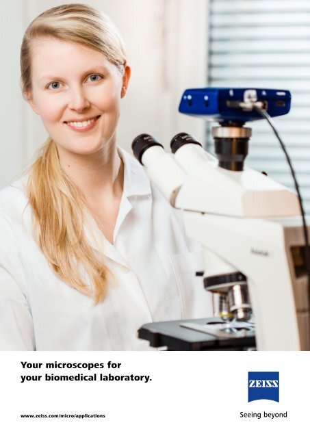
Your microscopes for<br />
your biomedical laboratory.<br />
www.zeiss.com/micro/applications
Your microscopes for increased efficiency in the lab.<br />
Enjoy the convenience for your daily checks, every day.<br />
Choose a sturdy microscope that is easy to use and has<br />
a long life. Make the best of your tuition and work with<br />
<strong>ZEISS</strong> microscope systems.<br />
Use a microscope to investigate cells<br />
and body fluids in your laboratory.<br />
You prepare, manipulate, or document<br />
human, plant, or animal organisms,<br />
often for several hours at a time.<br />
You assess the quantity, type, and<br />
characteristics of blood cells.<br />
You need convenient and efficient<br />
<strong>solutions</strong>. You need to easily operate<br />
your microscope and expect excellent<br />
optical performance. Does your<br />
microscope need to fit into a restricted<br />
space? Enjoy the convenience of <strong>ZEISS</strong><br />
laboratory microscopes for your daily<br />
checks, every day.<br />
These ergonomically designed<br />
microscopes are so flexible that<br />
they adapt to you and your working<br />
procedures. They speed up your daily<br />
routines. And they have an outstanding<br />
price–performance ratio.<br />
2
3
Select your system according to your requirements.<br />
Whether you use your microscope from time to<br />
time or for your daily laboratory investigations,<br />
your experience and knowledge grow from day<br />
to day. Of course, your microscope has always<br />
to perform reliably and should be easy to use.<br />
<strong>ZEISS</strong> microscopes have been optimized for<br />
use in your biomedical laboratory. The systems<br />
make it easier for you to efficiently apply your<br />
knowledge and methods on a daily basis.<br />
4
Stereo Microscopes and Zoom Microscopes<br />
<strong>ZEISS</strong> Stemi 305<br />
<strong>ZEISS</strong> Stemi 508<br />
<strong>ZEISS</strong> SteREO Discovery.V8<br />
<strong>ZEISS</strong> Axio Zoom.V16<br />
Page 6<br />
Upright Microscopes<br />
<strong>ZEISS</strong> Primostar 3<br />
<strong>ZEISS</strong> Primostar 3 iLED<br />
<strong>ZEISS</strong> Axiolab 5<br />
<strong>ZEISS</strong> Axioscope 5<br />
<strong>ZEISS</strong> Axio Imager<br />
Page 16<br />
Inverted Microscopes<br />
<strong>ZEISS</strong> Primovert<br />
<strong>ZEISS</strong> Axiovert 5<br />
<strong>ZEISS</strong> Axiovert 5 digital<br />
<strong>ZEISS</strong> Axio Observer<br />
Page 28<br />
Digital Documentation<br />
<strong>ZEISS</strong> Labscope<br />
<strong>ZEISS</strong> Z<strong>EN</strong> core<br />
<strong>ZEISS</strong> Axiocam<br />
<strong>ZEISS</strong> Mutlidiscussion<br />
Page 38<br />
5
Stereo Microscopes and Zoom Microscopes<br />
Brilliant 3D impressions with good depth of field.<br />
6 Stereo Microscopes and Zoom Microscopes
With these microscopes, you can observe your large or living samples nondestructively and<br />
without needing complex preparation. Zooming smoothly, you can adjust the magnification to<br />
your object and analyze its morphology. In the lower overview magnification, you can screen and<br />
sort your samples. Then, with higher magnification, you can effortlessly analyze details<br />
and prepare and manipulate the samples thanks to a large working distance that enables good<br />
access to them.<br />
Stereo Microscopes and Zoom Microscopes 7
<strong>ZEISS</strong> Stemi 305<br />
Compact size, big impact: your stereo microscope with<br />
integrated illumination and documentation.<br />
Configured to Your Requirements<br />
Stemi 305 is your compact Greenough stereo microscope with<br />
5:1 zoom. Equally at home in the biology classroom, research<br />
lab or on the industrial shop floor. Observe your samples<br />
as they are: three dimensional and crisp in contrast with no<br />
preparation required. Then share your images, whenever you<br />
want.<br />
Profit from an easy-to-use microscope, where everything is<br />
integrated: long-life LED illumination, reflected and<br />
transmitted light and documentation. Stemi 305 makes<br />
documentation easy and affordable. Simply snap your images<br />
with the integrated 1.2 Megapixel Wi-Fi camera and share<br />
them using Labscope, the iPad imaging app. Or opt for<br />
the conventional phototube to have access to all Axiocam<br />
cameras and free Z<strong>EN</strong> lite imaging software.<br />
Microscopes<br />
Stemi 305<br />
Stemi 305 trino with phototube (fixed division 50/50)<br />
Stemi 305 cam with integrated camera<br />
Stands<br />
Stand K, stand K MAT, stand K EDU, stand K LAB,<br />
Boom stands: stand A, stand U with tilting arm<br />
Illumination Techniques<br />
Reflected light, transmitted light and variable mixed light<br />
Brightfield, darkfield and oblique light, polarization<br />
Illumination<br />
Reflected light: spot, double spot, ring light, near vertical,<br />
polarization<br />
Transmitted light: homogeneous brightfield, darkfield, oblique<br />
light with relief contrast, polarization<br />
8 Stereo Microscopes and Zoom Microscopes<br />
Accessories<br />
Eyepieces and interchangeable front optics, eyepiece reticles,<br />
fiberoptic cold-light sources with various light guides, stages,<br />
polarization accessories
50 μm<br />
Wing of Chrisopidae; transmitted light brightfield Royal fern, sori and sporangia; spot K LED, oblique light, zoom 2.0×<br />
Simpler. More Intelligent. More Integrated.<br />
Created for Your Applications<br />
• Stemi 305 integrates everything you need. This compact<br />
Greenough stereo microscope comes without additional<br />
boxes and cables.<br />
• With the microscope camera already on board, you‘re<br />
prepared to save your results, share your images and<br />
collaborate on projects with friends, colleagues and<br />
classmates.<br />
• An LED illumination is already integrated in stands K EDU/<br />
LAB / MAT and provides reflected, oblique and transmitted<br />
light. Easily select and mix the integrated LED illuminations<br />
such as vertical and oblique reflected light, so as<br />
transmitted light.<br />
• Stemi 305 comes with two options for documentation.<br />
Choose the conventional phototube and have access to all<br />
Axiocam microscope cameras.<br />
• With the iPad imaging app Labscope you create your own<br />
digital classroom and share your images.<br />
• Stemi 305 microscope sets for education, lab and industry<br />
ensure optimized object illumination for your application.<br />
• In your practical botanical work, you investigate the<br />
morphology of plants’ organs. Your zoological studies deal<br />
with worms, snails, spiders, frogs, crabs, eggs, and larvae.<br />
• As a fungus expert, you investigate the macroscopic<br />
characteristics of the fruiting bodies of large fungi to<br />
differentiate between edible mushrooms and inedible lookalikes.<br />
The Stemi 305 large working distance allows you to<br />
examine whole mushrooms without the need for extensive<br />
preparation.<br />
• Are you a veterinarian who carries out investigations<br />
and does surgery? Then you will particularly appreciate<br />
the shadow-free, homogeneous illumination provided<br />
by Stemi 305 as well as the flexible alignment of the<br />
microscope with stand U with tilting arm.<br />
Stereo Microscopes and Zoom Microscopes 9
<strong>ZEISS</strong> Stemi 508<br />
Your apochromatic stereo microscope with 8:1 zoom for<br />
excellent image contrast and color accuracy.<br />
Configured to Your Requirements<br />
Stemi 508 is compact, reliable and equipped with optics and<br />
mechanics designed for heavy workloads. With the large<br />
36 mm object field you always keep the overview of your<br />
sample. The 8:1 zoom then allows to bring details up to<br />
50× magnification. You even have larger samples? Add<br />
interchangeable optics and observe an area of up to 122 mm,<br />
making Stemi 508 a top perin its class. Stemi 508 offers<br />
better ergonomics than any other Greenough-type stereo<br />
microscope: The low viewing angle of 35° lets you<br />
keep a relaxed posture even after hours of work.<br />
With Stemi 508 you observe and document your samples<br />
exactly as they are: rich in detail, sharp in focus and free<br />
from distortion or color fringes. Stemi 508 is your robust<br />
all-rounder for everyday lab work and industrial inspections:<br />
accurate, ergonomic – and always easy to use.<br />
Microscopes<br />
Stemi 508<br />
Stemi 508 doc with phototube and (100/0 switchover)<br />
Stands<br />
Stand K, stand K MAT, stand K EDU, stand K LAB, stand N<br />
Boom stands: stand A, SDA and stand U with tilting arm<br />
Illumination Techniques<br />
Reflected light, transmitted light and variable mixed light<br />
Brightfield, darkfield and oblique light, polarization<br />
Illumination<br />
Reflected light: light guides for spot, ring, line, vertical,<br />
diffuser, and area illumination, direct LED spots and segment<br />
ring lights<br />
Transmitted light: brightfield, darkfield, oblique light with<br />
relief contrast and polarization option<br />
10 Stereo Microscopes and Zoom Microscopes<br />
Accessories<br />
Interchangeable eyepieces and front optics, eyepiece reticles,<br />
camera adapter, cold-light sources with various light<br />
guides, gliding stage, rotating stage, ball-and-socket stage,<br />
polarization accessories
0,2 mm<br />
Powdery mildew on Norway maple, cleistothecia,<br />
Spot K LED, oblique reflected light, zoom 2.0×<br />
Tick, segmentable ring light K LED, half circle mode, zoom 1.0×<br />
Simpler. More Intelligent. More Integrated.<br />
Created for Your Applications<br />
• Thanks to their excellent optics, Stemi 508 stereo<br />
microscopes provide a crisp and highly resolved<br />
threedimensional image, sharp in focus and free of<br />
distortions or color fringes.<br />
• Enjoy the 8:1 zoom range and observe even minute<br />
structures. Zoom in on details, either continuously or<br />
reproducibly by adding click stops. Due to mechanical<br />
corrected zoom curves and precize zoom mechanics, the<br />
image stays sharp in each zoom position.<br />
• The large field of view lets you overview an object area<br />
larger than 35 mm in diameter. The 0.3× supplementary<br />
lens even expands this to 123 mm.<br />
• Stemi 508 doc always comes with camera adapter 0.5× to<br />
connect Axiocam microscope cameras.<br />
• Configure exactly the stereo microscope you require –<br />
select from stands, mounting brackets and stages. A large<br />
range of fiberoptic or direct LED accessories allow various<br />
illumination contrasts in reflected and transmitted light,<br />
such as brightfield, darkfield, oblique light and polarization.<br />
• You work in developmental biology with model organisms<br />
such as Drosophila, C. Elegans, or Xenopus. You assess,<br />
select, and prepare eggs, larvae, and embryos using<br />
micromanipulators.<br />
• You are an entomologist who identifies insects, sometimes<br />
in the field – for example to map biotopes.<br />
• You look for and classify horse or cattle embryos for<br />
subsequent transfer or for deep freezing for breeding<br />
purposes. Then you need high-contrast oblique transmitted<br />
light.<br />
• Do you study, compare, and document plants from your<br />
herbarium? Then, for your larger samples, you will need a<br />
boom stand, a large working distance, and a maximum field<br />
of view.<br />
• You look for and identify macroparasites such as ticks,<br />
fleas, and lice, as well as their eggs and larvae.<br />
Stereo Microscopes and Zoom Microscopes 11
<strong>ZEISS</strong> SteREO Discovery.V8<br />
Acquire brilliant, high-contrast, three-dimensional images.<br />
Configured to Your Requirements<br />
SteREO Discovery.V8 is equipped with open interfaces and<br />
is completely integrated into the <strong>ZEISS</strong> system. Its modular<br />
design and extensive accessories offer you a variety of options<br />
to set up your workplace to your exact requirements. You can<br />
configure your microscope as a manual microscope for the<br />
preparation of specimens, as a powerful tool for fluorescent<br />
screening with easy-to-use documentation, or as a largely<br />
motorized system with ergonomic operation and imaging<br />
options.<br />
The impressive stereoscopic image helps you to better<br />
observe, understand, and manipulate your specimens. You<br />
get a high-resolution, high-contrast, and apochromatically<br />
corrected microscopic image – that has sharp edges over the<br />
entire field of view and is always in focus when zooming.<br />
With its 8× zoom, you can quickly change from the overview<br />
down to the magnified detail. Add in click stops to the<br />
continuous zoom and you can easily reproduce ten discrete<br />
levels of magnification so that you can correctly scale your<br />
images.<br />
Microscopes<br />
SteREO Discovery.V8 (manual)<br />
Illumination Techniques<br />
Brightfield, darkfield, oblique light, polarization, fluorescence<br />
Illumination<br />
Reflected light: fiber-optic cold-light sources with spot, ring,<br />
line, vertical, diffuser, area, and coaxial illumination, LED ring<br />
lights with a segment function<br />
Transmitted light: fiber-optic setup 450 with sliding mirror,<br />
low-profile LED setup 300<br />
Accessories<br />
Interchangeable lenses, observation and intermediate tubes,<br />
additional viewer attachments, illumination, manual<br />
and motor-driven stands, cameras, software modules to<br />
document images and for image processing<br />
12 Stereo Microscopes and Zoom Microscopes
Zebra fish embryos, four hours after fertilization, obliquely illuminated in<br />
transmitted light brightfield, magnification: 25× (as seen in the eyepiece)<br />
Housefly mouthparts, obliquely illuminated in transmitted light darkfield,<br />
magnification: 80×<br />
Simpler. More Intelligent. More Integrated.<br />
Created for Your Applications<br />
• The intermediate LED tubes for fluorescence have been<br />
designed for screening tasks. They are high performance,<br />
robust, and easy to use. For this, they combine Achromat S<br />
lenses with high transmission.<br />
• With the PlanApo S objective lenses, you get a level image<br />
with sharp edges and no distortion or color fringing.<br />
• The 300 and 450 stands ensure vibration-free 3D viewing –<br />
even at high magnification.<br />
• Choose between the variably adjustable fiber-optic<br />
transmitted light 450 unit and the especially low-profile<br />
300 LED unit.<br />
Both units offer brightfield, darkfield, oblique-light, and<br />
polarization contrast.<br />
• <strong>ZEISS</strong> cold-light sources provide intense light that is free<br />
from infrared to prevent damage to your sample. Longlife<br />
LEDs make lamp changes a thing of the past. A wide<br />
spectrum of light guides guarantees that your specimens’<br />
structures are optimally emphasized.<br />
• In macroscope mode, you observe your specimen vertically<br />
through the right-hand stereo channel. You can produce<br />
z-stacks that are free from parallax errors and with<br />
increased depth of field.<br />
• You work in embryology and prepare model organisms for<br />
more extensive imaging using laser scanning microscopes.<br />
Then the 5 – 45° ergotubes ensure that your working<br />
posture is ergonomic.<br />
• You can document the embryonic growth of your zebra fish<br />
with the time-lapse module in the Z<strong>EN</strong> imaging software.<br />
• You assess the health of plants or seeds, or you identify<br />
pathogens and record their incidence. When investigating<br />
whole plants, you will benefit for the large focusing range<br />
and the large sample space.<br />
• In their biology classes, your students can draw plants and<br />
animals using drawing-tube attachment S. You can teach<br />
the preparation of samples or monitor it in 3D with the<br />
additional viewer attachment S.<br />
• Do you carry out IVF or ICSI treatments in a fertility clinic?<br />
Then with SteREO Discovery.V8, you can isolate the eggs<br />
before fertilization and then later assess the growing embryos.<br />
• In the forensic department, with the plan apochromatic<br />
lens, you can compare fibers and hairs with no color tints.<br />
• Your task is restoration of precious museum artefacts? SteREO<br />
Discovery.V8 is an essential tool in restoration of museums<br />
collections to preserve Cultural Heritage for future generations.<br />
Stereo Microscopes and Zoom Microscopes 13
<strong>ZEISS</strong> Axio Zoom.V16<br />
Your zoom microscope for high resolution in large fields.<br />
Configured to Your Requirements<br />
Axio Zoom.V16 offers you a succesful combination of a large<br />
field of view, zoom, and working distance as in a stereo<br />
microscope together with the high resolution of a light<br />
microscope.<br />
With the Axio Zoom.V16 zoom microscope, thanks to its<br />
double-sized basic aperture compared to powerful CMO<br />
stereo microscopes, you benefit from a resolution that is<br />
2.5× higher, as well as fluorescence that is 10× brighter in<br />
comparable fields of view. As needed, you can quickly and<br />
easily switch in the stereoscopic image.<br />
Microscopes<br />
Axio Zoom.V16 (manual focus)<br />
Axio Zoom.V16 (motor-driven focus)<br />
Illumination Techniques<br />
Brightfield, darkfield, relief contrast with reflected,<br />
transmitted,<br />
and mixed light, polarization, fluorescence<br />
Illumination<br />
Reflected light: fiber-optic cold-light sources with spot, ring,<br />
line, vertical, diffuser, area, and coaxial illumination with<br />
switchable relief illumination, LED ring lights with a segment<br />
function<br />
Transmitted light: fiber-optic setup 450 with sliding mirror,<br />
low-profile LED setup 300<br />
14 Stereo Microscopes and Zoom Microscopes<br />
Accessories<br />
Interchangeable lenses (objective lenses, eyepieces),<br />
observation and intermediate tubes, manual and motordriven<br />
stands, manual and motor-driven stages, cameras<br />
and software modules to document images and for image<br />
processing
Tick (Ixodida) from below, objective lens PlanApo Z 1×/0.25 FWD 60 mm,<br />
autofluorescence, EDF<br />
Fruit fly larva (Drosophila), objective lens PlanNeoFluar Z 2.3×/0.57 FWD<br />
10 mm, multiple fluorescence<br />
Simpler. More Intelligent. More Integrated.<br />
Created for Your Applications<br />
• With a 16× zoom and a basic aperture of 0.25 (with a 1×<br />
objective lens), with Axio Zoom.V16 you will benefit from<br />
what is currently the most powerful available stereo or<br />
zoom microscope.<br />
• Axio Zoom.V16 offers you a high resolution of 0.3 μm<br />
in a large field of 1.6 mm.<br />
• Its patented eZoom allows you to choose between<br />
optimized zoom modes for viewing through the eyepiece,<br />
for fluorescent applications, or for the documentation of<br />
images.<br />
• With eZoom, you get reproducible magnifications with<br />
accuracy of over 99%.<br />
• Take advantage of the intelligence of the 450 mot<br />
transmitted-light module. When zooming in Best Mode, you<br />
get an image that is automatically optimized for contrast<br />
and brightness, while taking account of the microscope’s<br />
current state.<br />
• Use Axio Zoom.V16 when you need more resolution in<br />
larger fields.<br />
• You benefit from the significantly higher aperture if, with<br />
image processing software, you manage to add value to<br />
the information in the image compared to the classical view<br />
through the eyepiece.<br />
• Axio Zoom.V16 offers you high optical performance<br />
together with large working distances, which are of<br />
particular importance when manipulating your specimen.<br />
As well as in museums collection restoration.<br />
• Do you need to investigate model organisms and zoom<br />
from a large overview down into the smallest details of<br />
organs, tissues, and individual cells? Then Axio Zoom.V16 is<br />
your best choice.<br />
Stereo Microscopes and Zoom Microscopes 15
Upright Microscopes<br />
Use all contrast methods with reliable, compact microscopes.<br />
16 Upright Microscopes
You can detect even the smallest details of your specimen with upright <strong>ZEISS</strong> microscopes thanks<br />
to their numerous contrasting techniques. Especially in clinical labs, you can rely on proven,<br />
reliable technology when assessing complete blood counts, smear tests, or sections. Ranging<br />
from robust educational microscopes and ergonomically designed laboratory units up to the most<br />
demanding platforms, upright microscopes from <strong>ZEISS</strong> enrich your daily work.<br />
Upright Microscopes 17
<strong>ZEISS</strong> Primostar 3<br />
Your robust yet compact microscope for digital teaching and<br />
routine lab work<br />
Configured to Your Requirements<br />
In the classroom or in the routine lab, you need reliable<br />
microscopes that can take a lot of wear and tear. After all,<br />
you and your colleagues or students will be working long<br />
hours, often in cramped spaces. You need microscopes that<br />
will pay back your investment with smooth operation – dayto-day<br />
and year in, year out. Primostar 3 packs all of that<br />
into its sturdy metal frame. Yet this robust light microscope is<br />
also designed for maximum ease of use. For both productive<br />
learning and efficient lab work, students and staff alike will be<br />
free from the very beginning to focus on the essentials.<br />
Primostar 3 is your reliable partner in microscopy – today and<br />
in years to come.<br />
Microscopes<br />
Primostar 3 Fixed-Köhler<br />
Primostar 3 Full-Köhler<br />
Primostar 3 HD<br />
Contrasting Techniques<br />
Brightfield, darkfield, phase contrast, simple polarization<br />
contrast, fluorescence (optional)<br />
Illumination<br />
Transmitted light: HAL 30 (halogen), LED, illumination mirror<br />
Reflected light: LED fluorescent reflected light<br />
Accessories<br />
Objective lenses (HF, Ph, D = 0, 100× dry), Ph-sliders, set<br />
of filters (blue, green, yellow), reflected light fluorescence<br />
attachments, sample slide kit, transmitted light mirror,<br />
eyepiece pointer, crossline micrometer, simple polarization<br />
accessory, transport and storage cases<br />
18 Upright Microscopes
Corylus avallana in brightfield, magnification: 40× Rabbit tongue, taste buds in phase contrast, magnification: 40×<br />
Simpler. More Intelligent. More Integrated.<br />
Created for Your Applications<br />
• Choose the best configuration for your tasks at hand from a<br />
number of pre-defined packages. A selection of objectives<br />
is already included.<br />
• Plug in and start focusing on your work with the Fixed<br />
Köhler Primostar 3. Or train and learn the Köhler set-up<br />
with Primostar 3 Full-Köhler packages.<br />
• Transmitted light brightfield, darkfield, simple polarization,<br />
phase contrast and fluorescence contrast are contrast<br />
techniques of choice. Just choose what your application<br />
asked for.<br />
• Be ready to teach digital natives in a digital classroom with<br />
Primostar 3 HD.<br />
• Benefit from the integrated 8.3 MPx HD with numerous<br />
interfaces for your flexible setup in your training courses<br />
environment (USB 3.0, HDMI, Ethernet, Wi-Fi compatible).<br />
• Use free of charge Labscope App on your Windows PC or<br />
iOS device. Create images, movies, share your results with<br />
your peers and get ready for your first annotations.<br />
• You examine stained tissue sections using brightfield or<br />
fluorescent contrast. You look at unstained specimens with<br />
phase contrast. You analyze extremely fine structures such<br />
as diatoms using darkfield.<br />
• As a botanist, you examine cross sections of plant stems.<br />
• You examine tissue sections and blood smears from<br />
anatomy, pathology, hematology, and zoology to record<br />
symptoms.<br />
• You examine cultivated plants for phytopathogenic agents<br />
or pests, or you track the development of illnesses and the<br />
course of diseases.<br />
• You investigate the morphology of bacteria cells such as<br />
Bacillus subtilis, Staphylococcus epidermidis, Micrococcus<br />
luteus and Escherichia coli.<br />
Upright Microscopes 19
<strong>ZEISS</strong> Primostar 3 iLED<br />
Your Fluorescence Microscope for Sputum Examination<br />
Configured to Your Requirements<br />
<strong>ZEISS</strong> Primostar 3 iLED is your microscope to visualize small<br />
structures down to 0.2 – 5 μm. So you can even observe<br />
objects such as the rod-shaped Mycobacterium tuberculosis.<br />
The gold standard for sputum smear microscopy is Ziehl-<br />
Neelsen staining and brightfield light microscopy. According<br />
to WHO*, LED fluorescence microscopy is even more sensitive<br />
and less time-consuming, making it a real alternative to the<br />
conventional standard.<br />
Microscopes<br />
Primostar 3 iLED (fixed Köhler) with reflected fluorescent<br />
illumination<br />
Contrasting Techniques<br />
Brightfield, LED fluorescence<br />
Illumination<br />
• Transmitted light<br />
• LED Reflected light<br />
• Fluorescence module with 455 nm LED<br />
Accessories<br />
Objective D = 0, eyecups; optional: transport box,<br />
power bank, illumination mirror, Axiocam microscope<br />
cameras<br />
20 Upright Microscopes
Mycobacterium tuberculosis, Ziehl-Neelsen stain: the mycobacteria stained<br />
purple are hard to see in a microscopic image<br />
Representative image of mycobacterium tuberculosis visualized in<br />
fluorescence with auramine O. The mycobacteria are clearly visible as<br />
greenish yellow particles in front of a dark background<br />
Simpler. More Intelligent. More Integrated.<br />
Created for Your Applications<br />
• It is easy to change between fluorescence and brightfield.<br />
You get images with outstanding contrast, especially when<br />
you work with samples colored with auramine-rhodamine<br />
stain.<br />
• LED fluorescence is safe, energy-efficient, and easy to use.<br />
You neither have to wait for lamps to heat up or cool<br />
down, nor do you have to replace or adjust them.<br />
• In areas without a power supply, you can use a power bank.<br />
• With the ergonomic eyecups, you can get precise results<br />
even without a darkroom.<br />
• If you are a customer from the public health services of<br />
those countries most heavily affected by tuberculosis, you<br />
can get Primostar 3 iLED at an especially low price.<br />
• You examine stained tissue sections using brightfield or<br />
fluorescent contrast. You look at unstained specimens with<br />
phase contrast. You analyze extremely fine structures such<br />
as diatoms using optional darkfield.<br />
• As a botanist, you examine cross sections of plant stems.<br />
• You examine tissue sections and blood smears from<br />
anatomy, pathology, hematology, and zoology to record<br />
symptoms.<br />
• You examine cultivated plants for phytopathogenic agents<br />
or pests, or you track the development of illnesses and the<br />
course of diseases.<br />
• You investigate the morphology of bacteria cells such as<br />
Bacillus subtilis, Staphylococcus epidermidis, Micrococcus<br />
luteus and Escherichia coli.<br />
Upright Microscopes 21
<strong>ZEISS</strong> Axiolab 5<br />
Your smart microscope for more efficient routine lab work.<br />
Configured to Your Requirements<br />
Axiolab 5 is made for the routine work that goes on every day<br />
in your lab. Its compact and ergonomic design saves space<br />
and makes for easy handling.<br />
Microscopes<br />
Axiolab 5 (transmitted light)<br />
Axiolab 5 (transmitted light and reflected light fluorescence)<br />
Axiolab 5 is a real team player. Combine it with Axiocam 208<br />
color and take full advantage of the smart microscopy<br />
concept: you’ll be experiencing a completely new form of<br />
digital documentation. Just focus your sample and press a<br />
single button for crisp images in true color. The digital image<br />
will look exactly like you see it through the eyepieces, with all<br />
the details and subtle color differences clearly visible.<br />
Plus, Axiolab 5 automatically adds the correct scaling<br />
information to your images. You get all of this in a standalone<br />
operation, without needing a PC or any additional software.<br />
Save time, money and valuable lab space with Axiolab 5.<br />
Digital documentation has never been easier.<br />
Contrasting Techniques<br />
Brightfield, darkfield, phase contrast, simple polarization,<br />
LED fluorescence<br />
Illumination<br />
Transmitted light:<br />
• 35 W halogen illumination (optional)<br />
• 10 W LED illumination<br />
Reflected light:<br />
• Up to 3 fluorescence LEDs<br />
Accessories<br />
Axiocam 208 color and Axiocam 202 mono microscope<br />
cameras, stages for left and right hand operation, ergotubes,<br />
multidiscussion equipment<br />
22 Upright Microscopes
Blood smear, Giemsa staining, transmitted light brightfield, objective:<br />
Plan-Apochromat 63× / 1.4<br />
Rat retina, section, nuclear fast red, transmitted light brightfield, objective:<br />
Plan-Apochromat 20× / 0.8<br />
Simpler. More Intelligent. More Integrated.<br />
Created for Your Applications<br />
• Axiolab 5 offers you an easy handling, ergonomic user<br />
concept that’s adapted to your lab routine. You can control<br />
the microscope and its attached camera without even<br />
changing your grip.<br />
• To acquire an image, simply press the snap button<br />
right on the stand. Your smart microscope system<br />
then automatically adjusts the parameters for you and<br />
documents your sample precisely as you see it through the<br />
eyepieces – detail-rich and in true color. The correct scaling<br />
is always included automatically, even without a computer.<br />
• LED fluorescence is safe, energy-efficient, and easy to<br />
use. You neither have to wait for lamps to heat up or cool<br />
down, nor do you have to replace or adjust them.<br />
• Ergotubes and torque adaptable stage handle allow you to<br />
work in a comfortable position, even during extended use.<br />
• With the multidiscussion equipment, all co-observers can<br />
see the same image with the same orientation.<br />
• With Axiolab 5, it is particularly easy for you to count<br />
white blood cells in brightfield, as you can reach all of the<br />
essential controls with one hand.<br />
• In darkfield, you can recognize uncolored structures at a<br />
glance.<br />
• Using polarization contrast, you can detect birefringent<br />
crystals, for example when visualizing gout.<br />
• Using fluorescence contrast, you can examine heparinized<br />
blood for cytogenetic (chromosome analysis) and molecular<br />
cytogenetic investigations.<br />
• In the laboratory, you can analyze body fluids, tissues,<br />
and excretions. You can do hematological analyses<br />
on the cell morphology of blood and tissue cells and<br />
can do hemostasis analyses for bleeding tendency or<br />
thrombophilia.<br />
• In your IVF laboratory you use Axiolab 5 for enlarged<br />
visualization of sperm cells in brightfield or phase contrast.<br />
Upright Microscopes 23
<strong>ZEISS</strong> Axioscope 5<br />
Your smart microscope for biomedical routine and research.<br />
Configured to Your Requirements<br />
In the past, documenting samples with multiple fluorescent<br />
labels in your routine lab could be time consuming. To get<br />
best image quality, you needed to manually switch filters,<br />
adjust illumination intensities and exposure times and to snap<br />
each single channel image. For three different channels, this<br />
could sum up to 15 steps and clicks. With Smart Microscopy<br />
from <strong>ZEISS</strong>, this is a thing of the past.<br />
Your Axioscope 5 with Axiocam 202 mono and Colibri 3<br />
LED illumination take this workload from you. You don’t<br />
even need to move your hands from the microscope stand<br />
anymore. All you have to do is focus and press Snap – and<br />
you’re done! You can now concentrate on the essence of your<br />
job and let your Axioscope 5 work for you. You’ll work more<br />
efficiently, save time and produce high contrast images with<br />
best image quality. What’s more: this even works without any<br />
PC involved.<br />
Microscopes<br />
Axioscope 5, transmitted light, LED<br />
Axioscope 5, transmitted light, Hal 50<br />
Axioscope 5, fluorescence<br />
Contrasting Techniques<br />
Transmitted light: brightfield, darkfield, DIC, PlasDIC,<br />
simple polarization, phase contrast<br />
Reflected light: brightfield, darkfield, DIC, C-DIC,<br />
simple polarization, fluorescence<br />
Illumination<br />
Transmitted light:<br />
• LED 10W, Hal 50, Hal 100<br />
Reflected light, fluorescence:<br />
• Colibri 3, HXP 120, and other<br />
Accessories<br />
Axiocam 208 color and Axiocam 202 mono microscope<br />
cameras, Colibri 3, XY stages, ergotubes und multidiscussion<br />
equipment<br />
24 Upright Microscopes
100 µm<br />
Mouse kidney in fluorescence, cryosection, AF 488 – WGA, AF 568 Phalloidin,<br />
DAPI, objective: Plan-Apochromat 20× / 0.8<br />
Histological specimen, CDx immunohistological stain; Red: immunoreactive<br />
antigens in cytoplasm; Blue: nuclear counterstaining Ziehl-Neelsen-Färbung,<br />
objective: EC Plan-Neofluar 63× / 0.95 Korr.<br />
Simpler. More Intelligent. More Integrated.<br />
Created for Your Applications<br />
• Capture up to four fluorescence channels with just one<br />
click: the system automatically adjusts the exposure time,<br />
acquires the image, switches the channel and starts again.<br />
You get your overlayed multichannel fluorescence image<br />
including scale bar.<br />
• Colibri 3 provides up to four fluorescence LEDs which can<br />
be individually controlled, directly from the stand.<br />
• For transmitted light applications, the smart Axioscope 5<br />
system makes automatic adjustments for brightness and<br />
white balance to keep digital documentation easy.<br />
• Axioscope 5 uses its transmitted white light LED to provide<br />
powerful illumination with high color fidelity.<br />
• With Axioscope 5 you can use a sheer variety of contrasting<br />
techniques for your applications.<br />
• You can carry out histological or pathohistological analyses<br />
of tissue sections in brightfield contrast.<br />
• With polarization contrast, you can analyze foreign bodies<br />
and crystals in tissue and body fluids.<br />
• You can examine stained mucosal cells in hematology,<br />
urology, and gynecology using brightfield and fluorescence.<br />
• Perform multichannel fluorescence imaging of tissues and<br />
cells in your lab.<br />
Upright Microscopes 25
<strong>ZEISS</strong> Axio Imager 2<br />
All contrasting techniques on a single imaging platform.<br />
Configured to Your Requirements<br />
Axio Imager 2 supports your requirements – from brightfield<br />
observations and fluorescence light via polarization up to<br />
complex FISH applications. This system platform, with its<br />
modular architecture, is aimed at your growing needs.<br />
Application-specific components complement the solid<br />
fundamental characteristics of the Axio Imager 2 stand<br />
variants. See for yourself what the combination of<br />
outstanding optics, high resolution, and excellent contrast<br />
can do!<br />
Microscopes<br />
Axio Imager.A2 (manual)<br />
Axio Imager.A2 LED (manual, LED fixed Köhler illumination)<br />
Axio Imager.D2 (partially motor-driven)<br />
Axio Imager.M2p (pathology system, partially motor-driven)<br />
Contrasting Techniques<br />
Transmitted light: brightfield, darkfield, DIC, polarization,<br />
phase contrast<br />
Reflected light: brightfield, darkfield, DIC, C-DIC, fluorescence<br />
Illumination<br />
Transmitted light: DL 12 V 100 W HAL, 12 V LED<br />
Reflected light: 12 V 100 W HAL, 12 V 100 W HBO, 12 V LED,<br />
75 W XBO, VisiLED, microLED, Colibri 5 / 7<br />
Accessories<br />
LEDs with push-and-click modules, manual stages for leftand<br />
right hand operation, encoded and motorized stages,<br />
sample holders, binocular tubes with various viewing angles,<br />
camera tubes, multi-discussion equipment<br />
26 Upright Microscopes
HeLa cells, mitotic phase; red: Alexa Fluor 594-DM1-alpha, green: Alexa<br />
Fluor 488-Mad2, blue: DAPI, objective lens: EC Plan-NEOFLUAR 100×/1.3 oil,<br />
Sample: courtesy of H.Y. Li and Y. Xheng, department of embryology at<br />
HHMI and CIW, Maryland, USA<br />
Histological section; red: MPOX2, blue: nuclear counterstaining,<br />
objective lens: EC Epiplan-NEOFLUAR 10×/0.3, Sample: courtesy of<br />
A. Schmitt-Gräff, pathology department, Freiburg University, Germany<br />
Simpler. More Intelligent. More Integrated.<br />
Created for Your Applications<br />
• Axio Imager 2 impresses with its outstanding optics,<br />
perfect contrast and illumination.<br />
• It evenly illuminates your specimens.<br />
• Your Axio Imager 2 is equipped with a light manager for<br />
transmitted and reflected light. You benefit from a constant<br />
light impression at all magnifications and for all contrasting<br />
techniques.<br />
• The stands for Axio Imager 2 family are coded and all<br />
details of the image acquisition, such as objective lens and<br />
magnification, are saved together with the image.<br />
• Axio Imager.M2p is perfectly tailored for your requirements<br />
in the pathology department. Thanks to the encoded<br />
nosepiece turret and convenient motorization such as<br />
automated parfocal correction, you can work efficiently<br />
with a high specimen rate.<br />
• The motorization of Axio Imager 2 allows an ergonomical<br />
workflow and speeds up your work.<br />
• Axio Imager.A2 with LED illumination in connection with<br />
Achroplan or EC Plan-NEOFLUAR objective lenses is your<br />
ideal basic equipment for histology.<br />
• Axio Imager 2 with polarization contrast is indispensable in<br />
showing debris in tissue or in diagnosing Alzheimer’s<br />
disease, for example. Depending on the application, you<br />
can use fixed or rotating polarizers and analyzers, or even a<br />
lambda plate.<br />
• In histology and anatomy, you benefit from excellent<br />
resolution, convincing colors in details and overviews,<br />
and the ability to quickly and precisely relocate important<br />
positions in the specimen. The EC Plan-NEOFLUAR and<br />
Plan-APOCHROMAT objective lenses in connection with<br />
motorized stages are ideally tailored for this.<br />
• You can visualize parasites, bacteria, or clusters of viruses.<br />
• You identify extrinsical particles.<br />
Upright Microscopes 27
Inverted Microscopes<br />
Living cells in focus.<br />
28 Inverted Microscopes
With inverted microscopes, you can use the large sample space between the stage and the<br />
illumination for your cells in petri dishes, well plates, or culture flasks. You will have enough<br />
space for your roller bottles and for micromanipulation. And all that together with contrasting<br />
techniques such as brightfield, phase contrast and fluorescence, that you need in your laboratory.<br />
Your <strong>ZEISS</strong> microscope is compact and focuses on the essentials.<br />
Inverted Microscopes 29
<strong>ZEISS</strong> Primovert<br />
Examine and assess your living cells – quickly and easily.<br />
Configured to Your Requirements<br />
Now you can study the morphography of living cells and<br />
evaluate their development with this compact inverted<br />
microscope from <strong>ZEISS</strong>. Primovert is perfectly suited to your<br />
cell culture laboratory.<br />
It enables fast, efficient investigations of both unstained<br />
cells in phase contrast and GFP-labeled cells in fluorescence<br />
contrast. It fits straight into your laminar flow cabinet to work<br />
directly in a sterile environment. And it brings you a welcome<br />
degree of flexibility, too, with its integrated camera and<br />
the Labscope imaging app for iPad: observe your cells from<br />
outside the sterile working space and evaluate them with<br />
colleagues.<br />
Microscopes<br />
Primovert<br />
Primovert photo<br />
Primovert HDcam<br />
Primovert iLED<br />
Contrasting Techniques<br />
Brightfield, phase contrast, fluorescence<br />
Illumination<br />
HAL 30, LED<br />
Accessories<br />
Stage insert (glass or metal), holding frame for petri dishes,<br />
object guides, LD condensers, phase contrast slides,<br />
Plan-ACHROMAT and LD Plan-ACHROMAT objective lenses<br />
30 Inverted Microscopes
HeLa cells, phase contrast,<br />
objective: LD Plan-ACHROMAT 20×/0.3 Ph2<br />
U2OS cells expressing GFP, fluorescence contrast,<br />
objective: Plan-ACHROMAT, 20×/0.4<br />
Simpler. More Intelligent. More Integrated.<br />
Created for Your Applications<br />
• Switch from phase contrast to fluorescence contrast to<br />
assess both undyed and GFP-labeled cells.<br />
• The inverted microscope is compact and fits directly in your<br />
Laminar Flow Box – you work directly in the sterile<br />
environment.<br />
• Your Primovert is immediately ready for use. You reactivate<br />
the microscope in stand-by mode directly at the stage.<br />
Primovert switches in walk-away mode automatically after<br />
15 minutes off. This saves energy and increases the life of<br />
the light source.<br />
• Primovert HDcam integrates a camera. Use your iPad and<br />
the free imaging app Labscope and discuss the image<br />
together in the team.<br />
• Snap microscope images, annotate and create reports, and<br />
share them easily wirelessly with other.<br />
• With phase contrast, you get high-contrast images of<br />
unstained cells. You can analyze the growth, morphology,<br />
and condition of living cells at a glance.<br />
• Research the structure of plant cells and tissues,<br />
reproduction, growth, metabolic processes, and pathogens.<br />
• You can do sterility tests.<br />
• Examine cells before preparing protein, DNA, or RNA<br />
samples.<br />
• Differentiate between types of cells and characterize<br />
cell lines.<br />
Inverted Microscopes 31
<strong>ZEISS</strong> Axiovert 5<br />
Your smart microscope for cell culture and research.<br />
Configured to Your Requirements<br />
Axiovert 5 brings smart microscopy into your cell culture<br />
lab. All you need to do is focus on your samples and<br />
workflow. Then simply push Snap to get crisp images for<br />
documentation.<br />
Use all standard contrasting techniques in transmitted<br />
light and combine them with multichannel fluorescence to<br />
investigate your cell or tissue cultures. And when space is<br />
tight, you can even use this smart microscope as a standalone<br />
and save your images on USB. No extra computer or software<br />
needed.<br />
Microscopes<br />
Axiovert 5 TL (transmitted light)<br />
Axiovert 5 TL SCB (transmitted light, Smart Control Box)<br />
Axiovert 5 TL FL SCB<br />
(reflected fluorescent light, Smart Control Box)<br />
Contrasting Techniques<br />
Brightfield, phase contrast, PlasDIC, iHMC, DIC, fluorescence<br />
Illumination<br />
Transmitted light: White LED 10 W<br />
Reflected light: Colibri 3, HXP 120, Colibri 5 / 7, Xylis and more<br />
Accessories<br />
• Light shield, Mounting frame K thermo plate,<br />
iHMC module, polarizer slider TL 90° rotatable<br />
• Filter sets, aqua stop, condensor and more<br />
32 Inverted Microscopes
U2OS cells in transmitted light, phase contrast.<br />
Objective: LD A-Plan 40× / 0.55 Ph 1<br />
U2OS cells in multichannel fluorescence, 20×, Nucleus – NucRed,<br />
Mitochondria – MitoTracker green<br />
Simpler. More Intelligent. More Integrated.<br />
Created for Your Applications<br />
• Various contrasting techniques — DIC, iHMC and phase<br />
contrast as well as multichannel fluorescence — open up a<br />
huge diversity of applications.<br />
• Without modifying the stand, you can switch freely<br />
between iHMC, PlasDIC and DIC as you investigate your<br />
samples.<br />
• With Axiovert 5 your samples remain safe in gentle LED<br />
light. You profit from homogeneous illumination and<br />
freedom to align your sample.<br />
• Axiovert 5 delivers excellent results with fast time to image.<br />
Simply focus your sample and press a single button to save<br />
a crisp image of your cell or tissue culture.<br />
• For transmitted light and multichannel fluorescence images,<br />
this smart microscope automatically adjusts the settings<br />
and parameters for you.<br />
• If lab space is limited, just get rid of the PC and all that<br />
additional software: use Axiovert 5 in standalone mode and<br />
control the microscope via the OSD menu.<br />
• Check the vitality of your cell cultures.<br />
• Estimate the perfect timing for cell seeding.<br />
• Check the efficiency of transfections.<br />
• Perform ICSI, IMSI, and embryonic observations in<br />
an IVF clinic.<br />
Inverted Microscopes 33
<strong>ZEISS</strong> Axiovert 5 digital<br />
Your All-in-One Cell Imaging System.<br />
Configured to Your Requirements<br />
Artificial Intelligence (AI) is already helping us with our daily<br />
lives, from automated driving and home assistants to securing<br />
smartphones with facial recognition. Axiovert 5 digital brings<br />
AI into your cell lab to ease your daily work. It will make your<br />
processes more efficient and your results more reproducible.<br />
Stay relaxed, even when there is a lot going on around you.<br />
Just push a single button and your results will appear in real<br />
time.<br />
Microscopes<br />
Axiovert 5 digital (mono)<br />
Axiovert 5 digital (color)<br />
Contrasting Techniques<br />
Brightfield, phase contrast, fluorescence<br />
Illumination<br />
Transmitted light illumination with white LED 10 W<br />
4 solid state fluorescence LEDs<br />
Accessories<br />
• Stage inserts<br />
• Objectives, filter sets, FL-LEDs<br />
• Aqua Stop II<br />
34 Inverted Microscopes
100 µm 20 µm<br />
Cell counting and cell confluency at a glance with <strong>ZEISS</strong> Labscope BPAE cells in multichannel fluorescence, 40×<br />
Simpler. More Intelligent. More Integrated.<br />
Created for Your Applications<br />
• Experience the full advantages of an all-in-one microscope<br />
system. Don’t worry about settings or adjustments –<br />
they’re already done. All you have to do is turn on your<br />
system and start working.<br />
• Axiovert 5 digital uses artificial intelligence to optimally<br />
support daily workflows. Cell counting and cell confluency<br />
are automatically determined by readily available AI<br />
modules.<br />
• This all-in-one imaging system comes with an intuitive<br />
operating concept. One click on the snap button is<br />
sufficient to trigger image and video acquisition as well as<br />
analysis.<br />
• Observe labeled living cells in your cell laboratory.<br />
• Count cells to decide on cell proliferation and viability as<br />
well as the appropriate dilution values.<br />
• Measure the cell confluency to decide on cell proliferation,<br />
viability, and the perfect timing to start transfections or<br />
when to passage your cells.<br />
• Start today to improve the quality of your cell cultures<br />
and make your experiments more reproducible.<br />
Count cells automatically with AI.<br />
• Document your fluorescent cells in publication-ready<br />
quality.<br />
Inverted Microscopes 35
<strong>ZEISS</strong> Axio Observer<br />
Observe. manipulate. and analyze.<br />
Configured to Your Requirements<br />
You observe, analyze, and manipulate living cells. Then<br />
Axio Observer is your inverse microscope platform for<br />
maximum flexibility. Its open architecture can be costeffectively<br />
extended – from the base stand through to<br />
high-speed and laser-scanning microscopy or microdissection<br />
dimensions. There are not even any restrictions in adding<br />
external components to the system.<br />
Microscopes<br />
Axio Observer 3<br />
Axio Observer 5<br />
Contrasting Techniques<br />
Brightfield, phase contrast, PlasDIC, iHMC, DIC, fluorescence<br />
Illumination<br />
Transmitted light: halogen, LED<br />
Reflected light: HBO 50, HBO 100, HXP 120 V, Colibri 5 / 7<br />
Accessories<br />
Binocular tubes, binocular camera tubes, binocular ergotubes,<br />
stages, manual and motor-driven stages, condensers,<br />
objective lenses, cameras, software, incubation components<br />
36 Inverted Microscopes
Transgenic mouse embryos in various stages of development, PlasDIC,<br />
magnification: 40×, Sample: courtesy of Dr. Ropeter, Dragon-IVF, Dr. Michelmann,<br />
Gynecological Clinic Göttingen, and Ms. Buhtz, Göttingen University, Germany<br />
HeLa cells, multi-color fluorescence in combination with DIC.<br />
Blue (HOECHST 33342): cell nucleus, red (DsRed): cytoplasm.<br />
Courtesy of H. Wolff, GSF Neuherberg, Germany<br />
Simpler. More Intelligent. More Integrated.<br />
Created for Your Applications<br />
• The apochromatic fluorescence beam path ensures<br />
homogeneous intensity of fluorescence over the entire field<br />
of view. Colibri 5 / 7 light sources permit a fast LED change<br />
for fluorescent applications.<br />
• Combine Axio Observer with manipulators, and together<br />
with PlasDIC or iHMC, you will have the perfect platform<br />
for IVF and for your work with stem cells.<br />
• With the DIC contrasting technique, you can achieve the<br />
highest detail resolution and improved success rates – for<br />
example when assessing sperm.<br />
• Axio Observer combines all of the IVF contrast methods in<br />
a single microscope.<br />
• Observe and mark cells using vital stains.<br />
• You carry out series of experiments and need ocumentation<br />
and incubation.<br />
• Compare images from different fluorescence channels and<br />
require uncompromisingly brilliant images.<br />
• Carry out pronuclear injections working with transgenic<br />
animals.<br />
• You are responsible for ICSI, IMSI, and embryonic<br />
observations in an IVF clinic.<br />
Inverted Microscopes 37
Digital Documentation<br />
Network your microscopes and document your results.<br />
38 Connectivity and Documentation
Document exactly what you see. Fast, easily to access and with brilliant image quality.<br />
With the digital microscope cameras from <strong>ZEISS</strong>, you have the perfect tool for image acquisition<br />
and documenting your work. To display and edit your images, choose the imaging app Labscope.<br />
Using Labscope, you can connect several microscopes and digitize your classroom and easily<br />
take a look into your students’ work.<br />
Connectivity and Documentation 39
<strong>ZEISS</strong> Microscope Software for Your Routine <strong>Laboratory</strong><br />
Get it done. Easy.<br />
Decide for microscope software from <strong>ZEISS</strong> and simply<br />
get your work done. From super simple for basic image<br />
acquisition only, easy-to-use for standard imaging, up to<br />
customizable workflow <strong>solutions</strong> – choose the microscope<br />
software that fits exactly to your task: save time, reduce<br />
training expenses, and get your results faster.<br />
On-screen Display (OSD)<br />
Your choice for basic image acquisition<br />
Focus. Snap. Done.<br />
Your smart microscope camera Axiocam 208 color or<br />
Axiocam 202 mono facilitates image acquisition with your<br />
manual microscope greatly. The automatic functions require only<br />
minimal adjustments and – you don’t even need a computer.<br />
OSD is your perfect choice, if you:<br />
• Need image acquisition only<br />
• Have limited lab space and budget<br />
• Want an easy-to-use user interface which does not require<br />
any training<br />
• Don’t want to deal with IT topics such as operating system<br />
updates<br />
• Want to save your data on an USB-stick<br />
40 Connectivity and Documentation
<strong>ZEISS</strong> Labscope<br />
Your standard for routine imaging<br />
High quality images. Fast results. Fun to work with. With<br />
<strong>ZEISS</strong> Labscope you decide for an easy-to-use imaging<br />
software that fulfills all needs in your laboratory – from image<br />
acquisition, handy built-in measurement functions up to easy<br />
data sharing. And, you’re free to use it with your Windows PC<br />
but also with your tablet or mobile phone.<br />
<strong>ZEISS</strong> Z<strong>EN</strong> core<br />
For customized repetitive workflows<br />
Z<strong>EN</strong> core is your imaging software to handle applicationspecific<br />
routine workflows that can be individually defined.<br />
You can create and combine workbenches to create dedicated<br />
jobs that assist you through a defined flow of consecutive<br />
tasks. This ensures data repeatability and ease of use.<br />
Labscope is your preferred choice if you:<br />
• Want impressive ease of use without compromizing<br />
functionality<br />
• Need image acquisition, basic post-processing, annotation<br />
and measurement functions<br />
• Work with manual microscopes and fluorescence<br />
• Need access to multiple microscopes in a network<br />
• Want a simple way to share and display images<br />
• Occasionally need to scan whole slide images<br />
Decide for Z<strong>EN</strong> core if you:<br />
• Need to control a motorized <strong>ZEISS</strong> microscope for higher<br />
automation<br />
• Want to setup and use individually defined imaging<br />
workflows<br />
• Need a GxP compatible solution<br />
• Need to ensure data repeatability in a multi-user<br />
environment<br />
• Want to perform multi-modal imaging, e.g. light and<br />
electron microscope with the same sample<br />
Connectivity and Documentation 41
<strong>ZEISS</strong> Labscope<br />
Your imaging software for your daily laboratory work.<br />
Labscope is your choice to get fast results at the push of a button. From image acquisition, handy<br />
built-in measurement functions up to easy data sharing – with functions dedicated to your daily<br />
workflow, you can increase the efficiency of your biomedical lab. With Labscope you acquire<br />
brilliant images and get meaningful and reproducible results with ease.<br />
Acquisition settings<br />
Get in-app control of your hardware to adjust exposure, white balance, light<br />
intensity and more. Focus on the specimen all the time.<br />
Histogram<br />
The histogram panel is available for both live view and image view. The Min/<br />
Max and Best Fit functions optimize the display effect with just one click.<br />
Annotations and measurements<br />
Choose between 15 types of annotations and easy adjustments of color and<br />
size.<br />
Split view<br />
Compare your images in detail with the split view. Zoom in and zoom out,<br />
and even save the image of split view for documentation.<br />
Report<br />
Different kinds of report templates are prepared for you to quickly create<br />
reports from your microscopy work.<br />
File management<br />
Sort, search and share your images, videos and reports.<br />
Barcode scanning is also available when you want to set a file name or<br />
search a file.<br />
42 Connectivity and Documentation
Microscope list<br />
Get an overview of all connected microscopes. Manage and work on them<br />
easily.<br />
Live view and acquisition modes*<br />
High-quality live image and multiple acquisition modes make your<br />
microscopy work more flexible.<br />
Fast Panorama<br />
Acquire whole slide images with your manual microscope. Profit from a<br />
fast stitching speed and high image quality. Only avaliable for Windows<br />
Labscope.<br />
Multi-Channel<br />
Acquire fluorescence and transmitted light images in independent channels.<br />
The module supports adding false-color, comparing channels and reporting<br />
with displaying each channel.<br />
AI Cell confluency and AI Cell counting<br />
Analyze the number of cells and cell growth automatically with the help of<br />
artificial intelligence.<br />
* Availability of each acquisition mode is depending on cameras and microscopes.<br />
Connectivity and Documentation 43
<strong>ZEISS</strong> Z<strong>EN</strong> core<br />
Your microscope software for user-specific repetitive workflows<br />
in biomedical labs<br />
You decide what your microscope software looks like.<br />
With Z<strong>EN</strong> core you can adapt the user interface exactly to<br />
your application.<br />
• Configure your Z<strong>EN</strong> core only with necessary functions.<br />
• Define and combine workbenches easily to create<br />
workflows, so called jobs.<br />
• Jobs guide your operators through a defined flow of<br />
consecutive tasks ensuring data repeatability and ease<br />
of use.<br />
• Combine Job Mode with user management functions to<br />
assign specific imaging tasks to certain users and user<br />
groups.<br />
• Opt for the GxP or Multi Channel toolkit, when traceability<br />
or fluorescence imaging of biological samples is important<br />
to you<br />
44 Connectivity and Documentation
Connectivity and Documentation 45
<strong>ZEISS</strong> Axiocam – Microscope Cameras<br />
Accurate documentation is an important part of<br />
your daily analyses.<br />
Brilliant images reveal the state of your samples.<br />
Select the Axiocam microscope camera best suited to your application.<br />
CMOS<br />
CCD<br />
Microscope Camera Axiocam 105 color Axiocam 202 mono Axiocam 208 color Axiocam 305 mono<br />
Specification<br />
Effective pixels 5 megapixels 2 megapixels 8.3 megapixels 5.07 megapixels<br />
Number of pixels 2592 × 194 4 1920 × 1080 3840 × 2160 2464 × 2056<br />
Pixel size 2.2 µm 5.86 µm 1.85 μm 3.45 µm<br />
Sensor size 1/2.24" 1/1.2" 1/1.7" 2/3"<br />
Sensor diagonal 7 mm 13.4 mm 9.33 mm 11.1 mm<br />
Maximum frame rate<br />
at resolution<br />
17 fps at<br />
5 MP<br />
30 fps at 1080p 30 fps at 4K/1080p 67 fps at<br />
1920 × 1080<br />
PC interface USB 3.0 USB 3.0 Type C,<br />
Ethernet, HDMI<br />
Stand-alone mode • •<br />
USB 3.0 Type C,<br />
Ethernet, HDMI<br />
USB 3.0 SuperSpeed<br />
USB 2.0 optional<br />
Special features<br />
Recommended for<br />
Documentation and<br />
convenient image processing<br />
• • • • • • • • • • • • • •<br />
• • • • • Suitable<br />
• Partly suitable<br />
46 Connectivity and Documentation
Axiocam 305 color Axiocam 506 mono Axiocam 506 color<br />
5.07 megapixels 6 megapixels 6 megapixels<br />
2464 × 2056 2752 × 2208 2752 × 2208<br />
3.45 µm 4.54 μm 4.54 μm<br />
2/3" 1" 1"<br />
11.1 mm 16 mm 16 mm<br />
67 fps at<br />
1920 × 1080<br />
19 fps at<br />
2752 × 2208<br />
19 fps at<br />
2752 × 2208<br />
USB 3.0 SuperSpeed<br />
USB 2.0 optional<br />
USB 3.0 USB 3.0<br />
• • • • • • • • • • • •<br />
• • • • • Suitable<br />
• Partly suitable<br />
Connectivity and Documentation 47
Multidiscussion from <strong>ZEISS</strong><br />
Share your images with other viewers.<br />
Configured to Your Requirements<br />
You can use the classic multidiscussion system for training<br />
and consultation situations as well as in the medical world,<br />
for example when training students or when jointly assessing<br />
difficult specimens.<br />
With the multidiscussion unit from <strong>ZEISS</strong>, depending on the<br />
microscope and illumination used, up to 20 people can see<br />
the same image in the same orientation as the main viewer.<br />
This avoids irritation resulting from rotated or mirrored<br />
images. The main viewer and the additional viewers all profit<br />
from the homogeneously illuminated field of view.<br />
For specimens stained with different colors, you can smoothly<br />
adjust the intensity of the light pointer between white, green,<br />
and red. This helps with orientation.<br />
Microscopes<br />
Axiolab 5: at least up to ten additional viewers (LED)<br />
Axioscope 5: at least up to ten additional viewers (LED)<br />
or up to 20 additional viewers (HAL 100)<br />
Axio Imager.A2: up to 20 additional viewers (HAL 100)<br />
Accessories<br />
Central part, tube holder, tubes, eyepieces<br />
48 Connectivity and Documentation
Connectivity and Documentation 49
50
Service and support<br />
for your <strong>ZEISS</strong> microscope system.<br />
<strong>ZEISS</strong> moments are about passion. It is this passion with<br />
which we service and optimize your <strong>ZEISS</strong> microscope<br />
and keep it at the latest state of the art, so that your work<br />
can systematically lead to success.<br />
Experience service that lives up to its name.<br />
Your microscope system from <strong>ZEISS</strong> is one of your most important tools. For over<br />
160 years, the <strong>ZEISS</strong> brand and our experience have stood for reliable equipment<br />
with a long life in the field of microscopy.<br />
You can rely on us to ensure that you can always use your microscope’s full<br />
performance. With repair services and spare and replacement parts, our skilled<br />
<strong>ZEISS</strong> service team makes sure that your microscope is always ready for use.<br />
Our experts keep on working even after you have chosen <strong>ZEISS</strong>, with a wide range<br />
of additional services to ensure that you can experience those special moments –<br />
those special moments that inspire your work.<br />
Maintenance and optimization<br />
Your <strong>ZEISS</strong> Protect service agreement provides all-around security for your<br />
microscope system. There are no unexpected operating costs, and the availability<br />
of your system is increased. With preventative maintenance as a fundamental part<br />
of the service agreements, you benefit from optimized system performance.<br />
We’ll work with you to select the service package that best meets your needs, that<br />
corresponds to the equipment that you have, and that is tailored to the specific<br />
requirements of your applications.<br />
Enhance your microscope system<br />
Your <strong>ZEISS</strong> microscope is designed to be future-proof. Open interfaces allow you<br />
to extend your system. You can add your choice of accessories to keep up with the<br />
state of the art and thus extend your microscope’s useful life.<br />
We would be happy to help you to find which accessories are available for your<br />
microscope that ideally match your application.<br />
51
Not all products are available in every country. Use of products for medical diagnostic, therapeutic or treatment purposes may require a regulatory approval / registration with your local health authority.<br />
Contact your local <strong>ZEISS</strong> representative for more information.<br />
<strong>EN</strong>_41_010_022 | Version 3.3 | CZ 11-2023 | Design, scope of delivery and technical progress subject to change without notice. | © Carl Zeiss Microscopy GmbH<br />
Carl Zeiss Microscopy GmbH<br />
Carl-Zeiss-Promenade 10<br />
07745 Jena, Germany<br />
Email: microscopy@zeiss.com<br />
www.zeiss.com/micro/applications