Transfection of neural stem cells - Lonza AG
Transfection of neural stem cells - Lonza AG
Transfection of neural stem cells - Lonza AG

Create successful ePaper yourself
Turn your PDF publications into a flip-book with our unique Google optimized e-Paper software.
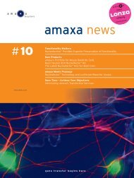
amaxa<br />
› amaxa news 02<br />
news<br />
› Nucle<strong>of</strong>ector technology and RNAi –<br />
application note, ‘essentials’, data<br />
› amaxa and QI<strong>AG</strong>EN ® cooperation –<br />
joined forces in gene silencing<br />
› New: nucle<strong>of</strong>ection <strong>of</strong> cardiomyocytes<br />
and natural killer <strong>cells</strong><br />
› New: nucle<strong>of</strong>ection <strong>of</strong> <strong>neural</strong> <strong>stem</strong><br />
<strong>cells</strong> and astrocytes<br />
gene transfer begins here<br />
02
Integrated Solutions — Gene Silencing<br />
Silence your genes in both<br />
adherent and suspension<br />
<strong>cells</strong>!<br />
New<br />
HUVEC <strong>cells</strong> transfected with siRNA<br />
using RNAiFect <strong>Transfection</strong> Reagent<br />
Jurkat <strong>cells</strong> transfected with siRNA using<br />
Nucle<strong>of</strong>ector technology<br />
Use our new partnership for gene silencing in adherent and<br />
suspension <strong>cells</strong>!<br />
QI<strong>AG</strong>EN ® and amaxa have joined forces to <strong>of</strong>fer you solutions<br />
for RNAi from design to delivery. QI<strong>AG</strong>EN’s siRNA service<br />
includes high-purity siRNA synthesis and an advanced design<br />
algorithm. RNAiFect <strong>Transfection</strong> Reagent provides effective<br />
transfection <strong>of</strong> adherent <strong>cells</strong>. amaxa’s Nucle<strong>of</strong>ector technology<br />
ensures success for transfection <strong>of</strong> suspension <strong>cells</strong>.<br />
■ Effective gene silencing — combining our expertise in<br />
siRNA design, synthesis, and delivery<br />
■ Efficient siRNA delivery — typically 95% transfection<br />
efficiency in adherent <strong>cells</strong> and 99% in suspension <strong>cells</strong><br />
■ Tested for many cell types — including HeLa, HEK293,<br />
NIH/3T3, CHO, C6, <strong>AG</strong>S, Jurkat, HL-60, K562, U937,<br />
THP-1, HUVEC, MEF, and primary human T <strong>cells</strong><br />
Find out more at<br />
www.qiagen.com/siRNA/TransfectAllCells !<br />
Trademarks: QI<strong>AG</strong>EN ® , RNAiFect (QI<strong>AG</strong>EN Group), Nucle<strong>of</strong>ector (amaxa GmbH),<br />
siRNA technology licensed to QI<strong>AG</strong>EN is covered by various patent applications,<br />
owned by the Massachusetts Institute <strong>of</strong> Technology, Cambridge, MA, USA and others.<br />
GSA1003AMAXA 10/2003 © 2003 QI<strong>AG</strong>EN, all rights reserved.<br />
WWW.QI<strong>AG</strong>EN.COM<br />
Editorial<br />
We are proud to introduce the second issue <strong>of</strong> ‘amaxa news’. Your<br />
feedback to our first issue was overwhelming and was a valuable<br />
resource to further improve this newsletter. Thank you again for<br />
your comments and your positive feedback!<br />
At present, RNAi is one <strong>of</strong> the most exciting topics in the scientific<br />
community and also <strong>of</strong> major importance to amaxa (pp. 6-9,<br />
12-15). The Nucle<strong>of</strong>ector technology addresses one <strong>of</strong> the key<br />
bottlenecks in RNAi – highly efficient delivery into hard-to-transfect<br />
cell lines and primary <strong>cells</strong>. In addition, amaxa has joined forces<br />
with QI<strong>AG</strong>EN ® to provide you with an integrated solution for siRNA<br />
design and delivery comprising QI<strong>AG</strong>EN’s siRNA design service<br />
and amaxa’s Nucle<strong>of</strong>ector technology. Read more about your<br />
benefits from this co-operation in this issue <strong>of</strong> ‘amaxa news’ (p. 6).<br />
Anything else besides RNAi? Quite a lot. ‘amaxa news’ will also<br />
give an update on our latest R&D developments. Find out more<br />
about our new Nucle<strong>of</strong>ector Kits for cardiomyocytes, natural<br />
killer <strong>cells</strong> (p. 4), astrocytes, <strong>neural</strong> <strong>stem</strong> <strong>cells</strong> (p. 5), mouse<br />
embryonic <strong>stem</strong> <strong>cells</strong> and mouse embryonic fibroblasts (pp. 10-11).<br />
And there is still more to learn … we wish you an interesting read.<br />
Dr. Gregor Siebenkotten Rainer Christine<br />
CSO CEO
[k]<br />
q<br />
d<br />
New products<br />
New products<br />
amaxa insights<br />
Application note<br />
New products<br />
Tips and hints<br />
Product update<br />
Backround information<br />
amaxa insights<br />
Highlighted paper<br />
amaxa insights<br />
amaxa worldwide<br />
amaxa worldwide<br />
FAQ<br />
Ordering information<br />
To order products contact our<br />
Scientific Support Team (see back<br />
cover for contact details).<br />
Supplementary information<br />
To order supplementary information<br />
complete the reply card attached in<br />
the center <strong>of</strong> this newsletter.<br />
amaxa web information<br />
To instantly find more detailed<br />
information on certain subjects visit<br />
the indicated website.<br />
› Table <strong>of</strong> content<br />
4 Nucle<strong>of</strong>ector Kits for transfection <strong>of</strong> human natural killer <strong>cells</strong><br />
and rat cardiomyocytes<br />
5 <strong>Transfection</strong> <strong>of</strong> <strong>neural</strong> <strong>stem</strong> <strong>cells</strong> (NSC) and astrocytes from<br />
mouse and rat<br />
6 amaxa and QI<strong>AG</strong>EN ® have joined forces in gene silencing<br />
7 RNAi in suspension <strong>cells</strong> using amaxa’s Nucle<strong>of</strong>ector<br />
technology together with QI<strong>AG</strong>EN ® ’s HPP Grade siRNA duplexes<br />
Elke Lorbach, Dietmar Lenz, Andrea Klaes, Katharina Hein, Bodo Ortmann, Tatjana Males,<br />
Hanns-Martin Schmidt, Peter Hahn, Wolfgang Bielke and Günter Kraus<br />
10 Nucle<strong>of</strong>ection <strong>of</strong> mouse embryonic fibroblasts (MEF) and<br />
mouse embryonic <strong>stem</strong> <strong>cells</strong><br />
12 Take a short cut for your RNAi experiments – the ‘essentials’ about<br />
Nucle<strong>of</strong>ector technology and RNAi<br />
14 Nucle<strong>of</strong>ector technology and RNAi – examples <strong>of</strong> successful<br />
knockdown provided by Nucle<strong>of</strong>ector users<br />
16 Basics about nucle<strong>of</strong>ection and the easiest way to transfect<br />
your cell line<br />
18 Fly amaxa competition winners announced<br />
Highlighted paper: Messi et al. (2003)<br />
19 amaxa user forum<br />
20 WAKO Pure Chemicals - amaxa’s competent partner in Japan<br />
21 Meet amaxa at your bench<br />
amaxa’s distributors around the world<br />
22 Frequently asked questions on Nucle<strong>of</strong>ector technology in<br />
neurobiology and immunology<br />
Editor<br />
Wolfgang Kintzel<br />
Editorial board<br />
Bernd Eschweiler, Katrin Höck,<br />
Katja Thieke, Michael Nix<br />
Production coordinator<br />
Benno Limberg<br />
amaxa news is published by<br />
amaxa GmbH<br />
Nattermannallee 1<br />
50829 Koeln<br />
Germany<br />
amaxa_news@amaxa.com<br />
Tel: +49(0)221-99199-0<br />
Fax:+49(0)221-99199-111<br />
› page 3 › www.amaxa.com<br />
amaxa’s patent pending technologies<br />
and information is the express and<br />
proprietary information <strong>of</strong> amaxa GmbH.<br />
amaxa, Nucle<strong>of</strong>ection, Nucle<strong>of</strong>ector,<br />
“gene transfer begins here” are all<br />
trademarks <strong>of</strong> amaxa GmbH.<br />
Please note that to date amaxa’s<br />
Nucle<strong>of</strong>ector technology is intended for<br />
research use or investigational use only.<br />
Design<br />
vierviertel – Agentur für<br />
Kommunikationsdesign GmbH<br />
info@vierviertel.com<br />
© 2003 amaxa. All rights reserved.<br />
Printed in Germany.
› New products<br />
new<br />
Nucle<strong>of</strong>ector Kits for transfection<br />
<strong>of</strong> human natural killer<br />
<strong>cells</strong> and rat cardiomyocytes<br />
Nucle<strong>of</strong>ection <strong>of</strong> primary human natural killer <strong>cells</strong><br />
Cancer and immunology research was frequently hampered due<br />
to the inability to transfect primary NK <strong>cells</strong> non-virally. With the<br />
Human NK Cell Nucle<strong>of</strong>ector Kit a major breakthrough has been<br />
accomplished circumventing the use <strong>of</strong> rather artificial cell lines.<br />
Due to the major obstacles encountered during NK cell transfection<br />
so far an efficiency <strong>of</strong> up to 20% can be a major advancement<br />
for your research.<br />
natural<br />
killer <strong>cells</strong><br />
› first non-viral transfection technology for primary NK <strong>cells</strong><br />
› transfection efficiencies <strong>of</strong> up to 20%<br />
› viability post nucle<strong>of</strong>ection: 50-60%<br />
Nucle<strong>of</strong>ection <strong>of</strong> primary neonatal rat cardiomyocytes<br />
Cardiovascular research has to struggle with two constraints: the<br />
lack <strong>of</strong> appropriate cell line models and an efficient transfection<br />
method for primary cardiomyocytes. Nucle<strong>of</strong>ection <strong>of</strong> rat cardiomyocytes<br />
will help to overcome both limitations.<br />
› first efficient non-viral transfection technology for primary<br />
neonatal rat cardiomyocytes<br />
› reproducible transfection efficiencies up to 40%<br />
› viability post nucle<strong>of</strong>ection: 50-60%<br />
cardiomyocytes<br />
plasmid encoding DsRed. Two days post nucle<strong>of</strong>ection, the <strong>cells</strong><br />
[k] Ordering information<br />
Human NK Cell Nucle<strong>of</strong>ector Kit Cat. No.: VPA-1005 d<br />
Rat Cardiomyocyte Nucle<strong>of</strong>ector Kit Cat. No.: VPE-1002<br />
› page 4 › amaxa news 02<br />
SSC<br />
NK <strong>cells</strong> -DNA NK <strong>cells</strong> +DNA<br />
A B<br />
eGFP<br />
0.5%<br />
SSC<br />
eGFP<br />
Nucle<strong>of</strong>ection <strong>of</strong> neonatal rat cardiomyocytes<br />
Primary neonatal rat cardiomyocytes were nucle<strong>of</strong>ected with a<br />
were analyzed by fluorescence microscopy. Fig A shows DsRed<br />
expressing <strong>cells</strong>. Cardiomyocytes stained with FITC-labeled<br />
tropomyosin antibody (a fibrous protein that is part <strong>of</strong> the muscle<br />
fibers) are shown in fig. B. Fig. C is an overlay. (Photographs<br />
courtesy <strong>of</strong> F. Engel and M. Keating, Cardiology Department,<br />
Childrens’ Hospital, Havard Medical School, Boston, MA, USA)<br />
amaxa web information<br />
Cancer www.amaxa.com/cancer<br />
Immunology www.amaxa.com/immunology<br />
Cardiology www.amaxa.com/cardio<br />
16.9%<br />
Nucle<strong>of</strong>ection <strong>of</strong> primary human NK <strong>cells</strong><br />
Primary human natural killer <strong>cells</strong> were nucle<strong>of</strong>ected with a<br />
plasmid encoding eGFP protein. Cells were analyzed by flow<br />
cytometry 24 hours post nucle<strong>of</strong>ection. eGFP expression in<br />
natural killer <strong>cells</strong> is shown after nucle<strong>of</strong>ection without (A) and<br />
with plasmid DNA (B). (Courtesy <strong>of</strong> J. Sundbäck and K. Kärre,<br />
Karolinska Institute, Microbiology and Tumor Biology Center,<br />
Stockholm, Sweden)<br />
A<br />
C<br />
B
[k]<br />
% nucle<strong>of</strong>ected <strong>cells</strong><br />
› New products<br />
<strong>Transfection</strong> <strong>of</strong> <strong>neural</strong> <strong>stem</strong><br />
<strong>cells</strong> (NSC) and astrocytes from<br />
mouse and rat<br />
Recently amaxa introduced four new Nucle<strong>of</strong>ector Kits for the transfection <strong>of</strong> <strong>neural</strong> <strong>stem</strong> <strong>cells</strong> and astrocytes to<br />
further support your research in neurobiology. For the most important cell types in neurobiology Nucle<strong>of</strong>ector Kits<br />
are now available. These include kits for mouse, rat, and chicken neurons and for PC12 and C6 cell lines.<br />
(Coming soon: SH-SY5Y).<br />
Use the Nucle<strong>of</strong>ector for your neurobiology research:<br />
non-viral › first efficient non-viral method for <strong>neural</strong> and glial <strong>cells</strong><br />
high efficiencies › up to 70% transfection efficiency<br />
functionality › <strong>cells</strong> remain functional after nucle<strong>of</strong>ection<br />
long-term survival › up to three weeks survival<br />
no optimization › ready-to-use kits<br />
100<br />
90<br />
80<br />
70<br />
60<br />
50<br />
40<br />
30<br />
20<br />
10<br />
0<br />
new<br />
eGFP<br />
48h 24h 48h 24h<br />
mouse mouse rat rat<br />
NSC astrocytes astrocytes NSC<br />
Average transfection efficiencies for <strong>neural</strong> <strong>stem</strong> <strong>cells</strong> and<br />
astrocytes<br />
Mouse and rat NSC and astrocytes were transfected with a<br />
plasmid encoding the eGFP protein. <strong>Transfection</strong> efficiencies<br />
are given as % positive <strong>cells</strong>. Analysis was performed by<br />
fluorescence microscopy.<br />
Ordering information<br />
Mouse NSC Nucle<strong>of</strong>ector Kit Cat. No.: VPG-1004<br />
Rat NSC Nucle<strong>of</strong>ector Kit Cat. No.: VPG-1005<br />
Mouse Astrocyte Nucle<strong>of</strong>ector Kit Cat. No.: VPG-1006<br />
Rat Astrocyte Nucle<strong>of</strong>ector Kit Cat. No.: VPG-1007<br />
› page 5 › www.amaxa.com<br />
A<br />
Example for Nucle<strong>of</strong>ection <strong>of</strong> rat <strong>neural</strong> <strong>stem</strong> <strong>cells</strong> (NSC)<br />
Primary rat <strong>neural</strong> <strong>stem</strong> <strong>cells</strong> isolated from rat embryos (E14) were nucle<strong>of</strong>ected with<br />
a plasmid encoding the eGFP protein under the control <strong>of</strong> an EF1d promoter. After<br />
nucle<strong>of</strong>ection <strong>cells</strong> were cultured with bFGF for two days, then for five days without<br />
bFGF to differentiate NSC into neurons and astrocytes. Cells were analyzed by fluorescence<br />
microscopy two days (A) and seven days (B) post nucle<strong>of</strong>ection. (Photographs<br />
courtesy <strong>of</strong> Dr. S.H. Lee, College <strong>of</strong> Medicine, Dept. <strong>of</strong> Biochemistry, Hanyang<br />
University, Seoul, South Korea)<br />
q<br />
Supplementary information<br />
Neurobiology flyer – Nucle<strong>of</strong>ection in neurobiology<br />
WKB-1004<br />
B<br />
d amaxa web information www.amaxa.com/neurobiology
› amaxa insights<br />
amaxa and QI<strong>AG</strong>EN ® have<br />
joined forces in gene silencing<br />
RNAi provides a rapid, efficient and powerful tool in functional genomics drawing widespread attention to this field. Although<br />
RNAi has proven to be a powerful method for gene silencing some technical difficulties remain. QI<strong>AG</strong>EN and amaxa<br />
have joined forces to address the two key bottlenecks in RNAi-mediated gene silencing i.e. siRNA design and delivery.<br />
Take advantage <strong>of</strong> our partnership for siRNA<br />
design and delivery<br />
By combining QI<strong>AG</strong>EN’s knowledge <strong>of</strong> siRNA<br />
design and synthesis and amaxa’s know-how on<br />
efficient delivery, into virtually any cell, a com-<br />
› page 6 › amaxa news 02<br />
plete service for your gene silencing experiments is now available.<br />
QI<strong>AG</strong>EN and amaxa’s technical services provide an invaluable<br />
source <strong>of</strong> information for details on siRNA experiment set-up,<br />
siRNA design and transfection protocols for optimal delivery. Call<br />
today to discuss your experiment!<br />
siRNA design made easy with QI<strong>AG</strong>EN<br />
Optimal siRNA design › Use the convenient online QI<strong>AG</strong>EN siRNA Design Tool which<br />
uses the sensitive Smith-Waterman algorithm.<br />
High-purity, custom › HPP (High Performance Purity) Grade siRNA is > 90% pure.<br />
synthesized siRNA<br />
Minimal risk for gene silencing › 4-For-Silencing siRNA Duplexes are designed by QI<strong>AG</strong>EN<br />
scientists to target your gene <strong>of</strong> interest. QI<strong>AG</strong>EN guarantees<br />
70% knockdown or four additional duplexes will be provided.<br />
Efficient siRNA delivery with amaxa’s Nucle<strong>of</strong>ector technology<br />
Efficient delivery › Up to 99% siRNA transfection efficiency already proven in<br />
many different <strong>cells</strong>.<br />
Low cytotoxicity › Typically over 85% cell viability post nucle<strong>of</strong>ection regardless<br />
<strong>of</strong> siRNA amount transfected.<br />
No optimization › Ready-to-use kits and protocols for a wide range <strong>of</strong> cell types.<br />
Flexibility <strong>of</strong> RNAi application › One nucle<strong>of</strong>ection protocol for siRNA duplexes, vectors or<br />
in vitro transcribed siRNAs.<br />
d amaxa web information amaxa siRNA delivery www.amaxa.com/RNAi<br />
QI<strong>AG</strong>EN web information QI<strong>AG</strong>EN siRNA synthesis www.qiagen.com/siRNA<br />
contact information<br />
Australia QI<strong>AG</strong>EN Pty Ltd • Orders 03-9489-3666 • Fax 03-9489-3888 • Technical 1-800-243-066 › Canada QI<strong>AG</strong>EN Inc.<br />
Orders 800-572-9613 • Fax 800-713-5951 • Technical 800-DNA-PREP (800-362-7737) › France QI<strong>AG</strong>EN S.A. • Orders 01-60-920-920<br />
Fax 01-60-920-925 • Technical 01-60-920-930 › Germany QI<strong>AG</strong>EN GmbH • Orders 02103-29-12000 • Fax 02103-29-22000<br />
Technical 02103-29-12400 › Italy QI<strong>AG</strong>EN S.p.A. • Orders 02-33430411 • Fax 02-33430426 • Technical 02-33430414 › Japan<br />
QI<strong>AG</strong>EN K.K. • Telephone 03-5547-0811 • Fax 03-5547-0818 • Technical 03-5547-0811 › Switzerland QI<strong>AG</strong>EN <strong>AG</strong> • Orders 061-319-30-30<br />
Fax 061-319-30-33 • Technical 061-319-30-31 › United Kingdom QI<strong>AG</strong>EN Ltd. • Orders 01293-422-911 • Fax 01293-422-922<br />
Technical 01293-422-999 › USA QI<strong>AG</strong>EN Inc. • Orders 800-426-8157 • Fax 800-718-2056 • Technical 800-DNA-PREP (800-362-7737)
› Application note<br />
RNAi in suspension <strong>cells</strong> using amaxa’s<br />
Nucle<strong>of</strong>ector technology together with<br />
QI<strong>AG</strong>EN ® ’s HPP Grade siRNA duplexes<br />
Elke Lorbach*, Dietmar Lenz*, Andrea Klaes*, Katharina Hein*, Bodo Ortmann*, Tatjana Males*, Hanns-Martin Schmidt*,<br />
Peter Hahn**, Wolfgang Bielke**, Günter Kraus*<br />
Successful siRNA gene silencing depends on many different parameters such as mRNA turnover, mRNA abundance or<br />
half-life <strong>of</strong> the protein. Two important factors that can positively influence your siRNA experiment are the design <strong>of</strong><br />
the siRNA oligo and the efficient delivery <strong>of</strong> the siRNA. Here we show that HPP Grade siRNA from QI<strong>AG</strong>EN works well<br />
in combination with amaxa’s Nucle<strong>of</strong>ector technology and can be used to efficiently silence genes in various<br />
hard-to-transfect suspension cell lines as well as in primary <strong>cells</strong>.<br />
* amaxa GmbH,<br />
Köln, Germany<br />
** QI<strong>AG</strong>EN GmbH,<br />
Hilden, Germany<br />
Introduction<br />
RNA interference (RNAi) allows targeted<br />
knockdown <strong>of</strong> gene expression by introducing<br />
short interfering RNA (siRNA),<br />
homologous to the mRNA <strong>of</strong> the target<br />
gene, into <strong>cells</strong>. This method <strong>of</strong> gene<br />
silencing has become a valuable tool in<br />
functional genomics research, target<br />
validation, and gene-specific therapeutic<br />
research. Successful RNAi experiments<br />
are dependent on two major parameters:<br />
siRNA design and efficient delivery <strong>of</strong><br />
siRNA into <strong>cells</strong>.<br />
QI<strong>AG</strong>EN and amaxa have joined forces<br />
to provide a complete solution for RNAi<br />
that overcomes the problems associated<br />
with the key issues <strong>of</strong> siRNA design<br />
and delivery into mammalian <strong>cells</strong>.<br />
amaxa’s R&D team has demonstrated<br />
efficient siRNA delivery in suspension<br />
<strong>cells</strong> using QI<strong>AG</strong>EN’s HPP (High-Performance<br />
Purity) Grade siRNA and amaxa’s<br />
Nucle<strong>of</strong>ector technology.<br />
Figure 1: Easy siRNA design<br />
Screenshot <strong>of</strong> the QI<strong>AG</strong>EN siRNA Design<br />
Tool.<br />
In this study, efficient silencing <strong>of</strong> the<br />
housekeeping gene vimentin and the<br />
T cell receptor genes, CD2 and CD4, is<br />
shown in a variety <strong>of</strong> suspension cell lines,<br />
such as JURKAT, U937, THP-1, HL-60 and<br />
K562, as well as in non-dividing, unstimulated<br />
primary human T <strong>cells</strong>.<br />
siRNA design and synthesis<br />
The design and quality <strong>of</strong> an siRNA has<br />
a strong influence on the level and<br />
specificity <strong>of</strong> gene silencing. QI<strong>AG</strong>EN<br />
provides a siRNA Design Tool that uses<br />
state-<strong>of</strong>-the-art design criteria for easy<br />
design <strong>of</strong> siRNA (Figure 1).<br />
To further minimize the risk that a particular<br />
siRNA will not result in effective<br />
silencing, 4-For-Silencing siRNA Duplexes<br />
are available. These are a set <strong>of</strong> four HPP<br />
Grade siRNA duplexes, designed by<br />
QI<strong>AG</strong>EN scientists to target the gene <strong>of</strong><br />
choice. QI<strong>AG</strong>EN guarantees 70% mRNA<br />
knockdown with at least one <strong>of</strong> these<br />
1000<br />
900<br />
800<br />
700<br />
600<br />
500<br />
400<br />
300<br />
200<br />
Figure 2: High-purity siRNA for<br />
efficient gene silencing<br />
QI<strong>AG</strong>EN performs MALDI-TOF mass<br />
spectrometric analysis on every HPP<br />
Grade siRNA to confirm its integrity.<br />
› page 7 › www.amaxa.com<br />
Intensity<br />
100<br />
0<br />
5000 5500 6000 6500 7000 7500 8000 8500<br />
Mass/Charge<br />
siRNA duplexes, otherwise four additional<br />
siRNA duplexes are provided without<br />
additional cost.<br />
siRNAs directed against a range <strong>of</strong><br />
endogenous and reporter genes are<br />
available for developing and optimizing<br />
knockdown experiments. In addition,<br />
library sets are available for screening<br />
large numbers <strong>of</strong> genes. A regularly<br />
updated list can be found at<br />
www.qiagen.com/siRNA. QI<strong>AG</strong>EN’s HPP<br />
Grade siRNA is delivered >90% pure,<br />
eliminating the time and expense needed<br />
for HPLC or P<strong>AG</strong>E purification (Figure 2).<br />
siRNA delivery with Nucle<strong>of</strong>ector<br />
technology<br />
The Nucle<strong>of</strong>ector technology is a nonviral<br />
method for transfection <strong>of</strong> hard-totransfect<br />
cell lines and primary <strong>cells</strong>.<br />
It is based on two components, the<br />
Nucle<strong>of</strong>ector Device, which delivers<br />
unique electrical parameters, and the<br />
Nucle<strong>of</strong>ector Kits which contain cell-specific<br />
and optimized Nucle<strong>of</strong>ector Solutions.<br />
Ready-to-go protocols are available<br />
for many cell lines and primary cell types.<br />
The technology is suitable for the delivery<br />
<strong>of</strong> different substrates such as DNA, RNA<br />
or siRNA oligos without further optimization<br />
<strong>of</strong> the nucle<strong>of</strong>ection conditions.<br />
Suspension <strong>cells</strong> are difficult to transfect<br />
with conventional transfection<br />
methods, e.g. lipid-based reagents, with<br />
both plasmid DNA and siRNA oligos. In<br />
this study, we show that siRNA oligos<br />
can be efficiently delivered into many<br />
different suspension cell types with the<br />
Nucle<strong>of</strong>ector technology and that<br />
effective gene silencing can be achieved<br />
as shown by different methods,<br />
such as flow cytometry, western blot<br />
and quantitative RT-PCR.
› Application note<br />
JURKAT<br />
JURKAT<br />
HL-60<br />
HL-60<br />
K562<br />
K562<br />
THP-1<br />
THP-1<br />
Figure 3: Up to 99% siRNA delivery<br />
into suspension <strong>cells</strong><br />
Different suspension cell lines were<br />
nucle<strong>of</strong>ected using the appropriate<br />
Nucle<strong>of</strong>ector Kit and Rhodamine-labeled<br />
siRNA. Three hours post nucle<strong>of</strong>ection<br />
<strong>cells</strong> were analyzed by light and<br />
fluorescence microscopy.<br />
Gene silencing <strong>of</strong> various genes in<br />
suspension cell lines<br />
In order to determine the efficiency <strong>of</strong><br />
nucle<strong>of</strong>ection with siRNA duplexes,<br />
Rhodamine-labeled siRNA [QI<strong>AG</strong>EN] was<br />
transfected into different suspension<br />
cell types, such as JURKAT, HL-60, K562<br />
and THP-1 <strong>cells</strong> (Figure 3). Cells were<br />
analyzed by fluorescence microscopy<br />
and siRNA delivery was achieved with up<br />
to 99% efficiency.<br />
JURKAT, HL-60, K562, THP-1, U937 and<br />
primary human T <strong>cells</strong> were transfected<br />
with siRNA targeted to the housekeeping<br />
gene vimentin [QI<strong>AG</strong>EN, library<br />
siRNA; Cat.No. 1022055]. 24 and 48 hours<br />
after nucle<strong>of</strong>ection, silencing <strong>of</strong> vimentin<br />
gene expression was determined by<br />
quantitative, real-time RT-PCR (Figure 4).<br />
This experiment clearly demonstrates<br />
that expression <strong>of</strong> vimentin mRNA was<br />
downregulated with silencing effects<br />
between 70 and 90%. Furthermore, it<br />
% relative expression<br />
› page 8 › amaxa news 02<br />
100<br />
75<br />
50<br />
25<br />
0<br />
shows that this level <strong>of</strong> silencing is<br />
dependent on the cell type and time <strong>of</strong><br />
analysis due to different mRNA kinetics<br />
in different cell types.<br />
Knockdown <strong>of</strong> vimentin was also shown<br />
at protein level using western blot.<br />
JURKAT <strong>cells</strong> were transfected with<br />
three different concentrations <strong>of</strong> siRNA<br />
targeted to the vimentin gene (Figure 5).<br />
Already after 24 hours, the amount <strong>of</strong><br />
vimentin protein was clearly reduced<br />
using 0.7 µg siRNA. After 48 hours, the<br />
expression was further downregulated<br />
and with 0.7 µg the signal for vimentin<br />
was no longer detectable. These data<br />
indicate that silencing occurs also on the<br />
protein level and that the silencing<br />
effects are dose dependent.<br />
Two other suspension cell lines, the<br />
promyelocytic cell line HL-60 and the<br />
monocytic leukemia cell line THP-1, were<br />
used to show gene silencing <strong>of</strong> the CD4<br />
Figure 4: Gene silencing <strong>of</strong> vimentin in various suspension cell types<br />
Six different suspension cell types were transfected with 1.4 µg siRNA targeted to<br />
vimentin or with 1.4 µg control siRNA using the cell-type specific Nucle<strong>of</strong>ector<br />
protocols. After 24 and 48 hours total RNA was prepared from <strong>cells</strong> and used for quantitative<br />
real-time RT-PCR analysis. The expression level <strong>of</strong> vimentin mRNA was normalized<br />
to the expression <strong>of</strong> a housekeeping gene and quantified using a standard curve.<br />
kDa<br />
62<br />
Vimentin -<br />
47<br />
Actin -<br />
32<br />
Human T <strong>cells</strong> JURKAT THP-1 U937 K562 HL-60<br />
M<br />
Vimentin knockdown at protein level in JURKAT <strong>cells</strong><br />
24 hours 48 hours<br />
No siRNA +<br />
nucle<strong>of</strong>ection<br />
No nucle<strong>of</strong>ection<br />
1.4 µg siRNA<br />
0.14 µg<br />
0.7 µg<br />
Figure 5: Vimentin knockdown at protein level in JURKAT <strong>cells</strong><br />
JURKAT (ATCC) <strong>cells</strong> were transfected using Nucle<strong>of</strong>ector Kit V, program C-16 and<br />
0.14 µg, 0.7 µg, or 1.4 µg siRNA targeted to vimentin. Western blot analysis was carried<br />
out 24 and 48 hours post nucle<strong>of</strong>ection. Control lanes show samples with siRNA<br />
added without nucle<strong>of</strong>ection and with nucle<strong>of</strong>ection, but no addition <strong>of</strong> siRNA. The<br />
level <strong>of</strong> actin expression is shown for comparison. M=markers.<br />
1.4 µg<br />
M<br />
control siRNA<br />
vimentin siRNA (24h)<br />
vimentin siRNA (48h)<br />
No siRNA +<br />
nucle<strong>of</strong>ection<br />
0.14 µg<br />
0.7 µg<br />
1.4 µg<br />
M
q<br />
q<br />
› Application note<br />
counts<br />
For further<br />
info on siRNA<br />
design and<br />
synthesis go to<br />
www.qiagen.com<br />
/siRNA.<br />
For further info<br />
on siRNA<br />
delivery go to<br />
www.amaxa.com<br />
/RNAi.<br />
180<br />
150<br />
120<br />
90<br />
60<br />
30<br />
0<br />
100 101 102 103 104 0.14 µg CD4 siRNA<br />
10<br />
CD4-PE<br />
0 101 102 103 104 0.35 µg CD4 siRNA<br />
10<br />
CD4-PE<br />
0 101 102 103 104 0.7 µg CD4 siRNA<br />
10<br />
CD4-PE<br />
0 101 102 103 104 1.4 µg CD4 siRNA<br />
10<br />
CD4-PE<br />
0 101 102 103 104 1.0 µg CD4 siRNA<br />
CD4-PE<br />
Figure 6: Optimization <strong>of</strong> siRNA<br />
amounts in HL-60 <strong>cells</strong><br />
Five different amounts <strong>of</strong> siRNA duplexes<br />
targeting human CD4 were nucle<strong>of</strong>ected<br />
T cell receptor gene. Both cell types<br />
were transfected with siRNA targeted<br />
to the CD4 gene [QI<strong>AG</strong>EN, library<br />
siRNA; Cat.No. 1024675]. 48 hours after<br />
delivery <strong>of</strong> siRNA, downregulation <strong>of</strong><br />
CD4 was analyzed by flow cytometry<br />
(Figure 6 and 7). In HL-60 <strong>cells</strong> five<br />
different amounts <strong>of</strong> siRNA duplexes<br />
were transfected, and down regulation<br />
<strong>of</strong> CD4 expression occurred in a dosedependent<br />
manner. The most pronounced<br />
gene silencing effects were achieved<br />
with 1.4 µg siRNA (Figure 6). In THP-1<br />
<strong>cells</strong>, 1.4 µg siRNA was transfected and<br />
effective knockdown <strong>of</strong> CD4 expression<br />
was achieved (Figure 7). <strong>Transfection</strong><br />
<strong>of</strong> non-specific control siRNA duplexes<br />
did not influence the expression <strong>of</strong><br />
CD4.<br />
Successful gene silencing in primary<br />
human T <strong>cells</strong><br />
Efficient delivery <strong>of</strong> siRNA oligonucleotides<br />
into primary <strong>cells</strong> is the key<br />
180<br />
120<br />
60<br />
0<br />
counts 240<br />
Fig. 7<br />
10 0 10 1 10 2 10 3 10 4<br />
CD4-PE<br />
CD4 siRNA<br />
control siRNA<br />
no siRNA + nucle<strong>of</strong>ection<br />
Figure 7: CD4 Gene Silencing in<br />
THP-1 Cells<br />
THP-1 <strong>cells</strong> were transfected using<br />
Nucle<strong>of</strong>ector Kit V and program V-01<br />
and 1.4 µg siRNA targeted to CD4.<br />
48 hours after nucle<strong>of</strong>ection, <strong>cells</strong> were<br />
stained with anti-CD4-PE antibody and<br />
analyzed by flow cytometry. Controls<br />
included nucle<strong>of</strong>ection <strong>of</strong> 1.4 µg control<br />
siRNA and nucle<strong>of</strong>ection without siRNA.<br />
into HL-60 <strong>cells</strong> using Nucle<strong>of</strong>ector Kit<br />
V and program T-19. 48 hours after nucle<strong>of</strong>ection,<br />
<strong>cells</strong> were stained with anti-<br />
CD4-Phycoerythrin (PE) antibody and<br />
bottleneck before functional genomic<br />
studies can be performed under in vivolike<br />
conditions, such as those found in<br />
primary <strong>cells</strong>. Here we show that primary<br />
human T <strong>cells</strong> can be used for siRNA<br />
experiments with the Nucle<strong>of</strong>ector<br />
technology. Unstimulated human T <strong>cells</strong><br />
were transfected with siRNA targeted to<br />
the pan T cell receptor gene CD2 [siRNA<br />
sequence published by D. Unutmaz in<br />
amaxa application note: RNAi-mediated<br />
CD2 suppression in primary human T<br />
<strong>cells</strong>, WTB-1001]. 48 hours after nucle<strong>of</strong>ection,<br />
the downregulation <strong>of</strong> CD2<br />
expression could be shown by flow cytometry<br />
(Figure 8A). Expression <strong>of</strong> the<br />
CD4 receptor was not influenced as<br />
demonstrated by control staining with a<br />
PE-coupled CD4 antibody and flow cytometric<br />
analysis (Figure 8B). This clearly<br />
indicates that the gene silencing effect<br />
achieved was CD2 specific thus proving<br />
the Nucle<strong>of</strong>ector technology an efficient<br />
tool to transfect siRNA duplexes<br />
CD2 siRNA<br />
control siRNA<br />
no siRNA + nucle<strong>of</strong>ection<br />
Figure 8: High specificity <strong>of</strong> CD2<br />
siRNA in primary human T <strong>cells</strong><br />
Primary human T <strong>cells</strong> were nucle<strong>of</strong>ected<br />
with 1.4 µg siRNA targeted to CD2<br />
using the Human T Cell Nucle<strong>of</strong>ector<br />
Kit. Cells were stained with A anti<br />
› page 9 › www.amaxa.com<br />
counts<br />
200<br />
160<br />
120<br />
80<br />
40<br />
0<br />
Fig. 8A<br />
10 0 10 1 10 2 10 3 10 4<br />
CD2-FITC<br />
counts<br />
analyzed by flow cytometry. siRNA targeted<br />
against vimentin was used as control.<br />
CD4 siRNA control siRNA<br />
not only into suspension cell lines but<br />
also into primary suspension <strong>cells</strong>.<br />
Trademarks and disclaimers<br />
Nucle<strong>of</strong>ector,<br />
Nucle<strong>of</strong>ection (amaxa GmbH)<br />
QI<strong>AG</strong>EN ® (QI<strong>AG</strong>EN GmbH)<br />
siRNA technology licensed to QI<strong>AG</strong>EN is<br />
covered by various patent applications,<br />
owned by the Massachusetts Institute <strong>of</strong><br />
Technology, Cambridge, MA, USA and<br />
others.<br />
© 2003 amaxa, all rights reserved.<br />
QI<strong>AG</strong>EN guarantees that under appropriate<br />
transfection conditions at least<br />
one <strong>of</strong> the 4-For-Silencing siRNA Duplexes<br />
will reduce expression <strong>of</strong> your target<br />
mRNA by 70%. If not, QI<strong>AG</strong>EN will<br />
design and supply four additional siRNA<br />
duplexes against the same gene target<br />
at no additional charges.<br />
300<br />
240<br />
180<br />
120<br />
60<br />
0<br />
Fig. 8B<br />
10 0 10 1 10 2 10 3 10 4<br />
CD4-PE<br />
CD2-FITC antibody or B anti CD4-PE<br />
antibody, and analyzed by flow cytometry<br />
44 hours after transfection.<br />
Controls included nucle<strong>of</strong>ection with<br />
1.4 µg control siRNA and nucle<strong>of</strong>ection<br />
without siRNA.
Step 1<br />
Step 2<br />
› New products<br />
new<br />
Nucle<strong>of</strong>ection <strong>of</strong> mouse<br />
embryonic fibroblasts (MEF) and<br />
mouse embryonic <strong>stem</strong> <strong>cells</strong><br />
From cell differentiation to knock-out mice - amaxa introduces the new Nucle<strong>of</strong>ector Kits for transfection <strong>of</strong> mouse<br />
embryonic fibroblasts (MEF) and mouse embryonic <strong>stem</strong> <strong>cells</strong> (mouse ES <strong>cells</strong>). <strong>Transfection</strong> <strong>of</strong> MEF and mouse ES<br />
<strong>cells</strong> has broad research potential. Both kits will be important for research on cell differentiation, organogenesis, cellbased<br />
therapies, testing <strong>of</strong> new drugs, and gene function approaches like ‘loss-<strong>of</strong>-function’ and ‘gain-<strong>of</strong>-function’ studies.<br />
The MEF strategy –<br />
two simple steps for successful transfection<br />
MEF display significant phenotypic variations<br />
which depend on the mouse strain, the genetic<br />
background <strong>of</strong> the donor mice as well as the<br />
immortalization strategy. Due to these manifold<br />
properties, each MEF type may behave differently<br />
MEF Starter Kit<br />
The MEF Starter Nucle<strong>of</strong>ector Kit is designed to<br />
determine the ideal nucle<strong>of</strong>ection conditions for<br />
each MEF cell type. This is accomplished in a total<br />
MEF Kits 1 and 2<br />
After you have determined the appropriate MEF<br />
Nucle<strong>of</strong>ector Kit for your MEF strain you can<br />
› page 10 › amaxa news 02<br />
with respect to the optimal conditions for transfection.<br />
amaxa has addressed this by developing two MEF Nucle<strong>of</strong>ector<br />
Kits (1 and 2) and an easy-to-use MEF Starter Nucle<strong>of</strong>ector<br />
Kit. This strategy allows fast and individual nucle<strong>of</strong>ection <strong>of</strong> your<br />
MEF <strong>cells</strong>.<br />
non-viral transfection › efficient transfection <strong>of</strong> MEF <strong>cells</strong><br />
simple optimization strategy › two step optimization with MEF Nucle<strong>of</strong>ector Starter Kit<br />
applicable for virtually any MEF strain › taking into account the significant phenotypic strain variations<br />
high cell viability › viability from 60 - 80% (in strains tested so far)<br />
1 Solution MEF 1 MEF 2<br />
program 1 A-23 A-23<br />
program 2 T-20 T-20<br />
<strong>of</strong> just four reactions using MEF 1 and MEF 2 Nucle<strong>of</strong>ector Solution<br />
with two different programs. In the next step the appropriate MEF<br />
Nucle<strong>of</strong>ector Kit (1 or 2) can be used to perform the experiments.<br />
>>><br />
2<br />
3<br />
Order MEF 1 or 2 Nucle<strong>of</strong>ector TM Kit<br />
+<br />
>>><br />
nucle<strong>of</strong>ect the <strong>cells</strong> with MEF Nucle<strong>of</strong>ector Kit 1 or 2 and the<br />
appropriate program.
› New products<br />
60<br />
50<br />
40<br />
30<br />
20<br />
10<br />
0<br />
% transfection efficiency<br />
A B<br />
MEF<br />
protein. 24 hours post nucle<strong>of</strong>ection, the <strong>cells</strong> were analyzed<br />
Example for transfection efficiencies <strong>of</strong> primary MEF<br />
MEF were nucle<strong>of</strong>ected with a plasmid encoding the eGFP<br />
by fluorescent microscopy. (A) Mouse strain C57BL/6, spontaneously<br />
immortalized, passage 20, 1.5 µg eGFP (Courtesy<br />
<strong>of</strong> Dr. H. Hermanns and Pr<strong>of</strong>. P.H. Heinrich, University <strong>of</strong><br />
Aachen, Germany). (B) Mouse strain C57B6Jx129SV, SV40<br />
transformed, passage 4, 7 µg eGFP (Courtesy <strong>of</strong> Pr<strong>of</strong>. Saftig,<br />
University <strong>of</strong> Kiel, Germany)<br />
Research with mouse ES <strong>cells</strong> has already made<br />
its way to a sophisticated assay sy<strong>stem</strong> for<br />
studying diverse biological questions. Transgenic<br />
technology now allows to create mice with<br />
various genotypes and assess the consequences<br />
<strong>of</strong> this genotype in the context <strong>of</strong> developing and<br />
adult animals. <strong>Transfection</strong> <strong>of</strong> mouse ES <strong>cells</strong> will<br />
support the widespread adoption <strong>of</strong> the transgenic<br />
sy<strong>stem</strong> and the research on differentiation<br />
<strong>of</strong> mouse ES <strong>cells</strong>.<br />
Nucle<strong>of</strong>ector technology is an efficient alternative<br />
to standard electroporation to create<br />
[k] [k]<br />
Ordering information<br />
MEF Starter Nucle<strong>of</strong>ector Kit Cat. No.: VPD-1006<br />
MEF Nucle<strong>of</strong>ector Kit 1 Cat. No.: VPD-1004<br />
MEF Nucle<strong>of</strong>ector Kit 2 Cat. No.: VPD-1005<br />
› page 11 › www.amaxa.com<br />
Example for nucle<strong>of</strong>ection <strong>of</strong> primary MEF<br />
Spontaneously immortalized mouse embryonic fibroblasts (strain C57B6Jx129SV)<br />
were nucle<strong>of</strong>ected using the MEF Nucle<strong>of</strong>ector Kit 1 and a plasmid encoding the<br />
enhanced green fluorescent protein eGFP. 24 hours post nucle<strong>of</strong>ection, the <strong>cells</strong><br />
were analyzed by fluorescence microscopy. (Photograph courtesy <strong>of</strong> Dr. H. Hermanns<br />
and Pr<strong>of</strong>. P.H. Heinrich, University <strong>of</strong> Aachen, Germany)<br />
Nucle<strong>of</strong>ection <strong>of</strong> Mouse ES <strong>cells</strong><br />
knockout mice. In addition, the cell-type specific nucle<strong>of</strong>ection<br />
conditions optimized for gentle DNA delivery in vitro support<br />
differentiation <strong>of</strong> transfected mouse ES <strong>cells</strong>. Together with other<br />
Nucle<strong>of</strong>ector Kits for primary mouse <strong>cells</strong>, genetic manipulation<br />
and analysis <strong>of</strong> various cell types is made possible.<br />
Successfully tested ES cell lines:<br />
› WW6<br />
› R1<br />
› CK-35<br />
› H7<br />
› U295V (an E14 subclone)<br />
Ordering information<br />
Mouse ES Cell Nucle<strong>of</strong>ector Kit Cat. No.: VPH-1001
H<br />
› Tips and hints<br />
Selection <strong>of</strong> appropriate Nucle<strong>of</strong>ector Kit<br />
Each Nucle<strong>of</strong>ector Kit contains a specific<br />
Optimized Protocol for the respective cell type.<br />
This protocol can be used for the nucle<strong>of</strong>ection<br />
<strong>of</strong> either siRNA oligos or vectors without any<br />
further changes <strong>of</strong> electrical parameters.<br />
Methods for generating siRNA<br />
Several different methods for generating siRNA<br />
are known; such as:<br />
› synthetic siRNA oligos<br />
› in vitro transcribed oligos<br />
› siRNA expression vectors<br />
Take a short cut for your<br />
RNAi experiments –<br />
the ‘essentials’ about Nucle<strong>of</strong>ector technology and RNAi<br />
The Nucle<strong>of</strong>ector technology has proven its capability for efficient delivery <strong>of</strong> siRNA into hardto-transfect<br />
cell lines and primary <strong>cells</strong>. To save your time and allow for a successful setup <strong>of</strong> RNAi<br />
experiments in your lab we compiled some information on the ‘essentials’ about RNAi and the<br />
Nucle<strong>of</strong>ector technology. Some <strong>of</strong> the most crucial aspects are listed below. The opposite page<br />
outlines how to establish RNAi for your particular cell type. For further questions on your RNAi<br />
experiments contact our Scientific Support Team.<br />
Tips for a successful<br />
siRNA experiment<br />
All methods can be used together with the<br />
Nucle<strong>of</strong>ector technology for efficient delivery<br />
<strong>of</strong> siRNA into primary <strong>cells</strong> and cell lines.<br />
› page 12 › amaxa news 02<br />
siRNA oligo design<br />
Design and selection <strong>of</strong> siRNA oligo target sequences play an<br />
important role. In general, testing <strong>of</strong> 2-6 different siRNA oligos is<br />
sufficient to find one or more siRNA oligos that display good silencing<br />
effects. We recommend using Qiagen’s siRNA oligos and<br />
design s<strong>of</strong>tware.<br />
siRNA oligo concentration<br />
The amount <strong>of</strong> siRNA oligo required to efficiently downregulate a<br />
specific gene has to be determined empirically and depends on<br />
many parameters, such as efficiency <strong>of</strong> siRNA oligo sequence,<br />
method <strong>of</strong> oligo preparation, cell type, mRNA abundance, mRNA<br />
turnover etc. amaxa’s R&D team recommends to use between 0.14<br />
and 1.4 µg siRNA.<br />
Labeled siRNA oligos<br />
Fluorescent labeled siRNA can be used to analyze transfection<br />
efficiency. FITC or Rhodamine labeled siRNA oligos should be<br />
analyzed after 0.5-3 hours. Cy5 labeled siRNAs can be detected<br />
up to 24 hours post nucle<strong>of</strong>ection.
d<br />
› Tips and hints<br />
eGFP<br />
amaxa web information www.amaxa.com/RNAi<br />
A simple strategy for RNAi in cell lines and primary <strong>cells</strong><br />
Follow the scheme below for RNAi experiments in your primary cell or cell line <strong>of</strong> interest.<br />
primary <strong>cells</strong><br />
Optimized Protocol<br />
available / yes<br />
Choose the appropriate Nucle<strong>of</strong>ector<br />
Kit for cell type <strong>of</strong> interest.<br />
Use the Optimized Protocol and an eGFP<br />
plasmid to establish the Nucle<strong>of</strong>ector<br />
technology for your cell type.<br />
Alternatively, fluorescence labeled<br />
siRNA oligos can be used.<br />
Establish RNAi experiments with positive<br />
control siRNA oligos, e.g. vimentin or<br />
lamin A/C.<br />
Nucle<strong>of</strong>ection <strong>of</strong> eGFP plasmid should<br />
be included as a positive control for<br />
transfection efficiency.<br />
Efficient gene silencing in primary <strong>cells</strong><br />
› page 13 › www.amaxa.com<br />
cell lines<br />
Use Cell Line Optimization<br />
Nucle<strong>of</strong>ector Kit.<br />
Optimize nucle<strong>of</strong>ection conditions with<br />
eGFP plasmid according to the optimization<br />
strategy:<br />
Use optimized parameters to establish<br />
RNAi experiments.<br />
V<br />
Optimized Protocol<br />
available / no<br />
Solution R T V<br />
R T<br />
V<br />
program 1 A-23 A-23 A-23<br />
program 2 A-27 A-27 A-27<br />
program 3 T-20 T-20 T-20<br />
program 4 T-27 T-27 T-27<br />
program 5 T-16 T-16 T-16<br />
program 6 T-01 T-01 T-01<br />
program 7 G-16 G-16 G-16<br />
program 8 O-17 O-17 O-17<br />
Efficient gene silencing in cell lines.<br />
+
› Product update<br />
Nucle<strong>of</strong>ector technology and RNAi -<br />
examples for successful knockdown provided by Nucle<strong>of</strong>ector users<br />
The following examples demonstrate an efficient targeted knockdown <strong>of</strong> gene expression by introducing siRNA via<br />
the Nucle<strong>of</strong>ector technology. In addition, the data show the suitability <strong>of</strong> the Nucle<strong>of</strong>ector technology in answering<br />
scientific questions by RNAi especially in hard-to-transfect cell lines and primary <strong>cells</strong> for your basic and applied<br />
research needs.<br />
* winner <strong>of</strong> publication award, see page 18<br />
RNAi to study the caspase-8 mediated<br />
activation <strong>of</strong> T <strong>cells</strong><br />
Chun et al. (2002) (1) studied the role <strong>of</strong> caspase-8<br />
in T lymphocyte activation. Therefore, resting<br />
T lymphoctes were nucle<strong>of</strong>ected with siRNA<br />
against caspase-8. Transfected <strong>cells</strong> were then<br />
stimulated with CD3 and CD28, and the effect on<br />
T cell activation markers CD25 and CD69 was<br />
analyzed by FACS. The data nicely show a specific<br />
downregulation. Caspase-8 siRNA leads to a<br />
reduction in activation marker expression<br />
indicating a role <strong>of</strong> caspase-8 not only in apoptosis<br />
but also in T cell activation.<br />
Downregulation <strong>of</strong> eGFP in primary mouse<br />
neurons with siRNA<br />
Primary mouse neurons were co-nucle<strong>of</strong>ected<br />
with GFP and siRNA against GFP. GFP expression<br />
was significantly downregulated.<br />
› page 14 › amaxa news 02<br />
A CD69 (fold induction) B CD25 (fold induction)<br />
60<br />
50<br />
40<br />
30<br />
20<br />
10<br />
NS C8<br />
0 Pr2 Pr2 Pr3 Pr3 NS<br />
20 40 20 40 50 nM<br />
Figure 1: Downregulation <strong>of</strong> lymphocyte activation by nucle<strong>of</strong>ection <strong>of</strong> caspase-8<br />
siRNA oligonucleotides. Resting PBLs were transfected with the Nucle<strong>of</strong>ector technology<br />
with siRNA oligonucleotides in two different concentrations that were either nonspecific<br />
(NS) or specific for caspase-8 (Pr2 and 3) and were then stimulated with CD3 and<br />
CD28 for 18 hours. Induction <strong>of</strong> surface expression <strong>of</strong> CD69 is shown in (A) and CD25 in<br />
(B). Insets show by RT-PCR that less caspase-8 mRNA is produced with siRNA pair 3 (C8)<br />
than with non-specific siRNA (NS). (Data reproduced with permission from Nature)<br />
A B<br />
C D<br />
30<br />
25<br />
20<br />
15<br />
10<br />
5<br />
0 Pr2 Pr2 Pr3 Pr3 NS<br />
20 40 20 40 50 nM<br />
Figure 2: Downregulation <strong>of</strong> eGFP in primary mouse cortical neurons with siRNA<br />
oligonucleotides. Cortical neurons isolated from mouse embryos (E17) were co-nucle<strong>of</strong>ected<br />
using the Mouse Neuron Nucle<strong>of</strong>ector Kit with a plasmid encoding the eGFP<br />
protein (CMV promoter) and siRNA oligonucleotides against eGFP. Four days post<br />
nucle<strong>of</strong>ection, <strong>cells</strong> were fixed, mounted on a cover slide and analyzed by fluorescence<br />
microscopy. Nuclei were stained with DAPI (C, D) to show comparable number <strong>of</strong> <strong>cells</strong><br />
per sample. Compared to control (A), overexpression <strong>of</strong> eGFP can be significantly<br />
downregulated in co-nucle<strong>of</strong>ection experiment (B). (Courtesy <strong>of</strong> H-J. Kim and T.<br />
Vartanian, Beth Israel Deaconess Medical Center, Dept. <strong>of</strong> Neurology Boston, MA, USA)
d<br />
› Product update<br />
RNAi to study the early commitment phase <strong>of</strong><br />
apoptosis<br />
Bidere et al. (2003) (2) analyzed the mitochondrial<br />
apoptotic pathway activated by staurosporine<br />
(STS). The publication reveals the role <strong>of</strong> Cathepsin<br />
D (Cat D), Bax and AIF (apoptosis inducing<br />
factor) in activating the mitochondrial pathway.<br />
Cat D, Bax and AIF were efficiently knocked down<br />
by nucle<strong>of</strong>ection <strong>of</strong> specific siRNA in primary<br />
human T <strong>cells</strong>. Subsequent stimulation <strong>of</strong> apoptosis<br />
with STS showed that Cat D triggers Bax<br />
activation and AIF release.<br />
The examples shown here addressed the sequential<br />
relationship between Cat D activity, Bax conformational<br />
activation, and mitochondrial AIF<br />
release. Optimum Cat D knockdown was found at<br />
1.5 µM Cat D-siRNA (Figure A). Following exposure<br />
to STS, these <strong>cells</strong> no longer displayed Bax<br />
immunoreactivity (Figure B), nor AIF relocation<br />
(data not shown). It was shown that the depletion<br />
<strong>of</strong> Cat D protein by RNAi results in the<br />
suppression <strong>of</strong> Bax activation and <strong>of</strong> AIF release<br />
in STS-treated <strong>cells</strong>.<br />
› page 15 › www.amaxa.com<br />
Figure 3: Downregulation <strong>of</strong> Cat D expression by RNAi results in the<br />
suppression <strong>of</strong> Bax activation in primary human T lymphocytes<br />
Resting human T lymphocytes were nucle<strong>of</strong>ected with Cat D-siRNA. Negative controls<br />
consisted <strong>of</strong> untransfected <strong>cells</strong>, <strong>of</strong> mock transfected <strong>cells</strong>, and <strong>of</strong> <strong>cells</strong> transfected<br />
with an inefficient siRNA (control siRNA). 16 hours later <strong>cells</strong> were stimulated<br />
with the mitogenic CD2 mAb pair (GT2 + T11.1) for four days. (A) shows percentages<br />
<strong>of</strong> <strong>cells</strong> displaying Cat D-associated immun<strong>of</strong>luorescence (<strong>cells</strong> are detected by DAPI<br />
staining and conventional fluorescence microscopy). (B) Four days after stimulation,<br />
Cat D-siRNA-transfected <strong>cells</strong> were incubated with 100 nM STS for 90 min. Cells were<br />
counted for Bax-NT associated immun<strong>of</strong>luorescence by conventional microscopy.<br />
(Data taken from Bidere et al. (2003); reproduced with permission from Journal <strong>of</strong><br />
Biological Chemistry)<br />
References<br />
1. Chun HJ et al. (2002) Nature; 419:395-399<br />
2. Bidere N et al. (2003) J Biol Chem; 278:31401-31411<br />
amaxa web information RNAi www.amaxa.com/RNAi<br />
publications www.amaxa.com/citations<br />
% Cat D positive <strong>cells</strong><br />
A Cat D knockdown B Suppression <strong>of</strong> Bax after<br />
Cat D down regulation<br />
100<br />
90<br />
80<br />
70<br />
60<br />
50<br />
40<br />
30<br />
20<br />
10<br />
0<br />
not transfected<br />
0 1.5 0.75 1.5(µM)<br />
mock<br />
control<br />
Cat D<br />
Cat D<br />
% Bax-NT positive <strong>cells</strong><br />
100<br />
75<br />
50<br />
25<br />
siRNAs not transfected<br />
0<br />
0 1.5 1.5(µM)<br />
mock<br />
pretreatment<br />
STS<br />
no<br />
control<br />
siRNAs<br />
Cat D
› Background information<br />
Basics about nucle<strong>of</strong>ection and<br />
the easiest way to transfect your<br />
cell line<br />
Thank you for your remarks on our first newsletter. Due to the great response to the first edition <strong>of</strong> ‘amaxa news’ we<br />
selected two <strong>of</strong> the most frequent requested topics to be further highlighted in this edition <strong>of</strong> amaxa news. Get information<br />
on the Nucle<strong>of</strong>ector technology basics and learn more about the easiest way to transfect your cell line <strong>of</strong><br />
interest. In addition, we set up the ‘amaxa user forum’ to give you the opportunity to discuss your topics <strong>of</strong> interest<br />
with other Nucle<strong>of</strong>ector users (see page 19 in this newsletter).<br />
The Nucle<strong>of</strong>ector technology - what is it all about?<br />
The Nucle<strong>of</strong>ector technology is a major breakthrough in transfection<br />
since it delivers DNA straight into the nucleus <strong>of</strong> cell lines,<br />
primary tumor <strong>cells</strong> and primary <strong>cells</strong>. This is achieved by an<br />
optimization <strong>of</strong> the two components <strong>of</strong> nucle<strong>of</strong>ection: electrical<br />
parameters and cell type specific surroundings. The Nucle<strong>of</strong>ector<br />
Device delivers unique electrical parameters – already individually<br />
optimized for each cell type. For successful transfection, you just<br />
select the appropriate Nucle<strong>of</strong>ector program and press ‘Start’.<br />
The Nucle<strong>of</strong>ector Kits contain Nucle<strong>of</strong>ector Solution and<br />
Supplement, specifically developed for each cell type. They provide<br />
a cell-friendly environment during the nucle<strong>of</strong>ection procedure<br />
and support the delivery <strong>of</strong> DNA straight into the cell nucleus<br />
yielding high efficiencies and cell viability.<br />
This combination guarantees gene expression shortly after<br />
transfection (see Figure 1) even in non-dividing primary <strong>cells</strong>.<br />
The Nucle<strong>of</strong>ection procedure does not impair cell functionality.<br />
Nucle<strong>of</strong>ected <strong>cells</strong> show long-term survival in culture and can<br />
easily be differentiated or stimulated. Check our homepage<br />
to view citations for the successful use <strong>of</strong> the Nucle<strong>of</strong>ector<br />
technology (www.amaxa.com/citations).<br />
Nucle<strong>of</strong>ector technology at a glance<br />
› DNA delivery straight into the nucleus<br />
› short time until analysis<br />
› maintenance <strong>of</strong> cell functionality<br />
› applicable for many different substrates<br />
› ready to use protocols<br />
› page 16 › amaxa news 02<br />
Nucle<strong>of</strong>ector technology – DNA delivery straight<br />
into the nucleus<br />
50<br />
40<br />
30<br />
20<br />
10<br />
0<br />
percentage <strong>of</strong> <strong>cells</strong> (n=3)<br />
Expression <strong>of</strong> cytoplasmic eGFP and surface<br />
molecule H-2Kk in unstimulated human T <strong>cells</strong><br />
10 30 100 300 1000<br />
time after nucle<strong>of</strong>ection (min)<br />
Figure 1: Expression kinetics <strong>of</strong> two proteins showing the<br />
early start <strong>of</strong> protein expression after nucle<strong>of</strong>ection in<br />
unstimulated primary human T <strong>cells</strong>.<br />
Human PBMCs were nucle<strong>of</strong>ected with either the cytoplasmic<br />
protein eGFP or the surface protein H-2K k , a mouse MHC class I<br />
heavy chain molecule. Due to the direct transport <strong>of</strong> DNA into<br />
the cell nucleus, expression <strong>of</strong> eGFP can already be detected<br />
after 40 min. Expression <strong>of</strong> H-2K k is delayed as the protein has<br />
to be processed and transported to the cell surface.
d<br />
› Background information<br />
How do I nucle<strong>of</strong>ect my cell line?<br />
The easiest way to find the appropriate nucle<strong>of</strong>ection<br />
conditions for your cell line is to visit our<br />
webpage at www.amaxa.com/celldatabase.<br />
The amaxa cell database on our homepage gives<br />
you extended information on the nucle<strong>of</strong>ection<br />
conditions <strong>of</strong> many different cell lines. One <strong>of</strong> the<br />
following three cases may apply for your cell line<br />
<strong>of</strong> interest:<br />
1. Cell lines optimized by amaxa: These are cell<br />
lines where amaxa’s R&D team has developed<br />
a ready-to-use Optimized Protocol indicating<br />
optimal nucle<strong>of</strong>ection conditions such as best<br />
Nucle<strong>of</strong>ector programs and Nucle<strong>of</strong>ector<br />
Solution. These Optimized Protocols can be<br />
downloaded from our webpage.<br />
2. Customer cell line data: For a multitude <strong>of</strong> cell<br />
lines we can <strong>of</strong>fer preliminary data from other<br />
scientists who used the Cell Line Optimization<br />
Nucle<strong>of</strong>ector Kit. These data may help you<br />
with your research by providing basic nucle<strong>of</strong>ection<br />
conditions for your particular cell type.<br />
This database is an increasing source <strong>of</strong> information<br />
and will provide a platform to share<br />
amaxa web information Nucle<strong>of</strong>ector technology www.amaxa.com/technology publications www.amaxa.com/citations<br />
overview www.amaxa.com/celldatabase user forum forum.amaxa.com<br />
› page 17 › www.amaxa.com<br />
your experience with other Nucle<strong>of</strong>ector users. Therefore,<br />
amaxa encourages you to submit your transfection results<br />
including transfection efficiency and viability. Simply fill out<br />
the optimization form and receive a gift for your contribution.<br />
3. Any other cell line: For any other cell line not yet optimized by<br />
amaxa or tested by other Nucle<strong>of</strong>ector users, use our Cell<br />
Line Optimization Nucle<strong>of</strong>ector Kit to find the optimal<br />
nucle<strong>of</strong>ection settings.<br />
www.amaxa.com/celldatabase<br />
› Complete listing <strong>of</strong> all Nucle<strong>of</strong>ector Kits for primary <strong>cells</strong> and<br />
cell lines<br />
› Overview <strong>of</strong> average transfection efficiencies for primary <strong>cells</strong><br />
and cell lines<br />
› Links to Optimized Protocols<br />
› Links to product-specific information<br />
› Access to amaxa cell line database for obtaining and submitting<br />
transfection data<br />
Cell Line Optimization Nucle<strong>of</strong>ector Kit<br />
› Achieve transfection for virtually any cell line or primary<br />
tumor cell.<br />
› Contains three Nucle<strong>of</strong>ector Solutions and gives eight<br />
programs for a simple optimization within just two weeks.<br />
› Constant support and fine tuning from amaxa’s Scientific<br />
Support Team.
d<br />
› Produkt amaxa insights News<br />
› Highlighted paper<br />
Fly amaxa competition<br />
winners announced<br />
amaxa is pleased to announce the winners <strong>of</strong> the 2003 “Fly amaxa” publication awards, established to reward published<br />
studies with the highest scientific value that used Nucle<strong>of</strong>ector technology. The high quality <strong>of</strong> publications submitted<br />
for consideration made the selection process extremely difficult for our scientific committee and we would like to<br />
congratulate and thank all <strong>of</strong> the participants. The prize money listed for each entry below is awarded to help fund<br />
attendance at a scientific conference <strong>of</strong> the winner’s choice.<br />
1 st prize $ 2.000<br />
2 nd prize $ 1.500<br />
3 rd prize $ 1.000<br />
Hyung J. Chun, for his publication<br />
”Pleiotropic defects in lymphocyte activation caused by caspase-8 mutations lead to<br />
human immunodeficiency.” (Nature 2002; 419:395-399)<br />
Julie Harriague, for her publication<br />
”Imaging antigen-induced PI3K activation in T <strong>cells</strong>.”<br />
(Nature Immunology 2002; 3:1090-1096)<br />
Hyeseon Cho, for his publication<br />
”Pericyte-specific expression <strong>of</strong> Rgs5: implications for PDGF and EDG receptor<br />
signaling during vascular maturation.” (FASEB Journal 2003; 17:440-442)<br />
Highlighted paper<br />
Messi M, Giacchetto I, Nagata K, Lanzavecchia A, Natoli G, Sallusto F. (2003) Memory and flexibility <strong>of</strong> cytokine gene<br />
expression as separable properties <strong>of</strong> human TH1 and TH2 lymphocytes. Nat Immunol. 4(1):78-86.<br />
% Cytokine positive <strong>cells</strong><br />
40<br />
30<br />
20<br />
10<br />
0<br />
HA -<br />
HA +<br />
IFN-g IL-4<br />
= T H1 = T H2<br />
› page 18 › amaxa news 02<br />
The authors studied memory <strong>of</strong> flexibility <strong>of</strong> differentiated T lymphocytes an their<br />
ability to switch between the T helper cell type 1 (TH1) and TH2 lineage.<br />
In the experiment shown here, TH2 <strong>cells</strong> were nucle<strong>of</strong>ected with a T-bet expression<br />
vector (HA+, dark blue bars) or control vector (HA-, light blue bars) and analyzed<br />
for cytokine production after stimulation. Overexpression <strong>of</strong> T-bet transcription<br />
factors caused a re-commitment <strong>of</strong> the TH2 to the TH1 lineage. Between 12-22% <strong>of</strong><br />
TH2 <strong>cells</strong> expressing ectopic T-bet rapidly acquired the ability to produce IFN-g (TH1 specific response) indicating that T-bet expression plays a major role in commitment<br />
to TH2 lineage.<br />
amaxa web information citations www.amaxa.com/citations
d<br />
› amaxa insights<br />
new<br />
Discuss your<br />
topics around<br />
nucle<strong>of</strong>ection<br />
Join now<br />
amaxa user forum<br />
An increasing number <strong>of</strong> laboratories worldwide<br />
are using the Nucle<strong>of</strong>ector and gaining<br />
extensive experience on transfection with the<br />
Nucle<strong>of</strong>ector technology. This knowledge is an<br />
important source <strong>of</strong> information for potential<br />
users and those that are already applying the<br />
technology for a variety <strong>of</strong> different research<br />
questions. To give you the opportunity to share<br />
Be part <strong>of</strong> the ‘Nucle<strong>of</strong>ector community’! You<br />
can access the user forum via the amaxa homepage<br />
(www.amaxa.com). Just click on ‘user<br />
forum’ in the horizontal navigation bar. Alterna-<br />
amaxa web information user forum forum.amaxa.com<br />
› page 19 › www.amaxa.com<br />
and benefit from this information we set up the<br />
amaxa user forum. You can post your questions<br />
or comments in four categories:<br />
› primary <strong>cells</strong><br />
› cell lines<br />
› siRNA<br />
› miscellaneous<br />
tively enter ‘forum.amaxa.com’ in your browser<br />
to access the forum directly. To post a message<br />
you have to register. Simply follow the instructions<br />
on the forum page.
[k]<br />
› amaxa worldwide<br />
WAKO Pure Chemicals –<br />
amaxa’s competent partner in Japan<br />
When Japanese scientists want to use the Nucle<strong>of</strong>ector technology, they can trust in an outstanding<br />
partner. WAKO Pure Chemicals has been distributing the Nucle<strong>of</strong>ector technology and<br />
the complete range <strong>of</strong> Nucle<strong>of</strong>ector Kits to Japanese scientists since February 2002. WAKO has<br />
established the Nucle<strong>of</strong>ector technology as state <strong>of</strong> the art in primary cell and cell line transfection<br />
in Japan. This is reflected by the increasing number <strong>of</strong> publications describing the successful use <strong>of</strong><br />
the Nucle<strong>of</strong>ector technology in Japanese laboratories. Join us to learn more about amaxa’s partner<br />
and the support WAKO provides to the scientific community in Japan.<br />
One <strong>of</strong> WAKO’s missions is to<br />
provide excellent support to the<br />
Japanese scientists and to help<br />
them in their daily workflow.<br />
They aim to be a ”R & D oriented<br />
corporation” and a ”corporation<br />
trusted by customers”.<br />
Construction <strong>of</strong> an information<br />
network and complete scientific<br />
WAKO research laboratory in Osaka, support is certainly one reason<br />
Japan<br />
for the great success in Japan.<br />
In addition to the headquarters in Osaka, WAKO has a branch<br />
<strong>of</strong>fice in Tokyo and 12 regional <strong>of</strong>fices. Furthermore WAKO works<br />
with 62 partners located in 218 places throughout Japan. As a<br />
result <strong>of</strong> this, approximately 1100 sales assistants provide competent<br />
customer support and are complemented by experienced<br />
scientific support staff.<br />
Founded in 1922, WAKO Pure Chemical Ind., Ltd. is well known in<br />
Japanese labs as a supplier <strong>of</strong> laboratory chemicals and diagnostic<br />
reagents with production facilities in Japan, Europe und the<br />
United States and a proprietary distribution sy<strong>stem</strong> and research<br />
Ordering and scientific support ooga.yoshinobu@wako-chem.co.jp<br />
› page 20 › amaxa news 02<br />
laboratories. With amaxa’s Nucle<strong>of</strong>ector technology<br />
WAKO has extended its product portfolio<br />
in the Life Sciences field.<br />
As a matter <strong>of</strong> course WAKO has designed specific<br />
catalogs, newsletters, hand-outs and brochures<br />
in Japanese. Furthermore, WAKO generated<br />
siRNA data in cooperation with Nippon Genetech,<br />
a local supplier <strong>of</strong> chemically synthesized<br />
siRNA oligonucleotides, demonstrating the<br />
functionality <strong>of</strong> the Nucle<strong>of</strong>ector technology<br />
also in the area <strong>of</strong> RNAi.<br />
Constant exchange <strong>of</strong> information is guaranteed<br />
since WAKO participates in scientific meetings<br />
and organizes workshops and seminars. At regular<br />
intervals WAKO hosts seminars during major<br />
scientific conferences where amaxa scientists as<br />
well as Japanese users <strong>of</strong> the Nucle<strong>of</strong>ector<br />
technology share their extensive transfection<br />
knowledge and hands-on experience.<br />
d<br />
WAKO web information www.wako-chem.co.jp
d<br />
› amaxa worldwide<br />
Meet amaxa at your bench<br />
One <strong>of</strong> the most likely ways in which you first hear about amaxa is through one <strong>of</strong> our experienced European or US<br />
sales managers. Not only do they demonstrate the Nucle<strong>of</strong>ector technology in your lab and arrange a test use for<br />
you, they normally act as the first contact person in answering your questions. To ensure that they give you the best<br />
possible customer support amaxa continually provides each sales manager with trainings to keep them well informed.<br />
Meet your amaxa sales representative on the<br />
phone, in your lab or at our booth during a scientific<br />
conference. Check amaxa’s web page to find<br />
out who your local sales representative is or call<br />
our Scientific Support Team for contact details.<br />
Australia / New Zealand<br />
Integrated Sciences Pty. Ltd.<br />
phone +61-1800-252-204<br />
email tech@integratedsci.com.au<br />
Canada<br />
ESBE Scientific<br />
phone +1-800-268-3477<br />
email custserv@esbe.com<br />
China<br />
Jingmei Biotech co. Ltd.<br />
phone +86-7558-3546 194<br />
email shenzhen@jingmei.com<br />
Czech Republic<br />
KRD - molecular technologies sro<br />
phone +42-02-5162 6019<br />
email viktor.k@krd.cz<br />
Denmark<br />
REACTIONLAB AS<br />
phone +45-7027 9595<br />
email info.denmark@reaction-lab.com<br />
Finland<br />
REACTIONLAB OY<br />
phone +358-2-410 17 46<br />
email info.finland@reaction-lab.com<br />
India<br />
Genetix Biotech Asia Pvt. Ltd.<br />
phone +991-112 542 1714<br />
email genetix@nda.vsnl.net.in<br />
amaxa’s international sales team<br />
amaxa’s distributors around the world<br />
Israel<br />
Tec-O-Pharm-Libra<br />
phone +972-2-587 08 75<br />
email itzik@toplibra.co.il<br />
Italy<br />
Instrumentation Laboratory spa<br />
phone +39-0225-22 766<br />
email beretta@itmil.ilww.com<br />
Japan<br />
Wako Pure Chemical Industries, Ltd.<br />
phone +81-6-6203-2756<br />
email ooga.yoshinobu@wako-chem.co.jp<br />
Korea<br />
Young In Scientific Co., Ltd.<br />
phone +82-2-519 73 45<br />
email shinhyuk@youngin.com<br />
Malaysia<br />
Research Biolabs Malaysia<br />
phone +603 8070 3101<br />
email biolabs@tm.net.my<br />
Norway<br />
REACTIONLAB AS<br />
phone +47-66 98 97 42<br />
email info.norway@reaction-lab.com<br />
Poland<br />
MEDianus<br />
phone +48-12-665 31 31<br />
email medianus@medianus.net<br />
Singapore<br />
Research Biolabs Pty. Ltd.<br />
Singapore<br />
phone +65-6273 1066<br />
email sales@researchbiolabs.com<br />
Slovakia<br />
KRD - molecular technologies sro<br />
phone +421-2-645 340 41<br />
email krd@krd.sk<br />
Spain / Portugal<br />
IZASA, S.A.<br />
phone +34-902-20 30 90<br />
email CLC@izasa.es<br />
techservice:<br />
email consultasbiotec@izasa.es<br />
Sweden<br />
REACTIONLAB SVERIGE AB<br />
phone +46-18-14 90 00<br />
email info.sweden@reaction-lab.com<br />
Taiwan<br />
Taigen Bioscience Corporation<br />
phone +886-2-2880 2913<br />
email taigen@ms10.hinet.net<br />
Thailand<br />
Bio-Active Co., Ltd.<br />
phone +662-295-4804<br />
email psuwan@inet.co.th<br />
amaxa web information For worldwide ordering details www.amaxa.com/ordering<br />
› page 21 › www.amaxa.com
d<br />
› FAQ<br />
FAQ - frequently asked questions<br />
on Nucle<strong>of</strong>ector technology in neurobiology and immunology<br />
› I noticed amaxa recommends a really<br />
high cell density for plating <strong>of</strong> nucle<strong>of</strong>ected<br />
neurons. Is this advisable and how do I<br />
get these cell numbers?<br />
›› We recommend to prepare and transfect a<br />
“mixed glial culture”. This provides you with<br />
enough <strong>cells</strong> to get 4x10⁶ <strong>cells</strong> per nucle<strong>of</strong>ection<br />
sample from just one mouse. By using <strong>of</strong> this<br />
“high” amount <strong>of</strong> <strong>cells</strong> during nucle<strong>of</strong>ection and<br />
plating them as densely as recommended you<br />
will increase cell survival significantly. The glial<br />
<strong>cells</strong> support neuron recovery after nucle<strong>of</strong>ection.<br />
By changing the culture medium to a<br />
medium that impairs glial cell growth you can<br />
start depleting these <strong>cells</strong> 2-4 h after nucle<strong>of</strong>ection<br />
to get a pure neuron culture.<br />
› I’m transfecting neurons in a mixed glial<br />
culture. Is there an ’optimal’ way to<br />
influence cell survival and selection for<br />
neurons after nucle<strong>of</strong>ection?<br />
›› To improve survival it is crucial to change<br />
medium two hours post nucle<strong>of</strong>ection since any<br />
debris present in the culture will impair cell<br />
survival. If you are interested in getting a pure<br />
neuronal cell population as fast as possible you<br />
can switch to culture medium II including the<br />
cytotoxin cytosine arabinoside (ARA-C) at this<br />
time point. Alternatively, you can increase cell<br />
survival by exchanging with culture medium I<br />
(contains no ARA-C) two hours post nucle<strong>of</strong>ection<br />
for another 24 or 48 hours.<br />
More information needed?<br />
It takes just one e-mail, phone call or fax to<br />
amaxa web information<br />
www.amaxa.com/faq/<br />
Find all Optimized Protocols at<br />
www.amaxa.com/celldatabase<br />
› page 22 › amaxa news 02<br />
› Is there a special recommendation on T cell enrichment<br />
before nucle<strong>of</strong>ection?<br />
›› For nucle<strong>of</strong>ection it is preferable to use T cell populations<br />
enriched by magnetic separation (e.g. MACS Microbeads by<br />
Miltenyi Biotec) or by a rosetting method. For magnetic separation<br />
we recommend negative selection or detaching beads after<br />
enrichment. Do not apply protocols using hypo-osmolar buffers.<br />
This may lead to high cell mortality after nucle<strong>of</strong>ection.<br />
› Are there any recommendations to avoid excessive<br />
mortality during T cell nucle<strong>of</strong>ection?<br />
›› Try to keep the DNA amount for nucle<strong>of</strong>ection quite low<br />
(e.g. 1 µg plasmid DNA) since higher DNA amounts might cause<br />
increasing toxicity and mortality. It is also important to use<br />
highly purified DNA (e.g. QI<strong>AG</strong>EN ® EndoFree ® Plasmids Kits). The<br />
A260:A280 ratio should be at least 1.8 for nucle<strong>of</strong>ection.<br />
› Is there a way to increase cell viability after<br />
nucle<strong>of</strong>ection for dendritic cell transfection?<br />
›› LPS (lipopolysaccharides) has a strong negative effect on cell<br />
viability. Please make sure that all components you use for<br />
dendritic cell culture and transfection, e.g. PBS, FCS and especially<br />
DNA, are LPS free. We recommend additionally purifying your<br />
DNA by precipitating it twice with 20% PEG/2.5 M NaCl.<br />
› I want to stimulate T <strong>cells</strong> post nucle<strong>of</strong>ection.<br />
Is there anything I have to bear in mind?<br />
›› After nucle<strong>of</strong>ection, just wait for at least four hours before<br />
stimulation. Stimuli immediately after nucle<strong>of</strong>ection may lead to<br />
increased cell mortality.<br />
obtain more information on the Nucle<strong>of</strong>ector technology. Just<br />
contact our Scientific Support Team.<br />
Scientific Support Europe/World USA<br />
phone +49(0)221-99199-400 (240) 632-9110<br />
fax +49(0)221-99199-499 (240) 632-9112<br />
e-mail techservice@amaxa.com techservice.US@amaxa.com
d<br />
› › amaxa Produkt insights News<br />
We are looking forward to<br />
welcoming you at amaxa’s<br />
booth at one <strong>of</strong> the following<br />
meetings this year!<br />
www.amaxa.com/meetings<br />
RNAi Quest<br />
› › page page 23 › › www.amaxa.com<br />
www.amaxa.com<br />
Meet amaxa in 2003 at:<br />
Nov 14 - 17 European Society for Gene Therapy,<br />
11th Annual Meeting, Booth #1, Edinburgh, Scotland<br />
Nov 25 - 26 International Biotech 2003,<br />
Booth #B102, London, England<br />
Nov 26 - 28 French Society for Immunology – Annual Congress,<br />
Antibes, France<br />
Dec 2 - 5 British Society for Immunology – Annual Congress,<br />
Booth #35, Harrogate, England<br />
Dec 6 - 9 ASH - American Society for Hematology,<br />
45th Annual Meeting, Booth #815, San Diego, USA<br />
Dec 13 - 17 ASCB – American Society for Cell Biology,<br />
43rd Annual Meeting, Booth #1533, San Francisco, USA<br />
Dec 18 - 19 Dutch Society for Immunology – Annual Meeting,<br />
Noordwijkerhout, The Netherlands<br />
newGet the highscore!<br />
amaxa’s new RNAi game<br />
Meet ‘Electroman’ and ‘Solutiongirl’<br />
on their challenging quest<br />
to deliver siRNA into mammalian<br />
<strong>cells</strong> and to knock out genes.<br />
But watch for the evil mycoplasma<br />
monsters and other traps on<br />
your way!<br />
www.amaxa.com/RNAiQuest<br />
d www.amaxa.com/RNAiQuest
WAA-1001_02<br />
amaxa GmbH<br />
Nattermannallee 1<br />
50829 Koeln<br />
Germany<br />
Scientific Support Scientific Support<br />
Europe/World USA<br />
phone +49(0)221-99199-400 phone (240) 632-9110<br />
fax +49(0)221-99199-499 fax (240) 632-9112<br />
e-mail techservice@amaxa.com e-mail techservice.US@amaxa.com<br />
Nucle<strong>of</strong>ection <strong>of</strong> rat<br />
oligodendrocytes<br />
Benefit from the Nucle<strong>of</strong>ector<br />
technology for rat oligodendrocytes<br />
> first efficient non-viral transfection<br />
technology for primary<br />
rat oligodendrocytes<br />
> for precursor <strong>cells</strong> with full<br />
potential for differentiation<br />
into oligodendrocytes<br />
› www.amaxa.com<br />
amaxa Inc.<br />
814 West Diamond Avenue<br />
Suite 110<br />
Gaithersburg, MD 20878<br />
USA<br />
Nucle<strong>of</strong>ection <strong>of</strong> rat oligodendrocytes<br />
P0/1 rat oligodendrocyte precursor <strong>cells</strong><br />
(forebrain) were nucle<strong>of</strong>ected with a<br />
plasmid encoding the enhanced green<br />
fluorescent protein eGFP. Subsequently<br />
the <strong>cells</strong> were cultured for 7 days in<br />
a chemically defined medium. (Photograph<br />
courtesy <strong>of</strong> Dr. Wengbin Deng,<br />
Children’s Hospital, Boston, USA)