uric acid-mediated modulation of the transcriptional regulator hucr ...
uric acid-mediated modulation of the transcriptional regulator hucr ...
uric acid-mediated modulation of the transcriptional regulator hucr ...

You also want an ePaper? Increase the reach of your titles
YUMPU automatically turns print PDFs into web optimized ePapers that Google loves.
URIC ACID-MEDIATED MODULATION OF THE<br />
TRANSCRIPTIONAL REGULATOR HUCR FROM<br />
DEINOCOCCUS RADIODURANS<br />
A Dissertation<br />
Submitted to <strong>the</strong> Graduate Faculty <strong>of</strong> <strong>the</strong><br />
Louisiana State University and<br />
Agricultural and Mechanical College<br />
in partial fulfillment <strong>of</strong> <strong>the</strong><br />
requirements for <strong>the</strong> degree <strong>of</strong><br />
Doctor <strong>of</strong> Philosophy<br />
in<br />
The Department <strong>of</strong> Biological Sciences<br />
by<br />
Steven P. Wilkinson<br />
B.S., The College <strong>of</strong> William & Mary, 1995<br />
May 2005
DEDICATION<br />
My honor in being granted <strong>the</strong> title Doctor <strong>of</strong> Philosophy is <strong>the</strong> consequence <strong>of</strong> my mom and<br />
dad’s lifetime <strong>of</strong> selfless hard work, perseverance, and love.<br />
ii
ACKNOWLEDGMENTS<br />
I wish to thank my advisor, Dr. Anne Grove, for her unwavering guidance, patience, and<br />
support through my graduate research. I feel incredibly fortunate and privileged to have had <strong>the</strong><br />
opportunity to learn from one whom I hold in <strong>the</strong> highest regard as a scientist.<br />
Secondly, I wish to express my sincerest gratitude to my committee: Dr. Sue Bartlett, Dr.<br />
Vince LiCata, Dr. Grover Waldrop, and Dr. Jill Johnson for <strong>the</strong>ir insightful discussions and<br />
perceptive suggestions regarding my research and for <strong>the</strong>ir encouragement. I owe a special<br />
acknowledgment to my fellow lab mates for <strong>the</strong>ir friendship and enthusiasm.<br />
I wish to especially thank my family who has always been <strong>the</strong>re for me. Thanks Cindy,<br />
David, and John for being my role models and voices <strong>of</strong> wisdom. Thanks Lauren for reminding<br />
me <strong>of</strong> what is important in life. Thanks Mema and Papa for all that you taught me. Thanks<br />
Mindy, April, Sugar, Christy, Toby, Alexis, Corrie, Scottie, Ridgie, Stella, Katie, Luke, and<br />
Molly for your companionship.<br />
And finally, I wish to thank my wife, Emily, who has been my constant partner and<br />
inspiration. Thank you so much for your understanding and belief in me.<br />
iii
TABLE OF CONTENTS<br />
DEDICATION………………………………………………………………………………….…ii<br />
ACKNOWLEDGMENTS…………………………………………..……………………………iii<br />
LIST OF FIGURES…………………………………………………………………………….…v<br />
ABSTRACT…………………………………………………………………………………..…vii<br />
CHAPTER 1. INTRODUCTION……………………...…………………………………………1<br />
Helix-Turn-Helix DNA-Binding Proteins and Discovery <strong>of</strong> <strong>the</strong> Winged Helix Motif…..1<br />
Winged Helix DNA-Binding Motif……………………..………………………………...6<br />
DNA Recognition by Winged Helix Proteins (Role <strong>of</strong> <strong>the</strong> Wings)…………………..…..7<br />
Discovery <strong>of</strong> <strong>the</strong> mar Regulon and <strong>the</strong> Identification <strong>of</strong> MarR……………...…………..10<br />
Structural Analysis <strong>of</strong> MarR Homologs……………………..…………………………..11<br />
Structural Evidence for Mechanisms <strong>of</strong> DNA-Binding by MarR Homologs………....…14<br />
Structural Data on Phenolic Recognition in <strong>the</strong> MarR Subfamily…………………….…18<br />
Proposed Mechanisms <strong>of</strong> Allosteric Regulation <strong>of</strong> MarR Proteins…………….………..20<br />
O<strong>the</strong>r Members <strong>of</strong> <strong>the</strong> MarR Family……………………………………………..……...22<br />
Deinococcus radiodurans………………………………………………………………..30<br />
References………………………………………………………………………………..32<br />
CHAPTER 2. HUCR, A NOVEL URIC ACID RESPONSIVE MEMBER<br />
OF THE MARR FAMILY OF TRANSCRIPTIONAL<br />
REGULATORS FROM DEINOCOCCUS RADIODURANS…………….…..…38<br />
Introduction………………………………………………………………………………38<br />
Experimental Procedures……………………………………………………………...…40<br />
Results……………………………………………………………………………………47<br />
Discussion…………………………………………………………………………….….61<br />
References………………………………………………………………………………..65<br />
CHAPTER 3. NEGATIVE COOPERATIVITY OF URIC ACID BINDING<br />
IN THE DNA-BINDING DOMAIN OF HUCR……………………………...…69<br />
Introduction………………………………………………………………………………69<br />
Experimental Procedures………………………………………………………...………71<br />
Results……………………………………………………………………………………76<br />
Discussion………………………………………………………………………………..96<br />
References………………………………………………………………………………105<br />
CHAPTER 4. SUMMARY AND CONCLUSIONS…………………………………………..108<br />
DNA-Binding by HucR…………………………………………………………...……108<br />
Negative Cooperativity in Uric Acid Binding……………………………………....….110<br />
Physiological Considerations………………………………………………………...…111<br />
References…………………………………………………………………...………….113<br />
VITA……………………………………………………………………………………………115<br />
iv
LIST OF FIGURES<br />
Fig. 1.1 Prototypical helix-turn-helix DNA-binding motif……………………………...…3<br />
Fig. 1.2 DNA-binding by a winged helix motif…………………………………………....9<br />
Fig. 1.3 MarR-salicylate co-crystal structure……………………………………………..13<br />
Fig. 1.4 Comparison <strong>of</strong> <strong>the</strong> predicted MexR “closed” and “open” conformations……….15<br />
Fig. 1.5 MarR salicylate binding sites SAL-A and SAL-B……………………………….19<br />
Fig. 2.1 The genetic organization <strong>of</strong> dr1159 and adjacent ORFs………………………...48<br />
Fig. 2.2 Multiple sequence alignment <strong>of</strong> HucR and<br />
representative MarR family members……………………………………………50<br />
Fig. 2.3 Purified HucR……………………………………………………………………51<br />
Fig. 2.4 CD spectral analysis <strong>of</strong> HucR……………………………………………………53<br />
Fig. 2.5 HucR binding to its promoter/operator region, hucO……………………………54<br />
Fig. 2.6 Footprinting analysis <strong>of</strong> HucR:hucO complex…………………………………..56<br />
Fig. 2.7 HucR effector binding assays……………………………………………………57<br />
Fig. 2.8 Analysis <strong>of</strong> hucR and dr1160 (<strong>uric</strong>ase) gene expression…………………….…..60<br />
Fig. 3.1 Binding-induced conformational changes in DNA and protein…………………77<br />
Fig. 3.2 Model <strong>of</strong> HucR monomer………………………………………………………..79<br />
Fig. 3.3 Equilibrium sedimentation pr<strong>of</strong>ile <strong>of</strong> HucR……………………………………..81<br />
Fig. 3.4 HucR-ligand interactions, at low ligand concentrations…………………………82<br />
Fig. 3.5 HucR fluorescence spectra, normalized to <strong>the</strong><br />
fluorescence intensity maximum………………………………………………...84<br />
Fig. 3.6 Purified HucR mutants…………………………………………………………...87<br />
Fig. 3.7 Affinity measurements <strong>of</strong> HucR mutants for <strong>the</strong><br />
HucR binding sequence (hucO)………………………………………………….89<br />
Fig. 3.8 Competition assays……………………………………………………………....90<br />
v
Fig. 3.9 Fluorescence spectra <strong>of</strong> free and DNA-bound HucR-variants………………..…91<br />
Fig. 3.10 DNA-binding antagonism by <strong>uric</strong> <strong>acid</strong>……………………………………….….93<br />
Fig. 3.11 Fluorescence detection <strong>of</strong> <strong>uric</strong> <strong>acid</strong> binding to HucR-mutants…………………..95<br />
vi
ABSTRACT<br />
The MarR family <strong>of</strong> <strong>transcriptional</strong> <strong>regulator</strong>s comprises a subset <strong>of</strong> winged helix DNA-<br />
binding proteins and includes numerous members that function in environmental surveillance <strong>of</strong><br />
aromatic compounds. This study presents a biochemical characterization <strong>of</strong> a novel MarR<br />
homolog, HucR (hypo<strong>the</strong>tical <strong>uric</strong>ase <strong>regulator</strong>), from <strong>the</strong> DNA damage-resistant eubacterium,<br />
Deinococcus radiodurans.<br />
Circular dichroism spectroscopy suggests that HucR has ∼47% α-helix and 10% β-strand<br />
conformation at 25°C, and undergoes a transition to a disordered state with Tm = 51.1 ± 0.0°C.<br />
HucR binds as a homodimer with high sequence-specificity to a single site in its promoter region<br />
(hucO) with an apparent Kd = 0.29 ± 0.02 nM. DNaseI and hydroxyl-radical footprinting indicate<br />
HucR binding site sizes <strong>of</strong> ∼24 bp and 21 bp, respectively. The binding site contains a<br />
pseudopalindromic sequence comprised <strong>of</strong> 8 bp inverted repeats separated by 2 bp that overlaps<br />
predicted promoter elements for hucR and a putative <strong>uric</strong>ase (dr1160). Specific phenolic weak<br />
<strong>acid</strong>s, notably <strong>uric</strong> <strong>acid</strong>, antagonize HucR-hucO complex formation. In vivo, <strong>uric</strong> <strong>acid</strong> increases<br />
transcript levels <strong>of</strong> hucR and dr1160, ∼1.6-fold, and stimulates <strong>uric</strong>ase activity 1.5-fold.<br />
HucR-hucO complex formation involves protein conformational changes and a decrease<br />
in <strong>the</strong> helical twist <strong>of</strong> <strong>the</strong> DNA duplex. Intrinsic fluorescence measurements show that <strong>uric</strong> <strong>acid</strong><br />
induces HucR conformational changes, and its apparent Kd = 11.6 ± 3.7 µM and Hill coefficient<br />
<strong>of</strong> 0.7 ± 0.1 suggest negative cooperativity. An amino <strong>acid</strong> substitution in <strong>the</strong> predicted HucR<br />
wing (HucR-R118A) reduces DNA-binding affinity ∼5-fold (Kd = 1.60 ± 0.14 nM), whereas a<br />
substitution in <strong>the</strong> predicted recognition helix (HucR-S104A) does not significantly alter DNA-<br />
binding affinity (Kd = 0.23 ± 0.03 nM). Each mutation decreases complex stability on <strong>the</strong> gel, but<br />
does not affect sequence-specificity. Intrinsic fluorescence spectra suggest altered conformations<br />
vii
<strong>of</strong> <strong>the</strong> HucR-variants and altered mechanisms <strong>of</strong> DNA association. The mutations at HucR<br />
positions 118 and 104 also alter a predicted weak ligand-binding site, as indicated by minor<br />
changes in <strong>uric</strong> <strong>acid</strong> affinities for HucR-R118A and HucR-S104A (Kd = 9.7 ± 3.2 µM and 7.4 ±<br />
0.5 µM, respectively) and modest attenuations <strong>of</strong> protein-hucO complex formation in response to<br />
<strong>uric</strong> <strong>acid</strong>.<br />
viii
CHAPTER 1<br />
INTRODUCTION<br />
It has been suggested that approximately 6 – 7% <strong>of</strong> a eukaryotic genome, and 2 – 3% <strong>of</strong> a<br />
prokaryotic genome encodes DNA-binding proteins (1). However, up to 10% <strong>of</strong> <strong>the</strong> genome <strong>of</strong> a<br />
bacterial species capable <strong>of</strong> surviving exposure to a variety <strong>of</strong> environmental conditions may<br />
encode <strong>regulator</strong>s <strong>of</strong> gene expression (2). There are multiple systems <strong>of</strong> classification for DNA-<br />
binding proteins, however, several major families may be identified by sequence and structural<br />
similarities in <strong>the</strong> motifs used for DNA recognition, including <strong>the</strong> helix-turn-helix (HTH),<br />
homeodomain, zinc finger, steroid receptor, leucine zipper, helix-loop-helix, and β-ribbon<br />
proteins (1, 3). NMR and crystal structure analysis <strong>of</strong> prokaryotic <strong>transcriptional</strong> <strong>regulator</strong>s has<br />
revealed three recurrent DNA-binding motifs, namely <strong>the</strong> β-ribbon, HTH, and winged helix<br />
motifs (4). The winged helix DNA-binding proteins comprise a subset <strong>of</strong> <strong>the</strong> HTH proteins and<br />
<strong>the</strong> winged helix structure is strikingly <strong>the</strong> most abundant DNA-binding motif in <strong>the</strong><br />
transcription factors <strong>of</strong> bacteria and archaea (2). Based upon <strong>the</strong> wide distribution <strong>of</strong> winged<br />
helix DNA-binding proteins amongst bacterial and archaeal genomes, this structure has been<br />
suggested to be <strong>the</strong> earliest HTH motif to have evolved (2). The MarR (multiple antibiotic<br />
resistance <strong>regulator</strong>) family <strong>of</strong> prokaryotic <strong>transcriptional</strong> <strong>regulator</strong>s is one <strong>of</strong> nine families <strong>of</strong><br />
winged helix <strong>transcriptional</strong> <strong>regulator</strong>s suggested to have existed in <strong>the</strong> common ancestor that<br />
preceded <strong>the</strong> evolutionary split <strong>of</strong> archaea and bacteria ∼3.5 billion years ago (1, 2, 5).<br />
Helix-Turn-Helix DNA-Binding Proteins and Discovery <strong>of</strong> <strong>the</strong> Winged Helix Motif<br />
Structural determination <strong>of</strong> <strong>the</strong> HTH motif was first accomplished for <strong>the</strong> λ Cro and<br />
Escherichia coli CAP proteins (6-8). The structures <strong>of</strong> numerous HTH proteins, free and<br />
complexed with DNA, have since been solved at high resolution, revealing general features <strong>of</strong><br />
1
this DNA-binding motif. As <strong>the</strong> name suggests, <strong>the</strong> HTH motif consists <strong>of</strong> two α-helices<br />
separated by a turn, but <strong>the</strong> HTH motif by itself is not a stable, functional domain, but ra<strong>the</strong>r<br />
exists in <strong>the</strong> context <strong>of</strong> a larger DNA-binding domain. The prototypical HTH motif is roughly 20<br />
– 21 amino <strong>acid</strong>s in length, with <strong>the</strong> first helix averaging seven residues and <strong>the</strong> second helix<br />
(<strong>of</strong>ten called <strong>the</strong> “DNA-recognition helix”), averaging nine residues. The two α-helices are<br />
generally separated by a 3 – 4 amino <strong>acid</strong> β-turn (Fig. 1.1). There is a strong conservation <strong>of</strong><br />
residues at certain positions in <strong>the</strong> HTH motif (9). In <strong>the</strong> first helix <strong>of</strong> <strong>the</strong> motif, a hydrophobic<br />
residue generally occupies <strong>the</strong> fourth position and <strong>the</strong> fifth position is <strong>of</strong>ten a residue with a<br />
small side chain, such as glycine or alanine, due to steric considerations with <strong>the</strong> recognition<br />
helix. In <strong>the</strong> turn region, <strong>the</strong> first and third amino <strong>acid</strong>s are usually hydrophobic, with a glycine at<br />
<strong>the</strong> second position. The fourth and seventh residues in <strong>the</strong> DNA recognition helix are <strong>of</strong>ten<br />
hydrophobic. As prolines disrupt α-helical secondary structure, prolines are invariably absent<br />
from <strong>the</strong> interiors <strong>of</strong> both helices in <strong>the</strong> HTH motif (9, 11). The result <strong>of</strong> <strong>the</strong> conserved,<br />
hydrophobic residues in <strong>the</strong> helices and turn, and <strong>the</strong> minimal variability in <strong>the</strong> length <strong>of</strong> <strong>the</strong> turn,<br />
is that <strong>the</strong> axes <strong>of</strong> <strong>the</strong> two α-helices <strong>of</strong> <strong>the</strong> HTH motif are oriented at an angle <strong>of</strong> ∼120°, with<br />
little variability observed between canonical HTH proteins.<br />
Side chains from <strong>the</strong> DNA recognition helix have been shown to mediate hydrogen bonds<br />
with both <strong>the</strong> nucleobases <strong>of</strong> <strong>the</strong> DNA duplex and with <strong>the</strong> phosphodiester backbone. In addition,<br />
<strong>the</strong> DNA recognition helix has been shown to be stabilized on <strong>the</strong> double helix by hydrophobic<br />
contacts with thymine methyl groups in <strong>the</strong> DNA major groove (3, 9). Side chains outside <strong>the</strong><br />
DNA recognition helix <strong>of</strong>ten are involved in stabilizing <strong>the</strong> protein-DNA complex. For example,<br />
<strong>the</strong> first helix <strong>of</strong> <strong>the</strong> HTH motif in λ repressor mediates a hydrogen bond with <strong>the</strong> DNA sugar-<br />
phosphate backbone, and <strong>the</strong> first helix <strong>of</strong> a HTH motif in <strong>the</strong> prd protein from Drosophila<br />
2
N<br />
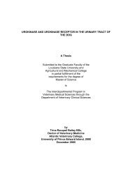
Fig. 1.1 Prototypical helix-turn-helix DNA-binding motif. The HTH motif <strong>of</strong> <strong>the</strong> trp repressor<br />
from E. coli bound to its sequence-specific DNA-binding site (blue). The first helix <strong>of</strong> <strong>the</strong> motif<br />
is colored green (with <strong>the</strong> amino-terminal end designated by “N”) and <strong>the</strong> turn colored yellow.<br />
The DNA-recognition helix (red) is inserted into <strong>the</strong> major groove (10).<br />
3
makes extensive contacts with <strong>the</strong> phosphate backbone (12, 13). Additional contacts also exist<br />
between <strong>the</strong> α-helices <strong>the</strong>mselves, stabilizing <strong>the</strong>ir relative orientations and <strong>the</strong>ir respective<br />
contacts with <strong>the</strong> DNA duplex. Fur<strong>the</strong>rmore, polypeptide backbone-, cation-, and water-<strong>mediated</strong><br />
(as seen for <strong>the</strong> trp repressor) contacts with <strong>the</strong> DNA are sometimes observed for HTH proteins<br />
(1, 3, 10). Residues outside <strong>the</strong> HTH motif also contact <strong>the</strong> DNA and help anchor <strong>the</strong> HTH motif<br />
in a specific orientation relative to <strong>the</strong> DNA duplex. For example, an N-terminal arm <strong>of</strong> <strong>the</strong> λ<br />
repressor wraps around <strong>the</strong> DNA double helix and mediates contacts in <strong>the</strong> major groove <strong>of</strong> <strong>the</strong><br />
consensus binding half-site (12).<br />
The DNA recognition helix usually tracks <strong>the</strong> DNA major groove such that <strong>the</strong> N-<br />
terminal protein side chains are in closest contact with <strong>the</strong> nucleobases <strong>of</strong> <strong>the</strong> double helix. The<br />
orientation <strong>of</strong> <strong>the</strong> recognition helical axis relative to <strong>the</strong> direction <strong>of</strong> <strong>the</strong> major groove varies,<br />
such that <strong>the</strong> angle between <strong>the</strong> α-helical axis and <strong>the</strong> axis <strong>of</strong> <strong>the</strong> DNA duplex varies by at least<br />
15° in each direction. The orientation <strong>of</strong> <strong>the</strong> recognition helix, determined by o<strong>the</strong>r structural<br />
components in <strong>the</strong> protein, in addition to <strong>the</strong> actual amino <strong>acid</strong> sequence <strong>of</strong> <strong>the</strong> α-helix,<br />
contributes to DNA sequence recognition. The N-terminus <strong>of</strong> <strong>the</strong> first α-helix <strong>of</strong> <strong>the</strong> HTH motif<br />
<strong>of</strong>ten contacts <strong>the</strong> phosphodiester backbone, but lies above <strong>the</strong> DNA recognition helix and <strong>the</strong><br />
DNA duplex (3, 9).<br />
Structural features <strong>of</strong> a DNA-binding protein form only part <strong>of</strong> <strong>the</strong> picture in analyzing<br />
mechanisms <strong>of</strong> sequence specific DNA-binding: DNA sequence, conformation, and dynamics<br />
are equally important. The patterns <strong>of</strong> hydrogen bond donors and acceptors presented in <strong>the</strong><br />
major and minor grooves are sequence specific, with each <strong>of</strong> <strong>the</strong> four base pairs forming a<br />
distinctive pattern in <strong>the</strong> major groove, with considerably less variability in <strong>the</strong> minor groove<br />
(14). In addition, <strong>the</strong> conformation <strong>of</strong> <strong>the</strong> sugar-phosphate backbone is sequence dependent. As<br />
4
numerous DNA-binding proteins induce bends in <strong>the</strong> DNA duplex, flexibility <strong>of</strong> <strong>the</strong> DNA is a<br />
critical determinant <strong>of</strong> binding affinities. In this regard, it has been shown that A-T steps<br />
generally favor duplex bending toward <strong>the</strong> minor groove, whereas G-C steps favor bending<br />
toward <strong>the</strong> major groove (9). Also, inter-base pair hydrogen bonding stabilizes high propeller<br />
twist for AT base pairs when consecutive adenines occur on <strong>the</strong> same strand, with <strong>the</strong> result that<br />
AT tracts have reduced conformational flexibility (15). Moreover, <strong>the</strong> average minor groove<br />
width <strong>of</strong> 6 Å is reduced in sequences rich in AT base pairs (9).<br />
Comparisons <strong>of</strong> HTH proteins from eukaryotic organisms with those <strong>of</strong> prokaryotes<br />
reveal very little sequence similarity, despite <strong>the</strong> conservation <strong>of</strong> <strong>the</strong> HTH structure in <strong>the</strong>ir<br />
DNA-binding domains (13, 16, 17). Eukaryotic proteins with HTH DNA-binding motifs are<br />
usually classified by <strong>the</strong> structural domain containing <strong>the</strong> HTH motif (e.g. POU and<br />
homeodomain families). Prokaryotic HTH proteins almost always bind as homodimers to a<br />
palindromic, or pseudopalindromic, DNA recognition sequence, such that <strong>the</strong> DNA recognition<br />
helices from each half <strong>of</strong> <strong>the</strong> dimer bind <strong>the</strong> symmetric DNA half-sites within <strong>the</strong> major groove<br />
<strong>of</strong> <strong>the</strong> duplex. There are exceptions to this rule: for example, proteins from <strong>the</strong> AraC family,<br />
including MarA and Rob, bind as monomers via two HTH motifs to sequence specific DNA sites<br />
(18-20).<br />
Interest in winged helix proteins has increased steadily since <strong>the</strong> co-crystal structure was<br />
provided <strong>of</strong> <strong>the</strong> DNA-binding domain <strong>of</strong> HNF-3γ complexed with its target DNA sequence (21).<br />
From this structure, it was determined that members <strong>of</strong> <strong>the</strong> eukaryotic HNF-3/fork head family<br />
<strong>of</strong> proteins mediate contacts with DNA through a novel α/β DNA-binding domain. The N-<br />
terminal half <strong>of</strong> this domain formed a HTH-like structure, with <strong>the</strong> α-helices extended in length,<br />
relative to those in <strong>the</strong> canonical HTH proteins, with <strong>the</strong> first and recognition helices being 10<br />
5
and 14 amino <strong>acid</strong>s in length, respectively. In addition, <strong>the</strong> turn in <strong>the</strong> HTH region <strong>of</strong> HNF-3γ<br />
was 8 residues, at least twice <strong>the</strong> length seen in canonical HTH domains, creating an angle<br />
between <strong>the</strong> two helices <strong>of</strong> 140° (22). The DNA recognition helix was positioned in <strong>the</strong> major<br />
groove <strong>of</strong> <strong>the</strong> DNA duplex, inducing a narrowing <strong>of</strong> this groove, and an overall bend <strong>of</strong> ∼13°<br />
(21). The average helical twist <strong>of</strong> <strong>the</strong> DNA was increased slightly, to 35°. The HNF-3γ DNA-<br />
binding fold revealed an unusual, three-stranded, antiparallel β-sheet from which two loops, or<br />
“wings” extended. Interestingly, residues from each <strong>of</strong> <strong>the</strong>se wings were shown to contact DNA,<br />
prompting <strong>the</strong> designation <strong>of</strong> this DNA recognition motif as <strong>the</strong> “winged helix” motif.<br />
Winged Helix DNA-Binding Motif<br />
Numerous DNA-binding proteins with a winged helix DNA recognition motif have since<br />
been characterized in eukarya, prokarya, archaea, and viruses. Particular interest, in regards to<br />
elucidating archaeal <strong>transcriptional</strong> regulation, has stemmed from analyses <strong>of</strong> <strong>the</strong> known archaeal<br />
genomes which suggest that most <strong>of</strong> <strong>the</strong> predicted HTH proteins, and most <strong>of</strong> <strong>the</strong> putative<br />
<strong>transcriptional</strong> <strong>regulator</strong>s, in <strong>the</strong>se organisms belong to <strong>the</strong> winged helix subfamily <strong>of</strong> DNA-<br />
binding proteins (2, 16, 23). However, this DNA-binding motif is not restricted to <strong>transcriptional</strong><br />
activators and repressors; histone H5 also binds DNA via a winged helix motif (22).<br />
The winged helix DNA binding motif, also referred to as <strong>the</strong> winged helix-turn-helix<br />
(wHTH) motif, is defined topologically by secondary structure elements arranged in <strong>the</strong><br />
following order: H1-S1-H2-H3-S2-W1-S3-W2, where “H” represents α-helix, “S” represents β-<br />
strand, and “W” represents a loop (5). The sequence spanning α-helices H2 through H3<br />
constitutes <strong>the</strong> general HTH motif, with H3 being <strong>the</strong> DNA recognition helix. The two<br />
eponymous “wing” structures are actually formed by β-strands and loops; “wing 1” is a β-hairpin<br />
motif, comprised <strong>of</strong> <strong>the</strong> S2-W1-S3 secondary structure elements and “wing 2” is formed by <strong>the</strong><br />
6
S3-W2 elements. The three β-strands interact to form an antiparallel β-sheet, however, in some<br />
winged helix proteins S1 is represented by a single, hydrophobic residue (21, 24-26). Whereas<br />
wing 1 is invariably present in winged helix proteins, some members <strong>of</strong> this family do not<br />
contain wing 2, as observed in <strong>the</strong> crystal structures <strong>of</strong> <strong>the</strong> winged helix DNA-binding domains<br />
<strong>of</strong> E2F4, DP2, histone H5 and MarR (22, 26, 27). As observed in HNF-3λ, <strong>the</strong> length <strong>of</strong> <strong>the</strong> turn<br />
in <strong>the</strong> winged helix motif can vary significantly from <strong>the</strong> 3-4 residues found in canonical HTH<br />
proteins (21). Consequently, greater variation in <strong>the</strong> angle between <strong>the</strong> two helices <strong>of</strong> <strong>the</strong> HTH<br />
motif is observed amongst <strong>the</strong> proteins in <strong>the</strong> winged helix family, than in <strong>the</strong> canonical HTH<br />
proteins. For example, <strong>the</strong> turn in DP2 is approximately 10 residues in length, whereas <strong>the</strong> turn<br />
in BirA is approximately 3 amino <strong>acid</strong>s, allowing angles <strong>of</strong> 100° and 150°, respectively (5, 25,<br />
27).<br />
DNA Recognition by Winged Helix Proteins (Role <strong>of</strong> <strong>the</strong> Wings)<br />
Structural analysis <strong>of</strong> winged helix proteins complexed with <strong>the</strong>ir target DNA sites<br />
reveals that this family is similar to HTH proteins, in that proteins similar in <strong>the</strong> tertiary<br />
structures <strong>of</strong> <strong>the</strong>ir DNA-binding motifs <strong>of</strong>ten differ in <strong>the</strong> manner in which <strong>the</strong>y contact <strong>the</strong> DNA<br />
(28). The DNA-recognition helix (H3) almost invariably contributes most <strong>of</strong> <strong>the</strong> contacts<br />
determining sequence-specificity. However, <strong>the</strong> role <strong>of</strong> <strong>the</strong> wing(s) in contributing to DNA-<br />
binding affinity and specificity seems to vary widely, with <strong>the</strong> wings <strong>of</strong> some proteins being<br />
critical for site-specific DNA-binding by mediating numerous base-specific and sugar-phosphate<br />
backbone contacts, while in o<strong>the</strong>r proteins, <strong>the</strong> wings mediate few contacts. Moreover, <strong>the</strong><br />
relative contributions from each wing (when both are present) also varies. The co-crystal<br />
structure <strong>of</strong> HNF-3λ with its target DNA-site revealed that <strong>of</strong> <strong>the</strong> 17 amino <strong>acid</strong>-<strong>mediated</strong> DNA<br />
contacts, 6 were contributed from <strong>the</strong> wing 2 structure, including three hydrogen bonds with<br />
7
ackbone phosphates, one hydrophobic interaction with a backbone ribose, one direct hydrogen<br />
bond with a base in <strong>the</strong> minor groove, and one water-<strong>mediated</strong> contact with a base in <strong>the</strong> major<br />
groove. In contrast, wing 1 contributed one hydrogen bond to <strong>the</strong> phosphate backbone from each<br />
<strong>of</strong> <strong>the</strong> S3 and W1 elements (21). Solution NMR analyses <strong>of</strong> <strong>the</strong> eukaryotic protein genesis<br />
indicate that wing 1 makes contacts with <strong>the</strong> DNA minor groove but that this wing is<br />
conformationally flexible, even in <strong>the</strong> DNA-bound state. Wing 2 appears to become less flexible<br />
upon complex formation, suggesting its importance in stabilizing <strong>the</strong> interaction. Evidence<br />
indicates that it is unlikely to contribute to sequence specificity (29-31). Conversely, wing 1<br />
appears to be more important than wing 2 in stabilizing <strong>the</strong> BmrR-DNA complex; wing 1 from<br />
BmrR mediates four hydrogen bond and hydrophobic contacts with <strong>the</strong> sugar-phosphate<br />
backbone at its target DNA sequence, and forms a hydrogen bond and a hydrophobic interaction<br />
with two bases in <strong>the</strong> minor groove (Fig. 1.2) (32). NMR structural analysis <strong>of</strong> <strong>the</strong> bacteriophage<br />
protein MuR in complex with its cognate DNA site reveals that <strong>the</strong> β-hairpin wing 1 undergoes a<br />
transition from a disordered state to a defined conformation upon DNA-binding, and that side<br />
chains from <strong>the</strong> wing hydrogen bond with bases in <strong>the</strong> minor groove, thus immobilizing <strong>the</strong> wing<br />
(33). Similarly, wing 1 <strong>of</strong> FadR mediates sequence-specific contact between a histidine with two<br />
bases in <strong>the</strong> minor groove; substitution <strong>of</strong> this histidine with a glycine completely disrupts DNA<br />
binding (34, 35). The wing-<strong>mediated</strong> DNA contacts in FadR allows an unusual mode <strong>of</strong> binding<br />
in which only <strong>the</strong> N-terminal end <strong>of</strong> each recognition helix <strong>of</strong> <strong>the</strong> homodimer contacts <strong>the</strong> DNA,<br />
allowing both recognition helices to occupy <strong>the</strong> same major groove (34). However, <strong>the</strong> wing<br />
structures do not always appear to be important in stabilizing <strong>the</strong> protein-DNA complex. The co-<br />
crystal structure <strong>of</strong> <strong>the</strong> E2F4-DP2 heterodimer with its cognate DNA-binding site revealed that<br />
<strong>the</strong> single wing from E2F4 mediates two contacts with backbone phosphates and <strong>the</strong> single wing<br />
8
N<br />
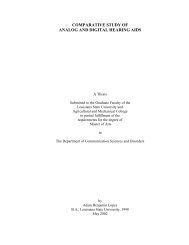
Fig. 1.2 DNA-binding by a winged helix motif. The DNA recognition helix (green) <strong>of</strong> BmrR<br />
(from <strong>the</strong> MerR family <strong>of</strong> <strong>transcriptional</strong> <strong>regulator</strong>s) is inserted into <strong>the</strong> DNA major groove<br />
(blue). “N” identifies <strong>the</strong> amino-terminal end <strong>of</strong> <strong>the</strong> helix. The β-hairpin, wing 1, motif is<br />
comprised <strong>of</strong> secondary structure elements topologically arranged (in <strong>the</strong> N- to C- terminal<br />
direction): β-strand (red), loop (yellow), β-strand (orange). Only a single strand <strong>of</strong> <strong>the</strong> cognate<br />
BmrR DNA-binding site is shown.<br />
9
from DP2 mediates only one hydrogen bond with a backbone phosphate. For this protein-DNA<br />
complex it is apparent that <strong>the</strong> wings make only slight contributions to <strong>the</strong> complex stability (27).<br />
The co-crystal structure <strong>of</strong> <strong>the</strong> winged helix protein RFX1 with its site-specific DNA-<br />
binding site revealed a strikingly different mode <strong>of</strong> interacting with <strong>the</strong> DNA duplex (36). The β-<br />
hairpin, wing 1, structure makes extensive base-specific hydrogen bonds with <strong>the</strong> DNA major<br />
groove in one half-site <strong>of</strong> <strong>the</strong> recognition sequence, narrowing <strong>the</strong> major groove by 1 Å.<br />
Surprisingly, <strong>the</strong> α-helix corresponding to <strong>the</strong> DNA recognition helix mediates only a single<br />
hydrogen bond between a lysine and a cytosine in <strong>the</strong> minor groove on <strong>the</strong> opposite face <strong>of</strong> <strong>the</strong><br />
same half-site, causing <strong>the</strong> minor groove to widen by more than 3 Å.<br />
Discovery <strong>of</strong> <strong>the</strong> mar Regulon and <strong>the</strong> Identification <strong>of</strong> MarR<br />
The identification <strong>of</strong> <strong>the</strong> MarR (multiple antibiotic resistance <strong>regulator</strong>) family <strong>of</strong><br />
<strong>transcriptional</strong> <strong>regulator</strong>s began with <strong>the</strong> identification <strong>of</strong> a chromosomally encoded mechanism<br />
<strong>of</strong> multidrug resistance in E. coli K-12 (37, 38). Genetic selections identified mutants that<br />
exhibited resistance to a broad range <strong>of</strong> antibiotics including tetracycline, chloramphenicol, β-<br />
lactams, puromycin, nalidixic <strong>acid</strong>, penicillins, fluoroquinolones, and organic solvents (37-40).<br />
The mar (multiple antibiotic resistance) phenotype was shown to be conferred by <strong>the</strong> marRAB<br />
operon, specifically by <strong>the</strong> expression <strong>of</strong> marA, which encodes a <strong>transcriptional</strong> activator<br />
belonging to <strong>the</strong> AraC family (20, 41-43). MarA is an activator <strong>of</strong> <strong>the</strong> marRAB operon and<br />
induces <strong>the</strong> expression <strong>of</strong> a number <strong>of</strong> genes responsible for <strong>the</strong> mar phenotype, including <strong>the</strong><br />
expression <strong>of</strong> <strong>the</strong> AcrAB-TolC multidrug efflux system and <strong>the</strong> gene, micF, that downregulates<br />
<strong>the</strong> syn<strong>the</strong>sis <strong>of</strong> <strong>the</strong> porin OmpF (39, 44-47). In vivo upregulation <strong>of</strong> marRAB expression and <strong>the</strong><br />
mar phenotype was shown to be inducible by a range <strong>of</strong> antibiotics and anionic lipophilic<br />
compounds, including 2, 4-dinitrophenol, menadione, plumbagin, and salicylic <strong>acid</strong> (48, 49).<br />
10
The product <strong>of</strong> <strong>the</strong> first gene <strong>of</strong> this locus, MarR (144 amino <strong>acid</strong>s), was shown to be a<br />
<strong>transcriptional</strong> repressor <strong>of</strong> <strong>the</strong> marRAB operon (48, 50). MarR binds to two separate 21 bp sites<br />
in <strong>the</strong> marRAB promoter/operator region (marO) (51). An apparent Kd <strong>of</strong> ~1 nM was calculated<br />
for MarR binding to marO. DNaseI footprinting indicated that MarR binding site I overlaps <strong>the</strong><br />
predicted –35 and –10 promoter elements and site II overlaps <strong>the</strong> putative ribosome binding site<br />
and ends immediately before <strong>the</strong> first codon <strong>of</strong> marR (51). The size <strong>of</strong> each footprint, and <strong>the</strong><br />
fact that each binding site is palindromic, with 5 bp half-sites separated by 2 bp, is consistent<br />
with MarR binding as a homodimer at each site and is supported by size-exclusion<br />
chromatography evidence that uncomplexed MarR exists as a dimer, or higher order oligomers,<br />
in solution (51). Interestingly, a number <strong>of</strong> phenolic compounds that have been shown to<br />
increase marRAB expression in vivo also antagonize MarR-marO complex formation in vitro,<br />
including 2, 4-dinitrophenol, menadione, plumbagin, and salicylic <strong>acid</strong> (52). MarR was measured<br />
to bind salicylic <strong>acid</strong> with an apparent Kd <strong>of</strong> 0.5 mM by equilibrium dialysis (51). These results,<br />
in toto, revealed a system <strong>of</strong> intrinsic multidrug resistance in E. coli that is under <strong>the</strong> control <strong>of</strong> a<br />
novel <strong>transcriptional</strong> repressor, MarR, that responds to cytoplasmic phenolic compounds.<br />
Functional marRAB operons have since been identified in Salmonella typhimurium and<br />
Enterobacter aerogenes (53, 54).<br />
Structural Analysis <strong>of</strong> MarR Homologs<br />
A search <strong>of</strong> <strong>the</strong> sequenced eukaryotic and prokaryotic genomes reveals numerous<br />
predicted MarR homologs throughout <strong>the</strong> bacterial and archaeal domains. However, to date,<br />
structural data has only been provided for two members <strong>of</strong> <strong>the</strong> MarR subfamily <strong>of</strong> winged helix<br />
DNA-binding proteins.<br />
11
The co-crystal structure <strong>of</strong> a MarR-salicylate complex was determined at 2.3 Å resolution<br />
and reveals <strong>the</strong> protein to exist as a dimer with a pyramidal shape and overall dimensions <strong>of</strong> 50 x<br />
55 x 45 Å 3 (Fig. 1.3) (26). Each monomer consists <strong>of</strong> 6 α-helices and two β-strands. The N-<br />
terminal region encompassing α-helix 1, and <strong>the</strong> C-terminal region encompassing helices 5 and<br />
6, interdigitate with <strong>the</strong> corresponding regions <strong>of</strong> <strong>the</strong> o<strong>the</strong>r subunit to form a dimerization<br />
domain with a buried surface area <strong>of</strong> 3570 Å 2 . The stabilization <strong>of</strong> MarR as a homodimer is<br />
predominantly <strong>mediated</strong> by hydrophobic contacts involving 10 residues from each subunit.<br />
Several inter-subunit hydrogen bonds (H-bonds) in this domain contribute to dimer stability: <strong>the</strong><br />
Nζ <strong>of</strong> a lysine in helix 1 H-bonds with <strong>the</strong> main chain carbonyl <strong>of</strong> <strong>the</strong> C-terminal residue, and <strong>the</strong><br />
Nζ <strong>of</strong> a lysine in helix 6 H-bonds with <strong>the</strong> main chain carbonyl in <strong>the</strong> N-terminal coil region.<br />
Helices 1 and 5 <strong>of</strong> each subunit connect <strong>the</strong> dimerization domain to a globular DNA-<br />
binding domain, such that <strong>the</strong> two DNA-binding “lobes” <strong>of</strong> <strong>the</strong> dimer are juxtaposed and related<br />
by a two-fold rotational symmetry. The topological arrangement <strong>of</strong> <strong>the</strong> secondary structure<br />
elements in <strong>the</strong> DNA-binding domain (H1-S1-H2-H3-S2-W1-S3) indicates that MarR binds via<br />
a winged helix motif. Each DNA-binding domain is stabilized by hydrophobic interactions<br />
involving 14 residues from each <strong>of</strong> <strong>the</strong> secondary structural elements, which serve to make <strong>the</strong><br />
domain compact. The residues spanning MarR helices 3 and 4 (corresponding to H2-H3 above)<br />
constitute <strong>the</strong> HTH motif. The β-hairpin (S2-W1-S3) following <strong>the</strong> HTH motif forms <strong>the</strong><br />
eponymous “wing” structure. The β-strands (S1, S2, S3) in each MarR subunit form an<br />
antiparallel β-sheet, with S1 being comprised <strong>of</strong> a single isoleucine residue, similar to <strong>the</strong><br />
structures <strong>of</strong> OmpR and BirA (24, 25). The wing extending from S3 in some winged helix<br />
proteins is absent from <strong>the</strong> DNA-binding domain <strong>of</strong> MarR, resembling <strong>the</strong> structures <strong>of</strong> E2F4,<br />
DP2, and histone H5 (22, 27). The surface potential <strong>of</strong> each DNA-binding domain is highly<br />
12
β1<br />
β2<br />
SAL-A<br />
α5<br />
α3<br />
α2<br />
C<br />
α4<br />
α6<br />
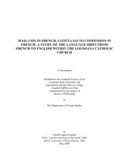
Fig. 1.3. MarR-salicylate co-crystal structure. MarR is shown as a monomer with <strong>the</strong> N- and<br />
C- termini labeled. MarR α-helices are identified as α1-6 and β-strands as β1 and β2. The wing<br />
is comprised <strong>of</strong> β1, β2, and <strong>the</strong> intervening loop (yellow). The DNA-recognition helix (green) is<br />
flanked by salicylate binding pockets SAL-A and SAL-B. Salicylate molecules are depicted in<br />
red.<br />
13<br />
SAL-B<br />
α1<br />
N
electropositive, and a 6 Å wide cleft separates <strong>the</strong> lobes from each subunit. In this salicylate-<br />
bound structure, <strong>the</strong> DNA-binding lobes <strong>of</strong> MarR interact through two salt bridges: D67 from <strong>the</strong><br />
turn <strong>of</strong> <strong>the</strong> HTH contacts R73´ in <strong>the</strong> putative recognition helix <strong>of</strong> <strong>the</strong> o<strong>the</strong>r subunit (and <strong>the</strong><br />
reciprocal interaction).<br />
MexR is a MarR homolog from Pseudomonas aeruginosa that serves as a repressor <strong>of</strong> <strong>the</strong><br />
MexAB-OprM multidrug efflux operon (55). The crystal structure <strong>of</strong> <strong>the</strong> MexR dimer in <strong>the</strong><br />
absence <strong>of</strong> effector compounds was solved at a resolution <strong>of</strong> 2.1 Å and revealed an overall,<br />
pyramidal structure very similar to MarR (Fig. 1.4) (56). Four copies <strong>of</strong> <strong>the</strong> MexR dimer were<br />
present in <strong>the</strong> crystallographic asymmetric unit, providing several conformational views <strong>of</strong> <strong>the</strong><br />
dimer. The MexR monomer is 147 amino <strong>acid</strong>s in length and is comprised <strong>of</strong> 6 α-helices and 3<br />
β-strands. The N- and C-terminal regions encompassing α-helices 1, 5, and 6 from each subunit<br />
intertwine with <strong>the</strong> reciprocal regions in <strong>the</strong> o<strong>the</strong>r subunit to create a dimerization interface with<br />
a total buried surface area ranging from 4360 – 4930 Å 3 . Stabilization <strong>of</strong> <strong>the</strong> dimer is provided<br />
almost entirely by hydrophobic contacts. Helices 1 and 5 <strong>of</strong> each subunit connect <strong>the</strong><br />
dimerization domain to a compact, globular DNA-binding domain (residues 36 – 97). The<br />
topological arrangement <strong>of</strong> <strong>the</strong> secondary structure elements <strong>of</strong> <strong>the</strong> DNA-binding domain<br />
indicates a winged helix fold, with <strong>the</strong> sequence spanning helices 3 and 4 forming <strong>the</strong> HTH. β-<br />
strands 2 and 3 and <strong>the</strong> intervening sequence form <strong>the</strong> wing. As for MarR, MexR lacks a wing 2<br />
structure. The two salt bridges that connect <strong>the</strong> DNA-binding lobes <strong>of</strong> <strong>the</strong> MarR dimer are absent<br />
in <strong>the</strong> MexR structure.<br />
Structural Evidence for Mechanisms <strong>of</strong> DNA-Binding by MarR Homologs<br />
The biochemical and genetic data, to date, is insufficient to explain <strong>the</strong> DNA-binding<br />
mechanism <strong>of</strong> MarR. A MarR-marO co-crystal structure, and biophysical analysis <strong>of</strong> MarR are<br />
14
Fig. 1.4 Comparison <strong>of</strong> <strong>the</strong> predicted MexR “closed” and “open” conformations. The<br />
individual subunits <strong>of</strong> <strong>the</strong> MexR dimer are colored red and blue (dimer AB) or yellow and green<br />
(dimer CD). MexR dimer AB is in <strong>the</strong> “closed” conformation, predicted to resemble its ligandbound<br />
state. MexR dimer CD is in <strong>the</strong> “open” conformation, suggested to resemble its DNAbinding<br />
conformation. Indicated below each structure is <strong>the</strong> distance separating <strong>the</strong> central<br />
residues in <strong>the</strong> DNA recognition helices from each half <strong>of</strong> <strong>the</strong> dimer.<br />
15
critically needed to solve this mystery. The structure <strong>of</strong> salicylate-bound MarR does not allow<br />
modeling onto B-DNA with <strong>the</strong> known MarR binding sequences. However, this finding is<br />
consistent with evidence that salicylate is a negative effector <strong>of</strong> MarR-marO complex formation.<br />
Never<strong>the</strong>less, <strong>the</strong> combined data from <strong>the</strong> MarR-salicylate co-crystal structure, MarR<br />
footprinting experiments, and MarR mutational studies, are suggestive <strong>of</strong> possible modes <strong>of</strong><br />
DNA-recognition. The identification <strong>of</strong> 5 bp inverted repeats separated by 2 bp, within each 21<br />
bp MarR binding site, would place <strong>the</strong> half-site sequences on opposite faces <strong>of</strong> <strong>the</strong> DNA double<br />
helix (51). As prokaryotic <strong>transcriptional</strong> <strong>regulator</strong>s almost invariably bind DNA as homodimers<br />
to palindromic sequences, it is reasonable to suspect that MarR does so as well. Mutational<br />
studies <strong>of</strong> MarR have shown that both <strong>the</strong> wing and <strong>the</strong> DNA recognition helix are critical for<br />
MarR-DNA complex formation, so it is highly probable that <strong>the</strong> winged helix motif <strong>of</strong> each<br />
MarR subunit binds at each half-site (11). The DNA-binding lobes <strong>of</strong> <strong>the</strong> salicylate-bound MarR<br />
structure would have to undergo significant conformational changes to accommodate binding to<br />
DNA half-sites on opposite faces <strong>of</strong> <strong>the</strong> double helix. Such conformational shifts <strong>of</strong> <strong>the</strong> globular<br />
DNA-binding domains would require flexibility in <strong>the</strong> α-helices that connect <strong>the</strong> lobes to <strong>the</strong><br />
dimerization domain. That MarR possesses such intrinsic flexibility is supported by <strong>the</strong> crystal<br />
structure, which indicates poorly ordered loop regions that might allow conformational shifts in<br />
<strong>the</strong> α-helices that form <strong>the</strong> dimerization domain (26). Also, <strong>the</strong> van der Waals interactions that<br />
stabilize <strong>the</strong> dimer do not require specific geometries <strong>of</strong> <strong>the</strong> residues involved, suggesting that<br />
flexibility would be allowed at <strong>the</strong> dimer interface. In addition, in vivo selections <strong>of</strong> MarR<br />
mutants with enhanced DNA-binding activity indicated that residues in <strong>the</strong> N- and C-terminal<br />
regions <strong>of</strong> MarR (in addition to <strong>the</strong> wing) are important in defining DNA-binding affinity (57).<br />
In vivo selection <strong>of</strong> mutants with reduced DNA-binding activities primarily identified residues in<br />
16
<strong>the</strong> recognition helix and wing, but also indicated that mutations to residues in <strong>the</strong> C-terminal<br />
region reduced DNA-binding (11). Fur<strong>the</strong>rmore, MarR crystals grown in <strong>the</strong> absence <strong>of</strong> ligand<br />
were determined to be highly disordered, consistent with MarR being intrinsically flexible (26).<br />
Protein-induced conformational changes in <strong>the</strong> DNA-binding site could also accommodate<br />
binding <strong>of</strong> a MarR dimer. For example, overwinding or underwinding <strong>of</strong> <strong>the</strong> DNA duplex, to<br />
increase or decrease <strong>the</strong> helical twist, respectively, would serve to position <strong>the</strong> major groove <strong>of</strong><br />
each half-site in an orientation that would require a less drastic conformational change in <strong>the</strong><br />
MarR dimer. Biophysical experiments to ascertain <strong>the</strong> conformational flexibility <strong>of</strong> MarR and its<br />
cognate DNA-binding sequence would be crucial in testing <strong>the</strong>se possibilities.<br />
The crystal structure <strong>of</strong> MexR in <strong>the</strong> absence <strong>of</strong> a ligand revealed a dimer in a<br />
conformation that could be docked to a linear, B-form representation <strong>of</strong> its known DNA-binding<br />
sequence with a reasonably good fit (56). The sequence-specific MexR binding site contains 5 bp<br />
half-sites separated by 5 bp, so that <strong>the</strong> center <strong>of</strong> each half-site would likely be positioned on <strong>the</strong><br />
same face <strong>of</strong> <strong>the</strong> DNA duplex (58). The MexR dimer (denoted “CD”) modeled onto <strong>the</strong> DNA-<br />
binding site was determined to be in an “open” conformation, such that <strong>the</strong> globular DNA-<br />
binding lobes were separated by a distance optimal for <strong>the</strong> insertion <strong>of</strong> <strong>the</strong> recognition helices<br />
into consecutive major grooves, and <strong>the</strong> wings were positioned to mediate contacts with <strong>the</strong><br />
sugar-phosphate backbone and minor groove (56). The MexR dimer CD structure reveals <strong>the</strong><br />
centers <strong>of</strong> <strong>the</strong> two recognition helices to be separated by 29.2 Å, close to <strong>the</strong> 34 Å distance that<br />
separates <strong>the</strong> center <strong>of</strong> each half-site in linear B-form DNA. This “open” conformation is likely<br />
maintained by electrostatic repulsions between positively charged residues lining <strong>the</strong> crevice<br />
between <strong>the</strong> DNA-binding domains. The model suggests that bending <strong>of</strong> <strong>the</strong> DNA duplex, or an<br />
increase in <strong>the</strong> helical twist, might occur upon MexR binding to accommodate a tighter fit.<br />
17
Additionally, <strong>the</strong> DNA-binding lobes <strong>of</strong> <strong>the</strong> MexR dimer might undergo conformational shifts, a<br />
possibility suggested by <strong>the</strong> intrinsic flexibility <strong>of</strong> <strong>the</strong> MexR dimer (see below).<br />
Structural Data on Phenolic Recognition in <strong>the</strong> MarR Subfamily<br />
The MarR-salicylate co-crystal structure revealed two ligand binding sites per subunit,<br />
labeled SAL-A and SAL-B (Fig. 1.5) (26). Interestingly, <strong>the</strong> bound salicylates flanked <strong>the</strong><br />
proposed DNA-recognition helix on ei<strong>the</strong>r side. Ligand binding site SAL-A is packed in <strong>the</strong><br />
interior <strong>of</strong> <strong>the</strong> globular DNA-binding domain, and is formed by residues from both helices <strong>of</strong> <strong>the</strong><br />
HTH motif and from <strong>the</strong> wing. The side chain hydroxyl <strong>of</strong> T72 from <strong>the</strong> recognition helix forms<br />
an H-bond with <strong>the</strong> salicylate hydroxyl group, <strong>the</strong> guanidinium group <strong>of</strong> R86 H-bonds with <strong>the</strong><br />
salicylate carboxylate, and <strong>the</strong> aliphatic pyrrolidone ring <strong>of</strong> P57 is positioned within 3.5 Å over<br />
<strong>the</strong> hydrophobic ring <strong>of</strong> salicylate. Site SAL-B is exposed to <strong>the</strong> surrounding solvent and most<br />
contacts with salicylate are <strong>mediated</strong> by residues from <strong>the</strong> recognition helix. The backbone<br />
carbonyl <strong>of</strong> A70 H-bonds to <strong>the</strong> salicylate hydroxyl, <strong>the</strong> guanidinium group <strong>of</strong> R77 H-bonds with<br />
<strong>the</strong> salicylate carboxylate, and <strong>the</strong> hydrophobic ring <strong>of</strong> salicylate is within 3.5 Å <strong>of</strong> <strong>the</strong><br />
hydrophobic side chain <strong>of</strong> M74. Q42 from helix 2 may also H-bond with <strong>the</strong> salicylate<br />
carboxylate.<br />
The H-bond forming residues in site SAL-A are strictly conserved in MexR, and <strong>the</strong><br />
MarR proline mediating hydrophobic contact with <strong>the</strong> salicylate ring is replaced with a leucine in<br />
MexR. Site SAL-B, however, is not conserved in MexR. The natural ligands <strong>of</strong> MexR are<br />
unknown and <strong>the</strong> structure was determined in <strong>the</strong> absence <strong>of</strong> any potential effectors (56).<br />
However, <strong>the</strong> C-terminal polypeptide from an adjacent dimer was inserted in <strong>the</strong> cleft between<br />
<strong>the</strong> DNA-binding domains <strong>of</strong> MexR dimer “AB”, resulting in a dimer significantly different in<br />
conformation than <strong>the</strong> dimer CD that was modeled onto <strong>the</strong> MexR DNA-binding site (see<br />
18
R86<br />
P57<br />
M74<br />
R77<br />
A70<br />
Fig. 1.5 MarR salicylate binding sites SAL-A and SAL-B. Salicylates are colored red.<br />
Oxygen, nitrogen, and sulfur atoms from MarR side-chains are colored red, blue, and yellow,<br />
respectively. Predicted hydrogen bonds are indicated by dashes with approximate distances.<br />
19<br />
T72
elow). The polypeptide was stabilized between <strong>the</strong> DNA-binding lobes by electrostatic<br />
interactions between <strong>the</strong> positively charged residues <strong>of</strong> <strong>the</strong> dimer lining <strong>the</strong> crevice and<br />
negatively charged glutamate and aspartate residues from <strong>the</strong> inserted polypeptide. Several van<br />
der Waals contacts between proline, leucine, and isoleucine residues in <strong>the</strong> polypeptide with<br />
hydrophobic residues lining <strong>the</strong> crevice <strong>of</strong> <strong>the</strong> dimer also stabilized <strong>the</strong> interaction.<br />
Proposed Mechanisms <strong>of</strong> Allosteric Regulation <strong>of</strong> MarR Proteins<br />
As discussed above, <strong>the</strong> salicylate-bound structure <strong>of</strong> MarR is in a conformation that is<br />
unlikely to bind its sequence specific DNA-binding site (26). Such binding would require<br />
significant conformational flexibility in <strong>the</strong> protein and/or <strong>the</strong> DNA duplex. The location <strong>of</strong> <strong>the</strong><br />
two salicylate binding sites on ei<strong>the</strong>r side <strong>of</strong> <strong>the</strong> proposed DNA recognition helix is suggestive <strong>of</strong><br />
possible mechanisms by which phenolic ligands antagonize MarR-DNA interaction. As both<br />
ligand-binding sites SAL-A and SAL-B are composed <strong>of</strong> residues from <strong>the</strong> DNA-recognition<br />
helix and wing motif, and as both <strong>of</strong> <strong>the</strong>se regions <strong>of</strong> <strong>the</strong> winged helix fold have been shown to<br />
be critical for DNA-binding, it is clear that <strong>the</strong> ligand- and DNA-binding sites are not separate in<br />
MarR. It is plausible that ligand binding at one, or both, sites coordinates residues required for<br />
direct, or water-<strong>mediated</strong>, contacts with <strong>the</strong> cognate DNA-binding site, thus preventing<br />
sequence-specific MarR-marO complex formation (11). Alternatively, or in combination with<br />
<strong>the</strong> effect posited above, ligand binding might stabilize a MarR dimer conformation that cannot<br />
accommodate <strong>the</strong> insertion <strong>of</strong> <strong>the</strong> recognition helices into <strong>the</strong> major grooves at <strong>the</strong> binding<br />
sequence half-sites. The suggested intrinsic flexibility <strong>of</strong> MarR at <strong>the</strong> dimerization interface is<br />
consistent with a ligand-<strong>mediated</strong> conformational shift in <strong>the</strong> relative positions <strong>of</strong> <strong>the</strong> DNA-<br />
binding lobes, from a DNA-binding state, to <strong>the</strong> state observed in <strong>the</strong> crystal structure.<br />
20
The four dimers present in <strong>the</strong> crystallographic asymmetric unit <strong>of</strong> MexR provided a<br />
fortuitous glimpse <strong>of</strong> a possible mechanism <strong>of</strong> ligand-<strong>mediated</strong> allosteric control <strong>of</strong> MexR-DNA<br />
binding (56). A comparison <strong>of</strong> <strong>the</strong> “open” MexR dimer CD conformation, that was readily<br />
modeled onto its linear B-form DNA binding site, with <strong>the</strong> MexR dimer AB-polypeptide<br />
conformation, revealed <strong>the</strong> latter to be incompatible with binding to its recognition sequence.<br />
Notably, <strong>the</strong> distance separating <strong>the</strong> centers <strong>of</strong> <strong>the</strong> recognition helices had been reduced to 22.6<br />
Å in <strong>the</strong> MexR dimer AB, compared to <strong>the</strong> distance <strong>of</strong> 29.2 Å observed in <strong>the</strong> “open”<br />
conformation (Fig. 1.4). Comparisons <strong>of</strong> all four dimer representations revealed that <strong>the</strong> basis for<br />
this conformational shift resided in <strong>the</strong> intrinsic flexibility <strong>of</strong> <strong>the</strong> dimerization domain. Whereas<br />
<strong>the</strong> winged helix DNA-binding domains appeared to shift in orientation as a rigid body, flexible<br />
loop regions allow for significant conformational flexibility in α-helices 1, 5, and 6. The helix<br />
orientations <strong>of</strong> α-helices 1, 5, and 6 vary by 17°, 6.8°, and 12.1°, respectively, with <strong>the</strong>ir<br />
midpoint positions deviating by 4.9 Å, 1.7 Å, and 8.2 Å. This flexibility <strong>of</strong> <strong>the</strong> helices that<br />
comprise <strong>the</strong> dimerization domain is consistent with variable geometries being allowed for <strong>the</strong><br />
van der Waals contacts that stabilize <strong>the</strong> dimer. As α-helices 1 and 5 connect <strong>the</strong> dimerization<br />
domain to <strong>the</strong> DNA-binding domain, <strong>the</strong>ir flexibility results in concomitant shifts in <strong>the</strong> DNA-<br />
binding lobes. The “closed” conformation observed for MexR dimer AB suggests that ligands<br />
may disrupt MexR-DNA complex formation by neutralizing <strong>the</strong> electrostatic repulsions that<br />
o<strong>the</strong>rwise maintain <strong>the</strong> dimer in an “open”, DNA-binding conformation. Additional hydrophobic<br />
contacts between <strong>the</strong> ligand and residues lining <strong>the</strong> crevice <strong>of</strong> <strong>the</strong> MexR dimer contribute to<br />
bring <strong>the</strong> DNA-binding lobes into closer proximity. As all known ligands <strong>of</strong> MarR homologs are<br />
anionic lipophilic compounds, it is tempting to speculate that members <strong>of</strong> this family share a<br />
similar mechanism <strong>of</strong> allosteric control.<br />
21
O<strong>the</strong>r Members <strong>of</strong> <strong>the</strong> MarR Family<br />
Since <strong>the</strong> discovery <strong>of</strong> MarR, a number <strong>of</strong> MarR homologs have been predicted from <strong>the</strong><br />
genomes <strong>of</strong> Gram-positive and Gram-negative bacteria, mycobacteria, and archaea. However,<br />
only a small subset <strong>of</strong> <strong>the</strong> potential MarR homologs have been characterized biochemically or<br />
genetically.<br />
All members <strong>of</strong> <strong>the</strong> MarR family possess a winged helix DNA-binding motif. All<br />
characterized homologs exist as dimers in both <strong>the</strong> uncomplexed and DNA-bound states and this<br />
may be a defining characteristic <strong>of</strong> this family (lower proportions <strong>of</strong> higher order oligomers have<br />
been observed for some uncomplexed homologs). Consistent with <strong>the</strong> observed DNA-binding<br />
stoichiometries, MarR homologs invariably bind to palindromic or pseudopalindromic DNA<br />
sequences that presumably reflect <strong>the</strong> two-fold rotational symmetry <strong>of</strong> <strong>the</strong> protein dimer. The<br />
gene encoding each MarR homolog is generally part <strong>of</strong> a gene cluster containing <strong>the</strong> gene(s)<br />
under its regulation (with <strong>the</strong> possible exceptions <strong>of</strong> FarR and PecS). In some cases, <strong>the</strong> MarR<br />
homolog is encoded in its regulated operon. A large proportion <strong>of</strong> <strong>the</strong> characterized family<br />
members are adjacent to <strong>the</strong> divergently transcribed gene(s) <strong>the</strong>y regulate, such that <strong>the</strong> MarR<br />
homolog binding site(s) reside in <strong>the</strong> intergenic region containing <strong>the</strong> associated, divergent<br />
promoters. More than half <strong>of</strong> <strong>the</strong> characterized MarR homologs have been shown to be<br />
auto<strong>regulator</strong>y. Most members <strong>of</strong> this family serve as repressors <strong>of</strong> gene transcription, but<br />
several activators have been identified. Response to environmental phenolic ligands has been<br />
demonstrated for a number <strong>of</strong> MarR proteins. Specifically, ligand-responsive MarR proteins<br />
almost invariably respond to anionic lipophilic compounds in <strong>the</strong>ir capacity to bind <strong>the</strong>ir cognate<br />
DNA sequences. The physiological roles <strong>of</strong> MarR proteins can be classified into three general<br />
categories, with some proteins serving multiple <strong>regulator</strong>y roles: 1) regulation <strong>of</strong> response to<br />
22
environmental stress, 2) regulation <strong>of</strong> aromatic catabolic pathways, and 3) regulation <strong>of</strong> virulence<br />
factors.<br />
MarR Regulators <strong>of</strong> Stress Response<br />
The MarR homolog, EmrR from E. coli, was first demonstrated to be repressor <strong>of</strong><br />
microcin B and C production and later shown to be <strong>the</strong> encoded by <strong>the</strong> first gene <strong>of</strong> <strong>the</strong> emrRAB<br />
operon, which encodes a multidrug resistance pump (59, 60). Analyses in vivo using lacZ fusions<br />
demonstrated that emrR expression represses <strong>the</strong> emrRAB locus and that this repression could be<br />
relieved by certain antibiotics and protonophores that are <strong>the</strong> targets <strong>of</strong> <strong>the</strong> EmrAB pump (60). In<br />
addition, <strong>the</strong> MarR ligands, salicylate and 2,4-dinitrophenol, also induced expression <strong>of</strong> this<br />
operon. Gel electrophoresis under non-reducing conditions suggested that EmrR exists as a<br />
dimer in solution (61). DNaseI footprinting analysis revealed a surprisingly large EmrR binding<br />
site <strong>of</strong> 42 bp that partially overlaps <strong>the</strong> –35 promoter element and extends past <strong>the</strong> start site <strong>of</strong><br />
transcription for <strong>the</strong> emrRAB operon (62). This site contains a pseudopalindromic sequence<br />
comprised <strong>of</strong> 9 bp half-sites separated by 3 bp. Several compounds were shown to negatively<br />
effect EmrR-DNA complex formation. Direct binding between potential ligands and EmrR was<br />
demonstrated using equilibrium dialysis and <strong>the</strong> ligand binding affinities <strong>of</strong> 2,4-dinitrophenol<br />
and two protonophores were measured spectrophotometrically, revealing apparent dissociation<br />
constants <strong>of</strong> approximately 2.0, 3.0, and 15.0 µM, respectively (61). Interestingly, fitting <strong>the</strong> data<br />
to <strong>the</strong> Scatchard equation suggested that each ligand bound to a single site in <strong>the</strong> EmrR<br />
monomer.<br />
The gene encoding <strong>the</strong> MarR homolog, MexR from P. aeruginosa, is adjacent to <strong>the</strong><br />
oppositely oriented mexAB-oprM operon that encodes a non-ATPase, multisubstrate efflux pump<br />
that contributes to this organism’s intrinsic multidrug resistance (55). Initial genetic evidence<br />
23
suggested a <strong>regulator</strong>y role for MexR. A mexR mutant strain was shown to have enhanced<br />
resistance to antibiotics, and MexR was shown to reduce <strong>the</strong> expression <strong>of</strong> mexA:lacZ,<br />
mexA:phoA, and mexR:lacZ fusions, suggesting that MexR is an autorepressor and repressor <strong>of</strong><br />
<strong>the</strong> mexAB-oprM operon (55). Despite suggestions that MexR might possess dual repressor and<br />
activator roles in vivo, extensive genetic experiments indicate that MexR functions only as a<br />
repressor <strong>of</strong> this operon (63). DNaseI footprinting identified two MexR binding sites within <strong>the</strong><br />
274 bp intergenic region that separates mexR and mexA (58). Site I is ∼29 bp and overlaps <strong>the</strong><br />
predicted –10 promoter element for mexR and a putative –35 promoter element for <strong>the</strong> mexAB-<br />
oprM operon. Site II is ∼28 bp and overlaps <strong>the</strong> predicted –35 promoter element for mexR and a<br />
putative –10 promoter element for <strong>the</strong> operon. Each site contains a palindromic sequence<br />
comprised <strong>of</strong> 5 bp half-sites separated by 5 bp. Curiously, <strong>the</strong> footprints <strong>of</strong> sites I and II are<br />
separated by only 3 bp (58). In addition to <strong>the</strong> crystal structure evidence, two-hybrid experiments<br />
are consistent with MexR existing as a dimer in solution, and it is <strong>the</strong>refore likely that MexR<br />
binds each <strong>of</strong> its palindromic sites as a dimer (64). It will be interesting to see if cooperativity<br />
exists in MexR binding to its closely spaced binding sequences. Surprisingly, <strong>the</strong> selection <strong>of</strong><br />
trans-dominant MexR mutants that were defective in DNA-binding, but not dimerization,<br />
predominantly identified single-amino <strong>acid</strong> substitutions <strong>of</strong> hydrophobic residues in <strong>the</strong> DNA-<br />
binding domain. Only 2 <strong>of</strong> <strong>the</strong> 25 mutations were to charged residues, suggesting <strong>the</strong> importance<br />
<strong>of</strong> hydrophobic contacts in this region in stabilizing <strong>the</strong> winged helix motif in a conformation<br />
that can accommodate DNA-binding (65).<br />
A member <strong>of</strong> <strong>the</strong> MarR family has been characterized from Neisseria gonorrhoeae that<br />
likely mediates <strong>the</strong> resistance <strong>of</strong> this organism to antimicrobial hydrophobic agents. The farAB<br />
operon <strong>of</strong> N. gonorrhoeae encodes an efflux pump that exports out <strong>of</strong> <strong>the</strong> cell host-derived<br />
24
antimicrobial agents such as long-chain fatty <strong>acid</strong>s (FAs). Using lacZ reporter fusions, it was<br />
shown that farAB and farR expression was enhanced in strains mutated at farR (66). Also, strains<br />
mutated at <strong>the</strong> farR site were less resistant to FAs. Electrophoretic mobility shift assays<br />
demonstrated that FarR binds sequence specifically in <strong>the</strong> farAB and farR promoter regions.<br />
Thus, FarR is auto<strong>regulator</strong>y and represses <strong>the</strong> farAB efflux system, but it remains to be<br />
determined if its <strong>regulator</strong>y activity is affected by certain fatty <strong>acid</strong>s or o<strong>the</strong>r potential ligands.<br />
MarR Regulators <strong>of</strong> Aromatic Catabolism<br />
A phenolic sensing protein from Rhodopseudomonas palustris has been characterized as<br />
an inducer <strong>of</strong> <strong>the</strong> badDEFG operon, which encodes benzoyl-CoA reductase, an enzyme involved<br />
in <strong>the</strong> anaerobic catabolism <strong>of</strong> benzoate (67). Analysis in vivo using a badE:`lacZ fusion<br />
construct demonstrated that, in <strong>the</strong> presence <strong>of</strong> benzoate or 4-hydroxybenzoate, BadR increases<br />
lacZ expression approximately 5-fold. Strains deficient in badR grew slowly under anaerobic<br />
conditions with benzoate as a carbon source. Though not shown, it is likely that <strong>the</strong> stimulatory<br />
effect <strong>of</strong> BadR occurs through direct binding <strong>of</strong> benzoyl-CoA reductase substrates to this protein,<br />
which induces DNA-binding <strong>of</strong> BadR to <strong>the</strong> promoter <strong>of</strong> badDEFG. This proposed mechanism is<br />
interesting in light <strong>of</strong> <strong>the</strong> fact that <strong>the</strong> known ligands <strong>of</strong> o<strong>the</strong>r MarR homologs almost invariably<br />
antagonize DNA-binding. The badR gene is part <strong>of</strong> a gene cluster including <strong>the</strong> badDEFG<br />
operon, but is transcribed from a separate promoter. The mechanism by which <strong>the</strong> proposed<br />
DNA-binding by BadR activates gene expression, and whe<strong>the</strong>r or not BadR is auto<strong>regulator</strong>y,<br />
remains to be determined.<br />
The MarR homolog, CbaR from Comamonas testosteroni BR60, is a phenolic-sensing<br />
modulator <strong>of</strong> <strong>the</strong> cbaABC operon that encodes enzymes involved in <strong>the</strong> oxidation <strong>of</strong> 3-<br />
chlorobenzoate (68). An intergenic region <strong>of</strong> 667 bp separates cbaR from <strong>the</strong> oppositely oriented<br />
25
cbaABC operon. Electrophoretic mobility shift assays, and in vivo analysis monitoring lacZ<br />
expression, indicated that CbaR does not regulate its own expression. However, gel shifts and<br />
DNaseI footprinting indicated that CbaR forms two distinct complexes, dissimilar in binding<br />
affinities, in <strong>the</strong> promoter region <strong>of</strong> <strong>the</strong> cbaABC operon, suggesting that CbaR represses gene<br />
expression. The high-affinity ∼22 bp binding site is located ∼40 bp downstream <strong>of</strong> <strong>the</strong><br />
<strong>transcriptional</strong> start site and contains a palindromic sequence comprised <strong>of</strong> 4 bp half-sites<br />
separated by 6 or 9 base pairs. The low-affinity binding site contains a pseudopalindromic<br />
sequence similar to <strong>the</strong> high-affinity sequence. The substrate <strong>of</strong> <strong>the</strong> enzymes encoded by <strong>the</strong><br />
cbaABC operon, 3-chlorobenzoate, is an efficient antagonist <strong>of</strong> CbaR complex formation at both<br />
sites. Interestingly, <strong>the</strong> downstream product <strong>of</strong> 3-chlorobenzoate catabolism, protocatechuate, is<br />
also an efficient antagonist, suggesting a mechanism <strong>of</strong> positive feedback in cbaABC expression.<br />
Curiously, <strong>the</strong> phenolic compounds 3-hydroxybenzoate and 3-carboxybenzoate might promote a<br />
slight increase in <strong>the</strong> affinity <strong>of</strong> CbaR for each <strong>of</strong> its binding sites, suggesting that <strong>the</strong>se<br />
compounds enhance CbaR-<strong>mediated</strong> gene repression.<br />
A MarR homolog has been characterized from <strong>the</strong> ruminal bacterium Butyrivibrio<br />
fibrisolvens E14 that regulates <strong>the</strong> expression <strong>of</strong> an enzyme involved in <strong>the</strong> catabolism <strong>of</strong><br />
polysaccharide derivatives in plant cell walls (69). It was shown that cells overexpressing a 142<br />
residue, 16 kDa protein, CinR, and a cinnamoyl ester hydrolase (CinB) displayed less activity<br />
from this enzyme than did cells overexpressing CinB alone. The open reading frames <strong>of</strong> cinR and<br />
cinB are oriented in <strong>the</strong> opposite direction in <strong>the</strong> Butyrivibrio genome and are separated by an<br />
intergenic region <strong>of</strong> 170 bp containing predicted, overlapping promoter elements for each gene.<br />
Gel shifts demonstrated that CinR binds a DNA fragment containing <strong>the</strong> intergenic region<br />
between cinR and cinB. Sequence analysis identified two identical 16 bp palindromic sites in this<br />
26
intergenic region, comprised <strong>of</strong> 8 bp half-sites. One <strong>of</strong> <strong>the</strong>se potential CinR binding sites<br />
overlaps <strong>the</strong> predicted cinR <strong>transcriptional</strong> start site and <strong>the</strong> o<strong>the</strong>r lies just downstream <strong>of</strong> <strong>the</strong><br />
putative cinB <strong>transcriptional</strong> start site, suggesting that CinR represses its own expression and that<br />
<strong>of</strong> cinB. Two cinnamic <strong>acid</strong> sugar esters, potential substrates <strong>of</strong> CinB, were shown to antagonize<br />
CinR-DNA complex formation. Notably, certain cinnamic <strong>acid</strong>s, sugars, and a non-sugar<br />
cinnamic <strong>acid</strong> ester were not negative effectors <strong>of</strong> CinR DNA-binding activity, indicating that<br />
both <strong>the</strong> sugar and cinnamic groups are essential components <strong>of</strong> a CinR effector.<br />
The MarR homolog HcaR, from Acinetobacter sp. strain ADP1, regulates <strong>the</strong> hcaABCDE<br />
operon that encodes genes required for <strong>the</strong> catabolism <strong>of</strong> plant-derived hydroxycinnamates.<br />
Genetic analyses indicated that HcaR represses this operon and that this repression is relieved by<br />
hydoxycinnamoyl-CoA thioesters (70). The gene encoding HcaR and <strong>the</strong> hcaABCDE operon are<br />
part <strong>of</strong> <strong>the</strong> same gene cluster, but are divergently transcribed. Direct interaction <strong>of</strong> HcaR with <strong>the</strong><br />
operon promoter has not been demonstrated and <strong>the</strong> HcaR recognition sequence remains<br />
unknown.<br />
The hpa-meta operon <strong>of</strong> E. coli encodes a set <strong>of</strong> genes required for <strong>the</strong> catabolism <strong>of</strong> 4-<br />
hydroxyphenylacetic <strong>acid</strong> (4-HPA). An intergenic region <strong>of</strong> 219 bp separates <strong>the</strong> first ORF <strong>of</strong><br />
this operon from a divergently transcribed gene encoding HpaR. In vitro transcription assays<br />
demonstrated that HpaR repressed <strong>the</strong> expression from <strong>the</strong> hpa-meta operon promoter and that 4-<br />
HPA effectively relieved this repression (71). In addition, 4-HPA, and two structurally similar<br />
compounds (3-HPA and 3,4-HPA), increased expression from a lacZ fusion with <strong>the</strong> hpa-meta<br />
operon promoter. In vivo assays involving a lacZ translational fusion with <strong>the</strong> hpaR promoter<br />
demonstrated that HpaR is an autorepressor, and <strong>the</strong> same phenolic compounds listed above<br />
relieve that repression. EMSAs and DNaseI footprinting identified two 27 bp HpaR binding sites<br />
27
in <strong>the</strong> intergenic region between hpaR and <strong>the</strong> hpa-meta operon. The affinity <strong>of</strong> HpaR for one<br />
site (OPR1) was 10-fold higher than for <strong>the</strong> o<strong>the</strong>r site (OPR2). Site OPR1 overlapped <strong>the</strong> –10<br />
promoter element and <strong>transcriptional</strong> start site <strong>of</strong> <strong>the</strong> hpa-meta operon and contained a<br />
palindromic sequence comprised <strong>of</strong> 9 bp half-sites separated by 4 bp. Site OPR2 was located<br />
between <strong>the</strong> <strong>transcriptional</strong> start site and ribosome binding site <strong>of</strong> hpaR. OPR2 contained a<br />
pseudopalindromic sequence, with one <strong>of</strong> its half-sites differing in 5 bp from <strong>the</strong> half-sites in<br />
OPR1. Interestingly, gel shifts comparing HpaR binding affinities to DNA fragments containing<br />
one, or both, sites indicated that HpaR binding is positively cooperative at <strong>the</strong>se sites. Potassium<br />
permanganate footprinting demonstrated that HpaR likely represses transcription <strong>of</strong> <strong>the</strong> hpa-meta<br />
operon by blocking promoter escape by RNA polymerase. In <strong>the</strong> presence <strong>of</strong> HpaR, <strong>the</strong> open<br />
complex was shifted upstream <strong>of</strong> <strong>the</strong> HpaR binding site, and partially overlapped <strong>the</strong> –35<br />
promoter element (71).<br />
MarR Regulators <strong>of</strong> Virulence Factors<br />
A MarR homolog from <strong>the</strong> nitrogen-fixing bacterium, Sinorhizobium meliloti, has been<br />
shown to activate transcription <strong>of</strong> three exp operons that are involved in <strong>the</strong> production <strong>of</strong><br />
galactoglucan, an exopolysaccharide required for this organism to infect plant roots for nodule<br />
formation (72, 73). ExpG was shown to mediate this activation by binding to promoter regions in<br />
<strong>the</strong> exp gene cluster (74). Size exclusion chromatography suggested that this protein, ExpG,<br />
exists as a dimer in solution (75). Analysis <strong>of</strong> ExpG-DNA binding using atomic force<br />
microscopy (AFM) revealed that <strong>the</strong> DNA curvature at <strong>the</strong> ExpG binding site changes upon<br />
complex dissociation, suggesting that ExpG induces DNA bending upon complex association.<br />
Electrophoretic mobility shift assays and AFM experiments demonstrated that ExpG binding<br />
sites require a palindromic sequence comprised <strong>of</strong> 6 bp half-sites separated by 3 bp.<br />
28
Measurements <strong>of</strong> <strong>the</strong> binding kinetics for ExpG at 3 different promoter binding sites indicated<br />
that <strong>the</strong> ExpG dimer binds with a very high on-rate (ranging from 1.0 – 5.0 x 10 5 M -1 s -1 ) and<br />
dissociates with a very low <strong>of</strong>f-rate (ranging from 4.3 – 1.3 x 10 -4 s -1 ) indicating dissociation<br />
constants ranging from 0.58 – 1.3 nM. Interestingly, <strong>the</strong> DNA-binding affinity <strong>of</strong> ExpG is<br />
enhanced by <strong>the</strong> presence <strong>of</strong> short conserved sequences (“boxes 1 and 2”) located on ei<strong>the</strong>r side<br />
<strong>of</strong> <strong>the</strong> palindrome. The distance <strong>of</strong> <strong>the</strong>se boxes from <strong>the</strong> conserved 21 bp sequence containing <strong>the</strong><br />
palindrome ranges from 5 – 30 bp. Dynamic force spectroscopy was used to calculate <strong>the</strong> natural<br />
<strong>the</strong>rmal <strong>of</strong>f-rates for <strong>the</strong> binding kinetics between ExpG and a wild-type binding site, a binding-<br />
site mutated in box 1, and a binding site mutated in box 2. The <strong>of</strong>f-rate was increased<br />
approximately 10-fold by a mutation in box 1 and approximately 100-fold by a mutation in box<br />
2. Sites analogous to boxes 1 and 2 have not been described for any o<strong>the</strong>r MarR family member.<br />
Gel shift experiments determined that <strong>the</strong> MarR homolog, PecS from Erwinia<br />
chrysan<strong>the</strong>mi, binds specifically to <strong>the</strong> <strong>regulator</strong>y regions <strong>of</strong> pectinase and cellulase enzymes,<br />
virulence factors required for this organism to infect plant cells (76, 77). PecS binds with a Kd <strong>of</strong><br />
∼200 nM to <strong>the</strong> promoter regions <strong>of</strong> two pectinase genes and has been shown to be involved in a<br />
complex <strong>regulator</strong>y system <strong>of</strong> <strong>the</strong>se enzymes, involving CRP and ano<strong>the</strong>r repressor, KdgR (77).<br />
DNaseI footprinting experiments determined that PecS protects an ∼50 bp site in <strong>the</strong> pectinase<br />
promoter region, which contains a high-affinity binding site for <strong>the</strong> activator, CRP, suggesting a<br />
mechanism for PecS-induced <strong>transcriptional</strong> attenuation (77). A 23 bp consensus DNA-binding<br />
sequence for PecS was defined and shown to contain a palindromic sequence comprised <strong>of</strong> 5 bp<br />
half-sites separated by 3 bp (78). In addition, PecS has been shown to regulate <strong>the</strong> expression <strong>of</strong><br />
genes involved in <strong>the</strong> production <strong>of</strong> flagella, by binding to a <strong>regulator</strong>y region with an apparent<br />
Kd <strong>of</strong> 20 nM (78). Fur<strong>the</strong>rmore, genetic experiments identified PecS as a repressor <strong>of</strong> a cluster <strong>of</strong><br />
29
genes involved in <strong>the</strong> syn<strong>the</strong>sis <strong>of</strong> <strong>the</strong> blue pigment, indigoidine (79). Gel shifts determined that<br />
PecS binds to <strong>the</strong> promoter regions <strong>of</strong> two genes in this cluster, with apparent Kd’s <strong>of</strong> 5 and 20<br />
nM. Interestingly, increased production <strong>of</strong> indigoidine, conferred by a pecS mutation in E.<br />
chrysan<strong>the</strong>mi, resulted in a significant increase in tolerance to oxidative stress, presumably due<br />
to <strong>the</strong> ability <strong>of</strong> this aromatic compound to scavenge reactive oxygen species (79).<br />
Deinococcus radiodurans<br />
Deinococcus radiodurans is one <strong>of</strong> 11 members belonging to <strong>the</strong> family Deinococcaceae,<br />
which includes at least 7 members that are intrinsically resistant to extreme levels <strong>of</strong> radiation<br />
and o<strong>the</strong>r sources <strong>of</strong> DNA damage, such as oxidative stress and dessication (80-84). Though<br />
Deinococcus <strong>of</strong>ten stains Gram-positive, sequence comparisons <strong>of</strong> 16S rDNA suggests that this<br />
lineage is most closely related to <strong>the</strong> <strong>the</strong>rmophilic, Gram-negative genus, Thermus (85). This<br />
relationship is consistent with <strong>the</strong> peptidoglycan chemotype shared by <strong>the</strong>se two genera (86, 87).<br />
The D. radiodurans genome is comprised <strong>of</strong> two chromosomes, one megaplasmid, and one<br />
plasmid and is predicted to encode 3195 ORFs (88). D. radiodurans maintains 8-10 copies <strong>of</strong> its<br />
genome during exponential growth and approximately 4 copies during stationary phase (89).<br />
Interest in D. radiodurans has focused primarily on its ability to repair extensive levels <strong>of</strong><br />
DNA damage. It has been shown that D. radiodurans is approximately 50-fold more resistant to<br />
ionizing radiation than is E. coli, demonstrating 10% survival at 800 kilorads, whereas E. coli<br />
has 10% survival at 15 kilorads (90-92). This organism can survive high doses <strong>of</strong> acute gamma<br />
radiation (greater than 1500 kilorads) and grow under conditions <strong>of</strong> prolonged exposure at 6<br />
kilorads, remarkably without accumulating abnormal levels <strong>of</strong> mutations (93-95). Similar levels<br />
<strong>of</strong> resistance are observed when D. radiodurans is exposed to prolonged dessication, UV<br />
radiation, or oxidative stress (83, 92, 96). The damage accrued by <strong>the</strong> D. radiodurans genome<br />
30
under <strong>the</strong>se conditions suggests that this organism’s intrinsic resistance is a consequence <strong>of</strong> a<br />
remarkable DNA repair system (93, 97). Whereas E. coli can only sustain 10-15 double strand<br />
breaks in its genome, D. radiodurans can repair up to 2000 such breaks (92). Efficient<br />
homologous recombination is most likely <strong>the</strong> primary means by which D. radiodurans can<br />
withstand such high levels <strong>of</strong> DNA damage (98, 99). However, numerous non-resistant<br />
organisms possess <strong>the</strong> same complement <strong>of</strong> genes for recombination, leaving unresolved <strong>the</strong> full<br />
mechanism by which D. radioduran’s maintains resistance to extreme levels <strong>of</strong> DNA damage<br />
(92). Recent evidence demonstrates that <strong>the</strong> D. radiodurans nucleoid adopts an unusual toroidal<br />
morphology, which suggests that efficient DNA recombination may be facilitated by <strong>the</strong><br />
alignment <strong>of</strong> homologous segments from multiple copies <strong>of</strong> <strong>the</strong> genome (100).<br />
It has been noted that D. radiodurans undergoes a dose-dependent growth lag after<br />
exposure to DNA-damaging events (92). Damaged nucleotides and nucleosides are exported<br />
from <strong>the</strong> cell during this period <strong>of</strong> growth inhibition (101). In addition, D. radiodurans must<br />
handle <strong>the</strong> enhanced levels <strong>of</strong> reactive oxygen species that are <strong>the</strong> direct products <strong>of</strong> ionizing and<br />
UV radiation, dessication, and oxidative stress agents. Catalase and superoxide dismutase<br />
enzymes have been shown to be critical in this regard, as mutations to <strong>the</strong>se genes reduce D.<br />
radiodurans resistance to ionizing radiation (96). In addition, <strong>the</strong> D. radiodurans genome<br />
encodes two Dps (DNA protection during starvation) homologs. Dps proteins are structurally<br />
related to ferritin, and have been shown to protect DNA from damage by physically binding<br />
DNA and by chelating iron (102, 103). Iron, in its ferrous (Fe 2+ ) state, can be particularly toxic to<br />
aerobically respiring organisms such as D. radiodurans by reacting with hydrogen peroxide to<br />
generate reactive oxygen species that are damaging to amino <strong>acid</strong>s and lipids, as well as DNA<br />
nucleobases. One <strong>of</strong> <strong>the</strong>se Dps homologs in D. radiodurans, Dps-1, has been recently<br />
31
characterized and shown to possess ferroxidase activity, oxidizing Fe 2+ to Fe 3+ (104). The<br />
dodecameric form <strong>of</strong> this protein stores <strong>the</strong> oxidized iron, while a dimeric form <strong>of</strong> Dps-1 protects<br />
DNA from DNaseI and hydroxyl radical cleavage, suggesting a role for this protein in mediating<br />
oxidative stress resistance in D. radiodurans (104).<br />
As numerous MarR proteins have been shown to regulate prokaryotic stress responses to<br />
toxic compounds and oxidative stress agents, <strong>the</strong> identification <strong>of</strong> two predicted MarR homologs<br />
within <strong>the</strong> D. radiodurans genome suggested <strong>the</strong> possibility <strong>of</strong> o<strong>the</strong>r uncharacterized stress<br />
response proteins that contribute to <strong>the</strong> extreme-resistance phenotype <strong>of</strong> this organism.<br />
References<br />
1. Luscombe, N.M., Austin, S.E., Berman, H.M., and Thornton, J.M. (2000) Genome Biol. 1,<br />
1-37<br />
2. Pérez-Rueda, E., Collado-Vides, J., and Segovia, L. (2004) Comput. Biol. Chem. 28, 341-<br />
350<br />
3. Pabo, C.O., and Sauer, R.T. (1992) Annu. Rev. Biochem. 61, 1053-1095<br />
4. Huffman, J.L., and Brennan, R.G. (2002) Curr. Opin. Struct. Biol. 12, 98-106<br />
5. Gajiwala, K.S. and Burley, S.K. (2000) Curr. Opin. Struct. Biol. 10, 110-116<br />
6. Anderson, W.F., Ohlendorf, D.H., Takeda, Y., Mat<strong>the</strong>ws, B.W. (1981) Nature 290, 754-758<br />
7. Ohlendorf, D.H., Anderson, W.F., Fisher, R.G., Takeda, Y., Mat<strong>the</strong>ws, B.W. (1982) Nature<br />
298, 718-723<br />
8. McKay, D.B., Steitz, T.A. (1981) Nature 290, 744-749<br />
9. Harrison, S.C., and Aggarwal, A.K. (1990) Annu. Rev. Biochem. 59, 933-969<br />
10. Otwinowski, Z., Schevitz, R.W., Zhang, R.G., Lawson, C.L., Joachimiak, A., Marmorstein,<br />
R.Q., Luisi, B.F., and Sigler, P.B. (1988) Nature 335, 321-329<br />
11. Alekshun, M.N., Kim, Y.S., and Levy, S.B. (2000) Mol. Microbiol. 35, 1394-1404<br />
12. Jordan, S.R., and Pabo, C.O. (1988) Science 242, 893-899<br />
32
13. Xu, W., Rould, M.A., Jun, S. Desplan, C., and Pabo, C.O. (1995) Cell 80, 639-650<br />
14. Seeman, N.C., Rosenberg, J.M., and Rich, A. (1976) Proc. Natl. Acad. Sci. U.S.A. 73, 804-<br />
808<br />
15. Nelson, H.C.M., Finch, J.T., Luisi, B.F., and Klug, A. (1987) Nature 330, 221-226<br />
16. Aravind, L., and Koonin, E.V. (1999) Nucleic Acids Res. 27, 4658-4670<br />
17. Suzuki, M., and Brenner, S.E. (1995) FEBS Lett. 372, 215-221<br />
18. Gallegos, M.T., Schleif, R., Bairoch, A., H<strong>of</strong>mann, K., and Ramos, J.L. (1997) Microbiol.<br />
Mol. Biol. Rev. 61, 393-410<br />
19. Kwon, H.J., Bennik, M.H., Demple, B., and Ellenberger, T. (2000) Nat. Struct. Biol. 7, 424-<br />
430<br />
20. Rhee, S., Martin, R.G., Rosner, J.L., and Davies, D.R. (1998) Proc. Natl. Acad. Sci. U.S.A.<br />
95, 10413-10418<br />
21. Clark, K.L., Halay, E.D., Lai, E. and Burley, S.K. (1993) Nature 364, 412-420<br />
22. Brennan, R.G. (1993) Cell 74, 773-776<br />
23. Ouhammouch, M., and Geiduschek, E.P. (2001) EMBO J. 20, 146-156<br />
24. Martínez-Hackert, E. and Stock, A.M. (1997) Structure 5, 109-124<br />
25. Wilson, K.P., Shewchuk, L.M., Brennan, R.G., Otsuka, A.J., and Mat<strong>the</strong>ws, B.W. (1992)<br />
Proc. Natl. Acad. Sci. U.S.A. 89, 9257-9261<br />
26. Alekshun, M.N., Levy, S.B., Mealy, T.R., Seaton, B.A., and Head, J.F. (2001) Nature<br />
Struct. Biol. 8, 710-714<br />
27. Zheng, N., Fraenkel, E., Pabo, C.O., and Pavletich, N.P. (1999) Genes Dev. 13, 666-674<br />
28. Roberts, V.A., Case, D.A., and Tsui, V. (2004) Proteins: Structure, Function, and<br />
Bioinformatics 57, 172-187<br />
29. Jin, C., Marsden, I., Chen, X., and Liao, X. (1999) J. Mol. Biol. 289, 683-690<br />
30. Jin, C. and Liao, X. (1999) J. Mol. Biol. 292, 641-651<br />
31. Marsden, I., Jin, C., and Liao, X. (1998) J. Mol. Biol. 278, 293-299<br />
32. Zheleznova Heldwein, E.E. and Brennan, R.G. (2001) Nature 409, 378-382<br />
33
33. Wojciak, J.M., Iwahara, J., and Clubb, R.T. (2001) Nat. Struct. Biol. 8, 84-90<br />
34. van Aalten D.M., DiRusso, C.C., and Knudsen, J. (2001) EMBO J. 20, 2041-2050<br />
35. Raman, N., Black, P.N., and DiRusso, C.C. (1997) J. Biol. Chem. 272, 30645-30650<br />
36. Gajiwala, K.S., Chen, H., Cornille, F., Roques, B.P., Reith, W., Mach, B., and Burley, S.K.<br />
(2000) Nature 403, 916-921<br />
37. George, A.M. and Levy, S.B. (1983) J. Bacteriol. 155, 531-540<br />
38. George, A.M. and Levy, S.B. (1983) J. Bacteriol. 155, 541-548<br />
39. Cohen, S.P., McMurry, L.M., Hooper, D.C., Wolfson, J.S., and Levy, S.B. (1989)<br />
Antimicrob. Agents Chemo<strong>the</strong>r. 33, 1318-1325<br />
40. Asako, H., Nakajima, H., Kobayashi, K., Kobayashi, M., and Aono, R. (1997) Appl.<br />
Environ. Microbiol. 63, 1428-1433<br />
41. Cohen, S.P., Hächler, H., and Levy, S.B. (1993) J. Bacteriol. 175, 1484-1492<br />
42. Sulavik, M.C., Gambino, L.F., and Miller, P.F. (1994) J. Bacteriol. 176, 7754-7756<br />
43. Gambino, L., Gracheck, S.J., and Miller, P.F. (1993) J. Bacteriol. 175, 2888-2894<br />
44. Martin, R.G., Jair, K.-W., Wolf, R.E., Jr., and Rosner, J.L. (1996) J. Bacteriol. 178, 2216-<br />
2223<br />
45. Jair, K.-W., Martin, R.G., Rosner, J.L., Fujita, N., Ishihama, A., and Wolf, R.E., Jr. (1995)<br />
J. Bacteriol. 177, 7100-7104<br />
46. Cohen, S.P., McMurry, L.M., and Levy, S.B. (1988) J. Bacteriol. 170, 5416-5422<br />
47. Okusu, H., Ma, D., and Nikaido, H. (1996) J. Bacteriol. 178, 306-308<br />
48. Cohen, S.P., Levy, S.B., Foulds, J., and Rosner, J.L. (1993) J. Bacteriol. 175, 7856-7862<br />
49. Seoane, A.S. and Levy, S.B. (1995) J. Bacteriol. 177, 3414-3419<br />
50. Ariza, R.R., Cohen, S.P., Bachhawat, N., Levy, S.B., and Demple, B. (1994) J. Bacteriol.<br />
176, 143-148<br />
51. Martin, R.G., and Rosner, J.L. (1995) Proc. Natl. Acad. Sci. U.S.A. 92, 5456-5460<br />
52. Alekshun, M.N. and Levy, S.B. (1999) J. Bacteriol. 181, 4669-4672<br />
34
53. Sulavik, M.C., Dazer, M., and Miller, P.F. (1997) J. Bacteriol. 179, 1857-1866<br />
54. Chollet, R., Bollet, C., Chevalier, J., Malléa, M., Pagès, J-M., and Davin-Regali, A. (2002)<br />
Antimicrob. Agents Chemo<strong>the</strong>r. 46, 1093-1097<br />
55. Poole, K., Tetro, K., Zhao, Q., Neshat, S., Heinrichs, D.E., and Bianco, N. (1996)<br />
Antimicrob. Agents Chemo<strong>the</strong>r. 40, 2021-2028<br />
56. Lim, D., Poole, K., and Strynadka, N.C.J. (2002) J. Biol. Chem. 277, 29253-29259<br />
57. Alekshun, M.N. and Levy, S.B. (1999) J. Bacteriol. 181, 3303-3306<br />
58. Evans, K., Adewoye, L., and Poole, K. (2001) J. Bacteriol. 183, 807-812<br />
59. del Castillo, I., Gomez, J.M., and Moreno, F. (1990) J. Bacteriol. 172, 437-445<br />
60. Lomovskaya, O., Lewis, K., and Matin, A. (1995) J. Bacteriol. 177, 2328-2334<br />
61. Brooun, A., Tomashek, J.J., and Lewis, K. (1999) J. Bacteriol. 181, 5131-5133<br />
62. Xiong, A., Gottman, A., Park, C., Baetens, M., Pandza, S., and Matin, A. (2000)<br />
Antimicrob. Agents Chemo<strong>the</strong>r. 44, 2905-2907<br />
63. Srikumar, R., Paul, C.J., and Poole, K. (2000) J. Bacteriol. 182, 1410-1414<br />
64. Adewoye, L., Su<strong>the</strong>rland, A., Srikumar, R., and Poole, K. (2002) J. Bacteriol. 184, 4308-<br />
4312<br />
65. Saito, K., Akama, H., Yoshihara, E., and Nakae, T. (2003) J. Bacteriol. 185, 6195-6198<br />
66. Lee, E.-H., Rouquette-Loughlin, C., Folster, J.P., and Shafer, W.M. (2003) J. Bacteriol. 185,<br />
7145-7152<br />
67. Egland, P.G. and Harwood, C.S. (1999) J. Bacteriol. 181, 2102-2109<br />
68. Providenti, M.A. and Wyndham, R.C. (2001) Appl. Environ. Microbiol. 67, 3530-3541<br />
69. Dalrymple, B.P. and Swadling, Y. (1997) Microbiology 143, 1203-1210<br />
70. Parke, D., and Ornston, L.N. (2003) Appl. Environ. Microbiol. 69, 5398-5409<br />
71. Galán, B., Kolb, A., Sanz, J.M., García, J.L., and Prieto, M.A. (2003) Nucleic Acids Res. 31,<br />
6598-6609<br />
72. Rüberg, S., Pühler, A., and Becker, A. (1999) Microbiology 145, 603-611<br />
35
73. Becker, A., Rüberg, S., Küster, H., Roxlau, A.A., Keller, M., Ivashina, T., Cheng, H.P.,<br />
Walker, G.C., and Pühler, A. (1997) J. Bacteriol. 179, 1375-1384<br />
74. Bartels, F.W., Baumgarth, B., Anselmetti, D., Ros, R., and Becker, A. (2003) J. Struct. Biol.<br />
143, 145-152<br />
75. Baumgarth, B., Bartels, F.W., Anselmetti, D., Becker, A., and Ros, R. (2005) Microbiology<br />
151, 259-268<br />
76. Praillet, T., Nasser, W., Robert-Baudouy, J., and Reverchon, S. (1996) Mol. Microbiol. 20,<br />
391-402<br />
77. Rouanet, C., Nomura, K., Tsuyumu, S., and Nasser, W. (1999) J. Bacteriol. 181, 5948-5957<br />
78. Rouanet, C., Reverchon, S., Rodionov, D.A., and Nasser, W. (2004) J. Biol. Chem. 279,<br />
30158-30167<br />
79. Reverchon, S., Rouanet, C., Expert, D., and Nasser, W. (2002) J. Bacteriol. 184, 654-665<br />
80. Battista, J.R., and Rainey, F.A. (2001) In: Bergey’s Manual <strong>of</strong> Systematic Biology. pp. 395-<br />
414, 2nd Ed., Springer, New York, NY<br />
81. Minton, K.W. (1996) Mutant Res. 363, 1-7<br />
82. Ferreira, A.C., Nobre, M.F., Rainey, F.A., Silva, M.T., Wait, R., Burghardt, J., Chung, A.P.,<br />
and da Costa, M.S. (1997) Int. J. Syst. Bacteriol. 47, 939-947<br />
83. Mattimore, V., and Battista, J.R. (1996) J. Bacteriol. 178, 633-637<br />
84. Minton, K.W. (1994) Mol. Microbiol. 13, 9-15<br />
85. Rainey, F.A., Nobre, M.F., Shumann, P., Stackebrandt, E., and da Costa, M.S. (1997) Int. J.<br />
Syst. Bacteriol. 47, 510-514<br />
86. Quintela, J.C., Pittenauer, E., Allmaier, G., Aran, V., and de Pedro, M.A. (1995) J.<br />
Bacteriol. 177, 4947-4962<br />
87. Quintela, J.C., Garcia-del Portillo, F., Pittenauer, E., Allmaier, G., and de Pedro, M.A.<br />
(1999) J. Bacteriol. 181, 334-337<br />
88. White, O., Eisen, J.A., Heidelberg, J.F., Hickey, E.K., Peterson, J.D., Dodson, R.J., Haft,<br />
D.H. et al. (1999) Science 286, 1571-1577<br />
89. Harsojo, S., Kitayama, S., and Matsuyama, A. (1981) J. Biochem. 90, 877-880<br />
36
90. Moseley, B.E., and Evans, D.M. (1983) J. Gen. Microbiol. 129, 2437-2445<br />
91. Smith, K.C., and Martignoni, K.D. (1976) Photochem. Photobiol. 24, 515-523<br />
92. Makarova, K.S., Aravind, L., Wolf, Y.I., Tatusov, R.L., Minton, K.W., Koonin, E.V., and<br />
Daly, M.J. (2001) Microbiol. Mol. Biol. Rev. 65, 44-79<br />
93. Daly, M.J., Ouyang, L., Fuchs, P., and Minton, K.W. (1994) J. Bacteriol. 176, 3508-3517<br />
94. Lange, C.C., Wackett, L.P., Minton, K.W., and Daly, M.J. (1998) Nat. Biotechnol. 16, 929-<br />
933<br />
95. Venkateswaran, A., McFarlan, S.C., Ghosal, D., Minton, K.W., Vasilenko, A., Makarova,<br />
K.S., Wackett, L.P., and Daly, M.J. (2000) Appl. Environ. Microbiol. 66, 2620-2626<br />
96. Markille, L.M., Varnum, S.M., Hradecky, P., and Wong, K., K. (1999) J. Bacteriol. 181,<br />
666-669<br />
97. Lin, J., Qi, R., Aston, C., Jing, J., Anantharaman, T.S., Mishra, B., White, O., Daly, M.J.,<br />
Minton, K.W., Venter, J.C., and Schwartz, D.C. (1999) Science 285, 1558-1562<br />
98. Daly, M.J., and Minton, K.W. (1995) J. Bacteriol. 177, 5495-5505<br />
99. Kim, J.-I., and Cox, M.M. (2002) Proc. Natl. Acad. Sci. U.S.A. 99, 7917-7921<br />
100. Levin-Zaidman, S., Englander, J., Shimoni, E., Sharma, A.K., Minton, K.W., and Minsky,<br />
A. (2003) Science 299, 254-256<br />
101. Battista, J.R. (1997) Annu. Rev. Microbiol. 51, 203-224<br />
102. Almirón, M., Link, A.J., Furlong, D., and Kolter, R. (1992) Genes Dev. 6, 2646-2654<br />
103.Ilari, A., Ceci, P., Ferrari, D., Rossi, G.L., and Chiancone, E. (2002) J. Biol. Chem. 277,<br />
37619-37623<br />
104. Grove, A., and Wilkinson, S.P. (2005) J. Mol. Biol. 347, 495-508<br />
37
CHAPTER 2<br />
HUCR, A NOVEL URIC ACID RESPONSIVE MEMBER OF THE MARR<br />
FAMILY OF TRANSCRIPTIONAL REGULATORS FROM<br />
DEINOCOCCUS RADIODURANS<br />
Introduction<br />
Members <strong>of</strong> <strong>the</strong> MarR family <strong>of</strong> winged helix <strong>transcriptional</strong> <strong>regulator</strong>s control a variety<br />
<strong>of</strong> biological functions in bacteria and archaea (1). Several <strong>transcriptional</strong> activators have been<br />
identified in this family, but <strong>the</strong> majority <strong>of</strong> MarR homologs are <strong>transcriptional</strong> repressors (2, 3).<br />
A number <strong>of</strong> bacterial MarR proteins regulate environmental stress responses and <strong>the</strong> expression<br />
<strong>of</strong> pathogenic factors. For example, MexR represses <strong>the</strong> MexAB-OprM operon <strong>of</strong> Pseudomonas<br />
aeruginosa, an operon that encodes a tripartite efflux system responsible for this organism’s<br />
intrinsic resistance to multiple antibiotics (4, 5). PecS from Erwinia chrysan<strong>the</strong>mi regulates<br />
pectinase and cellulase production, <strong>the</strong> main virulence determinants <strong>of</strong> this plant pathogen, and<br />
<strong>the</strong> syn<strong>the</strong>sis <strong>of</strong> indigoidine, an apparent scavenger <strong>of</strong> reactive oxygen species (6, 7). O<strong>the</strong>r<br />
members <strong>of</strong> <strong>the</strong> MarR family regulate <strong>the</strong> catabolism <strong>of</strong> aromatic compounds, such as HpaR,<br />
which mediates <strong>the</strong> catabolism <strong>of</strong> 4-hydroxyphenylacetic <strong>acid</strong> in Escherichia coli, and HpcR,<br />
which regulates homoprotocatechuate catabolism (8, 9). Autoregulation is a characteristic <strong>of</strong> a<br />
number <strong>of</strong> MarR proteins, including <strong>the</strong> operon repressors EmrR and MarR, and several<br />
homologs that are not encoded in <strong>the</strong> context <strong>of</strong> a contiguous operon, such as MexR and HpaR<br />
(8, 10-12).<br />
Phenolic ligands have been shown to regulate gene expression by negatively affecting<br />
interactions <strong>of</strong> MarR homologs with <strong>the</strong>ir cognate promoter/operator regions. Repressor activity<br />
______________________________________________________________________________<br />
Reprinted from Journal <strong>of</strong> Biological Chemistry vol 279. Copyright 2004, The American Society for Biochemistry<br />
and Molecular Biology, Inc.<br />
38
<strong>of</strong> EmrR on <strong>the</strong> multidrug resistance operon, emrRAB in E. coli, is antagonized in vitro by<br />
sodium salicylate and a variety <strong>of</strong> structurally unrelated phenolic drugs that are putative ligands<br />
<strong>of</strong> <strong>the</strong> multidrug pump encoded by this operon (10, 13). The affinity <strong>of</strong> CinR, a repressor <strong>of</strong> a<br />
cinnamoyl ester hydrolase from Butyrivibrio fibrisolvens E14, for its binding region is reduced in<br />
vitro by cinnamic <strong>acid</strong> derivatives (14). HpcR repression <strong>of</strong> homoprotocatechuate metabolic<br />
genes, and HpaR repression <strong>of</strong> <strong>the</strong> hpa-meta operon, are relieved by <strong>the</strong> respective aromatic<br />
substrates <strong>of</strong> <strong>the</strong>se catabolic pathways (8, 9). Similarly, <strong>the</strong> <strong>regulator</strong>s <strong>of</strong> benzoic <strong>acid</strong> and 3-<br />
chlorobenzoate catabolism, BadR and CbaR, respectively, respond to <strong>the</strong> aromatic substrates <strong>of</strong><br />
<strong>the</strong>ir pathways (2, 15).<br />
The prototypical member <strong>of</strong> this protein family, MarR from E. coli, negatively regulates<br />
<strong>the</strong> marRAB operon, <strong>the</strong> expression <strong>of</strong> which confers an intrinsic phenotypic resistance to<br />
structurally diverse antibiotics, organic solvents, oxidative stress agents, and household<br />
disinfectants (11, 16, 17). MarR binds as a homodimer to two sites on <strong>the</strong> operator/promoter<br />
(marO) <strong>of</strong> this operon with an apparent Kd <strong>of</strong> ~ 1nM, with site I partially overlapping <strong>the</strong> -35 and<br />
-10 regions <strong>of</strong> <strong>the</strong> promoter and site II overlapping <strong>the</strong> putative ribosome binding site (18). A<br />
range <strong>of</strong> phenolic compounds induce marRAB transcription in vivo, including sodium salicylate,<br />
cinnamate, 2,4-dinitrophenol, acetaminophen, sodium benzoate, tetracycline, and<br />
chloramphenicol and a subset <strong>of</strong> <strong>the</strong>se, as well as plumbagin and menadione, antagonize DNA<br />
binding activity <strong>of</strong> MarR in vitro (16, 19-21). Sodium salicylate has been shown to bind MarR<br />
with an apparent Kd <strong>of</strong> 0.5 – 1 mM (18). The 2.3 Å crystal structure <strong>of</strong> MarR bound to sodium<br />
salicylate revealed two binding sites per monomer for this inducer, with each site flanking <strong>the</strong><br />
putative recognition helix <strong>of</strong> <strong>the</strong> DNA binding motif (22).<br />
39
The heterotrophic, mesophilic bacterium Deinococcus radiodurans is best known for its<br />
resistance to high levels <strong>of</strong> ionizing and UV radiation, bulky chemical-adducts, and o<strong>the</strong>r agents<br />
that damage DNA (23-25). Genomic analysis revealed that D. radiodurans also encodes<br />
orthologs <strong>of</strong> nearly every known bacterial stress response protein, suggesting mechanisms <strong>of</strong><br />
resistance to osmotic, temperature, pH, starvation, toxin, phage, dessication, antibiotic, and<br />
oxidative stresses (25, 26). The presence <strong>of</strong> two genes encoding MarR homologs, dr1159 and<br />
dra0248 (TIGR gene annotation), within <strong>the</strong> genome suggested an uncharacterized stress<br />
response <strong>regulator</strong>y system or a novel mode <strong>of</strong> metabolic regulation in D. radiodurans.<br />
In this paper, we report <strong>the</strong> cloning <strong>of</strong> dr1159 from D. radiodurans and <strong>the</strong> subsequent<br />
purification <strong>of</strong> a hypo<strong>the</strong>tical <strong>uric</strong>ase <strong>regulator</strong> (HucR) belonging to <strong>the</strong> MarR family <strong>of</strong><br />
<strong>transcriptional</strong> <strong>regulator</strong>s. Characterization <strong>of</strong> this novel protein reveals that it binds as a dimer<br />
with very high affinity to a promoter region shared between hucR and a neighboring <strong>uric</strong>ase.<br />
Through electrophoretic mobility shift assays (EMSAs) we demonstrate that this affinity is<br />
antagonized by specific phenolic compounds, notably <strong>uric</strong> <strong>acid</strong>. These results, in conjunction<br />
with in vivo analyses, indicate a novel catabolic <strong>regulator</strong>y system in D. radiodurans.<br />
Experimental Procedures<br />
Cloning, Overexpression, and Purification <strong>of</strong> HucR<br />
D. radiodurans R1 was kindly provided by J. Battista and genomic DNA was isolated as<br />
described (27). PCR amplification <strong>of</strong> <strong>the</strong> hucR ORF (dr1159) from <strong>the</strong> genome was achieved<br />
using primers HucR-fwd (5'-GCT CGT GTT CAT ATG TCA GCC CGC-3'), which introduced<br />
an NdeI site (bold) overlapping <strong>the</strong> first codon (underlined), and HucR-rev (5'-CCT TTC CGG<br />
AAT TCC GGG AAT C-3'), which introduced an EcoRI site (bold) downstream <strong>of</strong> <strong>the</strong> hucR<br />
stop codon. The resulting 589 bp PCR product was cloned into pET-5a, generating pHucR.<br />
40
Plasmid pHucR was transformed into E. coli TOP10 (Invitrogen). Fidelity <strong>of</strong> <strong>the</strong> construct was<br />
verified by DNA sequencing. Plasmid pSPW1 was subsequently transformed into E. coli<br />
BL21(DE3)pLysS. Cultures were grown in LB containing 100 µg/ml ampicillin at 37°C to A600<br />
= 0.5 and HucR overexpression was induced with 0.2 mM isopropyl-1-thio-β-D-<br />
galactopyranoside for 1 hour. Cells were pelleted at 4°C and stored at -80°C. All subsequent<br />
steps were carried out at 0 - 4°C. Cells were resuspended in lysis buffer (50 mM Tris-HCl (pH<br />
8), 25 mM NaCl, 5% glycerol (v/v), 5 mM Na2EDTA, 0.15 mM phenyl methyl sulfonyl fluoride<br />
(PMSF), 10 mM 2-mercaptoethanol) and incubated with 200 µg/ml lysozyme for 1 hour. Triton<br />
X-100 and NaCl were added to final concentrations <strong>of</strong> 0.05% (v/v) and 0.5 M, respectively.<br />
DNA was removed from <strong>the</strong> lysate by slow addition <strong>of</strong> Polymin P to a final concentration <strong>of</strong><br />
0.5% (v/v) followed by centrifugation at 11,000g for 20 minutes. The supernatant was dialyzed<br />
overnight against 30 volumes <strong>of</strong> HA buffer (20 mM Tris-HCl (pH 8.7), 50 mM KCl, 4.8%<br />
glycerol (v/v), 1 mM Na2EDTA, 4.3 mM 2-mercaptoethanol, 0.2 mM PMSF) and centrifuged at<br />
11,000g for 20 minutes. The supernatant was loaded onto CM-Sepharose and DEAE-Sepharose<br />
columns, linked in tandem and equilibrated with HA buffer, pH 8.7. The HucR-containing flow<br />
through and wash fractions were combined and concentrated using a Centriprep centrifugal filter<br />
device (Millipore). The concentrated retentate was dialyzed for 3 hours against 60 volumes <strong>of</strong><br />
HAP buffer (20 mM potassium phosphate (pH 7), 50 mM KCl, 4.8% glycerol (v/v), 1 mM<br />
Na2EDTA, 4.3 mM 2-mercaptoethanol, 0.2 mM PMSF) and loaded onto a hydroxyapatite<br />
column equilibrated with HAP buffer, pH 7. The wash fractions containing HucR were loaded<br />
onto a heparin column equilibrated with HAP buffer, pH 7, and HucR was eluted with a linear<br />
gradient <strong>of</strong> 50 mM – 1 M KCl in HAP buffer, pH 7. Peak fractions were pooled, concentrated,<br />
and <strong>the</strong> glycerol concentration was increased to 20%. The purity <strong>of</strong> HucR was established by<br />
41
SDS-PAGE and Coomassie staining. HucR concentration was ascertained<br />
spectrophotometrically using ε280 = 13,512 M -1 cm -1 (calculated from amino <strong>acid</strong> extinction<br />
coefficients), and verified by SDS-PAGE using bovine serum albumin (BSA) as a standard.<br />
Circular Dichroism Spectroscopy<br />
Circular dichroism (CD) spectroscopy was performed on an Aviv 202 CD<br />
spectrophotometer. HucR was diluted to 0.2 mg/ml in CD buffer A (20 mM potassium<br />
phosphate (pH 7), 34 µM EDTA, 0.8% glycerol, 13 mM KCl). Ellipticity measurements were<br />
performed in triplicate at 25°C using a quartz cuvette with a 0.1 cm pathlength. Measurements<br />
were made at 1 nm steps over <strong>the</strong> wavelength range 250 – 190 nm, and were corrected for buffer<br />
contributions to <strong>the</strong> signal. HucR secondary structure composition was calculated using <strong>the</strong><br />
secondary structure algorithm CDSSTR and protein reference set 7 provided by <strong>the</strong> authors (28-<br />
32). The goodness-<strong>of</strong>-fit was determined from <strong>the</strong> NRMSD value <strong>of</strong> 0.013.<br />
For measurement <strong>of</strong> <strong>the</strong>rmal stability, HucR was diluted to 0.05 mg/ml in CD buffer B<br />
(20 mM potassium phosphate (pH 7), 8 µM EDTA, 0.2% glycerol, 3 mM KCl) and<br />
measurements were made using a quartz cuvette with a 1 cm pathlength. Ellipticity readings<br />
from 230 – 200 nm (1 nm steps) were taken over <strong>the</strong> temperature range 19 – 70°C, with steps <strong>of</strong><br />
3°C (19 - 37°C and 61 - 70°C) or 1.5°C (37 – 61°C). Each sample also underwent a reverse scan<br />
from 67 – 19°C. Three minutes was allowed for <strong>the</strong>rmal equilibration after each step.<br />
Wavelength scans from 240 – 200 nm were performed at 19°C and 70°C to verify <strong>the</strong> native and<br />
denatured states <strong>of</strong> HucR, respectively. CD measurements <strong>of</strong> HucR melting were performed in<br />
triplicate and corrected for buffer contributions to <strong>the</strong> signal. Calculation <strong>of</strong> <strong>the</strong> Tm <strong>of</strong> HucR was<br />
made based on ellipticity measurements from 224 – 220 nm. Temperature dependent ellipticity<br />
42
values were plotted at each wavelength and fit to a two state model for protein unfolding (33).<br />
The Tm <strong>of</strong> HucR is reported as <strong>the</strong> average ± standard deviation.<br />
HucR-hucO Stoichiometry and Affinity Determination<br />
Primers were designed according to <strong>the</strong> D. radiodurans genome to amplify a 241 bp<br />
segment that included <strong>the</strong> entire intergenic region between hucR and dr1160 and extended 61 bp<br />
and 79 bp into <strong>the</strong> coding region <strong>of</strong> each gene, respectively. The resulting PCR product, hucO,<br />
was gel purified and 32 P-labeled with T4-polynucleotide kinase (T4-PNK).<br />
For binding assays under stoichiometric conditions, 0.1 µM 32 P-labeled hucO was titrated<br />
with HucR up to 0.35 µM, in a total reaction volume <strong>of</strong> 10 µl in Binding Buffer (20 mM Tris-<br />
HCl (pH 8), 0.1 mM Na2EDTA, 0.075% BRIJ58, 50 mM NaCl, 5 mM MgCl2, 50 µg/ml BSA,<br />
and 4% (v/v) glycerol). Protein-DNA complexes were equilibrated at 22°C for 1 hour. A non-<br />
denaturing 6.5% polyacrylamide gel was pre-run for 30 minutes in 0.5xTBE buffer (45 mM Tris-<br />
borate (pH 8.3), 1.25 mM Na2EDTA) and samples were loaded with <strong>the</strong> power on. After 1.25<br />
hours <strong>of</strong> electrophoresis <strong>the</strong> gels were dried and protein-DNA interactions analyzed by<br />
phosphorimaging using a STORM 840 phosphorimager and ImageQuant 1.1 s<strong>of</strong>tware.<br />
Fractional complex formation was plotted against ([HucR]/[hucO]) and fit to a spline curve.<br />
Tangents were generated from data points in <strong>the</strong> upward slope and <strong>the</strong> plateau, and <strong>the</strong><br />
stoichiometry <strong>of</strong> HucR:hucO complex formation extrapolated algebraically. Experiments were<br />
performed in duplicate.<br />
EMSAs for Kd determination were performed as described above, except that binding<br />
reactions involved 0.1 nM 32 P-labeled hucO titrated with HucR up to 30 nM. The binding<br />
iso<strong>the</strong>rm for Kd determination was generated by non-linear fit <strong>of</strong> three data sets to <strong>the</strong> binding<br />
equation: normalized fractional saturation <strong>of</strong> hucO = (n(P)/Kd)/(1 + (P/Kd)) where n is <strong>the</strong><br />
43
number <strong>of</strong> HucR binding sites, P is free protein concentration, and Kd is <strong>the</strong> observed<br />
equilibrium dissociation constant. The Kd value is reported as <strong>the</strong> mean ± standard deviation. In<br />
<strong>the</strong> competition assay, binding conditions were as described above, involving 0.1 nM 32 P-labeled<br />
hucO titrated with up to 10 nM <strong>of</strong> unlabeled hucO or pGEM5. HucR was added last to <strong>the</strong><br />
binding reactions, at a final concentration <strong>of</strong> 1 nM.<br />
Methidiumpropyl-EDTA (MPE)-Fe(II) and DNaseI Footprinting<br />
Complimentary 77-mer oligonucleotides were gel purified and <strong>the</strong> “top” strand was 5'-<br />
end- 32 P-labeled with T4-PNK. Annealing <strong>of</strong> <strong>the</strong> oligonucleotides was accomplished by slow<br />
cooling from 90°C to 16°C. Binding reactions were in a total volume <strong>of</strong> 10 µl and included 500<br />
fmol <strong>of</strong> DNA in modified Binding Buffer with 0.06% BRIJ58, 20 µg/ml BSA, and 1.5%<br />
glycerol. Protein-DNA complexes were equilibrated for 1 hour at 22°C. For MPE-Fe(II)<br />
footprinting, 1 µl <strong>of</strong> 10 mM sodium ascorbate was added followed by 2 µl <strong>of</strong> MPE mix (25 µM<br />
MPE, 25 µM FeNH4SO4), and digestion allowed to proceed for 2 minutes at room temperature.<br />
DNaseI footprinting samples were incubated with 10 -3 to 10 -2 units <strong>of</strong> DNaseI (Epicentre) for 30<br />
seconds at room temperature. Digestion was terminated by phenol-chlor<strong>of</strong>orm extraction and<br />
samples were ethanol precipitated. A/G chemical sequencing ladders were generated according<br />
to Sambrook et al. (34). DNA fragments were separated on a 12% polyacrylamide sequencing<br />
gel. Density pr<strong>of</strong>iles were obtained by phosphorimaging.<br />
Ligand Binding Assays<br />
A 180 bp sequence spanning bp -158 to +22 relative to <strong>the</strong> hucR translational start site<br />
(bp -76 to +104 relative to <strong>the</strong> predicted dr1160 <strong>transcriptional</strong> start site) was amplified, gel<br />
purified, and 32 P-labeled with T4-PNK. For each ligand assayed, binding conditions were<br />
established to buffer pH effects from <strong>the</strong> compound. Binding reactions were assembled in<br />
44
Binding Buffer with 0.1 nM DNA, 0.75 nM HucR, and up to 25 mM sodium salicylate. EMSA<br />
were also performed without BSA in <strong>the</strong> Binding Buffer, with no detectable effect. For<br />
acetylsalicylate assays, acetylsalicylate was dissolved in ethanol and binding reactions were<br />
assembled in modified Binding Buffer with 400 mM Tris-HCl (pH 8) and 20% ethanol with up<br />
to 25 mM <strong>of</strong> <strong>the</strong> compound. Uric <strong>acid</strong> was dissolved in 0.35 M NaOH to a concentration <strong>of</strong> 125<br />
mM. Binding reactions were assembled in modified Binding Buffer with 500 mM Tris-HCl (pH<br />
8), 0.05% BRIJ58, 7.5 µg/ml BSA, and 0.6% glycerol with up to 20 mM <strong>uric</strong> <strong>acid</strong>. Reactions<br />
were equilibrated for 1 hour at 22°C and protein-DNA complexes were analyzed by EMSA as<br />
described above. All experiments were carried out in triplicate. Data was fit to a single<br />
exponential equation: normalized fractional complex formation = e -kL , where k is <strong>the</strong><br />
exponential decay constant and L is <strong>the</strong> ligand concentration.<br />
RNA Dot Blot Hybridization<br />
Ten µl <strong>of</strong> an overnight culture <strong>of</strong> D. radiodurans was added to 5 ml <strong>of</strong> TGY broth (0.5%<br />
tryptone, 0.3% yeast extract, 0.1% glucose) or 5 ml <strong>of</strong> TGY broth supplemented with 10 mM<br />
<strong>uric</strong> <strong>acid</strong>. Before inoculation, <strong>the</strong> pH <strong>of</strong> broths containing <strong>uric</strong> <strong>acid</strong> was adjusted to that <strong>of</strong> TGY<br />
broth. Cultures were grown at 30°C to A600 = 0.45 and cells were harvested by centrifugation at<br />
4°C at 12,000g for 10 minutes and stored at -80°C. Pellets were resuspended in 3 ml <strong>of</strong> 95%<br />
ethanol and held at 4°C for 10 minutes and harvested by centrifugation at 4°C at 12,000g for 10<br />
minutes. Total RNA was prepared as described (35). DNA contamination was removed by<br />
digestion with 1 µg RNase-free DNaseI (Epicentre) at 37°C for 1 hour.<br />
RNA dot blots were performed essentially as described (35). For each RNA sample, 15<br />
µg was adhered to a (+)-charged nylon membrane using a Bio-Dot micr<strong>of</strong>iltration apparatus<br />
(Bio-Rad) and crosslinked using a Stratalinker UV crosslinker (Stratagene) at 120,000 µJ/cm 2 for<br />
45
50 seconds. Oligonucleotide probes complementary to <strong>the</strong> dr1160 transcript (+30 to +49 relative<br />
to <strong>the</strong> predicted start site <strong>of</strong> translation) and to <strong>the</strong> hucR transcript (+21 to +40 relative to <strong>the</strong> start<br />
site <strong>of</strong> translation) were used. As groESL (dr0606-0607) expression in D. radiodurans has been<br />
shown to be constitutively expressed under different growth conditions, a probe was designed<br />
complementary to residues +310 to +330 <strong>of</strong> groES to serve as a normalizing factor for RNA<br />
samples (36). The probes were 32 P-labeled with T4-polynucleotide kinase, separately hybridized<br />
to <strong>the</strong> crosslinked membranes overnight and washed under moderate conditions, as described<br />
(35). The densitometric count obtained with <strong>the</strong> dr1160 or hucR probe for each RNA sample<br />
was normalized by multiplying by <strong>the</strong> ratio <strong>of</strong> sample 32 P-groES counts to control RNA 32 P-<br />
groES counts. RNA dot blot analysis was performed in duplicate from separate RNA<br />
preparations. The levels <strong>of</strong> hucR and dr1160 transcript from cells grown in 10 mM <strong>uric</strong> <strong>acid</strong> are<br />
reported as <strong>the</strong> mean ± standard deviation relative to <strong>the</strong> transcript level from control cells.<br />
Analysis <strong>of</strong> Uricase Activity<br />
D. radiodurans was grown in TGY or TGY + 10 mM <strong>uric</strong> <strong>acid</strong> as described above. Cell<br />
pellets were resuspended in chilled HAP buffer, pH 7.5 without 2-mercaptoethanol and lysed by<br />
sonication. Insoluble material was removed by centrifugation at 4°C. Protein concentrations <strong>of</strong><br />
whole cell lysates were determined from a BSA standard using <strong>the</strong> modified Lowry protein assay<br />
(Pierce).<br />
Uricase activity in <strong>the</strong> whole cell lysate was analyzed using an Amplex® Red Uric<br />
Acid/Uricase Assay Kit (Molecular Probes). Each reaction included 45 µg <strong>of</strong> whole cell lysate<br />
protein. Uricase activity is normalized to <strong>the</strong> activity recorded for cells grown in absence <strong>of</strong><br />
added <strong>uric</strong> <strong>acid</strong> and is reported as <strong>the</strong> mean ± standard deviation (n = 5).<br />
46
Results<br />
The hucR Regulatory Region<br />
Locus dr1159 in chromosome I <strong>of</strong> D. radiodurans encodes a putative 181 residue, 19.7<br />
kDa MarR homolog. An intergenic region <strong>of</strong> 101 bp separates dr1159 from <strong>the</strong> oppositely<br />
oriented ORF <strong>of</strong> a hypo<strong>the</strong>tical <strong>uric</strong>ase (dr1160) (Fig. 2.1). Pairwise alignment <strong>of</strong> <strong>the</strong> predicted<br />
protein product <strong>of</strong> dr1160 with a characterized <strong>uric</strong>ase from Bacillus subtilis indicates 29.5%<br />
identity and 54% similarity, suggesting that <strong>the</strong> D. radiodurans homolog is functional (37).<br />
Uricase catalyzes <strong>the</strong> conversion <strong>of</strong> <strong>uric</strong> <strong>acid</strong> into allantoin during purine catabolism, and its<br />
activity is correlated with oxidative stress response in mammals (38, 39). A downstream locus,<br />
dr1161, encoding a hypo<strong>the</strong>tical, transthyretin-like protein is oriented in <strong>the</strong> same direction as<br />
dr1160, with <strong>the</strong> ORFs overlapping by one codon. A study <strong>of</strong> D. radiodurans promoter elements<br />
indicated a strong similarity to <strong>the</strong> E. coli σ 70 consensus -10 and -35 regions, amongst <strong>the</strong><br />
promoters surveyed (40). We identified σ 70 -like promoter elements for dr1159 and dr1160 in<br />
<strong>the</strong> sequence between <strong>the</strong>se two genes. These promoter regions are partially overlapping, with<br />
<strong>the</strong> putative transcription initiation site <strong>of</strong> each gene positioned in <strong>the</strong> Pribnow box <strong>of</strong> <strong>the</strong> o<strong>the</strong>r.<br />
The spacing and orientation <strong>of</strong> <strong>the</strong> hypo<strong>the</strong>tical MarR homolog encoded at locus dr1159, relative<br />
to <strong>the</strong> ORFs <strong>of</strong> dr1160 and dr1161 suggested a potential <strong>regulator</strong>y design analogous to systems<br />
observed with o<strong>the</strong>r MarR proteins, such as MexR from P. aeruginosa and HpaR from E. coli (8,<br />
12). We were <strong>the</strong>refore prompted to designate dr1159 as hucR for hypo<strong>the</strong>tical <strong>uric</strong>ase <strong>regulator</strong>.<br />
Sequence and Structural Analysis <strong>of</strong> HucR<br />
Pairwise alignment <strong>of</strong> <strong>the</strong> amino <strong>acid</strong> sequence <strong>of</strong> HucR with <strong>the</strong> prototype <strong>of</strong> this family,<br />
MarR, reveals 29% identity and 49% similarity between <strong>the</strong>se two proteins. Pairwise alignment<br />
with o<strong>the</strong>r characterized MarR <strong>regulator</strong>s shows that HucR shares higher homology with EmrR<br />
47
Fig. 2.1. The genetic organization <strong>of</strong> dr1159 and adjacent ORFs. The relative orientations <strong>of</strong><br />
<strong>the</strong> ORFs are represented by open arrows. HucR is encoded by dr1159, dr1160 encodes a<br />
hypo<strong>the</strong>tical <strong>uric</strong>ase, and dr1161 encodes a transthyretin-like protein. An intergenic region <strong>of</strong><br />
101 bp separating hucR and dr1160/dr1161 contains putative σ 70 -10 and -35 promoter elements<br />
for each gene, designated by bars above (dr1160/dr1161) or below (dr1159) <strong>the</strong> sequence.<br />
Predicted transcription and translation start sites are identified by asterisks and arrows,<br />
respectively. The shaded box marks <strong>the</strong> HucR binding site. Each half-site <strong>of</strong> <strong>the</strong><br />
pseudopalindromic sequence within <strong>the</strong> binding site is identified in bold.<br />
48
and MexR, demonstrating 34% identity with each, and 55% and 49% similarity with <strong>the</strong>se<br />
repressors, respectively. Multiple sequence alignment <strong>of</strong> HucR with eight representative MarR<br />
homologs reveals seven identical residues in <strong>the</strong> C-terminal half <strong>of</strong> <strong>the</strong>se proteins (Fig. 2.2).<br />
Five <strong>of</strong> <strong>the</strong>se sites (residues 118, 124, 126, 132, and 133) occur within <strong>the</strong> β-sheet and turn<br />
structural elements <strong>of</strong> MarR that form <strong>the</strong> “wing 1” motif, and a sixth identical residue lies in an<br />
α-helix immediately adjacent to this region (22, 41). The wing 1 structural element <strong>of</strong> <strong>the</strong><br />
winged helix motif flanks <strong>the</strong> DNA recognition helix, and has been shown to make direct<br />
contacts with DNA, in ei<strong>the</strong>r <strong>the</strong> minor or major groove (41). Of <strong>the</strong> 14 residues identified from<br />
<strong>the</strong> MarR crystal structure to form <strong>the</strong> hydrophobic core <strong>of</strong> <strong>the</strong> monomeric DNA binding<br />
domain, 10 are conserved or identical in <strong>the</strong> MarR homologs analyzed (22). Direct comparison<br />
<strong>of</strong> HucR and MarR reveals that 5 <strong>of</strong> <strong>the</strong>se residues are identical and an additional 6 are<br />
conserved.<br />
The residues in <strong>the</strong> MarR monomer that were shown to make contacts with <strong>the</strong> two<br />
salicylate ligands are highly conserved in HucR. In ligand binding site “A” <strong>of</strong> MarR, Thr104<br />
hydrogen bonds with <strong>the</strong> salicylate hydroxyl, Arg118 hydrogen bonds with <strong>the</strong> salicylate<br />
carboxylate, and Pro89 is located within 3.5 Å <strong>of</strong> <strong>the</strong> unsubstituted side <strong>of</strong> <strong>the</strong> salicylate ring. In<br />
HucR, Arg118 and Pro89 are conserved, while serine replaces threonine at position 104. Arg109<br />
in MarR, which is conserved in HucR, hydrogen bonds to <strong>the</strong> salicylate carboxylate group in site<br />
“B” <strong>of</strong> MarR. Val128 <strong>of</strong> MarR is positioned 3.6 Å above <strong>the</strong> salicylate ring in site “A” <strong>of</strong> <strong>the</strong><br />
adjacent half <strong>of</strong> <strong>the</strong> dimer. This valine is replaced by alanine in HucR.<br />
The coding region <strong>of</strong> hucR was cloned into a pET5a expression vector and <strong>the</strong> product,<br />
HucR, was purified to greater than 95% homogeneity, as revealed by SDS-PAGE (Fig. 2.3). The<br />
far-UV circular dichroism spectrum from 250–190 nm was recorded at 25°C to determine <strong>the</strong><br />
49
Fig. 2.2. Multiple sequence alignment <strong>of</strong> HucR and representative MarR family members.<br />
The alignment was generated using ClustalX and conserved residues were colored using<br />
MacBoxshade. Numbering is based upon <strong>the</strong> HucR sequence. Residues that are identical in all<br />
nine homologs are shaded red. Residues that are ≥ 80% conserved are shaded blue/green, where<br />
green indicates non-identity to <strong>the</strong> HucR residue at that position. Plus signs above <strong>the</strong> sequence<br />
designate residues identified from <strong>the</strong> MarR crystal structure to form <strong>the</strong> hydrophobic core <strong>of</strong> <strong>the</strong><br />
monomeric DNA binding domain (22). The helix-turn-helix DNA-binding motif, and flanking<br />
β-sheet and “wing 1” region identified from <strong>the</strong> MarR crystal structure, are designated below <strong>the</strong><br />
alignment. Numbering <strong>of</strong> <strong>the</strong> secondary structural elements is as reported for MarR, with α4<br />
being <strong>the</strong> DNA recognition helix. Proteins are HucR from D. radiodurans R1, a homolog from<br />
Sinorhizobium meliloti (NP_384406), two homologs from Agrobacterium tumefaciens<br />
(NP_530978 labeled as “1” and NP_353303 as “2”), EmrR from E. coli (P24201), MexR from P.<br />
aeruginosa (C83593), PecS from Erwinia chrysan<strong>the</strong>mi (P42195), MarR from E. coli (P27245),<br />
and HpaR from E. coli (Q07095).<br />
50
Fig. 2.3. Purified HucR. HucR was purified to > 95% homogeneity. Lane 1, molecular mass<br />
marker, in kDa; lane 2, 1 µg <strong>of</strong> purified HucR. Monomeric HucR migrates at approximately 19<br />
kDa, close to its predicted molecular mass <strong>of</strong> 19.7 kDa.<br />
51
secondary structure composition <strong>of</strong> HucR (Fig. 2.4). The α-helical and β-sheet content for HucR<br />
was calculated to be approximately 47% and 10%, respectively, with 17% turns and 25% random<br />
coil (28-32). In comparison, <strong>the</strong> crystal structure <strong>of</strong> MarR revealed its composition to be ~58%<br />
helical and 10% β-sheet (22). The MexR crystal structure also revealed a large α-helical content<br />
and a relatively small β-sheet contribution (42). Ellipticity measurements at five wavelengths<br />
spanning <strong>the</strong> negative ellipticity maximum characteristic <strong>of</strong> α-helices (220–224 nm) were<br />
recorded over <strong>the</strong> temperature range <strong>of</strong> 19 - 70°C and plotted to measure protein denaturation<br />
(Fig. 2.4B). From <strong>the</strong> CD melting curve, <strong>the</strong> Tm <strong>of</strong> HucR was calculated to be 51.1 ± 0.0 °C.<br />
HucR did not refold during <strong>the</strong> reverse temperature scan, so ∆H° values for folding transitions<br />
could not be determined.<br />
Binding <strong>of</strong> HucR to its Promoter/Operator Region<br />
Given <strong>the</strong> prevalence <strong>of</strong> auto<strong>regulator</strong>y MarR transcription factors, we biochemically<br />
assessed HucR for such a capacity. To test <strong>the</strong> affinity <strong>of</strong> HucR for its promoter/operator region<br />
(hucO), we amplified a 241 bp region <strong>of</strong> <strong>the</strong> D. radiodurans genome, extending from 180 bp<br />
upstream <strong>of</strong> <strong>the</strong> putative hucR translation start site to 61 bp within <strong>the</strong> coding region. EMSA<br />
revealed HucR to have high affinity for hucO, with an apparent Kd <strong>of</strong> 0.29 ± 0.02 nM (Fig. 2.5)<br />
and a concomitant ∆Gassoc. <strong>of</strong> -12.9 kcal/mol. The single complex observed, and <strong>the</strong> goodness <strong>of</strong><br />
fit to a single-site binding polynomial, suggested that HucR binds hucO at a single site. The<br />
specificity <strong>of</strong> this interaction was verified by <strong>the</strong> inability <strong>of</strong> pGEM5 to compete for HucR<br />
binding (Fig. 2.5B). Complex formation between 1 nM HucR and 0.1 nM 32 P-labeled hucO was<br />
reduced to half saturation at approximately 1 nM unlabeled hucO, whereas only a minor<br />
reduction in complex formation was elicited by 10 nM pGEM5. As pGEM5 contributes an<br />
approximately twelve-fold greater molar excess <strong>of</strong> base pairs than hucO, <strong>the</strong> competition assay,<br />
52
Fig. 2.4. CD spectral analysis <strong>of</strong> HucR. A, Far-UV CD spectrum <strong>of</strong> HucR at 25°C. Ellipticity<br />
measurements are expressed in machine units (millidegrees). B, Thermal unfolding transition <strong>of</strong><br />
HucR. Ellipticity measurements were collected over <strong>the</strong> temperature range <strong>of</strong> 19 - 70°C at five<br />
wavelengths spanning <strong>the</strong> negative ellipticity maximum characteristic <strong>of</strong> α-helices: (�) = 220<br />
nm, (�) = 221 nm, (❏) = 222 nm, (▲) = 223 nm, and (▼) = 224 nm.<br />
53
Fig. 2.5. HucR binding to its promoter/operator region, hucO. A, EMSA. 0.1 nM hucO was<br />
titrated with HucR. Uncomplexed hucO and <strong>the</strong> single hucO:HucR complex are identified by<br />
arrows. Protein concentrations are indicated above <strong>the</strong> corresponding lanes. B, Competition<br />
assay. 0.1 nM <strong>of</strong> labeled hucO and 1 nM HucR was titrated with up to 10 nM <strong>of</strong> ei<strong>the</strong>r unlabeled<br />
hucO or pGEM5 and complexes resolved by EMSA. The first lane contains only labeled hucO.<br />
Concentrations <strong>of</strong> unlabeled competitor DNA are indicated above <strong>the</strong> corresponding lanes.<br />
Complexed and uncomplexed hucO are identified by arrows. C, Binding iso<strong>the</strong>rm depicting<br />
normalized hucO fractional saturation as a function <strong>of</strong> uncomplexed HucR. Data were collected<br />
in triplicate. D, Stoichiometry <strong>of</strong> HucR:hucO complex formation.<br />
54
in conjunction with footprinting analyses (see below), unequivocally demonstrates site specific<br />
binding <strong>of</strong> HucR and hucO. From EMSA performed under stoichiometric conditions, HucR was<br />
shown to bind hucO at a ratio <strong>of</strong> 1.85:1, suggesting that HucR binds its promoter/operator region<br />
as a dimer (Fig. 2.5D). This finding is consistent with crystallographic and biochemical analyses<br />
<strong>of</strong> o<strong>the</strong>r MarR homologs which have also been shown to form homodimers (13, 22, 42).<br />
Methidiumpropyl-EDTA-Fe(II) (MPE-Fe(II)) and DNaseI footprinting was performed<br />
using <strong>the</strong> top strand <strong>of</strong> a 77 bp hucO fragment. MPE-Fe(II) footprinting revealed <strong>the</strong> HucR<br />
dimer to protect 21 bp <strong>of</strong> hucO spanning <strong>the</strong> region from -19 to +3 relative to <strong>the</strong> putative <strong>uric</strong>ase<br />
(dr1160) <strong>transcriptional</strong> start site (Fig. 2.6). The footprint was extended by partial protection <strong>of</strong><br />
bp -20 and -21. The footprint generated by DNaseI (Fig. 2.6) shows protection from -18 to +6,<br />
relative to <strong>the</strong> <strong>uric</strong>ase <strong>transcriptional</strong> start site. Analysis <strong>of</strong> <strong>the</strong> DNaseI footprint also revealed<br />
sites <strong>of</strong> hypersensitive cleavage flanking each end <strong>of</strong> <strong>the</strong> HucR dimer binding site (+8, +7, and -<br />
19), suggesting that HucR distorts hucO upon binding. Winged helix proteins from <strong>the</strong> MarR<br />
family characteristically bind as dimers to inverted repeat sequences in <strong>the</strong>ir cognate recognition<br />
sites (12). Footprinting analysis revealed <strong>the</strong> HucR dimer binding site within hucO to contain an<br />
imperfect 8 bp inverted repeat, with two bp separating each half <strong>of</strong> <strong>the</strong> palindrome (Fig. 2.1).<br />
Ligand Binding Assays<br />
Members <strong>of</strong> <strong>the</strong> MarR family <strong>of</strong> <strong>transcriptional</strong> <strong>regulator</strong>s are natural phenolic sensors,<br />
and hence play critical roles in environmental surveillance. Given <strong>the</strong> significant conservation in<br />
HucR <strong>of</strong> residues that are involved in binding salicylate in MarR, we tested this anionic<br />
lipophilic compound as a potential ligand <strong>of</strong> HucR. Normalized fractional HucR:hucO complex<br />
formation was analyzed as a function <strong>of</strong> sodium salicylate concentration (Fig. 2.7). Sodium<br />
salicylate antagonized <strong>the</strong> binding <strong>of</strong> HucR to hucO, with 5.2 mM <strong>of</strong> <strong>the</strong> compound reducing <strong>the</strong><br />
55
Fig. 2.6. Footprinting analysis <strong>of</strong> HucR:hucO complex. A, DNaseI (lanes 1 – 3) and MPE-<br />
Fe(II) (lanes 4 – 9) footprinting <strong>of</strong> top strand <strong>of</strong> hucO. Lanes 1 and 4, A/G ladder ; lane 2,<br />
DNaseI treatment <strong>of</strong> hucO in <strong>the</strong> absence <strong>of</strong> HucR ; lane 3, DNaseI treatment <strong>of</strong> hucO incubated<br />
with 200 nM HucR. Lanes 5 -9, MPE-Fe(II) treatment <strong>of</strong> hucO after incubation with 0 nM, 25<br />
nM, 100 nM, 200 nM, or 400 nM HucR, respectively. The predicted positions <strong>of</strong> <strong>the</strong> dr1160 -10<br />
promoter element and <strong>transcriptional</strong> start site are indicated at <strong>the</strong> right. B, Densitometric<br />
pr<strong>of</strong>ile <strong>of</strong> <strong>the</strong> HucR:hucO complex, as determined by MPE-Fe(II) footprinting. MPE-Fe(II)<br />
treated hucO in <strong>the</strong> absence <strong>of</strong> HucR is represented by <strong>the</strong> gray trace. The dark line represents<br />
DNA incubated with 200 nM HucR (lane 8 in panel A). C, Densitometric pr<strong>of</strong>ile <strong>of</strong> <strong>the</strong><br />
HucR:hucO complex, as determined by DNaseI footprinting. The gray line shows <strong>the</strong><br />
densitometric trace <strong>of</strong> DNaseI treated hucO, in <strong>the</strong> absence <strong>of</strong> HucR. DNaseI cleavage <strong>of</strong> hucO<br />
incubated with 200 nM HucR is represented by <strong>the</strong> dark trace. Sequence numbering in panels A<br />
– C is relative to <strong>the</strong> predicted +1 <strong>transcriptional</strong> start site <strong>of</strong> dr1160, as in Fig. 2.1.<br />
56
Fig. 2.7. HucR effector binding assays. A, Structures <strong>of</strong> <strong>the</strong> compounds tested: salicylic <strong>acid</strong>,<br />
acetylsalicylic <strong>acid</strong>, and <strong>uric</strong> <strong>acid</strong> (shown in its more stable keto tautomeric conformation). B,<br />
EMSA demonstrating <strong>the</strong> capacity <strong>of</strong> <strong>uric</strong> <strong>acid</strong> as a negative effector <strong>of</strong> HucR binding to its<br />
cognate DNA-binding site. Uric <strong>acid</strong> concentrations (in mM) are shown above <strong>the</strong><br />
corresponding lanes. Bands corresponding to free hucO and hucO:HucR complex are marked by<br />
arrows. Lane 1, hucO incubated with 15 mM <strong>uric</strong> <strong>acid</strong> in <strong>the</strong> absence <strong>of</strong> HucR. C, Normalized<br />
hucO:HucR complex formation as a function <strong>of</strong> ligand concentration. Experiments were<br />
performed in triplicate. (▲) = sodium salicylate ; (�) = acetylsalicylate; (◊) = <strong>uric</strong> <strong>acid</strong>.<br />
57
normalized fractional saturation to 0.5. At 25 mM sodium salicylate, <strong>the</strong> fraction <strong>of</strong> complexed<br />
hucO approached zero. As BSA is known to bind salicylate, it was possible that, due to <strong>the</strong> BSA<br />
in <strong>the</strong> binding reaction, salicylate’s antagonistic effect on HucR:hucO interaction was greater<br />
than we observed. However, removal <strong>of</strong> BSA from <strong>the</strong> binding reactions resulted in no<br />
observable change in salicylate’s role as a negative effector <strong>of</strong> HucR (data not shown).<br />
Acetylsalicylate was suggested to induce transcription <strong>of</strong> <strong>the</strong> marRAB operon in vivo, yet<br />
in vitro analysis suggested that acetylsalicylate does not bind MarR (16, 18). Acetylsalicylate<br />
caused a gradual decrease in complexed hucO with increasing drug concentration, however, even<br />
at 25 mM acetylsalicylate, <strong>the</strong> normalized fractional saturation <strong>of</strong> hucO remained at ~0.6.<br />
Approximately 46 mM acetylsalicylate is required to reduce <strong>the</strong> fractional saturation to 0.5.<br />
Therefore, an acetyl group esterified to <strong>the</strong> 2-hydroxyl <strong>of</strong> salicylate decreases by 9-fold <strong>the</strong><br />
negative effector capacity <strong>of</strong> <strong>the</strong> ligand.<br />
The apparent phenolic sensing capability <strong>of</strong> HucR, and its high affinity for a site within<br />
<strong>the</strong> promoter/operator region <strong>of</strong> a putative <strong>uric</strong>ase (Fig. 2.1), suggested a potential catabolic<br />
<strong>regulator</strong>y system in which HucR regulation <strong>of</strong> <strong>uric</strong>ase expression is responsive to levels <strong>of</strong> <strong>uric</strong><br />
<strong>acid</strong>, <strong>the</strong> substrate for this enzyme. Like salicylic <strong>acid</strong>, <strong>uric</strong> <strong>acid</strong> is a planar, aromatic compound,<br />
but consists <strong>of</strong> two conjugated ring systems. As seen in Fig. 2.7, <strong>uric</strong> <strong>acid</strong> is a potent <strong>regulator</strong> <strong>of</strong><br />
HucR; <strong>the</strong> normalized fraction <strong>of</strong> complexed hucO approached zero at ~1 mM <strong>uric</strong> <strong>acid</strong>, with<br />
0.26 mM <strong>uric</strong> <strong>acid</strong> being sufficient to reduce <strong>the</strong> normalized fractional complex to 50%. Uric<br />
<strong>acid</strong> is <strong>the</strong>refore an approximately 20-fold stronger antagonist <strong>of</strong> HucR:hucO interaction than<br />
salicylate, and over 175-fold stronger than acetylsalicylate. This pronounced attenuation <strong>of</strong><br />
complex formation elicited by <strong>uric</strong> <strong>acid</strong>, relative to <strong>the</strong> effects observed from <strong>the</strong> o<strong>the</strong>r aromatic<br />
compounds, suggests that this compound is <strong>the</strong> natural ligand <strong>of</strong> HucR.<br />
58
In vivo Analysis <strong>of</strong> Gene Regulation by HucR<br />
The in vitro investigations described above are consistent with a model in which<br />
transcription <strong>of</strong> hucR and dr1160 (putative <strong>uric</strong>ase) is regulated by <strong>the</strong> high-affinity interaction<br />
<strong>of</strong> HucR in <strong>the</strong> intergenic region. The dramatic reduction in HucR:hucO affinity educed by <strong>uric</strong><br />
<strong>acid</strong> suggests that this compound would weaken HucR-<strong>mediated</strong> repression. Fur<strong>the</strong>rmore, <strong>the</strong><br />
homology between <strong>the</strong> hypo<strong>the</strong>tical enzyme encoded by dr1160, <strong>the</strong> only putative <strong>uric</strong>ase in <strong>the</strong><br />
D. radiodurans genome, and <strong>the</strong> characterized <strong>uric</strong>ase from B. subtilis suggests that this gene<br />
encodes a functional enzyme (37). We investigated this model through a combination <strong>of</strong> RNA<br />
dot blot hybridization and <strong>uric</strong>ase activity experiments.<br />
Transcript levels <strong>of</strong> hucR and dr1160 were compared from D. radiodurans grown in <strong>the</strong><br />
presence versus absence <strong>of</strong> 10 mM <strong>uric</strong> <strong>acid</strong> (Fig. 2.8A). RNA dot blot hybridization <strong>of</strong> a 32 P-<br />
labeled probe complementary to <strong>the</strong> sense strand <strong>of</strong> hucR revealed a higher level <strong>of</strong> transcript in<br />
cells grown in 10 mM <strong>uric</strong> <strong>acid</strong>, at a ratio <strong>of</strong> 1.6 ± 0.1 relative to control cells. Hybridization <strong>of</strong> a<br />
probe complementary to <strong>the</strong> sense strand <strong>of</strong> dr1160 revealed upregulation <strong>of</strong> <strong>the</strong> putative <strong>uric</strong>ase<br />
in <strong>the</strong> cells grown in 10 mM <strong>uric</strong> <strong>acid</strong>, at a ratio <strong>of</strong> 1.6 ± 0.0 relative to control cells.<br />
A coupled enzymatic assay was performed to assess <strong>uric</strong>ase activity in protein from<br />
whole cell lysate <strong>of</strong> cells grown in <strong>the</strong> presence versus absence <strong>of</strong> 10 mM <strong>uric</strong> <strong>acid</strong> (Fig. 2.8B).<br />
The assay links <strong>the</strong> activity <strong>of</strong> <strong>uric</strong>ase from <strong>the</strong> samples to <strong>the</strong> activity <strong>of</strong> horseradish peroxidase,<br />
producing resorufin, which has an absorption maximum near 560 nm. Uricase activity was<br />
detected in cells supplemented with or without <strong>uric</strong> <strong>acid</strong>, suggesting that dr1160 does indeed<br />
encode a functional <strong>uric</strong>ase. Uricase activity was 1.5 ± 0.0 times higher in cells grown in <strong>the</strong><br />
presence <strong>of</strong> 10 mM <strong>uric</strong> <strong>acid</strong> compared to its absence. The enhanced levels <strong>of</strong> hucR and dr1160<br />
transcript and <strong>the</strong> elevated <strong>uric</strong>ase activity in cells grown in excess <strong>uric</strong> <strong>acid</strong> is consistent with<br />
59
Fig. 2.8. Analysis <strong>of</strong> hucR and dr1160 (<strong>uric</strong>ase) gene expression. A, Expression <strong>of</strong> hucR and<br />
dr1160 in log-phase cells grown in <strong>the</strong> absence (black bars) or presence (cross-hatched bars) <strong>of</strong><br />
10 mM <strong>uric</strong> <strong>acid</strong>, as determined by RNA dot blot analysis. Transcript levels <strong>of</strong> hucR and dr1160<br />
were calculated using radiolabeled probes complementary to <strong>the</strong> sense strand <strong>of</strong> <strong>the</strong> respective<br />
gene, as indicated on <strong>the</strong> x-axis. The transcript levels <strong>of</strong> <strong>uric</strong> <strong>acid</strong> supplemented cells are<br />
reported relative to those <strong>of</strong> control cells. The error bars indicate <strong>the</strong> standard deviations from<br />
two experiments. B, Analysis <strong>of</strong> <strong>uric</strong>ase activity in log-phase cells grown in <strong>the</strong> absence (black<br />
bars) or presence (cross-hatched bars) <strong>of</strong> 10 mM <strong>uric</strong> <strong>acid</strong>. Uricase activity is measured by <strong>the</strong><br />
absorbance <strong>of</strong> resorufin at 560 nm. Activity in <strong>uric</strong> <strong>acid</strong> supplemented cells is reported relative<br />
to that in control cells. The error bar indicates <strong>the</strong> standard deviation from five experiments.<br />
60
<strong>the</strong> in vitro ligand binding studies with HucR, and strongly support a model in which<br />
transcription <strong>of</strong> <strong>the</strong>se two divergent genes is de-repressed by <strong>the</strong> effector <strong>uric</strong> <strong>acid</strong>.<br />
Discussion<br />
The identification <strong>of</strong> MarR homologs within <strong>the</strong> D. radiodurans genome suggested <strong>the</strong><br />
presence <strong>of</strong> uncharacterized <strong>regulator</strong>y systems responsive to phenolic compounds within this<br />
stress-resistant microorganism. In this study, we demonstrate that HucR binds with very high<br />
affinity (Kd = 0.29 ± 0.02 nM) at a single site in its <strong>regulator</strong>y region (Fig. 2.5 and 2.6). In<br />
comparison, E. coli MarR binds its cognate site with an apparent Kd <strong>of</strong> 1 nM and <strong>the</strong> winged<br />
helix protein, Ptr1, binds its cognate sites with apparent Kd values <strong>of</strong> 1-2 nM (18, 43). Winged<br />
helix proteins from <strong>the</strong> MarR family characteristically bind to sites containing palindromic, or<br />
pseudopalindromic sequences. HpaR, for example, binds to sequences possessing two 9 bp half-<br />
sites separated by 2 bp and CinR binds to a 16 bp palindrome composed <strong>of</strong> two adjacent 8 bp<br />
half-sites (8, 14). The binding site <strong>of</strong> EmrR contains an imperfect 9 bp inverted repeat with each<br />
half-site separated by 3 bp (10). The binding site for HucR contains an imperfect 8 bp inverted<br />
repeat, with each half-site separated by 2 bp (Figs. 2.1 and 2.6). The center <strong>of</strong> each half-site <strong>of</strong><br />
<strong>the</strong> palindrome is <strong>the</strong>refore separated by 10 bp, thus positioning <strong>the</strong> binding site for each half <strong>of</strong><br />
<strong>the</strong> HucR homodimer on <strong>the</strong> same face <strong>of</strong> <strong>the</strong> double-helix. This contrasts with <strong>the</strong> binding site<br />
<strong>of</strong> MarR, which contains two inverted 5 bp sequences separated by 2 bp, thus positioning <strong>the</strong><br />
half-sites on different faces <strong>of</strong> <strong>the</strong> double helix (18). In <strong>the</strong> case <strong>of</strong> MexR, <strong>the</strong> 5 bp inverted<br />
repeat sequences are separated by 5 bp, which would orient <strong>the</strong> major groove <strong>of</strong> each half-site on<br />
<strong>the</strong> same face <strong>of</strong> <strong>the</strong> DNA helix (12). There is variation within <strong>the</strong> winged helix family in <strong>the</strong><br />
mode <strong>of</strong> DNA binding, but <strong>the</strong> recognition helix typically makes most <strong>of</strong> <strong>the</strong> sequence-specific<br />
contacts within <strong>the</strong> major groove (38, 44). It is <strong>the</strong>refore likely that HucR shares a similar mode<br />
61
<strong>of</strong> DNA binding with its two closest, characterized homologs, EmrR and MexR, in which <strong>the</strong><br />
recognition helix <strong>of</strong> each half <strong>of</strong> <strong>the</strong> homodimer binds in <strong>the</strong> major groove on <strong>the</strong> same face <strong>of</strong><br />
<strong>the</strong> double-helix.<br />
A number <strong>of</strong> auto<strong>regulator</strong>y members <strong>of</strong> <strong>the</strong> MarR family have been described, including<br />
repressors that, like HucR, are not encoded in a contiguous operon. The position <strong>of</strong> <strong>the</strong> HucR<br />
binding site in hucO suggests a mechanism <strong>of</strong> simultaneous <strong>transcriptional</strong> repression <strong>of</strong> <strong>the</strong><br />
divergent hucR and <strong>uric</strong>ase genes, involving steric inhibition <strong>of</strong> RNA polymerase recruitment to<br />
<strong>the</strong> promoter or elongation. The orientation <strong>of</strong> hucR and <strong>the</strong> <strong>uric</strong>ase gene, with an intervening<br />
<strong>regulator</strong>y region, is similar to <strong>the</strong> genetic organization <strong>of</strong> o<strong>the</strong>r MarR homologs. For example,<br />
MexR binds to two sites in <strong>the</strong> mexR-mexA intergenic region, with each binding site containing<br />
promoter elements <strong>of</strong> each gene, explaining <strong>the</strong> mechanism <strong>of</strong> MexR repression (12). HpaR<br />
regulates its own expression, and that <strong>of</strong> an oppositely oriented gene cluster, by binding in <strong>the</strong><br />
central operator/promoter region (8).<br />
Our finding that salicylate is a negative effector <strong>of</strong> HucR:hucO interaction is consistent<br />
with HucR’s conservation <strong>of</strong> residues that form <strong>the</strong> salicylate binding site in MarR (Fig. 2.7)<br />
(22). The decreased affinity <strong>of</strong> HucR (and MarR) for acetylsalicylate is likely due to <strong>the</strong> extra<br />
acetyl group and <strong>the</strong> concomitant loss <strong>of</strong> hydrogen bonding capacity with Ser104 (Thr104 in<br />
MarR) (18). An explanation <strong>of</strong> HucR’s apparent higher affinity for <strong>uric</strong> <strong>acid</strong> compared to<br />
salicylate awaits structural details <strong>of</strong> this protein. The efficient antagonism <strong>of</strong> HucR:hucO<br />
interaction by <strong>uric</strong> <strong>acid</strong> suggests a <strong>regulator</strong>y mechanism <strong>of</strong> <strong>uric</strong> <strong>acid</strong> catabolism (Fig. 2.7).<br />
Several repressors <strong>of</strong> aromatic catabolic pathways from <strong>the</strong> MarR family have been characterized<br />
(45). For instance, HpaR represses <strong>the</strong> transcription <strong>of</strong> genes involved in <strong>the</strong> catabolism <strong>of</strong> 4-<br />
hydroxyphenylacetic <strong>acid</strong>, and this repression is relieved by <strong>the</strong> binding <strong>of</strong> <strong>the</strong> substrate, 4-<br />
62
hydroxyphenylacetic <strong>acid</strong>, to HpaR (8). The repression <strong>of</strong> a cinnamoyl ester hydrolase by CinR<br />
is relieved by aromatic substrates <strong>of</strong> this enzyme (14). The catabolism <strong>of</strong> <strong>uric</strong> <strong>acid</strong> in D.<br />
radiodurans is likely regulated in an analogous manner, with <strong>uric</strong> <strong>acid</strong> directly relieving HucR<br />
<strong>mediated</strong> repression <strong>of</strong> <strong>uric</strong>ase levels in <strong>the</strong> cell. This model <strong>of</strong> co-repression <strong>of</strong> hucR and<br />
dr1160 (predicted <strong>uric</strong>ase) is supported by RNA dot blot analysis which reveals <strong>uric</strong> <strong>acid</strong>-<br />
induced upregulation <strong>of</strong> both genes to similar transcript levels in D. radiodurans (Fig. 2.8A) and<br />
by <strong>the</strong> upregulation <strong>of</strong> <strong>uric</strong>ase activity in <strong>the</strong> presence <strong>of</strong> excess <strong>uric</strong> <strong>acid</strong> (Fig. 2.8B). As dr1160<br />
is <strong>the</strong> only <strong>uric</strong>ase homolog in <strong>the</strong> D. radiodurans genome, we interpret <strong>the</strong>se results as support<br />
<strong>of</strong> a model <strong>of</strong> HucR <strong>mediated</strong> regulation.<br />
The physiological significance <strong>of</strong> <strong>uric</strong>ase regulation in D. radiodurans is unclear. This<br />
enzyme acts as part <strong>of</strong> <strong>the</strong> purine degradation pathway in prokaryotes and in eukaryotes. In<br />
primates, birds, terrestrial reptiles, and many insects, <strong>uric</strong> <strong>acid</strong> is <strong>the</strong> final product <strong>of</strong> purine<br />
catabolism, and is excreted into <strong>the</strong> environment. Selected soil bacteria are capable <strong>of</strong><br />
catabolizing <strong>uric</strong> <strong>acid</strong> as a carbon and energy source, including strains <strong>of</strong> Bacillus fastidiosus,<br />
which require <strong>uric</strong> <strong>acid</strong> (or its degradation products allantoin and allantoic <strong>acid</strong>) as a substrate for<br />
growth (46). B. subtilis can utilize <strong>uric</strong> <strong>acid</strong> as its sole nitrogen source (47). As D. radiodurans<br />
is also a soil dwelling microorganism, it is tempting to speculate on <strong>the</strong> possibility that it, too,<br />
has acquired <strong>the</strong> capacity to use <strong>uric</strong> <strong>acid</strong> as a source <strong>of</strong> carbon, energy, or nitrogen.<br />
Analysis <strong>of</strong> 16S ribosomal DNA sequences suggests that Deinococcus forms a separate<br />
bacterial phylum with <strong>the</strong> <strong>the</strong>rmophilic Thermus genus (48). However, <strong>the</strong> Tm <strong>of</strong> 51.1 ± 0.0°C<br />
for HucR, is similar to <strong>the</strong> Tm values (at similar salt concentrations) <strong>of</strong> o<strong>the</strong>r helix-turn-helix<br />
<strong>transcriptional</strong> <strong>regulator</strong>s from mesophilic organisms, such as <strong>the</strong> LacI DNA-binding domain,<br />
which has a Tm <strong>of</strong> 45.6 ± 0.2°C and <strong>the</strong> DNA-binding domain <strong>of</strong> MATα2, which has a Tm <strong>of</strong><br />
63
56.5°C (49, 50). Despite <strong>the</strong> close relationship <strong>of</strong> Deinococcus to a <strong>the</strong>rmophilic genus, HucR<br />
does not appear to be an unusually <strong>the</strong>rmostable protein. HucR shares <strong>the</strong> highest sequence<br />
similarity to uncharacterized MarR homologs from Sinorhizobium meliloti and Agrobacterium<br />
tumefaciens (Fig. 2.2), both <strong>of</strong> which are soil dwelling, mesophilic, plant symbionts from <strong>the</strong><br />
family Rhizobiaceae. This family is grouped with <strong>the</strong> Proteobacteria, a eubacterial lineage that<br />
is distantly related to Deinococcus. Pairwise alignment <strong>of</strong> HucR and its homolog from<br />
Sinorhizobium shows 38% sequence identity and 55% similarity. Alignment with each <strong>of</strong> <strong>the</strong><br />
Agrobacterium homologs reveals 36.5% identity and 55.7% similarity. As D. radiodurans is a<br />
soil dwelling mesophile, it is plausible that HucR’s similarity to <strong>the</strong>se Rhizobial proteins is a<br />
result <strong>of</strong> horizontal gene transfer (25). Multiple genes in D. radiodurans appear to have been<br />
acquired via horizontal transfer, including at least seven stress response genes that were likely<br />
acquired from species belonging to <strong>the</strong> family Rhizobiaceae (25). HucR’s potential role in<br />
regulating <strong>uric</strong> <strong>acid</strong> levels, suggests that it, too, is involved in D. radiodurans stress response.<br />
Uric <strong>acid</strong> is an efficient scavenger <strong>of</strong> reactive oxygen species, including hydroxyl radicals,<br />
superoxide anion, and singlet oxygen and is thought to be a critical antioxidant in mammals (51,<br />
52). Indeed, D. radiodurans demonstrates extreme resistance to oxidative damage (53-55).<br />
HucR <strong>mediated</strong> regulation <strong>of</strong> <strong>uric</strong> <strong>acid</strong> levels in D. radiodurans could <strong>the</strong>refore contribute to this<br />
organism’s observed resistance to high levels <strong>of</strong> oxidative stress. It has been demonstrated that<br />
oxygen increases <strong>the</strong> lethality <strong>of</strong> ionizing radiation in D. radiodurans presumably by generating<br />
reactive oxygen species (56). By regulating levels <strong>of</strong> <strong>uric</strong> <strong>acid</strong>, HucR could <strong>the</strong>refore also<br />
participate in <strong>the</strong> response <strong>of</strong> D. radiodurans to ionizing radiation. It is likely that <strong>the</strong> sensitivity<br />
<strong>of</strong> HucR activity to levels <strong>of</strong> <strong>uric</strong> <strong>acid</strong> serves to maintain an optimum level <strong>of</strong> this scavenger <strong>of</strong><br />
peroxynitrite and o<strong>the</strong>r reactive oxygen species in <strong>the</strong> cytoplasm. The low solubility <strong>of</strong> <strong>uric</strong> <strong>acid</strong><br />
64
is well documented and is manifested in precipitate-related health problems such as gout in<br />
humans. O<strong>the</strong>r proteins in this family have been shown to regulate resistance to oxidative stress,<br />
including MarR, SlyA, and OhrR (57-59). PecS control <strong>of</strong> indigoidine, an apparent radical<br />
scavenger, regulates E. chrysan<strong>the</strong>mi resistance to reactive oxygen species (7). It is thus<br />
tempting to speculate that HucR shares with <strong>the</strong>se MarR homologs a common functional role <strong>of</strong><br />
mediating oxidative stress response.<br />
References<br />
1. Sulavik, M.C., Gambino, L.F., and Miller, P.F. (1995) Mol. Med. 1, 436-446<br />
2. Egland, P.G. and Harwood, C.S. (1999) J. Bacteriol. 181, 2102-2109<br />
3. Oscarsson, J., Mizunoe, J.Y., Uhlin, B.E., and Haydon, D.J. (1996) Mol. Microbiol. 20, 191-<br />
199<br />
4. Poole, K., Tetro, K., Zhao, Q., Neshat, S., Heinrichs, D.E., and Bianco, N. (1996) Antimicrob.<br />
Agents Chemo<strong>the</strong>r. 40, 2021-2028<br />
5. Srikumar, R., Paul, C.J., and Poole, K. (2000) J. Bacteriol. 182, 1410-1414<br />
6. Rouanet, C., Nomura, K., Tsuyumu, S., and Nasser, W. (1999) J. Bacteriol. 181, 5948-5957<br />
7. Reverchon, S., Rouanet, C., Expert, D., and Nasser, W. (2002) J. Bacteriol. 184, 654-665<br />
8. Galán, B., Kolb, A., Sanz, J.M., García, J.L., and Prieto, M.A. (2003) Nucleic Acids Res. 31,<br />
6598-6609<br />
9. Roper, D.I., Fawcett, T., and Cooper, R.A. (1993) Mol. Gen. Genet. 237, 241-250<br />
10. Xiong, A., Gottman, A., Park, C., Baetens, M., Pandza, S., and Matin, A. (2000) Antimicrob.<br />
Agents Chemo<strong>the</strong>r. 44, 2905-2907<br />
11. Alekshun, M.N. and Levy, S.B. (1997) Antimicrob. Agents Chemo<strong>the</strong>r. 41, 2067-2075<br />
12. Evans, K., Adewoye, L., and Poole, K. (2001) J. Bacteriol. 183, 807-812<br />
13. Brooun, A., Tomashek, J.J., and Lewis, K. (1999) J. Bacteriol. 181, 5131-5133<br />
14. Dalrymple, B.P. and Swadling, Y. (1997) Microbiology 143, 1203-1210<br />
65
15. Providenti, M.A. and Wyndham, R.C. (2001) Appl. Environ. Microbiol. 67, 3530-3541<br />
16. Cohen, S.P., Levy, S.B., Foulds, J. and Rosner, J.L. (1993) J. Bacteriol. 175, 7856-7862<br />
17. Moken, M.C., McMurry, L.M., and Levy, S.B. (1997) Antimicrob. Agents Chemo<strong>the</strong>r. 41,<br />
2770-2772<br />
18. Martin, R.G. and Rosner, J.L. (1995) Proc. Natl. Acad. Sci. U.S.A. 92, 5456-5460<br />
19. Seoane, A.S. and Levy, S.B. (1995) J. Bacteriol. 177, 3414-3419<br />
20. Hächler, H., Cohen, S.P., and Levy, S.B. (1991) J. Bacteriol. 173, 5532-5538<br />
21. Alekshun, M.N. and Levy, S.B. (1999) J. Bacteriol. 181, 4669-4672<br />
22. Alekshun, M.N., Levy, S.B., Mealy, T.R., Seaton, B.A., and Head, J.F. (2001) Nat. Struct.<br />
Biol. 8, 710-714<br />
23. Battista, J.R., Earl, A.M., and Park, M. (1999) Trends Microbiol. 7, 362-365<br />
24. Sweet, D.M. and Moseley, B.E. (1976) Mutat. Res. 34, 175-186<br />
25. Makarova, K.S., Aravind, L., Wolf, Y.I., Tatusov, R.L., Minton, K.W., Koonin, E.V., and<br />
Daly, M.J. (2001) Microbiol Mol. Biol. Rev. 65, 44-79<br />
26. White, O., Eisen, J.A., Heidelberg, J.F., Hickey, E.K., Peterson, J.D., Dodson, R.J., Haft,<br />
D.H. et al. (1999) Science 286, 1571-1577<br />
27. Earl, A.M., Rankin, S.K., Kim, K., Lamendola, O.N., and Battista, J.R. (2002) J. Bacteriol.<br />
184, 1003-1009<br />
28. Lobley, A. and Wallace, B.A. (2001) Biophys. J. 80, 373<br />
29. Lobley, A., Whitmore, L., and Wallace, B.A. (2002) Bioinformatics 18, 211-212<br />
30. Compton, L.A. and Johnson, W.C., Jr. (1986) Anal. Biochem. 155, 155-167<br />
31. Manavalan, P. and Johnson, W.C., Jr. (1987) Anal. Biochem. 167, 76-85<br />
32. Sreerama, N. and Woody, R.W. (2000) Anal. Biochem. 287, 252-260<br />
33. Ramsay, G.D. and Eftink, M.R. (1994) Methods Enzymol. 240, 615-645<br />
34. Sambrook, J. and Russell, D.W. (2001) Molecular Cloning: A Laboratory Manual, 3 rd Ed.,<br />
Cold Spring Harbor Laboratory, Cold Spring Harbor, NY<br />
66
35. Ausubel, F.M., Brent, R., Kingston, R.E., Moore, D.D., Seidman, J.G., Smith, J.A., and<br />
Struhl, K. (1998) Current Protocols in Molecular Biology, Vol. 1, John Wiley & Sons, Inc.,<br />
Canada<br />
36. Narumi, I., Satoh, K., Kikuchi, M., Funayama, T., Yanagisawa, T., Kobayashi, Y.,<br />
Watanabe, H., and Yamamoto, K. (2001) J. Bacteriol. 183, 6951-6956<br />
37. Imh<strong>of</strong>f, R.D., Power, N.P., Borrok, M.J., and Tipton, P.A. (2003) Biochemistry 42, 4094-<br />
4100<br />
38. Scott, G. Spitsin, S.V., Kean, R.B., Mikheeva, T. Koprowski, H., and Hooper, D.C. (2002)<br />
Proc. Natl. Acad. Sci. U.S.A. 99, 16303-16308<br />
39. Machin, M., Simoyi, M.F., Blemings, K.P., and Klandorf, H. (2004) Comp. Biochem.<br />
Physiol. B. Biochem. Mol. Biol. 137, 383-390<br />
40. Meima, R., Rothfuss, H.M., Gewin, L., and Lidstrom, M.E. (2001) J. Bacteriol. 183, 3169-<br />
3175<br />
41. Gajiwala, K.S. and Burley, S.K. (2000) Curr. Opin. Struct. Biol. 10, 110-116<br />
42. Lim, D., Poole, K., and Strynadka, N.C.J. (2002) J. Biol. Chem. 277, 29253-29259<br />
43. Ouhammouch, M. and Geiduschek, E.P. (2001) EMBO J. 20, 146-156<br />
44. Kenney, L.J. (2002) Curr. Opin. Microbiol. 5, 135-141<br />
45. Díaz, E. and Prieto, M. (2000) Curr. Opin. Biotechnol. 11, 467-475<br />
46. Bongaerts, G.P.A. and Vogels, G.D. (1976) J. Bacteriol. 125, 689-697<br />
47. Schultz, A.C., Nygaard, P., and Saxild, H.H. (2001) J. Bacteriol. 183, 3293-3302<br />
48. Rainey, F.A., Nobre, M.F., Shumann, P., Stackebrandt, E., and da Costa, M.S. (1997) Int. J.<br />
Syst. Bacteriol. 47, 510-514<br />
49. Felitsky, D.J. and Record, Jr., T. (2003) Biochemistry 42, 2202-2217<br />
50. Carra, J.H. and Privalov, P.L. (1997) Biochemistry 36, 526-535<br />
51. Kean, R.B., Spitsin, S.V., Mikheeva, T., Scott, G.S., and Hooper, D.C. (2000) J. Immunol.<br />
165, 6511-6518<br />
52. Hink, H.U., Santanam, N., Dikalov, S., McCann, L., Nguyen, A.D., Parthasarathy, S.,<br />
Harrison, D.G., and Fukai, T. (2002) Arterioscler. Thromb. Vasc. Biol. 22, 1402-1408<br />
67
53. Carbonneau, M.A., Melin, A.M., Perromat, A., and Clerc, M. (1989) Arch. Biochem.<br />
Biophys. 275, 244-251<br />
54. Markillie, L.M., Varnum, S.M., Hradecky, P., and Wong, K.K. (1999) J. Bacteriol. 181, 666-<br />
669<br />
55. Wang, P. and Schellhorn, H.E. (1995) Can. J. Microbiol. 41, 170-176<br />
56. Misra, H.P. and Fridovich, I. (1976) Arch. Biochem. Biophys. 176, 577-581<br />
57. Ariza, R.R., Cohen, S.P., Bachhawat, N., Levy, S.B., and Demple, B. (1994) J. Bacteriol.<br />
176, 143-148<br />
58. Buchmeier, N., Bossie, S., Chen, C.Y., Fang, F.C, Guiney, D.G., and Libby, S.J. (1997)<br />
Infect. Immun. 65, 3725-3730<br />
59. Fuangthong, M., Atichartpongkul, S., Mongkolsuk, S., and Helmann, J.D. (2001) J.<br />
Bacteriol. 183, 4134-4141<br />
68
CHAPTER 3<br />
NEGATIVE COOPERATIVITY OF URIC ACID BINDING IN THE DNA-<br />
BINDING DOMAIN OF HUCR<br />
Introduction<br />
The identification <strong>of</strong> <strong>the</strong> MarR (multiple antibiotic resistance <strong>regulator</strong>) family <strong>of</strong><br />
<strong>transcriptional</strong> <strong>regulator</strong>s was initiated by genetic experiments that identified strains <strong>of</strong><br />
Escherichia coli K-12 with enhanced resistance to structurally diverse antibiotics, oxidative<br />
stress agents, and organic solvents (1-5). The locus responsible for <strong>the</strong> resistance phenotype was<br />
identified as <strong>the</strong> marRAB (multiple antibiotic resistance) operon (6, 7). Transcriptional repression<br />
was shown to be conferred by <strong>the</strong> product <strong>of</strong> <strong>the</strong> first gene <strong>of</strong> this operon, MarR, which binds<br />
with high affinity (Kd ∼1 nM) as a homodimer to two sites in <strong>the</strong> marRAB promoter/operator<br />
region (4, 8). MarR-<strong>mediated</strong> repression was shown to be relieved, both in vitro and in vivo, by<br />
structurally diverse phenolic compounds, including some <strong>of</strong> <strong>the</strong> antimicrobial agents to which<br />
<strong>the</strong> operon confers resistance (4, 9-11). Structural determination identified MarR as a winged<br />
helix DNA-binding protein (12).<br />
Numerous prokaryotic MarR homologs have since been identified with physiological<br />
<strong>regulator</strong>y roles in antibiotic and oxidative stress response (MarR, MexR, EmrR, PecS, HucR),<br />
<strong>the</strong> production <strong>of</strong> virulence factors (PecS), and <strong>the</strong> catabolism <strong>of</strong> aromatic compounds (HpaR,<br />
HpcR, CinR, BadR, CbaR, HucR) (9, 13-17). The DNA-binding capacities <strong>of</strong> a number <strong>of</strong> <strong>the</strong>se<br />
MarR family members have been shown to be altered by anionic lipophilic compounds, <strong>of</strong>ten by<br />
<strong>the</strong> suggested target <strong>of</strong> <strong>the</strong> regulated genes.<br />
The structure <strong>of</strong> <strong>the</strong> MarR dimer bound to <strong>the</strong> effector salicylate has been determined at<br />
2.3 Å resolution, revealing two ligand binding sites, designated as SAL-A and SAL-B, on ei<strong>the</strong>r<br />
side <strong>of</strong> <strong>the</strong> proposed DNA recognition helix (12). The proximity <strong>of</strong> <strong>the</strong> ligand-binding sites in<br />
69
MarR to residues shown to be critical for MarR binding activity suggested a potential mechanism<br />
<strong>of</strong> regulation (18). In addition, <strong>the</strong> crystal structure <strong>of</strong> a MarR family member, MexR from<br />
Pseudomonas aeruginosa, has been solved at a resolution <strong>of</strong> 2.1 Å (19). Several conformational<br />
states <strong>of</strong> <strong>the</strong> MexR dimer were observed in <strong>the</strong> asymmetric unit, suggesting that <strong>the</strong> mechanism<br />
<strong>of</strong> ligand-induced effects on <strong>the</strong> DNA binding activity <strong>of</strong> this protein involves alterations to <strong>the</strong><br />
relative orientations <strong>of</strong> <strong>the</strong> DNA binding domains from each half <strong>of</strong> <strong>the</strong> dimer. However, without<br />
combined structural data for each protein in its DNA-bound, ligand-bound, and apo-<br />
conformations, <strong>the</strong> DNA-binding and ligand-response mechanisms <strong>of</strong> <strong>the</strong>se MarR homologs<br />
remain unknown.<br />
We previously reported <strong>the</strong> biochemical characterization <strong>of</strong> HucR, a novel MarR<br />
homolog from Deinococcus radiodurans (17). HucR binds as a homodimer with very high<br />
affinity (Kd = 0.29 nM) to a single 21 bp site containing E. coli σ 70 -like promoter elements<br />
driving its own expression and that <strong>of</strong> an adjacent, putative <strong>uric</strong>ase (dr1160). The affinity <strong>of</strong><br />
HucR for its cognate binding sequence is antagonized by certain anionic lipophilic compounds,<br />
most notably <strong>uric</strong> <strong>acid</strong>. Analyses in vivo indicate that <strong>uric</strong> <strong>acid</strong> upregulates transcription <strong>of</strong> hucR<br />
and dr1160 in D. radiodurans and suggest that <strong>uric</strong>ase activity in D. radiodurans total protein<br />
extracts is increased in cells grown in <strong>the</strong> presence <strong>of</strong> <strong>uric</strong> <strong>acid</strong>.<br />
In this study, we provide <strong>the</strong> first biochemical investigation into <strong>the</strong> mechanisms <strong>of</strong> DNA<br />
interaction and ligand response for a member <strong>of</strong> <strong>the</strong> MarR family. Spectroscopic evidence<br />
reveals that <strong>the</strong> mechanism by which HucR binds its sequence-specific DNA binding site<br />
involves conformational flexibility in both <strong>the</strong> protein and in <strong>the</strong> DNA helix. We show that HucR<br />
possesses at least two <strong>uric</strong> <strong>acid</strong> binding sites with dissimilar ligand-binding affinities. Moreover,<br />
our data suggest that <strong>the</strong> highly conserved ligand-binding site corresponding to SAL-A in MarR<br />
70
is a weak ligand-binding site in HucR, indicating that <strong>uric</strong> <strong>acid</strong>’s antagonistic effect on HucR-<br />
DNA interaction is not <strong>mediated</strong> primarily by site SAL-A.<br />
Experimental Procedures<br />
Circular Dichroism (CD) Spectroscopy<br />
Complimentary syn<strong>the</strong>tic oligonucleotides were gel purified and annealed to generate a<br />
31 bp fragment (hucO-31) containing <strong>the</strong> HucR binding site, spanning <strong>the</strong> sequence -23 to +8<br />
relative to <strong>the</strong> putative <strong>transcriptional</strong> start site <strong>of</strong> dr1160. CD spectroscopy was performed on an<br />
AVIV 202 CD spectrophotometer using a quartz cuvette with a 1 cm pathlength. DNA was<br />
diluted to a final concentration <strong>of</strong> 2 µM in CD buffer (20 mM potassium phosphate, pH 7, 50<br />
mM KCl) and titrated with concentrated HucR to a final concentration <strong>of</strong> 4 µM monomer. After<br />
each titration, samples were equilibrated at 25°C for 15 minutes before data was collected.<br />
Ellipticity measurements were collected at 25°C from 340 - 190 nm in steps <strong>of</strong> 1 nm. All data<br />
was corrected for buffer contributions to <strong>the</strong> signal and samples containing HucR were corrected<br />
for additional ellipticity contributions from <strong>the</strong> protein. Experiments were performed in duplicate<br />
(with representative data reported from one experiment).<br />
Intrinsic Fluorescence Measurements<br />
Fluorescence emission spectra from 280 - 440 nm were recorded on a Jasco FP-6300<br />
spectr<strong>of</strong>luorometer with an excitation wavelength <strong>of</strong> 280 nm at 25°C using a 0.5 cm pathlength<br />
cuvette. All experiments were performed with 0.03 mg/ml protein (1.52 µM wild type or mutant<br />
HucR) in 40 mM Tris-Cl, pH 8.0, 0.2 mM EDTA, 0.1% BRIJ58, 100 mM NaCl, and 10 mM<br />
MgCl2, unless stated o<strong>the</strong>rwise. Parallel absorbance spectra were recorded for each sample from<br />
190 - 450 nm. For measurements <strong>of</strong> protein-hucO-31 complexes, HucR (wt or mutant) was<br />
titrated with concentrated hucO-31, mixed, and equilibrated for 15 minutes before scanning. For<br />
71
<strong>the</strong> protein-ligand binding assays, stock solutions <strong>of</strong> <strong>uric</strong> <strong>acid</strong>, salicylic <strong>acid</strong>, and acetylsalicylic<br />
<strong>acid</strong> were prepared as described (17). Serial ligand dilutions were prepared such that <strong>the</strong> addition<br />
<strong>of</strong> 0.5 µl to <strong>the</strong> protein sample would attain <strong>the</strong> desired ligand concentration. Samples were<br />
mixed and incubated for 60 seconds before each scan.<br />
The corrected protein fluorescence intensity at each wavelength (Fcorr(λ)) was obtained<br />
from <strong>the</strong> observed fluorescence by first correcting for dilution from titration and background<br />
fluorescence to obtain Fc(λ). Inner filter effects were <strong>the</strong>n resolved by <strong>the</strong> following correction<br />
factor,<br />
(Aex/2 + Aem/2)<br />
Fcorr(λ) = Fc(λ) x 10<br />
where Aex and Aem are <strong>the</strong> absorbances at <strong>the</strong> excitation and emission wavelengths, respectively<br />
(20). Fcorr(λ) is only reported for samples where both Aex and Aem are less than 0.2. For ligand<br />
binding assays where Aex exceeded 0.2, normalized fluorescence intensities were obtained by<br />
partial correction <strong>of</strong> <strong>the</strong> inner filter effect (Fpcorr(λ)),<br />
Fpcorr(λ) = Fc(λ) x 10 (Aem/2)<br />
before being normalized to <strong>the</strong> maximal Fpcorr. For fluorescence measurements involving<br />
acetylsalicylic <strong>acid</strong>, Fc(λ) was calculated by incorporating an additional, experimentally<br />
determined factor to correct for quenching resulting from <strong>the</strong> solvent, ethanol.<br />
Percent quenching induced by DNA binding was calculated by:<br />
%Q = 100(1 – (Fcorr(338 nm) {Protein + DNA} / Fcorr(338 nm) {Protein}))<br />
For ligand binding at low ligand concentrations, quenching at 338 nm was calculated by:<br />
quenching (338 nm) = 1 – (Fcorr[X] / Fcorr[0])<br />
where Fcorr[X] and Fcorr[0] are <strong>the</strong> corrected fluorescence intensities at 338 nm for ligand<br />
concentrations X µM and 0 µM, respectively. Uric <strong>acid</strong> binding iso<strong>the</strong>rms were generated by<br />
72
nonlinear fits to <strong>the</strong> Hill equation: quenching (338 nm) = {n(1/Kd)(U) nH } / (1 + (1/Kd)(U) nH ),<br />
where n represents <strong>the</strong> quenching plateau, U is <strong>uric</strong> <strong>acid</strong> concentration, Kd is <strong>the</strong> observed<br />
dissociation constant, and nH is <strong>the</strong> Hill coefficient. Data are reported as <strong>the</strong> mean ± standard<br />
deviation for two separate experiments.<br />
Sedimentation Equilibrium<br />
A 150 µl HucR sample at A280 = 0.65 was dialyzed overnight at 4°C against 350 ml <strong>of</strong><br />
AU buffer (20 mM Tris-HCl (pH 8.0), 50 mM NaCl, 5 mM MgCl2). The reference and solution<br />
sectors <strong>of</strong> an analytical cell with a double-sector centerpiece were loaded with 125 µl AU buffer<br />
and 110 µl HucR sample, respectively. Equilibrium analysis was performed at 20°C at 10,000<br />
rpm using a Beckman Optima XL-A analytical ultracentrifuge equipped with a An-60 Ti rotor.<br />
The cell was scanned at 5-hr intervals at 294 nm with a step size <strong>of</strong> 0.004 cm until <strong>the</strong> system<br />
reached equilibrium. The partial specific volume <strong>of</strong> HucR at 20°C was determined to be 0.7387<br />
cm 3 /g from <strong>the</strong> primary amino <strong>acid</strong> sequence using <strong>the</strong> program SEDNTERP, and <strong>the</strong> solution<br />
density calculated to be 1.00120 g/cm 3 . Equilibrium sedimentation data were analyzed using<br />
Origin Equilibrium s<strong>of</strong>tware and fit to an equation describing a single ideal protein species.<br />
Cloning, Overexpression, and Purification <strong>of</strong> HucR Mutants<br />
Primers were designed to introduce <strong>the</strong> desired amino <strong>acid</strong> substitutions into <strong>the</strong> hucR<br />
coding sequence via whole plasmid PCR using <strong>the</strong> recombinant plasmid pHucR, which contains<br />
<strong>the</strong> hucR coding region as a template (17). Arginine 118 was converted to alanine using primers<br />
R118A-FWD (5′- CTG ATC GAG GCC CGC GAG GAC -3′; nucleotide substitutions yielding<br />
R118 → A in bold) and R118A-REV (5′- GCC CTT TTC GAG CAG CCG CAC- 3′). Serine<br />
104 was converted to alanine using primers S104A-FWD (5′- CCT TCG ACG GCC AAC CGG<br />
ATC -3′; nucleotide substitutions yielding S104 → A in bold) and S104A-REV (5′- CCC GGA<br />
73
AAT GGC GGC CAG G -3′). PCR products were treated with DpnI and transformed into E. coli<br />
TOP10 (Invitrogen). The fidelity <strong>of</strong> <strong>the</strong> recovered plasmids, identified as pR118A and pS104A,<br />
was verified by DNA sequencing. Plasmids pR118A and pS104A were individually transformed<br />
into E. coli BL21(DE3)pLysS. HucR mutants were expressed, and cells lysed essentially as<br />
described for wild type HucR, with <strong>the</strong> exception that protein expression was induced with 0.8<br />
mM isopropyl-1-thio-β-D-galactopyranoside for 75 minutes (17). Cell lysates were precipitated<br />
on ice with ammonium sulfate to 60% saturation and <strong>the</strong> resulting pellets resuspended in 100 ml<br />
<strong>of</strong> <strong>the</strong> appropriate buffer: for HucR-R118A purification, HA buffer (20 mM Tris-HCl (pH 7.5),<br />
50 mM KCl, 4.8% glycerol (v/v), 1 mM Na2EDTA, 4.3 mM 2-mercaptoethanol, 0.2 mM<br />
phenylmethylsulfonyl fluoride) and for HucR-S104A purification, HAPT buffer (20 mM<br />
potassium phosphate (pH 6.0), 50 mM KCl, 10% glycerol (v/v), 1 mM Na2EDTA, 0.02%<br />
Tween-20, 4.3 mM 2-mercaptoethanol, 0.2 mM phenylmethylsulfonyl fluoride) and<br />
subsequently dialyzed overnight against 40 volumes <strong>of</strong> <strong>the</strong> respective buffer (this and all<br />
subsequent purification steps were performed at 4°C). Each dialysate was subsequently<br />
centrifuged at 11,000 x g for 20 min and <strong>the</strong> supernatants processed as described below.<br />
For HucR-R118A purification, <strong>the</strong> supernatant was loaded onto CM-Cellulose and<br />
DEAE-Sepharose columns linked in tandem and equilibrated with HA buffer, pH 7.5. The<br />
HucR-R118A-containing flow-through and wash fractions were dialysed against HAP buffer, pH<br />
6.0 (same as HA buffer, except that 20 mM potassium phosphate (pH 6.0) replaces Tris-HCl)<br />
and loaded onto a CM-Cellulose column equilibrated with HAP buffer, pH 6.0. The HucR-<br />
R118A-containing flow-through was loaded onto a hydroxyapatite column equilibrated with<br />
HAP buffer, pH 6.0 The HucR-R118A-containing flow-through and wash fractions were loaded<br />
onto a heparin column equilibrated with HAP buffer, pH 6.0. HucR-R118A was eluted with a<br />
74
linear gradient <strong>of</strong> 50 mM to 1 M KCl in HAP buffer, pH 6.0. Peak fractions were pooled,<br />
dialyzed against HAP buffer, pH 6.0, and loaded onto a Cibacron Blue 3GA column equilibrated<br />
with HAP buffer, pH 6.0. The column was eluted with a linear gradient <strong>of</strong> 50 mM to 1 M KCl<br />
and peak fractions pooled. HucR-R118A was concentrated, and <strong>the</strong> KCl concentration reduced to<br />
∼50 mM, using a Centriprep centrifugal filter device. The glycerol concentration was<br />
subsequently increased to 20%.<br />
For HucR-S104A purification, <strong>the</strong> supernatant was loaded onto a CM-Cellulose column<br />
equilibrated with HAPT buffer, pH 6.0. The HucR-S104A-containing flow-through and wash<br />
fractions were loaded onto a heparin column equilibrated with HAPT buffer, pH 6.0. The HucR-<br />
S104A-containing wash was loaded onto a Cibacron Blue 3GA column equilibrated with HAPT<br />
buffer, pH 6.0. The column was eluted with a linear gradient <strong>of</strong> 50 mM to 1 M KCl and peak<br />
fractions pooled. HucR-S104A was concentrated and <strong>the</strong> KCl concentration reduced to ∼50 mM.<br />
The glycerol concentration was subsequently increased to 20%.<br />
HucR-R118A and HucR-S104A concentrations were determined spectrophotometrically<br />
using ε280 = 13,512 M -1 cm -1 (calculated from amino <strong>acid</strong> extinction coefficients) and verified by<br />
SDS-PAGE using bovine serum albumin (BSA) as a standard.<br />
Electrophoretic Mobility Shift Assays (EMSAs)<br />
The DNA used for protein-DNA affinity measurements was prepared by PCR<br />
amplification from <strong>the</strong> D. radiodurans genome, as described, and spans a 180 bp sequence<br />
containing a single HucR binding site (17). The resulting PCR product, hucO, was gel purified<br />
and 32 P-labeled with T4-polynucleotide kinase. EMSAs were performed as described for <strong>the</strong> wild<br />
type protein; 0.1 nM 32 P-labeled hucO was titrated with up to 15 nM <strong>of</strong> protein dimer (17). Each<br />
binding iso<strong>the</strong>rm was generated by a nonlinear fit <strong>of</strong> three data sets to <strong>the</strong> equation: fractional<br />
75
saturation <strong>of</strong> hucO = (n(P)/Kd)/(1 + (P/Kd)), where n represents maximal fractional saturation, P<br />
is <strong>the</strong> concentration <strong>of</strong> free protein dimer, and Kd is <strong>the</strong> apparent equilibrium dissociation<br />
constant. Each Kd is reported as <strong>the</strong> mean ± S.D. Binding conditions for <strong>the</strong> competition assays<br />
were as described above, except that a slightly larger, gel purified PCR product <strong>of</strong> 241 bp was<br />
used (containing a single HucR binding site). Each binding reaction contained 0.1 nM 32 P-<br />
labeled hucO titrated with up to 15 nM unlabeled hucO or pGEM5. Protein was added last to <strong>the</strong><br />
binding reactions, at a final concentration <strong>of</strong> 2.5 nM HucR-S104A dimer or 10 nM HucR-R118A<br />
dimer. Protein-hucO binding conditions for HucR-S104A and HucR-R118A, analyzing <strong>uric</strong> <strong>acid</strong><br />
as an effector, were as described previously for wild type HucR, except that protein dimer<br />
concentrations were 3.75 nM and 12.5 nM for HucR-S104A and HucR-R118A, respectively.<br />
Data were fit to an equation for two single-exponential decays: normalized fractional complex<br />
formation = (n1e -jL ) + (n2e -kL ), where L is <strong>uric</strong> <strong>acid</strong> concentration, n1 and n2 are fractional<br />
complex amplitudes, and j and k are decay constants.<br />
Results<br />
Complex Formation is Accompanied by Conformational Changes in Both HucR and hucO<br />
Previous footprinting analyses <strong>of</strong> <strong>the</strong> binding interaction between HucR and its sequence-<br />
specific dsDNA binding site suggested that <strong>the</strong> process involved deformation <strong>of</strong> <strong>the</strong> DNA (17).<br />
To assess <strong>the</strong> role <strong>of</strong> conformational changes in <strong>the</strong> DNA double helix in <strong>the</strong> HucR binding<br />
mechanism, we used CD spectroscopy to analyze HucR-<strong>mediated</strong> changes in <strong>the</strong> secondary<br />
structure <strong>of</strong> a 31 bp DNA fragment (hucO-31) containing <strong>the</strong> 21 bp HucR binding site. Protein<br />
contributions to <strong>the</strong> CD signal in <strong>the</strong> near UV are minimal; above 240 nm, ellipticity is<br />
dominated by contributions from <strong>the</strong> purine and pyrimidine bases <strong>of</strong> <strong>the</strong> DNA duplex. The CD<br />
spectrum <strong>of</strong> hucO-31 is characteristic <strong>of</strong> B-form DNA (Fig. 3.1a). Under stoichiometric binding<br />
76
Fig. 3.1. Binding-induced conformational changes in DNA and protein. (a) Near-UV CD<br />
spectra <strong>of</strong> 2 µM hucO in <strong>the</strong> absence <strong>of</strong> protein (closed circles) or in complex with 4 µM HucR<br />
(open triangles). Data were corrected for protein and solvent contributions to <strong>the</strong> signal.<br />
HucR:hucO-31 complex formation was verified by an electrophoretic mobility shift assay (data<br />
not shown). (b) DNA induced changes in <strong>the</strong> intrinsic fluorescence spectrum <strong>of</strong> HucR. The<br />
intrinsic fluorescence spectrum <strong>of</strong> 1.52 µM HucR was measured in <strong>the</strong> absence or presence <strong>of</strong><br />
increasing concentrations <strong>of</strong> hucO-31: 0:1 molar ratio <strong>of</strong> hucO-31 to HucR (black, closed circle);<br />
0.25:1 (red, closed square); 0.5:1 (green, diamond); 0.75:1 (blue, triangle); 1:1 (orange, inverted<br />
triangle).<br />
77
conditions, HucR induced a significant increase in <strong>the</strong> magnitude <strong>of</strong> <strong>the</strong> positive CD band at 275<br />
nm. Changes in <strong>the</strong> ellipticity band at 275 nm have been correlated with changes in <strong>the</strong> DNA<br />
winding angle (21-24). The HucR <strong>mediated</strong> effect on <strong>the</strong> hucO-31 CD band at 275 nm is<br />
consistent with an increase in <strong>the</strong> number <strong>of</strong> base pairs per turn <strong>of</strong> <strong>the</strong> DNA, or a decrease in <strong>the</strong><br />
helical twist <strong>of</strong> <strong>the</strong> DNA duplex.<br />
Secondary structure prediction algorithms suggest that <strong>the</strong> HucR monomer is composed<br />
<strong>of</strong> 7 α-helices and two β-strands (25, 26) with helices 4 and 5 comprising <strong>the</strong> two α-helices <strong>of</strong><br />
<strong>the</strong> helix-turn-helix motif. HucR contains two tryptophans located at positions 20 and 72 in <strong>the</strong><br />
primary amino <strong>acid</strong> sequence, and two tyrosines at positions 62 and 79. Secondary structure<br />
prediction places W20 in helix 1, Y62 in helix 2, and W72 and Y79 in helix 3. Structural<br />
modeling <strong>of</strong> HucR, and structural analysis <strong>of</strong> <strong>the</strong> homologs MexR and MarR, places helix 3 in<br />
<strong>the</strong> globular interior <strong>of</strong> <strong>the</strong> monomer and helices 1 and 2 at <strong>the</strong> dimer interface (Fig. 3.2). None<br />
<strong>of</strong> <strong>the</strong>se residues are expected to be in <strong>the</strong> wing or DNA recognition helix and hence changes in<br />
<strong>the</strong> intrinsic fluorescence spectrum <strong>of</strong> HucR would indicate conformational changes in <strong>the</strong><br />
dimerization domains and hydrophobic cores <strong>of</strong> <strong>the</strong> homodimer. Using an excitation wavelength<br />
<strong>of</strong> 280 nm, <strong>the</strong> measured emission signal is dominated by <strong>the</strong> tryptophan residues due this amino<br />
<strong>acid</strong>’s much higher quantum yield relative to tyrosine. The intrinsic fluorescence <strong>of</strong> HucR was<br />
measured as a function <strong>of</strong> hucO-31 concentration to monitor protein conformational changes<br />
upon complex formation (Fig. 3.1b). The intrinsic fluorescence spectrum <strong>of</strong> HucR is<br />
characterized by an emission maximum at 338 nm and a “shoulder” at 328 nm. Titration <strong>of</strong><br />
HucR with hucO resulted in a significant quenching <strong>of</strong> <strong>the</strong> entire fluorescence spectrum, with no<br />
change in <strong>the</strong> wavelength <strong>of</strong> maximal fluorescence or in <strong>the</strong> relative intensity <strong>of</strong> <strong>the</strong> shoulder.<br />
78
W72<br />
Fig. 3.2. Model <strong>of</strong> HucR monomer. The known structure <strong>of</strong> <strong>the</strong> closely related MarR family<br />
member, MexR, was used as a template to model HucR via SWISS-MODEL. Residues 178-181<br />
<strong>of</strong> HucR are not shown.<br />
79<br />
W20
This quenching effect was saturated at a hucO-HucR-monomer molar ratio <strong>of</strong> 0.5:1, consistent<br />
with previous work demonstrating that HucR binds its cognate DNA site as a homodimer.<br />
The equilibrium sedimentation pr<strong>of</strong>ile <strong>of</strong> HucR was attained to determine <strong>the</strong> oligomeric<br />
state <strong>of</strong> HucR in <strong>the</strong> absence <strong>of</strong> DNA (Figure 3.3). Data were best fit to a model describing a<br />
single, non-associating protein species, yielding a molecular weight average <strong>of</strong> 43,016 ± 4,374<br />
for HucR. As <strong>the</strong> calculated molecular weight <strong>of</strong> a HucR monomer from its primary amino <strong>acid</strong><br />
sequence is 19,711.56, <strong>the</strong> equilibrium sedimentation data suggests that HucR exists<br />
predominantly as a homodimer in <strong>the</strong> absence <strong>of</strong> DNA, consistent with crystallographic and<br />
biochemical data for o<strong>the</strong>r MarR homologs (12, 19). This supports <strong>the</strong> accuracy <strong>of</strong> <strong>the</strong> Kd value<br />
<strong>of</strong> 0.29 nM that we reported for <strong>the</strong> HucR dimer-hucO association, as <strong>the</strong> binding mechanism is<br />
uncomplicated by monomer-dimer equilibrium.<br />
Uric Acid-HucR Interactions<br />
Uric <strong>acid</strong> was shown to be an efficient negative effector <strong>of</strong> HucR’s capacity to bind its<br />
cognate DNA, with a concentration <strong>of</strong> 0.26 mM being required to reduce HucR-DNA complex<br />
formation to 50%, compared to 5.2 mM for salicylic <strong>acid</strong>, and 46 mM for acetylsalicylic <strong>acid</strong><br />
(17). To monitor binding <strong>of</strong> HucR to its ligands, we measured <strong>the</strong> intrinsic fluorescence <strong>of</strong> HucR<br />
as a function <strong>of</strong> ligand concentration. Absorption limitations at <strong>the</strong> excitation wavelength <strong>of</strong> 280<br />
nm determined <strong>the</strong> maximum concentration <strong>of</strong> ligand for which fluorescence intensities could be<br />
fully corrected for inner filter effects. At concentrations up to 50 µM, <strong>uric</strong> <strong>acid</strong> induced a strong<br />
quenching effect on <strong>the</strong> emission spectra <strong>of</strong> HucR. The quenching <strong>of</strong> <strong>the</strong> fluorescence emission<br />
maximum at 338 nm as a function <strong>of</strong> <strong>uric</strong> <strong>acid</strong> concentration was fit to <strong>the</strong> Hill equation, yielding<br />
an apparent dissociation constant for HucR-<strong>uric</strong> <strong>acid</strong> interaction <strong>of</strong> 11.62 ± 3.71 µM (Fig. 3.4a).<br />
The fit provided a Hill coefficient (nH) <strong>of</strong> 0.73 ± 0.08, suggesting negative cooperativity.<br />
80
Fig. 3.3. Equilibrium sedimentation pr<strong>of</strong>ile <strong>of</strong> HucR. The absorbance <strong>of</strong> 40 µM HucR<br />
(monomer equivalents) is shown as a function <strong>of</strong> <strong>the</strong> radial cell position (lower panel). Data were<br />
fit to a model describing a single, non-associating, species. Residuals to <strong>the</strong> fit were randomly<br />
distributed (upper panel).<br />
81
Fig. 3.4. HucR-ligand interactions, at low ligand concentrations. (a) Quenching <strong>of</strong> HucR<br />
fluorescence at 338 nm is plotted against ligand concentration: <strong>uric</strong> <strong>acid</strong> (closed circles, solid<br />
line); salicylic <strong>acid</strong> (closed squares, dashed line); acetylsalicylic <strong>acid</strong> (closed triangles, dotted<br />
line). (b) Scatchard plot: quenching (338 nm)/<strong>uric</strong> <strong>acid</strong> concentration is plotted against<br />
quenching (338 nm). Double-reciprocal plot (inset).<br />
82
Analysis <strong>of</strong> <strong>the</strong> data using equations for non-equivalent, non-interacting sites resulted in fits with<br />
high error, suggesting that measurements <strong>of</strong> fluorescence quenching at <strong>uric</strong> <strong>acid</strong> concentrations<br />
below 50 µM predominantly monitors binding at higher affinity binding site(s), and that <strong>the</strong><br />
observed negative cooperativity is ei<strong>the</strong>r between equivalent sites in a protein homodimer, or<br />
between <strong>the</strong> higher affinity site and one, or more, lower affinity ligand binding sites within <strong>the</strong><br />
same monomer. Confirmation <strong>of</strong> negative cooperativity is provided by linear transforms <strong>of</strong> <strong>the</strong><br />
data (Fig. 3.4b). As described by Hensley, Scatchard and double-reciprocal representations <strong>of</strong><br />
data provide a diagnostic method for qualitative confirmation <strong>of</strong> cooperativity (27). A Scatchard<br />
plot <strong>of</strong> <strong>uric</strong> <strong>acid</strong>-induced quenching <strong>of</strong> HucR fluorescence at 338 nm reveals a clear deviation<br />
from linearity, and <strong>the</strong> direction <strong>of</strong> <strong>the</strong> curvature is indicative <strong>of</strong> negative cooperativity. The<br />
double-reciprocal plot is hyperbolic, also indicating negative cooperativity (Fig. 3.4b) (27, 28).<br />
Separate titrations <strong>of</strong> HucR with salicylic <strong>acid</strong> and acetylsalicylic <strong>acid</strong> up to final concentrations<br />
<strong>of</strong> 100 µM resulted in essentially no effect on <strong>the</strong> intrinsic fluorescence <strong>of</strong> HucR (Fig. 3.4a),<br />
consistent with previous data showing little effect on DNA-HucR complex formation at <strong>the</strong>se<br />
compound concentrations (17).<br />
To monitor <strong>the</strong> effects <strong>of</strong> higher ligand concentrations on HucR fluorescence, <strong>the</strong><br />
emission spectra were corrected for inner filter effects <strong>of</strong> ligand absorbance at emission<br />
wavelengths, and absorbance at <strong>the</strong> excitation wavelength was removed by normalizing <strong>the</strong><br />
emission spectrum to <strong>the</strong> maximal fluorescence intensity. In this way, alterations to <strong>the</strong> HucR<br />
emission spectrum could be observed, in addition to <strong>the</strong> fluorescence quenching observed at low<br />
<strong>uric</strong> <strong>acid</strong> concentrations. Titration <strong>of</strong> HucR with <strong>uric</strong> <strong>acid</strong> resulted in a gradual red-shift in <strong>the</strong><br />
wavelength <strong>of</strong> maximal fluorescence, from 338 nm for unbound HucR to 341 nm at 200 µM <strong>uric</strong><br />
<strong>acid</strong> (Fig. 3.5a). This red-shift saturated at 200 µM <strong>uric</strong> <strong>acid</strong>, as <strong>the</strong> fluorescence maximum<br />
83
Fig. 3.5. HucR fluorescence spectra, normalized to <strong>the</strong> fluorescence intensity maximum. (a)<br />
HucR + <strong>uric</strong> <strong>acid</strong>; (b) HucR + salicylic <strong>acid</strong>; (c) HucR + acetylsalicylic <strong>acid</strong>. Representative<br />
spectra from one <strong>of</strong> two separate experiments, showing HucR in <strong>the</strong> absence <strong>of</strong> ligand (black,<br />
closed circle) or in <strong>the</strong> presence <strong>of</strong> 30 µM ligand (red, closed square), 200 µM ligand (blue,<br />
closed diamond), and 1 mM ligand (green, closed triangle). The divergence <strong>of</strong> <strong>the</strong> spectrum <strong>of</strong><br />
HucR + 1mM salicylic <strong>acid</strong> from <strong>the</strong> o<strong>the</strong>r spectra in panel (b) is likely due to difficulties in<br />
correcting for <strong>the</strong> inherent fluorescence <strong>of</strong> salicylic <strong>acid</strong> (wavelength <strong>of</strong> maximum fluorescence<br />
∼410 nm).<br />
84
emained at 341 nm at <strong>uric</strong> <strong>acid</strong> concentrations up to 1 mM. The fluorescence shoulder,<br />
characteristic <strong>of</strong> <strong>the</strong> HucR spectrum, is sensitive to <strong>uric</strong> <strong>acid</strong> concentration. The shoulder<br />
intensity, relative to <strong>the</strong> fluorescence maximum, underwent a gradual quenching with increasing<br />
<strong>uric</strong> <strong>acid</strong> concentration. These effects on <strong>the</strong> HucR emission spectrum were not induced by <strong>the</strong><br />
low affinity ligands, salicylic <strong>acid</strong> and acetylsalicylic <strong>acid</strong> (Fig. 3.5b and 3.5c). In comparison to<br />
<strong>the</strong> red-shift observed with <strong>uric</strong> <strong>acid</strong>, separate titrations up to 1 mM <strong>of</strong> each compound did not<br />
shift <strong>the</strong> wavelength <strong>of</strong> maximum fluorescence from 338 nm. In addition, nei<strong>the</strong>r salicylic <strong>acid</strong><br />
nor acetylsalicylic <strong>acid</strong> induced quenching <strong>of</strong> <strong>the</strong> fluorescence shoulder. From this data, we<br />
conclude that three characteristic alterations to <strong>the</strong> fluorescence spectrum <strong>of</strong> HucR are indicative<br />
<strong>of</strong> <strong>uric</strong> <strong>acid</strong> binding: (1) quenching, observable at low ligand concentrations, (2) red-shift in <strong>the</strong><br />
wavelength <strong>of</strong> maximum fluorescence intensity, and (3) quenching <strong>of</strong> <strong>the</strong> intensity <strong>of</strong> <strong>the</strong><br />
shoulder at an emission wavelength 10 nm below <strong>the</strong> wavelength <strong>of</strong> maximum intensity.<br />
HucR Mutants<br />
The observation <strong>of</strong> negative cooperativity in <strong>uric</strong> <strong>acid</strong> binding to HucR (Fig. 3.4) is<br />
consistent with three possible models: (1) <strong>the</strong>re are at least two separate <strong>uric</strong> <strong>acid</strong> binding sites<br />
per HucR monomer and <strong>the</strong> negative cooperativity exists between <strong>the</strong>se sites, (2) <strong>the</strong>re is only<br />
one <strong>uric</strong> <strong>acid</strong> binding site per HucR monomer and <strong>the</strong> negative cooperativity is between<br />
homologous sites in a homodimeric assembly, or (3) <strong>the</strong>re are multiple <strong>uric</strong> <strong>acid</strong> binding sites per<br />
monomer and <strong>the</strong> negative cooperativity is between homologous sites in a homodimer. The co-<br />
crystal structure <strong>of</strong> MarR with salicylate reveals two binding pockets for salicylate within each<br />
half <strong>of</strong> a homodimer, designated as sites SAL-A and SAL-B (12). However, <strong>the</strong> physiological<br />
relevance <strong>of</strong> <strong>the</strong>se binding pockets remains to be determined, as <strong>the</strong> protein crystals were<br />
saturated at <strong>the</strong> exceedingly high salicylate concentration <strong>of</strong> 250 mM.<br />
85
Primary sequence alignment <strong>of</strong> HucR with its most closely related homologs from <strong>the</strong><br />
MarR family revealed a high degree <strong>of</strong> conservation <strong>of</strong> residues that form <strong>the</strong> ligand binding<br />
pockets <strong>of</strong> MarR (12, 17). Most striking in <strong>the</strong> alignment is <strong>the</strong> conservation <strong>of</strong> residues that<br />
form ligand binding pocket SAL-A in MarR. The guanidinium group <strong>of</strong> R118 (numbering<br />
according to HucR primary sequence) forms a hydrogen bond with <strong>the</strong> carboxylate group <strong>of</strong><br />
salicylate at this site (∼3 Å); R118 is strictly conserved in <strong>the</strong> alignment. The threonine residue<br />
that hydrogen bonds to <strong>the</strong> hydroxyl group <strong>of</strong> salicylate in site SAL-A (∼3 Å) is strictly<br />
conserved amongst <strong>the</strong> HucR homologs in <strong>the</strong> alignment, but is replaced by a homologous serine<br />
in HucR (S104). The hydrogen bonding residues in site SAL-B are less well conserved in <strong>the</strong><br />
alignment, but HucR retains R109 which coordinates with <strong>the</strong> carboxylate <strong>of</strong> salicylate<br />
To determine whe<strong>the</strong>r or not <strong>the</strong> mechanism <strong>of</strong> ligand recognition suggested by <strong>the</strong> MarR<br />
co-crystal structure is retained by HucR, we generated two HucR mutants, each containing a<br />
single amino <strong>acid</strong> substitution in ligand binding site SAL-A. HucR-R118A and HucR-S104A<br />
were overexpressed and purified to greater than 95% homogeneity, as shown by SDS-PAGE<br />
(Fig. 3.6). Secondary structure prediction, sequence alignment against MarR, and structural<br />
modeling <strong>of</strong> HucR (Fig. 3.2) positions R118 in <strong>the</strong> β-hairpin motif that forms <strong>the</strong> characteristic<br />
“wing” structure <strong>of</strong> winged helix proteins and positions S104 in <strong>the</strong> fifth alpha-helix <strong>of</strong> HucR<br />
that forms <strong>the</strong> DNA recognition helix <strong>of</strong> <strong>the</strong> winged helix motif. Therefore, changes in both <strong>the</strong><br />
ligand- and DNA-binding properties <strong>of</strong> HucR were expected from each mutation.<br />
DNA Binding Properties <strong>of</strong> HucR Mutants<br />
We measured <strong>the</strong> affinities <strong>of</strong> HucR-R118A and HucR-S104A for <strong>the</strong> HucR binding site,<br />
hucO. EMSA revealed an apparent Kd <strong>of</strong> 0.23 ± 0.03 nM (∆Gassoc = -13.0 kcal/mol) for <strong>the</strong><br />
interaction <strong>of</strong> HucR-S104A with hucO, nearly identical to <strong>the</strong> apparent Kd <strong>of</strong> 0.29 ± 0.02 nM<br />
86
kDa<br />
42.7<br />
36.5<br />
26.6<br />
20<br />
14.3<br />
1 2 3<br />
Fig. 3.6. Purified HucR mutants. Lane 1, molecular mass marker in kDa; lane 2, 1 µg <strong>of</strong><br />
purified HucR-R118A; Lane 3, 1 µg <strong>of</strong> purified HucR-S104A.<br />
87
previously measured for wild type HucR under identical conditions (Fig. 3.7). The affinity <strong>of</strong><br />
HucR-R118A for hucO was reduced approximately five-fold, revealing an apparent Kd <strong>of</strong> 1.60 ±<br />
0.14 nM (∆Gassoc = -11.9 kcal/mol). The complex <strong>of</strong> each HucR mutant with hucO dissociates<br />
during electrophoresis, as reflected in a fractional saturation less than 100%. In addition, whereas<br />
only a single protein-DNA complex is observed in each HucR and HucR-R118A titration, a<br />
second complex may be distinguished at high HucR-S104A concentrations.<br />
The protein-DNA complex instability observed for both HucR-S104A and HucR-R118A<br />
suggested <strong>the</strong> possibility that <strong>the</strong> DNA binding specificities <strong>of</strong> <strong>the</strong> HucR mutants were reduced,<br />
relative to <strong>the</strong> wild type. A measure <strong>of</strong> <strong>the</strong> sequence specificity in binding by HucR-S104A and<br />
HucR-R118A was obtained by a direct comparison <strong>of</strong> <strong>the</strong> abilities <strong>of</strong> unlabeled hucO and<br />
pGEM5 to compete for protein binding with 0.1 nM 32 P-labeled hucO (Fig. 3.8). Each HucR<br />
mutant is highly sequence specific in its DNA binding capacity, as reported for <strong>the</strong> wild type<br />
protein. Titration <strong>of</strong> unlabeled hucO to a concentration <strong>of</strong> 5 nM elicited a clear reduction in<br />
labeled complex formation with HucR-S104A and HucR-R118A However, when using pGEM5<br />
as a competitor, only a slight reduction in <strong>the</strong> labeled hucO-protein complex is observed at a<br />
concentration <strong>of</strong> 15 nM pGEM5, for each mutant. Since pGEM5 contributes a greater than<br />
twelve-fold molar excess <strong>of</strong> base pairs compared to hucO, <strong>the</strong> competition assays demonstrate<br />
<strong>the</strong> retention <strong>of</strong> a definite sequence specific binding preference for both HucR mutants.<br />
Consistent with <strong>the</strong> apparent DNA binding constants for <strong>the</strong> HucR variants (Fig. 3.7), <strong>the</strong><br />
DNA-responsive intrinsic fluorescence spectra <strong>of</strong> <strong>the</strong> proteins suggest altered mechanisms <strong>of</strong><br />
DNA association (Fig. 3.9). A comparison <strong>of</strong> <strong>the</strong> intrinsic fluorescence spectra <strong>of</strong> HucR, HucR-<br />
S104A, and HucR-R118A reveals significant differences. Whereas <strong>the</strong> measured fluorescence<br />
maximum is at 338 nm for each protein, <strong>the</strong> peak intensity is 86.2 for HucR, 73.2 for HucR-<br />
88
Fig. 3.7. Affinity measurements <strong>of</strong> HucR mutants for <strong>the</strong> HucR binding sequence (hucO).<br />
Panels a-c depict EMSAs in which 0.1 nM hucO was titrated with protein. Uncomplexed hucO<br />
and protein-DNA complexes are identified by arrows. (a) Wild type HucR: 0, 0.05, 0.125, 0.25,<br />
0.5, 1.0, 2.5, 5.0, 15.0 nM (b) HucR-S104A; protein concentrations as for panel a (c) HucR-<br />
R118A: 0, 0.25, 0.5, 2.5, 5.0, 10.0, 15.0, 20.0 nM (d) Binding iso<strong>the</strong>rms for HucR-S104A (solid<br />
line, closed circles) and HucR-R118A (dashed line, closed squares). Data were collected in<br />
triplicate. Fractional saturation is reported as <strong>the</strong> mean ± S.D.<br />
89
Fig. 3.8. Competition assays. Binding reactions involved 0.1 nM 32 P-labeled hucO and titration<br />
<strong>of</strong> up to 15 nM unlabeled hucO or pGEM5, followed by <strong>the</strong> addition <strong>of</strong> protein. Complexes were<br />
resolved by EMSAs. The first lane in each gel contains only labeled DNA. Concentrations <strong>of</strong><br />
unlabeled competitor DNA are indicated above <strong>the</strong> corresponding lanes. Complexed and<br />
uncomplexed hucO are identified by arrows. (a) HucR-S104A (b) HucR-R118A.<br />
90
Fig. 3.9. Fluorescence spectra <strong>of</strong> free and DNA-bound HucR-variants. The intrinsic<br />
fluorescence spectrum is shown for 1.52 µM <strong>of</strong> each HucR variant before (solid line, closed<br />
circles) or after (dashed line, open triangles) incubation with 0.76 µM hucO-31. (a) Wild type<br />
HucR. (b) HucR-S104A. (c) HucR-R118A.<br />
91
S104A, and 111.6 for HucR-R118A, suggesting that <strong>the</strong> environments <strong>of</strong> ei<strong>the</strong>r one, or both, <strong>of</strong><br />
<strong>the</strong> tryptophans differ in <strong>the</strong>se HucR variants. When complexed with a stoichiometric quantity <strong>of</strong><br />
hucO, such that all protein is expected to be in complex with DNA, fluorescence quenching is<br />
observed for each protein, indicating altered environments for one, or both, tryptophans.<br />
However, <strong>the</strong> degree <strong>of</strong> quenching induced by complex formation differs significantly between<br />
<strong>the</strong> proteins. HucR interaction with its cognate DNA binding site leads to a quenching <strong>of</strong><br />
intrinsic fluorescence by 10.0%, compared to 5.4% for HucR-S104A, and 22.2% for HucR-<br />
R118A. From <strong>the</strong>se two observations, we conclude that protein conformational changes upon<br />
DNA binding by <strong>of</strong> HucR, HucR-S104A, and HucR-R118A also differ.<br />
Uric Acid Binding by HucR Mutants<br />
As both R118 and S104 mediate hydrogen bond contacts with a ligand in <strong>the</strong> crystal<br />
structure <strong>of</strong> MarR, and as both <strong>of</strong> <strong>the</strong>se residues are highly conserved in <strong>the</strong> MarR family, we<br />
also analyzed <strong>the</strong> roles <strong>of</strong> each <strong>of</strong> <strong>the</strong>se side chains in <strong>uric</strong> <strong>acid</strong> recognition by HucR. For each<br />
HucR mutant, increasing concentrations <strong>of</strong> <strong>uric</strong> <strong>acid</strong> reduced protein-hucO complex formation<br />
(Fig. 3.10a,b). However, in contrast to <strong>the</strong> case <strong>of</strong> wild type HucR, where DNA complex<br />
formation was abolished in <strong>the</strong> presence <strong>of</strong> approximately 1 mM <strong>uric</strong> <strong>acid</strong>, we observed faint<br />
smears extending above <strong>the</strong> band <strong>of</strong> free hucO at <strong>uric</strong> <strong>acid</strong> concentrations up to 10 mM. This<br />
effect was shown not to be a result <strong>of</strong> <strong>uric</strong> <strong>acid</strong> or its solvent affecting <strong>the</strong> DNA migration (data<br />
not shown). Additional controls confirmed that <strong>the</strong> <strong>uric</strong> <strong>acid</strong> solvent did not affect protein-hucO<br />
complex formation (data not shown). We <strong>the</strong>refore interpreted <strong>the</strong> smearing above <strong>the</strong> band <strong>of</strong><br />
free DNA to be residual DNA complex formation with HucR-S104A and HucR-R118A. At <strong>uric</strong><br />
<strong>acid</strong> concentrations below 1 mM, a steep decrease in normalized DNA complex formation is<br />
seen for all three variants (Fig. 3.10c). At <strong>uric</strong> <strong>acid</strong> concentrations above 1 mM, <strong>the</strong> antagonistic<br />
92
Fig. 3.10. DNA-binding antagonism by <strong>uric</strong> <strong>acid</strong>. Protein-hucO association was challenged<br />
with increasing concentrations <strong>of</strong> <strong>uric</strong> <strong>acid</strong>, and complexes resolved by EMSA. Reactions<br />
contained 0.1 nM 32 P-hucO. Uric <strong>acid</strong> concentrations are 0, 0.1, 0.5, 1, 5, 10 mM (a) HucR-<br />
S104A. (b) HucR-R118A. (c) normalized protein-hucO complex formation as a function <strong>of</strong> <strong>uric</strong><br />
<strong>acid</strong> concentration for wild type HucR (solid line, closed circles), HucR-R118A (dashed line,<br />
closed squares), and HucR-S104A (dotted line, closed triangles). Experiments were performed in<br />
triplicate and data reported as mean ± standard error.<br />
93
effect <strong>of</strong> <strong>uric</strong> <strong>acid</strong> approaches saturation; for wild type HucR, fractional complex saturation<br />
approaches zero while fractional saturation for <strong>the</strong> HucR-mutants near saturation at ∼30%. At 10<br />
mM <strong>uric</strong> <strong>acid</strong>, approximately 20% <strong>of</strong> <strong>the</strong> HucR-S104A complex remains and approximately 10%<br />
<strong>of</strong> <strong>the</strong> HucR-R118A complex remains.<br />
In addition to affects on DNA affinity, <strong>the</strong> S104A and R118A amino <strong>acid</strong> substitutions in<br />
HucR, <strong>the</strong>refore, each appear to reduce <strong>the</strong> protein’s response to high concentrations <strong>of</strong> <strong>uric</strong> <strong>acid</strong>.<br />
This data is consistent with <strong>the</strong>re being at least two separate <strong>uric</strong> <strong>acid</strong> binding sites in HucR with<br />
dissimilar ligand affinities. Our data suggests that <strong>the</strong> mutations at HucR positions 104 and 118,<br />
corresponding to ligand binding pocket SAL-A in MarR, each affect a lower affinity <strong>uric</strong> <strong>acid</strong><br />
binding site.<br />
The interaction <strong>of</strong> <strong>uric</strong> <strong>acid</strong> with HucR-S104A and HucR-R118A was measured by<br />
intrinsic fluorescence (Fig. 3.11a). For HucR-S104A, <strong>the</strong> apparent Kd for interaction with <strong>uric</strong><br />
<strong>acid</strong> was 7.4 ± 0.5 µM, compared to 9.7 ± 3.2 µM for HucR-R118A, and 11.6 ± 3.7 µM for<br />
HucR. Interestingly, <strong>the</strong> negative cooperativity in <strong>uric</strong> <strong>acid</strong> binding by HucR was reduced, or<br />
lost, through <strong>the</strong> S104A and R118A mutations. Whereas HucR displayed an nH <strong>of</strong> 0.73 ±0.08,<br />
fits for HucR-S104A and HucR-R118A revealed nH values <strong>of</strong> 1.10 ± 0.06 and 0.94 ± 0.16,<br />
respectively. As for <strong>the</strong> wild type protein, fits <strong>of</strong> <strong>the</strong> data for <strong>the</strong> mutants to an equation for non-<br />
equivalent, independent sites resulted in high error. As measurements <strong>of</strong> fluorescence quenching<br />
at low concentrations <strong>of</strong> <strong>uric</strong> <strong>acid</strong> predominantly monitors binding at a high affinity site(s), <strong>the</strong><br />
unchanged, or small increase in, affinity for <strong>uric</strong> <strong>acid</strong> observed for both HucR mutants is<br />
consistent with each mutation residing in a low affinity <strong>uric</strong> <strong>acid</strong> binding site.<br />
We <strong>the</strong>n compared <strong>the</strong> effects <strong>of</strong> <strong>uric</strong> <strong>acid</strong> on <strong>the</strong> normalized fluorescence spectra <strong>of</strong><br />
HucR, HucR-S014A, and HucR-R118A. The <strong>uric</strong> <strong>acid</strong> induced red-shift in <strong>the</strong> wavelength <strong>of</strong><br />
94
Fig. 3.11. Fluorescence detection <strong>of</strong> <strong>uric</strong> <strong>acid</strong> binding to HucR-mutants. (a) Binding<br />
iso<strong>the</strong>rms for protein-<strong>uric</strong> <strong>acid</strong> interactions. Quenching <strong>of</strong> protein fluorescence at 338 nm is<br />
plotted against <strong>uric</strong> <strong>acid</strong> concentration for wild type HucR (solid line, closed circles), HucR-<br />
R118A (dashed line, closed squares), and HucR-S104A (dotted line, closed triangles). (b and c)<br />
Fluorescence spectra, normalized to <strong>the</strong> fluorescence intensity maximum, for protein in <strong>the</strong><br />
absence (black, closed circles) or presence <strong>of</strong> 30 µM (red, closed squares), 200 µM (blue, closed<br />
diamond), and 1 mM (green, closed triangles) <strong>uric</strong> <strong>acid</strong>. (b) HucR-S104A. (c) HucR-R118A.<br />
95
maximal fluorescence intensity for HucR was also observed in both HucR mutants (Fig.<br />
3.11b,c). For each protein, <strong>the</strong> wavelength <strong>of</strong> maximal intensity shifted from 338 nm, for <strong>the</strong><br />
apo-protein, to 341 nm at 200 µM <strong>uric</strong> <strong>acid</strong> and remained at 341 nm at a ligand concentration <strong>of</strong><br />
1 mM. The reduction <strong>of</strong> <strong>the</strong> shoulder fluorescence intensity relative to <strong>the</strong> peak intensity<br />
observed in HucR, was also retained in both HucR mutants, even at low <strong>uric</strong> <strong>acid</strong> concentrations.<br />
We <strong>the</strong>refore concluded that <strong>the</strong>se effects on <strong>the</strong> fluorescence spectra were induced by ligand<br />
binding at a site that was unaltered by <strong>the</strong> separate mutations at S104 and R118. Moreover, as <strong>the</strong><br />
red-shift and shoulder-quenching effects were observed at ligand concentrations below 50 µM,<br />
we conclude that <strong>the</strong>se effects correspond to binding at a high affinity site(s) and that <strong>the</strong> S104A<br />
and R118A mutations perturb a low-affinity ligand binding site(s).<br />
Discussion<br />
HucR-hucO Association − Conformational Changes<br />
The conformational changes observed in HucR and <strong>the</strong> DNA upon binding indicates that<br />
<strong>the</strong> interaction is not a static one. Ra<strong>the</strong>r, a DNA distortion is observed that may indicate a<br />
decrease in <strong>the</strong> duplex winding angle (i.e. an increase in <strong>the</strong> number <strong>of</strong> base pairs per turn) (Fig.<br />
3.1). This finding is consistent with <strong>the</strong> mechanism <strong>of</strong> site specific DNA binding by <strong>the</strong> winged<br />
helix protein MerR from E. coli which has been shown to decrease <strong>the</strong> helical twist <strong>of</strong> <strong>the</strong> DNA<br />
duplex, in addition to inducing a 25° bend, upon complex formation (29). Binding to DNA by a<br />
Bacillus subtilis winged helix protein from <strong>the</strong> MerR family, BmrR (in its activated ligand bound<br />
state), is accommodated by a significant distortion in <strong>the</strong> cognate DNA binding site, with a<br />
reduction in <strong>the</strong> helical twist and a bending <strong>of</strong> <strong>the</strong> DNA duplex by ∼50° (30). Insertion <strong>of</strong> <strong>the</strong><br />
recognition helix <strong>of</strong> <strong>the</strong> E. coli helix-turn-helix protein TetR into <strong>the</strong> major groove causes a<br />
widening <strong>of</strong> <strong>the</strong> major groove by 2.8 Å and a concomitant reduction in <strong>the</strong> DNA helical twist,<br />
96
ut does not alter <strong>the</strong> overall curvature <strong>of</strong> <strong>the</strong> DNA binding site (31). Similarly, <strong>the</strong><br />
Staphylococcus aureus protein QacR, from <strong>the</strong> TetR family, does not bend DNA upon binding,<br />
but insertion <strong>of</strong> <strong>the</strong> recognition helix into <strong>the</strong> major groove induces a significant reduction in <strong>the</strong><br />
helical twist to 32.1° per bp (and 11.2 bp per turn) and a concomitant widening <strong>of</strong> <strong>the</strong> major<br />
groove (32). As <strong>the</strong> center <strong>of</strong> each inverted repeat in <strong>the</strong> HucR binding site is separated by one<br />
full turn <strong>of</strong> <strong>the</strong> double helix, it is tempting to speculate that unwinding <strong>of</strong> <strong>the</strong> double helix is<br />
required to accommodate insertion <strong>of</strong> <strong>the</strong> recognition helices <strong>of</strong> a HucR homodimer into<br />
consecutive, widened major grooves. In <strong>the</strong> absence <strong>of</strong> a crystal structure <strong>of</strong> a DNA-bound MarR<br />
homolog, this is <strong>the</strong> first piece <strong>of</strong> evidence suggesting that untwisting <strong>of</strong> <strong>the</strong> DNA duplex and<br />
concomitant widening <strong>of</strong> <strong>the</strong> major groove is required to accommodate binding by <strong>the</strong>se proteins.<br />
Our finding <strong>of</strong> HucR-induced changes to DNA structure is interesting in comparison with<br />
suggestions that MarR-DNA binding is accommodated strictly by protein conformational<br />
changes (33). However, <strong>the</strong>se suggestions were based on observations that MarR-binding to<br />
DNA did not confer sites <strong>of</strong> hypersensitivity to DNaseI, which does not preclude <strong>the</strong> possibility<br />
<strong>of</strong> alterations to <strong>the</strong> helical twist <strong>of</strong> <strong>the</strong> DNA duplex (8, 34). However, it is interesting to note<br />
that DNA binding by <strong>the</strong> archaeal winged helix protein Sac10a from Sulfolobus <strong>acid</strong>ocaldarius<br />
induces <strong>the</strong> opposite effect on <strong>the</strong> DNA duplex; <strong>the</strong> DNA is distorted such that <strong>the</strong> number <strong>of</strong><br />
base pairs per turn decreases (winding angle increases) (35).<br />
Quenching <strong>of</strong> <strong>the</strong> intrinsic fluorescence <strong>of</strong> HucR upon complex formation with hucO<br />
suggests conformational changes in <strong>the</strong> homodimer that extend beyond <strong>the</strong> helix-turn-helix and<br />
wing motifs that are predicted to make all direct contacts with DNA; <strong>the</strong> two HucR tryptophans<br />
are expected to be in <strong>the</strong> globular interior <strong>of</strong> <strong>the</strong> protein and at <strong>the</strong> dimer interface (Fig. 3.1b, 2).<br />
Quenching <strong>of</strong> <strong>the</strong> intrinsic fluorescence <strong>of</strong> <strong>the</strong> indole ring <strong>of</strong> tryptophan has been associated with<br />
97
conformational shifts to three altered environments: (1) increased solvent exposure, (2) closer<br />
contact with amide bonds in <strong>the</strong> peptide backbone, or (3) closer contact with polar side chains<br />
(36, 37). Additional work will be required to elucidate <strong>the</strong> nature <strong>of</strong> <strong>the</strong> conformational changes<br />
induced in HucR and its cognate DNA binding site upon complex formation.<br />
HucR DNA-Binding Mechanism<br />
Given <strong>the</strong> predicted locations <strong>of</strong> S104 and R118 in <strong>the</strong> DNA recognition helix and wing<br />
<strong>of</strong> HucR, respectively, it is not surprising that alanine substitutions at <strong>the</strong>se positions affected<br />
HucR-hucO association (Fig. 3.7). The protein-DNA complex stability for each HucR-mutant,<br />
relative to that <strong>of</strong> <strong>the</strong> wild type protein, is reduced on <strong>the</strong> gel; we cannot say if this relative<br />
instability is also present in solution. However, an increased k<strong>of</strong>f for <strong>the</strong> complex equilibria<br />
suggested from EMSA would explain <strong>the</strong> increased Kd observed for HucR-R118A, relative to<br />
HucR. Also consistent with <strong>the</strong> increased apparent Kd for HucR-R118A is <strong>the</strong> intrinsic<br />
fluorescence data suggesting an altered DNA-binding mechanism for this mutant: <strong>the</strong> greater<br />
intrinsic fluorescence <strong>of</strong> HucR-R118A relative to HucR and <strong>the</strong> proportionally greater extent <strong>of</strong><br />
quenching induced by DNA-binding is consistent with an altered protein conformation that must<br />
undergo a different conformational change to accommodate DNA-binding (Fig. 3.9). The<br />
intrinsic fluorescence data also <strong>of</strong>fers an explanation for <strong>the</strong> nearly identical apparent Kd values<br />
for <strong>the</strong> S104A and wild type HucR-variants (Fig. 3.9). The combination <strong>of</strong> <strong>the</strong> lower intrinsic<br />
fluorescence <strong>of</strong> HucR-S104A, relative to HucR, and <strong>the</strong> proportionally lower degree <strong>of</strong><br />
quenching induced by DNA-binding suggests that <strong>the</strong> state <strong>of</strong> <strong>the</strong> free protein is closer to <strong>the</strong><br />
DNA-binding conformation, perhaps leading to a higher kon for complex formation. The<br />
combined effects <strong>of</strong> an increased k<strong>of</strong>f, suggested by EMSA, and an increased kon is consistent<br />
with <strong>the</strong> essentially unaltered apparent Kd for HucR-S104A. In addition, <strong>the</strong> modest alterations to<br />
98
DNA binding affinities observed in our HucR mutants is consistent with mutational analysis <strong>of</strong><br />
MarR, in which <strong>the</strong> selection <strong>of</strong> trans-dominant mutants that interfered with <strong>the</strong> activity <strong>of</strong> wild<br />
type MarR did not isolate individual mutations <strong>of</strong> <strong>the</strong> arginine or threonine corresponding to<br />
HucR positions 118 and 104 (18).<br />
These effects, induced by <strong>the</strong> individual mutations, upon HucR’s site specific DNA-<br />
binding support <strong>the</strong> prediction that HucR mediates binding via a winged helix fold. In addition,<br />
<strong>the</strong> decreased affinity for <strong>the</strong> HucR binding site, elicited by <strong>the</strong> R118A mutation, strongly<br />
suggests that <strong>the</strong> β-hairpin wing, and not just <strong>the</strong> recognition helix, makes direct contacts with<br />
<strong>the</strong> DNA duplex. Moreover, <strong>the</strong> high degree <strong>of</strong> amino <strong>acid</strong> conservation in <strong>the</strong> wing region <strong>of</strong><br />
MarR homologs suggests its importance in directing association with DNA (17). It will be<br />
interesting to determine whe<strong>the</strong>r <strong>the</strong> HucR wing motif contacts <strong>the</strong> minor groove, as seen with<br />
<strong>the</strong> prokaryotic winged helix proteins MuR and FadR, or with <strong>the</strong> sugar-phosphate backbone, as<br />
observed with BmrR, or within <strong>the</strong> major groove, as observed with <strong>the</strong> eukaryotic protein, RFX1<br />
(30, 38, 39).<br />
HucR-Uric Acid Association and Negative Cooperativity<br />
The apparent Kd <strong>of</strong> 11.62 ± 3.71 µM for binding <strong>of</strong> <strong>uric</strong> <strong>acid</strong> to HucR indicates a ligand-<br />
binding affinity significantly higher than has been observed for MarR. The apparent dissociation<br />
constants for MarR binding to <strong>the</strong> phenolic compounds salicylate, plumbagin, 2,4-dinitrophenol,<br />
and menadione have been measured to be 500 µM, 250 µM, 250 µM, and 800 µM, respectively<br />
(8, 11, 33). However, <strong>the</strong> HucR-<strong>uric</strong> <strong>acid</strong> affinity is similar to <strong>the</strong> drug binding affinities <strong>of</strong> <strong>the</strong><br />
MarR homolog, EmrR, which binds phenolic ligands with Kd values ranging from approximately<br />
2.0 – 15.0 µM (40). The ligand dissociation constants for <strong>the</strong> multi-drug binding protein QacR,<br />
range from 0.1 – 5 µM (33).<br />
99
Our <strong>uric</strong> <strong>acid</strong> binding assays lead us to conclude that <strong>the</strong> HucR monomer contains at least<br />
two separate <strong>uric</strong> <strong>acid</strong> binding sites with dissimilar affinities, and that <strong>the</strong> HucR site<br />
corresponding to <strong>the</strong> MarR ligand-binding pocket SAL-A binds <strong>uric</strong> <strong>acid</strong> with low affinity.<br />
These conclusions are based upon <strong>the</strong> following observations: 1) Uric <strong>acid</strong>-induced quenching <strong>of</strong><br />
HucR intrinsic fluorescence reveals negative cooperativity in <strong>uric</strong> <strong>acid</strong> binding, indicating that at<br />
least two <strong>uric</strong> <strong>acid</strong> binding sites are involved (Fig. 3.4). Uric <strong>acid</strong> quenching predominantly<br />
monitors binding at a high affinity site(s) as indicated by <strong>the</strong> poor fit <strong>of</strong> <strong>the</strong> data to equations for<br />
multiple, non-equivalent, non-interacting sites, suggesting that negative cooperativity is between<br />
homologous sites in each monomer. 2) The S104A and R118A mutations result in little to no<br />
change in <strong>uric</strong> <strong>acid</strong> binding affinity and abolish cooperativity <strong>of</strong> <strong>uric</strong> <strong>acid</strong> binding, consistent<br />
with each mutation residing in <strong>the</strong> same binding site (Fig. 3.11a). 3) The alterations to <strong>the</strong><br />
normalized fluorescence spectra observed at <strong>uric</strong> <strong>acid</strong> concentrations up to 1 mM were nearly<br />
identical for all three HucR variants, indicating that <strong>the</strong>se effects were induced by ligand binding<br />
at a site unaltered by <strong>the</strong> separate S104A and R118A mutations (Fig. 3.5; 3.11b,c). Moreover, as<br />
<strong>the</strong>se alterations to <strong>the</strong> emission spectra also occurred at <strong>uric</strong> <strong>acid</strong> concentrations below 50 µM,<br />
we conclude that <strong>the</strong>se effects correspond to binding at high affinity site(s). 4) Increasing<br />
concentrations <strong>of</strong> <strong>uric</strong> <strong>acid</strong> up to 1 mM result in a similar, steep antagonism <strong>of</strong> HucR-hucO<br />
complex formation for HucR, HucR-S104A, and HucR-R118A (Fig. 3.10). However, unlike<br />
HucR, residual protein-DNA complex is observed for both HucR mutants up to 10 mM <strong>uric</strong> <strong>acid</strong>.<br />
These observations are consistent with each mutation residing in a low affinity ligand binding<br />
site. Saturation <strong>of</strong> this lower affinity site in wild type HucR nearly completely abolishes complex<br />
formation with DNA at a <strong>uric</strong> <strong>acid</strong> concentration <strong>of</strong> 2 mM, whereas approximately 30% <strong>of</strong> <strong>the</strong><br />
initial protein-DNA complex remains at this ligand concentration for both mutants.<br />
100
The loss <strong>of</strong> <strong>uric</strong>-<strong>acid</strong> binding cooperativity induced by each mutation, combined with <strong>the</strong><br />
differences in <strong>the</strong> intrinsic fluorescence spectra <strong>of</strong> <strong>the</strong> HucR variants (Fig. 3.9), suggests that<br />
each mutation propagated a conformational change in HucR that disrupted <strong>the</strong> cooperativity<br />
between <strong>the</strong> high affinity site(s) in each half <strong>of</strong> <strong>the</strong> homodimer. Alternatively, <strong>uric</strong> <strong>acid</strong> binding<br />
at <strong>the</strong> low affinity site (containing HucR residues S104 and R118) might stabilize <strong>the</strong> recognition<br />
helix in a position such that ligand binding at <strong>the</strong> high affinity site might reduce <strong>the</strong> affinity <strong>of</strong><br />
<strong>the</strong> corresponding site in <strong>the</strong> adjacent monomer. This latter model is consistent with <strong>the</strong> expected<br />
ligand-binding site SAL-B in HucR. The data does not rule out <strong>the</strong> possibility that <strong>the</strong> negative<br />
cooperativity in HucR is between <strong>the</strong> low affinity site comprised <strong>of</strong> residues S104 and R118, and<br />
a separate high affinity site(s). However, ligand binding in <strong>the</strong> low-affinity, SAL-A site is<br />
unlikely to be involved in negative cooperativity, based on our observation that negative<br />
cooperativity is seen at <strong>uric</strong> <strong>acid</strong> concentrations below 50 µM, where site SAL-A is probably not<br />
filled.<br />
It has been proposed that <strong>the</strong> physiological benefit <strong>of</strong> negative cooperativity is that it<br />
increases <strong>the</strong> concentration range over which <strong>the</strong> protein can respond (36, 41). In addition,<br />
negative cooperativity can increase <strong>the</strong> protein’s sensitivity to low ligand concentrations. Given<br />
<strong>the</strong> tendency <strong>of</strong> <strong>uric</strong> <strong>acid</strong> to precipitate and <strong>the</strong> consequential deleterious effects to <strong>the</strong> cell, it<br />
would likely be advantageous for D. radiodurans to upregulate <strong>uric</strong>ase in response to low <strong>uric</strong><br />
<strong>acid</strong> concentrations.<br />
Possible Basis for Changes in <strong>the</strong> Fluorescence Spectrum <strong>of</strong> HucR on Ligand or DNA Binding<br />
W20 <strong>of</strong> HucR is predicted to reside in an N-terminal α-helix that does not exist in ei<strong>the</strong>r<br />
MarR or MexR. As <strong>the</strong> N-terminal helices <strong>of</strong> both MarR and MexR form <strong>the</strong>ir respective<br />
hydrophobic dimer interfaces, it is likely that <strong>the</strong> extra N-terminal helix <strong>of</strong> HucR serves in such a<br />
101
capacity. It is likely that this helix is quite flexible in <strong>the</strong> <strong>uric</strong> <strong>acid</strong>-<strong>mediated</strong> allosteric mechanism<br />
that regulates DNA binding <strong>of</strong> HucR. Structural determination <strong>of</strong> MarR was based upon a single<br />
homodimer in <strong>the</strong> asymmetric unit (12). The DNA binding domains <strong>of</strong> <strong>the</strong> ligand-bound<br />
structure were in a conformation that precluded DNA binding to <strong>the</strong> known, palindromic binding<br />
site (8, 12). Movement <strong>of</strong> <strong>the</strong> DNA binding domains to accommodate binding to <strong>the</strong> sequence-<br />
specific sites would require significant distortion <strong>of</strong> MarR α-helix 1 and <strong>the</strong> C-terminal α-<br />
helices. Structural determination <strong>of</strong> MexR allowed for multiple views <strong>of</strong> this protein, as <strong>the</strong><br />
asymmetric unit was comprised <strong>of</strong> 4 separate MexR homodimers. There is significant flexibility<br />
in <strong>the</strong> MexR dimerization domain, as observed in a 17° deviation in <strong>the</strong> helix orientation <strong>of</strong> α-<br />
helix 1, and a 4.9 Å shift in its helix midpoint position. Moreover, as dimerization <strong>of</strong> both MexR<br />
and MarR are <strong>mediated</strong> primarily by van der Waals contacts involving <strong>the</strong>ir respective N- and C-<br />
terminal helices, flexibility in this domain is expected as such hydrophobic interactions do not<br />
require specific molecular orientations. Except for <strong>the</strong> “wing” itself, <strong>the</strong> MexR winged-helix<br />
DNA binding domain appears to be considerably less flexible; ra<strong>the</strong>r, each domain moves as a<br />
rigid body relative to <strong>the</strong> homologous domain in <strong>the</strong> dimer. HucR α-helix 3, which contains<br />
W72, corresponds to <strong>the</strong> first helix in this motif. The alterations <strong>of</strong> <strong>the</strong> HucR fluorescence<br />
spectrum observed upon DNA binding and ligand binding are <strong>the</strong>refore consistent with<br />
conformational changes in <strong>the</strong> dimerization domain containing HucR tryptophan 20 and/or rigid<br />
movements <strong>of</strong> HucR helix 3 in <strong>the</strong> winged-helix DNA binding domain.<br />
Proposed Mechanism for Uric Acid-Mediated Allosteric Control <strong>of</strong> HucR-DNA Complex<br />
Formation<br />
Though <strong>the</strong>re is considerable variability in <strong>the</strong> mechanisms <strong>of</strong> allosteric control <strong>of</strong> <strong>the</strong><br />
DNA-binding properties <strong>of</strong> homodimeric winged helix proteins, <strong>the</strong> effector binding domains in<br />
102
<strong>the</strong>se proteins are generally distal to <strong>the</strong> winged helix DNA binding domains, and ligand-induced<br />
conformational changes are propagated to <strong>the</strong> winged helix motif (39). Such domain separation<br />
is observed in MerR and o<strong>the</strong>r MerR family members including BmrR and MtaN as well as in<br />
FadR from <strong>the</strong> GntR winged helix family (33). For <strong>the</strong> CRP protein, signals are transmitted from<br />
<strong>the</strong> N-terminal cAMP-binding domain to <strong>the</strong> C-terminal DNA-binding domain (42). Likewise,<br />
<strong>the</strong> phosphorylated “receiver” domain <strong>of</strong> OmpR is separated by a linker region from its winged<br />
helix DNA-binding domain (43). Indeed, all characterized prokaryotic multidrug binding<br />
<strong>transcriptional</strong> <strong>regulator</strong>s o<strong>the</strong>r than MarR incorporate a spatial separation <strong>of</strong> ligand- and DNA-<br />
binding domains (33, 44). Even though <strong>the</strong> MarR family is proposed to share a common<br />
evolutionary history with <strong>the</strong> GntR, MerR, and Crp families, MarR proteins appear to have an<br />
interesting structural organization such that <strong>the</strong> ligand- and winged helix DNA-binding domains<br />
almost completely overlap in <strong>the</strong> residues involved.<br />
The MexR structure reveals an “open” dimer conformation in which basic charge<br />
repulsions between <strong>the</strong> two DNA binding domains maintains <strong>the</strong> dimer in a conformation that<br />
can be docked onto <strong>the</strong> known MexR DNA binding site. Comparison to ano<strong>the</strong>r MexR dimer<br />
suggested a mechanism <strong>of</strong> allosteric control in which <strong>the</strong> binding <strong>of</strong> a potential effector between<br />
<strong>the</strong> two DNA binding domains perturbs <strong>the</strong> relative orientation <strong>of</strong> <strong>the</strong> monomers to become<br />
incompatible with DNA binding. The mechanism involves effector-<strong>mediated</strong> neutralization <strong>of</strong><br />
<strong>the</strong> charge repulsion between <strong>the</strong> DNA binding domains and hydrophobic interactions between<br />
<strong>the</strong> effector and protein side chains, with <strong>the</strong> result that <strong>the</strong> distance between <strong>the</strong> DNA binding<br />
domains is reduced to be no longer optimal for DNA binding. Support for this mechanism comes<br />
from <strong>the</strong> MarR-salicylate co-crystal structure, which reveals <strong>the</strong> orientation <strong>of</strong> <strong>the</strong> DNA-binding<br />
lobes to be incompatible with binding to <strong>the</strong> MarR binding sites. Indeed, <strong>the</strong> DNA-binding<br />
103
capacities <strong>of</strong> a number <strong>of</strong> homodimeric winged helix proteins are <strong>mediated</strong> by allosteric changes<br />
in <strong>the</strong> proximal distance between relatively rigid DNA binding lobes (39). In <strong>the</strong> multidrug-<br />
binding protein QacR, ligand binding induces a coil-to-helix transition that propagates a protein<br />
conformational change resulting in a 36.7 ° rotation and 9.1 Å translation <strong>of</strong> <strong>the</strong> DNA-binding<br />
domain; <strong>the</strong> altered distance between <strong>the</strong> recognition helices <strong>of</strong> <strong>the</strong> homodimer precludes binding<br />
to its DNA site (44)<br />
As for MexR, MarR, and o<strong>the</strong>r winged-helix proteins, HucR is strongly electropositive in<br />
its proposed DNA binding region. Uric <strong>acid</strong> could serve to neutralize charge repulsions that<br />
maintain <strong>the</strong> DNA binding lobes <strong>of</strong> <strong>the</strong> HucR dimer in a DNA binding conformation. Uric <strong>acid</strong>’s<br />
hydrophobic rings could also potentiate van der Waals interactions with hydrophobic HucR side<br />
chains, fur<strong>the</strong>r distorting <strong>the</strong> relative conformations <strong>of</strong> <strong>the</strong> monomers. It is likely that <strong>the</strong> <strong>uric</strong><br />
<strong>acid</strong> binding site mediating such conformational events corresponds to ligand binding site SAL-<br />
B in MarR. Ligand binding site SAL-A is located between <strong>the</strong> DNA recognition helix and <strong>the</strong><br />
wing region, whereas site SAL-B is located on <strong>the</strong> o<strong>the</strong>r side <strong>of</strong> <strong>the</strong> DNA recognition helix; <strong>the</strong><br />
two SAL-B sites in <strong>the</strong> homodimer would be adjacently positioned in <strong>the</strong> channel between <strong>the</strong><br />
DNA binding lobes (6 Å wide in MarR structure). Binding <strong>of</strong> <strong>uric</strong> <strong>acid</strong> at such a position could<br />
potentially disrupt <strong>the</strong> relative orientation <strong>of</strong> <strong>the</strong> HucR DNA binding domains. Such a<br />
perturbation <strong>of</strong> <strong>the</strong> spatial separation <strong>of</strong> <strong>the</strong> recognition helices in <strong>the</strong> homodimer would be<br />
dependent upon <strong>the</strong> flexibility <strong>of</strong> <strong>the</strong> dimerization domain suggested by <strong>the</strong> fluorescence data.<br />
Support for our observation that site SAL-A in HucR is a low affinity ligand binding site and our<br />
hypo<strong>the</strong>sis that site SAL-B mediates allosteric events comes from <strong>the</strong> fact that <strong>the</strong> residues that<br />
comprise ligand binding site SAL-A, but not SAL-B, are highly conserved in MexR and MexR<br />
does not respond to salicylic <strong>acid</strong> as an effector (19). The conservation <strong>of</strong> residues in <strong>the</strong><br />
104
proposed site SAL-B in HucR, relative to site SAL-B in MarR, is greater than for MexR. The<br />
observed differences may explain <strong>the</strong> high affinity <strong>of</strong> HucR for <strong>uric</strong> <strong>acid</strong>.<br />
The significant alterations in <strong>the</strong> fluorescence spectrum <strong>of</strong> HucR that are induced upon<br />
<strong>uric</strong> <strong>acid</strong> and DNA binding likely correspond to movements <strong>of</strong> both tryptophans (though we<br />
know not to which degree each contributes). According to this hypo<strong>the</strong>sis, <strong>uric</strong> <strong>acid</strong> would distort<br />
<strong>the</strong> relative positions <strong>of</strong> <strong>the</strong> DNA binding domains in <strong>the</strong> homodimer, thus altering <strong>the</strong><br />
environment <strong>of</strong> tryptophan 72. Such movements <strong>of</strong> <strong>the</strong> winged-helix domains would require<br />
flexibility <strong>of</strong> <strong>the</strong> N-terminal α-helices <strong>of</strong> each monomer, altering <strong>the</strong> environment <strong>of</strong> HucR<br />
tryptophan 20. Similarly linked movements would be expected for <strong>the</strong> HucR dimer to adopt a<br />
DNA binding conformation.<br />
References<br />
1. George, A.M., and Levy, S.B. (1983) J. Bacteriol. 155, 531-540<br />
2. George, A.M., and Levy, S.B. (1983) J. Bacteriol. 155, 541-548<br />
3. Hächler, H., Cohen, S.P., and Levy, S.B. (1991) J. Bacteriol. 173, 5532-5538<br />
4. Ariza, R.R., Cohen, S.P., Bachhawat, N., Levy, S.B., and Demple, B. (1994) J. Bacteriol.<br />
176, 143-148<br />
5. Asako, H., Nakajima, K., Kobayashi, K., Kobayashi, M., and Aono, R. (1997) Appl. Environ.<br />
Microbiol. 63, 1428-1433<br />
6. Cohen, S.P., Hächler, H., and Levy, S.B. (1993) J. Bacteriol. 175, 1484-1492<br />
7. Sulavik, M.C., Gambino, L.F., and Miller, P.F. (1994) J. Bacteriol. 176, 7754-7756<br />
8. Martin, R.G., and Rosner, J.L. (1995) PNAS U.S.A. 92, 5456-5460<br />
9. Cohen, S.P., Levy, S.B., Foulds, J., and Rosner, J.L. (1993) J. Bacteriol. 175, 7856-7862<br />
10. Seoane, A.S., and Levy, S.B. (1995) J. Bacteriol. 177, 3414-3419<br />
11. Alekshun, M.N., and Levy, S.B. (1999) J. Bacteriol. 181, 4669-4672<br />
105
12. Alekshun, M.N., Levy, S.B., Mealy, T.R., Seaton, B.A., and Head, J.F. (2001) Nat. Struct.<br />
Biol. 8, 710-714<br />
13. Lomovskaya, O., Lewis, K., and Matin, A. (1995) J. Bacteriol. 177, 2328-2334<br />
14. Rouanet, C., Reverchon, S., Rodionov, D.A., and Nasser, W. (2004) J. Biol. Chem. 279,<br />
30158-30167<br />
15. Dalrymple, B.P., and Swadling, Y. (1997) Microbiology 143, 1203-1210<br />
16. Providenti, M.A., and Wyndham, R.C. (2001) Appl. Environ. Microbiol. 67, 3530-3541<br />
17. Wilkinson, S.P., and Grove, A. (2004) J. Biol. Chem. 279, 51442-51450<br />
18. Alekshun, M.N., Kim, Y.S., Levy, S.B. (2000) Mol. Microbiol. 35, 1394-1404<br />
19. Lim, D., Poole, K., and Strynadka, N.C.J. (2002) J. Biol. Chem. 277, 29253-29259<br />
20. Carpenter, M.L., Oliver, A.W., and Kneale, G.G. (2001) Methods Mol. Biol. 148, 491-502<br />
21. Woody, R.W. (1995) Methods Enzymol. 246, 34-71<br />
22. Chan, A., Kilkuskie, R., and Hanlon, S. (1979) Biochemistry 18, 84-91<br />
23. Baase, W.A., and Johnson, W.C., Jr. (1979) Nucleic Acids Res. 6, 797-814<br />
24. Johnson, W.C., Jr. (1985) Methods Biochem. Anal. 31, 61-163<br />
25. Pollastri, G., Przybylski, D., Rost, B., and Baldi, P. (2002) Proteins 47, 228-235<br />
26. Baldi, P., Brunak, S., Frasconi, P., Pollastri, G., and Soda, G. (1999) Bioinformatics 15, 937-<br />
946<br />
27. Hensley, P., Yang, Y.R, and Schachman, H.K. (1981) J. Mol. Biol. 152, 131-152<br />
28. Sloane, V. and Waldrop, G.L. (2004) J. Biol. Chem. 279, 15772-15778<br />
29. Ansari, A.Z., Chael, M.L., and O’Halloran, T.V. (1992) Nature 355, 87-89<br />
30. Zheleznova Heldwein, E.E., and Brennan, R.G. (2001) Nature 409, 378-382<br />
31. Orth, P., Schnappinger, D., Hillen, W., Saenger, W., and Hinrichs, W. (2000) Nat. Struct.<br />
Biol. 7, 215-219<br />
32. Schumacher, M.A., Miller, M.C., Grkovic, S., Brown, M.H., Skurray, R.A., and Brennan,<br />
R.G. (2002) EMBO J. 21, 1210-1218<br />
106
33. Schumacher, M.A, and Brennan, R.G. (2002) Mol. Microbiol. 45, 885-893<br />
34. Martin, R.G., Jair, K.W., Wolf, R.E., Jr., and Rosner, J.L. (1996) J. Bacteriol. 178, 2216-<br />
2223<br />
35. Edmondson, S.P., Kahsai, M.A., Gupta, R., and Shriver, J.W. (2004) Biochemistry 43,<br />
13026-13036<br />
36. Suzuki, Y., Moriyoshi, E., Tsuchiya, D. and Jingami, H. (2004) J. Biol. Chem. 279, 35526-<br />
35534<br />
37. Chen, Y., and Barkley, M.D. (1998) Biochemistry 37, 9976-9982<br />
38. Gajiwala, K.S., Chen, H., Cornille, F., Roques, B.P., Reith, W., Mach, B., and Burley, S.K.<br />
(2000) Nature 403, 916-921<br />
39. Huffman, J.L., and Brennan, R.G. (2002) Curr. Opin. Struct. Biol. 12, 98-106<br />
40. Brooun, A., Tomashek, J.J., and Lewis, K. (1999) J. Bacteriol. 181, 5131-5133<br />
41. Koshland, D.E., Jr. (1996) Curr. Opin. Struct. Biol. 6, 757-761<br />
42. Tworzydlo, M., Polit, A., Mikolajczak, J., and Wasylewski, Z. (2005) FEBS J. 272, 1103-<br />
1116<br />
43. Kenney, L.J. (2002) Curr. Opin. Microbiol. 5, 135-141<br />
44. Godsey, M.H., Zheleznova-Heldwein, E.E., and Brennan, R.G. (2002) J. Biol. Chem. 277,<br />
40169-40172<br />
107
CHAPTER 4<br />
SUMMARY AND CONCLUSION<br />
Interest in <strong>the</strong> MarR family <strong>of</strong> <strong>transcriptional</strong> <strong>regulator</strong>s has increased since <strong>the</strong> eponym,<br />
MarR from E. coli, was identified as a repressor <strong>of</strong> a chromosomally encoded operon that<br />
confers intrinsic resistance to structurally diverse drugs, organic solvents, and oxidative stress<br />
agents (1-4). Gene <strong>regulator</strong>y activity by MarR homologs is <strong>mediated</strong> by DNA-binding via a<br />
conserved winged helix motif that likely originated before <strong>the</strong> divergence <strong>of</strong> archaea and bacteria<br />
∼3.5 billion years ago (5, 6). MarR proteins appear to be especially well adapted to binding<br />
anionic lipophilic compounds, particularly aromatic ligands, and this may prove to be a defining<br />
feature <strong>of</strong> all members <strong>of</strong> this family. The identification <strong>of</strong> two putative MarR homologs in <strong>the</strong><br />
D. radiodurans genome, dr1159 from chromosome I and dra0248 from chromosome II,<br />
suggested <strong>the</strong> presence <strong>of</strong> novel phenolic-sensitive <strong>regulator</strong>y systems in this eubacterium.<br />
Particularly enticing, given <strong>the</strong> extreme resistance <strong>of</strong> D. radiodurans to radiation, desiccation,<br />
and oxidative stresses, is <strong>the</strong> involvement <strong>of</strong> numerous MarR homologs in stress response<br />
systems.<br />
DNA-Binding by HucR<br />
The high affinity <strong>of</strong> HucR for its cognate DNA-binding sequence (hucO) (Kd = 0.29 ± 0.02<br />
nM) (Fig. 2.5) is comparable to <strong>the</strong> binding affinities <strong>of</strong> ExpG (Kd = 0.58-1.3 nM) and MarR (Kd<br />
∼1 nM) for <strong>the</strong>ir respective sites (7, 8). The homolog PecS, however, binds site-specifically with<br />
apparent Kd’s ranging from 5 nM to ∼200 nM (9, 10). Despite <strong>the</strong> dearth <strong>of</strong> quantitative DNA-<br />
binding characterization for MarR proteins, it is clear that <strong>the</strong>se proteins bind site-specifically to<br />
palindromic, or pseudopalindromic, sequences, consistent with <strong>the</strong> fact that most prokaryotic<br />
transcription factors bind DNA as homodimeric assemblies. HucR binds hucO as a dimer (Fig.<br />
108
2.5D and Fig. 3.1) and exists as a dimer in solution (Fig. 3.3) consistent with o<strong>the</strong>r evidence that<br />
MarR proteins are predominantly dimers in <strong>the</strong> absence <strong>of</strong> DNA (7, 8, 11, 12).<br />
Sequence alignment <strong>of</strong> HucR with its most closely related homologs indicates high<br />
conservation <strong>of</strong> residues in <strong>the</strong> predicted β-hairpin (wing) motif in <strong>the</strong> DNA-binding domain,<br />
suggesting <strong>the</strong> importance <strong>of</strong> this structure in gene <strong>regulator</strong>y activity (Fig. 2.2). Binding site<br />
sizes for most characterized MarR homologs range from 21 bp (for HucR) (Fig. 2.6) to 29 bp<br />
(for MexR), similar to <strong>the</strong> regions <strong>of</strong> DNaseI protection observed for most dimeric, canonical<br />
HTH proteins (13-15). However, biophysical, genetic, and structural data clearly implicates <strong>the</strong><br />
involvement <strong>of</strong> <strong>the</strong> wing motif in mediating DNA contacts in most winged helix proteins (16-<br />
18). Accordingly, <strong>the</strong> substitution <strong>of</strong> an arginine with an alanine in <strong>the</strong> wing motif <strong>of</strong> HucR<br />
(HucR-R118A) decreased <strong>the</strong> binding affinity for hucO ∼5-fold (Fig. 3.7). Moreover, HucR-<br />
S104A and HucR-R118A demonstrated reduced DNA-complex stability on <strong>the</strong> gel, but not<br />
reduced sequence-specificity (Fig. 3.7 and 3.8). It is likely that <strong>the</strong> reduced complex stabilities<br />
observed on <strong>the</strong> gel are a consequence <strong>of</strong> <strong>the</strong> amino <strong>acid</strong> substitutions inducing global<br />
conformational changes propagated throughout <strong>the</strong> protein, as suggested by <strong>the</strong> altered intrinsic<br />
fluorescence spectra <strong>of</strong> <strong>the</strong> HucR variants (Fig. 3.9). Thus, <strong>the</strong> high affinity and stability <strong>of</strong> <strong>the</strong><br />
HucR-hucO complex appears to depend not only on specific sequence and structural<br />
relationships in <strong>the</strong> winged helix domain, but also on <strong>the</strong> conformations <strong>of</strong> <strong>the</strong> residues that<br />
comprise <strong>the</strong> dimerization domain. Atomic force microscopy data suggests that DNA bending is<br />
critical in <strong>the</strong> binding mechanism <strong>of</strong> <strong>the</strong> MarR homolog, ExpG (7). We provide <strong>the</strong> first evidence<br />
that alterations to <strong>the</strong> helical twist <strong>of</strong> <strong>the</strong> DNA duplex may also be part <strong>of</strong> <strong>the</strong> DNA-binding<br />
mechanisms <strong>of</strong> MarR homologs (Fig. 3.1).<br />
109
Negative Cooperativity in Uric Acid Binding<br />
Interestingly, <strong>the</strong> MarR-salicylate co-crystal structure identified residues from <strong>the</strong> winged<br />
helix DNA-binding domain mediating all <strong>of</strong> <strong>the</strong> H-bond and hydrophobic contacts with <strong>the</strong> two<br />
salicylate molecules bound at sites SAL-A and SAL-B (19). This co-localization <strong>of</strong> ligand- and<br />
DNA-binding domains is unusual in <strong>transcriptional</strong> <strong>regulator</strong>s, and <strong>the</strong> physiological relevance <strong>of</strong><br />
<strong>the</strong> MarR structural data is questionable, as <strong>the</strong> protein crystals had been soaked in a relatively<br />
concentrated solution <strong>of</strong> salicylate. Never<strong>the</strong>less, residues comprising both sites are conserved in<br />
<strong>the</strong> MarR family, particularly in site SAL-A (Fig. 2.2) (19). Whe<strong>the</strong>r such conservation is related<br />
to <strong>the</strong> importance <strong>of</strong> <strong>the</strong>se residues in ligand- and/or DNA-binding had not been rigorously<br />
analyzed.<br />
Our results indicate that in HucR, site SAL-A is a weak <strong>uric</strong> <strong>acid</strong> binding site and that a high<br />
affinity <strong>uric</strong> <strong>acid</strong> binding site(s) exist elsewhere in this protein (Fig. 3.4 and 3.11). This notion is<br />
supported by observations that mutations at site SAL-A resulted in only modest alterations to<br />
<strong>uric</strong> <strong>acid</strong>-responsive HucR-hucO complex formation at low <strong>uric</strong> <strong>acid</strong> concentrations, but allowed<br />
residual protein-DNA complex at <strong>uric</strong> <strong>acid</strong> concentrations that eliminated wild type complex<br />
(Fig. 3.10). It is likely that site SAL-B is a high affinity ligand binding site, and <strong>the</strong> relatively<br />
reduced conservation <strong>of</strong> residues at this site might be <strong>the</strong> consequence <strong>of</strong> adaptation to structural<br />
variation <strong>of</strong> <strong>the</strong> phenolic ligands. To address this hypo<strong>the</strong>sis, efforts are underway to purify a<br />
HucR variant with an arginine to asparagine substitution at position 106, corresponding to M74<br />
in MarR site SAL-B. Additional efforts are underway to generate a HucR variant with an<br />
arginine to asparagine substitution at position 109, corresponding to R77 in MarR site SAL-B.<br />
Our data suggesting a high affinity ligand binding site, possibly corresponding to site<br />
SAL-B, in conjunction with our observations <strong>of</strong> intrinsic flexibility in HucR (Fig. 3.1 and 3.5)<br />
110
supports a model <strong>of</strong> ligand-responsive allosteric control similar to <strong>the</strong> one proposed by Lim (11).<br />
Uric <strong>acid</strong> binding at a high affinity site could alter <strong>the</strong> relative orientations <strong>of</strong> <strong>the</strong> DNA-binding<br />
lobes <strong>of</strong> <strong>the</strong> HucR dimer such that DNA-binding is no longer accommodated. Our data indicating<br />
that HucR mutations at positions 118 and 104 attenuate, or remove, <strong>the</strong> negative cooperativity in<br />
<strong>uric</strong> <strong>acid</strong> binding, in concert with <strong>the</strong> fluorescence data suggesting altered intrinsic<br />
conformations for <strong>the</strong> HucR variants, is consistent with <strong>uric</strong> <strong>acid</strong> binding with negative<br />
cooperativity at a high affinity site in each monomer (Fig. 3.9 and 3.11). Ongoing efforts by Tee<br />
Bordelon in <strong>the</strong> lab <strong>of</strong> Dr. Marcia Newcomer to obtain a high-resolution crystal structure <strong>of</strong><br />
HucR will likely enhance our understanding <strong>of</strong> <strong>the</strong> allosteric mechanism by which <strong>uric</strong> <strong>acid</strong><br />
modulates DNA-binding activity <strong>of</strong> HucR.<br />
Physiological Considerations<br />
HucR is a <strong>uric</strong> <strong>acid</strong>-responsive auto<strong>regulator</strong> and <strong>regulator</strong> <strong>of</strong> <strong>the</strong> expression <strong>of</strong> a putative<br />
<strong>uric</strong>ase enzyme (Fig. 2.7 and 2.8). Uricase converts <strong>uric</strong> <strong>acid</strong> to allantoin in <strong>the</strong> purine<br />
degradation pathway in both prokaryotes and eukaryotes. The genetics <strong>of</strong> purine catabolic<br />
pathways have been delineated in relatively few prokaryotic species, including Bacillus subtilis<br />
and Methanococcus vannielii, but indicate that <strong>the</strong>y are diverse, and include both aerobic and<br />
anaerobic systems (20-22). However, catabolism beyond <strong>uric</strong> <strong>acid</strong> occurs only in <strong>the</strong> aerobic<br />
pathways, as O2 is a required electron acceptor in <strong>uric</strong>ase activity (22). It is worth noting that D.<br />
radiodurans genes dr1160 and dr1161 encoding <strong>the</strong> putative <strong>uric</strong>ase and a predicted,<br />
transthyretin-like protein share significant homology to <strong>the</strong> genes pucL and pucM from B.<br />
subtilis, respectively, which have been shown to encode <strong>uric</strong>ase activity (21). Moreover, <strong>uric</strong>ase<br />
activity was detected in total protein extracts from D. radiodurans (Fig. 2.8), and we are in <strong>the</strong><br />
process <strong>of</strong> cloning <strong>the</strong> HucR-regulated gene encoding this putative <strong>uric</strong>ase to confirm that it is a<br />
111
functional enzyme. It is likely that D. radiodurans possesses a full complement <strong>of</strong> genes for<br />
purine catabolism, as a genome search identifies <strong>the</strong> following putative enzymes <strong>of</strong> aerobic<br />
purine catabolism: adenine deaminase (dra0270), xanthine dehydrogenase (N-terminal subunit,<br />
dra0177; C-terminal subunit, dra0178), allantoinase (dr1153), and N-carbamyl-L-amino <strong>acid</strong><br />
amidohydrolase (dr1154).<br />
Upregulation <strong>of</strong> <strong>uric</strong>ase activity in response to increased concentrations <strong>of</strong> its substrate,<br />
<strong>uric</strong> <strong>acid</strong>, is logical given <strong>the</strong> low solubility <strong>of</strong> this compound. However, given <strong>the</strong> ability <strong>of</strong> <strong>uric</strong><br />
<strong>acid</strong> to scavenge damaging reactive oxygen species (23, 24), it seems beneficial to <strong>the</strong> cell for<br />
this <strong>uric</strong> <strong>acid</strong>-modulated response to be attenuated, such that an optimal concentration <strong>of</strong> <strong>uric</strong><br />
<strong>acid</strong> is maintained in <strong>the</strong> cell below a critical threshold where precipitation would occur. Though<br />
it remains to be determined how many <strong>uric</strong> <strong>acid</strong> binding sites must be filled in <strong>the</strong> HucR dimer to<br />
inhibit HucR-hucO complex formation, <strong>the</strong> negative cooperativity observed in <strong>uric</strong> <strong>acid</strong> binding<br />
in HucR could be critical in maintaining optimal <strong>uric</strong> <strong>acid</strong> levels in <strong>the</strong> cell. Negative<br />
cooperativity could extend <strong>the</strong> <strong>uric</strong> <strong>acid</strong> concentration range over which HucR can respond, by<br />
effectively increasing <strong>the</strong> <strong>uric</strong> <strong>acid</strong> concentration at which all HucR-<strong>mediated</strong> <strong>uric</strong>ase repression<br />
is relieved, thus allowing a basal level <strong>of</strong> <strong>uric</strong> <strong>acid</strong> to remain in <strong>the</strong> cell. Simultaneously, negative<br />
cooperativity <strong>of</strong> <strong>uric</strong> <strong>acid</strong> binding would effectively decrease hucR transcription at low <strong>uric</strong> <strong>acid</strong><br />
concentrations, likely to maintain an optimal level <strong>of</strong> <strong>uric</strong>ase in <strong>the</strong> cell. RNA dot blot analysis<br />
suggests that hucR and dr1160 transcript levels are proportionally upregulated in response to <strong>uric</strong><br />
<strong>acid</strong> (Fig. 2.8), but a full understanding <strong>of</strong> this tight <strong>regulator</strong>y system will require knowledge <strong>of</strong><br />
<strong>the</strong> relative <strong>transcriptional</strong> and translational efficiencies <strong>of</strong> <strong>the</strong>se genes, as well as <strong>the</strong> half-lives<br />
<strong>of</strong> <strong>the</strong>ir protein products.<br />
112
The ability <strong>of</strong> D. radiodurans to survive extreme damage to its DNA requires that it be able<br />
to cleanse itself efficiently <strong>of</strong> <strong>the</strong> reactive oxygen radicals that result from ionizing and UV<br />
radiation, desiccation, and oxidative stress agents (25). In addition, D. radiodurans must quickly<br />
degrade, export, or recycle damaged DNA components to prevent mutations. As a <strong>uric</strong> <strong>acid</strong><br />
responsive <strong>transcriptional</strong> <strong>regulator</strong>, HucR is a likely candidate in contributing in both capacities.<br />
References<br />
1. George, A.M. and Levy, S.B. (1983) J. Bacteriol. 155, 531-540<br />
2. George, A.M. and Levy, S.B. (1983) J. Bacteriol. 155, 541-548<br />
3. Cohen, S.P., Levy, S.B., Foulds, J., and Rosner, J.L. (1993) J. Bacteriol. 175, 7856-7862<br />
4. Ariza, R.R., Cohen, S.P., Bachhawat, N., Levy, S.B., and Demple, B. (1994) J. Bacteriol.<br />
176, 143-148<br />
5.Pérez-Rueda, E., and Collado-Vides (2001) J. Mol. Evol. 53, 172-179<br />
6. Pérez-Rueda, E., Collado-Vides, J., and Segovia, L. (2004) Comput. Biol. Chem. 28, 341-<br />
350<br />
7. Baumgarth, B., Bartels, F.W., Anselmetti, D., Becker, A., and Ros, R. (2005) Microbiology<br />
151, 259-268<br />
8. Martin, R.G., and Rosner, J.L. (1995) Proc. Natl. Acad. Sci. U.S.A. 92, 5456-5460<br />
9. Rouanet, C., Reverchon, S., Rodionov, D.A., and Nasser, W. (2004) J. Biol. Chem. 279,<br />
30158-30167<br />
10. Reverchon, S., Rouanet, C., Expert, D., and Nasser, W. (2002) J. Bacteriol. 184, 654-665<br />
11. Lim, D., Poole, K., and Strynadka, N.C.J. (2002) J. Biol. Chem. 277, 29253-29259<br />
12. Adewoye, L., Su<strong>the</strong>rland, A., Srikumar, R., and Poole, K. (2002) J. Bacteriol. 184, 4308-<br />
4312<br />
13. Evans, K., Adewoye, L., and Poole, K. (2001) J. Bacteriol. 183, 807-812<br />
14. Pfahl, M., and Hendricks, M. (1984) J. Mol. Biol. 172, 405-416<br />
15. Wonderling, L.D., and Stauffer, G.V. (1999) J. Bacteriol. 181, 1912-1919<br />
113
16. Wojciak, J.M., Iwahara, J., and Clubb, R.T. (2001) Nat. Struct. Biol. 8, 84-90<br />
17. Alekshun, M.N., and Levy, S.B. (1999) J. Biol. Chem. 181, 3303-3306<br />
18. van Aalten, D.M., DiRusso, C.C., and Knudsen, J. (2001) EMBO J. 20, 2041-2050<br />
19. Alekshun, M.N., Levy, S.B., Mealy, T.R., Seaton, B.A., and Head, J.F. (2001) Nature<br />
Struct. Biol. 8, 710-714<br />
20. DeMoll, E., and Auffenberg, T. (1993) J. Bacteriol. 175, 5754-5761<br />
21. Schultz, A.C., Nygaard, P., and Saxild, H.H. (2001) J. Bacteriol. 183, 3293-3302<br />
22. Vogels, G.D., and van der Drift, C. (1976) Bacteriol. Rev. 40, 403-469<br />
23. Kean, R.B., Spitsin, S.V., Mikheeva, T., Scott, G.S., and Hooper, D.C. (2000) J. Immunol.<br />
165, 6511-6518<br />
24. Hink, H.U., Santanam, N., Dikalov, S., McCann, L., Nguyen, A.D., Parthasarathy, S.,<br />
Harrison, D.G., and Fukai, T. (2002) Arterioscler. Thromb. Vasc. Biol. 22, 1402-1408<br />
25. Makarova, K.S., Aravind, L., Wolf, Y.I., Tatusov, R.L., Minton, K.W., Koonin, E.V., and<br />
Daly, M.J. (2001) Microbiol. Mol. Biol. Rev. 65, 44-79<br />
114
VITA<br />
Steven Wilkinson was born in Chesterfield, Virginia, where he attended Clover Hill High<br />
School. In <strong>the</strong> fall <strong>of</strong> 1991, he entered The College <strong>of</strong> William & Mary in Williamsburg,<br />
Virginia, and received his Bachelor <strong>of</strong> Science in biology in December 1995, with a minor in<br />
anthropology. He enrolled in <strong>the</strong> biochemistry doctoral program in <strong>the</strong> Department <strong>of</strong> Biological<br />
Sciences at Louisiana State University in <strong>the</strong> fall <strong>of</strong> 1999. In <strong>the</strong> spring <strong>of</strong> 2000 he joined <strong>the</strong><br />
laboratory <strong>of</strong> Dr. Anne Grove where his primary research involved <strong>the</strong> biochemical<br />
characterization <strong>of</strong> a novel <strong>transcriptional</strong> <strong>regulator</strong> from <strong>the</strong> radiation-resistant eubacterium,<br />
Deinococcus radiodurans. As a graduate student, he taught laboratory courses in microbiology,<br />
protein biochemistry, and <strong>the</strong> biochemistry <strong>of</strong> nucleic <strong>acid</strong>s. In addition, he served as a graduate<br />
assistant in <strong>the</strong> scope-on-a-rope outreach program, funded by <strong>the</strong> Howard Hughes Medical<br />
Institute. He will complete <strong>the</strong> requirements for <strong>the</strong> Doctor <strong>of</strong> Philosophy degree in biochemistry<br />
in May 2005. In June 2005 he will join <strong>the</strong> laboratory <strong>of</strong> Dr. E. Peter Geiduschek at <strong>the</strong> Center<br />
for Molecular Genetics at <strong>the</strong> University <strong>of</strong> California, San Diego as a postgraduate research<br />
scientist.<br />
115