Product Catalog – Champions Patent Zirconia Implant
The PATENT ceramic implant consists of zirconia and 0.25% of aluminium oxide (Al2O3). In fact, the PATENT’s strength is significantly higher compared to the strength of the titanium implant. However, the PATENT’s strength is not high to such an extent that there is a higher risk of fracture since higher strength (in MPa) also means lower elasticity. Production is not performed by injection molding or in-mold process, but each implant is milled individually. With an easy-to-use software, dentists can also have customized implants manufactured. This is the first time that CleanImplant Foundation has awarded a certificate for a ceramic implant: PATENT; it has been shown that the PATENT implant surface is clean and free from polyoxymethylene and production residues.
The PATENT ceramic implant consists of zirconia and 0.25% of aluminium oxide (Al2O3). In fact, the PATENT’s strength is significantly higher compared to the strength of the titanium implant. However, the PATENT’s strength is not high to such an extent that there is a higher risk of fracture since higher strength (in MPa) also means lower elasticity.
Production is not performed by injection molding or in-mold process, but each implant is milled individually. With an easy-to-use software, dentists can also have customized implants manufactured.
This is the first time that CleanImplant Foundation has awarded a certificate for a ceramic implant: PATENT; it has been shown that the PATENT implant surface is clean and free from polyoxymethylene and production residues.

Create successful ePaper yourself
Turn your PDF publications into a flip-book with our unique Google optimized e-Paper software.
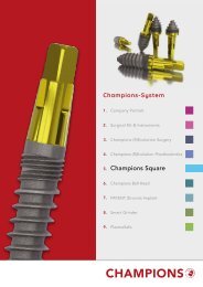
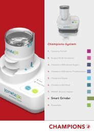
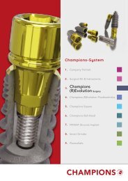
PRODUKTPORTFOLIO 2020<br />
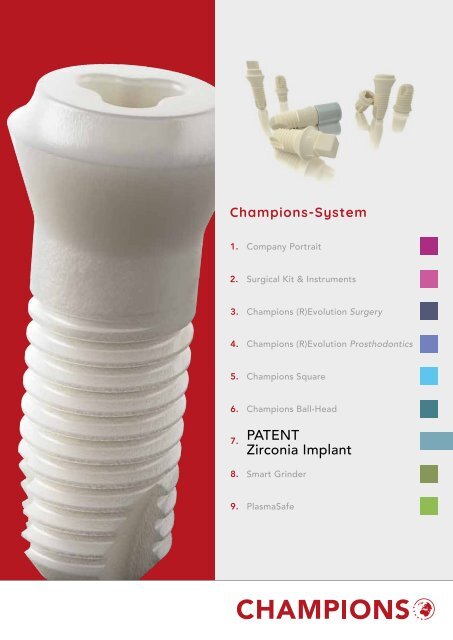
<strong>Champions</strong>-System<br />
1.<br />
Company Portrait<br />
2.<br />
Surgical Kit & Instruments<br />
3.<br />
<strong>Champions</strong> (R)Evolution Surgery<br />
4.<br />
<strong>Champions</strong> (R)Evolution Prosthodontics<br />
5.<br />
<strong>Champions</strong> Square<br />
6.<br />
7.<br />
8.<br />
<strong>Champions</strong> Ball-Head<br />
PATENT<br />
<strong>Zirconia</strong> <strong>Implant</strong><br />
Smart Grinder<br />
9.<br />
PlasmaSafe
TABLE OF CONTENTS<br />
PATENT <strong>Zirconia</strong> <strong>Implant</strong> | Page 4<br />
PATENT Tools | Page 8<br />
We are at<br />
your disposal<br />
Instructions | Page 10<br />
Mondays, Tuesdays, Thursdays:<br />
7.30 <strong>–</strong> 18.00<br />
Wednesdays:<br />
7.30 <strong>–</strong> 16.00<br />
Fridays:<br />
7.30 <strong>–</strong> 14.00<br />
PHONE:<br />
0049 6734 91 40 80<br />
FAX:<br />
0049 6734 10 53<br />
E-MAIL:<br />
info@champions-implants.com<br />
ONLINE-SHOP:<br />
champions-implants.shop<br />
MOBILE CEO ARMIN NEDJAT<br />
0049 151 15 25 36 92<br />
The <strong>Champions</strong>-Team<br />
(staff clip, scan QR-Code)<br />
The products and designations mentioned in this catalog are partly protected by<br />
trademarks, patents, and copyrights. The absence of a special reference or of the<br />
sign "®" or "TM" does not mean that no legal protection exists.<br />
<strong>Champions</strong> ® , <strong>Champions</strong> (R)Evolution ® , LOC ® , MIMI ® and Win! ® are registered trademarks<br />
of Dr. Armin Nedjat; Cleanser ® of KometaBio; Docklocs ® of Medealis GmbH;<br />
BloodSTOP ® iX of LifeScience PLUS Inc., LOCATOR ® of Zest Dental Solutions; CEREC ®<br />
of Sirona Dental GmbH; FiBER FORCE ® of Bio Composants Médicaux; MEDENTiKA of<br />
Straumann GmbH; PlasmaSafe ® of Infini-TI Biomedical GmbH; PATENT TM of Zircon<br />
Medical Management AG.<br />
Manufacturer details according to MDD (Medical Device Directive)/ MDR (Medical<br />
Device Regulation) are indicated on the product label.
PATENT<br />
<strong>Zirconia</strong> <strong>Implant</strong>
PATENT<br />
The PATENT ceramic implant consists of zirconia and 0.25 % of aluminium oxide (Al 2<br />
O 3<br />
). In fact,<br />
the PATENT’s strength is significantly higher compared to the strength of the titanium implant.<br />
However, the PATENT’s strength is not high to such an extent that there is a higher risk of fracture<br />
since higher strength (in MPa) also means lower elasticity.<br />
<strong>Product</strong>ion is not performed by injection molding or in-mold process, but each implant is<br />
milled individually. With an easy-to-use software, dentists can also have customized implants<br />
manufactured.<br />
This is the first time that Clean<strong>Implant</strong> Foundation has awarded a certificate for a ceramic<br />
implant: PATENT; it has been shown that the PATENT implant surface is clean and free from<br />
polyoxymethylene and production residues.<br />
4 PATENT ZIRCONIA IMPLANT
The PATENT implant<br />
(listed* as <strong>Champions</strong><br />
BioWin! by the Clean<strong>Implant</strong><br />
Foundation) is the only<br />
ceramic implant for which<br />
Clean<strong>Implant</strong> has awarded a<br />
certificate for a clean surface,<br />
free from production residues.<br />
Glass Fiber Post<br />
Due to the bonding of the Post<br />
with the implant body, there is<br />
a bacteria-resistant connection.<br />
<strong>Zirconia</strong><br />
Aluminium oxide of<br />
only 0.25 %<br />
Rough surface<br />
of PATENT<br />
5
HOW DOES THE PATENT DENTAL IMPLANT SYSTEM WORK?<br />
The <strong>Patent</strong> two-piece implant system consists<br />
of only two components: the implant with an<br />
integrated zirconia abutment and the high-tech<br />
Glass Fiber Post. The upper transmucosal part<br />
of the implant does not have a thread.<br />
The lower part, which is inserted into the bone,<br />
has a threaded part with a surface roughness<br />
of approximately 6 μm (RA); i.e. up to five<br />
times rougher than other implant surfaces. This<br />
surface roughness is created using a patented<br />
manufacturing process, during which all steps<br />
are performed in the pre-sintered stage. Any<br />
process-induced micro-cracks are filled in the<br />
subsequent sintering process, thus resulting in<br />
homogeneous, solid material.<br />
The implant surface is hydrophilic and osteoconductive,<br />
thus enhancing osseointegration of the<br />
implant. The transgingival part of the implant<br />
has a mechanically milled surface that ensures<br />
good soft tissue reaction. Several clinical studies<br />
have shown a better soft tissue integration of<br />
zirconia compared to titanium, which has also<br />
been confirmed by other clinical studies of the<br />
PATENT implant system. It was found that the<br />
soft tissue response was very favorable.<br />
In addition, Probing Pocket Depth (PPD) and<br />
Bleeding On Probing (BOP) have been reported<br />
to be even lower around the PATENT implants<br />
than around natural teeth.<br />
In the absence of the micro-gap at the bone<br />
level, this implant design reduces the risk of<br />
moving parts, wear and tear, screw loosening,<br />
and disruption to the healing process.<br />
The prosthetic connection is made with a<br />
high-tech Glass Fiber Post, allowing for great<br />
restoration flexibility since the Glass Fiber Post<br />
can be individually prepared for the respective<br />
clinical indication. There are neither screws nor<br />
screwdrivers.<br />
The same handling applies as in conventional<br />
Dentistry. The Glass Fiber Post is cemented<br />
on the implant. The crown is cemented on the<br />
Glass Fiber Post and encloses the implant,<br />
allowing for a very solid construction. The combination<br />
of the very rigid zirconia implant and<br />
the more flexible (E-module similar to dentin)<br />
high-tech Glass Fiber Post results in optimal<br />
load distribution of the masticatory forces. The<br />
cementation procedure is easy to check since<br />
it is performed at tissue level. The prosthetic<br />
workflow is very efficient; there is no need for<br />
additional instruments or components. Basically,<br />
you don’t need any additional training either<br />
since the procedure is similar to conventional<br />
crown and bridge work.<br />
Studies:<br />
Brüll F, van Winkelhoff AJ, Cune MS. <strong>Zirconia</strong> dental implants: a clinical,<br />
radiographic, and microbiologic evaluation up to 3 years. Int J Oral<br />
Maxillofac <strong>Implant</strong>s. 2014 Jul-Aug;29(4):914-20. Doi: 10.11607/jomi.3293<br />
Roehling S, Schlegel K A, Woelfler H, Gahlert M. Performance and outcome<br />
of zirconia dental implants in clinical studies: a meta-analysis. Clin Oral<br />
Impl Res. 2018;29 (Suppl. 16):135<strong>–</strong>153<br />
Becker J, John G, Becker K, Mainusch S, Diedrichs G, Schwarz F. Clinical<br />
performance of two-piece zirconium implants in the posterior mandible<br />
and maxilla: a prospective cohort study over 2 years. Clin. Oral Impl. Res.<br />
28, 2017, 29<strong>–</strong>35 doi: 10.1111/clr.12610<br />
You can find some studies under:<br />
https://www.mypatent.com/dental-professionals-research<br />
6 PATENT ZIRCONIA IMPLANT
”Based on my experience of more than 10 different<br />
zirconia dental implant systems, I can say that<br />
the PATENT implant system ensures an unparalleled<br />
bio-integration.“<br />
Prof. Dr. Marcel Wainwright<br />
Taking an impression is very efficient. For a<br />
conventional impression there is no need for<br />
impression copings, and a scan body is not<br />
used for intraoral scans since the upper part<br />
of the implant serves as scan body when the<br />
laboratory manufactures the model. However,<br />
analogs are available for printed models: REF<br />
31219 DIM-Analog <strong>Champions</strong> (R)Evolution<br />
and REF 31131 DIM-Analog Multi-Unit. The<br />
fact that no impression coping or scan body is<br />
used saves significant treatment time, makes it<br />
more comfortable for the patient, and reduces<br />
the exchange of components in the soft tissue.<br />
Apart from the clinical performance, the<br />
PATENT implant system also provides some<br />
significant efficiency gains. In fact, a treatment<br />
can be performed in only two sessions,<br />
benefiting the patient and saving chair time.<br />
The Glass Fiber Post can be prepared outside<br />
the patient’s mouth. In this way, the Glass Fiber<br />
Post and the restoration can be cemented in<br />
the same session.<br />
On top of these benefits, the PATENT implant<br />
system comes with a lifetime guarantee, which<br />
means that an implant will be replaced if it<br />
breaks.<br />
”There is a zirconia implant with very<br />
good long-term results.”<br />
Prof. Dr. Joachim Hermann<br />
7
H1<br />
EP<br />
H2<br />
L<br />
D<br />
PATENT ONE-PIECE<br />
The one-piece PATENT implant is a rotation-symmetric endosseous implant<br />
made from zirconia. This implant can be placed in all maxillary and mandibular<br />
bone quality types (D1-D4) with and without augmentation. PATENT<br />
implants are inserted at bone level. Available in a wide range of diameters<br />
and lengths, these implants cover a wider range of indications.<br />
These implants are suitable for insertion in the jawbone that has completely<br />
healed (late implantation), delayed immediate implantation (1<strong>–</strong>8 weeks after<br />
dental extraction), and immediate implantation (implantation immediately<br />
after dental extraction - under certain conditions). Please note the restrictions<br />
of indications in the respective Instructions for Use.<br />
Material: zirconia<br />
AVAILABLE LENGTHS AND DIAMETERS<br />
Diameter Lenght Height 1 Height 2* 1 EP* 2 REF<br />
4.1 mm 7.0 mm 4.0 mm 2.5 mm 5.2 mm P1S4107<br />
09.0 mm 4.0 mm 2.5 mm 5.2 mm P1S4109<br />
11.0 mm 4.0 mm 2.5 mm 5.2 mm P1S4111<br />
13.0 mm 4.0 mm 2.5 mm 5.2 mm P1S4113<br />
4.5 mm 7.0 mm 4.0 mm 2.5 mm 6.2 mm P1S4507<br />
9.0 mm 4.0 mm 2.5 mm 6.2 mm P1S4509<br />
11.0 mm 4.0 mm 2.5 mm 6.2 mm P1S4511<br />
13.0 mm 4.0 mm 2.5 mm 6.2 mm P1S4513<br />
5.0 mm 7.0 mm 4.0 mm 2.5 mm 6.2 mm P1S5007<br />
9.0 mm 4.0 mm 2.5 mm 6.2 mm P1S5009<br />
11.0 mm 4.0 mm 2.5 mm 6.2 mm P1S5011<br />
13.0 mm 4.0 mm 2.5 mm 6.2 mm P1S5013<br />
* 1 Transmucosal<br />
* 2 Emergence profile<br />
8 PATENT ZIRCONIA IMPLANT
H1<br />
EP<br />
H2<br />
L<br />
D<br />
PATENT TWO-PIECE<br />
The two-piece PATENT implant is a rotation-symmetric endosseous implant<br />
made from zirconia. This implant can be placed in all maxillary and<br />
mandibular bone quality types (D1-D4) with and without augmentation.<br />
PATENT implants are inserted at bone level. Available in a wide range of<br />
diameters and lengths, these implants cover a wider range of indications.<br />
These implants are suitable for insertion in the jawbone that has completely<br />
healed (late implantation), delayed immediate implantation (1<strong>–</strong>8 weeks<br />
after dental extraction), and immediate implantation (implantation immediately<br />
after dental extraction - under certain conditions). Please note the<br />
restrictions of indications in the respective Instructions for Use.<br />
The Post consists of glass fiber and can be shaped in the intraoral and<br />
extraoral position. It is bonded with the implant body in the supragingival<br />
position without a gap (recommendation: RelyX Unicem [3M Espe]).<br />
Material: zirconia<br />
AVAILABLE LENGTHS AND DIAMETERS<br />
Diameter Lenght Height 1 Height 2* 1 EP* 2 REF<br />
4.1 mm 7.0 mm 1.6 mm 2.5 mm 5.2 mm P2S4107<br />
9.0 mm 1.6 mm 2.5 mm 5.2 mm P2S4109<br />
11.0 mm 1.6 mm 2.5 mm 5.2 mm P2S4111<br />
13.0 mm 1.6 mm 2.5 mm 5.2 mm P2S4113<br />
4.5 mm 7.0 mm 1.2 mm 2.5 mm 6.2 mm P2S4507<br />
9.0 mm 1.2 mm 2.5 mm 6.2 mm P2S4509<br />
11.0 mm 1.2 mm 2.5 mm 6.2 mm P2S4511<br />
13.0 mm 1.2 mm 2.5 mm 6.2 mm P2S4513<br />
5.0 mm 7.0 mm 1.2 mm 2.5 mm 6.2 mm P2S5007<br />
9.0 mm 1.2 mm 2.5 mm 6.2 mm P2S5009<br />
11.0 mm 1.2 mm 2.5 mm 6.2 mm P2S5011<br />
13.0 mm 1.2 mm 2.5 mm 6.2 mm P2S5013<br />
* 1 Transmucosal<br />
* 2 Emergence profile<br />
9
OSSEOINTEGRATION IS NOT ENOUGH!<br />
Since the early beginnings of Dentistry, replacing<br />
missing teeth to restore masticatory function<br />
has been one of the most sought-after treatments<br />
by patients. Before 1965, unfortunately,<br />
the solutions for edentulous people were rudimentary<br />
and often resulted in further deterioration<br />
and seriously compromised mastication.<br />
A new era of oral rehabilitation began thanks to<br />
the major discovery of the concept of osseointegration<br />
with titanium implants by the famous<br />
Prof. Per- Ingvar Brånemark. Millions of people<br />
worldwide have been able to improve their<br />
quality of life by recovering masticatory function<br />
and esthetics due to osseointegration with a titanium<br />
dental implant treatment.<br />
According to Prof. Brånemark, osseointegration<br />
is a ”direct connection between living bone<br />
and the load <strong>–</strong> carrying endosseous implant<br />
at the light microscopic level.“ Further studies<br />
have confirmed this definition and today, this<br />
healing mechanism around titanium implants is<br />
well documented.<br />
In the early days of <strong>Implant</strong>ology, the strong focus<br />
was on osseointegration to make sure that<br />
the implants remained anchored in bone for a<br />
long time. In the last few years, the focus has<br />
shifted towards soft tissue integration and different<br />
prosthetic components to achieve longterm<br />
esthetic results associated with healthy<br />
gingiva and tissue.<br />
However, the increased complexity of the solutions<br />
and the phenomenon of peri-implantitis<br />
have presented a challenge with current systems,<br />
jeopardizing long-term success. So, today,<br />
osseointegration is not enough anymore<br />
for a successful treatment outcome.<br />
Healthy soft tissue<br />
Healthy soft tissues and hard tissues play a key<br />
role for ensuring successful long-term results,<br />
which are also influenced by the biological<br />
width, consisting of the epithelium and connective<br />
tissue lengths. Lee et al. have found that<br />
the ratio of connective tissue within the whole<br />
biological width in natural teeth (65.9 %) is similar<br />
to that of zirconia (65.4 %). Obviously, a<br />
higher connective tissue content allows for more<br />
adequate protection of the bone-implant interface.<br />
These observations have been confirmed<br />
by several reports on the soft tissue reactions to<br />
zirconia.<br />
You can find some studies under:<br />
https://mypatent.com/reference<br />
10 PATENT ZIRCONIA IMPLANT
Natural teeth<br />
<strong>Zirconia</strong> implants<br />
CT Epi.<br />
Biological width<br />
CT Epi.<br />
Biological width<br />
Biological width<br />
Biological width<br />
Connective tissue 65.9 % Epithelium 34.1 % Connective tissue 65.4 % Epithelium 34.6 %<br />
11
Soft Tissue Level = no micro-movements<br />
In fact, the implant design is another factor that<br />
influences soft tissue integration. Titanium systems<br />
as well as zirconia bone level systems with<br />
micro-gaps and joints that greatly extend into<br />
the mucosa have a potentially adverse effect.<br />
Combinations of materials with very different<br />
elasticity modules increase risks of adverse tissue<br />
reactions. In the installation phase there<br />
are components that must be removed and replaced<br />
several times. Inevitably, there are some<br />
micro-movements in the final construction. With<br />
a tissue level design, any junctions are moved<br />
to an equigingival position; compared to titanium,<br />
zirconia has an enhanced aesthetic potential<br />
due to its color and soft tissue adaptation.<br />
<strong>Patent</strong>ed production process<br />
As opposed to most manufacturing processes of<br />
zirconia implants where the surface roughness is<br />
created in the sintered stage, the surface of the<br />
PATENT implants is created in the pre-sintered<br />
stage; the latter allows micro-cracks induced in<br />
the process to be closed in the following sintering<br />
process, linked to a particle reduction of<br />
about 20 %.<br />
According to Mombelli et al., zirconia requires<br />
high roughness for predictable osseointegration.<br />
If you tried to achieve a very rough surface<br />
in the sintered phase, you would compromise<br />
the material strength since a lot of micro-cracks<br />
would be induced in the process.<br />
The good integration of the soft tissues prevents<br />
adhesion of bacteria, which causes inflammatory<br />
processes. In addition, zirconia has a low<br />
affinity for plaque.<br />
Due to the very rough surface at the endosseous<br />
part, the machined surface at the transmucosal<br />
part, and the tissue level design, the PATENT<br />
implant system is characterized by favorable<br />
properties for bio-integration.<br />
The surface of the PATENT <strong>Implant</strong> is very rough,<br />
which makes it hydrophilic and osteoconductive.<br />
During the healing phase, bone starts to<br />
form on the surface of the implant, which is the<br />
same behavior as on moderately rough surfaces<br />
of the modern titanium implants.<br />
12 PATENT ZIRCONIA IMPLANT
X 2,500<br />
X 10,000 X 20,000<br />
Hydrophilic characteristic <strong>–</strong> surface attracts<br />
blood<br />
Human blood on PATENT surface <strong>–</strong> within 10 minutes the fibrin network is attached to the<br />
surface. This attachment is a prerequisite for contact osteogenesis.<br />
What is bio-integration?<br />
Bio-integration is defined as ”the bonding of living tissue to the surface of a biomaterial or an implant“.<br />
Unlike osseointegration, which focuses on the bone behavior, bio-integration is only achieved if all<br />
surrounding tissues are bonded to the implant.<br />
13
PATENT Instruments<br />
PATENT Surgical Kit<br />
Includes all necessary instruments<br />
for PATENT implants<br />
Material of the Surgical Kit: plastic<br />
REF: PSK0000<br />
14 PATENT ZIRCONIA IMPLANT
15
POSTERIOR THREE-UNIT BRIDGE<br />
Author: Dr. med. dent. Gernot Obermair <strong>–</strong> dental office ”happy implant”<br />
Introduction<br />
In the early days of <strong>Implant</strong>ology,<br />
the strong focus was on osseointegration<br />
to make sure that the<br />
implants remained anchored in<br />
bone for a long time. In the last few<br />
years, the focus has shifted towards<br />
soft tissue integration and different<br />
prosthetic components to achieve<br />
long-term esthetic results associated<br />
with healthy gingiva and stable<br />
tissue levels.<br />
However, the increased complexity<br />
of the solutions and the phenomenon<br />
of peri-implantitis have<br />
presented a challenge with current<br />
systems, jeopardizing long-term<br />
success.<br />
The PATENT dental implant system<br />
meets challenges by providing a<br />
ceramic implant with unique characteristics<br />
to establish complete<br />
bio-integration. The surface is patented<br />
and significantly rougher<br />
than other systems. The bonded<br />
secondary part prevents a microgap.<br />
The high-tech Glass Fiber Post<br />
serves as great support and allows<br />
for load distribution for the superstructure.<br />
The PATENT dental implant system<br />
is the only ceramic two-piece<br />
implant system on the market with<br />
peer-reviewed long-term clinical research<br />
1 .<br />
Two independent studies have<br />
shown that the survival rate of the<br />
PATENT zirconia implants is similar<br />
to that of titanium implants and that<br />
the stable marginal bone level and<br />
soft tissue integration of PATENT<br />
zirconia implants are superior compared<br />
with titanium implants. 2,3<br />
Initial situation<br />
A partially edentulous 59-year-old<br />
male patient asked for a dental<br />
implant treatment. Due to periodontitis,<br />
the teeth had been extracted<br />
a year before implantation.<br />
Dental implants were planned in<br />
teeth sites 15, 24, 25, 26, and 36.<br />
Bone quality was D3 in sites 24-26<br />
and D2/D3 in sites 15 and 36. The<br />
implant selection is presented in<br />
Chart 1.<br />
Position<br />
<strong>Implant</strong> diameter<br />
(mm)<br />
Chart 1:<br />
<strong>Implant</strong> dimensions for different positions<br />
Diameter of the<br />
prosthetic platform<br />
(mm)<br />
<strong>Implant</strong> length<br />
(mm)<br />
15 4.1 5.2 11<br />
24 4.1 5.2 13<br />
25 4.1 5.2 13<br />
26 4.5 6.2 9<br />
36 4.5 6.2 13<br />
(1) Roehling S, Schlegel K A, Woelfler H, Gahlert M. Performance and outcome of zirconia dental implants in<br />
clinical studies: a meta-analysis. Clin Oral Impl Res. 2018;29(Suppl. 16):135<strong>–</strong>153.<br />
(2) Brüll F, van Winkelhoff AJ, Cune MS. <strong>Zirconia</strong> dental implants: a clinical, radiographic, and microbiologic<br />
evaluation up to 3 years. Int J Oral Maxillofac <strong>Implant</strong>s. 2014 Jul-Aug;29(4):914-20. Doi: 10.11607/jomi.3293<br />
(3) Becker J, John G, Becker K, Mainusch S, Diedrichs G, Schwarz F. Clinical performance of two- piece zirconium<br />
implants in the posterior mandible and maxilla: a prospective cohort study over 2 years. Clin. Oral Impl. Res. 28,<br />
2017, 29<strong>–</strong>35 doi: 10.1111/clr.12610<br />
16 PATENT ZIRCONIA IMPLANT
Pre-treatment<br />
The teeth were extracted, and socket<br />
preservation with PRGF was<br />
performed. No further bone graft<br />
procedure was performed. A conservative<br />
periodontal treatment<br />
was successfully performed on the<br />
remaining teeth. A titanium simulation<br />
test was done, which revealed<br />
high titanium particle-induced<br />
inflammatory values. We therefore<br />
opted for a PATENT ceramic implant<br />
for the patient and made a<br />
flapless surgery treatment plan.<br />
Surgical procedure<br />
With dynamic navigation, the implants<br />
were placed. First, osteotomies<br />
were prepared, and then the<br />
implants were placed without any<br />
problems. The insertion torques<br />
were between 22<strong>–</strong>35 Ncm. The<br />
surface of the PATENT implant is<br />
very hydrophilic, see (Fig. 1).<br />
It is important to place the implants<br />
in the right vertical position<br />
in relation to the soft tissue (in the<br />
epigingival position) in order to<br />
facilitate the prosthodontic procedure.<br />
At the time of surgery, intraoral<br />
check X-rays were taken, and<br />
a cone beam was performed, see<br />
(Fig. 2).<br />
Fig. 1:<br />
<strong>Implant</strong> placement. The PATENT<br />
implant has a hydrophilic surface.<br />
Fig. 2:<br />
Check X-rays at the time of the<br />
implantation<br />
17
Prosthetic reconstruction<br />
After 3 months of healing, the implants<br />
were restored with dentures.<br />
The Glass fiber Posts were cemented<br />
into place and prepared in the<br />
same way as conventional crowns<br />
and bridges, see (Fig. 3).<br />
A conventional impression was taken<br />
and sent to the dental laboratory.<br />
The laboratory prepared the<br />
models in the same way as conventional<br />
crown and bridge work, see<br />
(Fig. 4).<br />
Neither impression posts nor model<br />
analogs are needed. All crowns<br />
and bridges were made from zirconia<br />
with an occlusal surface made<br />
from composite. The flexibility of<br />
the plastic allows for a more favorable<br />
stress absorption of the masticatory<br />
forces, see (Fig. 5).<br />
Instead of 3 single crowns, a<br />
bridge was made on 3 implants in<br />
teeth sites 24<strong>–</strong>26 to allow masticatory<br />
forces to be distributed more<br />
evenly. The antagonist to the implant<br />
in position 26 is also an implant.<br />
Since the size of the implant<br />
in position 26 was only 4.5 x 9 mm,<br />
we opted for a bridge construction<br />
to distribute the load on the 3<br />
implants, see (Fig. 6<strong>–</strong>9). After the<br />
cementation, check X-rays were taken,<br />
(Fig. 10).<br />
The implant in position 15 was a<br />
single-tooth implant, (see Fig. 11).<br />
Another single-tooth implant was<br />
placed in tooth site 36 (see Fig.<br />
12). The implant was slightly exposed,<br />
but soft tissue is expected to<br />
grow to a certain extent over time.<br />
Check X-rays (Fig. 13<strong>–</strong>14) show<br />
stable marginal bone levels.<br />
Fig. 3:<br />
Glass Fiber Posts were bonded and prepared<br />
after 3 months of healing. Note the<br />
healthy soft tissue.<br />
Fig. 4:<br />
The laboratory works on a cast model<br />
in the same ways as with crowns and<br />
bridges. Neither impression posts nor<br />
model analogs are required.<br />
Fig. 5:<br />
The basic restoration material is composed<br />
of zirconia. The occlusal surface<br />
is made from composite to absorb the<br />
masticatory forces.<br />
Fig. 6:<br />
Occlusion was checked.<br />
Fig. 7:<br />
Vestibular view<br />
Fig. 8:<br />
Occlusal view<br />
18 PATENT ZIRCONIA IMPLANT
Fig. 9:<br />
Final result at the time of the fitted<br />
denture<br />
Fig. 10:<br />
Check X-ray after prosthetic restoration.<br />
Note the stable marginal bone levels.<br />
Fig. 11:<br />
Single-tooth implant in position 15<br />
Fig. 12:<br />
Single-tooth implant in position 36<br />
Fig. 13:<br />
Fig. 14:<br />
Check X-rays of position 36 at the time of implantation and prosthetic restoration.<br />
Very stable marginal bone levels are observed.<br />
Conclusion<br />
Thanks to its Glass Fiber Post, the PATENT implant<br />
system allows for an expansion of prosthetic flexibility.<br />
Single units or bridge constructions can be efficiently<br />
manufactured using conventional Dental Technology<br />
methods. With its rough surface and the machined<br />
surface on the transmucosal part, this implant design<br />
allows for complete bio-integration in the bone.<br />
In combination with the design without a micro-gap<br />
deep in the soft tissue, very stable soft tissue levels<br />
have been achieved. See X-rays in the figures (Fig. 3,<br />
10, 14). Besides showing favorable soft tissue reactions,<br />
the PATENT implant system also shows survival<br />
rates that are comparable to titanium dental implants.<br />
”There are minimal probing<br />
pocket depths thanks to the<br />
PATENT implant system.”<br />
Dr. Gernot Obermair<br />
19
IMMEDIATE IMPLANTATION AND IMMEDIATE LOADING OF A ZIRCONIA<br />
TWO-PIECE IMPLANT<br />
In this case report, the author describes an extraction of a tooth that could no longer be preserved<br />
and the immediate implantation of a PATENT two-piece implant with immediate restoration in the<br />
same session. For stabilizing soft tissue, the root of the extracted tooth was processed into autologous<br />
bone graft with the Smart Grinder procedure and grafted around the implant.<br />
Author: Dr. Lavinia Neuss-Zaar <strong>–</strong> Expert in <strong>Implant</strong>ology und <strong>Implant</strong> Prosthodontics CIPC<br />
A female patient presented to our dental office complaining<br />
of a toothache in the anterior region when<br />
masticating.<br />
X-rays exhibited severe bone loss in the mesial position<br />
11, accompanied by inflammation. The tooth<br />
could therefore no longer be preserved. Because of<br />
bone loss, the tooth 11 was extruded in the distobuccal<br />
position, accompanied by a considerable enlargement<br />
of an existing diastema.<br />
During a consultation prior treatment, we presented<br />
our treatment concept to our patient:<br />
classical immediate implantation after the extraction<br />
of the tooth using the “Sofi-Protokoll” (immediate<br />
implantation protocol) according to Dr. Nedjat.<br />
We suggested placing a ceramic implant, which does<br />
not show through the gingiva in the long term. The<br />
patient accepted this treatment. We suggested making<br />
the crown 11 wider than the tooth and treating<br />
Tooth 21 with a veneer for closing the diastema almost<br />
completely.<br />
implants consist of only 0.25% of Al 2<br />
O 3<br />
; by comparison,<br />
ATZ-implants are composed of about 25% of<br />
Al 2<br />
O 3<br />
. We made a dental appointment for surgery<br />
the next day.<br />
In our dental office there are no days reserved for<br />
implantation since this therapy does not prevail<br />
over other therapies. Our multi-disciplinary dental<br />
team integrates implant therapy in the same way as<br />
other therapies such as dental extractions, endodontic<br />
treatments, or fillings. Making an appointment for<br />
surgery in the short term is not a problem. In our<br />
practice there are available implants in the most<br />
important lengths and diameters in stock to meet<br />
patients‘ demands.<br />
Day of the surgery<br />
The patient came into the dental office that day. Anesthesia<br />
was administered with small Ultracain D-S<br />
deposits around the surgery site. As a rule Ultracain<br />
D-S forte is not recommended since the agent epinephrine<br />
(adrenaline) causes anemia in the surgery<br />
site. The implant site should normally bleed slightly.<br />
We decided to place a two-piece implant to open<br />
the door to more prosthetic solutions. PATENT of<br />
Zircon Medical is the only zirconia implant system<br />
supported by proven long-term studies, which have<br />
shown a bio-integration rate of 96 %, providing great<br />
comfort to dentists and patients. In addition, it is the<br />
only almost metal-free ceramic implant. There is no<br />
screwing of the implant with the abutment. PATENT<br />
With a Bein’s elevator and forceps, we carefully extracted<br />
the tooth, applying pressure to the buccal<br />
lamella with the thumb to prevent fractures. With a<br />
sharp spoon, the alveolus was curetted to remove<br />
inflammation residues. The absence of buccal bone<br />
was clinically verified.<br />
20 PATENT ZIRCONIA IMPLANT
Fig. 1: Initial situation Fig. 2 & 3: Clinical view of Tooth 11<br />
Fig. 4: Extraction alveolus<br />
Fig. 5: Preparation of the implant site with<br />
the white <strong>Champions</strong> Drill with the Drill<br />
Extension<br />
Fig. 6: PATENT-Drill ø 3.5 mm<br />
Fig. 7: <strong>Champions</strong> Condenser ø 4.3 mm<br />
Fig. 8: Check X-rays with the Condenser<br />
ø 4.3 mm<br />
Fig. 9: Placing a PATENT ceramic implant<br />
(length 13 mm, ø 4.5 mm)<br />
21
Fig. 10: Placed implant<br />
Fig. 11: Immediately after surgery<br />
Fig. 12: Shaped Post<br />
Fig. 13: Temporary restoration<br />
Fig. 14: Grafted autologous bone graft<br />
produced from the patient’s own tooth<br />
using the Smart Grinder protocol<br />
Fig. 15: 1 week after surgery<br />
Fig. 16: 3 months after surgery with fitted<br />
denture 11 and veneer 21<br />
Fig. 17: X-rays with fitted denture 3<br />
months after surgery<br />
Link to video showing surgery<br />
The PATENT Surgical Kit includes all necessary instruments<br />
for inserting ceramic implants in all bone quality<br />
types (D1 to D4). For the pilot drill the yellow Conical Triangular<br />
Drill is used (length 20 mm, ø 2.3 mm) from the<br />
Surgical Kit of <strong>Champions</strong>-<strong>Implant</strong>s.<br />
The first drilling was performed in the slightly palatinal<br />
position, at about 15°. After the pilot drill, the cavity was<br />
widened with the white Conical Triangular Drill (length<br />
18 mm, ø 3.3 mm). Using a Drill Extension with the Drill,<br />
we were able to check the drilling axis more carefully.<br />
Although a perforation of the bone walls can be avoided<br />
due to the drilling at low speed, we always check the<br />
bone cavity with a thin probe in all 5 directions to check<br />
the bone for perforation. On this occasion, the mesiobuccal<br />
bone defect was verified.<br />
For widening the cavity, we used the PATENT-Drills having<br />
diameters of ø 3.0 mm, ø 3.5 mm, and ø 3.8 mm.<br />
With the blue Condenser ø 4.3 mm from the <strong>Champions</strong><br />
Surgical Kit, primary stability was checked and achieved<br />
at about 40 Ncm, which sufficed to place a PATENT-implant<br />
(length 13 mm, ø 4.5 mm). X-rays con firmed the<br />
correct position.<br />
The PATENT ceramic implant (Zircon Medical, Zurich,<br />
distribution <strong>Champions</strong>-<strong>Implant</strong>s) has a very rough surface,<br />
enhancing bio-integration. However, the insertion<br />
itself slightly differs from the insertion of a titanium implant.<br />
It is recommended that you should place ceramic<br />
implants slightly mechanically with an implant motor at<br />
about 20 rpm, preventing an eventual inclination. The<br />
maximum torque is limited to 30 Ncm. A Torque Wrench<br />
is used for the final insertion in the slightly subgingival<br />
22 PATENT ZIRCONIA IMPLANT
position.<br />
Retro-alveolar X-rays confirmed the correct position<br />
of the implant.<br />
Temporary restoration<br />
In the same session, the Post was bonded onto the<br />
implant body with RelyX from 3M Espe, an adhesive<br />
dual-cure cement. Then, the Post was shaped<br />
by means of a coarse diamond bur under water<br />
cooling. Consisting of a glass fiber polymer, the<br />
Post can be easily and quickly shaped. Finally, a<br />
temporary dental crown is fitted. The dental crown<br />
was prepared in the basal area and splinted with the<br />
adjacent teeth.<br />
Summary<br />
This case shows that successful implantation does<br />
not depend on the availability of a buccal wall. However,<br />
in these cases, it is recommended that you<br />
should place a (white) ceramic implant; the implant<br />
body does not show through the gingiva.<br />
This efficient “Sofi-Verfahren”, i.e. the immediate<br />
implantation procedure according to Dr. Nedjat,<br />
is spearheading a perfectly effective treatment ensuring<br />
es thetic appearances of the soft tissue. Another<br />
advantage is that you can visualize the bone<br />
and that crestal relief is unnecessary.<br />
Using the Smart Grinder Protocol, the root of the<br />
extracted tooth was processed into autologous<br />
bone graft chairside within 8 minutes. The dentin<br />
particulate was cleansed in the Cleanser to dissolve<br />
all bacteria, viruses, and fungi. Then, the particulate<br />
was rinsed in a phosphate buffered saline solution<br />
at the pH level of 7.1. The gap between the implant<br />
body and the soft tissue was grafted with this bone<br />
graft to stabilize the gingiva.<br />
A week after surgery, a check-up showed good healing<br />
results without discomfort.<br />
Fitting the final denture<br />
After 3 months, the final denture was fitted, and<br />
Tooth 21 was treated with a veneer to close the<br />
diastema. Final X-rays with the fitted denture confirmed<br />
the good bio-integration of the implant.<br />
”The PATENT- ceramic implant body<br />
does not show through the gingiva.“<br />
Dr. Lavinia Neuss-Zaar<br />
23
Instructions<br />
Here you can find films of the PATENT system on the link or scan the<br />
QR-Code on your mobile.<br />
The PATENT zirconia implant<br />
Presentation of the PATENT<br />
https://vimeo.com/485480817<br />
Animation of an insertion<br />
View of an insertion of a two-piece PATENT zirconia implant on<br />
a glass jaw model<br />
https://vimeo.com/326287083/7d59750057<br />
Insertion and cementation<br />
Insertion of 2 two-piece PATENT zirconia implants,<br />
cementation of the Posts without gaps, and checkbite<br />
https://vimeo.com/488882191<br />
Insertion<br />
Insertion of 5 PATENT zirconia implants in the maxilla.<br />
https://vimeo.com/501735016<br />
24 PATENT ZIRCONIA IMPLANT
Insertion<br />
Insertion of 2 PATENT zirconia implants in sites 23 and 25<br />
https://vimeo.com/502126637<br />
Insertion<br />
Insertion of 2 PATENT zirconia implants in a narrow jaw ridge using<br />
the MIMI II-protocol<br />
https://vimeo.com/559883111<br />
Immediate implantation<br />
Immediate implantation and immediate restoration of a PATENT<br />
zirconia implant in site 11 and Smart Grinder procedure<br />
https://vimeo.com/533481215<br />
25
Welcome<br />
to the<br />
future<br />
26 PATENT ZIRCONIA IMPLANT<br />
<strong>Champions</strong>-<strong>Implant</strong>s GmbH<br />
Im Baumfeld 30 | <strong>Champions</strong> Platz 1 | D-55237 Flonheim<br />
Tel. 0049 6734 91 40 80 | Fax 0049 6734 10 53<br />
info@champions-implants.com<br />
champions-implants.com<br />
Produktkatalog <strong>Champions</strong> <strong>Patent</strong> EN<br />
Stand: 6. Dezember 2022, 8:08 AM