Constructing universal multiplex PCR systems for ... - Library
Constructing universal multiplex PCR systems for ... - Library
Constructing universal multiplex PCR systems for ... - Library

You also want an ePaper? Increase the reach of your titles
YUMPU automatically turns print PDFs into web optimized ePapers that Google loves.
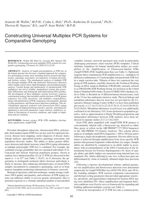
Jeanette M. Wallin, 1 M.P.H.; Cydne L. Holt, 1 Ph.D.; Katherine D. Lazaruk, 1 Ph.D.;<br />
Theresa H. Nguyen, 1 B.S.; and P. Sean Walsh, 1 M.P.H.<br />
<strong>Constructing</strong> Universal Multiplex <strong>PCR</strong> Systems <strong>for</strong><br />
Comparative Genotyping<br />
REFERENCE: Wallin JM, Holt CL, Lazaruk KD, Nguyen TH,<br />
Walsh PS. <strong>Constructing</strong> <strong>universal</strong> <strong>multiplex</strong> <strong>PCR</strong> <strong>systems</strong> <strong>for</strong> comparative<br />
genotyping. J Forensic Sci 2002;47(1):52-65.<br />
ABSTRACT: Analysis of length polymorphisms at STR loci in<br />
the human genome has become a standard approach <strong>for</strong> comparative<br />
genotyping in many areas including disease research and diagnostics,<br />
parentage assessment, investigations of human diversity,<br />
and <strong>for</strong>ensic science. The simultaneous analysis of multiple STR<br />
loci through <strong>multiplex</strong> <strong>PCR</strong> and multicolor fluorescence detection<br />
offers sample conservation, high throughput, and automated genetic<br />
analysis. Careful design and optimization of tetranucleotide STR<br />
<strong>multiplex</strong>es has led to reliable, standardized <strong>systems</strong> that powerfully<br />
differentiate and distinguish individual human DNA profiles.<br />
The development of these <strong>multiplex</strong> <strong>systems</strong> involved a rigorous<br />
experimental strategy that included careful selection of <strong>PCR</strong> primer<br />
sequences (<strong>for</strong> yield, specificity, and <strong>multiplex</strong> compatability),<br />
along with optimization of <strong>PCR</strong> component concentrations, thermal<br />
cycling parameters, and fluorescence detection conditions. This developmental<br />
approach rendered well-characterized DNA typing<br />
<strong>systems</strong> that are high per<strong>for</strong>ming (sensitive, specific, and balanced),<br />
optimized to <strong>universal</strong> parameters (same reaction conditions), resilient<br />
to fluctuations in reaction conditions, and simple to implement<br />
and use routinely.<br />
KEYWORDS: <strong>for</strong>ensic science, <strong>PCR</strong>, STR, <strong>multiplex</strong>, development,<br />
optimization, AmpF�STR<br />
Prevalent throughout eukaryotic chromosomal DNA, polymorphic<br />
short tandem repeat (STR) loci are key tools <strong>for</strong> rapid gene discovery,<br />
disease locus mapping, carrier diagnosis of disease states,<br />
linkage analyses, agricultural genetics, parentage assessment, and<br />
population diversity studies. The ability to detect genetic differences<br />
between individuals increases when DNA typing in<strong>for</strong>mation<br />
at multiple polymorphic STR loci is combined. For example, the<br />
combined average probability that two unrelated individuals (U.S.<br />
Caucasian and African American population groups), chosen at<br />
random, have identical profiles (PI) at 15 STR loci discussed in this<br />
report is ~1 in 10 15 (see Table 1; 18,47). As PI decreases, the opportunity<br />
to distinguish unshared alleles between individuals is<br />
likely to increase, thus enhancing in<strong>for</strong>mativeness of mixed DNA<br />
samples encountered in comparative genotyping, such as in bone<br />
marrow transplant monitoring (chimaerism) or <strong>for</strong>ensic casework.<br />
Biological specimen collection technique, sample storage conditions,<br />
DNA quality and quantity, and resilience of STR typing <strong>systems</strong><br />
can influence ability to recover genetic in<strong>for</strong>mation. Due to<br />
potential exposure to a virtually unlimited number of uncontrolled<br />
1 Human Identification Group, PE Bio<strong>systems</strong>, Foster City, CA.<br />
Received 10 July 2000; and in revised <strong>for</strong>m 25 Jan. 2001; accepted 12 April<br />
2001.<br />
Copyright © 2002 by ASTM International<br />
52<br />
variables, <strong>for</strong>ensic casework specimens may result in particularly<br />
challenging polymerase chain reaction (<strong>PCR</strong>) templates. Critical<br />
attributes mandatory <strong>for</strong> human identification studies are exemplified<br />
in the Amplification of Fluorescent-labeled STRs<br />
(AmpF�STR®) <strong>PCR</strong> Amplification Kits (see Table 1). Optimized<br />
reagents direct simultaneous <strong>PCR</strong> amplification (i.e., <strong>multiplex</strong>) of<br />
different combinations of 15 polymorphic tetranucleotide STR loci<br />
in a single reaction tube. Thirteen of these loci represent the core<br />
group of STR markers, carefully chosen by the Technical Working<br />
Group on DNA Analysis Methods (TWGDAM; currently referred<br />
to as SWGDAM) STR Working Group, <strong>for</strong> inclusion in the United<br />
States COmbined DNA Index System (CODIS) DNA database (refer<br />
to Table 1). Resident on 14 different human chromosomes, each<br />
of the 15 loci has undergone intensive study. Physical linkage and<br />
mendelian inheritance data <strong>for</strong> most loci are provided by the Cooperative<br />
Human Linkage Center (CHLC) or have been published<br />
previously (1,2,7,10,15,16,21,22,24,27,29,31,32,34,43,49,50,53,<br />
54,55,56,63). Mendelian inheritance at each locus was additionally<br />
confirmed in our laboratory (18). Issues pertinent to population genetics,<br />
such as approximation to Hardy-Weinberg proportions and<br />
independent inheritance between STR markers, have been addressed<br />
in separate studies (5,11,13,18,23,65).<br />
Each STR allele amplified with AmpF�STR <strong>PCR</strong> primers is<br />
concomitantly labeled with a fluorescent tag, detected as either<br />
blue, green, or yellow on the ABI PRISM 377 DNA Sequencer<br />
or the ABI PRISM 310 Genetic Analyzer. This scheme allows<br />
analysis of multiple small DNA fragments (~100 to 350 base pairs)<br />
following a single electrophoretic separation. Discrete allele recognition<br />
is achieved through amplification from locus-specific <strong>PCR</strong><br />
primers that generate fragments within distinct size ranges. Sample<br />
alleles are identified by comparison to an allelic ladder in accordance<br />
with recommendations of the DNA Commission of the International<br />
Society <strong>for</strong> Forensic Haemogenetics (9). Sizes of sample<br />
alleles and allelic ladders are measured using internal lane size<br />
standards (30). Alleles differing by one, two, or three base pairs are<br />
differentiated by virtue of routinely obtained single base precision<br />
(25).<br />
Following a rigorous developmental scheme, optimal parameters<br />
<strong>for</strong> amplification and fluorescence detection were determined<br />
empirically <strong>for</strong> AmpF�STR Blue loci (D3S1358, FGA, and vWA),<br />
the founding <strong>multiplex</strong>. <strong>PCR</strong> reaction component concentrations<br />
and thermal cycling parameters that provided appropriate sensitivity,<br />
specificity, and signal balance important <strong>for</strong> <strong>for</strong>ensic DNA typing<br />
were identified (59). These amplification and fragment detection<br />
conditions were fixed to serve as framework <strong>for</strong> expanding and<br />
building additional <strong>multiplex</strong> <strong>systems</strong> (see Table 1).<br />
Critical <strong>for</strong> incorporation of 15 STR loci into a common proto-
col was extensive screening and selection of <strong>PCR</strong> primers. Primer<br />
selection, <strong>PCR</strong> optimization, and fluorescent allele detection conditions<br />
are explored as primary focus points.<br />
Materials and Methods<br />
Sample Source and Extraction Protocols<br />
Human DNA samples from bloodstains, hair, and buccal cell<br />
swabbings were extracted using either a phenol/chloro<strong>for</strong>m organic<br />
method (Serological Research Institute, Richmond, CA; 46) or<br />
Chelex (PE Bio<strong>systems</strong>, Foster City, CA; 42,60); this sample set<br />
is used in all experiments, unless stated otherwise. Nonhuman<br />
DNA samples (BIOS Laboratories, Inc., New Haven, CT) included<br />
primates (gorilla, chimpanzee, orangutan, and macaque) and nonprimates<br />
(horse, cow, chicken, pig, rabbit, dog, cat, fish, hamster,<br />
mouse, rat), fungi (Candida albicans, Saccharomyces cerevisiae),<br />
and bacteria (Rhodotorula rubra, Legionella pneumophilia, Escherichia<br />
coli, Listeria monocytogenes, Neisseria lactamica, Vibrio<br />
mimicus, Citrobacter freundii, Salmonella typhimurium). DNA<br />
extracts were quantitated using the QuantiBlot® Human DNA<br />
Quantitation Kit (Perkin Elmer, Norwalk, CT; 61), on agarose gels,<br />
and/or by ultraviolet spectrophotometry.<br />
Population database DNA samples were extracted from liquid<br />
blood of unrelated individuals, as follows: 459 African Americans,<br />
200 U.S. Caucasians, and 113 U.S. West Coast Hispanics. All Caucasian<br />
and 195 African American DNA samples were provided by<br />
Laboratory Corporation of America (Research Triangle Park, NC;<br />
18). Liquid blood was collected from all Hispanic individuals and<br />
the remaining 264 African Americans by the Cali<strong>for</strong>nia Department<br />
of Justice DNA Laboratory (Berkeley, CA). DNA was extracted<br />
by the phenol/chloro<strong>for</strong>m method, concentrated by Centricon®<br />
Centrifugal Filter Devices (model YM-100; Amicon Inc.,<br />
Beverly, MA), and quantitated using PicoGreen® (Molecular<br />
Probes Inc., Eugene, OR) and an SLT Fluostar plate reader<br />
(TECAN, Research Triangle Park, NC) or by QuantiBlot.<br />
WALLIN ET AL. • CONSTRUCTING <strong>PCR</strong> SYSTEMS FOR GENOTYPING 53<br />
TABLE 1—AmpF�STR <strong>PCR</strong> Amplification kits.<br />
AmpF�STR AmpF�STR AmpF�STR AmpF�STR AmpF�STR AmpF�STR<br />
Locus Blue Green I Profiler Profiler Plus COfiler SGM Plus<br />
D3S1358* X X X X X<br />
vWA* X X X X<br />
FGA* X X X X<br />
TH01* X X X X<br />
TPOX* X X X<br />
CSF1PO* X X X<br />
D5S818* X X<br />
D13S317* X X<br />
D7S820* X X X<br />
D8S1179* X X<br />
D21S11* X X<br />
D18S51* X X<br />
D16S539* X X<br />
D2S1338 X<br />
D19S433 X<br />
Amelogenin X X X X X<br />
* 13 CODIS Core Loci.<br />
TABLE 1—Fifteen STR loci of six AmpF�STR <strong>PCR</strong> amplification kits. D3S1358, vWA, FGA, D16S539, and D2S1338 are labeled with 5FAM dye and<br />
detected as blue using virtual filter set F on the ABI PRISM instruments. TH01, TPOX, CSF1PO, D8S1179, D21S11, D18S51, and Amelogenin are labeled<br />
with JOE dye and detected as green. D5S818, D13S317, D7S820, and D19S433 are labeled with NED dye and detected as yellow. TH01 and FGA primers<br />
in the AmpF�STR SGM Plus kit are identical in sequence to those in the other <strong>multiplex</strong>es but are labeled with NED.<br />
<strong>PCR</strong> Amplification<br />
Unless otherwise stated, 1.0 to 2.5 ng of genomic DNA was amplified<br />
in 50-�L reaction volumes using AmpF�STR <strong>PCR</strong> amplification<br />
reagents and suggested protocols (PE Bio<strong>systems</strong>;<br />
35–38,40,41). Kit reagents included <strong>PCR</strong> reaction mix, AmpliTaq<br />
Gold DNA Polymerase (3), primer cocktail <strong>for</strong> relevant <strong>multiplex</strong>,<br />
positive control DNA 9947A (12), allelic ladder, and mineral<br />
oil. <strong>PCR</strong> reaction mix final concentrations are 10 mM Tris�HCl<br />
(pH 8.3), 50 mM potassium chloride (KCl), 1.25 mM magnesium<br />
chloride (MgCl2), 800 �M blended deoxynucleotide triphosphates<br />
(dNTPs), and 0.16�g/�L bovine serum albumin (BSA). Four and<br />
a half units of AmpliTaq Gold DNA Polymerase were used in each<br />
50-�L <strong>PCR</strong> reaction.<br />
Samples were amplified in either GeneAmp® Thin-Walled Reaction<br />
Tubes in the DNA Thermal Cycler 480 or MicroAmp® Reaction<br />
Tubes with Caps in the GeneAmp <strong>PCR</strong> Systems 2400 or<br />
9600 (Perkin Elmer). The recommended protocol is identical between<br />
these thermal cyclers <strong>for</strong> a given <strong>multiplex</strong> and the same between<br />
all six kits, with noted exceptions, as follows: enzyme activation<br />
at 95°C <strong>for</strong> 11 min; followed by 28 cycles (29 cycles <strong>for</strong><br />
AmpF�STR Green I) of denaturation at 94°C <strong>for</strong> 1 min, annealing<br />
at 59°C <strong>for</strong> 1 min, and extension at 72°C <strong>for</strong> 1 min; and a final extension<br />
at 60°C <strong>for</strong> 45 min (30 min <strong>for</strong> AmpF�STR Blue and<br />
AmpF�STR Green I). Note that enzyme activation is necessary<br />
when using AmpliTaq Gold DNA Polymerase.<br />
Sample Electrophoresis and Data Analysis<br />
Amplification products were electrophoresed on either the ABI<br />
PRISM 377 DNA Sequencer (vertical slab gel) or the ABI PRISM<br />
310 Genetic Analyzer (capillary electrophoresis (CE)). Briefly, <strong>for</strong><br />
the 377 DNA Sequencer protocol (35–38,40,41), 4 �L of amplicons<br />
(2 �L <strong>for</strong> AmpF�STR Blue and AmpF�STR Green I) and 0.5<br />
�L of GeneScan-500 (or GeneScan-350) [ROX] Internal<br />
Lane Size Standard (PE Bio<strong>systems</strong>) were added to 4.5 �L of load-
54 JOURNAL OF FORENSIC SCIENCES<br />
ing buffer (dextran blue dye, deionized <strong>for</strong>mamide) and denatured<br />
at 95°C <strong>for</strong> 2 min. One and a half �L was loaded on a 5% Long-<br />
Ranger gel (Pharmacia Biotech Inc., Piscataway, NJ) in 36 cm<br />
well-to-read plates (PE Bio<strong>systems</strong>), and run <strong>for</strong> 2.25 h at 3 kV<br />
(full-scan mode; see AmpF�STR User’s Manuals <strong>for</strong> 377-XL upgrade<br />
protocol). For the ABI PRISM 310 Genetic Analyzer<br />
(35–38,40,41), 1.5 �L of amplicons (1 �L <strong>for</strong> AmpF�STR Blue<br />
and AmpF�STR Green I) and 1 �L of GeneScan-500 (or Gene-<br />
Scan-350) [ROX] Internal Lane Size Standard were added to 24<br />
�L of deionized <strong>for</strong>mamide (�5 �S conductivity) and denatured at<br />
95°C <strong>for</strong> 3 min. The <strong>PCR</strong> products were injected (5 s) and electrophoresed<br />
at 15 kV in Per<strong>for</strong>mance Optimized Polymer 4<br />
(POP4). Data were collected using either the ABI PRISM 377<br />
Collection software application, primarily with run module “GS<br />
Run 36F-2400,” or the ABI PRISM 310 Collection software application<br />
with run module “GS STR POP4 (1 mL) F.” Results were<br />
analyzed using the GeneScan Analysis® software application.<br />
Results<br />
Primer Selection<br />
Multiplex <strong>PCR</strong> targets multiple loci simultaneously in a single<br />
reaction utilizing a cocktail of primer pairs. Without optimization,<br />
coamplification of several loci may introduce challenges <strong>for</strong> signal<br />
strength and amplification specificity. These challenges are due to<br />
potential competition <strong>for</strong> <strong>PCR</strong> building blocks and undesired complementarity<br />
both between primers (primer dimer) and between<br />
primers and genomic DNA template (mispriming). Consequently,<br />
development of a robust <strong>multiplex</strong> <strong>PCR</strong> environment commands<br />
careful optimization, particularly <strong>for</strong> automated analyses or human<br />
identification applications.<br />
Signal Strength and Nontemplate Base Addition in Single Locus<br />
<strong>PCR</strong> Reactions—In the development of each AmpF�STR <strong>multiplex</strong><br />
<strong>PCR</strong> system, oligonucleotide primers were designed to yield amplicons<br />
within specific size ranges and to maximize signal strength<br />
using a single amplification protocol (17,45,64). Hundreds of candidate<br />
primers were first screened <strong>for</strong> signal strength in singleplex<br />
reactions at each locus, using AmpF�STR <strong>PCR</strong> reaction mix and<br />
thermal cycling parameters. Of those yielding the highest signal, a<br />
range of concentrations was tested to determine signal strength<br />
plateau, between 0.2 to 0.4 �M <strong>for</strong> most primer pairs. Optimal<br />
primer concentrations were defined as approximately 20% greater<br />
than the concentration at the point of plateau. This provided a per<strong>for</strong>mance<br />
window within which signal intensity was consistent.<br />
Primer selection strategy additionally addressed the terminal<br />
transferase-like activity of DNA polymerases (8,20). Significant<br />
amounts of amplicons (�N products), relative to products that<br />
have undergone nontemplate 3� terminal addition (�N products),<br />
can compromise signal strength and may complicate data interpretation<br />
in certain circumstances (see Thermal Cycling Temperatures<br />
and Times). Thus, the strategy was to drive the reaction to produce<br />
a majority of �N products at the levels of primer selection and<br />
thermal cycling parameters. For each primer, the particular sequence<br />
at the 3� end of each dye-labeled amplicon was considered<br />
(4,28,44) and efficiency of nontemplate base addition, using<br />
AmpF�STR <strong>PCR</strong> mix and conditions, was evaluated.<br />
Signal Strength in Multiplex <strong>PCR</strong> Reactions—Candidate primer<br />
pairs were subsequently <strong>multiplex</strong>ed together, using a <strong>PCR</strong> annealing<br />
temperature (TA) of 59°C, to ascertain interlocus signal<br />
strength. Additionally, signal strength using a 61°C TA assessed<br />
primer binding stability at higher stringency. Of particular significance<br />
in these studies was relative fluorescent intensity between<br />
loci detected with the same dye label in context of each <strong>multiplex</strong>.<br />
Signal strength between the three fluorescent dyes (5FAM, JOE,<br />
NED) of the AmpF�STR kits, detected on the ABI PRISM instruments,<br />
varies due to specific chemical characteristics of each dye<br />
(39). By maximizing signal strength at each locus, similar peak<br />
heights (relative fluorescence units, RFU) were obtained routinely<br />
<strong>for</strong> loci with the same dye label (“balanced”). When peak heights<br />
from high-quality, nondegraded DNA profiles are relatively balanced<br />
between loci, the potential to recover data from every locus<br />
in the typing system is enhanced. An additional benefit is seen<br />
when challenging samples are processed; the potential of data loss<br />
due to weak signal at particular loci is decreased. When DNA<br />
degradation and <strong>PCR</strong> inhibition are observed, obvious profile<br />
trends (e.g., loss of or weak higher molecular weight loci) allow<br />
these conditions to be identified more readily and addressed<br />
appropriately.<br />
Amplification Specificity—Candidate primers which passed the<br />
above described tests were scrutinized <strong>for</strong> amplification specificity<br />
in several assays. Primers were tested together in <strong>multiplex</strong> using<br />
conditions of reduced stringency. In each assay, <strong>PCR</strong> specificity<br />
was challenged by evaluating the effects of one or more of the following:<br />
1. amplification with AmpliTaq® DNA polymerase, 2. reduced<br />
TA, 3. increased DNA template, 4. Chelex-extracted (singlestranded)<br />
DNA template, and 5. increased MgCl2 concentration.<br />
AmpliTaq, as opposed to AmpliTaq Gold, can reduce <strong>PCR</strong> stringency<br />
as the enzyme may begin extending primers which have annealed<br />
during <strong>PCR</strong> setup or while the thermal cycler is ramping to<br />
95°C, in absence of a manual hot start. Multiple amplifications<br />
were per<strong>for</strong>med on approximately 15 organic and Chelex extracts<br />
over the course of <strong>multiplex</strong> development (including final versions).<br />
Candidate primer pairs which produced nonspecific <strong>PCR</strong><br />
products under these less stringent conditions, particularly between<br />
75 to 400 base pairs (bp), were rejected from further consideration.<br />
Excessive MgCl2 can result in spurious peaks during <strong>PCR</strong>. A<br />
representative example of AmpF�STR specificity at high MgCl2<br />
(3.0 mM) is shown in the last panel of Fig. 1. Nonspecific <strong>PCR</strong><br />
products were not observed at any of the following concentrations<br />
tested: 0.6, 0.7, 0.8, 0.85, 0.9, 0.95, 1.0, 1.05, 1.10, 1.15, 1.20, 1.25,<br />
1.30, 1.35, 1.40, 1.45, 1.50, 1.75, 2.0, 2.5, 3.0 mM (a subset is<br />
shown in Fig. 1).<br />
Specificity was additionally examined with regard to species using<br />
nonhuman DNA extracts. DNA samples from each primate (2.5<br />
ng) and nonprimate (50 ng) were examined; fungal and bacterial<br />
DNA copy numbers were equivalent to approximately 50 ng of human<br />
DNA. As expected, amplicons were generated from the primate<br />
DNA samples at most loci (data not shown; 26,59). Nonprimate<br />
DNA samples did not produce any amplicons, with the<br />
exception of a previously reported monomorphic product in the<br />
cow, pig, and dog DNA samples generated by amelogenin primers<br />
(6).<br />
Primer Binding Site Mutations—Candidate primers meeting the<br />
specificity requirements were subjected to database screens in<br />
search of primer binding site (pbs) mutations. A mutation in the pbs<br />
region of the target sequence may compromise amplification efficiency<br />
of that allele, particularly when the position of the mutation<br />
is close enough to the 3� end of the primer to destabilize annealing<br />
(26,44,51,62, unpublished data). Because the intensity of alleles at<br />
a locus can be in<strong>for</strong>mative in the interpretation of DNA mixtures,
pbs mutations were avoided by designing primers in conserved sequence<br />
regions flanking STR motifs. Since it is not possible to select<br />
a primer in a region in which every base is conserved in every<br />
individual of all populations, pbs mutations were at least minimized<br />
by implementing a peak height ratio (PHR) assay. This involved<br />
screening candidate primers in population database samples<br />
WALLIN ET AL. • CONSTRUCTING <strong>PCR</strong> SYSTEMS FOR GENOTYPING 55<br />
FIG. 1—<strong>PCR</strong> specificity at different concentrations of MgCl2: 0.6, 0.85, 0.95, 1.0, 1.25, 1.5, 3.0 mM. Triplicate amplifications were per<strong>for</strong>med; one set<br />
of representative results are shown. The X-axes indicate base pair size and the y-axes indicate signal strength (RFU). Results were acquired using recommended<br />
conditions <strong>for</strong> AmpF�STR Profiler Plus and the 310 Genetic Analyzer. Locus key: 1. Amelogenin, 2. D3S1358, 3. D8S1179, 4. D5S818, 5. vWA,<br />
6. D21S11, 7. D13S317, 8. FGA, 9. D7S820 and 10. D18S51.<br />
<strong>for</strong> pbs mutation detection. Database samples supplied many genotypes,<br />
comprised of DNA from individuals of African American,<br />
U.S. Caucasian, and U.S. West Coast Hispanic descent.<br />
Screens <strong>for</strong> pbs mutants relied upon attributes of heterozygous<br />
and homozygous profiles. Using AmpF�STR <strong>PCR</strong> amplification<br />
and detection conditions (including input DNA concentration of 1
56 JOURNAL OF FORENSIC SCIENCES<br />
to 2.5 ng), heterozygous alleles at each locus were found to amplify<br />
with relatively the same efficiency and, there<strong>for</strong>e, had similar<br />
within-locus peak heights. Peak height ratios (peak height of the<br />
lower intensity allele divided by peak height of the higher intensity<br />
allele, expressed as a percentage) of �70% at all 16 loci were typically<br />
observed under these conditions (35–38,40,41,59). A mutation<br />
in the primer binding region may result in less efficient amplification,<br />
causing a reproducible drop of the observed PHR to below<br />
70% (i.e., imbalanced).<br />
While evaluating heterozygous profiles, samples suspected of<br />
containing a mutant sequence due to reproducible imbalance at one<br />
or more loci were reamplified at lower (�57°C) and higher<br />
(�61°C) TA. Since primer annealing stringency can alter amplification<br />
efficiency (particularly in the presence of a primer/template<br />
mismatch), PHRs between mutant and consensus alleles were generally<br />
�70% at lower TA and ��70% at higher TA. Mutations were<br />
confirmed by DNA sequencing using primers flanking the candidate<br />
<strong>PCR</strong> primer binding region. Figure 2A illustrates a reproducibly<br />
imbalanced profile at the D16S539 locus identified in a<br />
population database sample containing a point mutation (transversion)<br />
under the reverse <strong>PCR</strong> primer (Fig. 2B). Interestingly, in each<br />
sample containing this mutation (4 total), two other single nucleotide<br />
point mutations (1 transversion and 1 transition) were<br />
identified upstream of the repeat region, bracketing the final <strong>for</strong>ward<br />
primer. In general, several of the 16 STR markers examined<br />
displayed single nucleotide transition and transversion events in<br />
the flanking regions of the repeat polymorphisms; mutations involving<br />
insertions or deletions were not observed.<br />
Homozygous loci were also investigated in pbs mutant screens.<br />
Using AmpF�STR conditions, homozygous peaks were observed<br />
to be approximately two-fold higher in peak height than heterozygous<br />
peaks at a given locus, as expected. Potential pbs mutations<br />
were detected in homozygous profiles by comparing peak heights<br />
to peaks of other loci with the same dye label. Cases in which the<br />
apparent homozygous peak was of similar peak height to heterozygous<br />
profiles were reexamined. As described <strong>for</strong> heterozygous<br />
genotypes, a potential mutation in a homozygous profile was tested<br />
<strong>for</strong> reproducibility at varied annealing temperatures and confirmed<br />
by DNA sequencing (data not shown).<br />
Pbs mutation(s) that destabilize primer binding enough to render<br />
a very weak or undetected (null) allele are potentially flagged by<br />
artificially inflated population homozygosity levels. In statistical<br />
testing of random population database samples, amplified with final<br />
primer sequences, excess homozygosity was not detected<br />
(18,33).<br />
Upon confirmation of a destabilizing pbs mutation, an improved<br />
primer was selected by shifting the priming region to permit efficient<br />
amplification of both consensus and mutant sequences (data<br />
not shown). Alternatively, a degenerate primer, containing the<br />
complementary mutant nucleotide sequence, was added to the consensus<br />
primer pair. Because complementarity dictates competitive<br />
binding, efficient amplification of the mutant allele was facilitated<br />
without affecting amplification efficiency of nonmutant alleles<br />
(14). This strategy accommodated pbs point mutations at both<br />
D16S539 (see Fig. 2C) and vWA (26). The reported databases in<br />
Holt et al. (18) contained no detectable pbs mutations at any loci,<br />
using finalized primer cocktails.<br />
<strong>PCR</strong> Reaction Mix Components<br />
Per<strong>for</strong>mance of candidate primers was interrogated in various<br />
<strong>for</strong>mulations of the AmpF�STR <strong>PCR</strong> reaction mix. This identified<br />
reasonable windows in which results did not vary significantly. A<br />
range of concentrations of each component was tested individually<br />
(�15 to 20% of the standard <strong>PCR</strong> reaction mix concentration).<br />
Evaluation of more extreme concentrations (e.g., �50%) ensured<br />
maximal signal strength of all loci and defined points and characteristics<br />
where system per<strong>for</strong>mance began to deteriorate.<br />
Components tested in dilution series were Tris�HCl (pH 8.3),<br />
KCl, MgCl2, dNTPs, and BSA; varying amounts of AmpliTaq<br />
Gold and relevant primer cocktail were additionally tested. An example<br />
of effects of KCl concentration on AmpF�STR COfiler <strong>multiplex</strong><br />
reactions is shown in Fig. 3. Concentrations of KCl tested<br />
were 50 mM (standard concentration), �20% of 50 mM (40 mM,<br />
60 mM), and �50% of 50 mM (25 mM, 75 mM). Peak heights at<br />
40, 50, and 60 mM KCl were similar <strong>for</strong> each locus. At 25 mM KCl,<br />
peak heights were approximately 50% of those at 50 mM; at 75<br />
mM, lower peak height intensities were seen in this sample and correlated<br />
with amplicon size. Such experiments were per<strong>for</strong>med to<br />
test each component on approximately ten organic and Chelex extracts<br />
during <strong>multiplex</strong> system development (including final versions).<br />
Comparable results were observed <strong>for</strong> each DNA sample<br />
and each <strong>multiplex</strong> system at 25 to 60 mM. Variation in peak height<br />
at 75 mM KCl, depending on the sample, was noted. In every case,<br />
genotyping results remained unchanged.<br />
Optimal MgCl2 concentration and per<strong>for</strong>mance windows were<br />
determined similarly. Figure 1 shows results using AmpF�STR<br />
<strong>PCR</strong> reaction mix at MgCl2 concentrations of 1.25 mM, � 20%<br />
(1.0 mM, 1.5 mM), and incremental concentrations to in excess of<br />
� 50% (3.0 mM). At 0.95 mM MgCl2, decreased amplification efficiency<br />
was evidenced most obviously by lower peak heights at<br />
the larger loci (FGA, D18S51, D7S820). Signal intensity progressively<br />
deteriorated with decreasing concentrations of MgCl2 until<br />
finally, at 0.6 mM, no loci were detectable. Significant differences<br />
in signal strength were not observed from 1.0 mM to 3.0 mM<br />
MgCl2. Genotyping results remained unchanged regardless of<br />
MgCl2 concentration. The optimal concentration was defined at<br />
1.25 mM to maintain maximum <strong>PCR</strong> specificity and because slight<br />
increases in the proportion of stutter/slippage <strong>PCR</strong> products were<br />
sometimes observed with increasing MgCl2 (unpublished observations).<br />
This optimum created a robust �20% per<strong>for</strong>mance window.<br />
Thermal Cycling Temperatures and Times<br />
Effects of a range of thermal cycling temperatures and times,<br />
bracketing the standard AmpF�STR parameters, on STR multilocus<br />
profile results were examined from the DNA Thermal Cycler<br />
480 and GeneAmp 2400 and 9600 <strong>PCR</strong> <strong>systems</strong>. Denaturation and<br />
annealing temperature optima were determined by testing those of<br />
the final recommended protocols (94 and 59°C, respectively) and<br />
�2°C (TC480) or �1.5°C (GeneAmp 2400, 9600) to verify reproducible<br />
per<strong>for</strong>mance within a temperature window. Temperature<br />
variation specifications on each thermal cycler, �1.5°C (TC480)<br />
and �1.25°C (GeneAmp 2400, 9600), were thus adequately addressed.<br />
For each <strong>multiplex</strong>, approximately three to five organic<br />
and Chelex extracts were tested several times in final kit configuration.<br />
No significant differences in peak height intensities or balance<br />
between loci were observed over the denaturation and annealing<br />
temperature ranges or between the different thermal cyclers<br />
(data not shown).<br />
Activation time <strong>for</strong> AmpliTaq Gold DNA polymerase was determined<br />
at a fixed activation temperature of 95°C. This optimal<br />
temperature was determined previously by enzyme activation assays<br />
(3) and confirmed as optimal with the STR <strong>multiplex</strong>es after
WALLIN ET AL. • CONSTRUCTING <strong>PCR</strong> SYSTEMS FOR GENOTYPING 57<br />
FIG. 2—Degenerate primer strategy to address D16S539 point mutation. A. D16S539 amplified at annealing temperatures of 59°C (standard; Panel 1)<br />
and 61°C (Panel 2) using AmpF�STR COfiler primer set excluding the degenerate primer. The peak height ratios are 63% (Panel 1) and 27% (Panel 2).<br />
B. D16S539 sequence base change (T→A transversion) and position under reverse primer (3 rd nucleotide from 3� end) in consensus and mutant DNA sequence.<br />
C. D16S539 amplified at annealing temperatures of 59°C (standard; Panel 1) and 61°C (Panel 2) using AmpF�STR COfiler primer set, including<br />
the degenerate primer. The peak height ratios are 99% (Panel 1) and 78% (Panel 2). The X-axes indicate base pair size and the Y-axes indicate signal<br />
strength (RFU). Amplicons were detected on the 377 DNA Sequencer. Locus key: 1. Amelogenin, 2. D3S1358, 3. TH01, 4. TPOX, 5. D16S539, 6. D7S820,<br />
and 7. CSF1PO.
58 JOURNAL OF FORENSIC SCIENCES<br />
evaluating results at 94, 95, and 96°C (data not shown). Enzyme<br />
activation was tested over different lengths of time to measure the<br />
point at which signal strength plateaued. In each <strong>multiplex</strong>, as exemplified<br />
by AmpF�STR Profiler in Fig. 4, maximum yield of amplicons<br />
was obtained after 8 min. An optimal activation time of 11<br />
min provided a window of �20% (8.8 to 13.2 min) <strong>for</strong> reproducible<br />
per<strong>for</strong>mance.<br />
As described earlier, one design goal concerning nontemplate<br />
nucleotide addition was to drive reactions to predominantly �N<br />
products to enhance signal strength and simplify data interpretation.<br />
Signal strength was reduced when both �N and �N products<br />
were present due to division of two different amplicon lengths into<br />
two differently sized peaks. As follows, when all amplicons were<br />
either �N or �N products, all signal contributed to a single peak.<br />
This effect is evident when relative signal and number of peaks per<br />
allele are compared in the first two electropherograms of Fig. 5. If<br />
approximately equal proportions of �N and �N products are present,<br />
there is some potential to complicate allele assignment, unless<br />
additional in<strong>for</strong>mation across the entire profile is considered. For<br />
instance, when interpreted in isolation, the TH01, 10, 10 genotype,<br />
intentionally generated under suboptimal conditions (Fig. 5, panel<br />
1 inset), resembles the TH01 9.3, 10 heterozygote in Panel 3.<br />
FIG. 3—Amplification results of AmpF�STR control DNA 9947A at different concentrations of KCl: 25, 40, 50, 60, 75 mM. One of three different DNA<br />
extracts is shown. The X-axes indicate base pair size and the Y-axes indicate signal strength (RFU). Results were acquired using recommended conditions<br />
<strong>for</strong> AmpF�STR COfiler and the 377 DNA Sequencer. Locus key: 1. Amelogenin, 2. D3S1358, 3. TH01, 4. TPOX, 5. D16S539, 6. D7S820, and 7. CSF1PO.
While our primer design strategy promoted nontemplate nucleotide<br />
addition, this alone was insufficient <strong>for</strong> complete addition<br />
at all loci immediately following <strong>PCR</strong> cycling. A final post-<strong>PCR</strong> incubation<br />
step drove all, or nearly all, amplicons to �N length from<br />
as much as 5 ng of starting template genomic DNA, a two-fold excess<br />
of the maximum DNA quantity recommended <strong>for</strong> detection on<br />
the ABI PRISM instruments (see Optimization <strong>for</strong> Amplicon Detection).<br />
Amplification of 5 ng input genomic DNA and only a 30-min<br />
incubation was sufficient <strong>for</strong> complete amplicon extension in the<br />
triplex <strong>systems</strong> (AmpF�STR Blue, AmpF�STR Green I) but resulted<br />
in incomplete conversion to �N products of AmpF�STR<br />
Profiler amplicons (ten loci; Fig. 6A, panel 3). Final extension required<br />
lengthening to 45 min <strong>for</strong> this multipex (Fig. 6A, panel 4) as<br />
well as the other <strong>multiplex</strong>es of 7–11 loci. Among the 16 loci of the<br />
six <strong>multiplex</strong>es, least efficient addition of the nontemplated 3� nucleotide<br />
was seen at D3S1358 and vWA. Figure 6B illustrates the<br />
dependence of input genomic DNA quantity, and there<strong>for</strong>e again<br />
the amount of total amplicons generated, on the proportion of �N<br />
products at a fixed extension time and temperature.<br />
Optimization <strong>for</strong> Amplicon Detection<br />
Optimization of the AmpF�STR <strong>multiplex</strong> <strong>systems</strong> on ABI<br />
PRISM laser-induced fluorescence (LIF) detection plat<strong>for</strong>ms was<br />
necessary to maximize signal strength and ensure reliable data interpretation.<br />
Signal strength may be altered by many parameters,<br />
including <strong>PCR</strong> input DNA concentration, number of <strong>PCR</strong> cycles,<br />
amount of sample loaded on a gel (or, <strong>for</strong> CE, time and voltage of<br />
electrokinetic injection), and intensity of fluorescent dye labels un-<br />
WALLIN ET AL. • CONSTRUCTING <strong>PCR</strong> SYSTEMS FOR GENOTYPING 59<br />
FIG. 4—AmpliTaq Gold DNA polymerase activation at 95°C in a MicroAmp 9600 <strong>PCR</strong> system <strong>for</strong> different lengths of time: 2, 5, 8, 11, 14, 17, 20 min.<br />
Averaged peak heights of a DNA sample amplified in duplicate are shown. The X-axis indicates activation time and the Y-axis indicates signal strength<br />
(RFU). Results were acquired using recommended conditions <strong>for</strong> AmpF�STR Profiler and the 377 DNA Sequencer.<br />
der the given detection conditions. To examine signal strength<br />
across the linear dynamic range of both the ABI PRISM 310 and 377<br />
instruments, studies of these sensitivity parameters were per<strong>for</strong>med<br />
using DNA samples amplified over a large range of concentrations.<br />
Results of multiple experiments per<strong>for</strong>med on approximately three<br />
to five organic and Chelex extracts, using each <strong>multiplex</strong>, showed<br />
comparable sensitivity under optimized conditions. Furthermore,<br />
the 310 Genetic Analyzer and the 377 DNA Sequencer final protocols<br />
resulted in similar sensitivity.<br />
Shown in Fig. 7 are representative sensitivity results, generated<br />
on a 377 DNA Sequencer. Amplifications covered a range of template<br />
DNA concentrations, from 10 pg to 10 ng, using AmpF�STR<br />
Profiler Plus. Examination of signal strength at the lower end of the<br />
dynamic range of both the ABI PRISM 310 and 377 instruments assessed<br />
factors that might be considered in implementing interpretation<br />
guidelines regarding minimum signal. With very low template<br />
copy number, <strong>PCR</strong> founder effects and stochastic allele<br />
sampling (48) may disable detection of one allele at a heterozygous<br />
locus or may result in an imbalanced heterozygote (�70% PHR).<br />
Consequently, AmpF�STR protocols were designed to produce little<br />
or no signal from approximately 35 pg (roughly ten copies of<br />
DNA template, or five diploid cells).<br />
Virtually no peaks were detectable (�50 RFU) with less than<br />
~20 to 40 pg template DNA (Fig. 7, lower panels). Stochastic sampling<br />
effects were sometimes observed when �250 to 300 pg DNA<br />
was amplified. For example, in Fig. 7, stochastic sampling was observed<br />
from amplification of ~300 pg at the D18S51 locus (63%<br />
PHR), and at some of the other loci using �300 pg. The average
60 JOURNAL OF FORENSIC SCIENCES<br />
FIG. 5—Effect of incomplete nontemplate nucleotide addition on signal strength and data interpretation. Panel 1 shows a TH01 10,10 genotype generated using 5 ng of DNA and by eliminating<br />
the final thermal cycling 60°C extension. After later incubating the sample <strong>for</strong> 30 min at 60°C, nontemplate nucleotide addition was complete (Panel 2). For comparison, Panel 3 shows a TH01 9.3,10<br />
genotype generated using ~1.5 ng following recommended conditions. The X-axes indicate base pair size and the Y-axes indicate signal strength (RFU). Results were acquired using AmpF�STR<br />
COfiler and the 310 Genetic Analyzer. Locus key: 1. Amelogenin, 2. D3S1358, 3. TH01, 4. TPOX, 5. D16S539, 6. D7S820, and 7. CSF1PO.
WALLIN ET AL. • CONSTRUCTING <strong>PCR</strong> SYSTEMS FOR GENOTYPING 61<br />
FIG. 6—Efficiency of AmpliTaq Gold DNA polymerase in per<strong>for</strong>ming nontemplate nucleotide addition at the 3� terminus of dye-labeled amplicons. A.<br />
Final <strong>PCR</strong> extension times shown are 3, 15, 30, and 45 min (Panels 1–4, respectively), using 5 ng DNA at 60°C. B. One half, 2.5, 5, and 20 ng (Panels 1–4,<br />
respectively) genomic DNA amplified using a final <strong>PCR</strong> extension time of 45 min at 60°C. The x-axes indicate base pair size and the y-axes indicate signal<br />
strength (RFU). Results were acquired using recommended conditions <strong>for</strong> AmpF�STR Profiler and the 377 DNA Sequencer; only the FAM-labeled loci<br />
are shown. Locus key: 1. D3S1358, 2. vWA, and 3. FGA.
62 JOURNAL OF FORENSIC SCIENCES<br />
FIG. 7—<strong>PCR</strong> amplification of varying DNA template concentrations: 10, 5, 2.5, 1.25, 0.63, 0.31, 0.16, 0.08, 0.04, 0.02, 0.01 ng, respectively. Triplicate<br />
amplifications were per<strong>for</strong>med; one set of representative results are shown. The x-axes indicate base pair size and the y-axes indicate signal strength<br />
(RFU). Red bars in Panels 1 and 2 indicate off-scale peaks. Results were acquired using recommended conditions <strong>for</strong> AmpF�STR Profiler Plus and the<br />
377 DNA Sequencer. Locus key: 1. Amelogenin, 2. D3S1358, 3. D8S1179, 4. D5S818, 5. vWA, 6. D21S11, 7. D13S317, 8. FGA, 9. D7S820 and 10. D18S51.<br />
62
peak height observed from ~300 pg at a heterozygous locus ranged<br />
from 75 to 150 RFU. There<strong>for</strong>e, in all experiments, loci with single<br />
peaks �~150 RFU were interpreted with caution. Detection of only<br />
one allele at a truly heterozygous locus was not seen until fluorescent<br />
signal was less than 50 RFU (~40 pg template DNA). When<br />
considering the triplexes versus the larger <strong>multiplex</strong>es, there was<br />
no observed difference in the DNA concentration range that was<br />
potentially susceptible to unequal allele sampling.<br />
Examinations of signal strength at the high end of the dynamic<br />
range of both the ABI PRISM 310 and 377 instruments assessed increasing<br />
amounts of input DNA template. Maximum signal on<br />
these instruments is 8191 RFU (unaveraged, raw data). There<strong>for</strong>e,<br />
peak intensities of 8191 RFU may be quantitated inaccurately and<br />
are referred to as offscale. Offscale peaks may be identified in<br />
Genescan Analysis software, Version 3.X, by red bars in the electropherograms<br />
(see Panels 1 and 2 of Fig. 7). Among all six <strong>multiplex</strong><br />
<strong>systems</strong>, peaks generally exceeded the dynamic range maximum<br />
at input genomic DNA concentrations greater than 2.5 ng, as<br />
illustrated in Fig. 7.<br />
Discussion<br />
Well-optimized <strong>PCR</strong> reactions were designed to produce reliable<br />
STR typing <strong>systems</strong>. Universal reaction conditions allowed a<br />
glimpse at human polymorphisms on 14 different chromosomes<br />
and provide a clear direction towards automation. In lieu of reliable<br />
means to predict primer characteristics a priori, success at fixed<br />
conditions was achieved by thorough empirical query of <strong>PCR</strong><br />
primer per<strong>for</strong>mance. Selection and incorporation of highly competent,<br />
locus-specific primers maximized signal strength and specificity<br />
under both optimal and suboptimal amplification conditions.<br />
<strong>PCR</strong> primers that per<strong>for</strong>med well within a range of thermal cycling<br />
parameters and <strong>PCR</strong> environments (per<strong>for</strong>mance windows) were<br />
selected deliberately so that the six described <strong>multiplex</strong> <strong>systems</strong> are<br />
tolerant to reaction condition alterations. Such robustness lends to<br />
standardized laboratory use and provides an appropriate environment<br />
to assay challenging DNA samples. Successful integration of<br />
key attributes (e.g., balanced fluorescent signal within each color,<br />
complete terminal nucleotide addition, absence of spurious peaks)<br />
into final validated protocols enhanced the straight<strong>for</strong>ward nature<br />
of multilocus STR data interpretation. Furthermore, rigorous development<br />
led to a greater understanding of issues potentially affecting<br />
assay per<strong>for</strong>mance and data interpretation, including primer<br />
binding site (pbs) mutations, amplification efficiency, incomplete<br />
nontemplate nucleotide addition, and instrument dynamic range.<br />
Due to the polymorphic nature of the human genome, potential<br />
<strong>for</strong> pbs mutations was considered and minimized by careful primer<br />
selection. Nonetheless, as comparative genotyping in<strong>for</strong>mation expands,<br />
observations of such mutations, albeit rare, may be observed.<br />
In contrast to genotype comparisons per<strong>for</strong>med within a<br />
laboratory that employs the same standardized typing plat<strong>for</strong>m(s),<br />
some degree of locus nonconcordance (i.e., homozygosity versus<br />
heterozygosity) may be seen in interlaboratory genotype comparisons<br />
of results generated using primers of differing sequences<br />
(62). Primer sequences <strong>for</strong> each locus are identical between all<br />
AmpF�STR <strong>multiplex</strong>es.<br />
While DNA quality, <strong>PCR</strong> inhibitors, gross changes to <strong>PCR</strong> parameters,<br />
etc. may affect amplification efficiency, no consequence<br />
to reliability of STR genotype determination was encountered.<br />
Studies of exaggerated suboptimal <strong>PCR</strong> and detection conditions<br />
show that as peak intensities approach background noise levels, alleles<br />
simply become undetectable. As per<strong>for</strong>mance boundaries of<br />
WALLIN ET AL. • CONSTRUCTING <strong>PCR</strong> SYSTEMS FOR GENOTYPING 63<br />
the STR typing <strong>systems</strong> are reached, they are reflected as loss of in<strong>for</strong>mation;<br />
no genotype result is obtained. Understanding conditions<br />
that contribute to low signal may be helpful <strong>for</strong> both data interpretation<br />
and troubleshooting of challenging samples.<br />
Characterization of STR <strong>multiplex</strong> per<strong>for</strong>mance also delineates<br />
limitations regarding input DNA concentration at both lower and<br />
upper extremes. These studies suggest that care should be taken in<br />
interpreting peaks of low fluorescence signal, especially if chance<br />
allele sampling or <strong>PCR</strong> founder effects (e.g., an undetected allele)<br />
are a possibility. Intralaboratory determination of sensitivity levels<br />
may be instructional as slight differences in DNA quantitation<br />
methods, <strong>PCR</strong> amplification efficiency (e.g., quality of DNA extracts),<br />
and detection sensitivity may exist across laboratories. Although<br />
it is prudent to recognize a cautionary zone <strong>for</strong> signal<br />
strength, valuable in<strong>for</strong>mation may be gleaned from data displaying<br />
low peak heights.<br />
While mindful interpretation of peaks with a low signal-to-noise<br />
ratio is imperative, equally important are peaks of the opposite<br />
spectrum. When excess <strong>PCR</strong> product is generated, incomplete terminal<br />
nucleotide addition, offscale data, and/or fluorescence<br />
bleedthrough (“pull-up”) may result. Incompletely extended <strong>PCR</strong><br />
products (�N) are easily recognized; detection of �N products is<br />
probable at multiple loci, particularly D3S1358 and vWA. Moreover,<br />
reduced peak heights at the locus in question, the intralocus<br />
proportion of �N products, and the ability, or lack thereof, to drive<br />
�N products by additional incubation at 60°C all serve as interpretational<br />
aids.<br />
Accurate quantitative interpretation of data requires on-scale fluorescence<br />
signal. When a peak is detected as offscale, the actual<br />
height of the peak is undetermined. Because the ABI PRISM 310<br />
and 377 detectors quantitate raw signal no greater than 8191 RFU,<br />
this maximum value is the default peak height. Additionally, suboptimal<br />
multicomponent spectral separation may result, causing<br />
pull-up and possibly baseline problems in electropherograms<br />
(39,52,58). In general, amplicons labeled with 5FAM (blue dye)<br />
tend to exceed the ABI PPRISM 310 and 377 dynamic range be<strong>for</strong>e<br />
the other dyes used <strong>for</strong> the AmpF�STR <strong>multiplex</strong>es (detected with<br />
Filter Set F). This is largely due to dye excitation efficiency. The<br />
closer the dye’s excitation maximum is to the excitation source<br />
wavelength(s), the brighter the emitted fluorescence. The excitation<br />
maximum of 5FAM (493 nm) is near the primary wavelengths<br />
of the dual-line argon ion laser (488, 514.5 nm) whereas those of<br />
JOE (528 nm) and NED (553 nm) are further away (39).<br />
The AmpF�STR <strong>multiplex</strong> <strong>systems</strong> satisfy stringent requirements<br />
<strong>for</strong> human genetic comparative applications, allowing <strong>for</strong><br />
standardized approaches that facilitate accurate interlaboratory<br />
comparison. Collectively, components of this development process<br />
address developmental validation guidelines of both TWGDAM<br />
and the DNA Advisory Board, written to promote quality assurance<br />
in human genetic comparisons (57). Additional studies were<br />
per<strong>for</strong>med during development of each <strong>multiplex</strong> <strong>PCR</strong> system to<br />
examine robustness with mock <strong>for</strong>ensic samples. Such studies, including<br />
examination of DNA from adjudicated biological evidence,<br />
DNA coextracted and coamplified with <strong>PCR</strong> inhibitors, degraded<br />
DNA, and single locus versus <strong>multiplex</strong> amplifications, are<br />
topics of separate reports (19,35–38,40,41,59). Taken together, the<br />
studies described in this manuscript indicate that each AmpF�STR<br />
<strong>multiplex</strong> in its final version, along with associated procedures <strong>for</strong><br />
<strong>PCR</strong> and detection provide robust, reliable results. In understanding<br />
the optimization of each parameter and limitations of the <strong>multiplex</strong><br />
<strong>PCR</strong> <strong>systems</strong>, analysts are empowered to interpret results<br />
soundly and conservatively.
64 JOURNAL OF FORENSIC SCIENCES<br />
Acknowledgments<br />
We are grateful to Laboratory Corporation of America (Research<br />
Triangle, NC), Cali<strong>for</strong>nia Department of Justice DNA Laboratory<br />
(Berkeley, CA), and John Hartman (Orange County Coroner Department,<br />
Santa Ana, CA) <strong>for</strong> providing DNA samples. We thank<br />
Eileen Brown (<strong>PCR</strong> Technical Support, PE Corporation, Norwalk,<br />
CT) <strong>for</strong> generating AmpliTaq Gold activation time data and acknowledge<br />
technical contributions by Nicola Fildes (Human Identification<br />
Group, PE Bio<strong>systems</strong>), James Robertson (FBI Academy,<br />
Quantico, VA), and Nicola Oldroyd (Technical Support, PE Corporation,<br />
United Kingdom) to AmpF�STR Blue, AmpF�STR<br />
Green I, and AmpF�STR SGM Plus <strong>multiplex</strong> development, respectively.<br />
References<br />
1. Anker R, Steinbrueck T, Donis-Keller H. Tetranucleotide repeat polymorphism<br />
at the human thyroid peroxidase (hTPO) locus. Hum Mol<br />
Genet 1992;1:137.<br />
2. Barber, Parkin. Sequence analysis and allelic designation of the two<br />
short tandem repeat loci D18S51 and D8S1179. Int J Legal Med 1996;<br />
109:62–5.<br />
3. Birch DE, Kolmodin L, Laird WJ, McKinney N, Wong J, Young KY, et<br />
al. Simplified hot start <strong>PCR</strong>. Nature 1996;381:445–6.<br />
4. Brownstein MJ, Carpten JD, Smith JR Modulation of non-templated nucleotide<br />
addition by Taq DNA polymerase: primer modifications that facilitate<br />
genotyping. Biotechniques 1996;20:1004–10.<br />
5. Chakraborty R, Stivers DN, Su B, Zhong Y, and Budowle B. The utility<br />
of short tandem repeat loci beyond human identification: Implications<br />
<strong>for</strong> development of new DNA typing <strong>systems</strong>. Electrophoresis 1999;20:<br />
1682–96.<br />
6. Buel E, Wang G, Schwartz M. <strong>PCR</strong> amplification of animal DNA with<br />
human X-Y amelogenin primers used in gender determination. J Forensic<br />
Sci 1995;40:641–4.<br />
7. CHLC (Cooperative Human Linkage Center). http://www.chlc.org, University<br />
of Iowa, #140 EMRB, Iowa City, IA, 52242.<br />
8. Clark J. Novel non-templated nucleotide addition reactions catalyzed by<br />
procaryotic and eucaryotic DNA polymerases. Nucleic Acids Res 1988;<br />
16(20):9677–86.<br />
9. DNA Recommendations. Report concerning further recommendations<br />
of the DNA commission of the ISFH regarding <strong>PCR</strong>-based polymorphisms<br />
in STR (short tandem repeat) <strong>systems</strong>. Int J Leg Med 1994;107:<br />
159–60.<br />
10. Edwards A, Hammond H, Jin L, Caskey CT, Chakraborty R. Genetic<br />
variation at five trimeric and tetrameric repeat loci in four human population<br />
groups. Genomics 1992;12:241–53.<br />
11. Entrala C, Lorente M, Lorente JA, Alvarez JC, Moretti T, Budowle B, et<br />
al. Fluorescent <strong>multiplex</strong> analysis of nine STR loci: Spanish population<br />
data. Forensic Sci Int 1998;98:179–83.<br />
12. Fregeau CJ, Aubin RA, Elliott JC, Gill SS, Fourney RM. Characterization<br />
of human lymphoid cell lines GM9947 and GM9948 as intra- and<br />
interlaboratory reference standards <strong>for</strong> DNA typing. Genomics 1995;28:<br />
184–97.<br />
13. Garofano L, Pizzamiglio M, Vecchio C, Lago G, Floris T, D’Errico G,<br />
et al. Italian population data on thirteen short tandem repeat loci:<br />
HUMTH01, D21S11, D18S51, HUMVWFA31, HUMFIBRA,<br />
D8S1179, HUMTPOX, HUMCSF1PO, D16S539, D7S820, D13S317,<br />
D5S818, D3S1358. Forensic Sci Intl 1998;97:53–60.<br />
14. Gibbs RA, Nguyen PN, and Caskey CT. Detection of single DNA base<br />
differences by competitive oligonucleotide priming. Nucleic Acids Res<br />
1989;17(7):2437–48.<br />
15. Green ED, Mohr RM, Idol JR, Jones M, Buckingham JM, Deaven LL, et<br />
al. Systematic generation of sequence-tagged sites <strong>for</strong> physical mapping<br />
of human chromosomes: application to the mapping of human chromosome<br />
7 using yeast artificial chromosomes. Genomics 1991;11:548–64.<br />
16. Hammond HA, Jin L, Zhong Y, Caskey CT, Chakraborty R. Evaluation<br />
of 13 short tandem repeat loci <strong>for</strong> use in personal identification applications.<br />
Am J Hum Genet 1994;55:175–89.<br />
17. He Q, Marjamaki M, Soini H, Mertsola J, Viljanen MK. Primers are decisive<br />
<strong>for</strong> sensitivity of <strong>PCR</strong>. BoiTechniques 1994;17:82–6.<br />
18. Holt C, Stauffer C, Wallin J, Lazaruk K, Nguyen T, Budowle B, et al.<br />
Practical applications of genotypic surveys <strong>for</strong> <strong>for</strong>ensic STR testing. For<br />
Sci Int 2000;112:91–109.<br />
19. Holt C, Buoncristiani M, Wallin J, Nguyen T, Lazaruk K, Walsh S.<br />
TWGDAM validation of the AmpF�STR <strong>PCR</strong> amplification kits <strong>for</strong><br />
<strong>for</strong>ensic DNA casework. J Forensic Sci 2002;47(1).<br />
20. Hu G. DNA polymerase-catalyzed addition of nontemplated extra nucleotides<br />
to the 3� end of a DNA fragment. DNA Cell Biol 1993;12(8):<br />
763–70.<br />
21. Hudson TJ, Stein LD, Gerety SS, Ma J, Castle AB, Silva J, et al. An<br />
STR-based map of the human genome. Science 1995;270:1945–53.<br />
22. Kimpton C, Walton A, Gill P. A further tetranucleotide repeat polymorphism<br />
in the vWF gene. Hum Mol Genet 1992;1:287.<br />
23. Kupferschmidt K, Calicchio T, Budowle B. Maine caucasian population<br />
DNA database using twelve short tandem repeat loci. J Forensic Sci<br />
1999;44(2):392–5.<br />
24. Lareu M-V, Barral S, Salas A, Rodriguez M, Pestoni C, Carracedo A.<br />
Further exploration of new STRs of interest <strong>for</strong> <strong>for</strong>ensic genetic analysis.<br />
In: Olaisen B, Brinkmann B, Lincoln PJ, editors. Progress in Forensic<br />
Genetics 7: Proceedings of the 17 th International ISFH Congress,<br />
Oslo 2–6 Sept 1997. Elsevier: Amsterdam, 1998;195–200.<br />
25. Lazaruk KD, Walsh PS, Oaks F, Gilbert D, Rosenblum B, Menchen S,<br />
et al. Genotyping of <strong>for</strong>ensic STR <strong>systems</strong> based on excellent sizing precision<br />
in a capillary electrophoresis instrument. Electrophoresis; 1998;<br />
19:86–93.<br />
26. Lazaruk KD, Wallin JM, Holt CL, Nguyen T, Walsh PS. Sequence variation<br />
in humans and other primates at six short tandem repeat loci used<br />
in <strong>for</strong>ensic identity testing. For Sci Int. 2001;119(1):1–10.<br />
27. Li H, Schmidt L, Wei M-H, Hustad T, Lerman MI, Zbar B, et al. Three<br />
tetranucleotide polymorphisms <strong>for</strong> loci: D3S1352; D3S1358; D3S1359.<br />
Hum Mol Genet 1993;2(8):1327.<br />
28. Magnuson VL, Ally DS, Nylund SJ, Karanjawala ZE, Rayman JB,<br />
Knapp JI, et al. Substrate nucleotide-determined non-templated addition<br />
of adenine by Taq DNA Polymerase: Implications <strong>for</strong> <strong>PCR</strong>-based genotyping<br />
and cloning. Biotechniques 1996;21:700–9.<br />
29. Mancuso D, Tuley E, Westfield L, Worrall N, Shelton-Inloes B, Sorace<br />
J, et al. Structure of the gene <strong>for</strong> human von Willebrand factor. J Biol<br />
Chem 1989;264:19514–27.<br />
30. Mayrand PE, Corcoran KP, Ziegle JS, Robertson JM, Hoff LB, Kronick<br />
MN. The use of fluorescence detection and internal lane standards to size<br />
<strong>PCR</strong> products automatically. Appl Theor Electrophor 1992;3:1–11.<br />
31. Mills KA, Even D, Murray JC. Tetranucleotide repeat polymorphism at<br />
the human alpha fibrinogen locus (FGA). Hum Mol Genet 1992;1:779.<br />
32. Nakahori Y, Takenaka O, Nakagome Y. A human X-Y homologous region<br />
encodes amelogenin. Genomics 1991;9:264–9.<br />
33. Nei M, Roychoudhury AK. Sampling variances of heterozygosity and<br />
genetic distance. Genetics 1974;76:379–90.<br />
34. Oldroyd NJ, Urquhart AJ, Kimpton CP, Millican, ES, Watson, SK,<br />
Downes T, et al. A highly discriminating octoplex short tandem repeat<br />
polymerase chain reaction system suitable <strong>for</strong> human individual identification.<br />
Electrophoresis 1995;16:334–7.<br />
35. PE Bio<strong>systems</strong>. AmpF�STR Blue User’s Manual. Foster City; Version<br />
A, 1996.<br />
36. PE Bio<strong>systems</strong>. AmpF�STR Green I User’s Manual. Foster City; Version<br />
A, 1997.<br />
37. PE Bio<strong>systems</strong>. AmpF�STR Profiler User’s Manual. Foster City; Version<br />
A, 1997.<br />
38. PE Bio<strong>systems</strong>. AmpF�STR Profiler Plus User’s Manual. Foster City;<br />
Version A, 1997.<br />
39. PE Bio<strong>systems</strong>. Genescan Reference Guide. Foster City; 1997.<br />
40. PE Bio<strong>systems</strong>. AmpF�STR COfiler User Bulletin. Foster City; 1998.<br />
41. PE Bio<strong>systems</strong>. AmpF�STR SGM Plus User’s Manual. Foster City;<br />
1999.<br />
42. Perkin-Elmer Corporation. AmpliType User’s Guide. Foster City; Version<br />
2, 1993.<br />
43. Polymeropoulos MH, Xiao H, Rath DS, Merril CR. Tetranucleotide repeat<br />
polymorphism at the human tyrosine hydrolase gene (TH). Nucleic<br />
Acids Res 1991;19:3753.<br />
44. Robertson J, Walsh-Weller J. <strong>PCR</strong> primer design and optimization. In:<br />
Lincoln PJ, Thomson J, editors. Forensic DNA Profiling Protocols. New<br />
Jersey: Humana, 1998;126–7.<br />
45. Saiki R. The design and optimization of the <strong>PCR</strong>. In: Erlich H, editor.<br />
<strong>PCR</strong> technology: Principles and Applications <strong>for</strong> DNA Amplification.<br />
New York: Stockton, 1989;7–16.<br />
46. Sambrook J, Fritch EF, Maniatis T. Molecular cloning: a Laboratory<br />
Manual. New York: Cold Spring Harbor Laboratory, 1989.<br />
47. Sensabaugh G. Biochemical markers of individualization. In: Saferstein<br />
R, editor. Forensic Science Handbook. New Jersey: Prentice-Hall, 1982;<br />
338–415.
48. Sensabaugh GF, von Beroldingen C. The polymerase chain reaction: application<br />
to the analysis of biological evidence. In: Farley MA, Harrington<br />
JJ, editors. Forensic DNA Technology. Michigan: Lewis, 1991;63–<br />
82.<br />
49. Sharma V, Litt M. Tetranucleotide repeat polymorphism at the D21S11<br />
locus. Hum Mol Genet 1992;1:67.<br />
50. Sheffield VC, Weber JL, Buetow KH, Murray JC, Even DA, Wiles K, et<br />
al. A collection of tri- and tetranucleotide repeat markers used to generate<br />
high quality, high resolution human genome-wide linkage maps.<br />
Hum Mol Genet 1995;4(10):1837–44.<br />
51. Sommer R, Tautz D. Minimal homology requirements <strong>for</strong> <strong>PCR</strong> primers.<br />
Nucleic Acids Res 1989;17(16):6749.<br />
52. Sparkes R, Kimpton C, Gilbard S, Carne P, Andersen J, Oldroyd N, et al.<br />
The validation of a 7-locus <strong>multiplex</strong> STR test <strong>for</strong> use in <strong>for</strong>ensic casework:<br />
(II) Artefacts, casework studies, and success rates. Int J Legal Med<br />
1996;109:195–204.<br />
53. Straub RE, Speer MC, Luo Y, Rojas K, Overhauser J, Ott J, et al. A microsatellite<br />
genetic linkage map of the human chromosome 18. Genomics<br />
1993;15:48–56.<br />
54. Sullivan KM, Mannucci A, Kimpton CP, and Gill P. A rapid and quantitative<br />
sex test: Fluorescence-based <strong>PCR</strong> analysis of X-Y homologous<br />
gene amelogenin. Biotechniques 1993;15:636–41.<br />
55. Urquhart A, Kimpton CP, Downes TJ, Gill P. Variation in short tandem<br />
repeat sequences—a survey of twelve microsatellite loci <strong>for</strong> use as <strong>for</strong>ensic<br />
identification markers. Int J Leg Med 1994;107:13–20.<br />
56. Urquhart A, Oldroyd NJ, Kimpton CP, Gill P. Highly discriminating<br />
heptaplex short tandem repeat <strong>PCR</strong> system <strong>for</strong> <strong>for</strong>ensic identification.<br />
Biotechniques 1995;18:116–21.<br />
57. U.S. Department of Justice, Federal Bureau of Investigation. Quality Assurance<br />
Standards <strong>for</strong> Forensic DNA Testing Laboratories. Washington,<br />
D.C.; 1998.<br />
58. Wallin JM, Lazaruk KD, Holt CL, Walsh PS. AmpF�STR multicomponent<br />
analysis. Eighth International Symposium on Human Identification,<br />
Scottsdale, AZ; 1997.<br />
WALLIN ET AL. • CONSTRUCTING <strong>PCR</strong> SYSTEMS FOR GENOTYPING 65<br />
59. Wallin JM, Buoncristiani MR, Lazaruk KD, Fildes NJ, Holt CL, Walsh<br />
PS. TWGDAM validation of the AmpF�STR Blue <strong>PCR</strong> Amplification<br />
Kit <strong>for</strong> <strong>for</strong>ensic casework analysis. J Forensic Sci 1998;43(4):854–70.<br />
60. Walsh PS, Metzger DA, Higuchi R. Chelex 100 as a medium <strong>for</strong> simple<br />
extraction of DNA <strong>for</strong> <strong>PCR</strong>-based typing from <strong>for</strong>ensic material.<br />
Biotechniques 1991;10:506–18.<br />
61. Walsh PS, Valaro J, Reynolds R. A rapid chemiluminescent method <strong>for</strong><br />
quantitation of human DNA. Nucleic Acids Res 1992;20:5061–5.<br />
62. Walsh PS. Commentary on Kline MC, Jenkins B, Rogers S: Non-amplification<br />
of a vWA allele. J Forensic Sci 1998;43(1):250. J Forensic Sci<br />
1998;43(5):1103–4.<br />
63. Watson S, Kelsey Z, Webb R, Evans J, Gill P. The development of a<br />
third generation STR <strong>multiplex</strong> system (TGM). In: Olaisen B,<br />
Brinkmann B, Lincoln PJ, editors. Progress in Forensic Genetics 7: Proceedings<br />
of the 17 th International ISFH Congress, Oslo 2–6 Sept 1997.<br />
Elsevier: Amsterdam, 1998;192–4.<br />
64. Wu D, Ugozzoli L, Pal B, Qian J, Wallace R. The effect of temperature<br />
and oligonucleotide primer length on the specificity and efficiency of<br />
amplification by the polymerase chain reaction. DNA Cell Biol 1991;<br />
10(3):233–8.<br />
65. Yamamoto T, Uchihi R, Nozawa H, Huang X-L, Leong Y-K, Tanaka M,<br />
et al. Allele distribution at nine STR loci-D3S1358, vWA, FGA, TH01,<br />
TPOX, CSF1PO, D5S818, D13S317 and D7S820-in the Japanese population<br />
by <strong>multiplex</strong> <strong>PCR</strong> and capillary electrophoresis. J Forensic Sci<br />
1999;44(1):167–70.<br />
Additional in<strong>for</strong>mation and reprint requests:<br />
Jeanette M. Wallin<br />
Cali<strong>for</strong>nia Department of Justice<br />
DNA Laboratory<br />
626 Bancroft Way<br />
Berkeley, CA, 94710<br />
Phone: 510-540-2434<br />
Fax: 510-540-2701<br />
Email: jeanette.wallin@doj.ca.gov