Dental Asia September/October 2022
For more than two decades, Dental Asia is the premium journal in linking dental innovators and manufacturers to its rightful audience. We devote ourselves in showcasing the latest dental technology and share evidence-based clinical philosophies to serve as an educational platform to dental professionals. Our combined portfolio of print and digital media also allows us to reach a wider market and secure our position as the leading dental media in the Asia Pacific region while facilitating global interactions among our readers.
For more than two decades, Dental Asia is the premium journal in linking dental innovators and manufacturers to its rightful audience. We devote ourselves in showcasing the latest dental technology and share evidence-based clinical philosophies to serve as an educational platform to dental professionals. Our combined portfolio of print and digital media also allows us to reach a wider market and secure our position as the leading dental media in the Asia Pacific region while facilitating global interactions among our readers.

Create successful ePaper yourself
Turn your PDF publications into a flip-book with our unique Google optimized e-Paper software.
www.dentalasia.net<br />
SEPTEMBER / OCTOBER <strong>2022</strong>
16 18 22<br />
30 48 55<br />
CONTENTS<br />
TRENDS<br />
16 Online comments and reviews<br />
– how dental professionals can<br />
respond<br />
18 Pledge for sustainable dentistry<br />
20 Shapeshifting microrobots can<br />
brush and floss teeth<br />
UNDER THE SPOTLIGHT<br />
22 What it takes to be a dental<br />
influencer<br />
DENTAL PROFILE<br />
24 Transforming the future of implant<br />
dentistry<br />
26 Peek into the digital future<br />
30 The hundred years of Shofu<br />
CLINICAL FEATURE<br />
34 Effects of PBM on implant and bone<br />
grafting after sinus elevation<br />
38 Root coverage on a gingival<br />
recession type 2<br />
42 Endodontic perforation: The hole<br />
that pricks<br />
USER REPORT<br />
44 Biomimetic oral care: Preventing<br />
oral diseases with hydroxyapatite<br />
48 Alternative solution for managing<br />
implant failure<br />
BEHIND THE SCENES<br />
51 Indirect hybrid nano-ceramic<br />
adhesive restorations<br />
55 Emotional dentistry<br />
IN DEPTH WITH<br />
58 Increasing patient comfort with<br />
advanced technology<br />
59 Award-winning CAD software for<br />
single-visit dentistry<br />
60 Full range of oral surgery<br />
applications<br />
62 Discovering perfection in<br />
prophylaxis<br />
63 LED headlights as clinical<br />
illumination<br />
64 Precise, fast and clean<br />
– that’s Simplex<br />
SHOW PREVIEW<br />
66 IDEM <strong>2022</strong> returns with exciting,<br />
new programmes and initiatives<br />
75 exocad Insights <strong>2022</strong>: Learn.<br />
Connect. Enjoy.<br />
SHOW REVIEW<br />
78 VOCO welcomes dentists<br />
from different countries to<br />
their International Fellowship<br />
Symposium<br />
REGULARS<br />
4 Editor’s note<br />
6 <strong>Dental</strong> Updates<br />
68 Product Highlights<br />
79 Events Calendar<br />
80 Advertisers’ Index<br />
2<br />
DENTAL ASIA SEPTEMBER / OCTOBER <strong>2022</strong>
STAY AHEAD<br />
OF THE GAME.<br />
+45<br />
NEW FEATURES<br />
<strong>Dental</strong>CAD 3.1 Rijeka<br />
More than 45 new and over 85 enhanced features level up your journey to beautiful results. Faster<br />
design of single-unit restorations, reuse of custom tooth setups, highly automated pre-op workflows,<br />
a more intuitive Model Creator and more flexible denture design awaits you in <strong>Dental</strong>CAD 3.1 Rijeka.<br />
Contact your reseller to upgrade.<br />
Imagine the CADABILITIES
EDITOR’S NOTE<br />
The digital future<br />
is bright<br />
PABLO SINGAPORE<br />
Publisher<br />
Publications Director<br />
Assistant Editor<br />
William Pang<br />
williampang@pabloasia.com<br />
Jamie Tan<br />
jamietan@pabloasia.com<br />
Czarmaine Masigla<br />
czarmaine@pabloasia.com<br />
Two years ago, it was unimaginable<br />
when will the battle with COVID-19 end.<br />
However, today, we are now seeing the<br />
end of the tunnel.<br />
For instance, after four years of hiatus,<br />
the much-awaited physical event of<br />
the International <strong>Dental</strong> Exhibition and<br />
Meeting (IDEM) finally returns. With the<br />
theme “Building resilience in dentistry”,<br />
participants can look forward to a series<br />
of lectures, face-to-face networking<br />
activities, and so much more (p.66).<br />
vice-president of Dentsply Sirona’s<br />
Global Implant Solutions, shared that<br />
the advances in technology have<br />
helped in increasing patients’ dental<br />
knowledge, hence, they were more<br />
accepting of treatment (p.24).<br />
Indeed, it pays to empower patients<br />
– as how Dr Kayla Teh, dentist and<br />
content creator, found online and<br />
real-life success by raising dental<br />
awareness and elevating the public’s<br />
perception of dental care (p.22).<br />
Graphic Designer<br />
Circulation Manager<br />
PABLO BEIJING<br />
General Manager<br />
PABLO SHANGHAI<br />
Senior Editor<br />
Cayla Ong<br />
cayla@pabloasia.com<br />
Shu Ai Ling<br />
circulation@pabloasia.com<br />
Ellen Gao<br />
pablobeijing@163.com<br />
Daisy Wang<br />
pabloshanghai@163.net<br />
Looking back, the dental industry rose<br />
beyond the challenges of the pandemic<br />
by tapping on modern technologies and<br />
innovative solutions.<br />
In fact, Rune Fisker, SVP of 3Shape,<br />
said that digital adoption was pushed<br />
forward by three to five years. With<br />
dental professionals benefitting<br />
from streamlined and more hygienic<br />
workflow, they managed to bounce back<br />
to practice quicker (p.26).<br />
What’s more, apart from optimised<br />
workflow, digital tools positively<br />
affected patients’ behaviour towards<br />
treatment too. As Tony Susino, group<br />
These recent developments in<br />
the industry have opened a lot of<br />
opportunities for market growth that<br />
we can expect to see in the years to<br />
come. Exciting times are ahead of us!<br />
Czarmaine Masigla<br />
Assistant Editor<br />
Scan for digital copy<br />
of <strong>Dental</strong> <strong>Asia</strong><br />
HEAD OFFICE<br />
PABLO PUBLISHING &<br />
EXHIBITION PTE LTD<br />
3 Ang Mo Kio Street 62 #01-23<br />
Link@AMK, Singapore 569139<br />
Tel: (65) 62665512<br />
Email: info@pabloasia.com<br />
Website: www.dentalasia.net<br />
Company Registration No.: 200001473N<br />
Singapore MICA (P) No. 104/12/2021<br />
Malaysia KDN: PPS1528/07/2013 (022978)<br />
REGIONAL OFFICES<br />
PABLO BEIJING<br />
Tel: +86-10-6509-7728<br />
Email: pablobeijing@163.com<br />
PABLO SHANGHAI<br />
Tel: +86-21-52389737<br />
Email: pabloshanghai@163.net<br />
ADVISORY BOARD<br />
Dr William Cheung<br />
Dr Choo Teck Chuan<br />
Dr Chung Kong Mun<br />
Dr George Freedman<br />
Dr Fay Goldstep<br />
Dr Clarence Tam<br />
Prof Nigel M. King<br />
Dr Anand Narvekar<br />
Dr Kevin Ng<br />
Dr William O’Reilly<br />
Dr Wong Li Beng<br />
Dr Adrian U J Yap<br />
Dr Christopher Ho<br />
Dr How Kim Chuan<br />
Dr Derek Mahony<br />
Prof Alex Mersel
DENTAL UPDATES<br />
Align Technology collaborates with the European Aligner Society<br />
Align Technology has collaborated with the<br />
European Aligner Society (EAS) at the Society’s<br />
Summer Meeting in Portugal held on 1-2 Jul<br />
<strong>2022</strong>. The Align EAS Collaboration is intended<br />
to elevate educational standards in orthodontic<br />
aligner therapy, which are consistent<br />
with the EAS/European Board of Aligner<br />
Orthodontics (EBAO) Clinical Excellence Master<br />
requirements.<br />
Align Education has developed orthodontic<br />
development programmes which includes<br />
advanced and masters plan programmes for<br />
continued learning in clear aligner therapy with<br />
Invisalign aligners. The EAS recognises these<br />
programmes are consistent with the EBAO<br />
clinical aims and objectives.<br />
Prof Tommaso Castroflorio commented:<br />
“EAS has established the EBAO to certify the<br />
expertise, skills, attributes, and comprehensive<br />
knowledge of orthodontics, with an emphasis<br />
on orthodontic aligner treatment, through<br />
certification and periodic re-evaluation, and by<br />
encouraging the achievement and maintenance<br />
of diplomate status.<br />
“A suitable candidate is to be committed to the<br />
advancement of orthodontic aligner treatment,<br />
to life-long learning and a lifetime of ethical<br />
practices. Furthermore, EBAO aims to dignify<br />
the art and science of orthodontics and elevate<br />
the quality of orthodontic care by promoting<br />
high standard evidence-based practice in<br />
orthodontic aligner treatment and encouraging<br />
research in this field. EBAO also collaborate<br />
with other entities and organisations to improve<br />
the whole profession of dentistry.”<br />
Now that it has established recognition of<br />
the educational programmes, Align will be<br />
advocating them as preparation for the EAS/<br />
EBAO Clinical Excellence Master certification.<br />
During these development programmes, Align<br />
will share guidance on the five case submission<br />
required for the EBAO Clinical Programme<br />
examination. This will be positioned as<br />
aspiration and best practice within Align’s<br />
development programmes.<br />
By meeting the education standards, this<br />
collaboration will prepare orthodontists for the<br />
EBAO Clinical Excellence Master certification<br />
and recognise their expertise and experience in<br />
treating complex cases. ■<br />
Amann Girrbach now offers in-house fabrication processes<br />
With its 4.2 upgrade of the Ceramill Mind and<br />
Ceramill Match software, Amann Girrbach<br />
now offers dentists and dental technicians<br />
new opportunities for the in-house fabrication<br />
of restorations while significantly reducing<br />
cost. For example, the new software features<br />
allow titanium materials to be machined with<br />
the Ceramill Matik milling unit. The in-house<br />
fabrication of titanium-based implant-supported<br />
restorations is associated with external cost<br />
savings of up to 40%. The entire milling unit<br />
can therefore be financed in full by even a small<br />
number of cases. Another new feature is the<br />
Speedlining mode: with the aid of specifically<br />
developed tools, users of the Ceramill Matik,<br />
Ceramill Motion 3 and Ceramill Motion 2 can<br />
cut-grind hard block materials at full speed.<br />
Dentists and dental technicians thus benefit<br />
from almost 50% faster production while<br />
maintaining restoration quality and reliability.<br />
Moreover, the planning of dentures can now be<br />
performed directly on the implant and without<br />
abutments – regardless of whether single<br />
crowns, multi-part bars and bridges or full dental<br />
arches are involved. This implant solution is<br />
the result of a cooperation between Amann<br />
Girrbach and Tri <strong>Dental</strong> Implants. With matrix, Tri<br />
<strong>Dental</strong> Implants has launched the first approved<br />
dental implant specifically designed for digital<br />
fabrication technologies such as CAD/CAM<br />
milling or 3D printers. With the matrix concept,<br />
Amann Girrbach now offers its customers a<br />
comprehensive CAD/CAM-compatible implant<br />
system in both a digital and validated workflow.<br />
Implant-based Zolid zirconia crowns can thus be<br />
produced in less than two hours in combination<br />
with the Therm DRS sintering furnace.<br />
In addition to the fully digital and validated<br />
Amann Girrbach workflow for printing<br />
implant models and laboratory analogues,<br />
the upgrade also offers numerous other CAD/<br />
CAM functions. To give an example, these<br />
include the AI-assisted design of bridges using<br />
the Instant Anatomic Morphing feature or the<br />
implementation of the smile creator report in<br />
PDF format. This allows patients to see the<br />
expected result in advance in a before-and-after<br />
comparison and to participate in the planning<br />
process if they so wish. In addition, the upgrade<br />
also offers quick to perform quality checks with<br />
the aid of the new nesting visualisation: this<br />
makes it possible to repeatedly check whether a<br />
restoration can be milled successfully. Additional<br />
process reliability can thus be created with just<br />
a single click. However, as the digitisation of<br />
production increases, so does the output of<br />
milling units and 3D printers. To stay on top of<br />
things, the Ceramill System now enables milled<br />
and printed restorations to be marked with notes.<br />
“With the upgrade of our Ceramill Mind and<br />
Ceramill Match software to Version 4.2, we are yet<br />
again focusing on the core principles of in-house<br />
fabrication and by doing so, also on the vision of<br />
Amann Girrbach. We are delighted to have come<br />
a great deal closer to this vision now with the new<br />
features, improvements and functions,” explained<br />
Nikolaus Johannson, head of the Global Business<br />
Unit Lab CAD/CAM at Amann Girrbach. ■<br />
6 DENTAL ASIA SEPTEMBER / OCTOBER <strong>2022</strong>
FOR DIGITAL TEAMPLAYERS.<br />
The new dimension of united dentistry<br />
in laboratory and practice.<br />
CASE SHARING<br />
Intraoral scanner, software and AG.Live<br />
Case Sharing for Same Day Dentistry<br />
www.ceramill-drs.com<br />
CONNECTION KIT PRODUCTION KIT HIGH-SPEED ZIRCONIA KIT<br />
Up to 3-pontic bridges directly in<br />
the practice within one session<br />
H<br />
20<br />
I G H<br />
min<br />
S<br />
- S P E E D<br />
Sintering zirconia in just 20 minutes<br />
with 16 perfectly matched VITA shades<br />
Amann Girrbach <strong>Asia</strong><br />
Fon +65 6592 5190<br />
singapore@amanngirrbach.com<br />
www.amanngirrbach.com<br />
I N G<br />
I N T E R<br />
Available exclusively in selected markets for the time being.<br />
Interested parties outside Germany please contact the responsible Amann Girrbach dealer.
DENTAL UPDATES<br />
HKU Faculty of Dentistry utilises AI technology to automate the process of<br />
denture design and enhance treatment efficiency<br />
Researchers from the Faculty of Dentistry<br />
at the University of Hong Kong (HKU) and<br />
the Department of Computer Science of<br />
Chu Hai College of Higher Education, have<br />
collaborated to develop a new approach using<br />
artificial intelligence (AI) to automate the<br />
design of individualised dentures to enhance<br />
the treatment efficiency and improve patient<br />
experience.<br />
The AI technology used in the process<br />
was based on 3D Generative Adversarial<br />
Network (3D-GAN) algorithm and tested on<br />
175 participants recruited at HKU. The study<br />
shows that AI technology could reconstruct<br />
the shape of a natural healthy tooth and<br />
automate the process of false teeth design<br />
with high accuracy.<br />
“The 3D GAN algorithm was selected due<br />
to its superior performance on 3D object<br />
reconstruction compared to other AI<br />
algorithms. In the preliminary study, 3D<br />
GAN was able to rebuild similar shapes to<br />
the original teeth for 60% of the cases. It is<br />
expected to mature with more AI training<br />
data,” explained Dr Reinhard Chau, coinvestigator.<br />
The new approach only requires the digital<br />
model of a patient’s dentition to function. It can<br />
learn the features of an individual’s teeth from<br />
the rest of the dentition and generate a false<br />
tooth that looks like the missing tooth.<br />
“This will facilitate the treatment workflow for<br />
dentists in replacing a missing tooth, as the<br />
preparation and fitting process will require<br />
minimal time, and a patient will not need to stay<br />
at the clinic for long hours,” said Dr Walter Lam,<br />
principal investigator.<br />
The study entitled “Artificial intelligencedesigned<br />
single molar dental prostheses: A<br />
protocol of prospective experimental study”<br />
is published in the journal PLoS ONE. The<br />
preliminary results of the study were presented<br />
in the recent International Association of <strong>Dental</strong><br />
Research (IADR) General Session. The study<br />
won the IADR Neal Garrett Clinical Research<br />
Prize and first runner-up in the <strong>2022</strong> IADR-SEA<br />
Hatton Award - Senior Category. ■<br />
Henry Schein advances ESG stewardship<br />
Henry Schein has announced the publication<br />
of its 2021 Sustainability and Corporate<br />
Social Responsibility (CSR) report, titled<br />
“Bold Leadership for a Healthier World”.<br />
The company’s 2021 Sustainability and CSR<br />
report details work done over the past year<br />
to advance its environmental, social, and<br />
governance (ESG) stewardship, including<br />
expanded ESG disclosures, several distinct<br />
health equity initiatives, and continued<br />
collaborations with business partners and<br />
suppliers to help address climate change.<br />
As part of the company’s work to enhance<br />
its ESG transparency, Henry Schein reported<br />
for the first time on its sustainability efforts<br />
in 2021 in accordance with Sustainability<br />
Accounting Standards Board and Global<br />
Reporting Initiative Standards. The company<br />
also issued its first task force on Climate-<br />
Related Financial Disclosures report,<br />
outlining Henry Schein’s governance and<br />
related strategies to address climate risks<br />
and opportunities.<br />
“Our commitment to the success of our<br />
stakeholders – our customers, TSMs, supplier<br />
partners, shareholders, and society at large –<br />
has long been the foundation of our purposedriven<br />
approach to corporate citizenship and<br />
commercial engagement, and continues to<br />
inform our bold efforts to create a healthier<br />
world,” said Stanley M. Bergman, chairman of<br />
the board and chief executive officer of Henry<br />
Schein. “Throughout our 90 years in business,<br />
we’ve faced many challenges as an industry<br />
and a society, and Team Schein remains<br />
determined to support our customers and<br />
our business through our robust stakeholder<br />
engagement model. Henry Schein will continue<br />
to pursue our higher ambition as a socially<br />
responsible and sustainable organisation while<br />
giving back in times of crisis and doing our<br />
part to build a healthier future for all.”<br />
Over the past decade, Henry Schein has<br />
further invested in its commitment to a<br />
higher ambition model of “ethical capitalism”,<br />
formalising its ESG strategy into five focus<br />
areas: Empowering Team Schein, Advancing<br />
Health Equity, Accelerating Environmental<br />
Sustainability, Supply Chain Resilience, and<br />
Ethical Governance. The 2021 Sustainability<br />
and CSR report underscores the values<br />
instilled at the company’s founding and<br />
highlights how Henry Schein gives back to<br />
the professions and communities it serves. ■<br />
8 DENTAL ASIA SEPTEMBER / OCTOBER <strong>2022</strong>
DENTAL UPDATES<br />
<strong>Dental</strong>Monitoring launches DM<br />
Intelligent Platform<br />
<strong>Dental</strong>Monitoring has launched a new service for dental<br />
professionals and industry partners: the DM Intelligent Platform. It<br />
is the only digital workflow which can interface and integrate with all<br />
dental digital solutions available on the market today.<br />
This accomplishment reinforces <strong>Dental</strong>Monitoring’s vision of<br />
making dentistry smarter, helping improve clinical outcomes and<br />
patient experience and enabling practice growth and efficiency.<br />
Since 2014, <strong>Dental</strong>Monitoring has revolutionised the management<br />
of clinical care and practice workflows through its AI-powered<br />
solutions, championing the utilisation of data to improve the quality<br />
of care.<br />
The company’s patented platform will greatly increase the freedom<br />
to operate and choice of partners for all dental professionals,<br />
and allow digital solution providers the ability to leverage<br />
<strong>Dental</strong>Monitoring’s AI technology.<br />
The DM Intelligent Platform includes the following solutions:<br />
• DataHub, a data analytics tool enabling continuous and immediate<br />
feedback and information to doctors, practices and partners related<br />
to clinical efficacy and operational efficiency<br />
• Export of manufacture-ready STL files from monitoring scans<br />
without the need for an in-practice appointment*<br />
• API/SDK Interfaces enabling integration of the DM Intelligent<br />
Platform with any digital partners, including patient management<br />
systems, CRM systems, digital treatment planning systems for<br />
aligners, braces and any other dental appliances, as well as devices<br />
such as connected electric toothbrushes<br />
PERFECTION IN<br />
BONE SURGERY<br />
→ YOUR SURGICAL<br />
APPROACH WILL CHANGE -<br />
THE PIEZOSURGERY® touch<br />
→ best cutting efficiency<br />
→ optimal intraoperative control<br />
→ perfect ergonomics<br />
→ made in Italy<br />
→ VISIT OUR BOOTH<br />
EJ01-03<br />
AT IDEM SINGAPORE<br />
07-09 OCTOBER<br />
<strong>2022</strong><br />
“We’re excited that our unique technology trusted by thousands<br />
of doctors and more than 1.5m patients worldwide will now<br />
be available to enhance product capabilities, workflows and<br />
solutions throughout the dental world,” said Philippe Salah, CEO<br />
of <strong>Dental</strong>Monitoring. “We believe that our only truly open digital<br />
platform in dentistry today positions us well to continue to positively<br />
impact the sustainability of dental care and improve the lives<br />
of millions of patients. We invite all our doctors and partners to<br />
leverage our technology to its full potential and help us in our vision<br />
to make dentistry smarter.” ■<br />
*Products availability, claims<br />
and regulatory status may differ<br />
across countries depending on<br />
local regulations. Contact your<br />
local representative for further<br />
information<br />
→ www.mectron.com<br />
DENTAL ASIA SEPTEMBER / OCTOBER <strong>2022</strong> 9<br />
ad_PStouch_dental_asia_IDEM_95x250_en_220704.indd 1 04.07.22 12:11
DENTAL UPDATES<br />
Dentsply Sirona appoints Simon Campion as chief executive officer<br />
Dentsply Sirona<br />
has appointed<br />
Simon Campion<br />
president and chief<br />
executive officer<br />
and will join the<br />
company’s board of<br />
directors, effective<br />
12 Sep <strong>2022</strong>. He<br />
succeeds John Groetelaars, who has served<br />
as interim chief executive officer since Apr<br />
<strong>2022</strong>. Groetelaars will continue to serve on<br />
the board.<br />
Campion joins Dentsply Sirona from Becton,<br />
Dickinson and Company (BD), where he<br />
most recently served as executive vicepresident<br />
and president of the medical<br />
segment and prior to that served as<br />
president of the interventional segment<br />
following BD’s acquisition of C. R. Bard.<br />
He previously held several leadership roles<br />
across the C. R. Bard organisation, including<br />
leading the surgery business unit.<br />
With the hiring of Campion, the board has<br />
requested a full review of the company’s<br />
business and operations including<br />
competitive positioning and portfolio,<br />
margin enhancement opportunities, and<br />
capital structure.<br />
Eric K. Brandt, chairman of the Dentsply<br />
Sirona Board, said: “Simon’s appointment<br />
follows a comprehensive search process to<br />
identify the right leader to help the company<br />
deliver on its strategic and financial goals.<br />
Simon is a high-integrity, transformational<br />
leader with a record of world-class<br />
operational expertise and sustainable<br />
growth. In his 25 years in the medical device<br />
industry, Simon has led global business<br />
units, driven commercial execution, and<br />
developed and executed strategies to expand<br />
innovation and generate revenue growth<br />
at scale. During his tenure at BD, Simon<br />
delivered above-market annualised revenue<br />
growth, accelerated the segments’ M&A<br />
activity, and expanded the organic innovation<br />
agenda. We believe Simon’s values and<br />
leadership, will be highly impactful in moving<br />
the company forward particularly as the<br />
company prepares to complete the ongoing<br />
audit committee review, improve operational<br />
rigor, and drive accountability.”<br />
Campion said: “It’s an honour to join<br />
Dentsply Sirona and have the opportunity to<br />
lead this great team. During my career, I’ve<br />
developed a clear understanding of what<br />
it takes for a global healthcare company<br />
to succeed, and I am confident that with<br />
its strong foundation of industry-leading<br />
products and patient-driven innovation,<br />
Dentsply Sirona is well-positioned to<br />
capitalise on the market opportunities ahead<br />
and deliver long-term growth and value<br />
creation.”<br />
Brandt continued: “The board is most grateful<br />
to the employees of Dentsply Sirona for their<br />
patience, resiliency, and commitment to our<br />
providers and patients during this time of<br />
uncertainty as the company prepares for<br />
completion of the ongoing audit committee<br />
review. Additionally, the board thanks John<br />
Groetelaars for his service as interim chief<br />
executive officer. During his tenure, John put<br />
in place management systems that are key<br />
to focusing and advancing the company’s top<br />
priorities and set an exemplary standard of<br />
integrity and accountability. We look forward<br />
to continuing to benefit from his expertise on<br />
the board.”<br />
Groetelaars said: “It has been a privilege<br />
to serve as Dentsply Sirona’s interim CEO.<br />
Over the last several months, I’ve gained<br />
deep insight into Dentsply Sirona’s business,<br />
operations, and opportunities. Dentsply<br />
Sirona has a solid strategy in place, and I am<br />
eager to apply the perspectives I’ve gained<br />
as I continue to support the company’s<br />
progress from the board.” ■<br />
FDI and Lancet Migration launch joint policy brief<br />
on oral health for people on the move<br />
Globally, more than one billion people are on<br />
the move 1 due to forced global displacement.<br />
Unfortunately, vulnerable groups such as<br />
refugees, migrants, and other people on the<br />
move often face oral health inequalities and<br />
are at heightened risk of developing diseases.<br />
The current health systems must address this<br />
issue and increase access to oral healthcare<br />
for vulnerable groups through integration into<br />
general healthcare systems.<br />
To address the oral health needs for people on<br />
the move, FDI has joined forces with Lancet<br />
Migration to develop a policy brief aimed<br />
at policymakers, governments and health<br />
systems, non-governmental organisations<br />
and charities, and academics and research<br />
institutions.<br />
It sheds light on the urgent need for action<br />
to reduce oral health inequalities amongst<br />
this particular group and provides key<br />
recommendations through calls-to-action.<br />
World Refugee Day is the perfect occasion to<br />
launch this publication and incentivise all those<br />
who can help to raise awareness and to act on<br />
the recommendations made in the Oral Health<br />
for People on the Move policy brief.<br />
Emphasising that strong and coherent action is<br />
required to improve the oral health of people on<br />
the move through a comprehensive universal<br />
health system approach, the policy brief<br />
highlights the current gaps in the system. Its<br />
key calls-to-action include increasing adequate<br />
epidemiological surveillance systems, oral<br />
health education, access to care, access to<br />
digital technologies and many more. ■<br />
1<br />
WHO Refugee and Migrant Health Fact Sheet,<br />
https://www.who.int/news-room/fact-sheets/<br />
detail/refugee-and-migrant-he…<br />
10 DENTAL ASIA SEPTEMBER / OCTOBER <strong>2022</strong>
DENTAL UPDATES<br />
exocad to host global CAD/CAM conference<br />
Insights <strong>2022</strong> in Palma de Mallorca<br />
exocad has announced that Insights <strong>2022</strong>,<br />
the third edition of the company’s global<br />
community event for dental labs and<br />
dentists, will be held in Palma de Mallorca,<br />
Spain, on 3-4 Oct, under the motto “Learn.<br />
Connect. Enjoy.” Held every two years,<br />
Insights <strong>2022</strong> will include a full, two-day<br />
programme of lectures from top-tier dental<br />
speakers, break-out learning sessions, a<br />
comprehensive partner exhibition and an<br />
evening event.<br />
The agenda for Insights <strong>2022</strong> includes a<br />
diverse programme of dynamic lectures<br />
by international speakers, software<br />
sessions with exocad’s team of application<br />
specialists that will present highlights of<br />
the latest Rijeka 3.1 software releases, and<br />
presentations by 11 international companies<br />
on the latest CAD/CAM trends. Up to 800<br />
dental technicians and dentists from Europe,<br />
North and South America, <strong>Asia</strong>, Africa, and<br />
Australia are expected to attend the exclusive<br />
event.<br />
“Connection is one of our top priorities at<br />
exocad: connecting with our community,<br />
connecting through technology, and<br />
connecting to learn and educate about the<br />
latest advancements in digital dentistry,”<br />
said Tillmann Steinbrecher, CEO of exocad.<br />
“Insights is our main event to strengthen<br />
those connections, offer a platform for<br />
exchange and a vision of the future.”<br />
Insights <strong>2022</strong> will focus on the possibilities<br />
of digital dentistry using an open software<br />
platform. With exocad’s open architecture,<br />
digital technologies like DVT, milling machines,<br />
3D printers, intraoral scanners and desktop<br />
scanners can be connected on a single<br />
software platform, allowing dental technicians<br />
and dentists to easily collaborate with one<br />
another and implement treatment concepts more<br />
efficiently and predictably. Over the two days<br />
of Insights <strong>2022</strong>, attendees will experience the<br />
variety of possible solutions for the CAD/CAMsupported<br />
manufacturing of dental restorations<br />
and how to create consistent digital workflows.<br />
They’ll hear from internationally renowned dental<br />
guest speakers Steven Campbell, Dr Christian<br />
Coachman, Dr Gulshan Murgai, Prof Dr Guilherme<br />
Saavedra, MDT Sascha Hein, Dr Diana Tadros,<br />
Dr Lori Trost and Waldo Zarco Nosti.<br />
Sessions will be held in English with simultaneous<br />
translations in German, Italian and Spanish, and<br />
will include CE credits for select countries. ■<br />
HIGH-PERFORMANCE<br />
DYNAMIC INSTRUMENTS<br />
SIMPLIFYING DENTAL MOTION<br />
TeKne <strong>Dental</strong> srl – Italy<br />
info@teknedental.com www.teknedental.com<br />
DENTAL ASIA SEPTEMBER / OCTOBER <strong>2022</strong> 11
DENTAL UPDATES<br />
Formlabs announces fifth annual User Summit<br />
Formlabs has announced the return of its<br />
global Formlabs User Summit, a free virtual<br />
event from 26-27 Oct <strong>2022</strong>. The 3D printing<br />
user conference reconvenes for its fifth year<br />
to celebrate more than 100 million innovations<br />
printed on Formlabs 3D printers, the positive<br />
impacts of these designs and 3D printing<br />
applications.<br />
At the <strong>2022</strong> User Summit, Formlabs will<br />
unite users and enthusiasts to inspire the<br />
next generation of innovations. The event<br />
will feature discussions about extraordinary<br />
3D printing innovations, from generative and<br />
nature-inspired product design and hyperrealistic<br />
props and prosthetics from TV shows,<br />
to how access to additive manufacturing tools<br />
prepares students for future careers, and<br />
how in-house rapid tooling adds flexibility to<br />
manufacturing workflows.<br />
Formlabs has assembled an enticing speaker<br />
lineup for the User Summit, tapping thought<br />
leaders who are using Formlabs 3D printing<br />
in a variety of industries, including Sheri<br />
Thorn, aerospace engineer, NASA Goddard;<br />
Amy Alexander, unit head biomechanical<br />
development & applied computational<br />
engineering, Mayo Clinic; Dr Alejandro Pineda,<br />
director, Clinica Lhasa; Jessica Rosenkrantz<br />
and Jesse Louis-Rosenberg, co-founders,<br />
Nervous System; Ryan Mandell, director,<br />
Innovation Hub Makerspace, Penn State; and<br />
Jaco Snyman, founder and lead production<br />
designer, Dreamsmith.<br />
The Summit will offer sessions in eight<br />
languages, including a multi-level Formlabs<br />
University track that will help users boost<br />
their 3D printing skills, a User Talk Track<br />
highlighting case studies and applications<br />
across engineering, product design,<br />
manufacturing, education, entertainment,<br />
jewellery, automotive, aerospace, and more.<br />
Innovations in dental and medical industries<br />
will also be featured, including a womenled<br />
Medical Talk track that will address<br />
3D printing’s application in personalised<br />
healthcare.<br />
Formlabs co-founder and CEO Max Lobovsky<br />
said: “Each year, the Formlabs User Summit<br />
is an opportunity to celebrate everything<br />
that our community has accomplished and<br />
inspire future innovators. Our users achieved<br />
an amazing milestone, printing more than<br />
100 million parts with Formlabs printers. This<br />
year’s Summit and expanded Impact Awards<br />
will shine the spotlight on the many ways<br />
that their 3D printed designs, prototypes, and<br />
devices have made an impact on the medical,<br />
automotive, and entertainment industries, to<br />
name a few.” ■<br />
Neocis announces FDA 510(k) clearance for YomiPlan Go workflow<br />
Neocis has announced that their latest<br />
software offering, YomiPlan Go, has received<br />
510(k) clearance from the US Food and Drug<br />
Administration (FDA) after the successful<br />
completion of a recent clinical study. This marks<br />
the 13th FDA clearance Neocis has received to<br />
date.<br />
Yomi, the first and only FDA-cleared robotic<br />
system for dental surgery, assists clinicians in<br />
both planning and operative phases of dental<br />
implant placement via interactive digital planning<br />
(using YomiPlan software) and robotic guidance<br />
of surgical instrumentation, respectively.<br />
YomiPlan Go, which is expected to be<br />
commercially available soon, is a softwarebased<br />
workflow that allows doctors to directly<br />
visualise a patient’s anatomy, set the desired<br />
location and angulation of an implant using<br />
their drill tip, and then perform an osteotomy<br />
under robotic guidance. This mode differs from<br />
the platform’s other workflow options (YomiPlan<br />
Complete and YomiPlan Dynamic) as it frees<br />
clinicians from having to take a CT scan for<br />
case planning in YomiPlan, allowing them to get<br />
underway with surgery faster.<br />
“This new FDA-cleared workflow demonstrates<br />
our commitment to continuous innovation,” said<br />
Alon Mozes, co-founder and CEO of Neocis. “We<br />
are dedicated to providing all of our partners<br />
with versatile solutions that allow them to use<br />
Yomi for the wide variety of clinical cases they<br />
take on every day.”<br />
YomiPlan Go’s fast, flexible workflow is<br />
similar to freehand surgery but offers the<br />
enhanced precision and repeatability of robotic<br />
assistance. In this mode, Yomi’s guide arm<br />
stabilises the drill, maintains positional and<br />
angular precision as the clinician changes<br />
bits, and prevents overdrilling. Users can also<br />
accurately parallelise multiple fixtures by using<br />
the initial implant as a reference point.<br />
“By combining the feel of freehand surgery with<br />
the advantages of haptic guidance, YomiPlan<br />
Go adds a human touch to robotic implant<br />
placement,” said Massachusetts-based general<br />
dentist Dr Sathish Palayam. “It’s as simple as<br />
point and place.”<br />
The growing YomiPlan software suite gives<br />
dental practices the flexibility to choose a robotic<br />
workflow that best fits their needs, so they can<br />
perform robot-assisted surgery with minimal<br />
disruption to their existing clinical processes and<br />
techniques.<br />
“We’re thrilled to introduce another innovation<br />
that directly resulted from our users’ input,” said<br />
Wayne Craig, chief commercial officer of Neocis.<br />
“I’ve never been with a company that so closely<br />
partners with and listens to its clients to develop<br />
innovative, solution-oriented offerings. We’re<br />
excited to provide freehand surgeons with a more<br />
precise and versatile alternative to this method,<br />
without sacrificing speed and clinical efficiency.” ■<br />
12 DENTAL ASIA SEPTEMBER / OCTOBER <strong>2022</strong>
DENTAL UPDATES<br />
Vivos Therapeutics announces multiple<br />
regulatory clearances in Australia for its entire<br />
line of oral appliances<br />
Vivos Therapeutics has received multiple<br />
Class I clearances by the Therapeutic Goods<br />
Administration for multiple indications<br />
to include the treatment of snoring and<br />
obstructive sleep apnoea (mild, moderate and<br />
severe) for both adults and children for its<br />
series of oral appliances.<br />
“These recent clearances provide further<br />
validation to our technology and extend the<br />
scope of our international market,” said Kirk<br />
Huntsman, co-founder and CEO of Vivos<br />
Therapeutics. “The Australian regulators<br />
recognised and accepted the clinical data<br />
we presented showing the clinical safety<br />
and efficacy of our technology regardless of<br />
age or the severity of the obstructive sleep<br />
apnoea (OSA). We are progressively moving<br />
forward with other domestic and international<br />
regulatory agencies with high expectations<br />
that our applications will be as well received<br />
as they have been in Australia. Poor sleep<br />
is a global epidemic. Vivos has developed a<br />
safe, comfortable, non-invasive, non-surgical<br />
solution, and we intend to offer it worldwide.<br />
We have already received clearances in the US<br />
and Canada, and now, Australia will become<br />
part of our expanding international network.<br />
We will continue to grow and spread into other<br />
parts of the world.”<br />
Australia follows the standards applied by the<br />
International Organization for Standardization<br />
(ISO), which is currently made up of 165<br />
members/countries. These new clearances will<br />
allow Vivos to share its innovative technology and<br />
distribute devices that will help not only adults,<br />
but also children who suffer from sleep breathing<br />
disorders. These latest clearances include all<br />
classes of OSA as defined by the newly received<br />
Global Medical Device Nomenclature (GMDN)<br />
Vivos has received in Australia.<br />
symptoms of OSA and mild, moderate and<br />
severe sleep-disordered breathing.<br />
• Class I device clearances for the DNA<br />
and Vivos Guide/Way for palate lifting (jaw<br />
expansion causing airway enhancement),<br />
repositioning/straightening teeth, and as<br />
an anti-snoring orthosis. These appliances<br />
can be used for the purposes of reducing<br />
night-time snoring, to encourage nasal<br />
breathing in adults and children (limiting<br />
oral breathing) and for OSA and sleepdisordered<br />
breathing in children and<br />
adults. This is an important new clearance<br />
for Vivos.<br />
Equivalent to the Food and Drug<br />
Administration (FDA) in the United States,<br />
the Therapeutic Goods Administration<br />
(TGA) regulates the manufacturing and<br />
distribution of therapeutic goods in<br />
Australia. These clearances allow Vivos<br />
Therapeutics to distribute its appliances<br />
for several approved indications inclusive<br />
of snoring, sleep-disordered breathing<br />
and obstructive sleep apnea within the<br />
Australian market.<br />
Members of the professional dental<br />
community are excited to introduce The<br />
Vivos Method into their practices. “After<br />
20 years of practicing airway breathing<br />
and dental sleep medicine, it excites me<br />
to find Vivos,” said Dr Mark Levi, who runs<br />
a predominantly airway dental practice<br />
in Sydney. “Vivos has changed the way I<br />
look at treating adults and allows me to<br />
improve the wellness of young children.<br />
I’m thrilled to be able to integrate Vivos<br />
into my practice.” ■<br />
COOL<br />
DIAMANT<br />
A class of its own<br />
• superior grinding performance<br />
• improved coolant flow<br />
7.-9.10.<strong>2022</strong><br />
Level B-2<br />
Booth No EN-09<br />
More information:<br />
www.busch.eu<br />
The clearances are as follows:<br />
• Class I device clearances for the mRNA<br />
and mmRNA, for mandibular repositioning,<br />
palate lifting (jaw expansion causing airway<br />
enhancement) and as an anti-snoring orthosis.<br />
These appliances can be used for the purpose<br />
of reducing snoring and alleviating the<br />
There is no substitute for quality<br />
Cool 60 x 234 mm + 3 mm<br />
DENTAL ASIA SEPTEMBER / OCTOBER <strong>2022</strong> 13<br />
COOL_60x234mm_MH_GB.indd 1 01.08.22 15:49
DENTAL UPDATES<br />
Oral-B partners with the International Association<br />
of Disability and Oral Health<br />
Oral-B has partnered with the International<br />
Association of Disability and Oral Health (iADH)<br />
as part of the brand’s drive to make oral care<br />
more inclusive, accessible, and positive for<br />
people with disabilities, their caregivers, and<br />
everyone in between.<br />
Benjamin Binot, oral care senior vice-president<br />
at P&G Europe, said: “We recognise the<br />
importance of making oral care accessible<br />
to all – and continuously listen to people’s<br />
experiences with disabilities to understand how<br />
we can help them. Through this work, we have<br />
learned that oral health care struggles amongst<br />
this audience are most common because<br />
many haven’t been taught the skills, they need<br />
to brush their teeth effectively, they experience<br />
a sense of inevitability about their teeth<br />
deteriorating and may be especially anxious<br />
in the dentist’s chair. As a brand, we recognise<br />
our responsibility to help ensure that oral care<br />
is accessible to all and we are proud to partner<br />
with the iADH to work to improve the experience<br />
for people living with disabilities.”<br />
The first step of this partnership is to co-create<br />
a ‘Positive Practices’ training programme<br />
designed to train and educate dental practices<br />
on how to become more confident and<br />
inclusive when it comes to their patients.<br />
Alison Dougall, president of iADH, said: “We<br />
are delighted to be working alongside Oral-B to<br />
address the significant oral health inequalities<br />
experienced by people with disabilities in<br />
Europe, through innovation, education, research<br />
and advocacy. By working together, we aim<br />
to make a positive difference to the oral care<br />
experience for people living with visible and<br />
nonvisible disabilities.<br />
“iADH exists to improve desired oral health<br />
outcomes for people with disabilities and<br />
Oral-B exists to create the best oral care<br />
outcomes by providing the most rewarding and<br />
enjoyable oral care experiences for everyone,<br />
everyday – given our shared values, it felt like<br />
a very natural fit when Oral-B approached us to<br />
come together in partnership”<br />
Benjamin Binot, oral care senior vice-president<br />
at Procter & Gamble and Prof Alison Dougall,<br />
president of the International Association<br />
of Disability and Oral Health (iADH). Credit:<br />
Djibrann Hass<br />
The brand will also be present at this year’s<br />
Biennial iADH Congress <strong>2022</strong> in Paris to learn<br />
from the global members, who will be sharing<br />
their knowledge and experience to improve oral<br />
health outcomes for people with disabilities in<br />
a KOL workshop hosted by Oral-B. The findings<br />
gathered from the workshop will help to inform<br />
Positive Practices and ensure that the training<br />
programme is underpinned by insight to deliver<br />
the best outcome for change. ■<br />
Roland DG Corporation announces planned<br />
relocation to a newly constructed headquarters<br />
Roland DG Corporation will be relocating<br />
to a newly constructed headquarters near<br />
its current facilities in Hamamatsu, Japan.<br />
The completion of Roland DG’s new nearly<br />
ZEB-certified building in the Miyakoda<br />
area of Kita-ku, Hamamatsu on Jul 2023<br />
will integrate the company’s factory and<br />
headquarters, which are currently located at<br />
two separate venues.<br />
ZEB certification is defined by four levels<br />
of achieving zero-energy construction by<br />
combining energy-saving technologies,<br />
such as the use of natural energy and<br />
efficient equipment systems, with energycreating<br />
technologies, such as solar power<br />
generation. Roland DG’s new building will<br />
achieve a primary energy consumption of<br />
75%+ through energy conservation (50%+)<br />
and energy creation.<br />
The new building will provide a space that<br />
creates new value by organically combining<br />
R&D, administration, and sales functions<br />
together in a single office area. All floors<br />
will have public areas, interaction areas, and<br />
creative areas designed to enable employees<br />
to freely change their work environment,<br />
generate ideas, and communicate with each<br />
other.<br />
Shizuoka Prefecture sets a seismic zone<br />
standard that is stricter than the national<br />
standard, and the new building to be<br />
constructed will be 1.5 times safer than<br />
the national standard. The new building<br />
will also be equipped with a solar power<br />
generation system on the rooftop, automatic<br />
lighting control and inverter airflow control<br />
for exhaust fans, and a top-class efficiency<br />
air conditioning system, all of which are<br />
consistent with global efforts to achieve<br />
carbon neutrality.<br />
The building will provide a comfortable<br />
workplace that accommodates a variety of<br />
work styles, including a deck terrace taking<br />
advantage of its location in front of Miyakodasogo<br />
Park, cafeteria lounge, training room,<br />
and showers.<br />
At a time when the role of an office building<br />
is rapidly changing due to the pandemic, the<br />
design takes into account a hybrid working<br />
style between office and telecommuting,<br />
considering the way of working in the future.<br />
The company aims to create an environment<br />
that facilitates communication and promotes<br />
team building to realise its slogan to “Inspire<br />
the Enjoyment of Creativity” and spark<br />
excitement. ■<br />
14 DENTAL ASIA SEPTEMBER / OCTOBER <strong>2022</strong>
Stop Neck Pain!<br />
With Ergonomic Loupes From SurgiTel ® .<br />
Vision & Ergonomics<br />
at Work<br />
Unmatched Ergonomics &<br />
Superior Magnification.<br />
You can’t provide great care without great loupes. SurgiTel’s<br />
premium magnification provides vivid visual detail so you can<br />
see what you’re working with up-close.<br />
• SurgiTel’s ergonomic loupes promote a working head<br />
tilt angle of less than 20 degrees, keeping your spine in<br />
alignment so you can work pain-free.<br />
• Highest resolution magnification options from 2.5x – 10.0x.<br />
• Wider field-of-view with patented compact designs.<br />
• Longer depth-of-field with unique manufacturing<br />
technologies.<br />
• Multi-vision options with working distance caps.<br />
SurgiTel Lights are the Safest<br />
to your Eyes.<br />
• Blue light is not healthy 1 yet many clinicians are using bluetinted<br />
LEDs, even though they were advertised as true color.<br />
• Third-party evaluations have determined SurgiTel Lights have<br />
the safest color temperature starting at 3,900 Kelvin (K) while<br />
other brands range from 5,400K to 10,000K. 2<br />
SurgiTel Micro LED<br />
SurgiTel Others SurgiTel Others<br />
How does your light compare? Find out at surgitel.com/blue-light<br />
TTL (Through-The-Lens)<br />
FLM (Front-Lens-Mounted)<br />
1. https://www.anses.fr — ANSES OPINION on the “effects on human health and the environment (fauna and flora) of systems using light-emitting<br />
diodes (LEDs)”<br />
2. https://www.cliniciansreport.org — Clinicians Report May 2017, Volume 10 Issue 5<br />
Visit us at SA <strong>Dental</strong> Booth in Hall F Stand FS-34 during IDEM<br />
to receive a free evaluation and learn about eye-safe headlights!<br />
Find your local distributor at www.surgitel.com/asia
TRENDS<br />
Online comments and reviews<br />
– how dental professionals<br />
can respond<br />
With social media platforms as<br />
well as review and comparison<br />
sites, patients can easily post<br />
their feedback on the service<br />
rendered to them. Positive<br />
reviews are heartening to read,<br />
but how should the dental<br />
professionals handle the<br />
negative ones?<br />
By Dr Bobby Nicholas and Dr Louise Eggleton,<br />
medicolegal and dentolegal consultants at<br />
Medical Protection<br />
Feedback in general – good and bad –<br />
can be useful in helping a dentist make<br />
improvements to the service or treatment<br />
they provide. However, receiving negative<br />
comments can be challenging, even more<br />
so if unfair criticism has been published<br />
online, in full view of colleagues, patients<br />
and friends. It can also be particularly<br />
frustrating if it is felt that there is no right<br />
to reply.<br />
The language used on social media and<br />
review sites can often be emotive,<br />
confrontational, and not what we usually<br />
expect in the course of spoken dialogue.<br />
This, together with the fact that there are<br />
limited safeguards to prevent patients from<br />
making unpleasant or factually inaccurate<br />
posts and potentially impacting the professional<br />
reputation of the clinician or practice, makes<br />
it understandable as to why clinicians are<br />
becoming increasingly concerned.<br />
<strong>Dental</strong> Protection is often asked to provide<br />
support and advice for dental professionals<br />
who feel that they are being unfairly portrayed<br />
in online comments.<br />
How a dental professional respond to negative<br />
feedback can be an important factor in whether<br />
matters escalate. Some types of comments<br />
may warrant reporting to the website, although<br />
this does not prevent the person from simply<br />
posting the comments elsewhere. Of course,<br />
any abusive or threatening comments are<br />
completely unacceptable and should be reported<br />
to the online administrator. A decision then<br />
needs to be taken by the clinician or practice<br />
if they consider police involvement is also<br />
necessary.<br />
If the online comments can be considered a<br />
misrepresentation of facts, in the first instance,<br />
one may be able to contact the website<br />
administrator to request for the removal of the<br />
review or comment.<br />
Unfortunately, this may be quite challenging<br />
if the comment is deemed to be the personal<br />
opinion of an individual as it is difficult to argue<br />
against the patient in question feeling that they<br />
have had a particular experience.<br />
<strong>Dental</strong> professionals can sometimes feel that<br />
negative feedback amounts to a personal<br />
attack or defamation. Taking legal action<br />
16 DENTAL ASIA SEPTEMBER / OCTOBER <strong>2022</strong>
TRENDS<br />
over a comment is an option but is something<br />
that needs to be considered very carefully,<br />
and the chances of success and indeed any<br />
benefits may well be far less than we might<br />
first consider.<br />
The defamatory nature of content is subjective,<br />
and a challenge of this nature can risk inviting<br />
further attention and publicity towards the<br />
unfavourable comments with possibly only a<br />
small chance of success for the clinician.<br />
Practitioners may wish to encourage other<br />
patients who have had a different experience<br />
at the practice to post reviews instead. This<br />
goes to demonstrate the majority of patients<br />
value and appreciate the service they have<br />
received whilst moving the negative review<br />
further down the postings, eventually making<br />
it less and less visible.<br />
When receiving negative feedback, it is<br />
tempting to defend one’s reputation by<br />
engaging in a war of words with the patient<br />
via the social media platform or website.<br />
However, dentists need to be mindful of<br />
their duty of confidentiality when<br />
responding to feedback on public<br />
forums. When writing in haste or<br />
a defensive manner, it is possible<br />
that comments may inadvertently<br />
breach patient confidentiality by<br />
revealing details of the treatment<br />
or person. It is essential that<br />
any response observes the data<br />
protection guidelines which govern<br />
our practice.<br />
We will go on to look at how to respond<br />
in these situations to help mitigate any<br />
potential escalations and reduce the<br />
risk of further challenge or disagreement with a<br />
patient being played out in a public forum.<br />
CALM AND PROFESSIONAL<br />
RESPONSE<br />
<strong>Dental</strong> professionals and practice staff need<br />
to be prepared to deal with negative online<br />
comments. A dental practice which is seen to<br />
engage with negative feedback can also use it<br />
as an opportunity to demonstrate all concerns<br />
are taken seriously and that they want to<br />
ensure the care which has been provided to all<br />
patients meets their expected standards.<br />
An effective response should express genuine<br />
disappointment that the patient did not have<br />
a positive experience, inviting the patient to<br />
discuss their concerns in more detail offline.<br />
If the patient does wish to engage with the<br />
practice and their complaint or query is<br />
subsequently resolved, the patient may perhaps<br />
choose to remove the comments themselves.<br />
Bear in mind that if you think a comment is<br />
unreasonable or unfair, others may see it that<br />
way too. Informal patients’ surveys in the US<br />
found that 65% of patients tended to ignore<br />
negative reviews that seemed unreasonable or<br />
exaggerated 1 .<br />
Similar US surveys found that almost one in<br />
five patients will disregard a negative review<br />
if the provider has responded in a thoughtful<br />
manner 2 .<br />
A calm and professional response will come<br />
across well to others. Here are four steps that<br />
will help you provide a good response:<br />
1. Although a prompt response is important,<br />
try to make sure the reply is calm, measured<br />
and not written in haste. It is helpful to have<br />
a named colleague at the practice regularly<br />
monitoring and managing all social media<br />
accounts the practice chooses to engage in.<br />
2. A simple and brief acknowledgement of<br />
the comments should be provided. Thank the<br />
patient for their feedback and acknowledge<br />
concerns they may have raised.<br />
3. Invite the patient to contact the practice<br />
directly in order to further discuss their<br />
experience.<br />
4. Bear in mind your duty of confidentiality and<br />
do not disclose any personal information.<br />
Finally, <strong>Dental</strong> Protection members can contact<br />
us for further advice if they are unsure of how<br />
to proceed upon receiving a negative online<br />
comment. DA<br />
REFERENCES:<br />
1.<br />
Software Advice: How patients use online reviews<br />
www.softwareadvice.com/resources/howpatients-use-online-reviews/.<br />
Accessed on 28<br />
June 2021.<br />
2.<br />
PatientPop: 3 ways to respond to negative online<br />
reviews. Available at: www.patientpop.com/<br />
whitepapers/3-ways-to-respond-to-negativeonline-reviews/.<br />
Accessed on 28 June 2021.<br />
DENTAL ASIA SEPTEMBER / OCTOBER <strong>2022</strong> 17
TRENDS<br />
Pledge for sustainable dentistry<br />
FDI World <strong>Dental</strong><br />
Federation launches<br />
Consensus Statement<br />
on sustainable oral<br />
healthcare.<br />
FDI World <strong>Dental</strong> Federation (FDI) has launched<br />
its inaugural Consensus Statement on<br />
Environmentally Sustainable Oral Healthcare,<br />
aimed at moving the oral healthcare sector<br />
towards more environmentally friendly<br />
practices that ultimately reduce the carbon<br />
footprint of the profession.<br />
The Consensus Statement was released<br />
at a special summit organised by FDI that<br />
brought together a diverse range of influential<br />
stakeholders, including leading figures from<br />
industry as well as health professionals,<br />
academic experts, legislative authorities, and<br />
dental associations.<br />
“It will come as a surprise to many people that<br />
the healthcare sector is responsible for around<br />
5% of global greenhouse gas emissions,<br />
of which oral healthcare is an important<br />
contributor,” said Prof Ihsane Ben Yahya, FDI<br />
president and dean of the Faculty of Dentistry,<br />
Mohammed VI University of Health Sciences,<br />
Casablanca, Morocco.<br />
“The dental industry has a collective<br />
responsibility to reduce its environmental<br />
impact and today’s Consensus Statement<br />
is the first major step to achieving that. The<br />
Consensus Statement reflects the growing<br />
recognition within the oral healthcare<br />
community that we must strive to improve oral<br />
health in a sustainable manner in compliance<br />
with UN targets. Healthier mouths mean a<br />
healthier planet.”<br />
Oral healthcare contributes to the environmental<br />
burden through air pollution arising from the<br />
release of CO2 associated with travel and<br />
transport, incineration of waste, lack of<br />
recyclable packaging, the greenhouse gas<br />
impact of anaesthetic gases such as nitrous<br />
oxide, and the high consumption of water.<br />
The Consensus Statement identifies the<br />
complex drivers that underpin current<br />
behaviours and practices and recommends<br />
remediation strategies based on the four Rs:<br />
Reduce, Recycle, Reuse and Rethink.<br />
Whilst highlighting some of the unavoidable<br />
environmental impacts linked to providing<br />
accessible dental check-ups and treatments<br />
to facilitate good oral health and reduce<br />
inequalities in healthcare, it also makes the<br />
case for minimising avoidable oral healthcare.<br />
This is best achieved through both the delivery<br />
and maintenance of good oral healthcare,<br />
focused on prevention with the promotion of<br />
good oral hygiene, healthy low-sugar diets, and<br />
avoidance of tobacco.<br />
“Prevention is better than cure and it is the<br />
most impactful and practical way of reducing<br />
the need for clinical interventions and<br />
associated environmental impacts,” said Prof<br />
Nicolas Martin, chair of the FDI Sustainability<br />
in dentistry task team and clinical professor<br />
in restorative dentistry in the School of<br />
Clinical Dentistry at the University of Sheffield<br />
in the UK.<br />
“When treatment is required, oral healthcare<br />
should focus on the provision of durable<br />
fillings, using high-quality products and<br />
materials that will last longer and require<br />
fewer replacements.” DA<br />
This article was first published at the FDI<br />
World <strong>Dental</strong> Federation website.<br />
18 DENTAL ASIA SEPTEMBER / OCTOBER <strong>2022</strong>
The perfect duo<br />
BRILLIANT EverGlow ® &<br />
ONE COAT 7 UNIVERSAL<br />
BRILLIANT EverGlow is a universal submicron hybrid<br />
composite of the latest generation and a true all-round<br />
material. It has been developed with special focus on high,<br />
long-lasting gloss and shade aesthetics and handling<br />
convenience. Meeting highest requirements for “brilliant”<br />
anterior and posterior restorations, it is the ideal modern<br />
filling material.<br />
ONE COAT 7 UNIVERSAL is a light-cured,<br />
one component bonding agent used<br />
effortlessly, for all adhesive restoration<br />
techniques. The outstanding shear bond<br />
strength ensures excellent adhesion to<br />
enamel and dentine.<br />
More information about<br />
BRILLIANT EverGlow<br />
More information about<br />
ONE COAT 7 UNIVERSAL<br />
COLTENE.<strong>Asia</strong>Pacific<br />
007439 05.22
TRENDS<br />
Shapeshifting microrobots<br />
can brush and floss teeth<br />
The technology, developed by a multidisciplinary team at the University of<br />
Pennsylvania, is poised to offer a new and automated way to perform the<br />
mundane but critical daily tasks of brushing and flossing.<br />
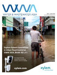
By Katherine Unger Baillie, senior science news officer, University Communications, University of Pennsylvania<br />
A shapeshifting robotic microswarm<br />
may one day act as a toothbrush, rinse,<br />
and dental floss in one. It is a system<br />
that could be particularly valuable for<br />
those who lack the manual dexterity to<br />
clean their teeth effectively themselves.<br />
The building blocks of these microrobots<br />
are iron oxide nanoparticles that have<br />
both catalytic and magnetic activity.<br />
Using a magnetic field, researchers could<br />
direct their motion and configuration to<br />
form either bristle-like structures that<br />
sweep away dental plaque from the<br />
broad surfaces of teeth, or elongated<br />
strings that can slip between teeth like<br />
a length of floss. In both instances, a<br />
catalytic reaction drives the nanoparticles<br />
to produce antimicrobials that kill harmful<br />
oral bacteria on site.<br />
Experiments using this system on mock<br />
and real human teeth showed that the<br />
robotic assemblies can conform to a<br />
variety of shapes to nearly eliminate the<br />
sticky biofilms that lead to cavities and<br />
gum disease. The Penn team shared<br />
their findings establishing a proof-ofconcept<br />
for the robotic system in the<br />
journal ACS Nano.<br />
The nanoparticles have both magnetic and catalytic properties; catalysed hydrogen<br />
peroxide produced free radicals that eliminated tooth decay-causing pathogens as well<br />
(Image: Minjun Oh/Penn <strong>Dental</strong> Medicine)<br />
Medicine and co-corresponding author<br />
on the study. “You have to brush your<br />
teeth, then floss your teeth, then rinse<br />
your mouth; it’s a manual, multistep<br />
process. The big innovation here is that<br />
the robotics system can do all three in<br />
a single, hands-free, automated way.”<br />
and forth across a space, much like<br />
flossing. The way it works is similar<br />
to how a robotic arm might reach<br />
out and clean a surface. The system<br />
can be programmed to do the<br />
nanoparticle assembly and motion<br />
control automatically.”<br />
“Routine oral care is cumbersome and<br />
can pose challenges for many people,<br />
especially those who have hard time<br />
cleaning their teeth,” said Hyun (Michel)<br />
Koo, a professor in the Department<br />
of Orthodontics and Divisions of<br />
Community Oral Health and Paediatric<br />
Dentistry in Penn’s School of <strong>Dental</strong><br />
“Nanoparticles can be shaped and<br />
controlled with magnetic fields in<br />
surprising ways,” added Edward<br />
Steager, a senior research investigator<br />
in Penn’s School of Engineering and<br />
Applied Science and co-corresponding<br />
author. “We form bristles that can<br />
extend, sweep, and even transfer back<br />
DISRUPTING ORAL CARE<br />
TECHNOLOGY<br />
“The design of the toothbrush has<br />
remained relatively unchanged for<br />
millennia,” said Koo. While adding<br />
electric motors elevated the basic<br />
bristle-on-a-stick format, the<br />
fundamental concept has remained<br />
20 DENTAL ASIA SEPTEMBER / OCTOBER <strong>2022</strong>
TRENDS<br />
the same. “It’s a technology that has<br />
not been disrupted in decades.”<br />
Several years ago, Penn researchers<br />
within the Center for Innovation and<br />
Precision Dentistry (CiPD), of which<br />
Koo is a co-director, took steps<br />
toward a major disruption, using this<br />
microrobotics system.<br />
Their innovation arose from a bit<br />
of serendipity. Research groups<br />
in both Penn <strong>Dental</strong> Medicine and<br />
Penn Engineering were interested in<br />
iron oxide nanoparticles but for very<br />
different reasons. Koo’s group was<br />
intrigued by the catalytic activity of<br />
the nanoparticles. They can activate<br />
hydrogen peroxide to release free<br />
radicals that can kill tooth decaycausing<br />
bacteria and degrade<br />
dental plaque biofilms. Meanwhile<br />
Steager and engineering colleagues,<br />
including Dean Vijay Kumar and Prof<br />
Kathleen Stebe, co-director of CiPD,<br />
were exploring these nanoparticles<br />
as building blocks of magnetically<br />
controlled microrobots.<br />
With support from Penn Health<br />
Tech and the National Institutes<br />
of Health’s National Institute of<br />
<strong>Dental</strong> and Craniofacial Research,<br />
the Penn collaborators married<br />
the two applications in the current<br />
work, constructing a platform to<br />
electromagnetically control the<br />
microrobots, enabling them to adopt<br />
different configurations and release<br />
antimicrobials on site to effectively<br />
treat and clean teeth.<br />
“It doesn’t matter if you have straight<br />
teeth or misaligned teeth, it will adapt<br />
to different surfaces,” said Koo. “The<br />
system can adjust to all the nooks and<br />
crannies in the oral cavity.”<br />
The researchers optimised the motions<br />
of the microrobots on a small slab of<br />
toothlike material. Next, they tested the<br />
microrobots’ performance adjusting<br />
to the complex topography of the<br />
tooth surface, interdental surfaces,<br />
and the gumline, using 3D-printed<br />
Magnetic and catalytic properties of the iron oxide nanoparticles and their assembly into bristle and floss-like<br />
forms (Image: Melissa Pappas/Penn Engineering)<br />
tooth models based on scans of human<br />
teeth from the dental clinic. Finally, they<br />
trialled the microrobots on real human<br />
teeth that were mounted in such a way<br />
as to mimic the position of teeth in the<br />
oral cavity.<br />
On these various surfaces, the researchers<br />
found that the microrobotics system<br />
could effectively eliminate biofilms,<br />
clearing them of all detectable pathogens.<br />
The iron oxide nanoparticles have been<br />
FDA approved for other uses, and tests<br />
of the bristle formations on an animal<br />
model showed that they did not harm the<br />
gum tissue.<br />
Indeed, the system is fully programmable;<br />
the team’s roboticists and engineers<br />
used variations in the magnetic field<br />
to precisely tune the motions of the<br />
microrobots as well as control bristle<br />
stiffness and length. The researchers<br />
found that the tips of the bristles could<br />
be made firm enough to remove biofilms<br />
but soft enough to avoid damage to<br />
the gums.<br />
The customisable nature of the system,<br />
the researchers said, could make it<br />
gentle enough for clinical use, but also<br />
personalised, able to adapt to the<br />
unique topographies of a patient’s<br />
oral cavity.<br />
To advance this innovation to the clinic,<br />
the Penn team is continuing to optimise<br />
the robots’ motions and considering<br />
different means of delivering the<br />
microrobots through mouth-fitting<br />
devices. They are eager to see their<br />
device help people in the clinic.<br />
“We have this technology that’s as or<br />
more effective as brushing and flossing<br />
your teeth but doesn’t require manual<br />
dexterity,” said Koo. “We’d love to see<br />
this helping the geriatric population and<br />
people with disabilities. We believe it will<br />
disrupt current modalities and majorly<br />
advance oral health care.”<br />
Koo and Steager’s co-authors on the<br />
paper are Penn <strong>Dental</strong> Medicine’s Min<br />
Jun Oh, Alaa Babeer, Yuan Liu, and Zhi<br />
Ren; and Penn Engineering’s Jingyu Wu,<br />
David A. Issadore, Kathleen J. Stebe, and<br />
Daeyeon Lee.<br />
This work was supported in part by<br />
the National Institute for <strong>Dental</strong> and<br />
Craniofacial Research (grants DE025848<br />
and DE029985), Procter & Gamble, and<br />
the Postdoctoral Research Programme<br />
of Sungkyunkwan University. DA<br />
This article was first published at Penn<br />
Today.<br />
DENTAL ASIA SEPTEMBER / OCTOBER <strong>2022</strong> 21
UNDER THE SPOTLIGHT<br />
What it takes to be<br />
a dental influencer<br />
Dentist, entrepreneur, content creator – how does one find the<br />
balance in juggling these roles? <strong>Dental</strong> <strong>Asia</strong> finds out more from<br />
Dr Kayla Teh.<br />
By Czarmaine Masigla<br />
Transforming smiles one tooth<br />
(and content) at a time, Dr Kayla<br />
Teh Khe Sin has over 79,000<br />
followers on Instagram, 27,000<br />
followers on Facebook, and<br />
33,000 subscribers on YouTube.<br />
Also known as Dr Kayla to her<br />
patients and followers, she has<br />
established her name not just in<br />
the online space but also as a<br />
friendly dentist to her patients.<br />
With her enthusiasm for raising<br />
oral health awareness as well<br />
as her passion for aesthetic<br />
dentistry, she founded Dr Kayla<br />
<strong>Dental</strong> Clinic and now leads<br />
a team of dental specialists<br />
providing comprehensive dental<br />
care. Read on as she shares her<br />
humble beginnings and how her<br />
persistence brought online and<br />
real-life success.<br />
What sparked your interest to<br />
become a content creator and<br />
at the same time specialise in<br />
aesthetic dentistry?<br />
Dr Kayla Teh: When I was<br />
a dental student, I joined a<br />
volunteering programme at<br />
a rural village in Malaysia.<br />
There, I met a young kid with a<br />
cleft lip who was all alone and<br />
none of the other kids wanted<br />
to be his friend because of<br />
his appearance. That could’ve been<br />
prevented if his parents were aware<br />
of bringing him to an oral surgeon to<br />
do repair surgery during the earlier<br />
years of his life. That was the moment<br />
when I realised how important dental<br />
awareness is. I started making videos<br />
on it, and that was back in 2018 when<br />
videos on social media were not such a<br />
common thing.<br />
Then, I began to have some patients<br />
walking in, telling me that they do not<br />
have the confidence to get a job, partner<br />
or simply smile in front of the camera.<br />
So, when I did my first smile makeover<br />
case with veneers, my patient regained<br />
her confidence and came back to tell<br />
me that she met a new partner in life.<br />
That was when I felt true happiness and<br />
realised my ability to help others as a<br />
dentist. This experience and more after<br />
inspired me to keep doing this every<br />
day and keep improving in the field of<br />
aesthetic dentistry.<br />
What are your aspirations as an oral<br />
health advocate and an influencer?<br />
Dr Kayla: It is important to uphold the<br />
standard of dentistry while having fun<br />
on social media. Whenever I create<br />
a piece of content on social media,<br />
I always have three goals in mind:<br />
educate, entertain and inspire.<br />
I love to educate the public on certain<br />
dental topics through videos or social<br />
22 DENTAL ASIA SEPTEMBER / OCTOBER <strong>2022</strong>
UNDER THE SPOTLIGHT<br />
media posts. I also like entertaining<br />
them from the point of view of a dentist<br />
because that shows them that dentists<br />
are human too and we just wanted to<br />
help with their dental problems. That<br />
can actually increase the patient’s<br />
acceptance of dental treatment, which<br />
in turn elevates public awareness of<br />
dental care.<br />
I hope to inspire the younger generation<br />
on the beautiful aspect of dentistry –<br />
which to me is an amazing field. I enjoy<br />
every moment of it, creating smiles and<br />
building connections with my patients<br />
which last for generations. I hope that<br />
more people can see and appreciate<br />
this aspect of dentistry as much as I do!<br />
Can you walk us through the<br />
conceptualisation of your dream clinic,<br />
and what is the significance of this<br />
milestone in your career as a dentist?<br />
Dr Kayla: I have always wanted to<br />
make people feel safe. But dentistry?<br />
Oh man, it can be scary to our patients.<br />
When I build my clinic, I want people<br />
to feel like they’re coming back home<br />
to a safe place where they can be<br />
comfortable and more relaxed to share<br />
their dental concerns.<br />
That is why my clinic is homey, it is<br />
also covered in a pastel pink tone –<br />
which is unconventional. But that’s<br />
the beauty of it. It doesn’t give off<br />
that tense, strict vibe. But rather, it<br />
makes you feel comfortable like you’re<br />
walking into an area where you can<br />
relax as well as enjoy the view.<br />
As a dentist, this is a huge milestone<br />
for me. It is a sign that I am no longer<br />
just a clinician, but an entrepreneur,<br />
marketer, manager, and so much more<br />
too. I have a bigger vision now, to not<br />
only excel in my clinical work but also<br />
to build a team of doctors who strive<br />
to achieve excellence in dentistry as<br />
well as patient management. And this<br />
is something we did not learn in dental<br />
school; this is the real world where<br />
we have to be independent, creative,<br />
caring and better than who we are<br />
yesterday.<br />
As a practitioner of digital dentistry,<br />
what are the must-have devices in your<br />
practice, and how do they help you<br />
enhance your service to your patients?<br />
Dr Kayla: Intraoral scanner (IOS), digital<br />
planning software and 3D printer are a<br />
necessity in my daily clinical workflow.<br />
IOS enables me to take dental record<br />
conveniently and comfortably, and helps<br />
in enhancing the communication among<br />
clinicians, dental technologist and<br />
ceramists.<br />
I am also using exocad software to<br />
plan out my cases, be it prosthodontics,<br />
restorative or orthodontics. It helps<br />
me to bring a more comprehensive<br />
approach to my case planning.<br />
For example, most of my patients<br />
who came to me for veneers often<br />
need orthodontics treatment. I will<br />
incorporate digital dentistry into their<br />
orthodontics treatment to measure<br />
the space distribution for their final<br />
veneers as well as monitor their<br />
treatment progress. This gives me more<br />
predictability and control in my clinical<br />
cases, which also gives my patients<br />
more confidence and reassurance.<br />
How do you envision your career as a<br />
dentist in the next five to 10 years, and<br />
what else are you looking forward to in<br />
the future?<br />
Dr Kayla: I look forward to creating more<br />
quality content in dentistry and inspiring<br />
more young dentists to do better in their<br />
clinical work. I hope to show people that<br />
just because you’re out there on social<br />
media, doesn’t mean that you’re all talk<br />
and cannot produce good clinical work.<br />
We are all clinicians and we are here<br />
to help our patients through the skills<br />
and experience we have. I also look<br />
forward to meeting and learning from<br />
more dentists across the globe, after all,<br />
learning never ends.<br />
How do you find the balance between<br />
your role as a dentist and content<br />
creator, and what advice would you like<br />
to give to dental practitioners who are<br />
keen to follow in your footsteps?<br />
Dr Kayla: It is difficult to maintain social<br />
media while upholding your clinical work<br />
standards — at least for me. I plan out<br />
my shooting time as well as clinical<br />
time, but that also means that I have to<br />
sacrifice my personal and family time.<br />
My advice to those who wanted to<br />
engage more patients online is to<br />
be yourself on social media. Many<br />
people think they have to pretend to be<br />
someone with “personality” to engage<br />
more audience but simply being yourself<br />
and showcasing your best sides are<br />
good enough. It also gives a sense of<br />
authenticity when a patient, or a fellow<br />
dentist finally meets you in person. After<br />
all, people love making friends with a<br />
person who is true to himself. DA<br />
DENTAL ASIA SEPTEMBER / OCTOBER <strong>2022</strong> 23
DENTAL PROFILE<br />
Transforming the future<br />
of implant dentistry<br />
With innovation and technology, Dentsply Sirona ensures<br />
that clinicians can provide dental implant therapy for life-long<br />
function and aesthetics. <strong>Dental</strong> <strong>Asia</strong> speaks with Tony Susino,<br />
group vice-president of Global Implant Solutions, to find out<br />
the latest trends in implant dentistry and how the company<br />
shapes the digital future of the field.<br />
By Czarmaine Masigla<br />
People and products — veteran business<br />
executive Tony Susino shared that this<br />
combination was the key factor when he<br />
joined Dentsply Sirona, one of the world’s<br />
largest manufacturers of dental products<br />
and technologies, this year as the group vicepresident<br />
of Global Implant Solutions.<br />
Having spent the last 15 years in the dental<br />
space, he has never been more optimistic<br />
about the opportunities Dentsply Sirona<br />
provides in terms of an ecosystem of products<br />
and digital solutions.<br />
“At Dentsply Sirona, the employees have a<br />
great passion to create a superior experience<br />
for our customers through optimised<br />
workflows. With our cutting-edge products and<br />
technologies, the company is uniquely suited<br />
to win in this space,” he shared.<br />
In today’s dental landscape, Susino said<br />
that dental implants are set to become the<br />
standard of care for tooth replacement and<br />
edentulism in the years to come.<br />
“Clinicians and their staff are more widely<br />
aware and promoting these therapies over<br />
traditional crown and bridge dentistry, and<br />
that will continue to drive the steady market<br />
growth. <strong>Dental</strong> disorders such as tooth decay<br />
and cavities, unfortunately, continue to be on<br />
the rise based on the availability of sugary<br />
foods and overall poor nutrition and hygiene<br />
choices,” he elaborated.<br />
The advances in technology and accessibility<br />
of treatment, Susino suggested, have<br />
increased patients’ dental knowledge<br />
too, thereby sustaining the momentum of<br />
industry growth.<br />
“As implant therapy becomes the more<br />
accepted gold standard, governmentled<br />
initiatives and public health dental<br />
programmes will also make implant surgery<br />
more easily available to those in need,”<br />
he continued.<br />
SEAMLESS SOLUTIONS<br />
Dentsply Sirona is poised to meet the<br />
rapidly growing dental implant<br />
demands with their offering<br />
of implant systems,<br />
digital planning<br />
platform, regenerative<br />
and restorative<br />
solutions and<br />
educational<br />
programmes.<br />
Susino<br />
highlighted that<br />
Dentsply Sirona’s<br />
dental implant<br />
systems are<br />
Tony Susino<br />
24 DENTAL ASIA SEPTEMBER / OCTOBER <strong>2022</strong>
DENTAL PROFILE<br />
based on a solid scientific foundation with<br />
long-term clinical documentation. In fact, their<br />
products and solutions have been the subject<br />
of more than 2,900 scientific publications<br />
with data from 50,000 patients and 125,000<br />
implants.<br />
For instance, one of the company’s latest<br />
innovations, the DS PrimeTaper Implant<br />
System features a union of form and<br />
function. It has a thread design that enables<br />
it to quickly cut and engage with the bone,<br />
allowing for immediate function and shorter<br />
procedure times.<br />
And with the comprehensive restage of its<br />
implants business including three signature<br />
digital workflows, Dentsply Sirona provided<br />
dental professionals with an innovative way<br />
of practising implantology from scan to crown<br />
that meet expectations in efficiency, accuracy,<br />
aesthetics, longevity and simplicity.<br />
“Dentsply Sirona is committed to the<br />
advancement of our industry as evidenced by<br />
our significant investments in R&D and the<br />
recently built Innovation Center in Charlotte,<br />
NC (US). We also have various facilities<br />
globally that are on the forefront of clinical<br />
education to provide training and resources<br />
for clinicians to continually be on the cutting<br />
edge of their field,” he added.<br />
For practitioners who are just starting out<br />
with their digital implant journey, Susino<br />
recommended accessing the DS Academy’s<br />
<strong>Dental</strong> Implant Curriculum.<br />
He explained: “It contains interactive,<br />
procedure-based courses, designed by<br />
world-renowned clinicians such as Dr Lyndon<br />
F. Cooper and Dr Bryan Limmer. The virtual<br />
curriculum is comprehensive, tuition-free and<br />
aims to inspire dental professionals to build<br />
and develop their clinical knowledge and digital<br />
skills in implant dentistry, through eight levels<br />
each consisting of 24 courses.<br />
“Practitioners of all backgrounds and different<br />
levels of knowledge can access this course —<br />
from foundational to mastery. They can learn at<br />
their own pace and gain new skills that will help<br />
them give their patients the best possible care<br />
for improved oral health.”<br />
BEST WAY FORWARD<br />
Dentsply Sirona is committed to enabling<br />
clinicians, to focus on growth and enhance<br />
their service. Streamlining implant treatments<br />
equates to consistent patient satisfaction.<br />
“I’ve already met and gotten to know many of<br />
our practitioners. They are exceptional, care<br />
deeply about our products and believe — like<br />
I do — that their success is our success and<br />
vice versa,” shared Susino.<br />
To stay at the forefront of digital dentistry,<br />
Susino encourages dental practitioners to<br />
attend as many symposiums and events<br />
as possible. He believes that they can learn<br />
much from their peers, build professional<br />
networks, tap into each other’s knowledge,<br />
and create groups to discuss trends and<br />
what’s coming next.<br />
“Dentsply Sirona holds events such as<br />
Dentsply Sirona World every year that do<br />
exactly this. It draws in professionals from<br />
all over the world who come to see the<br />
latest in digital dentistry, hear from expert<br />
speakers and see our newest products in<br />
action,” he shared.<br />
On top of that, they have also set up a registry<br />
study which aims to involve more than 500<br />
clinicians and over 2,500 implants for longterm<br />
follow-up.<br />
Embracing and adopting digital dentistry is<br />
the best way forward according to Susino,<br />
who reassured dental professionals that<br />
Dentsply Sirona will be there for them at every<br />
step of their digital transformation: “We are<br />
shaping the technology behind our innovative<br />
solutions, conducting research, and creating<br />
resources to help practitioners expand their<br />
clinical knowledge and digital skills. That is<br />
where we are going, and we are leading the<br />
charge into the future. The future is now!” DA<br />
DENTAL ASIA SEPTEMBER / OCTOBER <strong>2022</strong> 25
DENTAL PROFILE<br />
Peek<br />
into the<br />
digital<br />
future<br />
What trends will<br />
sustain the positive<br />
momentum of digital<br />
dentistry? Rune Fisker,<br />
SVP of 3Shape, shares<br />
his insights.<br />
By Czarmaine Masigla<br />
26 DENTAL ASIA SEPTEMBER / OCTOBER <strong>2022</strong>
DENTAL PROFILE<br />
The digital era is here. From artificial<br />
intelligence, automated processes,<br />
and so much more, an array of dental<br />
treatment options are now accessible<br />
at a practitioner’s fingertips. These<br />
technologies have elevated the standards<br />
of dental care to a new height which<br />
would not be otherwise possible.<br />
Leading this revolution is 3Shape, a<br />
developer and manufacturer of 3D<br />
scanners and CAD/CAM software for<br />
the dental and audio industries based<br />
in Copenhagen, Denmark. Since the<br />
launch of 3Shape’s first dental lab<br />
scanner software in 2005, the company<br />
has continuously innovated and<br />
reinvented their product solutions.<br />
Over the years, 3Shape has supported<br />
many dental professionals in embarking<br />
on their digital journey, even more<br />
so when facing the unprecedented<br />
challenges brought by the pandemic.<br />
According to Fisker, due to COVID-19,<br />
digital adoption was dramatically<br />
pushed forward by three to five years.<br />
What will continue driving its growth?<br />
He highlighted five major trends:<br />
Intraoral scanning will keep driving<br />
digitalisation. The use of intraoral<br />
scanners (IOS) in dental clinics has<br />
skyrocketed and already penetrated<br />
30-35% in the US and mature markets.<br />
A small, handheld device that builds a<br />
digital model of the teeth and surrounding<br />
structures, many clinicians consider IOS<br />
their entry point in digital workflow –<br />
streamlining the cumbersome procedure<br />
of taking an impression and sending it to<br />
the lab.<br />
Speaking with Prof Dr Lucio Lo Russo,<br />
an Italian dentist, regarding the hygiene<br />
aspect of digital pathways, Fisker<br />
shared that besides the time and money<br />
clinicians save using a digital workflow,<br />
another important aspect to consider<br />
was safety.<br />
Highlighting that digital workflow has<br />
become a means for professionals to<br />
avoid cross-contamination issues,<br />
he explained: “Remember, there is<br />
no impression material to handle<br />
or ship with a digital workflow. So<br />
aside from eliminating the possible<br />
breakage of models with all the<br />
handling, professionals no longer<br />
need to touch, share, or ship models<br />
back and forth.<br />
“All those objects which were in<br />
contact with the patient’s mouth are<br />
theoretically, potentially infected.<br />
If we are not transferring anything<br />
from the clinic to the lab, we are<br />
reducing and controlling any chance<br />
of infection.”<br />
Artificial intelligence (AI) is here.<br />
A field that combines computer<br />
science and robust datasets to enable<br />
problem-solving, AI has finally made<br />
its mark on CAD/CAM. This past year,<br />
Fisker shared that with the launch of<br />
3Shape Automate, scans can now be<br />
uploaded to the cloud, and practitioners<br />
can fully design a crown using AI.<br />
DENTAL ASIA SEPTEMBER / OCTOBER <strong>2022</strong> 27
DENTAL PROFILE<br />
we live. Think Netflix or Spotify. With<br />
Unite, our goal is to deliver a similar and<br />
seamless service where professionals<br />
can pick and choose their treatment<br />
partner just like they would pick the next<br />
movie to watch.”<br />
SYNERGISTIC COLLABORATIONS<br />
As the technology for digital pathways<br />
continue to improve, Fisker is positive<br />
that more practitioners will embrace it in<br />
practice and put high appreciation for its<br />
potentials.<br />
“When it comes to introducing new<br />
solutions, it is one thing to have an idea,<br />
it is a whole other ball game to make it<br />
happen. That is where 3Shape’s huge<br />
brainpower trust, an army of engineers,<br />
and data comes in,” he said.<br />
3Shape has also utilised this technology<br />
in TRIOS intraoral scanner to allow user<br />
of any skill level to capture accurate<br />
digital intraoral impressions with greater<br />
ease in less time.<br />
“There will be much more to come as<br />
AI is making computers smarter and<br />
faster across all applications in digital<br />
dentistry,” he continued.<br />
Aligners and dentures are rapidly<br />
adapted. Fisker said that cosmetic<br />
dentistry and clear aligners will continue<br />
to push digitalisation forward, driven by<br />
consumer demand for aesthetics.<br />
While dentures remain the big missing<br />
indication, he noted that their stats show<br />
that this is changing fast with three-digit<br />
growth in digital denture cases.<br />
Considering <strong>Asia</strong>’s ageing population<br />
affecting the demands for tooth<br />
replacement, he said: “When treating<br />
older patients, digital dentistry delivers<br />
higher efficiency, better clinical results<br />
and an overall improved patient<br />
experience that further accelerates<br />
digital adoption.”<br />
3D printers are hot. A true game-changer<br />
will be 3D printing materials, Fisker<br />
emphasised, as new and improved<br />
materials are being released every day.<br />
He elaborated: “Driven by new printing<br />
materials, high-quality splints are a<br />
reality and I believe, 100% 3D printed<br />
final dentures will soon take off.<br />
Ultimately, all indications will have the<br />
potential to be 3D printed, which would<br />
make the major question being how<br />
deep will 3D printers penetrate the<br />
practice?”<br />
With strong technology development<br />
and approximately only 5% of the<br />
world’s dentures presently being<br />
manufactured digitally, Fisker foresees<br />
that the industry will witness a lot<br />
of growth in this area. As printing<br />
materials become better, he added,<br />
more and more types of restorations<br />
will be printed due to their affordability,<br />
speed, and practicality.<br />
The dental platform race has begun.<br />
3Shape is one of the key drivers of this<br />
trend globally and is playing a crucial<br />
role in its rapid adoption as reflected<br />
with the successful launch of their<br />
Unite platform.<br />
Fisker elaborated: “Like all industries,<br />
platforms have disrupted the way<br />
For instance, 3Shape has recently<br />
launched a Smart Phone App that enables<br />
dentists and labs to communicate and<br />
add pictures directly from the phone –<br />
anywhere, anytime. They have also<br />
released TRIOS Unite, which delivered<br />
significant improvements to the workflow<br />
and communications.<br />
He explained: “Little additions like<br />
enabling partners to chat, so they do not<br />
need to keep checking emails, creating<br />
a URL for the lab to share a case with a<br />
practice that may not be using 3Shape<br />
solutions, or DIY order forms for labs,<br />
these types of initiatives go a long way<br />
in making dental professionals and their<br />
workflows more efficient.”<br />
3Shape has also taken advantage<br />
of social media to stay close to<br />
dental professionals considering it<br />
as a learning and sharing platform.<br />
Fisker shared that on Facebook,<br />
there are two study groups focusing<br />
on 3Shape solutions, and combined<br />
they have nearly 45,000 members. He<br />
considers this as a great resource for<br />
understanding their customers.<br />
“That is a lot of people talking about<br />
digital workflows, tips and tricks and<br />
even complaining about something.<br />
But it gives us a direct and instant<br />
28 DENTAL ASIA SEPTEMBER / OCTOBER <strong>2022</strong>
DENTAL PROFILE<br />
apartment to being a business leader and<br />
innovator across the globe. The cool thing<br />
though, is somehow we have managed to<br />
retain that entrepreneurial spirit of always<br />
wanting to get better, grow and be open to<br />
new ideas.”<br />
Fisker considers South East <strong>Asia</strong> (SEA)<br />
as a terrific market for digital dentistry<br />
as many of the countries are already<br />
electronically savvy that adopting new<br />
technology is second nature. In addition,<br />
digital pathways provide an opportunity<br />
to deliver more cost-effective treatments.<br />
connection to what is happening out<br />
there. I would quickly run out of fingers<br />
counting the amounts of times we have<br />
received a suggestion for a new feature<br />
from a study group and then made it<br />
happen in the next software update,”<br />
he commented.<br />
Fisker highlighted that working alongside<br />
dental professionals to translate their<br />
ideas into reality is the cornerstone of<br />
3Shape’s product development.<br />
SHAPING THE FUTURE<br />
Undoubtedly, 3Shape has become a<br />
major player in the dental industry<br />
shaping the future of digital dentistry.<br />
Being one of the pioneers in 3Shape<br />
– in fact, the third employee – Fisker<br />
himself was astonished as to how<br />
3Shape changed over the years as an<br />
organisation and a trailblazer the field.<br />
He shared: “What I have witnessed is us<br />
go from a little start-up in a Copenhagen<br />
He concluded: “In many of the rural areas,<br />
a digitally printed prosthesis may be the<br />
only affordable alternative for a person<br />
and could literally mean the difference<br />
between life and death for some patients.<br />
The more digital penetration in the SEA<br />
market, the more people will have access<br />
to dental treatment.<br />
“We see lots of innovative business<br />
models for selling intraoral scanners<br />
around the world, I think if entrepreneurs<br />
and even governments could get behind<br />
dental professionals and make them easier<br />
to purchase, it would be a tremendous<br />
benefit to millions of people.” DA<br />
DENTAL ASIA SEPTEMBER / OCTOBER <strong>2022</strong> 29
DENTAL PROFILE<br />
The<br />
hundred<br />
years<br />
of Shofu<br />
Established on 15 May 1922 in Kyoto, Japan,<br />
Shofu is a global manufacturer deeply<br />
involved in the field of dentistry offering a<br />
wide range of dental materials, equipment<br />
and instruments.<br />
By Czarmaine Masigla<br />
Over the years, the treatment<br />
philosophy in dentistry has shifted<br />
from invasive to a more conservative<br />
approach – with Shofu spearheading<br />
development of products and<br />
technologies, emphasising prevention<br />
and preservation of natural tooth<br />
structure.<br />
Commemorating their centennial<br />
anniversary, Mr Noriyuki Negoro, CEO<br />
and chairman of Shofu, takes a trip<br />
down memory lane with <strong>Dental</strong> <strong>Asia</strong><br />
to ponder on the company’s founding<br />
years, and elaborates on how they will<br />
continue to revolutionise the dental<br />
industry in years to come.<br />
FOUNDING YEARS<br />
Mr Kajo Shofu III, the founder of<br />
Shofu, was involved in numerous<br />
businesses, including the export of<br />
ceramics, industrialisation of ceramic<br />
manufacturing, and the insulator<br />
business based on Kiyomizu-yaki<br />
porcelain technology, as Mr Negoro<br />
shared.<br />
In 1915, during a visit to North America<br />
to expand sales channels for the export<br />
of ceramics, the founder was advised by<br />
Dr Mitsuru Okada, MD (later professor<br />
of oral surgery at Keio University School<br />
of Medicine) on the need for domestic<br />
production of high-grade porcelain teeth.<br />
30 DENTAL ASIA SEPTEMBER / OCTOBER <strong>2022</strong>
DENTAL PROFILE<br />
The journey was not a walk in the<br />
park – as it required identifying<br />
tooth shapes and sizes suitable<br />
for the Japanese people, sourcing<br />
biocompatible materials and<br />
appropriate manufacturing methods.<br />
Seven years later, after overcoming<br />
several challenges, Mr Shofu III<br />
realised his dream with the help<br />
and cooperation of many people to<br />
successfully manufacture the first<br />
high-grade porcelain teeth in Japan –<br />
laying the cornerstone for Shofu.<br />
In 1965, Shofu succeeded in<br />
developing the world’s first dental<br />
spherical amalgam, “Shofu Spherical<br />
Amalgam”, recognised by clinicians as<br />
a revolutionary product with excellent<br />
handling and mechanical properties,<br />
and was considered a breakthrough in<br />
the dental industry.<br />
the Japanese dental industry to be<br />
listed in the Osaka Stock Exchange<br />
and subsequently in the Tokyo Stock<br />
Exchange in 2007,” he added.<br />
Since then, the company has diversified<br />
its product range to include not only<br />
artificial teeth, but a wide variety of<br />
dental products, laboratory materials,<br />
preventive products, and materials for<br />
digital dentistry. Now an international<br />
enterprise, Shofu has subsidiaries and<br />
sales offices in the US, Germany, UK,<br />
Singapore, China and in recent years<br />
Italy, India, Brazil, the Middle East and<br />
Taiwan.<br />
Mr Negoro continued: “These<br />
developments aim to contribute towards<br />
improving the standards of dental care.<br />
The R&D-oriented and innovation-driven<br />
management approach adopted since<br />
the company’s founding is proof that our<br />
founder’s passion for ‘high aspirations<br />
and drive for achieving them’ lives on in<br />
our DNA.”<br />
COMMITMENT TO BETTER<br />
DENTISTRY<br />
According to the Global Burden<br />
of Disease study published by the<br />
World Health Organization (WHO) in<br />
2016, dental caries remains the most<br />
common disease in the world, despite<br />
regional disparities due to economic<br />
status and healthcare systems. The<br />
Lancet and Journal of <strong>Dental</strong> Research<br />
also reported that more than 30% of<br />
people have untreated dental caries.<br />
Hence, Shofu’s vision is to continue<br />
to provide proven products for better<br />
dentistry worldwide with a commitment<br />
to quality and excellence, emphasising<br />
that beautiful teeth are the symbol of<br />
health and happiness.<br />
Additionally, the company’s rotary<br />
instruments was well regarded,<br />
and the brand reputation of “Shofu<br />
Abrasives” was established not only<br />
in North America but also in the<br />
global market.<br />
Such innovation helped propel the<br />
company’s overseas expansion,<br />
leading to the establishment of sales<br />
offices in the US in 1971, Germany in<br />
1978 and Singapore in 1980.<br />
Mr Negoro also shared that during<br />
the 1980s when Shofu was under<br />
a crisis due to Japan’s healthcare<br />
reform, then President Kajo Shofu V,<br />
changed the company’s focus from<br />
quantity to quality and implemented<br />
management reforms to ensure the<br />
company’s survival, under the slogan<br />
of “Response to Change”.<br />
“The foresight of the president and<br />
commitment of the employees<br />
resulted in the development of a<br />
series of high-quality value-added<br />
products that helped transform the<br />
company into a highly profitable<br />
business. As a result, in 1989, Shofu<br />
became the first company from<br />
Mr Noriyuki Negoro, CEO & chairman of Shofu (Left) with Mr Tetsuo Takami, president & COO of Shofu (Right)<br />
DENTAL ASIA SEPTEMBER / OCTOBER <strong>2022</strong> 31
DENTAL PROFILE<br />
With the dental field continuously<br />
evolving digitalisation, artificial<br />
intelligence (AI), and dental material<br />
science taking the centre stage in<br />
product innovation, Shofu maintains<br />
profitability and growth by identifying<br />
expansion opportunities.<br />
“As a company, we had focused our<br />
business activities mainly in Japan,<br />
a stable market with a generous<br />
national medical insurance system<br />
and successfully introduced many<br />
revolutionary products over the years<br />
contributing to the development<br />
of dentistry. Our success in the<br />
domestic market laid the foundation<br />
for international expansion, with more<br />
strategic business activities extended<br />
beyond Japan to capture the global<br />
market,” he explained.<br />
Based on their management philosophy<br />
and vision, Shofu is actively engaged<br />
in business activities to increase<br />
their global presence with continued<br />
efforts to provide evidence-based<br />
product innovations using cutting-edge<br />
technology with end-to-end solutions<br />
for the wider dental community.<br />
And although the company promotes<br />
globalisation in its business activities<br />
internally and externally, Mr Negoro<br />
highlighted that it is equally important<br />
for them to adapt to local needs across<br />
diverse markets.<br />
For instance, the Shofu <strong>Dental</strong> <strong>Asia</strong>-<br />
Pacific subsidiary has been a crucial<br />
part of Shofu’s global footprint looking<br />
S-PRG Technology<br />
after its growth in the <strong>Asia</strong>-Pacific and<br />
Middle-East markets. Recognising that<br />
the local treatment trends had shifted<br />
from aggressive to minimally invasive,<br />
Shofu has become the official partner<br />
of the Minimally Invasive Cosmetic<br />
Dentistry (MiCD) Global Network and its<br />
digital platforms.<br />
Mr Negoro shared: “Our team at Shofu<br />
<strong>Dental</strong> <strong>Asia</strong>-Pacific felt that the MiCD<br />
philosophy matched very well with<br />
the benefits of our range of bioactive<br />
products, hence we have worked closely<br />
with Dr Sushil Koirala and like-minded<br />
clinicians to establish the MiCD Concept<br />
and treatment protocol.”<br />
BRIGHT SMILES FOR THE NEXT<br />
CENTURY<br />
Commemorating their rich history and<br />
legacy of product innovations toward<br />
enhancing smiles, Shofu launched their<br />
100th-anniversary logo and slogan<br />
representing their commitment to<br />
contributing to the bright smiles of<br />
people globally for the next century.<br />
Mr Negoro noted that as the company<br />
values the tireless efforts of its people,<br />
the slogan was selected from the<br />
employee submissions: “The ‘0’ in the<br />
centre of the ‘100’ figure takes the form<br />
of a tooth and a face depicting a ‘bright<br />
smile’. The line extending from the roots<br />
of the tooth towards the Shofu logo<br />
indicates the progress of the company<br />
over the years from 1922 to <strong>2022</strong>, and<br />
the journey ahead for the next 100 years.”<br />
Looking ahead, Mr Negoro foresees that<br />
public health focus would shift towards<br />
prevention and oral health education.<br />
He also predicts that AI-driven remote<br />
examination with digital diagnostic<br />
techniques and preventive treatment<br />
will be implemented in each generation,<br />
especially in the ageing population.<br />
He elaborated: “Adequate oral hygiene<br />
maintenance becomes more challenging<br />
in older age group due to chronic<br />
diseases such as diabetes, druginduced<br />
xerostomia and other medical<br />
conditions as they retain more teeth.<br />
Unlike in the past, implant-supported<br />
dentures and partial dentures are<br />
becoming more popular increasing the<br />
risk of oral disease.”<br />
Therefore, Shofu targets to expand<br />
their Giomer/S-PRG technology<br />
into various areas and to enrich the<br />
Giomer product tree, with a specific<br />
elder care product range that includes<br />
a homecare treatment regimen for<br />
exposed root surfaces and denturerelated<br />
products.<br />
“S-PRG fillers are incorporated into<br />
new bioactive materials based on<br />
our proprietary technology (PRG<br />
technology), and have been proven<br />
to provide bioactive benefits such<br />
as inhibiting bacterial growth and<br />
adhesion, reduction in plaque formation,<br />
acid neutralisation and increased acid<br />
resistance by strengthening the natural<br />
tooth structure,” he explained.<br />
A gateway to overall health, oral health<br />
is essential for people to lead rich<br />
and fulfilling lives. As the world enters<br />
the era of a 100-year human lifespan,<br />
Mr Negoro concluded that the role<br />
of dentistry will become increasingly<br />
important in extending people’s healthy<br />
life expectancy.<br />
“For the next 100 years, we will continue<br />
to contribute towards the advances in<br />
dentistry while embracing the challenge<br />
of creating bright smiles that improve<br />
the quality of life of people around<br />
the world. In the past 13 years, I have<br />
served as the president and CEO of the<br />
company and am proud to announce<br />
that Mr Tetsuo Takami has now<br />
become my successor as the president<br />
and COO effective 24 Jun <strong>2022</strong>.<br />
“I will oversee the management of<br />
the company as CEO and chairman,<br />
while Mr Takami will be responsible for<br />
business execution. We will continue<br />
to focus on developing innovative<br />
solutions that meet the changing trends<br />
in dentistry and we are looking forward<br />
to the future growth of Shofu under the<br />
new management.” DA<br />
32 DENTAL ASIA SEPTEMBER / OCTOBER <strong>2022</strong>
CLINICAL FEATURE<br />
Effects of PBM on implant<br />
and bone grafting after<br />
sinus elevation<br />
Dr Kevin Ng and Dato’ Dr How Kim Chuan discuss the use<br />
of photobiomodulation (PBM) device with 42 LED cores<br />
producing 650nm wavelength light in shortening treatment<br />
time and improving surgical outcome.<br />
Maxillary bone resorption after<br />
extraction of posterior teeth could<br />
cause severe bone loss and greatly<br />
reduce bone height and volume at<br />
the sinus base if the edentulous<br />
sites were not loaded for a long<br />
period. It may not show apparent<br />
sign and symptoms and not cause<br />
any concerns to patient; however,<br />
the resorption process is irreversible<br />
unless treatment is performed.<br />
Eventually, it will become an<br />
obstacle in placing dental implants<br />
and the average treatment time to<br />
graft and gain bone is usually long<br />
to achieve satisfactory results.<br />
These factors have been bothering<br />
dentists and patients since<br />
treatment outcomes may not<br />
always be successful if implant<br />
stability is not achieved. To assist<br />
graft, bone healing and growth,<br />
and at the same time, enhance<br />
patient cooperation and compliance,<br />
photobiomodulation (PBM) can be<br />
utilised to reduce pain and shorten<br />
treatment time.<br />
Although different clinicians hold<br />
different views regarding to the<br />
application of PBM to influence bone<br />
regeneration and outcomes, the<br />
following case showed satisfactory<br />
patient feedback and acceptable<br />
clinical result.<br />
Fig. 1<br />
Fig. 2<br />
Fig. 3<br />
THE PBM DEVICE<br />
The bite plane device consists of 42<br />
LED cores and the power input is<br />
4.2V. The current of each LED core is<br />
7mA. The LED cores produce light of<br />
650nm wavelength to stimulate the<br />
target sites. The energy absorbed<br />
after six minutes stimulation per<br />
arch is about 306.5 joules (Figs. 1-3).<br />
The manufacturer claims that the<br />
science of PBM mechanism is to<br />
enhance the biological activities at<br />
cellular level of the treatment area<br />
and improve the healing of bone and<br />
soft tissues and further enhance<br />
bone growth and repairs.<br />
34 DENTAL ASIA SEPTEMBER / OCTOBER <strong>2022</strong>
CLINICAL FEATURE<br />
Figs. 4a<br />
Figs. 4b<br />
Figs. 4a-b: Pre-treatment (a) OPG planning and 4b CT view show only 2mm bony height (b) OPG planning for augmentation site<br />
Figs. 5a<br />
Figs. 5b<br />
Figs. 5a-b: Post-treatment (a) Bone graft after five weeks (b) Week 10 - implants ready to load<br />
Figs. 6a<br />
Figs. 6b<br />
CASE REPORT<br />
A 55-year-old male patient presented<br />
with severe bone loss at the upper<br />
right molar region (teeth 15, 16, 17<br />
area) with only 2-3mm remaining<br />
bone height (Figs. 4a-b). The patient<br />
had difficulty chewing on the right<br />
side and requested for dental<br />
implant. The medical history was<br />
clear and he had received implants<br />
before at teeth 45 and 46.<br />
Sinus floor elevation was performed<br />
by external window and 2cc of<br />
graft were placed with membrane<br />
protection. PBM was prescribed<br />
to patient for 5-6mins daily. Pain<br />
thresholds were recorded for two<br />
weeks post-operatively. Two dental<br />
implants were placed at teeth 15 and<br />
16 areas five weeks after the graft<br />
with insertion torques of 40N, that<br />
were achieved with ISQ 65, 68. PBM<br />
was continued for 5mins daily in<br />
another five weeks before ISQ 70<br />
were recorded for both implants<br />
and successfully loading with two<br />
splinted crowns restoration.<br />
The total treatment time was 10<br />
weeks, and the patient reported<br />
that the chewing function was<br />
satisfactory after the splinted<br />
crowns were constructed. The<br />
PBM device was utilised for 5mins<br />
daily and the patient was advised to<br />
return to clinic monthly to check<br />
stability of implant and ensure<br />
good oral hygiene. At the same<br />
time, the patient was also informed<br />
to record the degree of pain or<br />
discomfort in the given form during<br />
treatment. The feedback pain<br />
scores were two to three for the<br />
whole period. OPG was taken after<br />
treatment revealing uneventful<br />
healing with no sign of infection<br />
(Figs. 5a-b, 6a-b).<br />
DENTAL ASIA SEPTEMBER / OCTOBER <strong>2022</strong> 35
CLINICAL FEATURE<br />
Fig. 7a<br />
Figs. 7a-b: Energy of light is absorbed<br />
Fig. 7b<br />
were released in the gingival crevice and<br />
affect bone remodelling. PBM stimulations<br />
have increased the levels of IL-1β, IL-8,<br />
OPN, and PGE2 LLLT was statistically<br />
related to an increase of IL-1β levels. It was<br />
suggested cytochrome c oxidase present in<br />
mitochondria of cells can absorb photons<br />
and convert this EMF energy into chemical<br />
energy (ATP). Surgical manipulation causes<br />
wound tissue ischemic and hematopoietic.<br />
Precursor cells and fibroblasts decrease ATP<br />
and metabolism, PBM deliver energy to cells<br />
and increase ATP production which aids<br />
wound or tissue healing and bone formation<br />
with increased blood supply to the area 17 .<br />
Fig. 8a<br />
Figs. 8a-b: Implant healing process<br />
DISCUSSION<br />
Dentists routinely use external sinus lift<br />
technique for augmentation of maxillary<br />
sinus to increase bone height and volume,<br />
enhancing implant stability. The surgical<br />
procedure is complicated and involves<br />
buccal wall removal to place sufficient graft<br />
materials for maximum bone volume to grow.<br />
The post-operative pain and complications<br />
cause concerns to dentists and patients.<br />
With the usage of PBM devices in different<br />
specialties of dentistry in the past decade,<br />
Kau in 2013 assessed 73 patients and 17<br />
controls who were fitted with traditional<br />
orthodontic brackets and wires and treated<br />
with 850nm wavelength near-infrared light<br />
with a power density of 60mW/cm2 for 20-<br />
30mins per day. He observed PBM achieved<br />
clinically significant accelerated tooth<br />
movements as compared to control group.<br />
Lao proposed PBM is a non-invasive<br />
stimulation of the dentoalveolar complex<br />
with ATP production at the mitochondrial<br />
cells. Due to increased metabolic activity,<br />
ATP is increased at a localised level and<br />
cause remodelling process. Infrared<br />
light double cytochrome oxidase level<br />
which mediates ATP production. Higher<br />
Fig. 8b<br />
ATP availability accelerate cell turnover<br />
resulting faster remodelling process<br />
and tooth movement. A total of 1.12mm<br />
movement per week for PBM group were<br />
observed compared to 0.49mm in control<br />
group (Figs. 7a-b, 8a-b).<br />
The mechanism of PBM is due to certain<br />
biological wavelengths elicited by the<br />
LED cores that have therapeutic effects at<br />
cellular levels. The photon source after LED<br />
produced favourable effects both in animal 3<br />
and recently human clinical studies 12,14,15 .<br />
The early clinical trial started with pain<br />
control, tooth movement acceleration, and<br />
increase bone remodelling and quality.<br />
Recently, this therapeutic technology had<br />
success reports on the improvement in<br />
mini-screw stability, TMD disorders, root<br />
resorption, bone consolidation during<br />
maxillary expansion and distraction<br />
osteogenesis. It is easy to operate and<br />
non-invasive, the clinical use is wide. Shan<br />
et al. in 2021 found that PBM had promising<br />
effect on post-orthodontic root resorption<br />
rehabilitation 1 .<br />
Reis et al. in 2021 suggested during<br />
orthodontic tooth movement, cytokines<br />
Brawn et al. in 2017 investigated the use of<br />
Biolux light emitting diode phototherapy<br />
daily for 21 days on extracted socket with HA<br />
grafting. They found there was accelerated<br />
bone healing in the phototherapy-treated HA<br />
socket graft. This may provide faster implant<br />
osseointegration and healing compared to<br />
control group 17 .<br />
Ekizer et al. in 2016 studies found PBM had<br />
potential of accelerating tooth movement<br />
and had a positive effect on mini-screws 4,9<br />
and these results were agreed by Asparanin<br />
et al. in 2019. They found that there was<br />
statistically significant difference between<br />
PBM therapy compared to non-stimulated<br />
group with a mean difference of 0.59 8 . It<br />
was proposed that the action of LPT IL-1B<br />
attract leucocytes and stimulate fibroblasts,<br />
endothelial cells, osteoclasts cause the<br />
effects 9 .<br />
In another study, Ozturk in 2020 found PBM<br />
applications has showed inhibitory and<br />
reparative effects on OIIRR by modulating<br />
the RANKL and COX-2 expression levels,<br />
which include resorption lacunae volume (p<br />
& lt; 0.001), number of resorption lacunae (p<br />
& lt; 0.05), and percentage of the resorption<br />
(PR) lacunae (p & lt; 0.001). The levels<br />
decreased with PBM applications when<br />
compared with the positive control groups.<br />
They also found the effects of the different<br />
wavelengths were similar 7 .<br />
Sfondrini et al. in 2020 studied the pain<br />
experience on subjects with banded maxillary<br />
36 DENTAL ASIA SEPTEMBER / OCTOBER <strong>2022</strong>
CLINICAL FEATURE<br />
first molars. Each molar received one<br />
session of PBM treatment on two buccal and<br />
two palatal points. In the trial group, PBM<br />
treatment has shown to be an alternative<br />
to decrease pain sensation as compared to<br />
control group 11 .<br />
Reis et al. in 2021 suggested during<br />
orthodontic tooth movement, cytokines<br />
were released in the gingival crevice and<br />
affect bone remodelling. PBM stimulations<br />
have increased the levels of IL-1β, IL-8, OPN,<br />
and PGE2 LLLT was statistically related to an<br />
increase of IL-1β levels 10 .<br />
Abellán in 2021 use intraoral scanner and<br />
CBCT to study molar intrusion using mini<br />
screw as anchorage. Again, they found<br />
the technique is effective in accurately<br />
monitoring the intrusion distance<br />
(p > 0.05). CBCT records allowed volumetric<br />
evaluation of the root resorption process<br />
and found lesser in the PBM group. They<br />
also suggested: ‘’application of PBM may<br />
provide better periodontal records and<br />
lower progression of root resorption at the<br />
expense of little lower intrusion distance<br />
and velocity’’ 5 .<br />
Both Kwong in <strong>2022</strong> and Kulkarni in 2019<br />
reported several in vivo and in vitro studies,<br />
revealing that PBM improved bone wound<br />
repairs after surgery when compared with<br />
control groups. <strong>Dental</strong> implants stability<br />
were also improved with less pain 17,18 .<br />
Finally, Caccianiga in <strong>2022</strong> reported 30<br />
patients treated rapid palatal expansion. Pain<br />
was significantly lower in the experimental<br />
group 16 . PBM was used in orthodontics<br />
together with anchoring TAD micro-screws<br />
as stability. After 60 days post-treatment,<br />
significantly higher stability was noted in the<br />
PBM group, and the authors had concluded<br />
that application of the 808nM diode laser<br />
increased micro-implant stability clinically.<br />
PBM also decreased pain felt by patient 2 .<br />
CONCLUSION<br />
With the updated literature presented<br />
above, it is possible to apply PBM safely to<br />
assist sinus lifted patient and to improve<br />
treatment time and outcome for patients<br />
who have received implant treatments.<br />
The findings here suggest that intraoral<br />
PBM could be used to reduce pain and<br />
discomfort and improve quality of bone<br />
formation that could promote bone healing<br />
and osseointegration of dental Implants.<br />
The healing time and loading time had been<br />
reduced to half in this case. However, due<br />
to its research limitations, further extensive<br />
research by multi-centred, randomised<br />
clinical trial is suggested. DA<br />
REFERENCES<br />
1.<br />
Shan Z, Wong K, McGrath C et al (2021)<br />
Comprehensive Effects of Photobiomodulation<br />
Therapy as an Adjunct to Post-orthodontic<br />
Treatment Care: A Systematic Review. Oral Health<br />
& Preventive Dent 19:203-216 quintpub.com 2021<br />
2.<br />
Matys, J.; Jaszczak, E.; Flieger, R.; Kostrzewska-<br />
Kaminiarz, K.; Grzech-Leśniak, K.; Dominiak,<br />
M. Effect of ozone and diode laser (635 nm) in<br />
reducing orthodontic pain in the maxillary arch—a<br />
randomised clinical controlled trial. Lasers Med.<br />
Sci. 2020, 35, 487–496<br />
3.<br />
Melo Conti C, Suzuki H et al(2019) Effect of PBM<br />
on root resorption induced by orthodontic tooth<br />
movement and RANKL/OPG expression in rats.<br />
Photochemistry and Wiley Online Library<br />
4.<br />
Atsawasuwan P, Shirazi S (2018) Advances in<br />
orthodontic tooth movement: gene therapy and<br />
molecular biology aspect Current approaches in<br />
Orthodontics, pp 41-68 2018-books.google.com<br />
5.<br />
Abellan R, Gomez C, Palma J (2021) Effect of PBM<br />
on the Upper first molar intrusion movement using<br />
mini-screw anchorage: A randomised Controlled<br />
Trial Photobiomodulation, Photomedicine, and<br />
Laser Surgery Vol. 39, No. 8-liebertpub.com<br />
6.<br />
Kumar AN, Jadhav V et al ((2021) Light Emitting<br />
Diode Mediated PBM Therapy in Orthodontics-A<br />
Review of Contemporary Literature Journal of<br />
Evolution of Medical and <strong>Dental</strong> Sciences(Vol. 10,<br />
Issue 32)<br />
7.<br />
Ozturk T, Amuk GN (2020) Effect of PBM at<br />
different wavelengths on orthodontically induced<br />
root resorption In orthodontic retention period : a<br />
micro-CT and RT-PCR study. Lasers Med Sci 2020<br />
35: 1419-29<br />
8.<br />
AlShahrani I, Togoo RA Hosmani J (2019)<br />
PBM in acceleration of orthodontic tooth<br />
movement: A systemic review and meta analysis<br />
Complementary Therapies in medicine Vol 47 2019<br />
–Elsevier<br />
9.<br />
Ekizer A, Türker G, Uysal T, Güray E, Taşdemir<br />
Z (2016) Light emitting diode mediated<br />
photobiomodulation therapy improves orthodontic<br />
tooth movement and mini-screw stability: a<br />
randomized controlled clinical trial. Lasers Surg<br />
Med 48(10):936–943<br />
10.<br />
Reis C, Furtado T,Mendes W (2021) PBM impacts<br />
the levels of inflammatory mediators during<br />
orthodontic tooth movement? A Systemic review<br />
with meta-analysis Lasers in Medical Sc 2021<br />
Springer<br />
11.<br />
Sfondrini MF, Vitale M, Pinheiro ALB (2020)<br />
PBM and pain reduction in patients requiring<br />
orthodontic band application: randomized clinical<br />
trial BioMed res Int Article ID 7460938 2020 –<br />
hindawi.com<br />
12.<br />
Heravi F, Moradi A, and Ahrari F, (2014) The<br />
effect of low level laser therapy on the rate of<br />
tooth movement and pain perception during<br />
canine retraction Oral Health and <strong>Dental</strong><br />
Management, vol. 13, no. 2, pp. 183–188<br />
13.<br />
Shaughnesy T, Kantarci A et al (2016)<br />
Intraoral photobiomodulation-induced<br />
orthodontic tooth alignment: a preliminary<br />
study, BMC Oral Health volume 16, Article<br />
number: 3<br />
14.<br />
Kau CH, Kantarci A, Shaughnessy T et<br />
al (2013) Photobiomodulation accelerates<br />
orthodontic alignment in the early phase of<br />
treatment Progress in Orthodontics volume 14,<br />
Article number: 30<br />
15.<br />
Deana NF, Zaror C, Sandoval P, Alves N<br />
(2017) Effectiveness of Low-Level Laser<br />
Therapy in Reducing Orthodontic Pain: A<br />
Systematic Review and Meta-Analysis. Pain<br />
Res Manag<br />
16.<br />
Caccianiga G et al (<strong>2022</strong>) Pain Reduction<br />
during Rapid Palatal Expansion Due to LED<br />
Photobiomodulation Irradiation: A Randomized<br />
Clinical Trial Life <strong>2022</strong>, 12, 37. 17. Brawn P,<br />
Kwong A (2007)<br />
Histologic comparison of light emitting diode<br />
phototherapy-treated hydroxyapatite-grafted<br />
extraction sockets: a same-mouth case study<br />
Implant Dent 2007 Jun;16(2):204-11<br />
17.<br />
Kwong A, Kantarci A (<strong>2022</strong>) PBM Implant:<br />
Intraoral PBM Device <strong>Dental</strong> <strong>Asia</strong> May/June<br />
<strong>2022</strong> pp34-36<br />
18.<br />
Kulkarni S, Meer M, George R (2019) Efficacy<br />
of photobiomodulation on accelerating bone<br />
healing after tooth extraction: a systemic<br />
review, Lasers Med Sci 34: 685-692<br />
ABOUT THE AUTHORS<br />
Dr Kevin Ng is<br />
a specialist in<br />
community dentistry,<br />
a visiting professor<br />
at Guangzhou<br />
Medical University,<br />
and was a Hon a/clinical professor at<br />
the University of Hong Kong from 2017<br />
to 2019.<br />
Dato’ Dr How<br />
Kim Chuan is the<br />
president of ICD<br />
Section XV and the<br />
Osseointegration<br />
Society of Malaysia,<br />
and a visiting<br />
professor at Zhejiang Chinese Medical<br />
University; a visiting associate professor<br />
at both the Air Force University and<br />
Hong Kong University.<br />
DENTAL ASIA SEPTEMBER / OCTOBER <strong>2022</strong> 37
CLINICAL FEATURE<br />
Root coverage on a<br />
gingival recession type 2<br />
Dr Francesca Daye See-Santos presents a coronally-advanced flap<br />
with subepithelial connective tissue graft to address periodontal<br />
attachment loss due to aggressive toothbrushing.<br />
A 45-year-old female came to the<br />
periodontics department of the<br />
University of the East-Manila,<br />
complaining about how her maxillary<br />
canines appear longer than before.<br />
The patient has an unremarkable past<br />
medical history and no known vices<br />
such as smoking or alcoholism.<br />
Upon intraoral examination, several<br />
dental caries and non-carious cervical<br />
lesions (NCCL) were observed. The<br />
patient was currently wearing upper<br />
removable prosthesis and admits<br />
of aggressive toothbrushing on her<br />
remaining natural teeth.<br />
Deep NCCL restorations have been<br />
done to some of her posterior<br />
teeth, while surgical root coverage<br />
procedure was discussed to be done<br />
on her maxillary canines. Patient’s<br />
labial probing depths in millimetres<br />
were 2, 2, 2 (distal to mesial) and<br />
gingival margins were +2, +5, +1 (distal<br />
to mesial). Clinical attachment loss<br />
computed was as 4mm on mesial,<br />
7mm at midroot and 3mm on distal.<br />
DIAGNOSIS<br />
As per gingival recession classification<br />
by Cairo et al., where interdental<br />
clinical attachment loss is measured,<br />
the patient’s gingival recession on<br />
the right maxillary canine is classified<br />
as Recession Type 2 (RT2). RT2 is<br />
characterised by the interproximal<br />
attachment loss being less than or<br />
equal to the buccal attachment loss<br />
(Figs. 1a-d).<br />
SURGICAL PROTOCOL<br />
After disinfection with chlorhexidine<br />
mouth rinse, supraperiosteal<br />
infiltration on labial and palatal was<br />
performed and the exposed root<br />
of tooth 13 was scaled and planed<br />
using 2R/2L universal curette. Local<br />
anaesthesia given was 2% Lidocaine<br />
HCl with 1:100,000 epinephrine.<br />
Fig. 1a<br />
Fig. 1b<br />
The recipient site was prepared<br />
by making a trapezoidal partialthickness<br />
flap with two vertical<br />
incisions mesiodistally with the use<br />
of a 15c blade (Fig. 2). A sterile strip of<br />
aluminium foil is used as a template<br />
for harvesting donor tissue and<br />
Fig. 1c Fig. 1d Fig. 2<br />
Figs. 1a-d: Pre-operative intraoral photos and radiograph (Tooth 13)<br />
Fig 2: Trapezoidal partial thickness flap (two releasing incisions)<br />
38 DENTAL ASIA SEPTEMBER / OCTOBER <strong>2022</strong>
CLINICAL FEATURE<br />
Fig. 3a Fig. 3b Fig. 3c<br />
Figs. 3a-c: Preparation of recipient site and application of root conditioner<br />
Fig. 4a Fig. 4b Fig. 4c<br />
Figs. 4a-c: Pre-operative photos of harvesting of connective tissue graft on palate donor site.<br />
(a) Bleeding points noted (b) harvested palatal tissue with 3mm of keratinised mucosa (c) donor site<br />
bleeding points were done (Fig. 4a).<br />
The planned donor connective tissue<br />
was characterised by an epithelial cuff<br />
covering half of the harvested tissue<br />
and is to be placed coronally at the<br />
recipient site.<br />
Using a 15c blade, a partial thickness<br />
horizontal incision was made 6mm<br />
apical to the gingival margin in the<br />
palate, then another horizontal<br />
incision at full thickness was made<br />
3mm apical to the palatal gingival<br />
margin. It was followed by a full<br />
thickness vertical incision mesiodistally<br />
approximating the width and length of<br />
the necessary graft.<br />
A primary partial-thickness flap<br />
(1.5mm thick) was prepared toward<br />
the centre of the palate, parallel<br />
to the palatal gingiva so that the<br />
underlying connective tissue is<br />
exposed. The connective tissue<br />
graft was reflected, and was<br />
brought toward the centre of the<br />
palate and separated from the<br />
bone using a surgical blade 15. An<br />
absorbable haemostatic gel was<br />
placed on the palatal surgical site<br />
to control bleeding (Figs. 4a-c).<br />
At the recipient site, smear layer<br />
was removed on exposed root<br />
surface with tetracycline solution<br />
on a piece of cotton, left for three<br />
minutes, and washed thoroughly.<br />
The connective tissue graft was<br />
positioned at the recipient site with<br />
the thin border of epithelium coronal<br />
to cemento-enamel junction (CEJ).<br />
Interrupted sutures using Vicryl<br />
5-0, 3/8 reverse cutting 12mm needle<br />
was placed on the interdental papilla<br />
to secure the graft in place, followed<br />
by multiple periosteal sutures at the<br />
edges of the graft for stability. An<br />
interdental concavity suture was<br />
done to prevent dead space formation<br />
under the graft. The partial thickness<br />
flap was displaced coronally to<br />
DENTAL ASIA SEPTEMBER / OCTOBER <strong>2022</strong> 39
CLINICAL FEATURE<br />
Before any elective oral surgical procedures,<br />
systemic health, smoking and oral hygiene<br />
must be considered because healing of the<br />
surgical site may be compromised. For root<br />
coverage procedures, gingival recession<br />
classification should be taken into account<br />
for predictable outcomes.<br />
Fig. 5a<br />
Fig. 5b<br />
Fig. 5c Fig. 6<br />
Figs. 5a-c: (a) Connective tissue graft placement (shaded region shows keratinised mucosa)<br />
(b) subepithelial connective tissue graft secured with interdental concavity sutures and periosteal sutures<br />
(c)Partial thickness flap secured with interrupted sutures<br />
Fig. 6: Post-operative photo after seven days<br />
Miller classified gingival recessions into 4<br />
classes based on severity:<br />
A. Class I: Marginal tissue recession does not<br />
extend beyond the mucogingival junction<br />
(MGJ) and no loss of interproximal tissue.<br />
B. Class II: Marginal tissue recession<br />
extend to or beyond MGJ, with no loss of<br />
interproximal tissue.<br />
C. Class III: Marginal tissue recession<br />
extends to or beyond MGJ, with some loss<br />
of interproximal tissue or tooth rotation and<br />
interproximal bone is still coronal to the<br />
apical extent of the recession<br />
D. Class IV: Marginal tissue recession<br />
extends to or past the MGJ, with severe loss<br />
of interproximal tissue or tooth rotation.<br />
cover the graft as much as possible and is<br />
secured with sutures (Figs. 5a-c). Lastly,<br />
several interrupted sutures were placed<br />
on the palatal donor site to hold the blood<br />
clot as well as the haemostatic gel (Fig. 4c).<br />
Post-operative instructions for bleeding,<br />
wound care, diet and oral hygiene were<br />
given. Over-the-counter analgesics were<br />
prescribed and patient was dismissed<br />
with minimal bleeding on both surgical<br />
sites. Patient was instructed to avoid<br />
warm or hot food and beverages, place<br />
cold compress only 20 mins of every<br />
hour for the first day, avoid chewing at<br />
the surgical site, avoid brushing on the<br />
area for the first few days, have a cold,<br />
soft diet, drink plenty of fluids, and gently<br />
gargle 10ml of undiluted 0.12% Orahex<br />
Mouthrinse for 30 seconds, twice a day<br />
for two weeks.<br />
The patient was informed to come back<br />
for follow-up checkup after seven days<br />
for review and to remove necessary<br />
sutures if needed (Fig. 6).<br />
DISCUSSION<br />
Gingival recessions are seen as the<br />
displacement of the gingival margin<br />
apically, thereby exposing the tooth’s root<br />
surface to the oral environment. They may<br />
be accompanied with sensitivity, which<br />
is one of the most common reasons for<br />
a patient to seek treatment, along with<br />
patient’s aesthetic complaints (Chan et al.,<br />
2015 and Douglas et al., 2013). Causative<br />
factors include low-level, long-lasting<br />
trauma, chronic inflammatory periodontal<br />
disease, periodontal treatment and<br />
occlusal trauma (Jati et al., 2016).<br />
As the patient disclosed, her aggressive<br />
toothbrushing habit caused chronic<br />
trauma to the delicate gingival tissues and<br />
consequently making her teeth appear<br />
longer. Cervical abrasions may be also<br />
observed in patient with inappropriate<br />
brushing techniques. Patients with bone<br />
dehiscence, tooth malposition, thin gingival<br />
tissue, inadequate attached gingiva and<br />
frenal pull can predispose the patient to<br />
receding gingival margins (Chan et al., 2015).<br />
Classes I and II expect 100% root coverage,<br />
Class II expects 70% root coverage and<br />
Class IV less than 50%. Limitations for this<br />
classification were noted such as difficulty<br />
of Class I and Class II identification and<br />
prognosis per classification no longer<br />
matches the predicted outcome when<br />
more advanced surgical techniques were<br />
performed (Pini-Prato, 2010).<br />
The patient has a dental history of tooth<br />
extractions notably adjacent to the maxillary<br />
canines, which hastened proximal bone loss.<br />
Interproximal and buccal clinical attachment<br />
level is suggested to have predictive value for<br />
root coverage procedures (Cairo et al., 2011).<br />
Written below is the modern, treatmentoriented<br />
classification of recession based on<br />
interdental clinical attachment level:<br />
A. Recession Type 1 (RT1): Gingival recession<br />
with no loss of interproximal attachment.<br />
Interproximal CEJ is clinically not detectable<br />
at both mesial and distal aspects of the<br />
tooth.<br />
B. Recession Type 2 (RT2): Gingival recession<br />
associated with loss of interproximal<br />
attachment. The amount of interproximal<br />
40 DENTAL ASIA SEPTEMBER / OCTOBER <strong>2022</strong>
CLINICAL FEATURE<br />
attachment loss (measured from the<br />
interproximal CEJ to the depth of the<br />
interproximal sulcus or pocket) is less than<br />
or equal to the buccal attachment loss<br />
(measured from the buccal CEJ to the apical<br />
end of the buccal sulcus or pocket).<br />
C. Recession Type 3 (RT3): Gingival recession<br />
associated with loss of interproximal<br />
attachment. The amount of interproximal<br />
attachment loss (measured from the<br />
interproximal CEJ to the apical end of the<br />
sulcus or pocket) is greater than the buccal<br />
attachment loss (measured from the buccal<br />
CEJ to the apical end of the buccal sulcus<br />
or pocket).<br />
Areas with Cairo RT1 (Miller Class I and<br />
Class II) that had undergone treatment is<br />
predicted to achieve 100% coverage. Teeth<br />
with Cairo RT2 (Miller Class III overlap) may<br />
be observed to have 100% root coverage<br />
with different root coverage techniques. In<br />
recessions classified as Cairo RT3 (Miller<br />
Class IV overlap), complete root coverage is<br />
not achievable.<br />
The patient reported here cannot be<br />
classified as Miller Class I, though recession<br />
does not go beyond MGJ, interproximal<br />
tissue is present. Therefore, Cairo RT2 is<br />
well-suited for this case, interproximal<br />
attachment loss is less than buccal<br />
attachment loss.<br />
Researches over the years have been made<br />
in order to forecast results of root coverage<br />
procedures. One stated that the final<br />
position of the new gingival margin following<br />
such procedures may be determined by<br />
the interdental papilla as stated in some<br />
researches (Zuchelli et al., 2010) because of<br />
the blood supply provided by the interdental<br />
soft tissue and stability it may provide<br />
during healing. Gingival recession with<br />
interproximal bone loss was reported to only<br />
achieve partial root coverage (Miller 1985).<br />
Coronally-advanced flaps (CAF) with<br />
connective tissue graft enhanced complete<br />
root coverage probability compared<br />
coronally-advanced flaps alone (Cairo et al.,<br />
2008). Restoration of the cervical abrasion<br />
of tooth 13 was not done due to the lack of<br />
Fig. 7a<br />
Figs. 7a-b: (a) Pre-operative photo (b) post-operative photo after 18 months<br />
deep wedge-shaped NCCL, although studies<br />
regarding root coverage procedures with<br />
Class V restorations have shown promising<br />
results (Zuchelli et al., 2011). Tetracycline<br />
hydrochloride application to the exposed<br />
root surface was done for it reported<br />
actions such as demineralisation of root<br />
surface, removal of smear layer, fibrin<br />
clot stabilisation, and increased fibroblast<br />
adhesion and growth (Dantas et al., 2008).<br />
The 18-month post operative periodontal<br />
probing values on the labial (distal to<br />
mesial) were 2, 2, 2 and gingival margin<br />
values were +2, +2, +1. Computed clinical<br />
attachment gain at distal site is 0mm,<br />
3mm gain at midroot and 0mm gain at the<br />
mesial site. Gingivoplasty was suggested<br />
to the patient for more aesthetic gingival<br />
form (Figs. 7a-b). DA<br />
REFERENCES<br />
• Chan HL, Chun YH, MacEachern M, Oates TW.<br />
Does gingival recession require surgical treatment?<br />
Dent Clin North Am. 2015;59:981–996.<br />
• Douglas de Oliveira DW, Oliveira-Ferreira<br />
F, Flecha OD, Gonçalves PF. Is surgical root<br />
coverage effective for the treatment of cervical<br />
dentin hypersensitivity? A systematic review. J<br />
Periodontol. 2013;84:295–306.<br />
• Jati, A. S., Furquim, L. Z., & Consolaro, A. (2016).<br />
Gingival recession: its causes and types, and the<br />
importance of orthodontic treatment. <strong>Dental</strong> Press<br />
Journal of Orthodontics, 21(3), 18–29. https://doi.<br />
org/10.1590/2177-6709.21.3.018-029.oin<br />
• Zucchelli, G., Mele, M., Stefanini, M., Mazzotti,<br />
C.,Mounssif, I., Marzadori, M. & Montebugnoli,<br />
L.(2010) Predetermination of root coverage.<br />
Journal of Periodontology 81, 1019–1026.<br />
• Miller, P. D. (1985) A classification of marginal<br />
tissue recession. International Journal of<br />
Periodonticsand Restorative Dentistry 5, 8–13.<br />
• Cairo, F., Nieri, M., Cincinelli, S., Mervelt, J., &<br />
Pagliaro, U. (2011). The interproximal clinical<br />
Fig. 7b<br />
attachment level to classify gingival recessions<br />
and predict root coverage outcomes: an<br />
explorative and reliability study. Journal of Clinical<br />
Periodontology, 38(7), 661–666. https://doi.<br />
org/10.1111/j.1600-051x.2011.01732.x<br />
• Pini-Prato, G. (2010). The Miller classification of<br />
gingival recession: limits and drawbacks. Journal<br />
of Clinical Periodontology, 38(3), 243–245. https://<br />
doi.org/10.1111/j.1600-051x.2010.01655.x<br />
• Cairo, F., Pagliaro, U., & Nieri, M. (2008).<br />
Treatment of gingival recession with coronally<br />
advanced flap procedures: a systematic review.<br />
Journal of Clinical Periodontology, 35, 136–162.<br />
https://doi.org/10.1111/j.1600-051x.2008.01267.x<br />
• Zucchelli G, Gori G, Mele M, Stefanini M, Mazzotti<br />
C, Marzadori M, Montebugnoli L, De Sanctis M.<br />
Non-carious cervical lesions associated with<br />
gingival recessions: a decision-making process. J<br />
Periodontol 2011: 82: 1713–1724.<br />
• Dantas, A. A. R., de Paula Ishi, E., Batista, L. H.<br />
C., Onofre, M. A., & Sampaio, J. E. C. (2008). Smear<br />
Layer Removal and Collagen Fiber Exposure Using<br />
Tetracycline Hydrochloride Conditioning. The<br />
Journal of Contemporary <strong>Dental</strong> Practice, 9(5),<br />
25–33. https://doi.org/10.5005/jcdp-9-5-25<br />
ABOUT THE AUTHOR<br />
Dr Francesca Daye C.<br />
See-Santos<br />
graduated with a<br />
degree of Doctor of<br />
<strong>Dental</strong> Medicine in<br />
2014 and Masters of<br />
Science in Clinical Dentistry major in<br />
Periodontics in 2020 from the University<br />
of the East, College and Graduate School<br />
of Dentistry (Philippines), respectively.<br />
She is a visiting periodontics consultant<br />
at St Paul Augustine <strong>Dental</strong> Clinic in San<br />
Pablo City, Laguna, and a periodontist<br />
at Dayanghirang <strong>Dental</strong> Clinic, Manila<br />
branch.<br />
DENTAL ASIA SEPTEMBER / OCTOBER <strong>2022</strong> 41
CLINICAL FEATURE<br />
Endodontic perforation:<br />
The hole that pricks<br />
Dr Denzil Albuquerque and Dr Jojo Kottoor discuss the management and<br />
prevention of endodontic perforations to increase the success rate and<br />
predictability of treatment.<br />
passive ultrasonic activation (PUI) followed by<br />
heated obturation to obtain a hermetic seal up<br />
to working length.<br />
Tooth perforation is an artificial<br />
communication between the root canal<br />
system and the supporting tissues of teeth<br />
or to the oral cavity. They can either occur<br />
accidentally or due to pathologic causes<br />
like resorptions or caries. Accidental root<br />
perforations do occur in approximately<br />
2–12% of endodontically treated teeth that<br />
might have serious implications.<br />
Perforations occurring during root canal<br />
therapy may account for as many as 10% of<br />
all failed endodontic cases. In multi-rooted<br />
teeth, furcation perforations may occur<br />
whilst searching for the canal orifices, as<br />
dentin is removed from the pulpal floor.<br />
Misidentification of orifices or canals and<br />
routine endodontic causes (47%) along with<br />
over-zealous post space preparation (53%)<br />
are other common causes.<br />
The biologic response of a perforated tooth<br />
is inflammatory and can cause a breakdown<br />
of the osseous and peri-radicular tissues.<br />
This perforation acts as an open channel<br />
encouraging bacterial entry either from root<br />
canal or periodontal tissues or both eliciting<br />
inflammatory response that results in fistula<br />
including bone resorptive processes may<br />
follow. When perforation occurs laterally<br />
or in furcation area, there might be over<br />
growth of gingival epithelium towards the<br />
perforation site. There are only two options<br />
in managing perforations, repair via a coronal<br />
approach or surgically, or else extraction. If a<br />
tooth is restorable, then visibility and access<br />
to the perforation site determine treatment<br />
success.<br />
CASE REPORT<br />
A 40-year-old patient was referred by a<br />
dentist who encountered a bleeding point<br />
in a previously root canal treated tooth. The<br />
patient complained of mild, intermittent<br />
pain with her right mandibular first molar.<br />
Clinically, the tooth had a temporary occlusal<br />
filling and was tender to percussion and<br />
palpation. The gingiva had mild recession<br />
with furcal probing on the buccal. A preoperative<br />
x-ray and clinical exploration<br />
showed a bur-hole size furcal perforation<br />
along with thinning of the dentin on the<br />
pulpal floor. The radiograph confirmed<br />
an inadequate previously done root canal<br />
treatment and persistent apical periodontitis<br />
with both roots apices.<br />
Root canal retreatment was completed in<br />
a single visit. Key emphasis was placed on<br />
irrigation with 5% sodium hypochlorite and<br />
The furcal perforation was cleaned of any<br />
inflamed or granulation tissue, the dentin<br />
defect was smoothened out using slow<br />
speed burs and then filled with white<br />
mineral trioxide aggregate (MTA), (ProRoot<br />
MTA, DentsplySirona). The MTA was then<br />
immediately covered with a layer of resinmodified<br />
glass ionomer cement (Fuji, GC)<br />
followed by composite resin (Empress,<br />
Ivoclar). The patient was then referred back<br />
to the referring dentist for a bonded coronal<br />
restoration and was advised regular follow-up.<br />
DISCUSSION<br />
The best treatment for any complication<br />
is prevention. For instance, hasty and<br />
impatient work or inappropriate patient<br />
scheduling is avoidable. Sound knowledge and<br />
understanding of the internal and external<br />
tooth anatomy along with multiple angulated<br />
pre-operative radiographs add clarity and<br />
allow for improved planning before the<br />
procedure.<br />
Possessing the right tools and equipment and<br />
using them appropriately; like accurate bur<br />
orientation, slower speed, safe ended burs,<br />
measured depth of penetration are important<br />
factors in uneventful endodontics. Recognising<br />
change in handpiece angulation and thus<br />
access orientation due to restricted mouth<br />
opening or by the bow of rubber dam clamps<br />
is crucial.<br />
Use of magnification, adequate illumination,<br />
ultrasonics and long shank burs provide<br />
42 DENTAL ASIA SEPTEMBER / OCTOBER <strong>2022</strong>
CLINICAL FEATURE<br />
enhanced visualisation and thus better<br />
control. Negotiating calcified pulp chambers<br />
should be done with caution especially<br />
for completely attached pulp stones.<br />
Visualising the pulpal floor aided with<br />
magnification will prevent misidentification<br />
of orifices or canals. Using non-diamond<br />
rotary drills at slow speed and respecting<br />
canal and root dimensions will prevent<br />
perforations during post space preparation.<br />
The first sign of a perforation is sudden,<br />
profuse uncontrolled bleeding possibly<br />
accompanied with pain. Bleeding can be<br />
controlled by pressure or hypochloritesoaked<br />
cotton pellets on the pulpal floor,<br />
or paper points or calcium hydroxide<br />
within canals.<br />
If a perforation is suspected or perceived,<br />
it can be confirmed in the following ways.<br />
Direct visualisation using a dental operating<br />
microscope. Using radiographs with hand<br />
files extending through the perforation will<br />
demonstrate the files extruding laterally<br />
from the tooth outlines or a cone shift may<br />
be necessary. An apex locator is another<br />
useful gadget to accurately and quickly point<br />
out an undesired communication with periradicular<br />
tissues. CBCT scans are additional<br />
means of confirming the location and extent<br />
of the perforation defect.<br />
Once a perforation has been diagnosed, the<br />
most critical factor determining prognosis<br />
in the long term is time to treatment.<br />
Immediate or early sealing of the perforation<br />
provides the tooth the best prognosis in the<br />
long term. If a referral is needed, immediate<br />
temporary filling to seal the access cavity<br />
and an early appointment should be sought<br />
for an endodontist.<br />
The aim of perforation management is<br />
regeneration of healthy periodontal tissues<br />
against the perforation without persistent<br />
inflammation or loss of periodontal<br />
attachment. If there is a case of periodontal<br />
breakdown, then the aim here is to reestablish<br />
tissue attachment. The major<br />
factors determining the prognosis include<br />
size of the defect and its location; time and<br />
the duration of exposure to contamination,<br />
the material used to repair it. The material<br />
used to repair the defect along with the<br />
clinician’s skill are the only controllable<br />
factors.<br />
Numerous materials are available and<br />
have been used for perforation closure or<br />
repair. Among the most promising with<br />
dependable clinical results are bioactive<br />
materials including MTA, BioDentine<br />
(Septodont) and BioCeramics. Each have<br />
their advantages and disadvantages, but<br />
in the present case MTA was preferred<br />
because of its ease of availability and<br />
familiarity with the material along with<br />
long-term clinical evidence of success.<br />
A clean dentin surface allows for a good<br />
chemical and physical bond to form<br />
between the material and tooth substrate.<br />
An MTA carrier (MAPS System or Dogvan<br />
Carrier) should be used to place MTA on<br />
to the defect and compact it to a minimum<br />
thickness of 2mm. An internal matrix, like a<br />
collagen sponge or membrane (CollaCote)<br />
can be used as an apical barrier to prevent<br />
excess extrusion of MTA. Compacting MTA<br />
with low density materials like microapplicator<br />
tip, cotton pledget or butt end<br />
of paper points or gutta percha is always<br />
helpful. Additional use of low level, indirect<br />
ultrasonics allows for a more compact<br />
material fill. Once placed, MTA should be<br />
left undisturbed for maturation for about<br />
24-48 hours.<br />
Immediate adhesive reconstruction of the<br />
tooth provides less possibility for coronal<br />
leakage and strengthens the tooth. When<br />
MTA is used, it is preferable to place a<br />
barrier of resin modified GIC over the<br />
MTA and restore the tooth immediately.<br />
Biodentine, on the other hand, allows for<br />
immediate restoration after its setting time<br />
of approximately 12mins.<br />
Restoration of a perforated tooth should<br />
be planned with adhesive techniques,<br />
minimally destructive of remaining tooth<br />
structure. Preserving peri-cervical dentin<br />
and minimising transfer of excessive or<br />
prolonged occlusal forces on the tooth<br />
should be prioritised.<br />
In cases of furcal or lateral perforations,<br />
additional periodontal procedures may<br />
be necessary along with guided bone<br />
regeneration. Long-term follow up of all<br />
perforations are essential, to allow for early<br />
intervention whenever necessary.<br />
As more complex endodontic treatment<br />
cases are being attempted, it is not an<br />
unrealistic expectation that there will be an<br />
increased frequency of perforations in the<br />
future. Management of perforations is a lot<br />
more predictable if done early through a<br />
coronal access and using magnification. DA<br />
REFERENCES<br />
• Kratchman SI. Perforation repair and one-step<br />
apexification procedures. DCNA 2004; 48: 291–307.<br />
• Tsesis I, Fuss Z. Diagnosis and treatment of<br />
accidental root perforations. Endodontic Topics.<br />
2006;13:95–107.<br />
• Clauder T, Shin S J. Repair of perforations with<br />
MTA: clinical applications and mechanisms of action.<br />
Endodontic Topics 2006; 15: 32–55.<br />
• Mohammed S, Ashley MP, Darcey J. Root<br />
perforations: aetiology, management strategies and<br />
outcomes. The hole truth. BDJ 2016; 220: 171-180.<br />
ABOUT THE AUTHORS<br />
Dr Denzil Albuquerque,<br />
MDS, is a clinician,<br />
writer and speaker<br />
with a private practice<br />
exclusively dedicated<br />
to microscope assisted<br />
endodontics, restorative and cosmetic<br />
dentistry at Mumbai, India.<br />
Dr Jojo Kottoor, MDS,<br />
is an academician and<br />
professor at Royal<br />
<strong>Dental</strong> College, Kerala,<br />
India. He runs a private<br />
practice dedicated<br />
to microscope assisted endodontics,<br />
restorative and cosmetic dentistry at<br />
Kochi, India.<br />
DENTAL ASIA SEPTEMBER / OCTOBER <strong>2022</strong> 43
USER REPORT<br />
Biomimetic oral care: Preventing<br />
oral diseases with hydroxyapatite<br />
Hydroxyapatite is the main mineral component of tooth enamel and dentin.<br />
Used as an active ingredient in oral care products, Dr Bernard Siew, together<br />
with Dr Joachim Enax and Dr Frederic Meyer, discuss its effectiveness in the<br />
prevention of tooth decay and gum disease.<br />
Oral health in Singapore remains a big<br />
issue. <strong>Dental</strong> caries and periodontal<br />
diseases do not only affect children<br />
but also adults and the elderly. As the<br />
proportion of children with dental<br />
caries decreased between 1970 and 1994,<br />
recent data suggest an increase of this<br />
disease in children.<br />
Studies indicate a prevalence of dental<br />
caries of up to nearly 50% of young<br />
children (18 months to four years of<br />
age) 1 . Early childhood caries is a very<br />
serious problem 2 . A worse oral health<br />
status were reported in adults and the<br />
elderly. In a cohort of people aged 60<br />
or older, the proportion of people with<br />
natural teeth was only 10.3% in women<br />
and 12.8% in men, and many of them were<br />
edentulous (37% of women and 24% of<br />
men) 3 . The high level of tooth loss can be<br />
attributed to dental caries and periodontal<br />
disease 4-6 .<br />
Apart from the loss of teeth from oral<br />
diseases, other issues can affect the<br />
quality of life 7 . Dentin hypersensitivity,<br />
usually due to excessive enamel loss by<br />
attrition, abrasion or acid erosion, is well<br />
known to have a negative impact on an<br />
individual’s sense of well-being. Dentin<br />
hypersensitivity is a result of exposed<br />
dentinal tubules. Patients with dentin<br />
hypersensitivity often experience short<br />
and sharp pain when consuming cold or hot<br />
food and beverage 8 .<br />
The simplest process to prevent oral diseases<br />
and their resultant symptoms is with correct<br />
regular oral care. Tooth brushing for at least<br />
two minutes twice a day using an effective<br />
toothpaste is the basic standard of care.<br />
As more is understood of the demineralisationremineralisation<br />
process of our tooth structure,<br />
more research and development has led to the<br />
concept of pH control and supersaturation the<br />
tooth surfaces with minerals.<br />
These minerals were traditionally different<br />
types of fluoride such as monofluorophosphate,
USER REPORT<br />
control, confirmed the efficacy of HAP<br />
containing toothpastes as anti-caries<br />
agent. Limeback et al. recently published<br />
a meta-analysis summarising these<br />
results 16 . The results from this analysis<br />
confirm the efficacy of HAP as cariespreventive<br />
agent. HAP has been shown<br />
to remineralise enamel defects 26-27 , and<br />
to reduce bacterial adhesion on dental<br />
surfaces 28-29 . Furthermore, HAP can buffer<br />
acidic (caries) biofilms and simultaneously<br />
release calcium which can prevent the<br />
teeth from demineralisation and increase<br />
remineralisation 30 .<br />
Gum health<br />
Besides anti-biofilm (anti-adherence)<br />
properties of HAP, clinical trials report of<br />
improved gum health, when HAP-products<br />
are used for daily oral care. A German<br />
multi-centre trial has confirmed these<br />
results 31 .<br />
Fig. 1: Modes of action of HAP used as active agent for the prevention of oral diseases. HAP does not<br />
only form a protective layer on enamel and dentin, but also occludes dentin tubuli. The figure was<br />
originally published elsewhere 15 .<br />
sodium fluoride or stannous fluoride.<br />
However, fluoride has certain limitations,<br />
as calcium and phosphate are needed for<br />
the remineralisation-process 9 . Additionally,<br />
fluoride has to be applied in only small<br />
amounts for children (due to its toxicity),<br />
but rice-sized or pea-sized amounts<br />
of toothpaste have a limited cleaning<br />
efficacy 10 .<br />
The concept of biomimicry and its<br />
efficacy in toothpaste formulation was<br />
studied since the 1980s 11 . There have been<br />
developments of a biomimetic toothpaste<br />
with the aim of tackling early childhood<br />
caries as excessive fluoride exposure have<br />
traditionally led to dental fluorosis and<br />
fluoride toxicity 12-13 .<br />
HYDROXYAPATITE<br />
One possible option is choosing a<br />
hydroxyapatite-based toothpaste.<br />
Hydroxyapatite (HAP) is not only known<br />
to be the hardest material in our body as<br />
enamel (approximately 97% HAP), but also to<br />
be the main part of dentin (approximately<br />
70% HAP), and it can be synthetically<br />
produced to be used as active ingredient in<br />
oral care products 7,14 .<br />
Recently published studies, including<br />
clinical trials and meta-analyses report<br />
of HAP containing oral care products to<br />
be effective in preventing various oral<br />
diseases 7,15-18 . While fluoride, an active<br />
ingredient to prevent tooth decay 19 , has<br />
many confirmed or discussed side-effects<br />
(e.g. fluorosis) 12-13,20-22 , HAP shows good<br />
biocompatibility as it is the main part<br />
of human teeth 15,23,24 . In the scientific<br />
literature including in situ studies and<br />
clinical trials, HAP is described as an<br />
effective agent used in oral care 25 . Figure<br />
1 shows the modes of action of HAP as<br />
published elsewhere 15 .<br />
Caries prevention<br />
Placebo-controlled clinical trials, and<br />
comparative in vivo and in situ studies<br />
with fluoride toothpaste used as positive<br />
Erosion protection<br />
<strong>Dental</strong> erosion is increasingly prevalent as<br />
the reason for accelerated loss of enamel<br />
and dentin. When the pH of the oral<br />
environment drops below 5.5, the HAP<br />
crystal structure of the enamel dissolves<br />
leading to demineralisation of the<br />
enamel. Among others, Fabritius-Vilpoux<br />
et al. used a cyclic demineralisationremineralisation<br />
model, with or without<br />
the application of HAP. The results confirm<br />
that HAP is able to protect the teeth from<br />
mineral loss 18 .<br />
Prevention of dentin hypersensitivity<br />
Several clinical trials have shown that HAP<br />
can relieve sensitivity effectively after<br />
only three to five days when used twice<br />
daily 11,32 . Compared to other actives used<br />
for dentin hypersensitivity prevention,<br />
such as stannous fluoride, HAP does not<br />
stain teeth, but has a natural whitening<br />
effect 33 . Stannous fluoride is known to<br />
reduce dentin hypersensitivity, but at the<br />
same time leading to a yellow or brown<br />
stain on teeth 34 . Clinical trials and casereports,<br />
and a recently published metaanalysis<br />
come to the conclusion that HAP<br />
is effective at reducing sensitivity from<br />
external stimuli 17 .<br />
DENTAL ASIA SEPTEMBER / OCTOBER <strong>2022</strong> 45
USER REPORT<br />
CONCLUSION<br />
Hydroxyapatite (HAP) is an effective<br />
active ingredient used in oral care<br />
products (toothpaste, gel, mouth rinse)<br />
to prevent tooth decay and gum disease.<br />
Further benefits are well known from<br />
clinical trials, such as preventing dentin<br />
hypersensitivity. Compared to other<br />
actives (i.e., fluorides), HAP shows<br />
good biocompatibility and is safe if<br />
swallowed. DA<br />
REFERENCES<br />
1.<br />
B. Lai, W.K. Tan, Q.S. Lu, Clinical efficacy of a<br />
two-year oral health programme for infants and<br />
toddlers in Singapore, Singapore Med J 59(2)<br />
(2018) 87-93.<br />
2.<br />
F. Meyer, J. Enax, Early childhood caries:<br />
Epidemiology, aetiology, and prevention, Int. J.<br />
Dent. 2018 (2018) 7.<br />
3.<br />
C.-T. Chiu, R. Malhotra, S.M. Tan, J. Lim, A. Chan,<br />
K.H. Teoh, S.T. Gan, Y. Saito, <strong>Dental</strong> health status<br />
of community-dwelling older Singaporeans:<br />
findings from a nationally representative survey,<br />
Gerodontology 34(1) (2017) 57-67.<br />
4.<br />
J.E. Frencken, P. Sharma, L. Stenhouse,<br />
D. Green, D. Laverty, T. Dietrich, Global<br />
epidemiology of dental caries and severe<br />
periodontitis - a comprehensive review, J Clin<br />
Periodontol 44 Suppl 18 (2017) S94-S105.<br />
5.<br />
P.C. Passarelli, S. Pagnoni, G.B. Piccirillo, V.<br />
Desantis, M. Benegiamo, A. Liguori, R. Papa, P.<br />
Papi, G. Pompa, A. D’Addona, Reasons for tooth<br />
extractions and related risk factors in adult<br />
patients: A cohort study, Int J Environ Res Public<br />
Health 17(7) (2020) 2575.<br />
6.<br />
M.A. Peres, L.M.D. Macpherson, R.J. Weyant,<br />
B. Daly, R. Venturelli, M.R. Mathur, S. Listl, R.K.<br />
Celeste, C.C. Guarnizo-Herreño, C. Kearns, H.<br />
Benzian, P. Allison, R.G. Watt, Oral diseases:<br />
a global public health challenge, The Lancet<br />
394(10194) (2019) 249-260.<br />
7.<br />
J. Enax, H.O. Fabritius, K. Fabritius-Vilpoux,<br />
B.T. Amaechi, F. Meyer, Modes of action and<br />
clinical efficacy of particulate hydroxyapatite<br />
in preventive oral health care − state of the art,<br />
Open Dent J 13 (2019) 274-287.<br />
8.<br />
D.G. Gillam, Dentine hypersensitivity: Advances<br />
in diagnosis, management, and treatment,<br />
Springer International Publishing2015.<br />
9.<br />
M. Epple, J. Enax, F. Meyer, Prevention of<br />
caries and dental erosion by fluorides-a critical<br />
discussion based on physico-chemical data and<br />
principles, Dent. J. 10(1) (<strong>2022</strong>) 6.<br />
10.<br />
S. Sarembe, C. Ufer, A. Kiesow, H. Limeback,<br />
F. Meyer, I. Fuhrmann, J. Enax, Influence of the<br />
amount of toothpaste on cleaning efficacy: An in<br />
vitro study, European journal of dentistry (<strong>2022</strong>).<br />
11.<br />
R.W. Huettemann, H. Doenges, Untersuchungen<br />
zur Therapie überempfindlicher Zahnhälse mit<br />
Hydroxylapatit, Dtsch. Zahnärztl. Z. 42 (1987)<br />
486-488<br />
12.<br />
M. Ekambaram, A. Itthagarun, N.M. King,<br />
Ingestion of fluoride from dentifrices by young<br />
children and fluorosis of the teeth--a literature<br />
review, J Clin Pediatr Dent 36(2) (2011) 111-21.<br />
13.<br />
R. Green, B. Lanphear, R. Hornung, D. Flora, E.A.<br />
Martinez-Mier, R. Neufeld, P. Ayotte, G. Muckle,<br />
C. Till, Association between maternal fluoride<br />
exposure during pregnancy and IQ scores in<br />
offspring in Canada, JAMA Pediatr (2019)<br />
e191729.<br />
14.<br />
H.-O. Fabritius, J. Enax, F. Meyer, Eine Reise ins<br />
Innere unserer Zähne/A Journey into our Teeth,<br />
Titus Verlag (2021).<br />
15.<br />
F. Meyer, J. Enax, B.T. Amaechi, H. Limeback,<br />
H.-O. Fabritius, B. Ganss, M. Pawinska, E.<br />
Paszynska, Hydroxyapatite as remineralization<br />
agent for children’s dental care, Frontiers in<br />
<strong>Dental</strong> Medicine 3 (<strong>2022</strong>).<br />
16.<br />
H. Limeback, J. Enax, F. Meyer, Biomimetic<br />
hydroxyapatite and caries prevention: a<br />
systematic review and meta-analysis, Can J<br />
Dent Hyg 55 (2021) 148-159.<br />
17.<br />
M.L. Hu, G. Zheng, H. Lin, M. Yang, Y.D. Zhang,<br />
J.M. Han, Network meta-analysis on the<br />
effect of desensitizing toothpastes on dentine<br />
hypersensitivity, J Dent 88 (2019) 103170.<br />
18.<br />
K. Fabritius-Vilpoux, J. Enax, D. Mayweg,<br />
F. Meyer, M. Herbig, D. Raabe, H.-O. Fabritius,<br />
Ultrastructural changes of bovine enamel<br />
and dentin surfaces under chemical erosion<br />
in presence of biomimetic hydroxyapatite<br />
crystallites, Bioinspir. Biomim. Nan. 10(4) (2021)<br />
132-145.<br />
19.<br />
T. Walsh, H.V. Worthington, A.M. Glenny, V.C.C.<br />
Marinho, A. Jeroncic, Fluoride toothpastes of<br />
different concentrations for preventing dental<br />
caries, The Cochrane database of systematic<br />
reviews (3) (2019).<br />
20.<br />
A.L. Choi, G. Sun, Y. Zhang, P. Grandjean,<br />
Developmental fluoride neurotoxicity: A<br />
systematic review and meta-analysis, Environ.<br />
Health Perspect. 120(10) (2012) 1362-1368.<br />
21.<br />
P. Grandjean, P.J. Landrigan, Neurobehavioural<br />
effects of developmental toxicity, The Lancet<br />
Neurology 13(3) (2014) 330-338.<br />
22.<br />
A.J. Malin, C. Lesseur, S.A. Busgang, P. Curtin,<br />
R.O. Wright, A.P. Sanders, Fluoride exposure and<br />
kidney and liver function among adolescents in<br />
the United States: NHANES, 2013–2016, Environ<br />
Int (2019) 105012.<br />
23.<br />
C.C. Coelho, L. Grenho, P.S. Gomes, P.A.<br />
Quadros, M.H. Fernandes, Nano-hydroxyapatite<br />
in oral care cosmetics: characterization and<br />
cytotoxicity assessment, Sci. Rep. 9(1) (2019)<br />
11050.<br />
24.<br />
J. Ramis, C. Coelho, A. Córdoba, P. Quadros,<br />
M. Monjo, Safety assessment of nanohydroxyapatite<br />
as an oral care ingredient<br />
according to the EU cosmetics regulation.,<br />
Cosmetics 5(3) (2018) 53.<br />
25.<br />
K. O’Hagan-Wong, J. Enax, F. Meyer, B. Ganss,<br />
The use of hydroxyapatite toothpaste to prevent<br />
dental caries, Odontology, 110, 223–230 (<strong>2022</strong>).<br />
26.<br />
B.T. Amaechi, P.A. AbdulAzees, D.O. Alshareif,<br />
M.A. Shehata, P.P.d.C.S. Lima, A. Abdollahi, P.S.<br />
Kalkhorani, V. Evans, Comparative efficacy of<br />
a hydroxyapatite and a fluoride toothpaste for<br />
prevention and remineralization of dental caries<br />
in children, BDJ Open 5(1) (2019) 18.<br />
27.<br />
B.T. Amaechi, P.A. AbdulAzees, L.O. Okoye, F.<br />
Meyer, J. Enax, Comparison of hydroxyapatite<br />
and fluoride oral care gels for remineralization<br />
of initial caries: a pH-cycling study, BDJ Open<br />
6(1) (2020) 9.<br />
28.<br />
A. Kensche, C. Holder, S. Basche, N. Tahan,<br />
C. Hannig, M. Hannig, Efficacy of a mouthrinse<br />
based on hydroxyapatite to reduce initial<br />
bacterial colonisation in situ, Arch. Oral. Biol. 80<br />
(2017) 18-26.<br />
29.<br />
C.M.G. Nobre, B. König, N. Pütz, M. Hannig,<br />
Hydroxyapatite-based solution as adjunct<br />
treatment for biofilm management: An in situ<br />
study, Nanomat. 11(9) (2021) 2452.<br />
30.<br />
F. Cieplik, C.M. Rupp, S. Hirsch, D. Muehler, J.<br />
Enax, F. Meyer, K.-A. Hiller, W. Buchalla, Ca2+<br />
release and buffering effects of synthetic<br />
hydroxyapatite following bacterial acid<br />
challenge, BMC oral health 20(1) (2020) 85.<br />
31.<br />
I. Harks, Y. Jockel-Schneider, U. Schlagenhauf,<br />
T.W. May, M. Gravemeier, K. Prior, G. Petersilka, B.<br />
Ehmke, Impact of the daily use of a microcrystal<br />
hydroxyapatite dentifrice on de novo plaque<br />
formation and clinical/microbiological<br />
parameters of periodontal health. A randomized<br />
trial, PloS one 11 (2016) e0160142.<br />
32.<br />
G. Orsini, M. Procaccini, L. Manzoli, S.<br />
Sparabombe, P. Tiriduzzi, F. Bambini, A.<br />
Putignano, A 3-day randomized clinical trial to<br />
investigate the desensitizing properties of three<br />
dentifrices, J. Periodontol. 84 (2013) 65-73.<br />
33.<br />
S. Sarembe, J. Enax, M. Morawietz, A.<br />
Kiesow, F. Meyer, In vitro whitening effect of a<br />
hydroxyapatite-based oral care gel, Eur. J. Dent.<br />
(2020).<br />
34.<br />
J.E. Ellingsen, H.M. Eriksen, G. Rolla, Extrinsic<br />
dental stain caused by stannous fluoride.,<br />
Scand. J. Dent. Res. 90(1) (1982) 9-13.<br />
ABOUT THE AUTHOR<br />
Dr Bernard Siew<br />
is a Malaysianborn<br />
Australian<br />
from Adelaide.<br />
The director of<br />
Smilefocus <strong>Dental</strong><br />
Clinic, he is qualified<br />
from the University<br />
of Adelaide and has practiced dentistry<br />
in Singapore for over 20 years. He is<br />
passionate about managing excessively<br />
worn teeth due to lifestyle problems,<br />
ranging from grinding to acid reflux. His<br />
focus is on preventative and minimally<br />
invasive therapy.<br />
46 DENTAL ASIA SEPTEMBER / OCTOBER <strong>2022</strong>
Torq Control®<br />
Universal Torque<br />
Wrench<br />
Torq Control® is the Anthogyr universal<br />
torque wrench offering the guarantee of<br />
tightening precision, whatever the type<br />
of implant connection or the difficulties of<br />
access.<br />
Precise tightening is a key factor to secure<br />
implant treatment success. Torq Control®<br />
has been specially designed by Anthogyr<br />
to meet these requirements for all<br />
prosthetic manipulations, in all safety<br />
thanks to automatic declutching.<br />
A must-have, especially for full-arch<br />
restorations.
USER REPORT<br />
Alternative solution for<br />
managing implant failure<br />
Dr Claude Authelain presents a surgical protocol utilising<br />
the Anthogyr Mini Implant System to manage a case of failed<br />
maxillary implants.<br />
The 72-year-old smoking patient<br />
presented a case of implant failure<br />
with an all-on-six case treated 15<br />
years ago. With advanced periimplantitis,<br />
the patient has already<br />
lost two implants. The proposed<br />
treatment is a maxillary implantstabilised<br />
removable overdenture<br />
solution.<br />
Surgical protocol:<br />
Fig. 1: Pre-operative panoramic x-ray<br />
Fig. 2<br />
Fig. 3<br />
Figs. 2-3: Former all-on-six that was placed 15 years ago after two failed implants<br />
Fig. 4: Residual implants on the day of surgery. The anterior<br />
implant will be re-embedded and the other two removed<br />
Fig. 5: View of the bone crest after removal<br />
of the implants<br />
48 DENTAL ASIA SEPTEMBER / OCTOBER <strong>2022</strong>
USER REPORT<br />
Fig. 6: Mini implant on the sterile implant holder<br />
Fig. 7: The cap served as initial implant holder<br />
Fig. 8: The implants were screwed in using the special<br />
tightening mandrel<br />
Fig. 9: The alignment of the<br />
implants was checked using<br />
the depth and alignment<br />
gauges included in the Mini<br />
Implant System surgical kit<br />
Fig. 10 Fig. 11<br />
Figs. 10-11: Mini implants in place<br />
Fig. 12: Post-surgical situation<br />
Fig. 13: Post-surgical x-ray<br />
DENTAL ASIA SEPTEMBER / OCTOBER <strong>2022</strong> 49
USER REPORT<br />
Fig. 14<br />
Fig. 15 Fig. 16<br />
Figs. 14-16: Gingival healing three months after the placement of the Mini Implants<br />
Fig. 18a<br />
Fig. 18b<br />
Fig. 17: Intraoral view with mounting collars<br />
which protect the implant neck from any resin or<br />
adhesive residue<br />
Figs. 18a-b: (a) Mini Implant with mounting collar and white insert mounted into the matrix housing. Valoc<br />
recommends the use of low retention inserts (yellow or white) for the initial preparation stages and the<br />
first placement of the denture<br />
(b) These inserts will then be replaced by inserts of other colours, offering stronger retention force, if<br />
required<br />
Fig. 19: Mini implants with mounting collars and<br />
matrices, positioned during the preparation stage,<br />
before loading the prosthesis. The collars will be<br />
removed during the final stage, before placement<br />
of the denture in the patient’s mouth<br />
Fig. 20: Bottom view of the complete<br />
removable prosthesis stabilised by the mini<br />
implants using an Optiloc connection<br />
Fig. 21: Smile of the patient following the<br />
placement of the maxillary removable prosthesis<br />
CONCLUSION<br />
Mini implants are an alternative to<br />
conventional intra-axial precision<br />
attachments, which are usually placed<br />
on two implants in the mandible and<br />
four implants in the maxilla. Their<br />
small diameter does not hinder their<br />
primary stability, thanks to the selftapping<br />
design of the implant, and<br />
enables their use in thin crests or for<br />
the management of implant failures,<br />
as in the case presented here. This<br />
makes it possible to place the implants<br />
in areas of reduced bone where there<br />
is damage due to previous periimplantitis.<br />
The Anthogyr Mini Implant System is a<br />
good compromise for the practitioner<br />
and an alternative solution for the<br />
patient. The surgical protocol is very<br />
user-friendly and simple, and the<br />
prosthetic rehabilitation of Optilock<br />
prosthetic system is of excellent<br />
quality and seems to have long lasting<br />
durability. DA<br />
ABOUT THE AUTHOR<br />
Dr Claude Authelain<br />
has an exclusive<br />
practice in France<br />
focussing on<br />
implantology, bone<br />
reconstruction<br />
and regeneration,<br />
and mucogingival<br />
surgery. He holds university diploma<br />
in implantology, bone tissue and<br />
biomaterials as well forensic dentistry.<br />
50 DENTAL ASIA SEPTEMBER / OCTOBER <strong>2022</strong>
BEHIND THE SCENES<br />
Indirect hybrid nano-ceramic<br />
adhesive restorations<br />
In a mixed analogue-digital workflow, Dr Pierre Dimitrov, Dr Assen Marinov<br />
and MDT Boyanka Vladimirova demonstrate how they utilised Cerasmart270<br />
to aesthetically restore three posterior teeth in the lower quadrant, without<br />
compromising on its longevity.<br />
Indirect composite and ceramic restorations<br />
are a valid solution in restoring medium<br />
to heavily damaged teeth in the posterior<br />
region, providing strength, longevity and<br />
aesthetics. With the advancement of CAD/<br />
CAM technologies and intraoral scanners,<br />
we can produce and deliver these kinds<br />
of restorations in one visit or a few days,<br />
minimising the chance of sensitivity,<br />
maintaining tooth vitality and protecting<br />
the damaged tooth structures from cracks<br />
and fractures.<br />
Using contemporary adhesive systems and<br />
composite materials, we are able to bond<br />
indirect restorations with little to no<br />
retention in the preparation, without<br />
unnecessary sacrifice of tooth structures<br />
and still be able to guarantee a predictable<br />
long-term success to our patients.<br />
In the following case, we used mixed<br />
analogue-digital workflow to restore<br />
three posterior teeth in a quadrant<br />
where Cerasmart270 blocks (Fig. 1) in two<br />
translucencies were used – A3 HT for the<br />
molars and A3 LT for the endodontically<br />
treated, darker shaded premolar. The<br />
treatment was completed in two visits in<br />
the span of three days.<br />
INITIAL SITUATION<br />
The patient initially presented with a few<br />
direct restorations in the left lower jaw<br />
(third quadrant) (Fig. 2). Complaints of the<br />
patient were increased sensitivity to<br />
thermal stimuli in this region and food<br />
impaction.<br />
The second premolar was endodontically<br />
treated without radiologic or symptomatic<br />
signs of apical periodontitis, the buccal<br />
and lingual walls and the mesial marginal<br />
ridge were thin and undermined, and the<br />
shade of the tooth was visibly different.<br />
The first molar was vital with a big direct<br />
composite restoration of the mesial,<br />
occlusal and distal surfaces with residual<br />
caries on the distal. The buccal and<br />
lingual wall were thin and suspected to<br />
crack or fracture at any time. The second<br />
molar was vital with a faulty direct<br />
restoration, showing degradation of the<br />
bond between restorative material and<br />
tooth, uncovered dentin, suboptimal<br />
proximal contacts and thin buccal and<br />
lingual walls.<br />
The soft tissues were inflamed. The<br />
decided treatment plan with consent of<br />
the patient was to restore the second<br />
premolar and two molars with full<br />
coverage Cerasmart270 overlays in<br />
two visits – one for preparation and<br />
impressions and another one for bonding<br />
the restorations.<br />
Fig. 1 Fig. 2<br />
Fig. 1: CERASMART270 overlay, milled with Sirona Cerec 4<br />
Fig. 2: Preoperative photograph, occlusal view<br />
TREATMENT PROCEDURE<br />
After isolation with rubber dam, the old<br />
restorations and underlying caries were<br />
removed with a round diamond bur on<br />
high speed with plenty of water cooling,<br />
followed by final cleaning of the decayed<br />
dentin with a steel manual excavator<br />
and gentle sandblasting with 27micron<br />
aluminium oxide particles.<br />
DENTAL ASIA SEPTEMBER / OCTOBER <strong>2022</strong> 51
BEHIND THE SCENES<br />
Fig. 3a<br />
Fig. 3b<br />
shade A2, was applied over the dentin,<br />
sealing it and removing all uneven surfaces<br />
and undercuts of the preparation. With<br />
a diamond bur, the enamel margins were<br />
prepared once again to ensure to adhesive<br />
or composite has been left. Preparation<br />
and marginal finishing were done after the<br />
removal of the rubber dam (Fig. 4).<br />
Fig. 4<br />
Fig. 5<br />
Fig. 6 Fig. 7<br />
A one-step VPS impression of the lower<br />
jaw, an alginate impression of the upper<br />
jaw, a bite registration and a face bow<br />
were transferred from the patient to the<br />
laboratory. In the lab, stone models were<br />
fabricated from GC FujiRock. The models of<br />
the jaws were mounted in a semi-adjustable<br />
articulator using average settings using<br />
the bite registration and the face bow. The<br />
preparation model was cut into separate<br />
stone dies for optimal scanning and access<br />
to the margins. A check model of the<br />
prepared teeth was also poured from GC<br />
FujiRock (Fig. 5).<br />
Fig. 8<br />
Figs. 3a-b: Intraoperative view – restoration and<br />
caries removal, cusp reduction<br />
Fig. 4: Immediate dentin sealing and deep margin<br />
elevation<br />
Fig. 5: Stone models mounted in the articulator<br />
Fig. 6: Digital design of the restorations<br />
Fig. 7: Milled restorations straight from the milling<br />
machine<br />
Fig. 8: The finished restorations on the check model<br />
The models and the separate preparation<br />
dies were scanned using Medit Identica<br />
T500 laboratory scanner. The restorations<br />
were then designed in exocad according<br />
to the static occlusion and the dynamic<br />
relationship of the teeth in protrusion<br />
and lateral movements using the virtual<br />
articulator function (Fig. 6).<br />
A small portion of the mesiolingual pulp<br />
horn was exposed. The undermined and<br />
unsupported cusps and walls were reduced<br />
to ensure stable dentinal support of the<br />
tooth structures and provide restorative<br />
material space of 1.5-2mm. A buccal chamfer<br />
preparation was chosen for the second<br />
premolar out of aesthetic considerations<br />
(Figs. 3a-b).<br />
An immediate dentin sealing procedure<br />
was carried out to ensure optimal<br />
adhesion to the freshly prepared dentin<br />
and to create a hermetically sealed<br />
biological system, protecting the tooth<br />
structures from contamination. A deep<br />
margin elevation was performed for the<br />
distal margin of the second premolar.<br />
Enamel margins were left uncovered to<br />
be available as an adhesion substrate in<br />
the cementation step. The pulp exposure<br />
was only sealed using adhesive and flowable<br />
composite.<br />
The tooth structures were gently sandblasted<br />
for 10-15 seconds with 27micron aluminium<br />
oxide particles under pressure with plenty<br />
of water cooling. Dentin was then etched<br />
with 37% phosphoric acid for 15 seconds and<br />
generously rinsed afterwards, followed by<br />
gentle drying of the dentinal structure with a<br />
three-in-one syringe to prevent desiccation<br />
of the tooth structures.<br />
With a microbrush, a coating of G-Premio<br />
Bond was applied and rubbed for 20 seconds<br />
onto the dentin. After thinning of the<br />
adhesive with air it was cured for 20 seconds<br />
using the high-power mode of D-Light Pro.<br />
A layer of G-ænial Universal Injectable,<br />
The restorations were milled from<br />
Cerasmart270 with a Roland DWX-4W<br />
milling machine using a milling strategy<br />
for hybrid ceramics. The second premolar<br />
restoration was milled from Cerasmart270<br />
A2 LT and the two molars were milled<br />
from Cerasmart270 A2 HT (Fig. 7).<br />
The restorations were cut from the sprues,<br />
finished with a rubber point, sandblasted<br />
with 27micron aluminium oxide and<br />
cleaned with a steam cleaner. A coating<br />
of Ceramic Primer II was applied and set<br />
to dry. Characterisation was done using<br />
Optiglaze Colour and Optiglaze Clear.<br />
Each layer of the stains was polymerised<br />
for 20 seconds using D-Light Pro in highpower<br />
mode. The restorations were finally<br />
polished with diamond paste and a goathair<br />
brush (Fig. 8).<br />
52 DENTAL ASIA SEPTEMBER / OCTOBER <strong>2022</strong>
BEHIND THE SCENES<br />
Fig. 9<br />
Fig. 10<br />
Fig. 11a<br />
Fig. 11b<br />
Fig. 11c<br />
Fig. 11d<br />
Fig. 11e<br />
Fig. 11f<br />
Fig. 12a Fig. 12b Fig. 12c<br />
Fig. 9: Inner surface treatment before adhesive cementation of the restorations<br />
Fig. 10: Isolated and sandblasted preparations, prepared for adhesive cementation<br />
Figs. 11a-f: Adhesive cementation of the restorations using G-Premio Bond & G-ænial Universal Injectable<br />
Figs. 12a-c: Final view of the cemented restorations after rubber dam removal, inspection for excess cement with D-Light Pro in DT Mode<br />
On the day of the cementation, the<br />
restorations’ inner surfaces were<br />
sandblasted with 27micron aluminium<br />
oxide particles and subsequently cleaned<br />
using phosphoric acid for 30 seconds.<br />
With a microbrush, a coating of Ceramic<br />
Primer II was applied to the clean surface<br />
and let to evaporate. G-ænial Universal<br />
Injectable A2 was used as the cementing<br />
composite. It was applied right before the<br />
cementation (Fig. 9).<br />
The preparations were isolated with a<br />
rubber dam and were sandblasted with<br />
27-micron aluminium oxide particles under<br />
pressure with plenty of water cooling in<br />
order to achieve a clean and rough surface,<br />
ensuring optimal adhesion between the<br />
restorations and the teeth (Fig. 10).<br />
The cementation of the restorations was<br />
carried out one by one for each tooth<br />
following the same protocol for treating the<br />
tooth surface. Enamel and composite were<br />
etched with 37% phosphoric acid for 30<br />
seconds, followed by generous rinsing with<br />
water. The preparation surface was air dried<br />
with air. G-Premio Bond was applied to the<br />
preparation surfaces in accordance with the<br />
manufacturer’s instructions. The restoration<br />
was put and held in place by hand. All<br />
composite excess was removed using a<br />
probe and a brush until visual control of the<br />
sealed preparation’s margins was achieved.<br />
Everything was polymerised after<br />
complete cleaning on each surface of the<br />
tooth (buccal, lingual and occlusal) for<br />
20 seconds on each surface with D-Light<br />
Pro in high-power mode (Figs. 11a-f).<br />
Finishing and polishing of the margins<br />
of the restorations were done using<br />
abrasive metal and polishing plastic<br />
strips, rubber points and a polishing<br />
brush with diamond paste. Using the<br />
detection mode of GC’s D-Light Pro,<br />
we were able to inspect the margins of<br />
the restoration and tooth structures for<br />
excess cement. A good overall integrity<br />
of the tooth-restoration complex was<br />
achieved (Figs. 12a-c).<br />
DENTAL ASIA SEPTEMBER / OCTOBER <strong>2022</strong> 53
BEHIND THE SCENES<br />
ABOUT THE AUTHORS<br />
Fig. 13 Fig. 14<br />
Fig. 13: Bitewing radiograph after cementation of the CERASMART270 restorations<br />
Fig. 14: Two months recall of the cemented CERASMART270 restorations<br />
A bitewing radiograph of the cemented<br />
overlays was taken to assure proper<br />
restoration adaptation and visualise<br />
composite excess. The small excess of<br />
composite viewed at the distal margin<br />
of the second molar was removed<br />
subsequently and the margin was polished<br />
using rubber points and polishing brush<br />
(Fig. 13).<br />
A checkup of the restorations two months<br />
after cementation showed good aesthetic<br />
and functional integration. The patient<br />
was comfortable and satisfied with the<br />
treatment. No complains of sensitivity,<br />
food impaction or any discomfort have<br />
been reported. The restorations displayed a<br />
pleasant aesthetic integration, including the<br />
second premolar which had a significantly<br />
darker shade before the treatment (Fig. 14).<br />
CONCLUSION<br />
Cerasmart270 is a great addition to<br />
GC’s CAD/CAM solutions. Compared<br />
to its original predecessor, it offers<br />
increased strength, while maintaining<br />
flexibility, simplified laboratory steps and<br />
maintaining the same clinical protocols in<br />
preparation and cementation procedures<br />
Thanks to the great line and wide<br />
selection of GC’s restorative materials –<br />
flexible and simplified adhesive systems,<br />
different kinds of clinical and laboratory<br />
composite materials, highly aesthetic and<br />
durable ceramics and other equipment,<br />
we are confident to offer our patients a<br />
long-term solution and provide them with<br />
functional and aesthetic restorations with<br />
a minimised risk of procedural mistakes<br />
and complications. DA<br />
Dr Pierre Dimitrov<br />
graduated from the<br />
Medical University of<br />
Sofia (Bulgaria), Faculty<br />
of <strong>Dental</strong> Medicine in<br />
2016. He is working in the<br />
dental clinic DentaConsult<br />
in Sofia. His interests are in restorative<br />
dentistry of posterior teeth, endodontics,<br />
digital dentistry and dental technology. He has<br />
attended postgraduate courses in the fields<br />
of composite restorations, indirect ceramic<br />
restorations, endodontic treatment and digital<br />
workflow in restorative dentistry.<br />
Dr Assen Marinov<br />
graduated from the<br />
Medical University of<br />
Sofia (Bulgaria), Faculty<br />
of <strong>Dental</strong> Medicine. He<br />
is practicing in the fields<br />
of dental implantology,<br />
functional and aesthetic dentistry. Dr<br />
Marinov has finished the basic curriculum<br />
at the Vienna Interdisciplinary School of<br />
Dentistry (VieSID) (Austria), implementing<br />
the protocol of Prof Rudolf Slavicek in his<br />
practice. He also finished the complete<br />
course in functional diagnostic wax-up of<br />
DTG Stephan Provancher and attended the<br />
Master Programme in digital and aesthetic<br />
dentistry with Paulo Kano. Together with<br />
the team of DentaConsult, Dr Marinov is<br />
developing a growing practice in Sofia. He is<br />
combining digital and analogue workflow in<br />
treatment planning and execution.<br />
MDT Boyanka Vladimirova<br />
became a dental technician<br />
in 1994. She got her training<br />
and <strong>Dental</strong> Council’s<br />
registration at the Medical<br />
Collage in Varna (Bulgaria).<br />
She is a member of the<br />
<strong>Dental</strong> Technologists Association (DTA). She<br />
has been working at various laboratories and<br />
owns her own company since the beginning<br />
of 2018. She is also a ceramist at the team of<br />
DentaConsult. Her focus is on aesthetic crowns<br />
and bridges, with a strong attention to design,<br />
detail and quality.<br />
54 DENTAL ASIA SEPTEMBER / OCTOBER <strong>2022</strong>
BEHIND THE SCENES<br />
Emotional dentistry<br />
The practice of dentistry today is not just repairing cavities or replacing missing<br />
teeth with restorations – it became an active part of aesthetic comfort feeling as<br />
Mr Alexander Wünsche elaborates.<br />
Combined with plastic surgery or other<br />
aesthetic treatments, aesthetic dentistry<br />
is one of the most impactful tools to help<br />
human beings feel beautiful and gain<br />
confidence. When we are looking into<br />
aesthetics and wellbeing, we automatically<br />
associate emotions. So, it would be a more<br />
proper way to describe aesthetic dentistry<br />
as – emotional dentistry.<br />
WHY EMOTIONAL DENTISTRY?<br />
Most treatments take several weeks to<br />
finish before the patient finally receives the<br />
outcome he or she was looking for. During<br />
this time, emotions are a crucial aspect<br />
during treatment. We need these emotions<br />
to help us to achieve the target outcome<br />
and success.<br />
The worst nightmare is when patients freak<br />
out when the first temporary is placed.<br />
So, we already have to pay attention that<br />
the provisional restoration not only looks<br />
good, but that the placement procedure is<br />
also running smoothly and that we have all<br />
the necessary tools, guides and other aids<br />
to place a temporary in the correct place.<br />
This requires pre-treatment planning and<br />
planning of the final outcome from the first<br />
moment we plan the treatment.<br />
In the following case examples, I would like<br />
to show how proper treatment planning<br />
helps to arrive at a successful final outcome,<br />
without stressing the patient’s emotions.<br />
FIRST CASE<br />
The presented case is a 24-year-old photo<br />
model who went overseas to have his<br />
smile corrected. The treatment plan was<br />
to minimally prep 10 anterior teeth in the<br />
maxilla and veneer via the direct bonding<br />
technique with composite. As we can see,<br />
the standard at which this was done, would<br />
never satisfy a 24-year photo model whose<br />
livelihood is his smile.<br />
After nine months, he presented himself to<br />
one of my clients with whom I do aesthetic<br />
rehabilitations and explained that he was<br />
not happy with his smile and was deeply<br />
frustrated and depressed, since he has to<br />
hide his smile ever since coming back.<br />
The patient was invited to visit me in my<br />
laboratory to take initial photos in my photo<br />
studio, so that we could evaluate his current<br />
situation and start planning (Figs. 1-4).<br />
As we can see, the proportions and positions<br />
are grossly misaligned, and the tooth shade is<br />
not what the patient wishes for.<br />
Planning started with a smile design and<br />
evaluation of the dentition<br />
Presenting the proposed outcome in a digital<br />
manner not only allows dentist and patients<br />
to get an idea of the final smile, but also gives<br />
us technicians a guide which we can use<br />
throughout the whole treatment.<br />
After the digital smile design was approved<br />
by the patient, it was printed with a 3D model<br />
printer. We need three different models to<br />
move forward – the current situation, the<br />
digitally designed and proposed design and<br />
a model with both situations combined. The<br />
last model was crucial, as it supports the<br />
treating dentist in prepping the current teeth<br />
as a visual guide. I also prepared silicone<br />
matrices of the proposed digital design<br />
model. One I left untouched, the other I cut<br />
horizontally into two pieces. This helped to<br />
guide during prepping the teeth, so that the<br />
exact amount will be prepped.<br />
In our case, the treatment team decided for<br />
24 crowns. The reason for not being able to<br />
fabricate minimal invasive restorations, was<br />
the pre-treatment with non-guided preps for<br />
composite veneers.<br />
After receiving the impressions, face bow and<br />
bite records in the laboratory, models with<br />
Fig. 1 Fig. 2 Fig. 3 Fig. 4<br />
Figs. 1-4: Initial situation (first case)<br />
DENTAL ASIA SEPTEMBER / OCTOBER <strong>2022</strong> 55
BEHIND THE SCENES<br />
the Giroform System from Amann Girrbach<br />
were fabricated and articulated in the Artex<br />
CR articulator. That allowed me to imitate<br />
all the patient’s movements properly as they<br />
were in the patients’ mouth.<br />
The next step was to scan all the records<br />
(Fig. 6) with the Ceramill Map 400 scanner<br />
and start on the design. Throughout the<br />
design process, the occlusion and function<br />
can be fully controlled with the virtual<br />
articulator in the Ceramill Mind design<br />
Fig. 5: Scanned models<br />
Fig. 6: Restorative design<br />
software (Fig. 7). For maximum aesthetics,<br />
we decided to fabricate fully layered<br />
restorations. But to guarantee stable<br />
and long-lasting crowns, the restoration<br />
was designed with a minimal cutback for<br />
micro layering. Therefore, the digital smile<br />
design was digitally overlaid on the actual<br />
restorative design, so that the crowns were<br />
literally a copy of the smile design and just<br />
minimally reduced (Fig. 8).<br />
The case is nested in the Ceramill Match<br />
2 software so it can be milled in Zolid HT+<br />
in the Ceramill Motion 2 DNA. I decided in<br />
favour of Zolid HT+, because the patient’s<br />
stump shade was in the darker range and<br />
the targeted final tooth colour is OM1.<br />
After milling, the restorations were sintered<br />
overnight. I usually utilise overnight sintering<br />
with an eight-hour programme, to avoid any<br />
chance of stress to the zirconia.<br />
The next day, all copings were seated on<br />
the master casts and GC ZrFs porcelain<br />
was applied with my own micro layer<br />
technique. Before glazing, we went through<br />
a bisquebake try in, so occlusion, function<br />
and aesthetics can be evaluated and minor<br />
adjustments were completed before all<br />
crowns went through a stain and glaze<br />
process.<br />
Finally, our patient got his well-deserved and<br />
wanted smile (Figs. 8-9).<br />
SECOND CASE<br />
My second case is a combination of tooth<br />
borne and implant borne reconstructions.<br />
The patient presented with an edentulous<br />
posterior region in the maxilla. This resulted<br />
in a totally collapsed occlusion (Figs. 10-12).<br />
Over the years, the patient had postponed<br />
any dental treatment because of her fear<br />
of dentists, so it took her years to get<br />
comfortable with her current dentist and<br />
she was able to start her treatment. Implants<br />
were previously placed, two on each<br />
posterior side in the maxilla. The collapsed<br />
situation over such a long period resulted in<br />
excessive wear of the lower anterior teeth.<br />
After evaluating the patient’s photos and<br />
speaking to the patient personally, the<br />
decision was made to treat the patient<br />
with a full mouth rehabilitation in stages.<br />
So, a digital smile design was fabricated.<br />
With this, we were able to decide for a bite<br />
opening of several millimetres, so enough<br />
space was given for veneers in the mandible<br />
anterior area and proper-sized crowns in<br />
the posterior area. For the anteriors in the<br />
maxilla, we decided for crowns and threeunit<br />
implant bridges in the posterior area.<br />
Fig. 8 Fig. 9<br />
Fig. 7: Overlay of actual restorative design and the<br />
previous smile design<br />
Figs. 8-9: Final restoration of the first case<br />
Fig. 10 Fig. 11 Fig. 12<br />
Figs. 10-12: Initial situation (second case)<br />
56 DENTAL ASIA SEPTEMBER / OCTOBER <strong>2022</strong>
BEHIND THE SCENES<br />
Since the periodontist had to proceed with<br />
crown lengthening for the anteriors in the<br />
mandible, we started with the maxilla. Not<br />
to lose track throughout the treatment is of<br />
absolute priority.<br />
Therefore, the dentist took an implant-level<br />
impression. After model fabrication with the<br />
Giroform System, the records were scanned<br />
with the Ceramill Map 400 scanner and the<br />
bite opened with the virtual articulator,<br />
the same way as with the overlaid digital<br />
smile design. At first, the three-unit screwretained<br />
implant bridges were designed<br />
following an anterior temporary shell.<br />
Fig. 13 Fig. 14<br />
Figs. 13-14: Milled temporaries and printed placement guide<br />
This process was necessary to carefully open<br />
the patient’s occlusion before fabricating<br />
the final restorations. The digital design was<br />
milled with the Ceramill Motion 2 DNA in<br />
Ceramill Temp Multilayer.<br />
Fig. 15: Guided anterior preparation<br />
Fig. 16: Placed temporary implant bridges<br />
With this process, we can evaluate the<br />
patient’s smile with the executed opening of<br />
the occlusion in conjunction with test driving<br />
function. To be able to place a combination<br />
of implants and temp shells in one arch, we<br />
need to fabricate placement guides.<br />
I designed a stent over my final temporary<br />
design to help the dentist to seat everything<br />
in the perfect location with the planned<br />
occlusion.<br />
This stent can be 3D printed or milled. In<br />
this presented case, it was 3D printed. After<br />
milling the implant bridges, I cemented the<br />
bridges to Variobases (Straumann) on the<br />
master cast to guarantee a perfect passive<br />
fit (Figs. 13-14).<br />
After delivering the case to the dentist, she<br />
was able to prep the anterior teeth in the<br />
maxilla with the help of our printed models<br />
of the smile design, the current situation and<br />
the overlaid smile design over the current<br />
situation. She was also able to utilise all<br />
fabricated matrices to guide her through the<br />
prep protocol (Fig. 15).<br />
Next, she placed the implant bridges first<br />
(Fig. 16), put the temp shells into the 3D<br />
printed stent and placed them over the<br />
Fig. 17: Guided anterior placement<br />
preparations. So, everything was perfectly<br />
in place and slight occlusal adjustments<br />
were finalised during this temporary fit<br />
(Figs. 17-18).<br />
The patient will go through the crown<br />
lengthening procedure next before we<br />
start the final restorations in the maxilla<br />
and mandible.<br />
CONCLUSION<br />
Both treatments were emotionally very<br />
involved throughout the whole process,<br />
not just for the patient, but for the whole<br />
treatment team. I want to say thank you for<br />
the partnership and exceptional teamwork<br />
in both cases to Dr Rita Dargham, Miami<br />
FL, US and her team as well as the team of<br />
Zahntechnique, <strong>Dental</strong> Laboratory, Miami,<br />
FL, US. DA<br />
Fig. 18: Guided anterior bridge placement<br />
ABOUT THE AUTHOR<br />
Alexander Wünsche<br />
is the president of<br />
Zahntechnique <strong>Dental</strong><br />
Laboratory located<br />
in Miami, Florida. He<br />
completed a four-year<br />
multidisciplinary programme at the Otto<br />
Umfried School of <strong>Dental</strong> Technology<br />
in Nuertingen, Germany. He attained<br />
accreditation as a CDT in Germany and in<br />
the US, specialising in ceramics. Today,<br />
Wünsche fabricates a wide variety of case<br />
types and specialises in cosmetic and<br />
complex implant restorations and speaks<br />
internationally regarding innovations in<br />
dental technology.<br />
DENTAL ASIA SEPTEMBER / OCTOBER <strong>2022</strong> 57
IN DEPTH WITH<br />
Increasing patient<br />
comfort with<br />
advanced technology<br />
Helping patients relax allows practitioners<br />
to do their treatment efficiently, thus<br />
providing a more pleasant experience.<br />
In any healthcare environment, patient<br />
comfort is incredibly important. While<br />
hygienists are highly trained with this in<br />
mind, technology can make it even easier<br />
to ensure patients are relaxed when in<br />
the chair. In fact, when dental offices stay<br />
up to date on the latest technological<br />
advancements, it assures patients they are<br />
getting the highest level of care and comfort.<br />
It also leads to better outcomes for those<br />
patients — who are more likely to return for<br />
care and to have lifelong and multigenerational<br />
relationships with the practice.<br />
One excellent use for today’s dental technology<br />
has to do with a cause near and dear to our<br />
hearts: relieving patient anxiety safely.<br />
No matter how much clinicians try to make<br />
patients feel at ease, some of them will still<br />
have tremendous anxiety about dental care.<br />
Clinicians can focus on how comfortable<br />
the chair and neck support are, give them<br />
sunglasses to block out the harsh lighting, and<br />
even create ambience with lighting and music,<br />
but they are still tense and uncomfortable.<br />
And the tenser the patients are, the more<br />
likely they will jolt and jump during the<br />
procedure, which makes it hard to work with<br />
instruments, drills, and other equipment.<br />
This is a real concern.<br />
In those situations, one way to deliver<br />
treatment safely is to use nitrous oxide<br />
or oxygen sedation, which can make the<br />
patient’s experience much less stressful. It<br />
keeps them calm throughout the procedure,<br />
while helping them feel as if the time has<br />
passed faster. This happens with minimal<br />
side effects – just five minutes of 100%<br />
oxygen eliminates nitrous oxide from the<br />
bloodstream.<br />
Using nitrous oxide is also beneficial for<br />
the practice. More cooperative and relaxed<br />
patients create a less stressful environment<br />
for clinicians. The higher patient satisfaction<br />
can then lead to more referrals and better<br />
reviews for the practice.<br />
Nitrous oxide and oxygen delivery is easily<br />
adjustable so users can provide the right<br />
mix for each patient. While analogue and<br />
digital technology accomplish the same<br />
job, the Accutron Digital Ultra Flowmeter<br />
actually has more safety features than the<br />
other models. Plus, the completely flat<br />
surface allows for easy disinfection or barrier<br />
protection.<br />
The American <strong>Dental</strong> Association’s published<br />
best practices for using nitrous oxide<br />
dictates that the practices use a single-use<br />
nasal mask and tubing that is sterilised per<br />
the manufacturer’s recommendations.<br />
Nasal masks are beneficial to patients in the<br />
age of COVID-19 in that they require patients<br />
to breathe through their nose and lessen<br />
their exposure to respiratory droplets.<br />
The ClearView Nasal Mask uses a patient<br />
demand system for administering gas, which<br />
is designed to match the patient’s flow of<br />
breathing. And the soft inner mask creates<br />
a better seal against the patient’s face for<br />
better administration of gas. Features of the<br />
ClearView Nasal Mask include mask-in-mask<br />
design to reduce ambient N2O with clear<br />
outer mask that shows patient “breathing<br />
through nose”. It also has low profile that<br />
provides easier access to the patient’s mouth.<br />
The Axess Low Profile Nasal Mask is a<br />
single-use mask that works with positive<br />
flow systems. Its low profile offers better<br />
visibility and better access to a patient’s<br />
mouth. The nasal mask fits comfortably<br />
and snug, and its lightweight circuit tubing<br />
makes it easier for patients to move without<br />
displacing the mask.<br />
To help make patients even more comfortable<br />
and relaxed, both the ClearView and the Axess<br />
Nasal Masks come in scents and colours that<br />
appeal to patients.<br />
There is no doubt that patient comfort<br />
is among the top priorities among dental<br />
practices today. Combining best practices<br />
with technology is the most effective way<br />
to ensure patient comfort while providing a<br />
superior patient experience. DA<br />
Courtesy of<br />
HuFriedyGroup<br />
58 DENTAL ASIA SEPTEMBER / OCTOBER <strong>2022</strong>
IN DEPTH WITH<br />
Award-winning CAD software<br />
for single-visit dentistry<br />
ChairsideCAD from exocad receives a Cellerant Best of Class Technology Award<br />
for the fourth consecutive year.<br />
exocad, an Align Technology company<br />
and a dental CAD/CAM software provider,<br />
has announced that ChairsideCAD, its<br />
CAD software for single-visit dentistry,<br />
received a <strong>2022</strong> Best of Class Technology<br />
Award from Cellerant Consulting Group.<br />
This is the fourth consecutive year that<br />
ChairsideCAD has been recognised for<br />
this award.<br />
“To receive this prestigious industry<br />
award from dental industry leaders for a<br />
fourth consecutive year makes us truly<br />
proud,” said Larry Bodony, president<br />
of exocad America. “We designed<br />
ChairsideCAD with clinicians in mind,<br />
aiming to simplify the chairside design<br />
process, while making it faster and more<br />
accurate. We’re confident the intuitive<br />
design workflow of ChairsideCAD, plus the<br />
freedom to use any open hardware, makes<br />
this a fantastic tool to streamline even the<br />
most complex cases.”<br />
exocad is the software of choice for<br />
manufacturers of dental CAD/CAM<br />
systems because of its flexibility, reliability<br />
and ease of use. <strong>Dental</strong> professionals<br />
use exocad software to streamline their<br />
workflows for a broad range of indications.<br />
Based on exocad’s lab software <strong>Dental</strong>CAD,<br />
ChairsideCAD is the first complete openarchitecture<br />
CAD software platform<br />
for single-visit dentistry. The software<br />
includes a step-by-step guide through the<br />
design process, simple integrations with a<br />
broad spectrum of devices and the ability<br />
to seamlessly share information between<br />
clinicians and labs.<br />
Since the inaugural presentation in 2009,<br />
the Cellerant Best of Class Technology<br />
Awards announcement has grown to<br />
occupy a unique space in dentistry by<br />
creating awareness in the community<br />
of manufacturers who are driving the<br />
discussion on how practices operate now<br />
and in the future. The <strong>2022</strong> Cellerant Best<br />
of Class Technology Award was decided<br />
on by a panel of technology leaders in<br />
dentistry. DA<br />
DENTAL ASIA SEPTEMBER / OCTOBER <strong>2022</strong> 59
IN DEPTH WITH<br />
Full range of oral surgery<br />
applications<br />
With the Piezomed module, a new add-on<br />
for Implantmed Plus, W&H breaks new<br />
ground: implantology and piezo surgery<br />
are now combined in a single device.<br />
For the first time, W&H can cover the<br />
entire workflow for minimally invasive<br />
oral surgery with a modular solution,<br />
simplifying clinical work and opening up<br />
new prospects for treatment. Andreas<br />
Brandstätter, strategic product manager<br />
for Oral Surgery and Implantology,<br />
expounds on the product’s positioning<br />
and its benefits for users.<br />
What makes the Piezomed module<br />
from W&H a genuine breakthrough in<br />
oral surgery?<br />
Andreas Brandstätter: At W&H, we want<br />
to push boundaries and think outside<br />
the box with our users. We definitely<br />
achieved this in the development of our<br />
Piezomed module. It is easy and costeffective<br />
to fit onto the Implantmed<br />
Plus. The result is a<br />
modular system that combines<br />
implantology and piezo surgery in one<br />
device for the first time, simplifying<br />
workflows. It is a component that was<br />
missing on the market until now.<br />
This, coupled with the implant stability<br />
measurement and documentation<br />
features, makes us the first<br />
manufacturer to cover the entire<br />
surgical workflow. Oral surgeons can<br />
now discover a new world of treatment<br />
possibilities. For me, this marks a<br />
genuine breakthrough in oral surgery<br />
that meets our users’ requirements.<br />
How does W&H’s surgical portfolio differ<br />
from that of competitors?<br />
Brandstätter: We have the complete range<br />
of oral surgery portfolio fully tailored to the<br />
user’s workflow. For instance, Implantmed<br />
Plus ensures efficient, safe implantology<br />
work, while the Piezomed module gives<br />
oral surgeons all the benefits of W&H piezo<br />
technology.<br />
Our surgical contra-angle handpieces allow<br />
procedures to be performed with precision,<br />
while the wireless foot control ensures<br />
greater comfort and freedom of movement.<br />
The Osstell Beacon for measuring implant<br />
stability provides certainty in assessing<br />
the correct loading time for an implant.<br />
Comprehensive documentation guarantees<br />
full traceability. That means we are offering<br />
60 DENTAL ASIA SEPTEMBER / OCTOBER <strong>2022</strong>
IN DEPTH WITH<br />
a modular system with dovetailed<br />
components, guaranteeing maximum<br />
efficiency and reliability in everyday<br />
clinical practice.<br />
What market strategy are you pursuing<br />
with the Piezomed module?<br />
Brandstätter: We want to embed piezo<br />
surgery as the new oral surgery standard<br />
in dental practice and establish it as a fixed<br />
element of surgical workflows. The aim is<br />
for our new modular system to become the<br />
synonym for oral surgery and implantology<br />
in the future.<br />
What are the strengths of W&H piezo<br />
technology?<br />
Brandstätter: First and foremost, it is easy<br />
and intuitive to use. Another technical<br />
highlight is the patented automatic<br />
instrument detection. This automatically<br />
sets the right power class when the<br />
instrument is inserted and reduces the<br />
risk of instrument overload.<br />
In addition, with more than 30 different<br />
instruments and saws, our product range<br />
covers a broad spectrum of tasks. The<br />
specially developed saw-cutting geometry<br />
ensures outstanding cutting performance.<br />
For example, bone block removals can be<br />
carried out with little bone loss, which is<br />
Andreas Brandstätter, strategic product manager for Oral Surgery and Implantology<br />
ideal for ensuring procedures are minimally assisting with tooth extractions, or for<br />
invasive. And, we use a special spray design intensive use, both versions are designed to<br />
to cool the treatment site perfectly.<br />
simplify processes in dental practice.<br />
What advantages does the W&H modular Another benefit is the standardised operation.<br />
system have in store for users?<br />
What’s more, only irrigation tubing and a<br />
Brandstätter: We are offering two versions saline solution are required, which enables<br />
of our Piezomed module: Piezomed Plus optimised handling. In addition, users always<br />
and Piezomed Classic. Whether it is for have the right device (implantology motor<br />
general dental scenarios, for example or piezosurgical instrument) to hand in a<br />
compact and space-saving form, whatever<br />
the application.<br />
What is your personal highlight of the<br />
product innovation?<br />
Brandstätter: With our modular system, we<br />
are pushing the boundaries of oral surgery.<br />
The way of working is changing and becoming<br />
simpler, helping modern dentistry to progress.<br />
In addition, we are offering our customers an<br />
attractive solution in terms of price.<br />
Even as early as the product development<br />
stage, we kept an eye on the manufacturing<br />
costs and deliberately avoided using multiple<br />
components. For example, only one display,<br />
touch screen and coolant pump are used<br />
for a surgical application. We are delighted<br />
we can pass on this manufacturing cost<br />
reduction to our customers – a classic winwin<br />
situation. DA<br />
DENTAL ASIA SEPTEMBER / OCTOBER <strong>2022</strong> 61
IN DEPTH WITH<br />
Discovering perfection<br />
in prophylaxis<br />
Mectron introduces the new Combi Touch, an ultrasonic<br />
unit and powder blasting device in one.<br />
The Combi Touch combines ultrasound<br />
and air-polishing in one unit to provide a<br />
complete prophylaxis treatment from the<br />
removal of supragingival and subgingival<br />
calculus to gentle removal of stain and<br />
biofilm, and even implant cleaning.<br />
The ergonomic touch panel allows for fast<br />
and intuitive control of every function and<br />
at the same time cleans and disinfects the<br />
device in no time.<br />
The ultrasound unit, thanks to its Soft<br />
Mode function, allows for ultra-gentle<br />
scaling, which reduces the insert’s<br />
oscillation amplitude, rendering its motion<br />
compatible with even the most sensitive<br />
patients. On the other hand, while also<br />
guaranteeing optimal performance with<br />
prosthetics and extractions treatments,<br />
Combi Touch’s Pulse Mode function<br />
transforms the ultrasound oscillation to a<br />
new profile.<br />
Combi Touch is also provided with the<br />
new slim handpiece with circular LED<br />
light weighing only 55g, minimising the<br />
discomfort for the operator and allowing<br />
for optimal illumination of the treatment<br />
area at all times. It can either be hooked up<br />
to the office’s regular water system or used<br />
with a 500ml external bottle.<br />
The air-polishing unit allows the use of<br />
different types of powder (supra or<br />
subgingival), depending on the desired<br />
treatment type. The greatest advantage<br />
is the ability to manage the use of both<br />
powders on the same patient with a<br />
simple click.<br />
Furthermore, it is not necessary to change the<br />
air-polishing handpiece. Instead, users can<br />
simply insert any one of the newly available<br />
handpieces with different orientations (120°,<br />
Perio and the optional 90°). The Combi Touch<br />
Technology makes it possible for the operator<br />
to decontaminate the oral cavity effectively,<br />
while being able to work ergonomically, easily<br />
reaching all necessary sites.<br />
The greatest advantage for the operator,<br />
therefore, is that the technology provides a<br />
complete set of tools for effective, fast and<br />
minimally invasive non-surgical periodontal<br />
therapy. It is suitable in periodontal pockets<br />
deeper than 5mm, thanks to the dedicated<br />
subgingival perio tips attached to the perio<br />
handpiece. Soft, flexible, and anatomically<br />
adjustable to the periodontal pocket, this<br />
tip gently removes bacterial biofilms from<br />
the periodontal and peri-implant pockets.<br />
This combined technique allows the<br />
operator to obtain an excellent clinical<br />
result, benefitting the timing of the<br />
procedure much appreciated by patients.<br />
The device maintenance is fast and simple,<br />
thanks to containers that can be removed<br />
without having to switch off the device,<br />
and an exclusive anti-clogging system for<br />
the powder. DA<br />
62 DENTAL ASIA SEPTEMBER / OCTOBER <strong>2022</strong>
IN DEPTH WITH<br />
LED headlights<br />
as clinical illumination<br />
Dr B. Jin Chang, PhD, president and chief scientist of SurgiTel,<br />
answers commonly raised questions about LED headlights.<br />
Is there an ideal<br />
clinical LED<br />
headlight?<br />
Dr B. Jin Chang:<br />
No. For dental<br />
procedures,<br />
neutral or warm<br />
LED lights are<br />
recommended.<br />
Neutral LED lights<br />
offer the best colour<br />
accuracy and are safe for the eyes. If you<br />
want to minimise the blue light hazard, you<br />
can use warm LED lights which are similar<br />
to halogen light: good colour accuracy and<br />
the safest for eyes. For surgical procedures,<br />
cool LED light may be a better choice.<br />
Are brighter lights better?<br />
Dr Chang: The definition of good illumination<br />
is neither too bright nor too dim. Eyes can<br />
adapt to the wide range of brightness by<br />
controlling light which is entering the eyes<br />
by adjusting the iris of the eyes (1-3mm<br />
under the bright illumination). If you can<br />
control the overhead illumination to meet<br />
the recommended target to background<br />
ratio by adjusting the brightness of<br />
background illumination, all LED lights<br />
available today are bright enough for most<br />
procedures. If you compare cool LED lights<br />
with warm or neutral LED lights, cool LED<br />
lights will make you feel brighter because<br />
these lights have a stronger blue light. Cool<br />
LED lights not only distort the colour but<br />
are also harmful to the eyes.<br />
Which LED lights do you recommend for<br />
dental procedures?<br />
Dr Chang: Neutral or warm LED lights are<br />
recommended. SurgiTel neutral LED lights<br />
have a balanced spectral distribution and<br />
thus offer the best colour accuracy and<br />
are relatively safer for eyes. If you want to<br />
minimise the blue light hazard, you may<br />
choose SurgiTel’s new warm LED lights<br />
which offer good colour accuracy and are<br />
the safest for eyes as shown in Figs. 1a-b.<br />
Which LED lights do you recommend for<br />
surgical procedures?<br />
Dr Chang: Cool LED lights are recommended<br />
for surgical applications because cool LED<br />
lights will enhance the image of veins and<br />
nerves. Because red tissues absorb most<br />
blue lights, surgeons will face less blue<br />
light hazards.<br />
I am a surgeon and feel that SurgiTel LED<br />
headlights are not bright enough for my<br />
surgical procedures. Do you have brighter<br />
LED headlights?<br />
Dr Chang: SurgiTel will continue to design<br />
new LED headlights with new features.<br />
If you reduce the brightness of the<br />
overhead illumination to meet the target<br />
to background ratio (3:1), you will find<br />
that our LED headlights become very<br />
bright. My personal opinion is that surgical<br />
overhead lights are too bright. Too bright<br />
background illumination will strain your<br />
eyes and dry tissues quickly. Surgeons<br />
who have been practicing with reduced<br />
overhead lights told me that their eyes are<br />
no longer strained.<br />
Can the colour rendering index (CRI)<br />
be used to evaluate the quality of LED<br />
headlights?<br />
Dr Chang: The CRI was developed to<br />
evaluate traditional thermal light sources<br />
for architectural lighting by using sunlight<br />
as the reference. Using CRI for evaluating<br />
the quality of LED lights used as clinical<br />
illumination is not proper because<br />
lights with low CRI can show anatomical<br />
features better.<br />
The use of LED lights as clinical<br />
illumination should be evaluated by<br />
illuminating specific anatomical subjects.<br />
Some will compare two light sources<br />
using a supposedly neutral target such<br />
as a piece of white paper. However, this<br />
type of test does not take into account<br />
the actual use of the light. Two lights<br />
that have the same appearance on a<br />
white piece of paper may look drastically<br />
different when cast on anatomical objects.<br />
For example, red tissue may appear dull<br />
under a white light composed of yellow<br />
and blue light. Under equally white light,<br />
but composed of a continuous spectrum,<br />
red tissue will appear a true red.<br />
Therefore, if we are evaluating the colour<br />
quality of light for clinical applications, we<br />
should test light sources on anatomical<br />
objects and not a neutral subject, like a<br />
piece of white paper. DA<br />
Figs. 1a-b: Comparison of neutral and<br />
warm LED Light<br />
DENTAL ASIA SEPTEMBER / OCTOBER <strong>2022</strong> 63
IN DEPTH WITH<br />
Precise, fast and clean<br />
– that’s Simplex<br />
Orthodontic offices and laboratories that value eco-friendly 3D model<br />
printing free of harmful fumes will find the Simplex 3D filament<br />
printer system from Renfert the ideal printer for their everyday work.<br />
3D printing opens up a whole new world<br />
of possibilities in orthodontics. With 3D<br />
printer, users only need to press a button<br />
to generate a physical model (e.g. a<br />
planning or aligner model) from the digital<br />
data set in the dental office or laboratory.<br />
But which printer is best suited for model<br />
printing in orthodontics?<br />
There are various 3D printing methods<br />
(e.g. filament printing, resin printing).<br />
However, many resins used in 3D printing<br />
are not biodegradable and are a hazard to<br />
the environment unless properly disposed<br />
of. In addition, odour-intensive vapours<br />
are released during printing, which are<br />
annoying and can affect health. A more<br />
environmentally friendly alternative is<br />
filament printing (FFF/FDM). Bio filaments<br />
are often used. The great advantage of<br />
filament printing is that both office and<br />
laboratory remain free of unpleasant<br />
chemical odors.<br />
“The Simplex 3D filament printer stands<br />
out thanks to its simplicity and intuitive<br />
operation. And there are no chemicals<br />
and no post processing. This means no<br />
isopropanol and no light curing. This allows<br />
us to meet our objectives, not only regarding<br />
environmental and climate protection but<br />
also regarding health protection within<br />
our office team,” said Dr Oliver Raeth,<br />
orthodontist from Engen, Germany.<br />
THE HEALTHY AND MORE<br />
ENVIRONMENTALLY FRIENDLY WAY<br />
TO THE PRINTED MODEL<br />
The Simplex 3D filament printing system<br />
from Renfert is the first 3D printing system<br />
specifically tailored to orthodontics.<br />
The 3D printing system offers several<br />
advantages all at once. In addition to being<br />
easy to use and cost-effective, Simplex<br />
special filaments are the strong answer<br />
to the demand for more environmental<br />
protection. Unlike resin printing, the<br />
air remains free of irritant components,<br />
64 DENTAL ASIA SEPTEMBER / OCTOBER <strong>2022</strong>
IN DEPTH WITH<br />
because filament printing does not release any harmful<br />
vapors during the printing process.<br />
The high-quality Simplex filaments are suitable for the<br />
special requirements of the orthodontic field. They offer<br />
excellent mechanical and physical properties and at<br />
the same time are biologically harmless. For example,<br />
the Simplex Study Model Filament is recyclable and<br />
biodegradable under the special conditions of industrial<br />
composting. Since no post-processing is required for<br />
filament printing, no chemicals are needed for cleaning.<br />
NEW<br />
3D printing with<br />
Ease of use, automated presets, mainly biologically-based<br />
filaments and a clean printing process make the Simplex<br />
3D filament printer system a state-of-the-art “colleague”<br />
in day-to-day orthodontic work. With its dental-specific<br />
slicer software and special filaments, the printer ensures<br />
consistently reproducible, reliable results.<br />
“The Simplex 3D filament printer stands out thanks<br />
to its simplicity and intuitive operation. And there<br />
are no chemicals and no post processing. This means<br />
no isopropanol and no light curing. This allows us to<br />
meet our objectives, not only regarding environmental<br />
and climate protection but also regarding health<br />
protection within our office team,” said Dr Oliver Raeth,<br />
orthodontist from Engen, Germany. DA<br />
The first filament<br />
printer system<br />
specifically designed<br />
for orthodontic<br />
model fabrication.<br />
SPECIFICATIONS<br />
• Simplex consisting of: Simplex 3D filament printer,<br />
Simplex sliceware, Simplex print, Simplex study<br />
model, Renfert Connect app<br />
• Process reliability thanks to filament monitoring<br />
system and automated troubleshooting<br />
• Intuitive touchscreen navigation<br />
plug<br />
and<br />
print<br />
Getting started is so<br />
easy – simply at the<br />
touch of a button –<br />
find out more:<br />
• Safety thanks to closed building chamber, lockable<br />
door, and removable cover<br />
• Heatable print bed: optimal adhesion and simple<br />
cleaning<br />
• Simple handling thanks to removable print platform<br />
• Wi-Fi (currently EU and US) for print control<br />
• Working with filaments that are harmless to health<br />
making work easy<br />
DENTAL ASIA SEPTEMBER / OCTOBER <strong>2022</strong> 65<br />
<strong>2022</strong>0412_SIMPLEX_Anzeige_95x250_EN.indd 1 12.04.<strong>2022</strong> 09:16:33
SHOW PREVIEW<br />
IDEM <strong>2022</strong> returns<br />
with exciting,<br />
new programmes<br />
and initiatives<br />
The biennial International <strong>Dental</strong> Event and<br />
Meeting (IDEM) trade fair and convention<br />
returns with its 12th edition this year. Themed<br />
“Building Resilience in Dentistry”, the physical<br />
event will be held from 7–9 <strong>October</strong> at Sands<br />
Expo and Convention Centre, Marina Bay<br />
Sands, Singapore.<br />
After the in-person hiatus caused by the<br />
pandemic, participants will now be able to<br />
come together again to explore more and<br />
better business opportunities, and network with<br />
likeminded professionals face-to-face to learn<br />
about the latest trends and updates in dentistry.<br />
In addition to conferences conducted by<br />
renowned speakers, there will also be handson<br />
and interactive workshops for delegates,<br />
showcases and exhibits for visitors to<br />
experience and learn about new technologies<br />
and the latest innovations in the dental sector.<br />
Participants can look forward to a curated<br />
series of programmes:<br />
Full-day SDA Masterclass conducted by<br />
leading clinical researcher in periodontology,<br />
Dr Maurizio Tonetti, on the topic— “Implants<br />
in Stage III and IV Periodontitis Patients:<br />
Benefits and Risks”. Dr Tonetti will present<br />
key concepts for attendees to understand the<br />
importance of appropriate periodontal therapy<br />
before implant placements, benefits, and risks<br />
of biological complications in Stage III and IV<br />
66 DENTAL ASIA SEPTEMBER / OCTOBER <strong>2022</strong>
SHOW PREVIEW<br />
periodontitis patients. He will also share on<br />
specific approaches to decrease the risk of<br />
peri-implantitis in these high-risk subjects.<br />
Trade Exhibition in a 14,000sqm allencompassing<br />
exhibition hall showcasing<br />
close to 500 international exhibiting brands<br />
from more than 30 countries. Participants can<br />
meet manufacturers and cornerstone brands<br />
in person, speak directly with the producer of a<br />
product, and learn about the finer nuances of<br />
a tool and how it can enhance their business<br />
or practice. Participants can also look to build<br />
international partnerships with exhibitors from<br />
countries such as Brazil, France, Germany, Italy,<br />
Japan, Korea, Switzerland, China, US and more.<br />
AOHTS@IDEM (Association for Oral Health<br />
Therapists Singapore Symposium in<br />
collaboration with IDEM) brings together the<br />
AOHTS’ flagship event with IDEM’s <strong>Dental</strong><br />
Hygienist and Therapist Forum (DHTF)<br />
providing hygienists, therapists, and oral health<br />
therapists a central platform to network and<br />
learn about the latest developments in their<br />
field of practice. There will be 10 conferences<br />
conducted by notable speakers to cover<br />
topics such as dental care for the elderly<br />
with neurological diseases, child protection<br />
for dental professionals, oral health care<br />
responsibility for the dental profession and<br />
more.<br />
Singapore Speaker Series is a brand-new<br />
programme featuring local speakers who will<br />
cover specialised topics on comprehensive,<br />
conservative approaches to dentistry.<br />
Participants can look forward to learning<br />
about a multidisciplinary approach to aesthetic<br />
rehabilitation, contemporary techniques on<br />
perio-restorative outcomes, patient-centric<br />
orthodontics and treating ameloblastomas.<br />
Innovation Arena is a first-of-its-kind initiative<br />
curated for young companies and start-ups in<br />
the dental and non-dental sectors to showcase<br />
the latest innovations and products targeting<br />
the dental market. The Innovation Arena aims<br />
to bridge start-ups with participants in order to<br />
promote cross-sector collaborations between<br />
the dental sector and businesses at large.<br />
IDEM360+, a digital community platform<br />
supplements IDEM <strong>2022</strong> in providing<br />
participants with a holistic experience:<br />
• Watch conference content shared by industry<br />
experts after the event has ended<br />
• Preview the latest products and solutions<br />
prior to the exhibition<br />
• Shortlist and book meetings with exhibitors<br />
before heading down to the event<br />
• Bookmark conference sessions for easier<br />
navigation and schedule planning<br />
• Build connections and network with fellow<br />
participants<br />
• Access IDEM information 24/7 from anywhere<br />
with no additional cost<br />
Delegate registrants will be able to attend all<br />
on-site activities and have full access to the<br />
digital platform IDEM360+. DA<br />
DENTAL ASIA SEPTEMBER / OCTOBER <strong>2022</strong> 67
PRODUCT HIGHLIGHTS<br />
Garrison <strong>Dental</strong> Solutions<br />
Five-in-one multi-function composite instrument<br />
The special coating process withstands<br />
diverse cleaning and sterilisation methods,<br />
including ultrasonic cleaning.<br />
Moreover, its lightweight large-diameter<br />
stainless steel handles provide improved<br />
ergonomics and precise control for delicate<br />
contouring and shaping.<br />
The five-in-one composite instrument includes<br />
two blades, two condensers and one acorn.<br />
Garrison <strong>Dental</strong> Solution’s five-in-one<br />
composite instrument has been carefully<br />
designed to provide the most streamlined<br />
and versatile instrument assortment for<br />
composite restorations.<br />
Its titanium nitride coating is 40% harder<br />
than stainless steel and testing has<br />
proven it to be the best non-stick surface<br />
for placement, shaping and carving of<br />
composites.<br />
Garrison <strong>Dental</strong> Solutions specialises in the<br />
design, development and manufacturing of<br />
product solutions to improve the quality<br />
and efficiency of dentistry. Established in<br />
1996, the company is the industry leader in<br />
sectional matrix systems and its products are<br />
sold globally. ■<br />
SprintRay<br />
Excellent fit and reproducible results<br />
remarked Sumeet Jain, vice-president<br />
of Materials at SprintRay. “By reducing<br />
the cost and lead times associated with<br />
restorative procedures, we’re enhancing<br />
dentistry for patients and doctors.”<br />
SprintRay Crown is Class IIa medical<br />
product with outstanding marginal<br />
integrity. It is easy to handle and polish,<br />
and formulated for radiographic visibility.<br />
A material collaboration between<br />
SprintRay and Bego, SprintRay Crown<br />
is a ceramic-filled hybrid material for<br />
3D printing permanent single crowns,<br />
inlays, onlays, and veneers. It delivers<br />
an excellent fit and reproducible<br />
results, allowing dental professionals<br />
to provide budget-friendly custom<br />
restorations confidently.<br />
“With this new material introduction,<br />
SprintRay continues to shape the future<br />
of the dental industry, widening the<br />
reach and scope of digital technology,”<br />
“<strong>Dental</strong> 3D printing solutions, especially<br />
for durable restorations, are becoming<br />
increasingly important. In addition to the<br />
excellent material properties and good<br />
aesthetics, are the low material costs, the<br />
speed in production and the customisability<br />
of the dental objects that make SprintRay<br />
Crown future-proof,” said Thomas Kwiedor,<br />
head of Business Development 3D Printing<br />
for Bego. “The expansion of the partnership<br />
with SprintRay and the introduction of<br />
SprintRay Crown will further accelerate this<br />
trend.” ■<br />
68 DENTAL ASIA SEPTEMBER / OCTOBER <strong>2022</strong>
PRODUCT HIGHLIGHTS<br />
Parkell<br />
Articulation simplified<br />
AccuFilm has introduced its simplest and thinnest film<br />
to date. At only eight microns, AccuFilm FastCheck<br />
incorporates the accuracy dental professionals routinely<br />
rely on with the added convenience of a built-in frame.<br />
CONSISTENT STRENGTH<br />
SEAMLESS BEAUTY<br />
The built-in frame facilitates easy positioning over the<br />
occlusal surfaces of posterior teeth without the need<br />
for forceps. Its thin film consistently produces sharp,<br />
easy-to-interpret markings even on hard-to-test highly<br />
polished metal and ceramic surfaces.<br />
Practitioners can easily identify the smallest occlusal<br />
discrepancies with precise marking on the points<br />
of contact. Its double-sided colour coating for<br />
simultaneous marking of both arches and it is equally<br />
effective on both wet and dry metal, porcelain, and<br />
zirconia surfaces.<br />
For almost 75 years, Parkell has been designing and<br />
manufacturing innovative, premium quality, and<br />
internationally recognised dental materials and<br />
equipment, with predominately all activities proudly<br />
being performed in the United States on Long Island,<br />
New York. Parkell has a diverse and robust variety of<br />
product offerings, with advanced dental materials and<br />
devices. Parkell’s products are sold in over 60 countries<br />
through an extensive network of distributors, as well as<br />
our European subsidiary. ■<br />
VITA YZ® ST Multicolor<br />
• High strength (1200 MPa) for precise processing<br />
and predictable results<br />
• Fascinating esthetics and high shade reliability<br />
thanks to the VITA Shade Formula<br />
• Speed Sintering in under 60 minutes<br />
For more information,<br />
scan the QR Code<br />
DENTAL ASIA SEPTEMBER / OCTOBER <strong>2022</strong> 69
PRODUCT HIGHLIGHTS<br />
Bego<br />
Compact DLP 3D printer<br />
The high dental technology requirements<br />
regarding precision, safety, and<br />
reproducibility of 3D printing solutions<br />
differ considerably from those of other<br />
application fields.<br />
Varseo XS from Bego is a compact DLP<br />
3D printer specially developed for dental<br />
uses. This high-resolution DLP 3D printer<br />
comes with outstanding detail at an<br />
attractive price. Its building speed is<br />
independent of the number of elements<br />
to be produced.<br />
What’s more, Varseo XS has a handy<br />
building platform — it is capable of<br />
printing up to 20 crowns or two largespan<br />
bridges with up to seven units.<br />
The network capability via W-LAN or<br />
Ethernet connection allows fast and<br />
uncomplicated data exchange to the PC.<br />
Right-sized and appealing design, Varseo<br />
XS features a replaceable resin tank<br />
which allows easy material change. It is<br />
compatible with VarseoSmile Crownplus,<br />
VarseoSmile Temp, VarseoWax CAD/Cast<br />
and VarseoWax Model.<br />
BEGO is a long-established family<br />
company with 130 years of dental<br />
expertise, a pioneer of CAD/CAM<br />
technology with more than 20 years of<br />
experience in the field of 3D printing,<br />
and a specialist in all dental fabrication<br />
techniques.<br />
The BEGO Varseo 3D printing system<br />
offers users a system specially developed<br />
with and for dental laboratories; a<br />
coordinated portfolio of 3D printer,<br />
light-curing device, scientifically tested<br />
materials, software tools, and services;<br />
the means to achieve fast, simple and<br />
cost-efficient fabrication of a wide range<br />
of restorations made from resins and<br />
ceramic filled hybrids — in their own<br />
laboratory and with complete flexibility<br />
and unrivalled precision; and validated<br />
processes from scanning to finishing. ■<br />
Directa<br />
Initiate extraction process simply<br />
The Luxator P-series from Directa are<br />
periotomes intended to cut off the<br />
periodontal ligament axially before<br />
final extraction is made with forceps<br />
or elevator. It features a grip-friendly<br />
pen-liked designs that provides<br />
maximum tactility, sight and control.<br />
• Luxator P3, contra angle, dark blue<br />
• Luxator P4, dual edge, lilac<br />
Directa’s products are developed, tested<br />
and evaluated by selected team of highlyqualified<br />
and renowned Swedish and<br />
international dental professionals. Their<br />
aim is to make life easier for all dental<br />
professionals in their daily clinical work.<br />
Modern techniques as implants requires<br />
instruments and equipment offering<br />
possibilities to achieve fast healing<br />
and placement of direct implants after<br />
extraction. ■<br />
The upper part of the instrument is<br />
equipped with a knob that allows for<br />
increased axial force by a surgical<br />
mallet. Additionally, the tips are<br />
coated with titanium nitride for<br />
durability.<br />
The Luxator P-series includes:<br />
• Luxator P1, straight blade, dark green<br />
• Luxator P2, inverted curved, light blue<br />
70 DENTAL ASIA SEPTEMBER / OCTOBER <strong>2022</strong>
PRODUCT HIGHLIGHTS<br />
Ultradent<br />
<strong>Dental</strong> bone grafting plug<br />
The OsteoGen Plug by Impladent is a<br />
dental bone grafting plug that combines<br />
non-ceramic OsteoGen bioactive<br />
resorbable calcium apatite crystals with<br />
a Type I bovine achilles tendon collagen<br />
matrix.<br />
The bone graft crystals are contained<br />
within the collagen matrix, allowing for<br />
simple and effective placement without<br />
a membrane or the need to mix and<br />
pack the graft. The bioactive calcium<br />
phosphate crystals create a structure<br />
that mimics the organic and inorganic<br />
characteristics of actual bone.<br />
This resorbable quality allows the<br />
structure to be replaced by host bone<br />
tissue within a few months. The collagen<br />
matrix also absorbs and delivers blood<br />
to the graft during resorption, a crucial<br />
factor for the successful formation of<br />
new bone tissue, and provides a scaffold<br />
for keratinised tissue to develop over the<br />
grafted site.<br />
The OsteoGen bone grafting plug is<br />
radiolucent when placed but will become<br />
radiopaque within three to five months<br />
(when it has been replaced with host bone<br />
tissue). Implant placement can then be<br />
achieved.<br />
The OsteoGen crystals are a calcium<br />
phosphate-based bone graft that is not a<br />
β-TCP and not a dense ceramic HA, nor is<br />
it a biphasic mixture of the two. OsteoGen<br />
has a unique Ca:P ratio that mimics the<br />
composition of human bone. It comes in<br />
three sizes with four-year shelf life from<br />
manufacturing date:<br />
• Slim size: 6x25mm<br />
• Large size: 10x20mm<br />
• Extra large size: 15x20mm ■<br />
Busch<br />
Diamond-interspersed polishers for ceramics in dental laboratories<br />
Final corrections are frequently needed<br />
on ceramic restorations, even after the<br />
final glaze firing. To eliminate the need<br />
for renewed final glaze firing, Busch<br />
offers a two-step diamond-interspersed<br />
polishing system for all popular<br />
ceramics used in dental laboratories.<br />
The diamond grit size and the bond<br />
hardness are optimally matched to the<br />
properties of the ceramics.<br />
Their comprehensive range of products<br />
includes carbide burs and cutters,<br />
diamond burs, polishers, abrasives,<br />
steel instruments and ceramic cutters.<br />
As a manufacturer, absolute precision<br />
and high-quality standards are the<br />
benchmarks for their production. This<br />
is why the entire production chain, from<br />
the blank to the final product is done<br />
at their site in Engelskirchen, Germany.<br />
The company-owned engineering<br />
department designs and realises highprecision<br />
manufacturing facilities<br />
according to their internal specification<br />
for products. The company is certified<br />
according to ISO 13485. ■<br />
There are five application-specific<br />
shapes each for pre-polishing and<br />
high shine polishing. The desired high<br />
shine can be easily achieved in just<br />
two steps, eliminating the need for an<br />
additional final glaze firing. For easier<br />
identification, the diamond-interspersed<br />
polishers come in two colours: reddishbrown/yellow<br />
for pre-polishing and<br />
light green/yellow for high shine<br />
polishing.<br />
Busch is a specialist for rotary precision<br />
instruments and tools since 1905.<br />
DENTAL ASIA SEPTEMBER / OCTOBER <strong>2022</strong> 71
PRODUCT HIGHLIGHTS<br />
Premier <strong>Dental</strong><br />
Unsurpassed radiopacity<br />
Premier Implant Cement Plus is the<br />
ultimate solution for implant crown<br />
cementation. Versatile by design,<br />
this implant cement provides strong,<br />
secure retention yet enables easy, nondestructive<br />
crown retrieval, if necessary,<br />
at a later date.<br />
The elastomeric cement flexes under<br />
force, absorbs shock and creates a tight<br />
marginal seal. Clinicians will appreciate<br />
the predictable and easy application<br />
with no etch, primer or bonding agent<br />
required. They can easily remove excess<br />
cement too and its gel stage is just two<br />
minutes.<br />
Premier Implant Cement Plus comes in<br />
two shades and delivers unsurpassed<br />
radiopacity — enabling clear visibility on<br />
radiographs to help deliver better patient<br />
outcomes.<br />
As a fourth generation, family-owned<br />
business founded in 1913 by the Charlestein<br />
family, Premier <strong>Dental</strong> is one of the most<br />
trusted and respected names in dentistry.<br />
With a history of innovation, dental<br />
professionals worldwide have turned<br />
to Premier for quality, state-of-the-art<br />
dental equipment to improve their daily<br />
practices. ■<br />
Glidewell<br />
Enabling safe, efficient implant placement for fully edentulous patients<br />
Glidewell now offers tissue- and bonesupported<br />
surgical guides to help<br />
dentists optimise implant positioning<br />
for cases where a fixed or removable<br />
implant restoration is planned. Further<br />
strengthening the company’s position<br />
as a comprehensive implant solutions<br />
provider, the Digital Treatment<br />
Planning (DTP) line extension provides<br />
prosthetically driven implant positioning<br />
and a smooth, predictable surgical<br />
procedure for fully edentulous patients.<br />
Glidewell’s DTP service leverages the<br />
expertise of the most technologically<br />
advanced dental lab in the US to aid<br />
clinicians in determining where implants<br />
should be placed to maximise safety, avoid<br />
vital anatomical structures, save chair time,<br />
minimise surgical trauma for the patient, and<br />
ensure a predictable restorative outcome.<br />
“With our new service for fully edentulous<br />
cases, we use advanced 3D treatment<br />
planning software to produce surgical guides<br />
that position implants in the optimal location<br />
needed to support the desired restoration,<br />
be that an implant-supported overdenture<br />
or the BruxZir Implant Prosthesis,” said Ilan<br />
Sapir, manager of the Digital Treatment<br />
Planning department.<br />
The DTP service is available for all major<br />
guided implant systems and is a key element<br />
of Glidewell Implant Solutions, which<br />
provides dentists with everything they need<br />
to succeed in implant dentistry, from live<br />
and online continuing education, to dental<br />
implants, to cutting-edge restorations. This<br />
“implant to crown” approach is exemplified<br />
by the company’s comprehensive implant<br />
warranty that guarantees the Hahn Tapered<br />
Implant as well as custom abutments and<br />
BruxZir restorations produced by Glidewell<br />
for the life of the patient.<br />
The expanded offerings available through<br />
Glidewell DTP also include an addition for<br />
tooth-supported guides. For cases where<br />
immediate loading is planned, a customised<br />
provisional crown can be added to the case<br />
for delivery on the day of surgery. Designed<br />
in collaboration with the experienced<br />
laboratory technicians in Glidewell’s implant<br />
lab, the immediate provisional helps contour<br />
the gingival tissue for an optimal emergence<br />
profile when the case is restored. This<br />
service is available for the Hahn Tapered<br />
Implant now and will be expanded to all<br />
major implant systems later this year.<br />
“Our goal has always been to make highquality<br />
dental care available to as many<br />
patients as possible,” said Jim Glidewell,<br />
CDT, founder and president of Glidewell.<br />
“Many dentists who choose to place<br />
implants for their patients follow this same<br />
philosophy, and we support them through<br />
education and innovative but cost-effective<br />
surgical and restorative solutions. The fully<br />
edentulous have the most to gain from<br />
implant treatment, and our new full-arch<br />
surgical guides will help more dentists<br />
place implants with the confidence they<br />
need to provide care for this important<br />
patient population.” ■<br />
72 DENTAL ASIA SEPTEMBER / OCTOBER <strong>2022</strong>
PRODUCT HIGHLIGHTS<br />
Voco<br />
Nano-ceramic hybrid CAD/<br />
CAM block<br />
Either as crown, inlay, onlay, veneer or implant-supported<br />
crown – Voco’s Grandio blocs successfully cover numerous<br />
indications in prosthetics and are solidly established in<br />
practices and laboratories around the world<br />
Thanks to its filler content of 86% w/w, Grandio blocs<br />
score with an outstanding material strength. Furthermore,<br />
the flexural strength shows values that usually are only<br />
reached with silicate ceramics, although Grandio blocs are<br />
much more antagonist-friendly.<br />
The material can be milled very finely and is easy to polish,<br />
so that the restoration fits optimally and meets high<br />
aesthetic requirements. In addition, the wide shade rage in<br />
two translucency grades supports a natural look. Grandio<br />
blocs are available in two sizes, 12 and 14L.<br />
Grandio blocs with the appropriate pin are compatible<br />
with the milling devices PlanMill 30S and PlanMill 40S<br />
from Planmeca. The devices, which are provided with<br />
high-speed spindles, offer a milling speed that advances<br />
the work efficiency in practice and laboratory. At the same<br />
time, the linear motors ensure maximum precision.<br />
→ DISCOVER<br />
PERFECTION<br />
IN PROPHYLAXIS -<br />
THE NEW COMBI touch<br />
→ ultra-gentle prophylaxis<br />
→ ergonomic handling<br />
→ made in Italy<br />
→ 40 years experience<br />
→ VISIT OUR BOOTH<br />
EJ01-03<br />
AT IDEM SINGAPORE<br />
07-09 OCTOBER<br />
<strong>2022</strong><br />
In addition to the milling devices from Planmeca, Grandio<br />
blocs with the universal pin can be processed with other<br />
milling units available on the market. These include, for<br />
example, vhf, imes icore, Zirkonzahn or DGSHAPE. ■<br />
→ www.mectron.com<br />
DENTAL ASIA SEPTEMBER / OCTOBER <strong>2022</strong> 73
PRODUCT HIGHLIGHTS<br />
Dentsply Sirona<br />
Print-on-demand denture teeth<br />
The conversion of denture<br />
manufacturing to printed<br />
workflows is accelerating. The next<br />
breakthrough is Lucitone Digital IPN<br />
3D Premium Tooth material from<br />
Dentsply Sirona. The new denture<br />
tooth material enables labs to printon-demand<br />
denture teeth with wear<br />
resistance and aesthetics expected<br />
in premium-level denture teeth. The<br />
new material features the superior<br />
durability of traditional IPN. It is<br />
available in 16 A-D shades plus two<br />
bleach options.<br />
The Lucitone Digital IPN material is<br />
fully integrated with the Lucitone<br />
Digital Print Denture System and<br />
is validated for Carbon M-Series<br />
printers and Asiga PRO 4K and MAX<br />
UV. It marks the next step towards<br />
the migration of removable denture labs to<br />
digital workflows, covering full dentures,<br />
implant-supported dentures, and partial<br />
dentures.<br />
“The New Lucitone Digital IPN Resin<br />
by Dentsply Sirona is incredible. The<br />
aesthetics, translucency, workflow,<br />
OnDemand manufacturing, and<br />
customisation are simply ground<br />
breaking,” said Trà Chambers, owner<br />
at Express <strong>Dental</strong> Laboratory and<br />
Dentsply Sirona’s key opinion leader.<br />
“This is definitely a step in the right<br />
direction for digital dentistry and<br />
digital dentures.”<br />
The Lucitone Digital IPN 3D Premium<br />
Tooth material is supported by new<br />
highly characterised (HC) libraries,<br />
which utilise techniques of light<br />
reflection and refraction. Optimised<br />
for the Lucitone Digital Print Denture<br />
workflow and specifically the new<br />
Lucitone Digital IPN material, the<br />
HC libraries enable printed denture<br />
teeth to feature aesthetic textures and<br />
translucencies expected in premiumlevel<br />
denture teeth. ■<br />
Ivoclar Vivadent<br />
Protective varnish to reduce caries risk<br />
The caries risk rises if the number<br />
of mutans streptococci increases<br />
significantly in the mouth. In these<br />
cases, routine oral hygiene measures,<br />
professional tooth cleaning and<br />
fluoridation do not adequately protect<br />
the teeth from damage.<br />
Ivoclar Vivadent’s Cervitec Plus<br />
varnish system contains the effective<br />
combination of the active ingredients<br />
chlorhexidine and thymol to selectively<br />
control germs. It aids in reducing<br />
an important caries risk factor and<br />
controlling gingivitis.<br />
Generally, Cervitec Plus is applied every<br />
three months. If intensive treatment<br />
is required, however, the varnish may<br />
also be applied more frequently. It can<br />
be applied on exposed root surfaces<br />
and hypersensitive cervicals to reduce<br />
bacterial activity and desensitise tooth<br />
surface.<br />
A colourless, transparent clear varnish,<br />
Cervitec Plus offers optimum aesthetics<br />
in the anterior region. Moreover, it<br />
features enhanced moisture tolerance<br />
for an easy application and provides<br />
protection for at-risk areas with<br />
optimum flow and wetting behaviour.<br />
Cervitec Plus is based on its predecessor<br />
Cervitec, whose effectiveness has been<br />
well-documented for years. It is part<br />
of Ivoclar Care which is a coordinated<br />
range of products that enables dentists<br />
to offer patients oral health management<br />
measures tailored to their specific needs.<br />
Cervitec Plus comes in single dose and<br />
economical multi dose. ■<br />
74 DENTAL ASIA SEPTEMBER / OCTOBER <strong>2022</strong>
SHOW PREVIEW<br />
exocad Insights <strong>2022</strong>:<br />
Learn. Connect. Enjoy.<br />
exocad, an Align Technology company, will<br />
host its third exocad Insights on 3-4 Oct <strong>2022</strong>.<br />
The global event will take place at the Palma<br />
Congress Center in Palma de Mallorca, Spain.<br />
Around 800 dental technicians and dentists<br />
from around the world are expected on site,<br />
as well as more than 40 strategic partner<br />
companies who will showcase the latest digital<br />
technologies. Novica Savic, chief commercial<br />
officer at exocad, shares a preview of the twoday<br />
event that is dedicated to digital dentistry<br />
for labs and practices.<br />
“Learn. Connect. Enjoy.” Can you explain the<br />
motto behind exocad Insights <strong>2022</strong>?<br />
Novica Savic: “Learn,” stands for intensive<br />
training. “Connect,” means networking in<br />
person and on site with the global exocad<br />
community — that includes registrants from<br />
around 40 countries to date. “Enjoy,” reminds<br />
us that we also want to enjoy our two days<br />
together and have a good time at our evening<br />
event.<br />
Who are you targeting with the Insights<br />
<strong>2022</strong> event?<br />
Savic: The event is aimed at<br />
dental technicians and<br />
dentists who already work<br />
digitally in their practices<br />
and laboratories or are<br />
planning to enter the<br />
digital world. The programme offers<br />
interesting topics for both groups.<br />
Guest speakers include leading digital<br />
experts from the laboratory and clinical<br />
environments, including internationally<br />
renowned digital smile design specialist<br />
Dr Christian Coachman from Brazil.<br />
Speaking of topics, what will be discussed<br />
on the main stage?<br />
Savic: Industry trailblazers will give<br />
participants tips on how to use digital<br />
tools to achieve better aesthetic treatment<br />
results for patients and how to work even<br />
more productively. They will demonstrate<br />
various ways to collaborate — explaining<br />
how laboratories and practices can work<br />
seamlessly with each other thanks to<br />
modern digital tools. Among the cases<br />
presented, participants will see complex<br />
cases involving orthodontic, restorative<br />
and aesthetic aspects.<br />
At Insights, lab owners and dentists will<br />
share how they’ve evolved workflows in their<br />
Novica Savic, CCO exocad<br />
exocad Insights will take place on 3-4 Oct <strong>2022</strong>, on the Spanish island of<br />
Mallorca and will offer a comprehensive, two-day programme for dental<br />
technicians and dentists.<br />
DENTAL ASIA SEPTEMBER / OCTOBER <strong>2022</strong> 75
SHOW PREVIEW<br />
labs or practices with digital technologies.<br />
They’ll also discuss how labs are bringing<br />
dentists with them on the digital journey.<br />
Participating partner companies will also<br />
offer sessions. What will those involve?<br />
Savic: We’ll have 11 different partner sessions,<br />
each lasting 30mins. Visitors can decide<br />
which sessions they’d like to attend while at<br />
the event. This way, participants can choose<br />
the topics most relevant to their practices<br />
or labs. Our experience from the first two<br />
Insights events has shown us that this “circuit<br />
training” is very well received because it allows<br />
participants to take away a variety of new<br />
impressions and ideas in a very short amount<br />
of time.<br />
What do the participants learn at the exocad<br />
software sessions?<br />
Savic: At this year’s Insights, there will again<br />
be new software releases to present. That<br />
means the topic for our software sessions<br />
on Tuesday has been decided. Based on<br />
typical lab and practice cases, users will<br />
learn about the new features of the release<br />
and hear helpful tips on how to get the most<br />
out of the software. The clinical sessions will<br />
be geared toward dentists, and we’ll have<br />
laboratory sessions for dental technicians.<br />
We’ll also have three software stations<br />
at the exhibition where our application<br />
specialists will be available to answer<br />
individual questions and give concrete tips<br />
to the participants.<br />
Why is it worth investing in an exocad<br />
Insights ticket?<br />
Savic: The ticket price includes almost<br />
everything: presentations, demo sessions,<br />
food and drinks throughout the event,<br />
exocad Insights attendees can look forward to hearing tips and tricks on exocad’s latest software releases, like <strong>Dental</strong>CAD 3.1 Rijeka<br />
76 DENTAL ASIA SEPTEMBER / OCTOBER <strong>2022</strong>
SHOW PREVIEW<br />
and the evening event with dinner and live<br />
music on Monday evening. Over two days,<br />
participants will receive many new ideas on<br />
how to advance their labs or practices using<br />
digital technologies. They’ll discover new<br />
ways to make workflows more productive and<br />
learn how to improve treatment results<br />
to benefit patients.<br />
The future of digital<br />
dentures at exocad<br />
Insights <strong>2022</strong><br />
Dr Lori Trost, a comprehensive restorative<br />
dentist from the St Louis, Missouri area with<br />
more than 30 years of experience, will share<br />
a presentation on why colleagues can “Start<br />
Loving Dentures.” Dr Trost talks about how<br />
digital workflows are improving denture work<br />
and why Insights is a must-see event.<br />
What will you present at exocad Insights <strong>2022</strong>?<br />
Dr Lori Trost: The Insights event is an amazing<br />
opportunity to share patient cases with the<br />
audience, show the steps and the digital<br />
workflow, and demonstrate the results that<br />
can happen when you lean on technology. I<br />
will talk specifically about digital dentures and<br />
how that process has greatly improved. In<br />
the past, there were many hands-on steps for<br />
dentures. The process was laden with issues of<br />
inaccuracies. Now, with digital technology, we<br />
can produce something that is more accurate<br />
and do so more efficiently. And accuracy in the<br />
real world translates to happier patients, fewer<br />
adjustments, and fewer appointments. We are<br />
now able to deliver results faster, easier, better,<br />
and patients are thrilled.<br />
Can you share some examples of how your<br />
work has improved with digital workflows?<br />
Dr Trost: Last week, I delivered two immediate<br />
dentures, and I saw both patients 24 hours<br />
post-op. Neither needed adjustments. To get<br />
those results and to have patients be that<br />
satisfied is amazing. I am going to share those<br />
types of experiences at Insights and hopefully<br />
build excitement because this is the forefront<br />
of what’s coming, and it will only get better.<br />
Any other advice for colleagues looking to<br />
evolve their practice?<br />
Dr Trost: I believe that it is such an exciting<br />
time to be a dentist now. For instance, if<br />
you look at the past 15 to 20 years, we had<br />
an onslaught of adhesive dentistry. Almost<br />
everyone can now do really refined bonding. We<br />
have mastered that. Now let’s back up and take<br />
a look at those patients who need dentures.<br />
In the same way that adhesive dentistry<br />
developed, technology is now merging for these<br />
edentulous patients. I see a lot of excitement<br />
with other doctors who support this population<br />
of patients. We can offer them substantially<br />
better care now and to be able to deliver a highquality<br />
product is very motivating.<br />
What do people gain from attending Insights?<br />
Dr Trost: Any time that you can gather<br />
with colleagues and further your process,<br />
knowledge, and understanding is worth it.<br />
Going to a meeting like this is important<br />
because it sets the stage for collaboration<br />
across the aisle — by that, I mean with labs,<br />
software platforms, and clinical dentistry. It is<br />
a wonderful merging of interests that no other<br />
meeting offers right now. I find it really exciting.<br />
At Insights, you have some of the best minds in<br />
our profession come together and have open<br />
conversations. I encourage everyone to take<br />
advantage of that and to have one-on-ones with<br />
your peers.<br />
Any final thoughts?<br />
Dr Trost: If you are on the fence about whether<br />
you should go — go! Because you’ll come away<br />
with a great sense of what’s coming. These<br />
developments are going to be a tidal wave and<br />
really push dentistry in a great direction. DA<br />
Guest speakers include leading digital experts from the laboratory and clinical worlds, including internationally renowned digital smile design specialist<br />
Dr Christian Coachman from Brazil<br />
DENTAL ASIA SEPTEMBER / OCTOBER <strong>2022</strong> 77
SHOW REVIEW<br />
VOCO welcomes dentists<br />
from different countries<br />
to their International<br />
Fellowship Symposium<br />
Lectures at the highest standard,<br />
demanding discussions on current<br />
topics and trends about modern<br />
dentistry, as well as personal exchange<br />
between colleagues from around<br />
the world: This is what the VOCO<br />
International Fellowship Symposium<br />
has stood for many years.<br />
For the fifth event, 50 dentists from<br />
almost 30 countries again accepted<br />
the invitation of the <strong>Dental</strong>ists from<br />
Cuxhaven to further intensify the<br />
dialogue between the manufacturer<br />
and the dental practice.<br />
The three-day event with more than<br />
30 lectures focused on processes<br />
and issues relating to conventional<br />
restorative and innovative digital<br />
dentistry. Furthermore, topics like<br />
fabrication of endocrowns and updates<br />
on molar incisor hypomineralisation<br />
(MIH) were focal points.<br />
In diversified lectures, the fellows<br />
presented their current clinical cases<br />
as well as solutions and treatment<br />
methods. Numerous questions from<br />
the audience proved the high relevance<br />
of the topics presented, so that the<br />
dental experts took home ideas and<br />
new approaches. At the same time,<br />
the presentations offered the VOCO<br />
team important feedback on the<br />
products used.<br />
“The close and trustful exchange with<br />
the dental clinic is indispensable for us<br />
as manufacturer: This is the only way<br />
we know the needs of everyday clinical<br />
practice and can specifically consider<br />
them in product development,” said Dr<br />
Matthias Mehring, the event manager.<br />
TALK FROM DR JOHN RUNDELL<br />
PAREDES<br />
Dr John Rundell Paredes is a general<br />
dentist passionate in aesthetic<br />
dentistry. He has a practice in Metro<br />
Manila, Philippines, and conducts<br />
training programmes and lectures with<br />
the organisation PIPE about direct and<br />
indirect composite as well as dental<br />
photography.<br />
His talk during the symposium about<br />
“Augmenting Aesthetics in Direct Ormocer<br />
Layering” detailed the techniques to make<br />
composite restorations look stunning,<br />
real and dynamic. He also discussed the<br />
properties of translucencies, opacities,<br />
stains and shade value. DA<br />
78 DENTAL ASIA SEPTEMBER / OCTOBER <strong>2022</strong>
EVENTS CALENDAR<br />
SEPTEMBER<br />
15 – 17 Dentsply Sirona World <strong>2022</strong><br />
Las Vegas, NV, US<br />
Venue: Caesars Forum<br />
https://www.dentsplysirona.com/en-us/ds-world.html<br />
16 – 18 56th Indian Orthodontic Conference<br />
Pune, India<br />
Venue: Bharati Vidyapeeth Campus<br />
https://www.56thiocpune.com<br />
26 – 29 <strong>Dental</strong> Expo <strong>2022</strong> – The 52nd Moscow<br />
International dental forum and exhibition<br />
Moscow, Russia<br />
Venue: Crocus Expo Fairgrounds<br />
https://en.dental-expo.com/dental-expo-en<br />
28 – 30 China <strong>Dental</strong> Show <strong>2022</strong><br />
OCTOBER<br />
Suzhou, China<br />
Venue: Suzhou Jinji Lake International Convention Center<br />
http://www.chinadentalshow.com/en<br />
3 – 4 exocad Insights <strong>2022</strong><br />
Palma de Mallorca, Spain<br />
Venue: Palma Congress Center<br />
https://exocad.com/insights<strong>2022</strong><br />
7 – 9 IDEM <strong>2022</strong> – International <strong>Dental</strong> Exhibition<br />
and Meeting<br />
Singapore<br />
Venue: Marina Bay Sands, Singapore<br />
https://www.idem-singapore.com<br />
13 – 15 Central <strong>Asia</strong> <strong>Dental</strong> Expo <strong>2022</strong> –<br />
6th International <strong>Dental</strong> Exhibition<br />
Almaty, Kazakhstan<br />
Venue: ATAKENT Expo 11th Pavilion Timiryazev Str<br />
https://cadex.kz/en<br />
13 – 15 <strong>Dental</strong> World <strong>2022</strong><br />
Budapest, Hungary<br />
Venue: HUNGEXPO Budapest Fair Center<br />
https://dentalworld.hu/dental-world-<strong>2022</strong>-en<br />
27 – 29 IFED <strong>2022</strong> – 12th World Congress<br />
of Aesthetic Dentistry<br />
Abu Dhabi, United Arab Emirates<br />
Venue: Conrad Abu Dhabi Etihad Towers<br />
https://ifed<strong>2022</strong>.com<br />
28 – 30 Korean Association of Orthodontists 55th Annual<br />
Congress and <strong>Asia</strong>n Pacific Orthodontic Society<br />
13th Congress<br />
NOVEMBER<br />
Seoul, South Korea<br />
Venue: COEX, Seoul<br />
https://www.kao.or.kr/abstract/<strong>2022</strong>_fall/main_en/main.html<br />
25 – 30 Greater New York <strong>Dental</strong> Meeting <strong>2022</strong> –<br />
98th Annual Session<br />
DECEMBER<br />
New York City, US<br />
Venue: Jacob K. Javits Convention Center<br />
https://www.gnydm.com<br />
1 – 3 The International Vietnam <strong>Dental</strong> Equipment and<br />
Materials Exhibition<br />
Hanoi City, Vietnam<br />
Venue: ICE – 91 Tran Hung Dao Str., Dist Hoan Kiem<br />
http://vietnamdental.com.vn<br />
5 – 7 The 3rd Taiwan International Orthodontic Forum<br />
Taipei, Taiwan<br />
Venue: NTUH International Convention Center<br />
http://www.asianpacificortho.org/events<br />
With the evolving COVID-19 situation, kindly check with<br />
organisers for updates on the related events. Please refer to the<br />
local airports’ websites for the latest travel advisories too.<br />
DENTAL ASIA SEPTEMBER / OCTOBER <strong>2022</strong> 79
ADVERTISERS’ INDEX<br />
COMPANY<br />
PAGE<br />
3Shape A/S<br />
Inside Front Cover<br />
3Shape A/S 1<br />
Align Technology, Inc.<br />
Front Cover<br />
Amann Girrbach AG 7<br />
Anthogyr France 47<br />
BUSCH & CO. GmbH & Co. KG 13<br />
Carestream <strong>Dental</strong> LLC 5<br />
COLTENE 19<br />
exocad GmbH 3<br />
IDEM Singapore <strong>2022</strong> 67<br />
Mectron SpA 9<br />
Mectron SpA 73<br />
Medical Protection Society Limited<br />
Outside Back Cover<br />
Renfert GmbH 65<br />
Shofu Inc. 33<br />
SurgiTel 15<br />
For information, visit us at www.dentalasia.net or<br />
contact us at sales@pabloasia.com<br />
Tekne <strong>Dental</strong> 11<br />
VITA Zahnfabrik 69<br />
@dentalasia<br />
W&H<br />
Inside Back Cover<br />
Company Registration No: 200001473N<br />
All rights reserved. Views of writers do not necessarily reflect the views of the Publisher. No part of this publication may be reproduced in any form or by any means, without<br />
prior permission in writing from the Publisher and copyright owner. Whilst every care is taken to ensure accuracy of the information in this publication, the Publisher accepts no<br />
liability for damages caused by misinterpretation of information, expressed or implied, within the pages of the magazine. All advertisements are accepted on the understanding<br />
that the Advertiser is authorised to publish the contents of the advertisements, and in this respect, the Advertiser shall indemnify the Publisher against all claims or suits for<br />
libel, violation of right of privacy and copyright infringements. <strong>Dental</strong> <strong>Asia</strong> is a controlled-circulation bi-monthly magazine. It is mailed free-of-charge to readers who meet a set<br />
of criteria. Paid subscription is available to those who do not fit our terms of control. Please refer to subscription form provided in the publication for more details.<br />
Printed by Times Printers Pte Ltd<br />
80<br />
DENTAL ASIA SEPTEMBER / OCTOBER <strong>2022</strong>
Anything can happen in life. That’s why we protect you beyond patient<br />
claims. We’re here to support you at every step of your career.<br />
• A wide range of protection beyond claims<br />
• Advice for any eventuality your career may face<br />
• Fast to respond to unexpected situations<br />
• Support today, tomorrow and yesterday<br />
Always there for you<br />
dentalprotection.org<br />
<strong>Dental</strong> Protection Limited is registered in England (No. 2374160) and is a wholly owned subsidiary of The Medical Protection Society Limited (“MPS”) which is registered in England (No. 00036142).<br />
Both companies use ‘<strong>Dental</strong> Protection’ as a trading name and have their registered office at Level 19, The Shard, 32 London Bridge Street, London, SE1 9SG. <strong>Dental</strong> Protection Limited serves and<br />
supports the dental members of MPS with access to the full range of benefits of membership, which are all discretionary, and set out in MPS’s Memorandum and Articles of Association. MPS is not<br />
an insurance company. <strong>Dental</strong> Protection® is a registered trademark of MPS.<br />
2207044050 07/22