EPN 53-3

Create successful ePaper yourself
Turn your PDF publications into a flip-book with our unique Google optimized e-Paper software.
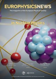
EUROPHYSICSNEWS<br />
The magazine of the European Physical Society<br />
Medical Physics<br />
ITER:<br />
the module has landed<br />
Mass of<br />
the W boson<br />
Modelling the effect<br />
of microwave ablation<br />
VOLUME <strong>53</strong> I NUMBER 3 I EUROPEAN COUNTRIES PRICE: 113€ PER YEAR (VAT NOT INCLUDED)
VOLUME <strong>53</strong> I NUMBER 3 I EUROPEAN COUNTRIES PRICE: 113€ PER YEAR (VAT NOT INCLUDED)<br />
CONTENTS<br />
EUROPHYSICSNEWS<br />
The magazine of the European Physical Society<br />
europhysicsnews<br />
Medical Physics<br />
ITER:<br />
the module has landed<br />
Mass of<br />
the W boson<br />
Modelling the effect<br />
of microwave ablation<br />
Cover picture: ??? ©iStockPhoto<br />
EPS EDITORIAL<br />
03 Towards the EPS 2.0<br />
L. Bergé<br />
IN THE SPOTLIGHTS<br />
m PAGE 16<br />
Accelerators for<br />
health: from current<br />
to dream machines<br />
04 ITER: the module has landed<br />
05 New CLOUD results<br />
06 MIRI’s Sharper View<br />
07 Mass of the W boson<br />
09 CO 2 conversion via coupled plasma-electrolysis process<br />
10 Modelling the effect of microwave ablation<br />
11 Impact of medical and imaging physics on the COVID pandemic<br />
YOUNG MINDS<br />
12 International Day of Women and Girls in Science event<br />
S. Brackenhoff and A. Viswanathan<br />
FEATURES<br />
14 Column: the European X-ray Free Electron Laser<br />
m PAGE 24<br />
Tiny robots made<br />
from biomolecules<br />
F. Boscherini<br />
16 Accelerators for health: from current to dream machines<br />
A. Faus-Golf and E. Benedetto<br />
20 Plasma medicine: the great prospects when physics meets medicine<br />
J.M. Sadowska, N. Skoro, R. Laurita, S. Bekeschus, A. Przekora-Kuśmierz, A. Lin,<br />
S. Laurencin, S. Sério, S. Cousty and C. Canal<br />
24 Tiny robots made from biomolecules<br />
T. Pirzer and F. C. Simmel<br />
28 Lasers for health<br />
G. Cerullo and R. Vanna<br />
BOOK REVIEW<br />
m PAGE 28<br />
Lasers for health<br />
32 Computational Statistical Physics<br />
H. Rieger<br />
<strong>EPN</strong> <strong>53</strong>/3 01
EPS EDITORIAL<br />
[EPS EDITORIAL]<br />
Towards the EPS 2.0<br />
The European Physical Society (EPS) was founded in 1968 in<br />
Geneva through the inspiring leadership of Gilberto Bernardini<br />
(1906-1995). Since then, the EPS has grown from 6 to 42 members<br />
states. Today it hosts 12 Divisions and 6 Groups, mostly leading<br />
the various fields of physics or societal activities ranging from<br />
physics for development to technology and innovation. Promoting<br />
scientific excellence, the EPS Divisions and Groups organise<br />
many of European major conferences and award Europe’s most<br />
prestigious prizes in their respective areas. This “backbone” of<br />
the EPS is reinforced by several committees in charge of the EPS<br />
Historic Sites, Equal Opportunities or Young Minds activities. So<br />
far, the EPS has also succeeded in attracting more than 40 Associate<br />
Members and 30 cooperating societies with whom the Society has<br />
bi-lateral agreements.<br />
Under the guidance of the EPS president, the Executive<br />
Committee, elected by the EPS Council, is currently composed<br />
of 13 members who meet about 4 times a year in order to pilot<br />
activities of the EPS and review its budget. Among the agenda of<br />
the last Executive Committee meeting were addressed, among<br />
other points, updates on prize charters, the Grand Challenges<br />
for Physics on the Horizon 2050 - brightly conducted by Carlos<br />
Hidalgo – reports on the activities at our Brussels’ office, or the<br />
follow up on our actions related to the Russian war in Ukraine. In<br />
this respect, the Executive Committee decided to relay initiatives<br />
that offer effective assistance to scientific refugees from Ukraine<br />
(e.g., scienceforukraine.eu, ERA4Ukraine) and support projects<br />
that help children and students to recover decent education<br />
infrastructures inside Ukraine (e.g., science4people).<br />
Since its creation, the EPS has known several evolutions and<br />
thus successive transformations of its constitution, regularly revised<br />
between 2004 and 2016. This necessary evolution inspired the<br />
EPS Strategic Plan 2010+ that aimed at reviewing the governance<br />
of our learned society and at outlining a possible strategy for its<br />
future. Since 2010 our world has, however, rapidly changed too<br />
and new priorities occurred. The first priority is to reinforce the<br />
link with the industrial sector as more than 50% of our young<br />
physicists are employed in industry. This goal is also deeply related<br />
to the recruitment of new Associate Members belonging to this<br />
community. Specific projects like the EPS Forum, the first edition<br />
of which took place in Paris on 2-3 June 2022, will contribute<br />
to highlight this new direction and broaden our community.<br />
A second objective is to help young physicists. To this aim, the<br />
EPS Young Minds Programme was created to accompany and<br />
support the next generation of researchers and leaders in physics.<br />
Moreover, promoting inclusion, gender equality and helping<br />
young professionals who work in low-income countries to keep<br />
an excellent academic level have become current objectives in all<br />
international learned societies.<br />
Several workgroups have been created to address these new<br />
challenges and improve the management of the EPS activities.<br />
There are also challenges at the EPS secretariat to distribute<br />
and handle the workload generated by these novel activities.<br />
Meanwhile, the environment in which the EPS operates has<br />
also become more complex. There is generally greater scrutiny<br />
of associations, in particular when those receive income from<br />
conference organisation and editorial services. There is nowadays<br />
a need for greater transparency for any association that wishes<br />
to be involved in EU financed projects, in particular for not for<br />
profit associations whose financial statutes must be adapted. We<br />
already identified new statutory auditors to guide us through<br />
these transformations.<br />
Therefore, an extraordinary Council held on 26 January 2022<br />
approved to a large majority the proposal to set up a working group<br />
in charge of reviewing the statutes and by-laws of the EPS. This<br />
working group will propose recommendations to modernise the<br />
EPS constitution and Structures during a further Council Meeting<br />
in 2023. In order to pilot these profound structural changes, it was<br />
proposed to extend the mandate of all members of the Executive<br />
Committee by one year, which was also accepted, considering that<br />
enough time needs to be allocated to the examination to each of<br />
the EPS components. We deeply thank the Council delegates for<br />
placing their trust in us for another year.<br />
The EPS 2.0 is now in progress. n<br />
l Luc Bergé, EPS President<br />
<strong>EPN</strong> <strong>53</strong>/3 03
IN THE SPOTLIGHTS<br />
iter news<br />
The module has landed<br />
Te ITER Newsline reported in May 2022 a milestone in<br />
the installation of the tokamak. The first module of the<br />
vacuum vessel – an assembly of vacuum vessel sector,<br />
two vertical D-shaped magnetic coils and corresponding<br />
thermal shield panels, plus all kinds of piping and appendices<br />
- with total mass of 1,380 tonnes was slowly extracted from the<br />
sub-assembly tool, lifted and transported over the wall separating<br />
the Assembly Hall from the machine assembly pit, and positioned<br />
with sub-millimetric precision on its supports. An impressive<br />
achievement. Eight more modules to go. n<br />
04 <strong>EPN</strong> <strong>53</strong>/3
NEWS BY CERN<br />
IN THE SPOTLIGHTS<br />
New CLOUD results<br />
The CLOUD experiment at CERN discovered a new way by which aerosols rapidly<br />
form and grow at high altitude. The resultant particles quickly spread around the globe,<br />
potentially influencing Earth’s climate on an intercontinental scale.<br />
The CLOUD experiment<br />
at CERN is operated by<br />
an interdisciplinary team<br />
of atmospheric scientists,<br />
cosmic-ray physicists and particle<br />
physicists. The scientists aim at studying<br />
the influence of cosmic rays on<br />
aerosols in the Earth’s atmosphere.<br />
Aerosol particles are known to generally<br />
cool the climate by reflecting<br />
sunlight back into space and by making<br />
clouds more reflective. However,<br />
aerosol formation itself is poorly understood.<br />
Measuring the underlying<br />
microphysics in controlled laboratory<br />
conditions is important to get a better<br />
understanding.<br />
In a special cloud chamber aerosols<br />
form an artificial cloud, while the<br />
Proton Synchrotron at CERN provides<br />
an artificial source of “cosmic<br />
rays” that simulates natural conditions<br />
between ground level and the stratosphere.<br />
A beam of particles is passed<br />
through the cloud chamber and its effects<br />
on aerosol production or on liquid<br />
or ice clouds inside the chamber<br />
are recorded.<br />
In a paper in Nature in May<br />
2022 [1], the CLOUD collaboration<br />
reports that aerosol particles<br />
can form and grow in Earth’s upper<br />
troposphere in an unexpected way.<br />
The new mechanism may represent<br />
a major source of cloud and ice seed<br />
particles in areas of the upper troposphere<br />
where ammonia is efficiently<br />
transported vertically, such as over<br />
the Asian monsoon regions. Using<br />
mixtures of sulfuric acid, nitric acid<br />
and ammonia vapours in the chamber<br />
at atmospheric concentrations,<br />
the CLOUD team found that the<br />
three vapours form new particles<br />
10-1000 times faster than a sulfuric<br />
acid–ammonia mixture, which, from<br />
m FIG. 1: Simulation of aerosol particle formation during the Asian<br />
monsoon compared to the results of an earlier version of the model.<br />
(Credit CLOUD Collaboration)<br />
previous CLOUD measurements,<br />
was considered to be the dominant<br />
source of upper tropospheric particles.<br />
Once the three-component particles<br />
form, they can grow rapidly to<br />
sizes where they seed clouds with ice<br />
crystals, comparable to desert dust<br />
particles, which are thought to be<br />
the most widespread and effective<br />
ice seeds in the atmosphere. When<br />
a supercooled cloud droplet freezes,<br />
the resulting ice particle will grow at<br />
the expense of any unfrozen droplets<br />
nearby, so ice has a major influence<br />
on cloud microphysical properties<br />
and precipitation. New models developed<br />
by the team of CLOUD<br />
showed that although the particles<br />
form locally, e.g. in Asia, they travel<br />
from there to North America in just<br />
three days via the subtropical jet<br />
stream. In figure 1 a simulation is<br />
shown of the aerosol particle formation<br />
during the Asian monsoon in a<br />
global aerosol model with efficient<br />
vertical transport of ammonia into<br />
the upper troposphere. Including a<br />
mixture of sulfuric acid, nitric acid<br />
and ammonia enhances upper-tropospheric<br />
particle number concentrations<br />
over the Asian monsoon region<br />
by a factor of 3–5 compared with the<br />
same model with only sulfuric acid<br />
and ammonia.<br />
Once again, CLOUD found that<br />
anthropogenic ammonia has a major<br />
influence on atmospheric aerosol particles.<br />
Atmospheric concentrations<br />
of sulfuric acid, nitric acid and ammonia<br />
were much lower in the preindustrial<br />
era than they are now, and<br />
each is likely to follow different concentration<br />
trajectories under future air<br />
pollution controls. Ammonia in the<br />
upper troposphere originates from<br />
livestock and fertiliser emissions<br />
– which are unregulated at present –<br />
and is carried aloft in convective cloud<br />
droplets, which release their ammonia<br />
upon freezing. The CLOUD results<br />
are important for policies for future air<br />
pollution regulations. n<br />
Reference<br />
[1] M. Wang et al. Nature, doi:10.1038/<br />
s41586-022-04605-4 (2022).<br />
<strong>EPN</strong> <strong>53</strong>/3 05
IN THE SPOTLIGHTS<br />
NEWS BY NASA<br />
MIRI’s Sharper View<br />
The new James Webb Space Telescope is now aligned across all four of its science<br />
instruments 1 . A test image of the Mid-Infrared Instrument (MIRI) shows the promising<br />
performance of the Webb telescope.<br />
In Figure 1 a comparison between images of the same part of<br />
the Large Magellanic Cloud taken by the retired Spizer Space<br />
Telescope and by MIRI. The latter shows the interstellar gas in<br />
unprecedented details. You can see the emission from “polycyclic<br />
aromatic hydrocarbons,” or molecules of carbon and hydrogen<br />
that play an important role in the thermal balance and chemistry of<br />
interstellar gas. When Webb is ready to begin science observations,<br />
studies such as these with MIRI will give new insights into the birth<br />
of stars and protoplanetary systems. n<br />
1<br />
https://www.jwst.nasa.gov<br />
. FIG. 1: Infrared image of part of the Large Magellanic by the James Webb Space<br />
Telescope (right, NASA/ESA/CSA/STScI) compared to that by the Spitzer Space Telescope<br />
(left, NASA/JPL-Caltech).<br />
06 <strong>EPN</strong> <strong>53</strong>/3
PARTICLE PHYSICS NEWS<br />
IN THE SPOTLIGHTS<br />
Mass of<br />
the W boson<br />
The mass of the W boson is one of the<br />
predictions of the Standard Model. A<br />
new - most precise ever - measurement<br />
of the W-mass by the CDF collaboration<br />
based on the data set from the former<br />
Tevatron collider at Fermilab breaks<br />
with the prediction.<br />
The Standard Model of particle physics is the<br />
theoretical description of nature at its most<br />
fundamental level. The W boson is the carrier<br />
of the weak nuclear force. In the electroweak<br />
theory, the mass of the W boson depends in first order on<br />
the mass of the Z boson and the value of the weak mixing<br />
angle. At higher order, additional dependencies are introduced,<br />
in particular on the mass of the heavy top quark<br />
and the Higgs boson. Since its discovery in 1983, various<br />
experiments have measured the W-mass with increasing<br />
precision. In April 2022, after ten years of analysing the<br />
full data set from the former Tevatron collider at Fermilab,<br />
the CDF collaboration has published [1] the most precise<br />
measurement to date: 80,433.5 ± 6.4 (stat) ± 6.9 (syst) MeV.<br />
The value not only stands 7σ above the prediction of the<br />
Standard Model, it also deviates from all earlier measurements.<br />
This discrepancy must be understood and the value<br />
must be confirmed by other experiments such as the D0 experiment<br />
at the former Tevatron and the ATLAS and CMS<br />
experiments at the LHC collider at CERN. If the new<br />
m FIG. 1: Comparison of this CDF II measurement and<br />
past MW measurements with the SM expectation (from [1]).<br />
<strong>EPN</strong> <strong>53</strong>/3 07
IN THE SPOTLIGHTS<br />
PARTICLE PHYSICS NEWS<br />
value for the W-mass will be confirmed,<br />
it probably means that the boson<br />
feels the influence of unknown particles<br />
or unknown forces. Possible extensions of<br />
the Standard Model include new Higgslike<br />
particles, dark matter particles or supersymmetry.<br />
For particle physicists it is<br />
exciting times again. n<br />
Reference<br />
[1] Science 376, Issue 6589, 170<br />
Run 3 of LHC has started<br />
After a break of more than three years for maintenance and upgrade, particle beams are circulating again in the LHC collider<br />
at CERN. Also the experiments underwent major upgrades. Run 3 of LHC operations has begun. Unprecedented number<br />
of collisions at record energy will allow physicists to study in even more detail the Higgs boson and put the Standard<br />
Model to even more stringent tests. In addition, two new experiments start operation: FASER - designed to search for light<br />
and extremely weakly interacting particles - and SND@LHC, that will make measurements of neutrinos produced at the<br />
particle collider. n<br />
LINKS: https://home.cern/news/news/accelerators/large-hadron-collider-restarts<br />
https://op-webtools.web.cern.ch/vistar/vistars.php<br />
Celebration of Higgs@10<br />
Ten years ago, on 4 July 2012, the discovery of the Higgs boson<br />
was announced at CERN by the ATLAS and CMS collaborations.<br />
Rolf Heuer, then director general of CERN, proposed the audience<br />
“As a layman, I would say we got it. Do you agree?” An<br />
overwhelming applause was the answer. Francois Englert and<br />
Peter Higgs, theoretical physicists who predicted the Higgs particle,<br />
were moved by the moment and expressed their gratitude<br />
for and admiration of the experimentalists that confirmed their<br />
prediction. This year the 10 th birthday of the Higgs particle is<br />
being celebrated. With a symposium at CERN, with parties in<br />
the particle physics institutes, with a series of ‘Higgs stories’. n<br />
LINK: https://home.cern/news/news/cern/higgs10-save-date<br />
08 <strong>EPN</strong> <strong>53</strong>/3
ITER NEWS<br />
IN THE SPOTLIGHTS<br />
CO 2 conversion<br />
via coupled plasma-electrolysis process<br />
Researchers at the Dutch Institute for Fundamental Energy Research DIFFER<br />
have demonstrated that electrolysis coupled with plasmolysis leads to a novel route<br />
for CO 2 conversion. That is important in the quest for sustainable chemicals<br />
and fuels and large-scale, long-term electricity storage.<br />
Production of renewable electricity is<br />
making great strides. In 2021 more<br />
than 35% of EU electricity production<br />
came from renewable source,<br />
well underway to reach the EU target of 40%<br />
renewable energy by 2030 1 . Or is it? On top of<br />
nearly doubling the deployment rate of solar<br />
and wind capacity, the grid has to cope with<br />
variable input. Intermittency and seasonal<br />
mismatch of supply and demand call for longterm<br />
(interannual), large scale (PJ) electricity<br />
storage, lest power is being wasted by curtailment.<br />
Energy storage in high energy density<br />
fuels and chemicals could meet requirements.<br />
By converting air (CO 2 , N 2 ) and water, powered<br />
by (surplus) renewable electricity, high<br />
energy density products can be synthesised.<br />
This so-called Power-to-X scheme couples<br />
the power sector to the chemical, the heating<br />
and the fuel sectors thus avoiding the need<br />
for fossil fuels in those sectors.<br />
Conversion starts at splitting the strong<br />
carbon dioxide and/or nitrogen bond, the<br />
most energy intensive step in the process.<br />
This provides the basic building blocks CO,<br />
N, H 2 for the synthesis of fuels and chemicals.<br />
Low temperature plasmas have shown<br />
to be effective in splitting CO 2 and N 2 molecules,<br />
creating a mixture of highly active<br />
atoms, molecules and radicals [1]. The challenge<br />
is how to unravel the gas mixture<br />
produced. In particular the about equally<br />
sized CO and O 2 molecules are difficult to<br />
separate. The weak paramagnetic property<br />
of O 2 is one avenue to explore. However,<br />
electro-chemical separation is a more mature<br />
technology applied in electrolysis and<br />
fuel cells. A solid oxide electrolyte cell selectively<br />
transports negative oxygen ions<br />
from cathode to anode thereby separating<br />
the oxygen from the incoming gas mixture.<br />
Researchers of the Dutch Institute for<br />
Fundamental Energy Research DIFFER<br />
have recently demonstrated that the coupling<br />
between electrolysis and plasmolysis<br />
leads to a novel route for CO 2 conversion<br />
[2]. The researchers are working in<br />
the context of the EU H2020 funded project<br />
KEROGREEN. The project’s ultimate<br />
goal is to build a container sized pilot plant<br />
for the production of green aviation fuel.<br />
High-temperature electrolysis is characterised<br />
by high yield and high energy efficiency.<br />
This process results in the direct separation of<br />
the reaction products CO and oxygen (O 2 ).<br />
However, challenging requirements on electrode<br />
materials are being encountered. CO 2<br />
plasmolysis on the other hand, offers similar<br />
energy efficiencies, and no scarce electrode<br />
materials are required. However, the mixture<br />
of the reaction products cannot be used directly<br />
in a chemical synthesis plant. The CO<br />
produced remains mixed with O 2 and residual<br />
CO 2 and therefore an additional gas separation<br />
step is required. DIFFER researchers have<br />
demonstrated that the coupling of electrolysis<br />
and plasmolysis leads to a renewable-electricity-driven<br />
route for CO production, overcoming<br />
the main bottleneck of CO 2 plasmolysis.<br />
In the coupled process, the CO 2 plasmolysis<br />
gas mixture is supplied to a high-temperature<br />
electrolyser to separate the product gases<br />
1<br />
https://renewablesnow.com/news/renewables-share-in-eu-electricity-climbs-to-37-in-2020-770744/<br />
or https://www.greentechmedia.com/articles/read/eus-new-2030-climate-target-signals-acceleratedrenewable-deployment<br />
electrochemically using solid oxide electrolyte<br />
cells (SOEC). Oxygen is transferred to one<br />
side of the SOEC, where it is removed. The<br />
remaining CO and CO 2 are ready for chemical<br />
synthesis of liquid fuel.<br />
In a single pass, the coupled-process product<br />
contains 91% less O 2 and 138% more CO<br />
compared with bare plasmolysis. "And what<br />
is more important: when the solid oxide electrolyte<br />
cells operate with the plasma mixture<br />
of CO, O 2 and CO 2 in the inlet, we didn't observe<br />
any electrode degradation in durability<br />
tests", says Michail Tsampas, leader of the<br />
DIFFER group Catalytic and Electrochemical<br />
Processes for Energy Applications. The synergy<br />
between plasmolysis and electrolysis<br />
opens up a novel route to efficient CO 2 conversion<br />
into valuable building blocks for sustainable<br />
chemicals and fuels. n<br />
Acknowledgement<br />
This project has received funding from the<br />
European Union’s Horizon 2020 Research<br />
and Innovation Programme under Grant<br />
Agreement Nr. 763909<br />
Author<br />
Adelbert Goede (DIFFER, Eindhoven, The<br />
Netherlands; board member of the EPS<br />
Environmental Physics Division)<br />
References<br />
[1] A. Goede and R. van de Sanden, Eur Phys News<br />
47-3, 22 (2016),<br />
http://dx.doi.org/10.1051/epn/2016304<br />
[2] A. Pandiyan, V. Kyriakou, Dragos Neagu, R. Sharma,<br />
S. Welzel, A. Goede, M.C.M. van de Sanden<br />
and M. N. Tsampas, J. CO2 Util. 57 (2022) 101904<br />
https://doi.org/10.1016/j.jcou.2022.101904<br />
<strong>EPN</strong> <strong>53</strong>/3 09
IN THE SPOTLIGHTS<br />
Highlight from EPL<br />
Modelling the effect<br />
of microwave ablation<br />
Microwave ablation is gaining popularity as an efficient therapeutic option for treating<br />
cancerous tumor cells in patients who are non-surgical candidates. It is a minimally<br />
invasive treatment with a short recovery time. Modelling the effect of the treatment<br />
helps to minimise damage of healty tissues.<br />
Microwave ablation<br />
(MWA) is a material-specific<br />
responsiveness<br />
procedure with the<br />
advantage of faster ablation times,<br />
larger ablation volumes, and potentially<br />
strong deactivations for tumors<br />
compared to other thermal ablation<br />
methods. However, the interaction<br />
of the medical tools with the tissue<br />
may result in healthy tissue damage<br />
which can be eliminated by clarifying<br />
the causation and conditions of their<br />
development. To ensure the destruction<br />
of cancer cells with minimal damage<br />
to healthy tissue, the elevation of<br />
temperature and the evolution of the<br />
necrotic tissue need to be controlled.<br />
Besides experimental methods, computer<br />
modeling is proven to be an effective<br />
approach for improving the<br />
performance of MWA. Moreover,<br />
since the thermal spread in biological<br />
tissue is difficult to measure, generating<br />
the predictive models from procedural<br />
planning to execution may<br />
provide a great impact on patient care.<br />
Due to complexity and computational<br />
resources consumption, most<br />
of the existing numerical models are<br />
two-dimensional axisymmetric to emulate<br />
actual three-dimensional cancers<br />
and surrounding tissue. A mathematical<br />
model for the simulation of MWA<br />
should consist of three essentials components.<br />
The first component includes<br />
the model of the antenna that generates<br />
a microwave field in the tissue.<br />
The second component deals with the<br />
heat distribution in the tissue including<br />
sources and sinks and the phase<br />
changes. The third part describes the<br />
effect of heat on tumor cells and their<br />
destruction. All these components of<br />
the ablation model depend on the dielectric<br />
properties and thermal parameters<br />
of tissues as well as a variety of<br />
material parameters. Additionally, the<br />
inclusion of the temperature dependence<br />
of dielectric properties, the heat<br />
capacity, thermal conductivity, and<br />
blood perfusion, is pivotal for establishing<br />
the correct model of the ablation<br />
process.<br />
Although MWA operates in the frequency<br />
range from 915 MHz to 2.45<br />
GHz, the antenna system at 2.45 GHz<br />
produces greater power deposition and<br />
larger ablations in a shorter time due<br />
to matching the frequency with water<br />
molecule resonance. Microwave power<br />
radiated from the antenna is absorbed<br />
by surrounding tissue, leading to heating<br />
the targeted tissue, consisting of the<br />
m FIG. 1:<br />
a) Temperature<br />
[oC] distribution<br />
and b) fraction of<br />
damage of liver,<br />
lung, kidney, and<br />
bone tissue after<br />
600s of exposure<br />
to microwave<br />
frequency of<br />
2.45 GHz and the<br />
input power of<br />
10 W. The black<br />
circle shows the<br />
boundary of the<br />
tumoral tissue.<br />
tumor and a margin of surrounding<br />
tissue. It was found that temperatures<br />
above 60°C cause relatively instantaneous<br />
cell death, while temperatures from<br />
50 to 60°C will induce coagulation and<br />
cell death in a matter of minutes, typically<br />
10 min.<br />
In our study, calculations were performed<br />
for liver (high perfusion and<br />
large heat sinks), lung (low conductivity<br />
and large heat sinks), kidney (high<br />
perfusion and large heat sinks), and<br />
bone (low conductivity) tissue by using<br />
the Comsol Multiphysics simulation<br />
package. The temperature distribution<br />
has an ellipsoidal shape reaching the<br />
highest values near the antenna slot as<br />
illustrated in Figure 1a. With increasing<br />
the distance from the antenna, the heat<br />
source becomes weaker and the perfusion<br />
of blood limits the extent of the<br />
heated area. The highest and the lowest<br />
values of the temperature correspond<br />
to the kidney and bone tissue, respectively.<br />
Ablation zones are concentrated<br />
around the tip and slot of the antenna<br />
as shown in Figure 1b. The necrotic tissue<br />
is mainly located in the tumor with<br />
a small amount of surrounding tissue<br />
being damaged. Microwaves are less<br />
susceptible to perfusion or heat sinks<br />
so they successfully penetrate deep<br />
into low-conductivity materials such<br />
as lungs and bone. The obtained results<br />
indicate that the simulation technique<br />
can be useful for predicting optimal<br />
conditions for MWA and should be<br />
considered in treatment planning. n<br />
Reference<br />
[1] M. Radmilovi-Radjenovi et al., EPL 136,<br />
28001 (2021)<br />
10 <strong>EPN</strong> <strong>53</strong>/3
HIGHLIGHT FROM EPJ PLUS<br />
IN THE SPOTLIGHTS<br />
Impact of medical and imaging physics<br />
on the COVID pandemic<br />
The COVID pandemic has had a profound global impact, a new collection of papers<br />
looks at a multidisciplinary approach to aspects of the pandemic.<br />
The COVID pandemic has impacted<br />
every aspect of our lives<br />
over the past two years. It is little<br />
surprise then that this crisis has<br />
united a wealth of disparate fields of science<br />
in attempts to mitigate the virus and its effects.<br />
A new focus issue of EPJ Plus collects<br />
together invited papers that detail the<br />
impact of medical imaging and medical<br />
physics technology on various aspects of<br />
the COVID pandemic. The focus issue is<br />
guest-edited by Evaristo Cisbani and Franco<br />
Garibaldi, both from Istituto Superiore di<br />
Sanita, Rome, and Istituto Nazionale di<br />
Fisica Nucleare, Rome, along with Stephanie<br />
Majewski, UC Davis, California, USA, and<br />
Andrea Gori, University of Milano, Italy.<br />
“The focus issue deals with Molecular<br />
imaging techniques applied to Covid<br />
19. This is just the right time for such a<br />
collection, as we believe we have now accumulated<br />
enough data and information,”<br />
Garibaldi says. “We have had the problem of<br />
COVID for the last two years and we must<br />
be prepared for possible future pandemics.”<br />
One particular aspect of the COVID infection<br />
looked at by the collection and its<br />
authors is inflammation. This is one of the<br />
primary causes of severe sickness, especially<br />
in those with underlying health conditions<br />
such as diabetes.<br />
In their Editorial, the guest editors of the<br />
focus issue say that new advanced technologies<br />
already available in the research field<br />
and undergoing constant improvement<br />
could translate into increased sensitivity<br />
of early detection and avoiding the longterm<br />
side effects of inflammation.<br />
The key to this is understanding what<br />
actually needs to be imaged or visualised<br />
and what the emerging imaging needs<br />
are. Thus, the editors asked experts to address<br />
the biomedical issues related to the<br />
COVID-19 pandemic and associated inflammatory<br />
effects.<br />
The editors lay out the multidisciplinary<br />
focus points for the topical collection papers<br />
as: the origin and evolution of COVID infection<br />
from a biological perspective, diagnosis<br />
and early differentiation of COVID-19 from<br />
other diseases, and the role of advanced detection<br />
technology including medical physics<br />
and artificial intelligence. n<br />
Reference<br />
[1] Focus Point Issue “Progress in medical<br />
physics in times of CoViD-19 and related<br />
inflammatory diseases”, Guest Editors:<br />
E. Cisbani, F. Garibaldi, S. Majewski, A. Gori,<br />
Eur. Phys. J Plus (2022)<br />
FIG. 1:<br />
Top: T-cells can<br />
be detected by<br />
a PET scan of<br />
the total body<br />
(courtesy of UC<br />
Davis EXPLORER<br />
group); bottom:<br />
18F-FDG PET<br />
scan shows<br />
unsuspected<br />
pulmonary<br />
thrombi in<br />
COVID-19<br />
patients.<br />
<strong>EPN</strong> <strong>53</strong>/3 11
YOUNG MINDS<br />
International Day of Women and Girls<br />
in Science event - Young Minds Groningen<br />
Accelerating progress towards full and equal access and participation<br />
of women and girls in science<br />
l Stefanie Brackenhoff and Akshara Viswanathan, EPS YM Groningen<br />
“You can’t solve a problem until you’re<br />
asking the right question. Raw data will<br />
not solve our problems, asking the right<br />
questions will.”<br />
Policymakers and faculty boards should<br />
consider this approach by Albert<br />
Einstein while dealing with diversity<br />
issues in science. On the 11 th of<br />
February 2022, the UNESCO Day<br />
of Women and Girls in Science, the<br />
Groningen Young Minds Section organized<br />
a webinar to discuss the barriers<br />
female scientists encounter. The<br />
event featured scientific talks by two<br />
renowned female scientists, followed<br />
by presentations about gender equality<br />
by two STEM practitioners, and an<br />
open discussion session.<br />
Role models - the agents<br />
of change<br />
Stating that “Being a woman in science<br />
is a responsibility in itself for the<br />
up-and-coming generation.” Prof.<br />
Irene D’Amico (Full Professor at the<br />
University of York) and Prof. Ivone<br />
Albuquerque (Full Professor at the<br />
University of São Paulo) kicked off<br />
the day, while Dr. Francesca Primas<br />
(Full astronomer at the European<br />
Southern Observatory and former<br />
Chair of the International<br />
Astronomical Union Working Group<br />
on Women in Astronomy) and Dr.<br />
Tana Joseph (Postdoctoral Research<br />
Fellow at the University of Amsterdam<br />
and Coordinator of the Netherlands<br />
Astronomy Equity and Inclusion<br />
Committee) picked up the gender<br />
equality baton for the afternoon.<br />
Prof. D’Amico talked about the use<br />
of networks of spins to perform quantum<br />
information processing and how<br />
evolutionary algorithms may help with<br />
this task [1]. Prof. Albuquerque welcomed<br />
us to the ‘Dark Side’ experiment<br />
that is designed to directly detect dark<br />
matter in the Universe [2]. Dr. Primas<br />
discussed a selection of past and ongoing<br />
equity, diversity and inclusion efforts<br />
in astronomy. Dr. Joseph outlined<br />
the advantages and disadvantages she<br />
experienced on her journey as an aspiring<br />
black female astronomer, and what<br />
we can learn from these experiences<br />
as a community. Parallel flashbacks<br />
between how astronomy in pre-internet<br />
South Africa grew versus how<br />
Dr. Joseph’s astronomy career started<br />
inspired the participants. During the<br />
open discussion, audience members<br />
were able to bring issues they experienced<br />
to the table.<br />
. Top left: Prof.<br />
Irene D’Amico.<br />
Top right:<br />
Prof. Ivone<br />
Albuquerque.<br />
Bottom left:<br />
Dr. Francesca<br />
Primas.<br />
Bottom right:<br />
Dr. Tana Joseph.<br />
A holistic view of equity,<br />
diversity and inclusion<br />
in astronomy<br />
“Diversity is the mix we want to achieve.<br />
Inclusion is what makes the mix work.”<br />
– Francesca Primas<br />
Dr. Primas discussed the gender<br />
gap in astronomy: although the influx<br />
of female students is approaching<br />
that of male students, women disproportionally<br />
experience discrimination<br />
(56% of women vs. 8% of men)<br />
or harassment (30% of women vs. 3%<br />
of men) [3]. Furthermore, the success<br />
rate of female astronomers in winning<br />
observing time on the Hubble<br />
Space Telescope and other facilities<br />
was found to be systematically lower<br />
than that of their male counterparts<br />
[4, 5, 6]. Preliminary results indicate<br />
that implementing dual-anonymous<br />
reviews removes this issue [7, 8]. Dr.<br />
Primas’ talk made ongoing efforts<br />
12 <strong>EPN</strong> <strong>53</strong>/3
YOUNG MINDS<br />
heard and kept enough room for improvements in following<br />
a principled and pragmatic approach towards making<br />
the voice of the underrepresented groups heard.<br />
“Seek first to understand, then to be understood.” -<br />
Stephen R. Covey<br />
We all have an individual responsibility to make the scientific<br />
world a welcome place for everybody. Understanding<br />
the humanity, how people operate in targeted communities<br />
and keeping an open mind and eye is the key to approaching<br />
historically excluded communities. The scientific community<br />
must create `cheerleaders’ for them. Do not just talk<br />
to the children themselves, but also their parents, teachers,<br />
and community leaders. Create an environment in which<br />
they can thrive. Dr. Joseph also called out on facing ‘cultural<br />
taxation’ – responsibilities placed on faculty of discriminated<br />
races without any compensation for their efforts.<br />
To get compensated for all her efforts in achieving equity,<br />
diversity and inclusion in astronomy, she started her own<br />
company ‘AstroComms’.<br />
Gender biases in science versus engineering<br />
One participant raised concerns about the field of engineering<br />
performing poorly in achieving gender equality.<br />
Implementing outreach activities specifically targeted at<br />
high school students was proposed as one of the many solutions.<br />
Yet the problem is also about how universities promote<br />
courses and degrees in engineering and that applied sciences<br />
are still perceived as being for ‘the stereotypical man’ in society.<br />
We conclude with Dr. Joseph’s proposal of implementing<br />
a scientific approach that unites us. “We can treat diversity<br />
issues like a Bayesian optimization problem. Implement an<br />
approach, check how well this approach worked, and update<br />
it if needed.” There is no clear endpoint, but we must converge<br />
to a more equal scientific community. n<br />
Reference<br />
[1] L. Mortimer et al., Advanced Quantum Technology 48, (2021).<br />
https://onlinelibrary.wiley.com/doi/10.1002/qute.202100013<br />
[2] The DarkSide collaboration et al., Phys. Rev. D 104, 082005<br />
(2021). https://journals.aps.org/prd/abstract/10.1103/<br />
PhysRevD.104.082005<br />
[3] M. F. Roy et al., South African Journal of Science 115, 3 (2019).<br />
https://journals.co.za/doi/epdf/10.17159/sajs.2019/a0305<br />
[4] I. N. Reid, the Astronomical Society of the Pacific 126, 92 (2014).<br />
https://iopscience.iop.org/article/10.1086/678964<br />
[5] F. Patat, ESO Messenger 165, 2 (2019). https://ui.adsabs.harvard.edu/<br />
abs/2016Msngr.165....2P/abstract<br />
[6] C.J. Lonsdale, F.R. Schwab, G. Hunt, Gender-Related Systematics in<br />
the NRAO and ALMA Proposal Review Processes, preprint, (2016).<br />
https://doi.org/10.48550/arXiv.1611.04795<br />
[7] S. K. Johnson, J.F. Kirk, the Astronomical Society of the<br />
Pacific 132, 1009 (2020). https://iopscience.iop.org/<br />
article/10.1088/1<strong>53</strong>8-3873/ab6ce0/meta<br />
[8] R. Berkowitz et al., Physics Today 1945, 0699 (2021).<br />
https://physicstoday.scitation.org/do/10.1063/PT.6.2.20211216a/full/<br />
<strong>EPN</strong> <strong>53</strong>/3 13
[Column]<br />
The European X-ray Free Electron Laser:<br />
a tool for fundamental research<br />
and a wide range of applications<br />
DOI: https://doi.org/10.1051/epn/2022301<br />
X-ray free-electron lasers (XFELs) are the first<br />
light sources that are able to routinely generate<br />
coherent, ultra-brilliant, tunable laser pulses<br />
in the X-ray regime. The European XFEL (www.xfel.eu), is<br />
a world-leading large scale research facility, member of<br />
EIROforum (www.eiroforum.org), an intergovernamental<br />
association of eight of the leading European large-scale<br />
infrastructures. Thanks to a 1.7 km long, 17.5 GeV linear<br />
electron accelerator based on superconducting resonant<br />
cavities and the Self Amplified Spontaneous Emission<br />
(SASE) process taking place in very long undulator magnet<br />
arrays, high repetition rate ultrashort X-ray<br />
flashes with a brilliance that is a billion times<br />
higher than that of the best conventional<br />
synchrotron X-ray radiation sources are<br />
produced. The European XFEL is opening up<br />
areas of research from physics to structural<br />
biology that were previously inaccessible.<br />
Using the X-ray flashes of the European<br />
XFEL since the start of its operation in mid-<br />
2017, scientists from all over the world are<br />
able to map the atomic details of viruses, decipher the<br />
molecular composition of cells, take three-dimensional<br />
images of the nanoworld, film chemical reactions on the<br />
femtosecond time scale and study processes such as those<br />
occurring deep inside planets and in extreme conditions<br />
of temperature, pressure and applied magnetic field.<br />
The European XFEL is becoming a very efficient<br />
decoder, obtaining in a much shorter time and with<br />
reduced effort many structures for molecules such as<br />
membrane proteins, from which crystals larger than a<br />
micrometer in size are hard to obtain. This will advance<br />
progress in our understanding of pathogens and the<br />
development of pharmaceutical remedies. In addition,<br />
biomolecules modify their structure while performing<br />
their respective tasks. It would be extremely illuminating<br />
to follow these modifications and see the motion of the<br />
moving parts in a movie. To make a film of a moving<br />
object, it is necessary to take many snapshots. Faster<br />
movement requires a shorter exposure time and a<br />
greater number of snapshots to avoid blurring the<br />
pictures. This is where the ultrashort duration of the FEL<br />
pulses will ensure sharp, non-blurred pictures of very<br />
fast processes. During the Covid-19 pandemic, XFEL was<br />
used to gain insights into protein shape and function at<br />
the micro- and nanoscale (SAXS curve). The results from<br />
this experiment could improve our understanding of the<br />
immune response to coronavirus and help to develop<br />
medical strategies to overcome COVID-19.<br />
The European XFEL provides an opportunity to<br />
educate a new generation of scientists to address the<br />
frontiers of research in an open environment, promoting<br />
the European dimension of knowledge and<br />
its international mobility. The European<br />
XFEL is located in the metropolitan area of<br />
Hamburg, Germany, and has a long-standing<br />
collaboration agreement with DESY for the<br />
accelerator operation. It is organized as a<br />
non-profit company with limited liability<br />
under German private law (GmbH) that is<br />
publicly funded (total construction budget:<br />
1.54 B€; operation budget for 2022: 141 M€)<br />
through its international shareholders from 12 European<br />
countries. The shareholders’ assembly, the so-called<br />
Council, is the supreme organ of the European XFEL<br />
GmbH, which decides on all important issues of the<br />
company (like the annual financial statement and the<br />
annual operation budget) and important personnel<br />
matters as well as the further development of the facility.<br />
The Council meets at least three times a year and is led<br />
by a Chair and a Vice-Chair, who are elected from the<br />
Council delegations for a total of up to two terms, not<br />
exceeding two years each, and who, upon election, leave<br />
their delegations and become supra partes.<br />
We note, that Federico Boscherini (in the photo)<br />
Professor of Physics at the Physics and Astronomy<br />
Department of the University of Bologna, has been elected<br />
as the new Chair of the XFEL Council. The appointment<br />
starts from July 2022 for a first two year period. n<br />
l Eugenio Scapparone,<br />
INFN Bologna Section director<br />
14<br />
<strong>EPN</strong> <strong>53</strong>/3
FEATURES<br />
ACCELERATORS FOR HEALTH<br />
ACCELERATORS FOR HEALTH:<br />
FROM CURRENT TO DREAM MACHINES<br />
l Angeles Faus-Golfe 1 and Elena Benedetto 2 – DOI: https://doi.org/10.1051/epn/2022302<br />
l 1 Irene Joliot Curie Laboratory - CNRS-IN2P3 University of Paris-Saclay, Orsay, France<br />
l 2 SEEIIST Association, Geneva, Switzerland<br />
Any kind of sculpted particle beams from high-energy photons (X-rays and gamma<br />
rays), electrons, protons, neutrons to various atomic nuclei and more exotic species<br />
have been used to treat cancer. The development of a next generation of accelerators<br />
to face the challenges and issues of Particle Therapy is crucial. What are the most<br />
promising accelerator techniques, particles or dose delivery modes?<br />
m Layout of SEEIIST<br />
(South East Europe<br />
International<br />
Institute for<br />
Sustainable<br />
Technologies)<br />
facility<br />
The potential of accelerator-based Radiotherapy<br />
(RT) has increased considerably over the past<br />
decades, playing an increasingly important<br />
role in identifying and curing affections, such<br />
as cancer. Energetic particles of any kind have been used<br />
in the past, but nowadays photons (X-rays), low-energy<br />
electrons, protons and carbon ions are the most common<br />
types of irradiation (Figure 1).<br />
X-rays are the most common method of RT for cancer<br />
treatment. Even if the X-rays therapy is a mature technology<br />
there is room for improvement. The current challenges<br />
are related to the accurate delivery of X-rays to tumours<br />
involving sophisticated techniques to combine imaging<br />
and therapy. Hadron (i.e. proton and ion) beam therapy<br />
has growing potential in dealing with tumours close to<br />
organs at risks, because the irradiation dose (Figure 1) is<br />
mainly deposited at a specific longitudinal position, the<br />
Bragg peak, which depends on the energy of the particles.<br />
Also, some treatments may benefit from the use of ions that<br />
deliver doses with a greater radiobiological effectiveness<br />
(RBE), notably Carbon. Helium and Oxygen are also<br />
promising candidates for therapy and in particular techniques<br />
combining irradiation with multiple ions are getting<br />
more interest, to exploit the advantages of the different<br />
species[1]. With the recent developments of high-gradient<br />
normal conducting (NC) Radio Frequency (RF) linear<br />
accelerator (linacs) technology (Figure 2) or even the<br />
novel acceleration techniques such as the Laser-Plasma<br />
Accelerator (LPA), Very High-Energy Electrons (VHEE)<br />
with energies between 50-200 MeV offer a very promising<br />
option for anticancer RT.<br />
Whatever RT particle used, the treatment is limited by the<br />
toxicity introduced in the healthy tissues surrounding the<br />
tumours. A novel paradigm-shifting method for delivering<br />
ultra-high doses within an extremely short irradiation time<br />
(tenths of a second), known as FLASH-RT is now under<br />
study. The technique has recently been shown to preserve<br />
normal tissue in various species and organs while still maintaining<br />
anti-tumour efficacy equivalent to conventional RT<br />
at the same dose level. The “FLASH effect” has been shown<br />
16 <strong>EPN</strong> <strong>53</strong>/3
ACCELERATORS FOR HEALTH<br />
FEATURES<br />
to take place with electron, photon and more recently for<br />
proton beams. For details we refer to [2, 3].<br />
Towards a compact and flexible<br />
Hadron Therapy facility<br />
For proton therapy, compact accelerator systems are<br />
nowadays available on the market: mostly cyclotrons in<br />
Europe and America and synchrotrons in Japan. The irradiation<br />
is mainly done via 3D beam-scanning.<br />
Cyclotrons have the advantage that they are very compact<br />
and have only a few tuning parameters, thus are simple<br />
to operate in a hospital environment. However, scaling to<br />
the energies needed for Carbon-ions, which have a factor<br />
3 higher beam rigidity (i.e. the resistance to deflection by a<br />
magnetic field) is challenging. Another limitation of conventional<br />
cyclotrons is their fixed extraction energy, which<br />
implies the use of degraders, with associated transmission<br />
losses and radioprotection constrains. Current R&D goes<br />
in the direction of energy-variable cyclotrons and the use<br />
of high-field superconducting (SC) iron free magnets [3].<br />
Synchrotrons are easily scalable to ion operation, even<br />
if this implies an increase of their footprint, and allow acceleration<br />
of different type of ions. The preferred choice<br />
for carbon-ions today is a rather large synchrotron (of<br />
>20-m diameter compared to the 6-m size for protons).<br />
Most important, synchrotrons are flexible with respect<br />
to the extraction energy, which can be easily changed<br />
between one cycle to the next. A limitation of today synchrotrons,<br />
though, is the length of each cycle, of the order<br />
of 1s. In Europe, and following what was recently implemented<br />
in National Institute of Radiological Sciences<br />
(NIRS) in Japan [5], the tendency is to operate synchrotrons<br />
with the so-called multi-energy extraction (MEE)<br />
scheme, to deliver beams at different energies within the<br />
same cycle, thus saving time.<br />
Ultimately, if one could accelerate the total number<br />
of ions needed for a treatment session within one cycle<br />
(thus a factor 10-20 times higher intensity, accumulated<br />
by multi-turn injection), the full dose will be delivered<br />
significantly faster. Moreover, this mode of operation<br />
will make the use of SC magnets feasible, thus allowing<br />
a significant reduction of the dimensions of the ion<br />
synchrotron (indeed the SC magnets ramping time is<br />
much longer than for NC magnets). The R&D in this<br />
sense focus on ions sources to reliably deliver a higher<br />
current, on advanced injection and extraction schemes<br />
and on strongly curved SC magnets. The development of<br />
a SC-magnet carbon-ion synchrotron is precisely one of<br />
the main objectives of the Heavy Ion Therapy Research<br />
Integration (HITRIplus) [6] and what is being studied by<br />
the SEEIIST (South East Europe International Institute<br />
for Sustainable Technologies) [7] and the CERN NIMMS<br />
(Next Ion Medical Machine Study) initiative [8].<br />
Accelerators other than cyclotrons and synchrotrons<br />
are also under study, in particular Fixed Field Alternating<br />
gradient (FFA) and linacs. After several years of R&D and<br />
developments in research and international laboratories<br />
environment, the first linac based proton therapy facilities<br />
is under construction and commissioning in Europe.<br />
With the use of high-frequency high-gradient copper<br />
structures, designed to achieve relatively compact solution<br />
and high repetition rate operation, linacs will allow<br />
the production of beams with fast energy variation as<br />
well as small emittance beams that are potentially suited<br />
for the further development of mini-beams dose delivery<br />
techniques. Detailed information in [9, 10].<br />
b FIG. 1:<br />
Dose profile for<br />
various particle<br />
beams in water<br />
(beam widths<br />
r=0.5 cm)<br />
<strong>EPN</strong> <strong>53</strong>/3 17
FEATURES<br />
ACCELERATORS FOR HEALTH<br />
c FIG. 2:<br />
CLIC RF X-band<br />
cavity prototype<br />
(12 GHz, 100 MV/m)<br />
Towards a VHEE RT facility<br />
Low-energy electrons have historically been used to treat<br />
cancer for more than five decades, but mostly for the<br />
treatment of superficial tumours given their very limited<br />
penetration depth. However, this limitation can be<br />
overcome is VHEE (50-200 MeV, Figure 1) are used.<br />
Theoretically, VHEE beams offers several benefits. The<br />
ballistic and dosimetry properties of VHEE provide<br />
small-diameter beams that could be scanned and focused<br />
easily, enabling finer resolution for intensity-modulated<br />
treatments than is possible with photons beams. Electron<br />
accelerators are more compact and cheaper than proton<br />
therapy accelerators. Finally, VHEE beams can be operated<br />
at very high-dose rates and fast electromagnetic<br />
scanning providing a uniform dose distribution throughout<br />
the target and allowing for unforeseen RT modalities<br />
in particular the FLASH-RT.<br />
Normal Conducting RF linac is the technology being<br />
used for most of the VHEE research. The main advantages<br />
of the linacs are the flexibility and the compactness.<br />
Regarding the linac design in the energy range of interest<br />
for VHEE applications there are different possibilities offering<br />
the desired performances and compactness with<br />
different degrees of technology matureness. The S-band<br />
(~3 GHz) technology is the most mature one, High-<br />
Gradient compact linacs of this type are already available<br />
from various industrial partners. The C-band (~5<br />
GHz) and X-band (~12 Ghz) RF linacs still less mature<br />
and are mainly constructed in accelerator laboratories<br />
with the help of industries for the machining. Lately a<br />
considerable effort is being made from the industrialization<br />
point of view [11]. A limited number of accelerators<br />
in Europe and USA are available for VHEE R&D. The<br />
majority based in NC RF linacs, few of them are based<br />
on Super Conducting (SCRF) linac technology at L-band<br />
(~1.3 GHz). Finally, a VHEE-FLASH RT facility based<br />
on a Compact Linear Collider (CLIC) X-band 100 MeV<br />
linac is being designed at CERN in collaboration with<br />
Centre Hospitalier Universitaire Vaudois (CHUV) to treat<br />
large, deep-seated tumours in FLASH conditions. The facility<br />
is as compact as to fit on a typical hospital campus.<br />
Recent advances in the high-gradient RF structures,<br />
mainly in the material domain (origin, purity, surface<br />
treatment) and manufacturing technology in one side<br />
and the consistency and reproducibility of the test results<br />
in the other side, are transforming the landscape<br />
for VHEE RT. Some promising R&D in the next decade<br />
are: the distributed coupling accelerator and the use of<br />
cryogenic copper that is transforming the linac design<br />
offering a new frontier from beam brightness, efficiency<br />
and cost-capability. Another approach for the next generation<br />
of compact, efficient and high performance VHEE<br />
accelerator is the use of higher frequencies millimetric<br />
waves (~100 GHz) and higher-repetition rates using THz<br />
sources. An important R&D effort to apply these accelerator<br />
technologies in the medical has to be made in the<br />
next decade to make VHEE RT a clinical reality [2].<br />
Therapy facilities based on Laser-<br />
Plasma-Acceleration<br />
As high-performance lasers took big increased enhancements<br />
in the last years concerning power and repetition<br />
rate their use for particle therapy may be possible in future.<br />
The actual limit of about 100 MeV achieved for the highest<br />
proton energies driven by ultra-intense lasers using Target<br />
Normal Sheath Acceleration (TNSA) depicts a major milestone<br />
on the way to the needed energies. But still the broad<br />
energy spread of the accelerated protons is not feasible for<br />
treatment modalities. The reached energies for laser accelerated<br />
ions is still a magnitude lower and thus far from necessary<br />
values. The very short dose peaks may be attractive for<br />
FLASH therapy, but then the repetition rate of the Petawatt<br />
lasers should reach 100 Hz and more, which is not the case<br />
nowadays. In addition, the target configuration has to resist<br />
this high load on a long-time basis – a therapy facility<br />
runs several 1000 hours a year. Furthermore, the reliability<br />
of a laser-based proton or ion accelerator must reach 98%<br />
or more to be of practical use in a medical facility. But this<br />
technique should be explored with high effort in the next<br />
decade to identify the long-term potential. Concerning the<br />
VHEE there is an intense R&D effort in LPA for being applied<br />
in the next generation of VHHE-RT facilities. The<br />
major challenge for the LPA technique is the beam quality,<br />
reproducibility and reliability needed for RT applications.<br />
This R&D is being carried out in some LPA facilities in<br />
Europe, Japan and USA, where dedicated beamlines provide<br />
18 <strong>EPN</strong> <strong>53</strong>/3
ACCELERATORS FOR HEALTH<br />
FEATURES<br />
ADVERTORIAL<br />
stable experimental conditions for radiobiology and dosimetry<br />
R&D. A wide international R&D programme, in particular we<br />
highlight the role of the EU network: EUPRAXIA (Compact<br />
European Plasma Accelerator with Superior Beam Quality)<br />
[12], will be needed in the next decade in order to convert these<br />
“dream" facilities into reality.<br />
Final remarks<br />
To enhance the coverage of particle therapy in Europe and<br />
worldwide and to enlarge the number of patients, which can<br />
profit from this special treatment, the investment costs of<br />
such facilities should be reduced as much as possible, which<br />
requests smaller and simpler machines to save manufacturing<br />
and operating effort. Especially the size of the accelerator<br />
has important influence on the building costs. And the<br />
amount of beam losses demands more or less concrete for<br />
radiation shielding, therefore they should be minimized by<br />
design. In addition, the operating and maintenance team<br />
needed should be small, but adequate and well-trained, sustained<br />
by a modern control system, which predicts pre-emptive<br />
maintenance measures through Artificial Intelligence<br />
algorithms and thus guarantees highest availability. n<br />
About the Authors<br />
Angeles Faus-Golfe is Senior Research<br />
Engineer at the Accelerator Department<br />
at Laboratoire de Physique des 2 Infinis<br />
Irène Joliot-Curie (IJCLab) CNRS-IN2P3 -<br />
University of Paris-Saclay. Her research focuses<br />
on accelerator studies for the Future<br />
Colliders and the development of application of accelerators.<br />
Elena Benedetto is Senior Researcher at<br />
the SEEIIST Association and is the coordinator<br />
of the synchrotron facility design.<br />
She works in collaboration with CERN and<br />
the European partners of HITRIplus to the<br />
development of next generation medical<br />
accelerators and gantries.<br />
References<br />
[1] https://pubmed.ncbi.nlm.nih.gov/317729<strong>53</strong>/<br />
[2] Very High Energy Electron Radiotherapy Workshop (VHEE'2020)<br />
(5-7 October 2020) https://indico.cern.ch/event/939012/<br />
[3] https://2021.frpt-conference.org/about-frpt/<br />
[4] https://tlo.mit.edu/technologies/ironless-cyclotron<br />
[5] https://www.qst.go.jp/site/qst-english/<br />
[6] https://www.hitriplus.eu/<br />
[7] https://doi.org/10.3389/fphy.2020.567466<br />
[8] https://accelconf.web.cern.ch/ipac2021/papers/mopab413.pdf<br />
[9] K. Peach et al. http://dx.doi.org/10.1103/PhysRevSTAB.16.030101<br />
[10] https://www.avoplc.com/en-gb/<br />
[11] CLIC Project website: http://clic-study.web.cern.ch/clic-study/<br />
[12] http://www.eupraxia-project.eu/<br />
Vacuum Transducer for rough<br />
industrial environments<br />
Thyracont Vacuum Instruments expands its range of USB<br />
transducers with model VSC43USB. This compact transducer<br />
with a chemical resistant ceramic sensor and FKM<br />
sealing is resistant to dirt and therefore ideally suited for<br />
rough industrial environments.<br />
The VSC43USB measures independent of gas type and offers<br />
an extended measuring range from 2000 down to 1 mbar<br />
(absolute pressure) as well as from -1060 to +1200 mbar<br />
(relative pressure), qualifying for a broad field of applications.<br />
Displaying precise results right away and having a<br />
short response time of maximum 20 ms, the robust ceramic<br />
sensor contributes to efficient production processes.<br />
The USB interface increases flexibility and allows a plug<br />
& play connection to all common computers or Android<br />
smartphones displaying the measured vacuum precisely and<br />
quickly during process monitoring. The reduced power consumption<br />
of the device is covered by all standard computers<br />
and smartphones and no separate power supply is required.<br />
Exact digital readjustment to atmospheric or zero pressure<br />
and other functions can be carried out intuitively<br />
using Thyracont’s VacuGraph TM software or the Android<br />
app VacuSniff which supports visualization of measurements<br />
on smartphones and tablets.<br />
The combination of mobile devices and USB-C allows to put<br />
sensors into operation while the vacuum process is running<br />
and by that improves monitoring e.g., for service purposes.<br />
For more information, contact:<br />
Thyracont Vacuum Instruments<br />
https://thyracont-vacuum.com/en<br />
Phone: +49 851 95986-0<br />
<strong>EPN</strong> <strong>53</strong>/3 19
FEATURES<br />
PLASMA<br />
MEDICINE:<br />
THE GREAT<br />
PROSPECTS<br />
WHEN PHYSICS<br />
MEETS MEDICINE<br />
l J.M. Sadowska 1 , N. Skoro 2 , R. Laurita 3 , S. Bekeschus 4 ,<br />
A. Przekora-Kuśmierz 5 , A. Lin 6 , S. Laurencin 7 , S. Sério 8 ,<br />
S. Cousty 7 , C. Canal 9<br />
l DOI: https://doi.org/10.1051/epn/2022303<br />
l 1 Royal College of Surgeons in Ireland University of Medicine and Health<br />
Sciences, Dublin, Ireland<br />
l 2 Institute of Physics, University of Belgrade, Belgrade, Serbia<br />
l 3 Department of Industrial Engineering, Alma Mater Studiorum-Università di<br />
Bologna, Italy<br />
l 4 ZIK plasmatis, Leibniz Institute for Plasma Science and Technology<br />
(INP), Germany<br />
l 5 Unit of Tissue Engineering and Regenerative Medicine, Medical University<br />
of Lublin, Poland<br />
l 6 Plasma Lab for Applications in Sustainable and Medicine-Antwerp,<br />
University Antwerp, Belgium<br />
l 7 Toulouse Health Faculty, CHU Toulouse, Paul Sabatier University, France.<br />
l 8 Department of Physics, NOVA University Lisbon, Portugal<br />
l 9 Research Center for Biomedical Engineering, Universitat Politècnica de<br />
Catalunya, Spain.<br />
The research has demonstrated<br />
the antimicrobial properties of<br />
plasma urging the incorporation of<br />
cold atmospheric plasma (CAP)<br />
decontamination in current clinical<br />
therapies with the aim to improve the<br />
benefits on the patients and on society.<br />
Plasma medicine is an innovative and interdisciplinary<br />
field of science which has experienced<br />
an immense international upswing in<br />
the last years. It has emerged two decades ago<br />
as a commingling of plasma technology with physics,<br />
chemistry, engineering and life science. The final aim<br />
of plasma medicine research is to introduce CAPs into<br />
clinical medicine and bioengineering fields for human<br />
and veterinary therapeutic applications [1].<br />
What is plasma and how it works?<br />
Gas discharge plasma is an ionized gas, composed of free<br />
electrons, ions, radicals, excited atoms and molecules,<br />
neutral molecules, electromagnetic fields and UV-Vis<br />
radiation with no net electrical charge [1] (Figure 1).<br />
The features of CAP, according to their non-equilibrium<br />
character, include the extremely high concentration of<br />
chemically reactive species and a bulk temperature close<br />
to the room temperature, which makes it an ideal tool<br />
for applications in many fields including agriculture, environment,<br />
manufacturing and most of all, medicine.<br />
The reactive species, derived from oxygen and nitrogen<br />
(RONS - i.e. O, 1 O 2 , O 3 , ·OH, ·O 2 H, ·O 2- , ·O 3- , ·NO, ·NO 2 )<br />
are particularly relevant for the medical field as they can<br />
diffuse from the gas phase to a solution/biological medium,<br />
generating less reactive and longer-lived secondary<br />
species, which offer a myriad of potential biological<br />
applications [1].<br />
Hence, this has led to the development of two approaches<br />
with regards to the putative application methods<br />
of plasmas (Figure 2):<br />
i) a direct CAP treatment of the biological target<br />
20<br />
<strong>EPN</strong> <strong>53</strong>/3
PLASMA MEDICINE<br />
FEATURES<br />
(e.g. microbes, eukaryotic cells healthy or diseased and<br />
pathological tissue), which exhibits the synergetic effects<br />
derived of all the above-mentioned plasma components<br />
on cells.<br />
ii) an indirect CAP treatment consisting on the treatment<br />
of biocompatible and biologically relevant liquids<br />
(plasma treated liquids - PTLs), which allows for minimally<br />
invasive therapy in the target site. The PTLs-based<br />
therapy mainly delivers the RONS, which have been reported<br />
to be one of the major players controlling biological<br />
processes [2,3].<br />
Plasma-generated RONS<br />
and their biological relevance<br />
Part of the action of CAP can be explained thanks to<br />
advances in redox biology, which can be used as the<br />
scientific basis to explain the biological effects related<br />
to CAP-generated RONS [3]. Briefly, the two general<br />
molecular mechanisms of the RONS to highlight are<br />
(i) alterations of the intracellular redox state and (ii)<br />
oxidative modification of proteins involved in multiple<br />
signalling pathways. According to this, CAP treatment<br />
can affect all physiological processes in the human or<br />
animal body, where RONS play an important role, such<br />
as regulation of blood coagulation, vascular contraction,<br />
nerve impulse transmission, angiogenesis, inflammation,<br />
and immune system response. In addition, at the<br />
cellular level, CAP-derived RONS can alter molecular<br />
signalling pathways in both prokaryotic (e.g. bacteria)<br />
and eukaryotic cells (e.g. cancer cells) related to cell-tocell<br />
adhesion, synthesis of growth factors, cell differentiation,<br />
division, migration, and apoptosis [3]. The<br />
important biological role of RONS in prokaryotic and<br />
eucaryotic cells has led to two main capabilities of CAPs<br />
targeting both type of organisms with corresponding<br />
therapeutical applications.<br />
Plasma medicine:<br />
CAPs effects on prokaryotic cells<br />
The antimicrobial properties of CAPs have been investigated<br />
for over the last two decades, anchoring the concept<br />
of plasma decontamination in this field of science. The<br />
main medical application of plasma has been focused on<br />
the sterilization of surfaces, materials and devices such as<br />
prostheses or implants [4]. Nevertheless, the increasingly<br />
growing development of atmospheric pressure plasma<br />
has promoted the exploration of novel potential applications,<br />
especially on living tissues targeting different<br />
pathogens such as bacteria, viruses, yeasts and fungi [5].<br />
The ever-growing incidence of bacteria with resistance<br />
to most antibiotics and the emergence of new unknown<br />
pathogens whose transmission is most probably airborne<br />
(i.e. SARS-CoV-2) has necessitated novel solutions to<br />
overcome the handicaps of the available treatments. In<br />
this regard, recent research has proved the effectiveness<br />
of CAP for inactivation of biofilms and overcoming their<br />
acquired resistance to antibiotics [6]. From another point<br />
of view, a fundamental finding in this area has been the<br />
discovery that under specific conditions, CAP can kill or<br />
inactivate harmful microorganisms infecting skin, without<br />
causing damage to patient’ tissue, thereby facilitating<br />
the acceleration of wound healing and treatment of<br />
pathogen-based skin diseases [7]. Moreover, CAP has<br />
proved to be very effective against bio non-cellular infection-transmitting<br />
agents that are resistant to more<br />
conventional techniques, like the prions, which are held<br />
responsible for neurodegenerative diseases such as transmissible<br />
spongiform encephalopathy or Alzheimer's disease,<br />
respectively [8]. The research has demonstrated the<br />
antimicrobial properties of plasma medicine urging the<br />
incorporation of CAP decontamination in current clinical<br />
therapies with the aim to improve the welfare on<br />
patients and on society.<br />
Plasma medicine:<br />
CAPs effects on eukaryotic cells<br />
The recent advances in plasma medicine have shown that<br />
CAPs can exhibit the effects on eukaryotic cells and living<br />
tissues in the human and animal body evidencing the<br />
versatility of plasma treatment. Specifically, it has been<br />
demonstrated that the controlled exposure of mammalian<br />
cells to different conditions of CAP can lead either<br />
to stimulation or inhibition of cellular functions, such as<br />
cell proliferation, tissue regeneration, cell detachment,<br />
apoptosis, and necrosis [9]. This has opened the door<br />
to new therapeutical applications such as tissue<br />
. FIG. 1: Plasma<br />
components include<br />
free electrons, ions,<br />
radicals, excited<br />
atoms and molecules,<br />
neutral molecules,<br />
electromagnetic<br />
fields and UV-Vis<br />
radiation with no<br />
overall electrical<br />
charge.<br />
<strong>EPN</strong> <strong>53</strong>/3 21
FEATURES<br />
PLASMA MEDICINE<br />
m FIG. 2:<br />
Models of action,<br />
targets and<br />
applications of<br />
cold atmospheric<br />
plasmas (CAPs).<br />
. FIG. 3:<br />
Treatment with CAP<br />
delivers exogenous<br />
ROS bringing the<br />
metabolically more<br />
active cancer cells<br />
over the survival/<br />
death threshold.<br />
regeneration and wound healing (e.g. diabetic leg<br />
healing, ulcers, burns, etc.), with potential implications in<br />
the cosmetics field (e.g. skin regeneration, scar treatment,<br />
etc.), as well as cancer therapy (e.g. melanoma, glioblastoma,<br />
colon cancer, etc.).<br />
For instance, different atmospheric pressure plasma<br />
jets, which have been recently commercialized, have<br />
demonstrated their effectiveness at supporting the healing<br />
of non-infected acute wounds (kINPen, PlasmDERM,<br />
SteriPlas, Plason, PlasmaCare) [10]. To date, these<br />
plasma sources have also been applied in the treatment<br />
of long-lasting chronic and infected wounds, particularly<br />
in cases where conventional treatment have failed,<br />
with evidence in vitro and in vivo with studies in animals,<br />
that have led to the initiation of the first clinical trials.<br />
In diabetic patients with chronic leg and venous ulcers,<br />
plasma-treated patients experienced accelerated wound<br />
healing [5]. These results suggest that wound healing may<br />
be accelerated due to the simultaneous stimulation of tissue<br />
regeneration and angiogenesis. The results observed<br />
in such studies enable this vision of CAP technology on<br />
its way to becoming a clinical routine for wound healing<br />
and skin treatment. This regenerative potential of CAP<br />
on skin is currently being explored in the anti-aging and<br />
skin-wellness industry. This capacity of plasmas to stimulate<br />
tissue regeneration and repair can also be exploited<br />
for other tissues [11], opening new avenues that deserve<br />
further exploration.<br />
Recent advances in plasma medicine have been exploiting<br />
the potential use of CAP in cancer therapies.<br />
Cancer is a leading cause of death worldwide and despite<br />
the enormous amount of research and rapid developments<br />
seen during the past decade, cancer treatment is<br />
still challenging. In this sense, one key aspect which is<br />
attracting increased attention is the ability of CAPs to<br />
selectively kill cancer cells without damaging the surrounding<br />
tissues, thus offering a less aggressive solution<br />
compared to common anticancer therapies (i.e.<br />
chemotherapy and radiotherapy). The anticancer effects<br />
of CAP have been, at least in part, related to the<br />
RONS generated by plasmas, which are important mediators<br />
in stem cell biology. In fact, high levels of RONS<br />
have long been suggested to be detrimental to cellular<br />
health, and adding high amounts of exogenous RONS<br />
can induce cell cycle arrest, while higher doses lead to<br />
the induction of apoptotic and/or necrotic cell death<br />
(Figure 3). In this context, cancer cells are metabolically<br />
more active than healthy cells, thereby generating<br />
higher amount of intrinsic RONS. For this reason,<br />
delivering plasma with low exogenous RONS, triggers<br />
cancer cells to surpass the toxicity threshold and activate<br />
apoptosis without affecting normal cells of the<br />
surrounding healthy tissue. Interestingly, many studies<br />
demonstrated that indirect treatment using PTLs exert<br />
very similar effects compared to direct CAP treatment<br />
what could be particularly favorable in the treatments<br />
of areas harder to reach and an injection of PTLs may<br />
be a suitable alternative [12].<br />
Quo Vadis, plasma medicine?<br />
Merging physics, chemistry, and engineering with medical<br />
science gave rise to plasma medicine, which aims to<br />
develop novel and innovative technologies to improve the<br />
quality of life of patients and their families. The recent<br />
advancements in the field have demonstrated the great<br />
versatility of CAP systems and their ability to induce,<br />
mainly through RONS delivery, specific biological responses<br />
in pathogens (bacteria, viruses, yeasts and fungi),<br />
cells (healthy and cancellous), and living tissues. This has<br />
opened the door to a myriad of applications on the edge<br />
of tissue engineering and regenerative medicine such as<br />
eradication of biofilms, wound-healing, treatment of neurodegenerative<br />
diseases or cancer therapy. Nevertheless,<br />
22 <strong>EPN</strong> <strong>53</strong>/3
PLASMA MEDICINE<br />
FEATURES<br />
there is still much to be done including the rigorous in vivo<br />
evaluation of plasma treatments or unravelling the specific<br />
molecular mechanisms that are involved on the intra- or<br />
inter-cellular level of on living cells and tissues treated with<br />
plasmas. Thus, the research community must keep exploring<br />
the CAP-tivating versatility, feasibility and therapeutic<br />
potential of plasma medicine.<br />
The recently initiated COST Action CA20114 PlasTHER<br />
“Therapeutical Applications of Cold Plasmas” (www.plasther.eu)<br />
is dealing with all the aforementioned challenges,<br />
with the help of a big network of experts in different areas<br />
and hopes to bring significant advances to the field forward<br />
in the coming years. n<br />
Acknowledgements<br />
This article/publication is based upon work from COST<br />
Action Action CA20114 PlasTHER “Therapeutical<br />
Applications of Cold Plasmas”, supported by COST<br />
(European Cooperation in Science and Technology).<br />
References<br />
[1] M. Laroussi, Plasma Process. Polym 11(12), 1138 (2014)<br />
[2] P. J. Bruggeman et al., Plasma Sources Sci. Technol. 25(5),<br />
<strong>53</strong>002 (2016)<br />
[3] T. Von Woedtke et al., In Vivo 33(4), 1011 (2019).<br />
[4] M. Laroussi, Plasma Process. Polym. 2(5), 391 (2005).<br />
[5] T. Bernhardt et al., Oxid. Med. Cell. Longev. 2019, 10 (2019).<br />
[6] T. T. Gupta et al., Appl. Sci. 9(17), (2019).<br />
[7] B. Haertel et al., Biomol. Ther. 22(6), 477 (2014).<br />
[8] H. C. Baxter et al., J. Gen. Virol. 86(8), 2393 (2005).<br />
[9] F. Virard et al., PLoS One 10(8), 1 (2015).<br />
[10] S. Vandersee et al., Laser Phys. Lett. 11(11), 115701 (2014).<br />
[11] P. Eisenhauer et al., J. Tissue Eng. Regen. Med. 10(9), 772 (2016).<br />
[12] J. Tornin et al., Nat. Protocols 16, 2826 (2021).<br />
The Authors, from left to right:<br />
Joanna M. Sadowska, Nikola Skoro,<br />
Romolo Laurita, Sander Bekeschus,<br />
Agata Przekora-Kuśmierz, Abraham<br />
Lin, Sara Laurencin, Susana Sério,<br />
Sarah Cousty, Cristina Canal<br />
<strong>EPN</strong> <strong>53</strong>/3 23
FEATURES<br />
TINY ROBOTS<br />
MADE FROM BIOMOLECULES<br />
l Tobias Pirzer and Friedrich C. Simmel – DOI: https://doi.org/10.1051/epn/2022304<br />
l Physics Department E14, Technical University Munich, 85748 Garching, Germany – Email: simmel@tum.de<br />
Can we scale down robots to small scales and realize them with self-organizing molecules?<br />
As biological cells already act a little like robots – they sense, compute, move, and respond<br />
to their environment – the answer is probably “yes”. But a wide range of interesting<br />
physical challenges have to be tackled.<br />
m Fluorescence<br />
overlay image<br />
obtained from a<br />
large number of DNA<br />
robot arms such as<br />
that on the bottom<br />
of Fig. 2 that are<br />
rotating (the arms<br />
are fluorescently<br />
labeled at their<br />
tips, resulting in the<br />
circular shapes).<br />
Robots have changed the way we work and<br />
live, and will continue to do so in the future.<br />
Robotic systems speed up and improve manufacturing<br />
processes, they assist in many areas<br />
such as health care or environmental regeneration. They<br />
can work at places which are inaccessible, or whose environments<br />
are too harsh or dangerous for humans. In<br />
science labs they perform large numbers of experiments<br />
in parallel and without getting tired. These robotic systems<br />
typically consist of three main units: sensors and<br />
actuators, which are coordinated by a computing device.<br />
The sensors collect information about the environment or<br />
the current state of the robot. This sensory information<br />
is then evaluated by a computer and used to decide on<br />
the necessary actions carried out by actuators - and these<br />
actions often mean mechanical motion. Most robots realized<br />
so far are rather large in size and utilize macroscopic<br />
mechanical parts and mechanisms combined with electronic<br />
control systems and computers.<br />
Interesting questions and challenges arise, when we<br />
ask how far we can scale down robotic systems:<br />
Is it possible to realize robots with just a few atoms,<br />
molecules, or nanoparticles? Without on-board electronics<br />
and power supply? How would one realize sensing, actuation<br />
and computation at such small scales? In fact, over the<br />
past years researchers in the physical sciences have begun<br />
to work on the development of molecular and cell-scale<br />
systems, in which robotic functions are realized, at least<br />
24 <strong>EPN</strong> <strong>53</strong>/3
TINY ROBOTS<br />
FEATURES<br />
up to a certain degree. We are particularly interested in<br />
robotic systems based on biomolecules, which have the<br />
ability to work in biological environments and interact<br />
with biological entities. Among the most prominent goals<br />
of this research direction is the realization of nanomedical<br />
robots that autonomously detect and cure diseases at<br />
the earliest stages, but also the generation of molecular<br />
assembly lines that enable the programmable production<br />
of chemical compounds is one of its grand visions.<br />
From biological inspiration to molecular<br />
and cell-scale robotics<br />
Biology has inspired the development of robotic systems at<br />
the macroscale in various ways. Their shapes and appearances<br />
are often derived from humans, dogs, or insects, etc.,<br />
and their movements and actions resemble those of their<br />
living counterparts. Roboticists are concerned with “motion<br />
planning”, “cognition”, etc. and therefore ask similar<br />
questions as neuroscientists. Another exciting example for<br />
biomimetics in this context is the field of swarm robotics,<br />
which is inspired by the observation of social interactions<br />
and dynamics among biological “agents”.<br />
At the cellular and molecular level, we can find inspiration<br />
for robots as well. Biological cells have sensors<br />
and actuators, they store and process information. They<br />
move, manufacture and interact with other cells. Other,<br />
specific examples are bacterial swimming and swarming,<br />
cell-shape changes, cell-cell communication, the immune<br />
system, muscle function, etc.<br />
In the end, biology is a very different kind of “technology”<br />
than electronics and mechatronics. Biological<br />
systems are self-organized chemical systems existing far<br />
from thermal equilibrium, and if we want to build bioinspired<br />
robots at this tiny scale, we will have to apply to<br />
other principles than those developed for “animal-scale”<br />
robotics. In the nano world, other physical laws are relevant<br />
than in our macroscopic world. For instance, gravity<br />
plays no role, but viscous friction is an important player.<br />
How should we think of or perform computation at this<br />
scale? Should we implement digital computation or develop<br />
molecular analog computers?<br />
DNA-based robots<br />
DNA-molecules turn out to be ideal to experimentally<br />
explore ideas for nanoscale robotic systems. DNA nanotechnology,<br />
especially the so-called “DNA origami”<br />
technique, makes it possible to assemble almost arbitrarily<br />
shaped molecular objects. Furthermore, various<br />
chemical and physical mechanisms have been used to<br />
switch these objects between distinct mechanical states.<br />
By doing so, linear or rotary molecular motors could be<br />
realized. DNA naturally lends itself for information storage<br />
and various computational schemes involving DNA<br />
have been developed. A wide range of DNA-based sensors<br />
– responding to nucleic acids, ions, small molecules<br />
or even light – are available. Thus, in principle, all of the<br />
aforementioned components of robotic systems can be<br />
realized with DNA alone. To name but a few, DNA robots<br />
have been created that act as smart drug delivery<br />
devices [2,3], DNA walkers have been shown that make<br />
decisions, sort, transport and assemble cargo [4-6], and<br />
DNA nanomechanical devices have been realized that<br />
can be actuated with magnetic or electric fields [1,7], or<br />
that themselves actuated soft robotic devices [8].<br />
While DNA-based robot prototypes are very promising,<br />
many challenges remain, and these will also have to<br />
be tackled by robots based on molecules other than DNA.<br />
First, the information processing capabilities of individual<br />
molecular structures are quite limited. Second, most<br />
realizations of DNA robots so far are slow and cannot<br />
quickly respond to external inputs. Thirdly, molecular<br />
robots are, of course, small. This makes it hard to integrate<br />
them into larger systems, let them move across larger<br />
length scales, and to operate many of them in parallel.<br />
Another challenge relates to the question how to best fuel<br />
the robots at this scale, whether they can be autonomous<br />
or must be externally controlled, etc.<br />
Biological molecular motors<br />
Proteins are another promising class of biomolecules<br />
which might be used to construct nanorobots. In biology,<br />
a plethora of molecular motors exist. For instance,<br />
processive motors such as kinesin walking along protein<br />
filaments or rotary motors that drive the flagella of bacteria.<br />
Protein-based motors on their own or in combination<br />
with DNA have already been utilized to create active<br />
materials [9,10] and “swarms” [11]. But autonomously<br />
moving particles or components alone will not make a<br />
robot. The challenge will be to integrate such active behavior<br />
with other functions, and it would be desirable to<br />
find ways to control and program active behavior. For<br />
example, after sensing the environment the output of a<br />
sensor module could be used to control a physicochemical<br />
parameter that is important for movement. Active<br />
particles that move in chemical gradients need to<br />
m FIG. 1:<br />
Robots and cells have<br />
similar functional<br />
modules and<br />
capabilities (the<br />
image only highlights<br />
a few examples). Can<br />
we create cell-scale<br />
robots that perform<br />
non-biological tasks<br />
or use biological cells<br />
themselves as robots?<br />
<strong>EPN</strong> <strong>53</strong>/3 25
FEATURES<br />
TINY ROBOTS<br />
m FIG. 2: DNA is an<br />
ideal molecule for<br />
exploring molecular<br />
robotics. It inherently<br />
stores information,<br />
it is a structural rigid<br />
molecule, and it can<br />
be used for sensing<br />
(e.g., via base-pairing<br />
interactions). DNA<br />
has already been<br />
used to create<br />
nanoscale structures<br />
and machines (the<br />
example at the<br />
bottom is an image<br />
of a rotatable robotic<br />
arm made with<br />
the DNA origami<br />
technique [1]).<br />
26 <strong>EPN</strong> <strong>53</strong>/3<br />
be asymmetric. And this asymmetry could be controlled<br />
by a decision-making molecular circuit. Another<br />
challenge will be to find the “right chemistry”. It should<br />
allow active processes and other robotic modules to operate<br />
under realistic environmental conditions - e.g., inside<br />
a living organism.<br />
Synthetic cells as robots<br />
Maybe there is a “complexity threshold” above which<br />
robotic functions can be realistically implemented in<br />
molecular systems, and single molecules or small supramolecular<br />
assemblies are simply below that threshold. On<br />
the other hand, biological cells – more complex assemblies<br />
of molecules – really already behave like microscale<br />
robots (see above). Biology has successfully tackled the<br />
“systems integration” challenge and realized out-of-equilibrium<br />
systems that sense, compute and respond to their<br />
environment. In cells, various functional modules play<br />
together, behave in a context-dependent manner and are<br />
controlled by molecular programs.<br />
When envisioning the realization of small robots, we<br />
might therefore ask whether we can either build synthetic<br />
systems that imitate cells but perform technologically<br />
relevant, non-biological functions? Or whether we<br />
can engineer biological cells to become more like robots?<br />
Essentially both of these approaches to engineer biological<br />
systems are already pursued in the (closely related)<br />
field of synthetic biology.<br />
The bottom-up approach - putting together all the necessary<br />
molecular parts to generate a synthetic life-like system<br />
- poses a huge systems-engineering challenge. In order to<br />
realize such systems, metabolic processes need to be compartmentalized<br />
and coupled to information processing,<br />
movement, and other types of actuation, which has not been<br />
entirely achieved so far. The second approach circumvents<br />
the challenge of realizing a consistent multifunctional molecular<br />
system, but engineering of extant cells is difficult<br />
due to the sheer complexity of these systems. Engineered<br />
modules put additional load on a cell, which compromises<br />
their fitness, and they also often suffer from unexpected<br />
interactions with other cellular components.<br />
Applications envisioned<br />
for micro- and nanorobots<br />
Where can we find applications for such tiny robotic systems?<br />
Most likely, micro- and nanorobots will be used when a direct<br />
physical interaction with the molecular or cellular world is<br />
required. One of the main applications envisioned for such<br />
systems will be in nanomedicine, e.g., as highly advanced<br />
drug delivery vehicles. Such devices could sense their environment,<br />
release drugs on demand, or stimulate cell-signaling<br />
events. They may potentially be equipped with simple<br />
information-processing capabilities that can integrate more<br />
complex sensory information; for instance, to evaluate the<br />
presence of a certain tissue or cell type, and thus location in<br />
the body, and apply diagnostic rules such as “if condition X is<br />
met, bind to receptor Y, release compound Z”, etc. Given the<br />
limited capabilities of small-scale systems, it is not clear how<br />
programmable such robotic devices will be. Autonomous<br />
robots will have to find their location by themselves, which<br />
for some applications may be achieved by circulation and<br />
targeted localization in the organism. Alternatively, hybrid<br />
approaches are conceivable, which allow for active control<br />
from the outside, e.g., by magnetic or laser manipulation.<br />
There are many additional challenges for such devices, which<br />
are similar to those for conventional drugs, e.g., degradation,<br />
allergenicity, dose, or circulation time.<br />
Apart from nanomedical robots, for which the first examples<br />
are already emerging, a wide range of applications<br />
can be envisioned in biomaterials and hybrid robotics.<br />
Hybrid robots (composed of “classical” and molecular<br />
components) could use an interface to the environment<br />
that is equipped with biomolecular sensor and actuator<br />
modules, which would allow, e.g., to release or present<br />
molecules in response to an input. Here, the overall robot<br />
would be macroscale, but with the ability to act on the<br />
microscale. In materials applications, surfaces or particles<br />
could be modified with active molecular robotic devices,<br />
which can then be programmed and change their properties<br />
in response to environmental signals in various ways.<br />
Conclusions<br />
The realization of biomolecular robots is a highly interdisciplinary<br />
endeavour with a potentially huge technological<br />
impact. First examples of tiny robotic systems have been<br />
realized, but real-world applications will require further<br />
development. Apart from its more applied aspects, the<br />
field is also interesting for physicists, as it touches upon<br />
fundamental questions, which are related to the basic<br />
robotic functionalities: What are the physical limits of
TINY ROBOTS<br />
FEATURES<br />
sensing? What is the computational power of a molecule<br />
or a cell? How can we generate autonomous motion at<br />
the nano- and microscale, in the presence of Brownian<br />
motion? How can we realize collective behaviors and decision-making<br />
processes in systems of many interacting<br />
agents? How do we supply these systems with energy?<br />
These are questions naturally asked also by researchers<br />
in biophysics, statistical physics and complex systems. n<br />
About the Authors<br />
Tobias Pirzer obtained his PhD in biophysics<br />
at the Technical University<br />
Munich (TUM) in 2010. Since 2012<br />
he is working as senior scientist in the<br />
group of Friedrich Simmel.<br />
Friedrich Simmel obtained his PhD<br />
in physics at the Ludwig-Maximilians-<br />
University Munich (LMU) in 1999. After<br />
a postdoctoral stay at Bell Labs, he started<br />
an independent junior research group at<br />
LMU Munich and became a full professor<br />
of physics at the Technical University<br />
Munich in 2007. His research focuses on biophysics and its<br />
applications in bionanotechnology and synthetic biology.<br />
References<br />
[1] E. Kopperger, J. List, S. Madhira, F. Rothfischer, D. C. Lamb,<br />
and F. C. Simmel, Science 359, 296 (2018)<br />
[2] S. M. Douglas, I. Bachelet, and G. M. Church, Science 335, 831 (2012)<br />
[3] S. Li, Q. Jiang, S. Liu, Y. Zhang, Y. Tian, C. Song, J. Wang, Y. Zou,<br />
G. J. Anderson, J.-Y. Han, Y. Chang, Y. Liu, C. Zhang, L. Chen,<br />
G. Zhou, G. Nie, H. Yan, B. Ding, and Y. Zhao, Nature<br />
Biotechnology 36, 258 (2018)<br />
[4] S. F. J. Wickham, J. Bath, Y. Katsuda, M. Endo, K. Hidaka, H. Sugiyama,<br />
and A. J. Turberfield, Nature Nanotechnology 7, 169 (2012)<br />
[5] A. J. Thubagere, W. Li, R. F. Johnson, Z. Chen, S. Doroudi, Y. L.<br />
Lee, G. Izatt, S. Wittman, N. Srinivas, D. Woods, E. Winfree, and<br />
L. Qian, Science 357, eaan6558 (2017)<br />
[6] H. Gu, J. Chao, S.-J. Xiao, and N. C. Seeman, Nature 465, 202 (2010)<br />
[7] S. Lauback, K. R. Mattioli, A. E. Marras, M. Armstrong,<br />
T. P. Rudibaugh, R. Sooryakumar, and C. E. Castro, Nature<br />
Communications 9, 1446 (2018)<br />
[8] A. Cangialosi, C. Yoon, J. Liu, Q. Huang, J. Guo, T. D. Nguyen,<br />
D. H. Gracias, and R. Schulman, Science 357, 1126 (2017)<br />
[9] Y. Sato, Y. Hiratsuka, I. Kawamata, S. Murata,<br />
and S.-i. M. Nomura, Sci Robotics 2, eaal3735 (2017)<br />
[10] T. Nitta, Y. Wang, Z. Du, K. Morishima, and Y. Hiratsuka,<br />
Nat Mater 20, 1149 (2021)<br />
[11] J. J. Keya, R. Suzuki, A. M. R. Kabir, D. Inoue, H. Asanuma,<br />
K. Sada, H. Hess, A. Kuzuya, and A. Kakugo, Nature<br />
Communications 9, 1 (2018)<br />
<strong>EPN</strong> <strong>53</strong>/3 27
FEATURES<br />
LASERS FOR HEALTH<br />
l Giulio Cerullo 1,2 and Renzo Vanna 2 – DOI: https://doi.org/10.1051/epn/2022305<br />
l 1 Dipartimento di Fisica, Politecnico di Milano, Piazza L. Da Vinci 32, 20133 Milano, Italy<br />
l 2 Istituto di Fotonica e Nanotecnologie-CNR, Piazza L. Da Vinci 32, 20133 Milano, Italy<br />
Thanks to its spatial and temporal coherence properties, laser light lends itself to a<br />
wealth of biomedical applications. We review the use of lasers in medical sciences, from<br />
microscopy for understanding the origin of diseases, to diagnostics for enhancing the<br />
accuracy of therapies to surgery of almost any organ of the human body.<br />
Light for life<br />
Light is intimately linked to life. Many bio-organisms<br />
have developed sophisticated molecular machineries to<br />
interact with light and exploit it for their functions. In<br />
photosynthesis, plants absorb solar energy in light-harvesting<br />
complexes and use it to drive charge separation<br />
and ultimately convert it into chemical energy [1]. The<br />
vision system of higher organisms is based on phototransduction<br />
triggered by the isomerization of the retinal<br />
chromophore within visual (opsin) proteins [2]. Some<br />
animals have learned to generate and manipulate light<br />
for improved survival and reproduction. Examples are<br />
fireflies and jellyfish which exploit chemical reactions in<br />
luciferin compounds to generate bioluminescence, which<br />
is used to attract preys, repel predators or to communicate.<br />
Other animals, such as peacocks and butterflies,<br />
use photonic crystal structures to generate bright color<br />
patterns [3].<br />
Light is also intimately linked to medicine, since almost<br />
every component of the electromagnetic spectrum<br />
can be used for diagnostic and therapeutic purposes [4].<br />
For example, radio waves are employed for magnetic resonance<br />
imaging (MRI), infrared and visible light finds<br />
application to microscopy and laser surgery, ultraviolet<br />
(UV) light is used for eye refractive surgery and for virus<br />
and bacteria disinfection, X-rays are employed for computed<br />
tomography (CT) and gamma rays for positron<br />
emission tomography (PET) and radiotherapy. We will<br />
here focus on the medical application of lasers, sources<br />
of coherent electromagnetic waves in the optical range<br />
of frequencies.<br />
Lasers and medicine<br />
The invention of the ruby laser by Theodor Maiman in<br />
1960 immediately triggered a variety of medical applications.<br />
Differently from natural light sources, lasers emit<br />
light at specific wavelengths, tunable from the infrared<br />
to the UV, and with a high degree of spatial coherence;<br />
in addition, mode-locked lasers generate ultrashort light<br />
pulses, with picosecond to femtosecond duration [5].<br />
These properties allow to concentrate large amounts of<br />
electromagnetic energy into small volumes, resulting in<br />
28 <strong>EPN</strong> <strong>53</strong>/3
LASERS FOR HEALTH<br />
FEATURES<br />
precise tissue ablation. Early examples of laser therapies were<br />
photocoagulation in the retina, destruction of skin lesions<br />
and removal of cardiovascular plaque. Nowadays, laser applications<br />
in medicine can be broadly classified in three categories:<br />
i) microscopy, for studying fundamental biological<br />
processes and understanding the cellular mechanisms leading<br />
to the development of diseases; ii) optical diagnosis, for<br />
real-time visualization of tissues and cells, also in the operating<br />
room and inside the body thanks to endoscopic probes;<br />
iii) laser surgery and light-activated therapies.<br />
Optical microscopy<br />
The birth of modern biology goes hand in hand with the invention<br />
of the optical microscope. In 1655 the English physicist<br />
Robert Hooke used one of the first compound optical<br />
microscopes to observe thin cork slices and coined the word<br />
“cells”, likening their structure to that of cubicles in monasteries.<br />
Since then, optical microscopy has gone a long way<br />
in imaging the composition of living cells and tissues and<br />
studying their dynamical evolution. With respect to other<br />
imaging techniques such as MRI, it has the advantage of high<br />
spatial resolution; with respect to electron microscopy, it has<br />
the advantage of being non-destructive, enabling to operate<br />
on living samples. While there are many imaging modalities,<br />
the most used, due to its sensitivity, is fluorescence, using<br />
either exogenous (such as dyes and quantum dots) or endogenous<br />
(such as fluorescent proteins) chromophores [6].<br />
The resolution of optical microscopy is set by diffraction<br />
of light and, according to Abbe’s limit, is of the order of half of<br />
the wavelength of light (or 200-300 nm in the visible range).<br />
Recently, super-resolution microscopy techniques such as<br />
stimulated emission depletion (STED) [7] and photoactivated<br />
localization microscopy (PALM) [8] enabled to overcome<br />
the diffraction limit by almost an order of magnitude<br />
and to understand biological function at the molecular level.<br />
Lasers for diagnostics<br />
Coherent vibrational microscopies, such as stimulated<br />
Raman scattering (SRS), enable to determine the chemical<br />
composition of cells and tissues in a label-free and non-destructive<br />
way [9]. The current gold standard of tumor diagnosis<br />
in histopathology is the century old H&E technique,<br />
which requires staining the tissue slices with the hematoxylin<br />
and eosin dyes, followed by visual inspection by the<br />
histopathologist. SRS promises to improve the diagnostic<br />
accuracy by measuring the vibrational response of unstained<br />
samples (virtual histopathology) [10] and providing not<br />
only morphological but also biochemical information (spectral<br />
histopathology).<br />
Many laser-based optical imaging techniques have been<br />
developed which are non-invasive and with high spatial resolution,<br />
employing a multiplicity of contrast mechanisms.<br />
They are used both in diagnosis and in therapy to enhance<br />
the precision of surgical interventions, enabling to distinguish<br />
between healthy and diseased tissue with<br />
<strong>EPN</strong> <strong>53</strong>/3 29
FEATURES<br />
LASERS FOR HEALTH<br />
m FIG. 1:<br />
OCT image of the<br />
retina of a patient<br />
affected by diabetic<br />
macular edema (both<br />
intraretinal and<br />
subfoveal subretinal<br />
fluid are present in<br />
the image). Image by<br />
Dr. Caterina Toma and<br />
prof. Stefano De Cillà<br />
. FIG. 2:<br />
Laser treatment<br />
being done inside<br />
a mobile eye van<br />
for a patient with<br />
diabetic retinopathy.<br />
by Rajesh Pandey.<br />
Copyright CC<br />
BY-NC-SA 2.0.<br />
higher accuracy with respect to the naked eye.<br />
Fluorescence guided surgery uses the orally administered<br />
5-aminolevulinic acid (ALA) which accumulates in<br />
the tumour tissues and is metabolically activated to form<br />
protoporphyrin IX, with intense red fluorescence. ALA<br />
assisted surgery allows a complete tumor resection with<br />
significantly improved outcome. Another successful imaging<br />
technique is optical coherence tomography (OCT),<br />
which exploits interference of low-coherence broadband<br />
light to obtain a 3D image of light back-scattered by a<br />
tissue with high spatial resolution (down to a few μm)<br />
at depth of up to several mm [11]. OCT has become<br />
a standard technique in ophthalmology, as it allows to<br />
obtain high resolution images of the retina morphology.<br />
It is the standard for diagnosis of pathologies such as<br />
glaucoma, diabetic retinopathy (see Fig. 1) and age-related<br />
macular degeneration. Time Domain Near-infrared<br />
Spectroscopy (TD-NIRS) uses the absorption and scattering<br />
of short (picosecond) laser pulses to measure the<br />
concentration and localization of various components of<br />
tissues, such as oxy- and deoxyhemoglobin, lipids and<br />
water. TD-NIRS allows non-invasive monitoring of tissue<br />
hemodynamics and oxidative metabolism, for studies of<br />
functional activation in our brain [12] and diagnosis of<br />
a variety of diseases.<br />
Lasers for surgery<br />
With respect to the use of a scalpel, laser surgery has the<br />
advantage of being a non-contact technique and to enable<br />
high precision in tissue removal, limiting the damage<br />
to the adjacent tissue and, in some cases, cauterizing<br />
the surrounding vascular network. Among the factors<br />
that dictate the choice of the wavelength for laser surgery<br />
are the absorption coefficient of the tissue and the<br />
availability of optical fibers for endoscopic light transport.<br />
As tissues are made mostly of water, it is instructive to<br />
consider the water absorption coefficient, which peaks<br />
in the infrared around 3 μm, where Erbium lasers emit;<br />
however, no convenient optical fibers are available at this<br />
wavelength. A secondary maximum occurs around 2 μm,<br />
where Holmium lasers emit, a wavelength that can be<br />
easily guided by optical fibers. Light absorption is also<br />
very high in the UV range, where excimer lasers emit.<br />
Lasers find numerous applications in dermatology, for<br />
permanent air removal, skin resurfacing resulting in facial<br />
rejuvenation and removal of tattoos or port wine stains.<br />
An application currently under development is laser lithotripsy<br />
for kidney stone fragmentation, using a Holmium<br />
laser coupled to an ureteroscope. Similarly, Holmium lasers<br />
are used for the treatment of benign prostatic hyperplasia,<br />
removing the excess tissue. Erbium lasers hold<br />
promise in dentistry for the ablation of hard (dentin and<br />
enamel) dental tissue, despite the complication caused by<br />
the lack of effective delivery fibers. Low intensity laser light<br />
in the red and near infrared is used for photobiomodulation,<br />
also known as low level laser therapy, to treat inflammation,<br />
chronic pain and sport injuries. The underlying<br />
mechanisms, still not fully understood, are related to light<br />
activation of the mitochondria in the cells.<br />
Lasers find also numerous applications in ophthalmology,<br />
such as e.g. in the surgery of retinal detachment, of<br />
diabetic retinopathy (see Fig. 2) and of secondary cataract.<br />
An established application is refractive surgery for<br />
the correction of visual defects, by reshaping of the curvature<br />
of the cornea. In laser-assisted in situ keratomileusis<br />
(LASIK) a wavefront sensor first characterizes the curvature<br />
of the cornea. Subsequently, the corneal epithelium<br />
is cut creating a flap which is folded back by the surgeon<br />
to reveal the middle section of the cornea, the so-called<br />
stroma. At this point an UV excimer laser at 193 nm is<br />
used to ablate the stroma, precisely remodeling its curvature.<br />
Finally, the flap is folded back in place, and, after<br />
a short post-operative care, full visual acuity is recovered<br />
accompanied by a correction of the defect. Initially the<br />
flap was mechanically cut by a metal blade, but recently<br />
a femtosecond laser was found to give better outcomes<br />
[13]. With millions of surgeries performed and a very<br />
high degree of patient satisfaction, LASIK is an impressive<br />
example of how even sophisticated laser technologies<br />
have nowadays reached a level of maturity sufficient for<br />
their employment in mainstream applications.<br />
30 <strong>EPN</strong> <strong>53</strong>/3
LASERS FOR HEALTH<br />
FEATURES<br />
Outlook<br />
The future of applications of lasers to health looks bright.<br />
More and more sophisticated imaging techniques, both<br />
in vivo and ex vivo, can be used to improve the accuracy<br />
of the diagnostics and assist during treatment, allowing<br />
a personalized, precision medicine. On the other hand,<br />
technological progress has made even sophisticated systems,<br />
such as femtosecond lasers, robust and reliable<br />
enough to allow their use by non-experts in a medical<br />
environment. This considerably broadens the range of<br />
surgical applications for lasers. Finally, lasers can play<br />
an important role in nanomedicine, in which organic or<br />
inorganic nanoparticles are used to carry a drug cargo<br />
into the body [14]. Light-activated drug release will help<br />
to tailor the localization and the dose of the therapy to<br />
maximize its efficacy. n<br />
About the authors<br />
Giulio Cerullo is full professor of Physics at Politecnico<br />
di Milano, Italy. His research activity focuses on ultrafast<br />
optical spectroscopy of molecules and nanostructures<br />
and nonlinear optical microscopy for<br />
biomedical applications.<br />
Renzo Vanna is staff scientist at Institute for Photonics<br />
and Nanotechnologies of the National Council of Research<br />
(IFN-CNR) of Italy. With a Phd degree in molecular medicine<br />
he focuses his interest on biophotonics, mainly by<br />
applying Raman-based approaches to medicine.<br />
Acknowledgement<br />
We thank Dr. Caterina Toma and prof. Stefano De Cillà<br />
(Eye Clinic, University Hospital Maggiore della Carita’,<br />
Novara, Italy) for providing the OCT image.<br />
References<br />
[1] G. Scholes, G. Fleming, A. Olaya-Castro et al., Nature Chem 3, 763 (2011).<br />
[2] D. Polli, P. Altoè, O. Weingart et al., Nature 467, 440 (2010).<br />
[3] P. Vukusic, J.R. Sambles, Nature 424, 852–855 (2003).<br />
[4] S. Yun and S. Kwok, Nat Biomed Eng 1, 0008 (2017).<br />
[5] O. Svelto, Principles of Lasers, fifth edition, Springer (2010).<br />
[6] J. B. Pawley, Handbook of Biological Confocal Microscopy,<br />
3 rd ed. Springer, New York 2006<br />
[7] S.W. Hell, Science 316, 11<strong>53</strong> (2007).<br />
[8] E. Betzig et al., Science 313, 1642 (2006).<br />
[9] D. Polli, V. Kumar, C. M. Valensise, M. Marangoni, and G. Cerullo,<br />
Laser Photonics Rev. 12, 1800020 (2018).<br />
[10] T.C. Hollon et al., Nat. Med. 26, 52 (2020).<br />
[11] D. Huang et al., Science 254, 1178 (1991).<br />
[12] A. Torricelli et al., Neuroimage 85, 28 (2014).<br />
[13] T. Lim, S. Yang, M. Kim, and H. Tchah, Am. J. Ophthalmol. 141, 833 (2006).<br />
[14] K. Riehemann, S. Schneider, T. Luger, B. Godin, M. Ferrari,<br />
and H. Fuchs, Ang. Chem. Intl. Ed. 48, 872 (2009).<br />
<strong>EPN</strong> <strong>53</strong>/3 31
BOOK REVIEW<br />
europhysicsnews<br />
2022 • Volume <strong>53</strong> • number 3<br />
Europhysics news is the magazine of the European<br />
physics community. It is owned by the European Physical<br />
Society and produced in cooperation with EDP Sciences.<br />
The staff of EDP Sciences are involved in the production<br />
of the magazine and are not responsible for editorial<br />
content. Most contributors to Europhysics news<br />
are volunteers and their work is greatly appreciated<br />
by the editors and the Editorial Advisory Board.<br />
Europhysics news is also available online at:<br />
www.europhysicsnews.org<br />
Editor: Els de Wolf (NL)<br />
Email: epn.editor@eps.org - Twitter @elsdewolf1<br />
Science Editor: Ferenc Igloi (HU)<br />
Email: igloi.ferenc@wigner.mta.hu<br />
Executive Editor: David Lee<br />
Email: david.lee@eps.org<br />
Graphic designer: Xavier de Araujo<br />
Email: xavier.dearaujo@eps.org<br />
Director of Publication: Agnès Henri<br />
Editorial Advisory Board:<br />
Richard Zeltner (Young Minds), Tracey Clarke (UK),<br />
Gonçalo Figueira (PT), Zsolt Fülöp (HU), Manuel Güdel (A),<br />
Tero Heikkilä (FI), Agnès Henri (FR), Jo Hermans (NL),<br />
Christoph Keller (NL), Antigone Marino (IT), Arnulf Quadt (DE),<br />
Laurence Ramos (FR), Chris Rossel (CH),<br />
Victor R. Velasco (SP), Sylvain Viroulet (FR)<br />
© European Physical Society and EDP Sciences<br />
EPS Secretariat<br />
Address: EPS • 6 rue des Frères Lumière<br />
68200 Mulhouse • France<br />
Tel: +33 389 32 94 40 • fax: +33 389 32 94 49<br />
www.eps.org<br />
Secretariat is open 09.00–12.00 / 13.30–17.30 CET<br />
except weekends and French public holidays.<br />
EDP Sciences<br />
Managing & Publishing Director: Agnès Henri<br />
Email: agnes.henri@edpsciences.org<br />
Production Manager: Florence Anglezio<br />
Advertising Manager: Bernadette Dufour<br />
Email: bernadette.dufour@edpsciences.org<br />
Address: EDP Sciences<br />
17 avenue du Hoggar • BP 112 • PA de Courtabœuf<br />
F-91944 Les Ulis Cedex A • France<br />
Tel: +33 169 18 75 75 • fax: +33 169 28 84 91<br />
www.edpsciences.org<br />
Subscriptions<br />
Individual Members of the European Physical<br />
Society receive Europhysics news free of charge.<br />
Members of EPS National Member Societies receive<br />
Europhysics news through their society, except members<br />
of the Institute of Physics in the United Kingdom and the<br />
German Physical Society who have access to an e-version<br />
at www.europhysicsnews.org. The following are 2021<br />
print version subscription prices available through<br />
EDP Sciences (Prices include postal delivery cost).<br />
Institutions - European Union countries: 113 €<br />
(VAT not included, 20%). Rest of the world: 136 €<br />
Student - European Union countries: 54.17 €<br />
(VAT not included, 20%). Rest of the world: 65 €<br />
Contact: Europhysics News, EDP Sciences<br />
17 avenue du Hoggar - Parc d'activités de Courtaboeuf<br />
BP 112 - F-91944 Les Ulis CEDEX A, France<br />
subscribers@edpsciences.org<br />
or visit www.edpsciences.org<br />
ISSN 0<strong>53</strong>1-7479 • ISSN 1432-1092 (electronic edition)<br />
Printer: Fabrègue • Saint-Yrieix-la-Perche, France<br />
Legal deposit: July 2022<br />
Computational<br />
Statistical Physics<br />
By Lucas Böttcher and Hans J. Herrmann<br />
DOI: https://doi.org/10.1051/epn/2022306<br />
“Computational Statistical Physics”<br />
by Lucas Böttcher and Hans J.<br />
Herrmann is a textbook for physics<br />
students taking a Bachelor or Master<br />
course in computational physics and<br />
focuses on teaching them stochastic<br />
and molecular dynamics methods<br />
used in Statistical Physics. The part<br />
on stochastic methods covers random<br />
numbers, introduces various<br />
standard models like percolation or<br />
the Ising model, then phase transitions<br />
and finite size scaling and finally<br />
discusses various methods:<br />
Monte Carlo methods, Cluster algorithms,<br />
histogram methods, numerical renormalization group, parallelization,<br />
and the Gillespie algorithm.<br />
The part on molecular dynamics covers various standard integration algorithms<br />
and the cell index method, and then discusses the algorithmic solutions<br />
to the specific problems that arise in connection with composed particles, longrange<br />
interactions, thermostats, inelastic collisions, and event-driven dynamics<br />
as they occur for instance in the simulation of granular matter.<br />
Finally discrete fluid models like the lattice Boltzmann method are briefly<br />
touched as well as a short discussion of quantum mechanical ab initio simulations.<br />
Apart from the latter two each chapter contains exercises, which ask the<br />
reader to implement the discussed algorithm.<br />
The organization and content of the book is very similar to my own lecture<br />
on computational physics, and it could very well serve as a textbook for it. It<br />
covers a lot of material but has only 250 pages, so a lecturer might have to add<br />
additional material or refer to more extensive textbooks on Monte Carlo or molecular<br />
dynamics simulations. And she/he would have to add basics of quantum<br />
Monte Carlo methods used in computational statistical physics, which is not<br />
contained in the book although it could have been straightforwardly integrated<br />
into the chapter on the Ising model.<br />
For lecturers as well as students it would have been very useful to provide<br />
solutions and computer codes for the exercises, which are unfortunately not<br />
included in the book. Very appealing is the layout of the text, the Cambridge<br />
University Press textbook style: with blue boxes for highlighted information,<br />
grey boxes for additional information, and green boxes for the exercises.<br />
Moreover, many pages display on their marginal column pictures, plus some<br />
personal data, of the researcher who made an important contribution to the<br />
topic of the chapter. For this reason, it is also fun to scroll through the book and<br />
learn about the historic evolution of stochastic methods and molecular dynamics<br />
in statistical physics. Therefore, I enjoyed reading it and I can recommend it. n<br />
l Heiko Rieger, Saarland University<br />
32<br />
<strong>EPN</strong> <strong>53</strong>/3
SECTION<br />
COMPANY DIRECTORY<br />
Highlight your expertise. Get your company listed in europhysicsnews company directory<br />
For further information please contact bernadette.dufour@edpsciences.org<br />
BARTINGTON INSTRUMENTS<br />
www.bartington.com<br />
Bartington Instruments designs and<br />
manufactures high precision fluxgate<br />
magnetometers, Helmholtz coil systems,<br />
gradiometers, magnetic susceptibility<br />
instruments and associated data<br />
processing equipment, for the precise<br />
generation and measurement of magnetic<br />
fields around physics experiments, and<br />
for calibration of magnetometers in<br />
consumer electronics.<br />
BLUEFORS OY<br />
Bluefors.com<br />
Bluefors manufactures cryogenic<br />
measurement systems and has a strong<br />
focus in the field of quantum technology.<br />
Our reliable and easy-to-operate systems<br />
can be customized to meet the special<br />
requirements of each experiment<br />
allowing the customer to get in direct<br />
contact with the scientists and engineers<br />
who design their system.<br />
EDMUND OPTICS<br />
www.edmundoptics.com<br />
Edmund Optics® (EO) is a leading U.S.<br />
global manufacturer and distributor<br />
of precision optics, optical assemblies<br />
and image processing components<br />
with manufacturing facilities in the U.S.,<br />
Asia and Europe. EO has an extensive<br />
inventory of optical components for<br />
fast shipping and offers products,<br />
standard or customized, from small to<br />
full-production volume quantities for<br />
various industries.<br />
HUBNER<br />
www.hubner-photonics.com<br />
HUBNER Photonics offer the full range of<br />
high performance Cobolt lasers, the CW<br />
tunable laser C-wave, VALO femtosecond<br />
lasers along, a full selection of C-FLEX<br />
laser combiners. HUBNER Photonics has<br />
become a preferred supplier of lasers<br />
to major instrument manufacturers<br />
and leading research labs for cuttingedge<br />
applications.<br />
MAD CITY LABS<br />
www.madcitylabs.com<br />
For 20+ years we have provided<br />
Nanopositioning Systems, Micropositioners,<br />
Atomic Force Microscopes (AFM), Near Field<br />
Scanning Optical Microscopes (NSOM),<br />
and Single Molecule Microscopes for<br />
physicists. Our closed loop nanopositioners<br />
feature PicoQ® sensors with ultra-low noise<br />
& high stability performance yielding<br />
sub-nanometer precision. Suitable for<br />
quantum sensing, AFM, astronomy,<br />
biophysics, and materials science.<br />
MB SCIENTIFIC AB<br />
www.mbscientific.se<br />
MB Scientific AB is a Swedish company<br />
which develops and produces state of<br />
the art instruments for the photoelectron<br />
spectroscopy experiments. Our<br />
photoelectron energy analyser MBS A-1<br />
gives you the opportunity to do world<br />
leading research together with MBS VUV<br />
photon sources, MBS L-1 and T-1, which<br />
produce the brightest and narrowest<br />
lines existing to be used for this type<br />
of experiments.<br />
PFEIFFER VACUUM<br />
www.pfeiffer-vacuum.com/en/<br />
Pfeiffer Vacuum stands for innovative and<br />
custom vacuum solutions worldwide,<br />
technological perfection, competent<br />
advice and reliable service. With the<br />
invention of the turbopump, the company<br />
paved the way for further development<br />
within the vacuum industry. Pfeiffer<br />
Vacuum offers a complete product<br />
portfolio: backing pumps, leak detectors,<br />
measurement and analysis devices,<br />
components as well as vacuum chambers<br />
and systems.<br />
THYRACONT<br />
thyracont-vacuum.com/en<br />
Thyracont develops vacuum gauges and<br />
supplies innovative devices for leading<br />
manufacturers of vacuum pumps and<br />
process plants. As specialists in vacuum<br />
metrology, their portfolio includes vacuum<br />
sensors, transducers, meters, switches,<br />
controllers as well as accessories and<br />
components. They likewise offer a wide<br />
range of measuring principles in various<br />
combinations. Thyracont is involved today<br />
in your technological need of tomorrow.<br />
TOPTICA PHOTONICS<br />
www.toptica.com<br />
TOPTICA Photonics, founded in 1998<br />
near Munich (Germany), develops and<br />
manufactures high-end laser systems for<br />
scientific and industrial applications. The<br />
portfolio includes diode lasers, ultrafast fiber<br />
lasers, terahertz systems and frequency<br />
combs. OEM customers, scientists, and over<br />
a dozen Nobel laureates all acknowledge<br />
the world-class exceptional specifications<br />
of TOPTICA’s lasers, as well as their reliability<br />
and longevity.<br />
The EPS is not responsible for the content of this section.<br />
<strong>EPN</strong> <strong>53</strong>/3 3