Agrobacterium tumefaciens-mediated transformation of Arabidopsis ...
Agrobacterium tumefaciens-mediated transformation of Arabidopsis ...
Agrobacterium tumefaciens-mediated transformation of Arabidopsis ...

You also want an ePaper? Increase the reach of your titles
YUMPU automatically turns print PDFs into web optimized ePapers that Google loves.
Proc. Natl. Acad. Sci. USA<br />
Vol. 85, pp. 5536-5540, August 1988<br />
Botany<br />
<strong>Agrobacterium</strong> <strong>tumefaciens</strong>-<strong>mediated</strong> <strong>transformation</strong> <strong>of</strong> <strong>Arabidopsis</strong><br />
thaliana root explants by using kanamycin selection<br />
(herbicide resistance/neomycin phosphotransferase II/plant regeneration/tumor-inducing Ti plasmid/transgenic plants)<br />
DIRK VALVEKENS, MARC VAN MONTAGU*, AND MIEKE VAN LIJSEBETTENS<br />
Laboratorium voor Genetica, Rijksuniversiteit Gent, B-9000 Ghent, Belgium<br />
Contributed by Marc Van Montagu, April 5, 1988<br />
ABSTRACT Culture conditions were developed that induce<br />
<strong>Arabidopsis</strong> thaliana (L.) Heynh. root cuttings to regenerate<br />
shoots rapidly and at 100% efficiency. The shoots<br />
produce viable seeds in vitro or after rooting in soil. A<br />
<strong>transformation</strong> procedure for <strong>Arabidopsis</strong> root explants based<br />
on kanamycin selection was established. By using this regeneration<br />
procedure and an <strong>Agrobacterium</strong> tumor-inducing Ti<br />
plasmid carrying a chimeric neomycin phosphotransferase II<br />
gene (neo), transformed seed-producing plants were obtained<br />
with an efficiency between 20% and 80% within 3 months after<br />
gene transfer. F1 seedlings <strong>of</strong> these transformants showed<br />
Mendelian segregation <strong>of</strong> the kanamycin-resistance trait. The<br />
<strong>transformation</strong> method could be applied to three different<br />
<strong>Arabidopsis</strong> ecotypes. In addition to the neo gene, a chimeric<br />
bar gene conferring resistance to the herbicide Basta was<br />
introduced into <strong>Arabidopsis</strong>. The expression <strong>of</strong> the bar gene was<br />
shown by enzymatic assay.<br />
<strong>Arabidopsis</strong> thaliana (L.) Heynh. has many advantages as a<br />
model system for plant molecular biology (1, 2). The small<br />
plant has a generation time <strong>of</strong> only 4-6 weeks, and a single<br />
plant can be grown to maturity on as little as 1 cm2. The<br />
haploid genome size <strong>of</strong> <strong>Arabidopsis</strong> is only 70,000 kilobases<br />
(kb), which is roughly the size <strong>of</strong> the Drosophila melanogaster<br />
and Caenorhabditis elegans genomes (3-5). Like<br />
these model animal systems, <strong>Arabidopsis</strong> has excellent<br />
genetics. Many developmental and physiological mutations<br />
are characterized and mapped (6), although most <strong>of</strong> these<br />
gene loci have not been cloned. Because <strong>of</strong> its small genome<br />
size, <strong>Arabidopsis</strong> should be ideally suited for cloning such<br />
genes by insertional mutagenesis and/or complementation<br />
experiments using shotgun <strong>transformation</strong> <strong>of</strong> cosmid clones<br />
(7). One obstacle in the development <strong>of</strong> these gene-cloning<br />
strategies was the lack <strong>of</strong> a rapid and efficient <strong>Arabidopsis</strong><strong>transformation</strong><br />
procedure. Several <strong>Arabidopsis</strong>-<strong>transformation</strong><br />
procedures using leaf material infected by <strong>Agrobacterium</strong><br />
<strong>tumefaciens</strong> have been published (8-10). In our hands<br />
the regeneration <strong>of</strong> plants by these protocols was generally<br />
not efficient and took 4-5 months or more. Therefore, we<br />
examined the regenerative response <strong>of</strong> <strong>Arabidopsis</strong> roots.<br />
We found that <strong>Arabidopsis</strong> root explants have a high<br />
potential for rapid shoot regeneration. Fertile plants can be<br />
regenerated reproducibly within 2-3 months. We incorporated<br />
this regeneration method with A. <strong>tumefaciens</strong> infection<br />
to develop an efficient and rapid <strong>transformation</strong> procedure<br />
using kanamycin (Km) selection.<br />
MATERIALS AND METHODS<br />
<strong>Arabidopsis</strong> Strains. A. thaliana seeds collection number<br />
C24 were provided by M. Jacobs (Vrije Universiteit Brussels);<br />
the <strong>Arabidopsis</strong> ecotype Landsberg erecta, by M. Koornneef<br />
The publication costs <strong>of</strong> this article were defrayed in part by page charge<br />
payment. This article must therefore be hereby marked "advertisement"<br />
in accordance with 18 U.S.C. §1734 solely to indicate this fact.<br />
5536<br />
(Landbouwuniversiteit Wageningen); and <strong>Arabidopsis</strong> ecotype<br />
Columbia, by G. Rddei (Columbia University).<br />
<strong>Agrobacterium</strong> Strains. A. <strong>tumefaciens</strong> C58ClRifR containing<br />
the nononcogenic tumor-inducing Ti plasmid<br />
pGSFR1161 was obtained from J. Botterman (Plant Genetic<br />
Systems, Ghent, Belgium). pGSFR1161 is a cointegrate<br />
vector between pGV2260 (11) and pGSFR161 (Fig. 1). Between<br />
the borders <strong>of</strong> the transferred DNA (T-DNA; portion<br />
<strong>of</strong> the Ti plasmid that is transferred to plant cells), the<br />
construct contains a chimeric bar (13) and neo gene (from<br />
transposon TnS) under control <strong>of</strong> the T-DNA TR promoter<br />
(the dual promoter from the TR-DNA <strong>of</strong> an octopine Ti<br />
plasmid) (14); the neo gene encodes neomycin phosphotransferase<br />
II (NeoPTase II), and the bar gene confers resistance<br />
to the herbicide Basta. The binary vector pCLH88 (C.<br />
Bowler, personal communication) contains a neo gene under<br />
the control <strong>of</strong> the T-DNA nopaline synthase gene (nos)<br />
promoter and was used together with pGV2260 in an A.<br />
<strong>tumefaciens</strong> C58ClRifR background. <strong>Agrobacterium</strong> containing<br />
the nononcogenic Ti plasmid pGV2260 alone was used<br />
as a control strain in <strong>transformation</strong> experiments. Bacteria<br />
were grown overnight in Luria broth at 280C with swirling<br />
(200 rpm).<br />
Tissue Culture Conditions. <strong>Arabidopsis</strong> seeds were vernalized<br />
for 7 days at 40C before germination. Seeds were surfacesterilized<br />
for 2 min in 70% EtOH, transferred to 5%<br />
NaOCI/0.5% NaDodSO4 for 15 min, rinsed five times with<br />
sterile distilled water, and placed on 150 x 25 mm Petri dishes<br />
containing germination medium (GM) (Table 1) to germinate.<br />
Petri dishes were sealed with gas-permeable medical tape<br />
(Urgopore, Chenove France). Plants were grown at 220C in a<br />
16-hr light/8-hr dark cycle. The same growth-room conditions<br />
were used for tissue culture procedures.<br />
All plant media were buffered with 2-(N-morpholino)ethanesulfonic<br />
acid at 0.5 g/liter (pH 5.7; adjusted with 1 M<br />
KOH), solidified with 0.8% Difco Bacto agar, and autoclaved<br />
at 121'C for 15 min. Hormones and antibiotics were dissolved<br />
in dimethyl sulfoxide and water, respectively, and were<br />
added to the medium after autoclaving and cooling to 65TC.<br />
Transformation <strong>of</strong> <strong>Arabidopsis</strong> Root Explants. Intact roots<br />
were incubated for 3 days on solidified 0.5/0.05 mediup<br />
(Table 1). Roots were then cut into small pieces <strong>of</strong> about 0.5<br />
cm (herein referred to as "root explants") and transferred to<br />
10 ml <strong>of</strong> liquid 0.5/0.05 medium; 0.5-1.0 ml <strong>of</strong> an overnight<br />
<strong>Agrobacterium</strong> culture was added. The root explants and<br />
bacteria were mixed by gentle shaking for about 2 min.<br />
Abbreviations: PAcTase, phosphinothricin acetyltransferase; NeoP-<br />
Tase II, neomycin phosphotransferase II; Km, kanamycin; Kms and<br />
KmR, Km sensitive and resistant; 2,4-D, 2,4-dichlorophenoxyacetic<br />
acid; T-DNA, portion <strong>of</strong> the Ti plasmid that is transferred to plant<br />
cells; TR promoter, dual promoter from the TR-DNA <strong>of</strong> an octopine<br />
Ti plasmid; GM, germination medium; CIM, callus-inducing medium;<br />
SIM, shoot-inducing medium.<br />
*To whom reprint requests should be addressed at: Laboratorium<br />
voor Genetica, Rijksuniversiteit Gent, K.L. Ledeganckstraat 35,<br />
B-9000 Ghent, Belgium.
Botany: Valvekens et al.<br />
2' 1<br />
bar _O 3ocs/<br />
pGSFR161<br />
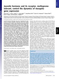
FIG. 1. Schematic representation <strong>of</strong> pGSFR161. The chimeric<br />
bar and neo genes are under the control <strong>of</strong> the 2' and 1' TR promoter,<br />
respectively, and are inserted at opposite orientation between the<br />
T-DNA border repeats <strong>of</strong> pGV1500 (12). The respective genes have<br />
termination and polyadenylylation signals <strong>of</strong> the T-DNA ocs (3' ocs)<br />
and the T-DNA gene 7 (3' g7). RB, right border; LB, left border.<br />
Subsequently, the root explants were blotted on sterile filter<br />
paper to remove most <strong>of</strong> the liquid medium and cocultivated<br />
for 48 hr on 0.5/0.05 agar. The explants were then rinsed in<br />
liquid 0.5/0.05 medium containing 1000 mg <strong>of</strong> vancomycin<br />
(Sigma) per liter. The pieces were blotted and then incubated<br />
on 0.15/5 agar (Table 1) supplemented with 750 mg <strong>of</strong><br />
vancomycin and 50 mg <strong>of</strong> Km per liter. Three weeks after<br />
infection with agrobacteria containing a chimeric neo gene,<br />
green Km-resistant (KmR) calli were formed in a background<br />
<strong>of</strong> yellowish root explants. At this point the root explants<br />
were transferred to fresh 0.15/5 agar containing only 500 mg<br />
<strong>of</strong> vancomycin and 50 mg <strong>of</strong> Km per liter. Three weeks later<br />
most green calli had formed shoots. Transformed shoots were<br />
transferred to 150 x 25 mm Petri dishes containing GM to<br />
form roots or seeds or both. In these Petri dishes, many<br />
regenerants formed seeds without rooting. Rooted plants<br />
could also be transferred to soil to set seed.<br />
Enzymatic Assays. Assays for NeoPTase II were performed<br />
as described by Reiss et al. (21); the assay for<br />
phosphinothricin acetyltransferase (PAcTase) has been described<br />
by De Block et al. (13). For both assays, transformed<br />
shoots free <strong>of</strong> agrobacteria were used.<br />
DNA Methods. Total <strong>Arabidopsis</strong> DNA was isolated as<br />
described by Dellaporta et al. (22). Genomic hybridizations<br />
were performed as described by Simoens et al. (7).<br />
RESULTS<br />
Capacity <strong>of</strong> Root Explants to Form Shoots. To test their<br />
regenerative capacity, we incubated <strong>Arabidopsis</strong> C24 root<br />
explants from 4-wk-old, sterile-grown plantlets for 7 or 14<br />
days on PG1 or R3 CIM (Table 1). They were then transferred<br />
to 0.15/5 or 0.05/7 (auxin/cytokinin ratio) SIM (Table 1).<br />
Roots that were callus-induced for 7 days were completely<br />
covered with shoots after 4 wk <strong>of</strong> incubation on SIM. By<br />
contrast, roots that were transferred to SIM after 14 days <strong>of</strong><br />
callus induction produced no shoots but formed brown<br />
degenerating callus instead.<br />
Omitting the callus induction step and culturing roots<br />
immediately onto SIM resulted in shoot formation at the<br />
proximal end <strong>of</strong> the explant only. Fig. 2 summarizes the<br />
different morphogenetic pathways that can be induced from<br />
<strong>Arabidopsis</strong> root explants by manipulating culture conditions.<br />
Clearly, the combination <strong>of</strong> callus induction followed by shoot<br />
induction was critical for efficient formation <strong>of</strong> shoots.<br />
The ontological pathway leading to rapid shoot formation<br />
from <strong>Arabidopsis</strong> roots is illustrated in Fig. 3 a-e. Induction<br />
Proc. Natl. Acad. Sci. USA 85 (1988) 5537<br />
Table 1. Plant media<br />
CIM SIM<br />
GM R3* PG1* 0.5/0.05 0.05/7* 0.15/5*<br />
Salts + vitamins<br />
Sucrose, g/L<br />
Glucose, g/L<br />
IAA, mg/L<br />
2,4-D, mg/L<br />
2ipAde, mg/L<br />
Kin, mg/L<br />
MS<br />
10<br />
-<br />
MS<br />
30<br />
5<br />
0.5<br />
0.3<br />
B5<br />
-<br />
20<br />
2<br />
0.05<br />
B5<br />
20<br />
0.5<br />
0.05<br />
MS<br />
30<br />
-<br />
0.05<br />
7<br />
B5<br />
20<br />
0.15<br />
5<br />
L, liter; IAA, indole-3-acetic acid; Kin, kinetin; 2ipAde, N6-(2isopentenyl)adenine;<br />
CIM, callus-inducing medium; SIM, shootinducing<br />
medium; MS, Murashige & Skoog medium (19); B5,<br />
Gamborg B5 medium (20).<br />
*Data for R3, PG1, 0.05/7 and 0.15/5, were from refs. 15-18, respectively.<br />
with 2,4-dichlorophenoxyacetic acid (2,4-D) resulted in a<br />
slight thickening <strong>of</strong> the root explants (Fig. 3a). When this<br />
tissue was transferred to high cytokinin/auxin-containing<br />
medium, root hairs covered the entire surface within 5-10<br />
days (Fig. 3b). A few days later, new areas <strong>of</strong> growth, green<br />
and devoid <strong>of</strong> root hairs, became visible on the root surface.<br />
After 2-3 wk on SIM, the first shoots developed from these<br />
green areas (Fig. 3c). Finally, after only 3-4 wk <strong>of</strong> tissue<br />
culture, the original root was completely converted in a dense<br />
growth <strong>of</strong> shoots (Fig. 3d). These shoots were transferred to<br />
150 x 25 mm Petri dishes containing GM. About 50% <strong>of</strong> the<br />
shoots grew roots in 2-4 wk. Rooted shoots could then be<br />
transferred to soil to flower and to produce seed. However,<br />
almost all shoots, regardless <strong>of</strong> whether they had roots or not,<br />
would form siliques and viable seeds in the 150 x 25 mm Petri<br />
dishes (Fig. 3e). In these aseptic conditions, typically 30-100<br />
seeds per regenerant were produced.<br />
Influence <strong>of</strong> 2,4-D and Callus-Induction Period on Morphogenesis.<br />
Since our initial experiments suggested that the<br />
concentration <strong>of</strong> 2,4-D in CIM and the length <strong>of</strong> the callusinduction<br />
period were determinative for homogenous shoot<br />
formation, we further tested the influence <strong>of</strong> these parameters<br />
on regeneration from root cuttings. We incubated root<br />
explants on CIM containing 0.2, 0.5, 1.0, 2.0, and 5.0 mg <strong>of</strong><br />
2,4-D and 0.05 mg <strong>of</strong> kinetin per liter. Explants were<br />
transferred to 0.15/5 SIM after 0, 4, 7, 10, and 14 days <strong>of</strong><br />
incubation on each 2,4-D-containing CIM. Optimal shoot<br />
regeneration was achieved when we incubated the root<br />
explants for 4 days on a medium containing 0.5 mg <strong>of</strong> 2,4-D<br />
per liter prior to shoot induction (Fig. 4). Significantly more<br />
aberrant shoots were obtained with 2,4-D at 0.2 mg/liter.<br />
Increasing concentrations <strong>of</strong> 2,4-D and/or longer periods <strong>of</strong><br />
callus induction resulted in a severe decrease in the regeneration<br />
efficiency. For example, 10 days <strong>of</strong> callus induction<br />
on a medium containing 2 mg <strong>of</strong> 2,4-D per liter and subsequent<br />
transfer to SIM resulted in thickening <strong>of</strong> the explants<br />
and formation <strong>of</strong> root hairs. However, further differentiation,<br />
ROOT T<br />
EXPLANTS~<br />
I<br />
CIM<br />
2SIg/ DEGLENUERATING<br />
CALLUS<br />
CALLUS<br />
%/////S<br />
IM/ / / / / / SHOOTS<br />
SIM kh, POLAR SHOOT<br />
~~~~~FORMATION<br />
TIME<br />
40 (days)<br />
FIG. 2. Summary <strong>of</strong> different possible morphogenetic pathways<br />
with <strong>Arabidopsis</strong> C24 root explants combining CIM and SIM.<br />
14
5538 Botany: Valvekens et al.<br />
a b c d<br />
Proc. Natl. Acad. Sci. USA 85 (1988)<br />
FIG. 3. Regeneration sequence <strong>of</strong> <strong>Arabidopsis</strong> C24 root explants. A root system was incubated for 4 days on the 0.5/0.05 CIM (Table 1)<br />
and subsequently transferred to the 0.15/5 SIM (Table 1). Differentiation on SIM is shown after 1 day (a), 4 days (b), 17 days (c), and 25 days<br />
(d). The first siliques are formed 2 wk after transfer <strong>of</strong> shoots to hormone-free GM-that, is within 6 wk <strong>of</strong> tissue culture (e).<br />
<strong>Arabidopsis</strong>-transformed shoots are induced on root explants after ± 5 wk <strong>of</strong> selection on 0.15/5 SIM containing 50 mg <strong>of</strong> Km per liter (f).<br />
such as green callus and shoot formation, was completely<br />
blocked. Higher concentrations <strong>of</strong> 2,4-D for longer periods<br />
resulted in total loss <strong>of</strong> morphogenetic potential even before<br />
root hairs were formed (Fig. 4).<br />
Influence <strong>of</strong> Antibiotics on Regeneration from Roots. To<br />
develop a rapid and efficient <strong>Agrobacterium</strong>-<strong>mediated</strong> <strong>transformation</strong><br />
method for <strong>Arabidopsis</strong>, we assayed the action <strong>of</strong><br />
a number <strong>of</strong> antibiotics that are currently used as selectable<br />
markers in plant <strong>transformation</strong> procedures. We found that<br />
50 mg <strong>of</strong> Km, 10 mg <strong>of</strong> G418, or 25 mg <strong>of</strong> hygromycin per liter<br />
completely blocked regeneration from untransformed roots<br />
and, therefore, could be used to select for transformed cells.<br />
Preliminary <strong>transformation</strong> experiments indicated that<br />
cefotaxime, an antibiotic commonly used to kill <strong>Agrobacterium</strong><br />
after cocultivation with plant material, severely inhib-<br />
2.4 D(mg/I)<br />
5.0<br />
2.0<br />
1.0<br />
0.5<br />
0.2<br />
0 days<br />
callus induction<br />
I I Li<br />
4 10 14 21 31 70 TIME<br />
(days)<br />
7 days<br />
4 days<br />
,callus induction<br />
2.4D(mg/I)<br />
(days)<br />
4 10 14 21 31 70 TIME<br />
days<br />
5.0<br />
2.0<br />
1.0<br />
0.5<br />
0.2<br />
[<br />
no shoots<br />
L polar shoot formation<br />
dispersed shoots<br />
shoots covering<br />
entire explants<br />
FIG. 4. Effects <strong>of</strong> the 2,4-D concentration in CIM and <strong>of</strong> the<br />
length <strong>of</strong> the induction period on regeneration <strong>of</strong> plants from root<br />
explants. Roots <strong>of</strong><strong>Arabidopsis</strong> C24 were incubated for 0, 4, 7, 10, and<br />
14 days on CIM containing different 2,4-D concentrations (ordinate)<br />
and 0.05 mg <strong>of</strong> kinetin per liter before transfer to 0.15/5 SIM. Each<br />
different combination <strong>of</strong> parameters was evaluated after 4, 10, 14, 21,<br />
31, and 70 days <strong>of</strong> tissue culture (abscissa).<br />
ited regeneration from <strong>Arabidopsis</strong> root explants. Therefore,<br />
in addition to cefotaxime, three other <strong>Agrobacterium</strong>inactivating<br />
antibiotics were tested for root explant toxicity<br />
at the effective concentrations <strong>of</strong> 0.5 and 1.0 g/liter (D.V.,<br />
unpublished data). Except for slight vitrification, vancomycin<br />
and triacillin showed no inhibitory effects on regeneration<br />
from root explants. By contrast, carbenicillin was slightly<br />
inhibitory and cefotaxime inhibited almost totally the root<br />
regeneration. Because the pGV2260 (11) and pGSFR1161 Ti<br />
plasmids we used contained aB-lactamase (bla) gene capable<br />
<strong>of</strong> inactivating both carbenicillin and triacillin, vancomycin<br />
(750 mg/liter) was used in subsequent <strong>transformation</strong> experiments<br />
to stop <strong>Agrobacterium</strong> growth after cocultivation.<br />
Transformation <strong>of</strong> Root Explants. After 3 days <strong>of</strong> callus<br />
induction on 0.5/0.05 medium, roots were cut into small<br />
pieces (0.5 cm) and cocultured on the same medium for 48 hr<br />
with an A. <strong>tumefaciens</strong> strain conferring KmR phenotype to<br />
plant cells after transfer <strong>of</strong> the T-DNA. Three to 4 wk after<br />
transfer to the 0.15/5 SIM containing 50 mg <strong>of</strong> Km per liter,<br />
green transformed calli (1-2 mm) in a background <strong>of</strong> yellowish<br />
root explants were formed. More than 80% <strong>of</strong> these green<br />
calli formed shoots after transfer to fresh SIM (Fig. 3f).<br />
Transformed shoots were transferred to GM for seed set.<br />
Using this method based on Km selection, we could reproducibly<br />
achieve <strong>transformation</strong> efficiencies between 20% and<br />
80% (root explants forming seed-producing transformants<br />
per total number <strong>of</strong> infected root explants) (Table 2). Root<br />
explants infected with <strong>Agrobacterium</strong> harboring the T-DNAless<br />
pGV2260 Ti plasmid produced no green calli or shoots on<br />
Km-containing medium (Table 2).<br />
DNA from four randomly chosen KmR regenerants was<br />
analyzed by DNA gel-blot hybridization to determine the<br />
T-DNA copy number. The data in Fig. Sb, lanes 1-3, suggest<br />
that each <strong>of</strong> three transformants have one T-DNA insert<br />
hybridizing to both right- and left-border probes. However,<br />
one transformant (Fig. 5b, lane 4) probably contains four<br />
inserts. One DNA fragment <strong>of</strong> this transformant (marked by<br />
the arrowhead) hybridized to both right- and left-border<br />
probes. This hybridization pattern and the length <strong>of</strong> the<br />
fragment suggest that two <strong>of</strong> the four inserts might be<br />
arranged in a directly repeated tandem structure. In addition,<br />
both NeoPTase II and PAcTase activities were found in all<br />
transformants tested (Fig. 5 c and d).<br />
neo Gene Segregation in the F1 Progeny <strong>of</strong> Transformants<br />
Confirms the Low-Copy-Number T-DNA Insertion. Seeds<br />
from 10 randomly chosen transformants were germinated on<br />
GM containing 50 mg <strong>of</strong> Km per liter. Two weeks after<br />
germination, Km5 seedlings had completely etiolated cotyledons,<br />
and development <strong>of</strong> roots and leaves was completely<br />
inhibited. By contrast, KmR plants had produced two to three<br />
pairs <strong>of</strong> leaves and a normal, elongating root (Fig. 6). All 10<br />
seed lines tested showed both KmR and Kms plants. x2<br />
analysis confirms that they all segregate for one or two<br />
functional T-DNA copies (Table 3).
Botany: Valvekens et al.<br />
Proc. Natl. Acad. Sci. USA 85 (1988) 5539<br />
Table 2. Transformation efficiency <strong>of</strong> root explants<br />
<strong>Agrobacterium</strong><br />
strain<br />
Chimeric selectable<br />
marker gene<br />
and promoter (P)<br />
<strong>Arabidopsis</strong><br />
ecotype Inoculated<br />
Root explants, no.<br />
With green calli<br />
on medium with Km<br />
(50 mg/liter)<br />
Transformation<br />
efficiency*<br />
pGSFR1161<br />
pGV2260<br />
pGSFR1161<br />
pGV2260<br />
pCLH88*<br />
pGV2260<br />
pGSFR1161<br />
pGV2260<br />
PTR-neo<br />
PTR-neo<br />
Pnos-neo<br />
PTR-neo<br />
C24<br />
C24<br />
C24<br />
C24<br />
C24<br />
C24<br />
Landsberg erecta<br />
Landsberg erecta<br />
251<br />
119<br />
443<br />
51<br />
171<br />
28<br />
118<br />
29<br />
81<br />
0<br />
131<br />
0<br />
170<br />
0<br />
104<br />
0<br />
32.0<br />
0<br />
29.6<br />
0<br />
99.4t<br />
0<br />
88.1t<br />
0<br />
Root explants ( 0.5 cm <strong>of</strong> a plant root system) were transformed as described. After 4 wk <strong>of</strong> selection on medium containing Km at 50<br />
mg/liter, the number <strong>of</strong> green calli was determined.<br />
*% <strong>of</strong> root explants forming green calli.<br />
tRoot explants form one to three green calli.<br />
Genotype Effects on Root Regeneration and Root Transformation.<br />
All experiments described above were performed<br />
with A. thaliana collection number C24. We also tested the<br />
root regeneration and <strong>transformation</strong> <strong>of</strong> <strong>Arabidopsis</strong> ecotypes<br />
Landsberg erecta and Columbia. <strong>Arabidopsis</strong> Landsberg<br />
erecta roots were both regenerated (D.V., unpublished<br />
data) and transformed equally efficiently as C24 roots (Table<br />
2). Root explants <strong>of</strong> <strong>Arabidopsis</strong> ecotype Columbia could<br />
also be transformed efficiently. Every infected Columbia root<br />
explant developed one to three independent green calli.<br />
However, when compared with C24, these calli were both<br />
slower and less efficient in shoot formation. Additional<br />
experiments are required to circumvent these regeneration<br />
problems in <strong>Arabidopsis</strong> ecotype Columbia.<br />
DISCUSSION<br />
A short preculture on 2,4-D-containing CIM and subsequent<br />
incubation on SIM with a high cytokinin concentration<br />
transforms <strong>Arabidopsis</strong> roots into a dense growth <strong>of</strong> shoots<br />
in only a few weeks time (Fig. 3). Similar conditions have<br />
been applied (17, 18) to induce shoot formation from Convulvus<br />
and <strong>Arabidopsis</strong> ecotype Wassilewskija (WS) leaf<br />
material. The short preculture on the 2,4-D medium was an<br />
essential step in the regeneration procedure, as immediate<br />
incubation <strong>of</strong> roots on shoot-inducing medium led to shoot<br />
formation at the proximal end <strong>of</strong> the root only (Figs. 2 and 4).<br />
This polar shoot formation has been reported for roots <strong>of</strong><br />
several other plant species and is thought to result from the<br />
endogenous auxin gradient that exists in roots (23, 24). The<br />
high incidence <strong>of</strong> shoot formation and the histological simplicity<br />
<strong>of</strong> <strong>Arabidopsis</strong> roots make this an elegant system to<br />
study initiation and development <strong>of</strong> shoots. By sectioning<br />
root explants at several stages during regeneration, the<br />
tissues giving rise to shoot primordia can be identified.<br />
In contrast to previous reports (9, 10), we successfully used<br />
Km selection to develop a reliable A. <strong>tumefaciens</strong>-<strong>mediated</strong><br />
root <strong>transformation</strong> procedure (Fig. 3f).<br />
Infected root explants reproducibly yielded fertile transformants<br />
with an efficiency between 20% and 80%, which is<br />
considerably higher than in previous reports (8, 9, 10, 25).<br />
Seeds obtained from transformants germinated on Kmcontaining<br />
medium, resulting in both etiolated (KmS) seedlings<br />
and phenotypically normal (KmR) plants, further evidenced<br />
the inheritance and Mendelian segregation <strong>of</strong> the neo<br />
gene (Fig. 6). Segregation analysis and genomic-blot analysis<br />
revealed that most transformants contained only one T-DNA<br />
insert (Table 3; Fig. 5 a and b). Km also can be used<br />
efficiently as a selectable marker in <strong>Arabidopsis</strong> leaf disc<br />
<strong>transformation</strong> (M.V.L., unpublished data).<br />
We also introduced a chimeric bar gene into <strong>Arabidopsis</strong>.<br />
The bar gene encodes PAcTase, which inactivates phosphi-<br />
nothricin, the active compound <strong>of</strong> the commercial herbicide<br />
Basta (Hoechst, Frankfurt). Unlike other marker genes that<br />
a<br />
A<br />
BamH<br />
Nco<br />
Hind III<br />
FRSB<br />
LB<br />
I<br />
-DPNW37 I bar 12' 1'| neo 3'ocs T-DNA<br />
b 1 2<br />
A B A B<br />
c 1 2 3 4 5 6<br />
_<br />
up<br />
3 4 5<br />
m-----l m -<br />
A B A B A B<br />
d<br />
"W<br />
a<br />
-a<br />
B<br />
p_<br />
- t5k5 b<br />
- 5kb<br />
-2.5 k b<br />
2 3 4 5 6<br />
Mw _4- _ w w e<br />
FIG. 5. Molecular analysis <strong>of</strong> <strong>Arabidopsis</strong> root-derived transformants.<br />
(a) T-DNA region <strong>of</strong> the pGSFR161 vector. A is the 1.6-kb<br />
BamHI-Nco I fragment; B is the 1.3-kb Nco 1-HindIlI fragment.<br />
Both were used as probes in genomic hybridizations. Notation is as<br />
in Fig. 2. (b) Genomic hybridizations. DNA from four transformants<br />
(lanes 1-4) and from an untransformed regenerant (lane 5) was<br />
digested with Nco I, separated on agarose gels, and blotted onto<br />
nylon membranes. Lanes: A, hybridization to the BamHI-Nco I<br />
probe; B, hybridization to the Nco 1-HindIII probe. Except for the<br />
band marked by the arrowhead, which is probably the internal<br />
fragment <strong>of</strong> a direct T-DNA repeat, all bands hybridizing to both<br />
probes are composite fragments between T-DNA and plant DNA. (c)<br />
NeoPTase II assays. Lanes: 1-4, transformants; 5, negative control<br />
(C24 plant); 6, positive control (purified NeoPTase II enzyme). (d)<br />
PAcTase assays. Lanes: 1-4, transformants; 5, negative control<br />
(untransformed regenerant); 6, positive control (purified PAcTase).<br />
*
5540 Botany: Valvekens et al.<br />
FIG. 6. Segregation <strong>of</strong> F1 progeny <strong>of</strong> transformant R72. F1 seeds<br />
were germinated on GM containing Km at 50 mg/liter. Two weeks<br />
after germination, Kms seedlings are etiolated while KmR seedlings<br />
grow phenotypically normal.<br />
can be used in <strong>Arabidopsis</strong>, the bar gene is easily selected for<br />
at the adult plant level, which facilitates gene mapping. We<br />
demonstrated PAcTase activity in transformed shoots (Fig.<br />
Sd). Moreover, herbicide spray tests showed cosegregation<br />
<strong>of</strong> Basta resistance with T-DNA in F1 progeny <strong>of</strong><strong>Arabidopsis</strong><br />
transformants (M.V.L., unpublished data).<br />
Because <strong>of</strong> its high efficiency, the root <strong>transformation</strong><br />
system described here may be used to study early <strong>Agrobacterium</strong><br />
<strong>transformation</strong> events. Sectioning <strong>of</strong> root explants<br />
during and after infection with an A. <strong>tumefaciens</strong> strain<br />
containing a f3-glucuronidase chimeric gene construct (26)<br />
could reveal which cell types are infected by <strong>Agrobacterium</strong><br />
and how transformed cells grow into shoots compared to<br />
untransformed cells.<br />
A. thaliana and D. melanogaster have similar genome sizes<br />
(3, 4). In Drosophila, P-element insertions have been a useful<br />
Table 3. Segregation <strong>of</strong> F1 progeny <strong>of</strong> transformants<br />
Number <strong>of</strong><br />
F1 plants __*<br />
Trans- One T-DNA Two T-DNA Three T-DNA<br />
formant KmR KmS insert inserts inserts<br />
R16 49 12 0.66t 16.53§<br />
R18 23 2 3.85t o.00t 1.23t<br />
R24 18 5 0.02t 6.96t<br />
R35 23 9 0.04t 22.53§<br />
R38 16 5 0.02t 8.26i<br />
R44 34 8 0.25t 9.66t<br />
R50 42 10 0.64t 12.02§<br />
R72 18 17 9.15t 99.89§<br />
R85 20 3 1.17t 0.79t 12.95§<br />
R102 39 17 0.60t 5i.5i§<br />
F1 seeds were germinated on GM (Table 1) containing Km at 50<br />
mg/liter. Two weeks after germination, the number <strong>of</strong> KmR and KmS<br />
seedlings was determined.<br />
*Because <strong>of</strong> the small numbers <strong>of</strong> seeds the Yates correction term<br />
was used in the V2 calculations.<br />
tp > 0.05.<br />
t0.05 > P > 0.001.<br />
§p < 0.001.<br />
Proc. Natl. Acad. Sci. USA 85 (1988)<br />
tool for the molecular cloning <strong>of</strong> developmentally important<br />
regulatory genes with a frequency <strong>of</strong> P-linked mutations <strong>of</strong><br />
roughly 10% (27). In <strong>Arabidopsis</strong>, it is not yet known what<br />
fraction <strong>of</strong> the T-DNA insertions produces detectable mutations.<br />
We screened the progeny <strong>of</strong> 10 independent <strong>Arabidopsis</strong><br />
root transformants and <strong>of</strong> about 50 independent leaf<br />
disc transformants. So far, this strategy did not yield any<br />
mutant clearly linked to the T-DNA (M.V.L., unpublished<br />
data). However, it is too early to conclude whether the<br />
T-DNA will be an effective mutagen in <strong>Arabidopsis</strong>.<br />
We thank Arlette Cattoir-Reynaerts, Marc De Block, Dirk Inzd,<br />
Diane J<strong>of</strong>uku, and Jack Okamuro for critical reading and suggestions;<br />
Rudy Vanderhaegen for technical assistance; and Martine De Cock,<br />
Karel Spruyt, Stefaan Van Gijsegem, and Vera Vermaercke for help<br />
in preparing the manuscript. This research was supported by grants<br />
from the Fonds voor Geneeskundig Wetenschappelijk Onderzoek<br />
(3.0002.82), and from the Services <strong>of</strong> the Prime Minister (Onderling<br />
Overlegde Akties 12.0561.84 and Interuniversitaire Attraktiepolen<br />
120C0187). D.V. is indebted to the Instituut voor Wetenschappelijk<br />
Onderzoek in Nijverheid en Landbouw for a fellowship.<br />
1. Somerville, C. R., McCourt, P., Caspar, T., Estelle, M. & Keith, K.<br />
(1985) in Plant Genetics, UCLA Symposia on Molecular and<br />
Cellular Biology, New Series, ed. Freeling, M. (Liss, New York),<br />
Vol. 35, pp. 651-660.<br />
2. Meyerowitz, E. M. (1987) Annu. Rev. Genet. 21, 93-111.<br />
3. Leutwiler, L. S., Hough-Evans, B. R. & Meyerowitz, E. M. (1984)<br />
Mol. Gen. Genet. 194, 15-23.<br />
4. Manning, J. E., Schmid, C. W. & Davidson, N. (1975) Cell 4, 141-<br />
155.<br />
5. Sulston, J. E. & Brenner, S. (1974) Genetics 77, 95-104.<br />
6. Koornneef, M. (1987) in Genetic Maps 1987, ed. O'Brien, S. J.<br />
(Cold Spring Harbor Lab., Cold Spring Harbor, NY), Vol. 4, pp.<br />
742-745.<br />
7. Simoens, C., Alliotte, Th., Mendel, R., Muller, A., Schiemann, J.,<br />
Van Lijsebettens, M., Schell, J., Van Montagu, M. & Inzd, D. (1986)<br />
Nucleic Acids Res. 14, 8073-8090.<br />
8. An, G., Watson, B. D. & Chiang, C. C. (1986) Plant Physiol. 81,<br />
301-305.<br />
9. Lloyd, A. M., Barnason, A. R., Rogers, S. G., Byrne, M. C.,<br />
Fraley, R. T. & Horsch, R. B. (1986) Science 234, 464-466.<br />
10. Sheikholeslam, S. N. & Weeks, D. P. (1987) Plant Mol. Biol. 8,<br />
291-298.<br />
11. Deblaere, R., Bytebier, B., De Greve, H., Deboeck, F., Schell, J.,<br />
Van Montagu, M. & Leemans, J. (1985) Nucleic Acids Res. 13,<br />
4777-4788.<br />
12. Deblaere, R., Reynaerts, A., H<strong>of</strong>te, H., Hernalsteens, J.-P., Leemans,<br />
J. & Van Montagu, M. (1987) Methods Enzymol. 153, 277-<br />
292.<br />
13. De Block, M., Botterman, J., Vandewiele, M., Dockx, J., Thoen,<br />
C., Gosseld, V., Movva, R., Thompson, C., Van Montagu, M. &<br />
Leemans, J. (1987) EMBO J. 6, 2513-2518.<br />
14. Velten, J. & Schell, J. (1985) Nucleic Acids Res. 13, 6981-6998.<br />
15. Christianson, M. L. (1979) Environ. Exp. Bot. 19, 217-221.<br />
16. Negrutiu, I., Beeftink, F. & Jacobs, M. (1975) Plant Sci. Lett. 5,<br />
293-304.<br />
17. Christianson, M. L. & Warnick, D. A. (1983) Dev. Biol. 95, 288-293.<br />
18. Feldmann, K. A. & Marks, M. D. (1986) Plant Sci. 47, 63-69.<br />
19. Murashige, T. & Skoog, F. (1962) Physiol. Plant. 15, 473-497.<br />
20. Gamborg, 0. L., Miller, R. A. & Ojima, K. (1968) Exp. Cell Res. 50,<br />
151-158.<br />
21. Reiss, B., Sprengel, R., Will, H. & Schaller, H. (1984) Gene 30, 211-<br />
218.<br />
22. Dellaporta, S. L., Wood, J. & Hicks, J. B. (1983) Plant Mol. Biol.<br />
Rep. 1, 19-21.<br />
23. Lazzeri, P. A. & Dunwell, J. M. (1984) Ann. Bot. (London) 54, 341-<br />
350.<br />
24. Warmke, H. E. & Warmke, G. L. (1950) Am. J. Bot. 37, 272-280.<br />
25. Pang, P. P. & Meyerowitz, E. M. (1987) BiolTechnology 5, 1177-<br />
1181.<br />
26. Jefferson, R. A. (1987) Plant Mol. Biol. Rep. 5, 387-405.<br />
27. Cooley, L., Kelley, R. & Spradling, A. (1988) Science 239, 1121-<br />
1128.