EPOS 2021 Congress Review

Create successful ePaper yourself
Turn your PDF publications into a flip-book with our unique Google optimized e-Paper software.
ACHONDROPLASIA.EXPERT<br />
CONGRESS REVIEWS<br />
<strong>EPOS</strong> <strong>2021</strong><br />
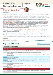
Welcome to our Achondroplasia.expert congress reviews.<br />
Our summaries highlight key data on achondroplasia,<br />
skeletal dysplasia, and other disorders of short stature<br />
reported at congresses.<br />
Our review of <strong>EPOS</strong> <strong>2021</strong> includes summaries of many advances in<br />
the treatment and management of achondroplasia. Our summaries<br />
include new technologies to improve limb lengthening, a complex<br />
procedure with a high complication rate and no consensus on<br />
its use in achondroplasia, but one that could result in height<br />
enhancement in adults with achondroplasia, nonetheless. Claire<br />
Shannon showed results from implantable limb lengthening<br />
devices in congenital limb length discrepancy, and Emily Dodwell<br />
presented data from Sidharthan et al. on the use of tantalum<br />
beads as landmarks to measure bone growth directly. Finally,<br />
Benedick et al. shared data on the use of knee radiographs to<br />
predict skeletal maturity, which could be used alongside the<br />
new growth charts that are available in achondroplasia.<br />
We also review two symposiums, looking at the natural history<br />
of achondroplasia, and the challenges of rare disease diagnosis.<br />
We trust you will find this summary useful in staying current with<br />
developments in the field.<br />
The Achondroplasia Team<br />
TECHNOLOGICAL<br />
INNOVATION<br />
Using tech in<br />
orthopaedics.<br />
TURN TO PAGE 2<br />
SURGICAL ADVANCES<br />
Surgery is a key topic<br />
in <strong>EPOS</strong>, with many<br />
groups reporting on<br />
limb lengthening<br />
techniques in various<br />
congenital diseases.<br />
TURN TO PAGE 2<br />
ACHONDROPLASIA<br />
NATURAL HISTORY<br />
Highlights from the<br />
BioMarin symposium<br />
with a focus on the<br />
LIAISE study.<br />
TURN TO PAGE 7<br />
The abstracts, plenary presentations and symposiums can be found here<br />
Treatments mentioned in this document may not be approved for use in your country. Please consult local authorising authorities for further information.<br />
Some links in this document are “external links” to websites over which BioMarin has no control and for which BioMarin assumes no responsibility.<br />
When visitors choose to follow a link to any external website, they are subject to the cookie, privacy and legal policies of the external website.<br />
Compliance with applicable data protection and accessibility requirements of external websites linked to from this website falls outside the control<br />
of BioMarin and is the explicit responsibility of the external website.<br />
Achondroplasia.expert is organised and funded by BioMarin.<br />
© <strong>2021</strong> BioMarin International Ltd.<br />
All Rights Reserved. EU-VOX-00185 October <strong>2021</strong><br />
1
Achondroplasia.expert <strong>Congress</strong> <strong>Review</strong>s: <strong>EPOS</strong> <strong>2021</strong><br />
TECHNOLOGICAL INNOVATION<br />
Technology and smart apps are increasingly becoming part of everyday life,<br />
and they can offer utility in the medical setting too.<br />
Peter Stevens looked at smartphone<br />
monitoring of guided growth, tested in<br />
approximately 200 patients over 2 years.<br />
Guided growth for angular correction is<br />
widespread as a treatment of choice and<br />
can be used in growing patients from<br />
18 months to 18 years. Although guided<br />
growth is minimally invasive, it consumes time<br />
and resources – requiring a clinic visit every<br />
3 months, with full length teleoroentgenogram<br />
to document improvement in the mechanical<br />
axis. This can be challenging for the patient,<br />
their parents, and the surgical clinic team.<br />
Stevens suggests a more efficient means of<br />
monitoring alignment by examining monthly<br />
smartphone photos from caregivers. The<br />
images document the child holding a placard<br />
and standing with knees (or ankles) touching<br />
and are e-mailed to the clinic monthly until<br />
the deformities are corrected, whereupon<br />
they are instructed to return to the hospital<br />
for surgery. Stevens’ results show that<br />
only a small percentage of parents fail to<br />
take and send the pictures as instructed.<br />
Only 3 patients experienced overcorrection<br />
sufficient to warrant reversal of the 8-plates<br />
(medial to lateral), and none required salvage<br />
osteotomies. The author concludes that<br />
engaging parents in the documentation of<br />
progressive improvement of alignment gives<br />
them greater investment in the process, and<br />
delivers savings in time, travel, and cost –<br />
as well as saving unnecessary clinic and<br />
imaging time. [OP-015]<br />
SURGICAL ADVANCES<br />
Surgery is an important part of orthopaedic care, and is required for many<br />
paediatric joint and bone disorders. New techniques and data were explored for<br />
surgical advances in several diseases.<br />
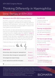
Michiel Van de Sande gave an oral<br />
presentation on reconstructions in the<br />
very young. He began by discussing bone<br />
sarcoma resections, and the challenges<br />
of reconstruction for function and growth<br />
in young children. About 77.5% of children<br />
diagnosed with osteo-sarcoma before the age<br />
of 12 survive for 5 years. Generally,<br />
it is recommended that limbs should<br />
not be lengthened by more than 50% of<br />
the final length, with minimal resection<br />
for non-invasive growers, and minimal<br />
fixation length for stem or motor. The use<br />
of radiotherapy before or after surgery may<br />
facilitate more marginal resections but cannot<br />
always prevent loss of the growth plate.<br />
Winkelman A1 resection<br />
Most modern non-invasive growers have<br />
comparable survival rates, but implant<br />
failures are more common in children under<br />
the age of 10, compared to older children.<br />
An alternative reconstruction in cases with<br />
complications is rotationplasty, where the<br />
lower limb is rotated 180 degrees, and the<br />
ankle joint – powered by the lower limb<br />
muscles – functions as the knee. This is a<br />
good choice for young children with joint and<br />
physis involvement. Where rotationplasty is<br />
not possible, functional limbs can also be<br />
reconstructed with 3D implants.<br />
Claire Shannon presented on Extramedullary<br />
Implantable Limb Lengthening (EILL)<br />
for congenital limb length discrepancy<br />
(LLD) as an alternative to external fixation<br />
lengthening. This retrospective case review<br />
Adapted from de Sande, <strong>2021</strong><br />
in 23 patients aged 3.5 to 20 years aimed<br />
to evaluate whether EILL can extend the<br />
indication for implantable limb lengthening<br />
to younger and smaller children and replace<br />
external fixation as the only method to<br />
lengthen children whose medullary canals<br />
are too small or whose growth plates would<br />
be violated by intramedullary lengthening<br />
devices. The patients involved had a variety<br />
of diagnoses, including skeletal dysplasia.<br />
A small diameter solid rod was inserted<br />
in the medullary canal to help maintain<br />
alignment and the lengthening nail was affixed<br />
outside the bone with screws. Lengthening<br />
rate was 0.75mm/day, with daily physical<br />
therapy and night extension knee bracing<br />
for femurs, ankle dorsiflexion bracing for<br />
tibias. Overall, the average lengthening was<br />
48 mm. Complications included one broken<br />
2 3
Achondroplasia.expert <strong>Congress</strong> <strong>Review</strong>s: <strong>EPOS</strong> <strong>2021</strong><br />
screw requiring revision fixation, and two<br />
subluxations of hip treated with open reduction<br />
and periacetabular osteotomy. There were<br />
no infections and no axial deviation. The<br />
authors conclude that EILL is an alternative<br />
to external fixation lengthening and extends<br />
the indications for implantable lengthening<br />
to younger children. However, it is important<br />
to follow the same principles as with external<br />
fixation lengthening in terms of stabilisation<br />
of joints with preparatory surgery and bracing<br />
to prevent knee subluxation and contracture.<br />
Lengthening should be restricted to 5 cm.<br />
[OP-009]<br />
Emily Dodwell (on behalf of Sidharthan et<br />
al.) showed that monitoring with tantalum<br />
beads demonstrates no clinically significant<br />
growth following percutaneous transphyseal<br />
screw epiphysiodesis. It has previously been<br />
suggested that there is a 6–12-month delay<br />
in growth cessation following percutaneous<br />
transphyseal screw epiphysiodesis, and<br />
the growth inhibition has been estimated<br />
based on predicted versus actual growth<br />
of the entire bone. Tantalum beads provide<br />
landmarks by which growth can be measured<br />
directly, and not inferred. This retrospective<br />
study aimed to evaluate growth at the physis<br />
following percutaneous transphyseal screw<br />
epiphysiodesis (PETS) using tantalum bead<br />
markers inserted medially and laterally both<br />
proximal and distal to the physis. Inter-bead<br />
distance perpendicular to the physis was<br />
measured on calibrated radiographs.<br />
The study included 18 patients (12 boys<br />
and 6 girls) with predicted LLD> 2 cm at<br />
maturity who underwent PETS of the distal<br />
femur and/or proximal tibia with tantalum<br />
bead placement and had at least 6 months’<br />
post-operative imaging. Median bone age<br />
was 12.5 and 13.3 years among girls and<br />
boys, respectively. Median current LLD was<br />
1.4 cm and median predicted LLD at time<br />
of skeletal maturity was 2.7 cm. Median<br />
follow-up was 53 weeks.<br />
In both the femoral and tibial physes, median<br />
change in inter-bead distance was 0 mm. The<br />
authors concluded that PETS demonstrated<br />
immediate cessation of growth in all patients.<br />
This technique inhibits growth at the physis<br />
effectively, with no evidence of time lag in<br />
growth inhibition. No complications related<br />
to transphyseal screws or the tantalum<br />
beads were identified. [OP-074]<br />
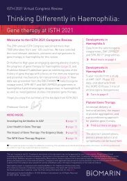
A poster from Popkov et al. discussed the use<br />
of titanium telescopic rodding and reduced<br />
external fixation in paediatric osteogenesis<br />
imperfecta (OI) patients. The major limitation<br />
of any intramedullary telescopic system is<br />
rotational and longitudinal instability. This<br />
study aimed to examine the outcomes of<br />
deformity correction by combined technique<br />
uniting titanium telescopic rod and reduced<br />
Ilizarov frame in 12 children with OI with a<br />
minimum follow-up of 1 year. In 5 cases,<br />
telescopic rod insertion was performed<br />
simultaneously with Ilizarov frame removal<br />
from the segment previously operated; in<br />
other consecutive surgeries, there was a<br />
2–6-month interval between operations.<br />
Telescoping gain related to spontaneous<br />
growth assessed at 1 year was 15.9 ± 2.3<br />
and 13.7 ± 3.1 mm in the femur and tibia,<br />
respectively. The authors concluded that –<br />
despite the abnormal bone – a combination<br />
of titanium telescopic rod with reduced<br />
external fixation provides advantages in<br />
orthopaedic surgery for children with OI.<br />
Titanium alloy telescopic rod is not prone to<br />
Telescopic rodding of tibia: amount of telescoping since frame removal = 19 mm<br />
limited telescoping, deformity relapse, or rod<br />
migration. Children demonstrated walking<br />
with weight-bearing early in the postoperative<br />
period. Temporary gait changes<br />
were influenced by the size of the external<br />
device, and by strategies to reduce pain at<br />
the pin sites. [EP-004]<br />
The surgical treatment of severe cervical<br />
kyphosis in diastrophic dysplasia was<br />
covered in a poster from Heydemann et al.<br />
The incidence of cervical kyphosis is 15–44%<br />
in children with diastrophic dysplasia.<br />
Although spontaneous improvement is<br />
seen in 75%, progression can result in<br />
severe deformity, spinal cord compression,<br />
and neurologic injury. Bracing can delay<br />
surgery, but treatment requires spinal<br />
fusion, sometimes in combination with spinal<br />
cord decompression. This single-centre<br />
Popkov et al. [EP-004]<br />
retrospective series included 47 patients<br />
with diastrophic dysplasia treated from<br />
1984–2017 with minimum 2-year follow-up.<br />
Patients underwent posterior fusion plus<br />
anterior corpectomy, decompression<br />
and fusion for spinal cord compression.<br />
Cervical kyphosis was found in 27 of the<br />
patients – an incidence of 57%, which is<br />
higher than previously reported. Of these,<br />
21 patients spontaneously improved, but<br />
6 required surgery. The results demonstrated<br />
substantial improvement in deformity and<br />
sagittal balance, with 100% fusion rate<br />
and no major adverse events or neurologic<br />
injuries. Given the high incidence reported,<br />
the authors concluded there is a need<br />
for vigilance. In children with diastrophic<br />
dysplasia, surgical correction of severe<br />
cervical kyphosis can be achieved safely<br />
and reliably. [EP-005]<br />
4 5
Achondroplasia.expert <strong>Congress</strong> <strong>Review</strong>s: <strong>EPOS</strong> <strong>2021</strong><br />
BASIC SCIENCE<br />
The basic science session included a round-up of presentations looking at<br />
animal models and imaging considerations in orthopaedics.<br />
Raymond Liu (on behalf of Benedick et al.)<br />
described using knee radiographs during<br />
pre-adolescence to estimate skeletal maturity.<br />
Knowing skeletal age is useful in the treatment<br />
of LLD; however, a quick, accurate method in<br />
this age range is lacking. The authors analysed<br />
serial knee radiographs leading up to the<br />
chronological age associated with 90% of final<br />
height (an enhanced skeletal maturity gold<br />
standard compared to peak height velocity) in<br />
75 children. The goal was to develop a quick,<br />
accurate and reproducible knee skeletal<br />
maturity system that would outperform current<br />
methods in the pre-adolescent age range.<br />
Epi- and metaphyseal widths of the distal<br />
femur, proximal tibia, and proximal fibula were<br />
measured, and the ratio calculated. Greulich<br />
and Pyle (GP) bone ages were assigned using<br />
radiographs of the left hand. In total, 258 left<br />
knee radiographs from 39 girls (mean age 8.6)<br />
and 36 boys (mean age 10.6) were included.<br />
The results showed a strong positive correlation<br />
between the epiphyseal: metaphyseal ratio<br />
value and years away from reaching 90% of<br />
final height, with Pearson R values of 0.80,<br />
0.84, and 0.84 for the femur, tibia, and fibula,<br />
respectively (all P
Achondroplasia.expert <strong>Congress</strong> <strong>Review</strong>s: <strong>EPOS</strong> <strong>2021</strong><br />
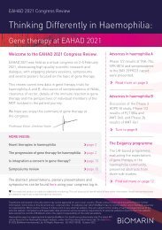
LIAISE: Burden of medical conditions and disorders*<br />
120<br />
100<br />
Event rate per 100PY<br />
77.5<br />
80<br />
60<br />
43.6<br />
40<br />
28.4<br />
24.4<br />
20<br />
39.3<br />
33.1<br />
101.2<br />
81.7<br />
91.4<br />
national guidelines. Patient organisations<br />
in all countries provide support and collaborate<br />
in the care pathway. In all five countries,<br />
pre-natal diagnosis is considered preferable,<br />
and molecular diagnosis is the gold standard.<br />
In infancy and early childhood there is a<br />
typically follow-up every 3–6 months, with<br />
care given by a multidisciplinary team. There<br />
are also yearly follow ups at specialist centres,<br />
in which the key areas monitored are weight,<br />
dental issues, and leg deformities. There is<br />
evidence that limb-lengthening is common<br />
in Italy and Spain, but rarely undertaken in<br />
France, Germany, and the UK. Notably, a<br />
gap has been identified for the transition into<br />
adult care, with limited structured care. Adults<br />
have specific needs, such as pain, obstetric<br />
advice, genetic counselling, and support in the<br />
workplace. With potential new therapies on<br />
the horizon, more systematic and structures<br />
0<br />
0–4<br />
(n=100)<br />
5–10<br />
(n=121)<br />
11–15<br />
(n=79)<br />
16–20<br />
(n=51)<br />
21–30<br />
(n=43)<br />
31–40<br />
(n=31)<br />
41–50<br />
(n=23)<br />
51–60<br />
(n=13)<br />
>60<br />
(n=8)<br />
Healthcare set-up for achondroplasia<br />
Age (years)<br />
Centralised<br />
Decentralised<br />
*Medical conditions and disorders are those that required a visit to the doctor.<br />
LIAISE study – a multinational, observational<br />
natural history study being conducted across<br />
Europe to examine the lifetime impact<br />
of achondroplasia. This initiative aims to<br />
quantify the impact across the age spectrum<br />
by measuring the clinical burden, healthcare<br />
resource use, and health-related quality of<br />
life. Data are being collected from 13 sites in<br />
six countries. Initial results show a bimodal<br />
U-shaped distribution of medical events<br />
by age group, with youngest and oldest the<br />
most affected. ENT and neurological issues<br />
are more common children, with pain, ENT<br />
issues (excluding otitis media) and spinal<br />
issues increasing with patient age. The<br />
most frequently reported complication by<br />
system organ class is musculoskeletal and<br />
connective tissue disorders, and nervous<br />
systems issues – each reported in up to 60%.<br />
Professor Maghnie concluded by stating<br />
that many unmet needs remain for people<br />
with achondroplasia. These are physical,<br />
psychosocial, economic, and environmental<br />
Maghnie [BioMarin symposium]<br />
–some of which can be avoided if they are<br />
detected early. There are also still gaps<br />
in knowledge, notably around potential<br />
complications and functional consequences<br />
across the lifespan, and the impact of<br />
emerging disease-modifying therapies.<br />
Professor Guillén-Navarro looked at the<br />
current care pathway for people with<br />
achondroplasia. For efficient clinical<br />
management, it is critical to understand<br />
the natural history – but in general there is<br />
a scarcity of clinical practice guidelines to<br />
assist appropriate decision making. With<br />
advances in potential drug management for<br />
achondroplasia, there is a need to better<br />
understand the natural history and clinical<br />
care framework. Recently, an expert meeting<br />
was convened to address this issue in Europe,<br />
beginning with an analysis of the healthcare<br />
set-up, guidelines, and patient associations in<br />
five countries. There are significant differences<br />
in how achondroplasia is managed, and –<br />
to date – France is the only country with<br />
France<br />
UK<br />
National network for skeletal dysplasia<br />
consisting of one national coordinating Centre<br />
of Reference (Necker in Paris)<br />
21 Centres of Competence in the regions; and<br />
two Centres of Reference for adults (in Paris)<br />
Expert centres belong to the bigger network<br />
‘Filiere OSCAR’<br />
UK-wide ACH network has been established to<br />
develop consistent approaches in managing the<br />
patient journey to the specialist centres<br />
Germany<br />
Approx 35 centres for rare diseases, with some<br />
(5–7) of these specialised in bone diseases<br />
Decentralised healthcare system<br />
Care of ACH patients may be organised<br />
by paediatric orthopaedic, clinical genetics<br />
or paediatric endocrinology departments<br />
depending on region<br />
Italy<br />
Every region has one or more rare disease<br />
centres and a centre of expertise<br />
Only few institutions specialised<br />
(multidisciplinary) in treatment of ACH patients<br />
This does allow some centralisation of care<br />
Devolved care in many regions<br />
Spain<br />
17 regional health system with autonomous<br />
decisions<br />
Each region responsible for implementing<br />
follow-up care for patients<br />
a few reference centres for surgical treatments<br />
of skeletal dysplasia<br />
Clinical genetics is not yet recognised as a<br />
speciality in Spain but experts trained abroad<br />
providing service and raising profile<br />
Guillén-Navarro [BioMarin symposium]<br />
8 9
Achondroplasia.expert <strong>Congress</strong> <strong>Review</strong>s: <strong>EPOS</strong> <strong>2021</strong><br />
care is needed for standardisation of care and<br />
access to specialist services, and guidelines<br />
are need for the different aspects of care.<br />
Dr Leiva-Gea focused on current thinking<br />
on limb lengthening in people with<br />
achondroplasia. Limb lengthening is not<br />
typically part of the natural history, and<br />
there is no consensus on its use at present.<br />
If limb length can be shown to have an<br />
impact on health-related quality of life then<br />
lengthening surgery may have important role<br />
to play, but at present it is not clear which<br />
aspects of achondroplasia impact quality<br />
of life. There is some evidence suggesting<br />
upper limb lengthening provides functional<br />
improvement and gains in quality of life,<br />
including self-esteem. Leiva-Gea and<br />
colleagues have recently published a protocol<br />
on limb lengthening, showing different steps<br />
– maintaining the idea that some people<br />
with achondroplasia will benefit from limb<br />
lengthening. Limb lengthening increases<br />
overall height and arm span, and improves<br />
proportionality. But the true impact on<br />
functionality, quality of life, and metabolic<br />
issues is not known, and more studies are<br />
needed to create more robust evidence on<br />
the efficacy and safety, and to identify which<br />
patients are appropriate for this intervention.<br />
FOP is an incredibly rare condition caused<br />
by a spontaneous missense mutation in<br />
ALK2/ACVR1. Genetic testing is key to FOP<br />
diagnosis. Dr Kannu presented a case of<br />
a 2-year-old boy, which was subsequently<br />
confirmed as FOP. Aggressive juvenile<br />
LIST OF ABBREVIATIONS<br />
EILL<br />
FOP<br />
GP<br />
JIA<br />
Extramedullary Implantable<br />
Limb Lengthening<br />
Fibrodysplasia Ossificans Progressiva<br />
Greulich and Pyle<br />
Juvenile Idiopathic Arthritis<br />
fibromatosis is a common misdiagnosis for<br />
FOP, and early lesions can look identical under<br />
the microscope. Other differential diagnoses<br />
include autosomal dominant progressive<br />
osseous heteroplasia, osteoma cutis, and<br />
Albright’s hereditary osteodystrophy.<br />
LLD<br />
MTX<br />
OI<br />
PETS<br />
Limb length discrepancy<br />
Methotrexate<br />
Osteogenesis Imperfecta<br />
Percutaneous Transphyseal<br />
Screw Epiphysiodesis<br />
CHALLENGES OF RARE DISEASE DIAGNOSIS<br />
The Ipsen symposium focused on the challenges of rare disease diagnosis,<br />
centred around a series of patient cases. The session was chaired by<br />
Peter Kannu, who also gave a presentation, alongside Vladimir Kenis.<br />
Dr Kannu highlighted that while a rare<br />
disease is defined as one with an incidence<br />
of fewer than 5 cases per 10,000 population<br />
the vast majority are far less common than<br />
this. Although individually rare, collectively<br />
rare diseases have a significant burden,<br />
and 1 in 17 people are affected by a rare<br />
disease at some point in their life. Typically,<br />
diagnosis can take a long time, which causes<br />
deterioration in health, lack of treatment,<br />
and potential harm and costs.<br />
Heterotopic ossification is a rare disease<br />
that causes pathological formation of<br />
extra-skeletal bone in muscles and soft<br />
tissue. Dr Kenis discussed two children<br />
with deformities of the foot. On referral to<br />
genetic testing, diagnosis was confirmed as<br />
fibrodysplasia ossificans progressiva (FOP)<br />
– a type of heterotopic ossification with a<br />
prevalence of 1.36 per million individuals that<br />
exists across a spectrum of presentations.<br />
To date, there is no conventional treatment<br />
strategy for heterotopic ossification, and<br />
surgery can be ineffective and dangerous,<br />
leading to further deterioration. Dr Kenis<br />
stressed that orthopaedists are often the first<br />
specialty consulted, and it is important to be<br />
able to identify rare diseases. He concluded<br />
that genetic testing is the easiest way to<br />
confirm a rare disease, and to find the most<br />
appropriate treatment.<br />
10 11
Achondroplasia.expert <strong>Congress</strong> <strong>Review</strong>s: <strong>EPOS</strong> <strong>2021</strong><br />
12