Dental Asia March/April 2019
For more than two decades, Dental Asia is the premium journal in linking dental innovators and manufacturers to its rightful audience. We devote ourselves in showcasing the latest dental technology and share evidence-based clinical philosophies to serve as an educational platform to dental professionals. Our combined portfolio of print and digital media also allows us to reach a wider market and secure our position as the leading dental media in the Asia Pacific region while facilitating global interactions among our readers.
For more than two decades, Dental Asia is the premium journal in linking dental innovators
and manufacturers to its rightful audience. We devote ourselves in showcasing the latest dental technology and share evidence-based clinical philosophies to serve as an educational platform to dental professionals. Our combined portfolio of print and digital media also allows us to reach a wider market and secure our position as the leading dental media in the Asia Pacific region while facilitating global interactions among our readers.

You also want an ePaper? Increase the reach of your titles
YUMPU automatically turns print PDFs into web optimized ePapers that Google loves.
Behind the Scenes<br />
A JOURNEY THROUGH ELEMENTAL<br />
REINFORCEMENT OF FUNCTIONAL AND<br />
AESTHETIC HARMONY<br />
By Dr. Anand Narvekar<br />
It did take quite some time for me<br />
to understand that dentistry is<br />
similar to photography. One has<br />
to be at the moment, intervene<br />
and implicate the right treatment<br />
regimen to provide another’s smile. In<br />
order to do all that, I have learned that<br />
patience is an important virtue that we<br />
must possess through our entire smile<br />
journey. This particular case has taught<br />
me in all different aspects of dentistry and<br />
motivates me to continue contributing<br />
smiles, just like how photographers keep<br />
on taking more photographs of their work.<br />
Case details<br />
A 62-year-old female patient came up<br />
to my clinic with a chief complaint of<br />
fracturing her ceramic crowns in the<br />
upper right posterior region and was<br />
experiencing mild pain/sensitivity on<br />
hot/cold in the upper left molar tooth<br />
region. Patient also complained of food<br />
lodgement in the upper palatal and<br />
gingival areas.<br />
Observations<br />
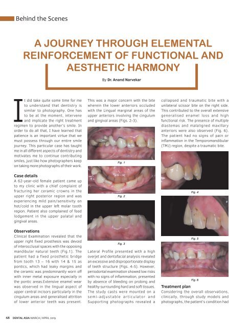
Clinical Examination revealed that the<br />
upper right fixed prosthesis was devoid<br />
of interocclusal spaces with the opposing<br />
mandibular natural teeth (Fig.1). The<br />
patient had a fixed prosthetic bridge<br />
from tooth 13 – 16 with 14 & 15 as<br />
pontics, which had leaky margins and<br />
the ceramic was predominantly worn off<br />
with inner metal exposure especially in<br />
the pontic areas.Extensive enamel wear<br />
was observed in the lingual aspect of<br />
upper central incisors particularly in the<br />
cingulum areas and generalised attrition<br />
of lower anterior teeth was present.<br />
This was a major concern with the bite<br />
wherein the lower anteriors occluded<br />
with the Lingual marginal areas of the<br />
upper anteriors involving the cingulum<br />
and gingival areas (Figs. 2-3).<br />
Fig. 1<br />
Fig. 2<br />
Fig. 3<br />
Lateral Profile presented with a high<br />
overjet and dentofacial analysis revealed<br />
an excessive and disproportionate display<br />
of teeth structure (Figs. 4-5). However,<br />
periodontal examination showed low risks<br />
with no signs of inflammation, presented<br />
by absence of bleeding on probing and<br />
healthy surrounding hard and soft tissues.<br />
The study casts were mounted on a<br />
semi-adjustable articulator and<br />
Supporting photographs revealed a<br />
collapsed and traumatic bite with a<br />
unilateral scissor bite on the right side.<br />
This contributed to the overall extensive<br />
generalised enamel loss and high<br />
functional risk. The presence of multiple<br />
diastemas and malaligned maxillary<br />
anteriors were also observed (Fig. 6).<br />
The patient had no signs of pain or<br />
inflammation in the Temporomandibular<br />
(TMJ) region, despite a traumatic bite.<br />
Fig. 4<br />
Fig. 5<br />
Fig. 6<br />
Treatment plan<br />
Considering the overall observations,<br />
clinically, through study models and<br />
photographs, the patient’s condition had<br />
68<br />
DENTAL ASIA MARCH / APRIL <strong>2019</strong>