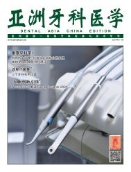
Dental Asia March/April 2020
For more than two decades, Dental Asia is the premium journal in linking dental innovators and manufacturers to its rightful audience. We devote ourselves in showcasing the latest dental technology and share evidence-based clinical philosophies to serve as an educational platform to dental professionals. Our combined portfolio of print and digital media also allows us to reach a wider market and secure our position as the leading dental media in the Asia Pacific region while facilitating global interactions among our readers.
For more than two decades, Dental Asia is the premium journal in linking dental innovators
and manufacturers to its rightful audience. We devote ourselves in showcasing the latest dental technology and share evidence-based clinical philosophies to serve as an educational platform to dental professionals. Our combined portfolio of print and digital media also allows us to reach a wider market and secure our position as the leading dental media in the Asia Pacific region while facilitating global interactions among our readers.
You also want an ePaper? Increase the reach of your titles
YUMPU automatically turns print PDFs into web optimized ePapers that Google loves.
www.dentalasia.net<br />
MARCH / APRIL <strong>2020</strong><br />
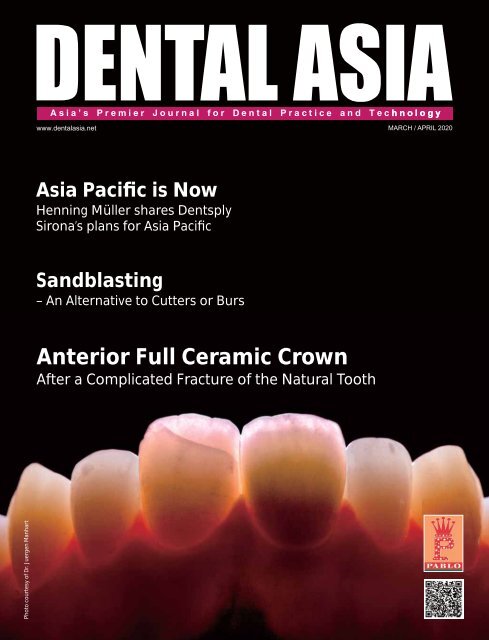
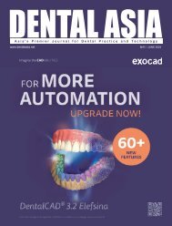
<strong>Asia</strong> Pacific is Now<br />
Henning Müller shares Dentsply<br />
Sirona’s plans for <strong>Asia</strong> Pacific<br />
Sandblasting<br />
– An Alternative to Cutters or Burs<br />
Anterior Full Ceramic Crown<br />
After a Complicated Fracture of the Natural Tooth<br />
Photo courtesy of Dr. Juergen Manhart
CONTENTS<br />
MARCH / APRIL <strong>2020</strong><br />
<strong>Dental</strong> Management<br />
20<br />
An Online Journey to Healthcare and<br />
Wholeness<br />
<strong>Dental</strong> Profile<br />
26<br />
30<br />
Clinical Feature<br />
34<br />
44<br />
48<br />
52<br />
Anterior Full Ceramic Crown After a<br />
Complicated Fracture of the Natural Tooth<br />
User Report<br />
54<br />
<strong>Asia</strong> Pacific is Now<br />
Smarter, Faster, and More Accurate Digital<br />
Diagnostics<br />
Amlodipine Induced Gingival Enlargement<br />
Biomimicry Simplified using a Novel, Huebased<br />
Direct Composite Resin System<br />
Considering Alternative Options for Teeth<br />
Lightening<br />
Onlay for Upper Right Molar Using Chairside<br />
Workflow with 3Shape TRIOS Design Studio<br />
In Depth with<br />
63<br />
64<br />
65<br />
Show Preview<br />
71<br />
Regulars<br />
6<br />
8<br />
66<br />
Efficient Aesthetics with Direct Filling<br />
Treatments<br />
Harnessing the Power of Sound<br />
Diagnostic Excellence with Humanised<br />
Technology<br />
Experience the Latest <strong>Dental</strong> Innovations<br />
at Sino-<strong>Dental</strong> in Beijing<br />
First Words<br />
<strong>Dental</strong> Updates<br />
Product Highlights<br />
Behind the scenes<br />
58<br />
61<br />
Sandblasting – An Alternative to Cutters<br />
or Burs<br />
Going Digital – From Holding a Model to<br />
Holding a Mouse<br />
74<br />
75<br />
76<br />
Giving Back to Society<br />
Events Calendar<br />
Advertiser’s Index
First Words<br />
Taking the rough with the smooth<br />
Pang Yanrong<br />
Senior Editor<br />
With COVID-19<br />
rapidly travelling<br />
around the globe, the<br />
WHO (World Health<br />
Organization) has<br />
now considered the<br />
virus a pandemic (as<br />
of this writing).<br />
The effects felt has been unprecedented —<br />
with countries on lockdown, cruise ships<br />
quarantining their passengers and crew<br />
members, and events being postponed. For<br />
instance, exocad Insights <strong>2020</strong> has moved<br />
from mid-<strong>March</strong> to the second half of the<br />
year. IDEM (International <strong>Dental</strong> Exhibition<br />
and Meeting), which was planned for <strong>April</strong>,<br />
has been postponed to June.<br />
But as the world grapples with the changes<br />
COVID-19 brings, there are still the movers<br />
and shakers in the industry who are making<br />
the best of the situation. Take for instance,<br />
Henning Müller, group vice president of <strong>Asia</strong><br />
Pacific RCO at Dentsply Sirona, who sees <strong>Asia</strong><br />
Pacific as the future for growth (p.26).<br />
“Many markets across <strong>Asia</strong> are becoming<br />
increasingly important, not only as<br />
emerging markets, but also as trendsetters<br />
as they continue to mature. To tap into<br />
the growing potential of the region,<br />
and connect better with local dental<br />
professionals, Dentsply Sirona opened a<br />
showroom in Singapore’s Post Centre, as<br />
well as a state-of-the-art training centre<br />
and showroom in Hong Kong in 2018,”<br />
he revealed.<br />
Prof. Dr. Juergen Manhart and CDT Hubert<br />
Schenk continue to bring great smiles to<br />
people despite the situation. They shared<br />
how they had to do an anterior full crown<br />
after a complicated fracture of a natural<br />
tooth (p.34).<br />
Dr. Douglas G Watt also shared his<br />
experience with a female patient who<br />
had came in requesting for orthodontic<br />
treatment and restoration of her dentition,<br />
which he succeeded in doing so with<br />
3Shape TRIOS Design Studio (p.54).<br />
“The use of 3Shape TRIOS scanner and<br />
TRIOS Design Studio software as part of the<br />
continuous and seamless workflow, along<br />
with the direct integration with DGShape,<br />
is an easy and predictable option for inhouse<br />
manufacture, and same day crown<br />
workflow. The update to allow use of TRIOS<br />
Patient Specific Motion made this case even<br />
more predictable as the patient’s actual<br />
excursive and protrusive movements can<br />
be utilised rather than a virtual articulator,”<br />
he said.<br />
And then there’s Guido Testa who came up<br />
with the idea of using sandblasting as an<br />
alternative to cutters or burs (p.58), and<br />
Dr. Kevin Ng who shares about the three<br />
dental instruments that have been useful<br />
for him at his practice (p.24).<br />
As we continue our fight with COVID-19,<br />
I hope everyone stays safe during this<br />
period, and please continue to often wash<br />
your hands with soap and water so as to<br />
reduce the spread of the virus and other<br />
infectious diseases!<br />
FOLLOW US<br />
@dentalasia<br />
ADVISORY BOARD<br />
Dr William Cheung<br />
Dr Choo Teck Chuan<br />
Dr Chung Kong Mun<br />
Dr George Freedman<br />
Dr Fay Goldstep<br />
Dr Clarence Tam<br />
Prof Nigel M. King<br />
Dr Anand Narvekar<br />
Dr Kevin Ng<br />
Dr William O’Reilly<br />
Dr Wong Li Beng<br />
Dr Adrian U J Yap<br />
Dr Christopher Ho<br />
Dr How Kim Chuan<br />
Dr Derek Mahony<br />
Prof Alex Mersel
<strong>Dental</strong> Updates<br />
Align Technology Acquires exocad<br />
Align Technology will be acquiring dental<br />
CAD/CAM software platform exocad GmbH<br />
for approximately €376 million, bringing<br />
exocad’s expertise in restorative dentistry,<br />
implantology, guided surgery, and smile<br />
design to the Align technology portfolio,<br />
extending Align’s Invisalign and iTero digital<br />
solutions.<br />
“Dentistry today is evolving digitally,<br />
with technology advances and consumer<br />
awareness driving new opportunities in orthorestorative<br />
and comprehensive treatment,”<br />
said Joe Hogan, Align Technology’s<br />
president and CEO. exocad will broaden and<br />
deepen Align’s digital platform by addressing<br />
restorative needs.<br />
“I am excited by the enormous growth<br />
opportunity for exocad and its customers<br />
that comes with being a part of Align<br />
Technology,” said Tillmann Steinbrecher, CEO<br />
Joe Hogan<br />
and co-founder of exocad. “Together, we will<br />
further strengthen exocad’s position as a key<br />
technology provider for the dental CAD/CAM<br />
industry and drive continuous innovation with<br />
the open and integrated approach that is the<br />
foundation of our company.”<br />
exocad will continue to operate as it exists<br />
today, and co-founders Tillmann Steinbrecher<br />
and Maik Gerth, along with their team, are<br />
expected to remain after the transaction<br />
closes.<br />
“The acquisition of exocad adds a talented<br />
and passionate team as well as a highly<br />
innovative, industry-leading product suite<br />
to our portfolio,” said John Morici, Align<br />
Technology’s senior vice president and CFO.<br />
“We will continue to invest in and build on<br />
exocad’s leadership in the dental CAD/CAM<br />
market and look to them to make significant<br />
contributions to Align’s overall strategy.” ■<br />
Tillmann Steinbrecher<br />
exocad Insights moves dates to 21 - 22 September, <strong>2020</strong><br />
Due to the spread of the coronavirus<br />
worldwide and increasing travel restrictions,<br />
exocad GmbH has decided to postpone the<br />
Insights <strong>2020</strong> event, originally planned for<br />
mid-<strong>March</strong>, to 21 st – 22 nd September. The<br />
venue remains unchanged at darmstadtium<br />
in Darmstadt, Germany. The decision is also<br />
supported by the 43 cooperation partners.<br />
“The safety and health of our participants,<br />
partners and employees is our top priority,”<br />
explained Novica Savic, CCO of exocad.<br />
Insights <strong>2020</strong> focuses on the almost limitless<br />
possibilities of exocad’s open software<br />
platform, on which intraoral and model<br />
scanners, milling machines, 3D printers and<br />
DVT devices from different manufacturers<br />
can be combined to form a consistent digital<br />
workflow. ■<br />
An evening with dinner and live music will round off the event in the<br />
festive presence of the approximately 850 international participants<br />
The two-day event, which is aimed at both dental technicians and dentists, offers a varied<br />
programme consisting of software education, partner sessions and international guest<br />
speakers<br />
8 DENTAL ASIA MARCH / APRIL <strong>2020</strong>
<strong>Dental</strong> Updates<br />
Modern and Intuitive: Double Triple for W&H Design<br />
When man meets machine, user-friendly operation plays a crucial<br />
role. For Austrian company W&H, this is precisely where the focus<br />
lies when designing medical technology products. W&H’s designs<br />
have succeeded in winning over not one but two international juries,<br />
with three products winning prizes at the Good Design Award 2019<br />
in the USA and the German Design Award <strong>2020</strong>.<br />
They fit perfectly in the hand, are intuitive in their operation, provide<br />
optimal freedom of movement and also look great: these are the<br />
main reasons why W&H products — Proxeo Ultra, Proxeo Twist<br />
Cordless and Assistina Twin — were awarded two internationally<br />
renowned Design Awards.<br />
Proxeo Ultra,<br />
Proxeo Twist<br />
Cordless, and<br />
Assistina Twin<br />
Head of W&H product design team, Kerstin Molzbichler, said, “When<br />
developing our products, our focus is firmly on the customer and<br />
their needs. Ultimately, our products are the interface between man<br />
and machine, between dentist and patient.<br />
Maria Protiwa-Kinzl, Nils Deringer, Kerstin Molzbichler, Kerstin Oberhofer, Leon<br />
Koopman, and Doris Rausch<br />
“Innovation is reflected in cost-effective solutions that customers<br />
can also relate to on an emotional level and appreciate for their<br />
function and economy. We are delighted that three products have<br />
been awarded two renowned prizes.” ■<br />
The TeleDentists Lead Educational Awareness Campaign on Dry Mouth<br />
Virtual dental service The TeleDentists<br />
is providing education to help increase<br />
the identification, diagnosis, and<br />
treatment of dry mouth, also known as<br />
xerostomia, beginning with the city of<br />
New York. The condition is a common<br />
oral condition among the elderly<br />
together with periodontal disease and<br />
dental caries, and can cause serious<br />
complications in the oral cavity.<br />
Dr. Maria Kunstadter<br />
To combat this silent epidemic, The<br />
TeleDentists has partnered with calcium phosphate rinse maker<br />
SalivaMAX to provide teledentistry and relief to xerostomia and other<br />
oral health conditions.<br />
“As dentists, we see the damaging effects of dry mouth every day. If<br />
you’re not aware that you have the condition, and don’t see a dentist<br />
before the damage sets in, you may end up needing extensive dental<br />
work,” said The TeleDentists Co-founder Dr. Maria Kunstadter. After<br />
a virtual consultation, a solution, such as a prescription rinse, can<br />
be found.<br />
A rinse for<br />
dry mouth<br />
The connected care platform does not take the place of the local<br />
dentist, but facilitates emergency care and coordinates the followup<br />
trip to the dentist. It can be especially helpful for those who lack<br />
access to a dentist. Teledentistry has also been tested as a means of<br />
pushing oral health video visits into underserved areas like low-income<br />
neighbourhoods and community health centres. ■<br />
10 DENTAL ASIA MARCH / APRIL <strong>2020</strong>
<strong>Dental</strong> Updates<br />
Formlabs Partners with BEGO for 3D Printed Crowns and Bridges<br />
<strong>Dental</strong> customers of 3D printing system<br />
manufacturer, Formlabs, will soon be able<br />
to 3D print crowns and bridges for patients<br />
with BEGO’s best-in-class dental materials<br />
in a new partnership. The completely digital<br />
dental workflows will enable a rapid, low-cost,<br />
and iterative process for better patient care<br />
and case acceptance.<br />
“Directly printing temporary crowns and<br />
bridges are one of the most sought-after<br />
applications from Formlabs customers. By<br />
partnering with BEGO and leveraging their<br />
130 years of dental experience, we will be<br />
able to not only address this need, but take<br />
it a step further by offering materials for<br />
permanent crowns. We are excited to see<br />
how this partnership can continue to advance<br />
the dental industry and overcome the major<br />
challenges labs and dentists face as digital<br />
dentistry becomes a standard<br />
for patient care,” said Dávid<br />
Lakatos, chief product officer<br />
at Formlabs.<br />
chief executive officer of BEGO <strong>Dental</strong>, Axel<br />
Klarmeyer, said, “We could not be happier<br />
to partner with Formlabs, especially at this<br />
time, where digital dentistry is reaching a<br />
breakthrough. It took some time and a lot<br />
of effort and commitment of all involved<br />
people to be able to offer to the market a<br />
fully validated workflow for final restorations.<br />
This partnership underlines BEGO’s leading<br />
position in the dental 3D printing materials<br />
market.” ■<br />
Dávid Lakatos and Axel Klarmeyer<br />
3D printed crowns<br />
3D-Printers for Labs of All Sizes<br />
Printers from German prototyping company<br />
Rapid Shape allow lab owners to make an<br />
impact in the dental prosthetic industry. Small<br />
labs can start 3D printing with professional<br />
equipment on a small budget, while dentists<br />
can print locally even without specialist<br />
knowledge. Besides its D20+ printer, which is<br />
the most economic professional 3D printing<br />
solution for dental labs, the company also<br />
manufactures D30+ and D40II, which are fast,<br />
flexible, and semi-automated, and D30+ ortho,<br />
which prints up to six models in 20 minutes.<br />
Rapid Shape has an open material system with a growing material library,<br />
and compatibility with common CAD programmes<br />
Rapid Shape products communicate with each other, simplifying complex processes<br />
A machine that works 24/7<br />
The key to printing large quantities efficiently<br />
is automation. For example, the D90+, which<br />
is meant for industrial dental production, has<br />
an automation system for up to eight selfloading<br />
platforms. The ready built job is taken<br />
out directly and the next print starts without<br />
any delay. This means that the machine is<br />
able to work without breaks, and even in<br />
night shifts. Another labour-saving feature<br />
can be seen in the automated part separation<br />
module (ASM, patent pending) for the D100+<br />
ortho. Multiple print jobs are collected in an<br />
integrated catch tank, eliminating the need<br />
for a human operator. It is also fast, and able<br />
to print up to 24 precise ortho models in 25<br />
minutes. ■<br />
12 DENTAL ASIA MARCH / APRIL <strong>2020</strong>
<strong>Dental</strong> Updates<br />
Carestream <strong>Dental</strong> Introduces Digital<br />
Dentistry Difference Campaign<br />
With an educational campaign, digital dental solutions provider,<br />
Carestream <strong>Dental</strong>, will educate consumers about the benefits<br />
of digital dentistry, and arm doctors with resources to prompt<br />
conversation with their patients, solving a significant awareness<br />
gap.<br />
According to surveys, patients across the globe believe that the<br />
oral healthcare industry lags behind both optical and orthopaedic<br />
in terms of the technology used. Even more surprising, less than<br />
half of patients even believe their doctor uses very advanced<br />
technology.<br />
The campaign will explain how 3D CBCT imaging<br />
allows a more complete and detailed picture<br />
“It is a missed opportunity when you consider that doctors<br />
are investing in digital dentistry to improve patient care, yet<br />
many patients do not understand the level of advanced digital<br />
technology the industry offers, or how it can change their lives,”<br />
said Ed Shellard, D.M.D., chief dental officer of Carestream <strong>Dental</strong>.<br />
“This campaign will change that by speaking directly to consumers<br />
about why they should be asking about the technology used by<br />
their doctor.”<br />
This education is likely to help increase patient retention, bring in<br />
new patients, and enhance patient perception of care. The tools<br />
available include a video and infographics which can be shared<br />
through social channels, in a doctor’s waiting room, or referenced<br />
during patient consultations. ■<br />
The campaign will explain how a digital scan is<br />
more comfortable and quicker
<strong>Dental</strong> Updates<br />
Gain Practical Knowledge and Improve Treatment Success<br />
Dr. Peter Wöhrle, who will be a speaker<br />
and moderator at the upcoming Nobel<br />
Biocare Global Symposium <strong>2020</strong> in Las<br />
Vegas, US, gave insights into what implant<br />
professionals can expect at this world-class<br />
event.<br />
He said, “What I always find exciting about<br />
Nobel Biocare symposia are the first<br />
revelations of innovative new solutions that<br />
have real potential to improve the way we<br />
can treat patients, and subsequently make<br />
my practice stand out from the rest. This<br />
year is no exception.”<br />
In particular, he is heartened that the panel<br />
of speakers will include the next generation<br />
of experts, who have already excelled in<br />
implant dentistry and are bringing a new<br />
approach and perspective to the field.<br />
“On a personal level, it’s extremely<br />
rewarding to network with other dental<br />
Dr. Peter Wöhrle<br />
professionals. I learn so much from the<br />
experiences my colleagues are having,<br />
we compare the challenges we have<br />
to deal with, share different opinions<br />
about this exciting and rapidly evolving<br />
market. It can be challenging for a lot<br />
of practitioners and technicians to find<br />
the time to interact with their peers, but<br />
don’t underestimate how much you can<br />
learn from networking and socialising. I find<br />
this such a valuable part of the symposia<br />
Delivering predictable aesthetic outcomes,<br />
combined with shorter time-to-teeth, is<br />
paramount in today’s market place<br />
At the Global Symposium, you can gain<br />
practical knowledge to improve treatment<br />
success and skill sets with a mix of hands-on<br />
sessions, discussions and presentations from<br />
many experts<br />
experience,” he said.<br />
The programme will feature topics of interest<br />
such as hard and soft tissue regeneration,<br />
treatment planning, managing complications,<br />
extreme cases, ceramic implants, and digital<br />
dentistry. These solutions are based upon<br />
scientific principles, which are presented in a<br />
logical and convincing way. ■<br />
The panel of speakers is comprised of a<br />
diverse group of experts from different<br />
specialties, both from clinical practice and<br />
scientific research, many of whom have been<br />
world-renowned for many years<br />
The question of how dental implant professionals around the world can advance their skills and practice<br />
implant dentistry at the highest level is answered at the symposium<br />
14 DENTAL ASIA MARCH / APRIL <strong>2020</strong>
<strong>Dental</strong> Updates<br />
A Place for Every Endodontic Material<br />
Specially developed to bring order to the materials used<br />
for endodontic treatment, VDW.FLO Endo Organisers from<br />
endodontic solutions provider VDW keeps all necessary materials<br />
tidy and readily at hand during treatment. The organiser has<br />
compartments that are precisely matched to the packaging of<br />
relevant materials, and fits standard drawer and trolley sizes.<br />
Loose instruments, irrigation, obturation, and post-endodontic<br />
materials can be organised on the multifunctional insert for a<br />
clear overview of inventory. It can also be adapted to individual<br />
workflows with a set of blank and pre-printed sticker labels. Made<br />
of easy-to-clean and robust plastic, the organiser is equipped<br />
with a non-slip silicone underside to facilitate compliance with<br />
hygiene measures in practice.<br />
Integrated solutions for endodontics<br />
For over 150 years, VDW has represented experience in the<br />
development and manufacture of products for endodontics.<br />
Today, VDW offers a holistic range of solution covering the<br />
entire endodontic treatment spectrum, from obturation to postendodontic<br />
care. Its product range includes instruments for<br />
root canal preparation, an endodontic ultrasonic device, fillers<br />
and sealers for root canals, and quartz fibre posts. The company<br />
also runs application-related advanced training programmes in<br />
endodontics. ■
<strong>Dental</strong> Updates<br />
Rebranding as a Premium <strong>Dental</strong> Tourism Destination<br />
The rise of India, Malaysia and Thailand<br />
as medical tourist destinations comes at<br />
the expense of Singapore, which is rapidly<br />
losing market share to its neighbours<br />
which are able to offer cheaper healthcare<br />
and compelling savings to tourists.<br />
In particular, many tourists are drawn by<br />
lower costs of dental work, which are done<br />
by local dentists who had returned from<br />
overseas studies. Some<br />
medical groups have<br />
already set up offices<br />
in regions such as<br />
Vietnam and China,<br />
to take advantage of<br />
the demand. Others<br />
have diversified<br />
their foreign<br />
patients<br />
base to lesstraditional<br />
markets<br />
such as Myanmar, Cambodia,<br />
Bangladesh, and India.<br />
According to Dr. Melvin Look<br />
of Pan<strong>Asia</strong> Surgery Group,<br />
to stay ahead, private<br />
groups here must continue<br />
to reinvent themselves and<br />
reinvest, and start looking<br />
into exporting Singapore<br />
medicine overseas, rather<br />
than wait for the patients<br />
to come.<br />
Others, such as Dr. Kelvin Loh of IHH<br />
Healthcare, believe that as healthcare<br />
standards in neighbouring countries move<br />
up the healthcare value chain, Singapore<br />
will stand out with its proven track record<br />
of delivering superior clinical outcomes.<br />
“Singapore can stay ahead of the curve by<br />
continuing to enhance its service offerings,<br />
such as investing in state-of-the-art<br />
Dr. Kelvin Loh<br />
facilities, disruptive medical technologies,<br />
and a strong team of doctors and healthcare<br />
professionals.”<br />
In the days to come, medical tourism to<br />
Singapore will be supported by regional<br />
growth trends, with the demand for quality<br />
private healthcare rising as populations<br />
age. ■<br />
Dr. Melvin Look<br />
Relief Efforts for the Australian Bushfires<br />
In response to the devastation caused by<br />
the Australian bushfires, American dental<br />
supplier Henry Schein is raising money for<br />
recovery and rebuilding efforts, donating<br />
much-needed health care supplies, and<br />
supporting its dental customers who may<br />
be impacted by the ongoing crisis.<br />
The Disaster Relief Fund will seed<br />
a US$50,000 donation and match<br />
employee contributions up to<br />
US$25,000. In addition, Henry Schein<br />
plans to donate up to US$50,000 worth of<br />
health care product to relief organisations.<br />
Locally, Henry Schein Australia is also<br />
donating a portion of its January sales to<br />
local relief efforts.<br />
“Team Schein stands ready to assist our<br />
relief agency partners and local health<br />
care providers in their efforts to rebuild<br />
and recover from these terrible fires,” said<br />
Stanley M. Bergman, chairman of the board<br />
and CEO of Henry Schein. “Our company<br />
has long been committed to supporting<br />
disaster preparedness and recovery, and<br />
we are working with our supplier partners<br />
and Team Schein Members to provide<br />
relief agencies with the resources they<br />
need to support public health.”<br />
The fund is not limited to Team Schein<br />
Members. Credit card donations can<br />
be made on the Henry Schein Cares<br />
Foundation website, and cheques can<br />
be made payable to “Henry Schein Cares<br />
Foundation” and mailed to Kate Sorrillo,<br />
Henry Schein Cares Foundation, Inc., 135<br />
Duryea Road, Melville, NY 11747. ■<br />
16 DENTAL ASIA MARCH / APRIL <strong>2020</strong>
<strong>Dental</strong> Updates<br />
Arizona Mission of Mercy <strong>Dental</strong> Mission<br />
Dr. Jennifer Wynn (third from left) and her incredible<br />
team prepped and seated 17 crowns in under six<br />
hours using Dentsply Sirona’s CEREC technology<br />
and extractions to endodontics and<br />
restorations. Many patients waited through<br />
the night to ensure they were among the<br />
first to receive treatment. Approximately<br />
200 dentists and 1,300 assistants,<br />
hygienists and dental students volunteered<br />
at the event.<br />
During the 2019 Arizona Mission of Mercy,<br />
Dentsply Sirona provided free dental<br />
restoration treatment to hundreds of<br />
Arizonans in need. In partnership with<br />
Patterson <strong>Dental</strong>, Dentsply Sirona’s CEREC<br />
team set up five treatment stations for the<br />
recent event, helping volunteer healthcare<br />
professionals to perform more than 125 free<br />
restorations in just two days.<br />
About 2,000 patients received dental<br />
treatments ranging from cleanings, fillings<br />
General manager of Patterson <strong>Dental</strong>, Jay<br />
Connors, said, “We partner with Dentsply<br />
Sirona each year to bring in two to three<br />
panoramic x-ray units that the service<br />
team sets up and uses for imaging. Having<br />
five full CEREC stations allowed our<br />
volunteer CEREC dentists to complete more<br />
restorations.<br />
“The dental care our volunteers provide<br />
not only solves patients’ dental issues, but<br />
more importantly, helps them to regain<br />
Around 1,500 volunteers came together to provide<br />
free dental care for 2,000 patients during the 2019<br />
Arizona Mission of Mercy<br />
their confidence and empowers them to<br />
smile again. When you are not in pain or<br />
embarrassed to smile, life is better. We are<br />
grateful to support this wonderful event<br />
where people’s lives are literally changed<br />
for the better.” ■<br />
MARCH / APRIL 2019 DENTAL ASIA<br />
17
<strong>Dental</strong> Updates<br />
Prescription of Opioids and Antibiotics for Emergency <strong>Dental</strong> Conditions<br />
A study in the <strong>March</strong> issue of The Journal of the American <strong>Dental</strong><br />
Association (ADA) from the Centres for Disease Control and<br />
Prevention found that antibiotics and opioids are frequently<br />
prescribed during emergency department visits for dental<br />
conditions, further emphasising the need for continued efforts to<br />
combat both opioid abuse and overuse of antibiotics.<br />
The authors found that more than 50% of patients who visited<br />
the emergency department for a dental-related condition filled<br />
a prescription for antibiotics and approximately 40% filled a<br />
prescription for opioids.<br />
Acetaminophen<br />
A growing body of research supports ADA policy that dentists<br />
should consider prescribing non-steroidal<br />
anti-inflammatory drugs (NSAIDs) alone or<br />
In 2019, the ADA released a<br />
new guideline indicating that, in<br />
most cases, antibiotics are not<br />
recommended for toothaches, which<br />
are a common dental-related reason<br />
to visit an emergency department.<br />
in combination with acetaminophen over<br />
opioids as first-line therapy for acute pain<br />
management. In <strong>March</strong> 2018, the organisation<br />
adopted policy that indicates a combination of<br />
ibuprofen and acetaminophen can be just as<br />
effective as opioids for acute pain.<br />
The guideline was developed by a<br />
multidisciplinary panel, including<br />
an emergency medicine physician<br />
nominated by the American College<br />
of Emergency Physicians.<br />
American College of Emergency Physicians<br />
The ADA will continue to work together with<br />
physicians, pharmacies, policymakers and the<br />
public to address these issues that are critical<br />
to public health. ■<br />
Preventing and Healing Tooth Decay with a Bioactive Peptide<br />
Cavities, or dental caries, are the most widespread noncommunicable<br />
disease globally, according to the World Health reduce demineralisation, or the dissolving of tooth enamel, while<br />
by the plaque-forming bacteria that cause cavities, and 2)<br />
Organisation. Having a cavity drilled and filled at the dentist’s increasing remineralisation, or repair.<br />
office can be painful, but untreated caries could lead to worse pain,<br />
tooth loss, infection, and even illness or death. Now, researchers The researchers based their anti-cavity coating on a natural<br />
in ACS Applied Materials and Interfaces report a bioactive peptide antimicrobial peptide called H5. Produced by human salivary<br />
that coats tooth surfaces, helping prevent new cavities and heal glands, H5 can adsorb onto tooth enamel and destroy a broad<br />
existing ones in lab experiments.<br />
range of bacteria and fungi. To promote remineralisation, the team<br />
added a phosphoserine group to one end of H5, which they thought<br />
Conventional treatment for dental cavities involves removing could help attract more calcium ions to repair the enamel than<br />
decayed tissue and filling the hole with<br />
natural H5. They tested the modified peptide<br />
materials, such as amalgam or composite<br />
on slices of human molars. Compared with<br />
resin. However, this procedure can damage<br />
natural H5, the new peptide adsorbed more<br />
healthy tissue and cause severe discomfort<br />
strongly to the tooth surface, killed more<br />
for patients. Hai Ming Wong, Quan Li Li and<br />
bacteria and inhibited their adhesion, and<br />
colleagues want to develop a two-pronged<br />
protected teeth from demineralisation. After<br />
strategy to prevent and treat tooth decay:<br />
brushing, people could someday apply the<br />
1) prevent colonisation of the tooth surface<br />
modified peptide to their teeth as a varnish or<br />
gel to protect against tooth decay. ■<br />
H5 antimicrobial peptide<br />
18 DENTAL ASIA MARCH / APRIL <strong>2020</strong>
<strong>Dental</strong> Updates<br />
Infection and Prevention Control<br />
Recommendations for Healthcare<br />
Professionals in the Current Coronavirus<br />
Situation<br />
Based on currently available information about COVID-19,<br />
guidance has been provided by the Centers for Disease Control<br />
and Prevention. These include minimising chance for exposures,<br />
adherence to standard and transmission-based precautions,<br />
patient placement, taking precautions when performing aerosolgenerating<br />
procedures, collection of diagnostic respiratory<br />
specimens, and management of visitor access and movement<br />
within the facility.<br />
Personal protective equipment<br />
There is an increased emphasis on early identification and<br />
implementation of source control, meaning putting a face mask<br />
on patients presenting with symptoms of respiratory infection.<br />
Updated recommendations include reserving airborne infection<br />
isolation rooms for patients undergoing aerosol-generation<br />
procedures, while patients with known or suspected COVID-19<br />
should be cared for in a single-person room with the door closed.<br />
Based on local and regional situational analysis of personal<br />
protective equipment (PPE) supplies, facemasks are an acceptable<br />
alternative when the supply chain of respirators cannot meet the<br />
demand. Available respirators should be prioritised for procedures<br />
that are likely to generate respiratory aerosols, which would<br />
pose the highest exposure risk to healthcare personnel. As major<br />
distributors in the United States have reported shortages of PPE,<br />
specifically N95 respirators, alternatives such as elastomeric halfmask<br />
and full facepiece air purifying respirators, and powered air<br />
purifying respirators should be considered.<br />
Eye protection, gown, and gloves continue to be recommended. ■
An Online<br />
Journey to Healthcare and Wellness<br />
DoctorxDentist started off as<br />
a simple blog in 2016 where<br />
doctors and dentists could<br />
contribute educational articles<br />
and answer health-related<br />
enquires. Today, the platform<br />
is supported by 300 doctors<br />
from 33 different specialties.<br />
Tristan Hahner, Co-founder of<br />
DoctorxDentist, shares his vision<br />
for the platform and beyond.<br />
An alternate window<br />
Healthcare is very personal and private<br />
to each individual. Marrying this into the<br />
Internet space provides people a convenient<br />
alternative to better understand their health<br />
conditions and even exchange treatment<br />
experiences.<br />
The majority of these online sources specific<br />
to healthcare are written to be general to be<br />
relatable to the masses. While the availability<br />
of such articles are useful in many ways,<br />
they can easily allow people to assume the<br />
worst too, bringing forth cyberchondria, a<br />
term coined for an Internet-induced anxiety<br />
from self-diagnosing health problems<br />
online, said Tristan Hahner, Co-founder of<br />
DoctorxDentist (DxD).<br />
He explained, “In the past, for example,<br />
people considering a wisdom tooth<br />
extraction surgery would proceed with<br />
treatment along with a sense of assurance,<br />
after hearing from the surgeon about<br />
their prognosis. Today, however, the mere<br />
mention of a possible nerve damage during<br />
surgery could spook patients into changing<br />
their minds and even opting out of surgery.“<br />
Hahner stressed that patients ought to be<br />
thoroughly informed of all the risks and<br />
dangers of treatments before making a<br />
decision. In addition to the insights they<br />
are able to gain through researching their<br />
medical health online, he highlighted the<br />
key is to cross-check the online information<br />
with trained physicians and attend physical<br />
consultations as these opinions help to put<br />
things into perspective for patients, on a<br />
case-by-case basis.<br />
Turning to the Internet for diagnosis<br />
As early as 2002, about 93 million Internet<br />
users in the US are able to access health<br />
information from online sources, according<br />
to a study released by the Pew Internet &<br />
American Life Project. This statistic has<br />
definitely grown exponentially, Hahner<br />
said, as the world becomes more digitally<br />
connected and reliant on the web.<br />
He further pointed out that the platform has<br />
seen an upward trend in web traffic as more<br />
users in Singapore are researching their<br />
health problems online, and elaborated, “I<br />
notice an increased level of sophistication<br />
in users’ questions – a sign that users<br />
have aggregated knowledge about a given<br />
subject. One example is a recent question<br />
we have received on the dental effects<br />
of hydrogen peroxide in teeth whiteners<br />
where the user is clearly aware of potential<br />
negative side effects of hydrogen peroxide.”<br />
20 DENTAL ASIA MARCH / APRIL <strong>2020</strong>
<strong>Dental</strong> Management<br />
Explaining the reasons for users to seek<br />
health advice online, Hahner credited<br />
the option to be anonymous, especially<br />
when identity protection is key to users.<br />
And on costs of treatments, which is a<br />
popular concern among DxD users, the<br />
platform features articles with detailed<br />
cost breakdowns and are well-received.<br />
Particularly for the Singapore market, he<br />
pointed out that Singaporeans are open<br />
to receiving multiple opinions from various<br />
doctors, and are “very meticulous” and<br />
like to be well-informed and fully prepared<br />
before making a decision.<br />
The lack of information is a dangerous thing<br />
Information in the world of the Internet<br />
travel fast, and at times, unverified or even<br />
fake. Hahner acknowledged the danger of<br />
using information that is not verified by<br />
accredited and licensed entities, and said<br />
that “casual” health information found<br />
online may have been overly simplified,<br />
misinterpreted, taken out of context, and<br />
even rife with conflicts of interests at times.<br />
He explained, “Many content writers of such<br />
unaccredited sources have no background in<br />
medicine and rely on second- or even thirdhand<br />
sources. For example, teeth whitening<br />
products have been declared harmful by<br />
some online sources when dentists have<br />
clarified that the levels are often too low to<br />
damage otherwise healthy teeth. Relying on<br />
and trusting unverified health information<br />
have the potential to cause public health<br />
scares, monger irrational fear and even<br />
complicate proper treatment.”<br />
This brings forth an opportunity for DxD as<br />
a platform that brings health professionals<br />
together to share their knowledge and<br />
expertise. Having an online voice on a<br />
content platform multiplies the ability of<br />
doctors and dentists to help and effect<br />
change, from helping individuals to helping<br />
populations all around the world.<br />
“As with doctors, I stress that patients<br />
should always consult doctors to clarify<br />
any medical doubts and queries. Reading<br />
up health content is a great way to expand<br />
our health knowledge and stay informed<br />
of the latest news in the medical sphere.<br />
Nevertheless, always check with a doctor,<br />
who can then put such information into<br />
context and present patients with completed<br />
information,” Hahner said.<br />
“Health is very personal and individual<br />
– what applies to one may not apply to<br />
another; only a doctor, equipped with the<br />
necessary tests, observations, knowledge<br />
and experience can safely and reliably<br />
prescribe the best course of action.”<br />
More than an information provider<br />
DxD envisions a world where everyone –<br />
regardless of language or location – is<br />
empowered to make better informed health<br />
decisions. As the only website in Singapore<br />
certified by the Health On the Net Foundation<br />
(HON) for trustworthy medical information,<br />
DxD takes its responsibilities as information<br />
provider seriously.<br />
Other than supplying the public with<br />
accurate health information, DxD also<br />
aims to support medical practitioners in<br />
Singapore. “We help them to be recognised<br />
for the excellent work they do, and we keep<br />
their practises alive, even in difficult times<br />
like this, where patients are afraid to go<br />
to hospitals because of the coronavirus<br />
outbreak. Through the DxD platform,<br />
Singapore doctors are able to connect<br />
to the public in a safe and productive<br />
environment,” said Hahner.<br />
DoctorxDentist is now available also<br />
in Indonesia, where there are just 0.2<br />
physicians per 1,000 people according to<br />
the World Health Organisation. Launching<br />
a local team and website will bring expert<br />
health content to Indonesians, while growing<br />
Indonesian patient referrals to Singaporean<br />
doctor partners. Indonesian patients bring<br />
revenues of $100 million each year to<br />
Singaporean doctors, but many medical<br />
practises in Singapore lack an effective<br />
marketing channel to reach them.<br />
“However, I would also highlight that<br />
engaging in DxD’s services does not<br />
constitute as nor take the place of a<br />
medical consultation, and are, therefore,<br />
not considered telemedicine. It is our<br />
longstanding belief that it is best for patients<br />
to visit a doctor for their medical issues.” DA<br />
22 DENTAL ASIA MARCH / APRIL <strong>2020</strong>
Word of Mouth<br />
WORD OF MOUTH<br />
By Dr. Kevin Ng<br />
Dr. Kevin Ng’s post-graduate qualifications include Master of <strong>Dental</strong> Surgery (Sydney), Fellowship of the Royal Australasian College of<br />
<strong>Dental</strong> Surgeons (FRACDS), Diploma in Implant Dentistry at the Royal College of Surgeons of England, and Membership of the Faculty of<br />
<strong>Dental</strong> Surgery (MFDS) at the Royal College of Surgeons of Edinburgh. He has been part-time lecturing on oral and maxillofacial surgery<br />
and cariology at Hong Kong University since 1981. Here, he shares the three dental instruments that have been most useful to<br />
him at a private practice in Hong Kong.<br />
CS 9600 CBCT Scanner from Carestream<br />
The high resolution of the CS 9600 CBCT system provides crisp images with exact<br />
anatomy landmarks that help tremendously in assessment and planning for all my dental<br />
surgeries. Its ability to capture images at low doses meets my safety considerations<br />
for patients.<br />
As all my 2D images, 3D images, and 3D face scans are assessable from the accompanying<br />
software, CS Imaging version 8, communicating with patients has become easier. Patients<br />
trust us more when we stay up-to-date with the latest technological advancements.<br />
CAD/CAM data assessable from the same software also makes liaising with technicians<br />
more convenient than before, when it comes to making surgical stents and temporary<br />
restorations for implant surgeries.<br />
iTero Element 5D from Align Technology<br />
Besides saving costs on traditional impression materials, I have found that digital<br />
impressions produced by intraoral scanner are more accurate and precise.<br />
I’m also able to detect inter-proximal caries early. Instead of sending physical<br />
impressions to the laboratory, I can now send the digital impressions via email,<br />
which is a big time-saver.<br />
When I can explain treatment outcomes using the scans, patients are able to<br />
understand me better. It also makes diagnosis and treatment planning better<br />
and more efficient, which will ultimately translate into the clinic’s bottom line.<br />
Waterlase iPlus all-tissue laser from Biolase<br />
I use the minimally invasive dental laser system primarily in<br />
periodontal treatments, both on natural teeth and those affected<br />
by implantitis. Compared to traditional tools such as needles,<br />
drills and scalpels, the laser is relatively pain-free for patients. Its<br />
ability to remove soft tissue with no bleeding and allow enamel<br />
etching without the use of acid are also plus points.<br />
I also prefer using this in the debridement and irrigation of<br />
root canals in endodontic treatments compared to traditional<br />
chemomechanical methods. Finally, it comes with a good record<br />
system that helps me monitor treatment progress and saves<br />
me time.<br />
24<br />
DENTAL ASIA MARCH / APRIL <strong>2020</strong>
<strong>Dental</strong> Profile<br />
<strong>Asia</strong><br />
Pacifi c<br />
is<br />
Now<br />
Henning Müller<br />
Group Vice President <strong>Asia</strong> Pacific RCO<br />
Dentsply Sirona<br />
Since joining Sirona <strong>Dental</strong><br />
Systems in 2008, Henning<br />
Müller had taken on a variety<br />
of roles, such as vice president<br />
of Sales for China, Hong Kong,<br />
Korea, and ASEAN. He then moved on to the<br />
implants business as vice president of Sales<br />
for Japan, Australia, New Zealand, India, as<br />
well as several markets in southern Europe,<br />
before becoming group vice president of<br />
<strong>Asia</strong> Pacific RCO at Dentsply Sirona in July<br />
2017.<br />
professionals, Dentsply Sirona opens<br />
showrooms and training centers all over<br />
<strong>Asia</strong> Pacific, e.g. in Singapore’s Post Centre,<br />
as well as a state-of-the-art training centre<br />
and showroom in Hong Kong in 2018.<br />
Our most recent showroom and academy<br />
was opened in Jakarta, Indonesia, in June<br />
2019. <strong>Dental</strong> professionals have responded<br />
positively to our new facilities and the live<br />
training sessions, demonstrating a keen<br />
desire to invest in their careers. In addition<br />
to our own training centers we are happy to<br />
collaborate with strong, independent<br />
partners such as International CEREC<br />
Institute (ICI) in Daejeon, South Korea; and<br />
the CEREC <strong>Asia</strong> Training Centre in Taipei,<br />
Taiwan to provide education courses.<br />
What are Dentsply Sirona’s plans for<br />
<strong>Asia</strong> Pacific in the next three to five<br />
years?<br />
We see tremendous opportunity in the<br />
region and are focusing to increase our<br />
presence across the region more and<br />
more, e.g. in Indonesia and Vietnam.<br />
Our employees have spent a lot of<br />
time listening to our customers, and<br />
engaging with dental professionals<br />
as part of our research process and<br />
product development. Digital is the<br />
future of dentistry, and technological<br />
innovation is the key to providing value<br />
for our customers. From a customer<br />
service perspective, we are implementing<br />
more digital tools to create a better<br />
patient experience, and developing more<br />
e-commerce solutions.<br />
How does Dentsply Sirona differentiate<br />
its products in the market?<br />
One of the special things that differentiates<br />
us is our ability to integrate products from<br />
different areas, thus creating a workflow that<br />
helps to improve the quality of treatment<br />
and its clinical results. For example, our<br />
Sharing with <strong>Dental</strong> <strong>Asia</strong>, he talks about<br />
digital plans for <strong>Asia</strong> Pacific, continuing<br />
education for dental professionals and<br />
Dentsply Sirona’s employees, as well as<br />
equipping the next generation of dentists.<br />
Opening of Dentsply Sirona’s showroom<br />
and training centre in Hong Kong<br />
What is the company’s overall growth<br />
and expansion strategy in the region?<br />
<strong>Asia</strong> Pacific is now. Many markets across<br />
<strong>Asia</strong> are becoming increasingly important,<br />
not only as emerging markets, but also as<br />
trendsetters as they continue to mature.<br />
To tap into the growing potential of the<br />
region and connect better with local dental<br />
26 DENTAL ASIA MARCH / APRIL <strong>2020</strong>
<strong>Dental</strong> Profile<br />
integrated solutions in implantology take<br />
clinicians from data capturing, restorative<br />
and implant planning, guided surgery and<br />
intraoral scanning to implant selection,<br />
positioning, restoration design and<br />
manufacturing, to restorative solution.<br />
Our customers do not have to worry about<br />
interfaces or compatibility at any point in<br />
time – it simply works.<br />
When customers invest in Dentsply Sirona<br />
products, they are assured that their<br />
investment is for the long term. A great<br />
example would be our Orthophos imaging<br />
devices. A practice might be just getting<br />
started with digital dentistry, so we offer<br />
the flexibility of a hybrid 2D/3D unit that<br />
can be ordered with 2D capability, and<br />
to be upgraded to a 3D unit when the<br />
practitioner is ready.<br />
Another example is CEREC, which integrates<br />
scanning, design, milling, grinding and<br />
sintering through CEREC Primescan,<br />
CEREC Software, CEREC Primemill, and<br />
CEREC SpeedFire*. This allows users to<br />
produce impressive restorations with<br />
precise margins in a thoroughly thoughtout<br />
workflow, from a single source that<br />
is only available from Dentsply Sirona.<br />
These CAD/CAM solutions open up a world<br />
of possibilities for customers in digital<br />
dentistry. For example, the time required to<br />
fabricate a zirconia crown can be shortened<br />
to as little as five minutes.<br />
Investigator Initiated Studies. Currently,<br />
more than 170 such research projects<br />
are in progress, involving more than 300<br />
investigators in over 20 countries.<br />
While a product is going through approval<br />
and regulatory processes, key<br />
specialists are trained extensively<br />
on the product before it is brought to the<br />
market. We offer a mentorship programme<br />
for first-time users, who learn from<br />
experienced fellow practitioners.<br />
How do you encourage your team to<br />
remain motivated and enthusiastic?<br />
We value our employees and know<br />
that a company can never rest on its<br />
laurels regardless of success. We have a<br />
responsibility to create opportunities for<br />
our employees to develop professionally<br />
throughout their careers, at all levels.<br />
Therefore, we’ve invested in our own virtual<br />
training hub known as DS University, where<br />
employees can participate in a variety of<br />
courses, which we keep expanding year<br />
after year.<br />
In addition, we discuss the mission and<br />
vision of the company in depth with our<br />
employees. Recently, CEO Don Casey and<br />
the executive team engaged with more<br />
than 13,000 employees worldwide to<br />
discuss the company culture and our<br />
The renewed CEREC system - CEREC Speedfire,<br />
CEREC Primemill and CEREC Primescan<br />
purpose to make people smile in a 24-hour<br />
livestream event. From a TV studio created<br />
in the middle of our production facility in<br />
Bensheim, Germany, the leadership team<br />
connected with 90 global sites. Knowing<br />
that we are all working towards worthwhile<br />
goals is a powerful motivation.<br />
How has your team adapted in<br />
responding to emerging needs?<br />
More than a year ago, our team moved to a<br />
new office in Hong Kong, which is the hub<br />
for Dentsply Sirona in <strong>Asia</strong>. The organisation<br />
has undergone a restructuring, which gave<br />
us tremendous opportunities for growth,<br />
How does Dentsply Sirona show<br />
customers that its continuing education<br />
courses are backed by the latest<br />
science?<br />
Besides the ease of use and speed, dental<br />
professionals are also concerned with the<br />
steep learning curves, and the science<br />
behind products. Dentsply Sirona is<br />
dedicated to science, and runs a variety of<br />
research activities in close collaboration<br />
with universities, dental professionals<br />
and opinion leaders. An important part<br />
of our clinical research programme is the<br />
*Note: Availability depends on registration timelines.<br />
MARCH / APRIL <strong>2020</strong> DENTAL ASIA<br />
27
<strong>Dental</strong> Profile<br />
Intego treatment centre in Dentsply Sirona’s<br />
Singapore Showroom and Training Centre<br />
and led to a greater focus on the customer<br />
within our workforce. We are now better able<br />
to customise our offering and this enables<br />
the talent of the team to shine through.<br />
Our employees are our greatest assets when<br />
it comes to responding to emerging needs.<br />
They care deeply about the customers and<br />
have a sense of purpose for the work they<br />
are doing. Working as a solutions provider in<br />
healthcare gives us the privilege of helping<br />
dental professionals give the best possible<br />
care to their patients. As a team, our size<br />
and depth allow us to focus on developing<br />
specific expertise for emerging needs.<br />
the high level of customisation required,<br />
Dentsply Sirona’s Special Clinic Solutions<br />
team put together a complete solution<br />
from the ground up and supplied all of<br />
the equipment needed. Since clinicians<br />
understand best the needs of their patients,<br />
our Special Clinic Solutions team works<br />
closely with them to build the set-up of their<br />
dreams.<br />
What else can we expect from Dentsply<br />
Sirona’s <strong>Asia</strong> Pacific team in the days to<br />
come?<br />
I’d like to share one piece of exciting news.<br />
<strong>2020</strong> marks the first year of our very<br />
own APAC Dentsply Sirona World edition,<br />
taking place in Taiwan. We will partner<br />
with CEREC <strong>Asia</strong>, the leading digital dental<br />
training institute in <strong>Asia</strong>, and integrate<br />
their annual congress into one single<br />
event at the Hilton Hotel in Taipei City from<br />
11 th – 13 th September, <strong>2020</strong>. The format<br />
will be similar to other Dentsply Sirona<br />
World events, with world-class speakers,<br />
breakout sessions, and entertainment.<br />
Simultaneous translation will be provided<br />
in both English and Chinese throughout<br />
the event. To find out more about Dentsply<br />
Sirona World Taiwan, visit the website: www.<br />
dentsplysirona.com/dsworldtaiwan. DA<br />
How does Dentsply Sirona keep a finger<br />
on the pulse of the evolving needs of<br />
clinicians and their patients?<br />
We draw great insights about the next<br />
generation of dental professionals due to<br />
our cooperation with academics and dental<br />
schools, and the projects that we work on<br />
with universities. For example, we helped<br />
equip a university hospital and academic<br />
facility, Shanghai No. 9 People’s Hospital,<br />
with 60 simulation stations.<br />
Intraoral scanner from<br />
Dentsply Sirona, Primescan<br />
Another example is our project with the<br />
University of Otago in New Zealand, where<br />
we built their new facility entirely around<br />
the patient experience. To accommodate<br />
28 DENTAL ASIA MARCH / APRIL <strong>2020</strong>
<strong>Dental</strong> Profile<br />
“Both dentist and patient are looking for quality and predicable treatment in a shorter time frame,” said Shane Gibson<br />
Smarter, Faster,<br />
and More Accurate<br />
Digital Diagnostics<br />
Carestream <strong>Dental</strong>, Inc., formerly part of Eastman<br />
Kodak Company, became an independent corporation<br />
in 2007. Today, the organisation is well known for its<br />
digital dental imaging solutions, such as scanners,<br />
cameras, and cone-beam computed tomography.<br />
Managing director for Carestream <strong>Dental</strong> ASEAN, Shane Gibson,<br />
shares about the organisation’s mission and role advancing<br />
diagnostic technologies in the dental scene.<br />
Please share with us briefly about your background. As<br />
head of South East <strong>Asia</strong> of Carestream <strong>Dental</strong>, what is your<br />
involvement in product development, marketing strategies<br />
and dental education?<br />
I first joined the company as general manager of the dental<br />
business at Carestream Health in Australia in 2013. My role is to<br />
lead the company in its mission to transform dentistry, simplify<br />
technology and change lives.<br />
30 DENTAL ASIA MARCH / APRIL <strong>2020</strong>
<strong>Dental</strong> Profile<br />
At the core of what we do at Carestream <strong>Dental</strong> is our customers<br />
– our products are designed by clinicians, for clinicians. We work<br />
closely with the dental practitioners throughout <strong>Asia</strong> to understand<br />
their needs and workflows, and guide how products are developed.<br />
Their feedback also steers the educational services that we provide,<br />
to help them achieve better diagnostic and clinical outcomes for<br />
their patients.<br />
In what ways has Carestream <strong>Dental</strong>’s work transformed<br />
from what was offered in the pre-digital era?<br />
Together with the pioneers of our company – Eastman Kodak,<br />
Trophy Radiologie, and PracticeWorks – we have brought many<br />
new technologies to dental practitioners, such as modern film,<br />
digital radiography sensors, and orthodontic practice management<br />
software. We are committed to innovation, investing heavily in<br />
research and development to stay ahead of the curve, deliver bestin-class<br />
solutions, and expand into new areas.<br />
Thirdly, scalability. The needs and requirements of each practice<br />
can vary significantly. CS 9600 can be configured with the optimal<br />
fields of view and scanning resolutions for a clinician’s current<br />
clinical and financial requirements. As these needs change, the CS<br />
9600 Fields of View can be expanded up to 16x17cm and modules<br />
such as Face Scanning, Cephalometric imaging, Prosthetic Driven<br />
Implant Planning (PDIP), CS Airway, CS Model, and CS Model+ can<br />
be integrated with CS 9600 to enhance diagnoses and treatment<br />
planning capabilities.<br />
Another product Carestream <strong>Dental</strong> is known for is its<br />
intraoral scanner CS 3700. What has the feedback been like<br />
from dentists who have tried it?<br />
The response since we launched the CS 3700 last year has<br />
been positive. It combines high performance, ergonomics and<br />
intelligence all into one scanner, and comes with a 20% faster<br />
scanning speed than before.<br />
Additionally, we are at the forefront of CBCT technology, panoramic<br />
and cephalometric imaging, and intraoral sensors. In fact, more<br />
than 200,000,000 dental images are captured each year with<br />
Carestream <strong>Dental</strong> imaging. We also work on integrating products<br />
seamlessly into practices’ workflows, and enhance interactions<br />
with patients.<br />
Carestream <strong>Dental</strong>’s CS 9600 5-in-1 CBCT Imaging System<br />
was awarded the 2019 Cellerant Best of Class Technology<br />
Winner. What makes it stand out from others?<br />
Firstly, innovation and precision. Digital dentistry is constantly<br />
changing, and we want to enable dental professionals with<br />
technology that allows them to innovate and lead within their<br />
particular fields. The CS 9600 is our fifth generation CBCT imaging<br />
platform. It features intelligent automation, first-time right imaging<br />
technology, and highly simplified workflows. It is also customisable,<br />
and can be configured to suit any dental specialty.<br />
Secondly, speed and accuracy. The SmartPad feature on the CS<br />
9600 ensures scans can be taken both quickly and precisely.<br />
Instead of using traditional laser beams, the CS 9600 uses a live<br />
camera positioning system to align patients. These cameras are<br />
assisted with in-built AI technology, which allows a swift and highly<br />
precise patient setup, while reducing the need for retakes.<br />
Users have also noticed the completely new interface design on<br />
the software that comes with the intraoral scanner, CS ScanFlow.<br />
It consists of a four-step process: Scan, Check, Export, and Adapt.<br />
From a single scan, the interface gives clinicians the freedom to<br />
navigate among different workflows. <strong>Dental</strong> professionals can<br />
enjoy faster dataset processing, vibrant HD 3D digital impressions,<br />
and all of the other advanced features they enjoyed with our past<br />
scanning acquisition software updates.<br />
How important is continuing education for dental<br />
professionals?<br />
In the age of digital dentistry, technology changes fast, and we<br />
need to keep our customers informed. Carestream <strong>Dental</strong> actively<br />
does this by supporting professional development and learning<br />
events throughout <strong>Asia</strong>. We also conduct webinar training events<br />
throughout the year. These webinars are presented by clinicians<br />
and product experts, covering the latest in digital dental technology<br />
and trends.<br />
Even the way dental professionals are learning has changed.<br />
We have adapted by introducing our online learning portal. The<br />
Carestream <strong>Dental</strong> Institute provides users with self-paced learning<br />
modules and guided videos for the latest about Carestream<br />
<strong>Dental</strong>’s technology.<br />
*Note: Availability depends on registration timelines.<br />
MARCH / APRIL <strong>2020</strong> DENTAL ASIA<br />
31
<strong>Dental</strong> Profile<br />
What opportunities do you see in the dental industry in <strong>Asia</strong>?<br />
Please share some trends you foresee.<br />
Each country’s market is unique. Carestream <strong>Dental</strong> adapts its<br />
portfolio to each market, based on the maturity of the industry.<br />
From a digital standpoint, I believe most countries are seeing<br />
strong growth in CBCT technologies as well as intraoral scanning.<br />
The convergence of rapidly evolving digital technologies has<br />
elevated the overall quality of dentistry across the profession. Both<br />
dentist and patient are looking for quality and predicable treatment<br />
in a shorter time frame.<br />
As a digital data solution provider with a legacy of more than<br />
30 years in digital, Carestream <strong>Dental</strong> will continue to focus on<br />
providing diagnostic excellence within our digital equipment and<br />
software portfolio. Our priority will be the development of complete<br />
solutions for dental professionals across various specialties, such<br />
as implant surgical guides and orthodontic treatment planning.<br />
What will be the main challenges for Carestream <strong>Dental</strong> in<br />
the next five years?<br />
With any fast-paced industry, it is always a challenge to stay at<br />
the forefront of technology while being mindful of what dental<br />
professionals are looking for to simplify their workflow. This is an<br />
exciting time for the ASEAN dental market, and we look forward<br />
to working with our customers to help them transform dentistry.<br />
Our passion is to support our customers and their practice, drive<br />
better clinical outcomes, and make life easier for practitioners and<br />
patients. DA<br />
Shane Gibson<br />
32 DENTAL ASIA MARCH / APRIL <strong>2020</strong>
Clinical Feature<br />
Anterior Full Ceramic Crown After a<br />
Complicated Fracture of the Natural Tooth<br />
By Prof. Dr. Juergen Manhart & CDT Hubert Schenk<br />
In the maxillary anterior region, the<br />
integrity of the teeth is of great<br />
importance to many people. In<br />
the case of damage, restoring an<br />
attractive smile is a great need for<br />
patients. Depending on the defect size and<br />
its configuration, various clinically proven<br />
direct and indirect therapeutic options are<br />
available. For heavily damaged anterior<br />
teeth, all-ceramic crowns are a reliable<br />
and proven therapy option for restoring<br />
function and aesthetics.<br />
Lithium-disilicate glass ceramic<br />
The integrity of their anterior teeth is of<br />
paramount importance for most patients due<br />
to their prominent position. The impairment<br />
of teeth in the anterior aesthetic zone by<br />
carious defects, chipping or fractures, clear<br />
visible fillings, discolourations, anomalies<br />
in shape, alignment and position within the<br />
dental arch often results in considerable<br />
restrictions for the patients. Therefore,<br />
dentists should take into account all aspects<br />
of treatment, including a team of different<br />
specialists, in order to preserve or restore<br />
the natural dentition.<br />
Today, the range of therapies in modern<br />
dentistry offers a variety of methods<br />
to restore or optimise the function and<br />
aesthetics of the teeth in the anterior<br />
region. These include – depending on<br />
the initial situation and the degree of<br />
destruction of the individual teeth –<br />
polychromatic multilayer direct composite<br />
restorations, laboratory-made or industrially<br />
manufactured composite veneers, ceramic<br />
veneers, partial veneers (additional veneers),<br />
veneer crowns, full crowns (metal ceramics,<br />
all-ceramics) and orthodontic measures 1-3 .<br />
A majority of today’s patients asks for<br />
aesthetic restorations and metal-free<br />
alternatives to traditional prosthodontic<br />
approaches. All-ceramic restorations have<br />
gained popularity during the last 30 years<br />
for a number of reasons; especially their<br />
favourable optical properties, excellent<br />
and durable aesthetic appearance, wear<br />
resistance, colour stability, chemical<br />
inertness and durability, biocompatibility,<br />
and strengthening of the remaining<br />
tooth structure when they are adhesively<br />
bonded 4-17 .<br />
In the last three decades, many different<br />
all-ceramic systems were introduced to the<br />
dental profession 19 . <strong>Dental</strong> ceramics can<br />
be classified according to their material<br />
composition, fabrication workflow (for<br />
example, powder-liquid slurry, slip-casting,<br />
pressable ceramics, CAD/CAM millable), or<br />
clinical indications 20-22 . Nowadays, the most<br />
common clinical indications for all-ceramic<br />
restorations consist of inlays, onlays, partial<br />
crowns, full crowns, bridges, veneers,<br />
posterior occlusal veneers (table tops/<br />
posterior cuspal protection restorations),<br />
implant abutments and implants 23-36 . These<br />
restorations present a scientifically proven,<br />
high-quality permanent treatment option<br />
for the aesthetically challenging anterior<br />
and load-bearing posterior regions when<br />
the indications and limitations of the<br />
respective ceramic systems are respected<br />
and an appropriate luting procedure<br />
is employed; their reliability has been<br />
documented in literature 18, 32, 37-56 . All-ceramic<br />
restorations are used meanwhile on a<br />
routine basis in everyday dentistry.<br />
For single-unit restorations, lithium-disilicate<br />
(LS2) glass ceramic is the material of choice for<br />
many dental practitioners because of its good<br />
mechanical strength (IPS e.max Press: 470<br />
MPa mean biaxial flexural strength), excellent<br />
aesthetic properties and its versatility. It can<br />
be used in monolithic form, when maximum<br />
strength is required (for example, tabletop<br />
restorations for increasing the vertical<br />
dimension of occlusion or posterior crowns),<br />
or in a layered form (pressed LS2 coping<br />
with additional veneering porcelain) when<br />
aesthetics is of utmost importance. Singleunit<br />
LS2-crowns demonstrate an excellent<br />
longevity for anterior 57-59 and posterior<br />
teeth 56-59 , comparable to the survival rate of<br />
metal-ceramic crowns 60, 61 .<br />
This clinical report illustrates the restoration<br />
of a maxillary central incisor affected by a<br />
complicated crown fracture with a veneered<br />
lithium-disilicate glass ceramic crown after<br />
endodontic therapy.<br />
Fig. 1a – b: Initial situation: 24-year-old female patient after trauma. In addition to the<br />
fractured tooth 11, there is extensive injury to the lower lip. The first treatment of the soft<br />
tissue injury occurred at the venue of the accident abroad<br />
34 DENTAL ASIA MARCH / APRIL <strong>2020</strong>
Clinical Feature<br />
Fig. 2a: One week later, the patient appeared in our<br />
dental office. Tooth 11 had a complicated crown<br />
fracture with exposure of the pulp<br />
Fig. 2b: The incisal half of the clinical crown of<br />
tooth 11 had fractured horizontally<br />
Fig. 3: Exposure of the pulp was diagnosed at the<br />
mesial aspect of the fracture site<br />
Clinical case report<br />
Initial situation<br />
A systemically healthy 24-year old female<br />
patient presented in our dental clinic with<br />
a trauma-related fractured right maxillary<br />
central incisor. The accident had already<br />
occurred one week earlier abroad, where the<br />
patient, a medical student, was in a clinical<br />
traineeship. After the initial treatment of<br />
soft tissue injuries on the spot by a fellow<br />
student (Fig. 1a and b), the patient decided<br />
to cancel the stay abroad, where medical<br />
and dental facilities were lacking.<br />
During the examination in our clinic<br />
one week after the incident, the patient<br />
presented a still untreated traumainjured<br />
tooth 11 (Fig. 2a – b). The clinical<br />
inspection showed a complicated crown<br />
fracture with exposure of the pulp (Fig. 3);<br />
the incisal half of the clinical crown had been<br />
completely lost 62, 63 . Patient assessment<br />
revealed a sharp painful response to cold<br />
thermal stimulus using refrigerant spray<br />
and a pathologic response to percussion of<br />
the respective tooth 64 . Given that the pulp<br />
had already been exposed to the oral cavity<br />
environment for one week, the tooth showed<br />
unprovoked pain symptoms and root growth<br />
was completed. Together with the informed<br />
patient, we decided to completely remove<br />
the infected pulp with subsequent root canal<br />
treatment (Fig. 4a – c).<br />
The patient was informed about various<br />
therapeutic approaches (direct composite<br />
restoration, ceramic veneer, full ceramic<br />
crown, PFM crown) including their<br />
respective advantages and disadvantages,<br />
and associated costs. The patient decided<br />
in favour of an adhesively luted glass<br />
ceramic crown made of veneered lithiumdisilicate<br />
ceramics. This restoration type<br />
can be recommended as evidence-based<br />
treatment in the anterior region 65 . In<br />
literature, survival rates of between 93.8%<br />
and 96.8% are reported at five-, eight-, or<br />
10-year observation periods 57-59 .<br />
Fig. 4a: Root canal treatment was initiated since<br />
the pulp had already been exposed to the oral<br />
cavity environment for one week<br />
Fig. 4b: Periapical<br />
radiograph to determine<br />
the working length<br />
Fig. 4c: Control radiograph<br />
of the root canal filling<br />
in lateral condensation<br />
technique<br />
After completion of the root canal<br />
treatment, a long-term provisional buildup<br />
of the tooth was carried out with an<br />
adhesive direct composite restoration<br />
(Fig. 5a – b) in order to spare the patient a<br />
preparation and impressions until the soft<br />
Fig. 5a: Long-term provisional build-up of the<br />
tooth with a direct composite restoration<br />
Fig. 5b: The composite restoration remained<br />
until completed soft tissue healing<br />
Fig. 6: After three months, the soft tissue<br />
situation presented in perfect condition<br />
MARCH / APRIL <strong>2020</strong> DENTAL ASIA<br />
35
Clinical Feature<br />
In principle, in the dental aesthetic analysis<br />
by the ceramist technician already a<br />
“virtual layering” of the restoration is<br />
done, with determination of the necessary<br />
ceramic masses. The result of this “virtual<br />
layering” is recorded in the ceramic buildup<br />
scheme (Fig. 8). This procedure is done on<br />
site in the dental laboratory under ideal<br />
lighting conditions (which are often not<br />
found in dental practices) by the ceramist<br />
technician, who will ultimately also fabricate<br />
the restorations.<br />
Fig. 7a – d: Aesthetic analysis by the dental technician. The distribution of the different shades and<br />
translucent or opaque tooth areas in the area to be restored are determined<br />
Fig. 8: Ceramic layering concept as result of<br />
dental aesthetic analysis<br />
tissue situation had completely healed.<br />
After a waiting period of three months, a<br />
new clinical examination was carried out,<br />
in which tooth 11 and its adjacent teeth,<br />
including the antagonists in the lower jaw,<br />
were inconspicuous (Fig. 6). The patient<br />
was asked to present herself the next day<br />
for shade determination and, in general,<br />
for dental aesthetic analysis in the dental<br />
laboratory 66 . A basic requirement for<br />
accurate colour determination is that the<br />
teeth are not dehydrated, otherwise they<br />
appear lighter and more opaque 67-69 .<br />
Aesthetic Analysis<br />
As part of the aesthetic analysis by the<br />
dental technician, the distribution of the<br />
different shades of colour and translucent<br />
or opaque tooth areas in the area to be<br />
restored is determined (Fig. 7a – d). The ageappropriate<br />
design of the restoration with<br />
corresponding individual characteristics -<br />
enamel cracks, white spots, mamelons, halo<br />
effect - as well as the appropriate surface<br />
texture, and the correct gloss level, are also<br />
analysed.<br />
If the colour analysis is performed directly<br />
by the dental technician, and not by the<br />
dentist or other practice staff, there is<br />
usually no misunderstanding and the<br />
responsibilities for this important aspect in<br />
the treatment process are clearly assigned.<br />
Such a procedure eliminates the risk of<br />
communication breakdowns and saves<br />
time. It is through direct contact with the<br />
patient that the dental technician is able to<br />
produce an aesthetically perfect matching<br />
ceramic crown 70 . It is optimal if this analysis<br />
is performed before treatment starts. This<br />
allows the dental technician to form his<br />
own impression of the initial situation,<br />
and also to query, unfiltered, the patient’s<br />
expectations of the new restorations. A<br />
patient-specific optimal tooth position<br />
and shape of the restorations is sought 71 .<br />
Implementation of the individual functional<br />
and aesthetic optimum for each patient<br />
thus requires close collaboration with<br />
the specialised dental technician from<br />
the beginning of the planning phase. The<br />
independent aesthetic analysis of the<br />
intraoral situation by the ceramist is a<br />
basic requirement for success 72 .<br />
Fig. 9a – b: Tooth 11 was prepared for a full ceramic crown with a circumferential<br />
shoulder with rounded inner edges<br />
Fig. 9c: Incisal view of the final preparation with a<br />
circumferential shoulder of 1 mm depth<br />
36 DENTAL ASIA MARCH / APRIL <strong>2020</strong>
Clinical Feature<br />
Tooth preparation<br />
In the next appointment, the tooth 11<br />
was prepared for receiving a full ceramic<br />
crown with a circumferential shoulder with<br />
rounded inner edges (Fig. 9a – c).<br />
The strength of all-ceramic restorations<br />
is determined by the type of ceramic used<br />
with the resulting inherent mechanical<br />
stability of the respective ceramic<br />
material. Furthermore, the fracture<br />
strength is determined by the geometry<br />
of the restoration and, thus, by the shape<br />
of the cavity or crown preparation. The<br />
basic principle of preparation design for<br />
all-ceramic restorations avoids tensile<br />
stresses in the material as much as<br />
possible and loads the restoration primarily<br />
in compression mode by an adequate<br />
preparation geometry 73, 74 . Fracture strength<br />
of the restorations is determined by size,<br />
volume, shape and surface characteristics<br />
of the ceramic material, and additionally<br />
by structural inhomogeneities introduced<br />
during the manufacturing process 75 .<br />
The dentist must be aware of the fact<br />
that the shape and finish of the tooth<br />
preparation have a major impact on the<br />
clinical success and longevity of all-ceramic<br />
restorations 76-78 . The preparation should<br />
exhibit a retention form and resistance<br />
form optimal for a ceramic crown 76, 79, 80 :<br />
• height of prepared tooth (abutment<br />
height) minimum 4mm<br />
• occlusal convergence angle between 6<br />
and 10 degrees<br />
• finish line: circumferential shoulder<br />
with rounded inner edges or obvious<br />
deep chamfer with 1mm width<br />
• incisal / occlusal reduction of 1.5 – 2.0mm<br />
(adhesively luted full contour lithiumdisilicate<br />
crown: minimum 1.0 mm)<br />
• axial reduction depth (sufficient circular<br />
crown thickness) of 1.2 – 1.5mm<br />
• in the anterior region: a rounded incisal<br />
edge<br />
• rounded internal line angles and point<br />
angles<br />
• smooth surface texture<br />
Tooth substance removal was controlled<br />
in all dimensions. Attention was paid to<br />
the possibility of a sufficient palatinal<br />
layer thickness of the crown framework to<br />
be produced, even in positions which the<br />
tooth occupies, in addition to the static<br />
occlusion, in dynamic occlusion (Fig. 10).<br />
After tooth preparation<br />
During the fabrication of highly aesthetic<br />
restorations, the influence of the shade of<br />
the prepared tooth on the final result is a<br />
decisive aspect. After finishing the crown<br />
preparation, the shade of the prepared<br />
tooth substance was documented by the<br />
dentist with a digital photo referencing<br />
to a special shade guide (IPS Natural Die<br />
Material shade guide, Vivadent, Schaan,<br />
Liechtenstein). The image file was then<br />
made available electronically to the dental<br />
laboratory. This enables the technician,<br />
who otherwise has no information about<br />
the colour of the tooth stump, to fabricate a<br />
model die similar in colour to the prepared<br />
tooth of the patient. This forms the basis<br />
from which the correct shade, translucency<br />
and brightness values of the all-ceramic<br />
restoration will be selected.<br />
In order to achieve a perfect impression, the<br />
marginal gingiva was displaced by insertion<br />
of a retraction cord to the sulcus (Fig. 11a –<br />
b). After taking impressions of the prepared<br />
tooth and the antagonistic dentition, an<br />
occlusion protocol with Shimstock foil was<br />
made, as well as intermaxillary registration<br />
by fabricating an interocclusal record in<br />
maximal intercuspal position and arbitrary<br />
transfer of the maxillary position using a<br />
facebow record were performed 81, 82 .<br />
A diagnostic template made from a gypsum<br />
duplicate of the analytical wax-up using<br />
a transparent polyethylene foil allows the<br />
chairside fabrication of a direct provisional<br />
restoration with correct dimensions and<br />
alignment (Fig. 12a). The provisional was<br />
seated using a eugenol-free temporary<br />
cement (Fig. 12b).<br />
Laboratory work<br />
In the dental laboratory, the ceramic<br />
crown for tooth 11 was produced. For<br />
this purpose, a crown framework made of<br />
lithium-disilicate reinforced high-strength<br />
glass ceramic was pressed corresponding<br />
to the anatomically correct shape of the<br />
respective tooth (Fig. 13a). This coping<br />
was subsequently finalised by individual<br />
veneering with layered porcelain (presslayer-technique)<br />
(Fig. 13b – g).<br />
Try-in and adhesive luting procedure<br />
One week after impressioning, the final<br />
appointment was scheduled. After the<br />
removal of the provisional restoration and<br />
cleaning of the tooth with a rotating brush<br />
and fluoride-free prophylaxis paste, the<br />
gingiva presented in healthy condition (Fig.<br />
Fig. 10: Tooth substance removal was controlled<br />
in dynamic occlusion to ensure sufficient<br />
thickness of the glass ceramic framework<br />
Fig. 11a – b: Placing a retraction cord to displace the marginal gingiva and expose the<br />
finish line before taking an impression<br />
MARCH / APRIL <strong>2020</strong> DENTAL ASIA<br />
37
Clinical Feature<br />
Fig. 12a: Chairside fabrication of a direct<br />
provisional restoration<br />
Fig. 12b: The provisional was seated using a<br />
eugenol-free temporary cement<br />
Fig. 13a: Pressed crown framework made of<br />
lithium-disilicate reinforced high-strength glass<br />
ceramic<br />
Fig. 13b: First bake of the veneering porcelain Fig. 13c: Second bake of the veneering porcelain Fig. 13d: Finalised full ceramic crown made of a<br />
pressed lithium-disilicate coping and individually<br />
layered veneering porcelain<br />
Fig. 13e: The ceramic crown exhibits a naturallooking,<br />
life-like surface texture<br />
Fig. 13f: The circular 1mm shoulder is clearly<br />
detectable<br />
Fig. 13g: Palatal surface of the ceramic crown<br />
Fig. 14: Situation after removal of the<br />
provisional restoration. The marginal gingiva<br />
presents in perfect condition<br />
14). Using coloured, glycerin-based tryin<br />
pastes, the aesthetics of the ceramic<br />
crown was checked intraorally with<br />
reference to hydrated adjacent teeth and<br />
the correct shade of the luting resin was<br />
determined 83-85 . Subsequently, the precise<br />
Fig. 15: Placing a retraction cord to expose the<br />
finish line prior to the adhesive luting procedure<br />
fit of the crown on the prepared tooth and<br />
quality of proximal contacts were checked<br />
before minor functional interferences<br />
during protrusive and laterotrusive<br />
movement paths were eliminated.<br />
The method of cementation of the crown to<br />
the prepared tooth was by adhesive luting,<br />
as opposed to conventional cementation.<br />
This gives a positive effect on the overall<br />
strength of the restoration, in particular for<br />
glass ceramic materials, which are more<br />
prone to bulk fracture and chipping effects<br />
than zirconia. Ceramic materials with<br />
fracture strength less than 350 MPa are not<br />
indicated for conventional cementation 36 .<br />
Among those are feldspathic porcelains<br />
and leucite-reinforced glass ceramics that<br />
have to be placed adhesively using bonding<br />
agents and luting resins. Due to the adhesive<br />
bond between the ceramic restoration and<br />
enamel or dentin, a considerable increase<br />
in strength can be obtained because the<br />
38 DENTAL ASIA MARCH / APRIL <strong>2020</strong>
Clinical Feature<br />
inner surface of the ceramic restoration<br />
no longer acts as a mechanical boundary<br />
line at which fracture-causing cracks can<br />
initiate due to tensile stresses 86 .<br />
The adhesive cementation of ceramic<br />
restorations is a very technique-sensitive<br />
procedure. In order to achieve a reliable and<br />
long-lasting bond to the tooth structures, it<br />
is of utmost importance to prepare this step<br />
carefully and to observe the cementation<br />
protocol 87 . Prior to the luting procedure, the<br />
marginal gingiva was displaced to expose<br />
the complete shoulder using a retraction<br />
cord (Fig. 15). Afterwards, the inner surfaces<br />
of the lithium-disilicate glass ceramic crown<br />
were etched with hydrofluoric acid for 20s<br />
(Fig. 16). After thoroughly rinsing and drying<br />
the crown, the fitting surfaces were silanised<br />
(Fig. 17) 88-91 . After adhesive pretreatment of<br />
the prepared tooth by conditioning enamel<br />
Fig. 16: Etching the inner surface of the<br />
lithium-disilicate glass ceramic crown with<br />
hydrofluoric acid for 20s<br />
Fig. 17: The intaglio surfaces of the glass ceramic<br />
crown are treated with a silane coupling agent<br />
Fig. 18: Etching the tooth surfaces with 37%<br />
phosphoric acid<br />
Fig. 19a: Application of an Etch-and-<br />
Rinse-adhesive<br />
Fig. 19b: Air-thinning the adhesive<br />
Fig. 20a: The ceramic crown is positioned on the<br />
tooth with a dual-curing luting composite<br />
Fig. 20b: The excess resin cement is carefully<br />
removed using a foam pellet<br />
Fig. 20c: Placing a glycerin gel on the luting gap<br />
to avoid the formation of an oxygen-inhibited<br />
superficial composite layer<br />
Fig. 20d: Light polymerisation of the<br />
dual-cure resin cement<br />
and dentin with 37% phosphoric acid (Fig.<br />
18) the tooth was thoroughly rinsed with<br />
water-spray and air-dried. An Etch-and-<br />
Rinse-adhesive was applied meticulously<br />
on all surfaces covered by the restoration<br />
(Fig. 19a). After the correct exposure time<br />
the bonding layer was air-thinned and the<br />
solvent was evapourated carefully with<br />
compressed air (Fig. 19b). The ceramic<br />
Fig. 21a: The ceramic restoration exhibits a<br />
perfect functional and aesthetic integration<br />
into the neighbouring teeth<br />
Fig. 21b: The incisal view shows a copy of the<br />
natural central incisor<br />
crown was adhesively luted by covering its<br />
MARCH / APRIL <strong>2020</strong> DENTAL ASIA<br />
39
Clinical Feature<br />
intaglio surfaces and the prepared tooth<br />
both with dual-curing resin cement in<br />
the previously determined shade and the<br />
restoration was positioned on the tooth with<br />
light finger pressure (Fig. 20a – d).<br />
Two weeks after placement, the restoration<br />
exhibited an optimal functional and aesthetic<br />
integration into the neighbouring teeth<br />
(Fig. 21a – b). Background illumination<br />
demonstrates the excellent light<br />
transmittance capacity of the glass<br />
ceramic crown, which impresses by having<br />
virtually the same optical properties<br />
as the surrounding natural dentition<br />
(Fig. 22). Ultraviolet light activates the<br />
inherent fluorescence properties of the<br />
restoration, which are equal to natural<br />
tooth structure (Fig. 23). Silicate ceramics<br />
exhibit translucency effects and lightoptical<br />
properties that are comparable<br />
to natural hard tooth substance, making<br />
them ideal for fabricating restorations<br />
that have to meet highest aesthetic<br />
demands. The light scattering of silicate<br />
ceramics also supports a natural vital<br />
appearance of the adjacent gingiva.<br />
The difference to this “pink aesthetics”<br />
becomes clear in comparison with metalsupported<br />
restorations, which block this<br />
light conduction towards the marginal soft<br />
tissue and often cause a greyish shading<br />
on the marginal gingiva 92, 93 . At the end<br />
of the successful treatment, the patient’s<br />
smile was no longer compromised. The<br />
crown harmonises in dialogue with the lips<br />
(Fig. 24).<br />
Five years after adhesive cementation of<br />
the ceramic crown, there is still an excellent<br />
integration in the surrounding teeth, both<br />
in habitual intercuspation and in dynamics<br />
(Fig. 25a – e). The patient was still fully<br />
satisfied with the result and thanked the<br />
treatment team with a happy smile (Fig. 26).<br />
Conclusion<br />
All-ceramic restorations have achieved<br />
an excellent quality and are an<br />
indispensable therapeutic means for<br />
modern conservative and prosthetic<br />
dental treatment procedures 94 . Aesthetics<br />
and biocompatibility characterise these<br />
restorations. At the same time, this<br />
type of restorations achieves excellent<br />
patient acceptance. Clinical trials exhibit<br />
an excellent longevity for all-ceramic<br />
restorations if a correct indication is<br />
selected and material- and patient-related<br />
limitations are observed 95, 96 . DA<br />
Fig. 22: Excellent light transmittance capacity of<br />
the ceramic crown, indistinguishable from the<br />
neighbouring dentition<br />
Fig. 23: Ultraviolet light activates the inherent<br />
fluorescence properties of the glass ceramic<br />
restoration, which equals natural tooth structures<br />
Fig. 24: The ceramic crown shows a perfect<br />
harmony with the architecture of the lips<br />
Fig. 25a – c: Five years after incorporation, there is still an excellent integration of the crown into neighbouring dentition<br />
Fig. 25d – e: In the right and left lateral views, the crown also presents inconspicuously<br />
Fig. 26: The<br />
patient was fully<br />
satisfied with<br />
the result and<br />
presented a warm<br />
and big smile<br />
as a reward for<br />
the completed<br />
treatment<br />
40 DENTAL ASIA MARCH / APRIL <strong>2020</strong>
Clinical Feature<br />
About the Authors<br />
Dr. Juergen Manhart is professor of the Department<br />
of Restorative Dentistry at the <strong>Dental</strong> School of LMU-<br />
University in Munich, Germany. He offers seminars<br />
and practical hands-on workshops in aesthetic<br />
restorative dentistry, such as direct composite<br />
restorations, all-ceramic restorations, ceramic<br />
veneers, management of severely worn dentition,<br />
and treatment planning in aesthetic dentistry.<br />
Reference<br />
1. Celik, C. and D. Gemalmaz, Comparison of<br />
marginal integrity of ceramic and composite<br />
veneer restorations luted with two different resin<br />
agents: an in vitro study. Int J Prosthodont, 2002.<br />
15(1): p. 59-64.<br />
2. Magne, P., Noninvasive bilaminar CAD/CAM<br />
composite resin veneers: a semi-(in)direct<br />
approach. Int J Esthet Dent, 2017. 12(2): p.<br />
134-154.<br />
3. Hajmasy, A. and H. Schorn, Maximaler<br />
Substanzerhalt bei maximaler Ästhetik. Quintessenz<br />
Zahntech, 2010. 36(3): p. 352-356.<br />
4. Edelhoff, D., Vollkeramische Restaurationen. wissen<br />
kompakt, 2015. 9(4): p. 149-160.<br />
5. Manhart, J., Vollkeramikrestaurationen in der<br />
restaurativen Zahnmedizin. Was ist machbar?<br />
wissen kompakt, 2007. 1(4): p. 3-14.<br />
6. Miyazaki, T., et al., A review of dental CAD/CAM:<br />
current status and future perspectives from 20<br />
years of experience. Dent Mater J, 2009. 28(1):<br />
p. 44-56.<br />
7. Höland, W., et al., Bioceramics and their application<br />
for dental restoration. Advances in Applied<br />
Ceramics: Structural, Functional and Bioceramics,<br />
2009. 108(6): p. 373-380.<br />
8. Holand, W., et al., Ceramics as biomaterials for<br />
dental restoration. Expert Rev Med Devices, 2008.<br />
5(6): p. 729-45.<br />
9. Denry, I. and J.A. Holloway, Ceramics for <strong>Dental</strong><br />
Applications: A Review. Materials, 2010. 2(1): p.<br />
351-368.<br />
10. Ritzberger, C., et al., Properties and Clinical<br />
Application of Three Types of <strong>Dental</strong> Glass-<br />
Ceramics and Ceramics for CAD-CAM Technologies.<br />
Materials, 2010. 3(6): p. 3700-3713.<br />
11. Kelly, J.R. and P. Benetti, Ceramic materials in<br />
dentistry: historical evolution and current practice.<br />
Aust Dent J, 2011. 56 Suppl 1: p. 84-96.<br />
12. McLean, J.W., Evolution of dental ceramics in the<br />
twentieth century. J Prosthet Dent, 2001. 85(1):<br />
p. 61-66.<br />
13. Kelly, J.R., I. Nishimura, and S.D. Campbell,<br />
Ceramics in dentistry: historical roots and current<br />
perspectives. J Prosthet Dent, 1996. 75(1): p.<br />
18-32.<br />
14. Conrad, H.J., W.J. Seong, and G.J. Pesun, Current<br />
15.<br />
16.<br />
17.<br />
18.<br />
19.<br />
20.<br />
21.<br />
22.<br />
23.<br />
24.<br />
25.<br />
26.<br />
Hubert Schenk, CDT<br />
ceramic materials and systems with clinical<br />
recommendations: A systematic review. Journal<br />
of Prosthetic Dentistry, 2007. 98(5): p. 389-404.<br />
Denry, I. and J.R. Kelly, Emerging ceramic-based<br />
materials for dentistry. J Dent Res, 2014. 93(12):<br />
p. 1235-42.<br />
Li, R.W., T.W. Chow, and J.P. Matinlinna, Ceramic<br />
dental biomaterials and CAD/CAM technology:<br />
state of the art. J Prosthodont Res, 2014. 58(4):<br />
p. 208-16.<br />
Cotert, H.S., B.H. Sen, and M. Balkan, In vitro<br />
comparison of cuspal fracture resistances of<br />
posterior teeth restored with various adhesive<br />
restorations. Int J Prosthodont, 2001. 14(4): p.<br />
374-378.<br />
Beier, U.S. and H. Dumfahrt, Langzeitbewährung<br />
silikatkeramischer Restaurationen für die<br />
Einzelzahnversorgung. Stomatologie, 2013.<br />
110(7): p. 21-25.<br />
Ahmed, S.N., T.E. Donovan, and E.J. Swift, Jr.,<br />
Evaluation of contemporary ceramic materials. J<br />
Esthet Restor Dent, 2015. 27(2): p. 59-62.<br />
McLaren, E.A. and J. Figueira, Updating<br />
Classifications of Ceramic <strong>Dental</strong> Materials: A<br />
Guide to Material Selection. Compend Contin Educ<br />
Dent, 2015. 36(6): p. 400-405.<br />
Helvey, G.A., Classifying dental ceramics: numerous<br />
materials and formulations available for indirect<br />
restorations. Compend Contin Educ Dent, 2014.<br />
35(1): p. 38-43.<br />
Lawson, N.C., <strong>Dental</strong> Ceramics: A Current Review.<br />
Compend Contin Educ Dent, 2014. 35(3): p.<br />
161-166.<br />
Magne, P., K. Stanley, and L.H. Schlichting,<br />
Modeling of ultrathin occlusal veneers. Dent Mater,<br />
2012. 28(7): p. 777-82.<br />
Sasse, M., et al., Influence of restoration thickness<br />
and dental bonding surface on the fracture<br />
resistance of full-coverage occlusal veneers made<br />
from lithium disilicate ceramic. Dent Mater, 2015.<br />
31(8): p. 907-15.<br />
McLaren, E.A., J. Figueira, and R.E. Goldstein,<br />
Vonlays: a conservative esthetic alternative to<br />
full-coverage crowns. Compend Contin Educ Dent,<br />
2015. 36(4): p. 282-289.<br />
Edelhoff, D., Okklusionsveränderung mit<br />
Kauflächen-Veneers: CAD/CAM-gefertigte Table<br />
Tops korrigieren die Bisslage. Zahnärztliche<br />
Mitteilungen, 2014. 104(8): p. 48-50.<br />
27. Magne, P., M.P. Paranhos, and L.H. Schlichting,<br />
Influence of material selection on the risk of inlay<br />
fracture during pre-cementation functional occlusal<br />
tapping. Dent Mater, 2011. 27(2): p. 109-13.<br />
28. Magne, P., et al., In vitro fatigue resistance of<br />
CAD/CAM composite resin and ceramic posterior<br />
occlusal veneers. J Prosthet Dent, 2010. 104(3):<br />
p. 149-57.<br />
29. Guess, P.C., et al., Monolithic CAD/CAM lithium<br />
disilicate versus veneered Y-TZP crowns:<br />
comparison of failure modes and reliability after<br />
fatigue. Int J Prosthodont, 2010. 23(5): p. 434-42.<br />
30. Clausen, J.O., M. Abou Tara, and M. Kern, Dynamic<br />
fatigue and fracture resistance of non-retentive allceramic<br />
full-coverage molar restorations. Influence<br />
.<br />
of ceramic material and preparation design. Dent<br />
Mater, 2010. 26(6): p. 533-8.<br />
31. Manhart, J., Keramikveneers. Erfolgreiche,<br />
minimalinvasive Frontzahnrestaurationen. BZB<br />
Bayerisches Zahnärzteblatt, 2015. 52(10): p. 52-59.<br />
32. Beier, U.S. and H. Dumfahrt, Longevity of silicate<br />
ceramic restorations. Quintessence Int, 2014.<br />
45(8): p. 637-44.<br />
33. Bachhav, V.C. and M.A. Aras, Zirconia-based fixed<br />
partial dentures: a clinical review. Quintessence<br />
Int, 2011. 42(2): p. 173-82.<br />
34. Spear, F. and J. Holloway, Which all-ceramic system<br />
is optimal for anterior esthetics? J Am Dent Assoc,<br />
2008. 139 Suppl: p. 19S-24S.<br />
35. Raigrodski, A.J., Contemporary materials and<br />
technologies for all-ceramic fixed partial dentures:<br />
a review of the literature. J Prosthet Dent, 2004.<br />
92(6): p. 557-562.<br />
36. Kern, M., et al., All-ceramics at a Glance (3rd<br />
English Edition). An introduction to the indications,<br />
material selection, preparation and insertion<br />
techniques for all-ceramic restorations. 2017,<br />
Ettlingen: AG für Keramik in der Zahnheilkunde e.V.<br />
37. Fabbri, G., et al., Clinical evaluation of 860 anterior<br />
and posterior lithium disilicate restorations:<br />
retrospective study with a mean follow-up of 3<br />
years and a maximum observational period of 6<br />
years. Int J Periodontics Restorative Dent, 2014.<br />
34(2): p. 165-77.<br />
38. Layton, D.M., M. Clarke, and T.R. Walton, A<br />
systematic review and meta-analysis of the survival<br />
of feldspathic porcelain veneers over 5 and 10<br />
years. Int J Prosthodont, 2012. 25(6): p. 590-603.<br />
39. Otto, T. and W.H. Mormann, Clinical performance of<br />
chairside CAD/CAM feldspathic ceramic posterior<br />
shoulder crowns and endocrowns up to 12 years.<br />
Int J Comput Dent, 2015. 18(2): p. 147-61.<br />
40. Manhart, J., et al., Review of the clinical survival of<br />
direct and indirect restorations in posterior teeth<br />
of the permanent dentition. Oper Dent, 2004.<br />
29(5): p. 481-508.<br />
41. Sulaiman, T.A., A.J. Delgado, and T.E. Donovan,<br />
Survival rate of lithium disilicate restorations at 4<br />
years: A retrospective study. J Prosthet Dent, 2015.<br />
114(3): p. 364-6.<br />
42. Zenthofer, A., et al., Performance of zirconia<br />
MARCH / APRIL <strong>2020</strong> DENTAL ASIA<br />
41
Clinical Feature<br />
43.<br />
44.<br />
45.<br />
46.<br />
47.<br />
48.<br />
49.<br />
50.<br />
51.<br />
52.<br />
53.<br />
54.<br />
55.<br />
56.<br />
57.<br />
58.<br />
59.<br />
ceramic cantilever fixed dental prostheses: 3-year<br />
results from a prospective, randomized, controlled<br />
pilot study. J Prosthet Dent, 2015. 114(1): p. 34-9.<br />
Guncu, M.B., et al., Zirconia-based crowns up to 5<br />
years in function: a retrospective clinical study and<br />
evaluation of prosthetic restorations and failures.<br />
Int J Prosthodont, 2015. 28(2): p. 152-7.<br />
Toman, M. and S. Toksavul, Clinical evaluation of<br />
121 lithium disilicate all-ceramic crowns up to 9<br />
years. Quintessence Int, 2015. 46(3): p. 189-97.<br />
Tartaglia, G.M., E. Sidoti, and C. Sforza, Sevenyear<br />
prospective clinical study on zirconia-based<br />
single crowns and fixed dental prostheses. Clin Oral<br />
Investig, 2015. 19(5): p. 1137-45.<br />
Guess, P.C., et al., Prospective clinical study of<br />
press-ceramic overlap and full veneer restorations:<br />
7-year results. Int J Prosthodont, 2014. 27(4):<br />
p. 355-8.<br />
Fasbinder, D.J., et al., A clinical evaluation of<br />
chairside lithium disilicate CAD/CAM crowns: a<br />
two-year report. J Am Dent Assoc, 2010. 141<br />
Suppl 2: p. 10S-14S.<br />
Otto, T. and S. de Nisco, Computer-aided direct<br />
ceramic restorations: a 10-year prospective clinical<br />
study of Cerec CAD/CAM inlays and onlays. Int.J<br />
Prosthodont., 2002. 15(2): p. 122-128.<br />
Stoll, R., et al., Survival of inlays and partial crowns<br />
made of IPS empress after a 10-year observation<br />
period and in relation to various treatment<br />
parameters. Oper Dent, 2007. 32(6): p. 556-63.<br />
Kramer, N. and R. Frankenberger, Clinical<br />
performance of bonded leucite-reinforced glass<br />
ceramic inlays and onlays after eight years. Dent<br />
Mater, 2005. 21(3): p. 262-71.<br />
Fradeani, M. and M. Redemagni, An 11-year clinical<br />
evaluation of leucite-reinforced glass-ceramic<br />
crowns: a retrospective study. Quintessence Int,<br />
2002. 33(7): p. 503-10.<br />
McLaren, E.A. and S.N. White, Survival of In-Ceram<br />
crowns in a private practice: a prospective clinical<br />
trial. Journal of Prosthetic Dentistry, 2000. 83:<br />
p. 216-222.<br />
Segal, B.S., Retrospective assessment of 546 allceramic<br />
anterior and posterior crowns in a general<br />
practice. J Prosthet Dent, 2001. 85(6): p. 544-50.<br />
Zitzmann, N.U., et al., Clinical evaluation of Procera<br />
AllCeram crowns in the anterior and posterior<br />
regions. Int J Prosthodont, 2007. 20(3): p. 239-41.<br />
Toksavul, S. and M. Toman, A short-term clinical<br />
evaluation of IPS Empress 2 crowns. Int J<br />
Prosthodont, 2007. 20(2): p. 168-72.<br />
Marquardt, P. and J.R. Strub, Survival rates of IPS<br />
empress 2 all-ceramic crowns and fixed partial<br />
dentures: results of a 5-year prospective clinical<br />
study. Quintessence Int, 2006. 37(4): p. 253-9.<br />
Gehrt, M., et al., Clinical results of lithium-disilicate<br />
crowns after up to 9 years of service. Clin Oral<br />
Investig, 2013. 17(1): p. 275-84.<br />
Valenti, M. and A. Valenti, Retrospective survival<br />
analysis of 261 lithium disilicate crowns in a<br />
private general practice. Quintessence Int, 2009.<br />
40(7): p. 573-9.<br />
Steeger, B., Survival analysis and clinical followup<br />
examination of all-ceramic single crowns. Int J<br />
Comput Dent, 2010. 13(2): p. 101-19.<br />
60. Walton, T.R., The up to 25-year survival and clinical<br />
performance of 2,340 high gold-based metalceramic<br />
single crowns. Int J Prosthodont, 2013.<br />
26(2): p. 151-60.<br />
61. Walton, T.R., A 10-year longitudinal study of<br />
fixed prosthodontics: clinical characteristics and<br />
outcome of single-unit metal-ceramic crowns. Int<br />
J Prosthodont, 1999. 12(6): p. 519-26.<br />
62. Nolte, D., et al., S2k-Leitlinie (Langversion):<br />
Therapie des dentalen Traumas bleibender Zähne.<br />
AWMF-Registernummer: 083-004. AWMF, 2015.<br />
63. Bakland, L.K. and J.O. Andreasen, <strong>Dental</strong><br />
traumatology: essential diagnosis and treatment<br />
planning. Endodontic Topics, 2004. 7: p. 14-34.<br />
64. Jafarzadeh, H. and P.V. Abbott, Review of pulp<br />
sensibility tests. Part I: general information and<br />
thermal tests. Int Endod J, 2010. 43(9): p. 738-62.<br />
65. Meyer, G., et al., S3-Leitlinie: Vollkeramische<br />
Kronen und Brücken. AWMF-Registernummer:<br />
083-012. AWMF, 2014.<br />
66. Manhart, J., Keramikveneers. Teil 1: Indikation und<br />
Behandlungsplanung. Quintessenz, 2011. 62(7):<br />
p. 869-883.<br />
67. Winter, R., Visualizing the natural dentition. J Esthet<br />
Dent, 1993. 5(3): p. 102-17.<br />
68. Schroeder, H.E., Pathobiologie oraler Strukturen.<br />
1991, Basel: Karger-Verlag.<br />
69. Hall, N.R. and M.C. Kafalias, Composite colour<br />
matching: the development and evaluation of<br />
a restorative colour matching system. Aust<br />
Prosthodont J, 1991. 5: p. 47-52.<br />
70. Gehringer, U., Die Quelle des Erfolgs. dental<br />
dialogue, 2014. 15(1): p. 32-35.<br />
71. Javaheri, D., Considerations for planning esthetic<br />
treatment with veneers involving no or minimal<br />
preparation. The Journal of the American <strong>Dental</strong><br />
Association, 2007. 138(3): p. 331-337.<br />
72. Rouse, J. and S. McGowan, Restoration of the<br />
anterior maxilla with ultraconservative veneers:<br />
clinical and laboratory considerations. Pract<br />
Periodontics Aesthet Dent, 1999. 11(3): p. 333-339.<br />
73. Arnetzl, G.V. and G. Arnetzl, Biomechanical<br />
examination of inlay geometries--is there a basic<br />
biomechanical principle? Int J Comput Dent, 2009.<br />
12(2): p. 119-30.<br />
74. Arnetzl, G.V. and G. Arnetzl, Design of preparations<br />
for all-ceramic inlay materials. Int J Comput Dent,<br />
2006. 9(4): p. 289-98.<br />
75. Breviary Technical Ceramics. 2009, Nuremberg:<br />
Fahner Verlag.<br />
76. Guth, J.F., et al., Computer-aided evaluation of<br />
preparations for CAD/CAM-fabricated all-ceramic<br />
crowns. Clin Oral Investig, 2013. 17(5): p. 1389-95.<br />
Goodacre, C.J., W.V. Campagni, and S.A. Aquilino,<br />
77. Tooth preparations for complete crowns: an art<br />
form based on scientific principles. J Prosthet Dent,<br />
2001. 85(4): p. 363-76.<br />
78. Castelnuovo, J., et al., Fracture load and mode of<br />
failure of ceramic veneers with different preparations.<br />
J Prosthet Dent, 2000. 83(2): p. 171-80.<br />
79. Blair, F.M., R.W. Wassell, and J.G. Steele, Crowns and<br />
other extra-coronal restorations (8): preparations<br />
for full veneer crowns. Br Dent J, 2002. 192(10):<br />
p. 561-4, 567-71.<br />
80. Al-Dwairi, Z.N., A.S. Al-Hiyasat, and H. Aboud,<br />
Standards of teeth preparations for anterior resin<br />
bonded all-ceramic crowns in private dental practice<br />
in Jordan. J Appl Oral Sci, 2011. 19(4): p. 370-7.<br />
81. Morneburg, T., et al., Wissenschaftliche Mitteilung<br />
der Deutschen Gesellschaft für Prothetische<br />
Zahnmedizin und Biomaterialien e. V. (DGPRo)<br />
(vormals DGZPW): Anwendung des Gesichtsbogens<br />
beim funktionsgesunden Patienten im Rahmen<br />
restaurativer Maßnahmen. Deutsche Zahnärztliche<br />
Zeitschrift, 2010. 65(11): p. 690-694.<br />
82. Steele, J.G., F.S. Nohl, and R.W. Wassell, Crowns<br />
and other extra-coronal restorations (5): occlusal<br />
considerations and articulator selection. Br Dent<br />
J, 2002. 192(7): p. 377-80, 383-7.<br />
83. Chadwick, R.G., J.F. McCabe, and T.E. Carrick,<br />
Rheological properties of veneer trial pastes<br />
relevant to clinical success. Br Dent J, 2008.<br />
204(6): p. E11.<br />
84. Xing, W., et al., Evaluation of the esthetic effect<br />
of resin cements and try-in pastes on ceromer<br />
veneers. J Dent, 2010. 38 Suppl 2: p. e87-e94.<br />
85. Sheets, C.G. and T. Taniguchi, Advantages and<br />
limitations in the use of porcelain veneer restorations.<br />
J Prosthet Dent, 1990. 64(4): p. 406-11.<br />
86. Mehl, A., et al., Stabilization effects of CAD/CAM<br />
ceramic restorations in extended MOD cavities. J<br />
Adhes Dent, 2004. 6(3): p. 239-45.<br />
87. Watanabe, M.U., Minimally invasive reconstruction<br />
of anterior teeth - A combination of 3D printing,<br />
press technique and adhesive bonding.<br />
International Dentistry - African Edition, 2019.<br />
9(1): p. 54-58.<br />
88. Brentel, A.S., et al., Microtensile bond strength of<br />
a resin cement to feldpathic ceramic after different<br />
etching and silanization regimens in dry and aged<br />
conditions. Dent Mater, 2007. 23(11): p. 1323-31.<br />
89. Matinlinna, J.P., Processing and bonding of dental<br />
ceramics, in Non-Metallic Biomaterials for Tooth<br />
Repair and Replacement., P. Vallittu, Editor. 2013,<br />
90. Woodhead Publishing Ltd.: Oxford. p. 129-160.<br />
Ho, G.W. and J.P. Matinlinna, Insights on Ceramics<br />
as <strong>Dental</strong> Materials. Part II: Chemical surface<br />
treatments. Silicon, 2011. 3(3): p. 117-123.<br />
91. Canay, S., N. Hersek, and A. Ertan, Effect of different<br />
acid treatments on a porcelain surface. J Oral<br />
Rehabil, 2001. 28(1): p. 95-101.<br />
92. Magne, P., M. Magne, and U. Belser, The esthetic<br />
width in fixed prosthodontics. J Prosthodont, 1999.<br />
8(2): p. 106-18.<br />
93. Magne, P., M. Magne, and I. Magne, Porcelain Jacket<br />
Crowns: Back to the Future Through Bonding. QDT<br />
Quintessence <strong>Dental</strong> Technology, 2010: p. 89-96.<br />
94. Santos, M.C., et al., Current All-Ceramic Systems<br />
in Dentistry: A Review. Compend Contin Educ Dent,<br />
2015. 36(1): p. 31-37.<br />
95. Friedman, M.J., A 15-year review of porcelain<br />
veneer failure - a clinician’s observations.<br />
Compendium of Continuing Education in Dentistry,<br />
1998. 19: p. 625-636.<br />
96. Swift, E.J., Jr. and M.J. Friedman, Critical appraisal.<br />
Porcelain veneer outcomes, part I. J Esthet Restor<br />
Dent, 2006. 18(1): p. 54-57.<br />
42 DENTAL ASIA MARCH / APRIL <strong>2020</strong>
Clinical Feature<br />
Amlodipine<br />
Induced Gingival<br />
Enlargement<br />
By Dr. Anand Narvekar and Dr. Richa Kapoor<br />
Patients medicated with certain<br />
drugs may suffer from Drug<br />
Induced Gingival Enlargement<br />
(DIGO), which may interfere<br />
with aesthetics, mastication, or<br />
speech. Besides affecting one’s appearance,<br />
such gingival overgrowth also often impairs<br />
nutrition and oral hygiene, resulting in an<br />
increased susceptibility to oral infection,<br />
caries and periodontal diseases.<br />
Drugs associated with gingival overgrowth<br />
can be broadly divided into three categories:<br />
Anticonvulsants, calcium channel blockers,<br />
and immunosuppressants. Amlodipine<br />
is a dihydropyridine calcium channel<br />
blocker that is used in the management<br />
of both hypertension and angina. Ellis et<br />
al., first reported gingival sequestration of<br />
amlodipine and amlodipine-induced gingival<br />
overgrowth.<br />
In this report, a case of amlodipine-induced<br />
gingival overgrowth has been treated in<br />
four steps: (1) Thorough Phase 1 therapy,<br />
(2) Substitution of the drug, (3) Surgical<br />
excision of the gingival overgrowth and (4)<br />
Maintenance and supportive therapy.<br />
Fig. 1: Periodontal examination revealed grade I<br />
mobility irt 33, 32, 31, 43, 42, 41, generalised pseudo<br />
and true pockets and subgingival deposits<br />
Fig. 3: Left lateral pre-operative view<br />
Fig. 2: Right lateral pre-operative view<br />
Fig. 4: Anterior teeth showing oedematous<br />
gingiva with bleeding on probing<br />
“Swollen Gums”<br />
A 64-year-old female patient reported to<br />
the dental office complaining of ‘swollen’,<br />
bleeding gums, with difficulty in chewing<br />
food. Past medical history revealed the<br />
44 DENTAL ASIA MARCH / APRIL <strong>2020</strong><br />
Fig. 5: Lower anterior teeth showed true and<br />
pseudo pockets with subgingival deposits and<br />
rolled out gingival margins<br />
Fig. 6: Pre-operative view of maxillary<br />
palatal area. Bead like appearance of the<br />
interdental papillae
Clinical Feature<br />
Fig. 7: Lower mandibular anterior pre-operative view<br />
Fig. 8: One week after scaling, note the improvement<br />
in the gingival oedema and inflammation<br />
Fig. 9: After prophylaxis, there were no true<br />
pockets in lower anterior region<br />
Fig. 10: Maxillary anterior region intra-operative<br />
view of gingivectomy and gingivoplasty<br />
Fig. 11: Modified Widman flap in 14 to 17 and<br />
24 - 27 region. One wall defect seen irt 16, 17 and<br />
grade I furcation irt 16<br />
Fig. 12: Two weeks post-operative view of<br />
maxillary and mandibular anterior region<br />
condition of hypertension, for which she<br />
took 20mg of Amlodipine orally every day<br />
for the last three to four years.<br />
Clinical examination showed a generalised<br />
overgrowth of marginal and papillary gingiva<br />
throughout the maxilla and mandible in<br />
buccal, palatal, and lingual segment, with<br />
those on the mandibular anterior segments<br />
being more pronounced.<br />
The enlargement was firm in consistency,<br />
with slight bleeding on probing in mandibular<br />
anterior segment (Fig. 1 - 3). Periodontal<br />
pockets with a mean probing depth of 7mm<br />
were present in the posterior segments. (Fig.<br />
2 - 3). In maxillary and mandibular anterior<br />
segment, only pseudo pockets were present<br />
(Fig. 4 - 5). Palatal aspect of maxillary arch<br />
showed bead-like enlargement of marginal<br />
and papillary gingiva (Fig. 6). Marked<br />
enlargement was seen on buccal and lingual<br />
area of the mandibular arch. (Fig 7).<br />
Treatment<br />
Phase I – Scaling and root planing, followed by<br />
oral hygiene instructions, and chlorhexidine<br />
mouthwash. Drug modification.<br />
Phase II – Gingivectomy and gingivoplasty<br />
in maxillary and mandibular anterior region.<br />
Modified Widman Flap in posterior segments.<br />
Phase III – Maintenance phase<br />
After scaling and root planing, there was<br />
marked reduction in the inflammation and<br />
the tissues became firm in consistency<br />
(Fig. 8).<br />
Gingivectomy and gingivoplasty was done<br />
under local anaesthesia with a No. 15<br />
blade. Haemorrhage was controlled with<br />
pressure pack and non-eugenol periodontal<br />
dressing was given. Suitable analgesics with<br />
chlorhexidine mouthwash were advised.<br />
Postoperative healing after one week<br />
showed absence of enlargement, mild edema<br />
was visible but patient was asymptomatic.<br />
Modified Widman Flap was done in remaining<br />
posterior segments. Internal bevel incision<br />
was made 0.5 – 1mm away from marginal<br />
gingiva, followed by sulcular incision. Full<br />
thickness mucoperiosteal flap was raised.<br />
After debridement, most areas had horizontal<br />
bone loss, except in 16 and 17 regions, where<br />
a hemiseptal defect was present. Osteoplasty<br />
was done to remove the bony ledge in the<br />
same region for better flap adaptation. Figure<br />
of eight sutures were given. (Fig. 8 – 10).<br />
One week post operatively, all sutures were<br />
removed (Fig. 12). Mandibular and maxillary<br />
anterior segment showed uneventful healing<br />
(Fig. 13 – 14).<br />
Fig. 13 - 14: Mandibular and maxillary anterior<br />
region showing satisfactory healing<br />
MARCH / APRIL <strong>2020</strong> DENTAL ASIA 45
Clinical Feature<br />
Fig. 15: Intra-operative view of mandibular<br />
posterior region<br />
Fig. 20: Six months post-operative. No<br />
bleeding on probing<br />
After care<br />
The treatment options for drug-induced<br />
gingival enlargement should be based<br />
on the medication being used, and the<br />
clinical presentation of the individual<br />
case. Consideration should be given to the<br />
possibility of substituting the drug.<br />
Besides surgical excision, maintenance<br />
carried out in the post-operative phase<br />
is crucial for preventing recurrence. The<br />
patient should be taught correct oral<br />
hygiene maintenance. Regular follow-ups<br />
are also needful. DA<br />
Fig. 16: After one week, satisfactory<br />
healing was seen<br />
Fig. 21: Six months post-operative right lateral<br />
view showing healthy tissue<br />
Fig. 22: Six months post-operative left view<br />
showing healthy tissue<br />
Fig. 17 – 18: Three weeks post-operative view<br />
of mandibular and maxillary anterior region<br />
Fig. 19: Maxillary palatal view three weeks<br />
post operatively<br />
Fig. 23: Six months post-operative left and right<br />
mandibular lateral view showing healthy tissue<br />
Fig. 24: Retracted frontal view showing stages<br />
starting from preoperative, during healing phase,<br />
post six months and post four years<br />
46<br />
DENTAL ASIA MARCH / APRIL <strong>2020</strong>
Clinical Feature<br />
Fig. 25: Reflected mandibular view showing stages<br />
starting from preoperative, during healing phase,<br />
post six months and post four years<br />
Fig. 26: Retracted maxillary anterior view showing<br />
stages starting from preoperative, during healing<br />
phase, post six months and post four years<br />
Fig. 27: Retracted mandibular anterior view<br />
showing stages starting from preoperative, during<br />
healing phase, post six months and post four years<br />
Reference<br />
1. Lafzi A, Farahani RM, Shoja MA. Amlodipine-induced gingival hyperplasia.Med Oral Pathol Oral Cir Buccal.2006;11:480–2.<br />
2. Ellis JS, Seymour RA, Thomason JM, Monkman SC, Idle JR. Gingival sequestration of amlodipine and amlodipine-induced gingival<br />
overgrowth (letter)Lancet.1993;341:1102–3.<br />
About the Authors<br />
Dr. Anand Narvekar graduated in dentistry from<br />
Bharati Vidyapeeth Deemed University in 1996,<br />
and has since focused on aesthetic dentistry.<br />
His interest lies in treating complex full-mouth<br />
rehabilitation and smile design cases. His keen eye<br />
for aesthetics extends beyond dentistry as he is an<br />
avid and globally acclaimed photographer. He is a<br />
fellow, advocator, and trainer for MiCD (Minimally Invasive Cosmetic<br />
Dentistry) Global Academy, and a key opinion leader for a number of<br />
companies, conducting workshops in the fields of articulators, direct<br />
resin restorations, and photography.<br />
Dr. Richa Kapoor Miglani is a periodontist<br />
from Mumbai. After completing her Master<br />
of <strong>Dental</strong> Surgery from Saveetha <strong>Dental</strong><br />
College, she undertook a Fellowship in Laser<br />
Dentistry from the University of Genova,<br />
Italy. She heads a dental centre at Mumbai.<br />
Besides recognising the contributions of<br />
female clinicians through Indian <strong>Dental</strong> Diva Awards, she also<br />
leads hands-on training programmes on periodontal treatments.<br />
MARCH / APRIL <strong>2020</strong> DENTAL ASIA 47
Clinical Feature<br />
Biomimicry<br />
Simplified using<br />
a Novel, Huebased<br />
Direct<br />
Composite<br />
Resin System<br />
By Dr. Clarence Tam<br />
Rationale for Choice of Material<br />
The pursuit of natural restorations has been the mainstay of<br />
aesthetic dentistry for at least the last decade. It is known that<br />
the colour corollary of composite shades between manufacturers<br />
varies drastically, as well as with the reference Vita Classic shade<br />
guide 1 . To further complicate matters, the opacities and colour<br />
saturation of respective enamel and dentin shades also varies<br />
between systems, creating the need to generate a site-specific<br />
colour map of the tooth in a pre-dehydrated state. Enamel is an<br />
achromatic, inorganic dominant material. The hue or true colour<br />
of the tooth comes from the dentin. Value (brightness) and chroma<br />
(saturation) are modulated in nature according to the thickness<br />
of the enamel 2 . StyleItaliano is the first comprehensive anterior<br />
composite layering system to utilise a single, universal enamel shade,<br />
the thickness of which is designed to be modulated to generate<br />
greater or less chroma with a particular hue of dentin. To simplify<br />
matters, dentin options are distributed only between five options<br />
which vary significantly in colour space, making selection efficient:<br />
Si0, Si0.5, Si1, Si2 and Si3. Effect shades consist of MP (Multiplo),<br />
which is a translucent flowable with opalescent properties as well<br />
as M (Masque or Masking), which is a flowable featuring greater<br />
opacity that blocks out “shine through” in augmentation or fracture<br />
repair cases. Posterior restorations are simplified via the use of a<br />
single posterior shade (P), which has a beautiful chameleon effect<br />
and ideal sculptability.<br />
Enamel-Dentin Tooth Fracture<br />
An 11-year-old male, exhibiting an uncomplicated enamel-dentin<br />
fracture of tooth 21 (#9) MIBP, was presented to the practice.<br />
He had been opening a farm gate while duck shooting and the<br />
latch had sprung up, impacting his tooth. He had no immediate<br />
or delayed sensitivity, and the tooth remained vital. He was not<br />
interested in pre-prosthetic tooth whitening but was interested in<br />
exactly recreating the subtle optical nuances of the area that was<br />
fractured. Medical history was positive for controlled ADHD, with<br />
known drug allergies to cotrimoxazole, Ceclor and Amoxicillin. At<br />
the initial consultation, the tooth was smoothed, and 2% CHX (aq)<br />
was applied. Dentin conditioner and a glass ionomer provisional<br />
restoration were then placed to insulate the dentin (Fuji BULK, GC<br />
America). Possible optical and pulpal sequelae were discussed and<br />
full informed consent given.<br />
Procedure<br />
It is well known that the optical properties of enamel drastically<br />
change in a dehydrated state. The refractive index of water is 1.33<br />
and that of air is 1.00. Therefore, as the enamel interprismatic spaces<br />
lose water and gain air, they assume a lower refractive index, and gain<br />
a higher reflective index, essentially becoming more opaque. Shade<br />
selection was completed prior to application of topical anaesthetic,<br />
as the extended time of lip retraction (typically two minutes) could<br />
possibly affect the full state of hydration and thus incorrect colour<br />
selection. Shades selected were: dentin (Si2), enamel (E), masking<br />
Fig. 1: Pre-dehydrated<br />
view. Shade selection via<br />
shade button technique<br />
Fig. 1 Fig. 2<br />
Fig .2: Split rubber dam<br />
isolation. Pre-operative<br />
view<br />
48 DENTAL ASIA MARCH / APRIL <strong>2020</strong>
Clinical Feature<br />
Fig. 3: Intraoperative<br />
view. Lingual shelf<br />
construction utilising<br />
Enamel (E) shade. Halo<br />
effect completed using<br />
a rolled worm of Si2 in<br />
corresponding areas<br />
Fig. 4: Masking liner<br />
applied using the<br />
flowable Masque (M)<br />
shade<br />
Fig. 3<br />
Fig. 4<br />
Fig. 5: Masque (M) liner<br />
after first application<br />
Fig. 6: Dentin body<br />
construction utilising<br />
shade Si2<br />
Fig. 5<br />
Fig. 6<br />
Fig. 7: Multiplo (MP)<br />
utilised in the negative<br />
space between the<br />
dentin fingerlings<br />
incisally. White tint<br />
utilised in a manner<br />
similar to the adjacent<br />
teeth using the predehydrated<br />
photograph<br />
as reference<br />
Fig. 8: Enamel (E) layer<br />
applied, and primary<br />
anatomy completed.<br />
Start of planning<br />
secondary anatomy<br />
Fig. 7<br />
Fig. 8<br />
(M) and Multiplo (MP). Marked horizontal perikymata-like maverick<br />
effects were noted and white tint was also planned to mimic the<br />
hypocalcified streaks of the natural optics in the adjacent tooth<br />
structure. The patient was anesthetised using one carpule of a 4%<br />
Articaine solution with 1:100,000 epinephrine. A split dam rubber<br />
dam format was utilised for isolation before conservatively rounding<br />
the fracture margins, blending the preparation to infinity before<br />
utilising micro air abrasion (27 micron aluminum oxide). A total<br />
etch technique was utilised before bonding using G-Premio Bond<br />
(GC America). The lingual shelf was created freehand using a precrimped<br />
Mylar strip and the E (Enamel) shade. The proximal axial<br />
and incisal halo were accentuated by rolling a thin worm of Si2 and<br />
placing it along the incisal and mesiolateral borders of the tooth with<br />
the strip still in place. To maximise the ability to block out the shinethrough<br />
of the oral cavity through the new restoration, a masking<br />
liner (M) was utilised in multiple thin layers prior to placement of the<br />
dentin volume proper using Si2. Incisally, the dentin lobules were<br />
sculpted and cured. It is critical to always judge each respective layer<br />
from the incisal view prior to curing to ensure that it has not been<br />
overbuilt, and that the volume is accurate. A translucent, opalescent<br />
flowable was applied to the negative space created between the<br />
dentin lobules and cured. A white tint was utilised and streaked in a<br />
similar horizontal pattern using the pre-operative colour reference<br />
photograph as inspiration. Finally, the enamel layer was completed<br />
in a single increment to minimise the risk of join lines from incomplete<br />
blending. Primary anatomy was developed using Sof-Lex discs<br />
(Sof-Lex, 3M). Secondary anatomy was created using a series of<br />
fine diamond burs. Tertiary anatomy refers typically to texture. Fine<br />
horizontal texture was created to mimic that seen in the contralateral<br />
tooth using a coarse green-stripe diamond bur running lightly across<br />
MARCH / APRIL <strong>2020</strong> DENTAL ASIA 49
Clinical Feature<br />
Fig. 9: Immediate postoperative<br />
result. Texture<br />
of perikymata visible<br />
Fig. 9<br />
Fig. 10<br />
Fig. 10: Incisal view<br />
showing re-creation<br />
of line angle and<br />
integration of buccal<br />
embrasures with the arch<br />
form<br />
Fig. 11: Muted indirect<br />
light post-operative<br />
photograph<br />
Fig. 11<br />
Fig. 12<br />
Fig. 12: Post-operative<br />
retracted frontal view<br />
the semi-finished surface at stall speed. The restoration<br />
was then polished to high shine incorporating the<br />
horizontal scratches into the surface texture design<br />
using diamond-impregnated silicone polishing cups<br />
(Double Diamond D-Fine, Clinicians Choice).<br />
Conclusion<br />
All in all, the patient received a restoration that was<br />
able to mimic the natural halo effect and translucency<br />
seen in the incisal window, married with hypocalcified<br />
tinting and a surface texture that allowed the restored<br />
tooth to aesthetically-integrate harmoniously into the<br />
dental arch once again. The patient was thrilled with<br />
the result and cannot wait to fracture another tooth so<br />
we can do this all over again. Just kidding. DA<br />
Reference<br />
1. Carney, M.N. and Johnston, W.M. Appearance<br />
Differences between Lots and Brands of Similar<br />
Shade Designations of Composite Resin Systems.<br />
J Esthet Restor Dent. 2017 Apr; 29(2): E6–E14.<br />
2. Sikri, V.K. Color Implications in Dentistry. J Conserv<br />
Dent. 2010 Oct-Dec; 13(4): 249–255.<br />
About the Author<br />
Fig. 13<br />
Fig. 13: Post-operative<br />
retracted lateral oblique<br />
view<br />
Dr. Clarence Lam heads a practice in Auckland, New Zealand,<br />
which specialises in cosmetic and restorative dentistry. She<br />
is originally from Canada, where she completed her Doctor of<br />
<strong>Dental</strong> Surgery and General Practice Residency at the University<br />
of Western Ontario and the University of Toronto respectively.<br />
Dr. Tam is the Chairperson and Director of the New Zealand<br />
Academy of Cosmetic Dentistry. She is currently the only person<br />
in Australasia to hold Board-Certified Accredited Member Status<br />
with the American Academy of Cosmetic Dentistry. She frequently and continually<br />
lectures internationally.<br />
50<br />
DENTAL ASIA MARCH / APRIL <strong>2020</strong>
Clinical Feature<br />
Considering Alternative<br />
Options for Teeth Lightening<br />
By Dr. Aodhan Docherty<br />
The appearance of our patient’s teeth is becoming ever more<br />
important to them and we know from a multitude of clinical studies<br />
that people make judgements about others from the appearance of<br />
their teeth. One of the major factors which influences this is the shade<br />
of the teeth. The perception of shade of the teeth is the result of the<br />
amount and wavelength of light that is reflected back to the eye. Here,<br />
we discuss a minimally invasive technique to improve the appearance<br />
of the smile and enhance self-confidence for our patients.<br />
Why teeth darken<br />
Shade is produced by a combination of the natural baseline colour<br />
of teeth which mainly comes from the colour of the dentine, plus<br />
staining. The baseline colour of teeth is the result of how light<br />
interacts with the inherent structure of the tooth substrates. Stains<br />
are organic pigments which can be located either on the surface of<br />
the tooth within the biofilm layer (extrinsically) or within the tooth<br />
structure (intrinsically) 1 .<br />
Extrinsic stains are derived from smoking, dietary intake of tanninrich<br />
drinks and foods (for example, coffee, tea, and red wine), cationic<br />
antiseptics such as chlorhexidine, and metallic salts such as iron.<br />
Intrinsic stains are usually located within the dentine and are often<br />
from systemic or pulpal origin, such as tetracycline, fluorosis, pulpal<br />
haemorrhage products, molar incisal hypoplasia, amelogenesis<br />
imperfecta, dentinogenesis imperfecta, internal resorption, and<br />
ageing.<br />
At a molecular level these pigment molecules have multiple<br />
conjugated bonds which reflect light within the visible spectrum and<br />
this is the shade (hue and value) which we can see, which are called<br />
chromophores 2 . The larger the pigment the more chromophores<br />
they have, and therefore the darker they appear. With time, these<br />
extrinsic pigments join together and the larger they become the<br />
darker they are perceived 3 . Furthermore, the extrinsic pigments tend<br />
to penetrate the tooth structure with time and form a tightly woven<br />
network or matrix 3 . This creates a complex network of pigments<br />
which have large numbers of chromophores and hence appear dark.<br />
There are many options available for lightening the shade of teeth<br />
including scaling and polishing, teeth whitening or bleaching,<br />
composite resin veneers, porcelain veneers, crowns, micro-abrasion<br />
and air powdered polishing.<br />
Figs. 1-3: Before<br />
A minimally invasive solution<br />
The following case demonstrates the use of teeth whitening using<br />
opalescence 10% carbamide peroxide (Ultradent Products, USA).<br />
The patient is a male in his early twenties who originally presented<br />
asking to have porcelain veneers. After lengthy discussion, it was<br />
established that the features of his teeth which he did not like<br />
were primarily the shade and minor chipping. As an alternative to<br />
52<br />
DENTAL ASIA MARCH / APRIL <strong>2020</strong>
Clinical Feature<br />
Fig. 4: Opalescence 10%<br />
embarking upon irreversible invasive<br />
treatment to this young man’s teeth,<br />
teeth whitening, followed by reshaping<br />
of the chipped edges was<br />
proposed. This leaves the option<br />
of embarking upon some minor<br />
orthodontics and composite resin<br />
bonding if deemed appropriate at<br />
re-assessment.<br />
The patient used the gel nightly for approximately eight hours for<br />
four consecutive weeks with minimal sensitivity or side effects. The<br />
anterior chipping was re-contoured at review.<br />
Simple, cost-effective, and non-invasive<br />
An acceptable result was achieved with combination of minor reshaping<br />
of the chipped edges. The patient did not have to undergo<br />
aggressive tooth preparations for porcelain veneers or have any<br />
restorative material added to his teeth to achieve a satisfactory<br />
outcome. We have minimised the restorative burden for this patient<br />
whilst still leaving the door open to allow him to embark upon any<br />
further dental cosmetic treatment in the future.<br />
This case epitomises the minimally invasive paradigm shift which is<br />
rapidly taking over the dental world. It demonstrates that simple,<br />
cost-effective and non-invasive treatment modalities can have a<br />
tangible effect upon the lives of our patients. DA<br />
References<br />
1. Brennan M, Hallas D, Jacobs S, Robbins M, Northridge M. 2014.<br />
Home-use whitening toothpastes for whitening teeth in adults.<br />
Cochrane Database of Systematic Reviews 2014, Issue 1. Art.<br />
No. CD010934.<br />
2. Alqahtani M. 2014. Tooth-bleaching procedures and their<br />
controversial effects: A literature review. Saudi Dent J. Apr;<br />
26(2): 33–46.<br />
3. Kurthy, R. (2012). Kor whitening reference manual. Evolve <strong>Dental</strong>.<br />
Figs. 5-7: After<br />
About the Author<br />
Dr. Aodhan Docherty completed his Bachelor of Medical Science and Bachelor of Dentistry at the University of<br />
Sydney, graduating with honours and University Medal. During his first five years of practice, he developed a passion<br />
for cosmetic and aesthetic treatment, and has since become alumni of the Kois Center. Dr. Docherty furthered his<br />
education by gaining distinctions across the board in his postgraduate diploma in clinical dentistry in the field of<br />
<strong>Dental</strong> Implants at the University of Sydney. He currently practices in Sydney, Australia where he uses 3D digital<br />
dentistry and a facially-driven workflow.<br />
MARCH / APRIL <strong>2020</strong> DENTAL ASIA 53
User Report<br />
Onlay for Upper Right Molar Using Chairside<br />
Workflow with 3Shape TRIOS Design Studio<br />
By Dr. Douglas G Watt<br />
A<br />
female patient, age 63, came<br />
to the practice requesting an<br />
orthodontic treatment and<br />
restoration for her dentition,<br />
which had had multiple failing<br />
posterior restorations. The case focused<br />
on her upper right first molar, which had a<br />
large amalgam restoration with secondary<br />
caries present. The patient requested<br />
a more aesthetic restoration to replace<br />
this and understood the need for cuspal<br />
coverage to protect the remaining tooth<br />
tissue.<br />
Fig. 1ii<br />
in-house manufacture of the restoration.<br />
The upper right first molar responded<br />
positively to the vitality test, and there was<br />
no sign of apical pathology on radiographic<br />
examination (Fig. 1i – ii).<br />
Fig. 3<br />
Treatment plan<br />
For the treatment, to prepare the tooth and<br />
to minimise the amount of tooth surface<br />
removed, a lithium disilicate onlay was<br />
used. This was carried out using a full<br />
digital workflow with direct integration<br />
between the 3Shape TRIOS Design Studio<br />
and the DGShape DWX-42W mill to allow for<br />
How 3Shape TRIOS Design Studio works<br />
The case was set up in TRIOS Design Studio<br />
as an inlay/onlay indication (Fig. 2i – ii).<br />
Following the software’s guided workflow,<br />
the margin line and insertion direction were<br />
defined and adjusted as necessary (Fig. 3).<br />
Fig. 4<br />
Fig. 2i<br />
Fig. 5<br />
Identification of the margin line was aided<br />
by the use of a HD overlay photo taken with<br />
3Shape TRIOS at the time of scanning (Fig.<br />
4 – 5).<br />
Fig. 1i<br />
Fig. 2ii<br />
The 3Shape TRIOS Design Studio software<br />
proposed an onlay design, which was<br />
adjusted as necessary to place occlusal<br />
contacts and interproximal contact point<br />
54 DENTAL ASIA MARCH / APRIL <strong>2020</strong>
User Report<br />
as required. The preferred level of contact<br />
between the teeth was chosen, and the<br />
set minimal thickness was in line with the<br />
manufacturer’s recommendations (Fig. 6).<br />
To assess dynamic occlusion, the software<br />
ran its virtual articulator. The design was<br />
corrected for occlusal interproximity and<br />
morphology by removing any working side<br />
and non-working side interferences (Fig.<br />
7i – ii).<br />
Fig. 8i<br />
Fig. 10i<br />
Achieving results<br />
Once design stages were completed, the<br />
crown was nested in preparation for milling<br />
and the block loaded into the DGShape<br />
DWX-42W in the correct position (Fig. 8i<br />
– 9). Following the milling, the restoration<br />
was cut off the reduced sprue, and the<br />
remaining sprue was smoothed. The<br />
Fig. 8ii<br />
Fig. 10ii<br />
restoration was then glazed, stained, and<br />
fired on a 15-minute firing cycle, before it<br />
was left to cool.<br />
Fig. 6<br />
Fig. 7i<br />
To confirm the fit, the restoration was tried<br />
in the mouth (Fig. 10i – ii). The fit surface<br />
of the restoration was then prepared with<br />
Ivoclar Vivadent Monobond Etch and<br />
Prime. After isolating the tooth with rubber<br />
dam, the tooth surface was prepared<br />
through sandblasting with 29-micron<br />
aluminium oxide, and acid etching with<br />
37% phosphoric acid etchant gel (Fig. 11i<br />
– ii). The prepared restoration was then<br />
bonded to the tooth using 3M Scotchbond<br />
universal, applied twice. Air thinned and left<br />
uncured, the fit surface of the restoration<br />
was loaded with 3M Rely-x Unicem.<br />
Fig. 7ii Fig. 9<br />
After the restoration was seated, it was<br />
flash cured. Any excess was removed,<br />
and the contacts were flossed prior to full<br />
curing. A final cure of the margins was<br />
carried out for 60 seconds on all accessible<br />
margins, with a glycerine gel covering to<br />
eliminate the oxygen inhibition layer (Fig.<br />
12). After removing the rubber dam, static<br />
MARCH / APRIL <strong>2020</strong> DENTAL ASIA<br />
55
User Report<br />
Fig. 11i<br />
Fig. 14: left: before; right: after<br />
and dynamic occlusion were checked. No<br />
adjustment was necessary (Fig. 13). The<br />
final result can be seen in Fig. 14.<br />
About the Author<br />
Fig. 11ii<br />
Fig. 12<br />
Fig. 13<br />
Conclusion<br />
The case was completed in one day, in a<br />
single appointment, and over a duration of<br />
two and a half hours. The initial prep and<br />
scan were completed in less than an hour,<br />
the design took 10 – 15 minutes, and the<br />
milling was completed in 20 – 25 minutes.<br />
Glazing and firing took 20 minutes with<br />
another half an hour used for placement<br />
of the rubber dam and bonding of the<br />
restoration to the tooth.<br />
The use of 3Shape TRIOS scanner and<br />
TRIOS Design Studio software as part of<br />
the continuous and seamless workflow,<br />
along with the direct integration with<br />
DGShape, is an easy and predictable option<br />
for in-house manufacture, and same day<br />
crown workflow. The update to allow use<br />
of TRIOS Patient Specific Motion made<br />
this case even more predictable, as the<br />
patient’s actual excursive and protrusive<br />
movements can be utilised rather than a<br />
virtual articulator. DA<br />
Dr. Douglas Watt graduated<br />
from Birmingham University in<br />
2003. He is a partner at a private<br />
practice in Royal Leamington<br />
Spa. His interest in dentistry are<br />
restorative dentistry, implant<br />
dentistry, endodontics, and<br />
digital dentistry. He is a board<br />
member of the International<br />
Digital <strong>Dental</strong> Academy;<br />
regional representative and full<br />
member of the British Academy<br />
of Cosmetic Dentistry; and a<br />
member of the Faculty of General<br />
<strong>Dental</strong> Practitioners. He lectures<br />
on digital dentistry and mentors<br />
dentists on the application of<br />
digital technology.<br />
56 DENTAL ASIA MARCH / APRIL <strong>2020</strong>
Behind the Scenes<br />
As an apprentice dental technician in northern Italy in 1979,<br />
Guido Testa was trained in traditional methods such as casting<br />
of metal crowns using the lost-wax technique. With the advent of<br />
technology, he started to implement new tools into his workflow,<br />
such as the 3D microscope from Renfert, which allows him to<br />
see even the smallest details inside a crown.<br />
S andblasting Testa came up with the idea of using the sandblasting device<br />
as a method of material removal after he heard German dental<br />
technician Oliver Brix’s technique of carving the occlusal<br />
surface of CAD/CAM manufactured lithium disilicate units via<br />
an alternative sandblasting.<br />
to cutters or burs<br />
By Guido Testa<br />
“My therory is based on experience. Lithium disilicate is mainly<br />
used for restorations on anterior teeth. Their anatomical shape<br />
means the preparation usually has a narrow incisal area,” said<br />
Testa. “In many cases, the CAD/CAM milling or grinding tools<br />
in the milling devices cannot reach these areas well. If you allow<br />
the cutter or diamond bur to mill all the way to the end of the<br />
preparation, depending on the diameter of the bur, too much<br />
material could be removed.”<br />
If more material than neccessary is removed and the wall<br />
thickness is maintained, it reduces the space available for<br />
aesthetic individualisation and characterisation. Another option<br />
is to mill the material in the machine with small instruments,<br />
but this process takes much longer. To ensure the restoration<br />
fits perfectly on the die, it is possible to grind the inner surface<br />
of the crown manually with a diamond bur and handpiece. An<br />
innovative way would be using a sandblasting device.<br />
Preparing the crown<br />
Before fitting the crown in place, the outer surface of the die is<br />
coated with a surface marker (Fig. 1).<br />
Any interfering contacts can then be seen on the inner surfaces<br />
of the crown (Fig. 2).<br />
The crown is then checked using the video microscope EASY<br />
view 3D from Renfert with an enlargement factor of 15 or 20,<br />
in both 2D and 3D to view the starting situation. Next, the<br />
individual areas are sandblasted with a fine sandblasting device.<br />
A prerequisite for this method is that the sandblasting device is<br />
equipped with an immediate stop function, because maximum<br />
precision is needed in this material removal method. The nozzle<br />
should have a diameter of 0.4 mm, while the blasting sand used<br />
should be Cobra 25 mμ at 1.0 Bar.<br />
58 DENTAL ASIA MARCH / APRIL <strong>2020</strong>
Behind the Scenes<br />
Fig. 1<br />
Fig. 4<br />
Fig. 2<br />
Fig. 5<br />
Fig. 3<br />
Fig. 6<br />
Areas that used to be trimmed with a bur and handpiece are now<br />
processed using a nozzle and blasting sand. While the principle is<br />
the same, the processing method is different.<br />
The die is first coated with a surface marker. After the crown is placed<br />
onto the die, its fit is checked visually (Fig. 3).<br />
Excess material is removed using the sandblasting device (Fig. 4).<br />
The process is repeated until the crown fits perfectly on the die<br />
(Fig. 5).<br />
The accuracy of the margin on restorations can be checked using<br />
the EASY view 3D Video Microscope (Fig. 6).<br />
When the results are completely satisfactory, the working steps are<br />
continued in the conventional manner (Figs. 7-9).<br />
A new way of working<br />
He explained his reasons for taking a new approach.<br />
1. There is no need to buy new equipment or materials.<br />
2. A lot of time is saved by using the sandblasting device to<br />
improve the internal fit of a crown.<br />
3. Less diamond grinding is needed to adapt the veneers or<br />
crowns onto the dies.<br />
4. The CAD/CAM milling machine is kept in good condition, and<br />
fewer fine milling tools with small diameters, which are costly,<br />
are needed.<br />
5. Since the crowns and veneers have an optimal wall thickness,<br />
they can be correctly built up with ceramic or characterised<br />
with stains.<br />
6. The resulting fit of the crowns are now of even greater accuracy<br />
on the prepared dies than before.<br />
MARCH / APRIL <strong>2020</strong> DENTAL ASIA 59
Behind the Scenes<br />
Fig. 7 Fig. 10<br />
Fig. 8<br />
Fig. 11<br />
Fig. 9<br />
Fig. 12<br />
Figs. 10-12: Final result<br />
“My conclusion is that one small change in the procedure can<br />
create many positive results, both in terms of savings and quality,”<br />
said Testa. “I would like to thank Renfert, the manufacturer of the<br />
fine sandblasting device with immediate stop function, and my<br />
colleague, Oliver Brix, for his suggestion to use devices creatively<br />
in order to improve the quality of our work.” DA<br />
Devices and materials used:<br />
• EASY view 3D<br />
• Occlusion spray Occlutec<br />
• Sandblasting unit Basic quattro IS<br />
• Sandblasting material Cobra 25 μm<br />
About the Author<br />
Guido Testa was born in the province of Varese, Italy, in 1958. He<br />
graduated in Milan in 1977 and became the owner of a laboratory<br />
in Busto Arsizio in 1984. He has been learning from Italian and<br />
international experts in dental technology since 1979. He credits<br />
mentors such as Prof. Gino Passamonti, Paul Muja, Bepi Plug,<br />
Gérard Ubassy, Enrico Steiger, and Oscar Raffeiner for his ability<br />
to make the best of different materials. His focus and devotion is<br />
mainly concentrated on fixed prosthesis.<br />
Besides collaborating with Renfert on workshops and as a tester<br />
of new tools and equipment, Testa also organises courses and<br />
meetings for dental professionals. He believes firmly in the<br />
exchange of technical know-how and experience.<br />
60<br />
DENTAL ASIA MARCH / APRIL <strong>2020</strong>
Behind the Scenes<br />
Nathan Barber (Nate), head of Tech Art Ceramics<br />
<strong>Dental</strong> Lab in Las Vegas, United States, started<br />
using a milling machine, sintering furnace,<br />
laboratory scanner, and digitally processable<br />
materials from Dentsply Sirona in July last year,<br />
and shares what the transition has been like.<br />
Transitioning as a team<br />
Tech Art Ceramics <strong>Dental</strong> Lab was founded in 1985, and<br />
specialises in cosmetic restorations, as well as crowns,<br />
bridges, and implants. Nate Barber had worked at the lab for<br />
17 years before purchasing the company in 2016. One of his<br />
priorities was to ensure everyone in the team was taken care<br />
of so that they can carry on with their work. They include<br />
Klara, a ceramist; Antonio, who does model work; Joseph,<br />
who does scanning; and Esmeralda, who does metal, waxing,<br />
and opaquing. He considers Wes, who is knowledgeable<br />
about the dental industry and lab businesses in Las Vegas,<br />
his mentor; his wife and daughters usually help out at the<br />
front office with pick up and deliveries.<br />
Before settling on the CAD/CAM equipment from Dentsply<br />
Sirona, Barber had contacted lab friends to find out what<br />
they were using.<br />
He said, “I primarily look for what’s going to make the lab<br />
more efficient. I also look for quality and durability. When<br />
we made the switch last year, I was scared to make the<br />
transition. But now I wish we had done it sooner. We would<br />
have been ahead of the curve.”<br />
Scaling the learning curve<br />
In terms of efficiency, inLab MC X5 from Dentsply Sirona,<br />
which processes zirconia and glass ceramics, has sped up<br />
the production at Tech Art Ceramics.<br />
From<br />
Holding a<br />
Model to<br />
Holding a<br />
Mouse<br />
Going<br />
Digital<br />
MARCH / APRIL <strong>2020</strong> DENTAL ASIA<br />
61
Behind the Scenes<br />
At the same time, Barber acknowledged that it took time to<br />
understand the scanning process with inEos X5, which uses<br />
blue light and camera optics designed specifically for dental<br />
applications, such as working models of veneers, crowns, and<br />
bridges.<br />
He elaborated, “Even though there are a lot of things you can do<br />
with digital, I still like to work with models by hand. It’s going to<br />
take time to get used to the process of scan and design. Since<br />
going digital, I’ve had to teach myself a different way of using my<br />
hands – from holding a model to holding a mouse.”<br />
Although there are different software platforms available that can<br />
help with design, Barber sees the benefit in hands-on tooling for<br />
aesthetic contouring, “I would say we use 90% digital, then 10%<br />
aesthetic contouring, tooling, and anatomy. This will change with<br />
time and practice.”<br />
The rewards of going digital<br />
Going from analogue to digital was slow at first, but Tech Art<br />
Ceramics is now on the upswing, and finds the process becoming<br />
smoother and faster, allowing the team to get work out sooner.<br />
He said, “We’re adjusting our workflow and changing the way we<br />
work. For example, Esmeralda does wax and metal finishing, but<br />
with digital, instead of waxing, she’s now grinding. You get used to<br />
your staff doing things in a certain way – but you learn to adjust.”<br />
Along with the milling machine, the sintering furnace inFire HTC<br />
speed also shortens the time it takes to turn around complete<br />
zirconia crowns and bridges, which can be done within one and a<br />
half hour. Thanks to the use of technologically enhanced heating<br />
elements, insulation materials, and sintering trays, the furnace is<br />
able to deliver time savings of up to 85%.<br />
Barber said, “Now that we don’t have to pay somebody else to mill,<br />
the money spent on outsourcing – which were getting as high as<br />
$11,000 – stays inhouse. We’ve definitely started to turn a profit.”<br />
Change can be positive<br />
Barber has plans to go completely digital. Currently using digitally<br />
processable materials such as Cercon xt Multilayer, which has an<br />
optimised colour gradient built into a proven material, he finds it<br />
more cost effective to outsource metal framework.<br />
He said, “I’d like to get a 3D printer – we would use 3D printing for<br />
models, implants, and implant supported bridges. I’m also looking<br />
to add more scanners and milling machines, so we can produce<br />
and speed up the turnaround time further.”<br />
He is looking forward to the technological development of digital<br />
dentures in the days to come.<br />
He advices fellow lab owners to be unafraid of change. “It’s taken<br />
me years to accept change. If you’re afraid, maybe you should step<br />
towards it rather than away from it.” DA<br />
62 DENTAL ASIA <strong>March</strong> / <strong>April</strong> <strong>2020</strong>
In Depth With<br />
Efficient Aesthetics with<br />
Direct Filling Treatments<br />
Direct filling tools from<br />
Ivoclar Vivadent<br />
Direct filling treatments from Ivoclar<br />
Vivadent has a new motto – individually<br />
good, together better. Each product plays<br />
a part in a series of working steps to form<br />
a harmonious workflow. As a team, the<br />
solutions help clinicians save time and<br />
money while achieving consistently good<br />
results.<br />
Time savings of up to 55%<br />
Direct restoratives, Tetric N composites, with<br />
shades that blend in naturally, can have their<br />
efficiency increased when used together with<br />
precise contouring instruments OptraSculpt<br />
and OptraSculpt Pad. The flexible lip and<br />
cheek retractor OptraGate can also be used<br />
for relative isolation.<br />
The delivery of adhesive material Tetric<br />
N-Bond Universal in adhesive dispenser<br />
VivaPen allows almost three times more<br />
applications per millilitre content than<br />
conventional bottles.<br />
The Bluephase Style curing device, with<br />
its special Polywave LED, offers high<br />
performance with short exposure times for<br />
all light-curing materials. When a treatment<br />
procedure has been optimised for efficiency,<br />
a Class II restoration can be done in just 11<br />
minutes, compared to the regular 17.<br />
Reliable quality<br />
Ivoclar Vivadent’s solutions inspire dental<br />
practices worldwide. When products are<br />
optimally coordinated, direct restorative<br />
procedures can be done faster and better.<br />
Without compromising on quality and<br />
aesthetics, clinicians can treat three patients<br />
instead of two in the same time.<br />
A mouth rinse with better patient<br />
compliance<br />
Cervitec Liquid from Ivoclar Vivadent, a<br />
ready-to-use alcohol-free mouth rinse for<br />
professional oral hygiene in the dental office<br />
and at home, now features an enhanced<br />
formula.<br />
The new formula contains xylitol, the<br />
provitamin D-panthenol and 0.1 %<br />
chlorhexidine. In a survey of 150 test<br />
subjects, 97% liked its delicate mint flavour,<br />
with 86% rating it as mild. 89% reported<br />
that their breath felt clean and fresh after<br />
rinsing, with nine in 10 saying they would<br />
continue using it.<br />
Rinsing with Cervitec Liquid is recommended<br />
for patients before and after implant,<br />
periodontal or orthodontic surgery, as well<br />
as for those with a high caries risk. DA<br />
Cervitec Liquid<br />
MARCH / APRIL <strong>2020</strong> DENTAL ASIA<br />
63
In Depth With<br />
Harnessing<br />
the Power<br />
of Sound<br />
bottle-system, angled spray nozzles and<br />
subgingival perio tips.<br />
PIEZOSURGERY ® touch, with black<br />
glass touch screen, easy-to-handle<br />
user interface, and handpiece with<br />
rotatable LED<br />
mectron has been active in the dental field<br />
since 1979, developing top-quality devices<br />
such as ultrasonic titanium handpieces and<br />
LED curing lamps for composite materials.<br />
The company stands out for its innovation<br />
and excellent design.<br />
PIEZOSURGERY ®<br />
PIEZOSURGERY ® uses ultrasonic microvibrations<br />
to cut bone. Unlike traditional burs<br />
and saws, the technique does not damage<br />
soft tissues, allowing dental surgeons to<br />
work with a precision that facilitates not<br />
only the surgery itself, but reduces postoperative<br />
discomfort for patients as well.<br />
PIEZOSURGERY ® inserts – with clinicians,<br />
for clinicians<br />
All PIEZOSURGERY ® inserts are developed<br />
in response to specific clinical needs and<br />
result from collaborations with universities<br />
and clinical practitioners. The best proof of<br />
our expertise is over 90 high quality insert<br />
designs that are now available to surgeons<br />
worldwide – and new inserts are released<br />
every year.<br />
Ultrasound<br />
When a gentle movement is needed,<br />
clinicians can choose the SOFT MODE,<br />
mectron innovative SOFT MODE avoids<br />
excessive ultrasound oscillation, allowing<br />
a gentle and efficient insert movement.<br />
Results: nearly painless treatment for<br />
patients and maximum comfort for clinicians.<br />
All efforts are met perfectly. For extractions<br />
and prosthetics, the pulse mode can be<br />
used, transforming the standard ultrasound<br />
oscillation to one that with specific power<br />
peaks. A comprehensive range of inserts is<br />
available for every application.<br />
Multipiezo<br />
white<br />
LED curing<br />
Weighing just 77g, the cordless LED curing<br />
lamp is equipped with the ability to harden<br />
a 2mm thick layer in 10 seconds. It runs<br />
for 240 cycles of 10 seconds each in<br />
succession on lithium-ion battery. Clinicians<br />
can opt between fast-curing (10 seconds<br />
at maximum light intensity) and slow-rise<br />
(20 seconds with progress increase of light<br />
intensity) modes.<br />
Building on its strength as the innovator<br />
of ultrasonic surgical unit for piezoelectric<br />
bone surgery since 2001, Mectron continues<br />
to pursue scientific excellence to bring<br />
dental solutions to 80 countries around<br />
the world. DA<br />
starlight lamps are effective on<br />
camphorquinone, used as a photoinitiator<br />
in 98% of the materials on the market<br />
Air polishing<br />
Combining ultrasound and air polishing,<br />
Combi touch operates according to the<br />
piezoelectric principle to shift tartar gently.<br />
The patient will be able to experience<br />
greater comfort as hammering movements<br />
are not needed. It is equipped with two<br />
removable powder chambers, illuminated<br />
<strong>Dental</strong> hygienists can switch between<br />
supra and subgival air polishing by<br />
pressing the ‘prophy’ or ‘perio’ buttons<br />
64 DENTAL ASIA MARCH / APRIL <strong>2020</strong>
In Depth With<br />
Combining automation,<br />
imaging technology and<br />
simplified workflows, the CS<br />
9600 cone beam computed<br />
tomography (CBCT) from<br />
Carestream <strong>Dental</strong>, a<br />
digital imaging solutions<br />
provider, ensures reliable<br />
patient positioning and<br />
reproducible results.<br />
Diagnostic<br />
Excellence<br />
with Humanised<br />
Technology<br />
The CS 9600 CBCT system<br />
Streamlined workflow<br />
The CS 9600 features SmartPad, an intuitive touchscreen interface<br />
that guides the operator through the exam. A live positioning system<br />
uses cameras to help users position patients in the correct position.<br />
And combined with face-to-face positioning, this technology<br />
facilitates patient alignment, thus enhancing exam precision and<br />
reducing risk of retakes.<br />
Implant planning software with<br />
customisable virtual crowns and<br />
comprehensive implant library<br />
High-res images of up to 75μm<br />
reveal more precise details for<br />
endodontic cases<br />
Live patient views<br />
are displayed on<br />
the touchscreen for<br />
“easy and exact”<br />
positioning<br />
Enhanced images<br />
The CS 9600 comes equipped with the CS Metal Artifact Reduction<br />
(MAR) technology. This algorithm provides specific metal artifact<br />
reduction, and a tool to compare live images with and without MAR<br />
filter.<br />
With up to 14 FOV, including full skull and<br />
temporomandibular joint (TMJ) imaging,<br />
the CS 9600 is ideal for maxillofacial<br />
surgery, including orthognathics<br />
3D Facial Scanning captures a 3D<br />
image of the patient’s face, and<br />
combines it with the CBCT image<br />
flexible scan sizes, adaptive X-ray filtration and 120kV imaging, the<br />
CS 9600 allows users to reduce dose to patients while optimising<br />
image quality.<br />
Ed Shellard, D.M.D, chief dental officer, Carestream <strong>Dental</strong>, said, “The<br />
CS 9600 is an intelligent and powerful imaging system that allows<br />
users of any experience level to capture high-quality diagnostic<br />
images. Digital dentistry is in a constant state of change. Not only<br />
do we want to enable doctors to keep up with these changes, we<br />
also want them to use these innovations to their fullest potential.”<br />
The CS MAR algorithm reduces metal artifacts<br />
Greater safety<br />
With its low dose programmes, the CS 9600 delivers 3D images at a<br />
significantly lower dose than 2D panoramic imaging. Combined with<br />
The CS 9600 comes in three different editions, featuring 10, 12<br />
and 14 fields of view (FOV), and can be upgraded to a larger field of<br />
view to ensure that the system covers all the needs of practices that<br />
place implants, perform oral and maxillofacial surgery or perform<br />
orthodontic, periodontal or endodontic procedures. DA<br />
MARCH / APRIL <strong>2020</strong> DENTAL ASIA<br />
65
Product Highlight<br />
DGSHAPE<br />
Intelligent & Effortless Wet <strong>Dental</strong> Milling<br />
DGSHAPE has developed the DWX-<br />
42W benchtop wet grinding machine<br />
for producing aesthetic glass ceramic<br />
restorations using CAD/CAM blocks. As<br />
the second-generation wet milling machine<br />
from DGSHAPE, the DWX-42W features<br />
an automatic tool changer with six tools,<br />
specialised tooling for finer restorations,<br />
updated coolant system, reduced air<br />
compressor requirements, and ethernet<br />
connectivity for remote operation.<br />
can be used in a clinical workflow with<br />
3Shape’s TRIOS intraoral scanner and<br />
DesignStudio software package.<br />
According to DGSHAPE, the DWX-42W’s<br />
ability to produce a single posterior<br />
crown in minutes gives dental clinics<br />
the possibility of providing same-day<br />
dentistry. Three blocks can be milled<br />
simultaneously for greater production<br />
turnaround.<br />
The DWX-42W can be integrated into<br />
multiple different workflows with any<br />
scanner and CAD/CAM equipment. It<br />
is also compatible with materials by<br />
manufacturers such as Ivoclar Vivadent,<br />
Dentsply, Straumann, Glidewell, and<br />
VITA. In particular, the wet grinding machine<br />
Wet grinding machine DWX-42W<br />
The machine comes with a touch panel and<br />
LED light warning system. Alternatively,<br />
users can perform routine maintenance<br />
and calibration, run jobs, and perform<br />
software updates through a virtual<br />
machine panel from the comfort of their<br />
desktop computer. ■<br />
Amann Girrbach<br />
A Universal Zirconia without Aesthetic limitations<br />
With an aim to eliminate time-consuming search for the right blank<br />
for corresponding restoration, Amann Girrbach has built upon the<br />
success of the Zolid HT+ product line and developed Zolid GEN-X,<br />
a multilayer, translucent zirconia restoration material. Combined<br />
with the aesthetic properties and mechanical values of the Zolid<br />
HT+ product line, Zolid GEN-X is further equipped with a natural<br />
colour gradient, thus enhancing the blank in the areas of efficiency<br />
and aesthetics.<br />
The company launched the Zolid HT+ translucent zirconia oxide<br />
material in 2017, and has manufactured 2.5 million units since. With<br />
Zolid GEN-X and its integrated colour and translucency gradient,<br />
a material has now been created that addresses common zirconia<br />
indications.<br />
Zolid GEN-X, a multilayer, translucent zirconia for all indications<br />
Laboratories can use this material to reduce inventory and dispense<br />
with time-consuming selection processes based on the indication<br />
and position of the restoration. As with all materials of the Zolid DNA<br />
Generation, the 16 shades have been matched to the VITA shade<br />
guide, ensuring the user security in the fabrication of consistently<br />
reproducible results. And, of course, the entire portfolio of the Zolid<br />
DNA Generation remains available to the user for aesthetics such as<br />
monolithic crowns in the anterior region or discoloured stumps. ■<br />
66 DENTAL ASIA MARCH / APRIL <strong>2020</strong>
Product Highlight<br />
Structo<br />
Structo partners uLab Systems to bring<br />
AI into aligner treatment planning<br />
<strong>Dental</strong> 3D printing<br />
solutions provider<br />
Structo has sealed<br />
a partnership with<br />
uLab which will<br />
see the latter’s<br />
AI-powered<br />
aligner treatment<br />
planning software<br />
integrated into the<br />
former’s Velox inoffice<br />
3D printing<br />
ecosystem.<br />
Design software uLab<br />
Huub van Esbroeck, founder of Structo, explained, “In-office and<br />
same day aligners has been the trend in orthodontics in recent<br />
times. We want to give Velox users the option of capitalising on<br />
this trend for less complicated clinical cases.”<br />
ULab will be the first direct integration<br />
of appliance design software in the<br />
Velox ecosystem. Paired with the<br />
self-contained Velox 3D printer that<br />
integrates print, wash and cure of the<br />
3D printing process, users will be able<br />
to insource some of their clear aligner<br />
cases for more control over clinical<br />
outcomes and quicker appliance<br />
turnaround time.<br />
Dr. Eric Wu<br />
A doctor who was given an early preview<br />
of this integration is Dr. Eric Wu of Wu<br />
Orthodontics in Palo Alto, California, US,<br />
who commented, “After we’ve scanned<br />
a patient in the morning, we plan the<br />
entire treatment in uLab’s software, and<br />
we print the first two aligners for the<br />
patient immediately using our Velox.<br />
That way, we get to deliver the first<br />
set of aligners to the patient by lunch,<br />
while we have the high-throughput<br />
DentaForm print the rest of the case<br />
in the background. That is how we get<br />
to offer same-day aligners.” ■<br />
Velox 3D printer
Product Highlight<br />
W&H<br />
Implantmed Plus: Smart ioDent System Saves Time<br />
W&H has introduced Implantmed Plus, a surgical device<br />
with the ioDent system, to its portfolio of networked<br />
products for dental practice. The ioDent system, with<br />
the Implantmed Plus, allows implantologists to plan<br />
treatments online via a computer or mobile device.<br />
The ioDent online platform’s intuitive user interface<br />
(UI) enables the<br />
user to predefine<br />
1<br />
all the treatment<br />
details. Implant<br />
positions and all<br />
the necessary<br />
programme steps,<br />
customised for each<br />
patient, may be planned<br />
and configured prior to the<br />
treatment.<br />
1<br />
2<br />
3<br />
With Implantmed Plus, implantologists have access to the ioDent system<br />
The ioDent system, which correspond to the ioDent gateway mini,<br />
allows treatments to be planned conveniently online<br />
ioDent, the smart solution for optimal treatment planning<br />
The package includes an implant<br />
manufacturer database in ioDent, where<br />
implantologists can select the<br />
appropriate implant system. These<br />
recommended steps are implemented<br />
into ioDent 1:1, and can be customised<br />
in the ioDent UI. The defined settings<br />
may simply be sent to the Implantmed Plus<br />
at the start of treatment.<br />
3<br />
Automated documentation<br />
process<br />
Be it torque progression, threading<br />
curve, drill protocol, or implant<br />
stability values, all the documentation<br />
data can be transferred from the<br />
Implantmed Plus surgical device to<br />
the online platform. The benefit is<br />
not only the time being saved each<br />
day, but also the safety of having<br />
access to an overview of all the data<br />
at all times. ■<br />
2<br />
Dentsply Sirona<br />
Full Dentures with inLab SW 20.0 – Simply Digital<br />
The new inLab software 20.0 from Dentsply Sirona has expanded the company’s<br />
expertise in the field of dental indications to include the fabrication of digital dentures.<br />
The software provides the dental laboratory with digital support for all relevant process<br />
steps – from accurate imaging of the bite registration with the inEos X5 extraoral scanner,<br />
through automated and careful model analysis, to the initial proposal of a possible tooth<br />
set-up and the design of the denture base.<br />
The denture optimised software also enables a new and fast scan workflow for the exact<br />
correlation of upper and lower jaw – the basis for the subsequent design.<br />
The subsequent manufacturing process<br />
can be carried out using various<br />
methods: the denture base can be<br />
milled out of a blank, such as Lucitone<br />
199 Denture Base Disc from Dentsply Sirona, or manufactured in 3D printing material<br />
such as Lucitone Digital Print 3D Denture Resin. Depending on the tooth set-up selected,<br />
the tooth arch or segments are milled out of a disc or the prefabricated IPN 3D Digital<br />
Denture Teeth from Dentsply Sirona are used. ■<br />
Due to various certification and registration periods, not all products are immediately available in all countries.<br />
The software guides the dental technician<br />
step-by-step through the entire digital process<br />
68<br />
DENTAL ASIA MARCH / APRIL <strong>2020</strong>
Product Highlight<br />
VOCO<br />
VOCO introduces new flowable composite<br />
The repair of defects, alongside filling of small cavities, are recurring<br />
tasks in the dental practice. The primary goal is a minimally invasive<br />
treatment to minimise the loss of healthy tooth substance. For<br />
such precise applications, dentists require a flowable material<br />
that displays the physical properties and resilience of a packable<br />
restorative composite.<br />
In addition, the product is also packed with superb physical<br />
properties, in addition to its flowability. For example, with a filler<br />
content of 76% by weight and a 3-point flexural strength of 151MPa,<br />
it attains values similar to those of a packable composite. ■<br />
To meet this need, VOCO has expanded its GrandioSO product range<br />
with a low-viscosity nano-hybrid composite – GrandioSO Light Flow.<br />
This new flowable composite, according to VOCO, is “thin-flowing,<br />
precise and high-strength”.<br />
The assets of GrandioSO Light Flow lie in the possibility of targeted<br />
and precise application with an extra fine cannula, which is even finer<br />
than a periodontal probe. VOCO added that this feature avoids the<br />
generation of excess residues, and saves on material.<br />
The GrandioSO Light<br />
Flow flowable composite<br />
from VOCO<br />
MARCH / APRIL <strong>2020</strong> DENTAL ASIA<br />
69
Product Highlight<br />
3Shape<br />
Facial-Driven Restorations with<br />
3Shape and Bellus3D Integration<br />
The app allows the clinician to align<br />
the face scan and the intraoral scan<br />
before sharing the aligned files into the<br />
<strong>Dental</strong> System software<br />
The integration from 3Shape TRIOS to Bellus3D <strong>Dental</strong> Pro<br />
Face scan enables clinicians to align TRIOS intraoral scans<br />
with the Bellus3D Face scan.<br />
Align face scan with intraoral scan<br />
The workflow starts with a TRIOS intraoral scan that is<br />
integrated into the Bellus3D <strong>Dental</strong> Pro app.<br />
Integrate in the 3D smile design process<br />
<strong>Dental</strong> Pro captures life-like quality 3D face scans, and<br />
provides tools to align the 3D face scan to dental surface<br />
scans to prepare them for import into dental design software.<br />
The aligned result can be used in<br />
3Shape <strong>Dental</strong> System lab software for<br />
the design of facial-driven restorations<br />
that reference patient’s individual<br />
smile and lip line for enhanced results<br />
and patient care<br />
Accelerate case acceptance<br />
Coupled with 3Shape TRIOS intraoral scanner and <strong>Dental</strong><br />
System, the integration accelerates high-value case<br />
acceptance and clinician approval by using facial-driven<br />
restorations that reference the patient’s individual smile and<br />
lip line for improved results and patient care. ■<br />
By incorporating 3D facial<br />
scans into the dental<br />
planning process, clinicians<br />
and dental labs can use the<br />
patient’s face in 3D as an<br />
integral part of the 3D smile<br />
design process<br />
Bausch<br />
Bausch adds new features to OccluSense iPad App<br />
A new iPad app version is available for the OccluSense system<br />
from Bausch, featuring new functions. The latest 1.7 version<br />
offers a simplified setup, and is able to pair the OccluSense<br />
handheld with the iPad app and the Wi-Fi network within seconds.<br />
An integrated support system has been added to provide answers to the<br />
most frequently asked questions about installation and application. A ticket<br />
system is also built into the OccluSense iPad app for users who require a<br />
direct contact with the support staff.<br />
Wider “XL” sensors BK 5035 are also available, at 60μm thin and colour<br />
coated. Like the smaller OccluSense sensors BK 5025, the colour transfer<br />
on the occlusal surfaces of the patient makes it easy to assign the recorded<br />
data. The masticatory force distribution, which can be displayed as a 2D or<br />
3D graphic with up to 150 images per second, can be saved in the patient<br />
database of the iPad app, reviewed at any time or exported.<br />
Bausch has packed its OccluSense iPad app with new features,<br />
including the improved pairing of the OccluSense handheld with<br />
the iPad and Wi-Fi network<br />
The OccluSense sensors are being used like a conventional occlusion<br />
test foil. The masticatory pressure distribution of the patient is digitally<br />
recorded in 256 pressure levels, and transferred to the OccluSense iPad<br />
app for further evaluation. ■<br />
70<br />
DENTAL ASIA MARCH / APRIL <strong>2020</strong>
Show Preview<br />
Experience the Latest <strong>Dental</strong><br />
Innovations at Sino-<strong>Dental</strong> in Beijing<br />
To be held from 9 th – 12 th June <strong>2020</strong><br />
at the China National Convention<br />
Centre in Beijing, the 25 th edition of Sino-<br />
<strong>Dental</strong> will feature academic seminars and<br />
workshops covering topics such as oral<br />
surgery, prosthodontics, orthodontics, clinic<br />
management, and trend analysis. Over<br />
140,000 visitors from over 90 countries<br />
are expected to attend the event.<br />
A platform for business collaborations<br />
Sino-<strong>Dental</strong> is a platform for dental<br />
professionals to build business networks<br />
and explore new markets. Both international<br />
and local dental solutions providers regard<br />
Sino-<strong>Dental</strong> as their “first choice” in China<br />
for the launch of new products.<br />
Distribution of visitors<br />
This year, Sino-<strong>Dental</strong> will host 900<br />
exhibitors from over 30 countries. Besides<br />
establishing business networks, visitors to<br />
the show will be able to exchange latest<br />
research and market information, and<br />
experience technology advances at the<br />
innovation exhibition zone.<br />
Congress and Seminar<br />
Over 100 academic seminars and workshops<br />
covering more than 400 topics will be also<br />
Besides the different branches of dentistry, clinic<br />
management and trends will also be discussed<br />
held during the exhibition. Visitors can<br />
attend these seminars and workshops to<br />
enhance their skills, as the latest academic<br />
research and technology development in<br />
the dental field will be presented. Some of<br />
the academic topics that will be covered<br />
include oral surgery, oral implantology,<br />
periodontics, endodontics, paediatric<br />
dentistry, oral prevention, hospital<br />
management, and dental emergency.<br />
The majority of attendees are members of public and private<br />
hospitals and clinics<br />
Main objectives for attending the show
Show Preview<br />
Service<br />
International Dealers<br />
• International Dealer’s Lounge: private space, free access,<br />
complimentary snack and beverage<br />
• Free interpretation service (appointment requested)<br />
• Complimentary accommodation during exhibition period<br />
for whom invite 10 or more dealers to Sino-<strong>Dental</strong>®<br />
• Free information release service to facilitate the<br />
matchmaking with local partners and suppliers<br />
Online Visitor Pre-Registration<br />
• No registration fee.<br />
• On-site quick pass counter for badge claim.<br />
• Free catalog, drinking water and gift.<br />
Visa Assistance<br />
• Invitation letter service is provided and please check with<br />
Sino-<strong>Dental</strong>.<br />
Hotel Reservation<br />
Complimentary services for an enhanced experience<br />
International dealers will be able to access the International<br />
Dealer’s Lounge, a free, private meeting space that is served with<br />
complimentary drinks and snacks. Free interpretation services may<br />
also be arranged in advance. Sino-<strong>Dental</strong> will also provide visitors,<br />
who invite 10 or more dealers to the event, accommodation during<br />
the exhibition period. The free information release service will also<br />
be available to international dealers to facilitate their business with<br />
local partners and suppliers.<br />
Beijing: Where ancient and modern meet<br />
With its blend of ancient and modern, Beijing is one of the most<br />
popular tourist destinations in the world. If time permits, don’t miss<br />
the opportunity to tour the city. Guided tours by partner travel agents<br />
with multiple choices of routes will provide an opportunity for visitors<br />
to explore the sights, sounds, and tastes of the heritage city. DA<br />
• Reserve a hotel room with Sino-<strong>Dental</strong> for exceptional<br />
savings, great room selection and convenience.<br />
Tourist Service<br />
• Guided tour on discount by partner travel agencies with<br />
multiple choices of routes will be a good opportunity to<br />
explore the ancient & modern city of Beijing.<br />
72 DENTAL ASIA MARCH / APRIL <strong>2020</strong>
Giving Back to Society<br />
Denticon Supports Teledentistry Team to Reach More Patients<br />
The Denticon cloud solution<br />
With the implementation of Denticon cloud<br />
solution that enables full teledentistry,<br />
clinicians at Healthy Smiles for Kids of<br />
Orange County (HSK) have been able to do<br />
away with paper charts and traditional onpremise<br />
software. X-rays are captured on<br />
site for children from low-income families,<br />
and reviewed remotely in real-time to<br />
develop comprehensive treatment plans.<br />
Dr. Harvey Lee, chief dental officer of HSK,<br />
said, “We were one of the first non-profits to<br />
begin a teledentistry programme in 2016,<br />
which wouldn’t have been possible without<br />
a cloud-based system like Denticon.” DA<br />
Additionally, the software’s advanced<br />
reporting capabilities empowered HSK<br />
with new insights into their various<br />
programmes, allowing them to track the<br />
overall effectiveness of each initiative<br />
and make more informed decisions. As a<br />
result, HSK is able to spend less time on<br />
administrative tasks and focus its efforts<br />
in giving their patients quality care and<br />
education.<br />
Through Denticon, images<br />
and patient notes can be<br />
remotely accessed<br />
Kids receive treatment<br />
in one of HSK’s mobile<br />
dental clinics<br />
Q&M Donates 50,000 Surgical Masks to Healthcare Workers<br />
Singapore-based Q&M <strong>Dental</strong> Group has donated 50,000<br />
surgical masks, along with hand scrubs, hand sanitisers,<br />
thermometers, and personal protective equipment such as<br />
gowns, goggles, and medical face shields to healthcare workers<br />
in 17 charity organisations, including Kreta Ayer Seniors<br />
Activity Centre, Lee Ah Mooi Old Age Home, Lions Home for the<br />
Elders, Singapore Children’s Society, and Thong Chai Medical<br />
Institution.<br />
Dr Ng Chin Siau, CEO of Q&M Group, said, “Even though the<br />
epidemic in Singapore is well controlled, there is still a risk and<br />
danger for those who are in the front line. As fellow healthcare<br />
workers, we empathise with our peers, and hope that our<br />
contributions can serve as a safety net for them.”<br />
Kreta Ayer Seniors Activity Centre<br />
The outbreak of the Covid-19 infection poses many challenges<br />
for all healthcare workers. The sick and elderly are reported<br />
to be more vulnerable to Covid-19. The demand for masks<br />
has led to a global shortage and rising costs, leaving some<br />
organisations vulnerable. DA<br />
74 DENTAL ASIA MARCH / APRIL <strong>2020</strong>
<strong>2020</strong> Events Calendar<br />
MARCH<br />
2 – 5 <strong>Dental</strong> South China <strong>2020</strong> - The 25 th <strong>Dental</strong> South<br />
China International Expo<br />
Guangzhou, China<br />
Venue: China Import & Export Fair Panzhou Complex<br />
13 – 15 ADX20 Sydney<br />
Sydney, Australia<br />
Venue: ICC - International Convention Centre<br />
14 – 15 AADS <strong>2020</strong> - Advanced <strong>Asia</strong>n <strong>Dental</strong> Summit<br />
Manila - Philippines<br />
Venue: Manila Hotel<br />
27 – 29 Borneo <strong>Dental</strong> Congress & Trade Exhibition<br />
<strong>2020</strong> - 10 th Edition<br />
Sarawak, Malaysia<br />
Venue: Imperial Hotel<br />
APRIL<br />
3-5 5 th Malaysia International <strong>Dental</strong> Show <strong>2020</strong><br />
Kuala Lumpur, Malaysia<br />
Venue: Sunway Pyramid Convention Centre<br />
16 -18 Nobel Biocare Symposium<br />
Las Vegas, USA<br />
Venue: Mandalay Bay Resort<br />
MAY<br />
29 – 31 Famdent Show Mumbai <strong>2020</strong><br />
Mumbai, India<br />
Venue: Bombay Convention and Exhibition Centre<br />
JUNE<br />
5 – 7 SIDEX <strong>2020</strong> - The 17 th Seoul International<br />
<strong>Dental</strong> Exhibition & Scientific Congress<br />
Seoul, Korea<br />
Venue: COEX (Seoul Convention and Exhibition Center)<br />
9 – 12 SINO-DENTAL <strong>2020</strong> - The 25 th China International<br />
<strong>Dental</strong> Exhibition and Scientific Conference<br />
Beijing, China<br />
Venue: (CNCC) China National Convention Centre<br />
9 – 13 APDC <strong>2020</strong> - 42 nd <strong>Asia</strong> Pacific <strong>Dental</strong> Congress<br />
Colombo, Sri Lanka<br />
Venue: BMICH Convention Center<br />
19 – 21 IDEM Singapore <strong>2020</strong> - International <strong>Dental</strong><br />
Exhibition and Meeting<br />
Singapore, Singapore<br />
Venue: Suntec Singapore Convention & Exhibition Centre<br />
JULY<br />
11 – 12 Delhi <strong>Dental</strong> Show <strong>2020</strong><br />
New Delhi, India<br />
Venue: Pragati Maidan, Hall A3<br />
AUGUST<br />
14 – 15 The <strong>2020</strong> <strong>Dental</strong> Expo<br />
Auckland, New Zealand<br />
Venue: ANZ VIaduct Events Centre<br />
21 – 23 HK IDEAS <strong>2020</strong> - Hong Kong International<br />
<strong>Dental</strong> Expo And Symposium<br />
Hong Kong<br />
Venue: Hong Kong Convention and Exhibition Centre<br />
28 – 30 MIDEC <strong>2020</strong> - Malaysia International<br />
<strong>Dental</strong> Exhibition and Conference<br />
Kuala Lumpur, Malaysia<br />
Venue: Kuala Lumpur Convention Center - KLCC<br />
SEPTEMBER<br />
1 – 4 FDI <strong>2020</strong> - Annual World <strong>Dental</strong> Congress<br />
Shanghai - China<br />
Venue: National Exhibition and Convention Center<br />
23 – 26 IFEA <strong>2020</strong> - 12 th World Endodontic Congress<br />
Chennai - India<br />
Venue: Chennai Trade Centre<br />
MARCH / APRIL <strong>2020</strong> DENTAL ASIA 75
Advertisers’ Index<br />
Advertisers<br />
Page<br />
3Shape A/S 7<br />
Amann Girrbach AG 2, 3<br />
Anthogyr France 5<br />
Bausch 17<br />
Carestream 33<br />
Coltene Whaledent 51<br />
<strong>Dental</strong> <strong>Asia</strong> 25<br />
Dentsply Sirona 29<br />
exocad GmbH 9<br />
Fotona 11<br />
Hong Kong International <strong>Dental</strong> Expo & Symposium 57<br />
Ivoclar Vivadent AG<br />
Outside Back Cover<br />
Mectron S.p.A 15, 67<br />
PABLO SINGAPORE<br />
Publisher<br />
Associate Publisher<br />
Publications Director<br />
Senior Editor<br />
Editor<br />
Graphic Designer<br />
Circulation Manager<br />
PABLO BEIJING<br />
General Manager<br />
PABLO SHANGHAI<br />
Editor<br />
William Pang<br />
williampang@pabloasia.com<br />
Pamela Buckley<br />
pamela@pabloasia.com<br />
Jamie Tan<br />
jamietan@pabloasia.com<br />
Pang Yanrong<br />
yanrong@pabloasia.com<br />
Qian Leung<br />
qianleung@pabloasia.com<br />
Edwin De Souza<br />
edwin@pabloasia.com<br />
Lancy Wu<br />
pabloshanghai@163.net<br />
Shu Ai Ling<br />
circulation@pabloasia.com<br />
Ellen Gao<br />
pablobeijing@163.com<br />
Sharon Wu<br />
pabloshanghai@163.net<br />
NSK <strong>Asia</strong> Inside Front Cover, 1<br />
Renfert 19<br />
Seoul International <strong>Dental</strong> Exhibition 43<br />
Shofu <strong>Dental</strong> <strong>Asia</strong>-Pacific Pte. Ltd. 23<br />
Sino-<strong>Dental</strong> Beijing 73<br />
HEAD OFFICE<br />
PABLO PUBLISHING PTE LTD<br />
3 Ang Mo Kio Street 62 #01-23<br />
Link@AMK Singapore 569139<br />
Tel : +65 6266 5512<br />
Email: info@pabloasia.com<br />
www.dentalasia.net<br />
REGIONAL OFFICES (CHINA)<br />
PABLO BEIJING<br />
Tel : +86 10 6509 7728<br />
Email: pablobeijing@163.com<br />
PABLO SHANGHAI<br />
Tel : +86 21 5238 9737 / 36<br />
Email: pabloshanghai@163.net<br />
TeKne 69<br />
VITA 13<br />
VOCO 21<br />
W&H <strong>Dental</strong>werk<br />
Inside Back Cover<br />
Company Registration No: 200001473N<br />
All rights reserved. Views of writers do not necessarily reflect the views of the<br />
Publisher. No part of this publication may be reproduced in any form or by any<br />
means, without prior permission in writing from the Publisher and copyright<br />
owner. Whilst every care is taken to ensure accuracy of the information in<br />
this publication, the Publisher accepts no liability for damages caused by<br />
misinterpretation of information, expressed or implied, within the pages of<br />
the magazine. All advertisements are accepted on the understanding that the<br />
Advertiser is authorised to publish the contents of the advertisements, and in<br />
this respect, the Advertiser shall indemnify the Publisher against all claims or<br />
suits for libel, violation of right of privacy and copyright infringements. <strong>Dental</strong><br />
<strong>Asia</strong> is a controlled-circulation bi-monthly magazine. It is mailed free-of-charge<br />
to readers who meet a set of criteria. Paid subscription is available to those<br />
who do not fit our terms of control. Please refer to subscription form provided<br />
in the publication for more details.<br />
Printed by Times Printers Pte Ltd<br />
76 DENTAL ASIA MARCH / APRIL <strong>2020</strong>


