

Coevolved Crypts and Exocrine Glands Support Mutualistic Bacteria ...
Coevolved Crypts and Exocrine Glands Support Mutualistic Bacteria ...
Coevolved Crypts and Exocrine Glands Support Mutualistic Bacteria ...

Create successful ePaper yourself
Turn your PDF publications into a flip-book with our unique Google optimized e-Paper software.
<strong>Coevolved</strong> <strong>Crypts</strong> <strong>and</strong> <strong>Exocrine</strong> Gl<strong>and</strong>s<br />
<strong>Support</strong> <strong>Mutualistic</strong> <strong>Bacteria</strong> in<br />
Fungus-Growing Ants<br />
Cameron R. Currie, 1,2,3 * Michael Poulsen, 1,4 John Mendenhall, 5<br />
Jacobus J. Boomsma, 4 Johan Billen 6<br />
Attine ants engage in a quadripartite symbiosis with fungi they cultivate for food, specialized<br />
garden parasites, <strong>and</strong> parasite-inhibiting bacteria. Molecular phylogenetic evidence supports an<br />
ancient host-pathogen association between the ant-cultivar mutualism <strong>and</strong> the garden parasite.<br />
Here we show that ants rear the antibiotic-producing bacteria in elaborate cuticular crypts,<br />
supported by unique exocrine gl<strong>and</strong>s, <strong>and</strong> that these structures have been highly modified across<br />
the ants’ evolutionary history. This specialized structural evolution, together with the absence of<br />
these bacteria <strong>and</strong> modifications in other ant genera that do not grow fungus, indicate that the<br />
bacteria have an ancient <strong>and</strong> coevolved association with the ants, their fungal cultivar, <strong>and</strong> the<br />
garden parasite.<br />
Attine ants are a New World tribe having<br />
obligate associations with fungi that<br />
they cultivate for food. The ants_ fungal<br />
gardens are host to specialized <strong>and</strong> virulent<br />
parasitic microfungi in the genus Escovopsis<br />
(Ascomycota, Hypocreales) (1–3). Infected colonies<br />
experience a significant reduction in<br />
garden growth rate <strong>and</strong> production of new<br />
workers, <strong>and</strong> under some conditions Escovopsis<br />
can completely overgrow the fungus garden (1, 2).<br />
The symbiotic association between attine ants,<br />
their fungal cultivars, <strong>and</strong> the specialized garden<br />
parasite Escovopsis originated about 50 to 65<br />
million years ago <strong>and</strong> has subsequently been<br />
shaped by millions of years of coevolution (4–6).<br />
The ant-cultivar-Escovopsis host-pathogen coevolution<br />
has resulted in ancient evolutionary<br />
congruence: Specific groups of attine ants are<br />
specialized on specific groups of cultivated<br />
fungi, <strong>and</strong> these fungi are host to specific groups<br />
of Escovopsis parasites (4). In addition, even<br />
at the finer phylogenetic level, there is para-<br />
REPORTS<br />
site specialization on fungal cultivar genotypes<br />
(7).<br />
To help defend their cultivar from the garden<br />
parasite, attine ants have a mutualistic association<br />
with filamentous bacteria that produce<br />
antibiotics with potent antagonistic properties<br />
against Escovopsis (3, 8, 9). The filamentous<br />
bacteria are in the genus Pseudonocardia (10);<br />
belonging in the order Actinomycetales, a<br />
group well known for its ability to produce<br />
antibiotics (11). Pseudonocardia bacteria are<br />
associated with all attine-ant species examined,<br />
<strong>and</strong> occur on specific locations on the cuticle of<br />
a given ant species. The bacterium is carried by<br />
gynes (female reproductive ants) on their<br />
mating flights <strong>and</strong> is thereby transmitted from<br />
parent to offspring colonies (8). Individual ant<br />
nests are associated with a single strain of<br />
Pseudonocardia, but genetically distinct strains<br />
1<br />
Department of Bacteriology, University of Wisconsin at<br />
Madison, Madison, WI 53706, USA. 2 Department of<br />
Ecology <strong>and</strong> Evolutionary Biology, University of Kansas,<br />
Lawrence, KS 66045, USA. 3 Smithsonian Tropical Research<br />
Institute, Apartado Box 2072, Balboa, Ancon, Republic of<br />
Panama. 4 Department of Population Biology, Institute of<br />
Biology, University of Copenhagen, DK-2100 Copenhagen,<br />
Denmark. 5 Institute for Cellular <strong>and</strong> Molecular Biology,<br />
University of Texas at Austin, TX 78712, USA. 6 Zoological<br />
Institute, Catholic University of Leuven, B-3000 Leuven,<br />
Belgium.<br />
*To whom correspondence should be addressed. E-mail:<br />
currie@bact.wisc.edu<br />
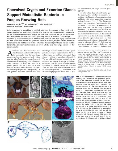
Fig. 1. (A) Photograph of Cyphomyrmex costatus<br />
showing the bacteria on the propleural plates.<br />
SEM of the plates in C. muelleri: The left plate is<br />
covered with bacteria (B), whereas they have been<br />
removed from the right plate, revealing the<br />
underlying fovea (C). (D) Light micrograph of a<br />
semithin cross section through the propleural<br />
plate of C. longiscapus showing the gl<strong>and</strong> (Gl)<br />
<strong>and</strong> duct cells (black arrow) associated with the<br />
fovea <strong>and</strong> the bacterium (Fb) on the plate (Cu for<br />
cuticle). (E) Photograph of C. longiscapus, illustrating<br />
foveae openings covering most of the<br />
cuticle. (F) Sagittal semithin section through a C.<br />
longiscapus worker, illustrating foveae outlining<br />
nearly the entire body of the ant. (G) Light<br />
micrograph of a single fovea within the cuticle<br />
(Cu) illustrating the abundance of mutualistic<br />
bacteria (Fb) within the crypt. (H) TEM of the<br />
lower section of a fovea showing a single<br />
gl<strong>and</strong>ular cell (Gl) <strong>and</strong> bacteria (Fb) within the<br />
crypt. Scale bars: 50 mm (AtoC),5mm (E <strong>and</strong> G),<br />
<strong>and</strong> 0.5 mm (F). [Photograph in (A) by A. Little]<br />
www.sciencemag.org SCIENCE VOL 311 6 JANUARY 2006 81
REPORTS<br />
82<br />
Fig. 2. Genus level phylogeny of<br />
fungus-growing ants [adapted<br />
from (15, 16)] illustrating the location<br />
<strong>and</strong> modifications of the<br />
exoskeleton for maintaining<br />
the mutualistic bacteria. The<br />
origin of fungus growing by attine<br />
ants <strong>and</strong> the leaf-cutters is<br />
represented by the Lepiotaceous<br />
mushroom <strong>and</strong> the worker carrying<br />
a leaf fragment, respectively.<br />
Major groups of attine<br />
ants are depicted by colored<br />
boxes, illustrating the phylogenetically<br />
basal genera in the<br />
‘‘paleo-attines’’ (red), the ‘‘lower’’<br />
attine genera (brown), the<br />
‘‘higher’’ attines (green), <strong>and</strong> the<br />
leaf-cutters (blue). (Column A)<br />
Photographs illustrate the location<br />
of the bacterium under the<br />
forelegs in the paleo-attines, on<br />
the propleural plates in the<br />
‘‘lower’’ <strong>and</strong> ‘‘higher’’ attines,<br />
thepresenceallovertheintegument<br />
in the genus Acromyrmex,<br />
<strong>and</strong> absence on the cuticle in<br />
Atta. (Column B) SEM micrographs<br />
of the location of the<br />
bacterium, under the forelegs in<br />
Apterostigma <strong>and</strong> on the propleural<br />
plates in other groups.<br />
(Column C) SEM micrograph<br />
close-ups for the structures<br />
presented in (B), showing the<br />
specific structural modifications<br />
for different groups of fungusgrowing<br />
ants. Presence of foveae<br />
(star) <strong>and</strong> tubercles (triangle)<br />
all over the body in some species<br />
within a genus are indicated by<br />
the corresponding symbol above<br />
the branch on the phylogeny.<br />
Scale bars: 0.5 mm (B), 10 mm<br />
(C). [The line drawings of Wasmannia<br />
auropunctata, Cyphomyrmex<br />
rimosus, Trachymyrmex<br />
septentrionalis, Acromyrmex versicolor,<br />
<strong>and</strong> Atta texana were<br />
made by Smith (20); those of<br />
Myrmicocrypta ednaella <strong>and</strong><br />
Sericomyrmex amabilis were<br />
made by Weber (21); <strong>and</strong> those<br />
of Apterostigma pilosum, Mycocepurus<br />
smithi, <strong>and</strong>Mycetarotes<br />
sp.weremadebyA.Little.Photographs<br />
of Acromyrmex octospinosus <strong>and</strong> Cyphomyrmex costatus in (A) were taken by A. Little.]<br />
<strong>and</strong>/or species of bacteria can occur within<br />
populations of the same species <strong>and</strong> between<br />
speciesofants(12). The diversity <strong>and</strong> mode of<br />
transmission predict congruence between the<br />
ant <strong>and</strong> bacteria phylogenies; however, the<br />
complete evolutionary history of ant-associated<br />
Pseudonocardia still remains to be determined.<br />
Here we examine the presence <strong>and</strong> evolution of<br />
specific cuticular structures on attine ants to<br />
house <strong>and</strong> maintain the parasite-inhibiting<br />
bacteria (8).<br />
To investigate this, we first examined ant<br />
species in the genus Cyphomyrmex because<br />
of the conspicuous white Bbloom[ of bacterium<br />
present on the propleural plates (Fig.<br />
1A). Scanning electron microscopy (SEM) of<br />
Cyphomyrmex longiscapus <strong>and</strong> C. muelleri workers<br />
with the filamentous bacterium removed<br />
6 JANUARY 2006 VOL 311 SCIENCE www.sciencemag.org<br />
revealed the presence of a previously unnoticed<br />
large crescent-shaped cavity (fovea) on each<br />
propleural plate (13) (Fig. 1, B <strong>and</strong> C; fig. S1A).<br />
The foveae are porous <strong>and</strong> occupy a significant<br />
proportion of the surface area of the propleural<br />
plates; the filamentous bacteria grow directly<br />
within these crypts (Fig. 1C).<br />
Our investigations further revealed, in the<br />
semithin sections of the propleural plates in C.
longiscapus, the presence of a previously unknown<br />
exocrine gl<strong>and</strong> located on the inner<br />
surface of the cuticle, just below the foveae.<br />
The gl<strong>and</strong> consists of bicellular units, each<br />
formed by a gl<strong>and</strong> cell <strong>and</strong> duct cell (14). The<br />
duct cells cross the cuticle <strong>and</strong> open within the<br />
foveae where the bacteria are cultured (Fig. 1D;<br />
fig. S1, A to C).<br />
In addition to foveae occurring on the propleural<br />
plates, C. longiscapus, C. muelleri, <strong>and</strong><br />
C. costatus ants also have bacteria-filled foveae<br />
covering most of the surface of worker exoskeletons,<br />
including the head, thorax, abdomen,<br />
<strong>and</strong> legs (Fig. 1, E to G). These crypts have<br />
small openings to the external surface of the<br />
ant, with minute microtrichia (hair-like cuticular<br />
projections) that appear to shield the<br />
opening of the crypt (fig. S1D). At the bottom<br />
of each fovea is a porous tubercle (integumental<br />
protrusion) (fig. S1E), connected via a duct cell<br />
to the corresponding gl<strong>and</strong> cell directly beneath<br />
the crypt (Fig. 1H).<br />
The locality of bacteria on the cuticle varies<br />
across fungus-growing ant species (Fig. 2, column<br />
A). Examination of specialized structures<br />
for bacterial maintenance across the phylogenetic<br />
diversity of attine ants revealed several<br />
broad evolutionary patterns (Fig. 2). Ant genera<br />
closely related to attine ants, Wasmannia <strong>and</strong><br />
Blepharidatta (15, 16), do not have filamentous<br />
bacteria, fovea, or tubercles (Fig. 2). In the<br />
most phylogenetically basal attine ants (paleoattines),<br />
such as the genus Apterostigma, the<br />
filamentous bacterium occurs on the mesopleura<br />
(under the forelegs), where it grows directly on<br />
the cuticle over the pores of duct cells connected<br />
to the corresponding gl<strong>and</strong> cells (Fig. 2, fig.<br />
S1F). In most species of Blower[ attine ants,<br />
mutualistic bacteria occur on the propleural<br />
plates (e.g., Cyphomyrmex costatus, inFig.2),<br />
in which the bacterium grows on tubercles<br />
within foveae. Similarly, the bacteria are also<br />
concentrated on the propleural plates in the<br />
Bhigher[ attine genus Trachymyrmex <strong>and</strong> the<br />
leaf-cutter genus Acromyrmex, although in these<br />
two genera the bacteria grow on gl<strong>and</strong> cell–<br />
associated tubercles directly on the exoskeleton<br />
rather than in foveae (Fig. 2).<br />
Several species of plants <strong>and</strong> animals engaged<br />
in mutualistic associations with microbes<br />
have evolved structures to house their<br />
symbionts. For example, root nodules in legumes<br />
house Rhizobium, squid light organs are<br />
filled with bioluminescent bacteria, aphids have<br />
modified bacteriocytes that form organlike<br />
structures to rear Buchnera, <strong>and</strong> some beetles<br />
<strong>and</strong> woodwasps have specialized structures<br />
(known as mycangia) to house mutualistic<br />
fungi (17–19). Our findings indicate that the<br />
exoskeleton of attine ants is modified to house<br />
mutualistic bacteria, apparently supporting their<br />
growth through gl<strong>and</strong>ular secretions. In addition,<br />
our phylogenetic examination of the structures<br />
across the fungus-growing ant tribe revealed<br />
that, like the cultivar <strong>and</strong> garden parasite, the<br />
mutualistic Pseudonocardia bacteria was apparently<br />
present at the earliest stages of fungus<br />
cultivation by ants. This is supported by the<br />
presence of the bacteria <strong>and</strong> bacteria-associated<br />
gl<strong>and</strong>s <strong>and</strong> duct cells in the most phylogenetically<br />
basal genera (e.g., Apterostigma), in contrast<br />
to their absence in closely related ants that<br />
do not cultivate fungus gardens (Blepharidatta<br />
<strong>and</strong> Wasmannia).<br />
The apparently early evolutionary origin of<br />
the bacteria within the fungus-growing ant symbiosis,<br />
in combination with bioassay results confirming<br />
that filamentous bacteria isolated from<br />
across the phylogenetic diversity of attine ants<br />
are effective at inhibiting their corresponding<br />
garden parasites (8, 10, 13), indicate that the<br />
bacteria have provided an efficient defense<br />
against Escovopsis for millions of years. This<br />
raises the question of how the antibiotics have<br />
remained effective without rampant evolution<br />
of resistance in the parasite over the long evolutionary<br />
history of this symbiosis.<br />
References <strong>and</strong> Notes<br />
1. C. R. Currie, U. G. Mueller, D. Malloch, Proc. Natl. Acad.<br />
Sci. U.S.A. 96, 7998 (1999).<br />
2. C. R. Currie, Oecologia 128, 99 (2001).<br />
3. C. R. Currie, Annu. Rev. Microbiol. 55, 357 (2001).<br />
4. C. R. Currie et al., Science 299, 386 (2003).<br />
5. I. H. Chapela, S. A. Rehner, T. R. Schultz, U. G. Mueller,<br />
Science 266, 1691 (1994).<br />
6. U. G. Mueller, T. R. Schultz, C. R. Currie, R. M. M. Adams,<br />
D. Malloch, Q. Rev. Biol. 76, 169 (2001).<br />
7. N. M. Gerardo, U. G. Mueller, S. L. Price, C. R. Currie,<br />
Proc. R. Soc. London Ser. B. 271, 1791 (2004).<br />
8. C. R. Currie, J. A. Scott, R. C. Summerbell, D. Malloch,<br />
Nature 398, 701 (1999).<br />
9. C. R. Currie, A. N. M. Bot, J. J. Boomsma, Oikos 101, 91<br />
(2003).<br />
10. M.J.Cafaro,C.R.Currie,Can. J. Microbiol. 51, 441 (2005).<br />
11. M. Goodfellow, T. Cross, The Biology of Actinomycetes<br />
(Academic Press, London, 1984).<br />
12. M. Poulsen, M. Cafaro, J. J. Boomsma, C. R. Currie, Mol.<br />
Ecol. 14, 3597 (2005).<br />
13. Materials <strong>and</strong> methods are available as supporting<br />
material on Science Online.<br />
14. J. Billen, E. D. Morgan, in Pheromone Communication<br />
in Social Insects: Ants, Wasps, Bees, <strong>and</strong> Termites, R.K.<br />
V<strong>and</strong>er Meer, M. D. Breed, M. L. Winston, K. E. Espelie,<br />
Eds. (Westview Press, Boulder, CO, 1998).<br />
15. T. R. Schultz, R. Meier, Syst. Entomol. 20, 337 (1995).<br />
16. J. K. Wetterer, T. R. Schultz, R. Meier, Mol. Phyl. Evol. 9,<br />
42 (1998).<br />
17. A. E. Douglas, Symbiotic interactions (Oxford Univ. Press,<br />
Oxford, 1994).<br />
18. L. Margulis, R. Fester, Symbiosis as a Source of<br />
Evolutionary Innovation (MIT Press, Cambridge, MA, 1991).<br />
19. S. Paracer, V. Ahmadjian, Symbiosis: An Introduction to<br />
Biological Associations (Oxford Univ. Press, Oxford, 2nd<br />
ed., 2000).<br />
20. M. R. Smith, Am. Midl. Nat. 37, 521 (1947).<br />
21. N. A. Weber, Gardening Ants: The Attines (American<br />
Philosophical Society, Philadelphia, 1972).<br />
22. This work was supported by the Smithsonian Tropical<br />
Research Institute (C.R.C.), NSF (DEB-0110073 to C.R.C.),<br />
the Danish Natural Science Research Council (21-01-0628 to<br />
J.J.B.), the Danish National Research Foundation (J.J.B.), the<br />
K.U. Leuven Research Fund (OT/2001/24 to J.B.),<br />
<strong>and</strong> the EU-FW5 Research-Training Network INSECTS<br />
(HPRN-CT-2000-00052 to J.J.B. <strong>and</strong> J.B.). We are grateful<br />
to ANAM of the Republic of Panama for granting collecting<br />
permits <strong>and</strong> to J. Ventocilla for expert help conducting SEM<br />
work. We thank G. de Alba, M. Cafaro,<br />
D. Crawford, N. Gerardo, A. Herre, M. Leone, A. Little,<br />
E. Magee, G. Maggiori, U. Mueller, S. Price, S. Rehner,<br />
H. Reynolds, J. Thomas, T. Schultz, <strong>and</strong> W. Wcislo for<br />
logistical support <strong>and</strong>/or comments on this paper.<br />
<strong>Support</strong>ing Online Material<br />
www.sciencemag.org/cgi/content/full/311/5757/81/DC1<br />
Materials <strong>and</strong> Methods<br />
SOM Text<br />
Figs. S1 <strong>and</strong> S2<br />
References<br />
6 September 2005; accepted 25 November 2005<br />
10.1126/science.1119744<br />
A Clonogenic Bone Marrow<br />
Progenitor Specific for<br />
Macrophages <strong>and</strong> Dendritic Cells<br />
Darin K. Fogg, 1 * Claire Sibon, 1 * Chaouki Miled, 1 Steffen Jung, 2 Pierre Aucouturier, 3<br />
Dan R. Littman, 4 Ana Cumano, 5,6 Frederic Geissmann 1,7 †<br />
Macrophages <strong>and</strong> dendritic cells (DCs) are crucial for immune <strong>and</strong> inflammatory responses <strong>and</strong><br />
belong to a network of cells that has been termed the mononuclear phagocyte system (MPS).<br />
However, the origin <strong>and</strong> lineage of these cells remain poorly understood. Here, we describe the<br />
isolation <strong>and</strong> clonal analysis of a mouse bone marrow progenitor that is specific for monocytes,<br />
several macrophage subsets, <strong>and</strong> resident spleen DCs in vivo. It was also possible to recapitulate<br />
this differentiation in vitro by using treatment with the cytokines macrophage colony-stimulating<br />
factor <strong>and</strong> granulocyte-macrophage colony-stimulating factor. Thus, macrophages <strong>and</strong> DCs appear<br />
to renew from a common progenitor, providing a cellular <strong>and</strong> molecular basis for the concept<br />
of the MPS.<br />
Macrophages (MFs) <strong>and</strong> dendritic cells<br />
(DCs) are involved in the scavenging<br />
of dying cells, pathogens, <strong>and</strong> molecules<br />
through phagocytosis <strong>and</strong> endocytosis<br />
REPORTS<br />
<strong>and</strong> the use of pattern recognition receptors<br />
(1). As a result, both cell types make a vital<br />
contribution to immunity <strong>and</strong> inflammatory<br />
responses to pathogenic microorganisms (2).<br />
www.sciencemag.org SCIENCE VOL 311 6 JANUARY 2006 83


