3. FOOD ChEMISTRy & bIOTEChNOLOGy 3.1. Lectures
3. FOOD ChEMISTRy & bIOTEChNOLOGy 3.1. Lectures
3. FOOD ChEMISTRy & bIOTEChNOLOGy 3.1. Lectures

You also want an ePaper? Increase the reach of your titles
YUMPU automatically turns print PDFs into web optimized ePapers that Google loves.
Chem. Listy, 102, s265–s1311 (2008) Food Chemistry & Biotechnology<br />
P08 QuANTITATIVE DETERMINATION OF<br />
SuLFONAMIDE RESIDuES IN ChICKEN<br />
MEAT by A NEw SOLID PhASE ExTRACTION<br />
AND hPLC-uV FOR DETECTION<br />
COnSTAnTIn BELE a , OCTAVIAn nEGREA a , ADELA<br />
PInTEA a , FRAnCISC VASILE DULF a , DAn LUPU b and<br />
ALEXAnDRU R. BIRIS b<br />
a University of Agricultural Sciences and Veterinary Medicine,<br />
R- 400372 Cluj-Napoca, Romania,<br />
b National Institute for Research and Development of Isotopic<br />
and Molecular Technologies, R-400293 Cluj-Napoca,<br />
Romania,<br />
cbele2002@yahoo.com<br />
Introduction<br />
Sulfonamides (SAs) are a group of synthetic antibiotics<br />
that have been used in food-producing animals for therapeutic<br />
and prophylactic purposes 1 . There is a risk of occurrence<br />
of unwanted residues in animal products if these antimicrobials<br />
have been improperly administered or if the proper<br />
withdrawal period has not been observed. To safeguard<br />
human health, the European Community has adopted for SAs<br />
safe maximum residue limits (MRLs) at the total level of<br />
100 μg kg –1 in food of animal origin 2 .<br />
Many methods such as LC and LC-MS, GC and GC-<br />
MS, ELISA, biosensor immunoassay (BIA) and high performance<br />
capillary electrophoresis (HPCE) have been developed<br />
for the determination of SA residues in food 3 . Solid phase<br />
extraction (SPE) was used as clean-up or enrichment method<br />
for SAs in tissues. The main sorbents used for the extraction<br />
of SAs are C18, aluminium oxide, strong cation-exchange 4 .<br />
Multi-walled carbon nanotubes (MWCnTs) are a novel<br />
carbon material used as an effective solid phase adsorbent for<br />
organic compounds (including SAs) 5 .<br />
In this paper, a sensitive method was developed for the<br />
determination of six SAs in chicken meat using MWCnTs<br />
and aluminium oxide as solid phase adsorbents followed by<br />
HPLC with UV detector.<br />
Experimental<br />
M a t e r i a l s a n d R e a g e n t s<br />
Chicken muscle tissues were purchased from local food<br />
market and deep frozen until analysis. Organic solvents such<br />
as acetonitrile, acetic acid and 1- propanol were all pesticide<br />
residue grade , commercially available from Merck . Anhydrous<br />
sodium sulfate was analytical grade (Bucarest, Romania).<br />
Deionized and redistilled water was prepared from Milli-Q<br />
Plus (Millipore). Sodium acetate trihydrate (Merck) was used<br />
as buffer for HPLC mobile phase. Sulfadiazin (SDZ), sulfamerazine<br />
(SMR), sulfapyridine (SPY), sulfisoxazole (SIO),<br />
sulfamethoxazole (SMO) and sulfadimethoxine (SDM) were<br />
purchased from Sigma.<br />
MWCnTs were purchased from Institute for Research<br />
and Development of Isotopic and Molecular Technologies<br />
Cluj-napoca, Romania.<br />
s589<br />
Standard stock solutions were prepared by accurately<br />
dissolving approximately 10 mg of SAs in 10 ml of acetonitrile<br />
LC grade and stored at 4 °C. Working standards were<br />
prepared weekly by appropriate dilution in acetate buffer at<br />
pH 4.5.<br />
C h r o m a t o g r a f i c S y s t e m a n d<br />
C o n d i t i o n s<br />
All experiments were carried out by using Shimadzu VP<br />
Series liquid chromatograph equipped with a UV-VIS detector.<br />
The chromatographic separation was accomplished in 30 min<br />
with gradient elution on a C 18 (250 mm × 4.6 mm, 5 μm)<br />
analytical column (Alltima) with a mobile phase 0.01M acetate<br />
buffer pH 4.5 (A) and acetonitrile (B). Flow 1 ml min –1<br />
was used for the separation of analytes at the following program:<br />
20 % B to 50 % within 22 min, back to 20 % in 3 min,<br />
equilibration for 5 min. The injection volume was 20μl and<br />
the detection of SAs was conducted at 266 nm.<br />
S a m p l e P r e p a r a t i o n a n d S a m p l e<br />
C l e a n - u p<br />
Ten grams of minced chicken tissue was placed into a<br />
50 ml polypropylene tube. 20 ml acetonitrile and 5 grams<br />
baked anhydrous sodium sulfate was then added. The sample<br />
was homogenized with an Ultraturax for about 1 min.,<br />
and then centrifuged at 5,000 rpm for 5 min.The residue was<br />
extracted by sonication with 20 ml acetonitrile and then centrifuged<br />
at 5000 rpm for 5 min.The extracts were combined<br />
and the solvent was concentrated to 5 ml.The solution was<br />
passed through the Alumina n SPE cartridge preconditioned<br />
by 10 ml acetonitrile.The analytes were eluted with 5 ml acetonitrile<br />
: water (80 : 20, v/v).The eluent and loading solvents<br />
were combined and transferred to concentration bottle.Then<br />
5 ml 1-propanol was added and the solution was evaporated<br />
to near dryness. The residue was dissolved by ultrasonication<br />
for 1 min with 30 ml acetate buffer (pH 5). A second SPE<br />
cartridge (200 mg MWCnTs) was utilized to remove potential<br />
interferences. The SPE was conditioned with acetonitrile<br />
(5 ml) and water (5 ml) prior to loading the sample. The<br />
analytes were eluted with a mixture of 2 ml acetate buffer<br />
(pH 4.5) and 4 ml acetonitrile. The eluate was evaporated to<br />
1 ml under nitrogen stream in a 40 °C block heater and filtered<br />
through a 0.45 μm disposable syringe filter.<br />
Results<br />
The present procedure uses two SPE cartridges for<br />
clean-up because acetonitrile extracted a lot of endogenous<br />
compounds from meat sample.The first one, Sep-Pak Alumina<br />
n, is a polar sorbent SPE cartridge. The second SPE<br />
cartridge (MWCnTs) is a non-polar sorbent and was utilized<br />
to further cleanse the extract.<br />
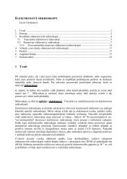
The calibration graphs obtained by plotting peak area<br />
versus drug concentration in 0.1–10 μg ml –1 range are<br />
reported in Table I.