3. FOOD ChEMISTRy & bIOTEChNOLOGy 3.1. Lectures
3. FOOD ChEMISTRy & bIOTEChNOLOGy 3.1. Lectures
3. FOOD ChEMISTRy & bIOTEChNOLOGy 3.1. Lectures

Create successful ePaper yourself
Turn your PDF publications into a flip-book with our unique Google optimized e-Paper software.
Chem. Listy, 102, s265–s1311 (2008) Food Chemistry & Biotechnology<br />
P89 ThE INFLuENCE OF SuRFACE<br />
ChARACTERISTICS ON bACTERIAL CELL<br />
ADhESION<br />
s780<br />
mined by the Surface Energy Evaluation system (Department<br />
of Physical Electronics, Faculty of Science, Masaryk University,<br />
Czech Republic).<br />
OLGA SCHREIBEROVá, TEREZA KRULIKOVSKá,<br />
JITKA HRDInOVá, JAn MASáK, ALEnA ČEJKOVá,<br />
VLADIMíR JIRKů and PETR HROn<br />
Institute of Chemical Technology Prague, Technická 5,<br />
166 28, Praha 6, Czech Republic,<br />
olga.schreiberova@vscht.cz<br />
Introduction<br />
Bacterial cell adhesion is the first step in the formation<br />
of multicellular structure called biofilm. Biofilm is a dynamic<br />
community of cells which display distinct properties from<br />
the planktonic cells. These can be utilized in bioremediation<br />
technologies1 , however the stability of adhesion must be ensured.<br />
For this purpose, the principles of initial adhesion must<br />
be investigated and understood.<br />
Experimental<br />
Microorganism.<br />
Gram-positive pollutant degrading bacteria Rhodococcus<br />
erythropolis CCM 2595 was obtained from the Czech<br />
Collection of Microorganisms (Masaryk University Brno,<br />
Czech Republic).<br />
Cultivation and biomass determination.<br />
Cells were cultivated in 200 ml of medium in shaked Erlenmeyer<br />
flasks. The growth of suspended cells was monitored<br />
as optical density at 400 nm (O.D.). Either a complex medium<br />
nutrient broth (HiMedia, India) or minimal medium was used<br />
(KH2PO4 0.17 g dm –3 , K2HPO4 0.13 g dm –3 , (nH4 ) 2SO4 0.71 g dm –3 , MgCl2 0.34 g dm –3 , MnCl2 1 m g dm –3 , CaCl2 0.26 m g dm –3 , FeSO4 0.6 m g dm –3 , na2MoO4 2 m g dm –3 ,<br />
pH 7). In minimal medium phenol or glucose was used as the<br />
only carbon and energy source.<br />
Cell hydrophobicity.<br />
Hydrophobicity of cells was determined by the<br />
MATH test2 Results<br />
T h e E x t e r n a l C o n d i t i o n s I n f l u e n c e<br />
o n C e l l H y d r o p h o b i c i t y<br />
Cell wall hydrophobicity reflects the cell physiological<br />
state and is one of the most important cell characterictics that<br />
determine the ability to adhere<br />
•<br />
•<br />
•<br />
.<br />
• Cell fatty acids.<br />
For fatty acids determination the cells were harvested<br />
by centrifugation, washed and lyophilized. Fatty acids were<br />
esterified to methyl esters, extracted to hexan and analysed<br />
by GC-FID.<br />
• Adhesion monitoring.<br />
The Flow cell 81 (BioSurface Technologies, USA) was<br />
used for assessing the adhesion of cells. Materials with different<br />
hydrophobicity and other properties were evaluated. Three types<br />
of glass were employed: microscope slide (labeled glass in following<br />
text), coated glass and hydrophobized glass. Also polymeric<br />
materials silicone and teflon were evaluated. The glass and<br />
silicone materials were prepared at the Department of Polymers,<br />
Faculty of Chemical Technology, Institute of Chemical Technology,<br />
Prague.<br />
• Material hydrophobicity.<br />
The material hydrophobicity (except teflon) was deter-<br />
3 . Cell hydrophobicity can be<br />
influenced by the type of the source of carbon and energy.<br />
In our study we investigated the effect of medium composition<br />
(complex and minimal media, optimal and stressful<br />
cultivation conditions). We found that the initial phenol concentration<br />
0.3 g dm –3 can be considered as optimal and that<br />
phenol concentration 0.7 g dm –3 partially inhibits the growth<br />
and can be called stressful (data not shown). Concentration<br />
1.0 g dm –3 caused considerable inhibition of the growth. nutrient<br />
broth was chosen as a complex medium. Also glucose<br />
(as a C source) in minimal medium was tested. During the<br />
experiments, hydrophobicity of cells in different growth<br />
phases was determined to ascertain the influence of this factor,<br />
which according to literature, can be considerable4 .<br />
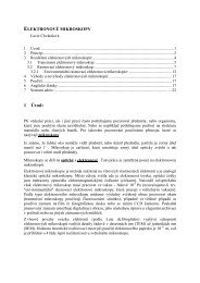
The Rhodococcus erythropolis cells were proven to be<br />
highly hydrophobic in all monitored media (see Fig. 1.). The<br />
medium composition influence on variation of cell hydrophobicity<br />
was significant. The changes in cell hydrophobicity<br />
during the growth (exponential, stationary phase) were not<br />
considerable and therefore the subsequent experiments were<br />
carried out with cells in stationary phase.<br />
100<br />
90<br />
80<br />
exponential phase stationary phase<br />
70<br />
60<br />
50<br />
40<br />
30<br />
20<br />
10<br />
0<br />
Fig. 1. The dependence of r. erythropolis cells hydrophobicity<br />
Figure on media 1. composition<br />
The dependence of media composition on R.<br />
erythropolis cells hydrophobicity in two growth phases.<br />
The fatty acid composition of cells cultivated in media<br />
with different phenol concentration is presented in the Table I.<br />
Results indicate that there is dependency of fatty acid composition<br />
on initial phenol concentration.<br />
hydrophobicity [%]<br />
phenol 0,3g/l<br />
phenol 0,7g/l<br />
phenol 1,0g/l<br />
nutrient broth<br />
glucose 0,5<br />
g/l