3. FOOD ChEMISTRy & bIOTEChNOLOGy 3.1. Lectures
3. FOOD ChEMISTRy & bIOTEChNOLOGy 3.1. Lectures
3. FOOD ChEMISTRy & bIOTEChNOLOGy 3.1. Lectures

You also want an ePaper? Increase the reach of your titles
YUMPU automatically turns print PDFs into web optimized ePapers that Google loves.
Chem. Listy, 102, s265–s1311 (2008) Food Chemistry & Biotechnology<br />
<strong>3.</strong> <strong>FOOD</strong> <strong>ChEMISTRy</strong><br />
& <strong>bIOTEChNOLOGy</strong><br />
<strong>3.</strong>1. <strong>Lectures</strong><br />
L01 NONSACChAROMyCES yEAST IN GRAPE<br />
MuST – ADVANTAGE OR SPOILAGE?<br />
JAROSLAVA KAňUCHOVá PáTKOVá a , EMíLIA<br />
BREIEROVá b and InGRID VAJCZIKOVá<br />
a Institute for viticulture and enology of SARC<br />
Matuškova 25, 831 01 Bratislava, Slovak republic,<br />
b Institute of ChemistrySAS, Dúbravská cesta 9, 845 38 Bratislava,<br />
Slovak republic,<br />
chememi@savba.sk<br />
Introduction<br />
The fresh grape must consists of spontaneous microflora<br />
formed from 90 to 99 % by yeasts. The most important<br />
genus is without doubt Saccharomyces cerevisiae which is<br />
responsible for successive fermentation and good wine quality.<br />
Recently the contribution of non-Saccharomyces yeasts<br />
have been widely discussed as there is not definitive opinion<br />
on their contribution to the wine quality, especially aroma.<br />
Hanseniaspora osmophila and Kloeckera apiculata should<br />
be considered detrimental yeast species, by higher acetic<br />
acid, acetaldehyde, ethyl acetate and acetoin production 1 . To<br />
avoid spoilage it is recommended to inoculate the grape must<br />
by Saccharomyces cerevisiae immediately after pressing.<br />
On the other hand, the apiculate non-Saccharomyces yeast<br />
is a natural indigenous microflora which contributes to the<br />
wine origin. Thus the question wheather to allow the apiculate<br />
microflora to start fermentation or not has not yet been<br />
solved.<br />
The aim of this study work was to determine the aroma<br />
profile of isolated non-Sacaharomyces yeast from the chemical<br />
as well as the sensorial viewpoints. The yeasts of<br />
the genera Rhodotorula, Sporobolomyces, Pichia, Hansenula,<br />
Issat-chenkia, and Torulospora, were tested from the<br />
viewpoint of their contribution to the wine aroma. The results<br />
were than exploited and tested in the real wine-making process.<br />
Experimental<br />
Following yeast strains were isolated from the grape<br />
must and degraded products: Rhodotorula mucilaginosa<br />
(2 strains), Sporobolomyces pararoseus, Pichia membranefaciens,<br />
Pichia anomala (2 strains), Candida intermediata,<br />
Torulospora delbruecki, and Issatchenkia orientalis. For<br />
comparison 3 Saccharomyces cerevisiae strains were also<br />
used. Two of them were isolated from the grape must, the<br />
third was a commercial one (Lallemand).<br />
Consequently the isolated yeast strains were inoculated<br />
to the first cultivation medium: the sterile grape must had<br />
s537<br />
been femented for 10 days at 10 °C under semiaerobic conditions<br />
or 4–6 weeks at 10 °C under anaerobic conditions.<br />
Second cultivation medium: the sterile Vinea drinks<br />
were inoculated by studied yeast strains and cultuivated at<br />
20 °C for 10 days under semiaerobic conditions.<br />
The samples were than sensorially evaluated by a group<br />
of degustators. The same samples were than analysed by gas<br />
chromatography for the aroma compounds production. Each<br />
sample was analysed on the GC MS (Shimadzu QP 2010)<br />
equipment and also on the GC FID equipment (GC 8000 CE<br />
Instruments).<br />
Two methods of sample preparation were done:<br />
The samples (20 ml) were extracted by ether (2 ml), and<br />
centrifuged prior to analysis. The etheric extract was used for<br />
analysis (liquid – liquid extraction). This method was used<br />
for higher alcohols (propanol, isoamylacohol, ethyl ester and<br />
higher alcoholos esters) determination<br />
The samples were extracted by Tenaq (solid phase microextraction)<br />
and than 10 min sampling according to 6 . This<br />
method was used for monoterpenic compounds determination.The<br />
same column and the same conditions were used by<br />
both analysis: Column: DB WAX 30 m, 0.25 × 0.25, temperature<br />
programme: 30 °C, hold 2 min, increase by 4 °C min –1<br />
up to 230 °C, hold 10 min, 1 ml of sample was injected to<br />
injection port at 200 °C, detector temperature 220 °C, carrier<br />
gas: helium, injektion mode: split 1 : 100, flow control mode:<br />
pressure 70 kPa<br />
Results<br />
After inoculation and fermentation of the grape must and<br />
Vinea drink the number of aroma compounds increased significantly,<br />
in both cases and more than 60 compounds were<br />
found. Most of them were recognized as typical fermentation<br />
products, etc. ethanol, izoamylacohol, propanol, etylester of<br />
caproic, caprylic and caprinic acids, ethylacetate, isovaleric<br />
acid, pentylacetate, 2,3-butandiol, furfural, 3-hydroxybutyrate,<br />
methionon, 1,4-butandiol. 2-metyl a 3-metylbutanoic<br />
acid, 2-phenyletylacetate, izoamylacetate, cis 3-hexenylacetate,<br />
etylbenzoate, α-terpineol, etyl isobutyrate, etyl butyrate,<br />
etyl 2-metylbutyrate, etyl isovalerate, isoamyl acetate, ethyl<br />
hexanoate, cis-3-hexenol, ethyl octanoate, furfural, linalool,<br />
ethyl furoate, ethyl decanoate, ethyl benzoate, α-terpineol,<br />
fenylethyl acetate, and geraniol. The increased production of<br />
typicall glycolysis products were also confirmed by several<br />
authors 2,3,4 . Ethyl propionate and propyl acetate, characterized<br />
the sample of the grape must fermented by Kloeckera<br />
apiculata, and 2-propanol and 2-hexanone characterized the<br />
sample of the grape must fermented by Pichia membranefaciens<br />
2 . Rojas 3 studied analysis of non-Saccharomyces yeast<br />
fermentation products and found higher acetate content, especially<br />
2-phenylacetate and isoamylacetate for Pichia yeasts.<br />
Albertazzi 4 also described higher levels of phenylacetate by<br />
other yeast - Pichia pastoris and Kloeckera saturnus (700–<br />
1700 mg dm –3 ). From our results we can confirm incereased<br />
ester production by all studied microorganisms, especially<br />
increased content of ethyl acetate.
Chem. Listy, 102, s265–s1311 (2008) Food Chemistry & Biotechnology<br />
However, we have found that some microorganisms<br />
produced special compounds, which were not recognised<br />
by other yeasts. After the fermentation of the grape must<br />
by R. mucilaginosa and Sp. pararoseus, aroma compounds<br />
significantly increased. Acetate, hexanal, heptanal, octanal,<br />
cyclopentanone, thiazole, decalactone, propyl-3-dimethyl<br />
aminopropyl, nonanone, heptanon, and butanediol were<br />
formed. P. anomala produced especially isoamyl benzyl<br />
ether. The medium fermented by Sporobolomyces was rich in<br />
sabinylacetate, 3,4-hexandion and eicosane. R. mucilaginosa<br />
generated cyklopentanol and, α-cyklogeraniol. S. cerevisiae<br />
produced vericaldehyd and γ-nonalakton.<br />
We have found out that yeasts of the genera Pichia, Rhodotorula,<br />
and Sporobolomyces did not produce the linalool<br />
acetate, contrary to S. cerevisiae.<br />
The differences in the compound production within<br />
the same yeast species were also observed. S. cerevisiae<br />
strain 8 produced caproic aldehdyd, trans-pinocamphon,<br />
dodecanal, 5-methyl-3-heptanon, izo-menthylacetate, contrary<br />
to S. cerevisiae strain 5 which did not produce any of<br />
these compounds. R. mucilaginosa strain 11 produced higher<br />
amounts of 2,3-butandiol, but did not produce any izopen-<br />
Table I<br />
Aroma compounds typically produced by various yeast species<br />
and flavoural characterisation of founded compounds<br />
Yeast strain compound Flavour fragrance<br />
R. mucilaginosa<br />
cyklopentanol,<br />
alfacyklogeraniol<br />
mint aroma<br />
spice flavour<br />
carnation odour<br />
Pichia anomala izoamyacetat banana flavour<br />
Sp. pararoseus sabinylacetate fruity aroma<br />
S. cerevisiae<br />
valericaldehyd,<br />
γ-nonalakton<br />
coffee aroma<br />
coconut odour<br />
Table II<br />
Sensorial evaluation of fermented grape must by various<br />
yeast strains, + positive impression, – negative impression<br />
Anaerobic Semiaerobic<br />
conditions conditions<br />
Yeast strain aroma perc. aroma perc.<br />
C.intermediata yeast + acidic –<br />
R.mucilaginosa 3 socks smelly – acethone –<br />
T.delbruecki pleasant + vanilla +<br />
I.orientalis autolyses – acethone –<br />
Pichia anomala acethone – lime +<br />
P.membranefaciens autolyses – yeasty +<br />
R. mucilaginosa 11 autolyses – honey +<br />
Pichia anomala acethone – honey +<br />
Sp. pararoseus not recognised – yeasty +<br />
S.cerevisiae 5 acidic – ferment +<br />
S.cerevisiae 8 fruity + ferment +<br />
S.cerevisiae 16 honey + ferment +<br />
s538<br />
Table III<br />
Evaluation of Vinea fermented under semiaerobic conditions<br />
Semiaerobic conditions<br />
Vinea<br />
20 °C<br />
Yeast strain<br />
Aroma Percept<br />
C. intermediata acidic +<br />
R. mucilaginosa 3 acethone +<br />
T. delbruecki yeasty +<br />
S. cerevisiae 5 yeastly +<br />
I. orientalis grape must +<br />
S. cerevisiae 8 honey +<br />
P. membranofaciens vinea –<br />
R. mucilaginosa 11 acethone +<br />
P. anomala honey +<br />
Sp. pararoseus acethone +<br />
tylformiate and 3,4-hexandion. These compounds produced<br />
strain Rhodotorula mucilaginosa strain <strong>3.</strong><br />
One of the most important factors in wine proofing is the<br />
sensorial evaluation of wine aroma. It is very difficult to estimate<br />
which from all above mentioned compounds will prevail<br />
over the other ones and wheather the wine could give positive<br />
or negative impression. It is due to the complexity of wine aromas,<br />
the heterogenity of perception and recognition thresholds<br />
for each one compound as well as many interactions occuring<br />
within and after fermentation 5 .<br />
As shown in Table II, under anaerobic conditions only<br />
T. delbruecki from apiculate microflora developed a pleasant<br />
aroma. All the other yeast strains evolved unpleasant, smelly<br />
aroma. However, the situation was radically changed when the<br />
fermentation occured under semi-aerobic conditions. Torulospora<br />
delbruecki produced the pleasant aroma, exactly defined<br />
as vanilla. Also both strains of P. anomala, Pichia membranefaciens,<br />
Sp. pararoseus and one strain of R. mucilaginosa evoked<br />
very pleasant aroma, some of them with honey notes,<br />
some of them fruity or increased fermetative impression.<br />
The results of better sensorial evaluation under semiaerobic<br />
conditions were subsequently tested by real winemaking<br />
fermentation. The problem which usually occurs in<br />
real process is that the present microfloras consists of several<br />
yeast genera which can possitively or negatively contribute<br />
to the wine quality.<br />
The group of degustators confirmed that the wines were<br />
better evaluated when they were fermented under semiaerobic<br />
conditions, with the inocoluation by S. cerevisae not directly<br />
after fermentation, but several hours after grape pressing. The<br />
main advantage of this system is its ability to improve the originality<br />
of the wine.<br />
Conclusions<br />
We have found compounds which were typically produced<br />
by some non-Saccharomyces yeast strains – cyklopentanol,<br />
alfacyklogeraniol for R. mucilaginosa, sabinylacetate,<br />
for Sp. pararoseus, and izoamylbenzylether for P. anomala.
Chem. Listy, 102, s265–s1311 (2008) Food Chemistry & Biotechnology<br />
We can confirm that under anaerobic conditions, most<br />
of the apiculate microflora, except T. delbruecki, negatively<br />
affected wine aroma because they produced higher amounts<br />
of aldehydes – pentanal, hexanal, heptanal, 3,4-hexanedione,<br />
eicosene which caused the buttery and waxy odour.<br />
However under semiaerobic conditions apiculate yeast<br />
species promoted positive aroma perception in products.<br />
Torulospora and Pichia yeast strains increased the fruit and/<br />
or coconut aroma by higher production of sabinyl res. isoamylacetate.<br />
Semiaerobic conditions applied several hours prior<br />
inoculating by S. cerevisiae improve sensorial evaluation<br />
of wine and increase the support of originality and variety<br />
typicity.<br />
However, the fermentation under such conditions is still<br />
very hazardous because oxidative defects or microbiological<br />
defaults of wine could occured.<br />
s539<br />
REFEREnCES<br />
1. Granchi L., Ganucci D., Messini A, Vincenzini M.:<br />
FEMS Yeast Res. 2, 403 (2002).<br />
2. Romano P., Fiore C., Paraggio M., Caruso M., Capece<br />
A.: Int. J. Food Microbiol, 86, 169 (2003).<br />
<strong>3.</strong> Rojas V., Gil J.V., Pinaga F., Manzanares P.: Inter. J.<br />
Food Microbiol. 70, 283 (2001).<br />
4. Albertazzi E, Cardillo R., Servi S.: Biotechnol. Lett. 16,<br />
491 (1994).<br />
5. Ribereau-Gayon P., Glories Y., Dubordieu D., Maujean<br />
A., Handbook of Enology, Ed. Wiley, Chicester, 404<br />
(2000).<br />
6. Kruzlicova D., Mocak J., Hrivnak J.: J. Food nutr. Res.<br />
47, 37 (2008).
Chem. Listy, 102, s265–s1311 (2008) Food Chemistry & Biotechnology<br />
L02 APPROAChES TO MINIMIZATION OF<br />
ACRyLAMIDE LEVEL IN <strong>FOOD</strong>S<br />
ZUZAnA CIESAROVá<br />
VÚP Food Research Institute<br />
Priemyselná 4, 824 75 Bratislava, Slovak Republic,<br />
ciesarova@vup.sk<br />
Introduction<br />
Thermal treatment of foods is a common way for improvement<br />
of digestibility, safety, quality and sensory properties<br />
of many foods which is used for ages. Besides unambiguous<br />
desirable aspects of this treatment some detrimental<br />
effects are still emerging e.g. a loss of nutrition-worthy compounds<br />
and an undesirable generation of contaminants.<br />
In 2002, Swedish researchers have first reported the<br />
formation of acrylamide in foods processed at elevated temperatures<br />
1 . Recent assessment by the Joint FAO/WHO Expert<br />
Committee on Food Additives (JECFA) in 2005 2 confirmed<br />
that a risk cannot be excluded for dietary intake of acrylamide<br />
because it is classified as a probable human carcinogen<br />
by the International Agency for Research on Cancer (IARC) 3 .<br />
In that assessment JECFA concluded that the margin of exposure<br />
for average and high consumers were low for compound<br />
that is genotoxic and carcinogenic and that this may indicate<br />
a human health concern. Therefore the Commission Recommendation<br />
since 2007 announced that “appropriate efforts<br />
to reduce acrylamide concentrations in foodstuffs should<br />
continue” 4 . Moreover, with respect to the last observations<br />
confirming the association between acrylamide intake and<br />
endometrial, ovarian 5 , and breast 6 cancer risk, the concern on<br />
the acrylamide mitigation activity is very urgent.<br />
Occurrence of Acrylamide in Thermally Treated Foods<br />
After the discovery of acrylamide, a lot of studies confirmed<br />
the presence of acrylamide in nearly all fried, baked<br />
and roasted foods. Acrylamide exposure varies depending<br />
upon the population´s eating habits and the way the foods<br />
are processed and prepared. Generally, fried potato products,<br />
ready-to-eat breakfast cereals, baked goods and roasted coffee<br />
are the most important food categories that contribute<br />
most to acrylamide exposure. An average long-term exposure<br />
of acrylamide was estimated of 0.3 to 0.8 μg (kg body<br />
weight) –1 day –1 on the base of the few data which were avail-<br />
able at the FAO/WHO Consultation in 2002 7 . Based on the<br />
reported data, the Committee JECFA in 2005 2 noted that<br />
children may have intakes of acrylamide around two or three<br />
times higher those of adult consumers when expressed on a<br />
body weight basis. It is expected that children and adolescents<br />
have consumption patterns different from adults. Most of the<br />
types of foods in which acrylamide was detected are popular<br />
among children and adolescents, such as French fries, snacks,<br />
biscuits and breads. Moreover, they have a lower average<br />
body weight and, consequently, a higher average food intake<br />
per kilogram body weight than adults. For that, acrylamide<br />
intake by these individuals is considered a concern.<br />
s540<br />
M e c h a n i s m o f A c r y l a m i d e<br />
F o r m a t i o n<br />
Initial results on acrylamide content indicated carbohydrate-rich<br />
foods to generate relatively more acrylamide 1 .<br />
Several researchers have established that the main pathway<br />
of acrylamide formation in foods is linked to the Maillard<br />
reaction and, in particular, the amino acid asparagine 8,9 . The<br />
link of acrylamide to asparagine, which directly provides the<br />
backbone of the acrylamide molecule, has been established by<br />
labelling experiments 9,10 . Study to date clearly shows that the<br />
amino acid asparagine is mainly responsible for acrylamide<br />
formation in heated foods after condensation with reducing<br />
sugars or a carbonyl source. Moreover, the sugar-asparagine<br />
adduct, n-glycosylasparagine, generates high amounts of<br />
acrylamide, suggesting the early Maillard reaction as a major<br />
source of acrylamide 9 . In addition, decarboxylated asparagine<br />
(3-aminopropionamid), when heated can generate acrylamide<br />
in the absence of reducing sugars 10 . A good evidence<br />
supporting the early Maillard rection as a main reaction pathway<br />
involving early decarboxylation of the Schiff base, rearrangement<br />
to the resulting Amadori product, and subsequent<br />
beta-elimination to release acrylamide has been presented 11 .<br />
Factors Affecting Acrylamide Formation in Foods<br />
The resulting acrylamide concentration in foods ultimately<br />
depends on both products and process variables.<br />
Acrylamide formation requires the amino acid asparagine<br />
and a carbonyl compound as the Maillard reaction precursors.<br />
The concentration of acrylamide precursors and temperature<br />
mainly affect the rate of acrylamide formation. It is a fact that<br />
formation and degradation of acrylamide occurs in the same<br />
time during heating at elevated temperatures, it means that<br />
measured acrylamide content of a food is net result of two<br />
consecutive reactions occurred during thermal processing12 .<br />
Based on the current knowledge of the mechanism of<br />
acrylamide formation, many parameters affecting the level<br />
of acrylamide in foods were investigated, e.g. heat intake, the<br />
level and type of saccharides and amino acids, moisture and<br />
water activity, additives, processing steps etc. 13 , and consequently<br />
various ways of acrylamide minimization in foods<br />
have been proposed. Many of them are summarized in a “living”<br />
document “The Acrylamide Toolbox” published by experts<br />
associated in the Confederation of the Food and Drink<br />
Industries of the European Union (CIAA) 14 .<br />
The mitigation approach is divided in two strategies:<br />
Prevention of acrylamide formation through a modification<br />
of food composition (a decline of asparagine<br />
and reducing saccharides contents), processing conditions<br />
(thermal input, pH, moisture), an addition of compounds<br />
suppressing the formation of acrylamide (acids,<br />
enzymes, proteins, antioxidants etc.) and an enhancement<br />
of processing steps (pre-treatment, blanching, fermentation<br />
etc.) 15 .<br />
Facilitation the acrylamide elimination through storage<br />
conditions or the initialization of acrylamide polymerization16,17<br />
•<br />
•<br />
.
Chem. Listy, 102, s265–s1311 (2008) Food Chemistry & Biotechnology<br />
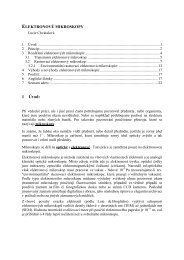
Temperature plays an important role in the formation<br />
and elimination of acrylamide. It is well known that acrylamide<br />
forms in foods that are cooked at high temperatures<br />
(> 120 °C) 8,13,18 . For shorter heating times as in the frying<br />
operation of potato chips or strips, lowering the frying temperature<br />
may significantly reduce the amount of acrylamide<br />
formed (Fig. 1.). The same may not be true for longer heating<br />
periods as in the roasting of coffee beans where extending<br />
the operation may result in a decrease in the amount of<br />
acrylamide persisted in the final product 19 . They may be a<br />
critical temperature/time zone where acrylamide is formed at<br />
a greater rate than it is destroyed, at temperatures outside of<br />
this zone little acrylamide is present.<br />
AA (mg/g Asn)<br />
120<br />
100<br />
80<br />
60<br />
40<br />
20<br />
0<br />
0 10 20 30 40 50<br />
time (min)<br />
180 °C<br />
160 °C<br />
140 °C<br />
130 °C<br />
120 °C<br />
Fig. 1. Amount of acrylamide after heating of equimolar mixture<br />
of glucose and asparagine at different temperatures<br />
The fact that acrylamide is not formed during boiling<br />
indicates that higher temperatures and/or low moisture conditions<br />
are needed for its formation. During heating under<br />
atmospheric conditions, higher temperatures can be reached<br />
only if simultaneous drying takes place, which is the case<br />
in frying, baking and roasting. The loss of water as the food<br />
dries during heating extracts a large amount of the incoming<br />
energy, and hence a bulk of the product is at a temperature<br />
very much lower than that of the heating medium. In this<br />
respect, temperature, time and moisture are key drivers of<br />
acrylamide formation in foods during heating (Fig. 2.). The<br />
moisture content determines the physical state and mobility<br />
of chemical constituents in food matrix. In addition, water<br />
alone affects the chemical route and the mechanistic pathway<br />
for acrylamide formation 13 .<br />
Concerning reducing sugars as carbonyl source, fructose<br />
has been found more effective than glucose in forming acrylamide<br />
(Fig. 2.). Both the chemical reactivity of sugars and<br />
their physical state play an important role in acrylamide formation.<br />
The melting point of fructose and glucose are 126 °C<br />
and 157 °C, respectively 13 . This explains why fructose is<br />
more reactive than glucose on acrylamide formation during<br />
heating. Frying, baking and roasting are simply characterized<br />
as open processes in which heat and mass transfer occur simultaneously.<br />
As the moisture reduces due to evaporation,<br />
sugars initially dissolved in water begin to form a saturated<br />
solution and then crystallize. After crystallization, melting<br />
s541<br />
is required to change their state to liquid, so to make them<br />
chemically reactive. In this respect, reducing sugars having<br />
a lower melting point is expected to form acrylamide earlier<br />
during heating.<br />
AA (mmol/mol Asn)<br />
<strong>3.</strong>0<br />
2.5<br />
2.0<br />
1.5<br />
1.0<br />
0.5<br />
fructose<br />
glucose<br />
0.0<br />
0 10 20 30 40 50 60 70 80<br />
moisture (%)<br />
Fig. 2. Amount of acrylamide after heating of equimolar mixture<br />
of asparagine and glucose/fructose at 180 °C for 20 min with<br />
different addition of water<br />
E n z y m e T r e a t m e n t L e a d i n g t o<br />
A c r y l a m i d e R e d u c t i o n<br />
One of the most effective ways to avoid acrylamide formation<br />
is removing the precursors, particularly amino acid<br />
L-asparagine. L-asparaginase as an enzyme of the hydrolases<br />
group (EC <strong>3.</strong>5.1.1.) selectively hydrolyses the amide bond of<br />
L-asparagine which results in the formation of aspartic acid<br />
and ammonia. Because the acrylamide formation correlates<br />
strongly with a free asparagine concentration, the reduction<br />
of L-asparagine in raw materials leads to the reduced<br />
level of acrylamide in final products 20 . The safety of asparaginase<br />
application is guaranteed by approving of GRAS<br />
status of Aspergillus oryzae asparaginase enzyme from<br />
novozymes A/S 21 , and Aspergillus niger asparaginase enzyme<br />
from DSM 22 , and a positive evaluation from the JECFA<br />
in 2007 23 as well. Moreover, this enzyme is inactivated by<br />
high temperature in the process of proteolysis.<br />
The application of L-asparaginase solution in a simulated<br />
potato matrix resulted in 50 to 90 % reduction of acrylamide<br />
content depending on the conditions (enzyme dose, time and<br />
temperature of incubation). no significant differences in<br />
impacts on L-asparagine conversion into L-aspartic acid in<br />
model samples between bacterial and fungal originated enzymes<br />
were observed. The positive effect of enzyme on the<br />
decrease of acrylamide content was confirmed also after Lasparaginase<br />
application on raw potato mash as well as dehydrated<br />
potato-wheat semi-products (Fig. <strong>3.</strong> and Fig. 4.) 20 .<br />
It is known that each intervention in the technology<br />
can be accompanied with consequences on the quality and<br />
sensory properties of final products which are strongly connected<br />
with the acceptability by consumers. For that reason<br />
the preliminary sensory evaluation of thermally and enzymatically<br />
treated products was done by a panel of trained<br />
judges. They described the main properties important for
Chem. Listy, 102, s265–s1311 (2008) Food Chemistry & Biotechnology<br />
these kinds of products such as darkness, yellowness, appearance,<br />
stickiness, crispness, oiliness, flavour, off-flavour, saltiness,<br />
sweetness and overall acceptability. Changes in colour<br />
were observed in pancakes prepared under different heating<br />
programmes, where darkness and crispness were more intensive<br />
in pancakes prepared at higher temperature of frying.<br />
no differences in evaluated sensory properties mentioned<br />
above were found out in any case of L-asparaginase application<br />
(P = 99 %) that was consider as a great advantage of<br />
the presented effective way of acrylamide reduction in food<br />
products 24 .<br />
AA (ng/g FW) .<br />
700<br />
600<br />
500<br />
400<br />
300<br />
200<br />
100<br />
0<br />
without enzyme<br />
2 U/g FW of enzyme; 10 min/ 37 °C incubation<br />
10 U/g FW of enzyme; 10 min/ 37 °C incubation<br />
Marabel Bellarosa<br />
Fig. <strong>3.</strong> Amount of acrylamide (AA) in raw potatoes (varieties<br />
Marabel and bellarosa) after enzymatic treatment (L-asparaginase<br />
produced by A. oryzae applied at concentration of 2 and<br />
10 u g –1 Fw and incubated at 37 °C for 10 min) and following<br />
heat treatment at 180 °C for 20 min<br />
AA (ug/kg FW) .<br />
800<br />
700<br />
600<br />
500<br />
400<br />
300<br />
200<br />
100<br />
0<br />
without enzyme 1 U/g FW of enzyme, 30 min/ 37°C incubation<br />
175 °C/20 min 180 °C/20 min 200 °C/20 min<br />
Fig. 4. Amount of acrylamide (AA) in pancakes prepared from<br />
potato-wheat powder at different heating temperatures (175 °C,<br />
180 °C and 200 °C) for 20 min with previous enzymatic treatment<br />
(L-asparaginase produced by A. oryzae applied at concentration<br />
of 1 u g –1 Fw and incubated at 37 °C for 30 min)<br />
Conclusions<br />
Since the acrylamide occurrence in foods and its potentiality<br />
to cause detrimental affects on human health attracts<br />
attention in all over the word, the effort to minimize its level<br />
in foods and consequently the human exposure to acrylamide<br />
is extremely advisable. Among many ways of acrylamide reduction<br />
the application of enzyme in order to prevent acry-<br />
s542<br />
lamide formation is feasible and effective without any undesirable<br />
effect on sensory quality of final products. For that<br />
reason, this procedure is protected by the patent application<br />
filed with the Industrial Property Office of the Slovak Republic<br />
under the number 5027-2006.<br />
This work was supported by the Slovak Research and<br />
Development Agency under the contract No. COST-0015-06.<br />
REFEREnCES<br />
1. Tareke E., Rydberg P., Karlsson P., Eriksson S., Tornqvist<br />
M.: J. Agric. Food Chem. 50, 4998 (2002).<br />
2. JECFA 2005 64th meeting Rome, 8-17 February 2005:<br />
http://www.who.int/ipcs/food/jecfa/summaries/summary_report_64_final.pdf<br />
<strong>3.</strong> IARC. Acrylamide. In IARC Monographs on the evaluation<br />
of carcinogen risk to humans: some industrial<br />
chemicals. International Agency for Research on Cancer,<br />
60, p. 389, Lyon, France 1994.<br />
4. Commission Recommendation of 3 May 2007 on the<br />
monitoring of acrylamide levels in food (notified under<br />
document number C(2007) 1873).<br />
5. Hogervorst J. G., Schouten L. J., Konings E. J.,<br />
Goldbohm R. A., Van Den Brandt P. A.: Cancer Epidemiol.<br />
Biomarkers Prevent. 16, 2304 (2008).<br />
6. Olesen P. T., Olsen A., Frandsen H., Frederiksen K.,<br />
Overvad K., Tjonneland A.: Int. J. Canc. 122, 2094<br />
(2008).<br />
7. FAO/WHO (2002): Opinion of the Scientific Committee<br />
on Food on new findings regarding the presence of acrylamide<br />
in food, Brussels, 3 July 2002.<br />
8. Mottram D. S., Wedzicha B. L., Dodson A. T.: nature<br />
419, 448 (2002).<br />
9. Stadler R. H., Blank I., Varga n., Robert F., Hau J.,<br />
Guy P. A., Robert M. C., Riediker S.: nature 419, 449<br />
(2002).<br />
10. Zyzak D. V., Sanders R .A., Stojanovic M., Tallmadge<br />
D. H., Eberhart B. L., Ewald D. K., Gruber D. C., Morsch<br />
T. R., Strothers M. A., Rizzi G. P., Villagran M. D.:<br />
J. Agric. Food Chem. 51, 4782 (2003).<br />
11. Yaylayan V. A., Wnorowski, A., Perez-Locas C.: J. Agric.<br />
Food Chem. 51, 1753 (2003).<br />
12. Biedermann M., Grob K.: Mitt. Geb. Lebensmitt. Hyg.<br />
94, 406 (2003).<br />
1<strong>3.</strong> Ciesarová Z., Kiss E., Kolek, E.: Czech J. Food Sci. 24,<br />
133 (2006).<br />
14. CIAA Acrylamide Toolbox [on-line]. Brussels : Confederation<br />
of the Food and Drink Industries of the European<br />
Union, 11 December 2007.<br />
15. Ciesarová Z.: Chem. Listy 99, 483 (2005).<br />
16. Kolek E., Šimon P., Šimko P.: J. Food Sci. 72, E341<br />
(2007).<br />
17. Kolek E., Šimko P., Šimon P., Gatial, A.: J. Food nutr.<br />
Res. 46, 39 (2007).<br />
19. Senyuva H. Z., Gökmen V.: Food Add. Contam. 22, 214<br />
(2005).
Chem. Listy, 102, s265–s1311 (2008) Food Chemistry & Biotechnology<br />
20. Ciesarová Z., Kiss E., Boegl P.: J. Food nutr. Res. 45,<br />
141 (2006).<br />
21. FDA 2006 Agency Response Letter GRAS notice no.<br />
GRn 000201.<br />
22. FDA 2007 Agency Response Letter GRAS notice no.<br />
GRn 000214.<br />
s543<br />
2<strong>3.</strong> JECFA 2007 68th meeting Geneva, 19-28 June 2007:<br />
http://www.who.int/ipcs/food/jecfa/summaries/summary68.pdf<br />
24. Ciesarová Z., Kukurová K.: Proc. ICC International<br />
Conference Bosphorus 2008, Istanbul 24-26 April 2008,<br />
p. 197.
Chem. Listy, 102, s265–s1311 (2008) Food Chemistry & Biotechnology<br />
L03 SOLID STATE FERMENTATION AS A TOOL<br />
FOR PREPARATION OF bIOPRODuCTS<br />
ENRIChED wITh POLyuNSATuRATED<br />
FATTy ACIDS<br />
MILAn ČERTíK, ZUZAnA ADAMECHOVá and LInDA<br />
néMETH<br />
Department of Biochemical Technology, Faculty of Chemical<br />
and Food Technology, Slovak University of Technology, Radlinského<br />
9, 812 37 Bratislava, Slovak Republic,<br />
milan.certik@stuba.sk<br />
Introduction<br />
Increasing demand for high-value lipids has focused<br />
commercial attention on the provision of suitable biosynthetic<br />
framework for their production. One of the main target for<br />
microbial oil transformation is construction of healthy and<br />
dietary important polyunsaturated fatty acids, such as γ-linolenic<br />
acid (18 : 3 ω-6; GLA), dihomo-γ-linolenic acid (20 : 3<br />
ω-6; DGLA), arachidonic acid (20 : 4 ω-6; AA), eicosapentaenoic<br />
acid (20 : 5 ω-3; EPA) and docosahexaenoic acid (22 : 6<br />
ω-3; DHA). Their applications in biomedical, nutritional and<br />
pharmaceutical fields coupled with their inadequacy from<br />
conventional agricultural and animal sources has looked for<br />
developing suitable biotechnologies to produce these compounds<br />
1 .<br />
Particularly active in the synthesis of PUFAs are species<br />
of fungi belonging to Zygomycetes 2 . Oleaginous fungi<br />
producing PUFA could be economically valuable because<br />
the most of their PUFAs occur in the triacylglycerol fraction<br />
of their lipids. Two basic processes have been developed for<br />
microbial production of PUFAs: submerged and solid state<br />
fermentations 3,4 . However, the principal difficulty that has<br />
been experienced with submerged PUFA-riched oil production<br />
has been in its marketing rather than in developing<br />
the large-scale fermentation and oil extraction process. Therefore,<br />
the association of oleaginous fungi with solid state<br />
fermentations (SSF) has been developed in order to improve<br />
commercial potential of microbial oils and thus to create new<br />
perspectives for the economic competitiveness and market of<br />
microbial polyunsaturated fatty acids (PUFAs). Solid state<br />
fermentation is a process in which microorganisms grow<br />
on a moist solid substrate in the absence of free water 5 . SSF<br />
simulates fermentation reactions occurring in the nature and<br />
allows microbial utilization of raw agro-materials or byproducts<br />
of the agro-food industries. Because some oleaginous<br />
fungi simultaneously decrease anti-nutrient compounds in<br />
the substrates (e.g. phytic acid) and partially hydrolyze substrate<br />
biopolymers, prefermented mass with a high content<br />
of PUFAs may be used as inexpensive food and feed supplement<br />
6 . Thus, SSF might provide the other opportunity to fill<br />
marketing claims in food, feed, pharmaceutical, veterinary<br />
and environmental fields.<br />
This paper deals with effectivity of several lower filamentous<br />
fungi to synthesize various PUFAs during their utilization<br />
of cereals by solid state fermentations.<br />
s544<br />
Experimental<br />
M i c r o o r g a n i s m s<br />
Thamnidium elegans CCF 1456, Cunninghamella echinulata<br />
CCF-103, Cunninghamella elegans CCF-1318, Mortierella<br />
isabelina CCF-14, Mortierella isabelina CCF-1098,<br />
Mortierella alpina CCF 185 were obtained from the Culture<br />
Collection of Fungi (Charles University, Prague, Czech<br />
Republic). The culture was maintained on modified Czapek-<br />
Dox agar slants with yeast extract (2.5 g dm –3 ) at 4 °C.<br />
S u b s t r a t e s a n d C u l t i v a t i o n<br />
C o n d i t i o n s<br />
Depending on the microorganism, various types of substrates<br />
were employed during SSF experiments. Spent malt<br />
grains (SMG) were added to some substrates. Autoclavable<br />
microporous polypropylene bags (160 × 270 mm 2 ) were filled<br />
with 10 g of dry substrate, moistened by the addition of 10 ml<br />
distilled water, soaked for 2 h at laboratory temperature and<br />
sterilized in autoclave (120 kPa, 120 °C, 20 min). In order<br />
to increased yield of PUFAs, sunflower or linseed oils were<br />
added to some substrates. In addition, various amounts of<br />
10% acetone or ethanol solutions of selected plant extracts<br />
were tested with the aim to activate enzymes involved into<br />
PUFA biosynthesis. The substrates were inoculated and<br />
mixed with 2 ml of spore suspension (1–2 × 10 6 spores per<br />
ml). Then each bag was closed with sterile cotton plugs,<br />
inoculated substrate was spread in the bags to obtain substrate<br />
layer of about 1 cm and incubated statically at 25 °C<br />
for 4–6 days (T. elegans, C. echinulata, C. elegans, M. isabellina)<br />
and 10–14 days (M. alpina). Triplicate SSF experiments<br />
for each substrate were prepared to assess reproducibility and<br />
average results are presented.<br />
L i p i d E x t r a c t i o n a n d F a t t y A c i d<br />
A n a l y s i s<br />
Prefermented cereal materials (bioproducts) were gently<br />
dried at 65 °C for 10 h and weighed. Lipids from homogenized<br />
bioproducts were isolated with chloroform/methanol<br />
(2 : 1, v/v) and purified according to Čertík et al. 7 and total<br />
lipids were determined gravimetrically. Fatty acids of total<br />
lipids were analyzed as their methyl esters 8 by gas chromatography<br />
according to Čertík et al 9 . Gas chromatograph (GC-<br />
6890 n, Agilent Technologies) was equipped with a capillary<br />
column DB-23 (60 m × 0.25 mm, film thickness 0.25 μm,<br />
Agilent Technologies) and a FID detector (constant flow,<br />
hydrogen 35 ml min –1 , air 350 ml min –1 , 250 °C). Analyses<br />
were carried out under a temperature gradient (130 °C for<br />
1 min; 130–170 °C at program rate 6.5 °C min –1 ; 170–215°C<br />
at program rate 2.7°C min –1 ; 215 °C for 7 min; 220–240 °C at<br />
program rate 2 °C min –1 ; 240 °C for 2 min) with hydrogen as<br />
a carrier gas (flow 2.1 ml min –1 , velocity 49 cm s –1 , pressure<br />
174 kPa) and a split ratio of 1/50 (inlets: heater 230 °C, total<br />
hydrogen flow 114 ml min –1 , pressure 174 kPa). The fatty<br />
acid methylester peaks were identified by authentic standards<br />
of C 4 –C 24 fatty acid methylesters mixture (Supelco, USA)<br />
and quantified by an internal standard of heptadecanoic acid
Chem. Listy, 102, s265–s1311 (2008) Food Chemistry & Biotechnology<br />
(C17 : 0, Supelco, USA). Fatty acid concentration was evaluated<br />
with ChemStation software B0103 (Agilent technologies,<br />
USA).<br />
Results and Discussion<br />
The extensive research and development of PUFA production<br />
by SSF is basically aimed at improving the economic<br />
competitiveness of that microbial process compared to<br />
plant- and animal-derived oils. Emphasis is put on increasing<br />
the product value, using inexpensive substrates, screening<br />
for more efficient strains and reducing the processing steps.<br />
Therefore, it is necessary to optimize the potential of microorganisms<br />
for transformation of agroindustrial materials and<br />
oil residues into desired metabolites.<br />
Screening of microorganisms has led to selection of<br />
T. elegans, C. echinulata, C. elegans and Mortierella isabellina<br />
as producers of GLA 6,9 and Mortierella alpina as a<br />
producer of DGLA, AA and EPA 10 . Generally, the surface<br />
of substrates was not only covered by the fungal mycelium<br />
during cultivation, but the fungal hyphae also penetrated into<br />
the substrates. Thus, fungal PUFAs were accumulated in the<br />
newly formed bioproduct and their amount depended on the<br />
substrates, microorganisms and cultivation conditions used.<br />
Depending on the microorganism, various types of cereal<br />
substrates were employed during SSF experiments (Table I).<br />
Spent malt grains (SMG) served as an internal support. Substrates<br />
without SMG in most cases led to agglomeration of<br />
substrate particles and created more compact mass which in<br />
turn interfered with microbial respiration and affected substrate<br />
utilization negatively. Presence of SMG improved<br />
bioconversion of linoleic acid from substrates to GLA 9 . Substrates<br />
with internal support not only provided better respiration<br />
and aeration efficiency due to an increased inter-particle<br />
space but also helped to remove the heat generated during<br />
fermentation. It should be noticed that although PUFAs were<br />
synthesized more effectively by SMG addition to substrates,<br />
total PUFAs yield was also dependent on substrate/SMG<br />
ratio. Unbalanced substrate/SMG ratio might provide limited<br />
surface for microbial attack and thus poorer availability of<br />
assimilable compounds (including oils) from substrates.<br />
Growth of fungi on a carbohydrates-containing substrates<br />
resulted after optimization of cultural conditions in constant<br />
lipid yield with the demanded fatty acid profile. Further<br />
improvement of PUFAs formation was achieved by physiological<br />
regulation of the SSF process employing following<br />
steps 10 : a) gradual elevation of carbon/nitrogen ratio with<br />
addition of appropriate carbon source; b) optimization of<br />
water activity, temperature and oxygen availability; c) transformation<br />
of exogenously added oils consisting of precursor<br />
of PUFAs. There is a stock of relatively cheap vegetable<br />
oils containing individual fatty acid precursors and SSF was<br />
applied for microbial utilization of renewable agricultural<br />
oils with the aim to modify their properties for production<br />
of value-added bioproducts with enhanced biological characteristics.<br />
Thus the ability of the strains to utilize exogenous<br />
fatty acids opens new possibilities to prepare PUFAs in high<br />
s545<br />
yield. Moreover, because fungi possess active oil-biotransforming<br />
system, these strains were also tested for their ability<br />
to convert directly oil-rich substrates (corn, sunflower seeds,<br />
linseeds, rapeseeds) to PUFAs.<br />
Table I<br />
Production of γ-linolenic acid (GLA), dihomo-γ-linolenic<br />
acid (DGLA), arachidonic acid (AA) and eicosapentaenoic<br />
acid (EPA) by solid state fermentations of selected fungi utilizing<br />
various cereal substrates. Ratio of susbtrate/SMG was<br />
1 : 3 (w/w)<br />
Strain Substrate PUFA Yield<br />
[g kg –1 BP]<br />
T. elegans oat flakes/SMG GLA 5.9<br />
wheat bran/SMG GLA 5.0<br />
wheat bran/SMG/<br />
GLA 10.0<br />
sunflower oil<br />
crushed corn GLA 10.0<br />
rye bran/SMG GLA 4.2<br />
buckwheat/SMG GLA 4.7<br />
millet/SMG GLA 6.5<br />
amaranth/SMG GLA 4.7<br />
C. echinulata barley GLA 6.1<br />
C. elegans barley GLA 7.0<br />
M. isabellina barley GLA 18.0<br />
M. alpina rice AA 21.4<br />
wheat sprout/SMG AA 36.1<br />
wheat bran/SMG AA 42.3<br />
rye bran/SMG AA 21.9<br />
peeled barley AA 16.2<br />
oat flakes AA 31.2<br />
M. aplina cresed sesame seeds DGLA 21.3<br />
M. alpina peeld barley/linseed EPA/AA 2<strong>3.</strong>4/36.3<br />
oil/SMG<br />
Biosynthesis and profile of fatty acids is controlled by<br />
enzymes involved in lipogenesis, so activation or inhibition<br />
of these metabolic steps is also useful tool for improving carbon<br />
flux to individual PUFAs 11 . For example, bioconversion<br />
of DGLA to AA is catalyzed by ‚∆ 5 ‘ desaturase and inhibition<br />
of this enzyme by crushed sesame seeds was accompanied by<br />
rapid increase of DGLA/AA ratio 11 ). In addition, application<br />
of various plant extracts possessing bioactive compounds<br />
seems to be promising way how to regulate fatty acid biosynthetic<br />
machinery in order to gain bioproduct with high<br />
yield of preferred PUFA. Application of ethanol extracts<br />
from ginger or sweet flag improved GLA yield by 30 % or<br />
25 %, respectively. On the other hand, biosynthesis of GLA<br />
was reduced by 70 % when acetone extract from tansy was<br />
employed to the substrate.<br />
Conclusions<br />
naturally prepared cereal based bioproducts enriched<br />
with PUFAs may be used as an inexpensive food and feed<br />
supplement. Thus, the association of selected microorganisms
Chem. Listy, 102, s265–s1311 (2008) Food Chemistry & Biotechnology<br />
with solid state fermentations has created new perspectives<br />
for the economic competitiveness and market of cereal based<br />
bioproducts containing PUFAs.<br />
The work was supported by grant VEGA No. 1/0747/08<br />
from the Grant Agency of Ministry of Education, Slovak<br />
Republic.<br />
REFEREnCES<br />
1. Gill I., Valivety R.: Trends Biotechnol. 15, 401 (1997).<br />
2. Čertík M., Shimizu S.: J. Biosci. Bioeng. 87, 1 (1999a).<br />
<strong>3.</strong> Čertík M., Shimizu S.: Agro Food Industry Hi-Tech. 10,<br />
26 (1999b).<br />
4. Čertík M., in: Biocatalysis and Bioenergy (Hou C.T.,<br />
Shaw J.-F., ed),p 571-585, Wiley, new York 2008<br />
s546<br />
5. Pandey A.: Biochem. Eng. J. 13, 81 (2003).<br />
6. Slugeň D., Streďanský M., Streďanská S., Čertík M.,<br />
Grego J.: Czech Patent 279043 (1994).<br />
7. Čertík M., Andráši P., Šajbidor J.: J. Am. Oil Chem. Soc.<br />
73, 357 (1996).<br />
8. Christoperson S.W., Glass R.L.: J. Dairy Sci. 52, 1289<br />
(1969).<br />
9. Čertík M., Sláviková L., Masrnová S., Šajbidor J.: Food<br />
Technol. Biotechnol. 44, 75 (2006).<br />
10. Sláviková L., Čertík M.: Chem. Listy, 99, 234 (2005).<br />
11. Čertík, M, Sakuradani, E, Shimizu S.: Trends Biotechnol.<br />
16, 500 (1998).
Chem. Listy, 102, s265–s1311 (2008) Food Chemistry & Biotechnology<br />
L05 PhySIOLOGICAL REGuLATION OF<br />
bIOTEChNOLOGICAL PRODuCTION OF<br />
CAROTENOID PIGMENTS<br />
VLADIMíRA HAnUSOVá a , MARTInA ČARnECKá b ,<br />
AnDREA HALIEnOVá b , MILAn ČERTíK a , EMíLIA<br />
BREIEROVá c and IVAnA MáROVá b<br />
a Department of Biochemical Technology, Faculty of Chemical<br />
and Food Technology, Slovak University of Technology,<br />
Radlinského 9, 812 37 Bratislava, Slovak Republic;<br />
b Faculty of Chemistry, Brno University of Technology, Purkyňova<br />
118, 612 00 Brno, Czech Republic;<br />
c Institute of Chemistry, Slovak Academy of Sciences, Dúbravská<br />
cesta 9, 845 38 Bratislava, Slovak Republic,<br />
milan.certik@stuba.sk<br />
Introduction<br />
Carotenoids represent one of the broadest group of<br />
natural antioxidants (over 600 characterized structurally)<br />
with significant biological effects and numerous of industrial<br />
applications. Because the application of synthetically prepared<br />
carotenoids as food additives has been strictly regulated<br />
in recent years, huge commercial demand for natural carotenoids<br />
has focused attention on developing of suitable biotechnological<br />
techniques for their production.<br />
There are many microorganisms including bacteria,<br />
algae, yeast and fungi, that are able to accumulate several<br />
types of pigments; but only a few of them have been exploited<br />
commercially 1 . From the view of yeasts, a range of species<br />
such as Rhodotorula, Rhodosporidium, Sporidiobolus, Sporobolomyces,<br />
Cystofilobasidium, Kockovaella and Phaffia<br />
have been screened for carotenoids formation. Yeast strains<br />
of Rhodotorula and Sporobolomyces formed β-carotene as<br />
the main pigment together with torulene and torularhodine as<br />
minor carotenoids. In contrast, Phaffia strains accumulated<br />
astaxanthin as a principal carotenoid. Comparative success<br />
in yeast pigment production has led to a flourishing interest<br />
in the development of fermentation processes in commercial<br />
production levels. However, in order to improve the yield of<br />
carotenoid pigments and subsequently decrease the cost of<br />
this biotechnological process, optimizing the culture conditions<br />
including both nutritional and physical factors have<br />
been performed. Factors such as carbon and nitrogen sources,<br />
minerals, vitamins, pH, aeration, temperature, light and<br />
stress showed a major influence on cell growth and yield of<br />
carotenoids.<br />
This paper summarizes our experience with physiological<br />
regulation and scale-up of biotechnological production of<br />
carotenoid pigments by yeasts.<br />
Experimental<br />
M i c r o o r g a n i s m s a n d C u l t i v a t i o n<br />
C o n d i t i o n s<br />
All strains investigated in this study (Sporobolomyces<br />
roseus CCY 19-6-4, S. salmonicolor CCY 19-4-10, Rhodotorula<br />
glutinis CCY 20-2-26, R. glutinis CCY 20-2-31, R. glu-<br />
s547<br />
tinis CCY 20-2-33, R. rubra CCY 20-7-28, R. aurantiaca<br />
CCY 20-9-7 and Phaffia rhodozyma CCY 77-1-1) were<br />
obtained from the Culture Collection of Yeasts (CCY; Institute<br />
of Chemistry, Slovak Academy of Sciences, Bratislava)<br />
and maintained on malt slant agar at 4 °C.<br />
The basic cultivation medium for flasks experiments<br />
for Rhodotorula and Sporobolomyces strains consisted of<br />
(g dm –3 ): glucose – 20; yeast extract – 4.0; (nH 4 ) 2 SO 4 – 10;<br />
KH 2 PO 4 – 1; K 2 HPO 4 . 3H2 O – 0.2; naCl – 0.1; CaCl 2 – 0.1;<br />
MgSO 4 . 7H2 O – 0.5 and 1 ml solution of microelements<br />
[(mg dm –3 ): H 3 BO 4 – 1.25; CuSO 4 . 5H2 O – 0.1; KI – 0.25;<br />
MnSO 4 . 5H2 O – 1; FeCl 3 . 6H2 O – 0.5; (nH 4 ) 2 Mo 7 O 24 . 4H2 O<br />
– 0.5 and ZnSO 4 . 7H2 O – 1]. The basic cultivation medium<br />
for flasks experiments for Phaffia strain consisted of<br />
(g dm –3 ): glucose – 20, yeast autolysate – 2.0, KH 2 PO 4 – 0.4,<br />
(nH 4 ) 2 SO 4 – 2.0, MgSO 4 . 7H2 O – 0.5, CaCl 2 – 0.1, naCl<br />
– 1.0. All strains grew under a non-lethal and maximally tolerated<br />
concentration of ni 2+ , Zn 2+ , Cd 2+ and Se 2+ ions. Also,<br />
stress conditions were induced by addition of various conventrations<br />
of naCl and H 2 O 2 . The cultures were cultivated<br />
in 500 ml flasks containing 250 ml cultivation medium on<br />
a rotary shaker (150 rpm) at 28 °C to early stationary grow<br />
phase. All cultivation experiments were carried out at triplicates<br />
and analyzed individually.<br />
Flasks results were verified in bioreactors and these<br />
scale-up experiments were carried out in 2 L fermentor (B.<br />
Braun Biotech), 20 L (SLF-20) and 100 L (Bio-la-fite) fermentors<br />
with an agitation rate of 250–450 rpm and a temperature<br />
of 20–22 °C. The pH was controlled at pH 5.0 by the<br />
addition of nH 4 OH and the dissolved oxygen concentration<br />
was maintained by supplying sterile air at a flow rate equivalent<br />
to 0.3–0.7 vvm.<br />
P i g m e n t I s o l a t i o n a n d A n a l y s i s<br />
Pigments from homogenized bioproducts were isolated<br />
by organic extraction and analyzed by high-performance<br />
liquid chromatography (HPLC). Analysis was carried out<br />
with an HP 1100 chromatograph (Agilent) equipped with a<br />
UV-VIS detector. Pigments extracts (10 μl) were injected<br />
onto LiChrospher ® 100 RP-18 (5 μm) column (Merck). The<br />
solvent system (the flow rate was 1 ml min –1 ) consisted of<br />
solvent A, acetonitrile/water/formic acid 86 : 10 : 4 (v/v/v),<br />
and B, ethyl acetate/formic acid 96 : 4 (v/v), with a gradient<br />
of 100 % A at 0 min, 100 % B at 20 min, and 100 % A at<br />
30 min.<br />
G e l E l e c t r o p h o p h o r e s i s<br />
1D PAGE-SDS electrophoresis of proteins was carried<br />
out by common procedure using 10% and 12.5% polyacrylamide<br />
gels. Proteins were staining by Coomassie Blue and by<br />
silver staining. For comparison, microfluidic technique using<br />
1D Experion system (BioRad) and P260 chips was used for<br />
yeast protein analysis too. 2D electrophoresis of proteins<br />
was optimized in cooperation with Laboratory of Functional<br />
Genomics and Proteomics, Faculty of Science, Masaryk<br />
University of Brno. 2D gels were obtained from protein pre-
Chem. Listy, 102, s265–s1311 (2008) Food Chemistry & Biotechnology<br />
paratives isolated from lyophilized cells. After optimization<br />
of separation conditions proteomes from stressed R.glutinis<br />
and R.rubra cells were isolated, lyophilized and analyzed.<br />
Quantitative analysis was done using BioRad Laboratories<br />
2D software. Identification of some spots was done using<br />
LC-MS/MS.<br />
Results and Disscussion<br />
To find suitable conditions for carotenoids production,<br />
several pigment forming yeasts have been tested for their<br />
ability to modify carotenoid biosynthesis based on media<br />
composition. Kinetic analysis revealed that yeasts strains<br />
differed from the view of growth rates and pigments formation.<br />
It must be emphasized that overall yield of carotenoids<br />
is directly related to the total biomass yield, thus, to keep both<br />
high growth rates and high flow carbon efficiency to carotenoids<br />
by optimal cultivation conditions is essential in order to<br />
achieve the maximal pigment productivity.<br />
Carotenoid accumulation in cells was significantly activated<br />
when yeasts were treated by selected stress conditions<br />
and their combinations (heavy metals, hydrogen peroxide<br />
and salt). However, such stimulation of pigment biosynthesis<br />
was more effective if stress factors were employed to the<br />
medium in exponential growth phase than from the beginning<br />
of cultivation 2–4 . It may be explained by hypothesizing<br />
a possible activation or inhibition mechanism by metal ions<br />
on specific carotenogenic enzymes, in particular, on specific<br />
enzymes involved in carotenoid biosynthesis, in agreement<br />
with previous studies reporting activation or inhibition by<br />
metal ions in microbial enzymes. The other explanation is<br />
based on observations that presence of heavy metals results<br />
in formation of various active oxygen radicals what, in a turn,<br />
induces generation of protective carotenoid metabolites that<br />
reduce negative behaviour of free radicals 5 .<br />
During environmental stress response many red yeasts<br />
exhibite cross-protective mechanisms. Preincubation of yeast<br />
Table I<br />
Production of carotenoids in flask and fermentation experiments<br />
Strain Pigment Pigment yields<br />
Flasks experiments<br />
S. roseus β-carotene 30 mg dm –3 (by Zn 2+ + H 2 O 2 )<br />
R. glutinis torulene 6 mg dm –3 (by Zn 2+ + H 2 O 2 )<br />
R. glutinis torularhodin 1 mg dm –3 (by Cu 2+ )<br />
P. rhodozyma astaxanthin 20 mg dm –3 (by Se 2+ )<br />
Scale-up fermentations<br />
S. salmonicolor. total carotene 41 mg dm –3 (by NaCl + H 2 O 2 )<br />
R. glutinis β-carotene 35 mg dm –3 (by NaCl + H 2 O 2 )<br />
P. rhodozyma astaxanthin 110 mg dm –3 (by NaCl + NaCl)<br />
s548<br />
culture with low concentration of one stress factor in inoculum<br />
media (e.g. salt, hydrogen peroxide) induces adaptation<br />
pathways resulted in enhanced carotenoid production 6 . Further<br />
addition of higher concentration of either the same or<br />
other stress factor can lead to significant (5–10 times) increase<br />
of β-carotene production in Rhodotorula glutinis and Sporobolomyces<br />
salmonicolor 2 . Simple preincubation of R. glutinis<br />
in presence of 2% naCl in inoculation medium followed by<br />
fermentation in inorganic production medium led to increased<br />
formation of biomass with accumulated carotenoids. This<br />
combined environmental stress using mild stress effect of salt<br />
or hydrogen peroxide could be industrially used for production<br />
of carotene enriched biomass. The results from flasks<br />
experiments are summarized in Table I.<br />
Laboratory flasks experiments were verified in semiscale<br />
conditions (from 2 dm 3 to 100 dm 3 fermentors). It was<br />
confirmend that glucose utilization was followed by increased<br />
pigment biosynthesis. Therefore, fed-batch fermentations<br />
were applied to enhanced carotenoid production. In addition,<br />
combination of aimed environmental condition resulted in<br />
activated accumulation of selected carotenoids (Table I).<br />
Enhancement of carotenoid production by environmental<br />
stress is also associated with changes in expression levels<br />
of various genes. Therefore, molecular changes in yeast cells<br />
on genome, proteome and metabolome level were studied<br />
using PFGE, 2D-GE, LC/MS/MS and EPR techniques. Presence<br />
of stress factors was accompanied by changes in carotenoid<br />
production as well as by alternations in protein levels.<br />
However, further analyses have to be focused on more precise<br />
characterization of proteins those displayed significant<br />
changes under increased biosynthesis of carotenoids.<br />
The work was supported by grant VEGA No. 1/0747/08<br />
and 2/7031/27 from the Grant Agency of Ministry of Education,<br />
Slovak Republic, grant FRVS 2541/G4/2008 of the<br />
Ministry of Education, Youth and Sports of the Czech Republic<br />
and grant IAA400310506 of Grant Agency of the Academy<br />
of Sciences of the Czech Republic.<br />
REFEREnCES<br />
1. Bhosale P.: Appl. Microbiol. Biotechnol. 73, 351<br />
(2004).<br />
2. Márová I., Breierová E., Kočí R., Friedl Z., Slovák B.,<br />
Pokorná J.: Annals of Microbiology 54, 73 (2004).<br />
<strong>3.</strong> Breierová E., Márová I., Čertík M.: Chem. Listy 99, 109<br />
(2005).<br />
4. Čertík M., Masrnová S., Sitkey V., Minárik M., Breierová<br />
E.: Chem. Listy 99, 237 (2005).<br />
5. Rapta P., Polovka M., Zalibera M., Breierová E., Žitňanová<br />
I., Márová I., Čertík M.: Biophys. Chem. 116, 1<br />
(2005).<br />
6. Kočí R., Drábková M., Márová I.: Chem. Listy 99, 297<br />
(2005).
Chem. Listy, 102, s265–s1311 (2008) Food Chemistry & Biotechnology<br />
L06 ENZyMATIC SACChARIDE ACETyLATIONS<br />
IN wATER – COMPARATION OF CATALySTS<br />
VLADIMíR MASTIHUBA a , TATIAnA KRAKOVá b and<br />
MáRIA MASTIHUBOVá a<br />
a Institute of Chemistry, Center for Glycomics, Slovak Academy<br />
of Sciences, Dúbravská cesta 9, 845 38 Bratislava, Slovak<br />
Republic,<br />
b Institute of Molecular Biology, Slovak Academy of Sciences,<br />
Dúbravská cesta 21, 845 51 Bratislava, Slovak Republic,<br />
chemvrma@savba.sk<br />
Introduction<br />
Enzymatic esterification of various hydroxy compounds<br />
is studied for about 40 years. Such reactions are usually performed<br />
in reaction systems comprising controlled amount of<br />
water to prevent product hydrolysis. Followingly, enzymatic<br />
esterification of unprotected saccharides is hampered by their<br />
decreased solubility in low water envíronment.<br />
We had recently described acetyl esterase from Trichoderma<br />
reesei to catalyze acetylations of several carbohydrates<br />
and alcohols in bulk water phase 1 . Acetyl esterases may<br />
be found in several raw commercial preparations of enzymes<br />
dedicated for biomass degradation since acetylation occurs<br />
widely on natural polysaccharides and plant cell wall fragments<br />
2 . Acetyl hydrolyzing activity occurs quite frequently<br />
in lipases (which may be result either of wider acyl specifity<br />
or of protein impurity) and some reports describe also esterase<br />
activity of serum albumins 3 .<br />
Our present work explores performance of several<br />
enzymes/crude enzyme preparations and proteins (Celluclast<br />
1.5 L FG, Ultraflo L, lipases from Candida antarctica, Candida<br />
rugosa, Aspergillus niger and Thermomyces lanuginosus,<br />
bovine serum albumin) in acetylations of glucose (1 a)<br />
and methyl α-D-glucopyranoside (1 b), comparing product<br />
yields and position selectivity of the reaction.<br />
Experimental<br />
Assays of acetyl esterase activity in enzyme preparations<br />
were performed as previously described 1 . Synthetic<br />
reactions were routinely performed by dissolving 1.2 g of<br />
glucose or alpha methyl glucoside in 10 ml mixture of water<br />
and acetyl donor (vinyl acetate or isopropenyl acetate, 4 : 1)<br />
and the reaction was started by adding 1 ml or 300 mg of<br />
the enzyme. Reaction mixture was then stirred at 37 o C for<br />
1–3 days and the course of the reaction was followed by t.l.c.<br />
on silica gel plates (chloroform – methanol, 3 : 1, visualisation<br />
by pouring into 5% sulphuric acid in ethanol and drying<br />
in hot air). Reaction mixture was then concentrated in vacuo,<br />
products separated by chromatography on silicagel column<br />
(chloroform – methanol, 3 : 1) and identified by nMR. For<br />
every reaction, a parallel experiment was executed without<br />
addition of enzyme to check occurence of a spontaneous acetylation.<br />
The spontaneous reactions were tested also separately<br />
in 0.1M acetate and phosphate buffers.<br />
s549<br />
Results<br />
Table I presents level of acetyl hydrolysing activities in<br />
enzyme preparations. Although water insoluble Lipolase 100<br />
was not active in photometric assay, it catalyzed acetylations<br />
of aliphatic alcohols in stired low water reactions (data not<br />
shown).<br />
Table I<br />
Acetyl hydrolysing activity of enzyme preparations<br />
enzyme activity activity<br />
(souce) [U ml –1 ] [U g –1 ]<br />
Celluclast 1.5 L FG<br />
(Trichoderma reesei)<br />
18.4<br />
Ultraflo L<br />
(Humicola insolens)<br />
31.5<br />
novozym 735<br />
(Candida antarctica)<br />
55.6<br />
BSA<br />
(Bos taurus)<br />
4.0<br />
Amano Lipase A<br />
(Aspergillus niger)<br />
219.1<br />
Lipase AYS<br />
(Candida rugosa)<br />
621.1<br />
Lipolyve CC<br />
319.0<br />
(Candida rugosa)<br />
Lipolase 100T<br />
(Thermomyces lanuginosus)<br />
not estimated<br />
Since phosphate buffer catalyzes acetylation of saccharides<br />
by vinyl acetate 4 , we tried to assess the extent of this<br />
process in our reactions (Table II).<br />
Table II<br />
Sponatneous acetylation of glucose by vinyl acetate (VA) and<br />
isopropenyl acetate (IPA) in buffered solutions and water<br />
pH Acetyl donor Monoacetates Diacetates<br />
4.01 VA + –<br />
4.51 VA ++ –<br />
5.02 VA ++ +/–<br />
5.51 VA +++ +<br />
5.72 VA +++ +<br />
6.03 VA +++ +<br />
Water VA – –<br />
Water IPA – –<br />
5.72 IPA +/– –<br />
Increasing pH of the buffer resulted in increased creation<br />
of glucose monoacetates and eventually diacetates when<br />
vinyl acetate was used as acetyl donor. To avoid such unselective<br />
acetylations, all enzymatic reactions were executed<br />
using water instead of the buffer.<br />
Scheme 1 summarizes in general the scale of products<br />
identified in reaction mixtures.
Chem. Listy, 102, s265–s1311 (2008) Food Chemistry & Biotechnology<br />
HO HO<br />
OH<br />
O<br />
OH<br />
R= α/β-H 1a<br />
α-CH3 1b<br />
OR<br />
HO O<br />
HO HO<br />
Yeast lipases gave low yields of pure 3 a,b, thus showing<br />
high specifity for acetylation of primary hydroxyl (Tables<br />
3,4). However, Lipolyve CC was reactive only with the glucoside<br />
1 b and did not acetylate free glucose within 3 days.<br />
On the other side, Amano Lipase A (Aspergillus niger)<br />
as a representative of fungal lipases gave 10% yield of an<br />
equimolar mixture of 2 a, and 3 a. Interestingly, acetylation<br />
O<br />
O<br />
HO HO<br />
O<br />
HO O<br />
O<br />
OH<br />
O<br />
O<br />
OH<br />
2 a,b<br />
O<br />
OH<br />
3 a,b<br />
OH<br />
4 a,b<br />
O<br />
5 a ,b<br />
Scheme 1<br />
Summarization of products created by enzymatic acetylations of<br />
glucose and methyl α-D-glucopyranoside with vinyl acetate or<br />
isoprenyl acetate<br />
Table III<br />
Enzymatic acetylations of glucose by vinyl acetate (VA) and<br />
isopropenyl acetate (IPA) in water<br />
enzyme<br />
acetyl<br />
donor<br />
O<br />
O<br />
O<br />
O<br />
OH<br />
OR<br />
OR<br />
OR<br />
OR<br />
mono-<br />
diacetates reaction<br />
acetates<br />
[%]<br />
time<br />
[%] [days]<br />
Celluclast VA<br />
19.7<br />
2 a : 3 a<br />
12 : 1<br />
0 1<br />
Lipolyve CC IPA 0 0 3<br />
novozym 735 VA<br />
2.0<br />
9.6<br />
3 a<br />
3<br />
not<br />
determined<br />
Amano<br />
Lipase A<br />
IPA<br />
10.0<br />
2 a : 3 a<br />
1:1<br />
4.2<br />
5 a<br />
2<br />
s550<br />
did continue to create only one diacetate (3,6-di-O-acetyl<br />
glucose, 5 a), thus keeping the original specifity for positions<br />
3-O- and 6-O- (Table III). Acetylations of methyl α-Dglucopyranoside<br />
(1 b) with isopropenyl acetate proceeded<br />
with the same positional specifity to produce 2 b and 3 b.<br />
When isopropenyl acetate was replaced by vinyl acetate as<br />
acetyl donor, selectivity increased markedly in favor of 2 b<br />
(Table IV). no reaction was observed when another fungal<br />
lipase – Lipolase 100T – was used as biocatalyst.<br />
Acetylations of 1 b catalyzed by bovine serum albumin<br />
and Ultraflo L (an enzyme cocktail from Humicola insolens)<br />
were unselective, giving mixtures of three monoacetates<br />
(Table IV).<br />
The best results were achieved by use of Celluclast, a<br />
cellulase preparation from Trichoderma reesei RUT C-30<br />
comprising the same acetyl esterase as reported earlier 1 . Acetylations<br />
gave 19.7 % and 10 % of monoacetylated glucose<br />
and methyl glucoside, respectively (Tables III, IV). The selectivity<br />
was exclusively to position 3-O- in acetylation of 1 b<br />
while in acetylation of glucose, almost 8% of 6-O- acetate 3 a<br />
was found in the monoacetate fraction.<br />
Table IV<br />
Enzymatic acetylations of methyl glucoside by vinyl acetate<br />
(VA) and isopropenyl acetate (IPA) in water<br />
enzyme<br />
acetyl<br />
donor<br />
Celluclast IPA<br />
10.0<br />
2 b<br />
Lipolyve CC IPA<br />
2.2<br />
3 b<br />
Lipase AYS IPA<br />
1.9<br />
3 b<br />
Amano<br />
Lipase A<br />
IPA<br />
10.5<br />
2 b : 3 b<br />
2,5 : 1<br />
Amano<br />
Lipase A<br />
VA<br />
6.4<br />
2 b : 3 b<br />
13 : 1<br />
2.1<br />
BSA VA 3 b : 4 b : 2 b<br />
2.0 : 1.3 : 1.0<br />
<strong>3.</strong>9<br />
Ultraflo L IPA 3 b : 4 b : 2 b<br />
4.5 : 2.5 : 1.0<br />
mono-<br />
diacetates<br />
reaction<br />
acetates<br />
[%]<br />
time<br />
[%] [days]<br />
0 1<br />
0 0.9<br />
0 0.16<br />
1.7<br />
5 b<br />
0.7<br />
5 b<br />
Conclusions<br />
Our results proved Celluclast to be the best catalyst for<br />
acetylations of glucose and methyl α-D-glucopyranoside<br />
according to its selectivity, product yields, availability and<br />
price. Since Celluclast has significantly lower acetyl esterase<br />
activity comparing to the other enzymes tested in this study,<br />
the enzyme seems rather unique. The enzyme selectivity to<br />
position 3-O- gives a promiss of its use in two step enzymatic<br />
1<br />
1<br />
0 5<br />
0 1
Chem. Listy, 102, s265–s1311 (2008) Food Chemistry & Biotechnology<br />
esterifications of saccharides, in which acetylation provides<br />
organic solvent-soluble sugar derivatives possessing free primary<br />
hydroxyls.<br />
This work has been supported by a grant from the Slovak<br />
Grant Agency for Science VEGA under the Project No.<br />
2/0145/08.<br />
s551<br />
REFEREnCES<br />
1. Kremnický ľ., Mastihuba V., Côté G. L.: J. Mol. Catal.<br />
B: Enzymatic 30, 229 (2004).<br />
2. de Vries R. P., Visser J.: Microbiol. Mol. Biol. Rev. 65,<br />
497 (2001).<br />
<strong>3.</strong> Sedlacek M., Manoharan I., Boopathy R., Duysen E.<br />
G., Masson P., Lockridge O.: Biochem. Pharmacol. 70,<br />
1673 (2005).<br />
4. Shi C., Zhang J. B., Fu H., Tang J.: Lett. Org. Chem. 3,<br />
932 (2006).
Chem. Listy, 102, s265–s1311 (2008) Food Chemistry & Biotechnology<br />
L07 PREPARATION OF bIODIESEL FROM TALL<br />
OIL<br />
RADOSLAV MIKULáŠIK a , IGOR ŠURInA a ,<br />
SVETOZáR KATUŠČáK a , Ján CVEnGROŠ b and<br />
MARTIn POLOVKA c<br />
Department of Chemical Technology of Wood, Pulp and<br />
Paper, a Institute of Polymer Materials,<br />
b Institute of Physical Chemistry and Chemical Physics, Faculty<br />
of Chemical and Food Technology, Slovak University of<br />
Technology,Radlinského 9, 812 37, Bratislava, Slovakia,<br />
c Food Research Institute, Department of Chemistry and Food<br />
Analysis, Priemyselná 4, 824 75, Bratislava, Slovakia,<br />
radoslav.mikulasik@stuba.sk<br />
Introduction<br />
It has known that after delignification kraft pulping process<br />
almost all extractable dissolved or emulsified substances<br />
of wood pass into black liquor. Evaporation of water from<br />
black liquor causes an increase in sodium salts content. Soaps<br />
are settled on all places, where liquor remains motionless. Tall<br />
soaps flowing on surface are separated in the form of foam<br />
in all tanks and their amount increases with density of black<br />
liquor 1–3 . Tall soap may be incinerated together with higherdensity<br />
black liquor within chemicals recovery or separated<br />
and utilised to high value chemicals. The latter alternative is<br />
more cost effective 4 . Kraft tall soap (TS) may be converted<br />
by acidification to tall oil (TO) and individual components<br />
isolated from it by vacuum distilation.<br />
TO is a dark brown, viscous and odorous liquid 5 containing<br />
mainly higher fatty acids (HFA), resin acids (RA),<br />
sterols and many other extractable substances 6 .<br />
Very interesting is HFA content ranging from 26 to<br />
58 % wt. 7 and depending mainly on the sort of wood. Given<br />
the high amount of wood, which is delignificated in paper<br />
mill in Central-european region, it represents a cheap raw<br />
material for biodiesel production. Biodiesel is exclusively<br />
produced from expensive vegetable oil.<br />
This work is focused on biodiesel production from tall<br />
oil and testing of selected biodiesel properties and their comparison<br />
with En 14214 8 . A method based on esterification<br />
straight by methanol and subsequent vacuum distillation of<br />
methylesters HFA from mixture was chosen. The method<br />
seems to be more advantageous than that based on esterification<br />
of HFA fraction obtained preferentially by vacuum distillation<br />
of TO. namely, boiling point of HFA methylesters is<br />
lower than that of HFA.<br />
Our method is more efficient from the viewpoint of<br />
energy demands when compared with the method realised,<br />
e.g., by AltÂparmak D., Keskin A., Koca A., Gürü M., 9 .<br />
Experimental<br />
M a t e r i a l s<br />
Chemicals: sulphuric acid, 96 % wt., Microchem, SR,<br />
Tall oil was prepared from aqueous solution of tall soap<br />
supplied by MOnDI Packaging Paper Štětí, ČR.<br />
s552<br />
I n s t r u m e n t s , F a c i l i t i e s , M e t h o d s<br />
Gas chromatography and mass spectrometry (GC/MS).<br />
Analysis of tall oil composition was performed by GC/MS<br />
with a Hewlett – Packard 5890A/5790B equipment.<br />
GC conditions: column: PTE–5, 30 m, 0.25 mm ID,<br />
0.25 μm film, carrier gas: helium, flow-rate 1 ml min –1 ,<br />
pressure 50 kPa , purge off: 0 min (split), injection temperature:<br />
275 °C, temperature of detector: 280 °C, temperature<br />
program: 170 °C (3 min), 5 °C min –1 – 270 °C (9 min),<br />
15 °C min –1 – 300 °C (3 min)<br />
MS conditions: mass range: 43–550 amu, EMV: 2400 V,<br />
threshold setting: 1000, SD: 3 min<br />
IR spectroscopy. Infrared spectra of all specimens were<br />
obtained by Digilab Excalibur FTS 3000MX – FTIR method.<br />
Samples were measured by diffusion scatter method (DRIFT)<br />
in KBr powder (5 % wt.).<br />
Vacuum (molecular) distillation. A laboratory molecular<br />
vacuum evaporator MO15 10 was used to isolate methylesters<br />
HFA and also to purify this product.<br />
Other laboratory equipment: furnace, balance, centrifuge,<br />
vacuum rotary evaporator.<br />
TO preparation from TS was carried out at following<br />
conditions:<br />
Table I<br />
Conditions of tall oil preparation from tall soap<br />
Mass TS ~ 1,000 g<br />
Heating time TS 25 → 95 °C ~ 15 min.<br />
H 2 SO 4 concentration 4<strong>3.</strong>6 wt. %<br />
Volume of added H 2 SO 4 solution 1.5 dm 3<br />
Reaction time 200 min. (<strong>3.</strong>3 h)<br />
Agitation intensive (<strong>3.</strong>3 h)<br />
TO was consequently esterified at conditions:<br />
Table II<br />
Conditions of tall oil esterification by CH 3 OH<br />
Conc. sulphuric acid [H 2 SO 4 , 96 % wt.] 5 % wt.<br />
of the mass of HFA in TO<br />
Temperature of reaction mixture 60 °C<br />
Molar ratio of CH 3 OH to HFA in TO 6 : 1<br />
Reaction time 5.5 h<br />
Intensive agitation of reaction mixture 5.5 h<br />
Table III<br />
Properties of tall soap and tall oil<br />
Dry matter [% wt.] 65.56<br />
Ash [% wt.] 11.36<br />
TO in TS [% wt.] 84.00<br />
Acid number of prepared TO<br />
[mg KOH g<br />
154.70<br />
–1 TO]
Chem. Listy, 102, s265–s1311 (2008) Food Chemistry & Biotechnology<br />
Results and Discussion<br />
P r e p a r a t i o n o f T O<br />
TO was prepared by TS acidification by sulphuric acid<br />
at conditions given in Table I. Sulphuric acid was added<br />
in excess. Reaction mixture undergoes vigorous agitation<br />
during the process.<br />
At the beginning, about 1,000 g TS (one batch) was<br />
heated in a 4,000 ml beaker in water bath. Temperature of<br />
water bath was kept in the range of 90–95 °C by adjustable<br />
cooker. After heating of TS at the water bath temperature,<br />
sulphuric acid was added and a blender activated. The blender<br />
ensured intensive agitation during the whole process. At<br />
TO preparation, the following reaction proceeded:<br />
R—COOna + H 3 O + → R—COOH + H 2 O + na +<br />
Scheme 1<br />
Conversion of saponified HFA to acid form<br />
Volume of obtained tall oil after separation of aqueous and<br />
a lignin phase from one batch was 482 ml or 465 g, and the yield<br />
in % wt. was 70.9.<br />
Having concluded the process, phase separation was<br />
performed. TO was separated from aqueous and lignin phases<br />
by separatory. Subsequently, warm water was used to wash<br />
TO, until pH of waste water reached ∼ 6–7 due to necessity<br />
to eliminate H 2 SO 4 residues from esterified oil. Finally, TO<br />
was dehydrated in a vacuum rotary evaporator.<br />
Preparation of tall oil was performed using two apparatus<br />
simultaneously. For totally <strong>3.</strong>3 h, 2 × 482 ml of oil after<br />
separation of aqueous and lignin phase was obtained. This<br />
process was repeated 6 times and a total amount of prepared<br />
oil was ~ 5,784 ml (5,580 g). A total amount of TS used in the<br />
process was ~ 7,867 g (besides moisture).<br />
Average batch yield reached 70.93 % wt. Theoretical<br />
yield was 84.0 % wt., i.e. weight loss was 1<strong>3.</strong>07 % wt. This<br />
amount included the matter remaining on the beaker and funnels<br />
walls owing to a decrease in temperature and increase in<br />
viscosity of tall oil. The inner surface of 4,000 ml beaker and<br />
2,000 ml separatory funnel was significant.<br />
Moreover, not all saponificed HFA and RA were converted<br />
to their acid form by the reaction shown in Scheme 1.<br />
G C / M S A n a l y s i s o f T O<br />
The prepared TO contained 30 % wt. HFA, 20 % wt.<br />
RA, ~ 18.5 % sterols. non-analysed portion represented<br />
~ 44.2 % wt. and contained higher fatty alcohols, hydrocarbons,<br />
lignin and cellulose fragments, non-methylated fatty<br />
and resin acids and inorganic compounds. These substances<br />
cannot be classified into the above three groups. In addition,<br />
these substances could be modified by the preparation process<br />
or its conditions.<br />
s553<br />
Table IV<br />
GC/MS analysis – composition of tall oil<br />
Extractives [% wt.]<br />
Fatty acids 29.8<br />
Resin acids 20.0<br />
Sterols 6.0<br />
Analysed portion 55.8<br />
non-analysed portion 44.2<br />
Total 100.0<br />
E s t e r i f i c a t i o n o f T a l l O i l<br />
– P r e p a r a t i o n o f T a l l O i l<br />
M e t h y l e s t e r s ( T O M E )<br />
The prepared TO, after washing by water and dehydratation<br />
were subjected to esterification at optimal conditions<br />
based on neaves D.E. 11 .<br />
R C<br />
O<br />
O H<br />
R C<br />
+<br />
-<br />
H +<br />
H -<br />
OH<br />
O +<br />
OH<br />
H<br />
OH<br />
R C +<br />
H<br />
- H2O -<br />
H +<br />
O<br />
R C<br />
Mechanism of acid catalysed esterification by methanol<br />
is proposed in Scheme 2. Conditions of esterification are<br />
summarized in Table II.<br />
1,000 g TO with HFA content ~ 30 % wt. i.e. ~ 300 g<br />
was processed. As catalyst, suphuric acid ~ 15 g (8.2 ml at<br />
density 1.835 g cm –3 ) was used. The total amount of methanol<br />
added to the reactor was ~ 204.8 ml (methanol was added<br />
in excess to HFA, in molar ratio 6 : 1)<br />
Average molar mass of fatty acids was estimated as<br />
281.55 g mol –1 . Stemming from the fact that optimal conditions<br />
(time 2–3 h) given by literature concern only the fraction<br />
HFA isolated from TO and not the fraction included in<br />
TO, reaction time was prolonged to 5.5 h to ensure maximum<br />
conversion.<br />
The process was repeated 3 times and total volume of<br />
prepared TOME approached 3,000 ml (2,835 g).<br />
H<br />
O<br />
CH 3<br />
H3C Scheme 2<br />
Mechanism of HFA esterification with methanol in acid media<br />
+<br />
O<br />
O CH 3
Chem. Listy, 102, s265–s1311 (2008) Food Chemistry & Biotechnology<br />
V a c u u m D i s t i l l a t i o n o f T O M E<br />
Data on amounts of obtained distillates (D1, D2), residues<br />
(Z1, Z2) and weight losses (WL) from 1 st and 2 nd distillation<br />
stage are listed in Table V and VI.<br />
Table V<br />
Vacuum Distillation material balance: 1 st stage<br />
TOME D1 Z1 WL<br />
[g] [g] [wt. %] [g] [wt. %] [wt. %]<br />
2362 845 35.8 1419 60.0 4.2<br />
Table VI<br />
Vacuum distillation material balance: 2 nd stage<br />
TOME D2 Z2 WL<br />
[g] [g] [wt. %] [g] [wt. %] [wt. %]<br />
845.3 712 84.2 101.4 12.0 <strong>3.</strong>8<br />
The prepared amount of TOME was heated and then let<br />
settle. Formed water and unreacted methanol were removed<br />
with a vacuum (molecular) evaporator.<br />
Then 2.5 dm 3 TOME was withdrawn from 3 dm 3 volume<br />
of TOME and distillated at the folowing conditions: pressure<br />
10–50 Pa and temperature 135–140 °C. 955 ml (~ 845 g) of<br />
distillate (D1) was obtained which represented ~ 38 % vol.<br />
(~ 36 % wt.). During this distillation D1 was polluted<br />
by RA.<br />
Destillate D1 955 ml (~ 845 g) was consequently distillated<br />
at pressure 2–20 Pa and temperature 130–135 °C.<br />
Under these conditions, we obtained 835 ml (711.8 g) of distillate<br />
D2, i.e. ~ 87 % vol. from D1, (84 % wt.)<br />
The total yield of these processes: from 2,500 ml TOME<br />
(2,363 g) we obtained 835 ml (711.8 g) which represented<br />
3<strong>3.</strong>4 % vol. (30.1 % wt.).<br />
Acid number parameter of D2 (19.610 mg KOH g –1 ) did<br />
not comply with European standard En 14214 for biodiesel<br />
(stipulating less than 0.5 mg KOH g –1 ), D2 had to undergo<br />
further treatment. D2 included some RA. They were neutralised<br />
and precipitated and D2 (tall oil fatty acids methyl esthers<br />
– TOFAME) after this treatment had acid number 0.3 mg<br />
KOH g –1 . This value fully meets En 14214. However, total<br />
volume of the final product decreased from 835 to 630 ml<br />
(566.6 g) which represented – when comparing to the TOME<br />
amount – 25.2 % vol., (~ 24 % wt.).<br />
The final product prepared in such a mode was subjected<br />
to analyses in laboratory of VÚRUP, which was accredited<br />
for analyses of biofuels. Results are assembled in Table VII.<br />
R e s u l t s O b t a i n e d b y F o u r i e r<br />
T r a n s f o r m I n f r a r e d S p e c t r o m e t r y<br />
( F T I R )<br />
FTIR spectra of our product and a sample of soybean<br />
oil methylesters (SOME) were scanned and were found to<br />
s554<br />
comply with En 14214. We observed very good similarity<br />
in absorption peaks of our product and SOME sample. Both<br />
spectra are almost identical. This may mean that both speci-<br />
Fig. 1. Comparison of FTIR spectra: TOFAME and SOME<br />
mens have similar physical and chemical properties.<br />
T e s t R e s u l t s f o r B i o d i e s e l f r o m<br />
Table VII<br />
Values of prepared biodiesel compared with En14214 standard<br />
values<br />
Properties Units Determined En 14214 Uncertainty<br />
value value<br />
Density at<br />
kg m –3 15 °C<br />
Carbon residue<br />
899.3 875–900 0.1 %<br />
(10% distillation wt. %<br />
residue)<br />
Kinematic<br />
< 0.010 max. 0.3 10 %<br />
mm2 s –1 viscosity<br />
at 40 °C<br />
<strong>3.</strong>932 <strong>3.</strong>5–5 1.5 %<br />
Cold filter<br />
plugging point<br />
°C –12<br />
0 to –13<br />
–13 to –20<br />
(winter)<br />
3 °C<br />
Ester content wt. % 95.7 min. 96.5 2.5<br />
Linolenic acid<br />
methyl ester<br />
wt. % 0.7 max. 12 –<br />
Sulphur<br />
mg kg –1 content<br />
1208 max. 10 2 %<br />
A c c r e d i t e d L a b o r a t o r y<br />
Conclusions<br />
Prepared tall oil was esterified by methanol at optimal<br />
conditions in the presence of acid catalyst. Tall oil fatty acids<br />
methyl esters were isolated from TOME mixture by vacuum<br />
distillation.<br />
Total yield of these processes: from 2,500 ml TOME<br />
(2,363 g), 630 ml (566.6 g) resulted which represents
Chem. Listy, 102, s265–s1311 (2008) Food Chemistry & Biotechnology<br />
25.2 % vol. (~ 24 % wt.). Biodiesel prepared in this procedure<br />
complies with En 14214 except of chemicaly bonded<br />
sulphur.<br />
We thank Slovak Grant Agency (Project VEGA<br />
1/0770/08) for its financial support.<br />
REFEREnCES<br />
1. Košík M. , Blažej A.: PHYTOMASS: A Raw Materials<br />
for Chemistry and Biotechnology, Veda-Ellis Horwood<br />
Ltd., Chichester, Great Britain, 199<strong>3.</strong><br />
2. Lužáková V., Vrška M.: Cellul. Chem. Technol. 39, 237<br />
(2005).<br />
<strong>3.</strong> Lužáková V., Marcincinová T., Vrška M.: Chemické<br />
listy 94, 880 (2000).<br />
4. Šutý L.: Výroba a vlastnosti buničín, p.147, ALFA, Bratislava,<br />
1982.<br />
5. Lee S.Y., Hube M. A., Saka Sh.: BioResources 1, 150<br />
(2006).<br />
s555<br />
6. Bokis C. P., Chen Ch. Ch., Orbey H.: Fluid Phase Equilibria<br />
155, 193 (1999).<br />
7. Košík M., Blažej A.: Fytomasa ako chemická surovina,<br />
p. 340, VEDA, Bratislava, 1985.<br />
8. En 14214, EUROPEAn STAnDARD – Automotive<br />
fuels – Fatty acid methyl esters (FAME) for diesel engines<br />
– Requirements and test methods,<br />
http://www.acbiodiesel.net/docs/FAME%20Euro %20st<br />
andard%20draft%20Oct02.pdf<br />
http://www.biopalivo.sk/En%2014214.pdf<br />
9. AltÂparmak D., Keskin A., Koca A., Gürü M.: Bioresour.<br />
Technol. 98, 241 (2007).<br />
10. Cvengroš J.: Chem. Prům. 40, 135 (1990).<br />
11. neaves D. E.: Evaluation of fatty acid fraction derived<br />
from tall oil as a feedstock for biodiesel production,<br />
Master thesis, Faculty of Mississippi State University,<br />
School of Chemical Engineering, May 2007.
Chem. Listy, 102, s265–s1311 (2008) Food Chemistry & Biotechnology<br />
L08 ARTIFICIAL NEuRAL NETwORKS IN <strong>FOOD</strong><br />
ANALySIS<br />
Ján MOCáK a,b , VIERA MRáZOVá a , DáŠA<br />
KRUŽLICOVá b and FILIP KRAIC a<br />
a Department of Chemistry, University of Ss. Cyril & Methodius,<br />
Nám. J. Herdu 2, 917 01 Trnava, Slovakia,<br />
b Institute of Analytical Chemistry, Slovak University of Technology,<br />
Radlinskeho 9, 812 37 Bratislava, Slovakia, jan.<br />
mocak@ucm.sk<br />
Introduction<br />
During the last twenty years the chemists have get<br />
accustomed to the use of computers and consequently to the<br />
exploitation of various complex mathematical and statistical<br />
methods, by which they have been trying to explore multivariate<br />
correlations between the output and input variables<br />
more and more in detail. Input variables of different kind are<br />
often continuous and represent usually the results of instrumental<br />
measurements but also other observations, sometime<br />
discrete or categorical, are important for characterizing the<br />
investigated objects. Such kinds of input variables are for<br />
example the results of sensorial assessment of foodstuffs or<br />
simply some qualitative attributes like odour or colour (agreeable/disagreeable<br />
or clear/yellowish/yellow). The output<br />
variables are the targets of the corresponding study, which<br />
again can be represented by some continous variable or a categorical<br />
one with two or more levels. With the increasing complexity<br />
of analytical measurements, and the analysed sample<br />
itself, it becomes clear that all effects that are of interest cannot<br />
be described by a simple univariate relation but they are<br />
multivariate and often they are not linear. A set of methods,<br />
which allow study of multivariate and non-linear correlations<br />
that have recently found very intensive use among chemists<br />
are the artificial neural networks (Anns for short) 1 .<br />
The Anns are difficult to describe using a simple definition.<br />
Perhaps the closest description would be a comparison<br />
with a black box having multiple input and multiple output<br />
which operates using a large number of mostly parallel connected<br />
simple arithmetic units. The most important thing to<br />
characterize about all Ann methods is that they work best<br />
if they are dealing with non-linear dependence between<br />
the inputs and outputs. They have been applied for various<br />
purposes, e.g. optimisation 2,3 quantification of unresolved<br />
peaks 4,5 , estimation of peak parameters, estimation of model<br />
parameters in the equilibria studies, etc. Pattern recognition<br />
and sample classification is also an important application area<br />
for the Ann 6–8 , which is important and fully applicable in<br />
food chemistry.<br />
The most widespread application areas of implementing<br />
the Anns for solution of foodstuff problems are: (i) wine<br />
characterization and authentification, (ii) edible oil characterization<br />
and authentification, (iii) classification of dairy products<br />
and cheese, (iv) classification of soft drinks and fruit<br />
products, (v) classification of strong drinks. In this paper two<br />
examples are given, which exemplify the application of arti-<br />
s556<br />
ficial neural networks for authentification of varietal wines<br />
and olive oils.<br />
Theory<br />
The theory of the Ann is well described in monographs<br />
9–13 and scientific literature. Therefore only a short<br />
description of the principles needed for understanding the<br />
Ann application will be given here. The use of the Ann for<br />
data and knowledge processing can be characterized by analogy<br />
with biological neurons. The artificial neural network<br />
itself consists of neurons connected into networks. The neurons<br />
are sorted in an input layer, one or more hidden layer(s)<br />
and an output layer. The input neurons accept the input data<br />
characteristic for each observation, the output neurons provide<br />
predicted value or pattern of the studied objects, and<br />
the hidden neurons are interconnected with the neurons of<br />
two adjacent layers but neither receive inputs directly nor<br />
provide the output values directly. In most cases, the Ann<br />
architecture consists of the input layer and two active layers<br />
– one hidden and one output layer. The neurons of any two<br />
adjacent layers are mutually connected and the importance of<br />
each connection is expressed by weights.<br />
The role of the Ann is to transform the input information<br />
into the output one. During the training process the weights<br />
are gradually corrected so as to produce the output values as<br />
close as possible to the desired (or target) values, which are<br />
known for all objects included into training set. The training<br />
procedure requires a pair of vectors, x and d, which together<br />
create a training set. The vector x is the actual input into the<br />
network, and the corresponding target - the desired pre-specified<br />
answer, is the vector d. The propagation of the signal through<br />
the network is determined by the connections between<br />
the neurons and by their associated weights, so these weights<br />
represent the synaptic strengths of the biological neurons. The<br />
goal of the training step is to correct the weights w ij so that<br />
they will give a correct output vector y for the vector x from<br />
the training set. In other words, the output vector y should be<br />
as close as possible to the vector d. After the training process<br />
has been completed successfully, it is hoped that the network,<br />
functioning as a black box, will give correct predictions for<br />
any new object x n , which is not included in the training set.<br />
The hidden x i and the output y i neuron activities are defined<br />
by the relations:<br />
where j = 1, …, p concern neurons x j in the previous<br />
layer which precede the given neuron i. ξ i is the net signal<br />
– the sum of the weighted inputs from the previous layer, υ i is<br />
the bias (offset), w ij is the weight and, finally, t(ξ i ) is transfer<br />
function, expressed in various ways, usually as the threshold<br />
(1)<br />
(2)<br />
(3)
Chem. Listy, 102, s265–s1311 (2008) Food Chemistry & Biotechnology<br />
logic function, sigmoid function or hyperbolic tangent function.<br />
The most common sigmoid function is of the form<br />
where k is a constant. The aim of the neural network<br />
training is to minimize the error E by changing the weights<br />
and offsets<br />
where r is the number of the input-output vector pairs in<br />
the training set, d i is the respective component of the required<br />
output vector and y i is the response to the adequate component<br />
x i of the input vector. The error E is minimized most often by<br />
the steepest descent method or another gradient method. The<br />
described theory is adequate mainly for the multilayer perceptron<br />
algorithms like Back Propagation, Quick Propagation<br />
and Quasi-newton, with some differences in details.<br />
The Ann calculations can be effectively made using<br />
several commercial software packages like Trajan 14 , Statistica<br />
neural networks 15 , SAS JMP 16 and others 17,18 .<br />
Experimental<br />
D e s c r i p t i o n o f W i n e S a m p l e s a n d<br />
I n s t r u m e n t a t i o n<br />
72 wine samples of 6 varieties originated from Small<br />
Carpathian region (Slovakia) and produced in West Slovakia<br />
in 2003 were quantitatively analysed by gas chromatography<br />
using headspace solid-phase microcolumn extraction.<br />
The set of samples contained 11 samples of Frankovka Blue<br />
(code FM), 12 samples of Chardonnay (Ch), 16 samples of<br />
Műller Thurgau (MT), 9 samples of Welsch Riesling (RV),<br />
7 samples of Sauvignon (Sv) and 17 samples of Gruener<br />
Veltliner (VZ). Wine aroma compounds were extracted from<br />
the headspace into a microcolumn; the microcolumn was<br />
then transferred into a modified GC injection port for thermal<br />
desorption and the released compounds were analysed.<br />
Areas of chromatographic peaks of the same retention time<br />
corresponding to the selected 65 volatile aroma compounds<br />
were used in all samples. Wines were characterised by a set<br />
of identified compounds with corresponding relative abundances.<br />
Analyses were carried out on a GC 8000 Top Series,<br />
CE Instruments (Rodano-Milan, Italy) equipped with a modified<br />
split-splitless inlet and flame ionization detector. The<br />
inlet was modified so that it was possible to insert a glass<br />
microcolumn (1 mm i.d., packed with 5.0 mg of 60–80 mesh<br />
Tenax TA). The fused silica capillary column Omegawax<br />
250, 30 m × 0.25 mm × 0.25 μm film thickness (Supelco, Bellefonte,<br />
Pennsylvania, USA) was used. The GC inlet and the<br />
detector temperatures were 250 °C and the initial column<br />
temperature was maintained at 25 °C. The thermal desorption<br />
was performed at 10 kPa pressure for 5 min, then the<br />
pressure was increased to 50 kPa and the column tempera-<br />
(4)<br />
(5)<br />
s557<br />
ture was programmed at a rate of 4 °C min –1 up to 210 °C and<br />
maintained at 210 °C for 10 min. Helium was used as the carrier<br />
gas. A computer program Class-VP 7.2, SP1 (Shimadzu,<br />
Columbia, Maryland, USA) was used for data acquisition.<br />
Analyses of each wine sample were repeated twice.<br />
D e s c r i p t i o n o f D r i n k i n g W a t e r<br />
S a m p l e s a n d I n s t r u m e n t a t i o n<br />
93 water samples containing potable, spring and mineral<br />
waters, originated from Croatia (54 samples), Slovenia (30),<br />
the Czech Republic (6), and France (3) were studied. From<br />
each brand 3 specimens were sampled so that the analyses<br />
were finally made for 15 tap water samples, 51 spring water<br />
samples, of which 12 samples were carbonated, and 27 mineral<br />
water samples, of which 9 samples were carbonated.<br />
Experiments were performed using a high-resolution inductively-coupled<br />
plasma mass spectrometer (ICP MS) Element 2<br />
(Thermo, Bremen, Germany) equipped by autosampler (ASX<br />
510, Cetac Technologies, USA), the sample introduction kit<br />
with a conical nebulizer (Thermo, Bremen, Germany) and a<br />
Scott-type glass spray chamber (Thermo, Bremen, Germany)<br />
for transporting the analytes into the plasma of the ICP MS<br />
unit. The investigated water samples were analysed and<br />
characterized by thirty one continuous variables – nuclide<br />
concentrations determined by the ICP MS measurements:<br />
Ag107, Ag109, Al27, As75, B11, Ba138, Be9, Bi209, Cd111,<br />
Cd114, Co59, Cr52, Cu63, Fe56, Li7, Mn55, Mo95, Mo98,<br />
ni60, Pb208, Sb121, Sb123, Se77, Sn118, Sn120, Sr86, Ti47,<br />
Tl205, U238, V51, and Zn66. The standard solutions and the<br />
blank solutions were prepared by adding of 1 % high purity<br />
nitric acid (Fluka, Steinheim, Switzerland) and 1 % high<br />
purity hydrochloric acid (Merck, Darmstadt, Germany).<br />
Results<br />
C l a s s i f i c a t i o n o f V a r i e t a l W i n e s<br />
For quantitative analysis, based on the integrated peak<br />
area, 65 chromatographic peaks were selected. A complete<br />
assignment of the analysed compounds to the selected peaks<br />
was not necessary in the applied approach, however, for 19<br />
peaks the corresponding species were identified. It is very<br />
important to note that the retention time order for all selected<br />
compounds was the same for all 72 samples and the way of<br />
chromatographic signal evaluation was identical. The obtained<br />
final data matrix suitable for chemometrical processing<br />
contained 72 rows (objects) and 65 columns (variables).<br />
Since the number of variables was too large compared to the<br />
number of objects, selection of the best variables, based on<br />
the F-test, was performed by stepwise feature selection. In<br />
this way, 30 best variables were chosen enabling best discrimination<br />
among the six studied wine varieties. For comparison<br />
purposes, the wine classification was performed not only<br />
using the Anns but also several techniques of discriminant<br />
analysis were implemented.<br />
The classification model was calculated using the training<br />
set of samples containing all samples but one when the<br />
leave-one-out validation was used or without three samples in
Chem. Listy, 102, s265–s1311 (2008) Food Chemistry & Biotechnology<br />
case of using the leave-three-out validation. Thus, the remaining<br />
one sample or three samples were not included in the<br />
training procedure but were used for inspecting the quality<br />
of prediction whether the predicted variety of wine matches<br />
the real wine variety. The results achieved by several classification<br />
techniques and different software are summarized<br />
in Table I. The presented Ann results were achieved after<br />
optimising the neural network; the lowest error was obtained<br />
when using a three layer perceptron with 30 input neurons<br />
(areas of the selected best peaks), 3 hidden neurons and<br />
one six-level output neuron (representing the predicted wine<br />
variety).<br />
All wine samples (100 %) were correctly classified into<br />
six classes by variety when the calculated multidimensional<br />
model is considered – no one sample was allocated to a wrong<br />
class. Considering the validation results, the performance in<br />
leave-one-out validation depend on the applied multivariate<br />
technique. The leave-three-out manual technique can be used<br />
also in the case when the automatic leave-one-out validation<br />
is not enabled for the given method and software. All validation<br />
results shown in Table I are above 90 %, which justifies<br />
very good ways of wine variety prediction enabling to confirm<br />
or reject wine authenticity.<br />
C l a s s i f i c a t i o n o f D r i n k i n g W a t e r<br />
Three classification criteria were used for the classification<br />
of drinking waters: (1) by three types of water – potable,<br />
mineral and spring water, (2) by five types of water – potable,<br />
mineral, mineral carbonated, spring and spring carbonated<br />
water, (3) by three countries of origin – Slovenia, Croatia<br />
and Czechia (the category of French waters was not used due<br />
to a very low number of samples). Before the calculations<br />
new categorical variables were created, which correspond to<br />
the first, second and third classification criterion: WType 3,<br />
WType 5 and Country, resp. In addition, a special categorical<br />
variable Carbon was created in order to mark whether the<br />
sample is carbonated or not (c/n). When using the Anns it is<br />
possible to utilize this variable at the input to provide some<br />
Table I<br />
Success in prediction of wine variety using 72 varietal wines,<br />
30 optimally selected chromatographic peaks, 5 classification<br />
and 2 validation techniques<br />
Method Classif. Leave-1- Leave-3- Software<br />
success out out<br />
LDA<br />
Correct/all<br />
%<br />
69/72<br />
95.8<br />
69/72<br />
95.8<br />
QDA<br />
correct/all<br />
%<br />
66/72<br />
91.7<br />
65/72<br />
90.3<br />
Knn<br />
correct/all<br />
%<br />
72/72<br />
100.0<br />
67/72<br />
9<strong>3.</strong>1<br />
LR<br />
correct/all<br />
%<br />
–<br />
–<br />
69/72<br />
95.8<br />
Ann<br />
correct/all<br />
%<br />
–<br />
–<br />
68/72<br />
94.4<br />
SAS<br />
SAS<br />
SAS<br />
SPSS<br />
JMP<br />
s558<br />
additional information about the sample. It is worth noting<br />
that the discriminant analysis techniques, except logistic<br />
regression, do not permit the use of non continuous input<br />
variables. The mentioned additional information cannot be<br />
of course used when the classification by the second criterion<br />
is used.<br />
Table II shows the classification results for five cases<br />
using categorization by water type into 3 and 5 classes, the<br />
same categorization but with the help of additional categorical<br />
variable Carbon, and finally categorization by 3 countries<br />
of origin. Intelligent Problem Solver is an extremely useful<br />
module of Trajan software facilitating the selection of the<br />
optimal neural network. For the sake of place, Table II exhibits<br />
only five best networks, automatically selected by this<br />
module, but a good possibility is to make a choice among<br />
a larger nuber of networks. Moreover, the networks belonging<br />
to different Ann variants can be examined in this way<br />
(e.g. Radial Base Ann). In Table II, the ordinal number of<br />
the network is marked by no, the number of neurons in individual<br />
layers is marked by I (input), H (hidden), and O (output);<br />
Err. indicates the sum of squares error obtained both<br />
for the training and test sets. The most important results are<br />
Table II<br />
Ann – selection of the best network for 5 different criteria of<br />
water classification using Intelligent Problem Solver of software<br />
Trajan 6.0<br />
MLP networks<br />
Categor<br />
neurons<br />
variables<br />
no I H O<br />
Train.<br />
set<br />
Err.<br />
Test<br />
set<br />
Err.<br />
Success<br />
[%]<br />
1 31 6 1(3) 0.009 <strong>3.</strong>759 77.3<br />
2 31 6 1(3) 0.023 4.706 81.8<br />
Country 3 31 5 1(3) 0.023 2.801 77.3<br />
4 31 6 1(3) 0.017 <strong>3.</strong>464 81.8<br />
5 31 6 1(3) 0.0044 5.239 86.4<br />
1 32 5 1(3) 0.0079 <strong>3.</strong>215 86.4<br />
Country<br />
c/n<br />
2 32<br />
3 32<br />
4 32<br />
6<br />
6<br />
6<br />
1(3)<br />
1(3)<br />
1(3)<br />
0.001<br />
0.100<br />
0.0032<br />
<strong>3.</strong>921<br />
2.242<br />
1.829<br />
81.8<br />
86.4<br />
86.4<br />
5 32 6 1(3) 0.144 0.480 90.9<br />
6 31 6 1(3) 0.0054 0.0026 100.0<br />
7 31 6 1(3) 0.0008 1.602 86.4<br />
WType3 8 31 6 1(3) 0.0002 0.649 95.5<br />
9 31 6 1(3) 0.0010 0.204 95.5<br />
10 31 6 1(3) 0.0000 0.935 86.4<br />
1 32 6 1(3) 1.4 × 10<br />
WType3<br />
–5 2. × 10 –5 100.0<br />
c/n<br />
2 32 6 1(3) 1.1 × 10 –6 5.8 × 10 –6 3 32 5 1(3)<br />
100.0<br />
5.9 × 10 –6 4 32 6 1(3)<br />
0.0029 100.0<br />
1.2 × 10 –9 8.2 × 10 –6 100.0<br />
5 32 6 1(3) 1.5 × 10 –8 9.1 × 10 –3 100.0<br />
1 31 6 1(5) 0.2946 0.744 90.9<br />
2 31 6 1(5) 0.215 0.599 86.4<br />
WType5 3 31 6 1(5) 0.424 1.154 81.8<br />
4 31 6 1(5) 0.197 0.4498 59.1<br />
5 31 6 1(5) 0.480 0.934 90.9
Chem. Listy, 102, s265–s1311 (2008) Food Chemistry & Biotechnology<br />
in the last column, which expresses the ratio between the<br />
numbers of correct classifications to the total number in per<br />
cents. The results are excellent when categorization by the<br />
type of water is concerned. A bit less successful but still very<br />
good is classification by the country of origin, however, some<br />
difficulty here is cause by the fact that drinking waters cannot<br />
be strictly differentiated by the country borders but the geological<br />
factors can be more important.<br />
Conclusions<br />
Theoretical background and practical examples of<br />
exploitation of artificial neural networks were given.<br />
Quantitative results facilitating characterization, classification<br />
and authentification of Slovak varietal wines using<br />
artificial neural networks and other multidimensional chemometrical<br />
techniques were acquired. Six studied wine varieties<br />
were correctly categorized on the basis of the areas of chromatographic<br />
peaks corresponding to the selected 65 volatile<br />
aroma compounds in all samples. In all studied cases the<br />
calculated classification performance was higher than 90 %.<br />
A higher classification performance was achieved when<br />
instead of all peaks 30 optimally selected peaks were used in<br />
the training procedure. Applied approach is suitable mainly<br />
when many analytical data and complex analytical signals<br />
are obtained.The established classification models are fully<br />
applicable for the prediction of the category of an unknown<br />
wine sample.<br />
Using the nuclide concentration determined by the ICP<br />
MS method, five types of drinking water from different European<br />
countries were classified. The classification results corresponding<br />
to the selected classification criteria were very<br />
good – from 90 to 100 % for classification by the water type<br />
and about 85 % for classification by the country of origin.<br />
A detailed study may also enable to find the nuclides, which<br />
concentration level is characteristic for the given kind of the<br />
water sample. Some details concerning implementation of<br />
neural networks were also referred.<br />
s559<br />
The support of this work by the grants VEGA 1/3584/06<br />
and APVV-0057-06 is highly acknowledged.<br />
REFEREnCES<br />
1. Zupan J.: Acta Chim. Sloven. 41, 327 (1994).<br />
2. Havel J., Madden J. E., Haddad P. R.: Chromatographia<br />
49, 481 (1999).<br />
<strong>3.</strong> Havlis J., Madden J. E., Revilla A. L., Havel J.: J. Chromatography<br />
B 755, 185 (2001).<br />
4. Bocaz-Beneventi G., Latorre R., Farková M., Havel J.:<br />
Anal. Chim. Acta 452, 47(2002).<br />
5. Dohnal V., Farková M., Havel J.: Chirality 11, 616<br />
(1999).<br />
6. Fidencio P. H., Ruisanchez I., Poppi R. J.: Analyst 126,<br />
2194 (2001).<br />
7. Sanni O. D., Wagner, M. S., Briggs, D.: Surf. Interface<br />
Anal. 33, 715 (2002).<br />
8. Ball G., Mian S., Holding F., Allibone R.O., Lowe J., Ali<br />
S., Li G., McCardle S., Ellis I.O., Creaser C., Rees R.C.:<br />
Bioinformatics 18, 395 (2002).<br />
9. Gasteiger J., Zupan J.: Angew. Chem. 32, 503 (1993).<br />
10. Devillers J.: Neural Network in QSAR and Drug Design.<br />
Academic Press, San Diego 1996.<br />
11. Kvasnička V., Beňušková L., Pospíchal J., Farkaš I.,<br />
Tiňo P., Kráľ A.: Introduction into the Theory of Neural<br />
Networks (in Slovak). IRIS, Bratislava 1997.<br />
12. Zupan J., Gasteiger J.: Neural Networks in Chemistry<br />
and Drug Design. 2nd Ed., Wiley, Weinheim 1999.<br />
1<strong>3.</strong> Bishop C.M.: Neural Networks for Pattern Recognition.<br />
Oxford Univ. Press, Oxford 2004.<br />
14. Trajan Software: Trajan Neural Network Simulator,<br />
Release 6.0. Trajan Software Ltd., Durham, U.K. 2004.<br />
15. Statistica ver. 7.0: Statsoft, Inc., Tulsa, OK, U.S.A.<br />
2004.<br />
16. JMP ver. 6.0.2: SAS Institute, Cary, nC, U.S.A. 2006.<br />
17. neuroSolutions ver. 5.0.3: neuroDimension, Inc., Gainesville,<br />
FL, U.S.A. 2005.<br />
18. Matlab: Neural Network Toolbox. The MathWorks, Inc.,<br />
natick, MA, U.S.A. 2005.
Chem. Listy, 102, s265–s1311 (2008) Food Chemistry & Biotechnology<br />
L09 DETECTION OF SPICES’ SPICES IRRADIATION by<br />
MODERN SPECTROSCOPIC TEChNIQuES<br />
MARTIn POLOVKA and MILAn SUHAJ<br />
Department of Chemistry and Food Analysis, VÚP Food<br />
Research Institute, Priemyselná 4, P. O. Box 25, 824 75 Bratislava,<br />
Slovak Republic,<br />
polovka@vup.sk<br />
Introduction<br />
Herbs and spices are daily used condiments, frequently<br />
undergoing the microbial contamination (average contamination<br />
by microorganisms and/or their spores can reach up<br />
10 5 –10 8 microorganisms per gram) 1 . Thus γ-irradiation treatment<br />
is accepted as one of the most effective sterilization<br />
technique.<br />
Toxicological and nutritional tests proved the safety<br />
of γ-radiation doses below 10 kGy, which was accepted by<br />
Codex Alimentarius (CA) General Standard for irradiated<br />
foods as the maximum legal/allowed absorbed dose for dried<br />
aromatic herbs, spices and vegetable seasonings sterilisation,<br />
with the exception for cases when the higher dose application<br />
is necessary to achieve a legitimate technological purpose 2 .<br />
In contradiction to CA standards, the limitation of US Food<br />
and Drug Administration (FDA) set the maximum allowed<br />
dose for culinary herbs and spices to 30 kGy 3 .<br />
Besides the positive effects of γ-radiation, its negative<br />
impact on environment or even on human health forces the<br />
food control authorities to develop reliable and sensitive<br />
methods applicable for dosimetric purposes even long time<br />
after the radiation process.<br />
It is well known, that γ-radiation of food samples results<br />
in the formation of free radical species. Thus, Electron Paramagnetic<br />
Resonance (EPR) spectroscopy represents a suitable<br />
tool to investigate the irradiated spice. 5–12 As follows<br />
from several recently published data, the application of EPR<br />
spectroscopy for dosimetric purposes is limited by several<br />
factors, mostly by limited lifetime and thermal stability of<br />
γ-radiation induced radicals. 5–13<br />
Our previous investigations were focused on the monitoring<br />
of radiation-induced changes e.g., in black pepper, oregano,<br />
allspice, ginger, or clove. 5,9–12<br />
The aim of the present study was to monitor the effect<br />
of γ-irradiation on the microbiological quality of powder<br />
samples of ground dry caraway seed (Carum carvi, L.) and<br />
ground dry laurel leaves (Laurus nobilis, L.). The influence<br />
of absorbed dose on the character of formed paramagnetic<br />
structures, as well as their life-time was investigated by means<br />
of EPR spectroscopy. Moreover, antioxidant properties of<br />
individual spice extracts were characterized using both EPR<br />
and UV-VIS spectrophotometer by means of 1,1-diphenyl-<br />
2-picrylhydrazyl ( • DPPH), 2,2’-azinobis (3-ethylbenzothiazoline-6-sulfonic<br />
acid) diammonium salt (ABTS +• ) radicals,<br />
ferric reducing power (FRP) and thiobarbituric acid reactive<br />
substances (TBARS) assays. Total contents of polyphenols<br />
s560<br />
(TPC) in each extract was also monitored and expressed as<br />
Gallic acid equivalent. In addition, multivariate statistical<br />
methods were used for the discrimination of native (non-irradiated)<br />
samples from that exposed to γ-radiation.<br />
Experimental<br />
S a m p l e s C h a r a c t e r i s a t i o n<br />
Samples of ground dry caraway seed (dry matter content,<br />
92.1 %) originating from Austria and laurel leaves (dry<br />
matter content, 92.5 %) from Turkey were provided by Kotanyi,<br />
GmbH, Vienna, Austria. Spice samples were irradiated<br />
using 60 Co source at average doses of 5, 10, 20 and 30 kGy<br />
(dose rate, 2 kGy h –1 ) according to commercial practices at<br />
Artim, Ltd. (Prague, Czech Republic). After the irradiation,<br />
all the samples were stored in closed bags in the darkness at<br />
ambient conditions.<br />
M i c r o b i o l o g i c a l A n a l y s i s<br />
Elementary microbiological analysis of all spice samples<br />
(total counts of microorganisms, presence of coliform bacteria,<br />
yeasts and moulds) was carried out following the relevant<br />
STn ISO standards two times: immediately after the irradiation<br />
and after 6 months of post-irradiation storage. 14–16<br />
E P R E x p e r i m e n t s<br />
EPR experiments with solid samples were performed<br />
identically as previously described elsewhere, using a portable<br />
X-band EPR spectrometer e-scan (Bruker, GmbH, Karlsruhe,<br />
Germany). 5,10–12 Spice sample (100 mg) was placed in<br />
the thin-wall EPR quartz tube (internal diameter, 3 mm) and<br />
cylindrically shaped column was formed (sample column<br />
heights: 2.1 ± 0.2 cm (caraway) and 2.0 ± 0.2 cm (laurel<br />
tree), respectively; and then inserted into the standard rectangular<br />
cavity of EPR spectrometer.<br />
Ethanolic extracts of spice samples were prepared identically<br />
as described in our previously published papers, by<br />
mixing 0.4 g of respective spice sample with 8 ml ethanol of<br />
spectroscopic grade. 5,10,12 Their ability to terminate • DPPH<br />
and ABTS •+ radicals was monitored. 5,10,12,17,18<br />
Experimental EPR spectra were recorded at 298 K. The<br />
response and settings of EPR spectrometers were checked by<br />
means of solid DPPH and Strong pitch standards (Bruker)<br />
daily before the experiments, The obtained spectra were evaluated<br />
using WIn EPR and SimFonia software (Bruker) as<br />
described e.g. in. 5,9–11,19,20<br />
U V - V I S E x p e r i m e n t s<br />
Extracts used in UV-VIS experiments were prepared by<br />
mixing 2.0 g of respective spice sample with 50 ml methanol/<br />
water (80 %, v/v) solvent 9,11 . Double-beam UV-VIS spectrometer<br />
Specord M40 (Carl Zeiss, Jena, Germany) with accessories<br />
was used for the monitoring of antioxidant properties.<br />
All the experiments were carried out in the same square quartz<br />
UV-VIS transparent cells (path length, 1 cm). The monitoring<br />
of antioxidant ability of spices’ extracts was performed
Chem. Listy, 102, s265–s1311 (2008) Food Chemistry & Biotechnology<br />
identically as described in our previous papers, involving the<br />
DPPH, TBARS and FRP assays. Total phenolic compounds<br />
content of extracts was evaluated, as well. 9, 11<br />
M u l t i v a r i a t e S t a t i s t i c a l A n a l y s i s<br />
Canonical discriminant analysis of all results obtained<br />
from UV-VIS experiments was performed using the Unistat ®<br />
software in order to distinguish the native (non-irradiated)<br />
spice samples from that exposed to γ-radiation.<br />
Results<br />
Microbiological analysis performed immediately after<br />
the irradiation process proved, that as a result of γ-irradiation,<br />
the total count of microorganisms in caraway sample irradiated<br />
at dose of 5kGy decreased considerably from 2.8 × 10 4<br />
colony forming unit (CFU) detected in reference sample, to<br />
less than 10 CFU g –1 . The same effect of γ-irradiation on laurel<br />
leaves was achieved using the dose of 10 kGy, still fulfilling<br />
the requirements of international standards on irradiation<br />
3,4 .<br />
Table I<br />
Microbiological analysis of ground caraway seeds (C) and<br />
laurel bay leaves (L) samples, γ-irradiated at doses of <br />
kGy using 60Co-source performed one day after the γ-irradiation<br />
Radiation Total count of<br />
dose microorganisms<br />
Coliform<br />
bacteria<br />
Moulds<br />
[CFU g –1 [kGy] [CFU g<br />
]<br />
–1 ] [CFU g –1 ]<br />
C L C L C L<br />
0 2.8 × 10 4 1.7 × 10 5 1.0 × 10 4 8.6 × 10 3 9.8 × 10 3 5.7 × 10 3<br />
5 < 10 5.7 × 10 3 < 10 5.0 × 10 1 2.5 × 10 2 < 10<br />
10 < 10 < 10 < 10 < 10 < 10 < 10<br />
20 < 10 < 10 < 10 < 10 < 10 < 10<br />
30 < 10 < 10 < 10 < 10 < 10 < 10<br />
As follows from data presented in Table I, the presence of<br />
oliform bacteria as well as of yields and moulds was effectively<br />
suppressed by the irradiation. Analysis performed 6 months of<br />
post-irradiation storage confirmed, that microbial status of both<br />
spices remained practically unchanged.<br />
EPR spectrum of both reference (non-irradiated) samples<br />
represents broad singlet line with unresolved hyperfine splitting,<br />
attributable mostly to Mn 2+ ions, upon which the additional sharp<br />
EPR line (g eff = 2.0022, ∆B pp ~ 1 mT) is superimposed, previously<br />
assigned to stable semiquinone radicals produced by the<br />
oxidation of polyphenolic compounds present in plants. In addition,<br />
the presence of low-intensive EPR singlet line was noticed<br />
in caraway reference sample, attributable to radicals generated<br />
during the grinding process (Table II). 5,9–11<br />
EPR spectra of γ-radiation treated spices showed the formation<br />
of additional paramagnetic structures. As follows from<br />
detail simulation analysis of obtained spectra (Table II), different,<br />
mostly cellulosic and carbohydrate radical structures were<br />
identified.<br />
s561<br />
Table II<br />
Identification of radical structures found in reference and<br />
γ-irradiated samples of ground caraway and laurel leaves<br />
EPR signal g-value Hyperfine ΔB pp [mT]<br />
origin splittings [mT]<br />
Reference samples<br />
Semiquinones g ┴ = 2.0042 – 0.52<br />
g ║ = 2.0030<br />
Carbohydrate I g ┴ = 2.0041 – 0.06<br />
g ║ = 2.0028<br />
γ- irradiated samples<br />
Carbohydrate II g ┴ = 2.0041 A ┴ = 0.7 0.45<br />
g ║ = 2.0033 A ║ = 0.6 (2H)<br />
Carbohydrate III g ┴ = 2.0032 A ┴ = 0.85 0.67<br />
g ║ = 2.0025 A ║ = 0.7 (2H)<br />
Carbohydrate IV g ┴ = 2.0030 A ┴ = 0.45 0.59<br />
g ║ = 2.0038 A ║ = 0.40 (1H)<br />
Cellulosic g ┴ = 2.0029 A ┴ = <strong>3.</strong>00 1.20<br />
g ║ = 2.0014 A ║ = 1.70 (2H)<br />
Integral EPR intensity<br />
8<br />
7<br />
6<br />
5<br />
4<br />
3<br />
2<br />
1<br />
March<br />
April<br />
May<br />
June<br />
0 5 10 15 20 25 30<br />
Radiation dose, kGy<br />
Fig. 1. Dependence of integral EPR intensity of ground caraway<br />
seed on γ-radiation dose measured immediately after<br />
the irradiation (March) and during three month of post irradiation<br />
storage (April-june). EPR spectra were recorded using<br />
0.633 mw microwave power at 298 K<br />
These radicals originate either from cleavage processes<br />
of cellulose matter (laurel leaves) and/or of other polysaccharides<br />
forming the skeleton of plant structures and their cells,<br />
as the cellulosic radicals were not detected in γ-irradiated caraway<br />
samples.<br />
In accord with our previously published papers, the<br />
dose-dependent formation of radical structures’ in γ-irradiated<br />
samples of both spices under study was found (Fig. 1.).
Chem. Listy, 102, s265–s1311 (2008) Food Chemistry & Biotechnology<br />
The obtained dependence can be effectively used as calibration<br />
curve enabling the estimation of previously absorbed<br />
dose. 5,9–11<br />
Relative radical content, a.u.<br />
1.00<br />
0.75<br />
0.50<br />
0.25<br />
0.00<br />
2 34 61 98 128 190<br />
Time after γ-irradiation, day<br />
Semiquinones<br />
Carbohydrates I<br />
Carbohydrates II<br />
Carbohydrates III<br />
Carbohydrates IV<br />
Fig. 2. The dependence of relative radicals’ content on time<br />
after the γ-irradiation obtained from the simulation analysis of<br />
experimental EPR spectra of caraway sample treated at dose of<br />
30 kGy<br />
Relative ABTS •+ concentration, a. u.<br />
1.00<br />
0.75<br />
0.50<br />
0.25<br />
0.00<br />
4 HN 3 OS<br />
CH 2 CH 3<br />
CH 2 CH 3<br />
S<br />
N N<br />
S<br />
3 6 9 12 15 18<br />
Time after ABTS •+ addition, min<br />
SO 3 NH 4<br />
Reference<br />
0 kGy<br />
5 kGy<br />
10 kGy<br />
20 kGy<br />
30 kGy<br />
Fig. <strong>3.</strong> The dependence of relative AbTS •+ concentration on<br />
time after its mixing with caraway ethanolic extracts prepared<br />
from samples exposed to different doses of γ-radiation. Inset<br />
represents the structural formula of AbTS. Extracts were prepared<br />
2 days after the irradiation. Pure ethanol was used as reference<br />
As a result of irreversible decay of radical structures<br />
induced by γ-radiation, the integral EPR spectra intensity<br />
of irradiated samples decreased gradually as is clearly<br />
demonstrated on Fig. 1. and Fig. 2. for caraway samples. On<br />
the contrary, the signal intensity of both reference samples<br />
remains practically unchanged.<br />
s562<br />
Detail evaluation of experimental spectra revealed the<br />
lowest stability of cellulosic radicals in laurel leaves (half life<br />
∼ 10 weeks) followed by carbohydrate radicals, which stability<br />
ranged from 20 up to 60 weeks. These results are in good<br />
agreement with our previously published papers. 5,9–11<br />
Ethanolic extracts of both reference spices revealed significant<br />
ability to terminate • DPPH as well as ABTS •+ radicals.<br />
As demonstrated on Fig. <strong>3.</strong>, it is only slightly influenced<br />
by the absorption of γ-radiation. Results obtained moreover<br />
showed, that radical-scavenging ability of laurel leaves’<br />
extracts is significantly higher than that of caraway; probably<br />
due to the differences in types and concentrations of polyphenolic<br />
compounds.<br />
UV-VIS experiments proved, that radiation treatment<br />
resulted in significantly increased • DPPH radical-scavenging<br />
activity of bay leaves methanolic extracts. In addition, the<br />
TBARS value of caraway extracts, representing a measure of<br />
oxidative products concentration, was slightly increased. As<br />
a result of post-irradiation storage, a minor increase of ferric<br />
reducing power, • DPPH radical-scavenging ability as well as<br />
content of polyphenolic compounds of bay leaves extracts<br />
was noticed, whereas the TBARS values of both, caraway<br />
and bay leaves extracts, was somewhat reduced.<br />
Discriminant analysis of all variables obtained from UV-<br />
VIS experiments was used to differentiate the studied spices<br />
according to the absorbed dose. As depicted on Fig. 4., the<br />
effective discrimination was achieved only when non-irradiated<br />
samples and samples treated by dose of 30 kGy were<br />
compared. Using this approach, 93% correctness of caraway<br />
classification was found, influenced mostly by TBARS values<br />
and TPC content. In the case of bay leaves, 73% correct differentiation<br />
of non-irradiated sample from sample γ-irradiated<br />
at 30 kGy was found.<br />
Fig. 4. Canonical discriminant analysis of non-irradiated<br />
caraway samples (C0) and of samples γ-irradiated at 30 kGy<br />
(C30). Characteristics obtained from uV-VIS experiments<br />
( • DPPh, TbARS, FRP, TPC) immediately after the irradiation<br />
and during the storage of samples at ambient conditions were<br />
used as variables
Chem. Listy, 102, s265–s1311 (2008) Food Chemistry & Biotechnology<br />
Conclusions<br />
It was proved, that γ-irradiation of caraway and bay leaves<br />
samples even at doses of 5 kGy and 10 kGy, respectively,<br />
is an efficient microbiological decontamination tool.<br />
The consideration of γ-radiation impact on spices is a<br />
multi-component problem. As follows from results presented<br />
in this study, EPR spectroscopy represents a valuable dosimetric<br />
tool. In addition, the canonical discriminant analysis<br />
based on UV-VIS characteristics of spices’ extracts can be<br />
effectively used for the differentiation of spices treated at higher<br />
doses of γ-radiation from the unaffected ones.<br />
This work was supported by the Slovak Research and<br />
Development Agency under the contract No. LPP-0136-06<br />
(MP) and by the Research project of Ministry of Agriculture<br />
of the Slovak republic “Development of progressive methods<br />
and practices for continuous quality improvement in the process<br />
of food production and monitoring” No. 08W0301 (MS).<br />
Johann Kotányi GmbH Vienna, Austria, is gratefully acknowledged<br />
for spice samples provision and Dr. Jana Koreňová<br />
from Biocentrum Modra, Slovakia, for microbiological analysis<br />
performance.<br />
REFEREnCES<br />
1. Grecz n., Al-Harity R., Jaw R.: J. Food Saf. 7, 241<br />
(1986).<br />
2. Thayer D. W., Josephson E.S., Brynjolfsson A., Giddings<br />
G.G.: Radiation pasteurization of food. Council for Agricultural<br />
Science and Technology CAST Issue Paper<br />
no. 7. Ames, Iowa, USA 1996.<br />
<strong>3.</strong> CODEX STAn 106-1983: General standard for irradiated<br />
foods.<br />
4. Code of Federal Regulations 21CFR179: Irradiation in<br />
the production, processing and handling of food.<br />
5. Polovka M., Brezová V., Šimko P.: J. Food nutr. Res.<br />
46, 75 (2007).<br />
s563<br />
6. Raffi J., Yordanov n. D., Chabane S., Douifi L., Gancheva<br />
V., Ivanova S.: Spectrochim. Acta A 56, 409<br />
(2000).<br />
7. Yordanov n. D., Aleksieva K.: Radiat. Phys. Chem. 69,<br />
59 (2004).<br />
8. Korkmaz M., Polat M.: Radiat. Phys. Chem. 62, 411<br />
(2001).<br />
9. Suhaj M, Rácová J., Polovka M., Brezová V.: Food<br />
Chem. 97, 696 (2006).<br />
10. Polovka M., Brezová V., Staško A., Mazúr M., Suhaj<br />
M., Šimko P: Radiat. Phys. Chem. 75, 309 (2006).<br />
11. Horváthová J., Suhaj M., Polovka M., Brezová V., Šimko<br />
P.: Czech J. Food Sci. 25, 131 (2007).<br />
12. Sádecká J., Polovka M.: J. Food nutr. Res. 47, 51<br />
(2008).<br />
1<strong>3.</strong> Franco R. W. A., Martin-neto L., Kato M. S. A., Furlan<br />
G. R., Walder J. M. M., Colnago L. A.: J. Food Sci.<br />
Technol. 39, 395 (2004).<br />
14. ISO Standard 4833:2003: Microbiology of food and animal<br />
feeding stuffs – Horizontal method for the enumeration<br />
of microorganisms – Colony-count technique at<br />
30 °C.<br />
15. ISO Standard 4832:2006: Microbiology of food and animal<br />
feeding stuffs – Horizontal method for the enumeration<br />
of coliforms – Colony-count technique.<br />
16. ISO Standard 7954:1987: Microbiology – General guidance<br />
for enumeration of yeasts and moulds – Colony<br />
count technique at 25 °C.<br />
17. Pellegrini n., Simonetti P., Gardana C., Brenna O.,<br />
Brighenti F., Pietta P.: J. Agric. Food Chem. 75, 339<br />
(2001).<br />
18. Staško A., Brezová V., Biskupič S., Rapta P.: J. Food<br />
nutr. Res. 46, 145 (2007).<br />
19. Polovka M.: J. Food nutr. Res. 45, 1 (2006).<br />
20. Polovka M., Brezová V., Staško A.: Biophys. Chem.<br />
106, 39 (2003).
Chem. Listy, 102, s265–s1311 (2008) Food Chemistry & Biotechnology<br />
L10 APPLICATION POTENTIAL OF NOVEL GAS<br />
ChROMATOGRAPhy hIGh ThROuGhPuT<br />
TIME-OF-FLIGhT MASS SPECTROMETERy<br />
SySTEM (TRu TOF) IN <strong>FOOD</strong> AND<br />
ENVIRONMENTAL ANALySIS<br />
JAKUB SCHůREK, JAnA PULKRABOVá and JAnA<br />
HAJŠLOVá<br />
Department of Food Chemistry and Analysis, Faculty of Food<br />
and Biochemical Technology, Institute of Chemical Technology,<br />
Technická 5, Praha 166 28, Czech Republic,<br />
jakub.schurek@vscht.cz<br />
Introduction<br />
In last years, analytical approaches employing gas chromatography<br />
coupled to time-of-flight mass spectrometry<br />
(GC-TOF MS) proved to be a useful tool in assessment of<br />
quality and safety of food 1–4 and also environmental matrices<br />
5 . But only recently, at the end of 2007, new time-of-flight<br />
mass spectrometric (TOF MS) detector specialy designed for<br />
high-throughput of samples, has been introduced. High throughput<br />
is the key to increased profitability of the analyses,<br />
while obtaining faster results and optimization. The need for<br />
selected ion monitoring (SIM) operation and low dynamic<br />
range associated with traditional quadrupoles and ion traps<br />
may take time and money away from laboratory’s bottomline.<br />
The assessed instrument (GC-HT TOF MS) is combining<br />
fast acquisition mass spectrometer (80 Hz) with specific<br />
data-mining algorithms. The aim of this benchtop instrumental<br />
set-up is to achieve the speed and resolution necessary<br />
to accomplish Time-Compressed Chromatography. Using<br />
such detector, sufficient data density is obtained to accurately<br />
characterize even the narrowest GC peaks produced under<br />
conditions of fast chromatography separation. The acquisition<br />
of the full mass spectral information of the sample with<br />
comparable sensitivity as obtained by selected ion monitoring<br />
(SIM) mode with quadrupole or ion trap instruments makes<br />
feasible the application of a deconvolution algorithm obtaining<br />
pure mass spectra even for coeluting compounds and<br />
achieving reliable confirmation. Consequently, trace level<br />
analysis of unknown sample components can be performed.<br />
The schematic view of GC-HT TOF MS is shown in<br />
Figure 1. Within the mass spectrometer source, the filament<br />
continuously generates an electron beam. The GC effluent<br />
is introduced into the source through a heated transfer line.<br />
Electron ionization (EI) occurs as a result of interactions<br />
between an electron beam with an analyte molecule from the<br />
GC effluent. Chemical ionization (CI) occurs as a result of EI<br />
interactions between the electron beam with the CI reagent<br />
gas which creates charged reagent ions that ionize the analyte<br />
molecules from the GC effluent. Ions are pulsed from the orthogonal<br />
accelerator at a nominal frequency of 20 kHz. Each<br />
pulse of ions into the flight tube results in a mass spectrum<br />
which is referred to as a transient. The transients are then<br />
summed to provide mass spectral acquisition rates up to<br />
80 spectra second –1 . The focusing optics are used to direct<br />
s564<br />
ions through the system and ensure a high recovery of signal<br />
at the detector. Deflection optics are used to prevent unwanted<br />
signals, such as ions from a solvent front or unwanted background<br />
ions generated by carrier gas or residual gas, and<br />
extend the life of the detector. The ions are pulsed into the<br />
flight tube with equal kinetic energies (K.E. = 1/2 mv 2 ).<br />
Therefore, ions of varying mass-to-charge ratios will have<br />
different velocities as they move through the flight tube.<br />
Fragment ions with different velocities traveling over the<br />
same fixed distance will have different arrival times at the<br />
end of that distance (velocity = distance/time). Masses are<br />
resolved in time-of-flight mass spectrometers by the time<br />
each mass takes to reach the detector at the end of the flight<br />
path (time = constant x m 1/2 ). For example, a mass of 100 mu<br />
will take approximately 15 microseconds to reach the detector<br />
while a mass of 1,000 mu will take 50 microseconds to<br />
travel the same distance.<br />
In this work we aimed to evaluate new GC-HT TOF<br />
MS instrumentation in analysis of pesticides, pharmaceuticals,<br />
poly-chlorinated biphenyls (PCBs) and poly-brominated<br />
dibenzo ethers (PBDEs). The best GC-MS settings has<br />
been optimized in order to obtaine fast and reliable analytical<br />
methods for routine control of purified extracts of different<br />
food-stuffs, or sediment, and water. Appraisal of mentioned<br />
technique with respect to the cost and time demands was also<br />
done.<br />
Fig. 1. Schematic diagram of GC-hT TOF MS<br />
Experimental<br />
R e a g e n t s a n d M a t e r i a l<br />
Tested compounds (listed in Table II) with purity ranging<br />
from 95 to 99 % were purchased from Dr. Ehrenstorfer<br />
(Augsburg, Germany) in case of pesticides and PCBs.<br />
PBDEs and estrogenic pharmaceuticals were obtained from<br />
Cambridge Isotope Laboratories (CIL, UK). All solvents<br />
used within sample preparation (see Table I) were of analytical<br />
grade (Scharlau, Barcelona, Spain). Working solutions<br />
(concentration 1.25–250 µg dm –3 ) were prepared by series of<br />
dilutions of the stock solutions (10 mg dm –3 ) with appropriate<br />
solvent.
Chem. Listy, 102, s265–s1311 (2008) Food Chemistry & Biotechnology<br />
Table I<br />
Sample preparation and GC-HT TOF MS instrumental set-up<br />
Instrumental<br />
set-up<br />
Pesticides<br />
(baby food)<br />
PCBs<br />
(fat)<br />
Analytes group<br />
PBDEs<br />
(sewage water)<br />
Pharmaceuticals<br />
(river sediment)<br />
Sample<br />
preparation<br />
Ethyl acetate extraction<br />
of baby food followed<br />
by HPGPC clean-up.<br />
Soxhlet extraction of<br />
adipose tissue using<br />
Filtration of water followed<br />
by microextraction in<br />
Hexane:acetone (1 : 1,<br />
v : v) extraction followed<br />
6<br />
hexane:dichlormethane<br />
(1 : 1, v : v) followed by<br />
packed siringe<br />
(MEPS, C-18).<br />
by SPE clean-up.9<br />
8<br />
HPGPC clean-up. 7<br />
Injection: splitless 1 µl,<br />
250 °C<br />
Injection: pulse splitless Injection: PTV, 10 µl, Injection: pulse splitless<br />
1 µl, 250 °C, 1.5 min 50 °C (2 min) 400 °C min–1 GC<br />
Column: forte BPX-5<br />
(40 m × 0.18 mm ×<br />
0.18 μm) Temperature<br />
programming: 70 °C<br />
(1 min), 45 °C min –1<br />
to 300 °C (3 min)<br />
at 50 p.s.i.<br />
Column: forte BPX-5<br />
(30 m × 0.25 mm ×<br />
0.25 µm) Temperature<br />
programming: 80 °C<br />
to 300 °C<br />
Column: DB-XLB<br />
(15 m × 0.25 mm ×<br />
0.1 µm) Temperature<br />
programming: 80 °C<br />
1 µl, 250 °C, 2 min<br />
at 90 p.s.i.<br />
Column: forte BPX-5<br />
(30 m × 0.25 mm ×<br />
0.25 µm) Temperature<br />
programming: 80 °C<br />
(1.5 min) 45° C min–1 (2 min) 50 °C min –1 (2 min) 55 °C min –1<br />
Column flow: 1 ml min –1 to 340 °C (6 min)<br />
Column flow: 1 ml min<br />
to 320 °C (4 min) to 330 °C (5 min)<br />
–1 Column flow: 1.5 ml min –1 Column flow: 2 ml min –1<br />
TOF MS<br />
Detector voltage: 1,750 V<br />
Time of analysis: 10 min<br />
Detector voltage: 1,875 V<br />
Time of analysis: 14 min<br />
Detector voltage: 1,800 V<br />
Time of analysis: 11 min<br />
Detector voltage: 1,875 V<br />
Time of analysis: 12 min<br />
For optimization and validation purposes blank matrix<br />
(pork fat, apple based baby food) was chosen. Sewage water<br />
was obtained from local water treatment facility (Prague) and<br />
sediment was sampled at Podoli site (river Vltava).<br />
S a m p l e A n a l y s i s<br />
Different sample preparation procedures for each analytes<br />
group were applied. 6–9 Their brief overwiev can found<br />
in Table I. The GC-HT TOF MS system consisted of a HP<br />
6890 (Agilent Technologies, Palo Alto, CA, USA) gas chromatograph<br />
with split-splitless injector (or Agilent PTV Inlet)<br />
and LECO TruTOF HT time-of-flight mass spectrometer<br />
(LECO, St Joseph, MI, USA). The detector operated in<br />
electron impact ionisation mode (EI) or positive chemical<br />
ionization mode (CI+). GC-TOF MS was performed using<br />
set-up and conditions summarized also in Table I.<br />
Results<br />
F a s t A n a l y s i s a n d S p e c t r a l<br />
D e c o n v o l u t i o n<br />
To demonstrate the application potential of TruTOF<br />
HT system and ChromaTOF software for detection/identification,<br />
selected contaminats and residues were analyzed<br />
under optimized conditions shown in previous chapter. All of<br />
the analyzed sample has been processed by automated peak<br />
finding and deconvolution function. Within peak finding,<br />
the Chroma TOF software automatically detects peaks at<br />
all acquired single masses (m/z 35–650 in our work) above<br />
certain signal-to-noise level (S/n = 25 was used in the given<br />
cases). After that, deconvolution alghoritm mathematically<br />
separates mass spectra of chromatographically coeluted compounds.<br />
Deconvoluted spectra are then compared to nIST<br />
s565<br />
library and the identification is performed automatically. As<br />
the example Fig. 2. shows the Deconvoluted Total Ion Chromatogram<br />
(DTIC) for matrix matched standard (25 µg dm –3 ).<br />
Due to rapid analysis (fast temperature programming rate<br />
45 °C), several coelutions occured (see Fig. 2.A). Successful<br />
spectral deconvolution followed by positive identification<br />
of overlapping chromatographic peaks have been enabled<br />
by data acquisition rate of 15 Hz offered by the Tru TOF<br />
(see Fig 2.B–C). Althought, within all tested analytes groups<br />
and matrices certain coelutions occured, the quantification<br />
Fig. 2. GC-hT TOF MS analysis of matrixe match standard of<br />
pesticides at 25 µg dm –3 (apple based baby food). (A) Deconvoluted<br />
TIC chromatogram. Measured mass spectrum (upper line)<br />
of Fenthion (b), Chlorpyrifos (C) and Parathion (D) along with<br />
respective NIST library hits (bottom line)
Chem. Listy, 102, s265–s1311 (2008) Food Chemistry & Biotechnology<br />
of target compouds was possible using peak abundancies of<br />
deconvoluted peaks at their particular uniqu masses.<br />
M e t h o d s P e r f o r m a n c e<br />
C h a r a c t e r i s t i c s<br />
Relevant performance characteristics of the analytical<br />
methods (repeatability, LOD/LOQ and linearity) were tested<br />
on spiked samples in three replicates. As long as this work<br />
was mainly focused at the final determinative step of the<br />
whole analytical process, issues associated with sample preparation<br />
(such as recoveries) were not discussed. The results<br />
are summarized in Table II.<br />
Table II<br />
Method performance characteristics of selected analytes in<br />
various food matrices analyzed by GC-HT TOF MS. LOQs<br />
were estimated in purified matrix extracts or in water (for<br />
BPDEs). Repeatability (RSD) was measured at 25 µg dm –3<br />
Compound Linearity LOQ RSD<br />
(R2) [µg dm –3 ] (n =3 , [%])<br />
Pesticides in baby food<br />
HCB 0.9991 2.5 2<br />
DDT 0.9994 5 5<br />
DDE 0.9981 5 7<br />
DDD 0.9980 5 9<br />
lindan 0.9995 2.5 4<br />
endrin 0.9993 5 4<br />
chlorpiryfos 0.9975 5 6<br />
heptachlor 0.9972 5 8<br />
PCBs in pork fat<br />
PCB 28 0.9983 2.5 5<br />
PCB 52 0.9963 5 9<br />
PCB 101 0.9955 5 6<br />
PCB 118 0.9959 5 10<br />
PCB 138 0.9960 5 5<br />
PCB 153 0.9942 10 12<br />
PBDEs in water<br />
BDE-28 0.9987 0.005 2<br />
BDE-47 0.9954 0.001 4<br />
BDE-66 0.9891 0.025 3<br />
BDE-85 0.9930 0.010 4<br />
BDE-99 0.9912 0.010 5<br />
BDE-153 0.9854 0.025 7<br />
BDE-183 0.9900 0.050 12<br />
Pharmaceuticals in sediment<br />
Ethinylestradiol 0.9880 5 6<br />
Dienestrol 0.9947 5 4<br />
Diethylstilbestrol 0.9962 5 3<br />
The limits of detection (LOD), were defined as the<br />
lowest detectable concentration (S/n ≥ 3). The limits of quantification<br />
(LOQ) were estimated as a lowes calibration level<br />
(LCL).<br />
s566<br />
A n a l y s i s o f R e a l - l i f e S a m p l e s<br />
The ability of the presented technique to determine target<br />
analytes in real life samples was tested at various food<br />
extracts (for pesticides, PCBs) and water samples (for PB-<br />
DEs).<br />
As the example, case of PDBEs in sewage water is presented.<br />
It should be noted that the trace level target analysis<br />
as well as analysis of unknown sample components could be<br />
facilitated when working with TOF mass spectrometers due<br />
to their excellent confirmation power. 10 The identification of<br />
BDE-28 is documented in Fig. <strong>3.</strong> Full mass spectrum obtained<br />
as a result of electron ionization (70 eV) was obtained<br />
after automatic spectral deconvolution. This fact proved, that<br />
coupling MEPS with PTV-GC TOF MS would express good<br />
potential for quantification of BFRs at their native concentration<br />
range (10 0 –10 1 ng dm –3 ) in releases of a waste water<br />
treatment plant effluent or sewage water. 11,12 Moreover, such<br />
results showed new possible concepts in rapid water analysis<br />
by employing microextraction in packed sirynge (MEPS)<br />
coupled to PTV-GC TOF MS. This approach minimizes sample<br />
handling and reduces time/cost of measurement.<br />
Fig. <strong>3.</strong> MEPS-GC TOF MS analysis of sewage water sample.<br />
(A) TIC chromatogram of sewage water. (b) Zoomed part of<br />
chromatogram – masses 406 + 408 are displayed.(C) Measured<br />
mass spectrum of natively present bDE-28<br />
Conclusions<br />
This study briefly described a relatively fast separation<br />
for analysis of different microcontaminants in food<br />
and environmental samples. High-speed temperature programming<br />
significantly reduced the overall analysis time<br />
as compared to traditional methods. Detection by TOF MS<br />
gives all of the sensitivity needed in order to identify trace<br />
level components while at the same time providing the data<br />
density needed to define narrow GC peaks and deconvolute<br />
overlapping peaks.<br />
Further tests will be facilitated in order to create ultra fast<br />
multimethod for simultaneous detection of wide spectrum of<br />
contaminants by GC-HT TOF MS. The hyphenation of this
Chem. Listy, 102, s265–s1311 (2008) Food Chemistry & Biotechnology<br />
technique to microextraction sample preparations (MEPS,<br />
SPME) is the possible option to enlarge spectrum of HT TOF<br />
MS applications.<br />
This work was realized with kind support of LECO<br />
Instrumente Plzeň s.r.o.<br />
REFEREnCES<br />
1. Lehotay S.J., Hajslova J.: TrAC 21, 686 (2002).<br />
2. Hajslova J., Zrostlikova J.: J. Chromatogr. A 1000, 181<br />
(2003).<br />
<strong>3.</strong> Schureka J., Portoles T., Hajslova J., Riddellova K.,<br />
Hernandez F.: Anal. Chim. Acta 611, 163 (2008).<br />
4. Agustin M.R.R., Park H., Hong S., Ryu J., Lee K.: J. of<br />
Chromatogr.A 1085, 278 (2005).<br />
5. Ticha J., Hajslova J., Jech M., Honzicek J., Lacina O.,<br />
Kohoutkova J., Kocourek V., Lansky M., Kloutvorova<br />
J., Falta V.: Food Control 19, 247 (2008).<br />
s567<br />
6. Hajslova J., Zrostlikova J.: J. Chromatogr. A 1000, 181<br />
(2003).<br />
7. Suchan P., Pulkrabová J., Hajšlová J., Kocourek V.,<br />
Anal. Chim. Acta 520, 193 (2004).<br />
8. Schurek J., Pulkrabova J., Hajslová J., Lahoutifard n.:<br />
Organohalogen Compd. 2008, 70.<br />
9. Hájková K., Pulkrabová J., Schůrek J., Hajšlová J.,<br />
Poustka J., nápravníková M., Kocourek V.: Anal. Bioanal.<br />
Chem. 387, 1351 (2007).<br />
10. Schurek J., Portoles T., Hajslova J., Riddellova K., Hernandez<br />
F.: Anal. Chim. Acta 611, 1636 (2008).<br />
11. Hale R. C., Guardia M. J. L., Harvey E., Gaylor M. O.,<br />
Mainor T. M.: Chemosphere 64, 181 (2006).<br />
12. north K. D.: California. Environ. Sci. Technol. 38, 4484<br />
(2004).
Chem. Listy, 102, s265–s1311 (2008) Food Chemistry & Biotechnology<br />
L12 INTERACTIONS bETwEEN ORGANIC <strong>FOOD</strong><br />
CONTAMINANTS AND PLASTIC PACKAGING<br />
MATERIALS<br />
P. ŠIMKOa , B. SKLáRŠOVáa , P. ŠIMOnb and<br />
B. DROBnác aFood Research Institute, Priemyselna 4, P. O. Box 25, 824 75<br />
Bratislava,<br />
bInstitute of Physical Chemistry and Chemical Physics, Faculty<br />
of Chemical and Food Technology, Slovak University of<br />
Technology, Radlinského 9, 812 37 Bratislava, Slovakia,<br />
cDepartment of Toxic Organic Pollutants, Research Base of<br />
the Slovak Medical University, Limbova 12, 833 03 Bratislava,<br />
Slovakia,<br />
simko@vup.sk<br />
Introduction<br />
Polychlorinated biphenyls (PCBs) are a group of 209<br />
man-made organic chemicals containing carbon, hydrogen<br />
and differing amounts of chlorine. The commercial production<br />
of PCBs started in 1929 but their use had been banned<br />
or severely restricted in many countries since the 1970s and<br />
80s because of serious risks to human health and the environment.<br />
PCBs have been used in a wide range of products such<br />
as plastics, paints, and adhesives. Since PCBs are resistant<br />
to acids and bases as well as to heat, they have mainly been<br />
used in electric equipment as heat transfer fluids, or lubricants,<br />
respectively. Disposal of waste that contains PCBs in<br />
landfills or incinerators can lead to environmental contamination.<br />
PCBs were first detected in environmental samples<br />
in 1966 1 . PCBs can persist in the environment and accumulate<br />
in animals and along the food-chain. A human organism<br />
may be exposed to PCBs by ingestion of contaminated food<br />
and water, or inhaling contaminated air. PCBs have been<br />
shown to cause cancer and a number of serious non-cancer<br />
health effects in animals, including effects on the immune<br />
system, reproductive system, nervous system, endocrine<br />
system, more frequent infections, and changes of the skin,<br />
particularly rashes and chloracne. Their harmful effects to<br />
man and the environment are well documented in a number<br />
of reviews. 2–7 Therefore, the elimination of PCBs from foods<br />
and the environment is important. Adsorption of organic contaminants<br />
on plastic packaging materials is a promising way<br />
of decreasing levels of organic contaminants in foods. 8–13<br />
For example, on the basis of interactions with plastic package,<br />
the concentrations of polycyclic aromatic hydrocarbons<br />
(PAHs) in a liquid smoke flavour stored in low density<br />
polyethylene (LDPE) bottles were lowered by two orders<br />
during 14 days 8 . The rate-limiting step was diffusion in the<br />
liquid media 14 . PAHs were primarily adsorbed on the LDPE<br />
surface, with migration into the bulk of the polymer, intensifing<br />
the effectiveness of the removal process 15 . This process<br />
is also effective for removal of PAHs from solid media, e.g.<br />
benzo[a]pyrene content in duck meat was lowered by 75 %<br />
after 24 hours of interaction with LDPE 16 . The ability of<br />
polyethylene terephtalate (PET) to decrease PAH concentra-<br />
s568<br />
tions in polar and non-polar liquid media has already been<br />
unambiguously proven 9 . However, the removal was limited<br />
only to surface adsorption which led to equilibrium between<br />
liquid and solid phase. Moreover, this process was also affected<br />
by other compounds (vitamins, synthetic antioxidants,<br />
fytosterols, etc.) present in such liquid media as vegetable<br />
oils 12 . The aim of this work was to study the possibilities of<br />
PCB removal from water and rapeseed oil by adsorption onto<br />
PET and polystyrene (PS) receptacles.<br />
Experimental<br />
P E T R e c e p t a c l e s<br />
In the experiment, pre-bubbled PET receptacles of cylindrical<br />
shape with i.d. of 21.4 mm and height 150 mm were<br />
used. The receptacles were provided by Palma-Tumys. The<br />
company uses them for oil and fruit syrup packaging after<br />
blowing to volume of 2 dm 3 .<br />
P S R e c e p t a c l e s<br />
PS tubes with i.d. of 23 mm and height 120 mm were<br />
supplied by Čechvalab (Bratislava, Slovakia).<br />
P C B<br />
PCB congeners (PCB 28 – 2,4,4 ’ -trichlorbiphenyl;<br />
PCB 52 – 2,2 ’ ,5,5 ’ -tetrachlorbiphenyl; PCB 101 – 2,2 ’ ,4,5,5 ’ -<br />
pentachlorbiphenyl; PCB 103 – 2,2 ’ ,4,5 ’ ,6-pentachlorbiphenyl;<br />
PCB 138 – 2,2 ’ ,3,4,4 ’ ,5 ’ -hexachlorbiphenyl; PCB 153<br />
– 2,2 ’ ,4,4 ’ ,5,5 ’ -hexachlorbiphenyl; PCB 174 – 2,2 ’ ,3,3 ’ ,4,5,6 ’<br />
-heptachlorbiphenyl; PCB 180 – 2,2 ’ ,3,4,4 ’ ,5,5 ’ -heptachlorbiphenyl)<br />
were purchased from LABSERVICE (Spišská nová<br />
Ves, Slovakia) each with the concentration of 100 µg ml –1<br />
in hexane.<br />
S o l v e n t s<br />
Hexane of analytical grade was purchased from Merck,<br />
Darmstadt, Germany. The solvents were rectified just before<br />
use in a distillation apparatus.<br />
O t h e r C h e m i c a l s a n d M a t e r i a l s<br />
Anhydrous na 2 SO 4 , was also purchased from Merck.<br />
E x p e r i m e n t<br />
Distilled water was spiked with six PCB congeners solutions<br />
diluted in acetone to obtain final concentration in water<br />
between 1–7 μg dm –3 . Then the water was heated to 40 °C for<br />
1 hour and occasionally shaken to evaporate residual acetone.<br />
After cooling to 19 °C, the water was sampled for initial PCB<br />
concentration. Then the PET and PS receptacles were filled<br />
with the spiked water and placed into a polystyrene box to<br />
protect them from light and to keep constant temperature of<br />
19.2 °C. The temperature was monitored and recorded by a<br />
thermometer (ThermoScan, BDV, mesto, netherlands). The<br />
samples for analysis were taken after 1; 3; 5; 7; 11; 24; 72 h.<br />
To maintain the same conditions, a new set of receptacles was<br />
taken for each analysis.
Chem. Listy, 102, s265–s1311 (2008) Food Chemistry & Biotechnology<br />
S a m p l e P r e p a r a t i o n<br />
5 ml of water were extracted with hexane four times.<br />
Hexane layers were combined, dried with anhydrous na 2 SO 4<br />
and evaporated until nearly dry. Remaining solvent was evaporated<br />
at room temperature just before analysis. The residuum<br />
was dissolved in a known volume of standard solution<br />
of PCB-103 and analysed by GC.<br />
G C A n a l y s i s<br />
Analysis were performed on gas chromatograph Agilent<br />
Technologies 6890n equipped with electron capture detector<br />
using capillary GC column DB-5, 60 m × 0.25 mm (i.d.),<br />
0.25 µm film thickness (purchased from J&W Scientific, Folsom,<br />
California, USA) using splitless technique with temperature<br />
program as follows: isothermal at 110 °C for 1.5 min.,<br />
then temperature elevation to 200 °C at 30 °C min –1 , hold for<br />
0.2 min, then temperature elevation to 300 °C at 2.5 °C min –1 .<br />
Helium was used as carrier gas with a constant flow rate of<br />
0.8 ml min –1 .<br />
Fig. 1. Changes in PCb concentration in water stored in PET<br />
receptacles (experimentally obtained data compared with data<br />
calculated using the kinetic equation of adsorption 1 )<br />
(♦) – PCB 28 c exp , (░) – PCB 28 c cald , (▲) – PCB 52 c exp , (▒) PCB<br />
52 c cald , (○) –PCB 101 c exp , (— – –) – PCb 101 c cald , (∆) – PCB 138<br />
c exp , (- ) – PCb 138 c cald , (□) – PCB 153 c exp , (─ ─) – PCB 153 c cald ,<br />
(■) – PCB 180 c exp , (──) – PCB 180 c cald<br />
Results<br />
At experiments the concentration of PCBs started to<br />
decrease immediately after filling the receptacles in both systems<br />
studied as seen from Figs. 1 and 2.<br />
The observed dependences of PCBs concentration vs.<br />
time were modelled using the kinetic equation (1), which has<br />
been derived for the diffusion of PAHs in non-stirred liquids<br />
placed into cylindrically shaped receptacles 9 :<br />
, (1)<br />
where c 0 is the initial concentration of PCBs in water, c t is<br />
the concentration of PCBs in the medium at time t, and c ∞<br />
is the concentration of PCBs corresponding to infinite time<br />
(equilibrium), a is the radius of the cylinder, α n are the roots<br />
s569<br />
Fig. 2. Changes in PCb concentration in water stored in PS<br />
receptacles (experimentally obtained data compared with data<br />
calculated using the kinetic equation of adsorption 1 ).<br />
(♦) – PCB 28 c exp , (░) – PCB 28 c cald , (▲) – PCB 52 c exp , (▒) PCB<br />
52 c cald , (○) –PCB 101 c exp , (— – –) – PCb 101 c cald , (∆) – PCB 138<br />
c exp , (-) – PCb 138 c cald , (□) – PCB 153 c exp , (─ ─) – PCB 153 c cald ,<br />
(■) – PCB 180 c exp , (──) – PCB 180 c cald<br />
of the zero-order first-kind Bessel function and D is the diffusion<br />
coefficient of PCB in water. The parameters c ∞ , c 0 – c ∞<br />
and D were identified by the non-linear least squares method<br />
by minimizing the sum of squares of differences between<br />
the PCB concentrations measured experimentally and those<br />
calculated by equation (1). The values of identified parameters<br />
are listed in Skláršová et al. 17 .<br />
The extent of PCBs removal can be characterized by the<br />
distribution coefficient expressed by the formula:<br />
The higher is the value of β, the greater part of PCBs<br />
is removed from water after reaching the equilibrium. The<br />
values of β are listed in Skláršová et al. 17 . The values of β for<br />
PS are much higher than the corresponding values for PET.<br />
This implies a much higher affinity of PCBs to PS in comparison<br />
with PET.<br />
For understanding the sorption process, it is useful to<br />
quantify the ratio of the“total PCBs area” to the contact area<br />
of the plastic receptacles. The surface areas S of the molecules<br />
of the congeners were calculated using the Savol program<br />
(Tripos, St. Louis, Missouri, USA) and are summarised in<br />
Skláršová et al. 17 . The number of PCB molecules adsorbed<br />
on the polymer surfaces was calculated from the differences<br />
between the initial and the equilibrium concentrations of<br />
PCBs in water. The following formula gives C, the coverage<br />
of the plastic surface by the PCB monolayer:<br />
C<br />
. (2)<br />
( − )<br />
N a c c S<br />
A 0 ∞<br />
= , (3)<br />
2M<br />
where M is the molar mass of the PCB, N A is the Avogadro<br />
number. The results showed that the total area composed<br />
from partial PCB areas adsorbed onto PET is equal to 30.2 %
Chem. Listy, 102, s265–s1311 (2008) Food Chemistry & Biotechnology<br />
of the total PET area, while the total area adsorbed onto PS<br />
from water is equal to 50.7 %. These values indicate that the<br />
PCBs removal from water could be classified as monomolecular<br />
adsorption on the plastic surfaces. The values of PCB<br />
diffusion coefficients in water, calculated from equation (1)<br />
for both experimental systems are not very different which<br />
indicates that the same physicochemical processes take place<br />
in the PCB removal process from water. The curves of PCBs<br />
concentration vs. time are subject to measurement uncertainties<br />
that propagate into the values of D.<br />
Conclusions<br />
The results and findings of this work lead to the following<br />
conclusions:<br />
• The PCB concentration in the water filled into PET and<br />
PS receptacles decreases due to adsoption of PCBs onto<br />
the surface of plastics.<br />
• The interaction PCB – plastics can be classified as a<br />
monomolecular adsorption of PCBs on the polymer surface<br />
so that it can be expected that the adsorption equilibrium<br />
will obey the Langmuir isotherm.<br />
• The values of diffusion coefficients obtained enable to<br />
predict the rate of PCB elimination at any time of the<br />
interaction; the values of the distribution coefficients<br />
characterize the extent of the PCBs removal.<br />
• Although both polymers are suitable for PCB removal,<br />
PS is far more effective in comparison with PET.<br />
This work was supported by the Science and Technology<br />
Assistance Agency of Slovak Republic under the contract No.<br />
APVT-27-002204.<br />
REFEREnCES<br />
1. Jensen S.: Report of a new chemical hazard. New Scientist,<br />
32, pp. 116, 1966.<br />
2. IARC Monographs on the evaluation of the carcinogenic<br />
risk of chemicals to humans. Vol. 18. Polychlorinated<br />
biphenyls and polybrominated biphenyls. Lyon : International<br />
Agency for Research on Cancer, 140 p, 1978.<br />
s570<br />
<strong>3.</strong> IARC Monographs on the evaluation of the carcinogenic<br />
risk of chemicals to humans. Supplement 7. Overall<br />
evaluation of carcinogenicity: An updating of IARC<br />
monographs, Vol 1 to 42. Lyon : International Agency<br />
for Research on Cancer, 440 p, 1987.<br />
4. Lorenz H., neumeier G.: Polychlorinated biphenyls<br />
(PCBs). A joint report of the Federal Health Office and<br />
the Federal Environmental Agency. BGA Publication<br />
no.4/8<strong>3.</strong> Munich: mmV Medizin Press, 198<strong>3.</strong><br />
5. Kimbrough R. D.: Annu. Rev. Pharmacool. Toxicol. 27,<br />
87 (1987).<br />
6. Toxicological profiles for polychlorinated biphenyls.<br />
Washington, D.C. : U.S. Department of Health and<br />
Human Services, Agency for Toxic Substances and<br />
Disease Registry, 199<strong>3.</strong><br />
7. Environmental Health Criteria 140. Polychlorinated<br />
biphenyls and Terphenyls. 2 nd ed. Geneva : World Health<br />
Organization, 682 p, 199<strong>3.</strong><br />
8. Šimko P.,Bruncková B.: Food Addit. Contam. 10, 257<br />
(1993).<br />
9. Šimko P.,Šimon, P., Belajová, E.: Eur. Food Res. Technol.<br />
219, 273 (2004).<br />
10. Šimko P., Skláršová B., Šimon P., Belajová E.: Eur. J.<br />
Lipid Sci. Technol. 107, 187 (2005).<br />
11. Šimko P., Skláršová B., Šimon P., Belajová E.: Czech J.<br />
Food Sci. 24, 143 (2006).<br />
12. Skláršová B., Šimko P., Šimon P., Belajová E.: J. Food<br />
nutr. Res. 45, 12 (2006).<br />
1<strong>3.</strong> Šimko P., Kolek E., Skláršová B., Šimon P.: J. Food<br />
nutr. Res. 45, 179 (2006).<br />
14. Šimko P., Šimon P., Khunová V., Bruncková B., Drdák<br />
M.: Food Chem. 50, 65 (1994).<br />
15. Šimko P., Šimon P., Khunová V.: Food Chem. 64, 157<br />
(1999).<br />
16. Chen J., Chen S.: Food Chemistry 90, 461 (2005).<br />
17. Skláršová, B., Šimko, P., Šimon, P., Drobná, B.: J. Food<br />
nutr. Res. 46, 128 (2007).
Chem. Listy, 102, s265–s1311 (2008) Food Chemistry & Biotechnology<br />
L13 ANALySIS OF fusAriuM MyCOTOxINS: A<br />
CRITICAL ASSESSMENT<br />
MILEnA ZACHARIáŠOVá a , JAnA HAJŠLOVá a , JAn<br />
POUSTKA a , MARTA KOSTELAnSKá a , ALEXAnDRA<br />
KRPLOVá a and LUKአVáCLAVíK a<br />
a Institute of Chemical Technology, Prague, Faculty of Food<br />
and Biochemical Technology, Department of Food Chemistry<br />
and Analysis, Technicka 3, 166 28 Prague 6, Czech Republic,<br />
milena.zachariasova@vscht.cz<br />
Introduction<br />
Mycotoxins, secondary metabolites of microscopic filamentary<br />
fungi, are compounds generally considered to be very<br />
toxic. Their occurrence in agricultural commodities represents<br />
a major health concern for humans and animals. One<br />
of the most important fungi genera, that produce mycotoxins,<br />
is Fusarium. Fusarium species are able to produce lots of<br />
structurally different mycotoxins including trichothecenes,<br />
zearalenones and fumonisins. Trichothecenes are a group of<br />
tetracyclic seskviterpene alcohols and can be divided into two<br />
groups: type A and type B (they differ from each others in the<br />
existence of ketone at C8 in trichothecene type B molecule).<br />
The main representatives of type A trichothecenes include<br />
e.g. T-2 toxin, HT-2 toxin, diacetoscirpenol, 15-acetoscirpenol<br />
and neosolaniol. The most important naturally occurring<br />
type B trichothecenes is deoxynivalenol representing the<br />
most abundant trichothecene found in cereals, followed by<br />
15 acetyldeoxynivalenol, 3-acetyldeoxynivalenol, nivalenol<br />
and fusarenone-X. Other mycotoxins belonging to the genus<br />
Fusarium are fumonisins (esters of 20-carbone backbone and<br />
two tricarballylic acids) and zearalenone (substituted lacton<br />
of resorcyclic acid). 1–3 Beside free mycotoxins, the existence<br />
of mycotoxin conjugates, toxins bound to more polar compounds<br />
like glucose, which originate as a result of mycotoxins<br />
metabolism by plants, was proved 4,5 . Deoxynivalenol-3-glucoside<br />
(DOn-3-Glc) was found to be dominating 6 .<br />
Because of relatively high incidence of mycotoxins,<br />
there is an urgent need for effective monitoring of these compounds<br />
in foodstuffs. Over the past 15 years, a great progress<br />
in mycotoxins analysis area has been made. Sampling contributes<br />
to the largest variability in the analysis of mycotoxins ,<br />
thus, obtaining of a representative sample represents a crucial<br />
step. Sampling strategies of agricultural commodities within<br />
mycotoxin analysis are defined by the European legislation<br />
EC/401/2006. 7–9<br />
There are various criteria typically considered when<br />
analytical method is developed. not only the overall cost, but<br />
also other factors such as speed of the analysis, level of technical<br />
skills of an analyst, and the type of results provided by<br />
the method (qualitative or also quantitative) need to be balanced<br />
and the relative importance of each criterion evaluated.<br />
Especially for multidetection methods, when a large number<br />
of mycotoxins possessing a wide range of physicochemical<br />
properties considered, a compromise between the factors<br />
mentioned above is usually needed. 8–10<br />
s571<br />
The first step in multimycotoxin analysis includes the<br />
isolation of particular analytes. The extraction should perform<br />
a compromise between the solvent strength required for<br />
the transfer of analytes from the matrix to the solution and the<br />
compatibility of solvent with further analytical process. An<br />
acetonitrile – water (84 : 16, v/v) mixture is commonly used<br />
providing sufficient recoveries, and simultaneously minimises<br />
number of co-extracted matrix compounds. Additionally,<br />
this solvent mixture represents an azeotropic composition<br />
and thereby its evaporation within further sample preparation<br />
procedure is facilitated11 .<br />
Once the sample extract is obtained, the clean-up step<br />
should be performed to remove impurities potentially interfering<br />
during the determination step. Beside this, the cleanup<br />
procedure helps to concentrate the mycotoxins prior to<br />
their analysis; especially immunoaffinity columns based on<br />
specific immunochemical reaction of analyte and antibody<br />
present in the cartridge are very convenient for this purpose.<br />
The specificity of immunoaffinity clean-up for only one target<br />
mycotoxin or class of mycotoxins can be in some respect<br />
a limiting factor, particularly in the case of multimycotoxin<br />
analysis9,12 . However, multimycotoxin immunoaffinity<br />
clean-up cartridges specific to a wide range of analytes are<br />
already available13,14 . Additionally, common SPE approaches<br />
representing by e.g. MycoSep columns are also widely used.<br />
The main advantage of MycoSep clean-up is the speed of<br />
procedure: no time-consuming rinsing steps as in the case of<br />
immunoaffinity and/or other SPE clean-up are required. 15–17<br />
The final step of the analytical procedure is the determinative<br />
step. nowadays, reference methods for mycotoxins<br />
determination involve almost exclusively liquid chromatography<br />
coupled to tandem mass spectrometry (LC-MS/<br />
MS), which provide both selectivity and sensitivity of the<br />
detection. 7–9 There are no limitations in sample preparation<br />
such as derivatisation step, which is needed within<br />
the gas chromatographic approaches GC-ECD or GC-MS<br />
(Note: derivatisation of hydroxyl groups in order to attain<br />
the volatility is typically performed using heptafluorobutylimidazole<br />
or trimethylsilylether). 18–20 A challenging option<br />
in the field of LC-MS analysis is the use of high-resolution<br />
system LC-TOFMS, which provides a feasible tool for non<br />
target masked mycotoxin screening.<br />
Beside classical confirmatory chromatographic methods<br />
mentioned above, there are also methods based on immunological<br />
assays. Especially ELISA as rapid quantitative tools<br />
for mycotoxin analysis is very suitable. However, possible<br />
overestimation of results caused by cross-reactions with<br />
compounds similar to target analytes could be a limitation of<br />
this approach.<br />
7– 9,17<br />
In the following text, three different analytical approaches<br />
are discussed. Firstly, performance characteristics of<br />
GC-ECD and LC-MS/MS methods are compared. Secondly,<br />
results obtained by means of two commercial ELISA assays<br />
and reference LC-MS/MS method are introduced. A proof of<br />
ELISA capability in terms of its cross-reaction potential is<br />
presented.
Chem. Listy, 102, s265–s1311 (2008) Food Chemistry & Biotechnology<br />
Experimental<br />
M a t e r i a l s a n d M e t h o d s<br />
Standards of mycotoxins nivalenol (nIV), deoxynivalenol<br />
(DOn), deoxynivalenole-3-glucoside (DOn-3-Glc),<br />
3-acetyldeoxynivalenol (3-ADOn), 15-acetyldeoxynivalenol<br />
(15-ADOn), fusarenone-X (fus-X), T-2 toxin (T-2),<br />
HT-2 toxin (HT-2) and zearalenone (ZEA) were purchased<br />
from Biopure (Tulln, Austria). A Synergi Hydro RP column<br />
(Phenomenex, Torrance, CA, USA), a liquid chromatograph<br />
HP1100 Binary Series LC system (Agilent Technologies,<br />
Palo Alto, CA, USA) coupled to an ion trap mass analyser<br />
LCQ Deca (Finnigan, San Jose, CA, USA) were employed<br />
for LC separation and MS/MS detection. For GC–ECD<br />
method, a gas chromatograf HP 5890 Series II with an<br />
electron capture detector ( 63 ni) and a capillary column HP-<br />
35 (30 m × 0,25 mm I.D. × 0,25 μm phase) with (35%-fenyl)methylpolysiloxan<br />
stationary phase (Agilent Technologies,<br />
Palo Alto, CA, USA) were used. For ELISA analysis, two<br />
commercial available DOn kits, i.e. Ridascreen ® DOn (R-<br />
Biopharm, Darmstadt, Germany) and AgraQuant ® DOn<br />
Assay 0.25/5.0 Test Kit (Romer Labs, Tulln, Austria), were<br />
purchased.<br />
gC-eCD method: An amount of 10 g of homogenised<br />
ground sample was extracted by shaking with 100 mL of an<br />
acetonitrile–water mixture (84 : 16, v/v) for one hour and<br />
extract was filtered (Filtrak no. 390, VEB Freiberger, Berlin,<br />
Germany). The crude extract was passed through MycoSep<br />
#225 and four millilitres of cleaned extract was evaporated.<br />
Residue after evaporation was redissolved in methanol,<br />
transferred into the derivatisation vial, and after methanol<br />
evaporation using a gentle stream of nitrogen, derivatisation<br />
for 20 min at 60 °C using 100 μl trifluoracetanhydride with<br />
addition of 10 mg naHCO 3 was performed. Further, 500 μl<br />
of isooctane, 1 ml of deionised water and 0.5 g of anhydrous<br />
sodium sulphate was added followed by removing of 300 µl<br />
of the organic layer and addition of 200 μl of isooctane to the<br />
vial for GC–ECD analysis.<br />
lC-Ms/Ms method: An amount of 12.5 g of homogenised<br />
ground sample was extracted by shaking with 50 mL<br />
of an acetonitrile–water mixture (84 : 16, v/v) for one hour<br />
and the crude extract was filtered (Filtrak no. 390, VEB Freiberger,<br />
Berlin, Germany). Eight millilitres of filtered extract<br />
were passed through the MycoSep #226 column and four<br />
millilitres of filtered extract were evaporated to dryness and<br />
dissolved in 1 mL of a water–methanol mixture (50 : 50, v/v).<br />
Finally, the sample was passed through a 0.2 µm microfilter<br />
(Alltech, Deerfield, IL, USA) prior to analysis.<br />
elisA: In case of immunochemical assays, both sample<br />
preparation and the ELISA analysis itself were carried<br />
out strictly according to manufacturer recommendations. An<br />
amount of 20 g of ground sample was extracted with 100 ml<br />
of deionised water and vigorously shaken for 3 minutes. Further,<br />
the extract was filtered and an appropriate aliquot was<br />
placed into the microwell.<br />
s572<br />
Fig. 1. Correlation of the results of wheat samples obtained by<br />
LC–MS/MS and GC–ECD methods for a) NIV b) DON.<br />
Fig. 2. DON content in reference materials measured by LC–<br />
MS/MS and two types of commercial ELISA kits.<br />
G C - E C D v s . L C - M S / M S<br />
Using the previously used GC–ECD method, nIV,<br />
DOn, FUS-X, 3-ADOn, 15-ADOn, HT-2 and T-2 were<br />
analysed within a single run. Recoveries 68–97% and relative<br />
standard deviations (RSDs) ≤ 16 % were obtained for<br />
all of analytes analysed. As regards LC–MS/MS method,<br />
the scope of target analytes has been extended for ZEA and<br />
“masked” mycotoxin DOn-3-Glc. Recoveries of LC-MS/MS
Chem. Listy, 102, s265–s1311 (2008) Food Chemistry & Biotechnology<br />
ranged between 76–95 % with exception of nIV and DOn-<br />
3-Glc, where recoveries were somewhat lower (approx.<br />
65 and 43 %, respectively). The low recoveries of nIV and<br />
DOn-3-Glc can be contributed to their partial adsorption<br />
onto the polar MycoSep #226 column cartridge. In spite of<br />
somewhat recoveries for nIV and DOn-3-Glc, the RSDs of<br />
LC-MS/MS method were ≤ 10%.<br />
The detection limits (LODs) were 2–10 times lower for<br />
LC-MS/MS compared to GC-ECD. In addition, for T-2, the<br />
LOD decreased even less (from 130 to 1 ng g –1 ) by employing<br />
LC-MS/MS procedure.<br />
With respect to our effort to increase the DOn-3-Glc<br />
recovery, the LC-MS/MS analytical procedure was performed<br />
without MycoSep clean-up step. Although the LODs<br />
of LC-MS/MS method were approximately 5 times higher<br />
compared to LC-MS/MS method with MycoSep #226<br />
clean-up. , the recovery of nIV and DOn-3-Glc increased to<br />
78 and 89 %, respectively. Since a higher amount of matrix<br />
components is introduced to the MS system (causing the ionisation<br />
suppression in ESI ion source), it is important to use<br />
for quantification purposes the matrix-matched standards to<br />
avoid quantification errors.<br />
For comparison purposes, the data obtained by GC-ECD<br />
(with MycoSep #225 clean-up) and LC–MS/MS methods<br />
(with MycoSep #226 clean-up), a set of 25 naturally contaminated<br />
wheat samples has been analysed. With regard to<br />
low incidence of other mycotoxins than DOn and nIV in<br />
the investigated samples, correlation of results was carried<br />
out for these two analytes only (see Fig. 1.). As shown in<br />
Fig. 1.a, GC-ECD method provided systematically underestimated<br />
results for nIV compared to LC-MS/MS. This phenomenon<br />
can be explained by decomposition of nIV during<br />
derivatisation 18 . For DOn, acceptable correlation between<br />
GC-ECD and LC-MS/MS results was achieved (except of<br />
five outliers). Discrepancies between the results presumably<br />
lies also in the crucial derivatisation step 21,22 .<br />
E L I S A v s . L C - M S / M S<br />
Once the immunochemical assay was performed, comparison<br />
with results obtained by LC-MS/MS was accomplished.<br />
We should note that LC-MS/MS analyses were<br />
performed without MycoSep clean-up to avoid the discrimination<br />
of DOn-3-Glc caused by its trapping on MycoSep cartridges).<br />
As shown in Fig. 2., the results obtained by ELISA<br />
(Ridascreen) are comparable to that of LC-MS/MS (Note:<br />
total DOn content calculated from DOn-3-Glc and acetylated<br />
DOns contribution is considered). Whereas AgraQuant kit<br />
provided overestimated results compared to LC-MS/MS<br />
(overestimation 49, 46 and 69 % for if total DOn obtained<br />
by LC-MS/MS is considered 100 %). This trend could be<br />
explained by cross-reaction of kit antibodies with matrix<br />
components and/or with other DOn conjugates. Similar phenomenon<br />
was observed during the analysis of beer samples<br />
by AgraQuant ELISA kit (overestimation compared to total<br />
DOn determined by LC-MS/MS was up to 600 %) 23 .<br />
s573<br />
Conclusions<br />
Different analytical strategies for sample preparation and<br />
instrumental analysis for the determination of mycotoxins<br />
were investigated. The choice of method used depends on<br />
various factors such as overall cost, detection level required,<br />
complexity of matrix and available instrumentation. Obtained<br />
results differed by method used, which was demonstrated<br />
especially for DOn and partially also for nIV. For GC-ECD,<br />
derivatisation step was shown to the most critical part of<br />
the method; losses of analytes were demonstrated compare<br />
to LC-MS/MS approach. As regards ELISA assays (determination<br />
of DOn), they tended to overestimate the results<br />
compare to LC-MS/MS method. We hypothesis that this can<br />
be partially explained by the cross-reactivity to DOn conjugates<br />
(acetylated DOns and DOn-3-Glc shown in our recent<br />
study) 23 , however further research is needed to fully explain<br />
this phenomenon.<br />
This study was undertaken within the project MSM<br />
6046137305 supported by the Ministry of Education, Youth<br />
and Sport of the Czech Republic.<br />
REFEREnCES<br />
1. Moss M. O., Thrane U.: Tox. Lett. 153, 23 (2004).<br />
2. Cumagun C. J. R. Rabenstein F., Miedaner T.: Plant<br />
Pathol. 53, 446 (2004).<br />
<strong>3.</strong> Yu K., Yuan X., Huang B.: Siliao Yanjiu, 2006, 35.<br />
4. Berthiller F., Lemmens M., Werner U., Krska R., Hauser<br />
M. T., Adam G., Schuhmacher R.: Mycotoxin Res. 23,<br />
68 (2007).<br />
5. Berthiller F., Werner U., Adam G., Krska R., Lemmens<br />
M., Sulyok M., Hauser M. T., Schuhmacher R.: Ernaehrung<br />
30, 477 (2006).<br />
6. Berthiller F., Krska R., Dall’Asta C., Lemmens M.,<br />
Adam G., Schuhmacher R.: Mycotoxin Res. 21, 205<br />
(2005).<br />
7. Krska R., Molinelli A.: Anal. Bioanal. Chem. 387, 145<br />
(2007).<br />
8. Pittet A.: Mitteilungen aus Lebensmitteluntersuchung<br />
und Hygiene 96, 424 (2005).<br />
9. Krska R., Schubert-Ullrich P., Molinelli A., Sulyok M.,<br />
MacDonald S., Crews C.: Food Addit. Contam. 25, 152<br />
(2008).<br />
10. Miller J. D.: Food Addit. Contam. 25, 219 (2008).<br />
11. Sulyok M., Krska R., Schuhmacher R.: Anal. Bioanal.<br />
Chem. 389, 1505 (2007).<br />
12. Barricelli M., Schmidt K., Boerner B.: Deutsche Lebensmittel-Rundschau<br />
104, 22 (2008).<br />
1<strong>3.</strong> Lattanzio V. M. T., Solfrizzo M., Powers S., Visconti A.:<br />
Rapid. Commun. Mass Spectrom. 21, 3253 (2007).<br />
14. MacDonald S., XII IUPAC Symposium on Mycotoxins<br />
and Phytotoxins, Istanbul, 21-25 May 2007 (Poster<br />
no. 1438).<br />
15. Liang Y., Liu L., Zhang Ch.: Zhongguo Liangyou<br />
Xuebao 21, 160 (2006).
Chem. Listy, 102, s265–s1311 (2008) Food Chemistry & Biotechnology<br />
16. Pussemier L., Pierard J. Y., Anselme M., Tangni E. K.,<br />
Motte J. C., Larondelle Y.: Food Addit. Contam. 23,<br />
1208 (2006).<br />
17. Krska R.: J. Chromatogr. A 815, 49 (1998).<br />
18. Croteau S. M., Prelusky D. B., Trenholm H. L.: J. Agric.<br />
Food Chem. 42, 928 (1994).<br />
19. Razzazi-Fazeli E., Rabus B., Cecon B., Bohm J.: J.<br />
Chromatogr. A 968, 129 (2002).<br />
20. Langseth W., Rundberget T.: J. Chromatogr. A 815, 103<br />
(1998).<br />
s574<br />
21. Welzig E., Drs E., Josephs R. D., Schothorst R. C.,<br />
van Egmond H. P., Pettersson H., Chan D., Krska R.:<br />
Mycotoxin Res. 21, 224 (2005).<br />
22. Krska R., Welzig E., Drs E., Josephs R. D., Schothorst<br />
R. C., van Egmond H. P., Pettersson H., Chan D.,<br />
MacDonald S.: J. AOAC Int. 89, 1573 (2006).<br />
2<strong>3.</strong> Zachariasova M., Hajslova J., Kostelanska M., Poustka<br />
J., Krplova A., Cuhra P., Hochel I.: Anal. Chim. Acta<br />
(paper submitted).
Chem. Listy, 102, s265–s1311 (2008) Food Chemistry & Biotechnology<br />
<strong>3.</strong>2. Posters<br />
P01 DETERMINATION OF β-CAROTENE IN THE<br />
GELATIN CAPSuLE<br />
ZUZAnA KOLAJOVá a , BAYAnMUnKH<br />
ALTAnGEREL a , DAnIELA KRAMářOVá a , OTAKAR<br />
ROP a and IGnáC HOZA a<br />
a Department of Food Engineering Faculty of Engineering,<br />
Faculty of Technology, TBU in Zlin, Czech Republic,<br />
bayanmunkh_mn@yahoo.com<br />
Introduction<br />
β-carotene is the most well known of the carotenoids,<br />
a phytonutrients family that represents one most of the widespread<br />
groups of naturally occurring pigments. Steenbock<br />
suggested that the plant pigment carotene was responsible for<br />
the vitamin A activity and that carotene could not be vitamin<br />
A, it may be converted metobolitically to the actual vitamin in<br />
the 1919 1 . β-carotene consists of a chain of 40 carbon atoms,<br />
with conjugated double bonds and a ring structure at each<br />
end of the chain. Depending on the positions of the molecular<br />
groups attached to the carbon chain, naturally occurring beta<br />
carotene may be: cis-trans and all trans isomers. The most<br />
common carotenoid in all green plants is β-carotene. Animal<br />
and our body cannot synthesis β-carotene and they depend on<br />
feed for their supply. There are many studies showing β-carotene<br />
can show role of antioxidant potency. Also several studies<br />
have been focused on the content of β-carotene in food<br />
products, food supplements and biological fluids. 2–4<br />
One of the vitamin supplement package products is<br />
gelatin capsule. Gelatin capsule has a very rapid dissolution<br />
property, usually 90 seconds in water at 37 °C. So, this protein<br />
is a very useful raw material for emulsifier in food, pharmaceutical<br />
and others products. Previous studies show that<br />
gelatin is used as a carrier, coating or separating agent for<br />
other substances. Supplemental intake of β-carotene probably<br />
should not exceed 3–15 mg per day. β-carotene supplements<br />
are found on the Generally Recognized as Safe (GRAS) list<br />
issued by the Food and Drug Administration.<br />
The purpose of this study was to find new method for<br />
determination of β-carotene and isolation process, of course.<br />
Experimental<br />
High-performance liquid chromatography is generally<br />
accepted as the modern method of choice to separate, identify,<br />
and quantify caroteniods. Specially, it combined with<br />
ECD, UV-VIS and fluorescence detector are the common<br />
method for determination and identification of β-carotene in<br />
vegetables and other samples.<br />
C h e m i c a l s a n d M a t e r i a l s<br />
β-carotene was obtained from personal prepared tablet<br />
materials (at TBU in Zlin). Methanol (purity: 99.8 %), acetonitrile<br />
(purity: 99.9 %) were obtained from Merck KGa<br />
(Darmstadt, Germany). Phosphoric acid (purity: 85 %) was<br />
s575<br />
obtained from Chemapol (Prague, Czech Republic). All solvents<br />
were of HPLC grade. Deionized water, purified by Aqua<br />
osmotic system (Aquaosmotic, Tisnov, Czech Republic) was<br />
used for solve sample preparation stages. Hexan (purity:<br />
99.9 %) was obtained from Penta (Prague, Czech Republic),<br />
was used in extraction of sample. Ethanol was obtained from<br />
Chemapol (Prague, Czech Republic), used to solve sample<br />
after evaporation.<br />
E q u i p m e n t a n d C h r o m a t o g r a p h i c<br />
C o n d i t i o n s<br />
HPLC separation was performed using HPLC with ECD<br />
detection (Coulochem III, ESA). Potentials of the cells for<br />
detector were 500 and 600 mV.<br />
β-carotene was separated on the Supelcosil LC18 DB<br />
(250 × 4.6 mm,5 μm) with a mobile phase of methanol/acetonitrile/phosphoric<br />
acid (70 : 29.5 : 0.5, v/v/v) at a flow rate of<br />
1.1 ml min –1 . The separation was carried out ambient temperature.<br />
In this experiment we used isocratic elution.<br />
S a m p l e P r e p a r a t i o n<br />
Filling from gelatin capsule was dissolved into 10 ml<br />
of deionized water and after that extracted using 10 ml of<br />
n-Hexan solution and extraction process was repeat six times.<br />
After extraction, sample solution was evaporated to dryness<br />
in rotary evaporator at 35–40 °C. After evaporation, the residue<br />
was dissolved in 10 ml of ethanol and was filtered trough<br />
0.45 μm filter (nylon) before injected into column. Sample<br />
(20 μl) was injected into a column that has been equilibrated<br />
with solvent mixture of mobile phase.<br />
Results<br />
The total carotenoid content of the tested gelatin capsule<br />
of vitamin supplement (β-carotene, extract from blueberries)<br />
was 6 mg (Table I).<br />
Table I<br />
β-carotene content in the gelatin capsules<br />
name Weight β-Carotene Extraction of<br />
[g] [mg] blueberries [mg]<br />
0.3156 6 150<br />
Fig. 1. Calibration curve of β-Carotene
Chem. Listy, 102, s265–s1311 (2008) Food Chemistry & Biotechnology<br />
Standard curve of β-carotene are shown in Fig. 1. We<br />
prepared standard solution from 25 µg ml –1 to 400 µg ml –1 for<br />
construction of a standard curve in our study.<br />
We were interested to quantification of real value of<br />
β-Carotene in oil matrix gelatin capsule vitamin supplement.<br />
We were conducting extraction process at water bath at 30 °C<br />
for 2 min and at ambient temperature. The samples were<br />
completely extracted 6 times after 30 minute extraction using<br />
n-hexane.<br />
Fig. 2. Chromatograms of a gelatin capsule vitamin supplements<br />
Peak 3 were positively identified like β-carotene. Each<br />
sample was measured five times (Table II).<br />
Result of our study shows 5.83 ± 0.02 mg per capsule<br />
β-carotene in examined samples.<br />
s576<br />
Table II<br />
Content of β-carotene in a gelatin capsule vitamin supplement<br />
Sample Average peak area Content of β-caroten<br />
[mV s –1 ] in tablet [mg]<br />
1 52.00 5.804<br />
2 51.25 5.700<br />
3 5<strong>3.</strong>30 5.986<br />
4 52.45 5.867<br />
5 5<strong>3.</strong>40 5.999<br />
Average 52.48 5.83 ± 0.020<br />
Conclusions<br />
This study shows that the gelatin capsule vitamin supplement<br />
analyzed here contains 5.83 ± 0.02 mg of β-carotene.<br />
Producer declares 6.00 mg in each capsule. Must be checked<br />
it, if producer has a right vehicle for mixture of β-carotene<br />
and blueberry extract, because we have verified our method<br />
with standard addition. Finally we can conclude that HPLC<br />
with ECD method have shown to be acceptable for determination<br />
and quantification of β-carotene in a gelatin capsule<br />
vitamin supplements.<br />
REFEREnCES<br />
1. Gerald F.: Jr. “The vitamins” fundamental aspects in<br />
nutrition and health 1999, 2<strong>3.</strong><br />
2. Hiroshi Iwase.: J. Anal. Chem. Act. 463, 21 (2002).<br />
<strong>3.</strong> Olives Barba A. I., Camara Hurtado M, Sanchez Mata<br />
M. C., Fernandez Ruiz V., Lopez Saenz de Tejada M.: J.<br />
Food Chem. 95, 328 (2006).<br />
4. Ferruzzi M., Mario G.: J. Anal. Biochem. 256, 74<br />
(1998).
Chem. Listy, 102, s265–s1311 (2008) Food Chemistry & Biotechnology<br />
P02 DETERMINATION OF β-CAROTENE<br />
IN TOMATO by hIGh PREFORMANCE<br />
LIQuID ChROMATOGRAPhy wITh<br />
ELECTROChEMICAL DETECTOR<br />
PETRA VOJTíŠKOVá a , BAYAnMUnKH<br />
ALTAnGEREL a , DAnIELA KRAMářOVá a , OTAKAR<br />
ROP a and IGnáC HOZA a<br />
a Department of food Engineering, Faculty of Technology,<br />
T.Bata University in Zlin, Czech Republic,<br />
bayanmunkh_mn@yahoo.com<br />
Introduction<br />
Commonly studied carotenoid in the human diet is<br />
β-carotene because of its antioxidative and provitamin A<br />
activities. Provitamin A carotenoids, particularly β-carotene<br />
in fruits and vegetables, are the major source of vitamin A<br />
for its deficiency is a serious health problem in many developing<br />
countries. The abundant sources of β-carotene are<br />
sweet potatoes, carrots, spinach, tomato and other vegetables<br />
and fruits. Since tomatoes are major use for human dietary<br />
in many countries, it is becoming a prevention of deficiency<br />
of antioxidant vitamins. Many studies have been focused on<br />
antioxidant vitamins content in tomatoes. Abdulnabi et al. 1<br />
investigated the antioxidative vitamin (vitamin E, vitamin C<br />
and β-carotene) content in tomatoes cultivated in Hungary,<br />
using HPLC. This study suggests that the highest values of<br />
β-carotene were found in Gitana, Katinka and Delfino cultivars<br />
(<strong>3.</strong>13–<strong>3.</strong>79 μg g –1 ) and the lowest levels of β-carotene<br />
were in Tampo and Selma cultivars. Therefore, β-carotene<br />
occurs in tomatoes and various tomato products in amount of<br />
0.23–2.83 mg 100 g –1 (ref. 2 ).<br />
The objective of this work was to estimate, using modern<br />
analytical techniques (ESA Coulochem III Multi-Electrode<br />
Detector), and the β-carotene content of tomatoes.<br />
Experimental<br />
High performance liquid chromatography (HPLC) is<br />
the most commonly used method for the separation, quantitation,<br />
and identification of carotenoids found in vegetables<br />
samples.<br />
S a m p l e P r e p a r a t i o n<br />
Initially a 10 g sample of tomato was placed in a 50 ml<br />
flask and mixed with 26 ml of extraction solvent (acetone<br />
: hexane, 50 : 50, v/v). The mixture was shaken in a water<br />
bath at 35 °C for 20 min. The upper phase was collected and<br />
poured into a 50 ml flask. The lower phase was extracted<br />
again with same solvent and shaken for 30 min. The upper<br />
phase was also collected and poured into the same flask.<br />
After filtration through Filtrak no.390 filter paper. The filtrate<br />
was poured into a 250 ml flask and evaporated under<br />
vacuum at 30 °C and residues were redissolved in 5 ml of<br />
ethanol. The solution was filtrated through a 0.45 μm nylon<br />
filter and 20 μl was injected into HPLC system. HPLC separation<br />
was performed using ESA HPLC with ECD detection<br />
s577<br />
(Coulochem III) which is equipped with analytical column<br />
Supercosil LC18-DB (250 × 4.6mm, particle size 5 μm).<br />
Separation took place at 30 °C, flow rate of mobile phase<br />
(methanol : acetonitrile : phosphoric acid) was 1.0 ml min –1 .<br />
Potentials of the cells for detector were 500 and 600 mV.<br />
β-carotene standard solution concentrations ranging<br />
from 25 to 400 μl ml –1 were prepared for the standard curve.<br />
I d e n t i f i c a t i o n a n d Q u a n t i f i c a t i o n<br />
o f β - c a r o t e n e i n T o m a t o<br />
The identification of β-carotene was carried out by the<br />
retention time. The equation from the calibration curve was<br />
used for the calculation of the amount of β-carotene in tomatoes.<br />
The regression equation and correlation coefficient (R 2 )<br />
were obtained using Microsoft Excel 2003 software to calculate<br />
the quantity of β-carotene in tomatoes.<br />
Results<br />
Typical chromatogram depicting the separation of a βcarotene<br />
standard solution is shown in Fig. 1. We were interested<br />
in checking chromatographic condition and retention<br />
time of β-carotene using a selected method in this investigation.<br />
Fig. 1. Chromatogram of standard β-carotene<br />
The predominant peak at approximately <strong>3.</strong>3 min is<br />
β-carotene. This value is used for identification of β-carotene<br />
in samples. The calibration curve was measured with the<br />
Fig. 2. Calibration curve of β-Carotene
Chem. Listy, 102, s265–s1311 (2008) Food Chemistry & Biotechnology<br />
standard of β-carotene that was dissolved in ethanol. Fig. 2.<br />
shows calibration curve of a standard β-carotene.<br />
The regression equation was obtained from the calibration<br />
curve of β-carotene, y = 0.3026 x + 10.353 (R 2 = 0.9904).<br />
This studied range was appropriate to calculate concentration<br />
of β-carotene in tomatoes.<br />
The small peak showing the β-carotene on Fig. <strong>3.</strong> (very<br />
closely at 2.912 min) has not been identified, because it is an<br />
admissive peak area that matches β-carotene’s peak in chromatograms.<br />
Fig. <strong>3.</strong> Chromatograms from tomato extract<br />
However, this does not pose any problem; usually tomatoes<br />
do not contain high concentration of β-carotene. Result<br />
of this chromatogram suggests the presence of other organic<br />
compounds in tomato. Many study indicated that lycopene<br />
and/or other common cartenoids’s levels are higher than βcarotene<br />
in tomatoes.<br />
Table I shows the results obtained from the analysis of<br />
β-carotene in different samples. The first sample was immediately<br />
extracted with extraction solution after purchased<br />
from supermarket. The second sample was stored 2 weeks in<br />
a refrigerator before extraction.<br />
The higher value of β-carotene (1.197 mg 100 g –1 ) was<br />
obtained with the first sample. The values obtained in this<br />
s578<br />
Table I<br />
Tomato concentration of β-carotene<br />
Sample Weight Peak area Amount of β-carotene<br />
[g] [mV s –1 ] [mg 100 g –1 of fresh mass]<br />
1 10.0707 14.0 1.197<br />
2 10.1726 1.6 0.024<br />
study are very close to those reported by Olives Barba et al. 3 ,<br />
who reported a range of 0.6–1.2 mg per 100 g for β-carotene<br />
in 4 tomato varieties.<br />
Bender (1993) has summarised the contradictory results<br />
of published studies designed to estimate the magnitude of<br />
vitamin loss during frozen storage of vegetables and fruits.<br />
Thus, we presumed that the content of β-carotene was reduced<br />
in second sample during storage period.<br />
Conclusions<br />
The present study shows that the measured levels of<br />
β-carotene (1.197 mg 100 g –1 ) were low compared to the<br />
recommended intake. European epidemiological studies<br />
revealed 6–10 mg β-carotene as the recommended daily<br />
intake to provide good health and a reduction in risk of diseases<br />
(Lachance, 1998; national Research Council, 1989).<br />
Accordingly, one serving 100 g tested tomato contributes<br />
11.97–19.95 % of the recommended daily intake of β-carotene.<br />
REFEREnCES<br />
1. Abdulnabi A. Abushita, Emhemed, A, Hedshi, Hussien<br />
G, Daood, Reter A. Biacs: J. Food Chem. 60, 207<br />
(1997).<br />
2. Barenska M., Schutze W., Schulz H.: J. Anal. Chem. 15,<br />
78 (2006).<br />
<strong>3.</strong> Olives Barba. A. I., Camara Hurtado M., Sanchez Mata<br />
M. C., Fernandez RuizV., Lopez Saenz de Tejada M.: J.<br />
Food Chem. 95, 328 (2006).
Chem. Listy, 102, s265–s1311 (2008) Food Chemistry & Biotechnology<br />
P03 PROTECTIVE EFFECT OF LyCOPENE FROM<br />
TOMATOES POwDER IN OxIDATIVE STRESS<br />
INDuCED by ADMINISTRATION OF L-<br />
ThyROxIN IN RATS<br />
SAnDA AnDREI, ADELA JOAnTA, ADELA PInTEA<br />
and M. MERCA SARLEA<br />
University of Agricultural Sciences and Veterinary Medicine,<br />
Faculty of Veterinary Medicine,Department of Biochemistry<br />
3–5 Mănăştur Street, Cluj-Napoca, Romania,<br />
sandrei@usamvcluj.ro<br />
Introduction<br />
Accumulating evidence has suggested that the hyperthyroidism<br />
is associated with increases in radical oxygen<br />
species (ROS) production and lipid peroxidation products in<br />
some tissues of rats. The response of the antioxidant systems<br />
is unclear. The changes in the levels of antioxidant enzymes<br />
in various tissues were found to be imbalanced and often<br />
opposite 1 . Lycopene, the main carotenoids in tomatos, has<br />
been shown to be a potent antioxidant in vitro and in vivo.<br />
Recent interest in lycopene is due to the finding of an inverse<br />
association between dietary lycopene and risk of some types<br />
of cancer and cardiovascular disease 2 .<br />
There are few literature data, which present studies regarding<br />
the effect of nonenzymatic antioxidants administration<br />
on oxidative stress induced by thyroid hyper function. In the<br />
present work, effects of the ingestion of tomatoes powder,<br />
rich in lycopene, on the activities of antioxidant enzymes<br />
(superoxid dismutase – SOD, catalase, and glutathion peroxidase),<br />
the oxidation of lipids and on fatty acids composition<br />
on liver, thyroid and myocardium were examined in rats<br />
intraperitonial injected with L-thyroxin.<br />
Experimental<br />
E x p e r i m e n t a l P r o c e d u r e<br />
Experiments were performed on three groups of male<br />
white Wistar rats: control (C – nontreated animals, normal<br />
diet); H group (treated animals with thyroxin and normal<br />
diet) and HT group (animals treated with thyroxin and tomatoes<br />
powder enriched diet). We induced hyperthyroidism<br />
by intraperitoneal L-thyroxine administration, 10 µg (100 g<br />
bodyweight) –1 , daily for a week. Daily dietary for group HT<br />
diet was supplemented with a natural product, tomato powder,<br />
obtained by Kunding Food Company. These powders contain<br />
62.5 µg lycopene (1 g powder) –1 . Each rats from HT group<br />
received 25 µg lycopene (100 g wet) –1 day –1 corresponding<br />
to 0.4 g tomato powder (100 g wet) –1 day –1 during 7 days.<br />
After 8 days the animals were sacrificed and thyroid, liver<br />
and myocardium tissue were collected.<br />
B i o c h e m i c a l D e t e r m i n a t i o n s<br />
Protein extracts was obtained using potassium phosphate<br />
buffer (pH = 7.35). The activities of SOD, catalase,<br />
glutathion peroxidase and lipids oxidation were estimated in<br />
this protein extract using photometric methods 3 .<br />
s579<br />
Lipids extracts from different tissues was obtained with<br />
methanol and chloroform, based on Folch procedure The<br />
fatty acids analysis was made on total lipids extracts using<br />
gas-chromatography (GC) 4 .<br />
Results<br />
In all biological samples we can observed an increase of<br />
antioxidant enzymes activities in H – rats compared with control.<br />
In the case of rats treated with diet rich tomatoes powder<br />
it can be observed a diminution of antioxidant activities probably<br />
due the antioxidant properties of lycopene (Table I).<br />
The increase of antioxidant enzymes activity in thyroidian<br />
tissue could be explained due to stimulation effect of thyroid<br />
hormones on protein shynthesis.<br />
Table I<br />
Antioxidant enzymes activity<br />
Glutathone Superoxid<br />
Tissue/ Catalase<br />
peroxidase dismutase<br />
Experimental ncat<br />
groups [g wet tissue –1 U GPx U SOD<br />
]<br />
[g wet tissue –1 ] [g wet tissue –1 ]<br />
Liver<br />
C 5068.79 62.93 225.69<br />
H 8499.99 40.30 24<strong>3.</strong>61<br />
HT 5891.08<br />
Thyroid<br />
61.64 206.23<br />
C 410.10 4.66 58.87<br />
H 79<strong>3.</strong>33 7.66 8<strong>3.</strong>99<br />
HT 424.99 5.16 100.96<br />
Myocardium<br />
C 717.64 20.18 5<strong>3.</strong>84<br />
H 835.08 28.55 11.33<br />
HT 690.62 2<strong>3.</strong>67 95.14<br />
Lipid peroxides levels were significantly increased compared<br />
with control group. Oral administration of lycopene<br />
resulted in a significant reduction of lipid peroxides levels<br />
in HT-group compared with rats treated only with thyroxin<br />
(Table II).<br />
Table II<br />
Lipids peroxides level (MDA μmoles g wet tissue –1 )<br />
Experimental<br />
groups<br />
Liver Thyroid Myocardium<br />
C 1239.44 4.98 912.11<br />
H 1750.40 12.91 1201.92<br />
HT 1294.74 7.96 988.24<br />
The results of the analysis of fatty – saturated fatty acids<br />
(SFA), monounsaturated fatty acids (MUFA) and polyunsaturated<br />
fatty acids (PUFA) – are showed in detail by Table III.<br />
In thyroid gland, compare with the control, we can observed<br />
a decrease of total fatty acids content in experimental<br />
group treated with L-thyroxin. The determination of content
Chem. Listy, 102, s265–s1311 (2008) Food Chemistry & Biotechnology<br />
in PUFA and the ratio between UFA and SFA are the other<br />
methods that can be used in order to determinate the level of<br />
lipids peroxidation process. These ratio decrease in T group<br />
(so in this case appear a rise of lipids peroxidation level) but<br />
increase in group supplemented with lycopene.<br />
Table III<br />
Fatty acids contents in total lipids extract [mg g –1 wet<br />
tissue]<br />
Tissue/FA C group H group HT group<br />
Liver<br />
SFA 1.56 <strong>3.</strong>59 10.07<br />
MUFA 1.03 <strong>3.</strong>52 5.81<br />
PUFA 1.36 5.70 18.15<br />
Thyroid<br />
SFA 11.91 2.26 16.14<br />
MUFA 5.85 1.20 7.95<br />
PUFA 16.13 2.88 26.34<br />
Myocardium<br />
SFA 4.02 2.23 5.32<br />
MUFA 2.69 2.26 4.33<br />
PUFA 4.35 <strong>3.</strong>08 8.68<br />
Thyroid hormones stimulated the biosynthesis of FA<br />
in liver 5 . In the case of rats treated with L-thyroxin the FA<br />
content is higher compare with the control. The highest level<br />
apear in the case of rats from HT group because the administration<br />
of L-thyroxin as an effect the stimulation of FA synthesis<br />
and lycopene protect these acids against oxidation.<br />
s580<br />
In myocardium fatty acids content decrease in T group<br />
compared with the control because in this tissue the lypogenic<br />
activities of thyroid hormones is lower compared with<br />
other tissue.<br />
Conclusions<br />
Tomatoes powder reduces the burden of oxidative stress<br />
in hyperthyroidism and led to reduction in lipid peroxidation<br />
and to increase in antioxidant level.<br />
The administration of L-thyroxin has an effect a decrease<br />
of FA content in thyroid gland and myocardium. The content<br />
of FA in hepatic tissue prelevated from rats treated with<br />
L-thyroxin is higher compared with the control because this<br />
thyroid hormone stimulate the synthesis of these acids.<br />
In all analyzed tissue the tomatoes powder administration<br />
has an effect an increase of FA content.<br />
REFEREnCES<br />
1. Joanta A., Filip A., Andrei S.: Acta Physiol. Hung., 93,<br />
347 (2006).<br />
2. Andrei S., Pintea A., Catoi C.: Bulletin USAMV-MV<br />
Cluj, 60, 24 (2003).<br />
<strong>3.</strong> Andrei S., Pintea A., Bunea A.: Bulletin USAMV-MV<br />
Cluj, 64, 32 (2007).<br />
4. Andrei S., .Pintea A., Catoi C.: Bulletin USAMV-MV<br />
Cluj, 62, 17 (2005).<br />
5. Blennemann B., Moon Y., Freake H: Endocrinology,<br />
130, 637 (1992).
Chem. Listy, 102, s265–s1311 (2008) Food Chemistry & Biotechnology<br />
P04 ThERMOPhILIC bACTERIA APPLICATION<br />
TO whEy bIODEGRADATION<br />
LIBOR BABáK and RADKA BURDYCHOVá<br />
Institute of Food Science and Biotechnology, Faculty of Chemistry,<br />
Brno University of Technology<br />
Purkyňova 118, 612 00 Brno, Czech Republic,<br />
babak@fch.vutbr.cz<br />
Introduction<br />
In former times the whey was thought as valueless waste<br />
of dairy industry rising in cheese, cottage and casein production.<br />
Its application was in feed only. In these days, the whey<br />
meaning inceases simultaneously with new knowledges of<br />
nutrition, with sepative methods development, envirtonment<br />
protection endeavour and rapid progress of food and pharmaceutical<br />
industry. Environmental aspect is purity rivers maintenance<br />
and it means the prohibition of waste-water disposal<br />
(the whey too). It is possible to make the whey „liquidation“<br />
by the help of microbial systems. Several microbial systems<br />
were used for these purposes but they are still pure described.<br />
Testing of new microbial systems for the degradation ot the<br />
whey is in progress. In this work, the testing of degradation of<br />
the whey medium using the mixture of thermophilic bacteria<br />
is described.<br />
Experimental<br />
Experiments were practised with thermophilic aerobic<br />
culture, the mixture of bacteria of the genus Bacilus and the<br />
genus Thermus (sludge from waste treatment plant Bystřice<br />
pod Hostýnem). The whey from cheese production (type<br />
ermine) was used as the medium. Casein (0.1 mol dm –3<br />
H 2 SO 4 ) and α,β-�lactoglobulins (20 min at 85 °C) were taken<br />
out before cultivations. The whey was centrifuged and pH<br />
value was adjusted. All cultivations were practised in the<br />
laboratory fermentor BIOSTAT B (B. Braun Biotech.) with<br />
working volume 2 dm 3 .<br />
Fig. 1. batch cultivation progress in longer time (312 hours)<br />
s581<br />
Cultivation conditions: multiple turbo-stirrer 250 min –1 ,<br />
aeration 10 dm 3 min –1 , pH adjusted on value 6.5, medium<br />
temperature 60 °C, vessel isolation Mirelon. The batch was<br />
selected as the cultivation method. The cultivation time was<br />
66 h (1 st experiments) and 312 h (2 nd experiments). The process<br />
characteristics were determined direct by fermentor system<br />
(dissolved oxygen concentration) or by the help of samples<br />
taking and their analyses (biomass – turbidimeter, COD<br />
– spectrofotometer at 600 nm, lactose – HPLC after microfiltration,<br />
100 % lactose ≈ 46 g.dm –3 . The statistical analyse<br />
was realized in the relevancy level 0.05.<br />
Results<br />
The experimental results are summarized in Fig. 1. & 2.<br />
Fig. 2. batch cultivation progress in shorter time (66 hours)<br />
•<br />
•<br />
•<br />
•<br />
•<br />
•<br />
Hence it follows:<br />
Lactose was not used as primary substrate source<br />
(Fig. 2.), consequently the exponential growth<br />
phase in time 3–43 hours (specific growth rate<br />
µ = 0.05 ± 0.01 h –1 ) is a result of simpler whey compound<br />
utilization probably, for example lactate.<br />
Lactose metabolisation was combined with the 2 nd<br />
growth phase in time 120–260 hours (specific growth<br />
rate µ = 0.010 ± 0.006 h –1 ) (Fig. 1.).<br />
The biomass accrument was very slow. It was probably<br />
an inhibition effect of concentrated substrate, especially<br />
in the 1 st cultivation period (Fig. 1., Fig 2.). Total biomass<br />
concentration after 312 hours: (<strong>3.</strong>3 ± 0.3) g dm –3 at<br />
productivity 0.01 g.dm –3 h –1 .<br />
The oxygen limitation can contibute to slow biomass<br />
growth (primarily in the 1 st growth phase).<br />
Maximum COD elimination was in the 1 st half of every<br />
exponetial growth phase: 15 ± 3 % after the 1 st growth<br />
phase and ± 4 % after the 2 nd growth phase.<br />
The oxygen transfer coefficient was determined too,<br />
k L a = (270 ± 21) h –1 .
Chem. Listy, 102, s265–s1311 (2008) Food Chemistry & Biotechnology<br />
Conclusions<br />
It was achieved total COD elimination 62 % after 312<br />
hours of the batch cultivation with thermophilic culture on<br />
waste whey. The biodegradation process rate is limited of<br />
lower biomass concentration and oxygen insufficiency. The<br />
correspondence between the time of exponential bacteria<br />
growth and the main COD elimination period was demonstrated.<br />
In conclusion, the mixture of termophilic bacteria<br />
population has significant whey biodegradation abilities in<br />
view of its application to dairy wastes.<br />
REFEREnCES<br />
1. Babak L., Rychtera M.: J. Biotechnol. 118, S184<br />
(2005).<br />
2. Brock T. D.: Thermophiles: General, Molecular and<br />
Applied Mikrobiology, Wiley, new York 1986.<br />
<strong>3.</strong> Drbohlav J.: Mlékařské Listy 41, 4 (1990).<br />
s582<br />
4. Jeppsson U.: Ph. D. Thesis, Lund Inst. of Technology<br />
1996.<br />
5. Kaštánek F.: Bioinženýrství, Academia, Praha 2001.<br />
6. Kittner Z.: Průmyslové vody, jejich úprava a čištění,<br />
VUT, Brno 1980.<br />
7. Kristjansson J. K.: Thermophilic Bakteria, CRC Press<br />
Inc., new York 1992.<br />
8. La Para T. M., Alleman J. E.: Wat. Res. 33, 895 (1999).<br />
9. LaPara T. M. et al.: J. Environ. Eng. 126, 739 (2000).<br />
10. Lopez Zavala, M. A. et al.: Wat. Res. 38, 5 (2004).<br />
11. Mullin W. J., Emmons, D. B.: Food Res. Int. 30, 2<br />
(1997).<br />
12. Rychtera M., Páca J.: Bioinženýrství kvasných procesů,<br />
SnTL, Praha 1985.<br />
1<strong>3.</strong> Vismara R.: Wat. Res. 4, 441 (1985).
Chem. Listy, 102, s265–s1311 (2008) Food Chemistry & Biotechnology<br />
P05 GROwTh CuRVES OF MIxED<br />
ThERMOPhILIC bACTERIA<br />
LIBOR BABáK, RADKA BURDYCHOVá and<br />
VLASTIMIL DOHnAL<br />
Institute of Food Science and Biotechnology, Faculty of<br />
Chemistry, Brno University of Technology, Purkyňova 118,<br />
612 00 Brno, Czech Republic,<br />
babak@fch.vutbr.cz<br />
Introduction<br />
Temperature is one of the most important enviromental<br />
factors controlling the activities and evolution of organisms<br />
and is one of the easiest variables to measure. not all temperatures<br />
are equally suitable for the growth and reproduction<br />
of living organisms. Most of animals, plants and eucaryotic<br />
microorganisms are not able to exist at temperatures above<br />
50 °C. It contrast to this fact, some procaryotic microorganisms<br />
can grow at temperatures above 60 °C commonly. We<br />
call them thermophiles. The most thermophilic microorganisms<br />
live in the thermal zone of the Earth. Their industrial use<br />
is various, from food industry to waste-water treatment. The<br />
important knowladge for any application of these microorganisms<br />
is their basic growth characteristics. The aim of this<br />
work was testing of growth of the mixture of thermophilic<br />
bacteria with possible potential to be used in waste-water industry.<br />
Experimental<br />
Experiments were carried out using the mixture of thermophilic<br />
bacteria of the genus Bacilus and genus Thermus<br />
(sludge from waste treatment plant Bystřice pod Hostýnem).<br />
The substrates composition: C-source 4 g dm –3 , MgSO 4 .7H 2 O,<br />
1. g dm –3 , (nH 4 ) 2 SO 4 0.3 g dm –3 , KH 2 PO 4 7 g dm –3 , yeast extract<br />
2.4 g dm –3 , Peptone 8.5 g dm –3 . Glucose, lactose, sacharose<br />
and maltose were tested as the main source of carbon in<br />
succesive steps. Inoculum was prepared in the same conditions<br />
as tested cultivations. Inoculation rate: 1 : 10. Cultivations<br />
were practised 50 hours in two different systems – in<br />
1.5 ml vials (anaerobic conditions) and in 1.5 dm 3 laboratory<br />
fermentor (aerobic conditions).<br />
The vials, with magnetic bar and tightly screw capped,<br />
were placed into autosampler plate thermostated at 60 °C.<br />
Flow injection analysis (FIA) was used for measurement of<br />
microorganism quantity. The amount of 10 µl of the sample<br />
was injected into water stream (0.2 ml min –1 ) and the absorbance<br />
at 600 nm was measured. The area under the curve of<br />
chromatogram was used for microorganism quantification.<br />
The sample was drawn and analysed each 5 minutes for<br />
50 hours. The HPLC system HP 1100 (Agilent Technologies,<br />
Palo Alto, USA) consisted of vacuum degasser unit<br />
(model G1322A), quaternary pump (G1311A), autosampler<br />
(G1313A) and quadrupole mass spectrometer (G1946VL)<br />
with electrospray ionization was used. The ChemStation software<br />
(Rev. A 10.02) controlled the chromatographic system<br />
and it was used for chromatogram evaluation. The vials with<br />
s583<br />
cultivation media were placed in thermostated plate KEVA<br />
(Ing. Pavel Krásenský, Brno, Czech Republic) and stirred using<br />
magnetic stirrer.<br />
The cultivations in bioreactor were practised in a laboratory<br />
batch fermentor BIOSTAT B (B. Braun Biotech.) Cultivation<br />
conditions: multiple turbo-stirrer 250 min –1 , aeration<br />
10 dm3 min –1 , pH adjusted on value 6.5, medium temperature<br />
60 °C, vessel isolation Mirelon. Biomass concentration was<br />
determined by the help of samples taking and their analyses<br />
in turbidimeter.<br />
Results<br />
The experimental results are summarized in Fig. 1. & 2.<br />
Fig. 1. Growth curves on different substrates into vial<br />
(10,000 mAu.s ~ 1 g dm –3 (biomass concentration)<br />
Fig. 2. Growth curves on different substrates in fermentor<br />
•<br />
Hence it follows (Fig. 1.):<br />
Sacharose was not utilised by mixed thermophilic bacteria<br />
in anaerobic system only. Enzyme decomposed sacha-
Chem. Listy, 102, s265–s1311 (2008) Food Chemistry & Biotechnology<br />
•<br />
•<br />
•<br />
•<br />
•<br />
•<br />
•<br />
•<br />
•<br />
rose (invertase) was probably not formed (Fig. 1.).<br />
In both systems, maximal cells growth was achieved on<br />
lactose (Fig. 1., 2.). Total biomass concentration after<br />
50 hours: 1.7 ± 0,2 g dm –3 in vials and 2.7 ± 0,4 g dm –3<br />
in the fermentor.<br />
Total biomass concentration on others substrates was as<br />
follows (Fig. 1., 2.): glucose 0.6 ± 0.2 g dm –3 (vials) and<br />
1.9 ± 0.5 g dm –3 (fermentor), maltose 0.3 ± 0.1 g dm –3<br />
(vials) and 1.4 ± 0.3 g dm –3 (fermentor), sacharose<br />
0,1 g dm –3 (vials) and 1.3 ± 0.2 g dm –3 (fermentor).<br />
The difference between total biomass concentration in<br />
the vials and in the fermentor is statistically significant<br />
on statistic level 0.05. It follows from these results that<br />
tested culture has mainly aerobic character.<br />
The biggest biomass productivity was reached in cultivation<br />
in fermentor, on lactose: 0.054 ± 0.009 g dm –3 h –1 .<br />
Lag-phase period (Fig. 1., Fig 2.) was the shortest in cultivations<br />
in the aerobic fermentor sytems (~ 3h on all<br />
substrates). In anaerobic vials was this phase very long:<br />
on maltose ~ 8h, on lactose (~ 25h). (Sacharose is not<br />
comparised).<br />
Specific growth rates: glucose 0.19 ± 0.05 h –1 (vials)<br />
and 0.10 ± 0.05 h –1 , maltose: 0,05 ± 0.01 h –1 (vials) and<br />
0.06 ± 0.02 h –1 (fermentor), sacharose 0.06 ± 0.02 h –1<br />
(fermentor only). The difference between specific<br />
growth rates in the vials and in the fermentor is not statistically<br />
significant on statistic level 0.05.<br />
It is possible to differ two exponential growth phases<br />
(I: 3–15 h., specific growth rate 0.25 ± 0.03 h –1 and<br />
II: 22–38 h. specific growth rate 0.12 ± 0.02 h –1 ) in the<br />
bioreactor cultivation on lactose. Lactose served as the<br />
main carbon source in the first phase of cultivation, in<br />
the second phase lactose was consumed immedately and<br />
the growth proceeds on organics acids by metabolic way<br />
generated (Fig. 2.).<br />
In vials on lactose was observed different situation, there<br />
was one exponential growth phase with specific growth<br />
rate: 0.31 ± 0.01 h –1 .(Fig. 1.).<br />
It follows from our experiments that the biggest growth<br />
rate was achieved in bacteria cultivation on lactose (in<br />
both systems – in the fermentor and in the vials).<br />
s584<br />
Conclusions<br />
The dramatically differences between growth rate and<br />
total biomass concentration in cultivations on lactose and<br />
others substrates were detected. Lactose was the most suitable<br />
substrate in growth aspect in both aerobic and anaerobic<br />
conditions. With regard to better growth results in the fermentor<br />
cultivations, the main population of the mixture of<br />
bacteria tested is aerobic.<br />
Maltose and glucose were utilised by thermophilic microorganisms<br />
too, but growth was slower. Sacharose was not<br />
utilised in anarobic vials, thermophilic bacteria was not able<br />
to decompose this substrate.<br />
In conclusion, this work described growth and growth<br />
characteristics of tested microorganisms and contributed to<br />
better knowledge of thermophilic mixed bacteria cultures.<br />
REFEREnCES<br />
1. Andrade C.M.M.C. et al.: Rev. Microbiol. 30, 4 (1999).<br />
2. Babak L., Rychtera M.: J. Biotechnol. 118, 184 (2005).<br />
<strong>3.</strong> Brock T. D.: Thermophiles: General, Molecular and Ap-<br />
plied Microbiology, Wiley, new York 1986.<br />
4. Brock T. D.: Thermophilic microorganisms and life at<br />
high temperatures, Springer-Verlag, new York 1978.<br />
5. Frank R. et al.: Thermophiles: Biology and Technology<br />
at high temperature, CRC Press Inc., new York, 2007.<br />
6. Jeppsson U.: Ph. D. Thesis, Lund Inst. of Technology<br />
1996.<br />
7. Kaštánek F.: Bioinženýrství, Academia, Praha 2001.<br />
8. Kristjansson J. K.: Thermophilic Bacteria, CRC Press<br />
Inc., new York 1992.<br />
9. Lopez Zavala, M. A. et al.: Wat. Res. 38, 5 (2004).<br />
10. Pozmogova I.n.: Mikrobiologiia 44, 3 (1975).<br />
11. Reysenbach A-L. et al.: Thermophiles: Biodiversity,<br />
Ecology and Evolution, Springer, new York, 2001.
Chem. Listy, 102, s265–s1311 (2008) Food Chemistry & Biotechnology<br />
P06 MONITORING OF ACRyLAMIDE LEVELS<br />
uSING LC-MS/MS: MALTS AND bEERS<br />
VEROnIKA BARTáČKOVá, LUKአVáCLAVíK,<br />
KATEřInA RIDDELOVá, VLADIMíR KOCOUREK and<br />
JAnA HAJŠLOVá<br />
Institute of Chemical Technology Prague,<br />
Department of Food Chemistry and Analysis,<br />
Technicka 5, Prague 6 – Dejvice, 166 28,<br />
veronika.bartackova@vscht.cz<br />
Introduction<br />
Acrylamide (AA), a hazardous chemical, is classified as<br />
a probable human carcinogen (IARC, 1994) 1 . AA is formed<br />
during Maillard reaction in starch-rich foods. Its occurrence<br />
in human diet was for the first time reported by Swedish<br />
scientists in 2002 2 .<br />
Alike many other heat processed foods prepared from<br />
cereals, malts may contain AA, originated from sugar and<br />
asparagin contained in barley. Besides of other factors, AA<br />
levels depend on the temperature and the time employed for<br />
treatment.<br />
In our study, various types of malts, commonly used as<br />
additives (colorants, aroma donors etc.) in bakery and/or in<br />
brewing industry were examined for AA levels using LC-<br />
MS/MS.<br />
In the next phase of our experiments the transfer of this<br />
processing contaminant into the beer was studied. The analytical<br />
procedure used for the analysis consists of cleanup step<br />
employing solid-phase extraction (SPE) followed by liquid<br />
chromatography coupled to tandem mass spectrometry (LC–<br />
MS/MS). A wide range of bottled beers obtained at the Czech<br />
retail market was examined within this monitoring study.<br />
Experimental<br />
C h e m i c h a l s a n d M a t e r i a l s<br />
AA (purity 99,5 %) was from Sigma/Aldrich/Fluka<br />
(Switzerland). 13 C3 -AA (isotopic purity ≥ 99 %) was<br />
purchased from CIL (USA). Magnesium sulphate was from<br />
Sigma/Aldrich/Fluka (Switzerland). Sodium chloride was<br />
from Penta (Czech Republic). Aluminium oxide (basic) was<br />
from Merck (Germany). All organic solvents were of HPLC<br />
grade quality.<br />
Calibration standard solutions were prepared in water by<br />
diluting stock standard solutions in concentration range<br />
1–250 ng ml –1 with fixed amount of 13 C3 –AA –<br />
100 ng ml –1 .<br />
Isolute Multimode ® (6 ml, 500 mg) and Isolute EnV+ ®<br />
(6 ml, 500 mg) SPE cartridges were purchased from IST<br />
(U.K.).<br />
S a m p l e s<br />
Czech special roasted malts (4 from barley and 1 from<br />
wheat) were employed for the study. In addition to the malt<br />
samples, intermediates collected during processing (that was<br />
s585<br />
conducted under different time and temperature conditions)<br />
were examined.<br />
The German set of samples included 12 malts (brew,<br />
special, caramel, wheat and rye), used in beer industry.<br />
Thirty beer samples representing various typical brands<br />
were purchased from the Czech retail market.<br />
M a l t S a m p l e P r e p a r a t i o n<br />
The key step of this method is the transfer of AA from<br />
the primary aqueous extract into acetonitrile forced by added<br />
salts (MgSO 4 and naCl); separation of aqueous and organic<br />
phases is induced 3 . Most matrix interferences are then removed<br />
from organic phase by dispersive solid phase extraction<br />
(MgSO 4 and basic Al 2 O 3 are used for this purpose). Acetonitrile<br />
is evaporated under a gentle stream of nitrogen and<br />
solvent is exchanged to water.<br />
B e e r S a m p l e P r e p a r a t i o n<br />
After decarbonization the beer sample undergoes cleanup<br />
with two subsequent SPE cartridges. Final extract is in<br />
60% methanol in water (v/v solution). The methanol is evaporated<br />
by a gentle stream of nitrogen 4 .<br />
A n a l y s i s o f A A b y L C - M S / M S<br />
LC-MS/MS measurements were accomplished with the<br />
use of a separation module Alliance 2695 (Waters, USA)<br />
interfaced to a Quattro Premier XE mass spectrometer<br />
(Micromass, UK). Chromatographic separation was carried<br />
out using an Atlantis T3 column (3 mm × 150 mm × 3 μm).<br />
Isocratic elution of AA was achieved with mobile phase composed<br />
of acetonitrile and water (3 : 97, v/v).<br />
The mass spectrometer, equipped with pneumatically<br />
assisted electrospray interface (ESI), was operated in a positive<br />
ionization mode.<br />
LOD´s and LOQ´s for malt samples were 10 µg kg –1 and<br />
30 µg kg –1 , respectively. LOD´s and LOQ´s for beer samples<br />
were 2 µg dm –3 and 5 µg dm –3 , respectively.<br />
Results<br />
AA levels in malt corresponded to the temperatures<br />
employed for their production. In dark malts AA content was<br />
relatively high, clearly due to advanced Maillard reaction<br />
induced by heating process.<br />
Table I<br />
Czech malts<br />
Malt AA (μg kg –1 )<br />
Semi-finished light caramel 35<br />
Light caramel 37<br />
Semi-finished dark caramel 404<br />
Dark caramel 243<br />
Coloured 111<br />
Czech 25<br />
Wheat < 10
Chem. Listy, 102, s265–s1311 (2008) Food Chemistry & Biotechnology<br />
The overview of this processing contaminant in various<br />
Czech and German malts is summarized in Table I and II.<br />
The AA levels as high as 900 µg kg –1 were found in German<br />
caramel rye malt used for the production of the speciality<br />
beers. Generally, only low AA levels were found in pale<br />
malts.<br />
Table II<br />
German malts<br />
Malt AA (μg kg –1 )<br />
Rye < 30<br />
Chocolate wheat 49<br />
Wheat malt < 10<br />
Munich dark 150<br />
Red caramel 81<br />
Caramel rye 909<br />
Light caramel < 30<br />
Caramel aromatic 409<br />
Melanoidin 105<br />
Smoked < 30<br />
Pilsner < 30<br />
Munich 67<br />
Some transfer of AA into beer was shown by analysis of<br />
beers collected at the Czech detail market (Table III). About<br />
30 % of examined samples contained levels above limit of<br />
quantification of the LC-MS/MS method. The range of AA<br />
level was between 5–10 µg dm –3 . The measured mean concetratin<br />
was 7 µg dm –3 . Two samples of non-alkoholic beer<br />
contained AA. In the 1 of 2 semi-dark beers was determined<br />
AA. Only 2 from the all (16) light beers were positive for AA.<br />
The most percentage of positive samples were in the group of<br />
dark beers. 4 from 5 dark beers contained acrylamide in mean<br />
level 9 µg dm –3 . no AA was detected in two special beers.<br />
Conclusions<br />
The levels of AA in malts vary in a wide range. Generally,<br />
the highest level of this proccesing contaminant occurred<br />
in caramel rye malt used for production of special beers.<br />
The examination of semi-finished malts indicates<br />
possible degradation of AA during roasting of malts. After<br />
reaching a maximum, the levels successively decrease.<br />
Predictably, the highest levels of AA were found in dark<br />
beers. In a few of light beer samples was AA detected too.<br />
s586<br />
Table III<br />
Czech beers<br />
Beer AA µg dm –3<br />
Light 6<br />
Light, non-alcoholic 6<br />
Light < 5<br />
Light, lager < 5<br />
Special lager < 2<br />
Light < 2<br />
Light, lager < 2<br />
Light, low sugar content < 2<br />
Dark, lager 10<br />
Light < 2<br />
Light, lager < 5<br />
Light, lager < 5<br />
Light, lager < 5<br />
Light, non-alcoholic < 2<br />
Light < 5<br />
Light, lager < 5<br />
24% Special, dark < 2<br />
Light, non-alcoholic 6<br />
16% Special, < 2<br />
Dark, lager 10<br />
Light, lager < 5<br />
Dark, lager 8<br />
Light, lager < 2<br />
Light < 5<br />
Dark, lager < 5<br />
Semi-dark, lager < 5<br />
Semi-dark, lager 5<br />
Light < 2<br />
Light, lager 7<br />
Dark, lager 8<br />
This study was carried out with support from TRU-<br />
E<strong>FOOD</strong> project – Traditional United Europe Food (<strong>FOOD</strong>-<br />
CZ-2006-016264).<br />
REFEREnCES<br />
1. IARC: IARC Monographs on the Evaluation of Carcinogenic<br />
Risks to humans. Lyon, France, 1994.<br />
2. Report from Swedish Scientific Expert Comittee: Acrylamide<br />
in food – Mechanisms of formation and influencing<br />
factors during heating of foods, June 30 2002.<br />
<strong>3.</strong> Mastovska K., Lehotay S. J.: J. Agric. Food Chem. 54,<br />
7001 (2006).<br />
4. Rosén J., Hellenäs K.-E.: Analyst 127, 880 (2002).
Chem. Listy, 102, s265–s1311 (2008) Food Chemistry & Biotechnology<br />
P07 CONTENT OF POLyCyCLIC AROMATIC<br />
hyDROCARbONS IN hONEyS FROM ThE<br />
CZECh REPubLIC<br />
PETRA BATELKOVá, IVAnA BORKOVCOVá, KLáRA<br />
BARTáKOVá and LEnKA VORLOVá<br />
University of Veterinary and Pharmaceutical Science Brno<br />
Palackého 1/3, 612 42 Brno,<br />
pbatelkova@vfu.cz<br />
Introduction<br />
Polycyclic aromatic hydrocarbons (PAHs) belong to the<br />
family of persistent organic pollutants having many properties<br />
with a negative impact on human organism (carcinogenicity,<br />
mutagenicity).<br />
In addition to the inhaled air the main exposition source<br />
of polycyclic aromatic hydrocarbons for the most of population<br />
are foodstuffs. PAHs were found in many foodstuffs both<br />
of animal origin (mainly as a result of culinary treatment) and<br />
plant origin (due to atmospheric deposition and growing in<br />
the contaminated soil). The data about PAHs content in honey<br />
are rare 1 . In the study dealing with the PAHs content in honey<br />
considerably high concentrations are reported 2 , especially<br />
for the honeydew honey, not only for those PAHs with 3–4<br />
aromatic rings (acenaphten 187 μg kg –1 , fluoren 163 μg kg –1 ,<br />
phenanthren 625 μg kg –1 , anthracen 635 μg kg –1 ) predominantly<br />
occurring in foodstuffs ant materials of plant origin 3 ,<br />
but also for the toxic PAHs (benzo-k-fluoranthen 58 μg kg –1 ,<br />
benzo-a-pyren 126 μg kg –1 ).<br />
The aim of this study was to estimate content of PAHs<br />
in Czech honeys whose high quality is known (absence of<br />
antibiotics, minimal content of hydroxymethylfurfural). The<br />
content of 15 priority pollutants according to the US EPA was<br />
monitored, namely naphtalen (nAPT), acenaphten (ACEn-<br />
APT), fluoren (FLU), anthracen (AnT), fluoranthen (FLT),<br />
pyren (PY), benzo-a-anthracen (BaA), chrysen (CHRY)<br />
benzo-b-fluoranthen (BbF), benzo-k-fluoranthen (BkF),<br />
benzo-a-pyren (BaP), dibenzo-a,h-anthracen (DBahA),<br />
benzo-g,h,i-perylen (BghiPE) and indeno-1,2,3-cd-pyren<br />
(IPY).<br />
Experimental<br />
I n s t r u m e n t s<br />
The Waters 2695 Alliance chromatographic system<br />
was equipped with the Waters 2475 fluorescence detector.<br />
A column PAH C18 (250 mm × 4.6 mm I.D. 5 µm (Waters,<br />
Germany) was used. The column was held at 30 °C with<br />
a column heater. Mobile phase A: water, B: acetonitrile,<br />
flow 1.4 ml min –1 , linear gradient were used. PAHs were<br />
detected by the fluorescent detector using an excitation and<br />
emission wavelength program (λ exc ranged from 232 nm to<br />
300 nm and λ em from 330 nm to 500 nm). The HPLC system<br />
was controlled and the data were processed by Waters<br />
Empower 2 software.<br />
s587<br />
C h e m i c a l s a n d R e a g e n t s<br />
Acetonitrile (Merck KGaA), dichloromethane (Merck<br />
KGaA) and hexane (Scharlau), the PAHs standard mixture<br />
(PAH mix 9, Dr. Ehrenstorfer), sodium sulphate anhydrous,<br />
sodium chloride, HPLC water. All solvents were HPLC grade<br />
or for residual analysis, other chemicals p.a. quality at least.<br />
S a m p l e s<br />
10 samples of honey obtained directly from Czech beekeepers<br />
and 10 samples of honey from shops with the Czech<br />
Republic declared as a country of origin were analysed. Each<br />
sample was analysed in at least duplicates, blank samples<br />
in each series were performed.<br />
P r o c e d u r e<br />
Three kinds of procedures of sample preparation were<br />
used.<br />
The first procedure: 10 g sample of honey was dissolved<br />
in 100 ml of deionised water, 1 g naCl and 10 ml hexane<br />
added and thoroughly shaken for 30 min. By means of separatory<br />
adapter an aliquot of organic layer was taken and evaporated<br />
to dryness. The residue was dissolved in 1 ml of acetonitrile,<br />
filtered through nylon membrane filter (0.45 μm)<br />
and analysed by HPLC.<br />
The second procedure: 10 g of honey sample was mixed<br />
with anhydrous sodium sulphate, added 40 ml of dichloromethane<br />
and extracted by means of ultrasonic bath and Ultra<br />
Turrax. After filtration the solvent was evaporated and next<br />
process was the same as in the first procedure.<br />
The third procedure: 10 g of sample was dissolved<br />
in 100 ml of deionised water and extracted by means of solid<br />
phase of SPE cartridges. The next process was as in the first<br />
procedure.<br />
Results<br />
Fig. 1. represents typical chromatogram obtained with<br />
honey sample. The average concentrations of each PAHs<br />
ranged from 0,02 to 2.22 μg kg –1 (Table I). Recoveries,<br />
which were calculated by using observed and spiked concentrations<br />
for PAHs, ranged from 60 to 90 %. Limit of quanti-<br />
Table I<br />
Concentrations of PAHs [μg.kg –1 ] in samples of honey<br />
honey from nAPT ACEnAP FLU PHE<br />
bee-keepers 0.29 0.20 0.66 0.32<br />
shops 0.82 0.45 0.49 2.22<br />
AnT FLT PY BaA<br />
bee-keepers 0.07 0.02 0.15 0.55<br />
shops 0.07 0.07 0.05 0.16<br />
CHRY BbF BkF BaPY<br />
bee-keepers 0.15 0.04 0.08 0.05<br />
shops 0.02 0.02 0.02 0.02<br />
DBahA BghiPE IPY<br />
bee-keepers 0.26 0.10 0.07<br />
shops 0.09 0.0 0.02
Chem. Listy, 102, s265–s1311 (2008) Food Chemistry & Biotechnology<br />
EU<br />
8 ,00<br />
7 ,00<br />
6 ,00<br />
5 ,00<br />
4 ,00<br />
3 ,00<br />
2 ,00<br />
1 ,00<br />
0 ,00<br />
fication, which was obtained from blank experiments, was<br />
0.01 μg kg –1 . RSD ranged from 0.01 to 20 %. These results<br />
were similar for each type of procedures.<br />
Conclusions<br />
The content of polycyclic aromatic hydrocarbons in<br />
analysed samples were very low, they ranged between 0.02<br />
to 2.22 μg kg –1 for individual PAHs. These values are usually<br />
found in uncontaminated foodstuffs. In conclusion we<br />
can say that Czech honeys proved their traditional safety and<br />
high quality.<br />
NAPT - 7,306<br />
ACENAPT - 10,413<br />
FLU - 11,013<br />
PHE - 12,045<br />
2 ,00 4,00 6,00 8,00 10 ,00 12,0 0 14 ,00<br />
M inutes<br />
1 6,00 18 ,00 2 0,00 22,0 0 2 4,00 26,0 0<br />
Fig. 1. 1. Chromatogram of PAhs PAHs in in honey from shops. The Conditions: conditions the of Waters analysis 2695 are shown Alliance in Section chromatographic 2<br />
system with the<br />
ANT - 13,247<br />
s588<br />
FLT - 14,249<br />
PY - 14,989<br />
BaA - 17,722<br />
CHRY - 18,515<br />
BbF - 20,174<br />
This work has been supported by grant MSM6215712402<br />
“Veterinary aspect of food safety and quality” of the Ministry<br />
of Education, Youth and Sports of the Czech Republic.<br />
REFEREnCES<br />
1. Albero B., Sanchez-Brunete C., Tadeo J. L.: J. AOAC<br />
Int. 86, 576 (2003).<br />
2. Dobrinas S., Birghila S., Coatu V.: J. Food Compost.<br />
Anal. 27, 71 (2008).<br />
<strong>3.</strong> EC (European Commission) 2002: Opinion of the Scientific<br />
Committee on Food on the risks to human health<br />
of Polycyclic Aromatic Hydrocarbons in food. http://<br />
ec.europa.eu/food/fs/sc/scf/out153_en.pdf. Date of<br />
download 20.5.2008.<br />
BkF - 21,383<br />
BaPY - 22,126<br />
DBahA - 23,678<br />
BghiPE - 24,006<br />
IPY - 24,994
Chem. Listy, 102, s265–s1311 (2008) Food Chemistry & Biotechnology<br />
P08 QuANTITATIVE DETERMINATION OF<br />
SuLFONAMIDE RESIDuES IN ChICKEN<br />
MEAT by A NEw SOLID PhASE ExTRACTION<br />
AND hPLC-uV FOR DETECTION<br />
COnSTAnTIn BELE a , OCTAVIAn nEGREA a , ADELA<br />
PInTEA a , FRAnCISC VASILE DULF a , DAn LUPU b and<br />
ALEXAnDRU R. BIRIS b<br />
a University of Agricultural Sciences and Veterinary Medicine,<br />
R- 400372 Cluj-Napoca, Romania,<br />
b National Institute for Research and Development of Isotopic<br />
and Molecular Technologies, R-400293 Cluj-Napoca,<br />
Romania,<br />
cbele2002@yahoo.com<br />
Introduction<br />
Sulfonamides (SAs) are a group of synthetic antibiotics<br />
that have been used in food-producing animals for therapeutic<br />
and prophylactic purposes 1 . There is a risk of occurrence<br />
of unwanted residues in animal products if these antimicrobials<br />
have been improperly administered or if the proper<br />
withdrawal period has not been observed. To safeguard<br />
human health, the European Community has adopted for SAs<br />
safe maximum residue limits (MRLs) at the total level of<br />
100 μg kg –1 in food of animal origin 2 .<br />
Many methods such as LC and LC-MS, GC and GC-<br />
MS, ELISA, biosensor immunoassay (BIA) and high performance<br />
capillary electrophoresis (HPCE) have been developed<br />
for the determination of SA residues in food 3 . Solid phase<br />
extraction (SPE) was used as clean-up or enrichment method<br />
for SAs in tissues. The main sorbents used for the extraction<br />
of SAs are C18, aluminium oxide, strong cation-exchange 4 .<br />
Multi-walled carbon nanotubes (MWCnTs) are a novel<br />
carbon material used as an effective solid phase adsorbent for<br />
organic compounds (including SAs) 5 .<br />
In this paper, a sensitive method was developed for the<br />
determination of six SAs in chicken meat using MWCnTs<br />
and aluminium oxide as solid phase adsorbents followed by<br />
HPLC with UV detector.<br />
Experimental<br />
M a t e r i a l s a n d R e a g e n t s<br />
Chicken muscle tissues were purchased from local food<br />
market and deep frozen until analysis. Organic solvents such<br />
as acetonitrile, acetic acid and 1- propanol were all pesticide<br />
residue grade , commercially available from Merck . Anhydrous<br />
sodium sulfate was analytical grade (Bucarest, Romania).<br />
Deionized and redistilled water was prepared from Milli-Q<br />
Plus (Millipore). Sodium acetate trihydrate (Merck) was used<br />
as buffer for HPLC mobile phase. Sulfadiazin (SDZ), sulfamerazine<br />
(SMR), sulfapyridine (SPY), sulfisoxazole (SIO),<br />
sulfamethoxazole (SMO) and sulfadimethoxine (SDM) were<br />
purchased from Sigma.<br />
MWCnTs were purchased from Institute for Research<br />
and Development of Isotopic and Molecular Technologies<br />
Cluj-napoca, Romania.<br />
s589<br />
Standard stock solutions were prepared by accurately<br />
dissolving approximately 10 mg of SAs in 10 ml of acetonitrile<br />
LC grade and stored at 4 °C. Working standards were<br />
prepared weekly by appropriate dilution in acetate buffer at<br />
pH 4.5.<br />
C h r o m a t o g r a f i c S y s t e m a n d<br />
C o n d i t i o n s<br />
All experiments were carried out by using Shimadzu VP<br />
Series liquid chromatograph equipped with a UV-VIS detector.<br />
The chromatographic separation was accomplished in 30 min<br />
with gradient elution on a C 18 (250 mm × 4.6 mm, 5 μm)<br />
analytical column (Alltima) with a mobile phase 0.01M acetate<br />
buffer pH 4.5 (A) and acetonitrile (B). Flow 1 ml min –1<br />
was used for the separation of analytes at the following program:<br />
20 % B to 50 % within 22 min, back to 20 % in 3 min,<br />
equilibration for 5 min. The injection volume was 20μl and<br />
the detection of SAs was conducted at 266 nm.<br />
S a m p l e P r e p a r a t i o n a n d S a m p l e<br />
C l e a n - u p<br />
Ten grams of minced chicken tissue was placed into a<br />
50 ml polypropylene tube. 20 ml acetonitrile and 5 grams<br />
baked anhydrous sodium sulfate was then added. The sample<br />
was homogenized with an Ultraturax for about 1 min.,<br />
and then centrifuged at 5,000 rpm for 5 min.The residue was<br />
extracted by sonication with 20 ml acetonitrile and then centrifuged<br />
at 5000 rpm for 5 min.The extracts were combined<br />
and the solvent was concentrated to 5 ml.The solution was<br />
passed through the Alumina n SPE cartridge preconditioned<br />
by 10 ml acetonitrile.The analytes were eluted with 5 ml acetonitrile<br />
: water (80 : 20, v/v).The eluent and loading solvents<br />
were combined and transferred to concentration bottle.Then<br />
5 ml 1-propanol was added and the solution was evaporated<br />
to near dryness. The residue was dissolved by ultrasonication<br />
for 1 min with 30 ml acetate buffer (pH 5). A second SPE<br />
cartridge (200 mg MWCnTs) was utilized to remove potential<br />
interferences. The SPE was conditioned with acetonitrile<br />
(5 ml) and water (5 ml) prior to loading the sample. The<br />
analytes were eluted with a mixture of 2 ml acetate buffer<br />
(pH 4.5) and 4 ml acetonitrile. The eluate was evaporated to<br />
1 ml under nitrogen stream in a 40 °C block heater and filtered<br />
through a 0.45 μm disposable syringe filter.<br />
Results<br />
The present procedure uses two SPE cartridges for<br />
clean-up because acetonitrile extracted a lot of endogenous<br />
compounds from meat sample.The first one, Sep-Pak Alumina<br />
n, is a polar sorbent SPE cartridge. The second SPE<br />
cartridge (MWCnTs) is a non-polar sorbent and was utilized<br />
to further cleanse the extract.<br />
The calibration graphs obtained by plotting peak area<br />
versus drug concentration in 0.1–10 μg ml –1 range are<br />
reported in Table I.
Chem. Listy, 102, s265–s1311 (2008) Food Chemistry & Biotechnology<br />
Table I<br />
Retention time (t R ) and linearity of SAs<br />
Sulfonamide t R [min] b a a b<br />
SDA 7.51 127301 –3582<br />
SPY 8.82 108274 –24580<br />
SMR 9.65 121645 –9957<br />
SIO 12.88 97992 2987<br />
SMO 1<strong>3.</strong>37 112048 18902<br />
SDM 20.79 83942 –14029<br />
b a : Slope<br />
a b : Intercept<br />
The linear correlation coefficient (r 2 ) are all above<br />
0.999.<br />
Chromatograms of a blank sample and of a spiked sample<br />
of chicken muscle are shown in Fig. 1. and 2.<br />
Fig. 1. The chromatogram of blank chicken muscle<br />
Table II<br />
Recoveries of SAs in chicken meat tissue (n = 3)<br />
Sulfonamides Spiked [μg kg –1 ] Recovery [%]<br />
SDA<br />
SPY<br />
SMR<br />
SIO<br />
SMO<br />
SDM<br />
50 78. ± 2.3<br />
100 80.4 ± 4.7<br />
50 72.6 ± 6.8<br />
100 75.3 ± 7.2<br />
50 65.6 ± 4.3<br />
100 67.9 ± 2.5<br />
50 77.8 ± 1.2<br />
100 79.4 ± <strong>3.</strong>4<br />
50 74.7 ± 5.6<br />
100 76.5 ± 6.4<br />
50 76.3 ± 7.6<br />
100 78.7 ± 6.9<br />
s590<br />
Fig. 2. The chromatogram of the spiked sample with 50 μg kg –1<br />
of the following sulfonamides: 1:SDA, 2: SPy, 3: SMR, 4: SIO,<br />
5: SMO, 6:SDM<br />
Under this extraction and pre-treatment procedure , the<br />
sulfonamide fortified chicken sample did not induce peaks<br />
interfering with SAs peaks. The recoveries of analytes were<br />
evaluated by spiking 0.5 and 1 μg of each standard analyte to<br />
10.0 g tissues. The results are listed in Table II.<br />
Good recoveries ranging from 65.6 to 80.4 % were<br />
determined, which indicated that sample preparation procedure<br />
was suitable for the analysis of SAs in chicken meat<br />
sample.<br />
As an application, 30 samples of commercial chicken<br />
meat purchased from local food market were analysed using<br />
the present method. no residue sulfonamide was detected in<br />
all meat sample. All HPLC chromatograms were free from<br />
interferences.<br />
Conclusion<br />
In this study , an analytical method to determine six SAs<br />
in chicken meat was developed utilizing two SPE cartridges<br />
for sample clean-up and pre-concentration of analytical components<br />
prior to HPLC analysis. The newly developed sample<br />
pre-treatment procedure effectively removed the potential<br />
matrix interferences from endogenous compounds of meat.<br />
Average recoveries of analytes from spiked meat ranging<br />
from 65.6 to 80.4 % of six SAs were determined.<br />
This work has been supported by the Natio-nal Authority<br />
for Scientific Research of Romania, CEEX project no.<br />
62/2006.<br />
REFEREnCES<br />
1. Kim D. S., Park M. S.: Yonsei Med. J. 39, 595 (1998).<br />
2. Establishment of Maximum Residue Levels of Veterinary<br />
Medical products in foodstuffs of animal origin,<br />
Council Regulation no. 2377/90 of EEC.
Chem. Listy, 102, s265–s1311 (2008) Food Chemistry & Biotechnology<br />
<strong>3.</strong> Shao B., Dong D., Wu Y., Hu J., Meng J., Tu X., Xu S.:<br />
Anal. Chim. Acta 546, 174 (2005).<br />
4. Kishida K., Furusawa n.: J. Chromatogr. A 937, 49<br />
(2001).<br />
5. Fang G. Z., He J. Z., Wang S.: J. Chromatogr. A 1127, 12<br />
(2006).<br />
s591
Chem. Listy, 102, s265–s1311 (2008) Food Chemistry & Biotechnology<br />
P09 MONITORING OF SENSORIALLy ACTIVE<br />
SuLPhuR SubSTANCES IN MALT AND bEER<br />
REnATA MIKULíKOVá, SYLVIE BěLáKOVá,<br />
ZDEněK SVOBODA and SIMOnA MACUCHOVá<br />
Research Institute of Brewing and Malting, Plc., Malting<br />
Institute Brno, Mostecká 7, 614 00 Brno,<br />
mikulikova@brno.beerresearch.cz<br />
Introduction<br />
Recently, a great deal of research effort has been devoted<br />
to sensorially active substances affecting beer quality. Quality<br />
of brewing materials, unhopped wort and hopped wort<br />
production technology, fermentation technology, beer maturation<br />
and beer ageing contribute significantly to analytical<br />
and sensorial beer characteristics.<br />
Heterocyclic and sulphur compounds, some of them with<br />
high sensorial activity even in extremely low concentrations,<br />
belong to sensorially active substances affecting beer quality<br />
principally 1 . Trace amounts of these compounds commonly<br />
detectable in food contribute to their flavor, therefore this effect<br />
can be generally assessed as favourable. In malt and beer,<br />
however, it is true to a limited extend only and the presence<br />
of heterocyclic and sulphur substances is evaluated rather<br />
negatively 2 . Sulphur compounds get into beer either with initial<br />
materials (malt, hop) or they are formed in the course<br />
of chemical or enzymatical reaction during the respective<br />
production phases (mashing, brewing, fermentation, ageing).<br />
Content of sulphur compounds in barley and hop depends not<br />
only on a variety but on a growing locality, course of weather<br />
and growing technology employed as well. In malt, content<br />
of sulphur substances depends first of all on malting technology<br />
and possible contamination with undesirable microorganisms<br />
3 . Most of the sulphur compounds present in barley,<br />
malt and beer are non-volatile substances (amino acids, proteins,<br />
inorganic sulphates). These substances do not directly<br />
account for unfavourable beer flavours and odours but under<br />
certain conditions they may be important precursors of sensorially<br />
active substances. These substances are, in majority of<br />
cases, volatile and their amount is usually less than 1 % of the<br />
total amount of the sulphur containing substances in beer, i.e.<br />
actual amounts of substances responsible for sulphur odours<br />
are extremely low 4 .<br />
Experimental<br />
S e n s o r i c a l l y A c t i v e S u l p h u r<br />
S u b s t a n c e<br />
Following volatile sulphur substances were monitored:<br />
dimethylsulphide [75-18-3], dimethyl disulphide [624-92-0],<br />
dimethyltrisulphide [3658-80-8], carbondisulphide [75-15-0],<br />
ethylsulphide [75-08-1], diethyldisulphide [111-81-6], methionol<br />
[505-10-2], 3-methylthiophen [616-44-4], ethylthioacetate<br />
[625-60-5], 2-methyl-1-buthanthiol [1878-18-8].<br />
s592<br />
S e l e c t i o n o f t h e A n a l y z e d S a m p l e s<br />
Sensorially active sulphur substances were determined<br />
in beers bought in retail stores. Six pale dispensed beers, 4<br />
dark, 6 lager and 3 non alcoholic beers were selected for the<br />
analyses.<br />
The analyzed malt samples were produced from different<br />
barley varieties (Bojos, Jersay, Malz, Prestige, Tolar,<br />
Xanadu), from 3 growing stations (Branišovice, Věrovany,<br />
Vysoká).<br />
D e t e r m i n a t i o n o f V o l a t i l e S u l p h u r<br />
S u b s t a n c e s<br />
Direct analysis of sulphur sensorially active substances<br />
is not easily applicable regarding their very low concentrations<br />
(µg kg –1 ,dm –3 – ng kg –1 ,dm –3 ) in the analyzed matrixes<br />
(malt, beer) 5,6 . Prior to the analysis, the analytes must be<br />
extracted from the matrix and concentrated. The HS-SPME<br />
method was used for extraction and concentration 7,8 . The<br />
CAR/PDMS fibre was chosen as the optimum fibre for this<br />
technique. The gas chromatographic analysis with flame photometric<br />
detection was employed to determinate sensorially<br />
active sulphur substances.<br />
HS-SPME/ GC/ FPD conditions:<br />
Sample: 2 ml of beer (vial 4 ml)<br />
6 ml of malt extract (vial 50 ml)<br />
Sample temperature: 45 °C, 10 min<br />
SPME fiber: 85 μm CAR/PDMS<br />
Extraction: HS-SPME, 30 min, 45 °C<br />
Desorption: 3 min, 250 °C<br />
Chromatographic conditions:<br />
Capillary column: GS-Gaspro (30 m × 0.32 mm)<br />
Thermal program: from 40 °C to 235 °C<br />
Flow of carrying gas He: 1.5 ml min –1<br />
PTV injector: 250 °C, splitless (3 min)<br />
FPD detector: T detector – 150 °C,<br />
T base – 250 °C<br />
Results and Discussion<br />
Two types of fibres (PEG, CAR/PDMS) were tested for<br />
the HS-SPME analysis. Fig. 1 shows higher affinity to the<br />
CAR/PDMS fiber for the analytes studied. This fibre was chosen<br />
for the analysis of sulphur substances, sorption time was<br />
optimized at 45 °C. Optimum sorption time was 30 minutes.<br />
Validation parameters for the relevant analytes are given<br />
in Table I.<br />
Carbon disulphide, methionol, dimethylsulphide,<br />
3-methylthiophen and diethyldisulphide were detected in<br />
beers. Table II shows results of beer sample analyses.<br />
In malt samples only dimethylsulphide was determined.<br />
Results of malt analyses are given in Fig. 2.
Chem. Listy, 102, s265–s1311 (2008) Food Chemistry & Biotechnology<br />
Table I<br />
Validation parameters of the analyzed sulphur substances<br />
Analyte<br />
Conclusions<br />
The HS-SPME/GC/FPD method was optimized and<br />
validated for the assay of sulphur volatile substances in malt<br />
and beer. This method determined following volatile sulphur<br />
substances in the beer sample: carbon disulphide, carbon<br />
disulphide, and dimethylsulphide in the sample of malt.<br />
Range of calibration<br />
R2 LOQ [μg dm –3 extended relative<br />
]<br />
concentrations [μg dm –3 ] uncertainty [%]<br />
carbon disulphide 0.04–0.15 0.9809 0.01 22<br />
methionol 2.4–10.0 0.9308 0.5 25<br />
ethylsulphide 0.15–0.70 0.9845 0.01 30<br />
dimethylsulphide 0.10–0.50 0.9898 0.005 3<br />
3-methylthiophen 0.05–0.20 0.9945 0.001 4<br />
dimethyldisulphide 0.03–0.15 0.9918 0.01 13<br />
2-methyl -1- buthanthiol 0.10–0.55 0.9768 0.05 29<br />
diethyldisulphide 0.2–0.8 0.9782 0.05 28<br />
dimethyltrisulphide 0.03–0.15 0.9956 0.005 25<br />
ethylthioacetate 0.03–0.15 0.9867 0.01 15<br />
Table II<br />
Contents of sulphur substances in beer<br />
[μg dm –3 ] Pale dispensed beers (n = 6) Pale lager beer (n = 6) Dark beer (n = 4) nonalcoholic beer (n = 3)<br />
carbon disulphide 0.02–0.05 < 0.01 0.05 < 0.01<br />
methionol 4.30–6.20 2.0–<strong>3.</strong>34 2.03–4.01 < 0.5<br />
ethylsulphide < 0.01 < 0.01 < 0.01 < 0.01<br />
dimethylsulphide 0.85–1.45 0.29–0.67 0.9–1.29 1.0<br />
3-methylthiophen 0.10–0.16 < 0.001 0.18 < 0.001<br />
dimethyldisulphide < 0.01 < 0.01 < 0.01 < 0.01<br />
2-methyl -1-buthanthiol < 0.05 < 0.05 < 0.05 < 0.05<br />
diethyldisulphide 0.08–0.20 0.05–0.25 0.12–0.19 0.36<br />
dimethyltrisulphide < 0.005 < 0.005 < 0.005 < 0.005<br />
ethylthioacetate < 0.01 < 0.01 < 0.01 < 0.01<br />
Fig. 1. Comparison of suitability of SPME fibres for the analysis<br />
of sulphur substances<br />
s593<br />
Fig. 2. DMS content in the analyzed malts<br />
This work has been supported by project MSM<br />
6019369701 of Czech Ministry of Education.
Chem. Listy, 102, s265–s1311 (2008) Food Chemistry & Biotechnology<br />
REFEREnCES<br />
1. Soltoft M.: Brygmesteren, 2, 18 (1988).<br />
2. White F. H., Parsons R.: European Brewery Convention<br />
Proceedings of the 15th Congress, nice, 1975, p. 721-<br />
734.<br />
<strong>3.</strong> Anness B. J., Bamforth C. W.: J Inst. Brew., 88, 244<br />
(1982).<br />
4. Werkhoff P., Guntert M., Krammer G., Sommer H.,<br />
Kaulen J.: J. Argic. Food Chem., 46, 1076 (1998).<br />
s594<br />
5. Mestres M., Busto O., Guasch J.: J. Chromatogr. A, 773,<br />
261 (1997).<br />
6. Pawliszyn J.: Solid phase Microextraction: theory and<br />
practice. 1997.<br />
7. Hill P. G., Smith R.M.: J. Chromatogr. A, 872, 2003<br />
(2000).<br />
8. Scarlata Ch. J., Ebeler S. E.: J. Agric. Food Chem., 47,<br />
2505 (1999).
Chem. Listy, 102, s265–s1311 (2008) Food Chemistry & Biotechnology<br />
P10 MONITORING OF FERuLIC ACID CONTENT<br />
DuRING MALT PRODuCTION<br />
SYLVIE BěLáKOVá, REnATA MIKULíKOVá,<br />
ZDEněK SVOBODA and SIMOnA MACUCHOVá<br />
Research Institute of Brewing and Malting, Malting Institute<br />
Brno, Mostecká 7, 614 00 Brno, Czech Republic,<br />
belakova@brno.beerresearch.cz<br />
Introduction<br />
Antioxidant activity of ferulic acid plays an important<br />
role in a biochemical process of beer production. Currently,<br />
causes of wort haze have been investigated and relationships<br />
between wort haze, filterability and beer quality have been<br />
studied. Information on ferulic acid may help elucidate causes<br />
of haze formation 1 .<br />
Ferulic acid (4-hydroxy-3-methoxycinnamic acid)<br />
belongs to phenolic acids derived from cinnamic acid. It is a<br />
natural antioxidants and inhibitors of enzymatic browning. In<br />
plant cell walls it protects cells from the effect of pathogens,<br />
controls expansibility of cells, growth and affects biological<br />
digestibility. Ferulic acid is frequently a part of fibre where it<br />
is bound by ester linkage to hemicelluloses.<br />
Generally, ferulic acid plays a principal role in breakdown<br />
of barley caryopsis cell walls. High content of ferulic<br />
acid in walls can serve as a protection against premature<br />
hydrolysis. The presence of ferulic acid protects cell walls<br />
against microbial attack. It stops lipid peroxidation and is effective<br />
at suppression of hydroxyl radicals 2,3 .<br />
The aim of our project was to determine the dependence<br />
of ferulic acid content on a barley variety and malting technology.<br />
Experimental<br />
Ferulic acid was determined in samples of eight barley<br />
varieties (Jersey, Maltz, Bojos, Radegast, Sebastian, Prestige,<br />
Tolar, and Xanadu). Barleys were from harvest 2007 and they<br />
came from two different growing localities – Krásné Údolí<br />
(forage area, worse growing conditions for spring malting<br />
barley) and Věrovany (sugar-beet area, good growing condi-<br />
s595<br />
tions for spring malting barley). Malts were prepared in the<br />
micro malting plant of the Malting Institute of the RIBM in<br />
Brno using the method with short steep and sucking off CO 2 .<br />
Wort was made by a so-called congress method, i.e. standard<br />
mashing procedure with finely ground malt.<br />
Alcalic hydrolysis with 2 M of natrium hydroxide was<br />
used to release ferulic acid from compact homogenized samples.<br />
After pH adjustment, the extract was purified using the<br />
SPE (Solid Phase Extraction).<br />
The purified extract was microfiltered and transferred to<br />
a vial. In liquid samples extraction with alcalic hydrolysis<br />
was omitted, following procedure remains the same.<br />
The samples were then analyzed using gradient elution<br />
on the UPLC WATERS ACQUITY liquid chromatograph<br />
equipped with WATERS 2996 PDA detector. Mobile phase<br />
consisted of 0.05M phosphate buffer (pH <strong>3.</strong>5) and acetonitrile.<br />
Column used was ACQUITY UPLC BEH C18 1.7 µm<br />
2.1 × 50 mm.<br />
Results<br />
Content of total ferulic acid was determined in a series<br />
barley – malt – wort. Results are presented in the Table I.<br />
Conclusions<br />
The results of measurement prove the dependence of<br />
content of total ferulic acid on a variety and growing locality<br />
in the series barley – malt – wort.<br />
Concentration of total ferulic acid in barley samples<br />
varied in the range of 650–950 mg kg –1 in dependence on a<br />
variety and growing locality.<br />
Concentration of total ferulic acid in malt samples varied<br />
in the range of 900–1,740 mg kg –1 in dependence on a variety<br />
and growing locality.<br />
Concentration of total ferulic acid in wort samples varied<br />
in the range of 6–9 mg dm –3 in dependence on a variety and<br />
growing locality.<br />
Results were obtained in the framework of solution of<br />
projects: VZ MSM 6019369701 and MŠM VC 1M0570<br />
Table I<br />
Content of total ferulic acid in a barley – malt – wort chain from the localities Krásné Údolí and Věrovany<br />
Krásné údolí Věrovany<br />
Barley Malt Wort Barley Malt Wort<br />
[mg kg –1 ] [mg kg –1 ] [mg dm –3 ] [mg kg –1 ] [mg kg –1 ] [mg dm –3 ]<br />
Jersey 746.1 1,012.6 7.99 825.1 1,537.0 8.09<br />
Maltz 69<strong>3.</strong>7 1,044.6 7.42 80<strong>3.</strong>1 1,61<strong>3.</strong>2 8.01<br />
Bojos 672.9 970.5 6.14 682.0 1,392.1 5.52<br />
Radegast 806.7 1,067.0 6.05 844.1 1,361.6 5.91<br />
Sebastian 712.2 948.7 8.87 832.9 1,737.4 6.96<br />
Prestige 73<strong>3.</strong>5 1,11<strong>3.</strong>9 7.90 911.0 1,428.6 7.23<br />
Tolar 734.3 1,15<strong>3.</strong>7 6.11 825.4 1,56<strong>3.</strong>2 5.57<br />
Xanadu 648.8 905.9 6.44 692.4 1,26<strong>3.</strong>0 8.45
Chem. Listy, 102, s265–s1311 (2008) Food Chemistry & Biotechnology<br />
REFEREnCES<br />
1. Kanauchi M., Bamforth C. W.: J.Inst Brew. 108, 73<br />
(2002).<br />
2. Sun R. S., Sun X. F., Wang S. Q., Zhu W., Wang X. Z.:<br />
Industrial Crops and Products 15, 179 (2002).<br />
<strong>3.</strong> Lee I. Y., Volm T. G., Rosazza J. P. n.: Enzyme Microb.<br />
Technol. 23, 261 (1998).<br />
s596
Chem. Listy, 102, s265–s1311 (2008) Food Chemistry & Biotechnology<br />
P11 CONTENT OF STRObILuRIN FuNGICIDES IN<br />
bARLEy, MALT AND bEER<br />
REnATA MIKULíKOVá, SYLVIE BěLáKOVá,<br />
ZDEněK SVOBODA and SIMOnA MACUCHOVá<br />
Research Institute of Brewing and Malting, Plc., Malting<br />
Institute Brno, Mostecká 7, 614 00 Brno<br />
mikulikova@brno.beerresearch.cz<br />
Introduction<br />
Strobilurin pesticides represent an important group of<br />
fungicides with a broad efficiency spectrum. These fungicides<br />
began to be used commercially in 1996 and they were<br />
produced by the companies Syngenta (azoxystrobin) and<br />
BASF (kresoxim-methyl) 1 .<br />
natural strobilurins (Strobilurus and Oudemansiella)<br />
were isolated from wood-rotting bracket fungi (Basidiomycetes).<br />
These wood decay fungi produce fungicidially<br />
acting substances in defense against lower fungi. The name<br />
strobilurine comes from Strobilurus tanacetus, one of the first<br />
fungi from which they were isolated (strobilurin A) 1 .<br />
Strobilurins represent an important group of efficient<br />
fungicides and together with oxazoline-diones (famoxadone)<br />
and imidazolinones (fenamidone) they are Qo inhibitors<br />
(Quinone outside Inhibitors). Their action is based on inhibition<br />
of mitochondrial respiration, they bind Qo instead of<br />
cytochrom b, which is a part of cytochromal complex bc1<br />
localized in the inner mitochondrial membrane of fungi.<br />
Binding blocks the transfer of electrons between cytochrom<br />
b and cytochrom c1, inhibiting thus production of ATP and<br />
disrupting the energy cycle of fungi. Originally, strobilurins<br />
are natural substances produced by higher fungi, e.g. Strobilurus<br />
tanacetus (strobilurin A), Oudimansiella mucida<br />
(oudemansin A). Chemically they belong to methoxyacrylates<br />
(e.g. azoxystrobin and picoxystrobin), methoxycarbamates<br />
(pyraclostrobin), oximino acetates (kresoxim – methyl,<br />
trifloxystrobin), ozimino-acetamides (dimoxystrobin, etc.),<br />
and dihydro-dioxazinones (fluoxastrobin). Their characters,<br />
including efficiency spectrum, are very variable. They affect<br />
mainly preventively, but also curatively and some even have<br />
eradicative effects. All of them act in contact, some of them<br />
in depth or translaminarily or systemically (azoxystrobin<br />
and picoxystrobin). They move acropetally in a plant. Their<br />
efficacy is very wide, they are active against sac fungi (Ascomycetes),<br />
bracket fungi, imperfect fungi and oomycetes.<br />
They are highly imperiled by the origin of resistance (cross<br />
resistance within the QoI fungicides) 1,2 .<br />
Resistance was proved in more pathogenic fungi. In<br />
cereals, powdery mildew (Blumeria graminis f.sp. tritici,<br />
Blumeria graminis f.sp. hordei), septoria blotch (Mycosphaerella<br />
graminicola), net blotch (Pyrenophora teres),<br />
helminthosporioses in wheat (Pyrenophora tritici – repentis),<br />
and in the other crops, e.g. in apple, scab (Venturia<br />
inaequalis), grapevine downy mildew (Plasmopara viticola),<br />
and grapevine powdery mildew (Erysiphe necator). When<br />
applying strobilurins, observance of all provisions is highly<br />
s597<br />
recommended to avoid the development of resistance 1,2,3 .<br />
Following fungicides are used against fungal diseases<br />
of barley in the Czech Republic azoxystrobin (Amistar),<br />
picoxystrobin (Acanto, Acanto Prima), trifloxystrobin (Sfera<br />
267.5 EC), fluoxystrobin (Fandango 200 EC), and kresoximmethyl<br />
(Juwel, Juwel Top). Azoxystrobin (Ortiva) is registered<br />
against fungal diseases of hop in the CR.<br />
Considering the increase in the extent of strobilurin use<br />
in the protection of barley and hop and their stability in a<br />
plant, it is necessary to introduce monitoring of their residues,<br />
intermediaries and final product.<br />
Experimental<br />
25 samples of beer, 50 samples of malt and 50 samples<br />
of barley were analyzed.<br />
60 ml of methanol/acetone mixture was added to 20 g of<br />
ground matrix. Disintegration of the mixture was performed<br />
in an ultrasound bath for 30 minutes. The disintegrated<br />
mixture was centrifuged at 6,500 rev min –1 for 15 min at<br />
10–15 °C. The obtained solutions were transferred to boiling<br />
flasks (250 ml) and evaporated to dryness on a vacuum evaporator.<br />
The obtained dry residue was dissolved with 20 ml<br />
of distilled water in the ultrasound bath for 1 minute. Mixture<br />
was purified through the SPE column EnVICarbII/PSA.<br />
pH of the beer sample (100 ml) is adjusted to pH 6 by<br />
adding naOH solution and the sample is then purified through<br />
the LiChrolut column.<br />
The analyses of samples were performed on a gas chromatograph<br />
(Trace GC Ultra, Thermo Finnigan) connected<br />
to a mass detector (Trace DSQ, Thermo Finnigan). To separate<br />
the analyzed substances a DB5-MS capillary column<br />
(30 m × 0.25 mm i.d., 0.25 mm) with following thermal program<br />
was used: initial temperature 70 °C for 1 min., increase<br />
in temperature 10 °C min –1 to 280 °C, maintained for 5 min.<br />
Table I<br />
Barley, malt (SPE-EnviCarbII/PSA)<br />
SPE<br />
recovery<br />
[%]<br />
LOQ RSD<br />
[mg kg –1 ] [%]<br />
azoxystrobin 83–88 0.003 8–12<br />
kresoxym - methyl 80–84 0.002 5–7<br />
picoxystrobin 85–90 0.002 5–7<br />
trifloxistrobin 82–87 0.002 7–10<br />
Table II<br />
Beer (SPE-LiChrolut)<br />
SPE<br />
recovery<br />
[%]<br />
LOQ RSD<br />
[mg kg –1 ] [%]<br />
azoxystrobin 88–90 0.003 < 8<br />
kresoxym – methyl 87–90 0.002 < 5<br />
picoxystrobin 90–95 0.002 < 5<br />
trifloxistrobin 87–91 0.002 < 5
Chem. Listy, 102, s265–s1311 (2008) Food Chemistry & Biotechnology<br />
Table III<br />
Content of strobilurin residues in the analyzed samples<br />
Programmed flow of carrying gas He was from 1.5 ml min –1<br />
to 3 ml min –1 . Temperature of PTV injector 280 °C, splitlless<br />
regime for 0.8 min. Temperature in the transfer line between<br />
GC and MSD was 200 °C. Mass spectrometer was adjusted<br />
in SCAn (50–450 m/z) and SIM (Selected Ion Monitoring)<br />
mod (EI+ – positive electron ionisation) and selected values<br />
(m/z) for relevant analytes were as follows:<br />
•<br />
•<br />
•<br />
•<br />
number of analyzed<br />
samples<br />
azoxystrobin – 344, 388 (m/z)<br />
trifloxistrobin – 116, 131, 222 (m/z)<br />
picoxystrobin – 145, 335 (m/z)<br />
kresoxym – methyl – 116, 131, 222 (m/z)<br />
Results<br />
The method for determination of strobilurins in barley,<br />
malt and beer was optimized. Validation parameters for the<br />
azoxystrobin kresoxym – methyl picoxystrobin trifloxistrobin<br />
barley 50 < LOQ < LOQ < LOQ < LOQ<br />
malt 50 < LOQ < LOQ < LOQ < LOQ<br />
beer 25 < LOQ < LOQ < LOQ < LOQ<br />
s598<br />
individual matrices and SPE columns are given in Table I and<br />
Table II. Contents of strobilurins in all the analyzed samples<br />
were below the limit of quantification (Table III).<br />
This work has been supported by project MSM<br />
6019369701 of Czech Ministry of Education.<br />
REFEREnCES<br />
1. Bartlett D. W., Clough J. M., Godwin J. R., Hall A. A.,<br />
Hamer M., Parr-Dobrzanski B.: Pest. Manag. Sci. 58,<br />
649 (2002).<br />
2. Beautement K., Clough J. M., de fraine P. J., Godfrey C.<br />
R. A.: Pestic. Sci. 31, 499 (1991).<br />
<strong>3.</strong> de Melo Abreu S., Correia M., Hebbert P., Santos L.,<br />
Alves A.: Food. Addit. Contam. 22, 549 (2005).
Chem. Listy, 102, s265–s1311 (2008) Food Chemistry & Biotechnology<br />
P12 ThE EFFECT OF FERMENTATION<br />
CONDITIONS ON ThE yEAST AROMA<br />
PROFILE<br />
JAROSLAVA KAňUCHOVá PáTKOVá a , EMíLIA<br />
BREIEROVá b , KORnéLIA nEMCOVá a and InGRID<br />
VAJCZIKOVá c<br />
a Institute for viticulture and enology of SARC<br />
Matuškova 25, 831 01 Bratislava, Slovak republic,<br />
b Institute of Chemistry SAS, Dubravska cesta 9, 845 38 Bratislava,<br />
Slovak republic,<br />
chememi@savba.sk<br />
Introduction<br />
Several factors influence the aroma profile during the<br />
alcoholic fermentation of the grape must. One of the widely<br />
discussed topics is the using of apiculate microflora and its<br />
s599<br />
contribution to the aroma of wine. Several authors have studied<br />
1–3 the behaviour of various yeast genera – Kloeckera,<br />
Pichia, Bretanomyces and their contribution to the wine<br />
aroma, considering the apiculate microflora as spoilage. Our<br />
previous results 4 confirmed the role of apiculate microflora<br />
in the wine aroma production under semiaerobic conditions<br />
and support of the original character of wine by apiculate<br />
microflora.<br />
The aim of this work was to determine the effect of<br />
changes in external conditions – oxygen, temperature or substrate<br />
composition on the sensorial expression of genera Rhodotorula,<br />
Sporobolomyces, Pichia, Hansenula Issat-chenkia,<br />
and Torulospora under the winemaking process. All above<br />
mentioned factors influence positively or negatively the final<br />
wine product. Multivariate analysis of variance was used to<br />
determine the overall weight of each one factor.<br />
Table I<br />
Sensorial evaluation of fermented grape must by various yeast strains under various fermentation conditions<br />
Anaerobic conditions Anaerobic conditions<br />
grape must synthetic medium<br />
Yeast strain<br />
10°C<br />
aroma percept aroma<br />
25°C<br />
percept<br />
10°C<br />
aroma percept<br />
25°C<br />
aroma percept<br />
C. intermediata yeast + sulphuric – grape + acetic –<br />
R.mucilaginosa socks smelly – yeast + yeast + socks smelly –<br />
T.delbruecki yeast + acetic – sweety + bread –<br />
S. cerevisiae acidic – fruity + sulphuric – sulphuric –<br />
I. orientalis autolyses – acethone – yeast – not recognised –<br />
Pichia anomala acethone – acidic – fruity + acethone –<br />
S.cerevisiae yeast – sulphuric – fruity + acethone –<br />
P. membranofaciens autolyses – sulphuric – socks smelly – not recognised –<br />
R.mucilaginosa autolyses – acidic – corny + sweet –<br />
P. anomala acethone – fruity– apple + candy floss + not recognised –<br />
Sp.pararoseus not recognised + fruity + socks smelly – acethone –<br />
S.cerevisiae acidic – sulphuric – rubber smell – autolyses –<br />
S.cerevisiae fruity + sulphuric – acidic + autolyses –<br />
Semiaerobic conditions Semiaerobic conditions<br />
grape must synthetic medium<br />
Yeast strain<br />
10°C<br />
aroma percept aroma<br />
25°C<br />
percept<br />
10°C<br />
aroma percept<br />
25°C<br />
aroma percept<br />
C. intermediata acidic – fruity + floral + fruity +<br />
R. mucilaginosa acethone – fruity + frowstilly – vanilla +<br />
T.delbruecki vanilla + vanilla + honey + fruity +<br />
S. cerevisiae autolyses – grape + sulphuric – acetic –<br />
I. orientalis acethone – fruity + autolyses – autolyses –<br />
P. anomala honey + fruity + fruity + fruity +<br />
S. cerevisiae honey + acetic – yeast + floral +<br />
P. membranofaciens yeast + acetic – medical – yeast +<br />
R.. mucilaginosa honey + fruity + sulphuric – not recognise –<br />
P. anomala honey + fruity – sweety + yeast +<br />
S. pararoseus yeast + fruity + sweety + not recognise +<br />
S. cerevisiae yeast + yeast + sweety + not recognised +<br />
S. cerevisiae yeast + fruity – not recognised + not recognised +<br />
S. cerevisiae yeast + fruity – autolyses – not recognised +
Chem. Listy, 102, s265–s1311 (2008) Food Chemistry & Biotechnology<br />
Experimental<br />
Following yeast strains were isolated from the grape<br />
must: Rhodotorula mucilaginosa (2 strains), Sporobolomyces<br />
pararoseus, Pichia membranefaciens, Pichia anomala (2<br />
strains), Candida intermediata, Torulospora delbruecki, and<br />
Issatchenkia orientalis. For comparison 4 Saccharomyces<br />
cerevisiae yeast strains were also used. Two of them were<br />
isolated from the grape must; two of them were commercial<br />
active dry yeast strains (Lallemand).<br />
Fermentation media: grape must or semisynthetic<br />
medium (10 g dm –3 glucose, 5 g dm –3 (nH4 ) 2SO4 , 2 g dm –3<br />
KH2PO4 , 1 g dm –3 MgSO .<br />
4 7H2O, 0.1 g dm –3 CaCl .<br />
2 2H2O, 0.1 g dm –3 naCl, 3 g dm –3 yeast autolysate, were used. The<br />
fermentation processed under semiaerobic and anaerobic<br />
conditions at the temperature 10 and 25 °C, respectively.<br />
At the end of fermentation process the samples were<br />
sensorially evaluated by a group of degustators.<br />
The identical samples were subsequently analysed by<br />
gas chromatography for the aroma compounds production.<br />
Each sample was analysed on the GC MS (Shimadzu QP<br />
2010) equipment and also on the GC FID equipment (GC<br />
8000 CE Instruments).<br />
Two methods of sample preparation were done:<br />
Samples (20 ml) were extracted by ether (2 ml), and centrifuged<br />
prior to analysis. The etheric extract was used<br />
for analysis. (liquid – liquid extraction). This method<br />
was used for higher alcohols (propanol, isoamylacohol,<br />
ethyl esterand esters determination.<br />
Samples were extracted by Tenaq (solid phase microextraction)<br />
and than 10 min sampled in the GC according<br />
to5 •<br />
•<br />
.<br />
The same column and the same conditions were used by<br />
both analysis: Column: DB WAX 30 m, 0.25 × 0.25, temperature<br />
programme: 30 °C, hold 2 min, increase by 4 °C min –1<br />
up to 230 °C, hold 10 min, 1 ml of sample was injected to<br />
injection port at 200 °C, detector temperature 220 °C, carrier<br />
gas: helium, injektion mode: split 1 : 100, flow control mode:<br />
pressure 70 kPa<br />
Results<br />
MAnOVA test was used to explain the variation in the<br />
aroma compounds produced by various yeast strains under<br />
aerobic/anaerobic conditions. The aerobic conditions influ-<br />
s600<br />
enced the variance in several compounds like ethylacetate,<br />
izoamylacete, and propanol.<br />
The other important factor was the temperature. When<br />
the temperature increased more compounds were produced<br />
by all yeast strains, but the effect of higher temperature was<br />
negative. From the statistical view point the less important<br />
factor was evaluated by the media composition, interpreting<br />
only 5 % of data variance.<br />
Table I shows the sensorial evaluation of fermented<br />
grape must by various yeast strains. There were differences in<br />
the specific aroma in several cases. For example Rhodotorula<br />
mucilaginosa was recognized as fruity aroma in fermenting<br />
grape must, and as vanillic aroma fermenting the synthetic<br />
media under semiaerobic conditions, however socks smelly<br />
and yeasty under anaerobic ones. Similar results were obtained<br />
by Torulospora delbruecki, fermenting semiaerobically<br />
grape must, however “only” honey and fruity aroma fermenting<br />
synthetic medium.<br />
Conclusions<br />
Using statistical data, we confirmed that the aroma compounds<br />
production is strongly influenced by the presence of<br />
oxygen. The temperature affected the production of aroma<br />
compounds to the lower extend and the media composition<br />
had the minor impact.<br />
The changes in the sensorial evaluation of aroma are<br />
more intensive and important than changes in the chemical<br />
composition.<br />
Under defined conditions the apiculate microflora can<br />
produce aromas which directly contribute to the variety or<br />
origin character and highly improve them.<br />
REFEREnCES<br />
1. Granchi L., Ganucci D., Messini A, Vincenzini M.:<br />
FEMS Yeast Res. 2, 403 (2002).<br />
2. Romano P., Fiore C., Paraggio M., Caruso M., Capece<br />
A.: Int. J. Food Microbiol. 86, 169 (2003).<br />
<strong>3.</strong> Rojas V., Gil J.V., Pinaga F., Manzanares P.: Inter. J.<br />
Food Microbiol. 70, 283 (2001).<br />
4. Pátková-Kaňuchová J., nemcová K., Vajcziková I., Breierová<br />
E.: In proceedings from 36 th Annual Konference<br />
on Yeasts, (2008).<br />
5. Kruzlicova D., Mocak J., Hrivnak J.: J. Food nutr.. Res..<br />
47, 37 (2008).
Chem. Listy, 102, s265–s1311 (2008) Food Chemistry & Biotechnology<br />
P13 bIOGENIC AMINES REDuCTION by<br />
PRObIOTIC l. CAsei DuRING RIPENING OF<br />
FERMENTED SAuSAGES<br />
RADKA BURDYCHOVá, VLASTIMIL DOHnAL and<br />
PETRA HOFERKOVá<br />
Department of Food Technology, Mendel University of Agriculture<br />
and Forestry in Brno, Zemedelska 1, 613 00 Brno,<br />
Czech Republic,<br />
burdycho@node.mendelu.cz<br />
Introduction<br />
Fermentation technology is one of the oldest food technology<br />
applications. The use of starter cultures in the field of<br />
meat fermentation has been reviewed several times 1–3 . Starter<br />
cultures have been used in the sausage production to reduce<br />
fermentation time, ensure low residual nitrate and nitrite<br />
contents in the end product and standardize the organoleptic<br />
characteristics 2 . Starter microorganisms can also improve<br />
safety and stability of the product, extending the shelf life<br />
and provides diversity resulting in new sensory properties.<br />
Metabolic activities of the microorganisms used as starters<br />
are essential for the desirable changes determining the particular<br />
characteristics of the fermented meat product. Lactic<br />
acid bacteria represent the most important group of starter<br />
microorganisms as they are well adapted to the meat environment<br />
and are involved in all changes occuring during fermentation<br />
and ripening 3 .<br />
In the last years, the adding of probiotics into fermented<br />
sausages became more and more popular. It has confronted<br />
new challenge in the area of functional food. Probiotics are<br />
defined by the FAO/WHO as live microorganisms which<br />
when administered in adequate amounts confer a health benefit<br />
on the host. When ingested in sufficient numbers regularly,<br />
probiotics are believed to play an important role in the<br />
control of host intestinal microbiota and maintenance of its<br />
normal state 4 . The presence of adequate number of live probiotic<br />
cells in fermented sausages in the time of consumption<br />
represents the first challenge for the development of such<br />
probiotic product, taking the recommended dose of at least<br />
10 8 cells a day 1 .<br />
Biogenic amines are organic molecules which occur in<br />
wide variety foods, mainly fermented foods like meat and<br />
dairy products 5–8 . They are formed mainly by decarboxylation<br />
of amino acids or by amination and transamination<br />
of aldehydes and ketones 9 . When high amounts of biogenic<br />
amines are consumed or when normal methabolic pathways<br />
of amine catabolism are inhibited, various negative effects<br />
like hypertension or hypotension, nausea, headache, cardiac<br />
palpitation or other complications can occur 10 . When applied<br />
into fermented foods, some probiotic were described to decrease<br />
biogenic amines formation (Sacco, Italy). To evaluate<br />
this theory, this study was carried out.<br />
s601<br />
Experimental<br />
The aim of this work was to study the effect of probiotic<br />
bacteria L. casei in the mixture with two different starter cultures<br />
(1 – Staphylococcus carnosus, Lactobacillus curvatus;<br />
2 – Pediococcus acidilactici) on biogenic amine formation<br />
during fermentation, ripening and storage of typical Czech<br />
fermented sausages „Herkules“. Other parameters like pH,<br />
total counts of microorganisms, LAB, counts of probiotic<br />
L. casei and the amounts of biogenic amines tyramine, histamine,<br />
putrescine and kadaverine were determined.<br />
B a c t e r i a l C u l t u r e s<br />
Probiotic culture L. casei 431 (Sacco, Italy) was obtained<br />
in lyofilized form. Two different starter cultures<br />
(i) Lactobacillus curvatus, Staphylococcus carnosus and<br />
(ii) Pediococcus acidilactici were used for production of fermented<br />
sausages.<br />
S a u s a g e s P r o d u c t i o n<br />
Two batches of sausages called „Herkules“ were made<br />
using as follows: mixture of frozen pork and pork fat (80 %),<br />
beef (20 %), sugars (0.6 % dextrose), 2.5 % nitrate salt (with<br />
0.9 % nitrite), E 250, E 316 and spices were added according<br />
to traditional recepture. Furthermore, 0.025 % starter<br />
(1 and 2) and the same amount of probiotic bacteria (L. casei)<br />
were added. Two control batches (starters 1 and 2) with no<br />
probiotic bacteria were included. The sausages were smoked<br />
for 6 days at 26–24 °C and processed in climate chamber<br />
in accordance with a traditional four weeks programme<br />
(11–13 °C, 75 % relative humidity). After fermentation, sausages<br />
were ripened at 15 °C for further 21 days.<br />
p H M e a s u r e m e n t<br />
pH was measured directly from the sausages as the mean<br />
value of three measurements in the day of production and<br />
after 3, 5, 7, 14, 21, 28 and 49 days of fermentation and ripening.<br />
M i c r o b i o l o g y A n a l y s i s o f S a u s a g e s<br />
Three sausages from each batch were analyzed at the day<br />
of production and after 14, 28 and 49 days after production<br />
to observe probiotic counts. Ten grams of each sample were<br />
homogenized in 90 ml of Ringer solution (noack, France) using<br />
Stomacher. 1 ml of appropriate dilution was plated on:<br />
(i) Plate count agar (PCA, noack, France) to determine total<br />
microbial count; (ii) MRS agar (Merck, Germany) to count<br />
lactic acid bacteria (LAB); (iii) MRS agar (noack, France)<br />
containing 112 mg dm –3 moxalactam (Sigma-Aldrich, Germany)<br />
for selective enumeration of L. casei. Plates for enumeration<br />
of total microbial count were incubated at 30 °C for<br />
72 hours. All other plates were incubated anaerobically for<br />
3 days at 37 °C.<br />
B i o g e n i c A m i n e s D e t e r m i n a t i o n<br />
The analytical determinations of biogenic amines were<br />
performed in triplicate. The amount of 10 g of each sausage
Chem. Listy, 102, s265–s1311 (2008) Food Chemistry & Biotechnology<br />
sample was weighted into the plastic test tube. A volume<br />
of 0.5 ml of an internal standard, 1,7-diaminoheptane, with<br />
concentration 1 mg ml –1 was added to the sample and it was<br />
extracted with 20 ml of 5% trichloroacetic acid for two minutes<br />
using a disintegrator Heidolph Diax 900 (Heidolph Instruments,<br />
Germany). Suspension was then centrifuged for<br />
10 minutes (3,000 rpm, 4 °C). The supernatant was filtered<br />
through paper filter (Filtrak no. 390) and the solid residue<br />
was extracted for the second time (using method described<br />
above). Both extracts were collected into one tube, filled in<br />
50 ml with 5% trichloroacetic acid and filtered through the<br />
nylon membrane filter (13 mm, 0.45 μm, Chromatography<br />
research Supplies, Edison, USA). An aliquot 1 ml of an extract<br />
was mixed with 0.5 ml of saturated sodium carbonate<br />
solution (Sigma-Aldrich, s.r.o, Prague, Czech Republic) and<br />
derivatized with 1 ml of acetonic solution of dansyl chloride<br />
(5 mg ml –1 ) for 1 hour at 40 °C. The excess of unreacted<br />
derivatisation agent was removed by reaction with 250 μl<br />
10mM ammonia solution. Derivatized biogenic amines were<br />
then extracted into diethyl ether (3 × 1 ml); etheric extract<br />
was evaporated to dryness and dissolved in 500 μl of acetonitrile.<br />
The same procedure was applied on standard solution<br />
of biogenic amines (Sigma-Aldrich). Biogenic amines<br />
were determined as described in Burdychova and Komprda 11<br />
and Burdychova and Dohnal 12 by high performance liquid<br />
chromatography with UV detection at 254 nm. The formation<br />
of biogenic amines by starter and probiotic cultures was<br />
screened as described by Burdychova and Dohnal 12 .<br />
Results<br />
M i c r o b i a l C o u n t s<br />
A number of health benefits have been claimed for probiotic<br />
bacteria such as Lactobacillus casei. Because of the<br />
potential health benefits, this organism is increasingly incorporated<br />
into fermented foods. However, studies have shown<br />
low viability of this probiotic in market preparations 13 .<br />
In both bathes with probiotic bacteria, L. casei was detected<br />
at concentration of 10 4 CFU g –1 during ripening period<br />
and stayed at this level during the whole storage period. The<br />
changes in probiotic flora during ripening and storage of sausages<br />
are shown in Fig. 1. Initial LAB counts in probiotic<br />
batches were significantly higher than in the control samples<br />
without probiotics (p < 0.05), due to the inoculation of starter<br />
strains. During first 14 days of ripening LAB numbers<br />
reached levels up to 10 7 CFU g –1 in all batches; thereafter,<br />
during next 14 days of ripening, the counts of LAB significantly<br />
decreased in all batches to the level of 10 6 CFU g –1 .<br />
The total microbial count reached levels up to 10 8 CFU g –1<br />
in all batches during first 14 days of ripening and their counts<br />
gradually decreased to the level of 10 7 CFU g –1 during next<br />
14 days of ripening and 21 days of storage (data not shown).<br />
To provide health benefits, the suggested concentration<br />
for probiotic bacteria is 10 6 CFU g –1 of a product 13 and daily<br />
consummation of 100 g of such product is recommended.<br />
However, the minimal dose is depended on several factors<br />
such as individual person, probiotic strain and type of food<br />
s602<br />
product 14 . On the other hand, dry sausages are products,<br />
which may be suitable carriers for probiotics into the human<br />
gastrointestinal tract. The question still is, if those types of<br />
meat products can be called functional foods and consumed<br />
daily.<br />
Fig. 1. Changes of probiotic l. casei during ripening (0–28)<br />
and storage (28–49) of sausages „Herkules“, ▲ starter + probiotic<br />
l. casei, ♦ starter 2 + probiotic l. casei<br />
B i o g e n i c A m i n e s F o r m a t i o n<br />
Biogenic amines are formed by the microbial decarboxylation<br />
of free amino acids in food 15 and they are generally<br />
present in dry sausages as reviewed by Maijala 16 and eerola<br />
et al. 17 As described by Buckenhüskes 18 , the absence of biogenic<br />
amines formation should be a selection criterion for<br />
strains used as meat starter cultures. In our study was confirmed<br />
that neither starter nor probiotic cultures formed biogenic<br />
amines (results not shown). All sausages were analyzed<br />
for biogenic amines by HPLC. The concentrations of BA in<br />
the raw material were low but during ripening, fermentation<br />
and storage period a sharp rise occurred in both control and<br />
probiotic batches. The only BA which were at low levels<br />
during the whole ripening and storage periods was histamine,<br />
which concentrations varied from 0.5 to 2.5 mg kg –1 in all<br />
batches (data not shown). nout 19 pointed out that histamine<br />
contents should be in the range of 50–100 mg kg –1 in sausages<br />
processed according to „Good Manufacturing Practice“, the<br />
amount of histamine measured in this study is in agreement<br />
with this rule.<br />
Putrescine was the main amine formed in both control<br />
batches, followed by cadaverine and tyramine. The concentration<br />
of putrescine increased from 1.9 to 101.2 mg kg –1<br />
(control batch 1) and from 4.6 to 350.5 mg kg –1 (control<br />
batch 2). The amount of cadaverine increased from 2.0 to<br />
39.9 mg kg –1 (control batch 1) and from 11.7 to 81.6 mg kg –1<br />
(control batch 2). The level of tyramine increased from 6.3 to<br />
204.4 mg kg –1 (control batch 1) and from 10.5 to 300.4 mg kg –1<br />
(control batch 2), respectively. In this study, putrescine, cadaverine<br />
and tyramine accumulation was significantly inhibited<br />
in probiotic sausages when compared with controls without<br />
probiotic L. casei culture. Putrescine concentration increased<br />
gradually during ripening and storage period, reaching levels<br />
5<strong>3.</strong>7 mg kg –1 (probiotic batch 1) and 16<strong>3.</strong>5 mg kg –1 (probio-
Chem. Listy, 102, s265–s1311 (2008) Food Chemistry & Biotechnology<br />
Fig. 2. Changes of bA during ripening (0–28) and storage<br />
(28–49) of sausages „herkules“. (A) tyramine concentrations,<br />
(b) putrescine concentrations, (C) cadaverine concentrations.<br />
▲ starter 1, ♦ starter 1 + probiotic l. casei (control batch 1)<br />
tic batch 2) and at the end of storage. Cadaverine amounts<br />
increased gradually during ripening period, during storage<br />
period level down to 1<strong>3.</strong>2 mg kg –1 (probiotic batch 1) and<br />
46.3 mg kg –1 (probiotic batch 2). The levels of tyramine fol-<br />
s603<br />
Fig. <strong>3.</strong> Changes of bA during ripening (0–28) and storage<br />
(28–49) of sausages „herkules“. (A) tyramine concentrations,<br />
(b) putrescine concentrations, (C) cadaverine concentrations.<br />
▲ starter 2, ♦ starter + probiotic l. casei (control batch 2)<br />
lowed the same trend reaching concentrations 144.6 mg kg –1<br />
(probiotic batch 1) and 137.5 mg kg –1 (probiotic batch 2) at<br />
the end of storage period. The results are shown in Fig. 2.<br />
and Fig. <strong>3.</strong>
Chem. Listy, 102, s265–s1311 (2008) Food Chemistry & Biotechnology<br />
The allowable maximum level of tyramine in foods<br />
is 100–800 mg kg –1 and 1,080 mg kg –1 is toxic 20 . The tyramine<br />
contents were lower than these values in all cases.<br />
nout 19 concluded that for „Good Manufacturing Practice“,<br />
the amount of tyramine should be in the range of<br />
100–800 mg kg –1 . I was described 21–23 that with occurrence<br />
and accumulation of biogenic amines putrescine and cadaverine<br />
largely contributes the contaminant microbial population<br />
(such as enterobacteria). Probiotic L. casei has evidently<br />
inhibitory effect on the growth of this bacteria or formed<br />
metabolites which influence the production of BA.<br />
p H<br />
pH profiles of control and probiotic batches were almost<br />
the same (data not shown). During the first 3 days of ripening,<br />
the pH of all sausages decreased (P < 0.05) to about 4.1–4.5<br />
due to production of lactic acid and other organic acids by<br />
lactic acid bacteria as described by Lücke3 . After that, pH<br />
increased (P < 0.05) due to decomposition of acids to become<br />
almost constant. Some authors have reported that the main<br />
reason for low levels of biogenic amines is the low pH during<br />
r ipening period6 . In this study, no correlation between pH<br />
and biogenic amines formation was found.<br />
Conclusions<br />
Some authors published that the inoculation of competitive<br />
and decarboxylase-negative starter cultures has been<br />
shown to be a useful tool to inhibit spontaneous aminogenic<br />
microflora and thus considerably reduce aminogenesis 24,22 .<br />
It seems that the lowering of BA concentrations by probiotic<br />
Lactobacillus casei described in this study is in agreement<br />
with these statements. Evidently, L. casei 01 (Sacco, Italy)<br />
was well adapted to meat fermentation and could have inhibitory<br />
effect on microorganisms producing BA.<br />
REFEREnCES<br />
1. Hammes W. P., Hertel C.: Meat Sci. 49, 125 (1998).<br />
2. Hugas M., Monfort J. M.: Food Chem. 59, 547 (1997).<br />
<strong>3.</strong> Lücke F. K. Meat Sci. 56, 105 (2000).<br />
s604<br />
4. Fuller R., in: Probiotics 2: Applications and practical<br />
aspects (Fuller R., ed.), chapter I, p.1. Chapman� Hall,<br />
new York 1997.<br />
5. Santos M. H. S.: Int. J. Food Microbiol. 29, 213 (1996).<br />
6. Maijala R., Eerola S.: Meat Sci. 35, 387 (1993).<br />
7. Stratton J. E., Hutkins R. W., Pylor S. L.: J.. Food Prot.<br />
54, 460 (1991).<br />
8. Vitova E. Loupancova B., Zemanova J., Stoudkova<br />
H., Brezina P., Babak L.: Czech. J. Food Sci. 24, 268<br />
(2006).<br />
9. Maijala R., nurmi E., Fischer A.: Meat Sci. 39, 9<br />
(1995).<br />
10. Rawles D. D., Flick G. J., Martin R. E.: Adv. Food nutr.<br />
Res. 39, 329 (1996).<br />
11. Burdychova R., Komprda T.: FEMS Mikrobiol. Lett.<br />
276, 149 (2007).<br />
12. Burdychova R., Dohnal V.: Chem. listy 101, 907<br />
(2007).<br />
1<strong>3.</strong> Shah n. P.: J. Dairy Sci. 83, 894 (2000).<br />
14. Perdigon G., Maldonado-Galdeano C., Valdez C., Medici<br />
M.: Eur J Clin nutr, 56, 21 (2002).<br />
15. Askar A., Treptow, H.:<br />
16. Maijala R.: Academic dissertation. Helsinki, Finland,<br />
1994.<br />
17. Eerola S., Hinkkanen R., Lindfors E., Hirvi T.: J. AOAC<br />
Int. 76, 575 (1993).<br />
18. Buckenhüskes H.: FEMS Microbiol. Rev. 12, 253<br />
(1993).<br />
19. nout M. J. R.: Food Res. Int. 27, 291 (1994).<br />
20. Shalaby A. R. Food Res. Int. 29, 675 (1996).<br />
21. Bover-Cid S., Holzapfel W. H.: Int. J. Food Mikrobiol.<br />
53, 33 (1999).<br />
22. Bover-Cid S., Izquierdo-Pulido M., Vidal-Carou M. C.:<br />
J. Food Prot. 64, 367 (2001).<br />
2<strong>3.</strong> Halász A., Bárách A., Simon-Sarkadi L., Holzapfel W.:<br />
Trends Food Sci. Technol. 5, 42 (1994).<br />
24. Bover-Cid S., Hugas M., Izquierdo-Pulido M., Vidal-<br />
Carou M. C.: J. Food Prot. 63, 237 (2000).
Chem. Listy, 102, s265–s1311 (2008) Food Chemistry & Biotechnology<br />
P14 TESTING OF DIFFERENT sACHArOMyCes<br />
SPECIES FOR ThE AbILITy TO SORb<br />
DEOxyNIVALENOL<br />
RADKA BURDYCHOVá, VLASTIMIL DOHnAL and<br />
DAnA VRánOVá<br />
Mendel University of Agriculture and Forestry in Brno<br />
Zemedelska 1, 613 00 Brno, Czech Republic,<br />
burdycho@node.mendelu.cz<br />
Introduction<br />
Mycotoxin contamination of food and feed is a high<br />
risk to human and animal health 2 . The majority of the causal<br />
organisms are producers of mycotoxins such as highly toxic<br />
trichothecenes 4 . Four types of trichothecenes are described,<br />
type A and type B, which differ in the presence or absence of<br />
a keto group at C-8 of the trichothecene skeleton, type C with<br />
additional epoxydic group and macrocyclic type D. The most<br />
common trichothecene in cereals is type B trichothecene deoxynivalenol<br />
(DOn).<br />
Deoxynivalenol, also called vomitoxin, is produced by<br />
Fusarium fungi, such as F. graminearum or F. culmorum. It<br />
has negative effect on animal growth and health. DOn inhibits<br />
the synthesis of DnA, RnA and proteins at the ribosomal<br />
level. High doses causes the vomiting in pigs, lower<br />
concentrations in the diet reduces feed intake and animal<br />
growth 5 . Different physical and chemical methods have been<br />
recommended for detoxification of mycotoxin-contaminated<br />
food and feed. nevertheless, only a few of them have been<br />
accepted for practical use. Thermal degradation of trichothecenes<br />
is not so effective, because they are relatively stable<br />
and they decomposes at 210 °C within 30–40 min 10 . From<br />
physical methods are the most frequently used feed additives<br />
on sorbent basis, such as bentonite or active charcoil. The<br />
disadvantages of adsorbents are their relatively high dosage<br />
and sorption of biologically active compounds, e.g. vitamins<br />
or trace metals. In addition, the sorbents can bind to<br />
only a limited group of mycotoxins and in some cases does<br />
not provide required effect 7 . Biological decontamination of<br />
mycotoxins using microorganisms is one of the well-known<br />
strategies for the management of mycotoxins in food and<br />
feed. Biological decontamination of mycotoxins by different<br />
microorganisms was reviewed several times. 1,8,11,15 There<br />
are two ways of action – sorption on cell walls or enzymatic<br />
degradation, for example using epoxydase. De-epoxylated<br />
form is less toxic than original DOn. Among the different<br />
potential decontaminating microorganisms, the genus Saccharomyces<br />
represent the unique group, which is widely used<br />
in food fermentation and preservation. The aim of this study<br />
was screening of ability of different Saccharomyces species<br />
to remove deoxynivalenol from liquid medium.<br />
s605<br />
Experimental<br />
M i c r o b i a l C u l t u r e s a n d C u l t u r e<br />
C o n d i t i o n s<br />
All cultures were obtained from Culture Collection of<br />
Yeast (Bratislava, Slovakia). The cultures were Saccharomyces<br />
cerevisiae 20 1 ; isolated from loaf of hornbeam, Saccharomyces<br />
bayanus 21-31-12; isolated from mushrooms,<br />
Saccharomyces paradoxus 2 isolated from needles of spruce,<br />
Saccharomyces paradoxus 21-53-2 isolated from soil and<br />
Saccharomyces paradoxus isolated from the loaf of locust.<br />
All cultures were cultivated in Plate Count Broth (PCB;<br />
Merck, Germany) at 30 °C for 48 hours and then subcultured<br />
by transfering 4 ml of the culture to the cultivation test tube.<br />
Three replicates per sample of inoculum were used at each<br />
measuring. On the base of O. D. (600 nm) values that have<br />
been taken during the cultivation of yeasts with deoxinyvalenol<br />
the analysis of their counts has been done.<br />
P r e p a r a t i o n o f Y e a s t C u l t u r e s f o r<br />
T e s t i n g o f D O n S o r b t i o n<br />
All used chemicals were analytical or gradient grade.<br />
Standard of deoxynivalenol (DOn) was obtained from<br />
Sigma-Aldrich, s.r.o. (Czech Republic). The stock solution<br />
of DOn was prepared by dissolution of 1 mg of DOn<br />
in 5 ml of acetonitrile to give solution with concentration<br />
0.2 mg ml –1 . The solution was stored at –18 °C. Working<br />
solutions for calibration curve measurement were prepared<br />
by dilution of stock solution.<br />
The amount of 0.5 μg of DOn was transferred to test<br />
tube and evaporated to dryness. In the next step, 4 ml of cultivation<br />
media (PCB) with yeasts were added to the test tube<br />
and the sample was cultivated in thermostated box at 30 °C<br />
for 4 hours. The concentration of free DOn was measured at<br />
the beginning of cultivation and after 4 hours of cultivation.<br />
Prior the DOn determination it was necessary to remove<br />
yeasts from culture medium using ultrafiltration through<br />
polytetrafluoroethylene membrane filter (SMI-LabHut Ltd.,<br />
UK) with pore size 0.20 μm. After this step, the filtrate was<br />
diluted with acetonitrile (in ratio 16 : 84). next, the clean-up<br />
with MycoSep ® 225 Trich was applied. Briefly, 5 ml of solution<br />
was transferred to the glass tube and pushed through the<br />
MycoSep ® 225 Trich column. 2 ml of this eluate were evaporated<br />
to dryness and redissolved in 400 μl of HPLC mobile<br />
phase. Mobile phase consisted of 1mM formic acid/acetonitrile<br />
(90 : 10, v:v) with flow rate 1 ml min –1 .<br />
C h r o m a t o g r a p h i c D e t e r m i n a t i o n o f<br />
D O n<br />
The HPLC system HP 1100 (Agilent Technologies,<br />
Palo Alto, USA) consisted of vacuum degasser unit<br />
(model G1322A), quaternary pump (G1311A), autosampler<br />
(G1313A) and quadrupole mass spectrometer (G1946VL)<br />
with electrospray ionization was used. The ChemStation software<br />
(Rev. A 10.02) controllig chromatographic system and<br />
was used for chromatogram evaluation.
Chem. Listy, 102, s265–s1311 (2008) Food Chemistry & Biotechnology<br />
Fig. 1. Elution of DON in mobile phase<br />
Fig. 2. Elution of DON in culture medium (Plate Count broth)<br />
Analytical determination of DOn was performed on<br />
reverse-phase chromatographic column LUnA (250 × 4.6 mm,<br />
particle size 5 μm; Phenomenex, USA). DOn was detected on<br />
mass spectrometric detector (MS) as positively charged ions:<br />
[DOn + H] + , m/z = 297 and [DOn + na] + , m/z = 319). The<br />
time of analysis was 15 min, DOn was eluted at 10.9 minutes.<br />
The separation was performed at laboratory temperature.<br />
The calibration curve was measured using standard addition<br />
method. The appropriate amount of DOn was added into<br />
pure cultivation media solution and processed in the same<br />
way as the real samples.<br />
Results<br />
Knowledge on interaction of yeasts with mycotoxins<br />
goes back more than three decades 6,12 .<br />
The yeast of the genus Saccharomyces were described<br />
as capable to bind different types of mycotoxins, like<br />
aflatoxins 14 , ochratoxin A 13 , T2 toxin and zearalonon 9 .<br />
s606<br />
In this study, isolates of yeasts belonging to different<br />
species of the genus Saccharomyces were tested for deoxinyvalenol<br />
binding.<br />
First of all, the detection of DOn in culture medium was<br />
tested. It was observed that the detector signal is much lower<br />
when analyzed DOn in culture medium (PCB) than when<br />
analyzed it in pure mobile phase (see Fig. 1. and 2.).<br />
The reason of this phenomenon could be the lowering of<br />
deoxynivalenol inonization caused by interfering compounds<br />
in culture medium.<br />
The detector signal of initial concentration of deoxynivalenol<br />
in culture medium was taken as 100 % for futher<br />
calculations.<br />
In all culture batches there was observed decrease in the<br />
concentration of DOn, which was measured using HPLC.<br />
Of the 5 Saccharomyces isolates, all of them were able<br />
to sorbe DOn.
Chem. Listy, 102, s265–s1311 (2008) Food Chemistry & Biotechnology<br />
The percentages of lowering of DOn concetrations after<br />
cultivation with all Saccharomyces isolates are shown in<br />
Table I.<br />
Table I<br />
Lowering of DOn concentration [%] in culture media with<br />
different Saccharomyses strains<br />
Yeast strains<br />
Incubation time [hours]<br />
0 4 8<br />
Concentration [%]<br />
S. cerevisiae 67.71 71.12 78.15<br />
S. bayanus 67.75 68.07 72.05<br />
S. paradoxus 66.04 67.92 75.65<br />
S. paradoxus 5.83 12.67 18.95<br />
S. paradoxus 57.77 62.61 76.25<br />
The maximum sorbtion of deoxinyvalenol was observed<br />
in all yeasts strains immediately after DOn addition to culture<br />
medium with cultured yeasts.<br />
The next cultivation did not significantly influenced further<br />
lowering of DOn concentration.<br />
Most of the yeast strains bound more than 70 % (w/w) of<br />
deoxinyvalenol. The only isolate from soil (Saccharomyces<br />
paradoxus 21-53-2) bound less than 20 % (w/w) of the added<br />
toxin in PCB.<br />
It follows from Table II that the most significant decrease<br />
of live yeast cells was detected at isolate of S. bayanus<br />
(isolated from muschrooms).<br />
This strain was the most sensitive to deoxynivalenol.<br />
On the other hand, the most resistant to deoxynivalol was<br />
S. paradoxus (isolated from the loaf of locust).<br />
From results obtained in this study it is clear that the<br />
ability to sorb deoxynivalenol by several strains of the genus<br />
Saccharomyces was demonstrated. Commertially, the yeast<br />
Tabulka II<br />
The yeasts concentration in cultivation media after addition<br />
of DOn<br />
Yeast strains<br />
Incubation time (hours)<br />
0 4 8<br />
Yeast concentration [CFU ml –1 ]<br />
S. cerevisiae 1.4 × 10 7 1.3 × 10 6 1.1 × 10 5<br />
S. bayanus 1.9 × 10 7 1.6 × 10 5 1.5 × 10 4<br />
S. paradoxus 1.7 × 10 6 1.6 × 10 5 1.3 × 10 5<br />
S. paradoxus 9.9 × 10 4 4.0 × 10 4 5.5 × 10 3<br />
S. paradoxus 4.5 × 10 5 1.5 × 10 5 1.1 × 10 5<br />
s607<br />
cell walls are applied as feed additives (Mycosorb). Their<br />
detoxifying activity is based on complex formation between<br />
glucomannan and mycotoxins.<br />
Conclusions<br />
It seems that, according to results of experiments realized<br />
till present time, microorganisms are the main living<br />
organisms applicable of mycotoxin biodegradation. Further<br />
screening of microorganisms for their ability to sorbe deoxynivalenol<br />
may lead to detection of more efficient and better<br />
applicable yeasts and bacteria.<br />
REFEREnCES<br />
1. Bata A., Lasztity R.: Trends Food Sci. Technol. 10, 223<br />
(1999).<br />
2. Bennett J. W., Klich M.: Clin. Microbiol. Rev. 16, 497<br />
(2003).<br />
<strong>3.</strong> Bhatnagar D., Lillehoj E. B., Bennet J. W.: In J. E. Smith<br />
and R. S. Henderson (Eds.) Mycotoxins and animal<br />
foods, p. 816, MA: CRC Press, Boston, 1991.<br />
4. Bottalio A., Perrone G.: Eur. J. Plant Pathol. 108, 611<br />
(2002).<br />
5. Canady R. A., Coker R. D., Egan S. K., Krska R., Kuiper-Goodman<br />
T., Olsen M., Pestka J., Resnik S., Schlatter<br />
J.: Deoxynivalenol (JECFA 47, 2001). http://www.<br />
inchem.org/documents/jecfa/jecmono/v47je05.htm.<br />
6. Ciegler A., Lillehoj E. B., Peterson R. E., Hall H. H.:<br />
Appl. Microbiol. 14, 934 (1966).<br />
7. Danicke S., Valenta H., Doll S., Ganter M., Flachowsky<br />
G.: Anim. Feed Sci. Technol 114, 141 (2004).<br />
8. Dohnal V., Ježková A., Jun D., Kuča K.: Curr. Drug<br />
Metab. 9, 77 (2008).<br />
9. Freimund S., Sauter M., Rys P.: J. Environ. Sci. Health<br />
B38, 243 (2003).<br />
10. Kamimura H. (1989) Removal of mycotoxins during<br />
food processing. In: natori, S., Hashimoto, K. & Ueno,<br />
Y., eds, Mycotoxins and Phycotoxins ’88,p. 169, Amsterdam:<br />
Elsevier Science Publisher.<br />
11. Karlovsky P.: nat. Toxins 7, 1 (1999).<br />
12. Mann R.: Zeitschrift für Lebensmittel Forschung 163,<br />
39 (1977).<br />
1<strong>3.</strong> Raju M. V., L. n. and Devegowda G.: Br. Poultry Sci.<br />
41, 640 (2000).<br />
14. Shetty P. H. and Jespersen L.: Trends Food Sci. Technol.<br />
17, 48 (2006).<br />
15. Styriak I., Concova E.: Vet. Hum. Toxicol. 44, 358<br />
(2002).
Chem. Listy, 102, s265–s1311 (2008) Food Chemistry & Biotechnology<br />
P15 SEPARATION OF COMPLEx<br />
OLIGOSACChARIDES FROM wORT AND<br />
bEER uSING hPLC<br />
JAnA CABáLKOVá and JAnETTE BOBáľOVá<br />
Institute of Analytical Chemistry of the AS CR, v. v. i., Veveří<br />
97, 602 00 Brno, Czech Republic,<br />
cabalkova@iach.cz<br />
Introduction<br />
Oligosaccharides (OS) including maltooligosacharides<br />
(MOS) are essential non-volatile compounds in fermentation<br />
processes appeared in a wide range of food products and<br />
beverage production involving beer. Specifically, monitoring<br />
of changes in carbohydrate pattern before, during, and after<br />
the malting is of great fundamental and practical significance<br />
for brewing technologies and the sensory characteristics of<br />
the beer. 1−3<br />
The intent of the study was to optimize analytical conditions<br />
for separation and quantification of the OS in the congress<br />
wort, hopped worth, green beer, and lager beer.<br />
Experimental<br />
M a t e r i a l s a n d M e t h o d s<br />
Standards of MOS (Sigma-Aldrich, St. Louis, USA)<br />
with degree of polymerization (DP) from maltotriose (MA3)<br />
up to maltoheptaose (MA7) were used for the primal identification<br />
and column calibration. Separation was achieved<br />
on 1100 Series HPLC system equipped with refractive index<br />
(RI) detector (Agilent Technologies) on Prevail Carbohydrate<br />
ES analytical column (250 × 4.6 mm, 5 μm; Alltech) using<br />
Fig. 1. Separation of congress wort (full line), hopped worth<br />
(dotted line), green beer (dashed line) of the same variety and<br />
lager beer (broken line) using hPLC. Detail of separation in<br />
the low-weight range (A) and higher oligosaccharides (b). Peak<br />
numbers correspond to the DP<br />
s608<br />
a mixture of acetonitrile (ACn)/water 3 : 2 (v/v) with a flow<br />
rate of 1 ml min –1 and 10 μl injection loop.<br />
S a m p l e s<br />
Eleven defined varieties of wort samples (congress wort,<br />
hopped worth, and green beer) were harvested during the year<br />
2006 in different regions all around the Europe. For comparison<br />
one sample of home-made lager beer with 5.0 % (v/v)<br />
of ethanol was used. Samples were sonicated for 10 minutes,<br />
centrifuged and filtered with 0.22 μm PVDF Millipore filter<br />
(Millipore, Bedford, USA) before analysis.<br />
Results<br />
Standards of non-derivatized MOS from MA3 up to<br />
MA7 were separated using different ratio of ACn/water to<br />
find optimal separation conditions. The ratio of 3 : 2 (v/v)<br />
enabled simultaneous and sufficiently selective separation<br />
in 20 minutes duration. Then, calibration dependencies were<br />
determined. Standard solutions of MOS in the concentration<br />
range of 0.25–10.0 mg ml –1 for MA3, 0.25–5.0 mg ml –1 for<br />
MA4, and 0.2–2.0 mg ml –1 for MA5, MA6, and MA7 were<br />
prepared by dissolving in mixture of ACn/water 1 : 1 (v/v).<br />
Triple injections were used for each standard solution and<br />
the peak areas were plotted against the corresponding concentration.<br />
The concentration dependencies were linear with<br />
correlation coefficients about 0.99 for all standards.<br />
After optimization, samples of eleven different barley<br />
varieties of congress wort, hopped worth, green beer, and<br />
the lager beer were separated using HPLC according to their<br />
DP. Elution time below 45 minutes for all wort samples was<br />
observed (Fig. 1.).<br />
Individual peaks in chromatogram (Fig. 1.) were labeled<br />
by DP in their increasing order. In general, the retention times<br />
Fig. 2. Differences in concentration profiles of OS in the congress<br />
wort prepared from variety Maltasia (full line) and Quench<br />
(dashed line) using hPLC. Detail of separation for low-weight<br />
OS. Peak numbers same as in Fig. 1.
Chem. Listy, 102, s265–s1311 (2008) Food Chemistry & Biotechnology<br />
Fig. <strong>3.</strong> Concentration profile of individual OS in the congress<br />
wort (A) and green beer (b) for different barley varieties. Symbols:<br />
DP 3 (vertical lines), DP 4 (white), DP 5 (black), DP 6 (horizontal<br />
lines), and DP 7 (transversal lines).<br />
of eluted OS with DP < 4 corresponded exactly to the retention<br />
times of standards of MA3, MA4 respectively, while<br />
small shifts in the retention times for OS with DP > 4 compared<br />
to the standards of MOS were observed. It seems that OS<br />
with DP < 4 are mostly consisted of linear α-(1 → 4)-linked<br />
glucose chains, while higher OS can be substituted by maltose/maltotriose<br />
residues of α-(1 → 6)-linked chains to form<br />
hardly fermentable saccharides so called dextrins. To identify<br />
the actual chain position, combination of HPLC with mass<br />
spectrometry has to be applied.<br />
Beside the quality differences, noticeable changes<br />
in the quantity of OS for multiple DP were observed. There<br />
was high concentration of mono-, di-, and trisaccharides<br />
s609<br />
in the congress wort and hopped worth, while green beer<br />
and beer revealed significant decrease of these saccharides<br />
as expected after fermentation process. Especially maltose<br />
– the most abundant saccharide in the congress wort – was<br />
mostly fermented, so its concentration significantly decreased<br />
in the green beer (Fig. 1.A). In comparison, the concentration<br />
of OS with DP > 4 did not show any significant<br />
changes neither in the wort nor in the beer samples. Their<br />
concentration remained on the same levels in all wort samples<br />
(Fig. 1.B).<br />
Beside concentration changes in the wort samples of the<br />
same variety, significant changes in the content of low-weight<br />
saccharides (up to DP 4) in the congress wort of different barley<br />
varieties were found (Fig. 2.).<br />
Obtained calibration data were used for quantification<br />
of OS corresponded to those with DP 3 to DP 7. The content<br />
of individual OS from different barley varieties is shown in<br />
Fig. <strong>3.</strong><br />
The highest concentration of MA3 and MA4 was found<br />
in the Bojos variety, while Maltasia revealed the lowest concentration.<br />
Observed changes in the saccharide concentration<br />
can serve as the indicator of the quality and the usefulness of<br />
individual barley varieties; therefore, is essential in the development<br />
and production of new sort of beer.<br />
Conclusions<br />
ACn/water 3 : 2 (v/v) with flow rate of 1 ml min –1 provided<br />
for good separation of oligosaccharides up to tetradecasaccharides<br />
in 45 minutes duration. Significant changes<br />
in the quantity of low oligosaccharides in eleven different<br />
wort samples were found. The changes in the concentration<br />
and pattern of OS are important knowledge for development<br />
of the new brewing technologies.<br />
Work was supported by the Ministry of Education, Youth<br />
and Sports of theCzech Republic, project No. 2B06037 and<br />
Institutional Research Plan AV0Z40310501.<br />
REFEREnCES<br />
1. de Keukeleire D.: Quim. nova 23, 108 (2000).<br />
2. Vinogradov E., Bock K.: Carbohydr. Res. 309, 57<br />
(1998).<br />
<strong>3.</strong> nogueira L. C., Silva F., Ferreira I. M. P. L. V. O., Trugo<br />
L. C.: J. Chromatogr. A 1065, 207 (2005).
Chem. Listy, 102, s265–s1311 (2008) Food Chemistry & Biotechnology<br />
P17 ExTENSION OF ASPARAGINASE<br />
APPLICATION TO ACRyLAMIDE<br />
MINIMIZATION FROM POTATO TO CEREAL<br />
PRODuCTS<br />
ZUZAnA CIESAROVáa , KRISTínA KUKUROVáa ,<br />
ALEnA BEDnáRIKOVáa and FRAnCISCO J.<br />
MORALESb aVÚP Food Research Institute, Priemyselná 4, 824 75 Bratislava,<br />
Slovak Republic,<br />
bCSIC Consejo Superior De Investigaciones Cientificas, Instituto<br />
Del Frio, José Antonio Novais 10, E-28040 Madrid, Spain,<br />
ciesarova@vup.sk<br />
Introduction<br />
Acrylamide is a suspected human carcinogen, formed in<br />
fried and baked carbohydrate-rich foodstuffs such as potatoes<br />
and cereals. The free amino acid asparagine and reducing<br />
sugars are considered as the main precurosrs 1 .<br />
A number of raw material pre-treatments were<br />
investigated which could mitigate acrylamide formation. Unfortunately,<br />
most of them may also have an impact on the<br />
product sensory quality. For example, the acidification may<br />
result in a sour product taste, the addition of amino acids<br />
may generate unpleasant off-flavours upon heating or in the<br />
case of calcium chloride addition, product texture might be<br />
improved, but on the other hand it causes a bitter aftertaste 2 .<br />
However, the application of L-asparaginase enzyme before<br />
heat treatment results in a sufficient decrease of acrylamide<br />
amount with no undesirable impact on sensory quality of<br />
final products 3 .<br />
The aim of this work was an extension of enzymatic way<br />
of acrylamide elimination in potato products 3 to cereal of rosquillas<br />
type in collaboration with CSIC Institute del Frio in<br />
Spain. In addition, the impact on the sensory quality of products<br />
prepared with L-asparaginase was evaluated.<br />
Experimental<br />
L-asparaginase (novozymes, Denmark) produced by<br />
Aspergillus oryzae was applied in simulated potato and cereal<br />
matrices and food products on potato and cereal base such as<br />
potato pancakes and typical Spanish cereal product named<br />
rosquillas (Spanish doughnuts), respectively. Following ana-<br />
lytical parameters were determined:<br />
•<br />
•<br />
•<br />
Saccharides using HPLC/RI according to ref. 4<br />
Amino acids using LC/MS/MS according to ref. 5<br />
Acrylamide using LC/MS/MS according to ref. 6<br />
The content of main acrylamide precursors (monosaccharides<br />
and amino acid asparagine) were analysed in raw<br />
material. The conversion of asparagine to aspartic acid and<br />
final acrylamide content after heat treatment was observed.<br />
In potato pancakes the preliminary sensory evaluation<br />
was done using a dual method. An appearance, colour, texture<br />
and selected descriptors of taste and aroma were deter-<br />
s610<br />
mined using 0–5 point scale. Five trained assessors payed an<br />
extraordinary attention to offlavour detection.<br />
P o t a t o M a t r i x<br />
Before the enzyme application in food products, the<br />
appropriate conditions of L-asparaginase incubation (time<br />
and temperature) were tested in simulated matrices. Potato<br />
matrix consisted of the main acrylamide precursors (asparagine<br />
and glucose), potato starch and water in typical proportions:<br />
asparagine and glucose were used in equimolar ratio and<br />
content of water was 80 %. Potato starch was dried before<br />
the application to matrix in the ratio of 1 : 0.2 with mixture of<br />
asparagine and glucose.<br />
C e r e a l M a t r i x<br />
Cereal matrix simulated Spanish cereal products recipe<br />
for rosquillas preparation and was composed of asparagine,<br />
glucose, fructose, wheat starch and water. Glucose and fructose<br />
were used in equimolar ratio and in 1 : 4 ratio with starch<br />
in a final mixture. Potency of 3 levels of asparagine (0.1, 0.5<br />
and 1.0 %) were compared. Content of water in cereal matrix<br />
was 50 %.<br />
A s p a r a g i n a s e A p p l i c a t i o n<br />
L-asparaginase was applied in concentrations of 2 and<br />
10 U g –1 to simulated potato matrix or 0.1, 0.5 and 1.0 U g –1<br />
to simulated cereal matrix. The enzyme was incubated at 20,<br />
37, 50, 60 and 70 °C for 5, 10, 20, 30 and 60 min in thermostat<br />
(Fried Electric, Haifa, Israel). Subsequently L-asparaginase<br />
was applied to different potato varieties (Marabel and Bellarosa)<br />
purchased from local Slovak market in concentrations<br />
of 1 and 2 U g –1 , respectively, to potato pancakes (potatocereal<br />
food product) in concentration of 1 U g –1 , and to cereal<br />
product rosquillas in concentrations of 100 and 500 U kg –1 of<br />
flour. L-asparaginase in food samples was incubated at 37 °C<br />
for 10 and 15 min, respectively.<br />
T h e r m a l T r e a t m e n t<br />
For potato matrix 0.2 g of glucose and asparagine<br />
mixture, 1 g of potato starch was weighed into the vial and<br />
4 ml of water (control sample) or 4 ml of enzyme solution<br />
with required concetration was added and in next step, after<br />
enzyme incubation, treated at 180 °C for 20 min in thermoblock<br />
(Liebisch Labortechnik, Bielefeld, Germany).<br />
For cereal matrix 0.24 g of glucose and fructose mixture<br />
and 0.76 g of wheat starch was weighed into the vial, 0.76 ml<br />
of asparagine solution (concentration of 0.1, 0.5 and 1.0 g in<br />
per 100 ml) in control sample or 0.38 ml of asparagine solution<br />
(concentration 0.2, 1.0 and 2.0 g per 100 ml) and 0.38 ml<br />
of enzyme solution with demanded concentration was added.<br />
Vials were incubated and heated at 190 °C for 15 min in thermoblock.<br />
Then, samples were cooled to room temperature and<br />
final content of acrylamide was determined.
Chem. Listy, 102, s265–s1311 (2008) Food Chemistry & Biotechnology<br />
P o t a t o P a n c a k e s P r e p a r a t i o n<br />
Potato-cereal powder for pancakes preparation was<br />
purchased from local market (Bramborák, Amylon, Czech<br />
Republic). Dough was prepared with water (control sample)<br />
and L-asparaginase solution (1 U g –1 ). 4 pancakes with<br />
weight of 35 ± 1 g were fried on teflon pan (Tescoma, Czech<br />
Republic) in one run in microwave MW 800 HW 25 (Orava,<br />
Slovak Republic) using convection programme (170, 185 and<br />
200 °C), 10 min for each side of pancake.<br />
acrylamide (mg.g -1 FW)<br />
5<br />
4<br />
3<br />
2<br />
1<br />
0<br />
0 10 20 30 40 50 60 70<br />
time (min)<br />
20 °C<br />
37 °C<br />
50 °C<br />
60 °C<br />
70 °C<br />
Fig. 1. L-asparaginase efficiency to acrylamide mitigation in<br />
dependence on the incubation temperature (20, 37, 50, 60 and<br />
70 °C) in a model system<br />
amino acids (ppb)<br />
5,0<br />
4,5<br />
4,0<br />
3,5<br />
3,0<br />
2,5<br />
2,0<br />
1,5<br />
1,0<br />
0,5<br />
0,0<br />
0 20 40 60 80 100 120 140<br />
time of incubation (min)<br />
Asn Bellarosa 2 U/g Asn Marabel 2 U/g<br />
Asn Bellarosa 10 U/g Asn Marabel 10 U/g<br />
Asp Bellarosa 2 U/g Asp Marabel 2 U/g<br />
Asp Bellarosa 10 U/g Asp Marabel 10 U/g<br />
Fig. 2. Conversion of asparagine to aspartic acid by L-asparaginase<br />
application (2 and 10 u g –1 , 37 °C) in 2 varieties of potatoes<br />
(Marabel, bellarosa)<br />
C e r e a l P r o d u c t s P r e p a r a t i o n<br />
Typical Spanish cereal products rosquillas were preapared<br />
from dough according to three different and simplified<br />
recipes consisted of flour, sugar or equimolar mixture of glucose<br />
and fructose, in water (flour : sugar : water was in 4 : 1 : 2<br />
ratio). In one recipe also sodium bicarbonate was added.<br />
From the dough samples of 10.0 ± 0.5 g weight and stick<br />
shape were formed and fried in sunflower oil in a fryer (Taurus,<br />
Spain). 4 samples in one run and also in duplicate were<br />
prepared for each temperature (180 and 200 °C), time (4, 6,<br />
8 min) for each recipe (control and two levels of L-asparaginase:<br />
100 and U kg –1 of flour). The dough with enzyme was<br />
incubated at 37 °C for 15 min in thermostat (Memmert, Germany).<br />
s611<br />
Results<br />
The content of asparagine in potato varies in the range<br />
from 2.3 to 39.4 mg g –1 of dry weight 7 that was comprised<br />
in a simulated potato matrix construction. Using different<br />
concentration of enzyme (2, 10 and 20 U g –1 ), time and temperature<br />
of enzyme incubation (at 20, 37, 50, 60 and 70 °C<br />
for 5–60 min) the efficiency of L-asparaginase activity was<br />
compared. It was found out that temperature of 37 °C was<br />
the most suitable for sufficient acrylamide content elimination<br />
(Fig. 1.). In the case of potato matrix the time of enzyme<br />
incubation was reckoned as a limiting factor. After 30 min at<br />
37 °C acrylamide reduction in final heated samples in levels<br />
of 8, 25 and 46 % was achieved and 91 % using 60 min of<br />
enzyme incubation.<br />
The content of asparagine in the used potato samples<br />
was determined on 4.42 mg g –1 of fresh weight for variety<br />
Marabel and <strong>3.</strong>52 mg g –1 of fresh weight for variety Bellarosa.<br />
Two levels of L-asparaginase (2 and 10 U g –1 ) were<br />
applied and incubated at 37 °C. In potatoes the conversion<br />
of asparagine to aspartic acid was observed to be very fast<br />
(Fig. 2.) resulting in a sufficient acrylamide elimination<br />
(98–99 % in Marabel and 84–86 % in Bellarosa) after 10 min<br />
of enzyme incubation.<br />
The content of asparagine in potato pancakes was<br />
2.2 mg g –1 of powder. Using 1 U g –1 of L-asparaginase and<br />
15 min of enzyme incubation at 37 °C, the 86% reduction of<br />
acrylamide content in a final product was achieved. no differences<br />
in sensory properties caused by enzyme were observed<br />
in the appearance (colour, texture) as well as in the taste<br />
and the aroma.<br />
The asparaginase application in the simulated cereal matrix<br />
resulted in 18, 90 and 96% reduction of acrylamide using<br />
enzyme concentration of 0.1, 0.5 and 1.0 U g –1 , respectively,<br />
which was followed by enzyme treatment of cereal products.<br />
This time the determination of final acrylamide reduction in<br />
these products is in progress.<br />
Conclusions<br />
L-asparaginase application is proved to be an effective<br />
way of acrylamide reduction in potato and cereal products. In<br />
the presented study the 80–98 % of acrylamide elimination<br />
was achieved in dependence on matrix composition. Unambiguously,<br />
the distinctive advantage of L-asparaginase application<br />
is the avoiding undesirable changes in organoleptic<br />
properties of final products.<br />
This work was supported by the Slovak Research and<br />
Development Agency under the contract No. COST-0015-06.<br />
REFEREnCES<br />
1. Stadler R. H. et al.: J. Agric. Food Chem. 52, 5550<br />
(2004).<br />
2. Mestdagh F. et al.: Food Chem. 106, 914 (2008).<br />
<strong>3.</strong> Ciesarová Z., Kiss E., Boegl P.: J. Food nutr. Res. 45,<br />
141 (2006).
Chem. Listy, 102, s265–s1311 (2008) Food Chemistry & Biotechnology<br />
4. Ozcan S., Senyuva H. Z.: J. Chromatogr. A 1135, 179<br />
(2006).<br />
5. Wilson A. M. et. al.: J. Food Sci. 46, 300 (1981).<br />
6. Ciesarová Z., Kiss E., Kolek E.: Czech J. Food Sci. 24,<br />
133 (2006).<br />
7. Tayemans D. et al.: Crit. Rev. Food Sci. nutr. 44, 323<br />
(2004).<br />
s612
Chem. Listy, 102, s265–s1311 (2008) Food Chemistry & Biotechnology<br />
P18 AMINO ACIDS PROFILE OF SELECTED<br />
whOLEGRAINS IMPORTANT TO<br />
ACRyLAMIDE FORMATION IN CEREALbASED<br />
PRODuCTS<br />
ZUZAnA CIESAROVá a , KRISTínA KUKUROVá a ,<br />
ALEnA BEDnáRIKOVá a , PETER HOZLáR b and<br />
ľUBOMíR RUCKSCHLOSS b<br />
a VÚP Food Research Institute, Priemyselná 4, 824 75 Bratislava,<br />
Slovak Republic,<br />
b Research and Breeding Station at Vígľaš-Pstruša, 962 12<br />
Detva, Slovak Republic,<br />
ciesarova@vup.sk<br />
Introduction<br />
Acrylamide as a suspected carcinogen attracts great<br />
attention due to its widespread occurrence in many staple<br />
foods of daily usage 1 as well as due to the recommendation of<br />
the European Commission since 2005 to minimize its level 2 .<br />
It is known that acrylamide arises from naturally occurred<br />
compounds in plants such as reducing saccharides and amino<br />
acids during the commonly used process of heat treatment<br />
of foods 3,4 . Acrylamide is preferably formed from amino<br />
acid L-asparagine, only in a less extent from aspartic acid,<br />
glutamine, and glutamic acid in the presence of saccharides<br />
during baking 5 . It is well established that the amount of reducing<br />
sugars is more important than free asparagine for the<br />
formation of acrylamide in potato-based products 6 . However,<br />
in cereal foods including bread, the main determinant of acrylamide<br />
formation during baking is free asparagine amount in<br />
raw material and ingredients, in particular cereal flour.<br />
The concentration of free asparagine have been studied<br />
in different commercial milling fractions of wheat and rye 7 .<br />
Whole grain flours showed higher amounts of asparagine (for<br />
wheat and rye 0.5 g kg –1 and 1.1 g kg –1 , respectively) versus<br />
sifted flours. Of the wheat fractions, wheat germs was<br />
reported to have the highest level of asparagine (4.9 g kg –1 ).<br />
However, agronomical factors (e.g. crop variety, climatic<br />
conditions fertilizer regimes) may significantly impact the<br />
amount of free asparagine in cereal crops 8 and consequently<br />
the concentration of acrylamide in the final product. Regional<br />
differences may account for levels that vary by more than<br />
fivefold, and today the scientific data that may explain this<br />
variability is lacking.<br />
Based on this knowledge it can be said that in the cereal<br />
sector the main way of acrylamide reduction is through<br />
amino acids control responsible for acrylamide formation.<br />
From the point of view of acrylamide formation, the selected<br />
10 sorts of wholegrains appointed for human usage in bread<br />
and cereal breakfast production which are bred in Slovakia<br />
as well as 6 kinds of wheat flours from different milles in<br />
Slovakia were assorted according to profile of amino acids<br />
asparagine, aspartic acid, glutamine, and glutamic acid.<br />
s613<br />
Experimental<br />
R a w M a t e r i a l s<br />
Wheat grains of 5 varieties (PS-3/05, PS-11, PS-9/06,<br />
PS-27/06, PS-51/06) and oat grains of 5 registrated varieties<br />
(Vendelin, Valentin, Zvolen, Atego, Detvan) were obtained<br />
from the Research and Breeding Station at Vígľaš-Pstruša.<br />
Wheat flours originated from Slovak milles (PMD Bratislava,<br />
Kolárovo, Sládkovičovo, Šurany) were purchased from local<br />
markets.<br />
R e a g e n t s<br />
Asparagine (Asn) standard (99.5%) was supplied by<br />
Fluka (Steinheim, Germany) and aspartic acid (Asp), glutamic<br />
acid (Glu), glutamine (Gln) standards (99%) were<br />
supplied by Merck (Darmstadt, Germany). D3-glutamic<br />
acid (d3-Glu) standard (97%) was supplied by Cambridge<br />
Isotope Laboratories (Andover, USA). Acetic acid (glacial)<br />
was HPLC reagent grade and obtained from Fisher Scientific<br />
(Loughborough, UK). Perfluorooctanoic acid (PFOA) (96%)<br />
and HPLC gradient grade acetonitrile were obtained from<br />
Sigma-Aldrich (Steinheim, Germany). Deionized water from<br />
a PURITE Select system (Oxon, UK) was used for preparation<br />
of amino acid and ion-pairing reagent solution.<br />
I n s t r u m e n t a t i o n<br />
The LC/ESI-MS-MS apparatus for quantification of<br />
4 free amino acids were performed by Agilent 1200 HPLC<br />
system (Waldbronn, Germany) consisting of a binary pump,<br />
an autosampler and a temperature controlled column oven,<br />
coupled to an Agilent 6410 Triple Quad detector equipped<br />
with ESI interface. The analytical separation was performed<br />
on a Purospher ® STAR RP-8ec column (150 mm × 4.6 mm,<br />
3 µm) using isocratic mixture of 100 ml of acetonitrile and<br />
900 ml of aqueous solution of PFOA (0.05mM) at a flow rate<br />
0.5 ml min –1 at room temperature. All parameters of the ESI-<br />
MS-MS system were based on in-source generation of the<br />
protonated molecular ions of the 4 amino acids measured and<br />
the internal standard (d3-Glu), as well as collision-induced<br />
production of amino acid-specific fragment ions for multiple<br />
reaction monitoring (MRM) experiments.<br />
S a m p l e P r e p a r a t i o n<br />
Stock solution of amino acids 1,000 µg ml –1 were prepared<br />
by dissolving 25 mg of each in 25 ml of deionized water.<br />
Working standards were prepared by diluting the stock solution<br />
of amino acids to concentrations of 0.05–2.00 µg ml –1 .<br />
Each working standard solutions consist of 0.5 µg ml –1 of<br />
internal standard (d3-Glu). Finely ground or homogenized<br />
sample (1 g) was extracted by 10 ml of 0.2mM acetic acid<br />
and after mixing in a vortex mixer for 2 min the mixture was<br />
centrifuged at 5,000 rpm for 10 min at –5 °C and filtered through<br />
0.45 µm nylon syringe filter prior to LC/MS analysis 9 .<br />
Results<br />
In this study, 5 varieties of wheat grains, and 5 varieties<br />
of oat grains as well as 6 kinds of wheat flours were used
Chem. Listy, 102, s265–s1311 (2008) Food Chemistry & Biotechnology<br />
for determination of amino acids profile. Since particularly<br />
free amino acid L-asparagine (Asn) and in a less extent also<br />
L-aspartic acid (Asp), glutamine (Gln) and glutamic acid (Glu)<br />
are responsible for the formation of acrylamide in cereal products<br />
during baking, the amino acids mentioned above were<br />
determined in the samples of wheat flours (Table I), wheat<br />
grains (Table II), and oat grains (Table III).<br />
Table I<br />
The amount of asparagine, aspartic acid, glutamine and glutamic<br />
acid in the samples of wheat flours<br />
Wheat flours<br />
Asn Asp Gln Glu<br />
[mg kg –1 ] [mg kg –1 ] [mg kg –1 ] [mg kg –1 ]<br />
A 78.51 130.86 25.01 45.53<br />
B 115.22 154.14 39.12 77.82<br />
C 56.14 106.85 22.28 51.68<br />
D 92.56 13<strong>3.</strong>03 32.38 67.73<br />
E 88.37 117.11 24.62 56.20<br />
F 81.75 121.87 31.03 59.59<br />
Table II<br />
The amount of asparagine, aspartic acid, glutamine and glutamic<br />
acid in the samples of wheat grains<br />
Wheat grains Asn Asp Gln Glu<br />
[mg kg –1 ] [mg kg –1 ] [mg kg –1 ] [mg kg –1 ]<br />
PS-51/06 221.71 79.46 24.74 76.41<br />
PS-11 228.88 218.43 44.08 126.72<br />
PS-27/06 290.86 181.67 121.25 187.29<br />
PS-9/06 222.07 137.94 51.61 142.20<br />
PS-3/05 188.04 117.02 32.76 11<strong>3.</strong>39<br />
Table III<br />
The amount of asparagine, aspartic acid, glutamine and glutamic<br />
acid in the samples of oat grains<br />
Oat grains Asn Asp Gln Glu<br />
[mg kg –1 ] [mg kg –1 ] [mg kg –1 ] [mg kg –1 ]<br />
Detvan 792.24 268.85 396.91 468.79<br />
Vendelin 567.55 144.62 211.15 261.89<br />
Valentin 546.22 156.83 240.65 269.36<br />
Zvolen 720.76 160.05 312.94 285.28<br />
Atego 522.36 201.01 199.48 305.56<br />
It is evident that in the wheat flour there is the aspartic<br />
acid which has the highest portion among determined<br />
amino acids. The amount of free L-asparagine varies between<br />
78.51 mg kg –1 and 115.22 mg kg –1 . On the other hand, in the<br />
s614<br />
whole grains of wheat and oat the free L-asparagine occupies<br />
the highest ratio of free amino acids. Moreover, the oat grains<br />
contain approx. two or three fold higher amount of L-asparagine<br />
comparing to wheat grains. It is important from the point<br />
of view of the next usage of these grains at their heat treatment<br />
in the process of production of bakery ware or breakfast<br />
cereals. Furthermore, there were observed some differences<br />
between the amino acid profile in the similar kinds of wheat<br />
flour. These differences should be considered at the choice of<br />
flour which should be used for production of high heat treated<br />
products.<br />
Conclusions<br />
Human exposure of acrylamide from cereal products<br />
represents approx. one half of daily exposure of this contaminant.<br />
The determination of amino acids in flours as well<br />
as in the whole grains which are responsible for acrylamide<br />
formation after heat treatment could be helpful at the choice<br />
of suitable flour and cereal variety with the lowest asparagine<br />
amount and could decrease of human exposure of acrylamide<br />
from cereal products.<br />
This work was supported by the Slovak Research and<br />
Development Agency under the contract No. COST-0015-06.<br />
REFEREnCES<br />
1. Tareke E., Rydberg P., Karlsson P., Eriksson S., Tornqvist<br />
M.: J. Agric. Food Chem. 50, 4998 (2002).<br />
2. JECFA 2005 64th meeting Rome, 8-17 February 2005:<br />
http://www.who.int/ipcs/food/jecfa/summaries/summary_report_64_final.pdf<br />
<strong>3.</strong> Mottram D. S., Wedzicha B. L., Dodson A. T.: nature<br />
419, 448 (2002).<br />
4. Stadler R. H., Blank I., Varga n., Robert F., Hau J.,<br />
Guy P. A., Robert M. C., Riediker S.: nature 419, 449<br />
(2002).<br />
5. Yaylayan V. A., Wnorowski, A., Perez-Locas C.: J. Agric.<br />
Food Chem. 51, 1753 (2003).<br />
6. Amrein T. M., Bachman S., noti A., Biedermann M.,<br />
Barbosa M. F., Biedermann-Brem S., Grob K., Keiser<br />
A., Realini P., Escher F., Amado R.: J. Agric. Food<br />
Chem. 51, 5556 (2003).<br />
7. Frederiksson H., Tallving J., Rosen J., Aman P.: Cereal<br />
Chem. 81, 650 (2004).<br />
8. Taeymans D., Wood L., Ashby P., Blank I., Studer A.,<br />
Stadler R. H., Gonde P., van Eijck P., Lalljie S., Limgnert<br />
H., Lindblom M., Matissek R., Muller D., Tallmadge D.,<br />
O´Brien J., Thompson S., Silvani D., Whitmore T.: Crit.<br />
Rev. Food Sci. & nutr. 44, 323 (2004).<br />
9. Ozcan S., Senyuva H. Z.: J. Chromatogr. A 1135, 179<br />
(2006).
Chem. Listy, 102, s265–s1311 (2008) Food Chemistry & Biotechnology<br />
P19 ChARACTERIZATION OF bETA-CAROTENE<br />
ENRIChED bIOMASS PRODuCTION by RED<br />
yEASTS<br />
MARTInA ČARnECKá a , AnDREA HáROnIKOVá a ,<br />
TEREZIE DVOřáKOVá a , AnDREA HALIEnOVá a ,<br />
IVAnA MáROVá a and EMíLIA BREIEROVá b<br />
a Brno University of Technology, Faculty of Chemistry,<br />
Department of Food Chemistry and Biotechnology, Purkyňova<br />
118, 612 00 Brno, Czech Republic,<br />
b Institute of Chemistry, Slovak Academy of Sciences, Dúbravská<br />
cesta 9, 845 38 Bratislava, Slovak Republic,<br />
carnecka@fch.vutbr.cz<br />
Introduction<br />
During the last decades fast progress has been made<br />
within the field of biochemistry of carotenoid biosynthesis<br />
in bacteria, fungi, and plants. Although more than 600 different<br />
carotenoids have been identified in nature, only a few<br />
are used industrially. There are many yeast strains able to<br />
produce different carotenoids. However, the heterobasidiomycetous<br />
yeast Xanthophyllomyces dendrorhous, are the<br />
only microbial systems with commercial potentials for the<br />
production of astaxanthin. Several genes involved in the astaxanthin<br />
biosynthetic pathway of X. dendrorhous have been<br />
cloned and characterized recently. Analysis of DnA in other<br />
red yeast is very complicated and only several genes were<br />
described yet.<br />
There are many yeast strains able to produce different<br />
carotenoids, mainly as a part of stress response. In this work<br />
three different red yeast strains (Sporobolomyces roseus,<br />
Rhodotorula glutinis, Rhodotorula rubra) were enrolled into<br />
a comparative study. To increase the yield of carotenoid pigments<br />
at improved biomass production, several types of exogenous<br />
as well as nutrition stress were tested. Each strain was<br />
cultivated at optimal growth conditions and in medium with<br />
modified carbon and nitrogen sources. Synthetic media with<br />
addition of complex substrates (e.g. yeast extract) and vitamin<br />
mixtures as well as some waste materials (whey, potato<br />
extract) were used as nutrient source. Some types of exogenous<br />
stress – peroxide, salt were applied too. The production<br />
of carotene-enriched biomass was carried out in flasks as well<br />
as in laboratory fermentor. Changes in yeast cells on metabolome<br />
level were studied using LC/MS techniques to carotenoid<br />
analysis.<br />
Materials and Methods<br />
S t r a i n s<br />
In the study following red yeast strains were tested: Rhodotorula<br />
glutinis CCY 20-2-26, Sporobolomyces roseus CCY<br />
19-4-8; Rhodotorula rubra CCY 20-7-31; Phaffia rhodozyma<br />
CCY 77-1-1, Sporobolomyces salmonicolor CCY 19-4-10.<br />
C u l t i v a t i o n<br />
Red yeasts were cultivated in a simple glucose medium<br />
aerobically at 28 °C. Physilogical stress was induced by nutri-<br />
s615<br />
tion components (C and n source) and by addition of 5 mM<br />
peroxide and 2% and/or 5 % naCl. Stress cultivations with S.<br />
salmonicolor, R. glutinis and P. rhodozyma were realized in<br />
flasks as well as in 2 L-laboratory fermentor (B.Braun Biotech).<br />
Three series of cultivations were realized with each<br />
strain. Two-step inoculation was done. All strains were firstly<br />
inoculated into a medium containing yeast autolysate (7 g),<br />
(nH 4 ) 2 SO 4 (5 g), glucose (40 g), KH 2 PO 4 (5 g), MgSO 4<br />
(0.34 g) per liter (InO I) and cultivated at 28 °C for 24 hours<br />
at permanent shaking and lighting. Second inoculum (InO II)<br />
was prepared similarly, in 1 st series was used the same<br />
medium as InO I, in 2 nd series lyophilized whey was added<br />
(7 g dm –3 ) and in 3 rd series potato extract (7 g dm –3 ) was<br />
added into InO II. Cultivation in InO II undergo at 28 °C<br />
for 24 hours at permanent shaking and lighting. Production<br />
media contained (nH 4 ) 2 SO 4 (5 g), glucose (40 g), KH 2 PO 4<br />
(5 g), MgSO 4 (0.34 g) per liter. Several waste substrates were<br />
added and cultivation was done for 80 hours at 28 °C under<br />
permanent lighting and shaking. Production media were pre-<br />
pared according to following scheme:<br />
•<br />
•<br />
•<br />
1 st series: InO I --- InO II --- production: 1 – control,<br />
2 – 5 mM peroxide, 3 – 2% naCl, 4 – 5% naCl,<br />
5 – lyophillized whey non-processed (7 g dm –3 ), 6<br />
– lyophillized whey processed by deproteination agent<br />
(7 g dm –3 ), 7 – liquid whey (250 ml dm –3 ), 8 – potato<br />
extract (Hi Media; 7 g dm –3 )<br />
2 nd series: InO I --- InO II (whey, 7 g dm –3 ) --- production:<br />
1 – control, 2 – 5 mM peroxide, 3 – 2% naCl,<br />
4 – 5% naCl, 5 – lyophillized whey non-processed<br />
(7 g dm –3 ), 6 – lyophillized whey processed by deproteination<br />
agent (7 g dm –3 ), 7 – liquid whey (250 ml dm –3 )<br />
3 rd series: InO I --- InO II (potato extract 7 g dm –3 ) --<br />
- production: 1 – control, 2 – 5 mM peroxide, 3 – 2%<br />
naCl, 4 – 5% naCl, 5 – potato extract (7 g dm –3 ).<br />
A n a l y z e d P a r a m e t e r s<br />
In all samples biomass and carotenoid content were evaluated.<br />
Biomass was determined gravimetrically. Levels of<br />
carotenoids – lycopene and beta-carotene were analyzed using<br />
HPLC/VIS (450 nm) and verified by HPLC/MS. Ergosterol<br />
and phytoene were analyzed by RP-HPLC (280 nm).<br />
Results<br />
In this work the growth of some red yeasts on waste substrates<br />
and subsequent effect of these substrates on beta-carotene<br />
production was studied. It was observed that addition<br />
of non-processed or deproteined whey or potato extract to<br />
media can increase beta-carotene production.<br />
In Rhodotorula glutinis addition of deproteined whey<br />
into production medium led to <strong>3.</strong>5 × increased production of<br />
beta-carotene without changes in biomass. non-processed<br />
whey or potato extract added to production media led to<br />
about 3 × increase of beta-carotene production but it was<br />
accompanied by lost in biomass. The highest yield was reached<br />
after addition of lyophillized whey to InO II as well as
Chem. Listy, 102, s265–s1311 (2008) Food Chemistry & Biotechnology<br />
Table I<br />
Production of biomass and beta-carotene by stressed red yeasts in laboratory flasks<br />
R. glutinis biomass R. glutinis Beta-carotene S. roseus biomass S. roseus Beta-carotene<br />
control 11.1 0.18 11.5 0.16<br />
2 mM peroxide/InO 10.6 0.17 10.6 0.26<br />
2% salt/ InO 11.8 0.38 11.6 0.05<br />
2% salt/ InO/<br />
10.1 0.56 11.7 0.17<br />
5 mM peroxide/prod.<br />
2 mM peroxide/InO/<br />
5 mM peroxide/prod.<br />
to production media. Liquid whey exhibited negative effect.<br />
Also potato extract added into InO II led to increased betacarotene<br />
production, while biomass yield was lower.<br />
Rhodotorula rubra is poor producer of carotenoids<br />
when compared with other strains. However, cultivation in<br />
presence of potato extract in InO II combined with salt stress<br />
in production medium led to the highest biomass as well as<br />
beta-carotene production observed yet.<br />
Sporobolomyces roseus exhibited substantial changes<br />
in biomass : carotene ratio dependent on whey addition. Substantial<br />
biomass decrease in presence of lyophillized whey<br />
in InO II (under 5 g dm –3 ) was accompanied by very high<br />
beta-carotene yield.<br />
In flasks combined stress led to induction of carotenoid<br />
production in all studied strains; 3 × increase of beta-carotene<br />
(R.glutinis) was obtained. Addition of stress factor into inoculation<br />
medium induced slight increase of biomass production<br />
(salt - R.glutinis) and beta-carotene production (R. glutinis).<br />
Conclusions<br />
Changes in medium composition can lead to substantial<br />
changes in biomass as well as carotenoid production. Waste<br />
10.0 0.29 10.0 0.29<br />
s616<br />
substrates can be used as medium component, which can in<br />
particular strains and conditions induce carotenoid as well<br />
as biomass production. Thus, waste substrates could be used<br />
industrially for carotenoid-rich biomass production.<br />
Predominantly strain Rhodotorula glutinis CCY 20-2-26<br />
can be used for industrial production of carotenoid-rich biomass<br />
using processed waste substrates and/or mild physiological<br />
stress.<br />
This work has been supported by project IAA400310506<br />
of Grant Agency of the Academy of Sciences of the Czech<br />
Republic.<br />
REFEREnCES<br />
1. Breierová E., Márová I., Čertík M.: Chem. Listy 99, 109<br />
(2005).<br />
2. Schmidt-Dannert C., Umeno D., Arnold F. H.: natur.<br />
Biotechnol. 18, 750 (2000).<br />
<strong>3.</strong> Lee P. C., Momen A. Z. R., Mijts B. n., Schmidt-Dannert<br />
C.: Chem. Biol. 10, 453 (2003).<br />
4. Marova I., Breierova E., Koci R., Friedl Z., Slovak B.,<br />
Pokorna J.: Ann. Microbiol. 54, 73 (2004).
Chem. Listy, 102, s265–s1311 (2008) Food Chemistry & Biotechnology<br />
P21 bIOGENIC AMINES IN ChEESES AND huMAN<br />
hEALTh<br />
ZUZAnA DIČáKOVá and eVA DUDRIKOVá<br />
University of Veterinary Medicine, Komenského 73, 041 81<br />
Košice, Slovak Republic,<br />
dicakova@uvm.sk<br />
Introduction<br />
Food safety is an increasingly important public health<br />
issue. Governments all over the world are intensifying their<br />
efforts to improve food safety. These efforts are in response to<br />
an increasing number of food safety problems and rising consumer<br />
concerns 1 . Like of other processed foods, also cheeses<br />
result from a manufacturing process involving the selection<br />
of raw material (e.g. milk, lactic acid bacteria, rennet), preparatory<br />
treatments such as milk pasteurization, renneting, drainage,<br />
salting, the ripening operation proper, preservation (if<br />
needed), packaging and storage. Last but not least, treatment<br />
of the food by the consumer influences its condition. Thus, an<br />
integral approach should be taken when assign the implications<br />
of individual risk factors for the safety of the consumer 2 .<br />
Cheeses are among those high-protein-containing foodstuffs<br />
in which enzymatic and microbial activities cause the<br />
formation of biogenic amines from amino acid decarboxylation<br />
(Innocente et al., 2007). Codex Alimentarius of the Slovak<br />
Republic stated the maximum limit only for the tyramine<br />
in hard cheeses, in which the concentration of tyramine must<br />
not exceed value of 200 mg kg –1 .<br />
The aim of this study was to determine cadaverine<br />
(CAD), histamine (HIS), tyramine (TYR) and putrescine<br />
(PUT) in some commercial cheeses commonly consumed<br />
in Slovakia using ion-exchange chromatography. Chemical<br />
analyses were carried out in parallel with determination of<br />
sensory assessment of cheeses and their microbiological quality<br />
with the respect of Enterobacteriaceae.<br />
Experimental<br />
In total 31 samples of five cheese typologies were taken<br />
from supermarkets and retail shops: 5 high cooked cheeses,<br />
like Emmental, 8 low cooked cheeses, like Edam, 9 mozzarella<br />
like cheeses, 5 mould cheeses, and 4 sour natural<br />
ripened cheeses containing yeasts and Brevibacterium linens.<br />
All products were sampled during the correct their shelf life.<br />
Samples were stored at 4 ° C until to analysis.<br />
B i o g e n i c A m i n e s A n a l y s i s<br />
The five biogenic amines (BA) studied were: cadaverine<br />
(CAD), histamine (HIS), tyramine (TYR) and putrescine<br />
(PUT). Ion exchange chromatography with amino acid analyzer<br />
(AAA 339 – T Microtechna, Czech Republic) was used<br />
for both qualitative and quantitative studies of biogenic amines<br />
in cheeses 3 .<br />
Each sample (100–200 g) was homogenised and mixed<br />
thoroughly with a Moulinex blender on a day of collection or<br />
purchase. A 10 g sample was extracted with 5% trichloracetic<br />
s617<br />
acid, centrifuged and filtrated through a membrane filter with<br />
a pore size of 0.45 μm. All the samples were prepared and<br />
analysed in duplicates.<br />
M i c r o b i o l o g i c a l A n a l y s i s<br />
All cheese samples were subjected to microbiological<br />
routine analysis: Enterobacteriaceae, E. coli, Staphylococcus<br />
aureus, yeasts and moulds. The cultured selective agar plates<br />
(blood agar, VRBG Agar, Endo Agar, Baird-Parker Agar;<br />
Oxoid, UK) were used. Petri dishes were inoculated with<br />
0.1 ml of diluting sample and cultivated in aerobic conditions<br />
24–48 h at 37 °C and 25 °C for five days, respectively. Pure<br />
cultures were identified at species by routine microscopical,<br />
cultivation and biochemical methods. Micromycetes were<br />
identified macroscopically and microscopically, using the<br />
method according to St-Germain and Summerbell 4 .<br />
S e n s o r y E v a l u a t i o n<br />
Six volunteers (all women) were evaluated attributes<br />
related to the flavour and mount feel of all type of cheeses<br />
used in the experiment. The descriptor chosen were: colour,<br />
consistency, mouth couting, cheese flavour intensity, sourness,<br />
bitterness, off-flavour, and overall acceptability.<br />
Results<br />
The mean value of biogenic amine sum (CAD + H<br />
IS + TYR + PUT) concentration in mg kg –1 varied from<br />
9.82 in cheese with moulds to 1,092.33 in sour natural<br />
ripened cheeses. In both groups of hard cheese the mean<br />
value of the sum biogenic amine concentration was very<br />
similar (175.05 mg kg –1 in high cooked cheeses versus<br />
198.32 mg kg –1 in low cooked cheeses). The prevailing amine<br />
in all cheeses included into the experiment was tyramine, following<br />
by histamine, although HIS was not detected in any<br />
case of mozzarella-like cheeses evaluated.<br />
Enterobacteriaceae count ranged from<br />
1.12 × 10 3 CFU g –1 (high cooked cheese samples) to<br />
21 × 10 3 CFU g –1 in cheese with mould. Totally, Enterobacteriaceae<br />
were isolated in 19 (61.29 %) of 31 evaluated cheese<br />
samples in this study.<br />
According to the sensory assessment, 87.01 % of evaluated<br />
cheese had characteristic organoleptic properties<br />
depending on the kind of cheese. In four of 31 evaluated cheeses,<br />
the cheese samples did not show excellent sensory qua-<br />
Table I<br />
Concentration of BA in low cooked cheeses and Enterobacteriaceae<br />
count (Ent) (CFU × 10 3 g –1 )<br />
Col. stats HIS TYR PUT CAD Sum Ent<br />
[mg kg –1 ]<br />
Mean 8.5 134.8 1<strong>3.</strong>2 18.0 17<strong>3.</strong>5 8.2<br />
sd 10.6 128.6 16.5 25.0 138.9 12.1<br />
Minimum 1.0 1.30 0.0 0.0 8.00 0.0<br />
Maximum 3<strong>3.</strong>2 305.1 39.9 64.8 324.0 32.5<br />
Median 4.5 111.7 <strong>3.</strong>9 8.2 17<strong>3.</strong>5 1.0
Chem. Listy, 102, s265–s1311 (2008) Food Chemistry & Biotechnology<br />
lity mainly in the flavour. In one of did not accepted cheeses<br />
from sensory viewpoint, the cheese sample from high cooked<br />
smoked and grate cheese, the original packing of cheese from<br />
the hypermarket we detected large defects in sensory cheese<br />
characteristic.<br />
Table II<br />
Concentration of BA in high cooked cheeses and Enterobacteriaceae<br />
count (Ent) (CFU × 10 3 g –1 )<br />
Col. stats<br />
HIS TYR PUT CAD Sum Ent<br />
[mg kg –1 ]<br />
Mean 24.7 150.7 1<strong>3.</strong>5 9.3 196.6 1.1<br />
sd 28.9 180.6 18.4 12.1 168.5 1.5<br />
Minimum 1.0 1.30 0.0 0.0 62.5 0.0<br />
Maximum 67.7 432.6 4<strong>3.</strong>2 29.7 464.0 <strong>3.</strong>1<br />
Median 12.8 102.6 4.9 4.9 174.0 0.0<br />
Table III<br />
Concentration of BA in mould cheeses and Enterobacteriaceae<br />
count (Ent) (CFU × 10 3 g –1 )<br />
Col. stats HIS TYR PUT CAD Sum Ent<br />
[mg kg –1 ]<br />
Mean 2.4 1.6 0.0 0.35 <strong>3.</strong>7 21.1<br />
sd 1.7 2.5 0.0 0.7 <strong>3.</strong>5 46.3<br />
Minimum 1.0 0.0 0.0 0.0 1.0 0.0<br />
Maximum 4.9 5.3 0.0 1.4 9.0 104.0<br />
Median 1.9 0.6 0.0 0.0 2.5 0.6<br />
Table IV<br />
Concentration of BA in mozzarella like cheeses and Enterobacteriaceae<br />
count (Ent) (CFU × 10 3 g –1 ) (*p < 0.05)<br />
Col. stats<br />
HIS TYR PUT CAD Sum Ent<br />
[mg kg –1 ]<br />
Mean 0.0 1.2 <strong>3.</strong>7 2.0 6.7* 1.2*<br />
sd 0.0 2.6 5.8 4.3 11.5 1.5<br />
Minimum 0.0 0.0 0.0 0.0 0.0 0.0<br />
Maximum 0.0 8.0 14.1 12.6 34.0 4.0<br />
Median 0.0 0.0 0.0 0.0 0.0 0.5<br />
Table V<br />
Concentration of BA in sour natural ripened cheeses and Enterobacteriaceae<br />
count (Ent) (CFU × 10 3 g –1 ) (*p < 0.05)<br />
Col. stats<br />
HIS TYR PUT CAD Sum Ent<br />
[mg kg –1 ]<br />
Mean 66.2 249.2 207.4 67<strong>3.</strong>2 1,194.0* 2.0*<br />
sd 51.2 169.2 194.3 462.8 78<strong>3.</strong>4 4.1<br />
Minimum 14.2 8<strong>3.</strong>8 18.2 190.9 44<strong>3.</strong>0 0.0<br />
Maximum 152.1 454.2 526.5 1,408.0 2446.0 8.2<br />
Median 168.1 151.4 65<strong>3.</strong>9 907.0 2446.0 0.0<br />
s618<br />
Cheese consisted of the gross crust of dark brown<br />
colour with a rare presence of cheese curd of golden – yellow<br />
colour. The consistency of cheese was dry and hard, odour<br />
expressive smoked and taste was biter and tarry (sum of<br />
BA = 466.3 mg kg –1 , Enterobacteriaceae were not isolated).<br />
According to the sensory assessment, 87.01 % of evaluated<br />
cheese had characteristic organoleptic properties<br />
depending on the kind of cheese. In four of 31 evaluated<br />
cheeses, the cheese samples did not show excellent sensory<br />
quality mainly in the flavour. In one of did not accepted<br />
cheeses from sensory viewpoint, the cheese sample from<br />
high cooked smoked and grate cheese, the original packing<br />
of cheese from the hypermarket we detected large defects in<br />
sensory cheese characteristic. Cheese consisted of the gross<br />
crust of dark brown colour with a rare presence of cheese<br />
curd of golden – yellow colour. The consistency of cheese<br />
was dry and hard, odour expressive smoked and taste was<br />
biter and tarry (sum of BA = 466.3 mg kg –1 , Enterobacteriaceae<br />
were not isolated).<br />
Conclusions<br />
From a “good manufacturing practice” point of view,<br />
total of 100–200 mg kg –1 of biogenic amines in fermented<br />
foods are regarded as acceptable 2 . The major BA producers in<br />
foods are except some lactic acid bacteria which do not produce<br />
significant levels of BA, the undesirable bacteria mostly<br />
Enterobacteriaceae and Enterococcus sp.5 According to our<br />
results, the count of Enterobacteriaceae and sum of biogenic<br />
amines was statistically significant only in mozzarella like<br />
cheeses and in sour natural ripened cheeses (p < 0.05).<br />
From a health point of view the sum BA as well as<br />
the concentration of individual biogenic amines should not<br />
to increase some certain levels. According to Spanier et al.<br />
(1991) the sum of BA (HIS+TYR+PUT+CAD) 900 mg g –1<br />
in cheese is recommended as the highest acceptable concentration.<br />
This limit was exceeded in sour natural ripened cheeses<br />
in which the sum of BA was determined in a value of<br />
1,194 ± 78<strong>3.</strong>4 mg kg –1 of cheese sample.<br />
This work has been supported by grant VEGA 1/3493/06<br />
and KEGA 3/5082/07.<br />
REFEREnCES<br />
1. WHO: Food safety and foodborne illness. Fact sheet no.<br />
237, 2007.<br />
2. nout M. J. R.: Food Chem., 101, 1285 (2007).<br />
<strong>3.</strong> Innocente n., Biasutti M., Padovese M., Moret S.:<br />
Determination of biogenic amines in cheese using HPLC<br />
technique and direct derivatization of acid extract.<br />
4. St-Germain G., Summerbell R.: Identifying filamentous<br />
fungi: A clinical laboratory handbook. Belmont California,<br />
USA, 1996.<br />
5. Spanier M. C., Bruin T. J. F., van Roode B. A. S. W.:<br />
nutr. Technol. Anal. Saf. VI. 15, 213 (1991).
Chem. Listy, 102, s265–s1311 (2008) Food Chemistry & Biotechnology<br />
P22 PhyTOSTEROL AND FATTy ACID PROFILE<br />
OF FOuR EDIbLE OILS PROCESSED IN<br />
ROMANIA<br />
FRAnCISC VASILE DULF a , CARMEn SOCAIU a ,<br />
COnSTAnTIn BELE a , ADELA PInTEA a and MIHAELA<br />
UnGURESAn b<br />
a University of Agricultural Sciences and Veterinary Medicine,<br />
3–5 Mãnãstur Str., 400372, Cluj-Napoca, Romania,<br />
b Technical University of Cluj-Napoca, Chemical Department,<br />
103–105 Muncii Bvd., 400641, Cluj-Napoca, Romania,<br />
francisc_dulf@yahoo.com<br />
Introduction<br />
Triglycerides represent the principal component (95–<br />
98 %) of most edible oils obtained from seeds and fruits.<br />
Small amounts of sterols, free fatty acids, hydrocarbons,<br />
tocopherols, phospholipids and triterpenic acids are also present.<br />
Fatty acids occur predominantly as esters of glycerol,<br />
i.e., triacylglycerol, in natural fats of animal and plant origin.<br />
Two distinct families of essential fatty acids exist in the<br />
s619<br />
human body: the ω-3 family (derived from α-linolenic acid)<br />
and ω-6 family (derived from linoleic acid) 1 .<br />
Phytosterols are a group of natural compounds found in<br />
all plants and in food products of plant origin. In vegetable<br />
oils, sterols are found primarily as free and esterified forms 2 .<br />
Four edible oils: walnuts, sesame, peanut and poppy<br />
seed oil, processed by minimal technologies (cold pressing)<br />
in Romania were investigated. The goal of the present study<br />
was the identification and quantification of fatty acids as<br />
FAMEs (fatty acid methyl esters), in the total lipid extracts<br />
(TLE) and in the esterified sterol fractions (ESF).Total, free<br />
and esterified sterols were also quantified as trimethylsilyl<br />
ether derivatives (TMS), using gas-chromatography (GC)<br />
with flame ionization detector (FID).<br />
Experimental<br />
S a m p l e P r e p a r a t i o n<br />
The FAMEs were prepared by transesterification of<br />
the oil samples and the esterified sterol fractions by sodium<br />
methoxide catalysis1 .<br />
Saponification, extraction, purification and preparation<br />
of TMS derivatives of the total, free and esterified sterols<br />
were made according to Phillips et al. 3 .<br />
Fig. 1. The fatty acid compositions [weight %] in total lipid extracts (TLE) and esterified sterol fractions (ESF) of: (a) walnut,<br />
(b) sesame, (c) peanut and (d) poppysed oils
Chem. Listy, 102, s265–s1311 (2008) Food Chemistry & Biotechnology<br />
G C A n a l y s i s<br />
GC conditions (for FAMEs): gas chromatograph SHI-<br />
MADZU GC-17-A equipped with FID detector and capillary<br />
column Alltech ATWAX (30 m × 0.25 mm × 0.25 µm)<br />
with temperature programme 150 o C held for 5 min, ramp<br />
4 o C min –1 up to 235 o C, held for 5 min. The flow rate of the<br />
carrier gas helium was 1.8 ml min –1 (split ratio 1 : 20). The<br />
injector and the FID temperature was 260 o C. The identification<br />
of FAMEs was based on retention times and comparison<br />
with those of standard FAMEs.<br />
GC conditions (for derivatized sterols): GC-FID apparatus<br />
was the same. An RTX-5 fused silica capillary column<br />
(30 m × 0.25 mm × 0.25 µm) was used. The temperature programme<br />
was: 200 o C held for 5 min, ramp 10 o C min –1 up to<br />
300 o C (held 20 min); injector temperature 280 o C; detector<br />
temperature 300 o C. The flow rate of the carrier gas helium<br />
was 0.58 ml min –1 (split ratio 1 : 17). Identification of sterols<br />
was based on comparison of their relative retention times<br />
(RR t to β-sitosterol) with data from the literature 4, 5 . A mixture<br />
of sterol standards was studied in the same conditions<br />
and the retention times (R t ) were used to assist the peak identification.<br />
The sterol concentrations were calculated using the<br />
area of the internal standard peak.<br />
All extractions and GC-FID runs were performed in triplicate.<br />
The mean values and standard deviations were calculated.<br />
Results<br />
The fatty acid and sterol compositions reported in this<br />
study are in good agreement with those reported in the literature.<br />
3,4,6,7<br />
Table I<br />
Total sterol concentrations after direct saponification of the<br />
oils (mg 100g –1 )<br />
Sterols Walnut Sesame Peanut Poppyseed RRt<br />
1.<br />
1.01<br />
± 0.12<br />
n.d. n.d. n.d. 0.80<br />
2. n.d. n.d. n.d. n.d. 0.83<br />
<strong>3.</strong><br />
1<strong>3.</strong>10<br />
± 0.74<br />
71.00<br />
± 0.32<br />
2<strong>3.</strong>90<br />
± 0.34<br />
41.18<br />
± 0.25<br />
0.90<br />
4.<br />
0.60<br />
± 0.07<br />
1.11<br />
± 0.11<br />
1.53<br />
± 0.06<br />
4.09<br />
± 0.11<br />
0.91<br />
5.<br />
0.42<br />
± 0.05<br />
34.10<br />
± 0.63<br />
14.60<br />
± 0.32<br />
11.44<br />
± 0.57<br />
0.94<br />
6.<br />
147.00<br />
± 1.03<br />
345.00<br />
± 0.66<br />
114.00<br />
± 0.86<br />
187.40<br />
± 0.56<br />
1.00<br />
7.<br />
7.15<br />
± 0.23<br />
2.71<br />
± 0.15<br />
2.46<br />
± 0.10<br />
2.33<br />
± 0.08<br />
1.02<br />
8.<br />
28.00<br />
± 0.94<br />
50.70<br />
± 0.50<br />
15.30<br />
± 0.49<br />
2<strong>3.</strong>29<br />
± 0.70<br />
1.03<br />
Total<br />
197.28<br />
± <strong>3.</strong>18<br />
504.62<br />
± 2.37<br />
171.79<br />
± 2.17<br />
269.73<br />
± 2.27<br />
s620<br />
Fig. 2. Free and esterified sterol concentrations in four edible<br />
oils: (a) walnut, (b) sesame, (c) peanut and (d) poppysed oil<br />
F a t t y A c i d C o m p o s i t i o n<br />
Fig. 1. illustrates the fatty acids compositions of edible<br />
oils used in this study. Three fatty acids were predominant in<br />
the analyzed oils (TLE) and theirs subfractions (ESF): linoleic<br />
acid (37.27–71.41 %), oleic (11.58–40.36 %) and palmitic<br />
acid (6.54–24.62 %).The unsaturated fatty acids/saturated
Chem. Listy, 102, s265–s1311 (2008) Food Chemistry & Biotechnology<br />
fatty acids ratios varied from <strong>3.</strong>96 to 10.62 for the total lipid<br />
extracts. For the studied ESF’s, these ratios varied from 2.64<br />
to <strong>3.</strong>44.<br />
The polyunsaturated fatty acids were the most abundant<br />
out of the unsaturated fatty acids of TLE’s, in all studied oils,<br />
except for peanut oil. The polyunsaturated/monounsaturated<br />
fatty acids ratios ranged from 0.92 to 4.49 for the analyzed<br />
TLE’s and from 1.83 to <strong>3.</strong>06 for ESF’s, respectively.<br />
S t e r o l C o m p o s i t i o n<br />
The predominant phytosterols in all four samples were<br />
sitosterol, campesterol and ∆5-avenasterol (see Table I).The<br />
proportion of free and esterified sterols varied widely among<br />
samples (see Fig. 2.). In all oils except sesame oil, most of the<br />
β-sitosterol was esterified.<br />
Conclusions<br />
The determination of fatty acids and sterols’ composition<br />
is essential for the analytical assessment of the quality,<br />
s621<br />
origin, extraction method, refining procedure and possible<br />
adulteration of the vegetable oils.<br />
REFEREnCES<br />
1. Christie W. W.: Gas Chromatography and Lipids. The<br />
Oily Press Ltd., Glasgow 1989.<br />
2. Abidi S. L.: J. Chromatogr. A 935, 173 (2001).<br />
<strong>3.</strong> Phillips K. M., Ruggio D. M., Toivo J.I., Swank M.A.,<br />
Simpkins A. H.: J. Food Comp. Anal. 15, 123 (2002).<br />
4. Phillips K. M., Ruggio D. M., Ashraf-Khorassani M.: J.<br />
Agric. Food Chem. 53, 9436 (2005).<br />
5. Yang B., Karlsson R. M., Oksman P. H., Kallio H.P.: J.<br />
Agric. Food Chem. 49, 5620 (2001).<br />
6. Dubois V., Breton S., Linder M., Fanni J., Parmentier<br />
M.: Eur. J. Lipid Sci. Tehnol. 109, 710 (2007).<br />
7. Beardsell D., Francis J., Ridley D.: J. Food Lipids 9, 1<br />
(2002).
Chem. Listy, 102, s265–s1311 (2008) Food Chemistry & Biotechnology<br />
P23 ANTIOxIDANT ACTIVITy OF FLAVANOLS<br />
FROM GRAPE SEED ExTRACTS<br />
RUGInA DUMITRITA, SIMOnA VICAS, CARMEn<br />
MOMEU and CARMEn SOCACIU<br />
Univeristy of Agricultural Sciences and Veterinary Medicine,<br />
Cluj-Napoca, Calea Manastur Street, No. 3–5, Romania,<br />
oliviapreda@gmail.com<br />
Introduction<br />
natural antioxidants, particularly from fruits and vegetables,<br />
have gained increased interest among consumers and<br />
scientific community, due to their lower risk for cardiovascular<br />
diseases and cancer demonstrated by many studies 6 .<br />
Grape seeds are a rich source of flavanols, having monomers<br />
such as catechin, epicatechin, epicatechin-3-o gallate<br />
and dimers, trimers, teramers. Extracts from four varieties of<br />
Romanian grapes: Merlot, Mustoasa, Feteasca, and Chasla<br />
were first analyzed by HPLC (high performarce liquid chromatography)<br />
regarding their flavonol content.<br />
The aim of this research was to evaluate the efficiency<br />
of ORAC, DPPH and ABTS radicals and to estimate the antioxidative<br />
capacity of grape seed extracts. The antioxidative<br />
activity of the extracts was determined through:<br />
•<br />
•<br />
•<br />
Oxygen radical absorbance capacity assay (ORAC)<br />
2,2’-diphenyl-1-pichrylhydrazyl assay (DPPH)<br />
Trolox equivalent antioxidant capacity assay (TEAC)<br />
Total phenolic compounds content was determinated<br />
colorimetrically using Folin-Ciocalteu reagent.<br />
Material and Methods<br />
P r e p a r a t i o n o f G r a p e S e e d<br />
e x t r a c t s<br />
Seeds of the Vitis vinifera grape were obtain from<br />
Recas area (Bihor, RO). Grape seed were first grinded until<br />
a powder was obtained, which was afterwards deoiled with<br />
hexan (1 part of powder to 10 parts of hexane w/v). The solid<br />
residue was kept under the hood in the dark to evaporate the<br />
hexane. Seed extracts were prepared for analysis by mixing<br />
the powder with metanol/water/acetic acid (70 : 29.05 : 0.5,<br />
v/v/v) for a ratio of 1 part powder to 10 parts solvent (w/v).<br />
The mixture was sonicated for 15 min and shaken for 30 min<br />
at 4,000 rpm. The extract was concentrated in vacuum rotary<br />
evaporator at 40 °C. Volume of the concentrate was then<br />
s622<br />
adjusted to obtain a concentration of 1 g solids ml –1 by adding<br />
a predetermined volume of methanol 7 .<br />
O R A C A s s a y<br />
ORAC assay measures antioxidant inhibition of peroxyl<br />
radical induced oxidation, reflecting classical radical chain<br />
breakage of antioxidant activity, by hydrogen atom transfer.<br />
The ORAC assay was performed as described by Ou et. al. 4 .<br />
D P P H A s s a y<br />
DPPH assay is based on the measurement of the reduction<br />
ability of antioxidants toward DPPH •+ . The kinetics<br />
of 400 µl grape seed extract in 2.8 ml of DPPH (80 µM<br />
in etanol) were registered in 30 min by monitoring DPPH<br />
disappearance at 515 nm.<br />
T E A C A s s a y<br />
TEAC assay asseses the capacity of a compound to scavenge<br />
ABTS radical (ABTS •+ ). Intensely colored radical cation<br />
ABTS •+ is formed by peroxyl radical oxidation of ABTS.<br />
The antioxidant ability is measured as the ability of test compounds<br />
to decrease the color formation. Using the method<br />
of Arnao et. al. (2002) 1 the interaction between the antioxidants<br />
and the ABTS ·+ was monitored spectrophotometricaly<br />
at 734 nm.<br />
F o l i n - C i o c a l t e u A s s a y<br />
In Folin-Ciocalteu assay 250 µl extract were mixed with<br />
1.25 ml of Folin-Ciocalteu reagent and 1.9 ml of sodium<br />
carbonate respectively and allowed to react for 2 hours.<br />
The absorption was measured with a Biotek Synergy HT<br />
spectrophotometer. The total phenolic compounds content<br />
was expressed as gallic acid equivalents (GAE mg g –1 dry<br />
weight DW).<br />
Results and Discussion<br />
The four grape seed extracts were analyzed by HPLC-<br />
UV, Merlot variety having 80 % of flavanols content, Feteasca<br />
50 %, Mustoasa 40 %, and the poorest flavanol content was<br />
for Chasla, only 20 % (data not shown). Table I and Fig. 1.<br />
shows the value obtained for the antioxidant capacity assays.<br />
ORAC, TEAC and DPPH values are expressed in µM Trolox<br />
g –1 DW. The antioxidant activity using TEAC and ORAC<br />
ranged from 86 to 280, and respectively from 100–183 µM<br />
Trolox g –1 DW. From the four grape seed extracts, Merlot and<br />
Table I<br />
Antioxidant activity of grape seed extracts as determined by ORAC, DPPH, TEAC and Folin-Ciocalteu assay<br />
Sample name µM Trolox g–1 DW mg GAE 100 mg –1 DW<br />
ORAC DPPH TEAC Folin-Ciocalteu<br />
Merlot 18<strong>3.</strong>73 ± 2.4 1,140 ± 0.08 280.3 ± 1.8 651.2 ± 0.02<br />
Mustoasa 12<strong>3.</strong>1 ± 0.2 424 ± 0.09 129.6 ± 0.6 467.5 ± 0.04<br />
Feteasca 155.3 ± 2.1 656.1 ± 1.3 202.1 ± 0.6 645.2 ± 0.01<br />
Chasla 100.7 ± 1.8 67.2 ± 1.1 86.18 ± <strong>3.</strong>1 42<strong>3.</strong>2 ± 0.01
Chem. Listy, 102, s265–s1311 (2008) Food Chemistry & Biotechnology<br />
Chasla varieties have the highest (280.3 ± 1.8), and respectively<br />
the lowest (86.18 ± <strong>3.</strong>1) TEAC values. Chasla have also<br />
the lowest ORAC value (100.7 ± 1.8).<br />
The antioxidant activities determined by TEAC and<br />
ORAC showed a very good correlation (r = 0.99). A good<br />
correlation was obtained also for ORAC and DPPH assay<br />
(r = 0.97). Both assays are based on Trolox (a water soluble<br />
derivative of vitamin E) equivalents, although the ORAC<br />
assay represents a hydrogen atom transfer reaction mechanism<br />
and the TEAC assay represents a single electron transfer-based<br />
method 5 . Ou et al. (2002) reported no correlation<br />
between the FRAP and ORAC techniques among most of 927<br />
freeze and dried vegetable samples, whereas these methods<br />
reveald a hight correlation in blueberry fruit (Connor et al,<br />
2002) 3,4 . Similarly, Awika et al, 2003 observed high correlation<br />
between ABTS, DPPH, ORAC among sorghum and its<br />
products 2 . DPPH assay values shows significant differences<br />
between the flavanol richest Merlot sample (1,140 ± 0.08) and<br />
the flavanol poorest Chasla sample (67.2 ± 1.1) respectively.<br />
The amounts of total phenolic compounds were of the same<br />
order of magnitude among the studied grape seed varieties<br />
from 423 mg GAE 100 g –1 DW to 651 mg GAE 100 g –1 DW<br />
material (Table I). This is most probably due to the presence<br />
of the same type of phenolic compounds in the samples 5 .<br />
Conclusions<br />
The flavanols contribute to the antioxidant activity of<br />
grape seed extracts. Ranking of antioxidant capacity was higly<br />
consistent across the different methods used, obtaining<br />
the same following ranking from the varities of the grapes<br />
seed extracts tested:<br />
Merlot> Feteasca> Mustoasa > Chasla<br />
This study confirms also the correlation between ORAC,<br />
DPPH and TEAC assays, their values being proportional with<br />
phenolic compounds contents.<br />
This work has been supported by CNCSIS TD, 2006–<br />
2008, Romanian Research Project.<br />
s623<br />
Fig. 1. The antioxidant activity of grape seed extracts determined<br />
by TEAC, ORAC, DPPh assays<br />
REFEREnCES<br />
1. Arnao M. B., Cano A., Alcolea J. F., Acosta M.: Phytochem.<br />
Anal. 12, 138 (2001).<br />
2. Awika J. M., Rooney L. W., Wu X., Prior R. L., Cisneros-Zevallos<br />
L.: J Agric Food. Chem. 51, 6657 (2003).<br />
<strong>3.</strong> Connor A. M., Luby J. J., Hancock J. F., Berkheimer S.,<br />
Hanson E. J.: J. Agric. Food. Chem. 50, 893 (2002).<br />
4. Ou B., Huang D., Hampsch-Woodill M., Flanagan J. A.,<br />
Deemer E. K.: J. Agric. Food. Chem. 50, 3122 (2002).<br />
5. Prior R. L., Cao G.: Proc. Soc. Exp. Biol. Med. 220, 255<br />
(1999).<br />
6. Thaipong K.: J. Agric. Food. Comp. Anal. 19, 659,<br />
(2006).<br />
7. Yilmaz Y., Toledo R. T.: J. Agric. Food. Chem. 52, 255<br />
(2004).
Chem. Listy, 102, s265–s1311 (2008) Food Chemistry & Biotechnology<br />
P24 INFLuENCE OF LONG-TERM STORAGE<br />
CONDITIONS ON ANTIOxIDANT AND<br />
OThER ACTIVE COMPONENT CONTENT IN<br />
SEVERAL SORTS OF APPLES<br />
KATEřInA DUROňOVá a , KATEřInA PAřILOVá a ,<br />
AnDREA HALIEnOVá a , JITKA DVOřáKOVá a ,<br />
RADKA KOČí a , JAn GOLIአb and IVAnA MáROVá a<br />
a Institute of Food Science and Biotechnology, Brno University<br />
of Technology, Purkyňova 118, 612 00 Brno, Czech<br />
Republic,<br />
b Department of Post-Harvest Technology of Horicultural<br />
Products, Valtická 337 691 44 Lednice, Czech Republic,<br />
katka.duronova@centrum.cz<br />
Introduction<br />
Apples are one of the most common sources of natural<br />
antioxidants in Czech population. This work was focused on<br />
study of changes of antioxidant content, enzyme activities,<br />
and protein composition in several local sorts of apples stored<br />
for a long time under controlled atmosphere with reduced<br />
oxygen content.<br />
To qualitative as well as quantitative analysis of individual<br />
low-molecular weight antioxidants RP-HPLC/UV-VIS<br />
and LC/MS were used. Activites of superoxide dismutase,<br />
peroxidase and polyphenoloxidase were measured spectrophotometrically,<br />
antioxidant activity was measured by Randox<br />
kit. Proteins were analyzed by 1D microfluidic system<br />
Experion, saccharides by HPLC/RI. Except long-term storage<br />
conditions also influence of some other commonly used<br />
technological processes were tested (freezing, drying).<br />
Material and Methods<br />
P l a n t M a t e r i a l<br />
Apples of three cultivars (Idared, Golden Delicious and<br />
Jonagored) were harvested and stored under Regular Atmosphere<br />
(RA) and controlled atmosphere-Fluctuated Anaerobiosis<br />
(FAn) for 158 days at 1 °C.<br />
T o t a l P h e n o l i c , F l a v o n o i d a n d<br />
A n t i o x i d a n t C a p a c i t y A s s a y s<br />
Total solube phenolics were analyzed colometrically<br />
with Folin Ciocalteu reagent using photometric detection<br />
(750 nm) and results were expressed as mg gallic acid per<br />
1 g of apple.<br />
Total flavonoid content was analyzed colometrically<br />
with nanO 2 + AlCl 3 using photometric detection (510 nm).<br />
Results were expressed as mg catechin per gram of apple tissue.<br />
Total antioxidant capacity was measured by Randox kit.<br />
This colorimetric method use radical ABTS˙ + (2,2’-Azinobis(3-ethyl-2,3-dihydroBenzoThiazol-6-Sulphonate))and<br />
photometric detection (600 nm). Results were expressed as<br />
mmol Trolox (6-hydroxy-2,5,7,8-tetramethylchroman-2-carboxylic<br />
acid) per liter of raw apple juice.<br />
s624<br />
I n d i v i d u a l A n t i o x i d a n t s A s s a y s<br />
Individual flavonoids were analyzed using RP- HPLC<br />
method with the extrenal standarts ((–)catechin, catechin<br />
gallate, chlorogenic acid, epicatechin, morin, phlorizin, quercetin,<br />
rutin). Spectrophotometric detection (UV-VIS) after<br />
their extraction was used. In these assays two types of extrac-<br />
tion were used:<br />
•<br />
•<br />
water extraction<br />
extraction by mixture of 1% HCl + ethylacetate<br />
Samples (20 μl) were injected into the RP-18 column<br />
(Biospher PSI 200 C18, 7 μm, 150 mm × 4.6 mm). Mobile<br />
phases were methanol/water (55 : 45) for water extraction<br />
and methanol/acetonitrile/water with 1% phosporic acid<br />
(20 : 30 : 50) for organic extration. The flow rate was maintained<br />
at 0.75 ml min –1 , analysis was performed at 30 °C.<br />
Carotenoids (beta-carotene, lycopene, luteine) were<br />
analyzed by RP-HPLC with spectrophotometric detection,<br />
organic extraction (acetone + diethylether) was used for pigment<br />
isolation. Samples (20 μl) were injected into the RP-18<br />
column (Hypersil C18, 5 μm, 250 mm × 4.6 mm). As mobile<br />
phase for isocratic elution methanol was used. The flow rate<br />
was 1.1 ml min –1 , analysis was done at 45 °C.<br />
A s c o r b i c A c i d A s s a y<br />
Ascorbic acid was determined using RP-HPLC on<br />
Hypersil APS-2, nH 2 , 5 μm, 150 mm × 4.6 mm column. Samples<br />
were stabilized by 2% HPO 3 , 20 μl was injected. Mobile<br />
phase was natrium acetate/acetonitrile (95/5). Analysis was<br />
performed at flow rate 0.6 ml min –1 and 30 °C.<br />
Microtitration method with 2,6-dichlorindofenol was<br />
used as comparative method too. The end point of titration<br />
was determined by pink colour.<br />
Results were expressed as mg of ascorbic acid per kg<br />
of apples.<br />
S u r f a c e M i c r o f l o r a A s s a y<br />
natural microflora was determined using Evirocheck ®<br />
kit (Merck). Two types of kit were used:<br />
•<br />
•<br />
Contact TVC – Total Viable Counts<br />
Contact YM(R) – Yeasts and Mould<br />
These tests can be used for analysis of liquid material<br />
as well as for surface testing. Results were expressed as<br />
KBE cm –2 or cfu cm –2 . Artificial injection of Gloeosporium<br />
and Penicillium moulds was done for comparison of effect<br />
of surface infection on apple quality. Infected apples were<br />
stored in darkness and cold (6 °C). After 4–8 weeks surface<br />
changes were observed and surface microscopy of apples was<br />
done too.<br />
S e n s o r y A n a l y s i s<br />
A group of 21 respondents were enrolled into orientation<br />
sensory study. They tested several apple varieties and evaluated<br />
basic sensory parameters. Apple variety preferences<br />
and apple intake was studied too. The group of respondents<br />
was divided into two age-different groups:
Chem. Listy, 102, s265–s1311 (2008) Food Chemistry & Biotechnology<br />
•<br />
•<br />
seniors: total 13, age 68.5 ± 7.16; 10 F/3 M,<br />
juniors: total 8, age 27.13 ± <strong>3.</strong>63; 6 F/2 M.<br />
Results<br />
Antioxidant levels in apples are relatively low. Differences<br />
were observed according to fruit colour (variety) – high<br />
content was found mainly in yellow sorts (Golden Delicious).<br />
Freezing led to very low changes of antioxidant levels. Controlled<br />
drying caused concentration of all antioxidants including<br />
ascorbate.<br />
Apple surface natural microflora contained mostly<br />
moulds and yeasts. Artificial infection of long-term stored<br />
apples (158 days) was more intensive in apples stored under<br />
normal atmosphere than under controlled storage conditions<br />
(fluctuated anaerobiosis).<br />
Total phenolic and flavonoid levels differed according<br />
to apple sort and colour. A part of phenolics formed by flavonoids<br />
was in range of 30–60 %. Ascorbate levels detected in<br />
apples were in range 12–51 mg kg –1 . In Idared apples about<br />
4 × higer content of ascorbate was found when compared<br />
with Jonagored apple. Concentrations of individual flavonoids<br />
(chlorogenic acid, morin, quercetin, rutin) carotenoids<br />
(lycopene, beta-carotene) and catechins ((–)catechin, catechin<br />
gallate, epicatechin) differed slighty according to apple sort.<br />
These substances exhibited significant contribution to final<br />
colour of apple fruits. Their content in apples was relatively<br />
low. TAS levels were related to the content of phenolics and<br />
flavonoids, no direct correlation was found. Antimutagenicity<br />
corresponded to TAS.<br />
Freezing is very suitable procedure for long-term conservation<br />
without significant changes of active substance<br />
content. After drying under regulated conditions (12 hours,<br />
50 °C) the highest decrease was observed in ascorbate level<br />
(62 %), TAS level decreased by 42 %, flavonoids and phenolics<br />
content decreased by 40–50 %. Regulated drying has<br />
probably no significant negative effect on biological activity<br />
of apples with regard to amount of dried fruit intake.<br />
Long-term storage (158 days) led to some increase in<br />
levels of total as well as individual antioxidants in all studied<br />
sorts. The highest level of all antioxidants was found<br />
in Idared variety, which is characteristic by the lowest longterm<br />
stability. Levels of most of antioxidants in apples stored<br />
under modified atmosphere were also lower than under RA.<br />
It seems that great group low molecular weight as well as<br />
s625<br />
high molecular weight antioxidants is mobilized in overriped<br />
or damaged fruits, the higest response was observed in Idared<br />
apples. Simultaneously, this variety was very sensitive to artificial<br />
mould infection, fruit damage after artificial infection<br />
was in this sort the highest from all studied apple varieties.<br />
Thus, some unbalance in apple antioxidant status could be<br />
accompanied with increased sensitivity to mould infection.<br />
In stored apples some antioxidant enzymes were analyzed<br />
too. The highest activity exhibited catalase (CAT) followed<br />
by superoxide dismutase (SOD). Level of polyphenoloxidase<br />
(PPO) was very low. Activities of CAT and PPO<br />
were lower in apples stored under regular than under modified<br />
atmosphere, while SOD activity was higher in apples<br />
stored under FAn. In red varieties the antioxidant enzymes<br />
exhibited higher values than in the green ones. Mainly CAT<br />
and PPO are mobilized on overriped and/or slightly damaged<br />
apples.<br />
Sensory analysis results obtained in two gorups of agedifferent<br />
subjects showed that in both groups no significant<br />
differences in evaluation taste, aroma, colour and texture in<br />
4 sorts of apples were found. The lowest degree of consumer<br />
acceptance was found in Idared apple, while Jonagold was<br />
the most prefferd sort from analyzed apples. In general, red<br />
apple sorts were better accepted than green sorts. Both group<br />
preffered fresh apples than dried fruits. Apple intake in senior<br />
group (1 apple daily, on average) was about 2 × higher than<br />
in junior group.<br />
Conclusions<br />
In general, apples contain a large number of different<br />
low-molecular weight as well as enzyme antioxidants, but<br />
levels of individual derivatives are relatively low. However,<br />
high intake of apples confirmed by questionnaire analysis can<br />
lead to cumulative effect. Thus, apples are one of the most<br />
important sources of natural antioxidants in Czech population<br />
(average intake in population is about 600 g per week).<br />
At present, more than 90 % of produced apples are<br />
being stored and the losses range from 5 % to 25 %. Study of<br />
modern storage technologies and their influence of fuit quality<br />
and consumer acceptability is very important problem.<br />
This work has been partially supported by project<br />
QH81056 of the Ministry of Agriculture of the Czech<br />
Republic.
Chem. Listy, 102, s265–s1311 (2008) Food Chemistry & Biotechnology<br />
P25 CONTENT OF POLyPhENOLS AND<br />
ANTIRADICAL ACTIVITy OF bEE POLLEN<br />
KATARínA FATRCOVá-ŠRAMKOVáa , JAnKA<br />
nôŽKOVáb , MAGDA MáRIáSSYOVác , MIROSLAVA<br />
KAČánIOVád and eVA DUDRIKOVáe aDepartment of Human Nutrition, Slovak University of<br />
Agriculture in Nitra (SUA), Tr. A. Hlinku 2, 949 76 Nitra,<br />
bInstitute of Biodiversity Conservation and Biosafety, SUA<br />
Nitra,<br />
cFood Research Institute, Centre in Modra,<br />
dDepartment of Microbiology, SUA Nitra,<br />
eUniversity of Veterinary Medicine in Košice, Slovak<br />
Republic,<br />
katarina.sramkova@uniag.sk<br />
Introduction<br />
Recently, many investigations have been concerned with<br />
antioxidant properties of different nutritional products. Bee<br />
gathered pollen is regarded as valuable special food and is<br />
used also in apitherapy 1 . This beehive product also has several<br />
useful pharmacological properties, such as antibiotic, antineoplastic,<br />
antidiarrhoeatic and as an antioxidant agent 2 . The<br />
antioxidant activity of honeybee-collected pollen has been<br />
recognized as a free radical scavenger and as a lipid peroxidation<br />
inhibitor 2,3 . This activity has been associated with the<br />
phenolic pollen content 2 .<br />
The aim of the study was to measure content of polyphenols<br />
and the antiradical activity of dried bee pollen.<br />
Materials and Methods<br />
The pollen loads were collected by 20 honey bee colonies<br />
(Apis mellifera) settled in hives with bottom-fitted pollen<br />
traps, from different areas of Slovakia, during the season<br />
2007.<br />
The fresh bee pollen was stored at –18 °C for approximately<br />
half of year, with moisture 20 %, until analysed.<br />
The dried pollen samples were dried (moisture 9–11 %).<br />
The moisture was tested by thermo-gravimetric analyzer.<br />
The pollen loads for analysis were taken from the following<br />
plant species: Papaver somniferum L., Brassica napus subsp.<br />
napus L., Helianthus annuus L.<br />
The pollen samples (10 g) were milled, homogenized<br />
and diluted in 100 ml 90% ethanol. The ethanol extracts of<br />
pollen were stored at 5 ºC for further analysis.<br />
The modified method by Brand-Williams was used 4,5 .<br />
Antiradical activity of various bee pollen samples was determined<br />
using the free DPPH • radical. Absorbance at 515.6 nm<br />
was measured at different time intervals using Shimadzu 1601<br />
UV/VIS spectrophotometer (UV-1601, Shimadzu, Tokyo,<br />
Japan) until the reaction reached a plateau. The absorbance of<br />
the 2,2-diphenyl-1-pikrylhydrazyle radical (DPPH • ) without<br />
an antioxidant (i.e. the control), was measured first. The percent<br />
of inhibition of the DPPH • radical by the sample was<br />
then calculated according to the formula:<br />
s626<br />
% inhib = [(A C0 – A At )/A C0 ] . 100, (1)<br />
where A C0 is the absorbance of the control at t = 0 minute,<br />
A At is the absorbance of the antioxidant at time t minutes,<br />
% inhib equals percentage of free DPPH • radicals.<br />
Total polyphenols content was quantified according to<br />
the Folin-Ciocalteau spectrophotometric method using tanin<br />
as reference standard 6 . Results were expressed as milligrams<br />
of tanins equivalent per kilogram of pollen and were presented<br />
as the mean of triplicate analyses. All values of antioxidant<br />
and antiradical activity are expressed as mean ± standard<br />
deviation.<br />
Results and Discussion<br />
The antiradical activity was in the particular samples<br />
in range from 48.83 to 86.12 % (average 71.39 ± 16.45 %).<br />
Antiradical activity as determined by the DPPH radical scavenging<br />
method decreased in the order: Brassica napus ><br />
Papaver somniferum > Helianthus annuus. In samples of bee<br />
pollen was the polyphenol content in the range from 76<strong>3.</strong>67<br />
to 1377.67 mg kg –1 (average 1026.67 ± 258.31 mg kg –1 ).<br />
Antiradical activity increased in the same order than content<br />
of polyphenols (Table I). In present investigations, great variability<br />
regarding content of polyphenols as well as antiradical<br />
activity in 3 pollens was found.<br />
Table I<br />
Antiradical activity and content of polyphenols in pollen<br />
Pollen<br />
DPPH Polyphenols<br />
[% of inhibition] [mg kg –1 ]<br />
H. annuus 48.43 ± 0.29 76<strong>3.</strong>67 ± 5.56<br />
P. somniferum 79.61 ± 0.45 938.67 ± <strong>3.</strong>09<br />
B. napus 86.12 ± 0.48 1377.67 ± <strong>3.</strong>68<br />
Almaraz-Abarca et al. 7 reported that antioxidant activities<br />
were different for each species and were not clearly<br />
associated to the flavonol content in pollen. Pollen from different<br />
botanical origin had different antioxidant capacity. The<br />
flavonol and phenolic acid composition, rather than the concentration,<br />
could be the determinant factor.<br />
Great variability of phenolic contents was observed in<br />
the pollen of 12 plant species. Great differences in the radical-scavenging<br />
activity (RSA) were observed and were not<br />
correlated with the content of phenolic compounds. The pollen<br />
species can be divided into three groups: those of high<br />
(61–91.3 %), medium (2<strong>3.</strong>5–29.6 %), and low RSA<br />
(8.6–16 %). In some of them a very high RSA corresponded<br />
to high levels of total phenols, phenylpropanoids and flavonols.<br />
8<br />
In the present investigations very high and high antiradical<br />
activity in the case of Brassica napus and Papaver<br />
somniferum, respectively, were manifested by very high and<br />
high content of polyphenols. The above results are partially<br />
in agreement with the Leja et al. 8 reports. Helianthus annuus<br />
pollen integrated to second group with medium RSA reported
Chem. Listy, 102, s265–s1311 (2008) Food Chemistry & Biotechnology<br />
by Leja et al. (2007). none of investigated pollens from our<br />
research is being possible to integrate to third category with<br />
the low RSA. High levels of phenolic constituents are often<br />
accompanied by high antioxidative capacity of pollen; however,<br />
according to reports of Campos et al. 9,10 , no direct correlation<br />
between flavonoids and radical-scavenging activity<br />
was found.<br />
Conclusions<br />
Further studies of the antioxidant properties (include<br />
antiradical activity) and the antioxidant components of bee<br />
pollen from different botanical origins are required, especially<br />
identification and quantification of individual antioxidants<br />
contained in pollen.<br />
This work has been supported by Science and Technology<br />
Assistance Agency under the contract No. APVT-20-<br />
026704.<br />
s627<br />
REFEREnCES<br />
1. Bogdanov S.: Apiacta 38, 334 (2004).<br />
2. Campos R. M. G.: Thesis. School of Pharmacy. Universidade<br />
de Coimbra. Portugal. 318 pp. (1997).<br />
<strong>3.</strong> Campos M. G. et al.: Polyphenols 94, 415 (1994).<br />
4. Brand-Williams W. et al.: Lebensm. Wiss. u. Technol.<br />
28, 25 (1995).<br />
5. Sánchez-Moreno C. et al.: J. Sci. Food Agric. 76, 270<br />
(1998).<br />
6. Singleton V. L. et al.: Meth. Enzymol., 299, 152 (1999).<br />
7. Almaraz-Abarca n. et al.: Interciencia 29, 574 (2004).<br />
8. Leja M. et al.: Food Chem. 100, 237 (2007).<br />
9. Campos M. G. et al.: J. Agric. Food Chem. 51, 742<br />
(2003).<br />
10. Campos M. G. et al.: Free-radical scavenging properties<br />
of bee-pollens – the non-involvement of flavonoids?<br />
Polyphenols Communications 2000, September 11-15.<br />
Freising-Weihenstephan Germany.
Chem. Listy, 102, s265–s1311 (2008) Food Chemistry & Biotechnology<br />
P26 REDuCTION POwER, POLyPhENOLS<br />
CONTENT AND ANTIMICRObIAL ACTIVITy<br />
OF hONEy<br />
KATARínA FATRCOVá-ŠRAMKOVáa , JAnKA<br />
nôŽKOVáb , MAGDA MáRIáSSYOVác , MIROSLAVA<br />
KAČánIOVád and eVA DUDRIKOVáe aDepartment of Human Nutrition, Slovak University of Agriculture<br />
in Nitra (SUA), Tr. A. Hlinku 2, 949 76 Nitra,<br />
bInstitute of Biodiversity Conservation and Biosafety, SUA<br />
Nitra,<br />
cFood Research Institute, Centre in Modra,<br />
dDepartment of Microbiology, SUA Nitra,<br />
eUniversity of Veterinary Medicine in Košice, Slovak Republic,<br />
katarina.sramkova@uniag.sk<br />
Introduction<br />
Recent views propose honey not only as a health-promoting<br />
dietary supplement, but shed light on its antioxidant<br />
properties. Honey has been reported to have antifungal activity,<br />
but not many species of fungi have been tested 1 . Superficial<br />
fungal infections are amongst the most difficult diseases<br />
to successfully treat, antibiotics which successfully combat<br />
bacterial diseases being largely ineffective against fungi.<br />
So a treatment which has both antifungal and antibacterial<br />
activities would be most beneficial. Therefore the effectiveness<br />
of honey against the dermatophyte species which<br />
most frequently cause superficial mycoses (tineas such as<br />
ringworm and athletes foot) was investigated 2 . The aim of<br />
the study was to measure the reduction power, content of<br />
polyphenols and antimicrobial activity of honey.<br />
Materials and Methods<br />
Two honey samples were obtained directly from beekeepers<br />
during the 2007 harvest, from different locations across<br />
Slovak Republic. The floral origin of the samples was specified<br />
by microscopic analyses of pollen grains at Institute of<br />
beekeeping in Liptovsky Hradok. Honeys were derived from<br />
different plant species namely Castanea sativa Mill. and Brassica<br />
napus subsp. napus L. Honey samples were stored at<br />
4 °C in the dark until analysed.<br />
Reduction power was evaluated by the method of Prietto<br />
et al. 3 . This method is established on reduction of Mo (VI)<br />
to Mo (V) with an effect of reduction parts in the presence<br />
of phosphor under formation of green phosphomolybdenum<br />
complex. Solution absorbance of reducing sample was<br />
measured at 705 nm (UV-1601, Shimadzu, Tokyo, Japan)<br />
toward black experiment (distilled water). Reduction power<br />
of compounds (RP AA ) expressed as quantity of ascorbic acid<br />
necessary to achieve the same effect in µg ml –1 was calculated<br />
using the equation (1).<br />
Total polyphenols content was quantified according to<br />
the Folin-Ciocalteau spectrophotometric method using tanin<br />
as reference standard 4 .<br />
s628<br />
RP AA = (A 705 nm – 0.0011)/0.00236 (1)<br />
The potential antimicrobial activity of selected honey<br />
samples against Alternaria infectoria, Scopulariopsis brevicaulis,<br />
Trichophyton ajelloi and Saccharomyces cerevisiae<br />
was studied using the agar well diffusion method. The strains<br />
of fungi were maintained on Czapek-Dox agar (CDA, HiMedia).<br />
Honey solutions were prepared in three fractions: 50, 25<br />
and 10 % (by mass per volume). Experimental results were<br />
expressed as means ± standard deviation. All tests were performed<br />
in triplicate.<br />
Results and Discussion<br />
Reduction power of chestnut honey compounds<br />
was higher (4,08<strong>3.</strong>67 ± 4.50 µg ml –1 ) than of rape honey<br />
(3,618.33 ± <strong>3.</strong>30 µg ml –1 ). Comparison of polyphenols<br />
content also refers on higher values in chestnut<br />
honey than in rape honey (65.33 ± <strong>3.</strong>86 mg kg –1 versus<br />
41.00 ± 2.16 mg kg –1 ). The value of polyphenols for chestnut<br />
honey was approximately 1.6-fold higher than that for rape<br />
honey.<br />
Bertoncelj et al. 5 reported that total phenolic content<br />
(determined by the modified Folin-Ciocalteu method), antioxidant<br />
activity and colour parameters differ widely among<br />
7 different Slovenian honey types. In the case of chestnut<br />
honey the phenol content was 199.9 ± 34.1 mg kg –1 , i.e. 3fold<br />
more than in our present study. The results obtained from<br />
the study by Beretta et al. 6 showed that the total phenol content<br />
in chestnut honey was 211.2 mg kg –1 , i.e. <strong>3.</strong>2-fold more<br />
than in our present study.<br />
Vela et al. 7 observed that the phenolic compounds are<br />
partly responsible for the antioxidant effects of honey, but<br />
obviously there are other factors involved.<br />
Table I<br />
Antimicrobial effect of honey samples<br />
Honey concentration [%]<br />
10 25 50<br />
Castanea sativa Mill.<br />
A. infectoria 11.46 ± 0.45 15.83 ± 0.99 18.21 ± 0.40<br />
S. brevicaulis 11.70 ± 0.67 14.40 ± 0.99 18.75 ± 0.25<br />
T. ajelloi 14.55 ± 1.12 18.11 ± 0.39 21.75 ± 0.35<br />
S. cerevisiae 8.15 ± 0.18 10.32 ± 0.30 12.84 ± 0.93<br />
Brassica napus subsp. napus L.<br />
A. infectoria 10.94 ± 1.59 15.77 ± 0.12 17.22 ± 0.35<br />
S. brevicaulis 9.93 ± 0.09 12.36 ± 0.44 17.37 ± 0.43<br />
T. ajelloi 1<strong>3.</strong>77s628 ± 1.01 17.38 ± 0.83 20.99 ± 0.52<br />
S. cerevisiae 7.51 ± 0.47 11.83 ± 0.89 14.48 ± 0.86<br />
Antifungal activities of the two honey samples with<br />
different concentration against fungi Alternaria infectoria,<br />
Scopulariopsis brevicaulis, Trichophyton ajelloi and Saccharomyces<br />
cerevisiae strains are presented in Table I. The<br />
obtained results characterize honey samples as a product with<br />
a broad antimicrobial effect.
Chem. Listy, 102, s265–s1311 (2008) Food Chemistry & Biotechnology<br />
A comparative method of adding honey to culture media<br />
was used to evaluate the action of starch on the antifungal<br />
activity of honey 8 . Brady et al. 9 studied antifungal activity<br />
of honey to dermatophytes. The results of this investigation<br />
show that the common dermatophytes are sensitive to the<br />
antimicrobial activity of honey, indicating that clinical evaluation<br />
of honey in the treatment of tineas is warranted.<br />
Conclusions<br />
Phenolic compounds appear to be responsible for the<br />
antioxidant activity of honey. Further studies are necessary to<br />
clarify the antioxidant effect of honey.<br />
This work has been supported by Science and Technology<br />
Assistance Agency under the contract No. APVT-20-<br />
026704.<br />
s629<br />
REFEREnCES<br />
1. Prietto P. et al.: Anal. Biochem., 269, 337 (1999).<br />
2. Singleton V. L. et al.: Methods Enzymol. 299, 152<br />
(1999).<br />
<strong>3.</strong> Brady n. F. et al.: Pharm. Sci. 2, 471 (1996).<br />
4. Boukraâ L., Bouchegrane, S.: Rev. Iberoam. Micol. 31,<br />
309 (2007).<br />
5. Rademaker M.: n. Z. Med. J. 106, 14 (1993).<br />
6. Bertoncelj J. et al.: Food Chem. 105, 822 (2007).<br />
7. Beretta G. et al.: Anal. Chim. Acta 533, 185 (2005).<br />
8. Vela L. et al.: J. Sci. Food Agric. 87, 1069 (2007).
Chem. Listy, 102, s265–s1311 (2008) Food Chemistry & Biotechnology<br />
P27 COMPARISON OF PECTATE hyDROLASES<br />
FROM PARSLEy ROOT CELLS<br />
DAnA FLODROVá a,b , EVA STRATILOVá b , SOňA<br />
GARAJOVá b and JIřInA OMELKOVá a<br />
a Brno University of Technology, Faculty of Chemistry, Purkyňova<br />
118, 612 00 Brno,<br />
b Institute of Chemistry, Slovak Academy of Sciences, Dúbravská<br />
cesta 9, 845 38 Bratislava, Slovakia,<br />
flodrova@fch.vutbr.cz<br />
Introduction<br />
Plant pectate hydrolases are in general supposed to be<br />
bound on primary cell wall where they cause the homogalacturonan<br />
degradation. Polygalacturonases can be classified<br />
into two groups depending on action pattern; enzymes randomly<br />
cleaving substrate (polygalacturonases, EC <strong>3.</strong>2.1.15)<br />
and enzymes terminally cleaving substrate (exopolygalacturonases,<br />
EC <strong>3.</strong>2.1.67) 1 . The biological function, structure as<br />
well as gene-expression of polygalacturonases have been studied<br />
in detail, while the research of exopolygalacturonases<br />
(exoPGs) is still on its beginning. ExoPGs have not yet been<br />
fully characterized in terms of developmental roles but could<br />
clearly have significant involvement in cell expansion processes.<br />
These enzymes are supposed to play a key role in the<br />
turnover of biologically active oligogalacturonates as signalling<br />
molecules affecting plant growth and development.<br />
Plant exoPGs were supposed to prefer only polymeric<br />
substrate and the ability for cleaving substrate with lower<br />
degree of polymerization (DP) was strictly attributed to<br />
enzymes produced by microorganisms. First description of<br />
plant enzyme preferring oligogalacturonates (oligogalactu-<br />
Scheme 1<br />
s630<br />
ronate hydrolase, OGH) appeared in 2005 when an enzyme<br />
from carrot roots was described 2 .<br />
Experimental<br />
P u r i f i c a t i o n o f E x o p e c t a t e<br />
H y d r o l a s e s<br />
In this work, exopectate hydrolases were isolated from<br />
parsley root juice and pulp extracts. The pulp protein mixture<br />
presents a very heterogeneous material what requires<br />
more complicated purification process. Accordingly, two different<br />
purification pathways were developed using different<br />
separation methods involving gel-permeation, affinity chromatography,<br />
chromatofocusing as well as preparative IEF<br />
(Scheme 1.).<br />
C h a r a c t e r i z a t i o n o f E x o p e c t a t e<br />
H y d r o l a s e s<br />
Activities of pectate hydrolases, utilizing substrates with<br />
various DP and different pHs, were determined by Somogyi<br />
assay 3 . The zymogram technique after IEF 4 with a colourless<br />
d-galacturonan DP 10 followed by staining of non cleaved<br />
substrate with ruthenim red was used for determination of<br />
pI. Molecular mass analysis of native and deglycosylated<br />
enzymes was done using SDS-PAGE with silver or Commasie<br />
Blue staining method for band visualization. Presence of<br />
Fig. 1. The ph optima of pectate hydrolases in parsley roots;<br />
enzyme activity on: • – 0,5% sodium pectate, ○ – 1 µmol ml –1<br />
pentagalacturonate, A – enzymes from parsley juice, b – enzymes<br />
from parsley pulp<br />
A<br />
B
Chem. Listy, 102, s265–s1311 (2008) Food Chemistry & Biotechnology<br />
these enzyme forms in individual cell structures was determined<br />
using the differential centrifugation method 5 .<br />
Results<br />
Only pectate hydrolases with terminal action pattern<br />
on substrate were found in parsley roots cells 6,7 . ExoPG and<br />
two types of OGH were isolated, partially purified, further<br />
characterized and in-between compared. The comparison of<br />
their molecular masses, isoelectric points (Table I), temperature<br />
optima, thermal stability and also action pattern showed<br />
very similar results. On the other hand there were observed<br />
differences in their pH optima (Fig. 1., Table I) and substrate<br />
specificity in respect to DP of substrate (Fig. 2., Table I). In<br />
addition, the individual enzyme forms occurred in different<br />
cell structures as is partially shown (Fig. <strong>3.</strong>).<br />
Fig. 2. The initial rates of pectate hydrolases from parsley<br />
roots on substrates with various DP: A – enzymes isolated from<br />
parsley juice with ph optimum <strong>3.</strong>6 – �, 4.2 – �, 4.6 – � and 5.2<br />
– �, b – enzyme found in parsley pulp with ph optimum 4.7<br />
Table I<br />
Characterization of pectate hydrolases from parsley root<br />
cells<br />
Enzyme<br />
DP of<br />
pH<br />
optimum preferred<br />
substrate<br />
A<br />
B<br />
M r pI Occurrence<br />
OGH6a <strong>3.</strong>6 6 55.3 5.45 organelles<br />
OGH6b 4.2 6 55.3 5.35 plasts, walls<br />
OGH6c 4.6 6 55.3 5.60 intracel. comp.<br />
OGH10 4.7 10 5<strong>3.</strong>5 5.30 wall<br />
exoPG 5.2 PGA 55.3 5.55 wall, ER<br />
s631<br />
Fig. <strong>3.</strong> Abundance of individual exopectate hydrolase activities<br />
in different cell component: A – cell wall, b – cytosol<br />
ExoPG preferring the polymeric substrate has its pH<br />
optimum at 5.2. In contrast, the other types of pectate hydrolases,<br />
OGHs, favour oligomeric substrates, hexagalacturonate<br />
(OGH6) and decagalacturonate (OGH10) respectively.<br />
OGH10 has its pH optimum at 4.7.<br />
OGH6 includes three isoforms with pH optima at <strong>3.</strong>6,<br />
4.2 and 4.6 (Table I). All forms showed temperature optima<br />
between 60–70 °C and were 100% stable at 55 °C (all<br />
unbound forms) and 50 °C (OGH10) respectively.<br />
Conclusions<br />
Five forms of pectate hydrolases purified from parsley<br />
root cells were described 6,7 . The main difference between<br />
these enzymes is mainly the substrate preference in relation<br />
to the chain length. It is accompanied by the sharp decrease<br />
of pH optima with decrease in DP of preferred substrate.<br />
The relationship between pH decrease in primary cell wall<br />
during auxin activation of proton pump bound on plasmatic<br />
membrane 8 and DP decrease in linear parts of pectin molecule<br />
side chains 9 regulated by pectate hydrolases with terminal<br />
action pattern can be indicated.<br />
One enzyme only (OGH10), with preference to decameric<br />
substrate, was strictly bound to the cell wall. next three<br />
forms, with preference to hexagalacturonate, were found in<br />
other cell structures. Only one typical form of exoPG with<br />
preference to polymeric substrate was identified; partially<br />
bound to the cell wall and partially free in the cytosol.<br />
A<br />
B
Chem. Listy, 102, s265–s1311 (2008) Food Chemistry & Biotechnology<br />
The OGH10 enzyme with unique substrate preference<br />
to decamer could also be important in determining the halflive<br />
of oligogalacturonic acids in the infected plant tissue.<br />
It could be the endogenous signals regulating aspect of programmed<br />
developmental events or responses to “invanders”.<br />
Therefore, this enzyme and other pectate hydrolases may be<br />
of importance to the plant from several perspectives.<br />
This research was supported by grants 2/6133/26,<br />
II/2/2005 and MSM 021630501<br />
REFEREnCES<br />
1. Rexová-Benková ľ., Markovič O.: Adv. Carbohydr.<br />
Chem. 33, 323 (1976).<br />
2. Stratilová E., Dzúrová M., Malovíková A., Omelková J.:<br />
Z. naturforsch 60c, 899 (2005).<br />
s632<br />
<strong>3.</strong> Somogyi M. J.: Biol. Chem. 195, 19 (1952).<br />
4. Radola B. J.: Electrophoresis 1, 43 (1980).<br />
5. Garajová S., Flodrová D., Ait-Mohand F., Farkaš V.,<br />
Stratilová E.: Biologia 63, 313 (2008).<br />
6. Flodrová D., Dzúrová M., Lišková D., Ait Mohand F.,<br />
Mislovičová D., Malovíková A., Voburka Z., Omelková<br />
J., Stratilová E.: Z. naturforsch. 62, 382 (2007).<br />
7. Flodrová D., Garajová,S., Malovíková A., Mislovičová<br />
D., Omelková J., Stratilová E.: Biologia, submitted<br />
8. Rayle D.L., Clealand R. E.: Plant Physiol. 99, 1271<br />
(1992).<br />
9. Vincken J. P., Schols H. A.,Oomen R. J., McCann M.<br />
C., Ulvskov P., Voragen A. G. J., Visser R. G. F.: Plant<br />
Physiol. 132, 1781 (2003).
Chem. Listy, 102, s265–s1311 (2008) Food Chemistry & Biotechnology<br />
P28 REACTION MEChANISM OF xyLOGLuCAN<br />
ENDOTRANSGLyCOSyLASE (xET) FROM<br />
PeTrOseliNuM CrisPuM ROOTS<br />
SOňA GARAJOVá, DAnA FLODROVá, VLADIMíR<br />
FARKAŠ and eVA STRATILOVá<br />
Institute of Chemistry, Slovak Academy of Sciences, Dúbravská<br />
cesta 9, 845 38 Bratislava, Slovakia<br />
chemsong@savba.sk<br />
Introduction<br />
Plant xyloglucan endotransglycosylases (XETs, EC<br />
2.4.1.207) catalyze the random cleavage of β-1,4-polyglucose<br />
backbone of the donor molecule (xyloglucan, XG) and<br />
the transfer of part of XG carrying the newly created reducing<br />
end to hydroxyl group at C-4 on the non-reducing end of<br />
another XG molecule or XG-derived oligosaccharide (acceptors).<br />
The process of transglycosylation can be described by<br />
Bi Bi reaction mechanism. Generally, there are two possible<br />
models of Bi Bi reaction mechanism: Pin-Pong and Sequential.<br />
In the Ping-Pong mechanism, the cleavage and transfer<br />
of part of polyglycan chain to the acceptor run in two steps.<br />
The first product (represented by the part of polyglycan chain<br />
carrying the original reducing end) is released from the stable<br />
enzyme-substrate complex before the acceptor substrate can<br />
bind. In contrast to the Ping-Pong mechanism, the sequential<br />
mechanism is observed like one step reaction, where a ternary<br />
complex of enzyme with the both substrates is formed.<br />
The products are released at the same time.<br />
The main XET form of parsley roots with the isoelectric<br />
point 4.6 has a broad pH optimum in the region of its stability<br />
(pH 4.5–9.0) with one maximum in acidic (pH 5.8) and<br />
the second one in alkalic (pH 8.8) region 1 . The kinetic analysis<br />
at these two pH optima was performed using radioactive<br />
alditols of XG octasaccharide (XLXGol + XXLGol) as<br />
an acceptor substrate as well as in dependence on the degree<br />
of polymerization (DP) of reducing xyloglucan oligosaccharides<br />
(XGOs). XGOs with DP 7, 8 and 9 were used.<br />
The mechanism of BiBi reaction was suggested from the<br />
nonlinear regression of kinetic data.<br />
Experimental<br />
E x t r a c t i o n o f X T H f r o m P a r s l e y<br />
R o o t s<br />
XET from parsley roots (Petroselinum crispum cv.<br />
Olomoucká dlouhá) was isolated and partially purified as<br />
described previously 1 .<br />
S u b s t r a t e s<br />
Tamarind seed xyloglucan used in this study was from<br />
Dainippon Pharmaceutical Co., Ltd, Osaka, Japan.<br />
XGOs with DP 7–9 were prepared by digestion of tamarind<br />
xyloglucan with Trichoderma cellulase 2 . They were<br />
purified on Biogel P2 column and further fractionated by<br />
preparative HPLC on TSK Gel Amide column (Tosoh) as<br />
described 3 . XG octasaccharide was converted to the corre-<br />
s633<br />
sponding 1-deoxy-1-aminoalditols (glycamines) by reductive<br />
amination 4 . Radioactive alditol of XG octasaccharide<br />
or reducing XGOs with DP 7, 8 and 9 were prepared by their<br />
reduction with na-borotritide [ 3 H]naBH 4 (ICn Radiochemicals,<br />
San Diego, CA) as described earlier 5 .<br />
K i n e t i c P a r a m e t e r s<br />
Standard XET assays were performed at 29 °C utilizing<br />
the radiometric method according to Fry et al. The initial<br />
rates were determined at pH 5.8 and 8.8 using five different<br />
concentrations of xyloglucan (0.5–5 g dm –3 at pH 5.8 and<br />
0.5–1.5 g dm –3 at pH 8.8, respectively) and of radioactive<br />
alditols of XG octasaccharide (0.6–<strong>3.</strong>1 µM). Further experiments<br />
were all performed at pH 5.8 using the same XG concentration<br />
range as described previously for this pH. Reducing<br />
XGOs with DP 7, 8 and 9 were used in the range of<br />
5–200 µM. The incorporation of each [1– 3 H]-labelled XGOs<br />
was measured by scintillation counting (Liquid Scintillation<br />
Analyzer Tri-Carb 2800TR, PerkinElmer, Illinois, USA) as<br />
described previously (Sulová et al., 1995). The K M values<br />
were calculated from nonlinear regression using Origin 6.0.<br />
Results<br />
XET as a member of glycoside hydrolase family GH16<br />
utilize a double displacement/retaining mechanism of transfer.<br />
This mechanism involves the formation of a covalent<br />
enzyme-substrate intermediate 6 , what is characteristic for<br />
1/v (nmol -1 .min)<br />
1/v (nmol -1 .min)<br />
75000<br />
50000<br />
25000<br />
150000<br />
100000<br />
50000<br />
0<br />
0 0.5 1 1.5 2 2.5<br />
1/XG (g -1 .L)<br />
0<br />
0 0.5 1 1.5 2 2.5<br />
1/XG (g -1 .L)<br />
Fig. 1. Plot of 1/v = f(1/XG) at different concentrations of mixture<br />
of XLXGol + XXLGol: 0.86 µM (▲), 1 µM (�), 1.24 µM (�),<br />
1.54 µM (◊), 2.1 µM (�) and <strong>3.</strong>1 µM (Δ). Michaelis parameters<br />
were determined at ph 5.8 (A) and 8.8 (b), respectively<br />
A<br />
B
Chem. Listy, 102, s265–s1311 (2008) Food Chemistry & Biotechnology<br />
retaining glycanase operating by Ping-Pong mechanism. This<br />
is supported by the most of works published to this date 3,7,8 .<br />
On the other hand, the kinetic data of XET isolated from suspension-culture<br />
poplar cells are consistent with the sequential<br />
mechanism 9 .<br />
1/v (nmol -1 .min)<br />
1/v (nmol-1.min)<br />
1/v (nmol -1 .min)<br />
750<br />
500<br />
250<br />
0<br />
0 0.5 1 1.5 2 2.5<br />
1800<br />
1200<br />
600<br />
1200<br />
1/XG (g -1 .L)<br />
0<br />
0 0.5 1 1.5 2 2.5<br />
800<br />
400<br />
1/XG (g -1 .L)<br />
0<br />
0 0.5 1 1.5 2 2.5<br />
1/XG (g -1 .L)<br />
Fig. 2. Plot of 1/v = f(1/xG) at different concentrations of<br />
nonasaccharide (A), octasaccharide (b) and heptasaccharide<br />
(C): 5 µM (�), 10 µM (♦), 20 µM (▲), 30 µM (�), 50 µM (�)<br />
and 200 µM (◊)<br />
Kinetic analysis was performed at both pH optima of<br />
XET (Figure 1 A, B) using radioactive alditols of XG octasaccharide<br />
(XLXGol + XXLGol).<br />
While K M values for mixture of XLXGol + XXLGol<br />
were very similar at both pH optima (123 µM at pH 5.8<br />
and 137 µM at pH 8.8, respectively) for XG they differed<br />
(0.565 g dm –3 at pH 5.8 and 2.42 g dm –3 at pH 8.8, respectively).<br />
A strong inhibition with higher concentrations of XG<br />
was observed especially at pH 8.8 where the linearity of reaction<br />
in dependence on XG concentration was limited. From<br />
A<br />
B<br />
C<br />
s634<br />
Table I<br />
Kinetic parameters calculated from nonlinear regression<br />
Acceptor Parameter values ± S.D.<br />
substrate KMXG [g dm –3 ] KMXGO [µmol dm –3 ]<br />
nonasaccharide 0.854 ± 0.21 42.199 ± 6.546<br />
octasaccharide 0.589 ± 0.22 92.396 ± 10.120<br />
heptasaccharide 0.755 ± 0.08 117.612 ± 5.588<br />
this reason the further study of kinetic parameters was carried<br />
out only at acidic pH using reducing XGOs.<br />
The Lineweaver-Burk plots for XET showed parallel<br />
lines at higher concentrations of reducing XGOs with DP 7, 8<br />
and 9 as acceptor substrates (Fig. 2. A, B, C).<br />
The kinetic parameters K M were determined by measuring<br />
initial transfer rates of reducing XGOs with DP 7, 8<br />
and 9 into XG (Table I). The data concluded in Table I show<br />
that the K M values for acceptor substrates increase with their<br />
decreasing DP. On the other hand the lowest K M value for<br />
donor substrate was calculated for octasaccharide as an<br />
acceptor.<br />
Conclusions<br />
The kinetic study analyzed at both pH optima of XET (pH<br />
5.8 and 8.8, respectively) indicate that the affinity of enzyme<br />
to acceptor substrate (mixture of XLXGol + XXLGol) is<br />
independent on pH unlike the K M values for XG, where significant<br />
differences can be seen.<br />
The relationship between the K M values and DP of<br />
acceptor substrates shows the decrease of enzyme affinity to<br />
reducing XGOs with their decreasing DP. As a consequence,<br />
the best acceptor substrate seems to be the nonasaccharide.<br />
The comparison of K M values for octasaccharides indicates<br />
that parsley XET has a higher affinity for reducing<br />
oligosaccharide than to its alditol.<br />
In all cases excepting the kinetic analysis using low concentrations<br />
of XGOs, the Lineweaver-Burk plots for XET<br />
showed parallel lines. These results indicated that the enzyme<br />
catalyzed the reaction utilizing a Ping-Pong (Bi Bi) mechanism<br />
rather than a sequential one. Hence, the non-parallel<br />
lines at lower concentrations of XGOs can be explained by<br />
the stronger influence of side reactions like interpolymeric<br />
transglycosylation and inhibitions at such concentrations of<br />
acceptor substrate.<br />
This research was supported by the Slovak Grant Agencies<br />
VEGA No. 2/6133/26, APVV No. LPP/0177/06 and by<br />
grant No. II/2/2005 from the Slovak Academy of Sciences to<br />
Centre of Excellence GLYCOBIOS.<br />
REFEREnCES<br />
1. Garajová S., Flodrová D. Ait-Mohand F., Farkaš V.,<br />
Stratilová E.: Biologia 63, 313 (2008).<br />
2. Sulová Z., Lednická M., Farkaš V.: Anal. Biochem. 229,<br />
80 (1995).
Chem. Listy, 102, s265–s1311 (2008) Food Chemistry & Biotechnology<br />
<strong>3.</strong> Baran R., Sulová Z., Stratilová E., Farkaš V.: Gen. Physiol.<br />
Biophys. 19, 427 (2000).<br />
4. Fry S. C.: Plant J. 11, 1141 (1997).<br />
5. Sulová Z., Takáčová M., Steele n. M., Fry S. C., Farkaš<br />
V.: Biochem. J. 330, 1475 (1998).<br />
6. Sinnott M. L.: Chem. Rev. 90, 1171 (1990).<br />
7. nakamura A., Haga K., Yamane K.: FEBS Lett. 337, 66<br />
(1994).<br />
s635<br />
8. Saura-Valls M., Faure R., Ragas S., Piens K., Brumer H.,<br />
Teeri T. T., Cottaz S., Driguez H., Planas A.: Biochem. J.<br />
395, 99 (2006).<br />
9. Takeda T., Mitsuishi Y., Sakai F., Hayashi T.: Biosci.<br />
Biotech. Biochem. 60, 1950 (1996).
Chem. Listy, 102, s265–s1311 (2008) Food Chemistry & Biotechnology<br />
P29 MuLTIELEMENTAL PROFILING AS<br />
FINGERPRINT OF wINES by SIZE-<br />
ExCLuSION COuPLED TO uV AND ICP-MS<br />
TAMARA GARCíA-BARRERA and JOSé LUIS GóMEZ-<br />
ARIZA<br />
Departamento de Química y Ciencia de los Materiales,<br />
Facultad de Ciencias Experimentales, Campus de El Carmen,<br />
Avda. de Fuerzas Armadas S/N, 21007-Huelva, Spain,<br />
tamara@dqcm.uhu.es<br />
Introduction<br />
A number of studies describes the levels of metals in wines<br />
1,2 and organic compounds with high complexation capacity<br />
with them like anthocyanins 3 , phenolic compounds 4,5 ,<br />
and so on. Correlation of metals and organic compound has<br />
been pointed out but it can not be probed since the measurements<br />
were performed off-line. Only in the case of Pb, the<br />
size exclusion chromatography coupled to an inductively<br />
coupled plasma mass spectrometry (SEC-ICP-MS) was used<br />
to determine biomolecular complexes 6 . However, the multielemental<br />
profiling of wines by SEC-ICP-MS has not been<br />
performed until now. This approach has been successfully<br />
applied to the fractionation of several elements in nuts 7,8 ,<br />
soybean flour 9 , and organs from Mus musculus 10 . Multielemental<br />
fractionation studies have also been performed by<br />
SEC-ICP-MS in premature human 11,12 and whey milk 13 . The<br />
aim of this work is to determine the metal-binding molecules<br />
profiles in red and white wines from different grape varieties<br />
as a preliminary step to find key compounds that can be<br />
used as fingerprint as well as to know all the metallospecies<br />
present in wines. The analytical methodology to obtain the<br />
distribution patterns of these elements was based on SEC online<br />
coupled to UV and ICP-MS.<br />
Experimental<br />
S a m p l e s<br />
Samples of red and white wines were purchased in a<br />
local supermarket. All the samples were stored al 4 °C until<br />
analysis. Table I shows the list of wines analyzed and the<br />
assigned codes.<br />
P r o c e d u r e s<br />
The SEC was carried out in a 26/70 XK column packed<br />
with Sephadex LH-20 (hydroxypropylated dextran beads<br />
crosslinked to yield a polysaccharide network) all from<br />
Amersham Biosciences (Uppsala, Sweden). An AKTA-Prime<br />
system (pump and UV detector at 254 nm) (Amersham) was<br />
used as the eluent delivery system, equipped with a 200 µl<br />
sample loop. ICP-MS Agilent, Model 7500 ce (Agilent Technologies,<br />
Tokyo, Japan). The instrumental operating conditions<br />
are given in Table II. Samples were two-fold diluted with<br />
mobile phase and 200 µl was injected in the SEC-UV-ICP-<br />
MS system.The mobile phase for SEC was daily prepared<br />
with a pH 4 methanol-buffer (50 : 50, v/v) solution.<br />
s636<br />
Table I<br />
Analysed samples<br />
Sample Origin Grape Variety Comments<br />
1 nAVARRA Tempranillo, Garnacha Red<br />
2 nAVARRA not determined Red<br />
3 JUMILLA Monastrell Red<br />
4 nAVARRA Garnacha Red<br />
5 RIOJA Tempranillo,<br />
Garnacha, Graciano<br />
Red<br />
6 JUMILLA Monastrell, Tempranillo Red<br />
7 PEnEDéS Tempranillo,<br />
Garnacha, Carieña<br />
Red<br />
8 RIOJA Tempranillo, Garnacha Red<br />
9 VALDEPEÑAS Tempranillo Rosé<br />
10 CAMPO DE BORJA<br />
Tempranillo, Garnacha Red<br />
11 PEnEDéS Garnacha, Cariñena Red<br />
12 LA MAnCHA Tempranillo Red<br />
13<br />
RIBERA<br />
DEL DUERO<br />
not determined Red<br />
14 JUMILLA Monastrell Red<br />
15 RIOJA Tempranillo, Garnacha Red<br />
16 CATALUÑA Macabeo White<br />
17<br />
COnDADO<br />
DE HUELVA<br />
Zalema White<br />
18<br />
COnDADO<br />
DE HUELVA<br />
Zalema, Palomino White<br />
19 PEnEDéS Chardonnay, Parellada White<br />
Table II<br />
Instrumental operating conditions for SEC-UV-ICP-MS<br />
SEC-UV<br />
Column Sephadex LH-20<br />
Exclusión limit 5,000 Da<br />
Mobile phase Methanol-water (pH 4.0)<br />
Flow rate 2 ml min –1<br />
Injection volumen 200 µl<br />
UV-visible wavelength 254 nm<br />
ICP-MS<br />
Forward power 1,500 W<br />
Carrier gas flow rate 0.8 dm 3 min –1<br />
Make up gas 0.1 dm 3 min –1<br />
S/C temperature –2 °C<br />
Sampling depth 8 mm<br />
Sampling and skimmer cones Pt<br />
Dwell Time 0.1 s per isotope<br />
55 Mn, 60 ni, 63 Cu,<br />
Isotopes monitored 66 Zn, 75 As, 208 Pb, 53 Cr,<br />
57 Fe, 59 Co, 82 Se, 111 Cd<br />
Results<br />
The fractionation profiles of the elements in wines are<br />
shown in Figs. 1.–4. Only the most interesting profiles are<br />
discussed in this paper.
Chem. Listy, 102, s265–s1311 (2008) Food Chemistry & Biotechnology<br />
M a n g a n e s e F r a c t i o n a t i o n P r o f i l e s<br />
Fig. 1 shows the chromatographic profiles of 55 Mn in the<br />
studied samples. The typical molecular mass distribution pattern<br />
of this element is the presence of only one highly abundant<br />
fraction between 2,126–7,000 Da. This fact indicates<br />
that manganese in wines is mainly associated with polysaccharides,<br />
peptides, proanthocyanidins or low molecular mass<br />
proteins. Condensed tannins and anthocyanins are the most<br />
important metal ligands, since these species have numerous<br />
coordination sites capable of binding metal cations. In addition,<br />
the molecular mass indicates that the organic compound<br />
is not an anthocyanin. It has been reported that cyanidin-3-<br />
O-glucoside anthocyanin with two – OH groups in the ortho<br />
position can complex Mn at the ratio 2 : 1 ref. 4 , but the chromatograms<br />
shows the absence of Mn in the retention time of<br />
this anthocyanin or the abundance is too low.<br />
Fig. 1. Molecular mass distribution of manganese species in<br />
wines<br />
Lead Fractionation Profiles<br />
Fig. 2. shows the superimposed chromatographic profiles<br />
of 208 Pb in wines. Lead is mainly bound with high<br />
molecular mass compounds as can be concluded from the<br />
co-elution with Metallothionein I (7,000 Da). All the samples<br />
present around the same abundance of this Pb-containg<br />
fraction. It is remarkable the presence of two peaks in this<br />
molecular mass region in sample 5. Sample 10 is the only<br />
one that presents a Pb-containing fraction of about 2,126 Da.<br />
In several samples 1,4,11,17,18 , a low abundant fraction with<br />
a molecular mass from 1,325 to 2,126 Da, have also been<br />
detected. Experiments with untreated wine passed through<br />
a minicolumn packed with polyurethane foam modified by<br />
2-(2-benzothiazo-lylazo)-p-cresol indicated that Pb(II) is<br />
strongly associated to other constituents, possibly bound with<br />
pectic polysaccharides and/or other related high-molecular-<br />
s637<br />
Fig. 2. Molecular mass distribution of lead species in wines<br />
weight natural organic species 14 . As previously reported 6 ,<br />
lead in wine can be bound with a structurally complex pectic<br />
polysaccharide (rhamnogalacturonan II-RG II) that present<br />
the ability to form dimmers cross-linked by 1 : 2 borate diol<br />
esters (dRG II). RG-II is a major polysaccharide of wine and<br />
is mainly present as a dimmer, although the monomeric form<br />
can also be detected. The molecular mass of dRG II is about<br />
10 kDa, so the presence of B and Pb in this region is a marker<br />
of the presence of the complex. For this reason 11B was<br />
monitored together with 208 Pb and they were detected in all<br />
the samples in the fraction of about 7,000 Da. Fig. 3 shows<br />
the presence of both elements in the most abundant fraction<br />
of lead in wines (about 7,000 Da) in sample 1.<br />
Fig. <strong>3.</strong> Coelution of Pb and b in sample 1<br />
A r s e n i c F r a c t i o n a t i o n P r o f i l e s<br />
The molecular mass fractionation profiles of arsenic<br />
(Fig. 4.) are very different to those previously considered for<br />
other elements. Although the abundance of As-containing<br />
peaks is low and not all the samples present this element<br />
above the detection limits, we can find that arsenic is present<br />
in two fractions, one of low molecular mass (526–1,355 Da)<br />
and other of about 2,126 Da. It has been reported (15) that<br />
the inorganic arsenic, As (III) (arsenite) is the major arsenic<br />
specie in wines but the organic species such as dimethylarsinic<br />
acid (DMAA) and monomethylarsonic acid (MMAA),<br />
are under the detection limits of hidride generation-atomic<br />
spectroscopy fluorescence (HG-AFS). Other papers report<br />
that, in most of wines, DMAA is the most abundant specie,<br />
but the total inorganic aresenic fraction is considerable 16 .<br />
Some arsenosugars have also been determined in wines with<br />
a molecular mass from 326 to 478 Da 17 . However, the association<br />
of As in wines with an organic compound of high<br />
molecular mass (2,126 Da) have not been reported until<br />
now.
Chem. Listy, 102, s265–s1311 (2008) Food Chemistry & Biotechnology<br />
Fig. 4. Molecular mass distribution of arsenic species in wines<br />
Conclusions<br />
The identification of chemical forms of elements in wine<br />
enables their toxicity and bioavailability to be estimated. The<br />
use of ICP-MS allows the multielement speciation in wines<br />
in only one chromatographic run, which constitutes a reliable<br />
technique with high throughput. The possibility to screen<br />
many elements in a given chromatographic peak from a sample<br />
is critical since there are no errors associated with different<br />
measurements. On the other hand, food samples are<br />
complex and the selectivity of the ICP-MS avoids the tedious<br />
sample preparation. Therefore, ICP-MS is an excellent guide<br />
for further studies of the fraction of interest like reversed<br />
phase coupled to ICP-MS, refractive index and UV detectors.<br />
The simultaneous determination of elements, carbohydrates,<br />
polysaccharides and polyphenols will be allowed. Finally,<br />
complexes will be identified by organic mass spectrometry.<br />
This work was supported by the projects CTM2006-<br />
08960-C02-01 from the Ministerio de Educación y Ciencia.<br />
REFEREnCES<br />
1. Jos, A.; Moreno, I.; González, A.G.; Repetto, G.;<br />
Cameán, A.M.: Talanta 63, 377 (2004).<br />
s638<br />
2. Serapinas, P.; Venskutonis, P.R.; Aninkevicius, V.;<br />
Ezerinskis, Z.; Galdikas, A.; Juzikiene, V.: Food Chem.<br />
107, 1652 (2008).<br />
<strong>3.</strong> Gómez-Ariza, J.L.; García-Barrera, T.; Lorenzo, F.:<br />
Anal. Chim. Acta 570, 101, (2006).<br />
4. Esperanza, I.; Salinas, I.; Caballero, I.; Santamaría, C.;<br />
Calvo, I.; García-Minac J.M.; Fernandez J.M.: Anal.<br />
Chim. Acta 524, 215 (2004).<br />
5. Karadjova, I.; Izgi, B.; Gucer, S.: Spectrochim. Acta Part<br />
B 57, 581 (2002).<br />
6. Szpunar, J.; Pellerin, P.; Makarov, A.; Doco, T.; Williams,<br />
P.; Medina, B.; Lobinski, R.: J. Anal. At. Spectrom.<br />
13, 749 (1998).<br />
7. Wuilloud, R.G.; Kannamaumarath, S.S.; Caruso, J.A.:<br />
Anal. Bioanal. Chem. 379, 495 (2004).<br />
8. Gómez-Ariza, J.L.; Arias-Borrego, A.; García-Barrera,<br />
T.: J. Chromatogr. A 1121, 191 (2006).<br />
9. Fingerová, H.; Koplíc, R.: Fresenius J. Anal. Chem. 363,<br />
545 (1999).<br />
10. González-Fernández, M.; García-Barrera, T.; Arias-Borrego,<br />
A.; Bonilla-Valverde, D.; López-Barea, J.; Pueyo,<br />
C.; Gómez-Ariza, J.L.: Anal. Bioanal. Chem. 390, 17<br />
(2008).<br />
11. de la Flor St Remy, R.R.; Fernández Sánchez, M.L.;<br />
López Sastre, J.B.; Sanz-Medel, A.: J. Anal. At.<br />
Spectrom. 19, 1104 (2004).<br />
12. Michalke, B.; Schramel, P.: J. Anal. At. Spectrom. 19,<br />
121 (2004).<br />
1<strong>3.</strong> Rivero-Martino, F.A.; Fernández-Sánchez, M.L.; Sanz-<br />
Medel, A.: J. Anal. At. Spectrom. 17, 1271 (2002).<br />
14. Pyrzynska, K.: Crit. Rev. Anal. Chem. 34, 69 (2004).<br />
15. Karadjova, I.B.; Lampugnani, L.; Onor, M.; D’Ulivo, A.;<br />
Tsalev, D.L.: Spectrochim. Acta Part B 60, 816 (2005).<br />
16. Herce-Pagliai, C.; Moreno, I.; González, A.G.; Repetto,<br />
M.; Cameán, A.M.: Food Addit. Contam. 19, 542<br />
(2002).<br />
17. Wangkarn, S.; Pergantis, S.A.: J. Anal. At. Spectrom. 15,<br />
627 (2000).
Chem. Listy, 102, s265–s1311 (2008) Food Chemistry & Biotechnology<br />
P30 SOLATION OF Cu-METALLOPROTEINS<br />
IN Mus MusCulus bRAIN ExTRACTS by<br />
REVERSE PhASE-hPLC COuPLED TO ICP-MS<br />
M. GOnZáLEZ-FERnánDEZ, T. GARCíA-BARRERA,<br />
J. LóPEZ-BAREA, C. PUEYO and J. L. GóMEZ-ARIZA<br />
Dpto. Química y Ciencias de los Materiales. Faculatad de<br />
Ciencias Experimentales. Universidad de Huelva, Campus<br />
de El Carmen, 21007 (Huelva), Spain,<br />
macarena.gonzalez@dqcm.uhu.es<br />
Introduction<br />
Orthogonal chromatography systems coupled to ICP-<br />
MS and organic mass spectrometry has been widely used for<br />
metal-biomolecules characterization in biological samples<br />
connected with in environmental issues. The use of these<br />
analytical approaches provides very interesting information<br />
to deep insight on toxicological concern of metals in the<br />
environment. The use of heteroatoms can be used as markers<br />
that simplify the traditional proteomic approaches 1,2 .<br />
In a previous study, several organs of Mus musculus<br />
mice were extracted and analyzed by size exclusion chromatography<br />
coupled to UV and inductively coupled plasma<br />
mass spectrometry (SEC-UV-ICP-MS) 3 . Some differences<br />
were found in the molecular mass distribution patterns of elements<br />
in the studied organs as in the case of a Cu-containing<br />
fraction that was only present in the brain. This fraction was<br />
collected, lyophilized and separated by reversed-phase (RP)<br />
high performance liquid chromatography (HPLC) following<br />
the Cu with the ICP-MS.<br />
2D-PAGE study of the extracts was performed in<br />
parallel, for the identification of overall proteins, after tryptic<br />
digestion of the spots. Comparison of metallomics and proteomics<br />
results was performed and correlated with Mus musculus<br />
genome information from data base.<br />
Experimental<br />
S t a n d a r d S o l u t i o n s a n d R e a g e n t s<br />
Methanol (Teknokroma, Barcelona, Spain) was of LC<br />
gradient grade. Double de-ionized water (18.2 MΩ CM)<br />
obtained from a Milli-Q water system (Millipore, Bedford,<br />
MA, USA) was used throughout. All the reagents used were<br />
of the highest available purity.<br />
The buffer solution was prepared by dissolving 100 mM<br />
of ammonium acetate (Merck, Darmstadt, Germany) in water<br />
containing 1 % (v/v) methanol (buffer A) and in methanol<br />
(buffer B), adjusting the pH to 7.4. Suprapur acetic acid (100%<br />
m/m) and ammonia (25% m/m) used for pH adjustment of<br />
the mobile phases were purcharsed from Merck 4 .<br />
I n s t r u m e n t a t i o n<br />
HPLC was performed with an Agilent 1100 Series<br />
(Waldbronn, Germany). Reversed-phase HPLC was performed<br />
with a Spherisorb ODS 2 column (250 mm × 4.6<br />
mm, 5 μm particle size: type PEEK) (Waters, Massachusetts,<br />
USA). The reversed-phase HPLC column was directly con-<br />
s639<br />
nected to the nebulizer of the ICP-MS instrument via PEEK<br />
tubing.<br />
Elemental detection was performed using a model<br />
Agilent 7500ce ICP-MS instrument (Waldbronn, Germany)<br />
Table I<br />
Instrumental conditions for (RP-HPLC) and (ICP-MS)<br />
Instrumental operating conditions<br />
Reverse phase-HPLC conditions<br />
Columns Spherisorb ODS 2 (250 mm × 4.6 mm, 5 μm)<br />
Mobile phase Concentration gradient methanol (pH 7.4)<br />
A: 100 mM nH 4 Ac in 1% methanol<br />
B: 100 mM nH 4 Ac in methanol<br />
Time [min] Buffer [%]<br />
0 0<br />
5 30<br />
15 30<br />
25 0<br />
Flow rate 0.4 ml min -1<br />
Injection volume 50 μl<br />
ICP-MS conditions<br />
Forward power 1,500 W<br />
Plasma gas flow rate 15.0 dm –3 min –1<br />
Auxiliary gas flow rate 1.00 dm –3 min –1<br />
Carrier gas flow rate 0.86 dm –3 min –1<br />
Sampling depth 6.5 mm<br />
Sampling and skimmer cones Platinum<br />
Dwell time 0.3 s per isotope<br />
Isotopes monitored 63 Cu<br />
P r o c e d u r e s<br />
Animals and sample preparation<br />
Mus musculus (inbred BALB/c strain) mice were from<br />
Charles River Laboratory (Spain). Mice of 7 weeks of age<br />
were fed ad libitum with feed conventional pellets. This feed<br />
contained 11.9 % moisture, 16.1 % crude protein, <strong>3.</strong>1 %<br />
crude oil, 60 % n-free extract (including starch, sugars,<br />
crude fibre, etc.) and 5.1 % total minerals. Cu concentrations<br />
included in the feed were as follows (the limits recommended<br />
for this element is indicated in parentheses): 17 mg kg –1<br />
Cu (10–35 mg kg –1 ).<br />
Mice were individually killed by cervical dislocation<br />
and dissected. Individual organs were excised, weighed in<br />
Eppendorf vials, cleaned with 0.9% naCl solution, frozen in<br />
liquid n 2 and stored at –80 °C until they were used for extract<br />
preparation. Mice were handled according to the norms stipulated<br />
by the European Community. The investigation was performed<br />
after approval by the Ethical Committee of the University<br />
of Córdoba (Spain). Entire organs of each type (lungs,<br />
livers, spleens, kidneys, brains, testicles, hearts and muscles)<br />
from 20 different animals were pooled. The weight of the<br />
brain in the pool was <strong>3.</strong>265 g. After that a solution (3 ml g –1 )<br />
was added containing the following: 50 mM Tris-HCl buffer<br />
solution at pH 8, 1 mM DTT, 1 mM PMSF and protease inhibitors<br />
(100 μl ml –1 ). Later, benzonase was added (500 U ml –1 )
Chem. Listy, 102, s265–s1311 (2008) Food Chemistry & Biotechnology<br />
to the extracts, they were incubated for 30 min at room temperature<br />
and, finally, were centrifuged at 80,000 rpm for 1 h.<br />
Extracts were stored at –80 °C until analysis.<br />
Results<br />
The profiles of different elements, such as Cu, Pb, Zn, ni,<br />
As, Mn were obtained with two SEC: High molecular mass<br />
(HMM) for a separation range of 3–70 kDa and low molecular<br />
mass (LMM) for a separation range below 10 kDa ref. 3 .<br />
This work focuses the profile of copper from different<br />
organs with the LMM column. In this figure, a differential<br />
peak between 2,126 to 7,000 Da was observed in the brain<br />
extract.<br />
Fig. 1. Molecular size distribution patterns of Cu obtained by<br />
SEC-ICP-MS with the LMw column<br />
For this reason, the copper fraction from brain was isolated<br />
for a preliminary study and was used a second chromatographic<br />
separation to purify the fraction previously mentioned<br />
by using (RP)-HPLC and ICP-MS.<br />
In Fig. 2., we can observe the two peaks obtained.<br />
The mass balance corresponding to both chromatography<br />
separation of this element was performed. A good recovery<br />
is obtained which de notes an acceptable isolation of the<br />
Cu molecules. For SEC the recovery of cooper from the total<br />
injected was 44.40 % and for (RP)-HPLC 76.5 %. The first<br />
peak contains 44.43 % of cooper from the total recovery for<br />
(RP)-HPLC and the second one 32.07 %. The total concentrations<br />
of cooper have been reported elsewhere 3 .<br />
Conclusions<br />
Previous works report the molecular size distribution<br />
patterns of elements in Mus musculus mice. In spite of Mus<br />
musculus is well known since a great number of proteomic<br />
s640<br />
Fig. 2. Elution profiles of Cu by (RP)-HPCL-ICPMS from the<br />
brain extract<br />
and genomic studies have been performed (i.e., the genome is<br />
completely known), the metallome has not been reported until<br />
now. The most interesting fraction is that from Cu in brain<br />
extract which is not present in any other organ. This fraction<br />
has been latterly purified by RP-HPLC-ICP-MS. The pure<br />
extracts from RP-HPLC present only two peaks that can be<br />
related to Cu-biomolecules. Future works will be focused<br />
in the identification of these copper-compounds by organic<br />
mass spectrometry.<br />
The ICP-MS constitutes a powerful technique to obtain<br />
the patterns of many elements in complex biological tissues<br />
and it is an excellent guide for further studies of the fraction<br />
of interest like further separations by orthogonal techniques,<br />
molecular mass-spectrometric elucidations of the metallospecies<br />
involved and so on.<br />
This work has been supported by the projects CTM2006-<br />
08960-C01/02 and BFU2005-02896 from the Ministerio de<br />
Educación y Ciencia and by projects FQM-348 and RNM-<br />
523 from the Consejería de Innovación, Ciencia y Empresa<br />
(Junta de Andalucía). M.G.-F. thanks the Ministerio de Educación<br />
y Ciencia for a predoctoral scholarship.<br />
REFEREnCES<br />
1. Gómez-Ariza, J.L.; García-Barrera, T.; Lorenzo, F.;<br />
Bernal, V.; Villegas, M. J.; Oliveira, V.: Anal Chim Acta<br />
524, 15 (2004).<br />
2. Szpunar, J.: Anal Bioanal Chem 378, 54 (2004).<br />
<strong>3.</strong> González-Fernández, M.; García-Barrera, T.; López-<br />
Barea, J.; Puedo, C.; Gómez-Ariza, J.L.: Anal. Bioanal.<br />
Chem. 390, 17 (2008).<br />
4. Goenaga-Infante, H.; Cuyckens, F.; Van Campenhout,<br />
K.; Blust, R.; Claeys, M.; Van Vaeck, L.; Adams, F.: J.<br />
Anal. At. Spectrom. 19, 156 (2004).
Chem. Listy, 102, s265–s1311 (2008) Food Chemistry & Biotechnology<br />
P31 ANALySIS OF FLAVOR COMPOuNDS IN<br />
FRuITS by GC wITh TwO DIMENSIONAL<br />
DETECTION by FID AND MS<br />
R. JARA BIEDMA, T. GARCíA-BARRERA and<br />
J. L. GóMEZ-ARIZA<br />
Dpto. de Química y CC.MM. Facultad de Ciencias Experimentales.<br />
Universidad de Huelva. Campus de El Carmen,<br />
21007 Huelva, Spain,<br />
tamara@dqcm.uhu.es<br />
Introduction<br />
The fruit juice industry has become one of the world´s<br />
major agricultural businesses with world trade in fruit juices<br />
annually exceeding 10 billion $ 1 . A key characteristic of<br />
this product is the flavour. 2–7 Sweet orange (Citrus sinensis)<br />
typical aroma is attributed to alcohols, hydrocarbons, esters<br />
and aldehydes considering solely the number of compounds<br />
involved. Among these compounds, citral, limonene, linalool,<br />
α-pinene, ethyl butanoate, acetaldehyde and octanal have<br />
been identified as most contributing to orange flavour, and<br />
can be used in orange juice authentication. In addition, some<br />
orange juice off-flavours can be developed such as α-terpineol<br />
that is a well known off-flavour compound present in<br />
stored citrus products formed from d-limonene or linalool 8 .<br />
In this work, a method based on liquid-liquid extraction<br />
with ethyl acetate and later preconcentration has been optimised<br />
for the extraction of about twenty flavour compounds<br />
in orange juice. The method is complemented by a chromatographic<br />
separation (GC) for the identification of different<br />
compounds based on the retention times obtained by gas<br />
chromatography. 4–9 with flame ionization detector (GC-FID)<br />
and the mass spectra of each analyte (GC-MS). Quantification<br />
data was obtained with both detectors. The levels of the<br />
flavour compounds have been used to enhance the quality<br />
of oranges for industrial juice production and authentication<br />
purposes.<br />
Experimental<br />
M a t e r i a l s<br />
The gas chromatograph used for this study is an Agilent<br />
6890n.<br />
•<br />
•<br />
•<br />
•<br />
Chromatographic parameters:<br />
Column: HP-5MS. 30 m in length 0.25 mm ID. 0.25 μm<br />
film.<br />
Stationary phase (5% phenyl)-methylpolysiloxane<br />
Injector Temperature: 250 °C<br />
Oven Programme (Table I):<br />
Table I<br />
Oven program for the chromatographic separation<br />
Temperature [°C] Rate [°C min –1 ] Hold [min] Total [min]<br />
50 5 5<br />
225 3 10 7<strong>3.</strong>33<br />
s641<br />
•<br />
•<br />
•<br />
•<br />
Detector temperature: 300 °C.<br />
Column flow rate: 1 ml min –1<br />
Carrier gas: Helium<br />
Injection volume: 5 μl<br />
A n a l y t i c a l M e t h o d s<br />
Sample preparation<br />
An aliquot of 30 ml of juice, previously tempered and<br />
homogenized by manual or mechanical shaking, was measured<br />
using a test tube of 50 ml Class A and transferred using a<br />
funnel tapered to a 100 ml separating funnel, provided with<br />
a Teflon key.<br />
After that, 20 ml of juice was extracted with a mixture<br />
of ethyl ether: methanol: ethyl acetate (18.5 : 1 : 0.5) and centrifuged<br />
to 5 °C and 4000 rpm for 5 minutes. Supernatant was<br />
withdrawn and passed to a 100 ml bottle with screw cap. The<br />
juice was extracted again with 20 ml of pentane and centrifuge<br />
to 5 °C and 4,000 rpm for 5 minutes. Supernatant was<br />
withdrawn and passed to 100 ml container together with the<br />
previous organic phase.<br />
The third extraction was carried out with 20 ml of dichloromethane:<br />
methanol (19 : 1) mixture and centrifuged to 5 °C<br />
and 4,000 rpm for 5 minutes. The contents of the centrifuge<br />
tube were transferred carefully to a 100 ml separating conical<br />
funnel. After few minutes the organic phase (bottom) was<br />
separated in a vial. Anhidrous sodium sulphate was added<br />
and the content was stirred and centrifuged under the same<br />
conditions above described.<br />
The total of all three phases was passed to a functional<br />
round-bottoned flask for evaporation with a rotary evaporator,<br />
keeping the bath temperature at 70 °C to eliminate the<br />
most volatile solvents (all of them with boiling point lower<br />
than 70 °C except ethyl acetate). The extract was transferred<br />
to an eppendorf tube and the internal standard (α-ionone)<br />
was added to achieve a final concentration 50 mg dm –3 . The<br />
extract was concentrated to a final volume of 1 ml.<br />
Chromatographic analysis<br />
5 μl of the extract with the internal standard were injected<br />
in the gas chromatograph with a FID detector. For the inequi-<br />
Fig. 1. Chromatogram of a sample of orange juice using GC-<br />
FID: 1. hexanal + ethyl butyrate, 2. Cis-3-hexen-1-ol, <strong>3.</strong> hexanol,<br />
4. α-Pinene, 5. (-) β-Pinene, 6. Ehyll hexanoate, 7. Limonene, 8.<br />
γ-Terpinene, 9. L-linalool, 10. (+)Terpinen-4-ol, 11. S-α-terpineol,<br />
12. Decanal, 1<strong>3.</strong> Neral, 14. Carvone, 15. Geranial, 16. Dodecanal,<br />
17. α-ionone (internal standard), 18. β-ionone, 19. Valencene
Chem. Listy, 102, s265–s1311 (2008) Food Chemistry & Biotechnology<br />
vocal identification of some compounds in the sample, the<br />
mass spectrometer detector was used.<br />
Results<br />
C a l i b r a t i o n a n d D e t e c t i o n L i m i t s<br />
Fig. 1 shows the typical chromatogram of a sample<br />
of orange juice. As we can see, 19 compounds responsible<br />
of the flavour were identified depending on their retention<br />
times using standards.<br />
P r e s e n c e o f V o l a t i l e C o m p o u n d s i n<br />
J u i c e s S t u d i e d<br />
Fig. 2 shows the concentration of minority compouns<br />
present in the analysed oranges juices.<br />
Fig. 3 shows the major flavour compounds found in<br />
the analysed samples. As we can see, the concentration of<br />
limonene is very high (with concentrations up to 72 ppm)<br />
and lower concentrations of S-α-Terpineol, a compound that<br />
comes from the degradation of limonene and is responsible of<br />
unpleasant odors at concentrations greater than 2 ppm.<br />
Fig. 2. Three-dimensional bar chart that represents the concentration<br />
of the compounds responsible for the aroma minority<br />
ten samples of orange juice<br />
Conclusions<br />
• It has been optimized a fast, accurate and sensible methodology<br />
for the quantification of volatile compounds<br />
responsible for the flavour of the orange juice.<br />
• It has developed a procedure for extraction of these<br />
compounds with recoveries around 80%.<br />
s642<br />
Fig. <strong>3.</strong> Three-dimensional bar chart that represents the concentration<br />
of the compounds responsible majority responsible<br />
for the scent of ten samples of orange juice<br />
•<br />
•<br />
The methodology developed is transferable to the food<br />
industry and can contribute to the development of a new<br />
products of enhanced quality<br />
The method allows monitoring the process of maturation<br />
of the orange (Citrus sinensis) by assessing the concentrations<br />
of aromas.<br />
REFEREnCES<br />
1. Robards, K.; Antolovich, M.: Analyst 120, 1 (1995).<br />
2. Mussinan C. J.; Morello M. J.; Flavour Analysis, Developments<br />
in Isolation and Characterization, American<br />
Chemical Society, Washington, DC, 1998.<br />
<strong>3.</strong> Hinterholzer, A.; Schieberle, P.: Flavour Fragr. J. 13, 49<br />
(1998).<br />
4. Högnadottir, A.; Rouseff, R. L. J.: Chromatogr. A 998,<br />
201 (2003).<br />
5. Brat, P.; Rega, B.; Reynes, M.; Brillouett, J.M. : J. Agric.<br />
Food Chem.1, 3442 (2003).<br />
6. Buettner, A.; Schieberle, P.: J. Agric. Food Chem. 49,<br />
2387 (2003).<br />
7. Porto, C. D.; Pizzale, L.; Bravin, M.; Conte, L.S.: Flavour<br />
Fragr. J. 18, 66 (2003).<br />
8. Gómez-Ariza, J. L.; García-Barrera, T.; Lorenzo, F.: J.<br />
Chromatogr. A 1047, 313 (2004).<br />
9. naim M.; Rouseff, R.L.; Zehavi, U.; Schutz, O.; Halvera-Toledo,<br />
E. in: Mussinan, C. J.; Morello, M. J.<br />
(Eds.): Flavour Análisis, Developments in Isolation<br />
and Characterization, American Chemical Society,<br />
Washington, p. 30, 1998.
Chem. Listy, 102, s265–s1311 (2008) Food Chemistry & Biotechnology<br />
P32 CONTENT OF CADMIuM IN SELECTED MEAT<br />
PRODuCTS SuPPLIED ON ThE SLOVAK<br />
MARKET<br />
JOZEF GOLIAn, BRAnISLAV ŠIŠKA, ROBERT<br />
TOMAn and MICHAL BOŠIAK<br />
Department of Food Hygiene and Safety, Faculty of Biotechnology<br />
and Food Sciences, Slovak Agricultural University in<br />
Nitra,Trieda A. Hlinku 2, 949 76 Nitra<br />
Jozef.Golian.AF@uniag.sk<br />
Introduction<br />
One of the most important aspects of environmental pollution<br />
to humans is that a significant intake of heavy metals<br />
occurs through diet. Food-producing animals are a rich source<br />
of food, but they are also a source of various contaminants.<br />
Heavy metals are natural components of the environment, but<br />
in recent years industrial, agricultural and zootechnic development<br />
has been responsible for the diffusion of these substances<br />
in the environment 1 . The toxicity and metabolism of<br />
metals may be influenced by number of factors, which vary<br />
in importance for different metals. Many factors, such as sex,<br />
age, metal interaction and diet, can affect the absorption and<br />
accumulation of these metals 2 . The cadmium levels in meat<br />
from muscle are of the order of 0.01 mg kg –1 for animal at<br />
slaughter, although they may be higher in older animals.<br />
Cadmium concentrations in liver, and particularly, kidney<br />
are substantially higher than that in muscle. In livers of calves,<br />
pigs and poultry, Cadmium levels range from 0.02 to<br />
0.2 mg kg –1 , these levels may also be higher in older animals.<br />
Cadmium levels found in kidneys from calves and pork pigs<br />
from 0.05 to 0.5 mg kg –1 , while the concentration in ox kidneys<br />
may approach 1 mg kg –1 . Horse kidney and liver may<br />
have cadmium concentrations exceeding 10 mg kg –1 (ref 3 ).<br />
The levels of cadmium and other elements were determined<br />
in beef and pork imported to Sweden from six different countries.<br />
All results for cadmium were below the detection limits<br />
of 0.001 mg kg –1 fresh weight 4 .<br />
Experimental<br />
In this study we analysed 360 samples of meat products<br />
– sausages, hams and frankfurters. The 90 of samples<br />
descended from Slovak Republic, 90 of samples from Czech<br />
Republic, 90 of samples from Austria and 90 of samples from<br />
Germany. The samples were collected of the Slovak market<br />
in 2007.<br />
Cadmium in food samples was determined by pressure<br />
mineralisation in the combination with atomic absorption<br />
spectrophotometry. The samples were separately packed in<br />
polyethylene bags and stored at –20 o C until the analysis.<br />
Samples were weighed in glass digestion tubes on a precision<br />
balance. Prior to analyses the samples were homogenised and<br />
then 1.5 g of sample was taken for pressure decomposition.<br />
A 10 ml high purity nitric acid (HnO 3 ) added to the tube and<br />
enclosed into pressure cooker, and stationed into pre-heat<br />
drier. The pressure decomposition insisted 3 hour at 150 o C.<br />
s643<br />
The measurements of cadmium were performed on an atomic<br />
absorption spectrophotometer VARIAn AA-175.<br />
Results<br />
We evaluated the content of cadmium in three kinds of<br />
the meat products: sausages, hams and frankfurters, which<br />
descended from different producers. The basic variance-statistical<br />
characteristics of cadmium in meat products are showed<br />
in Table I. The highest mean value of cadmium in sausages<br />
(0.0589 mg kg –1 ) was found at products supplied from Austria.<br />
The lowest mean value of cadmium (0.0525 mg kg –1 )<br />
was found in Slovak sausages. The highest mean value of<br />
cadmium (0.0583 mg kg –1 ) was found in Slovak hams, and<br />
the lowest average value of cadmium (0.046 mg kg –1 ) was detected<br />
in Austria hams. The highest mean value of cadmium<br />
(0.0679 mg kg –1 ) was found in Slovak frankfurters and lowest<br />
value of cadmium (0.0513 mg kg –1 ) was found in Austria<br />
frankfurters. Variability content of cadmium in individual<br />
meat products was very different and varied from 69.32 % to<br />
111.03 %. At sausages was the highest variability (111.03 %)<br />
at products made in Slovak republic, the lowest variability<br />
content of cadmium (70.28 %) was found in products made in<br />
Austria. At hams was the highest variability (96.77 %) of the<br />
content of cadmium at products made in the Slovak Republic,<br />
and the lowest variability at hams (69.32 %) was at products<br />
made in Germany. At frankfurters was found the highest variability<br />
of cadmium from Austria (107.5 %) and the lowest<br />
variability of cadmium was at frankfurters of Czech Republic<br />
(77.99 %). The number of samples for individual meat products<br />
and countries, which suitable respectively unsuitable<br />
applications of Food Codex Slovak Republic are shown in<br />
Table II. The number of unsuitable samples in the each group<br />
of meat products, ranged from 2 to 6. From total number 360<br />
of samples were an excess of the allowable limit in 57 cases.<br />
Content of selected chemical element in the followed meat<br />
products was different. The origin of cadmium in the meat<br />
products is from raw material (meat and organs of animals),<br />
additive material (condiment) and water. Due to this, it has to<br />
be stated, that differences in the content of chemical elements<br />
are of reason. Meat from different animals, of different conditions<br />
and breeding was used for products. They were fed with<br />
different feed. Therefore content of selected metal in their<br />
muscle is different. Meat has been minced and mixed together,<br />
therefore content of cadmium in the meat products is<br />
dependent on the level of mince or homogenisation respectively.<br />
Different occurrence of studied chemical element in the<br />
meat products is dependent on the level of meat and organs<br />
contaminant.<br />
Many of previous reports has followed the content of<br />
chemical elements in the muscle, kidney and liver. From the<br />
point of view of human exposure it is necessary to follow the<br />
content of toxic substance in meat products, as all slaughtered<br />
animals are not examined individually. Therefore monitoring<br />
of distribution and occurrence of chemical elements in<br />
the meat products is necessary.
Chem. Listy, 102, s265–s1311 (2008) Food Chemistry & Biotechnology<br />
The occurrence of cadmium in meat and meat products<br />
studied other authors; referred to toxic effects of cadmium 5,6,7 .<br />
The obtained results content of cadmium in meat products are<br />
comparing with the results of other authors 8,9 .<br />
Table I<br />
Basic variance-statistical characteristics of cadmium in meat<br />
products<br />
Parameters x S. D* C. V **<br />
x min<br />
x max<br />
[mg kg –1 ] [mg kg –1 ] [%]<br />
Slovak republic<br />
Sausages 0.0525 0.0583 111.03 0.004 0.241<br />
Hams 0.0583 0.0564 96.77 0.005 0.217<br />
Frankfurters 0.0679 0.0636 9<strong>3.</strong>67 0.008 0.247<br />
Czech republic<br />
Sausages 0.0544 0.0518 95.35 0.002 0.168<br />
Hams 0.0534 0.0427 79.83 0.004 0.146<br />
Frankfurters 0.0547 0.0427 77.99 0.001 0.174<br />
Austria<br />
Sausages 0.0589 0.043 7<strong>3.</strong>0 0.006 0.147<br />
Hams 0.046 0.0424 92.18 0.002 0.156<br />
Frankfurters 0.0513 0.0552 107.5 0.002 0.241<br />
Germany<br />
Sausages 0.034 0.0375 70.28 0.002 0.141<br />
Hams 0.047 0.0353 69.32 0.006 0.144<br />
Frankfurters 0.0562 0.0459 81.63 0.004 0.212<br />
Table II<br />
numbers of samples suitable and unsuitable by Food Codex<br />
in the Slovak Republic<br />
Products<br />
Suitable<br />
samples<br />
Slovak republic<br />
Unsuitable<br />
samples<br />
Sausages 24 6<br />
Hams 25 5<br />
Frankfurters 24<br />
Czech republic<br />
6<br />
Sausages 25 5<br />
Hams 25 5<br />
Frankfurters 25<br />
Austria<br />
5<br />
Sausages 24 6<br />
Hams 26 4<br />
Frankfurters 26<br />
Germany<br />
4<br />
Sausages 27 3<br />
Hams 28 2<br />
Frankfurters 24 6<br />
s644<br />
Conclusions<br />
We found, that meat product were contaminated by<br />
cadmium. From evaluated 360 samples (salami, hams<br />
and sausages) was 15.8 % (57 of samples) evaluated as<br />
over-limit in comparison with the highest allowable value<br />
(0.1 mg kg –1 ) by Food Codex Slovak Republic. The highest<br />
values of cadmium were detected in sausages<br />
made in the Slovak Republic (0.247 mg kg –1 respectively<br />
0.224 mg kg –1 ). As primary source of cadmium in meat products<br />
can be regarding liver, kidney and meat. The high variability<br />
content of cadmium is given mainly thereby, that products<br />
descended from different producers.<br />
This research is supported by a grant from Scientific<br />
Grant Agency of Ministry of Education of the Slovak Republic<br />
and Slovak Academy of Sciences, no. of project: VEGA<br />
1/3475/06.<br />
REFEREnCES<br />
1. Miranda M., Lopéz M. A., Castillo C., Hernández J.,<br />
Benedito J. L.: Vet. Hum. Toxicol. 42, 265 (2000).<br />
2. Goyer R. A., in: Metal toxicology (Goyer R. A., Klaassen<br />
C. D., Waalkes M. P.ed.). Academic Press, London<br />
1995.<br />
<strong>3.</strong> Satarug S., Haswell-Elkins M. R., Moore R. M.: Brit. J.<br />
nutr. 84, 79 (2000).<br />
4. Jorhem L., Sundstrom B., Engman J., Astrand-Yates C.,<br />
Olsson I.: Food Add. Contam. 13, 737 (1996).<br />
5. Miranda M., Lopéz M. A., Castillo C., Hernández<br />
J., Benedito J. L.: Eur. Food Res. Technol. 212, 426<br />
(2001).<br />
6. DPIE/nFA. Sources of cadmium in agricultural products.<br />
(Rayment G. E.). Queesland Department of natural<br />
Resources, Brisbane 1995.<br />
7. Ysart G., Miller P., Croasdale M., Crews H., Robb P.,<br />
Baxter M., De L´Argy Ch., Harrison n.: Food Add.<br />
Contam. 17, 755 (2000).<br />
8. Sola S., Barrio T., Martin A.: Food Add. Conatam. 15,<br />
580 (1998).<br />
9. Lopez A. M., Benedito J. L., Miranda M., Castillo C.,<br />
Hernandez J., Shore R. F.: Food Add. Contam. 17, 477<br />
(2000).
Chem. Listy, 102, s265–s1311 (2008) Food Chemistry & Biotechnology<br />
P33 ChIRAL SPECIATION OF IODINE IN<br />
ThyDOID hORMONES by uSING hPLC/MS/<br />
MS AND hPLC-ICP-MS<br />
ELISA GOMEZ DE LAS HERAS, JOSE LUIS GóMEZ-<br />
ARIZA and TAMARA GARCíA-BARRERA<br />
Universidad de Huelva, Departamento de Química y Ciencia<br />
de los Materiales, Facultad de Ciencias Experimentales,<br />
Campus de El Carmen, 21007-Huelv, Spain<br />
pippinheroe@hotmail.com<br />
Introduction<br />
Iodine is a key-element in the thyroid functions as active<br />
centre of the thyroid hormones, which activity depend on the<br />
number of iodine atoms into the molecule. These hormones<br />
are involved in the regulation of different important biological<br />
processes.<br />
Thyroxine (T 4 or 3,5,3’,5’-tetraiodothyronine) is the<br />
major hormone produced and secreted in the follicular cells<br />
of the thyroid gland by the protein thyroglobulin (Tg). Other<br />
compounds homologues have also been subsequently characterized.<br />
In addition, thyroid hormones have enantiomeric forms<br />
which affect their biological activities 1,2 . Therefore, these<br />
species have to be determined separately to know their real<br />
metabolic behaviour and action on living organisms.<br />
There are a number of problems associated with the<br />
analysis of thyroid hormones. The use of liquid chromatography/mass<br />
spectrometry presents several advantages over<br />
the currently used radio-immunoassay (RIA) and gas chromatography-mass<br />
spectrometry methods. The use of a UV<br />
detector to monitoring chromatographic separations allows<br />
a good sensitivity. There is an increasing interest for the use<br />
of HPLC coupled to MS, which increases the selectivity<br />
with acceptable sensitivity that is advisable for the complex<br />
biological matrices. However, the most sensitive analytical<br />
approach for iodinated hormones is HPLC-ICP-MS, which<br />
has been proposed by Michalke and other authors 3 .<br />
In the present study an analytical method for eight thyroid-related<br />
compounds has been performed. The coupling<br />
of HPLC-UV(PDA)-MS allows the identification and quantification<br />
of all the compounds and HPLC-ICP-MS the analysis<br />
of very low level of these hormones. A procedure for<br />
the analysis of these substances in human serum samples has<br />
been optimized and method reliability validated by recovery<br />
experiments.<br />
Experimental<br />
All the calibrants: L-thyronine – T 0 ; 3,5-diiodo-L-thyronine<br />
– L-T 2 ; 3,3’,5-triiodo-L-thyronine – L-T 3 ; 3,3’,5’-triiodo-L-thyronine<br />
– L-r-T 3 ; L-Thyroxine – LT 4 ; D-thyroxine<br />
– D-T4; 3,5-diiodo-L-tyrosine – DIT; 3-iodo-L-tyrosine<br />
– MIT were obtained from Sigma-Aldrich Chemie (Steinheim,<br />
Germany). Stock solutions were prepared at a concentration<br />
of 1 mg ml –1 in a mixture of MeOH and 10 nM naOH<br />
(1 : 1 (v/v)).<br />
s645<br />
C h r o m a t o g r a p h i c C o n d i t i o n s a n d<br />
I n s t r u m e t a t i o n<br />
A SpectraSYSTEM P4000 (HPLC) coupled in series<br />
with a SpectraSYSTEM UV6000LP photo diode array detector<br />
(PDA) and a LCQ Advantage mass spectrometer from<br />
Thermo-Finnigan (San José, CA, USA) with electrospray<br />
ionization (ESI) as ion source was used with a CHIRAL-<br />
PAK ® Qn-AX column. The mobile phase was 3% acetic<br />
acid in 40 % acetonitrile and 60 % water mixture; flow rate<br />
0.7 ml min –1 .<br />
H P L C - I C P - M S C o u p l i n g<br />
An ICP-MS (HP 7600e, Hewlett-Packard, USA) was<br />
used with the same chromatographic conditions of previous<br />
paragraph. Iodine was determined at m/z 127. The operational<br />
conditions were the followings: forward power 1500 W,<br />
plasma gas flow rate 15.0 ml min –1 , auxiliary gas flow rate<br />
1.0 ml min –1 , carrier gas flow rate 0.6 ml min –1 , optional gas<br />
(O 2 ) 6%, sampling depth 6.5 mm, sampling and skimmer<br />
cones of platinum. The dwell time was set to 0.3 s.<br />
H o r m o n e s E x t r a c t i o n f r o m S e r u m<br />
A ClinChek ® -Control serum (RECIPE, Chemicals+<br />
Instruments GmbH, Munich, Germany) serum was used for<br />
hormone extraction. A volume of sample between 300 and<br />
450 µl of serum was treated with a triple volume of 1% formic<br />
acid in acetonitrile. The mixture was vortexing for 5 min<br />
and centrifugated at 3,000 rpm for 10 min. The supernatant<br />
was filtered by 0.2 µm before to inject into the chromatographic<br />
column.<br />
Results<br />
The use of the in series coupling HPLC-PDA (UV-vis)-<br />
MS intents to combine the sensitivity of UV detector with the<br />
PDA system and the selectivity of MS. However, the very<br />
low levels of these hormones in healthy and hypothyroid people<br />
require more sensitive detection that can be reached with<br />
HPLC-ICP-MS.<br />
A chiral separation of these hormones was performed<br />
using a tert-butyl carbamoylated quinine stationary phase.<br />
Fig. 1. Tert-butyl carbamoylated quinine stationary phase<br />
Several solvent mixtures were tested as mobile phases.<br />
The optimum separation was obtained with 3 % of acetic in<br />
acetonitrile:water (40:60) at a flow rate 0.7 ml min –1 .<br />
In Fig. 2 is shown a typical chromatogram obtained<br />
with the HPLC-(PDA)UV system for 20 mg dm –3 of each<br />
compound. The different compounds are well resolved but
Chem. Listy, 102, s265–s1311 (2008) Food Chemistry & Biotechnology<br />
overlapping of T0 and L-MIT was observed. For this reason<br />
MS/MS mode detection was tested for selective extraction<br />
of the ion [M + H] + of To (m/z 274.1) against L-MIT (m/z<br />
308.1) – Fig <strong>3.</strong><br />
A similar chromatogram was obtained with HPLC-ICP-<br />
MS, but limits of detection decreased drastically and the mixtures<br />
of hormones were traced at 5 µg dm –3 (Fig. 4).<br />
Fig. 2. Chromatogram of thyroid hormones with hPLC-<br />
(PDA)uV<br />
Fig. <strong>3.</strong> Chromatogram of thyroid hormones with hPLC-MS/MS<br />
M e t h o d s P e r f o r m a n c e<br />
The method based on HPLC-(PDA)UV present a linear<br />
range between 1 and 25 mg dm –3 , with detection limits<br />
between 0.04 to 0.1 mg dm –3 .<br />
For the HPLC-ICP-MS method the linear range was<br />
obtained between the detection limit and 100 µg dm –3 , and<br />
detection limits between 0.15 and 2.0 µg dm –3 , depending of<br />
compound considered.<br />
s646<br />
Fig. 4. Chromatogram of thyroid hormones with hPLC-ICP-MS<br />
H o r m o n e s E x t r a c t i o n f r o m S e r u m<br />
Proteins present in the serum were eliminated by precipitation<br />
with 1 % of formic acid in acetonitrile.<br />
The final approach were appplied to a control human<br />
serum (ClinChek ® -Control serum), usually used in the hospital<br />
for quality control, with good recoveries.<br />
C o n c l u s i o n s<br />
The combination of HPLC-PDA-UV-MS and HPLC-<br />
ICP-MS allows identification and cuantification of thyroid<br />
hormones in human serum. The use t-BuCQn colunm allows<br />
chiral speciation of these hormones. Proteins elimination<br />
from serum is necessary for the analysis of thyroid hormones<br />
in human serum. The method is suitable for the analysis of<br />
these hormones in control serum samples. Further studies are<br />
necessary to check the applicability of the approach to real<br />
human serum.<br />
This work has been supported by the Grant CTM2006-<br />
08960-C02-01(Ministerio de Educación y Ciencia-Spain<br />
and project FQM-348 from the Consejería de Innovación,<br />
Ciencia y Empresa (Junta de Andalucía)<br />
REFEREnCES<br />
1. Gorman C. A., Jiang n. S., Elldfson R. D., Elveback L.<br />
R.: J.Clin. Endocrinol. Metab. 49, 1 (1979).<br />
2. Eisdorfer I. B., Post A., J. Pharm. Sci. 56, 1092 (1967).<br />
<strong>3.</strong> Simon R., Tietge J. E., Michalke B., Degitz S., Schramm<br />
K. W.: Anal. Bioanal. Chem. 372, 481 (2002).
Chem. Listy, 102, s265–s1311 (2008) Food Chemistry & Biotechnology<br />
P35 COMPARISON OF PROTEOME AND<br />
METAbOLOME ChANGES IN STRESSED<br />
yEAST STRAINS rHODOTOrulA gluTiNis<br />
AND rHODOTOrulA rubrA<br />
AnDREA HALIEnOVá a , IVAnA MáROVá a ,<br />
ZBYněK ZDRáHAL b , HAnA KOnEČná b , MARTInA<br />
ČARnECKá a , KATEřInA PAřILOVá a and EMíLIA<br />
BREIEROVá c<br />
a Department of Food Chemistry and Biotechnology, Faculty<br />
of Chemistry, Brno University of Technology, Purkyňova 118,<br />
612 00 Brno, Czech republic,<br />
b Laboratory of Functional Genomics and Proteomics, Faculty<br />
of Science, Masaryk University of Brno, Czech Republic,<br />
c Institute of Chemistry, Slovak Academy of Sciences, Dúbravská<br />
cesta 9, 845 38 Bratislava, Slovak Republic,<br />
halienova@fch.vutbr.cz<br />
Introduction<br />
Exogenous stress and other environmental factors can<br />
induce many changes in cell composition. Some of these<br />
changes involved in stress response may lead to over- or<br />
underexpression of cell proteins. Identification of metabolic<br />
markers characteristic for certain events provides important<br />
insight into the metabolism control. Additionally, this information<br />
could be used to the biotechnological production of<br />
some industrially significant metabolites.<br />
Carotenogenic yeasts produce high amount of lipidic<br />
compounds. Carotenoids are membrane-bound lipid-soluble<br />
pigments, which can act as effective antioxidants and scavenge<br />
singlet oxygen. In red yeasts they probably act as adaptive<br />
and/or protecive mechanism against exogenous oxidative<br />
stress and UV-irradiation. Carotenoids are produced by a specific<br />
branch of common isoprenoid pathway and accumulated<br />
in particular cell organelles.<br />
In this work, protein and metabolic profiles of salt- and<br />
peroxide- stressed carotenogenic yeasts of the genus Rhodotorula<br />
were analyzed. Yeast cells Rhodotorula glutinis CCY<br />
20-2-26 and Rhodotorula rubra CCY 20-7-29 were cultivated<br />
in glucose medium in presence of 2–5% naCl and 2–5 mM<br />
hydrogen peroxide. In yeast cells carotenoids and ergosterol<br />
as specific stress metabolites were measured using HPLC/<br />
DAD. Proteins were separated by 1D and 2D electrophoresis.<br />
Some spots were identified by LC/MS/MS.<br />
Materials and Methods<br />
S t r a i n s<br />
Industrial yeasts Rhodotorula glutinis CCY 20-2-26<br />
and Rhodotorula rubra CCY 20-7-29 were used as tested<br />
strains. As a comparative strain Saccharomyces cerevisiae<br />
CCY 21-4-88 was used. Red yeasts were cultivated on glucose<br />
medium aerobically at 28 °C. Exogenous stress was<br />
induced by 2–5 mM peroxide and 2–5% naCl.<br />
s647<br />
C e l l F r a c t i n a t i o n<br />
Protein fractions of red yeast cells were obtained by<br />
gradually separation using combination of chemical (naOH<br />
and detergents) and mechanical (glass beads; 100 µm) lysis.<br />
Protein fraction for 2D analysis was isolated mainly from<br />
lyophilized cells.<br />
C a r o t e n o i d A n a l y s i s<br />
Levels of carotenoids – lycopene, beta-carotene, torulen<br />
and phytoene were analyzed using HPLC/MS. Ergosterol<br />
was analyzed by RP-HPLC (280 nm).<br />
1 D G e l E l e c t r o p h o r e s i s<br />
1D PAGE-SDS electrophoresis of proteins was carried<br />
out by common procedure using 10% and 12.5% polyacrylamide<br />
gels. Proteins were staining by Coomassie Blue and by<br />
silver staining. For comparison, microfluidic technique using<br />
1D Experion system (BioRad) and P 260 chips was used for<br />
yeast protein analysis too.<br />
2 D G e l E l e c t r o p h o r e s i s a n d L C -<br />
M S / M S<br />
2D electrophoresis of proteins was optimized in cooperation<br />
with Laboratory of Functional Genomics and Proteomics,<br />
Faculty of Science, Masaryk University of Brno.<br />
2D gels were obtained using protein preparatives isolated<br />
from lyophilized cells. After optimization of separation conditions<br />
proteomes from stressed R. glutinis and R. rubra cells<br />
were isolated, lyophilized and analyzed. Quantitative analysis<br />
was done using BioRad Laboratories 2D software. Identification<br />
of some spots was done using LC-MS/MS.<br />
Results<br />
Proteins from red yeast species were isolated with cell<br />
lysis combined with mechanical disintegration and protein<br />
profiles were obtained by 1D and 2D electrophoresis. Meta-<br />
Fig. 1. 1D protein profiles – stressed r. rubra (NaOh);<br />
lane 1 – standard 6, 2 – proteome from lyofilized cells, 3 – proteome<br />
from non-lyofilized cells, 4 – proteome 2% NaCl, lyo,<br />
5 – 2 % NaCl, non-lyo, 6–7 – 5 % NaCl, 8–9 – 2 mM hydrogen<br />
peroxide, 10–11 – 5 mM peroxide; 12, 13 – standards 4, 5
Chem. Listy, 102, s265–s1311 (2008) Food Chemistry & Biotechnology<br />
bolome changes connected with proteome changes were<br />
observed. Presence of exogenous stress factor led to overproduction<br />
of carotenoids.<br />
Fig. 2. 1D protein profiles – stressed r. glutinis; lane 1 – standard<br />
6, 2 – proteome isolated by NaOh, 3 – proteome isolated<br />
by SDS, 4 – proteome 2% NaCl, NaOh, 5 – 2 % NaCl, SDS,<br />
6–7 – 5 % NaCl, 8–9 – 2 mM hydrogen peroxide, 10–11 – 5 mM<br />
peroxide; 12, 13 – standards 4, 5<br />
a b<br />
Fig. <strong>3.</strong> Protein profiles of rhodotorula glutinis a) control b) peroxide<br />
stress<br />
a b<br />
Fig. 4. Protein profiles of rhodotorula rubra a) control<br />
b) peroxide stress<br />
s648<br />
Conclusions<br />
In this work proteome changes in two Rhodotorula strains<br />
grown under salt and peroxide stress was studied. Isolation<br />
of proteins from red yeast cells is extremely complicated<br />
by unusual character of cell wall. Combination of several<br />
glycosidases and glucuronidases was the most common procedure.<br />
However, this method can lead to high interference of<br />
lytic enzymes during electrophoretic separation. In this work<br />
isolation of proteome using detergent SDS or alcalic lysis<br />
(naOH) combined by mechanical disintegration by glass<br />
beads was optimized.<br />
1D electrophoresis PAGE-SDS exhibited relatively<br />
good separation of proteins. Better results were obtained by<br />
alcaline lysis.<br />
2D electrophoresis was done using stressed yeasts Rhodotorula<br />
glutinis and Rhodotorula rubra. Proteomic profiles<br />
differed according to strain and stress type.<br />
In R. glutinis proteome more spots were evaluated.<br />
Significant 2D proteome changes were observed mainly in<br />
peroxide stress. Relatively well-balanced response to stress<br />
influence was observed in presence of 2 % salt; it seems that<br />
R.glutinis cells could exhibit long-term adaptation to this<br />
stress factor.<br />
In R. rubra proteome less protein spots were detected<br />
and changes in stress conditions were not such intensive as<br />
in R.glutinis.<br />
Proteome changes corresponded to some metabolome<br />
changes in Rhodotorula cells. Presence of some exogenous<br />
stress factor led to important overproduction of beta-carotene<br />
and ergosterol. Both these industrially significant metabolites<br />
are induced by the same way through the common isoprenoid<br />
pathway. In presence of salt well-balanced adaptation<br />
mechanisms were probably activated and, thus, salt stress<br />
could be used industrially to enhanced production of betacarotene<br />
enriched biomass. R. rubra produced substantially<br />
lower amount of beta-carotene, this production was quite<br />
independent on stress factor.<br />
Further experiments are needed for detailed characterization<br />
of molecular changes connected with exogenous stress<br />
and for identification of proteins involved in stress response<br />
of red yeasts.<br />
This work was supported by projects MSM 0021630501<br />
of the Czech Ministry of Education, Youth and Sport and<br />
IAA400310506 of Grant Agency of the Academy of Sciences<br />
of the Czech Republic.<br />
REFEREnCES<br />
1. Halienova A., Marova I., Carnecka M., Konečná<br />
H., Hanusova V., Hezinova V.: J. Biotechnol. 131,<br />
202 (2007).
Chem. Listy, 102, s265–s1311 (2008) Food Chemistry & Biotechnology<br />
P36 (1→3)(1→4)-β-D-GLuCAN: VARIABILITY AND<br />
FACTORS AFFECTING ITS CONTENT IN OAT<br />
GRAIN<br />
MICHAELA HAVRLEnTOVá, MILAn ČERTíK and Ján<br />
KRAIC<br />
Agricultural Biotechnologies Department, Slovak Agricultural<br />
Research Center – Research Institute of Plant Production<br />
Piešťany, Bratislavská cesta 122, 921 68 Piešťany, Slovak<br />
Republic,<br />
havrlentova@vurv.sk<br />
Introduction<br />
β-D-glucan is major polysaccharide constituent of cell<br />
walls in the Poales, an order that includes cereals as well as<br />
other grass species. It is a linear and partially water-soluble<br />
polysaccharide that consists only of glucose linked through<br />
both β-(1→3)- and β-(1→4)-glykosidic linkages in various<br />
ratios. β-D-glucan plays an important role in cell wall architecture<br />
and plant development 1 . It has a positive impact on<br />
human health in terms of lowering cholesterol and blood glucose<br />
levels 2 , increasing immunity against infection and positive<br />
influencing of bowel function 3 .<br />
Heritability estimates for β-D-glucan content have ranged<br />
from 0.27 to 0.58(ref. 4 ). β-D-glucan content is affected<br />
by environmental factors. Genotype and the environment are<br />
significant sources of variation for its content; the ranking of<br />
genotypes is generally consistent over the environments 5 .<br />
Experimental<br />
Mature grains of 99 oat genotypes were milled and<br />
passed through 0.5 mm sieve. The level of β-D-glucan was<br />
determined using Mixed-linkage Beta-glucan assay procedure,<br />
Megazyme (Ireland) 6 . Samples were suspended and<br />
dissolved in a 0.02M sodium phosphate buffer (pH 6.5),<br />
incubated with purified lichenase enzyme, and an aliquot of<br />
filtrate was reacted with purified β-glucosidase enzyme. The<br />
glucose product was assayed using an oxidase/peroxidase<br />
reagent. The measurement of the amount of β-D-glucan was<br />
performed in three parallel assays and expressed on a dry<br />
weight basis.<br />
Results and Discussion<br />
The β-D-glucan content ranged from 1.73 to 5.70 %,<br />
with <strong>3.</strong>44 % as a mean value. The result is consistent with<br />
the determined values of β-D-glucan content in the set of<br />
European oat genotypes (<strong>3.</strong>90 %, <strong>3.</strong>64 %, respectively) 7,8 .<br />
Our results show, likewise the literature 8 , that naked genotypes<br />
dispose of higher levels of this metabolite (4.38 %<br />
on average) in comparison with the hulled oat, where the<br />
mean content was <strong>3.</strong>26 % (Table I). The majority (90 %) of<br />
naked genotypes contain higher level of β-D-glucan than is<br />
the average in analysed oat set. The increased value of β-<br />
D-glucan is connected with the presence of gene nud for a<br />
hulless type of grain 9 . naked oats are the feed that provide<br />
the largest welfare benefits 10 and their demand as a food<br />
crop is on a world-wide scale increasing 11 . Genotypes Terra,<br />
s649<br />
Avenuda, SV-5, Arnold, neon, Izak, Salomon, Saul, Bandicoot,<br />
and ábel with the highest level (> 4.00 %) observed can<br />
be therefore used as a convenient material in breeding and<br />
food industry.<br />
In our work a relation between the oat glume colour (yellow,<br />
white, black) and the content of β-D-glucan was found.<br />
Our results indicate that the black genotypes show the lowest<br />
standard deviations in the content of evaluated metabolite.<br />
The degree variance of β-D-glucan level expressed as a coefficient<br />
of variation displayed the highest value in white hulled<br />
oat, next in yellow, and the lowest values were calculated<br />
for the genotypes with black glume colour (Table I). Because<br />
higher value of variation coefficient directly correlates with<br />
lower variance of metabolite biosynthesis within a set, we<br />
could state, that black hulled oat with low variation coefficient<br />
are characterized by markedly stable biosynthesis of<br />
β-D-glucan in the comparison with yellow and white types.<br />
Similarly, biosynthesis of other biochemical parameters (proteins,<br />
fatty acids, and lipids) also shows to by more stabile in<br />
black hulled oat 12 .<br />
Analysis of variance suggested that oat morphological<br />
and biochemical parameters depend on the environment and<br />
the genotype variously 13,14 . Test weight, groat percentage and<br />
weight, protein, and β-D-glucan content were about equally<br />
influenced by the environment and genotype 13 , whereas oat<br />
lipids, total starch, and amylose content were more strongly<br />
influenced by genotype.<br />
The content of monitored parameter according to the<br />
year of genotype registration is documented on the Fig. 1.<br />
The mean value of β-D-glucan in oats with the breeding date<br />
before the year 1960 was 2.53 %, moreover a stabilisation<br />
was observed during the period of years 1961–2000. At the<br />
beginning of the 21 st century a gradually raising of β-Dglucan<br />
content in oat seeds was detected.<br />
Table I<br />
Mean values of the β-D-glucan content in naked and hulled<br />
oat and in individual groups according to the glume colour<br />
Group of Mean value Standard Variation<br />
oat [%] deviation coefficient<br />
naked 4.38 *** 0.96 22.03<br />
Hulled <strong>3.</strong>26 0.60 18.34<br />
Yellow <strong>3.</strong>28 0.62 18.97<br />
White <strong>3.</strong>46 0.71 20.55<br />
Black <strong>3.</strong>31 0.46 1<strong>3.</strong>89 ***<br />
*** significant at P ≤ 0.001<br />
Conclusions<br />
naked genotypes dispose of higher β-D-glucan content<br />
in comparison with the hulled. Genotypes Terra, Avenuda,<br />
SV-5, Arnold, neon, Izak, Salomon, Saul, Bandicoot, and<br />
ábel with the highest level of monitored health beneficial<br />
polysaccharide can be used in breeding programs and seed<br />
processing in food industry as its donors. Genotypes with<br />
black colour of the glumes account significantly lower stan-
Chem. Listy, 102, s265–s1311 (2008) Food Chemistry & Biotechnology<br />
dard deviation and variation coefficients in the content of<br />
β-D-glucan, what indicates markedly stable biosynthetic<br />
mechanism of the studied metabolite.<br />
3,2<br />
3,1<br />
3<br />
2,9<br />
2,8<br />
2,7<br />
2,6<br />
2,5<br />
1951-60 1961-70 1971-80 1981-90 1991-94 1995-99 2000 2001 2002<br />
years<br />
present<br />
s650<br />
2. Kerckhoffs D. A. J. M., Hornstra G., Mensink R. P.: Am.<br />
J. Clin. nutr. 78, 221 (2003).<br />
<strong>3.</strong> Mälkki Y., Virtanen E.: Lebensmittel-Wissenschaft<br />
Technol. 34, 337 (2001).<br />
4. Kibite S., Edney M. J.: Can. J. Plant Sci. 78, 245<br />
(1998).<br />
5. Peterson D. M., Wesenberg D. M., Burrup D. E.: Crop<br />
Sci. 35, 965 (1995).<br />
6. McCleary B. V.: http://secure.megazyme.com/ downloads/en/data/K-BGLU.pdf<br />
7. Redaelli R., Sgrulleta D., DeStefanis E.: Cereal Res.<br />
Comm. 31, 185 (2003).<br />
8. Grausgruber H., Scheiblauer J., Schönlechner R., Ruckenbauer<br />
P., Berghofer E.: Genetic variation for plant<br />
breeding, Proceedings of the 17th EUCARPIA General<br />
Congress, Tulln, 8–11 Sept. 2004, Proceedings Book<br />
(Vollmann J., Grausgruber H., Ruckenbauer P., eds.,) p.<br />
Fig. 1. Mean content of β-D-glucan [%] according to the bree-<br />
Figure ding date<br />
1: Mean content of β-D-glucan (%) according to the breeding date. 2<strong>3.</strong><br />
9. Swanson J. S.: J. Cereal Sci. 22, 157 (1995).<br />
The work was supported by National Project of Research 10. Brennan J. P., Singh R. P.: Econ. Res. Rep. 3, 1 (2000).<br />
and Development No. 2003 SP 27/028 0E 02/028 0E 02 from 11. Stevens E. J., Armstrong K. W., Bezar H. J., Griffin W.<br />
Ministry of Agriculture and grant VEGA No. 1/0747/08 from B., Hampton J. G.: www.fao.org/docrep/008/y5765e/<br />
the Grant Agency of Ministry of Education, Slovak Republic. y5765e0i.htm<br />
The authors are grateful to Peter Hozlár for providing the 12. Čertík M., Havrlentová M., Ješko D., Bieliková M.,<br />
plant material.<br />
Hozlár P., Kraic J.: Agric. 54, 2 (2008).<br />
1<strong>3.</strong> Doehlert D. C., McMullen M. S., Hammond J. J.: Crop<br />
REFEREnCES<br />
Sci. 41, 1066 (2001).<br />
1. Buckeridge M. S., Rayon C., Urbanowicz B., Tiné M. A. 14. Rhymer C., Ames n., Malcolmson I., Brown D., Duguid<br />
S., Carpita n. C.: Cereal Chem. 81, 115 (2004).<br />
S.: Cereal Chem. 82, 197 (2005).
Chem. Listy, 102, s265–s1311 (2008) Food Chemistry & Biotechnology<br />
P37 ExTRACTION AND DETERMINATION OF<br />
FLAVONOIDS IN PLANT MATERIALS<br />
BARBORA HOHnOVá a,b , PAVEL KARáSEK b and<br />
MILEnA VESPALCOVá a<br />
a Faculty of Chemistry, Brno University of Technology, Purkyňova<br />
118, Brno 612 00,<br />
b Institute of Analytical Chemistry of the ASCR, v.v.i.,Veveří 97,<br />
Brno 602 00,<br />
hohnova@fch.vutbr.cz<br />
Introduction<br />
Flavonoids are one of the largest groups of secondary<br />
metabolites and they are responsible for coloration of plants.<br />
They are found in fruits, vegetables, beverages like tea,<br />
wine, beer, and large amount of flavonoids was also found in<br />
several medicinal plants. Flavonoids play important role in<br />
human diet because of their antioxidant properties, estrogenic<br />
action and wide range of antimicrobial and pharmaceutical<br />
activities 1 .<br />
Extraction of flavonoids from the samples is usually performed<br />
by maceration with aqueous methanol 2 , solid-phase<br />
extraction 3 , sonication 4 , and supercritical fluid extraction<br />
(SFE) 5 . High-performance liquid chromatography (HPLC) 6 ,<br />
capillary electrophoresis (CE) 7 , capillary electrochromatography<br />
(CEC) 8 or gas chromatography (GC) 9 are commonly<br />
used for their determination and quantification.<br />
Since rapid and efficient extraction technique prior to<br />
chromatographic analysis is nowadays of primary interest, the<br />
liquid extraction at high temeperature and pressure – Pressurized<br />
fluid extraction (PFE), was introduced. PFE offers several<br />
advantages over conventional extraction procedures. The use<br />
of higher temperature increases the diffusion rate, solubility<br />
and mass transfer of the compounds and decreases the viscosity<br />
and surfance tension of the extraction solvent. These<br />
changes improve the contact of analytes with the solvent and<br />
enhance extraction, which can be achieved more rapidly with<br />
less solvent consumption as compared with conventional<br />
extraction methods. The elevated pressure not only maintains<br />
the extraction solvents in liquid state, but also improves the<br />
contact of solvent with analytes trapped in matrix pores. The<br />
absence of light and air significantly reduces degradation and<br />
oxidation of compounds during extraction 10 .<br />
In the last years, pressurized fluid extraction has been<br />
succesfully applied to the extraction of flavonoids from plant<br />
materials and foods. This application of PFE as powerful<br />
sample preparation step was recently reviewed by Mendiola<br />
et al. 11<br />
Recently, intense attention has been paid to Stevia rebaudiana<br />
Bertoni (Asteraceae) because of content of low-caloric<br />
high intensity sweet diterpene glycosides (50–400 times<br />
sweeter than sucrose) 12 . Besides sweet glycosides also flavonoids<br />
have been isolated from Stevia. Rajbhandari et al.<br />
determined that Stevia contains apigenin, luteolin, quercetin,<br />
kaempferol and quercitrin. The flavonoids were extracted by<br />
s651<br />
classical maceration and their structures were determined by<br />
standard methods of uv, nmr, and ms spectroscopy 13 .<br />
In this study, we focused on optimalization of liquid<br />
chromatograpfic conditions after extraction step for rapid<br />
and succesful determination and quantification of quercetin,<br />
luteolin, apigenin and kaempferol contained in Stevia leaves.<br />
Experimental<br />
C h e m i c a l s a n d S t a n d a r d S o l u t i o n s<br />
Stevia leaves were obtained from Ukraine. Acetonitrile,<br />
methanol and formic acid, all HPLC-grade, were purchased<br />
from Riedel-de Haën (Prague, Czech Republic). Water was<br />
purified with a reverse osmosis system Ultra Clear UV<br />
(Barsbüttel, Germany).<br />
Flavonoid standards quercetin, kaempferol, luteolin and<br />
apigenin were obtained from Sigma-Aldrich (Prague, Czech<br />
Republic). Stock solution of all flavonoids (1 g dm –3 each)<br />
was prepared in methanol and stored in the fridge at 5 ˚C.<br />
P F E o f S t e v i a R e b a u d i a n a L e a v e s<br />
A static PFE of Stevia leaves was performed using a<br />
onePSE extractor (Applied Separations, Allentown, PA,<br />
USA). A portion (1 g) of leaves was placed into 22 ml extraction<br />
cell that contained inert material (glass beads (570–<br />
700 µm)) at the bottom of the cell. The PFE parameters were<br />
set as follows, temperature 60–120 ºC, pressure 15 MPa,<br />
extraction time 3 × 5 minutes, rinsing time 20 s, and nitrogen<br />
purge time 90 s after each cycle and 120 s after extraction<br />
run. After PFE run, the extract was cooled to 5 ºC and stored<br />
in the fridge until HPLC analysis.<br />
C h r o m a t o g r a p h i c C o n d i t i o n s<br />
HPLC apparatus was eqCipped with LC 1150 pump<br />
(GBC Scientific Equipment Pty Ltd, Dandenong, Australia),<br />
injection valve with 20-µl sample loop (Ecom, Praha, Czech<br />
Republic), column oven LCO 101 (Ecom, Praha, Czech<br />
Republic) and Linear UVIS-206 Multiple Wavelength detector<br />
(Linear Instruments, Fremont, CA). The wavelength was<br />
set at 360 nm. Mobile phase was composed of acetonitrile<br />
(A) and water (B) adjusted at pH 2.3 by formic acid. Several<br />
reversed-phase columns were tested: 1) 4.6 mm i.d., 150 mm<br />
long Symmetry C18 5 µm stationary phase (Waters, USA);<br />
2) <strong>3.</strong>0 mm i.d., 150 mm long Xbridge C18 <strong>3.</strong>5 µm stationary<br />
phase (Waters, USA); 3) <strong>3.</strong>0 mm i.d., 150 mm long Atlantis<br />
T3 C18 3 µm stationary phase (Waters, USA); 4) <strong>3.</strong>0 mm<br />
i.d., 150 mm long Xterra RP C18 <strong>3.</strong>5 µm stationary phase<br />
(Waters, USA); and 5) 4.6 mm i.d., 250 mm long Supelcosil<br />
LC-18 5 µm stationary phase (Supelco, USA). Gradient<br />
programme was employed at flow rate 0.5 ml min –1 : 0 min<br />
10 % A; 20 min 70 % A. When Xterra C18 column was used,<br />
the flow rate was set to 0.3 ml min –1 because of high backpressure.<br />
LC systems were connected to PC and controlled<br />
by Clarity software (DataApex, Czech Republic).
Chem. Listy, 102, s265–s1311 (2008) Food Chemistry & Biotechnology<br />
Results<br />
Since luteolin, quercetin, apigenin and kaempferol have<br />
very similar structures (Fig. 1.), the main point of view of<br />
this study was to find optimal analytical conditions for their<br />
efficient separation.<br />
Several single columns with different column lengths<br />
and different sorbent parameters were tested. The gradient<br />
of water/acetonitrile mixture was used as mobile phase. The<br />
replacement of acetonitrile with methanol as mobile phase<br />
component was also investigated, but the separations didn’t<br />
show satisfactory results. After modification of mobile phase<br />
compositions by formic acid at pH = 2.3, significant peak<br />
shape improvement of the observed compounds was noticed.<br />
In all experiments, the flow rate of mobile phase was<br />
0.5 ml min –1 , only when Xterra C18 column was tested, the<br />
flow rate was set to 0.3 ml min –1 because of the high column<br />
backpressure. The experiments were performed with flavonoid<br />
standard solution (c = 30 µg ml –1 ). Injected amount of<br />
analyte was 20 µl.<br />
Fig. 1. Chemical structures of analyzed flavonoids<br />
The comparison of column efficiency for separation of<br />
the above compounds is shown in Fig. 2. First, two hybrid<br />
C18 columns with the same column parameters were compared:<br />
(a) <strong>3.</strong>0 mm i.d., XBridge 150 mm long column, <strong>3.</strong>5 µm<br />
particle size, (b) <strong>3.</strong>0 mm i.d., Xterra 150 mm long column,<br />
<strong>3.</strong>5 µm particle size. Both separations showed only three<br />
peak. In separation (a) very nice separation of apigenin and<br />
kaempferol was observed, while in separation (b) the separation<br />
of apigenin and kaempferol was slightly poor. However,<br />
the column performance of both columns was too poor to<br />
separate luteolin and quercetin, the peak coelution was observed.<br />
nevertheless, neither the utilization of longer column,<br />
(c) 4.6 mm i.d., Supelcosil C-18 250 mm long column, 5 µm<br />
particle size, didn’t show the improvement of luteolin and<br />
quercetin separation and also the separation of kaempferol<br />
and apigenin wasn’t very acceptable. Further, (d) <strong>3.</strong>0 mm<br />
i.d., Atlantis T3 C18 150 mm long column, <strong>3.</strong>0 µm particle<br />
size was tested. The column separated not only apigenin<br />
and kaempferol but also luteolin and quercetin. However,<br />
although the column proved to separate single flavonoids, the<br />
peak shapes didn’t match our ideas. The efficient separation<br />
of observed compounds was achieved by (e) 4.6 mm i.d.,<br />
Symmetry C18 150 mm long column 5 µm stationary phase.<br />
The kaempferol and apigenin were separated to baseline and<br />
separation of luteolin and quercetin was sufficient for succesful<br />
determination and quantification of both compounds<br />
present in the sample.<br />
s652<br />
Fig. 2. hPLC chromatogram of comparison column separation<br />
of flavonoid standards. (1) quercetin, (2) luteolin, (3) apigenin,<br />
(4) kaempferol. (a) xbridge C18 column (<strong>3.</strong>0 × 150 mm length),<br />
(b) xterra C18 column (<strong>3.</strong>0 × 150 mm length), (c) Supelcosil<br />
C18 column (4.6 × 250 mm length), (d) Atlantis T3 C18 column<br />
(<strong>3.</strong>0 × 150 mm length), (e) Symmetry C18 column (4.6 × 150 mm<br />
length). For chromatographic conditions see the experimental<br />
part<br />
Conclusion<br />
The data presented in this report indicate that there<br />
are wide variations in the effectiveness of different reversed-phase<br />
HPLC columns when used to analyse flavonoids.<br />
High efficiency separation was obtained with Symmetry C18<br />
column. Gradient elution with acetonitrile in water adjusted<br />
to pH 2.3 by formic acid facilitated the separation of luteolin,<br />
quercetin, kaempferol and apigenin. In the future, this procedure<br />
will be used for quantitative analysis of Stevia flavono-
Chem. Listy, 102, s265–s1311 (2008) Food Chemistry & Biotechnology<br />
ids after PFE and appropriate pre-cleaning step of the extracts<br />
prior to HPLC analysis.<br />
The financial support of this work by the Czech Science<br />
Foundation (Project No. GA203/08/1536) and by the Academy<br />
of Sciences of the Czech Republic (Institutional Research<br />
Plan No. AV0Z40310501) is gratefully acknowledged.<br />
REFEREnCES<br />
1. Carlo G., Mascolo n., Azzo A. A., Capasso F.: Life Sci.<br />
65, 337 (1999).<br />
2. Molnár-Perl I., Füzfai Z.: J. Chromatogr. A 1073, 201<br />
(2005).<br />
<strong>3.</strong> Michalkiewicz A., Biesaga M., Pyrzynska K.: J. Chromatogr.<br />
A 1187, 18 (2008).<br />
4. Ogawa A., Arai H., Tanizawa H., Miyahara T., Toyooka<br />
T.: Anal. Chim. Acta 383, 221 (1999).<br />
5. Lin M. C., Tsai M. J., Wen K. C.: J. Chromatogr. A 830,<br />
387 (1999).<br />
s653<br />
6. Fang F., Li J. M., Pan Q. H., Huang W. D.: Food Chem.<br />
101, 428 (2007).<br />
7. Lee B. L., Ong C. n.: J. Chromatogr. A 881, 439<br />
(2000).<br />
8. Rodriquez-Delgado M. A., Pérez M. L., Corbella R.,<br />
Gonzalez G., Garcia Montelongo F. J.: J. Chromatogr. A<br />
871, 427 (2000).<br />
9. Deng F., Zito S. W.: J. Chromatogr. A 986, 121 (2003).<br />
10. Richter B. E., Jones B. A., Ezzell J. L., Porter n. L.:<br />
Anal. Chem. 68, 1033 (1996).<br />
11. Mendiola J. A., Herrero M., Cifuentes A., Ibañez E: J.<br />
Chromatogr. A 1152, 234 (2007).<br />
12. Brandle J. E., Starratt A. n., Gijzen M.:Can. J. Plant Sci.<br />
78, 527 (1998).<br />
1<strong>3.</strong> Rejbhandari A., Roberts M. F.: J. nat. Prod. 46, 194<br />
(1983).
Chem. Listy, 102, s265–s1311 (2008) Food Chemistry & Biotechnology<br />
P39 TriCHOsPOrON CuTANeuM: CELL<br />
ADhESION ON CELLOPhANE SuRFACE<br />
JITKA HRDInOVá, TEREZA KRULIKOVSKá,<br />
VLADIMíR JIRKů, OLGA SCHREIBEROVá, ALEnA<br />
ČEJKOVá and JAn MASáK<br />
Institute of Chemical Technology Prague,<br />
Technická 5, 166 28 Praha 6 – Dejvice, Czech Republic,<br />
jitka.hrdinova@vscht.cz<br />
Introduction<br />
Recently we have faced the problem of increasing<br />
amount of solid cellulose wastes that come mainly from agriculture<br />
activities, food industry as well as from municipal<br />
waste 1,2 .<br />
Cellulose is considered to be a solid nontoxic pollutant;<br />
however, its recalcitrant nature causes many difficulties in<br />
removal of cellulosic waste. The microbial degradation is one<br />
of the possibilities how it could be solved.<br />
Colonization of solid material by microbial cells is<br />
the crucial step in their biodegradation. Biofilm formation<br />
by cellulolytic microorganisms is not investigated enough.<br />
Therefore, establishing more efficient arrangement for technological<br />
solubilization of solid cellulosic wastes could be<br />
achieved using cellulolytic biofilms, formed by direct colonization<br />
of these wastes by cell populations. In this association,<br />
cellophane was chosen as a representative of solid cellulosic<br />
substrates (carrier) and the yeast Trichosporon cutaneum as a<br />
relatively little researched cellulolytic strain.<br />
Experimental<br />
The yeast Trichosporon cutaneum CCY 30-4-5 was<br />
obtained from Department of Genetics and Microbiology,<br />
Faculty of Science, Charles University, Czech Republic.<br />
Inoculum was grown in complex medium and after<br />
2 days it was replaced in minimal medium supplemented<br />
with 1% cellulosic substrates as a sole source of carbon. Sigmacell<br />
Type 101, carboxymethylcellulose – CMC, hydroxyethylcellulose<br />
– HEC, cellophane and filter paper was used<br />
as the representatives of these substrates. Minimal medium<br />
composition in g dm –3 : KH 2 PO 4 – 1.7; na 2 HPO 4 . 7H2 O<br />
– 0.75; (nH 4 ) 2 SO 4 – 5.0; MgSO 4 . 7H2 O – 0.02; CaCl 2 – 0.02;<br />
FeSO 4 . 7H2 O – 0.001; MnSO 4 . H2 O – 0.001, pH 5.8. The<br />
temperature was maintained at 28 °C. Cultures were harvested<br />
during the exponential phase; at 48–72 hr. Separated<br />
and rinsed cells were used for experiments.<br />
F C 8 1 F l o w C e l l D e s c r i p t i o n<br />
The FC 81 Flow Cell in Fig. 1. is a flat plate flow cell<br />
designed for use with transmitted light microscopes and it<br />
was used for observation of biofilm formation by yeast Trichosporon<br />
cutaneum. The capability of the yeast cells to<br />
colonize cellophane as well as the effect of shear stress was<br />
investigated.<br />
s654<br />
•<br />
•<br />
•<br />
•<br />
Experiment conditions:<br />
Cell suspension – OD 400 nm 0.1<br />
Temperature – 22 °C<br />
Flow rate – 2-20 ml min –1<br />
Time period – 2 hr.<br />
Fig. 1. Photograph of the FC 81 Flow Cell (biosurface Technologies,<br />
Corp., uSA)<br />
Experiments were carried out as shown by Fig. 2.;<br />
Fig. <strong>3.</strong> illustrates an emplacement of cellophane stripe in the<br />
flow cell.<br />
H y d r o p h o b i t y o f Y e a s t C e l l s<br />
The yeast populations were prepared in cultivation<br />
medium supplemented with different types of cellulosic substrates.<br />
Hydrophobity of yeast cell surface was determined<br />
using MATH method 3 .<br />
M i c r o s c o p y a n d I m a g e A n a l y s i s<br />
The colonization of cellophane was observed with<br />
transmitted light microscopy and analysis of the images was<br />
accomplished with Lucia (Laboratory Imaging, Ltd., UK).<br />
The areal parameters of objects (colonies, cells) such as<br />
length, width and colonized area were measured with image<br />
analysis4. The observation area of the flow cell was divided<br />
into three fields (inflow, middle, and outflow). Ten images<br />
were taken in every field.<br />
•<br />
•<br />
•<br />
Microscope – nIKOn Eclipse E400, Plan Fluor objective,<br />
10 ×/0.30, Ph1 DLL, ∞/0.17 WD 16.0 (Japan)<br />
Filter – 45 mm, nCB11<br />
Camera and software – Canon PowerShot A620, Zoom-<br />
Browser EX 5.5
Chem. Listy, 102, s265–s1311 (2008) Food Chemistry & Biotechnology<br />
3<br />
1 – Aeration<br />
2 – Stock bottle with mineral medium<br />
3 – Pump<br />
1 - Aeration<br />
4 – Flow Cell FC 81<br />
2 - Stock bottle with mineral medium<br />
Fig. 3 - 2. Pump Scheme of the FC 81 Flow Cell connection during the<br />
experiment 4 - Flow cell FC 81<br />
5<br />
5<br />
1<br />
2 3<br />
1 – Cover Glass<br />
1 - Cover 2 – Slide glass<br />
4 – Space for medium flow<br />
2 - Slide 3 – Cellophane 5 – Parts of the flow cell<br />
3 - Cellophane<br />
Fig. <strong>3.</strong> Cross-section of the FC 81 Flow Cell<br />
4 - Space for medium flow<br />
5 - Parts of the flow cell<br />
Results<br />
C h a r a c t e r o f C e l l E n v e l o p e s o f<br />
T r i c h o s p o r o n C u t a n e u m<br />
The results obtained by MATH method are summarized<br />
in Table I. The cell envelopes of the population Trichosporon<br />
cutaneum grown in the presence of filter paper had the most<br />
4<br />
4<br />
2<br />
Table II<br />
Influence of shear stress on the cellophane colonization and object morphology changes<br />
5<br />
5<br />
1<br />
s655<br />
Table I<br />
Hydrophobity of cell envelopes in dependence on using different<br />
types of cellulosic substrates as sole of carbon<br />
Cellulosic substrate Cell hydrofobity [%]<br />
filter paper 8<strong>3.</strong>8<br />
cellophane 51.8<br />
carboxymethylcellulose 76.4<br />
hydroxyethylcellulose 79.3<br />
Sigmacell Type 101 9.6<br />
Fig. 4. Cells of Trichosporon cutaneum attached on cellophane<br />
(phase contrast)<br />
hydrophobic character (8<strong>3.</strong>8 %), whereas the population cultivated<br />
on Sigmacell had very hydrophilic cell surfaces. The<br />
hydrophobity of cellophane stripes (95.6 %) was determined<br />
at Department of Polymers, ICT Prague. We wanted to choose<br />
cells with similar surface characters as the cellophane<br />
stripes. Filter paper could not be used for biomass preparation<br />
because of low biomass concentration. For this reason<br />
HEC was used for following experiments.<br />
I n f l u e n c e o f S h e a r S t r e s s o n<br />
C e l l o p h a n e C o l o n i z a t i o n<br />
Low flow rates of medium are less favourable for<br />
the cellophane colonization (Table II). Also small areas<br />
Flow rate [ml min –1 ] Colonized area [%] Objects in field Area [μm 2 ] Lenght [μm] Width [μm] Elongation*<br />
2 4.86 12 180.98 27.92 5.94 1.76<br />
4 4.42 12 176.64 24.00 6.33 1.81<br />
6 16.07 27 194.27 29.56 5.40 1.76<br />
8 8.49 40 80.42 15.02 5.01 1.69<br />
10 4.25 21 97.32 15.69 5.50 1.74<br />
15 2.38 15 86.32 15.79 5.25 1.97<br />
20 6.08 24 117.22 20.90 5.65 2.18
Chem. Listy, 102, s265–s1311 (2008) Food Chemistry & Biotechnology<br />
(on an average 4 %) were colonized as long as the rates are higher<br />
than 8 ml min –1 (probably the influence of shear stress).<br />
8–16 % area was colonized using the flow rates between 6<br />
and 8 ml min –1 . The increase of shear stress had not significant<br />
influence on morphological parameters of objects<br />
– width and elongation with exception of length and area of<br />
objects.<br />
Conclusions<br />
The capacity of Trichosporon cutaneum cells to colonize<br />
cellophane (used as a carrier and the source of carbon)<br />
was proved (Fig. 4.). FC 81 Flow Cell appears to be suitable<br />
apparatus for microscopy study of biofilm formation. Image<br />
analysis is favourable tool for monitoring and evaluation of<br />
biofilm formation.<br />
The flow rate 10 ml min –1 was appropriate for study of<br />
biofilm formation. However, for the purpose of cellophane<br />
degradation more colonized areas are required (6 ml min –1 ).<br />
s656<br />
The effect of shear stress is one of required information<br />
needed for optimalization of these processes. nutrient starvation,<br />
cell suspension density, medium composition etc. are<br />
factors that could probably affect biofilm formation.<br />
This work has been supported by project Eureka E !<br />
3654 Biopols.<br />
REFEREnCES<br />
1. Lynd R. L., Weimer J. P., Zyl W. H., Pretorius I. S.:<br />
Microbiol. Mol. Biol. Rev. 66, 505 (2002).<br />
2. Šulák M., Šmogrovičová D.: Chem. listy 102, 108<br />
(2008).<br />
<strong>3.</strong> Rosenberg M. D., Gutnick E.: Fems Microbiol. Lett. 9,<br />
29 (1980).<br />
4. Yang X., Beienal H.,: J Microbiol Methods 39, 109<br />
(2000).
Chem. Listy, 102, s265–s1311 (2008) Food Chemistry & Biotechnology<br />
P40 SEASONAL ChANGES OF RubISCO<br />
ACTIVITy AND ITS CONTENT IN NORwAy<br />
SPRuCE ExPOSED TO AMbIENT AND<br />
ELEVATED CO 2 CONCENTRATIONS<br />
MIROSLAV HRSTKA a , LUCIE ZACHOVá a , OTMAR<br />
URBAn b and MARTInA KOŠVAnCOVá b<br />
a Department of Food Chemistry and Biotechnology, Faculty<br />
of Chemistry, Brno University of Technology, Purkyňova 118,<br />
612 00 Brno, Czech Republic,<br />
b Institute of Systems Biology and Ecology AS CR, Poříčí 3b,<br />
603 00 Brno, Czech Republic,<br />
htka@fch.vutbr.cz<br />
Introduction<br />
Ribulose-1,5-bisphosphate carboxylase/oxygenase,<br />
Rubisco (EC 4.1.1.39) is the most abundant protein on the<br />
Earth. This enzyme catalyses carboxylation of D-ribulose-<br />
1,5-bisphosphate (RuBP), the first step of the Calvin cycle<br />
in competition with oxygenation of RuBP that leads to the<br />
photorespiratory pathway. Rubisco is a key enzyme of photosynthesis<br />
regulation1 . It must be reversibly activated with<br />
CO2 and Mg2+ before catalysis can occur. The activation in<br />
vivo must be facilitated by the presence of a second protein,<br />
Rubisco activase2 .<br />
Rubisco activity depends on the amount of enzyme and<br />
the activation state of its active sites and changes within<br />
several minutes depending on temperature, RuBP supply,<br />
irradiance, CO2 concentration, inorganic phosphate content,<br />
and presence of inhibitors in the active site3 . Rubisco content<br />
varies over the time scale of hours and days in dependence on<br />
specific saccharide contents (e.g. glucose, sucrose). Increased<br />
contents of these sugars lead, via a hexokinase-related signal,<br />
to the repression of Rubisco gene expression and subsequent<br />
decrease in the content of Rubisco protein4,5 . Decrease<br />
in Rubisco amount may be also the result of non-selective<br />
decrease in leaf nitrogen content6,7 . Many authors8–10 present<br />
that Rubisco content reaches a maximum soon after full<br />
expansion of the leaf. Then Rubisco is gradually degraded<br />
and its nitrogen is translocated into growing organs during<br />
senescence.<br />
Short-term exposure of higher plants to elevated CO2 concentrations usually increases photosynthetic CO2 uptake.<br />
Long-term growth in elevated CO2 usually leads to decrease<br />
of content and activity of Rubisco. This biochemical<br />
adjustment, which is often termed acclimation or down-regulation,<br />
reduces photosynthetic capacity11 .<br />
In this work we measured seasonal changes of Rubisco<br />
initial and total activities in vitro as well as Rubisco content<br />
in norway spruce needles to answer following quesions:<br />
• Is there a decrease of Rubisco activity and content in<br />
norway spruce during the growing season?<br />
• Is there a down-regulation of Rubisco activity and content<br />
in norway spruce cultivated in elevated CO2 concentration?<br />
s657<br />
Experimental<br />
M a t e r i a l s<br />
The experiment was conducted in 2007 at the experimental<br />
site Bílý Kříž in Beskydy Mts. Five-year old seedlings<br />
of norway spruce (Picea abies [L.] Karst.) were grown<br />
under ambient (A = 375 µmol (CO 2 ) mol –1 ) and elevated<br />
(E = 700 µmol (CO 2 ) mol –1 ) CO 2 concentrations using the<br />
glass domes with adjustable windows. Last year needles were<br />
sampled between 11 a.m. and 3 p.m. at the following dates:<br />
15th May, 23th July and 10th October. About 0.06 g of needles<br />
were weighed and the projected area of these needles was<br />
estimated according 12 . needles were inserted into microtube<br />
Eppendorf and put into liquid nitrogen.<br />
M e t h o d s<br />
Rubisco activity assay. needles from one microtube<br />
were homogenized in a chilled mortar with 0.02 g inert sand<br />
and 5 ml extraction buffer composed of: 50 mM HEPES,<br />
5 mM na 2 EDTA, 5 mM dithiothreitol (DTT), and 1 % insoluble<br />
polyvinylpolypyrrolidone, all at pH 8.0. The homogenate<br />
was centrifuged at 10,000 × g for 30 s and an aliquot of<br />
the supernatant was used immediately for spectrophotometric<br />
Rubisco activity assay, based on the continuous measurement<br />
of 3-phosphoglycerate-dependent nADH oxidation in<br />
a coupled enzyme system 13 . The initial Rubisco activity was<br />
determined by adding 20 μl of extract to 100 μl of activation<br />
solution which contained 25 mM KHCO 3 and 20 mM MgCl 2<br />
and 850 μl of assay solution composed of: 50 mM HEPES<br />
(pH 8.0), 25 mM KHCO 3 , 20 mM MgCl 2 , 5 mM na 2 EDTA,<br />
5 mM DTT, <strong>3.</strong>5 mM ATP, 0.35 mM nADH, <strong>3.</strong>5 mM phosphocreatine,<br />
80 nkat glyceraldehyde-3-phosphate dehydrogenase,<br />
80 nkat 3-phosphoglyceric phosphokinase, and<br />
80 nkat creatine phosphokinase. The reaction was started by<br />
the addition 30 μl of ribose-5-phosphate (R5P) at final concentration<br />
0.4 mM and the changes in absorbance at 340 nm<br />
were immediately measured at 25 ºC using Helios γ (Spectronic<br />
Unicam, UK) spectrophotometer.<br />
The total activity was measured after 15 min incubating<br />
20 μl of the extract with 100 μl of activation solution. 850 μl<br />
of assay solution were added, the reaction was again started<br />
by adding 30 μl of R5P at final concentration 0.4 mM and the<br />
changes in absorbance at 340 nm were measured.<br />
Rubisco content determination. needles from one<br />
microtube were homogenized in a chilled mortar with 0.02 g<br />
inert sand and 2 ml extraction buffer containing 62 mM Tris,<br />
2% (w/v) sodium dodecyl sulphate (SDS), 65 mM DTT, and<br />
10% (v/v) glycerol, all at pH 6.8. The homogenate was centrifuged<br />
at 10,000 × g for 2 min, 0.5 ml of the supernatant<br />
were added to 0.5 ml sample buffer composed of 3% (w/v)<br />
Tris, 5% 2-mercaptoethanol, 10% (w/v) SDS, 20% (v/v) glycerol,<br />
and 0.2% (w/v) bromophenol blue and the mixture was<br />
incubated 5 min at 100 ºC.<br />
Rubisco content was determined by SDS-PAGE with a<br />
Mini-PROTEAn 3 system (Bio-Rad). Resolving gels contained<br />
10 % (w/v) acrylamide, 0.27 % (w/v) n,n´-methy-
Chem. Listy, 102, s265–s1311 (2008) Food Chemistry & Biotechnology<br />
lene-bis-acrylamide, 0.37M Tris-HCl (pH 8.8) 0.1 % (w/v)<br />
SDS, 0.04 % (v/v) n,n,n’,n’-tetramethylethylene-diamine<br />
(TEMED), and 0.1 % (w/v) ammonium persulphate. Stacking<br />
gels contained 5 % (w/v) acrylamide, 0.13 % (w/v)<br />
n,n´-methylene-bis-acrylamide, 0.19M Tris, 0.02 % (v/v)<br />
TEMED, and 0.1 % (w/v) ammonium persulphate.<br />
A 5 μl of sample solution were loaded on polyacrylamide<br />
gel. Electrophoresis ran 60 min at the constant voltage<br />
140 V. The large subunit of Rubisco was detected by staining<br />
with Coomassie brilliant blue and its identity was confirmed<br />
by co-electrophoresis of Rubisco standard (Sigma-Aldrich)<br />
(Fig.1). Quantification of individual bands was performed on<br />
HP Scanjet 5590P with programme Advanced Image Data<br />
Analyzer, ver. <strong>3.</strong>2<strong>3.</strong>001 (Raytest, Germany).<br />
Results<br />
Fig. 1. A representative SDS-PAGE gel with separated proteins<br />
from Norway spruce needles (six left columns) and Rubisco<br />
standard 1 mg ml –1 (the first column from the right). The upper<br />
arrow indicates the large subunit of Rubisco protein and the<br />
lower arrow indicates the small one<br />
Both initial and total activities in E were lesser than<br />
those in A with the exception of autumn. It is in good accordance<br />
with data of other authors 14–16 . Maximum of activities<br />
we measured in May and then they gradually decreased.<br />
Rubisco initial activity in A decreased about 57 % while in<br />
E dropped about 41 % during the growing season. Total activity<br />
decreased about 66 % in A and about 44 % in E. Rubisco<br />
content dropped about 55 % in A and about 50 % in E. The<br />
sharp diminution of Rubisco content in E in July could be<br />
caused by enormous drought in the first half of summer in<br />
combination with the phenomenon that elevated CO 2 concentrations<br />
accelerate the senescence of leaves 6,8,10 . Both initial<br />
and total activities as well as content decreased less in E than<br />
in A (Fig. 2.). We hypothesised that down-regulation in E – if<br />
there was any – proceeded in the first year of needles life.<br />
s658<br />
Fig. 2. Rubisco activity and content in Norway spruce, A –<br />
ambient [CO 2 ], E – elevated [CO 2 ], n = 10<br />
Conclusions<br />
We confirmed a decrease both Rubisco initial and total<br />
activities as well as Rubisco content in norway spruce during<br />
the growing season. According to our measurements, there is<br />
not down-regulation of Rubisco activity and its content in last<br />
year needles in norway spruce during the growing season.<br />
The work forms a part of the research supported by<br />
grants No. 522/06/0930 (GA CR) and by the Research Intention<br />
of ISBE AS CR AV0Z60870520.
Chem. Listy, 102, s265–s1311 (2008) Food Chemistry & Biotechnology<br />
REFEREnCES<br />
1. Farquhar G. D., von Caemmerer S., Berry J. A.: Planta<br />
149, 78 (1980).<br />
2. Portis A. R. Jr.: Biochim. Biophys. Acta 1015, 15<br />
(1990).<br />
<strong>3.</strong> von Caemmerer S., Quick P.W.: Rubisco:Physiology in<br />
vivo. – In: Leegood R.C., Sharkey T.D., von Caemmerer<br />
S.: Photosynthesis: Physiology and Metabolism. Kluwer<br />
Acad. Publ., Dordrecht (2000).<br />
4. Drake B. G., Gonzales-Meler M.A., Long S.P.: Annu.<br />
Rev. Plant Physiol. Plant. Mol. Biol. 48, 609 (1997).<br />
5. Moore B. D., Cheng S. H., et al.: Plant Cell Environ. 22,<br />
567 (1999).<br />
6. Makino A., Harada M., et al.: Plant Physiol. 115, 199<br />
(1997).<br />
s659<br />
7. Curtis P. S. Vogel C. S., at al.: Ecol. Appl. 10, 3 (2000).<br />
8. Suzuki Y., Makino A., Mae T.: Plant Cell Environ. 24,<br />
1353 (2001).<br />
9. nie G. Y, Long S. P., et al.: Plant Cell Environ. 18, 855<br />
(1995).<br />
10. Makino A.: Soil Sci. Plant nutr. 49, 319 (2003).<br />
11. Urban O.: Photosynthetica, 41, 9 (2003).<br />
12. Kalina J., Slovák V.: Ekológia 23, 163 (2004).<br />
1<strong>3.</strong> Lilley R. M., Walker D. A.: Biochim. Biophys. Acta 358,<br />
226 (1974).<br />
14. Griffin K. L., Tissue D. T., et al.: Plant Cell Environ. 23,<br />
1089 (2000).<br />
15. Myers D. A., Thomas R. B., DeLucia E. H., et al.: Plant<br />
Cell Environ. 22, 473 (1999).<br />
16. Rogers A., Ellsworth D. S.: Plant Cell Environ. 25, 851<br />
(2002).
Chem. Listy, 102, s265–s1311 (2008) Food Chemistry & Biotechnology<br />
P41 LIPID DEGRADATION AND ITS APPLICATION<br />
TO LIPID-CONTAINING wASTEwATER<br />
KATEřInA ILLKOVá, JIřInA OMELKOVá and<br />
BOHUMILA VLČKOVá<br />
Institute of Food Chemistry and Biotechnology, Faculty of<br />
Chemistry, Brno University of Technology, Purkynova 118,<br />
612 00 Brno, Czech Republic,<br />
xcillkova@fch.vutbr.cz<br />
Introduction<br />
Food industry and restaurants produce each year about<br />
15 % of the total wastewater and 50 % of the organic pollution.<br />
Character and composition of wastewater depends on<br />
manufacturing technology and current raw material. Wastewater<br />
from food industry and restaurants contain lipids.<br />
These lipids are present in the wastewater. They are difficult<br />
to remove and degrade, because they are difficult to dissolve<br />
in the water 1 .<br />
For facilitation of production unit and treatment wastewater<br />
fats and oils, enzymatic preparations (EP), which are<br />
used, they are accelerated decomposition of organic material<br />
lipids matter. These EP mix of lipases, proteases, amylases<br />
and cellulases, some of them contain suitable combination<br />
of bacteria, and they contain detergents and surfactants, as<br />
well.<br />
In this study was evaluated 5 EP for their ability to degraded<br />
lipids in the wastewater. It was made isolation of lipiddegrading<br />
bacteria and the proof of lipolytic bacteria. They<br />
investigated their lipolytic activities at agar plates by aerobic<br />
and anaerobic conditions. Further ability of lipid degradation<br />
and fatty acids utilization at submerged cultivation and chemical<br />
oxygen demand was followed.<br />
Experimental<br />
Enzymatic preparations are mixtures of enzymes and<br />
bacteriological cultures. These preparations were inoculated<br />
agar plates with tributyrin agar (tributyrin agar base). Plates<br />
were incubed for 7 days at 27 °C for aerobic and anaerobic<br />
conditions. The growth colonies were used for determination<br />
of the lipolytic activity.<br />
Samples for determination of lipid degradation and utilization<br />
fatty acids were taken from submerged cultivation<br />
The medium for submerged cultivation contained per liter<br />
distilled water 1.12 g K 2 HPO 4 ; 0.48 g KH 2 PO 4 ; 5 g naCl;<br />
0.1 g MgSO 4 . 7H2 O; 2 g (nH 4 ) 2 SO 4 and 100 µl EDTA.<br />
Medium was autoclaved at 121 °C for 20 min and then the<br />
medium was supplemented with 1 ml olive oil, as the natural<br />
substrate. In the end, the medium was inoculated by enzymatic<br />
preparations. Medium were incubated under aerobic<br />
conditions at 27 °C and agitated at 160 rev min –1 for 14 days<br />
in Erlenmeyer flasks in a shaker. The % free fatty acids were<br />
determined as an indication of the olive oil degradation by<br />
the tested enzymatic preparations 1 .<br />
s660<br />
S p i r i t B l u e A g a r ( D e t e c t i o n o f<br />
L i p o l y t i c A c t i v i t y )<br />
Microoragnisms was grown on spirit blue agar plates,<br />
to which tributyrin and tween 80 were added as a lipase substrate<br />
in ratio 1 : 1. Plates were incubated at 27 °C. Lipolytic<br />
activity was identified the plates as a transparent halo around<br />
the colonies after 7 days of incubation 2 .<br />
E n z y m e a s s a y<br />
The activity was determined by using culture in 0, 65<br />
ml 0, 05 M phosphate buffer (pH 7.2) a 0.1 ml 0.025M pnPlaurate<br />
in ethanol. The hydrolytic reaction was carried out<br />
at 37 °C for 30 min, after which 0.25 ml 0.1M na 2 CO 3 was<br />
added. The mixture was centrifuged and the activity determined<br />
at 420 nm. One unit of lipase activity is defined as<br />
the amount of enzyme which liberates 1 µg p-nitrophenol<br />
from p-nP-laurate, as a substrate in 30 min under assay condition<br />
3,4 .<br />
D e t e r m i n a t i o n L i p i d D e g r a d a t i o n<br />
a n d f a t t y A c i d s U t i l i z a t i o n<br />
From each Erlenmeyer flasks 20 ml culture medium<br />
was aseptically drawn and transferred to a separating funnel,<br />
where it was mixed with 20 ml of hexane. The mixture was<br />
agitated for 2 min and the upper layer was put to the clean<br />
and weight beaker. The lower layer was re-extracted by a<br />
fresh 20 ml of hexane and the upper layer after the extraction<br />
was collected to the beaker again. The extract in the beaker<br />
was evaporated by heating at 100 °C. Then the dry extracted<br />
lipids were weight and dissolved in 50 ml of alcohol in the<br />
presence of phenolphethalene indicator. The solution was titrated<br />
with 0.1M KOH until the developing of pink color. The<br />
same procedure was repeated within 2, 6, 8, 10 and 14 days.<br />
The free fatty acids % in the sample was indicated lipid<br />
degradation and fatty acids utilization, which was calculated<br />
according to this equation:<br />
%free fatty acids<br />
V . c . M . 100<br />
KOH KOH<br />
= , (1)<br />
1000. m<br />
where V KOH is the volume of 0.1M KOH at the end<br />
point, c KOH actual concentration of the 0.1M KOH, M is the<br />
molecular weight of the oleic acid and m is the weight of the<br />
dry extract 1 .<br />
C h e m i c a l O x y g e n D e m a n d ( C O D )<br />
Chemical oxygen demand (COD) is a measure of the<br />
capacity of the water to consume oxygen during the decomposition<br />
of organic matter and the oxidation of inorganic<br />
chemicals such as ammonia and nitrite. COD measurements<br />
are commonly made with samples of wastewater or natural<br />
water, contaminated by domestic or industrial waste.<br />
COD was determined by the standard potassium dichromate<br />
method, descriebed in the Methods for the Examination<br />
of Waters and Associated Materials (2007).
Chem. Listy, 102, s265–s1311 (2008) Food Chemistry & Biotechnology<br />
At 80 ml of water, 2 ml of the oil and 50 µl enzymatic<br />
preparations was added in to the separatory funnel. One<br />
of the separatory funnel was used as a control. The volume<br />
of the separatory funnel was mixed and within 20 min the<br />
sample of the water phase was taken to determine chemical<br />
oxygen demand (COD).<br />
Results<br />
S p i r i t B l u e A g a r ( D e t e c t i o n o f<br />
L i p o l y t i c A c t i v i t y )<br />
The lipolytic microorganisms metabolized the lipid in<br />
the medium and form colonies with halos indicating lipolysis.<br />
It was found out, that in all tested microorganism preparations<br />
were presented and formed colonies with halos indicated<br />
lipolysis.<br />
Fig. 1. bacterial colonies with halos indicating lipolysis<br />
L i p o l y t i c A c t i v i t y<br />
Cultivation on agar plates. The highest value of the<br />
lipolytic activity was determined at EP 1, the lowest lipolytic<br />
activity was determined at EP <strong>3.</strong><br />
The highest calculated value was found in supernatant<br />
of EP 1 by anaerobic conditions, value of the activity was<br />
a = 9. 16 × 10 –3 mmol min –1 , the lowest value was in sediment<br />
of EP 3 by aerobic conditions, (a = 1 × 10 –4 mmol min –1 ).<br />
Lipolytic activity was detected at all five enzymatic preparations.<br />
Table I<br />
Lipolytic activity in 5 of enzymatic preparations (expression<br />
a × 10 –3 mmol min –1 )<br />
eP aerobe anaerobe<br />
supernatant sediment supernatant sediment<br />
1 7.11 1.96 8.79 2.95<br />
2 6.59 1.40 8.51 2.86<br />
3 5.04 0.10 7.25 1.77<br />
4 5.44 1.19 7.42 2.48<br />
5 8.52 2.32 9.16 <strong>3.</strong>28<br />
s661<br />
Fig. 2. Comparsion values of the lipolytic activity for the individual<br />
enzymatic preparations<br />
Submers cultivation. The highest activity was measured<br />
on the 10 th day of the cultivation. The decrease of activity<br />
was registered on the 10 th day. The highest activity had the<br />
enzymatic preparation 5 and the lowest had the enzymatic<br />
preparation <strong>3.</strong><br />
Table II<br />
Relativic activity a rel [%] in enzymatic preparations during<br />
submers cultivation<br />
eP<br />
[days]<br />
1 2 3 4 5<br />
2 62.97 5<strong>3.</strong>09 46.06 50.32 7<strong>3.</strong>56<br />
6 8<strong>3.</strong>26 74.35 66.98 68.69 85.46<br />
8 84.73 78.74 75.54 77.82 94.15<br />
10 100.00 100.00 100.00 100.00 100.00<br />
14 75.52 68.73 62.23 6<strong>3.</strong>50 90.00<br />
3 . 3 L i p i d D e g r a d a t i o n a n d F a t t y<br />
A c i d s U t i l i z a t i o n<br />
Results of the determination demonstrate at Table III<br />
and at Fig. <strong>3.</strong><br />
Table III<br />
% free fatty acids of EP during submers cultivation<br />
EP1 EP2 EP3 EP4 EP5 days<br />
9.69 10.07 14.87 <strong>3.</strong>68 <strong>3.</strong>72 2<br />
1<strong>3.</strong>04 37.74 19.02 4.75 25.43 6<br />
48.91 48.42 3<strong>3.</strong>28 7.13 39.02 8<br />
66.57 56.5 40.75 37.66 48.04 10<br />
20.1 22.83 26.49 20.38 26.15 14
Chem. Listy, 102, s265–s1311 (2008) Food Chemistry & Biotechnology<br />
Fig. <strong>3.</strong> Comparsion individual EP during 14 day’s submerged<br />
of cultivation<br />
D e t e r m i n a t i o n o f C O D<br />
Results of determination of COD show Table IV, the<br />
best values were reached at the EP 1 and EP 5.<br />
Table IV<br />
Chemical oxygen demand<br />
EP 1 2 3 4 5 control<br />
sample<br />
c<br />
[mg O2 dm<br />
61 89 143 94 64 31<br />
–3 ]<br />
s662<br />
Conclusions<br />
There were tested 5 commercial preparations with aim to<br />
find out, if they have lipolytic activity and if they have ability to<br />
degrade fats. Samples were cultivated at the agar plates and submerged<br />
cultivation in Erlenmeyer flasks. The comparison results<br />
of lipolytic activities of both cultivations were followed. The<br />
submerged cultivation, after 2 days of EP demonstrated approximately<br />
the same value of activities as cultivation on agar<br />
plates after 7 days.<br />
All tested EP had ability to degraded olive oil and released<br />
fatty acids. The best degrading ability had EP 1, the<br />
lowest ability EP 4.<br />
It was made a laboratory verification of function 5 EP<br />
for liquefaction and decomposition of fats and oils in the<br />
wastewater. On the basis obtained by dates, it might be choosen<br />
the best EP for suggestion technology. Biotechnology<br />
effects were made out for EP 1 and 5, the lowest efficiency<br />
was made out by EP 4.<br />
This work has been supported by Ministry of Education,<br />
Youth and Sports under project MSM 021630501.<br />
REFEREnCES<br />
1. El-Bestawy E., El-Masry M. H., El-Adl n. E.: World J.<br />
Microbiol. Biotechnol. 21, 815 (2005).<br />
2. Costas M., Deive F. J., Longo M. A.: Process Biochem.<br />
39, 2109 (2004).<br />
<strong>3.</strong> nawani n., Dosanjh n. S., Kaur J.: Biotechnol. Lett 20,<br />
997 (1998).<br />
4. Sigurgísladóttir S., Konráösdóttir M., Jónsson A.,<br />
Kristjánsson J. K., Matthiasson E.: Biotechnol. Lett 15,<br />
361.(1993).
Chem. Listy, 102, s265–s1311 (2008) Food Chemistry & Biotechnology<br />
P42 GAS ChROMATOGRAPhy-MASS<br />
SPECTROMETRy ChARACTERIZATION OF<br />
SEA buCKThORN<br />
AnDREEA IORDACHE, RADU MInEA, MOnICA<br />
CULEA and CAMELIA LEHEnE<br />
Babes-Bolyai University, Str. Kogalniceanu, nr.1, 3400 Cluj-<br />
Napoca, Romania,<br />
mculea@phys.ubbcluj.ro<br />
Introduction<br />
Besides naturally growing plants, sea buckthorn (Hippophae<br />
rhamnoides) hybrids of different subspecies are cultivated<br />
in order to prevent desertification, since it forms strong<br />
root and it is resistant to extreme temperature, drought and<br />
poor soil. There is a long tradition in Asia for using sea buckthorn<br />
berries in folk medicine. It was demonstrated that the<br />
oils extracted from pulp and seeds have regenerating, antiinflammatory,<br />
anti-ulcerogenic, hepatoprotective, cytoprotective<br />
properties. 1–4 Sea buckthorn berries are exceptionally<br />
rich in both lipophilic and hydrophilic bioactive compounds<br />
and represent an important resource for pharmacy and food<br />
industry. The pulp oil has a special composition with a very<br />
high content of unusual palmitoleic acid, up to 54 % of total<br />
fatty acids, while the seeds oil is rich in linoleic and α-linolenic<br />
acids. Other important compounds of sea buckthorn oil<br />
are sterols, tocopherols and tocotrienols and their content<br />
depend on the berries origin, growth condition and oil processing<br />
method.<br />
The aim of this study was to develop a sensitive analytical<br />
method for determination of fatty acids, vitamin F and<br />
E and sterols in sea buckthorn oil, by gas chromatography/<br />
mass spectrometry (GC/MS). The method was applied for<br />
testing the lipophilic compounds after the treatment at different<br />
kGy doses with accelerated electrons.<br />
Experimental<br />
M a t e r i a l s a n d M e t h o d s<br />
Standards of fatty acids methyl esters and acetyl chloride<br />
were from Fluka (Buchs, Switzerland), α-tocopherol<br />
standard was from Sigma-Aldrich (Munich, Germany). All<br />
other chemicals were from Comchim (Bucharest). The oil<br />
and plant samples were from Hofigal (Bucuresti).<br />
G C - M S a n d H P L C<br />
A Trace DSQ ThermoFinnigan quadrupol mass spectrometer<br />
coupled with a Trace GC was used. The methyl esthers<br />
of the fatty acids were separated on a Rtx-5MS capillary<br />
column, 30 m × 0.25 mm, 0.25 µm film thickness, using<br />
a temperature program from 50 °C (1 min) to 310 °C, at<br />
8 °C min –1 ; then kept 8 minutes at 310 °C. The conditions<br />
used for GC-MS were: transfer line temperature 250 °C,<br />
injector temperature 200 °C; ion source temperature 250 °C;<br />
Splitter: 10 : 1. Electron energy was 70 eV and emission current,<br />
100 µA.<br />
s663<br />
E x t r a c t i o n a n d D e r i v a t i z a t i o n<br />
Fatty acid methyl esters (FAME) were obtained by esterification<br />
reaction of known quantities of oil with 100 µl 3M<br />
HCl methanol at 100 °C for 60 min followed by evaporation<br />
to dryness under n 2 stream 0.1 mg of the internal standard<br />
C11 : 1 was added at 20 µl oil. Then the sample was solved<br />
in 100 µl DCM or ethyl acetate, and 3 µl were injected into<br />
GC-MS. Vitamin E and sterols were also determined in the<br />
different samples.<br />
Fig. 1. FAME, vitamin E and sterols separation in sea buckthorn<br />
oil<br />
H P L C A n a l y s i s o f α - t o c o p h e r o l<br />
The sea buckthorn oil samples were injected into HPLC<br />
for vitamin E determination. The sea buckthorn oil was saponified<br />
with 60% KOH in ethanol, in the presence of ethanol<br />
and pyrogallol, at 75 °C for 45 min. The sample mixture<br />
Table I<br />
The compounds identified in sea buckthorn oil<br />
Compounds RT [min] area [%]<br />
Methyl myristate (C14 : 0) 19.85 0.79<br />
methyl palmitoleate(C16 : 1) 22.36 30.68<br />
methyl palmitate(C16 : 0) 22.63 24.53<br />
methyl oleate +linoleat<br />
(C18 : 1 + C18 : 2)<br />
24.73 37.18<br />
methyl stearate (C18 : 0) 24.89 0.63<br />
C20 : 1 26.83 0.55<br />
methyl arachinate (C20 : 0) 27.01 0.3<br />
C22 : 1 28.93 0.86<br />
behenic acid methyl ester (C22 : 0) 29.03 0.22<br />
C24 : 1 30.57 0.41<br />
methyl lignocerate (C24 : 0) 30.89 0.14<br />
C26 : 0 32.31 0.26<br />
vitamin E 34.47 0.6<br />
sitosterol 36.36 2.13<br />
lupeol 37.41 0.42<br />
urs-12-en-28-ol 40.8 0.31
Chem. Listy, 102, s265–s1311 (2008) Food Chemistry & Biotechnology<br />
was extracted with hexane: ethyl acetate (9 : 1). The organic<br />
layer was washed with 5% sodium chloride solution and then<br />
evaporated to dry and dissolved in methanol. HPLC system<br />
contained LC20 AT Shimadzu pumps and a Waters 990<br />
PDA detector. A Supelco Discovery C18 (25 cm × 4.6 mm,<br />
5 μm particle diameters) HPLC column was used. The<br />
mobile phase consisted in methanol: water (98 : 2) and the<br />
chromatogram was monitored at 292 nm. Tocopherol identification<br />
was based on the retention time of α-tocopherol<br />
standard (Sigma) and UV-VIS spectra. Quantitative analysis<br />
was done using a calibration curve with α-tocopherol<br />
(0.01–0.2 mg ml –1 ), r = 0.989.<br />
Results<br />
The results obtained for the sea buckthorn oil by GC-MS<br />
are presented in Table I. The results obtained for sea buckthorn<br />
vitamin E at different doses are shown in Table II.<br />
Table II<br />
Vitamin E determination in sea buckthorn oil by HPLC at<br />
different doses [kGy]<br />
Dose [kGy] α-tocopherol [mg 100 ml oil –1 ]<br />
0 57.75<br />
0.03 50.52<br />
0.07 29.1<br />
0.38 26.8<br />
0.72 25.9<br />
1.05 25.8<br />
Fig. 2. The ratio between unsaturated and saturated fatty acids<br />
evolution with doses [kGy] in sea buckthorn oil<br />
Vitamin E decreased two times at 1.05 kGy treatment<br />
(Table II). In sea buckthorn oil, at the highest dose applied,<br />
the ratio between unsaturated and saturated fatty acids has<br />
decreased at half value of control (Fig. 2.). Also the sterols<br />
and vitamin E values have decreased with irradiation more<br />
than two times, as presented in Fig. 4. Similar results have<br />
been obtained in juice of the fruits. In sea buckthorn leaves<br />
the changes are not significant (Fig. <strong>3.</strong> and Fig. 5.).<br />
s664<br />
Fig. <strong>3.</strong> The ratio between unsaturated and saturated fatty acids<br />
evolution with doses [kGy] in sea buckthorn leaves<br />
Fig. 4. Vitamin E and sitosterol [% weight] versus applied<br />
doses (GC-MS determination)<br />
Fig. 5. E vitamin and sterols [area %] with doses [kGy] in sea<br />
buckthorn leaves<br />
Conclusions<br />
The GC-MS and HPLC methods developed showed significant<br />
changes of antioxidants as vitamins E, F and sterols<br />
with doses after the treatment of sea buckthorn oil, juice but<br />
not in leaves at different kGy doses with accelerated electrons.<br />
The treatment for decontamination needs to be optimized to<br />
avoid loses of its important nutrients.<br />
This work has been supported by the Romanian Research<br />
Foundation (CEEX, project number 761/2006).<br />
REFEREnCES<br />
1. Roussi A.: Annales Botanici Fennici 8, 177 (1971).<br />
2. Geetha S., Sai Ram M., Singh V., Ilavazhagan, G.,<br />
Sawhney, R.C.: J. Ethnopharmacol. 79, 373 (2002).<br />
<strong>3.</strong> Eccleston C., Yang B., Tahvonen R., Kalli, H., Rimbach<br />
G. H., Minihan A. M.: J. nutr. Biochem. 13, 346 (2002).<br />
4. Yang B., Kallio H.: Trends Food Sci. Technol. 13, 160<br />
(2002).
Chem. Listy, 102, s265–s1311 (2008) Food Chemistry & Biotechnology<br />
P43 RAPID AuThENTIFICATION OF NATuRAL<br />
juICES by GC-MS<br />
AnDREEA IORDACHE, MOnICA CULEA and OnUC<br />
COZAR<br />
Babes-Bolyai University, Str. Kogalniceanu, nr.1, 3400 Cluj-<br />
Napoca, Romania<br />
mculea@phys.ubbcluj.ro<br />
Introduction<br />
A simple and rapid GC-MS method for the detection of<br />
adulteration of natural fruit juices is presented. The method<br />
consists of the analysis of sterol patterns and it is a useful<br />
rapid approach for the fruit juices control by GC-MS 1 .<br />
A GC-MS analytical method is described for some<br />
natural juices analysis. The fingerprint of sterols was used to<br />
characterize the natural juice.<br />
Experimental<br />
A rapid liquid-liquid extraction method was used. The<br />
fruit juices were extracted by using petrol ether as a solvent.<br />
The fruit juice extracts were separated on a capillary column,<br />
and identified with a mass spectrometer.<br />
A p p a r a t u s<br />
The sterols were separated on a Rtx-5MS capillary<br />
column, 15 m × 0.25 mm, 0.25 µm film thickness, in a temperature<br />
program from 50 °C for 1 min, then ramped at<br />
15 °C min –1 to 300 °C and held for 15 min. Identification of<br />
sterols and their patterns were used for juice characterization.<br />
A Trace DSQ ThermoFinnigan quadrupol mass spectrometer<br />
in the EI mode coupled with a Trace GC was used.<br />
The following conditions were used: ion source temperature<br />
250 °C, injector temperature 200 °C, transfer line temperature<br />
250 °C, splitter 10 : 1, electron energy 70 eV and emission<br />
current 100 µA.<br />
E x t r a c t i o n P r o c e d u r e<br />
Ethanol, fruit juice and petrol ether 2 : 2.5 : 0.4, v/v/v,<br />
were mixed in a cap vial for 2 minutes. Then the mixture was<br />
centrifugated 2 minutes. 1μl was injected twice into the GC.<br />
Ethanol was used for pectine and emultion agents precipitation.<br />
Results<br />
Different compounds such as volatile aroma compounds,<br />
fatty acids and sterols have been identified into the cromatograms<br />
of the extracts of orange (Fig. 1.), grapefruit and<br />
pineapple juices. A nIST library was used for compounds<br />
identification from the mass spectra obtained after gas chromatographic<br />
separation. The sterols identified from the fruits<br />
juices were the following: cholesterol, campesterol, ergostanol,<br />
stigmasterol, beta-sitosterol, the main sterol, isofusterol<br />
and citrostadienol. Limonen was the main volatile compound<br />
extracted from citrus fruits.<br />
s665<br />
Fig. 1. Compounds separation from orange extract<br />
Fig. 2. presents the separation of sterols from the fruit<br />
juice extract (orange). The fingerprint chromatograms of<br />
different fruits differ in the concentration of this sterols, but<br />
there are also few variation of the sterols identified in orange,<br />
grapefruit and pineapple juices In pineapple was identified<br />
stigmastenone at the retention time of citrostadienol<br />
(alpha 1 sitosterol).<br />
Fig. 2. Separation of sterols from orange extract<br />
Table I<br />
Sterols identified in orange juice extract<br />
Tr [min] Compound M m/z<br />
11.3 Valencene 204 161<br />
14.6 nooklactone 218 147<br />
25.3 Campesterol 400 400<br />
25.4 Ergostanol 402 233<br />
25.5 beta-stigmasterol 412 412<br />
25.9 beta-sitosterol 414 414<br />
25.97 isofucosterol 412 314<br />
26.8 citrostadienol 426 285<br />
Table I presents the compounds identified into the chromatogram<br />
of the orange juice extract.<br />
Table II presents the important ions of the compounds<br />
identified in juices. The ratio of different peaks area from
Chem. Listy, 102, s265–s1311 (2008) Food Chemistry & Biotechnology<br />
Fig. <strong>3.</strong> Comparison of sterol profiles from pineapple, orange and grapefruit extracts in a faster temperature program<br />
Table II<br />
Relative intensity of the important ions of the sterols identified<br />
in orange juice extract<br />
Compound Base peak, Important ions<br />
m/z m/z, rel. intensity [%]<br />
Valencene 161 91, 93 (65); 105, 107 (60),<br />
133, 204 (50); 147, 189 (37)<br />
nooklactone 147 79, 121 (84); 91, 133 (65),<br />
105, 175, 203(56)<br />
Campesterol 400 315, 382, 367, 289 (50),<br />
145, 213, 255 (30)<br />
Ergostanol 233 424(99), 203,<br />
205 (20), 397 (30)<br />
beta-stigmasterol 412 255, 271, 300 (50), 351 (33)<br />
beta-sitosterol 414 329, 396 (50), 303, 381 (40)<br />
isofucosterol 314 281 (34), 229 (26), 315(25)<br />
citrostadienol 285 328, 286, 267 (35)<br />
s666<br />
the mass spectrum can be used for a better evaluation of the<br />
differences among juices, the base peak but also some other<br />
important ions could be used for authenticity evaluation of<br />
the natural juice, as adulteration of grapefruit more expensive<br />
juice with orange juice.<br />
The sterol profile is a useful approach for confirming the<br />
presence of juices of orange, grapefruit, pineapple and passion<br />
fruit in compounded beverages and for detecting of adulteration<br />
of fruit juices.<br />
Conclusions<br />
The developed method is very useful for fruit juices<br />
authentification. The method can be used in the control of<br />
adulteration of juices in the food industry. Sensitivity of the<br />
method could be 100 times improved in the SIM mode.<br />
This work has been supported by Romanian Scientific<br />
Research Agency (GRANT, project number 1311).<br />
REFEREnCES<br />
1. ng L.-K., Hupe M.: J. Sci. Food Agric. 76, 617 (1998).<br />
2. Ronards K., Antolovich M.: Analyst 120, 1 (1995).
Chem. Listy, 102, s265–s1311 (2008) Food Chemistry & Biotechnology<br />
P44 COMPARATIVE ExTRACTION METhODS OF<br />
SOME bIOLOGIC ACTIVE COMPOuNDS IN<br />
hERbS<br />
AnDREEA IORDACHE, MOnICA CULEA and OnUC<br />
COZAR<br />
Babes-Bolyai University, Str. Kogalniceanu, nr.1, 3400 Cluj-<br />
Napoca, Romania,<br />
mculea@phys.ubbcluj.ro<br />
Introduction<br />
Many studies are made for the optimization of the experimental<br />
design approach for obtaining the best recoveries,<br />
low solvent consumption and reduced extraction times. In the<br />
last years the number of procedures using extraction of organic<br />
compounds from different matrices has increased 1,2 .<br />
The aim of the present work is to present a comparison<br />
between some extraction methods, for qualitative characterization<br />
of flavors extracted from herb plants of different<br />
sources.<br />
Experimental<br />
A liquid-liquid extraction method (LLE), compared with<br />
two different solid-phase extraction methods (SPE- on 300 mg<br />
RP-18, C-18 silica bonded and 300 mg TCS), a microwave<br />
one (MWE) and an ultrasonic extraction one (USE) were<br />
compared using an aroma compounds standard mixture. The<br />
analytical method chosen were gas chromatography and gas<br />
chromatography – mass spectrometry (GC-MS).<br />
M a t e r i a l s a n d M e t h o d s<br />
A Hewlett Packard GC 5890 couplet with a MS engine<br />
5989B in the EI mode was used for compounds identification.<br />
The GC was equipped with a HP-5MS capillary column<br />
30 m × 0.25 mm diameter, 0,25 µm film thickness, in the temperature<br />
program: 50 °C for 2 min, then increased to 250 °C<br />
with a rate of 8 °C min –1 , helium flow rate 1 ml min –1 . Deactivation<br />
by treating the injector glass liner with 5% dimethyldichlorosilane<br />
in toluene was very important for a better sensitivity.<br />
The GC/MS interface line and the ion source were<br />
maintained to 200 °C, and quadrupol analyser at 100 °C.<br />
Electron energy was 70 eV and electron emission 300 µA.<br />
Methanol, methylen chloride, hexane, ethyl acetate, acetone<br />
were purchased from Comchim (Bucharest, Romania).<br />
The standards were from Fluka, Sueden: 1. 3-Hepten-2-one,<br />
used as external standard (ES), 2. 1,8-cineol (eucalyptol) <strong>3.</strong><br />
linalool 4. geraniol 5. alpha-terpinyl-acetate 6. geranyl acetate<br />
7. amyl salicylate 8. myristic acid (C14 : 0) 9. palmitic acid<br />
(C16: 0) 10. Stearic acid (C18 : 0) . FAME were from Polyscience<br />
Corporation, Evantson, Illinois, USA. The standards,<br />
100 µl each, formed the mixture M, except of the external<br />
standard, 3-hepten-2-one, added separately before extraction.<br />
Cartridges of 300 mg RP-18 and TSC were obtained from<br />
Merck. A stock solution was obtained by diluting 100 µl of<br />
each compound. Working standard was prepared by diluting<br />
the stock solution to obtain a concentration of <strong>3.</strong>3 % vol.<br />
s667<br />
L L e e x t r a c t i o n<br />
A mixture of three solvents (S) was prepared: ethyl acetate:<br />
hexane: methylen chloride (5 : 1 : 1, v/v/v). The LLE<br />
extraction procedure was: 30 µl mixture M in 1 ml solution<br />
distilled water: ethanol (1 : 1, v/v), or 1 ml hydroalcoholic<br />
flavour extract), 1 ml distilled water and 0.33 ml solvent<br />
S, (3 : 3 : 1, v/v/v) were mixed 1.5 minutes and then centrifugated<br />
2 minutes. 1 µl 3-hepten-2-one, was added to the<br />
supernatant and then 1 µl was injected two times by using the<br />
autosampler injector.<br />
S P E E x t r a c t i o n<br />
The solid phase was conditioned with 3 ml methanol<br />
and 3 ml distilled water. After sample application, washing<br />
and drying 10 minutes at vacuum, the sample was eluted with<br />
3 × 0.3 ml solvent. The solvent was solvent S in the case of<br />
RP-18 cartridges and chloroform-acetone (1 : 1, v/v), in the<br />
case of TSC cartridges. After adding of 1 µl of the external<br />
standard to the eluate, 1 µl was injected by using the<br />
autosampler injector. Each sample was injected twice.<br />
M W e e x t r a c t i o n<br />
The microwave extraction procedure was performed<br />
at 2,45 GHz for 4 sec, to a temperature of 60 °C, in a screw<br />
cap vessel. 30 µl mixture S was added to 1 ml hydroalcoholic<br />
solution, 1 ml distilled water and 0.33 ml solvent S were placed<br />
in the microwave funnel and extracted. Then 1 µl 3-hepten-2-one<br />
(ES), was added to the supernatant and analyzed<br />
twice.<br />
U S E E x t r a c t i o n<br />
The ultrasonic extraction procedure was performed<br />
1min, at a temperature of 60 °C. The ultrasonic probe was<br />
placed in the vessel containing 30 µl mixture S in 1 ml hydroalcoholic<br />
solution, 1 ml distilled water and 0.33 ml solvent<br />
S. After extraction, 1 µl 3-hepten-2-one (ES) was added to<br />
the supernatant and analyzed.<br />
Table I<br />
Recovery [%] obtained by different procedures<br />
Component LLE SPE SPE MW US<br />
heptenone<br />
1,8-cineole<br />
(eucalyptol)<br />
98.97 81.47 68.54 100.84 99.75<br />
linalool 96.67 86.96 72.07 100.98 100.50<br />
geraniol 94.31 87.54 77.91 111.48 112.12<br />
alfa-terpenyl<br />
acetate<br />
97.57 86.13 7<strong>3.</strong>52 102.26 100.37<br />
geranyl<br />
acetate<br />
96.28 8<strong>3.</strong>14 72.75 102.57 99.86<br />
amyl<br />
slicilate<br />
97.23 85.72 7<strong>3.</strong>99 120.91 118.03<br />
C14 : 0 100.82 86.24 75.07 98.02 95.04<br />
C16 : 0 97.14 86.73 75.87 96.91 92.95<br />
C18 : 0 96.93 85.54 74.68 95.74 92.07
Chem. Listy, 102, s265–s1311 (2008) Food Chemistry & Biotechnology<br />
Results<br />
The average values resulted from two or three extraction<br />
procedures and two injections of each extract. The relative<br />
standard deviation values were bellow 3 % for each extraction<br />
procedure. The mean values for the recoveries found for<br />
each aroma compound studied are presented for different<br />
extraction modes in Table I. The best values for recovery<br />
were found for MWE (103 %) and USE (101 %), followed<br />
by SPE (85.5 %) on RP-18 and LLE (97.3 %).<br />
Table II<br />
LLE precision characterization for vermut extracts<br />
Compound mean SD RSD [%]<br />
eucalyptol 8.86 0.14 1.58<br />
fenchone 2.34 0.05 2.35<br />
linalool 30.65 0.31 1.02<br />
terpinen 2.96 0.06 2.16<br />
geraniol 2.12 0.05 2.35<br />
cinnamaldehyde 0.56 0.05 8.89<br />
trans-anethole 4.2 0.09 2.22<br />
thymol 1.55 0.06 4<br />
alpha-terpenyl acetate 1.74 0.07 4.06<br />
isobornyl isovalerate 2.07 0.14 6.76<br />
eugenol 2.9 0.11 <strong>3.</strong>73<br />
miristicin 1.14 0.05 4.8<br />
elemicin 1.08 0.03 <strong>3.</strong>16<br />
herniarine 0.93 0.05 5.24<br />
bisabolol oxid A 1.04 0.03 <strong>3.</strong>17<br />
myristic acid 1.24 0.07 5.7<br />
ethyl miristate 1.39 0.14 9.78<br />
lactone 0.78 0.05 6.37<br />
Separation chromatogram is presented in Fig. 1. The<br />
LLE method (3 : 3 : 1, v/v/v) was used for studies on vermut,<br />
bitter and herbs aroma compounds. The relative standard<br />
deviation values found for four different extraction procedures<br />
injected twice were bellow 5 % in the case of vermut and<br />
also for bitter aroma compounds (n = 4).<br />
Fig. 1. Separation chromatogram for the standard mixture<br />
s668<br />
Tables II and III present the precision obtained for different<br />
aroma compounds extracted by LLE from vermut and<br />
respectively from bitter (n = 4). The identification of the<br />
compounds was obtained by GC/MS.<br />
Fig. 2. Recovery comparison for different extraction methods<br />
for the standard mixture<br />
Table III<br />
LLE precision characterization for bitter extracts<br />
Compound mean SD RSD [%]<br />
eucalyptol 0.34 0.001 0.29<br />
fenchone 2.51 0.03 1.05<br />
trans-sabinene<br />
hydrate<br />
1.23 0.04 2.93<br />
p-anis aldehyde 1.35 0.15 10.76<br />
cinammaldehyde 1.61 0.05 2.79<br />
trans anethol 8.87 0.42 4.68<br />
eugenol 71.79 0.71 0.98<br />
coumarin 0.86 0.05 6.15<br />
miristicin 1.11 0.01 1.04<br />
Conclusions<br />
This work presents a comparison of different extraction<br />
methods for the characterization of some aroma compounds.<br />
The methods are suitable for the determination<br />
of trace amounts of aroma compounds in plant extract.
Chem. Listy, 102, s265–s1311 (2008) Food Chemistry & Biotechnology<br />
Good recovery mean values were obtained by LLE (97 %)<br />
using an appropriate mixture of solvents comparable with the<br />
recoveries of the other extraction methods as SPE (86 %),<br />
MWE (103 %) and USE (101 %). The characterization of bitter<br />
and vermut aroma by LLE method showed R.S.D. lower<br />
than 5 %. The comparison of flavours of herbs from different<br />
sources showed qualitative and quantitative differences.<br />
s669<br />
This work has been supported by the Romanian Scientific<br />
Research Agency (Grant ID_501 number).<br />
REFEREnCES<br />
1. Gherman C., Culea M., Cozar O.: Talanta 53, 253<br />
(2000).<br />
2. Hu, S. G., Li L., He X. W.: J Chromatogr A. 1062, 31<br />
(2005).
Chem. Listy, 102, s265–s1311 (2008) Food Chemistry & Biotechnology<br />
P45 bIOREMEDIATION OF bOTTON SEDIMENTS<br />
uSING bACillus MegATeriuM AND<br />
bACillus Cereus<br />
KATARínA JABLOnOVSKá and IVETA ŠTYRIAKOVá<br />
Department of Biotechnology, Institute of Geotechnice of<br />
Slovac Academy of Sciences, Watsonova 45, 043 53 Kosice,<br />
Slovak Republic<br />
jablonov@saske.sk<br />
Introduction<br />
The water reservoir Ružín I. lies in an area, which for<br />
several centuries has been known for mining and metallurgical<br />
activities. The bottom sediments are contaminated<br />
with heavy metals, in the concrete with Hg, Cu, Mn, Zn, ni<br />
and Cd, which were washed away into the water reservoir<br />
from locations of former mining activities (Cicmanova et al.,<br />
2003). Enhanced amounts of heavy metals preclude of direct<br />
utilization of sediments in agricultural and building industry<br />
and also in ground shaping (Brehuv, J., 2000).<br />
The availing of bioleaching techniques on mobilization<br />
of heavy metals from sediments seems to be an appropriate<br />
manner of retreatment. In this attempt, besides indigenous<br />
microflora bacterial species Bacillus megaterium and Bacillus<br />
cereus was used. These bacterial species were izolate<br />
from the soil matrix of sampling place Hornád-inlet. Because<br />
heavy metals are increasingly found in microbial habitats due<br />
to natural and industrial processes, microbes have envolved<br />
several mechanisms to tolerate the presence of heavy metals<br />
(by either efflux, complexation, or reduction of metal ions) or<br />
to use them as terminal electron acceptors in anaerobic respiration<br />
(Senenska-Pobel S. et al, 1998). The objectives of this<br />
work were to determine the differences of influence Bacillus<br />
cereus and Bacillus megaterium on leachibility ni and Cd<br />
ions from polluted sediments.<br />
Experimental<br />
Sediment samples were obtained from the botton of<br />
water reservoir Ružín I in Slovakia at differend depths (20<br />
and 40 cm). Heavy metals composition is given in Table I.<br />
Biological leaching of the sample material was carried<br />
out in conical flasks with 30 g sediment and 600 ml Ashby´s<br />
medium. The Ashby´s medium contained (per liter) 0.2 g<br />
urea, 1 g K 2 HPO 4 , 0.075 g naCl. Glucosse (2 g dm –3 ) was<br />
added as the organic substrate. The flasks were inoculated<br />
with Bacillus megaterium and Bacillus cereus cultures, originally<br />
isolated from the sediment of water reservoir Ružín I.<br />
in Slovakia. The two strains were purified and followed by<br />
streak plating on nutrient agar cultures. The isolates were<br />
identified with the BBL Crystal Identification System (Becton<br />
Dickinson, USA).<br />
The flasks were incubated under static conditions for<br />
6 months at 25 °C in the dark. Appropriate abiotic controls<br />
were included in these experiments. Changes in the chemical<br />
composition of solid and liquid phases were measured by<br />
atomic absorption spectrometry (Varian AA240-Z with GTA-<br />
s670<br />
120 a AA240-FS). The particle size distribution was measured<br />
by the laser radiation scattering using a model 22 Laser-<br />
Particle – Sizer Analysette (Fritsch GmbH, Idar-Oberstein,<br />
Germany).<br />
Results<br />
The ni and Cd concentrations of sediments taken from<br />
20 and 40 cm depth layers are shown in Table I. The amount<br />
of investigated heavy metals overrun the limit values given<br />
by Metodical Instruction ministry of Environment – Slovak<br />
Republic no. 491/2002. The established values give information<br />
about the potential risk of ni and Cd releasing and about<br />
surrounding and outlying contamination ecosystems.<br />
Table I<br />
The results of surface layer analysis of sediments from 20 cm<br />
depth and their comparison with Metodical Instruction Ministry<br />
of Environment – Slovak Republic no. 491/2002<br />
Element ni Cd<br />
Depth [mg kg –1 ]<br />
20 cm 87 1.2<br />
40 cm 86 0.9<br />
MIME-SR MPC 44 12.0<br />
no. 491/2002 TV 35 0.8<br />
Table II<br />
Effectivity of bioleaching process with application of Bacillus<br />
megaterium and Bacillus cereus<br />
element<br />
Before BL<br />
[mg kg –1 ]<br />
After BL<br />
Bacillus<br />
megaterium<br />
[mg kg –1 ]<br />
After BL<br />
Bacillus cereus<br />
[mg kg –1 ]<br />
ni 86.0 79.0 70.0<br />
Cd 1.2 0.8 1.0<br />
Fig.1. depicts the kinetic of the bioleaching process of<br />
ni from sediment sample taken from the depth 20 cm. ni<br />
concentration was observed during bioleaching in soluble<br />
form. The curves present the differences between the effectivity<br />
of bioleaching processes in two bacterial systems,<br />
Bacillus megaterium and Bacillus cereus. The above mentioned<br />
processes took 54 days. The maximum concentration<br />
of eluted ni was achieved in the 21 th day (B. megaterium)<br />
and by B. cereus in the 14 th day. The maximum concentration<br />
ni in B. cereus system achieved 1,247.3 ng ml –1 and in B.<br />
megaterium system 1,087.5 ng ml –1 . The results refer to the<br />
higher effectivity in the case of application of the bacterial<br />
strain Bacillus cereus.<br />
Fig. 2. presents the kinetic curves of the bioleaching<br />
process of Cd from the sediment sample (20 cm depth).<br />
The Cd leaching processes were carried out under the same<br />
conditions as in the case of the ni leaching. In the presence<br />
of Bacillus megaterium an increased concentration<br />
of Cd was detected. The medium in the 14th day contained
Chem. Listy, 102, s265–s1311 (2008) Food Chemistry & Biotechnology<br />
Fig. 1. basic kinetic of the bioleaching Ni, sediment depth<br />
– 20 cm, bM – bacillus megaterium, bC – bacillus cereus<br />
14.2 ng ml –1 Cd. These experiments give a considerable evidence<br />
about the differences of metabolic activity of two bacterial<br />
strains, Bacillus cereus and Bacillus megaterium. The<br />
results show more effective application of Bacillus megaterium,<br />
because of the shorter time needed to Cd releasing<br />
into the solution. The concentration of Cd in the medium is<br />
approximately 3 times higher as in the case of obtained from<br />
the depth 40 cm were proved through the same experimental<br />
processes as the sediment from the 20 cm, but the behaviour<br />
and the results of bioleaching ni and Cd were in both cases<br />
the same (Data not showed).<br />
Fig. 2. basic kinetic of the bioleaching Cd, sediment depth<br />
– 20 cm, bM – bacillus megaterium, bC – bacillus cereus<br />
pH of bioleaching system has a direct relationship to the<br />
mobility of metals relating to their solubility. The concentration<br />
of cations in sediment solution usually increase greatly<br />
under a low pH condition (Wu et al., 2006). The decrease<br />
of pH values was achieved through bacterial production of<br />
acetic acid, butyric acid, pyruvic acid, lactic acid and formic<br />
acid (Styriakkova et al, 99). Fig. 5 presented the decreasing<br />
of pH values in both B. cereus and B. megaterium bioleaching<br />
s671<br />
Fig. <strong>3.</strong> The decrease of ph in course of bioleaching. bM – bioleaching<br />
system with the application of bacillus megaterium<br />
inoculum bC – bioleaching system with the application of bacillus<br />
cereus inoculum<br />
systems. The measurements indicate a markedly change in<br />
the pH solution during the first 20 days from pH 7 to 4.6<br />
(B. cereus) and 4,9 (B. megaterium). In the next experiment a<br />
relatively constant level pH values are shown.<br />
Conclusions<br />
Our study demonstrated that Bacillus spp. related to the<br />
species Bacillus megaterium and Bacillus cereus, presented<br />
and isolated from heavy metals contaminated sediments,<br />
are able to release ni and Cd ions from polluted sediments.<br />
This kind of heterotrophyc microorganisms may pose positive<br />
impact on the availability and mobility of ni and Cd and<br />
other heavy metals in sediments from water reservoir Ružín<br />
I . In this study Cd ions seems to be easier leached through<br />
Bacillus megaterium strain than Bacillus cereus according to<br />
their metabolic activity and adaptability to the Cd polluted<br />
environment. The ni ions was easier leached with application<br />
of Bacillus cerus strain. In general, the bacteria innoculation<br />
enhanced the leachability of ni and Cd. The possible mechanisms<br />
may also be responsible for the integrated effects of<br />
lowering pH through bacteria metabolism and production of<br />
organic acids.<br />
This work has been supported by VEGA Agency supporting<br />
the projects No-2/0049/08.<br />
REFEREnCES<br />
1. Cicmanova S., Lučivjanska V.: Ekototox. Char. Rizikik.<br />
Obl., MŽP SR and ŠGÚDŠ (2003).<br />
2. Brehuv J.: Act. Mont. Slov. 5, 3006, (2000).<br />
<strong>3.</strong> Selenska – Pobell S., Panak P., Miteva V., Boudakov I.,<br />
Bernard G., nitsche I.: FEMS Micr.Ecol. 29, 66 (1998).<br />
4. Wu S. C., Luo Y. M., Cheby K. C., Won M. H.: Env.<br />
Pollut. 144, 772 (2006).<br />
5. Štyriakova I., Štyriak I., Kušnierova M.: Biohydr.<br />
Environ. the Env. IBS ,99, 595, (1999).
Chem. Listy, 102, s265–s1311 (2008) Food Chemistry & Biotechnology<br />
P46 bIOSORbENTS PREPARATION FOR hEAVy<br />
METALS REMOVING FROM wATERS<br />
JAnA JEnČáROVá and ALEnA LUPTáKOVá<br />
Institute of Geotechnics of Slovak Academy of Sciences<br />
Watsonova 45, 043 53 Kosice, Slovakia,<br />
jencarova@saske.sk<br />
Introduction<br />
Heavy metals are very important pollutants in waters.<br />
They are non-degradable and therefore continue to accumulate<br />
in water bodies. Because of their toxic properties and the<br />
tendency for bioaccumulation in the food chain, it is imperative<br />
to take effective precautions and to reduce the concentration<br />
levels of heavy metals in waters 1 .<br />
Current methods in order to remove the toxic metal ions<br />
from wastewater are: chemical precipitation, ion exchange,<br />
extraction, adsorption, reverse osmosis techniques 2 .<br />
Adsorption is understood to involve the interphase<br />
accumulation or concentration of substances at a surface or<br />
interface. Absorption is a process in which the molecules or<br />
atoms of one phase interpenetrate nearly uniformly among<br />
those of another phase to form a solution with it 3 . A specific<br />
type of sorption based on the used solid phase that is derived<br />
from various types of biomaterials or biomass is biosorption<br />
4 .<br />
Microbial biomass is frequently used for the bioremediation<br />
of heavy metals polluted waters as the biomass capable<br />
absorb, adsorb and in some cases precipitate the metals out<br />
of solution 5 .<br />
Sulphate reducing bacteria (SRB) are genera of dissimilatory<br />
bacteria, which utilise sulphate as a terminal electron<br />
acceptor and perform dissimilatory reduction of sulphate ions<br />
(SO 4 2– ) to hydrogen sulphide (H2 S) gas. The hydrogen sulphide<br />
produced by the SRB, as part of their metabolic processes<br />
is capable to react with metal cations to form stable<br />
sulphide precipitates 6 .<br />
Microbial iron sulphide is well known as an adsorbent<br />
for the treatment of metallic ion polluted wastewater 5 .<br />
Material and Methods<br />
C u l t i v a t i n g M e d i u m<br />
For sulphate-reducing bacteria cultivation was modified<br />
nutrient medium DSM-63 (ref. 7 ) with the addition of<br />
Fe 2 (SO 4 ) 3 . 9H2 O used. This medium creates optimal conditions<br />
for SRB growth genera Desulfovibrio and Desulfotomaculum.<br />
S u l p h a t e - R e d u c i n g B a c t e r i a<br />
Bacteria were isolated from a mixed culture obtained<br />
from potable mineral water collected at Gajdovka spring<br />
(Košice, Slovak republic) with pH 7.5 and H 2 S odour.<br />
S o r b e n t s P r e p a r a t i o n<br />
The iron sulphides preparation was realized in the bioreactor<br />
filled with 500 ml of modified nutrient medium DSM-<br />
s672<br />
63 and inoculated with 100 ml of a culture of SRB during<br />
21 days at 30 °C under anaerobic conditions. These conditions<br />
were generated by introducing an inert gas (n 2 ) and chemically<br />
with sodium thioglycollate. The pH of the medium<br />
was adjusted to the value 6.8 with sodium hydroxide.<br />
Preparation was realized under 2 different modes. During<br />
first mode the bioreactor worked at 30 °C under anaerobic<br />
conditions without addition of fresh nutrient medium during<br />
21 days. During second process of preparation the bioreactor<br />
worked 4 days in batch mode and then 3 days in continuous<br />
mode (i.e. fresh medium was supplied into reactor for more<br />
intensive SRB growth and iron sulphides production). We<br />
realized 3 periods (21 days).<br />
M o d e l S o l u t i o n s<br />
Model solutions were prepared by dissolving<br />
ZnSO 4 . 7H2 O and CuSO 4 . 5H2 O analytical grade salts. For<br />
sorption experiments were solutions with contain of Zn 2+ and<br />
Cu 2+ 100 mg dm –3 used.<br />
S o r p t i o n E x p e r i m e n t s<br />
Batch sorption experiments were realized in Erlenmeyer<br />
flasks filled by 100 ml of model solution of Zn 2+ or Cu 2+ and<br />
the sorbent. The sorbent dose was 0.1 g. For the best contact<br />
between sorbent and model solution were samples agitated by<br />
mechanical laboratory shaker at 250 oscillations per minute.<br />
Sampling was conducted during 90 minutes. The concentration<br />
of zinc and copper was determined by atomic absorption<br />
spectrometry.<br />
Results and Discussion<br />
The process of the iron sulphides preparation by SRB<br />
was successful in both modes, demonstrated by black precipitates<br />
in Fig. 1. and the sensorial detection of H 2 S odour.<br />
Fig. 1. bacterially produced iron sulphides<br />
Sorption of Cu and Zn cations from model solutions by<br />
biogenic sorbents prepared in batch-continuous mode was<br />
studied. Fig. 2. shows removal of Zn 2+ and Cu 2+ during 90<br />
minutes. All copper ions were removed. In the same time<br />
only 50 % of zinc ions were removed.
Chem. Listy, 102, s265–s1311 (2008) Food Chemistry & Biotechnology<br />
Fig. 2. Removal of copper and zinc ions from model solutions.<br />
c – concentration of copper and zinc ions, �copper, � zinc,<br />
t – time<br />
In Fig. <strong>3.</strong> are present quantities of copper and zinc ions<br />
that iron sulphides were able to capture from 100 ml of solution<br />
in calculation on 1 g weights of dry the sorbent.<br />
Fig. <strong>3.</strong> Sorption of copper and zinc ions from model solutions.<br />
q – sorption, �copper,� zinc, t – time<br />
Fig. 4. shows sorption of zinc ions by iron sulphides prepared<br />
in batch-continuous mode (BCM) and in discontinuous<br />
mode (DM) from model solutions. We can see, that “batchcontinuous<br />
sorbent”, which production process is possible to<br />
Fig. 4. Removal of zinc ions using sorbent prepared in 2 different<br />
modes. c – concentration of zinc ions, � batch-continuous<br />
sorbent, � discontinuous sorbent, t – time<br />
s673<br />
see in Fig. 5., was able to remove more ions in the same times<br />
than “discontinuous sorbent”.<br />
Fig. 5. bioreactor during continuous mode<br />
In Fig. 6. are present quantities of zinc ions that sorbents<br />
captured from solutions in calculation on 1 g weights of dry<br />
the sorbent.<br />
Fig. 6. Sorption of zinc ions using sorbents prepared in 2 different<br />
modes. q – sorption � batch-continuous sorbent � discontinuous<br />
sorbent, t – time<br />
Conclusions<br />
This work was oriented on preparation of iron sulphides<br />
sorbents by sulphate-reducing bacteria for the heavy metals<br />
removing from waters and realization sorption experiments.<br />
From results it is clear that sorpion of copper ions was higher<br />
than sorption of zinc ions after 90 minutes of experiments.<br />
We can also conclude that different mode of iron sulphides<br />
preparation reflects in sortpive capability of sorbent, because<br />
batch-continuous prepared sorbent was more effective than<br />
discontinous prepared.<br />
This work was supported by the Slovak Research and<br />
Development Agency under the contract No. APVV-51-<br />
027705 and Slovak Grant Agency VEGA under the Project<br />
2/0075/08.
Chem. Listy, 102, s265–s1311 (2008) Food Chemistry & Biotechnology<br />
REFEREnCES<br />
1. Ozverdi A., Erdem M.: J. Hazard. Mater. B137, 626<br />
(2006).<br />
2. Erdem M., Ozverdi A.: Sep. Purif. Technol. 51, 240<br />
(2006).<br />
<strong>3.</strong> Volesky B.: Sorption and Biosorption. BV-Sorbex, Inc.,<br />
St.Lambert, Quebec 2004.<br />
4. Kaduková J., Virčíková E.: Minerálne biotechnológie<br />
III- biosorpcia kovov z roztokov. ES VŠB TU Ostrava<br />
200<strong>3.</strong><br />
s674<br />
5. Marius M. S., James P. A. B., Bahaj A. S., Smallman D.<br />
J.: J. Magn. Magn. Mater. 293, 567 (2005).<br />
6. Marius M. S., James P. A. B., Bahaj A. S., Smallman D.<br />
J.: J. Phys.: Conference Series 17, 65 (2005).<br />
7. Bergey´s Manual of Determinative Bacteriology. Willians<br />
& Wilkins, USA, Baltimore 1989.
Chem. Listy, 102, s265–s1311 (2008) Food Chemistry & Biotechnology<br />
P47 GENOTyPE VARIAbILITy OF FATTy ACIDS<br />
IN CEREAL GRAINS<br />
DALIBOR JEŠKO a and MILAn ČERTíK b<br />
a Slovak Agricultural Research Centre – Research Institute<br />
of Plant Production Piešťany, Bratislavská 122, 921 68<br />
Piešťany, Slovak Republic,<br />
b Department of Biochemical Technology, Faculty of Chemical<br />
and Food Technology, Slovak University of Technology,<br />
Radlinského 9, 812 37 Bratislava, Slovak Republic,<br />
jeskod@vurv.sk<br />
Introduction<br />
Cereals are important part of human and animal nutrition.<br />
From the view of lipids, cereal grains are quite low in<br />
fats (averaging <strong>3.</strong>6 %) that consist of mainly palmitic, oleic<br />
and linoleic acids. Oat (Avena spp) includes many species<br />
of which Avena sativa L is the most commonly cultivated 1 .<br />
It is mainly grown for feed use, but due to its high nutritional<br />
value, much interest is now focused on expanding its food<br />
applications. Among cereal grains, oat has the best protein<br />
quality and quantity and is a valuable source of minerals and<br />
vitamins, especially thiamine and pantothenic acid 2 . Moreover,<br />
the lipid content of oat is high with very good balance<br />
between polyunsaturated and saturated fatty acids 3,4 and,<br />
therefore it is important crop from the nutritional point of<br />
view 5 . Significant levels of other lipid compounds, such as<br />
phospholipids, glycolipids, and phytosterols, may also find<br />
application in various pharmaceutical, medical, food, and<br />
industrial fields.<br />
Buckwheat is annual melliferous crop. For many years<br />
cultivation of buckwheat declined, but recent interest in old,<br />
traditional foods have led to resurgence in its cultivation.<br />
Buckwheat belongs to pseudo-cereals together with amaranth<br />
and millet. These grains have been important food crops in<br />
various parts of the world and have potential for much greater<br />
and more widespread use 6 .<br />
Because lipids are essential compounds of cereal grains,<br />
this study was focused on genotype variability of fatty acid<br />
composition in following cereal grains: oat, millet, amaranth<br />
and buckwheat. The research in this field could significantly<br />
help in cereal classification.<br />
Experimental<br />
Oil content and fatty acid composition in following<br />
cereal grains have been determined in this study: oat (85 varieties),<br />
millet (29 varieties), amaranth (20 varieties) and buckwheat<br />
(10 varieties).<br />
Total lipids were extracted from oat grains 2-times by<br />
100 ml chloroform/methanol (2 : 1, v/v) for 60 min at laboratory<br />
temperature with occasional stirring. After extraction the<br />
mixture was filtered and 0.97% KCl (1.2-fold of total extract<br />
volume) was added. The mixture was stirred vigorously for<br />
1 min and centrifuged (5 min, 3,000 g) to effect phase separation.<br />
The chloroform – lipid containing layer was filtered<br />
through anhydrous na 2 SO 4 and evaporated under vacuum 7 .<br />
s675<br />
Table I<br />
Average amounts (AV), standard deviation (SD) and variation<br />
coefficient (VC) of lipid content and fatty acids in studied<br />
cereal grains<br />
Cereals<br />
(number)<br />
Lipids Fatty acids [%]<br />
[%] C16 : 0 C18 : 0 C18 : 1 C18 : 2 C18 : 3<br />
Oat<br />
(79)<br />
AV 4.5<br />
SD 1.2<br />
VK 26.9<br />
16.0<br />
0.8<br />
4.9<br />
1.7<br />
0.4<br />
22.2<br />
37.8<br />
2.5<br />
6.6<br />
38.5<br />
2.2<br />
5.6<br />
1.6<br />
0.4<br />
21.9<br />
Millet<br />
(29)<br />
AV <strong>3.</strong>8<br />
SD 0.57<br />
VK 14.9<br />
7.5 1.9 21.3 6<strong>3.</strong>6 1.2<br />
0.35 0.63 1.83 2.65 0.25<br />
4.6 3<strong>3.</strong>5 8.6 4.2 20.7<br />
AV 2.3 1<strong>3.</strong>3<br />
Buckwheat<br />
SD 0.54 0.52<br />
(10)<br />
VK 2<strong>3.</strong>5 <strong>3.</strong>9<br />
2.1 36.3 35.2<br />
0.07 0.97 0.53<br />
<strong>3.</strong>4 2.7 1.5<br />
1.9<br />
0.07<br />
<strong>3.</strong>5<br />
AV 5.2 17.1 2.7 2<strong>3.</strong>6 4<strong>3.</strong>0 0.7<br />
Amaranth<br />
SD 0.79 2.50 0.29 4.34 6.34 0.11<br />
(10)<br />
VK 15.1 14.6 10.8 18.4 14.8 15.3<br />
Fatty acids of total lipids were analyzed as their methyl<br />
esters by gas chromatography (GC-6890 n, Agilent Technologies)<br />
using a capillary column DB-23 (60 m × 0.25 mm,<br />
film thickness 0.25 μm, Agilent Technologies) and a FID detector<br />
(constant flow, hydrogen 35 ml min –1 , air 350 ml min –<br />
1 , 250 °C) under a temperature gradient (130 °C for 1 min;<br />
130–170 °C at program rate 6.5 °C min –1 ; 170–215 °C at<br />
program rate 2.7 °C min –1 ; 215 °C for 7 min; 220–240 °C at<br />
program rate 2 °C min –1 ) with hydrogen as carrier gas (flow<br />
2.1 ml min –1 , velocity 49 cm s –1 , pressure 174 kPa) and a<br />
split ratio of 1/50 (Inlets: heater 230 °C, total hydrogen flow<br />
114 ml min –1 , pressure 174 kPa) 8 . The fatty acid methylester<br />
peaks were identified by authentic standards of C 4 –C 24 fatty<br />
acid methylesters mixture (Supelco, USA) and evaluated<br />
by ChemStation 10.1 (Agilent Technologies). The variation<br />
coefficient (VC) of individual fatty acids was calculated<br />
from the equation: VC = (δ n-1 /average value of selected fatty<br />
acid) × 100, where δ n-1 is a decisive deviation.<br />
Results<br />
C18:2c-n6 [%]<br />
70<br />
65<br />
60<br />
15 20 25<br />
C18:1c-n9 [%]<br />
Fig. 1. Relationship between oleic and linoleic acid in millet<br />
lipids
Chem. Listy, 102, s265–s1311 (2008) Food Chemistry & Biotechnology<br />
C18 [%]<br />
C18:2c-n6 [%]<br />
85<br />
83<br />
81<br />
79<br />
77<br />
45<br />
40<br />
35<br />
30<br />
12 14 16 18<br />
C16 [% ]<br />
30 35 40 45<br />
C18:1c-n9 [%]<br />
Fig. 2. Relationship between main fatty acids in oat lipids<br />
Lipid amounts in investigated cereals varied between<br />
1.5 to 8.2 %. Linoleic (C18 : 2c-n6), oleic (C18 : 1c-n9) and<br />
palmitic (C16 : 0) acids were the major fatty acids whereas<br />
α-linolenic acid (C18 : 3c-n3) and stearic acid (C18 : 0) were<br />
minor fatty acids (Table I).<br />
Correlation between levels of oleic and linoleic acids<br />
C18:1c-n9/C18:2c-n6<br />
0.9<br />
0.7<br />
0.5<br />
0.3<br />
5 6 7 8 9<br />
Squalene<br />
Fig. <strong>3.</strong> Relationship between C18 : 1c-n9/C18 : 2c-n6 ratios and<br />
squalene amounts in amaranthus grains<br />
indicated that activity of ∆ 12 -desaturase might be controlled<br />
genotypically (Fig. 1.). The results indicate that elevation<br />
amount of total C16 fatty acids is related with loss of amount<br />
total C18 fatty acids in oat lipids. Also decrease of oleic acid<br />
amount is accompanied with higher linoleic and α-linolenic<br />
acid amounts. Accumulation of linoleic acid amount is<br />
<strong>3.</strong>5<br />
<strong>3.</strong>0<br />
2.5<br />
2.0<br />
1.5<br />
1.0<br />
C18:3c-n3 [%]<br />
s676<br />
C16:0 [%]<br />
C18:3c-n3 [%]<br />
18<br />
16<br />
14<br />
12<br />
10<br />
<strong>3.</strong>5<br />
<strong>3.</strong>0<br />
2.5<br />
2.0<br />
1.5<br />
1.0<br />
30 35 40 45<br />
C18:1c-n9 [%]<br />
30 35 40 45<br />
C18:2c-n6 [%]<br />
related with accumulation of α-linolenic acid amount as well<br />
(Fig. 2.). Variations in individual fatty acid levels also demonstrated<br />
deviation in biosynthesis stability of these compounds<br />
in selected cereal genotypes. Moreover, relationship between<br />
C18 : 1c-n9/C18 : 2c-n6 ratios and squalene amounts were<br />
found in amaranthus varieties (Fig. <strong>3.</strong>).<br />
Conclusions<br />
Although fatty acid composition in all varieties of individual<br />
cereal type was similar, statistical analysis and calculation<br />
of various ratios among fatty acids clearly showed that<br />
fatty acid profile can be used for more precise description<br />
of single cereral genotypes. Oats containing lower lipids<br />
amounts were characterized by increased fatty acid unsaturation<br />
due to elevated concentrations of both linoleic and<br />
α-linolenic acids and reduced levels of oleic acid. Fatty acid<br />
composition together with calculated fatty acid parameters<br />
offers not only valuable taxonomic and physiologic information<br />
for classification of various cereal varieties, but also may<br />
be used by farmers and producers for aimed applications of<br />
cereals from the view of desired lipid attributes.<br />
This work has been supported by National Project of<br />
Research and Development No. 2006 UO 27/091 05 01/091<br />
05 11 from Ministry of Agriculture, Slovak Republic and by<br />
grant VEGA No. 1/0747/08 from the Grant Agency of Ministry<br />
of Education, Slovak Republic.
Chem. Listy, 102, s265–s1311 (2008) Food Chemistry & Biotechnology<br />
REFEREnCES<br />
1. Schrickel D. J.: Oats production, value, and use, In Oats<br />
- Chemistry and Technology. Ed by Webster F. H., American<br />
Association of Cereal Chemists Inc, St Paul, Minnesota,<br />
USA, 1 (1986).<br />
2. Lockhart H. B., Hurt H. D.: Nutrition of oats In Oats<br />
- Chemistry and Technology. Ed by Webster F. H., American<br />
Association of Cereal Chemists Inc, St Paul, Minnesota,<br />
USA, 297 (1986).<br />
<strong>3.</strong> Cordain L.: World Rev. nutr. 84, 19 (1999).<br />
s677<br />
4. Molteberg E. L., Vogt G., nilsson A., Frolich W.: Cereal<br />
Chem. 72, 88 (1995).<br />
5. Zhou M., Robards K., Holmes M. G., Helliwell S.: J.<br />
Am.. Oil Chem..Soc.. 76, 159 (1999).<br />
6. Belton P. S., Taylor J. R. n.: Pseudocereals and less common<br />
cereals. Springer, 269 (2002).<br />
7. Čertík M., Andráši P., Šajbidor J.: J. Am. Oil Chem. Soc.<br />
73, 357 (1996).<br />
8. Čertík M., Sláviková L., Masrnová S., Šajbidor J.: Food<br />
Technol. Biotechnol. 44, 75 (2006).
Chem. Listy, 102, s265–s1311 (2008) Food Chemistry & Biotechnology<br />
P48 PRELIMINARy STuDIES ON ThE<br />
ANTIFuNGAL ACTIVITy AND<br />
COMPOSITION OF ThE hExANE ExTRACT<br />
OF ThE bRAZILIAN CHeNOPODiuM<br />
AMbrOsiOiDes L.<br />
GULAB nEWAnDRAM JHAM, CAROLInA<br />
MARAnGOn JARDIM, OnKAR DEV DHInGRA and<br />
MARCELO MOREIRA FREIRE<br />
Federal University of Viçosa (Universidade Federal de<br />
Viçosa), Department of Chemistry, Minas Gerais, 36571-<br />
000, Brazil,<br />
gulab@ufv.br<br />
Introduction<br />
Extracts of several edible botanicals are reported to have<br />
antifungal activity. However, little work has been carried out<br />
to manage fungal deterioration of stored products by plant<br />
derived bioactive compounds.<br />
Epazote (Chenopodium ambrosioides L.) is an herb<br />
native to South America and although there are a few reports<br />
on fungicidal properties of its dichloromethane extracts 1,2 , no<br />
studies have been conducted on its chemical compositions.<br />
In this study, we report our preliminary results on antifungal<br />
activity of the crude hexane extract hexane extract (HE) of<br />
the Brazilian epazote against four fungi (Aspergillus flavus,<br />
A. glaucus, A. niger, A. ochraceous) and identification of<br />
volatiles in the hexane extract by gas chromatography (GC)<br />
and gas chromatography combined with mass spectrometry<br />
(GC-MS).<br />
Experimental<br />
P l a n t M a t e r i a l a n d<br />
H y d r o d i s t i l l a t i o n<br />
The epazote leaves were harvested from shrubs in Viçosa,<br />
Minas Gerais, Brazil, and extracted with hexane (200 ml) for<br />
12 h. The organic phase was collected, dried over anhydrous<br />
sodium sulfate; hexane was evaporated in a rotatory evaporator<br />
at 30 °C under reduced pressure, weighed and evaluated.<br />
A n t i f u n g a l A c t i v i t y<br />
The antifungal activity of the hexane extract was tested<br />
on potato-dextrose agar (PDA) with use of poison food assay.<br />
Percent growth inhibition was calculated by dividing radial<br />
growth in the treatment plates by growth in the control plates<br />
and multiplying by 100. The data were analyzed by AnOVA<br />
and the means compared by the Tukey test (p = 0.05).<br />
I d e n t i f i c a t i o n o f C o m p o u n d s<br />
The volatiles in the crude hexane extract were identified<br />
by GC using RI (Kováts retention index) and GC-MS. The<br />
peaks were first identified by GC-MS library system based on<br />
similarity indexes (SI). RI was obtained for most GC peaks.<br />
The final identification was based on the best SI and RI fits<br />
s678<br />
Results and Discussion<br />
Depending upon the fungus an inhibition of 25–100 %<br />
was obtained (Fig. 1.). In this study, at concentration of 0.3 %,<br />
100% inhibition of four important post-harvest fungi was<br />
obtained. The minimum concentration of 0.1 % was reported<br />
for complete inhibition of Rhizoctonia solani 1 .<br />
Fig. 1. Percent radial growth inhibited by crude Chenopodium<br />
ambrosioides hexane extract after 6-day incubation (25 °C) at concentrations<br />
of 0.3 %, 0.2 and 0.1 %. Mean of three replications.<br />
For each concentration the histograms of different fungi, headed<br />
by the same letter do not differ at (p = 0.05). The bars represent<br />
the standard deviation within the treatment. A.f. = Aspergillus<br />
flavus, A.g. = A. glaucus, A.n = A. niger, A.o = A. ochraceous<br />
Table I<br />
Identification, based on gas chromatography (RIs-Kováts<br />
retention indexes) and gas chromatography-mass spectrometry<br />
and % composition of the crude hexane extract of the<br />
Brazilian Chenopodium ambrosioides<br />
Crude hexane extract<br />
Peak no KI % Compound Structure<br />
1 1019 11.2 α-terpinene<br />
2 1026 6.0 p-cymene<br />
3 1031 0.4 benzyl alcohol<br />
4 1247 54.0 (Z)-ascaridole<br />
5 1287 2.3 carvacrol<br />
6 1305 17.3 (E)-ascaridole<br />
Other identified 8.8 – –<br />
compounds<br />
The composition of the hexanic extract has not been<br />
reported in the literature. About 91.8 % of the volatiles in the<br />
hexane extract were identified by GC and GC-MS (Table I,
Chem. Listy, 102, s265–s1311 (2008) Food Chemistry & Biotechnology<br />
Fig. 2.). In adddition, other non-volatiles such as mono, di<br />
and triacylglycerides etc., which would not be dectected<br />
under our GC conditions were presumably also present.<br />
The % composition and antifungal activities were similar<br />
to those reported for the C. ambrosioides essential oil<br />
(EO) reported by us 3 .<br />
Fig. 2. Reconstructed gas chromatogram obtained on analyses<br />
of crude Chenopodium ambrosioides hexane extract<br />
s679<br />
Conclusions<br />
The antifungal activities of the C. ambrosioides essential<br />
oil 3 and hexane extract were very high and were attributed to<br />
terpene-like compounds. We are now conducting studies to<br />
isolate the active fraction of the hexane extract as well as<br />
extend our studies to other common storage fungus.<br />
Acknowledgements This work was supported by the Brazilian<br />
govt agencies (CAPES- Coordenação de Aperfeiçoamento<br />
de Pessoal de Nível Superior, CNPq-Conselho Nacional<br />
de Pesquisa and FAPEMIG- Fundação de Amparo de<br />
Pesquisa do Estado de Minas Gerais).<br />
REFEREnCES<br />
1. Arispuro I. V., Bernal S. A. Tellez M. A. M., nieblas M.<br />
O.: Rev. Mex. Fitopatolo. 15, 91 (1997).<br />
2. Dubey n. K., Kishore n., Srivastava O. P., Dikshit A.,<br />
Singh S. K.: Plant Soil 72, 91 (1983).<br />
<strong>3.</strong> Jardim C. M., Jham G. n. Dhingra O. D., Freire M. M.<br />
J.: Chem. Ecol. (2008, in press).
Chem. Listy, 102, s265–s1311 (2008) Food Chemistry & Biotechnology<br />
P49 EVALuATION OF ANTIOxIDANT AND<br />
ANTIMICRObIAL ACTIVITIES OF NATuRAL<br />
hONEyS<br />
MIROSLAVA KAČánIOVá, MARTInA FIKSELOVá,<br />
SOňA FELŠÖCIOVá, VLADIMíRA KňAZOVICKá,<br />
KATARínA FATRCOVá-ŠRAMKOVá, JAnKA<br />
nôŽKOVá and IVETA UBREŽIOVá<br />
Faculty of Biotechnology and Food Sciences, Slovak University<br />
of Agriculture, Tr. A.Hlinku 2, Nitra, 949 76 Slovakia,<br />
miroslava.kacaniova@uniag.sk<br />
Introduction<br />
In general, the antioxidant capacity of honey appeared to<br />
be a result of the compounds including phenolics, peptides,<br />
organic acids, enzymes and other phytochemicals (Gheldof et<br />
al., 2002). The phenolic compounds contribute significantly<br />
to the antioxidant capacity in many studies. Reports on honey<br />
as antifungal agent were comprehensively reviewed by Molan<br />
(1992). The “inhibine number” is the degree of dilution to<br />
which a honey retains its antibacterial activity, representing a<br />
sequential dilution of honey in steps of 5 %, from 25 to 5 %.<br />
Allen et al., (1991) found many honeys with low activity and<br />
36 % of samples with activity near or below detectable levels.<br />
In this study, antioxidant effects (by DPPH method) as well<br />
as the antifungal effects of natural honeys on five species of<br />
fungi collected, were tested.<br />
Experimental<br />
Honey was extracted from the hives of bees and kept at<br />
4 °C away from direct light before use. Samples were derived<br />
from different plant species of Acacia (2 samples), Brassica<br />
(1 sample), Thyme (1 sample) and the rest of samples (5 samples)<br />
were of floral origin. Samples originated from different<br />
places of Slovakia and one sample came from Rodos<br />
A n t i o x i d a n t A c t i v i t y<br />
A method was adapted according to Sanches Moreno<br />
et al. (1998) and Brand-Wiliams et al. (1995). Honey (10 g)<br />
was dissolved in 100 ml of ethanol (90%), and 0.1 ml of<br />
solution was taken to <strong>3.</strong>9 ml of DPPH methanolic solution<br />
(25 mg dm −3 ) and mixed. Absorbance at 515 nm was measured<br />
at different time intervals. The blank reference cuvette<br />
contained DPPH methanolic solution. Results were expressed<br />
as % inhibition.<br />
A n t i f u g a l A c t i v i t y<br />
All samples were prepared aseptically and handled protected<br />
from direct sunlight. Honey solutions were prepared in<br />
three fractions: 50, 25 and 10 % (by mass per volume). Five<br />
species of fungi were isolated from wheat samples collected<br />
from different locations in Slovakia: Penicillium crustosum,<br />
P. expansum, P. griseofulvum P. raistrickii and P. verrucosum<br />
by the agar well diffusion method. All fungal isolates were<br />
identified microscopically, and samples of each fungus were<br />
deposited in the fungal collection bank. The strains of fungi<br />
s680<br />
were maintained on Czapek yeast extract agar (CYA, HiMedia).<br />
All plates were incubated at 25 °C in the dark and inhibition<br />
zones were measured after 3 days. The statistical processing<br />
of the data obtained was implemented by means with<br />
SAS software.<br />
Results<br />
In term of antioxidant activity of honeys the best results<br />
were found at thyme honey from Rodos (11.84 %) and floral<br />
honeys from different places of Lučenec (10.09 %, 10.19 %).<br />
The lowest antiradical activity was determined at Acacia<br />
honey (7.615 %). Kücük et al. (2007) studied three different<br />
Turkish honeys and found high levels of polyphenolics and<br />
high antioxidant activity in three antioxidant assays. Radical<br />
scavenging assay was the highest at chestnut honey, then followed<br />
heterofloral honey and the least at rododendron one.<br />
Table I<br />
Antioxidant activity [%] of honey samples<br />
Honey/Place of origin Inhibition [%] ± SD<br />
Acacia/nitra 8.53 ± 0.08<br />
Brassica/nitra 9.89 ± 0.33<br />
Floral/Smrečany 8.39 ± 0.11<br />
Thyme/Rodos 11.84 ± 0.35<br />
Floral/nitra 8.98 ± 0.24<br />
Floral/Hlohovec 9.96 ± 0.40<br />
Floral/Lučenec 10.19 ± 0.53<br />
Acacia/Lučenec 7.62 ± 0.56<br />
Floral/Lučenec 10.09 ± 0.34<br />
All 9.50 ± 1.25<br />
Al-Mamary et al. (2002) showed a linear positive relationship<br />
with the extract concentration. The antioxidant activity<br />
of diluted honey samples increased with increasing the<br />
levels (50 μl, 100 μl, 200 μl) of honey samples. The total<br />
antioxidant activities of diluted samples varied from −6.48 %<br />
(prooxidant activity) to 65.44 % inhibition. In our case determined<br />
antioxidant activity (Table I) was low, varied from<br />
7.62 % (Acacia/Lučenec) to 11.84 % (Thyme/Rodos).<br />
The results obtained characterize honey samples as<br />
a product with a broad antimicrobial effect. The strongest<br />
antifungal effect was shown by honey samples of 50 % concentration<br />
against Penicillium raistricki strains. The least<br />
antifungal effect was shown by honey samples of 10 % concentration<br />
against to Penicillium expansum strains. no inhibitory<br />
activity was detected with any of the seven species<br />
with the pasture honey at any concentration up to the highest<br />
tested 50 % (v/v). Maughrabi (2003) found that wild honey<br />
was effective against Alternaria solani and Phytophthora<br />
infestanns, also against Rhizoctonia solani and Fusarium<br />
oxysporum, and to a less extent against Stemphylium solani<br />
and Colletotrichum sp.
Chem. Listy, 102, s265–s1311 (2008) Food Chemistry & Biotechnology<br />
Table II<br />
Antimicrobial effect of honey samples against Penicillium<br />
strains<br />
Microscopic Honey concentration [%]<br />
fungi 10 25 50<br />
Penicillium<br />
11.67 ± <strong>3.</strong>00 22.22 ± 8.38<br />
crustosum<br />
28.67 ± 8.11<br />
Penicillium<br />
expansum<br />
7.22 ± 5.63 12.22 ± 4.79 24.22 ± 16.54<br />
Penicillium<br />
10.78 ± 1.92 15.11 ± <strong>3.</strong>62<br />
griseofulvum<br />
21.56 ± 5.53<br />
Penicillium<br />
raistrickii<br />
8.67 ± 2.87 16.22 ± 5.07 3<strong>3.</strong>11 ± 14.59<br />
Penicillium<br />
verrucosum<br />
7.67 ± 1.94 12.78 ± <strong>3.</strong>67 2<strong>3.</strong>44 ± 9.84<br />
Conclusions<br />
Selected honeys showed low scavenging ability to DPPH<br />
radical, but can be classified as a product with a broad antimicrobial<br />
effect. The strongest antifungal effect was shown<br />
by honey samples of 50% concentration against Penicillium<br />
raistricki strains.<br />
s681<br />
This work has been supported by Science and Technology<br />
Assistance Agency under the contract No. APVT-20-<br />
026704.<br />
REFEREnCES<br />
1. Al-Mamary M., Al-Meeri A., Al-Habori M.: nutr. Res.<br />
22 (9), 1041 (2002).<br />
2. Allen K. L., Molan P. C., Reid G. M.: J. Pharm. Pharmacol.<br />
43, 817 (1991).<br />
<strong>3.</strong> Brady n. F., Molan P. C., Harfoot C. G.: Pharm. Sci. 2,<br />
471 (1996).<br />
4. Brand-Williams W., Cuvelier M. E., Berset C.: Lebensm.<br />
– Wiss. U.- Technol. 28, 25 (1995).<br />
5. Gheldof n., Wang X. H., Engeseth n. J.: J. Agric.Food<br />
Chem., 50 (21), 5870 (2002).<br />
6. Kücük M., Kolay S., Karaoglu S.,et al.: Food Chem.,<br />
100, 2526 (2007).<br />
7. Molan P.C.: Bee World 73, 5 (1992).<br />
8. Molan P.C.: Bee World 73, 59 (1992).<br />
9. Mughrabi K. I. A.: Phytopathol. Mediterr. 42, 280<br />
(2003).<br />
10. Sánchés-Moreno C., Larruri A., Saura-Calixto F.: J. Sci.<br />
Fd Agric. 270 (1998).
Chem. Listy, 102, s265–s1311 (2008) Food Chemistry & Biotechnology<br />
P50 COMbINED TEChNIQuE LC/MS IN<br />
ANALySES OF ANTIOxIDANT COMPOuNDS<br />
RADKA KOČí, MARIE TRČKOVá, LUKአMüLLER<br />
and IVAnA MáROVá<br />
Department of Food Chemistry and Biotechnology<br />
Faculty of Chemistry,Brno University of Technology<br />
Purkyňova 118, 612 00 Brno, Czech Republic,<br />
koci-r@fch.vutbr.cz<br />
Introduction<br />
Antioxidants constitute a large group of plant and microbial<br />
secondary metabolites (for exemple carotenoids, phenolics,<br />
oligopeptides, saccharidic derivates etc.) with major<br />
interest because of their anti-inflammatory effects and of their<br />
potential anti-atherogenic properities 1,2 . Many foods are good<br />
sources of antioxidants: vegetables, legumes, whole-grain<br />
cereals, tea extracts, honey etc. The best source of these physiologically<br />
active compounds with rapid effects on human<br />
health is represented by some sorts of fruit 1 . Especially citrus<br />
fruit contain high-levels of phenolics and/or ascorbic acid 3 .<br />
Widely consumed apples are rich on polyphenols too with the<br />
most abundant chlorogenic acid 4 .<br />
Phenolics in human plant nurtur play important roles in<br />
sensory properties of food, because of its influence on colour<br />
and/or flavour. Moreover major part of phenolic substances<br />
occure as physiologicaly active components with very hight<br />
antioxidative capability as well as effective protection against<br />
cancer growth and/or other degenerative diseases 3,4 .<br />
Analysis of phenolic substances could be realised using<br />
many different instrumental methods starting with TLC and<br />
ending with GC (some special applications). In the present<br />
the most preffered technique in separation of non-volatile<br />
compounds, such as phenolics are, is modern instrumental<br />
method LC/MS combining perfect separation with universal<br />
detection of individual sample components 5 .<br />
Experimental<br />
Presented work was focused on characterisation of several<br />
substances (phenolics, carotenoids) with antioxidant activity<br />
containned in biologic materials and on their distribution<br />
in individual sample fractions (juice, pulp, peel).<br />
Antioxidants were isolated (i) by ethylacetate-ether<br />
(phenolics) and/or aceton-ether (carotenoids) extraction, (ii)<br />
by SPE method with amide-2 (phenolics) and/or C18 AR<br />
columns (carotenoids).<br />
Separation and detection of phenolics were optimalizated<br />
using followed conditions: Restek C18 Ultra aqueous column<br />
heated on 30 °C, gradient elution using 1% acetic acid:<br />
acetonitril in range 60-45 : 40-55 with flow of mobil phase<br />
0.4 ml min –1 , UV-VIS detection (280 nm and/or 370 nm),<br />
MS tune file on chlorogenic acid (negative ion mode).<br />
Identification of carotenoids was gone on column<br />
Polaris C18 A heated on 45 °C, using isocratic elution with<br />
LC/MS methanol (flow 0.25 ml min –1 ), UV-VIS detection<br />
s682<br />
(450 nm) and/or MS detection (tune file on β-carotene in<br />
positive ion mode).<br />
Standard compounds as well as food samples were determined<br />
by MS full scan and/or MS/MS full scan mode.<br />
Results<br />
Standards of antioxidants were analysed using off-line<br />
and/or on-line HPLC/UV-VIS/ESI-MS. However reported<br />
results were obtained by on-line LC/UV-VIS/MS, because of<br />
low response of off-line method.<br />
Table I<br />
Precursors (MS full scan) and fragments (MS 2 full scan) of<br />
standards<br />
Compound m/z [M-H]– or [M] +• Fragments (m/z)<br />
β-carotene 537 –<br />
epicatechin 289 217, 245, 271<br />
catechin 289 227, 245, 271<br />
quercetin<br />
301<br />
107, 121, 151,<br />
239, 257, 273<br />
morin 301 229, 257, 273<br />
fisetin 285 163, 213, 229<br />
kaempferol 285 151, 229, 257<br />
acid gallic 169 –<br />
acid chlorogenic 353 191, 309, 339<br />
myricetin 317 179, 245, 289, 299<br />
naringenin 271 –<br />
rutin 609 301<br />
procyanidin 577 –<br />
In Table I determined parent ions (MS full scan) and/or<br />
typical fragments obtained by tandem mass spectrometry are<br />
resumed. MS full scan spectra of chosen phenolic standards<br />
are demonstrated on Fig. 1. and Fig. 2.<br />
Fig. 1. MS full scan spectrum of rutin<br />
Fig. 2. MS full scan spectrum of acid chlorogenic
Chem. Listy, 102, s265–s1311 (2008) Food Chemistry & Biotechnology<br />
Compounds in food samples are identified in MS spectra<br />
according to m/z values of determined standards (Table I).<br />
Phenolic substances were analysed with higher sensitivity<br />
than carotenoids, because of its moderate polarity – general<br />
condition of electrosprey use 3,5 .<br />
Fig. <strong>3.</strong> shows an exemple of chromatogram of several<br />
phenolics contained in orange.<br />
Fig. <strong>3.</strong> Phenolics in orange juice: naringanin, catechin, quercetin,<br />
kaempferol, rutin, acid chlorogenic and procyanidin<br />
Table II<br />
Antioxidants in food samples<br />
Sample<br />
Total phenolics β-carotene<br />
[mg 100 g –1 ] [μg 100 g –1 ]<br />
orange 572 ± 13 264.3 ± 1.2<br />
lemon 388 ± 12 218 ± 8<br />
tangerine 148 ± 13 720 ± 25<br />
grapefruit 172 ± 6 714 ± 36<br />
apple 2181 ± 43 593 ± 70<br />
carrot 1352 ± 46 5289 ± 123<br />
tomato 1<strong>3.</strong>4 ± 0.8 14.1 ± 2.1<br />
sweet pepper 4.37 ± 0.03 4660 ± 27<br />
onion 16.3 ± 1.0 40.1 ± 1.9<br />
garlic 1.32 ± 0.04 –<br />
potato 379 ± 16 2077 ± 127<br />
gourd – seeds 1898 ± 28 –<br />
sunflower-seeds 517 ± 10 –<br />
Fruit samples were observed as the best and complex<br />
sources of phenolics (Table II). Amount of total phenolics<br />
was highest in apple, the mostly consummed fruit. But considerable<br />
yield was observed in orange as well as in sunflowerseeds.<br />
Very important food source of phenolics are also carrot<br />
and/or gourd-seeds.<br />
Antioxidant capacity of carrot is increaced by highlevel<br />
of β-carotene (carotenoids). Important concentration of<br />
s683<br />
carotenoids was observed also in sweet pepper and/or potato<br />
(Table II).<br />
Studied samples contained rutin and/or acid chlorogenic<br />
as principal phenolics, their distribution in individual fractions<br />
of food is demonstrated on Fig. 4 and Fig. 5.<br />
General part of phenolic substances is presented in fruit<br />
and/or vegetable juice, while higher amount of selected phenolics<br />
was observed in pulp.<br />
Fig. 4. Occurrence of rutin and acid chlorogenic in juice<br />
Fig. 5. Presence of rutin and acid chlorogenic in pulp<br />
Peel was analysed in apple and potato only (data not<br />
show). This fraction was characteristic by higher amount of<br />
chlorogenic acid, while other analysed phenolics were presented<br />
in very lower concentration and/or was not determined.<br />
Conclusions<br />
Combined instrumental technique LC/ESI-MS is more<br />
suitable for analysis of phenolics than in identification of<br />
carotenoids. Carotenoid are lipid-soluble pigments, so ionisation<br />
process with ESI source is insufficient. More important<br />
method for carotnoid analysis is LC/APCI-MS.<br />
Analysis of compound distribution is surely influenced
Chem. Listy, 102, s265–s1311 (2008) Food Chemistry & Biotechnology<br />
by extraction method, however results demonstrate that fruit<br />
and/or vegetable should be used as whole product. The one<br />
fraction consummation (for exemple fruit juice only) reduces<br />
conciderable antioxidant yields contained in pulp as well as<br />
in peel of fruit and/or vegetable sources.<br />
REFEREnCES<br />
1. Abad-Garcia B., Berrueta, L. A., Berrueta, L. A., Crespo-Ferrer,<br />
I., Gallo, B., Vicente, F.: J. Chromatogr. A<br />
1154, 87 (2007).<br />
s684<br />
2. Li H., Tyndale S. T., Heath D. D., Letcher R. J.: J. Chromatogr.<br />
B 816, 49 (2005).<br />
<strong>3.</strong> Bilbao M.D.M., Andres-Lacueva C., Jauregui O.,<br />
Lamuela-Raventos R.M.: Food Chem. 101, 1742<br />
(2007).<br />
4. Oszmiański J., Wolniak M., Wojdylo A., Wawer I.: Food<br />
Chem. 107, 1473 (2008).<br />
5. de Rijke E., Out P., niessen W.M.A., Arbese F., Gooijer<br />
C., Brinkman U.A.Th.: J. Chromatogr. A 1112, 31<br />
(2006).
Chem. Listy, 102, s265–s1311 (2008) Food Chemistry & Biotechnology<br />
P51 INFLuENCE OF ChEMICAL TREATMENT ON<br />
FuSARIuM TOxINS IN bARLEy hARVESTED<br />
2005–2007<br />
JAnA KOHOUTKOVá, KATEřInA LAnCOVá,<br />
ALEXAnDRA KRPLOVá, OnDřEJ LACInA, JAnA<br />
HAJŠLOVá, JAnA MARKOVá and JAROSLAVA<br />
EHREnBERGEROVá<br />
Department of Food Chemistry and Analysis, Institute of<br />
Chemical Technology, Technická 5, 166 28 Prague 6<br />
Jana.Kohoutkova@vscht.cz<br />
Introduction<br />
Mycotoxins are toxic secondary metabolites produced<br />
by certain toxigenic microscopic fungi in various crops used<br />
as raw materials for the production of foodstuffs and fodder.<br />
Fusarium toxins have been shown to cause a variety of<br />
toxic effects (both acute and chronic) in humans and animals<br />
(vomiting, dermal irritation, haemorragic lesions, weight<br />
reduction, etc.).<br />
The main objective of this study was to investigate the<br />
influence of key agricultural practices (resistance of barley<br />
cultivars, chemical treatment use, pre-crop and different<br />
growing locality) on Fusarium mycotoxins in barley. Field<br />
trials were realized in cooperation with the Agricultural<br />
Research Institute Kroměříž (harvest 2005, 2006 and 2007).<br />
For the determination of the most important Fusarium<br />
mycotoxins, the high-performance liquid chromatography<br />
coupled to tandem mass spectrometry (LC–MS/MS) was<br />
used. The trueness of the generated data was successfully<br />
demonstrated in proficiency test (Food Analysis Performance<br />
Assessment Scheme, FAPAS ® ) organized by Central Science<br />
Laboratory (CSL, York, UK).<br />
Experimental<br />
M y c o t o x i n s<br />
Characterisation of analytical method: isolation<br />
of the most important Fusarium mycotoxins from sample<br />
was carried out using an acetonitrile/water mixture for<br />
one hour by a shaker. Extract was analysed by LC–MS/MS<br />
(LCQ Deca, Finnigen, USA), on column Synergi Hydro RP<br />
(150 mm ×3 mm × 4 μm) after clean-up by solid phase extraction<br />
employing the MycoSep ��226<br />
® cartridges.<br />
Target mycotoxins:<br />
• trichothecenes type B – deoxynivalenol (DOn), nivalenol<br />
(nIV), fusarenon-X (Fus-X), sum of 13-acetyldeoxynivalenol<br />
and 15-acetyldeoxynivalenol (expressed as<br />
ADOn)<br />
• trichothecenes type A – HT-2 toxin (HT-2), T-2 toxin<br />
(T-2)<br />
• zearalenone (ZOn)<br />
P e s t i c i d e s<br />
Characterisation of analytical method: Isolation of<br />
target pesticides from sample was carried out using a acetonitrile<br />
after addition 1% formic acid. Extract was analysed by<br />
s685<br />
LC–MS/MS (Quattro Premier XE, Waters, USA) on column<br />
Discovery C18 (100 mm × 3 mm × 5 μm).<br />
Table I<br />
Used pesticides<br />
G r o w i n g L o c a l i t y<br />
harvest 2005:<br />
•<br />
•<br />
•<br />
•<br />
•<br />
•<br />
•<br />
•<br />
•<br />
•<br />
•<br />
•<br />
Active compound Type Product<br />
Amidosulfuron Herbicid Secator<br />
Mefenpyr-diethyl Herbicid Secator<br />
Iodosulfuron-methyl-sodium Herbicid Secator<br />
Fenpropimorph Fungicid Cerelux<br />
Flusilazole Fungicid Cerelux<br />
MCPA Herbicid Optica trio<br />
Mecoprop Herbicid Optica trio<br />
Tebuconazole Fungicid Falcon<br />
Triadimenol Fungicid Falcon<br />
Spiroxamin Fungicid Falcon<br />
Žabčice<br />
chemical treatment (pesticide)<br />
no chemical treatment (pesticide)<br />
Kroměříž<br />
no chemical treatment (pesticide)<br />
harvest 2006:<br />
Kroměříž<br />
no chemical treatment (pesticide)<br />
Branišovice<br />
chemical treatment (pesticide)<br />
no chemical treatment (pesticide)<br />
Uherský Ostroh<br />
chemical treatment (pesticide)<br />
no chemical treatment (pesticide)<br />
harvest 2007:<br />
Žabčice<br />
chemical treatment (pesticide)<br />
no chemical treatment (pesticide)<br />
Kroměříž<br />
chemical treatment (pesticide)<br />
no chemical treatment (pesticide)<br />
e x a m i n e d S a m p l e s<br />
Varieties of barley: Amulet, Bojos, Jersey, Malz,<br />
Prestige, Sebastián, Tolar and other<br />
harvest 2005:<br />
6 barley samples<br />
30 malt samples prepared from tested barley
Chem. Listy, 102, s265–s1311 (2008) Food Chemistry & Biotechnology<br />
Results<br />
Table II<br />
harvest 2006:<br />
40 barley samples<br />
harvest 2007:<br />
36 barley samples<br />
Positive samples:<br />
Toxin [%]<br />
Harvest 2005<br />
DOn (major mycotoxin) 100.0<br />
Barley HT-2 toxin 86.0<br />
nIV 28.0<br />
T-2 toxin 17.0<br />
DOn (majority mycotoxin) 100.0<br />
ZOn 1<strong>3.</strong>3<br />
Malt HT-2 toxin 2<strong>3.</strong>3<br />
T-2 toxin 10.0<br />
sum ADOn 10.0<br />
Harvest 2006<br />
DOn (major mycotoxin) 100.0<br />
Barley nIV 3<strong>3.</strong>0<br />
T-2 toxin 65.0<br />
HT-2 toxin 22.5<br />
Harvest 2007<br />
DOn (major mycotoxin) 94.0<br />
nIV 92.0<br />
Barley HT-2 toxin 86.0<br />
T-2 toxin 58.0<br />
Fig. 1. Changes of DON concentrations after malting process<br />
Conclusions<br />
• Influence of pesticide treatment has not been documented.<br />
Highest and lowest incidence of DOn were detected<br />
at “no - chemical treatment” samples.<br />
s686<br />
Fig. 2. Influence of barley variety on trichothecene levels<br />
Fig. <strong>3.</strong> Influence of chemical treatment on production of fusarium<br />
toxins in barley samples<br />
•<br />
•<br />
The differences among localities were significant, that<br />
could be caused by different weather conditions and different<br />
spectrums of trichothecenes indicated a contamination<br />
of barley by various fusarium fungi.<br />
Influence of barley variety was significant. Highest incidences<br />
of mycotoxins were detected at Amulet and Merlin<br />
varieties.<br />
This study was carried out within the project RC No.<br />
1M0570 and the project MSM 6046137305 granted by the<br />
Ministry of Education, Youth and Sports of the Czech Republic.<br />
REFEREnCES<br />
1. Obst A., Lepschy J., Beck R., Bauer G., Bechtel A.:<br />
Mycotoxin Res., 16A, 16 (2000).<br />
2. Šíp V., Chrpová J., Leišová L., Sýkorová S., Kučera<br />
L., Ovesná J.: Czech Genet. J. Genet. Plant Breeding<br />
(2007).
Chem. Listy, 102, s265–s1311 (2008) Food Chemistry & Biotechnology<br />
P52 MONITORING OF ACRyLAMIDE IN <strong>FOOD</strong>S<br />
IN ThE CZECh REPubLIC<br />
JAnA KOLářOVá, IREnA řEHůřKOVá and JIří<br />
RUPRICH<br />
National Institute of Public Health – Centre for Hygiene of<br />
Food Chains, Palackeho 3a, 612 42 Brno, Czech Republic,<br />
kolarova@chpr.szu.cz<br />
Introduction<br />
Acrylamide is a substance that is produced, first of all<br />
(but not exclusively), in starch foods as a result of high temperature<br />
(> 120 °C) used during culinary processes, particularly<br />
during baking, grilling, frying or microwaving. Acrylamide<br />
can cause cancer in animals and experts assume that<br />
acrylamide can probably cause cancer also in humans 1,2 .<br />
The main mechanism of acrylamide formation in food<br />
is expected via a reaction between the amino acid asparagine<br />
and reducing sugars, e.g. glucose, under high temperature 1,3 .<br />
Methodology<br />
Since 2004, content of acrylamide has been monitored<br />
within the framework of “The Project on Dietary Exposure of<br />
the Czech Population to Selected Chemical Substances”carried<br />
out by the Centre for the Hygiene of Food Chains in Brno<br />
with the objective to describe the dietary exposure of the<br />
Czech population to chemical substances.<br />
Sample collection is designed to cover current diet composition<br />
and also various regions in the country. Important<br />
feature of the monitoring project is a fact that samples are<br />
culinary treated so that they could be analysed in the same<br />
stage as they are consumed 4 .<br />
The determination of acrylamide is conducted in starchrich<br />
foods but also othercomm. commodities, where acrylamide<br />
was previously detected 5 (for example olives, coffee<br />
and so on) within the range of so-called “Food basket for the<br />
Czech population”. Individual commodities were purchased<br />
four times per year.at a retail market of twelve towns in<br />
the CR 6 .<br />
The acrylamide is isolated from a matrix by the extraction<br />
to 100 ml demineralised warm water (60 °C). After 20 min.<br />
mixing the sample is centrifuged at 11,000 rpm for 10 min.<br />
7 ml of supernatant is then transferred into a stoppered flask<br />
containing 2 g anhydrous potassium bromide. Derivatization<br />
(conversion of acrylamide into 2,3-dibromopropionamide)<br />
is a next step. 2.5 ml saturated solution bromine (100 ml<br />
H 2 O + 3 ml bromine) were added into stoppered flask with<br />
sample. A product of bromination (over night reaction) was<br />
extracted with 2.5 ml ethylacetate and converted into stable<br />
2-bromopropenamide by dehydrobromination with triethylamine.<br />
The ethylacetate extract is filtered and analysed by gas<br />
chromatography (HP 5890) coupled with mass spectrometry<br />
(HP 5972) employing 13 C 3 acrylamide as the internal standard.<br />
The m/z 149 and 151 were used in the method of quantification<br />
(SIM) of 2-bromopropenamide and the m/z 152 and<br />
s687<br />
154 for labelled 2-bromopropenamide. The ions separated by<br />
two mass units are due to the contribution of the two isotopes<br />
of bromine. The ratio of 79Br to 81Br is 1 : 1, which enables<br />
us to choose between the above m/z in the process of quantification.<br />
The m/z value, which isn‘t influenced by interference,<br />
is chosen.<br />
The method was validated and accredited according to<br />
CSn En ISO/IEC 17025 (Czech Accreditation Institute). The<br />
quality control was implemented by participating in proficiency<br />
testing organised by Food Analysis Performance Assessment<br />
Scheme (FAPAS).<br />
In addition, FAPAS samples of known acrylamide content<br />
were used as reference materials for internal testing.<br />
Results and Discussion<br />
In total 803 various samples were analysed during<br />
2005–2007. The highest content of acrylamide was found in<br />
potato crisps (range 268–3,817 µg kg –1 ), french-fries (range<br />
< 15–705 µg kg –1 ), honey gingerbread (73–429 µg kg –1 ),<br />
cocoa powder (56–567 µg kg –1 ). Relatively high acrylamide<br />
levels were detected in spices (147–1,076 µg kg –1 ).<br />
Table I<br />
Acrylamide content in food samples (2005–2007)<br />
Commodity n µg kg –1<br />
Potato crisps 36 267.88–3817.48<br />
French fries 33 < 15–704.65<br />
Ground paprika 12 147.39–1075.98<br />
Pepper 12 182.06–456.48<br />
Biscuits 36 50.19–1337.24<br />
Savoury biscuits 24 68.70–708.38<br />
Gingerbread 24 7<strong>3.</strong>36–429.18<br />
Cocoa powder 24 55.63–566.91<br />
Wafers 36 22.00–420.94<br />
Plain chocolate 12 61.11–246.86<br />
Cornflakes 24 < 15–299.48<br />
Cake 24 15.59–190.24<br />
Muesli 24 < 15–195.36<br />
Flaky pastry 12 22.54–146.93<br />
Wheat-rye bread 24 19.84–8<strong>3.</strong>58<br />
Rye bread 36 < 15–92.67<br />
Cream cake 26 < 15–97.51<br />
Wholemeal bread 36 < 15–87.09<br />
Wholemeal rolls 36 < 15–98.19<br />
Pizza 12 < 15–66.29<br />
Chocolate sweets 36 < 15–8<strong>3.</strong>01<br />
Chocolate bars 36 < 15–12<strong>3.</strong>22<br />
Wheat rolls 24 17.78–70.26<br />
French loaf 24 16.17–5<strong>3.</strong>67<br />
Milk chocolate 12 28.19–6<strong>3.</strong>49<br />
Coffee (infusion) 12 4.42–21.81<br />
Peanuts 12 < 15–48.97<br />
Sponge biscuits 24 < 15–34.79<br />
Cocoa instant drink 12 < 15–20.84
Chem. Listy, 102, s265–s1311 (2008) Food Chemistry & Biotechnology<br />
Concentration in peanuts, chocolate sweets, bread, rolls,<br />
sponge biscuits ranged from 15 µg kg –1 (LOQ = 15 µg kg –1 )<br />
to 100 µg kg –1 . The results in Table I summarize results of<br />
work at the Centre for the Hygiene of Food Chains during<br />
2005–2007.<br />
Fig. 1. Acrylamide content in wholemeal bread<br />
Fig. 2. Acrylamide content in potato crisps<br />
The variation of acrylamide levels were recorded<br />
between individual samples of same commodities. It can be<br />
s688<br />
explained by fluctuation of acrylamide precursors (asparagine<br />
and sugars) in raw materials (potatoes, cereales – .),<br />
technology, storage conditions, etc. . For example Fig. 1. and<br />
Fig. 2. document variability of results for wholemeal breads<br />
and potato crisps.<br />
Conclusion<br />
The results will be used for both a point and probabilistic<br />
assessment of dietary exposure doses and consequently<br />
for health risk characterization. For more precise estimate of<br />
exposure doses more acrylamide concentration results are<br />
still required.<br />
REFEREnCES<br />
1. Eriksson S.: Acrylamide in food products: Identification,<br />
formation, and analytical methodology, doctoral thesis.<br />
Department of Environmental Chemistry, Stockholm<br />
University, Sweden 2005.<br />
2. Acrylamide : withdraw: www.acrylamide-food.org<br />
<strong>3.</strong> Acrylamide : withdraw: http://www.ciaa.be/documents/<br />
others/biscuits-En-final.pdf.<br />
4. Ruprich J.: The Health Risk Assessment of Dietary<br />
Exposure to Selected Chemical Substances in year 2002.<br />
national Institute of Public Health, Prague, 200<strong>3.</strong><br />
5. Kolářová J., Hradilová J., řehůřková I., Ruprich J.:<br />
Český Těšín, 55 2006.<br />
6. Ruprich J., Dofková M., Kopřiva V., Resová D., řehůřková<br />
I.: Spotřební koš potravin pro ČR. SZÚ Praha, 289<br />
p., 2000.<br />
7. Kolářová J., řehůřková I., Ruprich J.: Konference XXX-<br />
VII. Lenfeldovy a Hoklovy dny, Brno, Sborník přednášek,<br />
p. 90. 2007.
Chem. Listy, 102, s265–s1311 (2008) Food Chemistry & Biotechnology<br />
P53 TRANS FATTy ACIDS IN DIET OF ThE CZECh<br />
POPuLATION DuRING yEAR 2007<br />
JAnA HRADILOVá, IREnA řEHůřKOVá, JIří<br />
RUPRICH, SYLVA SALáKOVá and DAGMAR<br />
PEČInKOVá<br />
National Institute of Public Health, Centre for the Hygiene of<br />
Food Chains, Palackého 3a, 612 42 Brno, Czech Republic,<br />
hradilova@chpr.szu.cz<br />
Introduction<br />
The topic “trans fatty acid” recently appears as a problem<br />
of growing interest. This is especially due to the relationship<br />
to negative health impact of these fatty acids.<br />
Unsaturated fatty acids can have the same chemical formula<br />
but different chemical and consequently also physiological<br />
properties due to different geometrical configuration.<br />
The double bond of the unsaturated fatty acid can be in either<br />
cis or trans configuration. In nature most unsaturated fatty<br />
acids are in a cis configuration. This means that the hydrogen<br />
atoms are on the same side of the two carbons of the double<br />
bond. In the trans configuration, the hydrogen atoms are on<br />
the opposite side of the two double bond carbons.<br />
Trans fatty acids (TFA) are formed in technological and<br />
microbiological processes by isomerization of cis double<br />
bonds to trans double bonds. Small amounts of trans fat are<br />
produced in the rumen of ruminants and then found in dairy<br />
and beef fat. Trans fats are predominately and not-intentionally<br />
produced commercially in large quantities through a<br />
process called partial hydrogenation used to protect foods<br />
from spoilage. The goal of partial hydrogenation is to add<br />
hydrogen atoms to unsaturated fats, making them more saturated.<br />
These more saturated fats have a higher melting point,<br />
which makes them attractive for baking and extends their<br />
shelf-life. These more stable fats are used in margarines and<br />
shortenings. Most of the trans fats are found in foods made<br />
with or fried in partially hydrogenated oils (margarines, highfat<br />
baked goods, especially doughnuts, cookies, pastries and<br />
crackers). The trans fat content of these foods may be as high<br />
as 45–50 % of the fat.<br />
High intakes of TFA may have an influence on total cholesterol.<br />
Trans fats increase levels of LDL (so-called “bad”<br />
cholesterol) and also lower levels of HDL (so-called “good”<br />
cholesterol). TFA increase the risk of coronary heart disease<br />
and of other chronic health problem (cancer, diabetes, obesity,<br />
interfility).<br />
Several countries and the World Health Organization<br />
(WHO) have taken regulatory initiatives on the intake of<br />
TFA levels and proposed revisions to the criteria for nutrient<br />
content claims. The United States Food and Drug Administration<br />
(FDA) issued a regulation requiring manufacturers to<br />
list trans fat on the nutrition Facts panel of foods and some<br />
dietary supplements. With this rule, consumers will have<br />
more information to make healthier food choices that could<br />
lower their consumption of trans fat as a part of heart-health<br />
diet. The WHO has tried to balance public health goals with<br />
s689<br />
a practical level of trans fat consumption, recommending<br />
in 2003 that trans fat be limited to less than 1 % of overall<br />
energy intake. 1–7<br />
The important input highlighting problems and questions<br />
around TFA in the Czech diet was an opinion of the<br />
Scientific Committee on Foods (VVP) 8,9 . This opinion was<br />
requested by the Czech Ministry of Health as a reaction on<br />
the open letter written by the group of Czech researchers criticizing<br />
content of TFA in some foods (shortening fat, cakes<br />
and biscuits, where TFA is main element – 50 % of all fatty<br />
acids content) placed on a market and requesting obligatory<br />
labeling similar to that used in the USA or in Denmark. The<br />
VVP repeated some ideas of EFSA and measures which can<br />
help to reduction the amount of TFA in foods. The VVP also<br />
recommended to analyse food samples representing current<br />
Czech diet to estimate main dietary sources and their influence<br />
on the total TFA intake 8,9 .<br />
The national Institute of Public Health in Prague, Centre<br />
for the Hygiene of Food Chains in Brno, is responsible to<br />
organize and perform longitudinal monitoring project named<br />
“Dietary Exposure to Selected Chemical Substances”. In the<br />
framework of this project fatty acids have been monitored<br />
since 2005 10 .<br />
Material and Methods<br />
Fatty acids were determined in food samples representing<br />
so-called food basket of the Czech population. 268 various<br />
food samples were collected for analyses of FA in 2007<br />
(food samples are collected in 12 places which create 4 regions<br />
in the Czech Republic). Samples were culinary treated so<br />
that they could be analysed in the same stage as they are consumed.<br />
The result of the preanalytical treatment is a homogenous<br />
sample of cooked foods, which is then analysed 11 . One<br />
sample (mixture of samples from 3 places) represents composite<br />
sample for each region (A, B, C, D).<br />
The analytical procedure consists of the three consequent<br />
steps - extraction of the triacylglycerols from the matrix,<br />
saponification and ester interchange with methanol and finally<br />
analyse methylesters of fatty acid using the GC – FID method.<br />
After culinary treatment and pre-homogenisation the samples<br />
were homogenized and extracted with mixture of petroleum<br />
ether/acetone (ratio 2 : 1) as a solvent by homogenizator (high<br />
frequency of revolutions) or by using of a hot solvent. Triacylglycerols<br />
of the extracted fat were saponified and ester<br />
interchanged with methanol. Methylesters of fatty acid were<br />
shaken out with hexan, dried, filtrated and then were analysed<br />
by gas chromatograph (TRACE GC) with flame ionization<br />
detection, split/splitless injection and capillary column (SP<br />
2560, 100 m × 0,25 mm, thickness of film 0,2 µm). Standard<br />
Supelco 37 Component FAME Mix was used.<br />
The method was validated using EffiValidation <strong>3.</strong>0 software.<br />
The accuracy of the method was confirmed by analysing<br />
CRMs. Analytic quality control was implemented by<br />
participating in proficiency testing organised within Food<br />
Analysis Performance Assessment Scheme (FAPAS) of the<br />
Central Science Laboratory York (UK). Used method is
Chem. Listy, 102, s265–s1311 (2008) Food Chemistry & Biotechnology<br />
Table I<br />
Results of the monitoring of fat and TFA in 2007 in food samples collected in 4 regions (n = 4)<br />
name of sample Fat [%] TFA [%]<br />
Gingerbread 8.2–16.6 24.8–32.51<br />
Wafers 26.6–30.9 0.23–12.14<br />
Chocolate sweets 16.1–31.3 0.25–14.34<br />
Biscuits 14.8–20.1 0.85–5.73<br />
French fries 4.9–6.7 0.77–1.12<br />
Potato crisps 29.3–36.7 0.93–1.10<br />
Wheat-rye bread 0.2–0.3 0.14–0.45<br />
Rye bread 0.6–2.1 0.27–0.34<br />
Wholemeal bread 1.5–5.7 0.21–0.33<br />
Wheat rolls 2.6–<strong>3.</strong>4 0.09–1.73<br />
Sponge biscuits <strong>3.</strong>0–10.6 0.34–4.26<br />
Flour 0.7–0.7 0.08–0.12<br />
Milk 0.8–1.1 1.95–2.41<br />
Condensed milk 7.4–10.5 1.73–2.25<br />
Infant milk formula 15.1–21.9 0.74–1.05<br />
Butter 81.3–82.4 1.94–2.20<br />
Butter spread 28.2–32.6 2.0–9.87<br />
Margarines 41.2–59.3 0.13–1.15<br />
Milk pudding 1.4–1.7 0.97–1.43<br />
Whole milk yoghurt 4.9–5.6 1.65–1.78<br />
Curd dessert 1.0–4.4 1.76–1.98<br />
Whipping cream 24.8–31.3 1.40–1.70<br />
Cream 10.2–11.2 1.09–1.50<br />
Sour cream 11.5–14.0 2.03–2.27<br />
Ice cream 5.7–8.0 1.24–7.13<br />
Cream dessert 11.3–15.3 1.94–2.24<br />
Processed cheese 15.0–19.5 1.90–4.15<br />
Blue cheese 25.2–39.0 1.64–1.81<br />
Camembert cheese 18.7–24.1 1.54–2.0<br />
Hard cheese Edam 14.0–21.0 1.51–2.87<br />
Eggs 7.6–9.4 0.24–0.47<br />
accredited according to En ISO IEC 17025 by the Czech<br />
Accreditation Institute.<br />
Results and Discussion<br />
Fatty acids were determined in food samples representing<br />
current diet consumed by the Czech population. The highest<br />
concentrations of TFA have been found in gingerbread,<br />
wafers, chocolate sweets and biscuits. Relatively high concentrations<br />
of TFA were also determined in butter spread,<br />
sour cream and ice cream. Very low concentrations of TFA<br />
have been found in poultry offal, pork liver, marinated fish,<br />
sea fish, flour and cocoa. In the Table I the name of sample,<br />
the ranges of content of fat and TFA are shown.<br />
In total 268 individual food samples were analysed in<br />
2007 (food samples are collected in 12 places which create<br />
4 regions A, B, C, D) in the Czech Republic. In most of them<br />
the content of TFA did not exceed concentration 1 % TFA in<br />
fat (in 65 % of samples). The TFA concentration > 10 % in fat<br />
was observed in 2 % of samples. (Fig. 1.).<br />
s690<br />
name of sample Fat [%] TFA [%]<br />
Cocoa 2.2–<strong>3.</strong>3 not detected<br />
Semolina 0.2–0.4 0.07–1.11<br />
Soya products 1.4–2.2 0.05–0.05<br />
Smoked fish 15.8–2<strong>3.</strong>0 0.17–0.20<br />
Freshwater fish 11.1–16.5 0.49–0.66<br />
Canned fish 15.2–18.8 0.09–0.92<br />
Sea fish 0.29–0.29 0.07–0.07<br />
Marinated fish 4.2–12.6 0.06–0.07<br />
Poultry offal <strong>3.</strong>3–7.9 0.09–0.18<br />
Pork liver <strong>3.</strong>5–5.1 0.10–0.14<br />
Sausages 14.8–22.2 0.14–0.24<br />
Black and white pudding 12.8–24.7 0.19–0.21<br />
Beef 10.7–14.2 1.46–2.70<br />
Rabbit <strong>3.</strong>0–7.9 0.24–0.30<br />
Turkey 1.1–1.6 0.38–0.47<br />
Chicken 12.3–20.0 0.22–0.24<br />
Minced meat 22.5–29.8 0.76–1.14<br />
Hen 7.7–11.4 0.13–0.23<br />
Smoked meat 10.1–19.8 0.22–0.89<br />
Pork 9.7–1<strong>3.</strong>3 0.17–0.22<br />
Pork flank 22.2–35.8 0.15–0.22<br />
Frankfurters 14.7–38.9 0.51–1.0<br />
Liver sausage 19.6–28.6 0.17–0.21<br />
Cooked salami 14.7–2<strong>3.</strong>2 0.13–0.59<br />
Dry salami 25.1–48.9 0.21–0.32<br />
Bacon 45.7–62.1 0.15–0.22<br />
Poultry specialities 6.8–17.2 0.14–0.25<br />
Knackwurst 31.0–34.4 0.26–0.41<br />
Pork ham 2.9–4.3 0.10–0.60<br />
Head cheese 17.0–24.7 0.30–0.44<br />
Lard 86.2–97.9 0.15–0.18<br />
Fig. 1. The content of the TFA as % of fat in samples collected<br />
in 2007<br />
In Fig. 2. an example of the comparison four kinds of<br />
sweet food samples from 4 regions (A, B, C, D) is shown.<br />
In this samples were measured the highest concentrations<br />
of TFA; it is possible to see differences between individual<br />
measures.
Chem. Listy, 102, s265–s1311 (2008) Food Chemistry & Biotechnology<br />
Fig. 2. The content of the TFA in samples with the highest measured<br />
concentrations of TFA in 2007 from 4 regions<br />
As an example the average content of the TFA in meat<br />
(pork, hen, chicken, turkey, rabbit and beef) samples collected<br />
in 2007 from 4 regions is shown in Fig. <strong>3.</strong><br />
Fig. <strong>3.</strong> The content of the TFA in selected meat samples collected<br />
in 4 regions<br />
Conclusions<br />
The topic “trans fatty acid” has importance for public<br />
health protection and promotion.<br />
s691<br />
The content of TFA in the samples representing food<br />
basket for the Czech population was monitored in the year<br />
2007. The highest concentrations of TFA were found in gingerbread,<br />
wafers and biscuits. Very low concentrations of<br />
TFA were found in pork liver, sea fish, flour and cocoa.<br />
Ongoing monitoring program can cumulate new data<br />
which could be used for point and also probabilistic assessment<br />
of usual intake of FA/TFA. Results can considerably<br />
contribute to open public discussion focused on this important<br />
area of human nutrition.<br />
REFEREnCES<br />
1. Fatty Acids, http://www.cyberlipid.org/fa/acid0001.htm,<br />
loaded May 7, 2008.<br />
2. Brát J.: Potravinářská rev. 3, 23 (2005).<br />
<strong>3.</strong> Trans fat, http://en.wikipedia.org/wiki/Trans_fat, loaded<br />
May 7, 2008.<br />
4. Trans Fatty Acids and the Heart, http://heartdisease.<br />
about.com/cs/cholesterol/a/Transfat.htm, loaded<br />
May 7, 2008.<br />
5. Trans Fatty Acids, http://www.americanheart.org/presenter.jhtml?identifier=3030450,<br />
loaded 7.5.2008.<br />
6. World Health Organization, http://www.who.int, loaded<br />
May 7, 2008.<br />
7. Steinhart H., Rickert R., Winkler K.: Eur. J. Med. Res. 8,<br />
358 (2003). Review.<br />
8. Trans-mastné kyseliny v potravinách, http://www.chpr.<br />
szu.cz/vedvybor/dokumenty/stanoviska/stan_2004_11_<br />
deklas_TFA_info1rev.pdf, loaded September 25, 2006.<br />
9. Vědecký výbor pro potraviny, http://www.chpr.szu.cz<br />
loaded September 25, 2006.<br />
10. Ruprich J.: The Health Risk Assessment of Dietary<br />
Exposure to Selected Chemical Substances in year 2002.<br />
national Institute of Public Health, Prague 200<strong>3.</strong><br />
11. Ruprich J.: Food basket for CR: Exposure Factors – CR<br />
1997. national Institute of Public Health, Prague 2000.
Chem. Listy, 102, s265–s1311 (2008) Food Chemistry & Biotechnology<br />
P54 DIETARy ExPOSuRE MONITORING OF<br />
PERSISTENT ORGANIC POLLuTANTS FOR<br />
ThE CZECh POPuLATION<br />
SYLVA SALáKOVá, JAnA KOLářOVá, IREnA<br />
řEHůřKOVá and JIří RUPRICH<br />
National Institute of Public Health – Centre for Hygiene of<br />
Food Chains, Palackého 3a, 612 42 Brno, Czech Republic,<br />
salakova@chpr.szu.cz<br />
Introduction<br />
Persistent organic pollutants (POPs) are organic compounds<br />
that are resistant to environmental degradation through<br />
chemical, biological and photolytic processes. They<br />
are highly persistent, lipophilic and bioaccumulative industrial<br />
contaminants. POPs accumulate in fat of organisms.<br />
Some of them are classified as possible human carcinogens.<br />
Therefore POPs have been controlled as contaminants in the<br />
Czech environment for many years.<br />
The Centre for the Hygiene of Food Chains in Brno<br />
guarantees “the Project on Dietary Exposure to Selected<br />
Chemical Substances” whose objective is to describe dietary<br />
exposure to selected chemical substances for the Czech population.<br />
In the framework of this project 26 OCPs – p,p´-DDD,<br />
o,p´-DDD, p,p´-DDT, o,p´-DDT, p,p´-DDE , o,p´-DDE,<br />
endosulfan I + II, endosulfan sulfate, HCH(alpha-, beta-,<br />
gamma-, delta-,), aldrin, endrin and its metabolite endrinketone,<br />
dieldrin, methoxychlor, heptachlor and its metabolites<br />
heptachlorepoxide (A + B), HCB, alpha and gamma chlordane,<br />
mirex and the seven most significant indicators of<br />
PCBs (28, 52, 101, 118, 153, 180) have been monitored since<br />
1994. Cis- and trans- chlordane, oxychlordane and mirex<br />
were quantified since 2002.<br />
Experimental<br />
PCBs and OCPs were determined in samples which<br />
represent more than 95 % of composition (by weight) of<br />
current diet for the Czech population. First of all the samples<br />
bought on the Czech market were subjected to culinary<br />
treatment so that they could be analyzed under the same<br />
conditions as they are consumed. The result of the preanalytical<br />
treatment was a homogenous sample, which was then<br />
analyzed. The analytical procedure consisted of: (i) isolation<br />
(extraction) of the analytes from the matrix, (ii) removal of<br />
co-extracted compounds of the matrix and (iii) analysis using<br />
the GC method. The procedure was optimized as a multiresidual<br />
analysis for determination of polychlorinated biphenyls<br />
(PCBs) and organo-chlorine pesticides (OCPs).<br />
S t a n d a r d s<br />
We used a single standard solution of PCBs and OCPs<br />
(concentration 10 ng µl –1 , Dr. Ehrenstorfer) for preparation of<br />
calibration standard. Internal standards (PBB1, PCB 209 and<br />
2,4,5,6-Tetrachloro-m-xylene, concentration 10 ng µl –1 , Dr.<br />
Ehrenstorfer) were used to determine the extent of recovery<br />
of the analytical procedure.<br />
s692<br />
S a m p l e P r e p a r a t i o n<br />
The amount of the food samples for the analysis was<br />
50–200 g. Samples were ground with anhydrous sodium<br />
sulphate and spiked with PBB1, PCB 209 and 2,4,5,6-Tetrachloro-m-xylene<br />
as recovery standards. The first step of the<br />
preparation is the extraction. The type of extraction depends<br />
on the matrix of sample. The fat samples were extracted on<br />
automatic extraction device (Buchi Extraction System B-811)<br />
by hot solvents, non-fat samples were extracted at high speeds<br />
homogenizator (Polytron PT 3100) and the liquid samples<br />
were extracted in the liquid/liquid system. The mixture of<br />
petroleum ether/acetone (ratio 2 : 1) or dichloromethane was<br />
used as a solvent for liquid samples. The extracts were evaporated<br />
to constant weight in a rotary evaporator and the<br />
lipids were determined gravimetrically. The sample extracts<br />
were purified by a gel permeation chromatography (GPC) on<br />
column “Envirogel GPC cleanup column” fy Waters and by<br />
column chromatography on Florisil.<br />
G C A n a l y s i s<br />
The cleaned samples were analyzed by gas chromatograph<br />
(GC-Hewlett-Packard 5890) with two column system<br />
using different stationary phases and ECD detectors. For analyses<br />
were used following conditions:<br />
column: DB 5 (30 m × 0.25 mm × 0.25 µm)<br />
column: DB 17(30m × 0.25 mm × 0.25 µm)<br />
initial temperature: 90 °C<br />
initial time: 2 min<br />
temperature rate 1: 15 °C min –1<br />
final temperature: 180 °C<br />
temperature rate 2: 3 °C min –1<br />
final temperature: 220 °C<br />
temperature rate 3: 1 °C min –1<br />
final temperature: 250 °C, final time 0.67 min<br />
run time: 52 min<br />
The method for the determination of POPs is accredited<br />
by the Czech Accreditation Institute by the CSn En ISO/IEC<br />
17025 Standard. Quality control of the results was conducted<br />
by the means of testing materials (TM) and certified reference<br />
materials (CRM). Certified reference material for PCBs was<br />
BCR 350 (mackerel oil) and certified reference material for<br />
OCPs was BCR 598 (cod liver oil) and BCR 430 (pork fat).<br />
Limits of quantification, depending on the type of the matrix,<br />
ranged between 0.002 and 0.05 µg kg –1 .<br />
Results<br />
Every year from 1994 to 2007 the content of 37 POPs<br />
was determined in the samples of foodstuffs. 110 samples<br />
of foodstuffs are analyzed per year. The highest content of<br />
POPs was observed in fishes, meat products, butter and milk<br />
products.<br />
The results showed that the most abundant of all the<br />
measured analytes were p,p´-DDE and PCB 138, 153,180.<br />
The highest concentrations of these analytes were repeatedly
Chem. Listy, 102, s265–s1311 (2008) Food Chemistry & Biotechnology<br />
found in freshwater fish and fish products. Their occurrence<br />
was also observed in meat. A common source of them is also<br />
milk fat. Selection of the highest sums of analytical findings<br />
in 2007 (n = 110) is shown in the Fig. 1. and Fig. 2. The nonfat<br />
samples are logically less contaminated groups concerning<br />
these analytes. The data were used for assessment of<br />
dietary exposure of the population of the Czech Republic<br />
(Fig. <strong>3.</strong>, Fig. 4.).<br />
Conclusions<br />
As the PCBs can accumulate in animal tissues, the foods<br />
of animal origin are one of the most significant sources of<br />
exposure. Higher exposure doses can be particularly expected<br />
in persons with high intake of animal fats. Hence, the decrease<br />
in consumption of animal fat can significantly contribute<br />
to lowering exposure doses.<br />
Fig. 1. Selection of foods with the highest concentrations of<br />
PCbs (sum of PCb 28, 52, 101, 118, 138, 153, 180) in 2007<br />
Fig. 2. Selection of foods with the highest concentrations of<br />
sum of DDT + DDE + DDD in 2007<br />
The trends in population exposure to DDT isomers and<br />
its analogues (DDD, DDE) and PCBs have been investigated<br />
since 1994. The found results expressed as the dietary exposure<br />
for the average Czech population (considering food<br />
consumption and the culinary factor) did not even reach the<br />
acceptable daily intake for any monitored POPs. The found<br />
exposition doses of individual POPs probably do not present<br />
a serious health risk for the average population of the Czech<br />
Republic. Detailed information about the monitoring can<br />
be found in publications of the national Institute of Public<br />
s693<br />
Fig. <strong>3.</strong> The trend of exposure doses for a sum of 7 PCbs (28, 52,<br />
101, 118, 138, 153, 180) since 1996 to 2007<br />
Fig. 4. The trend of exposure to sum DDT (= DDT + DDE + DDD)<br />
since 1996 to 2007<br />
Health in Prague, describing human dietary exposure in the<br />
Czech Republic.<br />
REFEREnCES<br />
1. Ruprich J.: The Health Risk Assessment of Dietary<br />
Exposure to Selected Chemical Substances in year 2002.<br />
national Institute of Public Health, Prague 2003<br />
2. Ruprich J.: Food basket for CR: Exposure Factors – CR<br />
1994 . natinal Institute of Public Health, Prague 1997<br />
<strong>3.</strong> Ruprich J., Dofková M., KOPřIVA V., Resová D.,<br />
řehůřková I.: Spotřební koš potravin pro ČR. Praha:<br />
SZU Praha, 2000.<br />
4. Ruprich J., et al.: Zdravotní důsledky zátěže lidského<br />
organizmu cizorodými látkami z potravinových řetězců<br />
v roce 2005: Státní zdravotní ústav Praha, 2006. hppt://<br />
www.chpr.szu.cz/monitor/tds05c/tds05c.htm<br />
5. řehůřková I.: Cent. Eur. J. Publ. Health 10, 174 (2002).
Chem. Listy, 102, s265–s1311 (2008) Food Chemistry & Biotechnology<br />
P55 OCCuRRENCE OF FuSARIuM MyCOTOxINS<br />
AND ThEIR CONjuGATED FORMS IN<br />
CEREAL-bASED <strong>FOOD</strong>S<br />
M. KOSTELAnSKá, J. HAJŠLOVá, M.<br />
ZACHARIáŠOVá, A. KRPLOVá, J. POUSTKA and<br />
J. KOHOUTKOVá<br />
Institute of Chemical Technology Prague,<br />
Technická 3, 166 28 Prague 6 – Dejvice, Czech Republic,<br />
marta.kostelanska@vscht.cz<br />
Introduction<br />
Cereal-based foodstuff such as bread and beer belong<br />
in many countries to the most important items in markets´<br />
basket. Trichothecene mycotoxins, the toxic secondary<br />
metabolites produced by species of Fusarium genus, are the<br />
common “natural” contaminats of cereals worldwide. Furthermore,<br />
these toxins are relatively stable, surviving household/industrial<br />
processing, thus transferred into the final<br />
food products 1,2,3 .<br />
Deoxynivalenol (DOn) is one of the most abundant<br />
Fusarium mycotoxin. Recently, it has been shown, that besides<br />
the native form, DOn also occures in cereals conjugated<br />
to glucose 1 . This “masked” DOn-3-Glucoside (DOn-3-Glc)<br />
is produced by metabolism of living plants and therefore, the<br />
consumers are exposed to both these mycotoxins that occur<br />
in food. The recent studies have shown that, alike the DOn,<br />
“masked” forms can be transmitted thoughout the processing<br />
to food products 3 . With regard to the above mentioned, the<br />
special attention should be paid to control maximum limits<br />
established for toxins in unprocessed grains as well as food<br />
products.<br />
Several studies concern with mycotoxin contamination<br />
of cereal-based commodities such as bread or beer, have been<br />
published up to now 4,5 , however, only very limited information<br />
is known about the occurrence of conjugated forms of<br />
trichothecenes, especially DOn in processed cereal-based<br />
foodstuff. The current study concern with analysis of DOn-<br />
3-Glc in bread and beer is aiming at fillig the gap in this area.<br />
The aim of study was to determine: (i) the influence of baking<br />
technological times on levels of mycotoxins in bread and (ii)<br />
the average contamination of commercially available beers.<br />
Experimental<br />
C h e m i c a l s a n d R e a g e n t s<br />
Pure crystalline standards of analysed mycotoxins<br />
deoxynivalenol (DOn), DOn-3-Glucoside (DOn-3-Glc),<br />
3-acetylDOn and 15-acetylDOn (ADOns), nivalenol (nIV),<br />
fusarenon-X (Fus-X) HT-2 toxin (HT-2), T-2 toxin (T-2) and<br />
zearalenone (ZOn) were purchased from Biopure (Austria).<br />
Organic solvents in HPLC grade used for LC-MS/MS analysis<br />
were products of Sigma-Aldrich (Germany). Cellite<br />
for beer samples purification was obtained from Sigma-Aldrich<br />
as well. Ultra-pure water was produced by Milli-Q systém.<br />
Working composite standards stock solution prepared<br />
s694<br />
in acetonitrile at concentration 1,000 ng ml –1 was stored at<br />
4 °C. Matrix-matched standards used for analysis were prepared<br />
by procedure described below (see sample preparation)<br />
at concentration range 1–1,000 ng ml –1 .<br />
S a m p l e s<br />
Bread samples (n = 9) were prepared in laboratory scale<br />
from whole-wheat flour, which was ground from naturaraly<br />
infected wheat grains. The loaves of bread were prepared in<br />
3 variants differing in times of proofing, fermentation and<br />
baking (variant 1: 40 min. proofing and fermentation, variant<br />
2: 45 min. proof. + ferment., variant 3: 50 min. proof. + ferment).<br />
For the dough-making process flour (300 g), yeasts<br />
(5.4 g), salt (4.5 g), saccharose (6 g), vegetable oil (4.5 g) and<br />
distilled water (150–165 ml) were used. Immediately after<br />
farinographic kneading the dough was placed into the laboratory<br />
thermostat for 40 or 50 min. After the proofing the dough<br />
was divided into three pieces that were allowed to ferment<br />
for 40 or 50 min. The bread loaves were baked in an electric<br />
laboratory oven at 240 °C for 15 or 20 min.<br />
23 light and 7 dark beer samples, which were collected<br />
in Czech retail market in the year 2008. All of beers analysed<br />
within this monitoring study were derived only from barley<br />
malts and contained 4–6 volume % of alcohol.<br />
S a m p l e P r e p a r a t i o n<br />
Whole-wheat flour and bread samples after the drying<br />
were processed as follows: 12.5 g of homogenised samples<br />
were extracted with 50 ml of acetonitril-water mixture<br />
(84 : 16, v/v) for 30 min using an automatic laboratory shaker<br />
(IKA Laboratortechnik, Germany). Crude extract was then<br />
filtered (Filtrak no.390, VEB Freiberger, Germany) and 4 ml<br />
aliquot were evaporated to dryness, the residue was transfered<br />
into 1 ml of mobile phase in HPLC grade, consisted<br />
of water-methanol (1 : 1, v/v) and passed through a 0.2 μm<br />
microfilter (Alltech, USA).<br />
The extraction step of beer samples was carried out by<br />
analogous procedure. To 16 ml of degassed beer were added<br />
<strong>3.</strong>2 g of Cellite and 84 ml of acetonitrile. This heterogenous<br />
mixture was shaken for 30 min and then filtered. 5 ml<br />
of extract were evaporated to dryness and residue again redissolved<br />
in mixture of methanol-water (1 : 1, v/v).<br />
L C - M S / M S M y c o t o x i n s A n a l y s i s<br />
Separation and quantification of target analytes were<br />
carried out by means of procedure described in detail in our<br />
previous study 2 . Briefly, HPLC separation (HP 1100 LC system,<br />
Agilent Technologies, USA) with reversed phase hyphenated<br />
to tandem mass spectrometer (Finnigan LCQ Deca,<br />
USA) were used for chromatographic separation of analytes.<br />
Mobile phase consisted of methanol and water acidified with<br />
ammonium acetate. Further MS/MS identification and quantification<br />
of analytes was carried out by ion trap analyzer with<br />
APCI ion source in both positive and negative mode.
Chem. Listy, 102, s265–s1311 (2008) Food Chemistry & Biotechnology<br />
Q u a n t i f i c a t i o n a n d Q u a l i t y<br />
A s s u a r a n c e<br />
The sample analysis was carried out in three repetitive.<br />
Matrix-matched standards of samples were prepared as<br />
described above with the only one difference, residue after<br />
crude extract evaporation was dissolved in toxins standard<br />
solution (methanol: water, 1 : 1, v/v). By means of described<br />
analytical method the limits of detection were achieved<br />
in range 0.5–5 ng ml –1 for measured mycotoxins.<br />
Results<br />
The levels of Fusarium toxins found in wheat grains,<br />
whole-wheat flour and breads prepared by three alternative<br />
procedures are summarized in Table I. Out of 8 monitored<br />
target mycotoxins only nIV, DOn and its conjugates ADOns<br />
and DOn-3-Glc were present at levels exceeded method detection<br />
limits. no Fus-X, HT-2, T-2 and ZOn were found in<br />
examined samples. It should be noted, that conjugated DOn<br />
was contained in experimental wheat at relatively high level,<br />
in this particular case the DOn-3-Glc: DOn ratio value was<br />
approximately 3 : 10, while in previous studies the ratio was<br />
reported to be 1 : 10 ref. 2 .<br />
Within the baking experiments decrease of both DOn<br />
and DOn-3-Glc occurred (up to 33 % and 49 %, respectively),<br />
ADOns and nIV decreased below the limits of detection.<br />
Although no significant differences in mycotoxins<br />
levels were obtained in bread loaves prepared under various<br />
experimental conditions, some indirect correlation between<br />
DOn and DOn-3-Glc concentrations could be seen. The<br />
longer was fermentation and proofing phase the lower was<br />
DOn decrease, while decline DOn-3-Glc was higher. These<br />
results indicate a potential breakdown of DOn-3-Glc during<br />
bread-making yielding in free DOn. At the same time other<br />
unknown transformations of these mycotoxins take place<br />
resulting in overall lower trichothecenes levels in bread in<br />
comparison with wheat taken into the process.<br />
Table I<br />
Levels of mycotoxins in wheat, flour and bread variations<br />
Sample<br />
Mycotoxins [μg kg–1 ]<br />
DOn DOn-3-Glc ADOns nIV<br />
Wheat 469 162 73 47<br />
Flour 337 102 23 15<br />
Bread-var.1 263 29 < 5 < 5<br />
Bread-var.2 278 25 < 5 < 5<br />
Bread-var.3 292 22 < 5 < 5<br />
The data obtained in the second part of experiments concerned<br />
with beers are presented in Table II. The majority of<br />
beers contained detectable levels of DOn, ADOns and DOn-<br />
3-Glc. Occurrence of DOn-3-Glc was shown in all of light<br />
s695<br />
Table II<br />
Levels of mycotoxins in beers<br />
Beer Mycotoxin Incidence Mean Range<br />
[μg dm –3 ] [%] [μg dm –3 ] [μg dm –3 ]<br />
DOn 82 12.9 1.5–3<strong>3.</strong>7<br />
Light DOn–3–Glc 100 16.3 <strong>3.</strong>2–38.0<br />
ADOns 69 8.1 <strong>3.</strong>9–27.8<br />
DOn 84 9.3 1.0–29.5<br />
Dark DOn–3–Glc 92 11.0 2.5–40.2<br />
ADOns 71 8.8 5.1–19.8<br />
beer samples. Interestingly, its mean levels exceeded DOn<br />
levels in light as well as dark beers. Although bioavailability<br />
of DOn-3-Glc has not been documented until now, guestion<br />
arises whether this “masked” DOn does not increase the<br />
healt risk associated with trichothecenes dietary intake. The<br />
relative ratio of DOn-3-Glc and DOn in beers ranged from<br />
0.8 to 1.<strong>3.</strong><br />
Conclusions<br />
The results obtained in this study can be summarized as<br />
follows:<br />
(i) Some decrease of Fusarium mycotoxins including<br />
DOn and its major conjugates ADOns and DOn-3-Glc occurred<br />
during baking.<br />
(ii) no significant impact of the tested technological<br />
times (proofing and fermentation) was observed.<br />
(iii) Extensive contamination of beers obtained at Czech<br />
market demonstrated. The levels of DOn, ADOns and DOn-<br />
3-Glc in light and dark beers were comparable, the later compound<br />
was dominating.<br />
This study was carried out within the scope of research<br />
projects NPV II, 2B08049 supported by the ministry of Education,<br />
Youth and Sports of the Czech Republic.<br />
REFEREnCES<br />
1. Berthiller F, Dall’Asta Ch, Schuhmacher R, Lemmens<br />
M, Adam G, Krska R.: J. Agric. Food Chem. 53, 3421<br />
(2005).<br />
2. Lancova K., Hajslova J., Kostelanska M., Kohoutkova<br />
J., nedelnik J., Moravcova H., Vanova M.: Food Addit.<br />
Contam. 25, 650 (2008).<br />
<strong>3.</strong> Lancova K., Hajslova J., Poustka J., Krplova A., Zachariasova<br />
M., Dostalek P., Saschambula L.: Food Addit.<br />
Contam. 25, 732 (2008).<br />
4. Schollenberger M., Suchy S., Terry Jara H., Drochner<br />
W., Miller H.M.: Mycopathologia 147, 49 (1999).<br />
5. Papadopolou-Bouraoui A., Vrabcheva T., Valzacchi<br />
S., Stroka J., Anklam E.: Food Addit. Contam. 21, 607<br />
(2004).
Chem. Listy, 102, s265–s1311 (2008) Food Chemistry & Biotechnology<br />
P56 LIGNANS IN FLAxSEED – LC-MS/MS<br />
DETERMINATION<br />
AnnA KRAJČOVá a , VěRA SCHULZOVá a , JAnA<br />
HAJŠLOVá a and MARIE BJELKOVá b<br />
a Department of Food Chemistry and Analysis, Faculty of<br />
Food and Biochemical Technology, ICT Prague, Technická 5,<br />
166 28 Prague 6, Czech Republic,<br />
b Agritec Plant Research s. r. o., Šumperk<br />
anna.krajcova@vscht.cz<br />
Introduction<br />
Lignans constitute a group of important biologically<br />
active plant phenols, structually characterized by the coupling<br />
of two phenylpropanoid units by a bond between β-positions<br />
in propane side chains. Lignans were identified in trees<br />
already in the 19th century. Currently, wide occurrence of lignans<br />
in various vascular plants has been well documented1 .<br />
The interest in lignans and other phytoestrogens has<br />
grown in the recent years because of their putative beneficial<br />
health effects2 . They have been suggested to induce a various<br />
effects, such as antioxidant, antitumor, estrogenic, and<br />
antiestrogenic activities, and the protection against coronary<br />
heart disease3 .<br />
The reachest dietary source of lignans are flaxseeds,<br />
with glycosides of secoisolariciresinol (SECO) (Fig. 1.) and<br />
matairesinol (MAT) (Fig. 2.) as the major representatives of<br />
this group2 . After ingestion, these lignans are deglycosylated<br />
and partly converted to the mammalian enterodiol and enterolactone<br />
by colonic bacteria. These forms are considered as<br />
the biologically active substances responsible for the benefical<br />
effects in humans4 .<br />
In the presented study, a quantitative method (LC MS/<br />
MS) for determination of the plant lignans, secoisolariciresinol<br />
and matairesinol, in flaxseed has been developed. The<br />
study of variation in SECO and MAT content was based on<br />
examination of various cultivars of flaxseed.<br />
HO OH<br />
HO OH<br />
HO<br />
OH<br />
HO<br />
OH<br />
MeO<br />
MeO<br />
Figure 1: SECO<br />
Fig. 1. Figure SECO 1: SECO<br />
HO<br />
HO<br />
MeO<br />
MeO<br />
Fig. 2. MAT<br />
Figure 2: MAT<br />
Figure 2: MAT<br />
O<br />
O<br />
O<br />
O<br />
OMe<br />
OMe<br />
OH<br />
OH<br />
OMe<br />
OMe<br />
s696<br />
Experimental<br />
C h e m i c a l s<br />
The lignan standards, secoisolariciresinol and mataresinol<br />
were purchased from Sigma Aldrich (Germany). The<br />
β-glucuronidase/sulfatase was purchased from Sigma-Aldrich<br />
(Germany).<br />
Ethylacetate, cyclohexane, and acetic acid glacial were<br />
purchased from Sigma-Aldrich (Germany). Sodium hydroxide<br />
was purchased from Penta Chrudim (Czech republic).<br />
The deionised water was prepared using Milli-Q water system<br />
(Millipore, USA).<br />
S a m p l e s<br />
The flaxseeds were obtained from Agritec Plant Research<br />
s.r.o. (Šumperk, Czech republic). The levels of SECO and<br />
MAT were monitored in different cultivars. Cultivars AGT,<br />
Alaska, Astral, Atalante, Bajkal, Baladin, Flanders, Lola,<br />
Oural, Recital represented oil cultivars. Cultivar Venica<br />
represented fibre flax.<br />
S a m p l e P r e t r e a m e n t<br />
Defated flaxseeds were incubated overnight with enzyme<br />
of Helix pomatia β glucuronidase/sulfatase in sodium acetate<br />
buffer at 37 °C. Subsequently the enzymatic hydrolysates<br />
were extracted with ethylacetate/cyclohexane. After<br />
removing the solvent, the residue was transfered into mobile<br />
phase.<br />
A n a l y s i s<br />
LC-MS/MS method was employed for examination of<br />
extracts. The system consisted of Alliance chromatography<br />
separation Module 2695 (Waters, USA) equipped with Discovery<br />
C 18 column (Supelco, Germany) (50 mm × <strong>3.</strong>0 mm<br />
i.d., 5 µm), and Quattro Premier XE (Waters, UK) mass<br />
spectrometer detector employing an electrospray ionization<br />
source operating in negative mode.<br />
M e t h o d V a l i d a t i o n<br />
Analytes were quantified by the standard addition<br />
method to compensate the influence of the co-isolated matrix<br />
on the effectivity of the ionisation process. Matrix standards<br />
could not be used due to unavailability of blank flaxseeds.<br />
Repeated measurements were carried out to get performance<br />
characteristic of the method.<br />
Results<br />
Optimization of extraction and hydrolysis. The release<br />
of aglycones (SECO, MAT) from their glycosidic forms prior<br />
to analysis is necessary. Enzymatic hydrolysis was tested,<br />
and optimized. β glucuronidase/sulfatase (Helix pomatia)<br />
in sodium acetate buffer was used for hydrolysis of defated<br />
ground flaxseeds. Subsequent extraction of target analytes<br />
was carried out with ethylacetate/cyclohexane solvent mixture.<br />
Chromatografic and detection conditions. The LC MS/<br />
MS method earlier described by Milder at al. (2004) was
Chem. Listy, 102, s265–s1311 (2008) Food Chemistry & Biotechnology<br />
tested in the first stage. However, due to relatively high limits<br />
of detection, the composition of mobile phase was modified<br />
using acetic acid to enhance MS ionisation.<br />
Performance characteristics. The quantification was<br />
carried out using the standard addition method. The crude<br />
flaxseed extracts were spiked with analytes to increase the<br />
analytical signal by a factor of 1.5 to <strong>3.</strong><br />
The limits of detection (LOD) were determined by five<br />
repetitive analysis of standard solutions, the found values<br />
were for SECO 0.020 mg kg –1 and for MAT 0.025 mg kg –1 .<br />
The repeatability was determined by six times analysis<br />
of flaxseed extracts and was expressed as relative standard<br />
deviation (RSD, %), the value for MAT was <strong>3.</strong>2 % and for<br />
SECO 4.7 %.<br />
Table I<br />
Average levels of SECO and MAT in different cultivars of<br />
flaxseed<br />
Cultivar MAT [mg kg –1 ] SECO [mg kg –1 ]<br />
AGT 13 1157<br />
Alaska 22 2317<br />
Astral 14 1955<br />
Atalante 14 1317<br />
Bajkal 13 1172<br />
Baladin 11 1197<br />
Flanders 14 1193<br />
Lola 12 1150<br />
Oural 11 716<br />
Recital 12 1030<br />
Venica 16 682<br />
s697<br />
Concentration of lignans in flaxseeds. The mean levels<br />
of lignans are shown in Table I. Their amount in various flaxseed<br />
samples ranged for SECO from 219 (Venica) to 2610<br />
(Alaska) mg kg –1 , and for MAT from 5 (AGT 984) to 22<br />
(Alaska) mg kg –1 with average levels 1031 mg kg –1 of SECO<br />
and 13 mg kg –1 of MAT. The levels of SECO were significatly<br />
lower in fibre flax cultivar Venica (220–860 mg kg –1 )<br />
compared to linseed oil cultivars (280–2600 mg kg –1 ). The<br />
differences in MAT content were comparable in both types<br />
of linseeds.<br />
Conclusions<br />
LC-MS/MS method for determination of plant lignans,<br />
SECO and MAT, in flaxseed has been developed and validated.<br />
Flaxseeds were shown to be a rich dietary source of<br />
lignans. The dominating phytoestrogens representing this<br />
biologically active compounds were SECO and MAT. The<br />
highest lignans content was in cultivar Alaska.<br />
This study was carried out within the projects NPVII<br />
2B06087 and MSM 6046137305 supported by the Ministry of<br />
Education, Youth and Sports of the Czech Republic.<br />
REFEREnCES<br />
1. Willför S. M., Smeds A. I., Holmbom B. R.: J.: Chromatogr.<br />
A. 64, 1112 (2006).<br />
2. Sicilia T., niemeyer H. B., Honig D. M., Metzler M.: J.<br />
Agric. Food. Chem. 51, 1181 (2003).<br />
<strong>3.</strong> Smeds A. I., Eklund P. C., Sjohölm R. E., Willför S. M.,<br />
nishibe S., Deyama T., Holmbon B. R.: J. Agric. Food.<br />
Chem. 55, 1337 (2007).<br />
4. Charlet S., Bensaddek L., Raynaud S., Gillet, F.,<br />
Mesnard F., Fliniaux M.-A: Plant Physiol. Biochem. 40,<br />
225 (2002).
Chem. Listy, 102, s265–s1311 (2008) Food Chemistry & Biotechnology<br />
P57 ThE OCCuRENCE OF PhThALIC ACID<br />
ESTERS IN SELECTED FEEDSTuFFS<br />
LEnKA KRáTKá, ALŽBETA JAROŠOVá, JAnA<br />
MALYSZOVá, VLASTA STAnCOVá and JIří<br />
HARAZIM<br />
Mendelova zemědělská a lesnická univerzita v Brně<br />
Zemědělská 1, 613 00 Brno,<br />
krtaska@seznam.cz<br />
Introduction<br />
Phthalate acid esters (PAEs) are used in large quantities as<br />
softeners in many plastic products, paint, glue, putty, pharmaceutical<br />
products and cosmetics. 1–3 Significant migration<br />
into the environment is demonstrated during their production,<br />
manufacture, use and disposal. 4–5 Phthalates are not chemically<br />
bound to these products and are therefore continuously<br />
released into the air or through leaching into liquids. 6–11<br />
Humans can be exposed to vinyl plasticizers through ingestion,<br />
inhalation, direct injection, or by skin contact. Exposures<br />
to phthalates range from milligrams to micrograms 12 . The<br />
occurrence of di-(2-ethylhexyl) phthalate (DEHP) ranks over<br />
50 % of total PAEs. Butyl-benzyl phthalate (BBP) and di-nbutyl<br />
phthalate (DBP) are common occurrence 13 .<br />
Experimental<br />
The aim of this investigation was to determine the occurance<br />
and content of phthalate acid esters (DBP and DEHP)<br />
in feedstuffs and oils.<br />
M a t e r i a l<br />
Samples of feedstuffs, premixes and feed additives for<br />
livestocks were collected by qualified expert from ÚKZUZ<br />
(Central Agricultural Control and Testing Institute) in years<br />
2005 (n = 73) and 2006 (n = 59). These samples were collected<br />
from received feeds, dispatch stores, reception stores<br />
and multicomponent scales and stocked in polyethylene bags<br />
and glass bottles. Samples of oils A, B, C (n = 23) were collected<br />
during technological production processing in 2007.<br />
Oils A were stored in glass bottles, B and C in plastic barrels.<br />
M e t h o d s<br />
Proved methods were used for PAEs determination in<br />
feedstuffs 14 . Samples were stored in refrigerator after sampling<br />
and transporting.<br />
Before starting the analyses, glass and laboratory materials<br />
were washed and rinsed with acetone. Samples were sufficiently<br />
homogenized, extracted by organic solvents mixture<br />
hexan:acetone (1 : 1). PAEs were separated from co-extracts<br />
by GPC (gel permeation chromatography) in a Bio-beads S-<br />
X3 filling. After GPC, the eluate was cleaned with hydrated<br />
sulphuric acid. PAEs were determinated by HPLC and UV<br />
detection at 224 nm in a Cogent e-Column, C 18, using acetonitrile:water<br />
(9 : 1) as the mobile phase. The detection limit<br />
for PAEs in feeds is 0.03 mg kg –1 .<br />
s698<br />
Table I<br />
Concentrations of DBP, DEHP and ΣDBP + DEHP in feedstuffs<br />
in 2005<br />
Sample<br />
DBP DEHP ΣDBP + DEHP<br />
[mg kg –1 ] [mg kg –1 ] [mg kg –1 ]<br />
Wheat 0.91 <strong>3.</strong>15 4.06<br />
Soybean oil 110.96 20.46 131.42<br />
natrium chloride 0.12 0.79 0.91<br />
Vitamin K3 0.30 0.22 0.52<br />
Vitamin D3 0.09 0.99 1.08<br />
L-lysin < 0.03 < 0.03 < 0.03<br />
Vegetable<br />
soybean oil<br />
19.89 8.60 28.49<br />
Alimet-methionine 38.36 < 0.03 38.36<br />
Animal fat 47.29 11.58 58.87<br />
Animal fat 26.22 15.33 41.55<br />
Folic acid 0.07 1.46 1.53<br />
Corn 1.02 1.01 2.03<br />
Results<br />
Detected concentrations of DBP, DEHP and<br />
ΣDBP + DEHP in the first year of study (2005) are digested<br />
in Table I. The highest concentration of ΣDBP + DEHP<br />
was found in soybean oil (131.42 mg kg –1 ) and animal fat<br />
(58.87 mg kg –1 ). The lowest concentration was measured<br />
for L-lysin (< 0.03 mg kg –1 ). In 2006 (n = 59) levels of<br />
ΣDBP + DEHP ranged from 0.04 mg kg –1 (Wheat – start of<br />
processing) to 32.4 mg kg –1 (Rapeseed oil – end. plastic).<br />
A list of results in 2006 is given in Table II. PAEs in oils<br />
are listed in Table III. The highest concentrations of PAEs<br />
were measured in oils C (ΣDBP + DEHP = 61.55 mg kg –1 ),<br />
whereas lowest levels were found in oils A (5.14 mg kg –1<br />
ΣDBP + DEHP).<br />
Table II<br />
Concentrations of DBP, DEHP and ΣDBP + DEHP in feedstuffs<br />
in 2006<br />
Sample<br />
DBP DEHP ΣDBP + DEHP<br />
[mg kg –1 ] [mg kg –1 ] [mg kg –1 ]<br />
Vitamin D3 0.08 0.28 0.36<br />
Vitamin K3 < 0.03 0.19 0.19<br />
Biotin 0.21 1.68 1.89<br />
nicotinic acid 0.04 2.98 <strong>3.</strong>02<br />
L-lysin 0.05 1.71 1.76<br />
Vitamin E 0.62 1.53 2.15<br />
Wheat - start of<br />
processing<br />
< 0.03 0.04 0.04<br />
Rapeseed oil – start 2.43 18.55 20.98<br />
Rapeseed oil – end 4.68 17.19 21.87<br />
Rapeseed oil – end<br />
plastic<br />
26.63 5.77 32.4<br />
Alimet 1.29 <strong>3.</strong>23 4.52<br />
Mycocarb 0.05 0.04 0.09
Chem. Listy, 102, s265–s1311 (2008) Food Chemistry & Biotechnology<br />
Table III<br />
Concentration of DBP, DEHP and ΣDBP+DEHP in oils<br />
Sample<br />
DBP DEHP ΣDBP + DEHP<br />
[mg kg –1 ] [mg kg –1 ] [mg kg –1 ]<br />
Oil A 1.11 5.71 6.82<br />
Oil C 2.13 37.38 39.51<br />
Conclusions<br />
Samples of feedstuffs for livestock (complementary<br />
feeds, premixes, feed additives and raw materials) were collected<br />
in years 2005 and 2006, oils in 2007. Various levels<br />
of contamination were determinated in feedstuffs. Feed additives,<br />
premixes and complementary feeds as well are poor<br />
in phthalates. Raw materials have higher amounts of PAEs,<br />
but the highest concentrations were measured in materials<br />
with high fat content. Because of phthalate´s lipophilic<br />
nature, they are released mainly into feedstuffs containing<br />
fat 15 . Phthalic acid esters determinated in this investigation<br />
are similar to RASZYK et al. (1998) 16 , where was DBP level<br />
in feed additives 0.207 mg kg –1 and DEHP 0.216 mg kg –1 .<br />
Oils before technological production processing (in plastic<br />
barrels) contained lower levels of PAEs than after processing,<br />
so this study confirmed, that PAEs can easily migrate from<br />
platics to fat.<br />
This study was carried out within the scope of project<br />
NAZV 234/2202/MZ260041, supported by the Department of<br />
Agriculture, Res. & Dev., Czech Republic.<br />
REFEREnCES<br />
1. Agency for Toxic Substances and Disease Registry.<br />
2001. Toxicological Profile for Di-n-butyl Phthalate.<br />
Atlanta, GA: Agency for Toxic Substances and Disease<br />
Registry.<br />
s699<br />
2. Agency for Toxic Substances and Disease Registry.<br />
2002. Toxicological Profile for Di(2-ethylhexyl) Phthalate<br />
(DEHP). Atlanta, GA: Agency for Toxic Substances<br />
and Disease Registry.<br />
<strong>3.</strong> Schettler T.: Int. J. Androl. 29, 134 (2006).<br />
4. Polo M., Llompart M., Garcia-Jares C., Cela R.: J. Chromatogr.<br />
A 63, 1072 (2005).<br />
5. Staples C. A., Peterson D. R., Parkerton T. F., Adams W.<br />
J.: Chemosphere 35, 667 (1997).<br />
6. Afshari A., Gunnarsen L., Clausen P. A., Hansen V.:<br />
Indoor Air. 14,120 (2004).<br />
7. Fujii M., Sinohara n., Lim A., Otake T., Kumagai K.,<br />
Yanagisawa Y.: Atmos. Environ. 37, 5495 (2003).<br />
8. Elsisi A. E., Carter D. E., Sipes I. G.: Fundam. Appl.<br />
Toxicol.. 12, 70 (1989).<br />
9. McKee R. H., El-Hawari M., Stoltz M., Pallas F., Lington<br />
A. W.: J. Appl. Toxicol. 22, 293 (2002).<br />
10. Wormuth M., Scheringer M., Vollenweider M., Hungerbuhler<br />
K.: Risk. Anal. 26, 803 (2006).<br />
11. Schettler T.: Int. J. Androl. 29:134, 181 (2006).<br />
12. http://www.reason.org/peg2.html<br />
1<strong>3.</strong> Lopez-Jimenez F. J., Rubio S., Perez-Bendito D.: Anal.<br />
chim. acta 142, 551 (2005).<br />
14. Jarošová A. Habilitační práce. MZLU. Brno, 2004,<br />
p.137<br />
15. Tsumura Y., Ishimitsu S., namakura Z., Yoshii K., Kaihara<br />
A., Tonogai Y.: J. Food Hyg. Soc. Jap. 42, 128<br />
(2001).<br />
16. Raszyk J., Gajdůšková V., Jarošová A., Salava J., Palac<br />
J.: Veterinární Medicína – Czech 43, 93 (1998).
Chem. Listy, 102, s265–s1311 (2008) Food Chemistry & Biotechnology<br />
P58 NEw APPROACh IN ANALySIS OF FuSARIuM<br />
MyCOTOxINS AND ThEIR “MASKED”<br />
FORMS: IMMuNOAFFINITy COLuMN<br />
CLEAN-uP<br />
ALEXAnDRA MALACHOVá (KRPLOVá), MARTA<br />
KOSTELAnSKá, MILEnA ZACHARIáŠOVá, JAnA<br />
KOHOUTKOVá and JAnA HAJŠLOVá<br />
Institute of Chemical Technology Prague,<br />
Technická 5, 166 28 Prague 6 – Dejvice, Czech Republic,<br />
alexandra.malachova@vscht.cz<br />
Introduction<br />
Mycotoxins are classified as toxic, low molecular weight<br />
secondary metabolites of microscopic filamentous fungi.<br />
Fusarium toxins are the most common contaminants in cereals.<br />
Recently several research study have been focused on<br />
masked deoxynivalenol and other mycotoxins of these group.<br />
The hyphothesis on existence of “masked” mycotoxins was<br />
postulated as soon as in mid-1980s. Mycotoxicosis cases<br />
were observed in farm animals although the laboratory examination<br />
of a feeding stuff did not indicate high levels of<br />
toxins. It was concluded that hydrolysis of precursor toxins in<br />
digestive tracts of animals occurs. Follow up research showed<br />
that mycotoxins can be partly metabolized by living plants<br />
as a result of detoxification process. “Masked” mycotoxins<br />
escape routine analysis for several reasons. These substances<br />
are more polar than the precursor toxins, they are difficult to<br />
extract with the common organic solvents or get lost in the<br />
clean-up procces. In addition, standards of these substances<br />
are not commercially available. Currently deoxynivalenol-3glucoside<br />
(D3G) representing the “masked” form of the most<br />
common mycotoxin deoxynivalenol (DOn) is available.<br />
The most common method in mycotoxin analysis is<br />
liquid chromatography coupled with tandem mass spectrometry<br />
(LC-MS/MS). To reduce interferences due to matrix<br />
coextracts various clean-up procedures are involved in sample<br />
processing step.<br />
This study assessed the afficacy of several immunoaffinity<br />
columns (IACs) designed either for single fusarium toxins<br />
(DOnPREP ® , EASI EXTRACT HT-2�T-2 ® , EASI EXTRACT<br />
ZOn ® ) and DZT Multi Myco IACs for multimycotoxin analysis.<br />
Based on our recent research we investigated occurence<br />
of this phenomenon that documented cross reactivity of D3G<br />
in DOn dedicated ELISA kits in the above IACs.<br />
MycoSep ® 226 AflaZon� columns clean-up based on<br />
adsorbtion chromatography usage were used for crude extract<br />
processing.<br />
Experimental<br />
C h e m i c a l s<br />
Standards of analysed mycotoxins DOn, D3G, HT-2<br />
toxin (HT-2), T-2 toxin (T-2) and zearalenone (ZOn) were<br />
purchased from Biopure (Austria).<br />
Calibration standard solutions were prepared in the LC<br />
mobile phase methanol:water (50 : 50, v/v) by diluting of stock<br />
s700<br />
standard solutions in concentration range 5–1000 ng ml –1 .<br />
While standards in solvent were employed for calibration<br />
in case of purified samples, matrix-matched standards were<br />
used for unpurified extract.<br />
S a m p l e s<br />
naturaly contaminated maize containing several fusarium<br />
toxins (see Table I) was used for our experiments.<br />
Table I<br />
Contents of trichothecene mycotoxins<br />
Trichothecene<br />
Mean s.d.<br />
[µg kg –1 ] [µg kg –1 ]<br />
deoxynivalenol 620 21<br />
deoxynivalenol-3-glucoside 100 26<br />
HT-2 toxin 27 18<br />
T-2 toxin 52 15<br />
zearalenone 462 23<br />
I m m u n o a f f i n i t y C o l u m n s a n d<br />
M y c o S e p<br />
Immunoaffinity columns DOnPREP ® , EASI EXTRACT<br />
HT-2T-2 ® , EASI EXTRACT ZOn ® and DZT Multi Myco<br />
IACs were purchased from R-Biopharm Rhône Ltd (Germany).<br />
DOnPREP ® columns contain a highly specific monoclonal<br />
antibodies purifying and concentrating DOn from a<br />
sample extract. EASI EXTRACT HT-2�T-2 ® columns are<br />
made for determination of HT-2 and T-2 and EASI EXTRACT<br />
ZOn ® are specified for ZOn analysis. DZT Multi Myco<br />
IACs are designed as multi-mycotoxins columns. MycoSep ®<br />
226 AflaZon� columns (Romer Labs ® , Austria) are designed<br />
for complex matrices with more interference such as gluten,<br />
meal feed and processed food.<br />
S a m p l e P r e p a r a t i o n ( E x t r a c t i o n<br />
a n d C l e a n - u p )<br />
For preparation of purified extract column application<br />
instructions recommended by producers were followed.<br />
Extraction processes and all subsequented steps – diluting,<br />
passing through column and eluting are summarized in<br />
Table II.<br />
In addition, the acetonitrile:water (84 : 16, v/v) crude<br />
extract was prepared for LC-MS/MS analyses. This procedure<br />
is accredited to according ČSn En ISO/IEC 17025:2005 for<br />
direct LC-MS/MS analysis and accuracy of generated data is<br />
regularly documented through proficiency tests (FAPAS ® ).<br />
A n a l y s i s o f M y c o t o x i n s U s i n g L C -<br />
M S / M S<br />
High performance liquid chromatograph (HP1100<br />
binary series LC system, Agilent Technologies, USA)<br />
coupled with tandem mass spetrometer (Finnigan LCQ<br />
Deca, USA) were employed. Chromatographic separation of
Chem. Listy, 102, s265–s1311 (2008) Food Chemistry & Biotechnology<br />
Tabulka II<br />
Procedures employed for sample processing (according to producer recommendation)<br />
Step of sample Immunoaffinity columns SPE columns<br />
preparation DOnREP ® Easi exctract HT-2 & T-2 ® Easi exctract ZOn ® DZT Multi MyCo MycoSep ® 226<br />
extraction<br />
solution<br />
dilution crude<br />
extract<br />
deionised water<br />
no<br />
methanol : water<br />
(90 : 10, v/v)<br />
phosphate buffered<br />
saline<br />
acetonitrile : water<br />
(75 : 25, v/v)<br />
phosphate buffered<br />
saline<br />
methanol : water acetonitrile : water<br />
(75 : 25, v/v) (84 : 16, v/v)<br />
phosphate buffered<br />
no<br />
saline (PBS)<br />
mLs of extract<br />
passed through<br />
comumn<br />
2 ml extract 25 ml diluted extract<br />
25 ml diluted<br />
extract<br />
25 ml diluted<br />
exctract<br />
8 ml of extract<br />
washing<br />
5 ml deionised<br />
water<br />
20 ml deionised water 20 ml PBS 10 ml deionised water no<br />
elution<br />
3 × 1.5 ml<br />
methanol<br />
3 × 1.5 ml methanol<br />
3 × 1.5 ml<br />
methanol<br />
3 × 1.5 ml<br />
methanol<br />
no<br />
samples components was carried out in a reverse phase system<br />
using the column with polar endcapping (Synergi Hydro<br />
RP, 150 mm × 3mm × 4 μm).<br />
MS-detector was operated in atmospheric pressure chemical<br />
ionization (APCI) mode, selective negative ions were<br />
aquired for DOn, D3G and ZOn, while for HT-2 and T-2<br />
positive ions were monitored.<br />
Results<br />
The recoveries of fusarium toxins obtained within validation<br />
process employing spiked samples (200 μg kg –1 ) for<br />
repeated analysis procedures (n = 3) characterized in Table II<br />
were summarized in Table III. The cross reactivity value for<br />
D3G is higher than recovery for DOn in DOnPREP ® . Similarly<br />
D3G crossreacted in DZT Multi Myco IACs, nevertheless,<br />
it was fairly lower than aprox. 40 %. These DZT Multi<br />
Myco IACs provided good recoveries for HT-2 and T-2 even<br />
higher than in EASI EXTRACT HT-2�T-2 ® IACs dedicated<br />
Table III<br />
Recoveries of mycotoxins for various columns<br />
Columns Analyte Recovery [%]<br />
DOnPREP ® DOn<br />
D3G�<br />
76<br />
103<br />
EASI EXTR. HT-2T-2 ® HT-2<br />
T-2<br />
90<br />
73<br />
EASI EXTR. ZOn ® ZOn 66<br />
DOn 79<br />
D3G� 40<br />
DZT Multi MyCo HT-2 102<br />
T-2 106<br />
ZOn 94<br />
MycoSep ® DOn 94<br />
D3G� 40<br />
226 HT-2 92<br />
T-2 84<br />
ZOn 98<br />
s701<br />
these two toxins. Similarly, higher recovery was achieved<br />
in DZT Multi Myco IACs than in EASI EXTRACT ZOn ®<br />
IACs.<br />
The levels of fusarium toxins determined in analysis<br />
of contaminated maize corrected to recoveries are shown in<br />
Table IV. When taking the results in Table I obtained by direct<br />
LC-MS/MS as a reference (= 100 %), some more pronounced<br />
differences for results were seen. The most pronounced are<br />
higher concentration determined for HT-2 and T-2 by DZT<br />
Multi Myco IACs and ZOn by EASI EXTRACT ZOn ®<br />
IACs. On the other hand, underestimation of D3G content<br />
accords when it is used IACs clean-up step. We can see good<br />
agreement of generated data for most of tested approaches.<br />
Conclusions<br />
The usage of immunoaffinity columns represent the<br />
challenging approach in selective pre-concentration of target<br />
Table IV<br />
Data of different extraction and clean up process corrected to<br />
reference values noted in Table I<br />
Columns Analyte %<br />
DOnPREP ® DOn<br />
D3G<br />
86<br />
44<br />
EASI EXTR. HT-2T-2 ® HT-2<br />
T-2<br />
93<br />
125<br />
EASI EXTR. ZOn ® ZOn 151<br />
DZT Multi MyCo<br />
MycoSep ® 226<br />
DOn 97<br />
D3G 35<br />
HT-2 152<br />
T-2 131<br />
ZOn 101<br />
DOn 98<br />
D3G 25<br />
HT-2 104<br />
T-2 110<br />
ZOn 124
Chem. Listy, 102, s265–s1311 (2008) Food Chemistry & Biotechnology<br />
mycotoxins, moreover, isolation of same metabolite of parcial<br />
toxins is possible depending on cross reactivity of particular<br />
antibodies. Thanks to removing most of interferences<br />
with ionization process (resulting in signal suppression) and<br />
on this account, a lower detection limits can be obtained in<br />
LC-MS/MS. In addition, the improvement of method performance<br />
characteristics, the usage of standards in net solvent is<br />
possible for calibration. On the other hand, the cost of analysis<br />
is fairly increased by using immunoaffinity columns. Therefore,<br />
for analysis of sample containing relatively high levels<br />
of mycotoxins such as maize used in this study, the direct<br />
analysis of crude extract is better option. Moreover, more<br />
accurate quantification of D3G is obtained in this extract. As<br />
far as, usage of IACs is prefered to obtained high sensitivity<br />
s702<br />
of analysis then DZT Multi MyCo IACs are recommended<br />
for reliable pre-concentration of trichothecenes and ZOn.<br />
This study was carried out with support from<br />
MYKOTOXINY project (2B08049) – National program of<br />
research II.<br />
REFEREnCES<br />
1. Berthiller F., Dall’Asta Ch., Schuhmacher R., Lemmens<br />
M., Adam G., Krska R.: J. Agric. Food Chem. 53, 3421<br />
(2005).<br />
2. Lattanzio V. M. T., Solfrizzo M., Powers S., Visconti A.:<br />
Rapid Commun. Mass Spectrom. 21, 3253 (2007).
Chem. Listy, 102, s265–s1311 (2008) Food Chemistry & Biotechnology<br />
P59 ThE EFFECT OF CELL ADhESION ON<br />
POLLuTANT bIODEGRADATION<br />
TEREZA KRULIKOVSKá, OLGA SCHREIBEROVá,<br />
JITKA HRDInOVá, JAn MASáK, ALEnA ČEJKOVá<br />
and VLADIMíR JIRKů<br />
Institute of Chemical Technology Prague, Department of<br />
Fermentation Chemistry and Bioengineering<br />
Technicka 5, 166 28 Prague, Czech Republic,<br />
tereza.krulikovska@vscht.cz<br />
Introduction<br />
The unicellular microorganisms naturally form multicellular<br />
communities called biofilm. Bacterial biofilms are generally<br />
described as surface associated community consisting of<br />
microcolonies surrounded by a matrix of exopolymers (EPS)<br />
with varied composition. The characteristics of biofilm which<br />
are in focus of present research are the ability to withstand<br />
and degrade high concentration of various toxic substances.<br />
Phenol and catechol are toxic and persistent pollutants<br />
of the environment. They enter the environment from a number<br />
of industrial sources, namely from the production of pesticides,<br />
herbicides and many others. The method of removal<br />
by bacterial population is one of the possible solutions 1 .<br />
For our experiments gram-positive Rhodococcus erythropolis<br />
was chosen as a bacterium with a broad degradation<br />
potential, owning to its highly versatile metabolism.<br />
Experimental<br />
Rhodococcus erythropolis CCM 2595 was cultivated in<br />
BSM 2 medium at 28 °C. Carbon source was added (phenol or<br />
catechol 0.7 g dm –3 ) after sterilisation.<br />
Phenol and catechol concentration in media were determined<br />
by HPLC: Watrex column 250 × 4 mm, nucleosil<br />
100 C18, acetonitrile/deionized water (40/60), 1.0 ml min –1 ,<br />
UV detection 254 nm.<br />
Changes in biomass concentration were measured by<br />
spectrophotometric method at 595 nm as a total protein concentration<br />
3 .<br />
The cell surface hydrophobicity was assayed using a<br />
procedure according to Rosenberg 4 – BATH test. Polymer<br />
material surface hydrophobicity was measured by a contact<br />
angle measurement method 5 .<br />
For EPS composition analysis the biomass was removed<br />
from carrier by ultrasound and 2% EDTA addition. Saccharides<br />
were assayed according to Dubois 6 , proteins according<br />
to Bradford 3 .<br />
Rate of adhesion during initial period of cultivation was<br />
determined by fluorescent microscopy (Microscope nikon<br />
Eclipse E400). After staining of biofilm by SYTO 13 the part<br />
of colonized area was measured by the method of image analysis<br />
(LUCIA, Laboratory Imaging Ltd., CZ).<br />
s703<br />
Results<br />
S u s p e n d e d C e l l s<br />
Experiments were carried out in shaken flasks (120 rpm)<br />
in medium with phenol or catechol as a sole carbon source.<br />
The concentration of 0.7 dm l –3 of the pollutant was chosen<br />
as a stressed concentration according to previous experiments<br />
(data not shown). The changes of biomass concentration<br />
during cultivation were expressed as a total protein<br />
concentration. The results are presented in the Fig. 1. The<br />
biomass concentrations at the end of both cultivations were<br />
13 mg dm –3 . The phenol (0.7 g dm –3 ) was totally degraded after<br />
49.5 h, catechol (0.7 g dm –3 ) after 119.4 h.<br />
Experiments have shown that catechol biodegradation<br />
is affected by day light. During cultivation catechol was<br />
decomposed to compounds which interacted with glass. The<br />
repeated use of glass vessels was impossible.<br />
Fig. 1. rhodococcus erythopolis suspended cells growth and<br />
pollutant biodegradation. The cells were cultivated in shaken<br />
flasks in BSM medium at 28 °C. The phenol or catechol was used<br />
as sole carbon source (concentration 0.7 g dm –3 ). The phenol/catechol<br />
and biomass (expressed as a total protein) concentration<br />
were monitored.<br />
B i o f i l m<br />
The cell adhesion on solid materials is significantly<br />
affected by cell envelope composition and by hydrophobic/<br />
hydrophilic interactions between cells and carrier materials.<br />
The hydrophobicity and variance in EPS composition were<br />
studied.<br />
Table I<br />
Carrier materials hydrophobocity<br />
Material hydrophobicity Contact angle<br />
[°]<br />
Glass (microscopic slide) 26.4 ± 6.6<br />
Silicone 97.0 ± <strong>3.</strong>6
Chem. Listy, 102, s265–s1311 (2008) Food Chemistry & Biotechnology<br />
The material hydrophobicity was determined by contact<br />
angle measurement. The results are summarised in the<br />
Table I. If value of contact angle is less than 90 ° the material<br />
is considered to be hydrophilic, if contact angle is greater<br />
than 90 ° the material is hydrophobic.<br />
The cell hydrophobicity was determined by the BATH<br />
test. It was found that Rhodococcus erythropolis belongs to<br />
microorganisms with hydrophobic cell envelope character;<br />
the determined hydrophobicity is 89.0 ± 6.3 %.<br />
The quantity of proteins and saccharides in EPS is<br />
shown in the Table II. Both proteins and saccharides amount<br />
is approximately two times higher in biofilm EPS from silicon<br />
carriers.<br />
Table II<br />
The concentration of saccharides and proteins in EPS<br />
EPS composition<br />
silicon glass<br />
Saccharides 4.9 2.7<br />
Proteins 4.8 2.5<br />
Rate of adhesion during initial period of cultivation was<br />
determined by the method of image analysis. Results have<br />
shown (Table III) that silicone is colonized more rapidly<br />
and better that glass. Small colonies were visible on silicone<br />
materials immediately after beginning of cultivation. Their<br />
area increased in time.<br />
Table III<br />
Portion of carrier occupied area by biofilm<br />
Image analysis<br />
1h 24h<br />
colonized area [%] 0.0 0.2<br />
glass number of objects in the<br />
measured area<br />
0 46<br />
colonized area [%] 12.5 47.9<br />
silicone number of objects in<br />
measured area<br />
1,394 1,103<br />
The biofilm experiments were carried out in glass<br />
columns (Ø = 5 cm, volume 220 ml). The silicone and glass<br />
tubes (external diameter 0.5 cm, 1.0 cm length) were chosen<br />
as carriers. The biofilm growth was the first stage of<br />
the process. The cells were cultivated in columns in mineral<br />
medium (BSM) with phenol (concentration 0.3 g dm –3 ,<br />
changed at regular intervals) for 3 weeks at 20 °C. After this<br />
period the stabilized biofilm was used for phenol degradation.<br />
The results are summarised in the Fig. 2. The biofilm<br />
concentration on glass (silicone) tubes was 4.1 (8.7) g dm –3 ,<br />
respectively. The concentration of suspended cells in medium<br />
s704<br />
was lower in column with silicon tubes than in column with<br />
glass ones. The phenol was totally degraded in column with<br />
glass/silicone after 65 h/90 h, respectively.<br />
Fig. 2. Phenol biodegradation by biofilm of rhodococcus<br />
erythopolis. The cultivation was carried out in glass columns<br />
with two types of carrier (glass and silicone tubes, external diameter<br />
0.5 cm, length 1 cm). The biomass was measured as total<br />
protein concentration at 595 nm<br />
The comparison of the biodegradation rates of the pollutants<br />
are summarised in the Table IV. Results indicate that<br />
catechol has a lower degradation rate than phenol. The highest<br />
biodegradation rate is reached in biofilm system with<br />
glass carriers.<br />
Table IV<br />
Comparison of biodegradation rate of the pollutants by suspended<br />
and sessile cells<br />
Biodegradation rate<br />
Suspended<br />
cells<br />
Biofilm<br />
Phenol Catechol<br />
0.7 g dm –3 0.7 g dm –3<br />
<strong>3.</strong>89 1.61<br />
glass silicon not tested in<br />
12.01 4.09 glass collums<br />
Conclusions<br />
It was found out that Rhodococcus erythroposlis cells<br />
better colonize silicone carriers to glass ones. Although the<br />
biomass concentration on glass carriers was two times lower<br />
the biodegradation rate was three times higher than in system<br />
with silicone. This indicates that only a part of biofilm is<br />
metabolically active. Results confirmed the hypothesis of higher<br />
biodegradation velocity of pollutants in biofilm system.
Chem. Listy, 102, s265–s1311 (2008) Food Chemistry & Biotechnology<br />
This work was supported by the Ministry of Education,<br />
Youth and Sports, Czech Republic, projects: MSM6046137305<br />
and AROMAGEN – 2B08062.<br />
REFEREnCES<br />
1. Melenova I., Demnerova K.: Chem. Listy 98, 908<br />
(2004).<br />
2. Masak J., Cejkova A., Jirku V.: J. Microbiol. Methods<br />
30, 133 (1997).<br />
s705<br />
<strong>3.</strong> Bradford M. :Anal. Biochem. 72, 248 (1976).<br />
4. Rosenberg M.: FEMS Microbiol. Lett. 22, 289 (1984).<br />
5. Masak J., Cejkova A., Jirku V., Kotrba D., Hron P.,<br />
Siglova M.: Environ. Int. 31,197 (2005).<br />
6. Dubois M, Gilles KA, Hamilton JK, Rebers PA, Smith<br />
F: Anal. Chem. 28, 350 (1956).
Chem. Listy, 102, s265–s1311 (2008) Food Chemistry & Biotechnology<br />
P60 CATEGORIZATION OF OLIVE OILS<br />
DáŠA KRUŽLICOVá a , Ján MOCáK a,b and ERnST<br />
LAnKMAYR c<br />
a Institute of Analytical Chemistry, Slovak University of Technology,<br />
Radlinskeho 9, 812 37 Bratislava, Slovakia,<br />
b Department of Chemistry, University of Ss. Cyril & Methodius,<br />
Nám. J. Herdu 2, 917 01Trnava, Slovakia,<br />
c Institute for Analytical Chemistry, University of Technology<br />
Graz, A-8010 Graz, Austria,<br />
dasa.kruzlicova@stuba.sk<br />
Introduction<br />
Olive oil is an important food component, which enjoys<br />
special and increasing popularity in many countries not only<br />
due to its delicate taste but also because of its nutrition value.<br />
Depending on regional conditions a variety of oils are produced<br />
in different quality.<br />
Olive oil has several favourable health effects related to:<br />
reducing the content of adversely acting blood LDL cholesterol<br />
causing risk of cardiovascular diseases, decreasing the blood<br />
pressure, glucose content in blood, and increasing absorption of<br />
vitamins A, D, E, and K. The beneficial health effects of olive<br />
oil are connected to high contents of mono-unsaturated fatty<br />
acids and antioxidative substances.<br />
Chemical analysis of olive oil is cumbersome since it<br />
consists of a complex mixture of chemical compounds and<br />
due to enormous matrix effect 1 . In this study, olive oil samples<br />
of different oil types were characterized by absorbances<br />
in their UV-VIS spectra and by sensorial assessment. Using<br />
a new chemometrical approach 2 , based on the absorbances<br />
at the most informative wavelengths, neither the standard<br />
materials nor assignment of chemical compounds were<br />
necessary so that it is cheaper, requires less laboratory work<br />
but demands more computations.<br />
Experimental<br />
O l i v e O i l S a m p l e s a n d T h e i r<br />
C h a r a c t e r i s t i c s<br />
193 olive oil samples of Greek origin were studied<br />
belonging to five different oil types coded by M (31 samples),<br />
K (37 samples), E (13 samples), n (94 samples), and T (18<br />
samples); these codes were used instead of commercial brand<br />
on demand. The sensorial assessment of the oils was made in<br />
a ten-point scale. Absorbances were measured at 2001 wavelengths<br />
in the range 200–700 nm. Acidity, the peroxide value,<br />
and traditional measurements K232 and K270 were used as<br />
further characteristics of the oil quality.<br />
I n s t r u m e n t a t i o n a n d<br />
C h e m o m e t r i c a l T e c h n i q u e s<br />
UV-Vis spectra of olive oil samples were measured by<br />
spectrophotometer Varian, Cary 50 Conc (Varian, Vic., Australia)<br />
and software Cary Win UV was used for data acquisition<br />
and processing. Absorption spectra of diluted (1 : 300, v/v)<br />
olive oil were measured in isooctane (spectroscopy grade);<br />
s706<br />
then they were digitized using a 0.25 nm step and saved to<br />
the PC.<br />
For classification purposes new categorical variables<br />
Sens, Variety and Location were used denoting sensorial<br />
quality, oil type and geographical origin of the sample, resp.<br />
Linear discriminant analysis, quadratic discriminant analysis,<br />
logistic regression, the K-th nearest neighbour method and<br />
artificial neural networks were utilized as the classification<br />
techniques. The classification performance was evaluated<br />
for: (i) the training set used for computing the classification<br />
model, (ii) the test set created by the individual samples<br />
excluded from the training set by the “leave-one-out” principle<br />
3 .<br />
Results<br />
C a t e g o r i e s o f O l i v e O i l s<br />
According to sensorial characteristics, the collected<br />
samples were categorized into three classes using categorical<br />
variable Sens: the highest quality oils (6.5–9.0 points,<br />
denoted as “best”), the medium quality samples (<strong>3.</strong>5–6.4,<br />
“good”), and not acceptable quality (1.0–<strong>3.</strong>4, “worst”).<br />
Two further categorization principles were applied: (i)<br />
the type of olive oil using categorical variable Variety - five<br />
categories M, K, E, n, and T, (ii) the geographic locality,<br />
Peloponnese, Central Greece, and Crete, using the three-class<br />
categorical variable Location.<br />
C l a s s i f i c a t i o n o f O l i v e O i l s b y<br />
D i f f e r e n t C r i t e r i a<br />
Linear discriminant analysis (LDA) and other classification<br />
techniques need an optimal reduction of the original<br />
number of variables eliminating unimportant ones. For this<br />
purpose the stepwise variable selection was used, by which<br />
60 optimal wavelengths were selected in the case of the oil<br />
type categorization, 37 wavelengths were selected when sensorial<br />
quality was categorized, and 60 wavelengths when categorizing<br />
the oils by geographical origin.<br />
Fig. 1. 3D LDA plot of 5 different varieties of olive oils
Chem. Listy, 102, s265–s1311 (2008) Food Chemistry & Biotechnology<br />
Display of the olive oils discrimination by the oil type<br />
(Variety) in 3D space of first three discriminant functions is<br />
shown in Fig. 1. Analogical 2D displays in Fig. 2. and Fig. <strong>3.</strong><br />
depict the oils classification by sensorial quality and geographic<br />
origin. A very good separation of the oil classes is evident<br />
in all three studied cases.<br />
Fig. 2. 2D LDA plot of three olive oil classes differing by the<br />
sensorial quality<br />
Fig. <strong>3.</strong> 2D LDA plot of three olive oil classes differing by geographical<br />
origin<br />
Quadratic discriminant analysis (QDA), logistic regression<br />
(LR) and K-th nearest neighbour method (Knn) are<br />
further frequently used classification techniques 4 . They were<br />
used together with LDA and artificial neural networks (Ann) 5<br />
for a complex classification of olive oils. A very good separation<br />
of the olive oil samples according to two selected criteria<br />
is shown in Table I and Table II.<br />
s707<br />
Table I<br />
Results of classification of 187 olive oils by variety<br />
Method Classification Model Leave-1-out Software<br />
success<br />
LDA<br />
number of correct<br />
%<br />
186<br />
99.5<br />
177<br />
94.7<br />
QDA<br />
number of correct<br />
%<br />
187<br />
100.0<br />
131<br />
70.1<br />
Knn<br />
number of correct<br />
%<br />
186<br />
99.5<br />
185<br />
98.7<br />
LR<br />
number of correct<br />
%<br />
187<br />
100.0<br />
–<br />
–<br />
SPSS<br />
SAS<br />
SAS<br />
SPSS<br />
The effectivity of classification using different classification<br />
techniques and software is here expressed by the ratio<br />
of the successfully categorized objects to all objects in per<br />
cents. The most important are the results of cross-validation<br />
achieved here by leave-one-out method. They show the effectivity<br />
of prediction new objects (olive oils) not involved<br />
in the training procedure; the best techniques are close to<br />
100 %.<br />
Table II<br />
Results of classification of 93 olive oils by sensorial quality<br />
Method Classification Model Leave-1-out Software<br />
success<br />
LDA<br />
number of correct<br />
%<br />
87<br />
95.6<br />
81<br />
89.0<br />
QDA<br />
number of correct<br />
%<br />
91<br />
100.0<br />
60<br />
65.9<br />
Knn<br />
number of correct<br />
%<br />
85<br />
9<strong>3.</strong>4<br />
80<br />
87.9<br />
LR<br />
number of correct<br />
%<br />
91<br />
100.0<br />
–<br />
–<br />
Ann<br />
number of correct<br />
%<br />
91<br />
100.0<br />
–<br />
–<br />
SPSS<br />
SAS<br />
SAS<br />
SPSS<br />
JMP<br />
Classification of the oil samples by geographic origin<br />
was performed only by the LDA technique, which categorized<br />
98.4 % of samples by leave-one-out technique in a correct<br />
way (consistent with Fig. <strong>3.</strong>).<br />
Conclusions<br />
Classification of olive oils by variety, sensorial quality<br />
and geographic location was successfully performed by several<br />
chemometrical techniques using modern commercial software.<br />
The classification effectivity was in best cases close to<br />
100 %. A speciality and novelty of the applied approach is<br />
processing of spectral data without the need to utilize standards<br />
representing the most important components of olive<br />
oils. Therefore a very complex quantitative multicomponent<br />
analysis is not necessary and the expenses for it and for the<br />
standard materials are saved.
Chem. Listy, 102, s265–s1311 (2008) Food Chemistry & Biotechnology<br />
The support of this work by the grants VEGA 1/3584/06<br />
and APVV-0057-06 is highly acknowledged.<br />
REFEREnCES<br />
1. Kruzlicova D., Mocak J., Lankmayr E.: Proc. YISAC<br />
2006. Zagreb University, Zagreb 2006, p. 4<strong>3.</strong><br />
2. Lankmayr E., Mocak J., Serdt K., Balla B., Wenzl T.,<br />
Bandoniene D., Gfrerer M., Wagner S.: J. Biochem.<br />
Bioph. Methods 61, 95 (2004).<br />
s708<br />
<strong>3.</strong> Vandeginste B. G. M., Massart D. L., Buydens L. M. C.,<br />
de Jong S., Lewi P. J., Smeyers-Verbeke J.: Handbook<br />
of Chemometrics and Qualimetrics: Part B. Elsevier,<br />
Amsterdam 1998.<br />
4. Khattree R., naik D. n.: Multivariate data reduction<br />
and discrimination. SAS Institute, Cary, n.C. 2000.<br />
5. Zupan J., Gasteiger J.: neural networks in Chemistry<br />
and Drug Design. 2nd Ed. Wiley-VCH, Weinheim<br />
1999.
Chem. Listy, 102, s265–s1311 (2008) Food Chemistry & Biotechnology<br />
P61 ThE PROTEOMIC ANALySIS OF bARLEy<br />
ALbuMINS AND GLObuLINS<br />
MARKéTA LAŠTOVIČKOVá and JAnETTE<br />
BOBáľOVá<br />
Institute of Analytical Chemistry of the ASCR, v.v.i., 602 00<br />
Brno, Veveří 97, Czech Republic,<br />
lastovickova@iach.cz<br />
Introduction<br />
The proteins and glycoproteins play many important<br />
roles not only in nature, but also in technologies (e.g. food<br />
chemistry, medical technologies).<br />
The characterization of glycoproteins is not an easy task<br />
and several techniques have to be used for the resolving of<br />
their total chemical composition. Today, methods including<br />
gel electrophoresis, LC, and MS together with the database<br />
searches have been increasingly used. MS has become a widely<br />
used method in protein analysis after the invention of<br />
MALDI and electrospray ionization techniques 1 .<br />
Proteomics is increasingly used to address questions<br />
of development, physiology and quality of crop plants 2,3 .<br />
Modern analytical techniques are used for the characterization<br />
of cereal seed composition in order to increase basic<br />
knowledge about these plants for economical and nutritional<br />
aspects. One of the most important crops is barley (Hordeum<br />
Vulgare), which is used mainly for malting and animal feed 4 .<br />
The production of (glyco)proteins in barley seed is of particular<br />
interest, because this approach can be utilized to produce<br />
barley lines with improved properties, such as malting quality<br />
due to expression of thermotolerant enzymes 5 .<br />
Experimental<br />
e x t r a c t i o n o f P r o t e i n s<br />
The grains of malting barley (cultivar Jersey) were<br />
obtained from Research Institute of Brewing and Malting<br />
(Brno, Czech Republic). 250 mg of flour was mixed with<br />
2 ml of extraction reagent (TRIS buffer – 0.1 M, pH 7.8 containing<br />
0.5 M naCl; 1 mM MnCl 2 ; 1 mM CaCl 2 ) and shaken<br />
2 h. The suspension was centrifuged (20 min; 14,000 rpm).<br />
The supernatant containing the proteins was filtered using<br />
0.45 µm microfilter (Millipore) and used for the following<br />
experiments.<br />
A f f i n i t y C h r o m a t o g r a p h y ( A C )<br />
A glycoprotein enriched fraction was obtained by Concanavalin<br />
A (Con A) lectin chromatography (Calbiochem).<br />
4 ml of TRIS extract was applied on the column. Purified<br />
glycoproteins were released by 500 mM glucose.<br />
1 D - G E<br />
The proteins were separated by 12.5 % SDS gels. SDS-<br />
PAGE separations were performed on 16 × 14 cm gels (OWL<br />
SEPARATIOn SYSTEMS).The visualization was carried<br />
out by Coomassie Brilliant Blue R-250 (CBB).<br />
s709<br />
I n - G e l T r y p s i n D i g e s t i o n a n d M a s s<br />
S p e c t r o m e t r y<br />
The particular barley proteins and glycoproteins separated<br />
by 1-D GE were subjected to in-gel trypsin digestion 6 .<br />
Peptides, obtained by trypsin digest, were analyzed by MS.<br />
MS experiments were carried out on an Applied Biosystems<br />
4700 Proteomics Analyzer mass spectrometer. α-Cyano-4hydroxycinnamic<br />
acid (10 mg m –1 0.1% trifluoroacetic acid/<br />
acetonitrile (1 : 1, v/v)) was used as a matrix for MALDI-TOF<br />
MS analysis of peptides obtained by trypsin in-gel digestion.<br />
nitrogen was used as the collision gas for MS/MS experiments.<br />
Protein identification was performed by searching the<br />
peptide masses and MS/MS sequence stretches against the<br />
sequence databases using the MASCOT or ProFound search<br />
engines.<br />
Results<br />
This work was focused on the systematic proteomic<br />
study of barley grains. Barley was selected as a model sample<br />
because it contains a complex mixture of proteins and glycoproteins<br />
and has a lot of analogies in other plant materials.<br />
In general, it is not possible to identify the proteins<br />
without previous combination of MS with the separation<br />
techniques. Therefore especial attention was paid to the optimization<br />
of extractions and following separation methods.<br />
The different extraction reagents, selected on the basis of<br />
Osborn’s protein classification, were tested and obtained protein<br />
patterns were compared. Fig. 1. showed noticeable differences<br />
between individual extract. The attention was focused<br />
Fig. 1. Cbb stained 12.5 % 1-D SDS gel of barley proteins<br />
extracted by different extraction reagents. Lane 1 – molecular<br />
weight markers, Lane 2 – aqueous extract; Lane 3 – salt extract;<br />
Lane 4 – TRIS extract, Lane 5 – ethanol extract
Chem. Listy, 102, s265–s1311 (2008) Food Chemistry & Biotechnology<br />
Fig. 2. Cbb stained 12.5 % 1-D SDS gel of TRIS extracts and<br />
its AC fractions. Lane 1 – molecular weight markers, Lane 2<br />
– crude TRIS extract (CE); Lanes 3, 4 – Con A non-bound protein<br />
fractions of TRIS extract (NF); Lanes 5, 6 – Con A bound<br />
glycoprotein fraction of TRIS extract (bF)<br />
Table I<br />
Identified proteins from TRIS extract and its AC fractions<br />
s710<br />
on albumin/globulin fraction extracted by TRIS buffer (see<br />
Experimental) that is important for food industry, because the<br />
proteins belonging to this fraction survive malting and brewing<br />
procedures and influence beer quality.<br />
Lectin AC, one of the most effective glycoprotein isolation<br />
methods, was used for other simplification of albumins/<br />
globulins mixture and for the acquisition of fraction containing<br />
a high degree of barley glycoproteins. Since a common<br />
feature of plant seed glycoproteins is the presence oligosaccharide<br />
chains having an affinity for Con A, it was used to<br />
enrich the fraction of barley high-mannose and hybrid-types<br />
N-glycoproteins having affinity for this kind of lectin. The<br />
particular proteins (Con A non-bound fractions) and glycoproteins<br />
(Con A bound fraction) were subjected to SDS-<br />
PAGE. 1-D gel of AC fractions is shown in Fig. 2. Major<br />
bands were cut out (Fig. 2.) and exposed to the trypsin ingel<br />
digestions. Extracted mixture of peptides was subjected<br />
to MS and MS/MS analyses. Subsequent database searching<br />
led to the identification of numerous groups of barley proteins<br />
with molecular masses ranges from about 70 to below<br />
20 kDa. Their summary is shown in Table I.<br />
Conclusions<br />
These experiments confirmed that combination of AC,<br />
SDS-PAGE and MALDI-TOF MS is convenient for the<br />
characterization of barley (glyco)proteins. The developed<br />
procedures can be used for the solution of important industrial<br />
(e.g. brewing) problems.<br />
Indication Indication Molecular Molecular Accession Protein name<br />
of gel spots of gel spots mass (theor.) mass (exp.) number<br />
(extract) (AC fractions) [kDa] [kDa]<br />
– BF66 72.5 ~ 66 gi|421978 BEG1<br />
CE55 nF55 57.9<br />
~ 57<br />
gi|38349539<br />
59.6 gi|10953877<br />
β-Amylase<br />
CE55 BF52 57.7 ~ 57 gi|804656 β-Glycosidase<br />
CE 52 BF52 72.5 ~ 55 gi|421978 BEG1<br />
CE43 nF43 4<strong>3.</strong>3 ~ 43<br />
gi|1310677<br />
P06293<br />
Protein Z serpin<br />
Protein Z<br />
CE36 BF36 72.5 ~ 36 gi|421978 BEG1<br />
– BF32 31.5<br />
~ 31<br />
gi|15824660<br />
25.6 gi|72333<br />
Con A<br />
CE32 nF32 30.0 ~ 32 gi|132577 rRnA n-glycosidase<br />
CE28 nF28 28.5 ~ 28 gi|116316 Endochitinase<br />
CE25 BF25 72.5 ~ 25 gi|421978 BEG1<br />
CE23 nF23 20.0–22.4 ~ 23<br />
gi|439275<br />
gi|4699834<br />
gi|18920<br />
gi|18916<br />
Mixture of<br />
α-amylase/subtilisin<br />
inhibitors<br />
– BF21 72.5 ~ 21 gi|421978 BEG1<br />
CE16 nF16 16.1 ~ 16 gi|439275 CMa-α-amylase inhibitor<br />
CE16 nF16 17.9 ~ 16 gi|452325 CMd-α-amylase inhibitor<br />
– nF16 16.7 ~ 16 gi|6634471 CMe-α-amylase inhibitor<br />
– BF16 72.5 ~ 16 gi|421978 BEG1
Chem. Listy, 102, s265–s1311 (2008) Food Chemistry & Biotechnology<br />
This work was supported by the project Centre for Study<br />
of Extract Compounds of Barley and Hop No. 1M0570, and<br />
Research Plan of IAC of the ASCR (AV0Z40310501).<br />
REFEREnCES<br />
1. Finnie Ch., et al.: Phytochemistry 65, 1619 (2004).<br />
2. Østergaard O., et al.: Proteomics 4, 2437 (2004).<br />
<strong>3.</strong> Silva F., et al.: Food Chem. 106, 820 (2008).<br />
s711<br />
4. McGregor A.W., Bhatty R.S.: Barley: Chemistry and<br />
Technology. American Association of Cereal Chemists,<br />
Inc. St. Paul 199<strong>3.</strong><br />
5. Clark S.E., Muslin, E.H., Henson C.A.: Protein Eng.<br />
Des. Sel, 17, 245 (2004).<br />
6. Shevchenko A.; Wilm M.; Vorm O.; Mann. M.: Anal.<br />
Chem. 68, 850 (1996).
Chem. Listy, 102, s265–s1311 (2008) Food Chemistry & Biotechnology<br />
P62 COMPARATIVE STuDy OF ThE PROTEIN<br />
COMPOSITION INDuCED by MALTING<br />
bARLEy OF TwO VARIETIES<br />
MARKéTA LAŠTOVIČKOVá, MARTInA ŠOPíKOVá<br />
and JAnETTE BOBáľOVá<br />
Institute of Analytical Chemistry of the ASCR, v.v.i., 602 00<br />
Brno, Veveří 97, Czech Republic,<br />
lastovickova@iach.cz<br />
Introduction<br />
The selection of barley cultivar has great importance for<br />
the malting and brewing industries, as well as for breeders,<br />
because malting properties and resistance of barley plant to<br />
fungal or viral diseases is cultivar-dependent. Since protein<br />
content is one of the most important barley character related<br />
to malt qualities, proteomics is ideal tool for both differentiation<br />
of barley cultivars and description of malting course.<br />
Proteomic analysis usually includes some separation technique<br />
connecting with MS and the database searches. SDS-<br />
PAGE has been commonly used as a separation step before<br />
MS analysis of proteins. Moreover, it seems to be convenient<br />
tool for the study of qualitative differences between barley<br />
cultivar. 1–3<br />
The aim of this study was the characterization of protein<br />
changes and the identification of major proteins that undergo<br />
some modifications during malting process. For these experiments<br />
two barley cultivars were selected.<br />
Experimental<br />
E x t r a c t i o n o f P r o t e i n s f r o m B a r l e y<br />
G r a i n s<br />
Barley grains in different malting degrees (cultivar “Jersey”<br />
and KM1910) were obtained from Research Institute of<br />
Brewing and Malting (Brno, Czech Republic). 50 mg of sample<br />
was mixed with 1 ml of deionized water and shaken 1 hr.<br />
The suspension was centrifuged (10 min; 13,000 rpm). The<br />
supernatant containing the proteins was lyophilized.<br />
P r o t e i n S e p a r a t i o n<br />
Lyophilized extracts were resuspended in 150 μl sampling<br />
buffer (Laemmli Sample Buffer (Bio-Rad) with βmercaptoethanol,<br />
19 : 1) and denaturated (10 min, 95 °C).<br />
SDS-PAGE separations were performed on 15 % TRIS-HCl<br />
(Bio-Rad) or gradient (4–20 % TRIS-HCl, Bio-Rad) gels.<br />
The visualization was carried out by Coomassie Brilliant<br />
Blue (CBB) R-250, CBB G-250 and silver staining.<br />
P r o t e i n I d e n t i f i c a t i o n<br />
The selected proteins separated by 1-D GE were digested<br />
in-gel by chymotrypsin 4 . Obtained peptides were analyzed<br />
by MALDI-TOF MS (Applied Biosystems 4700 Proteomics<br />
Analyzer). α-Cyano-4-hydroxycinnamic acid (CHCA;<br />
10 mg ml –1 0.1% trifluoroacetic acid/acetonitrile (1 : 1, v/v))<br />
was used as a MALDI matrix. Air was used as the collision<br />
gas for all MS/MS experiments. Protein identification was<br />
s712<br />
performed by searching the peptide masses and MS/MS<br />
sequence stretches against the sequence databases using the<br />
MASCOT search engine (database nCBInr).<br />
Results<br />
P r o t e i n C h a n g e s D u r i n g M a l t i n g<br />
Water-soluble proteins were extracted from barley<br />
mature grains, grains after 1–5 days of malting and from green<br />
and ready malt. 1-D GE was performed to obtain a complex<br />
characterization of changes of protein profiles during malting.<br />
The gradient gel (4–20 %) was used to acquire the results<br />
covering the largest interval of molecular masses. Since the<br />
demand for a protein staining (sensitivity, compatibility with<br />
MS) is one of the most important tasks in GE, especial attention<br />
was paid to the selection of protein visualization. Two<br />
types of CBB (R-250 and G-250) and silver staining were<br />
compared (data not shown). The best results were obtained<br />
with CBB-G of which using the differences between protein<br />
patterns during malting were distinct (Fig. 1.).<br />
A visual inspection of gradient gel confirmed significant<br />
modifications observed after three days of malting at minimal<br />
five protein bands at molecular masses about 53 kDa, 43 kDa,<br />
26 kDa, and 23 kDa (see arrows in Fig. 1.). The major changes<br />
were noticed at the band with molecular mass about<br />
43 kDa. Therefore it was cut out, digested by chymotrypsin<br />
and obtained mixture of peptides was subjected to MS and<br />
MS/MS analyses (Fig. 2. a, b). Database searching revealed<br />
its identity as Protein Z (gi/131091). This protein is important<br />
for the brewers, because due to its posttranslational modifications<br />
(glycations) influence the sensory and technological<br />
properties of beer (e.g. foam).<br />
P r o t e i n P a t t e r n f o r D i f f e r e n t<br />
B a r l e y C u l t i v a r s<br />
Extracts from mature grains and malt of two different<br />
varieties, Jersey and KM, were separated via 15 % 1-D SDS-<br />
Fig. 1. Cbb-G stained gradient 1-D SDS gel of protein extracts<br />
from mature grains (Lane 2), one day malting grains (Lane 3),<br />
two days malting grains (Lane 4), three days malting grains<br />
(Lane 5), four days malting grains (Lane 6), five days malting<br />
grains (Lane 7), green malt (Lane 8) and ready malt (Line 9).<br />
Molecular weight markers are in Lanes 1 and 10
Chem. Listy, 102, s265–s1311 (2008) Food Chemistry & Biotechnology<br />
Fig. 2. A) MALDI-TOF mass spectrum of peptide mixture from<br />
in-gel tryptic digest of a protein band 43 kDa. The asterisks (*)<br />
mark peptides coming from barley Z protein (confirmed by MS/<br />
MS). b) Representative MALDI-TOF fragmentation spectrum<br />
of [M + h] + ion peptide at m/z 1086<br />
PAGE in order to compare their protein profile. An example<br />
of differences in the protein pattern between cultivars is given<br />
in Fig. <strong>3.</strong> The most relevant changes were observed between<br />
mature grains. As Fig. 3 showed, some of perceptible differences<br />
are in the protein concentrations (e.g. bands about<br />
40 kDa), some bands were not present in KM cultivar, others<br />
in Jersey (see arrows in Fig. <strong>3.</strong>).<br />
Conclusions<br />
Combination of 1D-GE and MALDI-TOF MS was successfully<br />
used for the revealing of characteristic changes in<br />
protein profile during malting process as well as the differences<br />
between two selected barley cultivars.<br />
s713<br />
Fig. <strong>3.</strong> Cbb-G stained 15 % 1-D SDS gel of protein extracts<br />
from mature grains (Lane 2 – variety jersey; Lane 3 – variety<br />
KM), and ready malt (Lane 4 – variety jersey; Lane 5 – variety<br />
KM). Molecular weight markers are in Lane 1<br />
This work was supported by the project Centre for Study<br />
of Extract Compounds of Barley and Hop No. 1M0570, and<br />
Research Plan of Institute of Analytical Chemistry, Academy<br />
of Sciences of the Czech Republic (AV0Z40310501).<br />
REFEREnCES<br />
1. Cooke R.J.: J. Chrom. A 698, 281 (1995).<br />
2. Hao J. et al.: J. Am. Soc. Brew. Chem 64, 166 (2006).<br />
<strong>3.</strong> Bobalova J., Chmelik J.: J. Chrom. A 80, 1163 (2007).<br />
4. Shevchenko A., Wilm M., Vorm O., Mann M.: Anal.<br />
Chem. 68, 850 (1996).
Chem. Listy, 102, s265–s1311 (2008) Food Chemistry & Biotechnology<br />
P63 FATTy ACIDS LIKE MARKERS OF<br />
PROCESSED ChEESE ChANGES DuRING<br />
STORAGE<br />
BLAnKA LOUPAnCOVá, EVA VíTOVá, HAnA<br />
ŠTOUDKOVá and FRAnTIŠEK BUňKA<br />
Institute of Food Science and Biotechnology, Faculty of<br />
Chemistry, Brno University of Technology, Purkyňova 118,<br />
612 00 Brno<br />
blankaloupancova@seznam.cz<br />
Introduction<br />
Pasteurized processed cheese products are cheese-based<br />
food produced by comminuting, blending and melting one or<br />
more natural cheeses and optional ingredients into a smooth<br />
homogenous blend with the aid of heat, mechanical shear and<br />
usually emulsifying salts.<br />
Optional ingredients which are determined by the product<br />
type include dairy ingredients, vegetables, meats, stabilizers,<br />
emulsifying salts, flavours, colours, preservatives<br />
and water 1,2 . Dairy ingredients means butter, cottage cheese,<br />
milk concentrates (e.g. skimmed milk powder, whey powder,<br />
caseinates).<br />
Chemical composition of processed cheese depends<br />
on a processed cheese type and raw materials used 3 .<br />
Although the dairy products fall into the class of so<br />
called non-acid foodstuffs and suitable methods must be used<br />
to prolong their durability, the processed cheese was proposed<br />
as a part of Combat Ratio. The Combat Ratio serves<br />
for nutrition of soldiers in specific combat situations such as<br />
separation from a unit, battle, etc. 3 . There are special requirements<br />
for a minimum durability of individual components of<br />
Combat Ration which has been set as 24 months depending<br />
on an ambient temperature 4 .<br />
Practically the only method of achieving the 24 month<br />
durability of processed cheese is the thermo-sterilization. In<br />
the article the effect of a defined sterilization heating and<br />
storage conditions, such as temperature and time, on changes<br />
of fatty acids contents in sterilized processed cheese was<br />
investigated.<br />
Experimental<br />
Processed cheese (40 % dry matter, 45 % fat in dry matter)<br />
was manufactured following the traditional technology<br />
process in a selected dairy. The processing temperature was<br />
91 °C, total time of heating was 5 minutes. Cheeses were than<br />
sterilized at 117 °C for 20 minutes and then cooled down to<br />
10 °C in 2 hours.<br />
Cheese samples were stored at 6 ± 2 °C, 23 ± 2 °C and<br />
40 ± 2 °C during 24 month.<br />
E x t r a c t i o n o f F a t<br />
The cheese sample (5.0 g) was heated with 15 ml of<br />
hydrochloric acid in boiling water bath until it dissolves and<br />
then still 20 minutes.<br />
s714<br />
Ethanol (15 ml), diethyl ether and petrol ether (both<br />
30 ml), were added into stirred mixture after cooling down.<br />
Closed flask was shaken and then allowed to separate the<br />
water and organic phases. The upper (organic) phase was displaced<br />
by the pipette to dried weighed flask. next the water<br />
phase was reextracted with 15 ml of diethyl ether and 15 ml<br />
of petrol ether two times.<br />
The rest of organic solvent was evaporated in 60 °C<br />
water bath (to dispose smell). Flask with fat sample was dried<br />
at 105 °C for one hour 1 .<br />
M e t h a n o l E s t e r i f i c a t i o n M e t h o d<br />
The fat sample (5.0 g) was saponified with 50 ml methanolic<br />
solution of potassium hydroxide (c = 0.5 mol dm –3 ) for<br />
30 minutes in distilling flask with condenser and was esterified<br />
after neutralization by sulphuric acid on methyl orange<br />
for 30 minutes again.<br />
After cooling methyl esters were shaken with 10 ml<br />
of heptane three times. The extract was dried by anhydrous<br />
sodium sulphate and filtered to a 50 ml volumetric flask<br />
again. Both heptane portions were rinsed with 20 ml of water<br />
twice. The extract was dried by anhydrous sodium sulphate<br />
and filtered to a 50 ml volumetric flask and filled up to the<br />
mark with heptane 5 .<br />
G C A n a l y s i s<br />
Prepared heptane methyl esters solutions were injected<br />
to gas chromatograph using autosampler.<br />
GC conditions: gas chromatograph TRACE GC (ThermoQuest<br />
Italia S. p. A., I) equipped with flame ionization detector,<br />
split/splitless injector and capillary column SPTM 2560<br />
(100 m × 0.25 mm × 0.2 μm) with the temperature programme<br />
60 °C held for 2 min, ramp 10 °C min –1 up to 220 °C, held for<br />
20 min. The injector temperature was 250 °C and the detector<br />
temperature was 220 °C. The flow rate of the carrier gas n 2<br />
was 1.2 ml min –1 .<br />
Results<br />
This work deals with the fatty acids included in processed<br />
cheese. The most abundant fatty acids such as lauric,<br />
Fig. 1. Influence of the storage time on the amount of fatty<br />
acids (cheeses stored at 6 ± 2 °C) (mg g –1 of cheese)
Chem. Listy, 102, s265–s1311 (2008) Food Chemistry & Biotechnology<br />
Fig. 2. Differences between amounts of fatty acids in cheeses<br />
stored at different temperature (storage time 23 months)<br />
(mg g –1 of cheese)<br />
myristic, palmitic, stearic and oleic acid were elected like<br />
markers of changes during storage. The differences between<br />
amounts of these fatty acids depending on the storage temperature<br />
and time of storage were observed in this experiment.<br />
In the Fig. 1 there are showed the differences between<br />
the processed cheeses stored at 6 ± 2 °C. By the increasing<br />
time of storage the amouths of elected fatty acids decrease.<br />
This can be explained by autooxidation process which can<br />
not be averted.<br />
Since the first months the volume of fatty acids decreased<br />
with the temperature. In the Fig. 2. we can see differences<br />
s715<br />
between the cheeses stored for 23 months at different conditions.<br />
The temperature promotes the oxidative process.<br />
Conclusions<br />
The changes of fatty acids inclued in sterilized processed<br />
cheeses during storage were observed in these experiments.<br />
The cheeses were stored at different conditions for<br />
the 24 months.<br />
We can conclude that the storage conditions significantly<br />
influence the content of individual fatty acids in cheeses.<br />
The contents of fatty acids in cheeses decreased as the<br />
consequence of longer time and higher temperature of storage.<br />
This fact can be probably caused by oxidation reactions<br />
of cheese fat.<br />
REFEREnCES<br />
1. Guinee T. P., in: Encyclopedia of Dairy science (Roginski<br />
H., ed.). Academic press, 411, London 200<strong>3.</strong><br />
2. Kristensen D, Hansen E, Arndal A., Trinderup R. A.,<br />
Skibsted L. H.: Int. Dairy J. 11, 837, 2001.<br />
<strong>3.</strong> Buňka F., Vítová E., Hrabě J., Zemanová J.: Int. Dairy<br />
J., Volume 14, 9 (2004).<br />
4. Standardization Agreement 2937 (2001): Survival,<br />
Emergency and Individual Combat Ration – Nutritional<br />
Values and Packaging. <strong>3.</strong> ed., nATO/MAS, Brussels.<br />
5. Sunesen L. O., Lund P., Sörensen J., Hölmer G.: Technol<br />
35, 128 (2002).
Chem. Listy, 102, s265–s1311 (2008) Food Chemistry & Biotechnology<br />
P64 DETERMINATION OF SELECTED<br />
ANTIOxIDANT ENZyMES IN bARLEy AND<br />
MALT<br />
SIMOnA MACUCHOVá a , REnATA MIKULíKOVá a ,<br />
IVAnA MáROVá b and SYLVIE BěLáKOVá a<br />
a Research Institute of Brewing and Malting, Plc., Mostecká 7,<br />
614 00 Brno, Czech Republic,<br />
b Institute of Food Chemistry and Biotechnology, Faculty of<br />
Chemistry, University of Technology Brno<br />
Purkyňova 118, 612 00 Brno, Czech Republic,<br />
macuchova@brno.beerresearch.cz<br />
Introduction<br />
Presence of Reactive Oxygen Species (ROS) negatively<br />
affects the sensory stability of beer, thus, final quality of beer<br />
is related to its antioxidant activity. Antioxidant content in<br />
beer is substantially dependent on anti-oxidative activity of<br />
the primary raw materials – barley and malt. In barley antioxidants<br />
prevent the undesirable and generally negative changes<br />
of grain quality during the cropping and stocking. Generally,<br />
antioxidants can act against ROS by radical trapping,<br />
by inhibition of their formation or by decreasing of their effects.<br />
Complex antioxidant system acts as a well-coordinated<br />
complex of low molecular weight- and enzyme antioxidants.<br />
In barley antioxidant enzymes are represented mainly by<br />
superoxide dismutase, catalase and peroxidase.<br />
Superoxide dismutase (SOD) is the main antioxidative<br />
enzyme in plant cells, where it stands for catalyse of dismutation<br />
of superoxide to dioxygen and hydrogen peroxide. This<br />
hydrogen peroxide is subsequently decomposited to dioxygen<br />
and water; catalases and peroxidases stand in this reaction<br />
as catalyzators.<br />
In this work the influence of selected biological and chemical<br />
factors on superoxide dismutase and catalase activity<br />
in barley and malt were studied. As the part of the work was<br />
the optimalization of enzyme activity determination methods<br />
associated.<br />
Methods<br />
S a m p l e s o f B a r l e y a n d M a l t<br />
The enzyme activities were studied in two sets<br />
of 24 samples (6 barley varieties – Bojos, Jersey, Malz, Radegast,<br />
Sebastian and Tolar, cultivated on 4 localities – Branišovice,<br />
Hrubčice, Lednice and Krásné Údolí): the first set<br />
treated by standard spraying, the second was not treated. The<br />
malts originated from these samples were analysed using the<br />
same methods as for barley grain analysis.<br />
D e t e r m i n a t i o n o f S u p e r o x i d e<br />
D i s m u t a s e<br />
The modified method using the diagnostic kit RAnSOD<br />
(Randox Laboratories, UK) was used for superoxide dismutase<br />
activity determination. This method employs xanthine<br />
oxidase to generate superoxide radicals, which react with<br />
2-(4-iodophenyl)-3-(4-nitrophenol)-5-phenyltetrazolium<br />
s716<br />
chloride forming a formazan dye. The superoxide dismutase<br />
activity is then calculated by the degree of this reaction’s<br />
inhibition.<br />
Because the original method was developed for superoxide<br />
dismutase determination from human blood, the modification<br />
for SOD analysis in barley and malt consists in extract<br />
preparation from smashed and homogenized samples (Belcredi<br />
at al., 2006): The sample is grinded and compounded<br />
in distilled water at 45 °C. The mixture is compounded for<br />
15 minutes in the tub at the same temperature of water bath.<br />
After half-hour chilling the mixture is filtered. The filtrate<br />
is analysed for SOD activity according to the RAnSOD kit<br />
manual.<br />
D e t e r m i n a t i o n o f C a t a l a s e<br />
The most frequently used method for catalase activity<br />
determination (Bergmeyer, 1970) modified by Góth (1991)<br />
was used in this work. Góth’s modification is based on fact,<br />
that the catalytic action of catalase could be stopped by addition<br />
of ammonium molybdate forming stable coloured complex<br />
with hydrogen peroxide.<br />
Grinded sample is mixed with phosphate buffer at<br />
pH = 7.0 and than is this mixture compounded for one hour<br />
at the temperature of 5 °C. After subsequent centrifuging<br />
is supernatant filtered and used for analysis: hydrogen peroxide<br />
(at 12 mmol dm –3 concentration) is added to specimen<br />
and after 1.5 minutes is also added ammonium molybdate<br />
(at 16.2 mmol d m–3 concentration). The absorbance of formed<br />
yellow complex is measured at 375 nm. The concentration of<br />
remaining hydrogen peroxide is observed at calibrating curve.<br />
Calculated amount of used hydrogen peroxide (in mmol min –1 )<br />
agree with catalase activity (in U-units).<br />
Results<br />
SOD activity determination in chosen samples of barley<br />
and malts shows significant differences. The highest values<br />
of SOD activity were obtained in Bojos variety samples, the<br />
lowest in Radegast variety; concerning locality differences<br />
the highest values were founded in Branišovice.<br />
Activity of catalase is corresponding with SOD activity<br />
– also these values were the highest namely for Bojos variety<br />
Table I<br />
SOD and catalase activity (U g –1 of dry matter) in barley and<br />
malt, depending on the localities<br />
Locality Treatment SOD activity Catalase activity<br />
Barley Malt Barley Malt<br />
Branišovice non-treated 120 143 114 166<br />
treated 128 149 115 148<br />
Hrubčice non-treated 108 129 116 118<br />
treated 116 118 116 120<br />
Krásné non-treated 112 109 78 92<br />
Údolí treated 97 106 71 85<br />
Lednice non-treated 90 119 86 105<br />
treated 86 117 90 138
Chem. Listy, 102, s265–s1311 (2008) Food Chemistry & Biotechnology<br />
Table II<br />
SOD and catalase activity (U g –1 of dry matter) in barley and<br />
malt, depending on the varieties<br />
Variety SOD activity Catalase activity<br />
Barley Malt Barley Malt<br />
Bojos 128 133 114 140<br />
Jersey 114 127 109 135<br />
Malz 93 126 97 110<br />
Radegast 92 113 107 123<br />
Sebastian 110 127 113 115<br />
Tolar 106 116 110 112<br />
and field in Branišovice; the lowest values were observed in<br />
Malz variety. Used method for catalase activity determination<br />
shows itself to be easy and quick. Above referred method was<br />
slightly modified for this work’s purposes: original concentration<br />
of ammonium molybdate (32.4 mmol dm –3 ) recorded<br />
s717<br />
non-linear dependence on analyzed hydrogen peroxide depletion,<br />
therefore it had to be halved, and hence the absorbance<br />
of formed yellow complex is measured at 375 nm instead of<br />
original 405 nm.<br />
This work has been supported by project MSM<br />
6019369701.<br />
REFEREnCES<br />
1. Bergmeyer, H. V.: Methods of enzymatic analysis. 636<br />
Verlag Chemie, Weinheim, Bergstre, 1970.<br />
2. Góth, L.: Clinica Chim. Acta 196, 143 (1991).<br />
<strong>3.</strong> Belcrediová, n., Ehrenbergerová, J., Havlová, P.: Acta<br />
Universitatis Agriculturae et Silviculturae Mendelianae<br />
Brunensis 24, 7 (2006).
Chem. Listy, 102, s265–s1311 (2008) Food Chemistry & Biotechnology<br />
P65 FAT CONTENT AND ITS QuALITy IN<br />
RAPESEED (brAssiCA NAPus L) DuRING<br />
STORAGE<br />
Ján MAREČEK, MARTInA FIKSELOVá, HELEnA<br />
FRAnČáKOVá and ZDEnKA MUCHOVá<br />
Department of Plant Processing and Storage, Faculty of Biotechnology<br />
and Food Sciences, Slovak University of Agriculture,<br />
Tr. A. Hlinku 2, Nitra, 949 76 Slovakia,<br />
Jan.Marecek@uniag.sk<br />
Introduction<br />
Fats and oils contain unsaturated fatty acids bound with<br />
triacyglycerols. Plant oils consist of polyunsaturated fatty<br />
acids which are susceptible for oxidation (Pokorný, 2001).<br />
Colza oil is low in saturated fatty acids content (6–8 %),<br />
but is source of linolenic acid, oleic acid (60 %), and has<br />
favourable ratio of n-3 and n-6 fatty acids.<br />
By the influence of higher temperatures the fat is decomposed.<br />
Chemical and physical changes of fat during frying<br />
cause an increase of fat viscosity, free fatty acids content,<br />
color changes, decrease of iodine value, and tendency to foaminess<br />
of fat (Venskutonis et al., 2000, Salková, 2002).<br />
The aim of this work was evaluation of the influence of<br />
storage time on fat stability in rapeseed (oxidation changes<br />
of fat fraction).<br />
M a t e r i a l a n d M e t h o d s<br />
Rapessed was stored in silo. Samples (15 kg) were continuosly<br />
taken and analysis were performed - at the begining<br />
of storage (after harvest), during the storage and at the end<br />
of storage time. Rapeseed was stored in Poľnonákup Hont in<br />
Hontianske nemce, as the mixture of variants. Analysis were<br />
performed at Department of Plant Processing and Storage,<br />
Faculty of Biotechnology and Food Sciences (SUA nitra),<br />
then at Slovak Agricultural Research Centre in nitra and<br />
at Food Research Institute, Biocenter Modra. Results are present<br />
from 3 years of observations (2003–2005).<br />
M e t h o d s<br />
Determined parameters of the rapeseed quality were the<br />
content of dry matter by Slovak Technical Standard (STS<br />
560116), ash content (combustion at 900 °C), fat content<br />
(STS 560116), peroxide value (STS 580130), iodine value<br />
(by Hanuš), acid value (STS 580130), thiobarbituric value<br />
(distilling method), nitrogen compounds (by Kjeldahl). Fatty<br />
acids representation (chromatographically) was determined<br />
as follows: myristic, palmitic, palmitoleic, stearic, oleic, linoleic,<br />
linolenic, arachidonic, behenic and erucic.<br />
•<br />
•<br />
•<br />
Terms of samplings:<br />
year 2003:1 st July 10, 2003, 2 nd October 12, 2003,<br />
3 rd January 15, 2004, 4 th April 20, 2004<br />
year 2004: 1 st August 23, 2004, 2 nd October 28, 2004, 3 rd<br />
January 24, 2005, 4 th April 20, 2005<br />
year 2005: 1 st August 10, 2005, 2 nd november 15, 2005,<br />
3 rd February 15, 2006, 4 th May 20, 2006<br />
s718<br />
Results and Discussion<br />
Mean dry matter content varied from 94.34 % (2005) to<br />
94.81 % (2003). Higher content of dry matter is important<br />
point for storage stabilize. In term of rapeseed storage it is<br />
important to take into account that water is situated in the<br />
hydrophilic part of the seeds, in the direction to the skin. In<br />
the middle of the seed there is the fat.<br />
nutritional compunds were found to be stabile. Mean<br />
nitrogen compounds were determined more than 22.42 %.<br />
Mean ash content of the rapeseed was <strong>3.</strong>83 %, ranged from<br />
3,65 (2004)–3,93 % (2003).<br />
E v a l u a t i o n o f O x i d a t i v e C h a n g e s<br />
i n L i p i d F r a c t i o n o f R a p e s e e d<br />
Mean fat content ranged from 4<strong>3.</strong>16 (2003) to 45.52 %<br />
(2004). Linhart (2006) declared that colza oil was for its<br />
benefits on immune system evaluated more favourable than<br />
the rest of edible oils. Biodiesel production from rapeseeds<br />
makes this more preferable compared to another oils. Both<br />
advantages increased the price of rapeseed.<br />
Peroxide value is optimal up to 10 mmol O 2 kg –1 .<br />
Our mean results showed 2.4 mol O 2 kg –1 . Acid value was<br />
more than 2 mg KOH g –1 of fat (2.28 mg KOH g –1 fat).<br />
According to the STS (46 2300-2) acidity of rapeseed during<br />
the storage got higher during the years 2004–2005. Higher<br />
content of free fatty acids can cause problems with oil refining.<br />
Mean iodine value was 102.3, then this fat can be classified<br />
as semi-drying. Thiobarbituric value is desired to be low<br />
Table I<br />
Oxidative changes of lipid fraction of rapeseed during the<br />
storage<br />
Peroxide<br />
Thiobarbituric<br />
Rape Fat value Acid value<br />
seed [%]<br />
[mg KOH g –1 value<br />
[mmol ] [mg<br />
O2 kg –1 ] MDA kg –1 ]<br />
2003<br />
1. 39.83 2.90 1.77 0.081<br />
2. 44.83 <strong>3.</strong>71 1.75 0.088<br />
<strong>3.</strong> 44.80 <strong>3.</strong>87 1.77 0.092<br />
4. 4<strong>3.</strong>16 <strong>3.</strong>85 1.79 0.096<br />
Mean 4<strong>3.</strong>16 <strong>3.</strong>58 1.77 0.089<br />
2004<br />
1. 45.20 1.82 2.02 0.058<br />
2. 44.18 2.21 <strong>3.</strong>19 0.056<br />
<strong>3.</strong> 46.50 <strong>3.</strong>08 <strong>3.</strong>09 0.066<br />
4. 46.20 <strong>3.</strong>75 <strong>3.</strong>20 0.063<br />
Mean 45.52 2.72 2.86 0.061<br />
2005<br />
1. 42.63 1.72 1.95 0.043<br />
2. 4<strong>3.</strong>60 1.95 1.95 0.051<br />
<strong>3.</strong> 44.15 1.98 2.42 0.056<br />
4. 44.26 2.24 2.52 0.064<br />
Mean 4<strong>3.</strong>66 1.97 2.22 0.053
Chem. Listy, 102, s265–s1311 (2008) Food Chemistry & Biotechnology<br />
during the storage, our mean value (0.0068 mg MDA kg –1 )<br />
showed the stability in oxidative changes (Table I).<br />
The highest representation of fatty acids was found<br />
at oleic acid content (61.658 %), followed by linoleic acid<br />
(24.112 %) and linolenic (6.690 %). Mean content of erucic<br />
acid was found 0.399 %.<br />
Conclusion<br />
Storage conditions affect quality of raw materials. We<br />
can conclude that during observed years were not found any<br />
important oxidative changes in fat of rapeseed, seems due to<br />
optimal conditons during its storage.<br />
REFEREnCES<br />
1. Baranyk P., Fábry A., Škerík J.: Výživa a potraviny 59,<br />
137 (2004).<br />
s719<br />
2. Linhart Z.: Farmář 12, 11 (2006).<br />
<strong>3.</strong> Pokorný J.: Výživa a potraviny 56, 39 (2001).<br />
4. Salková, Z.: Trendy v potravinárstve 9, 6 (2002).<br />
5. STn 46 23 00 – 2. Požiadavky na repku olejnú pri<br />
nákupe.<br />
6. STn 56 01 16. Stanovenie sušiny.<br />
7. STn En ISO 659. Stanovenie obsahu tuku.<br />
8. STn En ISO 665. Stanovenie vlhkosti a obsahu prchavých<br />
látok.<br />
9. STn 58 01 01. Stanovenie jódového čísla.<br />
10. STn 58 01 30. Stanovenie peroxidového čísla. Stanovenie<br />
čísla kyslosti.<br />
11. Venskutonis R., Pokorný J.: Czech J. Food Sci. 18, 148<br />
(2000).
Chem. Listy, 102, s265–s1311 (2008) Food Chemistry & Biotechnology<br />
P66 ANALySIS OF ACTIVE SubSTANCES IN<br />
hONEy – A CONTRIbuTION TO hONEy<br />
AuThENTICITy<br />
IVAnA MáROVá a , ZUZAnA JELénKOVá a ,<br />
KATEřInA DUROňOVá a , RADKA KOČí a , PETRA<br />
ŽDánSKá b and VEROnIKA EHREnBERGEROVá b<br />
a Institute of Food Science and Biotechnology, Brno University<br />
of Technology, Purkyňova 118, 612 00 Brno, Czech<br />
Republic,<br />
b The Secondary Technical School of Chemisty, Vranovská 65,<br />
614 00 Brno, Czech Republic,<br />
marova@fch.vutbr.cz<br />
Introduction<br />
Honey belongs to the oldest delicacies of natural origin.<br />
Moreover, honey contains many biologically active substances<br />
with positive effect on human health. This work was<br />
focused on study of antioxidant content, enzyme activities,<br />
protein and saccharide composition in 26 sorts of honey,<br />
which differed in geographical and botanical origins. Several<br />
qualitative as well as quantitative parameters were determined<br />
with the aim to contribute to identification of these compounds<br />
which could be considered as markers of the floral<br />
origin of honey.<br />
Material and Methods<br />
26 samples of honey, one sample of propolis and one<br />
sample of royal jelly were analyzed. Honey were harvested in<br />
year 2006 and 2007 and bought in retail chain, special shops<br />
and directly from bee-keeper. The study was focused on analysis<br />
of antioxidants. Followed groups of antioxidants were<br />
determined: flavonoids, catechins, carotenoids, vitamins E,<br />
C, A. Authenticity of quality were determined by hydroxymethylfurfural<br />
analysis.<br />
A c t i v e S u b s t a n c e s A n a l y s i s<br />
Total phenolics were analyzed colometrically with Folin<br />
Ciocalteu reagent (750 nm). Total flavonoid content was analyzed<br />
colometrically with nanO 2 + AlCl 3 (510 nm).Total<br />
antioxidant capacity was measured by Randox kit.<br />
Individual flavonoids were analyzed by RP- HPLC/<br />
UV-VIS method. Using extrenal standards concentration<br />
of ((-)catechin, catechin gallate, chlorogenic acid, epicatechin,<br />
morin, quercetin, rutin was done. Samples (20 μl) were<br />
injected into the RP-18 column (Biospher PSI 200 C18, 7 μm,<br />
150 mm × 4.6 mm). Mobile phases were methanol/water<br />
(55 : 45) for catechins and methanol/acetonitrile/water + 1%<br />
phosporic acid (20 : 30 : 50) for flavonoid analysis. The flow<br />
rate was maintained at 0.75 ml min –1 , analysis was performed<br />
at 30 °C. Carotenoids (beta-carotene, lycopene, luteine) were<br />
analyzed by RP-HPLC with spectrophotometric detection<br />
using Hypersil C18, 5 μm, 250 mm × 4.6 mm column, isocratic<br />
elution by methanol at the flow rate 1.1 ml min –1 and at<br />
45 °C.<br />
s720<br />
Identifiaction of individual flavonoids and catechins was<br />
performed by on-line LC/MS/ESI analysis (Mass spectrometer<br />
LCQ Advantage Max). Optimalization of mass spectrometry<br />
analysis in negative mode was done using chlorogenic<br />
acid. Samples of beer were mixed with 5 times higher amount<br />
of 2% HCl and extracted by SPE (Amid-2 column). Isocratic<br />
elution was performed by mobile phase acetonitrol:1% acetic<br />
acid 50 : 50 at flow rate 0.4 ml min –1 at 30 °C. Individual<br />
components were detected in full scan module.<br />
Ascorbic acid was determined usin RP-HPLC on Hypersil<br />
APS-2, nH2, 5 μm, 150 mm × 4.6 mm column. Samples<br />
were stabilized by 2% HPO 3 , 20 μl was injected. Mobile<br />
phase was natrium acetate/acetonitrile (95 : 5). Analysis was<br />
performed at flow rate 0.6 ml min –1 and 30 °C. Microtitration<br />
metod with 2,6-dichlorindofenol was used as comparative<br />
mthod too. The end point of titration was determined by pink<br />
colour.<br />
5 - h y d r o x y m e t h y l f u r f u r a l A s s a y<br />
Concentration of 5-HMF was analyzed by RP- HPLC/<br />
UV-VIS method using external calibration. Honey extracts<br />
(20 μl) were injected into the RP-18 column (Kromasil C18,<br />
7 μm, 150 × 4.6 mm). Isocratic elution was performed by<br />
mobile acetonitrile/water + 1% acetin acid (3:97). The flow<br />
rate was maintained at 1 ml min –1 , analysis was performed at<br />
30 °C, UV detection was done at 284 nm.<br />
S e n s o r y A n a l y s i s<br />
A group of 21 respondents were enrolled into orientation<br />
sensory study. They tested several fruit tea and evaluated<br />
basic sensory parameters. The droup of respondents was divi-<br />
ded into two age-different groups:<br />
•<br />
•<br />
seniors: total 13, age 68.5 ± 7.16; 10 F/3 M,<br />
juniors: total 8, age 27.13 ± <strong>3.</strong>63; 6 F/2 M.<br />
Additionally, consumer questionnaire was completed<br />
by 35 student respondents. Preferences and consumption of<br />
dried fruit and cereal bars were evaluated.<br />
Results<br />
Average values of total antioxidant capacity ranged<br />
(12.75–137.49) mmol 100 g –1 . Average values of total phenolic<br />
ranged (8.51–61.34) mg 100 g –1 and average values<br />
of total flavonoids ranged (0.75–6.04) mg 100 g –1 . Honey<br />
samples contained (41.83–585.10) µg 100 g –1 of rutin, (9.30–<br />
31<strong>3.</strong>40) µg 100 g –1 of myricetin, (6.5–171.90) µg 100 g –1<br />
of luteolin, (<strong>3.</strong>19–436.37) µg 100 g –1 of quercetin, (2.10–<br />
242.66) µg 100 g –1 of apigenin, (0.15–105.12) µg 100 g –1<br />
of caempferol and (0.07–17.52) mg 100 g –1 of naringenin.<br />
From group of catechins there were measured (5.98–<br />
310) µg 100 g –1 of catechin, (17.77–486.29) µg 100 g –1 of<br />
epicatechin, (0.18–64.90) µg 100 g –1 of catechin gallate<br />
and (0.59–140.56) µg 100 g –1 of epicatechin gallate. From<br />
lipophilic compounds the most abundant in honey samples<br />
was tocopherol, its value ranged (29.20–8531.17) µg 100 g –1 .
Chem. Listy, 102, s265–s1311 (2008) Food Chemistry & Biotechnology<br />
Table I<br />
Total antioxidant activity of several kinds of honey<br />
Honey kind TAS [mmol 100 g –1 ]<br />
linden 45.01 ± 0.51<br />
floral (whisked) 2007 26.86 ± 0.38<br />
floral (whisked) 2006 51.34 ± 0.61<br />
sunflower 55.76 ± 0.58<br />
rape (natural) 16.51 ± 0.31<br />
rape (pasted) 40.23 ± 0.50<br />
meadow (floral)<br />
(supermarket 1)<br />
22.31 ± 0.35<br />
forest (floral mixed)<br />
(supermarket 1)<br />
37.84 ± 0.49<br />
meadow (floral)<br />
(supermarket 2)<br />
16.20 ± 0.30<br />
forest<br />
44.61 ± 0.48<br />
(supermarket 2)<br />
thyme and herbal 55.04 ± 0.58<br />
acacia 16.73 ± 0.32<br />
royal jelly in honey 3,118.93 ± 11.02<br />
meadow (meadow flora) 21.99 ± 0.34<br />
eucalyptus 62.14 ± 0.67<br />
meadow 47.09 ± 0.44<br />
acacia 3<strong>3.</strong>28 ± 0.45<br />
orange flower honey 12.75 ± 0.29<br />
cereal 58.63 ± 0.55<br />
raspberry leaves 40.20 ± 0.42<br />
Floral – bee-keeper 146.68 ± 1.28<br />
honydew 137.49 ± 1.16<br />
Royal yelly 198.62 ± 1.33<br />
propolis 57,355.93 ± 300.42<br />
Ascorbate content ranged (0.65–4.65) mg100 g –1 . Content of<br />
hydroxymethyl furfural ranged (0.26–4.06) mg100 g –1 .<br />
By LC/MS method luteolin, naringenin, protocatechin<br />
acid, coffee acid and p-cumaric acid in honeydew were detected<br />
and, furthermore, kyanidin and pinocembrin in floral<br />
honey were found.<br />
Mono-floral honey contained biologically active compounds<br />
the most, imperceptibly less contained multi-floral<br />
honey and honeydew honey. Acacia honey contained the<br />
lowest amount of biologically active compounds.<br />
s721<br />
Table II<br />
5-HMF concentration in several kinds of honey<br />
Honey kind 5-HMF [mg 100 g –1 ]<br />
linden 0.24 ± 0.00<br />
floral (whisked) 2007 0.52 ± 0.01<br />
floral (whisked) 2006 1.46 ± 0.00<br />
sunflower <strong>3.</strong>09 ± 0.00<br />
rape (natural) 0.23 ± 0.01<br />
rape (pasted) 0.38 ± 0.00<br />
meadow (floral)<br />
(supermarket 1)<br />
0.26 ± 0.00<br />
forest (floral mixed)<br />
(supermarket 1)<br />
0.20 ± 0.00<br />
meadow (floral)<br />
(supermarket 2)<br />
1.79 ± 0.01<br />
forest<br />
0.88 ± 0.00<br />
(supermarket 2)<br />
thyme and herbal 0.89 ± 0.02<br />
acacia 4.06 ± 0.00<br />
royal jelly in honey 0.76 ± 0.01<br />
meadow (meadow flora) 0.56 ± 0.00<br />
eucalyptus 1.64 ± 0.01<br />
meadow 1.13 ± 0.00<br />
acacia 1.63 ± 0.00<br />
orange flower honey 0.73 ± 0.00<br />
cereal 2.09 ± 0.01<br />
raspberry leaves 0.66 ± 0.00<br />
Floral – bee-keeper 0.38 ± 0.00<br />
honydew 0.50 ± 0.02<br />
Royal yelly 0.41 ± 0.00<br />
propolis 0.54 ± 0.01<br />
Conclusions<br />
According to our analytically obtained data there is<br />
no possible to say which kind of honey (meadow or forest)<br />
exhibited more beneficial health effect. It seems that honey<br />
quality is more significantly influenced by processing and<br />
storage conditions than by honey origin alone.<br />
REFEREnCES<br />
1. Fallico, B., Zappala, M., Arena, E., Verzera, A.: Food<br />
Chem. 85, 305 (2004).
Chem. Listy, 102, s265–s1311 (2008) Food Chemistry & Biotechnology<br />
P67 ANTIOxIDANT AND ANTIMuTAGENIC<br />
ACTIVITy OF DRIED FRuIT, DRuIT TEAS<br />
AND CEREAL FRuIT PRODuCTS<br />
IVAnA MáROVá a , MICHAELA BAROŠOVá a ,<br />
MARTInA TOMKOVá a , HEDVIKA VOnDRáČKOVá a ,<br />
KATEřInA DUROňOVá a and RADKA KOČí a<br />
a Institute of Food Science and Biotechnology, Brno University<br />
of Technology, Purkyňova 118, 612 00 Brno, Czech<br />
Republic,<br />
marova@fch.vutbr.cz<br />
Introduction<br />
Cereal products form important par of human nutrition.<br />
They contain high amount of saccharides, proteins, fiber,<br />
vitamins, minerals and other nutrients. There are many studies<br />
about benefitial healt effect of cereals in prevention of<br />
cardivascular diseases, cancer and hyperlipidaemia. At this<br />
time cereals are recommended mainly in the form of complex<br />
natural saccharides digested for a long time. Many cereals are<br />
mixed with other components as cocoa, nuts, seeds, hioney,<br />
chocolade, and also dried fruit. Addition of several kinds of<br />
dried fruit could enhance beneficial effect of cereal product.<br />
In this work some biologically active substances in dried<br />
fruit, fruit teas and cereal products were analyzed.<br />
Material and Methods<br />
8 kinds of fruit teas (apple, pear, black currant, lemone,<br />
cherry etc.) were erolled into this study. 14 kinds of dried fruits<br />
were analyzed (apple, pear, plum, berries, tropical kinds<br />
etc). 9 kinds of cereal müsli bars with addition of dried fruit<br />
(e.g. berries, bear, apple, pear, plum, orange etc.) were analyzed<br />
too.<br />
A n t i o x i d a n t A n a l y s i s<br />
Total phenolics were analyzed colometrically with Folin<br />
Ciocalteu reagent (750 nm). Total flavonoid content was analyzed<br />
colometrically with nanO 2 + AlCl 3 (510 nm).Total<br />
antioxidant capacity was measured by Randox kit.<br />
Individual flavonoids were analyzed by RP-HPLC/<br />
UV-VIS method. Using extrenal standards concentration<br />
of ((–)catechin, catechin gallate, chlorogenic acid, epicatechin,<br />
morin, quercetin, rutin was done. Samples (20 μl) were<br />
injected into the RP-18 column (Biospher PSI 200 C18, 7 μm,<br />
150 mm × 4.6 mm). Mobile phases were methanol/water<br />
(55 : 45) for catechins and methanol/acetonitrile/water + 1%<br />
phosporic acid (20 : 30 : 50) for flavonoid analysis. The flow<br />
rate was maintained at 0.75 ml min –1 , analysis was performed<br />
at 30 °C. Carotenoids (beta-carotene, lycopene, luteine) were<br />
analyzed by RP-HPLC with spectrophotometric detection<br />
using Hypersil C18, 5 μm, 250 mm × 4.6 mm column, isocratic<br />
elution by methanol at the flow rate 1.1 ml min –1<br />
and at 45 °C.<br />
Identifiaction of individual flavonoids and catechins was<br />
performed by on-line LC/MS/ESI analysis (Mass spectrometer<br />
LCQ Advantage Max). Optimalization of mass spectro-<br />
s722<br />
metry analysis in negative mode was done using chlorogenic<br />
acid. Samples of beer were mixed with 5 times higher amount<br />
of 2% HCl and extracted by SPE (Amid-2 column). Isocratic<br />
elution was performed by mobile phase acetonitrol:1% acetic<br />
acid 50 : 50 at flow rate 0.4 ml min –1 at 30 °C. Individual<br />
components were detected in full scan module.<br />
Ascorbic acid was determined usin RP-HPLC on Hypersil<br />
APS-2, nH2, 5 μm, 150 mm × 4.6 mm column. Samples<br />
were stabilized by 2% HPO 3 , 20 μl was injected. Mobile<br />
phase was natrium acetate/acetonitrile (95 : 5). Analysis was<br />
performed at flow rate 0.6 ml min –1 and 30 °C. Microtitration<br />
metod with 2,6-dichlorindofenol was used as comparative<br />
mthod too. The end point of titration was determined by pink<br />
colour.<br />
A n t i m u t a g e n i t y A s s a y<br />
Test strain Saccharomyces cerevisiae D7 was used for<br />
antimutagenic effect trstiny. The frequency of spontaneous<br />
convertants at the tryptophan locus (trp5-12/trp5-27) and<br />
revertants at the isoleucine locus (ilv-1-92/ilv1-92) was<br />
tested. In each assay positive and negative controls were<br />
included. For positive controls 4-nitroquinoline-1-oxide was<br />
used as standard mutagen. Experiments were carried out by<br />
1-day treatment of the growing cells with tested samles. After<br />
treatment the cells were washed with with phosphate buffer<br />
and plated on a solid medium. After 5-days cultivation<br />
at 28 °C Ile-revertants and Trp-convertants colonies were<br />
counted. Five consecutive experiments were performed.<br />
S e n s o r y A n a l y s i s<br />
A group of 21 respondents were enrolled into orientation<br />
sensory study. They tested several fruit tea and evaluated<br />
basic sensory parameters. The droup of respondents was divi-<br />
ded into two age-different groups:<br />
•<br />
•<br />
seniors: total 13, age 68.5 ± 7.16; 10 F/3 M,<br />
juniors: total 8, age 27.13 ± <strong>3.</strong>63; 6 F/2 M.<br />
Additionally, consumer questionnaire was completed<br />
by 35 student respondents. Preferences and consumption of<br />
dried fruit and cereal bars were evaluated.<br />
Results<br />
Analysis of dried fruits showed high level of vitamins<br />
and phenolics mainly in berries. High level of carotenoids<br />
was observed in dried apricots and plums. High antioxidant<br />
activity was found mainly in dried apples, cranberries and<br />
blueberries (Table I).<br />
The highest content of total phenolics, total flavonoids,<br />
total antioxidant capacity, procyanidin B2 and chlorogenic<br />
acid was measured in plums and blueberry bars (Table II).<br />
The highest level of reduced saccharides was evaluated in<br />
raspberry and cereal bars.<br />
Higher antixidant content was found in herbal teas than<br />
in fruit teas. Comparing bag teas with loose leaf teas higher<br />
antioxidant activity was shown in loose leaf teas. In all tea<br />
samples catechins – catechin, epicatechin, epicatechin gallate
Chem. Listy, 102, s265–s1311 (2008) Food Chemistry & Biotechnology<br />
and other flavonoids – rutin, morin, quercetin, kaempferol,<br />
myricetin and luteolin were determined. In most of teas high<br />
level of catechin and rutin was detected. Higher vitamine C<br />
level was found in most of fruit teas and in rose hip tea.<br />
High antimutagenic activity exhibited dried cranberries<br />
and blueberries. Most of tested dried fruits with high antimutagenic<br />
effect exhibited also high antioxidant activity as<br />
well as high content of some antioxidants. no direct correlation<br />
was found among these parameters. High antimutagenic<br />
activity showed most of fruit teas, mainly pear tea.<br />
Table I<br />
Total antioxidant activity of dried fruit<br />
Dried fruit TAS [mmol dm –3 ]<br />
apple 12<strong>3.</strong>194 ± 1.280<br />
pear 55.020 ± 0.440<br />
apricot 37.380 ± 0.480<br />
plum 56.640 ± 0.980<br />
cherry 54.998 ± <strong>3.</strong>840<br />
blueberry 92.396 ± <strong>3.</strong>570<br />
cranberry 87.996 ± 4.280<br />
strawberry 59.874 ± 1.350<br />
raisin 82.862 ± 1.280<br />
date 40.456 ± 1.257<br />
fig 28.342 ± 1.100<br />
pineapple 46.198 ± 2.560<br />
mango 1.1568 ± 0.230<br />
papaya 17.9304 ± 0.985<br />
s723<br />
Table II<br />
Total antioxidant activity of cereal bars with dried fruit<br />
Fruit cereal bar TAS [mmol dm –3 ]<br />
plum 8.15 ± 1.28<br />
apple 4.02 ± 0.64<br />
apricot 5.26 ± 0.75<br />
raspberry 4.96 ± 0.52<br />
tropical 2.85 ± 0.48<br />
blueberry 8.76 ± 1.76<br />
pear 6.24 ± 1.02<br />
mixed 6.45 ± 1.14<br />
orange 6.02 ± 0.96<br />
During long-term storage of fruit teas (1 year, 20 °C,<br />
darkness) a significant decrease of all analyzed antioxidant<br />
parameters was followed.<br />
Conclusions<br />
In this work selected kinds of dried fruit, fruit teas and<br />
cereal fruit bars were compared. The hioghest values of active<br />
substances were found in dried berries, apples and apricote.<br />
Similarly, the highest amount of antioxidant substances was<br />
found in cereal bars with berries. Cereal part of bars exhibited<br />
also important contribution to total antioxidat activity. In fruit<br />
teas similar content of active substances was found because<br />
of identical basic composition of most teas (apple, rose hip,<br />
ibis).<br />
It can be concluded that cereal products, dried fruit ant<br />
their mixtures could be important foods recommended for<br />
modern human nutrition
Chem. Listy, 102, s265–s1311 (2008) Food Chemistry & Biotechnology<br />
P68 ChARACTERIZATION OF “CZECh bEER” – A<br />
PILOT STuDy<br />
IVAnA MáROVá a , REnATA MIKULíKOVá b ,<br />
ZBYněK ZDRáHAL c , HAnA KOnEČná c , KATEřInA<br />
PAřILOVá a and AnDREA HALIEnOVá a<br />
a Institute of Food Science and Biotechnology, Brno University<br />
of Technology, Purkyňova 118, 612 00 Brno, Czech<br />
Republic,<br />
b Malting Institute, Brno, Czech Republic,<br />
c Laboratory of Functional Genomics and Proteomics, Faculty<br />
of Science, Kamenice 5, Brno, Czech Republic,<br />
marova@fch.vutbr.cz<br />
Introduction<br />
The healthful and nutritive properties of beer have been<br />
recognized by the medical profession for thousands of years.<br />
Clinical and statistical evidence and laboratory studies have<br />
shown that active substances in beer could influence immune<br />
system, block cancer formation, protect against coronary<br />
disease and even prolong life. Total benefitial effect of beer<br />
and malt samples is a result of many individual contributions<br />
of natural substances present in such complicated biological<br />
material.<br />
Within the last few years, development of modern instrumental<br />
analytical methods has gained increasing importance<br />
in authenticity control of food and food ingredients. The<br />
characterization of beer samples has a lot of interest because<br />
their composition can affect the taste and stability of beer and<br />
consumer health. Many substances could contribute to final<br />
taste of beer. To the most important probably belong proteins<br />
and phenolic substances.<br />
Polypeptides and proteins that influence, direct or indirectly,<br />
beer foam quality are protein Z, LTP1 and hordein/glutelin<br />
fragments, which originated from malt and have a direct<br />
influence on beer foam quality. Other proteins, like malt<br />
hordeins and albumins and wheat are, to some degree, also<br />
important for beer quality. Protein hydrophobicity is pointed<br />
out as a key parameter to enhance foam quality. Electrophoretic,<br />
chromatographic and immunological analytical methods<br />
are currently used to study polypeptides and proteins present<br />
in barley, malt, wort, beer, and foam. Best results are obtained<br />
when combinations of these methods are applied.<br />
The aim of this pilot study is to compare composition<br />
and to find specific and/or characteristic components in several<br />
kinds of beer, malt and other brewing materials.<br />
Material and Methods<br />
13 samples of several types of malt (2 × Czech malt,<br />
2 × German malt, 2 × carapils, 2 × caramel, 3 × dark-caramel,<br />
2 × wheat) were gained from Malting Institute in Brno.<br />
10 kinds of analyzed beer samples were obtained from retail<br />
chain, additional 6 samples of Czech beer were gained from<br />
breweries.<br />
s724<br />
A n t i o x i d a n t A n a l y s i s<br />
Total phenolics were analyzed colometrically with Folin<br />
Ciocalteu reagent (750 nm). Total flavonoid content was<br />
analyzed colometrically with nanO 2 + AlCl 3 (510 nm).Total<br />
antioxidant capacity was measured by Randox kit.<br />
Individual flavonoids were analyzed by RP-HPLC/<br />
UV-VIS method. Using extrenal standards concentration of<br />
((-)catechin, catechin gallate, chlorogenic acid, epicatechin,<br />
morin, quercetin, rutin was done. Samples (20 μl) were injected<br />
into the RP-18 column (Biospher PSI 200 C18, 7 μm,<br />
150 mm × 4.6 mm). Mobile phases were methanol/water<br />
(55 : 45) for catechins and methanol/acetonitrile/water + 1%<br />
phosporic acid (20 : 30 : 50) for flavonoid analysis. The flow<br />
rate was maintained at 0.75 ml min –1 , analysis was performed<br />
at 30 °C. Carotenoids (beta-carotene, lycopene, luteine) were<br />
analyzed by RP-HPLC with spectrophotometric detection<br />
using Hypersil C18, 5 μm, 250 mm × 4.6 mm column, isocratic<br />
elution by methanol at the flow rate 1.1 ml min –1 and<br />
at 45 °C.<br />
Identifiaction of individual flavonoids and catechins was<br />
performed by on-line LC/MS/ESI analysis (Mass spectrometer<br />
LCQ Advantage Max). Optimalization of mass spectrometry<br />
analysis in negative mode was done using chlorogenic<br />
acid. Samples of beer were mixed with 5 times higher amount<br />
of 2% HCl and extracted by SPE (Amid-2 column). Isocratic<br />
elution was performed by mobile phase acetonitrol:1% acetic<br />
acid 50 : 50 at flow rate 0.4 ml min –1 at 30 °C. Individual<br />
components were detected in full scan module.<br />
1 D G e l E l e c t r o p h o r e s i s<br />
1D PAGE-SDS electrophoresis of proteins was carried<br />
out by common procedure using 15% and 17.5% polyacrylamide<br />
gels. Proteins were staining by Coomassie Blue and by<br />
silver staining. For comparison, microfluidic technique using<br />
1D Experion system (BioRad) and P260 chips was used for<br />
yeast protein analysis too.<br />
2 D G e l E l e c t r o p h o r e s i s a n d L C -<br />
M S / M S<br />
2D electrophoresis of proteins was optimized in cooperation<br />
with Laboratory of Functional Genomics and Proteomics,<br />
Faculty of Science, Masaryk University of Brno. 2D<br />
gels were obtained using protein preparatives isolated from<br />
lyophilized cells. After optimization of separation conditions<br />
proteomes from lyophilized beer samples were analyzed.<br />
Quantitative analysis was done using BioRad Laboratories<br />
2D software. Identification of some spots was done using<br />
LC-MS/MS.<br />
Results<br />
The content of total phenolics in malt was about 1.4 ×<br />
higher than in wort and hop wort and 1.5–2.5 × higher than<br />
in beer samples. Phenolic content decreased in sequence dark<br />
caramel – caramel – Czech – German – wheat malt. The main<br />
flavonoid detected in most of malt and wort samples was (–)<br />
catechin followed by rutin and quercetin. Czech malt con-
Chem. Listy, 102, s265–s1311 (2008) Food Chemistry & Biotechnology<br />
tained about 14 µg g –1 of (–)catechin, 2 μg g –1 of rutin and<br />
1.4 μg g –1 of caempherol, while wort contained about 5x<br />
higher (–)catechin level. The ration of individual flavonoids<br />
changed with malting temperature. Levels of flavonoids<br />
changed similarly as phenolic content. TAS levels were several<br />
times higher in dark types of malt than in wheat or Czech<br />
malt.<br />
Barley and malt proteins are relatively well-documented<br />
and their analysis belongs to commercially used tests for barley<br />
variety authenticity. Beer proteome has been not studied<br />
in detail. In this work main protein fractions (40 kDa, lower<br />
than 8–10 kDa, peptides – see Fig. 1. and Fig. 2.) were determined<br />
in lyophillized beer samples. Pilot 2D electrophoretic<br />
analysis of some beer samples was done mainly for optimization<br />
of analysis conditions.<br />
1 2 3 4 5 6 7 8 9 10<br />
Fig. 1. 1D PAGE-SDS analysis of beer proteins. 1 – standard 6<br />
(97.4, 67.0, 45.0, 29.0, 21.0, 12.5, 6.5 kDa; 3-7 – proteome from<br />
lyofilized beer; 9 – standard 5 (29.0, 21.0, 12.5, 6.5 kDa); 10 –<br />
standard 6<br />
Conclusions<br />
The aim of presented pilot study was to use modern<br />
techniques to qualitative and quantitative analysis of characteristic<br />
components of Czech beer. One of the main goals is<br />
separation and analysis of specific proteins and peptide fragments<br />
and changes of their content during beer processing.<br />
Based on analysis of barley variety purity using standard 1D<br />
s725<br />
Fig. 2. Microfluidic 1D electrophoresis of beer proteins<br />
PAGE-SDS detailed characteristics of proteins by microfluidic<br />
techniques, 2D-electrophoresis, LC/ESI-MS, LC/MS/<br />
MS was tested. Further aim was qualitative and quantitative<br />
analysis of individual phenolics in Czech beer by chromatographic<br />
techniques (RP-HPLC, LC/MS). All analyses will<br />
be performed in several kinds of Czech beer in comparison<br />
with beers processed by different technology. All results will<br />
be compared to obtain a set of parameters suitable to final<br />
characterization of Czech beer. This set could be used in food<br />
control to authenticity assesment of Czech beer.<br />
This work was supported by projects 2B08057 (National<br />
Research Program II) of the Ministry of Education, Youth and<br />
Sports of the Czech Republic and QH81056 of the Ministry of<br />
Agriculture of the Czech Republic.<br />
REFEREnCES<br />
1. Leiper K. A., Stewart G. G., McKeown I. P.: J. Inst.<br />
Brew. 109, 57 (2003).<br />
2. Zimmermann B. F., Galensa R.: Eur. Food. Res. Technik.<br />
224(3), 385 (2007).<br />
<strong>3.</strong> Perrocheau L., et al.: Proteomics 5, 2849 (2005).<br />
4. Boren M., Larsson H., Falk A., Jansson C.: Plant Sci.<br />
166, 617 (2004).<br />
5. Vundla W., Torline P.: J. Am. Soc. Brew. Chem. 65, 21<br />
(2007).
Chem. Listy, 102, s265–s1311 (2008) Food Chemistry & Biotechnology<br />
P69 TOCOPhEROL AND FATTy ACIDS CONTENTS<br />
OF SELECTED ROMANIAN CEREAL GRAINS<br />
CRISTIAn TUDOR MATEA, OCTAVIAn nEGREA,<br />
IOAn HAS, SIMOnA IFRIM and COnSTAnTIn BELE<br />
University of Agricultural Science and Veterinary Medicine,<br />
R-400372,Cluj-Napoca, Romania,<br />
a426401@yahoo.com<br />
Introduction<br />
Cereals grains are one of the main sources of tocopherols<br />
(Ts) for humans 1 . These compounds are recognized for their<br />
inhibition of lipid oxidation in biological systems and each<br />
isomer shows , to a different extent, vitamin E activity in the<br />
order α – T > β – T > γ – T > δ – T(ref. 2 ). The interest in the<br />
relationship between Ts and fatty acids (FA) is outlined by<br />
the fact that fats are carriers for Ts and they have at the same<br />
time a prooxidant effect since they are susceptible to oxidation<br />
3 .<br />
The objective of this study is to determine the content<br />
of tocopherols (α– , β + γ and δ–) and FA composition in<br />
19 elected Romanian cereal grain varieties (wheat, maize<br />
and oat).<br />
Experimental<br />
C e r e a l S a m p l e s<br />
Cereal grain samples harvested in 2006 and 2007 were<br />
obtained from Agricultural Research Institute Turda. The<br />
samples included eight wheat, eight maize and three oat varieties<br />
(var.). Samples from two replicates were chosen for analysis.<br />
All samples were ground in a laboratory mill. Ground<br />
samples were stored at –20 °C until extracted.<br />
M e t h o d s<br />
Total lipids (TL) were determined according to a routine<br />
procedure 4.<br />
The tocopherols were analysed using a Shimadzu VP<br />
Series liquid chromatograph equipped with a degasser and a<br />
fluorescence detector FR-10 AXL with excitation wavelength<br />
of 290 nm and emission wavelength of 325 nm. Chromatographic<br />
separation was performed on a Alltima RP C-18 column<br />
(250 mm × 4,6 mm, 5 μm). The column was used at 40 °C.<br />
The mobile phase was a mixture of acetonitrile and methanol<br />
(50 : 50 , v/v) and eluted at a flow rate of 1.0 ml min –1 .<br />
Sample treatment included saponification of 0.5 g cereal<br />
flour in a Pyrex glass tube with 0.5 ml 50% KOH and a mixture<br />
of 5 ml + 2 ml water in the presence of 0.1 g ascorbic<br />
acid. The tube was transferred to a boiling water bath for<br />
30 min. Then 6 ml of ethanol 50% was added to the cooled<br />
tube. Unsaponified lipids were extracted by using three portions<br />
(each 10 ml) of n-hexane: diethyl ether (7 : 3, v/v). After<br />
separation of the phases, the organic layers were collected<br />
in a separatory funnel. Organic extracts were washed three<br />
times with water and then evaporated in a vacuum rotary evaporator<br />
at 35 °C. The dried residue was extracted with 2 ml<br />
methanol + 2 ml acetonitrile by mixing on a vortex mixer for<br />
s726<br />
2 min. The tocopherol contents were calculated from the peak<br />
areas using standard curves of tocopherols (α – T, β – T, γ – T<br />
and δ – T) obtained from Merck and Sigma.<br />
FA were analyzed by gas-liquid chromatography (GLC)<br />
with flame ionization detection (FID). The sample (1 μl) was<br />
injected into gas-chromatograph, a Shimadzu GC-17 A series<br />
equipped with a 30 m Alltech AT-WAX coated with polyethylene<br />
glycol (0.25 mm I.D., 0.25 μm film thickness).The<br />
oven temperature was programmed as follows : 70 °C for<br />
2 min, then raised to 150 °C at 10 °C, held at 150 °C for a<br />
further 3 min, then raised to 235 °C at 4 °C min –1 . The final<br />
oven temperature was maintained for 5 min. The injector and<br />
detector temperature was 260 °C. The carrier gas was helium<br />
at a pressure of 147 kPa.<br />
FA were converted to methyl esters by reaction with<br />
boron trifluoride/methanol at 80 °C for 2 h in a Pyrex glass<br />
tube. Esters were extracted twice with 1 ml n-hexane, the<br />
extracts were combined, neutralized with sodium carbonate<br />
and dried with anhydrous sodium sulfate and filtered. Finally<br />
the filtrate was concentrated under a stream of nitrogen.<br />
Table I<br />
The content of tocopherols in different cereal grains<br />
Cereal Tocopherols [mg 100 g –1 ] range<br />
α β + γ δ<br />
Wheat 0.77 0.23 –<br />
0.64–0.98 0.16–0.31 –<br />
Maize 0.26 0.96 0.08<br />
0.16–0.34 0.75–1.21 0.05–0.12<br />
Oat 0.31 0.03 –<br />
0.27–0.37 0.02–0.04 –<br />
Results<br />
The means and ranges of tocopherols in different cereal<br />
grains are shown in Table I.<br />
α-tocopherol was the main isomer found among analysed<br />
tocopherols in wheat and oat var. The content of α-T in<br />
wheat var. was similar to that reported by other authors 1,5 .<br />
Maize had the highest β + γ tocopherol content. Small quan-<br />
Table II<br />
Total lipids and fatty acid profiles of selected cereals<br />
TL and fattyacids<br />
[g 100 g –1 ] food<br />
Wheat Maize Oat<br />
TL 1.6 5.2 5.8<br />
SFAs 1 0.17 0.55 0.74<br />
MUFAs 2 0.18 1.32 1.80<br />
PUFAs 3 – – –<br />
Total cis 0.67 2.66 1.75<br />
n-6 (as 18 : 2) 0.64 2.59 1.70<br />
n-3 0.03 0.07 0.05<br />
1 Saturated fatty acids<br />
2 Monounsaturated fatty acids<br />
3 Polyunsaturated fatty acids
Chem. Listy, 102, s265–s1311 (2008) Food Chemistry & Biotechnology<br />
tities of δ–T was found in maize whereas in wheat and oat it<br />
was not found.<br />
Table II presents the mean values for the analysis of TL<br />
and fatty acid profile of selected cereals. Lipids are only a<br />
minor component of cereals, with the amount varying from<br />
a lipid content of 1–3 % in wheat , to 5–9 % in maize and<br />
5–10 % in oats 6 .<br />
Total unsaturated fatty acids (UFAs) ranged from<br />
57–86 % of total fatty acids with linolenic and oleic acid predominant<br />
for wheat var. Total UFAs ranged from 72–87 %<br />
respectively 62–83 % for maize and respectively oat var. and<br />
the same predominant fatty acids. The content of linoleic acid<br />
was significantly higher in maize var. than in wheat and oat<br />
var.<br />
The lower content of α–T in maize var. does not correlate<br />
with the standpoint that higher PUFA content is accompanied<br />
by higher content of α–T (ref. 7 ).<br />
Conclusions<br />
This study focused on the quantification of tocopherols and<br />
fatty acids in 19 selected Romanian cereal grain varieties.<br />
s727<br />
The results of this study should be helpful in updating the<br />
national database for food composition and for assessment of<br />
the nutritional intake of the population.<br />
REFEREnCES<br />
1. Zielinski H., Ciska E., Kozlowska H.: Czech J. Food<br />
Sci. 19, 182 (2001).<br />
2. Cavallero A., Gianinetti A., Finocchiaro F., Delogu G.,<br />
Stanca A. M.: J. Cereal Sci. 39, 175 (2004).<br />
<strong>3.</strong> Ribarova F., Zanev R., Shishkov S., Rizov n.: J. Food<br />
Comp. Anal. 16, 659 (2003).<br />
4. Folch J., Lees M., Stanley G. H. S.: J. Biol. Chem. 226,<br />
497 (1957).<br />
5. Holasova M., Velisek J., Davidek J.: Potrav. Vedy 13,<br />
409 (1995).<br />
6. McKevith B.: nutr. Bull. 29, 111 (2004).<br />
7. Thomas M. J.: nutr 16 ,716 (2000).
Chem. Listy, 102, s265–s1311 (2008) Food Chemistry & Biotechnology<br />
P70 MONITORING OF bARLEy STARCh<br />
AMyLOLySIS by GRAVITATIONAL FIELD<br />
FLOw FRACTIONATION AND MALDI-TOF/<br />
TOF MS<br />
KAREL MAZAnEC and JAnETTE BOBáľOVá<br />
Institute of Analytical Chemistry of the ASCR, v. v. i.Veveří<br />
97, 602 00 Brno, Czech Republic,<br />
mazanec@iach.cz<br />
Introduction<br />
In barley grain, starch occurs in form of granules with<br />
bimodal size distribution. The oval large starch granules (A)<br />
have diameters in the range from 10 to 40 µm and they prevail<br />
in weight. On the other hand, the spherical small starch<br />
granules (B) have diameters from 1 to 10 µm and they prevail<br />
in number 1 . Enzymatic starch granule hydrolysis is one of<br />
the most important reactions occurring during malting and<br />
mashing.<br />
In this study, the capacity of gravitational field flow<br />
fractionation (GFFF) to monitor the amylolysis of starch granules<br />
was investigated. In previous works 2-6 GFFF was successfully<br />
used for study of size distribution of starch granules<br />
in different barley varieties. Degradation of starch particles<br />
from wheat was monitored by sedimentation FFF 7 . Lower<br />
saccharides released during amylolysis can be studied by<br />
MALDI-MS.<br />
Experimental<br />
Kernels of barley variety Jersey were used for isolation<br />
of starch granules. Kernels were graded and the fractions<br />
over 2.5 mm were used. The isolation procedure is described<br />
in detail elsewhere 8 . It combines classical approaches<br />
(incl. crushing of barley kernels by a roll crusher, steeping<br />
in 0.02M HCl, repeated rubbing and filtering through sieve<br />
0.08 mm) and present knowledge (treatment with ß-glucanase<br />
and cellulase).<br />
Starch granules were hydrolyzed by 2 units of<br />
α-amylase mg –1 starch (Sigma-Aldrich) added to 2 ml of 3%<br />
(w/v) starch suspension at 35 °C. Aliquots were removed in<br />
time interval during hydrolysis and centrifuged. Supernatant<br />
was mixed with 2,5-dihydroxybenzoic acid (DHB) and measured<br />
by 4700 Proteomics Analyzer (Applied Biosystems,<br />
USA) MALDI-TOF/TOF mass spectrometer (equipped with<br />
nd/YAG laser; 355 nm).<br />
Starch granules were resuspended in 10 -3 % sodium<br />
dodecyl sulphate (SDS) (Fluka, Germany), sonicated and<br />
analyzed by GFFF. Apparatus for GFFF is described in 6 .<br />
The channel dimensions were 360 × 20 × 0.150 mm. The highpressure<br />
pump HPP 4001 (Laboratory Instruments, Prague,<br />
Czech Republic) was used to introduce the carrier liquid<br />
into the channel via an inlet capillary situated at the channel<br />
head. UV/VIS Spectra 100 (Spectra Physics, San Jose, USA)<br />
operated at 470 nm was used as a detector. The samples were<br />
injected at the stopped flow by using a Hamilton microsyringe.<br />
Just after injection, a loading flow rate of 0.2 ml min –1<br />
s728<br />
was applied for 10 s. Then a relaxation time (stopped-flow<br />
period) was 1.5 min. After this time period, a linear flow rate<br />
of 0.8 ml min –1 was applied.<br />
Results<br />
Experimental conditions of GFFF separation were chosen<br />
in order to elute starch particles in focusing or lift-hyperlayer<br />
elution mode 6 . In this elution mode, starch granules do<br />
not interact with the channel bottom; therefore particle-wall<br />
interactions are negligible.<br />
Fig. 1. shows a representative GFFF elution fractogram<br />
obtained for native starch of barley variety Jersey. Three<br />
major peaks can be seen: the first corresponding to unretained<br />
species (void volume), the second corresponding to the<br />
large A starch granules and the third peak corresponding to<br />
the small B starch granules.<br />
Fig. 1. Representative fractogram of native starch from barley<br />
variety jersey after GFFF elution<br />
The α-amylase type VIII-A from barley (Sigma) was<br />
chosen for starch granule attack. In the case of bimodal starch<br />
population, the amylolysis mechanisms are more complex<br />
than those for small monomodal starches. Amylases start<br />
enzymatic digestion on specific points of the large A starch<br />
granules. Degradation then continues towards the centre of<br />
the granule by the formation of channels leading to particle<br />
Fig. 2. GFFF monitoring of barley starch enzymatic hydrolysis.<br />
Starch was isolated from barley variety jersey
Chem. Listy, 102, s265–s1311 (2008) Food Chemistry & Biotechnology<br />
Fig. <strong>3.</strong> MALDI-TOF mass spectra of saccharides released<br />
during amylolysis. Dhb was used as a matrix<br />
disruption and fragmentation after certain time 9 . Contrary, B<br />
granules are gradually digested from the whole surface.<br />
Fig. 2. shows fractograms of native barley starch incubated<br />
with amylase. Changes in GFFF fractogram profiles<br />
were observed during amylolysis. The rapid decrease of the<br />
intensity of B peak corresponds to the greater susceptibility<br />
of B granules to amylolysis 9 . The time depended decrease in<br />
peak A signal after certain incubation period is also well-marked.<br />
On the other hand, the fractograms of the control starch<br />
sample showed minimal changes through experiment. The<br />
results of experiments well reflects concept of pitting degradation<br />
of large A starch granules and pheripheral hydrolysis<br />
of small B granules.<br />
Lower sugars released during amylolysis were studied<br />
by MALDI-MS. The spectra of the control sample and digestion<br />
mixture during amylolysis are shown in Fig. <strong>3.</strong> Increasing<br />
s729<br />
intensities of peaks related to sugars correspond to released<br />
saccharides during amylolysis. With progress of hydrolysis<br />
several series of + 162 Da peaks can be seen in mass spectra<br />
(see the last spectrum in Fig. <strong>3.</strong>).<br />
Conclusions<br />
The results of incubation of native starch granules isolated<br />
from Jersey barley variety with amylases confirmed that<br />
GFFF appeared to be a good tool for monitoring of degradation<br />
progress of native barley starch. MALDI-MS of lower<br />
sugars released during amylolysis showed its potential for<br />
study of products of starch hydrolysis.<br />
This work was supported from the Ministry of Education,<br />
Youth and Sports, Czech Republic (Research Centre for<br />
the Study of Extract Compounds of Barley and Hop – No.<br />
1M0570) and the Institute Research Plan AV0Z40310501.<br />
REFEREnCES<br />
1. Tillet I. J. L., Bryce J. H.: European Brewery Convention:<br />
Proceedings of the 24th Congress (van Wijngaarden<br />
M. ed.), p.45. Oslo, norway199<strong>3.</strong><br />
2. Chmelík J., Krumlová A., Budinská M., Kruml T., Psota<br />
V., Bohačenko I., Mazal P, Vydrová H.: J. Inst. Brew.<br />
107, 11 (2001).<br />
<strong>3.</strong> Chmelík J., Krumlová A., Čáslavský J.: Chem. Pap.-<br />
Chem. Zvesti 52, 360 (1998).<br />
4. Janoušková J., Budinská M., Plocková J., Chmelík J.: J.<br />
Chromatogr. A 914, 183 (2001).<br />
5. Reschiglian P., Zattoni A., Casolari S., Krumlová A.,<br />
Budinská M., Chmelík J.: Anal. Chim. 92, 457 (2002).<br />
6. Chmelík J., Mazanec K., Bohačenko I., Psota V.: J. Liq.<br />
Chromatogr. Related Technik. 30, 1289 (2007).<br />
7. Salesse C., Battu S., Begaud-Grimaud G., Cledat D.,<br />
Cook-Moreau J., Cardot P. J. P.: J. Chromatogr. A 1129,<br />
247 (2006).<br />
8. Bohačenko I., Chmelík J., Psota V.: Czech J. Food Sci.<br />
24, 11 (2006).<br />
9. Lindeboom n., Chang P. R., Tyler R. T.: Starch/Stärke<br />
56, 89 (2004).
Chem. Listy, 102, s265–s1311 (2008) Food Chemistry & Biotechnology<br />
P72 ThERMOGRAVIMETRIC ANALySIS OF<br />
LIQuID PyROLySIS PRODuCTS OF wOODy<br />
bIOMASS<br />
RADOSLAV MIKULáŠIK, JAnA KOSInKOVá, ALEŠ<br />
HáZ, SOňA FIGEDYOVá and IGOR ŠURInA<br />
Department of Chemical Technology of Wood, Pulp and<br />
Paper, Faculty of Chemical and Food Technology, Slovak<br />
Technical Univerzity Radlinského 9, 812 37 Bratislava, Slovak<br />
Republic,<br />
Radoslav.Mikulasik@stuba.sk<br />
Introduction<br />
Thermal analysis (TA) represents a set of methods, used<br />
to study the changes of investigated material by measuring<br />
some physical properties as a function of temperature.<br />
nowadays, several thermoanalytical methods are used,<br />
differing in monitored physical property (mass, volume,<br />
length, magnetic susceptibility, temperature difference<br />
between sample and reference sample), etc.<br />
In thermogravimetric analysis (TGA), mass changes of<br />
sample are determined as a function of temperature.<br />
Data obtained from TG measurements are used to<br />
investigate thermal stability of substances, determination of<br />
relative volatility and determination of other physical and<br />
chemical parameters. First derivative of TGA curve according<br />
to temperature is denoted as DTGA or DrTGA and represents<br />
the rate of reaction.<br />
TGA is used very frequently to study thermal behavior<br />
of different kinds of biomass (in inert or oxidative atmospheres),<br />
mainly agricultural crops or their residues 1 , woody<br />
wastes 2 , some fossil fuels – coal and blend fuels (biomass/<br />
coal) 3,4 .<br />
Pyrolysis of plant or woody materials generates solids,<br />
liquids and gaseous fuels or products. Their composition<br />
depends on the pyrolysis process conditions. 5–8<br />
Thermal behaviour of liquid and solid pyrolysis products<br />
– bio-oil and wood coal – usable as fuels or for energetic<br />
purposes – can be studied by means of thermoanalytical<br />
methods. Experimental part of this work is focus on TGA,<br />
which was used for study of thermal behaviour and thermal<br />
stability of fractions of bio-oil in inert atmosphere.<br />
Experimental<br />
M a t e r i a l s<br />
Bio-oil fractions originating at industrial pyrolysis of<br />
beech wood – pyrolysis condensate (PC), pyro-oil (PO), and<br />
wood tar (WT) were suplied from SLZ Chémia, Slovakia.<br />
M e t h o d s a n d I n s t r u m e n t s<br />
Thermogravimetric analysis (TGA) was performed on<br />
a Mettler Thermoanalyzer 2, in inert gas atmosphere (nitrogen).<br />
Conditions: flow rate: 60 ml min –1 , temperature program<br />
was 10 °C min –1 from 25 to 600 °C, analysis time 60 min, and<br />
sample amount was ~ 20 mg.<br />
s730<br />
Results<br />
As can be seen from TGA results (Fig. 1.) of pyrolysis<br />
condensate (PC), temperature rise up to ~ 120 ºC resulted in<br />
more than 90 % weight loss. PC is formed of a mixture of<br />
compounds which distillated with the highest rate at 97 ºC.<br />
This fraction of bio-oil according to TGA curve contains<br />
mainly volatile organic compound (VOC) and water. VOC is<br />
defined as a compound which has boiling point equal or less<br />
than 250 °C at pressure 101.3 kPa 9 .<br />
Fig. 1. TG analysis of pyrolysis condensate<br />
The results of TGA analysis of pyro-oil (PO) document<br />
(Fig. 2.) that PO still contains VOC (6 %), but their amount is<br />
much lower than that present in the fraction of PC. Increasing<br />
the temperature to ~ 410 ºC leads to ~ 93 % weight loss and<br />
so the most part of PO distilled off or is thermally decomposed.<br />
Fig. 2. TG analysis of pyro-oil<br />
The results of TGA analysis of wood tar (Fig. <strong>3.</strong>) led<br />
to a conclusion that reaching temperature ~ 400 ºC, 75 % of<br />
wood tar fraction (WT) underwent decomposition.<br />
This fraction contains also thermally stable compounds<br />
as evidenced by residual more than 20 % amount at 600 ºC.<br />
The individual phases of the thermal decomposition<br />
or degradation – namely I. dehydratation and volatilisation,<br />
II. active pyrolysis and III. passive pyrolysis 10 were analysed.
Chem. Listy, 102, s265–s1311 (2008) Food Chemistry & Biotechnology<br />
Fig. <strong>3.</strong> TG analysis of wood tar<br />
Fig. 4. shows comparison of TGA curves of fractions of<br />
bio-oil. It is obvious from this comparison that the least stable<br />
fracion is PC, followed by PO and ending with WT.<br />
Fig. 4. Comparison of TG analysis of bio-oil fractions at the<br />
same conditions<br />
Temperatures and weight losses for individual stages are<br />
listed in Table I.<br />
Table II indicates total weight losses for individual fraction<br />
of bio-oil and corresponding decomposition temperatures<br />
at which the maximum weight loss per minute occurs.<br />
Data in tables for individual stages differ because every<br />
fraction contains other compounds and in different quantitative<br />
amounts.<br />
Table I<br />
Decomposition stages of bio-oil fractions and weight loss<br />
Stage I. II. III.<br />
T [°C] 25–130 130–410 410–600<br />
Sample [mg] [%] [mg] [%] [mg] [%]<br />
PC 18.80 90.8 0.91 4.4 0.26 1.3<br />
PO 1.29 6.3 16.89 82.8 0.74 <strong>3.</strong>6<br />
WT 2.29 11.2 12.80 62.4 0.89 4.3<br />
s731<br />
PC contains mainly water, VOC and hydrocarbons with<br />
short chains. PO contains hydrocarbons with longer chains,<br />
benzene and phenolic structures. WT is composed mainly<br />
of condensed aromatic substances, benzene and phenolic<br />
structures.<br />
Table II<br />
Total weight loss of bio-oil fractions and decomposition temperature<br />
at max. weight loss<br />
Sample Weight [mg] Loss [mg] Loss [%] t max [°C]<br />
PC 20.7 19.963 96.4 97.1<br />
PO 20.4 18.927 92.8 241.3<br />
WT 20.5 15.953 77.8 207.9<br />
Conclusions<br />
In this work, we investigated three liquid fractions of<br />
bio-oil generated in process of woody biomass pyrolysis<br />
beechen wood.<br />
TGA measurements indicated different composition of<br />
individal fraction of bio-oil.<br />
Pyrolysis condensate contains substantially volatile compounds<br />
(VOC) and water than pyro-oil and wood tar. Pyro-oil<br />
contains more VOC than wood tar, which is confirmed by<br />
total weight losses of these fractions. Pyrolysis condensate<br />
has total weight loss 96.4 %, pyro-oil 92.8 % and wood tar<br />
77.8. So the residue of wood tar is highest at 600 °C.<br />
At all fractions it is possible to deduce from TGA curves<br />
that if temperature increases gradually, then distillation of<br />
volatile compounds and their exothermal decomposition occur.<br />
Distillation and decomposition process proceed in different<br />
temperature intervals and weight losses at these intervals<br />
are different for individual fractions, which means that each<br />
fraction contains different substances with different ratio.<br />
Based on TGA results it can be concluded that the least<br />
thermal stability has fraction of pyrolysis condensate, followed<br />
by pyro-oil and, finally, the most stable was wood tar.<br />
We thank Slovak Grant Agency (Project VEGA<br />
1/3567/06) for its financial support.<br />
REFEREnCES<br />
1. Šimkovic I., Csomorova K.: J. Appl. Polym. Sci. 100,<br />
1318 (2006).<br />
2. Lapuerta M., Hernández J. J., Rodríguez J.: Biomass<br />
Bioenergy 27, 385 (2004).<br />
<strong>3.</strong> Vuthaluru H. B.: Bioresour. Technol. 92, 187 (2004).<br />
4. Jones J. M., Kubacki M., Kubica K., Ross A. B., Williams<br />
A.: J. Anal. Appl. Pyrolysis 74, 502 (2005).<br />
5. Czernik S.: Review of Fast Pyrolysis of Biomass Mississippi<br />
Bioenergy Thermochemical Conversion Technologies<br />
Roundtable, Jackson, MS, January 28, 2004,<br />
national Renewable Energy Laboratory 1617 Cole Boulevard,<br />
Golden, CO 80401, presentation.
Chem. Listy, 102, s265–s1311 (2008) Food Chemistry & Biotechnology<br />
6. Bridgwater A.V., Meier D., Radlein D.: Org. Geochem.<br />
30, 1479 (1999).<br />
7. Mohan D., Pittman Ch. U. Jr., Steele Ph. H.: Energy<br />
Fuels 20, 848 (2006).<br />
8. Bridgwater A. V., Peacocke G. V. C.: Renewable Sustainable<br />
Energy Rev. 4, 1 (2000).<br />
9. Directive 2004/42/CE of the European parlament and of<br />
the Council of 21 April 2004 on the limitation of emis-<br />
s732<br />
sions of volatile organic compounds due to the use of<br />
organic solvents in certain paints and varnishes and<br />
vehicle refinishing products and amending Directive<br />
1999/13/EC, Official Journal of the European Union, L<br />
143/89<br />
10. Kumar A., Wang L., Dzenis Y. A., Jones D. D., Hanna<br />
M. A.: Biomass Bioenergy (2007).
Chem. Listy, 102, s265–s1311 (2008) Food Chemistry & Biotechnology<br />
P73 ANALyTICAL MEASuREMENTS AND<br />
PhISICOChEMICAL PARAMETERS<br />
DETERMINATION IN STRAwbERRIES AND<br />
RASPbERRIES ENhANCE PRODuCTION<br />
A. GAGO, F. MOREnO, T. GARCíA-BARRERA and<br />
J. L. GóMEZ-ARIZA<br />
Dpto. de Química y CC.MM, Facultad de Ciencias Experimentales,<br />
Universidad de Huelva, Campus de El Carmen,<br />
21007 Huelva, Spain,<br />
amanda.gago@dqcm.uhu.es<br />
Introduction<br />
Strawberry is one of the most delicious fruits of the<br />
world, which is a rich source of vitamins and minerals 1 .The<br />
strawberry harvest in Huelva was included in the main group<br />
of agricultural activities in which Spain have an important<br />
position in the framework of the European Union. Huelva<br />
offers adequate climatic characteristics and soil composition<br />
as well as water disponibility for irrigation that is exceptional<br />
in the national territory. Contemporany history of cultivating<br />
strawberry de Huelva started in 60’s in Moguer city.<br />
In recent years, several day-neutral varieties have been<br />
introduced and different agro-techniques were standardized<br />
at various research stations 2 . In addition, strawberry has<br />
become the most favourite fruit crop among the growers,<br />
especially near towns and cities, which has resulted a phenomenal<br />
increase in its area and production. In Huelva,<br />
many varieties of strawberry and raspberry are grown, but<br />
(Fragaria vesca var. Camarosa and Rubus idaeus var. Glen<br />
Lyon) has become the most popular dessert variety due to its<br />
high production potential and because it is an attractive red<br />
medium-sized fruit of better quality.<br />
The safeguard of quality characteristics from harvest up<br />
to consumption is an essential requirement for the fruit sector.<br />
Quality control and authentication of fruits and derivatives<br />
is a key aspect for consumers and producers. Food authentication<br />
is the process by which a food is verified as complying<br />
with its label description. Labeling and compositional<br />
regulations, which may differ from country to country, have<br />
a fundamental place in determining which scientific tests are<br />
appropriate for a particular issue 3 Thus the availability of<br />
sound analytical methods which can ensure the authenticity<br />
of foods plays a fundamental role in the operation of modern<br />
society. In this sense, several analytical parameters have to be<br />
taken into account.<br />
In this work, we have analysed several samples of<br />
strawberries and raspberries (Fragaria vesca var. Camarosa<br />
and Rubus idaeus var. Glen Lyon) in relation with phisicochemical<br />
parameters (elements such as Ca, Mg, K, na, B,<br />
Cu, Fe, ni and Zn; anions: phosphates, nitrates, sulphates,<br />
carbonates; solids in suspension, pH, redox potential). The<br />
experiment was carried out during six months in which several<br />
agrochemical experiments have been developed such as<br />
the use of new plastics for the harvest and the pollination<br />
with bumblebees.<br />
s733<br />
Experimental<br />
S a m p l e s<br />
Samples of soils and leaves were analysed in this study.<br />
The samples of leaves were taken during the months of major<br />
production of both plants and the samples of soil at the beginning<br />
and at the end of the harvest.<br />
P r e p a r a t i o n o f t h e S a m p l e<br />
(i) Samples of leaves: The samples were dried in an oven<br />
at 100 °C. After that they were extracted by using a microwave<br />
oven: 0.2 g of sample was weighted and 4 ml of HnO3 (65%) were added. The program was from room temperature<br />
to 170 °C at 20 °C min –1 .<br />
(ii) Samples of soil: the same parameters that for the<br />
leaves were analyzed, but different extractant solutions were<br />
used4,5 :<br />
• Phosphorus: Extraction with sodium bicarbonate at pH<br />
8.5<br />
• Manganese and iron: Extraction with ammonium acetate<br />
pH 4.8<br />
• Sodium, potassium, calcium and magnesium: Extraction<br />
with ammonium acetate at pH 7<br />
• Organic matter: was determined by a redox process using<br />
potassium dichromate and sulphuric acid, to measure the<br />
excess of dichromate with a salt-ferrous<br />
M e a s u r e d E l e m e n t s a n d M e t h o d s o f<br />
A n a l y s i s :<br />
63Cu and 66Zn: measured with an Inductively Coupled<br />
Plasma Mass Spectrometry (ICP-MS) Model 4500<br />
(Agilent Technologies, Tokyo, Japan)<br />
Orthophosphates: measured with the molybdenum blue<br />
method4 •<br />
•<br />
.<br />
• Mn, Fe, Ca, Mg: they were measured with Atomic<br />
Absorption Spectroscopy (AAS).<br />
• K and na: Atomic Emission Spectroscopy (AES).<br />
• Determination of the soil texture: it was measured by<br />
using the Mastersizer 2000, a laser diffraction instrument.<br />
I C P - M S P a r a m e t e r s :<br />
•<br />
•<br />
•<br />
•<br />
•<br />
•<br />
•<br />
•<br />
RF power: 1,370 W<br />
Plasma gas flow-rate: 15 dm 3 min –1<br />
Auxiliary gas flow-rate: 1.00 dm 3 min –1<br />
nebulizer gas flow-rate: 1.15 dm 3 min –1<br />
Sampling depth: 6.5<br />
Sampler and skimmer cones: ni<br />
Time per isotope: 0.3 s per isotope<br />
Isotopes monitorized: 63 Cu and 66 Zn<br />
Results<br />
The results of analyses of soil throughout the study are<br />
collected in the Table I.<br />
•<br />
•<br />
At the beginning of the study (February).<br />
At the end of the harvest, in June.
Chem. Listy, 102, s265–s1311 (2008) Food Chemistry & Biotechnology<br />
Table I<br />
Results of analyses of soil throughout the study<br />
Strawberry Raspberry<br />
February June February June<br />
Carbon [%] 0.24 0.14 0.26 0.20<br />
Oxidable organic<br />
matter [%]<br />
0.42 0.24 0.44 0.34<br />
Organic matter<br />
total [%]<br />
0.54 0.31 0.57 0.45<br />
n [%] < D.L < 0.3 < D.L < 0.4<br />
H [%] < D.L. < 0.7 < D.L. < 0.8<br />
P [mg kg –1 ]<br />
extraction<br />
158.94 89.73 144.63 185.2<br />
nH4-Ac pH 7<br />
K [mg kg –1 ] 62.55 35.64 86.24 95.37<br />
na [mg kg –1 ] 14.5 n.D. 19.19 n.D.<br />
Ca [mg kg –1 ] 167.61 520 188.6 640<br />
Mg [mg kg –1 ]<br />
extraction<br />
30.91 3<strong>3.</strong>1 39.67 62.09<br />
nH4-Ac pH 4.8<br />
Mn [mg kg –1 ] 8.47 8.50 7.09 8<br />
Fe [mg kg –1 ]<br />
Principio del<br />
formulario<br />
14.78 6.91 10.54 8.41<br />
Soil texture4 Final del<br />
formulario<br />
Loamy sand Loamy sand<br />
Clay 5 %<br />
15 %<br />
Final del formulario<br />
Silt 17 % 37 %<br />
Sand<br />
Graves<br />
73 % 37 %<br />
Final del<br />
formulario<br />
5 % 11 %<br />
D.L. – Detection Limit; n.D. – not determined<br />
The results of the analyses of leaves throughout the study<br />
are represented in the Figs. 1., 2. and <strong>3.</strong><br />
Once we have obtained the results of the measured concentrations<br />
of elements in leaves and soil, it is required to<br />
get conclusions of the analysis comparing the obtained values<br />
with the common known concentrations for each specie 6,7 .<br />
It is important that the levels of the elements in the plants are<br />
the required ones in each stage of the growing.<br />
As it is shown in Fig. 1., the levels of copper and zinc are<br />
between required levels in both species. In this sense, we can<br />
observe that the level of zinc is major at the beginning of the<br />
harvest. Similar conclusions can be drawn for the concentration<br />
of lements in Figs. 2. and <strong>3.</strong><br />
As it is shown in Fig. 2., in the leaves from strawberries<br />
the levels an<br />
As it is shown in Table I, the levels of organic matter are<br />
low that is a characteristic of sand soils. In addition, the levels<br />
go down through the harvest. Although levels are low, the<br />
results of production has been good in both cases.<br />
s734<br />
Fig. 1. Evolution of copper and zinc in leaves of strawberries<br />
and raspberries<br />
Fig. 2. Evolution of magnesium, calcium, potassium and phosphorus<br />
in leaves of strawberries<br />
Fig. <strong>3.</strong> Evolution of magnesium, calcium, potassium and phosphorus<br />
in leaves of raspberries
Chem. Listy, 102, s265–s1311 (2008) Food Chemistry & Biotechnology<br />
The samples were taken monthly, and they covered<br />
all stages of development of the crop: vegetative growth,<br />
flowering and fruiting.<br />
These measured parameters were used to know the evolution<br />
of these two varieties and to observe the requirements<br />
of the plants. Future works will be focused in a discussion of a<br />
parelell study carried out to observe the evolution of the fruit,<br />
ºBrix, potential deformations, and other parameters such as<br />
pollination using bumble bees as well as the experimentation<br />
with new biodegradable plastic.<br />
Conclusions<br />
• An analytic methodology for the determination of a great<br />
number of analytical parameters of agronomic interest<br />
has been developed in this study.<br />
• The obtained analytic data are of great utility to evaluate<br />
the experiences of the use of biodegradable padding<br />
plastics in the cultivation of raspberry.<br />
• The use of pollination by bumblebees increases the production<br />
and improves the quality of the fruit.<br />
s735<br />
REFEREnCES<br />
1. Singh, R.; Sharma, R. R.; Tyagi, S. K.: Sci. Hort. 112,<br />
215 (2007).<br />
2. Sharma, R. R.; Sharma, V. P.; Pandey, S. n.: Acta Hort.<br />
662, 187 (2004).<br />
<strong>3.</strong> Gómez-Ariza, J. L.; García-Barrera, T.; Lorenzo, F.: J.<br />
Chromatogr. A. 1047, 313 (2004).<br />
4. Clescerl, L.; Greenberg, A.; Eaton, A.: Standard Methods<br />
for the Examination of Water & Wastewater: 21st edition.<br />
American Water Works Association (2005).<br />
5. Doménech, X.: Química del suelo. Miraguano, (1995).<br />
6. Primo-Yúfera, E.; Carrasco-Dorrién, J. M.: Química<br />
Agrícola I “Suelos y Fertilizantes”. Alhambra (1981).<br />
7. López-Ritas, J.; López-Mélida, J.: El diagnóstico de<br />
suelos y plantas. Ed. Mundi Prensa (1985).<br />
8. Dodd, M.B.; Lauenroth W. K.: Vegetatio 133, 13<br />
(1997).
Chem. Listy, 102, s265–s1311 (2008) Food Chemistry & Biotechnology<br />
P74 MuLTIVARIATE METhODS IN <strong>FOOD</strong><br />
ANALySIS<br />
VIERA MRáZOVá a , Ján MOCáK a , KATJA ŠnUDERL b<br />
and ERnST LAnKMAYR c<br />
a Department of Chemistry, University of Ss. Cyril & Methodius,<br />
Nám. J. Herdu 2, 917 01 Trnava, Slovak Republic,<br />
b Faculty of Chemistry and Chemical Engineering, Smetanova<br />
17, SI-2000 Maribor, Slovenia,<br />
c Institute for Analytical Chemistry, University of Technology<br />
Graz, A-8010 Graz, Austria,<br />
viera.mrazova@ucm.sk<br />
Introduction<br />
Chemometrical processing of the results of instrumental<br />
analytical measurements takes advantage of modern statistical<br />
methods and advanced software and creates new possibilities<br />
for solution of problems in various practical fields of<br />
application, e.g. in assessing quality of raw and processed<br />
food 1,2 , or in advancement of diagnosis in laboratory medicine,<br />
which we have studied in recent years 3,4 .<br />
Wine belongs to the commodities, which are very<br />
frequent objects of falsification 1 . Therefore it is necessary to<br />
develop procedures which make possible wine classification<br />
and authentication 5,6 , i.e. verification of the selected sample<br />
with regard to the wine variety, producer/locality as well as<br />
the year of production.<br />
Pumpkin seed oils enjoy special and increasing popularity<br />
mainly due to their characteristic taste. The oil is contained<br />
in the seeds, consists of approx. 70 % unsaturated fatty<br />
acids and contain a number of important compounds like triterpenoides,<br />
carotenoides, tocopheroles and phytosteroles 2 .<br />
The oil quality also depends on the geographical origin, seasonal<br />
variations and climatic influences.<br />
This study was focused on the classification of white<br />
varietal wines based on the results of chemical analyses.<br />
Another goal was spectral characterization of different sorts<br />
of pumpkin seed oils accompanied by the sensory evaluation,<br />
which after chemometrical data processing facilitated detecting<br />
the properties most informative about the oil quality.<br />
Experimental<br />
W i n e S a m p l e s<br />
Altogether 46 samples of Slovak varietal wines, Welsch<br />
Riesling, Grüner Veltliner and Chardonnay, were analyzed<br />
during two years using 18 traditionally analysed variables<br />
like SO 2 , total acids, citric acid, malic acid, tartaric acid, lactic<br />
acid, sugars by Shoorle, glucose, fructose, polyphenols,<br />
density, etc. The wines were produced by two producers in<br />
Bratislava and Hlohovec. Sensorial analysis of all examined<br />
wine samples was also provided.<br />
P u m p k i n O i l S a m p l e s<br />
36 commercially available pumpkin oil samples of<br />
Steyrian origin were studied. The samples were examined by<br />
s736<br />
sensorial analysis and spectroscopically using 38 variables<br />
representing the maximum fluorescence signal.<br />
S e n s o r i a l A n a l y s i s<br />
Sensorial analysis of wines was made by a group of<br />
experts who assessed in a twenty-point scale colour, bouquet,<br />
taste, and the total points. Two or three wine categories<br />
were made by sensorial quality according to the median or<br />
the lower and upper terciles of total points. Smell, taste and<br />
visual character of oils were concerned when rating the sensory<br />
quality of oils. The collected oil were categorized into<br />
two basic classes: fully satisfactory (“good”) vs. not fully<br />
satisfactory (“bad”).<br />
S t a t i s t i c a l A n a l y s i s<br />
Statistical treatment of the obtained data was performed<br />
using program packages SPSS (SPSS Inc., Chicago, U.S.A.)<br />
and STATGRAPHICS Plus 5.0 (Manugistics, Inc., Rockville,<br />
U.S.A.).<br />
Results<br />
W i n e<br />
Principal Component Analysis (PCA)<br />
Fig. 1. depicts the PCA representation of the samples<br />
of three wine varieties and two vintages where some natural<br />
grouping of the studied wines is visible. It is worth to note<br />
that the observed natural wine clusters are not created by the<br />
wine varieties but correspond mainly to the vintage categories:<br />
the 1999 samples are below –1.0 on the PC1 axis, the<br />
2,000 samples are above + 1.0.<br />
Fig. 1. PCA scatterplot of the studied wine samples<br />
Discriminant Analysis (LDA)<br />
LDA is a supervised learning method, in which the classification<br />
model is constructed using the the training data set.<br />
Then the developed model is used to classify the test samples<br />
data set. Three ways of classification were used: by variety,<br />
year of vintage, total sensorial quality. In addition, partial<br />
sensorial characteristics were used: colour, taste and bouquet<br />
of wine. The LDA results using different criteria are summarized<br />
in the last two columns of Table I. Success of classification<br />
for the set of 46 wine samples was close to 100 %. Fig. 2.<br />
exemplifies the successful classification by variety.
Chem. Listy, 102, s265–s1311 (2008) Food Chemistry & Biotechnology<br />
Fig. 2. LDA plot of three wine varieties<br />
The stepwise discriminate analysis was applied to the<br />
complete set of variables in order to select the variables most<br />
important regarding the classification criterion. The classification<br />
performance was evaluated for the best group of variables,<br />
the number of which is given in brackets.<br />
When the leave-one-out validation technique was<br />
applied for classification of wine by sensorial quality (“good”<br />
or “bad”) a 78 % and 87 % success were obtained for all and<br />
five best variables, resp.<br />
Table I<br />
Criteria for wine classification and success in classification<br />
when all or best variables were used<br />
Criterion number Classification success in %<br />
of classes All * Best *<br />
Variety 3 100.00 95.65 (3)<br />
Vintage 2 100.00 100.00 (2)<br />
Quality 2 9<strong>3.</strong>48 9<strong>3.</strong>48 (5)<br />
Quality 3 86.96 78.26 (9)<br />
Colour 2 91.30 89.13 (7)<br />
Colour 3 76.09 78.26 (6)<br />
Bouquet 2 97.83 9<strong>3.</strong>48 (8)<br />
Bouquet 3 9<strong>3.</strong>48 84.78 (10)<br />
Taste 2 91.30 91.30 (10)<br />
Taste 3 86.96 91.30 (7)<br />
Producer 2 100.00 100.00 (4)<br />
* “All” refers to 18 originally used variables; “Best” refers to<br />
the optimally selected variables with their number in brackets<br />
P u m p k i n O i l s<br />
Principal Component Analysis<br />
The data set of pumpkin oils characterized by 38 variables<br />
(maximal intensity of fluorescence using exitation wavelengths<br />
280–650 nm) was used for this study. The inspection<br />
of the PCA scatterplot (not shown) has revealed that two oil<br />
samples as outliers. The remaining oils are located in two<br />
natural clusters at negative values of PC1 and positive values<br />
of PC2, resp. In the loadings plot (Fig. <strong>3.</strong>), all excitation wavelengths<br />
are divided into three groups. A reasonable assignment<br />
of these groups is a task of our current study.<br />
s737<br />
Fig. <strong>3.</strong> PCA loadings plot showing the interposition of the used<br />
wavelengths for fluorescence measurements.<br />
Discriminant Analysis<br />
Fig. 4. represents the LDA graphical output, which<br />
shows that very good quality oils samples are located in a<br />
cluster at positive values of the first discriminant function<br />
(DF1) whilst the lower quality oils form a cluster at negative<br />
DF1 values. The separation of two sorts of oils differing by<br />
the sensorial quality is remarkable. The classification performance<br />
was 100 % for cross-validation using the leave-oneout<br />
procedure.<br />
Fig. 4. LDA plot of the oil sample number vs. the sole discriminant<br />
function DF1<br />
Conclusions<br />
White varietal wines were succesfully classified according<br />
to several classification criteria: by variety, vintage,<br />
producer as well as partial sensorial descriptors. A very good<br />
quantitative separation of the wine samples according to all<br />
chosen criteria was obtained by discriminant analysis.<br />
Stepwise variable selection enabled to find an optimal<br />
reduced set of variables. The established and validated discriminant<br />
models are fully applicable for the category prediction<br />
of the unclassified wine samples.<br />
Fluorescence analysis can be successfully applied for<br />
classification of commercial pumpkin oils according to their<br />
sensorial quality. The investigation of the species causing the<br />
most important fluorescent signals reflecting the oil quality is<br />
the object of our further study.
Chem. Listy, 102, s265–s1311 (2008) Food Chemistry & Biotechnology<br />
This work was supported by grants APVV-0057-06 and<br />
VEGA 1/3584/06.<br />
REFEREnCES<br />
1. Petka J., Mocak J., Farkas P., Balla B., Kovac M.: J. Sci.<br />
Food Agr. 81, 1533 (2001).<br />
2. Lankmayr E., Mocak J., Serdt K., Balla B., Wenzl T.,<br />
Bandoniene D., Gfrerer M.: J. Biochem. Biophys. Meth.<br />
61, 95 (2004).<br />
s738<br />
<strong>3.</strong> Kavkova D., Varmusova E., Tudik I., Mocak J., Balla B.,<br />
Berezova M.: Europ. Respirat. J. 18, 397 (2001).<br />
4. Balla B., Mocak J., Pivovarnikova H., Balla J.: Chemometrics<br />
Intell. Lab. Systems 72, 259 (2004).<br />
5. Penza M., Cassano G.: Food Chem. 86, 283 (2004).<br />
6. Ferreira A. P., Lopes J. A., Menezes J. C.: Anal. Chim.<br />
Acta 595, 120 (2007).
Chem. Listy, 102, s265–s1311 (2008) Food Chemistry & Biotechnology<br />
P75 PhyTOChEMICAL STuDy OF FLAVONOIDES<br />
FROM MelAMPyruM CrisTATuM L. SPECIES<br />
MELAnIA FLORInA MUnTEAnUa , LAUREAn<br />
VLASEb and CLAUDIA TOMAa a “Vasile Goldis” West University, 1 Feleacului Str., 300041,<br />
Arad, Romania,<br />
bUniversity of Medicine and Pharmacy „Iuliu Hateganu”<br />
Cluj-Napoca, Emil Isac street No. 13, Cluj-Napoca,<br />
anaionescuro@yahoo.com<br />
Introduction<br />
The Melampyrum genus is not studied in Romania that<br />
is why there had been chosen the study of Melampyrum cristatum<br />
L. species with the help of HPLC couplet with MS.<br />
A modification to this method achieved by a group of young<br />
researchers from Cluj was brought change of the mobile phase,<br />
from a mobile was brought phase with elements that are not<br />
volatile, there are arrived at a volatile one, the element from<br />
the chromatography column can be inserted in mass spectrometry<br />
being obtained supplementary information about the<br />
elements because the spectrum is registering the molecular<br />
mass and mass spectrum from the elements found. 1–4<br />
Experimental part<br />
M e t h o d o f A n a l y s i s<br />
In a Sox let device the plant powder is treated, leaves<br />
respectively flowers from Melampyrum cristatum L.,<br />
to remove the chlorophyll. There were taken work 10 g<br />
leaves, flowers, cut up sieve IV with 460 ml clorophorm,<br />
the extraction was carried out for 48 hours at 60 °C, until<br />
the solvent becomes completely colorless. The powder<br />
is subjected to another extraction with 140 ml methanol;<br />
the solution resulted being the basic one from which the qualitative<br />
determinations will be made.<br />
For the analysis of the polyphenol compounds from<br />
vegetable extracts the high performance liquid chromatography<br />
coupled with mass spectrometry (LC/MS) was used.<br />
D e v i c e s<br />
HPLC coupled with mass spectrometer; binary pump;<br />
autosampler; thermostat HP 1100 Series; detector UV HP<br />
1100 Series; mass spectrometer Agilent Ion Trap 1100 VL.<br />
MS working conditions: source of ions- ESI (electrospray);<br />
ionization manner-negative ; nebulizer-nitrogen ,<br />
pressure 60 PSI ; drying gas-nitrogen , flow rate 12 dm 3 min –1 ,<br />
temperature 300 °C , capillary potential: +3000 V; method<br />
of analysis-monitoring specific ions ( poly phenol carboxylic<br />
acids) or AUTO MS (flavonoides and their aglycons)<br />
HPLC working conditions: analytical column Zorbax<br />
SB-C18 100 mm × <strong>3.</strong>0 mm i.d., <strong>3.</strong>5 µm (Waters); Precolumn<br />
Zorbax SB-C18; the mobile phase: mixture methanol:<br />
acetic acid solution 0.1% (v/v) elution in gradient<br />
(start 5% methanol, up to 35 min 42% methanol, up to 45 min<br />
5% methanol – re-equilibration); flow rate: 1 ml min –1 , tem-<br />
s739<br />
perature: 48 °C; detection: UV 330 nm up tu 17 minutes,<br />
370 nm up to 38 minutes; volums of injection: 5 µl.<br />
P o l y p h e n o l A n a l y s i s b y U V<br />
D e t e c t i o n<br />
Every category of elements were detected at the wavelength<br />
corresponding to maximum of absorbtion of UV<br />
spectrum. So, the polyphenolcarboxilic acids were detected<br />
at the wave-length from 330 nm, but the flavonoides and their<br />
aglycons at 370 nm.<br />
P r o c e s s i n g o f S a m p l e s t o b e<br />
A n a l y s e d<br />
There were analyzed in parallel two samples of each<br />
vegetable extract, one as such, but another one hydrolyzed.<br />
The reason for which hydrolysis achieved is the fact that,<br />
in general, some floavonoide aglycons or some polyphenolicarboxilic<br />
acids are not free, but on bound (glycosides,<br />
esters, etc).<br />
So, carrying out on acid hydrolysis will lead to freeing<br />
there compounds from the bound form and it would bring<br />
more information about the chemical composition of the studied<br />
product.<br />
The samples that are analyzed as such are diluted 1 : 10<br />
before the injection. The hydrolysis was performed following<br />
the procedure: one part of the extract is diluted with<br />
one part of HCl solution and it is maintained on water bath<br />
at 80 °C for 40 min. It is brought at 10 ml (measuring flask)<br />
and it is injected in the chromatographic system.<br />
T h i n l a y e r C h r o m a t o g r a p h y w a s<br />
P e r f o r m e d i n t h e F o l l o w i n g<br />
C o n d i t i o n s<br />
Solution to analyzed: extract from Melampyrum cristatum<br />
flowers and leaves 25 μl applied.<br />
Standard solution: methanol solution from rutozid<br />
1.22 mg ml –1 (Roth), hiperozid 1.1 mg ml –1 (Merck), clorogenic<br />
acid 1 mg ml –1 (Fluka) and cafeic acid 1 mg ml –1<br />
(Roth) – 10 μl applied; stationary phase: Kieselgel 60F254<br />
(Merck); mobil phase: ethylacetate (Merck) – ethylmethylketone<br />
(Merck) – formic acid (Merck) – water (50 : 30 : 10 : 10,<br />
vol.); migration distance 10 cm: 12 cm; migration time: 60<br />
minute.<br />
The application of solution in layers was carried out<br />
in bands of 1 cm at 1.5 cm distance from the lower border<br />
of the plate.<br />
Reveal of plate it has realized with iron clorure 10 %<br />
in ethanol.<br />
The chromatogram analysis is realized in UV la 254 nm,<br />
after the revelation in visible light spectrum.<br />
T L C C o u p l e d w i t h P h o t o<br />
D e n s i t o m e t r y<br />
The chromatographic plate was scanned with a Shimadzu<br />
CS9000 photo densitometer after pulverization with<br />
iron clorure. The photo densitometer parameters are: way
Chem. Listy, 102, s265–s1311 (2008) Food Chemistry & Biotechnology<br />
of functioning in reflexion, wolfram lamp, scanning method:<br />
zig zag, λ = 550 nm.<br />
Results and Discussions<br />
Workings in the described condition there are presented<br />
the flavonoid concentrations in leaves, respectively in flowers<br />
from Melampyrum cristatum L.<br />
After the photodensitometric evaluation for the Melampyrum<br />
cristatum leaves, the sample which is more concentrated<br />
in flavonoides, the densitogram from Fig. 4 was<br />
obtained. The height of the drops in densitogram, the low<br />
flavonoide concentration.<br />
Fig. 1. Mass spectrum of methanolic unhydrolysed extract<br />
from Melampyrum cristatum l. leaves<br />
Table I<br />
Concentration of methanolic unhydrolyzed extract from Melampyrum cristatum L. leaves<br />
Compound nr. Identification Confirmation Concentration The quantity<br />
UV MS in extract [μg ml –1 ] in vegetable drug [mg g –1 ]<br />
Isoquercitrin 9 yes yes 60.838 0.30419<br />
Rutozid 10 yes yes <strong>3.</strong>715 0.01858<br />
Luteolina 16 yes yes 15<strong>3.</strong>390 0.76695<br />
Apigenina 18 yes yes 15.195 0.07598<br />
Table II<br />
Concentration of methanolic hydrolysed extract from Melampyrum cristatum L. leaves<br />
Compound nr. Identification Confirmation Concentration The quantity<br />
UV MS in extract [μg ml –1 ] in vegetable drug [mg g –1 ]<br />
Quercetol 14 yes yes <strong>3.</strong>380 0.01690<br />
Luteolina 16 yes yes 131.092 0.65546<br />
Kaempferol 17 yes yes 1.439 0.00720<br />
Apigenina 18 yes yes 11.235 0.05618<br />
Table III<br />
Concentration of methanolic unhydrolysed extract from Melampyrum cristatum L. flowers<br />
Compound nr.<br />
Identification Confirmation Concentration The quantity<br />
UV MS in extract [μg ml –1 ] in vegetable drug [mg g –1 ]<br />
Isoquercitrin 9 yes yes 46.930 0.23465<br />
Rutozid 10 yes yes 2.876 0.01438<br />
Luteolina 16 yes yes 140.680 0.70340<br />
Apigenina 18 yes yes 18.190 0.09095<br />
s740<br />
Fig. 2. Mass spectrum of methanolic hydrolysed extract from<br />
Melampyrum cristatum l. leaves<br />
Fig. <strong>3.</strong> Mass spectrum of methanolic unhydrolysed extract<br />
from Melampyrum cristatum l. flowers
Chem. Listy, 102, s265–s1311 (2008) Food Chemistry & Biotechnology<br />
Table IV<br />
Concentration of methanolic hydrolysed extract from Melampyrum cristatum L. flowers<br />
Compound nr.<br />
The quantitative analyses are made after the method<br />
from Cynarae folium monography and Crategus folium cum<br />
flore monography by FR IX and FR X.<br />
REFEREnCES<br />
1. Vlase L., Radu L., Fodorea C., Leucuţa S., Gocanj S.:<br />
Liq. Chromatogr. Related Technol. 28, 3109 (2005).<br />
2. Fodorea C. S., Vlase L, Leucuţa S., Tămaş M.: Clujul<br />
Medical, 4, 923 (2003).<br />
<strong>3.</strong> Suciu S., Vlase L., Fodorea C. S., Tămaş M., Leucuţa S.:<br />
Revista de Medicină şi Farmacie 50, 52 (2004).<br />
4. Suciu S., Vlase L., Fodorea C. S., Tămaş M., Leucuţa S.:<br />
Clujul Medical 3, 604, (2004).<br />
Identification Confirmation Concentration The quantity<br />
UV MS in extract [μg ml –1 ] in vegetable drug [mg g –1 ]<br />
Quercetol 14 yes yes 2.659 0.01330<br />
Luteolina 16 yes yes 11.122 0.05561<br />
Kaempferol 17 yes yes 1.642 0.00821<br />
Apigenina 18 yes yes 1<strong>3.</strong>479 0.06740<br />
Fig. 4. Mass spectrum of methanolic hydrolysed extract from<br />
Melampyrum cristatum l. flowers<br />
Table. V<br />
The apigenin and luteolin concentration obtained by TLC<br />
Test<br />
M.cristatum<br />
flowers<br />
M.cristatum<br />
leaves<br />
Apigenina Luteolina<br />
Area c [mg ml –1 ] Area c [mg ml –1 ]<br />
21,474.6 0.82 102,25<strong>3.</strong>9 0.74<br />
18,528.3 0.71 107,944.8 0.78<br />
Table VI<br />
The concentration found in rutozid and hiperozid by Melampyrum<br />
cristatum L.flowers and leaves<br />
Melampyrum cristatum<br />
leaves flowers<br />
Rutozid % 1.850 % 1.750 %<br />
Hiperozid % 0.43125 % 0.71875 %<br />
s741<br />
Fig. 5. Densitogram by Melampyrum cristatum flowers<br />
Fig. 6. Densitogram by Melampyrum cristatum leaves
Chem. Listy, 102, s265–s1311 (2008) Food Chemistry & Biotechnology<br />
P76 ThE INFLuENCE OF ChOICE FACTORS ON<br />
FERMENTATION OF RED wINE<br />
JIřInA OMELKOVá and LUCIE ZECHMEISTROVá<br />
Faculty of Chemistry, Brno University of Technology, Purkyňova<br />
118, 612 00 Brno, Czech Republic,<br />
omelkova@fch.vutbr.cz<br />
Introduction<br />
Winemaking can be summarized as the biotransformation<br />
of must into wine, which performed principally by Saccharomyces<br />
cerevisiae strains during the primary alcoholic<br />
fermentation. A secondary fermentation, the so-called malolatic<br />
fermentation is biodeacidifacation that is often encouraged,<br />
since it improves wine stability and quality. Malolatic<br />
fermentation (MLF) usually occurs either spontaneously or<br />
after inoculation with selected bacteria after fermentation.<br />
MLF, the enzymatic decarboxylation of L-malic acid to Llactic<br />
acid, is an important secondary fermentation process<br />
carried out by lactic acid bacteria (LAB) during the vinification<br />
of red must, and certain white and sparkling wine styles.<br />
In addition to deacidification, MLF can increase microbiological<br />
stability and enhance the flavour and aroma of wine. The<br />
study was focused on the composition of microflora during<br />
the extraction of flavour components from grape solids and<br />
during fermentation as well. In this way the two technology<br />
processes for the production of red wine from Velké Pavlovice<br />
region were monitored.<br />
Experimental<br />
Three different media were applied for the cultivation<br />
of microorganisms; first for monitoring of total volume of<br />
microorganisms, second for yeasts and third for lactic acid<br />
bacteria. The indirect method was used for the determination<br />
of the amount of viable cells. This method consists of the<br />
enumeration of visible macroscopic colonies grown up on<br />
agar plates. When the cells grew up, the forms of colonies<br />
were analyzed visually and the morphology of microorganisms<br />
was microscopically detected.<br />
Cultivation media:<br />
• Plate Count Agar. Suitable for the determination of total<br />
number of microorganisms in foodstuffs.<br />
• Malt Agar with the addition of antibiotics. Into the cultivation<br />
medium streptomycine 80 µg/l and propionic<br />
acid 0, 25 ml/l was added. Suitable for the isolation and<br />
identification of yeasts and fungi.<br />
• Tomato Juice Medium Base (for Lactobacillus from<br />
wine).Suitable for the isolation and identification of lactic<br />
acid bacteria occurring in wine.<br />
Results<br />
Two technological procedures in wine manufacturing,<br />
which differ in technological steps shown in the Table I, were<br />
compared.<br />
Since the start of wine manufacturing, till the last addition<br />
of SO 2 , the total number of yeasts in wine factory no. 1<br />
s742<br />
Table I<br />
Technological Preliminary Alcoholic<br />
procedure fermentation fermentation<br />
Application SO2 CE CY CB Mode of SO 2<br />
heating<br />
Wine factory – × × – regulated ×<br />
no. 1<br />
Wine factory × × × × non<br />
no. 2 regulated<br />
CE – Commercial Enzymes<br />
CY – Commercial Yeasts<br />
CB – Commercial Bacteria<br />
was monitored. number of cells in dependence on time is<br />
shown in Fig. 1. It is known 1 , that the yeasts Saccharomyces<br />
cerevisiae do not start the fermentation. The yeasts starting<br />
the fermentation are Hanseniaspora (Kloeckera), Candida<br />
Metschnikowia. From Fig. 1. follows that the number of<br />
starting yeasts and yeast microorganisms reached the value<br />
1.2 × 10 6 ± 1.3 × 10 5 cells in 1 ml. Other yeasts than Saccharomyces<br />
growing at the start of the fermentation, utilize<br />
aminoacids and vitamins for their growth and reduce the<br />
growth of Saccharomyces cerevisiae. These play an important<br />
role in the second half of the fermentation time 1 . From<br />
the Fig. 1. it is evident that even though the commercial culture<br />
of Saccharomyces cerevisiae was inoculated into the<br />
mash during the day of crushing, the increase of total number<br />
of yeasts occurred 4 days after the inoculation. The whole<br />
period of preliminary fermentation lasted 8 days and the highest<br />
value of total number of yeasts was achieved in a day<br />
of pressing. After that the decrease was observed. This effect<br />
can be attributed to the autolysis of yeasts after the alcoholic<br />
fermentation 1 . The decrease is also caused by the inhibition<br />
of starting population of bacteria Oenococcus oeni and malolactic<br />
fermentation, as shown in Fig. 2. After the alcoholic<br />
fermentation the autolysis of yeasts occurs, followed by the<br />
liberation of nutrients important for the growth of bacteria<br />
and the growth of present yeasts, as well. The decrease is<br />
also caused by the inhibition of starting population of bacteria<br />
Oenococcus oeni and malolactic fermentation, as shown<br />
Fig. 1. Number of yeasts – wine factory No. 1<br />
×
Chem. Listy, 102, s265–s1311 (2008) Food Chemistry & Biotechnology<br />
in Fig. 2. After the alcoholic fermentation the autolysis of<br />
yeasts occurs, followed by the liberation of nutrients important<br />
for the growth of bacteria and the growth of present<br />
yeasts, as well.<br />
Fig. 2. The comparison of the number of yeasts and lactic bacteria<br />
wine factory No. 1<br />
From the comparison of the number of yeasts in both<br />
wine factories (Fig. <strong>3.</strong>) follows that after the pressing the<br />
total number of yeasts in wine factory no. 2 was lower. This<br />
difference is probably a result of the application of small<br />
amount of SO 2 in factory no. 2 by the start of the preliminary<br />
fermentation of the juice. The application of SO 2 was carried<br />
out to eliminate the development of undesirable microflora.<br />
The difference can also result from the difference in heating<br />
of the must in tank.<br />
Fig. <strong>3.</strong> The comparison of the number of yeasts in wine factories<br />
No. 1 and No. 2<br />
After the alcoholic fermentation the start of malolactic<br />
fermentation is supposed. In wine factory no. 1 the amount<br />
of bacteria of malolactic fermentation was studied (Fig. 4.).<br />
In the case of wine factory no. 2 the commercial culture of<br />
bacteria Oenococcus oeni was applied. The heating of the<br />
must was realized using the heating rod. This procedure<br />
implies that the local overheating can occur, which destroys<br />
the microflora. In wine factory no. 2 by the application of<br />
malolactic bacteria no bacteria of lactic fermentation were<br />
detected.<br />
s743<br />
Fig. 4. Number of lactic bacteria – wine factory No. 1<br />
During the study of the fermentation process the attention<br />
was also focused on the fungi in red wine manufacturing.<br />
From the results based on the visual evaluation of the increase<br />
on Petri dishes can be supposed that the fungi Penicillium<br />
and Aspergillus dominate. These can produce the different<br />
micotoxins. Fig. 5. presents the mixed culture of microorganisms,<br />
which grew from the grape juice in the day of crushing.<br />
The important result is the presence of the fungi Botrytis.<br />
Fig. 5. The mixed culture of microorganisms, which grew from<br />
the grape juice in the day of crushing<br />
Fig. 6. is an example of the growing blue-green colony<br />
of yeasts with the white edge, which appeared on plates with<br />
the grown population of the yeasts. This figure is also an<br />
example of the fungi, which during its development colonized<br />
the population of the yeasts<br />
In Fig. 7. the blue-green fungi with the white edge can<br />
be seen again. In difference to Fig. 6. this fungi during its<br />
development did not colonize the site, where the population<br />
of yeasts was grown. These effect is in accordance with the<br />
observation 1 that the yeasts Metschnikowia pulcherrima,<br />
Pichia, Candida, Cryptococcus and some Saccharomyces<br />
and Zygosaccharomyces have strong antifungal aktivity
Chem. Listy, 102, s265–s1311 (2008) Food Chemistry & Biotechnology<br />
Fig. 6. The fungi, which during its development colonized the<br />
population of the yeasts<br />
In the framework of the study it was confirmed that the<br />
wine is the product of many diverse interactions between<br />
yeasts, fungi and bacteria. This microflora can be positively<br />
and negatively influenced by the different technological<br />
interventions<br />
s744<br />
Fig. 7. The blue-green fungi with the white edge<br />
This work was financially supported by the Ministry<br />
of Education, Youth and Sports under project MSM<br />
021630501.<br />
REFEREnCES<br />
1. Fleet G. H.: Intern. J. Food Microbiol. 86, 11 (2003).
Chem. Listy, 102, s265–s1311 (2008) Food Chemistry & Biotechnology<br />
P77 OPTIMALIZATION OF CuLTIVATION MEDIA<br />
AND hyDROxyLASE ENZyME PRODuCTION<br />
by AureObAsiDiuM PullulANs<br />
STAnISLAVA MATALOVá, JIřInA OMELKOVá and<br />
IVAn ŠIMKOVIČ<br />
Mendel University of Agriculture and Forestry, Zemědělská<br />
1, 602 00 Brno, Czech Republic,<br />
matalovas@volny.cz<br />
Introduction<br />
In many countries, wheat straw is an abundant lignocellulosic<br />
by-product from farming, consisting of cellulose<br />
(35–40 % wt.) and hemicellulose (25–30 % wt.) in close association<br />
with lignin (10–15 % wt.) 1 .<br />
Lignin is a natural, complex, heterogenous, phenylpropanoid<br />
polymer comprising 25–30 % of plant biomass.<br />
Primarily three enzymes, i.e., lignin peroxidase (LiP), manganese<br />
peroxidase (MnP) and laccase 2,3,4 have been held<br />
responsible for lignin degradation.<br />
Laccase (benzenediol: oxygen oxidoreductase, EC<br />
1.10.<strong>3.</strong>2) belongs to a group of polyphenol oxidases containing<br />
copper atoms in the catalytic centre and usually called<br />
multicopper oxidases.<br />
Laccases catalyze the reduction of oxygen to water<br />
accompanied by the oxidation of a substrate, typically a pdihydroxy<br />
phenol or another phenolic compound. It is difficult<br />
to define laccase by its reducing substrate due to its very<br />
broad substrate range.<br />
Although laccase was also called diphenol oxidase,<br />
monophenols like 2,6-dimethoxyphenol or guaiacol are often<br />
better substrates than diphenols, e.g. catechol or hydroquinone.Syringaldazine[n,n0-bis(3,5-dimethoxy-4-hydroxybenzylidene<br />
hydrazine)] and ABTS are often considered to<br />
be a unique laccase substrates 5 .<br />
Laccase activity has been demonstrated in many fungal<br />
species, but laccase production has never been demonstrated<br />
in lower fungi. There are many records of laccase production<br />
by ascomycetes. Yeasts are a physiologically specific group<br />
of both ascomycetes and basidiomycetes. This basidiomycete<br />
yeast produces a true laccase capable of oxidation of phenols<br />
and aminophenols and unable to oxidize tyrosine 6 . The<br />
production of laccase was not demonstrated in ascomycetous<br />
yeasts.<br />
In this study, the method suitable for production laccase<br />
as lignin-degrading enzymes by Aureobasidium pullulans<br />
was optimalized. Wheat straw was the only carbon source<br />
and only the most essential nutrients were added.<br />
Experimental Microorganism<br />
The fungal strain Aureobasidium pullulans F 8189 used<br />
in the present study was obtained from the culture collection<br />
of the Culture Collection of Yeasts (CCY), SAV, Institute of<br />
Chemistry. The tested culture was maintained at 26 °C on<br />
malt slant agar for 10 days.<br />
s745<br />
C e l l u l o l y t i c S u b s t r a t e<br />
Wheat straw from local sources (0.3–0.5 mm particle<br />
size) was used as natural substrate. This substrates was sterilized<br />
in autoclave at 120 °C for 20 minutes.<br />
O p t i m a l i z a t i o n o f C u l t u r e<br />
C o n d i t i o n s<br />
Growth of A. pullulans was studied in three cultivation<br />
media – glucose medium (GM), glucose medium with wheat<br />
straw (GSM) and basal medium with wheat straw (BSM)<br />
during solid-state (SSF) and submerged (SF) fermentation.<br />
The glucose medium contained in grams per 1,000 g:<br />
KnO 3 , 2; K 2 HPO 4 , 1; MgSO 4 , 0.5; glucose, 50. The basal<br />
medium has the same composition as glucose medium but<br />
without glucose. The pH of the both media was adjusted to 5.6.<br />
These media were then autoclaved for 30 min at 121 °C and<br />
poured into Petri dishes with diameter of 9 cm and in 100 ml<br />
Erlenmayer flasks. Cultivation media with wheat straw also<br />
contained sterilized wheat straw (1 g wheat straw 100 ml –1 of<br />
medium).<br />
Petri dishes and Erlenmayer flasks were inoculated<br />
by three ways – with fungal spores obtained from 4 days<br />
grown cultures on malt agar, with three mycelial discs (each<br />
1 × 1 cm) obtained from 4 days fungal culture grown on glucose<br />
agar and inoculation by loops. Growth of A.pullulans<br />
was observed at 26 and 32 °C.<br />
E n z y m e A s s a y<br />
Laccase (EC 1.10.<strong>3.</strong>2) activity was determined by the<br />
oxidation of 2,2‘-azino-bis(3-ethylthiazoline-6-sulfonate)<br />
(ABTS) according to Buswell et.al. 7 The reaction mixture for<br />
the standard assays contained 100 µl ABTS solution (1mM),<br />
300 µl sodium acetate buffer pH 5.0 (0.1M) and 600 µl<br />
enzyme extract. Oxidation was followed via the increase in<br />
absorbance at 420 nm (ε 420 = 36,000 M –1 cm –1 ). All enzyme<br />
assays were performed in triplicate.<br />
Protein concentration was determined by Lowry et al.<br />
method 8 with bovine serum albumin as standard.<br />
Results<br />
A comparison of growth pattern of A.pullulans in various<br />
cultivation conditions as medium composition, temperature<br />
or method of inoculation was made and the results are<br />
presented in Table I. As Table I demonstrates, A. pullulans<br />
were grown in different type of solid media. In the course<br />
of liquid cultivations less growth was observed regardless of<br />
temperature. The best growth rate was obtained in GSM or<br />
BSM medium in comparison to GM during solid-state fermentation.<br />
The effect of method of inoculation varied from inoculation<br />
with fungal spores (maximum growth) to inoculation by<br />
loops (minimum growth).<br />
For testing of production of hydroxylase were chosen<br />
microorganism Aureobasidium pullulans by methods<br />
of Azure-B agar and ABTS agar 9 . In an attemp to compare<br />
the production laccase during solid-state and submerged
Chem. Listy, 102, s265–s1311 (2008) Food Chemistry & Biotechnology<br />
Table I<br />
Growth rate of A. pullulans on different cultivations media<br />
– glucose medium (GM), glucose medium with wheat straw<br />
(GSM) and basal medium with wheat straw (BSM)<br />
Incubation<br />
time<br />
[days]<br />
Type of<br />
inoculation<br />
Solid state fermentation<br />
GM GSM BSM<br />
26/32 ˚C 26/32 ˚C 26/32 ˚C<br />
Loop 0/+ 0/0 0/+<br />
3 Fungal spores +/+ +/+ 0/+<br />
Mycelial discs 0/0 0/0 0/0<br />
Loop 0/+ 0/0 0/+<br />
5 Fungal spores +/+ +/+ +/+<br />
Mycelial discs +/0 +/0 +/0<br />
Loop 0/+ ++/0 ++/+<br />
10 Fungal spores ++/+ ++/0 ++/+<br />
Mycelial discs +/+ +/0 ++/+<br />
Loop 0/+ ++/+ +/+<br />
14 Fungal spores ++/+ ++/+ ++/+<br />
Mycelial discs +/+ +/+ ++/++<br />
0 = no growth, + = growth, ++ = extremelly growth<br />
fermentation, A. pullulans was studied in various solid and<br />
liguid media. no laccase activity could be detected in any<br />
solid media. In contrast to solid-state fermentation, this strain<br />
proceeded the maximum activity in liquid GM, but low level<br />
of laccase was detected in BSM. Laccase production reached<br />
its maxima on 8 and 15 day for GM and 7 day for GSM and<br />
BSM. (Fig. 1.).<br />
Fig. 1. Comparison of the production of laccase by A. pullulans<br />
grown on three different culture media during submerged<br />
cultivation<br />
s746<br />
Conclusions<br />
Our results indicated, that the conditions optimal for<br />
A. pullulans growth were determined for glucose medium<br />
with wheat straw, temperature 26 °C, inoculation by fungal<br />
spores and solid state fermentation. The results reported here<br />
also showed that optimal growth of A. pullulans and production<br />
of laccase were not be detected for the same cultivation<br />
conditions.<br />
During the optimalization of the culture medium hydroxylase<br />
activity reached higher level in the presence of yeast<br />
Aureobasidium pulllulans in the glucose medium without<br />
wheat straw during submerged cultivation. Relatively higher<br />
laccase production on GM showed twin peaks during their<br />
incubation.<br />
Reapperance of enzyme activity during later stage of<br />
wheat straw degradation might be attributed to fungal autolysis<br />
resulting in the release of cell membrane bound on intracellular<br />
enzymes in the medium 10 .<br />
This work has been supported by Ministry of Education,<br />
Youth and Sports under project MSM 021630501.<br />
REFEREnCES<br />
1. Zaldivar J., nielsen J., Olsson L.: Appl. Microbiol. Biotechnol.<br />
56, 17 (2001).<br />
2. Glenn J. K., Morgan M. A., Mayeld M. B., Kuwahara<br />
M., Gold M. H.: Biochem. Biophys. Res. Commun. 114,<br />
1077 (1983).<br />
<strong>3.</strong> Hatakka A.,Uusi-Rauva A. K.: Eur. J. Appl. Microbiol.<br />
Biotechnol. 17, 235 (1983).<br />
4. Glenn J. K.,Gold M. J.: Arch. Biochem. Biophys. 242,<br />
329 (1985).<br />
5. Harkin J. M., Larsen M. J., Obst J. R.: Mycologia 66,<br />
469 (1974).<br />
6. Williamson P. R.: J. Bacteriol. 176, 656 (1994).<br />
7. Buswell J. A., Cai Y., Chang S.: FEMS Microbiol. Lett.<br />
128, 81 (1995).<br />
8. Lowry O. H., Rosebrough n. J., Farr A. L., Randall R.<br />
J.: J. Biol. Chem. 193, 265 (1951).<br />
9. Matalová S., Omelková J., Šimkovič I.: International<br />
Conference of Applied Natural Sciences, Trnava, 7-9<br />
Nov. 2007, Book of Abstracts (Pipíška M., ed.), p. 15.<br />
10. Arora D.S., Chander M., Gill P. K.: Intern. Biodeter.<br />
Biodegr. 50, 115 (2002).
Chem. Listy, 102, s265–s1311 (2008) Food Chemistry & Biotechnology<br />
P78 INFLuENCE OF MODIFIED bIOCOMPOSITES<br />
ON PRODuCTION OF ExTRACELLuLAR<br />
POLySACChARIDES by IMMObILIZED<br />
AureObAsiDiuM PullulANs<br />
VLADIMíR OnDRUŠKA, IVAnA MáROVá, JAn<br />
DAVID AnD LUCY VOJTOVá<br />
Faculty of Chemistry, Brno University of Technology,<br />
Purkyňova 118, 612 00 Brno, Czech Republic,<br />
xcondruska@fch.vutbr.cz<br />
Introduction<br />
Pullulan is an extracellular water-soluble polysacharid<br />
produced by yeast-like strain Aureobasidium pullulans. It is<br />
a linear homopolysaccharide usually described as an a-(1–6)<br />
lincagen polymer, consisting mainly of maltotriose units 1 .<br />
The regular alternation of alpha-1,4 and alpha-1,6 bonds<br />
results in two distinctive properties, such as structure flexibility,<br />
enhanced water-solubility and excellent film- and fiber<br />
forming properties 2 . Thanks these characteristic pullulan<br />
can be used in low-calorie food aidtives, cosmetic emulsions,<br />
oxygen-impermeable film for packaging, adhesives, and<br />
thickening and extending agents. Recently, pullulan has been<br />
getting renewed attention as an excellent material for pharmaceutical<br />
and biomedical application 3 .<br />
Fermentation can be affected by different types of carbon<br />
and nitrogen source resulted in varying pullulan yields<br />
during the culture growth process, since the philamentous<br />
forms or chlamydospores are less productive than the yeast<br />
or pigment-free blastospores 4 .<br />
Fig. 1. Chemical structure of polysaccharide pullulan<br />
In this work, several media with different type of carbon<br />
and/or nitrogen source were used for A. pullulans cultivation.<br />
PUR foams modified by 10 % of carboxymethyl cellulose,<br />
2-hydroxyethyl cellulose, acetylated starch and acetyl<br />
celulose, respectively were used as immobilization agents.<br />
A. pullulans cells were cultivated in Erlenmeyer flasks at<br />
28° C for 120 to 480 hours. First, all materials were tested<br />
as potential carbon/nitrogen source. Further, microorganisms<br />
s747<br />
were cultivated for 4–6 weeks in presence of PUR under permanent<br />
shaking. Samples were taken in 24-hour intervals,<br />
quantitative changes of biomass and pullulan content were<br />
determined gravimetrically and/or spectrophotometrically.<br />
Additionally, surface microscopy of all degraded polyurethanes<br />
was tested.<br />
Material and methods<br />
M i c r o o r g a n i s m a n d C u l t u r e<br />
C o n d i t i o n s<br />
Strain of A. pullulans, CCM F-148 was purchased from<br />
Czech Collection of Microorganisms. The culture was stored<br />
on medium with malt extract and yeast malt agar, respectively,<br />
at 4° C. For biodegradation experiments 100 ml of<br />
inoculum was prepared in 500 ml Erlenmayer flask. Cultivation<br />
was performed at 28 °C and 150 rpm for 24 hours.<br />
Inoculum as well as production medium contained (g dm –3 ):<br />
yeast extract 7, potassium phosphate 5, ammonium sulphate<br />
5, magnesium sulphate 0.34, glucose 40 was used as carbon<br />
source, distilled water. Initial pH was 6.5 before autoclaving.<br />
All flasks were incubated in an incubator shaker operating at<br />
28–30 °C and 150 rpm for 120 hours.<br />
M a t e r i a l s<br />
General chemicals: polyether polyol, tolylene diisocyanate<br />
80/20 (TDI), tin and amine based catalysts, surfactant<br />
and water. Biodegradable fillers: acetylated starch (AS),<br />
acetylcellulose (AC) Mn = 30,000 Da and 2-hydroxyethylcellulose<br />
(HEC) Mn = 90,000 Da. The foams were prepared<br />
by a three-step reaction process. The chemical composition<br />
of the pulverized BIO-PU foams was proved by an infrared<br />
spectroscopy on the nicolet Impact 400D Fourier Transform<br />
InfraRed (FTIR) spectrometer using the KBr technique.<br />
M e a s u r e m e n d o f D r y C e l l W e i g h t<br />
a n d P u l l u l a n P r o d u c t i o n<br />
Samples for analyses (10 ml) were taken from each flask<br />
in regular 24-hour intervals. Total biomass (summ of mycelial<br />
and yeast cells) was determined after centrifugation of the<br />
culture sample at 12,000 × g for 20 min and washing the sediment<br />
with distilted water gravimetrically (drying at 105 °C<br />
for 2 hours).<br />
To analysis of pullulan production, supernatant was<br />
mixed with 2 volumes of absolute ethanol for 20 min. Precipited<br />
polysaccharide was separated by centrifugation or<br />
filtration and dried at 80 °C. Pullulan precipitate was purified<br />
twice by hot water and by ethanol. Crude pullulan yield was<br />
measured gravimetrically.<br />
Pure polysaccharide content was determined by analysis<br />
of products of its enzymatic hydrolysis (effect of pullulanase<br />
resulted in maltotriose). The content of maltotriose was determined<br />
by Somogyi-nelson method 5 , calibration with maltotriose<br />
was performed.
Chem. Listy, 102, s265–s1311 (2008) Food Chemistry & Biotechnology<br />
Results<br />
Fig. 2. shows biomass and pullulan production in presence<br />
of modified PUR foams. It seems that the highest pullulan<br />
production was observed in control medium (without<br />
PUR), so, PUR presence exhibited negative effect on pullan<br />
production.<br />
g dm -3<br />
14<br />
12<br />
10<br />
8<br />
6<br />
4<br />
2<br />
0<br />
control REF PUR 10% ACS 10% HEC 10% AC 10% CMC<br />
biomass (g dm-3)<br />
pullulan (g dm-3)<br />
Fig. 2. biomass and pullulan production by A. pullulans grown<br />
in presence of modified PuR foams<br />
Conclusions<br />
Yeast strain Aureobasidium pullulans can grow in presence<br />
of modified polyurethane foams. Degradation degree<br />
of BIO-PUR corresponded with growth of yeast culture. The<br />
highest degree of degradation was found in PUR modified by<br />
s748<br />
40 % carboxymethyl cellulose; degree of degradation decreased<br />
with CMC concentration.<br />
According to our results of A. pullulans grown in PUR<br />
presence it seems that most of modified polyurethane foams<br />
exhibited slight negative effect on pullullan (4.3 g dm –3 control,<br />
4.9 g dm –3 1% ACS-PUR, <strong>3.</strong>2 g dm –3 REF-PUR) as well<br />
as biomass production (11.5 g dm –3 control, 7.8 g dm –3 1%<br />
ACS-PUR, 8.9 g dm –3 REF-PUR). Acetyl cellulose was the<br />
only modification agent, which was able to enhance pullulan<br />
(6.5 g dm –3 ) as well as biomass (12.5 g dm –3 ) production.<br />
This work was supported by project MSM 0021630501<br />
of the Czech Ministry of Education, Youth and Sports.<br />
REFEREnCES<br />
1. Leathers T. D.: Appl Microbiol Biotechnol. 62, 468<br />
(2003).<br />
2. Lee J. H., Kim J. H., Zhu I. H., Zhan X. B., Lee J. W.,<br />
Shin D. H. et al.: Biotechnol Lett. 23, 817 (2001).<br />
<strong>3.</strong> Shingel K. I.: Carbohydrate Res. 339, 447 (2004).<br />
4. Audet J., Lounes M., Thibault J.: Bioprocess. Eng. 15,<br />
209 (1996).<br />
5. Káš J., Kodíček M., Valentová O.: Laboratorni techniky<br />
biochemie. Praha 2005.
Chem. Listy, 102, s265–s1311 (2008) Food Chemistry & Biotechnology<br />
P79 FATTy ACIDS DISTRIbuTION IN ThE LIPID<br />
FRACTIONS OF CAleNDulA OffiCiNAlis L.<br />
SEEDS OIL<br />
ADeLA PInTEA, FRAnCISC VASILE DULF,<br />
COnSTAnTIn BELE and SAnDA AnDREI<br />
University of Agricultural Sciences and Veterinary Medicine,<br />
Manastur 3–5, 400372, Cluj-Napoca, Romania,<br />
apintea@usamvcluj.ro<br />
Introduction<br />
Plant oils are important renewable resources used as<br />
food; feed or as industrial feedstocks 1 . The Calendula seeds<br />
oil is currently considered a potential oilseed crop with industrial<br />
or other functionalities. It is characterized by a high<br />
content of the unusual conjugated octadecatrienoic acid –<br />
calendic acid (18 : 3c 8t, 10t, 12c) and some other conjugated<br />
isomers, which give special chemical and physical properties<br />
2 . There is an increasing interest for conjugated fatty acids<br />
since some of them were proved to have anticancer and lipidlowering<br />
effects 3,4 . Calendic acids have inhibitory effect on<br />
human colon cancer cells, decrease body fat content and have<br />
hepatoprotective effect 5,6,7 . Here we present the fatty acids<br />
distribution in the lipid fractions of Calendula seeds and the<br />
fatty acids variation during seeds maturation.<br />
Experimental<br />
E x t r a c t i o n a n d L i p i d F r a c t i o n a t i o n<br />
Total lipids were extracted using Folch method 8 . neutral<br />
lipids were separated by TLC with a solvent mixture<br />
of hexane: ethyl ether: acetic acid (95 : 15 : 1). Polar lipids<br />
were scratched, extracted and separated according to Heape<br />
method 9 .<br />
Fatty Acids Analysis<br />
The total lipid extract and the lipid fractions were transesterified<br />
with BF3/methanol. The methyl esters of fatty<br />
acids (FAME) were dissolved in hexane and injected for GC<br />
analysis. A Shimadzu GC 17A with FID detector and a Crompack<br />
Silica 25 MXO capillary column (25 m × 0.25 mm i.d.,<br />
film thickness 0.25 µm) was used. The temperature program<br />
was: 5 min at 70°C, 4 °C/min to 235 °C (hold 5 min). The<br />
injector temperature was 260 °C and the detector temperature<br />
– 260˚C. The carrier gas was helium.<br />
S t e r o l s A n a l y s i s<br />
A part of total lipid extract was saponified by refluxing<br />
with 1M KOH ethanol/water (8 : 2, v/v) solution for 1 h. The<br />
unsaponifiables, containing total sterols, were then extracted<br />
first with petroleum ether and diethyl ether. The ether phases<br />
were combined, washed and evaporated to dryness.<br />
The sterols were derivatized trimethyl silyl ether<br />
(TMS) derivatives and separated on fused silica capillary<br />
column coated with 5% phenyl/95% dimethylpolysiloxane<br />
(30 m × 0.25 mm i.d., film thickness 0.25 µm; Rtx-5; Restek<br />
Corporation, Bellefonte, PA, USA) and using the same<br />
s749<br />
GC system mentioned above. The temperature program was:<br />
5 min at 200 °C, 10 °C/min to 300 °C (hold 20 min).<br />
FAME and sterols peaks were identified by comparison<br />
of their retention times with those of commercially available<br />
standards (Sigma). All extractions and GC-FID runs were<br />
performed in triplicate and mean values were calculated.<br />
Results<br />
Calendula seeds were analyzed in different stages<br />
during their maturation, seeds collected: immediately after<br />
flower drops (0), one week after (1) and two weeks after<br />
flower drops. (2). The fatty acids composition is presented<br />
in Fig. 1. The calendic acid represents – 8.62 % at first stage,<br />
it increased at 26.6 % one week after and reached the maximum<br />
content of 53 % in the mature seeds. The increasing of<br />
18 : 3c content occurs in the same time with the fast decreasing<br />
of linoleic acid and a slow decrease of oleic acid, while<br />
stearic and linolenic acid remain almost to the same values<br />
during seeds maturation.<br />
% of total fatty acids<br />
60%<br />
50%<br />
40%<br />
30%<br />
20%<br />
10%<br />
0%<br />
0<br />
14:0<br />
16:0<br />
week 1<br />
week 2<br />
18:0<br />
18:1 (9c)<br />
18:1 (11c)<br />
18:2<br />
18:3<br />
18:3c<br />
Fatty acid<br />
20:0<br />
20:1<br />
Triacylglycerols (TAG) contain the highest amount<br />
of conjugated fatty acids (33 %), while in diacylglycerols<br />
(DAG) and monoacylglycerols (MAG) contain less than<br />
10 % (data not shown). The polar lipid (PL) fraction is highly<br />
unsaturated, with more than 65 % of linoleic acid. Conjugated<br />
acids are present in polar lipids fraction fact which can<br />
22:0<br />
Fig. 1. Fatty acids variation during seeds maturation<br />
Table I<br />
Fatty acids distribution in lipid fractions<br />
Fatty acid TAG SE PL<br />
Miristic 14 : 0 0.75 1.58 0.34<br />
Palmitic 16 : 0 6.41 21.25 12.88<br />
Stearic 18 : 0 2.47 2.65 2.84<br />
Oleic 18 : 1 (9c) 7.33 10.02 7.21<br />
Linoleic 18 : 2 (9c, 12c) 47.2 47.8 65.56<br />
α-linolenic 18 : 3<br />
(9c, 12c, 15c)<br />
1.17 <strong>3.</strong>15 <strong>3.</strong>13<br />
Conjugated acids 18 : 3c 3<strong>3.</strong>33 11.5 6.56<br />
Arachidic 20 : 0 0.65 2.25 0.31<br />
Gadoleic 20 : 1 (9c) 0.49 0.65 0.30
Chem. Listy, 102, s265–s1311 (2008) Food Chemistry & Biotechnology<br />
Table II<br />
Sterols composition in Calendula seeds<br />
Component Retention [mg 100 g–1 ]<br />
time [min] seeds<br />
5-α-cholestane-3-β-ol<br />
(Internal standard)<br />
40.23 –<br />
Cholesterol 39.63 4.23<br />
Campesterol 45.07 9.21<br />
Campestanol 45.60 0.42<br />
Stigmasterol 46.50 17.6<br />
Δ-7-Campesterol 48.57 1.42<br />
β-Sitosterol 49.78 38.9<br />
(Sitostanol + β-amyrin) 50.63 0.97<br />
Δ-5-Avenasterol 51.05 <strong>3.</strong>72<br />
∆-7-sitosterol 5<strong>3.</strong>95 1<strong>3.</strong>98<br />
∆-7-avenasterol 55.13 2.91<br />
Citrostadienol 61.33 1.56<br />
Total sterol content 94.92<br />
be explained by the implication of phosphatidylcholine and<br />
some of his derivatives in the desaturation and transacylation<br />
processes. The esterified sterols (SE) contain the highest<br />
amount of palmitic acid, but also more than 11 % conjugated<br />
fatty acids (Table I). β-Sitosterol is the major phytosterol of<br />
seeds, representing 41 % of total sterols, followed by stigmasterol<br />
with 18 %, ∆7 – sitosterol – 15 % and campesterol<br />
– 10 % (Table II)<br />
Conclusions<br />
The fatty acids composition of Calendula seeds is strongly<br />
influenced by the degree of seeds maturation. Conjugated<br />
s750<br />
fatty acids reached maximum content in mature seeds. Conjugated<br />
fatty acids are concentrated in triacylglycerols and<br />
they are present in lower amount in other lipid fractions.<br />
Beside the industrial application, Calendula seeds oil can be<br />
considered as a nutraceutical due to his high content (over<br />
80 %) of biologically active polyunsaturated fatty acids and<br />
phytosterols.<br />
REFEREnCES<br />
1. Dyer J. M., Stymne S., Green A. G., Carlsson A. S.:<br />
Plant J. 54, 640 (2008).<br />
2. Chisholm M. J., Hopkins C. Y.: Can. J. Chem. 38, 2500<br />
(1960).<br />
<strong>3.</strong> Igarashi M., Miyazawa T.: Cancer Lett. 148, 173<br />
(2000).<br />
4. Koba k., Imamura J., Akashoshi A., Kahno-Murase J.,<br />
nishizono S., Iwabuchi M., Tanaka K., Sugano M.: J.<br />
Agric. Food Chem. 55, 3741 (2007).<br />
5. Yasui Y., Hosokawa M., Kohno H., Tanaka T., Miyashita<br />
K.: Anticancer Res. 26, 1855 (2006).<br />
6. Chardigny J. M., Hasselwander O., Genty M., Kraemer<br />
K., Ptock A., Sébédio J. L.: Lipids 38, 895 (2003).<br />
7. Pintea A., Bara A., Andrei S., Bele C.: Bulletin USAMV<br />
61, 116 (2004).<br />
8. Folch J., Lees M., Sloane-Stanley, G. H.: J. Biol. Chem.,<br />
226, 497 (1957).<br />
9. Heape A. M., Juguelin H., Boiron F., Cassagne C.: J. Chromat.<br />
322, 391 (1985).
Chem. Listy, 102, s265–s1311 (2008) Food Chemistry & Biotechnology<br />
P80 LIPID PEROxIDATION PRODuCTS IN<br />
PLASMA OF PATIENTS wITh ChRONIC<br />
PANCREATITIS<br />
MARTInA PODBORSKá a , AnTOnín LOJEK a , LUKáŠ<br />
KUBALA a , RADKA BUňKOVá a , IVAnA MáROVá b ,<br />
AROnA ŠEVČíKOVá c , JAn TRnA c and PETR DíTě c<br />
a Institute of Biophysics of the Academy of Sciences of The<br />
Czech Republic, v.v.i., Královopolská 135, 612 65 Brno,<br />
b Faculty of Chemistry, University of Technology Brno,<br />
c Clinic of Internal Medicine – Gastroenterology, Faculty<br />
Hospital Brno (Faculty of Medicine of MU Brno),<br />
podborska@ibp.cz<br />
Introduction<br />
The chronic inflammation of human pancreas (chronic<br />
pancreatitis) is known to be associated with an increased<br />
generation of reactive oxygen species damaging pancreatic<br />
tissue1,2 . One of the primary targets of these radicals are uns- unsaturated<br />
fatty acids. The lipid peroxidation leads to formation<br />
of mutagenic compounds such as malondialdehyde (MDA)<br />
and 4-hydroxynonenal (4-HnE). Therefore, the main aim of<br />
the study was to determine MDA and 4-HnE plasma levels in<br />
patients with chronic pancreatitis using HPLC-DAD.<br />
Experimental<br />
M a t e r i a l s<br />
EDTA tubes 7.5 ml (Sarstedt) were used for blood collection.<br />
Water G CHROMASOLV ® (for gradient elution),<br />
methanol G CHROMASOLV ® (for gradient elution, ACS),<br />
acetonitrile CHROMASOLV ® (for HPLC, gradient grade)<br />
and butylated hydroxytoluene (BHT) were purchased from<br />
Sigma-Aldrich Ltd. Solid Phase Extraction (DSC-C18; Discovery<br />
® , 1 ml Tube, 50 mg) tubes were obtained from Supelco.<br />
EDTA disodium salt (EDTA-na 2 ) and trichloroacetic<br />
acid (TCA) were from Fluka – Biochemika. 2,4-dinitrophenylhydrazine<br />
(DnPH) and potassium dihydrogen-phosphate<br />
(KH 2 PO 4 ) were purchased from Lachema and 35% hydrochloric<br />
acid (HCl) from Penta. All reagents and chemicals<br />
were analytical grade of highest purity. All organic solvents<br />
were HPLC grade.<br />
P l a s m a C o l l e c t i o n<br />
A total of 105 patients (range 20 to 60 years; 69 males<br />
and 36 females) with chronic pancreatitis was included in the<br />
study. Blood samples were collected by venipuncture into<br />
EDTA tubes twice from each patient. The second blood collection<br />
was performed six months after the first round. Whole<br />
blood was immediately centrifuged (3,500 rpm, 22 o C for<br />
10 min) and plasma was removed. Aliquots of plasma in test<br />
tubes (1.5 ml, with 1.96 mM BHT) were snap frozen in liquid<br />
nitrogen and then stored at –80 °C. The same procedure for<br />
plasma collection was used for the healthy male and female<br />
volunteers (range 20 to 60 years, 10 males and 17 females).<br />
s751<br />
P l a s m a P r e p a r a t i o n<br />
Hydrolysis of protein bound MDA and 4-HnE was<br />
achieved by incubating 500 µl plasma with 250 µl 0.25 n<br />
hydrochloric acid in 60 o C water bath for 40 min. Then, protein<br />
was precipitated with 250 µl 15% trichloracetic acid, and<br />
the mixture was centrifuged (16,000 G, 4 o C for 10 min). The<br />
supernatant was further used for HPLC analysis.<br />
H P L C A n a l y s i s<br />
For derivatization, 100 µl or 200 µl of DnPH reagent<br />
(5 mM solution in 2 M HCl) was added to 600 µl of supernatant<br />
for MDA-DnPH products or to 1,000 µl of supernatant<br />
for 4-HnE-DnPH products. This reaction mixture was<br />
incubated for 60 min at room temperature protected from<br />
light. 4-HnE-DnPH adducts were extracted with SPE tubes<br />
– DSC-C18 which were conditioned with 1 ml of methanol,<br />
then with 1 ml 25 mM KH 2 PO 4 for adjustment acidic pH 3<br />
of the sample matrix. Concentrated 4-HnE-DnPH adducts<br />
were eluated with 300 µl of acetonitrile. The adducts of 4-<br />
HnE-DnPH and MDA-DnPH after reaction with DnPH<br />
were directly injected onto Cogent Bidentate C18 column<br />
(4.2 µm, 4.6 × 150 mm I.D.) with pre-column MetaGuard Polaris-C18<br />
(5 µm, 4.6 mm). Chromatography was performed<br />
using Agilent 1100 series and DnPH derivates of aldehydes<br />
(MDA-DnPH and 4-HnE-DnPH) were detected with Agilent<br />
1100 photo-diode detector at 310 nm (MDA) or 350 nm<br />
(4-HnE) at flow-rate 1 ml min –1 with an isocratic elution<br />
acetonitrile-water-acetic acid (40 : 60 : 0.2, v/v/v) for MDA-<br />
DnPH detection and with linear gradient of acetonitrile-water-acetic<br />
acid (50 : 50 : 0.2, v/v/v) to acetonitrile-water-acetic<br />
acid (80 : 20 : 0.2) in 20 min (for 4-HnE-DnPH determination).<br />
The amounts of MDA and 4-HnE were quantified<br />
by performing peak area analysis using external calibration<br />
curve. The plasma concentrations of MDA and 4-HnE were<br />
expressed as µmol dm –3 .<br />
S t a t i s t i c a l A n a l y s i s<br />
Data are reported as mean ± S.E.M. The data were statistically<br />
analyzed by Student´s t-test. A P value of less than<br />
0.05 was considered significant.<br />
Results<br />
Fig. 1.(a) and Fig. 1.(b) demonstrate the concentration<br />
of MDA and 4-HnE in plasma of patients with chronic pancreatitis<br />
in the 1 st and 2 nd blood collection compared with<br />
healthy volunteers (control). MDA concentrations (mean<br />
0.29 ± S.E.M. 0.01 µmol dm –3 ) in plasma from the 1 st blood<br />
collection were significantly higher in comparison with<br />
healthy controls. Moreover, MDA levels (mean 0.41 ± S.<br />
E.M. 0.02 µmol dm –3 ) in the plasma from the 2 nd blood collection<br />
were significantly higher not only in comparison<br />
with healthy controls but also in comparison with MDA<br />
levels in plasma from the 1 st blood collection. Similarly,<br />
the concentrations of 4-HnE were significantly increased<br />
in plasma from the 1 st blood collection (mean 0.31 ± S.<br />
E.M. 0.05 µmol dm –3 ) in comparison with healthy controls.
Chem. Listy, 102, s265–s1311 (2008) Food Chemistry & Biotechnology<br />
(a) (b)<br />
Fig. 1. Effect of the chronic inflammation of human pancreas on lipid peroxidation expressed as MDA concentration (a) and 4-HNE<br />
concentration (b); values are the mean ± S.E.M. * Symbol shows significant difference compared to the control, † symbol shows significant<br />
difference between the 1 st and 2 nd blood collection<br />
However, the levels of 4-HnE in the 2 nd blood collection<br />
(mean 0.19 ± S.E.M. 0.01 µmol dm –3 ) were only slightly elevated<br />
in comparison with healthy controls. The concentration<br />
trend of 4-HnE is not correlated with MDA between the<br />
1 st and 2 nd blood collection.<br />
Conclusions<br />
In the present study, we demonstrated that the chronic<br />
inflammation in pancreas is accompanied with an increased<br />
concentration of secondary products of lipid peroxidation<br />
as MDA and 4-HnE in blood plasma. This observation is in<br />
agreement with published studies of other authors 3,4 .<br />
In contrast with 4-HnE, the concentration of MDA in<br />
plasma increased gradually during the followed time period.<br />
It can be suggested that MDA rather then 4-HnE could be a<br />
good biomarker 5,6,7 for monitoring of oxidative stress in patients<br />
with chronic pancreatitis and a progress of the disease.<br />
To confirm this suggestion, these patients will be examined<br />
periodically in the consequent two years.<br />
This study was conducted under the research plan<br />
AVOZ50040507 and supported by grant NR 9295-3 (Internal<br />
Grant Agency of Ministry of Health of The Czech Republic).<br />
s752<br />
REFEREnCES<br />
1. Frossard J. L., Dumonceau J. M., Pastor C., Spahr L.,<br />
Hadengue A.: World J. Gastroenterol. 14, 2596 (2008).<br />
2. Schoenberg M. H., Birk D., Beger H. G.: Am. J. Clin.<br />
nutr. 62, l306 (1995).<br />
<strong>3.</strong> Verlaan M., Hennie M. J. R., Van Schaik A., Wanten J.<br />
A. G., Jansen B. M. J. J., Peters H. M. W., Drenth P. H.<br />
J.: World J. Gastroenterol. 12, 5705 (2006).<br />
4. Casini A., Galli A., Pignalosa P., Frulloni L., Grappone<br />
C., Milani S., Pederzoli P., Cavallini R., Surrenti C.: J.<br />
Pathol. 192, 81 (2000).<br />
5. negre-Salvayre A., Coatrieux C., Ingueneau C. and Sal-<br />
vayre R: Br. J. Pharmacol. 153, 6 (2008).<br />
6. Dalle-Donne I., Rossi R., Colombo R., Giustarini D.,<br />
Milzani A.: Clin. Chem. 52, 601 (2006).<br />
7. nair U., Bartsch H., nair J.: Free Radic. Biol. Med. 43,<br />
1109 (2007).
Chem. Listy, 102, s265–s1311 (2008) Food Chemistry & Biotechnology<br />
P81 ThE EFFECT OF LIPOPhILICITy ON<br />
ANTIFuNGAL ACTIVITy OF SOME 2-<br />
AMINO AND 2-METhyLbENZIMIDAZOLE<br />
DERIVATIVES<br />
SAnJA O. PODUnAVAC-KUZMAnOVIć, DRAGOLJUB<br />
D. CVETKOVIć and DIJAnA J. BARnA<br />
University of Novi Sad, Faculty of Technology, Bulevar cara<br />
Lazara 1, 21000 Novi Sad, Serbia<br />
sanya@uns.ns.ac.yu<br />
Introduction<br />
Benzimidazole and its derivatives have a long history<br />
as antimicrobial agents. Several thousands of benzimidazole<br />
analogs have been synthesized and screened for pharmacological<br />
activity. They are of wide interest because of their<br />
diverse biological activity and clinical applications. These<br />
heterocycle systems have different activities as they can act<br />
as bacteriostats or bactericides, as well as fungicides 1–5 and<br />
they are present in numerous antiparasitic, antitumoral and<br />
antiviral drugs. 6–7 Also, some of them exhibit appreciable<br />
antiprotozoal activity 8 . It was confirmed to have a moderate<br />
in vitro anti-HIV activity 9 . The success with these group of<br />
molecules stimulated the search for new biologically active<br />
derivatives. Understanding the role of chemical structure on<br />
biological activity is very important. Predictions of biological<br />
and physicochemical properties of molecules based on their<br />
structure are the fundamental and most interesting objectives<br />
of chemistry.<br />
The conception that there exists a close relationship<br />
between bulk properties of compounds and the molecular<br />
structure of those compounds is quite rooted in chemistry.<br />
This idea allows one to provide a clear connection between<br />
the macroscopic and the microscopic properties of matter,<br />
and thus has been firmly established as one of the central<br />
foundations of chemistry. Therefore, it is the basic tenet of<br />
chemistry to attempt to identify these assumed relationships<br />
between molecular structure and physico-chemical properties<br />
and then to quantify them.<br />
A large number of research studies are nedded to analyze<br />
the pharmacophore present in these compounds using<br />
the Three Dimensional QSAR (quantitative structure-activity<br />
relationship) methods. 10–12 The physicochemical properties<br />
predicted from structure are helpful in the search for new<br />
molecules of similar or increased biological activity. QSAR<br />
studies enable the investigators to establish reliable quantitative<br />
structure-activity relationships, to derive a QSAR<br />
model and predict the activity of novel molecules prior to<br />
their synthesis. 13–16 . These studies reduce the trial- and error<br />
element in the design of compounds by establishing mathematical<br />
relationships between physical, chemical, biological,<br />
or environmental activities of interest and measurable or<br />
computable parameters such as physicochemical, electronic,<br />
topological, or stereochemistry. 3D-QSAR methodology has<br />
been succesfully used to generate models for various chemotherapeutic<br />
agents.<br />
s753<br />
Progress in the use of QSAR methods has shown the<br />
importance of the hydrophobic or lipophilic nature of biologically<br />
active molecules. The lipophilicity modifies the<br />
penetration of bioactive molecules through the apolar cell<br />
membranes. This property is usually characterized by partition<br />
coefficient (logP), which is essentially determined from<br />
distribution studies of the compound between an immiscible<br />
polar and non-polar solvent pair. To measure logP by the<br />
conventional “shake-flask” technique 17 is difficult and time<br />
resuming. It is complicated to determine logP for substances<br />
that are not very soluble in water or can not be detected<br />
by conventional techniques. Instead of measuring the logP<br />
values by equilibrium methods, partition chromatographic<br />
data can be determined.<br />
One of the most frequently used methods for lipophilicity<br />
measuring is reversed-phase thin-layer chromatography<br />
(RP TLC) 18–22 . Lipophilicity can be expressed in terms of<br />
many different descriptors (logP, π, f, logk w , R M , R M 0 ) obtained<br />
experimentally or calculated. Most frequently used experimental<br />
parameters are retention constants, R M 0 (RP TLC)<br />
and logk w (RP HPLC), whilst the calculated factor is logP.<br />
Samples of pure compounds are not always available, so it is<br />
important to develop QSAR methods that can efficiently predict<br />
biological activity by using theoretical descriptors computed<br />
from the chemical structure. logP is also used in many<br />
environmental studies to determine the environmental fate of<br />
chemicals. By knowing exact values for this parameter, it is<br />
possible to predict the inhibitory activity of a drug.<br />
In this context, the aim of the present study was to<br />
investigate the activity of different substituted 2-amino and<br />
2-methylbenzimidazoles against yeast Saccharomyces cerevisiae<br />
and study the quantitative effect of lipophilicity on<br />
antifungal activity. The main objective was to establish a<br />
quantitative lipophilicity-inhibitory activity relationships and<br />
derive a high-quality model which would link the lipophilicity<br />
of these compounds with their inhibitory activity.<br />
Experimental<br />
The structures of the benzimidazoles tested in this study<br />
are presented in Table I. All the compounds, except (1) and<br />
(8) were synthesized by a general procedure described by<br />
Vlaović 23 . 2-aminobenzimidazole (1) and 2-methylbenzimidazole<br />
(8) were of analytical reagent grade, commercially<br />
available.<br />
A n t i f u n g a l A c t i v i t y<br />
All the benzimidazole derivatives were tested for their in<br />
vitro growth inhibition activity against yeast Saccharomyces<br />
cerevisiae (ATCC 24860). For the evaluation of the antifungal<br />
activities of the samples, agar disc diffusion method was<br />
used as described by nCCLS 24 .<br />
The strains were grown on Sabouraud Dextrose slants<br />
for 24 hours at 25 °C and checked for purity. After incubation<br />
the cells were washed from the surface of agar and suspended<br />
in a sterile phisyological solution. The number of cells in<br />
1 ml of suspension for inoculation, measured by Mc Farland
Chem. Listy, 102, s265–s1311 (2008) Food Chemistry & Biotechnology<br />
Table I<br />
The structures of the compounds studied<br />
Compound R 1 R 2<br />
1 nH 2 H<br />
2 nH 2 C 6 H 5 –CH 2<br />
3 nH 2 4–CH 3 –C 6 H 4 –CH 2<br />
4 nH 2 4–Cl–C 6 H 4 –CH 2<br />
5 nH 2 C 6 H 5 –CO<br />
6 nH 2 4–CH 3 –C 6 H 4 –CO<br />
7 nH 2 4–Cl–C 6 H 4 –CO<br />
8 CH 3 H<br />
9 CH 3 C 6 H 5 –CH 2<br />
10 CH 3 4–CH 3 –C 6 H 4 –CH 2<br />
11 CH 3 4–Cl–C 6 H 4 –CH 2<br />
12 CH 3 C 6 H 5 –CO<br />
13 CH 3 4–CH 3 –C 6 H 4 –CO<br />
14 CH 3 4–Cl–C 6 H 4 –CO<br />
nefelometer, was 1 × 10 7 CFU cm –3 . The 1 ml of this suspensions<br />
was homogenized with 9 ml of melted (45 °C) Sabouraud<br />
Dextrose Agar and poured into Petri dishes. On the surface of<br />
the agar the 6 mm diameter sterile paper discs (Hi Media,<br />
Mumbai, India) were put and impregnated with 10 –3 ml of<br />
samples. The plates were incubated for 24–47 hours at 25 °C,<br />
and the diameter of the resulting inhibition zone (including<br />
the disc) was measured (in mm). The evaluation of antifungal<br />
activities of samples was carried out in three repetitions.<br />
Minimum inhibitory concentration (MIC) was determined<br />
by the agar dilution method according to guidelines<br />
established by the nCCLS standard M7-A5 25 . The MIC of<br />
tested benzimidazoles is defined as the lowest concentration of<br />
the compound at which no growth of the strain is observed in<br />
time and under specified experimental conditions. Stock solutions<br />
of the compounds were prepared in dimethylformamide<br />
(DMF). Further dilutions were performed with distilled<br />
water. The inoculated plates were then incubated at 35 °C<br />
for 16–20 h. A control (using DMF without any test compound)<br />
was included for each organism. It was determined<br />
that the solvent had no activity against any of the test microorganisms.<br />
The negative logarithms of molar MICs (log1/c MIC )<br />
were determined and used for further calculations.<br />
M o l e c u l a r M o d e l i n g a n d l o g P<br />
C a l c u l a t i o n s<br />
Molecular modeling studies were performed by using<br />
CS Chem-Office Software version 7.0 (Cambridge software)<br />
running on a P-III processor 26 . All molecules were<br />
constructed by using Chem Draw Ultra 7.0 and saved as<br />
the template structure. For every compound, the template<br />
structure was suitably changed considering its structural<br />
s754<br />
features, copied to Chem 3D 7.0 to create a 3-D model and,<br />
finally, the model was clened up and subjected to energy<br />
minimization using molecular mechanics (MM2). The minimization<br />
was executed until the root mean square (RMS)<br />
gradient value reached a value smaller than 0.1 kcal mol –1 ⋅A.<br />
The Austin Model-1 (AM-1) method was used for re-optimization<br />
until the RMS gradient attained a value smaller<br />
than 0.0001 kcal mol –1 ⋅A using MOPAC. The lowest energy<br />
structure was used for each molecule to calculate lipophilicity<br />
parameters (Table II).<br />
S t a t i s t i c a l M e t h o d s<br />
The complete regression analysis was carried out by<br />
PASS 2005, GESS 2006, nCSS Statistical Softwares 27 .<br />
Table II<br />
Data of the lipophilicity parameters<br />
Compound log P<br />
1 0.99<br />
2 2.96<br />
3 <strong>3.</strong>44<br />
4 <strong>3.</strong>52<br />
5 2.84<br />
6 <strong>3.</strong>32<br />
7 <strong>3.</strong>39<br />
8 1.48<br />
9 <strong>3.</strong>45<br />
10 <strong>3.</strong>94<br />
11 4.01<br />
12 <strong>3.</strong>33<br />
13 <strong>3.</strong>81<br />
14 <strong>3.</strong>89<br />
Results<br />
The results of antifungal studies of benzimidazoles<br />
tested against Saccharomyces cerevisiae are summarized in<br />
Table III. As indicated, all the compounds show antifungal<br />
activities against the tested yeast. Consequently, compounds<br />
with high log1/c MIC are the best antifungals. The MICs were<br />
compared with Ketoconazole and Amphotericin which were<br />
screened under similar conditions as reference drugs.<br />
In order to identify the effect of lipophilicity on the<br />
inhibitory activity, QSAR studies of title compounds were performed.<br />
A set of benzimidazoles consisting of 14 molecules was<br />
used for multilinear regression model generation. The reference<br />
drugs were not included in model generation as they<br />
belong to a different structural series. An attempt has been<br />
made to find structural requirement for inhibition of Saccharomyces<br />
cerevisiae using QSAR Hansch approach on benzimidazole<br />
derivatives. To obtain the quantitative effects of the<br />
lipophilicity parameter of benzimidazole derivatives on their<br />
antifungal activity, QSAR analysis with log P was operated.
Chem. Listy, 102, s265–s1311 (2008) Food Chemistry & Biotechnology<br />
Table III<br />
Antifungal screening summary<br />
log 1/c<br />
Compound MIC log 1/cMIC exp. predict.<br />
Usually, lipophilicity parameters are linearly related to<br />
pharmacological activity (MICs), but in the more general<br />
case this relationship is not linear 28 . Therefore, it was made<br />
a complete regression analysis resorting to linear, quadratic<br />
and cubic relationships. The statistical quality of the resulting<br />
models is determined by correlation coefficient (r), standard<br />
error of estimation (s), and probability factor related to Fratio<br />
(F). Good quality of mathematical models was obtained<br />
in cases of quadratic and cubic relationships, as depicted<br />
in Eqs.(1) and (2). It is noteworthy that both equations were<br />
derived using entire data set of compounds (n = 14) and no<br />
outliers were identified. The F-value obtained in Eqs.(1)<br />
and (2) is found statistically significant at 99 % level since<br />
all the calculated F values are higher as compared to tabulated<br />
values. It is apparent from the data that fitting equations<br />
improve when resorting to second order polynomial.<br />
For the estimation of the quality with regard to predictive<br />
ability of the best model (2), the cross-validation statistical<br />
technique has been applied. The simplest and most general<br />
cross-validation procedure is the leave-one-out technique<br />
(LOO technique). This method uses cross-validated fewer<br />
parameters: PRESS (predicted residual sum of squares), SSY<br />
(total sum of squares deviation), r 2 CV and r2 adj<br />
Residuals<br />
1 <strong>3.</strong>726 <strong>3.</strong>772 –0.046<br />
2 4.854 4.946 –0.092<br />
3 4.579 4.498 0.081<br />
4 4.615 4.632 –0.017<br />
5 4.880 4.986 –0.106<br />
6 4.604 4.657 –0.053<br />
7 4.638 4.568 0.07<br />
8 4.325 4.231 0.094<br />
9 4.551 4.483 0.068<br />
10 <strong>3.</strong>277 <strong>3.</strong>435 –0.158<br />
11 <strong>3.</strong>313 <strong>3.</strong>229 0.084<br />
12 4.577 4.645 –0.068<br />
13 <strong>3.</strong>602 <strong>3.</strong>778 –0.176<br />
14 <strong>3.</strong>637 <strong>3.</strong>573 0.064<br />
Ketoconazole 4.628 – –<br />
Amphotericin 4.869 – –<br />
log 1/c MIC = –0.748 log P 2 + <strong>3.</strong>654 log P + 0.712 (1)<br />
r = 0.962; s = 0.171; F = 69.32<br />
log1/cMIC = –0.201 log P3 + 0.863 log P2 –<br />
0.263 log P + <strong>3.</strong>382<br />
r = 0.979; s = 0.134; F = 77.56<br />
(Table IV).<br />
PRESS is an important cross-validation parameter as it is a<br />
good approximation of the real predictive error of the models.<br />
Its value being less than SSY points out that the model pre-<br />
(2)<br />
s755<br />
dicts better than chance and can be considered statistically<br />
significant. The present models have PRESS
Chem. Listy, 102, s265–s1311 (2008) Food Chemistry & Biotechnology<br />
pharmacological activity. This evidence was clearly described<br />
in lipid theory advanced by Meyer and Overton. According<br />
to this theory, log P is a measure of hydrophobicity which is<br />
important for the penetration and distribution of the drug, but<br />
also for the interaction of drug with receptors. Therefore, it<br />
can be suggested that lipophilic properties have to be checked<br />
for designing of potent antifungal agents as they are deciding<br />
factors for its activity.<br />
Fig. 2. Plot of residual values against the experimentally observed<br />
log 1/c MIC values<br />
Conclusions<br />
QSAR analysis was performed to estimate the quantitative<br />
effects of the lipophilicity parameter, logP, of the different<br />
substituted 2-amino and 2-methylbenzimidazole derivatives<br />
on their antifungal activity against Saccharomyces<br />
cerevisiae. log P values were calculated for each molecule,<br />
and high-quality mathematical model relating the inhibitory<br />
activity, log 1/c MIC , and log P was defined . . For the estimation<br />
of the predictive ability of this model, the cross-validation<br />
statistical technique was applied. Comparison of the linear,<br />
quadratic and cubic relationships showed that the cubic equation<br />
was the most appropriate for prediction of antifungal<br />
activity of the investigated class of molecules. It is concluded<br />
that strong influence of the partition coefficient, log P, is<br />
important for the inhibitory activity and this parameter is usually<br />
related to pharmacological activity. The obtained mathematical<br />
model was used to predict antifungal activity of the<br />
benzimidazoles investigated and close agreement between<br />
experimental and predicted values was obtained. It indicates<br />
that this model can be successfully applied to predict the antifungal<br />
activity of these class of molecules.<br />
This work has been supported by Ministry of Science<br />
and Environment Protection of the Republic of Serbia as are<br />
the part of the project No. 142028<br />
REFEREnCES<br />
1. Goker H., Alp M., Yildiz S.: Molecules 10, 1377 (2000).<br />
2. nguyen P. T. M., Baldeck J. D., Olsson J., Marquis R. E.:<br />
Oral Microbiol. Immunol. 20, 93 (2005).<br />
s756<br />
<strong>3.</strong> Podunavac-Kuzmanović S. O., Leovac V. M., Perišić-Janjić<br />
n. U., Rogan J., Balaž J.: J. Serb. Chem. Soc. 64, 381<br />
(1999).<br />
4. Podunavac-Kuzmanović S. O., Cvetković D.: J. Serb.<br />
Chem. Soc. 75, 381 (2007).<br />
5. Ayhan-Kilcigil G., Altanlar n.: Turk. J. Chem. 30, 223<br />
(2006).<br />
6. Boiani M., Gonzalez M.: J. Med. Chem. 5, 409 (2005).<br />
7. Garuti L., Roberti M., Cermelli C.: Bioorg. Medicinal<br />
Chem. Letter 9, 2525 (1999).<br />
8. Kazimierczuk Z., Upcroft J. A., Upcroft P., Gorska A., Starosciak<br />
B., Laudy A.: Acta Biochim. Polon. 49, 185 (2002).<br />
9. Akbay A., Oren I., Temiz-Arpaci O., Aki-Sener E., Yalcin I.:<br />
Arzneim.-Forcsh./Drug Res. 53, 266 (2003).<br />
10. Bevan D.: QSAR and Drug Design. network Science,<br />
http://www.netsci.org/Science/Compchem/feature12.html.<br />
11. QSAR, The Australian Computational Chemistry via the<br />
InternetProject,www.chem.swin.edu.au/modukes/mod4/<br />
index.html.<br />
12. Hansch C.: J. Med. Chem. 19, 1 (1976).<br />
1<strong>3.</strong> Vasanthanathan P., Lakshmi M., Babu M. A., Gupta A. K.,<br />
Kaskhedikar S. G.: Chem. Pharm. Bull. 54, 583 (2006).<br />
14. Melagraki G., Afantitis A., Sarimveis H., Igglessi-Markopoulou<br />
O., Supuran C. T.: Bioorg. Med. Chem. 14, 1108<br />
(2006).<br />
15. Podunavac-Kuzmanović S. O., Markov S. L., Barna D. J.:<br />
J. Theor. Comp. Chem. 6, 687 (2007).<br />
16. Podunavac-Kuzmanović, S. O., Barna, D. J., Cvetković<br />
D. M.: Acta Periodica Technol. 38, 139 (2007).<br />
17. Leo A., Hansch C., Elkins D.: Chem. Rev. 71, 525 (1971).<br />
18. nasal A., Siluk D., Kaliszan R.: Curr. Med. Chem. 10, 381<br />
(2003).<br />
19. Tiperciuc B, Sarbu C.: J. Liq. Chrom. Rel. Technol. 29,<br />
2257 (2006).<br />
20. Perišić-Janjić n. U., Podunavac-Kuzmanović S. O.: J.<br />
Planar Chromatogr. 21, 135 (2008).<br />
21. Perišić-Janjić n. U., Podunavac-Kuzmanović S. O., Balaž,<br />
J. S., Vlaović Dj.: J. Planar Chromatogr. 13, 123 (2000).<br />
22. Slawik T., Paw B.: J.Liq. Chromatogr. 27, 1043 (2004).<br />
2<strong>3.</strong> Vlaović Đ., Čanadanović-Brunet J., Balaž J., Juranić I., Đoković<br />
D., Mackenzie K.: Biosci. Biotech. Biochem. 56, 199<br />
(1992).<br />
24. national Committee for Clinical Laboratory Standards,<br />
nCCLS Approval Standard Document M2-A7. Vilanova,<br />
Pa, U.S.A. 2000.<br />
25. national Committee for Clinical Laboratory Standards,<br />
NCCLS Approval Standard Document M7-A5. Vilanova,<br />
Pa, U.S.A. 2000.<br />
26. CS. Chem. Office, Version 7.0, Cambridge Soft Corporation,<br />
100 Cambridge Park Drive, Cambridge, MA 02140-<br />
2317, U.S.A. 2001.<br />
27. www.ncss.com.<br />
28. Lien E.J.: Side Effects and Drug Design. Marcel Dekker,<br />
new York 1987.<br />
29. Jalali-Heravi M., Kyani A.: J. Chem. Inf. Comput. Sci. 44,<br />
1328 (2004).
Chem. Listy, 102, s265–s1311 (2008) Food Chemistry & Biotechnology<br />
P82 QSAR MODELS FOR PREDICTING ThE<br />
ANTIbACTERIAL ACTIVITy OF SOME 1bENZyLbENZIMIDAZOLE<br />
DERIVATIVES<br />
SAnJA O. PODUnAVAC-KUZMAnOVIć, DRAGOLJUB<br />
D. CVETKOVIć and DIJAnA J. BARnA<br />
University of Novi Sad, Faculty of Technology, Bulevar cara<br />
Lazara 1, 21000 Novi Sad, Serbia,<br />
sanya@uns.ns.ac.yu<br />
Introduction<br />
Quantitative structure-activity relationships (QSAR)<br />
represent an attempt to correlate structural or property<br />
descriptors of compounds with activities. These physicochemical<br />
descriptors, which include parameters to account<br />
for hydrophobicity, topology, electronic properties, and steric<br />
effects, are determined empirically or, more recently, by<br />
computational methods. Activities used in QSAR include<br />
chemical measurements and biological assays. For example,<br />
biological activity can be expressed quantitatively as in the<br />
concentration of a substance required to give a certain biological<br />
response. Additionally, when physiochemical properties<br />
or structures are expressed by numbers, one can form a<br />
mathematical relationship, or quantitative structure-activity<br />
relationship, between the two. The mathematical expression<br />
can then be used to predict the biological response of<br />
other chemical structures. QSAR currently are being applied<br />
in many disciplines, with many pertaining to drug design<br />
and environmental risk assessment. Using QSAR, it can be<br />
obtained an estimate of the activity of a chemical from its<br />
molecular structure only. 1–7 QSAR offers the possibility for<br />
screening a large number of chemicals in a short time and<br />
with low cost.<br />
The benzimidazole nucleus, which is a useful structure<br />
for further research and for development of new pharmaceutical<br />
molecules, has received much attention in last decade.<br />
Because of their antimicrobial activities, new benzimidazoles<br />
have been synthesized and investigated for medical applications.<br />
Position and type of the substituents on the benzimidazole<br />
ring are responsible for the variety of biological activity.<br />
Many derivatives of benzimidazole are well known as antibacterial<br />
agents, as well as this class of compounds have been<br />
found to show antimicrobial activities against Gram-positive<br />
and Gram-negative bacteria, primarly because of the potential<br />
bio-activity of benzimidazole-based ligands. 8–15 So, the<br />
incorporation of the imidazole and benzimidazole nuclei is an<br />
important synthetic strategy in drug discovery.<br />
Synthesis of benzimidazoles fused to another heterocyclic<br />
ring has attracted wide spread attention due to their<br />
diverse application as antioxidant 16,17 , antifungal 18 , antitubercular<br />
19 , anticancer 20,21 and antiallergic drugs 22 . Various<br />
benzimidazoles are also effective inhibitors of the growth of<br />
HIV-virus 23,24 .<br />
In view of above and in continuation of our studies on<br />
inhibitory activities of benzimidazole derivatives 4–7,11–14 ,<br />
as well as on correlation of molecular properties with acti-<br />
s757<br />
vity, the objective of this investigations was to study the usefulness<br />
of QSAR in the prediction of the antibacterial activity<br />
of benzimidazole derivatives against Gram-negative bacteria<br />
Pseudomonas aeruginosa. The multiple linear regression<br />
(MLR) models have been developed as a mathematical equations<br />
which relate chemical structure to the inhibitory activity.<br />
Table I<br />
Structural formulae of the compounds<br />
Cmpd R 1 R 2 R 3 R 4<br />
1 CH 3 H CH 3 CH 3<br />
2 Cl H CH 3 CH 3<br />
3 F H CH 3 CH 3<br />
4 OCH 3 H CH 3 CH 3<br />
5 CH 3 nH 2 H H<br />
6 Cl nH 2 H H<br />
7 F nH 2 H H<br />
8 OCH 3 nH 2 H H<br />
9 CH 3 nH 2 CH 3 CH 3<br />
10 Cl nH 2 CH 3 CH 3<br />
11 F nH 2 CH 3 CH 3<br />
12 OCH 3 nH 2 CH 3 CH 3<br />
Experimental<br />
The investigated compounds (Table I) were synthesized<br />
by procedure described earlier 25 .<br />
A n t i b a c t e r i a l I n v e s t i g a t i o n s<br />
All the benzimidazole derivatives were evaluated for<br />
their in vitro growth inhibitory activity against bacteria Pseudomonas<br />
aeruginosa (АТCC 27853). Antibacterial activities<br />
of the compounds were tested by the disc-diffusion method<br />
under standard conditions using Mueller-Hinton agar medium<br />
as described by nCCLS 26 .<br />
The investigated isolate of bacteria was seeded in the<br />
tubes with nutrient broth (nB). It was taken 1 ml of seeded<br />
nB and homogenized in tubes with 9 ml of melted (45 o C)<br />
nutrient agar (nA). The homogenous suspension was poured<br />
out in Petri dishes. The discs of filter paper (diameter 5 mm)<br />
were ranged on cool medium. After cooling on formed solid<br />
medium, 2 × 10 –5 dm 3 of the investigated compounds were<br />
placed by micropipette. After incubation of 24 hours in thermostat<br />
at 25–27 °C, inhibition (sterile) zone diameters (including<br />
disc) were measured (in mm). Inhibition zone diameter<br />
over 8 mm indicates the tested compound is active against<br />
microorganism. Every test was done in three replications.<br />
Minimum inhibitory concentration (MIC) was performed<br />
by the agar dilution method according to guidelines<br />
established by the nCCLS standard M7-A5 27 .
Chem. Listy, 102, s265–s1311 (2008) Food Chemistry & Biotechnology<br />
The MIC of tested benzimidazoles is defined as the lowest<br />
concentration of the compound at which no growth of the strain<br />
as observed in a period of time and under specified experimental<br />
conditions. Stock solutions of the compounds were prepared<br />
in dimethylformamide (DMF). Further dilutions were<br />
performed with distilled water. The concentration range of<br />
the compounds tested was between 6.25–125 µg ml –1 . The<br />
inoculated plates were than incubated at 35 °C for 16–20 h. A<br />
control using DMF without any test compound was included<br />
for each organisms. There was no inhibitory activity in the<br />
wells containing only DMF. The MIC values of the benzimidazoles<br />
tested were obtained as µg ml –1 . In order to classify the<br />
antibacterial activity, we established comparisons with antibacterial<br />
agents currently employed in therapeutic treatment.<br />
The MICs were compared with Ampicillin and Gentamicin<br />
which were screened under similar conditions as reference<br />
drugs.<br />
M o l e c u l a r M o d e l i n g<br />
All molecular modeling studies were performed by<br />
using HyperChem 7.5 software (HyperCube Inc, Version 7.5)<br />
running on P-III processor 28 . HyperChem includes a model<br />
builder that turns a rough 2Dsketch of a molecule into 3D.<br />
The created 3-D models were cleaned up and subjected to<br />
energy minimization using molecular mechanics (MM 2 ). The<br />
minimization is executed until the root mean square (RMS)<br />
gradient value reaches a value smaller than 0.1 kcal mol –1. A.<br />
The Austin Model-1 (AM-1) method was used for re-optimization<br />
until the RMS gradient attains a value smaller<br />
than 0.0001 kcal mol –1. A using MOPAC. The lowest energy<br />
structure was used for each molecule to calculate molecular<br />
descriptors.<br />
D e s c r i p t o r s G e n e r a t i o n<br />
The numerical descriptors are responsible for encoding<br />
important features of the structure of the molecules and can<br />
be categorized as electronic, geometric, hydrophobic, and<br />
topological characters. Descriptors were calculated for each<br />
compound in the data set, using the software HyperChem 28 ,<br />
Dragon 29 , and CS Chem Office Software version 7.0(ref. 30 ).<br />
Since there was a large number of desriptors for each compound,<br />
Pearson’s correlation matrix was used as a qualitative<br />
model, in order to select the suitable descriptors for MLR<br />
analysis. The eight descriptors which were showing maximum<br />
correlation with inhibitory activity were chosen for further<br />
evaluation. The values of descriptors selected for MLR<br />
model are presented in Table II (molar refractivity (MR),<br />
polarizability (P), molar volume (MV), hydration energy<br />
(HE), total energy (TE), surface area grid (SAG), and partition<br />
coefficient (log P)).<br />
S t a t i s t i c a l M e t h o d s<br />
The complete regression analysis were carried out by<br />
PASS 2005, GESS 2006, nCSS Statistical Softwares 31 . The<br />
Elimination Selection Stepwise regression (ES-SWR) algorithm<br />
was used to select the most appropriate descriptors. ES-<br />
s758<br />
Table II<br />
Values of molecular descriptors used in the regression analysis<br />
Cmpd MR P MV HE<br />
1 87.25 30.78 811.60 –1.00<br />
2 87.69 30.87 804.81 –1.86<br />
3 8<strong>3.</strong>10 28.85 777.39 –2.23<br />
4 89.34 31.42 841.79 –<strong>3.</strong>68<br />
5 81.56 28.46 744.54 –6.39<br />
6 81.99 28.55 736.31 –7.16<br />
7 77.40 26.53 704.51 –7.31<br />
8 8<strong>3.</strong>65 29.1 771.05 –8.95<br />
9 90.12 32.13 844.29 –4.26<br />
10 89.24 32.22 83<strong>3.</strong>57 –5.38<br />
11 85.97 30.2 802.16 –5.21<br />
12 92.27 32.77 871.99 –6.83<br />
TE DM SAG log P<br />
1 27.17 <strong>3.</strong>98 490.32 4.24<br />
2 26.87 <strong>3.</strong>974 429.54 4.31<br />
3 27.02 <strong>3.</strong>976 477.14 <strong>3.</strong>91<br />
4 28.92 <strong>3.</strong>978 507.17 <strong>3.</strong>63<br />
5 26.35 4.464 458.41 <strong>3.</strong>44<br />
6 26.03 4.427 450.08 <strong>3.</strong>52<br />
7 26.07 4.428 43<strong>3.</strong>17 <strong>3.</strong>12<br />
8 27.94 4.429 470.97 2.83<br />
9 27.67 4.428 50<strong>3.</strong>71 4.42<br />
10 27.18 4.409 498.16 4.49<br />
11 27.46 4.410 480.77 4.09<br />
12 29.38 4.406 517.74 <strong>3.</strong>80<br />
SWR is a popular stepwise technique that combines Forward<br />
Selection (FS-SWR) and Backward Elimination (BE-SWR).<br />
It is basically a forward selection approach, but at each step<br />
it considers the possibility of deleting a variable as in the<br />
backward elimination approach, provided that the number of<br />
model variables is greater than 2.<br />
Results<br />
The substituted benzimidazoles were first evaluated for<br />
in vitro antibacterial activity against Gram-negative bacteria<br />
Pseudomonas aeruginosa. The values of MIC are summarized<br />
in Table III. The screening results reveal that all the compounds<br />
exhibited in vitro activity against the tested strain.<br />
In an effort to determine the role of structural features,<br />
QSAR study was undertaken. A set of benzimidazoles consisting<br />
of 12 molecules was used for multilinear regression<br />
model generation.<br />
The reference drugs were not included in model generation<br />
as they belong to a different structural series. Different<br />
physicochemical, steric, electronic, and structural molecular<br />
descriptors were used as independent variables and were correlated<br />
with antibacterial activity.<br />
Developing a general model requires a diverse set of<br />
data, and, thereby a large number of descriptors have to be<br />
considered. Desriptors are numerical values that encode diff-
Chem. Listy, 102, s265–s1311 (2008) Food Chemistry & Biotechnology<br />
Table III<br />
Antibacterial screening summary<br />
Compound log 1/cMIC log 1/cMIC exp. predict.<br />
Residuals<br />
1 4.602 4.636 –0.034<br />
2 4.637 4.636 0.001<br />
3 4.609 4.518 0.091<br />
4 4.328 4.373 –0.045<br />
5 4.278 4.213 0.065<br />
6 4.314 4.235 0.079<br />
7 <strong>3.</strong>981 <strong>3.</strong>993 –0.012<br />
8 <strong>3.</strong>704 <strong>3.</strong>732 –0.028<br />
9 4.627 4.607 0.020<br />
10 4.659 4.591 0.068<br />
11 4.333 4.433 –0.100<br />
12 4.352 4.372 –0.020<br />
Ampicillin 4.446 – –<br />
Gentamicin 5.787 – –<br />
erent structural features of the molecules. Selection of a set of<br />
appropriate descriptors from a large number of them requires<br />
a method, which is able to discriminate between the parameters.<br />
Pearson’s correlation matrix has been performed on all<br />
descriptors by using nCSS Statistical Software. The analysis<br />
of the matrix revealed 8 descriptors for the development of<br />
MLR model (Table II).<br />
Mathematical models were formed by a stepwise addition<br />
of terms. A delition process was then employed where<br />
each variable in the model was held out in turn and using the<br />
remaining parameters models were generated. Each descriptor<br />
was chosen as input for the software package of nCSS<br />
and then the stepwise addition method implemented in the<br />
software was used for choosing the descriptors contributing<br />
to the antibacterial activity of benzimidazole derivatives.<br />
The partition coefficient (log P) tends to correlate with<br />
antibacterial activity exclusively and the best monoparametric<br />
model was found to be the following:<br />
log 1/c MIC = 0.518 log P + 2.391 (1)<br />
n = 12; r = 0.932; s = 0.085; F = 66.43<br />
Addition of HE as an additional parameter to log P,<br />
increased the correlation coefficient from 0.932 to 0.951<br />
(Eq.(2)).<br />
log 1/c MIC = 0.415 log P + 0.031 HE + 2.940 (2)<br />
n = 12; r = 0.951; s = 0.010; F = 42.21<br />
It should be noted that the addition of other parameters<br />
to log P and HE does not significantly improved the correlation<br />
coefficients. However, if quadratic values of descriptors<br />
were included in the stepwise multiple regression procedure,<br />
the best correlation was found as depicted in Eq.(3).<br />
For the testing the validity of the predictive power<br />
of selected MLR model (3) the leave-one-out technique<br />
s759<br />
log 1/c MIC = –0.201 log P 2 + 1.930 logP–<br />
0.001 HE2 + 0.014 HE + 0.082<br />
n = 12; r = 0.967; s = 0.008; F = 25.41<br />
(LOO technique) was used. The developed model was validated<br />
by the calculation of following statistical parameters:<br />
predicted residual sum of squares (PRESS), total sum of squa-<br />
res deviation (SSY), cross-validated correlation coefficient<br />
(r2 CV ), and adjusted correlation coefficient (r2 adj ) (Table IV).<br />
Table IV<br />
Cross-validation parameters<br />
Eq.(2)<br />
PRESS 0.151<br />
SSY 0.942<br />
PRESS/SSY 0.160<br />
r 2 CV 0.840<br />
r 2 adj 0.900<br />
PRESS is an important cross-validation parameter as it<br />
is a good approximation of the real predictive error of the<br />
models. Its value being less than SSY points out that the<br />
model predicts better than chance and can be considered statistically<br />
significant. Thus, in view of this, model 3 is statistically<br />
significant. Further, to be a reasonable QSAR model,<br />
PRESS/SSY ratio should be lesser than 0.4. The data presented<br />
in Table IV indicate that for the developed model this<br />
ratio is 0.160. The high value of r 2 CV and r2 adj<br />
are the essential<br />
criteria for qualifying the QSAR model 3 as the best one.<br />
To confirm the predictive power of a model the inhibitory<br />
activitiy of 12 molecules included in the study was calculated<br />
by the model <strong>3.</strong> The data presented in Table III show that the<br />
observed and the estimated activities are very close to each<br />
other. It indicates the good predictability of the established<br />
model <strong>3.</strong> Fig. 1. shows the plots of linear regression predicted<br />
versus experimental values of the antibacterial activity of<br />
benzimidazoles investigated. To investigate the existence of a<br />
systemic error in developing the QSAR models, the residuals<br />
Fig. 1. Plots of predicted versus experimentally observed inhibitory<br />
activity against Pseudomonas aeruginosa<br />
(2)
Chem. Listy, 102, s265–s1311 (2008) Food Chemistry & Biotechnology<br />
of predicted values of inhibitory activity were plotted against<br />
the experimental values in Fig. 2. The propagation of the residuals<br />
on both sides of zero indicates that no systemic error<br />
exists in the development of regression models as suggested<br />
by Jalali-Heravi and Kyani 32 . It indicates that these models<br />
can be successfully applied to predict the antibacterial activity<br />
of these class of molecules.<br />
Fig. 2. Plot of residual values against the experimentally observed<br />
log 1/c MIC values<br />
Conclusions<br />
From the results and discussion made above, we<br />
conclude that the 1-benzylbenzimidazole derivatives are effective<br />
in vitro against the Gram-negative bacteria Pseudomonas<br />
aeruginosa. Molecular modeling and QSAR analysis<br />
were performed to find the quantitative effects of the molecular<br />
structure of the compounds on their antibacterial activity.<br />
The problem of selecting the appropriate descriptor as input<br />
for theoretical model was overcome by Pearson correlation<br />
matrix, which can be used as a tool for identifying the appropriate<br />
descriptors when a large number of them with different<br />
features are available. An accurate mathematical model<br />
was developed for predicting the inhibitory activity of some<br />
benzimidazole derivatives. The validity of the model has<br />
been established by the determination of suitable statistical<br />
parameters. The established model was used to predict inhibitory<br />
activity of the benzimidazoles investigated and close<br />
agreement between experimental and predicted values was<br />
obtained. The low residual activity and high cross-validated<br />
r2 values (r2 CV ) observed indicated the predictive ability of<br />
the developed QSAR model. It indicates that the antibacterial<br />
activity of series of 1-benyzlbenzimidazole derivatives can<br />
be successfully modeled using different molecular descriptors.<br />
This work has been supported by Ministry of Science<br />
and Environment Protection of the Republic of Serbia as are<br />
the part of the project No. 142028.<br />
s760<br />
REFEREnCES<br />
1. Bevan D.: QSAR and Drug Design. network Science,<br />
http://www.netsci.org/Science/Compchem/feature12.<br />
html.<br />
2. QSAR, The Australian Computational Chemistry via<br />
the InternetProject,www.chem.swin.edu.au/modukes/<br />
mod4/index.html.<br />
<strong>3.</strong> Hansch C.: J. Med. Chem. 19, 1 (1976).<br />
4. Podunavac-Kuzmanović S. O., Markov S. L., Barna D.<br />
J.: J. Theor. Comp. Chem. 6, 687 (2007).<br />
5. Podunavac-Kuzmanović, S. O., Barna, D. J., Cvetković<br />
D. M.: Acta Periodica Technol. 38, 139 (2007).<br />
6. Perišić-Janjić n. U., Podunavac-Kuzmanović S. O.: J.<br />
Planar Chromatogr. 21, 135 (2008).<br />
7. Perišić-Janjić n. U., Podunavac-Kuzmanović S. O.,<br />
Balaž, J. S., Vlaović Dj.: J. Planar Chromatogr. 13, 123<br />
(2000).<br />
8. Kazimierczuk Z., Upcroft J. A., Upcroft P., Gorska A.,<br />
Starosciak B., Laudy A.: Acta Biochim. Polon. 49, 185<br />
(2002).<br />
9 . Goker H., Kus C., Boykin D. W., Yildiz S., Altanlar n.:<br />
Bioorg. Med.Chem. 17, 2233 (2007).<br />
10. Ozden S., Atabey D., Yildiz D., Goker H.: Bioorg. Med.<br />
Chem. 13, 1587 (2005).<br />
11. Podunavac-Kuzmanović S. O., Cvetković D. M.: Centr.<br />
Eur. J. Occupat. Environ. Med.. 12, 55 (2006).<br />
12. Podunavac-Kuzmanović S. O., Markov S. L.: Centr.<br />
Eur. J. Occupat. Environ. Med. 12, 61 (2006).<br />
1<strong>3.</strong> Podunavac-Kuzmanović S. O., Leovac V. M., Perišić-<br />
Janjić n. U., Rogan J., Balaž J.: J. Serb. Chem. Soc. 64,<br />
381 (1999).<br />
14. Podunavac-Kuzmanović S. O., Cvetković D.: J. Serb.<br />
Chem. Soc. 75, 381 (2007).<br />
15. Kucukbay H., Durmaz R., Orhan E., Gunal S.: Il Farmaco<br />
58, 431 (2003).<br />
16. Kus C., Ayhan-Kilcigil G., Can-Eke B., Iscan M.: Arch.<br />
Pharm. Res. 27, 156 (2004).<br />
17. Ayhan-Kilcigil G., Kus C., Coban T., Can-Eke B., Iscan<br />
M.: J. Enz. Inhib. Med. Chem. 19, 129 (2004).<br />
18. Ayhan-Kilcigil G., Altanlar n.: Turk. J. Chem. 30, 223<br />
(2006).<br />
19. Mohamed B. G., Hussein M. A., Abdel-Alim A. M.,<br />
Hashem M.: Arch. Pharm. Res. 29, 26 (2006).<br />
20. El-Hawash S. A., Badawey E. A., Kappe T.: Pharmazie<br />
54, 341 (1999).<br />
21. Soderlind K. J., Gorodetsky B., Singh A., Bachur<br />
n., Miller G., Lown J.: Anti Canc. Drug Des. 14, 19<br />
(1999).<br />
22. nakano H., Inoue T., Kawasaki n., Miyataka H., Matsumoto<br />
H., Taguchi T., Inagaki n., nagai H., Satoh T.:<br />
Chem. Pharm. Bull. 47, 1573 (1999).<br />
2<strong>3.</strong> Demirayak S., Abu-Mohsen U., Cagri Karaburun A.:<br />
Eur. J. Med. Chem. 37, 255 (2002).<br />
24. Rida S. M., El-Hawash S. A., Fahmy H. T., Hazzaa<br />
A. A., El-Meligy M. M.: Arch. Pharm. Res. 29, 826<br />
(2006).
Chem. Listy, 102, s265–s1311 (2008) Food Chemistry & Biotechnology<br />
25. Vlaović Đ., Čanadanović-Brunet J., Balaž J., Juranić I.,<br />
Đoković D., Mackenzie K.: Biosci. Biotech. Biochem.<br />
56, 199 (1992).<br />
26. national Committee for Clinical Laboratory Standards,<br />
NCCLS Approval Standard Document M2-A7. Vilanova,<br />
Pa, U.S.A. 2000.<br />
27. national Committee for Clinical Laboratory Standards,<br />
NCCLS Approval Standard Document M7-A5. Vilanova,<br />
Pa, U.S.A. 2000.<br />
28. HyperChem 7.5, Hypercube, Inc., 419 Phillip St., Waterloo,<br />
Ontario, Canada n2L 3X2.<br />
s761<br />
29. Dragon 5 Evaluation version, 2004, Talete SRL, Milano,<br />
Italy.<br />
30. CS. Chem. Office, Version 7.0, Cambridge Soft Corporation,<br />
100 Cambridge Park Drive, Cambridge, MA<br />
02140-2317, U.S.A. 2001.<br />
31. www.ncss.com.<br />
32. Jalali-Heravi M., Kyani A.: J. Chem. Inf. Comput. Sci.<br />
44, 1328 (2004).
Chem. Listy, 102, s265–s1311 (2008) Food Chemistry & Biotechnology<br />
P83 ANTIMICRObIAL INVESTIGATIONS OF<br />
NICKEL(II) COMPLExES wITh SOME 1bENZyLbENZIMIDAZOLE<br />
DERIVATIVES<br />
SAnJA O. PODUnAVAC-KUZMAnOVIć and<br />
DRAGOLJUB D. CVETKOVIć<br />
University of Novi Sad, Faculty of Technology, Bulevar cara<br />
Lazara 1, 21000 Novi Sad, Serbia,<br />
sanya@uns.ns.ac.yu<br />
Introduction<br />
Physiological activity and commercial applications of<br />
many benzimidazole derivatives have received much attention.<br />
Benzimidazole and its derivatives have different activities<br />
as they can act as bacteriostats or bactericides and<br />
fungicides. 1–10 Extensive biochemical and pharmacological<br />
activities have confirmed that these molecules are effective<br />
against RnA viruses and inhibit the formation of virus<br />
induced RnA polymerase, thereby preventing or retarding<br />
RnA synthesis various strains of microorganisms. 11–14 Antimicrobial<br />
activity of these class of compounds against Helicobacter<br />
pylori 15 and oral Streptococci 16 was also reported.<br />
Synthesis of benzimidazoles fused to another heterocyclic<br />
ring has attracted wide spread attention due to their diverse<br />
application as antioxidant 17,18 , antifungal 19 , antitubercular 20 ,<br />
anticancer 21,22 , and antiallergic drugs 23 . Also, some of these<br />
compounds exhibited anti-HIV activity 24,25 .<br />
The complexes of transition metals with benzimidazole<br />
and related ligands have been extensively studied as models<br />
of some important biological molecules 26 . It was found that<br />
the complexes showed larger antimicrobial activitiy than the<br />
ligands applied alone.<br />
Following our studies of the reactivity of benzimidazole<br />
derivatives with metallic halides 5–8 , we evaluated the antibacterial<br />
activity of this type of complexes in this study. We<br />
report in vitro inhibitory activities of 1-benzylbenzimidazoles<br />
and their nickel (II) complexes against three gram-positive<br />
bacterial strains: Bacillus cereus, Staphylococcus aureus<br />
and Sarcina lutea, one gram-negative isolate: Pseudomonas<br />
aeruginosa.<br />
Experimental<br />
In the present paper we evaluated the antibacterial<br />
activity of nickel (II) complexes with the following starting<br />
ligands: 1-(3-chlorobenzyl)-2-aminobenzimidazole (L 1 ),<br />
1-(3-fluoro-benzyl)-2-aminobenzimidazole (L 2 ), 1-(3-chlorobenzyl)-2-amino-5,6-dimethylbenzimidazole<br />
(L 3 ), 1-(3fluorobenzyl)-2-amino-5,6-dimethylbenzimidazole<br />
(L 4 ) and<br />
1-(3-methylbenzyl)-2-amino-5,6-dimethylbenzimidazole (L 5 ).<br />
All the ligands were synthesized by Vlaović et al. according<br />
to a procedure described earlier. 27 nickel (II) complexes<br />
were prepared following the same procedure described in our<br />
previous paper 7 .<br />
s762<br />
A n t i b a c t e r i a l a c t i v i t y<br />
All the nickel (II) complexes were tested for their in<br />
vitro growth inhibitory activity against Bacillus cereus ATCC<br />
10876, Staphylococcus aureus ATCC 25923, Sarcina lutea<br />
ATCC 9341 and Pseudomonas aeruginosa ATCC 2785<strong>3.</strong><br />
Antibacterial activities of the complexes were tested by<br />
the disc-diffusion method under standard conditions using<br />
Mueller-Hinton agar medium as described by nCCLS 28 . Each<br />
of the investigated isolates of bacteria was seeded in the tubes<br />
with nutrient broth (nB). 1 ml of seeded nB was homogenized<br />
in tubes with 9 ml of melted (45 °C) nutrient agar (nA).<br />
The homogenous suspension was poured out in Petri dishes.<br />
The discs of filter paper (diameter 5 mm) were ranged on cool<br />
medium. After cooling on formed solid medium, 0.02 ml of<br />
the investigated compounds (γ = 1000 µg ml -1 ) were placed<br />
by micropipette. After incubation of 24 hours in thermostat at<br />
25–27 °C, inhibition (sterile) zone diameters (including disc)<br />
were measured (in mm). Inhibition zone diameter over 8 mm<br />
indicates the tested compound is active against microorganisms.<br />
Every test was done in three replications. Antimicrobial<br />
activities of the free ligands against the same bacteria<br />
were tested in our previous studies 8 .<br />
Minimum inhibitory concentration (MIC) was performed<br />
by the agar dilution method according to guidelines established<br />
by the nCCLS standard M7-A5 29 . MIC was described<br />
as the lowest concentration of the compound that visibly<br />
inhibited colony’s growth. Stock solutions of the compounds<br />
were prepared in dimethylformamide (DMF). Further dilutions<br />
were performed with distilled water. The concentration<br />
range of the compounds tested was between 60–750 µg ml –1<br />
in two-fold dilution steps. The inoculated plates were than<br />
incubated at 35 °C for 16–20 h. A control using DMF without<br />
any test complex was included for each organisms. It was<br />
determined that the solvent had no activity against any of the<br />
test microorganisms.<br />
Results<br />
The results of antibacterial studies of the nickel (II) complexes<br />
with two series of 1-benzylbenzimidazole derivatives<br />
tested by the agar disc-diffusion method are summarized in<br />
Table I.<br />
As can be seen from the data, all the investigated compounds<br />
displayed in vitro inhibitory activity against very<br />
persistent bacteria. The complexes investigated were found<br />
Table I<br />
In vitro antibacterial activity of complexes at a concentration<br />
of 1,000 μg ml –1<br />
Complex Inhibition zone diameter [mm]<br />
P.aeruginosa B. cereus S. aureus S. lutea<br />
ni(L 1 ) 2 Cl 2 22 26 26 29<br />
nI(L 2 ) 2 Cl 2 17 24 24 25<br />
ni(L 3 ) 2 Cl 2 9 17 17 18<br />
ni(L 4 ) 2 Cl 2 8 16 17 17<br />
ni(L 5 ) 2 Cl 2 5 16 17 16
Chem. Listy, 102, s265–s1311 (2008) Food Chemistry & Biotechnology<br />
to be more active against Gram-positive than Gram-negative<br />
bacteria (Pseudomonas aeruginosa).<br />
In the case of Gram-negative isolate only complexes of<br />
2-aminobenzimidazole derivatives exhibited significant inhibitory<br />
activity. nickel (II) complexes of L 3 , L 4 and L 5 were<br />
slightly active against the Pseudomonas aeruginosa. In the<br />
case of Bacillus cereus and Staphylococcus aureus nickel<br />
(II) complexes of ligands L 1 and L 2 also express higher activity<br />
than another complexes. Gram-positive bacteria Sarcina<br />
lutea was persistent in all investigated cases, too. nickel (II)<br />
complexes containing L 1 and L 2 were very highly or highly<br />
active, respectively. On the other hand, complexes of L 3 , L 4<br />
and L 5 were moderately active against the same bacteria.<br />
In the next phase, MIC of the tested compounds was<br />
performed by the agar dilution method. From the results presented<br />
in Table II, it is seen that nickel(II) complex containing<br />
L 1 was active against Pseudomonas aeruginosa with a<br />
MIC value of 250 µg ml -1 , whilst ni(L 2 ) 2 Cl 2 was less toxic.<br />
However, ni(L 3 ) 2 Cl 2 and ni(L 4 ) 2 Cl 2 were same active, but<br />
complex of L 5 has the low activity against the same bacteria.<br />
In the case of Bacillus cereus and Staphylococcus aureus<br />
complexes containing 2-aminobenzimidazole derivatives as<br />
ligands were more active (MIC = 125 µg ml –1 ) than complexes<br />
of 2-amino-5,6-dimethylbenzimidazole derivatives.<br />
ni(L 3 ) 2 Cl 2 was equally active as ni(L 4 ) 2 Cl 2 with higher MIC<br />
value of 250 µg ml –1 against the same bacteria, whilst complex<br />
containing L 5 expressed MIC of 500 µg ml –1 .<br />
On the other hand, all the complexes were more active<br />
against Sarcina lutea. The complex of L 3 with a MIC value<br />
of 125 µg ml –1 has the same activity as ni(L 4 ) 2 Cl 2 , but complexes<br />
of 2-aminobenzimidazole derivatives were the most<br />
active. ni(L 5 ) 2 Cl 2 has the lowest activity against these two<br />
Gram-positive bacteria.<br />
Comparing the activities of the tested complexes it was<br />
found that 1-substituted-2-aminobenzimidazole derivatives<br />
(L 1 , L 2 ) formed the nickel (II) complexes which were more<br />
active than complexes of 1-substituted-2-amino-5,6-dimethylbenzimidazoles<br />
(L 3 , L 4 , L 5 ). Consequently, it is suggested that<br />
methyl groups at the 5 or 6 position decreases the general<br />
inhibitory activity of the tested complexes. Also, antibacterial<br />
results shows that if the benzimidazole nucleus was substituted<br />
with a 3-chlorobenzyl group at the n1 atom, the antibacterial<br />
activity was increased.<br />
The differences found in the activities of the nickel (II)<br />
complexes and the non-complexed ligands obtained in our<br />
previous investigations 8 , suggest that the coordinated ni(II)<br />
may play a significant role in the antibacterial potency. A<br />
possible explanation may be offers by the chelation theory<br />
stating a relationship between decreasing polarizability of<br />
the metal and increasing lipophilicity of the complexes. This<br />
property is now seen as an important parameters related to<br />
membrane permeation in biological system. Many of the processes<br />
of drug disposition depend on the ability or inability to<br />
cross membranes and hence there is a high correlation with<br />
measures of lipophilicity. Moreover, many of the proteins<br />
involved in drug disposition have hydrophobic binding sites<br />
s763<br />
further adding to the importance of lipophilicity. The latter<br />
might promote inhibitory activity.<br />
Moreover, the results of this study revealing that the compounds<br />
tested displayed higher activity against the Gram-positive<br />
than the Gram-negative one bacteria, likely point to the<br />
relevance of the structure of the bacterial cell wall in the antimicrobial<br />
potency of the substances. It is prospective because<br />
the cell wall is essential to the survival of many bacteria and<br />
some antibiotics are able to kill bacteria by inhibiting a step in<br />
the synthesis of peptydoglycan. Gram-positive bacteria possess<br />
a thick cell wall containing many layers of peptidoglycan and<br />
teichoic acids, but in contrast, Gram-negative bacteria have<br />
a relatively thin cell wall consisting of a few layers of peptidoglycan<br />
surrounded by a second lipid membrane containing<br />
lipopolysaccharides and lipoproteins. These differences in cell<br />
wall structure can produce differences in antibacterial susceptibility<br />
and some antibiotics can kill only Gram-positive bacteria<br />
and is ineffective against Gram-negative pathogens.<br />
Table II<br />
MIC tested of complexes<br />
Complex<br />
MIC [μg ml–1 ]<br />
P.aeruginosa B. cereus S. aureus S. lutea<br />
ni(L 1 ) 2 Cl 2 250 125 125 62.5<br />
ni(L 2 ) 2 Cl 2 500 125 125 62.5<br />
ni(L 3 ) 2 Cl 2 750 250 250 125<br />
ni(L 4 ) 2 Cl 2 750 250 250 125<br />
ni(L 5 ) 2 Cl 2 1000 500 500 250<br />
Conclusions<br />
The antibacterial activity of the nickel (II) complexes<br />
with two series of 1-benzylbenzimidazole derivatives was<br />
tested against very persistent microorganisms: Pseudomonas<br />
aeruginosa, Bacillus cereus, Staphylococcus aureus and Sarcina<br />
lutea. All the complexes displayed in vitro inhibitory<br />
activity, but 1-substituted-2-aminobenzimidazole derivatives<br />
formed the nickel (II) complexes which were more active<br />
than complexes of 1-substituted-2-amino-5,6-dimethyl-benzimidazoles.<br />
The basic antibacterial activity of the benzimidazoles<br />
was produced by the presence of an amino group at<br />
the position 2 of the benzimidazole ring. Methyl groups at<br />
the 5 or 6 position decreases the general inhibitory activity of<br />
the relevant benzimidazoles. Also, the results indicated that<br />
tested complexes were more active against Gram-positive<br />
than Gram-negative bacteria. It may be concluded that the<br />
antibacterial activity of the compounds is related to cell wall<br />
structure of the bacteria. It is possible because the cell wall is<br />
essential to the survival of many bacteria and some antibiotics<br />
are able to kill bacteria by inhibiting a step in the synthesis of<br />
peptidoglycan.<br />
This work has been supported by Ministry of Science<br />
and Environment Protection of the Republic of Serbia as are<br />
the part of the project No. 142028.
Chem. Listy, 102, s265–s1311 (2008) Food Chemistry & Biotechnology<br />
REFEREnCES<br />
1. Pandey V. K., Bishnoi A., Saxena R., Joshi M. n., Bajpai<br />
S. K.: Indian J. Chem. 41, 1978 (2002).<br />
2. Kucukbay H., Durmaz R., Orhan E., Gunal S.: Il Farmaco<br />
58, 431 (2003).<br />
<strong>3.</strong> Perišić-Janjić n. U., Podunavac-Kuzmanović S. O.: J.<br />
Planar Chromatogr. 21, 135 (2008).<br />
4. Perišić-Janjić n. U., Podunavac-Kuzmanović S. O.,<br />
Balaž, J. S., Vlaović Dj.: J. Planar Chromatogr. 13, 123<br />
(2000).<br />
5. Podunavac-Kuzmanović S. O., Cvetković D. M.: Centr.<br />
Eur. J. Occupat. Environ. Med.. 12, 55 (2006).<br />
6. Podunavac-Kuzmanović S. O., Markov S. L.: Centr.<br />
Eur. J. Occupat. Environ. Med. 12, 61 (2006).<br />
7. Podunavac-Kuzmanović S. O., Leovac V. M., Perišić-<br />
Janjić n. U., Rogan J., Balaž J.: J. Serb. Chem. Soc. 64,<br />
381 (1999).<br />
8. Podunavac-Kuzmanović S. O., Cvetković D.: J. Serb.<br />
Chem. Soc. 75, 381 (2007).<br />
9. Podunavac-Kuzmanović S. O., Markov S. L., Barna<br />
D. J.: J. Theor. Comp. Chem. 6, 687 (2007).<br />
10. Ozden S., Atabey D., Yildiz D., Goker H.: Bioorg. Med.<br />
Chem. 13, 1587 (2005).<br />
11. Pandey V.K., Upadhay M., Dev Gupta V.,Tandon M.:<br />
Acta Pharm. 55, 47 (2005).<br />
12. Garuti L., Roberti M., Cermelli C.: Bioorg. Med.Chem.<br />
Lett. 9, 2525 (1999).<br />
1<strong>3.</strong> Pandey V. K., Joshi M. n., Tandon M., Bajpai S.K.: Acta<br />
Pharm. 50, 293 (2000).<br />
14. Pandey V. K., Tusi Z., Tusi S., Joshi M.n., Bajpai S.K.;<br />
Indian J. Het. Chem. 111, 309 (2002).<br />
15. Gata L., Perna F., Figura n., Ricci C., Holton J., D′Anna<br />
L., Miglioli M., Vaira D.: J. Antimicrob. Chemother. 51,<br />
439 (2003).<br />
s764<br />
16. nguyen P. T. M., Baldeck J. D., Olsson J. R., Marquis<br />
R. E.: Oral Microbiol. Immunol. 20, 93 (2005).<br />
17. Kus C., Ayhan-Kilcigil G., Can-Eke B., Iscan M.: Arch.<br />
Pharm. Res. 27, 156 (2004).<br />
18. Ayhan-Kilcigil G., Kus C., Coban T., Can-Eke B., Iscan<br />
M.: J. Enz. Inhib. Med. Chem. 19, 129 (2004).<br />
19. Mohamed B. G., Hussein M. A., Abdel-Alim A. M.,<br />
Hashem M.: Arch. Pharm. Res. 29, 26 (2006).<br />
20. Ayhan-Kilcigil G., Altanlar n.: Turk. J. Chem. 30, 223<br />
(2006).<br />
21. Soderlind K. J., Gorodetsky B., Singh A., Bachur<br />
n., Miller G., Lown J.: Anti Canc. Drug Des. 14, 19<br />
(1999).<br />
22. El-Hawash S. A., Badawey E. A., Kappe T.: Pharmazie<br />
54, 341 (1999).<br />
2<strong>3.</strong> nakano H., Inoue T., Kawasaki n., Miyataka H., Matsumoto<br />
H., Taguchi T., Inagaki n., nagai H., Satoh T.:<br />
Chem. Pharm. Bull. 47, 1573 (1999).<br />
24. Fukuda T., Saito T., Tajima S., Shimohara K., Ito K.:<br />
Arzneim.-Forsch./Drug Res. 34, 805 (1984).<br />
25. Demirayak S., Abu-Mohsen U., Cagri Karaburun A.:<br />
Eur. J. Med. Chem. 37, 255 (2002).<br />
26. Rida S. M., El-Hawash S. A., Fahmy H. T., Hazzaa<br />
A. A., El-Meligy M. M.: Arch. Pharm. Res. 29, 826<br />
(2006).<br />
27. Vlaović Đ., Čanadanović-Brunet J., Balaž J., Juranić I.,<br />
Đoković D., Mackenzie K.: Biosci. Biotech. Biochem.<br />
56, 199 (1992).<br />
28. national Committee for Clinical Laboratory Standards,<br />
NCCLS Approval Standard Document M2-A7. Vilanova,<br />
Pa, U.S.A. 2000.<br />
29. national Committee for Clinical Laboratory Standards,<br />
NCCLS Approval Standard Document M7-A5. Vilanova,<br />
Pa, U.S.A. 2000.
Chem. Listy, 102, s265–s1311 (2008) Food Chemistry & Biotechnology<br />
P84 ChEESE SPECIES IDENTIFICATION by<br />
MuLTIVARIATE ANALySIS OF ELEMENTAL<br />
DATA<br />
MARTIn POLOVKA, MILAn SUHAJ AnD MáRIA<br />
KOREňOVSKá<br />
Department of Chemistry and Food Analysis, VÚP Food<br />
Research Institute, Priemyselná 4, P. O. Box 25, 824 75 Bratislava,<br />
Slovak Republic,<br />
polovka@vup.sk<br />
Introduction<br />
To guarantee cheeses’ authenticity, responsible food control<br />
authorities need to dispose of reliable analytical traceability<br />
techniques that enable to check whether the products are<br />
correctly described and labelled, or not.<br />
In the recent years, extensive literature data on the<br />
methods of analysis applicable to the detection of mixtures of<br />
milk from different species either in raw or even in processed<br />
milk products are available. A large number of methods are<br />
applicable to detect the presence of different milk species in<br />
cheeses, as well. 1–4<br />
Multivariate analysis represents valuable tool, making<br />
possible the categorization of different food samples via the<br />
consideration of many variables that can be measured, often<br />
in a single analytical level. In addition, multivariate analysis<br />
of chromatographic, electrophoretic or elemental data is considered<br />
to be the powerful method for the discrimination of<br />
cheeses based on the different geographical origin, varieties<br />
and quality or for the consideration and monitoring of cheese<br />
maturation.<br />
As follows from previously published data, the concentration<br />
ranges of some elements in milk and cheese are<br />
strongly dependent on animal species and feeding, season of<br />
sample collection, environmental conditions and manufacturing<br />
processes, as well. 5–11 Coni et al. revealed, that the concentration<br />
of selected trace elements (e. g., of Al, Ba, Cd, Co,<br />
Cr, Cu, Fe, Mg, Mn, ni, Pb, Pt, Sr and Zn) differ significantly<br />
for sheep and goat milks and for their related products 5 . The<br />
mineral composition of ewes’, cows’, and goats’ milk and<br />
of samples of different pure-milk cheeses made from them<br />
was analysed by Martin-Hernandéz using the stepwise discriminant<br />
analysis, reaching the 76 % correct classification of<br />
cheeses according to milk species 6 . Another statistical study<br />
dealt with the comparison of metal composition of different<br />
types of milks based on the presence of Se, Fe, Cu, Zn, na,<br />
K, Ca, Mg, brought 98% correct classification of the samples<br />
for each type of milk 7 . Discriminant analyses of some<br />
elemental data were successfully used by Koreňovská et al.<br />
for the identification of cheeses’ region of origin. 8–10 Using<br />
the same approach, 94.1 % of Mahon cheese samples were<br />
correctly classified into traditional and industrial groups, and<br />
89.7 % of samples into fresh, half-ripened, ripened and oldripened<br />
groups. 11<br />
As follows from the above-presented data, the authentification<br />
of cheeses using the elemental markers requires<br />
s765<br />
reliable data, obtained by analytical methods of high specificity<br />
and sensitivity, e. g. by atomic absorption spectrometry<br />
(AAS).<br />
In this paper, the identification and discrimination of<br />
cows’, sheeps’ and goats’ cheeses from different Slovak regions<br />
using the selective elemental data and pattern recognition<br />
analysis is presented.<br />
Experimental<br />
S a m p l e s C h a r a c t e r i s a t i o n<br />
Commercially available samples of 54 cow hard cheeses<br />
of Emmental and Edam type, 116 sheep (40 hard and<br />
76 bryndza type) and 20 goat cheeses, all of the Slovak origin<br />
were analyzed to the content of selected elemental markers<br />
(Ba, Cr, Cu, Hg, Mg, Mn, ni and V) by AAS. These markers<br />
were chosen on the base of geochemical characterization of<br />
Slovak regions 12 .<br />
R e a g e n t s<br />
All chemicals were of analytical grade. nitric acid of<br />
suprapure quality and stock solutions of respective metal<br />
(Ba, Cr, Cu, Hg, Mg, Mn, ni, and V at the concentration of<br />
1.00 g dm –3 , each) were purchased from Merck (Darmstadt,<br />
Germany). Lanthanum chloride (5%) used as ionic suppressor<br />
was delivered from SMÚ (Bratislava, Slovakia).<br />
A A S C o n d i t i o n s<br />
Respective cheese sample (0.5 g) was digested in a mixture<br />
of 4 ml of 65 % HnO 3 and 0.5 ml H 2 O 2 , using the Milestone<br />
MLS 1200 MEGA (Sorisole, Italy) microwave digestion<br />
system.<br />
Perkin Elmer 4100 atomic absorption spectrometer<br />
(norwalk, CT, USA) equipped with a deuterium lamp background-correction<br />
system and HGA 700 graphite tube atomizer<br />
with pyrolytically coated graphite tubes and flame was<br />
used. The presence and concentration of Mg and Mn metals<br />
were determined from atomic spectrometry measurements,<br />
using an air/acetylene flame. Metal elements of Ba, Cr, Cu,<br />
ni, and V were detected on graphite tube atomizer, as previously<br />
described by Koreňovská and Suhaj. 8–10<br />
The accuracy of results was verified by standard addition<br />
method. The accuracy of method used for metal elements<br />
determination was estimated by means of two reference<br />
materials (nCS ZC 73008, rice and nCS ZC 73013, spinage)<br />
to be 98–125 %. Recovery of the method was assessed by<br />
the analysis of fortified cheese samples; mean recoveries of<br />
elements reached 96–109 %. Finally, the combined standard<br />
uncertainty was 5.3–14 %.<br />
D e t e r m i n a t i o n o f H g<br />
For the determination of mercury in cheeses, single –<br />
purpose mercury analyser AMA 254 (Altech, Prague, Czech<br />
Republic) was used. The BCR-150 skim milk powder (Brussels,<br />
Belgium) served as reference material. 8–10
Chem. Listy, 102, s265–s1311 (2008) Food Chemistry & Biotechnology<br />
S t a t i s t i c s<br />
Multivariate statistical methods, e.g., cluster analysis,<br />
factor analysis, and canonical discriminant analysis, were<br />
performed using the Unistat ® software.<br />
Results<br />
Fig. 1. shows the preliminary predisposition of cows’,<br />
sheeps’ and goats’ cheese samples under the study to natural<br />
grouping, obtained by hierarchical cluster analysis of determined<br />
elemental markers. Clusters were constructed using<br />
ni, Cu and Mg variables, as they were found to have the most<br />
discriminating impact on cheeses distinction. As follows from<br />
data presented, samples are grouped into two major clusters:<br />
the first one correspond mostly to sheeps’ cheeses and the<br />
second to cow and goat cheese samples, respectively.<br />
Fig, 1. Cluster graph of sheeps’ (S), cows’ (C) and goats’ (G)<br />
cheeses, constructed using the Ni, Cu and Mg content as variables.<br />
Distance measure: block; Method: Median<br />
Although there is a clear differentiation tendency, both<br />
groups of clusters contain some incorrectly classified samples.<br />
In order to achieve the better differentiation of examined<br />
cheese species, factor analysis using the principal component<br />
factoring with the varimax-rotation was performed to<br />
describe the main variations between the Ba, Cr, Cu, Hg, Mg,<br />
Mn, ni and V content.<br />
Using this approach, mathematical model explaining<br />
the mutual correlation between a large set of variables was<br />
Fig. 2. Principal component factoring of sheeps’ (S), cows’ (C)<br />
and goats’ (G) cheeses; Rotation: Varimax; Variables selected:<br />
ba, Cr, Cu, hg, Mg, Mn, Ni and V<br />
s766<br />
constructed. As we found, first factor, related mainly to the<br />
content of Hg, Mg and ni, explain 26 % of the total markers’<br />
variation; the second one (18.5 %) is strongly influenced<br />
by Cu and Mn variability, and the last factor (15.4 %) by<br />
the variability of Ba and V. These three factors sufficiently<br />
explain more than 60 % of overall elemental data variations.<br />
Visualisation of data obtained (Fig. 2.) suggest, that the most<br />
effective differentiation of cows’, sheeps’ and goats’ cheeses<br />
can be achieved following the first factor axis.<br />
Results of canonical discriminant analysis (Fig. <strong>3.</strong>)<br />
demonstrated very high potential to distinguish the differences<br />
among the cows’, sheeps’ and goats’ cheeses.<br />
Fig. <strong>3.</strong> Canonical discriminant analysis of sheeps’ (S), cows’<br />
(C) and goats’ (G) cheeses; Plot of discriminant score; Variables<br />
selected: ba, Cr, Cu, hg, Mg, Mn, Ni and V<br />
Discriminant functions correctly classified 92.6 % of all<br />
samples according to the species’ origin. First discriminant<br />
function reveals the highest canonical correlation (89.5 %)<br />
and explains up to 8<strong>3.</strong>6 % of the total variance. As follows<br />
from the values of standardized coefficients, Mg, Mn and ni<br />
markers most significantly influence the discrimination at<br />
first function, whereas for the second discriminant function,<br />
the presence of Cu and Hg is essential.<br />
In addition, K th -neighbour discriminant analysis provided<br />
100 % correctness of samples classification at k = 1; and<br />
91.1 % at k = 2. Stepwise discriminant analysis, which sorts<br />
the used markers according to their descending influence on<br />
the discrimination, gives the following order: Mg, Cu, ni,<br />
Mn, Hg, V, Ba and Cr.<br />
Some of the markers used in this work, e.g. Cu, Mg,<br />
and Mn, were previously successfully used for cheeses’ origin<br />
authentification also by other authors. 5–7,13,14 Significant<br />
discrimination of cheeses’ species presented in this work can<br />
be effectively explained by the geochemical differences in<br />
sheeps’, cows’ and goats’ pasture soils. Sheeps’ pastures are<br />
located in the mid-mountain regions whereas cows’ and goats’<br />
ones are to be found predominantly in lowland agricultural<br />
areas. Thus, the content of minerals and other trace elements<br />
in feeding diet vary significantly, subsequently influencing<br />
their content in cattle milk and milk products.
Chem. Listy, 102, s265–s1311 (2008) Food Chemistry & Biotechnology<br />
Conclusions<br />
Multivariate statistical methods of elemental markers<br />
analyses involving both the principal factor analysis and<br />
canonical discriminant analysis represent effective tools to<br />
discriminate Slovak commercial cows’, sheeps’ and goats’<br />
cheeses and to identify their origin. Results obtained enable<br />
the development of simple and rapid protocols for cheeses’<br />
classification and quality ensurance.<br />
This work was supported by Research project of Ministry<br />
of Agriculture of the Slovak republic “Development of<br />
progressive methods and practices for continuous quality<br />
improvement in the process of food production and monitoring”<br />
No. 08W0301.<br />
REFEREnCES<br />
1. Commission Regulation (EC) no 1081/96. Official<br />
Journal of the European Union, L 142, 15 (1996).<br />
2. Moio L., Chianese L., Rivemale M., Addeo F.: Lait 72,<br />
87 (1992).<br />
<strong>3.</strong> Addeo F. L., Moio L., Chianese - nota G.: Ital. J. Food<br />
Sci. 1, 71 (1989).<br />
4. Moio L., Sasso M. L., Chianese L., Addeo F.: Ital. J.<br />
Food Sci. 3, 185 (1990).<br />
s767<br />
5. Coni E., Bocca A., Copplelli P., Caroli S., Cavallucci C.,<br />
Marinucci M. T.: Food Chem. 57, 253 (1996).<br />
6. Hernández C. M., Amigo L., Martín-Alvarez P. J., Juárez<br />
M.: Z. Lebensm. Forsch. 194, 541 (1992).<br />
7. Rodríguez E. M., Alaejos M. S., Romero C. D.: J. Agric.<br />
Food Chem. 47, 1520 (1999).<br />
8. Koreňovská M., Suhaj M.: Eur. Food Res. Technol. 225,<br />
707 (2007).<br />
9. Koreňovská M., Suhaj M.: Eur. Food Res. Technol. 221,<br />
550 (2005).<br />
10. Suhaj M., Koreňovská M.: J. Food nutr. Res. 46, 174<br />
(2007).<br />
11. Frau M., Simal S., Femenia A., Rosselló C.: Z. Lebensm.<br />
Forsch. 207, 164 (1998).<br />
12. Čurlík J., Šefčík P.: Geochemical Atlas of the Slovak<br />
Republic. Part V: Soils. Soil Science and Conservation<br />
Research Institute, Bratislava (1999).<br />
1<strong>3.</strong> Rincon F., Moreno R., Zurera G., Amaro M.: J. Dairy<br />
Res. 61, 151 (1994).<br />
14. Fresno J. M., Prieto B., Urdiales R., Sarmiento, R. M.,<br />
Carballo J.: J. Sci. Food Agric. 69, 339 (1995).
Chem. Listy, 102, s265–s1311 (2008) Food Chemistry & Biotechnology<br />
P85 OPTIMALIZATION OF METhOD FOR<br />
QuANTIFICATION OF sTrePTOCOCCus<br />
MuTANS TO DENTAL MATERIALS<br />
ILOnA PRUDíKOVá, STAnISLAVA MATALOVá and<br />
JOSEF JánČář<br />
Faculty of Chemistry, Brno University of Technology<br />
Purkyňova 118, 612 00 Brno, Czech Republic,<br />
xcprudikova@fch.vutbr.cz<br />
Introduction<br />
The attachment of certain microorganisms to specific<br />
surfaces in the human oral cavity and the resulting formation<br />
of dental plaque on teeth and dental materials are primary<br />
causes for oral diseases such as denture stomatitis, gingival<br />
inflammation, and secondary caries, which may consequently<br />
lead to unhealthy complications 1 . Secondary caries is the<br />
most frequent reason for the replacement of restorative materials<br />
including resin composites. That is reason, why antimicrobial<br />
agents are incorporated and bacterial adhesion has<br />
to be measured.<br />
In the literature, several methods for quantification of<br />
deposited oral microorganisms were described, but a lot of<br />
them could be characterized as time, materials, instruments<br />
and financially – consuming.<br />
The amount of sorbed bacteria could be expressed using<br />
radio-labelled microorganisms. Cells were radio-labelled using<br />
[3H] – thymidine 2–7 , [3H] – uridine or [35S] – methionine.<br />
The amount of adsorbed bacteria was measured in a scintillation<br />
counter and was determined as radioactive counts per<br />
minute (CPM) of the labelled bacteria after washing them<br />
with buffer (KCl) to remove unbound bacteria.<br />
Other tests for microorganisms quantification were carried<br />
on examine dental materials. Discs from this dental materials<br />
were inserted in petri dishes or tubes that contained<br />
cell suspension and than they were incubated for 24 hours.<br />
After incubation discs were removed and rinsed with distilled<br />
water or PBS. Adherent bacteria were fixed with methanol or<br />
glutaraldehyd and stained with acridine orange, crystal violet<br />
or modified Gram stain. Quantitative analysis was performed<br />
using a fluorescence microscope. The number of adherent<br />
cells in several random fields was counted on each sample<br />
and bacterial adhesion was expressed as percentage area coverage<br />
8 .<br />
These methods were used the most often, but except<br />
them, other minor methods were tested. Capopreso at.al. 9 analysed<br />
bacterial adhesion to dental alloys spectrophotometrically<br />
in a microplate reader at 570 nm. The bacterial adhesion<br />
of each specimen was quantified as the ratio between the optical<br />
density at 570 nm and the surface area of the specimen.<br />
Blunden 9 , Duskova 10,11 expressed the amount of deposited<br />
microorganisms as percentage weight gain. Wu-Yuan 12 and<br />
Wilbershausen 13 examined the attachment of oral bacteria<br />
by SEM. For SEM, the samples were fixed in formaldehyd<br />
or glutaraldehyd and dehydrated through a graded series of<br />
aceton or ethanol. Boeckh 14 placed bacterial suspensions<br />
s768<br />
in conical cavities within the material and after incubation,<br />
the suspensions were removed form the restoratives and the<br />
numbers of viable bacteria were counted.<br />
F. Ozer at al. used for testing antibacterial activity Tooth<br />
cavity model. They prepared three cylindrical cavities in the<br />
flat surface of human extracted tooth. Cell suspension and<br />
brain heart infusion (BHI) broth were put in cavities and incubated<br />
for 24 h at 37 ºC. The number of S. mutans recovered<br />
was determined by the classical bacterial counting method<br />
using 5% sheep blood agar 8 .<br />
Experimental<br />
The spectrophotometric Biuret method was used to<br />
quantified amount of bacteria S. mutans adhering to the polymer<br />
surface. In order to optimise Biuret method the composition<br />
of liquid culture medium, the amount of bacteria suspension<br />
used for the samples inoculation, the samples incubation<br />
time and methods for the bacteria releasing from the samples<br />
were tested.<br />
B i u r e t M e t h o d<br />
Under alkaline conditions substances containing two or<br />
more peptide bonds form a purple complex with copper salts<br />
in the reagent. Upon complexation, a violet color is observed.<br />
The absorbance of the Cu 2+ protein complex is measured at<br />
540 nm and compared to a standard curve. Bovine serum albumin<br />
is used as a standard. This method was employed for<br />
the quantification of the S. mutans in both culture medium<br />
and on the discs.<br />
Biuret agent was prepared by dissolving 1.5 g<br />
CuSO 4 . 5H2 O + 6 g C 4 H 4 O 6 Kna . 4H 2 O in 500 ml H 2 O.<br />
300 ml 10% naOH was added to the solution and distilled<br />
water to make 1,000 ml.<br />
When studying the amount of bacteria in the suspension<br />
following procedure was used: 1 ml of the tested suspension<br />
was mixed with 4 ml of Biuret agent and 1 ml distilled water.<br />
Upon 10 seconds shaking period and 30 minute incubation,<br />
absorbance was measured against a blank at 540 nm.<br />
To evaluate the amount of bacteria adhered to the disc,<br />
the bacteria had to be released from the disc. The samples<br />
were placed into 2 ml distilled water and ultrasonic bath were<br />
used. The time of ultrasonic bath was varied up to 8 minutes<br />
(2, 4, 6 and 8 minutes) in order to find out conditions when<br />
both the highest amount of bacteria is released and still no<br />
damage of the disc occurs. Further the procedure is same as<br />
in case of quantification of bacteria in suspension.<br />
D e n t a l M a t e r i a l s a n d P r e p a r e o f<br />
S a m p l e s<br />
The discs were prepared from the commercially available<br />
microhybrid composites (Adoro, Ivoclar Vivadent),<br />
D3MA resin-based materials (Advanced Dental Materials)<br />
and ceramic materials (Ivoclar Vivadent) as reference material.<br />
The material was placed in a metal mold between a layer<br />
of transparent foil and metal. It was cured in the Vectris cur-
Chem. Listy, 102, s265–s1311 (2008) Food Chemistry & Biotechnology<br />
ing machine for 20 minutes at 60 °C, 460 nm. After curing,<br />
the discs were removed from the mold and sterilized in autoclave.<br />
M i c r o b i a l W o r k<br />
An oral ral cavity isolant Streptococcus mutans P2093 has<br />
been chosen as a tested microorganism. S. mutans is oral<br />
bacteria, that is assumed to be the main infander of the dental<br />
caries. The bacterial strain S. mutans used in this study<br />
was clinical isolates from child mouth (obtained from prof.<br />
MUDr. Martina Kukletová, CSc., Brno Masaryk University).<br />
The microorganism was stored in commercially available<br />
Petri dish with Blood agar IMA VLAC and IMA VLGP<br />
(Laboratory of Medical Microbiology, Třinec).<br />
Fig. 1. s.mutans growing on composite polymer disc<br />
The microorganisms were cultivated in liquid culture<br />
medium BHIB. In order to improve the bacteria growth a<br />
certain amount of further source of sugar was added into the<br />
medium. 0.1; 1; 5 and 10 wt% of sacharose and glucose were<br />
tested.<br />
The samples were prepared by placing the sterilized disc<br />
in 5 ml BHIB medium in test tube and inoculating with bacteria<br />
suspension. The appropriate volume of bacteria suspension<br />
and incubation time had to be tested. First one was tested<br />
by inoculation of 0.2; 0.4; 0.6; 0.8 a 1.0 ml of bacteria suspension<br />
into 5 ml BHIB and evaluation of bacterial growth.<br />
The incubation time was varied up to 28 hours and appropriated<br />
incubation time was evaluated from the growing curve.<br />
Results<br />
First of all the appropriate composition of the culture<br />
medium was investigated. 0,1; 1; 5 and 10 wt% of sacharose<br />
and glucose were added to BHIB and the influence of additives<br />
on bacteria growth were evaluated see Fig. 2. Best<br />
growing results were obtained for the medium with 1 wt%<br />
of glucose.<br />
When testing the influence of inoculated volume on bacteria<br />
growth, the volume was varied up to 1.0 ml. The results<br />
are presented in Table I. The highest absorbance was mea-<br />
s769<br />
A 540nm<br />
0,500<br />
0,450<br />
0,400<br />
0,350<br />
0,300<br />
0,250<br />
0,200<br />
0,150<br />
0,100<br />
0,050<br />
0,000<br />
S. mutans in BHIB+<br />
Glucose<br />
S. mutans in BHIB+<br />
Sucrose<br />
0,1 1 5 10<br />
Amount of glucose/sucrose (percentage by<br />
weight)<br />
Fig. 2. s. mutans growing in bhIb with glucose or sacharose<br />
Table I<br />
Influence of suspension volume on S. mutans growth<br />
Inoculated volume [ml] Absorbance [540 nm]<br />
0.2 0.290<br />
0.4 0.288<br />
0.6 0.285<br />
0.8 0.286<br />
1.0 0.270<br />
sured when using 0.2 ml of bacteira suspension. This volume<br />
was used for further testing.<br />
Growing curve (Fig. <strong>3.</strong>) presents influence of incubation<br />
time on the bacterial growth respectively on absorbance. It<br />
could be seen that the amount of bacteria increases with increasing<br />
of the incubation time until it reached maximal value.<br />
In our experiment the maximal absorbance resp. amount of<br />
bacteria was obtained for the samples incubated for 18 hours.<br />
When incubating the bacteria in medium for longer time the<br />
nutriens in medium are exhausted. This results in dying out<br />
of bacteria and thus decreasing of absorbance.<br />
A 540nm<br />
0,50<br />
0,45<br />
0,40<br />
0,35<br />
0,30<br />
0,25<br />
0,20<br />
0,15<br />
0,10<br />
0 5 10 15 20 25 30<br />
Time [hour]<br />
Fig. <strong>3.</strong> s.mutans growing curve
Chem. Listy, 102, s265–s1311 (2008) Food Chemistry & Biotechnology<br />
Finally appropriated method for releasing the bacterial<br />
cells from disc had to be worked out. The ultrasonic bath was<br />
used and the working time was varied up to 8 minutes. The<br />
amount of released bacteria and effect of ultrasonic waves<br />
on the tested material was evaluated see Fig. 4. The best results<br />
were obtained for the working time of 6 minutes. Longer<br />
working time causes irreversible material damage while<br />
shorter treatment time did not released all bacteria.<br />
Fig. 4. Ceramic materials: A = without s. mutans, B = with<br />
s. mutans, C = after ultrasonig bath for 6 minutes<br />
The main goal of this work was to find out appropriate<br />
method for the studying of the microbial adhesion to new<br />
dental materials. The spectrofotometric techniques based on<br />
Biuret reagent were employed. The method was optimised in<br />
order to fulfill requirements for further material testing.<br />
According to our measurement the BHIB medium with<br />
1 wt% of glucose gives the best growing results. Samples<br />
placed in BHIB-glucose medium was further inoculated<br />
with 0,2 ml of S. mutans suspension. This volume fulfil<br />
both requirement – low volume and good growing results.<br />
Further the appropriate incubation time was investigated. The<br />
highest absorbance was obtained for the samples incubated<br />
for 18 hours.<br />
s770<br />
Finally time neccessary for releasing bacteria from the<br />
disc samples in ultrasonic bath was evaluated. The best results<br />
were obtained after 6 minutes. The highest amount of bactery<br />
was released while the disc still remained undamaged.<br />
This optimised method will be employed for testing new<br />
dental materials. Our further studies will be focused on testing<br />
various antimicrobial additives that will improve microbial<br />
resistance of dental restorative materials.<br />
This work has been supported by Ministry of Education,<br />
Youth and Sports under project MSM 021630501.<br />
REFEREnCES<br />
1. Buergers R., Rosentrirr M., Hande G.: RUMC, Regensburg,<br />
Germany<br />
2. Steinberg D., Eyal S.: J. Dent. 30, (2002).<br />
<strong>3.</strong> Saito T., Takatsuka T., Kato T., Ishihara K., Okuda K.:<br />
Archs. Oral Biol. 42, (1997).<br />
4. Steinberg D., Mor C., Dogan H., Zacks B., Rotstein I.:<br />
Dent. Mater. 15, 14 (1999).<br />
5. Orstavik J., Orstavik D.: Acta Odontol. Scand. 42,<br />
(1982).<br />
6. Fine D. H., Furgang, D., Kaplan J., Charlesworld J.,<br />
Figursky D. H.: Arch. Oral Biol. 44, (1999).<br />
7. Shahal Y., Steinberg D., Hirschfeld Z., Bronstheyn M.,<br />
Kopolovic K.: J. Oral Rehabil. 25, (1998).<br />
8. Matalová S.: Dissertation. Brno University of Technology,<br />
Brno, Czech Republic, 2004.<br />
9. Blunden R. E., Orth D., Orth M., Oliver R. G., Okane<br />
C. O.: Brit. J. Orth. 21, (1994).<br />
10. Dušková J., Broukal Z.: Česk. Stomat. 82, (1983).<br />
11. Dušková J., Broukal Z.: Česk. Stomat. 84, (1984).<br />
12. Capopreso S., Cerroni L., Frangini S., Barlattani A.,<br />
Condo S. G.: Minerva Stomatol. 48, (1999).<br />
1<strong>3.</strong> Wu-Yuan Ch. D., Eganhouse K. J., Keller J. C., Walters<br />
K. S.: J. Oral Implant. 21, (1995).<br />
14. Willershausen B., Callaway A., Ernst C. P., Stender E.:<br />
Inter. Dent. J. 49, (1999).
Chem. Listy, 102, s265–s1311 (2008) Food Chemistry & Biotechnology<br />
P86 bROMINATED FLAME RETARDANTS<br />
PROFILES IN bREAST MILK AND ADIPOSE<br />
TISSuE SAMPLES: CASE STuDy CONCERNED<br />
wITh CZECh POPuLATION<br />
JAnA PULKRABOVá, PETRA HRáDKOVá,<br />
MICHAELA náPRAVníKOVá, JAnA HAJŠLOVá and<br />
JAn POUSTKA<br />
ICT, Prague, Department of Food Chemistry and Analysis<br />
Technická 3, 166 28 Prague 6, Czech Republic,<br />
jana.pulkrabova@vscht.cz<br />
Introduction<br />
Brominated flame retardants (BFRs) are chemicals that<br />
are extensively used in various polymers and especially in<br />
electronic equipment, plastics, electronic, textiles and other<br />
furniture 1 . Due to their persistence and bioavailability in<br />
combination with their large consumption and disposal, they<br />
are becoming emerging environmental contaminants. now<br />
they have become ubiquitous in the environment and magnify<br />
in the biological tissue as seen in classical organochlorine<br />
compounds (OCs).<br />
The Czech population body burden of halogenated persistent<br />
organic pollutants (POPs) is of a great concern due to<br />
incautious handling of hazardous chemicals in the past time.<br />
A monitoring of human matrices, including polybrominated<br />
diphenyl ethers (PBDEs) and hexabromocyclododecane<br />
(HBCD) has been employed for this purpose in the recent<br />
decade.<br />
The main aim of the presented study was to investigate<br />
levels and profiles of (i) BFRs represented by PBDEs and<br />
HBCD and (ii) “classical” organochlorine contaminants occurring<br />
in the adipose tissue of people living in the Czech<br />
Republic and in human breast milk from Czech mothers. To<br />
our knowledge, this is the very first study reporting PBDEs<br />
levels in adipose tissue samples of Czech citizens together<br />
with similar studies conducted elsewhere in the world.<br />
Experimental<br />
B r e a s t M i l k<br />
Samples of breast milk were obtained from 56 Czech<br />
women living in Olomouc region (located in the north-east<br />
part of the Czech Republic) during the autumn 2007 in the cooperation<br />
with the Gynaecological-maternity Clinic, Faculty<br />
Hospital in Olomouc. The age of mothers participating in this<br />
study ranged from 21 to 45 years (mean and median 29 year).<br />
The breast milk was expressed manually into the glass bottles<br />
and stored at –18 °C until analysis according to WHO<br />
methodology. 2 The analytical procedure used for analysis of<br />
human breast milk samples was described in detail in our earlier<br />
study. 3,4<br />
H u m a n A d i p o s e T i s s u e<br />
Adipose fat tissue samples for determination of selected<br />
POPs were collected from patients (n = 98) who underwent a<br />
tumescent liposuction for aesthetic reasons. The mean age of<br />
s771<br />
the group was 35.5 years with a range of 17–60 years. The analytical<br />
procedure used for analysis of adipose tissue samples<br />
has been described in detail in our earlier study 5 ; therefore,<br />
there is only a brief summary of procedure steps. Approximately<br />
5 g of adipose fat tissue sample was homogenized<br />
with anhydrous sodium sulphate (20 g) and and extracted in<br />
a Soxhlet apparatus for 8 h using a hexane : dichlormethane<br />
mixture (1 : 1, v/v, 150 ml). The extract was rotary evaporated<br />
at 40 °C and residues were weighted for a lipid determination.<br />
An aliquot of isolated fat (cca 750 mg) was dissolved in 10<br />
ml of an internal standard (PCB 112) solution (cyclohexane:<br />
ethylacetate, 1 : 1, v/v). Sample extracts were then purified<br />
on a Bio Beads S-X3 column using cyclohexane : ethylacetate<br />
(1 : 1, v/v) as a mobile phase. A fraction corresponding to a<br />
elution volume of 14–30 ml was collected.<br />
Results<br />
The first data on the occurrence of BFRs in Czech humans<br />
employing breast milk as a bioindicator matrix were<br />
reported by Kazda et al. in 2004 4 , however, the information<br />
on the other major POPs, such as PCBs and DDTs was not<br />
provided.<br />
An overview of the most abundant PBDE and PCB congeners<br />
together with OCP levels in both types of examined<br />
matrices collected in the Czech Republic within presented<br />
study is shown in Table I. The results clearly document ubiquitous<br />
occurrence of PBDEs emission sources in the environment<br />
of general Czech population.<br />
BDE congeners 47 (tetrabromo-), 153 (hexabromo-)<br />
and 183 (heprabromo-) were predominant and accounted up<br />
to 80 % of the total PBDEs. Other relatively abundant representatives<br />
of this group were congeners BDE 99 and 100<br />
(both pentabromo-). BDEs 28, 49, 66, 85 and 154 were detected<br />
only in several samples, mostly close to the limit of<br />
quantification.<br />
Interestingly, PBDE profile in human adipose tissue<br />
found in our present study was not identical to that one observed<br />
in breast milk samples. While in the latter matrix BDE<br />
47 was the most abundant congener followed by BDE 153,<br />
opposite ratio of these major PBDEs was found in adipose<br />
tissue samples (see Fig. 1.). Biotransformation and accumulation<br />
kinetic properties of individual PBDE congeners after<br />
human exposure may contribute to these differences. As<br />
shown in Fig. 1., a similar trend was observed in a recent<br />
Japanese study 6 , while this was not reported in a study conducted<br />
in Sweden, which documented, in both human milk<br />
and adipose tissue, the levels of BDE 47 to be approximately<br />
4-times higher than BDE 15<strong>3.</strong> The use of Penta-BDE technical<br />
mixture with a majority of BDE 47 was probably dominating<br />
in this country 7 . In this Scandinavian country this difference<br />
could be attributed to different uses of these products<br />
or a different in the diet of individuals. On the other hand,<br />
no significant differences were found by a comparison PCB<br />
profiles in both examined matrices.
Chem. Listy, 102, s265–s1311 (2008) Food Chemistry & Biotechnology<br />
Table I<br />
Levels of PBDEs, PCBs and OCPs determined in breast milk<br />
and adipose tissue collected in the Czech Republic during the<br />
year 2007 (ng g –1 lipid)<br />
Breast milk Adipose tissue<br />
Analyte<br />
(n = 56) (n = 98)<br />
Mean Median Mean Median<br />
BDE 28 0.31 0.12 0.1 0.05<br />
BDE 47 1.52 1.11 1.1 0.7<br />
BDE 49 0.22 0.14 0.05 0.05<br />
BDE 66 0.13 0.13 0.1 0.05<br />
BDE 85 0.19 0.12 0.05 0.05<br />
BDE 99 0.73 0.53 0.5 0.2<br />
BDE 100 0.3 0.17 0.5 0.3<br />
BDE 153 0.54 0.21 1.3 1<br />
BDE 154 0.14 0.11 0.2 0.1<br />
BDE 183 0.32 0.15 0.7 0.4<br />
BDE 209 2.93 1.52 5.4 < 2<br />
HBCD 0.22 0.13 1.2 < 0.5<br />
PCB 28 2.3 1.56 2 1.8<br />
PCB 52 2.36 1.64 1.6 1.3<br />
PCB 101 2.73 2.34 4.2 1.9<br />
PCB 118 17.84 1<strong>3.</strong>43 17.7 14.3<br />
PCB 138 14<strong>3.</strong>61 119.84 121.6 110.1<br />
PCB 153 229.81 202.76 23<strong>3.</strong>6 219.8<br />
PCB 180 192.98 166.67 245 230.7<br />
HCB 40.78 34.72 120.4 72.1<br />
b-HCH 11.28 8.31 2<strong>3.</strong>9 17.5<br />
p,p´-DDE 178.95 148.81 582.5 478.8<br />
p,p´-DDD 4.26 <strong>3.</strong>94 1.7 1.3<br />
p,p´-DDT 22.04 16.64 24.9 20.4<br />
ng/g lipid<br />
2,5<br />
2<br />
1,5<br />
1<br />
0,5<br />
0<br />
Czech<br />
Republic<br />
(2007)<br />
Japan (2003–<br />
2004)<br />
BDE 47 BDE 99 BDE 100 BDE 153 BDE 154<br />
Sweden<br />
(1994)<br />
Czech<br />
Republic<br />
(2007)<br />
Japan (2004) Sweden<br />
(2000–2001)<br />
Fig. 1. PBDE congener profile in both examined matrices from<br />
various countries<br />
Generally, based on available studies, PBDE levels in<br />
breast milk and adipose tissue samples collected within this<br />
study were comparable to those reported in other European<br />
countries but considerably lower (by almost one order of<br />
magnitude) than results from the United States, probably<br />
due to a less extensive use of this group of BFRs in common<br />
goods and products 6-11 .<br />
As compared to PBDEs, the levels of OCs (PCBs and<br />
OCPs) were up to 2–3 orders of magnitude higher, obviously,<br />
due to a relatively longer history of their intensive use in a<br />
wide range of areas. Although PCBs, DDT and several other<br />
s772<br />
persistent OCs were banned many decades ago, and, in spite<br />
of their decline witnessed in the Czech Republic alike worldwide,<br />
these POPs are still persisting in very high quantities<br />
in the environmental compartments. not surprisingly, their<br />
transfer into the food chain still continues, what was documented<br />
also in this study. The OCs pattern found in Czech<br />
breast milk was: PCBs > DDTs > HCB > HCHs. The following<br />
PCBs profile was found: CB 153 > CB 138 > CB 180 ><br />
CB 118 > CB 101 ~ CB 52 ~ CB 28. The dominating PCB<br />
congeners no. 153, 138 and 180 contributed together to the<br />
total PCBs content by almost 80 %.<br />
Regarding to other chlorinated POPs, p,p´-DDE was<br />
their major representative in all analyzed samples. The contamination<br />
input in the food chain obviously occurred many<br />
years ago since the parent compound, p,p´-DDT, was significantly<br />
lower. Other OCPs like HCB (hexachlorobenezene)<br />
and β-HCH (hexachlorocyclohexane), were apparently in all<br />
samples at levels above LOQs (see Table I).<br />
The levels of PCBs were similar to those found in other<br />
European countries. While no age dependency was found for<br />
PBDEs, an increase of PCB and OCP levels with age was observed.<br />
Different exposure routes of donors were documented<br />
by the absence of the relationship between PCBs and OCPs.<br />
Conclusions<br />
This study reports the residue levels of PBDEs and<br />
HBCD together with “classical” OCs in human adipose tissue<br />
and breast milk samples obtained in the Czech Republic.<br />
The results clearly show the ubiquitous occurrence of<br />
BFRs in the general Czech population, BDE 153 and BDE 47<br />
being the most abundant congeners in examined biotic samples.<br />
Despite its broad use, BDE 209 was detected in only a<br />
few adipose tissue samples.<br />
The mean values of PBDEs in human fat from Czech<br />
donors did not largely differ from those recorded from other<br />
European countries<br />
Also PCB and OCP levels were comparable to those<br />
reported in similar studies conducted outside of the Czech<br />
Republic. It should be noted that the production and use of<br />
PCBs has been banned for three decades, whereas the use<br />
and production of PBDEs still continues. In spite of the fact,<br />
that penta- and octa-BDE technical mixtures were banned in<br />
the EU four years ago, humans are still exposed in their daily<br />
lives from food and emissions from various products into<br />
their environment.<br />
This research was supported by grants from the Ministry<br />
of Education, Youth and Sports of the Czech Republic – BIO-<br />
BROM (2B06151) and MSM 6046137305.<br />
REFEREnCES<br />
1. de Wit C. A.: Chemosphere 46, 583 (2002).<br />
2. World Health Organization, 1991.<br />
<strong>3.</strong> Čajka T., Hajšlová J.: Bull. Environ. Contam. Toxicol.<br />
70, 913 (2003).
Chem. Listy, 102, s265–s1311 (2008) Food Chemistry & Biotechnology<br />
4. Kazda R., Hajšlová J., Poustka J.,Čajka T.: Anal. Chim.<br />
Acta 520, 237 (2004).<br />
5. Hajšlová J., Pulkrabová J., Poustka J., Čajka T., Randák<br />
T.: Chemosphere 69, 1195 (2007).<br />
6. Kunisue T., Takayanagi n., Isobe T., Takahashi S., nose,<br />
Yamada T., et al.: Environ. Inter. 33, 1048 (2007).<br />
7. Guvenius D. M., Bergman Å., norén K.: Arch. Environ.<br />
Contam. Toxicol. 40, 564 (2001).<br />
8. Eslami B., Koizumi A., Ohta S., Inoue K., Aozasa O.,<br />
Harada K. et al.: Chemosphere 63, 554 (2006).<br />
s773<br />
9. Kalantzi O. I., Martin F. L., Thomas G. O., Alcock R. E.,<br />
Tang H. R., Drury S. C., et al.: Environ. Health Perspect.<br />
112, 1085 (2004).<br />
10. Lind Y., Darnerud P. O., Atuma S., Aune M., Becker W.,<br />
Bjerselius R., et al.: Environ. Res. 93,186 (2003).<br />
11. Johnson-Restrepo B., Kannan K.; Rapaport D. P., Rodan<br />
B. D.: Environ. Sci. Technol. 39, 5177 (2005).
Chem. Listy, 102, s265–s1311 (2008) Food Chemistry & Biotechnology<br />
P87 IODINE IN MILK ON ThE CZECh MARKET<br />
RADEK KAVříK, IREnA řEHůřKOVá and JIří<br />
RUPRICH<br />
National Institute of Public Health – Centre for Hygiene of<br />
Food Chains, Palackého 3a, 612 42 Brno, Czech Republic<br />
kavrik@chpr.szu.cz<br />
Introduction<br />
Iodine is an essential trace element. Biological role of<br />
this trace element is related to the hormonal system of thyroid<br />
gland where iodine is a constituent of thyroid hormones.<br />
Therefore iodine is very important element also for human<br />
population. natural sources of iodine are e.g. seaweed, seafood<br />
and also plants grown on iodine-rich soil 1 .<br />
Iodine deficit is typical problem for areas where there<br />
is low content of iodine in the diet especially in areas where<br />
seafood consumption is low 2 . It is well known that Europe<br />
and therefore also the Czech Republic belong between countries<br />
with known iodine deficiency. Usual public health protection<br />
measure is fortification of salt with iodine.<br />
Many studies have been published about problem of<br />
dietary intake of iodine for the Czech population. Summary<br />
study about situation in the Czech Republic published Scientific<br />
Committee on Food in 2007 3 .<br />
The national Institute of Public Health in Prague, Centre<br />
of Hygiene of Food Chains in Brno is involved in “The Project<br />
on Dietary Exposure to Selected Chemical Substances” 4 .<br />
The objective of the project is to describe the dietary exposure<br />
of the population of the Czech Republic. In the framework of<br />
this project iodine has been monitored since 1998.<br />
Since year 2000 to 2003 has been observed increasing<br />
trend of iodine intake. This effect causes mainly usage of fortified<br />
salt in kitchen and during industrial food processing.<br />
Doses observed in the 2003 reached <strong>3.</strong>59 μg kg –1 b.w./day<br />
(230 μg/person/day) 5 . This value matches 153 % of recommended<br />
daily intake by Czech law (Regulation 446/2004).<br />
As a biomarker of short term (24 hours) intake of iodine<br />
the urinary level is mostly used. The national Institute<br />
of Public Health in Prague and some other institutes monitor<br />
urinary level of iodine for many years. It was recognized that<br />
average iodine concentration in adult’s urine increased from<br />
85 μg dm –3 to 251 μg dm –3 (ref. 6 ) during years 1995–2005.<br />
This increase can be attributed to some specific foods in<br />
our diet. High concentrations of iodine were measured in fish,<br />
meat products, instant soups, milk and milks products. It was<br />
recognized that significant exposure sources are mainly milk<br />
and milks products, bread, meat products and eggs (descending<br />
order to total exposure dose).<br />
Probabilistic modelling was used to estimate usual daily<br />
intake of iodine from various kinds of foodstuffs for age<br />
groups in the Czech population 3,7 . It was observed that milk<br />
is the most important source of iodine for various age groups<br />
in the Czech Republic. Results of that modelling (it did not<br />
take added salt after cooking into account) also support an<br />
idea that high intake of iodine can be a certain risk for young<br />
s774<br />
children (< 10 years) where milk and dairy produces represent<br />
very important parts of their diet 7 .<br />
That was the reason why our laboratory focused its<br />
research on iodine concentration in marketed consumer<br />
milk.<br />
Experimental<br />
Twice a year, in May and november, 24 samples of milk<br />
(12 semi-skimed and 12 skimed milks) were purchased from<br />
markets in twelve towns in the Czech Republic. These samples<br />
were analyzed as one composite sample. Milk samples<br />
have been also analysed as individual samples since 2007 to<br />
get better imagination about distribution of iodine concentrations.<br />
For determination of iodine was used Sandell-Kolthoff<br />
method. The principle of this method is in catalytic function<br />
of iodide in oxidation-reduction reaction Ce(IV) and As(III).<br />
Samples are transferred to refractory glass tube. Solutions<br />
of potassium hydroxide and zinc sulphate are added.<br />
These mixtures are stirred and tubes are placed in an oven at<br />
105 °C to dry the samples and then in a cold muffle furnace<br />
where are left until temperature gradually rises to 600 °C.<br />
The tubes are taken out. At this stage the food samples are<br />
not completely digested. The samples are soaked with 0.5 ml<br />
of water and then placed in a cold muffle furnace and digestion<br />
process is repeated. now the samples are completely<br />
digested. Into each tube containing the digest, 6 ml of water<br />
is added and stirred to dissolution of the water-soluble part<br />
of the digest. The suspension is transferred into a centrifuge<br />
tube and centrifuged for 10 min at 2500 rev min –1 .<br />
Aliquot of the supernatant liquid is transferred to the glass<br />
tube and refilled with water to 2 ml. Then 2 ml of arsenic(III)<br />
solution is added and the tube is placed in the cooling bath<br />
(10 min). Then 2 ml of cerium(IV) solution is added and the<br />
tube is placed in the water bath at 40 °C (20 min) and then in<br />
the cooling bath (10 min). Then 0.5 ml of brucine is added<br />
and the tube is placed in an oven at 105 °C (15 min). After<br />
adding of every solution thorough stirring is important.<br />
Finally, a light absorption is measured by spectrometer<br />
at 430 nm. Limit of quantification is 15 μg kg –1 . This method<br />
is accredited according to CSn En ISO/EC 17025:2005 by<br />
the Czech Accreditation Institute.<br />
Results<br />
Average concentration of iodine in milk analysed since<br />
1998 to 2007 is 268 μg kg –1 . The results are shown in Fig. 1.<br />
Average concentration of iodine in milk composite samples<br />
has been higher since 200<strong>3.</strong><br />
In 2007, after publishing of results clarifying a key<br />
role of milk in iodine intake, we analyzed all of 24 samples<br />
from the Czech market individually. Results for semi-skimed<br />
milks are shown in Fig. 2. and for skimed milk in Fig. <strong>3.</strong><br />
Semi-skimed milk from town no. 7 (May, 07) has not been<br />
determined.<br />
Average content of iodine in semi-skimed milk was<br />
344 μg kg –1 in May and 277 μg kg –1 in november. Average
Chem. Listy, 102, s265–s1311 (2008) Food Chemistry & Biotechnology<br />
Fig. 1. Average iodine content in milk (1998–2007)<br />
Fig. 2. Iodine content in semi-skimed milk (2007)<br />
Fig. <strong>3.</strong> Iodine content in skimed milk (2007)<br />
content of iodine in skimed milk was 343 μg kg –1 in May and<br />
299 μg kg –1 in november. These data can signalize decreasing<br />
trend of iodine content in milk available at the Czech<br />
market in 2007 in comparison to previous years. Semi-skimed<br />
milk contain slightly higher average amount of iodine than<br />
skimed milk. Fig. 2. and Fig. <strong>3.</strong> show relatively high concentration<br />
variability between individual milk samples. Maximal<br />
concentration of iodine in milk was 509 μg kg –1 (skimed<br />
milk, XI/07, town no. 2) and minimal concentration was<br />
135 μg kg –1 (semi-skimed milk, V/07, town no. 6) in year<br />
2007.<br />
s775<br />
Conclusions<br />
The dietary intake of iodine has monitored since 1998.<br />
The monitoring of the dietary exposure requires information<br />
about iodine content in individual foodstuffs and extends of<br />
their consumption 7 . At the present information is available<br />
and therefore we can calculate both a point and a probabilistic<br />
estimate of intake for different age categories of the Czech<br />
population.<br />
It was found that milk is the most important source of<br />
iodine which can present certain health risk for high consumers<br />
mainly for children. According to the EC Regulation<br />
no. 1459/2005 allowed amount of iodine in complete feed<br />
for dairy cows and layers decreased from 10 mg kg –1 to<br />
5 mg kg –1 . Results of feed control are not known at the<br />
moment in the Czech Republic. Then samples of milks have<br />
been analysed individually since 2007 to clarify possible<br />
exposure doses dominy from milk.<br />
Average content of iodine in milk (period 1998–2007)<br />
was 268 μg kg –1 . Over the last years was this value about<br />
300 μg kg –1 . Average content of iodine in milk in May and<br />
november 2007 was 343 μg kg –1 and 288 μg kg –1 respectively.<br />
We found large variability of concentrations between<br />
individual milk samples. High concentration variability can<br />
be problem for food operators and also for consumers due to<br />
not standard iodine content (e.g. production of infant and baby<br />
milk formulas). Optimal content of iodine in milk in context<br />
of overall dietary exposure is about 200 μg kg –1 . It would be<br />
advantage for all to keep variability of iodine content in individual<br />
milk samples as much as close to this value.<br />
Optimal iodine intake is important for good health of<br />
Czech population. The most endangered group is children.<br />
These are reasons why monitoring of iodine content in individual<br />
samples of milk from the Czech market will continue.<br />
REFEREnCES<br />
1. Iodine, http://en.wikipedia.org/wiki/Iodine, loaded<br />
May 14, 2008<br />
2. Sources of iodine, International Council for the Control<br />
of Iodine Deficiency Disorders, http://www.iccidd.org/<br />
pages/iodine-deficiency/sources-of-iodine.php, loaded<br />
May 15, 2008.<br />
<strong>3.</strong> Ruprich J., řehůřková I.: Informace vědeckého výboru<br />
pro potraviny ve věci: Jód, ćást I: obvyklý dietární přívod<br />
pro populaci ČR, Státní zdravotní ústav, Centrum<br />
hygieny potravinových řetězců Brno, 2007, http://www.<br />
chpr.szu.cz/vedvybor/dokumenty/informace/Info_<br />
2006_18_deklas_JOD%20cast1.pdf.<br />
4. Ruprich J.: The Health Risk Assessment of Dietary<br />
Exposure to Selected Chemical Substances in year 2002.<br />
national Institute of Public Health, Prague 200<strong>3.</strong><br />
5. Ruprich J. a kol.: Zdravotní důsledky zátěže lidského<br />
organizmu cizorodými látkami z potravinových řetězců<br />
v roce 2003, Odborná zpráva za rok 2003, Státní zdravotní<br />
ústav Praha, 2003, http://www.chpr.szu.cz/monitor/tds03c/tds03c.htm.
Chem. Listy, 102, s265–s1311 (2008) Food Chemistry & Biotechnology<br />
6. Biologický monitoring – Odborná zpráva za rok 2006,<br />
Státní zdravotní ústav Praha, 2006 http://www.szu.cz/<br />
tema/zivotni-prostredi/odborne-zpravy-1.<br />
7. Ruprich J, Dofková M., řehůřková I., Slaměníková E.,<br />
Resová D.: Individuální spotřeba potravin - národní<br />
studie SISP04, Státní zdravotní ústav, Centrum hygieny<br />
potravinových řetězců Brno http://www.chpr.szu.<br />
cz/spotrebapotravin.htm.<br />
s776
Chem. Listy, 102, s265–s1311 (2008) Food Chemistry & Biotechnology<br />
P88 MONITORING OF MERCuRy CONTENT<br />
IN ThE <strong>FOOD</strong> bASKET FOR ThE CZECh<br />
POPuLATION DuRING 1994–2007<br />
J. řEHáKOVá, I. řEHůřKOVá, J. RUPRICH, J<br />
KALVODOVá and D. MATULOVá<br />
National Institute of Public Health – Centre for the Hygiene<br />
of Food Chains, Palackého 3a, Brno 612 42 Czech Republic,<br />
rehakova@chpr.szu.cz<br />
Introduction<br />
Mercury belongs to group of heavy metals. In nature<br />
mercury can be found in various forms (organic and inorganic<br />
compounds, elementary Hg 0 ) with different toxicity. For<br />
human organism all mercury forms can be toxic. The most<br />
important and the most danger forms are alkyl-mercury compounds<br />
like methylmercury (MeHg) and inorganic compound<br />
like Hg 2+ . The hazard is connected also with cumulative properties<br />
of mercury. The highest concentrations are measured<br />
in kidneys, liver and spleen.<br />
Less frequent acute intoxication causes vomit, diarrhea<br />
and abdominal ache. More important is a risk of chronic intoxication.<br />
Exposure can lead to cerebral paralysis, cephalonia,<br />
deafness, blindness and high blood pressure (Minamata<br />
disease) 1 . MeHg as the most danger mercury form comes through<br />
blood-brain barrier and via placenta can be transported<br />
into fetal brain.<br />
Main exposure routes for mercury are inhalation and<br />
digestion... Disputative question is long-term influence of<br />
amalgam fillings. Mercury is very well absorbed after oral<br />
exposure (95 % MeHg is absorbed). Due to potential health<br />
risks it is necessary to monitor the mercury content in foodstuffs<br />
of vegetable and as well as animal origin. Plants and<br />
animal contamination can be caused by growing in contaminated<br />
environment (industry wastes and exhalation). Important<br />
source of mercury exposure especially in seaside countries<br />
are fish.<br />
Many studies have been published about the determination<br />
of mercury in foods. Centre for the Hygiene of Food<br />
Chains in Brno has been involved in the monitoring of the<br />
content of total mercury in foods (see “The Project on Dietary<br />
Exposure of the Czech Population to Selected Chemical Substances”<br />
2,3 ). This project has been started in 1994 and the<br />
third period runs in this time. The aim of this monitoring<br />
programme is an assessment of dietary exposure, comparison<br />
with toxicological reference points, and characterization of<br />
health risks. To achieve these goals it is necessary to perform<br />
a systematic monitoring of the mercury content in the food<br />
basket of the Czech population as well as to know consumption<br />
of individual foods.<br />
This work summarizes the mercury content found in<br />
individual foods in our laboratory during past 14 years. Also<br />
their contribution to the total dietary exposure and validation<br />
by using of biomarkers are included.<br />
s777<br />
Methodology<br />
The analyzed foods were selected according to the<br />
knowledge of their consumption by the average person in<br />
the Czech population 4,5 . In total 143 kinds of foodstuffs were<br />
selected and studied in specific time periods. Food samples<br />
were collected in 4 regions (12 sampling towns) and delivered<br />
to Centre of Hygiene of Food Chains in Brno. This<br />
organization represents 572 food samples that have been processed<br />
during two-year period. Some kind of foodstuffs (more<br />
frequently consumed) are sampled repeatedly and measured<br />
every year (e.g. milk or potatoes even 2 × per year and town),<br />
others (less frequently consumed) are sampled and measured<br />
only once in the whole two-years period. In total 880 individual<br />
samples were measured in this period. After delivery of<br />
samples into the Centre of Hygiene of Food Chains in Brno,<br />
the samples undergo the culinary treatment in pre-analytical<br />
laboratory so that they are analyzed in the same state as they<br />
are consumed. The samples are then homogenized and delivered<br />
into the central analytical laboratory for further treatment<br />
and analyses.<br />
In analytical laboratory mercury content (as a total<br />
mercury) is determined by AAS method using principle of<br />
selective system AMA 254 with solid sample dosing without<br />
previous analytical preparation. Optimized method provides<br />
adequate sensitivity for the determination of the mercury in<br />
foods. The limit of quantification is 0.1 μg kg –1 for a minimal<br />
sample weight 200 mg. The degree of recovery ranged from<br />
95 to 105 %. The RSD of repeatability does not exceed 10 %.<br />
This method is accredited according to ČSn En ISO/IEC<br />
17025:2005. To ensure accuracy of the results matrix reference<br />
materials as well as various internal test materials are<br />
used. In addition our laboratory participates in proficiency<br />
testing (FAPAS, IMEP etc.).<br />
Results<br />
The content of total mercury was monitored in 143 kinds<br />
of food representing so called food basket of the Czech<br />
population. Based on previous experience only fifteen kinds<br />
of food with the highest measured mercury content were<br />
selected and visualized in Fig. 1. This figure shows foods<br />
with their corresponding concentration values. The dietary<br />
exposure is influenced not only by the content of mercury in<br />
the given food, but also by the quantity of consumed foods<br />
To calculate the dietary exposure doses it is also necessary<br />
to take into account the changes caused by the culinary treatment<br />
of the given food which are described by the so called<br />
culinary factor 3 . The influence of both the consumption and<br />
culinary factor on the final exposure dose is demonstrated by<br />
the following figure. The fifteen most significant exposure<br />
sources of total mercury are given in Fig. 2. In both figures<br />
(1.,2.) concentration data are used from the last monitoring<br />
period 2006–2007.<br />
Foodstuffs listed in Fig. 1. and Fig. 2. are traditionally<br />
highlighted in first places during whole period of monitoring<br />
program (1994–2007). Fig. <strong>3.</strong> shows the changes in measured<br />
content of total mercury [μg kg –1 ] in five selected most
Chem. Listy, 102, s265–s1311 (2008) Food Chemistry & Biotechnology<br />
Fig. 1. Total mercury concentration in selected foods in the<br />
Czech Republic in 2006/2007<br />
Fig. 2. Dietary exposure sources for total mercury in the Czech<br />
Republic in 2006/2007<br />
contaminated foods in time (years) and their contribution to<br />
the exposure doses is plotted in Fig. 4.<br />
The trend of exposure doses to total mercury is shown<br />
in Fig. 5. There are included exposure doses for different<br />
population groups in the Czech Republic during monitoring<br />
period 1994–2007. The point estimate of health risk indicates<br />
fluctuating trend with a slight tendency to increasing. The<br />
most exposed group are children due to higher usual intake of<br />
foods per kg of their body weight.<br />
For assessment of human exposure and better health<br />
risk characterization are often used also biomarkers (human<br />
blood, urine or hair). This special approach is also used by the<br />
national Institute of Public Health in the monitoring project<br />
called “Biological monitoring” 6,7 . In Fig. 6. the behaviour of<br />
the total dietary exposure with time is shown for monitored<br />
period 1994–2007. Also in this figure the levels of mercury<br />
in blood samples in selected years for adult Czech population<br />
in the same period are shown.<br />
s778<br />
Fig. <strong>3.</strong> Content of total mercury in selected foods<br />
Fig. 4. Contribution of selected foods to the overall exposure<br />
doses for total mercury<br />
Discussion<br />
The results from whole monitored period (1994–2007)<br />
support an idea that foods containing the highest amounts<br />
of total mercury are fish and fish products, delicate salads,<br />
mushrooms, spices and rice (Fig. 1.). With regard to the significance<br />
of foods as dietary exposure sources the most important<br />
foods are as well fish and fish products, delicates salads<br />
Fig. 5. Exposure doses: total mercury [μg kg –1 b.w. day –1 ] (consumption<br />
models according to the food guide pyramide)<br />
and rice. (Fig. 2.). It support an idea that the most hazardous<br />
foods from exposure point of view are fish meat and fish products.<br />
80–90 % of total mercury in fish is expected as MeHg<br />
in muscle 8,9 . Then total mercury content is suitable indica-
Chem. Listy, 102, s265–s1311 (2008) Food Chemistry & Biotechnology<br />
Fig. 6. Comparison of measured overall dietary exposure doses<br />
of total mercury and levels measured in blood (point estimate)<br />
tor of possible exposure to MeHg in this case. Other groups<br />
of foods are not of so important for exposure of the Czech<br />
population.<br />
The estimate of the dietary exposure from the most<br />
significant samples (sea fish) show tends to increase. This<br />
tendency to increase may be due to the trend of growing<br />
consumption this commodity given by development of business<br />
and import from seaside countries. The monitoring also<br />
showed an increasing tendency for the dietary exposure to<br />
mercury which was in correspondence with the levels of mercury<br />
in human blood in the same period. In the Fig. 5. are<br />
included data found for adult. The same trend can be seen for<br />
children too.<br />
Conclusion<br />
The content of total mercury in the samples of the food<br />
basket of the Czech Republic has been monitored since 1994.<br />
Produced data enabled the calculation of both - the exposure<br />
dose coming from individual samples and the total dietary<br />
exposure dose. The average value of the total dietary exposure<br />
in the period 1994–2007 is 0.0126 μg kg –1 b.w. day –1 It<br />
represents 0.0882 μg kg –1 b.w. week –1 .<br />
In 1978 the FAO/WHO JECFA evaluated total mercury<br />
and set PTWI at 5 μg kg –1 b.w. week –1 from which<br />
MeHg should not be higher than <strong>3.</strong>3 μg kg –1 b.w. week –1 .<br />
US EPA defined RfD 0.1 μg kg –1 b.w. day –1 . PTWI set by<br />
FAO/WHO JECFA for MeHg was revised into 1,6 μg kg –1<br />
b.w. week –1 (ref .9,10,11 ) in June 200<strong>3.</strong> According to longitudinal<br />
monitoring results it was proved that average value of<br />
overall dietary exposure to total mercury has not been breaking<br />
toxicological reference points set by FAO/WHO JECFA<br />
or US EPA in none of different population groups in the<br />
Czech Republic. Potentially risky groups are children and<br />
pregnant women or nursing mothers due to highest sensitivity<br />
of embryo/neonates in combination with dietary habit<br />
involving highly contaminated kinds of fish (mainly predator<br />
fish meat). Therefore it is important regularly evaluate dietary<br />
habit of the Czech population, analyse possible food sources<br />
of (at least) total mercury and do relevant advices to consumers.<br />
We should not forget that hazard of mercury in fish is<br />
s779<br />
in unknown extent balanced by benefits of some important<br />
nutrients coming from that food sources.<br />
REFEREnCES<br />
1. Masazumi Harada, M.D., Ph.D.: Minamata Disease and<br />
the Mercury Pollution of the Globe, http://www.einap.<br />
org/envdis/Minamata.html#name.<br />
2. Ruprich J., řehůřková I., Dofková M., in: .Environmental<br />
Health Monitoring System in the Czech Republic<br />
(Denková P., Puklová V., ed.), chapter VII, p. 58. national<br />
Institute of Public Health, Prague 2004.<br />
<strong>3.</strong> Ruprich J.: The Health Risk Assesment of Dietary Exposure<br />
to Selected Chemical Substances in year 2002.<br />
national Institute of Public Health, Prague 200<strong>3.</strong><br />
4. Černoevičová, M., Kořiva, V., Ostý, V., Resová, D.,<br />
řehůřková, I., Walterová, D.: Food Basket – Czech<br />
Republic(Ruprich J., ed.), national Institute of Public<br />
Health 199<strong>3.</strong><br />
5. Ruprich J. a kol.: Individuální spotřeba potravin v ČR<br />
– národní studie SISP04, Státní zdravotní ústav, Centrum<br />
hygieny potravinových řetězců Brno, http://www.<br />
chpr.szu.cz/spotrebapotravin.htm.<br />
6. Zdravotní důsledky expozice lidského organismu toxickým<br />
látkám ze zevního prostředí (biologický monitoring),<br />
Odborná zpráva za rok 2005, SZÚ Praha, Ústředí<br />
monitoringu zdravotního stavu, June 2006, http://www.<br />
szu.cz/uploads/documents/chzp/biomonitoring/biologicky_monitoring_05.pdf.<br />
7. Zdravotní důsledky expozice lidského organismu toxickým<br />
látkám ze zevního prostředí (biologický monitoring),<br />
Odborná zpráva za období 1994–2004, Státní zdravotní<br />
ústav Praha, Praha, June 2005. http://www.szu.cz/uploads/documents/chzp/biomonitoring/biologicky_monitoring_94_04.pdf.<br />
8. řehůřková I., Ruprich J. a kol.: Dietární expozice rtuti<br />
populace ČR, Snížení PTWI pro methyl rtuť, Státní zdravotní<br />
ústav, Centrum hygieny potravinových řetězců ,<br />
Brno, www.chpr.szu.cz .<br />
9. Ruprich J., řehůřková I., Drápal J., Kozáková M.:<br />
Stanovisko vědeckého výboru pro potraviny ve věci:<br />
Methylrtuť v rybách a rybích výrobcích, Státní zdravotní<br />
ústav, Centrum hygieny potravinových řetězců Brno,<br />
2004, http://www.chpr.szu.cz/vedvybor/dokumenty/stanoviska/stan_2004_5_deklas_Hg_rev1.pdf.<br />
10. World Health Organization, Food and Agriculture of<br />
the United nations Organization: Joint FAO/WHO<br />
expert committee on food additives, Sixty-first meeting,<br />
Summary and conclusions, Rome, 10-19 June<br />
2003, ftp://ftp.fao.org/es/esn/jecfa/jecfa61sc.pdf, loaded<br />
May 19, 2008.<br />
11. Ruprich J. a kol.: Zdravotní důsledky zátěže lidského<br />
organizmu cizorodými látkami z potravinových řetězců<br />
v roce 2005, Odborná zpráva za rok 2005, Státní zdravotní<br />
ústav Praha, 2006, http://www.chpr.szu.cz/monitor/tds05c/tds05c.htm.
Chem. Listy, 102, s265–s1311 (2008) Food Chemistry & Biotechnology<br />
P89 ThE INFLuENCE OF SuRFACE<br />
ChARACTERISTICS ON bACTERIAL CELL<br />
ADhESION<br />
s780<br />
mined by the Surface Energy Evaluation system (Department<br />
of Physical Electronics, Faculty of Science, Masaryk University,<br />
Czech Republic).<br />
OLGA SCHREIBEROVá, TEREZA KRULIKOVSKá,<br />
JITKA HRDInOVá, JAn MASáK, ALEnA ČEJKOVá,<br />
VLADIMíR JIRKů and PETR HROn<br />
Institute of Chemical Technology Prague, Technická 5,<br />
166 28, Praha 6, Czech Republic,<br />
olga.schreiberova@vscht.cz<br />
Introduction<br />
Bacterial cell adhesion is the first step in the formation<br />
of multicellular structure called biofilm. Biofilm is a dynamic<br />
community of cells which display distinct properties from<br />
the planktonic cells. These can be utilized in bioremediation<br />
technologies1 , however the stability of adhesion must be ensured.<br />
For this purpose, the principles of initial adhesion must<br />
be investigated and understood.<br />
Experimental<br />
Microorganism.<br />
Gram-positive pollutant degrading bacteria Rhodococcus<br />
erythropolis CCM 2595 was obtained from the Czech<br />
Collection of Microorganisms (Masaryk University Brno,<br />
Czech Republic).<br />
Cultivation and biomass determination.<br />
Cells were cultivated in 200 ml of medium in shaked Erlenmeyer<br />
flasks. The growth of suspended cells was monitored<br />
as optical density at 400 nm (O.D.). Either a complex medium<br />
nutrient broth (HiMedia, India) or minimal medium was used<br />
(KH2PO4 0.17 g dm –3 , K2HPO4 0.13 g dm –3 , (nH4 ) 2SO4 0.71 g dm –3 , MgCl2 0.34 g dm –3 , MnCl2 1 m g dm –3 , CaCl2 0.26 m g dm –3 , FeSO4 0.6 m g dm –3 , na2MoO4 2 m g dm –3 ,<br />
pH 7). In minimal medium phenol or glucose was used as the<br />
only carbon and energy source.<br />
Cell hydrophobicity.<br />
Hydrophobicity of cells was determined by the<br />
MATH test2 Results<br />
T h e E x t e r n a l C o n d i t i o n s I n f l u e n c e<br />
o n C e l l H y d r o p h o b i c i t y<br />
Cell wall hydrophobicity reflects the cell physiological<br />
state and is one of the most important cell characterictics that<br />
determine the ability to adhere<br />
•<br />
•<br />
•<br />
.<br />
• Cell fatty acids.<br />
For fatty acids determination the cells were harvested<br />
by centrifugation, washed and lyophilized. Fatty acids were<br />
esterified to methyl esters, extracted to hexan and analysed<br />
by GC-FID.<br />
• Adhesion monitoring.<br />
The Flow cell 81 (BioSurface Technologies, USA) was<br />
used for assessing the adhesion of cells. Materials with different<br />
hydrophobicity and other properties were evaluated. Three types<br />
of glass were employed: microscope slide (labeled glass in following<br />
text), coated glass and hydrophobized glass. Also polymeric<br />
materials silicone and teflon were evaluated. The glass and<br />
silicone materials were prepared at the Department of Polymers,<br />
Faculty of Chemical Technology, Institute of Chemical Technology,<br />
Prague.<br />
• Material hydrophobicity.<br />
The material hydrophobicity (except teflon) was deter-<br />
3 . Cell hydrophobicity can be<br />
influenced by the type of the source of carbon and energy.<br />
In our study we investigated the effect of medium composition<br />
(complex and minimal media, optimal and stressful<br />
cultivation conditions). We found that the initial phenol concentration<br />
0.3 g dm –3 can be considered as optimal and that<br />
phenol concentration 0.7 g dm –3 partially inhibits the growth<br />
and can be called stressful (data not shown). Concentration<br />
1.0 g dm –3 caused considerable inhibition of the growth. nutrient<br />
broth was chosen as a complex medium. Also glucose<br />
(as a C source) in minimal medium was tested. During the<br />
experiments, hydrophobicity of cells in different growth<br />
phases was determined to ascertain the influence of this factor,<br />
which according to literature, can be considerable4 .<br />
The Rhodococcus erythropolis cells were proven to be<br />
highly hydrophobic in all monitored media (see Fig. 1.). The<br />
medium composition influence on variation of cell hydrophobicity<br />
was significant. The changes in cell hydrophobicity<br />
during the growth (exponential, stationary phase) were not<br />
considerable and therefore the subsequent experiments were<br />
carried out with cells in stationary phase.<br />
100<br />
90<br />
80<br />
exponential phase stationary phase<br />
70<br />
60<br />
50<br />
40<br />
30<br />
20<br />
10<br />
0<br />
Fig. 1. The dependence of r. erythropolis cells hydrophobicity<br />
Figure on media 1. composition<br />
The dependence of media composition on R.<br />
erythropolis cells hydrophobicity in two growth phases.<br />
The fatty acid composition of cells cultivated in media<br />
with different phenol concentration is presented in the Table I.<br />
Results indicate that there is dependency of fatty acid composition<br />
on initial phenol concentration.<br />
hydrophobicity [%]<br />
phenol 0,3g/l<br />
phenol 0,7g/l<br />
phenol 1,0g/l<br />
nutrient broth<br />
glucose 0,5<br />
g/l
Chem. Listy, 102, s265–s1311 (2008) Food Chemistry & Biotechnology<br />
Table I<br />
Cell envelope fatty acid composition. Cells were harvested by centrifugation and lyophilized fatty acids<br />
were esterified to methyl esters, extracted to hexan and analysed by GC-FID<br />
Fatty acid (methyl ester) t R [min] Carbon atoms Phenol [g dm –3 ]<br />
0.3 0.7<br />
not identified 7.6 x<br />
14 : 0 tetradecanoate 1<strong>3.</strong>3 14 4.8<br />
i-15 : 0 13-methyltetradecanoate 14.9 15<br />
not identified 17.8 x<br />
16 : 1 cis-9-hexadecanoate 17.9 16 8.3 12.7<br />
16 : 0 hexadecanoate 18.3 16 41.8 51.7<br />
not identified 19.4 x <strong>3.</strong>2<br />
17 : 0 Δ cis-9,10-methylenhexadecanoate 20.6 17 8.8<br />
17 : 0 heptadecanoate 20.9 17<br />
18 : 1 9 cis-9-oktadecanoate 22.7 18<br />
18 : 0 oktadecanoate 2<strong>3.</strong>4 18 2.1 8.0<br />
not identified 24.2 x 3<strong>3.</strong>6 24.5<br />
not identified 26.8 x 2.1<br />
C e l l A d h e s i o n<br />
The determination of material hydrophobicity and other<br />
surface characteristics influence on cell adhesion was the aim<br />
of our work. Five materials were tested, both hydrophilic and<br />
hydrophobic. The hydrophilic materials were: glass without<br />
any modifications (strictly hydrophilic) and coated glass with<br />
slightly less hydrophilic surface. The hydrophobic materials<br />
were: hydrophobized glass and silicone with equal hydrophobicity,<br />
to ascertain the influence of surface moieties. The last<br />
material was teflon, known for its extreme hydrophobicity 5<br />
and antiadhesion properties.<br />
Experiments were carried out using cells precultivated<br />
in Erlenmeyer flasks. The hydrophobicity of these cells was<br />
approximately 85 % (Fig. 1.). Initial adhesion experiments<br />
(see Table II) were carried out in the presence of phenol<br />
(0.3 g dm –3 ). The adhesion was monitored after one hour and<br />
after 24 hours. The results confirmed that hydrophobic cells<br />
do not adhere to hydrophilic surface. The results also verified<br />
our assumption that beside hydrophobicity there are other<br />
important factors in the process of cell adhesion. The teflon<br />
was proven to be an unfavourable material for cell adhesion.<br />
Table II<br />
The adhesion of Rhodococcus erythropolis cells on different<br />
materials after one and twenty-four hours<br />
Experiment duration [h] 1 24<br />
material<br />
material<br />
hydrophobicity<br />
colonized area<br />
[%]<br />
glass 26.4 ± 6.6 0 0.2<br />
coated glass 55.9 ± 8.2 4.9 <strong>3.</strong>2<br />
hydrophobized glass 97.0 ± 2.0 12.1 12.9<br />
silicone 97.0 ± <strong>3.</strong>6 39.5 40.1<br />
teflon 108 5 7.6 4.9<br />
s781<br />
The colonized area was three times higher on silicone<br />
than on the hydrophobized glass with the same hydrophobicity.<br />
The influence of the experiment duration was not significant.<br />
Also the adhesion of cells with different inoculation origin<br />
was evaluated (see Table III). The cells were precultivated<br />
in inoculum medium (nutrient broth or mineral medium with<br />
phenol) and then transferred to experiment medium. This was<br />
proven to be substantial in cell attachment. The cells adhered<br />
the most to silicone, but only in medium with phenol concentration<br />
0.3 g dm –3 . When transferred from either nutrient broth or<br />
minimal medium to minimal medium with phenol concentration<br />
0.7 g dm –3 , cells adhered considerably less.<br />
Table III<br />
The influence of inoculum cultivation conditions on Rhodococcus<br />
erythropolis cells adhesion on different materials<br />
Experiment set-up<br />
inoculum medium n.broth n.broth phenol<br />
0.7 g dm –3<br />
experiment medium<br />
phenol phenol phenol<br />
0.3 g dm –3 0.7 g dm –3 0.7 g dm –3<br />
material<br />
material<br />
hydrophobicity<br />
colonized area [%]<br />
glass 26.4 ± 6.6 0 0.2 0.04<br />
silicone 97.0 ± <strong>3.</strong>6 39.5 4.5 9.2<br />
teflon 108 7.6 5.5 6.0<br />
Conclusions<br />
In our study the influence of cultivation conditions on<br />
cell hydrophobicity and cell adhesion was confirmed. It was<br />
found that not only hydrophobicity of materials plays important<br />
role in colonization, but also other surface characteristics<br />
are significant.
Chem. Listy, 102, s265–s1311 (2008) Food Chemistry & Biotechnology<br />
This work has been supported by the Ministry of<br />
Education, Youth and Sports, Czech Republic, projects:<br />
MSM6046137305 and AROMAGEN - 2B08062.<br />
REFEREnCES<br />
1. Melenová I., Demnerová K.: Chem. Listy 98, 908<br />
(2004).<br />
s782<br />
2. Rosenberg M. D., Gutnick D., Rosenberg E.: FEMS<br />
Microbiol. Lett. 9, 29 (1980).<br />
<strong>3.</strong> Masák J., Čejková A., Jirků V., Kotrba D., Hron P.,<br />
Siglová M.: Environ. Int. 31, 197 (2005).<br />
4. Walker S. L., Hill J. E., Redman J. A., Elimelech M.:<br />
Appl. Environ. Microbiol. 71, 3093 (2005).<br />
5. Tarasevich Y. I.: Theor. Exp. Chem. 42 (2006).
Chem. Listy, 102, s265–s1311 (2008) Food Chemistry & Biotechnology<br />
P91 SELECTION OF SuITAbLE OF<br />
MICROORGANISMS FOR PREPARATION OF<br />
LACTIC ACID FERMENTED CAbbAGE juICE<br />
IVAnA ŠIMOnOVá, JOLAnA KAROVIČOVá, MáRIA<br />
GREIFOVá and ZLATICA KOHAJDOVá<br />
Institute of Biotechnology and Food Science, Faculty of Chemical<br />
and Food Technology, Slovak University of technology,<br />
Radlinského 9, 812 37 Bratislava, Slovak Republic,<br />
ivana.simonova@stuba.sk<br />
Introduction<br />
The vegetable juices processed by the lactic acid fermentation<br />
introduce change in the beverage assortment.<br />
During fermentation, a great amount of another substances<br />
useful for health is produced by lactic acid bacteria and by<br />
other microorganisms 1 . Most probiotic microorganisms are<br />
lactic acid bacteria such as Lactobacillus plantarum, Lactobacillus<br />
casei, Lactobacillus acidophilus, and Streptococcus<br />
lactis 2 .<br />
The term probiotic refers to live microorganisms that<br />
survive passage through the gastrointestinal tract and have<br />
beneficial effects on the host 3 .<br />
Prebiotics are an alternative for probiotics or their<br />
cofactors. They are defined as non-digestible or low-digestible<br />
food ingredients that benefit the host organism by selectively<br />
stimulating the growth or activity of one or a limited<br />
number of probiotic bacteria in the colon 4 . Inulin and oligofructose<br />
are the most studied and well-established prebiotics<br />
5 . Inulin is a polydisperse fructan and it is a mixture of<br />
oligomers and polymers of chain of fructosyl units with one<br />
glucosyl unit at the end of a chain. The non-digestibility of<br />
inulin is the reason for the reduced caloric value of this natural<br />
polysaccharide.<br />
Inulin is frequently used as an additive in functional<br />
ood articles, especially as substitute for lipid compounds<br />
and upplement for sugar. It can also be used in products with<br />
increased dietary fibre content, e.g. bread or food product,<br />
with a bifidogenic effect 6 .<br />
The conditions for lactic acid fermentation based on a<br />
cabbage juice and four probiotic lactic acid bacteria has been<br />
studied. All cultures showed good biochemical activity in<br />
the cabbage juices. Addition of prebiotic preparation into the<br />
cabbage juices has been studied also.<br />
Experimental<br />
B a c t e r i a l S t r a i n s a n d G r o w t h<br />
C o n d i t i o n s<br />
Lactobacillus plantarum CCM 7039, Lactobacillus<br />
amylophillus CCM 7001, Lactobacillus amylovorus CCM<br />
4380 and Bifodobacterium longum CCM 4990 (obtained from<br />
Czech Collection of Microorganisms, Faculty of Sciences,<br />
Masaryk University, Brno, Czech Republic) were cultivated<br />
at 30 °C 2 day in the three medium (medium A-MRS broth,<br />
medium B-MRS broth with addition 2 % prebiotic preparation<br />
and medium C-MRS broth, the glucose was substituted<br />
s783<br />
prebiotic preparation). For fermentation we used night culture<br />
with initial concentration 10 6 CFU per milliliter. The pH<br />
of MRS medium was adjusted to 5.8 ± 0.2, and the medium<br />
was autoclaved 120 °C for 20 min.<br />
A n a l y s i s o f G r o w t h<br />
Cellular growth was followed by plate counting throughout<br />
the fermentation. The number of CFU per milliliter<br />
were obtained through enumeration on MRS agar after anaerobic<br />
incubation at 37 °C for 48 h.<br />
A n a l y s i s o f M e t a b o l i t e P r o d u c t i o n<br />
The concentration of lactic acid, citric acid and acetic acid<br />
were determined through high-performance liquid chromatography<br />
with RI detector (K-2301. Knauer). An Polymer IEX<br />
H form (250 × 8 mm, Watrex) column was used with 9 mM of<br />
H 2 SO 4 as the mobile phase at a flow rate of 1 ml min –1 . The<br />
column temperature was kept constant at 50 °C. Before injection<br />
the samples were filtred (pore size 0,20 μm). All samples<br />
were analyzed triplicate. The measurement of pH was<br />
performed using a LABOR-pH-meter CG-834 SCHOTT,<br />
Germany 7 .<br />
F e r m e n t a t i o n o f C a b b a g e J u i c e s<br />
The fresh raw material (cabbage – Brassica oleracea<br />
L. convar. capitata (L.) Alef. var. alba, variety HOLT was<br />
purchased in a local market in Slovakia. Fresh sample juices<br />
were obtained by extracting the juice from cabbage using a<br />
kitchen juicer. Subsequently, the juices were gauze-filtered,<br />
fortified by adding 2% D-glucose and 0.5% naCl, and inoculated<br />
with a culture of the lactic bacteria. Thus treated juices<br />
were poured into 250 ml graduated flasks, which were stoppered<br />
with sterile stoppers and left to ferment in a thermostat<br />
for 168 hours at 21 °C. During the fermentation, samples at<br />
specified intervals were taken for analytical determinations.<br />
Results<br />
In this study we applied Lactobacillus plantarum CCM<br />
7039, Lactobacillus. amylophillus CCM 7001, Lactobacillus<br />
amylovorus CCM 4380 for preparation of lactic acid<br />
fermented cabbage juices. The suitability of Bifidobacterium<br />
longum CCM 4990 was also tested because study of Lukačová<br />
et. al., describes this bacterium as a potential probiotic<br />
culture 8 .<br />
In this work, we also tested influence lactic acid bacteria<br />
on the prebiotic preparation Frutafit ® IQ (90–95 % instant<br />
inulín).<br />
At the first, we tested the grow lactic acid bacteria in the<br />
tree MRS medium. We find out, that the lactic acid bacterie<br />
during the 48 h of cultivation has suitable condition for grow<br />
in the MRS medium with glucose and 2% inulin than in the<br />
MRS medium (without glucose) with 2% inulin. The Fig. 1.<br />
describe the grow Lactobacillus amylophilus CCM 7001 in<br />
the free various medium. The Fig. 2. describe decrease of pH<br />
during cultivation of Lactobacillus amylophilus CCM 7001.
Chem. Listy, 102, s265–s1311 (2008) Food Chemistry & Biotechnology<br />
Fig. 1. The growth of lactobacillus amylophilus CCM 7001 in<br />
the tree various medium<br />
Fig. 2. The ph of lactobacillus amylophilus CCM 7001 in the<br />
tree various medium<br />
From this result we conclude that the Lactobacillus<br />
plantarum CCM 7039, Lactobacillus. amylophillus CCM<br />
7001, Lactobacillus amylovorus CCM 4380 and Bifodobacterium<br />
longum CCM 4990 were not capable utilised inulin<br />
from Frutafit ® IQ during 48 h of cultivation.<br />
During the fermentation, pH of the juices decreases<br />
usually from 6–6.5 to <strong>3.</strong>8–4.5. A rapid decrease of pH at the<br />
beginning of fermentation has decisive importance for the<br />
quality of the final product 9 . At the beginning of fermentation<br />
s784<br />
process, the cabbage juices had pH 6.3–6.1. At the end of<br />
fermentation process (after 168 h) the samples had pH 4.2–<br />
<strong>3.</strong>86. Formation of lactic acid is considered to be the key<br />
factor of the preserving effect in lactic acid fermentation 10 .<br />
After 168 h of fermentation, the cabbage juices containing<br />
5.5–11.7 g dm –3 of lactic acid.<br />
Conclusions<br />
From the results it could be concluded that cabbage<br />
could be an effective raw material for prepared lactic acid<br />
fermented juices inoculated with microorganisms Lactobacillus<br />
plantarum CCM 7039, Lactobacillus. amylophillus<br />
CCM 7001, Lactobacillus amylovorus CCM 4380 and Bifidobacterium<br />
longum CCM 4990.<br />
Also the results show, that tested probiotic bacteria and<br />
addition of 2 % prebiotic preparation were suitable for preparation<br />
lactic acid fermented cabbage juice with functional<br />
properties.<br />
This work was supported by the Slovak Grant Agency for<br />
Sciences VEGA Grant No. 1/3546/06 and Slovak Research and<br />
Development Agency under the contract No. APVV-0310-06.<br />
REFEREnCES<br />
1. Kohajdová Z., Karovičová J.: Chem. Pap. 59, 1 (2005).<br />
2. Yoon K. Y., Woodams E. E., Hang Y. D.:Bioresource<br />
technol. 97 (2006).<br />
<strong>3.</strong> De Roos M. n., Katan M. B.: Am. J. Clin. nutr. 71<br />
(2000).<br />
4. Grajek W., Olejnik A., Sip A.: Acta Biochim. Pol. 52, 3<br />
(2005).<br />
5. Roberfroid M. B.: Brit. J. nutr. 93 (2005).<br />
6. Golobi T., Mičović E., Bertoncelj J.: Acta Agricult. Slov.<br />
83, 2 (2004).<br />
7. Karovičová J., Drdák M., Polonský J., Rajniaková A.:<br />
J. Chrom. A, 665 (1994).<br />
8. Lukáčová D., Karovičová J., Greifová, M., Šovčíková<br />
A.: J. Food nut. Res. 2 (2006).<br />
9. Montaňo A., Sánchez A. H., Rejano L., De Castro A.:Int.<br />
J. Food Microbiol. 35 (1997).<br />
10. Kuchta T., Radošovská R., Glončáková B., Belicová A.,<br />
Lopašovská J.: Bull. Potrav. Výsk. 33 (1994).
Chem. Listy, 102, s265–s1311 (2008) Food Chemistry & Biotechnology<br />
P92 uSING OF hyDROCOLLOIDS FOR DELAyNG<br />
STALING OF bAKERy PRODuCTS<br />
ZLATICA KOHAJDOVá, IVAnA ŠIMOnOVá and<br />
JOLAnA KAROVIČOVá<br />
Institute of Biochemical and Food Technology, Department<br />
of Food Technology, Slovak University of Technology<br />
Radlinského 9, 812 37 Bratislava, Slovak Republic<br />
zlatica.kohajdova@stuba.sk<br />
Introduction<br />
One group of the most extensively used additives in the<br />
food industry are the hydrocolloids 1 . The most well know<br />
and applied in the industry polymers included in this kind<br />
of substances are alginates, carrageenans, agar, guar gum,<br />
arabic gum, methyl cellulose and carboxymethyl cellulose 2 .<br />
These compounds used in food products and processing can<br />
serve as processing aids, provide dietary fiber, impart specific<br />
functional properties or perform a combination of these<br />
roles. Guar and xanthan gums, for instance, have been used at<br />
7 % and 2 % levels, respectively, in bread to provide dietary<br />
fiber for therapeutic purposes. At a lower level of incorporation,<br />
gums have served as additives to improve the quality<br />
of bread 3 .<br />
In the baking industry, hydrocolloids are of increasing<br />
importance as bread improvers as they can induce structural<br />
changes in the main components of wheat flour systems along<br />
the breadmaking steps and bread storage 1 . The hydrocolloids<br />
are added to bakery products for control water absorption<br />
and consequently dough rheology 4 , improving their shelf life<br />
by keeping the moisture content and retarding the staling 5 . It<br />
is well known the effect of hydrocolloids on starch pasting<br />
properties 5,6 . These starch properties, that include gelatinization<br />
temperature, paste viscosity and retrogradation of the<br />
starch, affect baking and final quality and staling behaviour<br />
of baking products 6 .<br />
The present study was done to examine the effect of different<br />
hydrocolloids on retarding the staling process of baked<br />
goods.<br />
Experimental<br />
A wheat flour T650 (containing 11.20 ± 0.2 % of proteins,<br />
0.68 ± 0.001 % of ash in dry matter and 31.55 ± 0.4 %<br />
of wet gluten in dry matter) and wholemeal spelt flour (containing<br />
17.30 ± 0.3 % of proteins, 2.21 ± 0.002 % ash in dry<br />
matter and 40.99 ± 0.5 % of wet gluten in dry matter) and of<br />
local origin was used in the study. Blend flour was obtained<br />
mixing of wheat and wholemeal spelt flours in ratio 85:15.<br />
The hydrocolloids used were: guar gum, gum arabic,<br />
xanthan gum and methyl 2-hydroxyethyl celullose. The<br />
dough was prepared according to formulation, which was<br />
100 % blend flour, salt 2 %, sugar 1 %, yeast 4 %, sunflower<br />
oil 2.5 %, hydrocolloid 1 % on flour weight basis and water<br />
to farinographic consistency 400 BU (Brabender Units).<br />
All raw materials were procured from the local market in<br />
Slovakia.<br />
s785<br />
The ingredients were mixed during 6 minutes in farinographic<br />
mixing bowl. After 20 min fermentation, the dough<br />
was divided into 100 g loaves, formed on dough former, proofed<br />
45 min and baked in an electric oven during 12 min at<br />
230 °C. Baking trials were performed in triplicate.<br />
D e t e r m i n a t i o n o f C r u m b H a r d n e s s<br />
Crumb hardness was measured on freshly baked loves<br />
(2 h after baking) and on loaves that were stored for 24, 48<br />
and 72 hours at ambient temperature using a manually operating<br />
penetrometer AP 4/1 when 1 penetrating unit represented<br />
0.1 mm.<br />
Results<br />
Bakery products have a very short shelf life and their<br />
quality is highly dependent on the period of time between<br />
baking and consumption 7 .<br />
Firming of bread crumb during storage is a common<br />
phenomenon and leads to a crumbly texture, and lower consumer<br />
acceptance. Staling of bakery products is generally<br />
defined as an increase in crumb firmness and a parallel loss in<br />
product freshness 8 . Firming is the preferred parameter used<br />
to evaluate staling development 9 .<br />
In this study, the anti-staling potential of various hydrocolloids<br />
was investigated. We concluded that all from applied<br />
hydrocolloids expect methyl 2-hydroxyethyl cellulose reduced<br />
firmness during 72 h storage period, given softer crumb<br />
than control samples (without hydrocolloids). The reason<br />
for the softer texture with gum treatments might be that the<br />
hydrophylic gums were holding more water, which led to<br />
higher moisture content in the final baked product and as a<br />
consequence, retrogradation of the starch and bread firming<br />
is retarded 10 . Guar gum had the greatest effect in decreasing<br />
crumb hardness (see Fig. 1.). This hydrocolloid has a<br />
softening effect due to a possible inhibition of the amylopectin<br />
retrogradation, since guar gum preferentially binds<br />
to starch 11 . Effect addition of different concentration of guar<br />
gum (from 0 to 2 %) on shelf life of baked products was also<br />
Fig. 1. Effect hydrocolloids on crumb penetration (S-control<br />
sample, × – xanthan gum, A – gum arabic, G – guar gum, M-<br />
methyl 2-hydroxyethyl celullose
Chem. Listy, 102, s265–s1311 (2008) Food Chemistry & Biotechnology<br />
studied. From results concluded that addition 0.75–2 % of<br />
this compound significantly delaed baked goods staling process.<br />
Expect these positive effects, the incorporation of guar<br />
gum also improved palatability of final products.<br />
Conclusions<br />
nowadays, the use of additives has become a common<br />
practice in the baking industry. Baked goods staling evaluated<br />
during 72 h storage period through bread firmness values<br />
showed, that loaves prepared with hydrocolloids extent products<br />
contained celullose derivate were soften when control<br />
sample. Guar gum was showed as good bakery improver<br />
owing to its good crumb softening effects.<br />
The incorporation of hydrocolloids into the loaves also<br />
influenced rheological parameters of dough and sensory<br />
acceptance of final products in different ways.<br />
This work was supported by following grants: VEGA<br />
(Grant No. 1/0570/08), APVT (Grant No. 20-002904), AV<br />
(Grant No. 4/0013/07) and APVV (Grant No. 031006).<br />
s786<br />
REFEREnCES<br />
1. Selomulyo V. O., Zhou W.: J. Cereal Sci. 45, 1 (2007).<br />
2. Gómez-Diaz D., navaza J. M.: Food Agricult. Environ.<br />
1, 98 (2003).<br />
<strong>3.</strong> Onweluzo J. C., Leelavathi K., Rao P. H.: Plants Foods<br />
for human nutr. 54, 173 (1999).<br />
4. Mandala I., Karabela I., Kostaropoulos A.: Food Hydrocol.<br />
21, 1397 (2007).<br />
5. Rojas J. A., Rosell C. M., De Barber B.: Food Hydrocol.<br />
13, 27 (1999).<br />
6. Gómez M., Ronda F., Caballero P. A., Blanco C. A.,<br />
Rosell C. M.: Food Hydrocol. 21, 167 (2007).<br />
7. Barcenas M. E., Haros M., Benedito C., Rosell C. M.:<br />
Food Res Int. 36, 863 (2003).<br />
8. Ji Y., Zhu K., Qian H., Zhou H.: Food Chem. 104, 53<br />
(2007).<br />
9. Ribotta P. D., Le Bail A.: LWT - Food Sci. Technol. 40,<br />
879 (2007).<br />
10. Sharadanant R., Khan K., Cereal Chem. 80, 773 (2003).<br />
11. Shalini K. G., Laxmi A.: Food Hydrocol. 21, 110<br />
(2007).
Chem. Listy, 102, s265–s1311 (2008) Food Chemistry & Biotechnology<br />
P93 INFLuENCE OF Ph ON ThE RhEOLOGy<br />
PROPERTIES OF TOMATO KETChuPS<br />
JARMILA LEHKOŽIVOVá, JOLAnA KAROVIČOVá,<br />
JARMILA HOJEROVá, ZLATICA KOHAJDOVá and<br />
IVAnA ŠIMOnOVá<br />
Institute of Biotechnology and Food Science, Faculty of Chemical<br />
and Food Technology, Slovak University of Technology,<br />
Radlinského 9, 812 37 Bratislava, Slovak Republic,<br />
jarmila.lehkozivova@stuba.sk<br />
Introduction<br />
A large part of the world tomato crop is processed into<br />
tomato paste, which is subsequently used as an ingredient<br />
in many food products, mainly soups, sauces and ketchup 1 .<br />
Tomato ketchup is concentrated dispersion of insoluble matter<br />
in aqueous medium 2 .<br />
By definition, rheology is the study of the deformation<br />
and flow of matter. So, rheological properties are based on<br />
flow and deformation responses of foods when subjected to<br />
stress 3 . The flow properties of food products significantly<br />
influence not only technology processes during the production,<br />
but the texture of final products, too 4 . Tomato ketchup<br />
viscosity is very important from both engineering and consumer<br />
viewpoints 5 .<br />
The aim of this work was investigated the influence of<br />
pH on the rheological properties of 3 samples of the formulated<br />
tomato ketchups at pH levels <strong>3.</strong>5, 4.0 and 4.5. This pH<br />
range is most common for tomato ketchups. In recent years<br />
rheological properties of food products have become increasingly<br />
important in the formulation of food and the optimization<br />
of food processing.<br />
Experimental<br />
Three samples of tomato ketchup were prepared in laboratory<br />
conditions. The tomato paste with tomato soluble solids<br />
30 % and guar gum cames from the company Kolagrex Int.,<br />
s.r.o., Kolárovo, Slovak Republic. Some ingredients such as<br />
Fig. 1. Flow curves of the studied ketchup samples at shear<br />
rate 0–1,000 s –1 and temperature 25 ± 1 °C<br />
s787<br />
Isosweet and Resistamyl 348 were from Amylum Slovakia,<br />
s.r.o., Boleráz, Slovak Republic and other were purchased<br />
from local market.<br />
T o m a t o K e t c h u p P r e p a r a t i o n<br />
The 100 g of tomato ketchup samples were prepared<br />
following the recipe: 35 g of tomato paste (TSS 30 %)<br />
mixed with ingredients typically used in ketchup preparation<br />
(15 g of isosweet, 11.5 g of vinegar, 1.5 g of salt, 2 g<br />
of resistamyl 348, 0.1 g of guar gum and 34.9 g of water).<br />
The pH level of the mixture was modified at level <strong>3.</strong>5 (A1),<br />
4.0 (A2) or 4.5 (A3) and followed by stirring for 5 min. The<br />
mixture was heated and stirred constantly until temperature<br />
between 80–85 °C reached. Ketchup samples were then stored<br />
at ambient temperature (25 °C) for 24 h before analyses.<br />
R h e o l o g i c a l M e a s u r e m e n t<br />
The rheological measurements of this study were carried<br />
out using the rotational viscometer (controlled rate mode)<br />
HAAKE Viscotester VT 550 (Haake, Karlsruhe, Germany)<br />
with coaxial cylinder measuring sensor systems MV DIn<br />
53019. The Viscotester VT 550 was computer-operated with<br />
the application software RheoWin Job Manager and data<br />
evaluation software RheoWin Data Manager (ThermoHaake,<br />
Karlsruhe, Germany).<br />
Flow curves (the graphical correlation between shear<br />
stress τ and shear rate D) were measured at a continuously<br />
increasing shear rate ramp over 3 min from 0 to 1000 s –1 . The<br />
apparent viscosity:<br />
η a = τ/D (1)<br />
The thermal stability of the studied samples was measured<br />
at the low shear rate of 10 s –1 after 3 min of shearing.<br />
Results<br />
From the shape of the flow curves, which are demonstrated<br />
on the Fig. 1., it is possible to get the important information<br />
about rheological character of each ketchup sample.<br />
All 3 samples behave as non-newtonian fluids, of which<br />
shear stress and apparent viscosity was changed with rate of<br />
dynamic strain. Rheograms confirmed their pseudoplastic<br />
characters too.<br />
Viscosity curves (Fig. 2.) were constructed by conversion<br />
of values of flow curves. From their shape it is possible<br />
to set viscosity of the product at specific shear rates to which<br />
will be product exposed during dispersion, mixing, pumping,<br />
and filling to container as well as during application<br />
with consumer. The highest apparent viscosity during shear<br />
rate range 10–1,000 s –1 was determined in ketchup sample<br />
A1 with pH <strong>3.</strong>5. Decreasing in the apparent viscosity was<br />
followed by sample A2 with pH 4.0 and then by sample A3<br />
with pH 4.5.The requirement putting on some food products<br />
exposed to the specified temperature range is that its viscosity<br />
changed minimally. The effect of temperature on viscosity of<br />
ketchup samples is showed in Fig. <strong>3.</strong> Ketchups should have
Chem. Listy, 102, s265–s1311 (2008) Food Chemistry & Biotechnology<br />
Fig. 2. Viscosity curves of the studied ketchup samples at shear<br />
rate 10–1,000 s –1 and temperature 25 ± 1 °C<br />
small viscosity sensitivity between refrigerator and room<br />
temperature 6 . The smallest change in the apparent viscosity<br />
about 11.4 % was found in sample A1 with pH <strong>3.</strong>5.<br />
Conclusions<br />
Rheological properties are next to colour and flavour the<br />
important criterion of sensory evaluation of the food product<br />
and its acceptation by consumer. The necessity to set the flow<br />
behaviour of product so that their characteristics, standard<br />
and quality would be maintained during transportation, storage<br />
and application is important from aesthetical and practical<br />
point too.<br />
Three samples of the formulated ketchup with different<br />
pH level were investigated. The pH level of analysed tomato<br />
ketchups varied from <strong>3.</strong>5 to 4.5, the most common pH range<br />
in ketchups.<br />
The results indicated that studied samples of tomato<br />
ketchup behave as non-newtonian fluids with pseudoplastic<br />
characteristics. The apparent viscosity was decreased with<br />
increasing pH level of tomato ketchups samples at all tem-<br />
s788<br />
Fig. <strong>3.</strong> Effect of temperature on viscosity of the studied ketchup<br />
samples at shear rate D = 10 s –1 and temperature range<br />
from 5 ± 1 °C to 90 ± 1 °C<br />
perature range from 5 °C to 90 °C and at all shear rate range<br />
from 10 s –1 to 1,000 s –1 .<br />
This work has been supported by APVT No. 20-002904.<br />
REFEREnCES<br />
1. Valencia C., Sánchez M. C., Ciruelos A., Latorre A.,<br />
Madiedo J. M., Gallegos C.: Food Res. Int. 36, 911<br />
(2003).<br />
2. Sharoba A., Senge B., El-Mansy H., Bahlol H., Blochwitz<br />
R.: Eur. Food Res. Tech. 220, 142 (2005).<br />
<strong>3.</strong> Rao M. A.: Rheology of fluid and semisolid foods: principles<br />
and applications. Aspen Publishers Inc., Gaithersburg,<br />
Maryland 1999.<br />
4. Štern P., Hojerová J.: Bull Food Res. 39, 167 (2000).<br />
5. Valencia C., Sánchez M. C., Ciruelos A., Gallegos C.:<br />
Food Sci. Tech. Int. 10, 95 (2004).<br />
6. Hojerová J., Štern P., Zsemlye R.: Bull Food Res. 44, 83<br />
(2005).
Chem. Listy, 102, s265–s1311 (2008) Food Chemistry & Biotechnology<br />
P94 DISTRIbuTION OF DIAZINON IN<br />
VARIOuS TISSuES AND ITS EFFECT ON<br />
SERuM ChOLINESTERASE AFTER AN<br />
INTRAPERITONEAL ADMINISTRATION<br />
BRAnISLAV ŠIŠKA, JOZEF GOLIAn, ROBERT<br />
TOMAn and AnDREA KRAJČíROVá<br />
Department of Food Hygiene and Safety, Faculty of Biotechnology<br />
and Food Sciences, Slovak Agricultural University in<br />
Nitra,Trieda A. Hlinku 2, 949 76 Nitra,<br />
branislav.siska@gmail.com<br />
Introduction<br />
Diazinon (O,O-diethyl O-2-isopropyl-6-methylpyrimidin-4-yl<br />
phosphorothioate) belongs to the group of organophosphate<br />
insecticides used to control cockroaches, fleas and<br />
ants. It is also used to control a wide variety of sucking and<br />
leaf eating insects and it has also veterinary use 1 .<br />
Diazinon has toxic effect on nervous system. This effect<br />
is achieved through the inhibition of acetylcholinesterase<br />
2 . This enzyme has a key role in process for controlling of<br />
nervous signal transfer. Lack of acetylcholinesterase causes<br />
accumulation of acetylcholine. Accumulation of this neurotransmitter<br />
on the connections between nerves and muscles<br />
causes uncontrolled muscular contraction and algospasmus,<br />
between nerves and glands causes continual secretion of<br />
these glands, while acetylcholine cumulation between certain<br />
nerve cells in a brain causes sensory behavior disorders 3 .<br />
Diazinon is also able to cause oxidative stress of the<br />
organism through the forming of free radicals that are formed<br />
during the increased overextension of the organism by pesticides<br />
4,5 .<br />
LD 50 (letal dose for 50 % of tested animals) for diazinon<br />
is very different depending up to the species of used<br />
laboratory animal. For rats, this value was established to be<br />
1250 mg kg –1 after peroral administration and for laboratory<br />
mice 80–135 mg kg –1 of diazinon after peroral administration.<br />
ADI value (acceptable daily intake) was established on<br />
0.002 mg kg –1 of diazinon per day 3,6 . Typical symptoms of<br />
poisoning are weakness, headaches, tightness in the chest,<br />
blurred vision, nonreactive pinpoint pupils, salivation, sweating,<br />
nausea, vomiting, diarrhea, abdominal cramps, and slurred<br />
speech. Some researches pointed out that diazinon has<br />
also mutagenic effect, however current evidence is inconclusive<br />
3,7 . When it comes for a carcinogenic effect, diazinon is<br />
not considered to be a carcinogenic compound 7 .<br />
Metabolism and excretion rates for diazinon are rapid.<br />
The half-life of diazinon in animals is about 12 hours. The<br />
product is passed out of the body through urine and in the<br />
feces. The metabolites account for about 70 % of the total<br />
amount excreted. Diazinon does not belong to the group of<br />
chemicals that is characteristic with the long-lasting cumulation<br />
in human or animal tissues. However, in some cases<br />
could be diazinon detected in the samples of fat tisse, because<br />
this tissue has certain ability to cumulate organophosphate<br />
insecticides for relatively short period of time.Cattle exposed<br />
s789<br />
to diazinon may store the compound in their fat over the short<br />
term 8 .<br />
In this assay, we studied the effect of diazinon intraperitoneal<br />
administration on rat serum cholinesterase catalytic<br />
activity and the distribution of diazinon in various tissues of<br />
organs of experimental animals.<br />
Experimental<br />
Laboratory rats in the age of 135 days were randomly<br />
divided into 2 groups. Each group consisted of 10 males.<br />
Animals in the first group were administrated with diazinon<br />
(Sigma, USA) 20 mg kg –1 b.w. intraperitoneally in physiological<br />
solution. The second group served as a control group<br />
and was administrated only with the physiological solution.<br />
24 hours after the administration of tested substance, animals<br />
were sacrificed, blood samples were taken from hearts and<br />
samples of livers, kidneys, muscles and fat tissue were taken<br />
during the autopsy.<br />
Catalytic activity of cholinesterase was determined with<br />
using of Bio-La-Test ® . The amount of diazinon in tissues<br />
was determined with using of gas chromatography with mass<br />
spectrometry.<br />
Basic statistical characteristics – arithmetic mean, standard<br />
deviation and variation coefficient were calculated for<br />
cholinesterase catalytic activity and for amount of diazinon<br />
in each group. Obtained data were then processed in order<br />
to determinate statistical significance of the results. The<br />
Student’s t-test was finally used for establishment of statistical<br />
significance.<br />
Results<br />
no deaths were observed in any of groups of experimental<br />
animals. However, animals from diazinon treted group<br />
approximately 12 hours after the administration of diazinon<br />
showed symptoms connected with depression of cholinesterase<br />
activity and did not react on external stimuli. Results of<br />
the determination of cholinesterase activity are presented in<br />
Table I.<br />
Table I<br />
Cholinesterase activity (μkat dm –3 ) in different groups<br />
Cholinesterase activity Variation<br />
Group of animals activity coefficient<br />
(X ± SD) [%]<br />
Dizinon group 1.81 ± 0.79 * 44<br />
Control group <strong>3.</strong>69 ± 0.51 14<br />
X – arithmetic mean, SD – standard deviation,<br />
* p-value < 0.05<br />
Changes of cholinesterase catalytic activity were observed<br />
in diazinon treated animals. We observed significant decrease<br />
of cholinesterase catalytic activity from <strong>3.</strong>69 µkat dm –3 in<br />
control group to 1.81 µkat dm –3 in experimental group. Muscular<br />
weakness, confuse and lethargy were typical symptoms<br />
that were observed on experimental animals in our assay.
Chem. Listy, 102, s265–s1311 (2008) Food Chemistry & Biotechnology<br />
Fig. 1. Cholinesterase activity in different groups<br />
Results of the determination of diazino amount in various<br />
tissues of control animals in comparison with experimental<br />
group of animals are presented in Table II.<br />
Table II<br />
Amount of diazinon in various tissues [mg kg –1 ]<br />
Tissue<br />
Amount of diazinon [mg kg –1 ])<br />
Control<br />
Experimental Variation<br />
(X ± SD)<br />
coefficient<br />
[%]<br />
Liver 0 0 0<br />
Kidney 0 0.072 ± 0.036 49<br />
Muscle 0 0.067 ± 0.041 62<br />
Fat 0 <strong>3.</strong>717 ± <strong>3.</strong>749 * 100<br />
X – arithmetic mean, SD – standard deviation,<br />
* p-value
Chem. Listy, 102, s265–s1311 (2008) Food Chemistry & Biotechnology<br />
P95 IDENTIFICATION OF bACTERIAL STRAINS<br />
OF lACTOCOCCus lACTis SPECIES IN hARD<br />
ChEESES uSING PCR<br />
ALEnA ŠPAnOVá, JITKA HERZOGOVá, PETR<br />
PTáČEK and BOHUSLAV RITTICH<br />
Brno University of Technology, Faculty of Chemistry, Department<br />
of Food Chemistry and Biotechnology, Purkyňova 118,<br />
612 00 Brno, Czech Republic,<br />
spanova@fch.vutbr.cz<br />
Introduction<br />
Lactococci are the most prominent group of lactic acid<br />
bacteria applied in dairy fermentations. Strains of L. lactis<br />
ssp. lactis have been used in the manufacture of different<br />
types of cheese. Moreover, some lactococcal strains produce<br />
bacteriocins which show activity against food pathogens.<br />
Differentiation of these strains on the basis of phenotypic<br />
tests is time-consuming and can lead to misclassification.<br />
Due to the wide use in dairy industry and different technological<br />
properties, fast and reliable PCR-based methods were<br />
developed which enable identification of L. lactis 1 and distinction<br />
between the two subspecies lactis and cremoris 2 . Falsenegative<br />
results can occur due to the presence of extracellular<br />
PCR inhibitors in real tested samples of dairy products. 3–5<br />
The problem of pure DnA preparation can be resolved by<br />
means of various isolation and purification methods. Solid<br />
phase systems based on non-selectively adsorbing DnA have<br />
been developed. It was shown that PCR-ready DnA can be<br />
isolated using magnetic microspheres P(HEMA-co-EDMA)<br />
containing carboxyl groups 4,5 in the presence of high concentrations<br />
of PEG 6000 and naCl.<br />
The aim of this work was to develop a method for<br />
PCR-ready DnA isolation from different hard cheese samples.<br />
Carboxyl-functionalised magnetic poly(2-hydroxyethyl<br />
methacrylate-co-ethylene dimethacrylate) microspheres<br />
(P(HEMA-co-EDMA)) were used for DnA isolation. The<br />
quality of extracted DnA was checked by PCR amplification.<br />
Material and Methods<br />
C h e m i c a l s a n d E q u i p m e n t<br />
Primers for PCR were synthesised by Generi-Biotech<br />
(Hradec Králové, Czech Republic); TaqI DnA polymerase<br />
was from Bio-Tech (Prague, Czech Republic), DnA ladder<br />
100 bp from Malamité (Moravské Prusy). The PCR reaction<br />
mixture was amplified on an MJ Research Programme Cycler<br />
PTC-100 (Watertown, USA).<br />
Magnetic nonporous poly(2-hydroxyethyl methacrylateco-glycidyl<br />
methacrylate) P(HEMA-co-GMA) microspheres<br />
containing carboxyl groups were prepared according to the<br />
previously described procedure 6 in the Institute of Macromolecular<br />
Chemistry (Academy of Sciences of the Czech Republic)<br />
in Prague. Magnetic particles were separated on a Dynal<br />
MPC-M magnetic particle concentrator (Oslo, norway).<br />
s791<br />
M e t h o d s<br />
The type strain Lactococcus lactis subsp. lactis CCM<br />
1877 T from the Czech Collection of Microorganisms was<br />
used as a control strain. It was cultivated on MRS agar<br />
with 0.5 % of glucose at 30 ºC for 48 h. The cells from<br />
1.5 ml culture were centrifuged (14,000 g 5 min –1 ), washed<br />
in water, and resuspended in 500 µl of lysis buffer (10mM<br />
Tris, pH 7.8, 5mM EDTA pH 8.0, lysozyme 3 mg ml –1 ).<br />
After 1 hour, 12.5 µl of 20% SDS and 5 µl of proteinase K<br />
(10 μg ml –1 ) was added and incubated at 55 °C overnight.<br />
DnA was extracted using phenol methods 7 , precipitated with<br />
ethanol, and dissolved in TE buffer (10 mM Tris-HCl, 1mM<br />
EDTA, pH 7.8).<br />
The DnA from hard cheese samples was isolated from<br />
crude cell lysates from cheese filtrates by the phenol extraction<br />
procedure 7 (control) and by magnetic microspheres (see<br />
later). Magnetic microspheres P(HEMA-co-EDMA) containing<br />
carboxyl groups (100 µl, 2 mg ml –1 ) were added to the<br />
crude cell lysates (100 µl) together with 5M naCl (400 µl),<br />
40% PEG 6000 (200 µl) and water to a volume of 1,000 µl<br />
(200 µl). After 15 minutes of incubation at laboratory temperature,<br />
the microspheres with bound DnA were separated<br />
using magnet, washed in 70% ethanol, and DnA was eluted<br />
into 100 µl of TE buffer.<br />
Species-specific PCR primers PALA4 and PALA14 (targeted<br />
on acm A gene encoding N-acetylmuramidase specific<br />
to Lactococcus lactis, 1131 bp long PCR products) 1 were<br />
used for the identification of Lactococcus lactis species.<br />
The PCR mixture contained 1 µl of each 10mM dnTP, 1 µl<br />
(10 pmol µl –1 ) of each primer, 1 µl of Taq 1.1 polymerase<br />
(1 U µl –1 ), 2.5 µl of buffer (1.5mM), 1–3 µl of DnA matrix,<br />
and PCR water was added up to a 25 µl volume. The<br />
amplification reactions were carried out using the following<br />
cycle parameters: 5 min of the initial denaturation period at<br />
94 °C (hot start), 60 s of denaturation at 94 °C, 60 s of primer<br />
annealing at 45 °C, and 60 s of extension at 72 °C. The<br />
final polymerisation step was prolonged to 10 min. PCR<br />
was performed in 30 cycles. The PCR products were separated<br />
and identified using electrophoresis in 1.5% agarose<br />
gel. The DnA on the gel was stained with ethidium bromide<br />
(0.5 μg ml –1 ), observed on a UV transilluminator (305 nm),<br />
and documented.<br />
Results and Discussion<br />
Pre-PCR processing procedures have been developed<br />
to remove or reduce the effects of PCR inhibitors from hard<br />
cheese samples. Ten different cheese samples were used. The<br />
method of DnA isolation using magnetic microspheres was<br />
evaluated. Different amounts of cheese and different procedures<br />
of their homogenisation were tested at first. The best<br />
results were achieved with cheese samples (1 g of cheese<br />
1.5 ml –1 of sterile water) homogenised in a grinding mortar.<br />
The hard pieces of cheese samples were removed using filtration<br />
through sterile gauze. The fat layer was removed from<br />
the filtrates by pipetting. The cells in the filtrates (1.5 or 3 ml)<br />
were centrifuged (10,000 g 5 min –1 ), washed with 1 ml of ste-
Chem. Listy, 102, s265–s1311 (2008) Food Chemistry & Biotechnology<br />
rile water, and resuspended in 1 ml of lysis buffer with lysozyme<br />
and treated with laboratory temperature (see above).<br />
Then, the procedure of DnA isolation using magnetic microspheres<br />
was used (see above). In PCR 1 and 3 µl of DnA<br />
matrix were used. The results with 3 µl of DnA matrix are<br />
shown in Fig. 1. In all samples prepared from 3 ml of filtrate,<br />
PCR products specific to Lactococcus lactis were amplified.<br />
Fig. 1. Agarose gel electrophoresis of PCR products obtained<br />
after amplification of lactococcus lactis DNA (3 µl) isolated from<br />
hard cheese samples by magnetic microspheres. Lanes 1-10:<br />
hard cheese samples, lane 11: DNA standards (100 bp ladder),<br />
lane 12: negative control without DNA, lane 13: positive control<br />
with purified lactococcus lactis subsp. lactis CCM 1877T DNA<br />
(c DNA = 10 ng μl –1 )<br />
s792<br />
Conclusion<br />
The evaluated procedure of hard cheese sample preparation<br />
for DnA isolation using magnetic microspheres was<br />
verified on 10 different cheese samples with success. The quick<br />
and more simple method of DnA isolation with magnetic<br />
microspheres gave results comparable to those with DnA<br />
isolated using phenol extraction.<br />
The financial support of the National Grant Agency for<br />
Agricultural Research (NAZV) grant No. 1G 57053 and a<br />
long-term research programme of the Ministry of Education,<br />
Youth, and Sports of the Czech Republic (MSM 0021622415)<br />
are gratefully acknowledged. We thank Mr. Ladislav Červený<br />
for his kind language revision.<br />
REFEREnCES<br />
1. Buist G., Kok J., Leenhouts K.J., Dabrowska M.,<br />
Venema G., Haandrikman A.J.: J. Bacteriol. 177, 1554<br />
(1995).<br />
2. Barakat R.K., Griffiths M.W., Harris L.J.: Int. J. Food<br />
Microbiol. 62, 83 (2000).<br />
<strong>3.</strong> Burdychova R., Komprda T.: FEMS Microbiol. Lett.<br />
276, 149 (2007).<br />
4. Rittich B., Španová A., Horák D., Beneš M.J., Klesnilová<br />
L., Petrová K., Rybnikář A.: Colloids Surf. B: Biointerfaces<br />
52, 143 (2006).<br />
5. Španová A., Rittich B., Štyriak I., Štyriaková I., Horák<br />
D.: J. Chromatogr. A 1130, 115 (2006).<br />
6. Španová A., Horák D., Soudková E., Rittich B.: J. Chromatogr.<br />
B 800, 27 (2004).<br />
7. Sambrook J., Russel D.W.: Molecular Cloning (3rd ed.),<br />
Cold Spring Harbor Laboratory Press, new York, 2001.
Chem. Listy, 102, s265–s1311 (2008) Food Chemistry & Biotechnology<br />
P96 IDENTIFICATION OF bACTERIAL STRAINS<br />
OF sTrePTOCOCCus THerMOPHilus<br />
SPECIES uSING PCR<br />
ALEnA ŠPAnOVá, MARTInA TYCOVá, ŠTěPánKA<br />
TRACHTOVá and BOHUSLAV RITTICH<br />
Brno University of Technology, Faculty of Chemistry, Department<br />
of Food Chemistry and Biotechnology, Purkyňova<br />
118,612 00 Brno, Czech Republic,<br />
spanova@fch.vutbr.cz<br />
Introduction<br />
Most molecular diagnostic studies of lactic acid bacteria<br />
have been focused on the strains of Lactobacillus and Bifidobacterium<br />
1–4 genera due to their probiotic potential and<br />
commercial exploitation in food industry. Undeservedly less<br />
attention has been devoted to the identification of Streptococcus<br />
thermophilus in the literature. Streptococcus thermophilus<br />
is namely one of the most widely used lactic acid bacteria<br />
in the dairy industry as a starter culture component in the<br />
manufacture of fermented dairy products, such as yoghurt<br />
and cheese, and in probiotic preparations. Identification of<br />
Streptococcus thermophilus is therefore important. Classical<br />
methods based on biochemical characterisation are in many<br />
cases insufficient. For this reason, specific PCR primers<br />
for identification of Streptococcus thermophilus cells were<br />
described in the literature 5,6 .<br />
The aim of this work was to isolate PCR-ready DnA<br />
from yoghurt and milk drink samples containing PCR inhibitors.<br />
Carboxyl-functionalised magnetic poly(2-hydroxyethyl<br />
methacrylate-co-ethylene dimethacrylate) microspheres<br />
(P(HEMA-co-EDMA)) were used for DnA isolation. The<br />
quality of extracted DnA was checked by PCR specific to<br />
Streptococcus thermophilus.<br />
Material and Methods<br />
C h e m i c a l s a n d E q u i p m e n t<br />
Primers for PCR were synthesised by Generi-Biotech<br />
(Hradec Králové, Czech Republic); TaqI DnA polymerase<br />
was from Bio-Tech (Prague, Czech Republic), DnA ladder<br />
100 bp from Malamité (Moravské Prusy). The PCR reaction<br />
mixture was amplified on an MJ Research Programme Cycler<br />
PTC-100 (Watertown, USA).<br />
Magnetic nonporous poly(2-hydroxyethyl methacrylateco-glycidyl<br />
methacrylate) P(HEMA-co-GMA) microspheres<br />
containing carboxyl groups were prepared according to the<br />
previously described procedure 7 in the Institute of Macromolecular<br />
Chemistry (Academy of Sciences of the Czech<br />
Republic) in Prague. Magnetic microspheres were separated<br />
on a Dynal MPC-M magnetic particle concentrator (Oslo,<br />
norway).<br />
M e t h o d s<br />
The strain Streptococcus thermophilus CCM 4757 from<br />
the Czech Collection of Microorganisms was used as a control<br />
strain. It was cultivated on MRS agar with 0.5 % of glu-<br />
s793<br />
cose at 30 ºC for 48 h. The cells from 1.5 ml culture were<br />
centrifuged (14,000 g 5 min –1 ), washed in water, and resuspended<br />
in 500 µl of lysis buffer (10mM Tris, pH 7.8, 5mM<br />
EDTA pH 8.0, lysozyme 3 mg ml –1 ). After 1 hour, 12.5 µl of<br />
20% SDS and 5 µl of proteinase K (10 μg ml –1 ) were added<br />
and incubated at 55 °C overnight. DnA from these crude<br />
cell lysates was extracted using phenol method 8 , precipitated<br />
with ethanol and dissolved in TE buffer (10mM Tris-HCl,<br />
1mM EDTA, pH 7.8).<br />
Altogether 2 g of yoghurt (milk drink) samples resuspended<br />
in 2 ml water was centrifuged (12,000 g 5 min -1 ) to<br />
sediment the bacteria cells. The cells in the sediments were<br />
washed with 1 ml of sterile water and lysed. DnA from<br />
yoghurt samples was isolated from crude cell lysates by the<br />
phenol extraction procedure (control) (see above) and by<br />
magnetic microspheres (see later). Magnetic microspheres<br />
P(HEMA-co-EDMA) (100 µl, 2 mg ml –1 ) containing carboxyl<br />
groups were added to the crude cell lysates (100 µl) together<br />
with 5M naCl (400 µl), 40% PEG 6000 (200 µl) and<br />
water to a volume of 1,000 µl (200 µl). After 15 minutes of<br />
incubation at laboratory temperature, the microspheres with<br />
bound DnA were separated using magnet, washed in 70%<br />
ethanol, and DnA was eluted into 100 µl of TE buffer.<br />
Species-specific PCR primers were used for the identification<br />
of Streptococcus thermophilus species 9 . The PCR mixture<br />
contained 1 µl of each 10 mM dnTP, 1 µl (10 pmol µl –1 )<br />
of each primer, 1 µl of Taq 1.1 polymerase (1 U µl –1 ), 2.5 µl<br />
of buffer (containing 1.5 mM Mg 2+ ions), 1–5 µl of DnA<br />
matrix, and PCR water was added up to a 25 µl volume. The<br />
amplification reactions were carried out using the following<br />
cycle parameters: 5 min of the initial denaturation period at<br />
95 °C (hot start), 60 s of denaturation at 95 °C, 60 s of primer<br />
annealing at 58 °C, and 60 s of extension at 72 °C. The<br />
final polymerisation step was prolonged to 10 min. PCR<br />
was performed in 30 cycles. The PCR products were separated<br />
and identified using electrophoresis in 1.5% agarose<br />
gel. The DnA on the gel was stained with ethidium bromide<br />
(0.5 μg ml –1 ), observed on a UV transilluminator (305 nm),<br />
and documented on a TT667 film using a CD34 camera.<br />
Results and Discussion<br />
Fermented diary products (8 white and fruit yoghurts<br />
and 2 milk drinks) from the market were used for the analysis.<br />
Pre-PCR processing procedures have been developed to<br />
remove or reduce the effects of PCR inhibitors from tested<br />
samples. The quality of DnA in crude cell lysates of bacteria<br />
from yoghurt (milk drink) samples was proved (see Material<br />
and methods) using agarose gel electrophoresis. DnA<br />
in amounts from approximately 70 to 350 ng was detected.<br />
Then, the method of DnA isolation using magnetic microspheres<br />
was evaluated. The amounts of eluted DnA in all<br />
samples tested were smaller than the detection limit for gel<br />
electrophoresis, but these amounts were sufficient for PCR.<br />
Detection of specific 968 bp long PCR products is given in<br />
Fig. 1. in which PCR product intensities for different amounts<br />
of DnA matrix are compared. PCR products of higher inten-
Chem. Listy, 102, s265–s1311 (2008) Food Chemistry & Biotechnology<br />
Fig. 1. Agarose gel electrophoresis of PCR products obtained<br />
after amplification of streptococcus thermophilus DNA isolated<br />
from yoghurt and milk drink samples by magnetic microspheres.<br />
Lanes 1-8: yoghurt samples, lanes 9,10: milk drinks, lane 11:<br />
positive control with purified Streptococcus thermophilus CCM<br />
4757 DNA (c DNA = 10 ng μl –1 ), lane 12: DNA standards (100 bp<br />
ladder), lane 13: negative control without DNA. A: 1 µl of DNA<br />
matrix, b: 2 µl of DNA matrix<br />
sity were detected using higher amounts of DnA matrix.<br />
From these results it follows that the negative influence of<br />
PCR inhibitors in tested products on the PCR course was thus<br />
eliminated. DnA was isolated in a quality suitable for PCR.<br />
s794<br />
Conclusion<br />
The method of DnA isolation from yoghurt samples by<br />
magnetic microspheres was evaluated. Using the proposed<br />
DnA isolation process, PCR products specific to Streptococcus<br />
thermophilus were amplified in all samples. The results<br />
presented here show that the simple method proposed is suitable<br />
for the isolation of whole DnA from yoghurt and milk<br />
drink samples, which can be used for PCR.<br />
The financial support of the National Grant Agency for<br />
Agricultural Research (NAZV) grant No. 1G 57053 and a<br />
long-term research programme of the Ministry of Education,<br />
Youth, and Sports of the Czech Republic (MSM 0021622415)<br />
are gratefully acknowledged. We thank Mr. Ladislav Červený<br />
for his kind language revision.<br />
REFEREnCES<br />
1. Haarman M., Knol J.: Appl. Environ. Microbiol. 72,<br />
2359 (2006).<br />
2. Ward P., Roy D.: Lait 85, 23 (2005).<br />
<strong>3.</strong> Masco L., Vanhoutte T., Temmerman R., Swings J.,<br />
Huys G.: Int. J. Food Microbiol. 113, 351 (2007).<br />
4. Burdychova R., Komprda T.: FEMS Microbiol. Lett.<br />
276, 149 (2007).<br />
5. Brigidi P., Swennen E., Vitali B., Rossi M., Matteuzzi<br />
D.: Int. J. Food Microbiol. 81, 203 (2003).<br />
6. Timisjarvi A. T., Alatossava T.: Int. J. Food Microbiol.<br />
35, 49 (1997).<br />
7. Španová A., Horák D., Soudková E., Rittich B.: J. Chromatogr.<br />
B 800, 27 (2004).<br />
8. Sambrook J., Russel D. W.: Molecular Cloning (3 rd ed.),<br />
Cold Spring Harbor Laboratory Press, new York, 2001.<br />
9. Lick S., Drescher K., Heller K. J.: Appl. Environ. Microbiol.<br />
67, 4137 (2001).
Chem. Listy, 102, s265–s1311 (2008) Food Chemistry & Biotechnology<br />
P97 STuDy OF OxIDATION STAbILITy<br />
OF SELECTED VEGETAGLE OILS<br />
HAnA ŠTOUDKOVá, MOnIKA MAXOVá, JIří<br />
KUČERíK and JAnA ZEMAnOVá<br />
Brno University of Technology, Faculty of Chemistry, Purkynova<br />
118, 61200 Brno, Czech Republic,<br />
stoudkova@fch.vutbr.cz<br />
Introduction<br />
Vegetable oils are obtained from different parts of oilseed<br />
plants by cold expression and subsequent extraction<br />
and purification. Since long ago, oils belong to among basic<br />
cosmetic preparations and use in pharmaceuticals, food industry<br />
and other industrial purposes. They embody the whole<br />
series of authenticated positive efects on human organism<br />
and they are used as a part of food supplements 1 .<br />
Food lipids undergo a chain of changes due to ripening,<br />
harvesting, processing and storage. These changes are caused<br />
by several factors including browning reactions, microbial<br />
spoilage and lipid autoxidation. Lipid oxidation is a free-radical<br />
chain reaction that causes a total change in the sensory<br />
properties and nutritive value of food products 2 .<br />
Oxidation processes influence quality of oils and fats.<br />
Characteristic changes associated with oxidative deterioration<br />
include development of unpleasant tastes and odours<br />
as well as changes in color, viskosity, density or solubility 3 .<br />
The others of the effects of lipid oxidation can be loss of vitamins,<br />
and damage to proteins 2 .<br />
Differential scanning calorimetry (DSC) is the technique<br />
used for establishing the oxidative stability of oils and fats<br />
and characterizion of their physical properties 4,5 . Oxidative<br />
stability and deterioration of oils depend on initial composition,<br />
concentration of minor compounds with antioxidant<br />
or prooxidant characteristics, degree of processing, and storage<br />
conditions 6 . Quality and stability of vegetable oils are<br />
important factors that influence its acceptability and market<br />
value.<br />
The induction period (IP) is measured as the time<br />
required to reach an endpoint of oxidation corresponding to<br />
either a level of detectable rancidity or a sudden change in the<br />
rate of oxidation 7 .<br />
Experimental<br />
S a m p l e s<br />
Different vegetable oils from various plant origins were<br />
used in this study. These were Grape Seed oil refined, Crude<br />
Linseed oil, Castor oil, Almond oil refined, Soya Bean oil<br />
refined, Avocado oil refined, Apricot Kernel oil refined, Corn<br />
oil refined and Olive oil refined.<br />
Samples were obtained from M + H, Míča and Harašta,<br />
s.r.o. After opening the bottles with oils there were stored<br />
at 4 °C.<br />
s795<br />
M e t h o d<br />
Differential Scanning Calorimetry (DSC) of each sample<br />
in oxygen atmosphere were performed several times until<br />
reproducible results were obtained. For this purpose Shimadzu<br />
DSC-60 (Kyoto, Japan) was used connected through<br />
TA-60WS with computer, where the data were collected.<br />
The furnace was calibrated by using transition temperatures<br />
of fusion of indium and zinc (melting point: 156.6 °C<br />
for indium, 419.6 °C for zinc).<br />
Samples were measured out 2.5 µl (0.2,<br />
0.5 °C min –1 ), 5.0 µl (1.0, <strong>3.</strong>0, 5.0, 7.0 °C min –1 ) and 10 µl<br />
(10.0, 15.0 °C min –1 ) and measured in open aluminum pan.<br />
Flow rate of oxygen was set at 15 ml min –1 and rate of heating<br />
0.2, 0.5, 1.0, <strong>3.</strong>0, 5.0, 7.0, 10.0 and 15.0 °C min –1 from<br />
room temperature (25 °C) to 300 °C was applied. Obtained<br />
results were treated by means of enclosed software TA-60.<br />
Results<br />
For the using vegetable oils, the kinetic parameters<br />
important to the determination of induction period were<br />
obtained for non-isothermal DSC measurements. The onset<br />
temperatures of oxidation for various using oils were measured<br />
with scan rates 0.2, 0.5, 1.0, <strong>3.</strong>0, 5.0, 7.0, 10.0 and<br />
15.0 °C min –1 .<br />
The parameters A and B were obtained by a comparison<br />
of experimental and theoretical values of onset temperatures<br />
of the oxidation peaks using the program TInD. The values<br />
of kinetic parameters A and B are listed in the Table I.<br />
Table I<br />
Values of the kinetic parameters A and B<br />
Oils A [min] B [K]<br />
Castor 2.72 × 10 –13 1.38 × 10 4<br />
Olive 6.62 × 10 –13 1.32 × 10 4<br />
Soya Bean 1.54 × 10 –11 1.16 × 10 4<br />
Avocado 4.69 × 10 –11 1.12 × 10 4<br />
Grape Seed 4.43 × 10 –12 1.19 × 10 4<br />
Almond 5.26 × 10 –11 1.10 × 10 4<br />
Corn 1.00 × 10 –10 1.08 × 10 4<br />
Apricot Kernel 1.29 × 10 –10 1.06 × 10 4<br />
Crude Linseed 8.99 × 10 –12 1.12 × 10 4<br />
The temperature dependence of the induction period can<br />
be expressed according to the Equation (1):<br />
t i = A exp (B/T) (1)<br />
Providing that parameters A [min] and B [K] were obtained<br />
by minimizing the sum of squares between experimental<br />
and theoretical values of heating rate and T [K] is specific<br />
temperature, induction period t i [min] can be determined.<br />
Induction period values (t i ) of various using vegetable<br />
oils calculated for temperature 25 °C and 100 °C are shown<br />
in Table II.
Chem. Listy, 102, s265–s1311 (2008) Food Chemistry & Biotechnology<br />
Table II<br />
Values of the induction periods – extrapolation to 25 °C (t i 25 )<br />
and extrapolation to 100 °C (t i 100 )<br />
Oils t i 25 [min] ti 100 [min]<br />
Castor <strong>3.</strong>00 × 10 7 2.81 × 10 3<br />
Olive 1.28 × 10 7 1.70 × 10 3<br />
Soya Bean 1.34 × 10 6 5.29 × 10 2<br />
Avocado 1.03 × 10 6 5.37 × 10 2<br />
Grape Seed 9.87 × 10 5 <strong>3.</strong>22 × 10 2<br />
Almond 5.93 × 10 5 <strong>3.</strong>52 × 10 2<br />
Corn 5.44 × 10 5 <strong>3.</strong>80 × 10 2<br />
Apricot Kernel <strong>3.</strong>31 × 10 5 2.65 × 10 2<br />
Crude Linseed 1.79 × 10 5 9.49 × 10 1<br />
In the case low temperature (25 °C), sequence of stability<br />
vegetable oils was falled: castor > olive > soya bean > avocado<br />
> grape seed > almond > corn > apricot kernel > crude linseed.<br />
Enormously high induction period values, especially castor<br />
and olive oil, appeared unreal. By this reason, the extrapolation<br />
on high temperature (100 °C) was applied.<br />
From the determined values it is obvious that castor<br />
and olive oil had the longest induction period and apricot kernel<br />
and crude linseed oil had the shortest induction period. In<br />
the case soya bean and avocado oil, it was obtained the similar<br />
results and the values of the both oils differed about 8<br />
minutes.<br />
Induction periods for individual oils were determined<br />
from the known values onset temperatures and parameters<br />
A and B. Equation 1 was used for calculation. Dependences<br />
of the experimental calculated induction periods on the temperatures<br />
are demonstrated in Fig. 1.<br />
s796<br />
Fig. 1. Temperature dependences of the induction periods for<br />
the most stable (castor and olive oil) and the least stable (apricot<br />
kernel and crude linseed oil) vegetable oils<br />
Conclusions<br />
The order of stability of vegetable oils was identified<br />
by statistical comparison of record gained from various temperature<br />
extrapolation and reciprocal comparison of stability<br />
of given oils. The order of the most stable oils (castor<br />
and olive oil) and the least stable oil (linseed oil) depending<br />
on extrapolation and reciprocal comparison did not differ.<br />
The non-isothermal method DSC was employed with the aim<br />
to determine the induction period of vegetable oils.<br />
REFEREnCES<br />
1. Dugo G. et al.: Food Chem. 87, 639 (2004).<br />
2. Coni E., et. al.: Thermochim. Acta 418, 11 (2004).<br />
<strong>3.</strong> Šimon P. Kolman L., et al.: JAOCS 77, 639 (2000).<br />
4. Tan C. P., Che Man Y. B.: Trends Food Sci. Technol. 13,<br />
312 (2002).<br />
5. Giuffrida F., et al.: Food Chem. 101, 1108 (2007).<br />
6. Crapiste G. H., et al: JAOCS 77, 1437 (1999).<br />
7. Frankel E. n.: Trends Food Sci. Technol. 4, 220 (1993).
Chem. Listy, 102, s265–s1311 (2008) Food Chemistry & Biotechnology<br />
P98 ANTIOxIDANT PROPERTIES OF GRAPE<br />
SKINS ExTRACTS INVESTIGATED by EPR<br />
AND uV-VIS SPECTROSCOPy<br />
LEnKA ŠťAVíKOVá a and MARTIn POLOVKA b<br />
a Department of Food Chemistry and Biotechnology, Faculty<br />
of Chemistry, Brno University of Technology, Purkyňova 118,<br />
612 00 Brno, Czech Republic,<br />
b Department of Chemistry and Food Analysis, VÚP Food<br />
Research Institute, Priemyselná 4, 824 75 Bratislava,<br />
Slovak Republic,<br />
stavikova@fch.vutbr.cz<br />
Introduction<br />
The growing attention is recently focused on the improvement<br />
of human health by the consumption of food or food<br />
supplements rich in antioxidants. Grape skins contain a<br />
plenty of different flavonoids, e.g., quercetin, catechins, flavonols,<br />
anthocyanidins, phenolic acid derivatives and other<br />
compounds. Anthocyanins, as the most abundant of them,<br />
are associated with the colour of several aerial and subterranean<br />
organs in many plants. In grapevines, anthocyanins are<br />
accumulated in leaves during senescence and are responsible<br />
for the colouration of grape skins in red and rosé cultivars,<br />
and in the grape pulp, respectively. These pigments are water<br />
soluble and reveal the beneficial effects on human health,<br />
including the enhancement of visual acuity, but evinced also<br />
anticarcinogenic, antimutagenic or anti-inflammatory action.<br />
The reduction of coronary heart disease and thus the phenomenon<br />
known as the “French Paradox” is attributed to them,<br />
as well. 1-5<br />
Main interest is paid to the isolation of phenolic compounds<br />
from grapes and wine. A great effort is given to choose<br />
a suitable extraction system for anthocyanins isolation,<br />
as they are highly reactive compounds and exceptionally<br />
sensitive to pH changes 6,7 . Supercritical fluid extraction with<br />
carbon dioxide and pressurized liquid extraction with acidified<br />
water, sulphured water or acidified organic solvents were<br />
successfully applied for the extraction of different phenolic<br />
compounds from grapes and wines in the past. 8-11<br />
In this contribution, the complex study of grape skin<br />
methanolic extracts, prepared by Pressurized Fluid Extraction<br />
(PFE) 12,13 from two wine grape varieties, St. Laurent and<br />
Alibernet from Velké Pavlovice and Mikulov sub-regions<br />
(South Moravia region, Czech Republic) is presented.<br />
Experimental<br />
Respective methanolic extracts were prepared from three<br />
different amounts (0.5 g, 1.0 g and 1.5 g) of either crude dried<br />
grape skins (St. Laurent) or lyophilized grape skin powders<br />
(Alibernet) using the PFE extraction at different temperature<br />
(40 °C–120 °C) and pressure of 15 MPa12, 13 3 6 9 12 15 18 21 24 27 30 33<br />
3 6 9 12 15 18 21 24 27 30 33<br />
Time after H O addition, min<br />
2 2 Time after H O addition, min<br />
2 2<br />
Fig. 1. The time evolution of EPR spectra recorded in system<br />
.<br />
containing deionised water, methanol (reference) and methano-<br />
Figure 1: The time evolution of EPR spectra recorded in system containing deionised water,<br />
Antioxidant activity of extracts was tested by EPR methanol lic extracts (reference) of and St. methanolic Laurent variety extracts of prepared St. Laurent from variety 1.5 prepared g of crude from 1.5 g of<br />
spectroscopy using the Bruker portable EPR spectrome- crudegrape grape skins at at 40 -40–120 120°C, respectively; °C, respectively; Fenton’s Fenton’s reagents and reagents DMPO spin and trap. Spectra<br />
ter e-scan with accessories in Fenton system (H 2 O 2 /Fe 2+ ),<br />
generating reactive hydroxyl radicals ( • OH) followed by<br />
s797<br />
spin trapping technique, using the 5,5-dimethylpyrroline-noxide<br />
(DMPO) as spin trap 14 . In addition, radical scavenging<br />
activity of extracts was assessed applying 2,2-diphenyl-1picrylhydrazyl<br />
( • DPPH) free radical and 2,2’-azino-bis-(3ethylbenzthiazoline-6-sulfonic<br />
acid) cation radical (ABTS •+ )<br />
assays. All the experiments were performed at 298 K, using<br />
the same quartz flat cell. EPR spectra were processed similarly<br />
as previously described elsewhere 15 .<br />
Total phenolic compounds’ content (TPC) of individual<br />
extracts was determined using the Folin-Ciocalteu assay and<br />
their tristimulus colour values (CIE Lab) were estimated,<br />
using the UV-VIS spectrophotometer Specord (Carl Zeiss,<br />
Jena, Germany) 16,17 .<br />
In addition, pH values of all extracts were also measured<br />
using the combined glass electrode.<br />
All the data obtained were subsequently correlated and<br />
discriminated, using the multivariate statistics, involving the<br />
canonical discriminant analysis, principal component analysis,<br />
and canonical correlation analysis, respectively.<br />
Results<br />
E P R E x p e r i m e n t s<br />
Antioxidant properties of grape skin extracts were tested<br />
in experimental systems, in which free radicals were generated<br />
via Fenton reaction. Fig. 1. shows typical time evolution<br />
of EPR spectra recorded in system containing respective<br />
H 2 O Methanol<br />
t=40°C<br />
t=60°C<br />
t=80°C<br />
t=100°C t=120°C<br />
were recorded at 298 K using magnetic field sweep, SW=8 mT.<br />
DMPO spin trap. Spectra were recorded at 298 K using magnetic<br />
field sweep, SW = 8 mT
Chem. Listy, 102, s265–s1311 (2008) Food Chemistry & Biotechnology<br />
grape skin extract of St. Laurent wine variety and Fenton’s<br />
reagents, in the presence of DMPO spin trap. As follows from<br />
simulation analysis, in the presence of pure water, dominantly<br />
the formation of • OH radicals is observed, whereas in<br />
pure methanol (reference sample), the formation of different<br />
types of carbon-centred radicals is to be found, in accord with<br />
our previous experiments and already published data. 14,15,18<br />
The addition of grape skin extracts into system resulted in the<br />
decrease of spin adduct concentration as a result of competitive<br />
reactions between the antioxidants, generated free radicals<br />
and spin trap. 15,18,19<br />
Staško et al., suggested previously the expression of<br />
antioxidant activity of Tokay wines as the relative amounts of<br />
radicals scavenged 19 . Using this approach, we can conclude<br />
that antioxidant properties of St. Laurent variety extract is<br />
significantly lower than that of Alibernet (66 % and 89 %,<br />
respectively, for extracts prepared from 1.0 g at 40 °C). As<br />
we moreover proved, antioxidant activity of extracts is in<br />
both cases strongly dependent on extraction temperature, falling<br />
down to 9 % for St. Laurent extract prepared from 1.0 g<br />
at 120 °C, whereas that of Alibernet descended practically to<br />
zero. •DPPH and ABTS •+ radicals assays are traditionally<br />
used for food samples radical-scavenging ability (RSA) evaluation.<br />
They were previously successfully used, e. g., for tea<br />
or wine samples characterisation. 15,18-20 As we confirmed,<br />
extracts of both varieties prepared at 40 °C demonstrated significant<br />
ability to terminate • DPPH and ABTS •+ .<br />
Pellegrini et al. suggested the effective comparison of<br />
RSA for different food products, based on Trolox-equivalent<br />
antioxidant capacity (TEAC) calculation 21 . Following<br />
this approach, TEAC value of each extract was calculated<br />
for reactions with both • DPPH and ABTS •+ . As follows from<br />
the results obtained, the increasing extraction temperature<br />
TEAC ABTS•+ values of St. Laurent variety (Fig. 2.) are slightly<br />
TEAC (mmol Trolox/dm 3 )<br />
9.0<br />
7.5<br />
6.0<br />
4.5<br />
<strong>3.</strong>0<br />
1.5<br />
0.0<br />
40 60 80 100 120<br />
Extraction temperature, °C<br />
0.5 g<br />
1.0 g<br />
1.5 g<br />
s798<br />
lower in comparison to that of Alibernet, only slightly influenced<br />
by extraction temperature. On the other hand, TEA-<br />
C DPPH • values differ significantly, in the case of St. Laurent<br />
extracts prepared from 1.0 g, they range from 1.50 to 1.75,<br />
whereas in the case of Alibernet from <strong>3.</strong>7 to 4.0.<br />
U V - V I S E x p e r i m e n t s<br />
The TPC content of all extracts under study was monitored<br />
and expressed as Gallic acid equivalent (GAE) 16 . The<br />
results obtained indicate that the content of polyphenols in<br />
Alibernet variety extracts is more than two times higher than<br />
that of St. Laurent. This would explain generally higher antioxidant<br />
and radical-scavenging activities of Alibernet extracts.<br />
Significant correlation (> 85%, P > 0.01) between the<br />
TPC and TEAC for both types of extracts was achieved, in<br />
dependence on the mass of grape skins used for the extract<br />
preparation. As follows from data presented in Table I, correlation<br />
is better for St. Laurent varieties, probably due to<br />
different composition of polyphenols.<br />
Table I<br />
Correlation matrices between the TPC content and radicalscavenging<br />
abilities of grape skin extracts<br />
St. Laurent<br />
TPC TEAC ABTS•+ TEAC •DPPH<br />
TPC 1 0.9778 0.9518<br />
TEAC ABTS•+ 0.9778 1 0.9060<br />
TEAC •DPPH 0.9518 0.9060 1<br />
Alibernet<br />
TPC TEAC ABTS•+ TEAC •DPPH<br />
TPC 1 0.8650 0.9220<br />
TEAC ABTS•+ 0.8650 1 0.6909<br />
TEAC •DPPH 0.9220 0.6909 1<br />
As a result of growing extraction temperature from<br />
40 °C up to 120 °C, approx. 20% and 40% decrease of the<br />
TPC content in extracts of St. Laurent and Alibernet varieties<br />
was noticed, respectively.<br />
Figure 2: Trolox equivalent antioxidant capacity (TEAC) of St. Laurent extracts, prepared at<br />
different temperature from different mass of crude grape skins in the reaction with ABTS •+ Fig. 2. Trolox equivalent antioxidant capacity (TEAC) of<br />
St. Laurent extracts, prepared at different temperature from<br />
.<br />
different mass of crude grape skins in the reaction with AbTS•+ Fig. <strong>3.</strong> Classification – recognition of both wine grape varieties<br />
(1–30 Figure – 3: St. Classification Laurent, 31–60 – recognition – Alibernet) of both wine based grape on varieties principal (1-30 com–<br />
St. Laurent, 31-6<br />
ponent Alibernet) analysis<br />
based on principal component analysis.
Chem. Listy, 102, s265–s1311 (2008) Food Chemistry & Biotechnology<br />
The influence of extraction temperature on CIE L * , a * , b *<br />
colour characteristics or on pH values was only negligible.<br />
S t a t i s t i c a l E v a l u a t i o n<br />
All the above mentioned characteristics were used for<br />
the classification and mutual recognition of both wine grape<br />
varieties using the principal component analysis (Fig. <strong>3.</strong>) and<br />
canonical discrimination analysis 22 .<br />
Both statistical approaches provided practically 100%<br />
correct classification of both varieties. As also follows from<br />
discriminant analysis, all the monitored characteristics are<br />
dependent on the mass of grape skins used for respective extract<br />
preparation.<br />
Additional experiments are in progress to evaluate the<br />
influence of solvent on the above-mentioned characteristics<br />
as well as to analyze the profile of polyphenols in both grape<br />
skin varieties.<br />
Conclusions<br />
Multi-experimental analysis performed with methanolic<br />
extracts prepared from grape skins of two varieties (St. Laurent<br />
and Alibernet) by PFE, revealed their significant antioxidant<br />
and radical-scavenging abilities, in significantly positive<br />
correlation with the TPC content. The influence of extraction<br />
temperature on radical – scavenging abilities, tristimulus values<br />
or TPC content is non-significant.<br />
Czech Science Foundation (Project No. GA203/08/1536),<br />
Academy of Sciences of the Czech Republic (Institutional<br />
Research Plan No. AV0Z40310501) and Science and Research<br />
Assistant Agency (Project APVV SK – CZ 0072 – 07) are<br />
gratefully acknowledged for the financial support. Authors<br />
would like to express their thanks to Dr. Milan Suhaj (Food<br />
Research Institute, Bratislava) for statistical evaluation of<br />
experimental data and visualisations.<br />
REFEREnCES<br />
1. Ju Z. Y., Howard L. R.: J. Agric. Food Chem. 51, 5207<br />
(2003).<br />
2. Garcia-Alonso M., Rimbach G., Sasai M.: Mol. nutr.<br />
Food Res. 49, 1112 (2005).<br />
s799<br />
<strong>3.</strong> Longo L., Vasapollo G.: Food Chem. 94, 226 (2006).<br />
4. Cantos E., Espín J. C., Tomás-Barberán F. A.: J. Agric.<br />
Food Chem. 50, 5691 (2002).<br />
5. Czyzowska A., Pogorzelski E.: Eur. Food Res. Technol.<br />
214, 148 (2002).<br />
6. Durán n., Minussi R. C., Rossi M..: Food Chem. 82, 409<br />
(2003).<br />
7. Kähkönen M. P., Heinämäki J., Ollilainen V.: J. Sci.<br />
Food Agric. 83, 1403 (2003).<br />
8. Roth M., Karásek P., Planeta J.: J. Chromatogr. A 1002,<br />
13 (2003).<br />
9. Postecu I. D., Tatomir C., Chereches G..: J. Optoel. Adv.<br />
Mater. 9, 564 (2007).<br />
10. Luque-Rodríguez J. M., Luque de Castro M. D., Pérez-<br />
Juan P.: Biosource. Technol. 98, 2705 (2007).<br />
11. Ju Z. Y., Howard L. R.: J. Food Sci. 70, S270 (2005).<br />
12. Richter E. B., Jones B. A., Ezzell J.L.: Anal. Chem. 68,<br />
1033 (1996).<br />
1<strong>3.</strong> http://www.epa.gov/SW-846/pdfs/3545a.pdf.<br />
//www.epa.gov/SW-846/pdfs/3545a.pdf.<br />
14. Polyakov n. E., Kruppa A. I., Leshina T. V., Konovalova<br />
T. A., Kispert L. D.: Free Rad. Biol. Med. 31, 43<br />
(2001).<br />
15. Polovka M.: J. Food nutr. Res. 45, 1 (2006).<br />
16. Suhaj M, Rácová J., Polovka M., Brezová V.: Food<br />
Chem. 97, 696 (2006).<br />
17. Horváthová J., Suhaj M., Polovka M.: Chem. Pap. 61, 1<br />
(2007).<br />
18. Polovka M., Brezová V., Staško A.: Biophys. Chem.<br />
106, 39 (2003).<br />
19. Staško A., Polovka M., Brezová V., Biskupič S., Malík<br />
F.: Food Chem. 96, 185 (2006).<br />
20. Staško A., Brezová V., Biskupič S., Rapta P.: J. Food<br />
nutr. Res. 46, 145 (2007).<br />
21. Pellegrini n., Simonetti P., Gardana C., Brenna O.,<br />
Brighenti F., Pietta P.: J. Agric. Food Chem. 75, 339<br />
(2001).<br />
22. Koreňovská M., Suhaj M.: Eur. Food Res. Technol. 221,<br />
550 (2005).
Chem. Listy, 102, s265–s1311 (2008) Food Chemistry & Biotechnology<br />
P99 ThE IMPACT OF FERMENTATION PROCESS<br />
ON AMINO ACID PROFILE OF GRÜNER<br />
VELTLINER whITE wINE<br />
LEnKA ŠťAVíKOVá and MIROSLAV HTKA<br />
Department of Food Chemistry and Biotechnology, Faculty<br />
of Chemistry, Brno University of Technology, Purkyňova 118,<br />
612 00 Brno, Czech Republic,<br />
stavikova@fch.vutbr.cz<br />
Introduction<br />
Amino acids represent 30–40 % of total wine nitrogen.<br />
It is generally accepted, that they may act as nutrients for both<br />
yeast and malolactic bacteria during the wine fermentation.<br />
In the yeasts’ growing phase, they can be either partially or<br />
totaly metabolised, excreted by living yeasts at the end of the<br />
fermentation or released by proteolysis during the autolysis<br />
of the dead yeasts. In addition, amino acids can serve as substrate<br />
for the production of aroma compounds 3–5 or biogenic<br />
amines 6,7 in wine. On the contrary, their production by enzymatic<br />
degradation of the grape proteins was described 1,2 .<br />
There is no doubt, that their content in wines is dependent<br />
on the fertilization and climatic conditions, duration of<br />
skin maceration in the must, field treatments and on other<br />
factors 2,6–8 . Recently, amino acids’ content in wines is studied<br />
extensively for several reasons, e. g, due to the differentiation<br />
of Champagnes and sparkling wines 2 , the determination<br />
of geographic origin, variety and vintage 2,7,8 , the assessment<br />
of substances with enological interest (e.g., of aroma<br />
compounds) 3–5 , or toxicological interest (biogenic amines<br />
etc.) 6,7,9,10 , the monitoring of nitrogen metabolism during<br />
the fermentation 5,11 or their changes during the ageing of<br />
wines 12,13 .<br />
The determination of amino acid in wines is usually<br />
employed by HPLC with precolumn derivatization using<br />
o-phthaldialdehyde (OPA), fluorenylmethyl-chloroformate<br />
(FMOC) or 6-aminoquinolyl-n-hydroxysuc-cinimidyl carbamate<br />
(AQC). 2,4,6–8,10–14 Ion-exchange chromatography with<br />
post-column derivatization with ninhydrin reagent 1,9,15 or comprehensive<br />
two-dimensional gas chromatography 16 was successfully<br />
applied, as well.<br />
In this contribution, the impact of spontaneous and<br />
inoculated fermentations on amino acids profile of Grüner<br />
Veltliner (Vitis vinifera) white wine is presented, applying<br />
an ion exchange chromatography with post-column derivatization<br />
with ninhydrin and spectrophotometric detection<br />
system.<br />
Experimental<br />
W i n e S a m p l e<br />
Grüner Veltliner (Veltlínské zelené) white wine variety<br />
from Velké Pavlovice sub-region (South Moravia region,<br />
Czech Republic) was investigated. Wine samples were prepared<br />
by two different technological processes. In the first<br />
one, the spontaneous fermentation was used, whereas in the<br />
second, wine was prepared by inoculated fermentation. The<br />
s800<br />
sampling procedure was performed every other day continuously,<br />
starting from cider up to the final young wine.<br />
C h r o m a t o g r a p h i c C o n d i t i o n s<br />
Respective wine sample was analysed to free amino<br />
acids (FAA) content after its filtration and dilution in Li-citric<br />
buffer (pH 2.2), employing the AAA T339 amino acid analyser<br />
(Mikrotechna, Prague, Czech Republic) equipped with<br />
Ostion LG AnB ion-exchange resin column. Amino acids<br />
separation was achiewed by stepwise gradient elution using<br />
the Li-citric buffer system. Post-column derivatization with<br />
ninhydrin reagent and spectrophotometric measurement was<br />
used for their determination.<br />
Results<br />
Concentration profile of the following 36 compounds<br />
potentially present in wine samples under study was<br />
investigated: cysteic acid (CystAC), tautine (Tau), phosphoethanolamine<br />
(PE), urea, aspartic acid (Asp), hydroxyproline<br />
(Hpro), threonine (Thr), serine (Ser), asparagine (Asn), glutamic<br />
acid (Glu), glutamine (Gln), α-aminoadipic acid (α-<br />
AAD), proline (Pro), glycine (Gly), alanine (Ala), citrulline<br />
(Cit), α-aminobutyric acid (α-ABA), valine (Val), cysteine<br />
(Cys), methionine (Met), cystathionine (Cysne), isoleucine<br />
(Ile), leucine (Leu), tyrosine (Tyr), phenylalanine (Phe), αalanine<br />
(α-ALA), β-aminobutyric acid (β-ABA), γ-aminobutyric<br />
acid (GABA), ethanolamine (EA), ammonium (nH 3 ),<br />
ornithine (Orn), lysine (Lys), histidine (His), 1-methyl histidine<br />
(1-met His), 3-methyl histidine (3-met His), arginine<br />
(Arg).<br />
Results obtained showed, that total FAA concentration<br />
in wine samples prepared by spontaneous fermentation<br />
ranged from 302.30 mg dm –3 up to 1,578.25 mg dm –3 ,<br />
whereas in those prepared by inoculated fermentation, from<br />
289.64 mg dm –3 up to 1,371.41 mg dm –3 .<br />
In the sample prepared by a spontaneous fermentation,<br />
the total concentration of free amino acids slowly increased<br />
during the first three days from 1,299.75 g dm –3 to the maximum<br />
of 1,371.47 mg dm –3 , then in next six days of fermen-<br />
Fig. 1. Total free amino acids content monitored during 24 days<br />
in the wine samples prepared by spontaneous (�) and inoculated<br />
(�) fermentation processes
Chem. Listy, 102, s265–s1311 (2008) Food Chemistry & Biotechnology<br />
Fig. 2. The content of individual amino acids in a) cider, b) semi – fermented product, c) young wine prepared by spontaneous (black<br />
fill) and inoculated (gray fill) fermentation<br />
s801
Chem. Listy, 102, s265–s1311 (2008) Food Chemistry & Biotechnology<br />
tation, the gradual decrease was noticed, falling down to<br />
412.92 mg dm –3 . The continous fermentation proces caused<br />
their decrease to 289.64 mg dm –3 (Fig. 1.). The content of<br />
some amino acids, e.g. of Asp, Thr, Ser, Glu, Gln, Val, Ile,<br />
Leu, Ala, GABA, Orn, His and Arg revealed exactly the same<br />
decreasing trend as described above, whereas the content of<br />
Asp, Thr, Ser, Glu, Gln, Ala, Leu and Ile decreased after seven<br />
days of fermentation practically to zero.<br />
In the so-obtained young wine, the total amino acids<br />
content was found to be 31<strong>3.</strong>38 mg dm –3 . Proline was identified<br />
as the most abundant of them (11<strong>3.</strong>86 mg dm –3 ), followed<br />
by cysteine (47.18 mg dm –3 ) and citrulline (18.32 mg dm –3 ).<br />
Majority of amino acids identified in young wine reached<br />
the concentration level of 3–15 mg dm –3 , with the exception<br />
only for Tyr, Ser and Phe with the concentration lower than<br />
3 mg dm –3 .<br />
In the sample prepared by inoculated fermentation,<br />
a rapid decrease of total amino acids concentration from<br />
1,578.25 mg dm –3 to 567.37 mg dm –3 was observed right at<br />
the begining of the fermentation process. During the next<br />
four days, only slow decrease of the amino acids content was<br />
detected, as also depicted on Fig. 1.<br />
The same amino acids as in the case of spontaneous fermentation<br />
were found to influence the decrease of the total<br />
FAA content. On the other hand, the concentration of Asp,<br />
Tyr, Gly, Lys, Orn and EA amino acids remained practically<br />
unaffected by the fermentation.<br />
Total amino acids content in young wine after twenty<br />
four days of fermentation was 358.13 mg dm –3 (Fig. 1.,<br />
Fig. 2.).<br />
The results obtained are in good agreement with the<br />
amino acid values determinated in other wine varieties and<br />
viticultural regions 15,22 .<br />
Conclusions<br />
Twenty-three different amino acids were positively<br />
identified in Grüner Veltliner white wine. For the first time,<br />
the dependence of the amino acids’ profile on wine-making<br />
s802<br />
technologies is presented. The type of fermentation influences<br />
also the concentration of amino acids. This approach could<br />
be effectively used for the classification and identification of<br />
wines.<br />
REFEREnCES<br />
1. Héberger K., Cosmós E., Simon-Sarkadi L.: J. Agric.<br />
Food Chem. 51, 8055 (2003).<br />
2. Souflerous E. H., Bouloumpasi E., Tsarchopoulos C., et<br />
al.: Food Chem. 80, 261 (2003).<br />
<strong>3.</strong> Hernández-Orte P., Cacho J. F., Ferreira V.: J. Agric.<br />
Food Chem. 50, 2891 (2002).<br />
4. Hernández-Orte P., Ibarz M. J., Cacho J., et al.: Food<br />
Chem. 98, 300 (2006).<br />
5. Ardö Y.: Biotechnol. Adv. 24, 238 (2006).<br />
6. Torrea D., Ancín C.: J. Agric. Food Chem. 50, 4895<br />
(2002).<br />
7. Herbert P., Cabirta M. J., Ratola n., et al.: J. Food Eng.<br />
66, 315 (2005).<br />
8. Bouloumpasi E., Soufleros E.H., Tsarchopoulos C., et<br />
al.: Vitis 41, 195 (2002)<br />
9. Cosmós E., Simon-Sarkadi L.: Periodica Polytechnica<br />
Ser. Chem. Eng. 46, 73 (2002).<br />
10. Bauza T., Kelly M.T., Blaise A.: Food Chem. 105, 405<br />
(2007).<br />
11. Arias-Gil M., Garde-Cerdan T., Ancin-Azpilicueta C.:<br />
Food Chem. 103, 13152 (2007).<br />
12. Alcaide-Hidalgo J. M.., Moreno-Arribas M. V., Martin-<br />
Alvarez P. J., et al.: Food Chem. 103, 572 (2007).<br />
1<strong>3.</strong> Alcaide-Hidalgo J. M., Moreno-Arribas M. V., Polo M.<br />
C., et al.: Food Chem. 107, 622 (2008).<br />
14. Callejon R. M., Tesfaye W., Torija M. J., et al.: Eur. Food<br />
Res. Technol. 227, 93 (2008).<br />
15. Triebel S., Sproll C., Reusch H., et al.: Amino acids 33,<br />
451 (2007).<br />
16. Mayadunne R., nguyen T. T., Marriott P. J.: Anal. Bioanal.<br />
Chem. 382, 836 (2005).
Chem. Listy, 102, s265–s1311 (2008) Food Chemistry & Biotechnology<br />
P100 ChARACTERIZATION OF NEuTRAL LIPID<br />
COMPOSITION OF NigellA sATivA L. SEEDS<br />
OIL<br />
CLAUDIA TOMA a , ADELA PInTEA, COnSTAnTIn<br />
BELE and FRAnCISC V. DULF<br />
a Department of Pharmacognosy Faculty of Pharmacy,<br />
“Vasile Goldis” West University, 1 Feleacului Str., 300041,<br />
Arad, Romania,<br />
claudiatoma2004@yahoo.com<br />
Introduction<br />
Nigella sativa L. is an annual herbaceous plant which<br />
belongs to the family of Ranunculaceae, widely cultivated<br />
in Middle East, Asia and northern Africa. The seeds, known<br />
as black cumin, are used for culinary purposes, as a spice for<br />
pastry and bakery. Nigella seeds and oil are used in arabian<br />
folk medicine for the treatment of asthma, as carminative,<br />
diuretic or lactogogue 1,2 . More recently Nigella sativa fixed<br />
oil showed gastroprotective, hepatoprotective and antitumour<br />
effects in vivo 3,4 . The biological effects of the oil are mainly<br />
atributable to thymoquinones, known as powerfull antioxidants.<br />
Other components such are polyunsaturated fatty acids,<br />
phytosterols and tocopherols can contribute to the beneficial<br />
effects of the black cumin seeds.<br />
The lipid composition, including lipid classes, fatty<br />
acids and sterols of Nigella seeds of various geographical origins<br />
were reported 5,6,7 . The fixed oil contains high levels of<br />
unsaturated fatty acids and an unusual fatty acid – 20 : 2 (11c,<br />
14c) – considered as a chemotoaxanomic marker for Nigella<br />
species. The main sterols reported in black cumin seeds were<br />
β-sitosterol, stigmasterol and campesterol 6,7 . The aim of this<br />
work was the study of lipid fraction in the oil of Nigella seeds<br />
cultivated in Romania, as a potential nutraceutical or ingredient<br />
of functional food.<br />
Experimental<br />
E x t r a c t i o n a n d L i p i d F r a c t i o n a t i o n<br />
Total lipids were extracted with chloroform : methanol<br />
(2 : 1; v/v), evaporated to dry and kept –20 °C in use. neutral<br />
lipids were separated by TLC with a solvent mixture of<br />
hexane: ethyl ether: acetic acid (95 : 15 : 1).<br />
F a t t y A c i d s A n a l y s i s<br />
The fatty acids methyl esters of total lipids and lipid<br />
fractions (FAME) were obtained by transesterification with<br />
BF 3 /methanol. A Shimadzu GC 17A with FID detector and a<br />
Crompack Silica 25 MXO capillary column (25 m × 0.25 mm<br />
i.d., film thickness 0.25 µm) was used. The temperature<br />
program was: 5 min at 150 o C, 4 o C min –1 to 235 o C (hold<br />
5 min). The injector temperature was 260 °C and the detector<br />
temperature –260 °C. The carrier gas was helium.<br />
S t e r o l s A n a l y s i s<br />
A part of total lipid extract was saponified by refluxing<br />
with 1M KOH ethanol/water (8 : 2, v/v) solution for 1 h. The<br />
s803<br />
unsaponifiables were then extracted first with petroleum ether<br />
and diethyl ether.<br />
The sterols were derivatized BSTFA containing<br />
1% TMCS in pyridine and separated on fused silica capillary<br />
column coated with 5% phenyl/95% dimethylpolysiloxane<br />
(30 m × 0.25 mm i.d., film thickness 0.25 µm; Rtx-5; Restek<br />
Corporation, Bellefonte, PA, USA). The same GC system<br />
mentioned above was used, with the following temperature<br />
program: 5 min at 200 °C, 10 °C min –1 to 300 °C (hold<br />
20 min).<br />
FAME and sterols peaks were identified by comparison<br />
of their retention times with those of commercially available<br />
standards (Sigma). All extractions and GC-FID runs were<br />
performed in triplicate and mean values were calculated.<br />
Results<br />
Nigella seeds are characterized by high oil content, ranging<br />
between 30–40 % of fresh weight. The Romanian cultivar<br />
investigated has a total lipid (TL) content of 30.1 g 100 seeds –1 .<br />
For German cultivar a value of 39.2 % oil in fresh seeds was<br />
reported 8 . The major fraction is the neutral lipids, representing<br />
96.6 % of the oil while the polar lipids (PL) count for<br />
<strong>3.</strong>4 %. The total sterols were found at 87 mg 100g –1 seeds,<br />
value which is lower than those reported for German cultivar<br />
but higher than for the Tunisian one (Table I) 6,7 .<br />
Table I<br />
nigella seeds oil composition<br />
Fraction Amount<br />
neutral lipids 29.1 ± 0.05 g 100 g –1 seeds<br />
Polar lipids 0.9 ± 0.01 g 100 g –1 seeds<br />
Total sterols 87.2 ± 0.02 mg 100 g –1 seeds<br />
Total oil 30.1 ± 0.05 g 100 g –1 seeds<br />
In the total lipids extract the major fatty acid was linoleic<br />
acid – 56.6 %, followed by oleic acid – 2<strong>3.</strong>5 %. The most<br />
important saturated fatty acid was palmitic acid – 12.66 %.<br />
Saturated fatty acids represent 16 %, monounsaturated fatty<br />
acids – 2<strong>3.</strong>8 % and polyunsaturated fatty acids account for<br />
more than 59 % of total lipids. The unusual eicosadienoic<br />
acid 20 : 2 (11c, 14c) was found at 2.5 %, value which is close<br />
to that reported for other cultivars.<br />
The fatty acids composition of triacylglycerols (TAG)<br />
was closed to that of the TL. Diacylglycerols (DAG) and<br />
monoacylglycerols (MAG) contains lower palmitic and linoleic<br />
acid and higher oleic and eicosadienoic acid. The esterified<br />
sterols (SE) and the polar lipds (PL) contain the highest<br />
concentration of saturated fatty acids: palmitic (more than<br />
17 %) and stearic (more than 4 %). The PL class also has the<br />
highest level of eicosadienoic acid. Miristic, linolenic and<br />
gadoleic acids were present in negligible amount in all lipid<br />
classes (Table II). Only traces of arachidic acid 20 : 0 were<br />
found. The sterol fraction contains: 37 % β-sitosterol, 11 %<br />
stigmasterol and 10 % campesterol as major compunds.
Chem. Listy, 102, s265–s1311 (2008) Food Chemistry & Biotechnology<br />
Tabulka II<br />
Fatty acids composition of lipid classes [% of total fatty<br />
acids]<br />
Fatty acid TL TAG DAG MAG SE PL<br />
14 : 0 0.20 0.21 0.20 0.20 0.22 0.24<br />
16 : 0 12.66 12.21 11.43 11.36 17.59 17.88<br />
18 : 0 <strong>3.</strong>15 1.12 <strong>3.</strong>35 <strong>3.</strong>67 4.57 4.33<br />
18 : 1 n-9 2<strong>3.</strong>52 21.93 24.49 24.98 22.84 20.26<br />
18 : 2 n-6 56.61 57.05 51.34 50.21 46.52 47.28<br />
18 : 3 n-6 0.22 0.21 0.26 0.3 0.73 0.44<br />
20 : 1 n-9 0.26 0.26 0.19 0.27 0.29 0.26<br />
20 : 2 n-6 2.52 2.39 <strong>3.</strong>36 4.227 2.14 4.75<br />
Conclusions<br />
Black cumin seeds oil is exceptionally rich in biologically<br />
active polyunsaturated fatty acids, mainly linoleic acid.<br />
The presence of lipid-lowering phytosterols increases the<br />
nutritional value of the oil. There are small but non significant<br />
variations in the oil content and fatty acids composition<br />
s804<br />
in seeds of various origins which can be explained by genetic<br />
and environmental factors.<br />
REFEREnCES<br />
1. Ali B. H., Blunden G.: Phytoter. Res. 17, 299 (2003).<br />
2. Houghton P .J., Zarka R., De las Hera, B., Hoult J. R.:<br />
Planta Med. 61, 33 (1995).<br />
<strong>3.</strong> El-Abhar H. S., Abdallah D. M., Saleh S.: J. Ethnopharmacol.<br />
84, 251 (2003).<br />
4. Kanter M., Coskun O., Budancamanak M.: World J.<br />
Gastroenterol. 11, 6684 (2005).<br />
5. Saleh Al-Jasser M.: Food Chem. 45, 239 (1992).<br />
6. Ramadan M. F., Mörsel J. T.: Eur. Food Res. Technol.<br />
214, 521 (2002).<br />
7. Cheikh-Rouhou S., Besbes S., Blecker C., Deroanna C.,<br />
Attia H.: Food Chem. 101, 673 (2007).<br />
8. Ramadan M. F., Mörsel J. T.: nahrung/Food 46, 240<br />
(2002).
Chem. Listy, 102, s265–s1311 (2008) Food Chemistry & Biotechnology<br />
P101 IDENTIFICATION OF VIAbLE<br />
lACTObACillus CELLS IN FERMENTED<br />
DIARy PRODuCTS<br />
ŠTěPánKA TRACHTOVá, ALEnA ŠPAnOVá and<br />
BOHUSLAV RITTICH<br />
Brno University of Technology, Faculty of Chemistry, Department<br />
of Food Chemistry and Biotechnology<br />
Purkyňova 118, 612 00 Brno<br />
trachtova@fch.vutbr.cz<br />
Introduction<br />
Lactobacillus and Bifidobacterium species are commonly<br />
found in foods and are members of the gastrointestinal<br />
tract of humans and animals. They are the most commonly<br />
used group of lactic acid bacteria (LAB) in the production of<br />
human probiotics. Methods for qualitative and quantitative<br />
control of probiotic products are required due to the growing<br />
interest in their commercial exploitation. Differentiation of<br />
viable and non-viable cells of LAB from a product is still<br />
problematic. Culture-dependent enumeration is relatively<br />
time-consuming and the results may be influenced by poor<br />
viability or low densities of the target organisms 1 . Rapid and<br />
reliable methods are needed for routine determination of initial<br />
cell counts in the inoculum or for viable cell estimation<br />
during the time period of storage. Therefore, culture-independent<br />
analysis as an alternative and/or complementary method<br />
for quality control measurements of probiotic products was<br />
developed. The combination of PCR and the ethidium monoazide<br />
(EMA-PCR) dye is a new method for selective distinction<br />
between viable and dead bacterial cells 2-4 . The general<br />
application of EMA is based on EMA penetration into dead<br />
cells with compromised cell-membrane (cell-wall) integrity.<br />
EMA is covalently linked to DnA by photoactivation and this<br />
DnA cannot be amplified in PCR. Viable cells remain intact<br />
and only DnA from these cells can be amplified and gives a<br />
PCR product.<br />
The aim of this work was to optimise and use EMA-<br />
PCR for distinction between viable and dead Lactobacillus<br />
cells in real samples of dairy products (yoghurts).<br />
Materials and Methods<br />
C h e m i c a l s a n d E q u i p m e n t<br />
The primers for PCR were synthesised by Generi-<br />
Biotech (Hradec Králové, Czech Republic); TaqI DnA<br />
polymerase was from Bio-Tech (Prague, Czech Republic);<br />
DnA ladder 100 bp (Malamité, Moravské Prusy), and EMA<br />
from Sigma (St. Louis, USA). The Lactobacillus paracasei<br />
CCDM strain (obtained from the Czech Collection of Dairy<br />
Microorganisms, CCDM, Tábor, Czech Republic) was used<br />
for DnA isolation. The dairy products (white yoghurts before<br />
and after expiration date) were obtained from the market. The<br />
PCR reaction mixture was amplified on an MJ Research Programme<br />
Cycler PTC-100 (Watertown, USA).<br />
s805<br />
M e t h o d s<br />
Bacterial cells of Lactobacillus paracasei were cultivated<br />
at 37 °C aerobically in liquid MRS medium for 24 h<br />
or on MRS agar up to 48 hours, respectively. Altogether 1 ml<br />
of the cells was washed by water and resuspended in 500 μl<br />
lysis buffer (10 mM Tris-HCl, pH 7.8, 5 mM EDTA, pH 8.0,<br />
lysozyme 3 mg ml –1 ), and incubated at laboratory temperature<br />
for 1 h; 10 μl proteinase K (1 µg ml –1 ) and 2.5 μl SDS<br />
(20 %) (end concentration 0.5 %) was added and the mixture<br />
was incubated at 55 ºC for 18 h. DnA was extracted from<br />
crude cell lysates with phenol 5 and dissolved in TE buffer<br />
(10 mM Tris-HCl, pH 7.8; 1 mM EDTA, pH 8.0). The concentration<br />
of DnA was estimated spectrophotometrically and<br />
DnA was dissolved to a concentration of 10 µg ml –1 .<br />
Yoghurt samples (Klasik white yoghurt OLMA Olomouc<br />
from the trade network before and after expiration date, 1 g)<br />
was resuspended in 1 ml sterile water. The cells were washed<br />
twice with sterile water and treated with EMA (0.1 mg ml –1 )<br />
for 10 min at laboratory temperature. Photoactivation was<br />
performed using light exposure (halogen lamp, 500 W) for<br />
5 minutes on ice. Then the cells were washed with 1 ml of<br />
water to remove EMA from the sample. The cells without<br />
EMA treatment were used as control. Afterwards the cells<br />
were lysed by boiling (10 min) and crude cell lysates (5 μl)<br />
were used as DnA matrix in EMA-PCR.<br />
PCR was performed with LBLMA 1 and R16 primers<br />
specific to the Lactobacillus genus 6 , which enabled the amplification<br />
of a 250 bp long amplicon. Briefly: the PCR mixture<br />
contained 0.5 μl of each 10 mM dnTP, 0.5 μl (10 pmol μl –1 )<br />
of each primer, 0.5 μl of Taq 1.1 polymerase (1 U μl –1 ), 2.5 μl<br />
of buffer (1.5 mM), 1–5 μl of DnA matrix, and PCR water<br />
was added up to a volume of 25 μl. The amplification reactions<br />
were carried out using the following cycle parameters:<br />
5 min of the initial denaturation period at 94 °C (hot start),<br />
60 s of denaturation at 94 °C, 60 s of primer annealing at<br />
55 °C, and 60 s of extension at 72 °C. The final polymerisation<br />
step was prolonged to 10 min, the number of cycles<br />
was 30. The EMA-PCR products (250 bp) were detected<br />
using agarose gel electrophoresis (1.8 %) in 0.5 × TBE buffer<br />
(45 mM boric acid, 45 mM Tris-base, 1 mM EDTA, pH 8.0).<br />
The DnA was stained with ethidium bromide (0.5 μg ml –1 ),<br />
observed on a UV transilluminator (305 nm), and photographed<br />
on a TT667 film using a Polaroid CD34 camera.<br />
Results and Discussion<br />
The ability of EMA to covalently bind to DnA and to<br />
inhibit PCR was confirmed using purified DnA isolated from<br />
Lactobacillus paracasei cells. As a result of EMA activity,<br />
DnA treated with EMA was not amplified in PCR in contrast<br />
to DnA without EMA treatment. The results are shown in<br />
Fig. 1. The method developed was applied for the discrimination<br />
of viable and dead Lactobacillus cells from dairy products<br />
(yoghurt). The results are shown in Fig. 2. and Table I.<br />
non-expired or shortly expired (12 days) yoghurts contained<br />
both dead and viable Lactobacillus cells because intensities<br />
of EMA-PCR products amplified from EMA treated and
Chem. Listy, 102, s265–s1311 (2008) Food Chemistry & Biotechnology<br />
Fig. 1. Agarose gel electrophoresis of EMA-PCR products<br />
obtained after amplification of purified lactobacillus DNA:<br />
lane 1: EMA treatment, lane 2: no EMA treatment, lane 3: negative<br />
control without DNA, lane 4: positive control with purified<br />
lactobacillus DNA (c DNA = 10 ng μl –1 ), lane 5: DNA standard<br />
(100 bp ladder)<br />
Fig. 2. Agarose gel electrophoresis of EMA-PCR products<br />
obtained after amplification of lactobacillus DNA from yoghurt:<br />
lanes 1, 3, and 5: cells with EMA treatment, lanes 2, 4, and 6:<br />
cells without EMA treatment, lane 9: DNA standard (100 bp<br />
ladder); lanes 1 and 2: 32 days after yoghurt expiration, lanes 3<br />
and 4: not expired, lanes 5 and 6: 12 days after yoghurt expiration,<br />
lane 7: negative control without DNA, lane 8: positive control<br />
with purified lactobacillus DNA (c DNA = 10 ng μl –1 )<br />
not treated cells deffered. On the contrary, in longer expired<br />
yoghurts only dead cells were detected. This is in agreement<br />
with the presumption that non-expired yoghurt samples contain<br />
more viable cells.<br />
s806<br />
Table I<br />
Identification of viable and dead Lactobacillus cells in<br />
yoghurt samples<br />
Yoghurt<br />
expiration<br />
[days]<br />
EMA EMA-PCR Viability<br />
treatment product of cells<br />
non-expired + ++ viable and dead<br />
– +++<br />
12 + + viable and dead<br />
– ++<br />
32 + – dead<br />
– +++<br />
Conclusions<br />
The results presented here show that the method proposed<br />
(ethidium monoazide treated cells in combination with<br />
polymerase chain reaction) is suitable for the distinction of<br />
viable and dead Lactobacillus cells. The method developed<br />
was applied for the analysis of real dairy products (yoghurts).<br />
More viable cells were detected in non-expired yoghurt samples.<br />
The financial support of the National Grant Agency for<br />
Agricultural Research (NAZV) grant No. 1G 57053 and a<br />
long-term research programme of the Ministry of Education,<br />
Youth, and Sports of the Czech Republic (MSM 0021622415)<br />
are gratefully acknowledged. We thank Mr. Ladislav Červený<br />
for his kind language revision.<br />
REFEREnCES<br />
1. Roy D.: Int. J. Food Microbiol. 69, 167 (2001).<br />
2. Rudi K., Moen B., Drømtorp S. M., Holck A. L.: Appl.<br />
Environ. Microbiol. 71, 1018 (2004).<br />
<strong>3.</strong> Lee J. L., Levin R. E.: J. Microbial Methods 67, 456<br />
(2006).<br />
4. Soejima T., Iida K., Qin T., Taniai H., Seki M., Takade<br />
A., Yoshida S.: Microbial Immunol. 51, 763 (2007).<br />
5. Sambrook J., Russel D. W.: Molecular Cloning (3 rd ed.),<br />
Cold Spring Harbor Laboratory Press, new York, 2001.<br />
6. Dubernet S., Desmasures n., Guéguen M.: FEMS<br />
Microbiol. Letters 214, 271 (2002).
Chem. Listy, 102, s265–s1311 (2008) Food Chemistry & Biotechnology<br />
P102 DyNAMICS OF SELECTED PESTICIDES<br />
APPLIED TO APPLE PROTECTION DuRING<br />
PRE-hARVEST AND POST-hARVEST PERIOD<br />
JAnA URBAnOVá a , OnDřEJ LACInA a , JAKUB<br />
SCHůREK a , MIROSLAV LánSKý b and JAnA<br />
HAJŠLOVá a<br />
a Department of Food Chemistry and Analysis, Institute of<br />
Chemical Technology in Prague, Technicka 3, Prague 6,<br />
166 28, Czech Republic,<br />
b Research and Breeding Institute of Pomology, Holovousy,<br />
Czech Republic,<br />
jana.hajslova@vscht.cz<br />
Introduction<br />
Pesticides represent an important group of agrochemicals<br />
used for food crop protection. However, their residues<br />
can often be detected in treated crops at the harvest time.<br />
Because of the health hazard, this issue is of high concern,<br />
mainly when crops containing residues are intended for baby<br />
food production. Infants and young children are the most vulnerable<br />
group of human population; therefore, uniform maximum<br />
residue limit (MRL) 0.01 mg kg –1 has been established<br />
in EU for any pesticide residue occurring in processed baby<br />
food. Currently, most of producers require raw materials<br />
complying with this (low) residue limit, although it applies to<br />
processed product. Under real-life conditions, this may pose<br />
a problem, since many pesticide preparations even when used<br />
in accordance with Good Agriculture Practice (GAP) may<br />
leave residues exceeding the “baby food” MRL 1 .<br />
Table I<br />
Schedule of the pre-harvest treatment<br />
Application date<br />
Pesticide preparation/<br />
dosage [per hectare]<br />
s807<br />
Apples are the key raw material for production of fruitbased<br />
baby food. Unfortunately apples can be attacked by<br />
various disorders not only during pre-harvest period but also<br />
during storage. Sometimes incipient infection is too small to<br />
be seen prior to fruit storage but may develop under favourable<br />
conditions (high humidity) as a result of sporulation<br />
from older lesions. Particular treatment strategy should be<br />
applied to apples intended for storage because the decline of<br />
pesticide residues within a post-harvest time might be slower<br />
(depending on the storage conditions) than during the preharvest<br />
period when the drop is relatively rapid due to various<br />
environmental factors 2 .<br />
Experimental<br />
F i e l d E x p e r i m e n t s<br />
Four different apple varieties, Golden Delicious, Gloster,<br />
Idared and Melrose, were examined in this study. The<br />
same treatment schedule was applied to all studied varieties.<br />
The overview of pesticide preparations and some relevant<br />
characteristics of active ingredients are listed in Table I.<br />
In the first phase, the experimental apple trees obtained<br />
specific pesticide treatment from April to June, see Table I. In<br />
the second phase, the orchard was divided into four sections<br />
(S1–S4). In the S1 no pesticides were used, while in S2–S4,<br />
fungicides against storage pests were applied according to the<br />
scheme shown in Table II.<br />
The first set of samples was taken for analysis at the time<br />
of harvest (October 10, 2007) and the next two after three<br />
(January 8, 2008) and five (March 19, 2008) months of cold<br />
storage (1–3 ºC).<br />
Active ingredient Mode of action Safety period [days]<br />
April 12, 2007 Chorus 75 WG/0.2 kg cyprodinil fungicide 28<br />
Cascade 5 EC/1.5 kg flufenoxuron insecticide -<br />
April 19, 2007 Clarinet 20 SC/1.5 dm 3 fluquinconazole fungicide 28<br />
pyrimethanil fungicide<br />
April 26, 2007 Thiram Granulo/<strong>3.</strong>0 kg thiram fungicide 14<br />
May 3, 2007 Dithane neotec/<strong>3.</strong>0 kg mancozeb fungicide 21<br />
Calypso 480 SC/0.2 dm 3 thiacloprid insecticide 14<br />
May 11, 2007 Delan 700 WDG/0.6 dm 3 dithianon fungicide 21<br />
Score 250 EC/0.2 dm 3 difenoconazole fungicide 49<br />
May 18, 2007 Polyram WG/<strong>3.</strong>0 kg metiram fungicide 21<br />
Insegar 25 WP/0.3 kg fenoxycarb insecticide 60<br />
May 25, 2007 Integro/0.5 dm 3 methoxyfenozide insecticide 14<br />
June 1, 2007<br />
Baycor 25 WP/1.0 kg<br />
Score 250 EC/0.2 dm<br />
bitertanol fungicide 35<br />
3 difenoconatole fungicide 49<br />
June 7, 2007 Domark 10 EC/0.4 dm3 tetraconazole fungicide 14<br />
June 14, 2007 Syllit 400 SC/1.7 dm3 Trebon 10 EW/0.5 dm<br />
dodine fungicide 21<br />
3 etofenprox insecticide 28<br />
June 26, 2007 Mythos 30 SC/1.0 dm3 Mospilan 20 SP/0.25 kg<br />
pyrimethanil<br />
acetamiprid<br />
fungicide<br />
insecticide<br />
28<br />
28
Chem. Listy, 102, s265–s1311 (2008) Food Chemistry & Biotechnology<br />
Table II<br />
Schedule of the treatment against storage diseases<br />
Code<br />
Pesticide preparation [kg ha –1 ]<br />
Application date<br />
active ingredient – safety period<br />
S1 – –<br />
S2<br />
Delan 700 WG (0.7)<br />
dithianon – 21 days<br />
September 19, 2007<br />
S3<br />
Thiram Granuflo (<strong>3.</strong>0)<br />
thiram – 14 days<br />
September 26, 2007<br />
S4<br />
Zato 50 WG (0.15)<br />
trifloxystrobin – 14 days<br />
September 26, 2007<br />
A n a l y t i c a l M e t h o d s<br />
Most of pesticides were liquid chromatography (LC)<br />
amenable, so for isolation of 14 from 17 examined analytes<br />
the QuEChERS method 3 was applied. Crude extract (without<br />
cleanup step realized with primary-secondary amine (PSA))<br />
was examined by LC coupled with tandem mass spectrometry<br />
(MS/MS). Limits of quantitation (LOQ) for these<br />
pesticides were 0.002 mg kg –1 , only for dodine LOQ was<br />
0.005 mg kg –1 . Ethylenbisdithiocarbamates (EBDC), in this<br />
study represented by mancozeb, metiram and thiram, were<br />
determined as a sum by solid phase micro-extraction (SPME)<br />
of their degradation product (carbon disulfide), identification/<br />
quantitation was performed by gas chromatography coupled<br />
with mass spectrometry (GC-MS), LOQ was 0.001 mg kg –1 .<br />
s808<br />
Results<br />
At the time of harvest, residues of some pesticides (acetamiprid,<br />
cyprodinil, flufenoxuron, fluquinconazole, fenoxycarb,<br />
tetraconazole and thiacloprid) used for treatment<br />
(Table I) were not detected. Other pesticides exceeding the<br />
method LOQ could be grouped into three categories based<br />
on their concentration levels: (i) compounds with residues<br />
below 0.01 mg kg –1 at the time of harvest (i.e. difenoconazole,<br />
dithianon, methoxyfenozide, EBDC), (ii) compounds with<br />
residues exceeding the “baby food” limit at the harvest time,<br />
but declining below the limit at the end of the storage period<br />
(i.e. bitertanol, etofenprox) and (iii) compounds with residues<br />
not significantly declining and exceeding the 0.01 mg kg –1 all<br />
the time (i.e. dodine, pyrimethanil). no generalization on the<br />
relationship between residues dynamics and apple variety<br />
could be drawn. The highest amount of bitertanol residues<br />
occurred in variety Golden Delicious (Fig. 1. A) and etofenprox<br />
residues was the highest in variety Idared (Fig. 1. B) in<br />
all experiments. Worth to notice that the changes of pesticides<br />
classified as concentration category (iii) were not dependent<br />
on apple variety.<br />
Treatment for post-harvest disease protection was carried<br />
out before the harvest in compliance with required safety<br />
period. For this purpose three fungicide preparations with<br />
different active ingredients (dithianon, thiram and trifloxystrobin)<br />
were tested (Table II). Thiram, although applied only<br />
14 days before the harvest, did not exceed “baby food” limit,<br />
Fig. 1. The dynamics of selected pesticides during cold storage (1–3 ºC); A: bitertanol, B: etofenprox, C: trifloxystrobin,<br />
D: dithianon
Chem. Listy, 102, s265–s1311 (2008) Food Chemistry & Biotechnology<br />
whereas residues of dithianon and trifloxystrobin exceeded<br />
0.01 mg kg –1 during the entire storage period. As shown<br />
in Fig. 1. C, residues of trifloxystrobin were the highest in<br />
variety Golden Delicious. Different concentration trend was<br />
observed with dithianon residues, the lowest levels were detected<br />
in variety Melrose (Fig. 1. D) and similar dynamics of<br />
its residues could be observed although in apples treated five<br />
months before storage.<br />
Conclusions<br />
The presented study has documented relatively large<br />
variation of pesticide residues in various apple varieties<br />
obtained the same treatment. Interestingly the residues tended<br />
to be the highest in Golden Delicious while in Melrose<br />
were in most experiments the lowest. This may be associated<br />
with different wax content in these varieties. In any case,<br />
weather conditions before harvest play a key role in levels<br />
s809<br />
of residues occurring in apples at the start of storage period.<br />
Only repeated experiments may give more general conclusions.<br />
This study was carried out within the project NAZV<br />
1G46073, supported by the Ministry of Agriculture, Czech<br />
Republic.<br />
REFEREnCES<br />
1. Ticha J., Hajslova J., Kovalczuk T., Jech M., Honzicek<br />
J., Kocourek V., Lansky M., Kloutvorova J., Falta V.:<br />
Food Addit. Contam. 24, 605 (2007).<br />
2. Ticha J., Hajslova J., Jech M., Honzicek J., Lacina O.,<br />
Kohoutkova J., Kocourek V., Lansky M., KloutvorovaJ.,<br />
Falta V.: Food Control 19, 247 (2008).<br />
<strong>3.</strong> Anastassiades M., Lehotay S., Stajnbaher D., Schenck<br />
AOAC 2, 86 (2003).
Chem. Listy, 102, s265–s1311 (2008) Food Chemistry & Biotechnology<br />
P103 RELATIONShIP bETwEEN xANThOhuMOL,<br />
POLyPhENOLS, AND FLAVONOIDS CONTENT<br />
IN hOP LEAVES AND CONES wITh REGARD<br />
TO CuLTIVAR AND VEGETATION PERIOD<br />
eVA üRGEOVá, PETER KULACS, MáRIA VAŠKOVá<br />
and ľUDOVíT POLíVKA<br />
Department of Biotechnologies, Faculty of Natural Sciences,<br />
University of SS Cyril and Methodius in Trnava, Nám. J.<br />
Herdu 2, 917 01 Trnava, Slovakia,<br />
eva.urgeova@ucm.sk<br />
Introduction<br />
Some secondary metabolites are present in plant cells<br />
in the form of precursors and are activated after pathogen attack<br />
1 . Secondary metabolites having biocide effect include<br />
isoprenoids, phenol substances, organic sulphur compounds,<br />
saponins, alkaloids, proteins, and peptides 2 .<br />
In this perspective, increasing attention is being paid to<br />
the plant called common hop (Humulus lupulus L.). The plant<br />
is diecious. Wild-growing hop grows on wet soils, mainly in<br />
riparian zones, in shrubberies, on the edges of canals and in<br />
similar places 3,4 . All the main components are contained in<br />
lupuline glands 3,5 , although some components, e.g. essential<br />
oils, were also identified in the leaves of the plant 6 . Concerning<br />
the chemical composition, hop resins are prenylated derivates<br />
of floroglucinol, comprising alpha-bitter acids and beta-bitter<br />
acids 5 . In recent years, antibiotic 7 , antifungal 8 , and antioxidant<br />
activities of humulone 9 have been identified. One of the<br />
big groups of substances contained in the hop plant is the<br />
group of polyphenol compounds 10 , phenol acids, chalkones,<br />
flavanols, flavonols, antocyanides, catechines and proantocyanides.<br />
Flavonoids as a group of organic compounds display<br />
a wide range of biological and pharmacological properties 11 ,<br />
including antiviral, antimicrobial, antioxidant, and anticancer<br />
properties 12,13 . Stevens 14 identified three main groups of<br />
flavonoids in the hop plant including prenylflavonoids. The<br />
content of prenylated flavonoids in the hop plant fluctuates<br />
between 0.2–0.6 % 15 . The chemical composition of hop is not<br />
constant; it changes with time and depends on the conditions<br />
of post-harvest storage 4,15 . Regarding the chemical composition,<br />
an important role was played by such factors as the plant<br />
development stage, location, growing practices, weather, and<br />
pressure of pathogens. Langezaal 6 reports that in hop leaves<br />
there are essential oils in much lower concentrations than<br />
in cones. To our knowledge, the content of low-molecular<br />
polyphenyls in hop leaves has not been studied yet.<br />
Experimental<br />
M a t e r i a l s a n d M e t h o d s<br />
We tested the samples of hop, cones and leaves taken<br />
from the Gene Bank of the Slovak Republic in the Institute<br />
of Plant Production, Piešťany (SCPV–VÚRV). The samples<br />
of different cultivars of hop were dried and homogenized.<br />
We used the following cultivars of hop: Osvald’s clones 31,<br />
(K-31), and 72 (K-72), Bor, Sládek, Aromat, Zlatan, and Pre-<br />
s810<br />
miant. We collected the samples before flowering and at the<br />
end of vegetal period.<br />
The content of phenol substances was determined by<br />
Singelton’s method 16 . The amount of 0.2 ml of methanol<br />
extract was mixed with l ml of Folin-Ciocalteu’s reagent<br />
and 0.8 ml of 7.5 % (w/v) na 2 CO 3 . Absorbance was measured<br />
at 750 nm wave-length after 30 minutes of incubation<br />
in dark. The content of polyphenols was compared with the<br />
absorbance of galic acid.<br />
Flavonoids were determined by Rakotoarison’s<br />
method 17 . 1 ml of methanol extract was added to 1 ml of methanol<br />
solution of AlCl 3 . 6H2 O, 2% (w/v), and absorbance was<br />
measured after 10 minutes at 394 nm. Quercetin was taken as<br />
the standard.<br />
Xanthohumol was determined by HPLC. Dry material<br />
was homogenized with methanol in a sonificator (Bandelin<br />
Electronic, Germany) for about 10 minutes, and then purified<br />
by centrifugation and filtration on a column (Waters, USA).<br />
Eluate (5 µl) was injected into the HPLC instrument and detected<br />
at 370 nm. The mobile phase consisted of 900 ml of<br />
methanol with 100 ml of water. The analytical column was<br />
nucleodur Sphinx RP EC 150/4.6 (5 µm). Xanthohumol was<br />
purchased form Sigma, Germany.<br />
Results<br />
The concentration ranges of phenol substances, flavonoids<br />
and xanthohumol in hop cultivated in the Slovak Republic<br />
are summarized. The content of polyphenols and flavonoids<br />
was monitored during the vegetal period of plant growth. All<br />
cultivars showed different contents of secondary metabolites<br />
during the vegetal period, their content in leaves decreased<br />
from the end of June to September (Table I).<br />
Table I<br />
Polyphenols content in hop leaves during the vegetal period<br />
Cultivar Polyphenols [mg g –1 DM]<br />
06/07 * 09/07 *<br />
K-31 8.83 1.22<br />
Bor 5.00 2.25<br />
Sládek 5.76 2.04<br />
K-72 7.46 0.99<br />
Aromat 6.56 0.98<br />
Premiant 5.52 0.90<br />
Zlatan 6.23 1.82<br />
* 06/07 and 09/07 – date of sampling<br />
Phenol substances were in the interval between<br />
0.90–2.25 mg g –1 of dry matter in leaves, and from 4.0 to<br />
21.1 mg g –1 of dry matter in cones (Table II) at the end of the<br />
vegetal period.<br />
The content of polyphenols was dependent on the type<br />
of cultivar and on climatic conditions as well. Comparison<br />
of the contents of polyphenols in extracts from hop cones in<br />
2006 and 2007 is shown in Fig. 1. The content of phenol substances<br />
in leaves did not basically relate to the content of phe-
Chem. Listy, 102, s265–s1311 (2008) Food Chemistry & Biotechnology<br />
nol substances in cones. The highest content of polyphenols<br />
was detected in the aromatic cultivar Osvald’s clone K-72.<br />
Table II<br />
Phenol substances content in hop cones at the end of the<br />
vegetal period<br />
Cultivar Polyphenols [mg g –1 DM]<br />
K-31 16.2<br />
Bor 17.4<br />
Sládek 18.8<br />
K-72 21.1<br />
Aromat 11.2<br />
Premiant 20.0<br />
Zlatan 17.0<br />
Fig. 1. Comparison of the content of polyphenols in hop cones<br />
in 2006 and 2007<br />
Table III<br />
Flavonoids content in hop leaves during the vegetal period<br />
Cultivar Flavonoids [mg g –1 DM]<br />
06/07 * 09/07 *<br />
K-31 6.37 0.65<br />
Bor <strong>3.</strong>45 0.71<br />
Sládek <strong>3.</strong>24 0.76<br />
K-72 6.31 0.48<br />
Aromat 5.89 0.50<br />
Premiant 2.13 0.72<br />
Zlatan 5.03 0.61<br />
* 06/07 and 09/07 – date of sampling<br />
Table III and Table IV show the data on flavonoid content<br />
in leaves, compared with xanthohumol content. We<br />
noted a decline during the vegetal period in both cases, so as<br />
in phenol substances.<br />
Xanthohumol, prenylated flavonoid, was defined in<br />
extracts from hop cones. Its content, like the content of phenol<br />
substances and flavonoids, was the highest at the end of<br />
s811<br />
Table IV<br />
Averaged content of xanthohumol in hop leaves during the<br />
vegetal period<br />
Cultivar Xanthohumol [% wt.]<br />
9/2006 6/2007 9/2007<br />
K-31 0.009 0.055 0.014<br />
Bor 0.012 0.067 nd<br />
Sládek 0.013 0.068 0.014<br />
K-72 0.010 0.060 0.015<br />
Aromat 0.011 0.041 0.013<br />
Premiant nd 0.047 0.009<br />
Zlatan 0.011 0.039 0.016<br />
nd – no data<br />
Fig. 2. Flavonoids content in hop cones for 2006 and 2007<br />
Table V<br />
Averaged content of xanthohumol in hop cones for 2006 and<br />
2007 (cones sampled on September 22, 2006 and September<br />
24, 2007)<br />
Cultivar Xanthohumol [mg g –1 DM]<br />
2006 2007<br />
K-31 0.39 1.14<br />
Bor 1.98 <strong>3.</strong>11<br />
Sládek 1.11 8.87<br />
Aromat 0.49 0.62<br />
Zlatan 0.29 2.32<br />
vegetal period in cones (Figure 2). The level of xanthohumol<br />
was influenced by the climatic conditions (Table V).<br />
Conclusions<br />
The concentrations of secondary metabolites, phenol<br />
substances, and flavonoids depended on the vegetal period<br />
at first.<br />
The quantity of phenol substances was within the range<br />
of 5.00–8.83 mg g –1 of dry matter, for flavonoids 2.13–<br />
6.37 mg g –1 of dry matter in the leaves before flowering. The<br />
ranges decreased to 0.90–2.25 mg g –1 of dry matter of phenol
Chem. Listy, 102, s265–s1311 (2008) Food Chemistry & Biotechnology<br />
substances, and to 0.48–0.76 mg g –1 of dry matter at the end<br />
of vegetation.<br />
The climatic conditions influenced the content of secondary<br />
metabolites; the contents of xanthohumol were very different<br />
in identical cultivars, compared to the years 2006 and<br />
2007.<br />
This work has been supported by the grants VEGA<br />
1/0436/08 and AV 4/2024/08.<br />
REFEREnCES<br />
1. Morrissey J. P., Osbourn A. E.: Microbiol. Mol. Biol.<br />
Rev. 63, 708 (1999).<br />
2. Masarovičová E., Repcák M., Erdelský K., Gašparíková<br />
O., Ješko T., Mistrík I.: Fyziológia rastlín. Univerzita<br />
Komenského Bratislava 2002.<br />
<strong>3.</strong> Hornsey I.: R. Soc. Chem. 58, (1999).<br />
4. Moir M.: J. Am. Soc. Brew. Chem. 58, 131 (2000).<br />
5. De Keukeleire D.: Química nova 23, 108 2000.<br />
6. Langezaal C. R.: Pharm. World Sci. 15, 178 (1993)<br />
7. Simpson W. J., Smith A. R. W.: J. Appl. Bacteriol. 72,<br />
327 (1992).<br />
s812<br />
8. Mizobuchi S., Sato Y.: Agric. Biol. Chem. 49, 399<br />
(1985).<br />
9. Tagashira T., Watanabe M., Uematsu n.: Biosci. Biotechnol.<br />
Biochem. 59, 740 (1995).<br />
10. Inglis T.: http://www.nzhops.co.nz/articles/flavours.htm<br />
11. Hollman P. C., Katan M. B.: Arch. Toxicol. 20, 237<br />
(1998).<br />
12. Bylka W., Matlawska I., Pilewski n. A.: J. Am. nutr.<br />
Assoc. 7, 24 (2004).<br />
1<strong>3.</strong> Papadopoulou CH., Soulti K., Roussis I. G.: Food. Technol.<br />
Biotechnol. 43, 41 (2005).<br />
14. Stevens J. F., Miranda C. L., Buhler, D. R.: J. Am. Soc.<br />
Brew. Chem. 56, 136 (1998).<br />
15. De Keukeleire J., Ooms G., Heyerick, A., Roldan-Ruiz<br />
I., Van Bockstaele E., De Keukeleire D.: J. Agric. Food<br />
Chem. 51, 4436 (2003).<br />
16. Sigleton V. L., Rossi J. A.: Am. J. Enol. Viticult. 16, 144<br />
(1965).<br />
17. Rakotoarison D. A., Gressier B., Trotin F., Bruner C.,<br />
Dine T., Luyckx M., Vasseur J., Cazin M., Cazin J. C.,<br />
Pinkas M.: Pharmazia 52, 60 (1997).
Chem. Listy, 102, s265–s1311 (2008) Food Chemistry & Biotechnology<br />
P105 SEA buCKThORN – SOuRCE OF VITAMIN C<br />
MILEnA VESPALCOVá a , DAnA VRánOVá a , JIřInA<br />
EnDSTRASSEROVá a and VOJTěCH řEZníČEK b<br />
a Faculty of Chemistry, Brno University of Technology, Institute<br />
of Food Science and Biotechnology, Purkynova 118,<br />
612 00 Brno,<br />
b Faculty of Horticulture, Mendel University of Agriculture<br />
and Forestry, Department of Breeding and Propagation of<br />
Horticultural Plants, Valtická 337, 691 44 Lednice,<br />
vespalcova@fch.vutbr.cz<br />
Introduction<br />
Sea buckthorn (Hippophae rhamnoides) is a bush giving<br />
small orange fruits. The bush comes from Asia but it has<br />
spread almost all over the world during last century because<br />
it can be grown even in harsher climate conditions without<br />
special agrotechnical technology. The largest producers of<br />
sea buckthorn are China, Russia, Finland and Germany at<br />
present.<br />
Sea buckthorn has been used as a natural drug in Asia<br />
to treat heart diseases, digestive tract diseases, cough, etc.<br />
Today, sea buckthorn extract is added not only in homeopathic<br />
remedies and food supplements, but also into syrups,<br />
jams, sweets, oils, face creams, shampoos, soaps, teas and<br />
many other products all over the world. Sea buckthorn contains<br />
many substances beneficial to human health, particularly<br />
vitamin C in amount larger than in other kinds of fruit or<br />
vegetable. Among other substances, also carotenoids, flavonoids,<br />
essential unsatured fatty acids and phytosterols should<br />
be mentioned. Their content depends not only on individual<br />
genotypes and cultivars of this plant but also on the site at<br />
which the plant is growing. This, naturally, holds also for the<br />
vitamin C, which frequently serves as one of basic indicators<br />
of alimentary and economical attractivity of planted sea buckthorn<br />
cultivars.<br />
The aim of the presented work is therefore the comparison<br />
on the vitamin C content in the sea buckthorn cultivars<br />
and genotypes growed by Faculty of Horticulture, Mendel<br />
University of Agriculture and Forestry Brno. Two methods<br />
have been applied for the determination of the vitamin C<br />
content in the sea buckthorn juice: HPLC with photometric<br />
detection and the direct coulometric determination of the<br />
vitamin C.<br />
Fig. 1. Analysis of the Vitaminová cultivar<br />
s813<br />
Experimental<br />
T h e H P L C D e t e r m i n a t i o n<br />
The Liquid chromatograph Spectra System equipped<br />
with the UV 6000 LP detector and the Chromquest software<br />
(Thermo Separation Products, USA) were used. for analyses<br />
of sea buckthorn juices prepared from the sea buckthorn<br />
cultivars as described below. The juices were injected on<br />
the TESSEK Separon SGX RPS Column (TESSEK, Praha,<br />
Czech Republic). The separation bed of 5 μm particles was<br />
of 5 mm in diameter and of 250 mm in length. 0.01M oxalic<br />
acid was the mobile phase. The column effluent was monitored<br />
at 254 nm.<br />
D i r e c t c o u l o m e t r y<br />
The coulometric analyser PCA/C-Vit (ISTRAn, Bratislava,<br />
Slovak Republik) equipped with porous working<br />
electrode served for direct coulometric determination of vitamin<br />
C without preliminary separation of the compound from<br />
sea buckthorn juices. The determination utilizes oxidation of<br />
ascorbic acid to dehydroascorbic acid<br />
C 6 H 8 O 6 – 2e → C 6 H 6 O 6 + 2H + (1)<br />
T h e P r e p a r a t i o n o f J u i c e s<br />
We analysed 12 cultivars of sea buckthorn listed<br />
in Table I. The cultivars were planted in the breeding station<br />
of the Horticulture Faculty, Mendel University of Agriculture<br />
and Forestry Brno in Žabčice. Fruits harvested October<br />
4, 2006 were immediately freezed to –18 °C and stored<br />
at this temperature in the freezing box till the analyses started<br />
April 2007. Approximately 5 grams of dry fruits freely<br />
warmed to room temperature was weighed to five digits and<br />
homogenized in the mortar with small amount oxalic acid,<br />
quantitatively transfered to the 100 ml volumetric flask by<br />
2 % (w/v) solution of oxalic acid in water and than filled with<br />
the acid solution to the division line. For chromatographic<br />
analyses, aliquot part of solution was filtered using the Büchner<br />
slit-sieve funnel and by cellulose 3-μm microfilter. The<br />
filtered sample was enriched by the special reaction solution<br />
R-020T (ISTRAn, Bratislava, Slovak Republic) for coulometric<br />
analyses. The analysed sample were prepared daily<br />
just before use and stored in dark during analyses.<br />
Results and Discussion<br />
A typical chromatogram of C vitamin analysis at conditions<br />
specified in Experimental is given on at Fig. 1. Vitamin<br />
C is in second peak, first peak is a mixture of unindentified<br />
impurities.<br />
Main validation parameters of successively repeated<br />
chromatographic analyses are as follows: LOD and LOQ<br />
of ascorbic acid is 0.2 and 0.7 mg dm –3 , respectively. Correlation<br />
coefficient of ascorbic acid is R 2 = 0.9942 for the concentration<br />
range of vitamin C from 0.0007 to 500 mg dm –3 .<br />
Repeatability of analyses in concentration ranges<br />
151–200 mg dm –3 , 51–150 mg dm –3 and 10–50 mg dm –3 given<br />
by RSD vaules is 4 %, 6 % and 10 %, respectively. Mean<br />
recovery of used analysis is 89.5 %. However, repeatability
Chem. Listy, 102, s265–s1311 (2008) Food Chemistry & Biotechnology<br />
Table I<br />
Content of C vitamin in sea buckthorn cultivars and genotypes<br />
determined by HPLC<br />
Content of C vitamin [mg 100 g –1 ]<br />
Cultivar or<br />
1st run ** 2nd run ** 3rd genotype<br />
run ** Mean<br />
content<br />
Aromat * 18.2 25.7 22.4 22.1<br />
Buchlovice I * 69.7 62.7 79.3 70.6<br />
Buchlovice III * 41.5 46.1 44.6 44.1<br />
Hergo 32.0 24.1 27.7 27.9<br />
Kyjevský 105.2 92.9 94.0 97.2<br />
Leicora 158.0 137.0 136.1 14<strong>3.</strong>7<br />
Olejová 56.0 57.4 56.3 56.6<br />
Trofimovskaja 22.4 15.2 17.3 18.3<br />
Velkoosecký I 18<strong>3.</strong>4 187.3 168.9 179.9<br />
Velkoosecký II 167.9 166.3 168.5 167.6<br />
Velkoosecký III 161.3 165.0 152.1 159.5<br />
Vitaminová 98.0 91.0 10<strong>3.</strong>5 97.5<br />
* genotype<br />
** mean value from three subsequent analyses<br />
of the whole method including the juice preparation was<br />
lower as expected. This show columns 2, 3 and 4 in Table I.<br />
The yield variation in the preparation of the filtered juice is the<br />
reason for differences in paralel determinations that exceed<br />
repeatability of chromatographic analyses. Data summarized<br />
in Table I evidence that the vitamin C content differes by one<br />
order of magnitude in individual cultivar. Coulometic measurements<br />
summarized in Table II prove this principal finding<br />
of liquid chromatgraphy analyses Total error of analyses with<br />
the coulometric determination of the vitamin C content is<br />
s814<br />
Table II<br />
Content of C vitamin in selected sea buckthorn cultivars and<br />
genotypes determined by direct coulometry<br />
Cultivar or Mean content Total measurement<br />
genotype [mg 100 g –1 ] error [%]<br />
Buchlovice I * 60.2 21<br />
Hergo 20.9 29<br />
Leicora 120.4 18<br />
Olejová 40.5 26<br />
Trofimovskaja * 10.3 35<br />
rougly one order of magnitude higher than total error of analyses<br />
with the vitamin C content determined chromatographically.<br />
Moreover, the error of the coulometric determination<br />
increases with the decreasing vitamin C content in the filtered<br />
juice as indicate data in Table II. This evidences that columetry<br />
without vitamin C separation may be utilized as the final<br />
analytical step in need however only if semiquantitative precision<br />
of the whole method is acceptable, e.g., in preliminary<br />
analyses. For precise analyses, liquid chromatography has to<br />
be used as the final step.<br />
REFEREnCES<br />
1. Sava, C., Chirila, E., Bucurt, L.: Forest Res. 98 (2001).<br />
2. Tang, X., Tigerstedt, M. A. P.: Sci. Hort. 88, 203<br />
(2001).<br />
<strong>3.</strong> Kafkas, E., Kosar, M., Türemis, n., Baser, K. H. C.:<br />
Food Chem. 97, 732 (2006).<br />
4. Hernández, Y., Lobo, M. G., Gonzálet, M.: Food Chem.<br />
96, 654 (2006).
Chem. Listy, 102, s265–s1311 (2008) Food Chemistry & Biotechnology<br />
P106 ARSENIC SPECIATION IN FISh<br />
uSING hIGh PERFORMANCE LIQuID<br />
ChROMATOGRAPhy COuPLED wITh<br />
hyDRIDE GENERATION ATOMIC<br />
FLuORESCENCE SPECTROMETRy<br />
eVA VITOULOVá a , VEROnIKA HARKABUSOVá a and<br />
BLAnKA MACHARáČKOVá b<br />
a Department of Food Chemistry and Biotechnologies, Faculty<br />
of Chemistry, Brno University of Technology, Purkyňova<br />
118, 612 00 Brno, Czech Republic,<br />
b Department of Biochemistry, Chemistry and Biophysics,<br />
Faculty of Veterinary Hygiene and Ecology, University of<br />
Veterinary and Pharmaceutical Sciences,Palackého 1–3,<br />
612 42 Brno, Czech Republic,<br />
vitoulova@fch.vutbr.cz<br />
Introduction<br />
Investigations of the chemical constituensts of aquatic<br />
organisms can provide useful informations about the environment<br />
as well as toxicologically relevant data about the composition<br />
of biological species consumed by humans 1 . Arsenic<br />
causes a variety of adverse health effects to humans after acute<br />
and chronic exposures 2 . Toxicity, environmental mobility and<br />
accumulation in living organisms usually depend on the form<br />
in which the element is present. Information on the chemical<br />
forms is important for understanding the role of the element<br />
present as well as revealing its environmental cycle 3 . In the<br />
environment arsenic can be found as several compounds ranging<br />
from inorganic arsenite and arsenate to methylated compounds<br />
like monomethylarsonic acid, dimethylarsinic acid,<br />
trimethylarsine oxide, arsenobetaine, tetramethylarsonium<br />
ion and arsenocholine. In biologic samples several arsenosugars<br />
and arsenolipids can be present as well 4 .<br />
The determination of arsenic compounds is mostly<br />
carried out by hyphenated techniques, the main parts being<br />
liquid chromatographic separation and element specific detection.<br />
By far the most used systems are liquid chromatography<br />
inductively coupled plasma mass spectrometry (LC-<br />
ICP-MS) and liquid chromatography hydride generation<br />
atomic fluorescence spectrometry (LC-HG-AFS) 5 . Atomic<br />
fluorescence spectrometry (AFS) can be a good alternative<br />
to inductively coupled plasma mass spectrometry (ICP-MS)<br />
detector, with the advantage of a lower cost of investment<br />
and handling 5,6 .<br />
Experimental<br />
S t a n d a r d s a n d R e a g e n t s<br />
All solutions were prepared with deionized water<br />
(18.2 MΩcm). Stock solutions of 1,000 mg(As)dm –3 were<br />
prepared by dissolving naAsO 2 (Fluka, Switzerland),<br />
na 2 HAsO 4 . 7H 2 O (Fluka, Switzerland), CH 4 AsnaO 3 . 1.5H2 O<br />
(Chem. Service, USA), (CH 3 ) 2 AsHO 2 (Fluka, Switzerland)<br />
and arsenobetaine (Fluka, Switzerland). All arsenic solutions<br />
were stored in the dark at + 4 °C. These stock solutions were<br />
s815<br />
diluted with deionized water to the desired concentrations<br />
before use.<br />
For the analytical procedures the following chemicals<br />
were used: potassium dihydrogen phosphate (Fluka, Switzerland),<br />
potassium monohydrogen phosphate (Fluka, Switzerland),<br />
hydrochloric acid (Analytika, Czech Republic), potassium<br />
persulfate (Fluka, Switzerland), sodium borohydride<br />
(Riedel-de Haën, Germany), sodium hydroxide (Riedel-de<br />
Haën, Germany), nitric acid (Analytika, Czech Republic),<br />
hydrogen peroxide (Lach-ner, Czech Republic), ascorbic<br />
acid (Riedel-de Haën, Germany), potassium iodide (Analytika,<br />
Czech Republic), magnesium nitrate (Merck, Germany).<br />
All chemicals were at least of analytical grade.<br />
I n s t r u m e n t a t i o n<br />
A Gynkotek P508 (Gynkotek, Germany) HPLC pump<br />
and six-port injection valve (Ecom, Czech Republic) were<br />
used in conjunction with a strong anion-exchange column<br />
Hamilton PRP X-100 (Hamilton, USA). An Excalibur atomic<br />
fluorescence detector (PS Analytical, UK) equipped with a<br />
boosted discharge hollow cathode lamp (Photron, Australia)<br />
was used for detection. The measurements are carried out on<br />
the resonance line of As 19<strong>3.</strong>76 nm. For data collection and<br />
evaluation was used CSW software – version 1.7 (Data Apex,<br />
Czech Republic).<br />
S a m p l e P r e p a r a t i o n<br />
The muscle of commercially available fish samples were<br />
homogenized and freeze-dried.<br />
For the determination of total arsenic by AFS the microwave<br />
digestion (using HnO 3 and H 2 O 2 ) was employed;<br />
followed by the addition of Mg(nO 3 ) 2 to complete the<br />
destruction.<br />
Triplicate of samples were extracted with 10 ml of<br />
deionized water using ultrasonic bath at 50 °C for 2 hours.<br />
The extraction vessels were shaken every 15 min to ensure<br />
dispersion of the sample. After extraction the samples were<br />
centrifuged and extract was filtered through a 0.45 µm<br />
membrane filter and kept at 4 °C till speciation analysis.<br />
C o n d i t i o n s o f T o t a l A r s e n i c<br />
D e t e r m i n a t i o n<br />
A KI/ascorbic acid reducing solution was employed to<br />
reduce arsenic species to As (III). The reducing solution was<br />
added at least 30 minuts before analysis. Conditions for hydride<br />
generation: 1 % naBH 4 in 1 % naOH at flow rate the<br />
4.5 ml min –1 and 4.9 mol dm –3 HCl at flow rate 9 ml min –1 .<br />
S p e c i a t i o n a n a l y s i s c o n d i t i o n s<br />
The mobile phase was 10 mM K 2 HPO 4 /KH 2 PO 4 adjusted<br />
to pH 6.1 at flow rate 1 ml min –1 . The hydride generation of<br />
the arsines was carried out by adding 6 mol dm –3 HCl and<br />
1.4 % naBH 4 in 1 % naOH, both at the 3 ml min –1 flow rate.<br />
1 % K 2 S 2 O 8 in 1 % naOH at the flow rate 0.3 ml min –1 was<br />
used as oxidation solution, oxidation was supported by UV.
Chem. Listy, 102, s265–s1311 (2008) Food Chemistry & Biotechnology<br />
When analysing a samples containing arsenobetaine<br />
(AsB) it is necessary to analyse the sample twice, once with<br />
the UV lamp switched off and once with it switched on. This<br />
is due to the fact that AsB coelutes with arsenite. If the UV<br />
lamp is off AsB don’t react with naBH 4 and don’t give a<br />
signal.<br />
Results and Discussion<br />
T o t a l A r s e n i c C o n c e n t r a t i o n s<br />
The total arsenic concentrations in fish muscle are summarized<br />
in Table I. These results are lower than limits given<br />
by legislative of Czech Republic. All samples were also measured<br />
by atomic absorption spectrometry with hydride generation<br />
to ensure quality results. Results obtained by HG-AAS<br />
were comparable to HG-AFS results.<br />
Table I<br />
Total arsenic concentrations in the investigated samples<br />
determined by AFS<br />
Sample Total arsenic [μg kg –1 ] RSD [%]<br />
Salmon 1 186 2<br />
Salmon 2 741 4<br />
Trout 1 968 0<br />
Trout 2 966 1<br />
Trout 3 618 4<br />
Tunny 2059 1<br />
S p e c i a t i o n A n a l y s i s o f A r s e n i c<br />
The influence of pH of mobile phase on the separation<br />
efficiency has been investigated. The pH values were<br />
in the range of pH 4–8. The best results were obtained with<br />
pH 6.1. Separation of the five arsenic standards is shown in<br />
the Fig. 1.<br />
Typical chromatogram of fish extract (Thunnus atlanticus)<br />
is presented in Fig. 2. All analysed samples of fish contained<br />
only non-toxic arsenobetaine.<br />
The results of the speciation analysis are shown in the<br />
Table II. Comparision with total arsenic content reveals that<br />
extraction efficiencies ranged from 66 to 106 %.<br />
Fig. 1. Standard chromatogram of five arsenic species<br />
s816<br />
Fig. 2. Peak of arsenobetain in the chromatogram of tunny<br />
Table III<br />
Arsenobetaine concentrations in the investigated samples<br />
determined by HPLC-UV-HG-AFS<br />
Sample Extractable arsenic [μg kg–1 ] RSD [%]<br />
Salmon 1 170 9<br />
Salmon 2 783 4<br />
Trout 1 714 0<br />
Trout 2 644 1<br />
Trout 3 407 4<br />
Tunny 1760 2<br />
Conclusions<br />
Amount of total arsenic in the samples was lower than<br />
limits for arsenic given by legislative of Czech Republic.<br />
Arsenic was present in the form of nontoxic arsenobetaine in<br />
all analysed samples of fish.<br />
REFEREnCES<br />
1. Schaeffer R., Francesconi K. A., Kienzl n., Soeroes C.,<br />
Fodor P., Varadi L., Raml R., Goessler W., Kuehnelt D.:<br />
Talanta 69, 856 (2006).<br />
2. Mandal K. B., Suzuki K. T.: Talanta 58, 201 (2002).<br />
<strong>3.</strong> McSheehy S., Szpunar J., Morabito R., Quevauviller P.:<br />
Trends Anal. Chem. 22, 191 (2003).<br />
4. Slejkovec Z., Bajc Z., Doganoc D. Z.: Talanta 62, 931<br />
(2004).<br />
5. Schaeffer R., Soeroes C., Ipolyi I., Fodor P., Thomaidis<br />
n. S.: Anal. Chim. Acta 547, 109 (2005).<br />
6. Gomez-Ariza J. L., Sanches-Rodas D., Giraldez I.,<br />
Morales E.: Talanta 51, 257 (2000).
Chem. Listy, 102, s265–s1311 (2008) Food Chemistry & Biotechnology<br />
P107 COMPARISON OF AROMA PROFILES OF<br />
SEVERAL TyPES OF DARK ChOCOLATE<br />
eVA VíTOVá a , BLAnKA LOUPAnCOVá a , HAnA<br />
ŠTOUDKOVá a , JAnA ZEMAnOVá a , LIBOR BABáK a<br />
and IVAnA MACKů b<br />
a Faculty of Chemistry, Brno University of Technology<br />
Purkyňova 118, 612 00 Brno, Czech Republic,<br />
b Faculty of Technology, University of Tomas Baťa, T.G.<br />
Masaryk square 588, 760 01 Zlín, Czech Republic,<br />
evavitova@post.cz<br />
Introduction<br />
Chocolate (CHC) aroma has been the subject of extensive<br />
research. Most of the research work deals with cocoa<br />
beans, powder, liquor and CHC. CHC aroma is very complex<br />
and is determined by the cocoa plant variety and the fermentation<br />
and roasting process. Unfermented cocoa beans are<br />
nearly without odour and have a bitter and astringent flavour.<br />
Cocoa specific aroma precursors are formed during fermentation<br />
by acid induced proteolytic processes 1 . Correct fermentation<br />
is essential to produce a good flavour in the final CHC.<br />
The different ways of fermenting will give rise to different<br />
flavour, unfermented beans do not develop any CHC flavour<br />
and are excessively astringent and bitter 2 .<br />
The following roasting decreases water content and a<br />
full CHC flavour is developed. The high temperatures remove<br />
many of the volatile acids and other aroma compounds are<br />
formed, in particular as a result of caramelization of sugars,<br />
Maillard reaction, protein degradation and sulphur compounds<br />
synthesis. The sources of hundreds of volatiles found<br />
in roasted beans are the reducing sugars, free amino acids<br />
and oligopeptides 3,4 . Several hundreds compounds have been<br />
found in roasted cocoa, the most significant are n- and Ocontaining<br />
heterocyclic compounds – alkylpyrazines, furans<br />
and pyrroles. Roasting is the most important step for flavour<br />
development, it is possible that cocoa subjected to an improper<br />
roasting generates undesirable aroma 5 .<br />
Finally, conching ends flavour development. The conching<br />
is the mixing and shear of the CHC and results in the<br />
removal of some undesirable volatiles 2 .<br />
Current European legislation allows the addition of vegetable<br />
fats to CHC up to a level of 5 % of the product weight,<br />
provided that the addition is indicated on the label 6 . However,<br />
this addition can influence flavour of CHC. The aim of this<br />
work was to compare aroma profiles of three types of dark<br />
CHC, produced with or without addition of vegetable fat.<br />
Experimental<br />
S a m p l e s<br />
Three types of plain CHC: Figaro plain CHC (min. 48 %<br />
cocoa), Figaro for cooking (min. 37 % cocoa) and Kaumy<br />
CHC glaze (min. 35 % cocoa) were tested in this work.<br />
s817<br />
M e t h o d s<br />
Aroma compounds were determined by the SPME-GC.<br />
The SPME fibre CAR/PDMS 85 μm, extraction 20 min<br />
35 °C. GC conditions: gas chromatograph TRACE GC<br />
(ThermoQuest Italia) equipped with FID and split/splitless<br />
injection port, DB-WAX capillary column (30 m × 0.32 mm<br />
× 0.5 μm). The injector 250 °C, splitless mode, the desorption<br />
time 5 min, linear purge closed for 5 min. The detector<br />
220 °C. The carrier gas (n 2 ) 0.9 ml min –1 . The oven temperature<br />
program: 40 °C, 1 min, 5 °C min –1 to 200 °C, 7 min.<br />
Results<br />
SPME-GC was used for the analysis of volatile aroma<br />
compounds in three types of dark CHC with different content<br />
of vegetable fat. This method is simple, rapid and very<br />
mild, so it is suitable for the characterization of the food<br />
aroma. The reproducibility, linearity and detection limits of<br />
the method were determined in previous work 7 . Aroma active<br />
compounds were identified and quantified using standards, in<br />
total 56 compounds were found in CHC samples: 14 acids,<br />
9 aldehydes, 8 ketones, 17 alcohols, 7 esters and 1 nitrogen<br />
compound. According to the literature, the main cocoa flavour<br />
components are alcohols, ethers, hydrocarbons, furans,<br />
thiazoles, pyridines, acids, esters, aldehydes, imines, amines,<br />
oxazoles, pyrazines and pyrroles 5,8 .<br />
The total contents of aroma compounds of CHC tested<br />
were compared. This comparison is presented in Fig. 1. The<br />
comparison of single groups of compounds is in Fig. 2.<br />
Figaro plain CHC is classic CHC, produced without<br />
addition of vegetable fat. In total 50 aroma compounds were<br />
identified in this type of CHC, the most abundant were ethanol,<br />
propan-1-ol, butan-2,3-diol and phenylacetaldehyde.<br />
These compounds arise during fermentation of cocoa beans<br />
by the activity of present microflora 3 .<br />
Figaro for cooking is plain CHC, intended for making of<br />
various CHC sweets, glazes etc. It contains certain amount of<br />
vegetable fat up to 5 %. In total 47 compounds were identified<br />
here, the most abundant were methanol, ethanol, propan-1-ol,<br />
butan-2,3-diol, acetic acid, acetone and phenylacetaldehyde.<br />
Kaumy is CHC imitation, in which cocoa butter is completely<br />
replaced by vegetable fat. Although its appearance is<br />
very similar to CHC, different fat influences taste, it is not so<br />
delicious thus is not suitable for direct consumption. Kaumy<br />
is best for making of CHC sweets, where its unpleasant off<br />
flavour is not so obvious. In total 46 aroma compounds were<br />
found here, the most abundant were methanol, ethanol, propan-1-ol,<br />
butan-2,3-diol and acetic acid.<br />
All types of CHC contained the significantly high concentrations<br />
of alcohols and fatty acids (Fig. 2.). The highest<br />
content of alcohols and also quite high content of fatty acids<br />
was in Kaumy glaze, so several these compounds probably<br />
contribute to its unpleasant off flavour. Kaumy surprisingly<br />
had also the highest total content of aroma compounds<br />
(Fig. 1.), plain CHC, which is supposed to have the best<br />
cocoa flavour, the lowest.
Chem. Listy, 102, s265–s1311 (2008) Food Chemistry & Biotechnology<br />
Fig. 1. The comparison of total contents of aroma compounds<br />
of three types of dark chocolate<br />
Fig. 2. The comparison of single groups of aroma compounds<br />
of three types of dark chocolate. Figaro for cooking , Figaro<br />
plain chocolate , Kaumy glaze<br />
s818<br />
Conclusions<br />
Some important aroma compounds were found in three<br />
types of dark CHC. CHCs have similar aroma profiles, but<br />
owing to their different composition (cocoa butter, vegetable<br />
fat, cocoa dry matter) concentration of several aroma compounds<br />
is different. Fatty acids and alcohols were quantitatively<br />
the most important compounds in all types of CHC. The<br />
highest total content of all aroma compounds was in Kaumy<br />
glaze, so several these compounds probably contribute to its<br />
unpleasant off flavour.<br />
REFEREnCES<br />
1. Voight J., Biehl B., et al.: Food Chem. 49, 173 (1994).<br />
2. Beckett T. S.: The Science of chocolate. RSC Paperbacks,<br />
Cambridge 2004.<br />
<strong>3.</strong> Schwan R. F., Wheals A. E.: Crit. Rev. Food Sci. 44, 205<br />
(2004).<br />
4. Zemanová J., Vítová E., et al.: Czech J. Food Sci. 22,<br />
314 (2004).<br />
5. Bonvehí J. S.: Eur. Food Res. Technol. 221, 19 (2005).<br />
6. Ulberth F., Buchgraber M.: Eur. J. Lipid Sci. Technol.<br />
105, 32 (2003).<br />
7. Vítová E., Loupancová B., et al.: Czech J. Food Sci. 24,<br />
268 (2006).<br />
8. Komprda T., Burdychová R., et al.: Food Microbiol. 25,<br />
219 (2008).
Chem. Listy, 102, s265–s1311 (2008) Food Chemistry & Biotechnology<br />
P108 MONITORING OF ChANGES IN POPuLATION<br />
OF yEASTS DuRING FERMENTATION OF<br />
GRAPE MuST<br />
DAnA VRánOVá, MICHAELA ZDEňKOVá and<br />
REnATA VADKERTIOVá<br />
Faculty of Chemistry, Brno University of Technology, Purkyňova<br />
118, 612 00 Brno,Czech Republic,<br />
vranova@fch.vutbr.cz<br />
Introduction<br />
Traditionally, wine fermentation has been carried out in<br />
spontaneous way by indigenous yeasts present on the grapes<br />
when harvested, or introduced from the equipment and cellar<br />
during the fermentation process. At present most wine producers<br />
carry out the fermentation process by adding a pure<br />
Saccharomyces strains to the must to improve final wine quality<br />
1,23 .<br />
The strains at the species level can be identified using<br />
conventional methods based on biochemical and physiological<br />
characteristic or molecular techniques. Over the last<br />
decade molecular techniques for the identification of yeast<br />
strains have been widely studied 4,5,8 . The applicability of<br />
techniques as the RAPD method and restriction enzyme analysis<br />
of PCR amplified DnA fragments (PCR-RFLP) has<br />
been demonstrated as a tool for taxonomic purposes 11,16,19 .<br />
The fermentation of grape must into wine is a complex<br />
microbiological process characterized by the presence of a<br />
large number of different microorganisms. The composition<br />
of yeasts population on grape berries and leaves plays<br />
an important role in wine fermentations, as different genera,<br />
species and strains with their metabolic activity influence<br />
the sensory quality and organoleptic characteristics of wine.<br />
3,7,8,13,15,16,18 Yeasts associated with grape or wine ecosystem<br />
are usually classified in 15 different genera 21 : Brettanomyces/<br />
Dekkera, Candida, Cryptococcus, Debaryomyces, Hanseniaspora/Kloeckera.<br />
Kluyveromyces, Pichia, Metschnikowia,<br />
Rhodotorula, Saccharomyces, Saccharomycodes, Schizosaccharomyces,<br />
Zygosaccharomyses.<br />
The aim of this study was monitoring of the changes in<br />
yeast population during spontaneous alcoholic fermentation<br />
of cider of Veltlin green cultivar. We isolated yeasts in different<br />
period of grape cider fermentation. Identification of the<br />
individual strains was carried out using PCR-RFLP analysis<br />
of 5.8S-ITC region. 6,7,11,12<br />
Experimental<br />
S a m p l e P r e p a r a t i o n<br />
The samples analyzed in this study were isolated from<br />
white wine cider (“Veltlin green”) obtained from local producer<br />
from wine region Velke Pavlovice. Control yeast strains<br />
used in this study were from the Culture Collection of Yeasts,<br />
Bratislava, Slovakia (CCY).<br />
Sampling at different stages of fermentations was performed<br />
at days: 1, 3, 5, 7, 9, 11, 13, 15, 17, 19, 21, 23 Aliquots<br />
of each sample (several dilutions) were spred on to<br />
s819<br />
plates of wort agar. Cells were directly collected from a fresh<br />
yeast colony using a microbiological loop.<br />
DnA was extracted and purified by Ultra Clean Microbial<br />
DnA Isolation Kit – obtained from Elizabeth Pharmacon<br />
spol. s.r.o., CR. 3–10 ng of DnA in 1–2 μl of TBE buffrer<br />
were used in a 50 μl amplification reaction.<br />
P C R A m p l i f i c a t i o n o f t h e Y e a s t<br />
D n A<br />
The amplification reaction of the rDnA region spannding<br />
the 5.8 rDnA gene and the ITS regions were carried<br />
our under the following conditions: 50 µl of reaction mixture<br />
contained 5–20 ng template DnA, polymerase buffer,<br />
0.2 mM of dnTP (n = A, T, G, C) 0.5 µM of each primer<br />
and 1 U Taq DnA polymerase. The amplification protocol<br />
included the following steps: Initial denaturation at 94 °C<br />
for 4 min, 25 cycles of amplification: denaturation at 94 °C<br />
for 1 min, annealing at 48 °C for 30 sec, and extension at<br />
72 °C for 1 min. The final extension was at 72 °C for 10 min.<br />
Amplification products were analysed on 0.7% (w/v) agarose<br />
gel in TBE buffer. PCR amplification was carried out in a<br />
PTC-100 thermocycler (MJ.Research,Inc.). Upper primer:<br />
(ITS1) 5’-TCC GTA GGT GAA CCT GCG G-3’,Lower primer:<br />
(ITS4) 5’-TCC TCC GCT TAT TGA TAT GC-3’ (VBC<br />
BIOTECH, GmbH) 6 . With these two primers a different products<br />
(900, 800, 700, 500 and 450 bp) were obtained after<br />
amplification with DnA template from species of genus Saccharomyces<br />
(Fig. 1.).<br />
R e s t r i c t i o n A n a l y s i s a n d G e l<br />
E l e c t r o p h o r e s i s<br />
PCR products (20 µl) were digested in 20 µl of reaction<br />
volume with 1U of restriction endonuclease using the manufacturers<br />
conditions.<br />
The restriction enzymes used: Alu I, Hae III, Hinf I and<br />
Mse I. (Fermentas, Lithuania)<br />
The producst of digestion were analysed by horizontal<br />
gel electroforesis system (OWL, USA) in 2% (w/v) agarose<br />
gels and comparison with yeasts from CCY. After electroforesis<br />
the gels were stained with EtBr and visualized under UV<br />
light (Fig. 2.).<br />
Results<br />
The restriction fragments of amplified part of DnA from<br />
27 yeasts isolated from wine must were compared with those<br />
of 40 yeasts from the Collection of yeasts of Chemical institute<br />
of Slovak Academy of Sciences in Bratislava.<br />
Based on the identity of length and number of restriction<br />
fragments the taxonomic assignment of yeasts was performed.<br />
In some cases no species, but only genus assignment<br />
was carried out. Samples no. 83, 107, 70, 9, 30, 62 and 102<br />
were identified as Pichia fermentans, the sample no. 6 and<br />
106 were identified as Hanseniaspora uvarum.
Chem. Listy, 102, s265–s1311 (2008) Food Chemistry & Biotechnology<br />
Table I<br />
Yeasts identification during fermentation of wine cider<br />
Different stages of fermentation<br />
[days]<br />
number of<br />
samples<br />
Identification<br />
1<br />
83, 107<br />
106<br />
Pichia fermentans<br />
Hanseniaspora uvarum<br />
3<br />
9, 70<br />
6<br />
Pichia fermentans<br />
Hanseniaspora uvarum<br />
104 Rhodosporium sp.<br />
5 30 Pichia fermentans<br />
40 Saccharomyces sp.<br />
7<br />
7<br />
62, 102<br />
Saccharomyces mangini<br />
Pichia fermentans<br />
95A Saccharomyces cerevisiae<br />
9 95B, 96 Pichia sp.<br />
22, 87 Saccharomyces mangini<br />
11 67, 58 Saccharomyces mangini<br />
13 90, 72 Saccharomyces mangini<br />
15 73, 88 Saccharomyces mangini<br />
17 63 Saccharomyces mangini<br />
19<br />
18, 97<br />
46<br />
Saccharomyces mangini<br />
Metschnikowia pulcherrima<br />
After restriction analysis with enzyme HinfI the sample<br />
no. 106 was identified as Pichia fermentans.<br />
nevertheless, the detailed analysis of restriction fragments<br />
with enzyme AluI and microscopic analysis led to the<br />
attribution to Hanseniaspora uvarum. (TableI)<br />
After the restriction analysis of the sample no. 40 was<br />
assigned to the genus Saccharomyces without the determination<br />
of species. The sample no. 7 was identified as Saccharomyces<br />
mangini.Using the restriction analysis with enzymes<br />
HinfI, HaeIII and AluI the adherence to the genus Saccharomyces<br />
was proved by the samples 18, 22, 58, 63, 67, 72,<br />
73, 87, 88, 90 and 97. The analysis of these samples with<br />
enzyme MseI enabled the species identification. The simila-<br />
Fig. 1. Example of agarose gel with PCR products of an<br />
amplification of the 5.8S-ITS rDNA (S24–S39 = CCY yeasts,<br />
100 + – size marker<br />
s820<br />
rity with the collection species Saccharomyces mangini was<br />
evidenced.<br />
The restriction analysis using enzyme HinfI proved the<br />
similarity with the genus of Pichia in the case of the sample<br />
no. 96. Applying endonuclease HinfI the probe no. 96 was<br />
consequently assigned as Pichia fermentans. The submission<br />
to the genus of Pichia was determined also by the sample<br />
no. 95B using the enzyme HaeIII. By the sample no.95A the<br />
similarity with the collection species Saccharomyces cerevisiae<br />
was proved. The result of restriction analysis using the<br />
enzymes HinfI, MseI, HaeIII and AluI documented the identity<br />
with the collection species Metschnikowia pulcherrima<br />
by the sample no. 46.<br />
Fig. 2. Example of digestion products of the amplificates obtained<br />
using the restriction enzyme HinfI (S20–S34 = CCY yeasts,<br />
67, 72, 88, 96, 102 – wine yeasts,K – size marker 100pb)<br />
Conclusions<br />
The result of the analysis is the identification of yeasts<br />
of genus Pichia, Hanseniaspora and Rhodotorula in the initiation<br />
stages of fermentation and that of Saccharomyces in<br />
the further stages of fermentation. The species assignment is<br />
Pichia fermentans and Hanseniaspora uvarum. The unambiguous<br />
taxonomic assignment can be performed with Saccharomyces<br />
mangini. By other yeasts of genus Saccharomyces<br />
the determination of species was not possible.<br />
It was proved that in the stage of late time of must fermentation<br />
and of the formation of young wine the yeasts of<br />
genus Saccharomyces prevail. The presence of Saccharomyces<br />
cerevisiae was proved. The interesting result was identification<br />
of yeasts species Metschnikowia pulcherrima. This<br />
yeasts appears in the first stages of fermentation.<br />
The method PCR-RFLP is suitable for taxonomic assignment<br />
of yeasts of different genus. nevertheless, for the exact<br />
assignment the selection of primers and restriction enzymes<br />
plays the important role.
Chem. Listy, 102, s265–s1311 (2008) Food Chemistry & Biotechnology<br />
REFEREnCES<br />
1. Boekhout T., Robert V.: Yeast in food. Cambridge England:<br />
Woodhead publishing limited, 200<strong>3.</strong><br />
2. Capece A., Salzano G., Romano P.: Int. J. Food Microbiol.<br />
84, 33 (200<strong>3.</strong><br />
<strong>3.</strong> Clemente-Jimenez, et al.: Food Microbiol. 21, 149<br />
(2004).<br />
4. Deák T.: Trends Food Sci. Technol. 12, 165 (1995).<br />
5. Dlauchy D., Tornai-Lehoczki J., Péter G.: Syst. Appl.<br />
Microbiol. 22, 445 (2003).<br />
6. Esteve-Zarzoso B., Bellech C., Uruburu F., Querol A.:<br />
Int. J. Syst. Bacteriol. 49, 329 (1999).<br />
7. Espinosa J. C., et al.:I Food Technol.Biotechnol 40, 157<br />
(2002).<br />
8. Fernández-Espinar M. T., et al.: Int. J. Food Microbiol.<br />
70, 1 (2001).<br />
9. Fernández-Espinar M. T., Barrio E.,Querol A.: Yeasts<br />
20, 1213 (2003).<br />
10. Gomes L. H., et al.: Food Microbiol. 17, 217 (2000).<br />
11. Heras-Vazquez F. J., Mingorance-Cazorla L., Clemente-<br />
Jimenez J. M., Rodriguez–Vico F.: Federation of European<br />
Microbiological Societes 3, 3 (2003).<br />
12. Hiero n., Gonzáles á., Mas A., Guillamón J. M.: J.<br />
Appl. Microbiol. 97, 792 (2004).<br />
s821<br />
1<strong>3.</strong> Ivannikova Yu. V.,naumova E. S.,Martynenko n. n.,<br />
naumov G. I.: Microbiol. 76, 194 (2007).<br />
14. Josepa S., Guillamon J. M., Cano J.: FEMS Microbiol.<br />
Lett. 193, 255 (2000).<br />
15. López V., Fernandéz-Espinar T., Barrio E., Ramón<br />
D., Querol A.: Int. J. Food Microbiol. [online] 81, 63<br />
(2003).<br />
16. Masneuf I., Aigle M., Dubourdieu D.: FEMS Microbiol.<br />
Lett. 138, 239 (1996).<br />
17. Možina S., Raspor P.: Food Technol. Biotechnol. 35, 55<br />
(1997).<br />
18. nikolaou E., et al.: Food Control 18, 1458 (2007).<br />
19. nguyven Huu-Vang, Gaillardin C.: Syst. Appl. Microbiol.<br />
20, 286 (1997).<br />
20. Pulvirenti A., nguyen H. V., Caggia C., Giudici P.,<br />
Zambonelli C.:FEMS Mikrobiol. Lett. 192, 191 (2000).<br />
21. Raspor P., Damjana M., Polanc J., Možina S., Čadež n.:<br />
Int. J. Food Microbiol. 109, 97 (2006).<br />
22. Santos S. K. B., Basílio A. C. M., Brasileiro B. T. R.,<br />
Simões D. A., Silva-Filho E. A., Morais Jr. M.: World J.<br />
Microbiol. Biotechnol. 23, 1613 (2007).<br />
2<strong>3.</strong> Senses-Ergul S., ágoston R., Belák A., Deák T.: Int. J.<br />
Food Microbiol. 108, 120 (2006).