Active IQ Level 3 Diploma in Instructing Pilates Matwork (sample manual)
For more information, please visit http://www.activeiq.co.uk/qualifications/level-3/active-iq-level-3-diploma-in-instructing-pilates-matwork
For more information, please visit http://www.activeiq.co.uk/qualifications/level-3/active-iq-level-3-diploma-in-instructing-pilates-matwork
You also want an ePaper? Increase the reach of your titles
YUMPU automatically turns print PDFs into web optimized ePapers that Google loves.
Section 1: The heart and circulatory system and its relation to exercise and health<br />
The valves of the heart<br />
In order to function effectively as a pump, the heart needs<br />
to direct blood through the atria, ventricles and then the<br />
arteries of the body. The heart prevents unwanted backflow<br />
of blood <strong>in</strong>to the chambers us<strong>in</strong>g a number of valves.<br />
These valves open and close <strong>in</strong> response to changes <strong>in</strong><br />
pressure as the heart contracts and relaxes. The structure<br />
of the valves means that they only allow blood to flow<br />
<strong>in</strong> one direction by shutt<strong>in</strong>g once blood has been pushed<br />
through them. This is fundamental to effective circulation;<br />
any back-flow through the heart will compromise the<br />
efficiency of each heartbeat, which is likely to affect<br />
exercise performance and health.<br />
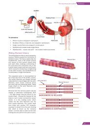
The ma<strong>in</strong> valves of the heart are the atrioventricular (AV)<br />
valves and the semilunar (SL) valves. The AV valves are<br />
located between the atria and the ventricles and prevent<br />
the back-flow of blood from the ventricles <strong>in</strong>to the atria.<br />
As the ventricles contract, pressure rises and forces the AV<br />
valves to snap shut, allow<strong>in</strong>g blood to be directed through<br />
the arteries leav<strong>in</strong>g the heart (pulmonary artery and aorta).<br />
SOMETHING EXTRA<br />
As the AV valves snap<br />
shut, they are anchored<br />
<strong>in</strong> place by tendonlike<br />
chords (chordae<br />
tend<strong>in</strong>eae) which prevent<br />
the valve flaps from be<strong>in</strong>g<br />
pushed too far <strong>in</strong>to the<br />
atria.<br />
The SL valves are located at<br />
the base of the arteries leav<strong>in</strong>g<br />
the heart (aorta and pulmonary<br />
artery). After each contraction,<br />
there is a relative drop <strong>in</strong><br />
pressure with<strong>in</strong> the ventricles<br />
as they relax. At this po<strong>in</strong>t, the<br />
blood with<strong>in</strong> the pulmonary<br />
artery and aorta could potentially<br />
flow back <strong>in</strong>to the ventricles. To<br />
prevent this, both sets of arteries<br />
have SL valves positioned at the<br />
po<strong>in</strong>t where they emerge from the ventricles. As the blood<br />
moves back towards the ventricles, the SL valves snap<br />
shut so blood cannot re-enter.<br />
It is the sequential shutt<strong>in</strong>g of the valves dur<strong>in</strong>g the cardiac<br />
cycle that causes the dist<strong>in</strong>ct ‘lub-dub’ noises associated<br />
with the heartbeat.<br />
Superior<br />
vena<br />
cava<br />
Right<br />
pulmonary<br />
ve<strong>in</strong>s<br />
Right<br />
atrium<br />
Right<br />
ventricle<br />
Inferior<br />
vena cava<br />
Atrioventricular (AV)<br />
valves<br />
Aorta<br />
Pulmonary<br />
artery<br />
Figure 1.2 The heart<br />
Figure 1.3 The valves of<br />
the heart<br />
Left<br />
pulmonary<br />
ve<strong>in</strong>s<br />
Left<br />
atrium<br />
Left<br />
ventricle<br />
Semilunar<br />
(SL) valves<br />
MEMORY JOGGER – HEART CIRCULATION<br />
The heart is stimulated to contract by a complex series of <strong>in</strong>tegrated systems. The heart’s pacemaker –<br />
the s<strong>in</strong>oatrial (SA) node – <strong>in</strong>itiates the cardiac muscle contraction. The SA node is located <strong>in</strong> the wall of<br />
the right atrium. The myocardium (heart muscle) is stimulated to contract about 72 times per m<strong>in</strong>ute<br />
by the SA node as part of the autonomic nervous system.<br />
5 | Copyright © 2017 <strong>Active</strong> <strong>IQ</strong> Ltd. Not for resale


