
You also want an ePaper? Increase the reach of your titles
YUMPU automatically turns print PDFs into web optimized ePapers that Google loves.
Manual<br />
<strong>Level</strong> 2 <strong>Certificate</strong><br />
<strong>in</strong> <strong>Gym</strong> Instruct<strong>in</strong>g<br />
Version A<strong>IQ</strong>005803
The skeletal system<br />
Section 1<br />
Section 1: The skeletal system<br />
Without the skeleton, we would be a heap of tissues all over the floor. It makes up almost one fifth of body weight to<br />
give us a flexible framework with which to move, protect and support <strong>in</strong>ternal and external systems.<br />
Structure of the skeleton<br />
The skeletal system can be classified as two ma<strong>in</strong> structures:<br />
Bone<br />
Calcified connective tissue that forms most of the adult<br />
skeleton. There are around 206 bones <strong>in</strong> the body and they<br />
are connected via a series of different types of jo<strong>in</strong>t.<br />
Cartilage Bone<br />
Dense, durable, tough fibrous connective tissue that is able<br />
to withstand compression forces. There are three types of<br />
cartilage found <strong>in</strong> the body, each fulfill<strong>in</strong>g a separate function.<br />
Types of cartilage<br />
The three types of cartilage found <strong>in</strong> the human body are:<br />
• Hyal<strong>in</strong>e cartilage: This is the tissue that forms the temporary skeleton of the foetus, which is eventually<br />
replaced by bone when calcium is deposited. It is found at the end of the long bones that meet to form the<br />
synovial jo<strong>in</strong>ts.<br />
• Elastic cartilage: This is similar to hyal<strong>in</strong>e cartilage, except that it has more fibres and most of these are<br />
made up of elast<strong>in</strong> as opposed to collagen. Elastic cartilage has the ability to rega<strong>in</strong> and return to its orig<strong>in</strong>al<br />
shape. It is found <strong>in</strong> the ear, the walls of the Eustachian tube and the epiglottis, which are all places that<br />
require a specific shape to be ma<strong>in</strong>ta<strong>in</strong>ed.<br />
• Fibrocartilage: This cartilage is thicker and stronger than the other types and has limited distribution with<strong>in</strong><br />
the body. It forms various shapes depend<strong>in</strong>g on its role and acts like a shock absorber <strong>in</strong> cartilag<strong>in</strong>ous jo<strong>in</strong>ts.<br />
The skeleton<br />
The skeleton is split <strong>in</strong>to two ma<strong>in</strong> sections:<br />
cranium<br />
cranium<br />
Pr<strong>in</strong>ciples of anatomy, physiology and fitness<br />
Axial skeleton<br />
Bones that form the<br />
ma<strong>in</strong> frame or axis:<br />
The sp<strong>in</strong>e, ribs and<br />
skull.<br />
Appendicular skeleton<br />
Bones that attach<br />
to the ma<strong>in</strong> frame<br />
(the appendages):<br />
The upper and lower<br />
limbs, the pelvic and<br />
shoulder girdles.<br />
clavicle<br />
sternum<br />
humerus<br />
rib<br />
lumbar vertebrae<br />
ulna<br />
radius<br />
pubis<br />
carpals<br />
metacarpals<br />
ischium<br />
femur<br />
patella<br />
tibia<br />
tarsals<br />
metatarsals<br />
phalanges<br />
cervical<br />
vertebrae<br />
scapula<br />
humerus<br />
thoracic<br />
vertebrae<br />
ilium<br />
sacrum<br />
coccyx<br />
phalanges<br />
femur<br />
fibula<br />
tibia<br />
Copyright © 2018 <strong>Active</strong> <strong>IQ</strong> Ltd. Not for resale 3
The neuromuscular system<br />
Section 2<br />
Section 2: The neuromuscular<br />
system<br />
All of the <strong>in</strong>ternal and external muscles <strong>in</strong> the body and the nerves serv<strong>in</strong>g them make up the neuromuscular<br />
system. Every movement your body makes requires communication between the bra<strong>in</strong> and muscles, some of which<br />
you don’t even have to th<strong>in</strong>k about, such as your digestive muscles break<strong>in</strong>g down <strong>in</strong>gested food.<br />
The muscular system<br />
There are over 600 muscles <strong>in</strong> the body, mak<strong>in</strong>g up around 40% of a person’s total weight.<br />
The bones and jo<strong>in</strong>ts create the framework of levers (bones) and pivots (jo<strong>in</strong>ts) which give the body the potential to<br />
move, but this framework cannot move on its own. It is the contraction and relaxation of muscles that br<strong>in</strong>g about<br />
movement.<br />
The muscular system produces a cont<strong>in</strong>uous and wide-rang<strong>in</strong>g number of actions, such as bodily movements<br />
(e.g. walk<strong>in</strong>g and jump<strong>in</strong>g) and the power<strong>in</strong>g of <strong>in</strong>ternal processes (e.g. contraction of the heart muscle and focuss<strong>in</strong>g<br />
of the eye).<br />
KEY<br />
POINT<br />
Types of muscle tissue<br />
There are three types of muscle tissue and each one has a different<br />
function. The three types are:<br />
• Cardiac muscle (myocardium), e.g. the heart.<br />
• Smooth muscle, e.g. the walls of the small <strong>in</strong>test<strong>in</strong>e.<br />
• Skeletal muscle (striated), e.g. the hamstr<strong>in</strong>gs or triceps.<br />
Contraction of the heart<br />
is controlled by the<br />
s<strong>in</strong>oatrial node (SAN).<br />
The set rhythm of the<br />
heart (on average,<br />
72bpm at rest) is called<br />
autorhythmicity.<br />
Pr<strong>in</strong>ciples of anatomy, physiology and fitness<br />
Copyright © 2018 <strong>Active</strong> <strong>IQ</strong> Ltd. Not for resale 13
The neuromuscular system<br />
Section 2<br />
To summarise:<br />
• Whole muscle is wrapped <strong>in</strong> epimysium.<br />
• Bundles of fibres, or fasciculi, are wrapped <strong>in</strong> perimysium.<br />
• S<strong>in</strong>gle muscle fibres are wrapped <strong>in</strong> endomysium.<br />
• Myofibrils are located <strong>in</strong>side s<strong>in</strong>gle fibres.<br />
• Myofilaments – myos<strong>in</strong> and act<strong>in</strong> ‒ are located <strong>in</strong>side sarcomeres.<br />
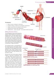
Slid<strong>in</strong>g filament theory<br />
The slid<strong>in</strong>g filament theory was proposed by<br />
Huxley <strong>in</strong> 1954 to expla<strong>in</strong> the contraction of<br />
skeletal muscle. The theory states that the<br />
myofilaments, act<strong>in</strong> (a th<strong>in</strong> prote<strong>in</strong> strand)<br />
and myos<strong>in</strong> (a thick prote<strong>in</strong> strand) slide<br />
over each other, creat<strong>in</strong>g a shorten<strong>in</strong>g of<br />
the sarcomere (the contractile units <strong>in</strong> the<br />
muscle where myos<strong>in</strong> and act<strong>in</strong> are found),<br />
which causes the shorten<strong>in</strong>g or lengthen<strong>in</strong>g<br />
of the entire muscle. The myofilaments do<br />
not decrease <strong>in</strong> length themselves.<br />
This proposed action is accomplished by<br />
the unique structure of the prote<strong>in</strong>, myos<strong>in</strong>.<br />
The myos<strong>in</strong> filaments are shaped like golf<br />
clubs and form cross bridges with the act<strong>in</strong><br />
filaments. Each myos<strong>in</strong> molecule (there<br />
are many) has two project<strong>in</strong>g heads. These<br />
heads attach to the act<strong>in</strong> filaments and<br />
pull them <strong>in</strong> closer.<br />
Act<strong>in</strong> filament<br />
Myos<strong>in</strong> filament<br />
Muscle fibre<br />
Myofibril<br />
Pr<strong>in</strong>ciples of anatomy, physiology and fitness<br />
Stimulus from the nervous system and the<br />
release of adenos<strong>in</strong>e triphosphate (ATP)<br />
– the high-energy molecule stored on the<br />
myos<strong>in</strong> head – provide the impetus for the<br />
head to ‘nod’ <strong>in</strong> what is termed the ‘power<br />
stroke’. It is this nodd<strong>in</strong>g action which<br />
‘slides’ the th<strong>in</strong> act<strong>in</strong> filaments over the<br />
thick myos<strong>in</strong> filaments. The myos<strong>in</strong> head<br />
then b<strong>in</strong>ds with another ATP molecule,<br />
caus<strong>in</strong>g it to detach from the act<strong>in</strong>-b<strong>in</strong>d<strong>in</strong>g<br />
site, which is known as the ‘recovery<br />
stroke’. It is then able to attach to the next<br />
b<strong>in</strong>d<strong>in</strong>g site and perform the same rout<strong>in</strong>e.<br />
SARCOMERE IS RELAXED<br />
SARCOMERE IS CONTRACTED<br />
Copyright © 2018 <strong>Active</strong> <strong>IQ</strong> Ltd. Not for resale 15
Section 3<br />
Cardiovascular and respiratory systems<br />
Heart valves<br />
There are a number of different valves around the heart, which all perform slightly different tasks.<br />
The atrioventricular (AV) valves separate the atria and ventricles and prevent the flow of blood back <strong>in</strong>to the atria<br />
dur<strong>in</strong>g ventricular contraction.<br />
The semilunar valves prevent the flow of blood back <strong>in</strong>to the right (pulmonary valve) and left ventricles (aortic valve)<br />
dur<strong>in</strong>g ventricular relaxation.<br />
Heart rate<br />
The heart is stimulated to contract by a complex series of <strong>in</strong>tegrated systems. The heart’s pacemaker – the s<strong>in</strong>oatrial<br />
node (SAN) – <strong>in</strong>itiates cardiac muscle contraction. The SAN is located <strong>in</strong> the wall of the right atrium. The myocardium<br />
(heart muscle) is stimulated to contract about 72 times per m<strong>in</strong>ute by the SAN as part of the autonomic nervous<br />
system.<br />
28<br />
Copyright © 2018 <strong>Active</strong> <strong>IQ</strong> Ltd. Not for resale
Cardiovascular and respiratory systems<br />
Section 3<br />
Nose and mouth<br />
Nose<br />
Larynx<br />
Trachea<br />
Pharynx (throat) and<br />
larynx (voice box)<br />
Lungs<br />
Trachea<br />
Bronchi<br />
Bronchioles<br />
Diaphragm<br />
Bronchi / bronchus<br />
The mechanics of breath<strong>in</strong>g<br />
The two ma<strong>in</strong> mechanisms that trigger the human body to breathe are:<br />
• Ris<strong>in</strong>g levels of carbon dioxide <strong>in</strong> the blood.<br />
• Stretch receptors <strong>in</strong> the respiratory muscles (<strong>in</strong>tercostal muscles) becom<strong>in</strong>g stretched.<br />
The ma<strong>in</strong> muscles <strong>in</strong>volved <strong>in</strong> the action of breath<strong>in</strong>g are the diaphragm and the <strong>in</strong>ternal and external <strong>in</strong>tercostal<br />
muscles.<br />
The ma<strong>in</strong> phases of the breath<strong>in</strong>g cycle are:<br />
• Inspiration/<strong>in</strong>halation – draw<strong>in</strong>g air<br />
<strong>in</strong>to the lungs.<br />
• Expiration/exhalation – expell<strong>in</strong>g air<br />
from the lungs.<br />
Right lung<br />
Trachea<br />
Bronchioles<br />
Alveoli<br />
Left lung<br />
Pr<strong>in</strong>ciples of anatomy, physiology and fitness<br />
There is also a short pause before both<br />
<strong>in</strong>spiration and expiration.<br />
Dur<strong>in</strong>g <strong>in</strong>spiration, the diaphragm muscle<br />
contracts, caus<strong>in</strong>g the normal ‘dome shape’<br />
to flatten. The external <strong>in</strong>tercostal muscles<br />
also contract, rais<strong>in</strong>g the ribcage. These<br />
actions <strong>in</strong>crease the chest cavity volume.<br />
This <strong>in</strong>crease <strong>in</strong> volume creates a negative<br />
pressure between the air <strong>in</strong> the lungs and<br />
air <strong>in</strong> the atmosphere. This is very much<br />
like a vacuum effect <strong>in</strong> which the negative<br />
pressure sucks air <strong>in</strong>to the lungs until the<br />
two pressures are balanced.<br />
Right<br />
bronchus<br />
Left<br />
bronchus<br />
Bronchiole<br />
Term<strong>in</strong>al<br />
bronchiole<br />
Alveoli<br />
Copyright © 2018 <strong>Active</strong> <strong>IQ</strong> Ltd. Not for resale 33
The digestive system<br />
Section 5<br />
Section 5: The digestive<br />
system<br />
The digestive system is responsible for the <strong>in</strong>take, breakdown, use and removal of food and dr<strong>in</strong>k. An efficient<br />
digestive system tells us when we are hungry, full and thirsty by send<strong>in</strong>g messages to and from the bra<strong>in</strong> via the<br />
nervous system. It extracts important nutrients for storage and immediate use and removes any waste.<br />
The digestive system has four stages:<br />
Ingestion<br />
Digestion<br />
Absorption<br />
Elim<strong>in</strong>ation<br />
Food enter<strong>in</strong>g<br />
the body<br />
through<br />
the mouth<br />
and be<strong>in</strong>g<br />
chewed.<br />
Break<strong>in</strong>g<br />
down of<br />
food through<br />
mechanical<br />
(smooth<br />
muscle<br />
action) and<br />
chemical<br />
(release of<br />
enzymes)<br />
processes.<br />
The pass<strong>in</strong>g<br />
of food<br />
<strong>in</strong>to the<br />
bloodstream<br />
to be used<br />
by the body’s<br />
tissues.<br />
The removal<br />
of waste.<br />
Journey through the alimentary canal (also known as<br />
the digestive tract/gastro<strong>in</strong>test<strong>in</strong>al tract/gut)<br />
Food’s journey through the alimentary canal can take up to 24 hours and covers a distance of around 9m (30 feet)<br />
from <strong>in</strong>gestion through the mouth to excretion through the anus.<br />
Mouth<br />
This is the entry po<strong>in</strong>t of food and where it beg<strong>in</strong>s to be broken down<br />
through the process of mastication (chew<strong>in</strong>g) <strong>in</strong>to a ball, or bolus.<br />
Pr<strong>in</strong>ciples of anatomy, physiology and fitness<br />
Oesophagus (gullet)<br />
This is a thick-walled, muscular tube that carries broken down food<br />
from the mouth to the stomach.<br />
Stomach<br />
The stomach is a muscular bag located on the left side of the upper<br />
abdomen. It breaks down food further by releas<strong>in</strong>g enzymes, and also<br />
kills bacteria.<br />
Small <strong>in</strong>test<strong>in</strong>e<br />
The small <strong>in</strong>test<strong>in</strong>e is a small, tightly folded tube that receives<br />
food from the stomach. It is the major site of digestion with<strong>in</strong> the<br />
alimentary canal. Its role is to absorb important nutrients <strong>in</strong>to the<br />
bloodstream to be passed to the body’s tissues and used for energy.<br />
The small <strong>in</strong>test<strong>in</strong>e is divided <strong>in</strong>to three sections: the duodenum,<br />
jejunum and ileum.<br />
The small <strong>in</strong>test<strong>in</strong>e is about as large<br />
as an adult’s middle f<strong>in</strong>ger but, when<br />
stretched out, it is about 22 feet<br />
(6.7m) long.<br />
Copyright © 2018 <strong>Active</strong> <strong>IQ</strong> Ltd. Not for resale 43
The importance of a healthy lifestyle<br />
Section 3<br />
Section 3: The importance of a<br />
healthy lifestyle<br />
Current prevalence of obesity <strong>in</strong> the UK<br />
27% of adults <strong>in</strong> England are obese and a further 36% are overweight. A summary of the most recent f<strong>in</strong>d<strong>in</strong>gs can<br />
be found below:<br />
Obesity is normally<br />
def<strong>in</strong>ed as hav<strong>in</strong>g a<br />
BMI of 30+<br />
18.5<br />
25<br />
30<br />
Underweight<br />
Normal Weight<br />
Overweight<br />
Obese<br />
Men are more likely to be<br />
overweight, but obesity<br />
rates are the same for<br />
both genders<br />
Obese<br />
Overweight<br />
Female<br />
Male<br />
Female<br />
Male<br />
9% of children <strong>in</strong><br />
England are obese by<br />
the age of 4-5<br />
9%<br />
27% of adults <strong>in</strong> England<br />
are obese. A further 36%<br />
are overweight<br />
27%<br />
Those aged 55-64 are<br />
most likely to be obese;<br />
16-24s are least likely<br />
Least<br />
deprived<br />
Most<br />
deprived<br />
16%<br />
36%<br />
16-24 55-64<br />
10-11 year olds <strong>in</strong> the<br />
most deprived areas<br />
are much more likely<br />
to be obese<br />
15%<br />
26%<br />
Rates of excess weight<br />
are highest <strong>in</strong> the North<br />
East and lowest <strong>in</strong><br />
London<br />
NE<br />
Yorks<br />
E Mids<br />
W Mids<br />
NW<br />
East<br />
SW<br />
SE<br />
London<br />
69%<br />
59%<br />
Obesity rates have<br />
grown slightly <strong>in</strong> the<br />
last decade<br />
52.9%<br />
61.8%<br />
62.9%<br />
1993 2004 2015<br />
In England, rates<br />
of obesity drug<br />
prescriptions are highest<br />
<strong>in</strong> Stoke North<br />
Stoke-on-Trent N<br />
Leigh<br />
Camborne & Rrth<br />
Knowsley<br />
Barnsley E<br />
22 per 1,000<br />
Conduct<strong>in</strong>g client consultations to support positive behaviour change<br />
Prescrib<strong>in</strong>g rates for<br />
obesity drugs have<br />
fallen <strong>in</strong> all UK countries<br />
s<strong>in</strong>ce 2008<br />
The number of bariatric<br />
surgeries on obese<br />
patients fell <strong>in</strong> the last<br />
three years<br />
UK obesity rates are<br />
below those of USA and<br />
Australia but above those<br />
of France & Germany<br />
ENG<br />
SCO<br />
WAL<br />
NI<br />
2008<br />
2014<br />
1,951<br />
2006/07<br />
8,794<br />
6,032<br />
2014/15<br />
USA<br />
AUS<br />
UK<br />
GER<br />
FRA<br />
JAP 3.9<br />
38.2<br />
27.9<br />
25.6<br />
23.6<br />
16.9<br />
House of Commons brief<strong>in</strong>g paper – Obesity statistics, 2017<br />
Copyright © 2018 <strong>Active</strong> <strong>IQ</strong> Ltd. Not for resale 47