Pediatric Epilepsy - Portal Neonatal
Pediatric Epilepsy - Portal Neonatal
Pediatric Epilepsy - Portal Neonatal

Create successful ePaper yourself
Turn your PDF publications into a flip-book with our unique Google optimized e-Paper software.
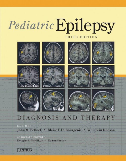
<strong>Pediatric</strong> <strong>Epilepsy</strong><br />
Diagnosis and Therapy<br />
Third Edition
<strong>Pediatric</strong> <strong>Epilepsy</strong><br />
Diagnosis and Therapy<br />
Third Edition<br />
EDITORS<br />
JOHN M. PELLOCK, MD<br />
Chairman, Division of Child Neurology<br />
Vice Chair, Department of Neurology<br />
Virginia Commonwealth University<br />
Medical College of Virginia<br />
Richmond, Virginia<br />
BLAISE F.D. BOURGEOIS, MD<br />
Professor of Neurology<br />
Harvard Medical School<br />
Director, Division of <strong>Epilepsy</strong> and Clinical Neurophysiology<br />
Joseph J. Volpe Chair<br />
Children’s Hospital Boston<br />
Boston, Massachusetts<br />
W. EDWIN DODSON, MD<br />
Associate Vice Chancellor and Associate Dean<br />
Professor of <strong>Pediatric</strong>s and Neurology<br />
Washington University School of Medicine<br />
St. Louis Children’s Hopsital<br />
St. Louis, Missouri<br />
ASSOCIATE EDITORS<br />
DOUGLAS R. NORDLI, JR., MD<br />
Director, Children’s Memorial <strong>Epilepsy</strong> Center<br />
Lorna S. and James P. Langdon Chair of <strong>Pediatric</strong> <strong>Epilepsy</strong><br />
Northwestern University’s Feinberg School of Medicine<br />
Chicago, Illinois<br />
RAMAN SANKAR, MD, PHD<br />
Professor and Chief, <strong>Pediatric</strong> Neurology<br />
Rubin Brown Distinguished Chair<br />
David Geffen School of Medicine<br />
Mattel Children’s Hospital<br />
University of California, Los Angeles<br />
Los Angeles, California<br />
New York
Acquisitions Editor: R. Craig Percy<br />
Cover Designer: Steven Pisano<br />
Compositor and Indexing: Publication Services, Inc.<br />
Printer: Sheridan Books, Inc.<br />
Visit our website at www.demosmedpub.com<br />
© 2008 Demos Medical Publishing, LLC. All rights reserved. This book is protected by copyright. No<br />
part of it may be reproduced, stored in a retrieval system, or transmitted in any form or by any means,<br />
electronic, mechanical, photocopying, recording, or otherwise, without the prior written<br />
permission of the publisher.<br />
Library of Congress Cataloging-in-Publication Data<br />
<strong>Pediatric</strong> epilepsy:diagnosis and therapy/edited by John M. Pellock, Blaise F.D. Bourgeois, W. Edwin<br />
Dodson;associate editors, Douglas R. Nordli Jr., Raman Sankar.—3rd ed.<br />
p.;cm.<br />
Includes bibliographical references and index.<br />
ISBN-13: 978-1-933864-16-7 (hardcover:alk. paper)<br />
ISBN-10: 1-933864-16-8 (hardcover:alk. paper)<br />
1. <strong>Epilepsy</strong> in children. I. Pellock, John M. II. Bourgeois, Blaise F.D. III. Dodson, W. Edwin<br />
[DNLM: 1. <strong>Epilepsy</strong>. 2. Child. 3. <strong>Epilepsy</strong>—therapy. 4. Infant.WL 385 P3795 2008]<br />
RJ496.E6P43 2008<br />
618.92'853—dc22<br />
2007032323<br />
Medicine is an ever-changing science undergoing continual development. Research and clinical<br />
experience are continually expanding our knowledge, in particular our knowledge of proper treatment<br />
and drug therapy. The authors, editors, and publisher have made every effort to ensure that all<br />
information in this book is in accordance with the state of knowledge at the time of production of<br />
the book.<br />
Nevertheless, this does not imply or express any guarantee or responsibility on the part of the authors,<br />
editors, or publisher with respect to any dosage instructions and forms of application stated in the book.<br />
Every reader should examine carefully the package inserts accompanying each drug and check with a<br />
physician or specialist whether the dosage schedules mentioned therein or the contraindications stated<br />
by the manufacturer differ from the statements made in this book. Such examination is particularly<br />
important with drugs that are rarely used or have newly been released on the market. Every dosage<br />
schedule or every form of application used is entirely at the reader’s own risk and responsibility. The<br />
editors and publisher welcome any reader to report to the publisher any discrepancies or inaccuracies<br />
noticed.<br />
Special discounts on bulk quantities of Demos Medical Publishing books are available to corporations,<br />
professional associations, pharmaceutical companies, health care organizations, and other qualifying<br />
groups. For details, please contact:<br />
Special Sales Department<br />
Demos Medical Publishing<br />
386 Park Avenue South, Suite 301<br />
New York, NY 10016<br />
Phone: 800–532–8663 or 212–683–0072<br />
Fax: 212–683–0118<br />
Email: orderdept@demosmedpub.com<br />
Made in the United States of America<br />
07 08 09 10 5 4 3 2 1
Dedication<br />
To our families and to the children and families for whom we care.
Contents<br />
Preface xiii<br />
Contributors xv<br />
I BASIC MECHANISMS<br />
Section Editor: Raman Sankar<br />
1. Pathophysiology of Seizures and <strong>Epilepsy</strong> in the Immature Brain:<br />
Cells, Synapses, and Circuits<br />
Libor Velíšek and Solomon L. Moshé 1<br />
2. Ion Channels, Membranes, and Molecules in <strong>Epilepsy</strong><br />
and Neuronal Excitability<br />
Laxmikant S. Deshpande and Robert J. DeLorenzo 31<br />
3. Channel Mutations in <strong>Epilepsy</strong>: A Neurodevelopmental Perspective<br />
Edward C. Cooper 47<br />
4. Metabolic and Pharmacologic Consequences of Seizures<br />
Michael V. Johnston and John W. McDonald 65<br />
5. Neuropathologic Substrates of <strong>Epilepsy</strong><br />
Caroline R. Houser and Harry V. Vinters 75<br />
6. Epileptogenic Cerebral Cortical Malformations<br />
Annapurna Poduri, Bernard S. Chang, and Christopher A. Walsh 101<br />
7. Genetic Influences on the Risk for <strong>Epilepsy</strong><br />
Asuri N. Prasad and Chitra Prasad 117<br />
vii
viii<br />
II CLASSIFICATION, EPIDEMIOLOGY, ETIOLOGY, AND DIAGNOSIS<br />
Section Editor: W. Edwin Dodson<br />
8. Classification of Epilepsies in Childhood<br />
Douglas R. Nordli, Jr. 137<br />
9. Epidemiology of <strong>Epilepsy</strong> in Children<br />
W. Allen Hauser and P. Nina Banerjee 147<br />
10. An Approach to the Child with Paroxysmal Phenomena with<br />
Emphasis on Nonepileptic Disorders<br />
Arthur L. Prensky and Amir Pshytycky 165<br />
11. Evaluating the Child with Seizure<br />
Sejal Jain and Lawrence D. Morton 185<br />
12. The Use of Electroencephalography in the<br />
Diagnosis of <strong>Epilepsy</strong> in Childhood<br />
Douglas R. Nordli, Jr. and Timothy A. Pedley 195<br />
13. Basics of Neuroimaging in <strong>Pediatric</strong> <strong>Epilepsy</strong><br />
James M. Johnston, Matthew D. Smyth, and Robert C. McKinstry 213<br />
III AGE-RELATED SYNDROMES<br />
Section Editors: Blaise F. D. Bourgeois and Douglas R. Nordli, Jr.<br />
NEWBORNS AND EARLY INFANCY<br />
14. <strong>Neonatal</strong> Seizures<br />
Eli M. Mizrahi 227<br />
15. Severe Encephalopathic <strong>Epilepsy</strong> in Early Infancy<br />
Shunsuke Ohtahara and Yasuko Yamatogi 241<br />
INFANCY<br />
CONTENTS<br />
16. Severe Encephalopathic <strong>Epilepsy</strong> in Infants:<br />
Infantile Spasms (West Syndrome)<br />
Richard A. Hrachovy and James D. Frost, Jr. 249<br />
17. Myoclonic Epilepsies in Infancy and Early Childhood<br />
Pierre Genton 269<br />
18. Partial Epilepsies in Infancy<br />
Kazuyoshi Watanabe 283<br />
19. Febrile Seizures<br />
Shlomo Shinnar and Tracy A. Glauser 293
20. Generalized <strong>Epilepsy</strong> with Febrile Seizures Plus (GEFS+)<br />
Douglas R. Nordli, Jr. and John M. Pellock 303<br />
CHILDHOOD<br />
21. Lennox-Gastaut Syndrome<br />
Diego A. Morita and Tracy A. Glauser 307<br />
22. Childhood Absence Epilepsies<br />
Phillip L. Pearl and Gregory L. Holmes 323<br />
23. Benign Focal Epilepsies of Childhood<br />
Colin D. Ferrie, Douglas R. Nordli, Jr., and Chrysostomos P. Panayiotopoulos 335<br />
24. The Landau-Kleffner Syndrome and <strong>Epilepsy</strong> with<br />
Continuous Spike-Waves during Sleep<br />
James J. Riviello Jr. and Stavros Hadjiloizou 351<br />
LATE CHILDHOOD AND ADOLESCENCE<br />
25. Idiopathic Generalized <strong>Epilepsy</strong> of Adolescence<br />
Reza Behrouz and Selim R. Benbadis 359<br />
26. Progressive Myoclonus Epilepsies<br />
Samuel F. Berkovic 367<br />
VARIABLE AGE OF ONSET<br />
CONTENTS<br />
27. Localization-Related Epilepsies: Simple Partial Seizures,<br />
Complex Partial Seizures, and Rasmussen Syndrome<br />
Prakash Kotagal 377<br />
28. Selected Disorders Associated with <strong>Epilepsy</strong><br />
Lawrence D. Morton 387<br />
IV GENERAL PRINCIPLES OF THERAPY<br />
Section Editor: John M. Pellock<br />
29. Treatment Decisions in Childhood Seizures<br />
Shlomo Shinnar and Christine O’Dell 401<br />
30. Comparative Anticonvulsant Profile and Proposed<br />
Mechanisms of Action of Antiepileptic Drugs<br />
H. Steve White and Karen S. Wilcox 413<br />
31. Evidence-Based Medicine Issues Related to Drug Selection<br />
Tracy A. Glauser and Diego A. Morita 429<br />
32. Combination Drug Therapy: Monotherapy Versus Polytherapy<br />
Blaise F. D. Bourgeois 441<br />
ix
x<br />
CONTENTS<br />
33. Adverse Effects of Antiepileptic Drugs<br />
L. James Willmore, James W. Wheless, and John M. Pellock 449<br />
34. Status Epilepticus and Acute Seizures<br />
David J. Leszczyszyn and John M. Pellock 461<br />
35. The Female Patient and <strong>Epilepsy</strong><br />
Mark S. Yerby 477<br />
36. Teratogenic Effects of Antiepileptic Medications<br />
Torbjörn Tomson and Dina Battino 489<br />
37. Pharmacokinetic Principles of Antiepileptic Therapy in Children<br />
W. Edwin Dodson 503<br />
38. Dosage Form Considerations in the Treatment of <strong>Pediatric</strong> <strong>Epilepsy</strong><br />
William R. Garnett and James C. Cloyd 515<br />
39. Principles of Drug Interactions: Implications for Treatment<br />
with Antiepileptic Drugs<br />
Barry E. Gidal 535<br />
V ANTIEPILEPTIC DRUGS AND KETOGENIC DIET<br />
Section Editors: Blaise F. D. Bourgeois and Raman Sankar<br />
40. ACTH and Steroids<br />
Rajesh RamachandranNair and O. Carter Snead, III 543<br />
41. Benzodiazepines<br />
Kevin Farrell and Aspasia Michoulas 557<br />
42. Carbamazepine and Oxcarbazepine<br />
W. Edwin Dodson 567<br />
43. Ethosuximide, Methsuximide, and Trimethadione<br />
Blaise F. D. Bourgeois 579<br />
44. Felbamate<br />
Blaise F. D. Bourgeois 585<br />
45. Gabapentin and Pregabalin<br />
Gregory L. Holmes and Philip L. Pearl 593<br />
46. Lamotrigine<br />
John M. Pellock 603<br />
47. Levetiracetam<br />
Raman Sankar and W. Donald Shields 611<br />
48. Barbiturates and Primidone<br />
Robert S. Rust 621
49. Phenytoin and Related Drugs<br />
W. Edwin Dodson 639<br />
50. Sulthiame<br />
Dietz Rating, Nicole Wolf, and Thomas Bast 653<br />
51. Tiagabine<br />
Shlomo Shinnar, Richard Civil, and Kenneth W. Sommerville 661<br />
52. Topiramate<br />
Tracy A. Glauser 671<br />
53. Valproate<br />
Blaise F. D. Bourgeois 685<br />
54. Vigabatrin<br />
Günter Krämer and Gabriele Wohlrab 699<br />
55. Vitamins, Herbs, and Other Alternative Therapies<br />
Orrin Devinsky, Daniel Miles, and Josiane LaJoie 711<br />
56. Zonisamide<br />
John F. Kerrigan and John M. Pellock 727<br />
57. The Ketogenic Diet<br />
Douglas R. Nordli, Jr. and Darryl C. De Vivo 739<br />
58. Inflammation, <strong>Epilepsy</strong>, and Anti-Inflammatory Therapies<br />
Stéphane Auvin and Raman Sankar 751<br />
59. Antiepileptic Drugs in Development<br />
John R. Pollard and Jacqueline A. French 759<br />
VI EPILEPSY SURGERY AND VAGUS NERVE STIMULATION<br />
Section Editor: Douglas R. Nordli, Jr.<br />
CONTENTS<br />
60. Surgical Evaluation<br />
Michael Duchowny 769<br />
61. Advanced Neuroimaging: PET-MRI Fusion<br />
and Diffusion Tensor Imaging<br />
Noriko Salamon 785<br />
62. Surgical Treatment of Therapy-Resistant <strong>Epilepsy</strong> in Children<br />
Gary W. Mathern and Olivier Delalande 791<br />
63. Outcome of <strong>Epilepsy</strong> Surgery in Childhood<br />
Shekhar Patil, J. Helen Cross, and William Harkness 801<br />
64. Vagus Nerve Stimulation Therapy in <strong>Pediatric</strong> Patients: Use<br />
and Effectiveness<br />
James W. Wheless 811<br />
xi
xii<br />
VII PSYCHOSOCIAL ASPECTS<br />
Section Editor: W. Edwin Dodson<br />
CONTENTS<br />
65. Economics of <strong>Pediatric</strong> <strong>Epilepsy</strong><br />
Charles E. Begley 831<br />
66. Quality of Life in Children with <strong>Epilepsy</strong><br />
Joan K. Austin and Nancy Santilli 837<br />
67. <strong>Epilepsy</strong>, Cerebral Palsy, and IQ<br />
W. Edwin Dodson 847<br />
68. Academic Deficits and Interventions in <strong>Pediatric</strong> <strong>Epilepsy</strong><br />
Caroline E. Bailey and Rochelle Caplan 865<br />
69. Cognitive Side Effects of Antiepileptic Drugs<br />
David E. Mandelbaum and Christine L. Trask 873<br />
Index 883
Preface<br />
As with the first and second editions, the goal of this<br />
third edition of <strong>Pediatric</strong> <strong>Epilepsy</strong>: Diagnosis and Therapy<br />
is to assist all professionals involved in the care of pediatric<br />
patients with seizures and epilepsy. Our goal continues<br />
to be the perfect result: no seizures, no side effects, and<br />
no stigma to limit these children from achieving their<br />
full potential.<br />
The scope and depth of coverage of this book remains<br />
unique in its field. We have again tried to balance discussions<br />
of practical medical management with the scientific<br />
basis of epilepsy and its treatment in a clear and concise<br />
manner. The book focuses on the special issues of children<br />
with epilepsy and is intended as both a practical guide and<br />
a reference for clinicians and investigators. With many<br />
more options for the treatment of epilepsy—including new<br />
antiepileptic drugs (AEDs), vagus nerve stimulation, the<br />
reintroduction of the ketogenic diet, increased emphasis<br />
on quality of life, as well as improved presurgical evaluation<br />
and surgical intervention—hope for a more normal<br />
life for all children with epilepsy should continue to grow.<br />
To accomplish all the new goals set for this third edition,<br />
the previous editors asked Drs. Douglas R. Nordli and<br />
Raman Sankar to join them as associate editors.<br />
Since the publication of the second edition, there<br />
has been a rapid expansion of basic knowledge, diagnostic<br />
techniques, and treatments affecting the management<br />
of epilepsy, including advances in basic neurosciences,<br />
genetics, definition of syndromes, as well as medical and<br />
surgical therapeutic approaches. Because of the increasing<br />
familiarity and refinement of knowledge related to the<br />
concept of epilepsy syndromes, a new separate section<br />
xiii<br />
has been added to this third edition. This section consists<br />
of an expanded and comprehensive coverage of the syndromes<br />
by age of onset, taking into account the difficulties<br />
that can be encountered in making a fixed diagnosis<br />
of a seizure type or epilepsy syndrome in young infants.<br />
Although the precise diagnosis of epilepsy has become<br />
more challenging and complex, the enhanced specificity<br />
of the final diagnosis will continue to improve therapeutic<br />
and prognostic accuracy. The diagnostic process has been<br />
further improved by the growing knowledge of metabolic<br />
disturbances, disease processes, and genetics of various<br />
forms of epilepsy. This has led to an ever-expanding availability<br />
of new diagnostic tests, especially those related to<br />
DNA analysis. Although the greater choice of diagnostic<br />
testing does not always yield an etiology in every patient,<br />
those patients without established etiology still have more<br />
and better therapeutic options. There has been further<br />
growth in the knowledge about clinical pharmacology of<br />
AEDs. In particular, experience and comfort have grown<br />
with the AEDs that had just been released at the time of<br />
the second edition, with recognition of new indications<br />
for some of these drugs. Throughout the discussion of<br />
individual drugs, available knowledge is discussed regarding<br />
their specific use and pharmacokinetics in newborns,<br />
infants, older children, and adolescents. Adverse effects<br />
are also covered, to include newer aspects as well as agerelated,<br />
gender-related, and pregnancy-related issues.<br />
<strong>Epilepsy</strong> surgery has become one of the widely applied<br />
standard therapeutic approaches to pediatric epilepsy,<br />
and it has now truly been incorporated into the treatment<br />
paradigms.
xiv<br />
In this third edition, every chapter either has been<br />
markedly updated or is a newly written chapter that<br />
was not included in the second edition. In the initial section<br />
on basic mechanisms of epilepsy, a new chapter by<br />
Dr. Edward Cooper on the epileptic channelopathies has<br />
been added because of the rapid and clinically relevant<br />
expansion of knowledge in this area during the past few<br />
years. Coverage of epileptogenic cerebral cortical malformations<br />
has been enhanced by the contribution of<br />
Drs. Christopher Walsh, Ann Poduri, and Bernard Chang.<br />
A new section on age-related syndromes has been created.<br />
This section is divided into groups of syndromes<br />
by age of onset and provides a more comprehensive and<br />
detailed coverage by several international authorities<br />
in the field, such as Drs. Shunsuke Ohtahara, Richard<br />
Hrachovy, Kazuyoshi Watanabe, Pierre Genton, Shlomo<br />
Shinnar, Tracy Glauser, Gregory Holmes, Chrysostomos<br />
Panayiotopoulos, Prakash Kotagal, and Samuel Berkovic.<br />
All of the chapters in this section now follow a unified<br />
structure. The section on general principles of therapy<br />
now includes a discussion of the emerging evidencebased<br />
approach to AED selection by Dr. Tracy Glauser.<br />
The issues of fetal effects of epilepsy and fetal effects of<br />
AEDs are covered separately by Drs. Mark Yerby and<br />
Torbjörn Tomson.<br />
Systematic coverage of individual drugs and other<br />
medical treatment modalities was already quite extensive<br />
in the second edition but has now been enhanced and<br />
expanded to cover the newer drugs not covered previously,<br />
as well as updated to include new information on<br />
the previously discussed drugs. A chapter on sulthiame by<br />
Dr. Dietz Rating has been added, because of the unique role<br />
of this drug in the treatment of the common syndrome of<br />
benign rolandic epilepsy. Other new chapters cover vitamins,<br />
dietary considerations, and alternative therapies<br />
PREFACE<br />
(Dr. Orrin Devinsky); immunotherapy (Dr. Raman Sankar);<br />
and AEDs in development (Dr. Jacqueline French). The<br />
section on surgical treatment contains updated coverage of<br />
neurophysiologic evaluation (including magnetoencephalography),<br />
advanced imaging, surgical procedures, and outcomes,<br />
in addition to an update on vagus nerve stimulation.<br />
The section on psychosocial aspects has been expanded<br />
and includes chapters on costs of pediatric epilepsy, quality<br />
of life, intelligence, co-morbidities, and educational placement,<br />
as well as cognitive side effects of AEDs.<br />
We would be remiss in introducing this third<br />
edition without acknowledging the contribution of<br />
several of the previous authors who have now died.<br />
Drs. Kiffin Penry and Fritz Dreifuss, two of the founding<br />
fathers of pediatric epilepsy in the United States, have<br />
left us to carry on their work. Dr. Eric Lothman died<br />
quite prematurely, but his contribution to the understanding<br />
of the pathophysiology of seizures and epilepsy<br />
remains current even in this edition and will do so for<br />
years to come. In honoring these and others who have<br />
encouraged and guided our careers, we hope that this<br />
book meets the needs of those who care for children<br />
with epilepsy as well as the children and families who<br />
have to deal with seizures.<br />
For some, epilepsy will be a transient and distant<br />
memory, while for others epilepsy is an ever-present burden.<br />
Evaluating and treating these children in the most<br />
appropriate and efficient fashion while avoiding adverse<br />
cognitive and psychosocial effects remains both challenging<br />
and rewarding, and it requires state-of-the-art knowledge.<br />
We hope that you will find such knowledge in this<br />
book, and we hope that this third edition of <strong>Pediatric</strong><br />
<strong>Epilepsy</strong>: Diagnosis and Therapy will help you and your<br />
colleagues provide state-of-the-art care to your patients<br />
and their families.<br />
John M. Pellock, MD<br />
Blaise F.D. Bourgeois, MD<br />
W. Edwin Dodson, MD<br />
Douglas R. Nordli, Jr., MD<br />
Raman Sankar, MD, PhD
Contributors<br />
Joan K. Austin, DNS, RN, FAAN<br />
Distinguished Professor and Sally Reahard Chair<br />
Indiana University School of Nursing<br />
Indianapolis, Indiana<br />
Chapter 66: Quality of Life in Children with<br />
<strong>Epilepsy</strong><br />
Stéphane Auvin, MD, PhD<br />
Department of <strong>Pediatric</strong> Neurology<br />
Lille University Hospital<br />
Pharmacology Laboratory<br />
Lille Medical School<br />
Lille, France<br />
Chapter 58: Inflammation, <strong>Epilepsy</strong>, and Anti-<br />
Inflammatory Therapies<br />
Caroline E. Bailey, PhD<br />
Assistant Professor<br />
Department of Human Services<br />
California State University<br />
Fullerton, California<br />
Chapter 68: Academic Deficits and Interventions in<br />
<strong>Pediatric</strong> <strong>Epilepsy</strong><br />
P. Nina Banerjee, PhD<br />
Gertrude H. Sergievsky Center<br />
College of Physicians and Surgeons<br />
Columbia University<br />
New York, New York<br />
Chapter 9: Epidemiology of <strong>Epilepsy</strong> in Children<br />
xv<br />
Thomas Bast, MD<br />
Department of Paediatric Neurology<br />
<strong>Epilepsy</strong> Centre<br />
Children’s Hospital<br />
University of Heidelberg<br />
Heidelberg, Germany<br />
Chapter 50: Sulthiame<br />
Dina Battino, MD<br />
Fondazione I.R.C.C.S. Istituto Neurologico “Carlo Besta”<br />
Milan, Italy<br />
Chapter 36: Teratogenic Effects of Antiepileptic<br />
Medications<br />
Charles E. Begley<br />
Co-Director, Center for Health Services Research<br />
University of Texas Health Science Center at Houston<br />
Houston, Texas<br />
Chapter 65: Economics of <strong>Pediatric</strong> <strong>Epilepsy</strong><br />
Reza Behrouz, DO<br />
Assistant Professor<br />
Department of Neurology<br />
University of South Florida College of Medicine<br />
Tampa, Florida<br />
Chapter 25: Idiopathic Generalized <strong>Epilepsy</strong> of<br />
Adolescence<br />
Selim R. Benbadis, MD<br />
Professor and Director<br />
Comprehensive <strong>Epilepsy</strong> Program<br />
Departments of Neurology and Neurosurgery<br />
University of South Florida and Tampa General Hospital<br />
Tampa, Florida<br />
Chapter 25: Idiopathic Generalized <strong>Epilepsy</strong> of<br />
Adolescence
xvi<br />
Samuel F. Berkovic, MD, FRS<br />
Director, <strong>Epilepsy</strong> Research Centre<br />
Department of Medicine<br />
University of Melbourne<br />
West Heidelberg, Victoria, Australia<br />
Chapter 26: Progressive Myoclonus Epilepsies<br />
Blaise F. D. Bourgeois, MD<br />
Professor of Neurology<br />
Harvard Medical School<br />
Director, Division of <strong>Epilepsy</strong> and<br />
Clinical Neurophysiology<br />
Joseph J. Volpe Chair<br />
Children’s Hospital Boston<br />
Boston, Massachusetts<br />
Part III: Age-Related Syndromes, Section Editor<br />
Part V: Antiepileptic Drugs and Ketogenic Diet, Section<br />
Editor<br />
Chapter 32: Combination Drug Therapy:<br />
Monotherapy Versus Polytherapy<br />
Chapter 43: Ethosuximide, Methsuximide, and<br />
Trimethadione<br />
Chapter 44: Felbamate<br />
Chapter 53: Valproate<br />
Rochelle Caplan, MD<br />
Professor<br />
David Geffen School of Medicine<br />
University of California, Los Angeles<br />
Los Angeles, California<br />
Chapter 68: Academic Deficits and Interventions in<br />
<strong>Pediatric</strong> <strong>Epilepsy</strong><br />
Bernard S. Chang, MD, MMSc<br />
Assistant Professor of Neurology<br />
Harvard Medical School<br />
Comprehensive <strong>Epilepsy</strong> Center<br />
Beth Israel Deaconess Medical Center<br />
Boston, Massachusetts<br />
Chapter 6: Epileptogenic Cerebral Cortical<br />
Malformations<br />
Richard Civil<br />
Vice President, Global Product Safety<br />
Cephalon, Inc.<br />
West Chester, Pennsylvania<br />
Chapter 51: Tiagabine<br />
James C. Cloyd, MD<br />
Professor and Lawrence C. Weaver Endowed<br />
Chair–Orphan Drug Development<br />
Director, Center for Orphan Drug Research<br />
College of Pharmacy<br />
University of Minnesota<br />
Minneapolis, Minnesota<br />
Chapter 38: Dosage Form Considerations in the<br />
Treatment of <strong>Pediatric</strong> <strong>Epilepsy</strong><br />
CONTRIBUTORS<br />
Edward C. Cooper, MD, PhD<br />
Assistant Professor<br />
Department of Neurology<br />
University of Pennsylvania<br />
Philadelphia, Pennsylvania<br />
Chapter 3: Channel Mutations in <strong>Epilepsy</strong>: A<br />
Neurodevelopmental Perspective<br />
J. Helen Cross, MB, ChB, PhD, FRCP, FRCPCH<br />
Professor of <strong>Pediatric</strong> Neurology<br />
Neurosciences Unit<br />
UCL–Institute of Child Health<br />
Great Ormond Street Hospital for Children<br />
NHS Trust<br />
London, United Kingdom<br />
Chapter 63: Outcome of <strong>Epilepsy</strong> Surgery in<br />
Childhood<br />
Olivier Delalande<br />
Service de Neurochirurgie Pédiatrique<br />
Fondation Opthalmologique A. de Rothschild<br />
Paris, France<br />
Chapter 62: Surgical Treatment of Therapy-Resistant<br />
<strong>Epilepsy</strong> in Children<br />
Robert J. DeLorenzo, MD, PhD, MPH<br />
George Bliley Professor of Neurology<br />
Professor of Pharmacology and Toxicology<br />
Professor of Molecular Biophysics and<br />
Biochemistry<br />
Virginia Commonwealth University<br />
Richmond, Virginia<br />
Chapter 2: Ion Channels, Membranes,<br />
and Molecules in <strong>Epilepsy</strong> and<br />
Neuronal Excitability<br />
Laxmikant S. Deshpande, MPharm, PhD<br />
Research Scientist<br />
Department of Neurology<br />
Virginia Commonwealth University<br />
Richmond, Virginia<br />
Chapter 2: Ion Channels, Membranes,<br />
and Molecules in <strong>Epilepsy</strong> and<br />
Neuronal Excitability<br />
Orrin Devinsky, MD<br />
Professor of Neurology, Neurosurgery and<br />
Psychiatry<br />
New York University School of Medicine<br />
New York, New York<br />
Chapter 55: Vitamins, Herbs, and Other Alternative<br />
Therapies<br />
Darryl C. De Vivo, MD<br />
Sidney Carter Professor of Neurology<br />
Professor of <strong>Pediatric</strong>s<br />
College of Physicians and Surgeons<br />
Columbia University<br />
New York, New York<br />
Chapter 57: The Ketogenic Diet
W. Edwin Dodson, MD<br />
Associate Vice Chancellor and Associate Dean<br />
Professor of <strong>Pediatric</strong>s and Neurology<br />
Washington University School of Medicine<br />
St. Louis Children’s Hospital<br />
St. Louis, Missouri<br />
Part II: Classification, Epidemiology, Etiology, and<br />
Diagnosis, Section Editor<br />
Part VII: Psychosocial Aspects, Section Editor<br />
Chapter 37: Pharmacokinetic Principles of<br />
Antiepileptic Therapy in Children<br />
Chapter 42: Carbamazepine and Oxcarbazepine<br />
Chapter 49: Phenytoin and Related Drugs<br />
Chapter 67: <strong>Epilepsy</strong>, Cerebral Palsy, and IQ<br />
Michael Duchowny, MD<br />
Professor of Clinical Neurology<br />
University of Miami School of Medicine<br />
Miami Children’s Hospital<br />
Miami, Florida<br />
Chapter 60: Surgical Evaluation<br />
Kevin Farrell, MB, ChB, FRCPC<br />
Professor<br />
Division of Child Neurology<br />
University of British Columbia<br />
British Columbia’s Children’s Hospital<br />
Vancouver, British Columbia, Canada<br />
Chapter 41: Benzodiazepines<br />
Colin D. Ferrie, MB, ChB, MD, MRCP, FRCPCH<br />
Consultant Paediatric Neurologist<br />
Leeds General Infirmary<br />
Leeds, United Kingdom<br />
Chapter 23: Benign Focal Epilepsies of Childhood<br />
Jacqueline A. French, MD<br />
Professor<br />
Department of Neurology<br />
New York University Medical School<br />
New York, New York<br />
Chapter 59: Antiepileptic Drugs in Development<br />
James D. Frost, Jr., MD<br />
Professor of Neurology and Neuroscience<br />
Peter Kellaway Section of Neurophysiology<br />
Department of Neurology<br />
Baylor College of Medicine<br />
Houston, Texas<br />
Chapter 16: Severe Encephalopathic <strong>Epilepsy</strong> in<br />
Infants: Infantile Spasms (West Syndrome)<br />
William R. Garnett, PharmD<br />
Professor of Pharmacy and Neurology<br />
Department of Pharmacy<br />
Virginia Commonwealth University<br />
Richmond, Virginia<br />
Chapter 38: Dosage Form Considerations in the<br />
Treatment of <strong>Pediatric</strong> <strong>Epilepsy</strong><br />
CONTRIBUTORS xvii<br />
Pierre Genton, MD<br />
Hôpital Henri Gastaut–Centre Saint Paul<br />
Marseille, France<br />
Chapter 17: Myoclonic Epilepsies in Infancy and<br />
Early Childhood<br />
Barry E. Gidal, PharmD<br />
Professor<br />
University of Wisconsin<br />
School of Pharmacy and Department of Neurology<br />
Madison, Wisconsin<br />
Chapter 39: Principles of Drug Interactions:<br />
Implications for Treatment with Antiepileptic Drugs<br />
Tracy A. Glauser, MD<br />
Director, Comprehensive <strong>Epilepsy</strong> Program<br />
Cincinnati Children’s Hospital Medical Center<br />
Professor of <strong>Pediatric</strong>s and Neurology<br />
University of Cincinnati College of Medicine<br />
Cincinnati, Ohio<br />
Chapter 19: Febrile Seizures<br />
Chapter 21: Lennox-Gestaut Syndrome<br />
Chapter 31: Evidence-Based Medicine Issues Related to<br />
Drug Selection<br />
Chapter 52: Topiramate<br />
Stavros Hadjiloizou, MD<br />
Division of <strong>Epilepsy</strong> and Clinical Neurophysiology<br />
Department of Neurology<br />
Children’s Hospital Boston<br />
Instructor of Neurology<br />
Harvard Medical School<br />
Boston, Massachusetts<br />
Chapter 24: The Landau-Kleffner Syndrome and<br />
<strong>Epilepsy</strong> with Continuous Spike-Waves during Sleep<br />
William Harkness, ChB, FRCS<br />
Consultant Paediatric Neurosurgeon<br />
Department of Neurosurgery<br />
National Hospital for Neurosurgery and Neurology<br />
London, United Kingdom<br />
Chapter 63: Outcome of <strong>Epilepsy</strong> Surgery in<br />
Childhood<br />
W. Allen Hauser, MD<br />
Professor of Neurology and Epidemiology<br />
Gertrude H. Sergievsky Center<br />
College of Physicians and Surgeons and Mailman<br />
School of Public Health<br />
Columbia University<br />
New York, New York<br />
Chapter 9: Epidemiology of <strong>Epilepsy</strong> in Children<br />
Gregory L. Holmes, MD<br />
Professor of Neurology and <strong>Pediatric</strong>s<br />
Chairman, Department of Neurology<br />
Neuroscience Center at Dartmouth<br />
Dartmouth Medical School<br />
Lebanon, New Hampshire<br />
Chapter 22: Childhood Absence Epilepsies<br />
Chapter 45: Gabapentin and Pregabalin
xviii<br />
Carolyn R. Houser, MD<br />
Professor of Neurobiology<br />
David Geffen School of Medicine<br />
University of California, Los Angeles<br />
Los Angeles, California<br />
Chapter 5: Neuropathologic Substrates of <strong>Epilepsy</strong><br />
Richard A. Hrachovy, MD<br />
Professor of Neurology<br />
Peter Kellaway Section of Neurophysiology<br />
Department of Neurology<br />
Baylor College of Medicine<br />
Michael E. DeBakey Veterans Affairs Medical Center<br />
Houston, Texas<br />
Chapter 16: Severe Encephalopathic Epilepsies in<br />
Infants: Infantile Spasms (West Syndrome)<br />
Sejal V. Jain, MD<br />
Neurophysiology Fellow<br />
Department of Clinical Neurophysiology<br />
Virginia Commonwealth University<br />
Richmond, Virginia<br />
Chapter 11: Evaluating the Child with Seizure<br />
James M. Johnston, MD<br />
Department of Neurosurgery<br />
Washington University–St. Louis<br />
St. Louis, Missouri<br />
Chapter 13: Basics of Neuroimaging in <strong>Pediatric</strong> <strong>Epilepsy</strong><br />
Michael V. Johnston, MD<br />
Chief Medical Officer<br />
Kennedy Krieger Institute<br />
Professor of Neurology, <strong>Pediatric</strong>s and Physical<br />
Medicine and Rehabilitation<br />
The Johns Hopkins School of Medicine<br />
Baltimore, Maryland<br />
Chapter 4: Metabolic and Pharmacologic<br />
Consequences of Seizures<br />
John F. Kerrigan, MD<br />
Assistant Professor of Clinical Neurology and <strong>Pediatric</strong>s<br />
University of Arizona College of Medicine, Phoenix<br />
Director of <strong>Pediatric</strong> <strong>Epilepsy</strong><br />
Barrow Neurological Institute<br />
Phoenix, Arizona<br />
Chapter 56: Zonisamide<br />
Prakash Kotagal, MD<br />
Head, <strong>Pediatric</strong> <strong>Epilepsy</strong> Section<br />
<strong>Epilepsy</strong> Center<br />
Cleveland Clinic Neurological Institute<br />
Cleveland, Ohio<br />
Chapter 27: Localization-Related Epilepsies:<br />
Simple Partial Seizures, Complex Partial Seizures,<br />
and Rasmussen Syndrome<br />
CONTRIBUTORS<br />
Günter Krämer, MD<br />
Medical Director<br />
Swiss <strong>Epilepsy</strong> Center<br />
Zurich, Switzerland<br />
Chapter 54: Vigabatrin<br />
Josiane LaJoie, MD<br />
Assistant Professor of Neurology and <strong>Pediatric</strong>s<br />
New York University School of Medicine<br />
New York, New York<br />
Chapter 55: Vitamins, Herbs, and Other Alternative<br />
Therapies<br />
David J. Leszczyszyn, MD, PhD<br />
Medical Director, Center for Sleep Medicine<br />
Assistant Professor, Division of Child Neurology<br />
Virginia Commonwealth University<br />
Richmond, Virginia<br />
Chapter 34: Status Epilepticus and Acute Seizures<br />
David E. Mandelbaum, MD, PhD<br />
Professor of Clinical Neurosciences and <strong>Pediatric</strong>s<br />
Warren Alpert Medical School of Brown University<br />
Director of Child Neurology and the Children’s<br />
Neurodevelopment Center<br />
Hasbro Children’s Hospital<br />
Providence, Rhode Island<br />
Chapter 69: Cognitive Side Effects of<br />
Antiepileptic Drugs<br />
Gary W. Mathern, MD<br />
Professor<br />
Department of Neurosurgery, The Mental Retardation<br />
Research Center, and The Brain Research Institute<br />
David Geffen School of Medicine<br />
University of California, Los Angeles<br />
Los Angeles, California<br />
Chapter 62: Surgical Treatment of Therapy-Resistant<br />
<strong>Epilepsy</strong> in Children<br />
John W. McDonald, MD, PhD<br />
Director, The International Center for<br />
Spinal Cord Injury<br />
Kennedy-Krieger Institute<br />
Department of Neurology<br />
The Johns Hopkins School of Medicine<br />
Baltimore, Maryland<br />
Chapter 4: Metabolic and Pharmocologic<br />
Consequences of Seizures<br />
Robert C. McKinstry, MD, PhD<br />
Associate Professor of Radiology and <strong>Pediatric</strong>s<br />
Chief, <strong>Pediatric</strong> Radiology and <strong>Pediatric</strong><br />
Neuroradiology<br />
St. Louis Children’s Hospital<br />
Washington University<br />
St. Louis, Missouri<br />
Chapter 13: Basics of Neuroimaging in <strong>Pediatric</strong><br />
<strong>Epilepsy</strong>
Aspasia Michoulas, BSc Pharm, MD<br />
Neurology Fellow<br />
Division of Child Neurology<br />
University of British Columbia<br />
British Columbia’s Children’s Hospital<br />
Vancouver, British Columbia, Canada<br />
Chapter 41: Benzodiazepines<br />
Daniel Miles, MD<br />
Assistant Professor of Neurology and <strong>Pediatric</strong>s<br />
New York University School of Medicine<br />
New York, New York<br />
Chapter 55: Vitamins, Herbs, and Other Alternative<br />
Therapies<br />
Eli M. Mizrahi, MD<br />
Head, Peter Kellaway Section of Neurophysiology<br />
Vice Chairman, Department of Neurology<br />
Director, Baylor Comprehensive <strong>Epilepsy</strong> Center<br />
Professor of Neurology and <strong>Pediatric</strong>s<br />
Baylor College of Medicine<br />
Houston, Texas<br />
Chapter 14: <strong>Neonatal</strong> Seizures<br />
Diego A. Morita, MD<br />
Assistant Professor of <strong>Pediatric</strong>s and Neurology<br />
Division of Neurology<br />
Department of <strong>Pediatric</strong>s<br />
Cincinnati Children’s Hospital Medical Center<br />
University of Cincinnati College of Medicine<br />
Cincinnati, Ohio<br />
Chapter 21: Lennox-Gastaut Syndrome<br />
Chapter 31: Evidence-Based Medicine Issues Related to<br />
Drug Selection<br />
Lawrence D. Morton, MD<br />
Medical Director, Clinical Neurophysiology<br />
Associate Professor, Neurology and <strong>Pediatric</strong>s<br />
Virginia Commonwealth University<br />
Richmond, Virginia<br />
Chapter 11: Evaluating the Child with Seizures<br />
Chapter 28: Selected Disorders Associated with<br />
<strong>Epilepsy</strong><br />
Solomon L. Moshé, MD<br />
Professor of Neurology, Neuroscience and <strong>Pediatric</strong>s<br />
Vice Chair, Department of Neurology<br />
Albert Einstein College of Medicine<br />
Bronx, New York<br />
Chapter 1: Pathophysiology of Seizures and <strong>Epilepsy</strong> in<br />
the Immature Brain: Cells, Synapses, and Circuits<br />
CONTRIBUTORS xix<br />
Douglas R. Nordli, Jr., MD<br />
Director, Children’s Memorial Epilespy Center<br />
Lorna S. and James P. Langdon Chair of <strong>Pediatric</strong><br />
<strong>Epilepsy</strong><br />
Northwestern University’s Feinberg School of Medicine<br />
Chicago, Illinois<br />
Part III: Age-Related Syndromes, Section Editor<br />
Part VI: <strong>Epilepsy</strong> Surgery and Vagus Nerve Stimulation,<br />
Section Editor<br />
Chapter 8: Classification of Epilepsies of Childhood<br />
Chapter 12: The Use of Encephalography in the<br />
Diagnosis of <strong>Epilepsy</strong> in Childhood<br />
Chapter 20: Generalized <strong>Epilepsy</strong> with Febrile Seizures<br />
Plus (GEFS�)<br />
Chapter 23: Benign Focal Epilepsies of Childhood<br />
Chapter 57: The Ketogenic Diet<br />
Christine O’Dell, RN, MSN<br />
Critical Nurse Specialist<br />
Department of Neurology<br />
Montefiore Medical Center<br />
Bronx, New York<br />
Chapter 29: Treatment Decisions in<br />
Childhood Seizures<br />
Shunsuke Ohtahara, MD, PhD<br />
Professor of Child Neurology, Emeritus<br />
Department of Child Neurology<br />
Okayama University<br />
Graduate School of Medicine, Dentistry and<br />
Pharmaceutical Sciences<br />
Okayama, Japan<br />
Chapter 15: Severe Encephalopathic <strong>Epilepsy</strong> in<br />
Early Infancy<br />
Chrysostomos P. Panayiotopoulos, MD, PhD, FRCP<br />
Consultant Emeritus<br />
Department of Clinical Neurophysiology and<br />
Epilepsies<br />
St. Thomas’ Hospital<br />
London, United Kingdom<br />
Chapter 23: Benign Focal Epilepsies of<br />
Childhood<br />
Shekhar Patil, MD, DM<br />
<strong>Epilepsy</strong> Fellow<br />
UCL–Institute of Child Health<br />
Great Ormond Street Hospital for Children<br />
NHS Trust<br />
London, United Kingdom<br />
Chapter 63: Outcome of <strong>Epilepsy</strong> Surgery in<br />
Childhood
xx<br />
Phillip L. Pearl, MD<br />
Department of Neurology<br />
Children’s National Medical Center<br />
George Washington University School of Medicine<br />
Washington, DC<br />
Chapter 22: Childhood Absence Epilepsies<br />
Chapter 45: Gabapentin and Pregabalin<br />
Timothy A. Pedley, MD<br />
Henry and Lucy Moses Professor of Neurology<br />
Chairman, Department of Neurology<br />
Columbia University<br />
New York, New York<br />
Chapter 12: The Use of Encephalography in the<br />
Diagnosis of <strong>Epilepsy</strong> in Childhood<br />
John M. Pellock, MD<br />
Chairman, Division of Child Neurology<br />
Vice Chair, Department of Neurology<br />
Virginia Commonwealth University<br />
Richmond, Virginia<br />
Part VI: General Principles of Therapy, Section Editor<br />
Chapter 20: Generalized <strong>Epilepsy</strong> with Febrile Seizures<br />
Plus (GEFS�)<br />
Chapter 33: Adverse Effects of Antiepileptic Drugs<br />
Chapter 34: Status Epilepticus and Acute Seizures<br />
Chapter 46: Lamotrigine<br />
Chapter 56: Zonisamide<br />
Annapurna Poduri, MD<br />
Assistant in Neurology<br />
Division of <strong>Epilepsy</strong> and Clinical Neurophysiology<br />
Children’s Hospital Boston<br />
Boston, Massachusetts<br />
Chapter 6: Epileptogenic Cerebral Cortical<br />
Malformations<br />
John R. Pollard, MD<br />
Assistant Professor<br />
Department of Neurology<br />
University of Pennsylvania<br />
Philadelphia, Pennsylvania<br />
Chapter 59: Antiepileptic Drugs in Development<br />
Asuri N. Prasad, MBBS, MD, FRCPC, FRCPE<br />
Associate Professor<br />
Departments of <strong>Pediatric</strong>s and Clinical Neurosciences<br />
University of Western Ontario<br />
London, Ontario, Canada<br />
Chapter 7: Genetic Influences on the Risk for <strong>Epilepsy</strong><br />
Chitra Prasad, MD, FRCPC, FCCMG, FACMG<br />
Associate Professor<br />
Section of Genetics, Metabolism<br />
Department of <strong>Pediatric</strong>s<br />
University of Western Ontario<br />
London, Ontario, Canada<br />
Chapter 7: Genetic Influences on the Risk for <strong>Epilepsy</strong><br />
CONTRIBUTORS<br />
Arthur L. Prensky, MD<br />
Professor of Neurology and <strong>Pediatric</strong>s, Emeritus<br />
Division of <strong>Pediatric</strong> Neurology<br />
Washington University School of Medicine<br />
St. Louis, Missouri<br />
Chapter 10: An Approach to the Child with<br />
Paroxysmal Phenomena with Emphasis on<br />
Nonepileptic Disorders<br />
Amir Pshytycky, MD<br />
Neurophysiology and <strong>Epilepsy</strong> Fellow<br />
Department of Neurology<br />
Children’s Hospital of Philadelphia<br />
Philadelphia, Pennsylvania<br />
Chapter 10: An Approach to the Child with<br />
Paroxysmal Phenomena with Emphasis on<br />
Nonepileptic Disorders<br />
Rajesh RamachandranNair, MD<br />
Assistant Professor<br />
McMaster University<br />
<strong>Pediatric</strong> Epileptologist<br />
McMaster Children’s Hospital<br />
Hamilton, Ontario, Canada<br />
Chapter 40: ACTH and Steroids<br />
Dietz Rating, MD<br />
Professor of <strong>Pediatric</strong>s<br />
Director, Department of Paediatric Neurology<br />
<strong>Epilepsy</strong> Centre<br />
Children’s Hospital<br />
University of Heidelberg<br />
Heidelberg, Germany<br />
Chapter 50: Sulthiame<br />
James J. Riviello, Jr., MD<br />
George Peterkin Endowed Chair in <strong>Pediatric</strong>s<br />
Professor of <strong>Pediatric</strong>s<br />
Department of <strong>Pediatric</strong>s<br />
Section of Neurology and Developmental Neuroscience<br />
Baylor College of Medicine<br />
The Blue Bird Circle Clinic for <strong>Pediatric</strong> Neurology<br />
Chief of Neurophysiology<br />
Texas Children’s Hospital<br />
Houston, Texas<br />
Chapter 24: The Landau-Kleffner Syndrome and<br />
<strong>Epilepsy</strong> with Continuous Spike-Waves during Sleep<br />
Robert S. Rust, MA, MD<br />
Thomas E. Worrell, Jr., Professor of Epileptology and<br />
Neurology<br />
Professor of <strong>Pediatric</strong>s<br />
The University of Virginia School of Medicine<br />
Co-Director, F. E. Dreifuss Comprehensive <strong>Epilepsy</strong><br />
and Child Neurology Clinics<br />
Charlottesville, Virginia<br />
Chapter 48: Barbiturates and Related Drugs
Noriko Salamon, MD<br />
Assistant Professor of Radiology<br />
Department of Radiology<br />
David Geffen School of Medicine<br />
University of California, Los Angeles<br />
Los Angeles, California<br />
Chapter 61: Advanced Neuroimaging: PET-MRI<br />
Fusion and Diffusion Tensor Imaging<br />
Raman Sankar, MD, PhD<br />
Professor and Chief, <strong>Pediatric</strong> Neurology<br />
Rubin Brown Distinguished Chair<br />
David Geffen School of Medicine<br />
Mattel Children’s Hospital<br />
University of California, Los Angeles<br />
Los Angeles, California<br />
Part I: Basic Mechanisms, Section Editor<br />
Part V: Antiepileptic Drugs and Ketogenic Diet,<br />
Section Editor<br />
Chapter 47: Levetiracetam<br />
Chapter 58: Inflammation, <strong>Epilepsy</strong>, and Anti-<br />
Inflammatory Therapies<br />
Nancy Santilli, MSN, PNP, FAAN<br />
Vice President of Commercial Strategy and<br />
Portfolio Planning<br />
Endo Pharmaceuticals, Inc.<br />
Chadds Ford, Pennsylvania<br />
Chapter 66: Quality of Life in Children with <strong>Epilepsy</strong><br />
W. Donald Shields, MD<br />
Professor of Neurology and <strong>Pediatric</strong>s<br />
David Geffen School of Medicine<br />
University of California, Los Angeles<br />
Los Angeles, California<br />
Chapter 47: Levetiracetam<br />
Shlomo Shinnar, MD, PhD<br />
Professor of Neurology, <strong>Pediatric</strong>s and Epidemiology<br />
and Population Health<br />
Hyman Climenko Professor of Neuroscience Research<br />
Director, Comprehensive <strong>Epilepsy</strong> Management Center<br />
Montefiore Medical Center,<br />
Albert Einstein College of Medicine<br />
Bronx, New York<br />
Chapter 19: Febrile Seizures<br />
Chapter 29: Treatment Decisions in Childhood Seizures<br />
Chapter 51: Tiagabine<br />
Matthew D. Smyth, MD, FACS, FAAP<br />
Assistant Professor of Neurosurgery and <strong>Pediatric</strong>s<br />
Director, <strong>Pediatric</strong> <strong>Epilepsy</strong> Surgery Program<br />
St. Louis Children’s Hospital<br />
Washington University<br />
St. Louis, Missouri<br />
Chapter 13: Basics of Neuroimaging in<br />
<strong>Pediatric</strong> <strong>Epilepsy</strong><br />
CONTRIBUTORS xxi<br />
O. Carter Snead III, MD<br />
Head, Division Of Neurology<br />
Department <strong>Pediatric</strong>s<br />
Hospital For Sick Children<br />
University of Toronto<br />
Toronto, Ontario, Canada<br />
Chapter 40: ACTH and Steroids<br />
Kenneth W. Sommerville, MD<br />
Vice President, Area of Neurology<br />
Schwarz Biosciences, Inc.<br />
UCB<br />
Adjunct Assistant Professor of Medicine<br />
Duke University<br />
Raleigh, North Carolina<br />
Chapter 51: Tiagabine<br />
Torbjörn Tomson, MD, PhD<br />
Professor of Neurology<br />
Department of Clinical Neuroscience<br />
Karolinska Institutet<br />
Stockholm, Sweden<br />
Chapter 36: Teratogenic Effects of Antiepileptic<br />
Medications<br />
Christine L. Trask, PhD<br />
Clinical Assistant Professor<br />
Department of Psychiatry and Human Behavior<br />
Warren Alpert Medical School of Brown University<br />
<strong>Pediatric</strong> Neuropsychologist<br />
Rhode Island Hospital<br />
Providence, Rhode Island<br />
Chapter 69: Cognitive Side Effects of Antiepileptic<br />
Drugs<br />
Libor Velíšek, MD, PhD<br />
Associate Professor of Neurology and Neuroscience<br />
Albert Einstein College of Medicine<br />
Bronx, New York<br />
Chapter 1: Pathophysiology of Seizures and <strong>Epilepsy</strong> in<br />
the Immature Brain: Cells, Synapses, and Circuits<br />
Harry V. Vinters, MD, FRCP(C), FCAP<br />
Professor of Pathology and Laboratory Medicine<br />
and Neurology<br />
Daljit S. and Elaine Sakaria Chair in Diagnostic<br />
Medicine<br />
David Geffen School of Medicine<br />
University of California, Los Angeles<br />
Chief, Section of Neuropathology<br />
UCLA Medical Center<br />
Los Angeles, California<br />
Chapter 5: Neuropathologic Substrates of <strong>Epilepsy</strong>
xxii<br />
Christopher A. Walsh, MD, PhD<br />
Chief, Division of Genetics<br />
Children’s Hospital Boston<br />
Howard Hughes Medical Institute<br />
Beth Israel Deaconess Medical Center<br />
Bullard Professor of Neurology<br />
Harvard Medical School<br />
Boston, Massachusetts<br />
Chapter 6: Epileptogenic Cerebral Cortical<br />
Malformations<br />
Kazuyoshi Watanabe<br />
Faculty of Medical Welfare<br />
Aichi Shukutoku University<br />
Chikusa-ku, Nagoya, Japan<br />
Chapter 18: Partial Epilepsies in Infancy<br />
James W. Wheless, MD<br />
Professor and Chief of <strong>Pediatric</strong> Neurology<br />
LeBonheur Chair in <strong>Pediatric</strong> Neurology<br />
University of Tennessee Health Science Center<br />
Director, LeBonheur Comprehensive <strong>Epilepsy</strong> Program<br />
and Neuroscience Institute<br />
LeBonheur Children’s Medical Center<br />
Memphis, Tennessee<br />
Chapter 33: Adverse Effects of Antiepileptic Drugs<br />
Chapter 64: Vagus Nerve Stimulation Therapy in<br />
<strong>Pediatric</strong> Patients: Use and Effectiveness<br />
H. Steve White, PhD<br />
Professor and Director<br />
Anticonvulsant Drug Development Program<br />
Department of Pharmacology and Toxicology<br />
University of Utah<br />
Salt Lake City, Utah<br />
Chapter 30: Comparative Anticonvulsant Profile and<br />
Proposed Mechanisms of Action of Antiepileptic<br />
Drugs<br />
L. James Willmore, MD<br />
Associate Dean and Professor of Neurology and<br />
Pharmacology and Physiology<br />
Department of Neurology and Psychiatry<br />
St. Louis University School of Medicine<br />
St. Louis, Missouri<br />
Chapter 33: Adverse Effects of Antiepileptic Drugs<br />
CONTRIBUTORS<br />
Karen S. Wilcox, PhD<br />
Research Associate Professor<br />
Anticonvulsant Drug Development Program<br />
Department of Pharmacology and Toxicology<br />
University of Utah<br />
Salt Lake City, Utah<br />
Chapter 30: Comparative Anticonvulsant Profile and<br />
Proposed Mechanisms of Action of Antiepileptic<br />
Drugs<br />
Gabriele Wohlrab, MD<br />
Department of Paediatric Neurology<br />
<strong>Epilepsy</strong> Centre<br />
Children’s Hospital<br />
University of Zurich<br />
Zurich, Switzerland<br />
Chapter 54: Vigabatrin<br />
Nicole Wolf, MD<br />
Department of Paediatric Neurology<br />
<strong>Epilepsy</strong> Centre<br />
Children’s Hospital<br />
University of Heidelberg<br />
Heidelberg, Germany<br />
Chapter 50: Sulthiame<br />
Yasuko Yamatogi, MD, PhD<br />
Professor, Faculty of Health and Welfare Science<br />
Okayama Prefectural University<br />
Soja, Okayama, Japan<br />
Chapter 15: Severe Encephalopathic <strong>Epilepsy</strong> in<br />
Early Infancy<br />
Mark S. Yerby, MD, MPH<br />
Associate Clinical Professor of Neurology<br />
Public Health and Preventive Medicine<br />
Oregon Health and Science University<br />
Portland, Oregon<br />
Chapter 35: The Female Patient and <strong>Epilepsy</strong>
I<br />
BASIC MECHANISMS
1<br />
Libor Velíšek<br />
Solomon L. Moshé<br />
Pathophysiology of<br />
Seizures and <strong>Epilepsy</strong> in<br />
the Immature Brain: Cells,<br />
Synapses, and Circuits<br />
E<br />
pidemiological studies show that<br />
the propensity of the young brain<br />
to develop seizures is much greater<br />
than that of the adult brain. These<br />
comprehensive studies cover not only the U.S. population<br />
(1, 2) but also populations in France, Great Britain, and<br />
Scandinavia, and thus the results appear to be geographically<br />
independent (3).<br />
There are many more provoked seizures in neonates<br />
and infants than in adults. Their causes may involve<br />
trauma, hypoxic-ischemic encephalopathy, hypertension,<br />
metabolic abnormalities (amino acid disturbances,<br />
hypocalcemia, hypoglycemia, and electrolyte imbalance),<br />
infections, drug withdrawal, pyridoxine dependency, and<br />
toxins (4). Similarly, a genetic predisposition to epilepsy<br />
may be expressed in infancy. Genetic factors may involve<br />
congenital cerebral malformations and familial seizures<br />
such as neurocutaneous syndromes, genetic syndromes,<br />
and benign familial epilepsy (4). Additionally, several<br />
intractable seizure syndromes occur in early infancy or<br />
childhood and not later on (5). In children, focal dysfunction<br />
may often produce multifocal seizures and status<br />
epilepticus, suggesting less effective barriers for seizure<br />
spread and generalization (6).<br />
There are several questions that can be posed. What<br />
are the conditions that make a part of the population<br />
prone to develop seizures or epilepsy at certain stages<br />
of development? Are these conditions acquired or inherited?<br />
Are there any seizure-provoking factors in epileptic<br />
patients? What are the factors that recruit interictal activity<br />
in an epileptic patient to a full seizure? Are these factors<br />
intrinsic features of the neurons, glial cells, or extracellular<br />
space in the patient’s brain or are they extrinsic,<br />
environmental factors? How do the seizures propagate?<br />
How (why) do they stop? What are the consequences?<br />
Table 1-1 summarizes some important factors that<br />
may play a role for these developmental windows of<br />
increased susceptibility to seizures and in the development<br />
of epilepsy in the immature brain. In this table, factors<br />
A1 through A5 are operant in the normal and abnormal<br />
brain while factors B1 through B3 may be more specific<br />
for abnormal brains. The contribution of these factors in<br />
the pathophysiology of childhood seizures can be studied<br />
in animal models of seizures and, if available, in models<br />
of epilepsy. To study the age-related changes in seizure<br />
susceptibility in experimental animals, the investigator<br />
has to correlate the brain development of the animal to<br />
human brain development. Such correlations are difficult<br />
and should be viewed cautiously. Not all parameters studied<br />
follow similar developmental curves in animals and<br />
humans. This issue is very carefully reviewed in a recent<br />
article (7). Nevertheless, based on comparative ontogenetic<br />
studies of the rate of brain growth, an 8–10-day-old<br />
rat (PN8–10) may be considered equivalent to a full-term<br />
3
4<br />
TABLE 1-1<br />
Factors That May Explain the Propensity of the<br />
Immature Brain to Develop Frequent Seizures<br />
Compared to the Adult Brain<br />
A. IN ANIMALS WITH NORMAL BRAIN DEVELOPMENT<br />
1. Excitatory processes developing before inhibitory<br />
processes<br />
2. Differences in ionic microenvironment<br />
3. Delayed development of circuits that can modify the<br />
expression of seizures<br />
4. Seizures begetting seizures as a consequence of<br />
frequent seizures early in life<br />
5. High chance of exposure to potentially epileptogenic<br />
stimuli (fever, infection, hypoxia)<br />
B. IN ANIMALS WITH ALTERED BRAIN DEVELOPMENT<br />
1. Genetic predisposition<br />
2. High incidence of structural brain anomalies<br />
3. High probability of seizures begetting seizures as a<br />
consequence of frequent seizures early in life<br />
human. In this case a rat less than 8–10 days old may be<br />
considered as equivalent to a human premature infant<br />
(7–10). On the other end, the male rat undergoes puberty<br />
from 35–36 to 55 days of age, whereas the female rat is<br />
in puberty between days 33 to 45 postnatally (11). Thus,<br />
the brain development in the rat at 2–3 weeks postnatally<br />
may be equivalent to a human infant/toddler, while<br />
at 4–5 weeks it may be considered as equivalent to an<br />
older, pre- or peripubescent child (7). Additionally, there<br />
may be different developmental dynamics. In a previous<br />
review (12), we attempted to calculate the aging rate in<br />
developing rat versus developing human. Although our<br />
calculations were purely mathematical and did not consider<br />
major developmental milestones, we can conclude<br />
that the rate of aging in the immature rat compared to<br />
that in the child is much higher.<br />
There is ample evidence that seizure susceptibility<br />
changes with age in experimental animals as it does in<br />
humans. In vitro studies of immature rat cortical neurons<br />
(2–3 weeks old) indicate that these neurons are more<br />
prone to develop epileptiform discharges following a variety<br />
of stimuli, such as low extracellular calcium (13),<br />
repetitive stimulation (14), penicillin, bicuculline, picrotoxin<br />
(15–18), or high potassium administration (19).<br />
In vivo studies indicate that, in 2–3-week-old rats, the<br />
susceptibility to seizures is higher, and seizures spread<br />
more easily than in any other ages in many seizure models<br />
including pentylenetetrazol (Figure 1-1A), bicuculline,<br />
allylglycine, kainic acid, N-methyl-D-aspartate (NMDA)<br />
(Figure 1-1B), and kindling (Figure 1-2) (20–34). These<br />
young, 2–3-week-old rats are also more prone to develop<br />
I • BASIC MECHANISMS<br />
FIGURE 1-1<br />
Increased susceptibility of immature rats to tonic-clonic seizures<br />
induced by pentylenetetrazol or NMDA. Increased<br />
susceptibility to seizures in immature rats is illustrated using<br />
two parameters. (A) Male rats at PN12, 18, 25, and adult rats<br />
were injected with 100 mg/kg of pentylenetetrazol subcutaneously.<br />
Latency to onset of tonic-clonic seizures, which occur<br />
throughout development (20), was determined. A shorter<br />
latency to onset of seizures indicates increased susceptibility<br />
to seizures. PN12 and PN18 rats have the shortest latencies.<br />
(B) Convulsant dose eliciting seizures in 50% of rats (CD 50)<br />
was calculated from dose responses for NMDA-induced tonicclonic<br />
seizures, which also occur throughout development (21).<br />
A lower CD 50 indicates increased susceptibility to seizures.<br />
PN12 rats required lowest doses of NMDA for induction of<br />
tonic-clonic seizures. Modified from (21, 22).<br />
status epilepticus than older rats. Indeed, in many seizure<br />
models 2–3-week-old rats are also more prone than<br />
younger, 1-week-old rats, to develop seizures, defining<br />
thus a specific developmental window for increased seizure<br />
susceptibility.<br />
It should be noted that most of the experimentally<br />
induced seizures are models of acute seizures following<br />
the single administration of chemical or electric stimulation<br />
(34). There is a semichronic model of seizures in the<br />
young brain represented by kindling, which is induced by<br />
repeated electrical stimulation of susceptible brain sites<br />
over 2–3 days in developing rats (29, 35). Until now, there<br />
have been only a few successful efforts to develop chronic<br />
models of seizures in the developing brain that would
1 • PATHOPHYSIOLOGY OF SEIZURES AND EPILEPSY IN THE IMMATURE BRAIN 5<br />
FIGURE 1-2<br />
Increased seizure susceptibility of the immature PN15 brain to kindling-related seizures. Electrodes were implanted in the<br />
amygdala in A, B, and C and into the amygdala and hippocampus in D and E. In this composite, the x-axis depicts the severity<br />
of kindled seizures using the kindling scale of Racine (23) as modified by Moshé (24) and later on by Haas et al. (25). Kindling<br />
stages higher than 4 are secondarily generalized convulsions. Each point represents the mean behavioral kindling stage for<br />
the particular stimulation. (A) Kindling can be induced in PN15 rats using a 15-minute interstimulus interval; this stimulation<br />
paradigm fails to elicit kindling in adult rats. (B) The intensity of postictal depression is decreased in PN15 rats. In this paradigm,<br />
PN15 and adult rats that had experienced a generalized seizure received 7 additional stimuli, each delivered at 2-minute<br />
intervals. PN15 rats experienced more secondarily generalized seizures than adults, especially during the first 8 minutes. (C) The<br />
intensity of postictal refractoriness increases with age. The same rats were tested at 3 different ages: PN15, PN150 and PN170.<br />
(D) Kindling antagonism in adult rats. Kindling stimuli were delivered in the amygdala and ipsilateral hippocampus on an alternating<br />
basis, and each stimulation is plotted separately. Kindling was induced from the amygdala but not the hippocampus.<br />
(E) Lack of kindling antagonism between amygdala and hippocampus in PN16–17 rats under the same experimental conditions<br />
that induce antagonism in adults. Modified from (25–27).
6<br />
better mimic human epilepsy. Examples of such studies<br />
include kindling in kittens (36) and the long-term effects of<br />
the administration of tetanus toxin in the hippocampus<br />
of young rats (37). The epileptic potential of the gamut<br />
of either chemically, physically, or genetically experimentally<br />
induced brain structural abnormalities has not been<br />
adequately explored (38–45). Why do these potentially<br />
useful models not attract more research interest? They<br />
are very expensive, labor-intensive, and time-consuming,<br />
and the data yield is relatively low. Yet this path may<br />
be required to establish models for refractory epileptic<br />
syndromes in children—catastrophic epilepsies of childhood.<br />
Recently, there has been an increase in utilization<br />
of these developmental models of pre- or perinatal brain<br />
damage, as discussed later.<br />
This review is intended to summarize current information<br />
about the differences in the epileptogenicity of<br />
young and adult brain and to point out possible future<br />
research directions in the field of developmental epilepsy.<br />
Since the first publication of this chapter there has been a<br />
tremendous increase in the available information about<br />
developing cells, synapses, and circuits; we attempt to<br />
include all the high points in this edition.<br />
NORMAL IMMATURE BRAIN: EXCITATORY<br />
PROCESSES PREDOMINATING<br />
Physiologic, morphologic, biochemical, and molecular<br />
biology studies have been performed to determine differences<br />
between the young and adult brain that may answer<br />
two questions: (1) What are the mechanisms responsible<br />
for the enhanced seizure susceptibility of the developing<br />
brain? (2) Why is the seizure spread less well controlled<br />
in the immature than in the adult brain?<br />
Physiology<br />
In vivo microelectrode studies show no evidence of inhibition<br />
in the paired-pulse paradigm in the cornu ammonis 1<br />
(CA1) area of the hippocampus prior to PN18 in rats,<br />
after which the intensity of inhibition steadily increases<br />
to reach adult levels by PN28. In contrast, already at<br />
PN14 the measures of excitation (i.e., excitability and<br />
spike width) are at fully mature levels (46). These results<br />
indicate that in the rat hippocampus, excitatory processes<br />
are well established or fully mature within 2 weeks after<br />
birth, whereas the maturation of inhibitory processes<br />
to adult levels may not be achieved until several weeks<br />
later (46). Two additional features should be discussed<br />
here. First, in the hippocampus and other structures in<br />
the immature rats, gamma-aminobutyric acidA (GABAA )<br />
receptors have been shown to have depolarizing effects<br />
and at times mediate excitatory events (19, 47, 48). Second,<br />
in immature animals (kittens, rats, and mice), excitatory<br />
I • BASIC MECHANISMS<br />
NMDA transmission peaks early in development (49–53),<br />
with the immature form of the NMDA receptor present<br />
in rats until PN14 (54). Both these features may significantly<br />
contribute to the increased epileptogenicity of the<br />
immature brain.<br />
The available data in seizure models support<br />
these findings. The intensity of postictal refractoriness<br />
(the period that follows a seizure) may increase with<br />
age (Figures 1-2, 1-3). Thus, in rats younger than 2–3<br />
weeks of age, electrical kindling in the amygdala or hippocampus<br />
can be induced with stimuli (400 µA, 60 Hz<br />
current for 1 s), given at short interstimulus intervals<br />
(26, 35, 55, 56). This is not the case in adult rats (57, 58).<br />
In younger rats, if two sites are kindled concurrently, both<br />
sites develop severe kindled seizures (25, 59). In adults,<br />
one of them develops kindling while the other site is suppressed<br />
(Figure 1-2) (60). The age-specific failure of both<br />
inter- and intrahemispheric mechanisms to suppress the<br />
development of multiple kindling foci may explain the<br />
high incidence of multifocal seizures in the immature CNS<br />
(25, 59). In PN15 rats, additional kindling stimulations<br />
easily induce severe seizures classified as stages 6–8 (25).<br />
In contrast, in adult rats, severe seizures may occur after<br />
approximately 250 kindling stimulations (61).<br />
The increased epileptogenesis may arise from<br />
age-specific conditions for excitation and inhibition in<br />
the brain (51). Early postnatally, cortical glutamatemediated<br />
excitation utilizes mostly NMDA receptors.<br />
This unusual role of NMDA receptors diminishes with<br />
increasing age in favor of alpha-amino-3-hydroxy-<br />
5-methyl-4-isoxazolepropionic acid (AMPA)-receptormediated<br />
glutamate transmission (49, 62). Additionally,<br />
early postnatally, GABA has excitatory effects through<br />
GABA A receptors in several structures, such as hippocampus,<br />
cerebral cortex, cerebellum, and spinal cord,<br />
and therefore rather supports than antagonizes glutamate<br />
excitatory actions (47, 51, 63). Thus, early in<br />
life, GABAergic inhibition may be mediated solely via<br />
GABA B receptors (51), and a fragile balance is maintained<br />
between powerful excitatory events mediated by GABA A ,<br />
NMDA, and AMPA receptors (64) and inhibitory events<br />
relying solely on GABA B transmission (51). Therefore in<br />
the developing brain, it seems logical that the GABA B<br />
inhibition may be strengthened; this appears to be the case<br />
also in the site critical for control of seizures, the substantia<br />
nigra pars reticulata (65), although this enhancement<br />
probably cannot compensate for abundant excitation. This<br />
indicates that early postnatal arrangement of principal<br />
ionotropic receptors favors excitatory processes and that<br />
the rearrangement of these receptors is necessary for the<br />
decrease of seizure susceptibility in the adolescent and<br />
adult brain. It should be noted, however, that the rearrangement<br />
of the function of GABA A receptors is activity<br />
dependent and may be altered by significant early postnatal<br />
activation, including seizures (66).
1 • PATHOPHYSIOLOGY OF SEIZURES AND EPILEPSY IN THE IMMATURE BRAIN 7<br />
Morphology<br />
Earlier anatomical studies have suggested that there is<br />
a differential development of morphologically distinct<br />
types of synapses that can be assigned to “excitatory”<br />
and “inhibitory” functions. The classic study of Schwartzkroin<br />
showed that the “excitatory type” of the synapse<br />
is already present during the first and second postnatal<br />
week in the rabbit hippocampal area CA1, whereas the<br />
synapses of the “inhibitory type” start to occur during the<br />
third postnatal week (67). Thus, net excitation develops<br />
in parallel with the “excitatory type” of synapses, while<br />
the development of inhibition follows the occurrence of<br />
the “inhibitory type” of synapses (68). In the area CA3<br />
of the immature hippocampus, the development of inhibition<br />
occurs earlier than in CA1. However, abundant<br />
excitatory axonal collaterals in this area form a complex<br />
network, which loses its complexity with maturation.<br />
Thus, the excessive excitatory wiring in the area CA3<br />
may also contribute to increased epileptogenicity of the<br />
immature hippocampus and also to faster, less controlled<br />
seizure propagation early in life (69, 70). Finally, faster<br />
spread of seizures and recruitment of additional areas<br />
in the immature nervous system, compared to the adult,<br />
may be due to an extensive direct coupling of the neurons<br />
demonstrated in the neocortex of young rats lost with<br />
maturation (71). Similar findings have been reported in<br />
the PN12–18 rat hippocampus (72).<br />
FIGURE 1-3<br />
Insufficient postictal refractory period in the PN12 hippocampus. PN12 and adult rats were implanted with hippocampal stimulating<br />
electrodes and cortical recording electrodes. Each of two stimulations consisted of two trains of 121 constant-current<br />
rectangular pulses (8 Hz, 1 ms duration; total train length 15 s) with intensity twice as high as necessary to induce a hippocampalcortical<br />
evoked potential using stimulation with a single pulse. Interstimulation intervals (x-axis) were in the range from 30 s to<br />
3600 s (1 hour). The duration of the afterdischarge (AD) was recorded after each stimulation, and the results were expressed<br />
as AD2/AD1 � 100. The postictal refractory period in PN12 rats was shorter than 30 s because already at this interstimulation<br />
interval there was no difference between the duration of the AD1 and AD2. In contrast, in adult rats the postictal refractory<br />
period lasted between 600 and 900 s because at 900 s interstimulation interval the duration of AD2 was equal to the AD1.<br />
Biochemistry and Molecular Biology<br />
The developmental hyperexcitability of the all subtypes<br />
of glutamate receptors (49, 73) may be a function of their<br />
developmentally regulated subunit composition (74). The<br />
AMPA subtype of glutamate receptors may change their<br />
subunit composition during development from calciumpermeable<br />
isoforms in the immature brain to calciumimpermeable<br />
isoforms in adulthood (75). In this scenario,<br />
it is possible that the enhanced calcium influx into the<br />
immature neurons via fast excitatory receptor channels<br />
may be a powerful source of depolarization. Similarly,<br />
there are significant developmental differences in the<br />
expression of GABAA receptor subunit messenger RNAs<br />
(mRNAs), which also suggest age-specific composition of<br />
GABAA receptors (76). The striking correlation between<br />
the seizure-specific function and the distribution of certain<br />
GABAA receptor subunit mRNAs in certain brain<br />
areas suggests the existence of developmentally regulated<br />
GABAA receptor isoforms that may affect seizures in an<br />
opposite way compared to adult GABAA receptor isoforms<br />
(77). Molecular biology studies also indicate that<br />
ionic transporter systems such as NKCC1 and KCC2<br />
go through maturational processes, which may affect<br />
excitatory and inhibitory features of the neurotransmitter<br />
receptors, particularly GABAA responses (78–80).<br />
One of the functional features that may comprise both<br />
the differential composition of GABAA receptors and
8<br />
the ionic (chloride) environment inside the neurons is<br />
GABA-mediated tonic inhibition (81). It has been demonstrated<br />
that the spillover of GABA outside the synaptic<br />
cleft acts on extrasynaptic GABA A receptors. Addition<br />
of bicuculline would block these receptors as well as the<br />
chloride currents they control. This tonic inhibition, which<br />
may be age-specific, presets excitability of the neuronal<br />
plasma membrane and modulates the gain of the transmission<br />
(82). These extrasynaptic receptors exhibit a high<br />
affinity for GABA (hence low ambient concentrations of<br />
GABA are sufficient), and do not inactivate rapidly, thus<br />
producing a tonic form of inhibition (83). Unlike the synaptic<br />
(phasic) receptors, they do not bind benzodiazepines<br />
(84, 85). Recent studies indicate that during this “excitatory<br />
GABA” developmental period, tonic and phasic<br />
GABA activation in immature neurons may play a critical<br />
role in their synaptic integration, which is activity dependent.<br />
This process takes place in both the immature brain<br />
and newly born neurons in the adult brain (86–88).<br />
The relatively delayed maturation of other neuromodulatory<br />
systems may contribute to the increased<br />
epileptogenicity of the immature brain. In adult rats,<br />
norepinephrine depletion accelerates the rate of kindling,<br />
decreases the intensity of postictal refractoriness<br />
levels, and permits the development of multiple seizure<br />
foci (89–91). In this respect, the norepinephrine-depleted<br />
adult rats resemble developing 2-week-old rats in which<br />
the norepinephrine transmission has not reached adult<br />
levels (29).<br />
NORMAL IMMATURE BRAIN: DIFFERENCES<br />
IN IONIC MICROENVIRONMENT AND<br />
GLIAL SUPPORT<br />
Setting the Scene<br />
In young children, the brain is more vulnerable to insults<br />
from the external environment (92, 93). These insults may<br />
be due to epileptogenic features of exogenous or endogenous<br />
pyrogens, toxins produced by infectious agents,<br />
or reactive metabolic byproducts of the host tissues. The<br />
increased vulnerability of the young brain is probably the<br />
result of the still developing blood-brain barrier, which, in<br />
fact, reflects the development of glial cells (94). Glia have<br />
an essential role in maintaining constant extracellular<br />
environment of the nervous system (95). In the mature<br />
brain, glia contribute to neuronal stability by maintaining<br />
ion homeostasis. Almost all ions involved in synaptic<br />
transmission undergo age-specific changes, as reflected<br />
by the availability of ionic channels and the efficacy of<br />
energy-dependent ionic transporters, both in glia and in<br />
neurons (96). There is no general rule that the development<br />
of ionic channels follows; however, it seems that<br />
in the early developmental stages many ionic channels<br />
I • BASIC MECHANISMS<br />
promote calcium permeability (96). For detailed information<br />
on the role of ionic channel in epileptogenesis, see<br />
Chapter 3, this volume.<br />
The relevant questions are these: Are the developmental<br />
changes in ionic environment powerful enough<br />
to affect age-specific seizure susceptibility? How does the<br />
maturation of transporter systems affect the development<br />
of seizures in the immature brain?<br />
Potassium<br />
Potassium has an important role in the regulation of<br />
ionic fluxes early in the development of the nervous system.<br />
Intracellular concentration of potassium in neurons<br />
increases with maturation. This may be due to an<br />
increase in Na � /K � -ATPase content and probably also<br />
activity. Na � /K � -ATPase is responsible for the afterhyperpolarization<br />
of the neuron following glutamateinduced<br />
excitatory postsynaptic potentials (EPSPs) (97).<br />
During afterhyperpolarization, it is more difficult to elicit<br />
another population spike. In young rats, the afterhyperpolarization<br />
is minimal until PN11 and increases during<br />
first five postnatal weeks (97). Therefore, young neurons<br />
have more favorable conditions to generate repetitive<br />
action potentials than adult neurons have, which may<br />
further increase the susceptibility of the immature brain<br />
to seizures.<br />
Extracellular K � efflux accompanies repetitive neuronal<br />
discharges (98). The rises in the extracellular K �<br />
as a consequence to neuronal (and epileptiform) activity<br />
are higher in the immature brain (99, 100) and the<br />
ceiling for extracellular K � concentration ([K � ] o ) induced<br />
by excessive neuronal activity is much lower in the adult<br />
than in the immature rats (10–12 mM versus 1420 mM)<br />
(17, 100). In any age group, accumulation of extracellular<br />
K � causes proportionate moderate depolarization of<br />
the neurons and thereby increases neuronal excitability.<br />
However, the capability of young CNS to accumulate<br />
extracellular K � is much larger than that of the adult<br />
brain, because of the immature clearance systems in the<br />
young (101). Additionally, repetitive discharges in the<br />
young brain tend to depolarize the neurons more than<br />
similar discharges in the adult brain, because the accumulation<br />
of potassium during afterdischarges is higher<br />
in the young (P9–11) rats than in older ages (101). In<br />
an environment with repeated epileptiform discharges,<br />
high [K � ] o may promote a transition from interictal<br />
to ictal state. This situation is more likely to occur in<br />
the immature brain (17, 101). This is because there are<br />
several extracellular K � clearance systems, all of which<br />
are immature in the young brain and may contribute to<br />
decreased K � removal from the extracellular space. The<br />
first clearance system is Na � /K � -ATPase with a neuronal<br />
(97) and a glial (102) pool. The second system is K � –Cl �<br />
transport into astrocytes (103). The density of astrocytes
1 • PATHOPHYSIOLOGY OF SEIZURES AND EPILEPSY IN THE IMMATURE BRAIN 9<br />
in the hippocampus during the first postnatal week in the<br />
rat is very low, while during the second postnatal week it<br />
is sparse only in hippocampal stratum pyramidale (104).<br />
The third mechanism of K � clearence is spatial redistribution<br />
by hiding and diluting K � in the remote and narrow<br />
processes of the extracellular space. Neurons, astrocytes,<br />
and oligodendrocytes share this function; however, the<br />
maturation of oligodendrocytes in not finished before<br />
four weeks of age (104). Therefore, in the young brain,<br />
the extracellular space is wider (105) and spatial redistribution,<br />
as measured by extracellular slow potentials<br />
(106), is slower, contributing to higher activity–dependent<br />
local concentrations of potassium. These findings demonstrate<br />
delayed maturation of various glial elements, which<br />
increases epileptogenesis of the immature brain and may<br />
support the progression of ongoing epileptiform activity<br />
(95, 107). Finally, there is a maturational pattern of<br />
potassium-chloride cotransporters (KCC), which are less<br />
significant for potassium clearance but very significant<br />
for chloride distribution (64).<br />
The developmental features of potassium concentration<br />
and clearance as well as potassium effects on<br />
neuronal membrane excitability make potassium a good<br />
ionic candidate responsible in part for the increased seizure<br />
susceptibility in the immature brain, especially for<br />
the promotion of the transition from interictal to ictal<br />
seizure state.<br />
Calcium<br />
Extracellular calcium has age-specific effects on neuronal<br />
excitability. Electrophysiological studies have shown<br />
that glutamate, and especially NMDA, receptors in the<br />
immature hippocampal pyramidal neurons are regulated<br />
by extracellular calcium instead of magnesium as in the<br />
case of mature neurons (108, 109). Therefore, in the<br />
immature brain, activity-dependent changes in extracellular<br />
calcium may have a greater influence on ion<br />
flow produced by activation of the NMDA receptor. As<br />
mentioned previously, enhanced calcium influx into the<br />
developing neurons due to the immature form of the<br />
AMPA subtype of glutamate receptor channels lacking<br />
the GluR2 subunit may be a powerful source of depolarization<br />
(75).<br />
The role of intracellular calcium in seizure susceptibility<br />
of the immature brain is less obvious and probably<br />
indirect. Intracellular calcium affects neuronal maturation<br />
through calcium-regulated transcription (110). However,<br />
the correlation between this calcium effect and seizure<br />
susceptibility of the immature brain has not been yet systematically<br />
studied. Further, developmentally regulated<br />
intracellular calcium levels (96, 111, 112) may determine<br />
the speed by which the axon reaches its synaptic target.<br />
Thus, higher intracellular calcium levels early in the<br />
development may affect the complexity of the neuronal<br />
network and also its excitability by delaying the “plug-in”<br />
of the inhibitory elements. Additionally, reciprocal calcium<br />
channel (T/R and L type), opening together with<br />
voltage-gated sodium channel activity, may contribute to<br />
intrinsic bursting, found in CA1 pyramidal cells during<br />
the third postnatal week of the rat (113), right after the<br />
GABA A receptor depolarization diminishes, extending<br />
effectively the period of increased seizure susceptibility<br />
demonstrated in vivo (34).<br />
Chloride<br />
Intracellular chloride can significantly affect features of<br />
the neuron and its responses to the neurotransmitter<br />
operating via chloride environment (i.e., GABA A ) receptors.<br />
Intracellular concentration of chloride has powerful<br />
effects on chloride fluxes out from and into the neuron<br />
(Figure 1-4). If the chloride concentration inside the cell is<br />
above electrochemical equilibrium, opening the GABAoperated<br />
chloride channel in GABA A receptors leads to<br />
chloride leakage from the cell and consequent depolarization.<br />
This is the situation often found in immature<br />
neurons. The reason is the developmentally regulated<br />
expression of KCC (NKCC1 and KCC2) in the immature<br />
neurons, which results in significant intracellular<br />
accumulations of chloride (114). NKCC1 pumps all<br />
ions, including chloride, into the immature neuron. Its<br />
expression (and thus, function) significantly decreases<br />
with maturation (64, 80, 115). Therefore, blockade of<br />
this cotransporter by the specific antagonist bumetamide<br />
significantly suppresses high (8.5 mM) potassium–induced<br />
epileptiform activity in brain slices from PN7–12 old rats<br />
but is ineffective in PN21–23 rats with already decreased<br />
NKCC1 expression; and, of course, bumetamide is also<br />
ineffective in slices from NKCC1 knockout mice. Similarly,<br />
bumetamide suppresses kainic acid seizures in<br />
PN10 rats (80). KCC2 lowers the intraneuronal chloride<br />
concentration below its electrochemical equilibrium,<br />
and this function increases with maturation (116, 117).<br />
If the NKCC1 concentration is high and KCC2 concentration<br />
is low, as demonstrated in immature neurons,<br />
chloride accumulates in the neurons (78). After GABA A<br />
receptor–operated chloride channels are opened, chloride<br />
leaks out, and the cell membrane depolarizes with<br />
possible firing of an action potential (64). In mice with<br />
disruption of the KCC2-encoding gene (118), electrophysiological<br />
recording showed hyperexcitability in the<br />
hippocampus, and heterozygotes had increased seizure<br />
susceptibility compared to those animals with uninterrupted<br />
genes (119). Additional data indicate activitydependent<br />
and also sex-specific expression of KCC2<br />
(79, 120). These findings may contribute to seizure<br />
modulation of consequent seizure susceptibility and to<br />
sex-differences synaptic events as well as seizure susceptibility<br />
(121).
10<br />
FIGURE 1-4<br />
Main factors affecting chloride concentrations in the immature<br />
and mature neurons. In the immature neuron, a sodiumpotassium-chloride<br />
cotransporter (NKCC1) is expressed in<br />
high concentrations. As a result, chloride anions are pumped<br />
in and accumulate inside the cell, since the low expression<br />
of the potassium-chloride cotransporter (KCC2) cannot<br />
extrude them out of the cell. This chloride accumulation<br />
shifts chloride reversal potential to a more positive value<br />
than the membrane potential. After the GABA A receptoroperated<br />
chloride channel is open after GABA binding, chloride<br />
flux is directed from inside the cell outward, carrying<br />
negative charge and thus making the inside of the immature<br />
neurons more positive—membrane depolarization occurs.<br />
This depolarization can generate sodium and calcium action<br />
potentials (64).<br />
In the mature neuron, NKCC1 expression is low, while the<br />
KCC2 expression is high. This arrangement keeps intracellular<br />
chloride concentration low, and the chloride reversal<br />
potential is more negative than the membrane potential. After<br />
GABA binding to the GABA A receptor, chloride channels open<br />
and chloride anions flow into the cell. This renders the cell<br />
more negative inside, and a membrane hyperpolarization<br />
occurs. Hyperpolarization makes firing action potentials<br />
more difficult.<br />
I • BASIC MECHANISMS<br />
Sodium<br />
Sodium also plays a significant role in early developmental<br />
seizures, as demonstrated by seizures resulting from<br />
sodium channel dysfunctions, which occur during infancy<br />
or early childhood and often disappear later on (122).<br />
The reasons for the developmental occurrence of seizures<br />
associated with sodium channel dysfunction are not clear.<br />
Although there are age-dependent expression patterns of<br />
sodium channel subunits (123), they do not define vulnerability<br />
windows. As an alternative, the age specificity of<br />
sodium channel defects may be a result of age-specific<br />
insults that interact with these channels. For example,<br />
increased body temperature (fever) may unmask specific<br />
deficits of mutated sodium channels (124). Consistently,<br />
sodium channel defects are particularly common in individuals<br />
with febrile seizures (125).<br />
NORMAL IMMATURE BRAIN: DELAYED<br />
DEVELOPMENT OF CIRCUITS THAT MODIFY<br />
THE EXPRESSION OF SEIZURES<br />
Spread of Metabolic Activation<br />
during Seizures<br />
Studying the role of brain structures in the initiation and<br />
propagation of seizures has, unfortunately, serious limitations.<br />
Although several morphological and physiological<br />
methods are available, none of them can give a direct<br />
answer as to whether the seizure propagates directly from<br />
one structure to another via a specific pathway, or whether<br />
these additional structures are involved indirectly through<br />
relay areas. The best way to approach involvement of different<br />
structures is a combination of electrophysiology and<br />
imaging that may provide significant correlates of seizure<br />
spread. Electrophysiology itself provides limited findings<br />
because surface electrodes cover only superficial neocortical<br />
regions; depth electrodes record from a restricted area<br />
only, and thus many electrodes must be placed to assess<br />
the electrical activity of the brain, including the brainstem.<br />
Therefore, metabolic imaging studies are more helpful.<br />
Metabolic studies using a radioactive glucose analog<br />
2-deoxyglucose (2-DG) have provided significant information<br />
about changes in the metabolic activity of several<br />
structures during seizures but also during the immediate<br />
postictal period. This information points only to the metabolic<br />
activity of the structures; their actual role in seizure<br />
generation, propagation, or control, however, can only be<br />
estimated. It should be noted that, although 2-DG is an<br />
excellent source of qualitative information about brain<br />
metabolism changes, 2-DG studies in animals undergoing<br />
seizures may not provide absolute quantification because<br />
certain assumptions proposed by Sokoloff are applicable<br />
only under normal (resting) conditions (126). Additionally,<br />
2-DG uptake informs about the activity of presynaptic
1 • PATHOPHYSIOLOGY OF SEIZURES AND EPILEPSY IN THE IMMATURE BRAIN 11<br />
elements in the structures but cannot give a simple answer<br />
about increases in inhibition or excitation. Further, excessive<br />
motor behavior (as during seizures) can change 2-DG<br />
uptake pattern and confound the results. Therefore, the<br />
2-DG results should be interpreted with caution.<br />
Several 2-DG metabolic studies demonstrate agespecific<br />
metabolic involvement of brain structures in<br />
kainic acid-, kindling-, bicuculline-, and pentylenetetrazolseizures,<br />
with somewhat limited data on flurothyl seizures<br />
(28, 127–130). The excitotoxin kainic acid, as well as<br />
kindling, induces age-specific seizures that originate<br />
focally with subsequent generalization. In addition,<br />
kainic acid–induced seizures progress into status epilepticus<br />
(28, 32, 131, 132). Pentylenetetrazol and flurothyl<br />
seizures are believed to be primarily generalized<br />
seizures and also have age-specific features; these seizures<br />
can lead to status epilepticus (20, 22, 133–138). More<br />
recently, the lithium-pilocarpine model of epilepsy has<br />
been explored (139).<br />
In young rats until the third week of age, kainic<br />
acid–induced status epilepticus produces a rise in<br />
metabolism restricted to the hippocampus and lateral<br />
septum (28, 130). This is paralleled by paroxysmal discharges<br />
observed on electroencephalogram (EEG) that<br />
are recorded in the hippocampus, although some studies<br />
have proposed that seizures are more prominent in the<br />
neocortex (28, 130, 132, 140, 141). Starting from the<br />
end of the third week, there is a rise in labeling of other<br />
structures that are part of or closely associated with the<br />
limbic system: the amygdala complex, the mediodorsal<br />
and adjacent thalamic nuclei, the piriform, entorhinal,<br />
and rostral limbic cortical regions, and areas of projection<br />
of the fornix. These metabolic maps are thus similar to<br />
those observed in adults (130, 142). In amygdala kindling<br />
in PN15, the metabolic activation during severe seizures is<br />
restricted to limbic structures (128, 129), even when the<br />
rats experience secondarily generalized seizures. Another<br />
interesting seizure-induced difference between the metabolic<br />
activation of the young and adult brain is in the<br />
involvement of the substantia nigra pars reticulata (SNR).<br />
Whereas in the adult rats there is a significant metabolic<br />
activation of the SNR during kindling and kainic acid<br />
status epilepticus (143–147), neither kindled seizures nor<br />
kainic acid status epilepticus induce metabolic activation<br />
of the SNR in young rats (28, 148).<br />
A series of studies on pentylenetetrazol-induced status<br />
epilepticus have shown an age-specific pattern of metabolic<br />
activity and blood flow. In young rats at PN10, there is a<br />
uniform increase in the cerebral metabolism and blood flow<br />
throughout the brain. At PN21, an age when developmentally<br />
specific pentylenetetrazol clonic seizures begin to occur<br />
(20, 22), there are significant decreases of local metabolism<br />
and blood flow in the cortex and hippocampus as well as<br />
in mammillary body, thalamic nuclei, and white matter<br />
areas. Moderate metabolic and blood flow increases are<br />
restricted to a few structures (134, 137). In the adult rats,<br />
there is a rise in metabolism primarily in the neocortex,<br />
cerebellum, and vestibular nuclei (136). The data show<br />
consistency between metabolic rate and blood flow rate<br />
and demonstrate age-specific patterns of brain metabolic<br />
activation due to seizures.<br />
Recently, there has been an increased interest in the<br />
brain metabolic patterns in the lithium-pilocarpine model<br />
of epilepsy. Studies have demonstrated that, in agreement<br />
with previous work, spontaneous seizures consistently<br />
develop in PN21 and older rats subjected to lithiumpilocarpine<br />
status epilepticus (139, 149, 150). No spontaneous<br />
seizures were observed in those rats subjected to<br />
status epilepticus on PN10 (139), and the spontaneous<br />
seizures developed infrequently (23%) in 2-week-old rats<br />
exposed to lithium-pilocarpine status epilepticus (150).<br />
Similarly, there was no neuronal damage and no metabolic<br />
consequences in the youngest group. Older groups<br />
than PN10 had neuronal damage in the hippocampal CA1<br />
(2-week-old) (149) and lateral thalamus (PN12) (151).<br />
Adult rats additionally had damage in the piriform cortex<br />
and basolateral amygdala, whereas the PN21 rats displayed<br />
neuronal damage also in the entorhinal cortex.<br />
During the latent period between the status epilepticus<br />
and development of spontaneous seizures, in<br />
PN21 and adult rats, there were decreases in metabolic<br />
activation in forebrain regions corresponding to the damaged<br />
areas. At the end of the latent period, metabolic<br />
increases in the brainstem occurred (139). Additionally,<br />
this model with lithium-pilocarpine status epilepticus<br />
induced on PN21 has been used in MRI studies for prediction<br />
of epilepsy development (152, 153). Rats with<br />
no MRI abnormalities did not develop epilepsy. Spontaneous<br />
seizures developed in two groups: with visible<br />
MRI abnormalities in one group and without visible<br />
abnormalities but with changes in T2 relaxation times<br />
in the other. These MRI changes did not correlate with<br />
neuronal damage, suggesting that subcellular changes<br />
may be responsible for the future development of spontaneous<br />
seizures (152, 153).<br />
In flurothyl-induced moderate seizures (20% mortality)<br />
in PN15 rats, there is an activation of glucose<br />
metabolism in the midbrain and brainstem (excluding<br />
the SNR, however) and a decrease of metabolism in the<br />
neocortex. Other structures are not affected (EF Sperber,<br />
personal communication).<br />
Brain Structures Controlling Seizures<br />
Several brain sites play a critical role in the control or activation<br />
of seizures. These sites involve SNR, superior colliculus,<br />
subthalamic nucleus, pedunculopontine nucleus, anterior<br />
thalamus, and area tempestas (154–163). Unfortunately,<br />
developmental data are currently available only for the<br />
SNR (77, 164).
12<br />
I • BASIC MECHANISMS<br />
FIGURE 1-5<br />
Two compartments of the substantia nigra pars reticulata (SNR) that in the adult rat brain differentially control seizures. Sagittal<br />
section of the rat brain 2.4 mm lateral from midline (168) limited to parts of diencephalon, midbrain, and the brainstem.<br />
The diagram illustrates both subregions of the SNR. In the adult male brain, microinfusions of muscimol placed bilaterally and<br />
symmetrically in the light gray area (SNR anterior) have anticonvulsant effects in flurothyl-induced clonic seizures. In the adult<br />
male brain, bilaterally symmetrical microinfusions of muscimol in the dark gray area (SNR posterior) have proconvulsant effects.<br />
In the adult female brain, symmetrical microinfusions of muscimol into SNR anterior have similar anticonvulsant effects as in the<br />
adult males. However, symmetrical muscimol infusions into SNR posterior in adult female rats are without any effect on seizures.<br />
Arrows mark the main directions.<br />
Abbreviations: APTD: anterior pretectal nucleus, dorsal. APTV: anterior pretectal nucleus, ventral. CA1, CA2, CA3, CA4: hippocampal<br />
cornu ammonis regions 1, 2, 3, and 4. DG: dentate gyrus. IC: inferior colliculus. LDVL: laterodorsal thalamic nucleus,<br />
ventrolateral. LP: lateral posterior thalamic nucleus. ml: medial lemniscus. Po: group of posterior thalamic nuclei. RR: retrorubral<br />
nucleus. RRF: retrorubral field. SC and the arrow mark the extent of the superior colliculus layers. STh: subthalamic nucleus.<br />
VLL: ventral nucleus of lateral lemniscus. VPM: ventral posteromedial thalamic nucleus. ZI: zona incerta.
1 • PATHOPHYSIOLOGY OF SEIZURES AND EPILEPSY IN THE IMMATURE BRAIN 13<br />
In the adult male rat SNR, there are two GABAA- sensitive, topographically distinct functional regions localized<br />
in the anterior and posterior SNR: SNRanterior and<br />
SNRposterior , respectively (165–168; Figure 1-5). These<br />
two SNR regions mediate differential effects on clonic<br />
flurothyl seizures. In the SNRanterior , bilateral microinfusions<br />
of muscimol have anticonvulsant effects, while in<br />
the SNRposterior , muscimol microinfusions have proconvulsant<br />
effects. Similarly, microinfusions of other GABA<br />
receptor–acting drugs (such as bicuculline, ZAPA (Z)-<br />
3-[(aminoiminomethyl) thio]prop-z-enoic acid sulfate],<br />
and zolpidem) have site-specific effects on seizures in the<br />
SNR (Table 1-2) (166, 169, 170). Site-specific effects of<br />
the SNR on flurothyl-induced seizures are probably not<br />
confined only to GABAergic drugs. Infusions with different<br />
pH as an effector have differential actions in the SNRan terior and SNRposterior (171). In situ histochemistry studies<br />
have demonstrated that in adult male rats there are two<br />
SNR functional regions, which differ in the distribution of<br />
the GABAA receptor alpha-1 subunit mRNA (165). This<br />
subunit is the most abundant GABAA receptor subunit<br />
in the adult SNR (172). At the cellular level, there are a<br />
few large clusters of labeled cells with high expression of<br />
hybridization grains in the SNRanterior . In the SNRposterior ,<br />
there is a high density of labeled cells that have moderate<br />
expression of the alpha-1 hybridization grains. The<br />
two SNR regions use different output pathways for their<br />
effects on seizures, as determined by 2-DG studies. In the<br />
SNRanterior , muscimol infusions decrease glucose utilization<br />
in the striatum, sensorimotor cortex, and ventromedial<br />
thalamus and increase glucose utilization in superior<br />
colliculus (165). In contrast, in the SNRposterior , muscimol<br />
infusions increase glucose utilization in the dorsal striatum,<br />
globus pallidus, and superior colliculus and decrease<br />
glucose utilization in thalamus (165). These data support<br />
the findings of the topographic and functional segregation<br />
of the SNR-mediated systems involved in the control of<br />
seizures in adult male rats. An additional study exploring<br />
the effects of unilateral SNR muscimol injections on<br />
2-DG uptake revealed that the activation maps in this<br />
experimental paradigm have age-, sex-, and SNR regionspecific<br />
patterns (173).<br />
However, the two regions of the SNR do not develop<br />
simultaneously. In PN15 male rats, activation of highaffinity<br />
GABA A receptors (using muscimol or gaboxadol-<br />
4, 5, 6, 7-tetrahydroisoxazolo[5, 4-c]pyridin-3-ol(THIP))<br />
mediates only proconvulsant effects (174, 175). Thus,<br />
the organization of the SNR into two functional regions,<br />
which was described for adult male rats (165, 166), is<br />
absent in PN15 male rats (165, 166). In PN15 rats, there<br />
is only one functional region within the SNR with respect<br />
to the effects of microinfusions of GABA A ergic drug on<br />
seizures. These results suggest that the immature, undifferentiated<br />
SNR has some similarities with the SNR posterior<br />
in terms of the presence of the “proconvulsant” GABA A<br />
receptor subtype. Similarly, in situ hybridization studies<br />
have revealed that at the cellular level the silver grains of<br />
alpha-1 subunit are clustered over the labeled cells and<br />
uniformly distributed throughout the SNR with moderate<br />
amounts of the hybridization grains in these clusters. Thus,<br />
the distribution and density of these clusters resembles the<br />
pattern described in the SNR posterior in adult male rats.<br />
In PN15 male rats, muscimol infusions in the SNR also<br />
produce specific metabolic changes compared to controls.<br />
Irrespective of the site of muscimol infusion, glucose utilization<br />
is increased in the ipsilateral dorsal striatum, in the<br />
globus pallidus and superior colliculus and not changed in<br />
the sensorimotor cortex. In contrast, muscimol infusions<br />
decrease glucose utilization in the ipsilateral ventromedial<br />
thalamus (165). Thus, the data suggest that in PN15 male<br />
rats, there is only one output network, which resembles<br />
the one observed in the SNR posterior in adult rats.<br />
TABLE 1-2<br />
Region- and Age-Specific Effects of Nigral GABAergic Drug Microinfusions on Flurothyl<br />
Seizures in Male Rats<br />
DRUG PN15 RATS ADULT RATS<br />
SNR anterior OR SNR posterior SNR anterior SNR posterior<br />
Muscimol Proconvulsant Anticonvulsant Proconvulsant<br />
Bicuculline Proconvulsant Proconvulsant No effect<br />
ZAPA Biphasic effects Anticonvulsant Proconvulsant<br />
Zolpidem Anticonvulsant Anticonvulsant No effect<br />
�-vinyl GABA Anticonvulsant Anticonvulsant Proconvulsant<br />
Muscimol: GABA A receptor agonist on both low- and high-affinity receptors. Bicuculline: GABA A receptor antagonist on low-affinity<br />
receptors. ZAPA: GABA A receptor agonist on low-affinity receptors. Zolpidem: agonist of benzodiazepine I binding site of GABA A receptor.<br />
�-vinyl GABA: irreversible inhibitor of the GABA degradation enzyme, GABA-transaminase.<br />
Based on data in (165, 166, 169, 170).
14<br />
The question is, When does the differentiation of<br />
the two SNR seizure-modifying regions occur? At PN25,<br />
the SNR starts to differentiate; infusions of muscimol<br />
in the SNR anterior have no effects on flurothyl seizures,<br />
while infusions in the SNR posterior have proconvulsant<br />
effects. This maturation of the “anticonvulsant” SNR<br />
region strikingly coincides with sexual maturation (11).<br />
In male rats, there is a sudden drop in plasma testosterone<br />
levels (PN20–25) (176–178) just prior to the age when the<br />
SNR anteior assumes its “anticonvulsant” characteristics. To<br />
test the hypothesis that testosterone may play a role in<br />
formation of the “anticonvulsant” SNR anterior , male rats<br />
were castrated on the day of birth. The rats were exposed<br />
to flurothyl at ages PN15 and PN25 following bilateral<br />
infusions of muscimol in the SNR anterior or SNR posterior .<br />
In the SNR anterior in PN15 neonatally castrated male rats,<br />
muscimol infusions had no effects on seizures; in PN25,<br />
muscimol infusions had anticonvulsant effects compared<br />
to saline-infused neonatally castrated controls. Thus, in<br />
neonatally castrated male rats the emergence of the “anticonvulsant”<br />
SNR anterior shifted to an earlier time point<br />
than that observed in intact or sham-operated male rats,<br />
suggesting that the depletion of postnatal testosterone<br />
may accelerate the appearance of the “anticonvulsant”<br />
SNR anterior (170).<br />
In female rats, a proconvulsant muscimol-sensitive<br />
SNR region does not develop. In PN15 female rats, there<br />
is only one functional region in the SNR. Infusions of<br />
muscimol into this region do not change seizure threshold.<br />
In adult female rats, the SNR anterior mediates anticonvulsant<br />
effects of muscimol similarly to the SNR anterior in<br />
adult male rats. In contrast to adult male rats, infusions of<br />
muscimol in the SNR posterior in female rats have no effects<br />
on seizure threshold. As just mentioned, maturation of the<br />
SNR regions is under control of perinatal testosterone or<br />
its metabolites. Thus, in females, postnatal administration<br />
of testosterone leads to the male SNR phenotype (170, 179).<br />
The male–female differences in seizure control seem to be<br />
associated with sex-specific differences in the GABAergic<br />
system within the SNR as well as in connectivity patterns<br />
(173). Further, it appears that the developmental control<br />
of seizures in the SNR is affected by sexually dimorphic<br />
maturation of KCC2 in this structure (79).<br />
The data demonstrate that the age-specific susceptibility<br />
to seizures may be partly due to developmental<br />
maturation of structures controlling seizures. This maturation<br />
may be under the influence of gonadal hormones<br />
and may exert sexually dimorphic features.<br />
ROLE OF STRUCTURAL BRAIN ANOMALIES<br />
Childhood epilepsies are often associated with a variety of<br />
brain anomalies. Among epileptic syndromes of childhood,<br />
catastrophic epilepsies, including West and Lennox-Gastaut<br />
I • BASIC MECHANISMS<br />
syndromes, are associated with the broadest spectrum of<br />
pathoanatomic abnormalities (180), including neuronal<br />
migration disorders and small foci of neuronal necrosis.<br />
Therefore, pre- and perinatal brain alterations become a<br />
condition sine qua non for the development of experimental<br />
seizures in the immature brain. The long-term goal of<br />
these studies is to induce a form of brain injury that would<br />
constitute a basis for the development of spontaneous seizures.<br />
The model, therefore, should demonstrate structural<br />
abnormalities and subsequent increased epileptogenicity in<br />
response to environmental stimuli, as well as spontaneous<br />
seizures. Until now, only few attempts have been made to<br />
approach developmental seizures in the presence of morphologic<br />
brain anomalies. How does the presence of brain<br />
anomalies or malformations lead to the development of<br />
epilepsy? Currently, data exist in four models of experimentally<br />
induced malformations: methylazoxymethanolinduced<br />
migration disorder, neocortical freezing lesions,<br />
neuronal migration disorder induced by irradiation in<br />
utero, and the double cortex mutation.<br />
Methylazoxymethanol-Induced Neuronal<br />
Migration Disorders<br />
Methylazoxymethanol (MAM) is a powerful alkylating<br />
agent (181) acting during the gap 1 (G1) and mitosis (M)<br />
phases of the mitotic cell cycle. Administration of MAM<br />
on embryonic day 15 in the rat interferes with neuroblastic<br />
division and neuronal migration toward cortical<br />
layers II–V (182) and is associated with the disruption<br />
of radial glia scaffolding with premature astroglia differentiation<br />
and thickening of the marginal zone, with<br />
redistribution of Cajal-Retzius neurons to deeper layers<br />
(183). These actions result in the disruption of the cortical<br />
plate and appearance of subventricular zone nodules<br />
(183). Consistent brain alterations are present in 100%<br />
of cases (Table 1-3; Figure 1-6) (184). Young rats with<br />
MAM-induced neuronal migration disorders have indeed<br />
a modest increase in seizure susceptibility in several models.<br />
In PN14 MAM-exposed rats, hyperthermia induced<br />
seizures in 14 of 39 rats, while in age-matched controls<br />
only one rat out of 30 had a seizure (39). The higher<br />
seizure susceptibility in MAM-exposed rats exposed to<br />
hyperthermia was associated with a higher mortality rate.<br />
Young MAM rats at PN14 have a lower threshold to<br />
kainic-acid–induced seizures, in terms of seizure onset<br />
and duration of seizures, than saline-exposed controls<br />
(38, 184, 185). These effects may be associated with the<br />
alterations in AMPA receptor GluR2 flip and NMDA receptor<br />
NR1 subunit distribution in the dysplastic areas of neocortex<br />
and hippocampus (186). Additionally, brain slices<br />
from rats exposed in utero to MAM display an increased<br />
proportion of bursting cells in the hippocampal CA1 pyramidal<br />
cells compared to controls (187, 188). Spontaneous<br />
seizures have not been observed in this model.
1 • PATHOPHYSIOLOGY OF SEIZURES AND EPILEPSY IN THE IMMATURE BRAIN 15<br />
MODEL AGE OF APPLICATION RESULTS<br />
Freezing Lesions<br />
A search for a model of perinatal damage resulting in<br />
dysplastic cortex rekindled the interest in the neocortical<br />
freezing-induced focus used in adult rats by Escueta (189,<br />
190) and in neonatal rats by Dvoˇrák and Feit (191). In this<br />
model, a relatively restricted freezing lesion is produced by<br />
a probe (cooled usually by liquid nitrogen) in the neonatal<br />
rat neocortex on PN0 or PN1 (41). The treatment results<br />
in the loss of normal cortical lamination and results in<br />
the creation of a focal microgyrus. Moreover, there are<br />
ectopic cell clusters in the layer I of the neocortex and<br />
in the white matter (41, 192). There is no significant loss<br />
TABLE 1-3<br />
Prenatal Brain Injury<br />
MAM E14, E15 Decreased brain size, decreased cortical thickness, loss of normal<br />
lamination in layers II–V of the neocortex with abnormally migrated<br />
neurons, bilateral ectopias in the pyramidal layer of the hippocampal<br />
CA1, cell loss in the striatum and thalamus (38)<br />
Irradiation E16, E17 Diffuse cortical dysplasia, periventricular heterotopia, dispersion of<br />
hippocampal pyramidal cell layer, corpus callosum agenesis (40)<br />
Double cortex Mutation Layers of gray matter placed inside the forebrain white matter; inverted<br />
migration pattern (42)<br />
FIGURE 1-6<br />
Brain section from PN15 rat with neuronal migration disorder<br />
induced by prenatal treatment with MAM. Coronal section<br />
of hippocampus showing areas of neuronal ectopias in the<br />
CA1 subfield (arrows). Ectopic neurons are scattered in the<br />
stratum oriens (so) and stratum radiatum (sr). Reprinted from<br />
Germano IM, Sperber EF. Transplacentally induced neuronal<br />
migration disorders: an animal model for the study of the<br />
epilepsies. J Neurosci Res 1998;51:473–488 (184). Copyright<br />
© 1998 Wiley-Liss, Inc., with permission of Wiley-Liss, Inc.,<br />
a subsidiary of John Wiley & Sons, Inc.<br />
of GABAergic neurons in the hyperexcitable tissue (193),<br />
but the GABAtransmission is impaired because of loss of<br />
alpha-1, alpha-2, alpha-5, and gamma-2 GABA A receptor<br />
subunits (194). Electrophysiology reveals prominent<br />
hyperexcitability of the disorganized neocortical network<br />
in the region of focal lesion as early as on PN12 (195).<br />
In adult (4–6 months old) rats, electrical stimulation of<br />
the neocortical afferents supplying the microgyrus leads to<br />
epileptiform discharges that propagate over 4 mm in the<br />
horizontal direction, whereas in sham-operated control<br />
tissue the horizontal propagation is limited to under 1 mm<br />
(41). A metabolic study demonstrated a similar extent<br />
of 2-deoxyglucose uptake reduction (196). Although the<br />
administration of the NMDA receptor antagonist MK-<br />
801 (dizocilpine maleate) prior to the lesion can prevent<br />
the development of the discharges, it has little or no effects<br />
on an already developed focus and on its propagation<br />
(41, 197). Discharges of the already developed focus can<br />
be inhibited by the AMPA receptor antagonist 6-nitro-7sulfamoylbenzo[f<br />
] quinoxaline-2, 3-dione (NBQX) (41).<br />
Optical recordings in brain slices have demonstrated high<br />
epileptogenicity of these lesions. In the low-Mg 2� model in<br />
vitro, epileptiform discharges started in variable locations<br />
in control slices. In the dysplastic brains after the freeze<br />
lesion, the discharges always emerged from the dysplastic<br />
cortex (198). This finding has been confirmed in vivo, since<br />
the rats with neonatal (P1) freeze lesions developed hypothermic<br />
seizures on P10 earlier (decreased threshold) and<br />
the ictal manifestations lasted longer than in control rats<br />
(44). However, in the case of freezing lesions in developing<br />
rats, spontaneous seizures may develop (43).<br />
Irradiation Lesions<br />
Exposure of embryonic day 16 (E16) or E17 rat embryos<br />
to irradiation disrupts migration patterns, resulting in<br />
the creation of numerous dysplastic lesions (40). These<br />
rats have a higher susceptibility to the development of<br />
electrographic epileptiform discharges after seizureprovoking<br />
test with acepromazine or xylazine. Similarly,
16<br />
in vitro neocortical slices from adult rats exposed to irradiation<br />
in utero demonstrate more robust epileptiform<br />
activity in bicuculline-containing medium than slices<br />
from control rats (45). The results suggest that irradiation<br />
lesions express their epileptogenic potential in the environment<br />
with altered GABAergic transmission. However,<br />
spontaneous seizures have not been observed.<br />
Recently this model has been revived. It has been demonstrated<br />
that E17 and E19 irradiation is associated with<br />
more severe pilocarpine-induced status epilepticus than E13<br />
or E15 irradiation is (199, 200). Similarly, the survival in E13<br />
and E15 groups was higher than in E17 and E19 groups.<br />
Interestingly, irradiation on E13 also decreased epileptogenicity<br />
of the postnatal mechanical injury inflicted on P30 (201).<br />
If the kainic acid status epilepticus model was employed,<br />
the profile was opposite to that found in pilocarpine model;<br />
that is, irradiation on E13 and E15 produced more severe<br />
symptoms than the irradiation on E17 and E19 did (202).<br />
GENETIC PREDISPOSITION TO SEIZURES<br />
AND EPILEPSIES<br />
Many forms of human epilepsy are genetically determined<br />
(203). The mechanisms by which a multitude of genetic<br />
aberrations alter epileptogenicity are unclear. To date, there<br />
are several models of genetically determined epilepsy available<br />
in baboons, in rats, and many mutant mouse strains<br />
with epileptic disorders (42, 204). The major advantage of<br />
gene mutation models is the occurrence of spontaneous seizures.<br />
The disadvantages may be that there may not be any<br />
obvious human correlate, or if there is, the developmental<br />
profile may not be similar to that of human epilepsy.<br />
BABOONS<br />
There is a subpopulation of Senegalese baboon with<br />
genetically determined photosensitive epilepsy, which was<br />
one of the earliest genetic epileptic disturbances studied<br />
(205–210). Rhythmic photic stimulation induces progressing<br />
myoclonus. The model reflects a quite rare human<br />
condition of reflex myoclonus (211). These baboons do<br />
not develop their photosensitivity until prepubertal age.<br />
The model is now rarely used because of decreased availability<br />
of these baboons and the ample availability of<br />
mutant mouse models. However, spontaneous myoclonic<br />
as well as tonic-clonic seizures have been observed in<br />
other baboon subspecies such as Papio hamadryas anubis<br />
and cynocephalus/anubis (212, 213). Photosensitivity is<br />
also present in these baboons (213).<br />
RATS<br />
There is a well-established model of generalized tonicclonic<br />
(or brainstem) seizures in the rat induced by intense<br />
I • BASIC MECHANISMS<br />
auditory stimulation. This genetically epilepsy-prone rat<br />
(GEPR) is susceptible to environmentally induced seizures<br />
that cannot be precipitated in neurologically normal<br />
subjects. Drug studies suggest that decrements in monoaminergic<br />
transmission may be one of the seizure susceptibility<br />
determinants. However, nonmonoaminergic<br />
abnormalities may also play an important role in seizure<br />
predisposition of the GEPR (214–216).<br />
There are two rat models with spontaneous spikeand-wave<br />
discharges akin to human absence seizures:<br />
The genetic absence epileptic rat of Strasbourg (GAERS)<br />
(217) and WAG/Rij strain with spontaneous absence<br />
epilepsy from Nijmegen (218) bring potentially useful<br />
information about the mechanisms involved in the generation<br />
of absence epilepsy in humans. The age of onset<br />
of these seizures is relatively late, so these models cannot<br />
be used for study of absence seizures early in life. Thus,<br />
spontaneous seizures in GAERS begin to occur well after<br />
4 weeks of age, and their incidence increases till adulthood<br />
(219). In the WAG/Rij strain, the seizures occur even later,<br />
around PN75 (220). This is in contrast to human absence<br />
epilepsy, which begins in childhood and by puberty may<br />
even spontaneously remit. On the other hand, the electroclinical<br />
correlation between these models and absence<br />
seizures is quite good, and thus the models may bring new<br />
information on the mechanisms of genesis of spike-andwave<br />
rhythm and the associated behaviors (Table 1-4).<br />
In the flathead mutant rat, the seizures develop in<br />
immature animals already during the second postnatal<br />
week. The seizure phenotype is related to cortical malformations,<br />
including microcephaly, and cellular abnormalities<br />
such as cytomegalic neurons and abnormal neuronal<br />
death with preferential loss of GABAergic interneuons<br />
(221–224). These seizures are myoclonic with loss of<br />
posture. Toward the third postnatal week, the seizures<br />
become rather tonic and lead to premature death (222).<br />
Mice<br />
A single-locus neurological mutation in mice provides<br />
a powerful genetic model system for exploration of the<br />
diversity of mechanisms that widely underlie epilepsy.<br />
TABLE 1-4<br />
Pros and Cons of Genetic Absence Models in Rats<br />
ABSENCE SEIZURE MODELS IN GENETICALLY PRONE RATS<br />
Positive features Spontaneous seizures<br />
Good electroclinical correlation to<br />
human absences<br />
Good response to antiabsence<br />
drugs<br />
Negative features Developmental discrepancy with the<br />
onset of human absence seizures
1 • PATHOPHYSIOLOGY OF SEIZURES AND EPILEPSY IN THE IMMATURE BRAIN 17<br />
Many spontaneous as well as engineered gene mutations<br />
were identified in mice and are now an important<br />
tool to study epilepsy syndromes, with the information<br />
well exceeding the extent of this chapter. Recently, several<br />
excellent reviews on this topic have been published<br />
(225–228); thus, we will not cover this topic in any detail,<br />
and only some models will be briefly discussed.<br />
Significant efforts have concentrated on the search<br />
for the genes responsible for absence epilepsy. A systematic<br />
EEG survey of over 110 mapped mouse mutants revealed 5<br />
mutant genes, located on separate chromosomes, that display<br />
a pattern of spontaneous 6–7 Hz spike-and-wave discharges<br />
accompanied by behavioral arrest, myoclonic jerks,<br />
and uniform responsivity to treatment with ethosuximide<br />
(204). These mouse strains have been classified according to<br />
the prevalent behavioral feature such as “lethargic” (lh/lh),<br />
“tottering” (tg/tg), “ducky” (du/du), “mocha (2J)” (mh 2J /<br />
mh 2J ), and “stargazer” (stg/stg) and provide a strong evidence<br />
for the genetic heterogeneity of the spike-and-wave<br />
trait in the mammalian brain. Developmentally, strains tg<br />
and stg show the onset of spontaneous seizures during a<br />
2-day period between PN16 and PN18. Seizure frequency<br />
increases with age and reaches adult levels within one week.<br />
Seizure severity varies across the strains and is apparently<br />
also genetically linked. Thus, the rates for spontaneous seizure<br />
are greatest in stg and lowest in du mice: stg �� tg<br />
�� lh � mh 2J �du (229). These data show that a defect<br />
at a single gene locus is sufficient to produce a spontaneous,<br />
generalized spike-and-wave seizure disorder. Additionally,<br />
the EEG trait is genetically heterogeneous and can arise<br />
from several different recessive mutations. Finally, the background<br />
cellular mechanisms responsible for these mutations<br />
are not necessarily identical, and each mutation gives rise<br />
to a distinct syndrome with a typical seizure frequency,<br />
sensitivity to antiepileptic drugs (AEDs), and severity of<br />
the associated neurological phenotype (229).<br />
Another mouse strain, with a mutation of the reeler<br />
gene, has a significant brain anomaly, with layers of gray<br />
matter placed inside the forebrain white matter. Similarly,<br />
in the scrambler mice, mutation of the same gene pathway<br />
as in reeler mice leads to a disruption of the “inside-out”<br />
migration pattern of neocortical neurons during cortical<br />
development; instead, an “outside-in” pattern of migration<br />
is seen (42). Analogous situations are observed in humans,<br />
and the anomaly is termed double cortex. Like the X-linked<br />
genetically induced double cortex in humans (230), these<br />
large ectopias in mice are associated with seizures (42).<br />
The tottering mouse exhibits intermittent focal myoclonic<br />
seizures with ataxia from the fourth postnatal week<br />
(231–233). In this model there is an increase in the axonal<br />
projections originating from the locus coeruleus, which<br />
corresponds to overproduction of norepinephrine in the<br />
terminal fields, especially within the cortex (233, 234).<br />
The Otx1–/– mouse is characterized by microcephaly,<br />
with the reduction expressed especially in the<br />
neocortex (235–238). Reduction of neuronal numbers<br />
involves mostly GABAergic interneurons. The seizures<br />
in these mice start occurring during the fourth postnatal<br />
week and are characterized by head nodding and forelimb<br />
clonus with rearing and falling.<br />
EXPOSURE TO EPILEPTOGENIC STIMULI<br />
(FEVER, INFECTION, HYPOXIA)<br />
DURING DEVELOPMENT<br />
Infants are exposed to a high risk of epileptogenic stimuli,<br />
represented by greater exposure to infectious agents that<br />
lead to fever and sometimes to cerebral infections. Neonates<br />
and infants may also often suffer from perinatal<br />
hypoxia/ischemia. In susceptible subjects, these stimuli<br />
may induce seizures, and because of the higher susceptibility<br />
of the young brain to seizures, these seizures occur<br />
multiple times a day and may be difficult to control with<br />
currently available AEDs (239–245). Several investigators<br />
have begun studying the effects of high temperature or<br />
hypoxia on seizures in rats with normal and abnormal<br />
brain. The pertinent questions are the following: In the<br />
experimental models, do the stimuli, such as fever or<br />
hypoxia, induce seizures if delivered early in life? If yes,<br />
do they increase subsequent seizure susceptibility? Is this<br />
change permanent? If the stimuli do not produce acute<br />
seizures, do they alter future susceptibility anyway? To<br />
date, there are studies using hyperthermia or hypoxia as<br />
the seizure-triggering stimuli in the developing brain. So<br />
far, there are no reports on infection-induced seizures.<br />
Normal Brain<br />
Hyperthermia-induced seizures can be effectively and reliably<br />
induced in developing rats (246–249), and may be a<br />
valuable model to assess developmental seizure susceptibility<br />
as a result of preexisting alterations, such as hypoxia<br />
(250). In PN10–11 rats, hyperthermia can be induced by a<br />
stream of heated air, and the seizures are easily determined<br />
by both behavioral and electroencephalographic criteria<br />
(247). In this model, stereotyped seizures are generated in<br />
almost 95% of subjects. EEG correlates of these seizures<br />
are not evident in cortical recordings but are present in<br />
recordings from the amygdala and hippocampus. There is a<br />
low mortality (11%) and long-term survival. This model is<br />
suitable for long-term studies and appears to be extremely<br />
valuable for studying the mechanisms and sequelae of<br />
febrile seizures (247). Although hyperthermic insult produces<br />
seizures in infant rats, the question is whether this<br />
insult results in spontaneous seizures. A long-term followup<br />
study indicates that in normal, brief hyperthermic seizures<br />
on PN10, there is no spontaneous seizure outcome.<br />
However, if prolonged (24-min duration) hyperthermic<br />
seizures are inflicted on PN10–11, spontaneous limbic
18<br />
seizures, defined by both motor and EEG patterns, occurred<br />
in about 35% of subjects at 3 months of age (251). Interictal<br />
discharges were found in 88% of rats in the hyperthermic<br />
seizure group. None of these phenomena were<br />
recorded in rats subjected to hyperthermia without seizures<br />
(blocked by pentobarbital) or in the rats removed from the<br />
cage for separation stress control (251). This was elegant<br />
experimental evidence that severe hyperthermic seizures<br />
early during development may result in development of<br />
epilepsy. An additional study demonstrated that there are<br />
sex differences in cytogenic response to hyperthermic seizures<br />
induced on PN10, determined at PN66 by counts of<br />
5-bromo-2´-deoxyuridine (BrdU)-positive cells after BrdU<br />
administration between PN11 and PN16 (252). Male rats<br />
had significantly more BrdU-positive cells than controls,<br />
while there was no difference in female rats. The model<br />
of hyperthermic seizures in normal brain has been further<br />
developed by using a neuroimmune challenge with<br />
lipopolysaccharide to increase body temperature while<br />
concurrently a subconvulsive dose of kainic acid has been<br />
administered. This treatment resulted in seizures in about<br />
50% of rats with no neuropathology (253).<br />
Other studies concentrated on compromising the<br />
young brain with a hypoxic insult (254–257). In one<br />
study, rat pups, exposed to a 3% O 2 on PN10, were either<br />
kindled or exposed to corneal electroshock at adulthood<br />
PN70. Neither kindled seizure development from the<br />
septal nucleus or amygdala nor electroconvulsive shock<br />
profiles were significantly altered by hypoxic pretreatment<br />
(258). In another study, rat pups were subjected to 6% O 2<br />
hypoxia on PN1 or PN10. In flurothyl-induced seizures<br />
and amygdala kindling, there were no differences between<br />
hypoxic rats and rats not exposed to hypoxia (255). In<br />
contrast, a study that subjected rats on PN5 to hypoxia<br />
demonstrated a slight transient increased susceptibility to<br />
seizures induced by hippocampal stimulation (256). In the<br />
hippocampal kindling model, mild or moderate hypoxia<br />
at PN15 did not change seizure susceptibility, while severe<br />
hypoxia associated with ischemia delayed the development<br />
of kindled seizures (259). Jensen et al. (254) determined the<br />
long-term consequences of exposure to hypoxia at PN10 in<br />
adult rats. While young rats exposed to hypoxia on PN10<br />
did not perform differently in the water maze, open field,<br />
and handling tests compared to nonhypoxic age-matched<br />
controls, these rats were more susceptible to flurothylinduced<br />
seizures in adulthood. This effect was enhanced<br />
in those PN10 hypoxic rats that had suffered from seizures<br />
during hypoxia. A detailed analysis (260) showed that<br />
global hypoxia (3–4%) induces acute seizure activity in<br />
young rats during a developmental window between PN5<br />
and PN17 with a peak around PN10–12. Animals rendered<br />
hypoxic between PN10 and PN12 had long term decreases<br />
in seizure threshold. Hypoxia-induced seizures and longterm<br />
changes in seizure susceptibility could be prevented by<br />
excitatory amino acid antagonists. There was no apparent<br />
I • BASIC MECHANISMS<br />
histological damage in these rats, suggesting that the neuronal<br />
changes are only functional (260). Chiba (257)<br />
showed that rats suffering from severe 0% O 2 hypoxia<br />
on PN10 have increased susceptibility to pentylenetetrazolinduced<br />
seizures in adulthood. Moreover, 13 of 20 hypoxic<br />
rats developed status epilepticus, while none of the 20<br />
controls experienced status epilepticus. Additionally, the<br />
amygdala kindling rate in adult rats subjected to hypoxia<br />
on PN10 was twice as fast as in controls. Although none<br />
of the hypoxia models induced spontaneous seizures, their<br />
value is in showing that hypoxia and hypoxia-induced<br />
seizures in young brain may increase (under certain circumstances)<br />
seizure susceptibility in adult brain although<br />
it may not alter other brain functions. The mechanism<br />
for increased seizure susceptibility after neonatal hypoxia<br />
may be the plastic calcium-dependent downregulation<br />
of GABAergic synaptic transmission (261). In a recent<br />
study, a combined hypoxia-ischemia was accomplished<br />
by a ligation of the right common carotid artery in PN7<br />
rats together with 2-hour exposure to 8% oxygen. The<br />
outcome included development of spontaneous seizures<br />
in 40% of rats, along with ipsilateral hippocampal lesions<br />
and bilaterally increased Timm stain scores in the inner<br />
molecular layer of the dentate gyrus (262).<br />
Thus, the available data suggest that febrile seizures<br />
and hypoxia-induced seizures may have model-specific<br />
consequences (254, 258). This is in agreement with epidemiological<br />
prospective studies in humans, which suggest<br />
that the outcome depends on the underlying disease<br />
rather than on the seizure itself (239).<br />
Abnormal Brain<br />
There are only few attempts to study the role of elevated<br />
temperature or hypoxia in animals with abnormal brain,<br />
although this situation may be relatively frequent in neonates<br />
and infants with seizures (263, 264).<br />
In one study, where neuronal migration disorders were<br />
induced by prenatal administration of the alkylating agent<br />
MAM, rat pups at PN14 had a higher incidence of hyperthermia-induced<br />
behavioral seizures and mortality rate than<br />
controls. Moreover, in rats with the neuronal migration disorder,<br />
hyperthermia resulted in hippocampal pyramidal cell<br />
loss independent of seizure activity (39). Similarly, neonatal<br />
(PN1) freeze lesion enhances susceptibility to hyperthermic<br />
seizures on PN10 (44), and in this case, with a long-term<br />
observation, spontaneous seizures may occur (43). However,<br />
more long-term studies are needed to better understand<br />
the long-term effects of developmental seizures.<br />
CONSEQUENCES OF SEIZURES<br />
In human epilepsy as well as in epilepsy models in animals,<br />
there are frequent findings of brain anomalies (265).
1 • PATHOPHYSIOLOGY OF SEIZURES AND EPILEPSY IN THE IMMATURE BRAIN 19<br />
In temporal lobe epilepsy in adulthood, sclerosis of the<br />
cornu ammonis (CA) is frequently found. Detrimental<br />
seizure syndromes in childhood (catastrophic epilepsies<br />
such as West syndrome and Lennox-Gastaut syndrome)<br />
are associated with a broad spectrum of pathoanatomic<br />
abnormalities involving dysraphic states, disruption of<br />
neuronal migration with pachygyria, neuronal necrosis,<br />
and microdysgenesis (180, 266). The principal questions<br />
that divide clinicians and researchers are the following:<br />
Are repeated seizures or status epilepticus the primary<br />
cause for hippocampal damage? Do severe seizures<br />
develop as a consequence of previous seizure-induced<br />
hippocampal damage? Is seizure-induced hippocampal<br />
damage developmentally regulated (267)?<br />
Seizure-Induced Structural<br />
Hippocampal Damage<br />
Studies in adult animals have shown that severe or<br />
repeated seizures can indeed induce hippocampal damage<br />
(268–274). In the adult rats, kainic acid or pilocarpine<br />
administration or electric stimulation produce status epilepticus<br />
and hippocampal damage. This seizure-induced<br />
damage is more pronounced in CA3 and hilar cells than in<br />
CA1 (145, 269, 273, 274) and is accompanied by sprouting<br />
of the mossy fibers of the granule cells in the dentate<br />
gyrus to the supragranular layers (275–281). Later, after<br />
1–2 months, this damage may facilitate the development<br />
of spontaneous seizures and may result in serious<br />
behavioral deficits (140, 282, 283). Thus, the relationship<br />
between seizures and hippocampal damage in the adult<br />
brain appears to suggest that repeated prolonged seizures<br />
induce hippocampal damage, which further deteriorates<br />
brain function and begets further seizures.<br />
The situation in the developing brain is still under<br />
investigation. Several studies in developing animals indicate<br />
that severe seizures induced by kainic acid, pilocarpine,<br />
or flurothyl do not produce extensive damage in the<br />
dorsal hippocampus until the end of the third postnatal<br />
week (28, 284–289). In PN15 rats, minor hippocampal<br />
damage was observed after pilocarpine- (lithium/<br />
pilocarpine) induced status epilepticus (289), specific<br />
for the CA1 region (150). Similarly in this age group<br />
rats, there is no synaptic reorganization following kindling,<br />
kainic acid, or flurothyl-induced status epilepticus<br />
(284). This is in contrast with the propensity of<br />
the immature brain to develop status epilepticus, which<br />
is much higher than that of the adult brain (26, 290).<br />
However, under certain circumstances in certain models<br />
(149), damage may occur even in young rats. Already in<br />
PN15 rats, lithium/pilocarpine-induced status epilepticus<br />
may induce damage in hippocampal CA1, subiculum,<br />
neocortex, amygdala, and thalamus, demonstrating additional<br />
involvement of extrahippocampal structures (150).<br />
In weanling PN21 rats, this injury is widespread (150,<br />
267, 291), and with time, spontaneous seizures occur<br />
in 73% of rats (149, 152). Another study determined<br />
neuron-specific enolase as a marker of neuronal damage<br />
after lithium/pilocarpine-induced status epilepticus.<br />
Interestingly, this enzyme increased in serum already in<br />
PN15 rats after status epilepticus; however, the increase<br />
was about 50% compared to controls. In contrast, there<br />
was a 500% increase in adult rats (292). Taken together,<br />
the data suggest that the immature hippocampus is more<br />
resistant to the development of seizure-induced hippocampal<br />
damage than the adult hippocampus, at least in<br />
terms of the classical pathology termed mesial temporal<br />
sclerosis (Figure 1-7) (293).<br />
Thus, it seems that the young hippocampus is more<br />
resistant to seizure-induced damage and reorganization;<br />
however, this feature is seizure model specific. Similarly,<br />
the transfer from damage-resistant to damage-sensitive<br />
phenotype is not as abrupt as thought previously but<br />
follows an age-dependent continuum. The question why<br />
the young hippocampus is less sensitive to seizure-induced<br />
damage is still unanswered. One possible explanation<br />
may be that it is the extracellular calcium influx into adult<br />
neurons, due to excessive depolarizations (such as during<br />
status epilepticus), that induces neuronal death (294).<br />
FIGURE 1-7<br />
Kainic acid–induced status epilepticus produces age-related<br />
hippocampal damage. Horizontal sections from adult rat<br />
(A, C) showing extensive cell loss in the hippocampal CA3<br />
subfield following status epilepticus. In comparison, in PN15<br />
rat, no cell loss is apparent (B, D). It should be noted that the<br />
severity of status epilepticus was greater in the PN15 rats than<br />
in the adults. Reprinted from Sperber EF, Haas KZ, Moshé<br />
SL. Developmental aspects of status epilepticus. Int Pediatr<br />
1992;7:213–222 (293) with permission.
20<br />
Immature neurons receive larger loads of calcium through<br />
Ca 2� -permeable, GluR2-free AMPA receptors. In contrast<br />
to adult neurons, they utilize powerful enzymatic,<br />
transport, and storage systems for management of these<br />
increased calcium loads, as demonstrated in neocortical<br />
neurons (295). After status epilepticus, these systems may<br />
clear intracellular excess of calcium rapidly enough either<br />
to prevent cell damage completely (296) or to induce only<br />
transient metabolic stress (137).<br />
Seizure-Induced Functional Alterations<br />
Are there any functional alterations in the immature brain<br />
as a consequence of severe seizures, similarly to those<br />
observed in the catastrophic epilepsies in childhood?<br />
In developing rats, there are several studies with<br />
follow-up after severe seizures. An interesting difference<br />
between kainic acid- and pentylenetetrazol-induced status<br />
epilepticus in PN10 and PN25 rats has been demonstrated<br />
(297). At PN45, only status epilepticus induced<br />
by kainic acid at both juvenile groups elicited deficits<br />
in shuttle box conditioned avoidance learning although<br />
the other groups experienced pentylenetetrazol-induced<br />
status epilepticus of similar duration and intensity. Thus,<br />
the learning impairment seems to be specific for kainic<br />
acid toxin and not a consequence of status epilepticus.<br />
In a different study (140), a correlation between the<br />
age, behavioral, and morphological deficits was shown.<br />
The rats, which experienced kainic acid–induced status<br />
epilepticus on PN5 and PN10, performed normally in<br />
behavioral tasks in adulthood. Rats exposed to kainic<br />
acid on PN20 demonstrated behavioral alterations in<br />
one task, while the rats exposed on PN30 rats were<br />
incapable of completing successfully all three tests and<br />
already have hippocampal lesions. Repeated flurothylinduced<br />
seizures in PN15 rats (three times daily for<br />
5 days) impaired the performance of these rats in adulthood<br />
in the water maze and auditory location but did<br />
not induce any gross morphological deficits in the hippocampus<br />
(298). Indeed, seizures in immature rats also<br />
have long-term metabolic consequences, as shown by<br />
several studies (134, 299–301). An interesting insight<br />
on long-term effects of status epilepticus in young brain<br />
was recently published (134). In young rats subjected<br />
to pentylenetetrazol-induced status epilepticus at either<br />
PN10 or PN21, neurons in the neocortex, hippocampus,<br />
thalamic and hypothalamic nuclei were transiently stained<br />
with acid fuchsin, with a peak occurring at 24 hours after<br />
the seizures. This staining was accompanied by shortterm<br />
increase and long term (in adulthood) decrease in<br />
the metabolic rate and blood flow rate in the respective<br />
structures, but not by cell degeneration. The study demonstrated<br />
that immature neurons, in contrast to adult<br />
neurons, were only transiently stressed by status epilepticus,<br />
without gross morphological damage but with<br />
long-term metabolic consequences (134). Several studies<br />
I • BASIC MECHANISMS<br />
have investigated properties of hippocampal neurons<br />
after a series of brief flurothyl-induced seizures during<br />
neonatal (PN1–5) period. The seizure led to permanent<br />
changes in the intrinsic membrane properties of CA1<br />
hippocampal neurons, such as the reduction in spike<br />
frequency adaptation and afterhyperpolarizing potential<br />
following the spike train (302). Additionally, miniature<br />
inhibitory postsynaptic potentials (IPSPs) were reduced<br />
in amplitude and frequency in CA3 pyramidal cells,<br />
suggesting impairments of GABA A inhibitory transmission,<br />
although there was no change in the GABA A switch<br />
from depolarizing to hyperpolarizing effects (303). An<br />
interesting study (304) investigated in cognitive effects<br />
of multiple brief seizures, brief seizures combined with<br />
preceding status epilepticus, and status epilepticus alone<br />
elicited in the immature rats. Only the combination of<br />
the status epilepticus and brief seizures caused decrease<br />
in performance in the Morris water maze two weeks after<br />
the last seizure. Interestingly, there was no histological<br />
neuronal injury in any of the groups.<br />
A singe injection of NMDA on PN15, which produced<br />
flexion seizures but not tonic-clonic seizures,<br />
resulted in significant impairment of spatial navigation<br />
in Morris water maze task in adulthood. It should be<br />
mentioned that this treatment does not produce any gross<br />
morphological deficits (305).<br />
These findings may suggest that although status epilepticus<br />
early in life does not produce hippocampal damage,<br />
it may impair other behaviors. The effects of seizures<br />
may be specific to the seizure type or the agent used to<br />
induce the seizures. It appears, however, that at the earlier<br />
developmental stages of the brain, the seizures must be<br />
very severe to induce at least some long-term functional<br />
alterations. It should be noted however that all these studies<br />
have been performed in normally developing rats. To<br />
better understand the impact of seizures in catastrophic<br />
epilepsies, similar studies should be performed in rats<br />
with pre- or perinatally compromised brains (39).<br />
Seizure-Induced Alterations in<br />
Seizure Susceptibility<br />
Since the nineteenth century, there has been a dictum in<br />
human epilepsy that “seizures beget seizures” (306). Animal<br />
studies support this theory in adults in many models<br />
of seizures. However, the question of whether seizures<br />
beget seizures early in life is under investigation.<br />
Prepubescent (PN30) and adult rats subjected to<br />
kainic acid–induced status epilepticus demonstrate a<br />
high incidence of spontaneous recurrent seizures and an<br />
increased susceptibility to seizures induced by kindling<br />
and flurothyl. However, kainic acid–induced seizures of<br />
similar severity in younger animals (earlier than PN20)<br />
have a low rate of spontaneous recurrent seizures in adulthood<br />
and do not differ from controls in their susceptibility<br />
to kindling- or flurothyl-induced seizures (140, 283,
1 • PATHOPHYSIOLOGY OF SEIZURES AND EPILEPSY IN THE IMMATURE BRAIN 21<br />
290). Long-term consequences have also been determined<br />
in the pilocarpine model of epilepsy in immature rats. In<br />
adult rats, this model is characterized by an acute period<br />
of status epilepticus, a silent period, and a chronic period<br />
of spontaneous recurrent seizures. However, spontaneous<br />
recurrent seizures occurred only in the rats exposed to<br />
pilocarpine-induced status epilepticus after PN18 (307).<br />
Kindling in developing rats has demonstrated that rat<br />
pups easily kindle beyond stage 5 (25) to severe seizures<br />
(stages 6–8) (59, 308). Similarly in kittens, the development<br />
of kindling was progressive and permanent (36). These<br />
studies demonstrated that in the young brain, kindling<br />
is permanent although there are no permanent synaptic<br />
changes in the dentate gyrus. Thus, these studies suggest<br />
that early in life there may be a dissociation between<br />
seizure-induced damage and permanence of seizures.<br />
Although permanent, the kindling seizures are still triggered<br />
by an external stimulus; however, on rare occasions<br />
few spontaneous seizures have been observed (36, 59).<br />
Different results were obtained by unilateral intrahippocampal<br />
injection of tetanus toxin in PN9–11 rats<br />
(37). Within 24–72 hours the rat pups develop frequent,<br />
prolonged behavioral and electrographic seizures in both<br />
the injected and contralateral hippocampus and bilaterally<br />
in the neocortex with multiple independent spike<br />
foci. One week following tetanus toxin injection, the<br />
number of seizures decreases; however, interictal spiking<br />
persists. In adulthood, some of these rats develop unprovoked<br />
behavioral seizures and/or epileptiform EEG activity.<br />
Analysis of the hippocampal slices of the seizing rats<br />
in adulthood shows burst discharges and paroxysmal<br />
depolarizing shifts in CA3 neurons, suggesting long-term<br />
changes in neuronal membrane properties (37).<br />
An interesting study investigated the relationship<br />
between numbers of hyperthermic seizures early during<br />
the development and subsequently to subconvulsant doses<br />
of pilocarpine at PN60–70. The study demonstrated that<br />
the risk of epilepsy development after the subconvulsant<br />
pilocarpine dose was parallel to the number of hyperthermic<br />
seizures experienced during the developmental period<br />
(309). This finding correlates with a recent study demonstrating<br />
that severe hyperthermic seizures on PN10–11<br />
result in development of limbic epilepsy in adulthood<br />
(251). These findings indicate that a relatively strong epileptogenic<br />
stimulus is required during infantile period for<br />
development of seizures in adulthood. The intensity of<br />
this stimulus must be sufficient to activate both GABA A<br />
and NMDA transmission, since a collaboration of these<br />
two systems has been proposed to result in early developmental<br />
epileptogenesis (310).<br />
Thus, the results suggest that in the normal rat, seizures<br />
that are triggered prior to the third week of life can<br />
on occasion predispose the brain to seizures in adulthood,<br />
either triggered (kindling) or spontaneous (tetanus toxin<br />
injection). It is interesting that on both occasions seizures<br />
early in life altered the subsequent seizure susceptibility<br />
if the seizures were multiple. However, a bout of status<br />
epilepticus does not appear to beget seizures later on.<br />
COMMENTS ON AGE-SPECIFIC TREATMENTS<br />
Based on clinical experience, it has been accepted that<br />
many AEDs that are effective against partial seizures in<br />
adults may also be effective against partial seizures in<br />
infancy and early childhood (311). However, the developmental<br />
neurobiology data discussed in this review suggest<br />
that the young brain is not just a small version of the<br />
adult brain. There are many age-related features, factors,<br />
and functions in the young brain that may specifically<br />
affect seizure susceptibility and seizure suppression (312).<br />
Therefore, the treatment of seizures should reflect these<br />
maturational changes. Additionally, there is the concern<br />
about the long-term effects of antiepileptic drug treatment<br />
on brain development. Thus, the relevant questions are:<br />
What are the best age-specific treatments of seizures for<br />
the young brain? How aggressive should this treatment<br />
be in terms of total seizure control, if the available drugs<br />
produce undesirable (and occasionally long-term) side<br />
effects? With this in mind, the consensus conference (311)<br />
held on the development of antiepileptic drugs in children<br />
proposed that studies for screening putative antiepileptic<br />
drugs should be performed in immature animals as well as<br />
in adult animals to identify agents that may be age-specific.<br />
In addition, the long-term effects of these drugs in the<br />
developing brain should be carefully assessed (311).<br />
There are several developmental studies showing<br />
age-specific short-term (acute) effects of classic AEDs,<br />
GABAergic drugs, and excitatory amino acid antagonists in<br />
seizures during development (Table 1-5) (313–320). Unfortunately,<br />
for several drugs the beneficiary acute anticonvulsant<br />
effects may be associated with long-term toxicity. Some<br />
drugs may even have acute toxic effects in the young brain<br />
at doses that may be therapeutic in the adult brain.<br />
TABLE 1-5<br />
Age-Specific Effects of Antiepileptic Drugs in Rats<br />
ANTIEPILEPTIC DRUGS<br />
HIGHLY EFFECTIVE EFFECTIVE IN<br />
IN DEVELOPING ADULT BRAIN,<br />
BRAIN, LESS INEFFECTIVE OR<br />
EFFECTIVE EFFECTIVE OR TOXIC IN<br />
AT ALL INEFFECTIVE IN DEVELOPING<br />
AGES ADULT BRAIN BRAIN<br />
Carbamazepine Vigabatrin Phenytoin<br />
Phenobarbital Baclofen Pyridoxine<br />
Midazolam MK-801<br />
Clonazepam
22<br />
Drugs Effective against Seizures at All Ages<br />
Carbamazepine as well as AEDs that enhance GABAA- mediated inhibition, such as phenobarbital and benzodiazepines<br />
(i.e., clonazepam and midazolam) are<br />
approximately equipotent in both young and adult rats in<br />
several seizure models (316, 321–324). However, in PN12<br />
rats in pentylenetetrazol-induced seizures, phenobarbital<br />
and carbamazepine can suppress tonic-clonic seizures and<br />
at the same time increase the incidence of clonic seizures<br />
(322, 323). This suggests that these drugs may have differential<br />
effects on various seizure types within a single<br />
model. Acute administration of phenobarbital prior to<br />
kainic acid inhibits clonic seizures in both PN12 and adult<br />
rats (321). However, chronic daily administration of phenobarbital<br />
after the kainic acid challenge may have more<br />
detrimental effects on memory, learning, and activity level<br />
than kainic acid–induced seizures per se (325).<br />
Drugs Effective against Seizures in the<br />
Developing Brain but Ineffective or Less<br />
Effective in Adults<br />
Systemic administration of vigabatrin (gamma-vinyl GABA;<br />
an irreversible inhibitor of the GABA-degrading enzyme<br />
GABA-transaminase) 24 hours prior to seizure testing is<br />
effective in PN15 but not in adult rats in the flurothyl seizure<br />
model (316). Baclofen (a GABAB receptor agonist)<br />
administered systemically is also much more effective in<br />
PN15 than in adult rats against flurothyl-, pentylenetetrazol-,<br />
and kindling-induced seizures (316, 326, 327).<br />
Most of the excitatory amino acid antagonists, such<br />
as MK-801 (dizocilpine; noncompetitive antagonist of<br />
NMDA receptors) and 2-amino-7-heptanoic acid (competitive<br />
antagonist of NMDA receptors) are more effective in<br />
PN12 rats against pentylenetetrazol- and flurothyl-induced<br />
seizures than in adult rats (317, 328, 329), an effect that<br />
may be associated with blood-brain barrier maturation.<br />
In contrast, MK-801 exacerbates kainic acid–induced seizures<br />
in neonatal and PN11–12 rats and, in higher doses,<br />
induces ictal electrographic and behavioral manifestations<br />
(330). Repeated administration of MK-801 in neonatal<br />
rats induces a significant decrease in brain size and<br />
weight (331, 332). In humans MK-801 has significant<br />
I • BASIC MECHANISMS<br />
behavioral effects, which are the reason for its withdrawal<br />
from clinical trials (333).<br />
Drugs Effective against Seizures in Adults but<br />
Less Effective or Toxic in Developing Brain<br />
There are drugs with acute toxic effects in the young brain<br />
after doses that may be therapeutically effective or even<br />
inactive in the adult brain. Phenytoin, which is an effective<br />
antiepileptic drug in both adult and young patients<br />
(334, 335), may be toxic in high doses (336). Whereas in<br />
the pentylenetetrazol model of seizures, phenytoin is anticonvulsant<br />
throughout development (337), high doses<br />
of phenytoin per se may be toxic and proconvulsant in<br />
young rats (319). Similarly, high doses of pyridoxine may<br />
be toxic in PN12 and PN18 rats (338).<br />
With certain drugs, such as the NMDA receptor<br />
antagonist CGP39551, the anticonvulsant activity is<br />
much stronger in adult than in PN12 rats (315). There<br />
may be several reasons for this effect, such as the existence<br />
of an active metabolite of the drug preferentially synthesized<br />
in the adult brain (339) or the delayed maturation<br />
of brain uptake mechanisms for the drug. Additionally,<br />
an important study revealed that, in fact, some of the<br />
anticonvuslant drugs commonly used in children, such as<br />
valproate, phenobarbital, phenytoin, and vigabatrin, may<br />
significantly increase apoptosis in the therapeutic concentration<br />
range (340). The reason for this effect may be a<br />
therapy-induced significant decrease in neuronal activity<br />
during the developmental period of synaptogenesis, with<br />
disruption of the timing and sequence of synaptic connections.<br />
This causes nerve cells to receive an internal signal<br />
to undergo apoptosis (341).<br />
FUTURE RESEARCH DIRECTIONS<br />
The Need to Create Models of Spectrum of<br />
Brain Anomalies and Determine Pathology-<br />
Specific Epileptogenicity<br />
Table 1-6 (180, 342) summarizes brain anomalies found<br />
in catastrophic epilepsies of childhood. This wide variety<br />
of anomalies may have a pre-, peri- or even postnatal<br />
TABLE 1-6<br />
Brain Anomalies Associated with Catastrophic Epilepsies of Childhood<br />
SYNDROME BRAIN ANOMALIES<br />
West syndrome Neuronal migration disruptions, pachygyria, brain weight reduction, neuronal necrosis,<br />
dysgenesis, dysraphic states (180)<br />
Lennox-Gastaut syndrome Small foci of neuronal necrosis in the cortical and subcortical structures of forebrain, also<br />
in the cerebellum (342)
1 • PATHOPHYSIOLOGY OF SEIZURES AND EPILEPSY IN THE IMMATURE BRAIN 23<br />
origin. Experimentally, there are two models of prenatally<br />
induced neuronal migration disorder available: methylazoxymethanol-induced<br />
DNA alkylation and prenatal<br />
irradiation resulting in severe neuronal migration disorders<br />
in the affected offspring (Table 1-3). There is a<br />
model of epileptogenic perinatal damage with microgyria<br />
induced by neocortical freezing (41) as well as genetically<br />
produced models of brain anomalies (42).<br />
There is a need to develop, experimentally, additional<br />
severe migration disorders that should involve prenatal<br />
and perinatal neuronal alterations, alterations of<br />
radial glia, and pre- or perinatal to determine the epileptic<br />
potential and consequences of these disorders.<br />
The Need to Create a Model of Chronic<br />
<strong>Epilepsy</strong>/Catastrophic <strong>Epilepsy</strong><br />
In the rat, infancy and childhood last a mere 5-week period<br />
until the onset of puberty. In cats (36) the process is slower<br />
and can be studied for months (with puberty arriving at<br />
7–9 months). Although the developmental models of seizures<br />
and progressive epileptic state (kindling) may bear<br />
certain similarities to many therapy-responding epileptic<br />
seizures in childhood, there are no models of intractable<br />
childhood seizures (or catastrophic epilepsies of childhood,<br />
such as early infantile epilepsies or West and Lennox-<br />
Gastaut syndromes). The available models of severe seizures<br />
that eventually express spontaneous seizures have an<br />
early developmental limit: they do not occur if the inducing<br />
condition (status epilepticus) occurs before PN18 in the<br />
rat—that is, later than the age corresponding (7, 9) to the<br />
ages of West and Lennox-Gastaut syndrome in infants.<br />
Therefore, in the rat, there should be a search for agespecific<br />
seizures within the age window between PN10 and<br />
PN20 with intractable features that would be similar to catastrophic<br />
childhood epilepsies and would provide a model<br />
to screen putative AEDs and to determine mechanisms of<br />
these therapy-resistant syndromes. In this respect, there may<br />
be promise in the tetanus toxin model, which results in the<br />
development of spontaneous seizures. NMDA receptor agonists<br />
also produce age-specific, intractable flexion seizures<br />
with certain features of the West and Lennox-Gastaut syndromes,<br />
although without the development of spontaneous<br />
seizures (21, 343, 344). These seizures produce long-term<br />
cognitive deficits (305). Additional recent data demonstrate<br />
that the flexion spasm model induced by systemic injection<br />
of NMDA in the brain prenatally exposed to corticosteroids<br />
is sensitive to ACTH therapy (345). However, further<br />
research in this area is still justified.<br />
The Need for Further Investigations on Gender<br />
Differences in Seizure Susceptibility<br />
Historic data from patients suggest that there may be significant<br />
gender-related differences in seizure susceptibility<br />
(306). These differences have been studied in several animal<br />
models of seizures (346–349). The results are mostly<br />
in favor of the anticonvulsant action of progesterone and<br />
its derivatives and the proconvulsant action of estrogens.<br />
However, these conclusions are not supported by all studies.<br />
There are many factors involved, including the dose<br />
and duration of estrogen effects, estrogen type, and gender<br />
of the recipient (Table 1-7). In particular, low doses of betaestradiol<br />
in females may have beneficial effects, especially<br />
TABLE 1-7<br />
Factors Affecting Effects of Estrogens on Seizures and Seizure-Induced Damage<br />
FACTORS<br />
ANTICONVULSANT<br />
(NEUROPROTECTIVE)<br />
OR NO EFFECT PROCONVULSANT EFFECT<br />
Sex Females Males<br />
Estrogen dose/level Low/physiological High/supraphysiological<br />
Pretreatment Chronic Acute<br />
Estrogen type Beta-estradiol All other<br />
Brain region/nuclei Ventral hippocampus Dorsal hippocampus<br />
Amygdala<br />
Seizure model Flurothyl Pentylenetetrazol<br />
Pilocarpine Electroshock<br />
Kainic acid<br />
NMDA<br />
Picrotoxin<br />
Hormonal status in females Estrogen replacement Ovariectomy<br />
Normally cycling Aging<br />
It should be emphasized that for the proconvulsant effect to occur, one proconvulsant factor is enough<br />
and in a combination of factors, the proconvulsant factor is prevailing.
24<br />
as means of neuroprotection against seizure-induced damage<br />
(350–352). Furthermore, Holmes showed that anticonvulsant<br />
effects of progesterone in kindling occurred only<br />
in developing, but not in adult, rats (353, 354).<br />
Additional studies show that gender differences, as<br />
reflected in the control of seizures, may be operant before<br />
puberty as a result of sexually dimorphic brain organization<br />
(355–358).<br />
The Need to Develop Age-Specific<br />
Antiepileptogenic and Neuroprotective<br />
Treatments<br />
Though antiepileptic drugs have mostly antiseizure effects,<br />
by their mechanisms of action some of these drugs may<br />
interfere with both principal mechanisms of neuronal<br />
death: necrosis and apoptosis (Figure 1-8). Some of the<br />
current AEDs indeed have this potency (359). However,<br />
use of phenobarbital with neuroprotective and antiepileptogenic<br />
intent in children with febrile seizures may be<br />
associated with a decrease in cognition (360). The question<br />
is how much this effect is associated with the aforementioned<br />
phenobarbital-induced increase in apoptosis (340).<br />
Similarly, there are other drugs with neuroprotective<br />
potential (such as felbamate) that may be harmful for<br />
the developing brain. In this regard, use of those add-on<br />
I • BASIC MECHANISMS<br />
FIGURE 1-8<br />
Simplified scheme of possible treatment interventions during epileptogenesis. The current view is that epileptogenesis proceeds<br />
from the initiating (precipitating) event to the first seizure (status epilepticus), which may induce structural and/or functional<br />
changes. These changes make the condition permanent and contribute to further generation of recurrent seizures; that is, epilepsy<br />
develops. The best treatment opportunity is to ameliorate the initial event; however, this event can be only infrequently<br />
diagnosed and rarely predicted, which is a condition for successful amelioration. However, after the first seizure occurs, antiepileptic<br />
therapy can be combined with neuroprotection so that the permanent fixation of changes is prevented. This is probably<br />
the most promising area for therapy development. Symptomatic (current) treatment, unfortunately, cannot reverse already<br />
established morphological changes.<br />
References<br />
1. Hauser WA, Kurland LT. The epidemiology of epilepsy in Rochester, Minnesota, 1935–<br />
1967. Epilepsia 1975; 16:1–66.<br />
2. Hauser W. The prevalence and incidence of convulsive disorders in children. Epilepsia<br />
1994; 35:1–6.<br />
neuroprotective treatments that would not be associated<br />
with negative cognitive impacts may be advantageous.<br />
The question remains as to how much this neuroprotective<br />
effect of antiepileptic therapy is associated with<br />
functional antiepileptogenic effect (361). Here, the data<br />
are still controversial. Several authors demonstrate that in<br />
animal models, neuroprotective therapy during the window<br />
immediately after the status epilepticus reduces the<br />
percentage of animals that develop spontaneous seizures<br />
(362, 363). On the other hand, recent data indicate that<br />
even though several antiepileptic drugs or other compounds<br />
(e.g., MK-801) (364) are neuroprotective, none<br />
of them are antiepileptogenic in experimental models or<br />
in humans (365).<br />
With the rapid expansion of knowledge in basic disciplines<br />
such as neurobiology, cell physiology, molecular<br />
biology, and neurochemistry, there is hope that appropriate<br />
developmental models of epilepsy will be created to<br />
deal with epilepsies of infancy and childhood.<br />
ACKNOWLEDGMENTS<br />
Supported by NIH, NINDS grants NS-20253 to S. L. M.<br />
and NS-41366 to L. V. S. L. M. is Martin A. and Emily<br />
Fisher Fellow.<br />
3. Hauser WA. Incidence and prevalence. In: Engel J Jr, Pedley TA, eds. <strong>Epilepsy</strong>: A Comprehensive<br />
Textbook. Philadelphia: Lippincott-Raven Publishers, 1997:47–57.<br />
4. Scher M. <strong>Neonatal</strong> seizures. In: Wyllie E, ed. The Treatment of <strong>Epilepsy</strong>: Principles and<br />
Practice. Baltimore: Williams & Wilkins, 1996:600–621.
1 • PATHOPHYSIOLOGY OF SEIZURES AND EPILEPSY IN THE IMMATURE BRAIN 25<br />
5. Shields WD. Investigational antiepileptic drugs for the treatment of childhod seizure<br />
disorders: a review of efficacy and safety. Epilepsia 1994; 35 Suppl 2:S24–S29.<br />
6. Swann JW, Moshé SL. Developmental issues in animal models. In: Engel J Jr, Pedley TA,<br />
eds. <strong>Epilepsy</strong>: A Comprehensive Textbook. Philadelphia: Lippincott-Raven Publishers;<br />
1997:467–479.<br />
7. Avishai-Eliner S, Brunson KL, Sandman CA, Baram TZ. Stressed-out, or in (utero)?<br />
Trends Neurosci 2002; 25:518–524.<br />
8. Dobbing J, Sands J. Comparative aspects of the brain growth spurt. Early Hum Dev<br />
1979; 3:79–83.<br />
9. Gottlieb A, Keydor I, Epstein HT. Rodent brain growth stages. An analytical review.<br />
Biol Neonate 1977; 32:166–176.<br />
10. Hayakawa K, Konishi Y, Kuriyama M, Matsuda T. Normal brain maturation in MRI.<br />
Eur J Radiol 1991; 12:208–215.<br />
11. Ojeda, SR, Urbanski, HF. Puberty in the rat. In: Knobil E, ed. The Physiology of Reproduction.<br />
New York: Raven Press, 1994:363–411.<br />
12. Velíšek L, Moshé SL. Effects of brief seizures during development. In: Sutula T, Pitkanen A,<br />
eds. Do Seizures Damage the Brain? Amsterdam: Elsevier; 2002:355–364.<br />
13. Hamon B, Heinemann U. Developmental changes in neuronal sensitivity to excitatory<br />
amino acids in area CA1 of the rat hippocampus. Brain Res 1988; 466:286–290.<br />
14. Schwartzkroin PA. Epileptogenesis in the immature CNS. In: Schwartzkroin PA, Wheal<br />
HV, eds. Electrophysiology of <strong>Epilepsy</strong>. London: Academic Press, 1984:389–412.<br />
15. Swann JW, Brady RJ. Penicillin-induced epileptogenesis in immature rats CA3 hippocampal<br />
pyramidal cells. Dev Brain Res 1984; 12:243–254.<br />
16. Swann JW, Smith KL, Brady RJ. Age-dependent alterations in the operations of hippocampal<br />
neural networks. Ann N Y Acad Sci 1991; 627:264–276.<br />
17. Hablitz JJ, Heinemann U. Extracellular K � and Ca 2� changes during epileptiform discharges<br />
in the immature rat neocortex. Brain Res 1987; 433:299–303.<br />
18. Hablitz JJ, Heinemann U. Alterations in the microenvironment during spreading depression<br />
associated with epileptiform activity in the immature neocortex. Brain Res Dev<br />
Brain Res 1989; 46:243–252.<br />
19. Khazipov R, Khalilov I, Tyzio R, Morozova E, et al. Developmental changes in GAB-<br />
Aergic actions and seizure susceptibility in the rat hippocampus. Eur J Neurosci 2004;<br />
19:590–600.<br />
20. Vernadakis A, Woodbury DM. The developing animal as a model. Epilepsia 1969;<br />
10:163–178.<br />
21. Mareš P, Velíšek L. N-methyl-D-aspartate (NMDA)-induced seizures in developing rats.<br />
Brain Res Dev Brain Res 1992; 65:185–189.<br />
22. Velíšek L, Kubová H, Pohl M, Staňková L, et al. Pentylenetetrazol-induced seizures in rats:<br />
an ontogenetic study. Naunyn Schmiedebergs Arch Pharmacol 1992; 346:588–591.<br />
23. Racine RJ. Modification of seizure activity by electrical stimulation: II. Motor seizures.<br />
Electroencephalogr Clin Neurophysiol 1972; 32:281–294.<br />
24. Moshé SL. The kindling phenomenon and its possible relevance to febrile seizures. In:<br />
Nelson, KB, Ellenberg, JH, eds. Febrile Seizures. New York: Raven Press, 1981:59–63.<br />
25. Haas K, Sperber EF, Moshé SL. Kindling in developing animals: expression of severe<br />
seizures and enhanced development of bilateral foci. Brain Res Dev Brain Res 1990; 56:<br />
275–280.<br />
26. Moshé SL, Albala BJ, Ackermann RF, Engel JJ. Increased seizure susceptibility of the<br />
immature brain. Dev Brain Res 1983; 7:81–85.<br />
27. Moshé SL, Albala BJ. Kindling in developing rats: persistence of seizures into adulthood.<br />
Dev Brain Res 1982; 4:67–71.<br />
28. Albala BJ, Moshé SL, Okada R. Kainic-acid-induced seizures: a developmental study.<br />
Dev Brain Res 1984; 13:139–148.<br />
29. Moshé SL, Sharpless NS, Kaplan J. Kindling in developing rats: afterdischarge thresholds.<br />
Brain Res 1981; 211:190–195.<br />
30. de Feo M, Mecarelli O, Ricci G. Bicuculline-and allylglycine-induced epilepsy in developing<br />
rats. Exp Neurol 1985; 90:411–421.<br />
31. de Feo MR, Mecarelli O. Ontogenetic models of epilepsy. In: Avanzini G, Fariello R,<br />
Heinemann U, Mutani R, eds. Epileptogenic and Excitotoxic Mechanisms. London:<br />
John Libbey; 1993:89–97.<br />
32. Velíšková J, Velíšek L, Mareš P. Epileptic phenomena produced by kainic acid in laboratory<br />
rats during ontogenesis. Physiol Bohemoslov 1988; 37:395–405.<br />
33. Zouhar A, Mares P, Lisková-Bernásková K, Mudrochová M. Motor and electrocorticographic<br />
epileptic activity induced by bicuculline in developing rats. Epilepsia 1989;<br />
30:501–510.<br />
34. Velíšek L. Models of chemically-induced acute seizures. In: Pitkanen A, Schwartzkroin PA,<br />
Moshé SL, eds. Models of Seizures and <strong>Epilepsy</strong>. Amsterdam: Elsevier; 2006:127–152.<br />
35. Baram TZ, Hirsch E, Schultz L. Short-interval amygdala kindling in neonatal rats. Brain<br />
Res Dev Brain Res 1993; 73:79–83.<br />
36. Shouse MN, King A, Langer J, Vreeken T, et al. The ontogeny of feline temporal lobe<br />
epilepsy: kindling a spontaneous seizure disorder in kittens. Brain Res 1990; 525:<br />
215–224.<br />
37. Lee CL, Hrachovy RA, Smith KL, Frost JD Jr, et al. Tetanus toxin-induced seizures in<br />
infant rats and their effects on hippocampal excitability in adulthood. Brain Res 1995;<br />
677:97–109.<br />
38. Germano IM, Sperber EF. Increased seizure susceptibility in adult rats with neuronal<br />
migration disorders. Brain Res 1997; 777:219–222.<br />
39. Germano IM, Zhang YF, Sperber EF, Moshe SL. Neuronal migration disorders increase<br />
susceptibility to hyperthermia-induced seizures in developing rats. Epilepsia 1996;<br />
37:902–910.<br />
40. Roper SN, Gilmore RL, Houser CR. Experimentally induced disorders of neuronal<br />
migration produce an increased propensity for electrographic seizures in rats. <strong>Epilepsy</strong><br />
Res 1995; 21:205–219.<br />
41. Luhmann HJ, Raabe K. Characterization of neuronal migration disorders in neocortical<br />
structures: I. Expression of epileptiform activity in an animal model. <strong>Epilepsy</strong> Res 1996;<br />
26:67–74.<br />
42. Gonzalez JL, Russo CJ, Goldowitz D, Sweet HO, et al. Birthdate and cell marker analysis<br />
of scrambler: a novel mutation affecting cortical development with a reeler-like phenotype.<br />
J Neurosci 1997; 17:9204–9211.<br />
43. Scantlebury MH, Gibbs SA, Foadjo B, Lema P, et al. Febrile seizures in the predisposed<br />
brain: a new model of temporal lobe epilepsy. Ann Neurol 2005; 58:41–49.<br />
44. Scantlebury MH, Ouellet PL, Psarropoulou C, Carmant L. Freeze lesion-induced focal<br />
cortical dysplasia predisposes to atypical hyperthermic seizures in the immature rat.<br />
Epilepsia 2004; 45:592–600.<br />
45. Roper SN, King MA, Abraham LA, Boillot MA. Disinhibited in vitro neocortical slices<br />
containing experimentally induced cortical dysplasia demonstrate hyperexcitability.<br />
<strong>Epilepsy</strong> Res 1997; 26:443–449.<br />
46. Michelson HB, Lothman EW. An in vivo electrophysiological study of the ontogeny of<br />
excitatory and inhibitory processes in the rat hippocampus. Brain Res Dev Brain Res<br />
1989; 47:113–122.<br />
47. Michelson HB, Wong RKS. Excitatory synaptic responses mediated by GABA(A) receptors<br />
in the hippocampus. Science 1991; 253:1420–1423.<br />
48. Cherubini E, Rovira C, Gaiarsa JL, Corradetti R, et al. GABA mediated excitation in<br />
immature rat CA3 hippocampal neurons. Int J Dev Neurosci 1990; 8:481–490.<br />
49. Tsumoto T, Hagihara H, Sato H, Hata S. NMDA receptors in the visual cortex of young<br />
kittens are more effective than those of adult cats. Nature 1987; 327:513–514.<br />
50. Brady RJ, Gorter JA, Monroe MT, Swann JW. Developmental alterations in the sensitivity<br />
of hippocampal NMDA receptors to AP5. Brain Res Dev Brain Res 1994; 83:190–196.<br />
51. Ben-Ari Y, Khazipov R, Leinekugel X, Caillard O, et al. GABA A , NMDA and AMPA receptors:<br />
a developmentally regulated “ménage à trois.” Trends Neurosci 1997; 20:523–529.<br />
52. Luhmann HJ, Prince DA. Transient expression of polysynaptic NMDA receptor-mediated<br />
activity during neocortical development. Neurosci Lett 1990; 111:109–115.<br />
53. Joshi I, Wang LY. Developmental profiles of glutamate receptors and synaptic transmission<br />
at a single synapse in the mouse auditory brainstem. J Physiol 2002; 540:861–873.<br />
54. Sircar R. Developmental maturation of the N-methyl-D-aspartic acid receptor channel<br />
complex in postnatal rat brain. Int J Dev Neurosci 2000; 18:121–131.<br />
55. Velíšek L, Mareš P. Increased epileptogenesis in the immature hippocampus. Exp Brain<br />
Res Series 1991; 20:183–185.<br />
56. Michelson HB, Williamson JM, Lothman EW. Ontogeny of kindling: The acquisition<br />
of kindled responses at different ages with rapidly recurring hippocampal seizures.<br />
Epilepsia 1989; 30:672.<br />
57. Goddard GV. The kindling model of epilepsy. Trends Neurosci 1983; 7:275–279.<br />
58. Racine R. Kindling: the first decade. Neurosurgery 1978; 3:234–252.<br />
59. Haas KZ, Sperber EF, Moshé SL. Kindling in developing animals: interactions between<br />
ipsilateral foci. Dev Brain Res 1992; 68:140–143.<br />
60. Burchfiel JL, Serpa KA, Duffy FH. Further studies of antagonism of seizure development<br />
between concurrently developing kindled limbic foci in the rat. Exp Neurol 1982;<br />
75:476–489.<br />
61. Pinel JPJ, Rovner LI. Experimental epileptogenesis: kindling induced epilepsy in rats.<br />
Exp Neurol 1978; 58:190–202.<br />
62. Tremblay E, Roisin MP, Represa A, Charriaut-Marlangue C, et al. Transient increased<br />
density of NMDA binding sites in the developing rat hippocampus. Brain Res 1988;<br />
461:393–396.<br />
63. Mueller A, Chesnut R, Schwartzkroin P. Actions of GABA in developing rabbit hippocampus:<br />
an in vitro study. Neurosci Lett 1983; 39:193–198.<br />
64. Ben-Ari Y. Excitatory actions of GABA during development: the nature of the nurture.<br />
Nat Rev Neurosci 2002; 3:728–739.<br />
65. Garant DS, Sperber EF, Moshé SL. The density of GABA B binding sites in the substantia<br />
nigra is greater in rat pups than in adults. Eur J Pharmacol 1992; 214:75–78.<br />
66. Brooks-Kayal AR. Rearranging receptors. Epilepsia 2005; 46 Suppl 7:29–38.<br />
67. Schwartzkroin PA, Kunkel DD, Mathers LH. Development of rabbit hippocampus;<br />
anatomy. Dev Brain Res 1982; 2:452–468.<br />
68. Schwartzkroin PA. Development of rabbit hippocamus; physiology. Dev Brain Res 1982;<br />
2:469–486.<br />
69. Swann JW. Synaptogenesis and epileptogenesis in developing neural networks. In:<br />
Schwartzkroin PA, Moshé SL, Noebels JL, Swann JW, eds. Brain Development and<br />
<strong>Epilepsy</strong>. New York-Oxford: Oxford University Press; 1995:195–233.<br />
70. Swann JW, Smith KL, Gomez CM, Brady RJ. The ontogeny of hippocampal local circuits<br />
and focal epileptogenesis. <strong>Epilepsy</strong> Res Suppl 1992; 9:115–125.<br />
71. Peinado A, Yuste R, Katz LC. Extensive dye coupling between rat neocortical neurons<br />
during the critical period of circuit formation. Neuron 1993; 10:103–114.<br />
72. Venance L, Rozov A, Blatow M, Burnashev et al. Connexin expression in electrically coupled<br />
postnatal rat brain neurons. Proc Natl Acad Sci U S A 2000; 97:10260–10265.<br />
73. Swann JW, Smith KL, Brady R. Neural networks and synaptic transmissions in immature<br />
hippocampus. In: Ben-Ari Y, ed. Excitatory Amino Acids and Neuronal Plasticity: Advances<br />
in Experimental Medicine and Biology. New York: Putnam Press; 1990:161–171.<br />
74. Pellegrini-Giampietro, D, Bennett, M, Zukin, R. Differential expression of three glutamate<br />
receptor genes in developing rat brain: an in situ hybridization study. Proc Natl<br />
Acad Sci U S A 1991; 88:4157–4161.<br />
75. Pellegrini-Giampietro DE, Gorter JA, Bennett MV, Zukin, RS. The GluR2 (GluR-B)<br />
hypothesis: Ca 2� -permeable AMPA receptors in neurological disorders. Trends Neurosci<br />
1997; 20:464–470.<br />
76. Laurie DJ, Wisden W, Seeburg PH. The distribution of thirteen GABA A receptor subunit<br />
mRNAs in the rat brain. III. Embryonic and postnatal development. J Neurosci 1992;<br />
12:4151–4172.
26<br />
77. Moshé SL, Garant DS, Sperber EF, Velísková J, et al. Ontogeny and topography of<br />
seizure regulation by the substantia nigra. Brain Dev 1995; 17:61–72.<br />
78. Stein V, Hermans-Borgmeyer I, Jentsch TJ, Hubner CA. Expression of the KCl cotransporter<br />
KCC2 parallels neuronal maturation and the emergence of low intracellular<br />
chloride. J Comp Neurol 2004; 468:57–64.<br />
79. Galanopoulou AS, Kyrozis A, Claudio OI, Stanton PK, et al. Sex-specific KCC2 expression<br />
and GABA(A) receptor function in rat substantia nigra. Exp Neurol 2003; 183:<br />
628–637.<br />
80. Dzhala VI, Talos DM, Sdrulla DA, Brumback AC, et al. NKCC1 transporter facilitates<br />
seizures in the developing brain. Nat Med 2005 11:1205–1213.<br />
81. Mody I. Aspects of the homeostaic plasticity of GABA A receptor-mediated inhibition.<br />
J Physiol 2005; 562:37–46.<br />
82. Semyanov A, Walker MC, Kullmann DM Silver, RA. Tonically active GABA A receptors:<br />
modulating gain and maintaining the tone. Trends Neurosci 2004; 27:262–269.<br />
83. Nusser Z, Sieghart W, Somogyi P. Segregation of different GABA A receptors to synaptic and<br />
extrasynaptic membranes of cerebellar granule cells. J Neurosci 1998; 18:1693–1703.<br />
84. Yamada J, Furukawa T, Ueno S, Yamamoto S, et al. Molecular basis for the GABA A<br />
receptor-mediated tonic inhibition in rat somatosensory cortex. Cereb Cortex 2006.<br />
85. Drasbek KR, Hoestgaard-Jensen K, Jensen K. Modulation of extrasynaptic THIP conductances<br />
by GABA A receptor modulators in mouse neocortex. J Neurophysiol 2007.<br />
86. Owens DF, Kriegstein AR. Is there more to GABA than synaptic inhibition? Nat Rev<br />
Neurosci 2002; 3:715–727.<br />
87. Ge S, Goh EL, Sailor KA, KitabatakeY, et al. GABA regulates synaptic integration of<br />
newly generated neurons in the adult brain. Nature 2006; 439:589–593.<br />
88. Wadiche LO, Bromberg DA, Bensen AL, Westbrook GL. GABAergic signaling to newborn<br />
neurons in dentate gyrus. J Neurophysiol 2005; 94:4528–4532.<br />
89. Corcoran ME, Mason ST. Role of forebrain catecholamines in amygdaloid kindling.<br />
Brain Res 1980; 190:473–484.<br />
90. McIntyre DC, Edson N. Facilitation of amygdala kindling after norepinephrine depletion<br />
with 6-hydroxydopamine in rats. Exp Neurol 1981; 74:748–757.<br />
91. Burchfiel JL, Applegate CD, Konkol RJ. Kindling antagonism: A role for norepinephrine in<br />
seizure suppresion. In: Wada JA, ed. Kindling 3. New York: Raven Press; 1986:213–229.<br />
92. Shinnar S, Berg AT, Moshé SL, Petix M, et al. Risk of seizure recurrence following a<br />
first unprovoked seizure in childhood. <strong>Pediatric</strong>s 1990; 85:1076–1085.<br />
93. Moshé SL, Shinnar S, Swann JW. Partial (focal) seizures in developing brain. In: Schwarzkroin<br />
PA, Moshé SL, Noebels JL, Swann, JW, eds. Brain Development and <strong>Epilepsy</strong>.<br />
New York:Oxford University Press, 1995:34–65.<br />
94. Rodier PM. Developing brain as a target of toxicity. Environ Health Perspect 1995; 103<br />
Suppl 6:73–76.<br />
95. Nixdorf-Bergweiler BE, Albrecht D, Heinemann U. Developmental changes in the number,<br />
size, and orientation of GFAP-positive cells in the CA1 region of rat hippocampus.<br />
Glia 1994; 12:180–195.<br />
96. Spitzer NC. Regulation of excitability in developing neurons. In: Schwartzkroin PA,<br />
Moshé SL, Noebels JL, Swann JW, eds. Brain Development and <strong>Epilepsy</strong>. New York<br />
and Oxford: Oxford University Press, 1995:144–170.<br />
97. Fukuda A, Prince DA. Postnatal development of electrogenic sodium pump activity in<br />
rat hippocampal pyramidal neurons. Brain Res Dev Brain Res 1992; 65:101–114.<br />
98. Kohling R, Lucke A, Nagao T, Speckmann EJ, et al. Extracellular potassium elevations<br />
in the hippocampus of rats with long- term pilocarpine seizures. Neurosci Lett 1995;<br />
201:87–91.<br />
99. Heinemann U, Lux HD. Ceiling of stimulus-induced rises in extracellular potassium<br />
concentration in the cat. Brain Res 1977; 120:231–249.<br />
100. Swann JW, Smith KL, Brady RJ. Extracellular K � accumulation during penicillin induced<br />
epileptogenesis in the CA3 region of immature rat hippocampus. Dev Brain Res 1986;<br />
395:243–255.<br />
101. Stringer JL. Regulation of extracellular potassium in the developing hippocampus. Brain<br />
Res Dev Brain Res 1998; 110:97–103.<br />
102. Grisar T. Glial and neuronal Na-K-pump in epilepsy. Ann Neurol 1984; 16:128–134.<br />
103. Ballanyi K, Grafe P, ten Bruggencate G. Ion activities and potassium uptake mechanisms<br />
of glial cells in guinea pig olfactory cortex slices. J Physiol 1987; 382:159–174.<br />
104. Heinemann U, Albrecht D, Beck H, Ficker E, Nixdorf B, Stabel J, von Haebler D. Potassium<br />
homeostasis and epileptogenesis in the immature hippocampus. In: Avanzini G,<br />
Fariello R, Heinemann U, Mutani R, eds. Epileptogenic and Excitotoxic Mechanisms.<br />
London: John Libbey, 1993:99–106.<br />
105. Ransom BR, Carlini WG, Connors BW. Brain extracellular space: developmental studies<br />
in rat optic nerve. Ann N Y Acad Sci 1986; 481:87–105.<br />
106. Lothman EW, Somjen GG. Extracellular potassium activity, intracellular and extracellular<br />
potential responses in the spinal cord. J Physiol 1975; 252:115–136.<br />
107. Vernadakis A. Neuronal–glial interactions during development and aging. Fed Proc<br />
1975; 34:89–95.<br />
108. Brady RJ, Smith KL, Swann, JW. Calcium modulation of the N-methyl-D-aspartate<br />
(NMDA) response and electrographic seizures in immature hippocampus. Neurosci<br />
Lett 1991; 124:92–96.<br />
109. Mayer ML, Westbrook GL, Guthrie PB. Voltage dependent block by Mg 2� of NMDA<br />
responses in spinal cord neurons. Nature 1984; 309:261–263.<br />
110. Bading H, Ginty DD, Greenberg ME. Regulation of gene expression in hippocampal<br />
neurons by distinct calcium signaling pathways. Science 1993; 260:181–186.<br />
111. Coulter DA, Huguenard JR, Prince DA. Calcium currents in rat thalamocortical relay<br />
neurones: kinetic properties of the transient, low threshold current. J Physiol 1989;<br />
414:587–604.<br />
112. Yaari Y, Hamon B, Lux HD. Development of two types of calcium channels in cultured<br />
mammalian hippocampal neurons. Science 1987; 235:680–682.<br />
I • BASIC MECHANISMS<br />
113. Chen S, Yue C, Yaari Y. A transitional period of Ca 2� -dependent spike after depolarization<br />
and bursting in developing rat CA1 pyramidal cells. J Physiol 2005; 567:79–93.<br />
114. Payne JA, Rivera C, Voipio J, Kaila K. Cation-chloride co-transporters in neuronal<br />
communication; development and trauma. Trends Neurosci 2003; 26:199–206.<br />
115. Yamada J, Okabe A, Toyoda H, Kilb W, et al. Cl � uptake promoting depolarizing GABA<br />
actions in immature rat neocortical neurones is mediated by NKCC1. J Physiol 2004;<br />
557:829–841.<br />
116. Rivera C, Voipio J, Payne JA, Ruusuvuori E, et al. The K � /Cl � cotransporter KCC2 renders<br />
GABA hyperpolarizing during neuronal maturation. Nature 1999; 397:251–255.<br />
117. Rivera C, Voipio J, Kaila K. Two developmental switches in GABAergic signalling:<br />
the K � /Cl � cotransporter KCC2 and carbonic anhydrase CAVII. J Physiol 2005;<br />
562:27–36.<br />
118. Hubner CA, Stein V, Hermans-Borgmeyer I, Meyer T, et al. Disruption of KCC2 reveals<br />
an essential role of K-Cl cotransport already in early synaptic inhibition. Neuron 2001;<br />
30:515–524.<br />
119. Woo NS, Lu J, England R, McClellan R, et al. Hyperexcitability and epilepsy associated<br />
with disruption of the mouse neuronal-specific K-Cl cotransporter gene. Hippocampus<br />
2002; 12:258–268.<br />
120. Ouardouz, M, Sastry, BR. Activity-mediated shift in reversal potential of GABA-ergic<br />
synaptic currents in immature neurons. Brain Res Dev Brain Res 2005; 160:78–84.<br />
121. Kyrozis A, Chudomel O, Moshé SL, Galanopoulou AS. Sex-dependent maturation of<br />
GABA(A) receptor-mediated synaptic events in rat substantia nigra reticulata. Neurosci<br />
Lett 2006; 398:1–5.<br />
122. Striano P, Bordo L, Lispi ML, Specchio N, et al. A novel SCN2A mutation in family<br />
with benign familial infantile seizures. Epilepsia 2006; 47:218–220.<br />
123. Gong B, Rhodes, KJ, Bekele-Arcuri, Z, Trimmer JS. Type I and type II Na(�) channel<br />
alpha-subunit polypeptides exhibit distinct spatial and temporal patterning, and association<br />
with auxiliary subunits in rat brain. J Comp Neurol 1999; 412:342–352.<br />
124. Spampanato J, Escayg A, Meisler MH, Goldin AL. Functional effects of two voltagegated<br />
sodium channel mutations that cause generalized epilepsy with febrile seizures<br />
plus type 2. J Neurosci 2001; 21:7481–7490.<br />
125. Scheffer IE, Berkovic SF. The genetics of human epilepsy. Trends Pharmacol Sci 2003;<br />
24:428–433.<br />
126. Sokoloff L, Reivich M, Kennedy C, Des Rosiers MH, et al. The [ 14 C] deoxyglucose method<br />
for the measurement of local cerebral glucose utilization: theory, procedures and normal<br />
values in the conscious and anesthetized albino rat. J Neurochem 1977; 28:897–916.<br />
127. Daval JL, Pereira de Vasconcelos A, el Hamdi G, Werck MC, et al. Quantitative autoradiographic<br />
measurements of functional changes induced by generalized seizures in the<br />
developing rat brain: central adenosine and benzodiazepine receptors and local cerebral<br />
glucose utilization. <strong>Epilepsy</strong> Res Suppl 1992; 9:83–92.<br />
128. Ackermann RF, Moshé SL, Albala BJ. Restriction of enhanced 14 C-2-deoxyglucose utilization<br />
to rhinencephalic structures in immature amygdala-kindled rats. Exp Neurol<br />
1989; 104:73–81.<br />
129. Ackermann RF, Moshé SL, Albala BJ, Engel JJ. Anatomical substrates of amygdala<br />
kindling in immature rats demonstrated by 2-deoxyglucose autoradiography. Epilepsia<br />
1982; 23:434–435.<br />
130. Tremblay E, Nitecka L, Berger M, Ben-Ari Y. Maturation of kainic acid seizure-brain<br />
damage syndrome in the rat. I. Clinical, electrographic and metabolic observations.<br />
Neuroscience 1984; 13:1051–1072.<br />
131. Cherubini E, DeFeo MR, Mecarelli O, Ricci GF. Behavioral and electrograhic patterns<br />
induced by systemic administration of kainic acid in developing rats. Dev Brain Res<br />
1983; 9:69–77.<br />
132. Ben-Ari Y, Tremblay E, Berger M, Nitecka L. Kainic acid seizure syndrome and binding<br />
sites in developing rats. Dev Brain Res 1984; 14:284–288.<br />
133. Nehlig A. Cerebral energy metabolism, glucose transport and blood flow: changes with<br />
maturation and adaptation to hypoglycaemia. Diabetes Metab 1997; 23:18–29.<br />
134. Nehlig A, Pereira de Vasconcelos A. The model of pentylenetetrazol-induced status epilepticus<br />
in the immature rat: short- and long-term effects. <strong>Epilepsy</strong> Res 1996; 26:93–103.<br />
135. Sperber EF, Moshé SL. Age-related differences in seizure susceptibility to flurothyl. Dev<br />
Brain Res 1988; 39:295–297.<br />
136. Ben-Ari Y, Riche D, Tremblay E, Charton G. Alterations in local glucose consumption<br />
following systemic administration of kainic acid, bicuculline or metrazol. Eur Neurol<br />
1981; 20:173–175.<br />
137. Pereira de Vasconcelos A, Boyet S, Koziel V, Nehlig A. Effects of pentylenetetrazolinduced<br />
status epilepticus on local cerebral blood flow in the developing rat. J Cereb<br />
Blood Flow Metab 1995; 15:270–283.<br />
138. Pereira de Vasconcelos A, el Hamdi G, Vert P, Nehlig A. An experimental model<br />
of generalized seizures for the measurement of local cerebral glucose utilization in<br />
the immature rat. II. Mapping of the brain metabolism using quantitative [ 14 C]-<br />
2-deoxyglucose technique. Brain Res Dev Brain Res 1992; 69:243–259.<br />
139. Dube C, da Silva Fernandes MJ, Nehlig A. Age-dependent consequences of seizures and<br />
the development of temporal lobe epilepsy in the rat. Dev Neurosci 2001; 23:219–223.<br />
140. Stafstrom CE, Chronopoulos A, Thurber S, Thompson JL, et al. Age-dependent cognitive<br />
and behavioral deficits after kainic acid seizures. Epilepsia 1993; 34:420–432.<br />
141. Stafstrom CE, Holmes GL. Electrophysiologic studies in the newborn. Semin Neurol<br />
1993; 13:10–19.<br />
142. Ben-Ari Y. Limbic seizure and brain damage produced by kainic acid: mechanisms and<br />
relevance to human temporal lobe epilepsy. Neuroscience 1985; 14:375–403.<br />
143. Engel J Jr, Wolfson L, Brown L. Anatomical correlates of electrical and behavioral events<br />
related to amygdaloid kindling. Ann Neurol 1978; 3:538–544.<br />
144. Campbell KA. Plasticity in the propagation of hippocampal stimulation-induced activity:<br />
a [ 14 C]2-deoxyglucose mapping study. Brain Res 1990; 520:199–207.
1 • PATHOPHYSIOLOGY OF SEIZURES AND EPILEPSY IN THE IMMATURE BRAIN 27<br />
145. Lothman EW, Collins RC. Kainic acid induced limbic seizures: metabolic, behavioral, electroencephalographic<br />
and neuropathological correlates. Brain Res 1981; 218:299–318.<br />
146. Pereira de Vasconcelos A, Vergnes M, Boyet S, Marescaux C, Nehlig A. Forebrain<br />
metabolic activation induced by the repetition of audiogenic seizures in Wistar rats.<br />
Brain Res 1997; 762:114–120.<br />
147. Wooten GF, Collins RC. Regional brain glucose utilization following intrastriatal injections<br />
of kainic acid. Brain Res 1980; 201:173–184.<br />
148. Sperber EF, Stanton PK, Haas K, Ackermann RF, et al. Developmental differences in the<br />
neurobiology of epileptic brain damage. <strong>Epilepsy</strong> Res Suppl 1992; 9:67–80.<br />
149. Sankar R, Shin D, Mazarati AM, Liu H, et al. Epileptogenesis after status epilepticus<br />
reflects age- and model- dependent plasticity. Ann Neurol 2000; 48:580–589.<br />
150. Sankar R, Shin DH, Liu H, Mazarati A, et al. Patterns of status epilepticus-induced neuronal<br />
injury during development and long-term consequences. J Neurosci 1998; 18:8382–8393.<br />
151. Kubová H, Druga R, Lukasiuk K, Suchomelová L, et al. Status epilepticus causes necrotic<br />
damage in the mediodorsal nucleus of the thalamus in immature rats. J Neurosci 2001;<br />
21:3593–3599.<br />
152. Roch C, Leroy C, Nehlig A, Namer IJ. Predictive value of cortical injury for the development<br />
of temporal lobe epilepsy in 21-day-old rats: an MRI approach using the lithiumpilocarpine<br />
model. Epilepsia 2002; 43:1129–1136.<br />
153. Roch C, Leroy C, Nehlig A, Namer IJ. Magnetic resonance imaging in the study of<br />
the lithium-pilocarpine model of temporal lobe epilepsy in adult rats. Epilepsia 2002;<br />
43:325–335.<br />
154. Gale K. Subcortical structures and pathways involved in convulsive seizure generalization.<br />
J Clin Neurophysiol 1992; 9:264–277.<br />
155. Gale K. Focal trigger zones and pathways of propagation in seizure generation.<br />
In: Schwartzkroin PA, ed. <strong>Epilepsy</strong>: Models, Mechanisms and Concepts. New York:<br />
Cambridge University Press, 1993:27–47.<br />
156. Iadarola MJ, Gale K. Substantia nigra: site of anticonvulsant activity mediated by<br />
�–aminobutyric acid. Science 1982; 218:1237–1240.<br />
157. Maggio R, Gale K. Seizures evoked from area tempestas are subject to control by GABA<br />
and glutamate receptors in substantia nigra. Exp Neurol 1989; 105:184–188.<br />
158. Redgrave P, Marrow L, Dean P. Topographical organization of the nigrotectal projection<br />
in rat: evidence for segregated channels. Neuroscience 1992; 50:571–595.<br />
159. Redgrave P, Marrow L, Dean P. Anticonvulsant role of nigrotectal projection in the maximal<br />
electroshock model of epilepsy. II. Pathways from substantia nigra pars lateralis and<br />
adjacent peripeduncular area to the dorsal midbrain. Neuroscience 1992; 46:391–406.<br />
160. Redgrave P, Simkins M, Overton P, Dean P. Anticonvulsant role of nigrotectal projection<br />
in the maximal electroshock model of epilepsy. I. Mapping of dorsal midbrain with<br />
bicuculline. Neuroscience 1992; 46:379–390.<br />
161. Deransart C, Marescaux C, Depaulis A. Involvement of nigral glutamatergic inputs in<br />
the control of seizures in a genetic model of absence epilepsy in the rat. Neuroscience<br />
1996; 71:721–728.<br />
162. Velíšková J, Velíšek L, Moshé SL. Subthalamic nucleus: A new anticonvulsant site in<br />
the brain. Neuroreport 1996; 7:1786–1788.<br />
163. Velíšková J, Miller AM, Nunes ML, Brown LL. Regional neural activity within the<br />
substantia nigra during peri-ictal flurothyl generalized seizure stages. Neurobiol Dis<br />
2005; 20:752–759.<br />
164. Velíšková J, Liptáková S, Hussain S. The effects of N-methyl-D-aspartate antagonist<br />
2-amino-7-phosphonoheptanoic acid microinfusions into the adult male rat substantia<br />
nigra pars reticulata are site-specific. Neurosci Lett 2001; 316:108–110.<br />
165. Moshé SL, Brown LL, Kubová H, Velíšková J, et al. Maturation and segregation of<br />
brain networks that modify seizures. Brain Res 1994; 665:141–146.<br />
166. Velíšková J, Velíšek L, Nunes M, Moshé S. Developmental regulation of regional functionality<br />
of substantia nigra GABA A receptors involved in seizures. Eur J Pharmacol<br />
1996; 309:167–173.<br />
167. Shehab S, Simkins M, Dean P, Redgrave P. Regional distribution of the anticonvulsant<br />
and behavioural effects of muscimol injected into the substantia nigra of rats. Eur J<br />
Neurosci 1996; 8:749–757.<br />
168. Paxinos G, Watson C. The rat brain in stereotaxic coordinates. 4th ed. San Diego:<br />
Academic Press, 1998.<br />
169. Velíšková J, Löscher W, Moshé SL. Regional and age specific effects of zolpidem microinfusions<br />
in the substantia nigra on seizures. <strong>Epilepsy</strong> Res 1998; 30:107–114.<br />
170. Velíšková, J, Moshé, SL. Sexual dimorphism and developmental regulation of substantia<br />
nigra function. Ann Neurol 2001; 50:596–601.<br />
171. Velíšek L, Velíšková J, Moshé SL. Site-specific effects of local pH changes in the substantia<br />
nigra pars reticulata on flurothyl-induced seizures. Brain Res 1998; 782:310–313.<br />
172. Wisden W, Laurie DJ, Monyer H, Seeburg PH. The distribution of 13 GABA A receptor<br />
subunit mRNAs in the rat brain. I. Telencephalon, diencephalon, mesencephalon.<br />
J Neurosci 1992; 12:1040–1062.<br />
173. Velíšek L, Velíšková J, Ravizza T, Giorgi FS, et al. Circling behavior and [ 14 C]2deoxyglucose<br />
mapping in rats: possible implications for autistic repetitive behaviors.<br />
Neurobiol Dis 2005; 18:346–355.<br />
174. Moshé SL, Albala BJ. Nigral muscimol infusions facilitate the development of seizures<br />
in immature rats. Brain Res 1984; 315:305–308.<br />
175. Xu SG, Garant DS, Sperber EF, Moshé SL. The proconvulsant effect of nigral infusions<br />
of THIP on flurothyl-induced seizures in rat pups. Brain Res Dev Brain Res 1992;<br />
68:275–277.<br />
176. Lee VWK, de Kretser DM, Hudson B, Wang C. Variations in serum FSH, LH and<br />
testosterone levels in male rats from birth to sexual maturity. J Reprod Fertil 1975;<br />
42:121–126.<br />
177. Piacsek BE, Goodspeed MP. Maturation of the pituitary-gonadal system in the male rat.<br />
J Reprod Fertil 1978; 52:29–35.<br />
178. Döhler KD, Wuttke W. Changes with age in levels of serum gonadotropins, prolactin, and<br />
gonadal steroids in prepubertal male and female rats. Endocrinology 1975; 97:898–907.<br />
179. Velíšková J, Moshé SL. Update on the role of substantia nigra pars reticulata in the<br />
regulation of seizures. <strong>Epilepsy</strong> Curr 2006; 6:83–87.<br />
180. Meencke HJ, Gerhard C. Morphological aspects of aetiology and the course of infantile<br />
spasms (West-syndrome). Neuropediatrics 1985; 16:59–66.<br />
181. Nagata Y, Matsumoto H. Studies on methylazoxymethanol; methylation of nucleic acids<br />
in fetal rat brain. Proc Soc Exp Biol Med 1969; 132:383–385.<br />
182. Angerine JB, Sidman RL. Autoradiographic study of cell migration during histogenesis<br />
of cerebral cortex of mouse. Nature 1961; 1192:766–768.<br />
183. Paredes M, Pleasure SJ, Baraban SC. Embryonic and early postnatal abnormalities<br />
contributing to the development of hippocampal malformations in a rodent model of<br />
dysplasia. J Comp Neurol 2006; 495:133–148.<br />
184. Germano IM, Sperber EF. Transplacentally induced neuronal migration disorders: an<br />
animal model for the study of the epilepsies. J Neurosci Res 1998; 51:473–488.<br />
185. de Feo MR, Mecarelli O, Ricci GF. Seizure susceptibility in immature rats with microencephaly<br />
induced by prenatal exposure to methylazoxymethanol acetate. Pharmacol<br />
Res 1995; 31:109–114.<br />
186. Rafiki A, Chevassus-au-Louis N, Ben-Ari Y, Khrestchatisky M, et al. Glutamate receptors<br />
in dysplasic cortex: an in situ hybridization and immunohistochemistry study in rats<br />
with prenatal treatment with methylazoxymethanol. Brain Res 1998; 782:147–152.<br />
187. Baraban SC, Schwartzkroin PA. Electrophysiology of CA1 pyramidal neurons in an<br />
animal model of neuronal migration disorders: prenatal methylazoxymethanol treatment.<br />
<strong>Epilepsy</strong> Res 1995; 22:145–156.<br />
188. Chevassus-au-Louis N, Rafiki A, Jorquera I, Ben-Ari Y, et al. Neocortex in the hippocampus:<br />
an anatomical and functional study of CA1 heterotopias after prenatal treatment<br />
with methylazoxymethanol in rats. J Comp Neurol 1998; 394:520–536.<br />
189. Escueta AV, Davidson D, Hartwig G, Reilly E. The freezing lesion. II. Potassium transport<br />
within nerve terminals isolated from epileptogenic foci. Brain Res 1974; 78:223–227.<br />
190. Escueta AV, Davidson D, Hartwig G, Reilly E. The freezing lesion. III. The effects of<br />
diphenylhydantoin on potassium transport within nerve terminals from the primary<br />
foci. Brain Res 1975; 86:85–96.<br />
191. Dvoˇrák, K, Feit, J. Migration of neuroblasts through partial necrosis of the cerebral<br />
cortex in newborn rats. Contribution of the problems of morphological developmental<br />
period of cerebral microgyria. Acta Neuropathol 1977; 38:203–212.<br />
192. Rosen GD, Sherman GF, Galaburda AM. Birthdates of neurons in induced microgyria.<br />
Brain Res 1996; 727:71–78.<br />
193. Schwarz P, Stichel CC, Luhmann HJ. Characterization of neuronal migration disorders<br />
in neocortical structures: loss or preservation of inhibitory interneurons? Epilepsia 2000;<br />
41:781–787.<br />
194. Redecker C, Luhmann HJ, Hagemann G, Fritschy JM, et al. Differential downregulation<br />
of GABA A receptor subunits in widespread brain regions in the freeze-lesion model of<br />
focal cortical malformations. J Neurosci 2000; 20:5045–5053.<br />
195. Jacobs KM, Gutnick MJ, Prince DA. Hyperexcitability in a model of cortical maldevelopment.<br />
Cereb Cortex 1996; 6:514–523.<br />
196. Kraemer M, Roth-Haerer A, Bruehl C, Luhmann HJ, et al. Metabolic and electrophysiological<br />
alterations in an animal model of neocortical neuronal migration disorder.<br />
Neuroreport 2001; 12:2001–2006.<br />
197. Rosen GD, Sigel EA, Sherman GF, Galaburda AM. The neuroprotective effects of MK-<br />
801 on the induction of microgyria by freezing injury to the newborn rat neocortex.<br />
Neuroscience 1995; 69:107–114.<br />
198. Redecker C, Hagemann G, Kohling R, Straub H, et al. Optical imaging of epileptiform<br />
activity in experimentally induced cortical malformations. Exp Neurol 2005; 192:<br />
288–298.<br />
199. Setkowicz Z, Janeczko K. Long-term changes in susceptibility to pilocarpine-induced<br />
status epilepticus following neocortical injuries in the rat at different developmental<br />
stages. <strong>Epilepsy</strong> Res 2003; 53:216–224.<br />
200. Setkowicz Z, Klak K, Janeczko K. Long-term changes in postnatal susceptibility to<br />
pilocarpine-induced seizures in rats exposed to gamma radiation at different stages of<br />
prenatal development. Epilepsia 2003; 44:1267–1273.<br />
201. Setkowicz Z, Janeczko K. A strong epileptogenic effect of mechanical injury can be<br />
reduced in the dysplastic rat brain. Long-term consequences of early prenatal gammairradiation.<br />
<strong>Epilepsy</strong> Res 2005; 66:165–172.<br />
202. Setkowicz Z, Janicka D, Kowalczyk A, Turlej A, et al. Congenital brain dysplasias of<br />
different genesis can differently affect susceptibility to pilocarpine- or kainic acid-induced<br />
seizures in the rat. <strong>Epilepsy</strong> Res 2005; 67:123–131.<br />
203. Pennacchio LA, Lehesjoki AE, Stone NE, Willour VL, et al. Mutations in the gene encoding<br />
cystatin B in progressive myoclonus epilepsy (EPM1). Science 1996; 271:1731–1734.<br />
204. Noebels JL, Tharp BL. Absence seizures in developing brain. In: Schwartzkroin PA,<br />
Moshé SL, Noebels JL, Swann JW, eds. Brain Development and <strong>Epilepsy</strong>. Oxford:<br />
Oxford University Press, 1995:66–93.<br />
205. Meldrum B. GABAergic agents as anticonvulsants in baboons with photosensitive epilepsy.<br />
Neurosci Lett 1984; 47:345–349.<br />
206. Meldrum BS, Croucher MJ, Badman G, Collins JF. Antiepileptic action of excitatory<br />
amino acid antagonists in the photosensitive baboon, Papio papio. Neurosci Lett 1983;<br />
39:101–104.<br />
207. Meldrum BS, Horton RW. Convulsive effects of 4-deoxypyridoxine and of bicuculline in<br />
photosensitive baboons (Papio papio) and in rhesus monkeys (Macaca mulatta). Brain<br />
Res 1971; 35:419–436.<br />
208. Meldrum BS, Horton RW. Neuronal inhibition mediated by GABA and patterns of<br />
convulsions in baboons with photosensitive epilepsy. In: Harris P, Mawdsley C, eds.<br />
<strong>Epilepsy</strong>. New York: Churchill-Livingstone, 1974:55–64.
28<br />
209. Meldrum BS, Horton RW, Brierley JB. Epileptic brain damage in adolescent baboons<br />
following seizures induced by allyl-glycine. Brain 1974; 97:407–418.<br />
210. Naquet R, Menini C, Riche D, Silva-Barrat C, et al. Photic epilepsy in man and in the<br />
baboon, Papio papio. In: Meldrum BS, Ferrendelli JA, Wieser HG, eds. Anatomy of<br />
Epileptogenesis. London-Paris: John Libbey, 1988:107–126.<br />
211. Fisher RS. Animal models of epilepsies. Brain Res Brain Res Rev 1989; 14:245–278.<br />
212. Szabo CA, Leland MM, Knape K, Elliott JJ, et al. Clinical and EEG phenotypes of<br />
epilepsy in the baboon (Papio hamadryas spp.). <strong>Epilepsy</strong> Res 2005; 65:71–80.<br />
213. Szabo CA, Leland MM, Sztonak L, Restrepo S, et al. Scalp EEG for the diagnosis of<br />
epilepsy and photosensitivity in the baboon. Am J Primatol 2004; 62:95–106.<br />
214. Browning RA, Wang C, Lanker ML, Jobe PC. Electroshock- and pentylenetetrazolinduced<br />
seizures in genetically epilepsy-prone rats (GEPRs): differences in threshold<br />
and pattern. <strong>Epilepsy</strong> Res 1990; 6:1–11.<br />
215. Browning RA, Wade DR, Marcinczyk M, Long GL, et al. Regional brain abnormalities<br />
in norepinephrine uptake and dopamine beta-hydroxylase activity in the genetically<br />
epilepsy prone rat. J Pharmacol Exp Ther 1989; 249:229–235.<br />
216. Jobe PC, Mishra PK, Ludvig N, Dailey JW. Scope and contribution of genetic models<br />
to an understanding of the epilepsies. Crit Rev Neurobiol 1991; 6:183–220.<br />
217. Marescaux C, Vergnes M, Depaulis A. Genetic absence epilepsy in rats from Strasbourg—a<br />
review. J Neural Transm Suppl 1992; 35:37–69.<br />
218. Ramakers GMJ, Peeters BWMM, Vossen JMH, Coenen AML. CNQX, a new non-<br />
NMDA receptor antagonist, reduces spike wave discharges in the WAG/Rij rat model<br />
of absence epilepsy. <strong>Epilepsy</strong> Res 1991; 9:127–131.<br />
219. Vergnes M, Marescaux C, Depaulis A, Micheletti G, et al. Ontogeny of spontaneous<br />
petit mal-like seizures in Wistar rats. Dev Brain Res 1986; 30:85–87.<br />
220. Coenen AM, Van Luijtelaar EL. The WAG/Rij rat model for absence epilepsy: age and<br />
sex factors. <strong>Epilepsy</strong> Res 1987; 1:297–301.<br />
221. Sarkisian MR. Overview of the current animal models for human seizure and epileptic<br />
disorders. <strong>Epilepsy</strong> Behav 2001; 2:201–216.<br />
222. Sarkisian MR, Rattan S, D’Mello SR, LoTurco JJ. Characterization of seizures in the<br />
flathead rat: a new genetic model of epilepsy in early postnatal development. Epilepsia<br />
1999; 40:394–400.<br />
223. Cogswell CA, Sarkisian MR, Leung V, Patel R, et al. A gene essential to brain growth<br />
and development maps to the distal arm of rat chromosome 12. Neurosci Lett 1998;<br />
251:5–8.<br />
224. Roberts MR, Bittman K, Li WW, French R, et al. The flathead mutation causes CNSspecific<br />
developmental abnormalities and apoptosis. J Neurosci 2000; 20:2295–2306.<br />
225. Noebels, JL. The biology of epilepsy genes. Annu Rev Neurosci 2003; 26:599–625.<br />
226. Noebels, JL. Exploring new gene discoveries in idiopathic generalized epilepsy. Epilepsia<br />
2003; 44 Suppl 2:16–21.<br />
227. Burgess DL. Transgenic and gene replacement models of epilepsy: targeting ion channel<br />
and neuroransmission pathways. In: Pitkanen A, Schwartzkroin P, Moshé SL, eds.<br />
Models of Seizures and <strong>Epilepsy</strong>. Amsterdam: Elsevier, 2006:199–222.<br />
228. Noebels JL. Spontaneous epileptic mutations in mice. In: Pitkanen A, Schwartzkroin P,<br />
Moshé SL, eds. Models of Seizures and <strong>Epilepsy</strong>. Amsterdam: Elsevier, 2006:223–232.<br />
229. Noebels JL. Genetic and phenotypic heterogeneity of inherited spike-and-and wave<br />
epilepsies. In Genton, P, Hirsch, E, Marescaux, C, Broglin, D, Bernasconi, R, eds. Idiopathic<br />
Generalized Epilepsies. London: John Libbey, 1994:215–225.<br />
230. Gleeson JG, Allen KM, Fox JW, Lamperti ED, et al. Doublecortin, a brain-specific<br />
gene mutated in human X-linked lissencephaly and double cortex syndrome, encodes a<br />
putative signaling protein. Cell 1998; 92:63–72.<br />
231. Noebels JL. Analysis of inherited epilepsy using single locus mutations in mice. Fed Proc<br />
1979; 38:2405–2410.<br />
232. Noebels JL, Sidman RL. Inherited epilepsy: spike-wave and focal motor seizures in the<br />
mutant mouse tottering. Science 1979; 204:1334–1336.<br />
233. Noebels JL. Mutational analysis of inherited epilepsies. Adv Neurol 1986; 44:97–113.<br />
234. Levitt P, Noebels JL. Mutant mouse tottering: selective increase of locus ceruleus axons<br />
in a defined single-locus mutation. Proc Natl Acad Sci U S A 1981; 78:4630–4634.<br />
235. Acampora D, Mazan S, Avantaggiato V, Barone P, et al. <strong>Epilepsy</strong> and brain abnormalities<br />
in mice lacking the Otx1 gene. Nat Genet 1996; 14:218–222.<br />
236. Avanzini G, Spreafico R, Cipelletti B, Sancini G, et al. Synaptic properties of neocortical<br />
neurons in epileptic mice lacking the Otx1 gene. Epilepsia 2000; 41 Suppl 6:S200–205.<br />
237. Sancini G, Franceschetti S, Lavazza T, Panzica F, et al. Potentially epileptogenic dysfunction<br />
of cortical NMDA- and GABA-mediated neurotransmission in Otx1–/– mice. Eur<br />
J Neurosci 2001; 14:1065–1074.<br />
238. Cipelletti B, Avanzini G, Vitellaro-Zuccarello L, Franceschetti S, et al. Morphological organization<br />
of somatosensory cortex in Otx1(–/–) mice. Neuroscience 2002; 115:657667.<br />
239. Verity CM, Ross EM, Golding J. <strong>Epilepsy</strong> in the first 10 years of life: findings of the<br />
Child Health and Education Study. Br Med J 1992; 305:857–861.<br />
240. Nelson KB, Ellenberg JH. Predictors of epilepsy in children who have experienced febrile<br />
seizures. N Engl J Med 1976; 295:1029–1033.<br />
241. Coulter DL. Continuous infantile spasms as a form of status epilepticus. J Child Neurol<br />
1986; 1:215–217.<br />
242. Constantinou JE, Gillis J, Ouvrier RA, Rahilly PM. Hypoxic-ischaemic encephalopathy<br />
after near miss sudden infant death syndrome. Arch Dis Child 1989; 64:703–708.<br />
243. Cowan LD, Bodensteiner JB, Leviton A, Doherty L. Prevalence of the epilepsies in<br />
children and adolescents. Epilepsia 1989; 30:94–106.<br />
244. Liu CC, Chen JS, Lin CH, Chen YJ, et al. Bacterial meningitis in infants and children<br />
in southern Taiwan: emphasis on Haemophilus influenzae type B infection. J Formos<br />
Med Assoc 1993; 92:884–888.<br />
245. Wen DY, Bottini AG, Hall WA, Haines SJ. Infections in neurologic surgery. The intraventricular<br />
use of antibiotics. Neurosurg Clin N Am 1992; 3:343–354.<br />
I • BASIC MECHANISMS<br />
246. Morimoto T, Fukuda M, Aibara Y, Nagao H, et al. The influence of blood gas changes<br />
on hyperthermia-induced seizures in developing rats. Brain Res Dev Brain Res 1996;<br />
92:77–80.<br />
247. Baram TZ, Gerth A, Schultz L. Febrile seizures: an appropriate-aged model suitable for<br />
long-term studies. Brain Res Dev Brain Res 1997; 98:265–270.<br />
248. Holtzman D, Obana K, Olson J. Hyperthermia-induced seizures in the rat pup: a model<br />
for febrile convulsions in children. Science 1981; 213:1034–1036.<br />
249. Olson JE, Scher MS, Holtzman D. Effects of anticonvulsants on hyperthermia-induced<br />
seizures in the rat pup. Epilepsia 1984; 25:96–99.<br />
250. Olson JE, Horne DS, Holtzman D, Miller M. Hyperthermia-induced seizures in rat pups<br />
with preexisting ischemic brain injury. Epilepsia 1985; 26:360–364.<br />
251. Dube C, Richichi C, Bender RA, Chung G, et al. Temporal lobe epilepsy after experimental<br />
prolonged febrile seizures: prospective analysis. Brain 2006; 129:911–922.<br />
252. Lemmens EM, Lubbers T, Schijns OE, Beuls EA, et al. Gender differences in febrile<br />
seizure–induced proliferation and survival in the rat dentate gyrus. Epilepsia 2005;<br />
46:1603–1612.<br />
253. Heida JG, Boisse L, Pittman QJ. Lipopolysaccharide-induced febrile convulsions in the<br />
rat: short-term sequelae. Epilepsia 2004; 45:1317–1329.<br />
254. Jensen FE, Holmes GL, Lombroso CT, Blume HK, et al. Age-dependent changes in<br />
long-term seizure susceptibility and behavior after hypoxia in rats. Epilepsia 1992; 33:<br />
971–980.<br />
255. Moshé SL, Albala BJ. Perinatal hypoxia and subsequent development of seizures. Physiol<br />
Behav 1985; 35:819–823.<br />
256. Marešová D, Mareš P. Effect of hypoxia on hippocampal afterdischarges in young rats.<br />
Exp Brain Res Series 1991; 20:171–173.<br />
257. Chiba S. Long term effect of postnatal hypoxia on the seizure susceptibility in rats. Life<br />
Sci 1985; 37:1597–1604.<br />
258. Applegate CD, Jensen F, Burchfiel JL, Lombroso C. The effects of neonatal hypoxia<br />
on kindled seizure development and electroconvulsive shock profiles. Epilepsia 1996;<br />
37:723–727.<br />
259. Holmes GL, Weber DA. Effects of hypoxic-ischemic encephalopathies on kindling in<br />
developing animals. Exp Neurol 1985; 90:194–203.<br />
260. Jensen FE. An animal model of hypoxia-induced perinatal seizures. Ital J Neurol Sci<br />
1995; 16:59–68.<br />
261. Sanchez RM, Dai W, Levada RE, Lippman JJ, et al. AMPA/kainate receptor-mediated<br />
downregulation of GABAergic synaptic transmission by calcineurin after seizures in the<br />
developing rat brain. J Neurosci 2005; 25:3442–3451.<br />
262. Williams PA, Dou P, Dudek FE. <strong>Epilepsy</strong> and synaptic reorganization in a perinatal rat<br />
model of hypoxia-ischemia. Epilepsia 2004; 45:1210–1218.<br />
263. Watanabe K, Hara K, Miyazaki S, Hakamada S. The role of perinatal brain injury in<br />
the genesis of childhood epilepsy. Fol Psychiatr Neurol Jpn 1980; 34:227–232.<br />
264. Watanabe K, Takeuchi T, Hakamada S, Hayakawa F. Neurophysiological and neuroradiological<br />
features preceding infantile spasms. Brain Dev 1987; 9:391–398.<br />
265. Haut SR, Velíšková J, Moshé SL. Susceptibility of immature and adult brains to seizure<br />
effects. Lancet Neurol 2004; 3:608–617.<br />
266. Meencke HJ, Veith G. Hippocampal sclerosis in epilepsy. In: Lüders, HO, ed. <strong>Epilepsy</strong><br />
Surgery. New York: Raven Press, 1991:705–715.<br />
267. Wasterlain CG. Recurrent seizures in the developing brain are harmful. Epilepsia 1997;<br />
38:728–734.<br />
268. Bekenstein J, Rempe D, Lothman E. Decreased heterosynaptic and homosynaptic paired<br />
pulse inhibition in the rat hippocampus as a chronic sequela to limbic status epilepticus.<br />
Brain Res 1993; 601:111–120.<br />
269. Nadler JV, Perry BW, Cotman CW. Intraventricular kainic acid preferentially destroys<br />
hippocampal pyramidal cells. Nature 1978; 271:676–677.<br />
270. Sloviter RS. “Epileptic” brain damage in rats induced by sustained stimulation of the<br />
perforant path I. Acute electrophysiological and light microscope studies. Brain Res<br />
Bull 1983; 10:675–697.<br />
271. Sloviter RS. Decreased hippocampal inhibition and a selective loss of interneurons in<br />
experimental epilepsy. Science 1987; 235:73–76.<br />
272. Sloviter RS, Dean E, Sollas AL, Goodman JH. Apoptosis and necrosis induced in different<br />
hippocampal neuron populations by repetitive perforant path stimulation in the<br />
rat. J Comp Neurol 1996; 366:516–533.<br />
273. Schmidt-Kastner, R, Heim, C, Sontag, K-H. Damage of substantia nigra pars reticulata<br />
during pilocarpine-induced status epilepticus in the rat: immunohistochemical study<br />
of neurons, astrocytes and serum-protein extravasation. Exp Brain Res 1991; 86:<br />
125–140.<br />
274. Schmidt-Kastner R, Humpel C, Wetmore C, Olson L. Cellular hybridization for BDNF,<br />
trkB, and NGF mRNAs and BDNF-immunoreactivity in rat forebrain after pilocarpineinduced<br />
status epilepticus. Exp Brain Res 1996; 107:331–347.<br />
275. Cronin J, Dudek FE. Chronic seizures and collateral sprouting of dentate mossy fibers<br />
after kainic acid treatment in rats. Brain Res 1988; 474:181–184.<br />
276. Golarai G, Parada I, Sutula T. Mossy fiber synaptic reorganization induced by repetitive<br />
pentylenetetrazol seizures. Abstr Soc Neurosci 1988; 14:882.<br />
277. Nadler JV, Perry BW, Cotman CW. Selective reinnervation of hippocampal area CA1 and<br />
the fascia dentata after destruction of CA3–CA4 afferents. Brain Res 1980; 182:1–9.<br />
278. Sutula T, He X, Cavazos J, Scott G. Synaptic reorganization induced in the hippocampus<br />
by abnormal functional activity. Science 1988; 239:1147–1150.<br />
279. Sperber EF. The relationship between seizures and damage in the maturing brain. In:<br />
Heinemann U, Engel J Jr, Avanzini G, Meldrum BS, et al, eds. Progressive Nature of<br />
Epileptogenesis. Amsterdam: Elsevier, 1996:365–376.<br />
280. Tauck DL, Nadler JV. Evidence of functional mossy fiber sprouting in hippocampal<br />
formation of kainic acid-treated rats. J Neurosci 1985; 5:1016–1022.
1 • PATHOPHYSIOLOGY OF SEIZURES AND EPILEPSY IN THE IMMATURE BRAIN 29<br />
281. Sperk G, Lassman H, Baran H, Seitelberger F, et al. Kainic acid-induced seizures: dose<br />
relationship of behavioural, neurochemical and histopathological changes. Brain Res<br />
1985; 338:289–295.<br />
282. Lothman EW, Bertram EH, Kapur J, Stringer JL. Recurrent spontaneous hippocampal<br />
seizures in the rat as a chronic sequela to limbic status epilepticus. <strong>Epilepsy</strong> Res 1990;<br />
6:110–118.<br />
283. Holmes, GL. The long-term effects of seizures on the developing brain: clinical and<br />
laboratory issues. Brain Dev 1991; 13:393–409.<br />
284. Sperber E. The relationship between seizures and damage in the maturing brain. In:<br />
Heinemann U, Engel JJ, Avanzini G, Meldrum B, et al, eds. Progressive Nature of<br />
Epileptogenesis. Amsterdam: Elsevier, 1996:365–376.<br />
285. Holmes GL, Thompson JL. Effects of kainic acid on seizure susceptibility in the developing<br />
brain. Brain Research 1988; 467:51–59.<br />
286. Holmes GL, Thompson JL. Effects of serial administration of kainic acid on the developing<br />
brain. Neuropharmacology 1988; 27:209–212.<br />
287. Holmes GL, Thompson JL, Marchi T, Feldman DS. Behavioral effects of kainic acid<br />
administration on the immature brain. Epilepsia 1988; 29:721–730.<br />
288. Nitecka L, Tremblay E, Charton G, Bouillot JP, et al. Maturation of kainic acid seizurebrain<br />
damage syndrome in the rat. II Histopathological sequelae. Neuroscience 1984;<br />
13:1073–1094.<br />
289. Cavalheiro EA, Silva DF, Turski WA, Calderazzo-Filho LS, et al. The susceptibility of rats<br />
to pilocarpine-induced seizures is age-dependent. Dev Brain Res 1987; 465:43–58.<br />
290. Okada R, Moshé SL, Albala BJ. Infantile status epilepticus and future seizure susceptibility<br />
in the rat. Dev Brain Res 1984; 15:177–183.<br />
291. Sankar R, Shin D, Liu H, Wasterlain C, et al. Epileptogenesis during development: injury,<br />
circuit recruitment, and plasticity. Epilepsia 2002; 43 Suppl 5:47–53.<br />
292. Sankar R, Shin DH, Wasterlain CG. Serum neuron-specific enolase is a marker for neuronal<br />
damage following status epilepticus in the rat. <strong>Epilepsy</strong> Res 1997; 28:129–136.<br />
293. Sperber EF, Haas KZ, Moshé SL. Developmental aspects of status epilepticus. Int Pediatr<br />
1992; 7:213–222.<br />
294. Schanne FAX, Kane AB, Young EE, Farber JL. Calcium dependence of toxic cell death:<br />
a final common pathway. Science 1979; 205:700–702.<br />
295. Alcantara S, de Lecea L, Del Rio JA, Ferrer I, et al. Transient colocalization of parvalbumin<br />
and calbindin D28k in the postnatal cerebral cortex: evidence for a phenotypic<br />
shift in developing nonpyramidal neurons. Eur J Neurosci 1996; 8:1329–1339.<br />
296. Friedman LK, Sperber EF, Moshé SL, Bennett MV, et al. Developmental regulation of<br />
glutamate and GABA(A) receptor gene expression in rat hippocampus following kainateinduced<br />
status epilepticus. Dev Neurosci 1997; 19:529–542.<br />
297. de Feo MR, Mecarelli O, Palladini G, Ricci GF. Long-term effects of early status epilepticus<br />
on the acquisition avoidance behavior in rats. Epilepsia 1986; 27:476–482.<br />
298. Neill JC, Liu Z, Sarkisian M, Tandon P, et al. Recurrent seizures in immature rats: effect<br />
on auditory and visual discrimination. Brain Res Dev Brain Res 1996; 95:283–292.<br />
299. Wasterlain CG. Effects of neonatal status epilepticus on rat brain development. Neurology<br />
1976; 26:975–986.<br />
300. Wasterlain CG, Dwyer BE. Brain metabolism during prolonged seizures in neonates. In:<br />
Delgado-Escueta AV, Wasterlain CG, Treiman, DM, Porter, RJ, eds. Status Epilepticus.<br />
New York: Raven Press, 1983:241–260.<br />
301. Wasterlain CG, Shirassaka Y. Seizures, brain damage and brain development. Brain Dev<br />
1994; 16:279–295.<br />
302. Villeneuve N, Ben-Ari Y, Holmes GL, Gaiarsa JL. <strong>Neonatal</strong> seizures induced persistent<br />
changes in intrinsic properties of CA1 rat hippocampal cells. Ann Neurol 2000; 47:<br />
729–738.<br />
303. Isaeva E, Isaev D, Khazipov R, Holmes GL. Selective impairment of GABAergic synaptic<br />
transmission in the flurothyl model of neonatal seizures. Eur J Neurosci 2006; 23:<br />
1559–1566.<br />
304. Hoffmann AF, Zhao Q, Holmes GL. Cognitive impairment following status epilepticus<br />
and recurrent seizures during early development: support for the “two-hit hypothesis.”<br />
<strong>Epilepsy</strong> Behav 2004; 5:873–877.<br />
305. Stafstrom CE, Sasaki-Adams DM. NMDA-induced seizures in developing rats cause<br />
long-term learning impairment and increased seizure susceptibility. <strong>Epilepsy</strong> Res 2003;<br />
53:129–137.<br />
306. Gowers WR. <strong>Epilepsy</strong> and other chronic convulsive diseases. London: JA Churchill,<br />
1881.<br />
307. Priel MR, dos Santos NF, Cavalheiro EA. Developmental aspects of the pilocarpine<br />
model of epilepsy. <strong>Epilepsy</strong> Res 1996; 26:115–121.<br />
308. Sperber EF, Haas KZ, Moshé SL. Mechanisms of kindling in developing animals. In:<br />
Wada JA, ed. Kindling 4. New York: Plenum Press, 1990:157–168.<br />
309. Gulec G, Noyan B. Do recurrent febrile convulsions decrease the threshold for pilocarpineinduced<br />
seizures? Effects of nitric oxide. Brain Res Dev Brain Res 2001; 126:223–228.<br />
310. Khalilov I, Le Van Quyen M, Gozlan H, Ben-Ari Y. Epileptogenic actions of GABA and<br />
fast oscillations in the developing hippocampus. Neuron 2005; 48:787–796.<br />
311. Sheridan PH, Jacobs MP. The development of antiepileptic drugs for children. Report<br />
from the NIH workshop, Bethesda, Maryland, February 17–18, 1994. <strong>Epilepsy</strong> Res<br />
1996; 23:87–92.<br />
312. Moshé SL. Sex and the substantia nigra: Administration, teaching, patient care, and<br />
research. J Clin Neurophysiol 1997; 14:484–494.<br />
313. Velíšek L, Mareš P. Developmental aspects of the anticonvulsant action of MK-801. In:<br />
Kamenka J-M, Domino EF, eds. Multiple Sigma and PCP Receptor Ligands: Mechanisms for<br />
Neuromodulation and Neuroprotection? Ann Arbor, MI: NPP Books, 1992:779–795.<br />
314. Velíšek L, Roztočilová L, Kusá R, Mareš P. Excitatory amino acid antagonists and<br />
pentylenetetrazol-induced seizures during ontogenesis: III. The action of kynurenic acid<br />
and glutamic acid diethylester. Brain Res Bull 1995; 38:525–529.<br />
315. Velíšek L, Vachová D, Mareš P. Excitatory amino acid antagonists and pentylenetetrazolinduced<br />
seizures during ontogenesis. IV. Effects of CGP 39551. Pharmacol Biochem<br />
Behav 1997; 56:493–498.<br />
316. Velíšek L, Velíšková J, Ptachewich Y, Ortíz J, Shinnar S, Moshé SL. Age-dependent<br />
effects of gamma-aminobutyric acid agents on flurothyl seizures. Epilepsia 1995; 36:<br />
636–643.<br />
317. Velíšek L, Kusá R, Kulovaná M, Mareš P. Excitatory amino acid antagonists and<br />
pentylenetetrazol-induced seizures during ontogenesis: I. The effects of 2-amino-7phosphonoheptanoate.<br />
Life Sci 1990; 46:1349–1357.<br />
318. Mareš P, Lišková-Bernášková K, Mudrochová M. Convulsant action of diphenylhydantoin<br />
overdose in young rats. Act Nerv Super (Praha) 1987; 29:30–35.<br />
319. Mareš P, Marešová D, Schickerová R. Effect of antiepileptic drugs on metrazol convulsions<br />
during ontogenesis in rats. Physiol Bohemoslov 1981; 30:113–121.<br />
320. Mareš P, Velíšek L. Comparison of antiepileptic action of valproate and ethosuximide<br />
in adult and immature rats. Pol J Pharmacol Pharm 1987; 39:505–512.<br />
321. Velíšek L, Kubová H, Velíšková J, Mareš P, et al. Action of antiepileptic drugs against<br />
kainic acid-induced seizures and automatisms during ontogenesis in rats. Epilepsia 1992;<br />
33:987–993.<br />
322. Kubová H, Mareš P. Anticonvulsant action of oxcarbazepine, hydroxycarbamazepine,<br />
and carbamazepine against metrazol-induced motor seizures in developing rats. Epilepsia<br />
1993; 34:188–192.<br />
323. Kubová H, Mareš P. Anticonvulsant effects of phenobarbital and primidone during<br />
ontogenesis in rats. <strong>Epilepsy</strong> Res 1991; 10:148–155.<br />
324. Kubová H, Mareš P, Vorlíček J. Stable anticonvulsant action of benzodiazepines during<br />
development in rats. J Pharm Pharmacol 1993; 45:807–810.<br />
325. Mikati MA, Holmes GL, Chronopoulos A, Hyde P, et al. Phenobarbital modifies seizurerelated<br />
brain injury in the developing brain. Ann Neurol 1994; 36:425–433.<br />
326. Velíšková J, Velíšek L, Moshé SL. Age-specific effects of baclofen on pentylenetetrazolinduced<br />
seizures in developing rats. Epilepsia 1996; 37:718–722.<br />
327. Wurpel JND, Sperber EF, Moshé SL. Baclofen inhibits amygdala kindling in immature<br />
rats. <strong>Epilepsy</strong> Res 1990; 5:1–7.<br />
328. Velíšek L, VelíškováJ, Ptachewich Y, Shinnar S, et al. Effects of MK-801 and phenytoin<br />
on flurothyl-induced seizures during development. Epilepsia 1995; 36:179–185.<br />
329. Velíšek L, Verešová S, Pobišová H, Mareš, P. Excitatory amino acid antagonists and<br />
pentylenetetrazol-induced seizures during ontogenesis: II. The effects of MK-801. Psychopharmacology<br />
1991; 104:510–514.<br />
330. Stafstrom CE, Tandon P, Hori A, Liu Z, et al. Acute effects of MK801 on kainic acidinduced<br />
seizures in neonatal rats. <strong>Epilepsy</strong> Res 1997; 26:335–344.<br />
331. Facchinetti F, Ciani E, Dall’Olio R, Virgili M, et al. Structural, neurochemical and<br />
behavioural consequences of neonatal blockade of NMDA receptor through chronic<br />
treatment with CGP 39551 or MK-801. Brain Res Dev Brain Res 1993; 74:219–224.<br />
332. Tandon P, Liu Z, Stafstrom CE, Sarkisian M, et al. Long-term effects of excitatory amino<br />
acid antagonists NBQX and MK-801 on the developing brain. Brain Res Dev Brain Res<br />
1996; 95:256–262.<br />
333. Troupin AS, Mendius JR, Cheng F, Risinger MW. MK-801. In: Meldrum BS, Porter RJ,<br />
eds. New Anticonvulsant Drugs. London: John Libbey, 1986:191–201.<br />
334. Woodbury DM. Antiepileptic drugs. Phenytoin: introduction and history. In: Glaser<br />
GH, Penry JK, Woodbury DM, eds. Antiepileptic Drugs: Mechanisms of Action. New<br />
York: Raven Press, 1980:305–313.<br />
335. Wyllie E. The treatment of epilepsy: principles and practice. Philadelphia: Lea and<br />
Febiger, 1993.<br />
336. Osorio I, Burnstine TH, Remler B, Manon-Espaillat R, et al. Phenytoin-induced seizures:<br />
a paradoxical effect at toxic concentrations in epileptic patients. Epilepsia 1989; 30:<br />
230–234.<br />
337. Staňková L, Kubová H, Mareš P. Anticonvulsant action of lamotrigine during ontogenesis<br />
in rats. <strong>Epilepsy</strong> Res 1992; 13:17–22.<br />
338. Verešová S, Kábová R, Velíšek L. Proconvulsant effects induced by pyridoxine in young<br />
rats. <strong>Epilepsy</strong> Res 1998; 29:259–264.<br />
339. Chapman AG, Graham JL, Patel S, Meldrum S. Anticonvulsant activity of two orally<br />
active competitive N-methyl-D-aspartate antagonists, CGP 37849 and CGP 39551,<br />
against sound-induced seizures in DBA/2 mice and photically induced myoclonus in<br />
Papio papio. Epilepsia 1991; 32:578–587.<br />
340. Bittigau P, Sifringer M, Genz K, Reith E, et al. Antiepileptic drugs and apoptotic neurodegeneration<br />
in the developing brain. Proc Natl Acad Sci U S A 2002; 99:15089–15094.<br />
341. Olney JW, Young C, Wozniak DF, Jevtovic-Todorovic V, et al. Do pediatric drugs cause<br />
developing neurons to commit suicide? Trends Pharmacol Sci 2004; 25:135–139.<br />
342. Caviness VSJ, Hatten ME, McConnell SK, Takahashi T. Developmental neuropathology<br />
and childhood epilepsies. In: Schwartzkroin PA, Moshé SL, Noebels JL, Swann JW, eds.<br />
Brain Development and <strong>Epilepsy</strong>. New York: Oxford University Press,1995:94–121.<br />
343. Velíšek L, Mareš P. Age-dependent anticonvulsant action of clonazepam in the N-methyl-<br />
D-aspartate model of seizures. Pharmacol Biochem Behav 1995; 52:291–296.<br />
344. Kábová R, Liptáková S, Šlamberová R, Pometlová M, et al. Age-specific N-methyl-Daspartate-induced<br />
seizures: perspectives for the West syndrome model. Epilepsia 1999;<br />
40:1357–1369.<br />
345. Velíšek L, Jehle K, Asche S, Velíšková J. Model of infantile spasms induced by NMDA<br />
in prenatally impaired brain. Ann Neurol 2007; 61:109–119.<br />
346. Woolley DE, Timiras PS. The gonad-brain relationship: effects of female sex hormones<br />
on electroshock convulsions in the rat. Endocrinology 1962; 70:196–209.<br />
347. Woolley DE, Timiras PS, Woodbury DM. Some effects of sex steroids on brain excitability<br />
and metabolism. Proc West Pharmacol Soc 1960; 3:11–23.<br />
348. Pericic D, Manev H, Geber J. Sex-related differences in the response of mice, rats and<br />
cats to the administration of picrotoxin. Life Sci 1986; 38:905–913.
30<br />
349. Pericic D, Manev H, Lakic N. Sex differences in the response of rats to drugs affecting<br />
GABAergic transmission. Life Sci 1985; 36:541–547.<br />
350. Velíšková J, Velíšek L, Galanopoulou AS, Sperber EF. Neuroprotective effects of estrogens<br />
on hippocampal cells in adult female rats after status epilepticus. Epilepsia 2000; 41:<br />
S30–35.<br />
351. Velíšek L, Velíšková J. Estrogen treatment protects GABA(B) inhibition in the dentate<br />
gyrus of female rats after kainic acid-induced status epilepticus. Epilepsia 2002; 43<br />
Suppl 5:146–151.<br />
352. Velíšková J. The role of estrogens in seizures and epilepsy: the bad guys or the good<br />
guys? Neuroscience 2006; 138:837–844.<br />
353. Chatot CL, Klein NW, Clapper ML, Resor SR, et al. Human serum teratogenicity studied<br />
by rat embryo culture: epilepsy, anticonvulsant drugs, and nutrition. Epilepsia 1984;<br />
25:205–216.<br />
354. Holmes GL, Weber DA. The effect of progesterone on kindling: a developmental study.<br />
Brain Res 1984; 318:45–53.<br />
355. Arnold AP, Gorski RA. Gonadal steroid induction of structural sex differences in the<br />
central nervous system. Annu Rev Neurosci 1984; 7:413–442.<br />
356. Dubrovsky B, Filipini D, Gijsbers K, Birmingham M. Early and late effects of steroid hormones<br />
on the central nervous system. Ciba Foundation Symposium 1990; 153:240–257.<br />
I • BASIC MECHANISMS<br />
357. McEwen BS. Non-genomic and genomic effects of steroids on neural activity. Trends<br />
Pharmacol Sci 1991; 12:141–144.<br />
358. McEwen BS. Steroid hormones: effect on brain development and function. Horm Res<br />
1992; 37 (suppl 3):1–10.<br />
359. Pitkanen A, Kubova H. Antiepileptic drugs in neuroprotection. Expert Opin Pharmacother<br />
2004; 5:777–798.<br />
360. Sulzbacher S, Farwell JR, Temkin N, Lu AS, et al. Late cognitive effects of early treatment<br />
with phenobarbital. Clin Pediatr (Phila) 1999; 38:387–394.<br />
361. Pitkanen A. Drug-mediated neuroprotection and antiepileptogenesis: animal data. Neurology<br />
2002; 59:S27–33.<br />
362. Pitkanen A, Nissinen J, Nairismagi J, Lukasiuk K, et al. Progression of neuronal damage<br />
after status epilepticus and during spontaneous seizures in a rat model of temporal lobe<br />
epilepsy. Prog Brain Res 2002; 135:67–83.<br />
363. Prasad A, Williamson JM, Bertram EH. Phenobarbital and MK-801, but not phenytoin,<br />
improve the long-term outcome of status epilepticus. Ann Neurol 2002; 51:175–181.<br />
364. Ebert U, Brandt C, Loscher W. Delayed sclerosis, neuroprotection, and limbic epileptogenesis<br />
after status epilepticus in the rat. Epilepsia 2002; 43 Suppl 5:86–95.<br />
365. Temkin NR. Antiepileptogenesis and seizure prevention trials with antiepileptic drugs:<br />
meta-analysis of controlled trials. Epilepsia 2001; 42:515–524.
2<br />
Laxmikant S. Deshpande<br />
Robert J. DeLorenzo<br />
Ion Channels,<br />
Membranes, and<br />
Molecules in <strong>Epilepsy</strong><br />
and Neuronal Excitability<br />
I<br />
mportant advances have been made<br />
in molecular neurobiology that<br />
enhances understanding the regulation<br />
of neuronal excitability in<br />
epilepsy and other brain functions. This research typically<br />
has involved antiepileptic drugs (AEDs) and other<br />
pharmacologic probes that have defined different mechanisms<br />
affecting neuronal excitability. The investigation<br />
of AEDs in these systems has also shed new light on the<br />
molecular basis of epilepsy and on the mechanisms of<br />
AED action (1–5).<br />
This chapter provides the scientific background to<br />
assist the clinician in keeping abreast of several rapidly<br />
advancing fields. It highlights issues that influence neuronal<br />
excitability and seizures with emphasis on the mechanisms<br />
of sustained repetitive firing (SRF), sodium channels,<br />
the gamma-aminobutyric acid (GABA)–chloride channel<br />
complex, potassium channels, excitatory transmission,<br />
regulation of metabotropic receptors, carbonic anhydrase<br />
inhibition, neuromodulators, calcium-mediated regulation<br />
of neuronal function, and inhibition of epileptogenesis. In<br />
considering these quite different mechanisms, this chapter<br />
brings together many of the concepts considered in<br />
Chapters 4 and 30.<br />
SUSTAINED REPETITIVE FIRING (SRF)<br />
Sustained high-frequency repetitive firing is an important<br />
property of vertebrate and invertebrate neurons that correlates<br />
with the excitability state of the neuron (6–10).<br />
Many central nervous system (CNS) neurons exhibit SRF.<br />
Although no direct evidence has demonstrated the link<br />
between SRF and epilepsy, information from in vitro studies<br />
on isolated neurons may have some bearing on altered<br />
neuronal excitability and anticonvulsant action.<br />
Sustained repetitive firing is a nonsynaptic property<br />
of neurons. The relevance of limitation of SRF to AED<br />
action is strengthened by several important observations.<br />
The therapeutic efficacy of each AED in controlling seizures<br />
in animals and humans is similar to that for controlling<br />
SRF in isolated, cultured neurons. These results<br />
indicate (7) that therapeutic levels of AEDs in cerebrospinal<br />
fluid (CSF) correlate with the concentrations that are<br />
most effective against SRF. The effects of anticonvulsants<br />
in limiting SRF have been shown in a variety of neurons<br />
from different regions of the mammalian CNS maintained<br />
in culture. This argues that this effect is not unique to<br />
specific regions or cell types. Anticonvulsants prevent<br />
bursting in the epileptic focus and restrict the spread of<br />
31
32<br />
epileptic activity from the focus to normal surrounding<br />
tissue. Thus, suppression of SRF by anticonvulsants may<br />
inhibit excitability by inhibiting the spread of seizure<br />
activity. Membrane properties of nonepileptic neurons<br />
may be important in understanding drug mechanisms of<br />
action and may have a special relationship to the properties<br />
of SRF. SRF is an important model for studying the<br />
excitability of isolated neurons.<br />
SUSTAINED REPETITIVE FIRING AND<br />
SODIUM CHANNEL REGULATION<br />
In studying SRF, several properties of neuronal excitability<br />
and seizure phenomenon have been recognized. Voltagegated<br />
sodium channels are responsible for the rising phase<br />
of neuronal action potentials. Upon neuronal depolarization<br />
to action potential threshold, the sodium channel<br />
undergoes a conformational change that results in the<br />
channel opening for a few milliseconds from its closed<br />
(resting), nonconducting state to permit sodium flux. The<br />
channel inactivates within a few milliseconds to terminate<br />
sodium ion influx. The membrane must repolarize before it<br />
can be activated again by a subsequent depolarization.<br />
Brain sodium channels can rapidly cycle through the conducting<br />
and nonconducting stages, allowing neurons to<br />
fire tonic high-frequency action potentials, as is required<br />
for both normal brain function and for the expression<br />
of epileptic activity characterized by SRF. Several agents<br />
block sodium channels to produce an antiepileptic effect.<br />
They include phenytoin, lamotrigine, carbamazepine,<br />
zonisamide, felbamate, topiramate, and valproate. The<br />
AEDs that block SRF have several properties in common.<br />
Blocking of SRF is voltage-dependent, and the anticonvulsant<br />
effect is use-dependent. Research on the role of the<br />
sodium channel in regulating SRF indicates that the drug<br />
effects on SRF are mediated through a use-dependent<br />
blockage of the sodium channel. Thus the block accumulates<br />
with SRF, inhibiting only the pathological highfrequency<br />
discharges but not the excitatory or inhibitory<br />
synaptic processes essential for normal brain functioning<br />
(11). Phenytoin and carbamazepine can reduce the amplitude<br />
of the sodium-dependent action potential in a usedependent<br />
fashion (12). Phenytoin is thought to induce a<br />
nonconducting state of the sodium channel that is similar<br />
to channel inactivation (11, 13). These studies indicate<br />
that modulation of sodium channels by anticonvulsants,<br />
and possibly endogenous anticonvulsant-like molecules,<br />
may play an important role in the regulation of neuronal<br />
excitability in seizure discharge. Newer compounds such<br />
as safinamide, which is in clinical trials, are thought to<br />
produce anticonvulsant effect, at least in part, through<br />
inhibition of sodium and calcium channels, stabilizing<br />
membrane excitability and inhibiting transmitter release<br />
(14). Investigating the heterogeneity of sodium channels<br />
I • BASIC MECHANISMS<br />
and their relationship to altered neuronal excitability is<br />
an area of important research.<br />
BENZODIAZEPINE RECEPTORS AND<br />
MEMBRANE EXCITABILITY<br />
In the 1970s use of radioactively labeled benzodiazepine<br />
derivatives allowed the detection of specific nanomolar<br />
benzodiazepine receptor sites in brain membrane (15–17).<br />
These sites have a very high affinity for the benzodiazepines,<br />
binding in low (nanomolar) concentration ranges.<br />
Binding to these receptors is reversible, saturable, and<br />
stereospecific. Nanomolar benzodiazepine receptors<br />
have now been identified in human brain, where they<br />
are widely distributed. The specific membrane protein<br />
that accounts for the majority of nanomolar benzodiazepine<br />
binding has a molecular weight of approximately<br />
50,000 daltons and has been purified from animal and<br />
human brain (18).<br />
High-affinity benzodiazepine binding has also been<br />
observed in peripheral, nonneuronal tissue (19). A different<br />
class of benzodiazepine receptor molecules causes<br />
this binding, because both the potency and the tissue<br />
distribution of this binding are different from that at the<br />
central-type receptor. This second class of high-affinity<br />
benzodiazepine binding sites was designated “peripheral<br />
type receptor’’ (19), but it was subsequently shown<br />
that the peripheral-type benzodiazepine receptor is also<br />
present in neuronal tissue. Thus, both peripheral and<br />
central high-affinity benzodiazepine receptors exist in<br />
the brain.<br />
High nanomolar and low micromolar benzodiazepine<br />
binding sites have been identified more recently<br />
in brain membranes (20, 21). In addition, another benzodiazepine<br />
receptor that binds in the high nanomolar<br />
range has been identified in brain cytosol (21). These<br />
novel benzodiazepine-binding sites are stereospecific and<br />
have potencies for benzodiazepine binding that correlate<br />
with the ability of these compounds to inhibit maximal<br />
electric shock–induced seizures. Benzodiazepine bound<br />
to micromolar benzodiazepine receptors is displaced by<br />
phenytoin. These results indicate that high nanomolar<br />
and low-micromolar-affinity benzodiazepine receptors<br />
may represent important anticonvulsant binding sites<br />
in brain membrane that mediate some of the effects of<br />
benzodiazepines in high concentration in curtailing status<br />
epilepticus, generalized tonic-clonic seizures, and maximal<br />
electric shock–induced seizures.<br />
Benzodiazepines are effective in nanomolar concentrations<br />
in blocking pentylenetetrazol-induced seizures<br />
in animals and in treating absence seizures. In addition,<br />
benzodiazepines in high nanomolar and low micromolar<br />
ranges inhibit maximal electric shock–induced seizures<br />
in animals. Furthermore, benzodiazepines are effective
2 • ION CHANNELS, MEMBRANES, AND MOLECULES IN EPILEPSY AND NEURONAL EXCITABILITY 33<br />
in humans in stopping generalized tonic-clonic seizures<br />
and status epilepticus when given in intravenous doses<br />
that produce low micromolar serum concentrations.<br />
The potency of the benzodiazepines in blocking maximal<br />
electric shock–induced seizures, however, correlates<br />
in neither time nor consequence with their ability to<br />
inhibit pentylenetetrazol-induced seizures or bind to the<br />
nanomolar central benzodiazepine receptor (20). Thus,<br />
other mechanisms appear to underlie this generalized<br />
anticonvulsant property of the benzodiazepines in high<br />
concentration ranges. Lower-affinity benzodiazepine<br />
binding sites in the high nanomolar and low micromolar<br />
ranges have also been described (20, 22). These loweraffinity<br />
receptor sites come into play in the concentration<br />
ranges in which benzodiazepines produce effects<br />
on maximal electric shock–induced seizures in animals,<br />
on generalized tonic-clonic seizures in man, and on SRF<br />
in cultured neurons.<br />
Diazepam and clonazepam reduce SRF in high<br />
nanomolar and low micromolar concentrations (7).<br />
These concentrations are above therapeutic free-serum<br />
concentrations achieved in ambulatory patients treated<br />
with benzodiazepines but are within the ranges of freeserum<br />
concentrations achieved in patients treated for<br />
status epilepticus or acutely for generalized tonic-clonic<br />
seizures. In addition to the discrepancy in concentration<br />
ranges, the potency of benzodiazepines for suppressing<br />
SRF does not correlate with benzodiazepine binding to<br />
the nanomolar central receptor or with their ability to<br />
inhibit pentylenetetrazol-induced seizures but does correlate<br />
with binding to the lower-affinity benzodiazepine<br />
binding system.<br />
THE GABA SYSTEM, NEURONAL<br />
EXCITABILITY, AND SEIZURE ACTIVITY<br />
Gamma-aminobutyric acid is the major inhibitory neurotransmitter<br />
in brain. It has been extensively characterized<br />
and plays a major role in regulating neuronal<br />
excitability by controlling chloride permeability (23–25).<br />
Specific binding sites for GABA molecules have been<br />
identified in neuronal membrane. Although not all GABA<br />
receptors are linked to the chloride channel, a large proportion<br />
of these receptors are directly involved in regulating<br />
chloride channel function. The major inhibitory effect<br />
of GABA on the nervous system is mediated through its<br />
ability to regulate chloride channel permeability. GABA<br />
binding to the GABA receptor potentiates the opening of<br />
the chloride channel, allowing chloride ions to flow more<br />
easily into the cell. This causes cellular hyperpolarization<br />
and inhibits neuronal firing, because chloride ions<br />
increase the internal electrical negativity of the cell. This<br />
is believed to be the major mechanism by which GABA<br />
produces its inhibitory effect on neurons.<br />
Functions of GABA in neuronal systems have been<br />
closely linked with the effects of the benzodiazepines<br />
(23–25). Significant portions of the nanomolar benzodiazepine<br />
receptors in the brain are associated with the<br />
GABA-binding sites and are thereby functionally linked<br />
to the chloride channel in neuronal membranes. The<br />
GABA–nanomolar benzodiazepine receptor/chloride<br />
ionophore macromolecular complex is an important<br />
example of the interrelationship between membrane<br />
receptors and neuronal excitability (25). The ability of<br />
AEDs and the benzodiazepines to modulate chloride conductances<br />
is a major molecular mechanism regulating<br />
neuronal excitability.<br />
The GABA–Chloride Channel Complex<br />
Regulates Seizure Discharge<br />
The chloride channel is surrounded by a GABA receptor,<br />
a nanomolar central benzodiazepine receptor, and<br />
a receptor site that binds picrotoxin and related convulsants<br />
as well as barbiturates and related depressants.<br />
This channel and its properties, related to the benzodiazepines,<br />
GABA, and convulsant and barbiturate molecules,<br />
have been characterized in detail (26). Picrotoxin<br />
binds to the proposed site and modulates benzodiazepine<br />
and GABA receptor binding in a way that inhibits<br />
chloride channel permeability, therefore making the cell<br />
more excitable. Barbiturate binding potentiates benzodiazepine<br />
receptor binding and thus indirectly potentiates<br />
the GABA effect on opening the chloride channel and<br />
enhancing neuronal inhibition. This complex interaction<br />
between GABA, benzodiazepines, picrotoxin and<br />
related convulsants, and the barbiturates and related<br />
depressants is a prime example of how pharmacologic<br />
agents modulate the function of ion channels through<br />
specific receptor binding.<br />
Benzodiazepines are an important class of compounds<br />
that have antianxiety or anxiolytic effects and<br />
sedative, muscle relaxant, and anticonvulsant properties<br />
(15, 23–25, 27). As anticonvulsants, these compounds<br />
are effective in blocking both pentylenetetrazol-induced<br />
seizures in animals and, at higher concentrations, maximal<br />
electric shock–induced seizures. Benzodiazepines are the<br />
most commonly prescribed drugs for the initial treatment<br />
of generalized tonic-clonic seizures and status epilepticus<br />
in hospital emergency rooms. The anticonvulsant properties<br />
of the benzodiazepines, therefore, not only are of academic<br />
importance but also have widespread clinical use.<br />
The research characterizing this benzodiazepine receptor<br />
(16–19, 28, 29) demonstrates AED receptor–mediated<br />
regulation of neuronal excitability. Benzodiazepine binding<br />
to the nanomolar receptor site potentiates GABA effects<br />
on neuronal inhibition, providing one clear mechanism<br />
by which these compounds regulate neuronal excitability.<br />
Correlative neuropharmacologic studies have indicated
34<br />
that the anxiolytic and anti-pentylenetetrazol-induced<br />
anticonvulsant activity of these compounds are mediated<br />
through the high-affinity benzodiazepine nanomolar<br />
receptors. The effects of benzodiazepines on maximal<br />
electric shock–induced seizures and on generalized convulsions,<br />
however, cannot be clearly explained by these<br />
nanomolar actions.<br />
Stiripentol is a novel antiepileptic compound that is<br />
structurally unrelated to any of the other currently marketed<br />
antiepileptic drugs (30). Studies in animal models<br />
have demonstrated that it produces anticonvulsant effect<br />
by increasing GABAergic transmission by enhancing the<br />
duration of opening of GABA A receptor channels (31).<br />
However, its clinical development was halted because of<br />
its inhibitory effect on cytochrome P450 enzymes. Despite<br />
this, it has been found to be beneficial for the treatment<br />
of early childhood epilepsy, and clinical trials are under<br />
way to further characterize its beneficial effect in pediatric<br />
patients (30).<br />
Recently two types of GABA A receptor–mediated<br />
inhibition have come to the forefront: phasic and tonic.<br />
Synaptically released GABA, acting on postsynaptic<br />
GABA A receptors, produces “phasic” inhibition, whereas<br />
ambient GABA continually present in the extracellular<br />
space produces persistent activation of extrasynaptic<br />
GABA A receptors, resulting in “tonic” inhibition (32).<br />
Elegant studies by Istvan Mody’s group strongly indicate<br />
that distinct GABA A receptor subtypes mediate these tonic<br />
and phasic receptor-mediated inhibitions (32–34). They<br />
found that the alpha-4 GABA A receptor subtype were<br />
extrasynaptically located and were responsible for tonic<br />
inhibition (34). These results suggested that alpha-4/beta/<br />
delta and alpha4/beta/gamma subunit–containing GABA A<br />
receptors may be present exclusively extrasynaptically,<br />
have a higher affinity for GABA, and not desensitize on<br />
the prolonged presence of agonist (35). On the other hand,<br />
alpha-1/beta/gamma-2 or alpha-2/beta/gamma-2 receptors<br />
are thought to mediate phasic inhibition, since the<br />
synaptic currents are known to be sensitive to benzodiazepines<br />
(36, 37). These studies provide another mechanism<br />
for GABA A regulation of synaptic activity.<br />
GABAA- and GABAB-Mediated Neuronal Inhibition<br />
As described previously, the best-characterized inhibitory<br />
effect of GABA on the nervous system is to open chloride<br />
channels and hyperpolarize the membrane, an effect that<br />
is mediated through GABAA receptors. In addition to<br />
GABAA receptors, scientists have characterized GABAB receptors that primarily open potassium channels in<br />
the membrane either pre-or postsynaptically. Opening<br />
potassium channels also results in hyperpolarization of<br />
the cell. Any alteration or decrease in GABA-mediated<br />
inhibition through GABAA or GABAB receptors can<br />
I • BASIC MECHANISMS<br />
lead to seizures. Thus GABA B knockout mice exhibit<br />
spontaneous seizures (38). These data clearly indicate<br />
that GABA receptors in the regulation of chloride and<br />
potassium channels can regulate seizure discharge.<br />
Potassium Channels<br />
Potassium channels play a major role in regulating<br />
neuronal excitability. Although more than 20 types of<br />
potassium channels have been identified by biophysical<br />
studies, there are four major groups: calcium-activated,<br />
voltage-gated, sodium-activated, and inwardly rectifying<br />
potassium channels. These different types of potassium<br />
channels are regulated by neuromodulators, ions,<br />
and second messenger systems. The opening of potassium<br />
channels has the effect of hyperpolarizing neurons<br />
or reversing depolarizing actions that exist during the<br />
transmission of the action potential or the neuroexcitatory<br />
input. Following depolarization, several calciumactivated<br />
potassium channels play a major role in the<br />
after-hyperpolarization that occurs to restore the resting<br />
potential of a neuron (39). Electrographic studies on hippocampal<br />
neurons from patients undergoing temporal<br />
lobectomies have demonstrated alterations in potassium<br />
channels in epileptic tissue (40). In addition, compounds<br />
that can block potassium channels and prevent the effect<br />
of these channels on hyperpolarization are potent convulsants.<br />
Fluoraminopyridine and dendrotoxin-I are potent<br />
convulsants that have been used in numerous animal<br />
models to cause seizures (41, 42). In addition, compounds<br />
that are currently used as antihypertensive drugs, including<br />
chromakalim, minoxidil, diazoxide, and penicidil,<br />
act as potassium channel openers in muscle membranes<br />
and may have potential for use as anticonvulsant compounds<br />
(43, 44). The possibility of developing new classes<br />
of compounds that can activate or potentiate potassium<br />
channel activity may play an important role in increasing<br />
hyperpolarization following excessive excitatory activity<br />
and may serve to reverse the decreased levels of some<br />
potassium channels observed in patients with epilepsy.<br />
Some anticonvulsant compounds may also play a<br />
role as potassium channel openers. Carbamazepine has<br />
been found to enhance potassium conductances in neurons<br />
(45). Other potential anticonvulsant compounds<br />
are being evaluated that may also potentiate potassium<br />
channel activation. Investigation of anticonvulsant drugs<br />
regulating potassium channels is a major frontier in the<br />
development of new anticonvulsant compounds. The small<br />
conductance Ca 2� -activated K � (SK) channels inhibit epileptiform<br />
bursting in hippocampal CA3 neurons. Compounds<br />
activating or inhibiting voltage-gated potassium<br />
channels formed by KCNQ2, KCNQ3, and KCNQ5<br />
assembly (M-channels) are undergoing clinical trials for<br />
epilepsy, stroke, and Alzheimer’s disease (46). Mutation in<br />
KCNQ2/3 causing mild reduction of M-channel activity is
2 • ION CHANNELS, MEMBRANES, AND MOLECULES IN EPILEPSY AND NEURONAL EXCITABILITY 35<br />
thought to enhance neuronal excitability associated with<br />
benign neonatal seizures. On the other hand, M-channel<br />
openers decrease the hyperexcitability responsible for epileptic<br />
seizures and migraine. Indeed, retigabine is thought<br />
to produce antiepileptic effect by opening the KNCQ2/3<br />
channels (47, 48). This is a promising area for novel<br />
anticonvulsant drug development.<br />
It is only in the recent past that scientists have begun<br />
to decipher the role of the hyperpolarization-activated<br />
cation (HCN) channel or h-channel (I h ) in regulating<br />
neuronal excitability and modulating seizures. The HCN<br />
family of genes consists of four subtypes and encodes<br />
HCN or I h (49). HCN1 and HCN2 are the most prevalent<br />
subtypes in cortex and hippocampus, and HCN2 and<br />
HCN4 predominate in the thalamus. HCN3 is modestly<br />
expressed in brain (50). HCN has the unique capacity of<br />
being able to produce opposite biophysical effects. Thus,<br />
it can be either excitatory or inhibitory with respect to<br />
its influence on action potential firing. For example, I h<br />
can diminish the effect of excitatory inputs, or it can<br />
set normal resting membrane potential, depolarizing the<br />
membrane from the K � reversal potential toward the<br />
firing threshold, thereby producing an excitatory effect<br />
(51). There is a growing body of evidence implicating<br />
HCN in epileptogenesis (51–54). For example, I h - and<br />
GABA-mediated inhibition is increased in febrile seizures<br />
(55, 56). Moreover, genetic deletion of HCN2 resulted in<br />
a mouse phenotype that exhibited spontaneous absence<br />
seizures as well as cardiac sinus arrhythmias, indicating<br />
that the absence of HCN2 is proconvulsive (57). There is<br />
conflicting pharmacologic evidence for the role of HCN<br />
in epilepsy (51). Certain AEDs, such as lamotrigine, act<br />
beyond their normal target—the Na � channels—and<br />
upregulate I h , and they reduce action potential firing<br />
that is initiated from dendritic depolarization but not<br />
from somatic depolarization (52). On the other hand,<br />
other Na � channel blockers, such as carbamazepine or<br />
phenytoin, did not lower dendritic excitability (52). Thus,<br />
the action of lamotrigine on HCN may constitute an<br />
important, novel anticonvulsant mechanism. The role of<br />
the HCN channel in epilepsy is an important area for<br />
further investigation.<br />
EXCITATORY TRANSMISSION<br />
Glutamate is the major excitatory neurotransmitter in<br />
the brain. Understanding the role of excitatory transmission<br />
and its overactivation in epilepsy is an important<br />
area for anticonvulsant drug development. Only since<br />
the early 1980s has the role of glutamate, aspartate, and<br />
other compounds that serve as excitatory transmitters<br />
been clearly identified. Several important receptors have<br />
been identified in the brain that respond to glutamate<br />
and other excitatory neurotransmitters (58). The major<br />
glutamate receptors that regulate ion channels include the<br />
N-methyl-D-aspartate (NMDA), quisqualate or alphaamino-5-methyl-3-hydroxy-4-isoxazolepropionic<br />
acid<br />
(AMPA), and kainate channels. In addition, there are<br />
other, more recently identified subcategories of excitatory<br />
amino acid ion channels regulated by glutamate (59).<br />
NMDA receptor–regulated ion channels are an<br />
important class of excitatory amino acid coupled channels<br />
in the brain that not only play a role in neuronal<br />
excitability but also have long-term effects on long-term<br />
plasticity changes in the brain. NMDA receptor–activated<br />
channels are permeable to sodium, potassium, and calcium.<br />
They can be voltage-blocked by magnesium, which<br />
indicates that they can be in an operative form only when<br />
the cell is partly depolarized by other excitatory receptor<br />
activation. This type of modulation allows for complex<br />
regulation of NMDA receptor activity. For an NMDA<br />
channel to be activated, it is necessary for either the excitatory<br />
amino acid transmitter glutamate or aspartate to<br />
bind to the NMDA receptor site and for the molecule<br />
glycine or D-serine to bind at the neuromodulatory site on<br />
the receptor. Antagonists of either glutamate or glycine at<br />
their respective recognition sites can block this channel.<br />
Regulation of NMDA receptors may play an important<br />
role in the development of anticonvulsant drugs. In addition,<br />
long-term changes in NMDA channel expression<br />
may accompany epileptogenesis. Properties of NMDA<br />
receptor channels are altered in neurons from kindled<br />
rats (60). Furthermore, NMDA receptor antagonists<br />
are anticonvulsants in several models of epilepsy (61).<br />
Glutamate (62) and glycine site competitive antagonists<br />
(63, 64) also have anticonvulsant activity. Noncompetitive<br />
channel blockers that work when the channel is open<br />
can also inhibit NMDA channels. These drugs include<br />
dizocilpine (MK-801) and phencyclidine. Although both<br />
of these compounds are good anticonvulsants, both have<br />
major side effects when used in anticonvulsant doses (65).<br />
Nevertheless, the possible role of glutamate/NMDAchannel<br />
regulation is an important area for the development<br />
of anticonvulsant compounds and those that may<br />
prevent epileptogenesis. Although there is a plethora of<br />
evidence from preclinical in vivo and in vitro models of<br />
epilepsy regarding the AED potential for glutamate receptor<br />
blockers, use of selective NMDA agents as AEDs has<br />
been rather disappointing in the clinical setting. However,<br />
there is evidence that some of the clinically used AEDs,<br />
such as felbamate, can block NMDA receptor at therapeutic<br />
concentrations (66).<br />
AMPA receptors are major excitatory amino acid<br />
receptors that play an important role in the fast depolarizing<br />
actions of glutamate, contributing to the fast excitatory<br />
transmission in the CNS. They are thought to be essential<br />
for seizure spread and are thus attractive targets for<br />
development of novel AEDs. During epilepsy the AMPA<br />
receptors are responsible for the early component of the
36<br />
discharges involved in spike discharges and paroxysmal<br />
depolarizing shifts. The later elements of these phenomena<br />
are primarily related to NMDA receptor activation. Quinoxalinediones<br />
(67) were one of the first AMPA receptor<br />
antagonists that were found to have anticonvulsant property<br />
in several models of epilepsy (62, 68). Several classes<br />
of AMPA channel inhibitors have also been developed that<br />
have anticonvulsant activity. Both indirect AMPA antagonists<br />
and noncompetitive allosteric inhibitors of AMPA<br />
receptors have anticonvulsant property in several animal<br />
models (68). The AMPA receptor has several molecular<br />
subtypes that are differentially expressed in different<br />
neurons (69). Receptors containing the GluR2 subunit<br />
have a much lower permeability to calcium and show a<br />
component of their activation that has a linear rectifying<br />
current–voltage relationship. Receptors lacking the GluR2<br />
subunit are different and show significant calcium permeability<br />
and an inwardly rectifying property (70). Evidence<br />
indicates that differential expression of the GluR2 subunit<br />
may play an important role in epileptogenesis and altered<br />
neuronal excitability. Thus, the ability to selectively modify<br />
AMPA receptors containing GluR2 subunits may play an<br />
important role in the development of anticonvulsant compounds.<br />
Selective AMPA receptor antagonists are under<br />
intense investigation as potential AEDs, but no currently<br />
marketed AEDs act through this mechanism (11).<br />
Kainate channels also play an important role in fast<br />
depolarizing actions of glutamate in the CNS. However,<br />
only a few drugs have been identified as primary kainate<br />
antagonists, although some of the AMPA antagonists<br />
also inhibit kainic acid channels (71). At the present<br />
time it is not clear whether selective inhibition of kainate<br />
channels has any advantage in terms of anticonvulsant<br />
development over the development of AMPA channel<br />
inhibitors. Sodium ion–permeable kainate receptors not<br />
only contribute to the postsynaptic excitation, but the<br />
presynaptically located kainate receptors can modulate<br />
release of glutamate from excitatory terminals and, interestingly,<br />
can also prevent release of GABA from inhibitory<br />
interneurons (72). Kainate receptor antagonists have<br />
been demonstrated to block seizures in the in vivo and in<br />
vitro epilepsy models (73). In fact, among topiramate’s<br />
multiple actions that contribute toward its being a potent<br />
AED, kainate receptor blockade is a participating mechanism<br />
of action (74, 75). The possible role of kainate<br />
glutamate receptors in modulating seizure discharge and<br />
anticonvulsant drug development is an important frontier<br />
for further research.<br />
METABOTROPIC RECEPTORS<br />
Metabotropic receptors are glutamate- or excitatory<br />
amino acid–activated receptors that are not coupled to<br />
ion channels. These excitatory amino acid receptors are<br />
I • BASIC MECHANISMS<br />
coupled to second messenger systems in the membrane<br />
that have an important role in regulating cellular metabolism<br />
and function (76). Metabotropic receptors are coupled<br />
to G proteins and enzymes that regulate adenylate<br />
cyclase or guanylate cyclase. Regulating these enzymes<br />
by metabotropic receptor activation changes levels of<br />
cyclic adenosine monophosphate (cAMP) or cyclic guanasine<br />
monophosphate (cGMP). Metabotropic glutamate<br />
receptors are classified into eight different types<br />
(mGluR1–mGluR8). These mGluRs are further classified<br />
according to the second messenger systems to which<br />
they are linked. Class I mGluRs consist of mGluR1 and<br />
mGluR5, are most potently activated by quisqualate,<br />
and are coupled to phospholipase C (PLC) activation.<br />
Class II mGluRs, consisting of mGluR2 and mGlur3, are<br />
activated by 2R,4R-4-aminopyrrolidine-2-4-dicarboxylate<br />
(APDC), and inhibit adenylate cyclase activity. Class III<br />
mGluRs (mGluR4 and mGluR6–mGluR8) are most<br />
potently activated by l-amino-4-phosphonobutyrate and<br />
inhibit adenylate cyclase activity, but to a lesser extent.<br />
Metabotropic glutamate receptors are found both on<br />
the presynaptic and postsynaptic membranes. Presynaptic<br />
mGluRs decrease neurotransmitter release, while<br />
mGluRs on the postsynaptic membrane regulate the<br />
function of ligand-gated ion channels, including all<br />
three subtypes of ionotropic glutamate receptors, as well<br />
as inhibit the function of voltage-gated Ca 2� channels<br />
(VGCCs) and some potassium channels. Thus, metabotropic<br />
glutamate receptors can act to modulate synaptic<br />
transmission in the CNS (77). Modulating the activation<br />
or inhibition of metabotropic receptor activation<br />
by excitatory transmission plays an important role in<br />
modifying neuronal function over both short- and longterm<br />
periods relative to seizure activity. Metabotropic<br />
receptors play an important role in producing sustained<br />
changes in neuronal excitability that may have implications<br />
in epileptogenesis and the development of seizure<br />
discharges. In patients with temporal lobe epilepsy (78)<br />
or focal cortical dysplasia patients (79), up-regulation<br />
of mGluR5 immunoreactivity has been described. In<br />
the pilocarpine-induced epilepsy model, mouse strains<br />
with differential susceptibility exhibit different expression<br />
of mGluR4 receptors, with up-regulation in the<br />
dentate gyrus of mice with attenuated seizure phenotype<br />
and down-regulation in mice with enhanced seizure<br />
susceptibility (80). Evidence has accumulated for<br />
a possible role of glutamate metabotropic receptors in<br />
the development of epileptogenesis (81). Inhibition of<br />
metabotropic receptor activation during epileptogenesis<br />
in several models has blocked the development of<br />
long-term epilepsy. Competitive antagonists of group I<br />
mGluRs have shown efficacy against seizures induced<br />
experimentally in several epilepsy models. Metabotropic<br />
receptor inhibitors, therefore, are possible future areas<br />
for the development of novel AEDs (80, 82, 83).
2 • ION CHANNELS, MEMBRANES, AND MOLECULES IN EPILEPSY AND NEURONAL EXCITABILITY 37<br />
CARBONIC ANHYDRASE INHIBITION<br />
Carbonic anhydrase (CA) is a major enzyme system that<br />
has been found to regulate GABA-mediated inhibitory<br />
potentials and therefore has important anticonvulsant<br />
or antiepileptic effects (84). GABA receptor ion channels<br />
are permeable to both chloride and bicarbonate ions.<br />
Bicarbonate ions normally move outward through the<br />
GABA receptor, producing a depolarizing effect that<br />
is normally smaller than the hyperpolarizing action of<br />
the inward movement of chloride ions. The movement<br />
of bicarbonate ions out of the neuron following entry<br />
of chloride ions can produce a potential depolarizing<br />
effect that can be mediated by the GABA-receptor channel.<br />
Inhibitors of CA play a role in blocking this effect.<br />
Thus, blocking the depolarization effects mediated by<br />
the GABA receptor through bicarbonate ion movement<br />
may be an important role of CA inhibitors in increasing<br />
the hyperpolarizing effect of the GABA receptor in the<br />
neuron. There may be other actions of CA inhibitors<br />
that go through other mechanisms, but this is another<br />
area of research for the development of anticonvulsant<br />
drugs. CA inhibitors such as sulthiame and acetazolamide<br />
(AZM) have previously been shown to lower neuronal<br />
intracellular pH (pHi), which effectively reduced<br />
epileptiform activity in epilepsy model systems in vitro<br />
(85). Topiramate (86) is shown to produce carbonic<br />
anhydrase inhibition that reduces the steady-state pHi of<br />
cornu ammonis field 3 (CA3) neurons in slices, thereby<br />
inhibiting epileptiform activity.<br />
NEUROMODULATORS<br />
There are many classes of neuromodulators, but two<br />
major classes have been widely studied that have<br />
significant implications in regulating seizure activity:<br />
adenosine and the monoamines. Both adenosine and<br />
monoamines influence seizures and epileptogenesis in<br />
several models of epilepsy. Adenosine is released during<br />
seizure activity and acts as an endogenous anticonvulsant<br />
(87, 88). One form of the adenosine receptor (A1 receptor)<br />
is coupled to the G protein system in a similar mechanism,<br />
as is the metabotropic receptor. Activation of the<br />
A1 adenosine receptor stabilizes the resting membrane<br />
potential presynaptically and blocks glutamate release but<br />
not GABA release (89). Several analogs of adenosine acting<br />
at the A1 receptor exhibit good anticonvulsant effects<br />
in several models of epilepsy (90, 91). These compounds,<br />
however, have significant adverse cardiovascular effects,<br />
and their potential role as anticonvulsants needs further<br />
investigation. Another major type of adenosine receptor<br />
is the A3 receptor. Selected antagonists of the A3 receptor<br />
have also been recently shown to have anticonvulsant<br />
activity (92).<br />
Regulation of adenosine receptors, both chronically<br />
and during seizure activity, may play important roles in<br />
developing new anticonvulsant drugs. The major challenge<br />
in this area, however, is to develop compounds<br />
that do not have complicating side effects, such as cardiovascular<br />
alterations or effects on other aspects of the<br />
nervous system.<br />
Monoamines are important classes of neurotransmitters<br />
that play important roles in modulating both<br />
excitatory and inhibitory neurotransmission. Abnormalities<br />
in monoaminergic transmission in several animal<br />
models and in human epileptic foci taken from patients<br />
following epilepsy surgery have indicated that there may<br />
a role for monoamine receptor activation in chronic epilepsy.<br />
Pharmacologic manipulation of monoaminergic<br />
neurotransmission plays a role in the development of several<br />
seizure syndromes, such as the reflex epilepsies (93).<br />
Noradrenaline agonists, such as alpha 2 agonists, are<br />
protective against seizure activity, and antagonists act<br />
as convulsant compounds. These effects have been well<br />
studied in amygdala kindling in rats and kittens (94, 95).<br />
Dopamine antagonists have also been shown to be protective<br />
in photosensitive epilepsy in animals and humans<br />
(96, 97). Monoamine oxidase B inhibitors play a role as<br />
anticonvulsants in several seizure models.<br />
There is a growing body of evidence implicating the<br />
serotonergic system in the etiology of seizures. For example,<br />
depletion of brain 5-hydroxytryptamine (5-HT, serotonin)<br />
levels by p-chlorophenylalanine (98) and the serotonergic<br />
neurotoxin 5,7-dihydroxytrpytamine (99) appears to exacerbate<br />
seizures, indicating that experimental manipulations<br />
that attenuate serotonergic neurotransmission can induce<br />
and/or augment epileptic seizures. In contrast, experimental<br />
manipulations that enhance serotonergic function, such<br />
as treatment with 5-HTP (100), fluoxetine (101), or stimulation<br />
of dorsal raphe nuclei (102–104) has been shown<br />
to inhibit seizures. Already there is conflicting evidence<br />
regarding the role of selective serotonin reuptake inhibitors<br />
(SSRIs) in seizures (105), with fluoxetine reported to<br />
elevate hippocampal seizure threshold in rats (106). In<br />
light of these findings, the role of 5-HT receptor subtypes<br />
is being actively investigated in epilepsy.<br />
Some clinicians consider vagus nerve stimulation<br />
(VNS) as an alternative in drug-resistant epilepsy. It is<br />
only in the recent past that we have begun to understand<br />
the mechanisms underlying VNS-mediated seizure suppression.<br />
One of the best indications regarding VNS’s<br />
mechanisms of action has come from Krahl et al (107).<br />
They chemically lesioned the locus coeruleus (LC) to<br />
chronically deplete rats of norepinephrine or acutely<br />
inactivated LC with lidocaine. Under both circumstances,<br />
VNS-induced seizure suppression was attenuated<br />
when tested in the maximal electroshock (MES)<br />
model. These studies indicate that the LC was necessary<br />
for the VNS-mediated anticonvulsant effects. Their
38<br />
studies also suggest that noradrenergic agonists could<br />
enhance VNS-induced seizure suppression. Thus, the<br />
possible role of monoamines in relation to monoamine<br />
receptors in epilepsy is another potential area for development<br />
of future anticonvulsants.<br />
CALCIUM REGULATION OF<br />
NEURONAL FUNCTION<br />
Calcium plays a major role in modulating normal activity<br />
and function of the nervous system (108, 109). One of<br />
its most widely recognized roles is modulating synaptic<br />
neurotransmission. A host of studies have demonstrated<br />
the importance of calcium in stimulus-secretion coupling<br />
(110). In addition to its important effects on neurotransmission,<br />
calcium plays a major role as a second messenger<br />
in neuronal and non-neuronal tissues.<br />
Several lines of evidence indicate calcium’s importance<br />
in regulating neuronal excitability and producing<br />
anticonvulsant effects. Because of calcium’s importance<br />
as a second messenger, it is reasonable to assume that<br />
alterations in the normal function of calcium-regulated<br />
processes may underlie some of the abnormalities of neuronal<br />
excitability seen in seizure disorders.<br />
Accumulating evidence suggests that abnormalities<br />
in major calcium-regulated enzymatic processes or ion<br />
channels underlie alterations in neuronal excitability and<br />
result in seizure activity (111). The role of calcium in<br />
antiepileptic drug action and in regulating seizure excitability<br />
has been recently reviewed (111, 112). Certain<br />
anticonvulsants have been shown to regulate the entry<br />
of calcium into cells through both voltage-regulated and<br />
transmitter-regulated calcium channels. In addition, anticonvulsants<br />
inhibit important calcium-mediated enzyme<br />
systems that play important roles in cell function and<br />
neuronal excitability. These mechanisms may also be<br />
significant for some anticonvulsant effects.<br />
Calcium Channels and Neuronal Excitability<br />
The entry of calcium into a cell triggers many biochemical<br />
and biophysical actions (113, 114). This major secondmessenger<br />
effect of calcium has been clearly linked to the<br />
regulation of neuronal excitability and cell metabolism<br />
(109, 114, 115). Thus, controlling calcium entry into<br />
the cell is the first major step in regulating the effect of<br />
calcium as a second messenger.<br />
Depolarization-dependent action potentials are typically<br />
mediated by large sodium currents into the cell.<br />
Calcium simultaneously enters the cell during depolarization.<br />
Recently, the importance of this calcium entry<br />
during action potential generation has been more clearly<br />
understood. Accumulation of increased concentrations<br />
of calcium within a neuron is related to SRF of neurons,<br />
I • BASIC MECHANISMS<br />
which can occur in vitro or during epileptic activity.<br />
Calcium entry is also regulated by specific excitatory<br />
amino acid receptors. This type of calcium channel is<br />
opened or closed in response to binding of excitatory<br />
amino acids (EAA) to specific calcium channel–linked<br />
receptors. The ability of these channels to produce tonic,<br />
long-lasting excitability changes in hippocampal neurons<br />
and in other cortical neurons has implications for longterm<br />
potentiation, memory, and excitability.<br />
In conceptualizing the role of calcium in neuronal<br />
excitability and anticonvulsant drug action, one must<br />
consider both voltage-regulated and excitatory amino<br />
acid–modulated calcium channels. The regulation of calcium<br />
channels, like the regulation of the chloride channel,<br />
by the benzodiazepines, barbiturates, and convulsant<br />
drugs may play an important role in modifying neuronal<br />
excitability.<br />
Voltage-Gated Calcium Channels<br />
Voltage-regulated or gated calcium channels affect neuronal<br />
excitability. As an action potential arrives at a nerve<br />
terminal, depolarization of the nerve terminal membrane<br />
causes entry of calcium through voltage-gated calcium<br />
channels with subsequent release of neurotransmitters.<br />
This is the classic paradigm for calcium-mediated neurotransmitter<br />
release and was the initial observation that<br />
demonstrated the importance of calcium in neuronal<br />
excitability, but little was understood about the specific<br />
mechanisms of calcium channels in brain.<br />
Early insight into the neuropharmacology of calcium<br />
channels came from studies of smooth muscle and<br />
cardiac cells during the 1970s. The dihydropyridine type<br />
of calcium channel blocker and related molecules were<br />
shown to be effective in blocking calcium channels in<br />
peripheral tissue (112, 116, 117). These compounds were<br />
classified as “organic calcium channel blockers’’ and represented<br />
a major advance in pharmacology. Numerous<br />
analogues were developed, and specific binding sites for<br />
the dihydropyridines and other analogs were identified<br />
and characterized. This led to the first molecular characterization<br />
of calcium channels and their regulation by<br />
specific receptor sites. A major controversy developed<br />
based on the observation that the organic calcium blockers,<br />
effective in inhibiting calcium entry into non-neuronal<br />
tissue, seemed to be ineffective in blocking voltage-gated<br />
calcium entry into neurons (118). Numerous investigators<br />
demonstrated that calcium entry as a result of neuronal<br />
activity was not significantly inhibited by therapeutic or<br />
relevant concentrations of the organic calcium channel<br />
blockers (112). Because there was at that time no clear<br />
evidence for more than one type of calcium channel, this<br />
dichotomy was not well understood and was attributed<br />
to unusual properties of the neuronal membrane and to<br />
specific differences in drug penetration into the nervous
2 • ION CHANNELS, MEMBRANES, AND MOLECULES IN EPILEPSY AND NEURONAL EXCITABILITY 39<br />
system. More recent studies using benzodiazepines and<br />
phenothiazines (119–122) demonstrated that these compounds<br />
in fact could significantly block voltage-gated<br />
calcium channels in neurons. These results indicated that<br />
calcium channels in brain were distinct from calcium<br />
channels in peripheral tissue.<br />
With the development of patch and voltage clamp<br />
technology, more sophisticated characterization of calcium<br />
channels has been possible. It is now clear that there<br />
are at least three, and possibly more, types of calcium<br />
channels in neurons (118, 123, 124). One type of voltagegated<br />
calcium channel is insensitive to dihydropyridines,<br />
while a second type of calcium channel is sensitive to these<br />
compounds. A third type of brain membrane calcium<br />
channel has also been postulated that is different from<br />
the first two types of channels. Although a set terminology<br />
has not been developed for these different channels,<br />
initial classification by Tsien (124) is currently in use and<br />
describes these channels as the T-, N-, and L-type channels,<br />
respectively.<br />
The heterogeneity of calcium channels provides<br />
a major insight into different mechanisms of regulating<br />
calcium excitability in the nervous system. These<br />
observations also explain the fact that the major voltagegated<br />
calcium channels in brain that were insensitive to<br />
dihydropyridines were a different class of channel from<br />
those found in peripheral tissues. In certain regions of<br />
the nervous system and at certain sites on the cell body,<br />
however, there are calcium channels that are sensitive<br />
to dihydropyridines and are similar to those calcium<br />
channels in non-neuronal tissue. The different types of<br />
calcium channels are distributed in characteristic patterns<br />
over the surface of a neuron. Some channels may be<br />
localized at the synapse, while others may be present at<br />
a higher density at the cell body. The heterogeneity and<br />
individual functions of specific types of calcium channels<br />
are important areas for further investigation, such<br />
as the development of specific drugs to regulate each<br />
type of channel.<br />
Several anticonvulsant compounds block or alter<br />
calcium entry through voltage-gated calcium channels.<br />
Ferrendelli and coworkers (125, 126) described the ability<br />
of phenytoin, phenobarbital, carbamazepine, but not<br />
ethosuximide, to block voltage-dependent calcium entry<br />
into isolated nerve terminal preparations. Subsequent<br />
studies have demonstrated that benzodiazepines, as well<br />
as phenytoin and barbiturates, can regulate calcium entry<br />
into isolated neurons. However, the concentrations of<br />
antiepileptic drugs that are required to block calcium<br />
entry are in the low micromolar range, concentrations<br />
that are approximately one order of magnitude higher<br />
than the therapeutic levels of these drugs achieved in spinal<br />
fluid. Although these concentrations may be relevant<br />
to anticonvulsant actions, they are more likely related to<br />
toxic side effects.<br />
Thalamic relay neurons fire in two different modes:<br />
burst and tonic. T-type Ca 2� channels underlie the burst<br />
firings. When activated, T-type channels produce lowthreshold<br />
Ca 2� currents that lead to the generation of a<br />
burst of action potentials (127). Burst firing of thalamic<br />
neurons have been implicated to play an important role in<br />
the pathogenesis of absence epilepsy (128). Ethosuximide,<br />
a known antiepileptic drug, is known to block T-type<br />
Ca 2� channels in thalamic relay neurons (129). Analysis<br />
of knockout mouse for the alpha1G gene coding for a<br />
subtype of T-type Ca 2� channels demonstrated absence<br />
of low-threshold, T-type, Ca 2� currents and an inability<br />
to induce burst firing in the thalamic relay neurons (130,<br />
131). Alpha1G knockout mice were resistant to GABA B<br />
receptor agonists; baclofen and gamma-hydrobutyrate<br />
(GHB) induced seizures (132). T-type Ca 2� channels represent<br />
a novel pharmacologic target for development of<br />
drugs for the treatment of epilepsy.<br />
Excitatory Amino Acid Receptors and<br />
Calcium Channels<br />
L-glutamate was proposed as an excitatory neurotransmitter<br />
over 30 years ago (133). Recently, excitatory amino<br />
acids (EAA) have been found to play important roles in<br />
epilepsy, neuronal excitability, and learning (133–135).<br />
The two main excitatory neurotransmitters currently<br />
known are glutamate and aspartate (136). Many pathways<br />
in the brain use these neurotransmitters, including<br />
hippocampal afferents and major cortical output tracts<br />
that are widely activated during convulsions.<br />
Excitatory amino acids bind to specific membrane<br />
receptors (58, 137). Currently, four major types<br />
of EAA receptors have been characterized. One major<br />
EAA receptor is characterized by binding of the EAA<br />
analog NMDA. The NMDA receptor has now been<br />
well studied and represents a specific type of excitatory<br />
amino acid receptor. Three types of non-NMDA receptors<br />
that bind other analogs of excitatory amino acids<br />
but have different properties from the NMDA receptor<br />
have also been identified. The non-NMDA receptors bind<br />
EAAs but not NMDA. The non-NMDA receptors include<br />
binding sites for kainic acid, quisqualate, and 2-amino-<br />
4-phosphonobutyrate (2-APB).<br />
Excitatory transmission regulated by EAA receptors<br />
plays a major role in synaptic transmission in the CNS.<br />
Excitatory amino acids regulate specific ion channels<br />
that allow calcium and sodium to enter the cell when<br />
the channel is activated by the EAA receptor (58, 137).<br />
These specific channels are actually opened or gated by<br />
the EAA. The currents activated by EAA receptors are<br />
both rapid and long lasting. The postsynaptic localization<br />
of these receptors, as well as their presence over the<br />
cell body, have been implicated in explaining how EAAs<br />
alter neuronal excitability, causing rapid depolarization
40<br />
in some situations and long-lasting neuronal changes in<br />
other cells. Thus, this type of calcium entry is activated<br />
by specific EAAs and has been implicated in many neuromodulating<br />
effects in the brain. Important convulsants,<br />
such as kainic acid, alter neuronal excitability by binding<br />
to these receptor sites and activating calcium and sodium<br />
channels. Compounds that bind to EAA receptors but<br />
do not activate the channel can inhibit EAA effects on<br />
these receptors. Several of these compounds have potent<br />
anticonvulsant actions and are very effective in maximal<br />
electric shock–induced seizure models (58).<br />
Because of their potential importance to epileptogenesis,<br />
EAA receptors and the calcium channels that they<br />
regulate have been the focus of extensive research (134,<br />
138). These receptor sites are responsible for mediating<br />
some of the effects of kainic acid in producing convulsant<br />
discharge in the brain. NMDA receptors have been implicated<br />
in the phenomenon of long-term potentiation and<br />
in long-term alterations of neuronal excitability. The EAA<br />
analog 2-amino-7-phosphonoheptanoate (APH, AP7), is a<br />
potent anticonvulsant in various seizure models. These<br />
and other investigations provide strong evidence that<br />
EAA receptors and their regulation of calcium channels<br />
are important mechanisms in regulating neuronal excitability<br />
and, potentially, in AED actions. It is anticipated<br />
that several new anticonvulsant compounds will be developed<br />
that act specifically through this mechanism.<br />
Modulating the Calcium Signal in<br />
Controlling Neuronal Excitability<br />
The discovery of a calcium-binding protein, calmodulin<br />
(CaM), was the first major breakthrough in understanding<br />
the molecular mechanisms that mediate calcium second<br />
messenger effects (139, 140). Evidence now suggests<br />
that many of the effects of calcium on cell function are<br />
mediated by calmodulin (120, 121, 141–143). Several<br />
important calcium-regulated processes are mediated by<br />
calmodulin and by a major calmodulin target enzyme system,<br />
calmodulin kinase II (144–146). Evidence from several<br />
laboratories has now confirmed the original calmodulin<br />
hypothesis of neurotransmission and substantiated the<br />
role of calmodulin in mediating some of the effects of<br />
calcium on neuronal excitability. Antiepileptic drugs,<br />
including phenytoin, carbamazepine, and the benzodiazepines,<br />
antagonize calcium-mediated effects and inhibit<br />
calmodulin activation of calmodulin kinase II (113, 147).<br />
Concentrations required to inhibit CaM kinase II are in<br />
the low micromolar concentration ranges for antiepileptic<br />
drug effects on the protein kinase. This enzyme plays an<br />
important role in mediating calcium-dependent protein<br />
phosphorylation of membrane and soluble proteins.<br />
CaM kinase II has been implicated by several investigators<br />
to be a major molecular mechanism mediating<br />
some of the second-messenger effects of calcium in the<br />
I • BASIC MECHANISMS<br />
cell. Thus, control of this important calcium-mediated<br />
event by phenytoin, carbamazepine, and diazepam may<br />
be a major action of these drugs. The precise relationship<br />
of this particular effect to clinically useful anticonvulsant<br />
activity remains to be elucidated.<br />
The importance of calmodulin kinase II in regulating<br />
neuronal excitability is widely recognized. Injection of CaM<br />
kinase II into invertebrate neurons regulates potassium and<br />
calcium currents (148). In addition, CaM kinase II levels<br />
in hippocampal neurons are chronically altered during the<br />
long-term alteration of neuronal excitability that occurs<br />
in kindling (149). CaM kinase II activity is inhibited by<br />
specific anticonvulsants (111, 121), and the subunits of<br />
CaM kinase II are a major protein component of the<br />
postsynaptic density, localizing this important enzyme<br />
system directly at the synapse (150, 151). Further understanding<br />
the role of CaM kinase II in the pathophysiology<br />
of epilepsy and in controlling neuronal excitability<br />
is clearly important.<br />
Another major molecular mechanism regulating the<br />
effects of calcium on neuronal excitability and cell function<br />
is the major enzyme system protein kinase C. Protein<br />
kinase C has been implicated in many of the effects of<br />
calcium on cell function and has been implicated in some<br />
of the effects of calcium in regulating specific ion conductances<br />
(108). Although no direct studies have been performed<br />
to investigate the effects of anticonvulsant drugs<br />
on the C-kinase system, this too is an important area for<br />
investigation. Modulation of these calcium target enzyme<br />
systems by anticonvulsant drugs is a potential area for<br />
new drug development.<br />
Calcium is a ubiquitous signaling molecule with<br />
significant regulatory function in neurons (152, 153).<br />
As a result even slightest changes in [Ca 2� ]i could sustain<br />
alterations in Ca 2� -dependent enzyme systems that could<br />
contribute to the generation of epileptiform activity. It has<br />
been demonstrated that levels of protein kinase C (alpha)<br />
isozyme are decreased after kindling epileptogenesis<br />
(154), and the activity of the Ca 2� /calmodulin-dependent<br />
phosphatase calcineurin is increased after pilocarpineinduced<br />
status epilepticus (SE) (155, 156). Further, the<br />
activity of CaM kinase II is depressed in human epileptic<br />
tissue (157) and in various in vivo models of epilepsy<br />
(158–162). Epileptic neurons in culture also manifest<br />
some of the biochemical, electrophysiologic, and molecular<br />
changes observed in animal models. Thus, decreased<br />
CaM kinase II activity and its alpha subunit expression<br />
are observed in the low-Mg 2� hippocampal neuronal<br />
culture model of epilepsy (163). As in epileptogenesis,<br />
these changes are dependent on activation of the NMDA<br />
receptor (162, 163). Further, mice lacking active CaM-<br />
KII manifest epileptiform activity after subconvulsive<br />
stimulation (164), implicating decreased CaMKII activity<br />
in epileptogenesis. In addition, hippocampal neuronal<br />
cultures, treated with antisense oligonucleotides to
2 • ION CHANNELS, MEMBRANES, AND MOLECULES IN EPILEPSY AND NEURONAL EXCITABILITY 41<br />
the CaMKII alpha subunit, express decreased levels of<br />
CaMKII and exhibit epileptiform discharges similar to<br />
the low-Mg 2� and glutamate injury-induced epileptogenesis<br />
models (165, 166).<br />
Anticonvulsants that Inhibit Vesicular<br />
Neurotransmitter Release<br />
SV2A, a synaptic vesicle protein, has recently been identified<br />
as a likely target for antiepileptic action. SV2A is a<br />
protein component of synaptic vesicles that is structurally<br />
similar to 12-transmembrane-domain transporters.<br />
SV2A is not essential for synaptic transmission, but mice<br />
in which the protein has been deleted by gene targeting<br />
exhibit seizures (167). This finding led to a search for<br />
compounds acting on SV2A as probable anticonvulsant<br />
agents. Levetiracetam (Keppra®) is thought to bind with<br />
SV2A and disrupt synaptic release mechanisms to produce<br />
an anticonvulsant effect (168). It is also speculated<br />
that levetiracetam binding enhances a function of SV2A<br />
that inhibits abnormal bursting in epileptic circuits, a<br />
function whose loss in the SV2A knockout models results<br />
in seizures (168). It is also suggested that a reduction of<br />
potassium currents may contribute to the antiepileptic<br />
effect(s) of levetiracetam (169). Newer analogs of levetiracetam,<br />
such as seleteracetam and brivaracetam, are<br />
currently investigated for probable anticonvulsant actions<br />
(170). Levetiracetam at clinically relevant concentrations<br />
is also reported to inhibit Na�-dependent Cl� _<br />
/HCO3 exchange, lower neuronal pH i , and thereby contribute<br />
to its anticonvulsive activity (85).<br />
Anticonvulsants that Target the<br />
Endocannabinoid System<br />
Recently the endocannabinoid system—an inhibitory neurotransmitter<br />
system—has generated considerable interest<br />
as a novel locus of anticonvulsant activity in brain (171–<br />
174). This unique system consists of at least two cannabinoid<br />
receptors (CB1 and CB2), the endogenous agonists<br />
(endocannabinoids: anandamide and 2- Arachidonyl<br />
Glycerol (2-AG)), and the protein machinery for their<br />
synthesis, transport, and degradation (175). Cannabinoids<br />
such as those naturally occurring in marijuana<br />
(Cannabis sativa) possess anticonvulsant properties (176,<br />
177) and have been used since antiquity for the treatment<br />
of seizures (178). Recently synthetic cannabinoids such<br />
as WIN 55,212-2 have been demonstrated to produce an<br />
anticonvulsant effect in a CB1 receptor–dependent manner<br />
(179, 180). Emerging evidences have indicated that<br />
the endocannabinoid system controls seizure threshold,<br />
duration, and seizure frequency (171, 181).<br />
Endocannabinoids regulate neuronal excitability<br />
via depolarization-induced suppression of excitation<br />
(DSE) (182) or inhibition (DSI) (183, 184). In response to<br />
Ca 2� -mediated depolarization of the postsynaptic membrane,<br />
endocannabinoids are synthesized “on demand”<br />
and then diffuse in a retrograde fashion and activate<br />
presynaptic CB1 receptors to inhibit neurotransmitter<br />
release and thus modulate neuronal excitability (185).<br />
Intense synaptic activity caused by seizure discharges<br />
stimulates 2-AG synthesis (171). Such an “on-demand”<br />
synthesis of endocannabinoids is thought to produce<br />
anticonvulsant effects, ultimately terminate seizures,<br />
and regulate seizure duration and frequency in the pilocarpine<br />
model of epilepsy. There is also evidence that<br />
the endocannabinoids, such as anandamide and 2-AG,<br />
dampen epileptiform activity in hippocampal brain slice<br />
preparations (186). Cannabinoids and endocannabinoids<br />
have also been demonstrated to block seizure spread via<br />
a CB1 receptor-dependent mechanism in the MES model<br />
of short-term seizure and the rat pilocarpine model of<br />
temporal lobe epilepsy (171, 179, 181). This plethora<br />
of evidence indicates that an active endogenous cannabinoid<br />
tone maintains chronic activation of presynaptic<br />
CB1 receptors that play an important role in dampening<br />
persistent excitation in epilepsy and may prevent the<br />
transition of a single seizure into SE. In the pilocarpine<br />
model of acquired epilepsy, inhibiting endogenous cannabinoid<br />
tone using a CB1 receptor antagonist caused a<br />
marked increase in seizure frequency and duration and<br />
produced SE-like activity (171), suggesting that the endocannabinoid<br />
system was playing an important role in preventing<br />
the development of SE. Thus, the endocannabinoid<br />
system appears to be the brain’s endogenous mechanism<br />
that guards against persistent epileptic neuroexcitation and<br />
prevents the transformation of single seizure to SE.<br />
New Frontiers in <strong>Epilepsy</strong> Treatment:<br />
Inhibition of Epileptogenesis<br />
More than half of the epilepsies are described as symptomatic<br />
or acquired. These types of epilepsy are acquired<br />
through environmental stress or injuries to the nervous<br />
system that result in the permanent alteration of neuronal<br />
function, resulting in epilepsy (187). Conditions<br />
such as stroke, head trauma, metabolic disease, prolonged<br />
seizures, tumors, or other neurologic insults can permanently<br />
alter the brain and trigger mechanisms of neuronal<br />
plasticity that eventually result in the development<br />
of epilepsy (188). Because of the diversity of causes of<br />
epilepsy, epilepsy is not considered a disease but rather a<br />
condition. In addition to the acquired causes as described<br />
previously, other idiopathic forms of epilepsy may have<br />
a genetic basis (187).<br />
The process of epileptogenesis means the development<br />
of spontaneous recurrent seizures in a previously<br />
normal brain (188). Symptomatic epilepsy, representing<br />
a significant number of patients that develop epilepsy,<br />
occurs through the process of epileptogenesis. This is an
42<br />
important area to consider for anticonvulsant drug development.<br />
Compounds that prevent the development of epileptogenesis<br />
may have important clinical ramifications.<br />
If specific antiepileptogenic drugs can be administered<br />
following a brain injury, symptomatic epilepsy may be<br />
prevented. Although these compounds may not be true<br />
anticonvulsant drugs, they would be the ultimate AEDs<br />
by preventing the development of epilepsy. This is a new<br />
and important area for future research.<br />
Molecular genetics has set the stage for major<br />
advances in studying neurologic diseases, and initial<br />
advances have been made in understanding specific<br />
mutations that are associated with epilepsy and other<br />
neurologic conditions (189). In addition to specific<br />
mutations, permanent alterations in brain function and<br />
References<br />
1. Meldrum BS. Current strategies for designing and identifying new anticonvulsant drugs.<br />
In: Engel J, Pedley TA, eds. <strong>Epilepsy</strong>: A Comprehensive Textbook. New York: Raven<br />
Press, 1997:1405–1416.<br />
2. Levy, R. H., R. H. Mattson, et al. (1995). Antiepileptic Drugs. New York: Raven Press.<br />
3. Delorenzo, R. J. and L. Dashefsky (1985). Anticonvulsants. Handbook of Neurochemistry.<br />
A. Lajtha. New York, Plenum Publishing Corp. 9:363–403.<br />
4. Delgado-Escueta, A. V., A. A. Ward, et al. (1986). New wave of research in the epilepsies.<br />
New York, Raven Press.<br />
5. Nistico, G., P. Morselli, et al. (1986). Neurotransmitters, Seizures, and <strong>Epilepsy</strong> III. New<br />
York: Raven Press.<br />
6. Macdonald RL, McLean MJ. Cellular bases of barbiturate and phenytoin anticonvulsant<br />
drug action. Epilepsia 1982; 23 Suppl 1:S7–S18.<br />
7. Macdonald RL, McLean MJ. Anticonvulsant drugs: mechanism of action. Adv Neurol<br />
1986; 44:713–736.<br />
8. McLean MJ, Macdonald RL. Multiple actions of phenytoin on mouse spinal cord neurons<br />
in cell culture. J Pharmacol Exp Ther 1983; 227(3):779–789.<br />
9. McLean MJ, Macdonald RL. Carbamazepine and 10,11-epoxycarbamazepine produce<br />
use- and voltage-dependent limitation of rapidly firing action potentials of mouse central<br />
neurons in cell culture. J Pharmacol Exp Ther 1986; 238(2):727–738.<br />
10. McLean MJ, Macdonald RL. Sodium valproate, but not ethosuximide, produces use-<br />
and voltage-dependent limitation of high frequency repetitive firing of action potentials<br />
of mouse central neurons in cell culture. J Pharmacol Exp Ther 1986; 237(3):<br />
1001–1011.<br />
11. Rogawski MA, Loscher W. The neurobiology of antiepileptic drugs. Nat Rev Neurosci<br />
2004; 5(7):553–564.<br />
12. McLean MJ, Macdonald RL. Limitation of high frequency repetitive firing of cultured<br />
mouse neurons by anticonvulsants drugs. Neurology 1984; 34:288.<br />
13. Kuo CC, and Bean BP. Na � channels must deactivate to recover from inactivation.<br />
Neuron 1994; 12(4):819–829.<br />
14. Salvati P, Maj R, Caccia C, Cervini MA, et al. Biochemical and electrophysiological<br />
studies on the mechanism of action of PNU-151774E, a novel antiepileptic compound.<br />
J Pharmacol Exp Ther 1999; 288(3):1151–9.<br />
15. Killiam EK, Suria A. Benzodiazepine. in Glaser G, Penry JK, and Woodbury DM, eds.<br />
Antiepileptic Drugs: Mechanisms of Action. New York: Raven Press, p. 1980:597–616.<br />
16. Braestrup C, Squires RF. Pharmacological characterization of benzodiazepine receptors<br />
in the brain. Eur J Pharmacol. 1978; 48(3):263–270.<br />
17. Mohler H, Okada T. Properties of 3 H-diazepam binding to benzodiazepine receptors in<br />
rat cerebral cortex. Life Sci (1977; 20(12):p. 2101–10.<br />
18. Battersby MK, Richards JG, Mohler H. Benzodiazepine receptor: photoaffinity labeling<br />
and localization. Eur J Pharmacol 1979; 57(2–3):277–278.<br />
19. Mestre M, Carriot T, Belin C, Uzan A, et al. Electrophysiological and pharmacological<br />
evidence that peripheral-type benzodiazepine receptors are coupled to calcium channels<br />
in the heart. Life Sci 1985; 36(4):391–400.<br />
20. Bowling AC, DeLorenzo RJ. Micromolar affinity benzodiazepine receptors: identification<br />
and characterization in central nervous system. Science 1982; 216(4551):1247–<br />
1250.<br />
21. Bowling AC, DeLorenzo RJ. Photoaffinity labeling of a novel benzodiazepine binding<br />
protein in rat brain. Eur J Pharmacol (1987; 135(1):97–100.<br />
22. Yang J, Johansen J, Kleinhaus AL, DeLorenzo RJ, et al. Effects of medazepam on<br />
voltage-gated ion currents of cultured chick sensory neurons. Eur J Pharmacol 1987;<br />
143(3):373–381.<br />
23. Tallman JF, Paul SM, Skolnick P, Gallager DW. Receptors for the age of anxiety: pharmacology<br />
of the benzodiazepines. Science 1980; 207(4428):274–281.<br />
I • BASIC MECHANISMS<br />
diminished expression are associated with epileptogenesis.<br />
In symptomatic epilepsy, normal brain tissue is permanently<br />
altered and develops spontaneous recurrent<br />
seizures. These changes entail long-lasting changes in<br />
gene expression at both transcriptional and posttranscriptional<br />
levels in association with the induction of<br />
epileptogenesis.<br />
Understanding the effects of severe environmental<br />
stresses on the multiple sites of transcriptional and<br />
posttranscriptional regulation of gene expression is<br />
likely to provide important insights into how altered<br />
neuronal function develops and point to novel strategies<br />
that will prevent epileptogenesis. This important<br />
area for future research represents a frontier in the<br />
development of AEDs.<br />
24. Olsen RW, Wong EH, Stauber GB, King RG. Biochemical pharmacology of the gammaaminobutyric<br />
acid receptor/ionophore protein. Fed Proc 1984; 43(13):2773–2778.<br />
25. Olsen RW, Wamsley JK, Lee RJ, Lomax P. Benzodiazepine/barbiturate/GABA receptorchloride<br />
ionophore complex in a genetic model for generalized epilepsy. Adv Neurol<br />
1986; 44:365–378.<br />
26. Ticku MK, Maksay G. Convulsant/anticonvulsant drugs and GABAergic transmission.<br />
In: Nistico G, Morselli P, Lloyd K, Fariello R, et al, eds. Neurotransmitters, Seizures,<br />
and <strong>Epilepsy</strong> III. New York: Raven Press, 1986:163–177.<br />
27. Martin IL, Brown CL, Doble A. Multiple benzodiazepine receptors: structures<br />
in the brain or structures in the mind? A critical review. Life Sci 1983; 32(17):1925–<br />
1933.<br />
28. Mohler H, Battersby MK, Richards JG. Benzodiazepine receptor protein identified and<br />
visualized in brain tissue by a photoaffinity label. Proc Natl Acad Sci U S A 1980; 77(3):<br />
1666–1670.<br />
29. Johansen J, Taft WC, Yang J, Kleinhaus AL, et al. Inhibition of Ca 2� conductance in<br />
identified leech neurons by benzodiazepines. Proc Natl Acad Sci U S A 1985; 82(11):<br />
3935–3939.<br />
30. Chiron C. Stiripentol. Expert Opin Investig Drugs 2005; 14(7):905–911.<br />
31. Quilichini PP, Chiron C, Ben-Ari Y, Gozlan H. Stiripentol, a putative antiepileptic<br />
drug, enhances the duration of opening of GABA-A receptor channels. Epilepsia 2006;<br />
47(4):704–716.<br />
32. Mody I. Aspects of the homeosta[t]ic plasticity of GABA A receptor-mediated inhibition.<br />
J Physiol 2005; 562(1):37–46.<br />
33. Stell BM, Mody I. Receptors with different affinities mediate phasic and tonic GABA A<br />
conductances in hippocampal neurons. J Neurosci 2002; 22(10):RC223.<br />
34. Nusser Z, Mody I. Selective modulation of tonic and phasic inhibitions in dentate gyrus<br />
granule cells. J Neurophysiol 2002; 87(5):2624–2628.<br />
35. BencsitsE, Ebert V, Tretter V, Sieghart W. A significant part of native gamma-aminobutyric<br />
acid A receptors containing alpha 4 subunits do not contain gamma or delta subunits.<br />
J Biol Chem 274(28):19613–19616.<br />
36. Otis TS, Mody I. Modulation of decay kinetics and frequency of GABA A receptor-mediated<br />
spontaneous inhibitory postsynaptic currents in hippocampal neurons. Neuroscience<br />
1992; 49(1):13–32.<br />
37. Hajos N, Nusser Z, Rancz EA, Freund TF, et al. Cell type- and synapse-specific variability<br />
in synaptic GABA A receptor occupancy. Eur J Neurosci 2000; 12(3):810–818.<br />
38. Schuler V, Luscher C, Blanchet C, Klix N, et al. <strong>Epilepsy</strong>, hyperalgesia, impaired memory,<br />
and loss of pre- and postsynaptic GABA B responses in mice lacking GABA B (1). Neuron<br />
2001; 31(1):47–58.<br />
39. Sah P. Ca(2�)-activated K� currents in neurones: types, physiological roles and modulation.<br />
Trends Neurosci 1996; 19(4):150–154.<br />
40. Beck H, Blumcke I, Kral T, Clusmann H, et al. Properties of a delayed rectifier potassium<br />
current in dentate granule cells isolated from the hippocampus of patients with chronic<br />
temporal lobe epilepsy. Epilepsia 1996; 37(9):892–901.<br />
41. Velluti JC, Caputi A, Macadar O. Limbic epilepsy induced in the rat by dendrotoxin, a<br />
polypeptide isolated from the green mamba (Dendroaspis angusticeps) venom. Toxicon<br />
1987; 25(6):649–657.<br />
42. Rutecki PA, Lebeda FJ, Johnston D. 4-Aminopyridine produces epileptiform activity in<br />
hippocampus and enhances synaptic excitation and inhibition. J Neurophysiol 1987;<br />
57(6):1911–1924.<br />
43. Weston AH, Edwards G. Recent progress in potassium channel opener pharmacology.<br />
Biochem Pharmacol 1992; 43(1):47–54.<br />
44. Popoli P, Pezzola A, Sagratella S, Zeng YC, et al. Cromakalim (BRL 34915) counteracts<br />
the epileptiform activity elicited by diltiazem and verapamil in rats. Br J Pharmacol<br />
1991; 104(4):907–913.
2 • ION CHANNELS, MEMBRANES, AND MOLECULES IN EPILEPSY AND NEURONAL EXCITABILITY 43<br />
45. Zona C, Tancredi V, Palma E, Pirrone GC, et al. Potassium currents in rat cortical<br />
neurons in culture are enhanced by the antiepileptic drug carbamazepine. Can J Physiol<br />
Pharmacol 1990; 68(4):545–547.<br />
46. Surti TS, Jan LY. A potassium channel, the M-channel, as a therapeutic target. Curr<br />
Opin Investig Drugs 2005; 6(7):704–711.<br />
47. Blackburn-Munro G, Jensen BS. The anticonvulsant retigabine attenuates nociceptive<br />
behaviours in rat models of persistent and neuropathic pain. Eur J Pharmacol 2003;<br />
460(2–3):109–116.<br />
48. Blackburn-Munro G, Dalby-Brown W, Mirza NR, Mikkelsen JD, et al. Retigabine:<br />
chemical synthesis to clinical application. CNS Drug Rev 2005; 11(1):1–20.<br />
49. Santoro B, Liu DT, Yao H, Bartsch D, et al. Identification of a gene encoding a<br />
hyperpolarization-activated pacemaker channel of brain. Cell 1998; 93(5):717–729.<br />
50. Santoro B, Chen S, Luthi A, Pavlidis P, et al. Molecular and functional heterogeneity of<br />
hyperpolarization-activated pacemaker channels in the mouse CNS. J Neurosci 2000;<br />
20(14):5264–5275.<br />
51. Poolos NP. The yin and yang of the h-channel and its role in epilepsy. <strong>Epilepsy</strong> Curr<br />
2004; 4(1):3–6.<br />
52. Poolos NP, Migliore M, Johnston D. Pharmacological upregulation of h-channels reduces<br />
the excitability of pyramidal neuron dendrites. Nat Neurosci 2002; 5(8):767–774.<br />
53. Poolos NP. The h-channel: a potential channelopathy in epilepsy? <strong>Epilepsy</strong> Behav 2005;<br />
7(1):51–56.<br />
54. Poolos NP. H-channel dysfunction in generalized epilepsy: it takes two. <strong>Epilepsy</strong> Curr<br />
2006; 6(3):88–90.<br />
55. Chen K, Baram TZ, Soltesz I. Febrile seizures in the developing brain result in persistent<br />
modification of neuronal excitability in limbic circuits. Nat Med 1999; 5(8):<br />
888–594.<br />
56. Chen K, Aradi I, Thon N, Eghbal-Ahmadi M, et al. Persistently modified h-channels<br />
after complex febrile seizures convert the seizure-induced enhancement of inhibition to<br />
hyperexcitability. Nat Med 2001; 7(3):331–337.<br />
57. Ludwig A, Budde T, Stieber J, Moosmang S, et al. Absence epilepsy and sinus dysrhythmia<br />
in mice lacking the pacemaker channel HCN2. EMBO J 2003; 22(2):216–224.<br />
58. Watkins JC, Evans RH, Excitatory amino acid transmitters. Annu Rev Pharmacol Toxicol<br />
1981; 21:165–204.<br />
59. Hollmann M, Heinemann S. Cloned glutamate receptors. Annu Rev Neurosci 1994;<br />
17:31–108.<br />
60. Kohr G, De Koninck Y, Mody I. Properties of NMDA receptor channels in neurons<br />
acutely isolated from epileptic (kindled) rats. J Neurosci 1993; 13(8):3612–3627.<br />
61. Croucher MJ, Collins JF, Meldrum BS. Anticonvulsant action of excitatory amino acid<br />
antagonists. Science 1982; 216(4548):899–901.<br />
62. Swedberg MD, Jacobsen P, Honore T. Anticonvulsant, anxiolytic and discriminative<br />
effects of the AMPA antagonist 2,3-dihydroxy-6-nitro-7-sulfamoyl-benzo(f)quinoxaline<br />
(NBQX). J Pharmacol Exp Ther 1995; 274(3):1113–1121.<br />
63. Wlaz P, Loscher W. Weak anticonvulsant effects of two novel glycineB receptor antagonists<br />
in the amygdala-kindling model in rats. Eur J Pharmacol 1998; 342(1):39–46.<br />
64. Wlaz P, Ebert U, Loscher W. Anticonvulsant effects of eliprodil alone or combined<br />
with the glycine B receptor antagonist L-701,324 or the competitive NMDA antagonist<br />
CGP 40116 in the amygdala kindling model in rats. Neuropharmacology 1999;<br />
38(2):243–251.<br />
65. Chapman AG, Meldrum BS. Non-competitive N-methyl-D-aspartate antagonists protect<br />
against sound-induced seizures in DBA/2 mice. Eur J Pharmacol 1989; 166(2):<br />
201–211.<br />
66. Rho JM, Donevan SD, Rogawski MA. Mechanism of action of the anticonvulsant<br />
felbamate: opposing effects on N-methyl-D-aspartate and gamma-aminobutyric acid A<br />
receptors. Ann Neurol 1994; 35(2):229–234.<br />
67. Honore T, Davies SN, Drejer J, Fletcher EJ, et al. Quinoxalinediones: potent competitive<br />
non-NMDA glutamate receptor antagonists. Science 1988; 241(4866):701–703.<br />
68. Chapman AG, Smith SE, Meldrum BS. The anticonvulsant effect of the non-NMDA<br />
antagonists, NBQX and GYKI 52466, in mice. <strong>Epilepsy</strong> Res 1991; 9(2):92–96.<br />
69. Geiger JR, Melcher T, Koh DS, Sakmann B, et al. Relative abundance of subunit mRNAs<br />
determines gating and Ca 2� permeability of AMPA receptors in principal neurons and<br />
interneurons in rat CNS. Neuron 1995; 15(1):193–204.<br />
70. Lomeli H, Mosbacher J, Melcher T, Hoger T, et al. Control of kinetic properties of AMPA<br />
receptor channels by nuclear RNA editing. Science 1994; 266(5191):1709–1713.<br />
71. Bleakman R, Schoepp DD, Ballyk B, Bufton H, et al. Pharmacological discrimination<br />
of GluR5 and GluR6 kainate receptor subtypes by (3S,4aR,6R,8aR)-6-[2-(1(2)Htetrazole-5-yl)ethyl]decahyd<br />
roisdoquinoline[sic]-3 carboxylic-acid. Mol Pharmacol<br />
1996; 49(4):581–585.<br />
72. Lerma J, Paternain AV, Rodriguez-Moreno A, Lopez-Garcia JC. Molecular physiology<br />
of kainate receptors. Physiol Rev. 2001; 81(3):971–998.<br />
73. Smolders I, Bortolotto ZA, Clarke VRJ, Warre R, et al. Antagonists of GLUK5containing<br />
kainate receptors prevent pilocarpine-induced limbic seizures. Nat Neurosci<br />
2002; 5(8):796–804.<br />
74. Gryder DS, Rogawski MA. Selective antagonism of GluR5 kainate-receptor-mediated<br />
synaptic currents by topiramate in rat basolateral amygdala neurons. J. Neurosci 2003;<br />
23(18):7069–7074.<br />
75. Kaminski RM, Banerjee M, Rogawski MA. Topiramate selectively protects against<br />
seizures induced by ATPA, a GluR5 kainate receptor agonist. Neuropharmacology 2004;<br />
46(8):1097–1104.<br />
76. Pin JP, Duvoisin R. The metabotropic glutamate receptors: structure and functions.<br />
Neuropharmacology 1995; 34(1):1–26.<br />
77. Conn PJ, and Pin JP. Pharmacology and functions of metabotropic glutamate receptors.<br />
Annu Rev Pharmacol Toxicol 1997; 37:205–237.<br />
78. Notenboom RG, Hampson DR, Jansen GH, van Rijen PC, et al. Up-regulation of hippocampal<br />
metabotropic glutamate receptor 5 in temporal lobe epilepsy patients. Brain<br />
2006; 129(Pt 1):96–107.<br />
79. Aronica E, Gorter JA, Jansen GH, van Veelen CWM, et al. Expression and cell distribution<br />
of group I and group II metabotropic glutamate receptor subtypes in Taylor-type<br />
focal cortical dysplasia. Epilepsia 2003; 44(6):785–795.<br />
80. Ure J, Baudry M, Perassolo M. Metabotropic glutamate receptors and epilepsy. J Neurol<br />
Sci. 2006; 247(1):1–9.<br />
81. Akiyama K, Daigen A, Yamada N, Itoh T, et al. Long-lasting enhancement of metabotropic<br />
excitatory amino acid receptor-mediated polyphosphoinositide hydrolysis in the<br />
amygdala/pyriform cortex of deep prepiriform cortical kindled rats. Brain Res 1992;<br />
569(1):71–77.<br />
82. Attwell PJ, Singh KN, Jane DE, Croucher MJ, et al. Anticonvulsant and glutamate<br />
release–inhibiting properties of the highly potent metabotropic glutamate receptor agonist<br />
(2S,2�R, 3�R)-2-(2�,3�-dicarboxycyclopropyl)glycine (DCG-IV). Brain Res 1998;<br />
805(1–2):138–143.<br />
83. Doherty J, Dingledine R. The roles of metabotropic glutamate receptors in seizures and<br />
epilepsy. Curr Drug Targets CNS Neurol Disord 2002; 1(3):251–260.<br />
84. Staley KJ, Soldo BL, Proctor WR. Ionic mechanisms of neuronal excitation by inhibitory<br />
GABA A receptors. Science 1995; 269(5226):977–981.<br />
85. Leniger T, Wiemann M, Bingmann D, Widman G, et al. Carbonic anhydrase inhibitor<br />
sulthiame reduces intracellular pH and epileptiform activity of hippocampal CA3<br />
neurons. Epilepsia 2002; 43(5):469–474.<br />
86. Shank RP, Gardocki JF, Streeter AJ, Maryanoff BE. An overview of the preclinical aspects<br />
of topiramate: pharmacology, pharmacokinetics, and mechanism of action. Epilepsia<br />
2000; 41 Suppl 1:S3–S9.<br />
87. Dragunow M. Purinergic mechanisms in epilepsy. Prog Neurobiol 1988; 31(2):85–108.<br />
88. During MJ, Spencer DD. Adenosine: a potential mediator of seizure arrest and postictal<br />
refractoriness. Ann Neurol 1992; 32(5):618–624.<br />
89. Yoon KW, Rothman SM. Adenosine inhibits excitatory but not inhibitory synaptic<br />
transmission in the hippocampus. J Neurosci 1991; 11(5):1375–1380.<br />
90. Von Lubitz DK, Paul IA, Carter M, Jacobson KA. Effects of N6-cyclopentyl adenosine<br />
and 8-cyclopentyl-1,3-dipropylxanthine on N-methyl-D-aspartate induced seizures in<br />
mice. Eur J Pharmacol 1993; 249(3):p. 265–270.<br />
91. Zhang G, Franklin PH, Murray TF. Activation of adenosine A1 receptors underlies<br />
anticonvulsant effect of CGS21680. Eur J Pharmacol 1994; 255(1–3):239–243.<br />
92. Von Lubitz DK, Carter MF, Deutsch SI, Lin RC, et al. The effects of adenosine A3<br />
receptor stimulation on seizures in mice. Eur J Pharmacol 1995; 275(1):23–29.<br />
93. Horton R, Anlezark G, Meldrum B. Noradrenergic influences on sound-induced seizures.<br />
J Pharmacol Exp Ther. 1980; 214(2):437–442.<br />
94. Pelletier MR, and Corcoran ME. Infusions of alpha-2 noradrenergic agonists and antagonists<br />
into the amygdala: effects on kindling. Brain Res 1993; 632(1–2):29–35.<br />
95. Shouse MN, Langer J, Bier M, Farber PR, et al. The alpha 2 adrenoreceptor agonist<br />
clonidine suppresses seizures, whereas the alpha 2 adrenoreceptor antagonist idazoxan<br />
promotes seizures: pontine microinfusion studies of amygdala-kindled kittens. Brain<br />
Res 1996; 731(1–2):203–207.<br />
96. Anlezark GM, Blackwood DH, Meldrum BS, Ram VJ, et al. Comparative assessment<br />
of dopamine agonist aporphines as anticonvulsants in two models of reflex epilepsy.<br />
Psychopharmacology (Berl) 1983; 81(2):135–139.<br />
97. Mervaala E, Andermann F, Quesney LF, Krelina M. Common dopaminergic mechanism<br />
for epileptic photosensitivity in progressive myoclonus epilepsies. Neurology 1990;<br />
40(1):53–56.<br />
98. Racine R, Coscina DV. Effects of midbrain raphe lesions or systemic p-chlorophenylalanine<br />
on the development of kindled seizures in rats. Brain Res Bull 1979; 4(1):1–7.<br />
99. Browning RA, Hoffmann WE, Simonton RL. Changes in seizure susceptibility after<br />
intracerebral treatment with 5,7-dihydroxytryptamine: role of serotonergic neurons.<br />
Ann N Y Acad Sci 1978; 305(1):437–456.<br />
100. De la Torre JC, Kawanaga HM, Mullan S. Seizure susceptibility after manipulation of<br />
brain serotonin. Arch Int Pharmacodyn Ther 1970; 188(2):298–304.<br />
101. Leander JD. Fluoxetine, a selective serotonin-uptake inhibitor, enhances the anticonvulsant<br />
effects of phenytoin, carbamazepine, and ameltolide (LY 201116). Epilepsia 1992;<br />
33(3):573–576.<br />
102. Kovacs DA, Zoll JG. Seizure inhibition by median raphe nucleus stimulation in rat.<br />
Brain Res 1974; 70(1):165–169.<br />
103. Lazarova M, Bendotti C, Samanin R. Studies on the role of serotonin in different regions<br />
of the rat central nervous system on pentylenetetrazol-induced seizures and the effect of<br />
di-n-propylacetate. Naunyn Schmiedebergs Arch Pharmacol 1983; 322(2):147–152.<br />
104. Lazarova M, Bendotti C, Samanin R. Evidence that the dorsal raphe area is involved<br />
in the effect of clonidine against pentylenetetrazole-induced seizures in rats. Naunyn<br />
Schmiedebergs Arch Pharmacol 1984; 325(1):12–16.<br />
105. Mazarati AM, Baldwin RA, Shinmei S, Sankar R. In vivo interaction between serotonin<br />
and galanin receptors types 1 and 2 in the dorsal raphe: implication for limbic seizures.<br />
J Neurochem 2005; 95(5):1495–1503.<br />
106. Wada Y, Nakamura M, Hasegawa H, Yamaguchi N. Microinjection of the serotonin<br />
uptake inhibitor fluoxetine elevates hippocampal seizure threshold in rats. Neurosci Res<br />
Commun 1993; 13:143–148.<br />
107. Krahl SE, Clark KB, Smith DC, Browning RA. Locus coeruleus lesions suppress the<br />
seizure-attenuating effects of vagus nerve stimulation. Epilepsia 1998; 39(7):709–714.<br />
108. Rasmussen H. The calcium messenger system (1). N Engl J Med 1986; 314(17):1094–<br />
1101.<br />
109. Rasmussen H. The calcium messenger system (2). N Engl J Med 1986; 314(18):1164–<br />
1170.
44<br />
110. Katz B, Miledi R. Further study of the role of calcium in synaptic transmission. J Physiol<br />
1970; 207(3):789–801.<br />
111. DeLorenzo RJ. A molecular approach to the calcium signal in brain: relationship to<br />
synaptic modulation and seizure discharge. Adv Neurol 1986; 44:435–464.<br />
112. Taft WC, DeLorenzo RJ. Regulation of calcium channels in brain: implications for the<br />
clinical neurosciences. Yale J Biol Med 1987; 60(2):99–106.<br />
113. Douglas WW. Stimulus-secretion coupling: the concept and clues from chromaffin and<br />
other cells. Br J Pharmacol 1968; 34(3):453–474.<br />
114. Rubin RP. The role of calcium in the release of neurotransmitter substances and hormones.<br />
Pharmacol Rev 1970; 22(3):389–428.<br />
115. Rasmussen H, and Goodman DB. Relationships between calcium and cyclic nucleotides<br />
in cell activation. Physiol Rev 1977; 57(3):421–509.<br />
116. Bolger GT, Gengo P, Klockowski R, Luchowski E, et al. Characterization of binding<br />
of the Ca �� channel antagonist, [ 3 H]nitrendipine, to guinea-pig ileal smooth muscle.<br />
J Pharmacol Exp Ther 1983; 225(2):291–309.<br />
117. Gould RJ, Murphy KM, Snyder SH. [ 3 H]nitrendipine-labeled calcium channels discriminate<br />
inorganic calcium agonists and antagonists. Proc Natl Acad Sci U S A 1982;<br />
79(11):3656–3660.<br />
118. Miller RJ. Multiple calcium channels and neuronal function. Science 1987; 235(4784):<br />
46–52.<br />
119. Leslie SW, Friedman MB, Coleman RR. Effects of chlordiazepoxide on depolarizationinduced<br />
calcium influx into synaptosomes. Biochem Pharmacol 1980; 29(18):2439–<br />
2443.<br />
120. DeLorenzo RJ. Role of calmodulin in neurotransmitter release and synaptic function.<br />
Ann N Y Acad Sci 1980; 356:92–109.<br />
121. DeLorenzo RJ. The calmodulin hypothesis of neurotransmission. Cell Calcium 1981; 2(4):<br />
365–385.<br />
122. DeLorenzo RJ, Taft WC, Andrews WT. Regulation of voltage-sensitive calcium channels<br />
in brain by micromolar affinity benzodiazepine receptors. In: Katz B, Rahamimoff<br />
R, eds. Calcium, Neuronal Function and Neurotransmitter Release. Boston: Martinus<br />
Nijhoff, 1985:375–394.<br />
123. Nowycky MC, Fox AP, Tsien RW. Three types of neuronal calcium channel with different<br />
calcium agonist sensitivity. Nature 1985; 316(6027):440–443.<br />
124. Tsien RW. Calcium currents in heart cells and neurons. In Kaczmarek L, Levitan I, eds.<br />
Neuromodulation: The Biochemical Control of Neuronal Excitability. New York:<br />
University Press, 1987; 206–242.<br />
125. Ferrendelli JA, Daniels-McQueen S. Comparative actions of phenytoin and other anticonvulsant<br />
drugs on potassium and veratridine-stimulated calcium uptake in synaptosomes.<br />
J Pharmacol Exp Ther 1982; 220:29–34.<br />
126. Ferrendelli JA, Kinscherf DA. Phenytoin: effects on calcium flux and cyclic nucleotides.<br />
Epilepsia 1977; 18(3):331–336.<br />
127. Perez-Reyes E. Molecular physiology of low-voltage-activated T-type calcium channels.<br />
Physiol. Rev 2003; 83(1):117–161.<br />
128. Shin, H-S. T-type Ca 2� channels and absence epilepsy. Cell Calcium 2006; 40(2):<br />
191–196.<br />
129. Coulter DA, Huguenard JR, Prince DA. Characterization of ethosuximide reduction<br />
of low-threshold calcium current in thalamic neurons. Annals of Neurology 1989;<br />
25(6):582–593.<br />
130. Talley EM, Cribbs LL, Lee JH, Daud A, et al. Differential distribution of three members<br />
of a gene family encoding low voltage-activated (T-type) calcium channels. J Neurosci<br />
1999; 19(6):1895–1911.<br />
131. Kim D, Song I, Keum S, Lee T, et al. Lack of the burst firing of thalamocortical relay<br />
neurons and resistance to absence seizures in mice lacking [alpha]1G T-Type Ca2�<br />
channels. Neuron 2001; 31(1):35–45.<br />
132. Snead OC III, Banerjee PK, Burnham M, Hampson D. Modulation of absence seizures<br />
by the GABA A receptor: a critical role for metabotropic glutamate receptor 4 (mGluR4).<br />
J Neurosci 2000; 20(16):6218–6224.<br />
133. Rothman SM, Olney JW. Excitotoxicity and the NMDA receptor—still lethal after eight<br />
years. Trends Neurosci 1995; 18(2):57–58.<br />
134. Turski L, Cavalheiro EA, Turski WA, Meldrum BS. Excitatory neurotransmission within<br />
substantia nigra pars reticulata regulates threshold for seizures produced by pilocarpine<br />
in rats: effects of intranigral 2-amino-7-phosphonoheptanoate and N-methyl-D-aspartate.<br />
Neuroscience 1986; 18(1):61–77.<br />
135. Choi DW, Koh JY, Peters S. Pharmacology of glutamate neurotoxicity in cortical cell<br />
culture: attenuation by NMDA antagonists. J Neurosci 1988; 8(1):185–196.<br />
136. Czuczwar S, Frey H, Loscher W. N-methyl-D,L-aspartic acid-induced convulsions in mice<br />
and their blockade by antiepileptic drugs and other agents. In: Nistico G, Morselli P,<br />
Lloyd K, Fariello R, et al, eds. Neurotransmitters, Seizures, and <strong>Epilepsy</strong> III. New York:<br />
Raven Press, 1986:235–246.<br />
137. Mayer ML, Westbrook GL. The physiology of excitatory amino acids in the vertebrate<br />
central nervous system. Prog Neurobiol 1987; 28(3):197–276.<br />
138. Meldrum B, Chapman A. Excitatory amino acid antagonists and anticonvulsant agents:<br />
receptor subtype involvement in different seizure models, In: Nistico G, Morselli P, Lloyd<br />
K, Fariello R, et al, eds. Neurotransmitters, Seizures, and <strong>Epilepsy</strong> III. New York: Raven<br />
Press, 1986:223–245.<br />
139. Cheung WY. Calmodulin plays a pivotal role in cellular regulation. Science 1980; 207<br />
(4426):19–27.<br />
140. Klee CB, Crouch TH, Richman PG. Calmodulin. Annu Rev Biochem 1980; 49:489–515.<br />
141. DeLorenzo RJ, Freedman SD, Yohe WB, Maurer SC. Stimulation of Ca 2� -dependent<br />
neurotransmitter release and presynaptic nerve terminal protein phosphorylation by<br />
calmodulin and a calmodulin-like protein isolated from synaptic vesicles. Proc Natl<br />
Acad Sci U S A 1979; 76(4):1838–1842.<br />
I • BASIC MECHANISMS<br />
142. DeLorenzo RJ. Calmodulin in neurotransmitter release and synaptic function. Fed Proc<br />
1982; 41(7):2265–2272.<br />
143. DeLorenzo, R. J. (1983). Calcium-calmodulin protein phosphorylation in neuronal transmission:<br />
a molecular approach to neuronal excitability and anticonvulsant drug action.<br />
Status Epilepticus. A. V. Delgado-Escueta, C. G. Wasterlain, L.J. Treiman and R. J. Porter,<br />
New York, Raven Press, 34:325–338.<br />
144. Goldenring JR, Gonzalez B, McGuire JS Jr, DeLorenzo RJ. Purification and characterization<br />
of a calmodulin-dependent kinase from rat brain cytosol able to phosphorylate tubulin<br />
and microtubule-associated proteins. J Biol Chem 1983; 258(20):12632–12640.<br />
145. Bennett MK, Erondu NE, Kennedy MB. Purification and characterization of a calmodulindependent<br />
protein kinase that is highly concentrated in brain. J Biol Chem 1983; 258(20):<br />
12735–12744.<br />
146. Schulman H, and Greengard P. Stimulation of brain membrane protein phosphorylation<br />
by calcium and an endogenous heat-stable protein. Nature 1978; 271(5644):478–479.<br />
147. DeLorenzo RJ, Burdette S, Holderness J. Benzodiazepine inhibition of the calcium–<br />
calmodulin protein kinase system in brain membrane. Science 1981; 213(4507):<br />
546–549.<br />
148. Sakakibara M, Alkon DL, DeLorenzo R, Goldenring JR, et al. Modulation of calciummediated<br />
inactivation of ionic currents by Ca 2� /calmodulin-dependent protein kinase II.<br />
Biophys J 1986; 50(2):319–327.<br />
149. Goldenring JR, Wasterlain CG, Oestreicher AB, de Graan PN, et al. Kindling induces a<br />
long-lasting change in the activity of a hippocampal membrane calmodulin-dependent<br />
protein kinase system. Brain Res 1986; 377(1):47–53.<br />
150. Goldenring JR, McGuire JS Jr, DeLorenzo RJ. Identification of the major postsynaptic<br />
density protein as homologous with the major calmodulin-binding subunit of a calmodulindependent<br />
protein kinase. J Neurochem 1984; 42(4):1077–1084.<br />
151. Kennedy MB, Bennett MK, Erondu NE. Biochemical and immunochemical evidence<br />
that the “major postsynaptic density protein” is a subunit of a calmodulin-dependent<br />
protein kinase. Proc Natl Acad Sci U S A 1983; 80(23):7357–7361.<br />
152. Ghosh A, and Greenberg ME. Calcium signaling in neurons: molecular mechanisms and<br />
cellular consequences. Science 1995; 268(5208):239–247.<br />
153. Berridge MJ. Neuronal calcium signaling. Neuron 1998; 21(1):13–26.<br />
154. Buzsaki G, Hsu M, Horvath Z, Horsburgh K, et al. Kindling-induced changes of<br />
protein kinase C levels in hippocampus and neocortex. <strong>Epilepsy</strong> Res Suppl 1992;<br />
9:279–284.<br />
155. Kurz JE, Sheets D, Parsons JT, Rana A, et al. A significant increase in both basal and<br />
maximal calcineurin activity in the rat pilocarpine model of status epilepticus. J Neurochem<br />
2001; 78(2):304–315.<br />
156. Kurz JE, Rana A, Parsons JT, Churn SB. Status epilepticus-induced changes in the subcellular<br />
distribution and activity of calcineurin in rat forebrain. Neurobiol Dis 2003;<br />
14(3):483–493.<br />
157. Battaglia G, Pagliardini S, Ferrario A, Gardoni F, et al. AlphaCaMKII and NMDAreceptor<br />
subunit expression in epileptogenic cortex from human periventricular nodular<br />
heterotopia. Epilepsia 2002; 43 Suppl 5:209–216.<br />
158. Bronstein JM, Farber DB, Micevych PE, Lasher PE, et al. Kindling-induced changes in<br />
calmodulin kinase II immunoreactivity. Brain Res 1990; 524(1):49–53.<br />
159. Bronstein JM, Micevych P, Popper P, Huez G, et al. Long-lasting decreases of type II<br />
calmodulin kinase expression in kindled rat brains. Brain Res. 1992; 584(1–2):<br />
257–260.<br />
160. Perlin JB, Churn SB, Lothman EW, DeLorenzo RJ. Loss of type II calcium/calmodulindependent<br />
kinase activity correlates with stages of development of electrographic seizures<br />
in status epilepticus in rat. <strong>Epilepsy</strong> Res 1992; 11(2):111–118.<br />
161. Wasterlain CG, Bronstein JM, Morin AM, Dwyer BE, et al. Translocation and autophosphorylation<br />
of brain calmodulin kinase II in status epilepticus. <strong>Epilepsy</strong> Res Suppl<br />
1992; 9:231–238.<br />
162. Kochan LD, Churn SB, Omojokun O, Rice A, et al. Status epilepticus results in an<br />
N-methyl-D-aspartate receptor-dependent inhibition of Ca 2� /calmodulin-dependent<br />
kinase II activity in the rat. Neuroscience 2000; 95(3):735–743.<br />
163. Blair RE, Churn SB, Sombati S, Lou JK, et al. Long-lasting decrease in neuronal Ca 2� /<br />
calmodulin-dependent protein kinase II activity in a hippocampal neuronal culture model<br />
of spontaneous recurrent seizures. Brain Res. 1999; 851(1–2):54–65.<br />
164. Butler LS, Silva AJ, Abeliovich A, Watanabe Y, et al. Limbic epilepsy in transgenic mice<br />
carrying a Ca 2� /calmodulin-dependent kinase II alpha-subunit mutation. Proc Natl Acad<br />
Sci U S A 1995; 92(15):6852–6855.<br />
165. Churn SB, Kochan LD, DeLorenzo RJ. Chronic inhibition of Ca(2�)/calmodulin<br />
kinase II activity in the pilocarpine model of epilepsy. Brain Res 2000; 875(1–2):66–77.<br />
166. Churn SB, Sombati S, Jakoi ER, Sievert L, et al. Inhibition of calcium/calmodulin kinase<br />
II alpha subunit expression results in epileptiform activity in cultured hippocampal<br />
neurons. Proc Natl Acad Sci U S A 2000; 97(10):5604–5609.<br />
167. Janz R, Goda Y, Geppert M, Missler M, et al. SV2A and SV2B function as redundant<br />
Ca 2� regulators in neurotransmitter release. Neuron 1999; 24(4):1003–1016.<br />
168. Lynch BA, Lambeng N, Nocka K, Kensel-Hammes P, et al. The synaptic vesicle protein<br />
SV2A is the binding site for the antiepileptic drug levetiracetam. Proc Natl Acad Sci<br />
U S A 2004; 101(26):9861–9866.<br />
169. Madeja M, Margineanu DG, Gorji A, Siep E, et al. Reduction of voltage-operated<br />
potassium currents by levetiracetam: a novel antiepileptic mechanism of action? Neuropharmacology<br />
2003; 45(5):661–671.<br />
170. Rogawski MA. Diverse mechanisms of antiepileptic drugs in the development pipeline.<br />
<strong>Epilepsy</strong> Research 2006; 69(3):273–294.<br />
171. Wallace MJ, Blair RE, Falenski KW, Martin BR, et al. (2003) The endogenous cannabinoid<br />
system regulates seizure frequency and duration in a model of temporal lobe<br />
epilepsy. J Pharmacol Exp Ther 307(1):129–137.
2 • ION CHANNELS, MEMBRANES, AND MOLECULES IN EPILEPSY AND NEURONAL EXCITABILITY 45<br />
172. Lutz B. On-demand activation of the endocannabinoid system in the control of neuronal<br />
excitability and epileptiform seizures. Biochem Pharmacol. 2004; 68(9):1691–1698.<br />
173. Smith PF. Cannabinoids as potential anti-epileptic drugs. Curr Opin Investig Drugs<br />
2005; 6(7):–6805.<br />
174. Jonsson KO, Holt S, Fowler CJ. The endocannabinoid system: current pharmacological<br />
research and therapeutic possibilities. Basic Clin Pharmacol Toxicol 2006; 98(2):<br />
124–134.<br />
175. Mackie K. Cannabinoid receptors as therapeutic targets. Annu Rev Pharmacol Toxicol.<br />
2006; 46(1):101–122.<br />
176. Karler R, Cely W, Turkanis SA. The anticonvulsant activity of cannabidiol and cannabinol.<br />
Life Sci. 1973; 13(11):1527–31.<br />
177. Karler R, Cely W, Turkanis SA. Anticonvulsant properties of delta 9-tetrahydrocannabinol<br />
and other cannabinoids. Life Sci 1974; 15(5):931–47.<br />
178. Adams IB, Martin BR. Cannabis: pharmacology and toxicology in animals and humans.<br />
Addiction 1996; 91(11):1585–1614.<br />
179. Wallace MJ, Wiley JL, Martin BR, DeLorenzo RJ. Assessment of the role of CB1 receptors<br />
in cannabinoid anticonvulsant effects. Eur J Pharmacol 2001; 428(1):51–57.<br />
180. Blair RE, Deshpande LS, Sombati S, Falenski KW, et al. Activation of the cannabinoid<br />
type-1 receptor mediates the anticonvulsant properties of cannabinoids in the hippocampal<br />
neuronal culture models of acquired epilepsy and status epilepticus. J Pharmacol<br />
Exp Ther 2006; 317(3):1072–1078.<br />
181. Wallace MJ, Martin BR, DeLorenzo RJ. Evidence for a physiological role of endocannabinoids<br />
in the modulation of seizure threshold and severity. Eur J Pharmacol 2002;<br />
452(3):295–301.<br />
182. Kreitzer AC, Regehr WG. Cerebellar depolarization-induced suppression of inhibition<br />
is mediated by endogenous cannabinoids. J Neurosci 2001; 21(20):RC174.<br />
183. Wilson RI, Kunos G, Nicoll RA. Presynaptic specificity of endocannabinoid signaling<br />
in the hippocampus. Neuron 2001; 31(3):453–462.<br />
184. Ohno-Shosaku T, Maejima T, Kano M. Endogenous cannabinoids mediate retrograde<br />
signals from depolarized postsynaptic neurons to presynaptic terminals. Neuron 2001;<br />
29(3):729–38.<br />
185. Vaughan CW, Christie MJ. Retrograde signalling by endocannabinoids. Handb Exp<br />
Pharmacol 2005; 168:367–383.<br />
186. Ameri A, Wilhelm A, Simmet T. Effects of the endogeneous cannabinoid, anandamide, on<br />
neuronal activity in rat hippocampal slices. Br J Pharmacol 1999; 126(8):1831–1839.<br />
187. Hauser WA, Hesdorffer DC. <strong>Epilepsy</strong>: frequency, causes and consequences. New York:<br />
Demos, 1990.<br />
188. DeLorenzo RJ, Sun DA, Deshpande LS. Cellular mechanisms underlying acquired epilepsy:<br />
the calcium hypothesis of the induction and maintainance of epilepsy. Pharmacol<br />
Ther 2005; 105(3):229–266.<br />
189. DeLorenzo, R.J., and T. A. Morris (1999). “Long-term modulation of genetic expression<br />
in epilepsy and other neurological diseases.” The Neuroscientist 5(2) 86–99.
3<br />
Edward C. Cooper<br />
Channel Mutations<br />
in <strong>Epilepsy</strong>:<br />
A Neurodevelopmental<br />
Perspective<br />
S<br />
earches for genes mutated in several<br />
hereditary forms of epilepsy affecting<br />
infants and children have resulted in<br />
the identification of epileptic channelopathies:<br />
that is, epilepsies resulting from mutations<br />
in ion channel genes. This is a rapidly evolving field, and<br />
reviews of recent progress appear frequently in journals.<br />
This chapter attempts to take a broader view and provide<br />
background that may serve as an introduction to this<br />
ongoing literature for interested pediatricians and pediatric<br />
neurologists. In particular, I attempt to make explicit key<br />
background, assumptions, and hypotheses underlying this<br />
relatively new field and explore (even at the risk of speculation)<br />
some of its current directions and promise.<br />
WHY HAVE INVESTIGATORS PURSUED A<br />
GENETIC STRATEGY FOCUSED ON RARE<br />
MENDELIAN FORMS OF EPILEPSY?<br />
Only a small minority of seizure patients encountered<br />
in the clinic have family histories of epilepsy. Why, then,<br />
focus on the even smaller group of patients in which<br />
pedigree analysis suggests inheritance of epilepsy resulting<br />
from effects of mutation in single genes? Investigators<br />
have chosen this approach partly out of recognition of<br />
the difficult nature of the problem. Seizures and epilepsy<br />
are complex phenomena. Seizures emerge suddenly from<br />
the brain’s interictal activity, often producing elaborate<br />
ictal behaviors and myriad consequences for development.<br />
During a seizure, normal neuronal firing patterns<br />
are replaced by synchronized rhythms that are excessively<br />
prominent and widespread. We know that the capacity<br />
to seize is latent in normal brain—convulsions can be<br />
provoked in any child or adult as a symptom of metabolic<br />
disturbances (e.g., hypoglycemia, hypocalcemia, uremia)<br />
or acute injury. What is different about the brains of<br />
people with epilepsy, who have seizures without such<br />
provocations? Furthermore, why do most syndromic<br />
forms of epilepsy exhibit distinctive developmental profiles,<br />
affecting individuals at particular periods of their<br />
lives? These are tough questions.<br />
Genetics is an approach to these questions that<br />
has unique advantages. Genetic research can lead to<br />
the identification of a responsible gene (or genes), even<br />
when no other biologic information about the cause<br />
of a disorder is available. So a successful genetic study<br />
offers the potential to “think outside the box” by identifying<br />
mechanisms previously completely unknown or<br />
unsuspected of being involved with a particular disease<br />
process (e.g., the nicotinic receptor, KCNQ channel and<br />
Lgi1 proteins, discussed subsequently). Genetics is relatively<br />
unbiased by preconceptions born from prevailing<br />
models and currently available technical approaches. Such<br />
47
48<br />
an approach is extremely valuable for scientists confronting<br />
a process as complex, heterogeneous, and poorly understood<br />
as epilepsy. Furthermore, even when the genetic form<br />
of a particular disorder is rare, there is a strong possibility<br />
that the mechanism revealed may be involved in pathogenesis<br />
of more common forms of the disease.<br />
Because of these potential advantages, over the last<br />
two decades efforts have been made to identify families in<br />
which epilepsy has occurred in patterns suggesting Mendelian<br />
inheritance—that is, in patterns suggesting that the<br />
epileptic trait resulted from mutations in a single gene.<br />
In the last dozen years, with a burst of success aided by<br />
tools derived from the Human Genome Project, hundreds<br />
of such mutations have now been discovered (Table 3-1).<br />
Remarkably, nearly all of the mutations causing idiopathic<br />
epilepsy discovered so far lie in genes encoding ion<br />
channels, and the exceptions are primarily in genes for<br />
proteins that interact directly with channels and regulate<br />
their activity. Some types of hereditary epilepsy associated<br />
with other features, such as episodic or progressive motor<br />
I • BASIC MECHANISMS<br />
dysfunction and cognitive impairment (i.e., symptomatic<br />
or cryptogenic in traditional classification schemes), have<br />
also been found to be due to channel mutations. The primary<br />
and direct role of ion channels in the pathogenesis<br />
of Mendelian forms of idiopathic epilepsy has rapidly<br />
gone from hypothesis to irrevocable fact.<br />
The forging of this sturdy conceptual link between<br />
epilepsy and channel genes has occurred coincidentally<br />
with a period of revolutionary progress in our understanding<br />
of channels themselves, coming from basic neuroscience.<br />
Very recently, investigators have begun to systematically<br />
explore the functions played by the newly identified<br />
“epilepsy genes.” They have used neurobiologic techniques<br />
to analyze with the requisite detail how mutations in particular<br />
channels alter the excitability of individual neurons<br />
and neural networks, ultimately resulting in epilepsy.<br />
These studies of the epileptic channelopathies are revealing<br />
important and previously unknown mechanisms controlling<br />
brain excitability at particular stages of development<br />
that represent novel targets for therapeutic drugs.<br />
TABLE 3.1<br />
Epileptic Channelopathies with Onset in Infancy, Childhood, or Adolescence (Identified as of January 2007)<br />
SYNDROME GENE(S) PROTEIN CHANNEL TYPE<br />
Benign familial neonatal KCNQ2 Kv7.2 M-channel; axonal Ks channel;<br />
seizures (BFNS) (KCNQ3) 1 Kv7.3 voltage-gated K � channel<br />
Benign familial neonatal-infantile SCN2A Nav1.2 Axonal voltage-gated Na � channel<br />
seizures (BFNIS) (KCNQ2)<br />
Generalized epilepsy with SCN1A Nav1.1 Interneuron voltage-gated Na � channel (?); 2<br />
febrile seizures plus (GEFS�) (SCN1B) Nav beta1 voltage-gated Na � channel accessory<br />
subunit<br />
(GABRG2) GABA A gamma2 Ligand-gated (GABA) Cl � channel subunit<br />
(GABRD) and delta<br />
subunits<br />
Severe myoclonic epilepsy of SCN1A Nav1.1 Interneuron voltage-gated Na � channel (?)<br />
infancy (SMEI)<br />
Cortical dysplasia-focal epilepsy CNTNAP2 Caspr2 Axonal K � channel interaction protein<br />
syndrome (CDFE)<br />
Generalized epilepsy with KCNMA1 BK subunit Large conductance Ca-dependent K � channel<br />
paroxysmal dyskinesia (GEPD)<br />
Childhood absence epilepsy (CAE) GABRG2 2 GABA A gamma Ligand-gated (GABA) Cl � channel subunit<br />
subunit<br />
CLCN2 CLC-2 Voltage-gated Cl - channel<br />
Autosomal dominant nocturnal CHRNA4 �4, �2, and �2 Neuronal nicotinic acetylcholine receptor<br />
frontal lobe epilepsy (ADNFLE) (CHRNA2) subunits of subunits (?inhibitory presynaptic terminals)<br />
(CHRNB2) acetyl-choline<br />
receptors<br />
Autosomal partial epilepsy with LGI1 Lgi1 K � channel interaction protein (?); glutamate<br />
auditory features (ADPEAF) receptor interaction protein (?)<br />
Autosomal dominant juvenile GABRA1 �2 GABA A subunit Ligand-gated (GABA) Cl � channel subunit<br />
myoclonic epilepsy (ADJME)<br />
1 Where one gene accounts for most mutations in a disorder, rarely mutated genes are shown enclosed in parentheses.<br />
2Where uncertainty exists as to channel function, (?) is shown.
3 • CHANNEL MUTATIONS IN EPILEPSY: A NEURODEVELOPMENTAL PERSPECTIVE 49<br />
ION CHANNEL FUNCTION, STRUCTURE,<br />
DISTRIBUTION, AND DIVERSITY<br />
What are ion channels? Ion channels are membrane proteins,<br />
tiny molecular machines about 10 nanometers in<br />
size, that allow ions to enter or leave the cell interior at<br />
very high rates—usually 10 6 to 10 7 ions per second per<br />
channel (1). These ion fluxes through individual channels<br />
cause transient, local changes in neuronal membrane<br />
potentials that represent the smallest units of brain electrical<br />
signaling. All neurons possess different classes of<br />
channels that are selectively permeable to the cations<br />
sodium (Na � ), potassium (K � ), and calcium (Ca 2� ) or<br />
the anion chloride (Cl � ). Because of the activity of pumps<br />
and co-transporters, Na � , Ca 2� , and Cl � are usually more<br />
concentrated extracellularly and K � more concentrated<br />
intracellularly. To maintain these concentration differences,<br />
the pumps consume energy in the form of adenosine<br />
triphosphate (ATP). As a result, when channels<br />
permeable to Na � or Ca 2� open, these ions enter the<br />
cell, depolarizing the membrane potential. When channels<br />
selective for K � or Cl � open, the membrane is made<br />
more hyperpolarized as K � leaves or Cl � enters the cell<br />
interior. So, at the simplest level, Na � or Ca 2� channel<br />
activity leads to increased excitation, whereas K � or Cl �<br />
channel activity reduces excitation.<br />
Although the function of an individual channel<br />
type may appear quite simple, groups of such channels<br />
can readily underlie complex, dynamic changes in the<br />
membrane potential, producing oscillations and rhythmic<br />
activity in neurons and neuronal networks. Grouping<br />
together even two channels capable of causing opposing<br />
changes in the membrane potential in a neuronal<br />
membrane creates a mechanism for generating membrane<br />
potential oscillations. The propagated action potential,<br />
mediated by sequential opening of voltage-gated Na �<br />
channels (depolarizing phase) and K � channels (repolarizing<br />
and/or hyperpolarizing phase) grouped along nerve<br />
axons, is the best-studied example of such signaling.<br />
We now know, however, that other channel groupings<br />
occur everywhere in nervous tissue. Particular<br />
channel types are deployed at specific locations, for<br />
specialized purposes. Indeed, humans possess several<br />
hundred genes encoding ion channel types that differ in<br />
key functional properties such as opening and closing<br />
rates (kinetics), the types of ions passed (ion selectivity),<br />
and the signals that bring about channel opening and<br />
closing (gating and modulation) (2). This allows different<br />
neurons to express distinct subsets of ion channels and<br />
for different channel types expressed by a neuron to be<br />
localized at specific sites in the cell membrane with great<br />
precision. For example, some channels are expressed at<br />
synapses. Some channels are specifically expressed on<br />
dendrites, but others are found at particular locations<br />
along axons or at presynaptic terminals. Some channels<br />
are preferentially expressed on inhibitory or excitatory<br />
neurons within a brain region, or on glia. Along with cell<br />
shape, cell-specific differences in ion channel expression<br />
contribute to each cell’s particular electrical “personality,”<br />
enabling neurons to perform distinctive functions in<br />
local networks, and underlying the differences between<br />
the network properties of different brain regions. It is<br />
important to note that the expression levels of many ion<br />
channels are not static during development; instead, ion<br />
channel genes exhibit distinctive developmental profiles.<br />
One goal of current research is to improve the current<br />
fragmentary understanding of how the roles played by<br />
different ion channels change in parallel with the achievement<br />
of motor, sensory and cognitive development milestones<br />
during infancy and childhood.<br />
So, when we learn that an ion channel mutation<br />
causes seizures, this leads to functional questions at several<br />
levels. At the level of the channel molecule, we seek<br />
to learn how the mutation alters intrinsic channel properties<br />
such as kinetics, selectivity, gating, and modulation.<br />
At the cellular level, we want to know how the mutation<br />
affects channel abundance and distribution. At the<br />
local network level, we need to know how the mutation<br />
alters the rhythmic properties and synchronization of firing<br />
occurring in networks of neurons in particular brain<br />
regions, since it is at this level that the seizure phenotype<br />
is manifest. Finally, we seek understanding of how the<br />
channel’s roles change during development, seeking clues<br />
from the age of onset and natural history of the clinical<br />
syndrome.<br />
Ion Channel Structure<br />
Many channel mutations exert their effects by disrupting<br />
the normal structure of the encoded channel proteins.<br />
Here we introduce some key common aspects of channel<br />
structure/function relationships most relevant to the epileptic<br />
channelopathies; more detailed reviews are available<br />
elsewhere (3–6).<br />
Cellular membranes are bilayers with polar surfaces<br />
facing the aqueous cytoplasmic and extracellular<br />
spaces and a �50-angstrom-thick hydrophobic core<br />
(Figure 3-1A). Although seemingly thin and fluid, the<br />
oily core of biologic membranes represents a formidable<br />
barrier for ions, which are even tinier (�1 angstrom) and<br />
highly charged. Furthermore, ions in solution attract an<br />
admiring “entourage”—a surrounding shell of water<br />
molecules that are also repulsed by the hydrophobic core<br />
of the membrane. Thus, transport across the membrane<br />
requires each ion to be sequentially bound by the<br />
channel, dehydrated (stripped of surrounding water<br />
molecules), carried across the inhospitable membrane<br />
core, rehydrated, and released. These steps must all happen<br />
very quickly. It takes the electrical charge from about<br />
10,000 ions to depolarize a 1 µm 2 patch of membrane by
50<br />
I • BASIC MECHANISMS<br />
FIGURE 3-1<br />
Structure and gating mechanism of representative ion channels. (A) Cartoon showing a cross-sectional view through the center<br />
of a model ion channel. The channel has hydrophobic surfaces facing the membrane lipid core (gray area) and hydrophilic surfaces<br />
with charged residues (indicated by �’s and –’s) facing the internal and external solution and lining the centrally located<br />
transmembrane ion path. At the narrowest part of the pore, permeant ions bind preferentially to the “ion selective filter.” The<br />
channel depicted is selectively permeable to Na � ions, which are shown moving through the channel from the extracellular<br />
solution to the cytoplasm. (B) Transmembrane topology, functional subdomains, and molecular movements of a voltage-gated<br />
K � channel subunit. The subunit polypeptide has intracellular amino and carboxy terminal domains, and six segments<br />
(S1 through S6) that traverse the membrane. S1–S4 form the voltage sensor domain. S5–S6, and the loop that connects them<br />
and is folded partly into the membrane, form the pore domain. The S4 domain possesses positively charged residues (indicated by<br />
�’s), and as a result, undergoes molecular movements in response to changes in the transmembrane electrical potential, moving<br />
outward when the membrane is depolarized (indicated by pointers). Outward movement of the S4 domain pulls the pore at the<br />
cytoplasmic end of S5–S6, allowing ion flow (right panel). (C) Cartoon showing K � channel subunits arranged around the central<br />
pore of an assembled channel—the fourth (front) subunit has been removed to reveal the ion path. Positions of transmembrane<br />
segments are indicated as in part B (gray, S5 and S6; dark gray, S1 to S4).
3 • CHANNEL MUTATIONS IN EPILEPSY: A NEURODEVELOPMENTAL PERSPECTIVE 51<br />
100 mV (the height of a typical action potential). For this<br />
to occur within a millisecond, ions must enter this small<br />
area of the membrane at a rate approaching 10 million<br />
per second. Ion channel routinely achieve this amazing<br />
feat, but how?<br />
Ion channel polypeptide chains traverse the membrane<br />
several times and assemble into proteins shaped<br />
like doughnuts embedded at their midsection in the membrane<br />
(Figure 3-1). The surfaces of the channel that face<br />
the extracellular and intracellular aqueous solution are<br />
hydrophilic and may contain charged residues and sugars<br />
that attract ions, whereas the circumferential band touching<br />
the oily membrane core is made up of hydrophobic<br />
amino acids. Ions traverse the membrane through a centrally<br />
located pore (the doughnut’s hole). The channel<br />
pore is water filled except for a narrow segment known<br />
as the selectivity filter. The selectivity filter possesses a<br />
row of sites where individual ions are briefly bound and<br />
released. The selectivity filter is equally accessible to ions<br />
from the intracellular and extracellular sides of the membrane.<br />
The net direction of flow is influenced by the fact<br />
that randomly moving ions are more likely to access the<br />
filter from the side where they are more highly concentrated,<br />
and less likely to return once released from the side<br />
where they are more dilute. In addition, once ions enter<br />
the channel pore, they come under the influence of the<br />
electrical potential difference between the two sides of the<br />
membrane—Na � , Ca 2� , and K � are attracted towards<br />
the negative side of the membrane, and Cl – towards the<br />
positive side. The balance between these concentration-<br />
and voltage-driven forces determines the direction of net<br />
ion flow.<br />
In addition to the pore-forming parts themselves,<br />
all ion channels have additional components that control<br />
the opening and closing of the ion pathway. Regulatory<br />
domains include voltage sensors embedded in the membrane<br />
(Figure 3-1B). Alternatively, some channels have<br />
parts that bind and respond to intracellular second messengers<br />
and metabolites (e.g., Ca 2� , ATP, cyclic adenosine<br />
monophosphate [cAMP], or polyamines), and extracellular<br />
sites for binding neurotransmitters. The best understood<br />
regulatory mechanism is that of voltage-gating<br />
mechanisms common to Na � , K � , and Ca 2� channels,<br />
which has been revealed through detailed studies combining<br />
electrophysiology and X-ray crystallography. A cluster<br />
of positive charges on one transmembrane segment<br />
(S4) cause it to move inward or outward, depending on<br />
which side of the membrane is more negatively charged<br />
(Figure 3-1B). In the closed state (inside-negative membrane<br />
potential), the S4 is pulled inward, forcing the porelining<br />
part of the channel into an angled position, so that<br />
the inner mouth of the pore is too small for ions to pass<br />
(Figure 3-1B, left). When the membrane is depolarized,<br />
the voltage sensor moves towards the extracellular space,<br />
pulling the pore-lining residues away from the center axis<br />
of the pore and opening the inner pore mouth sufficiently<br />
to allow ions to flow (Figure 3-1A, C).<br />
Ion Channel Diversity and Nomenclature<br />
Successive waves of technical advance over the last<br />
20 years have left a wake of very confusing nomenclature<br />
for ion channel currents, proteins, and genes. First,<br />
electrophysiologic methods of improved sensitivity have<br />
allowed previously overlooked subtypes in the channel<br />
currents to be recorded from neuronal membranes. This<br />
led to a new nomenclature subclassifying various voltage-<br />
and and ligand-gated channel types based on their kinetics,<br />
pharmacology, and other functional properties. Next,<br />
the advent of methods for isolating messenger RNAs from<br />
neural cells led to the identification of a large number of<br />
complementary DNAs (cDNAs) encoding channel subunits.<br />
Finally, the sequencing of the genomes of humans<br />
and various model organisms led to the identification of<br />
many additional channel genes. The process of correlating<br />
the channels actually expressed in neurons with specific<br />
genes and proteins is slow and painstaking, in part<br />
because most native channels are heteromeric—that is,<br />
formed by the coassembly of the protein products of two<br />
or more different gene. Only rarely do channels appear<br />
to be homomeric—that is, formed by multiple copies<br />
of the same subunit type. As a result, the exact subunit<br />
composition of most of the channels expressed in most<br />
brain neurons is not yet known—particularly in humans.<br />
The changes in channel composition and expression that<br />
occur during normal human development and in disease<br />
states are even less understood; with a few exceptions,<br />
such developmental changes have not yet been studied.<br />
Although much remains to be learned about the<br />
function of particular channel genes, genomics has<br />
revealed how ion channel diversity has been generated<br />
and, by implication, why. All human channels are derived<br />
from a very small number of ancestral genes, already<br />
present in the genomes of ancestral prokaryotes over<br />
a billion years ago (1, 2, 7). Just as genes are subject to<br />
random mutations, it is now known that all genes are<br />
subject to random duplication, albeit very rarely, as the<br />
result of errors occurring during DNA replication; indeed<br />
the rate of such gene duplications is estimated to be one<br />
per hundred million years (8, 9). Each gene duplication<br />
event creates a “surplus” gene that is relatively free to<br />
evolve new functional properties, since the original gene<br />
is still present and performing any essential functions.<br />
In the vast majority of cases, a duplicate gene does not<br />
evolve a novel useful function but instead accumulates<br />
inactivating mutations over time and, eventually, disappears<br />
from the genome. Rarely, the duplicate gene evolves<br />
a new function that provides a survival advantage to the<br />
organism—in such cases, both duplicate genes become<br />
“fixed” in the genome. The related genes produced by
52<br />
duplication (called “paralogs”) can then themselves be<br />
subsequently duplicated. The human ion channel families<br />
are incredibly large—for example, one such family<br />
consists of 80 K � channels, 10 voltage-gated Na � channels,<br />
11 voltage-gated Ca 2� channels, and an additional<br />
42 cation channels of various classes, all derived from<br />
the same ancestral K � channel (2)! The two other major<br />
families of channel genes, encoding voltage-gated Cl �<br />
channels and the ligand-gated channels, were produced<br />
by the same process of duplication and subsequent functional<br />
divergence (10–12). Because a duplicated neuronal<br />
channel gene must provide a survival advantage to first<br />
establish itself in its species of origin and remain stably<br />
present over the course of evolutionary time, gene<br />
survival alone is an indication that a duplicate gene<br />
has acquired a needed, novel role. That role may be<br />
restricted—for example, to particular neuronal cell types,<br />
or to a particular stage of development. Many of these<br />
roles remain poorly understood. Indeed, analysis of the<br />
phenotypes produced by disease-causing mutations in<br />
individual channel genes is an important source for clues<br />
regarding these unknown functions.<br />
EPILEPTIC CHANNELOPATHY SYNDROMES<br />
A first expectation, based on earlier studies of channel<br />
defects in hyperexcitability disorders of skeletal muscle<br />
(e.g., myotonia) and heart (long QT syndrome), would<br />
be that channel mutations leading to epilepsy would be<br />
associated with increases in Na � or Ca 2� channel activity<br />
(since such activity leads to membrane depolarization)<br />
or with reductions in K � or Cl � channel activity (since<br />
these currents prevent membrane depolarization) (13, 14).<br />
As will be discussed subsequently, we have learned that<br />
this expectation is often, but not always, fulfilled. To<br />
facilitate further reading, we have provided the listing<br />
for each disorder in the National Library of Medicine’s<br />
Online Mendelian Inheritance in Man (OMIM) database<br />
(http://www.ncbi.nlm.nih.gov/omim/), which is frequently<br />
updated.<br />
Channel Mutations in Epilepsies with<br />
Onset in Neonates and Infants<br />
Benign Familial <strong>Neonatal</strong> Seizures (BFNS; OMIM<br />
121200). Benign familial neonatal seizures (BFNS,<br />
also termed benign familial neonatal convulsions) is a<br />
syndrome characterized by recurrent, brief generalized or<br />
partial seizures that begin on about the fourth day of life<br />
and remit after 1–3 months (15). In the classic syndrome,<br />
first described by Rett and Teubel (15), infants otherwise<br />
grow and develop normally, without subsequent<br />
epilepsy, although affected persons carry a 10–16% risk<br />
I • BASIC MECHANISMS<br />
of experiencing one or more seizures again later in life<br />
(16, 17). In 1989, Leppert et al. linked the disease gene to<br />
chromosome 20 (18). Later, investigators identified additional<br />
families linked to chromosome 8, a first example<br />
of genetic heterogeneity in epilepsy (19, 20).<br />
The two BFNS genes on chromosome 20 and 8<br />
encode highly homologous potassium channel subunits,<br />
KCNQ2 and KCNQ3 (21–23). These neuronal channel<br />
subunits are relatives of a cardiac channel, KCNQ1,<br />
which previously was shown to be mutated in both dominantly<br />
and recessively inherited forms of the long QT<br />
syndrome (24, 25). Four KCNQ1 subunits combine to<br />
form the channels in the heart. In neurons, some channels<br />
are KCNQ2 tetramers, but many are composed of a<br />
combination of KCNQ2 and KCNQ3 subunits (26–31).<br />
Some disease-causing missense mutations in KCNQ2<br />
and KCNQ3 are associated with only modest reductions<br />
(20–30%) in current magnitude (32). This may<br />
imply the critical dependence of neuronal excitability on<br />
the absolute magnitude of KCNQ2/KCNQ3 potassium<br />
channel activity, especially during the neonatal period.<br />
KCNQ channels underlie an extensively studied potassium<br />
channel, found in central and autonomic neurons,<br />
known as the M-channel (26). The name “M-channel”<br />
comes from the fact that these channels are important<br />
effectors for acetylcholine acting via muscarinic receptors.<br />
Indeed, many neurotransmitters that activate intracellular<br />
G proteins increase neuronal excitability by reducing the<br />
openings of KCNQ channels (33). These channels open<br />
occasionally at the resting membrane potential and are<br />
slowly activated by membrane depolarization. Because of<br />
their slow kinetics, M-channel activation causes a period<br />
of membrane hyperpolarization after a cell receives excitatory<br />
input. As a result, neurons expressing M-channels<br />
tend to fire one action potential after receiving excitatory<br />
input, then become transiently quiescent. Inhibition of the<br />
M-channel by neurotransmitter receptors causes these<br />
neurons to become slightly depolarized and to fire multiple<br />
action potentials rhythmically after receiving excitatory<br />
inputs. Thus, M-channels may serve as a general<br />
brake on excess neuronal excitation, a brake that can<br />
be selectively removed when the cell receives input from<br />
neurotransmitters such as acetylcholine.<br />
KCNQ2 subunits are widely expressed in circuits<br />
implicated in generation of epileptic seizures (30, 34, 35).<br />
Interestingly, one conspicuous localization of KCNQ2<br />
expression is in inhibitory neurons important for synchronization<br />
of the firing of excitatory neurons, including<br />
the reticular thalamic neurons and several populations of<br />
interneurons in the hippocampus and cortex (34). A second<br />
important localization of KCNQ2 and KCNQ3 is at<br />
spike initiation zones at the beginning of axons, including<br />
those of pyramidal cells in the hippocampus and neocortex<br />
(30, 36). This localization may allow them to powerfully<br />
control the neuronal threshold, since each neuronal
3 • CHANNEL MUTATIONS IN EPILEPSY: A NEURODEVELOPMENTAL PERSPECTIVE 53<br />
action potential originates in the proximal axon (37). In<br />
transfection experiments, some BFNS mutations strongly<br />
disrupt proper traffic at the axonal initial segment; if this<br />
disruption occurs in vivo, it could be the main contributing<br />
factor in the BFNS phenotype (38).<br />
Recent studies have identified patients in BFNS pedigrees<br />
with KCNQ2 mutations and more severe epilepsy,<br />
impaired cognition (39–42) and myokymia (43, 44).<br />
KCNQ2 and KCNQ3 have been found at nodes of<br />
Ranvier of peripheral nerves and at the axonal initial segments<br />
of spinal motor neurons, and loss of their activity<br />
likely causes the aberrant impulse initiation in the nerve<br />
that is manifest as myokymia (30, 31, 36). It is not clear<br />
whether environmental or genetic factors underlie the<br />
more severe cognitive, motor, and epileptic phenotypes<br />
seen in some BFNS patients.<br />
Retigabine is a novel anticonvulsant, initially identified<br />
in the National Institute of Neurological Disease<br />
and Stroke (NINDS) antiepileptic (AED) drug screening<br />
program, that is effective in preventing seizures induced by<br />
electrical shock or by a broad range of chemical convulsants<br />
(pentylenetetrazole, N-methyl-D-aspartate [NMDA],<br />
4-aminopyridine, and picrotoxin, but not bicuculline or<br />
strychnine) (45). BMS-204352 is another drug initially<br />
developed as a potential therapy for acute stroke (46).<br />
Remarkably, recent studies have revealed that both these<br />
agents are potent activators of neuronal KCNQ channels<br />
(47–50). The discover of retigabine’s novel mode of action<br />
has contributed to new interest in its potential clinical<br />
usefulness, and it is currently undergoing stage III trials for<br />
adult partial epilepsy (50). Although these agents have not<br />
yet been shown to be of clinical usefulness, it is clear that<br />
the cloning of the KCNQ channels has revealed an important<br />
potential new target for drug screening studies.<br />
Benign Familial <strong>Neonatal</strong>-infantile Seizures (BFNIS;<br />
OMIM 607745). Kaplan and Lacey (51) and Shevell<br />
et al. (52) first described families in which nonfebrile seizures,<br />
like those seen in BFNS but with a somewhat later<br />
onset ranging from the first week of life to 3.5 months,<br />
were inherited in an autosomal dominant pattern. This<br />
delayed onset led to the suspicion of a distinctive etiology,<br />
which was affirmed when the two BFNS loci on chromosomes<br />
8 and 20 were excluded by genetic linkage analysis,<br />
and another location on chromosome 2, harboring a<br />
cluster of genes encoding several Na � channel proteins,<br />
was implicated (53–55). Screening of the chromosome 2<br />
genes in BFNIS patients revealed mutations in SCN2A,<br />
encoding the Na � channel subunit, Nav1.2.<br />
Cell biologic studies using rodents indicate that<br />
Nav1.2 channels play an important, transient role on<br />
neuronal axons in the developing brain and peripheral<br />
nerve (56). In rats and mice, Nav1.2 is at birth the predominant<br />
sodium channel at the spike initiation zone in<br />
the proximal axon (i.e., the axonal initial segment), but it<br />
is largely replaced at this location during the second and<br />
third postnatal week. A similar Nav1.2 to Nav1.6 switch<br />
occurs at the nodes of Ranvier of many newly myelinating<br />
fibers. Based on studies of other proteins, it seems likely<br />
that similar subunit switches occur in humans, but over a<br />
slower time period of several months (57). The observation<br />
that the channels mutated in BFNS and BFNIS are<br />
expressed in similar axonal locations during early development<br />
is certainly intriguing—but available data do not<br />
yet explain why KCNQ2/3 mutations would produce an<br />
earlier onset than SCN2A mutations.<br />
Generalized <strong>Epilepsy</strong> with Febrile Seizures Plus (GEFS�;<br />
OMIM 604233), Severe Myoclonic <strong>Epilepsy</strong> of Infancy<br />
(SMEI; OMIM 607208), Intractable Childhood <strong>Epilepsy</strong><br />
with Generalized Tonic-clonic Seizures (ICEGTCS;OMIM<br />
607208). Febrile seizures occur in 3–5% of all children.<br />
When febrile seizures occur in isolation, between<br />
age 3 months and 5 years, in children who are otherwise<br />
developing normally, the risk of later epilepsy is low<br />
(see Chapter 19). For a minority of cases, however, febrile<br />
seizures herald epilepsy of a variety of forms and degrees<br />
of severity.<br />
In a seminal clinical study, Scheffer and Berkovic (58)<br />
described a exceptionally large multigenerational family<br />
in which typical febrile seizures of infancy and early childhood<br />
were common, but a variety of nonfebrile seizure<br />
types also occurred. This spectrum of other seizure types<br />
represented in the pedigree included nonfebrile convulsions,<br />
absences, myoclonic seizures, atonic seizures, and<br />
severe myoclonic-astatic epilepsy with developmental<br />
delay. Scheffer and Berkovic argued that this heterogeneity<br />
represented variable phenotypes associated with<br />
the dominantly inherited trait and introduced the term<br />
“generalized epilepsy with febrile seizures plus” (GEFS�)<br />
to describe the new seizure syndrome. They further noted<br />
that such phenotypic heterogeneity would make this syndrome<br />
difficult to detect in smaller families, obscuring its<br />
frequency in the clinic. They concluded that molecular<br />
studies of large pedigrees such as the one they had analyzed<br />
might reveal the underlying genetic mechanism(s).<br />
Follow-up studies by the authors and others have confirmed<br />
all these predictions.<br />
Mutations in four channel genes have thus far been<br />
identified in patients with GEFS�. By far the largest numbers<br />
of them are in SCN1A, a close relative of SCN2A,<br />
also encoding a voltage-gated Na � channel pore-forming<br />
subunit. In addition, a few mutations have been found<br />
in SCN2A, SCN1B (encoding Nav �1, a voltage-gated<br />
Na � channel single-transmembrane accessory subunit)<br />
and GABRG2 and GABRD (encoding gamma-aminobutyric<br />
acid [GABA] receptor subunits).<br />
The single mutation identified in Nav beta1, C121W,<br />
has been described in two Anglo-Australian pedigrees<br />
(59, 60). C121 is located in the extracellular domain of
54<br />
Nav beta1, where it forms a disulfide bridge that plays<br />
a critical role in the proper assembly of the extracellular<br />
domain into an immunoglobulin-like folding pattern (61).<br />
Nav beta1 subunits have multiple roles, enhancing surface<br />
expression of the pore-forming alpha subunits, participating<br />
in protein-protein interactions within the extracellular<br />
space that are important for channel clustering at key<br />
sites such as the nodes of Ranvier, and enhancing rates<br />
of channel inactivation (62). Careful biochemical and<br />
electrophysiologic studies using coexpression with neuronal<br />
alpha subunits in mammalian cells have revealed<br />
two rather subtle effects of C121W (63). The mutation<br />
prevents protein-protein interactions mediated by the<br />
extracellular domain of Nav beta1 and shifts inactivation<br />
to hyperpolarized potentials, thereby increasing the<br />
number of channels available for opening.<br />
The discovery of a second GEFS� locus on chromosome<br />
2q (64, 65), a region that harbors a cluster of three<br />
genes encoding sodium channel alpha subunits, led to a<br />
search for mutations within these genes in GEFS� pedigrees.<br />
Sequencing of patient DNA revealed many different<br />
mutations in SCN1A and a single mutation in SCN2A,<br />
all resulting in single amino acid substitutions (missense<br />
mutations) in the resulting alpha subunit polypeptides<br />
(66–70). Interestingly, these studies have revealed a high<br />
frequency of partial seizures in members of some pedigrees<br />
that otherwise fit the original GEFS� profile (60,<br />
69, 70). The electrophysiologic properties of many of the<br />
mutant alpha subunits have now been studied fairly intensively<br />
in heterologous cells. A subset of the mutations<br />
show defective inactivation mechanisms, as observed first<br />
in studies of the C121W mutation in beta1 discussed in<br />
the preceding paragraph. For example, Lossin et al. found<br />
three mutations that exhibited a persistent current during<br />
prolonged depolarizations due to an apparent failure of<br />
inactivation gating (71). However, other mutations seem<br />
to show reduced current amplitudes or changes in gating<br />
that are predicted to reduce current flow, without a<br />
defect in inactivation (72, 73). Thus, only some GEFS�<br />
mutations appear to be associated with enhanced channel<br />
activity similar to the mutations in heart and skeletal<br />
muscle Na � channels that cause disorders of hyperexcitability<br />
and excessive synchrony (myotonia, long QT<br />
syndrome) in those tissues (74, 75).<br />
This issue has been further complicated by genetic<br />
and clinical evidence that an additional epilepsy syndrome,<br />
severe myoclonic epilepsy of infancy (SMEI), represents a<br />
severe form of the GEFS� spectrum (76–78). SMEI, first<br />
described by Dravet in 1978, is a progressive syndrome of<br />
refractory seizures and cognitive decline. After a normal<br />
early infancy, patients present with prolonged generalized<br />
or unilateral febrile seizures at 5–12 months of age.<br />
Other seizure types, including myoclonic, absence, partial,<br />
and atonic seizures, begin between 1 and 4 years of<br />
age. Psychomotor slowing becomes apparent during the<br />
I • BASIC MECHANISMS<br />
second year of life, and the patient may also experience<br />
obtunded states associated with myoclonus and develop<br />
ataxia, pyramidal dysfunction, and refractory generalized<br />
tonic-clonic and partial seizures. Intellectual outcome<br />
is generally poor. Although a family history of epilepsy<br />
had been previously recognized as common in SMEI, the<br />
nature of the seizure disorder in family members had not<br />
been systematically explored until recently. Singh et al.<br />
(76) described 12 SMEI pedigrees within which other<br />
family members exhibited forms of epilepsy consistent<br />
with the GEFS� spectrum. Subsequent screening of additional<br />
SMEI patients for mutations in SCN1A has identified<br />
more than 100 additional mutations. The majority<br />
of SMEI mutations are de novo frame-shift, nonsense, or<br />
splice-donor mutations predicted to result in markedly<br />
aberrant, truncated forms of the channel protein. In contrast<br />
to the modest changes in gating seen with GEFS�<br />
mutations that cause single amino acid changes, the SMEI<br />
truncations would be expected to produce proteins incapable<br />
of conducting ions. Most recently, an additional<br />
clinical syndrome, intractable childhood epilepsy with<br />
generalized tonic-clonic seizures (ICEGTCS), which is<br />
quite similar to SMEI except that patients do not show<br />
myoclonic seizures, has also been shown to result from<br />
mutations in SCN1A (79).<br />
These clinical and genetic observations are perplexing<br />
and lead to many questions concerning genotype-phenotype<br />
relations in the GEFS�/SMEI patients. How could gain-offunction<br />
mutations (i.e., the impaired inactivation seen in<br />
some GEFS� patients) cause a mild syndrome, if loss-offunction<br />
mutations cause a severe phenotype (in SMEI<br />
and ICEGTCS). Cell biologic studies are beginning to<br />
shed light on this. In early studies, SCN1A was described<br />
as widely expressed by excitatory and inhibitory neurons,<br />
with Nav1.1 proteins observed on neuronal somata and<br />
proximal dendrites (80). Very recent experiments are<br />
revising this view, however. In hippocampus, both the<br />
protein and mRNA are reportedly much more expressed<br />
in inhibitory neurons than in excitatory pyramidal cells,<br />
leading to the suggestion that the severe phenotype of<br />
null mutations reflects a simple loss of inhibition (81).<br />
Furthermore, careful immunohistochemical studies have<br />
revealed Nav1.1 channels clustered in the initial segments<br />
of some cortical interneurons (82).<br />
In short, the localization and relative importance of<br />
various subpopulations of Na � channels is incompletely<br />
understood but is rapidly being addressed by current<br />
studies. Studies using transgenic mouse models that allow<br />
mutant channels to be studied in vivo are critical to future<br />
success, as are correlative studies of channel distribution<br />
in human tissue.<br />
Screening of large clinical populations suggests, however,<br />
that amino acid coding mutations in SCN1A, SCN2A,<br />
and SCN1B account for only about 20% of familial cases<br />
of GEFS� and a far lower percentage of cases of idiopathic
3 • CHANNEL MUTATIONS IN EPILEPSY: A NEURODEVELOPMENTAL PERSPECTIVE 55<br />
generalized epilepsy (67, 68). This implies that other genes<br />
may be involved, and the search has begun. Recently,<br />
mutations in the GABRG2 and GABRD genes, which<br />
encode the gamma2 and delta subunits of the GABA A<br />
receptor, were also identified as causes of GEFS� (83, 84).<br />
GABA A receptors are ligand-gated Cl � channels (85).<br />
GABA released at inhibitory synapses binds to sites on<br />
the extracellular side of the channel, leading to channel<br />
opening and Cl � influx and generating a fast inhibitory<br />
postsynaptic potential (IPSP). There are 16 human<br />
genes for subunits of GABA receptors: alpha1–6, beta1–4,<br />
gamma1–3, delta, epsilon, and theta. Each GABA receptor<br />
is a pentameric complex consisting of two alpha subunits,<br />
two beta subunits, and single subunit of class gamma,<br />
delta, epsilon, or theta. Although receptors containing<br />
a variety of different subunit combinations are represented<br />
in vivo, the most abundant and widely distributed<br />
form of the receptor is formed by alpha1, beta2, and<br />
gamma2 subunits. The known gamma2 subunit mutations<br />
range widely in the severity of their effects on channel<br />
function and the associated clinical phenotype. One<br />
mutation (R43N), found in a pedigree with a relatively<br />
mild seizure phenotypes (febrile seizures and childhood<br />
absence epilepsy), introduces a point mutation in the<br />
site for channel modulation by benzodiazepines such as<br />
diazepam and lorazepam (86). R43N mutant channels<br />
expressed in Xenopus oocytes exhibited only 10% reductions<br />
in GABA current amplitudes, though responsiveness<br />
to benzodiazepines was abolished completely. A second<br />
missense mutation (K289M), in an extracellular loop<br />
between the M2 and M3 transmembrane segments, was<br />
associated with a �90% reduction in GABA currents and<br />
a more severe phenotype of febrile seizures and afebrile<br />
generalized tonic-clonic seizures (83). Finally, a truncation<br />
mutation in GABRG2 was found in a patient with<br />
SMEI within a branch of a large GEFS� pedigree with<br />
bilineal inheritance of epilepsy (87). When expressed in<br />
oocytes or cultured mammalian cells, this truncation<br />
mutation (Q351X) acted as a potent dominant negative,<br />
coassembling with normal GABA receptor subunits and<br />
preventing them from leaving the endoplasmic reticulum<br />
(87). Beyond these direct effects on GABA receptor functioning,<br />
three of the GEFS� mutations has been shown<br />
to impair channel transport to the membrane when<br />
expressed in cells maintained at elevated temperature<br />
(40°C); by contrast, wild-type channels did not show<br />
temperature dependence (88).<br />
Very recently, a fourth GABRG2 mutation was<br />
found in a small pedigree with febrile seizures and no<br />
other forms of epilepsy (89). The mutation described<br />
(R139G) was located in the extracellular portion of the<br />
subunit, near the location where benzodiazepines bind<br />
and thereby enhance channel openings. R139G had a<br />
slight effect on function, speeding the rate of channel<br />
closing after an opening elicited by GABA. Although the<br />
number of families is very small, so far there is a direct<br />
correlation between the severity of epilepsy and the severity<br />
of the effects on channel gating observed in the four<br />
known GABRG2 mutations.<br />
A single GEFS� pedigree with four affected patients<br />
has been has been linked to a mutation in GABRD. Channels<br />
including the delta subunit encoded by GABRD are<br />
located at nonsynaptic sites on the soma and dendrites (90).<br />
Although delta-containing receptors are present at low<br />
density, they make important contributions to regulation<br />
of neuronal responsiveness because of their long-lasting<br />
openings (91).<br />
Recessive Cortical Dysplasia—Focal <strong>Epilepsy</strong> Syndrome<br />
(CDFE; OMIM 610042). An Amish family<br />
was described in which 9 children were affected by a<br />
syndrome of cortical dysplasia, focal epilepsy, relative<br />
macrocephaly, and diminished deep-tendon reflexes (92).<br />
Intractable focal seizures began at 14–20 months of age.<br />
Subsequently, autism-like features, including language<br />
regression, hyperactivity, impulsive behavior, and mental<br />
retardation, developed in all children. Attempts to treat<br />
the refractory epilepsy by surgical removal of focal dysplasic<br />
cortical regions did not prevent the recurrence of<br />
seizures. Pathologic studies of dysplasias showed abnormal<br />
neurons, altered neuronal migration, and gliosis. All<br />
affected children were found to be homozygous for a<br />
mutation in CNTNAP2, encoding the protein contactinassociated<br />
protein-like 2 (Caspr2). Caspr2 is a single<br />
transmembrane protein possessing a large extracellular<br />
domain with multiple protein-protein interaction modules.<br />
Remarkably, Caspr2 is localized on axonal membranes<br />
at the axon initial segments and the nodes of<br />
Ranvier (93, 94). Caspr2 is required for targeting the<br />
Kv1 subclass of voltage-gated potassium channels to these<br />
important axonal subdomains (95–97).<br />
The Kv1 channel pore-forming subunits are encoded<br />
by KCNA genes. Six genes, KCNA1–6, are known to<br />
be expressed in the CNS, produced by gene duplication<br />
from a single ancient, ancestral gene. Indeed these genes<br />
are the homologs of a single fly gene, called Shaker. Flies<br />
with mutations in Shaker are hyperactive when undisturbed<br />
and, if exposed to low levels of anesthetic vapors,<br />
develop rapid shaking movements of their wings, legs,<br />
and abdomen that prevent normal behavior (98). Recordings<br />
from Shaker flies show failure of action potentials<br />
propagated along neuronal axons to repolarize normally<br />
(99). In mammals, the role played by Kv1 channels<br />
on axons undergoes a developmental switch during<br />
myelination (96, 100). Kv1 channels arrive near the node<br />
as it begins to form, and they play a key role in axonal<br />
action potential repolarization during the early stages of<br />
myelination. Once myelination is complete, Kv1 channels<br />
have a diminished role at nodes, because they are<br />
sequestered beneath the myelin sheath. However, they
56<br />
are also present on unmyelinated portions of the axon<br />
both proximally (i.e., the initial segments) and distally<br />
(at presynaptic terminals), and in these locations they<br />
contribute to control of initiation and neurotransmitter<br />
release (93, 101). The basis for epilepsy based on recessive<br />
inheritance of Caspr2 mutations is not established,<br />
but the connection with axonal excitability is particularly<br />
provocative in view of the better-established axonal roles<br />
of Nav and KCNQ subunits mutated in neonatal and<br />
infantile forms of epilepsy. This family study is also the<br />
first human example of recessive inheritance of epilepsy<br />
involving channel regulation. A large number of recessive<br />
epileptic channelopathies have been identified in<br />
inbred stains of laboratory mice (102); the relative lack<br />
of such mutations detected in human epilepsy reflects<br />
the uncommonness of consanguinity required for the<br />
rare pathogenic mutations to become symptomatic, and<br />
the difficulty of identifying mutations in small families.<br />
Axonal Hyperexcitability: An Emerging Theme in the<br />
Hereditary Epilepsies of Neotates and Infants. From<br />
the foregoing discussion, it is clear that recent evidence<br />
has linked KCNQ channels, Nav1.2, Nav1.1, and Caspr2<br />
mutations to forms of epilepsy involving neonates and/or<br />
infants. In parallel, cell biologic studies have revealed<br />
that all of these proteins have major roles in regulation<br />
of intrinsic axonal excitability in the developing nervous<br />
system. It seems clear that in these early-onset forms of<br />
epilepsy, disturbance of the ion channels that mediate<br />
the actual initiation and firing of action potentials is centrally<br />
important. It is not yet known why, for example,<br />
mutations in KCNQ2 should cause onset of seizures at<br />
an earlier age than mutations in Nav1.2. Further experiments<br />
should help to clarify the specific age-dependent<br />
phenotypes associated with various mutations and, in<br />
the process, the precise functions played by these channel<br />
types.<br />
Channel Mutations in Epilepsies with Onset<br />
In Childhood and Adolescence<br />
Autosomal Dominant Nocturnal Frontal Lobe <strong>Epilepsy</strong><br />
(ADNFLE; OMIM 600513). In 1994, Scheffer et al.<br />
described a new epilepsy syndrome characterized by clusters<br />
of brief nocturnal motor seizures (103, 104). The<br />
attacks began in childhood (median age 8 years) and<br />
persisted throughout life, often misdiagnosed as night<br />
terrors, nightmares, hysteria, or paroxysmal nocturnal<br />
dystonia. Video-EEG recordings revealed that the nocturnal<br />
attacks were epileptic seizures heralded by frontally<br />
predominant sharp- and slow-wave activity (104),<br />
usually arising from stage 2 non-REM sleep. In spite of<br />
improved understanding of the disorder, clinical differentiation<br />
from parasomnias and other paroxysmal disorders<br />
I • BASIC MECHANISMS<br />
of sleep can be challenging and may require video-EEG<br />
monitoring and polysomnography (105, 106).<br />
Initial analysis of ADNFLE pedigrees suggested<br />
autosomal dominant inheritance with incomplete (�70%)<br />
penetrance. Subsequent molecular genetic studies have<br />
revealed mutations in genes for three subunits of neuronal<br />
nicotinic acetylcholine receptors (nAchRs) in affected<br />
individuals: CHRNA4 on chromosome 20, CNRNB2<br />
on chromosome 1, and CHRNA2 on chromosome 8<br />
(107–111).<br />
Central nAchRs are pentameric cation channels,<br />
closely related to the well-studied nAchRs at skeletal<br />
muscle endplates, but are composed of alpha and beta<br />
subunits expressed only (or at least, primarily) by neurons.<br />
Binding of acetylcholine to sites on alpha subunits<br />
leads to channel opening and membrane depolarization.<br />
Although there are distinct genes for seven neuronal<br />
nAchR alpha subunits and three beta subunits, heteromeric<br />
channels formed by coassembly of alpha4 and beta2<br />
subunits appear to be the most abundant form expressed<br />
in brain. The subunits share a common basic structure,<br />
with a large, extracellular amino-terminal domain and<br />
four transmembrane alpha-helices. The second (M2) helix<br />
lines the transmembrane ion pathway and is believed to<br />
undergo a twisting conformational change after acetylcholine<br />
binding that allows ions to flow.<br />
The ADNFLE mutations in alpha4 or beta2 described<br />
so far all result in single amino acid changes at key positions<br />
within the pore-lining helices and might therefore<br />
be expected to cause changes in gating or ion conductance<br />
properties. Indeed, electrophysiologic experiments<br />
using the mutant subunits expressed in experimental<br />
cells such as Xenopus oocytes or mammalian cultured<br />
cells have revealed marked, albeit complex, changes in<br />
these properties (108, 112, 113). The �4 S248F mutant<br />
AchR responses showed faster desensitization, slower<br />
recovery from desensitization, less inward rectification,<br />
and virtually no Ca 2� permeability as compared with<br />
wild-type �4/�2 AChRs (112). Although these effects<br />
would all tend to reduce channel activity, this mutation<br />
also caused use-dependent up-regulation of activity and<br />
was more strongly activated by low levels of agonist,<br />
effects that might result in enhanced activity in vivo. Studies<br />
of the two known CHRNB2 mutants show differing<br />
effects: The V287M mutation shows normal kinetics but<br />
10-fold increased Ach sensitivity (110), whereas the V287L<br />
mutation shows normal Ach sensitivity but 30-fold slower<br />
kinetics (109). The alpha2 mutation, I279N, is in the first<br />
transmembrane segment (M1) that links the extracellular<br />
ligand-binding domain to the pore-lining segment (M1).<br />
Recordings of transfected cells expressing either the mutant<br />
or the wild-type receptor showed that I279N markedly<br />
increases the receptor sensitivity to acetylcholine (109).<br />
Immunolocalization studies show that the alpha4<br />
subunit is widely expressed and is localized on neuronal
3 • CHANNEL MUTATIONS IN EPILEPSY: A NEURODEVELOPMENTAL PERSPECTIVE 57<br />
somata and dendrites in cortical and subcortical structures,<br />
and it is particularly highly expressed by dopaminergic<br />
neurons in the substantia nigra (114). Labarca et al generated<br />
CHRNA mutant mice harboring a point mutation<br />
in the pore near the position of the ADNFLE mutations<br />
(115). This mutation caused 30-fold increased Ach sensitivity<br />
and is partially activated by choline at levels that<br />
are normally present in cerebrospinal fluid. Mice exhibited<br />
early lethality and loss of nigral dopaminergic neurons.<br />
More recently, investigators generated two lines of transgenic<br />
mice expressing different ADNFLE mutations and<br />
found, paradoxically, a 20-fold increase in nicotine-induced<br />
GABAergic inhibitory postsynaptic currents in cortical<br />
neurons, with no change in excitatory currents (116).<br />
How could increased inhibition lead to seizures?<br />
Because cortical GABAergic interneurons project widely<br />
to large numbers of neighboring pyramidal cells, they are<br />
powerful synchronizers of cortical output. Neurons in<br />
basal cholinergic nuclei project to the thalamocortical circuits<br />
that mediate transitions between sleep and wakefulness;<br />
acetylcholine release in these circuits contributes to<br />
arousal and alertness (117). Excessive cholinergic responsiveness<br />
during a critical stage of desynchronization of<br />
this circuit appears to be sufficient to cause seizures. The<br />
frontal lobe predominance of the semiology and epileptic<br />
discharges in ADNFLE patients is not explained by the<br />
distribution patterns of the receptors, which are widespread,<br />
but may reflect the inportance of the physiologic<br />
contribution made by the frontal lobes to the critical<br />
arousal transition that is disrupted in the disorder.<br />
Episodic Ataxia with Myokymia (and Partial <strong>Epilepsy</strong>)<br />
(EA-1; OMIM 160120). EA-1 is a dominantly inherited<br />
disorder involving both the brain and peripheral<br />
nerves (118–120). Patients experience recurrent attacks of<br />
unsteady gait and loss of limb coordination lasting minutes<br />
to hours. This phenotype suggests an intermittent derangement<br />
of cerebellar function. In some cases, transient cognitive<br />
deficits accompany the motor symptoms. Attacks may<br />
be provoked by a sudden stress or startle. Myokymia<br />
is continuously present in many EA-1 patients. This<br />
myokymia is unaffected by pharmacologic block at the<br />
proximal portion of peripheral nerves but is reduced by<br />
distal nerve block and abolished by inhibition of neuromuscular<br />
transmission. This observation suggests that,<br />
unlike myotonia or epilepsy, myokymia results from<br />
abnormal hyperactivity in the peripheral nerve. However,<br />
patients with EA-1 have about a 10-fold greater risk of<br />
epileptic seizures compared to the general population<br />
(118, 121).<br />
In 1994, Browne et al linked EA-1 to missense mutations<br />
in KCNA1 (120). KCNA1, one of the mammalian<br />
Shaker homologs, encodes voltage-gated potassium Kv1.1<br />
channels that, as discussed previously in connection with<br />
the severe CDFE syndrome, are targeted to axons by<br />
interaction with Caspr2. Extensive further work, including<br />
physiologic studies of the mutant channels, anatomical<br />
studies of KCNA channel localization in the central nervous<br />
system and peripheral nerve, and studies of knockout<br />
and knockin mice, gives useful clues for understanding the<br />
basis of the EA-1 phenotype in the brain and peripheral<br />
nerve (14). Studies in Xenopus oocytes indicate that the<br />
mutations causing EA-1 result in reductions in KCNA1<br />
expression and current magnitude (121–123). In some<br />
cases, EA-1 mutant channels in heterologous cells exhibit<br />
clear “dominant negative” effects, interacting with coexpressed<br />
normal channels and preventing them from functioning<br />
normally or trafficking to the cell surface. Other<br />
EA-1 mutants exhibit simple loss-of-function properties in<br />
vitro. As discussed previously, Kv1 family channels have<br />
been localized along axons and on presynaptic terminals<br />
(101, 124–126). Smart et al. generated knockout mice<br />
lacking KCNA1 (127). Interestingly, mice heterozygous<br />
for the knockout allele appear normal; this is in contrast<br />
to the dominantly inherited effects of EA-1 mutations in<br />
humans. Mice that are homozygous for the knockout allele<br />
have subtle defects in cerebellar function (128), but the<br />
most obvious aspects of their phenotype are very frequent<br />
generalized epileptic seizures and associated premature<br />
death (127). It has proved difficult to identify the alterations<br />
in cellular physiology that cause frequent spontaneous<br />
seizures in the Kv1.1 knockout mice; hippocampal<br />
slices prepared from the knockouts exhibit remarkably<br />
normal intrinsic neuronal and synaptic properties (127).<br />
A possible explanations for this was that channels containing<br />
the subunit are often localized to small axons and<br />
presynaptic termini, which are difficult to study directly<br />
using currently available physiologic methods (126). An<br />
interesting clue to the pathophysiology of EA-1 is the<br />
responsiveness of the condition in some but not all pedigrees<br />
to treatment with the carbonic anhydrase inhibitor<br />
acetazolamide (129). To further understand the EA-1<br />
phenotype, Herson et al generated knockin mice bearing a<br />
human EA-1 point mutation (130). Heterozygous mutant<br />
mice exhibited stress-induced loss of coordination that<br />
was ameliorated by pretreatment with acetazolamide.<br />
Recordings from brain slices suggest that expression of<br />
the mutant subunits results in increased neurotransmitter<br />
release at presynaptic terminals after invasion by action<br />
potentials. Seizures were not reported in the knockin heterozygous<br />
mutants, and the homozygous state was lethal<br />
early in embryonic development.<br />
As a larger number of patients with EA-1 have<br />
been identified, it has become clear that they differ<br />
in the severity of their symptoms over a broad spectrum<br />
and that more severe forms of the disorder are<br />
often associated with epilepsy. Zuberi et al described<br />
a family containing five patients with episodic ataxia<br />
and myokymia, two of which additionally had partial<br />
complex seizures (121). Eunson et al described a second
58<br />
family with this combination of symptoms, as well as<br />
families with severe and acetazolamide-resistant ataxia,<br />
or isolated myokymia (131). When studied in heterologous<br />
cells, the mutations associated with severe ataxia<br />
and epilepsy caused severe reductions in channel activity<br />
(132). In some cases, the EA-1 plus epilepsy mutant<br />
subunits were capable of dominant-negative interactions<br />
with wild-type subunits; that is, they were capable of<br />
coassembling with the wild-types, but the resulting channels<br />
were hypofunctional because of failure of intracellular<br />
trafficking or lack of intrinsic channel activity (132).<br />
Trafficking defects are likely to be particularly critical in<br />
neurons, because the KCNA channel proteins are normally<br />
localized to sites along myelinated and nonmyelinated<br />
axons and at presynaptic terminals, which are very<br />
distant from their sites of translation and assembly in the<br />
endoplasmic reticulum (125, 126, 133).<br />
Autosomal Dominant Juvenile Myoclonic <strong>Epilepsy</strong> (JME;<br />
OMIM 606904). JME is characterized by the onset,<br />
usually during adolescence, of bilateral single or repetitive<br />
myoclonic jerks, occurring most commonly upon<br />
morning awakening (134). A majority of patients have at<br />
least one generalized tonic-clonic seizure, and about 15%<br />
have absences. A characteristic EEG finding is frontally<br />
predominant bilateral 4–6 Hz polyspike-wave discharges.<br />
JME is not uncommon and represents approximately<br />
4–6% of all patients with epilepsy (134). Nevertheless,<br />
identifying genes for JME has been particularly difficult,<br />
likely as a result of the genetic heterogeneity and complexity<br />
underlying the syndrome. Recently, mutations in<br />
genes for a GABA receptor subunit and a subtype of<br />
voltage-gated Cl � channel were identified.<br />
Cossette et al. ascertained a large family in which<br />
epilepsy, with characteristic clinical and electroencephalographic<br />
features of common JME, was inherited in an<br />
autosomal dominant pattern (135). A missense mutation<br />
was found in GABRA1, resulting in an A322D change<br />
in a conserved site in the third transmembrane helix of<br />
the GABA receptor alpha1 subunit. Two alpha subunits<br />
are present in each pentameric GABA receptor; each<br />
of these subunits contributes both to a binding site for<br />
GABA and to the transmembrane pore. Peak currents<br />
mediated by receptors that included the A322D GABA<br />
alpha1 mutants were only 10% of those of wild-type<br />
receptors, and this weak response required 200-fold<br />
higher concentrations of GABA. Accordingly, the epileptic<br />
phenotype would be expected to be associated<br />
with greatly reduced responsiveness to inhibitory neurotransmission.<br />
It is unclear why this mutation results<br />
in epilepsy of the JME type.<br />
A genomewide linkage analysis of 130 families<br />
with idiopathic generalized epilepsy (IGE) identified a<br />
susceptibility locus on chromosome 3q (135). The gene<br />
CLCN2, encoding a voltage-gated Cl � channel protein<br />
I • BASIC MECHANISMS<br />
(called ClC-2 or CLC-2), was identified within the linked<br />
region and sequenced for mutations in samples from<br />
46 families (137). Three mutations were identified among<br />
these families, one in a small family with 5 patients with<br />
JME. CLC-2, like other members of the chloride channel<br />
family, forms dimeric channel complexes. Heretically,<br />
each chloride channel subunit possesses its own<br />
transmembrane ion path, and gating of the two pores<br />
within each channel is regulated by allosteric interactions<br />
within the protein (10). The JME mutation identified in<br />
the single family produced a truncated form of the protein<br />
that was unable to conduct ions itself or coassemble with<br />
wild-type subunits.<br />
<strong>Epilepsy</strong> with Grand Mal Seizures on Awakening (EGMA,<br />
OMIM 607628) and Childhood Absence <strong>Epilepsy</strong> (CAE;<br />
OMIM 600131). JME overlaps with another syndrome,<br />
termed “awakening epilepsy” or “epilepsy with grand<br />
mal seizures on awakening.” Patients with EGMA have<br />
difficulty falling asleep, abnormal sleep architecture, and<br />
the vast majority of their seizures occur clustered within<br />
the first minutes after awakening from sleep (irrespective<br />
of the time of day) (138). A CLCN2 mutation encoding<br />
a nonfunctional channel was identified in a large family<br />
with EGMA (137).<br />
A subset of JME and EGMA patients experience<br />
absence spells. A third CLCN2 mutation was found to<br />
segregate in a small family in which three children had<br />
typical childhood absence epilepsy (CAE) (137). This<br />
CAE-linked mutation resulted in a single amino acid<br />
change (G715E); mutant channels were reported to be<br />
functional but were reported to exhibit altered voltage<br />
dependence. This finding has been contradicted by two<br />
subsequent investigations showing activity of the mutant<br />
indistinguishable from wild type, further weakening the<br />
modest genetic evidence provided by the small family<br />
studied (10). A more recent survey of 55 families with IGE<br />
failed to identify additional CLCN2 mutations (138). At<br />
present, the strongest evidence linking CLCN2 to generalized<br />
epilepsy is provided by the large EGMA pedigree.<br />
Autosomal Dominant Partial <strong>Epilepsy</strong> with Auditory<br />
Features (ADPEAF, OMIM 600512). In 1995 Ottman<br />
et al described a large family in which partial seizures<br />
associated with unusual auditory auras were inherited<br />
in an autosomal dominant pattern linked to a gene on<br />
chromosome 10 (140). Several additional families with<br />
this distinctive syndrome were subsequently identified<br />
(141, 142). The age of seizure onset was typically at 9<br />
to 12 years of age. Seizures are heralded by a variety<br />
of stereotyped, but in nearly all cases nonverbal, auditory<br />
hallucinations (141). Mutations were subsequently<br />
identified in the gene Lgi1, a gene of unknown function<br />
previously shown to be disrupted in some patients with<br />
primary brain tumors (143). This set in motion efforts
3 • CHANNEL MUTATIONS IN EPILEPSY: A NEURODEVELOPMENTAL PERSPECTIVE 59<br />
to understand the function of the Lgi1 protein in brain<br />
development, auditory processing, and epileptogenesis.<br />
At present, evidence supporting two conflicting<br />
models has been described. One set of experiments shows<br />
that the Lgi1 protein is physically associated with and<br />
enhances the activity of Kv1 family voltage-gated potassium<br />
channels whose pore-forming subunits are encoded<br />
by the KCNA genes (144). In this model, Lgi1 is a cytosolic<br />
protein that binds to the intracellular portion of<br />
channels that contain both pore-forming subunits and<br />
previously described channel intracellular accessory units,<br />
called beta subunits. The beta1 subunit causes Kv1 channels<br />
to close after a period of milliseconds of membrane<br />
depolarization. This time- and voltage-dependent closing,<br />
called inactivation, is similar to the fast inactivation of<br />
sodium channels that helps terminate each action potential.<br />
However, inactivation of K � channels will generally<br />
tend to increase neuronal excitability. Wild-type Lgi1<br />
proteins antagonize this inactivation, prolonging channel<br />
opening. Mutations derived from ADPEAF patients,<br />
which produce truncated proteins, all abolish the ability<br />
of the Lgi1 protein to prolong the K � channel opening.<br />
This model is consistent with previously discussed findings<br />
that loss-of-function KCNA1 mutations that cause<br />
epilepsy in some patients with EA-1, and mutations in<br />
Caspr2 that disrupt Kv1 channel targeting in the recessive<br />
focal epilepsy.<br />
The alternative, and very different, model is based<br />
on the recent report that Lgi1 protein is packaged in<br />
secretory vesicles and released into the synaptic cleft,<br />
where it binds tightly to a cluster of proteins linked to<br />
postsynaptic glutamate receptors (145, 146). Lgi1 binds<br />
directly to ADAM22, and is thereby linked to PSD-95 and<br />
stargazin, all of which are components of the excitatory<br />
postsynaptic density. Application of soluble Lgi1 proteins<br />
to brain slices in vitro results in enhancement of alphaamino-3-hydroxy-5-methylisoxazole-4-propionic<br />
acid<br />
(AMPA)-type glutamate receptor currents. Lgi1 applied<br />
to neurons grown in dissociated culture increases the<br />
number of AMPA receptors at the cell surface. Mutations<br />
in stargazin and ADAM22 that are capable of disrupting<br />
the protein complex are associated with epilepsy in mice<br />
(although human mutations in these genes are unknown)<br />
(146–148). Effects of Lgi1 mutations on this complex and<br />
the implications for receptor function are incompletely<br />
known, but the implication is that mutations would tend<br />
to reduce postsynaptic excitatory currents.<br />
How can the two proposed functional roles of Lgi1 be<br />
reconciled? Immunoblots of proteins separated by sodium<br />
dodecyl sulfate (SDS) gel electrophoresis reveal two forms<br />
of Lgi1 that differ by 3–5 kDa, indicating that the protein<br />
is either alternatively spliced or post-translationally<br />
processed (150). Moreover, the larger form localizes to<br />
the cytoplasm, whereas the smaller is found in a microsomal<br />
fraction containing secretory vesicles. Although it<br />
is not clear which form is most important for epilepsy,<br />
hippocampal and cerebellar cortex (areas with particularly<br />
high densities of AMPA-type glutamatergic synapses)<br />
showed predominantly the smaller, presumed<br />
secreted form. The highest percentage of the cytosolic,<br />
high-molecular-weight form was present in temporal neocortex<br />
(150). This result favors the idea that the cytoplasmic<br />
interaction with potassium channel contributes to the<br />
auditory auras and seizures in ADPEAF, but additional<br />
studies are needed.<br />
Generalized <strong>Epilepsy</strong> with Paroxysmal Dyskinesias<br />
(OMIM 609446). Clinical studies have noted patients,<br />
sometimes in familial groups, with a combination of generalized<br />
epilepsy and paroxysmal dyskinesia (151, 152).<br />
Du et al. described an unusual family with 16 affected<br />
individuals in five consecutive generations, strongly indicating<br />
dominant inheritance. Seven had paroxysmal nonkinesogenic<br />
dyskinesias, four developed epilepsy only,<br />
and five exhibited both symptoms. The symptom onset<br />
age ranged broadly from 3 to 15 years, with one patient<br />
developing seizures before 6 months. After establishing<br />
linkage to chromosome 10, the investigators identified a<br />
mutation in KCNMA1, which encodes a pore-forming<br />
alpha subunit of a type of calcium-activated potassium<br />
channel called BK (“Big-K”; also “Maxi-K”) because<br />
these channels pass ions at higher rates than other K �<br />
channel types. BK channels are activated synergistically<br />
by membrane depolarization and elevated intracellular<br />
Ca 2� (1). Their opening rates are very fast, enabling them<br />
to speed repolarization of action potentials, shortening the<br />
sodium channel refractory period and thereby facilitating<br />
rapid neuronal bursting. The pathogenic mutation in BK<br />
identified increases channel activity (153). Interestingly,<br />
a knockout mutation in a BK accessory subunit has also<br />
recently been shown to cause epilepsy in transgenic mice<br />
(154). Careful electrophysiologic analysis of these mice<br />
indicates that this mutation also increases the activity of<br />
BK, leading to increased neuronal firing. The detailed biophysical<br />
mechanism whereby excitability is increased in<br />
the mice is intriguingly indirect. First, the enhanced BK<br />
activity and associated shortened action potential duration<br />
causes a reduction in Ca 2� entry during each spike.<br />
This diminishes the activation of a second type of Ca 2� -<br />
activated K � channel, known as SK (“small-conductance<br />
K � channel”). Despite their name, SK channels very<br />
potently limit excitability by means of their long-lasting<br />
openings. So preventing SK activation (by increasing BK<br />
openings) leads to markedly increased cellular excitability.<br />
In effect, the cell must balance BK and SK activity precisely<br />
to maintain the desired overall level of excitability. This<br />
elegant electrophysiologic analysis again illustrates the<br />
importance of transgenic mouse models for current studies<br />
of the epileptic channelopathies, and it begins to hint at<br />
how very indirect the effects of a mutation can be.
60<br />
CONCLUSIONS AND IMPLICATIONS FOR<br />
THERAPEUTICS<br />
Although the first identification of a channel gene mutation<br />
in epilepsy occurred only in 1994, many such mutations<br />
have now been identified in 15 different channel<br />
subunits and channel-associated proteins. What do these<br />
efforts reveal about the causes of epilepsy and potential<br />
for novel treatments? One remarkable result is that many<br />
of the mutations have been found in subunits of voltagedependent<br />
Na � channels and GABA receptors, important<br />
targets of the majority of currently approved drugs,<br />
all developed through in vivo pharmacologic screening.<br />
Many (but not all) Na � channel mutations in GEFS�,<br />
for example, result in increased Na � channel activity;<br />
by contrast, such drugs as phenytoin, carbamazepine,<br />
and lamotrigine act by blocking Na � channel activity.<br />
Similarly, in autosomal dominant JME and some cases<br />
of GEFS�, mutations in GABA receptor subunits that<br />
reduce activity are implicated, whereas benzodiazepines<br />
and barbiturates potentiate GABA receptor activity. This<br />
is satisfying, but is also potentially a cause for concern.<br />
If genetics were only to lead us back to the therapeutic<br />
targets we know about, it would be informative but of<br />
little practical use. In this regard, the stories of the identification<br />
of the KCNQ channel mutations in BFNS, nicotinic<br />
acetylcholine receptor mutations in ADNFLE, and<br />
Lgi1 mutations in ADPEAF are exciting and reassuring<br />
counterexamples. The neuronal KCNQ channels and the<br />
functions of Lgi1 regulating K � channels and glutamate<br />
receptors were unknown before these mutations led to<br />
the recent mechanistic studies discussed in this chapter. It<br />
seems possible that a new class of KCNQ opener drugs<br />
may be developed, with patterns of clinical usefulness<br />
that are quite different from available agents. The Lgi1dependent<br />
augmentation of K � channel activity is another<br />
potential target for future drug development. As neurobiologic<br />
studies allow us to understand the mechanistic<br />
pathways through which individual mutations lead to<br />
seizures, additional targets will be revealed beyond the<br />
mutated gene itself. An example of this is the unexpected<br />
observation that nicotinic receptor mutations in ADNFLE<br />
may cause seizures through enhanced inhibition during<br />
arousal from deep sleep. Interestingly, low doses of picrotoxin,<br />
a GABA antagonist that is strongly convulsant<br />
at higher doses, blocked epileptiform EEG activity and<br />
References<br />
1. Hille B. Ionic channels of excitable membranes. 3rd ed. Sunderland, MA.: Sinauer,<br />
2001.<br />
2. Yu FH, Catterall WA. The VGL-chanome: A protein superfamily specialized for electrical<br />
signaling and ionic homeostasis. Sci STKE 2004;2004:re15. http://stke.sciencemag.org.<br />
3. Tombola F, Pathak MM, Isacoff EY. How far will you go to sense voltage? Neuron<br />
2005; 48:719–725.<br />
I • BASIC MECHANISMS<br />
spontaneous seizures in transgenic mice bearing ADNFLE<br />
mutations (116).<br />
Although many aspects of genotype-phenotype<br />
relationships remain poorly understood, recent years<br />
have brought considerable progress, encouraging optimism<br />
that the pathogenic mechanisms in these disorders<br />
can be understood in detail. Examples of such progress<br />
are the identification of the role for KCNQ channels<br />
in setting thresholds by targeting to the axonal spike<br />
initiation zone, the discovery that SCN1A subunits are<br />
preferentially expressed on inhibitory interneurons, and<br />
the analysis showing how KCNM1 mutations increasing<br />
the activity of BK channels can paradoxically increase<br />
neuronal excitability (30, 81, 155). However, it remains<br />
unclear whether, in GEFS� families, the variable phenotype<br />
seen within pedigrees may reflect differences in<br />
genetic background between individuals, environmental<br />
exposures, or both. The spectrum of phenotypes associated<br />
with SCN1A mutations, ranging from normal gene<br />
carriers or patients with only FS in GEFS� pedigrees to<br />
SMEI patients with severe refractory seizures and progressive<br />
motor and cognitive decline, is impressive and<br />
requires further study. It remains unclear whether the<br />
more severe phenotypes in GEFS� pedigrees and SMEI<br />
should be seen as a part of the GEFS� spectrum, since<br />
they are almost certainly associated with secondary<br />
changes in brain structure and function (e.g., cell death,<br />
network reorganization, changes in the levels of expression<br />
of other channels and signaling proteins) that do not<br />
occur in mildly affected individuals. Finally, as pathogenic<br />
mechanisms in these Mendelian forms of epilepsy<br />
become clearer, efforts will focus on understanding to<br />
what extent these mechanisms contribute to the common,<br />
non-Mendelian forms of epilepsy. Already, the epileptic<br />
channelopathies fit poorly with our traditional classification<br />
of the epilepsies as idiopathic (i.e., hereditary, with<br />
normal function except for seizures) versus symptomatic<br />
(associated with cognitive, motor, and other deficits). Not<br />
only can different mutations in the same channel cause<br />
varying degrees of impairment—the same mutation can<br />
have variable effects within a pedigree.<br />
Resolving these and unforeseen questions arising<br />
with the identification of novel epilepsy mutations should<br />
be extremely fruitful both in terms of a better understanding<br />
of basic mechanisms underlying seizures and<br />
epileptogenesis and for drug development.<br />
4. Madden DR. The structure and function of glutamate receptor ion channels. Nat Rev<br />
Neurosci 2002; 3:91–101.<br />
5. Long SB, Campbell EB, Mackinnon R. Voltage sensor of Kv1.2: structural basis of<br />
electromechanical coupling. Science 2005; 309:903–908.<br />
6. Jiang Y, Lee A, Chen J, et al. X-ray structure of a voltage-dependent K � channel. Nature<br />
2003; 423:33–41.
3 • CHANNEL MUTATIONS IN EPILEPSY: A NEURODEVELOPMENTAL PERSPECTIVE 61<br />
7. Okamura Y, Nishino A, Murata Y, et al. Comprehensive analysis of the ascidian genome<br />
reveals novel insights into the molecular evolution of ion channel genes. Physiol Genomics<br />
2005; 22:269–282.<br />
8. Zhang J. Evolution by gene duplication: an update. Trends Ecol Evol 2003; 18:292–298.<br />
9. Lynch M, Conery JS. The origins of genome complexity. Science 2003; 302:1401–1404.<br />
10. Jentsch TJ, Poet M, Fuhrmann JC, et al. Physiological functions of CLC Cl � channels gleaned<br />
from human genetic disease and mouse models. Annu Rev Physiol 2005; 67:779–807.<br />
11. Mohler H. GABA(A) receptor diversity and pharmacology. Cell Tissue Res 2006; 326:<br />
505–516.<br />
12. Paoletti P, Neyton J. NMDA receptor subunits: function and pharmacology. Curr Opin<br />
Pharmacol 2007; 7:39–47.<br />
13. Ptacek L. The familial periodic paralyses and nondystrophic myotonias. Am J Med 1998;<br />
105:58–70.<br />
14. Cooper EC, Jan LY. Ion channel genes and human neurological disease: Recent progress,<br />
prospects, and challenges. Proc Natl Acad Sci U S A 1999; 96:4759–4766.<br />
15. Rett A, Teubel R. Neugeborenenkrämpfe im Rahmen einer epileptisch belasteten Familie.<br />
Wien Klin Wochenschr 1964; 74:609–613.<br />
16. Ronen GM, Rosales TO, Connolly M, et al. Seizure characteristics in chromosome 20<br />
benign familial neonatal convulsions. Neurology 1993; 43:1355–1360.<br />
17. Zimprich F, Ronen GM, Stogmann W, et al. Andreas Rett and benign familial neonatal<br />
convulsions revisited. Neurology 2006; 67:864–866.<br />
18. Leppert M, Anderson VE, Quattlebaum T, et al. Benign familial neonatal convulsions<br />
linked to genetic markers on chromosome 20. Nature 1989; 337:647–648.<br />
19. Lewis TB, Leach RJ, Ward K, et al. Genetic heterogeneity in benign familial neonatal<br />
convulsions: Identification of a new locus on chromosome 8q. Am J Hum Genet 1993;<br />
53:670–675.<br />
20. Steinlein O, Schuster V, Fischer C, et al. Benign familial neonatal convulsions: Confirmation<br />
of genetic heterogeneity and further evidence for a second locus on chromosome 8q.<br />
Hum Genet 1995; 95:411–415.<br />
21. Singh NA, Charlier C, Stauffer D, et al. A novel potassium channel gene, KCNQ2, is<br />
mutated in an inherited epilepsy of newborns. Nat Genet 1998; 18:25–29.<br />
22. Charlier C, Singh NA, Ryan SG, et al. A pore mutation in a novel KQT-like potassium<br />
channel gene in an idiopathic epilepsy family. Nat Genet 1998; 18:53–55.<br />
23. Biervert C, Schroeder BC, Kubisch C, et al. A potassium channel mutation in neonatal<br />
human epilepsy. Science 1998; 279:403–406.<br />
24. Neyroud N, Tesson F, Denjoy I, et al. A novel mutation in the potassium channel gene<br />
KvLQT1 causes the cardioauditory syndrome. Nat Genet 1997; 15:186–189.<br />
25. Wang Q, Curran ME, Splawski I, et al. Positional cloning of a novel potassium channel<br />
gene: KvLQT1 mutations cause cardiac arrhythmias. Nat Genet 1996; 12:17–23.<br />
26. Wang H-S, Pan Z, Shi W, et al. KCNQ2 and KCNQ3 potassium channel subunits:<br />
molecular correlates of the M-channel. Science 1998; 282:1890–1893.<br />
27. Hadley JK, Passmore GM, Tatulian L, et al. Stoichiometry of expressed KCNQ2/KCNQ3<br />
potassium channels and subunit composition of native ganglionic M channels deduced<br />
from block by tetraethylammonium. J Neurosci 2003; 23:5012–5019.<br />
28. Martire M, Castaldo P, D’Amico M, et al. M channels containing KCNQ2 subunits<br />
modulate norepinephrine, aspartate, and GABA release from hippocampal nerve terminals.<br />
J Neurosci 2004; 24:592–597.<br />
29. Shen W, Hamilton SE, Nathanson NM, et al. Cholinergic suppression of KCNQ channel<br />
currents enhances excitability of striatal medium spiny neurons. J Neurosci 2005;<br />
25:7449–7458.<br />
30. Pan Z, Kao T, Horvath Z, et al. A common ankyrin-G-based mechanism retains<br />
KCNQ and NaV channels at electrically active domains of the axon. J Neurosci 2006; 26:<br />
2599–2613.<br />
31. Schwarz JR, Glassmeier G, Cooper EC, et al. KCNQ channels mediate Iks, a slow K �<br />
current regulating excitability in the rat node of Ranvier. J Physiol 2006; 573:17–34.<br />
32. Schroeder BC, Kubisch C, Stein V, et al. Moderate loss of function of cyclic-AMP-modulated<br />
KCNQ2 KCNQ3 K � channels causes epilepsy. Nature 1998; 396:687–690.<br />
33. Brown D. M-currents: an update. Trends Neurosci 1988; 11:294–299.<br />
34. Cooper EC, Harrington E, Jan YN, et al. M-channel KCNQ2 subunits are localized<br />
to key sites for control of neuronal network oscillations and synchronization in mouse<br />
brain. J Neurosci 2001; 21:9529–9540.<br />
35. Cooper EC, Aldape KD, Abosch A, et al. Colocalization and coassembly of two human<br />
brain M-type potassium channel subunits that are mutated in epilepsy. Proc Natl Acad<br />
Sci U S A 2000; 97:4914–4919.<br />
36. Devaux JJ, Kleopa KA, Cooper EC, et al. KCNQ2 is a nodal K � channel. J Neurosci<br />
2004; 24:1236–1244.<br />
37. Stuart G, Spruston N, Sakmann B, et al. Action potential initiation and backpropagation<br />
in neurons of the mammalian CNS. Trends Neurosci 1997; 20:125–131.<br />
38. Chung HJ, Jan YN,Jan LY. Polarized axonal surface expression of neuronal KCNQ channels<br />
is mediated by multiple signals in the KCNQ2 and KCNQ3 C-terminal domains.<br />
Proc Natl Acad Sci U S A 2006; 103:8870–8875.<br />
39. de Haan GJ, Pinto D, Carton D, et al. A novel splicing mutation in KCNQ2 in a multigenerational<br />
family with BFNC followed for 25 years. Epilepsia 2006; 47:851–859.<br />
40. Dedek K, Fusco L, Teloy N, et al. <strong>Neonatal</strong> convulsions and epileptic encephalopathy<br />
in an Italian family with a missense mutation in the fifth transmembrane region of<br />
KCNQ2. <strong>Epilepsy</strong> Res 2003; 54:21–27.<br />
41. Borgatti R, Zucca C, Cavallini A, et al. A novel mutation in KCNQ2 associated with<br />
BFNC, drug resistant epilepsy, and mental retardation. Neurology 2004; 63:57–65.<br />
42. Pereira S, Roll P, Krizova J, et al. Complete loss of the cytoplasmic carboxyl terminus<br />
of the KCNQ2 potassium channel: A novel mutation in a large Czech pedigree<br />
with benign neonatal convulsions or other epileptic phenotypes. Epilepsia 2004; 45:<br />
384–390.<br />
43. Dedek K, Kunath B, Kananura C, et al. Myokymia and neonatal epilepsy caused by a<br />
mutation in the voltage sensor of the KCNQ2 K � channel. Proc Natl Acad Sci U S A 2001;<br />
98:12272–12277.<br />
44. Zhou X, Ma A, Liu X, et al. Infantile seizures and other epileptic phenotypes in a Chinese<br />
family with a missense mutation of KCNQ2. Eur J Pediatr 2006; 165:691–695.<br />
45. Rostock A, Tober C, Rundfeldt C, et al. D-23129: A new anticonvulsant with a broad spectrum<br />
activity in animal models of epileptic seizures. <strong>Epilepsy</strong> Res 1996; 23:211–223.<br />
46. Gribkoff VK, Starrett JE, Jr., Dworetzky SI, et al. Targeting acute ischemic stroke with<br />
a calcium-sensitive opener of maxi-K potassium channels. Nat Med 2001; 7:471–477.<br />
47. Schroder RL, Jespersen T, Christophersen P, et al. KCNQ4 channel activation by BMS-<br />
204352 and retigabine. Neuropharmacology 2001; 40:888–898.<br />
48. Wickenden AD, Yu W, Zou A, et al. Retigabine, a novel anti-convulsant, enhances<br />
activation of KCNQ2/Q3 potassium channels. Molec Pharmacol 2000; 58:591–600.<br />
49. Main MJ, Cryan JE, Dupere JR, et al. Modulation of KCNQ2/3 potassium channels by<br />
the novel anticonvulsant retigabine. Molec Pharmacol 2000; 58:253–262.<br />
50. Porter RJ, Nohria V, Rundfeldt C. Retigabine. Neurotherapeutics 2007; 4:149–154.<br />
51. Kaplan RE, Lacey DJ. Benign familial neonatal-infantile seizures. Am J Med Genet 1983;<br />
16:595–599.<br />
52. Shevell MI, Sinclair DB, Metrakos K. Benign familial neonatal seizures: Clinical and<br />
electroencephalographic characteristics. Pediatr Neurol 1986; 2:272–275.<br />
53. Lewis TB, Shevell MI, Andermann E, et al. Evidence of a third locus for benign familial<br />
convulsions. J Child Neurol 1996; 11:211–214.<br />
54. Malacarne M, Gennaro E, Madia F, et al. Benign familial infantile convulsions: Mapping<br />
of a novel locus on chromosome 2q24 and evidence for genetic heterogeneity. Am J Hum<br />
Genet 2001; 68:1521–1526.<br />
55. Leppert M, McMahon WM, Quattlebaum TG, et al. Searching for human epilepsy<br />
genes: a progress report. Brain Pathol 1993; 3:357–369.<br />
56. Boiko T, Rasband MN, Levinson SR, et al. Compact myelin dictates the differential targeting<br />
of two sodium channel isoforms in the same axon. Neuron 2001; 30:91–104.<br />
57. Dzhala VI, Talos DM, Sdrulla DA, et al. NKCC1 transporter facilitates seizures in the<br />
developing brain. Nat Med 2005; 11:1205–1213.<br />
58. Scheffer IE, Berkovic SF. Generalized epilepsy with febrile seizures plus. A genetic disorder<br />
with heterogeneous clinical phenotypes. Brain 1997; 120:479–490.<br />
59. Wallace RH, Wang DW, Singh R, et al. Febrile seizures and generalized epilepsy associated<br />
with a mutation in the Na � -channel �1 subunit gene SCN1B. Nat Genet 1998;<br />
19:366–370.<br />
60. Wallace RH, Scheffer IE, Parasivam G, et al. Generalized epilepsy with febrile seizures<br />
plus: Mutation of the sodium channel subunit SCN1B. Neurology 2002; 58:1426–<br />
1429.<br />
61. McCormick KA, Isom LL, Ragsdale D, et al. Molecular determinants of Na � channel function<br />
in the extracellular domain of the �1 subunit. J Biol Chem 1998; 273:3954–3962.<br />
62. Isom LL. Sodium channel � subunits: anything but auxiliary. Neuroscientist 2001; 7:<br />
42–54.<br />
63. Meadows LS, Malhotra J, Loukas A, et al. Functional and biochemical analysis of a<br />
sodium channel �1 subunit mutation responsible for generalized epilepsy with febrile<br />
seizures plus type 1. J Neurosci 2002; 22:10699–10709.<br />
64. Baulac S, Gourfinkel-An I, Picard F, et al. A second locus for familial generalized epilepsy<br />
with febrile seizures plus maps to chromosome 2q21–q33. Am J Hum Genet 1999;<br />
65:1078–1085.<br />
65. Moulard B, Guipponi M, Chaigne D, et al. Identification of a new locus for generalized<br />
epilepsy with febrile seizures plus (GEFS�) on chromosome 2q24–q33. Am J Hum Genet<br />
1999; 65:1396–1400.<br />
66. Escayg A, MacDonald BT, Meisler MH, et al. Mutations of SCN1A, encoding a neuronal<br />
sodium channel, in two families with GEFS�2. Nat Genet 2000; 24:343–345.<br />
67. Wallace RH, Scheffer IE, Barnett S, et al. Neuronal sodium-channel alpha1-subunit<br />
mutations in generalized epilepsy with febrile seizures plus. Am J Hum Genet 2001;<br />
68:859–865.<br />
68. Escayg A, Heils A, MacDonald BT, et al. A novel SCN1A mutation associated with<br />
generalized epilepsy with febrile seizures plus—and prevalence of variants in patients<br />
with epilepsy. Am J Hum Genet 2001; 68:866–873.<br />
69. Abou-Khalil B, Ge Q, Desai R, et al. Partial and generalized epilepsy with febrile seizures<br />
plus and a novel SCN1A mutation. Neurology 2001; 57:2265–2272.<br />
70. Sugawara T, Mazaki-Miyazaki E, Ito M, et al. Nav1.1 mutations cause febrile seizures<br />
associated with afebrile partial seizures. Neurology 2001; 57:703–705.<br />
71. Lossin C, Wang DW, Rhodes TH, et al. Molecular basis of an inherited epilepsy. Neuron<br />
2002; 34:877–884.<br />
72. Vanoye CG, Lossin C, Rhodes TH, et al. Single-channel properties of human NaV1.1<br />
and mechanism of channel dysfunction in SCN1A-associated epilepsy. J Gen Physiol<br />
2006; 127:1–14.<br />
73. Barela AJ, Waddy SP, Lickfett JG, et al. An epilepsy mutation in the sodium channel<br />
SCN1A that decreases channel excitability. J Neurosci 2006; 26:2714–2723.<br />
74. Bennett PB, Yazawa K, Makita N, et al. Molecular mechanism for an inherited cardiac<br />
arrhythmia. Nature 1995; 376:683–685.<br />
75. Cannon SC, Brown RH, Jr.,Corey DP. A sodium channel defect in hyperkalemic periodic<br />
paralysis: potassium-induced failure of inactivation. Neuron 1991; 6:619–626.<br />
76. Singh R, Andermann E, Whitehouse WP, et al. Severe myoclonic epilepsy of infancy:<br />
extended spectrum of GEFS�? Epilepsia 2001; 42:837–44.<br />
77. Claes L, Del-Favero J, Ceulemans B, et al. De novo mutations in the sodium-channel<br />
gene SCN1A cause severe myoclonic epilepsy of infancy. Am J Hum Genet 2001; 68:<br />
1327–1332.<br />
78. Sugawara T, Mazaki-Miyazaki E, Fukushima K, et al. Frequent mutations of SCN1A<br />
in severe myoclonic epilepsy in infancy. Neurology 2002; 58:1122–1124.
62<br />
79. Fujiwara T, Sugawara T, Mazaki-Miyazaki E, et al. Mutations of sodium channel alpha<br />
subunit type 1 (SCN1A) in intractable childhood epilepsies with frequent generalized<br />
tonic-clonic seizures. Brain 2003; 126:531–546.<br />
80. Westenbroek RE, Merrick DK,Catterall WA. Differential subcellular localization of the<br />
RI and RII Na � channel subtypes in central neurons. Neuron 1989; 3:695–704.<br />
81. Yu FH, Mantegazza M, Westenbroek RE, et al. Reduced sodium current in GABAergic<br />
interneurons in a mouse model of severe myoclonic epilepsy in infancy. Nat Neurosci<br />
2006; 9:1142–1149.<br />
82. Ogiwara I, Miyamoto H, Morita N, et al. Nav1.1 localizes to axons of parvalbuminpositive<br />
inhibitory interneurons: a circuit basis for epileptic seizures in mice carrying<br />
an SCNIA gene mutation. J Neurosci 2007; 27:5903–14.<br />
83. Baulac S, Huberfeld G, Gourfinkel-An I, et al. First genetic evidence of GABA(A) receptor<br />
dysfunction in epilepsy: a mutation in the gamma2-subunit gene. Nat Genet 2001;<br />
28:46–48.<br />
84. Dibbens LM, Feng HJ, Richards MC, et al. GABRD encoding a protein for extra- or<br />
peri-synaptic GABA(A) receptors is a susceptibility locus for generalized epilepsies. Hum<br />
Mol Genet 2004; 13:1315–1319.<br />
85. Moss SJ, Smart TG. Constructing inhibitory synapses. Nat Rev Neurosci 2001; 2:240–250.<br />
86. Wallace RH, Marini C, Petrou S, et al. Mutant GABA(A) receptor �2-subunit in childhood<br />
absence epilepsy and febrile seizures. Nat Genet 2001; 28:49–52.<br />
87. Harkin LA, Bowser DN, Dibbens LM, et al. Truncation of the GABA(A)-receptor �2<br />
subunit in a family with generalized epilepsy with febrile seizures plus. Am J Hum Genet<br />
2002; 70:530–536.<br />
88. Kang JQ, Shen W, Macdonald RL. Why does fever trigger febrile seizures? GABA(A)<br />
receptor �2 subunit mutations associated with idiopathic generalized epilepsies have<br />
temperature-dependent trafficking deficiencies. J Neurosci 2006; 26:2590–2597.<br />
89. Audenaert D, Schwartz E, Claeys KG, et al. A novel GABRG2 mutation associated with<br />
febrile seizures. Neurology 2006; 67:687–690.<br />
90. Nusser Z, Sieghart W, Somogyi P. Segregation of different GABA(A) receptors to<br />
synaptic and extrasynaptic membranes of cerebellar granule cells. J Neurosci 1998;<br />
18:1693–1703.<br />
91. Mortensen M,Smart TG. Extrasynaptic �� subunit GABA(A) receptors on rat hippocampal<br />
pyramidal neurons. J Physiol 2006; 577:841–856.<br />
92. Strauss KA, Puffenberger EG, Huentelman MJ, et al. Recessive symptomatic focal epilepsy<br />
and mutant contactin-associated protein-like 2. N Engl J Med 2006; 354:1370–1377.<br />
93. Inda MC, DeFelipe J,Munoz A. Voltage-gated ion channels in the axon initial segment<br />
of human cortical pyramidal cells and their relationship with chandelier cells. Proc Natl<br />
Acad Sci U S A 2006; 103:2920–2925.<br />
94. Poliak S, Gollan L, Martinez R, et al. Caspr2, a new member of the neurexin superfamily,<br />
is localized at the juxtaparanodes of myelinated axons and associates with K � channels.<br />
Neuron 1999; 24:1037–1047.<br />
95. Poliak S, Gollan L, Salomon D, et al. Localization of Caspr2 in myelinated nerves<br />
depends on axon-glia interactions and the generation of barriers along the axon.<br />
J Neurosci 2001; 21:7568–7575.<br />
96. Vabnick I, Trimmer JS, Schwarz TL, et al. Dynamic potassium channel distributions during<br />
axonal development prevent aberrant firing patterns. J Neurosci 1999; 19:747–758.<br />
97. Rasband MN, Park EW, Zhen D, et al. Clustering of neuronal potassium channels is<br />
independent of their interaction with PSD-95. J Cell Biol 2002; 159:663–672.<br />
98. Kaplan WD, Trout WE. The behavior of four neurological mutants of Drosophila.<br />
Genetics 1969; 61:399–409.<br />
99. Tanouye MA, Ferrus A, Fujita SC. Abnormal action potentials associated with the Shaker<br />
complex locus of Drosophila. Proc Natl Acad Sci U S A 1981; 78:6548–6552.<br />
100. Rasband MN, Shrager P. Ion channel sequestration in central nervous system axons.<br />
J Physiol 2000; 525 Pt 1:63–73.<br />
101. Geiger JR, Jonas P. Dynamic control of presynaptic Ca 2� inflow by fast-inactivating K �<br />
channels in hippocampal mossy fiber boutons. Neuron 2000; 28:927–939.<br />
102. Noebels JL. The biology of epilepsy genes. Annu Rev Neurosci 2003; 26:599–625.<br />
103. Scheffer IE, Bhatia KP, Lopes-Cendes I, et al. Autosomal dominant frontal epilepsy<br />
misdiagnosed as sleep disorder. Lancet 1994; 343:515–517.<br />
104. Scheffer IE, Bhatia KP, Lopes-Cendes I, et al. Autosomal dominant nocturnal frontal<br />
lobe epilepsy. A distinctive clinical disorder. Brain 1995; 118(Pt 1):61–73.<br />
105. Derry CP, Duncan JS, Berkovic SF. Paroxysmal motor disorders of sleep: the clinical<br />
spectrum and differentiation from epilepsy. Epilepsia 2006; 47:1775–1791.<br />
106. Provini F, Plazzi G, Tinuper P, et al. Nocturnal frontal lobe epilepsy. A clinical and<br />
polygraphic overview of 100 consecutive cases. Brain 1999; 122(Pt 6):1017–1031.<br />
107. Steinlein OK, Mulley JC, Propping P, et al. A missense mutation in the neuronal nicotinic<br />
acetylcholine receptor alpha 4 subunit is associated with autosomal dominant nocturnal<br />
frontal lobe epilepsy. Nat Genet 1995; 11:201–203.<br />
108. Steinlein OK, Magnusson A, Stoodt J, et al. An insertion mutation of the CHRNA4<br />
gene in a family with autosomal dominant nocturnal frontal lobe epilepsy. Hum Mol<br />
Genet 1997; 6:943–947.<br />
109. De Fusco M, Becchetti A, Patrignani A, et al. The nicotinic receptor �2 subunit is mutant<br />
in nocturnal frontal lobe epilepsy. Nat Genet 2000; 26:275–276.<br />
110. Phillips HA, Favre I, Kirkpatrick M, et al. CHRNB2 is the second acetylcholine receptor<br />
subunit associated with autosomal dominant nocturnal frontal lobe epilepsy. Am J Hum<br />
Genet 2001; 68:225–231.<br />
111. Aridon P, Marini C, Di Resta C, et al. Increased sensitivity of the neuronal nicotinic<br />
receptor alpha 2 subunit causes familial epilepsy with nocturnal wandering and ictal<br />
fear. Am J Hum Genet 2006; 79:342–350.<br />
112. Bertrand S, Weiland S, Berkovic SF, et al. Properties of neuronal nicotinic acetylcholine<br />
receptor mutants from humans suffering from autosomal dominant nocturnal frontal<br />
lobe epilepsy. Br J Pharmacol 1998; 125:751–760.<br />
I • BASIC MECHANISMS<br />
113. Kuryatov A, Gerzanich V, Nelson M, et al. Mutation causing autosomal dominant<br />
nocturnal frontal lobe epilepsy alters Ca 2� permeability, conductance, and gating of<br />
human �4�2 nicotinic acetylcholine receptors. J Neurosci 1997; 17:9035–9047.<br />
114. Arroyo-Jimenez MM, Bourgeois JP, Marubio LM, et al. Ultrastructural localization of<br />
the alpha4-subunit of the neuronal acetylcholine nicotinic receptor in the rat substantia<br />
nigra. J Neurosci 1999; 19:6475–6487.<br />
115. Labarca C, Schwarz J, Deshpande P, et al. Point mutant mice with hypersensitive alpha<br />
4 nicotinic receptors show dopaminergic deficits and increased anxiety. Proc Natl Acad<br />
Sci U S A 2001; 98:2786–2791.<br />
116. Klaassen A, Glykys J, Maguire J, et al. Seizures and enhanced cortical GABAergic inhibition<br />
in two mouse models of human autosomal dominant nocturnal frontal lobe epilepsy.<br />
Proc Natl Acad Sci U S A 2006; 103:19152–19157.<br />
117. McCormick DA, Bal T. Sleep and arousal: thalamocortical mechanisms. Annu Rev<br />
Neurosci 1997; 20:185–215.<br />
118. Van Dyke DH, Griggs RC, Murphy MJ, et al. Hereditary myokymia and periodic ataxia.<br />
J Neurol Sci 1975; 25:109–118.<br />
119. Gancher ST, Nutt JG. Autosomal dominant episodic ataxia: a heterogeneous syndrome.<br />
Mov Disord 1986; 1:239–253.<br />
120. Browne DL, Gancher ST, Nutt JG, et al. Episodic ataxia/myokymia syndrome is associated<br />
with point mutations in the human potassium channel gene, KCNA1. Nat Genet<br />
1994; 8:136–140.<br />
121. Zuberi SM, Eunson LH, Spauschus A, et al. A novel mutation in the human voltage-gated<br />
potassium channel gene (Kv1.1) associates with episodic ataxia type 1 and sometimes<br />
with partial epilepsy. Brain 1999; 122:817–825.<br />
122. Adelman JP, Bond CT, Pessia M, et al. Episodic ataxia results from voltage-dependent<br />
potassium channels with altered functions. Neuron 1995; 15:1449–1454.<br />
123. Zerr P, Adelman JP, Maylie J. Episodic ataxia mutations in Kv1.1 alter potassium<br />
channel function by dominant negative effects or haploinsufficiency. J Neurosci 1998;<br />
18:2842–2848.<br />
124. Wang H, Kunkel DD, Schwartzkroin PA, et al. Localization of Kv1.1 and Kv1.2, two<br />
K channel proteins, to synaptic terminals, somata, and dendrites in the mouse brain. J<br />
Neurosci 1994; 14:4588–4599.<br />
125. Rhodes KJ, Keilbaugh SA, Barrezueta NX, et al. Association and colocalization of K �<br />
channel �- and �-subunit polypeptides in rat brain. J Neurosci 1995; 15:5360–5371.<br />
126. Cooper EC, Milroy A, Jan YN, et al. Presynaptic localization of Kv1.4-containing Atype<br />
potassium channels near excitatory synapses in the hippocampus. J Neurosci 1998;<br />
18:965–974.<br />
127. Smart S, Lopantsev V, Zhang CL, et al. Deletion of the Kv1.1 potassium channel causes<br />
epilepsy in mice. Neuron 1998; 20:809–820.<br />
128. Zhang CL, Messing A,Chiu SY. Specific alteration of spontaneous GABAergic inhibition<br />
in cerebellar Purkinje cells in mice lacking the potassium channel Kv1.1. J Neurosci<br />
1999; 19:2852–2864.<br />
129. Lubbers WJ, Brunt ER, Scheffer H, et al. Hereditary myokymia and paroxysmal ataxia<br />
linked to chromosome 12 is responsive to acetazolamide. J Neurol Neurosurg Psychiatry<br />
1995; 59:400–405.<br />
130. Herson PS, Virk M, Rustay NR, et al. A mouse model of episodic ataxia type-1. Nat<br />
Neurosci 2003; 6:378–383.<br />
131. Eunson LH, Rea R, Zuberi SM, et al. Clinical, genetic, and expression studies of mutations<br />
in the potassium channel gene KCNA1 reveal new phenotypic variability. Ann<br />
Neurol 2000; 48:647–656.<br />
132. Rea R, Spauschus A, Eunson LH, et al. Variable K � channel subunit dysfunction in<br />
inherited mutations of KCNA1. J Physiol 2002; 538:5–23.<br />
133. Laube G, Roper J, Pitt JC, et al. Ultrastructural localization of Shaker-related potassium<br />
channel subunits and synapse-associated protein 90 to septate-like junctions in<br />
rat cerebellar pinceaux. Brain Res Mol Brain Res 1996; 42:51–61.<br />
134. Janz D. <strong>Epilepsy</strong> with impulsive petit mal (juvenile myoclonic epilepsy). Acta Neurol<br />
Scand 1985; 72:449–459.<br />
135. Cossette P, Liu L, Brisebois K, et al. Mutation of GABRA1 in an autosomal dominant<br />
form of juvenile myoclonic epilepsy. Nat Genet 2002; 31:184–189.<br />
136. Sander T, Schulz H, Saar K, et al. Genome search for susceptibility loci of common<br />
idiopathic generalised epilepsies. Hum Mol Genet 2000; 9:1465–1472.<br />
137. Haug K, Warnstedt M, Alekov AK, et al. Mutations in CLCN2 encoding a voltage-gated<br />
chloride channel are associated with idiopathic generalized epilepsies. Nat Genet 2003;<br />
33:527–532.<br />
138. Janz D. <strong>Epilepsy</strong> with grand mal on awakening and sleep-waking cycle. Clin Neurophysiol<br />
2000; 111 Suppl 2:S103–S110.<br />
139. Marini C, Scheffer IE, Crossland KM, et al. Genetic architecture of idiopathic generalized<br />
epilepsy: clinical genetic analysis of 55 multiplex families. Epilepsia 2004;<br />
45:467–478.<br />
140. Ottman R, Risch N, Hauser WA, et al. Localization of a gene for partial epilepsy to<br />
chromosome 10q. Nat Genet 1995; 10:56–60.<br />
141. Winawer MR, Boneschi FM, Barker-Cummings C, et al. Four new families with autosomal<br />
dominant partial epilepsy with auditory features: clinical description and linkage<br />
to chromosome 10q24. Epilepsia 2002; 43:60–67.<br />
142. Poza JJ, Saenz A, Martinez-Gil A, et al. Autosomal dominant lateral temporal epilepsy:<br />
clinical and genetic study of a large Basque pedigree linked to chromosome 10q. Ann<br />
Neurol 1999; 45:182–188.<br />
143. Kalachikov S, Evgrafov O, Ross B, et al. Mutations in Lgi1 cause autosomal-dominant<br />
partial epilepsy with auditory features. Nat Genet 2002; 30:335–341.<br />
144. Schulte U, Thumfart JO, Klocker N, et al. The epilepsy-linked Lgi1 protein assembles<br />
into presynaptic Kv1 channels and inhibits inactivation by Kv�1. Neuron 2006; 49:<br />
697–706.
3 • CHANNEL MUTATIONS IN EPILEPSY: A NEURODEVELOPMENTAL PERSPECTIVE 63<br />
145. Fukata Y, Adesnik H, Iwanaga T, et al. <strong>Epilepsy</strong>-related ligand/receptor complex Lgi1<br />
and Adam22 regulate synaptic transmission. Science 2006; 313:1792–1795.<br />
146. Snyder SH. Neuroscience. Adam finds an exciting mate. Science 2006; 313:<br />
1744–1745.<br />
147. Letts VA, Felix R, Biddlecome GH, et al. The mouse stargazer gene encodes a neuronal<br />
Ca 2� -channel gamma subunit. Nat Genet 1998; 19:340–347.<br />
148. Chen L, Chetkovich DM, Petralia RS, et al. Stargazin regulates synaptic targeting of<br />
AMPA receptors by two distinct mechanisms. Nature 2000; 408:936–943.<br />
149. Sagane K, Hayakawa K, Kai J, et al. Ataxia and peripheral nerve hypomyelination in<br />
Adam22-deficient mice. BMC Neurosci 2005; 6:33.<br />
150. Furlan S, Roncaroli F, Forner F, et al. The Lgi1/epitempin gene encodes two protein<br />
isoforms differentially expressed in human brain. J Neurochem 2006; 98:985–991.<br />
151. Cuenca-Leon E, Cormand B, Thomson T, et al. Paroxysmal kinesigenic dyskinesia and<br />
generalized seizures: clinical and genetic analysis in a Spanish pedigree. Neuropediatrics<br />
2002; 33:288–293.<br />
152. Guerrini R, Sanchez-Carpintero R, Deonna T, et al. Early-onset absence epilepsy and<br />
paroxysmal dyskinesia. Epilepsia 2002; 43:1224–1229.<br />
153. Diez-Sampedro A, Silverman WR, Bautista JF, et al. Mechanism of increased open probability<br />
by a mutation of the BK channel. J Neurophysiol 2006; 96:1507–1516.<br />
154. Wang B, Rothberg BS, Brenner R. Mechanism of �4 subunit modulation of BK channels.<br />
J Gen Physiol 2006; 127:449–465.<br />
155. Brenner R, Chen QH, Vilaythong A, et al. BK channel �4 subunit reduces dentate<br />
gyrus excitability and protects against temporal lobe seizures. Nat Neurosci 2005;<br />
8:1752–1759.
4<br />
Michael V. Johnston<br />
John W. McDonald<br />
Metabolic and<br />
Pharmacologic<br />
Consequences of Seizures<br />
he electrical discharge of neurons<br />
associated with seizure activity<br />
stimulates a marked rise in cerebral<br />
metabolic activity. Estimates from<br />
animal experiments indicate that energy utilization during<br />
seizures increases by more than 200 percent, while tissue<br />
adenosine triphosphate (ATP) levels remain at more than<br />
95 percent of control, even during prolonged status epilepticus<br />
(1, 2). Cerebral metabolic activity can be measured<br />
using 2-deoxy-2-[ 18F]-fluoro-D-glucose positron emission<br />
tomography (FDG PET) (3), and magnetic resonance<br />
spectroscopy (MRS) can be used to measure the turnover<br />
of glutamate, the major excitatory neurotransmitter (4).<br />
Comparison of FDG PET and MRS in patients with seizures<br />
indicates that glucose metabolism and the turnover<br />
of glutamate are tightly coupled (3). This is because the<br />
transporters that remove glutamate as well as gammaaminobutyric<br />
acid (GABA), the major inhibitory neurotransmitter,<br />
from the synaptic cleft require substantial<br />
energy (5). These transporters are located on astroglia<br />
that surround synapses, and they remove glutamate and<br />
GABA soon after their release from presynaptic nerve<br />
terminals to terminate their activity. The transporters<br />
are linked to a Na� /K� T<br />
-ATPase, which in turn is powered<br />
by anaerobic metabolism of glucose (4). The brain<br />
generally withstands the metabolic challenge of seizures<br />
quite well because enhanced cerebral blood flow delivers<br />
additional oxygen and glucose. Mild to moderate degrees<br />
of hypoxemia that commonly accompany seizures are<br />
usually harmless. In addition, the immature brain is less<br />
vulnerable to injury from seizures than the adult brain<br />
(6, 7). However, status epilepticus can sometimes produce<br />
an imbalance between metabolic demands and cerebral<br />
perfusion, especially if severe hypotension or hypoglycemia<br />
is present. Injury can also occur through a direct<br />
effect on synapses known as excitotoxicity (8, 9).<br />
Excitotoxicity refers to a process of injury triggered<br />
by excessive activation of neuronal receptors for the excitatory<br />
amino acid (EAA) neurotransmitters glutamate<br />
and aspartate. Under normal circumstances, activation<br />
of EAA receptors mediates more than half of the neuronal<br />
communication in the brain (10). Seizures initiate<br />
release of EAA neurotransmitters from nerve terminals<br />
into the synaptic cleft, where they bind to specific neuronal<br />
receptors and cause postsynaptic excitation. In the<br />
developing brain, EAA receptors serve an additional role<br />
as regulators of neuronal development (11). During a<br />
prolonged seizure, if the EAA neurotransmitter release<br />
continues unabated to raise the synaptic concentration<br />
of glutamate, excessive EAA receptor stimulation may<br />
damage and eventually kill neurons.<br />
An important component of the excitotoxicity<br />
theory holds that EAAs cause intracellular calcium<br />
overload, which intoxicates neurons (12–14). Calcium<br />
65
66<br />
and sodium are allowed to enter neurons through pores<br />
or ionophores controlled by EAA receptors. The concentration<br />
of calcium within the neuron is normally<br />
10,000 times lower than in the extracellular fluid, and<br />
calcium entry is tightly controlled. The neuron has<br />
several mechanisms for protecting itself from excessive<br />
calcium influx, including sequestration of calcium<br />
within mitochondria and endoplasmic reticulum and<br />
energy-dependent pumps that move calcium outward<br />
across the cell membrane. Continued bombardment of<br />
the neuron by EAA neurotransmitters can overwhelm<br />
these protective mechanisms, allowing calcium to poison<br />
the metabolic machinery of the neuron (15). The<br />
EAA theory proposes that excitatory neurotransmitters<br />
are a major direct link between excessive seizure activity<br />
and neuronal injury; however, metabolic derangements<br />
such as hypoxia may play a contributing destructive role<br />
by reducing the efficiency of energy-dependent protective<br />
mechanisms and hastening EAA-stimulated metabolic<br />
exhaustion (16).<br />
PHARMACOLOGY OF EXCITATORY<br />
AMINO ACID RECEPTORS<br />
Glutamate is released from presynaptic nerve terminals,<br />
but the synaptic concentration is quickly reduced by an<br />
active reuptake process mediated by its specific, energydependent<br />
reuptake pump. Impairment in the activity<br />
of the EAA reuptake pump may play an important role<br />
in EAA neurotoxicity in disease states. In experimental<br />
hypoxia-ischemia, synaptosomal uptake of glutamate is<br />
transiently reduced, contributing to marked elevations in<br />
extracellular glutamate concentrations, which may reach<br />
the micromolar range (17). Metabolic disorders may contribute<br />
to EAA neurotoxicity by this mechanism.<br />
The excitatory responses to glutamate are mediated<br />
by several receptor subtypes that may be classified<br />
into two broad categories as either N-methyl-D-aspartate<br />
(NMDA)-type or non-NMDA-type EAA receptors (18, 19)<br />
(Figures 4-1 and 4-2). Glutamate is a relatively flexible<br />
amino acid molecule that can assume several different<br />
conformations, and NMDA is a rigid glutamate analog<br />
that represents one of them (20). NMDA maximally activates<br />
a glutamate receptor that is linked to an ion channel.<br />
Together the receptor and its channel are referred to as<br />
the NMDA receptor/channel complex (see Figure 4-1).<br />
Molecular cloning of NMDA receptors showed that their<br />
diverse characteristics are related to variations in specific<br />
combinations of subunits (21). The NMDA channel is<br />
permeable to cations such as sodium and calcium, and<br />
at normal membrane potential the channel is blocked<br />
by magnesium ions (22). Partial depolarization of the<br />
membrane potential is necessary for the magnesium block<br />
to be removed. As a result, the NMDA receptor/channel<br />
I • BASIC MECHANISMS<br />
Glycine NMDA<br />
FIGURE 4-1<br />
Ca 2+<br />
Na +<br />
TCP<br />
Current understanding of the components of the NMDA<br />
receptor/channel complex, based on biochemical and electrophysiological<br />
evidence, is outlined in this schematic diagram.<br />
The NMDA recognition site is coupled to a cationic channel<br />
that is permeable to Ca 2� and Na � . NMDA receptor activation<br />
is modulated by several regulatory sites. Activation of a<br />
closely associated glycine recognition site is required for channel<br />
activation and markedly enhances responses to NMDA.<br />
Physiological concentrations of Mg 2� block the NMDAassociated<br />
ionophore, and membrane depolarization relieves<br />
this blockade. Thus, NMDA receptor/channel activation<br />
requires activation of both NMDA and glycine receptors and<br />
concurrent membrane depolarization. NMDA-receptor channel<br />
activation also can be modulated by Zn 2� and polyamines<br />
(not illustrated). Noncompetitive NMDA receptor antagonists,<br />
such as phencyclidine and its thienyl derivative TCP, block<br />
NMDA responses by binding within the NMDA-operated<br />
ionophore. Cerebral events that impair the mechanisms that<br />
regulate Ca 2� homeostasis can produce a prolonged elevation<br />
of intracellular Ca 2� concentration and cytotoxicity.<br />
complex is not generally involved in ordinary rapid<br />
impulse flow.<br />
Several antagonists of the NMDA receptor/channel<br />
complex have been developed, some of which block the<br />
NMDA receptor (competitive NMDA antagonists) and<br />
others that block the associated cation channel (noncompetitive<br />
antagonists). Drugs such as 3-(2-carboxylpiperazin-<br />
4-yl)-propyl-1-phosphonic acid (CPP), CGS-19755, and<br />
2-amino-5-phosphonovaleric acid (AP5) are competitive<br />
blockers at the NMDA receptor (23). The channel<br />
binding site is sometimes referred to as the phencyclidine<br />
receptor. Two potent preclinical noncompetitive<br />
channel-blocking drugs are MK-801 and phencyclidine<br />
(24, 25). The clinically approved anesthetic ketamine<br />
(26) and the antitussive dextromethorphan (27), as well<br />
as the anticonvulsant felbamate, are also NMDA channel<br />
blockers. Memantine, an “un-competitive” NMDA<br />
channel blocker, has recently been approved by the Food<br />
and Drug Administration for use in Alzheimer disease.<br />
It differs from noncompetitive NMDA blockers in that<br />
it binds more avidly to open channels, rather than indiscriminately<br />
blocking all channel conformations (28). This<br />
Mg
4 • METABOLIC AND PHARMACOLOGIC CONSEQUENCES OF SEIZURES 67<br />
Presynaptic<br />
mechanism may be associated with fewer psychotomimetic<br />
side effects than are associated with more complete<br />
channel blockade.<br />
The NMDA receptor/channel complex also includes<br />
several regulatory sites. One receptor is specific for the<br />
simple amino acid glycine (29, 30). Unlike the inhibitory<br />
glycine site in the spinal cord, this site is not sensitive<br />
to strychnine, and activation facilitates opening of the<br />
2<br />
GLY<br />
ATP<br />
ADP<br />
5<br />
Ca ++<br />
MIT<br />
6<br />
FIGURE 4-2<br />
This schematic diagram outlines the synaptic components that contribute to EAA-mediated synaptic transmission, secondmessenger<br />
generation, and calcium homeostasis. (1) EAAs such as glutamate are released from presynaptic terminals in a<br />
calcium-dependent process by presynaptic depolarization. (2–4) Glutamate in the synaptic cleft depolarizes the postsynaptic<br />
membrane by binding to at least three subsets of EAA receptors. Activation of (2) the NMDA receptor/channel complex,<br />
(3) AMPA receptors, and (4) kainate receptors, produced calcium (Ca 2�) and sodium (Na �) entry through receptor-associated<br />
ionophores. Furthermore, activation of a subset of metabotropic receptors that are linked to phospholipase C produces<br />
phosphoinositol hydrolysis and generation of the second messengers inositol triphosphate and diacylglycerol. (5) The excitatory<br />
action of synaptically released EAA is terminated by a presynaptic, high-affinity, energy-dependent transport process.<br />
EAA<br />
Ca<br />
3<br />
++<br />
Na + Na + Na +<br />
4<br />
Ca ++<br />
7<br />
Postsynaptic<br />
ATP<br />
EAA<br />
EAA<br />
EAA<br />
NMDA AMPA G<br />
EAA<br />
Ca ++<br />
Ca ++<br />
EAA<br />
EAA<br />
EAA<br />
1<br />
Ca ++<br />
PIP2 IP3 DG<br />
9<br />
+<br />
ER<br />
EAA<br />
8<br />
ADP<br />
KA<br />
NMDA channel by glutamate. Recognition sites for zinc<br />
and polyamines also appear to regulate NMDA receptor/<br />
channel complex activity (13). Drugs that block the glycine<br />
site on the NMDA receptor are being developed<br />
to attempt to avoid the psychotomimetic side effects of<br />
noncompetitive NMDA antagonists (31). Non-NMDA<br />
receptors were initially identified because they were activated<br />
preferentially by the glutamate analogs quisqualic
68<br />
acid and kainic acid (19). Kainic acid is a very potent<br />
neurotoxic and convulsant agent in the adult brain (32, 33).<br />
Currently, two major types of non-NMDA receptor are<br />
recognized: one that is linked to an ion channel that fluxes<br />
sodium and to a lesser extent calcium, and another that<br />
is linked to stimulation of phosphoinositide (PPI) turnover<br />
or downregulation of cyclic-AMP production (20).<br />
The so-called ionotropic non-NMDA receptors are now<br />
divided into kainate receptors and AMPA (alpha-amino-<br />
3-hydroxy-5-methyl-4-isoxazole propionate) receptors.<br />
Molecular studies indicate that these two receptors have<br />
similar molecular characteristics, with AMPA receptors<br />
made up from combinations of GluR1-4 receptor subunits,<br />
and kainate receptors made up of GluR5-7 subunits.<br />
AMPA receptors mediate most of the fast excitatory activity<br />
in the brain, and they include most of the receptors<br />
previously referred to as quisqualate receptors (20). The<br />
anticonvulsant topiramate is an AMPA receptor blocker.<br />
Kainate receptors stimulate presynaptic release of glutamate<br />
as well as activating postsynaptic receptors.<br />
The glutamate receptors linked to second messengers<br />
are called metabotropic receptors. A marked increase<br />
in glutamate release, which occurs during a prolonged<br />
seizure, is likely to result in the activation of all types of<br />
glutamate receptors.<br />
DEVELOPMENT OF EXCITATORY AND<br />
INHIBITORY SYNAPTIC MARKERS<br />
Synaptic markers for EAA neurotransmitters undergo<br />
marked changes during postnatal development (34–37).<br />
Studies of the ontogeny of NMDA and AMPA-sensitive<br />
receptor subtypes during postnatal development indicate<br />
that NMDA-type and non-NMDA-type glutamate<br />
receptors have a unique ontogenetic profile in the hippocampal<br />
formation (37). When receptors for NMDAsensitive<br />
3 H-glutamate binding, strychnine-insensitive<br />
glycine binding, and 3 H-N-(1[2-thienyl]cyclohexyl)3,<br />
4-piperidine (TCP) binding to the channel were examined<br />
independently, all were found to be overexpressed in the<br />
neonatal period relative to the adult. NMDA-sensitive<br />
binding exceeded adult levels by 50 to 120 percent in all<br />
regions of the hippocampus examined, with peak densities<br />
occurring between postnatal days 10 and 28. The<br />
ontogenetic profiles of the glycine modulatory site and<br />
the phencyclidine receptor were similar to each other but<br />
were delayed with respect to the timing of the NMDA<br />
recognition site. The physiologic relevance of overexpression<br />
of NMDA recognition sites relative to other components<br />
of the NMDA receptor/channel complex is unclear.<br />
However, the temporal expression of NMDA recognition<br />
sites correlates with the molecular changes in receptor<br />
subunits and synaptogenesis (38–40). Studies of NMDA<br />
receptors in human cerebral cortex in early life have also<br />
I • BASIC MECHANISMS<br />
demonstrated enhanced levels and different characteristics<br />
compared to adults (39, 41).<br />
The ontogeny of NMDA receptor binding also<br />
appears to correlate with physiologic changes. Longterm<br />
potentiation, a synaptic correlate of memory, is<br />
enhanced during the neonatal period in rats, at the same<br />
time that NMDA-mediated neurotransmission predominates<br />
over AMPA-mediated activity (42). Postsynaptic<br />
excitation appears to predominate over inhibition during<br />
the first postnatal weeks of hippocampal development,<br />
as does enhanced seizure susceptibility (43). In CA3<br />
of the hippocampus, superfusion with NMDA elicits<br />
recurrent synchronized burst activity, and the epileptogenic<br />
effect of NMDA increases from postnatal days 1<br />
to 10 (44). Also, the susceptibility to epileptiform activity<br />
increases from postnatal days 4 to 6 to postnatal<br />
day 14 (45). This activity is blocked by specific NMDA<br />
receptor antagonists (46). The development of NMDA<br />
receptors tends to lag behind the development of sensitivity<br />
to NMDA neurotoxicity, which transiently peaks<br />
on day seven.<br />
AMPA receptors also undergo developmental<br />
changes that make them more calcium permeable and<br />
more likely to mediate seizures and excitotoxicity in<br />
infancy and childhood (47). AMPA receptors that lack<br />
GluR2 subunits are calcium permeable, while AMPA<br />
receptors that include GluR2 subunits flux only sodium.<br />
Jensen and colleagues have studied the development of<br />
AMPA receptor subunits in rats and human postmortem<br />
tissue and found a correspondence between the expression<br />
of GluR2-lacking receptors and regional susceptibility to<br />
brain injury and seizures (48–50). GluR2-lacking AMPA<br />
receptors appear first in radial glia and oligodendrocytes<br />
in the periventricular white matter at 25–32 weeks gestation<br />
in humans (49). Their expression is delayed to<br />
around 40 weeks gestation in pyramidal neurons and<br />
nonpyramidal neurons of the cerebral cortex, persisting<br />
into early infancy and beyond in selected regions (49).<br />
These ontogenetic changes in AMPA receptors are consistent<br />
with experimental evidence that AMPA-receptor<br />
antagonists provide protection against seizures and damage<br />
at these stages in development (51).<br />
The development of markers for GABAergic inhibitory<br />
synapses also undergoes major changes in the postnatal<br />
period and tends to lag behind development of<br />
excitatory synapses (52–54). GABA A receptor antagonists<br />
cause prolonged seizure-like discharges in the hippocampus<br />
of 2–3-week-old rats (43). However, during early life,<br />
extending to the end of the first week postnatal in rats,<br />
stimulation of GABA A receptors paradoxically produces<br />
excitation rather than inhibition, as a result of a lag in<br />
the development of the chloride KCC2 transporter, which<br />
maintains low intracellular Cl � levels (55–58). Without<br />
this transporter, high concentrations of Cl � accumulate<br />
within the dendrite, resulting in depolarization when the
4 • METABOLIC AND PHARMACOLOGIC CONSEQUENCES OF SEIZURES 69<br />
Cl � channel is opened by GABA. This is consistent with<br />
the theme that excitatory neurotransmission is enhanced<br />
early in development to support activity-dependent neuronal<br />
development and plasticity and becomes more balanced<br />
with inhibitory neurotransmission later in life (59).<br />
These developmental programs contribute to the higher<br />
incidence of seizures in infants and children.<br />
DEVELOPMENTAL CHANGES IN<br />
GLUTAMATE NEUROTOXICITY<br />
The immature brain is quite sensitive to neurotoxicity<br />
from glutamate and certain of its analogs (33, 60). Olney<br />
discovered a number of years ago that feeding monosodium<br />
glutamate to neonatal mice produced characteristic<br />
lesions in the hypothalamus (33). More recently, it has<br />
become clear that the receptors that mediate glutamate<br />
neurotoxicity respond differently in the immature brain<br />
compared with the adult brain. These observations have<br />
important implications for the EAA neurotransmitter<br />
hypothesis of epileptic brain injury in children.<br />
In adults, kainic acid is the most potent neurotoxic<br />
analog of glutamate, but in neonatal rodents NMDA<br />
is the most toxic analog (60–62). Although kainic acid<br />
produces seizures in the immature brain, it produces<br />
little cytotoxicity (63). In contrast, NMDA injected into<br />
the 7-day-old rat brain produces prolonged seizures and<br />
extensive neurotoxicity. At 7 days of age, the relative neurotoxicity<br />
of several glutamate analogs is NMDA ���<br />
AMPA � kainic � zero, whereas in the adult kainic acid<br />
�� NMDA � AMPA. The severity of brain injury produced<br />
by direct injection of NMDA into the 7-day-old<br />
rat brain is approximately 60 times greater than in the<br />
adult (60). There is a relatively sharp peak of sensitivity<br />
to NMDA neurotoxicity at 7 days of age in the rat,<br />
with less sensitivity at times before and after, suggesting<br />
a potentially important effect of age on sensitivity to<br />
EAA-mediated injury. The shift in sensitivity to NMDA<br />
neurotoxicity parallels developmental changes in genetic<br />
expression of NMDA subunits that make up the receptor<br />
channel complex (40, 64). These changes appear to<br />
be programmed to mediate activity-dependent neuronal<br />
plasticity during early development (11). NMDA receptor<br />
activity is needed to promote normal development,<br />
and excessive blockade of these receptors leads to neuronal<br />
apoptosis (65). The NMDA receptor is a potential<br />
Achilles heel for the developing brain: both overstimulation<br />
and blockade can produce damage under certain<br />
circumstances (11).<br />
The susceptibility of the developing brain to<br />
NMDA-induced brain injury parallels the susceptibility<br />
to hypoxic-ischemic brain injury. Olney’s studies suggest<br />
that the acute neurohistologic picture of NMDA-induced<br />
injury in the 7-day-old rat brain is virtually identical to<br />
that from a hypoxic-ischemic injury at the same age (66).<br />
NMDA may induce metabolic changes similar to hypoxiaischemia.<br />
Studies using 31 P MRS in brain slices indicate<br />
that NMDA produces a rapid exhaustion of phosphocreatine<br />
stores and nucleotide triphosphate levels following<br />
its application (67). The noncompetitive antagonist<br />
MK-801 protects against hypoxic-ischemic damage in the<br />
7-day-old rat brain (68). These studies suggest that overactivation<br />
of NMDA receptors may be a final common<br />
pathway for hypoxic-ischemic injury, as well as other<br />
types of developmental brain injury.<br />
EVIDENCE THAT EAA NEUROTRANSMITTERS<br />
PLAY A ROLE IN SEIZURE-RELATED INJURY<br />
The link between excessive EAA neurotransmitter activity<br />
and epileptic brain injury was initially strengthened by<br />
Meldrum’s pioneering studies in subhuman primates (9).<br />
These studies showed that seizures alone could damage<br />
the brain and that the EAA analog kainic acid produced<br />
sustained seizures and brain injury with a regional histologic<br />
pattern resembling epileptic brain injury. Similar<br />
studies of selective injury in the hippocampus from kainic<br />
acid injection into the brain have been performed in<br />
rodents by Nadler and colleagues (69). Two additional<br />
models in adult animals have also provided additional,<br />
more direct experimental evidence.<br />
In experiments conducted by Sloviter, electrical<br />
stimulation of the perforant pathway that projects into<br />
the hippocampus from the entorhinal cortex leads to<br />
neuronal degeneration in hippocampal zones innervated<br />
by perforant pathway fibers (70). The perforant pathway<br />
is the major excitatory glutamatergic afferent pathway<br />
into the hippocampus (71). The acute neuropathologic<br />
changes produced by the persistent stimulation resemble<br />
the histopathology seen when glutamate analogues<br />
or other convulsant substances are injected into the hippocampus<br />
(72).<br />
The pathology replicates the typical “epileptic”<br />
pattern of injury with damage to dentate basket cells,<br />
hilar cells, and CA3 and CA1 pyramidal cells of the hippocampus.<br />
CA2 neurons are typically spared. Electron<br />
microscopy shows acutely swollen dendritic segments<br />
distributed in a laminar pattern corresponding to the<br />
neuronal-receptive fields of EAA pathways in the perforant<br />
projection. Somatostatin-containing neurons are<br />
especially vulnerable, whereas GABA neurons are relatively<br />
spared (73).<br />
In a second model of epileptic brain injury developed<br />
by Collins and Olney, persistent focal seizure<br />
activity is induced by focal application of the GABA<br />
receptor–blocking drug bicuculline onto the cerebral cortex<br />
(8). The seizure activity causes acute neurodegenerative<br />
changes in specific thalamic regions innervated by the
70<br />
corresponding corticothalamic pathway. This model also<br />
produces acute neuropathologic changes that resemble<br />
glutamate-like neuronal degeneration. Using this model,<br />
Olney’s group demonstrated that ketamine and MK-801,<br />
two powerful blockers of the action of NMDA-type EAA<br />
receptors, completely abolish the thalamic damage caused<br />
by the focal seizure activity (74). MK-801 reduced but did<br />
not prevent the electrical seizure activity itself, suggesting<br />
that the neuroprotective action resulted from blockade of<br />
NMDA-type EAA receptor activation rather than from<br />
a reduction in neurotransmitter release. The results of<br />
these experiments, along with the electrical stimulation<br />
studies, support the hypothesis that EAA receptor stimulation<br />
plays an important role in seizure-induced neuronal<br />
injury. They also suggest that the neurotoxic effects<br />
of seizures can be dissociated from effects of excessive<br />
electrical activity.<br />
MECHANISMS FOR SEIZURES AND INJURY<br />
IN THE IMMATURE BRAIN<br />
The immature brain, especially the seven-day-old rat<br />
brain, is quite sensitive to seizures produced by NMDA<br />
as well as to NMDA-mediated neurotoxicity, but the<br />
relationship between the two is not clear. The models<br />
of prolonged direct electrical stimulation of the perforant<br />
pathway and of bicuculline-induced focal seizures<br />
described for adult animals have not been examined in<br />
the infant. However, both perforant pathway stimulation<br />
and the lithium-pilocarpine model of status epilepticus<br />
have been shown to produce neurotoxicity in the hippocampus<br />
of 15-day-old rodents (75). In our experiments,<br />
anticonvulsants such as phenytoin, diazepam,<br />
and pentobarbital markedly reduced NMDA-induced<br />
seizure activity but did not reduce the neurotoxic effects<br />
of injected NMDA (76). MK-801 and other NMDA<br />
antagonists produce significant levels of neuroprotection<br />
at doses that do not block behavioral seizures. Seizures<br />
induced by injection of NMDA in immature animals<br />
have some similarity to electrographic and behavioral<br />
changes seen in infantile spasms, and this is an active<br />
area of investigation (77, 78).<br />
Activation of non-NMDA receptors also could<br />
contribute to seizure-related injury. A model in which<br />
kainic acid is injected into immature rodents has been<br />
extensively studied for its effects on brain injury as well<br />
as on long-term behavior. This is a useful model for trying<br />
to understand subtle effects of seizures themselves, as<br />
opposed to direct pathologic effects of neurotoxicity (79).<br />
Stimulation of AMPA receptors also produces seizures<br />
and brain injury in the immature rat (80). Metabotropic<br />
receptor stimulation may also contribute to effects of<br />
seizures because seizures stimulate phosphoinositide turnover<br />
and release of free fatty acids (81).<br />
I • BASIC MECHANISMS<br />
EAA mechanisms also could play a role in epileptogenesis<br />
and seizure expression (82). In an in vitro<br />
model of electrographic seizures in hippocampal slices,<br />
NMDA antagonists such as AP4 and MK-801 prevented<br />
the progressive development of seizures but did not<br />
block previously induced seizures (83). This suggests<br />
that the process of establishing a long-lasting seizureprone<br />
state depends on the NMDA receptor/channel<br />
complex, whereas the expression of seizures does not.<br />
AMPA receptor–mediated mechanisms also appear to<br />
play an important role in seizure susceptibility that follows<br />
exposure to hypoxia in rodents (84). This suggests<br />
an important distinction between antiepileptogenic and<br />
anticonvulsant pharmacologic agents (85).<br />
It is also possible that EAA receptors play a role in<br />
other more subtle developmental sequelae of seizures (43).<br />
In addition to their role in transmembrane signaling, EAA<br />
neurotransmitters participate in a variety of neurodevelopmental<br />
events. These include promotion of neuronal<br />
survival, growth, and differentiation of neurons; regulation<br />
of neuronal circuitry and cytoarchitecture; regulation of<br />
activity-dependent synaptic plasticity; and certain forms<br />
of learning and memory (86). Excitotoxic amino acids<br />
may act as neurotrophic factors promoting neuronal<br />
survival growth and differentiation during development.<br />
The NMDA receptor/channel complex appears to play a<br />
critical role in visually determined plasticity in the visual<br />
cortex (87). Drugs that block the NMDA receptor/channel<br />
complex may block the physiologically determined ocular<br />
dominance shifts that normally occur with monocular<br />
deprivation. The NMDA receptor/channel complex<br />
also appears to play a critical role in the formation of<br />
long-term potentiation, an electrical model of learning<br />
and memory (88). Disturbances in these normal developmental<br />
mechanisms by excessive EAA neurotransmitters<br />
released during repeated seizures could potentially be a<br />
mechanism for a variety of neurobehavioral disturbances<br />
in patients with seizures. Seizure-induced disturbances<br />
in EAA mechanisms involved in memory in the hippocampus<br />
might be responsible for disturbances in learning<br />
and memory in patients with frequent or prolonged<br />
seizures. This encephalopathic disturbance might occur<br />
in the absence of permanent injury.<br />
Synaptic inhibitory mechanisms also play an<br />
important role in both short- and long–term changes<br />
that result from seizures. Short-term changes in GABA<br />
receptors appear to reduce the efficacy of anticonvulsant<br />
drugs that target these receptors after extended periods<br />
of status epilepticus (89–93). Injection of tetanus<br />
toxin into the hippocampus of immature rats produces<br />
selective impairment of GABA neurotransmission and<br />
seizures, and this treatment has been shown to induce<br />
long-term behavioral and learning deficits as well as<br />
changes in the expression of NMDA receptor subunits (43).<br />
These experimental results reflect the plastic responses
4 • METABOLIC AND PHARMACOLOGIC CONSEQUENCES OF SEIZURES 71<br />
of developing excitatory and inhibitory synaptic mechanisms<br />
to seizures.<br />
METABOLIC AND PHARMACOLOGIC<br />
CONSEQUENCES OF SEIZURES<br />
Excitatory Amino Acid Mechanisms in the<br />
Pathogenesis of Partial <strong>Epilepsy</strong><br />
Whether excitotoxic mechanisms play a role in the<br />
pathogenesis of childhood temporal lobe epilepsy associated<br />
with hippocampal damage is the subject of debate<br />
(94, 95). Although laboratory evidence discussed earlier<br />
suggests this is likely, human epidemiologic evidence<br />
suggest that prolonged seizures are usually harmless in<br />
children (7). Although a history of febrile status epilepticus<br />
is frequently obtained in patients undergoing temporal<br />
lobectomy surgery for partial complex seizures,<br />
magnetic resonance imaging (MRI) studies suggest that<br />
many patients have hippocampal malformations that predated<br />
onset of seizures (96). However, recent serial MRI<br />
has identified a few children who acquired hippocampal<br />
sclerosis following very prolonged febrile seizures (97).<br />
This suggests that EAA pathways may have a role in the<br />
pathogenesis of certain forms of chronic epilepsy.<br />
Partial complex seizures following hippocampal<br />
injury or malformation are sometimes progressive, suggesting<br />
that further seizures lead to progressive injury<br />
and synaptic reorganization that promotes more seizures<br />
(98). EAA mechanisms could contribute to progressive<br />
metabolic disturbances and injury that could perpetuate<br />
this process. Studies of epileptogenic hippocampus<br />
using intracerebral microdialysis in patients undergoing<br />
monitoring prior to surgery showed high basal levels of<br />
glutamate, a low glutamate/glutamine ratio, and high<br />
lactate levels, which suggest poor glucose utilization (99).<br />
This is consistent with the observation that FDG PET<br />
commonly shows hypometabolism in epileptogenic hippocampi<br />
(100, 101). Interictal energy deficiency could<br />
contribute to impaired glutamate reuptake, persistently<br />
elevated glutamate, and EAA neurotoxicity. Abnormal<br />
recurrent sprouting of excitatory mossy fibers in the hippocampus<br />
that have lost their targets in the CA3 region<br />
has also been implicated in this process (102). Autoradiography<br />
studies of tissue resected from patients with<br />
temporal lobe epilepsy shows reduction in binding to the<br />
phencyclidine site of the NMDA receptor/channel complex<br />
and a relative increase in associated NMDA receptor<br />
sites (100). This resembles the ratio of receptor to channel<br />
binding found in the immature rat brain (37). Elevations<br />
in non-NMDA receptors have also been reported<br />
in human hippocampal tissue from patients with partial<br />
complex seizures (103). Studies of human hippocampal<br />
AMPA and NMDA messenger RNA and protein levels<br />
in brain tissue from temporal lobe epilepsy patients also<br />
found elevated levels for AMPA GluR1 and NMDA NR2<br />
subunits (104, 105), as well as up-regulation of metabotropic<br />
mGluR5 receptors (106). These results suggest<br />
that alterations in glutamate receptor levels and subunit<br />
composition contribute to neuronal hyperexcitability and<br />
seizure generation in temporal lobe epilepsy.<br />
These observations suggest a hypothetical mechanism<br />
for progressive epileptic change in certain patients<br />
with complex partial seizures (Figure 4-3). In this hypothesis,<br />
a severe insult such as prolonged status epilepticus<br />
or hypoxia-ischemia leads to EAA release and a glutamate<br />
type of injury to neurons in the hippocampus and<br />
other susceptible regions. This form of injury causes<br />
loss of some pyramidal neurons, but those that remain<br />
have stunted dendritic arbors (8, 43, 107). Perhaps in<br />
response to a reduced surface area of dendrites, postsynaptic<br />
NMDA receptor/channel complexes adjust by<br />
up-regulating their NMDA receptors. This would compensate<br />
for the reduced dendritic surface area available<br />
for excitatory input to the neuron. In addition, a reduction<br />
in the number of inhibitory GABA receptors might<br />
also serve to increase excitatory tone (100, 108).<br />
An increased ratio of NMDA receptors per channel<br />
might sensitize the neuron to enhance excitability<br />
and further injury from physiological amounts of synaptic<br />
glutamate. Potentially, the seizure threshold might<br />
be lower, and further dendritic injury might result from<br />
excitatory events that are only mildly superphysiological.<br />
This additional injury in turn leads to a cycle of repeated<br />
seizures and injury. An important implication of this<br />
model is that pharmacological attempts could be made<br />
to prevent further EAA-mediated injury and thereby halt<br />
the progression of the disorder. Based on studies suggesting<br />
that there is a distinction between epileptogenesis<br />
and epileptic expression, drugs might be developed that<br />
would reduce the progressive epileptic process, although<br />
they themselves might not be good antiseizure agents<br />
(83, 85, 109).<br />
CONCLUSION<br />
Although seizures dramatically raise the brain’s metabolic<br />
activity during the ictal event, they generally do not have<br />
any long-term damaging effect. However, data from animal<br />
models of status epilepticus and supraphysiological<br />
stimulation of specific excitatory pathways indicate that<br />
neuronal injury in sensitive areas such as the hippocampus<br />
can occur through a glutamate-mediated excitotoxic<br />
mechanism. Although children appear to be less affected<br />
by this mechanism than adults, recent experiments in animals<br />
suggest that the effects of prolonged seizures may be<br />
more injurious than previously thought (110). Studies in<br />
children suggest that hippocampal sclerosis occasionally
72<br />
may be caused by prolonged febrile status epilepticus.<br />
Analysis of hippocampal tissue removed at surgery from<br />
patients with temporal lobe epilepsy indicate that levels<br />
of specific neuronal glutamate and GABA receptors are<br />
I • BASIC MECHANISMS<br />
Injury<br />
Plasticity<br />
A B C<br />
Glutamate<br />
GABA<br />
NMDA sites<br />
TCP sites<br />
GABA sites<br />
FIGURE 4-3<br />
NMDA sites<br />
TCP sites<br />
GABA sites<br />
A speculative proposal of the mechanisms that contribute to the progressive epileptic changes observed in patients with complex<br />
partial seizures. (A) Schematic illustration of a typical pyramidal cell from Ca1–3 hippocampal subfields. Glutamatergic input<br />
(open triangles) from the perforant path originates in the entorhinal cortex and synapses on distal pyramidal cell dendrites and<br />
GABAergic interrneurons. A severe focal cerebral insult causes overactivation of EAA receptors and produces excitotoxic injury,<br />
localized mainly to dendritic regions of the pyramidal cells. (B) The dendritic regions of surviving pyramidal cells are stunted,<br />
and the number of functional NMDA recognition sites (solid circles) and corresponding TCP channels (solid squares) is reduced.<br />
Moderate loss of functional GABA A sites occurs. As a result of impaired EAA synaptic transmission, the level of pyramidal cell<br />
excitation is reduced, as depicted by the large arrow of the emerging axon. In response to reduced pyramidal cell excitation, a<br />
series of compensatory synaptic alterations take place. (C) Dendritic zones regenerate, EAA neurotransmission is enhanced by<br />
up-regulating NMDA recognition sites, and the level of functional inhibition is reduced by down-regulating GABA A sites. These<br />
changes in turn would compensate for the reduced pyramid cell excitation that results from cerebral injury by increasing excitatory<br />
tone. However, these compensatory changes could produced enhanced susceptibility to seizure and seizure-related excitotoxicity<br />
by lowering the seizure threshold. The compensatory epileptogenic events lead to a cycle of repeated seizures and injury.<br />
References<br />
1. Auer RN, Siesjo BK. Biological differences between ischemia, hypoglycemia, and epilepsy.<br />
Ann Neurol 1988; 24:699–707.<br />
2. Ingvar M, Siesjo BK. Local blood flow and glucose consumption in the rat brain during<br />
sustained bicuculline-induced seizures. Acta Neurol Scand 1983; 68:129–144.<br />
3. Pfund Z, Chugani DC, Juhasz C, Muzik O, et al. Evidence for coupling between glucose<br />
metabolism and glutamate cycling using FDG PET and 1 H magnetic resonance<br />
spectroscopy in patients with epilepsy. J Cereb Blood Flow Metab 2000; 20:871–878.<br />
4. Magistretti PJ, Pellerin L, Rothman DL, Shulman RG. Energy on demand. Science 1999;<br />
283:496–497.<br />
5. Magistretti PJ, Pellerin L. Cellular mechanisms of brain energy metabolism. Relevance to<br />
functional brain imaging and to neurodegenerative disorders. Ann N Y Acad Sci 1996;<br />
777:380–387.<br />
6. Holmes GL, Khazipov R, Ben-Ari Y. Seizure-induced damage in the developing human:<br />
relevance of experimental models. Prog Brain Res 2002; 135:321–334.<br />
7. Shinnar S, Pellock JM, Berg AT, O’Dell C, et al. Short-term outcomes of children with<br />
febrile status epilepticus. Epilepsia 2001; 42:47–53.<br />
8. Olney JW, Collins RC, Sloviter RS. Excitotoxic mechanisms of epileptic brain damage.<br />
Adv Neurol 1986; 44:857–877.<br />
altered, which may contribute to epileptogenicity and a<br />
progressive cycle of further excitotoxic injury. A better<br />
understanding is needed of the factors that contribute to<br />
epileptic excitotoxicity.<br />
9. Meldrum BS, Brierly JB. Prolonged epileptic seizures in primates. Ischemic cell change<br />
and its relationship to ictal physiological events. Arch Neurol 1973; 28:10–17.<br />
10. Fonnum F. Glutamate: a neurotransmitter in mammalian brain. J Neurochem 1984;<br />
42:1–11.<br />
11. McDonald JW, Johnston MV. Physiological and pathophysiological roles of excitatory<br />
amino acids during central nervous system development. Brain Res Brain Res Rev 1990;<br />
15:41–70.<br />
12. Siesjo BK. Cell damage in the brain: a speculative synthesis. J Cereb Blood Flow Metab<br />
1981; 1:155–185.<br />
13. Choi DW. Ionic dependence of glutamate neurotoxicity. J Neurosci 1987; 7:369–379.<br />
14. Rothman SM, Olney JW. Glutamate and the pathophysiology of hypoxic-ischemic brain<br />
damage. Ann Neurol 1986; 19:105–111.<br />
15. DeLorenzo RJ, Sun DA. Basic mechanisms in status epilepticus: role of calcium<br />
in neuronal injury and the induction of epileptogenesis. Adv Neurol 2006; 97:<br />
187–197.<br />
16. Novelli A, Reilly JA, Lysko PG, Henneberry RC. Glutamate becomes neurotoxic via<br />
the N-methyl-D-aspartate receptor when intracellular energy levels are reduced. Brain<br />
Res 1988; 451:205–212.
4 • METABOLIC AND PHARMACOLOGIC CONSEQUENCES OF SEIZURES 73<br />
17. Silverstein FS, Buchanan K, Johnston MV. Perinatal hypoxia-ischemia disrupts striatal highaffinity<br />
[ 3 H]glutamate uptake into synaptosomes. J Neurochem 1986; 47:1614–1619.<br />
18. Watkins JC, Evans RH. Excitatory amino acid transmitters. Annu Rev Pharmacol Toxicol<br />
1981; 21:165–204.<br />
19. Monaghan DT, Bridges RJ, Cotman CW. The excitatory amino acid receptors: their<br />
classes, pharmacology, and distinct properties in the function of the central nervous<br />
system. Annu Rev Pharmacol Toxicol 1989; 29:365–402.<br />
20. Watkins JC, Jane DE. The glutamate story. Br J Pharmacol 2006; 147 Suppl 1:S100–<br />
S108.<br />
21. Moriyoshi K, Masu M, Ishii T, Shigemoto R, et al. Molecular cloning and characterization<br />
of the rat NMDA receptor. Nature 1991; 354:31–37.<br />
22. Ascher P, Nowak L. The role of divalent cations in the N-methyl-D-aspartate responses<br />
of mouse central neurons in culture. J Physiol 1988; 399:247–266.<br />
23. Boast CA, Gerhardt SC, Pastor G, Lehmann J, et al. The N-methyl-D-aspartate antagonists<br />
CGS 19755 and CPP reduce ischemic brain damage in gerbils. Brain Res 1988;<br />
442:345–348.<br />
24. Gill R, Foster AC, Woodruff GN. Systemic administration of MK-801 protects against<br />
ischemia-induced hippocampal neurodegeneration in the gerbil. J Neurosci 1987;<br />
7:3343–3349.<br />
25. McDonald JW, Silverstein FS, Johnston MV. Neuroprotective effects of MK-801, TCP,<br />
PCP and CPP against N-methyl-D-aspartate induced neurotoxicity in an in vivo perinatal<br />
rat model. Brain Res 1989; 490:33–40.<br />
26. Borris DJ, Bertram EH, Kapur J. Ketamine controls prolonged status epilepticus. <strong>Epilepsy</strong><br />
Res 2000; 42:117–122.<br />
27. Hamosh A, McDonald JW, Valle D, Francomano CA, et al. Dextromethorphan and<br />
high-dose benzoate therapy for nonketotic hyperglycinemia in an infant. J Pediatr 1992;<br />
121:131–135.<br />
28. Lipton SA. Paradigm shift in neuroprotection by NMDA receptor blockade: memantine<br />
and beyond. Nat Rev Drug Discov 2006; 5:160–170.<br />
29. Johnson JW, Ascher P. Glycine potentiates the NMDA response in cultured mouse brain<br />
neurons. Nature 1987; 325:529–531.<br />
30. Slater P, McConnell SE, D’Souza SW, Barson AJ. Postnatal changes in N-methyl-Daspartate<br />
receptor binding and stimulation by glutamate and glycine of [ 3 H]-MK-801<br />
binding in human temporal cortex. Br J Pharmacol 1993; 108:1143–1149.<br />
31. Wood PL. The NMDA receptor complex: a long and winding road to therapeutics.<br />
IDrugs 2005; 8:229–235.<br />
32. Zaczek R, Nelson MF, Coyle JT. Effects of anaesthetics and anticonvulsants on the<br />
action of kainic acid in the rat hippocampus. Eur J Pharmacol 1978; 52:323–327.<br />
33. Olney JW, Ho OL, Rhee V. Cytotoxic effects of acidic and sulphur containing amino<br />
acids on the infant mouse central nervous system. Exp Brain Res 1971; 14:61–76.<br />
34. Ritter LM, Vazquez DM, Meador-Woodruff JH. Ontogeny of ionotropic glutamate<br />
receptor subunit expression in the rat hippocampus. Brain Res Dev Brain Res 2002;<br />
139:227–236.<br />
35. Brennan EM, Martin LJ, Johnston MV, Blue ME. Ontogeny of non-NMDA glutamate<br />
receptors in rat barrel field cortex: II. Alpha-AMPA and kainate receptors. J Comp<br />
Neurol 1997; 386:29–45.<br />
36. Blue ME, Johnston MV. The ontogeny of glutamate receptors in rat barrel field cortex.<br />
Brain Res Dev Brain Res 1995; 84:11–25.<br />
37. McDonald JW, Johnston MV, Young AB. Differential ontogenic development of three<br />
receptors comprising the NMDA receptor/channel complex in the rat hippocampus.<br />
Exp Neurol 1990; 110:237–247.<br />
38. Harris KM, Jensen FE, Tsao B. Three-dimensional structure of dendritic spines and<br />
synapses in rat hippocampus (CA1) at postnatal day 15 and adult ages: implications<br />
for the maturation of synaptic physiology and long-term potentiation. J Neurosci 1992;<br />
12:2685–2705.<br />
39. Law AJ, Weickert CS, Webster MJ, Herman MM, et al. Expression of NMDA receptor<br />
NR1, NR2A and NR2B subunit mRNAs during development of the human hippocampal<br />
formation. Eur J Neurosci 2003; 18:1197–1205.<br />
40. Sheng M, Cummings J, Roldan LA, Jan YN, et al. Changing subunit composition of<br />
heteromeric NMDA receptors during development of rat cortex. Nature 1994; 368:<br />
144–147.<br />
41. Slater P, McConnell SE, D’Souza SW, Barson AJ. Postnatal changes in N-methyl-Daspartate<br />
receptor binding and stimulation by glutamate and glycine of [ 3 H]-MK-801<br />
binding in human temporal cortex. Br J Pharmacol 1993; 108:1143–1149.<br />
42. Crair MC, Malenka RC. A critical period for long-term potentiation at thalamocortical<br />
synapses. Nature 1995; 375:325–328.<br />
43. Swann JW. The effects of seizures on the connectivity and circuitry of the developing<br />
brain. Ment Retard Dev Disabil Res Rev 2004; 33:96–100.<br />
44. King AE, Cherubini E, Ben-Ari Y. N-methyl-D-aspartate induces recurrent synchronized<br />
burst activity in immature hippocampal CA3 neurones in vitro. Brain Res Dev Brain<br />
Res 1989; 46:1–8.<br />
45. Swann JW, Brady RJ. Penicillin-induced epileptogenesis in immature rat CA3 hippocampal<br />
pyramidal cells. Brain Res 1984; 314:243–254.<br />
46. Brady RJ, Swann JW. Ketamine selectively suppresses synchronized afterdischarges in<br />
immature hippocampus. Neurosci Lett 1986; 69:143–149.<br />
47. Pellegrini-Giampietro DE, Gorter JA, Bennett MV, Zukin RS. The GluR2 (GluR-B) hypothesis:<br />
Ca(2�)-permeable AMPA receptors in neurological disorders. Trends Neurosci 1997;<br />
20:464–470.<br />
48. Jensen FE. The role of glutamate receptor maturation in perinatal seizures and brain<br />
injury. Int J Dev Neurosci 2002; 20:339–347.<br />
49. Talos DM, Follett PL, Folkerth RD, Fishman RE, et al. Developmental regulation of alphaamino-3-hydroxy-5-methyl-4-isoxazole-propionic<br />
acid receptor subunit expression in<br />
forebrain and relationship to regional susceptibility to hypoxic/ischemic injury. II. Human<br />
cerebral white matter and cortex. J Comp Neurol 2006; 497:61–77.<br />
50. Talos DM, Fishman RE, Park H, Folkerth RD, et al. Developmental regulation of alphaamino-3-hydroxy-5-methyl-4-isoxazole-propionic<br />
acid receptor subunit expression in<br />
forebrain and relationship to regional susceptibility to hypoxic/ischemic injury. I. Rodent<br />
cerebral white matter and cortex. J Comp Neurol 2006; 497:42–60.<br />
51. Koh S, Tibayan FD, Simpson JN, Jensen FE. NBQX or topiramate treatment after<br />
perinatal hypoxia-induced seizures prevents later increases in seizure-induced neuronal<br />
injury. Epilepsia 2004; 45:569–575.<br />
52. Brooks-Kayal AR, Pritchett DB. Developmental changes in human gamma-aminobutyric<br />
acid A receptor subunit composition. Ann Neurol 1993; 34:687–693.<br />
53. Chandler KE, Princivalle AP, Fabian-Fine R, Bowery NG, et al. Plasticity of GABA(B)<br />
receptor-mediated heterosynaptic interactions at mossy fibers after status epilepticus.<br />
J Neurosci 2003; 23:11382–11391.<br />
54. Johnston MV, Coyle JT. Ontogeny of neurochemical markers for noradrenergic,<br />
GABAergic, and cholinergic neurons in neocortex lesioned with methylazoxymethanol<br />
acetate. J Neurochem 1980; 34:1429–1441.<br />
55. Dzhala VI, Talos DM, Sdrulla DA, Brumback AC, et al. NKCC1 transporter facilitates<br />
seizures in the developing brain. Nat Med 2005; 11:1205–1213.<br />
56. Ben-Ari Y. Excitatory actions of GABA during development: the nature of the nurture.<br />
Nat Rev Neurosci 2002; 3:728–739.<br />
57. Leinekugel X, Khalilov I, McLean H, Caillard O, et al. GABA is the principal fast-acting<br />
excitatory transmitter in the neonatal brain. Adv Neurol 1999; 79:189–201.<br />
58. Chudotvorova I, Ivanov A, Rama S, Hubner CA, et al. Early expression of KCC2 in<br />
rat hippocampal cultures augments expression of functional GABA synapses. J Physiol<br />
2005; 566:671–679.<br />
59. Ben-Ari Y. Basic developmental rules and their implications for epilepsy in the immature<br />
brain. Epileptic Disord 2006; 8:91–102.<br />
60. McDonald JW, Silverstein FS, Johnston MV. Neurotoxicity of N-methyl-D-aspartate<br />
is markedly enhanced in developing rat central nervous system. Brain Res 1988; 459:<br />
200–203.<br />
61. Campochiaro P, Coyle JT. Ontogenetic development of kainate neurotoxicity: correlates<br />
with glutamatergic innervation. Proc Natl Acad Sci U S A 1978; 75:2025–2029.<br />
62. Ikonomidou C, Price MT, Mosinger JL, Frierdich G, et al. Hypobaric-ischemic conditions<br />
produce glutamate-like cytopathology in infant rat brain. J Neurosci 1989; 9:<br />
1693–1700.<br />
63. Holmes GL. Seizure-induced neuronal injury: animal data. Neurology 2002; 59:S3–S6.<br />
64. Burgard EC, Hablitz JJ. Developmental changes in the voltage-dependence of neocortical<br />
NMDA responses. Brain Res Dev Brain Res 1994; 80:275–278.<br />
65. Ikonomidou C, Bosch F, Miksa M, Bittigau P, et al. Blockade of NMDA receptors and<br />
apoptotic neurodegeneration in the developing brain. Science 1999; 283:70–74.<br />
66. Ikonomidou C, Mosinger JL, Salles KS, Labruyere J, et al. Sensitivity of the developing<br />
rat brain to hypobaric/ischemic damage parallels sensitivity to N-methyl-aspartate<br />
neurotoxicity. J Neurosci 1989; 9:2809–2818.<br />
67. Jacquin T, Gillet B, Fortin G, Pasquier C, et al. Metabolic action of N-methyl-D-aspartate<br />
in newborn rat brain ex vivo: 31 P magnetic resonance spectroscopy. Brain Res 1989; 497:<br />
296–304.<br />
68. McDonald JW, Silverstein FS, Johnston MV. MK-801 protects the neonatal brain from<br />
hypoxic-ischemic damage. Eur J Pharmacol 1987; 140:359–361.<br />
69. Nadler JV, Perry BW, Gentry C, Cotman CW. Fate of the hippocampal mossy fiber<br />
projection after destruction of its postsynaptic targets with intraventricular kainic acid.<br />
J Comp Neurol 1981; 196:549–569.<br />
70. Sloviter RS, Damiano BP. Sustained electrical stimulation of the perforant path duplicates<br />
kainate-induced electrophysiological effects and hippocampal damage in rats. Neurosci<br />
Lett 1981; 24:279–284.<br />
71. Olney JW, deGubareff T, Sloviter RS. “Epileptic” brain damage in rats induced by sustained<br />
electrical stimulation of the perforant path. II. Ultrastructural analysis of acute<br />
hippocampal pathology. Brain Res Bull 1983; 10:699–712.<br />
72. Sloviter RS, Dempster DW. “Epileptic” brain damage is replicated qualitatively in the<br />
rat hippocampus by central injection of glutamate or aspartate but not by GABA or<br />
acetylcholine. Brain Res Bull 1985; 15:39–60.<br />
73. Martin JL, Sloviter RS. Focal inhibitory interneuron loss and principal cell hyperexcitability<br />
in the rat hippocampus after microinjection of a neurotoxic conjugate of saporin<br />
and a peptidase-resistant analog of Substance P. J Comp Neurol 2001; 436:127–152.<br />
74. Clifford DB, Olney JW, Benz AM, Fuller TA, et al. Ketamine, phencyclidine, and MK-<br />
801 protect against kainic acid-induced seizure-related brain damage. Epilepsia 1990;<br />
31:382–390.<br />
75. Wasterlain CG. Recurrent seizures in the developing brain are harmful. Epilepsia 1997; 38:<br />
728–734.<br />
76. McDonald JW, Johnston MV. Excitatory amino acid neurotoxicity in the developing<br />
brain. NIDA Res Monogr 1993; 133:185–205.<br />
77. Stafstrom CE, Holmes GL. Infantile spasms: criteria for an animal model. Int Rev<br />
Neurobiol 2002; 49:391–411.<br />
78. Rho JM. Basic science behind the catastrophic epilepsies. Epilepsia 2004; 45 Suppl 5:5–11.<br />
79. Stafstrom CE, Chronopoulos A, Thurber S, Thompson JL, et al. Age-dependent cognitive<br />
and behavioral deficits after kainic acid seizures. Epilepsia 1993; 34:420–432.<br />
80. McDonald JW, Trescher WH, Johnston MV. Susceptibility of brain to AMPA induced<br />
excitotoxicity transiently peaks during early postnatal development. Brain Res 1992; 583:<br />
54–70.<br />
81. Iadorola MJ, Nicoletti F, Naranjo JR, Putnam F, et al. Kindling enhances the stimulation<br />
of inositol phospholipid hydrolysis elicited by ibotenic acid in rat hippocampal slices.<br />
Brain Res 1986; 374:174–178.
74<br />
82. Hablitz JJ, Lee WL. NMDA receptor involvement in epileptogenesis in the immature<br />
neocortex. <strong>Epilepsy</strong> Res Suppl 1992; 8:139–145.<br />
83. Stasheff SF, Anderson WW, Clark S, Wilson WA. NMDA antagonists differentiate<br />
epileptogenesis from seizure expression in an in vitro model. Science 1989; 245:<br />
648–651.<br />
84. Jensen FE. The role of glutamate receptor maturation in perinatal seizures and brain<br />
injury. Int J Dev Neurosci 2002; 20:339–347.<br />
85. Suchomelova L, Baldwin RA, Kubova H, Thompson KW, et al. Treatment of experimental<br />
status epilepticus in immature rats: dissociation between anticonvulsant and<br />
antiepileptogenic effects. Pediatr Res 2006; 59:237–243.<br />
86. Mattson MP. Neurotransmitters in the regulation of neuronal cytoarchitecture. Brain<br />
Res 1988; 472:179–212.<br />
87. Kleinschmidt A, Bear MF, Singer W. Blockade of “NMDA” receptors disrupts experiencedependent<br />
plasticity of kitten striate cortex. Science 1987; 238:355–358.<br />
88. Nicoll RA. Expression mechanisms underlying long-term potentiation: a postsynaptic<br />
view. Philos Trans R Soc Lond B Biol Sci 2003; 358:721–726.<br />
89. Brooks-Kayal AR. Rearranging receptors. Epilepsia 2005; 46 Suppl:29–38.<br />
90. Jones-Davis DM, Macdonald RL. GABA(A) receptor function and pharmacology in<br />
epilepsy and status epilepticus. Curr Opin Pharmacol 2003; 3:12–18.<br />
91. Sankar R, Shin D, Liu H, Wasterlain C, Mazarati A. Epileptogenesis during development:<br />
injury, circuit recruitment, and plasticity. Epilepsia 2002; 43 Suppl 5:47–53.<br />
92. Nishimura T, Schwarzer C, Gasser E, Kato N, et al. Altered expression of GABA(A) and<br />
GABA(B) receptor subunit mRNAs in the hippocampus after kindling and electrically<br />
induced status epilepticus. Neuroscience 2005; 134:691–704.<br />
93. Naylor DE, Liu H, Wasterlain CG. Trafficking of GABA(A) receptors, loss of inhibition,<br />
and a mechanism for pharmacoresistance in status epilepticus. J Neurosci 2005; 25:<br />
7724–7733.<br />
94. Lewis DV, Barboriak DP, MacFall JR, Provenzale JM, et al. Do prolonged febrile seizures<br />
produce medial temporal sclerosis? Hypotheses, MRI evidence and unanswered questions.<br />
Prog Brain Res 2002; 135:263–278.<br />
95. Lewis DV. Losing neurons: selective vulnerability and mesial temporal sclerosis. Epilepsia<br />
2005; 46 Suppl 7:39–44.<br />
96. Fernandez G, Effenberger O, Vinz B, Steinlein O, et al. Hippocampal malformation<br />
as a cause of familial febrile convulsions and subsequent hippocampal sclerosis. 1998.<br />
Neurology 2001; 57:S13–S21.<br />
I • BASIC MECHANISMS<br />
97. VanLandingham KE, Heinz ER, Cavazos JE, Lewis DV. Magnetic resonance imaging<br />
evidence of hippocampal injury after prolonged focal febrile convulsions. Ann Neurol<br />
1998; 43:413–426.<br />
98. Sutula TP, Pitkanen A. More evidence for seizure-induced neuron loss: is hippocampal<br />
sclerosis both cause and effect of epilepsy? Neurology 2001; 57:169–170.<br />
99. Cavus I, Kasoff WS, Cassaday MP, Jacob R, et al. Extracellular metabolites in the cortex<br />
and hippocampus of epileptic patients. Ann Neurol 2005 Feb;57:226-235.<br />
100. McDonald JW, Garofalo EA, Hood T, Sackellares JC, et al. Altered excitatory and<br />
inhibitory amino acid receptor binding in hippocampus of patients with temporal lobe<br />
epilepsy. Ann Neurol 1991; 29:529–541.<br />
101. Chassoux F, Semah F, Bouilleret V, Landre E, et al. Metabolic changes and electro-clinical<br />
patterns in mesio-temporal lobe epilepsy: a correlative study. Brain 2004; 127:164–174.<br />
102. Holmes GL. <strong>Epilepsy</strong> in the developing brain: lessons from the laboratory and clinic.<br />
Epilepsia 1997; 38:12–30.<br />
103. Hosford DA, Crain BJ, Cao Z, Bonhaus DW, et al. Increased AMPA-sensitive quisqualate<br />
receptor binding and reduced NMDA receptor binding in epileptic human hippocampus.<br />
J Neurosci 1991; 11:428–434.<br />
104. Mathern GW, Pretorius JK, Kornblum HI, Mendoza D, et al. Human hippocampal<br />
AMPA and NMDA mRNA levels in temporal lobe epilepsy patients. Brain 1997; 120<br />
(Pt 11):1937–1959.<br />
105. Mathern GW, Pretorius JK, Mendoza D, Lozada A, et al. Increased hippocampal AMPA<br />
and NMDA receptor subunit immunoreactivity in temporal lobe epilepsy patients.<br />
J Neuropathol Exp Neurol 1998 Jun;57:615–634.<br />
106. Notenboom RG, Hampson DR, Jansen GH, van Rijen PC, et al. Up-regulation of hippocampal<br />
metabotropic glutamate receptor 5 in temporal lobe epilepsy patients. Brain<br />
2006; 129:96–107.<br />
107. Swann JW, Al-Noori S, Jiang M, Lee CL. Spine loss and other dendritic abnormalities<br />
in epilepsy. Hippocampus 2000; 10:617–625.<br />
108. Loup F, Wieser HG, Yonekawa Y, Aguzzi A, Fritschy JM. Selective alterations in GABA A<br />
receptor subtypes in human temporal lobe epilepsy. J Neurosci 2000; 20:5401–5419.<br />
109. McNamara JO, Russell RD, Rigsbee L, Bonhaus DW. Anticonvulsant and antiepileptogenic<br />
actions of MK-801 in the kindling and electroshock models. Neuropharmacology<br />
1988; 27:563–568.<br />
110. Holmes GL, Ben-Ari Y. Seizures in the developing brain: perhaps not so benign after<br />
all. Neuron 1998; 21:1231–1234.
5<br />
Carolyn R. Houser<br />
Harry V. Vinters<br />
Neuropathologic<br />
Substrates of <strong>Epilepsy</strong><br />
INTRODUCTION<br />
The purpose of this chapter will be to review selected<br />
neuropathologic substrates of intractable epilepsy,<br />
including lesions that cause seizures in infants and<br />
children, as well as adults. We will not consider the<br />
pathologic findings in (usually low-grade) neoplasms<br />
that are especially common as a cause of intractable<br />
epilepsy.<br />
PATHOLOGY OF TEMPORAL LOBE EPILEPSY<br />
Temporal lobe epilepsy (TLE) is the most common form of<br />
epilepsy across all age groups, and hippocampal sclerosis<br />
is the most consistent pathology in this disorder. Although<br />
TLE is often diagnosed in adolescence or adulthood, the<br />
processes leading to epilepsy may have begun earlier and,<br />
in many cases, during childhood. In this context, we will<br />
consider the patterns of neuronal loss, as well as preservation,<br />
in hippocampal sclerosis; evidence for plasticity of<br />
remaining neurons, including the reorganization of their<br />
axons and synaptic connections; and a disorganization<br />
of the granule cells of the dentate gyrus that could be<br />
related to alterations in basic developmental programs<br />
for neuronal migration.<br />
Neuronal Loss and Resultant Circuitry<br />
Hippocampal sclerosis is characterized by a well-recognized<br />
and intriguing pattern of neuronal loss. This typically<br />
includes extensive loss of neurons in three major regions of<br />
the hippocampal formation—the hilus or polymorph layer<br />
of the dentate gyrus, CA3 and CA1 (1–3) (Figure 5-1). In<br />
contrast, granule cells of the dentate gyrus and neurons of<br />
CA2 and the subiculum are comparatively well preserved<br />
(Figure 5-1), even though some cell loss often occurs in<br />
these regions also. The cell loss is accompanied by gliosis,<br />
and the combined changes lead to a smaller than normal<br />
hippocampus (Figure 5-1A, B).<br />
Despite extensive neuronal loss, a functional circuit<br />
through the hippocampal formation is likely to remain.<br />
Even in the neuron-depleted CA1 and CA3 fields, some<br />
neurons are frequently present, either locally, within<br />
a hippocampal slice, or at different anterior-posterior<br />
levels of the hippocampus. It is presumed that such<br />
neurons continue to form a functional hippocampal<br />
circuit. Importantly, major input and output regions<br />
of the hippocampal formation, the dentate granule<br />
cells and subicular neurons, respectively, remain<br />
relatively intact. However, the normal inputs to both<br />
regions are often severely reduced as a result of cell<br />
loss in other hippocampal regions. Loss of both inhibitory<br />
gamma-aminobutyric acid (GABA) neurons and<br />
75
76<br />
FIGURE 5-1<br />
Hippocampal formation of human temporal lobe epilepsy<br />
(A) and normal autopsy (B) specimens that are stained with<br />
cresyl violet to demonstrate neuronal cell bodies. (A) The<br />
epilepsy specimen exhibits hippocampal sclerosis that is<br />
characterized by the smaller size of the hippocampus (compare<br />
to control in Panel B) and severe depletion of neurons<br />
in the dentate hilus (H), CA3 and CA1. Granule cells of the<br />
dentate gyrus (DG) and neurons in CA2 and the subciulum<br />
(S) are comparatively well preserved. (B) In a control specimen,<br />
neurons are abundant throughout the hilus (H) of the<br />
dentate gyrus (DG) and in the pyramidal cell layer of all<br />
hippocampal fields (CA3, CA2, CA1) and the subiculum (S).<br />
Specimens are shown at the same magnification.<br />
excitatory mossy cells in the hilus reduces their control<br />
of the dentate granule cells, and such loss of neurons in<br />
the hilus (sometimes referred to as CA4) is one of the<br />
most consistent findings in TLE (1, 4, 5). Likewise, the<br />
severe depletion of CA1 neurons considerably reduces<br />
I • BASIC MECHANISMS<br />
the normal input to the subiculum. Yet the subiculum<br />
remains active and constitutes a major output of the hippocampal<br />
formation. Activity from the subiculum can<br />
be propagated to the entorhinal cortex and from there<br />
to widespread regions of the brain as well as back to<br />
the dentate gyrus in a reentrant excitatory path. Thus,<br />
despite neuronal loss, a hippocampal circuit persists,<br />
and alterations of this remaining basic circuit are likely<br />
to form the morphological substrates for the excessive,<br />
hypersynchronous activity of epilepsy.<br />
Reorganization of Remaining Neurons<br />
Axonal Reorganization as a Basic Feature of TLE<br />
Pathology. Although, for many years, the focus of pathological<br />
studies of TLE has been on neuronal loss, it has<br />
become increasingly clear that remaining neurons and<br />
their connections are a critical part of the “pathology of<br />
epilepsy.” When the initial cell loss occurs in childhood,<br />
remaining neurons are present during a period when<br />
the nervous system has the capacity for the greatest<br />
developmental plasticity. In particular, processes such as<br />
axonal growth and synaptogenesis could be stimulated.<br />
Indeed, numerous studies now suggest that reorganization<br />
of the synaptic connections of remaining neurons is<br />
a central feature of the morphological changes in TLE.<br />
This axonal reorganization appears to occur in both<br />
principal cells (granule cells of the dentate gyrus and<br />
pyramidal cells of the hippocampus) and GABAergic<br />
interneurons.<br />
Reorganization of Principal Cells. The reorganization<br />
or sprouting of mossy fibers that originate from dentate<br />
granule cells is one of the most striking alterations in<br />
the hippocampus of humans with TLE. Since the early<br />
descriptions of mossy fiber sprouting in human epilepsy<br />
(6–8), numerous studies have confirmed the findings in<br />
a very high percentage of cases (e.g., 9, 10).<br />
The mossy fibers and their axon collaterals normally<br />
innervate neurons in the hilus and CA3, but in<br />
many patients with TLE, the mossy fibers are distributed<br />
aberrantly in the inner molecular layer, where<br />
they form a distinct band above the granule cell layer<br />
(Figure 5-2A, C). The inner molecular layer is normally<br />
innervated by excitatory mossy cells of the hilus, and<br />
these neurons are frequently lost as part of the extensive<br />
cell loss in the hilus. Loss of mossy cells may serve as a<br />
major stimulus for reorganization of mossy fibers into<br />
these regions because, through their loss, many of the<br />
normal targets of the granule cells in the hilus are missing<br />
(7). Also, synaptic sites on granule cells in the inner<br />
molecular layer are vacated as a result of degeneration<br />
of mossy cell terminals.<br />
The reorganized mossy fibers in human TLE appear<br />
to develop completely and form synaptic contacts in the<br />
molecular layer (8, 11, 12). The time course of mossy
fiber reorganization in humans cannot be determined,<br />
but in surgical specimens from patients with chronic epilepsy,<br />
the reorganized terminals appear morphologically<br />
mature (12). If the mossy fiber terminals had remained in<br />
an immature state, they would likely resemble developing<br />
mossy fiber terminals in rodents, in which synaptic vesicles<br />
are sparse and mitochondrial profiles are few (13).<br />
Remarkably, in human TLE, the reorganized mossy<br />
5 • NEUROPATHOLOGIC SUBSTRATES OF EPILEPSY 77<br />
FIGURE 5-2<br />
Dynorphin labeling of mossy fibers in the human hippocampus. (A) In a human temporal lobe epilepsy (TLE) specimen, a dense<br />
band of labeled fibers (arrows) is present in the inner molecular layer of the dentate gyrus and indicates an aberrant distribution of<br />
mossy fibers from dentate granule cells (G). Normally, such mossy fibers are restricted to the dentate hilus (H) and CA3. (B) In a<br />
control autopsy specimen, dynorphin-labeled mossy fibers are concentrated around neurons in the hilus (H) and are not present<br />
in the granule cell (G) and molecular (M) layers. (C) In a TLE specimen, dynorphin-labeled mossy fibers are concentrated in the<br />
inner molecular layer (M), and the normal labeling around cell bodies in the hilus (H) is reduced due to loss of neurons in this<br />
region. (D) In an electron micrograph of a TLE specimen, a dynorphin-containing mossy fiber terminal (MF) in the inner molecular<br />
layer contains numerous clear synaptic vesicles as well as several dense core vesicles. The terminal forms distinct, asymmetric<br />
synaptic contacts (arrows) with several dendritic profiles (D). The size of the mossy fiber terminal is considerably larger than that<br />
of another terminal (T) in the region.<br />
fiber terminals are packed with clear synaptic vesicles<br />
(Figure 5-2D), presumably containing glutamate, and<br />
contain numerous mitochondrial profiles, suggesting<br />
that the terminals are functional and capable of strong,<br />
excitatory influences. Many of the terminal profiles in<br />
humans are relatively large (Figure 5-2D), with a mean<br />
major diameter of 2.3 µm, and this exceeds the size of<br />
most other axon terminals in the region (12).
78<br />
Whether the reorganized mossy fibers form synaptic<br />
contacts with inhibitory neurons or other granule<br />
cells is important for determining their function,<br />
and the predominant target of sprouted mossy fibers<br />
in animal models continues to be debated (14–16).<br />
However, in humans, granule cell dendrites appear to<br />
be the most common target, with the vast majority of<br />
synaptic contacts being formed with dendritic spines<br />
and the remainder with dendritic shafts of granule cells<br />
(8, 11, 12).<br />
The morphological features of the reorganized<br />
mossy fiber synapses suggest that they could exert<br />
strong excitatory influences. The reorganized mossy<br />
fiber terminals in humans establish distinct, asymmetric<br />
synapses that are characteristic of excitatory synapses,<br />
and a single terminal often establishes multiple synaptic<br />
contacts (Figure 5-2D). The large aberrant mossy fibers<br />
are indented and invaginated by multiple spines (12),<br />
and such patterns are characteristic of the final stages of<br />
normal mossy fiber development in CA3, when complex<br />
spines are formed (13). Thus, reorganized mossy fibers<br />
in the human dentate gyrus appear to have progressed<br />
through their full development and show synaptic organization<br />
that is consistent with a powerful, aberrant,<br />
reexcitatory circuit among granule cells.<br />
Reorganized mossy fibers provide the clearest example<br />
to date of axonal reorganization and new synapse<br />
formation in the human central nervous system (CNS).<br />
However, axonal reorganization of other excitatory neurons<br />
is also likely to occur in TLE. In the dentate gyrus,<br />
the normal projection from the supramammillary region<br />
to the inner molecular layer appears to have increased, as<br />
indicated by an expanded band of calretinin labeling (17).<br />
Thus growth and reorganization of extrinsic as well as<br />
intrinsic excitatory projections may occur in the dentate<br />
gyrus in TLE and contribute to enhanced excitability in<br />
the region.<br />
Axonal reorganization may also occur in CA1 and<br />
other regions where there is partial loss of principal<br />
neurons. Aberrant collateral sprouting of CA1 pyramidal<br />
cell axons into the pyramidal cell layer and stratum<br />
radiatum has been described in human TLE following<br />
injections of fluorescent tracers in hippocampal slices<br />
(18). This network reorganization could contribute to<br />
increased excitability and synchrony among the CA1<br />
neurons.<br />
Loss and Reorganization of GABA Neurons. The general<br />
histological view of neuronal loss and preservation<br />
in TLE emphasizes the principal, excitatory neurons that<br />
form the basic circuitry through the hippocampal formation.<br />
The fate of GABA neurons that control activity within<br />
the circuit has been more difficult to determine. Indeed,<br />
early reports appeared to be contradictory, with some studies<br />
suggesting that GABA neurons are preserved in the<br />
I • BASIC MECHANISMS<br />
hippocampus of humans with TLE (19) and others demonstrating<br />
loss of specific groups of peptide-containing<br />
neurons (20, 21) that are known to be subgroups of<br />
GABA neurons.<br />
Current findings indicate that the changes in<br />
GABA neurons in human TLE are indeed complex but<br />
fascinating. First, different classes of GABA neurons<br />
appear to have different vulnerabilities, according to<br />
whether they innervate dendritic regions or perisomatic<br />
sites that include cell bodies and axon initial segments<br />
(5, 22). Within the dentate gyrus, neurons that terminate<br />
in dendritic regions are among the most vulnerable<br />
to damage, and many of these neurons are labeled with<br />
somatostatin and neuropeptide Y (NPY). Loss of these<br />
neurons was demonstrated initially by de Lanerolle and<br />
colleagues and others (20, 23), and their loss has been<br />
confirmed in subsequent studies (24). Because many of<br />
the cell bodies of somatostatin- and NPY-containing<br />
neurons that innervate dentate granule cells are located<br />
in the hilus, the loss of hilar neurons in TLE provides<br />
additional support for degeneration of these groups<br />
of neurons, perhaps as a result of an initial precipitating<br />
event (25, 26). This would be consistent with<br />
the extreme vulnerability of somatostatin neurons in<br />
animal models following status epilepticus, ischemia,<br />
and traumatic head injury (27–31). These hilar neurons<br />
normally project to the dentate molecular layer, where<br />
they help regulate the responses of granule cells to excitatory<br />
input from the entorhinal cortex, and loss of these<br />
GABA neurons could increase granule cell excitability<br />
in response to this input.<br />
Despite the marked loss of hilar neurons, a plexus<br />
of somatostatin- and NPY-containing fibers can still be<br />
observed in the molecular layer at chronic stages of epilepsy<br />
(20). Such findings have suggested that remaining<br />
GABA neurons, either within the hilus or other regions<br />
of the hippocampus, may have reorganized their axonal<br />
connections. This potentially reorganized plexus no longer<br />
exhibits a normal laminar pattern in the molecular<br />
layer and, instead, is distributed throughout the layer<br />
(20, 32). Despite the potential sprouting of these axons,<br />
their laminar distribution and associated specificity are<br />
altered, and thus it is unlikely that they will compensate<br />
fully for loss of the normal GABAergic innervation of<br />
the dendritic region.<br />
In contrast to a severe loss of hilar GABA neurons<br />
that innervate granule cell dendrites, those that<br />
innervate perisomatic regions in the dentate gyrus and<br />
hippocampus may be relatively well preserved (33, 34).<br />
Many of these neurons normally express the calciumbinding<br />
protein parvalbumin, and several studies of<br />
human TLE have reported a decrease in parvalbumin<br />
labeling (33, 35–37). Although this could reflect a loss<br />
of these GABA neurons, electron microscopic studies<br />
have found that many GABAergic axon terminals
around the cell bodies and axon initial segments, presumed<br />
to originate from parvalbumin-containing neurons,<br />
are preserved (33, 34, 37). Thus the decreased<br />
parvalbumin labeling has been interpreted by some<br />
investigators as a decrease in parvalbumin content in<br />
persisting interneurons (34). In regions such as the dentate<br />
gyrus, the number of terminals and complexity of<br />
the innervation around the cell bodies and along the<br />
axon initial segments are even increased (33, 38). Such<br />
alterations have again suggested sprouting of remaining<br />
GABA neurons.<br />
While such changes could be viewed as compensatory,<br />
Maglóczky and Freund have suggested that<br />
the maintained or increased GABAergic innervation<br />
on the cell bodies and axon initial segments of projection<br />
neurons could contribute to enhanced, abnormal<br />
synchronization of neuronal firing (22). This is consistent<br />
with current views of GABAergic function, in which<br />
GABAergic innervation at perisomatic locations is considered<br />
to be critical for the normal synchronization of<br />
ensembles of principal cells (39).<br />
The complexity of changes in GABAergic innervation<br />
in TLE has been emphasized in detailed studies<br />
of axo-axonic cells by deFelipe (37) and others (38).<br />
Despite what appeared to be a general preservation of<br />
perisomatic GABAergic innervation in human TLE, a<br />
loss of GABAergic terminals along the axon initial segments<br />
of some granule cells was identified (38). When<br />
a loss was observed, it was often extreme and could<br />
be sufficient to severely decrease inhibitory influences<br />
at the axon initial segment, which strongly influences<br />
the final output of the projection neurons (38). These<br />
findings suggest that loss of even a small number of<br />
GABA neurons could severely deplete the GABAergic<br />
innervation at critical locations on remaining principal<br />
cells, and such projection neurons could feasibly<br />
become sites of seizure initiation.<br />
Calbindin-containing interneurons are an additional<br />
group of GABA neurons that may be preserved in<br />
several regions of the hippocampal formation (17, 35).<br />
However, these persisting neurons undergo numerous<br />
morphological changes that include increased numbers<br />
of spines and increased GABAergic innervation (40, 41).<br />
Increased inhibitory control of inhibitory interneurons<br />
could potentially limit their output and lead to decreased<br />
inhibitory control of the principal cells.<br />
Finally, although no loss of GABA neurons and<br />
their terminals has been observed in the subiculum<br />
(38), functional alterations in GABAergic signaling<br />
have recently been described in this region. In a subgroup<br />
of pyramidal cells of the subiculum of human<br />
TLE specimens, GABA had unexpected depolarizing<br />
actions, and the resultant activity contributed to<br />
rhythmic interictal discharges (42). These responses are<br />
reminiscent of GABA-mediated excitation that occurs<br />
5 • NEUROPATHOLOGIC SUBSTRATES OF EPILEPSY 79<br />
during early development and is related to late expression<br />
of the potassium-chloride cotransporter (KCC2)<br />
that normally extrudes Cl – from the cell (43). Further<br />
studies have demonstrated both down-regulation of<br />
the KCC2 transporter mRNA and up-regulation of the<br />
Na � -K � -2Cl � (NKCC1) transporter mRNA, a transporter<br />
that facilitates accumulation of Cl � within neurons,<br />
in the subiculum in human TLE (44). Interestingly,<br />
similar changes were not found in the hippocampus or<br />
temporal neocortex. A functional reversion of some<br />
neurons in TLE to earlier developmental stages, when<br />
GABA has excitatory actions, could substantially alter<br />
GABAergic influences and possibly contribute to seizure<br />
activity (45–47).<br />
Current findings thus suggest that GABA neurons<br />
and their function can be altered in multiple ways in TLE.<br />
Clearly some groups of GABA neurons are vulnerable to<br />
damage, and their loss could reduce GABAergic control<br />
of principal cells at dendritic sites, where they receive<br />
their major excitatory input. In contrast, many GABA<br />
neurons remain viable, may exhibit substantial axonal<br />
reorganization, and could contribute to enhanced neuronal<br />
synchronization. Finally, some remaining GABA neurons<br />
could exert excitatory influences in specific regions<br />
of the hippocampal formation.<br />
Reorganization of the axons and synaptic connections<br />
of both principal cells and GABAergic interneurons<br />
now appears to be a central component of<br />
hippocampal pathology in TLE. Interestingly, the<br />
sprouting of excitatory neurons, as well as inhibitory<br />
neurons that innervate perisomatic sites, could contribute<br />
to enhanced synchrony of the projection neurons<br />
of the circuit, although through different mechanisms<br />
(22, 48). Determining the “rules” that govern such reorganization<br />
and its time course is critical for developing<br />
interventions that could limit the formation and maturation<br />
of aberrant synaptic connections that are likely<br />
to contribute to seizure activity.<br />
Granule Cell Disorganization<br />
Altered Patterns of Dentate Granule Cells. In many<br />
patients with TLE and hippocampal sclerosis, the granule<br />
cells of the dentate gyrus are one of the most persistent<br />
groups of neurons. However, in a subpopulation of these<br />
patients, the granule cells have a disorganized appearance<br />
that contrasts sharply with the compact, relatively<br />
narrow band of granule cells in the normal hippocampus<br />
(Figure 5-3A). The most common altered pattern<br />
has been described as granule cell dispersion (49) and is<br />
characterized by a wider than normal granule cell layer,<br />
increased space between many of the granule cells, and<br />
poorly defined laminar borders (Figure 5-3B). The granule<br />
cells are often aligned in vertical rows that extend into the
80<br />
molecular layer to varying extents and create an irregular<br />
outer border of the granule cell layer (Figure 5-4A). The<br />
vertical alignment of dispersed granule cells and their<br />
elongated appearance are remarkably similar to those<br />
of developing neurons as they migrate along radial glia<br />
(49) (Figure 5-4A). Recent studies have demonstrated<br />
a close approximation between dispersed granule cells<br />
and radial glia fibers and found that the length of the<br />
glial fibers increased with the severity of granule cell<br />
dispersion (50).<br />
In several series of TLE patients, granule cell dispersion<br />
has been found in approximately 40% of TLE cases<br />
(9, 10, 51, 52). Thus, this alteration is not an essential<br />
feature of hippocampal sclerosis. However, it is of interest<br />
because it suggests alterations in the normal development<br />
of the dentate gyrus that could be associated with<br />
I • BASIC MECHANISMS<br />
FIGURE 5-3<br />
Granule cell organization in the human hippocampus of control autopsy (A) and temporal lobe epilepsy (TLE) (B, C) specimens<br />
as demonstrated by NeuN immunolabling. (A) In control tissue, the granule cells of the dentate gyrus are closely approximated<br />
and form a relatively narrow, compact granule cell layer (G). (B) In a TLE specimen, granule cells (G) are dispersed, creating a<br />
widened layer with uneven borders. Granule cell somata are more widely separated than normal, particularly in the outer part<br />
of the layer. (C) In another TLE specimen, the granule cells (G) form a bilaminar pattern with a relatively cell-free zone between<br />
the two layers of neurons. Severe loss of neurons is evident in the hilus (H), CA3 and CA1.<br />
altered circuitry and contribute to abnormal function of<br />
the region.<br />
A second altered pattern is a bilaminar distribution<br />
of granule cells in which two layers of granule cells are<br />
separated by a clear region that is relatively devoid of<br />
neuronal cell bodies (49, 53) (Figure 5-3C) . The bilaminar<br />
pattern is observed less frequently than generalized<br />
dispersion and has been found in approximately 10% of<br />
TLE cases (10). Within dispersed and bilaminar regions,<br />
granule cells are numerous, and the laminar alterations<br />
do not appear to result primarily from granule cell loss.<br />
However, distinct regions of granule cell loss can sometimes<br />
be found in the granule cell layer (Figure 5-3C).<br />
Possible Mechanisms of Granule Cell Dispersion. The<br />
basic mechanisms responsible for granule cell dispersion in
human TLE have not been determined, but recent studies<br />
of human tissue and various animal models have suggested<br />
several interesting possibilities. Two general hypotheses are<br />
being explored. The first is that the alterations reflect an<br />
intrinsic, developmental migration disorder. The second is<br />
that the altered positioning of the granule cells is acquired as<br />
a result of an initial precipitating event such as severe febrile<br />
convulsions, status epilepticus, or, possibly, repeated seizure<br />
activity. Support can be found for both hypotheses.<br />
The presence of granule cell dispersion has generally<br />
been restricted to patients with TLE and has seldom been<br />
5 • NEUROPATHOLOGIC SUBSTRATES OF EPILEPSY 81<br />
FIGURE 5-4<br />
Granule cell organization in human temporal lobe epilepsy (TLE). (A) In a TLE specimen with granule cell dispersion, many<br />
of the NeuN-labeled granule cells have elongated cell bodies (examples at arrows) with vertically oriented processes and often<br />
appear to be aligned in columns. Numerous granule cells extend beyond the granule cell layer (G) into the molecular layer (M).<br />
Dispersed neurons are also evident in the hilus (H). (B) In a TLE specimen, neurons are identified specifically as granule cells<br />
by immunolabeling for Prox1 in their nuclei. Numerous labeled granule cells are evident in the molecular layer (M) as well as<br />
in the granule cell layer (G). Some ectopic granule cells (examples at arrows) are also present in the hilus (H). (C) In another<br />
TLE specimen, Prox1-labeled neurons in the granule cell layer (G) form a normal, compact layer. However, ectopic granule cells<br />
(examples at arrows) are concentrated near a blood vessel (BV) in the hilus (H).<br />
reported in autopsies of neurologically normal individuals<br />
or those with other neurological disorders. However,<br />
Harding and Thom (54) recently described bilateral granule<br />
cell dispersion in autopsy studies of three pediatric<br />
cases. In one case, the child presented with a severe seizure<br />
disorder, and bilateral hippocampal sclerosis accompanied<br />
the granule cell disorganization. In two other cases,<br />
no seizures were documented, and the remainder of the<br />
hippocampal formation appeared intact. In all three<br />
cases, heterotopias or polymicrogyria were also present.<br />
The presence of bilateral granule cell dispersion and other
82<br />
migrational defects suggested that granule cell dispersion<br />
could be an independent developmental disorder in at<br />
least a small subgroup of cases (54).<br />
Disorganization of dentate granule cells has also<br />
been observed in several mouse models with genetic<br />
mutations, and these include the Reeler mouse, which<br />
lacks the reelin protein, and the p35-deficient mouse.<br />
However, some differences exist between the patterns<br />
of neuronal lamination in human TLE and those in the<br />
mutant mice. In genetic models with altered neuronal<br />
migration, multiple groups of cortical and hippocampal<br />
neurons are often affected. Yet, in most TLE cases with<br />
granule cell dispersion, other regions of the hippocampal<br />
formation do not show noticeable laminar disruption<br />
(Figure 5-3C). In addition, granule cell dispersion in<br />
human TLE is frequently accompanied by severe cell loss<br />
in other regions of the hippocampus, and similar patterns<br />
of cell loss are not found in the mutant mice.<br />
The association between granule cell dispersion and<br />
neuronal loss suggests that either an initial precipitating<br />
event or recurrent seizures could have influenced the positions<br />
of the granule cells. In several studies of human TLE,<br />
a strong association has been found between neuronal<br />
loss in the hilar (polymorph) region and the presence<br />
of granule cell dispersion (49, 52, 55). Similarly, in a<br />
recent study of a large number of TLE cases, Thom and<br />
colleagues found that granule cell dispersion correlated<br />
more closely with the extent of neuronal loss throughout<br />
the hippocampal formation than with any other variables<br />
examined (10).<br />
This relationship, when viewed in conjunction with<br />
findings from mutant mice with altered granule cell lamination,<br />
suggests that the initial precipitating event could<br />
have produced specific cellular and molecular changes<br />
that, in turn, cause the altered patterns of granule cells.<br />
Several potential candidate molecules have recently been<br />
identified.<br />
Reelin is an extracellular matrix protein that is<br />
important for the proper lamination of the cerebral<br />
cortex and hippocampus, and loss of reelin, as occurs<br />
in the Reeler mutant mouse, leads to a severely disordered<br />
cortex and hippocampus, including nearly<br />
complete loss of a compact granule cell layer (56, 57).<br />
In the hippocampus, reelin is synthesized and secreted<br />
by Cajal-Retzius cells, which are located in the outer<br />
molecular layer of the dentate gyrus and are particularly<br />
prominent during development (58, 59).<br />
Recent studies by Frotscher and colleagues have<br />
found that reelin mRNA expression in Cajal-Retzius<br />
neurons of the dentate gyrus is reduced in some patients<br />
with TLE (60). Furthermore, the amount of loss of reelin<br />
mRNA-labeled cells was correlated with the extent of<br />
granule cell dispersion into the molecular layer. These<br />
findings are consistent with suggestions that reelin may<br />
be providing positional clues for migrating granule cells<br />
I • BASIC MECHANISMS<br />
or controlling their detachment from radial glia and that,<br />
following loss of reelin, a compact granule cell layer cannot<br />
be established or maintained (50, 60).<br />
The association between neuronal loss and granule<br />
cell dispersion suggests further that excitotoxic damage<br />
that produces cell loss in the hilus and other regions of<br />
the hippocampus could also cause loss of Cajal-Retzius<br />
cells and thus reelin. Findings that Cajal-Retzius neurons<br />
in rodents are sensitive to damage by glutamate agonists<br />
are consistent with this idea (59). While some investigators<br />
have found a loss of putative Cajal-Retzius cells (17),<br />
others have described a preservation or increase in these<br />
neurons, as identified by calretinin labeling, in human<br />
TLE (10, 61). Methodological differences may account<br />
for some of the discrepancies, or it could be that reelin<br />
expression is decreased within persisting neurons.<br />
Granule cell positioning could also be influenced by<br />
brain-derived neurotrophic factor (BDNF). In a mouse<br />
model of TLE that is produced by the intrahippocampal<br />
injection of kainate, granule cells become severely dispersed<br />
and enlarged (62). In this model, levels of BDNF<br />
are significantly elevated and could have stimulated<br />
migration of the granule cells as well as their increased<br />
size (63, 64). Injection of BDNF into the hilus in rodents<br />
also leads to increased numbers of ectopic granule cells<br />
in the region, possibly due to aberrant migration from<br />
the subgranular layer (65). Interestingly, increased BDNF<br />
mRNA and protein levels also have been found in humans<br />
with TLE (66, 67).<br />
The p35 signaling pathway, involved in neuronspecific<br />
activation of cyclin-dependent kinase 5 (Cdk5),<br />
could also be involved in granule cell dispersion and<br />
mossy fiber sprouting. In p35-deficient mice, lamination<br />
of the dentate gyrus is altered, and the distribution of<br />
granule cells closely resembles that of granule cell dispersion<br />
in TLE patients (68). Furthermore, these mice<br />
demonstrated spontaneous seizures (69).<br />
Altered migration of granule cells along radial glia<br />
or failure of the neurons to leave the scaffold could be<br />
a common mechanism underlying granule cell dispersion,<br />
through either the reelin or p35 signaling pathway<br />
(50, 70). Similarly, immature astrocytes and increased<br />
expression of several neural cell adhesion, and related,<br />
molecules could contribute to the altered cellular architecture<br />
of the dentate gyrus in TLE (71–73).<br />
Finally, evidence for the development of granule cell<br />
dispersion following an initial insult, rather than from<br />
an intrinsic developmental abnormality, has come from<br />
a study of the effects of limbic-like seizures in normal<br />
immature monkeys (74). Following a unilateral infusion<br />
of bicuculline in the entorhinal cortex and the production<br />
of limbic-like seizures, histological changes that resembled<br />
hippocampal sclerosis developed, and these included<br />
granule cell dispersion, in addition to hippocampal cell<br />
loss and mossy fiber sprouting (74). Thus many of the
changes observed in humans with TLE can be produced<br />
by an initial episode of status epilepticus in a normal,<br />
immature primate brain. Although spontaneous seizures<br />
were not detected up to 1 year after the initial seizure<br />
episode in these animals (74), it seems plausible that a<br />
longer postseizure period (years) might be required for<br />
such seizures to develop.<br />
Possible Contributions of Neurogenesis. Renewed interest<br />
in neurogenesis in the dentate gyrus during postnatal<br />
life and evidence that this process can be stimulated by<br />
seizure activity (75, 76) have led to speculation about<br />
the possible contributions of newborn neurons to granule<br />
cell disorganization as well as mossy fiber sprouting.<br />
The results of recent studies to determine whether newly<br />
generated neurons contribute to granule cell dispersion in<br />
patients with TLE have differed. In some studies, immunohistochemical<br />
markers of neural stem cells and progenitors,<br />
as well as immature glia, have been observed in<br />
the dentate gyrus and in regions of dispersion (77, 78).<br />
In contrast, other investigators, using similar markers<br />
for newly generated neurons in human surgical tissue,<br />
have found no evidence for such neurons in regions of<br />
dispersion (50) and have concluded that displacement of<br />
mature neurons, rather than newly generated neurons,<br />
leads to granule cell dispersion in the epileptic hippocampus<br />
(50, 79).<br />
The human studies cannot, however, rule out the<br />
possibility that increased neurogenesis at earlier times<br />
might have contributed to the altered distribution of<br />
dentate granule cells. The finding that the number of<br />
neurons within regions of dispersion is often greater than<br />
that in regions without dispersion is consistent with a<br />
contribution of seizure-stimulated neurogenesis to these<br />
regions (77). Likewise, in the pilocarpine model of recurrent<br />
seizures, some adult-generated granule cells have<br />
been identified in ectopic locations in the molecular layer<br />
as well as in the hilus (80). Also, in this model, newly<br />
generated granule cells in the subgranular zone disperse<br />
more rapidly than normal into outer parts of the granule<br />
cell layer, but further movement into the molecular layer<br />
was not detected (81).<br />
Although observed less frequently than granule cell<br />
dispersion in human TLE, clusters of granule cells in the<br />
hilus (Figure 5-4C) are consistent with generation of new<br />
neurons in a region that is a secondary germinal zone for<br />
granule cells during normal development (82). These and<br />
other dispersed neurons have been conclusively identified<br />
as granule cells by immunohistochemical labeling of<br />
Prox1, a homeobox gene that is specific for differentiated<br />
granule cells in the dentate gyrus (Figure 5-4B, C) (80).<br />
Interestingly, when aberrant clusters of granule cells are<br />
found in the hilus in human TLE, they are often located<br />
immediately adjacent to blood vessels (Figure 5-4C), and<br />
thus these regions have similarities to the vascular niche<br />
5 • NEUROPATHOLOGIC SUBSTRATES OF EPILEPSY 83<br />
that has been described in the dentate subgranular zone<br />
of rodents and is considered to be a key site for adult<br />
neurogenesis (83). More diffusely distributed granule<br />
cells in the hilus also could be generated locally or could<br />
migrate into the hilus after being generated in the subgranular<br />
zone (80).<br />
When considering neurogenesis as well as other<br />
types of morphological plasticity in TLE, a distinction<br />
between changes that occur at the time of an initial precipitating<br />
event and those that may occur as a result of<br />
later, spontaneous seizures could be particularly important.<br />
The initial event, such as a severe, prolonged seizure,<br />
is likely to stimulate neurogenesis acutely, as has<br />
been demonstrated in several animal models (76, 84, 85).<br />
Although the extent of neurogenesis in humans appears<br />
to be considerably less than that in rodents (86), any<br />
increased production of granule cells could be significant.<br />
The newly generated neurons may then require a period<br />
of time to mature and become completely integrated<br />
into the hippocampal circuitry, that is, several months<br />
in rodents (87, 88) and perhaps longer in humans. Events<br />
during this period could be of considerable importance<br />
for the functional integration of the newborn neurons<br />
(89) and could determine whether granule cells reach<br />
normal or abnormal positions, form proper or aberrant<br />
connections, and thus compensate for or contribute to<br />
an epileptic state.<br />
The effects of spontaneous seizures might differ<br />
from those of the acute events. Indeed, several studies<br />
have suggested a reduction of neurogenesis in the chronic<br />
pilocarpine and intrahippocampal kainate models of<br />
recurrent seizures (79, 90, 91), as well as in human pediatric<br />
patients with frequent extratemporal seizures (92).<br />
Nevertheless, ongoing spontaneous seizures could promote<br />
additional changes such as increased production of<br />
growth and trophic factors that could continue to influence<br />
neuronal migration and circuitry, leading to ectopic<br />
positioning of granule cells and the stimulation of axonal<br />
sprouting and new synapse formation.<br />
NEOCORTICAL LESIONS ASSOCIATED<br />
WITH SEIZURES<br />
Structural lesions (aside from TLE, see previous discussion)<br />
associated with epilepsy can be grouped into the<br />
following categories: (a) malformative, (b) neoplastic,<br />
(c) familial metabolic, (d) vascular/traumatic, and<br />
(e) infectious/inflammatory (93, 94). Investigators have<br />
suggested that some of these pathologic processes may<br />
overlap in epilepsy patients (95, 96) and “dual pathology”<br />
(e.g., a cortical resection specimen showing features<br />
of both a malformative and destructive lesion) is<br />
occasionally encountered (97). Some causal lesions—as<br />
with TLE, see previous discussion—are amenable to
84<br />
definitive surgical treatment (e.g., low grade neoplasms,<br />
malformations of cortical development). This<br />
section will focus on developmental malformations<br />
and destructive or inflammatory lesions of the neocortex<br />
that account for the majority of structural lesions<br />
seen in infants and children with intractable epilepsy,<br />
especially those with infantile spasms (IS). More subtle<br />
malformations are also recognized with increasing frequency<br />
in adults with epilepsy; on rare occasions, severe<br />
dysplastic lesions may, for unknown reasons, present to<br />
a neurologist or epileptologist in adulthood.<br />
MALFORMATIONS OF CORTICAL<br />
DEVELOPMENT<br />
Malformations of cortical development (MCDs) have<br />
usually been considered to represent disorders of neuronal<br />
migration (or vascular formation) and have variously<br />
been classified with regard to morphology or putative<br />
etiology (98–100). The classification of these lesions is<br />
increasingly being modified by (1) the availability of<br />
high-resolution multimodality neuroimaging techniques<br />
that can be used to predict pathologic abnormalities in a<br />
given patient (101–103), and (2) molecular genetic clues<br />
to pathogenesis, based upon techniques such as gene<br />
expression profiling following laser capture microdissection<br />
of surgical specimens (104). The genetic basis<br />
of some types of lissencephaly (e.g., Miller-Dieker syndrome,<br />
obviously not a surgically treatable lesion) is now<br />
well understood (104, 105). They can be categorized<br />
as follows: (a) cortical dysplasia (CD), which accounts<br />
for the majority of malformations associated with pediatric<br />
epilepsy and encompasses the full spectrum of<br />
neuronal migration disorders (NMDs), ranging from<br />
the most subtle to the most severe; (b) CNS structural<br />
lesions associated with tuberous sclerosis complex (TSC);<br />
(c) Sturge-Weber-Dimitri syndrome (SWDS), or encephalotrigeminal<br />
angiomatosis; (d) neurofibromatosis type II,<br />
which may be associated with meningio-angiomatosis;<br />
and (e) vascular malformations.<br />
Clinicopathologic Considerations<br />
Seizure disorders have traditionally been subdivided<br />
into syndromes based on clinical presentation and electroencephalographic<br />
(EEG) findings. However, there is<br />
often a marked discrepancy between the clinical phenomenology<br />
of a seizure disorder and its neuropathologic<br />
substrate(s). Infantile spasms (IS; West syndrome)<br />
and Lennox-Gastaut syndrome can be seen with a wide<br />
range of cerebral lesions (106–108). The clinical form of<br />
epilepsy seen in a given patient appears to depend more<br />
on when during cerebral development the lesion occurred<br />
than on the specific type or topographic distribution of<br />
I • BASIC MECHANISMS<br />
lesions (106, 109). It has been suggested that the CNS<br />
lesions associated with IS can be functionally categorized<br />
into three groups: (a) diffuse, (b) focal or multifocal cerebral<br />
lesions, and (c) cases with minimal neuropathologic<br />
change (108). Diffuse hemispheric lesions include hemimegalencephaly<br />
(HME), agyria/pachygyria-lissencephaly,<br />
and Aicardi syndrome (110). Focal and multifocal lesions<br />
include CD/MCD and destructive lesions (vascular and<br />
infectious), as well as cortical tubers seen in patients<br />
with TSC.<br />
MCDs can be viewed as a spectrum of CD resulting<br />
from derangement of the normal process of cortical<br />
development (111–115). This spectrum consists<br />
of a range of morphologic features associated with<br />
multiple putative etiologic factors, including genetic<br />
and environmental (e.g., destructive) influences. CD<br />
encompasses a broad range of neocortical malformations,<br />
ranging from the most subtle (“microdysgenesis”)<br />
to the most severe (HME), and includes such<br />
conditions as agyria/pachygyria-lissencephaly, polymicrogyria<br />
(PMG), and focal CD. The resultant neuropathologic<br />
features may reflect abnormalities that<br />
probably occur within well-defined time windows during<br />
brain development. Clinically there is an inverse<br />
correlation between the size and severity of CD and the<br />
age at clinical presentation (102, 111), supporting the<br />
notion that there is a predominance of pathologically<br />
severe CD in neonates and infants with seizures, including<br />
those with IS (116). Although CD accounts for the<br />
majority of malformations associated with pediatric<br />
epilepsy, other malformative lesions are also capable<br />
of producing these epilepsy syndromes, and they frequently<br />
show neuropathologic changes that overlap significantly<br />
with those seen in patients with CD. Jellinger<br />
(106) has noted that the clinical severity of a given<br />
seizure disorder appears to be more closely related to<br />
the timing of the insult (whether genetic or environmental)<br />
and its effect on the processes of development<br />
than to the nature of the lesion itself. This may explain<br />
some of the heterogeneity of neuropathologic lesions<br />
seen in children with epilepsy, as the clinical phenotype<br />
results not only from the lesion or putative “insult” to<br />
the developing CNS but also from the developmental<br />
processes it subsequently affects.<br />
A puzzling feature noted in pediatric epilepsy is the<br />
finding in cases of IS of diffuse and transient suppression<br />
of cortical activity in the presence of focal or multifocal<br />
lesions (108). This diffuse cortical suppression has not<br />
been linked to particular distributions or types of neuropathologic<br />
change. Rather, some have suggested that this<br />
feature of IS is associated with neocortical immaturity.<br />
The pattern of evolution of IS from transient and diffuse<br />
suppression of cortical discharge to ultimately focal or<br />
generalized seizures may represent a maturation of the<br />
malformed cortex (108).
Nosologic Considerations<br />
Understanding of the full clinicopathologic spectrum<br />
of CD is currently evolving and reflects a progressive<br />
elucidation of the basis of these complex lesions<br />
(109–125). Cortical malformations have historically<br />
been described largely by their gross characteristics (i.e.,<br />
agyria/pachygyria-lissencephaly, HME, microgyria). As<br />
investigators discovered the range of microscopic cortical<br />
malformations that produce epilepsy but show no (or<br />
relatively mild) gross abnormalities, additional terms such<br />
as microdysgenesis (109, 117, 118), dysplastic cortical<br />
architecture (not otherwise specified) (119), focal cortical<br />
dysplasia (FCD) (120, 121), and generalized or diffuse<br />
cortical dysplasia (122, 123, 124a) came into use. The<br />
nomenclature of CD has evolved through several schema<br />
of classification, each intended to provide correlations<br />
with morphology of seizures or to reflect etiologic mechanisms<br />
(98, 99, 111) The term FCD was initially used to<br />
specify lesions in which cytomegalic neurons were present<br />
(121), although its frequent use to describe a localized<br />
region of CD renders the term slightly ambiguous. Some<br />
investigators prefer to use a traditional classification of<br />
migrational disorders into four main groups: (a) agyria/<br />
pachygyria-lissencephaly, (b) microgyria-polymicrogyria,<br />
(c) dysplastic cortical architecture, and (d) heterotopias<br />
(119). Others, to denote the belief that these lesions are<br />
related and reflect developmental abnormalities along<br />
a continuous spectrum, have chosen to refer to them as<br />
neuronal migration disorders (NMDs) (98, 113, 114)<br />
or cerebral or synaptic dysgenesis (124, 125). A recent<br />
consensus conference resulted in a proposal to subclassify<br />
CD or grade its severity (in surgical resection specimens)<br />
using simple morphologic criteria identifiable by<br />
any experienced neuropathologist, for example, presence<br />
or absence of cortical disorganization, enlarged or dysmorphic<br />
neuronal cell bodies, and “balloon cells” (see<br />
following) (126).<br />
Development of the Neocortex<br />
The neuropathologic changes seen in infants and children<br />
with epilepsy frequently represent the end result(s) of<br />
insults to a rapidly developing brain. Several reviews have<br />
summarized the historical evolution in our understanding<br />
of the immensely complex molecular and neurobiologic<br />
processes that occur to create a normally functioning<br />
cerebral cortex (127–131). Neocortical development after<br />
neural tube formation can be roughly considered to be<br />
the end result of a series of overlapping processes: (a) cell<br />
proliferation in the ventricular and subventricular zones<br />
(VZ/SVZ), (b) early differentiation of neuroblasts and glioblasts,<br />
(c) programmed cell death of neuronal precursors<br />
and neurons, (d) migration of neuroblasts (both radially<br />
from the VZ/SVZ and tangentially) to form the mature<br />
5 • NEUROPATHOLOGIC SUBSTRATES OF EPILEPSY 85<br />
cortical plate, (e) late neuronal migration, (f) organization<br />
and maturation of the cortex, and (g) synaptogenesis/synaptic<br />
pruning (98, 110, 119, 132). A discussion<br />
of how derangement of these processes contributes to CD<br />
is beyond the scope of this review, but can be found elsewhere<br />
(133). Most developmental disorders of the brain<br />
associated with epilepsy are thought to originate from the<br />
perturbation of developmental events after the embryonic<br />
period, that is, after 6 weeks’ gestation, when cell proliferation<br />
starts along the wall of the neural tube. This generates<br />
a group of “matrix cells” (134), or precursor cells<br />
for all neuroblasts and glioblasts, forming the VZ/SVZ<br />
in the pallium, as well as the ganglionic eminence in the<br />
subpallium. Programmed cell death (PCD, apoptosis) is<br />
also an essential mechanism in normal brain development,<br />
determining the cellular constituents of the CNS. In normal<br />
brain development, there is a 25% to 50% overproduction<br />
of neuroblasts, many of which will undergo physiologic<br />
PCD (125). Synapse elimination is intimately intertwined<br />
with the remodeling of cortical connections and is a highly<br />
dynamic process (135), demonstrated both in vivo and in<br />
vitro (136).<br />
As the neocortex develops, the marginal zone, future<br />
molecular layer or cortical layer I, is composed largely of<br />
Cajal-Retzius cells. As previously discussed, they secrete the<br />
extracellular glycoprotein reelin, required for the normal<br />
inside-out positioning of neurons as they migrate from the<br />
ventricular zone along radial glia. Human Cajal-Retzius<br />
cells, characterized by the combined expression of reelin<br />
and p73, are transient cells, present from the preplate stage<br />
at 8 weeks’ gestation, and gradually increase in number<br />
(by tangential migration) until they disappear by the end<br />
of gestation (137, 138, 138a). In mice carrying mutations<br />
in RELN (Reeler mice) and in disabled-1 (Dab1), as well<br />
as in mice carrying double mutations of both very lowdensity<br />
lipoprotein receptor (VLDLR) and apolipoprotein<br />
E receptor 2 (ApoER2), normal neuroblast migration<br />
with an inside-out fashion is inverted (139). This suggests<br />
a role for these genes in the control of cell positioning in<br />
the developing CNS and predicts a pattern of cytoarchitectural<br />
alteration in patients carrying alterations in the reelin/<br />
lipoprotein receptor/Dab1 pathway, as well as RELN mutations<br />
causing lissencephaly (140). LIS1 also appears to have<br />
important effects on neuronal migration, and significant<br />
interactions with Cajal-Retzius cells (141). The superficial<br />
or subpial granular layer (SGL) is a transient cell layer; it<br />
appears beneath the pial surface between 13 and 24 weeks’<br />
gestation (93, 142). Cells in the SGL originate from the<br />
basal periolfactory subventricular zone (142–144) and<br />
migrate tangentially beneath the pia to cover the neocortical<br />
marginal zone. Cells in this layer express interneuron<br />
markers such as calretinin, calbindin, and GABA (145). The<br />
biological significance of the SGL, however, remains to be<br />
elucidated. PCD may, at least in part, contribute to its elimination<br />
(146). The human subplate contains large multipolar
86<br />
neurons. These neurons in the developing cerebrum, though<br />
they are transient and most disappear in early postnatal life,<br />
are thought to be important in organizing cortical connections<br />
in the developing cerebrum and are thought to act as<br />
pioneer corticofugal axons (147–150).<br />
Neuropathologic Significance of<br />
Cortical Dysplasia<br />
The <strong>Pediatric</strong> <strong>Epilepsy</strong> Surgery program at UCLA Center<br />
for the Health Sciences, active for over 20 years, has<br />
enabled us to examine more than 500 surgically resected<br />
specimens from infants and children with intractable seizures,<br />
including a spectrum from partial lobectomies to<br />
complete and partial (functional) hemispherectomies. The<br />
most common morphologic substrate was CD, this being<br />
of etiologic importance as a cause of intractable pediatric<br />
seizures in more than 80% of children younger than<br />
3 years of age. The extent of CD neuropathology can<br />
often be predicted by high-resolution neuroimaging studies.<br />
Such neuroimaging allows for stratification of CD<br />
cases into those that show hemimegalencephaly (HME),<br />
with diffuse enlargement of the gray and white matter,<br />
including thickening of the cortical ribbon, within an<br />
entire cerebral hemisphere; hemispheric CD, with multifocal<br />
CD affecting one cerebral hemisphere (though not<br />
causing enlargement of that hemisphere); or multilobar,<br />
lobar, or focal CD, the latter affecting as few as one gyrus<br />
or two adjacent gyri. The magnetic resonance imaging<br />
(MRI) findings of HME include an enlarged cerebral<br />
hemisphere and markedly thickened gyri, with loss of<br />
sulcation, deformity, and enlargement of the ipsilateral<br />
ventricle. Palmini type IIB CD is also easily identified by<br />
MRI, with focal thickness of the gyrus (gyri) and associated<br />
hyperintense T2-weighted signal changes in the<br />
adjacent white matter. It is as yet difficult to visualize<br />
Palmini type I CD by MRI; however, combining this with<br />
other modalities, such as positron emission tomography<br />
(PET) and magnetic source imaging (MSI), it is likely that<br />
the detectability of this lesion will increase.<br />
In resection specimens from such patients, macroscopic<br />
heterotopia and polymicrogyria (PMG) are occasionally<br />
seen. Loss of the normal cortex–white matter<br />
junction, best appreciated with a Klüver-Barrera or other<br />
myelin stain, is a frequent accompaniment and excellent<br />
predictor of severe microscopic CD. Many specimens,<br />
however, exhibit no striking gross cortical abnormalities<br />
even when severe microscopic lesions are present. CD can<br />
be further characterized with regard to specific and easily<br />
identifiable microscopic abnormalities, including cortical<br />
laminar disorganization, frequent single heterotopic<br />
white matter neurons, excess neurons in the neocortical<br />
molecular layer, marginal glioneuronal heterotopia,<br />
white matter neuronal heterotopia, neuronal cytomegaly<br />
with or without associated dysmorphic features of the<br />
I • BASIC MECHANISMS<br />
cytoplasm (the latter often accompanied by cytoskeletal<br />
abnormalities and balloon cell change) (111). These<br />
microscopic features can be used as the basis for a grading<br />
system for CD, for example, one presented recently by<br />
Palmini et al (126).<br />
Cortical laminar disorganization is a defining histopathologic<br />
feature of CD. As neocortical architecture is<br />
the end result of the developmental processes of proliferation<br />
of neuronal precursors, migration, terminal differentiation,<br />
PCD, and cortical remodeling, abnormalities<br />
in any of these processes may result in abnormal cortical<br />
architecture. Cortical disorganization (as well as cytologic<br />
abnormalities in individual neurons) can be highlighted<br />
using immunohistochemistry that incorporates primary<br />
antibodies to neurofilament epitopes (104). Although<br />
many neurons still reside in the intermediate zone/white<br />
matter in the last trimester of pregnancy and even into<br />
postnatal life (151, 152), the phenomenon of single heterotopic<br />
neurons in the white matter is accentuated in<br />
CD (109). It has been demonstrated in other series using<br />
morphometric techniques (153). It has been suggested<br />
that injury to the radial glial fibers leads to a “stranding”<br />
of the migrating neuroblasts within the white matter,<br />
where they further differentiate into mature neurons<br />
(125). Alternatively, overproduction of neurons late in<br />
neurogenesis may lead to crowding of migrating neurons<br />
toward the cortical surface (154). Morphometric analysis<br />
has demonstrated a statistically significant increase in<br />
the number of neurons within the molecular layer of the<br />
cortex in epileptic patients (118); this is considered to<br />
be evidence of a slight maldevelopment of the neocortex,<br />
sometimes described as microdysgenesis (108, 126).<br />
Persistence of the superficial granular layer (SGL) has<br />
been seen in association with many cortical malformations<br />
(111). Marginal glioneuronal heterotopia consists<br />
of excrescences of disorganized neuroglial tissue extending<br />
from the pial surface into the subarachnoid space.<br />
They are often found in association with persistent SGL.<br />
These lesions may be associated with a failure of the glia<br />
limitans. White matter neuronal heterotopia consists of<br />
disorganized masses of neurons in the white matter that<br />
usually occur in a periventricular position with a nodular<br />
morphology, although rare instances of laminar subcortical<br />
bands of heterotopic gray matter have been known to<br />
produce the appearance of a double cortex. These lesions<br />
may be associated with injury to a group of radial glia,<br />
leading to failure of a group of neuroblasts to migrate<br />
normally (125). Alternatively, a defect in genes controlling,<br />
neuroblast proliferation, neuroglial interactions, and<br />
PCD has been suggested as being causal (119).<br />
PMG denotes small, meandering gyri, often with<br />
bridging of the sulci by fusion of the molecular layers.<br />
It consists of two histologic types. Four-layered PMG<br />
is most frequently considered to result from a destructive<br />
lesion occurring at approximately 20 to 24 weeks
of gestation, and an unlayered form is thought to result<br />
from an insult earlier in brain development (at approximately<br />
13–16 weeks) (154).<br />
Neuronal cytomegaly denotes enlarged neurons, some<br />
of which may also be dysmorphic (Figure 5-5); these were<br />
first described in association with seizure-producing focal<br />
cortical malformations by Taylor et al. (121). Nerve cell<br />
hypertrophy was convincingly shown using quantitative<br />
histochemistry in a case of HME (155). The differentiation<br />
of cytomegalic vs. dysmorphic neurons is of importance in<br />
assigning a Palmini grade to a given lesion. Neurons that<br />
show enlargement of their cell bodies only are, in association<br />
with cortical architectural disorganization, typical of<br />
Palmini type IB lesions, whereas corticectomies that also<br />
contain dysmorphic neurons are characteristic of Palmini<br />
type II CD. When balloon cells are absent, the lesion is<br />
described as Palmini type IIA; when they are present, it<br />
becomes Palmini type IIB. (Palmini IIB CD corresponds to<br />
what has been described as Taylor-type focal cortical dysplasia,<br />
or T-FCD.) Dysmorphic neurons bear an extremely<br />
complex dendritic arborization as well as an abundance<br />
of perisomatic synapses and a paucity of axosomatic synapses<br />
(156, 157). Increased neuronal size has been associated<br />
with an increased DNA and RNA content, as well<br />
as increased nuclear and nucleolar volume suggestive of<br />
heteroploidy (124, 158). Argyrophilic, neurofibrillary-like<br />
tangles and cytoplasmic vacuoles have been demonstrated<br />
within many such neurons (Figure 5-5) (159), as has the<br />
existence of paracrystalline intracytoplasmic structures<br />
visible on ultrastructural examination (160). These neurofibrillary-like<br />
cytoplasmic inclusions differ from the neu-<br />
5 • NEUROPATHOLOGIC SUBSTRATES OF EPILEPSY 87<br />
FIGURE 5-5<br />
Cytologic neuronal changes in severe cortical dysplasia (CD). Panel A (stained with the Bielschowsky technique) shows many<br />
neurons with cytoskeletal abnormalities, and some with cytoplasmic vacuolization (circled neuron). The neuronal changes<br />
resemble neurofibrillary tangles (NFTs) seen in neocortex of patients with Alzheimer disease, although at the ultrastructural<br />
level paired helical filaments (typical of Alzheimer NFT) are not present in CD. Nissl techniques (Panel B, stained with Kluver-<br />
Barrera) also highlight neuronal disorganization, enlargement of neuronal cell bodies (e.g., neuron in the middle), and clumping<br />
of perinuclear Nissl together with clearing of peripheral cytoplasm.<br />
rofibrillary tangles of Alzheimer’s disease in that they do<br />
not contain paired helical filaments (161).<br />
Balloon cells, showing similarity to gemistocytic<br />
astrocytes, have eccentric nuclei and ballooned, glassy,<br />
or opalescent eosinophilic cytoplasm (Figure 5-6). They<br />
often demonstrate binucleation or dysmorphic nuclei,<br />
sometimes showing bridges of nucleoplasm between<br />
two separate islands of nuclear material within a cell.<br />
They may cluster at the cortex–white matter junction or<br />
be abundant within subcortical white matter (94, 104).<br />
Frequently they are admixed with dysmorphic and<br />
enlarged neuronal cell bodies. Ultrastructurally, they are<br />
packed with filaments ranging in size from 400 to 600 nm<br />
in length and 30 nm in thickness, interspersed with nonmembrane-bound,<br />
electron-dense, helical structures<br />
(162). Vinters et al. (159) and others have demonstrated<br />
dual staining of many cells in dysplastic cortex (including<br />
some balloon cells) with antibodies to both neuronal<br />
and glial markers, implying either a failure of the cells<br />
to commit to a specific phenotype or dedifferentiation.<br />
The resemblance of balloon cells to cells found within the<br />
cortical tubers of TSC has suggested the possibility that<br />
cases of CD harboring balloon cell change may represent<br />
a forme fruste of TSC (104, 113, 114, 162, 163).<br />
Pathogenesis of Cortical Dysplasia<br />
Although it is accepted that cortical dysplasia involves<br />
abnormal cerebral cortical development, it is unclear<br />
when this occurs and how it produces seizures. Most<br />
MCD classification systems are based on speculating
88<br />
when the first developmental steps involving cell proliferation,<br />
neuronal migration, and cortical organization<br />
become deranged to produce the malformed cortex. Previously,<br />
the presumed mechanisms of CD pathogenesis<br />
have been thought to be defects in neuronal migration,<br />
as one explanation for subcortical heterotopic neurons,<br />
and altered periventricular neuroglial differentiation to<br />
account for cytomegalic and dysmorphic neurons and<br />
balloon cells (164–166). Although such mechanisms<br />
appear to be operant in genetic forms of MCD, clinicopathologic<br />
investigations (using corticectomy specimens)<br />
have challenged this singular interpretation (98, 101, 102,<br />
167, 168).<br />
In recent studies, the UCLA group has found evidence<br />
that surgically treated CD seems to involve retention<br />
of cells of the human pre- and subplate, along with<br />
an overproliferation of cortical neurons. Prenatal human<br />
I • BASIC MECHANISMS<br />
FIGURE 5-6<br />
Changes of CD, Palmini type IIB. All panels are from sections stained with hematoxylin and eosin. Panel A shows marked<br />
neuronal disorganization. At higher magnification (Panel B) balloon cells are identified (arrow), as are dysmorphic neurons.<br />
Arrowheads highlight a cell with typical neuronal nucleus, but opalescent balloon-like cytoplasm, at the periphery of which<br />
Nissl-like substance is seen. Other neurons in this field show changes similar to those in Figure 5-5B. Panel C shows several<br />
balloon cells, with glassy eosinophilic cytoplasm (arrows) scattered throughout a surgical resection specimen.<br />
subplate cells have morphologic similarities to dysmorphic<br />
neurons found in postnatal CD tissue (168). Normal<br />
human subplate contains large multipolar neurons similar<br />
to cytomegalic neurons, and polymorphous and fusiform<br />
neurons with thick primary dendrites along with inverted<br />
pyramidal-shaped neurons, a feature noted in dysmorphic<br />
CD neurons (151). Most human subplate cells degenerate<br />
in the 4 to 6 weeks before birth (147, 169), which coincides<br />
with increasing definition of the gray-white matter<br />
junction and secondary gyral folding (170). Toward the<br />
end of normal neurogenesis, periventricular radial glial<br />
cells attach themselves to the tailing processes of the last<br />
produced cortical pyramidal neurons and migrate toward<br />
the cortex, where they detach and eventually transform<br />
into protoplasmic astrocytes (171–173). This may explain<br />
why balloon cells in CD tissue have some characteristics<br />
similar to those of radial glia (174–176).
The UCLA group has proposed that CD pathogenesis<br />
probably involves partial failure of the later phases<br />
of neocortical formation. As a consequence, subplate and<br />
radial glial degeneration and transformation would be<br />
reduced or prevented, giving the appearance of abnormal<br />
dysmorphic cells in postnatal CD tissue. A failure<br />
of late cortical maturation could explain the presence of<br />
abnormally thickened gyri with indistinct cortical graywhite<br />
matter junctions in the MRI scans of CD patients<br />
(101). The precise timing of these events during cortical<br />
development would explain the different forms of CD<br />
identified by MRI as well as the severity of CD assessed by<br />
histopathology. Developmental alterations during the late<br />
second or early third trimester would account for severe<br />
CD, such as hemimegalencephaly, whereas events occurring<br />
closer to birth might explain milder forms of CD. In<br />
addition, there appears to be an overproduction of neurons<br />
in the later phases of cortical development. Cerebral<br />
hemispheric volumes assessed by MRI were normal or<br />
increased in the case of hemimegalencephaly, and cortical<br />
thickness was the same or slightly increased (103, 154).<br />
Furthermore, neuronal densities were increased in the<br />
upper gray matter, molecular layer, and subcortical white<br />
matter. The location of excess neurons would be consistent<br />
with the idea that this process occurred in later<br />
periventricular cell cycles (i.e., ones occurring toward<br />
the end of neurogenesis). Thus, heterotopic subcortical<br />
white matter neurons are likely the result of excessive late<br />
generated pyramidal neurons trying to migrate toward<br />
the already overly crowded cortical ribbon, in combination<br />
with residual prenatal subplate neurons that failed<br />
to degenerate before birth.<br />
Cortical Tubers of the Tuberous Sclerosis<br />
Complex (TSC)<br />
Tuberous sclerosis complex (TSC) is an autosomal dominant,<br />
multisystem disorder in which the CNS, eyes,<br />
kidneys (Figure 5-7), skin, and heart are most commonly<br />
affected by malformative, hamartomatous, or neoplastic<br />
lesions (177–182). It has an incidence of 1:9,400 to<br />
10,000 births (177, 178); however, accurate estimates<br />
are difficult to ascertain, and a high rate of spontaneous<br />
TSC gene mutations has been described (177, 183).<br />
The clinical presentation of an individual with TSC may<br />
be with infantile spasms, autism, or mental retardation.<br />
Approximately 85% of TSC patients who come to medical<br />
attention have experienced a seizure at some time.<br />
Because genetic analysis to confirm the diagnosis of TSC<br />
remains unavailable to most physicians, diagnostic criteria<br />
for TSC have been enunciated (184, 185). In the 1998<br />
criteria, some clinical features previously thought to be<br />
pathognomonic for TSC were considered less specific,<br />
whereas clinical or radiographic features of the disease<br />
were subdivided into major and minor categories, based<br />
5 • NEUROPATHOLOGIC SUBSTRATES OF EPILEPSY 89<br />
FIGURE 5-7<br />
Angiomyolipoma, a tumor characteristic of TSC, is seen in the<br />
sectioned kidney of a patient who came to autopsy.<br />
upon their apparent degree of specificity for TSC. Using<br />
these newer revised criteria, a definitive diagnosis of TSC<br />
was made by the confirmation of two or more distinct<br />
types of lesion in a patient, rather than multiple lesions<br />
of the same type (e.g., tubers) in the same organ system.<br />
Thus no single lesion defines the disease.<br />
Neuropathology of Cortical Tubers as a Cause of<br />
Seizures. Individuals with TSC may rarely present with<br />
HME. However, the characteristic brain abnormalities of<br />
TSC include neocortical tubers (Figure 5-8), subependymal<br />
nodules (SENs), and subependymal giant cell astrocytomas<br />
(SEGAs). Cortical tubers are often associated<br />
with IS and intractable epilepsy in children. The lesions<br />
manifest as enlarged gyri in which the cortex–white matter<br />
junction has become blurred, resembling sporadic,<br />
severe cortical dysplasia (104, 111). Histologically the<br />
lesions show disorganized neocortex, with a variety of<br />
dysmorphic, markedly enlarged neurons, and bizarre
90<br />
gemistocytic astrocyte-like “balloon cells” having eccentric<br />
nuclei containing relatively coarse chromatin and glassy,<br />
eosinophilic cytoplasm (Figure 5-8). Balloon cells seen in<br />
TSC are almost identical to those seen in severe cortical<br />
dysplasia (CD). This observation has raised the possibility<br />
that cases of CD with balloon cell change represent a<br />
forme fruste of TSC (113, 114, 162, 163). Balloon cells,<br />
such as those in FCD, show morphological and immunohistochemical<br />
features of both neurons and astrocytes,<br />
suggesting a failure of commitment in neuroglial differentiation.<br />
Tubers may show prominent calcification. Giant<br />
cells within TSC tubers show halos of synaptophysin<br />
I • BASIC MECHANISMS<br />
FIGURE 5-8<br />
Tuber of tuberous sclerosis complex. All panels are micrographs from the same specimen, examined using different stains.<br />
Panel A (from a hematoxylin and eosin–stained section) shows features akin to those seen in Palmini type IIB cortical dysplasia.<br />
Note a markedly disorganized, crowded collection of dysmorphic neurons, and balloon-like cells. Panel B is from<br />
a section labeled with a digoxigenin-labeled probe for the TSC2 gene product. Note differential expression of the gene in<br />
various cells, but especially prominent expression within cells of neuronal phenotype. (For details, see M. Menchine, J.k.<br />
Emelin, PS Mischel, TA Haag, MG Norman, SH Pepkowitz, CT Welsh, JJ Townsend, HV Vinters. Tissue and cell type specific<br />
expression of the tuberous sclerosis-related gene, TSC2 in human tissues. Modern Pathology 1996; 9:1071–1080). Panel C<br />
is from a section stained (immunoperoxidase technique) with primary antibodies to the TSC1 gene product, hamartin. Note<br />
prominent immunoreactivity of clusters of cells (with uncertain phenotype, arrows) and perivascular cells (arrowheads).<br />
This tuber originated from a patient in whom it was unknown whether a mutation was present in the TSC1 or TSC2 gene.<br />
(For details of methodology, see MW Johnson J.K. Emelin, S-H Park, H.V. Vinters. Co-localization of TSC1 and TSC2 gene<br />
products in tubers of patients with tuberous sclerosis. Brain Pathology 1999; 9:45–54.)<br />
immunoreactivity around neurons, resembling those<br />
noted in gangliogliomas, as well as strong immunostaining<br />
with antibodies to the microtubule-associated protein<br />
2 (MAP-2) (186). Alpha B-crystallin, a member of the<br />
heat shock protein family of peptides, is found within<br />
dysgenetic cells of tubers, and within both SEGAs and<br />
SENs (187). Dysmorphic cytomegalic neurons express<br />
high levels of tuberin, as do individual cells within SEGAs<br />
and SENs. Tuberin appears to be present in most neuronal<br />
populations of the CNS from at least 20 weeks’ gestation,<br />
with an apparent up-regulation of its expression after 40<br />
weeks of gestation (181). Hamartin was found, albeit
with a weaker signal, in the same cell types during CNS<br />
development.<br />
Although architectural disarray is a defining feature<br />
of a tuber, cellularity of a lesion may be extremely<br />
variable. When high cell density is noted, a tuber may<br />
resemble a ganglioglioma (181, 188, 188a). Although<br />
the proliferative potential of these lesions appears low, as<br />
judged by immunohistochemistry for Ki-67, other markers<br />
of cellular proliferation (e.g., collapsin response mediator<br />
protein 4/CRMP4, doublecortin/DCX) are expressed<br />
within giant cells of cortical tubers and SEGAs, suggesting<br />
that they may represent newly generated cells that have<br />
migrated into tubers from the SVZ (188).<br />
Our current understanding of the molecular pathogenesis<br />
of TSC represents a triumph of multidisciplinary,<br />
multicenter collaborations focused on characterizing the<br />
complex gene defects that cause this disorder (189–192).<br />
TSC results from mutations in one of two nonhomologous<br />
tumor suppressor genes, TSC1 (192) on chromosome<br />
9 (9q34) encoding a 130-kDa protein, hamartin,<br />
and TSC2 (189) on chromosome 16 (16p13.3) encoding<br />
a 200-kDa protein, tuberin. About one-half of TSC<br />
families show linkage to each of these two genes. TSC1<br />
mutations, accounting for a minority of mutations identified,<br />
are slightly less common in sporadic TSC patients,<br />
and are more common in familial cases (13–50%) (193).<br />
Putative functions of both TSC1 and TSC2 gene products<br />
have been studied using mostly Drosophila and rodent<br />
models of TSC, but also using human tissues. This work<br />
initially suggested that both TSC1 and TSC2 gene products<br />
had growth suppressor properties (194–196). Significant<br />
interactions of the TSC gene products with each<br />
other, and with intracellular signaling pathways, have<br />
been implicated in disease pathogenesis.<br />
Hamartin may also interact with the ERM (ezrin,<br />
radixin, and moesin) family of actin-binding proteins<br />
to activate small GTPases of the Rho subfamily (Rho<br />
GTPases) (197). Rho GTPases are important regulators<br />
of the actin cytoskeleton and may be involved in<br />
neuronal developmental processes including migration,<br />
establishment of polarity, synapse formation, etc. (198).<br />
ERM proteins belong to the band-4.1 superfamily of<br />
membrane-cytoskeleton linking proteins (199), thought<br />
to function in multiple different fashions according to<br />
their interaction with various membrane proteins, Ras<br />
superfamily GTPases, and the actin cytoskeleton; they<br />
may be involved in the formation of microvilli, cell-cell<br />
adhesion, maintenance of cell shape, and motility (200).<br />
The observation that hamartin binds to ezrin in vivo and<br />
can modulate the activity of RhoA (Ras homologous<br />
member A) (197), suggests that tuberin and hamartin may<br />
be attached to the membrane-cytoskeletal cortex through<br />
activated ERM proteins (201, 202). Evidence suggests<br />
that ERM proteins function at a position upstream and<br />
downstream of Rho GTPases to regulate cellular adhesion<br />
5 • NEUROPATHOLOGIC SUBSTRATES OF EPILEPSY 91<br />
and motility (203, 204). ERM proteins (ezrin and moesin)<br />
are expressed in germinal matrix cells, migrating cells,<br />
and radial glial fibers in the developing human brain<br />
(201), correlating with RhoA expression in proliferating<br />
and migrating cells in the developing rat brain (205).<br />
Dysfunction of tuberin and hamartin may perturb communication<br />
between ERM proteins and Rho GTPase to<br />
cause abnormal neuronal migration, polarity, and morphology,<br />
resulting in the formation of dysplastic cortex.<br />
Hamartin and tuberin are coexpressed within a population<br />
of abnormal neuroglial cells (206), and both TSC<br />
gene products and ERM proteins are also coexpressed<br />
within a subpopulation of abnormal neuroglial cells in<br />
TSC tubers (201). Abnormalities of radial glia have also<br />
been implicated in the pathogenesis of brain lesions of<br />
TSC (206a).<br />
Tuberin contains a conserved 163-amino acid<br />
carboxy-terminal region that exhibits sequence homology<br />
to the catalytic domain of a GTPase-activating protein<br />
(GAP) for the small molecular weight GTPase Rap1<br />
(207) and for Rab5 (208). Using in situ hybridization,<br />
TSC2 mRNA was found to be widely expressed in various<br />
cell types throughout the body, including epithelia, lymphocytes,<br />
and endocrine organs; within the CNS, it was<br />
prominently and selectively expressed within neurons,<br />
especially motor neurons (209). Widespread expression<br />
of the TSC2 gene within developing and adult nervous<br />
system was noted in another study using various techniques<br />
(210). A study in mice showed that tuberin localized<br />
to the perinuclear region of cerebellar Purkinje cells,<br />
whereas hamartin was noted to distribute along neuronal<br />
or astrocytic processes (211). TSC2 mRNA and tuberin<br />
were found in abundance in many CNS cell types, including<br />
neurons and ependymal cells (212).<br />
TSC1 and TSC2 gene products colocalize within<br />
tubers (and sometimes within individual dysmorphic cells)<br />
of patients with TSC (206). Tissue culture experiments in<br />
various cell types show that both hamartin and tuberin<br />
interact with the G2/M cyclin-dependent kinase CDK1<br />
(213). It has also been suggested that hamartin and tuberin<br />
have separable and presumably distinct functions in mammalian<br />
cell cycle regulation (214); that is, hamartin has<br />
the ability to modulate cell proliferation independent of<br />
the presence of functional tuberin, and binding to hamartin<br />
is not always essential for tuberin to affect cell proliferation.<br />
Tsc1 and Tsc2, Drosophila homologs of TSC1<br />
and TSC2, function together in vivo to negatively regulate<br />
cell size, cell proliferation, and organ size in the insulin<br />
signaling pathway (PI3Kinase-Akt/PKB-mTOR-S6K-S6)<br />
at a position downstream of dAkt (Drosophila Akt) and<br />
upstream of dS6k (Drosophila S6 kinase) (215). This<br />
has been clearly confirmed in surgically resected TSC<br />
tubers by means of quantitative immunohistochemical<br />
evaluation using tissue microarray methodology (216);<br />
constitutive activation of S6K has been observed in TSC
92<br />
tubers but not in focal cortical dysplasia of Taylor type<br />
(T-FCD or CD Palmini type IIB), suggesting one difference<br />
between these MCDs (216, 217).<br />
STURGE-WEBER-DIMITRI (SWD)<br />
SYNDROME/ENCEPHALOTRIGEMINAL<br />
ANGIOMATOSIS<br />
This rare, nonfamilial, neurocutaneous syndrome of<br />
unknown etiology (218–220) is encountered in surgical<br />
specimens from infants and children with intractable<br />
epilepsy, although much less commonly than destructive<br />
and malformative/hamartomatous lesions. Its frequency<br />
is estimated to be 1 per 50,000 live births (221); 75%<br />
to 90% of affected children develop partial seizures by<br />
3 years of age (222). Clinicopathologic reports describe<br />
the association of the cerebral lesion, usually localized<br />
to the occipital cortex, with facial capillary hemangioma<br />
I • BASIC MECHANISMS<br />
FIGURE 5-9<br />
(port-wine stain) in the distribution of the ophthalmic<br />
division of the trigeminal nerve (220, 223, 224). Neuroimaging<br />
features are highly characteristic. Neuropathologic<br />
abnormalities in cortical resection specimens (Figure 5-9)<br />
are easily appreciated at low magnification. Soft tissue<br />
radiographs of the sliced specimen may show the characteristic<br />
“tram-track” pattern of neocortical calcification.<br />
Leptomeningeal angiomatosis is a key diagnostic feature<br />
of the syndrome (225), characterized by some authors as<br />
a venous angioma (224), consisting of dilated and tortuous<br />
thin-walled blood vessels within the subarachnoid<br />
space and pia, which may extend into the underlying<br />
cerebral cortex and subcortical white matter. The cortex<br />
itself shows calcifications centered on microvessels, with<br />
associated neuronal loss, astrocytic gliosis, and cortical<br />
atrophy (218, 219) that is assumed to result from ischemic<br />
phenomena secondary, at least in part, to the meningeal<br />
angiomatosis. Associated malformations such as polymicrogyria,<br />
agyria/pachygyria, heterotopias, and cortical<br />
Sturge-Weber disease (surgical specimens stained with hematoxylin and eosin). (A) A portion of occipital lobe showing barely<br />
visible punctate and granular calcification in the superficial cortex (arrows). (B) Venous angioma-like structure in meninges of<br />
another case (arrows; cortex is at top of the image).
disorganization may also be seen (220). Ultrastructural<br />
studies of the parenchymal calcifications in Sturge-Weber<br />
brain have suggested that the earliest calcium deposits<br />
occur within perithelial cells (pericytes) of small blood<br />
vessels, and that the underlying cause of the calcification<br />
may be anoxic injury to endothelial, perithelial, and glial<br />
mitochondria (226, 227).<br />
NEUROFIBROMATOSIS AND OTHER<br />
NEUROCUTANEOUS SYNDROMES<br />
Central neurofibromatosis (NF-2) is a genetic disorder<br />
characterized by neoplastic and dysplastic lesions of<br />
Schwann cells, meningeal cells, and glia (182, 228). It<br />
is associated with (a) central and peripheral schwannomas,<br />
including bilateral acoustic schwannomas,<br />
(b) meningiomas, (c) gliomas, and (d) glial hamartomas.<br />
It is inherited as autosomal dominant, with a high<br />
rate of sporadic mutations. The NF-2 gene, postulated<br />
to be a tumor-suppressor gene, has been localized to<br />
chromosome 22q12 and encodes a widely expressed<br />
protein, merlin (moesin, ezrin, radixin-like protein) or<br />
schwannomin, a new member of the protein 4.1 family<br />
of cytoskeleton-associated proteins (228, 229). Although<br />
seizures can develop in patients with NF-2, these are usually<br />
caused by a primary neoplasm rather than a malformative<br />
lesion. The most frequent malformations seen in<br />
association with NF-2 are meningio-angiomatosis and<br />
glial hamartomas. Meningio-angiomatosis (230, 231) is<br />
a rare epilepsy-associated lesion of the cerebral cortex of<br />
unknown etiology, with puzzling and rather dramatic vascular,<br />
malformative, and neoplastic elements (232, 232a).<br />
Rare neurocutaneous syndromes, such as epidermal nevus<br />
syndrome and hypomelanosis of Ito (233), have also been<br />
associated with pediatric epilepsy. The pathologic changes<br />
seen within the cerebral cortex of these patients are virtually<br />
identical to those seen in patients with CD.<br />
BRAIN INFLAMMATION AND EPILEPSY<br />
One of the most common inflammatory lesions encountered<br />
in corticectomies performed for intractable epilepsy<br />
is the chronic inflammatory reaction (often with<br />
a giant cell or granulomatous component) that is left by<br />
depth electrodes implanted for preoperative monitoring<br />
purposes within brain parenchyma. Leaving aside the<br />
obvious fact that any inflammatory disorder of the brain<br />
(especially viral encephalitides, e.g., caused by Herpes<br />
simplex) may be accompanied by seizure activity, evidence<br />
is emerging of an increasingly important role for<br />
brain inflammation in epilepsy. Steroids and adrenocorticotropic<br />
hormone (ACTH) have powerful anticonvulsant<br />
effects, especially in children with infantile spasms<br />
5 • NEUROPATHOLOGIC SUBSTRATES OF EPILEPSY 93<br />
or West syndrome. Seizure activity is regularly associated<br />
with a cerebrospinal fluid (CSF) pleocytosis and<br />
elevated CSF proinflammatory cytokines. Emerging evidence<br />
suggests that patients who develop temporal lobe<br />
epilepsy with hippocampal sclerosis (TLE-HS) following<br />
febrile convulsions may have been at risk for developing<br />
TLE-HS because of polymorphisms in the interleukin<br />
(IL)-1�-511T allele (234). Temporal lobes resected from<br />
patients with TLE have demonstrated overexpression<br />
of NFkappaB, a transcription factor involved in acute<br />
inflammation (235). There is indirect evidence that newonset<br />
refractory status epilepticus (NORSE) may have<br />
an inflammatory basis in that many patients have an<br />
antecedent inflammatory or infectious illness and CSF<br />
usually shows a pleocytosis (236).<br />
Rasmussen Syndrome (RS)<br />
Rasmussen syndrome (or Rasmussen encephalitis, RE) is<br />
characterized by intractable focal seizures (usually epilepsia<br />
partialis continua) with progressive hemiparesis,<br />
symptoms attributed to chronic (pathogen-free) inflammation<br />
of gray and white matter, with progressive unihemispheric<br />
atrophy (237–239). A recent European consensus<br />
meeting on RS has formulated diagnostic criteria, which<br />
incorporate clinical, EEG, MR, and pathologic findings<br />
(240). Although the pathologic appearances of brain tissue<br />
affected by RE suggest a chronic viral infection, no<br />
virus has ever been consistently isolated from RS brain<br />
tissue or discovered within it using modern molecular<br />
or microbial culture techniques (241, 242). Circulating<br />
anti-glu-R3 antibodies were reported to be of etiologic<br />
importance in some patients with RS (243, 244), but subsequently<br />
these antibodies were found to occur in other<br />
seizure disorders (245, 246). Very recently, autoantibodies<br />
against the N-methyl-D-aspartate glutamate receptor<br />
(NMDA-type GluR) epsilon2 subunit and its epitopes<br />
were reported in RS patients (247). However, although<br />
the initial report is highly promising, the diagnostic specificity<br />
of GluRepsilon2 for RS remains to be confirmed.<br />
Any explanation for RS will ultimately have to account<br />
for its unihemispheric topography. Rarely have pathologic<br />
studies demonstrated bihemispheric involvement (248).<br />
The neuropathologic findings of RS/RE (Figure 5-10)<br />
are said to comprise four merging stages (240, 249, 250),<br />
the earliest of which is characterized by inflammation,<br />
especially perivascular lymphocytes, and microglial<br />
nodule formation within brain parenchyma, but little<br />
morphologic evidence of neuronal injury. In stage 2,<br />
lymphocytic infiltration increases in density and both<br />
astrocytes and microglial cells become more widespread,<br />
tending to involve all cortical layers—a “panlaminar” pattern<br />
of cortical inflammation and gliosis. Patchy neuronal<br />
loss may be present. In stage 3 the neuronal population<br />
is depleted in either a patchy or panlaminar pattern, with
94<br />
severe cortical degeneration and gliosis. In stage 4 there<br />
is profound cortical atrophy with gliosis and vacuolation<br />
of the neuropil, with cystic cavitation in many cases<br />
(Figure 5-10). Frequently, areas of relative cortical normality<br />
surround zones of badly scarred cortex. This geographically<br />
defined severe pathologic change, often seen<br />
a few micrometers away from relatively normal brain<br />
parenchyma, means that a negative brain biopsy taken<br />
with the intent of establishing the diagnosis of RE never<br />
truly excludes the diagnosis. Occasionally, dual pathology<br />
including malformative elements of cortical dysplasia<br />
I • BASIC MECHANISMS<br />
FIGURE 5-10<br />
Histopathology of Rasmussen encephalitis (RE). All panels are from sections stained with hematoxylin and eosin. (A) Extensive<br />
cystic cavitation (gyrus at left, arrowheads) and extreme laminar necrosis (gyrus at right, arrows) in a resection specimen from a<br />
severely affected child. (B) Large cortical scar, seen in the center of this image, running vertically. (C) Perivascular inflammation<br />
by mononuclear inflammatory cells (arrows), accentuated in one part of the vessel wall. (D) Patchy meningeal inflammation<br />
(arrows) by mononuclear inflammatory cells.<br />
or vascular malformations and chronic inflammation<br />
are seen in a corticectomy originating from an epilepsy<br />
patient, suggesting that the two lesions may be etiologically<br />
connected (251).<br />
There is some interlobar variation in severity of the<br />
neuropathologic change, although usually the occipital<br />
cortex is less severely involved than the others. Subcortical<br />
white matter may show evidence of axonal injury in<br />
the form of neuroaxonal spheroids, although whether<br />
this is secondary to Wallerian degeneration or represents<br />
a separate cytotoxic attack on axons is uncertain. The
inflammation or neuronal destruction varies in its timing<br />
and progression from one region to another. Early<br />
changes consisting of inflammation only may be located<br />
adjacent to areas showing intense neuronal loss and gliosis.<br />
The almost unique unilateral nature of RS/RE separates<br />
it from all other immune-mediated disorders of the<br />
nervous system. Ultrastructural studies of RE/RS have<br />
failed to demonstrate viral particles consistently in brain<br />
biopsies/resections from affected patients, although rarely<br />
measles virus–like particles have been noted; rare cerebral<br />
endothelial cells in one case contained tubuloreticular<br />
5 • NEUROPATHOLOGIC SUBSTRATES OF EPILEPSY 95<br />
FIGURE 5-11<br />
Immunohistochemical features of Rasmussen encephalitis. The panels represent three parallel fields from a severe case<br />
(corticectomy specimen) stained with hematoxylin and eosin (A), or primary antibodies to a T-cell marker (CD3, Panel B) or<br />
microglial/macrophage marker (CD68, Panel C), highlighting the heterogeneity of the angiocentric infiltrate. (B lymphocytes<br />
are, however, usually few in number.)<br />
inclusions (252). Gene expression profiling of a brain<br />
specimen from an RE patient has shown a dramatic<br />
increase in expression of several genes related to inflammation,<br />
and a pronounced down-regulation of several<br />
GluRs, especially GluR4 (253).<br />
The inflammatory infiltrate in RS brain consists predominantly<br />
of CD8-positive T cells (254, 255). The lymphocytes<br />
lie adjacent to MHC class I(�)-expressing neurons<br />
and contain granzyme B, which has been suggested as the<br />
local mediator of T cell–mediated cytotoxic neuronal death<br />
in RS (254). There is little evidence to support a humoral
96<br />
FIGURE 5-12<br />
Severe intrauterine/perinatal brain injury (hematoxylin and<br />
eosin–stained section) usually manifests as cystic encephalomalacia,<br />
with variably severe involvement of the cortex<br />
(arrows) or subcortical white matter (section is from an<br />
autopsy rather than a surgical specimen).<br />
References<br />
1. Margerison JH, Corsellis JAN. <strong>Epilepsy</strong> and the temporal lobes: a clinical encephalographic<br />
and neuropathological study of the brain in epilepsy, with particular reference<br />
to the temporal lobes. Brain 1966; 89:499–530.<br />
2. Bruton CJ. The neuropathology of temporal lobe epilepsy. London, UK: Oxford,<br />
1988.<br />
3. Wieser HG. ILAE Commission Report. Mesial temporal lobe epilepsy with hippocampal<br />
sclerosis. Epilepsia 2004; 45:695–714.<br />
4. Sloviter RS. Hippocampal pathology and pathophysiology in temporal lobe epilepsy.<br />
Neurologia 1996; 11 Suppl 4:29–32.<br />
5. Houser CR. Neuronal loss and synaptic reorganization in temporal lobe epilepsy. In:<br />
Delgado-Escueta A, Wilson WA, Olsen RW, Porter RJ, eds. Jasper’s Basic Mechanisms<br />
of the Epilepsies. Philadelphia, PA: Lippincott Williams & Wilkins, 1999:743–761.<br />
6. Sutula T, Xiao-Xian H, Cavazos J, Scott G. Synaptic reorganization in the hippocampus<br />
induced by abnormal functional activity. Science 1988; 239:1147–1150.<br />
7. Houser CR, Miyashiro JE, Swartz BE, Walsh GO, et al. Altered patterns of dynorphin<br />
immunoreactivity suggest mossy fiber reorganization in human hippocampal epilepsy.<br />
J Neurosci 1990; 10:267–282.<br />
8. Babb TL, Kupfer WR, Pretorius JK, Crandall PH, et al. Synaptic reorganization by<br />
mossy fibers in human epileptic fascia dentata. Neuroscience 1991; 42:351–363.<br />
9. El Bahh B, Lespinet V, Lurton D, Coussemaq M, et al. Correlations between granule cell<br />
dispersion, mossy fiber sprouting, and hippocampal cell loss in temporal lobe epilepsy.<br />
Epilepsia 1999; 40:1393–1401.<br />
10. Thom M, Sisodiya SM, Beckett A, Martinian L, et al. Cytoarchitectural abnormalities<br />
in hippocampal sclerosis. J Neuropathol Exp Neurol 2002; 61:510–519.<br />
11. Franck JE, Pokorny J, Kunkel DD, Schwartzkroin PA. Physiologic and morphologic<br />
characteristics of granule cell circuitry in human epileptic hippocampus. Epilepsia 1995;<br />
36:543–558.<br />
12. Zhang N, Houser CR. Ultrastructural localization of dynorphin in the dentate gyrus in<br />
human temporal lobe epilepsy: a study of re-organized mossy fiber synapses. J Comp<br />
Neurol 1999; 405:472–490.<br />
I • BASIC MECHANISMS<br />
process in that B cells, immunoglobulin and complement<br />
are rarely found in RS brain tissue. The T cell infiltrate is<br />
of relatively restricted clonality (256). Abundant microglia<br />
and macrophages may also be present (Figure 5-11).<br />
DESTRUCIVE LESIONS/<br />
ENCEPHALOMALACIA<br />
Destructive lesions, with the neuropathologic appearance of<br />
regions of cystic encephalomalacia, are commonly encountered<br />
in corticectomies for epilepsy, especially in infants and<br />
children (162). They are presumed to represent sequelae of<br />
intrauterine, perinatal, or (rarely) postnatal brain infarcts<br />
(Figure 5-12) or hemorrhages, the etiology of which is multifactorial,<br />
extremely complex, and beyond the scope of this<br />
chapter. For excellent recent monographs on pathophysiologic<br />
mechanisms important in the evolution of destructive<br />
brain lesions in the developing human brain, including ones<br />
that may cause seizures, see the works by Kalimo (232) and<br />
Golden and Harding (232a).<br />
ACKNOWLEDGMENTS<br />
This work was supported by VA Medical Research Funds<br />
and National Institutes of Health Grant NS046524 (CRH),<br />
and the Daljit S. & Elaine Sarkaria Chair in Diagnostic Medicine<br />
(HVV). Christine Huang and Carol Appleton provided<br />
expert assistance with preparation of the figures.<br />
13. Amaral DG, Dent JA. Development of the mossy fibers of the dentate gyrus: I. A light<br />
and electron microscopic study of the mossy fibers and their expansions. J Comp Neurol<br />
1981; 195:51–86.<br />
14. Wuarin JP, Dudek FE. Excitatory synaptic input to granule cells increases with time<br />
after kainate treatment. J Neurophysiol 2001; 85:1067–1077.<br />
15. Buckmaster PS, Zhang GF, Yamawaki R. Axon sprouting in a model of temporal<br />
lobe epilepsy creates a predominantly excitatory feedback circuit. J Neurosci 2002;<br />
22:6650–6658.<br />
16. Sloviter RS, Zappone CA, Harvey BD, Frotscher M. Kainic acid-induced recurrent<br />
mossy fiber innervation of dentate gyrus inhibitory interneurons: possible anatomical<br />
substrate of granule cell hyper-inhibition in chronically epileptic rats. J Comp Neurol<br />
2006; 494:944–960.<br />
17. Maglóczky Z, Wittner L, Borhegyi Z, Halasz P, et al. Changes in the distribution and<br />
connectivity of interneurons in the epileptic human dentate gyrus. Neuroscience 2000;<br />
96:7–25.<br />
18. Lehmann TN, Gabriel S, Kovacs R, Eilers A, et al. Alterations of neuronal connectivity<br />
in area CA1 of hippocampal slices from temporal lobe epilepsy patients and from<br />
pilocarpine-treated epileptic rats. Epilepsia 2000; 41 Suppl 6:S190–S194.<br />
19. Babb TL, Pretorius JK, Kupfer WR, Crandall PH. Glutamate decarboxylaseimmunoreactive<br />
neurons are preserved in human epileptic hippocampus. J Neurosci<br />
1989; 9:2562–2574.<br />
20. deLanerolle NC, Kim JH, Robbins RJ, Spencer DD. Hippocampal interneuron loss and<br />
plasticity in human temporal lobe epilepsy. Brain Res 1989; 495:387–395.<br />
21. Mathern GW, Babb TL, Pretorius JK, Leite JP. Reactive synaptogenesis and neuron densities<br />
for neuropeptide Y, somatostatin, and glutamate decarboxylase immunoreactivity<br />
in the epileptogenic human fascia dentata. J Neurosci 1995; 15:3990–4004.<br />
22. Maglóczky Z, Freund TF. Impaired and repaired inhibitory circuits in the epileptic<br />
human hippocampus. Trends Neurosci 2005; 28:334–340.<br />
23. Robbins RJ, Brines ML, Kim JH, Adrian T, et al. A selective loss of somatostatin in the<br />
hippocampus of patients with temporal lobe epilepsy. Ann Neurol 1992; 29:325–332.
24. Sundstrom LE, Brana C, Gatherer M, Mepham J, et al. Somatostatin- and neuropeptide<br />
Y-synthesizing neurones in the fascia dentata of humans with temporal lobe epilepsy.<br />
Brain 2001; 124:688–697.<br />
25. Sagar HJ, Oxbury JM. Hippocampal neuron loss in temporal lobe epilepsy: correlation<br />
with early childhood convulsions. Ann Neurol 1987; 22:334–340.<br />
26. Mathern GW, Adelson PD, Cahan LD, Leite JP. Hippocampal neuron damage in human<br />
epilepsy: Meyer’s hypothesis revisited. Prog Brain Res 2002; 135:237–251.<br />
27. Sloviter RS. Decreased hippocampal inhibition and a selective loss of interneurons in<br />
experimental epilepsy. Science 1987; 235:73–76.<br />
28. Lowenstein DH, Thomas MJ, Smith DH, McIntosh TK. Selective vulnerability of dentate<br />
hilar neurons following traumatic brain injury: a potential mechanistic link between<br />
head trauma and disorders of the hippocampus. J Neurosci 1992; 12:4846–4853.<br />
29. Sperk G, Marksteiner J, Gruber B, Bellman R. Functional changes in neuropeptide Y- and<br />
somatostatin-containing neurons induced by limbic seizures in the rat. Neuroscience<br />
1992; 50:831–846.<br />
30. Matsuyama T, Tsuchiyama M, Nakamura H, Matsumoto M, et al. Hilar somatostatin<br />
neurons are more vulnerable to an ischemic insult than CA1 pyramidal neurons. J Cereb<br />
Blood Flow Metab 1993; 13:229–234.<br />
31. Buckmaster PS, Dudek FE. Neuron loss, granule cell axon reorganization, and functional<br />
changes in the dentate gyrus of epileptic kainate-treated rats. J Comp Neurol 1997;<br />
385:385–404.<br />
32. de Lanerolle NC. The pathology of the epilepsies: insights from pathology to mechanisms of<br />
causation of temporal lobe epilepsy. In: Pellock JM, Dodson WE, Bourgeois BFD, eds. <strong>Pediatric</strong><br />
<strong>Epilepsy</strong>: Diagnosis and Therapy. New York: Demos Medical Publishing, 2000:45–60.<br />
33. Wittner L, Maglóczky Z, Borhegyi Z, Halász P, et al. Preservation of perisomatic inhibitory<br />
input of granule cells in the epileptic human dentate gyrus. Neuroscience 2001;<br />
108:587–600.<br />
34. Wittner L, Eröss L, Czirják S, Halasz P, et al. Surviving CA1 pyramidal cells receive<br />
intact perisomatic inhibitory input in the human epileptic hippocampus. Brain 2005;<br />
128:138–152.<br />
35. Sloviter RS, Sollas AL, Barbaro NM, Laxer KD. Calcium-binding protein (calbindin-<br />
D28K) and parvalbumin immunocytochemistry in the normal and epileptic human<br />
hippocampus. J Comp Neurol 1991; 308:381–396.<br />
36. Zhu ZQ, Armstrong DL, Hamilton WJ, Grossman RG. Disproportionate loss of CA4<br />
parvalbumin-immunoreactive interneurons in patients with Ammon’s horn sclerosis.<br />
J Neuropathol Exp Neurol 1997; 56:988–998.<br />
37. DeFelipe J. Chandelier cells and epilepsy. Brain 1999; 122 Pt 10:1807–1822.<br />
38. Arellano JI, Muñoz A, Ballesteros-Yáñez I, Sola RG, et al. Histopathology and reorganization<br />
of chandelier cells in the human epileptic sclerotic hippocampus. Brain<br />
2004; 127:45–64.<br />
39. Cobb SR, Buhl EH, Halasy K, Paulson O, et al. Synchronization of neuronal activity in<br />
hippocampus by individual GABAergic interneurons. Nature 1995; 378:75–78.<br />
40. Wittner L, Eröss L, Szabó Z, Toth S, et al. Synaptic reorganization of calbindin-positive<br />
neurons in the human hippocampal CA1 region in temporal lobe epilepsy. Neuroscience<br />
2002; 115:961–978.<br />
41. Tóth K, Wittner L, Urbán Z, Doyle WK, et al. Morphology and synaptic input of<br />
substance P receptor-immunoreactive interneurons in control and epileptic human hippocampus.<br />
Neuroscience 2007; 144:495–508.<br />
42. Cohen I, Navarro V, Clemenceau S, Baulac M, et al. On the origin of interictal activity<br />
in human temporal lobe epilepsy in vitro. Science 2002; 298:1418–1421.<br />
43. Rivera C, Voipio J, Payne JA, Ruusovuori E, et al. The K�/Cl� co-transporter KCC2 renders<br />
GABA hyperpolarizing during neuronal maturation. Nature 1999; 397:251–255.<br />
44. Palma E, Amici M, Sobrero F, Spinelli G, et al. Anomalous levels of Cl– transporters in<br />
the hippocampal subiculum from temporal lobe epilepsy patients make GABA excitatory.<br />
Proc Natl Acad Sci U S A 2006; 103:8465–8468.<br />
45. Cohen I, Navarro V, Le Duigou C, Miles R. Mesial temporal lobe epilepsy: a pathological<br />
replay of developmental mechanisms? Biol Cell 2003; 95:329–333.<br />
46. Cossart R, Bernard C, Ben Ari Y. Multiple facets of GABAergic neurons and synapses:<br />
multiple fates of GABA signalling in epilepsies. Trends Neurosci 2005; 28:108–115.<br />
47. Dzhala VI, Talos DM, Sdrulla DA, Brumback AC, et al. NKCC1 transporter facilitates<br />
seizures in the developing brain. Nat Med 2005; 11:1205–1213.<br />
48. Nadler JV. The recurrent mossy fiber pathway of the epileptic brain. Neurochem Res<br />
2003; 28:1649–1658.<br />
49. Houser CR. Granule cell dispersion in the dentate gyrus of humans with temporal lobe<br />
epilepsy. Brain Res 1990; 535:195–204.<br />
50. Fahrner A, Kann G, Flubacher A, Heinrich C, et al. Granule cell dispersion is not<br />
accompanied by enhanced neurogenesis in temporal lobe epilepsy patients. Exp Neurol<br />
2007; 203:320–323.<br />
51. Houser CR, Swartz BE, Walsh GO, Delgado-Escueta AV. Granule cell disorganization<br />
in the dentate gyrus: possible alterations of neuronal migration in human temporal lobe<br />
epilepsy. <strong>Epilepsy</strong> Res Suppl 1992; 9:41–49.<br />
52. Mathern GW, Kuhlman PA, Mendoza D, Pretorius JK. Human fascia dentata anatomy<br />
and hippocampal neuron densities differ depending on the epileptic syndrome and age<br />
at first seizure. J Neuropathol Exp Neurol 1997; 56:199–212.<br />
53. Armstrong DD. The neuropathology of temporal lobe epilepsy. J Neuropathol Exp<br />
Neurol 1993; 52:433–443.<br />
54. Harding B, Thom M. Bilateral hippocampal granule cell dispersion: autopsy study of 3<br />
infants. Neuropathol Appl Neurobiol 2001; 27:245–251.<br />
55. Suckling J, Roberts H, Walker M, Highley JR, et al. Temporal lobe epilepsy with and<br />
without psychosis: exploration of hippocampal pathology including that in subpopulations<br />
of neurons defined by their content of immunoreactive calcium-binding proteins.<br />
Acta Neuropathol (Berl) 2000; 99:547–554.<br />
5 • NEUROPATHOLOGIC SUBSTRATES OF EPILEPSY 97<br />
56. Stanfield BB, Cowan WM. The morphology of the hippocampus and dentate gyrus in<br />
normal and reeler mice. J Comp Neurol 1979; 185:393–422.<br />
57. D’Arcangelo G, Miao GG, Chen SC, Soares HD, et al. A protein related to extracellular<br />
matrix proteins deleted in the mouse mutant reeler. Nature 1995; 374:719–723.<br />
58. D’Arcangelo G, Nakajima K, Miyata T, Ogawa M, et al. Reelin is a secreted glycoprotein<br />
recognized by the CR-50 monoclonal antibody. J Neurosci 1997; 17:23–31.<br />
59. Del Rio JA, Heimrich B, Borrell V, Forster E, et al. A role for Cajal-Retzius cells and<br />
reelin in the development of hippocampal connections. Nature 1997; 385:70–74.<br />
60. Haas CA, Dudeck O, Kirsch M, Huszka C, et al. Role for reelin in the development of<br />
granule cell dispersion in temporal lobe epilepsy. J Neurosci 2002; 22:5797–5802.<br />
61. Blümcke I, Beck H, Suter B, Hoffmann D, et al. An increase of hippocampal calretininimmunoreactive<br />
neurons correlates with early febrile seizures in temporal lobe epilepsy.<br />
Acta Neuropathol (Berl) 1999; 97:31–39.<br />
62. Bouilleret V, Schwaller B, Schurmans S, Celio MR, et al. Neurodegenerative and morphogenic<br />
changes in a mouse model of temporal lobe epilepsy do not depend on the<br />
expression of the calcium-binding proteins parvalbumin, calbindin, or calretinin. Neuroscience<br />
2000; 97:47–58.<br />
63. Suzuki F, Junier M-P, Guilhem D, Sorensen JC, et al. Morphogenetic effect of kainate<br />
on adult hippocampal neurons associated with a prolonged expression of brain-derived<br />
neurotrophic factor. Neuroscience 1995; 64:665–674.<br />
64. Guilhem D, Dreyfus PA, Makiura Y, Suzuki F, et al. Short increase of BDNF messenger<br />
RNA triggers kainic acid-induced neuronal hypertrophy in adult mice. Neuroscience<br />
1996; 72:923–931.<br />
65. Scharfman H, Goodman J, Macleod A, Phani S, et al. Increased neurogenesis and the<br />
ectopic granule cells after intrahippocampal BDNF infusion in adult rats. Exp Neurol<br />
2005; 192:348–356.<br />
66. Mathern GW, Babb TL, Micevych PE, Blanco CE, et al. Granule cell mRNA levels for<br />
BDNF, NGF, and NT-3 correlate with neuron losses or supragranular mossy fiber sprouting<br />
in the chronically damaged and epileptic human hippocampus. Mol Chem Neuropathol<br />
1997; 30:53–76.<br />
67. Takahashi M, Hayashi S, Kakita A, Wakabayashi K, et al. Patients with temporal lobe<br />
epilepsy show an increase in brain-derived neurotrophic factor protein and its correlation<br />
with neuropeptide Y. Brain Res 1999; 818:579–582.<br />
68. Wenzel HJ, Robbins CA, Tsai LH, Schwartzkroin PA. Abnormal morphological and<br />
functional organization of the hippocampus in a p35 mutant model of cortical dysplasia<br />
associated with spontaneous seizures. J Neurosci 2001; 21:983–998.<br />
69. Patel LS, Wenzel HJ, Schwartzkroin PA. Physiological and morphological characterization<br />
of dentate granule cells in the p35 knock-out mouse hippocampus: evidence for an<br />
epileptic circuit. J Neurosci 2004; 24:9005–9014.<br />
70. Gupta A, Sanada K, Miyamoto DT, Rovelstad S, et al. Layering defect in p35 deficiency<br />
is linked to improper neuronal-glial interaction in radial migration. Nat Neurosci<br />
2003; 6:1284–1291.<br />
71. Mikkonen M, Soininen H, Kälviäinen R, Tapiola T, et al. Remodeling of neuronal circuitries<br />
in human temporal lobe epilepsy: increased expression of highly polysialylated<br />
neural cell adhesion molecule in the hippocampus and the entorhinal cortex. Ann Neurol<br />
1998; 44:923–934.<br />
72. Crespel A, Coubes P, Rousset MC, Alonso G, et al. Immature-like astrocytes are associated<br />
with dentate granule cell migration in human temporal lobe epilepsy. Neurosci Lett<br />
2002; 330:114–118.<br />
73. Pirttilä TJ, Manninen A, Jutila L, Nissinen J, et al. Cystatin C expression is associated<br />
with granule cell dispersion in epilepsy. Ann Neurol 2005; 58:211–223.<br />
74. Wenzel HJ, Born DE, Dubach MF, Gunderson VM, et al. Morphological plasticity in an<br />
infant monkey model of temporal lobe epilepsy. Epilepsia 2000; 41 Suppl 6:S70–S75.<br />
75. Lie DC, Song H, Colamarino SA, Ming GL, et al. Neurogenesis in the adult brain:<br />
new strategies for central nervous system diseases. Annu Rev Pharmacol Toxicol 2004;<br />
44:399–421.<br />
76. Parent JM, Yu TW, Leibowitz RT, Geschwind DH, et al. Dentate granule cell neurogenesis<br />
is increased by seizures and contributes to aberrant network reorganization in<br />
the adult rat hippocampus. J Neurosci 1997; 17:3727–3738.<br />
77. Thom M, Martinian L, Williams G, Stoeber K, et al. Cell proliferation and granule<br />
cell dispersion in human hippocampal sclerosis. J Neuropathol Exp Neurol 2005;<br />
64:194–201.<br />
78. Crespel A, Rigau V, Coubes P, Rousset MC, et al. Increased number of neural progenitors<br />
in human temporal lobe epilepsy. Neurobiol Dis 2005; 19:436–450.<br />
79. Heinrich C, Nitta N, Flubacher A, Müller M, et al. Reelin deficiency and displacement of<br />
mature neurons, but not neurogenesis, underlie the formation of granule cell dispersion<br />
in the epileptic hippocampus. J Neurosci 2006; 26:4701–4713.<br />
80. Parent JM, Elliott RC, Pleasure SJ, Barbaro NM, et al. Aberrant seizure-induced neurogenesis<br />
in experimental temporal lobe epilepsy. Ann Neurol 2006; 59:81–91.<br />
81 Jessberger S, Römer B, Babu H, Kempermann G, et al. Seizures induce proliferation and<br />
dispersion of doublecortin-positive hippocampal progenitor cells. Exp Neurol 2005;<br />
196:342–351.<br />
82. Altman J, Bayer SA. Migration and distribution of two populations of hippocampal<br />
granule cell precursors during the perinatal and postnatal periods. J Comp Neurol 1990;<br />
301:365–381.<br />
83. Palmer TD, Willhoite AR, Gage FH. Vascular niche for adult hippocampal neurogenesis.<br />
J Comp Neurol 2000; 425:479–494.<br />
84. Bengzon J, Kokaia Z, Elmér E, Nanobashvili A, et al. Apoptosis and proliferation of<br />
dentate gyrus neurons after single and intermittent limbic seizures. Proc Natl Acad Sci<br />
USA 1997; 94:10432–10437.<br />
85. Gray WP, Sundstrom LE. Kainic acid increases the proliferation of granule cell progenitors<br />
in the dentate gyrus of the adult rat. Brain Res 1998; 790:52–59.
98<br />
86. Kornack DR, Rakic P. Continuation of neurogenesis in the hippocampus of the adult<br />
macaque monkey. Proc Natl Acad Sci U S A 1999; 96:5768–5773.<br />
87. van Praag H, Schinder AF, Christie BR, Toni N, et al. Functional neurogenesis in the<br />
adult hippocampus. Nature 2002; 415:1030–1034.<br />
88. Zhao C, Teng EM, Summers RG Jr, Ming GL, et al. Distinct morphological stages of<br />
dentate granule neuron maturation in the adult mouse hippocampus. J Neurosci 2006;<br />
26:3–11.<br />
89. Overstreet-Wadiche LS, Bromberg DA, Bensen AL, Westbrook GL. Seizures accelerate<br />
functional integration of adult-generated granule cells. J Neurosci 2006; 26:<br />
4095–4103.<br />
90. Hattiangady B, Rao MS, Shetty AK. Chronic temporal lobe epilepsy is associated with<br />
severely declined dentate neurogenesis in the adult hippocampus. Neurobiol Dis 2004;<br />
17:473–490.<br />
91. Kralic JE, Ledergerber DA, Fritschy JM. Disruption of the neurogenic potential of<br />
the dentate gyrus in a mouse model of temporal lobe epilepsy with focal seizures. Eur<br />
J Neurosci 2005; 22:1916–1927.<br />
92. Mathern GW, Leiphart JL, De Vera A, Adelson PD, et al. Seizures decrease postnatal<br />
neurogenesis and granule cell development in the human fascia dentata. Epilepsia 2002;<br />
43 Suppl 5:68–73.<br />
93. Friede RL. Developmental Neuropathology. 2nd ed. Berlin: Springer-Verlag, 1989.<br />
94. Vinters HV, Armstrong DL, Babb TL, Daumas-Duport C, et al. The neuropathology of<br />
human symptomatic epilepsy. In: Engel J Jr, ed. Surgical Treatment of the Epilepsies.<br />
2nd ed. New York: Raven Press, 1993:593–608.<br />
95. Becker AJ, Blümcke I, Urbach H, Hans V, et al. Molecular neuropathology of epilepsyassociated<br />
glioneuronal malformations. J Neuropathol Exp Neurol 2006; 65:99–108.<br />
96. Blümcke I, Thom M, Wiestler OD. Ammon’s horn sclerosis: a maldevelopmental disorder<br />
associated with temporal lobe epilepsy. Brain Pathol 2002; 12:199–211.<br />
97. Mischel PS, Vinters HV. Destructive lesions associated with cortical dysplasia: analysis<br />
of eleven cases. J Neuropathol Exp Neurol 1995; 54:413 (abstract).<br />
98. Barkovich AJ, Kuzniecky RI, Jackson GD, Guerrini R, et al. A developmental and<br />
genetic classification for malformations of cortical development. Neurology 2005;<br />
65:1873–1887.<br />
99. Barth PG. Disorders of neuronal migration. Can J Neurol Sci 1987; 14:1–16.<br />
99a. Pilz D, Stoodley N, Golden JA. Neuronal migration, cerebral cortical development, and<br />
cerebral cortical anomalies. J Neuropathol Exp Neurol 2002; 61:1–11.<br />
100. Prayson RA. Some thoughts on the classification of malformations of cortical development.<br />
Arch Pathol Lab Med 2006; 130:1101–1102.<br />
101. Cepeda C, André VM, Flores-Hernandez J, Nguyen OK, et al. <strong>Pediatric</strong> cortical dysplasia:<br />
correlations between neuroimaging, electrophysiology and location of cytomegalic<br />
neurons and balloon cells and glutamate/GABA synaptic circuits. Dev Neurosci 2005;<br />
27:59–76.<br />
102. Cepeda C, André VM, Levine MS, Salamon N, et al. Epileptogenesis in pediatric cortical<br />
dysplasia: the dysmature cerebral developmental hypothesis. <strong>Epilepsy</strong> Behav 2006;<br />
9:219–235.<br />
103. Salamon N, Andres M, Chute DJ, Nguyen ST, et al. Contralateral hemimicrencephaly<br />
and clinical-pathological correlations in children with hemimegalencephaly. Brain 2006;<br />
129:352–65.<br />
104. Crino PB, Miyata H, Vinters HV. Neurodevelopmental disorders as a cause of seizures:<br />
neuropathologic, genetic, and mechanistic considerations. Brain Pathol 2002;<br />
12:212–233.<br />
105. Reiner O, Carrozzo R, Shen Y, Wehnert M, et al. Isolation of a Miller-Dieker lissencephaly<br />
gene containing G protein beta-subunit-like repeats. Nature 1993; 364:717–721.<br />
106. Jellinger K. Neuropathological aspects of infantile spasms. Brain Dev 1987; 9:349–357.<br />
107. Meencke HJ. Morphological aspects of etiology and the course of infantile spasms (West<br />
syndrome). Neuropediatrics 1985; 16:59–66.<br />
108. Robain O, Vinters HV. Neuropathological studies. In: Dulac O, Chugani HT, Dalla<br />
Bernardina B, eds. Infantile Spasms and West Syndrome. London: WB Saunders,<br />
1994:99–117.<br />
109. Meencke HJ, Janz D. Neuropathological findings in primary generalized epilepsy: a<br />
study of eight cases. Epilepsia 1984; 25:8–21.<br />
110. Aicardi J. Diseases of the nervous system in childhood. Clinics in Developmental Medicine.<br />
Nr 115/118. London: MacKeith, 1992.<br />
111. Mischel PS, Nguyen L, Vinters HV. Cerebral cortical dysplasia associated with pediatric<br />
epilepsy. Review of neuropathologic features and proposal for a grading system.<br />
J Neuropathol Exp Neurol 1995; 54:137–153.<br />
112. Morris EB, Parisi JE, Buchhalter JR. Histopathologic findings of malformations of<br />
cortical development in an epilepsy surgery cohort. Arch Pathol Lab Med 2006;<br />
130:1163–1168.<br />
113. Palmini A, Andermann F, Olivier A, Tampieri D, et al. Focal neuronal migration disorders<br />
and intractable partial epilepsy: results of surgical treatment. Ann Neurol 1991;<br />
30:750–757.<br />
114. Palmini A, Andermann F, Olivier A, Tampieri D, et al. Focal neuronal migration<br />
disorders and intractable partial epilepsy: a study of 30 patients. Ann Neurol 1991;<br />
30:741–749.<br />
115. Rakic P. Defects of neuronal migration and the pathogenesis of cortical malformations.<br />
Prog Brain Res 1988; 73:15–37.<br />
116. Jonas R, Asarnow RF, LoPresti C, Yudovin S, et al. Surgery for symptomatic infantonset<br />
epileptic encephalopathy with and without infantile spasms. Neurology 2004;<br />
64:746–750.<br />
117. Hardiman O, Burke T, Phillips J, Murphy S, et al. Microdysgenesis in resected temporal<br />
neocortex: incidence and clinical significance in focal epilepsy. Neurology 1988;<br />
38:1041–1047.<br />
I • BASIC MECHANISMS<br />
118. Meencke HJ. Neuron density in the molecular layer of the frontal cortex in primary<br />
generalized epilepsy. Epilepsia 1985; 26:450–454.<br />
119. Rorke LB. A perspective: the role of disordered genetic control of neurogenesis in the<br />
pathogenesis of migration disorders. J Neuropathol Exp Neurol 1994; 53:105–117.<br />
120. Moreland DB, Glauser FE, Egnatchik JG, Heffner RR, et al. Focal cortical dysplasia.<br />
J Neurosurg 1988; 68:487–490.<br />
121. Taylor DC, Falconer MA, Bruton CJ, Corsellis JA. Focal dysplasia of the cerebral cortex<br />
in epilepsy. J Neurol Neursurg Psychiatry 1971; 34:369–387.<br />
122. Kazee AM, Lapham LW, Torres CF, Wang DD. Generalized cortical dysplasia. Clinical<br />
and pathological aspects. Arch Neurol 1991; 48:850–853.<br />
123. Kuzniecky R. Familial diffuse cortical dysplasia. Arch Neurol 1994; 51:307–310.<br />
124. Becker LE. Synaptic dysgenesis. Can J Neurol Sci 1991; 18:170–180.<br />
124a. Marchal G, Andermann F, Tampieri D, Robitaille Y, et al. Generalized cortical dysplasia<br />
manifested by diffusely thick cerebral cortex. Arch Neurol 1989; 46:430–434.<br />
125. Sarnat HB. Cerebral dysgeneses: embryology and clinical expression. New York: Oxford<br />
University Press, 1993.<br />
126. Palmini A, Najm I, Avanzini G, Babb T, et al. Terminology and classification of the<br />
cortical dysplasias. Neurology 2004; 62 Suppl 3:S2–S8.<br />
127. Chuong CM. Differential roles of multiple adhesion molecules in cell migration: granule<br />
cell migration in cerebellum. Experientia 1990; 46:893–899.23.<br />
128. Gray GE, Leber SM, Sanes JR. Migratory patterns of clonally related cells in the developing<br />
central nervous system. Experientia 1990; 46:929–940.<br />
129. Jan YN, Jan LY. Genes required for specifying cell fates in Drosophila embryonic sensory<br />
nervous system. Trends Neurosci 1990; 13:493–498.<br />
130. Rakic P. Specification of cerebral cortical areas. Science 1988; 241:170–176.<br />
131. Schmahl W, Knoedlseder M, Favor F, Davidson D. Defects of neuronal migration and<br />
the pathogenesis of cortical malformations are associated with Small eye (Sey) in the<br />
mouse, a point mutation at the Pax-6 locus. Acta Neuropathol 1993; 86:126–135.<br />
132. ten Donkelaar HJ, Lammens M, Hori A. Clinical neuroembryology. Berlin: Springer-<br />
Verlag, 2006.<br />
133. Vinters HV, Salamon N, Miyata H, Khanlou N, et al. Neuropathology of developmental<br />
disorders associated with epilepsy. In: Engel J Jr, Pedley TA, eds. <strong>Epilepsy</strong>. A Comprehensive<br />
Textbook. 2nd ed. Philadelphia-New York: Lippincott-Raven, 2007:in press.<br />
134. Fujita S. The matrix cell and cytogenesis in the developing central nervous system.<br />
J Comp Neurol 1963; 120:37–42.<br />
135. Purves D, Lichtman JW. Elimination of synapses in the developing nervous system.<br />
Science 1980; 210:153–157.<br />
136. Van Huizen F, Romijn HJ, Corner MA. Indications for a critical period for synapse elimination<br />
in developing rat cerebral cortex cultures. Brain Res Dev Brain Res 1987; 31:1–6.<br />
137. Meyer G, Goffinet AM. Prenatal development of reelin-immunoreactive neurons in the<br />
human neocortex. J Comp Neurol 1998; 397:29–40.<br />
138. Meyer G, Pérez-García CG, Abraham H, Caput D. Expression of p73 and Reelin in the<br />
developing human cortex. J Neurosci 2002; 22:4973–4986.<br />
138a. Marín-Padilla M. Cajal-Retzius cells and the development of the neocortex. Trends<br />
Neurosci 1998; 21:64–71.<br />
139. Trommsdorff M, Gotthardt M, Hiesberger T, Shelton J, et al. Reeler/disabled-like disruption<br />
of neuronal migration in knockout mice lacking the VLDL receptor and ApoE<br />
receptor 2. Cell 1999; 97:689–701.<br />
140. Hong SE, Shugart YY, Huang DT, Shahwan SA, et al. Autosomal recessive lissencephaly<br />
with cerebellar hypoplasia is associated with human RELN mutations. Nat Genet 2000;<br />
26:93–96.<br />
141. Meyer G, Pérez-García CG, Gleeson JG. Selective expression of doublecortin and LIS1<br />
in the developing human cortex suggests unique modes of neuronal movement. Cereb<br />
Cortex 2002; 12:1225–1236.<br />
142. Brun A. The subpial granular layer of the fetal cerebral cortex in man. APMIS Suppl<br />
1965; 179:1–71.<br />
143. Gadisseux J-F, Goffinet AM, Lyon G, Evrard P. The human transient subpial granular<br />
layer: an optical, immunohistochemical, and ultrastructual analysis. J Comp Neurol<br />
1992; 324:94–114.<br />
144. Meyer G, Wahle P. The paleocortical ventricle is the origin of reelin-expressing neurons<br />
in the marginal zone of the foetal human neocortex. Eur J Neurosci 1999;<br />
11:3937–3944.<br />
145. Rakic S, Zecevic N. Emerging complexity of layer I in human cerebral cortex. Cerebral<br />
Cortex 2003; 13:1072–1083.<br />
146. Spreafico R, Arcelli P, Frassoni C, Canetti P, et al. Development of layer I of the human<br />
cerebral cortex after midgestation: architectonic findings, immunocytochemical identification<br />
of neurons and glia, and in situ labeling of apoptotic cells. J Comp Neurol<br />
1999; 410:126–142.<br />
147. Chun JJM, Shatz CJ. Interstitial cells of the adult neocortical white matter are the<br />
remnant of the early generated subplate neuron population. J Comp Neurol 1989;<br />
282:555–569.<br />
148. Ghosh A, Antonin A, McConnell SK, Shatz CJ. Requirement for subplate neurons in<br />
the formation of thalamocortical connections. Nature 1990; 347:179–181.<br />
149. Ghosh A, Shatz CJ. Involvement of subplate neurons in the formation of ocular dominance<br />
columns. Science 1993; 255:1441–1443.<br />
150. McConnell SK, Ghosh A, Shatz CJ. Subplate pioneers and the formation of descending<br />
connections from cerebral cortex. J Neurosci 1994; 14:1893–1907.<br />
151. Kostovic I, Rakic P. Developmental history of the transient subplate zone in the visual<br />
and somatosensory cortex of the macaque monkey and human brain. J Comp Neurol<br />
1990; 297:441–70.<br />
152. Sidman RL, Rakic P. Neuronal migration, with special reference to developing human<br />
brain: a review. Brain Res 1973; 62:1–35.
153. Meencke HJ. The density of dystopic neurons in the white matter of the gyrus frontalis<br />
inferior in epilepsies. J Neurol 1983; 30:171–181.<br />
154. Andres M, André VM, Nguyen S, Salamon N, et al. Human cortical dysplasia and<br />
epilepsy: An ontogenetic hypothesis based on volumetric MRI and NeuN neuronal<br />
density and size measurements. Cerebral Cortex 2004; 15:194–210.<br />
155. Bignami A, Palladini G, Zappella M. Unilateral megalencephaly with nerve cell<br />
hypertrophy. An anatomical and quantitive histochemical study. Brain Res 1968;<br />
9:103–114.<br />
156. Choi BH, Matthias SC. Cortical dysplasia associated with massive ectopia of neurons<br />
and glial cells within the subarachnoid space. Acta Neuropathol 1987; 73:105–109.<br />
157. Robain O, Chiron C, Dulac O. Electron microscopic and Golgi study in a case of<br />
hemimegalencephaly. Acta Neuropathol 1989; 77:664–666.<br />
158. Manz HJ, Phillips TM, Rowden G, McCullough DC. Unilateral megalencephaly, cerebral<br />
cortical dysplasia, neuronal hypertrophy and heterotopia: cytomorphometric, fluorometric,<br />
cytochemical, and biochemical analyses. Acta Neuropathol 1979; 45:97–103.<br />
159. Vinters HV, Fisher RS, Cornford ME, Mah V, et al. Morphological substrates of infantile<br />
spasms: studies based on surgically resected cerebral tissue. Childs Nerv Syst 1993; 8:8–17.<br />
160. DeRosa MJ, Secor DL, Barsom M, Fisher RS, et al. Neuropathologic findings in surgically<br />
treated hemimegalencephaly: immunohistochemical, morphometric, and ultrastructural<br />
study. Acta Neuropathol 1993; 84:250–260.<br />
161. Duong T, DeRosa MJ, Poukens V, Vinters HV, et al. Neuronal cytoskeletal abnormalities<br />
in human cerebral cortical dysplasia. Acta Neuropathol 1994; 87:493–503.<br />
162. Farrell MA, DeRosa MJ, Curran JG, Secor DL, et al. Neuropathologic findings in cortical<br />
resections (including hemispherectomies) performed for the treatment of intractable<br />
childhood epilepsy. Acta Neuropathol 1993; 83:246–259.<br />
163. Robitaille Y, Rasmussen T, Dubeau F, Tampieri D, et al. Histopathology of non-neoplastic<br />
lesions in frontal lobe epilepsy. In: Chauvel P, Delgado-Escueta AV, et al, eds. Advances<br />
in Neurology. Vol. 57. New York: Raven Press, 1993:499–513.<br />
164. Raymond AA, Fish DR, Sisodiya SM, Alsanjari N, et al. Abnormalities of gyration, heterotopias,<br />
tuberous sclerosis, focal cortical dysplasia, microdysgenesis, dysembryoplastic<br />
neuroepithelial tumour and dysgenesis of the archicortex in epilepsy. Clinical, EEG and<br />
neuroimaging features in 100 adult patients. Brain 1995; 118 Pt 3:629–660.<br />
165. Schwartzkroin PA, Walsh CA. Cortical malformations and epilepsy. Ment Retard Dev<br />
Disabil Res Rev 2000; 6:268–280.<br />
166. Urbach H, Scheffler B, Heinrichsmeier T, von Oertzen J, et al. Focal cortical dysplasia of<br />
Taylor’s balloon cell type: a clinicopathological entity with characteristic neuroimaging<br />
and histopathological features, and favorable postsurgical outcome. Epilepsia 2002;<br />
43:33–40.<br />
167. Cepeda C, André VM, Vinters HV, Levine MS, et al. Are cytomegalic neurons and balloon<br />
cells generators of epileptic activity in pediatric cortical dysplasia? Epilepsia 2005;<br />
46 Suppl 5:82–88.<br />
168. Cepeda C, Hurst RS, Flores-Hernandez J, Hernandez-Echeagaray E, et al. Morphological<br />
and electrophysiological characterization of abnormal cell types in pediatric cortical<br />
dysplasia. J Neurosci Res 2003; 72:472–486. 170.<br />
169. Friauf E, McConnell SK, Shatz CJ. Functional synaptic circuits in the subplate during<br />
fetal and early postnatal development of cat visual cortex. J Neurosci 1990;<br />
10:2601–2613.<br />
170. Chi JG, Dooling EC, Gilles FH. Gyral development of the human brain. Ann Neurol<br />
1977; 1:86–93.<br />
171. Kriegstein AR, Noctor SC. Patterns of neuronal migration in the embryonic cortex.<br />
Trends Neurosci 2004; 27:392–399.<br />
172. Nadarajah B. Radial glia and somal translocation of radial neurons in the developing<br />
cerebral cortex. Glia 2003; 43:33–36.<br />
173. Noctor SC, Martinez-Cerdeno V, Ivic L, Kriegstein AR. Cortical neurons arise in symmetric<br />
and asymmetric division zones and migrate through specific phases. Nat Neurosci<br />
2004; 7:136–144.<br />
174. Englund C, Folkerth RD, Born D, Lacy JM, et al. Aberrant neuronal-glial differentiation<br />
in Taylor-type focal cortical dysplasia (type IIA/B). Acta Neuropathol 2005;<br />
109:519–533.<br />
175. Thom M, Martinian L, Sisodiya SM, Cross JH, et al. Mcm2 labelling of balloon cells<br />
in focal cortical dysplasia. Neuropathol Appl Neurobiol 2005; 31:580–588.<br />
176. Ying Z, Gonzalez-Martinez J, Tilelli C, Bingaman W, et al. Expression of neural stem<br />
cell surface marker CD133 in balloon cells of human focal cortical dysplasia. Epilepsia<br />
2005; 46:1716–1723.<br />
177. Crino PB, Henske EP. New developments in the neurobiology of the tuberous sclerosis<br />
complex. Neurology 1999; 53:1384–1390.<br />
178. Gomez MR, Sampson JR, Whittemore VH, eds. Tuberous sclerosis complex. 3rd ed.<br />
New York-Oxford: Oxford University Press, 1999.<br />
179. Kwiatkowski DJ, Short MP. Tuberous sclerosis. Arch Dermatol 1994; 130:348–354.<br />
180. Short MP, Richardson EP Jr, Haines JL, Kwiatkowski DJ. Clinical, neuropathological<br />
and genetic aspects of the tuberous sclerosis complex. Brain Pathol 1995; 5:173–179.<br />
181. Vinters HV, Park SH, Johnson MW, Mischel PS, et al. Cortical dysplasia, genetic abnormalities<br />
and neurocutaneous syndromes. Dev Neurosci 1999; 21:248–259.<br />
182. McLendon RE, Rosenblum MK, Bigner DD, eds. Russell and Rubinstein’s pathology<br />
of tumors of the nervous system. 7th ed. London: Hodder Arnold, 2006.<br />
183. Webb DW, Osborne JP. Non-penetrance in tuberous sclerosis. J Med Genet 1991;<br />
28:417–419.<br />
184. Roach ES, Gomez MR, Northrup H. Tuberous sclerosis complex consensus conference:<br />
revised clinical diagnostic criteria. J Child Neurol 1998; 13:624–628.<br />
185. Roach ES, Smith M, Huttenlocher P, Bhat M, et al. Report of the Diagnostic Criteria<br />
Committee of the National Tuberous Sclerosis Association. J Child Neurol 1992;<br />
7:221–224.<br />
5 • NEUROPATHOLOGIC SUBSTRATES OF EPILEPSY 99<br />
186. Yamanouchi H, Ho M, Jay V, Becker LE. Giant cells in cortical tubers in tuberous<br />
sclerosis showing synaptophysin-immunoreactive halos. Brain Dev 1997; 19:21–24.<br />
187. Iwaki T, Tateishi J. Immunohistochemical demonstration of alphaB-crystallin in hamartomas<br />
of tuberous sclerosis. Am J Pathol 1991; 139:1303–1308.<br />
188. Lee A, Maldonado M, Baybis M, Walsh CA, et al. Markers of cellular proliferation are<br />
expressed in cortical tubers. Ann Neurol 2003; 53:668–673.<br />
188a. Richardson EP Jr. Pathology of tuberous sclerosis. Neuropathologic aspects. Ann N Y<br />
Acad Sci 1991; 615:128–139.<br />
189. The European Chromosome 16 Tuberous Sclerosis Consortium. Identification and characterization<br />
of the tuberous sclerosis gene on chromosome 16. Cell 1993; 75:1305–1315.<br />
190. Janssen B, Sampson J, van der Est M, Deelen W, et al. Refined localization of TSC1<br />
by combined analysis of 9q34 and 16p13 data in 14 tuberous sclerosis families. Hum<br />
Genet 1994; 94:437–440.<br />
191. Povey S, Burley MW, Attwood J, Benham F, et al. Two loci for tuberous sclerosis: one<br />
on 9q34 and one on 16p13. Ann Hum Genet 1994; 58:107–127.<br />
192. van Slegtenhorst M, de Hoogt R, Hermans C, Nellist M, et al.Identification of the<br />
tuberous sclerosis gene TSC1 on chromosome 9q34. Science 1997; 277:805–808.<br />
193. MacCollin M, Kwiatkowski D. Molecular genetic aspects of the phakomatoses: tuberous<br />
sclerosis complex and neurofibromatosis 1. Curr Opin Neurol 2001; 14:163–169.<br />
194. Carbonara C, Longa L, Grosso E, Borrone C, et al. 9q34 loss of heterozygosity in a<br />
tuberous sclerosis astrocytoma suggests a growth suppressor-like activity also for the<br />
TSC1 gene. Hum MolGenet 1994; 3:1829–1832.<br />
195. Green AJ, Johnson PH, Yates JRW. The tuberous sclerosis gene on chromosome 9q34<br />
acts as a growth suppressor. Hum Mol Genet 1994; 3:1833–1834.<br />
196. Green AJ, Smith M, Yates JRW. Loss of heterozygosity on chromosome 16p13.3 in<br />
hamartomas from tuberous sclerosis patients. Nat Genet 1994; 6:193–196.<br />
197. Lamb RF, Roy C, Diefenbach TJ, Vinters HV, et al. The TSC1 tumour suppressor<br />
hamartin regulates cell adhesion through ERM proteins and the GTPase Rho. Nat Cell<br />
Biol 2000; 2:281–287.<br />
198. Luo L. Rho GTPases in neuronal morphogenesis. Nat Rev Neurosci 2000; 1:173–180.<br />
199. Takeuchi K, Kawashima A, Nagafuchi A, Tsukita S. Structural diversity of band 4.1<br />
superfamily members. J Cell Sci 1994; 107:1921–1928.<br />
200. Louvet-Vallée S. ERM proteins: from cellular architecture to cell signaling. Biol Cell<br />
2000; 92:305–316.<br />
201. Johnson MW, Miyata H, Vinters HV. Ezrin and moesin expression within the developing<br />
human cerebrum and tuberous sclerosis-associated cortical tubers. Acta Neuropathol<br />
2002; 104:188–196.<br />
202. McKay DJG, Esch F, Furthmayr H, Hall A. Rho- and rac-dependent assembly of focal<br />
adhesion complexes and actin filaments in permeabilized fibroblasts: an essential role<br />
for ezrin/radixin/moesin proteins. J Cell Biol 1997; 138:927–938.<br />
203. Takahashi K, Sasaki T, Mammoto A, Hotta I, et al. Interaction of radixin with Rho<br />
small G protein GDP/GTP exchange protein Dbl. Oncogene 1998; 16:3279–3284.<br />
204. Takahashi K, Sasaki T, Mammoto A, Takaishi K, et al. Direct interaction of the Rho<br />
GDP dissociation inhibitor with ezrin/radixin/moesin initiates the activation of the Rho<br />
small G protein. J Biol Chem 1997; 272:23371–23375.<br />
205. Olenik C, Aktories K, Meyer DK. Differential expression of the small GTP-binding<br />
proteins RhoA, RhoB, Cdc42u and Cdc42b in developing rat neocortex. Brain Res Mol<br />
Brain Res 1999; 18:9–17.<br />
206. Johnson MW, Emelin JK, Park SH, Vinters HV. Co-localization of TSC1 and TSC2 gene<br />
products in tubers of patients with tuberous sclerosis. Brain Pathol 1999; 9:45–54.<br />
206a. Park SH, Pepkowitz SH, Kerfoot C, De Rosa MJ, et al. Tuberous sclerosis in a 20-week<br />
gestation fetus: immunohistochemical study. Acta Neuropathol 1997; 94:180–186.<br />
207. Wienecke R, König A, DeClue JE. Identification of tuberin, the tuberous sclerosis-2 product.<br />
Tuberin possesses specific Rap1GAP activity. J Biol Chem 1995; 270:16409–16414.<br />
208. Xiao GH, Shoarinejad F, Jin F, Golemis EA, et al. The tuberous sclerosis 2 gene product,<br />
tuberin, functions as a Rab5 GTPase activating protein (GAP) in modulating endocytosis.<br />
J Biol Chem 1997; 272:6097–6100.<br />
209. Menchine M, Emelin JK, Mischel PS, Haag TA, et al. Tissue and cell type-specific<br />
expression of the tuberous sclerosis-related gene, TSC2, in human tissues. Mod Pathol<br />
1996; 9:1071–1080.<br />
210. Geist RT, Gutmann DH. The tuberous sclerosis 2 gene is expressed at high levels<br />
in the cerebellum and developing spinal cord. Cell Growth & Differentiation 1995;<br />
6:1477–1483.<br />
211. Gutmann DH, Zhang Y, Hasbani J, Goldberg MP, et al. Expression of the tuberous<br />
sclerosis complex gene products, hamartin and tuberin, in central nervous system tissues.<br />
Acta Neuropathol 2000; 99:223–230.<br />
212. Kerfoot C, Wienecke R, Menchine M, Emelin J, et al. Localization of tuberous sclerosis<br />
2 mRNA and its protein product tuberin in normal human brain and in cerebral lesions<br />
of patients with tuberous sclerosis. Brain Pathol 1996; 6:367–377.<br />
213. Catania MG, Mischel PS, Vinters HV. Hamartin and tuberin interaction with the G2/M<br />
cyclin-dependent kinase CDK1 and its regulatory cyclins A and B. J Neuropathol Exp<br />
Neurol 2001; 60:711–723.<br />
214. Miloloza A, Kubista M, Rosner M, Hengstschläger M. Evidence for separable functions<br />
of tuberous sclerosis gene products in mammalian cell cycle regulation. J Neuropathol<br />
Exp Neurol 2002; 61:154–163.<br />
215. Potter CJ, Huang H, Xu T. Drosophila Tsc1 functions with Tsc2 to antagonize insulin<br />
signaling in regulating cell growth, cell proliferation, and organ size. Cell 2001;<br />
105:357–368.<br />
216. Miyata H, Chiang ACY, Vinters HV. Insulin signaling pathways in cortical dysplasia<br />
and TSC-tubers: tissue microarray analysis. Ann Neurol 2004; 56:510–519.<br />
217. Baybis M, Yu J, Lee A, Golden JA, et al. mTOR cascade activation distinguishes tubers<br />
from focal cortical dysplasia. Ann Neurol 2004; 56:478–487.
100<br />
218. Norman MG, Ludwig SK. Congenital malformations of the nervous system. In: Davis<br />
RL, Richardson DM, eds. Textbook of neuropathology. 2nd ed. Baltimore: Williams<br />
and Wilkins, 1991:207–280.<br />
219. Norman MG, McGillivray BC, Kalousek DK, Hill A, et al. Congenital malformations of<br />
the brain. Pathologic, embryologic, clinical, radiologic and genetic aspects. New York:<br />
Oxford University Press, 1995.<br />
220. Venes JL, Linder S. Sturge-Weber-Dimitri syndrome encephalotrigeminal angiomatosis.<br />
In: Edwards MSB, Hoffman HJ, eds. Cerebral vascular disease in children and adolescents.<br />
Baltimore: Williams and Wilkins, 1989:337–341.<br />
221. Haslam R. Neurocutaneous syndromes. In: Nelson WE, Behrman RE, Kliegman RM,<br />
Arvin AM, eds. Nelson textbook of pediatrics. 15th ed. Philadelphia: WB Saunders,<br />
1996:1707–1709.<br />
222. Maria BL, Hoang K, Robertson RL, Barnes PD, et al. Imaging brain structure and<br />
functions in Sturge-Weber syndrome. In: Bodensteiner JB, Roach ES, eds. Sturge-Weber<br />
syndrome. Mount Freedom, NJ: The Sturge-Weber Foundation, 1999:43–69.<br />
223. Oakes WJ. The natural history of patients with the Sturge-Weber syndrome. Pediatr<br />
Neurosurg 1992; 18:287–290.<br />
224. Wohlwill FJ, Yakovlev PI. Histopathology of meningo-facial angiomatosis (Sturge-<br />
Weber’s disease). J Neuropathol Exp Neurol 1957; 16:341–364.<br />
225. Thomas-Sohl KA, Vaslow DF, Maria BL. Sturge-Weber syndrome: a review. Pediatr<br />
Neurol 2004; 30:303–310.<br />
226. Guseo A. Ultrastructure of calcification in Sturge-Weber disease. Virchows Arch A Pathol<br />
Anat Histol 1975; 366:353–356.<br />
227. Norman MG, Schoene WC. The ultrastructure of Sturge-Weber disease. Acta Neuropathol<br />
1977; 37:199–205.<br />
228. Louis DN, Ramesh V, Gusella JF. Neuropathology and molecular genetics of neurofibromatosis<br />
2 and related tumors. Brain Pathol 1995; 5:163–172.<br />
229. MacCollin M, Ramesh V, Jacoby LB, Louis DN, et al. Mutational analysis of patients<br />
with neurofibromatosis 2. Am J Hum Genet 1994; 55:314–320.<br />
230. Kasantikul V, Brown WJ. Meningioangiomatosis in the absence of von Recklinghausen’s<br />
disease. Surg Neurol 1981; 15:71–75.<br />
231. Paulus W, Peiffer J, Roggendorf W. Meningio-angiomatosis. Pathol Res Pract 1989;<br />
184:446–452.<br />
232. Kalimo H, ed. Pathology and genetics. Cerebrovascular diseases. Basel: ISN Neuropath<br />
Press, 2005.<br />
232a. Golden JA, Harding BN, eds. Pathology and genetics. Developmental neuropathology.<br />
ISN Neuropath Press, Basel, 2004:116–191.<br />
233. Kotagal P, Rothner AD. <strong>Epilepsy</strong> in the setting of neurocutaneous syndromes. Epilepsia<br />
1993; 34 Suppl 3:571–578.<br />
234. Kanemoto K, Kawasaki J, Yuasa S, Kumaki T, et al. Increased frequency of interleukin-<br />
1beta-511T allele in patients with temporal lobe epilepsy, hippocampal sclerosis, and<br />
prolonged febrile convulsion. Epilepsia 2003; 44:796–799.<br />
235. Crespel A, Coubes P, Rousset MC, Brana C, et al. Inflammatory reactions in human medial<br />
temporal lobe epilepsy with hippocampal sclerosis. Brain Res 2002; 952:159–169<br />
236. Wilder-Smith EP, Lim EC, Teoh HL, Sharma VK, et al. The NORSE (new-onset refractory<br />
status epilepticus) syndrome: defining a disease entity. Ann Acad Med Singapore<br />
2005; 34:417–420.<br />
I • BASIC MECHANISMS<br />
237. Rasmussen T, Olszewski J, Lloyd-Smith D. Focal seizures due to chronic localized<br />
encephalitis. Neurology 1958; 8:435–445.<br />
238. Rasmussen T. Further observations on the syndrome of chronic encephalitis and epilepsy.<br />
Appl Neurophysiol 1978; 41:1–12.<br />
239. Robitaille Y. Neuropathologic aspects of chronic encephalitis. In: Andermann F, ed.<br />
Chronic encephalitis and epilepsy. Rasmussen’s syndrome. Boston: Butterworth-Heinemann,<br />
1991:79–110.<br />
240. Bien CG, Granata T, Antozzi C, Cross JH, et al. Pathogenesis, diagnosis and treatment of<br />
Rasmussen encephalitis: a European consensus statement. Brain 2005; 128:454–471.<br />
241. Farrell MA, Cheng L, Cornford ME, Grody WW, et al. Cytomegalovirus and Rasmussen’s<br />
encephalitis Lancet 1991; 337:1551–1552.<br />
242. Vinters HV, Wang R, Wiley CA. Herpesviruses in chronic encephalitis associated with<br />
intractable childhood epilepsy. Hum Pathol 1993; 24:871–879.<br />
243. Andrews PI, McNamara JO. Rasmussen’s encephalitis: an autoimmune disorder? Curr<br />
Opin Neurol 1996; 6:673–678.<br />
244. Rogers SW, Andrews PI, Gahring LC, Whisenand T, et al. Autoantibodies to glutamate<br />
receptor GluR3 in Rasmussen’s encephalitis. Science 1994; 265:648–651.<br />
245. Mantegazza R, Bernasconi P, Baggi F, Spreafico R, et al. Antibodies against GluR3 peptides<br />
are not specific for Rasmussen’s encephalitis but are also present in epilepsy patients with<br />
severe, early onset disease and intractable seizures. J Neuroimmunol 2003; 131:179–185<br />
246. Wiendl H, Bien CG, Bernasconi P, Fleckenstein B, et al. GluR3 antibodies: prevalence<br />
in focal epilepsy but no specificity for Rasmussen’s encephalitis. Neurology 2001;<br />
57:1511–1514.<br />
247. Takahashi Y, Mori H, Mishina M, Watanabe M, et al. Autoantibodies and cell-mediated<br />
autoimmunity to NMDA-type GluRepsilon2 in patients with Rasmussen’s encephalitis and<br />
chronic progressive epilepsia partialis continua. Epilepsia 2005; 46 Suppl 5:152–158.<br />
248. Tobias SM, Robitaille Y, Hickey WF, Rhodes CH, et al. Bilateral Rasmussen encephalitis:<br />
postmortem documentation in a five-year-old. Epilepsia 2003; 44:127–130.<br />
249. Bien CG, Urbach H, Deckert M, Schramm J, et al. Diagnosis and staging of Rasmussen’s<br />
encephalitis by serial MRI and histopathology. Neurology 2002; 58:250–257.<br />
250. Pardo CA, Vining EPG, Guo L, Skolasky RL, et al. The pathology of Rasmussen syndrome:<br />
stages of cortical involvement and neuropathological studies in 45 hemispherectomies.<br />
Epilepsia 2004; 45:516–526.<br />
251. Hart YM, Andermann F, Robitaille Y, Laxer KD, et al. Double pathology in Rasmussen’s<br />
syndrome. A window on the etiology? Neurology 1998; 50:731–735.<br />
252. Park S-H, Vinters HV. Ultrastructural study of Rasmussen encephalitis. Ultrastruct<br />
Pathol 2002; 26:287–292.<br />
253. Baranzini SE, Laxer K, Bollen A, Oksenberg JR. Gene expression analysis reveals altered<br />
brain transcription of glutamate receptors and inflammatory genes in a patient with<br />
chronic focal (Rasmussen’s) encephalitis. J Neuroimmunol 2002; 128:9–15.<br />
254. Bien CG, Bauer J, Deckwerth TL, Wiendl H, et al. Destruction of neurons by cytotoxic T cells:<br />
a new pathogenic mechanism in Rasmussen’s encephalitis. Ann Neurol 2002; 51:311–318.<br />
255. Farrell MA, Droogan O, Secor DL, Poukens V, et al. Chronic encephalitis associated<br />
with epilepsy: immunohistochemical and ultrastructural studies. Acta Neuropathol Berl<br />
1995; 89:313–321.<br />
256. Li Y, Uccelli A, Laxer KD, Jeong MC, et al. Local-clonal expansion of infiltrating T<br />
lymphocytes in chronic encephalitis of Rasmussen. J Immunol 1997; 158:1428–1437.
6<br />
Annapurna Poduri<br />
Bernard S. Chang<br />
Christopher A. Walsh<br />
Epileptogenic Cerebral<br />
Cortical Malformations<br />
N<br />
euroimaging has become a standard<br />
part of the evaluation of patients with<br />
localization-related epilepsy or with<br />
any type of epilepsy in the setting of<br />
abnormal cognitive development. In this setting, malformations<br />
of cortical development are recognized more and<br />
more frequently as the etiology of epilepsy in children.<br />
The more aggressive pursuit of detailed neuroimaging<br />
in children with epilepsy is due in part to the more<br />
accepted role of focal resective epilepsy surgery as a treatment<br />
option in medically refractory localization-related<br />
epilepsy. Neuropathological data from patients undergoing<br />
surgical resection of epileptogenic foci have shed further<br />
light on the prevalence and histopathological properties of<br />
cerebral cortical malformations, even among some patients<br />
with normal magnetic resonance imaging (MRI). As such,<br />
the role of these malformations is proving to be substantial<br />
in the pathogenesis of refractory epilepsy in children.<br />
We use the terms “cerebral cortical malformation”<br />
and “malformation of cortical development” interchangeably<br />
in this chapter to discuss malformations that represent<br />
known or presumed disruptions of brain development.<br />
We reserve the term “disorders of neuronal migration”<br />
for disorders in which there is radiological, pathological,<br />
or other evidence implicating the abnormal migration<br />
of cortical neurons during development. In addition,<br />
whereas “cortical dysplasia” can refer to the broad range<br />
of cerebral cortical malformations (1, 2), we restrict our<br />
use of this term to discussions of focal cortical dysplasia<br />
(FCD), since to some readers the term may connote a<br />
specific, pathologically confirmed entity.<br />
We begin by highlighting the important role of malformations<br />
of cortical development in pediatric epilepsy.<br />
We discuss mechanisms of epileptogenesis in cerebral cortical<br />
malformations, with examples drawn from several<br />
types of malformations. In the second half of the chapter<br />
we review what has become a well-established framework<br />
for the understanding and classification of these disorders,<br />
taking into account central nervous system ontogeny<br />
and the genes that regulate it.<br />
CEREBRAL CORTICAL MALFORMATIONS<br />
IN PEDIATRIC EPILEPSY<br />
Type of Seizures and <strong>Epilepsy</strong> Associated<br />
with Cerebral Cortical Malformations<br />
The majority of patients with malformations of cortical<br />
development and epilepsy have symptomatic, localizationrelated<br />
epilepsy. This classification applies to any patient<br />
with a cerebral cortical malformation and seizures that<br />
originate in a focal region of cortex, regardless of whether<br />
the malformation itself is a focal lesion, such as a focal<br />
101
102<br />
cortical dysplasia, or whether it represents a more widespread<br />
abnormality affecting the entire cortex, such as<br />
lissencephaly.<br />
An individual with a cerebral cortical malformation<br />
may have seizures that appear generalized; such a patient<br />
most likely has secondarily generalized localizationrelated<br />
epilepsy, but symptomatic generalized epilepsy<br />
is also possible. In these cases, under the most recent<br />
semiology-based International League against <strong>Epilepsy</strong><br />
(ILAE) classification of seizures and epilepsy, the seizure<br />
phenomenology is classified as “generalized,” and the<br />
etiology should be specified as the malformation of cortical<br />
development (3).<br />
Epidemiology of Cerebral Cortical<br />
Malformations in <strong>Pediatric</strong> <strong>Epilepsy</strong><br />
1 ⁄2 to 1% of children are estimated to have epilepsy (4–6).<br />
About one-third of children with epilepsy do not respond<br />
to medication and are considered medically refractory<br />
(7), and it is in this group that malformations of cortical<br />
development are most frequently encountered.<br />
The role of cortical malformations has been widely<br />
recognized in cases of refractory localization-related epilepsy<br />
in children and adults, with malformations ranging from<br />
small heterotopia to large hemispheric malformations such<br />
as hemimegalencephaly (8–12). 10% to 20% of patients<br />
seen in subspecialty epilepsy centers are estimated to have<br />
cerebral cortical malformations by some authors (13). In an<br />
observational study of patients aged 15 years or older with<br />
localization-related epilepsy, only 70% of whom had any<br />
form of imaging performed, 3% of patients were found to<br />
have cerebral cortical malformations by MRI (14). In one<br />
series of adults with refractory epilepsy, 12% malformations<br />
of cortical development on MRI (15).<br />
Epidemiological studies performed in the era preceding<br />
the routine use of MRI do not reflect the role of malformations<br />
of cortical development in pediatric epilepsy, but<br />
there are several reports suggesting that their role is even<br />
greater in children than in adults with epilepsy. Kuzniecky<br />
and colleagues reported that one-quarter of a series of<br />
44 children who underwent surgical resection for treatment<br />
of epilepsy demonstrated cortical malformations (16).<br />
Pasquier and colleagues described 230 consecutive children<br />
and adults undergoing epilepsy surgery, the majority of<br />
whom had childhood-onset epilepsy; nearly one-quarter<br />
had some form of cerebral cortical malformation, including<br />
FCD, tubers suggesting tuberous sclerosis complex (TSC),<br />
and dysembryoplastic neuroepithelial tumor (DNET) (17).<br />
Sinclair and colleagues described a series of 42 children<br />
undergoing temporal lobectomy, in whom four had pathological<br />
evidence of FCD and four had tubers; additionally,<br />
of 13 patients who had temporal lobe tumors, eight fell<br />
into the spectrum of developmental malformations (seven<br />
gangliogliomas and one DNET) (18, 19).<br />
I • BASIC MECHANISMS<br />
In the subgroup of children undergoing epilepsy<br />
surgery for refractory infantile spasms, malformations of<br />
cortical development are a leading cause (20–22). Furthermore,<br />
there is a growing body of pathological evidence<br />
from epilepsy surgery series demonstrating that many<br />
adults and children with intractable localization-related<br />
epilepsy without MRI abnormalities have malformations<br />
of cortical development that are currently appreciated<br />
only microscopically (12, 23–28) or identified using<br />
[ 19 F]2-fluoro-2-deoxy-D-glucose ([ 19 F]2-FDG) (29).<br />
To our knowledge, large population-based studies of<br />
children with epilepsy have yet to fully capture the role<br />
of cerebral cortical malformations as a common etiology.<br />
Nonetheless, the accruing evidence and our personal<br />
experience lead us to conclude that malformations of<br />
cortical development account for a substantial portion of<br />
children with refractory localization-related epilepsy.<br />
EPILEPTOGENESIS OF CEREBRAL<br />
CORTICAL MALFORMATIONS<br />
The discussion of the epileptogenesis of malformations<br />
of cortical development draws on observations regarding<br />
the pathological, electrophysiological, and molecular<br />
properties that may underlie their propensity to generate<br />
seizures. Many features suggest the persistence of fetal<br />
patterns, suggesting a disruption of the early process of<br />
neuronal and glial precursor proliferation, which in turn<br />
may lead to perturbations of the later processes of neuronal<br />
migration, lamination, and later cortical organization.<br />
The mechanisms of epileptogenesis may differ among<br />
malformations of cortical development according to the<br />
stage at which development was disrupted (30). We will<br />
consider epileptogenesis as a phenomenon common to all<br />
of the malformations together, because specific influences<br />
of the timing of disruption have yet to be elucidated.<br />
Pathology of Cerebral Cortical Malformations<br />
Pathological studies provided an initial understanding of<br />
developmental malformations of the brain. Despite advances<br />
in neuroimaging, there are still patients with relatively subtle<br />
disturbances of cortical development that are manifest only<br />
as pathological changes; it is only on the basis of accurate<br />
neuropathological analysis that these patients’ lesions can<br />
be appropriately characterized and classified. Even in those<br />
patients with malformations evident by neuroimaging, the<br />
pathological abnormalities continue to provide insight into<br />
the developmental origin of these lesions.<br />
Both neurons and glia may appear abnormal in<br />
location and morphology in malformations of cortical<br />
development, as described by Taylor and colleagues in<br />
focal cortical lesions, now referred to as FCD with balloon<br />
cells or Taylor-type FCD (8). They and others since have
eported the following abnormalities in neurons in these<br />
developmental lesions: (1) neurons may be abnormally large<br />
and clustered; (2) many abnormal neurons are located in<br />
cortical layer I and sometimes heterotopically in the white<br />
matter; (3) the malformations contain bizarrely shaped<br />
cells of presumed glial origin; and (4) there is potential<br />
for severe disorganization of the cortex in these lesions (8,<br />
31, 32). Similar pathological changes have been described<br />
in many other types of cerebral cortical malformations,<br />
including hemimegalencephaly (33, 34). Figure 6-1 depicts<br />
these abnormal neuronal and glial features from a child<br />
FIGURE 6-1<br />
Abnormal neuronal and glial features from the hemisphere<br />
involved in hemimegalencephaly in a child who underwent<br />
hemispherectomy for refractory epilepsy. (a) Hematoxylin and<br />
eosin staining shows neurons with abnormal morphology,<br />
size, and orientation in the gray matter. The arrow indicates<br />
two abnormally shaped neurons with abnormal orientation<br />
and clustering. (b) Glial fibrillary acidic protein (GFAP) staining<br />
depicts abnormal astrocytes in the white matter (arrow)<br />
as well as intermediately stained cells with mixed neuronal<br />
and glial morphological properties (asterisk).<br />
6 • EPILEPTOGENIC CEREBRAL CORTICAL MALFORMATIONS 103<br />
with hemimegalencephaly who underwent hemispherectomy<br />
for seizure control. Beyond these cellular and<br />
architectural findings, Sankar and colleagues described<br />
severe dysmyelination underlying regions of pathologically<br />
abnormal cortex in cerebral cortical malformations,<br />
suggesting a more prominent role of white matter in the<br />
pathogenesis as well as epileptogenesis (23).<br />
One feature shared by many malformations of cortical<br />
development is disruption of the cortical architecture,<br />
which can be profound in such examples as double cortex<br />
syndrome and pachygyria, or more subtle in cases with<br />
only mild dyslamination. In some cases, the distinction<br />
between gray matter and white matter can be difficult<br />
to discern (35). Palmini and colleagues grouped the histopathological<br />
abnormalities seen in cerebral cortical<br />
malformations into four categories, listed by increasing<br />
degree of abnormality (36):<br />
1. Isolated architectural abnormalities, including<br />
dyslamination and columnar disorganization.<br />
2. Architectural abnormalities with the predominant<br />
abnormal cell type of giant neurons (pyramidalshaped<br />
neurons that are larger than normal layer<br />
V neurons).<br />
3. Architectural abnormalities with predominant<br />
dysmorphic neurons (neurons with abnormal<br />
cytoplasmic neurofilament accumulation leading to<br />
abnormalities in shape, size, orientation, and processes),<br />
along with which giant neurons may also be present.<br />
4. Architectural abnormalities with dysmorphic neurons<br />
and balloon cells (large cells that have thin<br />
cell membranes and eosinophilic cytoplasm, may<br />
be multinucleated, and typically display immunoreactivity<br />
to markers of both neurons and glia).<br />
The third and fourth of these categories correspond to<br />
what is often termed Taylor-type FCD (36).<br />
An additional feature sometimes seen in malformations<br />
of cortical development is the immature neuron,<br />
which is neither enlarged nor dysmorphic but has a large,<br />
immature-appearing nucleus and relatively sparse cytoplasm<br />
(36). From a pathological perspective, the abnormal<br />
cells seen in malformations of cortical development<br />
can be arranged from least to most severe in the following<br />
order: heterotopically placed but morphologically normal<br />
neurons, immature and giant neurons, dysmorphic neurons,<br />
and balloon cells (listed in Table 6-1). There is some<br />
evidence to suggest that pathological severity may reflect<br />
the severity of the associated epilepsy (37, 38).<br />
Pathological analysis of malformations of cortical<br />
development clearly demonstrates that the neurons and<br />
glia within these lesions display unusual morphological<br />
properties. One might expect that neurons with abnormal<br />
morphological features and cell surface properties would<br />
display abnormal connectivity that would give rise to
104<br />
TABLE 6-1<br />
Range of Abnormal Cells Identified<br />
in Cerebral Cortical Malformations,<br />
from Least Severe to Most Severe<br />
TYPE OF NEURON CHARACTERISTICS<br />
Normal-appearing neurons Located in abnormal<br />
locations (most<br />
commonly layer I)<br />
Immature neurons Normal shape and size,<br />
abnormally large nucleus<br />
Giant neurons Normal shape, abnormal<br />
size<br />
Dysmorphic neurons Abnormal shape, size,<br />
orientation, clustering<br />
Balloon cells Abnormal shape, size,<br />
orientation, clustering,<br />
neuronal and glial<br />
features<br />
epilepsy (39). Some cells seen in malformations of cortical<br />
development recall features of neuronal-glial progenitor<br />
cells or immature neurons, suggesting the abnormal<br />
persistence of a fetal-like state. These and other features<br />
may contribute to the proclivity of cerebral cortical malformations<br />
to engender seizures. Efforts to understand<br />
these malformations on a molecular level should render<br />
further insight into the precise means of epileptogenesis,<br />
both in and around the malformations and in epileptogenic<br />
foci in general.<br />
Persistence of Immature Patterns of Expression<br />
in Cerebral Cortical Malformations<br />
Analogous to the pathological features of cerebral cortical<br />
malformations, the abnormalities observed at the<br />
protein level are also reminiscent of immature patterns.<br />
Immunohistochemical study of neurons in malformations<br />
of cortical development reveals excessive reactivity<br />
to the neurofilament protein SMI311 and the microtubule<br />
associated protein MAP2, which are typically expressed<br />
early in the immature brain (32, 35). Immunoreactivity<br />
to the cell adhesion molecule L1, which usually peaks in<br />
expression near the end of normal human gestation, has<br />
been found in excess in abnormal neurons from samples<br />
of hemimegalencephaly (40). The simultaneous neuronal<br />
nuclei (NeuN) and glial fibrillary acidic protein (GFAP)<br />
immunoreactivity of some cells corresponds to their morphological<br />
ambiguity and suggests absent or aberrant<br />
differentiation of neuronal-glial precursors and possible<br />
arrest in, or return to, a pre-differentiated state.<br />
The immature and undifferentiated features of<br />
many of the cells seen in cerebral cortical malformations<br />
I • BASIC MECHANISMS<br />
certainly suggest a dysregulation of the processes of cell<br />
fate determination among neurons and glia. The mechanisms<br />
leading to these common features of many malformations<br />
of cortical development are not yet known,<br />
so it is not possible to state precisely which stage of<br />
cortical development is first disrupted. It is conceivable<br />
that a disruption during cell proliferation results in a<br />
more profound pathological picture, whereas a milder<br />
defect influencing neuronal migration may lead to more<br />
subtle ultrastructural changes with preserved neuronal<br />
maturation and cellular architecture. Until we discover<br />
the fundamental inciting events leading to each type of<br />
malformation, hypotheses based on the pathology and<br />
immunohistochemistry remain speculative.<br />
Abnormal Neurotransmitter Receptor Subunits<br />
in Cerebral Cortical Malformations<br />
An imbalance of excitatory and inhibitory synaptic activity<br />
can be implicated in any discussion of epileptogenesis.<br />
One approach to this issue in the realm of cerebral<br />
cortical malformations has been the study of expression<br />
of excitatory and inhibitory neurotransmitter receptor<br />
subunit proteins in surgical specimens from epilepsy surgery<br />
in humans as well as in animal models of malformations<br />
of cortical development. A thorough description of<br />
the role of excitatory and inhibitory neurotransmission<br />
in normal synaptic activity and epileptogenesis is provided<br />
elsewhere in this text. We concentrate here on the<br />
neurotransmitters for which there are data specific to<br />
malformations of cortical development.<br />
Glutamate Receptors<br />
Two classes of excitatory glutamate receptors that have<br />
been implicated in the epileptogenesis of malformations<br />
of cortical development are the N-methyl-D-aspartate<br />
(NMDA) and alpha-amino-3-hydroxy-5-methyl-<br />
4-isoxazole propionic acid (AMPA) receptors. A functioning<br />
NMDA receptor comprises of a four-part complex of<br />
NMDAR1 (or NR1) and NMDAR2 (or NR2) subunits,<br />
which have eight and four different forms, respectively;<br />
although the NMDAR2 subunits do not alone function<br />
as ion channels, they potentiate the conductance of<br />
NMDAR1 currents when the two subunits are combined<br />
(41, 42). Immunohistochemical studies of abnormal neurons<br />
from human malformation of cortical development<br />
have shown alterations in expression of the NMDAR2<br />
type A and B subunits (43–45). Crino and colleagues<br />
evaluated mRNA expression in individual abnormal neurons<br />
from regions of focal cortical dysplasia and reported<br />
increased expression of NMDAR2 type B and C subunits<br />
but decreased expression of NMDAR2 type A subunits<br />
in abnormal neurons from FCD (46). The relatively<br />
increased NMDAR2 subunit expression correlates with<br />
hyperexcitability in neurons in malformations of cortical
development; electrocorticography within malformations<br />
has revealed abnormal electroencephalographic (EEG)<br />
activity in regions subsequently found to have abnormal<br />
NMDA receptor profiles (42, 44).<br />
The AMPA receptor complex is formed by four<br />
subunits: GluR1 through GluR4. Immunohistochemical<br />
studies have shown increases in GluR2 and GluR3 subunit<br />
expression in giant neurons and dysplastic neurons from<br />
human malformations of cortical development (45). In<br />
contrast, mRNA expression studies have shown decreased<br />
GluR1 and increased GluR4 expression in abnormal neurons<br />
from cerebral cortical malformations, compared to<br />
normal neurons from control samples (46). Some authors<br />
suggest that the functional implications of any or all of<br />
these deviations from normal AMPA receptor subunit<br />
expression may involve altered subunit stoichiometry,<br />
leading to neuronal hyperexcitability (42), but the precise<br />
mechanisms have yet to be worked out.<br />
GABA Receptors<br />
Gamma-aminobutyric acid (GABA) is the chief inhibitory<br />
neurotransmitter in the mature central nervous system.<br />
Assaying regions of FCD with immunohistochemical<br />
markers that identify GABAergic neurons, Ferrer and<br />
colleagues reported a decrease in the number of GAB-<br />
Aergic neurons (47). In the study mentioned in the preceding<br />
section, Crino and colleagues reported a decrease<br />
in mRNA expression of the alpha-1, alpha-2, beta-1,<br />
and beta-2 subunits of the [GABA-A] receptor (46).<br />
During early brain development, there is limited GABAmediated<br />
inhibitory activity (48), and the role of GABA<br />
evolves over time from excitatory to inhibitory (49).<br />
Abnormalities in GABA receptor expression in malformations<br />
of cortical development must therefore be<br />
considered against the backdrop of this developmental<br />
shift in GABA function (see also Chapter 1). If abnormalities<br />
in GABA receptor subunit expression represent<br />
yet another recapitulation of an immature state,<br />
the persistence of GABA-mediated excitability could<br />
contribute to the epileptogenicity of malformations of<br />
cortical development (50).<br />
Clearly, in a patient with a cerebral cortical malformation<br />
or any predisposition to epilepsy, there are dynamic<br />
factors mediating the excitatory-inhibitory balance and<br />
establishing an electrophysiological threshold that is intermittently<br />
crossed, allowing a seizure to be generated and<br />
propagated. While the available data from neurotransmitter<br />
receptor subunit expression studies do not provide a<br />
complete model for epileptogenesis in malformations of<br />
cortical development, the pattern seen in these lesions<br />
appears to represent a developmental dysregulation of<br />
the balance of excitatory and inhibitory activity. Further<br />
study into the unique receptor properties in cerebral cortical<br />
malformations may not only shed further light onto<br />
the developmental aspects of the malformations but also<br />
6 • EPILEPTOGENIC CEREBRAL CORTICAL MALFORMATIONS 105<br />
lead to the targeting of specific pharmacological treatment<br />
strategies for patients with malformations of cortical<br />
development. Given the important role of glutamate and<br />
other neurotransmitters in learning and memory, a more<br />
complete understanding of neurotransmitter dysfunction<br />
in cerebral cortical malformations may also ultimately<br />
provide insight into the cognitive difficulties that often<br />
accompany the seizures in patients with malformations<br />
of cortical development.<br />
Genomic Study of Focal Cerebral<br />
Cortical Malformations<br />
The publication of the human genome, coupled with<br />
advances in informatics, allow us to study mRNA<br />
expression profiles and to conduct genome-wide studies<br />
to assay for deletions, duplications, and chromosomal<br />
rearrangements in the malformations of cortical development<br />
resected during epilepsy surgery. Kim and colleagues<br />
identified upregulation of genes associated with apoptosis<br />
and downregulation of other genes associated with<br />
apoptosis in tissue from focal malformations of cortical<br />
development (51). Immediate early genes have been similarly<br />
found to be upregulated in tissue from focal cerebral<br />
cortical malformations as well as in epileptic tissue not<br />
associated with malformations (52). In such studies, it<br />
is difficult to implicate the involvement of the identified<br />
genes in the causality of either the malformation or the<br />
epilepsy, because the abnormalities seen may represent<br />
an effect of electroclinical or electrographic seizures that<br />
have occurred. Using evidence from targeted microarrays,<br />
Yu and colleagues reported increased expression of genes<br />
encoding growth factors and transcription factors in tissue<br />
from hemimegalencephaly lesions, pointing to several<br />
potential pathways that may be involved in the aberrant<br />
growth responsible for these malformations (53).<br />
These latter studies may point to the very mechanisms<br />
that underlie the formation of malformations of cortical<br />
development, and the role of the genes identified in the<br />
lesions’ epileptogenicity remains to be studied.<br />
In our own study of tissue from epilepsy surgery<br />
samples, we have used single-nucleotide polymorphismbased<br />
copy number analysis and quantitative polymerase<br />
chain reaction to identify three copies of chromosome<br />
1q in two cases of large hemispheric malformation; in<br />
one case, there was no evidence of this duplication in<br />
the patient’s leukocytes, suggesting that a somatic event<br />
occurred in the brain (55). Further study is required to<br />
determine the implications of chromosomal aneuploidy<br />
in the development of focal malformations of cortical<br />
development and epilepsy. Although these DNA-based<br />
strategies may lead to further understanding of the genetic<br />
mechanisms underlying development of cerebral cortical<br />
malformations, they may not fully explain their epileptogenic<br />
potential.
106<br />
Epileptogenic Activity Arising in and around<br />
Cerebral Cortical Malformations<br />
Electroencephalographic data from scalp, subdural, and<br />
depth electrode recordings suggest intrinsic epileptogenicity<br />
of neurons within malformations of cortical development<br />
as well as the regions that surround them (9, 55–59).<br />
Table 6-2 (60–63) presents electrophysiologic characteristics<br />
that have been described in association with various<br />
types of cerebral cortical malformations based on routine<br />
or prolonged scalp EEG, subdural EEG recorded outside of<br />
the operating room with the intent of localizing seizure<br />
onset, and intraoperative corticography, the last of which is<br />
almost exclusively interictal (37, 55, 60–63). The common<br />
theme that emerges from all of these methods of recording<br />
is excessive rhythmicity, with higher than expected amplitudes<br />
and frequencies for age. Notably, investigators have<br />
reported abnormal electrophysiological properties and seizure<br />
onset zones in the regions immediately surrounding<br />
focal malformations of cortical development (9, 60, 64, 65).<br />
These latter findings have important implications for the<br />
treatment of cerebral malformations with resective surgery;<br />
the epileptogenic capacity of the tissue surrounding the<br />
regions of malformation that are visible macroscopically<br />
and by MRI suggest that subdural EEG recording may be<br />
required to establish seizure onset firmly, since it may not<br />
be restricted to the malformation.<br />
CLINICAL PRESENTATION AND<br />
CLASSIFICATION OF CEREBRAL CORTICAL<br />
MALFORMATIONS BY STAGES OF<br />
CORTICAL DEVELOPMENT<br />
The process of brain development, which follows a<br />
sequence that is under genetic regulation, provides a<br />
I • BASIC MECHANISMS<br />
TABLE 6-2<br />
EEG Characteristics of Cerebral Cortical Malformations<br />
TYPE OF RECORDING EEG CHARACTERISTICS<br />
Scalp EEG • High-amplitude (�100 µV) rhythmic theta and delta activity (55)<br />
• High-amplitude sharp-slow waves, alpha and beta activity (52)<br />
• Fluctuating amplitude theta and delta activity, faster than expected for age,<br />
possibly asynchronous (55)<br />
• High-amplitude or fluctuating amplitude �15 Hz beta activity, in bursts or<br />
continuous (55)<br />
• Localized slowing and epileptiform spikes (37)<br />
• Repetitive or nearly continuous spikes or seizures (60, 61)<br />
Subdural extraoperative EEG Continuous epileptiform discharges (60)<br />
Intraoperative corticography • Repetitive bursts of alpha and beta activity (�10 Hz) (61)<br />
• “Recruiting/derecruiting pattern” of spikes increasing in rhythmicity and<br />
frequency up to 12–16 Hz followed by focal slowing (61)<br />
• Continuous or nearly continuous rhythmic spiking (61–63)<br />
framework for understanding the variety of cerebral cortical<br />
malformations encountered in patients with pediatric<br />
epilepsy. The classification presented by Barkovich<br />
and colleagues has evolved to include an ever-expanding<br />
list of genetic etiologies for many cerebral malformations<br />
(1, 19, 66). The importance of its emphasis on genetic<br />
causes cannot be overstated; even when epilepsy is not<br />
the presenting symptom of a cerebral malformation, clinicians<br />
must address important questions regarding possible<br />
genetic etiologies. Table 6-3 presents the major categories<br />
of malformations of cortical development according to<br />
the stage that is thought to be first or primarily disrupted<br />
in brain development.<br />
We begin this section with some general clinical<br />
observations regarding patients with epilepsy caused by<br />
cerebral cortical malformations. We briefly review the<br />
normal sequence of cortical development before discussing<br />
representative examples from each major group<br />
according to the first stage of brain development known<br />
or thought to be disrupted.<br />
General Clinical Observations<br />
The focus of this chapter is the relationship between<br />
malformations of cortical development and epilepsy,<br />
so we provide estimates of the prevalence of epilepsy<br />
among patients with specific malformations when this<br />
information is available. In general, though the prevalence<br />
of epilepsy in each group is not 100 percent, we<br />
recommend advising patients with cerebral cortical malformations<br />
and their families that epilepsy is a common<br />
symptom of any structural brain malformation.<br />
Apart from the minority of patients with cerebral cortical<br />
malformations who are asymptomatic, many patients<br />
with malformations of cortical development have what<br />
some clinicians term a “chronic static encephalopathy,”
manifest not only as epilepsy but also varying degrees of<br />
developmental delay or mental retardation as well as other<br />
neurological problems. It is helpful to frame the individual<br />
patient’s neurological problems, including the epilepsy, as<br />
stemming from the malformation of cortical development<br />
and to attribute the malformation itself, along with any<br />
associated nonneurological sequelae, to the underlying<br />
genetic etiology whenever possible. The likelihood and typical<br />
age of onset of epilepsy vary among syndromes, and the<br />
most precise characterization of patients’ conditions will<br />
aid in the prognosis of their epilepsy as well as their neurodevelopmental<br />
outcome. In Table 6-4 we present a list of<br />
genes known to be associated with specific malformations<br />
of cortical development at the time of the writing of this<br />
chapter (67–86). Note that most of the conditions described<br />
thus far have been associated with autosomal recessive or<br />
X-linked patterns of inheritance, which is important when<br />
providing counseling to families about recurrence risk in<br />
subsequent children.<br />
The treatment of epilepsy caused by malformations<br />
of cortical development is beyond the scope of this<br />
chapter, and general principles of treatment of epilepsy<br />
6 • EPILEPTOGENIC CEREBRAL CORTICAL MALFORMATIONS 107<br />
TABLE 6-3<br />
Cortical Malformations Classified by Stages of Cortical Development<br />
STAGE OF DEVELOPMENT ASSOCIATED MALFORMATIONS<br />
Neuronal and glial proliferation Net decrease in neurons and glia—primary microcephaly<br />
• Normal or simplified gyral pattern<br />
• Normal or thin cortex<br />
• Microlissencephaly (thick cortex)<br />
• With or without polymicrogyria<br />
Net increase in neurons and glia—macrocephaly<br />
Abnormal proliferation<br />
• Cortical tuber<br />
• Focal cortical dysplasia with balloon cells, including transmantle<br />
dysplasia<br />
• Hemimegalencephaly<br />
Tumors—DNET, ganglioglioma, gangliocytoma<br />
Neuronal migration and cortical lamination Abnormal radial migration, including arrested migration<br />
• Lissencephaly (formerly type I lissencephaly)<br />
• Subcortical band heterotopia<br />
• Other subcortical heterotopia<br />
• Subependymal, periventricular heterotopia<br />
Abnormal radial migration, including overmigration<br />
• Cobblestone lissencephaly (formerly type II lissencephaly)<br />
Abnormal radial and tangential migration<br />
• X-linked lissencephaly with abnormal genitalia<br />
Later cortical organization • Polymicrogyria, bilateral and unilateral<br />
• Schizencephaly, bilateral and unilateral<br />
• Cortical dysplasia without balloon cells<br />
• Lesions with microscopic evidence of abnormal development<br />
(not apparent on MRI)<br />
Adapted with permission from Dr. AJ Barkovich.<br />
are covered in depth elsewhere in the text. Patients with<br />
refractory epilepsy due to cerebral cortical malformations<br />
and well-localized anatomical and seizure foci are<br />
often considered candidates for epilepsy surgery, and<br />
the outcome has been reported to be favorable in onethird<br />
to two-thirds of patients with brain malformations<br />
undergoing resective surgery for the treatment of<br />
seizures (87–89).<br />
Classification by Stage of Cortical Development<br />
Disrupted and Presentations<br />
Normal Cortical Development<br />
The processes underlying normal cortical development in<br />
the human continue to be elucidated in increasing detail.<br />
The currently accepted paradigm for the formation of the<br />
human cortex was outlined elegantly by Sidman and Rakic<br />
in 1973 (90) and includes in the following sequence of<br />
events: (1) proliferation of progenitor cells in ventricular<br />
zone, (2) neuronal migration along a radial glial scaffold,<br />
leading to the establishment of the six layers of the neocortex,<br />
and (3) further organization, including formation
108<br />
of gyri and sulci of the appropriate size and orientation,<br />
synaptic organization, and myelination. Figure 6-2 presents<br />
a schematic version of cortical development.<br />
This model for cortical development provides an<br />
excellent general framework and continues to be refined.<br />
Evidence that cortical neurons and the radial glia that guide<br />
their migration arise from a common neuronal-glial progenitor<br />
pool (91, 92) suggests that there are other elements<br />
between steps (1) and (2) in the aforementioned sequence<br />
that may be affected to produce malformations of cortical<br />
development, including the processes of cell fate determination<br />
and formation of the radial glial scaffold, which<br />
must occur before migration can begin. There is evidence<br />
from studies in Drosophila that neuronal differentiation<br />
occurs after a progenitor cell undergoes an asymmetric<br />
mitotic division, dividing into one “daughter” cell that<br />
eventually becomes a neuron and another “daughter” cell<br />
that continues to divide, maintaining the pool of progenitor<br />
cells; this process is directed and regulated by the action<br />
of a number of intrinsic and extrinsic proteins (93–96).<br />
Aberrations in these aspects of neuronal differentiation<br />
would also result in disorders of proliferation, though they<br />
have not yet been identified as such in humans.<br />
I • BASIC MECHANISMS<br />
TABLE 6-4<br />
Genes Responsible for Malformations of Cortical Development and <strong>Epilepsy</strong><br />
MALFORMATION OF CORTICAL DEVELOPMENT IDENTIFIED GENE(S) INHERITANCE<br />
Disorders of neuronal and glial proliferation<br />
Primary microcephaly MCPH1, ASPM (67, 68) AR<br />
With seizures ASPM (69) AR<br />
Microcephaly with bilateral periventricular heterotopia ARFGEF2 (70) AR<br />
Disorders with macrocephaly<br />
Neurofibromatosis type 1 NF1 (71) AD or sporadic<br />
Sotos syndrome NSD1 (72) AD or sporadic<br />
Cowden syndrome PTEN (73) AD<br />
Tuberous sclerosis complex TSC1, TSC2 (74, 75) AD or sporadic<br />
Disorders of neuronal migration and lamination<br />
Lissencephaly, male or female LIS1 (76) AD or sporadic<br />
Lissencephaly with cerebellar hypoplasia RELN (77) AR<br />
Lissencephaly, male DCX (76, 78) X-linked<br />
with abnormal genitalia ARX (79) X-linked<br />
Double cortex syndrome, female DCX (78, 80) X-linked<br />
Pachygyria DCX, LIS1 (76, 78, 80) X-linked, AD,<br />
or sporadic<br />
Cobblestone lissencephaly POMT1 (81) AR<br />
POMT2 (82)<br />
POMGnT1 (83)<br />
others<br />
Bilateral periventricular heterotopia FLNA (84) X-linked<br />
Disorders of later organization<br />
Bilateral fronto-parietal polymicrogyria GPR56 (85) AR<br />
Schizencephaly EMX2 (86) AD or sporadic<br />
AR � autosomal recessive, AD � autosomal dominant<br />
Another development that has added complexity to<br />
the paradigm of brain development as outlined above is the<br />
phenomenon of nonradial neuronal migration (97), characterized<br />
as tangential migration from the ventral telencephalon,<br />
giving rise predominantly to interneuron populations<br />
(98). If they lead to deficiencies of GABAergic interneurons<br />
in the cortex, abnormalities of tangential migration may<br />
be particularly likely to lead to cortical hyperexcitability<br />
and epileptogenesis. Advances in basic neuroscience will<br />
continue to shed light on the complex processes that may be<br />
disrupted to produce the broad range of known malformations<br />
of cortical development, and new clinical syndromes<br />
will likely invoke mechanisms that are yet to be described.<br />
Disorders of Neuronal and Glial Proliferation<br />
Disorders of neuronal and glial proliferation most likely<br />
result from abnormal proliferation of the undifferentiated<br />
progenitor of neurons and glia. These disorders<br />
may reflect an imbalance between proliferation of progenitor<br />
cells and apoptosis, or programmed cell death.<br />
This category includes those conditions thought to be a<br />
result of relative underproduction of neurons and glia<br />
or, conversely, those with a relative overproduction, as
outlined in Table 6-3. Conditions resulting in the production<br />
of abnormal cell types are also included in this<br />
group, with both nonneoplastic and low-grade neoplastic<br />
subgroups (19). It is important to note that any perturbation<br />
at this early stage of development is likely to have<br />
downstream effects on the later stage, and it is therefore<br />
not surprising that cortical lamination and further organization<br />
also appear abnormal in this group of disorders.<br />
Net decrease in neurons and glia—microcephaly.<br />
Microcephaly can be the result of a number of etiologies,<br />
including hypoxic-ischemic injury and exposure to viral<br />
6 • EPILEPTOGENIC CEREBRAL CORTICAL MALFORMATIONS 109<br />
FIGURE 6-2<br />
The formation of the cerebral cortex in humans involves the proliferation of progenitor cells (light gray ovals) in the ventricular<br />
zone (VZ); establishment of a radial glial scaffold (vertical lines) following differentiation of some progenitors into glial cells (cell<br />
bodies shown as dark gray ovals) and others into neurons (patterned ovals); and (3) neuronal migration along the radial glial<br />
scaffold. The panel on the left depicts the stage of approximately 16 to 18 weeks’ human gestation, by which time the layer I<br />
Cajal-Retzius neurons have migrated to the pial surface (represented by the top of the panel) followed by layer VI, layer V, and<br />
layer IV; representative neurons from layers III and II are shown migrating along radial glia. The panel on the right represents<br />
the adult six-layer cortex after neuronal migration is complete; note that the cortical plate has expanded with the addition of<br />
layers III and II. Not shown are the final stages of further cortical organization, including formation of gyri and sulci of the<br />
appropriate size and orientation, synaptic organization, and myelination. (MZ: marginal zone; SP: subplate neurons.)<br />
pathogens in utero that result in an underproduction, excessive<br />
apoptosis, or abnormal destruction of neurons and glia<br />
during gestation. When microcephaly is seen in the absence<br />
of such factors, it is considered primary microcephaly. At<br />
least seven genetic loci have been associated with autosomal<br />
recessive forms of primary microcephaly (99). Nonsense<br />
mutations in the gene ASPM have been found to be<br />
responsible for a particular familial form of microcephaly<br />
with seizures (69), reinforcing the connection between this<br />
structural brain malformation and epilepsy.<br />
The MRIs of patients with primary microcephaly, as<br />
described above, typically have normal or mildly simplified
110<br />
gyral pattern (100). Microcephaly can also be associated<br />
with a markedly simplified gyral pattern (101) and with<br />
polymicrogyria and other structural abnormalities, such<br />
as agenesis of the corpus callosum (102). Generally speaking,<br />
the more severe the associated gyral abnormality, the<br />
more severe the neurodevelopmental prognosis and the<br />
higher the chance of seizures will be (100).<br />
Net increase in neurons and glia—macrocephaly.<br />
Macrocephaly, or megalencephaly, is seen in isolation<br />
or in association with syndromes associated with general<br />
dysregulation of growth in multiple tissues, including<br />
neurofibromatosis type 1, Cowden syndrome, and<br />
Sotos syndrome (72, 103, 104). Some but not all individuals<br />
with these syndromes have epilepsy, but it is a less<br />
prominent feature than in those with most of the other<br />
malformations described (72, 105, 106). Orstavik and<br />
colleagues report a syndrome of macrocephaly, epilepsy,<br />
autism, and dysmorphic features in two sisters, the genetic<br />
basis of which is not yet known (107). In these cases of<br />
primary macrocephaly of known or likely genetic etiology,<br />
epilepsy may be one of many clinical manifestations<br />
but generally does not dominate the clinical picture.<br />
Abnormal proliferation—TSC, FCD, and<br />
hemimegalencephaly. Abnormalities at the time of proliferation<br />
of neuronal-glial progenitor cells are thought to<br />
be responsible for the formation of cortical tubers in TSC,<br />
FCD with balloon cells, and hemimegalencephaly, all of<br />
which share many pathological features (31, 33).<br />
Eighty to 90% of individuals with TSC have epilepsy;<br />
seizures typically begin during childhood and often<br />
are refractory to medical treatment (108). In particular,<br />
children with TSC have a particular propensity for developing<br />
infantile spasms (109).<br />
FCD with balloon cells is a common pathological<br />
finding in epilepsy surgery series, as previously discussed<br />
in the section on Epidemiology in this chapter. Subtypes<br />
of FCD with balloon cells include transmantle dysplasia<br />
(shown in Figure 6-3), extending from the ventricle<br />
through the white matter and into the gray matter, and<br />
the “bottom of the sulcus” cortical dysplasia (19). MRI<br />
features include thickening of the gray matter and blurring<br />
of the gray-white matter junction (110). FCDs that have<br />
ultimately been found to have balloon cells on pathological<br />
analysis have been far more likely than those without balloon<br />
cells to be associated with abnormal MRI T2 signal<br />
in the white matter underlying the cortical lesion (111).<br />
FCD with balloon cells is typically diagnosed by MRI after<br />
an individual has presented with epilepsy. Although some<br />
such individuals are assessed as cognitively normal, mild<br />
to severe mental retardation is also reported, with younger<br />
age of onset of epilepsy (particularly under one year) associated<br />
with worse cognitive outcome (110).<br />
The other characteristic malformation of cortical<br />
development in the category of abnormal proliferation<br />
I • BASIC MECHANISMS<br />
FIGURE 6-3<br />
This coronal T2 fast spin echo inversion recovery (FSEIR) image<br />
is from the MRI of a patient with focal seizures due to a transmantle<br />
dysplasia, a subtype of focal cortical dysplasia (arrow).<br />
This malformation of cortical development likely reflects a focal<br />
abnormality at the proliferative stage of brain development.<br />
is hemimegalencephaly, an example of which is shown<br />
in Figure 6-4. These lesions are large, hemispheric malformations<br />
with varying degrees of macroscopically disrupted<br />
architecture. As reviewed by Flores-Sarnat and<br />
colleagues, the presence of a unilateral malformation,<br />
with its characteristic appearance and associated pathological<br />
findings, strongly suggests a disruption early in the<br />
proliferative stage of brain development (34). Hemimegalencephaly<br />
can occur as an isolated entity or as part of a<br />
syndrome with somatic components, such as linear sebaceous<br />
syndrome, hypomelanosis of Ito, and, rarely but<br />
not surprisingly, other neurocutaneous syndromes such<br />
as TSC and neurofibromatosis type 1 (112). Why such<br />
a profound change occurs primarily unilaterally in the<br />
developing brain of patients with hemimegalencephaly is<br />
not yet understood. There is a report of microscopic evidence<br />
of abnormal development on the side contralateral<br />
to the large hemispheric malformation (113), the reason<br />
for which is also not known. <strong>Epilepsy</strong> inevitably occurs<br />
in patients with hemimegalencephaly, with a large number<br />
of infants presenting with severe neonatal seizures,<br />
and hemispherectomy is often a necessary component of<br />
seizure management (100).<br />
Abnormal neoplastic proliferation—developmental<br />
tumors. As listed in Table 6-3, DNET, ganglioglioma,<br />
and gangliocytoma are the developmental tumors associated<br />
with abnormal proliferation of progenitor cells and
FIGURE 6-4<br />
This axial T2 MR image from a child with intractable<br />
localization-related epilepsy shows right-sided hemimegalencephaly.<br />
The entire right hemisphere is larger than the<br />
left, and there are abnormalities involving the gray and white<br />
matter (R: right, L: left).<br />
associated with epilepsy. As previously detailed in the<br />
section on Epidemiology, they are reported with varying<br />
frequency in series of children undergoing epilepsy surgery<br />
(17–19). The line between such nonneoplastic lesions<br />
as FCD and this group of neoplastic lesions remains to<br />
be better defined by detailed comparative pathological<br />
and molecular studies of the two groups.<br />
Disorders of Neuronal Migration and<br />
Cortical Lamination<br />
Disorders of neuronal migration include a group of disorders<br />
with macroscopic disruptions in cortical architecture,<br />
resulting in either absent or profoundly abnormal<br />
cortical lamination. Classic examples in this category<br />
include lissencephaly as well as the X-linked conditions<br />
of double cortex syndrome and periventricular heterotopia,<br />
discussed in detail subsequently. In Table 6-3 we<br />
have subdivided this category slightly differently from<br />
Barkovich and colleagues (19) to highlight that disrupting<br />
the two different streams of migration—radial and<br />
tangential—can result in different phenotypes.<br />
Theoretically, a disruption at the earlier stage of proliferation<br />
could establish an abnormal substrate of neurons<br />
and glia, leading to abnormalities in migration and lamination.<br />
For the disorders in this group, it is helpful to conceptualize<br />
them as those with a dominant defect in migration<br />
6 • EPILEPTOGENIC CEREBRAL CORTICAL MALFORMATIONS 111<br />
with possible origin from an earlier defect or with defects<br />
at both the proliferation and migration stages. Indeed,<br />
molecular evidence from studies of LIS1, a gene classically<br />
associated with neuronal migration, demonstrates a role<br />
for the gene at the stage of progenitor proliferation in the<br />
developing mammalian brain (114). That these disorders<br />
that are primarily attributed to defects in radial migration<br />
are also associated with abnormalities in gyral folding<br />
emphasizes that defects in early stages of development will<br />
result in disruption of later stages.<br />
Abnormal radial migration—classical lissencephaly<br />
and subcortical heterotopia. Classic examples of these malformations<br />
of cortical development include the conditions<br />
of lissencephaly and double cortex syndrome, representing<br />
abnormal radial migration of neurons. Lissencephaly, meaning<br />
“smooth brain,” affects the entire cortex but can do<br />
so variably depending on the underlying genetic etiology.<br />
It is helpful to think of classical, or type I, lissencephaly<br />
as agyria-pachygyria with either an anterior-posterior or<br />
posterior-anterior gradient of smoothness (115). <strong>Epilepsy</strong><br />
and severe mental retardation are nearly universal features<br />
of classical lissencephaly. On the other hand, females with<br />
double cortex syndrome, or bilateral subcortical band heterotopia,<br />
typically have mild to moderate mental retardation<br />
and epilepsy that is generally less severe than in individuals<br />
with lissencephaly (116, 117)<br />
Classical lissencephaly occurs in both males and<br />
females. When a genetic cause can be identified, it is<br />
most often a deletion or mutation in the LIS1 gene (120).<br />
Figure 6-5 presents the MRI from a patient with<br />
lissencephaly due to a mutation in the LIS1 gene and<br />
symptomatic generalized epilepsy. The posteriorpredominant<br />
agyria accompanied by anterior pachygyria<br />
is typical of LIS1-associated lissencephaly. When there is<br />
a deletion of LIS1 and other genes in the 17p13.3 region,<br />
the affected individuals have not only lissencephaly and<br />
its consequences but also the dysmorphic features that are<br />
characterized as the Miller-Dieker syndrome (119).<br />
Classical lissencephaly in males also occurs as a<br />
result of mutations in the DCX gene (78, 79). Females<br />
with DCX mutations typically display double cortex<br />
syndrome, and their male offspring have a 50% risk of<br />
inheriting a mutated form of DCX and presenting with<br />
classical lissencephaly. Mutations include single amino<br />
acid substitutions as well as protein-truncating mutations,<br />
and somatic and germline mosaicism have been<br />
observed (120, 121). More recently, Guerrini and colleagues<br />
have shown that another phenotype, anteriorly<br />
predominant pachygyria and mental retardation, can<br />
be associated with DCX mutations as well; pharmacoresistant<br />
epilepsy occurred in all of the affected boys<br />
described (122). Figure 6-6 shows a range of patients<br />
with mutations in the DCX gene, including classical<br />
lissencephaly in a boy, pachygyria in a boy, and double<br />
cortex in a girl.
112<br />
Focal abnormalities of migration, such as focal subcortical<br />
heterotopia, are also seen in association with refractory<br />
epilepsy (123). An example is shown in Figure 6-7, which<br />
shows an image from the MRI of a patient with subcortical<br />
heterotopia, adjacent abnormal cortex, and also focal<br />
regions of periventricular heterotopia in the contralateral<br />
hemisphere. Genetic causes of subcortical heterotopia have<br />
I • BASIC MECHANISMS<br />
FIGURE 6-5<br />
An example of a disorder predominantly due to a defect in radial neuronal migration is shown in this MRI from a patient<br />
with lissencephaly due to a LIS1 mutation and symptomatic generalized epilepsy. An axial T2 image (a) and a sagittal T1<br />
image (b) are shown.<br />
FIGURE 6-6<br />
yet to be identified, and the focal nature of these lesions<br />
makes them attractive candidates for epilepsy surgery,<br />
though long-term outcome data are not available.<br />
Abnormal radial overmigration—cobblestone<br />
lissencephaly. Type II lissencephaly, or cobblestone lissencephaly,<br />
may resemble classical lissencephaly radiologically<br />
These axial images show a range of MRI abnormalities in patients with epilepsy and mutations in the DCX gene range from<br />
(a) lissencephaly in a boy, (b) pachygyria in a boy, and (c) double cortex in a girl. These cases illustrate global disruptions of<br />
neuronal migration.
FIGURE 6-7<br />
MRI is shown from a patient with localization-related epilepsy<br />
due to a large subcortical heterotopia with abnormal<br />
overlying cortex (arrowheads). The patient also had a small<br />
contralateral region of heterotopia (arrow), so these lesions<br />
appear to represent separate and asymmetric focal disruptions<br />
of neuronal migration.<br />
but represents a different pathophysiological entity that<br />
arises from an overmigration of neurons beyond the pial<br />
surface. There is a strong clinical association with cobblestone<br />
lissencephaly and congenital muscular dystrophy<br />
in conditions such as Walker-Warburg syndrome, muscle<br />
eye brain disease, and Fukuyama muscular dystrophy<br />
(124–126). <strong>Epilepsy</strong> is often an early component of these<br />
syndromes, which are also associated with other MRI<br />
abnormalities, varying degrees of mental retardation, and<br />
hypotonia. The pathophysiology of this group of disorders<br />
involves abnormal glycosylation of alpha-dystroglycan, as<br />
described in a recent review by Endo (127).<br />
Abnormal radial migration—periventricular<br />
heterotopia. X-linked bilateral periventricular heterotopia<br />
(PH) provides another example of abnormal radial<br />
neuronal migration, in this case with the appearance of<br />
arrested or minimal migration of a subset of neurons<br />
from the ventricular zone during cortical development.<br />
Figure 6-8 is an MRI from a female patient with this<br />
form of malformation due to a mutation in the FLNA<br />
gene on the X chromosome, the typical genetic abnormality<br />
associated with bilateral PH (84, 128). Females with<br />
bilateral PH usually have epilepsy, with age of onset in<br />
their teens or twenties and milder severity than patients<br />
with the other disorders of migration; cognition is often<br />
6 • EPILEPTOGENIC CEREBRAL CORTICAL MALFORMATIONS 113<br />
FIGURE 6-8<br />
This axial T1 spoiled gradient recalled (SPGR) MR image<br />
shows bilateral periventricular heterotopia in a female with<br />
a FLNA mutation, representing a disorder of radial neuronal<br />
migration.<br />
normal, though specific defects in reading ability have<br />
been identified (129). Less commonly, this condition can<br />
occur in males with a similar overall phenotype (128).<br />
Not all females with bilateral PH harbor mutations in<br />
FLNA (128); another gene that has been associated<br />
with PH is ARFGEF2, described in autosomal recessive<br />
pedigrees with PH and microcephaly (70) and classified<br />
by Barkovich and colleagues as a disorder of neuronal<br />
proliferation (19).<br />
Abnormal radial and tangential neuronal migration—<br />
XLAG. The last example in this section represents a<br />
disorder in which both radial and tangential migration of<br />
neurons is affected. X-linked lissencephaly with abnormal<br />
genitalia (XLAG) is the most severe phenotype described<br />
in association with mutations in the ARX gene (79). The<br />
lissencephaly is posterior-predominant, similar to classical<br />
lissencephaly seen with LIS1 mutations, and may<br />
be accompanied by agenesis of the corpus callosum and<br />
basal ganglia abnormalities (130). Also notable are ARX<br />
mutations in nonsyndromic boys with infantile spasms<br />
without lissencephaly (131) but presumably with disrupted<br />
migration at a microscopic level.<br />
Disorders of Later Cortical Organization<br />
Disorders of later cortical organization chiefly<br />
include polymicrogyria (PMG) and schizencephaly. FCD<br />
without balloon cells, and lesions that are evident only by
114<br />
FIGURE 6-9<br />
Bilateral fronto-parietal PMG (BFPP) is often associated with<br />
mutations in the gene GPR56, as in the patient whose axial T2<br />
MRI is shown here. Note the increased apparent cortical thickness<br />
in the regions of polymicrogyria in both frontal and parietal<br />
lobes. PMG is classified as a disorder of cortical organization.<br />
microscopic analysis, are included in this category as well.<br />
All of these conditions are associated with epilepsy.<br />
PMG can be created experimentally by freeze lesions<br />
in rodent cortex (132), suggesting that local disruptions<br />
of either genes or the environment during human brain<br />
development could produce similar focal lesions in<br />
humans. PMG has also been associated with disorders<br />
of peroxisome function and with in utero exposure to<br />
cytomegalovirus (133, 134). <strong>Epilepsy</strong> has been a defining<br />
feature of the bilateral PMG syndromes, occurring in the<br />
vast majority of cases, and these conditions bear further<br />
discussion. It is the bilateral PMG syndromes that most<br />
strongly suggest a genetic etiology for PMG, and a representative<br />
example is shown in Figure 6-9. Bilateral generalized<br />
polymicrogyria affects most or all of the cortex and<br />
is associated with epilepsy in almost all patients; nearly<br />
half of the patients have macrocephaly (135), suggesting<br />
a possible disruption as early as the stage of proliferation.<br />
The bilateral perisylvian PMG (BPP) syndrome consists<br />
of epilepsy and a characteristic oromotor apraxia; epilepsy<br />
occurs in nearly 90% of affected individuals (136).<br />
Genetic linkage studies of familial BPP suggest a locus<br />
on the X chromosome (137), and studies of pedigrees<br />
also suggest an autosomal dominant mode of inheritance<br />
References<br />
1. Barkovich AJ. Congenital malformations of the brain and skull. In: <strong>Pediatric</strong> Neuroimaging.<br />
Philadelphia: Lippincott Williams and Wilkins, 2005; 291–439.<br />
2. Guerrini R, Andermann F, Canapicchi R, Roger J, et al., eds. Dysplasias of the cerebral<br />
cortex and epilepsy. Philadelphia: Lippincott-Raven, 1996.<br />
3. Engel J; International League Against <strong>Epilepsy</strong> (ILAE). A proposed diagnostic scheme<br />
for people with epileptic seizures and with epilepsy: report of the ILAE Task Force on<br />
Classification and Terminology. Epilepsia 2001; 42(6):796–803.<br />
I • BASIC MECHANISMS<br />
(138). BPP has also been described in association with<br />
deletions of the 22q11 locus (139). Thus, BPP is clearly a<br />
genetically heterogeneous syndrome. The bilateral frontoparietal<br />
PMG (BFPP) syndrome—which includes epilepsy,<br />
mental retardation, dysconjugate gaze, and pyramidal<br />
signs—has been associated with mutations in the gene<br />
GPR56 (85, 140). Interestingly, in a series of 13 patients<br />
with a form of bilateral PMG restricted to the frontal<br />
regions, only five had epilepsy (141).<br />
Schizencephaly is a cerebral malformation consisting<br />
of one or more clefts that span the depth of the cortex, from<br />
the lateral ventricle to the pial surface, and that are lined by<br />
gray matter (142). Similarly to polymicrogyria, schizencephaly<br />
can occur unilaterally or bilaterally and can result from a<br />
variety of viral, vascular, or genetic abnormalities (86, 142,<br />
143). About half of the individuals affected are reported<br />
to have epilepsy, and as yet there is not a clear correlation<br />
between the type of schizencephaly and the occurrence or<br />
severity of epilepsy in these patients (144).<br />
CONCLUSIONS<br />
The majority of patients with cerebral cortical malformation<br />
will experience clinical epilepsy, often with onset<br />
during childhood. The clinical neurologist treating any<br />
patient with localization-related epilepsy or treating any<br />
child with presumed symptomatic epilepsy, should have<br />
a high index of suspicion for a malformation of cortical<br />
development as the etiology.<br />
Understanding the mechanisms through which malformations<br />
of cortical development occur will continue to<br />
teach us about normal brain development and will allow<br />
us to provide better explanations of causality to patients<br />
and their families. A detailed understanding of the genetic<br />
underpinnings of these malformations and the molecular<br />
basis of their epileptogenesis should ultimately lead to more<br />
refined and individualized approaches to treatment.<br />
ACKNOWLEDGMENTS<br />
A. P. was supported by a Clinical Research Training Fellowship<br />
from the American Academy of Neurology. B. S. C. was supported<br />
by a K23 award from the NINDS. C. A. W. was supported<br />
by grants from the NINDS, the NLM Foundation,<br />
and the Marilyn and James Simons Foundation. C. A. W. is<br />
an Investigator of the Howard Hughes Medical Institute.<br />
4. Cockerell OC, Eckle I, Goodridge DM, Sander JW, et al. <strong>Epilepsy</strong> in a population of<br />
6000 re-examined: secular trends in first attendance rates, prevalence, and prognosis.<br />
J Neurol Neurosurg Psychiatry 1995; 58(5):570–576.<br />
5. Cowan LD. The epidemiology of the epilepsies in children. Mental Retard Dev Disabil<br />
Res Rev 2002; 8(3):171–181.<br />
6. Hauser WA, Annegers JF, Kurland LT. Incidence of epilepsy and unprovoked seizures<br />
in Rochester, Minnesota: 1935–1984. Epilepsia 1993; 34(3):453–468.
7. Berg AT, Shinnar S, Levy SR, Testa FM, et al. Two-year remission and subsequent relapse<br />
in children with newly diagnosed epilepsy. Epilepsia 2001; 42(12):1553–1562.<br />
8. Taylor DC, Falconer MA, Bruton CJ, Corsellis JA. Focal dysplasia of the cerebral cortex<br />
in epilepsy. J Neurol Neurosurg Psychiatry 1971; 34(4):369–387.<br />
9. Palmini A, Andermann F, Olivier A, Tampieri D, et al. Focal neuronal migration disorders<br />
and intractable partial epilepsy: a study of 30 patients. Ann Neurol 1991; 30(6):741–749.<br />
10. Farrell MA, DeRosa MJ, Curran JG, Secor DL, et al. Neuropathologic findings in cortical<br />
resections (including hemispherectomies) performed for the treatment of intractable<br />
childhood epilepsy. Acta Neuropathol (Berl) 1992; 83(3):246–259.<br />
11. Vinters HV. Histopathology of brain tissue from patients with infantile spasms. Int Rev<br />
Neurobiol 2002; 49:63–76.<br />
12. Sisodiya SM. Malformations of cortical development: burdens and insights from important<br />
causes of epilepsy. Lancet Neurol 2004; (3):29–38.<br />
13. Sisodiya SM, Fish DR. Structural neuroimaging in the presurgical evaluation of patients<br />
with malformations of cortical development and neurocutaneous syndromes. In: Luders<br />
HO, Comair YG, eds. <strong>Epilepsy</strong> Surgery. Philadelphia: Lippincott Williams and Wilkins,<br />
2001; 239–246.<br />
14. Wieshmann UC. Clinical application of neuroimaging in epilepsy. J Neurol Neurosurg<br />
Psychiatry 2003; 74(4):466–470.<br />
15. Li LM, Fish DR, Sisodiya SM, Shorvon SD, et al. High resolution magnetic resonance<br />
imaging in adults with partial or secondary generalised epilepsy attending a tertiary<br />
referral unit. J Neurol Neurosurg Psychiatry 1995; 59(4):384–387.<br />
16. Kuzniecky R, Murro A, King D, Morawetz R, et al. Magnetic resonance imaging in childhood<br />
intractable partial epilepsies: pathologic correlations. Neurology 1993; 43(4):681–687.<br />
17. Pasquier B, Peoc’h M, Barnoud R, Pasquier T, et al. Surgical pathology of cortical<br />
dysplasia, tuberous sclerosis, and dysembryoplastic neuroepithelial tumours: experience<br />
with 55 cases in a recent series of 230 patients with chronic epilepsy. In: Spreafico R,<br />
Avanzini G, Andermann F, eds. Abnormal Cortical Development and <strong>Epilepsy</strong>: from<br />
Basic to Clinical Science. London: John Libbey, 1999; 227–240.<br />
18. Sinclair DB, Wheatley M, Aronyk K, Hao C, et al. Pathology and neuroimaging in pediatric<br />
temporal lobectomy for intractable epilepsy. <strong>Pediatric</strong> Neurosurgery 2001; 35:239–246.<br />
19. Barkovich AJ, Kuzniecky RI, Jackson GD, Guerrini R, et al. A developmental and genetic classification<br />
for malformations of cortical development. Neurology 2005; 65(12):1873–1887.<br />
20. Vinters HV, De Rosa MJ, Farrell MA. Neuropathologic study of resected cerebral tissue<br />
from patients with infantile spasms. Epilepsia 1993; 34(4):772–779.<br />
21. Wyllie E, Comair YG, Kotagal P, Raja S, et al. <strong>Epilepsy</strong> surgery in infants. Epilepsia<br />
1996; 37(7):625–637.<br />
22. Vinters HV, Fisher RS, Cornford ME, Mah V, et al. Morphological substrates of infantile<br />
spasms: studies based on surgically resected cerebral tissue. Childs Nerv Syst 1992;<br />
8(1):8–17.<br />
23. Sankar R, Curran JG, Kevill JW, Rintahaka PJ, et al. Microscopic cortical dysplasia<br />
in infantile spasms: evolution of white matter abnormalities. Am J Neuroradiol 1995;<br />
16(6):1265–1272.<br />
24. Keene D, Jimenez C, Ventureyra E. Cortical microdysplasia and surgical outcome in<br />
refractory epilepsy of childhood. Pediatr Neurosurg 1998; 29:69–72.<br />
25. Russo GL, Tassi L, Cossu M, Cardinale F, et al. Focal cortical resection in malformations<br />
of cortical development. Epileptic Disord 2003; 5 Suppl 2:S115–S123.<br />
26. Porter BE, Judkins AR, Clancy RR, Duhaime A, et al. Dysplasia: a common finding in<br />
intractable pediatric temporal lobe epilepsy. Neurology 2003; 61(3):365–368.<br />
27. Sinclair DB, Aronyk K, Snyder T, McKean JD, et al. Extratemporal resection for childhood<br />
epilepsy. Pediatr Neurol 2004; 30(3):177–185.<br />
28. Said RR, Poduri A, Bundock EA, Black PM, et al. Non-lesional focal epilepsy surgery:<br />
pathology identifies focal cortical dysplasia, gliosis, and mesial temporal sclerosis. Ann<br />
Neurol 2004; 58(S8):S112 (abstract).<br />
29. Chugani HT, Shewmon DA, Shields WD, Sankar R, et al. Surgery for intractable infantile<br />
spasms: neuroimaging perspectives. Epilepsia 1993; 34(4):764–771.<br />
30. Mochida G. Cortical malformation and pediatric epilepsy: a molecular genetic approach.<br />
J Child Neurol 2005; 20:300–303.<br />
31. Tassi L, Colombo N, Garbelli R, Francione S, et al. Focal cortical dysplasia: neuropathological<br />
subtypes, EEG, neuroimaging and surgical outcome. Brain 2002; 125(8):1719–1732.<br />
32. Garbelli R, Pasquier B, Minotti L, Tassi L, et al. Immunocytochemical studies in epileptogenic<br />
dysplastic tissue. In Spreafico R, Avanzini G, Andermann F, et al, eds. Abnormal<br />
Cortical Development and <strong>Epilepsy</strong>: From Basic to Clinical Science. London: John<br />
Libbey, 1999; 241–252.<br />
33. Arai Y, Edwards V, Becker LE. A comparison of cell phenotypes in hemimegalencephaly<br />
and tuberous sclerosis. Acta Neuropathol 1999; 98(4):407–413.<br />
34. Flores-Sarnat L, Sarnat HB, Dávila-Gutiérrez G, Alvarez A. Hemimegalencephaly: part 2. Neuropathology<br />
suggests a disorder of cellular lineage. J Child Neurol 2003; 18(11):776–785.<br />
35. Yamanouchi H, Zhang W, Jay V, Becker LF. Enhanced expression of microtubule-associated<br />
protein 2 in large neurons of cortical dysplasia. Ann Neurol 1996; 39(1):57–61.<br />
36. Palmini A, Najm I, Avanzini G, Babb T, et al. Terminology and classification of the<br />
cortical dysplasias. Neurology 2004; 62(6 Suppl 3):S2–8.<br />
37. Foldvary-Schaefer N, Bautista J, Andermann F, Cascino G, et al. Focal malformations<br />
of cortical development. Neurology 2004; 62(6 Suppl 3):S14–19.<br />
38. Widdess-Walsh P, Kellinghaus C, Jeha L, Kotagal P, et al. Electro-clinical and imaging<br />
characteristics of focal cortical dysplasia: correlation with pathological subtypes.<br />
<strong>Epilepsy</strong> Res 2005; 67(1–2):25–33.<br />
39. Chevassus-au-Louis N, Congar P, Represa A, Ben-Ari Y, et al. Neuronal migration<br />
disorders: heterotopic neocortical neurons in CA1 provide a bridge between the hippocampus<br />
and the neocortex. Proc Natl Acad Sci U S A 1998; 95:10263–10268.<br />
40. Tsuru A, Mizuguchi M, Uyemura K, Becker LE, et al. Immunohistochemical expression of<br />
cell adhesion molecule L1 in hemimegalencephaly. Pediatr Neurol 1997; 16(1):45–49.<br />
41. Nakanishi S. Molecular diversity of glutamate receptors and implications for brain<br />
function. Science 1992; 258(5082):597–603.<br />
6 • EPILEPTOGENIC CEREBRAL CORTICAL MALFORMATIONS 115<br />
42. Najm I, Ying Z, Babb T, Crino PB, et al. Mechanisms of epileptogenicity in cortical<br />
dysplasias. Neurology 2004; 62(6 Suppl 3):S9–13.<br />
43. Ying Z, Babb TL, Comair YG, Bingaman W, et al. Induced expression of NMDAR2<br />
proteins and differential expression of NMDAR1 splice variants in dysplastic neurons<br />
of human epileptic neocortex. J Neuropathol Exp Neurol 1998; 57(1):47-62.<br />
44. Najm IM, Ying Z, Babb T, Bingaman W, et al. Epileptogenicity correlated with increased<br />
N-methyl-D-aspartate receptor subunit NR2A/B in human focal cortical dysplasia.<br />
Epilepsia 2000; 41(8):971–976.<br />
45. André VM, Flores-Hernández J, Cepeda C, Starling AJ, et al. NMDA receptor alterations<br />
in neurons from pediatric cortical dysplasia tissue. Cereb Cortex 2004; 14:634–646.<br />
46. Crino PB, Duhaime AC, Baltuch G, White R. Differential expression of glutamate and<br />
GABA-A receptor subunit mRNA in cortical dysplasia. Neurology 2001; 56(7):906–913.<br />
47. Ferrer I, Oliver B, Russi A, Casas R, et al. Parvalbumin and calbindin-D28k immunocytochemistry<br />
in human neocortical epileptic foci. J Neurol Sci 1994; 123(1–2):18–25.<br />
48. Swann JW. Synaptogenesis and epileptogenesis in developing neural networks. In<br />
Schwartzkroin PA, Moshé SL, Noebels JL, Swann JW, eds. Brain Development and<br />
<strong>Epilepsy</strong>. New York: Oxford University Press, 1995.<br />
49. Brooks-Kayal AR. Rearranging receptors. Epilepsia 2005; 46 Suppl 7:29–38.<br />
50. Jacobs KM, Khazaria VN, Prince DA. Mechanisms underlying epileptogenesis in cortical<br />
malformations. <strong>Epilepsy</strong> Res 1999; 36:165–188.<br />
51. Kim SK, Wang KC, Hong SJ, Chung CK, et al. Gene expression profile analyses of<br />
cortical dysplasia by cDNA arrays. <strong>Epilepsy</strong> Res 2003; 56(2–3):175–183.<br />
52. Rakhade SN, Yao B, Ahmed S, Asano E, et al. A common pattern of persistent gene<br />
activation in human neocortical epileptic foci. Ann Neurol 2005; 58(5):736–747.<br />
53. Yu J, Baybis M, Lee A, McKhann G, et al. Targeted gene expression analysis in hemimegalencephaly:<br />
activation of beta-catenin signaling. Brain Pathol 2005; 15(3):179–186.<br />
54. Poduri A, Beroukhim R, Apse K, Bourgeois BFD, et al. Single nucleotide polymorphism<br />
analysis suggests increased allele copy number at chromosome 1q in focal malformations<br />
of cortical development. Epilepsia 2005; 46(suppl 8):370 (abstract).<br />
55. Dalla Bernardina B, Perez-Jimenez A, Fontan A, et al. Electroencephalographic findings<br />
associated with cortical dysplasias. In Guerrini R, Andermann F, Canapicchi R, et al., eds.<br />
Dysplasias of the Cerebral Cortex and <strong>Epilepsy</strong>. Philadelphia: Lippincott-Raven, 1996.<br />
56. Gambardella A, Palmini A, Andermann F, Dubeau F, et al. Usefulness of focal rhythmic<br />
discharges on scalp EEG of patients with focal cortical dysplasia and intractable epilepsy.<br />
Electroencephalogr Clin Neurophysiol 1996; 98(4):243–249.<br />
57. Kuzniecky R, Guthrie B, Mountz J, Bebin M, et al. Intrinsic epileptogenesis of hypothalamic<br />
hamartomas in gelastic epilepsy. Ann Neurol 1997; 42(1):60–67.<br />
58. Kothare SV, VanLandingham K, Armon C, Luther JS, et al. Seizure onset from periventricular<br />
nodular heterotopias: depth-electrode study. Neurology 1998; 51(6):1723–1727.<br />
59. Tassi L, Colombo N, Cossu M, Mai R, et al. Electroclinical, MRI and neuropathological<br />
study of 10 patients with nodular heterotopia, with surgical outcomes. Brain 2005;<br />
128(Pt 2):321–37.<br />
60. Chassoux F, Devaux B, Landré E, Turak B, et al. Stereoelectroencephalography in focal<br />
cortical dysplasia: a 3D approach to delineating the dysplastic cortex. Brain 2000;<br />
123(8):1733–1751.<br />
61. Palmini A, Gambardella A, Andermann F, et al. The human dysplastic cortex is intrinsically<br />
epileptogenic. In Guerrini R, Andermann F, Canapicchi R, et al, eds. Dysplasias of<br />
the Cerebral Cortex and <strong>Epilepsy</strong>. Philadelphia: Lippincott-Raven, 1996.<br />
62. Andermann F. Cortical dysplasias and epilepsy: a review of the architectonic, clinical, and<br />
seizure patterns. Adv Neurol 2000; 84:479–496.<br />
63. Rosenow F, Lüders HO, Dinner DS, Prayson RA, et al. Histopathological correlates of<br />
epileptogenicity as expressed by electrocorticographic spiking and seizure frequency.<br />
Epilepsia 1998; 39(8):850–856.<br />
64. Palmini A, Gambardella A, Andermann F, Dubeau F, et al. Intrinsic epileptogenicity of<br />
human dysplastic cortex as suggested by corticography and surgical results. Ann Neurol<br />
1995; 37(4):476–487.<br />
65. Raymond AA, Fish DR, Sisodiya SM, Alsanjari N, et al. Abnormalities of gyration, heterotopias,<br />
tuberous sclerosis, focal cortical dysplasia, microdysgenesis, dysembryoplastic<br />
neuroepithelial tumour and dysgenesis of the archicortex in epilepsy. Clinical, EEG and<br />
neuroimaging features in 100 adult patients. Brain 1995; 118 (Pt 3):629–660.<br />
66. Barkovich AJ, Kuzniecky RI, Dobyns WB, Jackson GD, et al. A classification scheme<br />
for malformations of cortical development. Neuropediatrics 1996; 27(2):59–63.<br />
67. Jackson AP, Eastwood H, Bell SM, Adu J, et al. Identification of microcephalin, a<br />
protein implicated in determining the size of the human brain. Am J Hum Genet 2002;<br />
71(1):136–142.<br />
68. Bond J, Scott S, Hampshire DJ, Springell K, et al. Protein-truncating mutations in ASPM<br />
cause variable reduction in brain size. Am J Hum Genet 2003; 73(5):1170–1177.<br />
69. Shen J, Eyaid W, Mochida GH, Al-Moayyad F, et al. ASPM mutations identified in<br />
patients with primary microcephaly and seizures. J Med Genet 2005; 42(9):725–729.<br />
70. Sheen VL, Ganesh VS, Topcu M, Sebire G, et al. Mutations in ARFGEF2 implicate<br />
vesicle trafficking in neural progenitor proliferation and migration in the human cerebral<br />
cortex. Nat Genet 2004; 36(1):69–76.<br />
71. Viskochil D, Buchberg AM, Xu G, Cawthon RM, et al. Deletions and a translocation interrupt<br />
a cloned gene at the neurofibromatosis type 1 locus. Cell 1990; 62(1):187–192.<br />
72. Tatton-Brown K, Rahman N. Clinical features of NSD1-positive Sotos syndrome. Clin<br />
Dysmorphol 2004; 13(4):199–204.<br />
73. Liaw D, Marsh DJ, Li J, Dahia PL, et al. Germline mutations of the PTEN gene in Cowden<br />
disease, an inherited breast and thyroid cancer syndrome. Nat Genet 1997; 16(1):64–67.<br />
74. Haines JL, Short MP, Kwiatkowski DJ, Jewell A, et al. Localization of one gene for<br />
tuberous sclerosis within 9q32–9q34, and further evidence for heterogeneity. Am J Hum<br />
Genet 1991; 49(4):764–772.<br />
75. The European Chromosome 16 Tuberous Sclerosis Consortium. Identification and<br />
characterization of the tuberous sclerosis gene on chromosome 16. Cell 1993; 75(7):<br />
1305–1315.
116<br />
76. Reiner O, Carrozzo R, Shen Y, Wehnert M, et al. Isolation of a Miller-Dieker lissencephaly<br />
gene containing G protein �-subunit-like repeats. Nature 1993; 364:717–721.<br />
77. Hong SE, Shugart YY, Huang DT, Shahwan SA, et al. Autosomal recessive lissencephaly<br />
with cerebellar hypoplasia is associated with human RELN mutations. Nat Genet 2000;<br />
26(1):93–96.<br />
78. des Portes V, Pinard JM, Billuart P, Vinet MC, et al. A novel CNS gene required for<br />
neuronal migration and involved in X-linked subcortical laminar heterotopia and lissencephaly<br />
syndrome. Cell 1998; 92(1):51–61.<br />
79. Kitamura K, Yanazawa M, Sugiyama N, Miura H, et al. Mutation of ARX causes<br />
abnormal development of forebrain and testes in mice and X-linked lissencephaly with<br />
abnormal genitalia in humans. Nat Genet 2002; 32(3):359–369.<br />
80. Gleeson JG, Allen KM, Fox JW, Lamperti ED, et al. Doublecortin, a brain-specific<br />
gene mutated in human X-linked lissencephaly and double cortex syndrome, encodes a<br />
putative signaling protein. Cell 1998; 92(1):63–72.<br />
81. Beltran-Valero de Bernabé D, Currier S, Steinbrecher A, Celli J, et al. Mutations in the<br />
O-mannosyltransferase gene POMT1 give rise to the severe neuronal migration disorder<br />
Walker-Warburg syndrome. Am J Hum Genet 2002; 71(5):1033–1043.<br />
82. van Reeuwijk J, Janssen M, van den Elzen C, Beltran-Valero de Bernabé D, et al. POMT2<br />
mutations cause alpha-dystroglycan hypoglycosylation and Walker-Warburg syndrome.<br />
J Med Genet 2005; 42(12):907–912.<br />
83. Yoshida A, Kobayashi K, Manya H, Taniguchi K, et al. Muscular dystrophy and neuronal<br />
migration disorder caused by mutations in a glycosyltransferase, POMGnT1. Dev Cell<br />
2001; 1(5):717–724.<br />
84. Fox JW, Lamperti ED, Ek¸sio˘glu YZ, Hong SE, et al. Mutations in filamin 1 prevent<br />
migration of cerebral cortical neurons in human periventricular heterotopia. Neuron<br />
1998; 21(6):1315–1325.<br />
85. Piao X, Hill RS, Bodell A, Chang BS, et al. G protein-coupled receptor-dependent development<br />
of human frontal cortex. Science 2004; 303(5666):2033–2036.<br />
86. Brunelli S, Faiella A, Capra V, Nigro V, et al. Germline mutations in the homeobox gene<br />
EMX2 in patients with severe schizencephaly. Nat Genet 1996; 12(1):94–96.<br />
87. Hamiwka L, Jayakar P, Resnick T, Morrison G, et al. Surgery for epilepsy due to cortical<br />
malformations: ten-year follow-up. Epilepsia 2005; 46(4):556–560.<br />
88. Edwards JC, Wyllie E, Ruggeri PM, Bingaman W, et al. Seizure outcome after surgery for epilepsy<br />
due to malformation of cortical development. Neurology 2000; 55(8):1110–1114.<br />
89. Fauser S, Schulze-Bonhage A, Honegger J, Carmona H, et al. Focal cortical dysplasias:<br />
surgical outcome in 67 patients in relation to histological subtypes and dual pathology.<br />
Brain 2004; 127(Pt 11):2406–2418.<br />
90. Sidman RL, Rakic P. Neuronal migration, with special reference to developing human<br />
brain: a review. Brain Research 1973; 62:1–35.<br />
91. Malatesta P, Hartfuss E, Gotz M. Isolation of radial glial cells by fluorescent-activated<br />
cell sorting reveals a neuronal lineage. Development 2000; 127(24):5253–5263.<br />
92. Noctor SC, Flint AC, Weissman TA, Wong WS, et al. Dividing precursor cells of the<br />
embryonic cortical ventricular zone have morphological and molecular characteristics<br />
of radial glia. J Neurosci 2002; 22(8):3161–3173.<br />
93. Kaltschmidt JA, Davidson CM, Brown NH, Brand AH. Rotation and asymmetry of the<br />
mitotic spindle direct asymmetric cell division in the developing central nervous system.<br />
Nat Cell Biol 2000; 2(1):7–12.<br />
94. Albertson R, Doe CQ. Dlg, Scrib and Lgl regulate neuroblast cell size and mitotic spindle<br />
asymmetry. Nat Cell Biol 2003; 5(2):166–170.<br />
95. Lee CY, Robinson KJ, Doe CQ. Lgl, Pins and aPKC regulate neuroblast self-renewal<br />
versus differentiation. Nature 2006; 439(7076):594–598.<br />
96. Siegrist SE, Doe CQ. Extrinsic cues orient the cell division axis in Drosophila embryonic<br />
neuroblasts. Development 2006; 133(3):529–536.<br />
97. Walsh C, Cepko CL. Widespread dispersion of neuronal clones across functional regions<br />
of the cerebral cortex. Science 1992; 255(5043):434–440.<br />
98. Anderson SA, Kaznowski CE, Horn C, Rubenstein JL, et al. Distinct origins of neocortical<br />
projection neurons and interneurons in vivo. Cereb Cortex 2002; 12(7):702–709.<br />
99. Woods CG, Bond J, Enard W. Autosomal recessive primary microcephaly (MCPH): a review<br />
of clinical, molecular, and evolutionary findings. Am J Hum Genet 2005; 76(5):717–728.<br />
100. Gaitanis JN, Walsh CA. Genetics of disorders of cortical development. Neuroimaging<br />
Clin N Am 2004; 14(2):219–229.<br />
101. Peiffer A, Singh N, Leppert M, Dobyns WB, et al. Microcephaly with simplified gyral<br />
pattern in six related children. Am J Med Genet 1999; 84(2):137–144.<br />
102. Sztriha L, Dawodu A, Gururaj A, Johansen JG. Microcephaly associated with abnormal<br />
gyral pattern. Neuropediatrics 2004; 35(6):346–352.<br />
103. Moore BD, Slopis JM, Jackson EF, De Winter AE, et al. Brain volume in children with neurofibromatosis<br />
type 1: relation to neuropsychological status. Neurology 2000; 54(4):914–920.<br />
104. Nelen MR, Padberg GW, Peeters EA, Lin AY, et al. Localization of the gene for Cowden<br />
disease to chromosome 10q22–23. Nat Genet 1996; 13(1):114–116.<br />
105. Korf BR, Carrazana E, Holmes GL. Patterns of seizures observed in association with<br />
neurofibromatosis 1. Epilepsia 1993; 34(4):616–620.<br />
106. Eng C, Murday V, Seal S, Mohammed S, et al. Cowden syndrome and Lhermitte-Duclos disease<br />
in a family: a single genetic syndrome with pleiotropy? J Med Genet 1994; 31(6):458–461.<br />
107. Orstavik KH, Strømme P, Ek J, Torvik A, et al. Macrocephaly, epilepsy, autism, dysmorphic<br />
features, and mental retardation in two sisters: a new autosomal recessive<br />
syndrome? J Med Genet 1997; 34(10):849–851.<br />
108. Thiele EA. Managing epilepsy in tuberous sclerosis complex. J Child Neurol 2004; 19:<br />
680–686.<br />
109. Curatolo P, Seri S, Verdecchia M, Bombardieri R. Infantile spasms in tuberous sclerosis<br />
complex. Brain Dev 2001; 23(7):502–507.<br />
110. Bast T, Ramantani G, Seitz A, Rating D. Focal cortical dysplasia: prevalence, clinical presentation<br />
and epilepsy in children and adults. Acta Neurol Scand 2006; 113(2):72–81.<br />
111. Lawson JA, Birchansky S, Pacheco E, Jayakar P, et al. Distinct clinicopathologic subtypes<br />
of cortical dysplasia of Taylor. Neurology 2005; 64(1):55–61.<br />
I • BASIC MECHANISMS<br />
112. Gupta A, Carreno M, Wyllie E, Bingaman WE. Hemispheric malformations of cortical<br />
development. Neurology 2004; 62(6 Suppl 3):S20–26.<br />
113. Jahan R, Mischel PS, Curran JG, Peacock WJ, et al. Bilateral neuropathologic changes<br />
in a child with hemimegalencephaly. Pediatr Neurol 1997; 17(4):344–349.<br />
114. Tsai JW, Chen Y, Kriegstein AR, Vallee RB. LIS1 RNA interference blocks neural<br />
stem cell division, morphogenesis, and motility at multiple stages. J Cell Biol 2005;<br />
170(6):935–945.<br />
115. Dobyns WB, Truwit CL, Ross ME, Matsumoto N, et al. Differences in the gyral pattern<br />
distinguish chromosome 17-linked and X-linked lissencephaly. Neurology 1999;<br />
53(2):270–277.<br />
116. Palmini A, Andermann F, Aicardi J, Dulac O, et al. Diffuse cortical dysplasia, or the<br />
“double cortex” syndrome: the clinical and epileptic spectrum in 10 patients. Neurology<br />
1991; 41(10):1656–1662.<br />
117. D’Agostino MD, Bernasconi A, Das S, Bastos A, et al. Subcortical band heterotopia<br />
(SBH) in males: clinical, imaging and genetic findings in comparison with females. Brain<br />
2002; 125(Pt 11):2507–2522.<br />
118. Pilz DT, Matsumoto N, Minnerath S, Mills P, et al. LIS1 and XLIS (DCX) mutations<br />
cause most classical lissencephaly, but different patterns of malformation. Hum Mol<br />
Genet 1998; 7(13):2029–2037.<br />
119. Dobyns WB, Reiner O, Carrozzo R, Ledbetter DH. Lissencephaly. A human brain<br />
malformation associated with deletion of the LIS1 gene located at chromosome 17p13.<br />
J Am Med Assoc 1993; 270(23):2838–2842.<br />
120. Gleeson JG, Minnerath SR, Fox JW, Allen KM, et al. Characterization of mutations in the gene<br />
doublecortin in patients with double cortex syndrome. Ann Neurol 1999; 45(2):146–153.<br />
121. Gleeson JG, Minnerath S, Kuzniecky RI, Dobyns WB, et al. Somatic and germline mosaic<br />
mutations in the doublecortin gene are associated with variable phenotypes. Am J Hum<br />
Genet 2000; 67(3):574–581.<br />
122. Guerrini R, Moro F, Andermann E, Hughes E, et al. Nonsyndromic mental retardation<br />
and cryptogenic epilepsy in women with doublecortin gene mutations. Ann Neurol<br />
2003; 54(1):30–37.<br />
123. Barkovich AJ. Subcortical heterotopia: a distinct clinicoradiologic entity. Am J Neuroradiol<br />
1996; 17(7):1315–1322.<br />
124. Dobyns WB, Pagon RA, Armstrong D, Curry CJ, et al. Diagnostic criteria for Walker-<br />
Warburg syndrome. Am J Med Genet 1989; 32(2):195–210.<br />
125. Diesen C, Saarinen A, Pihko H, Rosenlew C, et al. POMGnT1 mutation and phenotypic<br />
spectrum in muscle-eye-brain disease. J Med Genet 2004; 41(10):e115(1–5).<br />
126. Yoshioka M, Kuroki S. Clinical spectrum and genetic studies of Fukuyama congenital<br />
muscular dystrophy. Am J Med Genet 1994; 53(3):245–250.<br />
127. Endo T. Aberrant glycosylation of alpha-dystroglycan and congenital muscular dystrophies.<br />
Acta Myol 2005; 24(2):64–69.<br />
128. Sheen VL, Dixon PH, Fox JW, Hong SE, et al. Mutations in the X-linked filamin 1 gene<br />
cause periventricular nodular heterotopia in males as well as in females. Hum Mol Genet<br />
2001; 10(17):1775–1783.<br />
129. Chang BS, Ly J, Appignani B, Bodell A, et al. Reading impairment in the neuronal<br />
migration disorder of periventricular nodular heterotopia. Neurology 2005; 64(5):<br />
799–803.<br />
130. Kato M, Dobyns WB. X-linked lissencephaly with abnormal genitalia as a tangential migration<br />
disorder causing intractable epilepsy: proposal for a new term, “interneuronopathy.”<br />
J Child Neurol 2005; 20(4):392–397.<br />
131. Kato M, Das S, Petras K, Kitamura K, et al. Mutations of ARX are associated with<br />
striking pleiotropy and consistent genotype-phenotype correlation. Hum Mutat 2004;<br />
23(2):147–159.<br />
132. Jacobs KM, Hwang BJ, Prince DA. Focal epileptogenesis in a rat model of polymicrogyria.<br />
J Neurophysiol 1999; 81(1):159–173.<br />
133. Kaufmann WE, Theda C, Naidu S, Watkins PA, et al. Neuronal migration abnormality<br />
in peroxisomal bifunctional enzyme defect. Ann Neurol 1996; 39(2):268–271.<br />
134. de Vries LS, Gunardi H, Barth PG, Bok LA, et al. The spectrum of cranial ultrasound<br />
and magnetic resonance imaging abnormalities in congenital cytomegalovirus infection.<br />
Neuropediatrics 2004; 35(2):113–119.<br />
135. Chang BS, Piao X, Giannini C, Cascino GD, et al. Bilateral generalized polymicrogyria<br />
(BGP): a distinct syndrome of cortical malformation. Neurology 2004;<br />
62(10):1722–1728.<br />
136. Kuzniecky R, Andermann F, Guerrini R. Congenital bilateral perisylvian syndrome:<br />
study of 31 patients. The CBPS Multicenter Collaborative Study. Lancet 1993;<br />
341(8845):608–612.<br />
137. Villard L, Nguyen K, Cardoso C, Martin CL, et al. A locus for bilateral perisylvian<br />
polymicrogyria maps to Xq28. Am J Hum Genet 2002; 70(4):1003–1008.<br />
138. Guerreiro MM, Andermann E, Guerrini R, Dobyns WB, et al. Familial perisylvian<br />
polymicrogyria: a new familial syndrome of cortical maldevelopment. Ann Neurol 2000;<br />
48(1):39–48.<br />
139. Bingham PM, Lynch D, McDonald-McGinn D, Zackai E. Polymicrogyria in chromosome<br />
22 delection syndrome. Neurology 1998; 51(5):1500–1502.<br />
140. Chang BS, Piao X, Bodell A, Basel-Vanagaite L, et al. Bilateral frontoparietal polymicrogyria:<br />
clinical and radiological features in10 families with linkage to chromosome<br />
16. Ann Neurol 2003; 53(5):596–606.<br />
141. Guerrini R, Barkovich AJ, Sztriha L, Dobyns WB. Bilateral frontal polymicrogyria:<br />
a newly recognized brain malformation syndrome. Neurology 2000;<br />
54(4):909–913.<br />
142. Denis D, Chateil JF, Brun M, Brissaud O, et al. Schizencephaly: clinical and imaging<br />
features in 30 infantile cases. Brain Dev 2000; 22(8):475–483.<br />
143. Tietjen I, Erdogan F, Currier S, Apse K, et al. EMX2-independent familial schizencephaly:<br />
clinical and genetic analyses. Am J Med Genet 2005; 135(2):166–170.<br />
144. Granata T, Freri E, Caccia C, Setola V, et al. Schizencephaly: clinical spectrum, epilepsy,<br />
and pathogenesis. J Child Neurol 2005; 20(4):313–318.
7<br />
Asuri N. Prasad<br />
Chitra Prasad<br />
Genetic Influences on the<br />
Risk for <strong>Epilepsy</strong><br />
INTRODUCTION<br />
Of the many neurologic disorders affecting humans,<br />
epilepsy has been recognized since antiquity, with<br />
well-documented references in records dating back to<br />
the Mesopotamians. The word epilepsy is derived from<br />
the Greek verb epilambanein, which means “to seize or<br />
attack”; that is, a seizure was taken to mean that the<br />
patient was being literally seized by gods or demons, and<br />
hence the condition was given the status of a “sacred<br />
disease” in medical writings that date back to 400 B.C.<br />
However, in lay society the diagnosis of epilepsy has spelt<br />
fear, hopelessness, social stigma, and isolation.<br />
<strong>Epilepsy</strong> is a heterogeneous disorder with diverse etiologies,<br />
affecting nearly 1% of the world population (1).<br />
In the last century, epidemiological studies of families<br />
and twins with epilepsy have provided incontrovertible<br />
evidence in support of a genetic contribution to the risk<br />
of developing certain epilepsies. The discovery of the first<br />
single-gene defect underlying idiopathic epilepsy was<br />
made in 1995 (mutation in a gene coding for a neuronal<br />
nicotinic cholinergic receptor, leading to autosomal dominant<br />
nocturnal frontal lobe epilepsy). The subsequent<br />
identification of other gene defects has provided added<br />
evidence to support the role played by genetic susceptibility<br />
in the development of human epilepsy.<br />
The relatively recent breakthroughs in gene-sequencing<br />
technologies, accompanied by development of animal<br />
models and bioinformatics, have speeded up the identification<br />
of new gene defects underlying the many epilepsy<br />
syndromes. These have been summarized in several<br />
reviews in recent years (2–4). Though a long list of gene<br />
defects and mutations associated with human epilepsy is<br />
presently known, these are mostly monogenic disorders<br />
that are rare; a complete understanding of the genetic<br />
basis of the more common epilepsies remains a distant<br />
goal. The large majority of individuals affected with epilepsy<br />
do not have affected relatives, nor do they have<br />
a family pedigree that is consistent with the traditional<br />
Mendelian patterns of inheritance. Epileptic disorders<br />
such as those currently considered under the umbrella<br />
of idiopathic generalized epilepsies (IGE) are now considered<br />
as genetically complex as to their inheritance<br />
patterns and expression. In this chapter we explore the<br />
implications of considering human epilepsy as a complex<br />
disorder and how genetic studies can shed light on our<br />
understanding of the risk of developing epilepsy in the<br />
clinical setting.<br />
117
118 I • BASIC MECHANISMS<br />
MENDELIAN VERSUS COMPLEX<br />
INHERITANCE: HISTORICAL BACKGROUND<br />
Before proceeding to a discussion of the genetic influences<br />
in human epilepsy, it might be useful to introduce<br />
readers to the basic issues on how inheritance plays a role<br />
in epilepsy. Mendel in 1866 had worked out inheritance<br />
patterns and rules applicable to the segregation of distinct<br />
features termed “traits.” The traits Mendel studied<br />
focused on were qualitative differences (nature of the<br />
skin on the seeds, or color of the flowers); it soon became<br />
evident that many traits in the general population did not<br />
follow these rules of segregation. Galton and Pearson in<br />
the 1900s observed that quantitative traits (e.g., growth<br />
parameters and IQ scores) followed a Gaussian (normal)<br />
distribution among offspring, thus raising contradictions<br />
to Mendel’s conclusions. The work of Sir Ronald<br />
Fisher was instrumental in providing an explanation to<br />
this apparent contradiction. His work showed that the<br />
observed effects in the case of quantitative traits could<br />
be explained through the involvement of multiple genes<br />
that carried small effects, which acted in an additive manner<br />
(5). The influence of environmental factors was also<br />
recognized, and the cumulative additive effects of the<br />
combination tended to nudge the resultant effect toward<br />
a normative (Gaussian) distribution. This model, called<br />
“multifactorial and polygenic,” was extended to explain<br />
disorders such as epilepsy by Falconer. He postulated a<br />
normal distribution of risk in the form of a nonmeasurable<br />
quantity termed “liability” that led to people being<br />
affected if the liability crossed a particular “threshold.” In<br />
the case of epilepsy this could be considered as a “seizure<br />
threshold” (6). Other factors such as age, sex, puberty,<br />
or pregnancy can influence this threshold. Idiopathic epilepsy<br />
occurs with higher frequency in relatives of affected<br />
individuals; yet the different types do not conform to the<br />
expected frequencies for Mendelian traits. In these epilepsies,<br />
inheritance is considered to be multifactorial, where<br />
intrinsic susceptibility genes interact with environmental<br />
factors, producing a particular seizure phenotype (7).<br />
CAUSES OF EPILEPSY<br />
Broadly, epilepsy can be categorized on the basis of etiology<br />
(Figure 7-1) as symptomatic (a clear etiology can be demonstrated,<br />
such as a tumor, cortical dysplasia, or scarring),<br />
cryptogenic (a cause is suspected on the basis of associated<br />
neurologic features but remains hidden), or idiopathic<br />
(the individual appears to be normal on neurologic assessment,<br />
and no cause other than a hereditary predisposition<br />
established). Etiologies in the symptomatic category can<br />
be attributed to chromosomal aberrations (Angelman syndrome,<br />
Wolff-Hirschhorn syndrome) or single-gene defects<br />
for conditions such as tuberous sclerosis or lissencephaly.<br />
Though the epilepsy phenotypes encountered here are highly<br />
variable, these disorders display typical Mendelian inheritance<br />
patterns, wherein mutations at a single locus can explain the<br />
pattern of segregation of the disease within families. It is possible<br />
to list a large number of disorders of this type, but these<br />
conditions are uncommon in clinical practice and account<br />
for �1% of cases of genetic epilepsies.<br />
At least 40–50% of all epilepsies are considered to<br />
belong to the category of idiopathic epilepsy (i.e., with a<br />
presumed genetic basis). In the case of idiopathic epilepsies,<br />
it is now clear that inheritance patterns can involve<br />
a monogenic (single-gene defects), oligogenic (few genes),<br />
or polygenic (many genes) models. Even within the monogenic<br />
epilepsies the relationship between gene defect and<br />
clinical phenotype is susceptible to modification by “epigenetic<br />
factors.” These factors modify the penetrance of<br />
the gene, often resulting in fewer than expected recurrences<br />
within families. The disorders displaying polygenic<br />
or a complex inheritance pattern display marked phenotypic<br />
variability (intrafamilial as well as interfamilial),<br />
and recurrence of the trait and its expression within<br />
families is much harder to predict.<br />
Genetic inheritance patterns not conforming to<br />
classic Mendelian inheritance have also been recognized<br />
in the epilepsies. The mechanisms of genetic transmission<br />
in these conditions include triplet and dodecamer<br />
expansions and mitochondrial inheritance involving nonnuclear<br />
(mitochondrial) genome.<br />
GENETIC INFLUENCES IN EPILEPSY<br />
The importance of heredity in epilepsy has been recognized<br />
for more than a century. Gowers estimated a figure<br />
of around 30% in his case series as having a heritable<br />
form of epilepsy in his treatise on <strong>Epilepsy</strong> and Other<br />
Convulsive Disorders (8). In the early part of the twentieth<br />
century this idea was further explored in family<br />
studies using electroencephalography (EEG) in the identification<br />
of individuals with an epileptic trait (9). By the<br />
end of the twentieth century, different lines of evidence<br />
came to strongly support a genetic basis to several forms<br />
of common epilepsies. These lines of evidence included (1)<br />
linkage analysis and positional cloning of susceptibility<br />
genes as well as candidate genes, (2) molecular studies of<br />
single-gene defects associated with human epilepsy, and<br />
in animal models, (3) aggregation studies in families and<br />
twins, and finally (4) association studies (10).<br />
Linkage Analysis<br />
Linkage studies have traditionally provided the lead in the<br />
identification of susceptibility genes in monogenic as well<br />
as complex phenotypes of epilepsy (11). Typically, classic<br />
linkage analysis requires large family sizes, with multiple
7 • GENETIC INFLUENCES ON THE RISK FOR EPILEPSY<br />
Mendelian Inheritance<br />
Idiopathic<br />
/cryptogenic<br />
epilepsy<br />
syndromes<br />
Complex inheritance patterns<br />
Monogenic Oligogenic<br />
Polygenic<br />
Recessive inheritance<br />
Repeat Expansion<br />
Non Mendelian<br />
Progressive<br />
Myoclonic<br />
<strong>Epilepsy</strong><br />
syndromes<br />
Progressive Myoclonic<br />
epilepsy (EPM1)<br />
Dentatorubro<br />
pallidoluysian atrophy<br />
Lafora body disease<br />
BFNC, BFNIS, SMEI, GEFS+, JME, IGE, ADNFLE, ADPEAF<br />
Inherited <strong>Epilepsy</strong><br />
Symptomatic <strong>Epilepsy</strong><br />
Inherited Syndromes<br />
with <strong>Epilepsy</strong> as a<br />
major feature<br />
Dominant<br />
Recessive<br />
X linked<br />
<strong>Epilepsy</strong><br />
associated with<br />
malformations of<br />
cortical<br />
development<br />
Tuberous sclerosis<br />
Lissencephaly<br />
Periventricular<br />
nodular heterotopia<br />
FIGURE 7-1<br />
<strong>Epilepsy</strong><br />
associated with<br />
Chromosomal<br />
defects<br />
Down syndrome<br />
Angelman syndrome<br />
Inv duplication 15<br />
Wolff-Hirschhorn<br />
syndrome.<br />
Ring chromosome 20<br />
Recessively inherited<br />
enzymopathies<br />
<strong>Epilepsy</strong><br />
associated<br />
with inborn<br />
errors of<br />
metabolism<br />
Non ketotic<br />
hyperglycinemia<br />
Succinic<br />
semialdehyde<br />
dehydrogenase<br />
deficiency<br />
Pyridoxine<br />
responsive seizures<br />
Genetic influences in human epilepsies; modes of inheritance and examples. BFNC � benign familial neonatal convulsions,<br />
BFNIS � Benign Familial Infantile Seizures, SMEI � Severe Myoclonic <strong>Epilepsy</strong> of Infancy, GEFS� � Generalized <strong>Epilepsy</strong><br />
with Febrile Seizures�, JME � Juvenile Myoclonic <strong>Epilepsy</strong>, IGE � Idiopathic Generalized <strong>Epilepsy</strong>, ADNFLE � Autosomal<br />
Dominant Nocturnal Frontal Lobe <strong>Epilepsy</strong>, ADPEAF = Autosomal Dominant Partial <strong>Epilepsy</strong> with Auditory Features.<br />
119
120 I • BASIC MECHANISMS<br />
affected individuals (multiplex families) or many families<br />
that may share the same mutation and clinical phenotype.<br />
Using this approach several loci have been linked with different<br />
partial epilepsy syndromes for, for example, nocturnal<br />
frontal lobe epilepsy (NFLE), autosomal dominant partial<br />
epilepsy with auditory features (ADPEAF), and the syndrome<br />
of benign familial neonatal convulsions (BFNC).<br />
However, this approach seems to work less well for<br />
the situations where there may be incomplete penetrance<br />
or presence of multiple phenocopies (relatives with epilepsy<br />
caused by etiologies other than genetic). Although<br />
it is possible to adopt nonparametric approaches, studies<br />
carried out lacked sufficient power to detect the weaker<br />
susceptibility genes in the complex epilepsies. Thus, success<br />
with this approach has been limited.<br />
Single-Gene Defects in Human <strong>Epilepsy</strong>, Animal<br />
Models, and Molecular Studies of Mutations<br />
Single-gene defects are rare as causes of epilepsy and constitute<br />
a heterogeneous group of metabolic, degenerative conditions<br />
or malformations of cortical development in which<br />
epilepsy is often part of a much wider disorder. However,<br />
identification of single-gene defects in selected forms of<br />
idiopathic epilepsy has provided useful insights into epileptogenesis.<br />
The concept of epilepsy as a “channelopathy”<br />
was supported by the identification of mutations in<br />
voltage- and ligand-gated ion channels underlying BFNC;<br />
generalized epilepsy with febrile seizures plus (GEFS�) and<br />
severe myoclonic epilepsy of infancy (SMEI) as discussed<br />
later in this chapter. Genotype-phenotype correlations in<br />
these disorders have led to the conclusion that genetic heterogeneity<br />
is considerable, even in single-gene defects leading<br />
to inherited epilepsy. Mutations in different genes have<br />
been found to cause the same epilepsy syndrome (locus<br />
heterogeneity), while mutations in the same gene have been<br />
known to be associated with completely different epilepsy<br />
phenotypes (allelic heterogeneity) (Figure 7-2).<br />
Additional insight has been gained through studies<br />
of the molecular aspects of epileptogenesis in these<br />
monogenic disorders, and the study of animal models<br />
of epilepsy. The structure and subunit composition of<br />
membrane channels, synapses, neurotransmitters, and<br />
receptors (proteins involved in cell signaling and transport)<br />
have yielded and confirmed several targets affected<br />
by gene mutations leading to epilepsy.<br />
Mouse models of epilepsy (both transgenic models<br />
and mice with spontaneous seizures) have led to the identification<br />
of many epilepsy genes. Broadly these genes are<br />
involved in the regulation of (1) neuronal differentiation,<br />
migration, and cerebral morphogenesis; (2) membrane<br />
excitability (ion- and ligand-gated channels, neurotransmitter<br />
biosynthesis, release and reuptake) (3) intracellular<br />
pathways of intermediary metabolism, protein synthesis,<br />
degradation, and cellular homeostasis) (12).<br />
Aggregation Studies<br />
Strong support for a genetic basis for several epilepsies is<br />
also provided by studies of concordant rates of epilepsy in<br />
twin pairs. The concordance rate for IGE among monozygotic<br />
twins is up to 95% (13). The risk of occurrence<br />
of IGE in first-degree relatives of affected individuals is<br />
about 8–12%, and shows a significant drop to 1–2% in<br />
second-degree relatives. These risk assessments are very<br />
suggestive of an oligogenic pattern of inheritance (14).<br />
Whereas monozygotic twins share an identical<br />
genotype, dizygotic twins are as different in genotype as<br />
siblings. However, both types of twinning share a similar<br />
environment in utero. So if two cotwins (in a monozygotic<br />
pair) share a given disorder at a greater frequency<br />
than two cotwins of a dizygotic pair, the inference drawn<br />
supports a strong role for genetic factors in disease causation.<br />
Concordance rates for both febrile and afebrile<br />
seizures are consistently higher in monozygotic twins as<br />
compared to dizygotic twins (13, 15–17) (Table 7-1).<br />
Using the Danish population–based twin registry at<br />
the University of Southern Denmark, 34,076 twins were<br />
screened for epilepsy. A total of 214 pairs with epileptic<br />
seizures (single seizure events, febrile seizures included),<br />
and 190 pairs with epilepsy (recurrent unprovoked seizures,<br />
2 or more events) were identified. Higher concordance<br />
rates were found in monozygotic over dizygotic<br />
twins for epileptic seizures (0.56 vs. 0.21, p � 0.001)<br />
and for epilepsy (0.49 vs. 0.16, p � 0.001). In terms<br />
of epilepsy syndromes, concordance rates were higher<br />
for monozygotic twins for both generalized epilepsy<br />
(0.65 vs. 0.12) and for localization related epilepsy (0.30<br />
vs. 0.10), but significantly more for the former (17, 18).<br />
Similar findings of higher concordance rates in monozygotic<br />
twins over dizygotic twins for seizure are reported<br />
in a larger recent multinational study combining data<br />
from twin registries of Denmark, Norway, and the United<br />
States (18a). The data from this larger sample is likely<br />
to provide even stronger support for a genetic basis for<br />
epileptic seizures.<br />
Association Studies<br />
Association studies permit the examination of the association<br />
of different alleles at different loci between unrelated<br />
individuals using a genetic marker such as single nucleotide<br />
polymorphisms (SNPs). An allele can be considered to be<br />
associated with the disease if its frequency in cases is higher<br />
than in controls, greater than predicted through chance<br />
alone. Such an association favors linkage disequilibrium<br />
(LD). Association studies have the ease of case collection<br />
and enabling greater statistical power. However, there are<br />
limitations to this approach, because mere association does<br />
not imply causation. The findings have to be replicable<br />
and have to fulfill biological plausibility in order to be
CAE,<br />
FS<br />
7 • GENETIC INFLUENCES ON THE RISK FOR EPILEPSY<br />
De novo<br />
mutations<br />
GABRG2<br />
SCN1A SCN2A<br />
SMEI and<br />
related syndromes<br />
Missense<br />
mutations<br />
FIGURE 7-2<br />
GEFS+ BFNIS<br />
SCN1B<br />
Locus heterogeneity and allelic heterogeneity in epilepsy phenotypes. SCN1A (Sodium channel, alpha 1 subunit), SCN2A<br />
(Sodium channel, alpha 2 subunit), GABRG2 (GABA receptor gamma2 subunit), SCN1B (Sodium channel, beta 1 subunit),<br />
CAE (Childhood Absence <strong>Epilepsy</strong>), SMEI (Severe Myoclonic <strong>Epilepsy</strong> of Infancy), GEFS+ (Generalized <strong>Epilepsy</strong> with Febrile<br />
Seizures plus), BFNIS (benign familial neonatal-infantile seizures).<br />
TABLE 7-1<br />
Concordance Rate for <strong>Epilepsy</strong> among Monozygotic and Dizygotic Twin Pairs<br />
STUDY AUTHORS NATURE P OPULATION PROBANDWISE CONCORDANCE<br />
OF EPILEPSY IN PROBAND COHORTS RATES IN TWINS<br />
MONOZYGOTIC DIZYGOTIC<br />
Sillanpaa et al, 1991 (16) <strong>Epilepsy</strong> Finnish 0.10 0.05<br />
Corey et al, 1991 (15) <strong>Epilepsy</strong> Norwegian 0.19 0.07<br />
Febrile seizures Virginia 0.33 0.11<br />
Berkovic et al, 1998 (13) Generalized epilepsy Australian 0.82 0.26<br />
Localization related 0.36 0.05<br />
Kjeldsen et al, 2003 (17) Generalized epilepsy Danish 0.65 0.12<br />
Localization related 0.30 0.10<br />
121
122 I • BASIC MECHANISMS<br />
successful. Thus, these limitations have made association<br />
studies less popular, and have been regarded with greater<br />
skepticism than they once were. More recently there have<br />
been efforts to address these deficiencies through population<br />
stratification, use of family controls, and the use of two<br />
independent datasets of cases and controls as well as using<br />
rigorous measures of statistical significance. The results of<br />
more than 50 association studies carried out to date have<br />
either led to conflicting and nonreplicable findings or have<br />
failed to meet the requirement of biologic plausibility.<br />
The availability of a human genome map for SNPs<br />
(30,000 of them) will accelerate the use of whole-cell<br />
linkage disequilibrium techniques to detect association<br />
between a marker and the occurrence of disease (19).<br />
Such genomewide association studies are likely to inflate<br />
the number of associations reported. Whether the marker<br />
locus actually turns out to be the disease locus is dependent<br />
on the potential confounding effects of variables<br />
such as the presence or absence of linkage disequilibrium<br />
and population stratification. Microarray technology will<br />
further strengthen and accelerate the process of understanding<br />
gene expression regulation, as well as levels and<br />
timing of expression, by allowing us to monitor mRNAs<br />
of multiple genes in parallel (20).<br />
GENETICS OF EEG ABNORMALITIES<br />
Any consideration of the heritability of epilepsy is incomplete<br />
without a brief consideration of the genetics of EEG<br />
abnormalities. The same lines of evidence as for epilepsy<br />
support the inheritance of EEG abnormalities as a genetic<br />
trait. Even normal EEG patterns and variants are known<br />
to aggregate within families and co-occur in monozygotic<br />
twins. Studies have shown generalized spike wave<br />
discharges, photoparoxysmal responses, rolandic spikes,<br />
and multifocal spikes to conform to known inheritance<br />
patterns (21). However, EEG studies are complicated by<br />
problems related to sampling, because the occurrence of<br />
EEG abnormalities in a single record is influenced by many<br />
factors. Even in those with epilepsy, a single EEG record<br />
is likely to show abnormalities only 50% of the time. The<br />
association between epileptiform patterns on the EEG and<br />
the epilepsy syndromes in which they have been described<br />
are not considered to be invariable. In addition, lack of<br />
association between the presence of EEG abnormalities<br />
and clinically manifest epilepsy, as well as age-dependent<br />
penetrance of the trait, also act as confounders (10).<br />
ROLE FOR EPIGENETIC FACTORS?<br />
The enormous diversity of cells and organ systems, their<br />
constituent proteins, and their functions all arise from<br />
the same genetic code. The biological explanation for this<br />
diversity lies in understanding the process of modulation<br />
and regulation of gene expression during cell development<br />
and proliferation. The rapidly expanding field of epigenetics<br />
sheds new light on our understanding of cellular phenomena.<br />
Epigenetic processes do not affect the genetic code; however<br />
they modulate gene expression through changes in metagenetic<br />
information. These processes are stable and are also<br />
passed on during mitotic cell division. DNA methylation<br />
is one example of an epigenetic control mechanism; other<br />
examples include histone hypoacetylation, chromatin modifications,<br />
X-inactivation, and imprinting (22).<br />
Through the addition of a methyl group to a CpG<br />
dinucleotide, the gene is effectively silenced. Thus, methylation<br />
and demethylation processes effectively function<br />
as off-on switches for gene expression. Rett syndrome,<br />
a neurodegenerative disorder with significant epileptic<br />
seizures, results from heterozygous mutations in the<br />
X-linked methyl CpG binding protein 2 (MeCP2) gene.<br />
The MeCP2 gene product functions as a transcriptional<br />
repressor by binding to methylated DNA. The genetic<br />
deregulation that follows the occurrence of mutations<br />
in the X linked MeCP2 genes has been shown to affect<br />
the expression of genes (UBE3A and GABRB3) at<br />
other locations (e.g., 15q11–q13, Angelman syndrome)<br />
through possible histone modifications (23, 24). Both<br />
Angelman and Rett syndromes share significant features<br />
of their clinical phenotype, particularly in the occurrence<br />
of epileptic seizures. The reduced expression of<br />
GABRB3, a GABA receptor gene, is particularly interesting<br />
given the epilepsy phenotypes encountered in both<br />
Rett and Angelman syndromes. A better understanding<br />
of epigenetic factors no doubt will unravel many of the<br />
mysteries of variability in human epilepsy. Although<br />
the entire genome has been decoded, the information<br />
by itself is likely useless without understanding of the<br />
cellular processes involved in the timing and translation<br />
of this code.<br />
Recent research using advances in genome scanning<br />
technology have shed light in characterizing structural<br />
variations in human DNA that are �1 Kb to �3 Mb in<br />
size along the human genome. These copy number variations<br />
(CNV) occur at high frequencies when compared to<br />
other classes of cytogenetically detected variations. These<br />
variants, existing in a heterozygous state, lie between neutral<br />
polymorphisms and lethal mutations. Their position<br />
may directly interrupt genes or may influence neighboring<br />
gene function by virtue of a position effect. These CNVs<br />
or copy number polymorphisms (CNPs) are capable of<br />
influencing biochemical, morphologic, physiologic, and<br />
pathologic processes, thus yielding potentially new mechanisms<br />
to explain diversity in human genetic diseases. CNVs<br />
and CNPs have been used to identify genetic susceptibility<br />
loci and potential candidate genes successfully for complex<br />
genetic disorders such as schizophrenia, autism, and severe<br />
speech and language disorder (25, 26). It is only a question
of time before such an approach will yield new information<br />
on the genetically complex epilepsies.<br />
IDENTIFIED GENE DEFECTS IN<br />
IDIOPATHIC EPILEPSIES<br />
Broadly these defects fall into two major categories: (1)<br />
gene defects expressed in excitatory or inhibitory ion<br />
channels (Table 7-2) and (2) non–ion channel genes. It<br />
is of interest to note that despite the involvement of many<br />
channels, the net effect achieved is an increase in the<br />
excitability of the neuronal membrane and its ability to<br />
sustain repetitive firing.<br />
Mutations Affecting Ion Channel Genes<br />
Evidence linking channel defects with specific epilepsy phenotype<br />
came with discovery of K channel mutations associated<br />
with the clinically defined syndromes of BFNC, while<br />
Na channel defects have been associated with the recently<br />
defined benign familial neonatal-infantile seizures (BFNIS).<br />
Voltage-gated potassium channels play a number of crucial<br />
roles in neuronal physiology, including regulation of resting<br />
membrane potential and action potential duration. By<br />
impairing the restoration of ionic balance after neuronal firing,<br />
potassium channel dysfunction could lead to repetitive<br />
neuronal firing and a state of heightened excitability.<br />
In the BFNIS families, changes have been identified<br />
in the alpha2 subunit of the sodium channel (SCN2A).<br />
Sodium channels have a single pore-forming alpha subunit,<br />
and two beta subunits that modulate the gating<br />
effects. Both the mutations (L1330F and L1563V) affect<br />
a highly conserved leucine, resulting in an increased<br />
sodium current, thus reducing the rate of inactivation<br />
and increased excitability. Similarly, a mutation affecting<br />
a voltage-gated sodium channel beta subunit (associated<br />
with GEFS� phenotype) renders ineffective the rate of<br />
channel inactivation, and thereby increases excitability by<br />
permitting greater transmembrane sodium influx. Mutations<br />
affecting the alpha 1 subunit of the sodium channel<br />
(SCN1A) have come to be established as an important<br />
gene defect underlying a wide variety of idiopathic epilepsy<br />
syndromes (severe myoclonic epilepsy (SMEI) and other<br />
borderline epileptic encephalopathies). Truncation mutations<br />
account for nearly 50%, while missense, deletions,<br />
and splice site mutations account for the remainder. The<br />
spectrum of epileptic syndromes in early life associated<br />
with SCN1A mutations appears to be ever widening, further<br />
confirming an emerging role for this ion channel as<br />
a major contributor to the polygenic model of idiopathic/<br />
cryptogenic epilepsy.<br />
In a family with individuals affected only by juvenile<br />
myoclonic epilepsy (JME), a novel gene mutation<br />
(Ala322Asp) affecting the gene encoding for GABARA1<br />
7 • GENETIC INFLUENCES ON THE RISK FOR EPILEPSY 123<br />
(alpha subunit of the GABA receptor subtype A) was identified.<br />
Voltage clamp recordings from HEK293 whole-cell<br />
lines transfected with mutant and wild-type GABA receptors<br />
show a reduction in GABA activated currents in the<br />
former. This effect is attributed to altered GABA receptor<br />
function, and not to reduced expression (27).<br />
GABA receptor dysfunction may also be the consequence<br />
of mutations affecting other channels. Three different<br />
heterozygous mutations, a premature stop codon<br />
(M200fsX231), an atypical splicing site (del74-117), and<br />
an amino acid substitution (G715E), have been identified<br />
in the voltage-gated chloride channel gene (CLCN2),<br />
which co-segregate with other IGE traits in three families<br />
of 46 selected for the study (28). Mutations in the<br />
CLCN2 gene have been associated with common subtypes<br />
of idiopathic generalized epilepsy as well as focal<br />
epilepsy (28, 29). Whole-cell patch clamp recordings,<br />
after transfecting tsA201 cells with both mutant and<br />
wild-type genes, suggest an alteration of human ClCN2<br />
channel function through different mechanisms. The<br />
M200fsX231 and the del 174-117 mutation abolish chloride<br />
currents, while the G715E mutation alters chloride<br />
channel gating to induce a reverse current that affects<br />
membrane polarity and electrical stability. The end result<br />
is the induction of a hyperexcitable state of GABAergic<br />
synapses at a postsynaptic level. Altered GABA-related<br />
inhibitory influences underlie the IGE phenotype, despite<br />
the differing molecular targets of gene mutations.<br />
In addition to chloride channels, the T-type calcium<br />
channel is an obvious target channel, because it<br />
plays a critical role in the thalamocortical oscillatory<br />
network. Missense mutations in a heterozygous state<br />
were identified in a gene coding for a calcium channel<br />
(CACN1H) in the 16p13 region in a small group of<br />
individuals with absence epilepsy, suggesting a role<br />
for calcium channels in the genetic susceptibility to<br />
absence epilepsy (30). The described mutations affect<br />
highly conserved regions in the T-type calcium channel.<br />
Functional studies using site directed mutagenesis<br />
have shown a gain of function effect on calcium influx<br />
during physiologic activation, leaving the neuron vulnerable<br />
to repetitive firing (31).<br />
Several themes appear to be emerging: one, that mutations<br />
in different genes may encode for subunits of the same<br />
channel, resulting in an epilepsy phenotype that is essentially<br />
related to the channel dysfunction in a nonspecific manner<br />
(e.g., K channel mutations in BFNC, nAchR mutations in<br />
NFLE); on the other hand, mutations affecting different<br />
channels may result in the same phenotype (GEFS� from<br />
mutations affecting GABA A receptor or sodium channel<br />
subunits); finally, mutations may affect subunits of the same<br />
channel yet result in two entirely different phenotypes. The<br />
latter situation is illustrated by mutations in the alpha1 and<br />
gamma2 subunits of the GABA A receptor, resulting in the<br />
completely different phenotypes of GEFS� and autosomal
I • BASIC MECHANISMS<br />
TABLE 7-2<br />
Ion Channels: Structure and Subunit Composition, Mutations and Mutagenic Effects<br />
ASSOCIATED EFFECT OF MUTATIONS<br />
ION CHANNEL TYPE EPILEPSY COMPOSITION MUTATIONS ON CHANNEL FUNCTION<br />
Nicotinic<br />
acetylcholine<br />
receptors<br />
CHRNA4/<br />
CHRNB2<br />
Gammaaminobutyric<br />
acid, subtype A<br />
(GABRG2/<br />
GABRA1<br />
Sodium channel<br />
SCN1A/SCN2A<br />
SCN1B<br />
Potassium<br />
channel<br />
KCNQ2/KCNQ3<br />
Chloride channel<br />
Ligand<br />
gated<br />
Ligand<br />
gated<br />
Voltageg<br />
ated<br />
Voltage<br />
gated<br />
Voltage<br />
gated<br />
NFLE<br />
GEFSP<br />
ADJME<br />
GEFSP<br />
BFNIS<br />
SMEI<br />
BFNC<br />
BFNC with<br />
myokymia<br />
IGE<br />
Pentamers with each<br />
subunit containing 4<br />
transmembrane domains.<br />
n the human brain there<br />
are 10 subunits that can<br />
be expressed and may<br />
combine in heteromeric or<br />
homomeric combinations<br />
to form receptors. Relatively<br />
nonselective channel<br />
Also shares a pentameric<br />
structure with 4 transmembrane<br />
domains.<br />
Channel shows selective<br />
permeability to small anions<br />
and allows both chloride<br />
and bicarbonate.<br />
Each sodium channel is<br />
formed by one pore-forming<br />
�-subunit and two<br />
�-subunits that serve to<br />
modulate channel gating<br />
kinetics. Each �-subunit<br />
is made from 4 tandem<br />
domains, each of which<br />
contains 6 transmembrane<br />
domains.<br />
Each K channel is a tetramer<br />
made of 4 homologous<br />
subunits that contain<br />
6 trans membrane<br />
domains. The 4th transmembrane<br />
domain carries<br />
amino acids that are<br />
positively charged, which<br />
effects confor mational<br />
change during depolarization.<br />
The pore forming loop<br />
connects domains 5 and 6.<br />
Chloride channels have<br />
two subunits, each<br />
with its own pore<br />
CHRNA4:<br />
�4S248F,<br />
�4L776ins3,<br />
�4S252L<br />
CHRNB2:<br />
�2V287L,<br />
�2V287M<br />
GABRG2<br />
�2K289M<br />
GABRA1<br />
�1A322D<br />
SCN1B:<br />
�1C121W<br />
SCN2A:<br />
�2L1330F,<br />
�2L1563V<br />
SCN1A:<br />
Mutations<br />
are many and<br />
often occur<br />
de novo in<br />
the gene coding<br />
for this<br />
channel.<br />
Multiple<br />
mutations<br />
described,<br />
6 for<br />
KCNQ2<br />
3 different<br />
mutations:<br />
M200fsX231,<br />
IVS2-<br />
14del11,<br />
del 74-117<br />
G715E<br />
Gain of function resulting<br />
in an increase in Ach<br />
receptor sensitivity in<br />
reconstitution<br />
experiments.<br />
Mutations appear to affect<br />
the constituents around<br />
the pore.<br />
Loss of function.<br />
Mutations cause reduced<br />
amplitude of GABAactivated<br />
currents.<br />
Loss of function.<br />
Slower sodium channel<br />
inactivation.<br />
Loss of function.<br />
Reduction of the M current.<br />
Both KCNQ2 and KCNQ3<br />
form heterometric units<br />
that permits the M current<br />
K channels form the<br />
predominant inhibitory<br />
system in the neonatal<br />
period.<br />
The first two mutations<br />
caused a loss of function<br />
effect, while the third<br />
modifies the voltagedependent<br />
gating effect.
dominant juvenile myoclonic epilepsy (ADJME). It is of<br />
interest to note that de novo mutations in sodium channel<br />
lead to SMEI, while missense mutations in the same channel<br />
have been shown to lead to the phenotypically distinct<br />
syndrome of GEFS�.<br />
Mutations Affecting Non–Ion Channel Genes<br />
It is important to point out that not all idiopathic epilepsies<br />
are turning out to be channelopathies. The identification<br />
of mutation in a G protein–coupled receptor<br />
underlying a murine audiogenic epilepsy phenotype<br />
(Frings mouse) led to a search for mutations in the<br />
human ortholog (hMASS1) (32). The gene was mapped<br />
to the 5q34 locus, which exhibits linkage to febrile seizures<br />
(FEB4). Screening for mutations in the hMASS1<br />
gene in a family with this febrile seizure phenotype led<br />
to the identification of a nonsense mutation in one family.<br />
Although that mutation did not co-segregate with the<br />
febrile seizure phenotype in all families, it was suggested<br />
that a loss of function in this gene could lead to febrile<br />
and afebrile seizures (33). hMass1 and another gene,<br />
VLGR1, appear to be two halves of a single gene now<br />
called MASSive G protein–coupled receptor 1 gene<br />
(MGR1), which belongs to a family of genes coding<br />
for secretin-coupled receptors.<br />
Similarly the identification of a gene mutation in the<br />
leucine-rich, glioma-inactivated 1 gene (LGI1) underlying<br />
the rare autosomal dominant partial epilepsy with auditory<br />
features (ADPEAF) appears to involve non–ion channel<br />
genes. However, the structural aspects of the proteins<br />
coded by this and other genes (VLGR1 on 5q14.1, and<br />
TNEP1 on 21q22.3) share a sevenfold repeated motif in<br />
the C terminal region that appears to define a superfamily<br />
of related proteins. These tandem repeats have been<br />
termed EPTP (34). The mechanisms through which these<br />
gene products influence epileptogenesis remain unclear<br />
at this stage. Recent studies indicate that even non–ion<br />
channel genes may carry an influence in regulating ionic<br />
currents across ion channels. Five missense mutations<br />
have been identified in unrelated individuals with JME<br />
in the EFHC1 gene (6p12-p11 region) that codes for a<br />
protein with an EF hand motif (35). This protein has<br />
been demonstrated to affect neuronal apoptosis as well<br />
as affect R-type calcium currents. It is postulated that<br />
individuals with the EFHC1 mutation could carry excess<br />
numbers of dystopic neurons that are unstable electrically,<br />
giving rise to hypersynchronous neuronal networks<br />
(36). Other highly significant associations have been<br />
described with non–ion channel genes in families with<br />
JME. These include polymorphisms in genes coding for a<br />
transcriptional regulator BRD2 (37, 38), and ME2 (malic<br />
enzyme 2), a mitochondrial enzyme (39). The functions of<br />
the BRD2 gene product remain speculative, while ME2 is<br />
involved in GABA biosynthesis/metabolism (40). Whether<br />
7 • GENETIC INFLUENCES ON THE RISK FOR EPILEPSY<br />
these polymorphism associations are causal or merely one<br />
of the many genes involved in the generation of the complex<br />
phenotype in JME remains to be established.<br />
The genes underlying the syndrome of X-linked<br />
infantile spasms with mental retardation can also be<br />
considered as examples of mutations in non–ion channel<br />
genes leading to a specific epilepsy syndrome. The<br />
ARX homeobox and cyclin-dependent kinase-like 5<br />
(CDKL-5/STK-9) gene products have functional effects<br />
that are not yet well understood (41).<br />
GENETICS OF GENERALIZED EPILEPSIES:<br />
THE CLINICAL PHENOTYPES<br />
A genetic basis is widely accepted for the IGEs, which<br />
include JME, childhood and juvenile absence epilepsy<br />
(CAE, JAE), epilepsy with generalized tonic-clonic seizures<br />
(GTCS), and epilepsy with grand mal on awakening.<br />
The clinical and EEG characteristics of IGEs overlap<br />
considerably. Seizure types include tonic, tonic-clonic,<br />
clonic, or myoclonic seizures that are generalized, and the<br />
interictal EEG shows bursts of synchronous generalized<br />
epileptiform patterns over both cerebral hemispheres.<br />
In each syndrome, there is an age-dependent onset of<br />
a particular seizure type, while the EEG background is<br />
often normal. Affected individuals are of normal intelligence;<br />
the neurologic examination is usually normal.<br />
While this applies to the idiopathic epilepsies with onset<br />
in later years, disorders such as SMEI and related borderline<br />
conditions beginning in early life may be associated<br />
with epileptic encephalopathy and cognitive decline.<br />
Table 7-3 summarizes the chromosomal loci and known<br />
gene defects linked to idiopathic generalized epilepsy<br />
syndromes.<br />
Childhood Absence <strong>Epilepsy</strong> (CAE)<br />
There is much overlap in the clinical features between<br />
CAE and JAE, within the IGE umbrella. At present, mutations<br />
in calcium and chloride channel genes have been<br />
reported associated with the CAE phenotype (28, 31).<br />
Juvenile Myoclonic <strong>Epilepsy</strong> (JME)<br />
JME appears to be a more distinct genetic phenotype in<br />
comparison to other forms of IGE (CAE, and JAE) (42).<br />
Mutations affecting both ion channel genes (CACNB4) (43)<br />
and non–ion channel genes (EFHC1, BRD2) are reported<br />
to result in a JME phenotype (35, 37, 38).<br />
Benign Familial <strong>Neonatal</strong> Convulsions (BFNC)<br />
Mutations affecting the genes coding for a family of<br />
voltage-gated potassium channels (KCNQ) that are<br />
125
126 I • BASIC MECHANISMS<br />
responsible for the neuronal repolarization appear to<br />
underlie this form of epilepsy. Two loci are now recognized:<br />
one on chromosome 20q13.3 (EBN1) (44),<br />
another on chromosome 8q24 (EBN2) (45). The gene<br />
for EBN1, called KCNQ2 (46) codes for one potassium<br />
channel component, while the gene for EBN2, termed<br />
KCNQ3 (47) codes for another portion of the potassium<br />
channel. When expressed in Xenopus oocytes, KCNQ2<br />
facilitates the development of potassium currents,<br />
TABLE 7-3<br />
Idiopathic <strong>Epilepsy</strong> and Febrile Seizure Syndromes<br />
EPILEPSY TYPE GENE DEFECT<br />
CHROMOSOMAL LOCUS AND<br />
MODE OF INHERITANCE<br />
Benign familial neonatal convulsions<br />
(EBN1), MIM 121200<br />
Potassium channel gene KCNQ2 Autosomal dominant, 20q13.3<br />
Benign familial neonatal convulsion<br />
(EBN2), MIM 121201<br />
Potassium channel gene KCNQ3 Autosomal dominant, 8q24<br />
Benign familial neonatal-infantile<br />
seizures (BFN-IS), MIM #607745<br />
Sodium channel gene SCN2A Autosomal dominant, 2q<br />
Benign familial infantile convulsions ? Autosomal dominant, 19q11–13 in<br />
(BFIC1), MIM %601764 Italian families<br />
Benign familial infantile convulsions<br />
(BFIC2), MIM %605751<br />
? 16p in French kindred<br />
Idiopathic generalized epilepsy<br />
(IGE), MIM 600669;<br />
Complex polygenic<br />
childhood absence epilepsy (CAE), Calcium channel genes CACNA1A (IGE), 2q22–23<br />
MIM 607682; CACNB4 (IGE, JME), CACN1H (CAE)<br />
juvenile absence epilepsy (JAE); GABA receptor genes GABARA1 (JME) 5q34<br />
juvenile myoclonic<br />
epilepsy (JME), MIM 606904;<br />
Chloride channel gene CLCN2 (CAE) 3q26<br />
epile psy with grand mal on Non–ion channel gene EFHC1 6p12, 6p21.3<br />
awakening (JME), BRD2 (JME), ME2 (IGE)<br />
X linked infantile spasms syndrome Non–ion channel genes Aristaless<br />
(ARX), serine-threonine kinase (STK9)<br />
gene or cyclin-dependent<br />
kinase-like 5 (CDKL5)<br />
X linked, Xp22.13<br />
Generalized epilepsy with febrile Autosomal dominant Genetic<br />
seizures plus (GEFS�), heterogeneity Oligogenic in some<br />
MIM #604233: GEFS�1, famiilies<br />
GEFS+2, GEFS�3 Sodium channel genes SCN2A (GEFS�),<br />
SCN1B (GEFS�1), SCN1A (GEFS�2)<br />
19q13, 2q24<br />
GABA receptor gene GABRG2 (GEFS�3),<br />
GABARD (GEFS�)<br />
5q31<br />
Familial febrile seizures Genetic heterogeneity: 8q13<br />
(FEB1), 19p13 (FEB2), 5q14<br />
(FEB4)<br />
Severe myoclonic epilepsy of Sodium channel gene SCN1A (SMEI) 2q24, 5q31<br />
infancy (SMEI)<br />
whereas expression of the mutant channel shows no<br />
such currents (48). The reason why a fixed genetic deficit<br />
in potassium channel structure should lead only to<br />
intermittent seizures that tend to remit over time in early<br />
life may be related to changing levels in the expression<br />
of potassium channel genes during brain development<br />
(49). It is also possible that the M-current mediated by<br />
KCNQ2/3 is less critical as GABA inhibition matures.<br />
During early life, GABA plays an excitatory role in the
immature hippocampus. Coinciding with this period,<br />
KCNQ channels seem to perform predominantly an<br />
inhibitory role. The functional switching of GABA’s role<br />
from excitation to inhibition seems to interact with the<br />
KCNQ channels in an intricately time locked sequence<br />
that modulates hippocampal excitability to demonstrate<br />
an “on” and “off” effect (50, 51).<br />
Benign Familial <strong>Neonatal</strong>-Infantile<br />
Seizures (BFNIS)<br />
Among the families described with benign familial neonatal<br />
convulsions, there exist families with an age of<br />
onset intermediate between the classic BFNC seizures<br />
and the BFIC (i.e., onset predominantly after 1 month).<br />
The seizures are partial onset with secondary generalization<br />
beginning between 2 to 7 months and carry a good<br />
prognosis (52). A mutation in the alpha2 subunit of the<br />
voltage-gated sodium channel (SCN2A) has been identified<br />
in such individuals in an Australian family and in<br />
a family previously described by Lewis et al (53). Thus,<br />
the association suggests yet another seizure susceptibility<br />
syndrome in early life and has been named benign familial<br />
neonatal-infantile seizures (BFNIS).<br />
Infantile Spasms (IS)<br />
Infantile spasms represent a specific syndrome of generalized<br />
seizure associated with a characteristic EEG pattern of<br />
hypsarrhythmia. Although a variety of etiologies (tuberous<br />
sclerosis, inborn errors of metabolism, Down syndrome)<br />
are associated with this epilepsy syndrome, mutations in<br />
genes underlying X-linked IS and mental retardation have<br />
now been identified. These include the Aristaless homeobox<br />
gene (ARX) (54, 55) and the serine–threonine kinase<br />
(STK-9 or CKL5, cyclin-dependent kinase-like 5) gene<br />
(56), both of which are located on the short arm of chromosome<br />
X. The ARX gene defect may lead to defects in<br />
a putative transcription factor, while the STK-9/CDKL-5<br />
gene may have a pivotal role in synaptic plasticity, thus<br />
suggesting divergent mechanisms leading to this specific<br />
epilepsy phenotype (41). The phenotypic features of the<br />
latter condition clinically overlap with a Hanefeld variant<br />
of the Rett syndrome phenotype (57, 58).<br />
Febrile Seizures and Related Syndromes<br />
Table 7-3 summarizes the known gene loci and mutations<br />
associated febrile seizures and associated syndromes.<br />
Febrile Seizures (FS). A genetic basis for FS is likely, based<br />
on their increased occurrence in families, though the precise<br />
mode of inheritance remains uncertain. In particular, twin<br />
studies have demonstrated a much higher risk in monozygotic<br />
than in dizygotic twins (40% vs. 7%) (13, 15).<br />
7 • GENETIC INFLUENCES ON THE RISK FOR EPILEPSY<br />
A complex polygenic rather than simple Mendelian inheritance<br />
pattern is currently favored. Several gene loci have<br />
been identified for familial febrile seizure; however, no<br />
specific gene mutations have so far been identified.<br />
Linkage analysis suggests that a locus on chromosome<br />
8q (FEB1) is involved (60%) (59) with other loci<br />
(FEB2 at 19p13, and FEB4 at 5q14) identified so far. The<br />
FEB4 locus was also confirmed in nuclear families and<br />
may account for 70% of febrile seizure susceptibility.<br />
The FEB3 locus at 2q23 was reported in large Utah family,<br />
and the seizure descriptions are considered as close to<br />
GEFS�, so this may be considered more as a GEFS�<br />
locus rather than as one linked to febrile seizures (60).<br />
Generalized <strong>Epilepsy</strong> with Febrile Seizures Plus<br />
(GEFS�). Scheffer and Berkovic (61) identified an interesting<br />
pattern of FS in an Australian family. A variety<br />
of dominantly inherited epilepsy phenotypes occurred<br />
in these families, and they proposed a syndrome designated<br />
as “generalized epilepsy with FS plus” (GEFS�).<br />
The most common phenotype, termed “febrile seizures<br />
plus” (FSP), consisted of multiple FS in infancy and the<br />
continued occurrence of afebrile seizures beyond age 6<br />
years. All seizures remit by adolescence. Other phenotypes<br />
identified in this family include FS plus absences,<br />
FS plus myoclonic seizures, and FS plus atonic seizures.<br />
The mode of inheritance in GEFS� remains a matter of<br />
debate; in some a dominant pattern observed, in others<br />
an oligogenic effect, accounting for a wide variation in<br />
clinical phenotype.<br />
So far only a single gene defect has been identified<br />
in each family with GEFS� phenotype. Mutations appear<br />
to involve subunits of voltage gated sodium channels (62)<br />
and subunits of the ligand-gated GABAergic receptors (63).<br />
Both types of mutations appear to affect the functional<br />
properties of the respective channels and may serve to<br />
enhance seizure susceptibility in a nonspecific manner. An<br />
overlap is noted between the clinical features and genetic<br />
bases of GEFS� and SMEI (see following paragraph),<br />
exemplifying the enormous complexity of genotypephenotype<br />
correlations in the epilepsies.<br />
Severe Myoclonic <strong>Epilepsy</strong> of Infancy (SMEI, Dravet<br />
syndrome). Dravet in 1982 (64) first described the<br />
clinical features of this syndrome, where seizures begin in<br />
infancy and progress to an epileptic encephalopathy with<br />
myoclonic seizures as a prominent feature. Initially the<br />
seizures are triggered by febrile events and intercurrent<br />
infections, occurring between the second month and the<br />
first year of life, after a period of normal development.<br />
Seizures are generalized clonic or hemiclonic in character,<br />
often prolonged and recurrent. Hemiclonic seizures are<br />
thought to be a distinct feature; myoclonic jerks appear<br />
later in the course of the disease, accompanied by cognitive<br />
and motor decline. Missense and truncating mutations of<br />
127
128 I • BASIC MECHANISMS<br />
the SCN1A gene (coding for the alpha1 subunit for the<br />
sodium channel) have been identified in many families with<br />
SMEI (65–67). As pointed out earlier, a wide spectrum<br />
of idiopathic/cryptogenic epilepsy syndromes with both<br />
generalized and focal forms of epileptic seizures beginning<br />
in early life are being linked with mutations in the<br />
SCNA1 gene. SMEI likely represents the more severe end<br />
of this spectrum of overlapping conditions that includes<br />
both the milder variants of GEFS� and the severe epileptic<br />
encephalopathies (68).<br />
Genetics of Partial Epilepsies<br />
Most partial or focal epilepsies are assumed to not have a<br />
genetic basis. However, relatives of probands with partial<br />
epilepsy have an increased risk of epilepsy compared to<br />
the general population, suggesting a genetic influence in<br />
at least some forms of partial epilepsy. A defective gene<br />
might cause localized cortical hyperexcitability if it is<br />
expressed only in certain vulnerable neuronal regions, or<br />
if its effects are modified by local factors (69). The partial<br />
epilepsy syndromes and the chromosomal loci to which<br />
they are mapped are summarized in Table 7-4.<br />
Benign <strong>Epilepsy</strong> of Childhood with Central-Temporal<br />
Spikes (BECTS). A strong genetic influence in benign<br />
rolandic epilepsy has been observed (70). The disorder<br />
appears to be inherited in an autosomal dominant manner<br />
with age-dependent penetrance, and the EEG trait is<br />
inherited similarly (71). There is an increased incidence<br />
of both focal and generalized EEG abnormalities among<br />
first-degree relatives. (Interestingly, in relatives, generalized<br />
abnormalities are even more common than focal<br />
ones.) Doose considers typical BECTS to be a special case<br />
TABLE 7–4<br />
Idiopathic Partial Epilepsies<br />
CHROMOSOMAL LOCUS AND<br />
EPILEPSY TYPE GENE DEFECT MODE OF INHERITANCE<br />
Nocturnal frontal lobe epilepsy Cholinergic receptor gene CHRNA4 Autosomal dominant<br />
(ENFL1, 2, 3) MIM 600513 (NFL1), CHRNB2 (NFL3) 20q13.2-13.3<br />
#605375 (ENFLE3) 15q24<br />
1q<br />
Familial lateral temporal lobe Non–ion channel gene LGI1 Autosomal dominant<br />
epilepsy with auditory features Genetic heterogeneity<br />
(ADPEAF), MIM#600512 10q22<br />
Familial Partial <strong>Epilepsy</strong> with ? Autosomal dominant<br />
variable foci, MIM %604364 22q11–q12<br />
Benign epilepsy of childhood ? Autosomal dominant with<br />
with central temporal spikes incomplete penetrance (EEG trait)<br />
15q13<br />
in a broad spectrum of clinical manifestations due to this<br />
genetic excitability defect (72). Linkage to 15q13 locus<br />
has been demonstrated in family studies (73).<br />
Autosomal Dominant Nocturnal Frontal Lobe<br />
<strong>Epilepsy</strong>. This type of epilepsy (abbreviated ADNFLE)<br />
was first described in an Australian kindred of 27 affected<br />
individuals spanning six generations (74–76). ADNFLE is<br />
transmitted in an autosomal dominant fashion with incomplete<br />
penetrance. Seizures begin in mid-childhood, occurring<br />
in clusters during drowsiness or sleep. Affected individuals<br />
are of normal intelligence; they experience frequent nocturnal<br />
motor seizures (tonic or hyperkinetic), often preceded<br />
by an aura; cases are often misdiagnosed as nightmares or<br />
sleep disorders. The neurologic examination and the interictal<br />
EEG are normal; during the ictus, seizures can be shown to<br />
originate from the frontal lobes (77). There is considerable<br />
intrafamilial variability in clinical expression. Using exclusion<br />
mapping (i.e., by systematic elimination of candidate genes<br />
mapping to the same region), the disorder was assigned a chromosomal<br />
locus of 20q13.2. A missense mutation (�4S248F)<br />
in the alpha4 subunit of the nicotinic acetylcholine receptor<br />
(CHRNA4) mapped to this region in all 21 affected members<br />
of one ADNFLE family (76). However, there is allelic<br />
heterogeneity in this condition, as other families have shown<br />
different mutations in the CHRNA4 gene (78).<br />
It appears that mutations that directly affect the ion<br />
pore can result in the epilepsy phenotype, as all of the<br />
known mutations appear to affect transmembrane domains<br />
that form the wall of the ion channel. Differences in the<br />
electrophysiologic and pharmacologic profiles among the<br />
different receptor mutations have been shown in addition<br />
to the common effect of increasing cholinergic sensitivity.<br />
In reconstitution experiments carried out in Xenopus
oocytes, nicotinic cholinergic receptors with mutant alpha4<br />
subunits had faster desensitization kinetics than wild-type<br />
receptors; that is, they became rapidly unresponsive to acetylcholine<br />
stimulation (79). Reconstituted mutant receptors<br />
also show significantly reduced calcium permeability<br />
(80). These differences may account for additional specific<br />
phenotypic features, such as the occurrence of neuropsychiatric<br />
symptoms reported in �4776ins3 carriers (81).<br />
Familial Temporal Lobe <strong>Epilepsy</strong>. Ottman and colleagues<br />
described a single family of 11 affected individuals<br />
who presented with partial seizures and nonspecific auditory<br />
disturbances with onset during adolescence (82). This<br />
syndrome consists of the familial occurrence of mild temporal<br />
lobe seizures with age-dependent onset, with seizures<br />
originating from the mesial as well as the lateral temporal<br />
lobe. The hippocampal pathology seen in typical temporal<br />
lobe epilepsy is absent in the familial variety (83, 84). Using<br />
linkage analysis, the epilepsy susceptibility gene mapped to<br />
chromosome 10q22–24. The condition is now recognized<br />
by the phrase “autosomal dominant partial epilepsy with<br />
auditory features (ADPEAF).”<br />
When all the 28 genes in the implicated 10q24<br />
locus were sequenced, mutations were identified in the<br />
leucine-rich glioma-inactivated 1 gene (LGI1) (85). The<br />
implications of this finding have been discussed separately.<br />
Other autosomal dominant inheritance patterns<br />
for partial epilepsies have been identified in clinical studies<br />
of large families: autosomal dominant partial epilepsy<br />
with variable foci (83, 86), autosomal dominant rolandic<br />
epilepsy with speech dyspraxia (74), and benign familial<br />
infantile convulsions (BFIC) (87). Linkage studies have<br />
suggested associated loci; however, so far in none of these<br />
inherited syndromes has a mutation been identified.<br />
GENE-ENVIRONMENT INTERACTIONS<br />
Environmental factors can strongly influence the expression<br />
and risk of developing seizures. The gene-environment connection<br />
is challenging to study, and available studies show<br />
conflicting results. Although one would intuitively suspect<br />
factors such as head trauma to dramatically increase the<br />
risk of epilepsy in individuals with a genotype of increased<br />
seizure susceptibility, family studies suggest quite the opposite<br />
for some factors (identified postnatal insults such as<br />
head trauma) (88) but are in agreement for others (alcoholinduced<br />
seizures) (89). Alcohol exposure may carry a significant<br />
relationship with genetic susceptibility to seizures,<br />
a hypothesis that merits further exploration.<br />
Another example of this interaction is operative in<br />
the case of pregnant women. Pregnancy exposes the fetus<br />
to myriad environmental influences on early development.<br />
An increased risk of epilepsy (4–5-fold increase)<br />
has been shown in the offspring of women with a history<br />
of spontaneous abortions, and such a history may be a<br />
7 • GENETIC INFLUENCES ON THE RISK FOR EPILEPSY<br />
marker for increased genetic susceptibility to epilepsy in<br />
the mother (90). The molecular basis of this increased<br />
susceptibility remains unclear.<br />
In the case of febrile seizures, interesting insights<br />
have been provided at a molecular level between mutation<br />
effects on GABA A receptors and temperature<br />
effects (91). Impaired gamma2 subunit function has<br />
been strongly suspected as a factor in seizure susceptibility<br />
in patients with febrile seizures in the GEFS�<br />
spectrum. The gamma2 receptor subunit plays a critical<br />
role in receptor trafficking, clustering, and synapse<br />
maintenance, and when mutant receptors (with<br />
the described mutations in the GEFS� phenotype) are<br />
challenged with temperature elevation, a rapid deterioration<br />
in receptor trafficking and increased endocytosis<br />
is demonstrable, thus leading to reduced surface<br />
expression and increased vulnerability to fever induced<br />
seizures (91). The future will likely disclose many more<br />
interesting insights at the molecular level of the geneenvironment<br />
interaction.<br />
PHARMACOGENOMICS IN EPILEPSY<br />
With accelerated-throughput technologies that are increasingly<br />
available, the pace of identification of gene mutations<br />
has been hastened. With that, opportunities to understand<br />
the interactions of gene products and other proteins within<br />
the cell have opened up. As the intricate and complex<br />
mechanisms underlying epilepsies is unraveled, we have<br />
entered into the era of understanding the interaction<br />
between drugs and genotype. In the case of epilepsy, the<br />
developing branches of proteomics and pharmacogenomics<br />
will help us better understand the mechanisms of effectiveness<br />
of antiepileptic drugs as well as drug resistance.<br />
Variations in genotype will account for differences in drug<br />
pharmacokinetics as well as other factors that influence<br />
such differences (age, sex, race, etc.) (92).<br />
The effect of genetic polymorphisms on drug<br />
metabolism carries with it significant implications for<br />
serum drug levels and metabolite concentrations. These<br />
in turn can affect the clinical response. Therefore, dosage<br />
scheduling by genotype may well come to be an important<br />
treatment consideration in the future. End organ<br />
targets for antiepileptic drugs may be yet another source<br />
of genetically determined pharmacoresistance. Genetic<br />
polymorphisms in drug-metabolizing enzymes CYP2C9<br />
and in the SCN1A sodium channel receptor seem to carry<br />
significant implications for the prediction of maximal<br />
tolerated doses of phenytoin and carbamazepine in one<br />
study (93). Further studies are needed to confirm and to<br />
extend these observations.<br />
Drug efflux transporters are another area where<br />
the relationship of pharmacoresistence to antiepileptic<br />
drugs and genotype has been explored. An association<br />
between genetic polymorphisms in the ATP-binding<br />
cassette binding member 1 (ABCB1) and the phenotype<br />
129
130 I • BASIC MECHANISMS<br />
TABLE 7-5<br />
Risks of <strong>Epilepsy</strong> in First Degree Relatives and in Offspring<br />
GENETIC COUNSELING ISSUES VARIABLES RISK ESTIMATE APPROX %<br />
Risk of developing epilepsy General population risk estimates 1<br />
in other family members Acute symptomatic<br />
and relatives Febrile seizure 2.3<br />
Febrile seizure, then epilepsy 6<br />
<strong>Epilepsy</strong> age of onset<br />
Onset � age 15 4<br />
Onset � age 25 2.6<br />
<strong>Epilepsy</strong> etiology<br />
Postnatally acquired 1<br />
<strong>Epilepsy</strong> and MR/CP 3<br />
<strong>Epilepsy</strong> seizure type<br />
Partial epilepsy 2.5<br />
EEG pattern<br />
<strong>Epilepsy</strong> and GSW 6<br />
<strong>Epilepsy</strong> +(GSW+ PPR) 8<br />
or GSW+multifocal spikes<br />
Proband with epilepsy<br />
Parent also affected 8<br />
Parent also affected either with GSW 12<br />
GSW in probands and sib 15<br />
Seizure type<br />
Myoclonic 4–8<br />
Absence 5–9<br />
Risk to offspring (% of children Risk of overall epilepsy in children of probands 2–5%<br />
with epilepsy by age 20) Mother with epilepsy 2.8–8.7%<br />
Father with epilepsy 2.4%<br />
Parent with IGE 3.3%<br />
Parent with pure absence epilepsy 9%<br />
Parent with partial epilepsy 4.6%<br />
Parent age of onset<br />
Onset � 20 2.3–6%<br />
Onset � 20 1.0–3.6%<br />
of drug-resistant epilepsy was suggested by Siddiqui and<br />
associates (94). The associated polymorphism is silent,<br />
and the haplotype associated with drug resistance was<br />
not clearly identified. Subsequently the findings have been<br />
replicated in one study (95) but could not be replicated<br />
in other studies with a much larger sample size (96, 97).<br />
A biologically plausible result may sometimes be a false<br />
positive finding (98). Nevertheless the hypothesis of drug<br />
resistance mediated by transporters under genetic control<br />
remains attractive and merits continued exploration.<br />
Drug reactions are another area where genetic variation<br />
can influence the development of such reactions. Serious<br />
skin reactions to carbamazepine have been associated with a<br />
polymorphism at position 308 in the TNF alpha promoter<br />
region (99) as well as variants in HLA gene alleles such as<br />
HLA-B*1502 (100, 101). Eventually, genotyping could be<br />
used to predict drug reactions prior to initiating treatment<br />
with drugs to avoid such serious reactions.<br />
An improved understanding of pharmacogenetic<br />
interactions in epilepsy will eventually lead the physician<br />
in making correct choices of AEDs in treatment of<br />
seizures, improvements in drug design, prediction and<br />
avoidance of aberrant drug effects, and better strategies<br />
in dealing with pharmacoresistance.<br />
GENETIC COUNSELING ISSUES IN EPILEPSY<br />
The issues relating to genetic counseling center around<br />
addressing the heritability of epilepsy, the risk of epilepsy<br />
in family members and offspring, and the value of<br />
genetic testing in epilepsy (10). We have attempted to<br />
summarize the risk estimates in comparison to the general<br />
population risk in family relatives (sibship) and offspring<br />
(Table 7-5) based on the data from epidemiologic studies<br />
in the literature presented in (102).
At the outset, it should be clarified that epilepsy is<br />
not a single disorder, but a collection of many disorders<br />
with great a deal of variability in phenotype and causation.<br />
Therefore no single gene defect could account for<br />
the different kinds of epilepsy. The concept of the risk<br />
of developing epilepsy being influenced by single, few or<br />
many genes will need to be explained. Differences in penetrance<br />
could result in people being affected or remaining<br />
as asymptomatic carriers. The fact that common epilepsies<br />
are influenced by several genes (polygenic) and that<br />
inheritance patterns are likely to be complex and difficult<br />
to predict will need added emphasis. Finally, it must be<br />
pointed out that recurrence risks are likely to be influenced<br />
by other environmental factors.<br />
For disorders with single gene defects, which are<br />
inherited in true Mendelian fashion, risk estimates for<br />
family members are likely straightforward. But for the<br />
non-Mendelian disorders (i.e., most common idiopathic<br />
epilepsies), estimates of risks are best expressed in terms<br />
of absolute risk (%) with a range of values as well as to<br />
provide comparison figures for the general population.<br />
It may also be possible to express this risk as a ratio<br />
rather than a percentage (1 in 25 rather than 4%). It is<br />
customary to use data from epidemiologic studies while<br />
estimating the risk. The class of relative (in relationship to<br />
the probands) should be taken into account. Sibling risks<br />
are useful in helping families with one affected child and<br />
who are at the stage of planning a pregnancy. Offspring<br />
risks are similarly based on information from epidemiologic<br />
studies. Risk estimates are influenced by a number<br />
of factors, including seizure and syndrome type, type of<br />
EEG abnormality, age of onset, parental attributes (sex<br />
and affected status), and the number of individuals with<br />
epilepsy in the family (Box 7-1).<br />
The role of genetic testing in the routine management<br />
of common idiopathic epilepsies is limited. Affected<br />
families may undergo genetic testing through participation<br />
in numerous research studies and programs such as<br />
the gene discovery project of the <strong>Epilepsy</strong> Foundation<br />
of America. This project allows families to link up with<br />
researchers working in that area. These disorders are<br />
BOX 7-1<br />
KEY ISSUES IN GENETIC COUNSELING<br />
Is epilepsy inherited?<br />
The risk to other family members (sibs, offspring, class<br />
of relatives)<br />
Seizure etiology<br />
Age of onset of epilepsy<br />
<strong>Epilepsy</strong> syndrome<br />
Number of affected relatives<br />
Role of genetic testing in epilepsy<br />
7 • GENETIC INFLUENCES ON THE RISK FOR EPILEPSY<br />
relatively benign, and the complex modes of inheritance<br />
and various confounders that can influence the reliability<br />
of a result preclude routine use of genetic testing in this<br />
group. For the monogenic disorders that are rare, but<br />
where the genes and common mutations are identified,<br />
direct mutation analysis is possible in disorders such as the<br />
progressive myoclonic epilepsies (EPM1, EPM2, MERRF,<br />
and the ceroid lipofuscinoses). These are serious disorders<br />
with considerable morbidity and mortality, and genetic<br />
testing can be justified. Information gleaned can be used<br />
for appropriate genetic counseling. Molecular diagnostic<br />
tests for BFNC (KCNQ2 DNA sequencing), SMEI,<br />
and GEFS� (SCN1A DNA sequencing), are becoming<br />
commercially available (e.g., Athena Diagnostics, http://<br />
www.AthenaDiagnostics.com), and the list will, no doubt,<br />
grow longer.<br />
SUMMARY<br />
In summary, the modes of inheritance of epilepsy in the<br />
light of current understanding suggest diverse mechanisms<br />
and both Mendelian and non-Mendelian inheritance<br />
(complex oligogenic, polygenic). In essence, gene<br />
defects that can lead to the generation of an epilepsy<br />
phenotype display a remarkable functional diversity. The<br />
pathway from an altered genomic sequence meanders<br />
through mRNA, its stability and processing, protein<br />
translation defects, stability of the proteome, subcellular<br />
localization and expression, and, finally, to a state of<br />
electrical instability and a regional or generalized expression<br />
of seizures (Figure 7-3). Studies of other areas—<br />
for instance, regulatory molecular processes (integrins,<br />
growth factors, metabotropic glutamate receptors,<br />
GABA transporters) and mechanisms that underlying<br />
brain plasticity—will no doubt yield additional genes<br />
and their proteins linked with epilepsy. It is recognized<br />
that abnormalities of cortical development can lead to<br />
epilepsy, but the early occurrence of seizures also carries<br />
consequences for the development of neural networks in<br />
the developing brain. The genetic and epigenetic factors<br />
regulating these complex interactions will no doubt be<br />
essential for understanding the genetic-molecular aspects<br />
of epileptogenesis (12). Pathologic mutations leading<br />
to defects in the function of subcellular organelles<br />
(mitochondrial respiratory chain defects, peroxisome<br />
biogenesis defects) as well as enzymatic defects in critical<br />
metabolic pathways provide additional routes to an<br />
epileptogenesis. Ultimately, these numerous pathways<br />
seem to converge on the neuron, producing an unstable<br />
electrical state that promotes synchronized burst firing<br />
in vulnerable neuronal networks.<br />
Thus, the state of present-day understanding of epilepsy<br />
is at a crossroads in terms of complexity. In the discussion<br />
in this chapter we have highlighted the diversity<br />
131
132 I • BASIC MECHANISMS<br />
mRNA defects<br />
stability,<br />
localization and<br />
processing<br />
Regional<br />
expression leading<br />
to focal seizures<br />
of causes and mechanisms and the role of epilepsy genes<br />
in the process of epileptogenesis.<br />
Even within the span of a decade, the list of gene<br />
defects and mutations has increased at a steady pace each<br />
year. Our understanding of the role of these genes and<br />
the proteins that they code for is in its infancy, but it<br />
underscores the many pathways to epileptogenesis. Does<br />
the practicing epileptologist and neurologist have to<br />
Genomic DNA<br />
mutations<br />
Epigenetic factors<br />
Subcellular defects in<br />
cellular localization of<br />
proteins,<br />
signaling<br />
defects and electrical<br />
instability<br />
Ion channel<br />
subunit<br />
structure<br />
Stability<br />
Loss or gain of<br />
function interactions<br />
FIGURE 7-3<br />
Genetic and epigenetic processes that can influence epileptogenesis.<br />
Protein defects<br />
instability<br />
Post-translational<br />
defects resulting<br />
in loss or gain of function<br />
Widespread and<br />
generalized<br />
hypersynchronous<br />
activity<br />
know all of the numerous gene defects described? We do<br />
not think that is necessary. However, we do believe that<br />
these discoveries lay the foundation for our understanding<br />
human epilepsy and its protean presentations. From<br />
genetic defects in ion- and ligand-gated channels and<br />
signaling and regulatory molecules, chromosomal aberrations,<br />
and defects in intermediary metabolism, the multiplicity<br />
of mechanisms underlying the heterogeneity of
inherited epilepsy is truly breathtaking. For the clinician,<br />
a detailed collection of information on seizure semiology,<br />
age of onset, ethnic background, and family history<br />
remains critical in recognizing genetic contributions to<br />
the individual’s epilepsy. In selected instances of singlegene<br />
defects mutation analysis will be of help in diagnosis,<br />
and even in selecting treatment, where it is or becomes<br />
commercially available. In the majority of more common<br />
epilepsy syndromes, any form of mutation analysis<br />
to determine susceptibility genes is still a long way away.<br />
When such technology becomes available, the clinical<br />
implications of the findings will have to be carefully<br />
References<br />
1. Hauser WA, Hesdorffer DC, <strong>Epilepsy</strong> Foundation of America. <strong>Epilepsy</strong>: frequency,<br />
causes, and consequences. Landover, MD: <strong>Epilepsy</strong> Foundation of America, 1990.<br />
2. Prasad AN, Prasad C, Stafstrom CE. Recent advances in the genetics of epilepsy: insights<br />
from human and animal studies. Epilepsia 1999; 40:1329–1352.<br />
3. Scheffer IE, Berkovic SF. The genetics of human epilepsy. Trends Pharmacol Sci 2003;<br />
24:428–433.<br />
4. Robinson R, Gardiner M. Molecular basis of Mendelian idiopathic epilepsies. Ann Med<br />
2004; 36:89–97.<br />
5. Hopper JL. Variance components for statistical genetics: applications in medical research<br />
to characteristics related to human diseases and health. Stat Methods Med Res 1993;<br />
2:199–223.<br />
6. Falconer D. The inheritance of liability to certain diseases, estimated from the incidence<br />
among relatives. Ann Hum Genet 1965; 29:51–76.<br />
7. Durner M, Keddache MA, Tomasini L, Shinnar S, et al. Genome scan of idiopathic generalized<br />
epilepsy: evidence for major susceptibility gene and modifying genes influencing<br />
the seizure type. Annals of Neurology 2001; 49:328–335.<br />
8. Temkin O. The falling sickness: a history of epilepsy from the Greeks to the beginings<br />
of modern neurology. Baltimore: The Johns Hopkins University Press; 1994.<br />
9. Metrakos K, Metrakos JD. Genetics of convulsive disorders. II. Genetic and electroencephalographic<br />
studies in centrencephalic epilepsy. Neurology 1961; 11:474–483.<br />
10. Winawer MR. <strong>Epilepsy</strong> genetics. Neurologist 2002; 8:133–151.<br />
11. Greenberg DA. There is more than one way to collect data for linkage analysis. What<br />
a study of epilepsy can tell us about linkage strategy for psychiatric disease. Arch Gen<br />
Psychiatry 1992; 49:745–750.<br />
12. Noebels J. The inherited epilepsies. In: Scriver CR, Beaudet AL, Sly WS, ValleD, eds. The<br />
Metabolic and Molecular Basis of Inherited Disease, 8th ed. New York: McGraw-Hill,<br />
2001; 5807–5832.<br />
13. Berkovic SF, Howell RA, Hay DA, Hopper JL. Epilepsies in twins: genetics of the major<br />
epilepsy syndromes. Annals of Neurology 1998; 43:435–445.<br />
14. Steinlein OK. Genes and mutations in human idiopathic epilepsy. Brain Dev 2004;<br />
26:213–218.<br />
15. Corey LA, Berg K, Pellock JM, Solaas MH, et al. The occurrence of epilepsy and febrile<br />
seizures in Virginian and Norwegian twins. Neurology 1991; 41:1433–1436.<br />
16. Sillanpaa M, Koskenvuo M, Romanov K, Kaprio J. Genetic factors in epileptic seizures:<br />
evidence from a large twin population. Acta Neurol Scand 1991; 84:523–526.<br />
17. Kjeldsen MJ, Corey LA, Christensen K, Friis ML. Epileptic seizures and syndromes in<br />
twins: the importance of genetic factors. <strong>Epilepsy</strong> Res 2003; 55:137–146.<br />
18. Kjeldsen MJ, Kyvik KO, Friis ML, Christensen K. Genetic and environmental factors in<br />
febrile seizures: a Danish population-based twin study. <strong>Epilepsy</strong> Res 2002; 51:167–177.<br />
18a. Kjeldsen MJ, Corey LA, Solaas MH, Friis ML, Harris JR, Kyvik KO, Christensen<br />
K, Pellock JM. Genetic factors in seizures: a population-based study of 47,626 US,<br />
Norwegian, and Danish twin pairs. Twin Res Hum Genet 2005; 8(2):138–47.<br />
19. Sachidanandam R, Weissman D, Schmidt SC, Kakol JM, et al. A map of human genome<br />
sequence variation containing 1.42 million single nucleotide polymorphisms. Nature<br />
2001; 409:928–933.<br />
20. Del Rio JA, Barlow C. Genomics and neurological phenotypes: applications for seizureinduced<br />
damage. Prog Brain Res 2002; 135:149–160.<br />
21. Niedermeyer E. EEG patterns and genetics. In: Niedermeyer E, Lopes da Silva F, ed.<br />
Electroencephalography: Basic Principles, Clinical Applications, and Related Fields,<br />
4th ed. Philadelphia: Williams & Wilkins, 1999.<br />
22. Falsenfeld G, Groudine M. Controlling the double helix. Nature 2003; 421(6921):<br />
448–53 (review).<br />
23. Makedonski K, Abuhatzira L, Kaufman Y, Razin A, et al. MeCP2 deficiency in Rett<br />
syndrome causes epigenetic aberrations at the PWS/AS imprinting center that affects<br />
UBE3A expression. Hum Mol Genet 2005; 14:1049–1058.<br />
24. Samaco RC, Hogart A, LaSalle JM. Epigenetic overlap in autism-spectrum neurodevelopmental<br />
disorders: MeCP2 deficiency causes reduced expression of UBE3A and<br />
GABRB3. Hum Mol Genet 2005; 14:483–492.<br />
7 • GENETIC INFLUENCES ON THE RISK FOR EPILEPSY<br />
examined. These advances hold the promise of improving<br />
our understanding of epileptogenesis and will likely lead<br />
to methods of early detection and counseling and newer,<br />
more effective therapies for affected individuals and their<br />
family stricken by this complex disorder.<br />
ACKNOWLEDGMENTS<br />
The authors are indebted to Dr. T Frewen, Dept. of <strong>Pediatric</strong>s,<br />
the Children’s Hospital Western Ontario, for their<br />
support and encouragement, and Ms. J Carrier (Rathbun<br />
Library) for her help with obtaining original articles.<br />
25. Feuk L, Carson AR, Scherer SW. Structural variation in the human genome. Nat Rev<br />
2006; 7:85–97.<br />
26. Freeman JL, Perry GH, Feuk L, Redon R, et al. Copy number variation: new insights<br />
in genome diversity. Genome Res 2006; 16:949–961.<br />
27. Cossette P, Liu L, Brisebois K, Dong H. Mutation of GABRA1 in an autosomal dominant<br />
form of juvenile myoclonic epilepsy. Nat Genet 2002; 31:184–189<br />
28. Haug K, Warnstedt M, Alekov AK, Sander T, et al. Mutations in CLCN2 encoding a<br />
voltage-gated chloride channel are associated with idiopathic generalized epilepsies. Nat<br />
Genet 2003; 33:527–532.<br />
29. D’Agostino D, Bertelli M, Gallo S, Cecchin S, et al. Mutations and polymorphisms of<br />
the CLCN2 gene in idiopathic epilepsy. Neurology 2004; 63:1500–1502.<br />
30. Chen Y, Lu J, Pan H, Zhang Y, et al. Association between genetic variation of CACNA1H<br />
and childhood absence epilepsy. Ann Neurol 2003; 54:239–243.<br />
31. Khosravani H, Altier C, Simms B, Hamming KS, et al. Gating effects of mutations in<br />
the Cav3.2 T-type calcium channel associated with childhood absence epilepsy. J Biol<br />
Chem 2004; 279:9681–9684.<br />
32. Skradski SL, Clark AM, Jiang H, White HS, et al. A novel gene causing a mendelian<br />
audiogenic mouse epilepsy. Neuron 2001; 31:537–544.<br />
33. Nakayama J, Fu YH, Clark AM, Nakahara S, et al. A nonsense mutation of the MASS1<br />
gene in a family with febrile and afebrile seizures. Ann Neurol 2002; 52:654–657.<br />
34. Staub E, Pérez-Tur J, Siebert R, Nobile C, et al. The novel EPTP repeat defines a<br />
superfamily of proteins implicated in epileptic disorders. Trends Biochem Sci 2002;<br />
27:441–444.<br />
35. Suzuki T, Delgado-Escueta AV, Aguan K, Alonso ME, et al. Mutations in EFHC1 cause<br />
juvenile myoclonic epilepsy. Nat Genet 2004; 36:842–849.<br />
36. Turnbull J, Lohi H, Kearney JA, Rouleau GA, et al. Sacred disease secrets revealed: the<br />
genetics of human epilepsy. Hum Mol Genet 2005; 14:2491–2500.<br />
37. Lorenz S, Taylor KP, Gehrmann A, Becker T, et al. Association of BRD2 polymorphisms<br />
with photoparoxysmal response. Neurosci Lett 2006; 400:135–139<br />
38. Pal DK, Evgrafov OV, Tabares P, Zhang F, et al. BRD2 (RING3) is a probable major susceptibility<br />
gene for common juvenile myoclonic epilepsy. Am J Hum Genet 2003; 73:261–270.<br />
39. Greenberg DA, Cayanis E, Strug L, Marathe S, et al. Malic enzyme 2 may underlie<br />
susceptibility to adolescent-onset idiopathic generalized epilepsy. Am J Hum Genet<br />
2005; 76:139–146.<br />
40. Gardiner M. Genetics of idiopathic generalized epilepsies. Epilepsia 2005; 46 Suppl<br />
9:15–20.<br />
41. Crino PB. New candidate genes for infantile spasms and mental retardation. <strong>Epilepsy</strong><br />
Curr 2004; 4:11–12.<br />
42. Marini C, Scheffer IE, Crossland KM, Grinton BE, et al. Genetic architecture of idiopathic<br />
generalized epilepsy: clinical genetic analysis of 55 multiplex families. Epilepsia<br />
2004; 45:467–478.<br />
43. Escayg A, De Waard M, Lee DD, Bichet D, et al. Coding and noncoding variation of<br />
the human calcium-channel beta4-subunit gene CACNB4 in patients with idiopathic<br />
generalized epilepsy and episodic ataxia. Am J Hum Genet 2000; 66:1531–1539.<br />
44. Leppert M, Anderson VE, Quattlebaum T, Stauffer D, et al. Benign familial neonatal convulsions<br />
linked to genetic markers on chromosome 20. Nature 1989; 337:647–648.<br />
45. Lewis TB, Leach RJ, Ward K, O’Connell P, Ryan SG. Genetic heterogeneity in benign<br />
familial neonatal convulsions: identification of a new locus on chromosome 8q. Am J<br />
Hum Genet 1993; 53:670–675.<br />
46. Singh NA, Charlier C, Stauffer D, DuPont BR, et al. A novel potassium channel gene,<br />
KCNQ2, is mutated in an inherited epilepsy of newborns. Nat Genet 1998; 18:25–29.<br />
47. Charlier C, Singh NA, Ryan SG, Lewis TB, et al. A pore mutation in a novel KQT-like<br />
potassium channel gene in an idiopathic epilepsy family. Nat Genet 1998; 18:53–55.<br />
48. Biervert C, Schroeder BC, Kubisch C, Berkovic SF, et al. A potassium channel mutation<br />
in neonatal human epilepsy. Science 1998; 279:403–406.<br />
49. Tinel N, Lauritzen I, Chouabe C, Lazdunski M, et al. The KCNQ2 potassium channel:<br />
splice variants, functional and developmental expression. Brain localization and<br />
comparison with KCNQ3. FEBS Lett 1998; 438:171–176.<br />
133
134 I • BASIC MECHANISMS<br />
50. Okada M, Zhu G, Hirose S, Ito KI, et al. Age-dependent modulation of hippocampal<br />
excitability by KCNQ-channels. <strong>Epilepsy</strong> Res 2003; 53:81–94.<br />
51. Sankar R. A time to convulse, a time to stop . . . . <strong>Epilepsy</strong> Curr 2003; 3:82–83.<br />
52. Berkovic SF, Heron SE, Giordano L, Marini C, et al. Benign familial neonatal-infantile seizures:<br />
characterization of a new sodium channelopathy. Ann Neurol 2004; 55:550–557.<br />
53. Heron SE, Crossland KM, Andermann E, Phillips HA, et al. Sodium-channel defects in<br />
benign familial neonatal-infantile seizures. Lancet 2002; 360:851–852.<br />
54. Stromme P, Mangelsdorf ME, Scheffer IE, Gecz J. Infantile spasms, dystonia, and other<br />
X-linked phenotypes caused by mutations in Aristaless related homeobox gene, ARX.<br />
Brain Dev 2002; 24:266–268.<br />
55. Stromme P, Mangelsdorf ME, Shaw MA, Lower KM, et al. Mutations in the human<br />
ortholog of Aristaless cause X-linked mental retardation and epilepsy. Nat Genet 2002;<br />
30:441–445.<br />
56. Kalscheuer VM, Tao J, Donnelly A, Hollway G, et al. Disruption of the serine/threonine<br />
kinase 9 gene causes severe X-linked infantile spasms and mental retardation. Am J Hum<br />
Genet 2003; 72:1401–1411.<br />
57. Hanefeld F. The clinical pattern of the Rett syndrome. Brain Dev 1985; 7:320–325.<br />
58. Scala E, Ariani F, Mari F, Caselli R, et al. CDKL5/STK9 is mutated in Rett syndrome<br />
variant with infantile spasms. J Med Genet 2005; 42:103–107.<br />
59. Wallace RH, Berkovic SF, Howell RA, Sutherland GR, et al. Suggestion of a major<br />
gene for familial febrile convulsions mapping to 8q13–21. J Med Genet 1996;<br />
33:308–312.<br />
60. Iwasaki N, Nakayama J, Hamano K, Matsui A, et al. Molecular genetics of febrile<br />
seizures. Epilepsia 2002; 43 Suppl 9:32–35.<br />
61. Scheffer IE, Berkovic SF. Generalized epilepsy with febrile seizures plus. A genetic disorder<br />
with heterogeneous clinical phenotypes. Brain 1997; 120:479–490.<br />
62. Escayg A, Heils A, MacDonald BT, Haug K, et al. A novel SCN1A mutation associated<br />
with generalized epilepsy with febrile seizures plus—and prevalence of variants in<br />
patients with epilepsy. Ame J Hum Genet 2001; 68:866–873.<br />
63. Harkin LA, Bowser DN, Dibbens LM, Singh R, et al. Truncation of the GABA(A)receptor<br />
gamma2 subunit in a family with generalized epilepsy with febrile seizures plus.<br />
Am J Hum Genet 2002; 70:530–536.<br />
64. Dravet C, Bureau M, Oguni H, Fukuyama Y, Cokar O. Severe myoclonic epilepsy<br />
in infancy (Dravet syndrome). In: Roger J, Bureau M, Dravet C, Genton P, et al, eds:<br />
Epileptic Syndromes in Infancy, Childhood and Adolescence. 3rd ed. Eastleigh, UK:<br />
John Libbey & Co Ltd, 2002:81–103.<br />
65. Claes L, Ceulemans B, Audenaert D, Smets K, et al. De novo SCN1A mutations are a<br />
major cause of severe myoclonic epilepsy of infancy. Hum Mutat 2003; 21:615–621.<br />
66. Nabbout R, Gennaro E, Dalla Bernardina B, Dulac O, et al. Spectrum of SCN1A mutations<br />
in severe myoclonic epilepsy of infancy. Neurology 2003; 60:1961–1967.<br />
67. Wallace RH, Hodgson BL, Grinton BE, Gardiner RM, et al. Sodium channel alpha1subunit<br />
mutations in severe myoclonic epilepsy of infancy and infantile spasms. Neurology<br />
2003; 61:765–769.<br />
68. Harkin LA, McMahon JM, Iona X, Dibbens L, et al. The spectrum of SCN1A—related<br />
infantile epileptic encephalopathies. Brain 2007; 130(Pt 3):843–52.<br />
69. Ryan SG. Partial epilepsy: chinks in the armour. Nat Genet 1995; 10:4–6.<br />
70. Degen R, Degen HE. Some genetic aspects of rolandic epilepsy: waking and sleep EEGs<br />
in siblings. Epilepsia 1990; 31:795–801.<br />
71. Heijbel J, Blom S, Rasmuson M. Benign epilepsy of childhood with centrotemporal EEG<br />
foci: a genetic study. Epilepsia 1975; 16:285–293.<br />
72. Doose H. Genetic EEG traits in the pathogenesis of the epilepsies. J <strong>Epilepsy</strong> 1997;<br />
10:97–110.<br />
73. Neubauer BA. The genetics of rolandic epilepsy. Epileptic Disord 2000; 2 Suppl 1:<br />
S67–S68.<br />
74. Scheffer IE, Bhatia KP, Lopes-Cendes I, Fish DR, et al. Autosomal dominant frontal<br />
epilepsy misdiagnosed as sleep disorder. Lancet 1994; 343:515–517.<br />
75. Phillips HA, Scheffer IE, Berkovic SF, Hollway GE, et al. Localization of a gene for<br />
autosomal dominant nocturnal frontal lobe epilepsy to chromosome 20q 13.2. Nat<br />
Genet 1995; 10:117–118.<br />
76. Steinlein OK, Mulley JC, Propping P, Wallace RH, et al. A missense mutation in the<br />
neuronal nicotinic acetylcholine receptor alpha 4 subunit is associated with autosomal<br />
dominant nocturnal frontal lobe epilepsy. Nat Genet 1995; 11:201–203.<br />
77. Oldani A, Zucconi M, Ferini-Strambi L, Bizzozero D, et al. Autosomal dominant nocturnal<br />
frontal lobe epilepsy: electroclinical picture. Epilepsia 1996; 37:964–976.<br />
78. Steinlein OK, Magnusson A, Stoodt J, Bertrand S, et al. An insertion mutation of the<br />
CHRNA4 gene in a family with autosomal dominant nocturnal frontal lobe epilepsy.<br />
Hum Mol Genet 1997; 6:943–947.<br />
79. Weiland S, Witzemann V, Villarroel A, Propping P, et al. An amino acid exchange in the<br />
second transmembrane segment of a neuronal nicotinic receptor causes partial epilepsy<br />
by altering its desensitization kinetics. FEBS Lett 1996; 398:91–96.<br />
80. Kuryatov A, Gerzanich V, Nelson M, Olale F, et al. Mutation causing autosomal<br />
dominant nocturnal frontal lobe epilepsy alters Ca 2� permeability, conductance, and<br />
gating of human alpha4beta2 nicotinic acetylcholine receptors. J Neurosci 1997;<br />
17:9035–9047.<br />
81. Magnusson A, Stordal E, Brodtkorb E, Steinlein O. Schizophrenia, psychotic illness and<br />
other psychiatric symptoms in families with autosomal dominant nocturnal frontal lobe<br />
epilepsy caused by different mutations. Psychiatr Genet 2003; 13:91–95.<br />
82. Ottman R, Risch N, Hauser WA, Pedley TA, et al. Localization of a gene for partial<br />
epilepsy to chromosome 10q. Nat Genet 1995; 10:56–60.<br />
83. Berkovic SF, Scheffer IE. Genetics of human partial epilepsy. Curr Opin Neurol 1997;<br />
10:110–114.<br />
84. Cendes F, Lopes-Cendes I, Andermann E, Andermann F. Familial temporal lobe epilepsy:<br />
a clinically heterogeneous syndrome. Neurology 1998; 50:554–557.<br />
85. Kalachikov S, Evgrafov O, Ross B, Winawer M, et al. Mutations in LGI1 cause autosomaldominant<br />
partial epilepsy with auditory features. Nat Genet 2002; 30:335–341.<br />
86. Berkovic SF. <strong>Epilepsy</strong> genes and the genetics of epilepsy syndromes: the promise of new<br />
therapies based on genetic knowledge. Epilepsia 1997; 38:S32–S36.<br />
87. Vigevano F, Fusco L, Di Capua M, Ricci S, et al. Benign infantile familial convulsions.<br />
Eur J Pediatr 1992; 151:608–612.<br />
88. Ottman R, Annegers JF, Risch N, Hauser WA, et al. Relations of genetic and environmental<br />
factors in the etiology of epilepsy. Ann Neurol 1996; 39:442–449.<br />
89. Schaumann BA, Annegers JF, Johnson SB, Moore KJ, et al. Family history of seizures<br />
in posttraumatic and alcohol-associated seizure disorders. Epilepsia 1994; 35:48–52.<br />
90. Schupf N, Ottman R. Risk of epilepsy in offspring of affected women: association with<br />
maternal spontaneous abortion. Neurology 2001; 57:1642–1649.<br />
91. Kang JQ, Shen W, Macdonald RL. Why does fever trigger febrile seizures? GABA A<br />
receptor gamma2 subunit mutations associated with idiopathic generalized epilepsies<br />
have temperature-dependent trafficking deficiencies. J Neurosci 2006; 26:<br />
2590–2597.<br />
92. Holmes GL. The interface of preclinical evaluation with clinical testing of antiepileptic<br />
drugs: role of pharmacogenomics and pharmacogenetics. <strong>Epilepsy</strong> Res 2002;<br />
50:41–54.<br />
93. Tate SK, Depondt C, Sisodiya SM, Cavalleri GL, et al. Genetic predictors of the maximum<br />
doses patients receive during clinical use of the anti-epileptic drugs carbamazepine<br />
and phenytoin. Proc Natl Acad Sci U S A 2005; 102:5507–5512.<br />
94. Siddiqui A, Kerb R, Weale ME, Brinkmann U, et al. Association of multidrug resistance<br />
in epilepsy with a polymorphism in the drug-transporter gene ABCB1. N Engl J Med<br />
2003; 348:1442–1448.<br />
95. Zimprich F, Sunder-Plassmann R, Stogmann E, Gleiss A, et al. Association of an ABCB1<br />
gene haplotype with pharmacoresistance in temporal lobe epilepsy. Neurology 2004;<br />
63:1087–1089.<br />
96. Sills GJ, Mohanraj R, Butler E, McCrindle S, et al. Lack of association between the<br />
C3435T polymorphism in the human multidrug resistance (MDR1) gene and response<br />
to antiepileptic drug treatment. Epilepsia 2005; 46:643–647.<br />
97. Tan NC, Heron SE, Scheffer IE, Pelekanos JT, et al. Failure to confirm association of a<br />
polymorphism in ABCB1 with multidrug-resistant epilepsy. Neurology 2004; 63:1090–<br />
1092.<br />
98. Ott J. Association of genetic loci: replication or not, that is the question. Neurology<br />
2004; 63:955–958.<br />
99. Pirmohamed M, Lin K, Chadwick D, Park BK. TNFalpha promoter region gene polymorphisms<br />
in carbamazepine-hypersensitive patients. Neurology 2001; 56:890–896.<br />
100. Hung SI, Chung WH, Jee SH, Chen WC, et al. Genetic susceptibility to carbamazepineinduced<br />
cutaneous adverse drug reactions. Pharmacogenet Genomics 2006; 16:297–306.<br />
101. Hung SI, Chung WH, Liou LB, Chu CC, et al. HLA-B*5801 allele as a genetic marker<br />
for severe cutaneous adverse reactions caused by allopurinol. Proc Natl Acad Sci U S A<br />
2005; 102:4134–4139.<br />
102. Anderson V, Andermann E, Hauser WA. Genetic counseling. In: Engel JJ, Pedley T, eds.<br />
<strong>Epilepsy</strong>: A Comprehensive Textbook. Philadelphia: Lippincott-Raven, 1997:225–230.
II<br />
CLASSIFICATION,<br />
EPIDEMIOLOGY, ETIOLOGY,<br />
AND DIAGNOSIS
8<br />
Douglas R. Nordli, Jr.<br />
Classification of Epilepsies<br />
in Childhood<br />
HISTORICAL BACKGROUND<br />
Early discussions on epilepsy rarely differentiated between<br />
epilepsies in childhood and those in adult life. The history<br />
of classification of the epilepsies might be defined in three<br />
major eras: the philosophical era before the twentieth<br />
century, marked by patient observation and, to a large<br />
degree, philosophical speculation as to the nature of the<br />
disease; the era of the localizationalists and pathologists,<br />
spanning roughly the first half of the twentieth century;<br />
and the present molecular era, including neurochemistry<br />
and particularly receptor pharmacology, the physiology of<br />
excitatory and inhibitory systems, and molecular biology.<br />
The Philosophical Era<br />
In 1861 Reynolds (1) described convulsions in children<br />
by the name eclampsia. So-called eclamptic seizures<br />
in those days referred to seizures characteristic of<br />
the childhood age group, which encompassed febrile<br />
convulsions and convulsions due to specific systemic<br />
diseases. Although Poupart had described absence in a<br />
young girl in 1705 (2), different seizure types were not<br />
particularly related to different age groups until much<br />
later. In 1772 Tissot (3) classified epileptic seizures but<br />
made a more specific contribution that is often lost and<br />
that was further elaborated by Sachs (4) in the first<br />
English-language pediatric neurology textbook: that is,<br />
the concept that epilepsy is composed of an ongoing<br />
predisposing condition or diathesis, but that the individual<br />
epileptic seizures, or expression of the epileptic<br />
process, is triggered by a concatenation of factors that<br />
might be considered as precipitating or triggering factors.<br />
The latter are not always recognized, and epilepsy<br />
therefore has been defined as a liability to unprovoked<br />
seizures, which is probably a procrustean attempt to<br />
exclude febrile convulsions from the epilepsy rubric.<br />
Sachs divided childhood epilepsies into the eclamptic<br />
and the epileptic. The epileptic seizures were further<br />
divided into focal and generalized as well as lesional<br />
and idiopathic. He believed that idiopathic seizures on<br />
a heritable basis had an ultimately bad prognosis and<br />
that symptomatic seizures had an even worse prognosis.<br />
He thought that symptomatic epilepsies were the result<br />
of neonatal abnormalities, including brain hemorrhage.<br />
Freud (5) wrote about epilepsy in his text on the infantile<br />
cerebral palsies and related childhood seizures to major<br />
brain disturbances leading to the conditions included<br />
under the heading of cerebral palsy. Smith (6), in his book<br />
on diseases in children published over 100 years ago,<br />
stated that eclampsia in children was relatively benign<br />
except when severe and protracted, when it might be the<br />
cause of certain lesions, and he separated this from epilepsy<br />
occurring in older children, which he regarded as<br />
137
138<br />
II • CLASSIFICATION, EPIDEMIOLOGY, ETIOLOGY, AND DIAGNOSIS<br />
symptomatic. Sachs, agreeing with Tissot in defining an<br />
underlying predisposition and a precipitating cause, also<br />
stated that the prognosis largely depended on the underlying<br />
cause of the epilepsy and was otherwise not inherent<br />
in the convulsions—a somewhat different conclusion than<br />
had been arrived at by Gowers (7), who studied predominantly<br />
adults and who believed that the periodicity or<br />
repetitiveness of a convulsive disorder carried within it<br />
the seeds of a progressive disorder.<br />
The Localizationalist and Pathologist Era<br />
The period of the localizationalists and pathologists<br />
began with the experiments of Fritsch and Hitzig and of<br />
Ferrier and formed the basis of Hughlings Jackson’s localizational<br />
endeavors, which in turn inspired the beginning<br />
of epilepsy surgery 100 years ago by Victor Horsley. The<br />
landmark activities of Penfield, Erickson, and Jasper during<br />
the past 50 years more clearly defined the nature of<br />
epileptic seizures and their localization in the nervous system.<br />
The development of electroencephalography (EEG)<br />
and, in more recent times, electroencephalography with<br />
simultaneous visualization of epileptic seizure behavior<br />
on a split-screen TV (video EEG, vEEG) has contributed<br />
to understanding epilepsy. This allowed the elaboration<br />
of the 1981 classification of epileptic seizures (8), which<br />
represented an advance made possible by objective methods<br />
of documenting seizures. The addition of other factors,<br />
such as anatomic substrate, cause, and age, based on<br />
information other than intensive monitoring, were then<br />
incorporated into the definition of individual epileptic<br />
syndromes (9).<br />
The Molecular and Genetic Era Modeling of the epilepsies<br />
using various animal models; sophisticated neurophysiological,<br />
neurochemical, and pharmacological<br />
techniques; tissue slice preparations with the application<br />
of excitatory and inhibitory neurotransmitters and both<br />
extracellular and intracellular recordings; and the individual<br />
neuronal culture preparations have advanced the<br />
study of the epilepsies immensely. More recent advances<br />
in basic science research, oddly enough, could be seen as<br />
taking us back hundreds of years to the original postulates<br />
of Tissot regarding epileptogenesis. He postulated<br />
that certain factors influence the threshold for seizures,<br />
and others act in a role of acute provocation. Baraban (10)<br />
elegantly summarized the parallels in modern scientific<br />
studies: genomic research has identified factors that alter<br />
the threshold for seizures, and other factors, including<br />
glial activation, may act as the acute provocation. With<br />
these studies has come the realization of epilepsy as a<br />
system disease with an interplay of factors alluded to by<br />
students of epilepsy hundreds of years ago, the details of<br />
which are only becoming clear in the current era.<br />
CLASSIFICATION OF EPILEPTIC SEIZURES<br />
AND THE EPILEPSIES<br />
Evolution of the Classification Schemes<br />
Classification is an ongoing process that is sequentially<br />
refined by addition of new information regarding seizures<br />
and the epilepsies. Our predecessors worked hard<br />
to lay down the foundations of our currently used classification<br />
schemes: the 1981 International League Against<br />
<strong>Epilepsy</strong> (ILAE) classification of seizures and the 1989<br />
ILAE classification of epilepsy syndromes (8, 9). These<br />
classifications, intimately familiar to all epileptologists<br />
and many neurologists, have stood the test of time—a<br />
remarkable feat given the fact that these classifications<br />
were devised at a time when video EEG units were just<br />
starting to proliferate. Still, some of the terms are clearly<br />
outdated (e.g., “partial” is out of vogue and has been<br />
replaced by “focal”), new syndromes have been recognized<br />
in the interim, and some older syndromes have<br />
not held-up under further scrutiny. Modern genetics is<br />
challenging some of our assumptions, as in the dialectic<br />
of idiopathic versus symptomatic. Some epilepsies with<br />
strong genetic predeterminants (e.g., Dravet syndrome)<br />
do not phenotypically match the other idiopathic (sui<br />
generis) epilepsies. Clinical neurophysiological techniques<br />
and imaging have shown that seizures previously<br />
thought to have been “generalized” are really regional,<br />
or even triggered by focal processes. Many have recognized<br />
the need, therefore, to change the classification,<br />
but the critical question is how. What are the needs of<br />
the classification systems? Should they be practical, like<br />
a gardener’s system, or scientific, like a botanist’s (11)? If<br />
a scientific system is desired—one that provides insight<br />
into the underlying shared mechanisms—how is this<br />
done when we have only an incomplete knowledge of<br />
the pathogenesis of most epilepsies (12)? Some of the<br />
most thoughtful and well-informed people in the epilepsy<br />
field actively debate these issues, and we await<br />
their sage advice in the form of a newly revised ILAE<br />
classification system. Until then, the ILAE has made it<br />
clear that the only currently sanctioned classification<br />
systems are the ones from the 1980s (13). Accordingly,<br />
we will present the 1981 and 1989 classification schemes<br />
in this chapter, with modifications that incorporate the<br />
most recent published thoughts; however, the reader is<br />
encouraged to maintain an open mind and to think creatively<br />
of new ways to organize emerging information.<br />
Our predecessors never envisioned that the classification<br />
systems would remain stagnant; rather, they foresaw the<br />
need for further revisions. These will undoubtedly come<br />
forth, guided by the many exciting scientific advances<br />
in our field, and allow us to better understand and care<br />
for people with epilepsy.
Seizure Classification<br />
Seizures have historically been categorized as either focal<br />
or generalized (Table 8-1). Focal (or localization-related)<br />
seizures arise in specific loci in the cortex, which carry<br />
with them identifiable signatures, either subjective or<br />
observational. These may range from disorders of sensation<br />
or thought to convulsive movement of a part of<br />
the body. Originally, simple focal (partial) seizures were<br />
defined as those in which consciousness is preserved.<br />
The concept was that these arose from the six-layered<br />
TABLE 8-1<br />
Seizure Categories<br />
I. Focal Seizures (previously known as partial or local<br />
seizures)<br />
A. Simple focal seizures (consciousness not<br />
impaired)<br />
1. With motor symptoms<br />
2. With somatosensory or special sensory<br />
symptoms<br />
3. With autonomic symptoms<br />
4. With psychic symptoms<br />
B. Complex focal seizures (with impairment of<br />
consciousness)<br />
1. Beginning as simple focal seizures and progressing<br />
to impairment of consciousness<br />
a. With no other features<br />
b. With features as in A.1-4<br />
c. With automatisms<br />
2. With impairment of consciousness at onset<br />
a. With no other features<br />
b. With features as in A.1-4<br />
c. With automatisms<br />
C. Focal seizures secondarily generalized<br />
II. Generalized Seizures (convulsive or nonconvulsive)<br />
1. Absence seizures<br />
2. Atypical absence seizures<br />
3. Myoclonic seizures<br />
4. Clonic seizures<br />
5. Tonic seizures<br />
6. Tonic-clonic seizures<br />
7. Atonic seizures<br />
III. Unclassified Epileptic Seizures<br />
8 • CLASSIFICATION OF EPILEPSIES IN CHILDHOOD 139<br />
isocortex and remained localized sufficiently long to allow<br />
specific symptoms to be discerned. In contrast, complex<br />
focal (partial) seizures were defined as those in which<br />
consciousness was impaired. The implication of complex<br />
focal (partial) seizures was that they involved elaboration<br />
elements of the limbic system, thus leading to early bilateral<br />
dysfunction. The most recent work of the ILAE classification<br />
core group has moved away from the “simple”<br />
and “complex” designations. This is a welcomed change<br />
in pediatrics, because accurate determination of alteration<br />
of consciousness was often very challenging, particularly<br />
in the preverbal infant or the child with impaired ability<br />
to communicate (14)<br />
Generalized seizures involve large volumes of brain<br />
from the outset and are usually bilateral in their initial manifestations<br />
and associated with early impairment of consciousness.<br />
They may range from absence seizures, characterized<br />
only by impaired consciousness, to generalized tonic-clonic<br />
seizures (GTCS), in which widespread convulsive activity<br />
takes place. Myoclonic seizures, tonic seizures, and clonic<br />
seizures may also occur as generalized attacks.<br />
The latest report from the ILAE Classification<br />
Core Group has divided seizure types into those that are<br />
self-limited and status epilepticus. It further divides the<br />
self-limited seizures into those that are generalized at<br />
onset and those that are focal at onset. It does not do<br />
the same for status epilepticus. A simplified version is<br />
presented in Table 8-2 (15).<br />
The newly suggested classification scheme for seizures<br />
is more complex than the 1981 scheme. As mentioned, the<br />
elimination of the simple and complex categories is useful<br />
in pediatrics since it is often challenging or impossible<br />
to reliably assess alteration of consciousnsess in preverbal<br />
children. The current scheme may still be difficult to apply<br />
to children, even for experts aided by concurrent vEEG<br />
data. Focal seizures in the immature lack declarative focal<br />
features that might otherwise point to the type of cortex<br />
generating the ictus: contralateral dystonic hand posture,<br />
ipsilateral hand automatisms, and contralateral hand and<br />
eye version rarely occur (16–18). Automatisms, when<br />
expressed, tends to be elementary and to feature simple<br />
mouthing movements or swallowing motions. Tonic postures<br />
are often nonspecific and may relate to primitive subcortical<br />
reflexes that are disinhibited during a seizure (17).<br />
Preverbal children, of course, cannot volunteer auras and<br />
cannot cooperate with testing to determine alteration of<br />
consciousness or amnesia. For all these reasons, accurately<br />
diagnosing a focal seizure in an infant, unless there is focal<br />
limb clonus, can be very difficult. Reliable differentation of<br />
neocortical versus hippocampal seizures is probably next<br />
to impossible.<br />
Accordingly, many different seizures in the immature<br />
would be left unclassified. This may result in a<br />
loss of valuable diagnostic and prognostic information
140<br />
II • CLASSIFICATION, EPIDEMIOLOGY, ETIOLOGY, AND DIAGNOSIS<br />
TABLE 8-2<br />
Seizure Types<br />
I. Self-limited epileptic seizures<br />
A. Generalized onset<br />
1. Seizures with tonic and/or clonic manifestations<br />
a. Tonic-clonic seizures<br />
b. Clonic seizures<br />
c. Tonic seizures<br />
2. Absences<br />
a. Typical absences<br />
b. Atypical absences<br />
c. Myoclonic absences<br />
3. Myoclonic seizure types<br />
a. Myoclonic seizures<br />
b. Myoclonic astatic seizures<br />
c. Eyelid myoclonia<br />
4. Epileptic spasms<br />
5. Atonic seizures<br />
B. Focal onset<br />
1. Local<br />
a. Neocortical<br />
i. Without local spread<br />
ii. With local spread<br />
b. Hippocampal and parahippocampal<br />
2. With ipsilateral propagation to:<br />
a. Neocortical areas<br />
b. Limbic areas<br />
3. With contralateral spread to:<br />
a. Neocortical areas<br />
b. Limbic areas<br />
4. Secondarily generalized<br />
a. Tonic-clonic seizures<br />
b. Absence seizures<br />
c. Epileptic spasms<br />
C. <strong>Neonatal</strong> seizures<br />
II. Status epilepticus<br />
A. Epilepsia partialis continua (EPC)<br />
B. Supplementary motor area (SMA)<br />
C. Aura continua<br />
D. Dyscognitive focal<br />
E. Tonic-clonic<br />
F. Absence<br />
G. Myoclonic<br />
H. Tonic<br />
I. Subtle<br />
Modified slightly from Engel et al. (15).<br />
and will undoubtedly thwart the refinement of further<br />
classification schemes. Classifying these seizures in<br />
semiologically descriptive terms therefore may be more<br />
useful. (See Chapter 14 on neonatal seizures for more<br />
information.)<br />
The same difficulties apply to the propagation of<br />
seizures, even when high quality scalp EEG data is available.<br />
Most infantile seizures do not progress in the same<br />
orderly sequence that one observes in adults (19). Morever,<br />
there is often a mismatch between the scalp EEG<br />
and clinical findings. For example, a seizure with overt<br />
focal clinical findings may be accompanied by a diffuse<br />
attenuation on EEG, suggesting that a deep ictal focus is<br />
not spreading to the scalp in an organized fashion. Differentiating<br />
between the various types of spread would not<br />
possible for the majority of infantile seizures. For these<br />
reasons, a semiology-based scheme or a simple description<br />
of the fundamental clinical aspects of the seizures<br />
may be preferable, especially for the very young (20).<br />
Alternatively, one may add some modification to the new<br />
classification scheme to allow it to better encompass the<br />
full range of pediatric seizures. One such modification<br />
for self-limited seizures is listed in Table 8-3.<br />
CLASSIFICATION OF EPILEPSIES AND<br />
EPILEPTIC SYNDROMES<br />
In the 1989 ILAE classification scheme epilepsies are<br />
classified according to seizure type and EEG findings<br />
(for example, either focal or generalized) or according<br />
to cause (that is, idiopathic, genetic, or symptomatic).<br />
They are classified by anatomic localization (for example,<br />
frontal, rolandic, occipital, or temporal lobe epilepsies).<br />
Finally, they are sometimes classified according to precipitating<br />
factors. Other factors taken into consideration are<br />
age of onset and certain diurnal influences. All of these<br />
factors (Table 8-4) are taken into account in the 1989<br />
ILAE classification, and this is outlined in Table 8-5.<br />
DEFINITIONS<br />
Benign Childhood <strong>Epilepsy</strong> with<br />
Centrotemporal Spikes<br />
Benign childhood epilepsy with centrotemporal spikes is<br />
a syndrome of brief, simple, focal, hemifacial motor seizures,<br />
frequently with somatosensory symptoms, which<br />
have a tendency to evolve into GTCS (21–26). Both seizure<br />
types are often related to sleep. Onset is between<br />
3 and 13 years of age (peak, 9–10), and recovery before<br />
ages 15 to 16. Genetic predisposition is frequent, and<br />
there is male predominance. The EEG has blunt highvoltage<br />
centrotemporal spikes, often followed by slow<br />
waves that are activated by sleep.
I. Generalized onset<br />
TABLE 8-3<br />
Self-Limited Seizure Types<br />
A. Seizures with tonic and/or clonic manifestations<br />
1. Tonic-clonic seizures<br />
2. Clonic seizures<br />
3. Tonic seizures<br />
B. Absences<br />
1. Typical absences<br />
2. Atypical absences<br />
3. Myoclonic absences<br />
C. Myoclonic seizure types<br />
1. Myoclonic seizures<br />
2. Myoclonic astatic seizures<br />
3. Eyelid myoclonia<br />
D. Epileptic spasms<br />
E. Atonic seizures<br />
II. Focal (identified by the most prominent feature at<br />
onset)<br />
A. Autonomic<br />
B. Asymmetric epileptic spasms<br />
C. Behavioral arrest (with or without version)<br />
D. Clonus<br />
E. Gelastic<br />
F. Hypermotor<br />
G. Jacksonian march<br />
H. Myoclonus<br />
I. Sensory<br />
J. Tonic-clonic<br />
K. Tonic posture-asymmetric<br />
L. Versive<br />
Childhood <strong>Epilepsy</strong> with Occipital Paroxysms<br />
Two types of epilepsy have been described in this category:<br />
early-onset benign childhood occipital epilepsy<br />
(Panayiotopoulos type) and late-onset childhood occipital<br />
epilepsy (Gastaut type). In the former type, attacks<br />
are rare but may be prolonged. The seizures have pronounced<br />
autonomic features, often associated with vomiting.<br />
The affected child may be listless for a prolonged<br />
period with a pale countenance. Early reports highlighted<br />
the occpital predominance of the interictal epileptiform<br />
discharges, but later reports showed that spikes are multifocal<br />
and highly stereotyped so that they have a “clonedlike”<br />
appearance. The latter type, described by Gastaut,<br />
8 • CLASSIFICATION OF EPILEPSIES IN CHILDHOOD 141<br />
TABLE 8-4<br />
Features of Epileptic Syndromes<br />
Seizure type(s)<br />
Age of onset<br />
Etiology<br />
Anatomy<br />
Precipitating factors<br />
Severity<br />
EEG, both ictal and interictal<br />
Duration of epilepsy<br />
Associated clinical features<br />
Chronicity<br />
Diurnal and circadian cycling<br />
Prognosis (occasionally)<br />
features brief seizures with variable manifestations, but<br />
often including some type of visual phenomena. The EEG<br />
shows occipital spikes enhanced by eye closure. The latest<br />
work from the ILAE classification commission raised<br />
some doubts about the Gastaut variant because of the<br />
paucity of recent reports.<br />
Benign <strong>Neonatal</strong> Familial Convulsions<br />
Benign neonatal familial convulsions are rare, dominantly<br />
inherited disorders manifesting mostly on the second and<br />
third days of life, with clonic or apneic seizures and no<br />
specific EEG criteria (27). History and investigations<br />
reveal no etiological factors. Approximately 14 percent<br />
of these patients later develop epilepsy.<br />
Benign <strong>Neonatal</strong> Convulsions<br />
Benign neonatal convulsions are very frequently repeated<br />
clonic or apneic seizures occurring around the fifth day of<br />
life. They have no known cause or concomitant metabolic<br />
disturbance (28). Interictal EEG often shows alternating<br />
sharp theta waves (theta pointu alternant). There is no<br />
recurrence of seizures, and psychomotor development is<br />
not affected.<br />
Benign Myoclonic <strong>Epilepsy</strong> in Infancy<br />
Benign myoclonic epilepsy in infancy is characterized by<br />
brief bursts of generalized myoclonus that occur during<br />
the first or second year of life in otherwise normal<br />
children who often have a family history of convulsions<br />
or epilepsy (29). The EEG shows generalized spike<br />
waves occurring in brief bursts during the early stages of<br />
sleep. These attacks are easily controlled by appropriate<br />
treatment. They are not accompanied by any other types<br />
of seizures, although GTCS may occur during adolescence.<br />
The epilepsy may be accompanied by a relative<br />
delay of intellectual development and minor personality<br />
disorders.
142<br />
II • CLASSIFICATION, EPIDEMIOLOGY, ETIOLOGY, AND DIAGNOSIS<br />
TABLE 8-5<br />
International Classification of Epilepsies and Epileptic Syndromes (1989)<br />
1.0 Localization-related (focal, local, focal) epilepsies and syndromes<br />
1.1 Idiopathic (with age-related onset)<br />
1.2 Symptomatic<br />
1.3 Unknown as to whether the syndrome is idiopathic or symptomatic<br />
2.0 Generalized epilepsies and syndromes<br />
2.1 Idiopathic (with age-related onset-listed in order of age)<br />
Benign neonatal familial convulsions<br />
Benign neonatal convulsions<br />
Benign myoclonic epilepsy in infancy<br />
Childhood absence epilepsy (pyknolepsy)<br />
Juvenile absence epilepsy<br />
Juvenile myoclonic epilepsy (impulsive petit mal)<br />
<strong>Epilepsy</strong> with grand mal (GTCS) seizures on awakening<br />
Other generalized idiopathic epilepsies, if they do not belong to one of the above syndromes, can still be<br />
classified as generalized idiopathic epilepsies.<br />
2.2 Cryptogenic or symptomatic (in order of age)<br />
West syndrome (infantile spasms, Blitz-Nick-Salaam Krampfe)<br />
Lennox-Gastaut syndrome<br />
<strong>Epilepsy</strong> with myoclonic-astatic seizures<br />
<strong>Epilepsy</strong> with myoclonic absences<br />
2.3 Symptomatic<br />
2.3.1 Nonspecific etiology<br />
Early myoclonic encephalopathy<br />
2.3.2 Specific syndromes<br />
Epileptic seizures may complicate many disease states. Under this heading are included those diseases in<br />
which seizures are a presenting or predominant feature.<br />
3.0 Epilepsies and syndromes undetermined whether focal of generalized<br />
3.1 With both generalized and focal seizures<br />
<strong>Neonatal</strong> seizures<br />
Severe myoclonic epilepsy in infancy<br />
<strong>Epilepsy</strong> with continuous spike-waves during slow wave sleep<br />
Acquired epileptic aphasia (Landau-Kleffner syndrome)<br />
3.2 Without unequivocal generalized or focal features<br />
4.0 Special syndromes<br />
4.1 Situation-related syndromes (Gelegenheitsanfalle)<br />
Febrile convulsions<br />
Isolated seizures or isolated status epilepticus<br />
Seizures occurring only when there is an acute metabolic or toxic event due to, for example, alcohol, drugs,<br />
eclampsia, nonketogenic hyperglycemia, uremia
Childhood Absence <strong>Epilepsy</strong> (Pyknolepsy)<br />
This syndrome of childhood absence epilepsy (pyknolepsy)<br />
occurs in children of school age (peak manifestation,<br />
age 6 to 7) with a strong genetic predisposition in<br />
otherwise normal children (30–34). It appears more frequently<br />
in girls than in boys and is characterized by very<br />
frequent (several to many per day) absences. The EEG<br />
reveals bilateral, synchronous symmetrical spike waves,<br />
usually three per second, on a normal background activity.<br />
GTCS often develop during adolescence. Otherwise,<br />
absences may remit or, more rarely, persist as the only<br />
seizure type.<br />
Juvenile Absence <strong>Epilepsy</strong><br />
The absences of this syndrome are the same as in pyknolepsy,<br />
but absences with retropulsive movements are less<br />
common (35). Age of manifestation is around puberty.<br />
Seizure frequency is lower than in pyknolepsy, with<br />
absences occurring less frequently than every day, mostly<br />
sporadically. Association with GTCS is frequent, and they<br />
precede the absence manifestations more often than in<br />
childhood absence epilepsy, often occurring on awakening.<br />
Not infrequently, the patients also have myoclonic<br />
seizures. Sex distribution is equal. The spike wave rate<br />
is often faster than 3 per second. Response to therapy is<br />
excellent.<br />
Juvenile Myoclonic <strong>Epilepsy</strong><br />
(Impulsive Petit Mal)<br />
Juvenile myoclonic epilepsy appears around puberty and<br />
is characterized by seizures with bilateral, single or repetitive<br />
arrhythmic, irregular myoclonic jerks, predominantly<br />
in the arms (36–40). Some patients may suddenly fall<br />
from a jerk. No disturbance of consciousness is noticeable.<br />
The disorder may be inherited, and sex distribution<br />
is equal. Often, there are GTCS and, less often, infrequent<br />
absences. The seizures usually occur shortly after awakening<br />
and are often precipitated by sleep deprivation.<br />
Interictal and ictal EEG have rapid, generalized, often<br />
irregular spike waves and polyspike waves; there is no<br />
close phase correlation between EEG spikes and jerks.<br />
Frequently, the patients are photosensitive. Response to<br />
appropriate drugs is good.<br />
<strong>Epilepsy</strong> with GTCS on Awakening<br />
<strong>Epilepsy</strong> with GTCS on awakening is a syndrome with<br />
onset mostly in the second decade of life. The “grand<br />
mal” seizures are presumably mainly GTCS and occur<br />
exclusively or predominantly (over 90% of the time)<br />
shortly after awakening, regardless of the time of day,<br />
or in a second seizure peak in the evening period of<br />
8 • CLASSIFICATION OF EPILEPSIES IN CHILDHOOD 143<br />
relaxation. If there are other seizures, they are mostly<br />
absences or myoclonic, as in juvenile myoclonic epilepsy.<br />
Seizures may be precipitated by sleep deprivation and<br />
other external factors. Genetic predisposition is relatively<br />
frequent. The EEG shows idiopathic generalized<br />
epilepsy. There is a significant correlation with photosensitivity.<br />
The most recent work of the ILAE commission<br />
has brought into question whether this is a distinct<br />
entity (15).<br />
West Syndrome (Infantile Spasms,<br />
Blitz-Nick-Salaam Krampfe)<br />
West syndrome usually consists of a characteristic triad:<br />
infantile spasms, arrest of psychomotor development,<br />
and hypsarhythmia, although one element may be missing<br />
(41–45). Spasms may be flexor, extensor, lightning,<br />
or nods but most commonly are mixed. Onset peaks<br />
between 4 and 7 months and is always before 1 year.<br />
Boys are more commonly affected, and the prognosis<br />
is generally poor. West syndrome may be separated<br />
into two groups. The symptomatic group is characterized<br />
by the previous existence of brain damage signs<br />
(psychomotor retardation, neurologic signs, radiologic<br />
signs, or other types of seizures) or by a known cause.<br />
The smaller, idiopathic group is characterized by the<br />
absence of previous signs of brain damage and of<br />
known cause. The prognosis is dependent upon etiology<br />
and response to treatment; a subgroup of infants<br />
with no prior brain damage and good response to treatment<br />
may have an excellent prognosis with normal<br />
development and no recurrence of seizures. Patients<br />
with symptomatic etiology and incomplete response to<br />
treatment do poorly and often have mental retardation<br />
and persistent seizures.<br />
Lennox-Gastaut Syndrome<br />
Lennox-Gastaut syndrome manifests itself in children<br />
from 1 to 8 years of age but appears mainly in preschool-age<br />
children (46–48). The most common seizure<br />
types are tonic-axial, atonic, and absence seizures,<br />
but other types such as myoclonic, GTCS, or focal<br />
are frequently associated with this syndrome. Seizure<br />
frequency is high, and status epilepticus is frequent<br />
(stuporous states with myoclonias, tonic, and atonic<br />
seizures). The EEG usually has abnormal background<br />
activity, slow spike waves of less than 3 per second, and<br />
often multifocal abnormalities. During sleep, bursts of<br />
fast rhythms (around 10 per second) appear. In general,<br />
there is mental retardation. Seizures are difficult<br />
to control, and the development is mostly unfavorable.<br />
In 60% of cases, the syndrome occurs in children suffering<br />
from a previous encephalopathy, but it is primary<br />
in other cases.
144<br />
II • CLASSIFICATION, EPIDEMIOLOGY, ETIOLOGY, AND DIAGNOSIS<br />
<strong>Epilepsy</strong> with Myoclonic-Astatic Seizures<br />
Manifestation begins between the ages of 7 months and<br />
6 years, mostly from 2 to 5 years, with (except if beginning<br />
in the first year) twice as many boys affected (49, 50).<br />
There is frequently hereditary predisposition and usually a<br />
normal developmental background. The seizures are myoclonic,<br />
astatic, myoclonic-astatic, absences with clonic and<br />
tonic components, and tonic-clonic. Status epilepticus frequently<br />
occurs. Tonic seizures develop late in the course<br />
of unfavorable cases. The EEG is initially often irregular<br />
with fast spike waves or polyspike waves, often occurring<br />
in bursts so short that it is difficult to determine the repetition<br />
rate precisely. Biparietal rhythmic theta activity has<br />
also been noted. Course and outcome are variable.<br />
<strong>Epilepsy</strong> with Myoclonic Absences<br />
The syndrome of epilepsy with myoclonic absences is clinically<br />
characterized by absences accompanied by severe<br />
bilateral rhythmical clonic jerks, often associated with a<br />
tonic contraction (51). They are always accompanied on<br />
the EEG by bilateral, synchronous, and symmetrical discharge<br />
of rhythmical spike waves at 3 per second, similar to<br />
childhood absence. These seizures occur many times a day.<br />
Awareness of the jerks may be maintained. Associated seizures<br />
are rare. Age of onset is about seven years, and there<br />
is a male preponderance. Prognosis is less favorable than in<br />
pyknolepsy because of resistance to therapy for the seizures,<br />
mental deterioration, and possible evolution to other types<br />
of epilepsy such as Lennox-Gastaut syndrome.<br />
Early Myoclonic Encephalopathy and Early<br />
Infantile Epileptogenic Encephalopathy<br />
The principal features of early myoclonic encephalopathy<br />
are onset before 3 months of age; initially fragmentary myoclonus,<br />
then erratic focal seizures; and massive myoclonias or<br />
tonic spasms (52). The EEG is characterized by suppressionburst<br />
activity, which may vary in the awake and sleeping<br />
states. The course is severe, psychomotor development is<br />
arrested, and death may occur in the first year. Familial<br />
cases are frequent and suggest the influence of one or several<br />
congenital metabolic errors, but there is no constant genetic<br />
pattern. Early infantile epileptic encephalopathy with suppression<br />
bursts, described by Ohtahara and coworkers (52),<br />
presents in a similar fashion, but the predominant seizure<br />
type in this disorder is tonic and the EEG shows an invariant<br />
burst-suppression pattern with bursts occurring about<br />
every 3–5 seconds. It frequently evolves into a syndrome<br />
indistinguishable from West syndrome.<br />
<strong>Neonatal</strong> Seizures<br />
<strong>Neonatal</strong> seizures differ from those of older children and<br />
adults (53). The most frequent neonatal seizures are described<br />
as subtle because the clinical manifestations are frequently<br />
overlooked. These include tonic, horizontal deviation of the<br />
eyes with or without jerking; eyelid blinking or fluttering;<br />
sucking, smacking, or other buccal-lingual oral movements;<br />
swimming or pedaling movements; and occasionally apneic<br />
spells. Other neonatal seizures occur as tonic extension of<br />
the limbs, mimicking decerebrate or decorticate posturing.<br />
These occur particularly in premature infants. Multifocal<br />
clonic seizures characterized by clonic movements of a limb,<br />
which may migrate to other body parts or other limbs, or<br />
focal clonic seizures, which are much more localized, may<br />
occur. In the latter, the infant is usually not unconscious.<br />
Rarely, myoclonic seizures may occur, and the EEG pattern<br />
is frequently that of suppression-burst activity. The tonic seizures<br />
have a poor prognosis because they frequently accompany<br />
intraventricular hemorrhage. The myoclonic seizures<br />
also carry a poor prognosis because they are frequently a<br />
part of the early myoclonic encephalopathy syndrome.<br />
Dravet Syndrome (Severe Myoclonic<br />
<strong>Epilepsy</strong> in Infancy)<br />
Dravet syndrome is now the preferred term for what used<br />
to be called severe myoclonic epilepsy in infants, because<br />
myoclonus is not seen in all cases (54). Characteristics<br />
include a family history of epilepsy or febrile convulsions,<br />
normal development before onset, seizures beginning during<br />
the first year of life in the form of generalized or unilateral<br />
febrile clonic seizures, secondary appearance of myoclonic<br />
jerks, and often focal seizures. EEGs show generalized spike<br />
waves and polyspike waves, and early photosensitivity and<br />
focal abnormalities occur. Psychomotor development is often<br />
delayed from the second year of life on. Ataxia, pyramidal<br />
signs, and interictal myoclonus often appear. This type of<br />
epilepsy is very resistant to many forms of treatment.<br />
<strong>Epilepsy</strong> with Continuous Spike<br />
Waves During Slow Sleep<br />
<strong>Epilepsy</strong> with continuous spike waves during slow sleep<br />
results from the association of various seizure types,<br />
focal or generalized, occurring during sleep, and atypical<br />
absences when awake (55). Tonic seizures do not occur.<br />
The characteristic EEG pattern consists of continuous<br />
diffuse spike waves during slow-wave sleep, which occurs<br />
after the onset of seizures. Duration varies from months<br />
to years. The prognosis is guarded because of the appearance<br />
of neuropsychologic disorders despite the usually<br />
benign evolution of seizures.<br />
Acquired Epileptic Aphasia<br />
(Landau-Kleffner Syndrome)<br />
The Landau-Kleffner syndrome is a childhood disorder<br />
associating an acquired aphasia, multifocal spikes, and
spike-and-wave discharges (56). Epileptic seizures and behavioral<br />
and psychomotor disturbances occur in two-thirds of<br />
the patients. There is verbal auditory agnosia and rapid<br />
reduction of spontaneous speech. The seizures are mostly<br />
generalized convulsive or focal motor, are rare, and remit<br />
before the age of 15 years, as do the EEG abnormalities.<br />
Kozhevnikov’s Syndrome<br />
Two types of Kozhevnikov’s syndrome are now recognized,<br />
but only one of these two types is included among<br />
the epileptic syndromes of childhood because the other<br />
one is not specifically related to this age group (57). The<br />
first type represents a particular form of rolandic focal<br />
epilepsy in both adults and children and is related to a<br />
variable lesion of the motor cortex. Its principal features<br />
are motor focal seizures, always well localized; often late<br />
appearance of myoclonias in the same site where there are<br />
References<br />
1. Reynolds JR. <strong>Epilepsy</strong>: its symptoms, treatment and relation to other chronic convulsive<br />
diseases. London: Churchill, 1861.<br />
2. Temkin O. The falling sickness. a history of epilepsy from the greeks to the beginnings<br />
of modern neurology. 2nd ed. Baltimore: Johns Hopkins Press, 1971.<br />
3. Tissot SA. Traité de l’epilepsie faisant le tome troisième du traité des nerfs et de leurs<br />
maladies. Paris: PF Didot, 1772.<br />
4. Sachs B. A treatise on the nervous system of children for physicians and students. New<br />
York: William Wood, 1985.<br />
5. Freud S. Infantile cerebral paralysis. Coral Gables, FL: University of Miami Press, 1968.<br />
6. Smith JL. A treatise on the diseases of infancy and childhood. Philadelphia: Lea Brothers,<br />
1886.<br />
7. Gowers WR. <strong>Epilepsy</strong> and other chronic convulsive disorders. London: Churchill, 1881.<br />
8. Commission on Classification and Terminology of the International League Against <strong>Epilepsy</strong>.<br />
Proposal for revised clinical and electroencephalographic classification of epileptic<br />
seizures. Epilepsia 1981; 22:489–501.<br />
9. Commission on Classification and Terminology of the International League Against <strong>Epilepsy</strong>:<br />
Proposal for revised classification of epilepsies and epileptic syndromes. Epilepsia<br />
1989; 30:389–399.<br />
10. Baraban SC. Epileptogenesis in the dysplastic brain: a revival of familiar themes. <strong>Epilepsy</strong><br />
Curr 2001; 1(1):6–11.<br />
11. Wolf P. Of cabbages and kings: some considerations on classifications, diagnostic schemes,<br />
semiology, and concepts. Epilepsia 2003; 44(1):1–4, 13.<br />
12. Berg ATB, N.W. Of cabbages and kings: perspectives on classification from the field of<br />
systematics. Epilepsia 2003; 44(1):8–12.<br />
13. Engel J Jr. Reply to “Of cabbages and kings: some considerations on classifications,<br />
diagnostic schemes, semiology and concepts.” Epilepsia 2003; 44(1):4–6.<br />
14. Nordli DR Jr, Bazil CW, Scheuer ML, Pedley TA. Recognition and classification of seizures<br />
in infants. Epilepsia 1997; 38(5):553–560.<br />
15. Engel J Jr. Report of the ILAE classification core group. Epilepsia 2006; 47(9):1558–1568.<br />
16. Nordli DR, Bazil CW, Scheuer ML, Pedley TA. Recognition and classifiction of seizures<br />
in infants. Epilepsia 1997; 38:553–560.<br />
17. Duchowny MS. Complex focal seizures of infancy. Arch Neurol 1987; 44:911–914.<br />
18. Wyllie E, Chee M, Granstrom ML, et al. Temporal lobe epilepsy in early childhood.<br />
Epilepsia 1993; 34:859–868.<br />
19. Korff CM, Nordli DR Jr. The clinical-electrographic expression of infantile seizures.<br />
<strong>Epilepsy</strong> Res 2006; 70 Suppl 1:S116–S31.<br />
20. Loddenkemper T, Kellinghaus C, Wyllie E, Najm IM, et al. A proposal for a five-dimensional<br />
patient-oriented epilepsy classification. Epileptic Disord 2005; 7(4):308–320.<br />
21. Nayrac P, Beaussart M. Les pointe-ondes prerolandique: expression EEG très particulière.<br />
Rev Neurol (Paris) 1958; 99:201–206.<br />
22. Beaussart M. Benign epilepsy of children with rolandic (centro-temporal) paroxysmal<br />
foci. Epilepsia 1972; 13:795–811.<br />
23. Beaumanoir A, Ballist T, Varfis G, et al. Benign epilepsy of childhood with rolandic spikes.<br />
Epilepsia 1974; 15:301–315.<br />
24. Loiseau P, Beaussart M. The seizures of benign childhood epilepsy with rolandic paroxysmal<br />
discharges. Epilepsia 1973; 14:381–389.<br />
25. Lombroso CT. Sylvian seizures and midtemporal spike foci in children. Arch Neurol<br />
1967; 17:52–59.<br />
8 • CLASSIFICATION OF EPILEPSIES IN CHILDHOOD 145<br />
somatomotor seizures; an EEG with normal background<br />
activity and focal paroxysmal abnormalities (spikes and<br />
slow waves); occurrence at any age in childhood and adulthood;<br />
frequently demonstrable cause (tumoral, vascular);<br />
and no progressive evolution of the syndrome (clinical,<br />
EEG, or psychological) except the evolutive character<br />
of the causal lesion. The childhood disorder Rasmussen<br />
syndrome (58), which is suspected to be of viral etiology,<br />
has onset between 2 and 10 years (peak, 6 years) with<br />
seizures that are motor focal seizures but are often associated<br />
with other types. Fragmentary motor seizures appear<br />
early in the course of the illness and are initially localized<br />
but later become erratic and diffuse and persist during<br />
sleep. A progressive motor deficit follows, and mental<br />
deterioration occurs. The EEG background activity shows<br />
asymmetric and slow diffuse delta waves, with numerous<br />
ictal and interictal discharges that are not strictly limited<br />
to the rolandic area.<br />
26. Heijbel J, Blom S, Rasmuson M. Benign epilepsy of childhood with centrotemporal EEG<br />
foci: A genetic study. Epilepsia 1975; 16:285–293.<br />
27. Bjerre I, Corelius E. Benign familial neonatal convulsions. Acta Paediatr Scand 1968;<br />
57:557–561.<br />
28. Brown JK. Convulsions in the newborn period. Dev Med Child Neuol 1973; 15:823–846.<br />
29. Dravet C, Bureau M. Roger J. Benign myoclonic epilepsy in infants. In: Roger J, Dravet<br />
C, Bureau M, et al, eds. Epileptic Syndromes in Infancy, Childhood and Adolescence.<br />
London: John Libbey Eurotext, 1985:121–129.<br />
30. Drury I, Dreifuss FE. Pyknoleptic petit mal. Acta Neurol Scand 1985; 72:353–362.<br />
31. Loiseau P. Childhood absence epilepsy. In: Roger J, Dravet C, Bureau M, et al, eds. Epileptic<br />
Syndromes in Infancy, Childhood and Adolescence. London: John Libbey Eurotext,<br />
1985:106–120.<br />
32. Penry JK, Porter RJ, Dreifuss FE. Simultaneous recording of absence seizures with videotape<br />
and electroencephalography: a study of 374 seizures in 48 patients. Brain 1975;<br />
98:427–440.<br />
33. Currier RD, Kooi KA, Saidman LJ. Prognosis of pure petit mal. A follow-up study.<br />
Neurology 1963; 13:959–967.<br />
34. Livingston S, Torres I, Pauli LL, et al. Petit mal epilepsy. Results of a prolonged follow-up<br />
study of 117 patients. JAMA 1965; 194:113–118.<br />
35. Wolf P. Juvenile absence epilepsy. In: Roger J, Dravet C, Bureau M, et al, eds. Epileptic<br />
Syndromes in Infancy, Childhood and Adolescence. London: John Libbey Eurotext,<br />
1985:242–246.<br />
36. Janz D. Christian W. Impulsive-petit mal. Dtsch Z Nervenh 1957; 176:346–386.<br />
37. Delgado-Escueta AV, Enrile-Bascale F: Juvenile myoclonic epilepsy of Janz. Neurology<br />
1984; 34:285–294.<br />
38. Tsuboi T. Primary generalized epilepsy with sporadic myoclonias of myoclonic petit mal<br />
type. Stuttgart: Theime, 1977.<br />
39. Asconape J, Penry JK. Some clinical and EEG aspects of benign juvenile myoclonic<br />
epilepsy. Epilepsia 1984; 25:108–114.<br />
40. Dreifuss FE. Juvenile myoclonic epilepsy: characteristics of a primary generalized epilepsy.<br />
Epilepsia 1989; 30(Suppl 4):S1–S7.<br />
41. West WJ. On a peculiar form of infantile convulsions. Lancet 1841; 1:724–725.<br />
42. Jeavons PM, Bower BD. Infantile spasms: a review of the literature and a study of 112<br />
cases. Clinics in Developmental Medicine 15. London: Spastics Society and Heinemann,<br />
1964.<br />
43. Jeavons PM, Bower BD. Infantile spasms. In: Vinken PJ, Bruyn GW, eds. Handbook of<br />
Clinical Neurology, vol. 15. Amsterdam: North Holland, 1974:219–234.<br />
44. Kellaway P, Hrachovy RA, Frost JD, et al. Precise characteristics and quantification of<br />
infantile spasms. Ann Neurol 1979; 6:214–218.<br />
45. Lombroso CT. A prospective study of infantile spasms: clinical and therapeutic correlations.<br />
Epilepsia 1983; 24:135–158.<br />
46. Lennox WG, Davis JP. Clinical correlates of the fast and the slow spike and wave electroencephalogram.<br />
Trans Am Neurol Assoc 1949; 74:194–197.<br />
47. Lennox WG. The slow-spike-wave EEG and its clinical correlates. In: Lennox WG, ed.<br />
<strong>Epilepsy</strong> and Related Disorders, vol. 1. Boston: Little, Brown, 1966:156–170.<br />
48. Gastaut H, Roger J, Soulayrol R, et al. Childhood epileptic encephalopathy with diffuse<br />
slow spike-waves (otherwise known as “petit mal variant”) or Lennox syndrome.<br />
Epilepsia 1966; 7:139–179.
146<br />
II • CLASSIFICATION, EPIDEMIOLOGY, ETIOLOGY, AND DIAGNOSIS<br />
49. Doose H, Gerken H, Leonhardt R, et al. Centrencephalic myoclonic-astatic petit mal.<br />
Neuropediatrics 1970; 2:59–78.<br />
50. Doose H, Gundel A. 4–7 cps rhythms in the childhood EEG. In: Anderson VE, Hauser<br />
WA, Penry JK, et al. (eds.). Genetic Basis of the Epilepsies. New York: Raven Press,<br />
1982:83–93.<br />
51. Tassinari CA, Bureau M. <strong>Epilepsy</strong> with myoclonic absences.In: Roger J, Dravet C, Bureau<br />
M, et al, eds. Epileptic Syndromes in Infancy, Childhood and Adolescence. London: John<br />
Libbey Eurotext, 1985:121–129.<br />
52. Ohtahara S, Ishida T, Oka E, et al. On the age-dependent epileptic syndromes: the early<br />
infantile encephalopathy with suppression-burst. Brain Dev 1976; 8:270–288.<br />
53. Volpe JJ. <strong>Neonatal</strong> seizures. In: Volpe JJ, ed. Neurology of the Newborn. Philadelphia:<br />
Saunders, 1981:111–137.<br />
54. Dravet C, Bureau M, Roger J. Severe myoclonic epilepsy in infants. In: Roger J, Bureau<br />
M, Dravet C, et al, eds. Epileptic Syndromes in Infancy, Childhood and Adolescence.<br />
London: John Libbey Eurotext, 1985:58–67.<br />
55. Tassinari CA, Bureau M, Dravet C, et al. <strong>Epilepsy</strong> with continuous spike and waves<br />
during slow sleep. In: Roger J, Dravet C, Bureau M, et al, eds. Epileptic Syndromes in<br />
Infancy, Childhood and Adolescence. London: John Libbey Eurotext, 1985:194–204.<br />
56. Landau WM, Kleffner FR. Syndrome of acquired aphasia with convulsive disorder in<br />
children. Neurology 1957; 7:523–550.<br />
57. Kojewnikow L. Eine besondere Form von corticaler Epilepsie. Neurol Centralb 1895;<br />
14:47–48.<br />
58. Rasmussen TE, Olszewski J, Lloyd-Smith D. Focal cortical seizures due to chronic localized<br />
encephalitides. Neurology 1958; 8:435–445.
9<br />
W. Allen Hauser<br />
P. Nina Banerjee<br />
Epidemiology of <strong>Epilepsy</strong><br />
in Children<br />
A<br />
s a group, the convulsive disorders<br />
are among the most frequently<br />
occurring neurologic conditions<br />
in children. In the United States<br />
approximately 5% of children and adolescents experience<br />
a seizure of some type by the age of 20 (1). This proportion<br />
may be very different in other cultures—higher, for<br />
example, in Japanese and lower in Chinese or Asian Indians.<br />
The greatest proportion of these children experience convulsions<br />
only in association with a febrile illness. Only<br />
about a quarter of those experiencing seizures actually<br />
meet criteria for “epilepsy”—a condition characterized<br />
by recurrent unprovoked seizures (2, 3).<br />
Despite efforts of organizations such as the International<br />
League Against <strong>Epilepsy</strong> (ILAE) to develop<br />
standardized definitions for the convulsive disorders for<br />
epidemiological uses (3), there remain differences in definitions<br />
that preclude direct comparison across studies.<br />
Population-based studies dealing specifically with children<br />
are few and suffer from difficulties with definitions<br />
and differences in methodology. Further, total-population<br />
studies of the epidemiology of the convulsive disorders<br />
or epilepsy seldom provide sufficient detail regarding the<br />
specifics of seizure type and etiology in children.<br />
In this chapter we concentrate on studies targeted<br />
toward children while integrating data from total population<br />
studies. We focus on studies of incidence (newly<br />
diagnosed cases), since these are more useful in the assessment<br />
of etiology and outcome than prevalence studies. In<br />
addition, there is less variation in definitions of seizures<br />
and epilepsy in incidence studies than in prevalence studies,<br />
allowing some comparison across geographic areas.<br />
DEFINITIONS<br />
<strong>Epilepsy</strong><br />
<strong>Epilepsy</strong> is a condition characterized by recurrent (two or<br />
more) unprovoked seizures separated by more than 24 hours.<br />
Although epilepsy is the focus of this chapter, only a few of<br />
the cited studies here limit cases included to children with<br />
epilepsy. Rather, authors include various combinations so the<br />
other classes of convulsive disorders, so one must read papers<br />
carefully if cross-study comparisons are to be made.<br />
Seizure<br />
A seizure is the clinical manifestation of an abnormal and<br />
excessive activity of a set of cortical neurons.<br />
Acute Symptomatic Seizure<br />
Acute symptomatic seizures occur in close temporal association<br />
with a systemic or central nervous system (CNS) insult.<br />
147
148<br />
II • CLASSIFICATION, EPIDEMIOLOGY, ETIOLOGY, AND DIAGNOSIS<br />
About 5% of children with infections of the CNS have acute<br />
symptomatic seizures at the time of infection (4). About<br />
10% of children who suffer traumatic brain injury experience<br />
seizures early after their injury (5, 6). By age 20,<br />
1% of all children have experienced acute symptomatic<br />
seizures (7). Inclusion of children with acute symptomatic<br />
seizures as epileptic probably doubles the reported<br />
incidence of epilepsy.<br />
Febrile Seizure<br />
A febrile seizure is a convulsive episode occurring in association<br />
with an acute febrile illness. This is actually a<br />
subcategory of acute symptomatic seizure, differing only<br />
in that all children are exposed to the risk factor. Some<br />
authors place restrictions for inclusion in this category<br />
based on age or clinical symptomatology.<br />
In the United States and in northern European<br />
countries, between 2% and 4% of all children can be<br />
expected to experience a convulsion during a febrile<br />
illness by the age of 5 years (8–10), but there is striking<br />
variation in the frequency of occurrence of febrile<br />
convulsions worldwide (11–19). The high frequency<br />
reported in Japan (9%) has been attributed to recognition<br />
of symptoms. It is hypothesized that the typical<br />
sleeping arrangement, in which children tend to sleep<br />
with or in close proximity to parents, facilitates recognition<br />
of symptoms. However, this theory may not<br />
explain all geographic difference, because similar sleeping<br />
accommodations are common in China and India<br />
as well—regions in which incidence is low. It is possible<br />
that a selective genetic predisposition for febrile<br />
convulsions and the accompanying generalized spike<br />
and wave electroencephalograph (EEG) pattern exist<br />
in Japanese. The very high frequency of febrile seizures<br />
reported in Nigeria may be related to misclassification of<br />
children suffering from cerebral malaria, who probably<br />
have parasitemia (20). These children have temperature<br />
elevation, but probably do not have febrile seizures as<br />
defined in Western countries.<br />
Most contemporary studies segregate febrile seizures<br />
from epilepsy, but a few series have included selected<br />
cases, primarily those with “complex” features. Since<br />
such studies comprise 20% to 30% of all febrile convulsion<br />
cases, their inclusion may double the apparent<br />
incidence or prevalence of unprovoked seizures or epilepsy<br />
(21).<br />
<strong>Neonatal</strong> Seizures<br />
<strong>Neonatal</strong> seizures are those that occur in the first 28 days<br />
of life. This definition, which is derived from concepts of<br />
mortality statistics, is conceivably in flux. Infants less<br />
than 32 weeks gestational age (a group with improving<br />
survivorship) are incapable of developing integrated<br />
cerebral electrical activity. In some studies of premature<br />
infants, convulsive events occurring between birth and<br />
44 weeks gestational age have been considered a neonatal<br />
seizure. From the standpoint of the above classification,<br />
most neonatal seizures, particularly those occurring in the<br />
first few days of life, would fall into the acute symptomatic<br />
category. Epidemiologically, there seems little unique<br />
about seizures after the first 7 days of life in full-term<br />
infants when compared with seizures identified after the<br />
first month of life.<br />
Definitional difficulties are superimposed upon differences<br />
in risk within economic groups and geographic<br />
areas. Among full-term infants, the reported frequency<br />
of neonatal seizures ranges from slightly over 1 per 1,000<br />
live births to 8 per 1,000 live births (22–24). The incidence<br />
is lower in developed countries than in developing<br />
countries; the incidence may be higher in infants born<br />
to mothers of low social-economic class. The frequency<br />
is higher among those with low birth weight, and may<br />
be higher in children with intracerebral hemorrhage and<br />
those who are small for gestational age. There are definite<br />
temporal trends in incidence in recent years, with a<br />
reduced frequency among full-term infants in most but<br />
not all industrialized countries (2, 25, 26). There are also<br />
differences in causes over time. Metabolic insults such<br />
as hypoglycemia and hypocalcemia were important in<br />
the 1950s and 1960s, while in most contemporary studies<br />
hypoxic-ischemic insults account for the majority of<br />
cases.<br />
Among survivors of neonatal seizures (about 75%<br />
of the total), one-third to one-half have adverse neurologic<br />
outcomes. Approximately one-third might be<br />
expected to experience subsequent unprovoked seizures,<br />
mostly in those with neonatal seizures attributable to<br />
anoxic insults. Given that the majority of these children<br />
experiencing anoxia have associated neurologic disability,<br />
their epilepsy may be also be intractable. Children<br />
with “benign familial neonatal seizures” (27) and those<br />
experiencing “fifth day” seizures (28) have a slightly<br />
increased risk for later seizures, but their long-term<br />
prognosis in general is quite good. While most epidemiologic<br />
studies of epilepsy exclude neonatal seizures<br />
unless the patients have subsequent unprovoked seizures,<br />
their inclusion would substantially increase the reported<br />
incidence of epilepsy, particularly incidence reported in<br />
the first year of life.<br />
Unprovoked Seizures<br />
Unprovoked seizures occur in the absence of an identified<br />
acute precipitant. In studies in the United States<br />
and Iceland, about 25% of newly diagnosed unprovoked<br />
seizures in children occur as a single event and never<br />
meet criteria for epilepsy (see the following paragraph)<br />
(29–31). Half of newly identified unprovoked seizures
in childhood in Japan (11) and Spain (32) were isolated<br />
events and did not recur.<br />
THE EPIDEMIOLOGY OF EPILEPSY<br />
Incidence cohorts are necessary if one wishes to understand<br />
the geographic distribution, causes, and prognosis<br />
of epilepsy. A number of recent studies of the incidence<br />
have used similar methodology and definition, allowing<br />
some comparison. These studies form the basis for most<br />
of the following discussion.<br />
Incidence<br />
Information about the incidence of epilepsy in children<br />
is derived from studies of total populations that provide<br />
age-specific information about incidence (29, 31, 33) and<br />
from studies limited to children. Even in studies limited<br />
to children, there are difficulties in comparisons across<br />
studies because of the inclusion of different age groups<br />
as “children,” differences in inclusion criteria, and, for<br />
the total population studies that provide age specific incidence,<br />
different age grouping.<br />
Incidence rates have been estimated from many different<br />
populations over the past several years (11, 14,<br />
30, 33, 35–54) and have been found to range from 35<br />
per 100,000 in Finland (34) and Iceland (30) to 124 per<br />
100, 000 in Chile (35) (Table 9-1). Methodological differences<br />
in case ascertainment may account for some variation<br />
in rate, although the majority of studies have captured<br />
information using medical contacts. Some studies<br />
conducted in developing countries, however, have relied<br />
on door-to-door survey and may have been able to capture<br />
cases in the community who may not seek medical<br />
attention.<br />
Most recent incidence studies of all unprovoked seizures<br />
have been conducted in developed countries (30,<br />
55–59) (Table 9-2). The incidence of “all unprovoked<br />
seizures” should be somewhat higher than the incidence<br />
of epilepsy. An examination of the studies that have provided<br />
rates of all unprovoked seizures shows a reasonable<br />
similarity, with the exception of a study in Finland,<br />
representing the only outlier. In the most recent studies,<br />
the incidence in Tunisia (59) seems substantially higher<br />
than the incidences reported by the more recent studies<br />
in Iceland and in Sweden.<br />
One of the more intriguing observations from longitudinal<br />
studies in children has been a reduction in the<br />
incidence of epilepsy over time. In a study in Rochester,<br />
Minnesota, incidence of epilepsy fell by about 40%<br />
between 1935 and 1975; however, this trend seems<br />
to have reversed after 1975 (31) (Table 9-3). On the<br />
other hand, among the studies that included all afebrile<br />
9 • EPIDEMIOLOGY OF EPILEPSY IN CHILDREN 149<br />
seizures (60–64) (Table 9-4), a study of a British general<br />
practice reported that incidence under age 20 fell<br />
from 154 per 100,000 between 1975 to 1984 to 61 per<br />
100,000 from 1985 to 1994 (62). The fall in the middle<br />
decades of the past century is largely unexplained, but<br />
the increase after 1975 may be related to increased survivorship<br />
of very low-birth-weight infants. Interpretation<br />
of studies reporting frequency of afebrile seizures is<br />
difficult, since there seems to be wide variation. Again,<br />
some differences in rate may be due to definition or<br />
methodology. For example, the high incidence in Ecuador<br />
is likely due to the inclusion of acute symptomatic<br />
seizures.<br />
Some studies (12, 65, 66) have used other definitions<br />
of “epilepsy” (Table 9-5). In general, these studies<br />
seem consistent with studies from the same geographic<br />
area, taking into consideration the age structure studied.<br />
While there are a number of studies that report global<br />
incidence, there are few studies that provide data on<br />
gender, age, etiology, or seizure type. The following sections<br />
summarize results available from studies that report<br />
such data.<br />
Age-Specific Incidence<br />
In all studies providing separate information regarding<br />
the incidence by detailed age groups, the incidence is highest<br />
in the first year of life. The most recent studies report<br />
incidence in the first year of life of about 100 per 100,000<br />
children. Incidence falls after age 1 year, although the<br />
rate of the fall varies. In Canada, for example, incidence<br />
appears to be stable from ages 1 through 10 years at<br />
about 40 per 100,000, and in early adolescence incidence<br />
is similar to that reported in adult years of life (20 per<br />
100,000) (45).<br />
In many studies, incidence is typically provided only<br />
for 5- or 10-year age groups. In developing countries,<br />
incidence may be higher in adolescence than in early<br />
childhood, whereas in developed countries, incidence is<br />
lower in the second decade of life than in the first decade.<br />
The exception is a British study from which one can calculate<br />
age-specific incidence: the incidence seems highest<br />
in the late teenage years (65). Although this study used<br />
an unique definition of “epilepsy,” internal comparisons<br />
should be consistent, assuming that there is no bias in<br />
identification related to age group. As a result these investigators’<br />
observations may be valid.<br />
Gender-specific Incidence<br />
For most recent studies that report gender, gender-specific<br />
incidence in children is higher in males, although seldom<br />
significantly so. This seems to be true regardless of study<br />
definition. Only one Swedish study demonstrates a clearly<br />
higher incidence in females (58).
150<br />
TABLE 9-1<br />
Incidence of <strong>Epilepsy</strong> (Recurrent Unprovoked Seizures per 100,000)<br />
ETIOLOGY (%) SEIZURE TYPE (%)<br />
II • CLASSIFICATION, EPIDEMIOLOGY, ETIOLOGY, AND DIAGNOSIS<br />
REGION AND TIME REFERENCE PUBLICATION AGE IDIOPATHIC/<br />
PERIOD FIRST AUTHOR DATE GROUP INCIDENCE GENDER CRYPTOGENIC SYMPTOMATIC GENERALIZED PARTIAL<br />
Mariana Islands, Stanhope (14) 1972 5–10 72<br />
Guam<br />
1958–1967<br />
Sweden Blom (36) 1978 0–15 82 M�F 81 19 67 33<br />
1974<br />
Connecticut, Shamansky (37) 1979 0–14 56 M�F<br />
United States<br />
1960–1970<br />
Modena, Italy Cavazzuti (38) 1980 5–14 82 M�F 71 29 34 66<br />
1968–1973<br />
England Ross (39) 1980 0–4 68 75 25 30 70<br />
1958 Birth Cohort 0–10 41<br />
0–23 35<br />
Copparo, Italy Granieri (40) 1983 0–9 97 M�F 80 20<br />
1964–1978<br />
Piedmontese Benna (41) 1984 0–14 83 M�F 60 40<br />
District, Italy<br />
1978<br />
Faeroe Islands Joensen (42) 1986 0–20 85 M�F 51 49<br />
1970–1980<br />
Tokyo Tsuboi (11) 1988 0–14 54 M�F 85 15<br />
1972 Birth Cohort<br />
England Verity (43) 1992 0–10 43 72 30 50 50<br />
1970 Birth Cohort<br />
Chile Lavados (35) 1992 0–14 124 M�F 65 35<br />
1984–1988<br />
Tanzania Rwiza (33) 1992 0–19 95 M�F<br />
1989<br />
Minnesota, Hauser (31) 1993 0–14 53<br />
United States<br />
1935–1984
9 • EPIDEMIOLOGY OF EPILEPSY IN CHILDREN 151<br />
Sweden Braathen (44) 1995 0–16 53 70 30 48 52<br />
1990–1992<br />
Nova Scotia, Camfield (43) 1996 1m–16 41 M�F 47 53<br />
Canada<br />
1977–1985<br />
Ethiopia Tekle-Haimanot (46) 1997 0–9 94<br />
1990<br />
Yelandur, India Mani (47) 1998 0–19 62 M�F<br />
1990<br />
Texas, Annegers (48) 1999 5–14 65<br />
United States<br />
1995<br />
Minnesota, Zarrelli (49) 1999 0–14 62 74 26 45 55<br />
United States<br />
1980–1984<br />
Estonia Beilmann (50) 1999 1m–19 45 M�F 63 37 56 44<br />
1995–1997<br />
Spain Ramirez (51) 1999 1m–11 45 42 58<br />
1987–1991<br />
England MacDonald (52) 2000 0–4 86<br />
2000 5–9 46<br />
10–14 94<br />
Finland Sillanpaa (53) 2000 0–15 35<br />
1961–1964<br />
Germany Freitag (54) 2001 1m–15 61 M�F 62 38 58 42<br />
1999–2000<br />
Iceland Olafsson (30) 2005 0–14 35 M�F 95 5 23 76<br />
1995–1999
152<br />
II • CLASSIFICATION, EPIDEMIOLOGY, ETIOLOGY, AND DIAGNOSIS<br />
TABLE 9-2<br />
Incidence of All Unprovoked Seizures (per 100,000)<br />
ETIOLOGY (%) SEIZURE TYPE (%)<br />
REGION AND TIME REFERENCE PUBLICATION AGE IDIOPATHIC/<br />
PERIOD FIRST AUTHOR DATE GROUP INCIDENCE GENDER CRYPTOGENIC SYMPTOMATIC GENERALIZED PARTIAL<br />
NORTHERN SWEDEN Heijbel (55) 1975 0–15 134 45 35<br />
1973–1974<br />
Germany Doose (56) 1983 1m–9 72 M�F 60 40 80 20<br />
1957–1966<br />
Finland Wendt (57) 1985 0–14 132 M�F 58 42<br />
1966 birth cohort<br />
Northern Sweden Sidenvall (58) 1993 0–15 79 F�M 97 3 57 40<br />
1985–1987<br />
Tunisia Dogui (59) 2003 1m–15 102 M�F 91 9<br />
1998–1999<br />
Iceland Olafsson (30) 2005 0–14 63 M�F 85 15 54 46<br />
1995–1999
TABLE 9-3<br />
Incidence of All Unprovoked Seizures in<br />
Rochester, Minnesota, by Decade<br />
DECADE AGE GROUP<br />
9 • EPIDEMIOLOGY OF EPILEPSY IN CHILDREN 153<br />
0–4 0–9 0–14<br />
1935–44 129 104 91<br />
1945–54 124 101 87<br />
1955–64 85 74 63<br />
1965–74 73 69 59<br />
1975–84 93 79 72<br />
Source: Hauser et al. (31).<br />
Race<br />
One may make broad comparisons across studies of<br />
different races, but these are unreliable because of different<br />
definitions. In one study of children that used<br />
broad definitions of epilepsy, the incidence in African-<br />
American children was higher than that in Caucasian<br />
children (67). This study did not control for socioeconomic<br />
status.<br />
TABLE 9-4<br />
Incidence of All Afebrile Seizures (per 100,000)<br />
Seizure Type<br />
Most recent studies of epilepsy in developed countries<br />
report a slight predominance of partial seizure disorders<br />
over generalized seizure disorders. One must consider the<br />
age distribution being studied as well as seizures classified,<br />
because generalized onset seem to predominate in<br />
the first year of life, after which partial seizures seem to<br />
predominate. Some studies that seem to find exceptions<br />
to this, however, notably those from Tokyo, Japan (11),<br />
and Copparo, Italy (40), where 80% to 85% of new cases<br />
were considered generalized. Because both areas would<br />
be expected to have access to modern diagnostic techniques,<br />
the predominance is puzzling. When seizure type<br />
is reported, studies in developing countries seem to have<br />
a predominance of generalized epilepsy. Whether this represents<br />
misclassification related to limited evaluation of<br />
incidence cases remains uncertain. An excess of generalized-onset<br />
seizures may account for the higher incidence in<br />
some countries such as Chile. Studies of “all unprovoked<br />
seizures” and studies using more inclusive definitions (e.g.,<br />
“all afebrile seizures”) tend to report a preponderance of<br />
generalized seizures. It is likely that in children both single,<br />
nonrecurrent, unprovoked seizures and acute symptomatic<br />
seizures are predominantly generalized. This should<br />
account for the apparent difference in distribution by seizure<br />
type based upon study inclusion criteria.<br />
REGION AND TIME REFERENCE PUBLICATION AGE<br />
SEIZURE TYPE (%)<br />
PERIOD FIRST AUTHOR DATE GROUP INCIDENCE GENDER GENERALIZED PARTIAL<br />
California, Van den Berg (60) 1969 0–6 152 M�F<br />
United States<br />
1965–1966<br />
England Verity (43) 1992 0–10 57 50 50<br />
1970 British<br />
birth cohort<br />
Ecuador Placencia (61) 1992 0–19 219 M�F<br />
1984<br />
England Cockerell (62) 1995 0–20<br />
1974–1983 154<br />
1984–1993 61<br />
Geneva, Jallon (63) 1997 0–20 69 M�F<br />
Switzerland<br />
1990<br />
Martinique Jallon (64) 1999 0–15 82 M�F 60 40<br />
Islands<br />
1995<br />
Minnesota, Hauser (31) 1993 90<br />
United States<br />
1975–1984
154<br />
II • CLASSIFICATION, EPIDEMIOLOGY, ETIOLOGY, AND DIAGNOSIS<br />
TABLE 9-5<br />
Incidence of “<strong>Epilepsy</strong>” Using Other Definitions (per 100,000)<br />
REGION AND REFERENCE PUBLICATION AGE<br />
TIME PERIOD FIRST AUTHOR DATE GROUP INCIDENCE GENDER DEFINITION<br />
Japan Ohtahara (12) 1993 0–10 145 M�F AUS � some FS<br />
1992 5–9 63<br />
England Wallace (65) 1998 10–14 54 AUS or first AED �<br />
1995 15–19 101 some FS<br />
England Reading (66) 2006 0–14 66<br />
2001–2003<br />
AUS � all unprovoked seizures; FS � febrile seizures.<br />
Etiology<br />
When reported, between 60% and 80% of all new cases<br />
in children have no obvious antecedent to explain the<br />
condition. This is true even with the use of magnetic<br />
resonance imaging. A small proportion of new cases can<br />
be attributed to trauma, infection, postnatal vascular<br />
lesions, or CNS degenerative conditions. About 20% of<br />
cases are associated with neurologic handicaps presumed<br />
present from birth, mental retardation (MR), cerebral<br />
palsy (CP), or a combination thereof. Whereas most cases<br />
of MR or CP in and of themselves usually lack obvious<br />
causes, a more appropriate estimate for the proportion<br />
with identified cause may be 3%, as reported in a recent<br />
study from Sweden (68).<br />
Familial (genetic) predisposition certainly plays<br />
a role in the risk for developing epilepsy (69–71).<br />
The offspring or sibling of a person with epilepsy has<br />
a threefold increase in risk of developing epilepsy.<br />
Although familial aggregation consistent with Mendelian<br />
inheritance is rare, there are a few childhood-onset<br />
syndromes with clear genetic patterns of inheritance.<br />
Examples of these include benign familial neonatal seizures,<br />
benign infantile epilepsy, and Baltic myoclonic<br />
epilepsy. A localization on chromosome 6 and other<br />
chromosomal localizations for juvenile-onset epilepsies<br />
remains controversial, and the mode of inheritance is<br />
not understood. A linkage for the EEG pattern (not the<br />
epilepsy) has been reported for rolandic epilepsy with<br />
central temporal spikes (72, 73).<br />
It is important to point out factors that are not<br />
causal for epilepsy. Once cases of CP are accounted for,<br />
there has been no evidence of an association between<br />
adverse pre- and perinatal factors and the development<br />
of epilepsy. The concept of “birth trauma” or of “pregnancy<br />
complications” being a cause of epilepsy has not<br />
been supported in a variety of studies performed over the<br />
past 20 years (74–77). From a similar standpoint, febrile<br />
seizures are not “causal” for epilepsy. They are more likely<br />
a marker for a preexisting susceptibility—in some cases<br />
genetic, and in other cases “structural.”<br />
EPILEPSY SYNDROMES<br />
Considerable emphasis has been placed on epilepsy syndromes<br />
in recent years (78). The classification to date is<br />
most useful in children, but, even in epidemiologic studies<br />
in children, a substantial proportion of cases fall into<br />
nonspecific categories (30). Syndrome classification has<br />
generally not been useful for classification in populationbased<br />
studies, although its failure may occur primarily in<br />
studies of adults (79, 80). Despite limitations in these and<br />
other population-based studies, some information regarding<br />
the frequency of epilepsy syndromes is available.<br />
Only three population-based incidence studies of<br />
all ages classified all cases according to epilepsy syndromes.<br />
The incidence of epileptic syndromes for the<br />
period between 1980 and 1984 has been investigated<br />
for the population of Rochester, Minnesota (49). In this<br />
study approximately 20% of childhood cases fell into<br />
nonspecific categories, and about one-third were considered<br />
localization-related cryptogenic epilepsies without<br />
further localization. In the other two studies (conducted<br />
in Bordeaux, France, and Iceland), data on the incidence<br />
of seizure and epilepsy syndromes were not presented<br />
separately for children (30, 80). In France, approximately<br />
1% of the population with newly diagnosed epilepsy had<br />
juvenile myoclonic epilepsy, grand mal on awakening, or<br />
West syndrome, and 2% had pyknolepsy. The incidence<br />
of idiopathic localization-related epilepsy was 1.7 per<br />
100,000 (7% of all cases, adults and children). These<br />
proportions correspond to those reported for studies<br />
in children alone. In the population-based, prospective<br />
study of epilepsy in Iceland, juvenile myoclonic epilepsy,<br />
West syndrome, and childhood absence epilepsy again<br />
each accounted for 1% of all newly diagnosed cases, with<br />
an incidence of 0.7 per 100,000 person years. Benign
olandic epilepsy accounted for 20% of new-onset cases<br />
in children and 5% of cases in the total population, providing<br />
an incidence rate of 2.8 per 100,000 person years.<br />
In summary, most population studies show that specific<br />
syndromes are rare, with few exceptions.<br />
West Syndrome<br />
The incidence of West syndrome has been evaluated in<br />
several geographic areas and seems consistent across<br />
studies (11, 30, 42, 43, 56, 57, 69, 81–90). Incidence<br />
ranges from 2 to 8 per 10,000 live births (Table 9-6). In<br />
all studies reporting gender-specific incidence, there is a<br />
male preponderance.<br />
9 • EPIDEMIOLOGY OF EPILEPSY IN CHILDREN 155<br />
TABLE 9-6<br />
Incidence of West Syndrome (Infantile Spasms) per 10,000 Live Births<br />
REFERENCE PUBLICATION<br />
REGION FIRST AUTHOR DATE INCIDENCE GENDER<br />
California, United States Van den Berg (60) 1969 1.6<br />
Denmark Howitz (81) 1978 3.0<br />
Finland Riikonen (82) 1979 4.0 M�F<br />
Texas, United States Annegers (69) 1982 2.5<br />
Germany Doose (56) 1983 4.8<br />
Finland Wendt (57) 1985 5.0*<br />
Faeroe Islands Joensen (42) 1986 6.0<br />
Tokyo, Japan Tsuboi (11) 1988 1.3<br />
Oklahoma, United States Cowan (83) 1989 1.9*<br />
Minnesota, United States Hauser (84) 1991 2.0 M�F<br />
England Verity (43) 1992 2.0 M�F<br />
Sweden Sidenvall (58) 1993 6.0<br />
Iceland Ludvigsson (85) 1994 3.0 M�F<br />
Okayama Oka (86) 1995 3.3<br />
Finland Eriksson (87) 1997 3.2*<br />
Atlanta Trevathan (88) 1997 7.0*<br />
Lithuania Endziniene (89) 1997 7.5*<br />
Finland Rantala (90) 1999 4.1 M�F<br />
Iceland Olafsson (30) 2005 4.3<br />
*Estimated from prevalence data.<br />
TABLE 9-7<br />
Incidence of Lennox-Gastaut Syndrome (per 100,000)<br />
REFERENCE PUBLICATION AGE<br />
The clinical perception of West syndrome is that<br />
the prognosis is poor, but population-based studies in<br />
Iceland and Minnesota suggest an excellent prognosis in<br />
idiopathic cases (85). These findings underscore the need<br />
for more population-based incidence cohorts examining<br />
West syndrome.<br />
Lennox-Gastaut Syndrome<br />
The incidence of Lennox-Gastaut syndrome (11, 39, 42,<br />
56, 87, 90) is between 2 and 5 per 10,000 live births<br />
(Table 9-7), but there probably is little variation in frequency<br />
worldwide. The syndrome typically accounts for<br />
2% to 3% of new cases of epilepsy in children. However,<br />
REGION FIRST AUTHOR DATE GROUP INCIDENCE GENDER<br />
England Ross (39) 1980 0–11 1<br />
Germany Doose (56) 1983 0–8 3<br />
Faeroe Islands Joensen (42) 1986 0–19 3<br />
Tokyo, Japan Tsuboi (11) 1988 0–14 2<br />
Finland Eriksson (87) 1997 0–14 2 M�F<br />
Finland Rantala (90) 1999 1m–2 3 M�F
156<br />
II • CLASSIFICATION, EPIDEMIOLOGY, ETIOLOGY, AND DIAGNOSIS<br />
in Kiel, Germany, Doose syndrome accounts for about<br />
3 per 100,000 cases age 8 and under.<br />
Severe Myoclonic <strong>Epilepsy</strong> of Infancy<br />
(Dravet Syndrome)<br />
Based upon one case identified in the National Perinatal<br />
Collaborative Project (54,000 live births), and on five<br />
cases identified from clinics in western Texas, the cumulative<br />
risk through age 7 has been suggested to be approximately<br />
1 in 40,000 (91).<br />
Absence <strong>Epilepsy</strong>: Pyknolepsy and<br />
Juvenile Absence <strong>Epilepsy</strong><br />
Epilepsies characterized by absence seizures are separated<br />
into pyknolepsy and juvenile absence epilepsy. Although these<br />
syndromes are considered to be distinct, they are difficult to<br />
TABLE 9-8<br />
Incidence of Absence <strong>Epilepsy</strong> (per 100,000)<br />
REFERENCE PUBLICATION AGE PERCENT<br />
REGION FIRST AUTHOR DATE GROUP INCIDENCE TOTAL<br />
Germany Doose (56) 1983 0–8 8 11% AUS<br />
Italy Granieri (40) 1983 0–14 15 17% RUS<br />
Faeroe Islands Joensen (42) 1986 0–19 2 2.2% RUS<br />
Tokyo, Japan Tsuboi (11) 1988 0–14 4 7% RUS<br />
Sweden Sidenvall (58) 1993 0–15 7 9% AUS<br />
Sweden Braathen (44) 1995 0–15 4 8% RUS<br />
Nova Scotia, Camfield (45) 1996 0–14 6 14% RUS<br />
Canada<br />
Finland Eriksson (87) 1997 0–14 2 1.3% AUS<br />
Minnesota, Zarrelli (49) 1999 0–15 5 8% AUS<br />
United States<br />
Tunisia Dogui (59) 2003 0–15 2 2% AUS<br />
Iceland Olafsson (30) 2005 0–15 3 1 % AUS<br />
RUS � recurrent unprovoked seizures; AUS � all unprovoked seizures.<br />
TABLE 9-9<br />
Incidence of Juvenile Myoclonic <strong>Epilepsy</strong> (per 100,000)<br />
distinguish clinically and epidemiologically. As a result, they<br />
are lumped together in most epidemiologic studies (11, 30,<br />
40, 42, 44, 45, 49, 56, 59, 58, 87). In Sweden, Finland, and<br />
Rochester, Minnesota, absence epilepsy comprised approximately<br />
8% of all childhood-onset epilepsy, and the incidence<br />
was about 7 per 100,000 (Table 9-8). Incidence was somewhat<br />
lower in the Faeroes and another Finnish study (less<br />
than 2 per 100,000) and highest in Copporo, Italy (15 per<br />
100,000). Overall, there seems to be considerable consistency<br />
for this combined epilepsy syndrome category.<br />
Juvenile Myoclonic <strong>Epilepsy</strong><br />
Juvenile myoclonic epilepsy (JME) has received a considerable<br />
amount of attention and is perceived as a frequent epilepsy<br />
syndrome in children. Most of the studies reporting this<br />
syndrome in children (24, 30, 42, 44, 49, 54, 58) have<br />
been performed in Scandinavian countries (Table 9-9).<br />
REFERENCE PUBLICATION AGE<br />
REGION FIRST AUTHOR DATE GROUP INCIDENCE PERCENT TOTAL<br />
Faeroes Joensen (42) 1986 0–19 3.2 3.8% RUS<br />
Finland Tudehope (24) 1988 0–14 2.0 1.3% AUS<br />
Sweden Sidenvall (58) 1993 0–15 5.9 8% AUS<br />
Sweden Braathen (44) 1995 0–15 0.5 1% RUS<br />
Minnesota, Zarrelli (49) 1999 0–14 1.0 0.75% RUS<br />
United States<br />
Germany Freitag (54) 2001 0–15 1.7 3% RUS<br />
Iceland Olafsson (30) 2005 0–25 1.7 1% AUS
In Sweden, five children under the age of 15 had a diagnosis<br />
of JME with an incidence of 6 per 100,000. In the studies in<br />
Iceland and Rochester, Minnesota, most cases were identified<br />
between the ages of 15 and 24. If these age groups are<br />
included, the incidence under age 25 is approximately 6 per<br />
100,000. In both Rochester and Iceland, between 5% and<br />
10% of newly diagnosed epilepsy in children may meet<br />
criteria for this syndrome. This contrasts with the study in<br />
Bordeaux, France, in which 30% of cases were diagnosed<br />
at age 5.<br />
Benign Rolandic <strong>Epilepsy</strong><br />
Benign rolandic epilepsy is among the more common childhood<br />
epilepsy syndromes as based on studies (30, 38, 44,<br />
49, 56, 58, 59) from Scandinavia, Italy, and other countries<br />
(Table 9-10). In Italy and Iceland this syndrome accounts<br />
for approximately 25% of incidence cases of epilepsy in<br />
children from birth through age 15 (38, 90). In Sweden the<br />
incidence in those under age 15 is 10.7 per 100,000, and<br />
this syndrome accounts for about 14% of childhood epilepsy.<br />
Benign rolandic epilepsy accounted for less than 5%<br />
of childhood-onset epilepsy in Rochester, Minnesota.<br />
PREVALENCE OF CHILDHOOD EPILEPSY<br />
Tables 9-11–9-13 summarize numerous studies of the<br />
prevalence of epilepsy worldwide (11, 14, 19, 32–34,<br />
39, 40–43, 47, 56, 61, 62, 65, 83, 84, 86, 92–124). As<br />
mentioned earlier, prevalence provides little information<br />
beyond that required for health service needs. Prevalence<br />
data are virtually useless for prognosis or etiology. Furthermore,<br />
differences in methodology and definitions frequently<br />
preclude the ability to make comparisons across<br />
studies. Nonetheless, a presentation regarding the epidemiology<br />
of epilepsy would not be complete without some<br />
discussion of prevalence.<br />
9 • EPIDEMIOLOGY OF EPILEPSY IN CHILDREN 157<br />
TABLE 9-10<br />
Incidence of Benign Rolandic <strong>Epilepsy</strong> (per 100,000)<br />
REFERENCE PUBLICATION AGE<br />
REGION FIRST AUTHOR DATE GROUP INCIDENCE PERCENT TOTAL<br />
Italy Cavazzuti (38) 1980 4–15 20 25% AUS<br />
Germany Doose (56) 1983 0–8 8 11% AUS<br />
Sweden Sidenvall (58) 1993 0–15 11 14% AUS<br />
Sweden Braathen (44) 1995 0–15 5 11% RUS<br />
Minnesota, Zarrelli (49) 1999 0–14 3 5% RUS<br />
United States<br />
Tunisia Dogui (59) 2003 0–15 8 8% AUS<br />
Iceland Olafsson (30) 2005 0–15 12 5% AUS<br />
RUS � recurrent unprovoked seizures; AUS � all unprovoked seizures.<br />
The definitions of prevalence vary as extensively<br />
as the definitions of incidence. Prevalence rates in<br />
various studies worldwide are usually confound by a<br />
dizzying array of definitions. In Tables 9-11–9-13 we<br />
have attempted to stratify studies according to definition.<br />
When definitions are comparable, there seems<br />
little variation in the prevalence of epilepsy (12, 17, 32,<br />
84, 103, 122), except for a moderately higher prevalence<br />
in developing countries such as Tanzania (32),<br />
Ecuador (90), Thailand (101), Zambia (102), and<br />
South Africa (99). Compared with the studies in the<br />
United States and Europe, the prevalence of epilepsy<br />
may be higher in South America (125–127), and lower<br />
in some parts of Asia.<br />
In studies that have defined prevalence as seizures or<br />
medication in the past 5 years, estimates remain between<br />
3 and 7 per 1,000 in developed countries and in the range<br />
of 9–22 per 1,000 in developing countries. It is hypothesized<br />
that the increased prevalence in these regions is a<br />
result of parasitic infections. In some studies, prevalence<br />
rates seem to increase with age (40, 114, 117, 120). Studies<br />
that have measured lifetime prevalence (Table 9-13)<br />
within the same population at different points in time<br />
show that prevalence of epilepsy has decreased (62).<br />
Prevalence by Seizure Type<br />
Most prevalence studies find a preponderance of<br />
generalized-onset seizures. This appears to be true regardless<br />
of study site, although the proportion with partial<br />
seizures is higher in studies from developed countries.<br />
Etiology<br />
When etiology is reported in incidence studies, between<br />
60% and 80% of cases are of unknown cause. Even fewer<br />
prevalence studies report etiology in children. <strong>Epilepsy</strong><br />
associated with neurologic handicap present from birth
158<br />
TABLE 9-11<br />
Prevalence of <strong>Epilepsy</strong> (per 1000) Defined as Seizures or Medication in the Past 1–3 Years<br />
ETIOLOGY (%) SEIZURE TYPE (%)<br />
II • CLASSIFICATION, EPIDEMIOLOGY, ETIOLOGY, AND DIAGNOSIS<br />
REFERENCE PUBLICATION AGE IDIOPATHIC/<br />
REGION FIRST AUTHOR DATE GROUP PREVALENCE GENDER CRYPTOGENIC SYMPTOMATIC GENERALIZED PARTIAL<br />
Mississippi, Haerer (92) 1986 0–19 5.9 M�F<br />
United States<br />
Nigeria Osuntokun (93) 1987 0–19 6.0 M�F<br />
Spain Ochoa (32) 1991 6–14 3.7 M�F<br />
England Verity (43) 1992 10 2. 8 50 50<br />
Tanzania Rwiza (33) 1992 0–9 3.5 M�F<br />
10–19 11.1<br />
Ecuador Placencia (61) 1992 0–20 6.2 ** M�F<br />
Tunisia Attia-Rhomdhane (94) 1993 0–9 4.8 M�F 93 7<br />
10–19 5.7<br />
Kenya Snow (95) 1994 6–9 2.9 M�F 100<br />
10–19 5.5<br />
England Cockerell (62) 1995 4.8**<br />
India Pal (96) 1998 2–18 2.6<br />
Serbia Pavlovic (97) 1998 0–6 4.2 M�F 35 70 30<br />
England Wallace (65) 1998 5–9 3.2**<br />
10–14 4.1**<br />
15–19 5.2**<br />
Iceland Olafsson (98) 1999 0–14 3.4 M�F 80 20<br />
South Africa Christianson (99) 2000 2–9 6.7<br />
Saudi Arabia Al Rajeh (100) 2001 0–19 8.1 M�F<br />
Thailand Asawavichienjinda (101) 2002 0–14 7.3 F�M<br />
Zambia Birbeck (102) 2004 0–4 6.9 M�F<br />
5–15 26.2<br />
*AU � all unprovoked seizures; **AS � acute symptomatic seizures.
TABLE 9-12<br />
Prevalence of <strong>Epilepsy</strong> (per 1000) Defined as Seizures or Medication in the Past 5 Years<br />
ETIOLOGY SEIZURE TYPE<br />
REFERENCE PUBLICATION AGE IDIOPATHIC/<br />
REGION FIRST AUTHOR DATE GROUP PREVALENCE GENDER CRYPTOGENICSYMPTOMATIC GENERALIZED PARTIAL<br />
9 • EPIDEMIOLOGY OF EPILEPSY IN CHILDREN 159<br />
GUAM Stanhope (14) 1972 0–19 4.2 M�F<br />
Finland Ross (39) 1980 0–15 3.9 M�F<br />
Copparo, Granieri (40) 1983 0–9 4.7 M�F<br />
Italy 10–19 13.9<br />
Germany Doose (56) 1983 0–8 4.3*<br />
Faeroe Islands Joensen (42) 1986 0–19 6.5 M�F 49 51<br />
Mississippi, United States Haerer (92) 1986 0–19 8.3 M�F<br />
Italy Pisani (103) 1987 3–14 4.3 M�F 94 6<br />
Nigeria Osuntokun (93) 1987 0–19 6.0 M�F<br />
Tokyo, Japan Tsuboi ([11]) 1988 9–14 3.4 M�F<br />
Mumbai, India Bharucha (19) 1988 0–5 4.6 M�F 26 54<br />
10–19 3.4<br />
Kashmir, India Koul (104) 1988 �14 3.2 M�F<br />
Oklahoma, United States Cowan (83) 1989 0–19 4.7 M�F 69 25 38 22<br />
Minnesota, Hauser (84) 1991 0–14 3.9 M�F 80 20<br />
United States 0–14 7.1* M�F 85 15<br />
Chile Lavados (35) 1992 0–14 17 F�M<br />
Pakistan Aziz (105) 1994 0–9 9.3<br />
10–19 9.9<br />
Turkey Okan (106) 1995 0–5 9.5 M�F<br />
Sicily Reggio (107) 1996 0–9 3.5 M�F 54 46<br />
Guatemala Mendizabal (108) 1996 0–9 7.7*<br />
10–19 4.6<br />
Sweden Sidenvall (109) 1996 0–16 4.2 F�M 57 43 43 52<br />
Lithuania Endziniene (89) 1997 0–15 4.3 M�F 60 40 30 50<br />
Anatolia, Turkey Aziz (110) 1997 0–9 10.7 M�F<br />
10–19 7.3<br />
Kerala, India Hackett (111) 1997 8–12 22 M�F<br />
Yelandur, India Mani (47) 1998 0–20 4.6 M�F<br />
Estonia Beilmann (112) 1999 1m–19 3.6 M�F 60 40 58 42<br />
Norway Waaler (113) 2000 6–12 5.1 M�F 54 46<br />
Kerala, India Radhakrishnan (114) 2000 0–9 4.0 M�F<br />
10–19 6.5<br />
Turkey Karabiber (115) 2001 7–12 .8 M�F 83 10<br />
Italy Rocca (116) 2001 0–19 3.5 M�F<br />
Brazil Borges (117) 2002 0–4 4.9<br />
5–14 11.7<br />
Istanbul, Turkey Onal (118) 2002 0–19 8.9 M�F<br />
Argentina Somoza (119) 2005 6–16 2.7 M�F 57 43<br />
Honduras Medina (120) 2005 0–9 9.5 F�M 41 59<br />
10–19 18.4 22 78<br />
*AUS � all unprovoked seizures; **AAS � atypical absence seizures.
160<br />
TABLE 9-13<br />
Prevalence of <strong>Epilepsy</strong> (per 1000) in Lifetime<br />
ETIOLOGY SEIZURE TYPE<br />
REFERENCE PUBLICATION AGE IDIOPATHIC/<br />
REGION FIRST AUTHOR DATE GROUP PREVALENCE GENDER CRYPTOGENIC SYMPTOMATIC GENERALIZED PARTIAL<br />
II • CLASSIFICATION, EPIDEMIOLOGY, ETIOLOGY, AND DIAGNOSIS<br />
Italy Pazzaglia (121) 1976 6–14 3.0 74 26 42 58<br />
Kentucky Baumann (122) 1978 6–16 9.3<br />
11.0*<br />
England Ross (39) 1980 11 4.1 M�F<br />
Italy Cavazzuti (38) 1980 5–9 4.6 M�F 59 41 58 42<br />
10–14 4.5<br />
Germany Doose (56) 1983 0–8 6.3 * M�F<br />
Sudan Younis (123) 1983 6–19 0.9 M�F<br />
Italy Benna (41) 1984 0–14 4.5 M�F<br />
Nigeria Osuntokun (93) 1987 0–19 6.0 M�F<br />
Japan Tsuboi (11) 1988 9–14 8.2 M�F<br />
Minnesota, Hauser (84) 1991 0–14 4.0 M�F 80 20<br />
United States<br />
Spain Ochoa (32) 1991 6–14 5.7 M�F<br />
England Verity (43) 1992 0–10 5.7 M�F<br />
Finland Sillanpaa (34) 1992 4–15 6.8 60 40 45 55<br />
Ecuador Placencia (61) 1992 0–9 7.4** M�F 52 46<br />
10–19 13.9<br />
0–20 10.5<br />
England (1983) Cockerell (62) 1995 0–9 8.1** F�M<br />
10–19 20.7<br />
0–20 14.9<br />
England- (1993) Cockerell (62) 1995 0–9 5.1** F�M<br />
10–19 14.7<br />
0–20 10.2<br />
Japan Oka (86) 1995 0–10 8.3 M�F 70 30 31 44<br />
Georgia, Murphy (67) 1995 10 6.0 M�F 60 40 42 58<br />
United States<br />
Yelandur, India Mani (47) 1998 0–20 4.9 M�F<br />
Singapore Kun (124) 1999 18 4.9 Males only 73 27 65 35<br />
*AUS � all unprovoked seizures; **AAS � acute symptomatic seizures.
TABLE 9-14<br />
Cumulative Incidence of <strong>Epilepsy</strong><br />
ETIOLOGY (%) SEIZURE TYPE (%)<br />
9 • EPIDEMIOLOGY OF EPILEPSY IN CHILDREN 161<br />
REFERENCE PUBLICATION AGE CUMULATIVE IDIOPATHIC/<br />
REGION FIRST AUTHOR DATE GROUP INCIDENCE (%) DEFINITION GENDER CRYPTOGENIC SYMPTOMATIC GENERALIZED PARTIAL<br />
Finland Hagberg (128) 1976 0–14 1.5 AUS 58 42<br />
1960 Birth<br />
Cohort<br />
United States Ellenberg (129) 1984 0–7 1.0 AS<br />
NCPP<br />
Finland Wendt (57) 1985 0–13 1.7 AUS M�F 65 35 63 37<br />
1966 Birth<br />
Cohort<br />
Tokyo, Japan Tsuboi (11) 1988 0–15 1.6 AUS<br />
0.8 RUS<br />
Minnesota, Hauser (31) 1993 0–15 0.7 RUS<br />
United States 0.8 AUS<br />
1965–84 1.1 AS<br />
England Cockerell (62) 1995 0–20 1.0 AS<br />
Serbia Pavlovic (130) 1999 0–6 0.7 RUS 65 35 70 30<br />
Finland Gissler (131) 1999 0–7 0.7 AUS Male<br />
1987 Birth 0.6 Female<br />
Cohort<br />
RUS � recurrent unprovoked seizures; AUS � all unprovoked seizures; AS � acute symptomatic seizures.
162<br />
II • CLASSIFICATION, EPIDEMIOLOGY, ETIOLOGY, AND DIAGNOSIS<br />
(e.g., MR or CP) is the most common antecedent, but few<br />
studies provide quantitative data. Several studies report the<br />
frequency of developmental delay (independent of MR)<br />
and other handicap that occurs in up to 25% of cases.<br />
Gender<br />
Except for the British general practice study (62), which used<br />
broad definitions of epilepsy, there is a male excess in all studies,<br />
although it seldom reaches statistical significance. When<br />
age or gender breakdown is provided, females have a higher<br />
prevalence in the teenage years in some studies (109, 120).<br />
Race<br />
Prevalence by racial subgroups can be compared in some<br />
studies in the United States. Prevalence is higher in African<br />
Americans than in Caucasians. These studies do not<br />
address whether this difference is attributable to racial<br />
or socioeconomic factors.<br />
Cumulative Incidence<br />
A concept more useful than prevalence in terms of epidemiologic<br />
measurements is “cumulative incidence,”<br />
which is the risk of developing a convulsive disorder<br />
through to a specific age. It is the cumulative incidence,<br />
not prevalence, that is important for comparisons of risk<br />
for epilepsy.<br />
Cumulative incidence alone is reported from several<br />
studies (11, 31, 57, 62, 128–131) (Table 9-14) and has<br />
been reported in or may be calculated from data provided<br />
in many papers reporting incidence. If there is no mortality<br />
attributable to epilepsy (mortality is negligible in<br />
children without handicap), cumulative incidence of epilepsy<br />
and lifetime prevalence of epilepsy at age 20 should<br />
be similar. The cumulative incidence for all convulsive<br />
disorders through age 20 was slightly more than 4% in<br />
Rochester, Minnesota; that for epilepsy was slightly more<br />
than 1%; and that for all afebrile seizures was about<br />
2%. These proportions should be similar to the lifetime<br />
prevalence for these same definitions.<br />
References<br />
1. Hauser WA, Annegers JF, Rocca WA. Descriptive epidemiology of epilepsy: contributions<br />
of population-based studies from Rochester, Minnesota. Mayo Clin Proc 1996;<br />
71(6):576–586.<br />
2. Hauser WA, Lee JR, Annegers JF, Anderson VE. Risk of recurrent seizures after two<br />
unprovoked seizures. N Engl J Med 1998; 338(7):429–34.<br />
3. Commission on Epidemiology and Prognosis, International League Against <strong>Epilepsy</strong>.<br />
Guidelines for epidemiologic studies on epilepsy. Epilepsia 1993; 34(4):592–596.<br />
4. Annegers JF, Hauser WA, Beghi E, Nicolosi A, et al. The risk of unprovoked seizures<br />
after encephalitis and meningitis. Neurology 1988; 38(9):1407–1410.<br />
5. Hauser WA, Tabaddor K, Factor PR, Finer C. Seizures and head injury in an urban<br />
community. Neurology 1984; 34(6):746–751.<br />
6. Annegers JF, Grabow JD, Groover RV, Laws ER, et al. Seizures after head trauma: a<br />
population study. Neurology 1980; 30(7 Pt 1):683–689.<br />
SUMMARY<br />
<strong>Epilepsy</strong> contributes to a substantial amount of neurologic<br />
disease morbidity in children. Recent studies report incidence<br />
rates of 35 to 94 per 100,000 and, despite differences<br />
in methods and definitions, prevalence rates of 3 to 11<br />
per 1,000, depending on children’s ages. There are definite<br />
time trends, largely unexplained, that indicate that the incidence<br />
of epilepsy in certain populations is declining.<br />
With regard to the burden of epilepsy, it seems<br />
greater in males. Developing countries also seem to have a<br />
higher burden of disease. Some of the difference between<br />
developing and developed countries may be explained<br />
by methodological differences. Other factors include<br />
misclassification of cases or cases due to parasitic infections,<br />
which account for a large proportion of additional<br />
epilepsy cases in developing countries.<br />
More studies investigating the etiology of epilepsy<br />
are needed, since the large majority of convulsive disorders—more<br />
than 60%—have no known cause. Particularly<br />
useful would be population-based data on the<br />
natural history of untreated and treated epilepsies.<br />
It is difficult to compare seizure type between studies,<br />
since not all studies have used the same classification<br />
system. In addition, some individuals may have more than<br />
one seizure type, and different studies may report this<br />
information differently. Regardless of variations by study,<br />
generalized-onset seizures are reported to predominate in<br />
the first year of life, but later in life partial seizures are<br />
more common, at least in developed countries.<br />
The well-defined epilepsy syndromes are important<br />
but are less frequent than presumed, even in children.<br />
Because of the rarity of epileptic syndromes, information<br />
on etiology and classification is limited.<br />
Although no study answers all questions and there is<br />
still much to learn, descriptive epidemiological studies of<br />
epilepsy provide insights into the frequency of the burden<br />
of epilepsy and other convulsive disorders in childhood.<br />
This information serves to assist in generating hypotheses<br />
regarding the risk factors of epilepsy. Still, more research<br />
needs to be done to further our knowledge of the etiology<br />
and prevention of epilepsy.<br />
7. Annegers JF, Hauser WA, Lee JR, Rocca WA. Incidence of acute symptomatic seizures<br />
in Rochester, Minnesota, 1935–1984. Epilepsia 1995.36(4):327–333.<br />
8. Forsgren L, Sidenvall R, Blomquist HK, Heijbel J. A prospective incidence study of<br />
febrile convulsions. Acta Paediatr Scand 1990; 79(5):550–557.<br />
9. Verburgh, ME, Bruijnzeels MA, van der Wouden JC, van Suijlekom-Smit LW. et al.<br />
Incidence of febrile seizures in The Netherlands. Neuroepidemiology 1992; 11(4–6):<br />
169–172.<br />
10. Verity CM, Golding J. Risk of epilepsy after febrile convulsions: a national cohort study.<br />
Br Med J 1991; 303(6814):1373–1376.<br />
11. Tsuboi T. Prevalence and incidence of epilepsy in Tokyo. Epilepsia 1988; 29(2):103–110.<br />
12. Ohtahara S, Oka E, Ohtasuka Y, et al. An investigation on the epidemiology of epilepsy.<br />
In: Frequency, Causes and Prevention of Neurological, Psychiatric and Muscular<br />
Disorders. Ministry of Health and Welfare, Japan 1993:55–60.
13. Hauser WA. The natural history of febrile seizures. In: Nelson KB, Ellenberg J, editors.<br />
Febrile Seizures, New York: Raven Press, 1982:5–18.<br />
14. Stanhope JM, Brody JA, and Brink E, Convulsions among the Chamorro people of<br />
Guam, Mariana islands. I. Seizure disorders. Am J Epidemiol 1972; 95(3):292–8.<br />
15. Fukuyama Y, Kagawa K, Tanaka K. A genetic study of febrile seizures. Eur Neurology<br />
1978; 18:166–182.<br />
16. Fu Z, Lavine L, Wang Z, et al. Prevalence and incidence of febrile seizures (FBS) in<br />
China. Neurology 1987.37(Suppl 1):149.<br />
17. Hauser WA, Ortega R, Zarrelli M. The prevalence of epilepsy in a rural Mexican village.<br />
Epilepsia 1990; 31:604.<br />
18. Gracia F, de Lao SL, Castillo L, Larreategui M, et al. Epidemiology of epilepsy in Guaymi<br />
Indians from Bocas del Toro Province, Republic of Panama. Epilepsia 1990; 31(6):<br />
718–723.<br />
19. Bharucha NE, Bharucha AE, Bhise AV, Schoenberg BS. Prevalence of epilepsy in the<br />
Parsi community of Bombay. Epilepsia 1988.29(2):p. 111–1115.<br />
20. Akpede GO, Sykes RM, Abiodum PO. Convulsions with malaria: febrile or indicative<br />
of cerebral involvement? J Trop Pediatr 1993; 39:350–355.<br />
21. Annegers JF, Hauser WA, Shirts SB, Kurland LT. Factors prognostic of unprovoked<br />
seizures after febrile convulsions. N Engl J Med 1987; 316(9):493–498.<br />
22. Aireda KI. <strong>Neonatal</strong> seizures and a 2 year neurological outcome. J Trop Pediatr 1991;<br />
37:3313–3317.<br />
23. Eriksson M, Zetterstrom R. <strong>Neonatal</strong> convulsions. Incidence and causes in the Stockholm<br />
area. Acta Paediatr Scand 1979; 68:807–811.<br />
24. Tudehope DI, Harris A, Hawes D, Hawes M. Clinical spectrum and outcome of neonatal<br />
convulsions. Austral Paediatr J 1988; 24:249–253.<br />
25. Kawakami T, Yoda H, Shima Y, Akamatsu H. Incidence and causes of neonatal seizures<br />
in the last 10 years (1981–1990). No To Hattatsu 1992; 24:525–529.<br />
26. Goldberg HJ. <strong>Neonatal</strong> convulsions—a 10 year review. Arch Dis Child 1983; 58:976–978.<br />
27. Leppert M, Singh N. Benign familial neonatal epilepsy with mutations in two potassium<br />
and channel genes. Curr Opin Neurol 1999; 12:143–147.<br />
28. Pryor DS, Don N, Macourt DC. Fifth day fits: a syndrome of neonatal convulsions.<br />
Arch Dis Child 1981; 56:753–758.<br />
29. Olafsson E, Hauser WA, Ludvigsson P, Gudmundsson G. Incidence of epilepsy in rural<br />
Iceland: a population-based study. Epilepsia 1996; 37(10):951–955.<br />
30. Olafsson E, Ludvigsson P, Gudmundsson G, Hesdorffer D, et al. Incidence of unprovoked<br />
seizures and epilepsy in Iceland and assessment of the epilepsy syndrome classification:<br />
a prospective study. Lancet Neurol 2005; 4(10):627–634.<br />
31. Hauser WA, Annegers JF, Kurland LT. Incidence of epilepsy and unprovoked seizures<br />
in Rochester, Minnesota: 1935–1984. Epilepsia 1993; 34(3):453–468.<br />
32. Ochoa Sangrador C, Palencia Luaces R. Study of the prevalence of epilepsy among<br />
schoolchildren in Valladolid, Spain. Epilepsia 1991; 32(6):791–797.<br />
33. Rwiza HT, Kilonzo GP, Haule J, Matuja WB, et al. Prevalence and incidence of epilepsy<br />
in Ulanga, a rural Tanzanian district: a community-based study. Epilepsia 1992;<br />
33(6):1051–1056.<br />
34. Sillanpaa M. <strong>Epilepsy</strong> in children: prevalence, disability, and handicap. Epilepsia 1992;<br />
33(3):444–449.<br />
35. Lavados J, Germain L, Morales A, Campero M, et al. A descriptive study of epilepsy in<br />
the district of El Salvador, Chile, 1984–1988. Acta Neurol Scand 1992; 85(4):249–56.<br />
36. Blom S, Heijbel J, Bergfors PG. Incidence of epilepsy in children: a follow-up study<br />
three years after the first seizure. Epilepsia 1978; 19(4):343–350.<br />
37. Shamansky SL, Glaser GH. Socioeconomic characteristics of childhood seizure disorders<br />
in the New Haven area: an epidemiologic study. Epilepsia 1979; 20(5): 457–474.<br />
38. Cavazzuti GB. Epidemiology of different types of epilepsy in school age children of<br />
Modena, Italy. Epilepsia 1980; 21(1):57–62.<br />
39. Ross EM, Peckham CS, West PB, Butler NR. <strong>Epilepsy</strong> in childhood: findings from the<br />
National Child Development Study. Br Med J 1980; 280(6209):207–210.<br />
40. Granieri E, Rosati G, Tola R, Pavoni M, et al. A descriptive study of epilepsy in the<br />
district of Copparo, Italy, 1964–1978. Epilepsia 1983; 24(4):502–514.<br />
41. Benna P, Ferrero P, Bianco C, Asteggiano G, et al. Epidemiological aspects of epilepsy<br />
in the children of a Piedmontese district (Alba-Bra). Panminerva Med 1984; 26(2):<br />
113–118.<br />
42. Joensen P. Prevalence, incidence, and classification of epilepsy in the Faroes. Acta Neurol<br />
Scand 1986; 74(2):150–155.<br />
43. Verity CM, Ross EM, Golding J. <strong>Epilepsy</strong> in the first 10 years of life: findings of the<br />
child health and education study. Br Med J 1992; 305(6858):857–861.<br />
44. Braathen G, Theorell K. A general hospital population of childhood epilepsy. Acta<br />
Paediatr 1995; 84(10):1143–1146.<br />
45. Camfield CS, Camfield PR, Gordon K, Wirrell E, et al. Incidence of epilepsy in childhood<br />
and adolescence: a population-based study in Nova Scotia from 1977 to 1985.<br />
Epilepsia 1996; 37(1):19–23.<br />
46. Tekle-Haimanot R, Forsgren L, Ekstedt J. Incidence of epilepsy in rural central Ethiopia.<br />
Epilepsia 1997; 38(5):541–546.<br />
47. Mani KS, Rangan G, Srinivas HV, Kalyanasundaram S, et al. The Yelandur study: a<br />
community-based approach to epilepsy in rural South India—epidemiological aspects.<br />
Seizure 1998; 7(4):281–288.<br />
48. Annegers JF, Dubinsky S, Coan SP, Newmark ME, et al. The incidence of epilepsy and<br />
unprovoked seizures in multiethnic, urban health maintenance organizations. Epilepsia<br />
1999; 40(4):502–506.<br />
49. Zarrelli MM, Beghi E, Rocca WA, Hauser WA. Incidence of epileptic syndromes in<br />
Rochester, Minnesota: 1980–1984. Epilepsia 1999; 40(12):1708–1714.<br />
50. Beilmann A, Napa A, Hamanik M, Soot A, et al. Incidence of childhood epilepsy in<br />
Estonia. Brain Dev 1999; 21(3):166–174.<br />
9 • EPIDEMIOLOGY OF EPILEPSY IN CHILDREN 163<br />
51. Ramirez IO, Rodriguez MH, Aparicio Meix JM, Romero CC. Incidencia de las epilepsias<br />
y sindromes epilepticos de la infancia en la provincia de Albacete. An Esp Pediatr<br />
1999; 51(2):154–158.<br />
52. MacDonald BK, Cockerell OC, Sander JW, Shorvon SD. The incidence and lifetime<br />
prevalence of neurological disorders in a prospective community-based study in the<br />
UK. Brain 2000; 123 ( Pt 4):665–676.<br />
53. Sillanpaa M. Long-term outcome of epilepsy. Epileptic Disord 2000; 2:79–88.<br />
54. Freitag CM, May TW, Pfafflin M, Konig S, et al. Incidence of epilepsies and epileptic<br />
syndromes in children and adolescents: a population-based prospective study in Germany.<br />
Epilepsia 2001; 42(8):979–985.<br />
55. Heijbel J, Bohman M. Benign epilepsy of children with centrotemporal EEG foci: intelligence,<br />
behavior, and school adjustment. Epilepsia 1975; 16(5):679–687.<br />
56. Doose H, Sitepu B. Childhood epilepsy in a German city. Neuropediatrics 1983; 14(4):<br />
220–224.<br />
57. Wendt L, Rantakallio P, Saukkonen AL, Maikinen H. <strong>Epilepsy</strong> and associated handicaps<br />
in a year birth cohort in Northern Finland. Eur J Pediatr 1985; 144:149–151.<br />
58. Sidenvall R, Forsgren L, Blomquist HK, Heijbel J. A community-based prospective<br />
incidence study of epileptic seizures in children. Acta Paediatr 1993; 82(1):60–65.<br />
59. Dogui M, Jallon P, Tamallah JB, Sakly G, et al. EPISOUSSE: incidence of newly presenting<br />
seizures in children in the region of Sousse, Tunisia. Epilepsia 2003; 44(11):1441–1444.<br />
60. Van den Berg BJ, Yerushalamy J. Studies on convulsive disorders in young children.<br />
Incidence of febrile and nonfebrile convulsions by age and other factors. Pediatr Res<br />
1969; 3:298–304.<br />
61. Placencia M, Shorvon S, Paredes V, Bimos C, et al. Epileptic seizures in an Andean<br />
region of Ecuador. Incidence and prevalence and regional variation. Brain 1992; 115<br />
(Pt 3):771–782.<br />
62. Cockerell OC, Eckle I, Goodridge DM, Sander JW, et al. <strong>Epilepsy</strong> in a population of<br />
6000 re-examined: secular trends in first attendance rates, prevalence, and prognosis.<br />
J Neurol Neurosurg Psychiatry 1995; 58(5):570–576.<br />
63. Jallon P, Goumaz M, Haenggeli C, Morabia A. Incidence of first epileptic seizures in<br />
the canton of Geneva, Switzerland. Epilepsia 1997; 38(5):547–552.<br />
64. Jallon P, Smadja D, Cabre P, Le Mab G, et al. EPIMART: prospective incidence study of<br />
epileptic seizures in newly referred patients in a French Carribean island (Martinique).<br />
Epilepsia 1999; 40(8):1103–1109.<br />
65. Wallace H, Shorvon S, Tallis R. Age-specific incidence and prevalence rates of treated<br />
epilepsy in an unselected population of 2,052,922 and age-specific fertility rates of<br />
women with epilepsy. Lancet 1998; 352(9145):1970–1973.<br />
66. Reading R, Haynes R, Beach R. Deprivation and incidence of epilepsy in children. Seizure<br />
2006; 15(3):190–193.<br />
67. Murphy CC, Trevathan E, Yeargin-Allsopp M. Prevalence of epilepsy and epileptic<br />
seizures in 10-year-old children: results from the Metropolitan Atlanta Developmental<br />
Disabilities Study. Epilepsia 1995; 36(9): 866–872.<br />
68. Forsgren L, Bucht G, Eriksson S, Bergmark L. Incidence and clinical characterization of<br />
unprovoked seizures in adults: a prospective population-based study. Epilepsia 1996;<br />
37(3):224–229.<br />
69. Annegers JF, Hauser WA, Anderson VE, Kurland LT. The risks of seizure disorders among<br />
relatives of patients with childhood onset epilepsy. Neurology 1982; 32(2):174–179.<br />
70. Beck-Mannagetta G, Janz D, Hoffmeister U, Behl I, et al. Morbidity risk for seizures and<br />
epilepsy in offspring of patients with epilepsy. In: Beck-Mannagetta G, Anderson VE,<br />
Doose H, Janz D, eds. Genetics of the Epilepsies. Berlin: Springer-Verlag, 1989:119.<br />
71. Anderson E, Hauser WA. Genetics. In: Laidlow R, Richens A, Chadwick D, eds. Textbook<br />
of <strong>Epilepsy</strong>. Edinburgh: Churchill-Livingstone, 1993:47–75.<br />
72. Vadlamudi L, Kjeldsen MJ, Corey LA, Solaas MH, et al. Analyzing the etiology of benign<br />
rolandic epilepsy: a multicenter twin collaboration. Epilepsia 2006; 47(3):550–555.<br />
73. Neubauer BA, Fiedler B, Himmelein B, Kampfer F, et al. Centrotemporal spikes in families with<br />
rolandic epilepsy: linkage to chromosome 15q14. Neurology 1998; 51(6):1608–1612.<br />
74. Rocca WA, Sharbrough FW, Hauser WA, Annegers JF, et al. Risk factors for complex<br />
partial seizures: a population-based case-control study. Ann Neurol 1987; 21:22–31.<br />
75. Rocca WA, Sharbrough FW, Hauser WA, Annegers JF, et al. Risk factors for absence<br />
seizures: a population-based case-control study in Rochester, Minnesota. Neurology<br />
1987; 37:1309–1314.<br />
76. Rocca WA, Sharbrough FW, Hauser WA, Annegers JF, et al. Risk factors for generalized<br />
tonic-clonic seizures: a population-based case-control study in Rochester, Minnesota.<br />
Neurology 1987; 37:1315–1322.<br />
77. Nelson K, Ellenberg JH. Antecedents of seizure disorders in early childhood. Am J Dis<br />
Child 1986; 40:1053–1061.<br />
78. Commission on Classification and Terminology of the International League Against<br />
<strong>Epilepsy</strong>. Proposal for revised classification of epilepsies and epileptic syndromes. Epilepsia<br />
1989; 30(4):389–399.<br />
79. Manford M, Hart YM, Sander JW, Shorvon SD. The National General Practice Study<br />
of <strong>Epilepsy</strong>: the syndromic classification of the International League Against <strong>Epilepsy</strong><br />
applied to epilepsy in a general population. Arch Neurol 1992; 49(8):801–808.<br />
80. Loiseau J, Loiseau P, Guyot M, Duche B, et al. Survey of seizure disorders in the French<br />
southwest. I. Incidence of epileptic syndromes. Epilepsia 1990; 31(4):391–396.<br />
81. Howitz P, Platz P. Infantile spasms and HLA antigens. Arch Dis Child 1978; 53(8):<br />
680–682.<br />
82. Riikonen R, Donner M. Incidence and aetiology of infantile spasms from 1960 to 1976:<br />
a population study in Finland. Dev Med Child Neurol 1979; 21(3):333–343.<br />
83. Cowan LD, Bodensteiner JB, Leviton A, Doherty L. Prevalence of the epilepsies in<br />
children and adolescents. Epilepsia 1989; 30(1):94–106.<br />
84. Hauser WA, Annegers JF, Kurland LT. Prevalence of epilepsy in Rochester, Minnesota:<br />
1940–1980. Epilepsia 1991; 32(4):429–445.
164<br />
II • CLASSIFICATION, EPIDEMIOLOGY, ETIOLOGY, AND DIAGNOSIS<br />
85. Ludvigsson P, Olafsson E, Sigurdardottir S, Hauser WA. Epidemiological features of<br />
infantile spasms in Iceland. Epilepsia 1994; 35:802–805.<br />
86. Oka E, Ishida S, Ohtsuka Y, Ohtahara S. Neuroepidemiological study of childhood epilepsy<br />
by application of international classification of epilepsies and epileptic syndromes<br />
(ILAE, 1989). Epilepsia 1995; 36(7):658–661.<br />
87. Eriksson KJ, Koivikko MJ. Prevalence, classification, and severity of epilepsy and epileptic<br />
syndromes in children. Epilepsia 1997; 38(12):1275–1282.<br />
88. Trevathan E, Murphy CC, Yeargin-Allsopp M. Prevalence and descriptive epidemiology<br />
of Lennox-Gastaut syndrome among Atlanta children. Epilepsia 1997; 38(12):1283–<br />
1288.<br />
89. Endziniene M, Pauza V, Miseviciene I. Prevalence of childhood epilepsy in Kaunas,<br />
Lithuania. Brain Dev 1997; 19(6):379–387.<br />
90. Rantala H, Ingalsuo H. Occurrence and outcome of epilepsy in children younger than<br />
2 years. J Pediatr 1999; 135(6):761–764.<br />
91. Hurst DL, Epidemiology of severe myoclonic epilepsy of infancy. Epilepsia 1990;<br />
31(4):397–400.<br />
92. Haerer AF, Anderson DW, Schoenberg B.S. Prevalence and clinical features of epilepsy<br />
in a biracial United States population. Epilepsia 1986; 27(1):66–75.<br />
93. Osuntokun BO, Adeuja AO, Nottidge VA, Bademosi O, et al. Prevalence of the epilepsies<br />
in Nigerian Africans: a community-based study. Epilepsia 1987; 28(3):272–279.<br />
94. Attia-Romdhane N, Mrabet A, Ben Hamida M. Prevalence of epilepsy in Kelibia, Tunisia.<br />
Epilepsia 1993; 34(6):1028–1032.<br />
95. Snow RW, Williams RE, Rogers JE, Mung’ala VO, et al. The prevalence of epilepsy<br />
among a rural Kenyan population. Its association with premature mortality. Trop Geogr<br />
Med 1994; 46(3):175–179.<br />
96. Pal DK, Das T, Sengupta S. Comparison of key informant and survey methods for ascertainment<br />
of childhood epilepsy in West Bengal, India. Int J Epidemiol 1998; 27(4):672–<br />
676.<br />
97. Pavlovic M, Jarebinski M, Pekmezovic T, Levic Z. Seizure disorders in preschool children<br />
in a Serbian district. Neuroepidemiology 1998; 17(2):105–110.<br />
98. Olafsson E, Hauser WA. Prevalence of epilepsy in rural Iceland: a population-based<br />
study. Epilepsia 1999; 40(11):1529–1534.<br />
99. Christianson AL, Zwane ME, Manga P, Rosen E, et al. <strong>Epilepsy</strong> in rural South African<br />
children—prevalence, associated disability and management. S Afr Med J 2000;<br />
90(3):262–266.<br />
100. Al Rajeh S, Awada A, Bademosi O, Ogunniyi A. The prevalence of epilepsy and other<br />
seizure disorders in an Arab population: a community-based study. Seizure 2001;<br />
10(6):410–414.<br />
101. Asawavichienjinda T, Sitthi-Amorn C, Tanyanont W. Prevalence of epilepsy in rural<br />
Thailand: a population-based study. J Med Assoc Thai 2002; 85(10):1066–1073.<br />
102. Birbeck GL, Kalichi EM. <strong>Epilepsy</strong> prevalence in rural Zambia: a door-to-door survey.<br />
Trop Med Int Health 2004; 9(1):92–95.<br />
103. Pisani F, Trunfio C, Oteri G, Primerano G, et al. Prevalence of epilepsy in children of<br />
Reggio Calabria, southern Italy. Acta Neurol (Napoli) 1987; 9(1):40–43.<br />
104. Koul R, Razdan S, Motta A. Prevalence and pattern of epilepsy (Lath/Mirgi/Laran) in<br />
rural Kashmir, India. Epilepsia 1988; 29(2):116–122.<br />
105. Aziz H, Ali SM, Frances P, Khan MI, et al. <strong>Epilepsy</strong> in Pakistan: a population-based<br />
epidemiologic study. Epilepsia 1994; 35(5):950–958.<br />
106. Okan N, Okan M, Eralp O, Aytekin AH. The prevalence of neurological disorders<br />
among children in Gemlik (Turkey). Dev Med Child Neurol 1995; 37(7):597–603.<br />
107. Reggio A, Failla G, Patti F, Nicoletti A, et al. Prevalence of epilepsy. A door-to-door<br />
survey in the Sicilian community of Riposto. Ital J Neurol Sci 1996; 17(2):147–151.<br />
108. Mendizabal JE, Salguero LF. Prevalence of epilepsy in a rural community of Guatemala.<br />
Epilepsia 1996; 37(4):373–376.<br />
109. Sidenvall R, Forsgren L, Heijbel J. Prevalence and characteristics of epilepsy in children<br />
in northern Sweden. Seizure 1996; 5(2):139–146.<br />
110. Aziz H, Guvener A, Akhtar SW, Hasan KZ. Comparative epidemiology of epilepsy in<br />
Pakistan and Turkey: population-based studies using identical protocols. Epilepsia 1997;<br />
38(6):716–722.<br />
111. Hackett RJ, Hackett L, Bhakta P. The prevalence and associated factors of epilepsy in<br />
children in Calicut District, Kerala, India. Acta Paediatr 1997; 86(11):1257–1260.<br />
112. Beilmann A, Napa A, Soot A, Talvik T. Prevalence of childhood epilepsy in Estonia.<br />
Epilepsia 1999; 40(7):1011–1019.<br />
113. Waaler PE, Blom BH, Skeidsvoll H, Mykletun A. Prevalence, classification, and severity<br />
of epilepsy in children in western Norway. Epilepsia 2000; 41(7):802–810.<br />
114. Radhakrishnan K, Pandian JD, Santhoshkumar T, Thomas SV, et al. Prevalence,<br />
knowledge, attitude, and practice of epilepsy in Kerala, South India. Epilepsia 2000;<br />
41(8):1027–1035.<br />
115. Karabiber H, Yakinci C, Durmaz Y, Kutlu O, et al. Prevalence of epilepsy in 3637<br />
children of primary school age in the Province of Malatya, Turkey. J Trop Pediatr 2001;<br />
47(5):317–318.<br />
116. Rocca WA, Savettieri G, Anderson DW, Meneghini F, et al. Door-to-door prevalence survey<br />
of epilepsy in three Sicilian municipalities. Neuroepidemiology 2001; 20(4):237–241.<br />
117. Borges MA, Barros EP, Zanetta DM, Borges AP. [Prevalence of epilepsy in Bakairi Indians<br />
from Mato Grosso State, Brazil]. Arq Neuropsiquiatr 2002; 60(1):80–85.<br />
118. Onal AE, Tumerdem Y, Ozturk MK, Gurses C, et al. <strong>Epilepsy</strong> prevalence in a rural area<br />
in Istanbul. Seizure 2002; 11(6):397–401.<br />
119. Somoza MJ, Forlenza RH, Brussino M, Licciardi L. Epidemiological survey of epilepsy in<br />
the primary school population in Buenos Aires. Neuroepidemiology 2005; 25(2):62–68.<br />
120. Medina MT, Duron RM, Martinez L, Osorio JR, et al. Prevalence, incidence, and etiology<br />
of epilepsies in rural Honduras: the Salama Study. Epilepsia 2005; 46(1):124–131.<br />
121. Pazzaglia P, Frank-Pazzaglia L. Record in grade school of pupils with epilepsy: an<br />
epidemiological study. Epilepsia 1976; 17(4):361–366.<br />
122. Baumann RJ, Marx MB, Leonidakis MG. <strong>Epilepsy</strong> in rural Kentucky: prevalence in a<br />
population of school age children. Epilepsia 1978; 19(1):75–80.<br />
123. Younis YO. Epidemiology of epilepsy among school populations in Khartoum Province,<br />
Sudan. J Trop Med Hyg 1983; 86(6):213–216.<br />
124. Kun LN, Ling LW, Wah YW, Lian TT. Epidemiologic study of epilepsy in young Singaporean<br />
men. Epilepsia 1999; 40(10):1384–1387.<br />
125. Gomez JG, Arciniegas E, Torres J. Prevalence of epilepsy in Bogota, Colombia. Neurology<br />
1978; 28(1):90–94.<br />
126. Chiofalo N, Kirschbaum A, Fuentes A, Cordero ML, et al. Prevalence of epilepsy in<br />
children of Melipilla, Chile. Epilepsia 1979; 20(3):261–266.<br />
127. Garcia-Pedroza F, Rubio-Donnadieu F, Garcia-Ramos F, Escobedo-Rios, F, et al. Prevalence<br />
of epilepsy in children: Tlalpan, Mexico City, Mexico. Neuroepidemiology 1983;<br />
2:16–23.<br />
128. Hagberg G, Hansson O. Childhood seizures. Lancet 1976; 2(7978):208.<br />
129. Ellenberg JH, Hirtz DG, Nelson KB. Age at onset of seizures in young children. Ann<br />
Neurol 1984; 15(2):127–134.<br />
130. Pavlovic MV, Jarebinski MS, Pekmezovic TD, Marjanovic BD, et al. Febrile convulsions<br />
in a Serbian region: a 10-year epidemiological study. Eur J Neurol 1999; 6(1):39–42.<br />
131. Gissler M, Jarvelin MR, Louhiala P, Hemminki E. Boys have more health problems in<br />
childhood than girls: follow-up of the 1987 Finnish birth cohort. Acta Paediatr 1999;<br />
88(3):310–314.
10<br />
Arthur L. Prensky<br />
Amir Pshytycky<br />
An Approach to the<br />
Child with Paroxysmal<br />
Phenomena with Emphasis<br />
on Nonepileptic Disorders<br />
S<br />
eizures are symptoms of abnormal<br />
brain function that occur due to<br />
excessive electrical discharge of<br />
cerebral neurons. Any acute or<br />
chronic insult to the brain can produce seizures. During<br />
infancy and childhood, seizures may assume many<br />
different clinical forms (1). When a child presents with<br />
what may be his first seizure, the physician is obligated to<br />
define the nature of the event and, if necessary, to search<br />
for a specific cause. The most common type of seizure in<br />
children is associated with moderate to high fever (2). These<br />
seizures rarely result in epilepsy (recurrent unprovoked seizures)<br />
or cause neurologic damage (see Chapter 19). Unless<br />
there is reason to suspect sepsis or an infection of the central<br />
nervous system (CNS), the child does not require an extensive<br />
evaluation if prior development has been normal. Even<br />
if an initial seizure occurs in the absence of fever, a search<br />
for a specific cause is often not fruitful, as many occur in<br />
patients in whom there is no detectable evidence of brain<br />
damage or systemic illness. Some of these children have family<br />
members who have been diagnosed with epilepsy. This<br />
suggests a genetic predisposition for the patient’s seizure.<br />
EVALUATION OF THE FIRST SEIZURE<br />
There are two populations of children whose seizures are<br />
more likely to be associated with an identifiable insult<br />
to the brain: those who have other neurologic signs or<br />
a history of abnormal development and those who have<br />
partial seizures that arise from a specific region of the<br />
cerebral cortex. However, in infants and young children,<br />
in particular, it is not unusual for a generalized seizure to<br />
be the first sign of an acute or chronic metabolic disorder<br />
or an infection of the brain or meninges. All children<br />
who have had their first seizure, with or without fever,<br />
should be evaluated with a complete history and physical<br />
examination to determine whether prior development is<br />
suspicious, whether there are focal neurologic signs or<br />
evidence of increased intracranial pressure, and whether<br />
there is indication of other organ involvement. It is the<br />
history and physical examination, along with the age of<br />
the child at the time of the initial seizure, that determine<br />
the extent of the subsequent laboratory evaluations.<br />
The incidence of seizures in the neonatal period is<br />
greater than at any other time of life. Most of these seizures<br />
occur during the first week of life (3). <strong>Neonatal</strong> seizures are<br />
rarely idiopathic and often have multiple etiologies (4, 5);<br />
therefore, an extensive diagnostic evaluation is required.<br />
Prognosis often depends upon etiology (6). The most common<br />
etiologies are hypoxic-ischemic encephalopathy (HIE),<br />
intracranial hemorrhage, or infection and developmental<br />
defects. The timing of these seizures may suggest the etiology.<br />
Seizures due to HIE or hypoglycemia often begin in<br />
the first day of life. Infections, congenital malformations,<br />
and other metabolic derangements usually cause seizures<br />
after 48 hours of life.<br />
165
166<br />
II • CLASSIFICATION, EPIDEMIOLOGY, ETIOLOGY, AND DIAGNOSIS<br />
The evaluation of neonatal seizures is based upon<br />
the history and physical examination. It includes an electroencephalogram<br />
(EEG) to confirm or support seizure<br />
activity, neuroimaging, and laboratory studies aimed<br />
at identifying a treatable etiology. This includes serum<br />
chemistries: pH, electrolytes, glucose, calcium, magnesium,<br />
lactate, and pyruvate. Spinal fluid should also be<br />
obtained for routine analysis as well as lactate, pyruvate,<br />
and amino acids if seizures are repeated. If the cause of<br />
the seizures is still obscure, ancillary studies include titers<br />
for toxoplasmosis, syphilis, rubella, cytomegalovirus,<br />
and herpes simplex virus (TORCH), human immunodeficiency<br />
virus (HIV) testing, a drug screen, genetic studies,<br />
very-long-chain fatty acids, and lysosomal enzymes.<br />
Intractable seizures in a neonate can result from pyridoxine<br />
dependency (7). In this treatable autosomal recessive<br />
disorder, seizures can begin shortly after a normal birth.<br />
Seizures caused by hypoglycemia require evaluation of<br />
causes of limited sugar production or hyperinsulinism,<br />
including disorders involving organic acid, amino acid,<br />
and fatty acid metabolism.<br />
Neuroimaging can identify intracranial hemorrhage,<br />
congenital malformations, and other brain injuries. Cranial<br />
ultrasound (US) can detect intraventricular hemorrhage and<br />
many parenchymal hemorrhages but has limitations detecting<br />
convexity hemorrhages, developmental abnormalities,<br />
and infarcts (8). Once the neonate is stable, magnetic resonance<br />
imaging (MRI) should be performed because it is<br />
superior to ultrasound and computerized tomography (CT)<br />
scans in identifying these lesions. As timing of the insult<br />
is important, the MRI should include diffusion-weighted<br />
images (9).<br />
Older children and adolescents who are otherwise<br />
normal when they present with their first generalized<br />
seizure constitute a different situation (10). If the history<br />
is benign and the examination is normal, an EEG is the<br />
only test that is indicated. Even the EEG is helpful only in<br />
a minority of instances. A normal EEG would not exclude<br />
the diagnosis of a seizure if the history was sufficiently<br />
clear. An abnormal interictal EEG helps if the diagnosis<br />
made by history was uncertain. Many children who have<br />
had a seizure have a normal EEG in the interictal period<br />
or they show nonspecific abnormalities such as diffuse<br />
slowing. Just as the EEG does not always assist in the<br />
diagnosis of a seizure disorder, it also is not entirely reliable<br />
in predicting whether there will be further episodes<br />
(see Chapter 12); however, the initial EEG may define a<br />
focus of abnormal brain activity or occasionally show a<br />
specific pattern that has some predictive value and helps<br />
to plan the child’s treatment. The history is always the<br />
most critical part of making the diagnosis of a seizure<br />
unless the episode itself is observed by the doctor. It is<br />
very important to obtain a history from adolescents in<br />
the absence of their parents. This may help to determine<br />
whether they are at risk for a sexually transmitted dis-<br />
ease or whether they have a drug history that could have<br />
resulted in a convulsion.<br />
The evaluation of the initial seizure should be much<br />
more extensive if the child’s history or physical examination<br />
is not entirely normal. The following questions<br />
must be answered in the history: Was the event a seizure?<br />
Was the seizure focal or generalized? Is there a history<br />
of neurologic illness? Has the seizure occurred during<br />
the course of an acute or subacute systemic or neurologic<br />
illness—that is, were there prior or continuing<br />
symptoms such as fever, diarrhea, vomiting, headache,<br />
or change in level of consciousness or alterations of<br />
comprehension, speech, vision, balance, or strength?<br />
Does the child have other chronic disorders that might<br />
make his seizure more likely to be the result of structural<br />
damage to the brain, such as congenital heart disease,<br />
hypertension, sickle cell anemia, immunodeficiencies,<br />
or collagen vascular disease? A family history is also<br />
critical and should emphasize whether other members<br />
of the family have had epilepsy, febrile seizures, or other<br />
types of neurologic disorders.<br />
The child who presents with a first seizure deserves<br />
a thorough neurologic examination and general physical<br />
examination with special attention to signs of neuroectodermal<br />
diseases, including screening with a Wood’s lamp.<br />
Emphasis should be placed on whether the child has focal<br />
neurologic signs such as visual loss, weakness of one part<br />
of the body, reflex changes, or disturbances of balance or<br />
coordination that might suggest an area of brain damage<br />
that could also be the source of the seizure. Signs of<br />
elevated intracranial pressure should also be looked for<br />
(a bulging fontanel, split sutures, papilledema, slow pulse,<br />
and high blood pressure for age), as should signs of meningeal<br />
irritation (a stiff neck, Kernig’s signs, Brudzinski’s<br />
sign). Finally, any recent changes in the child’s intellectual<br />
capacities or language should be documented. The child<br />
with a generalized seizure who has a benign history and<br />
physical examination need have little more done than<br />
a serum glucose, calcium, and magnesium, and a routine<br />
awake and asleep EEG. If the history or physical<br />
findings suggest the possibility of focal brain damage or<br />
elevated intracranial pressure, CT or MRI is indicated.<br />
The latter is more sensitive, especially if one suspects there<br />
may be migrational abnormalities that have affected the<br />
cortex (11). The CT scan is a better indicator of subependymal<br />
deposits of calcium. The ways in which a<br />
child’s history and physical examination may influence<br />
the further evaluation of a child with a first seizure are<br />
outlined in Table 10-1.<br />
At times, more sophisticated metabolic studies are<br />
indicated to define the cause of an epileptic disorder.<br />
For example, infants or children whose seizures are<br />
poorly controlled with medication should have their<br />
cerebrospinal fluid (CSF) glycine levels determined.<br />
Type II hyperglycinemia may result in an intractable
10 • AN APPROACH TO THE CHILD WITH PAROXYSMAL PHENOMENA 167<br />
epilepsy, which can be diagnosed only by evaluation<br />
of glycine levels in the spinal fluid. Transport defects<br />
can affect the entry of glucose and other substrates<br />
(such as folate) (12, 13) into the spinal fluid, which<br />
can result in repeated seizures when serum levels are<br />
normal. Lactate may be more elevated in the CSF than<br />
in blood in some mitochondrial disorders. Any infant<br />
TABLE 10-1<br />
Clinical Basis for Laboratory Investigations<br />
HISTORY AND PHYSICAL EXAMINATION SEIZURE TYPE TYPE OF EVALUATION<br />
1. Normal Generalized Routine EEG; serum glucose, calcium,<br />
magnesium<br />
2. Normal Partial (focal) Routine or sleep-deprived EEG; brain scan<br />
(CT or MRI); serum glucose, calcium,<br />
magnesium<br />
3. Suggests a chronic neurologic insult Generalized or partial Routine or sleep-deprived EEG; brain<br />
not previously evaluated with focal imaging (CT or MRI); serum glucose,<br />
physical findings calcium, magnesium<br />
4. Presence of mental retardation or Generalized or partial Routine EEG; serum glucose, calcium;<br />
slow development without focal serum and urine amino acids;<br />
signs chromosome studies, if otherwise indicated;<br />
TORCH titers, if under age 12 months.<br />
If seizure is partial, brain imaging<br />
is indicated.<br />
5. Normal other than for presence of<br />
fever � vomiting or diarrhea<br />
Brain Imaging<br />
a. Seen acutely Generalized or partial Lumber puncture; serum glucose,<br />
calcium, electrolytes, BUN; EEG<br />
b. Seen some days later when well Generalized Fasting glucose, calcium; EEG<br />
6. Normal other than for a clouded Generalized or partial Brain imaging (CT or MRI). If scan is normal,<br />
sensorium a lumbar puncture, glucose, calcium,<br />
electrolytes. If these are normal, liver chemistries<br />
including a serum ammonia, urine<br />
ketones, drug screen; EEG; AIDS testing.<br />
7. Presence of increased ICP Generalized or partial Brain imaging with contrast enhancement<br />
� focal signs (CT or MRI); calcium, electrolytes, urinalysis;<br />
lumbar puncture if scan is normal;<br />
EEG. If no cause is found, an MRI may be<br />
indicated at a later date.<br />
8. Presence of focal signs of Generalized or partial Contrasted brain scan (CT or MRI); lumbar<br />
recent onset puncture if scan is normal; EEG; glucose,<br />
calcium, electrolytes. If CT scan normal,<br />
cardiac evaluation; screen for hemoglobinopathies<br />
and coagulation defects, sedimentation<br />
rate, antinuclear antibodies, serum<br />
cholesterol, triglycerides. Anticardiolipin<br />
antibodies if an infarct is seen on imaging.<br />
Abbreviations: EEG, electroencephalogram; CT, computed tomography; MRI, magnetic resonance imaging; MRV, magnetic resonance<br />
imaging of venous sinuses and veins; TORCH, toxoplasmosis, other (congenital syphilis and viruses), rubella, cytomegalovirus, and herpes<br />
simplex virus; BUN, blood urea nitrogen; AIDS, acquired immune deficiency syndrome; ICP, increased cranial seizures causes by hypoglycemia<br />
require evaluation of the causes of limited sugar production or hyperinsulinism including disorders involving organic or fatty acids.<br />
or young child with poorly controlled seizures should<br />
also be tested for partial pyridoxine dependency after<br />
by being placed on 25–150 mg of pyridoxine daily for<br />
at least 1 month to see whether that affects seizure<br />
control. Pyridoxine dependency after infancy cannot<br />
always be diagnosed by a single intravenous injection<br />
of the vitamin during an EEG.
168<br />
II • CLASSIFICATION, EPIDEMIOLOGY, ETIOLOGY, AND DIAGNOSIS<br />
DISORDERS THAT IMITATE EPILEPSY<br />
Physicians are often asked to distinguish between many<br />
forms of epilepsy and other transient disturbances of<br />
neurologic function (Table 10-2). Nonepileptic neurologic<br />
disorders can produce recurrent, paroxysmal<br />
changes of movement, consciousness, or behavior that are<br />
similar to those exhibited by a child with epilepsy. Some<br />
disorders that mimic seizures are more likely to occur in<br />
children who have epilepsy, to be associated with epileptiform<br />
EEGs, or to be relieved by antiepileptic drugs.<br />
TABLE 10-2<br />
Imitators of <strong>Epilepsy</strong><br />
This makes their differentiation from epilepsy even more<br />
difficult; however, many of these disorders are benign and<br />
do not carry the prognosis or stigma attached to many<br />
of the epilepsies. They require no treatment and disappear<br />
spontaneously. Others are best treated by medications<br />
other than antiepileptic drugs. Thus, it is extremely<br />
important to distinguish between repeated paroxysmal<br />
events that mimic a seizure and the recurrent seizures that<br />
define epilepsy. <strong>Epilepsy</strong> and the disorders that imitate<br />
it present at all ages and in many different forms. There<br />
are several excellent reviews of nonepileptic disorders<br />
NEONATE AND INFANT<br />
RELATIVE INCIDENCE WITH AGE<br />
EARLY CHILDHOOD LATE CHILDHOOD<br />
SYMPTOMS/SIGNS 0–2 YEARS 2–8 YEARS 8–18 YEARS<br />
A. Unusual movements<br />
Masturbation XXX X X<br />
Shuddering XXX X X<br />
Benign sleep myoclonus XXX X X<br />
Startle responses XX XX X<br />
Paroxysmal torticollis X XX<br />
Self-stimulating behaviors XXX XX<br />
Tics X XXX<br />
Chorea X XX<br />
Paroxysmal dyskinesias XX X<br />
Pseudoseizures X XX<br />
Unusual eye movements<br />
B. Loss of tone or consciousness<br />
XXX XX X<br />
Syncope XX XXX<br />
Drop attacks X X<br />
Narcolepsy/cataplexy X X<br />
Attention deficits<br />
C. Disorders of respiration<br />
XXX XXX<br />
Apneic attacks XXX<br />
Breath-holding XXX X<br />
Hyperventilation<br />
D. Perceptual disturbances<br />
X X<br />
Dizziness XXX XX<br />
Headache XXX XXX<br />
Abdominal pain XXX X<br />
E. Behavioral disorders<br />
Head banging X XXX<br />
Night terrors X XXX<br />
Sleepwalking XXX X<br />
Nightmares XX XX<br />
Hallucinations<br />
Rage XXX XX<br />
Confusion X XXX<br />
Fear X XXX
10 • AN APPROACH TO THE CHILD WITH PAROXYSMAL PHENOMENA 169<br />
based on cause (14–17). This discussion considers disorders<br />
based on their symptoms and signs and the age at<br />
which they are most likely to be confused with seizures,<br />
as well as whether they occur when the patient is awake<br />
or asleep.<br />
DISORDERS THAT OCCUR DURING SLEEP<br />
Movement Disorders (18)<br />
Benign Nenoatal Sleep Myoclonus. Benign neonatal<br />
sleep myoclonus is the most common quasi-epileptic disorder<br />
(19, 20). It usually occurs during sleep and begins<br />
early in infancy, often in the first weeks or months of<br />
life; however, it can be seen in older infants and even in<br />
young children. The disorder occurs during non–rapid<br />
eye movement (NREM) sleep. Rapid, forceful jerks may<br />
occur in the distal extremities, such as the hands or feet, or<br />
in the more proximal muscles, moving the entire limb or the<br />
trunk. The jerking usually recurs every 2 to 3 seconds,<br />
and episodes may last as long as 30 minutes, although<br />
most subside in 1 to 3 minutes only to recur repeatedly<br />
during the night. The movements migrate from one muscle<br />
group to another. Movements occur on both sides of<br />
the body. The involvement usually is synchronous, but it<br />
need not be. However, prolonged, repetitive involvement<br />
of the same muscles or repetitive synchronous involvement<br />
of the same muscle groups on the right and left<br />
sides of the body are more likely to be a true seizure. The<br />
movements of benign neonatal myoclonus are not stimulus<br />
sensitive, and the EEGs taken during sleep reveal that<br />
epileptiform activity is not present while the events are<br />
going on. The source of the myoclonic movements is<br />
thought to be in the brainstem. The movements stop if<br />
the child is awakened. When alert, the infant is seen to<br />
be developing normally and to have a normal neurologic<br />
examination.<br />
Benign neonatal myoclonus is extremely disturbing<br />
to parents, but the movements rarely interfere with<br />
the infant’s sleep and normally do not require treatment.<br />
If absolutely essential, the movements can generally<br />
be reduced by giving a small dose of clonazepam before<br />
bed. The prognosis for these infants is a good one. If the<br />
movements begin in early infancy, they tend to disappear<br />
in 3 to 4 months. Those few children who develop sleep<br />
myoclonus later in infancy may continue to exhibit the<br />
movements into the second year of life. There is no indication<br />
that benign neonatal myoclonus is associated with<br />
a higher incidence of epilepsy or abnormal neurologic<br />
development later in life.<br />
Head Banging and Other Rhythmic Parasomnias. Repetitive<br />
episodes of head banging can occur as an infant is<br />
falling asleep and may be mistaken for a seizure because,<br />
unlike diurnal episodes of stereotyped movements, they<br />
are not associated with emotional disturbances such as<br />
anxiety or frustration (21, 22). Other rhythmic movements<br />
of the neck and trunk, such as head rolling, body<br />
rocking, and leg banging, can occur in children during<br />
stage 2 of sleep. These movements are unassociated with<br />
EEG abnormalities and can easily be differentiated from<br />
seizures by video-EEG or polysomnographic monitoring.<br />
If the movements are violent enough to require treatment,<br />
they can usually be modified by giving clonazepam before<br />
bedtime. Periodic leg movements do not appear to quantitatively<br />
alter the quality of sleep in adults (23).<br />
Paroxysmal Hypnogogic Dyskinesia. Hypnogogic<br />
paroxysmal dyskinesia or dystonia is a rare disorder<br />
in which sleep is interrupted for brief periods of time<br />
(usually less than 1 minute) several times each night by<br />
severe dystonic, ballistic, or choreic movements sometimes<br />
accompanied by screaming (21). The motor patterns are<br />
complex with bimanual/bipedal activity, rocking axial<br />
and pelvic movements, and sometimes ambulation. Prolonged<br />
attacks are believed to be epileptic and arise from<br />
the frontal lobe from areas that cannot be accessed by<br />
scalp tracings. Thus, the interictal EEG is usually normal<br />
as are many ictal EEGs, although carbamazepine often<br />
reduces the number of events. Many patients with this<br />
disorder go on to develop other forms of diurnal seizures.<br />
The disorder indicates how difficult it may be to separate<br />
epileptic from nonepileptic events (24, 25).<br />
Disorders of Respiration<br />
Apnea. Disturbances of breathing during sleep are common<br />
in infants and children. Periods of apnea without<br />
other signs are rarely epileptic when they occur during<br />
sleep, but the abrupt cessation of respiration can sometimes<br />
be the only sign of a seizure in infants and young<br />
children (26, 27). The associated electrical abnormality<br />
is usually focal and in the temporal area; however, apnea<br />
has been described as the only manifestation of diffuse<br />
epileptiform discharges as well.<br />
Usually disorders leading to periods of apnea during<br />
sleep are classified as central, obstructive, or mixed (28).<br />
Most premature infants and some older infants have apneic<br />
events that can be central, secondary to delayed maturation<br />
of the centers that control breathing (29), or obstructive,<br />
resulting from partial constriction of the upper airway.<br />
Polysomnography uses airflow monitors and strain gauges<br />
to relate movement of the chest and abdominal musculature<br />
to effective rhythmic inspiration and expiration.<br />
In obstructive apnea, movements of chest or abdominal<br />
musculature continue while the flow of air is markedly<br />
decreased or stopped. This is followed by a significant<br />
drop in the oxygen saturation of the blood. In central<br />
apnea, muscle movements decrease coincident with the
170<br />
II • CLASSIFICATION, EPIDEMIOLOGY, ETIOLOGY, AND DIAGNOSIS<br />
drop in airflow. More sophisticated measures of PCO 2<br />
and transesophageal pressure may identify more subtle<br />
disorders of breathing during sleep (30, 31).<br />
Central apnea has been reported with tumors of<br />
the brainstem or compression of the medulla or upper<br />
cord by a mass, bony deformities of the upper spine or<br />
foramen magnum (32), partial herniation of the brain,<br />
or a Chiari malformation. Metabolic or infectious disorders<br />
that damage the respiratory center in the medulla<br />
may result in loss of automatic control of respiration<br />
during sleep (Ondine’s curse), producing long periods<br />
of apnea sufficient to cause further brain damage or<br />
even death.<br />
Obstructive or mixed forms of apnea are frequently<br />
seen in young children with cranial-facial deformities that<br />
narrow the oropharynx or with adenotonsillar hypertrophy<br />
(33, 34). Aspiration may cause similar problems<br />
and is more likely to occur in infants or children with<br />
significant neurologic damage. However, gastroesophageal<br />
reflux can occur in children who are neurologically<br />
normal and produce significant repeated periods of apnea<br />
in sleep as well as in the waking child.<br />
Studies indicate sleep-disordered breathing is sometimes<br />
associated with attention deficit disorders and<br />
learning difficulties that may improve with treatment of<br />
the sleep disorder (35).<br />
Behavioral Disorders<br />
The behavioral parasomnias noted in children that are<br />
often mistaken for epileptic activity include sleepwalking<br />
(somnambulism), night terrors (pavor nocturnus), and<br />
nightmares (36, 37). The violent behaviors in non-REM<br />
sleep in older adults that can result in injury to themselves<br />
or to others are rarely seen in children (38).<br />
Sleepwalking. Sleepwalking usually begins between<br />
5 and 10 years of age and can persist into adult life<br />
(39). Approximately 15% of all children sleepwalk at<br />
least one time. Repeated episodes are much less frequent<br />
involving less than 2% of the population. Sleepwalking<br />
is often confused with automatisms seen with complex<br />
partial seizures. The cause of the disorder is not known,<br />
but there is a definite increase in the prevalence of sleepwalking<br />
in the family members of children who suffer<br />
from the problem. Sleep-disordered breathing is also a<br />
predisposing factor. Episodes of sleepwalking usually<br />
occur 1 to 3 hours after sleep begins. The child arises<br />
and walks about the room or the house in a trance<br />
and then walks back to bed. Semipurposeful behavior<br />
such as undressing and dressing may occur during the<br />
attack. The child’s eyes are open and the child rarely<br />
walks into objects. The child often mumbles, but there<br />
is no purposeful speech. Sleepwalkers can sometimes be<br />
directed back to bed, but they often become agitated if<br />
restrained. If the child is left alone, physical violence<br />
is usually not a part of the attack. The child has no<br />
memory of the event the next morning. Attacks sometimes<br />
increase with stress.<br />
Usually no treatment is required other than providing<br />
for the safety of the child, and the disorder subsides<br />
spontaneously over a period of several years. Frequent or<br />
prolonged attacks can be treated with benzodiazepines.<br />
Polysomnography may reveal a predisposing sleep disorder.<br />
Treatment of the underlying problem may also reduce<br />
or eliminate episodes of sleepwalking (37).<br />
Night Terrors (Pavor Nocturnus). Night terrors are most<br />
commonly seen in children between 5 and 10 years of age.<br />
The disorder also occurs in the first 3 hours of sleep in<br />
stages 3 or 4 of slow-wave sleep. It may be confused with<br />
complex partial seizures. The cause is not known, but<br />
there is a familial predisposition. As with sleepwalking,<br />
night terrors can be associated with disturbed sleep and a<br />
result of disordered breathing or periodic leg movements.<br />
Night terrors do not appear to be related to the presence<br />
or development of epilepsy or other neurologic or<br />
psychiatric disorders. Approximately 3% of all children<br />
have this disorder. The child often screams and then sits<br />
up. The child continues to scream and appears to be terrified.<br />
There are signs of increased sympathetic activity<br />
such as excessive sweating and dilated pupils. The attack<br />
can last up to 10 minutes, after which the child falls back<br />
to sleep. When awakened, the child has no recollection<br />
of the event.<br />
Nightmares. Night terrors must be distinguished from<br />
nightmares. The latter usually occur later in the night<br />
during REM sleep. The EEG is normal. The child may<br />
be restless during the dream but usually does not scream.<br />
The child often recalls the nightmare and develops a fear<br />
of sleeping alone.<br />
Complex partial seizures that occur during sleep<br />
are usually associated with automatisms other than<br />
walking or with episodes that can be mistaken for night<br />
terrors (41). The behaviors are usually abrupt at onset,<br />
and if there are movements, they are frequently the same<br />
in each attack. The patient may appear anxious, but<br />
screaming and sympathetic overactivity are not seen<br />
unless someone tries to interrupt the seizure. The child<br />
may be incontinent. A video-EEG is sometimes needed<br />
to make a diagnosis.<br />
DISORDERS THAT OCCUR WHEN AWAKE<br />
Simple Paroxysmal Movement Disorders<br />
Myoclonus. Rapid, forceful but isolated or nonrhythmic<br />
jerks that occur when the infant or child is awake
10 • AN APPROACH TO THE CHILD WITH PAROXYSMAL PHENOMENA 171<br />
are considered myoclonic. Whether such movements<br />
are epileptic may be a matter of semantics. Myoclonus,<br />
like other transient, paroxysmal movements, can<br />
originate in areas of brain other than the cortex (42)<br />
and can occur in the absence of paroxysmal activity<br />
in the EEG at the time the movement is seen. The<br />
physiologic processes that regulate large groups of<br />
subcortical cells, causing them to fire synchronously<br />
and repeatedly, may be similar to those seen in the<br />
recognized epilepsies involving gamma-aminobutyric<br />
acid or glycine-mediated inhibitory mechanisms that<br />
control neuronal hyperexcitability (43). However, if<br />
the patient has no other neurologic signs, a repeatedly<br />
normal EEG, and a benign course in which he fails to<br />
develop other seizure types and the movements remain<br />
static and eventually disappear, the disorder is usually<br />
considered nonepileptic.<br />
Benign waking myoclonic patterns are usually seen in<br />
infants in the first year of life and almost always disappear<br />
spontaneously within 18 months. The jerks may be limited<br />
to head flexion, or there may be bilateral synchronous<br />
jerks of the arms (44). At times they can occur repetitively<br />
in brief clusters (45). Lombroso and Fejerman (46)<br />
described infants who had bursts of flexor or, less often,<br />
extensor movements of neck, trunk, and arms resembling<br />
those seen in infantile spasms. Their EEG tracings were<br />
always normal, and the children continued to develop<br />
normally. There was no increased incidence of epilepsy<br />
later in childhood.<br />
Spasmodic Torticollis. Benign paroxysmal torticollis<br />
(BPT) of infancy is a disorder characterized by recurrent<br />
episodes of head tilt. Episodes begin within the first<br />
12 months of life and resolve by 5 years. It presents<br />
with torticollis, with or without associated pallor, vomiting,<br />
and ataxia, settling spontaneously within hours<br />
or days. The torticollis is episodic, may occur to either<br />
side, and usually lasts between 4 hours and up to 4 days<br />
in duration. This syndrome may include two different<br />
situations: one, “periodic,” which is more common, that<br />
lasts several hours or days; the other, more rare, that is<br />
short-lived and “paroxysmal,” lasting only minutes and<br />
accompanied by ocular signs that are generally lacking<br />
in the periodic version (47). Typically the frequency<br />
and duration of these episodes decline as the child gets<br />
older, and by early to mid childhood they have resolved<br />
in their entirety. The etiology of BPT is not known. It<br />
has been related to an underlying vestibular disorder<br />
such as labyrinthitis or an immaturity of the central<br />
nervous system (48) and of the neurotransmitters in a<br />
given period of development. Some cases of BPT have<br />
evolved into classic migraine. BPT is considered as one<br />
of the pediatric migraine equivalent syndromes. Attacks<br />
of torticollis can also occur intermittently in the presence<br />
of gastroesophageal reflux (Sandifer’s syndrome).<br />
These episodes are usually not paroxysmal and tend to<br />
last longer (49). Inflammatory, developmental, and neoplastic<br />
disease of the cervical cord, spine, and neck also<br />
tend to produce sustained torticollis, but not a series of<br />
brief episodes. If no secondary cause can be discovered,<br />
spasmodic torticollis of infancy usually subsides in the<br />
first 3 years of life. No treatment is indicated.<br />
Paroxysmal Kinesogenic Dyskinesia (PKD). Children<br />
with paroxysmal kinesogenic dyskinesia have abrupt<br />
attacks of dystonia, chorea, ballismus, and combinations<br />
of different hyperkinesias (50, 51). These episodes<br />
are triggered by movement, most commonly whole-body<br />
activity such as initiation of standing or walking, and<br />
less frequently, by focal movement or a startle. Multiple<br />
episodes less lasting less than a minute can occur each<br />
day. The EEG is normal during the episode or shows<br />
movement artifacts. Paroxysmal kinesogenic dyskinesia<br />
most commonly presents between ages 6 and 15 years,<br />
although it has been reported in the first year of life. The<br />
disorder can be familial or sporadic. It is responsive to<br />
carbamazepine and phenytoin. Sporadic cases sometimes<br />
follow such insults as hypoxia, hypoglycemia or hyperglycemia,<br />
hypocalcemia, cerebral vascular injury, multiple<br />
sclerosis, and thyrotoxicosis. Some have postulated it is<br />
a form of subcortical epilepsy (52).<br />
Paroxysmal Nonkinesogenic Dyskinesia (PNKD).<br />
Paroxysmal nonkinesogenic dyskinesia usually manifests<br />
during childhood and early adolescence. Movements<br />
may involve trunk, lips, jaw, or tongue. It is rarely<br />
generalized. The movement is usually the same from<br />
one attack to another (53). The majority of cases are<br />
familial with autosomal dominant inheritance, although<br />
spontaneous cases do occur. Some cases are linked to<br />
chromosome 2q (54). Unlike PKD, the movements<br />
and abnormal postures of PNKD generally last longer,<br />
from 10 minutes to several hours, and their frequency<br />
is often as low as a few episodes per month with only<br />
few patients suffering up to three paroxysms per day.<br />
PNKD is not triggered by physical activity, but may be<br />
primed by fatigue, stress, excitement, alcohol, or caffeine.<br />
Paroxysmal nonkinesogenic dyskinesia does not<br />
respond to anticonvulsants, and limited success has been<br />
achieved with benzodiazepines.<br />
Paroxysmal Exercise-Induced Dyskinesia. Paroxysmal<br />
exercise-induced dyskinesia (PED) is the rarest form of<br />
paroxysmal dyskinesias. The paroxysms are triggered by<br />
prolonged exercise, usually running or walking, and not<br />
by exposure to cold or passive joint movements. The distribution<br />
of the attacks is either focal dystonia of the foot,<br />
hemidystonia, or generalized dyskinesias, involving the<br />
legs with and additional involvement of the trunk. The<br />
frequency is daily to monthly. Duration of the episodes
172<br />
II • CLASSIFICATION, EPIDEMIOLOGY, ETIOLOGY, AND DIAGNOSIS<br />
is extremely variable, from seconds to hours, even in the<br />
same patient. Several drugs including antiepileptic drugs,<br />
L-DOPA, trihexyphenidyl, and acetazolamide have been<br />
reported to be of some benefit in isolated cases (55, 56).<br />
Other Movements<br />
Neurologically impaired children may have many repetitive<br />
movements that can be mistaken for seizures. These include<br />
head shaking and nodding, staring, tongue thrusting, chewing<br />
movements, periodic hyperventilation, tonic postures,<br />
tics, and excessive startle reactions. Many have been treated<br />
for epilepsy unnecessarily because of these symptoms. In<br />
addition, self-stimulatory behaviors (discussed later) such<br />
as rhythmic hand shaking and body rocking are seen more<br />
often in retarded children.<br />
Jitteriness. Jitteriness occurs in newborns and young<br />
infants (57). It can be so severe when a neonate is handled<br />
or is irritable or crying that it is sometimes mistaken for<br />
a clonic seizure; however, purely clonic generalized seizures<br />
are extremely rare in the neonate. Furthermore, the<br />
jittery infant is usually alert. The movements of a jittery<br />
child have more of an oscillatory quality to them than<br />
the clonic jerks seen during a seizure. The tremors seen<br />
in jitteriness either occur spontaneously or can be provoked<br />
by stimulation (58). They diminish or stop when<br />
the extremity is repositioned. However, jittery neonates<br />
are much more likely to develop seizures than the normal<br />
infant, and they often have abnormal EEGs with epileptiform<br />
transients. Jitteriness sometimes has a specific, often<br />
treatable cause. The movements are often seen in response<br />
to metabolic encephalopathies caused by hypoxia, hypoglycemia,<br />
hypocalcemia, and narcotic withdrawal. If possible,<br />
the underlying cause of jitteriness should be treated.<br />
The movements themselves usually decrease rapidly with<br />
time and require no specific therapy. If they are so severe<br />
that it is not possible to care for the baby, the baby may<br />
have to be sedated for a brief period.<br />
Shuddering. Older infants and children can suffer from<br />
paroxysmal bouts of shivering during which spontaneous<br />
activity decreases and the upper extremities are adducted<br />
and flexed at the elbows or, less often, abducted and<br />
extended (59, 60). The head and knees are also frequently<br />
flexed. Aside from artifact, there are no EEG abnormalities<br />
during the attacks, and the incidence of epilepsy is<br />
no higher in these children than in the general population.<br />
Antiepileptic medications do not modify the attacks.<br />
Shuddering episodes gradually decrease in frequency and<br />
intensity in the first decade of life. Many children who<br />
have episodes of shuddering come from families in which<br />
many members have an essential tremor, and the two<br />
disorders may be related.<br />
Rumination. Rumination is a stereotypical behavior<br />
beginning with repetitive contractions of the abdominal<br />
muscles, diaphragm, and tongue, and culminating in<br />
regurgitation of gastric contents into the mouth, where<br />
the material is either expectorated or rechewed and<br />
reswallowed. During a typical attack the neck is hyperextended,<br />
and there are repetitive swallowing movements<br />
and tongue thrusts. It can be one of the presentations<br />
of gastroesophageal reflux disorder (GERD) in infancy.<br />
Because episodes usually occur during or directly following<br />
infant feedings, while the infants are alert and<br />
often uncomfortable, the event is usually easily distinguished<br />
from a seizure. Rumination can occur as voluntary<br />
habitual regurgitation of stomach contents into<br />
the mouth for self-stimulation in mentally handicapped<br />
children or in infants with infant rumination syndrome.<br />
This syndrome is rare and has its onset between 3 and<br />
8 months of age (61).<br />
Startle Responses. Startle disease (hyperekplexia) is a<br />
rare familial disorder that occurs in major and minor<br />
forms in the newborn period. In the minor form the startle<br />
response is exaggerated (62–64). In the major form, the<br />
infant becomes stiff when handled. Flexor spasms (without<br />
habituation) can be elicited by repeated tapping on<br />
the infant’s nasal bridge. The episodes of hypertonia may<br />
be severe, causing apnea, bradycardia, and, rarely, sudden<br />
death. Anecdotal reports suggest that forced flexion of the<br />
neck or hips interrupts the hypertonic episode. Stiffness<br />
decreases during the first year of life and disappears by 2 to<br />
3 years of age. During the same period, the infant develops<br />
violent, repetitive jerks upon falling asleep. Many of these<br />
children have paroxysmal abnormalities in their EEGs.<br />
When they are recorded during a startle response, there<br />
are bilateral centroparietal sharp waves followed by a train<br />
of slow waves. This complex is considered to be evoked by<br />
sensory stimuli and not an epileptiform transient. Older<br />
children, juveniles, and adults suddenly stiffen and fall.<br />
This is most likely to occur when they are startled, and this<br />
is what is usually misinterpreted as an epileptic disorder.<br />
The symptoms of hyperekplexia (stiffness, jerking, and<br />
falling) respond to antiepileptic drugs such as clonazepam<br />
or valproic acid. Despite these features, the disorder is not<br />
considered a form of stimulus-sensitive epilepsy. It more<br />
likely represents a defect in inhibitory regulation of brainstem<br />
centers by the cerebrum. The disorder remains stable<br />
or improves with age. Children who have these symptoms<br />
do not develop typical seizures more frequently than the<br />
average child. Recently, one gene for this disorder has been<br />
localized to chromosome 5q33–q35 (65). Mutations that<br />
involve the gene that encodes the alpha-1 subunit of the<br />
glycine receptor result in exaggerated startle responses<br />
(66). The disorder is usually inherited as a dominant but<br />
can be recessive. Genetic analysis helps to distinguish difficult<br />
cases of hyperekplexia from epilepsy or psychogenic
10 • AN APPROACH TO THE CHILD WITH PAROXYSMAL PHENOMENA 173<br />
disorders. It should prove most useful in those cases that<br />
were considered sporadic.<br />
Chorea. Choreic movements are rapid, purposeless<br />
movements that are not repetitive or rhythmic (67).<br />
They can involve any muscles but are more prominent<br />
in the distal musculature. In most disorders, the<br />
abnormal movements are bilateral but not synchronous.<br />
Common age of<br />
onset (years)<br />
Clinical picture<br />
Intent<br />
Voluntary inhibition<br />
Sleep<br />
Stress or startle<br />
Level of consciousness<br />
Associated neurological<br />
problems<br />
EEG<br />
Brain scan<br />
Treatment<br />
TABLE 10-3<br />
Tics, Chorea, and Myoclonus<br />
Hemichorea is limited to only one side of the body. It is<br />
seen, at times, with vascular and inflammatory disorders.<br />
In alert children, single movements such as chorea and tics<br />
can be confused with myoclonus, although, unlike myoclonic<br />
jerks, these movements almost always disappear<br />
in sleep (Table 10-3). At times, choreiform movements<br />
and multifocal myoclonic jerks are indistinguishable<br />
and must be categorized by the diseases with which they<br />
TIC CHOREA MYOCLONUS<br />
(5–10)<br />
Stereotyped, repetitive<br />
movements of one or more<br />
muscle groups most often<br />
located in the face, neck or<br />
upper trunk. They may be<br />
rhythmic for brief periods,<br />
but usually are irregular.<br />
Purposeless<br />
Possible for brief periods<br />
Improves markedly or<br />
disappears<br />
May worsen<br />
Compulsive behaviors,<br />
Attention deficit, learning<br />
disabilities, echolalia,<br />
coprolali<br />
Normal or background<br />
slowing unrelated to the<br />
movements<br />
Normal<br />
Haloperodol, clonidine, or<br />
other antipychotics such as<br />
respiridone<br />
(5–15)<br />
Rapid, jerky, arrhythmic<br />
movements that randomly<br />
migrate from one muscle<br />
group to another. May be<br />
unilateral. Movements<br />
tend to involve the limbs,<br />
tongue, and mouth more<br />
than other areas. Almost<br />
never synchronous.<br />
Purposeless. Patient may<br />
consciously integrate the<br />
jerk into a willed movement<br />
Rare<br />
Improves markedly or<br />
disappears<br />
No change<br />
Alert or obtunded<br />
Hypotonia, mild, encephalopathic<br />
changes<br />
Normal slow or epileptiform<br />
but unrelated to the<br />
movements<br />
Normal or hypodense areas<br />
in the corpus striatum or<br />
subthalamic region<br />
Benzodiazepines, haloperidol<br />
Any age<br />
Focal: repetitive and<br />
rhythmic jerks. Multifocal:<br />
lightening fast movements<br />
that can involve multiple<br />
muscle groups and move<br />
from one part of the body<br />
to another. May be<br />
synchronous.<br />
Purposeless<br />
None<br />
Slight or no improvement<br />
May worsen<br />
Alert or obtunded or<br />
comatose<br />
Focal: none or mild<br />
encephalopathic changes<br />
Multifocal: often evidence of<br />
acute or chronic, severe<br />
diffuse brain dysfunction<br />
Slow or epileptiform. Spikes<br />
can occasionally be linked<br />
to the movement.<br />
Often normal. Acute:<br />
occasionally evidence of<br />
cerebral edema. Chronic:<br />
occasional evidence of<br />
diffuse atrophy.<br />
Antiepileptics, especially<br />
clonazepam, zonisamide
174<br />
II • CLASSIFICATION, EPIDEMIOLOGY, ETIOLOGY, AND DIAGNOSIS<br />
are associated. Multifocal myoclonus is more likely to<br />
occur in patients who have progressive degenerative disease<br />
of the brain or who have an acute encephalopathy.<br />
Acute chorea can occur with metabolic disorders but is<br />
more likely to be seen as the patient recovers from an<br />
encephalopathic illness. It also occurs as a sequel to betahemolytic<br />
streptococcal infections (Sydenham’s chorea)<br />
(68) and as a result of drug ingestions or head trauma.<br />
Lupus erythematosus can be associated with unilateral or<br />
bilateral chorea, as can the antiphospholipid syndrome<br />
(69). Mass lesions or cerebrovascular accidents are<br />
more likely to produce a hemichorea. Usually this form<br />
of chorea is easily distinguished clinically from seizures.<br />
However, the EEG often fails to distinguish between diffuse<br />
chorea and multifocal myoclonus, as it is possible<br />
for both to occur in the presence of an EEG with slow<br />
background activity without epileptiform transients or<br />
with unrelated paroxysmal bursts. The presence of multifocal<br />
spikes throughout the record favors the diagnosis<br />
of myoclonus, especially when the spike is linked to the<br />
jerk. Both types of movements may respond favorably to<br />
clonazepam. Chorea may also respond to dopaminergic<br />
blockers such as haloperidol or pimozide or drugs that<br />
deplete dopamine such as tetrabenazine.<br />
Tics. Tics are rarely mistaken for myoclonus. They usually<br />
involve only one or, at most, several muscle groups, and<br />
only in the most severe cases are they migratory (70, 71).<br />
The movements are usually repetitive and stereotyped.<br />
Tics occur sometime during childhood in approximately<br />
20% of the pediatric population, but they are usually<br />
“simple” in that they are always characterized by the same<br />
movement involving only one or two muscle groups. The<br />
face and neck muscles are most commonly involved,<br />
but a simple tic can involve muscles of respiration and<br />
the extremities. Eye blinking, facial twitches, shrugging<br />
of the shoulders, head turning, sniffing, grunting, and<br />
repetitive clearing of one’s throat are common forms of<br />
simple tics. In some children, simple tics may be caused or<br />
worsened by anxiety or stress. If left alone, most simple tics<br />
subside within weeks to months, although they sometimes<br />
recur for another brief period of time. The EEG is usually<br />
normal and has no epileptiform activity. Multiple types<br />
of tics or tics that involve quite complicated movements<br />
of a number of muscle groups are known as complex tics.<br />
When they are chronically associated with vocal tics, the<br />
diagnosis of Gilles de la Tourette’s syndrome can be made.<br />
This is an organic brain disorder with a definite genetic<br />
component. It is inherited as an autosomal dominant with<br />
variable penetrance and variable expression. There is a<br />
much higher family incidence of simple and complex tics<br />
in relatives of these children when compared with the<br />
general population (72). Based on response to therapy,<br />
the syndrome may be the result of abnormal metabolism<br />
of dopamine in the brain. Although the disorder may<br />
stabilize or improve slightly in adolescence or early adult<br />
life, it fluctuates in childhood and can be sufficiently severe<br />
to modify normal activity. If so, it needs to be treated. The<br />
movements usually respond to small amounts of neuroleptics<br />
such as respiridone or olanzapine or to alpha blockers<br />
such as clonidine (73). Both tics and chorea can be associated<br />
with each other and with neuropsychiatric disorders,<br />
particularly obsessive compulsive disease. When this is seen<br />
following clinically apparent streptococcal infections or<br />
with high antistreptolysin-O (ASO titers), it is known as<br />
the PANDAS syndrome (74).<br />
Ocular Movements<br />
Paroxysmal Upgaze Deviation. Episodic upgaze is a feature<br />
of the oculogyric crisis and is usually part of a dystonic<br />
reaction to medications such as phenothiazines, carbamazepine,<br />
and lithium (75). Tonic upgaze is sometimes a<br />
transient event in neonates. Tonic upward deviation of the<br />
eyes has been observed in patients with Chédiak-Higashi<br />
disease, Rett’s and Tourette’s syndromes, and Wilson’s<br />
disease.<br />
Spasmus Nutans. Classically, spasmus nutans consists<br />
of a triad of signs: nystagmus, head nodding, and a head<br />
tilt (76). Head nodding and intermittent nystagmus are<br />
often the first abnormalities noted by the parents. The<br />
nystagmoid movements are often more pronounced in<br />
one eye than in the other and frequently lack a quick<br />
and slow component. Both symptoms can fluctuate in<br />
intensity and may come and go during the course of the<br />
day, resulting in some confusion as to whether the infant<br />
is having seizures. Infants with spasmus nutans have a<br />
somewhat higher incidence of EEG abnormalities than<br />
other children their age, but they are alert, in contact with<br />
the environment, and no more likely to develop epilepsy<br />
than the average child. The pathophysiology of spasmus<br />
nutans is not known. A small subgroup of these infants<br />
have mass lesions in the area of the optic chiasm or anterior<br />
third ventricle. A child who is diagnosed as having<br />
spasmus nutans should have an MRI scan. If the scan is<br />
normal, no further treatment is necessary. The majority<br />
of children with this disorder no longer have signs by<br />
5 years of age; however, some may have nystagmoid eye<br />
movements that persist into adult life, and these children<br />
cannot be differentiated from those who have congenital<br />
nystagmus.<br />
Opsoclonus (77). Opsoclonus-myoclonus syndrome is<br />
the presenting sign of 2% of children with neuroblastoma<br />
and ganglioneuroblastoma. The majority of patients with<br />
opsoclonus-myoclonus have a neuroblastoma. There<br />
are also reports of delayed onset opsoclonus-myoclonus<br />
syndrome and development of opsoclonus-myoclonus
10 • AN APPROACH TO THE CHILD WITH PAROXYSMAL PHENOMENA 175<br />
syndrome in the face of recurrent disease. The presence<br />
of opsoclonus-myoclonus syndrome is typically associated<br />
with a good prognosis for treatment of the tumor and longterm<br />
survival. It is believed to be of autoimmune etiology,<br />
as supported by identification of antineural protein antibodies<br />
in patients who have the disorder (78). It is believed<br />
that the precipitation of opsoclonus-myoclonus by certain<br />
viruses, such as coxsackie, Epstein-Barr virus, mumps, and<br />
rubella, occur as a result of molecular mimicry. Patients<br />
with opsoclonus-myoclonus syndrome have responded<br />
to immunomodulatory treatments such as steroids and<br />
intravenous immunoglobulin or plasmapheresis (79). The<br />
majority of children with opsoclonus-myoclonus syndrome<br />
have multiple relapses that require prolonged treatment.<br />
Developmental sequelae are frequent (80).<br />
Loss of Tone or Consciousness<br />
Attention Deficits and Daydreaming. Children with<br />
typical absence seizures have brief episodes in which<br />
their activity suddenly ceases and there is a brief loss<br />
of contact with the environment. Posture is maintained.<br />
Automatisms can occur, but they are unusual. Normal<br />
activity is usually resumed immediately. Atypical attacks<br />
are more indicative of complex partial seizures, during<br />
which the episodes last longer, automatisms are more<br />
common, incontinence can occur, and the child may be<br />
sleepy at the end of the seizure. Few problems mimic<br />
absence or complex partial seizures. The most common<br />
is daydreaming (81).<br />
Daydreaming is a common occurrence in children<br />
who have attention deficit disorders. They can<br />
be unaware of their immediate surroundings for a few<br />
seconds or as long as a minute. They may not react to<br />
voice or visual stimuli, but generally, they respond to<br />
touch. They may have no recollection of what transpired<br />
around them during the time their thoughts<br />
were elsewhere, but often they say they were thinking<br />
of something else at the time. Automatisms are rare,<br />
but some children have nervous habits such as picking<br />
at their nails or rubbing their hands, and these may<br />
continue while they stare ahead. The history can sometimes<br />
differentiate between daydreaming and absence<br />
seizures. Children who daydream never interrupt their<br />
own public recitations to stare ahead and lose track of<br />
time. A child who stops speaking in the middle of a sentence<br />
and has an episode of absence almost invariably<br />
has a seizure disorder. In some instances, video-EEG<br />
analysis of the spells is needed to arrive at the correct<br />
diagnosis (81).<br />
Drop Attacks. One form of generalized epilepsy only<br />
involves sudden loss of tone (82). There may not be a<br />
recognizable loss of consciousness. These attacks must be<br />
TABLE 10-4<br />
Causes of Sudden Loss of Posture<br />
Generalized epilepsy<br />
Syncope<br />
Basilar migraine<br />
Basilar insufficiency<br />
Cataplexy<br />
Compression of the upper cervical cord<br />
Hyperventilation syndrome<br />
Vestibular disorders<br />
Psychogenic illness: hysteria, malingering<br />
distinguished from other disorders that produce a similar<br />
clinical picture. Some of the causes of sudden drop attacks<br />
are listed in Table 10-4.<br />
These episodes can occur at any age but are more<br />
likely to be confused with epilepsy in later childhood and<br />
adolescence. Many disorders in which posture is suddenly<br />
lost are much more common in elderly patients.<br />
Certainly, this is true of basilar insufficiency and the<br />
peculiar vestibular disorders that suddenly throw people<br />
to the ground. Cataplexy is also much more likely<br />
to occur in adults. Compression of the upper cervical<br />
cord that presents with sudden drop attacks is exceedingly<br />
rare at any age. Basilar insufficiency and cervical<br />
cord compression are often associated with ictal and<br />
interictal neurologic signs that point to an insult of<br />
the brainstem or the cord. Hyperventilation leading to<br />
a loss of tone and consciousness can easily be recognized<br />
by observing the attack, and the patient or family<br />
often give a history of a period of intense overbreathing<br />
preceding the loss of posture and consciousness.<br />
An adolescent hysteric has many other symptoms of<br />
an emotional disorder that help to make the diagnosis.<br />
The malingerer may not fall abruptly and when<br />
“unconscious” may resist the limbs or eyelids being<br />
moved. The ultimate diagnosis of psychogenic drop<br />
attacks may depend on recording the attack with EEG<br />
telemetry. The three diagnoses that are most often confused<br />
with recurrent postural seizures in children and<br />
adolescents are syncope, narcolepsy or cataplexy, and<br />
basilar migraine. The latter two problems are relatively<br />
rare in the pediatric population, whereas syncope is a<br />
common event at all ages.<br />
Syncope. Most syncopal episodes in children are neurally<br />
mediated (83) (Table 10-5). There is reflex slowing<br />
of the heart producing a sinus bradycardia or a significant<br />
drop in systolic and, to a lesser degree, diastolic blood<br />
pressure, or both. This may be precipitated by well-defined<br />
physical events that overactivate the autonomic nervous
176<br />
II • CLASSIFICATION, EPIDEMIOLOGY, ETIOLOGY, AND DIAGNOSIS<br />
TABLE 10-5<br />
Causes of Syncope<br />
A. Secondary to known precipitating events<br />
1. Neurocardiogenic<br />
a. Vasovagal<br />
1. Fear<br />
2. Pain<br />
3. Unpleasant sights (situational)<br />
b. Reflex<br />
1. Cough<br />
2. Micturition<br />
3. Swallowing<br />
4. Carotid sinus pressure<br />
2. Decreased venous return<br />
a. Orthostatic (with change to an erect position)<br />
b. Soldier’s syncope (standing at attention)<br />
c. With Valsalva’s maneuver<br />
3. Hypovolemia or severe anemia<br />
B. No clear precipitating event by history<br />
1. Cardiac<br />
a. Arrhythmia<br />
b. Obstructive outflow<br />
2. Cerebrovascular insufficiency<br />
3. Psychogenic with or without hyperventilation<br />
4. No known cause<br />
system, especially the parasympathetic division. Coughing,<br />
micturition, swallowing, and carotid sinus pressure<br />
are among the more common reflex causes of neurogenic<br />
syncope. Vasovagal syncope is also neurogenic, but the<br />
physical stimuli are less well defined and include such<br />
sensations as heat, pain, and fear. The physiologic basis<br />
for neurogenic syncope is yet to be defined completely.<br />
Venous pooling may activate cardiac stretch receptors,<br />
which leads to reflex bradycardia and a drop in peripheral<br />
vascular resistance (84, 85). A second major cause of<br />
syncope in children results from decreased blood volume<br />
or decreased return of venous blood to the heart.<br />
The majority of syncopal events that are neurogenic<br />
in origin or due to decreased blood volume or venous return<br />
can be diagnosed by clinical history even if the decrease in<br />
blood flow to the brain is sufficiently severe or prolonged<br />
to result in seizure activity shortly after the loss of consciousness<br />
(86). Most of these children faint in response to<br />
one or more limited provocations, that is, micturition, the<br />
sight of blood, or a warm, crowded environment, or, in the<br />
case of decreased venous return, assumption of upright<br />
posture or prolonged standing (87). Many patients realize<br />
they are going to faint before losing consciousness.<br />
Nausea or vague epigastric sensations are experienced by<br />
patients with either syncope or seizures (88). However,<br />
other warning signs differ. Patients suffering from syncope<br />
are more likely to feel light-headed or dizzy than they are<br />
to complain of vertigo. Their visual sensations are also<br />
more simple than the visual auras of epileptic children.<br />
Vision begins to dim and objects lose their color, becoming<br />
gray or brown. Brief syncopal episodes are almost<br />
never followed by confusion lasting more than seconds<br />
after arousal. Drowsiness is less common after a syncopal<br />
episode than a seizure and is almost always less prolonged.<br />
After an ictal event, a deep sleep from which the patient<br />
can hardly be aroused suggests a seizure. Headache may<br />
also follow a brief syncope or a seizure, but it is usually<br />
more severe after the latter. Unfortunately, if the syncope<br />
is prolonged (lasting minutes) and is severe and the brain<br />
is deprived of oxygen for a relatively long period of time,<br />
the postictal signs of syncope and seizures may be indistinguishable,<br />
or the faint may lead to a seizure.<br />
Of course, the most serious causes of syncope are<br />
cardiac, and they often occur without warning and produce<br />
significant brain hypoxia, which may result in a<br />
confused child after consciousness returns. Disorders of<br />
cardiac conduction, particularly the long QT syndrome,<br />
are the most common cause of this type of syncope (89, 90).<br />
Rarely, lesions of the brainstem or upper cord can cause<br />
transient cardiac arrhythmias or arrest. Because cardiogenic<br />
syncope can sometimes result in sudden death, it<br />
is this group of disorders that must be eliminated as a<br />
possible cause of fainting when children repeatedly lose<br />
consciousness. One clue to a cardiac cause for syncope<br />
is that a child faints while exercising (91). Neurogenic<br />
syncope can also occur with exercise, but the proportion<br />
of children who have cardiac conduction disorders or<br />
reduced cardiac output is greater in the group that faints<br />
during exercise than in children who faint at rest.<br />
The physical examination may be of little help in<br />
distinguishing syncope from seizures because it is often<br />
normal in patients with either symptom, although seizures<br />
are more likely to be associated with signs of CNS injury.<br />
At the time of the event, pallor, sweating, a slow pulse,<br />
and low blood pressure are more suggestive of syncope<br />
than a seizure (86). At times, the cause of syncope can be<br />
suspected by obtaining supine blood pressure and noting<br />
its change when the child rapidly assumes the standing<br />
position. In normal children, the blood pressure remains<br />
stable (�5 points) or transiently increases slightly with<br />
an increase in the pulse rate of 8 to 20 beats. A drop in<br />
blood pressure of 15 or more points or a sinus bradycardia<br />
when rapidly standing confirms the history of possible<br />
orthostatic hypotension. Auscultation of the heart<br />
may also assist in the diagnosis. An irregular rhythm or<br />
a murmur may suggest that a cardiac abnormality is the<br />
cause of the faint. Provocative office tests such as carotid<br />
sinus massage and ocular pressure are not recommended<br />
and are rarely informative in children.<br />
The laboratory evaluation of children who faint<br />
has become a complicated and argumentative issue
10 • AN APPROACH TO THE CHILD WITH PAROXYSMAL PHENOMENA 177<br />
regardless of age (92). In the absence of a suspicious<br />
history or neurologic findings, an interictal EEG does<br />
not differentiate syncope from seizures even if there are<br />
paroxysmal transients in the tracing. Neuroimaging is<br />
almost always negative unless there are abnormalities<br />
on examination. An unexplained episode of fainting<br />
mandates an electrocardiogram (ECG). If repeated episodes<br />
of fainting have occurred with a typical history,<br />
we still obtain a routine 12-channel ECG as the initial<br />
study. Further testing may be indicated if the timing of<br />
the fainting suggests possible hypoglycemia. If there is<br />
no history of a provocative event or an aura, a more<br />
extensive cardiac evaluation is indicated if the syncope<br />
is repeated. This may include prolonged ambulatory<br />
recording of the ECG, stress testing, and possibly an<br />
echocardiogram. These are expensive and stressful tests,<br />
and their yield, even in high risk patients by history, is<br />
probably less than 20% of those tested (93).<br />
Should children suspected of having syncope undergo<br />
tilt-table testing, and at what point should such a test be<br />
attempted? Studies in pediatric patients and young adults<br />
thought to have neurocardiogenic syncope show that they<br />
have a statistically significant increase in either bradycardia,<br />
systolic hypotension, or asystole with head-up tilting<br />
for up to 60 minutes when compared with asymptomatic<br />
controls. Drugs such as isoproterenol increase the rate of<br />
positive responses and further separate the two groups (94).<br />
However, there are still many false positives in the control<br />
group and 10% to 15% false negatives in the neurocardiogenic<br />
group. Furthermore, positive responses in<br />
another group of young adults could only be reproduced<br />
54.5% of the time (95). It is not likely that a head-up tilt<br />
test has any more relevance to differentiating syncope<br />
from seizures than the EEG. Furthermore, tilt-table testing<br />
rarely adds useful information for patients who have<br />
a good history to suggest neurocardiogenic syncope (96).<br />
This leaves those children who have recurrent syncope<br />
without a definable cause, especially if it occurs during<br />
exercise. Most of these patients will have neurocardiogenic<br />
syncope. In this group a positive response to the<br />
head-up tilt test may establish the diagnosis and eliminate<br />
the need for further expensive tests. Hence, if a<br />
routine ECG and echocardiogram, ambulatory monitoring,<br />
and an exercise tolerance test fail to establish<br />
the diagnosis, a positive head-up tilt test might suggest<br />
appropriate therapy and allow the gradual resumption<br />
of full activity by the child. That would be particularly<br />
important for children who are interested in resuming<br />
competitive sports. A detailed study of the evaluation of<br />
syncope, irrespective of age, has been published by the<br />
American Heart Association (97).<br />
Narcolepsy and Cataplexy. Children who have cataplexy<br />
suddenly drop to the ground with bilateral loss<br />
of muscle tone in response to an unexpected touch or<br />
emotional stimulus such as laughter. The attacks are brief,<br />
and it is not clear if some patients lose consciousness<br />
during them, but that is unlikely. Most children with<br />
cataplexy also suffer from narcolepsy, a state of excessive<br />
daytime drowsiness punctuated by periods during which<br />
the patient rapidly falls asleep, frequently under unusual<br />
circumstances such as while engaged in conversation (98).<br />
This sometimes leads to confusing narcoleptic attacks<br />
with episodes of absence, or subliminal status, although<br />
the narcoleptic patient appears to be in a state of normal<br />
sleep and continues to sleep for many minutes unless<br />
aroused by an external stimulus. In addition to cataplexy<br />
and narcolepsy, the narcoleptic syndrome includes transient<br />
episodes of inability to move when awakening (sleep<br />
paralysis) and brief hallucinatory episodes on arousal,<br />
which are usually visual. The four components of the<br />
syndrome do not appear simultaneously, and some may<br />
never occur in a narcoleptic patient. Narcolepsy and cataplexy<br />
are the two that are most commonly associated.<br />
These four phenomena are consistent with features of<br />
REM sleep, in the waking state, probably as a result of yet<br />
undescribed defects in the reticular activating system.<br />
The EEG clearly differentiates between cataplexy<br />
and narcolepsy and a tonic or absence seizure if<br />
recorded during an attack. In the interictal period, a<br />
multiple sleep latency test using the EEG can be helpful.<br />
The child is allowed to go to sleep on five different<br />
occasions during the day. If REM sleep occurs within<br />
10 minutes of the onset of sleep on two of these occasions,<br />
the tracing is compatible with the diagnosis of<br />
narcolepsy. The HLA-DR2 haplotype has a high degree<br />
of association with the syndrome and may be helpful in<br />
making a diagnosis. Reduced hypocretin (orexin A) is<br />
found in the spinal fluid. The symptoms of narcolepsy<br />
can sometimes be ameliorated by the use of stimulant<br />
drugs such as modafanil, and cataplexy by antidepressant<br />
drugs such as fluoxetine (99) or rarely by an antiepileptic,<br />
carbamazepine (100).<br />
Basilar Migraine. The basilar variant of migraine<br />
generally begins in adolescence but may occur in children<br />
in the first years of life (101, 102). It is more<br />
common in females. Some attacks begin with sudden<br />
loss of consciousness and posture. Upon recovery, the<br />
child often experiences a severe occipital or vertex headache;<br />
however, most patients have symptoms of other<br />
abnormalities of brainstem function such as dizziness<br />
or vertigo or bilateral visual loss. Diplopia, dysarthria,<br />
and bilateral paresthesias occur less often. Frequently,<br />
there is a family history of migraine. Close relatives may<br />
have had similar attacks. Up to one-third of children<br />
with basilar migraine have paroxysmal interictal EEGs,<br />
some with occipital spike and wave complexes (103).<br />
A diagnosis must be made on clinical grounds. Many<br />
children with basilar migraine respond to antiepileptic
178<br />
II • CLASSIFICATION, EPIDEMIOLOGY, ETIOLOGY, AND DIAGNOSIS<br />
drugs, and others respond to agents that block the transport<br />
of calcium into the cell.<br />
Disorders of Respiration<br />
Apnea. Sudden apneic attacks that occur when the<br />
infant is awake are even more likely to be thought of as<br />
possible seizures, especially if the infant also suddenly<br />
develops a fixed stare or begins to flail about (104).<br />
However, these kinds of attacks are exactly what one sees<br />
with some infants who have gastroesophageal reflux (105).<br />
Each of these signs—apnea, staring, and flailing—may<br />
occur alone or in combination. Of the three, episodic<br />
apnea is the most frequent sign of reflux. The problem<br />
is the result of incompetence of the upper and lower<br />
esophageal sphincters, which leads to the reflux and<br />
sometimes to the aspiration of stomach contents. This<br />
occurs most often when infants are in a flat position just<br />
after they are fed. The flailing movements are generally<br />
thought to be a response to esophageal pain resulting<br />
from the acidic refluxed stomach contents. The episodes<br />
are not associated with EEG abnormalities. Proof of<br />
reflux is often difficult to obtain, although in many<br />
infants it can be seen during a barium swallow. Lesser<br />
degrees of reflux can, at times, be diagnosed by the<br />
use of radioisotopes or the measurement of esophageal<br />
pH. The presence of reflux does not necessarily mean<br />
that the physician has found the cause of the child’s<br />
symptoms unless the disorder coincides with the time of<br />
reflux. The presence of significant reflux in a symptomatic<br />
infant is sufficient reason to start therapy. Treatment<br />
usually involves positioning the baby in a more<br />
upright posture after feeding, thickening feedings, and<br />
finally, in rare instances, fundoplication (106). Video-<br />
EEG studies may be needed to prove an apneic attack<br />
is a form of epilepsy.<br />
Breath-Holding. Breath-holding also involves attacks<br />
of respiratory arrest that can be mistaken for seizures,<br />
but the episodes occur in a very different setting (107).<br />
There are two forms of breath-holding spells: pallid and<br />
cyanotic. Cyanotic spells were the first to be described.<br />
They usually peak in the second or third year of life.<br />
Almost invariably, the child has been frightened or<br />
frustrated or has had a minor injury and begins to cry<br />
vigorously. Then the child suddenly stops breathing,<br />
often on inspiration. After several seconds, the child<br />
turns blue and loses consciousness. The child is often<br />
limp at this time. The period of unconsciousness is brief,<br />
usually lasting less than 1 minute, although it can be<br />
more prolonged. When the child regains consciousness,<br />
he or she is alert and frequently resumes normal activities<br />
immediately. Pallid breath-holding attacks often<br />
follow minor trauma and actually represent vasovagal<br />
episodes. Crying is minimal or absent. The child quickly<br />
loses consciousness and is limp. The attacks last more<br />
than 1 minute and may result in a seizure caused by<br />
cerebral ischemia (108).<br />
Both cyanotic and pallid forms of breath-holding<br />
result from reflex changes that decrease cerebral blood<br />
flow. The exact mechanism may vary from one child to<br />
another. If there is a reflex cessation of respiration in inspiration,<br />
venous return to the thorax may be decreased,<br />
resulting in a decreased cardiac output. Children with<br />
pallid spells experience changes in heart rate and rhythm<br />
and a decrease in blood pressure.<br />
Neither type of breath-holding attack is accompanied<br />
by an epileptic EEG discharge. These attacks are<br />
not associated with an increased incidence of epileptic<br />
seizures later in life, even if generalized clonic jerks occur<br />
secondarily during the attack. The diagnosis is usually<br />
made by the history. Breath-holding attacks usually<br />
occur less than two to three times a month in susceptible<br />
children, and the episodes invariably cease by 5<br />
to 6 years of age. Occasionally children have pallid<br />
attacks that occur several times a week or even daily.<br />
The frequency of the episodes in these children may<br />
sometimes be reduced with the use of atropine-like<br />
agents or iron supplementation, but the optimal treatment<br />
is behavioral modification directed at reducing<br />
the parents’ emotional reactions to the episodes. Pallid<br />
attacks that are associated with severe bradycardia or<br />
asystole have been successfully treated with implantation<br />
of a cardiac pacemaker (109). Recently, it has<br />
been suggested that severe breath-holding spells may be<br />
inherited as an autosomal dominant trait with variable<br />
penetrance (110).<br />
Hyperventilation. Hyperventilation is defined as physiologically<br />
inappropriate overbreathing (111). Acute<br />
hyperventilation is often associated with a feeling of<br />
intense anxiety or even panic. Patients may feel they are<br />
suffocating or choking during the attack. Other symptoms<br />
of acute anxiety may occur, including dry mouth,<br />
globus hystericus, chest pain, palpitations, and tachycardia.<br />
If the patient becomes severely alkalotic, he may<br />
complain of headache, tinnitus, dizziness or vertigo, tingling<br />
of the face and hands, or carpopedal spasm. The<br />
patient may lose consciousness. This may be followed by<br />
a generalized seizure. The diagnosis is usually made by<br />
the history of symptoms and signs that precede the loss<br />
of consciousness. In the past, the diagnosis was assumed<br />
confirmed if (1) the attack were seen and aborted by having<br />
the patient rebreathe into a paper bag, and (2) the<br />
symptoms could be reproduced by the hyperventilation<br />
provocation test. Recently, however, studies have shown<br />
that patients’ symptoms may have nothing to do with<br />
their PCO 2 . Hyperventilation may be a consequence and<br />
not a cause of these attacks (112).
10 • AN APPROACH TO THE CHILD WITH PAROXYSMAL PHENOMENA 179<br />
Perceptual Disturbances<br />
Hallucinations. Hallucinatory episodes can be an aura<br />
to an ictus or less frequently or ictal event. However,<br />
hallucinatory experiences can be seen with psychoses<br />
and with extreme stress in nonpsychotic children (113).<br />
They also may result from drugs, particularly antiepileptics<br />
(114, 115). Episodes can be visual, auditory<br />
or, less often, sensory. The history is essential to<br />
the diagnosis. An EEG taken during the event is also<br />
helpful.<br />
Headache and Other Pains. Headache is often seen following<br />
a generalized motor seizure. Less frequently it occurs<br />
as an aura before a complex partial seizure (116). Ictal pain<br />
is usually unilateral; it occurs most often in the arm, and<br />
it may be the only manifestation of a seizure (117). The<br />
focus is often in the contralateral rolandic area. Headache<br />
may be the only symptom of a partial seizure, although<br />
this is extremely rare. Children who have this type of<br />
headache often have signs of cerebral injury, do not get<br />
relief from sleep, and do not have a family history of<br />
migraine (118).<br />
Paroxysmal recurrent headaches are characteristic<br />
of migraine. When children with migraine headaches have<br />
an aura, it usually is visual. A migrainous aura is usually<br />
made up of loss of part of a visual field or of simple<br />
lines or dots that are black and white. Visual phenomena<br />
more often associated with epilepsy consist of changes<br />
in the size or shape of objects or the intrusion of colored<br />
objects. Less common migrainous auras are loss of motor,<br />
speech, or sensory function. The headaches are apt to be<br />
throbbing and are sometimes unilateral. They are often<br />
relieved by a brief period of sleep. They are frequently<br />
associated with gastrointestinal disturbances. There is<br />
a strong history of migraine in the immediate family in<br />
more than half of the patients.<br />
<strong>Epilepsy</strong> and migraine coexist in many children.<br />
Between 3% and 7% of children with migraine have epilepsy.<br />
Most have partial seizures. Benign occipital and rolandic<br />
seizures are frequently associated with migraine (119).<br />
A large percentage of migraineurs (perhaps as high as<br />
20%) have interictal records that are paroxysmal even<br />
though they have no seizures. Approximately 60% of<br />
children who have migraine headaches obtain significant<br />
relief with antiepileptic medication (120). Because<br />
migraine occurs in 4% to 12% of children, depending<br />
on age, and epilepsy occurs in less than 1%, the<br />
most recurrent paroxysmal headaches are likely to be<br />
migrainous, even in those children who have complex<br />
partial seizures. Furthermore, migraine headaches can<br />
be clinically defined by their associated symptoms and<br />
family history. The distinction between migraine and<br />
epilepsy is made more difficult by the fact that a migrainous<br />
aura may not be followed by headache (acephalgic<br />
migraine). In addition, several paroxysmal events that<br />
occur in children such as recurrent episodes of abdominal<br />
pain or vomiting, vertigo, confusion, and alternating<br />
hemiplegia may be followed by migraine, not epilepsy,<br />
later in life (121).<br />
The criteria for diagnosing an isolated headache<br />
as an ictal event should rely heavily on recording a<br />
paroxysmal change in the EEG taken at the time of<br />
the episode.<br />
Recurrent abdominal pain (122), usually periumbilical,<br />
with or without vomiting, pallor, or an idiopathic<br />
fever, can be caused by migraine and perhaps, in rare<br />
cases, by epilepsy. Most children who have this problem<br />
do not have either disorder. Children with recurrent<br />
abdominal pain frequently see a physician to rule out<br />
epilepsy. The range of interictal paroxysmal EEGs in<br />
children with this syndrome is 7% to 76% in different<br />
series. Approximately 15% of these children do have<br />
epileptic seizures, and more than 40% have recurrent<br />
headaches. Approximately 20% have a family history<br />
of migraine. Even those children who have other epileptic<br />
seizures do not generally have isolated abdominal<br />
pain that is epileptic in origin. Children with abdominal<br />
pain usually do not respond to antiepileptic drugs,<br />
but approximately 20%, in our experience, do respond<br />
to antimigrainous drugs such as beta blockers or tricyclic<br />
antidepressants. Children with recurrent abdominal<br />
pain who do not have other seizures at the time they first<br />
present are unlikely to develop epilepsy. However, they<br />
are at greater risk for developing migraine later in life<br />
(123). In the majority, the attacks recede and are infrequent<br />
or have disappeared by the end of puberty. This<br />
is another pain syndrome in which the few cases that<br />
have been definitely diagnosed as the result of epilepsy<br />
have had EEG recordings at the time of the attack. The<br />
tracing becomes paroxysmal coincident with the onset<br />
of clinical symptoms.<br />
Vertigo and Ataxia<br />
Benign Paroxysmal Vertigo. Sudden or repeated attacks<br />
of dysequilibrium in children are sometimes confused<br />
with epilepsy. Children with benign paroxysmal vertigo<br />
(124) have repeated attacks of vertigo lasting minutes<br />
to hours. These episodes can occur as often two or three<br />
times a week, but many children have only one attack<br />
every 2 to 3 months. The onset is sudden. During the<br />
attack, the child is often unable to walk unaided and may<br />
be nauseated. Nystagmus need not be present. There is<br />
no hearing loss or tinnitus. The child is alert, responsive,<br />
and distressed. Interictally, the examination and the EEG<br />
are normal. There is no change in the audiogram. Caloric<br />
tests of vestibular function are usually normal, but a<br />
minority of children have evidence of canal paresis indicating<br />
dysfunction of the vestibular end organ in the ear.
180<br />
II • CLASSIFICATION, EPIDEMIOLOGY, ETIOLOGY, AND DIAGNOSIS<br />
Many of these children have a family history of migraine,<br />
and some already have migraine headaches. They may<br />
have a directional preponderance to their nystagmus on<br />
testing. A substantial number develop migraine later in<br />
life. The association between these attacks of vertigo<br />
in childhood and migraine is so strong that some have<br />
defined benign paroxysmal vertigo as a migraine variant.<br />
No treatment is indicated. The attacks do not seem to<br />
respond to either antiepileptic or antimigrainous medicines.<br />
The disorder usually subsides by 6 to 8 years of<br />
age. The term benign paroxysmal vertigo is also used<br />
to describe positional vertigo in adults. This disorder is<br />
associated with positional nystagmus and appears to be<br />
labyrinthine in origin (125).<br />
Vestibulocerebellar Ataxia. There are several forms of<br />
paroxysmal ataxia that may be mistaken for a seizure.<br />
One form is associated with nystagmus, but tests of the<br />
semicircular canals are normal. The disorder frequently<br />
responds to acetazolamide. It is an autosomal dominant<br />
disorder that has been mapped to chromosome 19p (126).<br />
In other families, similar symptoms and signs start in<br />
adult life and do not localize to chromosome 19p (127).<br />
These patients often do not respond to acetazolamide. In<br />
both disorders, the patients remain oriented and there is<br />
no other history to suggest seizures.<br />
Behavioral Disorders<br />
Stereotyped Movements. Stereotyped movements are<br />
patterned repetitive movements that recur frequently,<br />
often many times each day, in the same form (128, 129).<br />
These movements can be seen in normal children but<br />
are much more common in children who are mentally<br />
retarded or autistic. Head banging, head rolling, and<br />
body rocking often occur when the older infant or<br />
child is awake. Head banging is often part of a temper<br />
tantrum. Head rolling and body rocking are forms<br />
of self-stimulation that seem to result in pleasurable<br />
sensations. Flapping the hands or arms is a movement<br />
that also seems to relieve anxiety. The normal infant or<br />
child may stop these repetitive movements if touched<br />
or diverted by other stimuli. None of these behaviors<br />
have any direct association with future epileptic attacks,<br />
and they occur without a significant change in the EEG<br />
(when it is technically possible to record the patient).<br />
They are more apt to occur in irritable, excessively<br />
active, or retarded children. The movements decline<br />
rapidly toward the end of the second year of life. No<br />
treatment is necessary unless they persist until the child<br />
is 30 to 36 months of age. At that time, the movements<br />
may be reduced by behavioral modification techniques.<br />
There is no specific drug that is effective in treating<br />
these disorders without producing significant sedation,<br />
although recently it has been suggested, on the basis<br />
of a small series, that clomipramine may reduce the<br />
movements (130).<br />
Masturbation. Infants who are masturbating are usually<br />
sitting with their legs held tightly together, sometimes<br />
straddling the bars of their crib or playpen, rocking<br />
back and forth (131, 132). The behavior can almost<br />
always be aborted by a distracting stimulus and usually<br />
disappears spontaneously in several months. Masturbatory<br />
movements in older children are less likely<br />
to be confused with seizure activity. They usually lay<br />
prone on a flat surface such as a rug or bed and rub<br />
their genitals back and forth against the fibers. The<br />
children are alert and usually stop on command. Furthermore,<br />
they can demonstrate the movements when<br />
requested to do so. What the parents usually mistake as<br />
the seizure is the detached look and lack of responsiveness<br />
that the children get during the period of climax.<br />
The history preceding the climax makes the diagnosis.<br />
At times, home video or EEG documentation of these<br />
events may allow the physician to determine the nature<br />
of the behavior (133).<br />
Acute Confusion. Episodic confusion is not usually seen<br />
in children unless accompanied by other signs of acute disease<br />
such as fever, vomiting, or obtundation. Metabolic<br />
disorders that produce recurrent episodes of ketosis, acidosis,<br />
hypoglycemia, or hyperammonemia can also be the<br />
cause of recurrent confusional states. Children who have<br />
this problem should be screened for a metabolic disorder<br />
or drug ingestion (see Table 10-1).<br />
Repeated attacks of confusion, usually associated<br />
with delirium, may also occur in children with migraine<br />
(134, 135). There is often no associated headache, and<br />
there is no clinical or laboratory evidence of an infection<br />
or a metabolic disturbance. There may be a history of<br />
recent trauma. The attack lasts for hours but is almost<br />
always over within a day. There is usually a family history<br />
of migraine, and the patient may have had typical<br />
migraine headaches on other occasions. Confusional<br />
migraine is much more likely to occur in adolescence than<br />
in the first decade of life. Periods of confusion can be seen<br />
with complex parietal and absence seizures without ictal<br />
motor behaviors (136). Nonconvulsive status epilepticus<br />
may present as an acute confusional state at any age and<br />
can only be diagnosed by the EEG (137).<br />
Panic or Fear. Ictal fear is usually a brief event and often<br />
precedes or is accompanied by other evidence of a complex<br />
partial seizure (138). It is not related to provocation and<br />
can be overwhelming. Panic attacks can occur as an acute<br />
event during the course of a more chronic anxiety disorder<br />
or in patients who are depressed or schizophrenic, but they
10 • AN APPROACH TO THE CHILD WITH PAROXYSMAL PHENOMENA 181<br />
can also occur in the absence of an underlying emotional<br />
disorder. Ictal fear begins in childhood.<br />
Panic attacks last minutes to hours and are accompanied<br />
by many symptoms such as palpitations, sweating,<br />
dizziness or vertigo, and feelings of unreality or loss<br />
of control. The frequency of panic attacks increases in<br />
late adolescence and adult life. These symptoms are less<br />
prominent with ictal or preictal fear. Ictal fear is also<br />
more likely to be associated with repeated, stereotyped<br />
behaviors with each attack. Only an EEG at the time of<br />
the attack differentiates ictal fear from a nonepileptic<br />
panic attack if the history is not clear. Recent studies<br />
suggest that individuals who do not have seizures but<br />
do suffer from panic disorder may also have excessive<br />
neuronal activity in the frontotemporal region. Nonictal<br />
panic does not respond to antiepileptic drugs. The underlying<br />
disorder must be treated with psychiatric care using<br />
cognitive therapy, which can be combined with drugs to<br />
modify mood or anxiety (138, 139).<br />
Rage. Rage attacks (intermittent explosive disorder)<br />
(140) are not unusual in epileptic children, especially<br />
those who have frontal or temporal lobe lesions, but<br />
they are usually not part of the ictal event. Rage is also<br />
more common in hyperactive children and in others who<br />
have conduct, mood, or personality disorders. Ictal rage<br />
is unprovoked and not focused on a particular object<br />
or individual. Interictal rage or rage reactions without<br />
seizures usually occurs in response to frustration, stress,<br />
or threatening situations. It is not the only evidence of<br />
a behavioral problem. It is often preceded by a period<br />
of whining, screaming, and crying. The anger is frequently<br />
directed at the source. The child may remember<br />
the attack. Behavior can frequently be modified during<br />
the episode. The event can rarely be recorded by EEG<br />
because of artifact. This type of rage does not respond to<br />
antiepileptic drugs, with the possible exception of carbamazepine,<br />
and is best treated by behavior modification.<br />
Propranolol, antipsychotic, antidepressant, and mood<br />
stabilizing drugs may also help.<br />
Pseudoseizures (Nonepileptic Seizures) (141). Pseudoseizures<br />
are psychogenic seizures defined as episodes<br />
that resemble an epileptic seizure but are unaccompanied<br />
by EEG abnormalities (142, 143). Such episodes are felt<br />
to be the symptom of a dissociative disorder. Implicit<br />
in the definition is the idea that individuals who have<br />
pseudoseizures do not consciously produce or control<br />
them and thus are not malingerers. The definition of a<br />
pseudoseizure has always been an uncomfortable one<br />
because a number of unusual symptoms that are presumed<br />
to be epileptic can occur without an abnormality<br />
on the scalp EEG. Pseudoseizures do not respond to<br />
antiepileptic drugs. At least 20% of those who have these<br />
nonepileptic seizures are children with true epilepsy that<br />
is often poorly controlled, which further complicates the<br />
differential diagnosis.<br />
No particular behavior or movement before, during,<br />
or after the ictal state differentiates a seizure from a<br />
pseudoseizure. The onset of pseudoseizures often builds<br />
over minutes; the symptoms at the onset frequently<br />
suggest anxiety or a panic attack, that is, dyspnea,<br />
paresthesias, light-headedness, and palpitations; the<br />
ictal movements are often asymmetric and without<br />
rhythm, such as thrashing or flailing arm movements;<br />
the patient may scream or weep (144). There can be<br />
pelvic thrusting; unfortunately, similar movements and<br />
behaviors can be seen with seizures that arise deep in<br />
the frontal lobe. However, none of these may occur. A<br />
period of absence may still represent a psychologically<br />
determined event rather than epilepsy.<br />
The diagnosis of pseudoseizures has been aided<br />
considerably in recent years by the development of<br />
techniques that allow the EEG to be recorded for long<br />
periods of time. EEG telemetry allows the patient’s<br />
brain waves to be recorded during the episodes in<br />
question. The physician can also see and evaluate the<br />
clinical event. There are a number of clues that help<br />
distinguish seizures from pseudoseizures (Table 10-6),<br />
although none allow an absolute diagnosis. Pseudoseizures<br />
can sometimes be elicited by suggestion, usually<br />
by giving the patient a saline infusion (145). In our<br />
experience, this technique is not suitable for younger<br />
children or moderately to severely retarded patients.<br />
Photic stimulation or hyperventilation are more satisfactory<br />
to techniques for these populations. Suggestion<br />
may also induce true seizures in patients who have both<br />
pseudoseizures and epilepsy.<br />
The separation of epileptic seizures from pseudoseizures<br />
is often difficult and expensive, requiring prolonged<br />
EEG recordings and possible hospitalization. However,<br />
the distinction is extremely important because pseudoseizures<br />
can be sometimes be treated effectively as an<br />
emotional disturbance, thus avoiding inappropriate antiepileptic<br />
drug administration.<br />
SUMMARY<br />
In this chapter we have presented some of the clinical<br />
conditions that can be mistaken for epilepsy or an isolated<br />
seizure in an infant, young child, or adolescent.<br />
The large number of these conditions that encompass<br />
motor function, respiration, sensory experience, and<br />
behavior, both when awake and asleep in all ages from<br />
infancy into adult life, is testimony to the variety of ways<br />
in which epilepsy can present. Many families are often<br />
perplexed by—or often ignore—unusual behaviors their
182<br />
Type of movement<br />
Automatisms<br />
Consciousness<br />
Language<br />
Incontinence<br />
Self-injury<br />
Postictal confusion, headache,<br />
drowsiness<br />
Precipitating events<br />
Frequency<br />
EEG<br />
Interictal<br />
Ictal<br />
Elevated prolactin after ictus<br />
Response to antiepileptics<br />
II • CLASSIFICATION, EPIDEMIOLOGY, ETIOLOGY, AND DIAGNOSIS<br />
child is experiencing and postpone seeing a physician<br />
because their notion of epilepsy involves a sudden fall<br />
to the ground with stiffening and jerking of the limbs.<br />
It is important to recognize how many experiences that<br />
References<br />
TABLE 10-6<br />
Seizures and Pseudoseizures<br />
EPILEPTIC SEIZURES PSEUDOSEIZURES<br />
Clonic jerks that are usually flexor,<br />
rhythmic, and in all involved<br />
extremities. Sudden drops to the<br />
ground.<br />
Simple, such as lip smacking, picking<br />
at clothes. Complex, such as<br />
dressing, undressing, walking in<br />
circles<br />
Usually out of contact with the<br />
environment<br />
Initial scream, groans, mumbling<br />
Frequent<br />
Rare<br />
Frequent<br />
Can be present, specific stressful<br />
situations may increase seizure<br />
incidence<br />
Usually single episodes<br />
Often epileptiform transients<br />
Almost always a sudden, paroxysmal<br />
discharge, coincident with the attack<br />
Yes, but normal levels do not rule out<br />
a true seizure (146)<br />
Usually<br />
1. Tharp BR. An overview of pediatric seizure disorders and epileptic syndromes. Epilepsia<br />
1987; 28 Suppl:36–45.<br />
2. Arnold ST, Dodson WE. <strong>Epilepsy</strong> in children. Baillieres Clin Neurol 1996; 5:783–802.<br />
3. Mizrahi E. Acute and chronic effects of seizures in the developing brain: lessons from<br />
clinical experience. Epilepsia 1999; 40 Suppl 1:42–50.<br />
4. Arpino C. Prenatal and perinatal determinants of neonatal seizures occurring in the first<br />
week of life. J Child Neurol 2001; 9:651–656.<br />
5. Lombroso CT. <strong>Neonatal</strong> seizures: a clinician’s overview. Brain Dev 1996; 18:1–28.<br />
6. Tekgul H, Gauvreau K, Soul J, et al. The current etiologic profile and neurodevelopmental<br />
outcome of seizures in term newborn infants. <strong>Pediatric</strong>s 2006; 7 Suppl 4:1270–1280.<br />
Movements are often lateral, as well<br />
as flexor and extensor. Often out of<br />
phase in the involved limbs. Some<br />
movements have a tremor-like<br />
quality. Others are flailing. There are<br />
unusual movements such as pelvic<br />
thrusts, sudden drops to the ground<br />
Not usually seen or when noted, may<br />
not be stereotyped.<br />
May be responsive during the attack<br />
Yelling and vulgar language sometimes<br />
occur<br />
Rare<br />
Rare<br />
Rare<br />
Incidence can be increased by specific<br />
stressful situations<br />
May occur in clusters<br />
Often epileptiform transients<br />
No paroxysmal changes<br />
No<br />
Rare. There may be a placebo effect.<br />
are out of the normal flow of events during the day or<br />
during sleep can be epileptic and how many nonepileptic<br />
disorders can mimic those events and confuse the<br />
physician.<br />
7. Gospe SM Jr. Pyridoxine-dependent seizures: new genetic and biochemical clues to help<br />
with diagnosis and treatment. Curr Opin Neurol 2006; 2:148–153.<br />
8. Cowan F. Does cranial ultrasound indentify arterial cerebral infarction in term neonates?<br />
Arch Dis Child Fetal <strong>Neonatal</strong> Ed 2005; 6:2252–2256.<br />
9. Kieker W. MRI for the management of neonatal cerebral infarction: importance of<br />
timing. Childs Nerv Syst 2004; 20:742–748.<br />
10. Hirtz D, Ashwal S, Berg D, et al. Practice parameter: evaluating a first nonfebrile seizure<br />
in children: report of the quality standards subcommittee of the American Academy of<br />
Neurology, the Child Neurology Society, and the American <strong>Epilepsy</strong> Society. Neurology<br />
2000; 55:616–623.
10 • AN APPROACH TO THE CHILD WITH PAROXYSMAL PHENOMENA 183<br />
11. Sharma S, Riviello JJ, Harper MB, et al. The role of emergent neuroimaging in children<br />
with new-onset afebrile seizures. <strong>Pediatric</strong>s 2003; 111:1–5.<br />
12. Wang D, Pascual JM, Yang H, et al. Glut-1 deficiency syndrome: clinical, genetic, and<br />
therapeutic aspects. Ann Neurol 2005; 57:111–118.<br />
13. Ramaekers VT, Blau N. Cerebral folate deficiency. Dev Med Child Neurol 2004;<br />
46:843–851.<br />
14. Pedley TA. Differential diagnosis of episodic symptoms. Epilepsia 1983; 24 Suppl:<br />
31–44.<br />
15. Rothner AD. Not everything that shakes is epilepsy. Cleve Clin J Med 1989; 56 Suppl<br />
2:206–213.<br />
16. Williams J, Grant M, Jackson M, et al. Behavioral descriptors that differentiate<br />
between seizure and nonseizure events in a pediatric population. Clin Pediatr 1996;<br />
35:243–249.<br />
17. Fejerman N. Nonepileptic disorders imitating generalized idiopathic epilepsies. Epilepsia<br />
2005; 46 Suppl 9:80–83.<br />
18. Montagna P. Sleep-related non epileptic motor disorders. J Neurol 2004; 251:781–794.<br />
19. Turanli G, Senbil N, Altunbasak S, et al. Benign neonatal sleep myoclonus mimicking<br />
status epilepticus. J Child Neurol 2004; 19:62–63.<br />
20. Ramelli GP, Sozzo AB, Vella S, et al. Benign neonatal sleep myoclonus: an underrecognized,<br />
non-epileptic condition. Acta Paediatr 2005; 94:962–963.<br />
21. Dyken ME, Rodnitzky RL. Periodic, aperiodic, and rhythmic motor disorders of sleep.<br />
Neurology 1992; 42 7 Suppl 6:68–74.<br />
22. Dyken ME, Lin-Dyken DC, Yamada T. Diagnosing rhythmic motor movement disorder<br />
with video-poly-somnography. Pediatr Neurol 1997; 16:37–41.<br />
23. Mendelson WB. Are periodic leg movements associated with clinical sleep disturbance?<br />
Sleep 1996; 19:219–223.<br />
24. Meierkord H, Fish DR, Smith SJM, et al. Is nocturnal paroxysmal dystonia a form of<br />
frontal lobe epilepsy? Mov Disord 1992; 7:38–42.<br />
25. Provini F, Plazzi G, Lugaresi E. From nocturnal paroxysmal dystonia to nocturnal frontal<br />
lobe epilepsy. Clin Neurophysiol 2000; 111 Suppl 2:S2–S8.<br />
26. Watanabe K, Hara K, Hakamada S, et al. Seizures with apnea in children. <strong>Pediatric</strong>s<br />
1982; 79:87–90.<br />
27. Donati F, Schaffler L, Vassella F. Prolonged epileptic apneas in a newborn: a case report<br />
with ictal EEG recording. Neuropediatrics 1995; 26:223–225.<br />
28. Thach BT. Sleep apnea in infancy and childhood. Med Clin North Am 1985; 69:<br />
1289–1315.<br />
29. Katz-Salamon M. Delayed chemoreceptor responses in infants with apnoea. Arch Dis<br />
Child 2004; 89:261–266.<br />
30. Guilleminault C, Pelayo R, Leger D, et al. Recognition of sleep-disordered breathing in<br />
children. <strong>Pediatric</strong>s 1996; 98:871–882.<br />
31. Goldstein NA, Pugazhendhi V, Rao SM, et al. Clinical assessment of pediatric obstructive<br />
sleep apnea. <strong>Pediatric</strong>s 2004; 114:33–34.<br />
32. Yglesias A, Narbona J, Vanaclocha V, et al. Chiari type I malformation, glossopharyngeal<br />
neuralgia and central sleep apnoea in a child. Dev Med Child Neurol 1996;<br />
38:1126–1130.<br />
33. Kahn A, Groswasser J, Sottiaux M, et al. Mechanisms of obstructive sleep apneas in<br />
infants. Biol Neonate 1994; 65:235–239.<br />
34. Deutsch ES. Tonsillectomy and adenoidectomy. Changing indications. Pediatr Clin North<br />
Am 1996; 43:1319–1338.<br />
35. Halbower AC, Mahone ME. Neuropsychological morbidity linked to childhood sleepdisordered<br />
breathing. Sleep Med Rev 2006; 10:97–107.<br />
36. Schenck CH, Mahowald MW. Parasomnias. Managing bizarre sleep-related behavior<br />
disorders. Postgrad Med 2000; 107:145–156.<br />
37. Guilleminault C, Biol D, Palombini L, et al. Sleepwalking and sleep terrors in prepubertal<br />
children: what triggers them? <strong>Pediatric</strong>s 2003; 111:e17–e25.<br />
38. Moldofsky H, Gilbert R, Lue FA, et al. Sleep-related violence. Sleep 1995; 18:731–739.<br />
39. Masand P, Popli AP, Weilburg JB. Sleepwalking. Am Fam Physician 1995; 51:649–654.<br />
40. Szelenberger W, Niemeewicz S, Dabrowska AJ. Sleepwalking and night terrors: psychopathological<br />
and psychophysiological correlates. Int Rev Psychiatry 2005; 17:263–270.<br />
41. Scheffer IE, Bhatia KP, Lopes-Cendes I, et al. Autosomal dominant frontal epilepsy<br />
misdiagnosed as sleep disorder. Lancet 1994; 343:515–517.<br />
42. Rothwell JC. Brainstem myoclonus. Clin Neurosci 1995–96; 3:214–218.<br />
43. Caviness JN, Brown P. Myoclonus: current concepts and recent advances. Lancet Neurol<br />
2004; 3:598–607.<br />
44. Shuper A, Mimouni M. Problems of differentiation between epilepsy and non-epileptic<br />
paroxysmal events in the first year of life. Arch Dis Child 1995; 73:342–344.<br />
45. Ricci S, Cusmai R, Fusco L, et al. Reflex myoclonic epilepsy in infancy: a new agedependent<br />
idiopathic epileptic syndrome related to startle reaction. Epilepsia 1995;<br />
36:342–348.<br />
46. Lombroso CT, Fejerman N. Benign myoclonus of early infancy. Ann Neurol 1977;<br />
1:138–143.<br />
47. Drigo P, Carli G, Laverda AM. Benign paroxysmal torticollis of infancy. Brain Dev<br />
2000; 22:169–172.<br />
48. Eviatar L. Benign paroxysmal torticollis. Pediatr Neurol 1994; 11:72. (comment).<br />
49. Greco F, Finocchiaro M, Spino M, et al. Sandifer’s syndrome: a rare form of torticollis<br />
in childhood. A report of a patient. Pediatr Med Chir 1997; 19:227–230.<br />
50. Lance JW. Familial paroxysmal dystonic choreoathetosis and its differentiation from<br />
related syndromes. Ann Neurol 1977; 2:285–293.<br />
51. Bruno MK, Hallett M, Gwinn-Hardy K, et al. Clinical evaluation of idiopathic paroxysmal<br />
kinesigenic dyskinesia: new diagnostic criteria. Neurology 2004; 63:2280–2287.<br />
52. Lombroso CT. Paroxysmal choreoathetosis: an epileptic or non-epileptic disorder? Ital<br />
J Neurol Sci 1995; 16:271–277.<br />
53. Bressman SB, Fahn S, Burke RE. Paroxysmal non-kinesi-genic dystonia. Adv Neurol<br />
1988; 50:403–413.<br />
54. Fink JK, Hedera P, Mathay JG, et al. Paroxysmal dystonic choreoathetosis linked to<br />
chromosome 2q: clinical analysis and proposed pathophysiology. Neurology 1997;<br />
49:177–183.<br />
55. Bhatia KP, Soland VL, Bhatt MH, et al. Paroxysmal exercise-induced dystonia: eight<br />
new sporadic cases and a review of the literature. Mov Disord 1997; 12:1007–1012.<br />
56. Munchau A, Valente EM, Shahidi GA, et al. A new family with paroxysmal exercise<br />
induced dystonia and migraine: a clinical and genetic study. J Neurol Neurosurg Psychiatry<br />
2000; 68:609–614.<br />
57. Parker S, Zuckerman B, Bauchner H, et al. Jitteriness in full-term neonates: prevalence<br />
and correlates. <strong>Pediatric</strong>s 1990; 85:17–23.<br />
58. Uddin MK, Rodnitzky RL. Tremor in children. Semin Pediatr Neurol 2003; 10:26–34.<br />
59. Vanasse M, Bedard P, Andermann F. Shuddering attacks in children: an early clinical<br />
manifestation of essential tremor. Neurology 1976; 26:1027–1030.<br />
60. Holmes GL, Russman BS. Shuddering attacks. Am J Dis Child 1986; 140:72–73.<br />
61. Olden KW. Rumination. Current Treatment Options in Gastroenterology 2001;<br />
4:351–358.<br />
62. Andermann F, Keene DL, Andermann E, et al. Startle disease or hyperekplexia: further<br />
delineation of the syndrome. Brain 1980; 103:985–997.<br />
63. Praveen V, Patole SK, Whitehall JS. Hyperekplexia in neonates. Postgrad Med J 2001;<br />
77:570–572.<br />
64. Tijssen MA, Vergouwe MN, van Dijk JG, et al. Major and minor form of hereditary<br />
hyperekplexia. Mov Disord 2002; 17:826–830.<br />
65. Ryan SG, Sherman SL, Terry JC, et al. Startle disease, or hyperekplexia: a response to<br />
clonazepam and assignment of the gene (STHE) to chromosome 5q by linkage analysis.<br />
Ann Neurol 1992; 31:663–668.<br />
66. Eulenburg V, Wencke A, Heinrich B, et al. Glycine transporters: essential regulators of<br />
neurotransmission. Trends Biochem Sci 2005; 30:325–333.<br />
67. Shannon KM. Chorea. Curr Opin Neurol 1996; 9:298–302.<br />
68. Dale RC. Post-streptococcal autoimmune disorders of the central nervous system. Dev<br />
Med Child Neurol 2005; 47:785–791.<br />
69. Katzav A, Chapman J, Shoenfeld Y. CNS dysfunction in the antiphospholipid syndrome.<br />
Lupus 2003; 12:903–907.<br />
70. Saccomani L, Fabiana V, Manuela B, et al. Tourette syndrome and chronic tics in a<br />
sample of children and adolescents. Brain Dev 2005; 27:349–352.<br />
71. Rickards H. Tics and fits. The current status of Gilles de la Tourette syndrome and its<br />
relationship with epilepsy. Seizure 1995; 4:259–266.<br />
72. Spencer T, Biederman J, Harding M, et al. The relationship between tic disorders<br />
and Tourette’s syndrome revisited. J Am Acad Child Adolesc Psychiatry 1995;<br />
34:1133–1139.<br />
73. Seahill L, Erenberg G, Berlin CM Jr, et al. Contemporary assessment and pharmacotherapy<br />
of Tourette syndrome. NeuroRX 2006; 3:192–206.<br />
74. Dale RC, Heyman I, Surtees RAH, et al. Dyskinesias and associated psychiatric disorders<br />
following streptococcal infections. Arch Dis Child 2004; 89:604–610.<br />
75. Ruggieri VL, Yepez II, Fejerman N. Benign paroxysmal tonic upward gaze syndrome.<br />
Rev Neurol 1998; 27:88–91.<br />
76. King RA, Nelson LB, Wagner RS. Spasmus nutans. Arch Ophthalmol 1986;<br />
104:1501–1504.<br />
77. Averbuch-Heller L, Remler B. Opsoclonus. Semin Neurol 1996; 16:21–26.<br />
78. Connolly AM, Pestronk A, Mehta S, et al. Serum autoantibodies in childhood opsoclonusmyoclonus<br />
syndrome: an analysis of antigenic targets in neural tissues. J Pediatr 1997;<br />
130:878–884.<br />
79. Yiu VW, Kovithavongs T, McGonigle LF, et al. Plasmapheresis as an effective treatment<br />
for opsoclonus-myoclonus syndrome. Pediatr Neurol 2001; 24:72–74.<br />
80. Mitchell WG, Brumm VL, Azen CG, et al. Longitudinal neurodevelopmental evaluation<br />
of children with opsoclonus-ataxia. J Pediatr 2005; 116:901–907.<br />
81. Carmant L, Kramer U, Holmes GL, et al. Differential diagnosis of staring spells in<br />
children: a video-EEG study. Pediatr Neurol 1996; 14:199–202.<br />
82. Meissner I, Weibers DO, Swanson JW, et al. The natural history of drop attacks. Neurology<br />
1986; 36:1029–1034.<br />
83. McLeod KA. Syncope in childhood. Arch Dis Child 2003; 88:350–353.<br />
84. Somers VK, Abboud FM. Neurocardiogenic syncope. Adv Intern Med 1996; 41:<br />
399–435.<br />
85. Grubb BP. Neurocardiogenic syncope. N Engl J Med 2005; 352:1004–1010.<br />
86. McKeon A, Vaughan C, Delantyn N. Seizure versus syncope. Lancet Neurol 2006;<br />
5:171–180.<br />
87. Thomas JE, Schirger A, Fealey RD, et al. Orthostatic hypotension. Mayo Clin Proc<br />
1981; 56:117–125.<br />
88. Benke T, Hochleitner M, Bauer G. Aura phenomena during syncope. Eur Neurol 1997;<br />
37:28–32.<br />
89. Beder SD, Cohen MH, Riemenschneider TA. Occult arrhythmias as the etiology of<br />
unexplained syncope in children with structurally normal hearts. Am Heart J 1985;<br />
109:309–313.<br />
90. Lewis DA, Dhala A. Syncope in the pediatric patient. The cardiologist’s perspective.<br />
Pediatr Clin North Am 1999; 46:205–219.<br />
91. Driscoll DJ, Jacobsen SJ, Porter CJ, et al. Syncope in children and adolescents. J Am<br />
Coll Cardiol 1997; 29:1039–1045.<br />
92. Savvas H, O’Callaghan P, Smith PEM. The investigation of syncope. Seizure 2004;<br />
13:537–548.<br />
93. Steinerg LA, Knilans TK. Syncope in children: diagnostic tests have a high cost and low<br />
yield. J Pediatr 2005; 146:355–358.
184<br />
II • CLASSIFICATION, EPIDEMIOLOGY, ETIOLOGY, AND DIAGNOSIS<br />
94. Carlioz R, Graux P, Haye J, et al. Prospective evaluation of high-dose or low-dose<br />
isoproterenol upright tilt protocol for unexplained syncope in young adults. Am Heart<br />
J 1997; 133:346–352.<br />
95. Ruiz GA, Scaglione J, Gonzalez-Zuelgaray J. Reproducibility of head-up tilt test in<br />
patients with syncope. Clin Cardiol 1996; 19:215–220.<br />
96. Levine MM. Neurally mediated syncope in children: results of tilt testing, treatment,<br />
and long-term follow-up. Pediatr Cardiol 1999; 20:331–335.<br />
97. Strickberger SA, Benson DW, Biaggioni I, et al. AHA/ACCF scientific statement on the<br />
evaluation of syncope. Circulation 2006; 113:316–327.<br />
98. Stores G. The protean manifestations of childhood narcolepsy and their misinterpretation.<br />
Dev Med Child Neurol 2006; 48:307–310.<br />
99. Frey J, Darbonne C. Fluoxetine suppresses human cataplexy: a pilot study. Neurology<br />
1994; 44:707–709.<br />
100. Vaughn BV, D’Cruz OF. Carbamazepine as a treatment for cataplexy. Sleep 1996;<br />
19:101–103.<br />
101. Lapkin ML, Golden GS. Basilar artery migraine. A review of 30 cases. Am J Dis Child<br />
1978; 132:278–281.<br />
102. Kirchmann M, Thomsen LL, Olesen J. Basilar-type migraine. Clinical epidemiologic,<br />
and genetic features. Neurology 2006; 66:880–886.<br />
103. De Romanis F, Buzzi MG, Assenza S, et al. Basilar migraine with electroencephalographic<br />
findings of occipital spike-wave complexes: a long-term study in seven children. Cephalalgia<br />
1993; 13:192–196.<br />
104. Fogarasi A, Janszky J, Tuxhorn I. Autonomic symptoms during childhood partial epileptic<br />
seizures. Epilepsia 2006; 47:584–588.<br />
105. Mousa H, Woodley FW, Metheney M, Hayes J. Testing the association between gastroesophageal<br />
reflux and apnea in infants. J Pediatr Gastroenterol Nutr 2005; 41:169–177.<br />
106. Fonkalsrud EW, Ament ME. Gastroesophageal reflux in childhood. Curr Probl Surg<br />
1996; 33:1–70.<br />
107. Lombroso CT, Lerman P. Breath-holding spells (cyanotic and pallid infantile syncope).<br />
<strong>Pediatric</strong>s 1967; 39:563–581.<br />
108. Gauk EW, Kidd I, Prichard JS. Mechanism of seizures associated with breath-holding<br />
spells. N Engl J Med 1963; 268:1436–1441.<br />
109. Kelly AM, Porter CJ, McGoon MD, et al. Breath-holding spells associated with significant<br />
bradycardia: successful treatment with permanent pacemaker implantation.<br />
<strong>Pediatric</strong>s 2001; 108:698–702.<br />
110. DiMario FJ Jr. Prospective study of children with cyanotic and pallid breath-holding<br />
spells. <strong>Pediatric</strong>s 2001; 107:265–269.<br />
111. Evans RW. Neurologic aspects of hyperventilation syndrome. Semin Neurol 1995;<br />
15:115–125.<br />
112. Hornsveld HK, Garssen B, Dop MJ, et al. Double-blind placebo-controlled study of the<br />
hyperventilation provocation test and the validity of the hyperventilation syndrome.<br />
Lancet 1996; 348:154–158.<br />
113. Whitfield CL, Dube Sr, Felitti VJ, et al. Adverse childhood experiences and hallucinations.<br />
Child Abuse Negl 2005; 29:797–810.<br />
114. Benatar MG, Sahin M, Davis RG. Antiepileptic drug-induced visual hallucinations in a<br />
child. Pediatr Neurol 2000; 23:439–441.<br />
115. Akman CI, Goodkin HP, Rogers DP, et al. Visual hallucinations associated with<br />
zonisamide. Pharmacotherapy 2003; 23:93–96.<br />
116. Yankovsky AE, Andermann F, Mercho S, et al. Preictal headache in partial epilepsy.<br />
Neurology 2005; 65:1979–1981.<br />
117. Siegel AM, Williamson PD, Roberts DW, et al. Localized pain associated with seizures<br />
originating in the parietal lobe. Epilepsia 1999; 40:845–855.<br />
118. Haut SR, Bigal ME, Lipton RB. Chronic disorders with episodic manifestations: focus<br />
on epilepsy and migraine. Lancet Neurol 2006; 5:148–157.<br />
119. Andermann F, Zifkin B. The benign occipital epilepsies of childhood: an overview of the idiopathic<br />
syndromes and of the relationship to migraine. Epilepsia 1999; 40:1320–1323.<br />
120. Prensky AL, Sommer D. Diagnosis and treatment of migraine in children. Neurology<br />
1979; 29:506–510.<br />
121. Al-Twaijri WA, Shevell MI. <strong>Pediatric</strong> migraine equivalents: occurrence and clinical features<br />
in practice. Pediatr Neurol 2002; 26:365–368.<br />
122. Abu-Arafeh I, Russell G. Prevalence and clinical features of abdominal migraine compared<br />
with those of migraine headache. Arch Dis Child 1995; 72:413–417.<br />
123. Hammond J. The late sequelae of recurrent vomiting of childhood. Dev Med Child<br />
Neurol 1974; 16:15–22.<br />
124. Lanzi G, Balottin U, Fazzi E, et al. Benign paroxysmal vertigo of childhood: a long-term<br />
follow-up. Cephalalgia 1994; 14:458–460.<br />
125. Hughes CA, Proctor L. Benign paroxysmal positional vertigo. Laryngoscope 1997;<br />
107:607–613.<br />
126. Vehedi K, Joutel A, Van Bogart P, et al. A gene for hereditary paroxysmal cerebellar<br />
ataxia maps to chromosome 19p. Ann Neurol 1995; 37:289–293.<br />
127. Damji KF, Allingham RR, Pollock SC, et al. Periodic vestibulocerebellar ataxia, an<br />
autosomal dominant ataxia with defective smooth pursuit, is genetically distinct from<br />
other autosomal dominant ataxias. Arch Neurol 1996; 53:338–344.<br />
128. Smith EA, Van Houten R. A comparison of the characteristics of self-stimulatory behaviors<br />
in “normal” children and children with developmental delays. Res Dev Disabil<br />
1996; 17:253–268.<br />
129. Tan A, Salgado M, Fahn S. The characterization and outcome of stereotypical movements<br />
in nonautistic children. Mov Disord 1997; 12:47–52.<br />
130. Lewis MH, Bodfish JW, Powell SB, et al. Clomipramine treatment for stereotype and<br />
related repetitive movement disorders associated with mental retardation. Am J Ment<br />
Retard 1995; 100:299–312.<br />
131. Finkelstein E, Amichai B, Jaworowski S, et al. Masturbation in prepubescent<br />
children: a case report and review of the literature. Child Care Health Dev 1996;<br />
22:323–326.<br />
132. Yang ML, Fullwood E, Goldstein J, et al. Masturbation in infancy and early childhood<br />
presenting as a movement disorder: 12 cases and a review of the literature. <strong>Pediatric</strong>s<br />
2005; 116:1427–1432.<br />
133. Bye AM, Nunan J. Video EEG analysis of non-ictal events in children. Clin Exp Neurol<br />
1992; 29:92–98.<br />
134. Gascon G, Barlow C. Juvenile migraine presenting as an acute confusional state. J Pediatr<br />
1970; 45:628–635.<br />
135. Shaabat A. Confusional migraine in childhood. Pediatr Neurol 1996; 15:23–25.<br />
136. Gananamuthu C. Confusional states and seizures. When are they related? Postgrad Med<br />
1988; 84:149–152.<br />
137. Primavera A, Giberti L, Scotto P, et al. Nonconvulsive status epilepticus as a cause of<br />
confusion in later life: a report of 5 cases. Neuropsychobiology 1994; 30:148–152.<br />
138. Biraben A, Taussig D, Thomas P, et al. Fear as the main feature of epileptic seizures.<br />
J Neurol Neurosurg Psychiatry 2001; 70:186–191.<br />
139. Meuret AE, White KS, Ritz T, Roth WT, et al. Panic attack symptom dimensions and<br />
their relationship to illness characteristics in panic disorder. J Psychiatr Res 2006;<br />
40(6):520–527.<br />
140. McElroy SL, Soutullo CA, Beckman DA, et al. DSM-IV intermittent explosive disorder:<br />
a report of 27 cases. J Clin Psychiatry 1998; 59:203–210.<br />
141. Wyllie E, Glazer JP, Benbadis S, et al. Psychiatric features of children and adolescents<br />
with pseudoseizures. Arch Pediatr Adolesc Med 1999; 153:244–248.<br />
142. Stores G. Practitioner review: recognition of pseudoseizures in children and adolescents.<br />
J Child Psychol Psychiatry 1999; 40:851–857.<br />
143. Bhatia MS, Sapra S. Pseudoseizures in children: a profile of 50 cases. Clin Pediatr 2005;<br />
44:617–621.<br />
144. Kotagal P, Costa M, Wyllie E, et al. Paroxysmal nonepileptic events in children and<br />
adolescents. <strong>Pediatric</strong>s 2002; 110:e46. (abstract).<br />
145. Walczak TS, Williams DT, Berten W. Utility and reliability of placebo infusion in the<br />
evaluation of patients with seizures. Neurology 1994; 44:394–399.<br />
146. Chen DK, Yuen TS, Fisher RS. Use of serum prolactin in diagnosing epileptic seizures.<br />
Report of the therapeutics and technology assessment. Neurology 2005;<br />
65:668–675.
11<br />
Sejal V. Jain<br />
Lawrence D. Morton<br />
Evaluating the Child<br />
with Seizure<br />
S<br />
eizures are time-limited paroxysmal<br />
events that result from abnormal,<br />
involuntary rhythmic neuronal discharges<br />
in the brain (1). The term<br />
seizure appears to arise from a notion that patients were<br />
possessed or seized by spirits in old times. Age-adjusted<br />
incidence for first diagnosis of unprovoked seizure is 61<br />
per 100,000 person-years (2). Incidence of first nonfebrile<br />
seizure in children is 25,000 to 40,000 per year in the<br />
United States (3). Although mortality directly related to<br />
seizure is low in these children, 45% to 50% will have<br />
recurrence (4–6). Therefore it is important to evaluate<br />
patients with seizures and events that look like seizures.<br />
Common conditions that are mistaken for seizures<br />
are gastroesophageal (GE) reflux, apneic episodes, selfstimulating<br />
behavior, breath-holding spells, migraine,<br />
attention deficit hyperactivity disorder (ADHD), and<br />
sleep disorders (see Chapter 11).<br />
Seizures can be classified based on etiologies and<br />
types. Etiologic classification includes provoked and<br />
unprovoked seizures. Provoked seizures are defined as<br />
seizures occurring in close temporal association with an<br />
acute systemic, metabolic, or toxic insult or with an acute<br />
central nervous system (CNS) insult (7). Common causes<br />
of provoked seizures include metabolic disturbances such<br />
as hyponatremia, hypoglycemia and hyperglycemia,<br />
hypocalcemia, and acute neurological conditions such<br />
as trauma, stroke, meningitis, and recreational drug or<br />
alcohol use or withdrawal. Unprovoked seizures can be<br />
classified based on etiologies as idiopathic (with particular<br />
clinical characteristics and specific electroencephalographic<br />
[EEG] findings), symptomatic (due to metabolic,<br />
genetic disorders or structural abnormalities), or cryptogenic<br />
(unknown etiology) (7). The term cryptogenic suggests<br />
a cause may be subsequently discovered. Classification<br />
based on seizure types is based on International League<br />
Against <strong>Epilepsy</strong> (ILAE) classification (see Chapter 9).<br />
<strong>Epilepsy</strong> is defined as occurrence of two or more<br />
unprovoked seizures (7). It is important to note that the<br />
occurrence of multiple seizures in a 24-hour period is<br />
considered as one seizure (7).<br />
When a patient presents with a seizure or seizurelike<br />
events, goals are to identify whether the event was<br />
a seizure or not; if it was a seizure, to identify the type<br />
of seizure based on ILAE criteria, identify the etiology<br />
of seizure, and anticipate recurrence and the need for<br />
treatment.<br />
Identifying critical elements during the evaluation of<br />
a seizure or event that appeared seizure-like is the most<br />
important aspect of establishing the diagnosis of epilepsy.<br />
This chapter aims to recommend the initial evaluation of<br />
a patient presenting with seizure.<br />
185
186<br />
II • CLASSIFICATION, EPIDEMIOLOGY, ETIOLOGY, AND DIAGNOSIS<br />
PATIENT EVALUATION<br />
History<br />
The following pertinent history should be elicited:<br />
Age<br />
Certain seizure types are age dependent. Examples<br />
are infantile spasms, febrile seizures, benign epilepsy with<br />
centrotemporal spikes (BECTS), childhood absence seizures,<br />
juvenile absence seizures, and juvenile myoclonic<br />
epilepsy. Children are most vulnerable to develop epilepsy<br />
during the first year of life (2, 8). Etiologies for status<br />
epilepticus differ based on age. During the first 2 years of<br />
life status epilepticus is more common in neurologically<br />
normal children, whereas later on it is more common in<br />
neurologically abnormal children (9).<br />
Preceding or Precipitating Events<br />
Knowing the events immediately before a seizure<br />
episode is very important. Parents may not perceive that<br />
premonitory symptoms or auras are part of a seizure and<br />
may not volunteer this information to the physician. Most<br />
common manifestations of aura associated with temporal<br />
lobe seizures are autonomic. These include “funny feelings,”<br />
epigastric sensations, flushing, sweating, palpitations,<br />
nausea, or dizziness.<br />
The presence of preceding events also helps to differentiate<br />
nonepileptic events, such as crying preceding<br />
breath-holding spells and feeding preceding reflux. Loud<br />
noises and stimuli can lead to hyperekplexia, which may<br />
look like seizure.<br />
Certain seizures are provoked by specific stimuli<br />
and are called reflex seizures. Well-known precipitating<br />
factors are visual stimuli, flickering light, thinking, praxis,<br />
reading, somatosensory and proprioceptive stimuli, eating,<br />
music, hot water, and startle (10). Other nonspecific<br />
factors are also associated with induction of seizure. In a<br />
study of 1,677 patients with epilepsy, 53% reported one<br />
and 30% reported two or more precipitating factors.<br />
Most common factors were emotional stress, sleep deprivation,<br />
fatigue, and alcohol consumption. Patients with<br />
generalized seizures were more sensitive to sleep deprivation<br />
and flickering light than patients with localizationrelated<br />
epilepsy (11).<br />
Timing of Event<br />
Epilepsies associated with awakening seizures are<br />
usually primary generalized seizure disorders such as<br />
juvenile myoclonic epilepsy and absence seizures. Sleep<br />
epilepsies include focal or secondarily generalized seizure<br />
disorders such as BECTS, Landau-Kleffner, electrical status<br />
epilepticus in sleep (ESES), and frontal lobe seizures.<br />
Temporal lobe seizures are also more common during<br />
sleep. Complex partial seizures generalize more frequently<br />
during sleep. Seizures are more common during stage 1<br />
and 2 sleep and least common during rapid eye movement<br />
(REM) sleep. Also nonepileptic seizures do not occur out of<br />
sleep (12, 13). Unprovoked seizures occurring out of sleep<br />
have a higher risk of recurrence and the sleep state at the time<br />
of the first seizure correlates with successive seizures (14).<br />
Patients with seizures occurring in sleep have higher chances<br />
of having an abnormal EEG (15). Knowing the child’s state<br />
immediately before and after the event is extremely important<br />
and frequently diagnostic.<br />
The Event Itself<br />
Have the parents and family members describe a<br />
chronological account of what they witnessed. Caregivers<br />
often use diagnostic terms such as “petit mal,” drawing<br />
conclusions. Often the exact description may allow the<br />
physician to determine whether an event was a seizure or<br />
not, and if a seizure, what type. Also a majority of the time<br />
seizures are described as “shaking.” It is not reasonable to<br />
speculate that this means clonic activity because it could<br />
mean trembling associated with tonic seizure, clonic activity,<br />
or simply pelvic thrusting or bicycling motion. Witnesses<br />
should be asked to demonstrate the event if they can.<br />
A seizure may start in one part of the body and progress to<br />
the whole body with loss of consciousness. Therefore, it is<br />
important to ascertain how and where it started.<br />
The history of forced eye closure during an event<br />
most likely suggests a nonepileptic event. The mouth is<br />
usually open during tonic seizure. Forced clenching of<br />
the mouth is associated with pseudoseizures (16). Gaze<br />
deviation can help determine focal nature of the event<br />
and help in localization.<br />
Duration of the event may allow differentiation of<br />
seizure from status epilepticus, which is treated differently<br />
and has a different prognosis. Status epilepticus<br />
is defined as seizure or a series of seizures lasting 30 or<br />
more minutes (7). In a study of 407 children presenting<br />
with a first unprovoked seizure the authors reported that<br />
50% had seizures lasting 5 minutes or longer, 29% 20<br />
minutes, and 12% 30 minutes. Seizures of partial onset<br />
were more likely to be prolonged. 92% of the seizures<br />
stopped spontaneously (17). The likelihood of a seizure<br />
stopping spontaneously decreases after 5 minutes and<br />
reaches a minimum at 15 minutes. Also, the duration of<br />
subsequent seizures correlates with the length of the first<br />
episode (17). It is not uncommon, however, to overestimate<br />
or underestimate the duration of the event.<br />
It is important to ask the patient about what they<br />
remember of the event. This may help to identify the exact<br />
duration of loss of consciousness or awareness, if any.<br />
After the cessation of the event the patient’s condition<br />
is important to know. Malingering patients will<br />
be back to baseline immediately after a dramatic motor
seizure. Others may show confusion, tiredness, sleepiness,<br />
or hemiparesis. Continued altered awareness may be due<br />
to postictal state or continued subclinical seizure.<br />
Prior history of other paroxysmal events, similar<br />
events, other seizures, and febrile seizures is important.<br />
Recurrence risk after a second seizure increases to about<br />
70%. High risk was seen if seizure recurred within<br />
6 months of the first seizure (14). In patients with newly<br />
diagnosed epilepsy about 17% have seizures of more than<br />
one type (18). Such patients have lower a probability<br />
of remission of epilepsy (19). A total of 2% to 10% of<br />
patients with febrile seizures develop epilepsy. Complex<br />
febrile seizures are associated with an increased risk of<br />
subsequent development of epilepsy (4, 20).<br />
Associated Events<br />
Common events associated with generalized motor<br />
seizures are tongue biting, bowel or bladder incontinence,<br />
and respiratory attenuation. Tongue biting can be seen<br />
during the clonic phase of a seizure (16).<br />
Irregular respiration and pauses in respirations are<br />
seen during clonic and tonic phases of generalized seizures.<br />
Cyanosis may be present with a seizure. Often peripheral<br />
cyanosis is not associated with central hypoxia. Malingering<br />
patients may not be able to demonstrate cyanosis.<br />
Respiratory involvement may also make seizures more<br />
frightening for the family, and early interventions may be<br />
needed in those at risk for prolonged convulsions.<br />
Review of Systems<br />
Fever, lethargy, headache, and photophobia may<br />
point toward meningitis and encephalitis as a cause of seizures.<br />
Diarrhea and vomiting leading to dehydration and<br />
electrolyte imbalance can provoke seizures. It is important<br />
to ask about diet, especially in infants and teenage<br />
females. In infants who are formula fed, if the formula is<br />
improperly mixed, there are chances of electrolyte imbalance<br />
leading to seizures. Teenage females should be asked<br />
questions regarding bulimia, “dieting” and “diet pills” if<br />
suspected. Teenagers should also be asked about recreational<br />
drug and alcohol use. History of trauma should be<br />
evaluated. Other medical conditions such as diabetes and<br />
infections should be considered. Exposure to toxins and<br />
heavy metals should be suspected in a younger child.<br />
Medication<br />
It is helpful to know whether the patient is taking<br />
medications that are known to decrease seizure threshold.<br />
Also, the history of hypoglycemic agents and diuretic use<br />
is helpful.<br />
Birth History<br />
Premature newborns who weigh less than 1.5 lb<br />
are at higher risk for brain injury leading to acute or<br />
11 • EVALUATING THE CHILD WITH SEIZURE 187<br />
remote symptomatic seizures. In-utero exposure to infectious<br />
agents, recreational drugs, alcohol, and teratogenic<br />
medications can also cause structural brain abnormality.<br />
Meningitis, increased bilirubin levels, respiratory problems<br />
occurring during the postnatal period are important<br />
to know for the same reason. Maternal history of oligohydramnios<br />
or polyhydramnios, fetal hiccups, or episodic<br />
hammering fetal movements may suggest neurological<br />
disorders in the fetus.<br />
Developmental History<br />
History of language, motor or psychosocial delays,<br />
and regression and timing of regression should be evaluated.<br />
A total of 15% to 30% of patients with childhood-onset<br />
epilepsy have cerebral palsy or mental retardation and 11%<br />
to 39% of children with autistic spectrum disorders develop<br />
epilepsy (8, 21, 22). Patients with severe autism have a<br />
higher incidence of epilepsy (23). Developmental delays<br />
may be a hallmark of chromosomal disorders and associated<br />
epilepsy (24, 25). Language regression and seizures may represent<br />
Landau-Kleffner syndrome. Global regression may be<br />
seen at the onset of ESES (21) (see Chapter 26). Children with<br />
developmental delay or mental retardation have a higher risk<br />
of intractable seizure. It is likely that these disorders represent<br />
widespread underlying CNS abnormalities (19).<br />
Family History<br />
In a study of patients with seizures about 35% of<br />
patients reported seizures in first-degree relatives (26).<br />
Inherited epilepsies include autosomal dominant nocturnal<br />
frontal lobe epilepsy (ADNFLE), BECTS, juvenile<br />
myoclonic epilepsy (JME), generalized epilepsy with<br />
febrile seizures plus syndrome, benign familial neonatal<br />
convulsions, benign familial infantile convulsions, and<br />
other idiopathic generalized epilepsy (27).<br />
In a study looking at risk of epilepsy in adults with<br />
seizures, cryptogenic or idiopathic epilepsy with partial<br />
onset showed a higher occurrence rate in successive generations,<br />
whereas the occurrence rate for generalized epilepsy<br />
remained the same in all generations (28). In twin<br />
studies there was concordance of occurrence of seizure in<br />
monozygotic and dizygotic twins and status epilepticus<br />
in monozygotic twins (26, 29).<br />
Physical Examination<br />
General Examination<br />
Head circumference should be measured. Presence<br />
of microcephaly could be secondary to congenital infections,<br />
hypoxia or maternal diseases, and drug and toxin<br />
exposure. Cranial malformations such as lissencephaly,<br />
cortical dysplasia, and heterotropia are primary causes of<br />
microcephaly (30–32). Macrocephaly is associated with<br />
storage disorders and hydrocephalus (31).
188<br />
II • CLASSIFICATION, EPIDEMIOLOGY, ETIOLOGY, AND DIAGNOSIS<br />
Fundoscopic examination may show papilledema<br />
suggesting increased intracranial pressure or cherry red<br />
spot associated with storage diseases, or findings associated<br />
with neurocutaneous syndromes or congenital malformations.<br />
The examiner should look for dysmorphic features.<br />
Microcephaly, craniosynostosis, deep-set eyes, hypertelorism,<br />
low-set ears, midface hypoplasia, depressed<br />
bridge of nose, microstomia or macrostomia, simian<br />
crease, and hand and foot abnormalities are some of<br />
the features that present in chromosomal disorders<br />
associated with seizures (24, 25). Dysmorphic features<br />
such as cleft lip and cleft palate are known to be<br />
associated with cranial dysplasia. Abnormal hair patterning<br />
may suggest abnormalities in brain development<br />
before 18 weeks. Microcephaly with abnormal hair patterning<br />
may be seen with brain abnormality rather than<br />
craniosynostosis (33).<br />
The examination of the skin is important; a number<br />
of neurocutaneous syndromes are associated with<br />
seizures. Incontentia pigmenti, linear nevus syndrome,<br />
hypomelanosis of Ito, tuberous sclerosis, Sturge-Weber<br />
syndrome, and neurofibromatosis are examples of these<br />
neurocutaneous syndromes (see Chapter 30). Skin examination<br />
can also reveal signs of physical abuse.<br />
Nuchal rigidity, positive Kernig’s and Brudzinski’s<br />
signs suggest meningitis. Infants with open fontanel may<br />
not present with these signs but will show a bulging or<br />
tense fontanel.<br />
Organomegaly may be seen with metabolic storage<br />
diseases.<br />
Neurological Examination<br />
Determination of ongoing seizure should be the<br />
first step in the evaluation of a patient presenting with a<br />
history of seizure. The patient may present with altered<br />
sensorium, which could be due to ongoing seizure, postictal<br />
phenomena, or medication used. Dysarthria or visual<br />
changes may be present postictally. A hemiparesis may<br />
suggest Todd’s paralysis and a partial seizure. Presence<br />
of Todd’s paralysis after a first unprovoked seizure is<br />
associated with increased risk of recurrence (14).<br />
In patients suspected of having absense seizure<br />
hyperventilate for at least 3 minutes as this may precipitate<br />
a seizure.<br />
Laboratory Studies<br />
Basic Metabolic Panel<br />
Seizures provoked by metabolic abnormalities in an<br />
otherwise normal child are rare. Therefore, basic tests<br />
should be done based on clinical judgment per American<br />
Academy of Neurology (AAN) guidelines for evaluation<br />
of first unprovoked seizure (3).<br />
Lumbar Puncture<br />
If there is clinical suspicion of meningitis or encephalitis,<br />
lumbar puncture should be done per AAN guidelines<br />
for evaluation of first unprovoked seizure.<br />
Urine Toxicology<br />
Routine urinalysis is not recommended, but if there<br />
is clinical suspicion of recreational drug use, a urine drug<br />
screen should be obtained.<br />
Liver Function Tests<br />
If there is history suggestive of a potentially degenerative,<br />
metabolic or storage disorders or there is anticipation<br />
of starting antiepilepsy drugs (AEDs), liver function<br />
tests should be performed.<br />
Screening for Inherited Metabolic Disorders<br />
These tests may not be obtained during the initial<br />
evaluation, but are very important in seizure evaluation<br />
and management and so are mentioned here.<br />
About 200 inherited disorders are associated with<br />
seizures (34). There are clues such as certain seizure types,<br />
characteristic EEG patterns, and specific epilepsy syndromes<br />
that point to some of these disorders. Associated<br />
symptoms of mental retardation, developmental delays,<br />
hypotonia, macrocephaly and microcephaly, and failure to<br />
thrive may also suggest the possibility of a metabolic disorder.<br />
In patients with myoclonic seizures, spasms, earlyonset<br />
tonic seizures, and infantile-onset hypomotor and<br />
versive seizures, inherited metabolic disorders should be<br />
suspected (34). Other characteristics of seizures associated<br />
with inherited disorders include onset in the first month of<br />
life, usually partial seizures with migrant nature and successive<br />
appearance (35). West syndrome, early myoclonic<br />
encephalopathy, and Ohtahara syndrome are known to be<br />
associated with metabolic disorders (see Table 11-1).<br />
EEGs showing burst suppression, hypsarhythmia, and<br />
diffuse severe depression without obvious cause demand<br />
evaluation for inherited disorders (see Table 11-2).<br />
Serum amino acids and urine organic acids can be<br />
used as an initial screen in patients with the previously<br />
mentioned seizure types and syndromes. Cerebrospinal<br />
fluid (CSF) glucose, lactate, and amino acids are recommended<br />
for infantile seizures without clear etiology. It is<br />
important to obtain samples during the presentation of<br />
symptoms before treatment because otherwise samples<br />
may be falsely normal or difficult to interpret (36) (see<br />
Tables 11-3 and 11-4) (35–37).<br />
Disorders of carnitine and fatty acid oxidation<br />
acutely present with a history of decreased oral intake<br />
followed by increasing lethargy, obtundation or coma,<br />
and seizures. Other features include history of recurrent<br />
hypoglycemic, hypoketotic encephalopathy, potential<br />
developmental delay, progressive lipid storage myopathy,
TABLE 11-1<br />
Epileptic Syndromes and Inborn<br />
Errors of Metabolism<br />
NEONATAL SEIZURES (ILAE 3.1)<br />
Urea cycle defects: argininosuccinic acidemia, ornithine<br />
transcarbamylase, carbamylophosphate synthetase<br />
Organic acidurias: maple syrup urine disease<br />
Disorders of biotin metabolism: early-onset multiple<br />
carboxylase deficiency (holocarboxylase synthatase<br />
deficiency)<br />
Peroxisomal disorders: Zellweger syndrome, acyl-CoA<br />
oxidase deficiency<br />
Other: molybdenum cofactor deficiency/sulfite oxidase<br />
deficiency, disorders of fructose metabolism,<br />
pyridoxine dependency<br />
EARLY MYELOCLONIC ENCEPHALOPATHY/EARLY INFANTILE<br />
EPILEPTIC ENCEPHALOPATHY (ILAE 2.3.1)<br />
Nonketotic hyperglycinemia<br />
Propionic acidemia<br />
D-Glyceric academia<br />
Leigh disease<br />
CRYPTOGENIC MYELOCLONIC EPILEPSIES (ILAE 2.2)<br />
OTHER THAN INFANTILE SPASMS OR<br />
LENNOX-GESTAUT SYNDROME<br />
GM1 gangliosidosis<br />
GM2 gangliosidosis<br />
Infantile neuroaxonal dystrophy<br />
Neuronal ceroid lipofuscinosis<br />
Glucose transporter defect 1 deficiency<br />
Late-onset multiple carboxylase deficiency<br />
Disorders of folate metabolism, methylenetetrahydrofo<br />
late reductase deficiency<br />
Arginase deficiency (urea cycle defect)<br />
Tetrahydrobiopterin deficiency (aminoaciduria)<br />
Tyrosinemia type I<br />
WEST SYNDROME, GENERALIZED 2.2<br />
Phenylketonuria/hyperphenylalaninemia<br />
Pyruvate dehydrogenase deficiency<br />
Pyruvate carboxylase deficiency<br />
Carbohydrate-deficient glycoprotein syndrome (type III)<br />
Organic acidurias<br />
Amino acidurias<br />
Abbreviations: ILAE, International League Against <strong>Epilepsy</strong>;<br />
CoA, coenzyme A. Source: Nordli and De Vivo 2002 (34).<br />
recurrent myoglobinuria, hypotonia, neuropathy, and progressive<br />
cardiomyopathy. Screening tests should include<br />
serum total and free carnitine, acylcarnitines, free fatty<br />
acids and ketones and urine organic acids, acylglycines,<br />
and acylcarnitines. A serum free-fatty acid to ketone ratio<br />
11 • EVALUATING THE CHILD WITH SEIZURE 189<br />
TABLE 11-2<br />
EEG Patterns and Associated Disorders<br />
EEG PATTERN DISORDER<br />
Comblike rhythm Maple syrup urine disease<br />
Propionic acidemia<br />
Fast central spikes Tay-Sachs disease<br />
Rhythmic vertex Sialidosis (type I)<br />
positive spikes<br />
Vanishing EEG Infantile NCL (type I)<br />
High amplitude Infantile neuroaxonal<br />
16–24 Hz activity dystrophy<br />
Diminished spikes PME<br />
during sleep<br />
Marked photosensitivity PME and NCL particularly<br />
type II<br />
Burst suppression <strong>Neonatal</strong> citrullinemia<br />
Nonketotic hyperglycinemia<br />
Propionic academia<br />
Leigh disease<br />
D-Glyceric academia<br />
Molybdenum cofactor<br />
deficiency<br />
Menke’s syndrome<br />
Holocarboxylase<br />
synthetase deficiency<br />
<strong>Neonatal</strong> adrenoleukodystrophy<br />
Hypsarrhythmia Zellweger syndrome<br />
<strong>Neonatal</strong><br />
adrenoleukodystrophy<br />
Neuroaxonal dystrophy<br />
Nonketotic hyperglycinemia<br />
Phenylketonuria<br />
Carbohydrate-deficient<br />
glycoprotein syndrome<br />
(type III)<br />
Abbreviations: EEG, electroencephalogram; NCL, neuronal<br />
ceroid lipofuscinosis; PME, progressive myeloclonic epilepsy.<br />
Source: Nordli and De Vivo 2002 (34).<br />
of more than 2:1 suggests a block in fatty acid oxidation.<br />
Urine organic acid chain length and type will help to identify<br />
the site of block. Confirmations can be obtained by<br />
specific cultured skin fibroblast tests (38).<br />
A few cases of creatine metabolism defect and epilepsy<br />
have been reported. Associated symptoms include<br />
hypotonia, mental retardation, neurologic regression, and<br />
movement disorders. Initial tests should include serum and<br />
urine guanidinoacetic acid and creatine. Magnetic resonance<br />
(MR) spectroscopy also helps in the diagnosis (39).<br />
CSF metabolites. Disorders affecting CNS energy<br />
metabolism, creatine synthesis, glucose transport, and neurotransmitter<br />
metabolism may present with hypotonia,
190<br />
II • CLASSIFICATION, EPIDEMIOLOGY, ETIOLOGY, AND DIAGNOSIS<br />
TABLE 11-3<br />
Metabolic Diseases and Biochemical<br />
Abnormalities<br />
SEIZURES AND METABOLIC ACIDOSIS<br />
• Pyruvate dehydrogenase complex deficiency<br />
• Mitochondrial encephalomyopathies<br />
• Multiple carboxylase deficiency disorders<br />
• Intermittent MSUD<br />
• Isovaleric acidemia<br />
• Glutaric aciduria<br />
• Propionic acidemia<br />
• Methylmalonic acidemia<br />
SEIZURES AND HYPOGLYCEMIA<br />
• Glycogen storage disease<br />
• Fructose 1,6-diphosphate deficiency<br />
• Hereditary fructose intolerance<br />
• Organic acidemia<br />
SEIZURES AND HYPERAMMONEMIA<br />
• Biotinidase deficiency<br />
• Carnitine palmitoyl transferase I deficiency<br />
• Hyperammonemic hyperornithinemia with<br />
homocitrullinemia<br />
• Urea cycle disorders<br />
• Oxidative metabolism disorders<br />
• Propionic acidemia<br />
• Methylmalonic acidemia<br />
Abbreviation: MSUD, maple syrup urine disease.<br />
Source: Leary LD, Nordli DR, Jr., De Vivo DC 2006 (37).<br />
temperature instability, developmental delay, ptosis, dystonia,<br />
and tremors along with seizures. These disorders<br />
may not be detected by routine screen for inherited metabolic<br />
disorders. If initial metabolic screen and CSF lactate<br />
and amino acids are normal in an infant with seizure<br />
without clear etiology, profiles of CSF metabolites including<br />
biogenic amines should be considered (40).<br />
When gamma-aminobutyric acid (GABA)-related disorders<br />
are suspected—that is, GABA transaminase deficiency,<br />
succinic semialdehyde dehydrogenase deficiency (SSDD),<br />
glutamate decarboxylase or GABA receptor defects—CSF<br />
GABA level should be measured. It is increased in patients<br />
with SSDD and vigabatrin treatment. SSDD can also be<br />
detected in lymphocytes and fibroblasts (41).<br />
In nonketotic hyperglycinemia, CSF glycine is elevated.<br />
It is also elevated in patients who are receiving valproate<br />
treatment. It is decreased in defective serine synthesis (41).<br />
Guanidinoacetate and creatine measurement can<br />
aid in diagnosis of arginine:glycine aminotransferase or<br />
guanidinoacetate methyletransferase deficiencies, and<br />
creatine transporter defect.<br />
Measurement of tetrahydrobiopterin, neopterin,<br />
7,8-dihydrobiopterin, homovanillic acid, 5-hydroxyindoleacetic<br />
acid, and 3-O-methyldopa can help in diagnosis<br />
of tetrahydrobiopterin and biogenic amine metabolism.<br />
Pyridoxine-deficient and -responsive seizures are<br />
covered in another section (see Chapter 30).<br />
Tests for mitochondrial disorders. Mitochondrial<br />
disorders causing epilepsy are rare. It is possible that this<br />
rarity is due to our inability to recognize these disorders.<br />
A review by DiMauro showed that seizures are associated<br />
with MELAS (mitochondrial myopathy, encephalopathy,<br />
lactic acidosis, and strokelike episodes), MERRF (myoclonus<br />
epilepsy with ragged red fibers), pyruvate dehydrogenase<br />
complex deficiencies, cytochrome c oxidase<br />
deficiencies, and maternally inherited Leigh’s disease (42).<br />
All the patients in the review presenting with these disorders<br />
and seizure had symptoms such as developmental<br />
delay and eventually developed classic symptoms for the<br />
disorder. Partial seizures are more common in patients<br />
with mitochondrial encephalopathies (43).<br />
Ammonia, serum and CSF lactate, and urine organic<br />
acid can be used as an initial screen (36). In cases of<br />
intractable seizure and developmental delays, migraine,<br />
strokelike episodes, or retinitis pigmentosa, serum testing<br />
and muscle biopsy should be considered.<br />
Chromosomal Studies<br />
Chromosomal abnormalities with high association<br />
of seizures include 4p– syndrome, Miller-Dieker syndrome,<br />
Angelman syndrome, inversion duplication 15<br />
syndrome, terminal deletions of chromosome 1q and 1p,<br />
and ring chromosomes 14 and 20. Patients presenting<br />
with specific phenotypes and EEG abnormalities should<br />
have genetic testing done (24, 25, 44).<br />
EEG<br />
EEG remains the gold standard test for evaluation of<br />
seizures, especially when an ictus is captured during the<br />
study. It can help to differentiate nonepileptic events from<br />
seizure. It can also help to identify an epileptic syndrome,<br />
because epilepsy syndromes are classified based on specific<br />
patterns of abnormality on EEG. It helps to provide<br />
long-term prognosis and identify any need to do further<br />
testing. It influences the management by helping to select<br />
AEDs. Certain metabolic disorders have classic EEG patterns<br />
(see Table 11-2).<br />
In a study of patients with unprovoked seizures,<br />
EEG abnormalities were associated with increased recurrence.<br />
Abnormal EEGs were more common in patients<br />
with partial seizures than with generalized seizures (45).<br />
A study by Yoshinaga et al showed that more than 20%<br />
of seizures were classified incorrectly or missed when only<br />
history was used to identify paroxysmal events (46).
Recommendations regarding timing for the EEG<br />
testing are controversial. In a study by King et al, an EEG<br />
performed within the first 24 hours had a higher diagnostic<br />
yield (47). In a study done at Virginia Commonwealth<br />
University when EEGs were done in the emergency room<br />
in patients presenting with a seizure, 80% of patients with<br />
an abnormal EEG developed recurrence (48). EEG abnormalities<br />
are the best predictors of recurrence in neurologically<br />
normal children (3).<br />
Hyperventilation and photic stimulation increase<br />
the yield of EEG and are recommended (49). Even<br />
though Gilbert et al showed that sleep deprivation did<br />
not increase EEG abnormalities (50), a number of other<br />
studies have shown that sleep recordings can increase the<br />
yield of EEG (3, 13, 45, 49, 51).<br />
11 • EVALUATING THE CHILD WITH SEIZURE 191<br />
TABLE 11-4<br />
Metabolic Disorders and Diagnostic Laboratory Tests<br />
DIAGNOSIS LABORATORY TESTS<br />
Amioacidopathies Plasma, urine, and CSF amino acids<br />
• Urea cycle disorders<br />
• HHH syndrome<br />
• MSUD<br />
• Nonketotic hyperglycinemia<br />
• Sulfite oxidase deficiency<br />
Urine orotic acid<br />
Disorders of carbohydrate metabolism Fasting glucose<br />
• Glycogen storage disease Blood lactate<br />
• Fructose 1,6-diphosphase deficiency Fasting urine organic acids<br />
• D-Glyceric acidurias<br />
Organic acidurias Urine organic acids<br />
• Propionic, methylmalonic, glutaric aciduria type I Plasma acylcarnitine<br />
• Multicarboxylase deficiency<br />
• Fumarase deficiency<br />
Plasma and urine biotin concentration<br />
Disorders of folic acid and vitamin B12 metabolism Urine organic acids<br />
Plasma and urine homocystine and amino acids<br />
Mitochondrial diseases Plasma acylcarnitine<br />
• PDH Plasma and CSF lactate<br />
• Respiratory chain defects<br />
• MELAS syndrome<br />
• Carnitine transport defects<br />
• Defects of beta-oxidation<br />
Urine organic acids<br />
Peroxisomal disorders Plasma VLCFA<br />
Plasma and urine pipecolic and phytanic acid<br />
CDG syndrome Isoelectrofocus serum transferrin<br />
Krabbe’s disease High CSF protein concentration<br />
Menkes’ syndrome Serum copper, ceruloplasmin<br />
Pyridoxine dependency CSF GABA concentration<br />
Abbreviations: HHH, hyperammonemic hyperornithinemia with homocitrullinemia; MSUD, maple syrup urine disease; PKU, phenylketonuria;<br />
PDH, pyruvate dehydrogenase deficiency; MELAS, mitochondrial encephalomyopathy, lactic acidosis, and stroke-like episodes;<br />
VLCFA, very long chain fatty acids; CDG, carbohydrate-deficient glycoprotein; GABA, gamma-aminobutyric acid. Source: Vigevano and<br />
Bartuli 2002 (35).<br />
Thus EEG is recommended to evaluate paroxysmal<br />
events. In patients with altered consciousness and recurrent<br />
seizures, emergent EEG is recommended. Hyperventilation<br />
and sleep deprivation increase the yield of EEG. When<br />
possible, it should be obtained immediately after seizure<br />
presentation. A normal or suspicious EEG should be followed<br />
by sleep-deprived EEG.<br />
Imaging<br />
In a study by Shinnar et al (52), only 1% of children with a<br />
first unprovoked seizure had neuroimaging abnormalities<br />
requiring acute intervention. All these children had indications<br />
for neuroimaging such as an abnormal neurological<br />
examination or prolonged seizure. The most common
192<br />
GTC seizure<br />
Partial seizure<br />
Absence seizure<br />
Tonic seizure<br />
Myoclonic seizure<br />
II • CLASSIFICATION, EPIDEMIOLOGY, ETIOLOGY, AND DIAGNOSIS<br />
Normal<br />
Consider<br />
imaging<br />
Paroxysmal event<br />
Seizure Nonepileptic event<br />
CBC, BMP, LP<br />
Provoked Unprovoked<br />
Normal<br />
SDEEG<br />
PE<br />
Abnormal<br />
Imaging<br />
(MRI)<br />
MRI, UOA, SAA, Gene testing<br />
Lactate, Ammonia, Pyruvate<br />
FIGURE 11-2<br />
Recommended algorithm for seizure evaluation. Abbreviations: CBC, complete blood count; BMP, basic metabolic panel; LP, lumbar<br />
puncture; PE, physical examination; JME, juvenile myoclonic epilepsy; BECTS, benign epilepsy with centrotemporal spikes; DD,<br />
developmental delay; MR, mental retardation; SDEEG, sleep-deprived EEG; UOA, urine organic acid; SAA, serum amino acid.<br />
EEG<br />
Non specific/<br />
Non-benign<br />
Refractory<br />
DD/MR<br />
Dysmorphic<br />
features<br />
Abnormal<br />
Specific Syndrome No specific<br />
syndrome<br />
Confirmatory<br />
tests<br />
Normal PE<br />
Abnormal PE<br />
EEG<br />
Infant/young child<br />
Other age group<br />
FIGURE 11-1<br />
EEG<br />
EEG, MRI<br />
EEG, MRI<br />
Suggestive of<br />
syndromes like JME,<br />
BECTS<br />
CSF glucose, lactate, pyruvate<br />
CSF metabolites<br />
Mitochondrial tests<br />
nl/sy<br />
abnl<br />
Consider MRI, SDEEG<br />
Focal slowing or<br />
polymorphic spikes<br />
Imaging<br />
( MRI)<br />
MRI<br />
EEG, MRI, UOA, SAA,<br />
Lactate Pyruvate Ammonia<br />
CSF Glucose lactate pyruvate<br />
If no dx<br />
CSF Metabolites<br />
Mitochondrial testing<br />
Recommended algorithm based on seizure types. Abbreviations: GTC, genenralized tonic clonic; PE, physical examination; SDEEG,<br />
sleep-deprived EEG; UAO, urine organic acid; SAA, serum amino acid; nl/sy, normal or benign syndromic; abnl, abnormal.
abnormalities were focal encephalomalacia and cerebral<br />
dysgenesis. Suggested risk factors for abnormal imaging<br />
were partial seizures, abnormal neurological examination,<br />
and abnormal EEG (53). In children with a brief tonic<br />
clonic seizure, normal examination, and normal EEG,<br />
10% of imaging studies were abnormal (52).<br />
Imaging is recommended for seizure evaluation (3).<br />
Emergent imaging studies are required in case of prolonged<br />
seizure, prolonged postictal phase, and postictal hemiparesis<br />
to rule out stroke and other structural abnormalities (3).<br />
Outside of the acute situation, Magnetic Resonance<br />
Imaging is a superior study because it can identify subtle<br />
cortical abnormalities (3, 53).<br />
References<br />
1. Shneker BF, Fountain NB. <strong>Epilepsy</strong>. Dis Mon 2003; 49(7):426–478.<br />
2. Hauser WA, Annegers JF, Kurland LT. Incidence of epilepsy and unprovoked seizures in<br />
Rochester, Minnesota: 1935–1984. Epilepsia 1993; 34(3):453–468.<br />
3. Hirtz D, Ashwal S, Berg A, Bettis D, et al. Practice parameter: evaluating a first nonfebrile<br />
seizure in children: Report of the Quality Standards Subcommittee of the American<br />
Academy of Neurology, The Child Neurology Society, and The American <strong>Epilepsy</strong> Society.<br />
Neurology 2000; 55(5):616–623.<br />
4. Shinnar S, Berg AT, Moshe SL, O’Dell C, et al. The risk of seizure recurrence after a first<br />
unprovoked afebrile seizure in childhood: an extended follow-up. <strong>Pediatric</strong>s 1996; 98 2<br />
Pt 1:216–225.<br />
5. Shinnar S, Berg AT, Moshe SL, Petix M, et al. Risk of seizure recurrence following<br />
a first unprovoked seizure in childhood: a prospective study. <strong>Pediatric</strong>s 1990;<br />
85(6):1076–1085.<br />
6. Shinnar S, O’Dell C, Berg AT. Mortality following a first unprovoked seizure in children:<br />
a prospective study. Neurology 2005; 64(5):880–882.<br />
7. ILAE Commission Report. The epidemiology of the epilepsies: future directions. International<br />
League Against <strong>Epilepsy</strong>. Epilepsia 1997; 38(5):614–618.<br />
8. Shinnar S, Pellock JM. Update on the epidemiology and prognosis of pediatric epilepsy.<br />
J Child Neurol 2002; 17 Suppl 1:S4–S17.<br />
9. Shinnar S, Pellock J, Moshe SL, Maytal J, et al. In whom does status epilepticus occur:<br />
age-related differences in children. Epilepsia 1997; 38(8):907–914.<br />
10. Ferlazzo E, Zifkia BG, Andermann E, Andermann F. Cortical triggers in generalized reflex<br />
seizures and epilepsies. Brain 2005; 128 Pt 4:700–710.<br />
11. Nakken KO, Solaas MH, Kjeldsen MJ, Friis ML, et al. Which seizure-precipitating factors<br />
do patients with epilepsy most frequently report? <strong>Epilepsy</strong> Behav 2005; 6(1):85–89.<br />
12. Bazil CW, Walczak TS. Effects of sleep and sleep stage on epileptic and nonepileptic<br />
seizures. Epilepsia 1997; 38(1):56–62.<br />
13. Mendez M, Radtke RA. Interactions between sleep and epilepsy. J Clin Neurophysiol<br />
2001; 18(2):106–127.<br />
14. Shinnar S, Berg AT, O’Dell C, Newstein D, et al. Predictors of multiple seizures in a cohort<br />
of children prospectively followed from the time of their first unprovoked seizure. Ann<br />
Neurol 2000; 48(2):140–147.<br />
15. Shinnar S, Berg AT, Ptachewich Y, Alemany M. Sleep state and the risk of seizure<br />
recurrence following a first unprovoked seizure in childhood. Neurology 1993; 43(4):<br />
701–706.<br />
16. DeToledo JC, Ramsay RE. Patterns of involvement of facial muscles during epileptic and<br />
nonepileptic events: review of 654 events. Neurology 1996; 47(3):621–625.<br />
17. Shinnar S, Berg AT, Moshe SL, Shinnar R. How long do new-onset seizures in children<br />
last? Ann Neurol 2001; 49(5):659–664.<br />
18. Berg AT, Shinnar S, Levy SR, Testa FM. Newly diagnosed epilepsy in children: presentation<br />
at diagnosis. Epilepsia 1999; 40(4):445–452.<br />
19. Oskoui M, Webster RI, Zhang X, Shevell MI. Factors predictive of outcome in childhood<br />
epilepsy. J Child Neurol 2005; 20(11):898–904.<br />
20. Shinnar S, Glauser TA. Febrile seizures. J Child Neurol 2002; 17 Suppl 1:S44–S52.<br />
21. Ballaban-Gil K, Tuchman R. <strong>Epilepsy</strong> and epileptiform EEG: association with autism<br />
and language disorders. Ment Retard Dev Disabil Res Rev 2000; 6(4):300–308.<br />
22. Filipek PA, Accardo PJ, Ashwal S, Baranek GT, et al. Practice parameter: screening and<br />
diagnosis of autism: report of the Quality Standards Subcommittee of the American Academy<br />
of Neurology and the Child Neurology Society. Neurology 2000; 55(4):468–479.<br />
23. Danielsson S, Gillberg IC, Billstedt E, Gillberg C, et al. <strong>Epilepsy</strong> in young adults with autism:<br />
a prospective population-based follow-up study of 120 individuals diagnosed in childhood.<br />
Epilepsia 2005; 46(6):918–923.<br />
24. Kumada T, Ito M, Miyajima T, Fujii T, et al. Multi-institutional study on the correlation<br />
between chromosomal abnormalities and epilepsy. Brain Dev 2005; 27(2):127–134.<br />
25. Schinzel A, Niedrist D. Chromosome imbalances associated with epilepsy. Am J Med<br />
Genet 2001; 106(2):119–124.<br />
11 • EVALUATING THE CHILD WITH SEIZURE 193<br />
CONCLUSION<br />
Seizures are frightening events for the families and<br />
children in whom they occur and require precautions<br />
associated with driving, water activities, heights, and<br />
work environment. The diagnosis has immense psychosocial<br />
impact on both the family and the patient.<br />
Thus, the first seizure or similar event should be<br />
evaluated carefully to make an accurate diagnosis,<br />
the recurrence risk should be explained to the family,<br />
and appropriate treatment decisions should be made.<br />
Recommended algorithms for seizure evaluation are<br />
shown in Figures 11-1 and 11-2.<br />
26. Miller LL, Pellock JM, Boggs JG, DeLorenzo RJ, et al. <strong>Epilepsy</strong> and seizure occurrence<br />
in a population-based sample of Virginian twins and their families. <strong>Epilepsy</strong> Res 1999;<br />
34(2–3):135–143.<br />
27. Callenbach PM, van den Maagdenberg AM, Frants RR, Brouwer OF. Clinical and<br />
genetic aspects of idiopathic epilepsies in childhood. Eur J Paediatr Neurol 2005; 9(2):<br />
91–103.<br />
28. Ottman R, Lee JH, Risch N, Hauser WA, et al. Clinical indicators of genetic susceptibility<br />
to epilepsy. Epilepsia 1996; 37(4):353–361.<br />
29. Corey LA, Pellock JM, Boggs JG, Miller LL, et al. Evidence for a genetic predisposition<br />
for status epilepticus. Neurology 1998; 50(2):558–560.<br />
30. Barkovich AJ, Kuzniecky RI, Jackson GD, Guerrini R, et al. Classification system<br />
for malformations of cortical development: update 2001. Neurology 2001;<br />
57(12):2168–2178.<br />
31. Glass RB, Fernbach SK, Norton KI, Choi PS, et al. The infant skull: a vault of information.<br />
Radiographics 2004; 24(2):507–522.<br />
32. Sztrha L, Dawodu A, Gururaj A, Johansen JG. Microcephaly associated with abnormal<br />
gyral pattern. Neuropediatrics 2004; 35(6):346–352.<br />
33. Smith DW, Gong BT. Scalp-hair patterning: its origin and significance relative to early<br />
brain and upper facial development. Teratology 1974; 9(1):17–34.<br />
34. Nordli DR Jr, De Vivo DC. Classification of infantile seizures: implications for identification<br />
and treatment of inborn errors of metabolism. J Child Neurol 2002; 17 Suppl<br />
3:3S3–3S7; discussion 3S8.<br />
35. Vigevano F, Bartuli A. Infantile epileptic syndromes and metabolic etiologies. J Child<br />
Neurol 2002; 17 Suppl 3:3S9–3S13; discussion 3S14.<br />
36. Buist NR, Dulac O, Bottiglieri T, Gartner J, et al. Metabolic evaluation of infantile epilepsy:<br />
summary recommendations of the Amalfi Group. J Child Neurol 2002; 17 Suppl<br />
3:3S98–3S102.<br />
37. Leary LD, Nordli DR, Jr., De Vivo DC. <strong>Epilepsy</strong> in the setting of inherited metabolic<br />
and mitochondrial disorders. In: Wyllie E, Gupta A, Lachhwani DK. The Treatment<br />
of <strong>Epilepsy</strong>: Principles and Practice. Philadelphia: Lippincott, Williams, & Wilkins,<br />
2006:562.<br />
38. Tein I. Role of carnitine and fatty acid oxidation and its defects in infantile epilepsy.<br />
J Child Neurol 2002; 17 Suppl 3:3S57–3S82; discussion 3S82–3S83.<br />
39. Leuzzi V. Inborn errors of creatine metabolism and epilepsy: clinical features, diagnosis,<br />
and treatment. J Child Neurol 2002; 17 Suppl 3:3S89–3S97; discussion 3S97.<br />
40. Hyland K, Arnold LA. Value of lumbar puncture in the diagnosis of infantile epilepsy<br />
and folinic acid–responsive seizures. J Child Neurol 2002; 17 Suppl 3:3S48–3S55;<br />
discussion 3S56.<br />
41. Jaeken J. Genetic disorders of gamma-aminobutyric acid, glycine, and serine as causes<br />
of epilepsy. J Child Neurol 2002; 17 Suppl 3:3S84–3S87; discussion 3S88.<br />
42. DiMauro S, Andreu AL, De Vivo DC. Mitochondrial disorders. J Child Neurol 2002; 17<br />
Suppl 3:3S35–3S45; discussion 3S46–3S47.<br />
43. Canafoglia L, Franceschetti S, Antozzi C, Carrara F, et al. Epileptic phenotypes associated<br />
with mitochondrial disorders. Neurology 2001; 56(10):1340–1346.<br />
44. Singh R, Gardner RJ, Crossland KM, Scheffer IE, et al. Chromosomal abnormalities<br />
and epilepsy: a review for clinicians and gene hunters. Epilepsia 2002; 43(2):<br />
127–140.<br />
45. Shinnar S, Kang H, Berg AT, Goldensohn ES, et al. EEG abnormalities in children with<br />
a first unprovoked seizure. Epilepsia 1994; 35(3):471–476.<br />
46. Yoshinaga H, Hattori J, Ohta H, Asano T, et al. Utility of the scalp-recorded ictal EEG<br />
in childhood epilepsy. Epilepsia 2001; 42(6):772–777.<br />
47. King MA, Newton MR, Jackson GD, Pitt GJ, et al. Epileptology of the first-seizure<br />
presentation: a clinical, electroencephalographic, and magnetic resonance imaging study<br />
of 300 consecutive patients. Lancet 1998; 352:1007–1011.<br />
48. Alehan FK, Morton LD, Pellock JM. Utility of electroencephalography in the pediatric<br />
emergency department. J Child Neurol 2001; 16(7):484–487.
194<br />
II • CLASSIFICATION, EPIDEMIOLOGY, ETIOLOGY, AND DIAGNOSIS<br />
49. Jan MM. Assessment of the utility of paediatric electroencephalography. Seizure 2002;<br />
11(2):99–103.<br />
50. Gilbert DL, DeRoos S, Bare MA. Does sleep or sleep deprivation increase epileptiform<br />
discharges in pediatric electroencephalograms? <strong>Pediatric</strong>s 2004; 114(3):658–662.<br />
51. Flink R, Pederson B, Guekhi AB, Malmgren K, et al. Guidelines for the use of EEG<br />
methodology in the diagnosis of epilepsy. International League Against <strong>Epilepsy</strong>:<br />
commission report. Commission on European Affairs: Subcommission on European<br />
Guidelines. Acta Neurol Scand 2002; 106(1):1–7.<br />
52. Shinnar S, O’Dell C, Mitnick R, Berg AT, et al. Neuroimaging abnormalities in children<br />
with an apparent first unprovoked seizure. <strong>Epilepsy</strong> Res 2001; 43(3):261–269.<br />
53. Berg AT, Testa FM, Levy SR, Shinnar S. Neuroimaging in children with newly diagnosed<br />
epilepsy: a community-based study. <strong>Pediatric</strong>s 2000; 106(3):527–532.
12<br />
Douglas R. Nordli, Jr.<br />
Timothy A. Pedley<br />
The Use of<br />
Electroencephalography in<br />
the Diagnosis of <strong>Epilepsy</strong><br />
in Childhood<br />
T<br />
he electroencephalogram (EEG)<br />
is often the single most informative<br />
laboratory test in evaluating<br />
children with seizures. At the most<br />
rudimentary level, it helps in differentiating epileptic from<br />
nonepileptic behavior. Because many conditions, including<br />
breathholding spells, movement disorders, syncope, cardiac<br />
arrhythmias, sleep disorders, migraine, and various<br />
psychiatric syndromes, may mimic epilepsy, EEG findings<br />
are often essential in making an accurate diagnosis.<br />
EEG, however, offers much more. More detailed<br />
analysis of epileptiform abnormalities assists in distinguishing<br />
focal from generalized seizures and in identifying<br />
epileptic syndromes, which are indispensable cornerstones<br />
of rational therapy. EEG aids in recognizing subclinical<br />
and nonconvulsive seizures, in documenting antiepileptic<br />
drug (AED) toxicity, and probably in selecting patients<br />
for AED withdrawal after remission of seizures. Finally,<br />
EEG is critical in evaluating patients with medically<br />
refractory seizures for focal resective and other surgical<br />
procedures.<br />
EEG TECHNIQUE<br />
To record the EEG, a technician places electrodes, usually<br />
gold or silver discs, at standard locations on the scalp<br />
using collodion adhesive or a conducting paste. Potential<br />
differences between pairs of electrodes are then amplified<br />
and the net signal from each amplifier is displayed on<br />
a monitor (digital EEG machines) or written on paper<br />
(historical EEG machines) to provide a graphic record<br />
of EEG voltage changes over time. In practice, modern<br />
EEG machines display activity from 20 or more channels<br />
(1 pair of electrodes � 1 amplifier channel) simultaneously<br />
to provide a comprehensive survey of cerebral<br />
electrical activity.<br />
Much of the value of EEG lies in determining the<br />
spatial distribution of voltage fields on the scalp. To<br />
do this, electrodes are grouped in logical arrangements<br />
called montages. Montages typically allow comparisons<br />
between symmetric areas of the two hemispheres<br />
and between parasagittal and temporal areas in the same<br />
hemisphere. Most laboratories use as minimum a series of<br />
standard montages recommended by the American EEG<br />
Society (1). In addition to these, special montages may<br />
have to be designed to address issues posed by a particular<br />
patient. Creative use of rationally designed montages<br />
significantly enhances the utility of the EEG.<br />
A technician typically records spontaneous EEG<br />
activity for approximately 30 minutes. However, the yield<br />
of positive findings is greatly increased by several activating<br />
procedures: hyperventilation, photic stimulation,<br />
and sleep. All three are useful in children with suspected<br />
seizures or epilepsy.<br />
195
196<br />
Fp1–F3<br />
F3–C3<br />
C3–P3<br />
P3–O1<br />
Fp2–F4<br />
F4–C4<br />
C4–P4<br />
P4–O2<br />
II • CLASSIFICATION, EPIDEMIOLOGY, ETIOLOGY, AND DIAGNOSIS<br />
A B<br />
Start HV<br />
75 uV<br />
1 sec<br />
Hyperventilation<br />
Hyperventilation for at least 3 minutes, preferably<br />
5 minutes if absence seizures are strongly suspected, should<br />
be performed whenever possible. Overbreathing can be<br />
achieved even with preschool children if the technician<br />
incorporates playful strategies such as blowing a pinwheel<br />
or “having a race’’ to see who can breathe faster. The effect<br />
of hyperventilation on EEG activity in children is usually<br />
dramatic, with high-voltage, generalized, rhythmic slow<br />
waves appearing promptly (Figure 12-1). The mechanisms<br />
underlying this age-related normal phenomenon are not<br />
known, but changes in cerebral blood flow and the neuronal<br />
metabolic milieu are probable factors. What is more<br />
important, however, is the empiric observation that a brief<br />
period of vigorous overbreathing potently activates generalized<br />
epileptiform discharges, especially 3-Hz spike-wave<br />
activity. Sometimes focal slow-wave activity associated<br />
with structural lesions may appear or be accentuated during<br />
hyperventilation. The only responses to hyperventilation<br />
that can be unambiguously categorized as abnormal<br />
are asymmetric changes and epileptiform discharges.<br />
Photic Stimulation<br />
Photic stimulation is performed using stroboscopic<br />
flashes of high-intensity white light at rates of 1 to 30<br />
flashes per second. Stimulation is intermittent, with<br />
each frequency delivered for 10 to 20 seconds. A normal<br />
physiologic response is entrainment of EEG activity<br />
over the occipital lobes at the flash frequency (photic<br />
driving) (Figure 12-2A). In some normal children, photic<br />
stimulation produces no effect, and in others, the<br />
photic driving may be asymmetric. A photoparoxysmal<br />
response characterized by generalized bursts of irregular<br />
45 Sec<br />
FIGURE 12-1<br />
Effects of hyperventilation in a 5-year-old child. (A) EEG at onset of overbreathing effort; (B) 45 seconds later. There is a moderate<br />
buildup of generalized rhythmic slow-wave activity, maximal over the posterior head regions.<br />
150 uV<br />
5 y/o<br />
spikes, spike-wave discharges, and multiple spike-wave<br />
discharges (Figure 12-2B) occurs in some patients with<br />
idiopathic generalized epilepsy. However, it also occurs<br />
in a significant percentage of normal children without<br />
seizures, presumably as an asymptomatic marker of a<br />
genetic trait. Doose and Gerken (2) propose a multifactorial<br />
mode of inheritance.<br />
Sleep and Sleep Deprivation<br />
Light sleep substantially increases the percentage of EEGs<br />
showing epileptiform activity in patients with epilepsy.<br />
The occurrences of both focal and generalized discharges<br />
are increased in non-rapid-eye-movement (non-REM)<br />
sleep, but rapid eye movement (REM) sleep has a differential<br />
effect. Generalized epileptiform activity diminishes<br />
markedly or disappears altogether in REM, whereas focal<br />
spikes and sharp waves either are unaffected or actually<br />
increase in abundance (3).<br />
Sleep deprivation also activates epileptiform activity<br />
independent of its sleep-inducing effect (4, 5). Kellaway<br />
and Frost (6) have speculated that sleep deprivation and<br />
spontaneous or sedated sleep activate abnormalities via<br />
different mechanisms.<br />
Scheduling the EEG at the time a child normally naps<br />
facilitates sleep recordings. Older children may be partially<br />
sleep-deprived with benefit. If children do not sleep spontaneously,<br />
they may be given chloral hydrate (30–60 mg/kg)<br />
with appropriate guidelines for conscious sedation.<br />
Special Electrodes<br />
Some cortical areas (mesial temporal, orbital frontal, and<br />
interhemispheric regions) are relatively inaccessible to
12 • THE USE OF ELECTROENCEPHALOGRAPHY IN THE DIAGNOSIS OF EPILEPSY IN CHILDHOOD 197<br />
A<br />
Fp1–F3<br />
F3–C3<br />
C3–P3<br />
P3–O1<br />
Fp2–F4<br />
F4–C4<br />
C4–P4<br />
P4–O2<br />
Strobe<br />
B<br />
Fp1–F3<br />
F3–C3<br />
C3–P3<br />
P3–O1<br />
Fp2–F4<br />
F4–C4<br />
C4–P4<br />
P4–O2<br />
EKG<br />
Flash 15/sec<br />
18/sec<br />
17-o 90-1667<br />
100 µV<br />
1 sec<br />
conventional recording electrodes. If one of these areas is<br />
suspected of being the epileptogenic focus, supplemental<br />
electrode placements can augment standard ones. Thus,<br />
surface placement of anterior temporal, mandibular<br />
notch, and supraorbital electrodes are useful in documenting<br />
inferior frontal and mesiobasal temporal epileptiform<br />
discharges (7).<br />
More invasively, sphenoidal electrodes, which are<br />
fine wires inserted transcutaneously through the mandibular<br />
notch so that the tips lie near the foramen ovale<br />
at the base of the skull, are commonly used in monitoring<br />
FIGURE 12-2<br />
70 µV 1 Sec<br />
(A) Normal physiologic response is entrainment of cerebral electrical activity over the occipital regions at the frequency of light<br />
stimulation (driving response). (B) Photoparoxysmal response in another child occurring during stroboscopic light stimulation.<br />
Bursts of spikes and irregular spike-wave discharges occur during and immediately after the light stimulus. In this case, there<br />
were no clinical manifestations during the photoparoxysmal response and no clinical history of photosensitivity.<br />
units evaluating patients for resective surgery (8). Nasopharyngeal<br />
electrodes, which are flexible wires that are<br />
inserted through the nose to lie in the posterior pharynx,<br />
have been widely used in the past, but subsequent studies<br />
have cast doubt on their superiority over surface locations,<br />
especially anterior and inferior temporal leads, in<br />
recording mesial temporal lobe discharges (7, 9, 10).<br />
Invasive electrodes should not be used in children<br />
except in special circumstances. Adequate information can<br />
usually be obtained using standard scalp electrodes supplemented,<br />
as necessary, with additional special placements.
198<br />
A<br />
Fp1–F3<br />
F3–C3<br />
C3–P3<br />
P3–O1<br />
Fp2–F4<br />
F4–C4<br />
C4–P4<br />
P4–O2<br />
B<br />
Fp1–F3<br />
F3–C3<br />
C3–P3<br />
P3–O1<br />
Fp2–F4<br />
F4–C4<br />
C4–P4<br />
P4–O2<br />
Strobe<br />
II • CLASSIFICATION, EPIDEMIOLOGY, ETIOLOGY, AND DIAGNOSIS<br />
10 mo.M<br />
18 y/o M<br />
SPECIFIC EEG FINDING<br />
Epileptiform Activity and Epileptogenicity<br />
The only EEG finding that indicates a susceptibility to<br />
epileptic seizures is epileptiform activity, that is, spikes,<br />
sharp waves, or spike-wave discharges (Figure 12-3).<br />
Although epileptiform discharges indicate an<br />
increased risk of seizures, they vary in degree of epileptogenicity,<br />
that is, the association with active epilepsy.<br />
Epileptogenicity is at least partly age-related. In adults,<br />
epileptiform discharges have a high association with seizures<br />
and occur only rarely in normal individuals. The<br />
situation is more complicated in children, largely because<br />
of the occurrence of genetic patterns that may not be associated<br />
with clinical seizures. Nonetheless, even in children,<br />
epileptiform discharges are uncommon in normal<br />
individuals, approximating perhaps 2% in the prevalence<br />
studies of Petersen and coworkers (11). From another<br />
perspective, Trojaborg (12) found that 83% of children<br />
whose EEGs contained focal spikes had seizures.<br />
FIGURE 12-3<br />
(A) Focal left parietal spikes (discharge is at P3 in third and fourth channels) in a 10-month-old boy with simple partial seizures<br />
involving his right arm and face. (B) Generalized 4- to 5-Hz spike-wave activity in an 18-year-old male with idiopathic generalized<br />
tonic-clonic seizures, precipitated by stimluation.<br />
70 µV<br />
1 Sec<br />
70 µV<br />
1 Sec<br />
Another variable that relates to epileptogenicity is location<br />
of the discharge. The likelihood of epilepsy is highest<br />
when epileptiform discharges are multifocal or when they<br />
involve the temporal lobe: 76% and 90%, respectively (13).<br />
Risk of epilepsy is considerably lower (approximately 50%)<br />
when epileptiform activity involves the central- midtemporal<br />
(rolandic) or occipital regions (13).<br />
All epileptiform activity decreases in abundance<br />
with age or the passage of time, and less than 10% persists<br />
indefinitely (13). In general, there is no useful correlation<br />
in individual patients between the amount of<br />
epileptiform activity in the EEG and the number and<br />
intensity of clinical seizures.<br />
NONSPECIFIC EEG ABNORMALITIES<br />
Abnormalities other than epileptiform discharges are also<br />
common in the EEGs of patients with epilepsy. Nonepileptiform<br />
abnormalities are termed nonspecific because
12 • THE USE OF ELECTROENCEPHALOGRAPHY IN THE DIAGNOSIS OF EPILEPSY IN CHILDHOOD 199<br />
A<br />
Fp1–F3<br />
F3–C3<br />
C3–P3<br />
P3–O1<br />
Fp2–F4<br />
F4–C4<br />
C4–P4<br />
P4–O2<br />
B<br />
Fp1–F3<br />
F3–C3<br />
C3–P3<br />
P3–O1<br />
Fp2–F4<br />
F4–C4<br />
C4–P4<br />
P4–O2<br />
C<br />
Fp1–F3<br />
F3–C3<br />
C3–P3<br />
P3–O1<br />
Fp2–F4<br />
F4–C4<br />
C4–P4<br />
P4–O2<br />
9 y/o M<br />
4 M<br />
NL4 y/o<br />
they occur in many other conditions as well, or sometimes<br />
even in normal subjects. Examples of nonspecific abnormalities<br />
include focal or generalized slow-wave activity<br />
(Figure 12-4) or, less often, asymmetries of frequency or<br />
FIGURE 12-4<br />
(A) This EEG sample demonstrates continuous focal arrhythmic slow activity in the right central-parietal region (bottom three<br />
channels). (B) In contrast, this EEG sample shows excessive generalized arrhythmic slow activity. The EEG of a normal 4-yearold<br />
is shown for comparison (C).<br />
70 µV<br />
70 µV<br />
1 Sec<br />
1 Sec<br />
70 µV<br />
1 Sec<br />
voltage. Unlike epileptiform discharges, such findings do<br />
not provide support for a diagnosis of epilepsy because of<br />
their varied clinical associations and because they do not<br />
reflect an epileptogenic disturbance of neuronal function.
200<br />
Fp1–F3<br />
F3–C3<br />
C3–P3<br />
P3–O1<br />
Fp2–F4<br />
F4–C4<br />
C4–P4<br />
P4–O2<br />
II • CLASSIFICATION, EPIDEMIOLOGY, ETIOLOGY, AND DIAGNOSIS<br />
9.5 Months<br />
140 µV<br />
Nonspecific findings are important, however, in providing<br />
general information about cerebral function. Thus,<br />
evaluation of nonspecific EEG abnormalities may help<br />
identify associated static or reversible encephalopathies,<br />
underlying focal cerebral lesions, or progressive neurologic<br />
syndromes.<br />
PEDIATRIC EPILEPTIC SYNDROMES<br />
Electroencephalography is crucial for accurate classification<br />
of different forms of epilepsy. The following are<br />
brief descriptions of EEG findings in the more common<br />
epileptic syndromes encountered in childhood.<br />
1 Sec<br />
Infantile Spasms (West Syndrome)<br />
The EEG is always abnormal, grossly so if obtained<br />
when seizures are well established. Gibbs and Gibbs<br />
(14) identified the most characteristic pattern, hypsarhythmia,<br />
which consists of high-voltage, irregular<br />
slow waves occurring asynchronously and randomly<br />
over all head regions, intermixed with spikes and polyspikes<br />
from multiple independent loci (Figure 12-5).<br />
fiVariations of the pattern (modified hypsarhythmia)<br />
occur in up to 10% to 15% of children with infantile<br />
spasms (15). These other abnormalities, however, are<br />
more likely to occur in older infants or if the EEG is<br />
performed later in the course of the disorder. Like infantile<br />
spasms, hypsarhythmia is age-specific. It is usually<br />
most pronounced in slow-wave sleep and may disappear<br />
completely in REM. Ictal recordings show various<br />
patterns (16), but the most common accompaniment<br />
FIGURE 12-5<br />
Hypsarhythmia in a 9.5-month-old infant with infantile spasms. There is extremely high-voltage slow-wave activity diffusely,<br />
multifocal spikes, and lack of normal organization and patterns.<br />
to a spasm is one or more generalized, high-voltage<br />
slow or sharp-slow waves followed by abrupt voltage<br />
attenuation of background activity lasting from 1 to<br />
several seconds, the so-called electrodecremental event<br />
(Figure 12-6) (17). Type of spasm does not always correlate<br />
well with a particular EEG ictal pattern, and<br />
neither do all spasms have EEG correlates. EEG and<br />
clinical improvement usually parallel one another, but<br />
they may be dissociated.<br />
Childhood Absence <strong>Epilepsy</strong><br />
EEGs are rarely normal in untreated children with<br />
childhood absence epilepsy (formerly referred to as<br />
petit mal epilepsy), and hyperventilation is particularly<br />
effective in provoking the characteristic EEG<br />
abnormality. In fact, repeated normal EEGs in a child<br />
with lapse attacks argue strongly against a diagnosis<br />
of childhood absence epilepsy. Each absence seizure is<br />
accompanied by generalized, symmetric, stereotyped<br />
3- to 4-Hz spike-wave activity (Figure 12-7A). Background<br />
activity is otherwise normal or near normal.<br />
Sleep produces striking effects on appearance of the epileptiform<br />
activity (Figure 12-7B). Classic features are<br />
lost, and instead epileptiform bursts are fragmented,<br />
are of shorter duration, and contain more single spikes<br />
and multiple spikes.<br />
Spike-wave paroxysms begin abruptly, producing<br />
immediate alteration in the child’s responsiveness (18).<br />
Conversely, when the 3-Hz spike-wave discharge ends,<br />
the child’s behavior becomes normal at once (18).<br />
Detailed analysis of motor and cognitive effects<br />
reveals differences in their time course and evolution
12 • THE USE OF ELECTROENCEPHALOGRAPHY IN THE DIAGNOSIS OF EPILEPSY IN CHILDHOOD 201<br />
Fp1–F3<br />
F3–C3<br />
C3–P3<br />
P3–O1<br />
Fp2–F4<br />
F4–C4<br />
C4–P4<br />
P4–O2<br />
7 mos F<br />
in relation to the ictal discharge (19). Even short bursts<br />
of 3-Hz spike-wave activity produce some functional<br />
impairment that may go unrecognized clinically. Thus,<br />
childhood absence epilepsy is one instance in which<br />
follow-up EEGs are often necessary to gauge how<br />
effective treatment is; clinical reports alone may be<br />
inadequate.<br />
Lennox-Gastaut Syndrome<br />
The name Lennox-Gastaut syndrome (childhood epileptic<br />
encephalopathy) has been applied to a heterogeneous<br />
group of children with severe seizures, mental retardation,<br />
and a characteristic EEG pattern. EEG findings are<br />
most typical between 2 and 7 years of age and are remarkably<br />
consistent despite widely different underlying causes,<br />
including progressive degenerative or metabolic disease,<br />
cerebral malformations, perinatal asphyxia, severe head<br />
injury, anoxic encephalopathy, and central nervous system<br />
infection (20).<br />
EEGs show generalized, bisynchronous sharp- andslow-wave<br />
complexes occurring repetitively, often in<br />
extended runs, at 1.5 to 2.5 Hz (Figure 12-8). Within<br />
the same record and among patients, the sharp-slow<br />
waves vary somewhat in distribution, voltage, and repetition<br />
rate. Sleep usually activates epileptiform activity<br />
markedly; hyperventilation does so less consistently.<br />
Multifocal spikes or sharp waves also occur, especially<br />
in older children. Electrodecremental or tonic seizure<br />
patterns (Figure 12-9) lasting 2 to 4 seconds occur frequently<br />
during sleep, sometimes as often as hundreds of<br />
times per night. Clinical correlates of these nocturnal<br />
seizure discharges are minimal, most often being slight<br />
stiffening of the axial muscles. Background activity is<br />
FIGURE 12-6<br />
Electrodecremental event in a 7-month-old child characterized by transient flattening of background activity (middle of figure).<br />
Such events frequently accompany the massive spasms of West syndrome.<br />
70 µV<br />
1 Sec<br />
almost always moderately to severely abnormal because<br />
of excessive slowing and poor development of normal<br />
patterns for age.<br />
A history of West syndrome is present in up to onethird<br />
of children with Lennox-Gastaut syndrome (21),<br />
suggesting that EEG manifestations of severe encephalopathies<br />
reflect, in part, an age-dependent continuum<br />
with particular patterns emerging from interaction of<br />
maturational and pathologic factors.<br />
Myoclonic-Astatic <strong>Epilepsy</strong> As<br />
Described by Doose<br />
Myoclonic-astatic epilepsy (MAE) as described by Doose<br />
is sometimes confused with Lennox-Gastaut syndrome<br />
(21b). Children with MAE are usually normal at onset<br />
of the epilepsy. The most prominent seizure consists of a<br />
sudden drop attack, or myoclonic-astatic seizure, characterized<br />
by a combination of sudden myoclonic jerk of<br />
the arms and head followed by a brief period of atonia.<br />
This results in sudden falls with the potential for injury.<br />
This is often accompanied by a brief bursts of generalized<br />
spike or repeated spike-wave activity followedy by a<br />
diffuse profound attenuation (Figure 12-10). Nocturnal<br />
tonic seizures may be seen in some individuals, but more<br />
common are clonic-tonic-clonic seizures of intermediate<br />
duration (“moyen-mal”). Absence seizures can also occur,<br />
and sometimes children develop absence or myoclonic<br />
status epilepticus. The interictal EEG may show runs of<br />
rhythmic parasagittal slowing (Figure 12-11) and brief<br />
generalized spike-wave discharges. These discharges<br />
may appears as single spike-wave discharges or as doublets<br />
or triplets, so that it may be hard to determine the
202<br />
A<br />
II • CLASSIFICATION, EPIDEMIOLOGY, ETIOLOGY, AND DIAGNOSIS<br />
4F<br />
Fp1–F3<br />
F3–C3<br />
C3–P3<br />
P3–O1<br />
Fp2–F4<br />
F4–C4<br />
C4–P4<br />
P4–O2<br />
B<br />
Fp1–F3<br />
F3–C3<br />
C3–P3<br />
P3–O1<br />
Fp2–F4<br />
F4–C4<br />
C4–P4<br />
P4–O2<br />
repetition rate accurately. When discernible, repetition<br />
rates may be slow (i.e., below 3 Hz) or fast (i.e., above<br />
3 Hz) (Figure 12-12).<br />
Benign Focal <strong>Epilepsy</strong> with<br />
Central-Midtemporal Spikes<br />
The syndrome of benign focal epilepsy with centralmidtemporal<br />
spikes, which is also referred to as centraltemporal<br />
epilepsy, sylvian seizures, and rolandic epilepsy,<br />
T T T T T<br />
FIGURE 12-7<br />
(A) Stereotyped 3-Hz spike-wave activity in a 4-year-old child with childhood absence epilepsy. The bottom channel records<br />
a test tone (T) to which the child has been trained to respond by pressing a button (R). During the generalized spike-wave<br />
activity, the child’s ability to respond is impaired (absence attack). Normal responsiveness returns immediately upon cessation<br />
of the discharge. (B) EEG from same child showing alteration in spike-wave morphology during sleep. The epileptiform<br />
activity has become fragmented and contains multiple spikes (polyspikes). The well-formed 3-Hz spike-wave complexes<br />
seen during wakefulness no longer occur. Such changes, although varying in degree, are typical of generalized epileptiform<br />
abnormalities.<br />
70 µV<br />
1 Sec<br />
R<br />
70 µV<br />
1 Sec<br />
is an idiopathic localization-related epilepsy with highly<br />
characteristic EEG and clinical features (22–24).<br />
EEG findings are distinctive and diagnostic.<br />
Focal di- or triphasic sharp waves of almost invariant<br />
morphology occur in the central and midtemporal regions<br />
(Figure 12-13). Epileptiform discharges are usually of<br />
high voltage (�100 µV), tend to occur in clusters, and<br />
activate dramatically during sleep, when they may seem<br />
almost continuous. Discharges may be unilateral in a<br />
single EEG, but they are almost always bilaterally present<br />
with prolonged or repeated recordings. Lateralization
12 • THE USE OF ELECTROENCEPHALOGRAPHY IN THE DIAGNOSIS OF EPILEPSY IN CHILDHOOD 203<br />
Fp1–F3<br />
F3–C3<br />
C3–P3<br />
P3–O1<br />
Fp2–F4<br />
F4–C4<br />
C4–P4<br />
P4–O2<br />
4.5 y/o M<br />
may switch in serial tracings (22, 23). Generalized spikes<br />
and spike-wave activity occasionally occur, usually during<br />
sleep (23, 25, 26), and “benign” occipital spikes<br />
or multifocal spikes may coexist, especially in younger<br />
patients (22, 25).<br />
There is no correlation between EEG findings<br />
and seizure occurrence or frequency. As a rule, EEG<br />
abnormalities are much more impressive than clini-<br />
FIGURE 12-8<br />
Generalized sharp- and slow-wave complexes (slow spike-and-wave discharges) occurring repetitively at about 2 Hz in a child<br />
with Lennox-Gastaut syndrome.<br />
F7–Fp1<br />
Fp1–Fp2<br />
Fp2–F8<br />
F7–F3<br />
F3–Fz<br />
Fz–F4<br />
F4–F8<br />
A1–T3<br />
T3–C3<br />
C3–Cz<br />
Cz–C4<br />
C4–T4<br />
T4–A2<br />
12 mo.<br />
150<br />
µV<br />
1 Sec<br />
cal seizure activity. Indeed, when central-midtemporal<br />
spikes are recorded in children without seizures who<br />
have EEGs for other reasons, only about half eventually<br />
develop typical seizures (13). Furthermore, in<br />
symptomatic children, EEG abnormalities persist long<br />
after seizures cease. Thus, EEG does not provide assistance<br />
in making decisions about when or how long<br />
to treat.<br />
150 µV<br />
1 Sec<br />
Facial grimace, tonic posturing limbs<br />
FIGURE 12-9<br />
Tonic seizure during sleep in a child with severe mental retardation and uncontrolled tonic, atonic, and atypical absence seizures.<br />
The EEG correlate is an electrodecremental event. Sometimes a low voltage 16- to 25-Hz rhythmic discharge (tonic seizure pattern)<br />
occurs during this type of clinical event.
204<br />
II • CLASSIFICATION, EPIDEMIOLOGY, ETIOLOGY, AND DIAGNOSIS<br />
Genetic studies indicate that the EEG trait is<br />
controlled by an autosomal dominant gene with agedependent<br />
penetrance (27, 28).<br />
Benign Focal <strong>Epilepsy</strong> with Occipital Spikes<br />
Gastaut (29) has described another form of idiopathic<br />
localization-related epilepsy in children, in which visual<br />
symptoms—either amblyopia or hallucinations—are a<br />
common early feature of ictal events. The EEG shows<br />
stereotyped, high-voltage (200–300 µV) sharp-wave discharges<br />
over one or both occipital regions. Epileptiform<br />
activity is attenuated by eye opening and is activated by<br />
sleep. Discharge morphology resembles that of centralmidtemporal<br />
spikes. Background activity is normal. This<br />
electroclinical entity is more heterogeneous than benign<br />
focal epilepsy with central-midtemporal spikes. Nonetheless,<br />
outcome is usually benign in a typical case, with<br />
complete resolution of clinical and EEG findings in most<br />
children by 18 years of age. Like central-midtemporal<br />
spikes, occipital spikes do not correlate with clinical seizure<br />
activity or prognosis, and they often persist after<br />
seizures cease. Generalized spike-wave discharges and<br />
centralmidtemporal spikes may coexist.<br />
FIGURE 12-10<br />
This patient with myoclonic-astatic epilepsy had a head drop accompanied by a diffuse spike-wave discharge with after-going<br />
attenuation.<br />
A more common benign epilepsy has been described<br />
by Panayiotopoulos (29b). Affected children are usually<br />
younger than those described by Gastaut and have few<br />
clinical attacks. Seizures are, however, often prolonged<br />
and have prominent autonomic features. Children characteristically<br />
vomit and become listless during the seizures<br />
(autonomic status epilepticus). Early descriptions highlighted<br />
the importance of stereotyped occipital spikes,<br />
but Panayiotopoulos has subsequently showed that<br />
“cloned-like” or highly stereotyped interictal epileptiform<br />
discharges may be seen in multiple different locations.<br />
Spikes may be most prominent in the occipital regions in<br />
younger individuals, but they “migrate” to more anterior<br />
locations in older individuals (Figure 12-14).<br />
Dravet Syndrome (Severe<br />
Myoclonic <strong>Epilepsy</strong> in Infancy)<br />
Dravet syndrome manifests in the first year of life, often<br />
with febrile convulsions (29c). These may be hemiconvulsions<br />
and are often prolonged. Later, myoclonic seizures,<br />
atypical absences, and focal seizures develop. Not all seizure<br />
types are seen in each individual child. The interictal<br />
EEG may initially be normal. Some infants will show
12 • THE USE OF ELECTROENCEPHALOGRAPHY IN THE DIAGNOSIS OF EPILEPSY IN CHILDHOOD 205<br />
photoparoxysmal responses. Usually within one year<br />
of onset, the EEG becomes abnormal with background<br />
slowing and the appearance of interictal epielptiform discharges.<br />
In our experience, these are often most marked in<br />
the posterior derivations, and later multifocal spikes and<br />
generalized spike-wave discharges are commonly seen.<br />
Myoclonic jerks are often accompanied by generalized<br />
spike or polyspike-wave discharges. Focal seizures correlate<br />
with focal ictal patterns.<br />
Juvenile Myoclonic <strong>Epilepsy</strong><br />
Janz and Christian (30) described juvenile myoclonic<br />
epilepsy (JME) or “impulsive petit mal” as a subtype of<br />
idiopathic generalized epilepsy. Interictal EEGs show generalized<br />
4- to 6-Hz “atypical” spike-wave activity, short<br />
bursts of polyspikes, and polyspike-wave discharges<br />
(Figure 12-15). An incrementing run of 10- to 16-Hz polyspikes,<br />
maximal over the frontal-central areas, followed<br />
by a generalized burst of spikes or spike-wave activity<br />
accompanies bilateral myoclonic jerks (30, 31). If absence<br />
seizures occur, the EEG shows 3.5- to 4-Hz generalized<br />
spike-wave paroxysms. Photoparoxysmal responses are<br />
common in JME, occurring in approximately one-third<br />
of patients (32).<br />
FIGURE 12-11<br />
Children with MAE may have prominent runs of rhythmic biparietal slowing.<br />
Acquired Epileptic Aphasia<br />
Acquired epileptic aphasia, which is also known as the<br />
Landau-Kleffner syndrome, is not primarily an epileptic<br />
disorder, although epileptiform activity is part of<br />
the diagnostic criteria. Seizures occur in approximately<br />
70 percent of cases but usually are infrequent. EEGs<br />
show abundant high-voltage epileptiform activity, which<br />
may be temporal, bitemporal, or generalized. Considerable<br />
slow activity accompanies epileptiform discharges.<br />
In the early stages, EEG abnormalities may occur only<br />
during sleep, and throughout the illness slow-wave sleep<br />
produces marked activation, sometimes with almost<br />
continuous generalized spike-wave activity. Review of<br />
the original case (33) and subsequent cases (34–37) of<br />
acquired epileptic aphasia does not support any single<br />
EEG feature or combination of features as being distinctive<br />
of this syndrome. AED treatment does not clearly<br />
alter the natural evolution of EEG abnormalities, clinical<br />
findings, or outcome.<br />
Epilepsia Partialis Continua<br />
Epilepsia partialis continua manifests as several different<br />
subtypes, but one variant—Rasmussen syndrome—occurs<br />
primarily in children as a reasonably distinct entity (38).
206<br />
II • CLASSIFICATION, EPIDEMIOLOGY, ETIOLOGY, AND DIAGNOSIS<br />
EEG findings are variable, and their topography is often<br />
difficult to characterize precisely (38, 39). Most often,<br />
interictal EEGs show excessive arrhythmic or rhythmic<br />
delta activity that is usually bilateral but accentuated<br />
over a large area contralateral to the partial seizures.<br />
Epileptiform discharges are rarely well localized and often<br />
FIGURE 12-12<br />
The interictal epileptiform discharges in MAE are often brief generalized discharges with an irregular repetition rate.<br />
Fp1–F3<br />
F3–C3<br />
C3–P3<br />
P3–O1<br />
Fp2–F4<br />
F4–C4<br />
C4–P4<br />
P4–O2<br />
5 y/o F<br />
FIGURE 12-13<br />
sporadic, especially early in the illness. As the disease<br />
progresses, more abundant pleomorphic spikes and sharp<br />
waves appear over an extensive area or bilaterally. It is<br />
usually difficult to recognize distinct ictal discharges with<br />
scalp recordings; in any event, correlation of EEG changes<br />
with muscle jerks is always poor to nonexistent.<br />
Stereotyped di- and triphasic sharp wave discharges occur independently in the central regions bilaterally in this child with<br />
partial seizures (benign rolandic epilepsy).<br />
70 µV<br />
1 Sec
12 • THE USE OF ELECTROENCEPHALOGRAPHY IN THE DIAGNOSIS OF EPILEPSY IN CHILDHOOD 207<br />
NEONATAL EEG<br />
EEGs of neonates pose special problems in interpretation.<br />
From 26 to 40 weeks of gestation, there are explosive<br />
maturational changes in the brain that result in rapid<br />
changes in neuronal orientation, alignment, and layering<br />
accompanied by substantial dendritic development,<br />
extensive synaptogenesis, glial proliferation, and onset of<br />
myelination. These biological developments underlie rapid<br />
Fp1–F3<br />
F3–C3<br />
C3–P3<br />
P3–O1<br />
Fp2–F4<br />
F4–C4<br />
C4–P4<br />
P4–O2<br />
17 y/o M<br />
FIGURE 12-14<br />
Highly stereotypsed interical epileptiform discharges are seen in patients with Panayiotopoulos Syndrome.<br />
FIGURE 12-15<br />
and predictable sequences of EEG changes that are sufficiently<br />
consistent as to allow accurate determination of<br />
conceptional age in healthy newborns to within 2 weeks.<br />
EEGs in newborns are sufficiently different from<br />
those of older children and adults that one must take<br />
considerable caution not to overinterpret the significance<br />
of certain normal or inconsistent findings, all of which<br />
may be only transiently present. Thus, isolated spikes, or<br />
sharp transients, are common normal components of the<br />
Generalized multiple spike-wave (polyspike-wave) discharge followed by a single spike-wave complex in a child with juvenile<br />
myoclonic epilepsy.<br />
70 µV<br />
1 Sec
208<br />
Fp1–C3<br />
C3–O1<br />
Fp1–T3<br />
T3–O1<br />
Fp2–C4<br />
C4–O2<br />
Fp2–T4<br />
T4–O2<br />
T3–C3<br />
C3–CZ<br />
CZ–C4<br />
C4–T4<br />
FZ–CZ<br />
CZ–PZ<br />
II • CLASSIFICATION, EPIDEMIOLOGY, ETIOLOGY, AND DIAGNOSIS<br />
2 weeks old (33 weeks G.A.)<br />
newborn’s EEG, and their significance depends on their<br />
location and abundance and the infant’s state and conceptional<br />
age. Tharp (40), Lombroso (41), and Hrachovy<br />
and coworkers (42) give complete discussions of neonatal<br />
EEG interpretation.<br />
Just as there are special considerations that relate<br />
generally to EEG interpretation in the newborn, some cautionary<br />
notes are indicated about using EEG in diagnosing<br />
and treating newborns with seizures. EEG background<br />
activity characterized in terms of expected developmental<br />
features provides important information about the<br />
extent to which cortical physiology is normally maintained<br />
or disturbed (Figure 12-16). Background activity<br />
often deteriorates transiently following a seizure because<br />
of associated hypoxia or other metabolic stresses. Interictal<br />
background activity, therefore, is extremely variable,<br />
reflecting the severity of any encephalopathy underlying<br />
or associated with seizures and the timing of the EEG in<br />
relation to seizure activity. In some infants, specific patterns<br />
or evolution of findings on repeated EEGs provide<br />
helpful and reliable prognostic information. When EEG<br />
background activity is used to help predict long-term<br />
neurologic outcome, interpretation should be based on<br />
interictal tracings to avoid problems related to revers-<br />
50 µV<br />
FIGURE 12-16<br />
Severely abnormal EEG in a 2-week-old newborn born at 33 weeks gestational age. EEG activity is depressed and poorly differentiated<br />
bilaterally, with intermittent low-voltage activity and random sharp transients punctuated by higher-voltage mixedfrequency<br />
burst activity. Note the positive sharp transients occurring at Cz (channels 10–11 and 13–14) (middle portion of figure).<br />
Such positive rolandic discharges have a high correlation with periventricular leukomalacia.<br />
2 Sec<br />
ible postictal abnormalities. Identifying and visually<br />
quantifying spikes is important, but in newborns these<br />
results are less an indication of specific susceptibility to<br />
seizures (epileptogenicity) than a measure of the underlying<br />
encephalopathy. In general, one should be extremely<br />
conservative in using interictal spikes or sharp waves to<br />
diagnose neonatal seizures.<br />
To establish that an observed behavior is an epileptic<br />
seizure, one must record an associated ictal discharge.<br />
This can be done using conventional EEG recording if the<br />
technician makes copious and temporally accurate notations<br />
on the tracing. Better still, however, are simultaneous<br />
EEG, polygraphic, and video recordings, which provide<br />
objective documentation of the relationship among<br />
EEG activity, other physiologic variables, and behavior.<br />
Kellaway and colleagues at Baylor College of Medicine<br />
have exploited this methodology creatively to identify<br />
epileptic and nonepileptic paroxysmal behaviors and to<br />
propose a new classification of neonatal seizures (43–45).<br />
Such data indicate that one must be cautious in inferring<br />
an epileptic basis for all paroxysmal clinical events and<br />
that concurrent EEG recordings are usually necessary to<br />
distinguish epileptic seizures from similar phenomena<br />
caused by different mechanisms (43, 46).
A 36.5 wk. Awake<br />
Fp1–C3<br />
C3–O1<br />
Fp1–T3<br />
T3–O1<br />
Fp2–O4<br />
O4–O2<br />
Fp2–T4<br />
T4–O2<br />
Fp1–C3<br />
C3–O1<br />
Fp1–T3<br />
T3–O1<br />
Fp2–O4<br />
O4–O2<br />
Fp2–T4<br />
T4–O2<br />
Fp1–C3<br />
C3–O1<br />
Fp1–T3<br />
T3–O1<br />
Fp2–O4<br />
O4–O2<br />
Fp2–T4<br />
T4–O2<br />
12 • THE USE OF ELECTROENCEPHALOGRAPHY IN THE DIAGNOSIS OF EPILEPSY IN CHILDHOOD 209<br />
B 45 seconds later; staring<br />
C 70 seconds later; eyes open, occasional body movements<br />
D 20 seconds later; quiet<br />
Fp1–C3<br />
C3–O1<br />
Fp1–T3<br />
T3–O1<br />
Fp2–O4<br />
O4–O2<br />
Fp2–T4<br />
T4–O2<br />
100 µV<br />
2 Sec<br />
FIGURE 12-17<br />
EEG of a newborn with a postconceptional age of 36.5 weeks<br />
demonstrating onset of a repetitive sharp-wave discharge from<br />
the left occipital region (A). Forty-five seconds later the discharge<br />
involves the entire left hemisphere and is associated<br />
with staring (B). Simultaneously, there is a buildup of spiking<br />
in the right temporal area that evolves independently from<br />
the seizure pattern on the left (B, C). The baby’s eyes remain<br />
open, and there are occasional irregular body movements (C).<br />
Just over 2 minutes after the seizure begins, clinical and EEG<br />
seizure activity subside, and EEG background is severely<br />
depressed bilaterally (D).<br />
Focal or multifocal clonic seizures, focal tonic seizures,<br />
some generalized myoclonic seizures, and ictal apnea<br />
are consistently associated with EEG ictal patterns. In<br />
contrast, staring, ocular movements, excessive salivation,<br />
and various autonomic phenomena occurring in isolation,<br />
such as changes in heart rate and blood pressure, may be<br />
accompanied by EEG discharges but often are not. Motor<br />
automatisms, including various oral-buccal-lingual movements;<br />
progression movements such as stepping, pedaling,<br />
or swimming; and complex asynchronous motor activities<br />
such as thrashing and writhing are not consistently<br />
accompanied by EEG seizure patterns. Generalized tonic<br />
seizures in the newborn also have no EEG correlate. These<br />
latter events presumably arise either from nondetectable<br />
(at the scalp) epileptic mechanisms or, more likely, from<br />
nonepileptic dysfunction of motor systems at subcortical<br />
and brainstem levels (release phenomena) (43).<br />
Ictal discharges are almost always focal or multifocal<br />
in newborns, with variable spread to ipsilateral<br />
regions and the contralateral hemisphere (Figure 12-17).<br />
Repetitive rapid spiking is unusual at this age, especially<br />
in premature babies, and most seizure patterns consist<br />
of rhythmic sequences at almost any frequency or slowly<br />
repetitive (0.5–2 Hz) sharp waves. Seizures may occur<br />
simultaneously in the same hemisphere or in different<br />
hemispheres and progress independently.<br />
LONG-TERM MONITORING<br />
It is often desirable to record EEG activity for longer<br />
periods than conventional EEG recordings allow, so as to<br />
increase the likelihood of detecting epileptiform activity<br />
or capturing intermittent behavioral events. Two methods<br />
of long-term monitoring are now widely available; they<br />
have greatly improved diagnostic accuracy and reliability<br />
of seizure classification. Both have value in providing<br />
continuous recordings through one or more complete<br />
wake-sleep cycles and in documenting ictal episodes.<br />
Each has additional specific advantages and disadvantages<br />
(47–49). Which method one selects depends on the<br />
question posed by a particular patient. Gumnit (50) has<br />
provided a useful overview.<br />
Ambulatory Monitoring<br />
Multichannel cassette records allow relatively inexpensive<br />
continuous EEG recording in the outpatient setting.<br />
In terms of EEG information alone, ambulatory cassette<br />
recordings are only slightly less accurate in yield<br />
of epileptiform abnormalities than recordings made<br />
in intensive monitoring units (51). For this purpose,<br />
both methods are substantially more useful than routine<br />
EEG. Ambulatory EEG is often especially helpful<br />
in the pediatric setting, where the young child is often
210<br />
II • CLASSIFICATION, EPIDEMIOLOGY, ETIOLOGY, AND DIAGNOSIS<br />
more comfortable in his familiar and unrestricted home<br />
environment (52). Limitations include the relatively limited<br />
coverage of cortical areas and the absence of video<br />
documentation. Thus, ambulatory cassette recordings<br />
are inadequate for detailed localization of epileptogenic<br />
foci, and very discrete foci may be missed. Furthermore,<br />
negative ambulatory EEG recordings are not usually<br />
helpful when the question is one of nonepileptic paroxysmal<br />
events. Here the video image of the child is<br />
frequently indispensable in recognizing and definitively<br />
characterizing the event.<br />
Most makers of ambulatory EEG equipment have<br />
also converted to digital technology. This technique has<br />
all of the advantages of digital technology (described subsequently),<br />
allows more channels to be recorded, and<br />
offers automated computerized detection of interictal and<br />
ictal discharges. These technical advantages have greatly<br />
expanded the role of ambulatory monitoring.<br />
Intensive Closed Circuit<br />
Television–Video EEG Monitoring<br />
Closed circuit television (CCTV)–Video EEG monitoring<br />
in a specialty equipped inpatient unit is the procedure<br />
of choice to document pseudoseizures, establish precise<br />
electrical–clinical correlations, and localize epileptogenic<br />
foci for resective surgery Emphasis in intensive monitoring<br />
units usually is on behavioral events, not interictal<br />
activity.<br />
Demonstrating that an electrical seizure discharge<br />
accompanies undiagnosed disturbances in behavior is<br />
unequivocal proof of an epileptic mechanism. Unfortunately,<br />
the converse is not true without qualifications.<br />
Various physiologic and technical considerations conspire<br />
to obscure some ictal discharges and thus lead potentially<br />
to false negative results. When the epileptogenic focus lies<br />
deep to the surface and the ictal discharge remains localized<br />
to this area, scalp recordings may be negative. For example,<br />
epileptiform discharges confined to mesiobasal limbic<br />
structures may go undetected at the scalp. This means<br />
that simple partial seizures manifesting as autonomic,<br />
cognitive, affective, or special sensory changes (auras)<br />
have EEG correlates in only approximately 15 percent<br />
of cases (53). As another confounding variable, consider<br />
epileptogenic foci whose physical geometry is such that<br />
electrical activity is not ideally oriented for detection by<br />
scalp electrodes. Discharges arising from orbital frontal<br />
or mesial parasagittal areas may not result in long current<br />
loops (dipoles) whose vectors are orthogonal to the<br />
surface, the optimal orientation for scalp detection of<br />
electrical events. Finally, seizures may be associated with<br />
sufficient muscle activity that high-voltage noncerebral<br />
electrical activity [electromyography (EMG), movement<br />
artifact] prevents detection of low-voltage EEG changes.<br />
This is frequently the case with frontal lobe seizures, in<br />
which early onset of movement makes it difficult to determine<br />
whether EEG change has occurred. In the absence<br />
of an unambiguous ictal discharge, postictal slowing is<br />
usually indicative of a preceding epileptic event if similar<br />
changes are not seen in the preseizure record.<br />
To enhance detection of ictal discharges, especially<br />
their earliest manifestations, and to provide a comprehensive<br />
analysis of the epileptogenic region, most laboratories<br />
supplement usual EEG recording sites with additional<br />
placements, including extratemporal electrodes, nasopharyngeal<br />
and sphenoidal electrodes, and supraorbital electrodes.<br />
Because of the many additional recording sites,<br />
it is not unusual for monitoring units to record simultaneously<br />
from 32, 64, or even more channels. The need<br />
for many recording channels is even more urgent when<br />
intracranial electrodes are used. These may be depth<br />
probes, subdural strips, or subdural grids, each containing<br />
multiple recording contacts. Intracranial electrodes<br />
are required in patients being evaluated for resective<br />
surgery when extensive surface (including sphenoidal)<br />
recordings do not provide definitive localization of the<br />
epileptogenic zone (54–57) or when other clinical data<br />
conflict with EEG findings (58).<br />
A variety of ictal patterns may be seen in children.<br />
For the most part, these are similar to those encountered<br />
in adults. Focal ictal discharges are often characterized by<br />
sustained runs of rhythmic delta or theta-alpha activity<br />
that evolve in frequency and voltage. Generalized convulsive<br />
seizures may be characterized by runs of diffuse<br />
rhythmic fast activity during tonic phases or runs of spikewave<br />
discharges during clonic phases. Some patterns,<br />
however, are peculiar to children. Examples of these are<br />
runs of rhythmic positive sharp waves in some infantile<br />
focal seizures and electrodecremental events (Figure 12-6)<br />
during infantile spasms. Seizures in infants and young<br />
children usually are focal and secondarily generalize less<br />
often than in adolescents and adults. Bilaterally synchronous<br />
generalized ictal discharges occur only very rarely<br />
in infants less then 13 months old (59).<br />
MAGNETOENCEPHALOGRAPHY<br />
Magnetoencephalography (MEG) detects and measures<br />
magnetic activity of the cortex using superconductive<br />
quantum interference devices (SQUIDs). Neuronal aggregates<br />
that are current sources of brain electrical activity<br />
also generate magnetic fields. Models depicting the head as<br />
a homogeneously conducting sphere demonstrate that measurements<br />
of magnetic fields can provide three-dimensional<br />
localization of a neuronal source. MEG differs from EEG<br />
in ways that are potentially very useful. For example, MEG<br />
is more accurate than EEG in localizing dipoles of neuronal<br />
activity oriented parallel to the surface. Furthermore,<br />
MEG is relatively unaffected by overlying tissue and bone,
12 • THE USE OF ELECTROENCEPHALOGRAPHY IN THE DIAGNOSIS OF EPILEPSY IN CHILDHOOD 211<br />
which significantly attenuate volume conduction of electrical<br />
potentials between cortex and scalp. Finally, MEG<br />
appears to arise solely from intracellular currents in active<br />
neurons (60, 61). Thus, for some current sources, MEG<br />
may offer greater spatial resolution than EEG.<br />
Several investigators have used MEG to map epileptogenic<br />
foci, and results have compared favorably<br />
with EEG localization (61–64). Applications are currently<br />
limited by the complex technology required to<br />
detect the extraordinarily weak magnetic fields of the<br />
brain. Until now, most investigators have used only a<br />
single SQUID, which is then moved from place to place<br />
on the head. Multichannel recording is clearly feasible,<br />
however, and reports are appearing of recording devices<br />
with 20 or more channels. There is promise, therefore, of<br />
using MEG as a complement to EEG to improve source<br />
localization in studying epileptiform activity in people<br />
with epilepsy.<br />
Digital technology has quickly changed how EEG<br />
signals are collected and stored in many laboratories<br />
across the country. One difference is that digital signals<br />
are not contaminated by sources of noise encountered<br />
with analog technologies. The EEG signal is electronically<br />
stored and may be reviewed in any desired montage, sensitivity,<br />
or filter setting. The digital format also allows for<br />
quantitative methods of EEG analysis, including Fourier<br />
(spectral cross-correlation) and cross-spectral analysis.<br />
In addition, interpolation techniques may be applied<br />
to estimate the electric potentials at intermediate scalp<br />
positions from known values at each electrode position.<br />
Topographic or spatial maps are another way of displaying<br />
EEG data that differs from the conventional display<br />
by producing maps of electrical potentials that loosely<br />
resemble images seen with MR or CT. Finally, source<br />
dipole localization has been proposed as a method to<br />
find the intracranial sources generating scalp potentials.<br />
Most of these techniques are still research tools and have<br />
not yet come into common clinical practice.<br />
References<br />
1. American EEG Society. Guidelines in EEG and evoked potentials. Guideline seven: a<br />
proposal for standard montages to be used in clinical EEG. J Clin Neurophysiol 1986;<br />
3(Suppl 1):26–33.<br />
2. Doose H, Gerken H. On the genetics of EEG anomalies in childhood. IV. Photoconvulsive<br />
reaction. Neuropädiatrie 1973; 4:162–171.<br />
3. Dinner DS. Sleep and pediatric epilepsy. Cleve Clin J Med 1989; 56(Suppl 2):S234–S239.<br />
4. Ellingson RJ, Wilken V, Bennett DR. Efficacy of sleep deprivation as an activation procedure<br />
in epilepsy patients. J Clin Neurophysiol 1984; 1:83–101.<br />
5. Rowan AJ, Veldhuizen RJ, Nagelkerke NJD. Comparative evaluation of sleep deprivation<br />
and sedated sleep EEGs as diagnostic aids in epilepsy. Electroencephalogr Clin<br />
Neurophysiol 1982; 54:357–364.<br />
6. Kellaway P, Frost JD. Biorhythmic modulation of epileptic events. In: Pedley TA,<br />
Meldrum BS, eds. Recent Advances in <strong>Epilepsy</strong>. Edinburgh: Churchill-Livingstone,<br />
1983:139–154.<br />
7. Sadler RM, Goodwin J. Multiple electrodes for detecting spikes in partial complex seizures.<br />
Can J Neurol Sci 1989; 16:326–329.<br />
8. Binnie CD, Marston D, Polkey CE, Amin D. Distribution of temporal spikes in relation<br />
to the sphenoidal electrode. Electroencephalogr Clin Neurophysiol 1989; 73:403–409.<br />
CONCLUSIONS<br />
In this chapter we have indicated the role of EEG in evaluating<br />
children with known or suspected seizures. EEG is<br />
an important, perhaps the most important, adjunct to<br />
history and examination. But, as with any laboratory test,<br />
care and judgment must be used in relating EEG findings<br />
to other clinical data. Indeed, the full potential of EEG<br />
can be realized only when interpretations are placed in<br />
the full clinical context.<br />
EEG interpretation in infants and children is complicated<br />
by two important considerations. First, many<br />
normal potentials are high-voltage, occur paroxysmally,<br />
and have spiky waveforms. Unusual benign variants are<br />
common. Thus, EEGs of children are easy to overinterpret<br />
or misinterpret. The second issue is that the developing<br />
brain has unique neuroanatomic, biochemical, and<br />
neurophysiologic features. The various EEG abnormalities<br />
in childhood epilepsy and the their clinical expression<br />
result from the interaction of developmental features with<br />
genetic or acquired pathology.<br />
The EEG examination should always be framed to<br />
answer a particular question. There is no evidence that<br />
routine follow-up EEGs every 6 or 12 months serve any<br />
useful purpose. Dialog between the clinician and laboratory<br />
is essential, especially if special recording circumstances<br />
are necessary to obtain maximal information.<br />
Questions that the EEG may help answer include:<br />
1. Does the child have epilepsy?<br />
2. Are seizures of localized or generalized onset?<br />
3. Does the child fit a specific electroclinical<br />
syndrome?<br />
4. Is clinical deterioration the result of unrecognized<br />
seizures, a progressive neurological syndrome, or<br />
drug toxicity?<br />
5. Is the child a candidate for surgery?<br />
6. Can AEDs be safely withdrawn?<br />
9. Sperling MR, Engel J. Electroencephalographic recording from the temporal lobes: a<br />
comparison of ear, anterior temporal, and nasopharyngeal electrodes. Ann Neurol 1985;<br />
17:510–513.<br />
10. Sperling MR, Mendius JR, Engel J. Mesial temporal spikes: a simultaneous comparison<br />
of sphenoidal, nasopharyngeal, and ear electrodes. Epilepsia 1986; 27:81–86.<br />
11. Petersen I, Eeg-Olofsson O, Sellden U. Paroxysmal activity in EEG of normal children.<br />
In: Kellaway P, Petersen I, eds. Clinical Electroencephalography of Children. Stockholm:<br />
Alinquist and Wiksell, 1968:167–188.<br />
12. Trojaborg W. Changes of spike foci in children. In: Kellaway P, Petersen I, eds.<br />
Clinical Electroencephalography of Children. Stockholm: Almquist and Wiksell,<br />
1968:213–226.<br />
13. Kellaway P. The incidence, significance, and natural history of spike foci in children. In:<br />
Henry CE, ed. Current Clinical Neurophysiology, Update on EEG and Evoked Potentials.<br />
New York: Elsevier North-Holland, 1980:151–175.<br />
14. Gibbs RA, Gibbs EZ. Atlas of Electroencephalography. Vol 2. <strong>Epilepsy</strong>. Cambridge:<br />
Addison Wesley, 1952.<br />
15. Hrachovy RA, Frost JD, Kellaway P. Hypsarrhythmia: variations on the theme. Epilepsia<br />
1984; 25:317–325.
212<br />
II • CLASSIFICATION, EPIDEMIOLOGY, ETIOLOGY, AND DIAGNOSIS<br />
16. Kellaway P, Hrachovy RA, Frost JD, Zion T. Precise characterization and quantification<br />
of infantile spasms. Ann Neurol 1979; 6:214–218.<br />
17. Hrachovy R, Frost JD. Infantile spasms. Cleve Clin J Med 1989; 56(Suppl 1):S10–S16.<br />
18. Browne TR, Penry JK, Porter RJ, Dreifuss FE. Responsiveness before, during and after<br />
spike-wave paroxysms. Neurology 1974; 24:659–665.<br />
19. Dalby MA. <strong>Epilepsy</strong> and 3 per second spike and wave rhythms. A clinical, electrographic<br />
and prognostic analysis of 346 patients. Acta Neurol Scand 1969; 45(Suppl<br />
40):1–83.<br />
20. Markand ON. Slow spike-wave activity in EEG and associated clinical features often<br />
called “Lennox” or “Lennox-Gastaut” syndrome. Neurology 1977; 27:746–757.<br />
21. Roger J, Dravet C, Bureau M. The Lennox-Gastaut syndrome. Cleve Clin J Med 1989;<br />
56(Suppl 2):S172–S180.<br />
21b. Kaminska A, Ickowicz A, Ploin P, Bru MF, Dellatolas G, Dulac O. Delineation of cryptogenic<br />
Lennox-Gastaut syndrome and myclonic epilepsy using multiple correspondence<br />
analysis. <strong>Epilepsy</strong> Res 1999; 36:15–29.<br />
22. Lerman P, Kivity S. The benign focal epilepsies of childhood. In: Pedley TA, Meldrum BS, eds.<br />
Recent Advances in <strong>Epilepsy</strong>. Vol 3. Edinburgh: Churchill-Livingstone, 1986:137–156.<br />
23. Loiseau P, Duche B. Benign childhood epilepsy with centrotemporal spikes. Cleve Clin J<br />
Med 1989; 56:S17–S22.<br />
24. Beaumanoir A, Ballis T, Varfis G, Ansari K. Benign epilepsy of childhood with rolandic<br />
spikes. Epilepsia 1974; 15:301–315.<br />
25. Beaussart M. Benign epilepsy of childhood with rolandic (centrotemporal) paroxysmal<br />
foci. Epilepsia 1972; 13:795–811.<br />
26. Petersen J, Nielsen CJ, Gulann NC. Atypical EEG abnormalities in children with benign<br />
partial (rolandic) epilepsy. Acta Neurol Scand 1983; 94(Suppl):57–62.<br />
27. Bray FP, Wiser WC. Hereditary characteristics of familial temporal central focal epilepsy.<br />
<strong>Pediatric</strong>s 1965; 36:207–211.<br />
28. Heijbel J, Blom S, Rasmuson M. Benign epilepsy of childhood with centrotemporal EEG<br />
foci: a genetic study. Epilepsia 1975; 16:285–293.<br />
29. Gastaut H. A new type of epilepsy: benign partial epilepsy of childhood with occipital<br />
spike-waves. Clin Electroencephalogr 1982; 13:13–22.<br />
29b. Caraballa R, Cersosimo R, Fejerman N. Panayiotopoulos Syndrome: a prospective study<br />
of 193 patients. Epilepsia 2007; 48:1054–1061.<br />
29c. Korff C, Laux L, Kelley K, Goldstein J, Koh S, Nordli D Jr. Dravet syndrome (severe<br />
myoclonic epilepsy in infancy): a retrospective study of 16 patients. J Child Neurol 2007;<br />
22:185–194.<br />
30. Janz D, Christian W. Impulsiv-Petit mal. Dtsch Z Nervenheilkd 1957; 176:348–386.<br />
31. Delgado-Escueta AV, Enrile-Bascal FE. Juvenile myoclonic epilepsy of Janz. Neurology<br />
1984; 34:285–294.<br />
32. Wolf P, Goosses R. Relation of photosensitivity to epileptic syndromes. J Neurol Neurosurg<br />
Psychiatry 1986; 49:1368–1391.<br />
33. Landau WM, Kleffner F. Syndrome of acquired aphasia with convulsive disorder in<br />
children. Neurology 1957; 7:523–530.<br />
34. Holmes GL, McKeever M, Saunders Z. Epileptiform activity in aphasia of childhood: an<br />
epiphenomenon? Epilepsia 1981; 22:631–639.<br />
35. Sato S, Dreifuss FE. Electroencephalographic findings in a patient with developmental<br />
expressive aphasia. Neurology 1973; 23:181–184.<br />
36. Gascon G, Victor D, Lombroso L, Goodglass H. Language disorder, convulsive disorder<br />
and electroencephalographic abnormalities. Arch Neurol 1973; 28:156–162.<br />
37. Sawhney INS, Suresh N, Dhand UK, Chopra JS. Acquired aphasia with epilepsy—<br />
Landau-Kleffner syndrome. Epilepsia 1988; 29:283–287.<br />
38. Rasmussen T, Andermann E. Update on the syndrome of “chronic encephalitis” and<br />
epilepsy. Cleve Clin J Med 1989; 56(Suppl 2):S181–S184.<br />
39. Bancaud J. Kojewnikow’s syndrome (epilepsia partialis continua) in children. In: Roger J,<br />
Dravet C, Bureau M, Dreifuss FE, et al, eds. Epileptic Syndromes in Infancy, Childhood,<br />
and Adolescence. London: John Libbey, 1985:286–298.<br />
40. Tharp BR. <strong>Pediatric</strong> electroencephalography. In: Aminoff M, ed. Electrodiagnosis in<br />
Clinical Neurology.New York: Churchill-Livingstone, 1986:67–117.<br />
41. Lombroso CT. <strong>Neonatal</strong> electroencephalography. In: Niedermeyer E, Lopes da Silva E,<br />
eds. Electroencephalography: Basic Principles: Clinical Applications, and Related Fields.<br />
2nd ed. Baltimore: Urban & Schwarzenberg, 1987:725–762.<br />
42. Hrachovy RA, Mizrahi EM, Kellaway P. Electroencephalography of the newborn. In:<br />
Daly DD, Pedley TA, eds. Current Practice of Clinical EEG. 2nd ed. New York: Raven<br />
Press, 1990:201–242.<br />
43. Mizrahi EM, Kellaway R. Characterization and classification of neonatal seizures.<br />
Neurology 1987; 37:1837–1844.<br />
44. Kellaway P, Mizrahi EM. Clinical, electroencephalographic, therapeutic, and pathophysiologic<br />
studies of neonatal seizures. In: Wasterlain CG, Vert P, eds. <strong>Neonatal</strong> Seizures.<br />
Pathophysiology and Pharmacologic Management. New York: Raven Press, 1989.<br />
45. Mizrahi EM. Clinical and neurophysiologic correlates of neonatal seizures. Cleve Clin J<br />
Med 1989; 56(Suppl 1):S100–S104.<br />
46. Volpe JJ. <strong>Neonatal</strong> seizures: current concepts and revised classification. <strong>Pediatric</strong>s 1989;<br />
84:422–428.<br />
47. Binnie CD. Telemetric EEG monitoring in epilepsy In: Pedley TA, Meldrum BS, eds.<br />
Recent Advances in <strong>Epilepsy</strong>. Edinburgh: Churchill-Livingstone, 1983:155–178.<br />
48. Binnie CD, Rowan AJ, Overweg, et al. Telemetric EEG and video monitoring in epilepsy.<br />
Neurology 1981; 31:298–303.<br />
49. Kaplan P, Lesser R. Long-term monitoring. In: Daly DD, Pedley TA, eds. Current Practice<br />
of EEG. 2nd ed. New York: Raven Press, 1990.<br />
50. Gumnit RJ. Intensive neurodiagnostic monitoring: role in the treatment of seizures.<br />
Neurology 1986; 36:1340–1346.<br />
51. Bridgers SL, Ebersole JS. The clinical utility of ambulatory cassette EEG monitoring.<br />
Neurology 1985; 35:166–173.<br />
52. Stores G. Ambulatory diagnostic monitoring of seizures in children. In: Gumnit RJ, ed.<br />
Intensive Neurodiagnostic Monitoring. Advances in Neurology 46. New York: Raven<br />
Press, 1987:157–167.<br />
53. Devinsky O, Kelley K, Porter RJ, Theodore WH. Clinical and electroencephalographic<br />
features of simple partial seizures. Neurology 1988; 38:1347–1352.<br />
54. Lüders H, Hahn J, Lesser RP, et al. Basal temporal subdural electrodes in the evaluation<br />
of patients with intractable epilepsy. Epilepsia 1989; 30:131–142.<br />
55. Spencer SS, Spencer DD, Williamson PD, Mattson R. Combined depth and subdural<br />
electrode investigation in uncontrolled epilepsy. Neurology 1990; 40:74–79.<br />
56. Wyler AR, Richey ET, Hermann BP. Comparison of scalp to subdural recordings for<br />
localizing epileptogenic foci. J <strong>Epilepsy</strong> 1989; 2:91–96.<br />
57. Sperling MR, O’Connor MJ. Comparison of depth and subdural electrodes in recording<br />
temporal lobe seizures. Neurology 1989; 39:1497–1504.<br />
58. Sammaritano M, De Lotbiniere A, Andermann F, et al. False lateralization by surface<br />
EEG of seizure onset in patients with temporal lobe epilepsy and gross focal cerebral<br />
lesions. Ann Neurol 1987; 21:361–369.<br />
59. Nordli DR, Bazil CW, Scheuer ML, Pedley TA. Recognition and classification of seizures<br />
in infants. Epilepsia 1997; 38:553–560.<br />
60. Williamson SJ, Kaufman L. Analysis of neuromagnetic signals. In: Gevins AS, Rémond<br />
A, eds. Methods of Analysis of Brain Electrical and Magnetic Signals. Handbook of<br />
Electroencephalography and Clinical Neurophysiology, Revised Series, 1. Amsterdam:<br />
Elsevier, 1987:405–448.<br />
61. Salustri C, Chapman RM. A simple method for 3-dimensional localization of epileptic<br />
activity recorded by simultaneous EEG and MEG. Electroencephalogr Clin Neurophysiol<br />
1989; 73:473–478.<br />
62. Wheless JW, Castillo E, Maggio V, Kim HL, Breier JI, Simss PG, Papanicolaou AL.<br />
Magnetoencephalography (MEG) and magnetic source imaging (MSI). Neurologist 2004;<br />
10:138–153.<br />
63. Barth DS, Baumgartner C, Sutherling WW. Neuromagnetic field modeling of multiple<br />
brain regions producing interictal spikes in human epilepsy Electroencephalogr Clin<br />
Neurophysiol 1989; 73:389–402.<br />
64. Rose DF, Sato S, Smith PD, et al. Localization of magnetic interictal discharges in temporal<br />
lobe epilepsy. Ann Neurol 1987; 22:348–354.
13<br />
James M. Johnston<br />
Matthew D. Smyth<br />
Robert C. McKinstry<br />
Basics of Neuroimaging in<br />
<strong>Pediatric</strong> <strong>Epilepsy</strong><br />
N<br />
euroimaging, especially magnetic<br />
resonance imaging (MRI),<br />
has become a critical tool in the<br />
evaluation of the child with epilepsy.<br />
The introduction and ongoing refinement of MRI<br />
have allowed noninvasive visualization of the many<br />
substrates of epileptogenesis. Identification of mesial<br />
temporal sclerosis, neoplasms, vascular malformations,<br />
and subtle cortical dysplasias converts some cases of<br />
heretofore “nonlesional” to “lesional” epilepsy, and<br />
thus amenable to surgical intervention and improved<br />
outcomes.<br />
Imaging is an essential component of a multidisciplinary<br />
evaluation of children with epilepsy. Because<br />
imaging is only one of many evaluation tools available<br />
to help locate epileptogenic foci, imaging results<br />
need to be interpreted in the context of the medical<br />
history, physical exam, electroencephalogram (EEG),<br />
neuropsychologic testing, and, where indicated, invasive<br />
intracranial electrophysiologic monitoring. In this<br />
chapter we aim to provide a general overview of the<br />
many imaging modalities available to the clinician in<br />
furthering our understanding of the pathophysiology<br />
of epilepsy. We also briefly review newer technologies<br />
aimed at noninvasively localizing epileptogenic<br />
cortex.<br />
SEDATION<br />
Movement significantly decreases the resolution and<br />
quality of most advanced imaging techniques, especially<br />
MRI, making adequate sedation for uncooperative children<br />
quite important. Neonates often do not require sedation<br />
if they are fed immediately before arrival, kept warm,<br />
and provided with ear plugs during the study. Some institutions<br />
have installed audiovisual systems in their MR<br />
scanners, with subsequent reductions in sedation, motion<br />
artifact, and room time. However, most children under<br />
7 years of age will require some form of sedation. When<br />
sedation is needed, most institutional protocols call on<br />
the expertise of nurse anesthetists, hospitalists, or anesthesiologists<br />
to ensure safe and effective sedation.<br />
IMAGING MODALITIES<br />
Head Sonography<br />
Fetal ultrasound is a screening tool that has an established<br />
role in the diagnosis of cerebral malformations,<br />
congenital anomalies, and complex syndromes. In the<br />
postnatal period, ultrasound is often the initial study in a<br />
diagnostic workup for seizure activity. It has the benefits<br />
of being noninvasive and avoiding radiation exposure.<br />
213
214<br />
II • CLASSIFICATION, EPIDEMIOLOGY, ETIOLOGY, AND DIAGNOSIS<br />
<strong>Neonatal</strong> head ultrasound’s primary role has traditionally<br />
been in the evaluation of the preterm newborn for<br />
diagnosis of parenchymal hemorrhage, germinal matrix<br />
hemorrhage, and hydrocephalus. Ultrasound may detect<br />
changes of hypoxic ischemic injury, vascular anomalies,<br />
or brain malformations.<br />
Computed Tomography<br />
Computed tomography (CT) is most helpful in the identification<br />
of intracranial hemorrhage, major vascular<br />
malformations, and ventriculomegaly. The sensitivity of<br />
CT is approximately 30% in the detection of many of the<br />
causes of epilepsy (1–5). Given these limitations, as well<br />
as the risks of radiation exposure in infants and young<br />
children, CT has been replaced by MRI in the elective<br />
workup of childhood epilepsy.<br />
In cases of recording in order to localize epileptic<br />
foci with electrode grids, CT may also be used to obtain<br />
images of electrodes in situ that can later be fused onto<br />
preoperative MR images with standard software. This<br />
technique allows the use of neuronavigation to guide<br />
operative resection of epileptogenic lesions (6).<br />
Magnetic Resonance Imaging<br />
Technical Considerations. The continued development<br />
of MRI has improved our ability to visualize<br />
the most common causes of epilepsy. Routine sequences<br />
should include high-resolution T2-weighted, fluid attenuation<br />
inversion recovery (FLAIR), T-2*- weighted gradient<br />
echo, 3D high resolution T1W gradient echo, and<br />
diffusion-weighted imaging. Imaging after administration<br />
of gadolinium contrast agents helps differentiate neoplasia<br />
from dysplasia and should be done whenever a structural<br />
abnormality is present.<br />
The advent of high-resolution, multichannel coils<br />
and higher field strength (� 3 Tesla) has enhanced<br />
signal-to-noise ratio and reduced scan times. Phasedarray<br />
coil imaging has improved cortical lesion detection<br />
by 64% compared with standard 1.5-T coils<br />
(7). As phased-array technology is adapted for 3-T<br />
systems, signal-to-noise ratio and resolution should<br />
improve even more (8). Research with 7-T multichannel<br />
array technology promises visualization of cortical<br />
abnormalities of less than 100 µm in the near future,<br />
prompting some authorities to predict an era of “in<br />
vivo histology” (9).<br />
Overview of Myelination in the Normal Brain<br />
Any interpretation of magnetic resonance imaging of<br />
the developing brain must take into account the dynamic<br />
process of myelination and changes in water content, with<br />
corresponding signal intensity changes on T1- and T2weighted<br />
imaging. In most basic terms, T1 and T2 relaxation<br />
times become shorter as maturation progresses, as a<br />
result of increased myelin and decreased water content<br />
in imaged tissue. Myelination begins in the dorsal brainstem<br />
and cerebellum as early as the fifth fetal month, then<br />
involves the pyramidal and somatosensory radiations,<br />
and progresses later to the subcortical white matter of<br />
the prefrontal and anterior temporal regions over the<br />
first two years of life (10). Thus, white-matter maturation<br />
is evaluated best with T1-weighted images until 6 to 8<br />
months of age, after which T2-weighted images become<br />
more sensitive to white-matter changes.<br />
Quantitative MR Techniques<br />
Detailed manual segmentation of specific brain<br />
regions and structures such as hippocampus and amygdala<br />
has permitted a more quantitative formulation of standardized<br />
growth trajectories and better understanding of<br />
both normal and abnormal development (8). For example,<br />
mesial temporal sclerosis (MTS) has been studied using<br />
quantitative techniques that have excellent sensitivity and<br />
specificity for detecting hippocampal asymmetry (11).<br />
Quantitative studies of regional brain volume also have<br />
found decreases in cerebral and cerebellar size in children<br />
with epilepsy (12).<br />
Surface-based and volume-based representations<br />
of cortex provide population-averaged “architectonic”<br />
maps to permit truer comparisons of functional or structural<br />
data between individuals or research cohorts (13).<br />
For example, construction of a surface-based atlas has<br />
permitted the identification of cortical folding abnormalities<br />
in Williams syndrome (14).<br />
Cortical Malformations<br />
A wide spectrum of cortical malformations, due to<br />
genetic or environmental insults, can cause pediatric epilepsy.<br />
These can be classified in terms of disruption of<br />
neuronal production, differentiation, and migration. The<br />
most common malformations are well visualized on MRI,<br />
especially with the use of high resolution 3-T and modern<br />
phased-array technology. The examples that follow illustrate<br />
the types of abnormalities that can be identified. For<br />
a more extensive review of this topic, see the references<br />
by Barkovich and by Raybaud et al (15, 16).<br />
Transmantle Dysplasias<br />
Transmantle dyplasias are disorders of neuronal and<br />
glial differentiation with or without abnormal migration.<br />
Histologically, these abnormalities contain dysplastic<br />
glioneuronal elements that increase signal on T2/FLAIR<br />
imaging depending on the child’s age and state of myelination<br />
of the brain. In many ways these dysplasias represent<br />
a one pole of a spectrum that blends into low-grade<br />
neoplasia. As a result, it can be difficult to differentiate<br />
the two based on imaging alone.
FIGURE 13-1<br />
Cortical dysplasia. Coronal high-resolution T2W image demonstrates<br />
extensive abnormal signal hyperintensity in the right<br />
inferomedial temporal lobe (within the circle). The cortex has<br />
elevated signal, as does the subcortical white matter. The<br />
junction between the gray matter and the white matter is<br />
indistinct.<br />
Focal cortical dysplasias of Taylor are the most<br />
common of the transmantle dysplasias. Histologically,<br />
they are characterized by abnormal dendritic arborization,<br />
disorganized neurons, and large dysmorphic astrocytes<br />
called “balloon cells.” The poorly differentiated<br />
glioneuronal cells span the cerebral mantle, with blurring<br />
of the gray–white junction. These lesions are best visualized<br />
as high signal on T2 and FLAIR images. Classically<br />
they have a triangular shape with the apex pointed at<br />
the superolateral margin of the ventricle and the base<br />
on the cortical surface (Figure 13-1). Imaging after contrast<br />
administration helps differentiate this entity from<br />
a neoplasm.<br />
Hemimegalencephaly is a highly epileptogenic transmantle<br />
dysplasia. It is characterized by an enlarged hemisphere<br />
and ipsilateral ventricle, with tilting of the corpus<br />
callosum and septum pellucidum towards the involved<br />
side (Figure 13-2). There is variable loss of gray–white<br />
matter differentiation throughout the abnormal hemisphere.<br />
Typically, the white matter has increased T1 and<br />
decreased T2 signal, thought to represent hypercellularity<br />
or an advanced myelination (17).<br />
In pathologic specimens these hamartomas were<br />
described as “candle dripping” (Figure 13-3). Over<br />
time, because of increasing myelination and calcification,<br />
they are most easily identified on T1-weighted<br />
images and variably enhance with contrast. Cortical<br />
13 • BASICS OF NEUROIMAGING IN PEDIATRIC EPILEPSY 215<br />
FIGURE 13-2<br />
Hemimegalencephaly. Horizontal T2W image reveals the<br />
extensive transmantle dysplasia of left hemimegalencephaly.<br />
The right hemisphere is normal. On the left the cortical ribbon<br />
is thickened (long white arrow) relative to the normal cortex<br />
(white arrow head), the gray-white junction is indistinct, and<br />
the left ventricle is dysmorphic and enlarged.<br />
hamartomas (tubers) are characterized histologically<br />
by a dense gliosis, disordered myelination, and bizarre<br />
giant cells similar to those seen in cortical dysplasia.<br />
Sometimes calcified, the MR appearance of tubers<br />
changes with advancing age and myelination. In neonates,<br />
tubers appear as T1-hyperintense, T2-hypointense<br />
to surrounding white matter. With myelination,<br />
the lesions slowly become isointense, then T2/FLAIR<br />
hyperintense in older infants. Approximately 5–10%<br />
of children with TSC will develop a subependymal<br />
giant cell astrocytoma. Typically found near the foramen<br />
of Monro, these lesions can acutely become<br />
symptomatic when they enlarge and obstruct flow of<br />
CSF at the foramen. On MRI, they have increased T1<br />
and decreased T2 signal and enhance homogeneously<br />
after contrast administration. Degeneration into diffuse<br />
astrocytoma is rare but should be suspected when<br />
associated with increased T2 signal in the surrounding<br />
white matter.
216<br />
II • CLASSIFICATION, EPIDEMIOLOGY, ETIOLOGY, AND DIAGNOSIS<br />
FIGURE 13-3<br />
Tuberous sclerosis. Horizontal T2W image demonstrates three<br />
classic features of tuberous sclerosis. The black arrow indicates<br />
an area of subcortical flame-like hyperintensity typical<br />
of a cortical tuber. The arrow heads illustrate the T2-hypointense<br />
subependymal nodules, which are likely calcified. The<br />
white arrow identifies an intraventricular heterogeneous mass<br />
with bowing of the septum pellucidum due to a subependymal<br />
giant cell astrocytoma.<br />
Disorders of Migration. Heterotopias are characterized<br />
by groups of neurons in abnormal locations.<br />
Unlike the transmantle dysplasias described in the preceding<br />
paragraphs, signal characteristics are identical to<br />
normal cortex, without T2/FLAIR abnormality. However,<br />
the advent of increasingly sensitive MR techniques has<br />
allowed visualization of more subtle structural abnormalities.<br />
Nodular heterotopias are either isolated or diffuse<br />
clumping of gray matter adjacent to the ventricular wall<br />
(subependymal) (Figure 13-4, black arrow) or between<br />
the ventricle and cortical mantle (subcortical). The nodules<br />
may be unilateral or bilateral and vary greatly in<br />
size, and are often associated with commissural agenesis,<br />
polymicrogyria, or schizencephaly.<br />
Band heterotopias, also known as laminar heterotopias,<br />
are characterized by a duplication of the cortical<br />
gray matter extending below and then away from normal<br />
cortex. Typically they are bilateral and symmetrical, and<br />
they occur almost exclusively in females.<br />
FIGURE 13-4<br />
Periventricular nodular heterotopia and pachygyria. Horizontal<br />
T1W image from a child with extensive periventricular<br />
nodular heterotopia (black arrow) and bilateral posterior<br />
parieto-occipital pachygyria. Note the thickened, broadened,<br />
and flattened appearance to the pachygyria relative to normal<br />
cortex.<br />
Polymicrogyria (or microgyria) is a disorder of cortical<br />
sulcation and lamination with loss of intermediate<br />
cortical layers and fusion of the molecular layer. On<br />
MRI, the sylvian fissure extends posteriorly beyond its<br />
normal confines at the temporoparietal junction, and the<br />
remainder of the brain has an “overfolded” appearance<br />
(Figure 13-5, white arrows) as compared with normal<br />
cortex (Figure 13-5, black arrow). The peri-insular cortex<br />
is typically the most severely affected, and variable atrophy<br />
of the hemisphere is usually apparent. Often there<br />
is increased T2/FLAIR signal in the periventricular and<br />
subcortical white matter, suggestive of dysmyelination.<br />
Schizencephaly or split brain refers to the presence<br />
of a pia-lined cleft extending from the cortical surface to<br />
the ventricle (Figure 13-5, black arrow head). Subclassified<br />
as fluid filled (“open-lip”) or collapsed (“closed-lip”), it is<br />
often associated with adjacent polymicrogyria (Figure 13-5,<br />
white arrows), septal agenesis, partial agenesis of the corpus<br />
callosum, and cortical heterotopias. This malformation<br />
should be differentiated from encephaloclastic porencephaly,<br />
in which the cleft is lined with white matter.<br />
Lissencephaly (agyria-pachygyria complex) is a series<br />
of brain abnormalities with decreased sulcal and gyral
development. “Agyria” refers to an absence of gyri (complete<br />
lissencephaly), while “pachygyria” refers to areas of<br />
broad, flat gyri (incomplete lissencephaly). Both are characterized<br />
by thickened cortex, and result from abnormal<br />
neuronal migration (Figure 13-4, white arrow).<br />
Neoplasms<br />
Dysembryoplastic neuroepithelial tumors (DNET<br />
or DNT) are benign glioneuronal neoplasms of the<br />
cortex associated with partial complex epilepsy, usually<br />
without neurologic deficit. Accounting for nearly<br />
20% of the pathology found in pediatric cases of surgically<br />
treated epilepsy (18), approximately 60% of these<br />
tumors are located in the temporal lobe with 30% in the<br />
frontal lobe and the remainder distributed in other brain<br />
regions. Imaging findings on MR include decreased T1,<br />
increased T2/FLAIR signal without surrounding edema,<br />
and enhancement in approximately one-third of cases.<br />
Most have cystic or microcystic components and are<br />
well demarcated from the surrounding brain parenchyma<br />
(Figure 13-6, black arrow).<br />
Gangliogliomas and gangliocytomas are relatively<br />
rare. These mixed glioneuronal tumors account for<br />
3% of pediatric brain tumors and usually are seen in<br />
13 • BASICS OF NEUROIMAGING IN PEDIATRIC EPILEPSY 217<br />
FIGURE 13-5<br />
Polymicrogyria and schizencephaly. Parasagittal T1W image and horizontal T2W image from a child with extensive malformation<br />
of cortical development. The white arrows show cobblestone irregularity of the frontal lobe cortex with numerous small gyri<br />
compared with the normal cortex (black arrow). The black arrow head points to a closed lip schizencephalic cleft, which is lined<br />
by polymicrogyria. The dimple in the lateral wall of the left lateral ventricle is the site of the pial-ependymal seam.<br />
older children and young adults with long histories of<br />
partial seizures, occasionally with focal neurologic deficits<br />
(19). Gangliogliomas are most commonly located<br />
in the neocortex and temporal lobe, though they have<br />
been described near the hypothalamus and third ventricle.<br />
Lesions may be solid or cystic, with or without<br />
mural nodule. Solid portions of the tumor demonstrate<br />
increased T2 signal with variable enhancement, and calcification<br />
is occasionally present (Figure 13-7). Differential<br />
diagnosis includes astrocytoma, oligodendroglioma, and<br />
DNET. Surgical resection of these tumors usually cures<br />
the patient’s seizures.<br />
Oligodendrogliomas, though relatively rare (1% of<br />
all pediatric brain tumors), commonly cause seizures when<br />
they are present. These are most commonly found in the<br />
frontal and temporal lobes and are frequently cystic and<br />
calcified on CT. MR appearance is nonspecific: They are<br />
commonly isointense to surrounding brain on T1 and T2,<br />
with areas of T1 hyperintensity (related to calcification)<br />
and variable enhancement (Figure 13-8).<br />
Hypothalamic hamartomas are rare, congenital malformations<br />
located in the region of the tuber cinereum of<br />
the hypothalamus. Clinically, these are associated with<br />
precocious puberty, epilepsy, developmental delay, and
218<br />
II • CLASSIFICATION, EPIDEMIOLOGY, ETIOLOGY, AND DIAGNOSIS<br />
FIGURE 13-6<br />
Dysembryoplastic neuroepithelial tumor (DNET). Horizontal<br />
post-contrast T1W image of a child with a DNET. The lesion<br />
is based in the cortical and subcortical region of the posterior<br />
insula on the left. The lesion has minimal mass effect and a<br />
cystlike appearance medially with a posterolateral nodule<br />
that follows gray matter signal (black arrow).<br />
gelastic seizures as well as other types of partial and generalized<br />
seizures, including atypical absence and drop<br />
attacks (20). Lesions usually are isointense to gray matter<br />
on T1 and iso/hyper intense on T2 sequences without<br />
enhancement. Large hamartomas (up to 4 cm) may have<br />
cystic components extending into the sella or even middle<br />
cranial fossa.<br />
Vascular Malformations<br />
Cavernous angioma (or cavernoma) has been associated<br />
with epilepsy in children and is frequently associated<br />
with a benign venous malformation. Histologically, cavernomas<br />
are characterized by sinusoidal vascular spaces<br />
with dilated capillary beds filled with blood and breakdown<br />
products of varying age. MRI shows a lobulated<br />
center (“popcorn”) with high T1, T2, and FLAIR signal<br />
surrounded by a rim of T2 hypointensity related to hemosiderin<br />
and ferritin breakdown products. The rim, related<br />
to intermittent small hemorrhages, is best visualized with<br />
T2*-weighted gradient echo sequences. Some cavernomas<br />
have surrounding gliosis and increased T2 signal (21).<br />
FIGURE 13-7<br />
Ganglioglioma. Horizontal post-contrast T1W image of a left<br />
posteriomedial temporal lobe mass. The cystlike lesion with<br />
an enhancing mural nodule is a classic appearance for ganglioglioma.<br />
Sturge-Weber (encephalotrigeminal angiomatosis)<br />
is a sporadic disorder characterized by angiomatosis<br />
involving the eye, face (facial nevus flammus, or portwine<br />
stain), and/or leptomeninges. Intracranially, there<br />
is a disorganized angiomatosis of the leptomeninges<br />
involving the capillaries, venous channels, and arteries,<br />
with characteristic “matting” and fibrosis. There is also<br />
typically diffuse calcification of the underlying cortex,<br />
most commonly in the temporo-parieto-occipital region<br />
secondary to ischemia. Typical findings on MR include<br />
weblike enhancement of the subarachnoid space, corresponding<br />
to the matted vasculature of the pial angioma<br />
(Figure 13-9). Calcification of the underlying cortex is<br />
best visualized on CT or MR T2-weighted images and is<br />
usually unilateral. The ipsilateral choroid plexus may also<br />
be enlarged (22), and may be correlated with the extent<br />
of pial angiomatosis (23).<br />
Arteriovenous malformations (AVM) are abnormal,<br />
often friable connections between cerebral arteries and<br />
veins without intervening capillaries. Thought to be congenital,<br />
AVMs may grow over time, resulting in hypoperfusion<br />
and the development of aneurysms, and can cause<br />
seizures, neurologic deficits, headache, hydrocephalus, or<br />
hemorrhage (24). CT is most sensitive in the detection of<br />
hemorrhage that presents acutely. CT angiography with<br />
3D reconstructions can help delineate feeding artery and<br />
draining vein anatomy (25) (Figure 13-10, long white
13 • BASICS OF NEUROIMAGING IN PEDIATRIC EPILEPSY 219<br />
FIGURE 13-8<br />
Oligodendroglioma. Horizontal CT, T2W, and post-contrast T1W images from a right frontal lobe mass. The hyperdensity on the<br />
CT peripherally indicates calcification, a common feature of oligodendroglioma. The heterogeneous T2W signal with enhancement<br />
are nonspecific but common features in this neoplasm.<br />
arrow). Imaging findings on MR include flow voids on<br />
T2 spin echo sequences and low intensity T2* signal in<br />
areas of old hemorrhage.<br />
Mesial Temporal Sclerosis<br />
The term mesial temporal sclerosis (MTS) refers to<br />
hippocampal atrophy, with or without damage to surrounding<br />
structures in the mesial temporal lobe. Clinically,<br />
MTS is synonymous with temporal lobe epilepsy<br />
(TLE) or epilepsy with partial complex seizures. Typically,<br />
MTS involves lateral temporal neocortex as well as<br />
extratemporal structures, especially the frontal lobe. Histopathologically,<br />
hippocampal sclerosis refers to a small,<br />
firm hippocampus with neuronal loss in CA1 and CA3, as<br />
well as sclerosis in all sectors. As mentioned above, pathologic<br />
changes may be discerned in other mesial structures,<br />
including the amygdala, fornix, and mammillary body.<br />
MTS may be associated with other developmental abnormalities<br />
of the brain, especially porencephaly (26, 27). A<br />
current area of active investigation includes better delineating<br />
the relationship of MTS to complex febrile seizures<br />
of childhood as well as status epilepticus.<br />
The classic imaging findings in mesial temporal sclerosis<br />
are hippocampal atrophy, increased T2 signal, and<br />
loss or distortion of adjacent white matter (Figure 13-11,<br />
right image). In addition, quantitative methods have been<br />
developed by several groups with excellent results (11, 28).<br />
Both automatic and manual segmentation techniques<br />
have been applied to high-resolution T1 images in an<br />
FIGURE 13-9<br />
Sturge-Weber. Horizontal post contrast T1W image demonstrates<br />
diffuse, bilateral leptomeningeal enhancement and<br />
atrophy of the right hemisphere typical of encephalotrigeminal<br />
angiomatosis. The surface of the cortical gyri are enhancing.<br />
Bilateral involvement is present in this example, but unilateral<br />
involvement is the more typical presentation.
220<br />
II • CLASSIFICATION, EPIDEMIOLOGY, ETIOLOGY, AND DIAGNOSIS<br />
oblique coronal plane, perpendicular to the long axes of<br />
the hippocampi (Figure 13-11, left image). Volumetric<br />
analysis of hippocampi in normal children can provide<br />
normative data used for diagnosing hippocampal sclerosis<br />
(29). Hippocampal volume has been correlated with<br />
prognosis for seizure freedom and neuropathologic findings<br />
after surgical resection (30, 31).<br />
Rasmussen’s Encephalitis<br />
The term Rasmussen’s encephalitis describes a syndrome<br />
of partial-onset seizures and progressive neurologic<br />
FIGURE 13-10<br />
Arteriovenous malformation. Cerebral angiography (left) of a temporoparietal arteriovenous malformation (AVM). The long white<br />
arrow points to a tangle of vessels in the nidus of the AVM. BOLD functional MRI was performed (right) to determine whether<br />
the AVM involved eloquent cortex. The circle encloses the AVM. The crossing lines localize activation along the precentral gyrus.<br />
The graph confirms that the activation is synchronized with the finger-tapping task paradigm.<br />
deficits that commence between 14 months and 14 years<br />
of age. Fluctuating deficits follow seizures, lengthen, and<br />
then become permanent in more than half of affected children.<br />
Partial and generalized seizures often evolve over<br />
time, and progressive deterioration of neurologic status<br />
with hemiparesis, focal neurologic deficits, and dementia<br />
characterize the most severe cases. Histopathologically,<br />
the affected brain shows diffuse lymphocytic infiltration<br />
and perivascular cuffing, consistent with chronic viral<br />
encephalitis (32). MRI features have been correlated with<br />
quantitative histopathology, showing higher numbers of
T cells and reactive astrocytes in the earlier, acute inflammatory<br />
stage of the disease (33). Classic imaging findings<br />
include progressive brain atrophy, edema of the cortical<br />
ribbon with increased T2 and FLAIR signal, and relative<br />
enlargement of the ipsilateral ventricle.<br />
Diffusion Tensor Imaging<br />
Diffusion tensor imaging (DTI) has emerged as a powerful<br />
tool in the analysis of brain development and myelination<br />
of axons, and in visualization of the fine detail of white<br />
matter organization in children. Diffusion MR imaging,<br />
acquired using single-shot spin echo echo-planar pulse<br />
sequence using Stejskal-Tanner pulsed diffusion gradients,<br />
is sensitive to the subvoxel movement of water in<br />
brain tissue. Signal intensity in DTI is inversely related<br />
to the magnitude of water diffusion (hyperintense �<br />
decreased movement of water) (10).<br />
The apparent diffusion coefficient (ADC) is a quantitative<br />
measure of water diffusion along a given applied<br />
gradient. Water diffuses more freely in the direction of<br />
white matter bundles structural elements such as the<br />
myelin sheath, axolemma, and neurofilaments. The term<br />
fractional anisotropy (FA) refers to the degree of asymmetric<br />
water diffusion (e.g., greater along a fiber tract as<br />
opposed to between fiber tracts) within a given location<br />
(voxel). Thus, by measuring ADC along multiple diffusion-encoding<br />
directions it is possible to obtain information<br />
about rate, magnitude, and directionality of water<br />
13 • BASICS OF NEUROIMAGING IN PEDIATRIC EPILEPSY 221<br />
FIGURE 13-11<br />
Mesial temporal sclerosis. Coronal inversion recovery T1W and high resolution T2W images of left hippocampal sclerosis. The<br />
black arrows localize a small, T2-hyperintense left hippocampus. The internal architecture of the left hippocampus is indistinct<br />
as well.<br />
diffusion. This information is then translated into images<br />
depicting white matter tract location and directionality.<br />
During normal brain development, there is a general<br />
decrease in water diffusivity and increase in anisotropy associated<br />
with myelination and tract development. In general,<br />
these changes follow both a temporal and spatial sequence<br />
that parallels that of myelination noted previously.<br />
Diffusion tensor imaging has proven useful in the<br />
study of epilepsy. In MTS, increased diffusivity and<br />
decreased fractional anisotropy are present in the epileptogenic<br />
zone (34–36). Combining DTI with statistical<br />
parametric mapping (SPM) or zonally magnified oblique<br />
multi-slice echo planar imaging (ZOOM-EPI) acquisition,<br />
some investigators have suggested that increased diffusivity<br />
may be an early marker of mesial temporal lobe epilepsy<br />
(MTLE), even when the MRI is normal (37, 38). Recent<br />
work demonstrates more widespread bilateral changes,<br />
including increased diffusivity throughout the ipsilateral<br />
hippocampus and mesial temporal lobe, decreased anisotropy<br />
in posterior ipsilateral extratemporal regions, as well<br />
as decreased anisotropy in the contralateral hippocampus,<br />
ipsilateral thalamus (39), and bilateral limbic system<br />
(40), suggesting multiple abnormalities over the course<br />
of an “epileptogenic network” (41, 42).<br />
Diffusion tensor imaging has also been applied<br />
to extratemporal epilepsy with good results. DTI with<br />
SPM has demonstrated either reduced anisotropy or<br />
increased diffusivity in brain surrounding cortical<br />
malformations (43, 44). Paradoxically, dyplastic gray
222<br />
II • CLASSIFICATION, EPIDEMIOLOGY, ETIOLOGY, AND DIAGNOSIS<br />
matter demonstrates increased anisotropy in some<br />
reports (45). Among children with tuberous sclerosis, DTI<br />
studies have identified abnormalities in cortical tubers<br />
and in associated white matter lesions (46). Asymmetries<br />
in temporal anisotropy have been associated with atypical<br />
language lateralization (47). Fractional anisotropy in<br />
frontotemporal regions has been correlated with interictal<br />
psychosis (48). Generalized decreased fractional anisotropy<br />
in injured brain regions may be useful in predicting<br />
epileptogenesis in patients with traumatic brain injuries (49).<br />
Diffusion tensor imaging tractography is sensitive enough<br />
to detect both axonal fragmentation and delayed myelin<br />
degradation in patients undergoing corpus callosotomy<br />
(50). In combination with functional MRI and MR spectroscopy,<br />
DTI provides a noninvasive window on the<br />
functional, microstructural, and biochemical characteristics<br />
of epileptogenic tissue (51).<br />
DTI has also been utilized for both preoperative<br />
and intraoperative applications in the surgical treatment<br />
of intractable epilepsy (52). Preoperative DTI visualization<br />
of the optic pathways may also be useful for predicting<br />
visual field defects after resection of the temporal<br />
lobe (53).<br />
Diffusion spectroscopic imaging (DSI) and tractography<br />
uses probabilistic map calculation algorithms to<br />
better visualize small tracts, as well as the white matter<br />
organization outside large tracts, with a spatial resolution<br />
of 8 mm 3 (Figure 13-12). Increased resolution of this<br />
approach has the potential to allow better definition of<br />
the microanatomy of seizures.<br />
FIGURE 13-12<br />
Diffusion tensor tractography. Diffusion tensor tractography<br />
(DTT) of bilateral corticospinal tracts in a 3-year old child.<br />
(Figure courtesy of Joshua Shimony, Mallinckrodt Institute of<br />
Radiology, Washington University School of Medicine.)<br />
Proton MR Spectroscopic Imaging<br />
Proton magnetic resonance spectroscopy (MRS) is a technique<br />
for the assessment of chemical composition and<br />
cellular metabolism of the brain in vivo. The most commonly<br />
used nuclei are 1H and 31P. Magnetic resonance<br />
spectroscopy provides a qualitative and quantitative<br />
depiction of metabolites present in brain tissue. In clinical<br />
applications 1H-MRS is more commonly used because of<br />
its better spatial resolution. The most common metabolites<br />
imaged are N-acetyl aspartate (NAA), choline (Cho),<br />
glutamate/glutamine (Glx), lactate/lipid (Lac), myo-inositol<br />
(mI), and creatine/phosphocreatine (Cr/PCr). In general<br />
terms, NAA is a marker of neuronal health, and its<br />
concentration probably reflects neuronal density. Cho is<br />
thought to be a marker of cell membrane turnover and<br />
thus glial health or myelin turnover. Lactate is a marker<br />
of anaerobic glycolysis, whereas creatine is a marker of<br />
high-energy metabolism. Myo-inositol is an osmolyte and<br />
marker of glial health. Glutamate is elevated in ischemia<br />
and excitotoxic injury. 31P-MRS is useful in the imaging<br />
of phosphate-containing compounds such as adenosine<br />
triphosphate (ATP), phosophodiesters, phosphocreatine,<br />
and inorganic phosphate. It is also useful in determining<br />
tissue acidity/pH (54).<br />
MR spectroscopy has a role in the differentiation of<br />
high-grade tumors, in which choline is typically elevated<br />
relative to NAA and Cr, as compared to metabolic disorders,<br />
ischemia, and glial scarring. Other MR spectroscopy<br />
applications include metabolic disorders, stroke, hypoxia<br />
and monitoring gene therapy; however, this discussion<br />
focuses on its application in the study and localization<br />
of epileptogenic cortex.<br />
Studies of children with intractable epilepsy have<br />
shown a significantly decreased NAA/(Cho�Cr) ratio<br />
(55–59), on the order of 19% in the involved temporal lobe<br />
compared with normal controls (56). Postictal 1H-MRS of hippocampi has also shown increased NAA/(Cho�Cr)<br />
and lipid/lactate levels if performed within 7–24 hours<br />
following a seizure. When the findings were unilateral,<br />
they always corresponded to the affected side and were<br />
somewhat helpful in the localization of extratemporal<br />
foci (60–62). Although ipsilateral apparent diffusion<br />
coefficient (ADC) but not NAA/(Cr�Cho) correlated<br />
with disease duration, neither ADC nor NAA/(Cr�Cho)<br />
correlated with hippocampal volume. Hence these three<br />
variables appear to capture different but complementary<br />
aspects of hippocampal pathology (35).<br />
Spectroscopy has emerged as a means of locating epileptogenic<br />
cortex in children with otherwise normal MRI<br />
and hippocampal volumetry (63) and in children with<br />
complex febrile convulsions (64). When combined with<br />
amygdalo-hippocampal volumetry, the accuracy of 1H spectroscopic imaging (MRSI) maps approaches that of<br />
the EEG for lateralizing epileptogenic temporal cortex (65).
Quantitative 1 H-MRS has also proven useful in detecting<br />
metabolic changes in hippocampi in children with asymptomatic<br />
localization-related epilepsy and infrequent seizures<br />
(66). Some children with juvenile myoclonic epilepsy<br />
have reduced frontal lobe concentrations of NAA (67). In<br />
tuberous sclerosis, researchers have found significantly<br />
lower NAA/Cr and NAA/(Cho�Cr) in cortical tubers<br />
than in normal brain (68).<br />
In summary, proton MR spectroscopy holds great<br />
promise for improving our understanding of the metabolic<br />
dysfunction that accompanies or predisposes to epilepsy.<br />
Future applications will likely allow observation of transport<br />
and metabolism of macromolecules in the brain, combination<br />
with functional MRI, and assessment of the short-term<br />
effects of CNS-targeted pharmacologic interventions (69).<br />
Functional Neuroimaging<br />
Positron Emission Tomography<br />
Functional neuroimaging provides information<br />
complementary to structural MR imaging in the diagnosis<br />
of epilepsy. Positron emission tomography (PET)<br />
allows visualization of brain glucose metabolism with<br />
the use of positron-emitting ligands such as 2-deoxy-<br />
2-[ 18F]fluoro-D-glucose (FDG). Other ligands more<br />
specific to epilepsy have been developed, including<br />
[ 11C]flumazenil (FMZ), which binds to GABAA receptors<br />
(70), and alpha-[ 21C]methyl-L-tryptophan (AMT),<br />
which measures tryptophan metabolism (71). Research<br />
continues regarding the use of radiolabeled ligands to opioid,<br />
histamine H1, monoamine oxidase type B enzyme,<br />
N-methyl-D-aspartate, peripheral-type benzodiazepine,<br />
and serotonin 1A receptors.<br />
Baseline glucose metabolism varies significantly as a<br />
function of age, with low values in infants, increasing to a<br />
maximum between 4 and 12 years, slowly declining during<br />
adolescence (72). Quantitative analysis by statistical parametric<br />
mapping (SPM) and statistical probabilistic anatomical<br />
mapping improves diagnostic performance (45).<br />
PET has proven useful in the evaluation of many<br />
disorders associated with partial-onset seizures. These<br />
include infantile spasms, Rasmussen’s syndrome(73), as<br />
well as partial epilepsies without MRI abnormality (74).<br />
Interictal FDG PET imaging of patients with medically<br />
refractory temporal lobe epilepsy shows glucose hypometabolism<br />
of the involved temporal lobe in about 85%<br />
of cases (75). It is important to note that if unrecognized<br />
epileptic discharges occur during a PET scan that is<br />
thought to be interictal, hypermetabolism may be occurring<br />
in the affected cortex. When this occurs, it may lead<br />
to false lateralization to the normal side, which appears<br />
to be relatively hypometabolic in this setting. Furthermore,<br />
the degree of hypometabolism is related to seizure<br />
duration and is thus likely to be less sensitive in younger<br />
children than in adolescents or adults (76).<br />
13 • BASICS OF NEUROIMAGING IN PEDIATRIC EPILEPSY 223<br />
PET seems to be most useful in the localization of<br />
hypometabolic epileptogenic areas that appear to be<br />
normal by MRI (74, 75, 77–82). When areas of hypometabolism<br />
are found, they can guide the placement of<br />
intracranial electrodes used to further hone in on the<br />
epileptogenic region. In children with frontal lobe epilepsy,<br />
PET sensitivity of 92% and specificity of 62.5%<br />
have been reported (75). Though it is less commonly used<br />
than functional magnetic resonance imaging (fMRI), PET<br />
may also be useful in functional brain mapping to identify<br />
speech and sensory/motor areas (77).<br />
PET has also proven useful in furthering our understanding<br />
of certain epileptic syndromes. Heterogeneous<br />
metabolic patterns have been correlated with subtypes of<br />
Lennox-Gastaut syndrome (82–85). Bilateral abnormalities<br />
have been demonstrated in hemimegalencephaly (86).<br />
Finally, some authors propose that PET may provide a sensitive<br />
measure of the extent of involvement and correlate<br />
with clinical course in Sturge-Weber disease (73, 87).<br />
Single-Photon Emission Computed<br />
Tomography (SPECT)<br />
Single-photon emission computed tomography<br />
(SPECT) noninvasively measures changes in cerebral<br />
blood flow (CBF) associated with epileptic discharge.<br />
The agents (iodinated radiotracers or technetium-99m<br />
methyl cysteinate dimer) that are used in these studies<br />
quickly cross the blood-brain barrier and “imprint” the<br />
local brain parenchyma, providing a snapshot of CBF.<br />
The injection can occur either during seizure activity<br />
(ictal), in which case it is associated with an increase in<br />
CBF in discharging cortex; just after an event (postictal),<br />
when CBF is decreased; or at baseline (interictal)<br />
(88–89).<br />
In addition to being aware of the occurrence of seizure<br />
activity when SPECT is obtained, SPECT imaging in<br />
children must be interpreted with an understanding of the<br />
changes in cerebral metabolism and CBF that accompany<br />
growth and development. At birth, there is high baseline<br />
CBF in the thalamus, central white matter, and cerebellar<br />
vermis. By age 3 months, the striatum and occipitoparietal<br />
frontal and cerebellar cortex all become relatively<br />
hypometabolic. At 12 months of age, the infant brain<br />
is very similar to the adult one, but then between 2 and<br />
6 years of age it doubles adult values, finally decreasing<br />
to adult levels by age 15 (90).<br />
In adults with temporal lobe epilepsy, ictal and postictal<br />
SPECT have sensitivities of 97% and 75%, respectively<br />
(91). In extratemporal epilepsy the sensitivity of<br />
SPECT is less than 50%, though it may be improved to<br />
80% when subtraction ictal SPECT is coregistered to MRI<br />
in the so-called SISCOM (89, 92). Interictal SPECT is<br />
most useful as a baseline that can be compared with periictal<br />
images, and it has a reported sensitivity of 67% (93).<br />
SPECT has also been found to be useful in classification
224<br />
II • CLASSIFICATION, EPIDEMIOLOGY, ETIOLOGY, AND DIAGNOSIS<br />
of absence seizures associated with localization-related<br />
epilepsies (94).<br />
In pediatric patients, the role of SPECT is less well<br />
established. Among children who had a favorable surgical<br />
outcome, there was approximately a 70% correlation<br />
between areas of increased ictal perfusion and areas of<br />
cortical resection, suggesting that SPECT can help optimize<br />
the placement of intracranial electrodes (95). However,<br />
in a recent meta-analysis, data regarding the use of<br />
SPECT failed to support the use of SPECT in extra-temporal<br />
epilepsy in children, although ictal combined with<br />
immediately post-ictal SPECT scanning might improve<br />
results (91). In practical terms, however, performing true<br />
peri-ictal SPECT with the subtraction techniques like this<br />
requires a highly significant institutional commitment<br />
for radioactive tracer acquisition, tracer storage, coordination<br />
of peri-ictal injection, and prompt scanning of<br />
patients (92).<br />
Functional MRI (fMRI)<br />
Functional MRI allows visualization of active brain<br />
regions based on changes in CBF and oxygen metabolism.<br />
The most common contrast mechanism used in fMRI is<br />
blood oxygenation level-dependent (BOLD). Since neuronal<br />
activity drives cerebral metabolism and CBF, fMRI<br />
acts as a surrogate marker of cortical activation. Though<br />
originally applied to study the localization of movement,<br />
language, and memory, fMRI has been applied for the<br />
study of epilepsy as well.<br />
Practically speaking, task-related fMRI has been<br />
very useful in older children and adults, but can be difficult<br />
to impossible to perform in younger children. The<br />
most common applications of fMRI include lateralization<br />
of language and memory function as well as localization<br />
of epileptic phenomena (96). Among many helpful<br />
roles, fMRI can help localize eloquent cortex prior to<br />
epilepsy surgery (Figure 13-10), help guide the placement<br />
of subdural electrode grids, influence the extent of resection,<br />
especially in cases of subcortical abnormalities, and<br />
References<br />
1. Duncan R, Patterson J, Hadley DM, et al. CT, MR and SPECT imaging in temporal<br />
lobe epilepsy. J Neurol Neurosurg Psychiatry 1990; 53:11–15.<br />
2. Heinz ER, Heinz TR, Radtke R, et al. Efficacy of MR vs CT in epilepsy. AJR Am<br />
J Roentgenol 1989; 152:347–352.<br />
3. Kuzniecky R, Murro A, King D, et al. Magnetic resonance imaging in childhood intractable<br />
partial epilepsies: pathologic correlations. Neurology 1993; 43:681–687.<br />
4. Schorner W, Meencke HJ, Felix R. Temporal-lobe epilepsy: comparison of CT and MR<br />
imaging. AJR Am J Roentgenol 1987; 149:1231–1239.<br />
5. Theodore WH, Katz D, Kufta C, et al. Pathology of temporal lobe foci: correlation with<br />
CT, MRI, and PET. Neurology 1990; 40:797–803.<br />
6. Johnston JM, Smyth MD. CT/MRI image fusion for neuronavigation in grid-based<br />
epilepsy surgery. Presented at Annual Meeting of AANS/CNS Section on <strong>Pediatric</strong> Neurological<br />
Surgery, Denver, Colorado, 2006.<br />
7. Grant PE, Barkovich AJ, Wald LL, Dillon WP, et al. High-resolution surface-coil MR<br />
of cortical lesions in medically refractory epilepsy: a prospective study. AJNR Am<br />
J Neuroradiol 1997; 18:291–301.<br />
8. Grant PE. Imaging the developing epileptic brain. Epilepsia 2005; 46 Suppl 7:7–14.<br />
generally contribute positively to outcome (97). Recent<br />
work shows that fMRI imaging of the amygdala during<br />
the fearful face task plus Roland’s Hometown Walking<br />
Task highly increases the reliability of lateralization in<br />
patients with MTLE (98).<br />
Functional MRI data complement EEG data in<br />
the elucidating the pathophysiology of epilepsy (99). In<br />
approximately 68% of patients with focal epilepsy, BOLD<br />
signal and EEG data correlate significantly (100, 101).<br />
Atypical language lateralization identified with fMRI has<br />
been correlated with duration of epilepsy in pediatric<br />
patients (102).<br />
Magneto Encephalography and<br />
Magnetic Source Imaging<br />
Magnetoencephalography (MEG) and magnetic<br />
source imaging (MSI) are additional noninvasive techniques<br />
for localizing epileptogenic cortex. Highly sensitive<br />
magnetometers detect faint magnetic fields associated<br />
with current flow at postsynaptic membranes of neurons.<br />
These data are processed and then superimposed on anatomic<br />
MR images to produce MSI (103). Because there<br />
is no distortion of magnetic signals by the scalp or skull,<br />
MEG has the potential to surpass EEG in the localization<br />
of epileptic discharges (104), but this potential has<br />
yet to be realized. Compared with invasive monitoring,<br />
attempts at localization with MEG have a lower yield in<br />
classic temporal lobe epilepsy (105).<br />
Applications for MEG are expanding, with studies<br />
describing its use in preoperative evaluation, subdural<br />
electrode grid placement, and identification of the central<br />
sulcus and somatosensory cortex using evoked somatosensory<br />
fields. However, MEG mapping of language,<br />
primary motor areas, and memory has been inconsistent.<br />
As equipment and mathematical processing of data<br />
have improved, MEG is being investigated as a means of<br />
reducing the need for invasive subdural electrode monitoring<br />
(106–110). MEG may have a role in the localization of<br />
epileptogenic tubers in children with TSC (111, 112).<br />
9. Bridge H, Clare S. High-resolution MRI: in vivo histology? Philos Trans R Soc Lond B<br />
Biol Sci 2006; 361:137–146.<br />
10. Mukherjee P, McKinstry RC. Diffusion tensor imaging and tractography of human brain<br />
development. Neuroimaging Clin N Am 2006; 16:19–43, vii.<br />
11. Lawson JA, Nguyen W, Bleasel AF, et al. ILAE-defined epilepsy syndromes in children:<br />
correlation with quantitative MRI. Epilepsia 1998; 39:1345–1349.<br />
12. Lawson JA, Vogrin S, Bleasel AF, Cook MJ, et al. Cerebral and cerebellar volume reduction<br />
in children with intractable epilepsy. Epilepsia 2000; 41:1456–1462.<br />
13. Van Essen DC. A population-average, landmark- and surface-based (PALS) atlas of<br />
human cerebral cortex. Neuroimage 2005; 28:635–662.<br />
14. Van Essen DC, Dierker D, Snyder AZ, Raichle ME, et al. Symmetry of cortical folding<br />
abnormalities in Williams syndrome revealed by surface-based analyses. J Neurosci<br />
2006; 26:5470–5483.<br />
15. Barkovich AJ. <strong>Pediatric</strong> neuroimaging. 4th ed. Philadelphia: Lippincott Williams &<br />
Wilkins, 2005.<br />
16. Raybaud C, Shroff M, Rutka JT, Chuang SH. Imaging surgical epilepsy in children.<br />
Childs Nerv Syst 2006; 22:786–809.
17. Yagishita A, Arai N, Tamagawa K, Oda M. Hemimegalencephaly: signal changes suggesting<br />
abnormal myelination on MRI. Neuroradiology 1998; 40:734–738.<br />
18. Pasquier B, Peoc HM, Fabre-Bocquentin B, et al. Surgical pathology of drug-resistant<br />
partial epilepsy. A 10-year-experience with a series of 327 consecutive resections. Epileptic<br />
Disord 2002; 4:99–119.<br />
19. Rickert CH, Paulus W. Epidemiology of central nervous system tumors in childhood and<br />
adolescence based on the new WHO classification. Childs Nerv Syst 2001; 17:503–511.<br />
20. Mullatti N, Selway R, Nashef L, et al. The clinical spectrum of epilepsy in children and<br />
adults with hypothalamic hamartoma. Epilepsia 2003; 44:1310–1319.<br />
21. Rigamonti D, Drayer BP, Johnson PC, Hadley MN, et al. The MRI appearance of<br />
cavernous malformations (angiomas). J Neurosurg 1987; 67:518–524.<br />
22. Stimac GK, Solomon MA, Newton TH. CT and MR of angiomatous malformations<br />
of the choroid plexus in patients with Sturge-Weber disease. AJNR Am J Neuroradiol<br />
1986; 7:623–627.<br />
23. Griffiths PD, Blaser S, Boodram MB, Armstrong D, et al. Choroid plexus size in young<br />
children with Sturge-Weber syndrome. AJNR Am J Neuroradiol 1996; 17:175–180.<br />
24. Perret G, Nishioka H. Report on the cooperative study of intracranial aneurysms and<br />
subarachnoid hemorrhage. Section VI. Arteriovenous malformations. An analysis of<br />
545 cases of cranio-cerebral arteriovenous malformations and fistulae reported to the<br />
cooperative study. J Neurosurg 1966; 25:467–490.<br />
25. Bittles MA, Sidhu MK, Sze RW, Finn LS, et al. Multidetector CT angiography of pediatric<br />
vascular malformations and hemangiomas: utility of 3-D reformatting in differential<br />
diagnosis. Pediatr Radiol 2005; 35:1100–1106.<br />
26. Ho SS, Kuzniecky RI, Gilliam F, Faught E, et al. Congenital porencephaly and hippocampal<br />
sclerosis. Clinical features and epileptic spectrum. Neurology 1997; 49:1382–1388.<br />
27. Ho SS, Kuzniecky RI, Gilliam F, Faught E, et al. Congenital porencephaly: MR features<br />
and relationship to hippocampal sclerosis. AJNR Am J Neuroradiol 1998; 19:135–141.<br />
28. Jack CR, Jr., Sharbrough FW, Twomey CK, et al. Temporal lobe seizures: lateralization<br />
with MR volume measurements of the hippocampal formation. Radiology 1990;<br />
175:423–429.<br />
29. Mulani SJ, Kothare SV, Patkar DP. Magnetic resonance volumetric analysis of hippocampi<br />
in children in the age group of 6 to 12 years: a pilot study. Neuroradiology<br />
2005; 47:552–557.<br />
30. Jack CR Jr, Sharbrough FW, Cascino GD, Hirschorn KA, et al. Magnetic resonance<br />
image-based hippocampal volumetry: correlation with outcome after temporal lobectomy.<br />
Ann Neurol 1992; 31:138–146.<br />
31. Cascino GD, Jack CR, Jr., Parisi JE, et al. Magnetic resonance imaging-based volume studies<br />
in temporal lobe epilepsy: pathological correlations. Ann Neurol 1991; 30:31–36.<br />
32. Raybaud C, Guye M, Mancini J, Girard N. Neuroimaging of epilepsy in children. Magn<br />
Reson Imaging Clin N Am 2001; 9:121–147, viii.<br />
33. Bien CG, Urbach H, Deckert M, Schramm J, et al. Diagnosis and staging of Rasmussen’s<br />
encephalitis by serial MRI and histopathology. Neurology 2002; 58:250–257.<br />
34. Dumas de la Roque A, Oppenheim C, Chassoux F, Rodrigo S, et al. Diffusion tensor<br />
imaging of partial intractable epilepsy. Eur Radiol 2005; 15:279–285.<br />
35. Duzel E, Kaufmann J, Guderian S, Szentkuti A, et al. Measures of hippocampal volumes,<br />
diffusion and 1 H MRS metabolic abnormalities in temporal lobe epilepsy provide<br />
partially complementary information. Eur J Neurol 2004; 11:195–205.<br />
36. Assaf BA, Mohamed FB, Abou-Khaled KJ, Williams JM, et al. Diffusion tensor imaging<br />
of the hippocampal formation in temporal lobe epilepsy. AJNR Am J Neuroradiol 2003;<br />
24:1857–1862.<br />
37. Salmenpera TM, Simister RJ, Bartlett P, Symms MR, et al. High-resolution diffusion<br />
tensor imaging of the hippocampus in temporal lobe epilepsy. <strong>Epilepsy</strong> Res 2006;<br />
71:102–106.<br />
38. Rugg-Gunn FJ, Eriksson SH, Symms MR, Barker GJ, et al. Diffusion tensor imaging of<br />
cryptogenic and acquired partial epilepsies. Brain 2001; 124:627–636.<br />
39. Kimiwada T, Juhasz C, Makki M, Muzik O, et al. Hippocampal and thalamic diffusion<br />
abnormalities in children with temporal lobe epilepsy. Epilepsia 2006; 47:167–175.<br />
40. Concha L, Beaulieu C, Gross DW. Bilateral limbic diffusion abnormalities in unilateral<br />
temporal lobe epilepsy. Ann Neurol 2005; 57:188–196.<br />
41. Arfanakis K, Hermann BP, Rogers BP, Carew JD, et al. Diffusion tensor MRI in temporal<br />
lobe epilepsy. Magn Reson Imaging 2002; 20:511–519.<br />
42. Thivard L, Lehericy S, Krainik A, et al. Diffusion tensor imaging in medial temporal<br />
lobe epilepsy with hippocampal sclerosis. Neuroimage 2005; 28:682–690.<br />
43. Eriksson SH, Rugg-Gunn FJ, Symms MR, Barker GJ, et al. Diffusion tensor imaging<br />
in patients with epilepsy and malformations of cortical development. Brain 2001;<br />
124:617–626.<br />
44. Okumura A, Fukatsu H, Kato K, Ikuta T, et al. Diffusion tensor imaging in frontal lobe<br />
epilepsy. Pediatr Neurol 2004; 31:203–206.<br />
45. Lee JJ, Kang WJ, Lee DS, et al. Diagnostic performance of 18 F-FDG PET and ictal<br />
99m Tc-HMPAO SPET in pediatric temporal lobe epilepsy: quantitative analysis by<br />
statistical parametric mapping, statistical probabilistic anatomical map, and subtraction<br />
ictal SPET. Seizure 2005; 14:213–220.<br />
46. Karadag D, Mentzel HJ, Gullmar D, Rating T, et al. Diffusion tensor imaging in children<br />
and adolescents with tuberous sclerosis. Pediatr Radiol 2005; 35:980–983.<br />
47. Briellmann RS, Mitchell LA, Waites AB, et al. Correlation between language organization<br />
and diffusion tensor abnormalities in refractory partial epilepsy. Epilepsia 2003;<br />
44:1541–1545.<br />
48. Flugel D, Cercignani M, Symms MR, et al. Diffusion tensor imaging findings and their<br />
correlation with neuropsychological deficits in patients with temporal lobe epilepsy and<br />
interictal psychosis. Epilepsia 2006; 47:941–944.<br />
49. Gupta RK, Saksena S, Agarwal A, Hasan KM, et al. Diffusion tensor imaging in late<br />
posttraumatic epilepsy. Epilepsia 2005; 46:1465–1471.<br />
13 • BASICS OF NEUROIMAGING IN PEDIATRIC EPILEPSY 225<br />
50. Concha L, Gross DW, Wheatley BM, Beaulieu C. Diffusion tensor imaging of timedependent<br />
axonal and myelin degradation after corpus callosotomy in epilepsy patients.<br />
Neuroimage 2006; 32:1090–1099.<br />
51. Krakow K, Wieshmann UC, Woermann FG, Symms MR, et al. Multimodal MR imaging:<br />
functional, diffusion tensor, and chemical shift imaging in a patient with localizationrelated<br />
epilepsy. Epilepsia 1999; 40:1459–1462.<br />
52. Akhtari M, Salamon N, Duncan R, Fried I, et al. Electrical conductivities of the freshly<br />
excised cerebral cortex in epilepsy surgery patients; correlation with pathology, seizure<br />
duration, and diffusion tensor imaging. Brain Topogr 2006; 18:281–290.<br />
53. Powell HW, Parker GJ, Alexander DC, et al. MR tractography predicts visual field<br />
defects following temporal lobe resection. Neurology 2005; 65:596–599.<br />
54. Hunter JV, Wang ZJ. MR spectroscopy in pediatric neuroradiology. Magn Reson Imaging<br />
Clin N Am 2001; 9:165–189, ix.<br />
55. Park YD, Allison JD, Weiss KL, Smith JR, et al. Proton magnetic resonance spectroscopic<br />
observations of epilepsia partialis continua in children. J Child Neurol 2000;<br />
15:729–733.<br />
56. Cross JH, Connelly A, Jackson GD, Johnson CL, et al. Proton magnetic resonance<br />
spectroscopy in children with temporal lobe epilepsy. Ann Neurol 1996; 39:107–113.<br />
57. Garcia PA, Laxer KD, van der Grond J, Hugg JW, et al. Proton magnetic resonance spectroscopic<br />
imaging in patients with frontal lobe epilepsy. Ann Neurol 1995; 37:279–281.<br />
58. Kuzniecky R, Hetherington H, Pan J, et al. Proton spectroscopic imaging at 4.1 tesla<br />
in patients with malformations of cortical development and epilepsy. Neurology 1997;<br />
48:1018–1024.<br />
59. Woermann FG, McLean MA, Bartlett PA, Barker GJ, et al. Quantitative short echo time<br />
proton magnetic resonance spectroscopic imaging study of malformations of cortical<br />
development causing epilepsy. Brain 2001; 124:427–436.<br />
60. Holopainen IE, Lundbom NM, Metsahonkala EL, Komu ME, et al. Temporal lobe<br />
pathology in epilepsy: proton magnetic resonance spectroscopy and positron emission<br />
tomography study. Pediatr Neurol 1997; 16:98–104.<br />
61. Duc CO, Trabesinger AH, Weber OM, Meier D, et al. Quantitative 1 H MRS in the evaluation<br />
of mesial temporal lobe epilepsy in vivo. Magn Reson Imaging 1998; 16:969–979.<br />
62. Castillo M, Smith JK, Kwock L. Proton MR spectroscopy in patients with acute temporal<br />
lobe seizures. AJNR Am J Neuroradiol 2001; 22:152-157.<br />
63. Connelly A, Van Paesschen W, Porter DA, Johnson CL, et al. Proton magnetic resonance<br />
spectroscopy in MRI-negative temporal lobe epilepsy. Neurology 1998; 51:61–66.<br />
64. Holopainen IE, Valtonen ME, Komu ME, Sonninen PH, et al. Proton spectroscopy in<br />
children with epilepsy and febrile convulsions. Pediatr Neurol 1998; 19:93–99.<br />
65. Cendes F, Caramanos Z, Andermann F, Dubeau F, et al. Proton magnetic resonance<br />
spectroscopic imaging and magnetic resonance imaging volumetry in the lateralization<br />
of temporal lobe epilepsy: a series of 100 patients. Ann Neurol 1997; 42:737–746.<br />
66. Varho T, Komu M, Sonninen P, Lahdetie J, et al. Quantitative HMRS and MRI volumetry<br />
indicate neuronal damage in the hippocampus of children with focal epilepsy and<br />
infrequent seizures. Epilepsia 2005; 46:696–703.<br />
67. Savic I, Lekvall A, Greitz D, Helms G. MR spectroscopy shows reduced frontal lobe<br />
concentrations of N-acetyl aspartate in patients with juvenile myoclonic epilepsy. Epilepsia<br />
2000; 41:290–296.<br />
68. Mukonoweshuro W, Wilkinson ID, Griffiths PD. Proton MR spectroscopy of cortical<br />
tubers in adults with tuberous sclerosis complex. AJNR Am J Neuroradiol 2001;<br />
22:1920–1925.<br />
69. Novotny E, Ashwal S, Shevell M. Proton magnetic resonance spectroscopy: an emerging<br />
technology in pediatric neurology research. Pediatr Res 1998; 44:1–10.<br />
70. Henry TR, Frey KA, Sackellares JC, et al. In vivo cerebral metabolism and central<br />
benzodiazepine-receptor binding in temporal lobe epilepsy. Neurology 1993;<br />
43:1998–2006.<br />
71. Chugani DC, Chugani HT, Muzik O, Shah JR, et al. Imaging epileptogenic tubers in<br />
children with tuberous sclerosis complex using alpha-[ 11 C]methyl-L-tryptophan positron<br />
emission tomography. Ann Neurol 1998; 44:858–866.<br />
72. Bentourkia M, Michel C, Ferriere G, Bol A, et al. Evolution of brain glucose metabolism<br />
with age in epileptic infants, children and adolescents. Brain Dev 1998; 20:524–529.<br />
73. Lee JS, Asano E, Muzik O, Chugani DC, et al. Sturge-Weber syndrome: correlation<br />
between clinical course and FDG PET findings. Neurology 2001; 57:189–195.<br />
74. Juhasz C, Chugani HT. Imaging the epileptic brain with positron emission tomography.<br />
Neuroimaging Clin N Am 2003; 13:705–716, viii.<br />
75. Sood S, Chugani HT. Functional neuroimaging in the preoperative evaluation of children<br />
with drug-resistant epilepsy. Childs Nerv Syst 2006; 22:810–820.<br />
76. Gaillard WD, Kopylev L, Weinstein S, Conry J, et al. Low incidence of abnormal<br />
(18)FDG-PET in children with new-onset partial epilepsy: a prospective study. Neurology<br />
2002; 58:717–722.<br />
77. Mohan KK, Chugani DC, Chugani HT. Positron emission tomography in pediatric<br />
neurology. Semin Pediatr Neurol 1999; 6:111–119.<br />
78. Zupanc ML. Neuroimaging in the evaluation of children and adolescents with intractable<br />
epilepsy: II. Neuroimaging and pediatric epilepsy surgery. Pediatr Neurol 1997;<br />
17:111–121.<br />
79. Shulkin BL. PET applications in pediatrics. Q J Nucl Med 1997; 41:281–291.<br />
80. Cummings TJ, Chugani DC, Chugani HT. Positron emission tomography in pediatric<br />
epilepsy. Neurosurg Clin N Am 1995; 6:465–472.<br />
81. Messa C, Grana C, Lucignani G, Fazio F. Functional imaging using PET and SPECT in<br />
pediatric neurology. J Nucl Biol Med 1994; 38:85–88.<br />
82. Chugani HT. The role of PET in childhood epilepsy. J Child Neurol 1994; 9 Suppl 1:<br />
S82–S88.<br />
83. Miyauchi T, Nomura Y, Ohno S, Kishimoto H, et al. Positron emission tomography in<br />
three cases of Lennox-Gastaut syndrome. Jpn J Psychiatry Neurol 1988; 42:795–804.
226<br />
II • CLASSIFICATION, EPIDEMIOLOGY, ETIOLOGY, AND DIAGNOSIS<br />
84. Yanai K, Iinuma K, Matsuzawa T, et al. Cerebral glucose utilization in pediatric neurological<br />
disorders determined by positron emission tomography. Eur J Nucl Med 1987;<br />
13:292–296.<br />
85. Chugani HT, Mazziotta JC, Engel J Jr, Phelps ME. The Lennox-Gastaut syndrome:<br />
metabolic subtypes determined by 2-deoxy-2[ 18 F]fluoro-D-glucose positron emission<br />
tomography. Ann Neurol 1987; 21:4–13.<br />
86. Rintahaka PJ, Chugani HT, Messa C, Phelps ME. Hemimegalencephaly: evaluation with<br />
positron emission tomography. Pediatr Neurol 1993; 9:21–28.<br />
87. Pinton F, Chiron C, Enjolras O, Motte J, et al. Early single photon emission computed<br />
tomography in Sturge-Weber syndrome. J Neurol Neurosurg Psychiatry 1997;<br />
63:616–621.<br />
88. Newton MR, Berkovic SF, Austin MC, Rowe CC, et al. SPECT in the localisation of extratemporal<br />
and temporal seizure foci. J Neurol Neurosurg Psychiatry 1995; 59:26–30.<br />
89. O’Brien TJ, So EL, Mullan BP, et al. Subtraction SPECT co-registered to MRI improves<br />
postictal SPECT localization of seizure foci. Neurology 1999; 52:137–146.<br />
90. Chiron C, Raynaud C, Maziere B, Zilbovicius M, et al. Changes in regional cerebral<br />
blood flow during brain maturation in children and adolescents. J Nucl Med 1992;<br />
33:696–703.<br />
91. Devous MD Sr, Thisted RA, Morgan GF, Leroy RF, et al. SPECT brain imaging in<br />
epilepsy: a meta-analysis. J Nucl Med 1998; 39:285–293.<br />
92. O’Brien TJ, O’Connor MK, Mullan BP, Brinkmann BH, et al. Subtraction ictal SPET<br />
co-registered to MRI in partial epilepsy: description and technical validation of the<br />
method with phantom and patient studies. Nucl Med Commun 1998; 19:31–45.<br />
93. Spencer SS. The relative contributions of MRI, SPECT, and PET imaging in epilepsy.<br />
Epilepsia 1994; 35 Suppl 6:S72–S89.<br />
94. Iannetti P, Spalice A, De Luca PF, Boemi S, et al. Ictal single photon emission computed<br />
tomography in absence seizures: apparent implication of different neuronal mechanisms.<br />
J Child Neurol 2001; 16:339–344.<br />
95. Kaminska A, Chiron C, Ville D, Dellatolas G, et al. Ictal SPECT in children with epilepsy:<br />
comparison with intracranial EEG and relation to postsurgical outcome. Brain 2003;<br />
126:248–260.<br />
96. Detre JA. fMRI: applications in epilepsy. Epilepsia 2004; 45 Suppl 4:26–31.<br />
97. Liegeois F, Cross JH, Gadian DG, Connelly A. Role of fMRI in the decision-making<br />
process: epilepsy surgery for children. J Magn Reson Imaging 2006; 23:933–940.<br />
98. Schacher M, Haemmerle B, Woermann FG, Okujava M, et al. Amygdala fMRI lateralizes<br />
temporal lobe epilepsy. Neurology 2006; 66:81–87.<br />
99. Gotman J, Kobayashi E, Bagshaw AP, Benar CG, et al. Combining EEG and fMRI: a<br />
multimodal tool for epilepsy research. J Magn Reson Imaging 2006; 23:906–920.<br />
100. Salek-Haddadi A, Diehl B, Hamandi K, Merschhemke M, et al. Hemodynamic correlates<br />
of epileptiform discharges: an EEG-fMRI study of 63 patients with focal epilepsy. Brain<br />
Res 2006; 1088:148–166.<br />
101. Jokeit H, Okujava M, Woermann FG. Memory fMRI lateralizes temporal lobe epilepsy.<br />
Neurology 2001; 57:1786-1793.<br />
102. Yuan W, Szaflarski JP, Schmithorst VJ, et al. fMRI shows atypical language lateralization<br />
in pediatric epilepsy patients. Epilepsia 2006; 47:593–600.<br />
103. Chuang NA, Otsubo H, Pang EW, Chuang SH. <strong>Pediatric</strong> magnetoencephalography and<br />
magnetic source imaging. Neuroimaging Clin N Am 2006; 16:193–210, ix–x.<br />
104. Verrotti A, Pizzella V, Trotta D, Madonna L, et al. Magnetoencephalography in pediatric<br />
neurology and in epileptic syndromes. Pediatr Neurol 2003; 28:253–261.<br />
105. Sperli F, Spinelli L, Seeck M, Kurian M, et al. EEG source imaging in pediatric epilepsy<br />
surgery: a new perspective in presurgical workup. Epilepsia 2006; 47:981–990.<br />
106. Grondin R, Chuang S, Otsubo H, Holowka S, et al. The role of magnetoencephalography<br />
in pediatric epilepsy surgery. Childs Nerv Syst 2006; 22:779–785.<br />
107. Knowlton RC, Elgavish R, Howell J, Blount J, et al. Magnetic source imaging versus<br />
intracranial electroencephalogram in epilepsy surgery: a prospective study. Ann Neurol<br />
2006; 59:835–842.<br />
108. Knowlton RC, Shih J. Magnetoencephalography in epilepsy. Epilepsia 2004; 45 Suppl<br />
4:61–71.<br />
109. Otsubo H, Ochi A, Elliott I, et al. MEG predicts epileptic zone in lesional extrahippocampal<br />
epilepsy: 12 pediatric surgery cases. Epilepsia 2001; 42:1523–1530.<br />
110. Minassian BA, Otsubo H, Weiss S, Elliott I, et al. Magnetoencephalographic localization<br />
in pediatric epilepsy surgery: comparison with invasive intracranial electroencephalography.<br />
Ann Neurol 1999; 46:627–633.<br />
111. Wu JY, Sutherling WW, Koh S, Salamon N, et al. Magnetic source imaging localizes<br />
epileptogenic zone in children with tuberous sclerosis complex. Neurology 2006;<br />
66:1270–1272.<br />
112. Kamimura T, Tohyama J, Oishi M, et al. Magnetoencephalography in patients with<br />
tuberous sclerosis and localization-related epilepsy. Epilepsia 2006; 47:991–997.
III<br />
AGE-RELATED SYNDROMES
14<br />
Eli M. Mizrahi<br />
<strong>Neonatal</strong> Seizures<br />
T<br />
he first four weeks of life are one of<br />
the greatest periods of seizure hazard<br />
during childhood. The occurrence of<br />
seizures within this neonatal period<br />
is associated with significant morbidity and mortality.<br />
These seizures are often difficult to recognize clinically<br />
and treat effectively and can represent significant diagnostic<br />
and management challenges. In addition, despite their<br />
clinical importance, there are a number of unresolved<br />
issues concerning aspects of pathophysiology, mechanisms<br />
of comorbidity, and therapeutic strategies; these<br />
issues can complicate the care of affected infants.<br />
Reported incidence rates of neonatal seizures range<br />
from 1.5 to 5.5 per 1,000 neonates (1–7). Most have onset<br />
within the first week of life with a slow accumulation of<br />
incidence over the ensuing three weeks (5). Seizure occurrence<br />
may vary with risk factors such as degree of illness,<br />
possible etiology, and—according to several investigators—birth<br />
weight (5). Scher and colleagues (8, 9) reported<br />
that seizures occurred in 3.9% of neonates of less than<br />
30 weeks’ conceptional age and 1.5% in neonates older<br />
than 30 weeks. Similarly, Kohelet and colleagues (10)<br />
found an overall incidence of seizures in a cohort of very<br />
low-birth-weight infants to be 5.6/1,000, higher than most<br />
reported incidence in full-term infants.<br />
The International League Against <strong>Epilepsy</strong> (ILAE)<br />
has used the terms “acute reactive” or “symptomatic” to<br />
classify most neonatal seizures, implying an immediate<br />
cause of these events. However, the ILAE has also designated<br />
some so-called unprovoked neonatal seizures as<br />
the basis of epileptic syndromes. Two are relatively benign<br />
and two considered catastrophic (11–13). The benign syndromes<br />
are discussed in this chapter and include benign<br />
neonatal convulsions and benign familial neonatal convulsions.<br />
Two catastrophic syndromes are referred to below,<br />
but discussed in more detail in Chapter 15: early myoclonic<br />
encephalopathy (EME) and early infantile epileptic<br />
encephalopathy (EIEE). Acute reactive or symptomatic<br />
seizures are the most frequent; neonatal epileptic syndromes<br />
are rare.<br />
CLINICAL FEATURES<br />
<strong>Neonatal</strong> seizures have clinical features that are unique<br />
compared to those of infants and children. <strong>Neonatal</strong><br />
seizures may be fragmented; be disorganized; exhibit<br />
unusual patterns of spread; and may appear in various<br />
limbs simultaneously but asynchronously. These features<br />
are, for the most part, based on mechanisms of epileptogenesis<br />
in the immature. Other differences are based on<br />
the relative importance of nonepileptic mechanisms of<br />
“seizure” generation in this age group.<br />
229
230<br />
<strong>Neonatal</strong> seizures are classified by various methods:<br />
clinical features; the relation between clinical seizures and<br />
electrical seizure activity on the electroencephalogram<br />
(EEG); seizure pathophysiology; and epileptic syndromes.<br />
Each classification can be clinically useful. There have<br />
been a number of classification system proposed over<br />
the years (14–20). Early classification schemes focused<br />
on the differences between neonatal seizures and those<br />
of older children; neonatal seizures were reported to be<br />
clonic or tonic, not tonic-clonic, and when the seizures<br />
were focal, there were characterized as unifocal or multifocal<br />
(14). Later classifications included myoclonus (15).<br />
Early investigators also identified clinical events<br />
that had less of a traditional organization of motor<br />
III • AGE-RELATED SYNDROMES<br />
activity (14, 21, 22). Such seizures were initially characterized<br />
as “anarchic” (14) and then later as “subtle” (16)<br />
or “minimal” (18). These descriptions included oral-buccallingual<br />
movements such as sucking and chewing; movements<br />
of progression, such as bicycling of the legs and<br />
swimming movements of the arms; and random eye movements.<br />
While these events were initially considered to be<br />
epileptic in origin, others later suggested that they were exaggerated<br />
reflex behaviors and referred to them as “brainstem<br />
release phenomena” or “motor automatisms” (20).<br />
Tables 14-1 and 14-2 list the clinical characteristics<br />
of neonatal seizures according to a current classification<br />
scheme (23). This scheme can be applied through<br />
clinical observation of the neonate. The basic classifica-<br />
TABLE 14-1<br />
Clinical Characteristics, Classification, and Presumed Pathophysiology of <strong>Neonatal</strong> Seizures<br />
CLASSIFICATION CHARACTERIZATION PATHOPHYSIOLOGY<br />
Focal clonic Repetitive, rhythmic contractions of muscle groups of the<br />
limbs, face, or trunk<br />
May be unifocal or multifocal<br />
May occur synchronously or asynchronously in muscle<br />
groups on one side of the body<br />
May occur simultaneously, but asynchronously on both sides<br />
Cannot be suppressed by restraint or repositioning<br />
Epileptic<br />
Focal tonic Sustained posturing of single limbs<br />
Sustained asymmetrical posturing of the trunk<br />
Sustained eye deviation<br />
Cannot be provoked by stimulation or suppressed by restraint<br />
Epileptic<br />
Generalized tonic Sustained symmetrical posturing of limbs, trunk, and neck<br />
May be flexor, extensor, or mixed flexor/extensor<br />
May be provoked or intensified by stimulation<br />
May be suppressed by restraint or repositioning<br />
Presumed nonepileptic<br />
Myoclonic Random, single, rapid contractions of muscle groups of May be epileptic or<br />
the limbs, face, or trunk<br />
Typically not repetitive or may recur at a slow rate<br />
May be generalized, focal, or fragmentary<br />
May be provoked by stimulation<br />
nonepileptic<br />
Spasms<br />
Motor Automatisms<br />
May be flexor, extensor, or mixed flexor/extensor<br />
May occur in clusters<br />
Cannot be provoked by stimulation or suppressed by restraint<br />
Epileptic<br />
Ocular signs Random and roving eye movements or nystagmus (distinct<br />
from tonic eye deviation)<br />
May be provoked or intensified by tactile stimulation<br />
Presumed nonepileptic<br />
Oral-buccal-lingual Sucking, chewing, tongue protrusions Presumed nonepileptic<br />
movements May be provoked or intensified by stimulation<br />
Progression movements Rowing or swimming movements<br />
Pedaling or bicycling movements of the legs<br />
May be provoked or intensified by stimulation<br />
May be suppressed by restraint or repositioning<br />
Presumed nonepileptic<br />
From Mizrahi and Kellaway, 1998 (23).
TABLE 14-2<br />
Classification of <strong>Neonatal</strong> Seizures with<br />
Electroclinical Correlation<br />
CLINICAL SEIZURES WITH A CONSISTENT ELECTROCORTICAL<br />
SIGNATURE (PATHOPHYSIOLOGY: EPILEPTIC)<br />
Focal clonic<br />
Unifocal<br />
Multifocal<br />
Hemiconvulsive<br />
Axial<br />
Focal tonic<br />
Asymmetrical truncal posturing<br />
Limb posturing<br />
Sustained eye deviation<br />
Myoclonic<br />
Generalized<br />
Focal<br />
Spasms<br />
Flexor<br />
Extensor<br />
Mixed extensor/flexor<br />
CLINICAL SEIZURES WITHOUT A CONSISTENT<br />
ELECTROCORTICAL SIGNATURE (PATHOPHYSIOLOGY:<br />
PRESUMED NONEPILEPTIC)<br />
Myoclonic<br />
Generalized<br />
Focal<br />
Fragmentary<br />
Generalized tonic<br />
Flexor<br />
Extensor<br />
Mixed extensor/flexor<br />
Motor automatisms<br />
Oral-buccal-lingual movements<br />
Ocular signs (aside from sustained eye deviation)<br />
Progression movements<br />
Complex purposeless movements<br />
ELECTRICAL SEIZURES WITHOUT CLINICAL SEIZURE<br />
ACTIVITY<br />
From Mizrahi and Kellaway, 1998 (23).<br />
14 • NEONATAL SEIZURES 231<br />
tion includes seizures characterized as focal clonic, focal<br />
tonic, myoclonic, spasms, generalized tonic and motor<br />
automatisms (also referred to as “subtle seizures”). Paroxysmal<br />
changes in autonomic nervous system measurements<br />
have also been reported to be manifestations of<br />
seizures, such as alterations in heart rate, respiration,<br />
and blood pressure as well as flushing, salivation, and<br />
pupillary dilation (19, 24, 25). However, any of these<br />
findings occurring as isolated epileptic events are rare,<br />
and when they do occur, they do so most consistently<br />
in association with other clinical manifestations of<br />
seizures (20).<br />
Other classification systems are based upon the<br />
temporal relation of clinical events to the occurrence of<br />
electrical seizure activity on EEG. An “electroclinical”<br />
seizure occurs when the clinical event overlaps in time<br />
with electrographic seizure activity. A seizure is referred<br />
to as “clinical only” when it occurs in the absence of any<br />
EEG seizure activity. A seizure is referred to as electrographic<br />
“only” if the electrical seizure occurs without any<br />
coincident clinical seizure activity (26).<br />
Seizures may also be classified according to their<br />
pathophysiology: epileptic or nonepileptic in character (20).<br />
Some seizures may be confidently classified as “epileptic”<br />
in nature just by their clinical appearance: focal clonic,<br />
focal tonic, some types of myoclonic seizures, and spasms<br />
(Table 14-1). These seizure types can be recognized and<br />
characterized at the bedside by the visible features of<br />
the spontaneous event. In addition, during the event, the<br />
clinician can attempt to suppress the motor behavior by<br />
holding the affected limb; a continuation of rhythmic<br />
muscle contractions indicates the epileptic basis of the<br />
event. When EEG is available, these events occur in close<br />
association with EEG seizure activity, and the clinical<br />
event cannot be provoked by stimulation nor suppressed<br />
by restraint of the infant. When EEG is utilized, seizures<br />
characterized as electrical-only seizures are also considered,<br />
by definition, epileptic in origin.<br />
Other behaviors, previously referred to as “subtle” seizures,<br />
are best considered as nonepileptic in origin (4, 20)<br />
including some types of myoclonic events, generalized<br />
tonic posturing, and motor automatisms such as oralbuccal-lingual<br />
movements, movements of progression,<br />
and some ocular signs (Table 14-1). These events occur<br />
in the absence of electrical seizure activity but, more<br />
importantly, have clinical characteristics similar to<br />
reflex behaviors. These clinical events can be provoked<br />
by stimulation of the infant. Both the provoked and<br />
spontaneous events can be suppressed by restraint or<br />
by repositioning the infant during the event. In addition,<br />
the clinical events may increase in intensity with<br />
the increase in the repetition rate of stimulation (temporal<br />
summation) or the number of sites of simultaneous<br />
stimulation (spatial summation). The response can<br />
spread to regions of the body distant from the site of<br />
stimulation.
232<br />
CLINICAL NEUROPHYSIOLOGY<br />
Electroencephalography<br />
The application of the EEG in the evalution of a newborn<br />
suspected of seizures differs from its application to<br />
older children and adults in terms of the significance of<br />
interictal focal abnormalities and the interpretation of<br />
the background activity.<br />
Interictal EEG Findings. Although focal sharp waves<br />
may be present interictally in the neonatal EEG, they<br />
are not considered epileptiform. Some focal sharp waves<br />
are normal features of the neonatal EEG, such as frontal<br />
sharp transients and some temporal sharp waves (those<br />
that occur randomly, are low or moderate in voltage,<br />
and present in transitional or light sleep) (27). Other<br />
focal sharp waves are abnormal. They are persistent,<br />
excessively numerous, and high in amplitude; they are<br />
present in wakefulness and sleep; and they have complex<br />
morphology. Persistent focal sharp waves may suggest<br />
focal injury. When multifocal, such sharp waves may<br />
suggest diffuse dysfunction. There may also be focal<br />
spikes. These may suggest focal injury, such as localized<br />
stroke, or may have uncertain diagnostic significance<br />
(28). Despite the interpretation of some interictal<br />
focal sharp waves or spikes as abnormal, in the neonate<br />
they are are not usually considered direct evidence or<br />
confirmation that an individual has had or will have<br />
electrographic seizures.<br />
The background EEG activity can provide information<br />
concerning degree of associated central nervous<br />
system (CNS) dysfunction, potential risk for seizures, and<br />
prognosis. The degree of abnormality of the interictal<br />
background activity may suggest the extent and type of<br />
CNS dysfunction associated with seizures. The nature of<br />
the interictal background activity may also indicate the<br />
potential risk the individual infants have in experiencing<br />
a seizure (29). Infants with initial normal background<br />
activity are less likely to eventually experience electrographic<br />
seizures than those with persistent diffuse background<br />
abnormalities. In addition, the extent, degree,<br />
evolution, and rate of resolution (if any) of background<br />
EEG abnormalities can suggest prognosis. An EEG with<br />
normal background activity recording within the first<br />
24 hours of life may suggest a good outcome (30), while<br />
EEG background activity with abnormal features that<br />
persist or resolve slowly suggest a poorer outcome (28).<br />
Ictal EEG Findings. Although not clearly defined, it<br />
appears that electrical seizure activity in neonates is rare<br />
before 34–35 weeks. When recorded, the manifestations<br />
of electrical seizures have wide-ranging features (27, 31).<br />
Frequency, voltage, and morphology of the seizure discharges<br />
may change within an individual seizure, between<br />
III • AGE-RELATED SYNDROMES<br />
seizures in an individual infant or among infants. The minimum<br />
duration has been designated to be 10 seconds (32), but<br />
the duration discharges can vary widely. Electrical events<br />
are typically focal and well circumscribed. They frequently<br />
arise from the central or centro-temporal region of one<br />
hemisphere and less commonly in the occipital, frontal,<br />
or midline central regions. Although seizures may arise<br />
focally and remain confined to that region, they may<br />
also spread to other regions. This spread may appear<br />
as a gradual widening of the focal area, by an abrupt<br />
change from a small regional focus to involvement of<br />
the entire hemisphere (as in a hemiconvulsive seizure)<br />
or by migration of the electrical seizure from one area<br />
of a hemisphere to another or from one hemisphere to<br />
another (23, 27).<br />
There are some relatively unique ictal neonatal patterns<br />
that are typically associated with severe encephalopathies<br />
and accompanying abnormal background EEG.<br />
Seizure discharges of the depressed brain are typically low<br />
in voltage, long in duration, and highly localized (4). They<br />
may be unifocal or multifocal and show little tendency<br />
to spread or modulate; typically are not associated with<br />
clinical seizures; and occur when the EEG background is<br />
depressed and undifferentiated. Their presence suggest a<br />
poor prognosis. Alpha seizure activity is characterized by<br />
a sudden appearance of paroxysmal, rhythmic activity of<br />
the alpha frequency (8–12 Hz) typically in the temporal<br />
or central region and may evolve from the more typical<br />
seizure discharges or may appear de novo (33–35).<br />
Like seizure discharges of the depressed brain, alpha<br />
seizure discharges usually are not accompanied by clinical<br />
events and usually indicate the presence of a severe<br />
encephalopathy and poor prognosis.<br />
Video-EEG Monitoring<br />
Video-EEG monitoring has proved to be a powerful tool<br />
in diagnosis and management of neonatal seizures and as<br />
well as in clinical research (20, 36–38). It is now becoming<br />
more available at many centers for routine use and more<br />
widely employed in neonatal intensive care units (39).<br />
Correlation of EEG with video can be very helpful in<br />
seizure characterization and classification, although<br />
attended EEG can also provide important clinical information,<br />
when preformed by a well-trained electroneurodiagnostic<br />
technologist who can carefully observe an<br />
infant’s behavior and characterize the events.<br />
Computer-Assisted EEG Analysis<br />
of <strong>Neonatal</strong> Seizures<br />
Computer-assisted analysis of EEG has been applied<br />
to detect and quantify electrical seizure activity in neonates.<br />
Such programs have been developed and may<br />
provide reliable data, particularly if the recordings are
attended by a trained electroneurodiagnostic technician<br />
(ENDT) (40, 41). Amplitude integrated EEG (aEEG) or<br />
cerebral function monitors (CFM) have experienced a<br />
renewed interest (42–44). However, these techniques are<br />
limited in terms of their ability to provide data from all<br />
brain regions, low resolution, compressed time frame,<br />
and reliance on nonexpert health care professionals. One<br />
recent study reports that, using CFMs, up to 50% of<br />
neonatal seizures were misclassified or unrecognized (45).<br />
Shellhaas et al (46) examined 851 electrographic neonatal<br />
seizures detected in 125 conventional, full-array EEG<br />
recordings. A one-channel EEG (C3 ã C4) was digitally<br />
created to simulate the contemporary use of single-channel<br />
EEGs for seizure detection by CFMs. Although 78%<br />
of the seizures were visible in the single-EEG channel,<br />
the seizures were briefer and lower in amplitude, and<br />
less than half were diagnosed by aEEG based on the<br />
C3 ã C4 channel. On the other hand, advocates suggest<br />
that the ability to monitor specific brain regions<br />
with aEEG for long periods may balance the restricted<br />
localization and other limitations (47).<br />
ETIOLOGY<br />
<strong>Neonatal</strong> seizures are typically a correlate of CNS<br />
disease. In clinical practice, these seizures prompt a<br />
detailed evaluation for etiology. If the etiology is identified,<br />
etiologic-specific therapy is initiated. There is a<br />
wide range of protential causes of neonatal seizures,<br />
and this, in association with the susceptibility of the<br />
immature brain to injury, may account for the high<br />
incidence of acute neonatal seizures. The list of potential<br />
etiologies is extensive (23); however, most can be<br />
broadly categorized as hypoxia-ischemia, metabolic disturbances,<br />
CNS or systemic infections, and structural<br />
brain lesions. Table 14-3 (23) lists the most frequently<br />
identified etiologies of neonatal seizures.<br />
Symptomatic <strong>Neonatal</strong> Seizures<br />
Hypoxic-ischemic encephalopathy (HIE) is often cited<br />
as the most frequent cause of neonatal seizures. The<br />
diagnosis may be difficult to establish, because diagnostic<br />
criteria have not been uniformly accepted. In<br />
addition, some proposed criteria have been so restrictive<br />
that infants with encephalopathy may not meet<br />
all of them but still carry the diagnosis of “suspected<br />
HIE.” At other centers, clinical practice is directed<br />
towards identification of measures of asphyxia that<br />
have predictive value in the occurrence of long-term<br />
sequelae (48). This strategy has resulted in less restrictive<br />
criteria for HIE. Both approaches, however, include<br />
the tabulation of delivery room Apgar scores, blood<br />
gases, requirement for resuscitation, recognition of<br />
14 • NEONATAL SEIZURES 233<br />
TABLE 14-3<br />
Most Frequently Occurring Etiologies of<br />
<strong>Neonatal</strong> Seizures<br />
Hypoxia-ischemia<br />
Intracranial hemorrhage<br />
Intracerebral<br />
Intraventricular<br />
Subarachnoid<br />
Subdural<br />
Infection, CNS<br />
Encephalitis<br />
Intrauterine<br />
Meningitis<br />
Cerebral infarction<br />
Metabolic<br />
Hypocalcemia<br />
Hypoglycemia<br />
Hypomagnesemia<br />
Chromosomal anomalies<br />
Congenital abnormalities of the brain<br />
Neurodegenerative disorders<br />
Inborn errors of metabolism<br />
Benign neonatal convulsions<br />
Benign familial neonatal convulsions<br />
Drug withdrawal or intoxication<br />
Main categories are listed in relative order of frequency (subcategories<br />
listed alphabetically). “Unknown” etiology is not listed,<br />
although encountered in approximately 10% of cases (although<br />
some in this category may be benign neonatal convulsions). From<br />
Mizrahi and Kellaway, 1998 (23).<br />
clinical aspects of encephalopathy including seizures,<br />
and confirmation of multisystem involvement. There<br />
is also an emerging discussion of the use of computerassisted<br />
analysis of EEG, including aEEG, to aid in<br />
the staging of the severity of HIE (49), although this<br />
is still considered investigational.<br />
Associated metabolic disturbances range from electrolyte<br />
imbalances to inborn errors of metabolism. This<br />
category is important because of its potentially treatable
234<br />
disorders such as hypocalcemia, hypomagnesemia, and<br />
hypoglycemia. Inborn errors of metabolism are much less<br />
frequent and include aminoacidurias, urea cycle defects,<br />
or organic acidurias. Other rare causes of medically<br />
refractory neonatal seizures that are potentially treatable<br />
include pyridoxine and biotinidase deficiency.<br />
Bacterial and viral agents are associated with seizures<br />
to such an extent that almost all neonates with<br />
new-onset seizures are investigated for such infection.<br />
Some viral infections, such as herpes simplex encephalitis,<br />
may be treated empirically at clinical presentation prior<br />
to confirmation of the diagnosis. In addition, prenatal<br />
toxoplasmosis, rubella, cytomegalovirus, herpes simplex,<br />
or other (so-called TORCH) infections can be risk factors<br />
for neonatal seizures.<br />
Associated structural brain lesions include both<br />
acquired and congenital conditions, such as stroke or<br />
hemorrhage and developmental anomalies of the brain.<br />
Congenital brain malformations may range from highly<br />
localized focal dysplasias to catastrophic defects such as<br />
holoprosencephaly and lissencephaly.<br />
<strong>Neonatal</strong> Epileptic Syndromes<br />
In its Classification of Epilepsies and Epileptic Syndromes,<br />
the ILAE has designated only four syndromes<br />
in the neonatal period (11): benign neonatal convulsions,<br />
benign neonatal familial convulsions, early myoclonic<br />
encephalopathy, and early infantile epileptic encephalopathy.<br />
III • AGE-RELATED SYNDROMES<br />
These have been reviewed recently (50). The first two<br />
are associated with a relatively good prognosis and are<br />
discussed here, while the others suggest a poor outcome,<br />
have been included among the catastrophic pediatric epilepsy<br />
syndromes (51) and are discussed elsewhere in this<br />
volume (Chapter 15) and compared in Table 14-4 (12,<br />
52, 53).<br />
Benign <strong>Neonatal</strong> Convulsions. The syndrome of benign<br />
neonatal convulsions (BNC) has also been referred to as<br />
“benign idiopathic neonatal convulsions,” because of the<br />
lack of an identifiable etiology (54), and “fifth day fits”<br />
because of the timing of onset (55), and has been recently<br />
extensively reviewed by Plouin and Anderson (56). The<br />
features of this syndrome are normal interictal neonate;<br />
focal clonic seizures or, rarely, focal tonic seizures; no family<br />
history of neonatal seizures; full-term gestational age;<br />
and a history of an uncomplicated, normal pregnancy,<br />
labor, and delivery. Seizure onset is usually between the<br />
fourth and sixth day after birth. The seizures are typically<br />
brief (1 to 3 minutes), although, rarely, the seizures can<br />
be prolonged. The seizure disorder is self-limited in that<br />
seizures may recur only during a 24- to 48-hour period<br />
after onset, although this period on rare occasion may<br />
also be prolonged. The clinical seizures are usually unifocal<br />
clonic and may be associated with apnea. A critical<br />
feature for diagnosis is that no etiology for the seizures can<br />
be identified. In the past this disorder has been associated<br />
with a finding of rotavirus in stool but not in cerebro-<br />
TABLE 14-4<br />
Comparison of Early Myoclonic Encephalopathy (EME) and Early Infantile Epileptic Encephalopathy (EIEE).<br />
EME EIEE<br />
Age of onset <strong>Neonatal</strong> period Within first 3 months<br />
Neurologic status at onset Abnormal at birth or at seizure onset Always abnormal even prior<br />
to seizure onset<br />
Characteristic seizure type Erratic or fragmentary myoclonus Tonic spasm<br />
Additional seizure types Massive myoclonus, simple partial Focal motor seizures,<br />
seizures, hemiconvulsions, generalized seizures<br />
infantile spasms (tonic)<br />
Background EEG Suppression-burst Suppression-burst<br />
Etiology Cryptogenic, inborn errors of Cerebral dysgenesis, anoxia,<br />
metabolism, familial cryptogenic<br />
Natural course Progressive impairment Static impairment<br />
Incidence of death Very high, occurring in infancy High, occurring in infancy,<br />
childhood, or adolescence<br />
Status of survivors Vegetative state Severe mental retardation.<br />
quadriplegic, and bedridden<br />
Long-term seizure evolution Infantile spasms West syndrome, Lennox-<br />
Gastaut<br />
Based on data from Aicardi, 1997 (52), and Ohtahara et al, 1992 (53). Reprinted from Mizrahi and Clancy, 2000 (12).
spinal fluid (CSF) (57) and with acute zinc deficiency in<br />
CSF (25); these findings are now considered of doubtful<br />
etiologic relevance.<br />
The ictal EEG is characterized by focal, rhythmic,<br />
recurrent sharp wave or spike activity that is closely correlated<br />
with the clinical seizures. The background EEG is<br />
typically normal, although it has also been reported that<br />
in up to 60% of neonates with this syndrome the interictal<br />
EEG pattern referred to as “theta pointu alternant” may<br />
be present (58). The background activity is discontinuous<br />
and nonreactive. The bursts include rhythmic (4 to 7 Hz)<br />
theta activity, which may be mixed with sharp waves that<br />
alternate between hemispheres. Although in this disorder this<br />
pattern has not been considered diagnostic for BNC (56).<br />
The theta pointu alternant EEG pattern may persist for<br />
up to 2 weeks after the clinical syndrome resolves.<br />
This syndrome is a diagnosis of exclusion, since<br />
the initial clinical presentation may resemble infants<br />
with symptomatic focal clonic seizures. Thus, a thorough<br />
evaluation for an etiology is conducted in order<br />
to identify a potentially treatable cause of seizures, even<br />
though the diagnosis of benign neonatal convulsions may<br />
be considered at the onset (56). Treatment is controversial,<br />
although most frequently the seizures are treated<br />
with phenobarbital. Because the seizures tend to be brief<br />
and infrequent, it has been argued that antiepileptic drug<br />
(AED) therapy may not be needed. If AEDs are used, they<br />
are typically discontinued once the seizures subside. The<br />
prognosis is considered to be generally good. However,<br />
one group of investigators has found either transient or<br />
persistent psychomotor delays in affected infants (55).<br />
Plouin (59) found neurodevelopmental deficits at 2 years<br />
of age in 50% of those with this syndrome and a slightly<br />
higher incidence of postneonatal epilepsy (0.5% of those<br />
patients) than in the general population.<br />
Benign <strong>Neonatal</strong> Familial Convulsions. This syndrome<br />
is characterized by early-onset focal clonic or focal tonic<br />
seizures in a neonate with a family history of neonatal<br />
seizures and with no other neurological findings (60–62).<br />
There is an autosomal dominant pattern of inheritance<br />
with incomplete penetrance with two known chromosomal<br />
loci: one on chromosome 20q13 (63) and one on<br />
chromosome 8q (64–66). Genes responsible for this disorder<br />
are potassium channel genes, referred to as KCNQ2<br />
for the chromosome 20q gene (67, 68) and KCNQ3 for<br />
the chromosome 8q gene (66).<br />
The clinical seizures are best characterized as focal<br />
clonic or focal tonic. Seizure onset is between the first<br />
few days and one week of life (69), although there have<br />
been reports of onset as late as the second month of<br />
life. These various ages of onset may be developmentally<br />
determined, since infants who are born prematurely with<br />
this disorder will have seizure onset at an older chronological<br />
age than the infants born at term. The seizures may<br />
14 • NEONATAL SEIZURES 235<br />
be brief but can recur up to 2–3 months of age, when they<br />
will remit spontaneously. The interictal EEG is typically<br />
normal, although theta pointu alternant pattern has also<br />
been reported. While the outcome is generally good, there<br />
is a higher incidence of seizures in affected infants later in<br />
life; ranging from 11% to 16% (58, 70). Phenobarbital<br />
therapy is often used with success, although some investigators<br />
use valproate as an alternative (59).<br />
TREATMENT<br />
With seizure onset, basic principles of medical management<br />
are initially applied to establish and maintain<br />
airway patency and access the circulatory system. Some<br />
seizures, as well as the use of some AEDs, may be associated<br />
with changes in respiration, heart rate, and blood<br />
pressure, and the anticipation and potential treatment of<br />
these associated conditions are critical to the overall care<br />
of neonates with seizures.<br />
Following clinical stabilization, the assessement of<br />
seizure etiology and the initiation of the first phase of<br />
AED treatment are typically undertaken concurrently.<br />
Etiologic-specific therapy, if appropriate, is directed to<br />
treat the underlying cause of seizures and potentially limit<br />
associated brain injury. In addition, some seizures may not<br />
be controlled effectively with AEDs unless the underlying<br />
cause is treated—in particular, seizures associated with<br />
metabolic disturbances such as hypocalcemia, hypomagnesemia,<br />
and hypoglycemia. Additional etiologic-specific<br />
therapies include appropriate antiviral agents, antibiotics,<br />
treatments for some in-born errors of metabolism, and,<br />
in rare instances, surgery (71).<br />
Initiation of AED Therapy<br />
The decision to initiate AED therapy is based upon<br />
seizure type and seizure frequency and severity. Ideally,<br />
only seizures of epileptic origin should be treated<br />
with AEDs. These include electroclinical seizures when<br />
clinical seizures are witnessed during EEG recording.<br />
When clinical observation alone is the basis for<br />
management, focal clonic seizures and focal tonic<br />
seizures are the seizure types most clearly characterized<br />
as epileptic in origin. Clinical events considered<br />
nonepileptic in origin are generalized tonic and motor<br />
automatisms (those provoked by stimulation and suppressed<br />
by restraint) and are typically not treated with<br />
AEDs. Electrical seizures without clinical events are<br />
also treated with AEDs.<br />
An important, although unresolved issues, is whether<br />
all neonates with seizures of epileptic origin should be<br />
treated with AEDs, since some are brief, infrequent, and<br />
self-limited, presenting only as reaction to an acute CNS<br />
insult. It has been argued that these brief, reactive events
236<br />
many not warrant AEDs, with potential risks of acute<br />
and chronic AED therapy perhaps outweighing the potential<br />
risks of the seizures themselves. On the other hand,<br />
epileptic seizures that are long in duration, frequent, and<br />
not self-limited are treated acutely and aggressively with<br />
AEDs. The management of seizures with features that fall<br />
between these two extremes represent more of a clinical<br />
challenge. In clinical practice, almost all neonatal epileptic<br />
seizures within this intermediate category receive AEDs.<br />
Acute AED Therapy<br />
First-line AEDs that are most frequently used and their<br />
dosing schedules are listed in Table 14-5 (23, 72–74),<br />
although comprehensive clinical trials have not established<br />
the most effective regimen (75). The most established<br />
strategy is acute treatment with an AED that can<br />
be subsequently given as maintenance therapy; most often<br />
this is phenobarbital, and less frequently phenytoin or<br />
fosphenytoin.<br />
Phenobarbital is given as a loading dose followed by<br />
additional boluses titrated to response until the maximum<br />
tolerated dose, or serum level in the high therapeutic range,<br />
is reached. Boylan and colleagues prospectively assessed<br />
the efficacy of phenobarbital utilizing video-EEG monitoring<br />
(38) and found that only 4 of 14 infants responded,<br />
while the remainder experienced a reduction in electroclinical<br />
seizures but an increase in electrical seizures.<br />
After phenobarbital, phenytoin is the AED most<br />
often used as a first-line AED, now typically initiated<br />
with fosphenytoin because of reports of reduced adverse<br />
effects with acute administration (76). Phenytoin administration<br />
is increased to maximum tolerated dose or high<br />
therapeutic serum levels, and then a maintenance dose<br />
is established. Painter and colleagues (77) compared the<br />
effectiveness of acute administration of phenobarbital<br />
and phenytoin in seizure control and found no significant<br />
III • AGE-RELATED SYNDROMES<br />
difference between the two medications. However, neither<br />
proved as efficacious as generally believed.<br />
Another strategy for initial AED treatment is acute<br />
administration of repeated doses of short-acting benzodiazepines<br />
(lorazepam or midazolam) until seizures are<br />
controlled. Chronic AED therapy or maintenance doses<br />
are avoided if seizures do not recur.<br />
Control of both the clinical and electrical seizures is<br />
ideal but often difficult to acheive or, on occasion, unattainable.<br />
Typically, the initial response of electroclinical seizures<br />
to acute AED therapy is characterized by control of the<br />
clinical seizures with the persistence of the electrical seizure<br />
activity, referred to as “uncoupling” (20). While additional<br />
dosing may eventually control the electrical seizures (78)<br />
there are instances in which the EEG seizures cannot be<br />
controlled despite increasing doses of the initial AED and<br />
addition of other AEDs.<br />
In current practice, if no EEG is available and the<br />
clinician relies only on observation, acute AED therapy<br />
is given until clinical seizures are controlled. The first<br />
AED is given to serum levels in the high therapeutic<br />
range or maximum tolerated dose followed by the second.<br />
Benzodiazepines are also given as needed. If EEG is<br />
utilized, the same AED strategy is followed. The AEDs<br />
are given to achieve high therapeutic serum levels even if<br />
electrical seizure discharges persist, since most are often<br />
resistant to further AED therapy. A second AED may be<br />
added to high serum levels, but if electrical seizures continue<br />
unabated, additional drugs are typically not given,<br />
beyond a second maintenance AED and dosing of benzodiazepines,<br />
in order to avoid adverse effects without<br />
significant benefit.<br />
The pharmacology of phenobarbital and phenytoin in<br />
the neonate is discussed elsewhere in detail (Chapters 48<br />
and 49). However, there it is important to emphasize that<br />
some pathologic conditions can alter the availability of<br />
active drug given at standard doses in sick neonates (79–82).<br />
TABLE 14-5<br />
Dosages of First-Line and Second-Line AEDs in the Treatment of <strong>Neonatal</strong> Seizures<br />
DOSE<br />
AVERAGE<br />
THERAPEUTIC APPARENT<br />
DRUG LOADING MAINTENANCE RANGE HALF-LIFE<br />
Diazepam 0.25 mg/IV (bolus) May be repeated 1–2 times 31–54 h<br />
0.5 mg/kg (rectal)<br />
Lorazepam 0.05 mg/kg (IV) May be repeated 31–54 h<br />
(over 2–5 min)<br />
Phenobarbital 20 mg/kg IV 3–4 mg/kg in 2 doses 20–40 �g/L 100 h after<br />
(up to 40 mg) day 5–7<br />
Phenytoin 20 mg/kg IV 3–4 mg/kg in 2–4 doses 15–25 �g/L 100 h (40–200)<br />
(over 30–45 min)<br />
*Based on Fenichel, 1990 (72); Aicardi, 1994 (73); Volpe, 2001 (74). Table originally from Mizrahi and Kellaway, 1998 (23).
Phenobarbital is a weak acid and is protein bound. Infants<br />
with acidosis may have less active AED available, and<br />
those with hypoalbuminemia may have greater unbound<br />
or active drug available. Both conditions may be found<br />
in sick neonates. In addition, the drug is eliminated by<br />
the liver and kidney. Infants with impaired hepatic or<br />
renal function, such as those with hypoxic ischemic<br />
encephalopathy, may have a reduced rate of drug elimination<br />
and thus a potential for toxicity with standard<br />
dosing. In preterm infants, the half-life of phenobarbital<br />
is longer than in term infants, and in term infants it is<br />
reduced with chronologic age in the first month of life.<br />
Thus, in preterm infants there is a potential for higher<br />
serum levels with standard doses, and with that the<br />
potential for toxicity. With increasing age, there is the<br />
potential for established doses to result in lower serum<br />
levels over time, creating the potential for breakthrough<br />
seizures with no other change in the infant’s clinical<br />
condition (74, 83–85).<br />
Important pharmacologic characteristics of phenytoin<br />
require individualization of dosing after initiation<br />
of therapy. These include nonlinear pharmacokinetics; a<br />
variable rate of hepatic metabolism; a decrease in elimination<br />
rates during the first weeks of life; and a variable<br />
bioavailability of the drug with various generic preparations<br />
(86, 87). After initial dosing, there is a redistribution<br />
of the AED with a relative drop in brain concentrations<br />
after the first dose, which may result in breakthrough<br />
seizures with established doses.<br />
Alternative and Adjuvant AED Therapy<br />
Alternative or adjuvant AEDs fall into two categories:<br />
those administered in attempts to control otherwise medically<br />
refractory seizures and those used for control of<br />
acute seizures. The latter holds the most promise. Agents<br />
in both classes have been utilized either intravenously or<br />
orally with variable success. Those given intravenously<br />
and primarily as alternatives to acute therapy include:<br />
clonazepam (88), lidocaine (89), midazolam (90) and paraldehyde<br />
(not available in the United States) (91). One<br />
recent study reported success with continuous midazolam<br />
infusion in the treatment of otherwise uncontrolled<br />
neonatal seizures (92), although infants experienced<br />
treatment-related hypotension that was medically managed.<br />
Midazolam is given first as a 0.15 mg/kg bolus<br />
followed by initial continuous infusion of 1µg/kg/min,<br />
titrated by 0.5 to 1 µg/kg/min increments to termination<br />
of clinical or electrical seizures or a maximal dose<br />
of 18 µg/kg/min. Lidocaine is another medication that has<br />
been the topic of renewed interest. Boylan and colleagues (93)<br />
reported limited success with lidocaine in refractory neonatal<br />
seizures. Others (94) reported that during the use of<br />
lidocaine (initial loading dose of 2 mg/kg over 10 minutes<br />
followed by a continuous infusion of 6 mg/kg per<br />
14 • NEONATAL SEIZURES 237<br />
hour), 4.8% of infants experienced a cardiac arrhythmia<br />
(all responding to lidocaine discontinuation).<br />
Other agents have been given orally to control medically<br />
refractory seizures after acute AEDs have failed include<br />
carbamazepine (95), primidone (96), valproate (97), vigabatrin<br />
(98), and lamotrigine (99). The success of this latter<br />
group of AEDs is difficult to assess, since they have been<br />
used in conjunction with other AEDs and well into the<br />
course of the seizure disorder.<br />
Chronic AED Therapy<br />
After acute seizures have been controlled, maintenance<br />
therapy is typically initiated. However, there are no<br />
well-defined criteria for maintenance AED treatment.<br />
When chronic therapy is considered, either phenobarbital<br />
or phenytoin is given in maintenance doses of 3 to<br />
5 mg/kg/day and serum levels are monitored. There are<br />
also no well-established criteria for discontinuation of<br />
maintenance AED therapy. Reported schedules range<br />
from 1 week up to 12 months after the last seizure,<br />
although a currently utilized and successful schedule is to<br />
withdraw AEDs 2 weeks after the infant’s last seizure (72).<br />
At some centers, an EEG is also performed at the end of<br />
this 2-week period to be certain there are no subclinical<br />
electrical seizures.<br />
Potential Adverse Effect of AED Therapy<br />
There has been much concern about the potential adverse<br />
effects of AEDs on the developing brain. These have included<br />
alteration in cell growth and energy substrate utilization,<br />
although the applicability of these findings to humans has<br />
been called into question. There have been few studies<br />
of clinically significant adverse effects of acute AED<br />
therapy. However, vigorous acute AED administration<br />
can result in CNS depression, hypotension, bradycardia,<br />
and respiratory depression. Any of these clinical events<br />
represent risk for secondary CNS hypoxia or ischemia.<br />
More recently, Ikonomidou and colleagues (100) assessed<br />
the effect of phenytoin, phenobarbital, diazepam, clonazepam,<br />
vigabatrin, and valproate on apoptotic neurodegeneration<br />
in the developing rat brain in plasma<br />
concentrations relevant for seizure control in humans.<br />
The basis of their study was the consideration that AEDs’<br />
mechanisms of action are also those essential for the<br />
endogenous neuroprotective system crucial for neuronal<br />
survival during development. Thus, they suggested<br />
that these vital mechanisms would be blocked by AEDs<br />
and have an adverse effect on development. However,<br />
it is unclear whether these results can be extrapolated<br />
to the human. In addition, recent data from the same<br />
group show that another AED, topiramate, lacks the<br />
toxicity seen with some of the other drugs mentioned<br />
above (101).
238<br />
PROGNOSIS<br />
In general, the long-term outcome of neonates with seizures<br />
is predominantly determined by the etiology underlying<br />
seizure onset, although the relative contribution,<br />
if any, of adverse effects of seizures and of AEDs on the<br />
developing brain has not been definitively determined.<br />
Early investigators suggested that, even though the<br />
immature brain was more likely to develop seizures in<br />
response to injury than the more mature brain, either the<br />
immature brain was more resistant to seizure-induced<br />
injury or any seizure-related alterations were either transient<br />
or not clinically significant. More recent data suggests<br />
otherwise: Seizures in early life may result in permanent<br />
anatomical and functional alterations and enhanced<br />
epileptogenicity (102–105). A number of mechanisms of<br />
seizure-induced injury have been investigated, including<br />
direct neuronal injury; the so-called “double-hit”<br />
mechanism of enhanced vulnerability to a second injury<br />
after seizures; aberrant cell growth or reorganization of<br />
circuitry (104, 106); or the interference of fiber tracts<br />
projections during development (102, 107).<br />
Recent findings indicate that seizures in immature<br />
animals do not, in the immediate postseizure period,<br />
result in significant alterations of learning, memory, or<br />
activity levels. However, animals exposed to seizures<br />
early in life have demonstrated impairments and alterations<br />
in learning and behavior later in life in comparison<br />
to those exposed to seizures when mature (108–110).<br />
Adverse effects of seizures are more apparent when animals<br />
exposed to prolonged seizures early in life are then<br />
exposed to seizures when mature (111, 112). Holmes<br />
and colleagues (102, 113) indicate that prolonged seizures<br />
in immature animals result in minimal behavioral<br />
consequences when the animals are studied later in life<br />
and are associated with minimal morphological changes.<br />
However, such early-onset seizures result in changes in the<br />
brain that make it more vulnerable to the development<br />
of cognitive impairment when animals are exposed to<br />
post-neonatal seizure.<br />
Clinical studies describe the outcome of infants with<br />
neonatal seizures in terms of survival, neurological disability,<br />
developmental delay, and postneonatal epilepsy.<br />
Ortibus and colleagues (114) reported that 28% of those<br />
with neonatal seizures died, 22% of survivors were neurologically<br />
normal at an average of 17 months of age,<br />
14% had mild abnormalities, and 36% had severe abnormalities.<br />
In a prospective study of full-term infants 2 years<br />
following neonatal seizures, Mizrahi and colleagues (115)<br />
found that 28% died, and in survivors 42.7% had abnormal<br />
neurological examinations, 55.2% had Bayley Developmental<br />
Assessment of Mental Development Index less<br />
than 80, 49.6% had a Bayley Developmental Assessment<br />
of Psychomotor Developmental Index less that 80, and<br />
26% had postneonatal epilepsy (115). Brunquell and<br />
III • AGE-RELATED SYNDROMES<br />
colleagues (116) found that 30% of those with neonatal<br />
seizures died and 59% of survivors had abnormal neurological<br />
examinations, 40% were mentally retarded, 43%<br />
had cerebral palsy, and 21% had postneonatal epilepsy<br />
after a follow-up to a mean of 3.5 years. Most recently,<br />
Tekgul et al (117), describing a selected population, found<br />
that 7% of neonates who had experienced seizures died<br />
and, of the survivors, 28% had a poor outcome.<br />
Ellenberg and colleagues (118) studied postneonatal<br />
epilepsy in more detail. Postneonatal epilepsy occurred<br />
in approximately 20% of survivors of neonatal seizures<br />
studied; nearly two thirds of post-neonatal seizures occurred<br />
within the first 6 months of life (118). Similar rates of postneonatal<br />
epilesy were found by Scher and colleagues (8),<br />
17-30%; Ortibus and colleagues (114), 28%; Bye and<br />
colleagues (36), 21%; Mizrahi and colleagues (115),<br />
26%; Brunquell et al (116), 21%; and, most recently<br />
Da Silva et al (119), 22% at 12-month follow-up and<br />
33.8% within 48 months. Clancy and Legido (120) found<br />
a much higher (56%) rate of postneonatal epilepsy in a<br />
population skewed toward severe brain injury.<br />
Some clinical factors that may influence outcome<br />
may include seizure duration or severity and seizure type.<br />
Some infants who experience brief and infrequent seizures<br />
may have relatively good long-term outcomes, while<br />
those with prolonged seizures may not do as well. Easily<br />
controlled seizures or self-limited seizures may be the<br />
result of transient or more benign CNS disorders, whereas<br />
medically refractory neonatal seizures may be the result<br />
of more sustained, refractory, or more severe brain disorders.<br />
On the other hand, a relationship has been shown in<br />
infants with perinatal asphyxia between a greater amount<br />
of electrographic seizure activity and subsequent relative<br />
increased mortality and morbidity (121). Other investigators,<br />
utilizing proton magnetic resonance spectroscopy in<br />
neonates, found an association between seizure severity<br />
and impaired cerebral metabolism measured (122).<br />
However, the dominant variable that predicts neurodevelopmental<br />
outcome may be the underlying cause<br />
of the seizures rather than the presence, duration or<br />
degree of brain involvement of the seizures themselves.<br />
Mizrahi and Kellaway (23) analyzed a number of clinical<br />
studies indicating that normal outcomes occurred<br />
with increasing frequency in association with the following<br />
etiologies: HIE, infection, hemorrhage, hypoglycemia<br />
and hypocalcemia. In discussing prognosis, Volpe<br />
emphasized the clinical factors of gestational age and<br />
etiology on outcome and found that mortality increased<br />
with the greater degree of prematurity and that specific<br />
etiologies were associated with varying degrees of<br />
developmental delay. Most recently, Tekgul et al (117)<br />
suggested that seizure etiology and background EEG<br />
patterns—themselves markers of the degree and distribution<br />
of brain injury—remain powerful prognostic<br />
factors.
It has been suggested that clinical seizure type may<br />
also predict outcome (20), also most likely as a reflection<br />
of etiology or the degree of associated brain dysfunction<br />
at the onset of seizures. Focal clonic and focal tonic<br />
seizures were associated with a relatively good outcome,<br />
primarily because these seizure types are typically associated<br />
with relatively confined, nondiffuse brain injury and<br />
significant spared CNS function. Generalized tonic posturing<br />
and motor automatisms where associated with a<br />
poor outcome, since they are associated with diffuse CNS<br />
dysfunction. Recently Brunquell and colleagues (116)<br />
demonstrated similar findings in long-term studies.<br />
In multivariate analyses, predictors of outcome have<br />
included features of the interictal EEG from one or serial<br />
recordings, the ictal EEG, the neurological examination<br />
at the time of seizures, the character or duration of the<br />
seizures, etiology, findings on neuroimaging, conceptional<br />
age, and birth weight. Multiple rather than single factors<br />
appear to be most accurate in predicting outcome.<br />
Ortibus and colleagues (114) found that the predicted<br />
References<br />
1. Eriksson M, Zetterström R. <strong>Neonatal</strong> convulsions. Incidence and causes in the Stockholm<br />
area. Acta Paediatr Scand 1979; 68:807–811.<br />
2. Bergman I, Painter MJ, Hirsch RP, et al. Outcome in neonates with convulsions treated<br />
in an intensive care unit. Ann Neurol 1983; 14:642–647.<br />
3. Spellacy WN, Peterson PQ, Winegar A, et al. <strong>Neonatal</strong> seizures after cesarean delivery:<br />
higher risk with labor. Am J Obstet Gynecol 1987; 157:377–379.<br />
4. Kellaway P, Hrachovy RA. Status epilepticus in newborns: a perspective on neonatal<br />
seizures. In: Delgado-Escueta AV, Wasterlain CG, Treiman DM, et al, eds. Status Epilepticus.<br />
Advances in Neurology 34. New York: Raven Press, 1983:93–99.<br />
5. Lanska MJ, Lanska DJ, Baumann RJ, et al. A population-based study of neonatal seizures<br />
in Fayette County, Kentucky. Neurology 1995; 45:724–732.<br />
6. Ronen GM, Penney S. The epidemiology of clinical neonatal seizures in Newfoundland,<br />
Canada: a five-year cohort study. Ann Neurol 1995; 38:518–519.<br />
7. Saliba RM, Annegers JF, Mizrahi EM. Incidence of clinical neonatal seizures. Epilepsia<br />
1996; 37:13.<br />
8. Scher MS, Aso K, Berggarly ME, et al. Electrographic seizures in pre-term and full-term<br />
neonates: clinical correlates, associated brain lesions, and risk for neurologic sequelae.<br />
<strong>Pediatric</strong>s 1993; 91:128–134.<br />
9. Scher MS, Hamid MY, Steppe DA, et al. Ictal and interictal electrographic seizure durations<br />
in preterm and term neonates. Epilepsia 1993; 34:284.<br />
10. Kohelet D, Shochat R, Lusky A, Reichman B. Israel <strong>Neonatal</strong> Network. Risk factors<br />
for neonatal seizures is very low birthweight infants: population-based survey. J Child<br />
Neurol 2004; 19(2):123–128.<br />
11. Commission on Classification and Terminology of the International League Against<br />
<strong>Epilepsy</strong>. Proposal for revised clinical and classification of epilepsies and epileptic syndromes.<br />
Epilepsia 1989; 30:389–399.<br />
12. Mizrahi EM, Clancy RR. <strong>Neonatal</strong> seizures: Early-onset seizure syndromes and their<br />
consequences for development. Ment Retard Dev Disabil Res Rev 2000; 6:229–241.<br />
13. Tharp BR. <strong>Neonatal</strong> seizures and syndromes. Epilepsia 2002; 43:2.<br />
14. Dreyfus-Brisac C, Monod N. Electroclinical studies of status epilepticus and convulsions<br />
in the newborn. In: Kellaway P, Petersén I, eds. Neurological and Electroencephalographic<br />
Correlative Studies in Infancy. New York: Grune & Stratton, 1964:250–272.<br />
15. Rose AL, Lombroso CT. <strong>Neonatal</strong> seizure states. <strong>Pediatric</strong>s 1970; 45:404–425.<br />
16. Volpe JJ. <strong>Neonatal</strong> seizures. N Engl J Med 1973; 289:413–415.<br />
17. Volpe JJ. <strong>Neonatal</strong> seizures. <strong>Pediatric</strong>s 1989; 84:422–428.<br />
18. Lombroso CT. Seizures in the newborn. In: Vinken PJ, Bruyn GW, eds. Handbook of<br />
Clinical Neurophysiology. Vol 15. Amsterdam: North-Holland, 1974:189–218.<br />
19. Watanabe K, Hara K, Miyazaki S, et al. Electroclinical studies of seizures in the newborn.<br />
Folia Psychiatr Neurol Jpn 1977; 31:383.<br />
20. Mizrahi EM, Kellaway P. Characterization and classification of neonatal seizures. Neurology<br />
1987; 37:1837–1844.<br />
21. Sainte-Anne-Dargassies S, Berthault F, Dreyfus-Brisac C, et al. La convulsion du tout<br />
jeune nourrisson aspects cephalographiques du probleme. Presse Med 1953; 46:965.<br />
22. Minkowski A, Sainte-Anne-Dargassies S, Dreyfus-Brisac C, et al. L’état du mal convulsive<br />
du nouveau-né. Arch Franc de Pediatr 1955; 12:271–284.<br />
23. Mizrahi EM, Kellaway P. Diagnosis and management of neonatal seizures. Philadelphia,<br />
PA: Lippincott-Raven Publishers, 1998:181.<br />
14 • NEONATAL SEIZURES 239<br />
outcome is less reliable when based solely on EEG variables<br />
from a single recording obtained at seizure onset<br />
than when based on a combination of imaging findings<br />
and clinical and EEG data. Pisani and colleagues (123)<br />
also considered several different independent variables<br />
and found the character of the EEG background activity<br />
is predictive of developmental outcome. However, in<br />
these studies all variables related to a single factor—the<br />
degree of brain injury at the time of seizure occurrence—<br />
and this, in turn, is related to etiology.<br />
ACKNOWLEDGMENTS<br />
This work was, in part, supported by Contract NO<br />
1-01255, National Institutes of Neurological Disorders<br />
and Stroke, NIH, Bethesda, MD, and the Peter Kellaway<br />
Research Endowment, Baylor College of Medicine,<br />
Houston, TX. The Stiftung Michael, Bonn, Germany, is<br />
gratefully acknowledged.<br />
24. Lou HC, Friis-Hansen B. Arterial blood pressure elevations during motor activity and<br />
epileptic seizures in the newborn. Acta Paediatr Scand 1979; 68:803–806.<br />
25. Goldberg RN, Goldman SL, Ramsay RE, et al. Detection of seizure activity in the<br />
paralyzed neonate using continuous monitoring. <strong>Pediatric</strong>s 1982; 69:583–586.<br />
26. Clancy RR, Mizrahi EM. <strong>Neonatal</strong> seizures. In: Wyllie E, ed. The Treatment of<br />
<strong>Epilepsy</strong>: Principles & Practice. Philadelphia, PA: Lippincott Williams & Wilkins,<br />
2006:487–510.<br />
27. Mizrahi EM, Hrachovy RA, Kellaway P. Atlas of neonatal electroencephalography. 3rd<br />
ed. Philadelphia, PA: Lippincott Williams & Wilkins, 2003:274.<br />
28. Hrachovy RA, Mizrahi EM, Kellaway P. Electroencephalography of the newborn. In:<br />
Daly D, Pedley TA, eds. Current Practice of Clinical Electroencephalography. 2nd edition.<br />
New York: Raven Press, 1990:201–242.<br />
29. Laroia N, Guillet R, Burchfiel J, et al. EEG background as predictor of electrographic<br />
seizures in high-risk neonates. Epilepsia 1998; 39:545.<br />
30. Holmes GL, Lombroso CT. Prognostic value of background patterns in the neonatal<br />
EEG. J Clin Neurophysiol 1993; 10:323.<br />
31. Patrizi S, Holmes GL, Orzalesi M, et al. <strong>Neonatal</strong> seizures: characteristics of EEG ictal<br />
activity in preterm and fullterm infants. Brain Dev 2003; 25:427.<br />
32. Clancy RR, Legido A. The exact ictal and interictal duration of electroencephalographic<br />
neonatal seizures. Epilepsia 1987; 28:537.<br />
33. Knauss TA, Carlson CB. <strong>Neonatal</strong> paroxysmal monorhythmic alpha activity. Arch<br />
Neurol 1978; 35:104.<br />
34. Willis J, Gould JB. Periodic alpha seizures with apnea in a newborn. Dev Med Child<br />
Neurol 1980; 22:214.<br />
35. Watanabe K, Hara K, Miyazaki S, et al. Apneic seizures in the newborn. Am J Dis Child<br />
1982; 136:980.<br />
36. Bye AM, Cunningham CA, Chee KY, et al. Outcome of neonates with electrographically<br />
identified seizures, or at risk of seizures. Pediatr Neurol 1997; 16:225.<br />
37. Clancy RR, McGaurn SA, Wernovsky G, et al. Risk of seizures in survivors of newborn<br />
heart surgery using deep hypothermic circulatory arrest. <strong>Pediatric</strong> 2003; 111:592.<br />
38. Boylan GB, Rennie JM, Presler RM, et al. Phenobarbitone, neonatal seizures, and video-<br />
EEG. Arch Dis Child Fetal <strong>Neonatal</strong> Ed 2002; 86:F165.<br />
39. Clancy RR. Prolonged electroencephalogram monitoring for seizures and their treatment.<br />
Clin Perinatol 2006; 33(3):649–665.<br />
40. Gotman J, Flanagan D, Rosenblatt B, et al. Automatic seizure detection in newborns:<br />
validation with multicenter data. Epilepsia 1996 7(suppl):62.<br />
41. Karayiannis NB, Mukherjee A, Glover JR, Ktonas PY, et al. Detection of pseudosinusoidal<br />
epileptic seizure segments in the neonatal EEG by cascading a rule based algorithm<br />
with a neural network. IEEE Trans Biomed Eng 2006; 53(4):633–641.<br />
42. Toet MC, van der Meij W, de Vries LS, et al. Comparison between simultaneously<br />
recorded amplitude integrated electroencephalogram (cerebral function monitor) and<br />
standard electroencephalogram in neonates. <strong>Pediatric</strong>s 2003; 109:772.<br />
43. Hellstrom-Westas L, deVries L, Rosen I. An atlas of amplitude-integrated EEGs in the<br />
newborn. New York: Parthenon Publishing, 2003:51–66.<br />
44. Shah DK, Lavery S, Doyle LW, Wong C, et al. Use of 2-channel bedside electroencephalogram<br />
monitoring in term-born encephalopathic infants related to cerebral injury defined<br />
by magnetic resonance imaging. <strong>Pediatric</strong>s 2006; 118(1):47–55.
240<br />
45. Rennie JM, Chorley G, Boylan GB, et al. Non-expert use of the cerebral function monitor<br />
for neonatal seizure detection. Arch Dis Child Fetal <strong>Neonatal</strong> Ed 2004; 89:F37.<br />
46. Shellhaas, Venkat A, Clancy RR. Use of single channel EEG for neonatal seizure detection.<br />
Epilepsia 2006; 47(suppl 4):13–14.<br />
47. DeVries LS, Toet MC. Amplitude integrated electroencephalography in the full-term<br />
newborn. Clin Perinatol 2006; 33(3):619–632.<br />
48. Perlman JM. Intrapartum hypoxic-ischemic cerebral injury and subsequent cerebral<br />
palsy: medicolegal issues. <strong>Pediatric</strong>s 1997; 99:851.<br />
49. Shalak LF, Laptook AR, Velaphi SC, Perlman JM. Amplitude-integrated electroencephalography<br />
coupled with an early neurologic examination enhances prediction of term<br />
infants at risk for persistent encephalopathy. <strong>Pediatric</strong>s 2003; 111:351.<br />
50. Mizrahi EM. <strong>Neonatal</strong> seizures. In: Swaiman KF, Ashwal S, Ferriero D, eds. <strong>Pediatric</strong> Neurology:<br />
Principles and Practice. 4th ed. Philadelphia: Mosby-Elsevier, 2006:257–278.<br />
51. LaJoie J, Moshe SL. <strong>Neonatal</strong> seizures and neonatal epilepsy. In Devinsky O, ed. <strong>Epilepsy</strong><br />
and Developmental Disabilities. Woburn, MA: Butterworth-Heinemann, 1999:.<br />
52. Aicardi J. Overview: neonatal syndromes. In: Engel J Jr, Pedley TA, eds. <strong>Epilepsy</strong>: A Comprehensive<br />
Textbook. Philadelphia, PA: Lippincott-Raven Publishers, 1997:2243.<br />
53. Ohtahara S, Ohtsuka Y, Yamatogi Y, et al. Early-infantile epileptic encephalopathy with suppression-bursts.<br />
In: Roger J, Bureau M, Dravet Ch, et al, eds. Epileptic Syndromes in Infancy,<br />
Childhood and Adolescence. 2nd ed. London: John Libbey & Company Ltd., 1992:25.<br />
54. Plouin P. Benign neonatal convulsions. In: Wasterlain CG, Vert P, eds. <strong>Neonatal</strong> Seizures.<br />
New York: Raven Press, 1990:51–59.<br />
55. Dehan M, Quilleron D, Navelet Y, et al. Les convulsions du cinquieme jour de vie: un<br />
nouveau syndrome? Arch Fr Pediatr 1977; 34:730–742.<br />
56. Plouin P, Anderson VE. Benign familial and non-familial neonatal seizures. In: Roger<br />
J, Bureau M, Dravet C, et al, eds. Epileptic Syndromes in Infancy, Childhood and<br />
Adolescence. 4th ed. Montrouge, France: John Libbey & Co., 2005:3–16.<br />
57. Herrmann B, Lawrenz-Wolf B, Seewald C, et al. 5th day convulsions of the newborn<br />
infant in rotavirus infections. Monatsschr Kinderheilkd 1993; 141 120.<br />
58. Plouin P. Benign idiopathic neonatal convulsions. In: Roger J, Bureau M, Dravet Ch,<br />
Dreifuss FE, et al, eds. Epileptic Syndromes in Infancy, Childhood and Adolescence.<br />
2nd ed. John Libbey & Company Ltd., 1992:3.<br />
59. Plouin P. Benign familial neonatal convulsions and benign idiopathic neonatal convulsions.<br />
In: Engel J Jr, Pedley TA, eds. <strong>Epilepsy</strong>: A Comprehensive Textbook. Philadelphia,<br />
PA: Lippincott-Raven, 1997:2247.<br />
60. Pettit RE, Fenichel GM. Benign familial neonatal seizures. Arch Neurol 1980; 37:47.<br />
61. Hirsch E, Velez A, Sellal F, et al. Electroclinical signs of benign neonatal familial convulsions.<br />
Ann Neurol 1993; 34:835.<br />
62. Bye AM. Neonate with benign familial neonatal convulsions. Pediatr Neurol 1994; 10:164.<br />
63. Leppert M, Anderson VE, Quattelbaum TG, et al. Benign familial neonatal convulsions<br />
linked to genetic markers on chromosome 20. Nature 1989; 337:647–648.<br />
64. Ryan SG, Wiznitzer M, Hollman C, et al. Benign familial neonatal convulsions: evidence<br />
for clinical and genetic heterogeneity. Ann Neurol 1991; 29:469.<br />
65. Lewis TB, Leach RJ, Ward K, et al. Genetic heterogeneity in benign familial neonatal<br />
convulsions: identification of a new locus on chromosome-8q. Am J Hum Genet 1993;<br />
53:670–675.<br />
66. Steinlein O, Schuster V, Fischer C, et al. Benign familial neonatal convulsions: confirmation<br />
of genetic heterogeneity and further evidence for a second locus on chromosome<br />
8q. Hum Genet 1995; 95:411.<br />
67. Biervert C, Schroeder BS, Kubisch C, et al. A potassium channel mutation in neonatal<br />
human epilepsy. Science 1998; 279:403.<br />
68. Singh NA, Charlier C, Stauffer D, et al. A novel potassium channel gene, KCNQ2, is<br />
mutated in an inherited epilepsy of newborns. Nat Genet 1998; 18:25.<br />
69. Quattlebaum TG. Benign familial convulsions in the neonatal period and early infancy.<br />
J Pediatr 1979; 95:257–259.<br />
70. Ronen GM, Rosales TO, Connolly M, et al. Seizure characteristics in chromosome 20<br />
benign familial neonatal convulsions. Neurology 1993; 43:1355–1360.<br />
71. Lortie A, Plouin P, Chiron C, Delalande O, Dulac O. <strong>Epilepsy</strong> Res. 2002; 51(1–2):133–145.<br />
72. Fenichel GM. <strong>Neonatal</strong> neurology. 3rd ed. New York: Churchill-Livingstone, 1990.<br />
73. Aicardi J. <strong>Neonatal</strong> seizures. In: <strong>Epilepsy</strong> in Children. 2nd ed. International Review of<br />
Child Neurology Series. New York; Raven Press, 1994:217.<br />
74. Volpe JJ. <strong>Neonatal</strong> seizures. In: Neurology of the Newborn. Philadelphia: WB Saunders,<br />
2001:Chapter 5.<br />
75. Clancy RR, Mizrahi EM. <strong>Neonatal</strong> seizures. In: Wyllie E, ed. The Treatment of <strong>Epilepsy</strong>:<br />
Principles and Practice. 4th ed. Philadelphia: Lippincott, 2006:487–510.<br />
76. Pellock JM. Treatment of seizures and epilepsy in children and adolescents. Neurology<br />
1998; 51:S8.<br />
77. Painter MJ, Scher MS, Paneth NS, et al. Randomized trial of phenobarbital v. phenytoin<br />
treatment of neonatal seizures. Pediatr Res 1994; 35:384.<br />
78. Scher MS. <strong>Neonatal</strong> seizures. Pediatr Neurol 2003; 29:381.<br />
79. DeLorenzo RJ. Phenytoin. Mechanisms of action. In: Levy RH, Mattson RH, Meldrum<br />
BS, eds. Antiepileptic Drugs. 4th ed. New York: Raven Press, 1995:271.<br />
80. Levy RA, Mattson RH, Meldrum BS, eds. Antiepileptic drugs. 4th ed. New York: Raven<br />
Press, 1995.<br />
81. MacDonald RL. Benzodiazepines. In: Levy RH, Mattson RH, Meldrum BS, eds. Antiepileptic<br />
Drugs. 4th ed. New York: Raven Press, 1995:695–703.<br />
82. Painter MJ, Gaus LM. Phenobarbital: clinical use. In: Levy H, Mattson RH, Meldrum<br />
BS, eds. Antiepileptic Drugs. 4th ed. New York: Raven Press, 1995:401.<br />
83. Painter MJ, Pippenger C, MacDonald H, et al. Phenobarbital and diphenylhydantoin<br />
levels in neonates with seizures. J Pediatr 1978; 92:315.<br />
84. Gal P, Toback J, Boer HR, et al. Efficacy of phenobarbital monotherapy in treatment<br />
of neonatal seizures—relationship to blood levels. Neurology 1982; 32:1401–1404.<br />
85. Donn SM, Grasela TH, Goldstein GW. Safety of a higher loading dose of phenobarbital<br />
in the term newborn. <strong>Pediatric</strong>s 1985; 75:1061.<br />
III • AGE-RELATED SYNDROMES<br />
86. Bourgeois BFD, Dodson WE. Phenytoin elimination in newborns. Neurology 1983;<br />
33:173.<br />
87. Dodson WE. Antiepileptic drug utilization in pediatric patients. Epilepsia 1984; 25:s132.<br />
88. André M, Boutray MJ, Dubruc O, et al. Clonazepam pharmacokinetics and therapeutic<br />
efficacy in neonatal seizures. Eur J Clin Pharmacol 1986; 305:585.<br />
89. Hellström-Westas L, Westgren U, Rosen I, et al. Lidocaine for treatment of severe seizures<br />
in newborn infants. I. Clinical effects and cerebral electrical activity monitoring. Acta<br />
Paediatr Scand 1988; 77:79.<br />
90. Sheth RD, Buckley DJ, Gutierrez AR, et al. Midazolam in the treatment of refractory<br />
neonatal seizures. Clin Neuropharmacol 1996; 19:165.<br />
91. Koren G, Warwick B, Rajchgot R, et al. Intravenous paraldehyde for seizure control in<br />
newborn infants. Neurology 1986; 36:108.<br />
92. Hu KC, Chiu NC, Ho CS, et al. Continuous midazolam infusion in the treatment of<br />
uncontrollable neonatal seizures. Acta Paediatr Taiwan 2003; 44:279.<br />
93. Boylan GB, Rennie JM, Chorley G, Pressler RM, et al. Second-line anticonvulsant treatment<br />
of neonatal seizures: a video-EEG monitoring study. Neurology 2004; 62(3):486–488.<br />
94. Van Rooij LGM, Toet MC, Rademaker KMA, et al. Cardiac arrhythmias in neonates<br />
receiving lidocaine as anticonvulsive treatment. Eur J Pediatr 2004; 163:637.<br />
95. Mackintosh DA, Baird-Lampert J, Buchanan N. Is carbamazepine an alternative maintenance<br />
therapy for neonatal seizures? Dev Pharmacol Ther 1987; 10:100.<br />
96. Sapin JI, Riviello JJ Jr, Grover WD. Efficacy of primidone for seizure control in neonates<br />
and young infants. Pediatr Neurol 1988; 4:292.<br />
97. Gal P, Oles KS, Gilman JT, Weaver R. Valproic acid efficacy, toxicity, and pharmacokinetics<br />
in neonates with intractable seizures. Neurology 1988; 38(3):467–471.<br />
98. Aicardi J, Mumford JP, Dumas C, Wood S. Vigabatrin as initial therapy for infantile<br />
spasms: a European retrospective survey. Sabril IS Investigator and Peer Review Groups.<br />
Epilepsia 1996; 37:638.<br />
99. Mikati MA, Fayad M, Koleilat M, Mounla N, et al. Efficacy, tolerability, and kinetics<br />
of lamotrigine in infants. J Pediatr 2002; 141(1):31–35.<br />
100. Bittigau P, Sifringer M, Genz K, et al. Antiepileptic drugs and apoptotic neurodegeneration<br />
in the developing brain. Proc Natl Acad Sci U S A 2002; 99:15089.<br />
101. Glier C, Dzietko M, Bittigau P, et al. Therapeutic doses of topiramate are not toxic to<br />
the developing brain. Exp Neurol 2004; 187:403.<br />
102. Holmes GL. Effects of early seizures on later behavior and epileptogenicity. Ment Retard<br />
Dev Disabil Res Rev 2004; 10:101.<br />
103. Holmes GL, Khazipov R, Ben-Ari Y. New concepts in neonatal seizures. Neuroreport<br />
2002; 13:A3.<br />
104. Swann JW, Hablitz JJ. Cellular abnormalities and synaptic plasticity in seizure disorders<br />
of the immature nervous system. Ment Retard Dev Disabil Res Rev 2000; 6:258–267.<br />
105. Volpe JJ. Perinatal brain injury: from pathogenesis to neuroprotection. Ment Retard<br />
Dev Disabil Res Rev 2001; 7:56.<br />
106. McCabe BK, Silveira DC, Cilio MR, et al. Reduced neurogenesis after neonatal seizures.<br />
J Neurosci 2001; 21:2094.<br />
107. Schwartzkroin PA. Plasticity and repair in the immature central nervous system. In:<br />
Schwartzkroin PA, Moshé SL, Noebels JL, Swann JW, eds. Brain Development and<br />
<strong>Epilepsy</strong>. New York: Oxford University Press, 1995:234.<br />
108. Holmes GL, Chronopoulos A, Stafstrom CE, et al. Effects of kindling on subsequent learning,<br />
memory, behavior, and seizure susceptibility. Brain Res Dev Res 1993; 73:71–77.<br />
109. Ni H, Jiang YW, Bo T, et al. Long-term effects of neonatal seizures on subsequent<br />
N-methyl-aspartate receptor-1 and gamma-aminobutyric acid receptor A-alpha 1 receptor<br />
expression in hippocampus of the Wistar rat. Neurosci Lett 2004; 368:254.<br />
110. Sogawa Y, Monokoshi M, Silveira DC, et al. Timing of cognitive deficits following<br />
neonatal seizures: relationship to histological changes in the hippocampus. Brain Res<br />
Dev Brain Res 2001; 131:73.<br />
111. Koh S, Storey TW, Santos TC, et al. Early-life seizures in rats increase susceptibility to<br />
seizure-induced brain injury in adulthood. Neurology 1999; 53:915.<br />
112. Sarkisian MR, Tandon P, Liu Z, et al. Multiple kainic acid seizures in the immature and<br />
adult brain: Ictal manifestations and long term effects on learning and memory. Epilepsia<br />
1997; 38:1157.<br />
113. Hoffman AF, Zhao Q, Holmes GL. Cognitive impairment following status epilepticus<br />
and recurrent seizures during early development: support for the “two-hit” hypothesis.<br />
<strong>Epilepsy</strong> Behav 2004; 5:873.<br />
114. Ortibus EL, Sum JM, Hahn JS. Predictive value of EEG for outcome and epilepsy following<br />
neonatal seizures. Electroencephalogr Clin Neurophysiol 1996; 98:175.<br />
115. Mizrahi EM, Clancy RR, Dunn JK, et al. Neurologic impairment, developmental delay<br />
and postnatal seizures 2 years after EEG-video documented seizures in near-term and<br />
term neonates: report of the clinical research centers for neonatal seizures. Epilepsia<br />
2001; 42:102.<br />
116. Brunquell PJ, Glennon CM, DiMario FJ Jr, et al. Prediction of outcome based on clinical<br />
seizure type in newborn infants. J Pediatr 2002; 140:707.<br />
117. Tekgul H, Gauvreau K, Soul J, et al. The current etiologic profile and neurodevelopmental<br />
outcome of seizures in term newborn infants. <strong>Pediatric</strong>s 2006; 117(4):1270–1280.<br />
118. Ellenberg JH, Hirtz DG, Nelson KB. Age at onset of seizures in young children. Ann<br />
Neurol 1984; 15:127.<br />
119. Da Silva LFG, Nunes ML, Da Costa JC. Risk factors for developing epiliepsy after<br />
neonatal seizures. Pediatr Neurol 2004; 30:271.<br />
120. Clancy RR, Legido A. Postnatal epilepsy after EEG-confirmed neonatal seizures.<br />
Epilepsia 1991; 32:69–76.<br />
121. McBride MC, Laroia N, Guillet R. Electrographic seizures in neonates correlate with<br />
poor neurodevelopmental outcome. Neurology 2000; 55:506.<br />
122. Miller SP, Weiss J, Barnwell A, et al. Seizure-associated brain injury in term newborns<br />
with perinatal asphyxia. Neurology 2002; 58:542.<br />
123. Pisani F, Leali L, Parmigiani S, et al. <strong>Neonatal</strong> seizures in preterm infants: clinical outcome and<br />
relationship with subsequent epilepsy. J Matern Fetal <strong>Neonatal</strong> Med 2004 16(suppl 2):51.
15<br />
Shunsuke Ohtahara<br />
Yasuko Yamatogi<br />
Severe Encephalopathic<br />
<strong>Epilepsy</strong> in Early Infancy<br />
F<br />
rom the neonatal to the early<br />
infantile periods, seizures are relatively<br />
rare, compared with other<br />
periods of childhood, because<br />
of the structural and biochemical immaturity of the<br />
brain, which may regulate the seizure susceptibility<br />
or threshold. Only a few epileptic syndromes have<br />
their onset during these periods, and most of them<br />
are severe epilepsies (1). Peculiar representatives are<br />
early-infantile epileptic encephalopathy with suppression<br />
bursts or Ohtahara syndrome (OS) (2) and<br />
early myoclonic encephalopathy (EME) (2, 3). Both<br />
syndromes are classified as symptomatic generalized<br />
epilepsies with nonspecific etiology according to<br />
the present international classification (1), and are<br />
designated as epileptic encephalopathies in a newly<br />
proposed diagnostic scheme of International League<br />
Against <strong>Epilepsy</strong> (ILAE) classification (4). They could<br />
be inclusively called “early infantile epileptic syndromes<br />
with suppression-burst” or “severe neonatal<br />
epilepsies with suppression-burst pattern” (3) because<br />
of shared common characteristics: very early onset<br />
with frequent minor seizures and suppression-burst<br />
(S-B) pattern on the electroencephalogram (EEG).<br />
EARLY-INFANTILE EPILEPTIC<br />
ENCEPHALOPATHY WITH SUPPRESSION<br />
BURSTS OR OHTAHARA SYNDROME<br />
Ohtahara syndrome was first described by Ohtahara and<br />
coworkers in 1976 (5) as the earliest form of an agedependent<br />
epileptic encephalopathy, and it is characterized<br />
by frequent tonic spasms of early onset within the<br />
first few months of life and a S-B EEG pattern (6–8). Its<br />
evolutional change with age is specific (2, 3, 6, 9–12). The<br />
seizures are refractory to medications and the prognosis<br />
for neurological development is very poor.<br />
The age-dependent epileptic encephalopathy<br />
includes OS, West syndrome (WS), and Lennox-Gastaut<br />
syndrome (LGS). Although each of these syndromes is<br />
an independent electroclinical entity with its own special<br />
features, they have the following common characteristics<br />
(5–9, 12, 13):<br />
1. Onset during a specific period of life<br />
2. Peculiar types of frequent, minor generalized<br />
seizures<br />
3. Severe and continuous massive epileptic EEG<br />
abnormality<br />
241
242<br />
4. Heterogeneous cause<br />
5. Frequent association with mental defect<br />
6. Therapy resistance and grave prognosis<br />
Furthermore, transition is often observed over<br />
time among patients with these syndromes. A considerable<br />
number of OS cases evolve into West syndrome and<br />
from West to Lennox-Gastaut syndrome in their clinical<br />
course (9–12). Because of these common characteristics and<br />
mutual transition with age, the inclusive term age-dependent<br />
epileptic encephalopathy has been applied to this group of<br />
syndromes (7, 13). “Epileptic encephalopathy” was applied<br />
to them according to the following four characteristics: (1)<br />
the presence of serious underlying disorders, (2) extremely<br />
frequent seizures, (3) continuously and diffusely appearing<br />
marked epileptic abnormality on EEG, and (4) mental deterioration<br />
often manifesting with the persistence of seizures.<br />
As the electroclinical features specific to each syndrome<br />
appear on the basis of many causes commonly observed in<br />
the three syndromes, the age factor should be considered<br />
as the determinant for the manifestation of their individual<br />
characteristics. These syndromes, therefore, may be the agespecific<br />
epileptic reaction, at a special developmental stage,<br />
to various nonspecific exogenous brain insults.<br />
Epidemiology<br />
Several dozens of cases of OS have been reported, but<br />
its incidence is rare compared with those of West and<br />
Lennox-Gastaut syndromes. An epidemiological study<br />
on childhood epilepsy carried out in Okayama, Japan,<br />
in 1980, detected one case of OS (0.04%) and four cases<br />
of EME (0.17%) among 2,378 epileptic children younger<br />
than 10 years of age (14). Compared to the 40 cases<br />
(1.68%) of WS observed in the same study, the prevalence<br />
of OS and EME is low. Similarly, Kramer et al (15)<br />
described one case of OS (0.2%) and 40 cases of WS<br />
(9.1%) in a cohort of 440 consecutive children with epilepsy<br />
under 15 years of age in Tel Aviv, Israel. Thus, the<br />
relative prevalences of OS and EME to WS may be 1:40<br />
or less and 1:10 or less, respectively.<br />
No obvious sex difference has been observed in<br />
either syndrome.<br />
Clinical Manifestations<br />
The onset of seizures is very early and is confined to the<br />
first 2 or 3 months after birth, mainly the first month. The<br />
main seizure type is tonic spasms with or without clustering.<br />
The duration of each tonic spasm is up to 10 seconds.<br />
One cluster consists of 10 to 40 spasms, at intervals of<br />
5 to 15 seconds. These epileptic spasms occur not only in<br />
waking states but also during sleep states in most cases.<br />
Daily seizure frequency is very high, ranging from 100<br />
to 300 isolated spasms or 10 to 20 series in those with<br />
III • AGE-RELATED SYNDROMES<br />
clustering spasms. In addition to tonic spasms, partial<br />
seizures such as erratic focal motor seizures and hemiconvulsions<br />
are observed in about one-third of the cases.<br />
In contrast to EME, myoclonic seizures or myoclonia are<br />
rarely observed (2, 6).<br />
EEG Findings<br />
The most characteristic feature is the S-B pattern, which<br />
is persistently observed regardless of the circadian cycle<br />
(Figure 15-1). This is a diagnostically most important<br />
and indispensable finding. The S-B pattern is characterized<br />
by high-voltage bursts alternating with nearly<br />
flat patterns at an approximately regular rate. Bursts of<br />
1 to 3 seconds’ duration comprise 150 to 350 �V highvoltage<br />
slow waves intermixed with multifocal spikes.<br />
Duration of the suppression phase is 3 to 5 seconds.<br />
The burst-burst interval, measured from onset to onset<br />
of the bursts, ranges from 5 to 10 seconds. Presumably<br />
reflecting the underlying organic brain lesion, some<br />
asymmetry in S-B occurs in about two-thirds of cases,<br />
but no remarkable asynchrony is found except for the<br />
case of Aicardi syndrome (16).<br />
The ictal EEG of tonic spasms shows principally a<br />
desynchronization with or without evident, sometimes<br />
remarkable, initial rapid activity (6, 11). A low-voltage<br />
fast activity is also often observed. Tonic spasms appear<br />
concomitant with bursts. Partial seizures usually originate<br />
in some fixed foci and are followed by tonic spasms in<br />
series, or sometimes they follow them.<br />
Other Investigations<br />
Brain imaging such as computed tomography (CT) and<br />
magnetic resonance imaging (MRI) reveal structural<br />
abnormalities, notably asymmetric lesions even at the<br />
early stage of seizure onset in most cases. Progressive<br />
brain atrophy is often suspected with seizure persistence,<br />
particularly in infancy. No abnormalities are found in<br />
routine laboratory examinations of blood, urine, cerebrospinal<br />
fluid, bone marrow, and liver functions, or<br />
in metabolic studies including amino acids, lysosomal<br />
enzymes, pyruvate and lactate, and organic acids, or in<br />
immunological and virological examinations.<br />
Etiology<br />
Although the causes of OS are heterogeneous, obvious<br />
brain lesions including brain malformations are often<br />
found. Porencephaly, Aicardi syndrome (3, 16), hemimegalencephaly<br />
(17–19), olivary-dentate dysplasia (20, 21),<br />
lissencephaly, linear sebaceous nevus (22), Leigh encephalopathy<br />
(23), and subacute diffuse encephalopathy (3)<br />
have all been associated with the syndrome. In a few cases<br />
no cause has been identified, and these are labeled cryptogenic<br />
(3). But cryptogenic cases may have undetectable
F4-F8<br />
C4-F8<br />
F8-T6<br />
T6-P4<br />
F3-F7<br />
C3-F7<br />
F7-T5<br />
T5-P3<br />
F4-C4<br />
C4-P4<br />
P4-O2<br />
O2-T6<br />
F3-C3<br />
C3-P3<br />
P3-O1<br />
O1-T5<br />
F4-F8<br />
C4-F8<br />
F8-T6<br />
T6-P4<br />
F3-F7<br />
C3-F7<br />
F7-T5<br />
T5-P3<br />
F4-C4<br />
C4-P4<br />
P4-O2<br />
O2-T6<br />
F3-C3<br />
C3-P3<br />
P3-O1<br />
O1-T5<br />
15 • SEVERE ENCEPHALOPATHIC EPILEPSY IN EARLY INFANCY 243<br />
Awake<br />
Sleep<br />
FIGURE 15-1<br />
Interictal EEG (Ohtahara syndrome). Two-month-old boy. High-voltage bursts and almost flat suppressions alternately repeat<br />
continuously and consistently throughout the waking (upper) and sleeping (lower) states. The horizontal calibration mark,<br />
1 second; the vertical one, 50 �V.
244<br />
microdysgenesis or migrational disorders that cause the<br />
progressive atrophy during follow-up (6). Postmortem<br />
pathologic examination has sometimes disclosed significant<br />
abnormalities in cases with no evident abnormalities<br />
on neuroimaging (18). Metabolic disorder was not<br />
reported except in rare cases of cytochrome c oxidase<br />
deficiency or Leigh encephalopathy (3).<br />
Treatment and Prognosis<br />
Seizures are intractable. Natural or synthetic adrenocorticotropic<br />
hormone (ACTH) therapy is partially effective<br />
in only a few cases (6). Clobazam, acetazolamide,<br />
vitamin B6 , valproate, vigabatrin, and zonisamide are<br />
recommended to be tried (6). Successful resection of<br />
focal cortical dysplasia is reported (24, 25). Although<br />
seizures possibly subside by school age in about half<br />
of the cases, developmental and life prognoses are very<br />
poor. All those who survive are severely handicapped,<br />
both mentally and physically. Mortality is high, especially<br />
in the early stage of the disease (6).<br />
EARLY MYOCLONIC ENCEPHALOPATHY<br />
Early myoclonic encephalopathy is a rare epileptic syndrome<br />
of very early onset with frequent myoclonias and<br />
partial seizures, and S-B in the EEG. It was first described<br />
by Aicardi and Goutières (26) in 1978.<br />
Clinical Manifestations<br />
The onset of seizures is in the first 3 months of life,<br />
mostly during the neonatal period. The cardinal seizures<br />
are myoclonias, which are mostly fragmentary. Frequent<br />
erratic partial seizures, massive myoclonias, and tonic<br />
spasms are also seen. Among these types of seizures, fragmentary<br />
myoclonias are the essential symptom in EME<br />
and the initial seizure type in most cases (2–3, 26). They<br />
may occur within the first several hours of life or perhaps<br />
even prenatally (27).<br />
Such myoclonias are characterized by only a slight<br />
twitching of the distal ends of the extremities, eyelids, and<br />
corners of the mouth, and are sometimes too subtle to be<br />
detected without careful observation. The frequency of<br />
these myoclonias varies greatly from several times a day<br />
to several dozen times a minute. The ictal EEG usually<br />
shows no consistent change with myoclonia, although<br />
some myoclonias coincidentally occur with bursts (2, 28).<br />
This suggests that myoclonias in EME are mainly of a<br />
nonepileptic type.<br />
Throughout the course of the syndrome, the main<br />
seizure types are partial seizures; those include complex<br />
partial seizures with eye deviation or autonomic symptoms<br />
such as apnea and facial flushing, clonic seizures<br />
III • AGE-RELATED SYNDROMES<br />
of various parts of the body, and asymmetric tonic posturing<br />
with or without generalization. Partial seizures<br />
simultaneously associated with erratic myoclonias are<br />
particularly characteristic of this syndrome (2, 3, 29).<br />
Partial seizures have a tendency to cluster in the early<br />
stage. They are seen during both waking and sleep states.<br />
The frequency of partial seizures is remarkably high,<br />
ranging from 7 to 8 times to 30 to 100 times a day, but<br />
decreases with age.<br />
Tonic spasms are also often observed, but massive<br />
myoclonias and tonic spasms are not necessarily<br />
observed in all cases. Cases with tonic spasms are considered<br />
WS when tonic spasms appear at 3 to 4 months<br />
of age. The period of WS is, however, transient, and the<br />
EME state recurs and persists for a long period thereafter<br />
(28, 30). Tonic spasms are usually seen during<br />
both waking and sleeping states and appear either in<br />
series or in isolation. Tonic spasms in series sometime<br />
appear following a partial seizure, but a partial seizure<br />
may appear just after tonic spasms in other occasions<br />
or infants.<br />
EEG Findings<br />
Interictal EEG of EME is also characterized by a S-B<br />
pattern with bursts lasting 1 to 5 seconds and nearly flat<br />
periods of 3 to 10 seconds. This pattern becomes more<br />
distinct during sleep and more pronounced as sleep deepens<br />
(28, 29) (Figure 15–2).<br />
The S-B pattern tends to be replaced by atypical<br />
hypsarrhythmia or by multifocal paroxysms after 3 to<br />
5 months of age. In most cases, however, the appearance<br />
of atypical hypsarrhythmia is transient, lasting up<br />
to 2 years 6 months of age at the latest (28, 30), after<br />
which the S-B pattern returns again and persists for a long<br />
period thereafter (28, 30). In addition to the S-B pattern,<br />
multifocal spikes persist throughout the clinical course.<br />
The location of spike foci varies in every case and is not<br />
consistent in a given patient.<br />
With the ictal EEG of partial seizures, various patterns<br />
of focal onset, such as fast activity, alpha or theta<br />
patterns, rhythmic sharp waves, and irregular spike<br />
waves, are observed, usually from two or more sites with<br />
migration in each individual case (2, 28–31). Ictal EEG<br />
of tonic spasms shows desynchronization, which appears<br />
concomitant with bursts. Myoclonic seizures also occur<br />
during bursts (31, 32).<br />
Other Investigations<br />
Neuroimaging is often normal at the onset, but<br />
progressive cortical and periventricular atrophies are<br />
observed in some cases (2–3, 29). Abnormalities are<br />
often detected from 3 to 10 months of age, that is, 3 to<br />
8 months after the onset. Diffuse cortical atrophy is noted
Fp2-F8<br />
F8-T4<br />
T4-T6<br />
T6-O2<br />
Fp1-F7<br />
F7-T3<br />
T3-T5<br />
T5-O1<br />
F4-C4<br />
C4-P4<br />
P4-O2<br />
F3-C3<br />
C3-P3<br />
P3-O1<br />
T4-Cz<br />
Cz-T3<br />
Fp2-F8<br />
F8-T4<br />
T4-T6<br />
T6-O2<br />
Fp1-F7<br />
F7-T3<br />
T3-T5<br />
T5-O1<br />
F4-C4<br />
C4-P4<br />
P4-O2<br />
F3-C3<br />
C3-P3<br />
P3-O1<br />
T4-Cz<br />
Cz-T3<br />
15 • SEVERE ENCEPHALOPATHIC EPILEPSY IN EARLY INFANCY 245<br />
Awake<br />
Sleep<br />
FIGURE 15–2<br />
Intterictal EEG (early myoclonic encephalopathy). Six-month-old girl. Mutifocal spikes are noted frequently in the<br />
disorganized background activity in the waking record (upper), but suppression-burst pattern is apparent in the sleeping<br />
record (lower).
246<br />
in all, with associated ventricular dilatation in some cases,<br />
but focal abnormalities are exceptional.<br />
Etiology<br />
The most striking feature of this disorder is the high incidence<br />
of familial cases as found in 4 of 12 families in the<br />
series of Aicardi and Ohtahara (3) and 2 of 8 families in the<br />
series of Dalla Bernardina et al (29). This suggests the etiologic<br />
participation of some kinds of genetic and congenital<br />
metabolic errors in many cases. Nonketotic hyperglycinemia<br />
(32–34), propionic acidemia, methylmalonic acidemia, and<br />
D-glyceric acidemia, carbamyl-phosphate synthetase deficiency<br />
(34), pyridoxine-dependency, molybdenum cofactor<br />
deficiency, Menkes disease (3, 35), and Zellweger syndrome<br />
(36) have been reported to be associated with EME. In many<br />
cases; however, the cause remains unknown.<br />
Treatment and Prognoses<br />
None of the conventional antiepileptic drugs, ACTH,<br />
corticosteroids, or pyridoxine has been effective, excepting<br />
pyridoxine-dependency (3). Both partial seizures and<br />
myoclonias, however, decrease gradually with age. EME<br />
has an extremely poor prognosis including a high mortality,<br />
with death usually occurring before 2 years of age.<br />
Survivors have persistent partial seizures and progressive<br />
psychomotor deterioration to a vegetative state (3, 29).<br />
DEVELOPMENTAL ASPECTS<br />
Compared to EME, the remarkable characteristic of OS<br />
is its age-dependent evolutional change. It develops from<br />
OS to WS in middle infancy, particularly during 3 to<br />
6 months of age, in many cases, and further from WS to<br />
LGS in early childhood, at 1 to 3 years of age, in some<br />
cases (12, 30).<br />
The EEG also evolves from the S-B pattern to<br />
hypsarrhythmia in many cases at around 3 to 6 months<br />
of age, and further from hypsarrhythmia to diffuse slow<br />
spike-waves in some cases at around 1 year of age (9,12).<br />
Some cases evolve further to the severe epilepsy with<br />
multiple independent spike foci (37).<br />
Concerning the changing process of S-B pattern<br />
in OS, its transition to hypsarrhythmia starts with a<br />
gradual increase in the amplitude of the suppression<br />
phase. Disappearance of the S-B pattern in waking<br />
precedes that in sleep, and S-B remains in the sleeping<br />
EEG even after the waking EEG has already transformed<br />
to hypsarrhythmia. In the course of evolution<br />
from hypsarrhythmia to diffuse slow spike-waves, the<br />
change in the waking EEG is followed by the change in<br />
the sleep EEG. Thus, the evolution from a S-B pattern to<br />
hypsarrhythmia and further to diffuse slow spike-waves<br />
III • AGE-RELATED SYNDROMES<br />
proceeds in a close relation with the waking and sleeping<br />
cycle (2, 9, 12, 30). The timing of transition among the<br />
three epileptic syndromes and EEG patterns is specific;<br />
evolving syndromes and EEG patterns appear at characteristic<br />
ages.<br />
EME, however, manifests no fundamental change in<br />
the electroclinical feature throughout its course, except<br />
for the EEG change from suppression-burst or burst-suppression<br />
pattern to a disorganized pattern with frequent<br />
multifocal spikes that occur within several months of<br />
life (3, 26, 29) and transient manifestation of WS and<br />
hypsarrhythmia in some cases (28, 30).<br />
DIFFERENTIAL DIAGNOSIS<br />
Differential Diagnosis between OS and WS<br />
The age of onset of the two syndromes is different: OS<br />
appears from the neonatal to early infantile periods and<br />
WS in middle infancy. Although the main seizure type<br />
is tonic spasms in both syndromes, tonic spasms in OS<br />
appear not only while awake but also during sleep, and<br />
not only in clusters. Partial seizures also occur in some<br />
OS cases but are rare in WS. Most cases with OS have<br />
severe cortical pathology, often displaying asymmetric<br />
lesions on neuroimaging.<br />
The EEG discriminates the S-B pattern in OS from<br />
hypsarrhythmia in WS. The S-B pattern differs from the<br />
periodic type of hypsarrhythmia in which periodicity<br />
becomes evident only during sleep.<br />
Seizures are more intractable in OS, and ACTH therapy<br />
is usually not effective. Furthermore, children with OS<br />
have less favorable prognoses than those with WS.<br />
Differential Diagnosis between EME and OS<br />
As EME and OS have common clinical and electrical<br />
characteristics such as early onset within a few months<br />
of life and the S-B pattern on EEG, differentiation may be<br />
difficult (28, 30). The main seizure type is tonic spasms,<br />
and myoclonias are rarely seen in OS. In contrast, myoclonias,<br />
especially erratic myoclonias, and frequent partial<br />
seizures predominate in EME (28, 30).<br />
Electroencephalographically, the S-B pattern is a<br />
common feature of both syndromes, but its relation<br />
to the circadian cycle and age of its appearance and<br />
disappearance differ considerably. The S-B pattern in<br />
OS is characterized by consistent appearance during<br />
both waking and sleeping states, whereas in EME it is<br />
enhanced by sleep and often not manifest in the waking<br />
state. Concerning the duration of appearance, the S-B<br />
pattern appears at the beginning of the disease and disappears<br />
within the first 6 months of life in OS, whereas<br />
in EME, it becomes distinct at 1 to 5 months of age
in some cases and characteristically persists for a long<br />
period (28, 30).<br />
The evolution of the EEG abnormalities during the<br />
clinical course is a characteristic feature of OS: from the<br />
S-B pattern to hypsarrhythmia in many cases and further<br />
from hypsarrhythmia to diffuse slow spike-waves<br />
in some cases (2, 11, 12, 30). In EME, the S-B pattern<br />
persists for a long time, although atypical hypsarrhythmia<br />
appears transiently in some cases. Therefore, the agerelated<br />
evolutional pattern differs considerably between<br />
OS and EME (30).<br />
Regarding epileptic syndromes, OS shows a characteristic<br />
evolution as the earliest form of the age-dependent<br />
References<br />
1. Commission on Classification and Terminology of the International League Against <strong>Epilepsy</strong>.<br />
Proposal for revised classification of epilepsies and epileptic syndromes. Epilepsia<br />
1989; 30:389–399.<br />
2. Ohtahara S, Yamatogi Y. Epileptic encephalopathies in early infancy with suppressionburst.<br />
J. Clin Neurophysiol 2003; 20:398–407.<br />
3. Aicardi J, Ohtahara S. Severe neonatal epilepsies with suppression-burst pattern. In:<br />
Roger J, Bureau M, Dravet Ch, et al, eds. Epileptic Syndromes in Infancy, Childhood<br />
and Adolescence 4th ed. Montrogue: John Libbey Eurotext, 2005:39–50.<br />
4. Engel Jr J. ILAE Commission Report. A proposed diagnostic scheme for people with<br />
epileptic seizures and with epilepsy: Report of the ILAE Task Force on Classification<br />
and Terminology. Epilepsia 2001; 42:796–803.<br />
5. Ohtahara S, Ishida T, Oka E, et al. On the specific age dependent epileptic syndrome: the<br />
early-infantile epileptic encephalopathy with suppression-burst. No To Hattatsu (Tokyo)<br />
1976; 8:270–280 (in Japanese).<br />
6. Yamatogi Y, Ohtahara S. Early-infantile epileptic encephalopathy with suppressionbursts,<br />
Ohtahara syndrome; its overview referring to our 16 cases. Brain Dev 2002;<br />
24:13–23.<br />
7. Ohtahara S. A study on the age-dependent epileptic encephalopathy. No To Hattatsu<br />
(Tokyo) 1977; 9:2–21 (in Japanese).<br />
8. Ohtahara S. Clinico-electrical delineation of epileptic encephalopathies in childhood.<br />
Asian Med J 1978; 21:499–509.<br />
9. Ohtahara S,Yamatogi Y. Evolution of seizures and EEG abnormalities in childhood onset<br />
epilepsy. In: Wada JA, Ellingson RJ, eds. Clinical Neurophysiology of <strong>Epilepsy</strong>, Handbook<br />
of Electroencephalography and Clinical Neurophysiology. Revised Series, Vol. 4.<br />
Amsterdam: Elsevier, 1990:457–477.<br />
10. Ohtsuka Y, Ogino T, Murakami N, et al. Developmental aspects of epilepsy with special<br />
reference to age-dependent epileptic encephalopathy. Jpn J Psychiatry Neurol 1986;<br />
40:307–313.<br />
11. Yamatogi Y, Ohtahara S. Age-dependent epileptic encephalopathy: a longitudinal study.<br />
Folia Psychiatr Neurol Jpn 1981; 35:321–331.<br />
12. Ohtahara S, Ohtsuka Y, Yamatogi Y, et al. The early-infantile epileptic encephalopathy<br />
with suppression-burst: developmental aspects. Brain Dev 1987; 9:371–376.<br />
13. Donat JF. The age-dependent epileptic encephalopathies, J Child Neurol 1992;<br />
7:7–21.<br />
14. Oka E, Ishida S, Ohtsuka Y, Ohtahara S. Neuroepidemiological study of childhood epilepsy<br />
by application of international classification of epilepsies and epileptic syndromes<br />
(ILAE, 1989). Epilepsia 1995; 36:658–661.<br />
15. Kramer U, Nevo Y, Neufeld MY, et al. Epidemiology of epilepsy in childhood: a cohort<br />
of 440 consecutive patients. Pediatr Neurol 1998; 18:46–50.<br />
16. Ohtsuka Y, Oka E, Terasaki T, et al. Aicardi syndrome: a longitudinal clinical and electroencephalographic<br />
study. Epileplsia 1993; 34:627–634.<br />
17. Bermejo AM, Martin VL, Arcas J, et al. Early infantile epileptic encephalopathy: a case<br />
associated with hemimegalencephaly, Brain Dev 1992; 14:425–428.<br />
18. Miller SP, Dilenge M-E, Meagher-Villemure K, et al. Infantile epileptic encephalopathy<br />
(Ohtahara syndrome) and migrational disorder. Pediatr Neurol 1998; 19:50–54.<br />
15 • SEVERE ENCEPHALOPATHIC EPILEPSY IN EARLY INFANCY 247<br />
epileptic encephalopathy, whereas EME has no specific<br />
evolution with age (28, 30).<br />
Etiologically, OS is usually based on evident organic<br />
brain lesions including brain malformations. Neuroimaging<br />
demonstrates abnormal findings even at the early<br />
stage. No familial cases have been reported in OS. In<br />
contrast, the frequent incidence of familial cases suggests<br />
some undetermined inborn metabolic disorders as the<br />
cause in many cases of EME.<br />
The differences between OS and EME indicate that<br />
they are independent electroclinical entities (30). Efficiency<br />
of the developmental study should be stressed to<br />
clearly delineate both syndromes.<br />
19. Ohtsuka Y, Ohno S, Oka E. Electroclinical characteristics of hemimegalencephaly. Pediatr<br />
Neurol 1999; 20:390–393.<br />
20. Robain O, Dulac O. Early epileptic encephalopathy with suppression bursts and olivarydentate<br />
dysplasia. Neuropediatrics 1992; 23:162–164.<br />
21. Harding BN, Boyd SG. Intractable seizures from infancy can be associated with dentatoolivary<br />
dysplasia. J Neurol Sci 1991; 104:157–165.<br />
22. Hirata Y, Ishikawa A, Somiya K. A case of linear nevus sebaceous syndrome associated<br />
with early-infantile epileptic encephalopathy with suppression burst (EIEE). No To Hattatsu<br />
(Tokyo) 1985; 17:577–582 (in Japanese).<br />
23. Tatsuno M, Hayashi M, Iwamoto H, et al. Leigh’s encephalopathy with wide lesions and<br />
early infantile epileptic encephalopathy with burst-suppression; an autopsy case. No To<br />
Hattatsu 1984; 16:68–75 (in Japanese).<br />
24. Pedespan JM, Loiseau H, Vita A, et al. Surgical treatment of an early epileptic encephalopathy<br />
with suppression-bursts and focal cortical dysplasia. Epilepsia 1995; 36:37–40.<br />
25. Komaki H, Sugai K, Sasaki M et al. Surgical treatment of a case of early infantile epileptic<br />
encephalopathy with suppression-bursts associated with focal cortical dysplasia. Epilepsia<br />
1999; 40:365–369.<br />
26. Aicardi J, Goutières F. Encéphalopathie myoclonique néonatale. Rev EEG Neurophysiol<br />
1978; 8:99–101.<br />
27. Du Plessis AJ, Kaufmann WE, Kupsky WJ. Intrauterine-onset myoclonic encephalopathy<br />
associated with cerebral cortical dysgenesis, J Child Neurol 1993; 8:164–170.<br />
28. Murakami N, Ohtsuka Y, Ohtahara S. Early infantile epileptic syndromes with<br />
suppression-bursts: early myoclonic encephalopathy vs. Ohtahara syndome. Jpn J Psychiatry<br />
Neurol 1993; 47:197–200.<br />
29. Dalla Bernardina B, Dulac O, Fejerman N, et al. Early myoclonic epileptic encephalopathy<br />
(EMEE). Eur J Pediatr 1983; 140:248–252.<br />
30. Ohtahara S, Yamatogi Y. Ohtahara syndrome: with special reference to its developmental<br />
aspects for differentiating from early myoclonic encephalopathy. <strong>Epilepsy</strong> Res<br />
2006; 705:s58–67.<br />
31. Otani K, Abe J, Futagi Y, et al. Clinical and electroencephalographical follow-up study<br />
of early myoclonic encephalopathy. Brain Dev 1989; 11:332–337.<br />
32. Dalla Bernardina B, Aicardi J, Goutières F, et al. Glycine encephalopathy. Neuropädiatrie<br />
1979; 10:209–225.<br />
33. Terasaki T, Yamatogi Y, Ohtahara S, et al. A long-term follow-up study on a case with<br />
glycine encephalopathy. No To Hattatsu (Tokyo) 1988; 20:15–22 (in Japanese).<br />
34. Lombroso CT. Early myoclonic encephalopathy, early infantile epileptic encephalopathy,<br />
and benign and severe infantile myoclonic epilepsies: a critical review and personal<br />
contributions. J Clin Neurophysiol 1990; 7:380–408.<br />
35. Vigevano F, Bartuli A. Infantile epileptic syndromes and metabolic etiologies. J Child<br />
Neurol 2002; 17(suppl 3):359–14.<br />
36. Spreafico R, Angelini L, Binelli S et al. Burst suppression and impairment of neocortical<br />
ontogenesis: electroclinical and neuropathologic findings in two infants with early<br />
myoclonic encephalopathy. Epileplsia 1993; 34:800–808.<br />
37. Yamatogi Y, Ohtahara S. Severe epilepsy with multiple independent spike foci. J Clin<br />
Neurophysiol 2003; 20:442–448.
16<br />
Richard A. Hrachovy<br />
James D. Frost Jr.<br />
Severe Encephalopathic<br />
<strong>Epilepsy</strong> in Infants:<br />
Infantile Spasms<br />
(West Syndrome)<br />
INTRODUCTION AND DEFINITION<br />
Infantile spasms (West syndrome) is a unique disorder<br />
that is peculiar to infancy and early childhood. More than<br />
75 different names have been applied to the disorder (1),<br />
including massive spasms, flexion spasms, jackknife seizures,<br />
infantile myoclonic seizures, and the like, and they<br />
have been recognized as an epileptic phenomenon since<br />
they were first described by William West in 1841 (2).<br />
Approximately 100 years later, Gibbs and Gibbs (3)<br />
described the interictal electroencephalogram (EEG) pattern,<br />
hypsarrhythmia, which was noted to occur in a large<br />
number of patients with infantile spasms. Most patients<br />
with infantile spasms have some degree of mental and<br />
developmental retardation. In 1958, the first therapeutic<br />
breakthrough was reported by Sorel and Dusaucy-<br />
Bauloye (4), who observed improvement in EEGs and<br />
amelioration of spasms in patients treated with adrenocorticotropic<br />
hormone (ACTH).<br />
A considerable amount of literature pertaining<br />
to this disorder has accumulated over the past several<br />
decades. However, classifications and clinical descriptions<br />
of the seizures, based largely on routine bedside observations,<br />
have been highly variable, and this lack of uniformity<br />
has led to considerable confusion and controversy.<br />
Our understanding of the clinical manifestations of this<br />
disorder was greatly increased by the development of<br />
long-term polygraphic-video monitoring techniques in<br />
the 1970s (5). These techniques also provided objective<br />
means of evaluating the acute effects of therapy on seizure<br />
frequency and the EEG. The introduction of computed<br />
tomography (CT), magnetic resonance imaging (MRI),<br />
and positron emission tomography (PET) scanning in the<br />
1970s and 1980s greatly increased our understanding of<br />
associated brain abnormalities seen in these patients. Such<br />
information has not only been helpful in the classification<br />
of patients (symptomatic versus cryptogenic) but has also<br />
aided our understanding of the possible pathophysiological<br />
mechanism(s) underlying this condition.<br />
In this chapter, we briefly describe the clinical and<br />
EEG features of infantile spasms and review some of the<br />
more controversial and as yet unresolved issues, including<br />
therapy and pathophysiology.<br />
EPIDEMIOLOGY<br />
Although the reported incidence of infantile spasms has<br />
ranged from 0.05 to 0.60 per 1,000 live births, the average<br />
incidence of this disorder is approximately 0.31 per<br />
1,000 live births (1 in 3,225 live births) (1, 6). Spasm<br />
onset is usually within the first 4 to 8 months of life with<br />
a peak at 6 months. Most cases occur before 3 years of<br />
age. Although several studies have reported that infantile<br />
249
250<br />
spasms occur more commonly in males, there is no<br />
clear evidence of a preponderance of one sex over the<br />
other (1).<br />
The information concerning the familial occurrence of<br />
infantile spasms is limited. The percentage of patients who<br />
have a family member with infantile spasms has ranged<br />
from none to 7% (7–9). The percentage of cases having a<br />
positive family history for any type of epilepsy has ranged<br />
from none to 33% (7, 10–14). A major problem with<br />
most of these studies is that the authors do not provide<br />
comparable data for normal subjects.<br />
CLINICAL MANIFESTATIONS<br />
Description of Spasms<br />
The motor spasm typically consists of a brief, bilaterally<br />
symmetrical contraction of the muscles of the neck,<br />
trunk, and extremities. This muscle activity is typically<br />
characterized by an initial phasic contraction lasting<br />
less than 2 seconds, which may be followed by a less<br />
intense, but more sustained, tonic contraction lasting up<br />
to 10 seconds in duration. The exact character of the<br />
seizure depends on whether the flexor or extensor muscles<br />
are predominantly affected and on the distribution of the<br />
muscle groups involved (5, 15). The position of the body<br />
(e.g., supine versus sitting) usually influences the type of<br />
spasm. The intensity of the spasm may vary from a massive<br />
contraction of all flexor muscles, resulting in a jackknife<br />
at the waist, to a minimal contraction of muscles<br />
such as the abdominal recti.<br />
Three main types of motor spasm have been identified:<br />
flexor, extensor, and mixed flexor-extensor. In our<br />
polygraphic-video monitoring experience, mixed spasms<br />
were the most frequent (approximately 42%), followed<br />
by flexor spasms (approximately 34%), and extensor<br />
spasms were the least common (approximately 25%).<br />
Asymmetrical spasms were rare (less than 1%). Periods<br />
of attenuated responsiveness, which have been termed<br />
arrest phenomena, may occur following a motor spasm or<br />
may occur independently. Most infants with this disorder<br />
have more than one type of spasm (15, 16).<br />
A variety of clinical phenomena have been reported<br />
to occur in association with the motor spasms. These<br />
include autonomic changes (heart rate alterations, cyanosis,<br />
pallor, sweating, and flushing), respiratory rate<br />
changes, vocalizations (crying, laughter, and grunting<br />
sounds), hiccups, smiling, grimacing, tongue and mouth<br />
movements, and ocular events (eye deviation, nystagmus,<br />
eye opening or closing, pupillary dilation, and tearing).<br />
Monitoring studies have shown that there is little<br />
variation in the number of spasms recorded from the<br />
same patient in consecutive 24-hour monitoring periods;<br />
however, there is a marked variation in spasm frequency<br />
III • AGE-RELATED SYNDROMES<br />
when patients are monitored at 2-week intervals (17).<br />
The number of spasms recorded during 24-hour periods<br />
has ranged from a handful to several hundred in different<br />
patients (15, 16).<br />
Approximately the same number of spasms occurs<br />
during the day as at night; however, the spasms rarely<br />
occur when the infant is actually asleep (less than 3%)<br />
(18–20). Instead, they frequently occur immediately<br />
upon, or soon after, arousal (10, 12, 15, 16, 21, 22).<br />
The spasms are not precipitated by feeding or photic<br />
stimulation but may occasionally be elicited by tactile<br />
stimulation or unexpected loud noises, although this is<br />
uncommon (15).<br />
Although spasms may occur in an isolated fashion,<br />
they frequently occur in clusters, with clusters being<br />
reported in 47% to 84% of cases (13, 15, 16, 20, 23).<br />
In the series reported by Kellaway et al (15), the number<br />
of spasms per cluster varied from 2 to 138. Succession<br />
rates of up to 15 spasms per minute were recorded, and<br />
the intensity of the motor spasms within a cluster would<br />
usually wax and then wane.<br />
Spontaneous Remission<br />
The phenomenon of spontaneous remission of infantile<br />
spasms is poorly understood. Published data concerning<br />
the duration of this disorder have been infrequent and<br />
imprecise. In 1973, Jeavons and coworkers (24) reported<br />
that 28% of patients were free of spasms by 1 year of<br />
age, 49% before age 2, 65% before age 3, and 74% by<br />
age 4. Unfortunately, some of their patients had been<br />
treated with steroids, and EEG findings were not presented.<br />
To further investigate spontaneous remission in<br />
this disorder, we retrospectively studied 44 patients who<br />
had not been treated with hormonal drugs (25). Our<br />
results indicated that spontaneous remission of spasms<br />
and disappearance of the hypsarrhythmic EEG pattern<br />
can begin within 1 month of the onset of this disorder and<br />
that 25% of patients with infantile spasms experience<br />
spontaneous remission within 1 year.<br />
Spontaneous remission in two consecutively identified<br />
patients with infantile spasms being evaluated for<br />
possible admission to a trial of ACTH therapy is illustrated<br />
in Figures 16-1 and 16-2. This phenomenon must<br />
always be remembered when interpreting results of any<br />
therapeutic trial in this disorder and must also be considered<br />
in discussing possible pathophysiological mechanisms<br />
underlying infantile spasms.<br />
Coupling of Spasms with Other Seizures<br />
The coexistence of partial seizures and spasms in patients<br />
with infantile spasms has been recognized for many years<br />
(10, 12, 14, 26–28). Partial seizures may appear before<br />
the onset of spasms, concurrently with spasms, or after
A<br />
Fp 1C 3<br />
Fp 2C 4<br />
C 3O 1<br />
C 4O 2<br />
C 3T 3<br />
C 4T 4<br />
T 3 Fp 1<br />
T 4 Fp 2<br />
B<br />
Fp 1C 3<br />
Fp 2C 4<br />
C 3O 1<br />
C 4O 2<br />
C 3T 3<br />
C 4T 4<br />
T 3Fp 1<br />
T 4Fp 2<br />
16 • SEVERE ENCEPHALOPATHIC EPILEPSY IN INFANTS: INFANTILE SPASMS (WEST SYNDROME) 251<br />
Case 1 10/22/91 Awake<br />
Case 1 11/8/91 Awake<br />
50 µv<br />
1 sec<br />
1 sec<br />
50 µv<br />
spasms have ceased, either spontaneously or following<br />
treatment. Recently, there has been increased interest<br />
in the observation that in some patients with infantile<br />
spasms focal electrical seizure discharges (FS) may be<br />
tightly coupled with spasms. Our group first described<br />
this association in 1984 (29), and since that time several<br />
additional reports of this phenomenon in small groups of<br />
patients have appeared (30–35). Although this is an interesting<br />
phenomenon, its significance remains uncertain.<br />
These observations have been used by some investigators<br />
to support the hypothesis that infantile spasms occur as a<br />
result of an interaction between a primary cortical generator<br />
and subcortical structures (see subsequent section). To<br />
further investigate the significance of the temporal coupling<br />
of FS and spasms, we analyzed the video-EEG studies<br />
performed on 96 consecutive patients newly diagnosed<br />
with infantile spasms and hypsarrhythmic EEGs (36).<br />
Ten of these patients also demonstrated FS; however, in<br />
only five patients was there an apparent coupling of FS<br />
with spasms. More importantly, in only three patients<br />
(3% of the entire population) was the observed coupling<br />
Fp 1 C 3<br />
Fp 2C 4<br />
C 3 O 1<br />
C 4 O 2<br />
C 3 T 3<br />
C 4 T 4<br />
T 3 Fp 1<br />
T 4 Fp 2<br />
Fp1C3<br />
Fp2C4<br />
C3O1<br />
C4O2<br />
C3T3<br />
C4T4<br />
T3Fp1<br />
T4Fp2<br />
FIGURE 16-1<br />
Case 1 10/22/91 NREM sleep<br />
Case 1 11/8/91 NREM sleep<br />
50 µv<br />
1 sec<br />
50 µv<br />
1 sec<br />
Spontaneous remission in a 7-month-old girl with cryptogenic infantile spasms. ACTH was not initiated in this patient because<br />
of a concurrent infection. Representative samples of the awake and NREM-sleep EEG selected from 18-channel-24-hour polygraphic-video<br />
monitoring records. (A) Recording at the time of diagnosis shows hypsarrhythmia. Spasms were recorded. (B)<br />
Repeat study 3 weeks later showing normal activity awake and during NREM sleep. Awake sample shown was taken with the<br />
patient’s eyes open (eyes-closed recording revealed a well-defined occipital rhythm of 5–5.5 Hz). No spasms were recorded<br />
during the 24-hour monitoring study.<br />
statistically significant. Three different couplings were<br />
documented: FS could precede a cluster of spasms, could<br />
occur during a cluster of spasms, or could follow a cluster<br />
of spasms. FS always arose independently from various<br />
sites in a given patient, and in some patients coupling of<br />
spasms and FS always occurred on arousal from sleep.<br />
Our conclusion from this study is that coupling of<br />
FS and spasms at the time of diagnosis of infantile spasms<br />
occurs only rarely. We believe there are several possible<br />
explanations for this coupling phenomenon. In some<br />
instances, apparent coupling may best be explained by<br />
random coincidence. A second possibility is that FS may<br />
facilitate or induce the appearance of spasms (or vice<br />
versa), as has been the major explanation suggested by<br />
others. Finally, the coupling of spasms and FS may result<br />
from the effect of some “critical factor” that simultaneously<br />
affects the seizure thresholds of the neuronal systems<br />
involved in the generation of both seizure types. This<br />
final hypothesis assumes that in the presence of the critical<br />
factor, the seizure thresholds for FS and epileptic spasms<br />
are concurrently altered, resulting in the simultaneous or
252<br />
A<br />
Fp 1C 3<br />
Fp 2C 4<br />
C 3 O 1<br />
C 4 O 2<br />
C 3 T 3<br />
C 4T 4<br />
T 3 Fp 1<br />
T4Fp2 EOG<br />
EKG<br />
B<br />
Fp 1 C 3<br />
Fp 2 C 4<br />
C 3 O 1<br />
C 4 O 2<br />
C 3 T 3<br />
C 4 T 4<br />
T 3 Fp 1<br />
T 4 Fp 2<br />
EOG<br />
EKG<br />
Case 2 11/13/91 Awake<br />
50 µv<br />
1 sec<br />
Case 2 12/9/91 Awake<br />
near simultaneous occurrence of both seizure types. In<br />
some of our patients the critical factor appeared to be the<br />
arousal mechanism, a known potent activator of spasms<br />
and other seizures.<br />
Tonic seizures may also be coupled with spasms,<br />
and these two seizure types may be difficult to differentiate<br />
because the clinical and EEG features are very similar.<br />
The major distinguishing features between the two<br />
are that tonic seizures usually are more prolonged and<br />
lack the intense initial phasic component seen with most<br />
spasms. An interesting observation is that tonic seizures<br />
may rarely immediately precede a cluster of spasms. A<br />
review of video-EEG monitoring studies of 57 consecutive<br />
patients with infantile spasms revealed coupling of tonic<br />
seizures with spasms in three (5%) cases (personal observations).<br />
Because of the similarities between tonic seizures<br />
and spasms, it is likely that these two seizure types are<br />
III • AGE-RELATED SYNDROMES<br />
50 µv<br />
1 sec<br />
Fp 1C 3<br />
Fp 2 C 4<br />
C 3 O 1<br />
C 4 O 2<br />
C 3 T 3<br />
C 4 T 4<br />
T 3 Fp 1<br />
T 4 Fp 2<br />
EOG<br />
EKG<br />
Fp 1 C 3<br />
Fp 2 C 4<br />
C 3 O 1<br />
C 4 O 2<br />
C 3 T 3<br />
C 4 T 4<br />
T 3 Fp 1<br />
T 4 Fp 2<br />
EOG<br />
EKG<br />
FIGURE 16-2<br />
Case 2 11/13/91 NREM sleep<br />
Case 2 12/9/91 NREM sleep<br />
50 µv<br />
1 sec<br />
50 µv<br />
1 sec<br />
Spontaneous remission in a 5-month-old boy with symptomatic infantile spasms. The parents refused to give permission for<br />
treatment. Representative samples of the awake and NREM-sleep EEG selected from 18-channel 24-hour polygraphic-video<br />
monitoring records. (A) Recording at the time of initial diagnosis shows hypsarrhythmia. Spasms were recorded. (B) Repeat study<br />
3 weeks later, showing normal background for age. NREM-sleep sample shows right temporal spikes. Left temporal spikes (not<br />
shown) were also present in the sleep tracing. No spasms were recorded during the 24-houir monitoring study.<br />
generated by a similar mechanism and arise from the same<br />
region of the brain, possibly the brainstem.<br />
ELECTROENCEPHALOGRAPHIC FEATURES<br />
Interictal Patterns<br />
The interictal pattern most commonly associated with<br />
infantile spasms is hypsarrhythmia (Fig 16-3), which was<br />
originally defined by Gibbs and Gibbs (3) as follows:<br />
. . . random high voltage slow waves and spikes. These<br />
spikes vary from moment to moment, both in duration<br />
and in location. At times they appear to be focal, and<br />
a few seconds later they seem to originate from multiple<br />
foci. Occasionally the spike discharge becomes<br />
generalized, but it never appears as a rhythmically
Fp1-A1<br />
Fp2-A2<br />
C3-A1<br />
C4-A2<br />
O1-A1<br />
O2-A2<br />
T5-A1<br />
T6-A2<br />
Fp1-C3<br />
Fp2-C4<br />
C3-O1<br />
C4-O2<br />
C3-T3<br />
C4-T4<br />
T3-Fp1<br />
T4-Fp2<br />
16 • SEVERE ENCEPHALOPATHIC EPILEPSY IN INFANTS: INFANTILE SPASMS (WEST SYNDROME) 253<br />
Hypsarrhythmia. Digital recording from a 6-month-old male.<br />
repetitive and highly organized pattern that could be<br />
confused with a discharge of the petit mal or petit mal<br />
variant type. The abnormality is almost continuous,<br />
and in most cases it shows as clearly in the waking as<br />
in the sleeping record.<br />
This prototypic pattern is usually seen in the early<br />
stages of the disorder and most often in younger<br />
infants (younger than 1 year of age). The pattern has<br />
been reported in 7% to 75% of patients with infantile<br />
spasms (10, 12, 37–41). In addition, variations or modifications<br />
of this pattern may be seen in many patients.<br />
In 1984, we identified five variations of the originally<br />
described pattern after reviewing the 24-hour EEG-video<br />
monitoring studies in 67 infants with infantile spasms<br />
(29). These variations include hypsarrhythmia with a<br />
consistent focus of abnormal discharge, hypsarrhythmia<br />
with increased interhemispheric synchronization,<br />
hypsarrhythmia comprising primarily high-voltage,<br />
slow-wave activity with very little spike or sharp wave<br />
FIGURE 16-3<br />
activity, asymmetrical or unilateral hypsarrhythmia, and<br />
hypsarrhythmia with episodes of generalized, regional,<br />
or localized voltage attenuation, which, in its maximal<br />
expression, is referred to as the “suppression-burst variant.”<br />
These variations were subsequently confirmed by<br />
Alva-Moncayo et al (37) in 100 cases.<br />
In addition to demonstrating these basic variations,<br />
24-hour EEG-video monitoring studies have<br />
shown that hypsarrhythmia is a highly dynamic pattern,<br />
with transient alterations in the pattern occurring<br />
throughout the day. The hypsarrhythmic activity tends to<br />
be most pronounced and to persist to the latest age in<br />
slow-wave (non-rapid-eye movement [NREM]) sleep.<br />
During NREM sleep there is a tendency for grouping<br />
of the multifocal spike and sharp wave discharges<br />
resulting in a quasi-periodic appearance of the background<br />
activity (29, 42, 43). Also during NREM sleep,<br />
attenuation episodes frequently occur. The hypsarrythmic<br />
pattern is least evident or completely absent during<br />
REM sleep, when the background activity may
254<br />
Fp1-A1<br />
Fp2-A2<br />
C3-A1<br />
C4-A2<br />
O1-A1<br />
O2-A2<br />
T5-A1<br />
T6-A2<br />
Fp1-C3<br />
Fp2-C4<br />
C3-O1<br />
C4-O2<br />
C3-T3<br />
C4-T4<br />
T3-Fp1<br />
T4-Fp2<br />
III • AGE-RELATED SYNDROMES<br />
FIGURE 16-4<br />
Digital recording from a 6-month old male showing ictal EEG change associated with infantile spasms. Note the period of voltage<br />
attenuation associated with superimposed fast activity.<br />
appear normal (42). Transient disappearance or reduction<br />
of the hypsarrhythmic activity is usually seen on<br />
arousal from sleep; this normalization may last from<br />
a few seconds to many minutes. In addition, there is<br />
usually a reduction or disappearance of the hypsarrhythmic<br />
pattern during a cluster of spasms, with the<br />
pattern returning immediately following cessation of<br />
the spasms (15).<br />
Although hypsarrythmia or one of its variants is<br />
most commonly seen in patients with infantile spasms,<br />
several other interictal patterns may occur (1, 26).<br />
These include diffuse slowing of the background<br />
activity, focal slowing, focal or multifocal spikes and<br />
sharp waves, generalized slow-spike-and-slow-wave<br />
activity, focal depression, paroxysmal slow or fast<br />
bursts, or continuous spindling. These patterns may<br />
occur in isolation or in various combinations. In a<br />
small number of infants, the background activity may<br />
appear normal.<br />
Ictal Patterns<br />
A variety of ictal EEG patterns have been identified (15).<br />
These include generalized slow-wave transients, sharp-andslow-wave<br />
transients, and attenuation episodes, occurring<br />
alone or with superimposed faster frequencies.<br />
These patterns occur singly or in various combinations.<br />
However, the most common ictal EEG change is a generalized<br />
slow-wave transient, followed by an abrupt attenuation<br />
of background activity in all regions (Fig 16-4).<br />
The duration of the ictal EEG event may range from less<br />
than 1 second to more than 1 minute, with the longer<br />
episodes being associated with arrest phenomena. Also,<br />
episodes of generalized voltage attenuation may occur in
16 • SEVERE ENCEPHALOPATHIC EPILEPSY IN INFANTS: INFANTILE SPASMS (WEST SYNDROME) 255<br />
the absence of clinical spasms. These observations have<br />
been confirmed by many authors (16, 33, 44–55). There<br />
is no close correlation between the character of the ictal<br />
EEG event and the type of spasm, with the exception that<br />
an asymmetric ictal pattern usually correlates with focal<br />
or lateralized brain lesions (47, 56).<br />
PATHOPHYSIOLOGY<br />
The pathophysiological mechanism underlying infantile<br />
spasms is not known, and a suitable animal model<br />
exhibiting the major clinical and electroencephalographic<br />
features of this disorder has yet to be developed.<br />
At present, it is not even known whether the disorder<br />
occurs in any species other than humans. For those<br />
interested in a thorough discussion of the proposed<br />
pathophysiological mechanisms underlying this disorder,<br />
it is suggested that the reader review our recently<br />
published study on this topic (1). Here, we will provide<br />
only a brief overview of some of the hypotheses that<br />
have been proposed.<br />
Considerable evidence implicates the brainstem as<br />
the area in which epileptic spasms and the hypsarrhythmic<br />
EEG pattern originate (42, 57–61). We previously<br />
described a pathophysiological model of infantile spasms,<br />
based on our long-term polygraphic-video monitoring<br />
experience (42, 62), which suggested that dysfunction<br />
of certain monoaminergic or cholinergic regions of the<br />
brainstem involved in the control of sleep cycling may<br />
be responsible for the generation of the spasms and the<br />
EEG changes seen in this disorder (62). According to<br />
this model, the clinical spasms would result from phasic<br />
interference of descending brainstem pathways that<br />
control spinal reflex activity, whereas the hypsarrhythmic<br />
EEG pattern, and perhaps the cognitive dysfunction seen<br />
in these patients, would result from activity occurring<br />
in ascending pathways projecting from these brainstem<br />
regions to the cerebral cortex. Various other investigators<br />
have also suggested that dysfunction of monoaminergic<br />
neurotransmitter systems may be responsible for the<br />
generation of epileptic spasms (63–68). It has also been<br />
reported that corticosteroids (65) and ACTH (69) suppress<br />
central serotonergic activity, a finding consistent<br />
with this brainstem hypothesis. However, this model did<br />
not exclude the possibility that these critical brainstem<br />
region(s) might be affected by distant sites, because the<br />
brainstem sleep system receives input from many other<br />
areas (62). Several years later, Chugani and coworkers<br />
(60, 70) expanded our hypothesis. Primarily on the basis<br />
of PET scan studies, these authors suggested that the<br />
brainstem dysfunction causing infantile spasms was produced<br />
by an abnormal functional interaction between the<br />
brainstem (raphe nuclei) and a focal or diffuse cortical<br />
abnormality. According to this hypothesis, the cortical<br />
abnormality exerts a noxious influence over the brainstem<br />
from where the discharges spread caudally and<br />
rostrally to produce spasms and the hypsarrhythmic EEG<br />
pattern. The association of partial seizures with infantile<br />
spasms (described previously) was further evidence used<br />
to support the hypothesis that a primary cortical generator<br />
interacts with subcortical structures, resulting in<br />
infantile spasms. This model provides for the observation<br />
that a subset of infantile spasms patients with localized<br />
lesions in the cortex may have cessation of seizures and<br />
improved EEGs after resection of focal cortical lesions<br />
(70–73). A similar model proposing that spasms arise from<br />
subcortical structures was provided by Dulac et al (74). This<br />
group hypothesized that the epileptic spasms result from a<br />
functional deafferentation of subcortical structures such as<br />
the basal ganglia caused by abnormal cortical activity, but<br />
the hypsarrhythmic EEG pattern directly reflects the cortical<br />
dysfunction. A cortical-subcortical interaction was also postulated<br />
by Avanzini et al (75) and Lado and Moshe (76).<br />
Another major hypothesis is that infantile spasms<br />
is the result of a defect in the immunological system<br />
(62, 77, 78). Supportive evidence for this hypothesis<br />
includes the presence of antibodies to extracts of normal<br />
brain tissue in the sera of patients with infantile spasms<br />
(79, 80), the presence of increased numbers of activated<br />
B cells and T cells in the peripheral blood of patients with<br />
infantile spasms (81), and abnormal leukocyte antigen<br />
studies in patients with infantile spasms compared with<br />
control subjects (82–84). Although these findings indicate<br />
abnormal immune function in patients with infantile<br />
spasms, there is no direct evidence that an immunologic<br />
defect causes this disorder.<br />
Another hypothesis is that corticotropin-releasing<br />
hormone (CRH) may play a mechanistic role in infantile<br />
spasms (85–87). According to this model, stress or<br />
injury during early infancy results in the release of excess<br />
amounts of CRH, which in the presence of an abundance<br />
of CRH receptors, produces epileptogenic alterations<br />
in the brainstem pathways that result in spasms.<br />
The therapeutic benefit of corticosteroids and ACTH in<br />
this disorder would be secondary to the suppression of<br />
CRH synthesis by these hormones. However, although<br />
injection of CRH into the brains of infant rodents does<br />
produce seizures, the ictal behaviors and EEG features<br />
are not typical of those seen in the human condition (88).<br />
In addition, CRH levels are not elevated in the cerebrospinal<br />
fluid (CSF) of patients with infantile spasms (86).<br />
Furthermore, treatment of patients with infantile spasms<br />
with a competitive antagonist of CRH did not alter spasm<br />
frequency or significantly change the EEG pattern (89).<br />
Several additional pathophysiological mechanisms<br />
underlying this disorder have also been proposed. It has<br />
been suggested that infantile spasm results from a failure<br />
or delay of normal developmental processes (90). This<br />
theory is based largely on the assumption that ACTH and
256<br />
corticosteroids accelerate certain normal developmental<br />
processes in immature animals (91–95).<br />
Also, several biochemical and metabolic disturbances<br />
have been reported in patients with infantile<br />
spasms. These include dysfunction of metabolic pathways<br />
for neuropeptides, pyridoxine, and amino acids, such as<br />
aspirate, glutamate, and gamma-aminobutyric acid (1).<br />
Finally, there are 13 genetically based conditions<br />
associated with infantile spasms (1), some of which<br />
involve the same region of the X chromosome. For<br />
example, Aicardi syndrome has been associated with<br />
X chromosome abnormalities near Xp22 (96). Patients<br />
with incontinentia pigmenti type I have abnormalities<br />
in the same region, with evidence of X/autosomal translocation<br />
at Xp11 (97). Patients with X-linked infantile<br />
spasms have mutations involving the ARX gene located<br />
on the X chromosome at Xp22 (98–102) and the CDKL5<br />
(STK9) gene (103–109). Pyruvate dehydrogenase complex<br />
deficiency, a metabolic defect associated with infantile<br />
spasms, has been localized to Xp22.1–Xp22.2, a<br />
region similar to that associated with X-linked infantile<br />
Developmental processes<br />
(specified and controlled by primary genes)<br />
III • AGE-RELATED SYNDROMES<br />
Environmental factors influencing development<br />
(e.g., injury, toxicity, agents interfering with gene expression)<br />
Pretnatal<br />
Birth<br />
-6 +6<br />
Regulatory genes specifying transcription factors and<br />
other modulators of primary gene expression<br />
FIGURE 16-5<br />
spasms (110). These findings suggest that defects in the<br />
involved region of the X chromosome and products of<br />
the involved gene (or genes) may play a role in the pathophysiology<br />
of this disorder (1, 101, 111).<br />
Recently, we proposed a new model concerning<br />
the pathophysiology of this disorder based on developmental<br />
desynchronization (112). According to this<br />
model, infantile spasms results from a particular temporal<br />
desynchronization of two or more developmental<br />
processes, resulting in a specific disturbance of brain<br />
function. As shown in Figure 16-5, the developmental<br />
desynchronization could be produced by (1) a mutation<br />
or inherited abnormality affecting the primary genes<br />
governing ontogenesis, (2) a mutation or inherited<br />
abnormality affecting the genes specifying transcription<br />
factors (or other genetic modulators), or (3) an<br />
injurious external environmental factor affecting the<br />
maturational processes of brain tissues, neurochemical<br />
systems, or both. Each mechanism (or combination<br />
of mechanisms) could be manifested at different<br />
locations and at different points of development. As a<br />
Postnatal<br />
+12<br />
Months<br />
This developmental<br />
process is out of<br />
synchronization<br />
with the others at 6<br />
months of age.<br />
Developmental desynchronization model of infantile spasms pathogenesis showing schematically the interaction of developmental<br />
processes controlled by primary genes (e.g., neurogenesis, myelination, synaptogenesis, apoptosis, neurotransmitter systems)<br />
(horizontal lines) with regulatory gene effects (vertical lines from bottom) and environmental factors (vertical lines from top). Vertical<br />
dashed lines indicate hypothetical maximal extent of desynchronization consistent with normal function at 6 months. Reprinted from J.D.<br />
Frost, Jr. and R.A. Hrachovy, Pathogenesis of infantile spasms: A model based on developmental desynchronization, J. Clin Neurophysiol.<br />
22:25–36, Figure 1, page 28; copyright 2005, with kind permission from Wolters Kluwer/Lippincott Williams & Wilkins.
16 • SEVERE ENCEPHALOPATHIC EPILEPSY IN INFANTS: INFANTILE SPASMS (WEST SYNDROME) 257<br />
result, at least one developmental process would lag<br />
behind other processes, resulting in a loss of integration<br />
of brain function. This model would allow for<br />
the observation that multiple, seemingly unrelated,<br />
conditions and insults occurring at different points of<br />
development (prenatal, perinatal, or postnatal) could<br />
result in the same functional deficit. Also, this hypothetical<br />
model would be consistent with the response<br />
of patients with infantile spasms to a diverse group of<br />
therapeutic agents with different modes of action. All<br />
agents would not be effective in all patients because<br />
of the different fundamental impairments responsible<br />
for the common functional deficit resulting in spasms.<br />
Also, the phenomenon of spontaneous remission could<br />
result from internal control mechanisms detecting the<br />
developmental desynchronization that caused spasms<br />
and responding to it by the activation or modulation<br />
of other gene regulatory systems.<br />
DIAGNOSTIC EVALUATION AND TREATMENT<br />
Diagnostic Evaluation<br />
The diagnosis of infantile spasms is suggested on the<br />
basis of a good clinical history. Thorough general<br />
physical and neurological examinations must be performed.<br />
This should include a careful ophthalmic evaluation<br />
and close examination of the skin using a Wood’s<br />
lamp to rule out such conditions as tuberous sclerosis.<br />
A routine EEG, recorded with the infant awake and<br />
asleep, is then obtained, which helps confirm the diagnosis.<br />
If the routine EEG does not reveal hypsarrhythmia<br />
and if the typical ictal EEG patterns (described<br />
previously) or spasms are not recorded, a prolonged<br />
video-EEG monitoring study should be performed to<br />
establish the presence of the disorder. Neuroimaging<br />
studies, preferably MRI, should be obtained to search<br />
for structural brain abnormalities. If ACTH or corticosteroids<br />
are to be started, the neuroimaging studies<br />
should be obtained before institution of such therapy,<br />
because these agents produce enlargement of CSF spaces<br />
that cannot be easily distinguished from preexisting<br />
cerebral atrophy. Routine laboratory studies including<br />
complete blood count with differential, renal panel with<br />
electrolytes and glucose, liver panel, serum calcium,<br />
magnesium, and phosphorus, and urinalysis should be<br />
obtained in all cases before institution of therapy. If an<br />
associated etiology is not identified on the basis of the<br />
previous information, a metabolic workup including<br />
serum lactate and pyruvate, plasma ammonia, urine<br />
organic acids, serum and urine amino acids, and serum<br />
biotinidase should be obtained. Chromosomal analysis<br />
should be performed. The CSF should be evaluated for<br />
cell count, glucose, protein, viral and bacterial culture,<br />
lactate and pyruvate, and amino acids.<br />
Associated Etiological and Clinical Factors<br />
In approximately 40% of patients, no associated etiological<br />
factor can be clearly identified. In the other 60%,<br />
various prenatal, perinatal, and postnatal factors have<br />
been implicated. In our recent review of the more than<br />
400 published reports concerning etiology (1) more than<br />
200 associated conditions were identified. These include<br />
such prenatal conditions as cerebral dysgenesis (e.g., lissencephaly),<br />
intrauterine infection, hypoxia-ischemia,<br />
prematurity, and genetic disorders (e.g., tuberous sclerosis),<br />
perinatal conditions such as traumatic delivery<br />
and hypoxia-ischemia, and postnatal conditions such as<br />
inborn errors of metabolism (e.g., nonketotic hyperglycinemia),<br />
head injury, central nervous system (CNS) infection,<br />
hypoxia-ischemia, and intracranial hemorrhage.<br />
Approximately 80% of patients with infantile<br />
spasms show some degree of mental and developmental<br />
retardation, and approximately the same percentage of<br />
patients have neurologic deficits (1).<br />
Diffuse and focal abnormalities on MRI and CT scans<br />
may be seen in (70–80%) of cases. In addition, MRI may<br />
also reveal evidence of delayed myelination (113–115). PET<br />
reportedly detects focal or diffuse hypometabolic changes in<br />
up to 97% of patients with infantile spasms (116). However,<br />
these changes do not necessarily persist over time, suggesting<br />
that cortical hypometabolism in some patients with infantile<br />
spasms does not represent a structural lesion, but only a<br />
functional change (115, 117, 118).<br />
Immunization<br />
During the last several decades, there has been a major disagreement<br />
as to whether immunization is an etiological factor<br />
for infantile spasms. This is an important issue, not only<br />
from a medical standpoint but also from a legal point of<br />
view, as evidenced by the large number of lawsuits against<br />
manufacturers of vaccines. Of the various vaccines that<br />
have been reported to be associated with infantile spasms,<br />
the one most frequently implicated is the diphtheriapertussis-tetanus<br />
(DPT) vaccine. The pertussis agent has<br />
generated the most concern, and a number of publications<br />
have reported its apparent relationship to the development<br />
of infantile spasms (119–125). The major problem in determining<br />
whether there is a causal relationship between DPT<br />
immunization and infantile spasms is that the vaccine is<br />
given at the same age as the usual onset of infantile spasms.<br />
Therefore, if a large population were studied, an association<br />
between infantile spasms and DPT immunization would<br />
be expected on the basis of coincidence alone. Few studies<br />
have approached this problem in a manner amenable to<br />
statistical analysis; however, those that have done so have<br />
demonstrated that the apparent association between DPT<br />
immunization and infantile spasms is coincidental and that<br />
no causal relationship exists (126–129).
258<br />
Patient Classification<br />
In the past, the classification of infantile spasm patients<br />
was variable and inconsistent (1, 26). Currently, patients<br />
are best classified on the basis of medical history, developmental<br />
history, neurologic examination, and neuroimaging<br />
studies (MRI, CT, and perhaps PET). Based on these<br />
criteria, patients can be divided into two main groups:<br />
cryptogenic or symptomatic. Those with no abnormality<br />
on neurologic examination, no known associated etiological<br />
factor, normal development before onset of the<br />
spasms, and normal neuroimaging studies are categorized<br />
as cryptogenic. Currently, approximately 20% of patients<br />
with infantile spasms are classified as cryptogenic (1),<br />
with the remaining 80% classified as symptomatic. This<br />
classification scheme can be helpful in the management of<br />
these patients because patients in the cryptogenic category<br />
have the best prognosis for spasm control and long-term<br />
developmental outcome (see following discussion).<br />
Differential Diagnosis<br />
The diagnosis of infantile spasms is often delayed for<br />
weeks or months because parents, and even physicians,<br />
do not recognize the motor phenomena as seizures. Colic,<br />
Moro reflexes, and startle responses are diagnoses frequently<br />
made by pediatricians. Parents also may confuse<br />
infantile spasms with hypnagogic jerks occurring during<br />
sleep, head banging, transient flexor-extensor posturing<br />
of trunk and extremities of nonepileptic origin, and other<br />
types of myoclonic activity.<br />
Infants with benign myoclonic epilepsy in infancy<br />
(BMEI) may have repetitive jerks, but the seizures are much<br />
briefer than spasms, and the EEG during the seizures reveals<br />
3-Hz spike-and-wave or polyspike-and-wave activity. The<br />
background EEG activity is usually normal. These myoclonic<br />
seizures are treated with standard anticonvulsants,<br />
and the long-term prognosis in these patients is favorable.<br />
Another type of myoclonic movement that has been<br />
reported to be confused with infantile spasms is so-called<br />
benign myoclonus of early infancy (130). Infants with this<br />
disorder reportedly have tonic and myoclonic movements<br />
involving either the axial or limb musculature, which, like<br />
infantile spasms, may occur in clusters. The age at onset<br />
of this disorder (3 to 8.5 months) coincides with the age at<br />
onset of infantile spasms. In none of the reported cases did<br />
these movements persist beyond the age of 2 years. Unlike<br />
patients with infantile spasm, infants with benign myoclonus<br />
of early infancy have normal development, normal<br />
neurologic examinations, and normal EEGs. The motor<br />
movements are not accompanied by EEG changes, thus suggesting<br />
a nonepileptiform basis for the events. Patients with<br />
severe myoclonic epilepsy in infancy (SMEI), a disorder<br />
that may be confused with infantile spasms, usually have<br />
a family history of seizures and the disorder often begins<br />
III • AGE-RELATED SYNDROMES<br />
following a prolonged febrile seizure. Unilateral clonic<br />
seizures, generalized tonic-clonic seizures, and myoclonic<br />
seizures, but not epileptic spasms, typically occur in these<br />
patients. The EEG reveals generalized spike-and-wave or<br />
polyspike-and-wave activity (131–133). The seizures are<br />
typically refractory to anticonvulsants and mental retardation<br />
and neurological deficits are common. <strong>Epilepsy</strong> with<br />
myoclonic-astatic seizures (EMAS) may also be confused<br />
with infantile spasms. However, these patients experience<br />
brief myoclonic seizures, not epileptic spasms. The EEG<br />
typically shows spike-and-wave or polyspike-and-wave<br />
activity (134). Most patients are developmentally normal<br />
before onset of this disorder, which tends to be at a later<br />
age (7 months to 10 years) than infantile spasms.<br />
Related Syndromes<br />
Three different epilepsy syndromes—early myoclonic<br />
encephalopathy (EME), early infantile myoclonic epilepsy<br />
(EIEE) and Lennox-Gastaut syndrome—may be difficult<br />
to differentiate from infantile spasms. They may share a<br />
common pathophysiological basis, with each disorder<br />
being expressed at a different age. A comparison of the<br />
important ictal and interictal features of these disorders,<br />
as well as the other myoclonic epilepsies described previously,<br />
is provided in Table 16-1.<br />
Ohtahara (135) proposed that the syndrome of<br />
infantile spasms, suppression-burst activity in the EEG,<br />
and developmental retardation, when seen in the first few<br />
months of life, represents a disorder different from that seen<br />
in older infants, and he termed this disorder early infantile<br />
epileptic encephalopathy (EIEE). This disorder, which has<br />
been reported by many authors (133, 136–143), has a high<br />
mortality rate. The major reported difference between EIEE<br />
and infantile spasms is that the suppression-burst pattern<br />
seen with EIEE is continuous during wakefulness and sleep,<br />
whereas infantile spasms are associated with hypsarrhythmia.<br />
However, as discussed previously, a suppression-burst<br />
variant of hypsarrhythmia may be seen in patients with<br />
infantile spasms. Without knowing the age and clinical history<br />
of the patient, it is not possible to differentiate between<br />
these two suppression-burst patterns. Another reported difference<br />
between EIEE and infantile spasms is that in infantile<br />
spasms the spasms occur almost entirely while the patient is<br />
awake, whereas the spasms associated with EIEE reportedly<br />
occur both during wakefulness and sleep (138, 142).<br />
A similar syndrome, early myoclonic encephalopathy<br />
(EME), has an onset within the first few weeks of life (132,<br />
133, 144). This syndrome differs from EIEE and infantile<br />
spasms chiefly by the main type of clinical seizure observed.<br />
EME patients reportedly have fragmentary myoclonus,<br />
whereas patients with infantile spasms and EIEE have epileptic<br />
spasms. However, EME patients reportedly begin to<br />
experience epileptic spasms as they grow older. Also, the<br />
EEG in EME shows a suppression-burst pattern, but it is
16 • SEVERE ENCEPHALOPATHIC EPILEPSY IN INFANTS: INFANTILE SPASMS (WEST SYNDROME) 259<br />
TABLE 16-1<br />
Comparison of Childhood Epileptic Syndromes: Typical or Most Common Features<br />
EARLY BENIGN SEVERE EPILEPSY<br />
INFANTILE EARLY LENNOX- MYOCLONIC MYOCLONIC WITH<br />
EPILEPTIC MYOCLONIC INFANTILE GASTAUT EPILEPSY EPILEPSY IN MYOCLONIC<br />
ENCEPHALOPATHY ENCEPHALOPATHY SPASMS SYNDROME IN INFANCY INFANCY ASTATIC SEIZURES<br />
Age of onset 0–3 m 0–3 m 3–8 m 1–8 y 1–2 y 3 m–7 y 7 m–10 y<br />
Ictal events<br />
Epileptic (tonic) spasms ��� � ��� � � � �<br />
Tonic seizures � � � ��� � � �<br />
Clonic seizures � � � � � ��� �<br />
Tonic-clonic seizures � � � �� � � �<br />
Myoclonic seizures � ��� � � ��� �� ���<br />
Atonic seizures � � � ��� � � _<br />
Absence seizures � � � ��� � � �<br />
Partial seizures �� �� �� �� � �� _<br />
Interictal EEG pattern<br />
Hypsarrhythmia � � ��� � � � �<br />
Suppression-burst ��� ��� � � � � �<br />
Slow spike-wave � � � ��� � � �<br />
Other abnormality � � � � � ��� ���<br />
Normal � � � � ��� � �<br />
��� very common; �� common; � occasional; � rare or never.<br />
Reprinted from Frost JD Jr, Hrachovy RA, Infantile Spasms, Table 7.2, page 91, copyright © 2003 Kluwer Academic Publishers, with kind permission from Springer Science and<br />
Business Media.
260<br />
reportedly less persistent than the suppression-burst pattern<br />
seen in EIEE. The various etiologies associated with these<br />
three disorders overlap, although EME has been reported to<br />
be associated primarily with metabolic disorders, whereas<br />
EIEE is more likely to be associated with structural brain<br />
abnormalities (133, 137, 142, 144–146).<br />
It is usually not difficult to differentiate Lennox-<br />
Gastaut syndrome from infantile spasms in the infant<br />
younger than 1 year of age. However, in older patients,<br />
the myoclonic, brief tonic and atonic seizures seen in the<br />
patient with Lennox-Gastaut syndrome may be confused<br />
with infantile spasms, particularly on the basis of clinical<br />
description alone.<br />
The fact that these syndromes transition from one<br />
to the other also complicates the issue. For example,<br />
an average of 71% of patients with EIEE transition to<br />
infantile spasms. Some of these patients then transition to<br />
Lennox-Gastaut syndrome. Some cases of EME reportedly<br />
transition to EIEE, and some cases of EIEE may<br />
evolve directly to Lennox-Gastaut (1, 136, 138, 147). In<br />
addition, an average of 17% of patients with infantile<br />
spasms will evolve to Lennox-Gastaut syndrome. If these<br />
disorders do share a common pathophysiological mechanism,<br />
with the stage of brain maturation being the only<br />
factor affecting the appearance of each disorder, it is difficult<br />
to explain why the following evolution is not seen in<br />
all or most patients: EME � EIEE � infantile spasms �<br />
Lennox-Gastaut syndrome. From this brief discussion,<br />
it is clear that much additional work is needed to clarify<br />
the relationship of these disorders.<br />
Differentiation of infantile spasms from nonepileptic<br />
events, other types of myoclonic activity, and the three syndromes<br />
just described frequently requires that video-EEG<br />
monitoring studies be performed to capture the questionable<br />
episodes and thus provide a definitive diagnosis.<br />
TREATMENT<br />
No aspect of this disorder has created as much confusion<br />
and controversy as the area of therapy. During the<br />
past four decades, numerous studies on the treatment of<br />
infantile spasms have been published; however, the results<br />
of these studies are so diverse that no consensus exists,<br />
and no true “standard of care” has been established. In<br />
this section, we briefly review the prevailing attitudes<br />
and opinions on the treatment of this disorder and make<br />
recommendations for the most appropriate therapy based<br />
on the best available data.<br />
Medical Therapy<br />
Since 1958, when Sorel and Dusaucy-Bauloye (4) reported<br />
that treatment of patients with infantile spasms using<br />
ACTH resulted in cessation or amelioration of spasms and<br />
III • AGE-RELATED SYNDROMES<br />
disappearance of the hypsarrhythmic EEG pattern, many<br />
reports have appeared on the treatment of this disorder<br />
with ACTH and corticosteroids and more traditional anticonvulsants<br />
(1, 148, 149). To date, most of these studies<br />
have been plagued with methodological shortcomings that<br />
hamper interpretation and comparison of results. Some of<br />
the problems encountered are the following:<br />
1. The natural history of the disorder is not completely<br />
understood—particularly the phenomenon of spontaneous<br />
remission. As noted previously, we reported in<br />
a retrospective study (25) that spontaneous remission<br />
could begin within 1 month of onset of the disorder,<br />
and within 12 months of onset, one quarter of patients<br />
had disappearance of the hypsarrhythmic pattern and<br />
cessation of spasms. More recently, Appleton et al.<br />
(150) performed a comparative trial of vigabatrin and<br />
placebo and reported that 2 of 20 patients (10%)<br />
responded to placebo. In addition, there are several<br />
case reports documenting the spontaneous remission<br />
of infantile spasms (1, 151–153).<br />
2. There have been marked variations in dosages of<br />
medications used and durations of treatment.<br />
3. Usually, an objective method of determining treatment<br />
response (video-EEG monitoring) has not been<br />
used. Instead, most studies have relied on parental<br />
observation to determine spasm frequency, which, as<br />
we have shown in previous studies, is unreliable. As<br />
shown in Figure 16-6, parents often underestimate<br />
spasm frequency to such an extent that they might<br />
t<br />
n<br />
u<br />
o<br />
c<br />
'<br />
s<br />
t<br />
n<br />
e<br />
r<br />
a<br />
P<br />
50<br />
40<br />
30<br />
20<br />
10<br />
0<br />
0<br />
100 200<br />
Monitoring count<br />
FIGURE 16-6<br />
300<br />
Spasm frequency after institution of ACTH or prednisone<br />
therapy: comparison of parents’ estimates with results of<br />
24-hour polygraphic-video monitoring. The coefficient of<br />
determination (r 2 ) between the parents’ and video monitoring<br />
counts was 0.26. The 24-hour monitoring studies were performed<br />
2 to 4 weeks after institution of ACTH or prednisone<br />
therapy. Patients who failed to respond to ACTH were treated<br />
with prednisone and vice versa. Sixteen patients eventually<br />
responded to hormonal therapy.
16 • SEVERE ENCEPHALOPATHIC EPILEPSY IN INFANTS: INFANTILE SPASMS (WEST SYNDROME)<br />
report that no spasms occurred in a child who, in<br />
fact, is experiencing many spasms per day. In 8 of<br />
24 patients, parents reported complete cessation of<br />
spasms during ACTH or prednisone therapy; however,<br />
in 3 of these patients, the presence of spasms<br />
was documented by long-term polygraphic-video<br />
monitoring. Conversely, parents may report that<br />
spasms did occur in a child who, in reality, does<br />
not have spasms. These discrepancies are probably<br />
related to the fact that spasms often occur<br />
in clusters shortly after arousal from sleep; they<br />
occur relatively equally during the nighttime and<br />
daytime; they are relatively brief in duration and<br />
may be subtle in appearance, and they are easily<br />
confused with other types of infant behavior (see<br />
previous discussion).<br />
4. In most studies, response to therapy has been defined<br />
in a graded fashion. However, there is no reliable<br />
evidence that spasms respond to any form of therapy<br />
in such a manner. Our long-term monitoring of<br />
patients with infantile spasms treated with ACTH<br />
and prednisone indicates that the response to therapy<br />
is an all-or-none phenomenon—complete control or<br />
no control (154–157). This point is emphasized in<br />
the Guidelines to Antileptic Drug Trials in Children<br />
(Commission, 1994 [158]).<br />
5. Almost all reported studies were inadequately powered<br />
because of small study populations and so do<br />
not provide meaningful statistical data.<br />
6. Few well-controlled, prospective studies have been<br />
performed. Most studies have been case reports or retrospective<br />
studies that are uncontrolled and unblinded<br />
(1, 26, 148, 149). In our initial analysis of the various<br />
therapeutic agents used to treat this disorder, we<br />
categorized 214 treatment studies into six different<br />
groups (1) (Table 16-2). The most rigorously designed<br />
studies are listed in the first column of the table. The<br />
remaining studies meeting progressively less stringent<br />
criteria are listed in the remaining columns. Only six<br />
studies (150, 156, 157, 159–161) were prospective,<br />
using blinded and randomized protocols. Furthermore,<br />
only eight studies (154–157, 159, 162, 163)<br />
used serial 24-hour video-EEG monitoring to determine<br />
response to therapy objectively. Of the 15 agents<br />
shown in the table, 8 have never been evaluated using<br />
prospective, blinded, and randomized protocols, or<br />
with 24-hour EEG/video monitoring.<br />
In addition, Table 16-2 lists the response rates to the<br />
various agents used to treat these patients. The dosages<br />
and durations of treatment, side effects, formulations,<br />
proposed mechanisms of action, and response characteristics<br />
of each of these agents may be found in our review<br />
of the topic (1). Between 2003 and 2006, more than 30<br />
additional studies reporting the effectiveness of various<br />
treatment modalities have been published. Review of<br />
these studies reveals that almost all of these studies suffer<br />
from the same methodological shortcomings described<br />
previously, with the exception of two randomized, controlled<br />
studies (164, 165).<br />
Because of these methodological problems, several<br />
opinions have been published regarding the treatment<br />
of infantile spasms. After their review of the subject,<br />
Hancock and Osborne (148) concluded that no single<br />
treatment could be proven to be more efficacious than<br />
any other in terms of long-term psychomotor development<br />
or subsequent epilepsy rates. Vigabatrin may<br />
be more efficacious than placebo, and ACTH may<br />
be more efficacious than low doses of prednisone in<br />
stopping spasms. Vigabatrin may be more efficacious<br />
than hydrocortisone in stopping spasms in the group of<br />
patients with tuberous sclerosis. However, they found<br />
no treatment to be more efficacious than any other<br />
with regard to reduction in number of spasms, relapse<br />
rates, or resolution of hypsarrhythmia. Mackay et al<br />
(149) published a best practice parameter for the treatment<br />
of infantile spasms for the American Academy<br />
of Neurology and the Child Neurology Society. This<br />
group concluded that ACTH is probably effective in<br />
the short-term treatment of infantile spasms, but the<br />
evidence was insufficient to recommend the optimal<br />
dosage or duration of treatment. Vigabatrin is possibly<br />
effective in the short-term treatment of infantile<br />
spasms and possibly effective in children with tuberous<br />
sclerosis. However, there was insufficient evidence<br />
to recommend any other treatment for this disorder.<br />
Also, there was insufficient evidence to conclude that<br />
successful treatment of infantile spasms improves longterm<br />
prognosis.<br />
On the basis of our analysis of the available data,<br />
we believe that it is reasonable to conclude that:<br />
1. All agents listed in Table 16-2 have shown some<br />
efficacy in the treatment of infantile spasms.<br />
2. As concerns treatment of this disorder with corticosteroids<br />
and ACTH, most investigators believe<br />
ACTH to be more effective.<br />
3. There is no convincing evidence that higher doses<br />
of ACTH are more effective than lower doses of the<br />
drug.<br />
4. Vigabatrin appears to be particularly effective in<br />
stopping the spasms in patients with tuberous sclerosis.<br />
5. Response to any form of therapy usually occurs<br />
relatively quickly (within 1–2 weeks).<br />
6. About 25% to 33% of patients will relapse after<br />
initial response to an agent.<br />
7. There are no factors (e.g., treatment lag or patient<br />
classification) that can definitely be used to predict<br />
response to therapy.<br />
261
262<br />
TABLE 16-2<br />
Summary of 214 Therapeutic Trials (1958–2002)<br />
TYPE OF TRIAL<br />
24H MON. a SUBJ. b<br />
PROS. c PROS. c 24H MON. a SUBJ. b<br />
RAND. d RAND. d PROS. c PROS. c<br />
THERAPY BLINDED BLINDED OPENe OPENe RETRO. f CASE REPORTSg N H % i N H % i N H % i N H % i N H % i N H % i<br />
III • AGE-RELATED SYNDROMES<br />
ACTH 3 42–58 1 74 11 20–94 27 7–93 25 0–100<br />
ACTH (High dose) 2 50–87 1 93 4 54–100 6 50–100<br />
Corticosteroid 2 29–33 1 25 3 33– 67 9 14–77 9 0–100<br />
Vigabatrin 1 35 1 48 13 23–100 7 47–100 8 0–100<br />
Nitrazepam 1 2 25 5 20–82 3 0–30<br />
Valproate 1 2 22–50 4 18–43 5 0–50<br />
Pyridoxine (vitamin B6) 5 3–29 3 0–27 4 0–100<br />
Surgery 1 61 16 0–100<br />
Clonazepam 2 12 2 25–26 5 0–40<br />
Immunoglobulin 2 26–82 4 33–43<br />
TRH 2 47–54 1 31 1<br />
Zonisamide 3 20–36 2 38 2 33<br />
Topiramate 1 45 2 33–50 1 15 2 20–57<br />
Lamotrigine 4 15–29 1 100<br />
Felbamate 1 2 75<br />
a24h Mon. � 24-hour video-EEG monitoring.<br />
b Subj. � Subjective observation by parent/caregiver and/or short-term video-EEG monitoring (� 24 hours).<br />
cPros. � Prospective design.<br />
d Rand. � Randomized study.<br />
eOpen � Open-label design.<br />
f Retro. � Retrospective design.<br />
gCase reports � Case reports or trials with fewer than 11 subjects.<br />
hN � Number of trials in category.<br />
i % � Range of reported initial response to therapy is expressed as the percentage of patients exhibiting complete cessation of spasms.<br />
Reprinted from Frost JD Jr, Hrachovy RA, Infantile Spasms, Table 11.1, page 168, copyright © 2003 Kluwer Academic Publishers, with kind permission from Springer Science and<br />
Business Media.
16 • SEVERE ENCEPHALOPATHIC EPILEPSY IN INFANTS: INFANTILE SPASMS (WEST SYNDROME) 263<br />
Surgical Therapy<br />
Over the decades, several anecdotal reports have<br />
appeared reporting the beneficial effects of the surgical<br />
removal of anatomical lesions such as tumors<br />
and cysts (11, 72, 166–170). In recent years, there<br />
has been a greater emphasis on the surgical treatment<br />
of infantile spasms in patients with focal abnormalities<br />
on EEG, CT, MRI, or PET. In one of the largest<br />
series, Chugani and coworkers (171) reported that 9<br />
of 23 patients with either focal or lateralized hypometabolism<br />
(18 patients) or focal hypermetabolism<br />
(5 patients) on PET also had focal abnormalities on<br />
MRI, CT, or both. All 23 patients had focal EEG findings<br />
that matched focal areas of abnormality on PET;<br />
15 patients underwent focal cortical resection and 8<br />
underwent hemispherectomy. At an average follow-up<br />
of 28 months, 14 (64%) were reported to be seizurefree.<br />
Others (172, 173) have reported similar results.<br />
The most common pathological finding of the resected<br />
tissue was cortical dysplasia.<br />
There are several problems in interpreting the results<br />
of these surgical studies.<br />
1. Not all of the patients identified in these reports<br />
actually had infantile spasms at the time of surgery.<br />
Many of the patients had a prior history of infantile<br />
spasms, but at the time of surgery were actually<br />
experiencing other seizure types (e.g., partial seizures).<br />
2. The time required for cessation of infantile spasms<br />
following surgical treatment is usually not provided.<br />
In most instances, it is not possible to<br />
determine whether spasms stopped immediately<br />
following surgery or weeks to months later. This<br />
point is extremely important when one considers<br />
the phenomenon of spontaneous remission.<br />
3. Most patients who were treated surgically continued<br />
to receive medical therapy after surgical resections<br />
were performed.<br />
4. In most cases, video-EEG monitoring was not used<br />
to document the presence of spasms immediately<br />
before surgery or the cessation of spasms following<br />
surgery.<br />
The problem in determining the effect of surgical<br />
treatment on spasm frequency can be further demonstrated<br />
by comparing studies of the long-term outcome<br />
in a group of patients with surgically treated infantile<br />
spasms and in a group of patients who failed to respond<br />
to hormonal therapy but were not treated surgically.<br />
Of the 17 patients reportedly experiencing infantile<br />
spasms at the time of surgery in Chugani’s series (171),<br />
10 (59%) were reportedly seizure free at follow-up. In<br />
a study of 26 patients who failed to respond to hor-<br />
monal therapy but were not treated surgically (174),<br />
12 (46%) were seizure free at follow-up. Therefore, it<br />
is difficult to determine whether cessation of spasms<br />
in all of the surgically treated patients was secondary<br />
to the surgical procedure itself or to some other factor<br />
(i.e., spontaneous remission). Of equal importance is<br />
the question whether surgical treatment of patients<br />
with infantile spasms affects long-term development.<br />
Although some authors report that patients with infantile<br />
spasm treated surgically show some improvement<br />
in developmental skills following surgery (171), the<br />
degree of improvement is difficult to assess because of<br />
the limited developmental information provided and<br />
the lack of control subjects.<br />
Despite these shortcomings, focal cortical resection<br />
and hemispherectomy may contribute significantly to<br />
the treatment of a select group of patients with infantile<br />
spasms with focal cortical abnormalities who have failed<br />
medical treatment.<br />
Recommended Treatment Approach<br />
Based on critical analysis of the data (1, 148, 149), no<br />
single drug demonstrates superior efficacy, and several<br />
different treatment modalities appear to exert an effect<br />
on this disorder. Also, as indicated previously, focal<br />
cortical resection or hemispherectomy may benefit<br />
a small number of patients who have failed medical<br />
therapy. Because it is not possible to predict which<br />
patient will respond to a particular medical treatment<br />
or surgical approach, the following systematic<br />
approach is recommended (Fig 16-7). The primary goal<br />
is to obtain a therapeutic response (cessation of spasms<br />
and EEG improvement) as soon as possible and to<br />
avoid prolonged treatment with ineffective drugs. If the<br />
patient fails to respond to one modality, it should be<br />
immediately stopped, and another agent immediately<br />
initiated. The specific implementation guidelines for<br />
each modality are shown in Table 16-3.<br />
As discussed previously, prolonged video-EEG<br />
monitoring is the best method to use to determine if a<br />
response to treatment has occurred. However, if longterm<br />
video-EEG monitoring is not available, or if such<br />
monitoring is not reimbursed by third-party payors,<br />
the physician will have to rely on the observations<br />
of parents and the results of routine EEG studies<br />
to evaluate treatment response. In this situation, if<br />
the caregiver has not observed spasms during close<br />
observation for at least five consecutive days, and<br />
if a repeat EEG, including a sleep recording, reveals<br />
disappearance of the hypsarrhythmic pattern, it can<br />
be assumed that a response has occurred. If a relapse<br />
occurs, the treatment protocol should be restarted,<br />
beginning with the agent that previously produced<br />
a response.
264<br />
Lesion requiring<br />
immediate surgery<br />
Surgical resection<br />
Long-term<br />
follow-up<br />
Continue drug for<br />
recommended period<br />
(Table 16.3)<br />
• Patient<br />
intractable to<br />
medical Rx.<br />
• Reconsider<br />
suitability for<br />
resective<br />
surgery.<br />
COURSE AND PROGNOSIS<br />
Unique to medical treatment of infantile spasms has been<br />
the belief that treatment with various modalities not only<br />
improves the EEG and stops the spasms but also improves<br />
III • AGE-RELATED SYNDROMES<br />
Yes<br />
Yes<br />
Perform baseline<br />
diagnostic studies<br />
Focal features not<br />
requiring immediate<br />
surgery<br />
Select drug (Table 16.3)<br />
and initiate Rx<br />
• Adjust dose as required<br />
• Continue Rx for minimum<br />
period<br />
• Evaluate control status<br />
Have spasms<br />
stoped and<br />
EEG<br />
improved ?<br />
No<br />
Has<br />
maximum Rx<br />
period<br />
elapsed?<br />
Yes<br />
Have all<br />
appropriate<br />
drugs been<br />
tried?<br />
Yes<br />
Did patient<br />
have focal or<br />
lateralizing<br />
features?<br />
FIGURE 16-7<br />
No<br />
No<br />
No<br />
No focal features<br />
Taper current<br />
drug to 0<br />
• Patient<br />
intractable<br />
• Continue<br />
routine care.<br />
Flowchart summarizing recommended approach to the treatment of infantile spasms. Reprinted from J.D. Frost Jr. and R.A.<br />
Hrachovy, Infantile Spasms, Fig. 11.4, page 197; copyright 2003, Kluwer Academic Publishers, with kind permission from Springer<br />
Science and Business Media.<br />
the outlook for mental and motor development. However,<br />
there is no conclusive evidence that such treatment<br />
alters the developmental or mental outcome in these<br />
patients. This is because the designs of most studies concerned<br />
with long-term outcome do not permit definitive
16 • SEVERE ENCEPHALOPATHIC EPILEPSY IN INFANTS: INFANTILE SPASMS (WEST SYNDROME) 265<br />
TABLE 16-3<br />
Therapeutic Modalities with Demonstrated Efficacy in Infantile Spasms<br />
and Suggested Parameters for Implementation<br />
MAXIMUM CONTINUE<br />
MAXIMUM MINIMUM DURATION OF THERAPY IF<br />
MAINTENANCE DURATION OF THERAPY IF RESPONSE<br />
THERAPY INITIAL DOSE DOSE THERAPY NO RESPONSE OCCURS?<br />
ACTH 20 u/day 30 u/day 2 weeks 6 weeks No<br />
(plus 1 week taper) (plus 1 week<br />
taper)<br />
Corticosteroid 2 mg/kg/day 2 mg/kg/day 2 weeks<br />
(prednisone) (plus 1 week taper) 6 weeks<br />
(plus 1 week<br />
taper<br />
No<br />
Vigabatrina 50 mg/kg/day 200 mg/kg/day N/Ab 8 weeks Yesc Nitrazepama 1 mg/kg/day 10 mg/kg/day N/Ab 12 weeks Yesc Valproate 40 mg/kg/day 100 mg/kg/day N/Ab 8 weeks Yesc Pyridoxine 100 mg/day or 400 mg/day or 1 week 2 weeks Yesc (vitamin B6 ) 20 mg/kg/day 50 mg/kg/day<br />
Topiramate 12 mg/kg/day 24 mg/kg/day N/Ab 8 weeks Yesc Zonisamide 3 mg/kg/day 13 mg/kg/day N/Ab 6 weeks Yesc Immunoglobulin 100�400 400 mg/kg/day 5 days 8 weeks Yes, up to<br />
mg/kg/day � 5 days every 6 months<br />
� 1–5 days 6 weeks<br />
TRH 0.05–0.5<br />
mg/kg/day<br />
1.0 mg/kg/day 2 weeks 4 weeks No<br />
Surgery N/A b N/A b N/A b N/A b N/A b<br />
a These drugs are not approved for general use in the United States.<br />
b N/A � Not applicable to this form of therapy.<br />
c An attempt at discontinuation is suggested after several months.<br />
Reprinted from Frost JD Jr, Hrachovy RA, Infantile Spasms, Table 11.4, page 195, copyright © 2003 Kluwer Academic Publishers, with<br />
kind permission from Springer Science and Business Media.<br />
conclusions to be reached. Past studies of long-term prognosis<br />
have typically been retrospective, and older studies<br />
did not use such diagnostic tests as CT and MRI scans to<br />
aid in classifying patients as symptomatic or cryptogenic.<br />
Various treatment protocols have been used, and many<br />
patients were treated with multiple agents. No placebotreated<br />
groups were included for analysis, and times for<br />
follow-up were not standardized. Also, in most studies,<br />
standardized tests of developmental and mental status<br />
were not utilized (1, 148, 149).<br />
In our review of long-term outcome, we analyzed 67<br />
studies (1). We included studies (minimum of 25 patients<br />
per study) for which populations were not preselected.<br />
The average duration of follow-up was 31 months in 52<br />
studies that provided data concerning length of follow-up.<br />
Only 16% of the patients in these studies had normal<br />
development at follow-up, and approximately 47% continued<br />
to experience seizures. Symptomatic patients experienced<br />
a higher rate of seizure occurrence (54%) compared<br />
to cryptogenic patients (23%). The most common seizures<br />
observed were tonic, generalized tonic-clonic, and simple<br />
partial seizures. Lennox-Gastaut syndrome developed in<br />
an average of 17% of patients. Abnormal EEG findings<br />
were seen in 61% of patients and 44% had persistent<br />
neurologic deficits. The average mortality rate was 12%<br />
(mortality has declined slightly over the decades, and this<br />
is probably related to better medical care).<br />
Many factors have been reported to be predictive of<br />
a good outcome. In our analysis, the most favorable predictive<br />
factor was classification into the cryptogenic category.<br />
The percentage of cryptogenic patients with normal<br />
development (51%) was significantly higher than that of
266<br />
symptomatic patients (6%). Each of the criteria defining<br />
the cryptogenic category (normal neuroimaging, normal<br />
development before onset of spasms, and absence of associated<br />
etiological factors) may also be good prognostic<br />
indicators, but additional information is needed to confirm<br />
these observations. A sustained response to therapy<br />
References<br />
1. Frost JD Jr, Hrachovy RA. Infantile spasms. Boston, MA: Kluwer Academic Publishers,<br />
2003.<br />
2. West WJ. On a peculiar form of infantile convulsions. Lancet 1841; 1:724–725.<br />
3. Gibbs FA, Gibbs EL. Atlas of electroencephalography. Vol. 2. <strong>Epilepsy</strong>. Cambridge:<br />
Addison-Wesley, 1952.<br />
4. Sorel L, Dusaucy-Bauloye A. A propos de 21 cas d’hyp-sarythmie de Gibbs. Son traitement<br />
spectaculaire par l’A. C. T. H. Acta Neurol Psychiatr Belg 1958; 58:130–141.<br />
5. Frost JD Jr, Hrachovy RA, Kellaway P, Zion T. Quantitative analysis and characterization<br />
of infantile spasms. Epilepsia 1978; 19:273–282.<br />
6. van den Berg BJ, Yerushalmy J. Studies on convulsive disorders in young children. I.<br />
Incidence of febrile and nonfebrile convulsions by age and other factors. Pediatr Res<br />
1969; 3:298–304.<br />
7. Liou HH, Oon PC, Lin HC, Wang PJ, et al. Risk factors associated with infantile spasms:<br />
a hospital-based case-control study in Taiwan. <strong>Epilepsy</strong> Res 2001; 47:91–98.<br />
8. Sidenvall R, Eeg-Olofsson O. Epidemiology of infantile spasms in Sweden. Epilepsia<br />
1995; 36:572–574.<br />
9. Trojaborg W, Plum P. Treatment of “hypsarrhythmia” with ACTH. Acta Paediatr Scand<br />
1960; 49:572–582.<br />
10. Druckman R, Chao D. Massive spasms in infancy and childhood. Epilepsia 1955; 4:61–72.<br />
11. Gibbs EL, Fleming MM, Gibbs FA. Diagnosis and prognosis of hypsarrythmia and<br />
infantile spasms. <strong>Pediatric</strong>s 1954; 33:66–72.<br />
12. Jeavons PM, Bower BD. Infantile spasms: a review of the literature and a study of<br />
112 cases. Clinics in Developmental Medicine, No. 15. London: Spastics Society and<br />
Heinemann, 1964.<br />
13. Kurokawa T, Goya N, Fukuyama Y, et al. West syndrome and Lennox-Gastaut syndrome:<br />
a survey of natural history. <strong>Pediatric</strong>s 1980; 65:81–88.<br />
14. Lombroso CT. A prospective study of infantile spasms. Epilepsia 1983; 24:135–158.<br />
15. Kellaway P, Hrachovy RA, Frost JD Jr, Zion T. Precise characterization and quantification<br />
of infantile spasms. Ann Neurol 1979; 6:214–218.<br />
16. King DW, Dyken PR, Spinks IL Jr, Murvin AJ. Infantile spasms: ictal phenomena. Pediatr<br />
Neurol 1985; 1:213–218.<br />
17. Hrachovy RA, Frost JD Jr. Intensive monitoring of infantile spasms. In: Schmidt D,<br />
Morselli PL, eds. Intractable <strong>Epilepsy</strong>: Experimental and Clinical Aspects. New York:<br />
Raven Press, 1986:87–97.<br />
18. Horita H. Epileptic seizures and sleep-wake rhythm. Psychiatr Clin Neurosci 2001;<br />
55:171–172.<br />
19. Ohtsuka Y, Oka E, Terasaki T, Ohtahara S. Aicardi syndrome: a longitudinal clinical<br />
and electroencephalographic study. Epilepsia 1993; 34:627–634.<br />
20. Plouin P, Jalin C, Dulac O, Chiron C. [Ambulatory 24-hour EEG recording in epileptic<br />
infantile spasms]. Rev Electroencephalogr Neurophysiol Clin 1987; 17:309–318.<br />
21. Baird HW. Convulsions in infancy and childhood. Conn Med 1959; 23:149–151.<br />
22. Taylor FM. Myoclonic seizures in infancy and childhood. Tex Med 1952; 46:647–649.<br />
23. Gaily E, Liukkonen E, Paetau R, Rekola R, Granstrom ML. Infantile spasms: diagnosis<br />
and assessment of treatment response by video-EEG. Dev Med Child Neurol 2001;<br />
43:658–667.<br />
24. Jeavons PM, Bower BD, Dimitrakoudi M. Long-term prognosis of 150 cases of “West<br />
syndrome.” Epilepsia 1973; 14:153–164.<br />
25. Hrachovy RA, Glaze DG, Frost JD Jr. A retrospective study of spontaneous remission and<br />
long-term outcome in patients with infantile spasms. Epilepsia 1991; 32:212–214.<br />
26. Lacy JR, Penry JK. Infantile Spasms. New York: Raven Press, 1976.<br />
27. Kellaway P. Neurologic status of patients with hypsarrhythmia. In: Gibbs FA, ed. Molecules<br />
and Mental Health. Philadelphia, PA: Lippincott, 1959:134–149.<br />
28. Fukuyama Y. Studies on the etiology and pathogenesis of flexor spasms. Adv Neurol<br />
Sci (Tokyo) 1960; 4:861–867.<br />
29. Hrachovy RA, Frost JD Jr, Kellaway P. Hypsarrhythmia: variations on the theme.<br />
Epilepsia 1984; 25:317–325.<br />
30. Bour F, Chiron C, Dulac O, Plouin P. Caract´electro-eres ´cliniques des crises dans le<br />
syndrome d’Aicardi. EEG Neurophysiol Clin 1986; 16:341–353.<br />
31. Carrazana EJ, Barlow JK, Holmes GL. Infantile spasms provoked by partial seizures.<br />
J <strong>Epilepsy</strong> 1990; 3:97–100.<br />
32. Carrazana EJ, Lombroso CT, Mikati M, Helmers S, et al. Facilitation of infantile spasms<br />
by partial seizures. Epilepsia 1993; 34:97–109.<br />
33. Donat JF, Wright FS. Simultaneous infantile spasms and partial seizures. J Child Neurol<br />
1991; 6:246–250.<br />
34. Plouin P, Jalin C, Dulac O, Chiron C. Enregistrement ambulatoire de l’EEG pendant<br />
24 heures dans les spasmes infantiles epileptiques. Rev EEG Neurophysiol Clin 1987;<br />
17:309–318.<br />
III • AGE-RELATED SYNDROMES<br />
(no relapse) and the absence of other seizure types are also<br />
factors predicting a favorable outcome. The evidence that<br />
other factors frequently mentioned in the literature (e.g.,<br />
a classic hypsarrhythmic pattern, older age at onset, and<br />
short treatment lag) are associated with a good outcome<br />
is less convincing.<br />
35. Yamamoto N, Watanabe K, Negoro I, et al. Partial seizures evolving to infantile spasms.<br />
Epilepsia 1988; 29:34–40.<br />
36. Hrachovy RA, Frost JD Jr, Glaze DG. Coupling of focal electrical seizure discharges with<br />
infantile spasms: incidence during long-term monitoring in newly diagnosed patients.<br />
J Clin Neurophysiol 1994; 11:461–464.<br />
37. Alva-Moncayo E, Diaz-Leal MC, Olmos-Garcia de Alba G. [Electroencephalographic<br />
discoveries in children with infantile massive spasms in Mexico]. Rev Neurol 2002;<br />
34:928–932.<br />
38. Anandam R. Clinical and electroencephalographic study of infantile spasms. Indian<br />
J Pediatr 1983; 50:515–518.<br />
39. Jacobi G, Neirich U. Symptomatology and electroencephalography of the “genuine” type<br />
of the West syndrome and its differential diagnosis from the other benign generalized<br />
epilepsies of infancy. <strong>Epilepsy</strong> Res Suppl 1992; 6:145–151.<br />
40. Kholin AA, Mukhin K, Petrukhin AS, Il’ina ES. Electroencephalographic characteristics<br />
of West syndrome. Zh Nevrol Psikhiatr Im S S Korsakova 2002; 102:40–44.<br />
41. Vacca G, de Falco FA, Natale S, et al. EEG findings in West syndrome: a follow-up of<br />
20 patients. Acta Neurol (Napoli) 1992; 14:297–303.<br />
42. Hrachovy RA, Frost JD Jr, Kellaway P. Sleep characteristics in infantile spasms.<br />
Neurology 1981; 31:688–694.<br />
43. Watanabe K, Negoro T, Aso K, Matsumoto A. Reappraisal of interictal electroencephalograms<br />
in infantile spasms. Epilepsia 1993; 34:679–685.<br />
44. Acharya JN, Wyllie E, Luders HO, et al. Seizure symptomatology in infants with<br />
localization-related epilepsy. Neurology 1997; 48:189–196.<br />
45. Donat JF, Wright FS. Unusual variants of infantile spasms. J Child Neurol 1991; 6:313–318.<br />
46. Fusco L, Vigevano F. Ictal clinical electroencephalographic findings of spasms in West<br />
syndrome. Epilepsia 1993; 34:671–678.<br />
47. Gaily EK, Shewmon DA, Chugani HT, Curran JG. Asymmetric and asynchronous infantile<br />
spasms. Epilepsia 1995; 36:873–882.<br />
48. Haga Y, Watanabe K, Negoro T, et al. Do ictal, clinical, and electroencephalographic<br />
features predict outcome in West syndrome? Pediatr Neurol 1995; 13:226–229.<br />
49. Haga Y, Watanabe K, Negoro T, et al. Ictal electroencephalographic findings of spasms<br />
in West syndrome. Psychiatr Clin Neurosci 1995; 49:S233–234.<br />
50. Panzica F, Franceschetti S, Binelli S, et al. Spectral properties of EEG fast activity ictal<br />
discharges associated with infantile spasms. Clin Neurophysiol 1999; 110:593–603.<br />
51. Silva ML, Cieuta C, Guerrini R, et al. Early clinical and EEG features of infantile spasms<br />
in Down syndrome. Epilepsia 1996; 37:977–982.<br />
52. Viani F, Romeo A, Mastrangelo M, Viri M. Infantile spasms combined with partial<br />
seizures: electroclinical study of eleven cases. Ital J Neurol Sci 1994; 15:463–471.<br />
53. Vigevano F, Fusco L, Pachatz C. Neurophysiology of spasms. Brain Dev 2001; 23:467–472.<br />
54. Wong M, Trevathan E. Infantile spasms. Pediatr Neurol 2001; 24:89–98.<br />
55. Yamatogi Y, Ohtahara S. Age-dependent epileptic encephalopathy: a longitudinal study.<br />
Folia Psychiatr Neurol Jpn 1981; 35:321–332.<br />
56. Donat JF, Lo WD. Asymmetric hypsarrhythmia and infantile spasms in west syndrome.<br />
J Child Neurol 1994; 9:290–296.<br />
57. Morimatsu Y, Murofushi K, Handa T, Shinohara T, Shiraki H. Pathology in severe<br />
physical and mental disabilities in children—with special reference to four cases of<br />
nodding spasms. Adv Neurol Sci (Tokyo) 1972; 16:465–470.<br />
58. Satoh J, Mizutani T, Morimatsu Y, et al. Neuropathology of infantile spasms. Brain<br />
Dev 1984; 6:196 (abstract).<br />
59. Chugani HT, Mazziotta JC, Engel J Jr, Phelps ME. Positron emission tomography with<br />
18F-2-fluorodeoxyglucose in infantile spasms. Ann Neurol 1984; 16:376–377.<br />
60. Chugani HT, Shewmon DA, Sankar R, Chen BC, et al. Infantile spasms: II. Lenticular<br />
nuclei and brain stem activation on positron emission tomography. Ann Neurol 1992;<br />
31:212–219.<br />
61. Neville BG. The origin of infantile spasms: evidence from a case of hydranencephaly.<br />
Dev Med Child Neurol 1972; 14:644–647.<br />
62. Hrachovy RA, Frost JD Jr. Infantile spasms: a disorder of the developing nervous system.<br />
In: Kellaway P, Noebels JL, eds. Problems and Concepts in Developmental Neurophysiology.<br />
Baltimore. MD: Johns Hopkins University Press, 1989:131–147.<br />
63. Coleman M. Infantile spasms associated with 5-hydro-xytryptophan administration in<br />
patients with Down’s syndrome. Neurology 1971; 21:911–919.<br />
64. Klawans HL Jr, Goetz C, Weiner WJ. 5-Hydroxytrypto-phan-induced myoclonus in<br />
guinea pigs and the possible role of serotonin in infantile myoclonus. Neurology 1973;<br />
23:1234–1240.<br />
65. Nausieda PA, Carvey PM, Braun A. Long-term suppression of central serotonergic<br />
activity by corticosteroids: a possible model of steroid-responsive myoclonic disorder.<br />
Neurology 1982; 32:772–775.
16 • SEVERE ENCEPHALOPATHIC EPILEPSY IN INFANTS: INFANTILE SPASMS (WEST SYNDROME) 267<br />
66. Ross DL, Anderson G, Shaywitz B. Changes in monoamine metabolites in CSF during<br />
ACTH treatment of infantile spasms. Neurology 1983; 33 Suppl 2:75 (abstract).<br />
67. Silverstein F, Johnston MV. Cerebrospinal fluid monoamine metabolites in patients with<br />
infantile spasms. Neurology 1984; 34:102–104.<br />
68. Yamamoto H, Egawa B, Horiguchi K, Kaku A, et al. [Changes in CSF tryptophan<br />
metabolite levels in infantile spasms]. No To Hattatsu 1992; 24:530–535.<br />
69. Pranzatelli MR. In vivo and in vitro effects of adrenocorticotrophic hormone on serotonin<br />
receptors in neonatal rat brain. Dev Pharmacol Ther 1989; 12:49–56.<br />
70. Chugani HT, Shields WD, Shewmon DA, et al. Infantile spasms: I. PET identifies focal<br />
cortical dysgenesis in cryptogenic cases for surgical treatment. Ann Neurol 1990;<br />
27:406–413.<br />
71. Uthman BM, Reid SA, Wilder BJ, Andriola MR, Beydoun AA. Outcome for West<br />
syndrome following surgical treatment. Epilepsia 1991; 32:668–671.<br />
72. Wyllie E, Comair YG, Kotagal P, Raja S, et al. <strong>Epilepsy</strong> surgery in infants. Epilepsia<br />
1996; 37:625–637.<br />
73. Wyllie E, Comair Y, Ruggieri P, Raja S, et al. <strong>Epilepsy</strong> surgery in the setting of periventricular<br />
leukomalacia and focal cortical dysplasia. Neurology 1996; 46:839–841.<br />
74. Dulac O, Chiron C, Robain O, et al. Infantile spasms: a pathophysiological hypothesis.<br />
Semin Pediatr Neurol 1994; 1:83–89.<br />
75. Avanzini G, Panzica F, Franceschetti S. Brain maturational aspects relevant to pathophysiology<br />
of infantile spasms. Int Rev Neurobiol 2002; 49:353–365.<br />
76. Lado FA, Moshe SL. Role of subcortical structures in the pathogenesis of infantile spasms:<br />
what are possible subcortical mediators? Int Rev Neurobiol 2002; 49:115–140.<br />
77. Mandel P, Schneider J. Sur le mode d’action de l’A. C. T. H. dans l’E. M. I. H. In: Gastaut<br />
H, Roger J, Soulayrol R, Pinsard N, eds. L’encephalopathie myoclonique infantile avec<br />
hypsarythmie. Paris: Masson & Cie, 1964:177–189.<br />
78. Martin F. Physiopathog´enie. In: Gastaut H,Roger J, Soulayrol R, Pinsard N, eds.<br />
L’enc´ephalopathie myoclonique infantile avec hypsarythmie. Paris: Masson & Cie,<br />
1964:169–176.<br />
79. Reinskov T. Demonstration of precipitating antibody to extract of brain tissue in patients<br />
with hypsarrhythmia. Acta Paediatr Scand 1963; 140 (Suppl). 73.<br />
80. Mota NGS, Rezkallah-Iwasso MT, Peracoli MTS, Montelli TCB. Demonstration of<br />
antibody and cellular immune response to brain extract in West and Lennox-Gastaut<br />
syndromes. Arq Neuropsiquiatr 1984; 42:126–131.<br />
81. Hrachovy RA, Frost JD Jr, Shearer WT, et al. Immunological evaluation of patients with<br />
infantile spasms. Ann Neurol 1985; 18:414 (abstract).<br />
82. Hrachovy RA, Frost JD Jr, Pollack M, Glaze DG. Serologic HLA typing in infantile<br />
spasms. Epilepsia 1988; 29:817–819.<br />
83. Howitz P, Platz P. Infantile spasms and HLA antigens. Arch Dis Child 1978; 53:680–682.<br />
84. Suastegui RA, de la Rosa G, Carranza JM, Gonzalez-Astiazaran A, et al. Contribution<br />
of the MHC class II antigens to the etiology of infantile spasm in Mexican Mestizos.<br />
Epilepsia 2001; 42:210–215.<br />
85. Baram TZ. Pathophysiology of massive infantile spasms (MIS): perspective on the role<br />
of the brain adrenal axis. Ann Neurol 1993; 33:231–237.<br />
86. Baram TZ, Mitchell WG, Snead OC III, Horton EJ, et al. Brain-adrenal axis hormones<br />
are altered in the CSF of infants with massive infantile spasms. Neurology 1992;<br />
42:1171–1175.<br />
87. Brunson KL, Avishai-Eliner S, Baram TL. ACTH treatment of infantile spasms: mechanisms<br />
of its effects in modulation of neuronal excitability. Int Rev Neurobiol 2002;<br />
49:185–197.<br />
88. Baram TZ, Hirsch E, Snead OC III, Schultz L. Corticotropin-releasing hormone-induced<br />
seizures in infant rats originate in the amygdala. Ann Neurol 1992; 31:488–494.<br />
89. Baram TZ, Mitchell WG, Brunson K, Haden E. Infantile spasms: hypothesis-driven<br />
therapy and pilot human infant experiments using corticotropin-releasing hormone<br />
receptor antagonists. Dev Neurosci 1999; 21:281–289.<br />
90. Riikonen R. Infantile spasms: some new theoretical aspects. Epilepsia 1983; 24:159–168.<br />
91. Palo J, Savolainen H. The effect of high doses of synthetic ACTH on rat brain. Brain<br />
Res 1974; 70:313–320.<br />
92. Ardeleanu A, Sterescu N. RNA and DNA synthesis in developing rat brain: hormonal<br />
influences. Psychoneuroendocrinology 1978; 3:93–101.<br />
93. Huttenlocher PR, Amemiya IM. Effects of adrenocortical steroids and of adrenocorticotrophic<br />
hormone on (Na�-K�)-ATPase in immature cerebral cortex. Pediatr Res<br />
1978; 12:104–107.<br />
94. Doupe AJ, Patterson PH. Glucocorticoids and the developing nervous system. In: Ganton<br />
D, Pfatt D, eds. Current Topics in Neuroendocrinology: Adrenal Actions on Brain. Berlin:<br />
Springer-Verlag, 1982:23–43.<br />
95. Pranzatelli MR. On the molecular mechanism of adrenocorticotrophic hormone in the<br />
CNS: neurotransmitters and receptors. Exp Neurol 1994; 125:142–161.<br />
96. Ropers HH, Zuffardi O, Bianchi E, Tiepolo L. Agenesis of corpus callosum, ocular, and<br />
skeletal anomalies (X-linked dominant Aicardi’s syndrome) in a girl with balanced X/3<br />
translocation. Hum Genet 1982; 61:64–68.<br />
97. Hodgson SV, Neville B, Jones RW, Fear C, et al. Two cases of X/autosome translocation<br />
in females with incontinentia pigmenti. Hum Genet 1985; 71:231–234.<br />
98. Stromme P, Sundet K, Mork C, et al. X linked mental retardation and infantile spasms<br />
in a family: new clinical data and linkage to Xp11.4–Xp22.11. J Med Genet 1999;<br />
36:374–378.<br />
99. Strømme P, Mangelsdorf MMS, Lower K, et al. Mutations in the human ortholog<br />
of Aristaless cause X-linked mental retardation and epilepsy. Nat Genet 2002;<br />
30:441–445.<br />
100. Bienvenu T, Poirier K, Friocourt, et al. ARX, a novel Prd-class-homeobox gene highly<br />
expressed in the telencephalon, is mutated in X-linked mental retardation. Hum Mol<br />
Genet 2002; 11:981–991.<br />
101. Kato M, Dobyns WB. X-linked lissencephaly with abnormal genitalia as a tangential<br />
migration disorder causing intractable epilepsy: proposal for a new term, “interneuronopathy.”<br />
J Child Neurol 2005; 63:392–397.<br />
102. OMIM: National Center for Biotechnology Information (NCBI) online Human Genome<br />
Resources databases (http://www.ncbi.nlm.nih.gov/genome/guide/ human/), including<br />
Online Mendelian Inheritance in Man, (OMIM) [McKusick-Nathans Institute for<br />
Genetic Medicine, Johns Hopkins University, Baltimore, MD], MIM No. 300382.<br />
103. Buoni S, Zannolli R, Colamaria V, et al. Myoclonic encephalopathy in the CDKL5 gene<br />
mutation. Clin Neurophysiol 2006; 117:223–227.<br />
104. Evans JC, Archer HL, Colley JP, et al. Early onset seizures and Rett-like features associated<br />
with mutations in CDKL5. Eur J Hum Genet 2005; 25:1113–1120.<br />
105. Kalscheuer VM, Tao J, Donnelly A, et al. Disruption of the serine/threonine kinase 9<br />
gene causes severe X-linked infantile spasms and mental retardation. Am J Hum Genet<br />
2003; 72:1401–1411.<br />
106. OMIM: National Center for Biotechnology Information (NCBI) online Human Genome<br />
Resources databases (http://www.ncbi.nlm.nih.gov/genome/guide/ human/), including<br />
Online Mendelian Inheritance in Man, (OMIM) [McKusick-Nathans Institute for<br />
Genetic Medicine, Johns Hopkins University, Baltimore, MD], MIM No. 300203.<br />
107. Scala E, Ariani F, Mari F, et al. CDLK5/STK9 is mutated in Rett syndrome variant with<br />
infantile spasms. J Med Genet 2005; 80:103–107.<br />
108. Tao J, Van Esch H, Hagedorn-Greiwe M, et al. Mutations in the X-linked cyclindependent<br />
kinase-like (CDLK5/STK9) gene are associated with severe neurodevelopmental<br />
retardation. Am J Hum Genet 2004; 90:1149–1154.<br />
109. Weaving LS, Christodoulou J, Williamson SL, et al. Mutations of CDLK5 cause a severe<br />
neurodevelopmental disorder with infantile spasms and mental retardation. Am J Hum<br />
Genet 2004; 91:1079–1093.<br />
110. Borglum AD, Flint T, Hansen LL, Kruse TA. Refined localization of the pyruvate dehydrogenase<br />
E1 alpha gene (PDHA1) by linkage analysis. Hum Genet 1997; 99:80–82.<br />
111. Partington MW, Turner G, Boyle J, Gecz J. Three new families with x-linked mental<br />
retardation caused by the 428-451 dup(24bp) mutation in ARX. Clin Genet 2004;<br />
114:39–45.<br />
112. Frost JD Jr, Hrachovy RA. Pathogenesis of infantile spasms: a model based on developmental<br />
desynchronization. J Clin Neurophysiol 2005; 22:25–36.<br />
113. Schropp C, Staudt M, Staudt F, et al. Delayed myelination in children with West syndrome:<br />
an MRI-study. Neuropediatrics 1994; 25:116–120.<br />
114. Kasai K, Watanabe K, Negoro T, et al. Delayed myelination in West syndrome. Psychiatr<br />
Clin Neurosci 1995; 49:S265–S266.<br />
115. Natsume J, Watanabe K, Maeda N, et al. Cortical hypometabolism and delayed myelination<br />
in West syndrome. Epilepsia 1996; 37:1180–1184.<br />
116. Chugani H, Conti J. Classification of infantile spasms in 139 cases: the role of positron<br />
emission tomography. Epilepsia 1994; 35 Suppl 8:19 (abstract).<br />
117. Maeda N, Watanabe K, Negoro T, et al. Transient focal cortical hypometabolism in<br />
idiopathic West syndrome. Pediatr Neurol 1993; 9:430–434.<br />
118. Maeda N, Watanabe K, Negoro T, et al. Evolutional change of cortical hypometabolism<br />
in West’s syndrome. Lancet 1994; 343:1620–1623.<br />
119. Baird HW, Borofsky LG. Infantile myoclonic seizures. J Pediatr 1957; 50:332–339.<br />
120. Jeavons PM, Bower BD. Infantile spasms: a review of the literature and a study of<br />
112 cases. Clinics in Developmental Medicine, No. 15. London: Spastics Society and<br />
Heinemann, 1964.<br />
121. Kulenkampff M, Schwartzman JS, Wilson J. Neurological complications of pertussis<br />
inoculation. Arch Dis Child 1974; 49:46–49.<br />
122. Miller DL, Ross EM, Alderslade R, Bellman MH, et al. Pertussis immunization and serious<br />
acute neurologic illness in children. Br Med J (Clin Res) 1981; 282:1595–1599.<br />
123. Millichap JG, Bickford RG, Klass DW, Backus RE. Infantile spasms, hypsarrhythmia,<br />
and mental retardation. A study of etiologic factors in 61 patients. Epilepsia 1962;<br />
3:188–197.<br />
124. Strom J. Further experience of reactions, especially of a cerebral nature, in conjunction<br />
with triple vaccination: a study based on vaccinations in Sweden 1959–65. Br Med J<br />
1967; 4:320–323.<br />
125. Wilson J. Neurological complications of DPT inoculation in infancy. Arch Dis Child<br />
1973; 48:829–830.<br />
126. Bellman MH, Ross EM, Miller DL. Infantile spasms and pertussis immunization. Lancet<br />
1983; 1:1031–1034.<br />
127. Cody CL, Baraff LJ, Cherry JD, Marcy SM, et al. Nature and rates of adverse reactions<br />
associated with DPT and DT immunizations in infants and children. <strong>Pediatric</strong>s 1981;<br />
68:650–660.<br />
128. Fukuyama Y, Tomori N, Sugitate M. Critical evaluation of the role of immunization as<br />
an etiological factor of infantile spasms. Neuropaediatrie 1977; 8:224–237.<br />
129. Melchior JC. Infantile spasms and early immunization against whooping cough. Danish<br />
survey from 1970 to 1975. Arch Dis Child 1977; 52:134–137.<br />
130. Lombroso CT, Fejerman N. Benign myoclonus of early infancy. Ann Neurol 1977;<br />
1:138–143.<br />
131. Ogino T, Ohtsuka Y, Amano R, Yamatogi Y, et al. An investigation on the borderland<br />
of severe myoclonic epilepsy in infancy. Jpn J Psychiatry Neurol 1988; 42:554–555.<br />
132. Commission on classification and terminology of the International League Against <strong>Epilepsy</strong>.<br />
Proposal for revised classification of epilepsies and epileptic syndromes. Epilepsia<br />
1989; 30:389–399.<br />
133. Lombroso CT. Early myoclonic encephalopathy, early infantile epileptic encephalopathy,<br />
and benign and severe infantile myoclonic epilepsies: a critical review and personal<br />
contributions. J Clin Neurophysiol 1990; 7:380–408.<br />
134. Fejerman N. Differential diagnosis. In: Dulac O, Chugani H, Dalla Bernardina B, eds.<br />
Infantile Spasms and West syndrome. Philadelphia, PA: WB Saunders, 1994:88–98.
268<br />
135. Ohtahara S. Clinico-electrical delineation of epileptic encephalopathies in childhood.<br />
Asian Med J 1978; 21:499–509.<br />
136. Chakova L. On a rare form of epilepsy in infants—Ohtahara syndrome. Folia Med<br />
(Plovdiv) 1996; 38:69–73.<br />
137. Chen PT, Young C, Lee WT, et al. Early epileptic encephalopathy with suppression burst<br />
electroencephalographic pattern—an analysis of eight Taiwanese patients. Brain Dev<br />
2001; 23:715–720.<br />
138. Clarke M, Gill J, Noronha M, McKinlay I. Early infantile epileptic encephalopathy with<br />
suppression burst: Ohtahara syndrome. Dev Med Child Neurol 1987; 29:520–528.<br />
139. Itoh M, Hanaoka S, Sasaki M, Ohama E, et al. Neuropathology of early-infantile epileptic<br />
encephalopathy with suppression-bursts; comparison with those of early myoclonic<br />
encephalopathy and West syndrome. Brain Dev 2001; 23:721–726.<br />
140. Martinez Bermejo A, Roche C, Lopez Martin V, Pascual Castroviejo I. Early infantile<br />
epileptic encephalopathy. Rev Neurol 1995; 23:297–300.<br />
141. Trinka E, Rauscher C, Nagler M, et al. A case of Ohtahara syndrome with olivary-dentate<br />
dysplasia and agenesis of mamillary bodies. Epilepsia 2001; 42:950–953.<br />
142. Wang PJ, Lee WT, Hwu WL, Young C, et al. The controversy regarding diagnostic<br />
criteria for early myoclonic encephalopathy. Brain Dev 1998; 20:530–535.<br />
143. Ohtahara S, Ohtsuka Y, Yamatogi Y, Oka E. The early-infantile epileptic encephalopathy<br />
with suppression-burst: developmental aspects. Brain Dev 1987; 9:371–376.<br />
144. Aicardi J. <strong>Epilepsy</strong> in Children. New York: Raven Press, 1986.<br />
145. Ohtahara S, Ohtsuka Y, Oka E. Epileptic encephalopathies in early infancy. Indian<br />
J Pediatr 1997; 64:603–612.<br />
146. Otani K, Abe J, Futagi Y, et al. Clinical and electroencephalographical follow-up study<br />
of early myoclonic encephalopathy. Brain Dev 1989; 11:332–337.<br />
147. Ohtahara S, Ohtsuka Y, Yamatogi Y. The West syndrome: developmental aspects. Acta<br />
Paediatr Jpn 1987; 29:61–69.<br />
148. Hancock E, Osborne J. Treatment of infantile spasms. The Cochrane Database of<br />
Systematic Reviews 2003:(3):CD001770. doi: 10.1002/14651858.CD001770.<br />
149. Mackay MT, Weiss SK, Adams-Webber T, et al. Practice parameter: medical treatment<br />
of infantile spasms: Report of the American Academy of Neurology and the Child<br />
Neurology Society. Neurology 2004; 62:1668–1681.<br />
150. Appleton RE, Peters AC, Mumford JP, Shaw DE. Randomised, placebo-controlled study of<br />
vigabatrin as first-line treatment of infantile spasms. Epilepsia 1999; 40:1627–1633.<br />
151. Bachman DS. Spontaneous remission of infantile spasms with hypsarrhythmia. Arch<br />
Neurol 1981; 38:785 (abstract).<br />
152. Hattori H. Spontaneous remission of spasms in West syndrome—implications of viral<br />
infection. Brain Dev 2001; 23:705–707.<br />
153. Nalin A, Facchinetti F, Galli V, et al. Reduced ACTH content in cerebrospinal fluid<br />
of children affected by cryptogenic infantile spasms with hypsarrhythmia. Epilepsia<br />
1985; 26:446–449.<br />
154. Hrachovy RA, Frost JD Jr, Kellaway P, Zion T. A controlled study of prednisone therapy<br />
in infantile spasms. Epilepsia 1979; 20:403–407.<br />
155. Hrachovy RA, Frost JD Jr, Kellaway P, Zion T. A controlled study of ACTH therapy in<br />
infantile spasms. Epilepsia 1980; 21:631–636.<br />
III • AGE-RELATED SYNDROMES<br />
156. Hrachovy RA, Frost JD Jr, Kellaway P, Zion TE. Double-blind study of ACTH vs<br />
prednisone therapy in infantile spasms. J Pediatr 1983; 103:641–645.<br />
157. Hrachovy RA, Frost JD Jr, Glaze DG. High dose/long duration vs. low dose/short<br />
duration corticotropin therapy for infantile spasms: a single blind study. J Pediatr 1994;<br />
124:803–806.<br />
158. Commission. Guidelines for Antiepileptic Drug Trials in Children. Commission on Antiepileptic<br />
Drugs of the International League Against <strong>Epilepsy</strong>. Epilepsia 1994; 35:94–100.<br />
159. Baram T, Mitchell WG, Tournay A, et al. High-dose corticotropin (ACTH) versus prednisone<br />
for infantile spasms: a prospective, randomized, blinded study. <strong>Pediatric</strong>s 1996;<br />
97:375–379.<br />
160. Dreifuss F, Farwell J, Holmes G, et al. Infantile spasms: comparative trial of nitrazepam<br />
and corticotropin. Arch Neurol 1986; 43:1107–1110.<br />
161. Dyken PR, DuRant RH, Minden DB, King DW. Short-term effects of valproate on<br />
infantile spasms. Pediatr Neurol 1985; 1:34–37.<br />
162. Glauser TA, Clark PO, Strawsburg R. A pilot study of topiramate in the treatment of<br />
infantile spasms. Epilepsia 1998; 39:1324–1328.<br />
163. Vigevano F, Cilio MR. Vigabatrin versus ACTH as first-line treatment for infantile<br />
spasms: a randomized, prospective study. Epilepsia 1997; 38: 1270–1274.<br />
164. Debus OM, Kurlemann G; for the study group. Sulthiame in the primary therapy of<br />
West syndrome: a randomized double-blind placebo-controlled add-on trial on baseline<br />
pyridoxine medication. Epilepsia 2004; 45:103–108.<br />
165. Lux, AL, Edwards, SW, Hancock, E, et al. The United Kingdom Infantile Spasms Study<br />
comparing vigabatrin with prednisolone or tetracosactide at 14 days: a multicentre,<br />
randomized controlled trial. Lancet 2004; 364:1773–1778.<br />
166. Branch CE, Dyken PR. Choroid plexus papilloma and infantile spasms. Ann Neurol<br />
1979; 5:302–304.<br />
167. Dolman CL, Crichton JU, Jones EA, Lapointe J. Fibromatosis of dura presenting as<br />
infantile spasms. J Neurol Sci 1981; 49:31–39.<br />
168. Mimaki T, Ono J, Yabuuchi H. Temporal lobe astrocytoma with infantile spasms. Ann<br />
Neurol 1983; 14:695–696.<br />
169. Palm DG, Brandt M, Korinthenberg R. West syndrome and Lennox-Gastaut syndrome<br />
in children with porencephalic cysts: long-term follow-up after neurosurgical treatment.<br />
In: Niedermeyer E, Degen R, eds. The Lennox-Gastaut Syndrome. New York: Alan R.<br />
Liss, 1988:491–526.<br />
170. Ruggieri V, Caraballo R, Fejerman N. Intracranial tumors and West syndrome. Pediatr<br />
Neurol 1989; 5:327–329.<br />
171. Chugani HT, Shewmon A, Shields D, et al. Surgery for intractable infantile spasms:<br />
neuroimaging perspectives. Epilepsia 1993; 34:764–771.<br />
172. Adelson PD, Peacock WJ, Chugani HT, et al. Temporal and extended temporal resections<br />
for the treatment of intractable seizures in early childhood. Pediatr Neurosurg 1992;<br />
18:169–178.<br />
173. Hoffman HJ. Surgery for West’s syndrome. Adv Exp Med Biol 2001; 497:57–59.<br />
174. Glaze DG, Hrachovy RA, Frost JD Jr, Kellaway P, et al. Prospective study of outcome of<br />
infants with infantile spasms treated during controlled studies of ACTH and prednisone.<br />
J Pediatr 1988; 112:389–396.
17<br />
Pierre Genton<br />
Myoclonic Epilepsies<br />
in Infancy and Early<br />
Childhood<br />
I<br />
t is still common to use the concept<br />
of “myoclonic epilepsy” as a<br />
diagnosis in epilepsy, although the<br />
past decades have led to the individualization<br />
of numerous and highly different epileptic<br />
syndromes in this category. More important, perhaps, is<br />
the fact that myoclonias can be found in the most benign<br />
and in the most severe forms of epilepsy, particularly in<br />
young children. The clinician’s job is to provide patient<br />
and family with the best possible treatment, and with a<br />
prognosis; diagnosis of a “myoclonic” epilepsy may help<br />
narrow the range of possible diagnoses and may also help<br />
avoid the risk of using inappropriate therapies. Indeed,<br />
myoclonias and myoclonic seizures are (with absences)<br />
those that are most likely to be aggravated by a whole<br />
range of anticonvulsants (1).<br />
In this paper, we shall review some of the bestestablished<br />
early childhood epilepsy syndromes associated<br />
with the word “myoclonic,” and this review is<br />
based on recent reference textbooks on this subject (2, 3).<br />
However, we must acknowledge from the very start that<br />
many individual cases fall between the lines that separate<br />
syndromes and that diagnoses may remain tentative in<br />
some patients until a long-term follow-up has enabled the<br />
clinician to confirm (or change) the initial diagnosis.<br />
The traits of the three major myoclonic epilepsy syndromes<br />
found in infants and young children and treated<br />
in this chapter have been summarized in Table 17-1; we<br />
have included “differential diagnoses.” Indeed, many<br />
patients with myoclonias and myoclonic seizures cannot<br />
be diagnosed as having one of the typical syndromes.<br />
Several points should be made in this respect:<br />
• Lennox-Gastaut syndrome (LGS) is not typically<br />
a myoclonic epilepsy. Myoclonic jerks and seizures<br />
have been reported, historically, in some<br />
patients who would still be categorized as LGS,<br />
whereas most others would now be considered<br />
to have Dravet syndrome or Doose syndrome.<br />
Important clinical and neurophysiological differences<br />
exist between these entities, as shown by<br />
a recent neurophysiological study of myoclonic<br />
jerks comparing patients with LGS and patients<br />
with Doose syndrome: this work underlines the<br />
difference between truly generalized myoclonus<br />
(as in Doose syndrome) and secondary bilateral<br />
synchrony (as in LGS) (4).<br />
• Although epilepsy with myoclonic absences is a welldefined<br />
syndrome (5), there are, particularly among<br />
the cases with early-onset typical absences before<br />
the age of 2 or 3 years, many cases with prominent,<br />
nonrhythmic or stereotyped myoclonias. There<br />
are also typical childhood absence epilepsy cases<br />
with marked myoclonic (eyelid, perioral) features.<br />
269
270<br />
III • AGE-RELATED SYNDROMES<br />
TABLE 17-1<br />
Distinctive Features of the Main Types of Myoclonic Epilepsies Occurring in Younger Children<br />
AGE AT<br />
ASSOCIATED<br />
CONDITIONS/ PROGNOSIS/<br />
SYNDROME ONSET SEIZURE TYPES ETIOLOGY EEG FEATURES OUTCOME<br />
Benign myoclonic 4 m–3 y Bilateral jerks; Idiopathic Normal background; Excellent in<br />
epilepsy in (later onset spontaneous fast, irregular SW most; self-limited<br />
infancy uncommon) or reflex associated with condition;<br />
bilateral myoclonic treatment may<br />
jerks not be necessary<br />
Severe myoclonic 3 m–2 y Febrile, convulsive, Progressive mental Normal background Poor to very<br />
epilepsy in unilateral, sleep- decline; Na at onset, progressive severe; mental<br />
infancy (Dravet related; falsely channelopathy deterioration; handicap; high<br />
syndrome) generalized; in most polymorphic risk of SUDEP;<br />
myoclonic, interictal sensitivity to<br />
atypical absence, and ictal changes fever may persist;<br />
polymorphous most severe<br />
seizures seizures sleeprelated<br />
Myoclonic-astatic 1–4 y Myoclonic; Idiopathic; Normal background Excellent to poor;<br />
epilepsy (Doose astatic; progressive with some theta self-limited in<br />
syndrome) myoclonic-astatic; mental deterioration slowing; blateral some with offset<br />
absence status; in some; ion SW, atypical in childhood;<br />
GTCS channel disorder absences and chronic in others,<br />
demonstrated absence states, with severe<br />
in some polymorphous seizure and<br />
changes during cognitive<br />
sleep that may<br />
include tonic<br />
discharges<br />
handicap<br />
DIFFERENTIAL<br />
DIAGNOSIS<br />
Lennox-Gastaut 2–7 y Tonic, absence, Multiple etiologies; Slow spike-waves; Poor to very<br />
syndrome astatic; very no genetic factors fast activities in severe<br />
rarely myoclonic sleep<br />
Myoclonic 1–10 y Typical absences Usually idiopathic 3-Hz spike-waves Not necessarily<br />
absences with rhythmic with axial poor in all<br />
myoclonias hypertonia<br />
Early onset 6 m–3 y Typical absences Heterogenous Typical absences, Highly variable<br />
absences with myoclonus, various interictal<br />
GTCS changes<br />
Specific All ages Various Various metabolic Abnormal Usually poor,<br />
conditions with presentations and genetic background, specific depending on<br />
myoclonus disorders features according etiology<br />
to etiology
17 • MYOCLONIC EPILEPSIES IN INFANCY AND EARLY CHILDHOOD 271<br />
However, such cases should be discussed with the<br />
absence epilepsy syndromes.<br />
• Numerous specific, metabolic, and/or genetically<br />
determined conditions are associated with epileptic<br />
seizures and with myoclonias. Such conditions are<br />
beyond the sco pe of this chapter and are dealt with<br />
in other chapters of this volume.<br />
BENIGN MYOCLONIC EPILEPSY IN INFANCY<br />
Introduction and Definition<br />
Benign myoclonic epilepsy in infancy (BMEI) was individualized<br />
among the early-onset myoclonic epilepsies<br />
nearly 30 years ago (6). BMEI stands out as the earliest<br />
form of idiopathic generalized epilepsy (7, 8). It is easily<br />
recognizable, with solid clinical and electroencephalogram<br />
(EEG) features. Extensive experience with this<br />
syndrome has led to the description of clinical variants,<br />
which share its excellent overall prognosis. Its diagnostic<br />
criteria can be summarized as follows:<br />
• Onset in a normal infant aged 4 months to 3 years<br />
• Bilateral myoclonic jerks, isolated or in brief series,<br />
occurring spontaneously or, less commonly, after<br />
unexpected sensory stimulations<br />
• Myoclonic jerks always associated with fast, generalized,<br />
irregular spike-wave or polyspike-wave<br />
discharges<br />
• EEG showing normal background and few interictal<br />
changes<br />
• Favorable neurological and cognitive outcome in<br />
most with or without treatment, although some<br />
patients may experience infrequent generalized<br />
tonic-clonic seizures (GTCS), or rarely other forms<br />
of epilepsy, later in life<br />
Epidemiology<br />
In our experience, BMEI is uncommon, and boys clearly<br />
outnumber girls (M/F ratio close to 2). BMEI represents<br />
less than 1% of all epilepsies and less than 2% of all<br />
idiopathic generalized epilepsies (9). This was confirmed<br />
by other studies, BMEI representing around 2% of all<br />
epilepsies with onset in the first 3 years of life (10), or<br />
1–2% of all epilepsies with onset in the first year of life<br />
(11, 12).<br />
Clinical Manifestations<br />
Myoclonic jerks begin usually between the ages of<br />
4 months and 3 years. At the onset, the clinical manifestations<br />
are usually rare (seen less often than once/day)<br />
and barely noticeable. After some weeks or months, they<br />
become more frequent and more obvious. They involve<br />
prominently the upper limbs, with a sudden upward<br />
extension, but are usually generalized and may be associated<br />
with a head drop and a quick upward rolling of<br />
the eyes. They may cause falls, drops of objects, or crying.<br />
If a short cluster occurs, it does not last more than<br />
2–3 seconds. Sudden brief vocalization (13), or longer<br />
myoclonic attacks lasting up to 5 seconds (14), have also<br />
been reported, but are uncommon. There are usually no<br />
specific triggering factors, and attacks occur unexpectedly<br />
(although a slight increase of occurrence may be noted<br />
during drowsiness in some children).<br />
A significant clinical variant, now accepted as reflex<br />
BMEI (15, 16), is characterized by the triggering of myoclonic<br />
jerks by sudden tactile or acoustic stimuli. Its prognosis<br />
does not differ from the usual type, and it may<br />
even be more benign. Some authors have also stressed<br />
the possibility of a later age at onset, but such patients<br />
still experience the self-limited course of the usual type of<br />
BMEI, and there is apparently no overlap with juvenile<br />
myoclonic epilepsy (JME) (17).<br />
There are no significant associated features. Cognitive<br />
development and behavior remain normal, but some<br />
patients may experience various problems in this respect.<br />
A recent study showed that attention deficit or slightly<br />
below normal IQ can be found in single cases (18), but<br />
the significance of such findings is disputable (9).<br />
EEG Features<br />
As an idiopathic type of generalized epilepsy, BMEI is not<br />
associated with global changes of the waking or sleeping<br />
EEG (Figure 17-1). The interictal EEG is normal, with<br />
the exception of rare generalized discharges not associated<br />
with myoclonias, especially during sleep. Most<br />
importantly, all myoclonias occur in association with<br />
EEG discharges. The EEG may remain fully normal if<br />
no myoclonic attacks are recorded, and long-term waking<br />
video-EEGs should be obtained until the ictal event is well<br />
documented. Sleep EEGs are useful in terms of differential<br />
diagnosis, because most other syndromes that may be<br />
suspected in the early diagnostic phases will be associated<br />
with significant global EEG changes during sleep or<br />
at awakening. However, drowsiness may in some cases<br />
increase the incidence of jerks, and it is recommended<br />
that a daytime sleep recording (easy to obtain in infants<br />
or very young children) be added to the clinical workup<br />
of these patients.<br />
The typical EEG feature is a brief discharge of irregular,<br />
often fast spike-waves or polyspike-waves, that is<br />
very often associated with myoclonic jerks (Figure 9-1).<br />
This discharge can predominate anteriorly. Myoclonic<br />
jerks occur as an isolated event, or in very brief rhythmic<br />
or near-rhythmic clusters of 2–4 jerks, and may<br />
be associated with brief loss of tone in axial muscles<br />
(neck). There is no EEG or polygraphic particularity in
272<br />
FIGURE 17-1<br />
Benign myoclonic epilepsy in infancy (BMEI). Two-year old<br />
female infant with onset before age 1 of myoclonic jerks, who<br />
remained untreated until this polygraphic-EEG evaluation at<br />
age 2, showing waking and drowsiness. Note the correlation<br />
between generalized, irregular spike wave discharges on the<br />
EEG and myoclonic jerks shown on the deltoid EMG leads.<br />
the reflex forms, or in patients with slightly later onset,<br />
after infancy. The jerks may begin at the eyelids, but they<br />
predominate clinically in the arms in most patients (15).<br />
A photoparoxysmal response following intermittent light<br />
stimulation (ILS) may be found in ca. 10% of patients,<br />
sometimes inducing myoclonic jerks (9, 17, 19). Focal<br />
changes, mostly in the form of frontal or temporal spikewaves,<br />
can be found transiently in rare cases, often only<br />
in sleep recordings (9, 19).<br />
Pathophysiology<br />
There is no significant personal history in patients with<br />
BMEI, which has all the characteristics of idiopathic epilepsy.<br />
The only significant history is the occurrence of<br />
simple febrile seizures, which precede (or coexist with)<br />
BMEI in up to 26% of cases (9). The main physiopathological<br />
discussions surrounding BMEI concern the genetic<br />
context. Indeed, family histories of epilepsy (mostly of<br />
idiopathic forms) and/or febrile seizures are present in up<br />
to 44% of cases. There is no report of familial cases of<br />
BMEI, but this condition may occur in association with<br />
other forms of epilepsy in some families. One patient had<br />
a sibling with myoclonic-astatic epilepsy (20), but other<br />
family histories have not been reported in detail. There<br />
is, at present, no data to formally link BMEI with other<br />
III • AGE-RELATED SYNDROMES<br />
myoclonic epilepsies or to a larger grouping of idiopathic<br />
epilepsies. Nor is there data to exclude such linkage.<br />
Diagnostic Evaluation and<br />
Differential Diagnosis<br />
The diagnosis of BMEI should immediately come to mind<br />
when myoclonic jerks occur in an infant who is progressing<br />
normally. The only major differential diagnosis in<br />
this age class is with benign infantile spasms (21), a comparatively<br />
frequent, likewise benign condition in which<br />
the clinical manifestations occur in clusters and in which<br />
the EEG remains normal. A familial infantile myoclonic<br />
epilepsy was reported in a large kindred, with benign<br />
outcome and autosomal recessive inheritance and clinical<br />
and EEG characteristics that differ from both BMEI<br />
and SMEI (22). These patients had often long- lasting<br />
myoclonic attacks preceding GTCS, the GTCS were the<br />
initial seizure type in most, and seizures tended to persist<br />
into adulthood. Febrile myoclonus is clearly a different<br />
entity (23). The diagnosis of SMEI and of myoclonicastatic<br />
epilepsy, which may be confused with BMEI at<br />
their very early stages, will be discussed subsequently.<br />
The diagnosis of BMEI can be easily confirmed by<br />
video-EEG monitoring, coupled with clinical monitoring.<br />
Video documentation of the attacks by the family<br />
may be of help at the first consultation and should be<br />
requested whenever possible. The frequency of myoclonic<br />
jerks is usually such that video-EEG monitoring, coupled<br />
with polygraphic recording of the jerks by surface<br />
EMG, is productive (Figure 9-1). However, several hours<br />
of recording, or repeated recordings, may be necessary<br />
in some cases. In the absence of clear video-polygraphic<br />
data, or in the presence of atypical findings, the diagnosis<br />
and prognosis should remain open; long-term follow-up<br />
is necessary to bring about a better established diagnosis<br />
in such patients. Neuroimaging procedures are seldom<br />
performed and do not reveal significant findings (9). Cognitive<br />
and behavioral assessment may be useful for the<br />
management of cases with problems in these areas.<br />
Treatment<br />
Myoclonic jerks are easily controlled by valproate (VPA) (9).<br />
Untreated patients continue to experience myoclonic<br />
attacks, and this may contribute to difficulties in terms<br />
of psychomotor development and behavior. Thus the classically<br />
recommended attitude is to treat patients with VPA<br />
and discontinue treatment after several years of seizure<br />
freedom.<br />
In their review of the current literature, Dravet<br />
and Bureau (9) have stressed the major efficacy of VPA<br />
in BMEI. Out of 87 patients treated in monotherapy,<br />
73/82 became seizure free on VPA, 2/2 on phenobarbital<br />
(PB), 2/2 on clonazepam (CZP). Addition of PB, CZP,
17 • MYOCLONIC EPILEPSIES IN INFANCY AND EARLY CHILDHOOD 273<br />
or clobazam (CLB) resulted in seizure freedom in half of<br />
those who had not responded to VPA. Overall, 94% of<br />
the patients become seizure free on therapy. Resistance<br />
to VPA may be overcome using high initial doses (30 to<br />
40 mg/kg) (13). There are also patients with a very benign<br />
course who remained untreated, and the necessity of drug<br />
treatment may be discussed in individual cases.<br />
Course and Prognosis<br />
BMEI is a self-limited condition, with an active period<br />
lasting seldom more than a few years, and spontaneous<br />
remission during childhood. Myoclonic jerks disappear<br />
after a period of active epilepsy that may be shortened<br />
by anticonvulsant treatment; intellectual development<br />
progresses normally, and the EEG normalizes.<br />
However, this rule does not apply to all patients. The<br />
overall outcome may depend on an early diagnosis and<br />
treatment. Persistence of myoclonic attacks may lead<br />
to impaired psychomotor development and behavioral<br />
disturbances (9, 24). The cases with reflex triggering of<br />
myoclonic jerks appear to be more benign than average<br />
among BMEI patients, and often do not necessitate<br />
treatment.<br />
No other seizure types are usually seen in the course<br />
of BMEI, except simple febrile seizures, and afebrile<br />
GTCS: the latter occur very late, usually during adolescence,<br />
and are often ascribed to drug withdrawal at<br />
that age. VPA may still be withdrawn without recurrence<br />
of GTCS in some, but has to be maintained in others,<br />
for example, because of persisting photosensitivity.<br />
Photosensitivity can indeed appear after the cessation of<br />
myoclonic jerks and persist into adulthood (13, 25, 26).<br />
Some patients may present with other seizure types: two<br />
patients had typical absences at age 10 and 11 years, after<br />
several years of seizure freedom off VPA (14). A multicentric<br />
study reported that, among 34 BMEI cases diagnosed<br />
between 1981 and 2002, there were two who later developed<br />
JME (27), but such findings need confirmation.<br />
The cognitive outcome is favorable (9). In patients<br />
with long-term follow-up, 81.6% are normal, while<br />
14.47% have mild retardation and attend a specialized<br />
school, none being institutionalized. Associated<br />
conditions may account for some of these unfavorable<br />
courses; one of our patients had Down syndrome.<br />
In other cases, the myoclonic seizures had remained<br />
untreated for many years. Mangano et al. (18) reported<br />
the cognitive and behavioral outcome in seven patients.<br />
Five were normal, one had a slight and one a moderate<br />
mental retardation, and all but one also had attention<br />
deficit disorder. The pathogenesis of such unfavorable<br />
outcomes is probably multifactorial. In addition to the<br />
existence of co- pathologies, it appears that treatment<br />
delay (in patients with frequent attacks), familial anxiety,<br />
and inappropriate educational attitudes, as well<br />
as a putative underlying biological factor, may contribute<br />
to a less than fully benign prognosis in some<br />
patients.<br />
SEVERE MYOCLONIC EPILEPSY IN INFANCY<br />
(DRAVET SYNDROME)<br />
Introduction and Definition<br />
Severe myoclonic epilepsy in infancy (SMEI) was described<br />
in the late 1970s (28) as a condition with severe epilepsy<br />
and progressive mental impairment, distinct from<br />
Lennox-Gastaut syndrome. It was included among the<br />
undetermined (as to whether focal or generalized) epilepsies<br />
in the 1989 International League Against <strong>Epilepsy</strong><br />
(ILAE) classification (7) and ranked among the epileptic<br />
encephalopathies by the more recent classification scheme<br />
proposal (8). Following the increasing description of less<br />
typical forms, in which myoclonic jerks seem to play<br />
a lesser part, the eponym “Dravet syndrome” is now<br />
widely accepted, which puts less emphasis on its myoclonic<br />
components.<br />
SMEI has gained a major importance in recent years<br />
as a result of the realization that it is a fairly common<br />
syndrome, with many cases reported from all continents,<br />
and that many cases are related to a specific abnormality<br />
of sodium channel receptors (29). SMEI is nowadays<br />
considered an archetypical form of an ion channel<br />
disorder–related epilepsy.<br />
Although it is fairly easy to diagnose after a certain<br />
duration of follow-up, SMEI still poses multiple diagnostic<br />
and therapeutic problems. Its diagnostic criteria can<br />
be summarized as follows:<br />
• Onset in a normal infant aged 3 months to 2 years<br />
• Repeated simple febrile seizures, becoming progressively<br />
longer and afebrile, unilateral, sleep-related,<br />
and increasing in duration<br />
• Occurrence of myoclonias, both bilateral and erratic,<br />
in the second year of life or thereafter<br />
• Progressive occurrence of multiple seizure types,<br />
including falsely generalized seizures, various forms<br />
of status epilepticus, atypical absences, myoclonic<br />
seizures, and focal seizures, with increased incidence<br />
during febrile episodes<br />
• Progressive mental decline after the second-third year<br />
of life<br />
• EEG normal at onset, with progressive deterioration<br />
of background, frequent and early photosensitivity,<br />
and multiple interictal and ictal abnormal patterns<br />
• Poor prognosis, with progressive mental decline during<br />
the first years and a fairly stable residual state<br />
with marked to severe mental impairment, persisting<br />
seizures, and high mortality
274<br />
Epidemiology<br />
In spite of its major rank among epileptic encephalopathies,<br />
SMEI remains uncommon, but far from<br />
rare, and increasing numbers of cases are diagnosed<br />
nowadays. The incidence was estimated at less than<br />
1 per 40,000 (30). Among epilepsies with onset in the<br />
first year of life, SMEI represents 3% (11) or 5% (31).<br />
Among epilepsies with onset in the first three years of<br />
life, the prevalence has been estimated at 6.1% (29)<br />
or 7% (10). Males are more often affected, with a sex<br />
ratio of 2 (30, 31).<br />
Clinical Manifestations<br />
SMEI is characterized by a stereotyped clinical course,<br />
beginning with simple convulsive febrile seizures, which<br />
become frequent and predominantly nocturnal, increase<br />
in duration, and are progressively associated with afebrile<br />
events, multiple seizure types, myoclonias, and mental<br />
decline as well as neurological symptoms and behavioral<br />
disturbances.<br />
The first febrile seizures may not be particularly disquieting.<br />
In most cases they are associated with intercurrent<br />
ear, nose, and throat (ENT) infections, or they follow<br />
vaccinations, but Japanese authors have stressed that they<br />
can also be triggered by hot baths producing a rise in body<br />
temperature (32). Early characteristic features occur in<br />
some patients, with long febrile seizures, or clusters (33),<br />
or afebrile seizures in 28 to 48% of cases (29). Febrile<br />
seizures tend to recur within weeks, and the first afebrile<br />
seizures occur within 2 to 14 months (33). The diagnosis<br />
of SMEI becomes more apparent when other seizure types<br />
appear, and when cognitive problems start manifesting,<br />
between 1 and 4 years of age.<br />
There are several types of convulsive seizures in<br />
SMEI (29), which can all last 30 minutes or more, or<br />
recur after short interruptions:<br />
• GTCS are uncommon, usually short, with a tonic<br />
phase often already intermixed with clonias.<br />
• Hemiclonic seizures are seen in the very young,<br />
before age 3.<br />
• “Falsely generalized” seizures are complicated, with<br />
discrepancies between the EEG onset (which may<br />
precede the clinical phenomena) and offset (EEG<br />
discharges persisting after the clinical offset). They<br />
consist in bilateral, asymmetric, asynchronous tonic<br />
contraction and clonias, leading to variable postures<br />
during the seizure, and each component may transiently<br />
predominate on one side or one one limb.<br />
They last up to 2–3 minutes.<br />
• “Unstable” seizures are characterized by shifting<br />
EEG predominance, while their clinical appearance<br />
resembles the falsely generalized seizures.<br />
III • AGE-RELATED SYNDROMES<br />
Myoclonias appear after age 1 year, usually before<br />
age 5. Generalized myoclonic jerks, sometimes with falls,<br />
may be prominent upon awakening, disappear during<br />
sleep, and be provoked by intermittent light stimulation<br />
in the EEG laboratory. Fragmentary, asynchronous myoclonias<br />
are also seen (in up to 85% of the patients) in<br />
association with the former or, very often, as interictal<br />
manifestations, involving the limbs or facial muscles.<br />
Atypical absences, often associated with increased<br />
myoclonias, may appear at any age during childhood and<br />
are found in 40 to �90% of patients. The clouding of consciousness<br />
is incomplete, with some staring and slowing.<br />
The EEG correlates range from 3-Hz spike-wave discharges<br />
to diffuse slowing interspersed with multifocal spike-waves.<br />
Eyelid myoclonias and head drops may also occur during<br />
absences. They can culminate in states of obtundation,<br />
which occur in close to 50% of SMEI patients, and are<br />
characterized by fluctuating contact, slowing, and erratic<br />
myoclonias (Figure 9-2). Convulsive seizures may trigger,<br />
accompany, or terminate such states, which may last hours<br />
to days. The EEG shows diffuse slowing with focal and<br />
generalized spikes and spike-waves.<br />
Focal seizures occur early in the course of SMEI, as<br />
simple motor seizures or as complex focal seizures with<br />
autonomic symptoms, often as soon as 4 months after the<br />
first febrile events. They may follow a myoclonic seizure.<br />
Complex focal seizures are with drooling, pallor, automatisms,<br />
and erratic myoclonias. Simple motor focal seizures<br />
are usually versive, or clonic, limited to one side or one<br />
limb. The EEG changes are clearly focal, originating in<br />
the occipital, temporal, or frontal region.<br />
Tonic seizures are extremely uncommon but have<br />
been reported by several authors (29).<br />
Motor milestones are normal at the beginning, with<br />
walking at the normal age, but unsteady gait is often noticed.<br />
Slight ataxia is noted in 60%, and mild pyramidal signs in<br />
20%, during early childhood. Language also starts at the<br />
normal age but progresses slowly. Cognitive delay occurs<br />
often in the second year of life, sometimes only later. The<br />
children often become hyperkinetic. Twenty patients aged<br />
11 months to 16 years 7 months were investigated with<br />
neuropsychological tests (34): motor, linguistic, and visual<br />
abilities were strikingly affected. Children with an initial high<br />
frequency of convulsive seizures showed earlier cognitive<br />
slowing. The number and duration of convulsive seizures<br />
correlated with the degree of mental deterioration. Better<br />
development correlated with milder epilepsy. In children with<br />
marked photosensitivity, self-stimulation (using light sources<br />
or geometric patterns) may become prominent (35).<br />
EEG Features<br />
The EEG changes found in SMEI (Figure 17-2) are highly<br />
polymorphic and not specific. At the onset the EEG is<br />
normal and may remain so after repeated, simple febrile
17 • MYOCLONIC EPILEPSIES IN INFANCY AND EARLY CHILDHOOD 275<br />
FIGURE 17-2<br />
Dravet syndrome (severe myoclonic epilepsy in infancy,<br />
SMEI). This child had a typical history, with early-onset simple<br />
febrile seizures. This plate illustrate one of the multiple<br />
aspects of clinical manifestations of SMEI. During a state of<br />
obtundation, the patient has both segmental, asynchronous,<br />
and diffuse myoclonias, as shown on the polygraphic EMG<br />
leads. The EEG shows global slowing, and predominantly<br />
multifocal sharp waves.<br />
seizures. Later, the background activity is variable, partly in<br />
relation to the delay since the last convulsive seizure, showing<br />
diffuse slowing in the wake of major seizures. Sharp activities<br />
consist in spikes, spike-waves, and polyspike-waves,<br />
often predominating over the frontal and central areas,<br />
together with multifocal slow activities. Brief generalized<br />
discharges are associated with massive jerks, but there<br />
are no evident EEG correlates, or only focal spikes, in<br />
association with erratic or segmental myoclonias. Sleep<br />
patterns are conserved unless sleep-related seizures are<br />
frequent. Photoparoxysmal responses can be elicited by<br />
intermittent photic stimulation or by geometric patterns,<br />
but this EEG laboratory finding is not always correlated<br />
with clinical photosensitivity.<br />
The ictal patterns are also highly variable and depend<br />
on the seizure type. Some have been summarized in the<br />
previous section. The multifocal aspect of changes, both<br />
slow and sharp, is highlighted by the recording of a state<br />
of obtundation (Figure 17-2).<br />
The EEG changes progress during childhood, with<br />
diffuse, but moderate overall slowing, while the sharp<br />
changes tend to decrease over time.<br />
Pathophysiology<br />
The genetic context in which SMEI occurs has been<br />
stressed repeatedly, with a family history of febrile<br />
seizures in up to 71% of cases (34). A new light was<br />
shed on the nosology of SMEI after the description of<br />
the GEFS� syndrome (generalized epilepsy with febrile<br />
seizures “plus”) (36). Probands with SMEI may belong<br />
to GEFS� families, with other members having more<br />
benign phenotypes (such as simple febrile seizures, febrile<br />
seizures�, but also myoclonic-astatic epilepsy or focal<br />
epilepsy) (37). Indeed, although familial occurrence of<br />
SMEI remains rare (29), two GEFS� families, each with<br />
two siblings with SMEI, have been reported (38). SMEI<br />
was thus related to a group of epilepsies with sensitivity<br />
to fever.<br />
The next step in the discovery of pathophysiological<br />
mechanisms underlying SMEI occurred in 2001, when<br />
mutations in the SCN1A sodium-channel gene were found<br />
in all seven SMEI probands studied by Claes et al (39).<br />
These mutations occurred de novo, and were confirmed<br />
by numerous other studies. Mutations are predominantly<br />
frameshift and nonsense, but missense and other types have<br />
been reported. Most mutations were confirmed to occur<br />
de novo, but some, all missense, were inherited (40, 41).<br />
SCN1A mutations were thus found in most, but not all,<br />
patients with SMEI, with a lesser prevalence in borderline<br />
or atypical cases (29). Among 11 mutation-negative<br />
patients with typical or atypical SMEI, a recent study found<br />
that three had microdeletions in the SCN1A gene (42).<br />
Other candidate genes, including those implied in some<br />
families with GEFS�, were consistently negative, with<br />
one exception: a patient with a GABRG2 gene mutation<br />
was reported by Harkin et al. (43).<br />
Genotype-phenotype correlations were extensively<br />
studied (29). There is a higher frequency of truncating<br />
mutations in the patients with SMEI. Some authors<br />
emphasize the localization of missense mutations and<br />
found that those situated around the pore-forming region<br />
and around the voltage-sensor region were more likely<br />
to produce the more severe phenotypes (44). Most mutations<br />
result in loss of function of the SCN1A channel (45),<br />
while others can lead to increased function (46). In spite<br />
of marked and rapid progress in the understanding of the<br />
cellular mechanisms producing SMEI, there is thus still<br />
ample room for further research.<br />
Diagnostic Evaluation and<br />
Differential Diagnosis<br />
The diagnosis of SMEI can be suspected on clinical<br />
grounds in a context of repeated febrile seizures with a<br />
progressive tendency to occur at lower temperatures, to<br />
last longer, and to have an apparently unilateral or shifting<br />
aspect. It will be confirmed during follow-up, with<br />
the onset of myoclonias and multiple seizure types, and<br />
of progressive mental alteration. Waking and sleeping<br />
polygraphic video-EEG recordings are useful, because<br />
the children progressively fulfill the diagnostic criteria of
276<br />
SMEI. Molecular biology can, nowadays, help characterize<br />
a defect in sodium channel receptors in most cases, and<br />
positive findings in this field may constitute a diagnostic<br />
criterion in the future.<br />
CT scan and MRI are usually normal, or show<br />
slight diffuse atrophy, without hippocampal sclerosis, a<br />
strange fact given the occurrence of numerous prolonged<br />
febrile seizures in these patients (29). However, recent<br />
works have shown some significant findings. Ictal singlephoton<br />
emission computed tomography (SPECT) may<br />
show lateralized or bilateral hypoperfusion (47, 48),<br />
and interictal positron emission tomography (PET) may<br />
show lateralized cortical hypometabolism (49). Contrary<br />
to previous studies, unilateral or bilateral hippocampal<br />
sclerosis was found in 10 of 14 patients at<br />
various stages of SMEI (50), and 6 of these 10 patients<br />
had an initially normal MRI. The real incidence and<br />
significance of acquired, progressive lesions in SMEI<br />
thus remain to be defined. A high-resolution MRI is thus<br />
recommended, whenever possible, at the early stages of<br />
this condition.<br />
Many differential diagnoses are usually discussed<br />
before a consensus can be reached on the diagnosis of<br />
SMEI. Simple febrile seizures and BMEI can be easily<br />
eliminated after some follow-up. Lennox-Gastaut<br />
syndrome (LGS) may begin in infancy, but usually in<br />
children with brain lesions, who present with another<br />
type of symptomatic epilepsy, such as infantile spasms;<br />
moreover, the EEG is much more specific in the LGS,<br />
showing fast discharges during sleep, with or without<br />
overt tonic seizures. Myoclonic-astatic epilepsy (discussed<br />
subsequently) may be more difficult to differentiate in the<br />
first or second year of life. Major differential diagnoses<br />
are represented by the following:<br />
• Some metabolic disorders, such as mitochondrial<br />
encephalomyopathies or neuronal ceroid lipofuscinosis,<br />
resemble SMEI; one patient with a condition<br />
mimicking SMEI had biological signs of mitochondrial<br />
dysfunction (51).<br />
• Severe cryptogenic frontal lobe epilepsy with onset<br />
in infancy may be discussed in some patients also but<br />
is not associated with the polymorphic association<br />
of seizures or the alternating character of unilateral<br />
seizures seen in SMEI.<br />
• Recurrent febrile seizures may raise the possibility<br />
of SMEI, which remains unlikely if seizure types and<br />
febrile threshold do not change over time. Recurrent<br />
febrile seizures can occur in a context of GEFS� and<br />
may also be associated with Na channel functional<br />
defects.<br />
A major diagnostic problem is raised by the existence<br />
of atypical, or « borderline » cases of SMEI, that<br />
only partially fulfill the diagnostic criteria, but share the<br />
III • AGE-RELATED SYNDROMES<br />
same overall prognosis. Among such atypical features:<br />
the absence of prominent myoclonus, which, if all other<br />
criteria are present, will not change the practical management<br />
and the outcome (12, 31, 48); a clinical picture<br />
with predominant refractory GTCS or unilateral clonic<br />
or tonic-clonic seizures, onset in infancy, and an evolution<br />
similar to SMEI (52). In both cases, recent molecular<br />
studies have shown that an SCN1A gene mutation could<br />
be demonstrated in patients with borderline SME (53)<br />
or with only refractory GTCS seizures (41). Thus the<br />
borders of SMEI remain controversial, and the actual<br />
weight of molecular biology in its diagnosis has yet to<br />
be established.<br />
Treatment<br />
SMEI is characterized by marked drug resistance, and<br />
there is no report of full seizure control over a long period<br />
with any single drug or drug combination. In spite of this,<br />
a rational approach of pharmacological management can<br />
be proposed in SMEI.<br />
Some anticonvulsants have a clearly deleterious<br />
effect, which has been noted by clinicians and has not<br />
received satisfactory explanations. Lamotrigine was<br />
shown to aggravate at least 80% of SMEI patients, at<br />
any stage of the condition (54). The same phenomenon<br />
has been noted for carbamazepine (55), and both drugs<br />
(with the likely addition of oxcarbazepine, which is<br />
closely related to CBZ) should not be used in SMEI.<br />
Most other anticonvulsants have been used with<br />
some benefit in SMEI. Phenobarbital (PB), VPA, and benzodiazepines<br />
may decrease the frequency and duration<br />
of convulsive seizures. Phenytoin (PHT) can be useful in<br />
critical situations. Zonisamide is a potent antimyoclonic<br />
agent and has been used with success (56). Other drugs<br />
merit special attention:<br />
• Bromides brought major improvement of convulsive<br />
seizures in 8 of 22 patientswith SMEI, but did not<br />
influence absences or myoclonias (57).<br />
• Topiramate caused a 50–100%, long-term reduction<br />
in convulsive seizures (up to 36 months) in<br />
23 patients out of 27 in our experience (Villeneuve,<br />
personal communication), and good results have<br />
been published by other groups (58, 59).<br />
• Felbamate, in our experience, also proved efficient<br />
in several patients with SMEI, reducing all seizure<br />
types; there is, however, no published study to confirm<br />
this interesting effect in SMEI.<br />
• Stiripentol was efficient in association with VPA and<br />
CLB on convulsive seizures in SMEI (60). In this<br />
randomized, placebo-controlled, add-on trial, 15 of<br />
21 children (71%) had more than 50% reduction in<br />
seizure frequency (nine were seizure free), compared<br />
to 1 of 20 (not seizure free) under placebo (5%).
17 • MYOCLONIC EPILEPSIES IN INFANCY AND EARLY CHILDHOOD 277<br />
The ketogenic diet is also an option: Of seventeen<br />
patients, seven achieved a 75–100%, and two a 50–74%<br />
seizure reduction (61). Seizures remained well controlled<br />
for more than one year. Steroids may be used in case of<br />
repeated status but do not have long-term efficacy. In<br />
some patients, immunoglobulins gave satisfactory results<br />
(Dravet, personal communication). In case of convulsive<br />
status, intravenous benzodiazepines, clonazepam, and<br />
midazolam can be used (62), in association with chloral<br />
hydrate or barbiturates.<br />
Course and Prognosis<br />
SMEI stands out as a particularly severe epilepsy, with a<br />
very poor overall prognosis, confirmed by all published<br />
studies. Seizures tend to persist, all patients have cognitive<br />
dysfunction, and mortality is high, especially when<br />
patients reach adolescence and adulthood (30). The diagnosis<br />
of SMEI is comparatively easy in adults, because<br />
it is based on a fairly typical clinical evolution. A recent<br />
assessment of 14 adult patients with SMEI showed that<br />
predominantly nocturnal convulsive seizures persisted<br />
and that all patients were mentally disabled and had various<br />
degrees of neurological dysfunction (63).<br />
Seizures are drug-resistant at all ages but tend to<br />
become less frequent in early adolescence, persisting as<br />
nocturnal convulsive attacks, often triggered by intercurrent<br />
febrile conditions. In the Tokyo cohort (30), one<br />
patient has been seizure free for more than one year, 20<br />
(51%) had weekly seizures, 14 (36%) monthly seizures,<br />
3 (8%) one seizure in 1 to 6 months, and 3 (7%) one<br />
seizure in 6 months to 1 year. Febrile status still happens<br />
during adolescence, but febrile episodes are less frequent<br />
than in young children.<br />
Neurological impairment may appear insidiously<br />
during childhood, with poor coordination, tremor, or<br />
slight action myoclonus (64). Some patients may necessitate<br />
wheelchairs as a result of ataxia, spastic paresis, and<br />
kyphoscoliosis. The cognitive outcome is poor, but deficits<br />
tend to stabilize in older children. Among the patients<br />
followed at our institution (30), all those aged more than<br />
10 years needed institutional care, and half had an IQ<br />
below 50. Behavior tended to become less hyperkinetic<br />
and rather marked by slowness and perseverations.<br />
Mortality is a major problem in SMEI at any age.<br />
Among 128 patients who died in a department of child<br />
neurology, there were 4 of 8 patients with SMEI, including<br />
three during status epilepticus (65). The experience<br />
from our center notes that by 1992, 15.9% of our patients<br />
had died during follow-up, with the following causes (30):<br />
sudden unexpected death in epilepsy (SUDEP) (2 cases,<br />
aged 7 and 13 years), drowning (3 cases, aged 4, 10 and<br />
14 years), status epilepticus during respiratory infection<br />
(1 case, age 3years 3 months), accident (1 case, age 15 years),<br />
malignant measles under steroids (1 case, age 3 years<br />
4 months), unknown (1 case). Over the past 15 years,<br />
several adolescent and adult patients have died in their<br />
sleep, with indirect evidence of seizures in some. We now<br />
recommend that adolescent and adult patients with SMEI<br />
sleep without soft cushions, and we also recommend that<br />
febrile episodes be treated as early as possible.<br />
MYOCLONIC-ASTATIC EPILEPSY<br />
(DOOSE SYNDROME)<br />
Introduction and Definition<br />
Myoclonic-astatic epilepsy (MAE) is one of the few epileptic<br />
syndromes named after a particular seizure type (66),<br />
but it is also known under the name of Doose syndrome,<br />
a tribute to the German author who first described “centroencephalic<br />
myoclonic-astatic petit mal” (67), without,<br />
however, separating it clearly from Lennox-Gastaut syndrome<br />
(LGS), which had been characterized in the same<br />
period, or from SMEI, which was described later. It was<br />
classified among the cryptogenic or symptomatic generalized<br />
epilepsies in the 1989 international classification of<br />
epilepsies (7), but rightly moved to the idiopathic forms<br />
in the 2001 proposal (8). Recent studies have stressed<br />
the relationship between MAE and other idiopathic epilepsies,<br />
including the GEFS� syndrome. There are still<br />
debates about the precise syndromic classification of<br />
patients diagnosed with MAE, “cryptogenic” LGS, and<br />
some cases with BMEI or SMEI.<br />
The diagnostic criteria of MAE can be summarized<br />
in the following manner:<br />
• Onset in a normal child, between age 1 and 5 years<br />
• Absence of structural brain anomalies<br />
• Strong genetic context, with high incidence of<br />
idiopathic epilepsy in the family<br />
• Myoclonic-astatic seizures resulting in drops and<br />
falls, associated with multiple generalized seizure<br />
types: myoclonic, atonic, absence, GTC, less commonly<br />
tonic seizures, as well as nonconvulsive status<br />
epilepticus<br />
• Variable response to medication, with high efficacy<br />
of the valproate � lamotrigine combination<br />
• Possible progressive cognitive impairment in some<br />
patients<br />
• Variable prognosis for epilepsy and cognition,<br />
ranging from excellent to poor<br />
Epidemiology<br />
The sex ratio is strongly in favor of males (2.7–3:1) (68).<br />
Incidence data vary according to the precise definition<br />
used, but overall MAE represents 1–2% of all childhood<br />
epilepsies (69), or 2.2% of children with seizure
278<br />
onset between age 1 and 10 years in a hospital-based<br />
study (70).<br />
Clinical Manifestations<br />
MAE occurs in neurologically normal young children.<br />
Seizure onset is between 18 and 60 months, and 94% of<br />
the children start their seizures before age 5 years. Onset<br />
in children older than 7 has not been reported. MAE<br />
may be preceded by (or associated with) simple febrile<br />
seizures in 11 to 28% of children in the usual age range<br />
(age 17 to 40 months) (70, 71), or 90% if MAE occurs<br />
in GEFS� families (72).<br />
The first seizures are usually generalized tonic-clonic,<br />
and soon after, they become myoclonic. They increase in<br />
frequence over a few months (73). Up to one-third of the<br />
patients can experience within 1–3 months a “stormy”<br />
evolution with multiple convulsive seizures per day, episodes<br />
of nonconvulsive status, or multiple daily falls due<br />
to myoclonic-astatic seizures (68). Such patients do not<br />
necessarily have a worse outcome than do those with a<br />
more gradual onset.<br />
The characteristic presentation of MAE is as a multiple<br />
seizure disorder. Myoclonic and myoclonic-astatic<br />
seizures are present in all, as are generalized jerks occurring<br />
isolated or in clusters of 2 to 3. A prominent feature<br />
consists of sudden falls that may be related to myoclonic<br />
jerks or to atonias, usually to the characteristic combination<br />
of both. This can be documented by polygraphic<br />
video-EEG recording (74). Injuries are common. Minor<br />
episodes are limited to head drops.<br />
The following are other seizure types that may occur<br />
in MAE (68, 70, 71):<br />
• GTCS are found in 75–95% of patients and may<br />
occur during waking, but mostly during sleep after<br />
the first months.<br />
• Pure atonic seizures were documented in 11 of 30<br />
patients and associated with sudden falls and immediate<br />
recovery (74).<br />
• Atypical absences are found in 62–89% of patients,<br />
often associated with lowered muscle tone.<br />
• Stupor, or nonconvulsive status, has been reported<br />
in up to 95% of patients. Such episodes may occur<br />
upon awakening from sleep or from a nap and last<br />
up to one hour or more, or occur as true status, lasting<br />
up to several days, often longer in children with<br />
unfavorable outcome. Discrete, arrhythmic myoclonias<br />
of the face and extremities usually accompany<br />
these episodes (Figure 17-3).<br />
• Tonic seizures may occur during sleep.<br />
EEG Features<br />
At first appearance of seizures, the EEG (Figure 17-3) has<br />
a normal background and may show brief bursts of 2–3<br />
Hz generalized spike-wave or polyspike-wave discharges,<br />
III • AGE-RELATED SYNDROMES<br />
FIGURE 17-3<br />
Doose syndrome (myoclonic-astatic epilepsy, MAE). A typical<br />
aspect of the EEG in a patient with MAE. Both segments: generalized<br />
or diffuse EEG changes with irregular spike waves.<br />
Right segment: a myoclonic-astatic episode, with myoclonias<br />
and atonia (as shown on the polygraphic EMG leads).<br />
which increase during sleep. The presence of a regular,<br />
rhythmic theta activity at 4–7 Hz predominating over<br />
the vertex and central areas is considered fairly specific<br />
of MAE, but it is not present in all patients and may<br />
become less prominent during follow-up. Myoclonic and<br />
myoclonic-astatic seizures are characterized by a single<br />
generalized (poly)spike-wave complex or by a cluster of<br />
sharp elements at 3–4 Hz lasting 2–6 seconds (74). Closer<br />
analysis of myoclonic seizures with back-averaging techniques<br />
have demonstrated the generalized nature of the<br />
ictal phenomenon, with a 2–4 ms latency between sides,<br />
which contrasts with the findings in Lennox-Gastaut syndrome,<br />
in which the interside latency is much higher and<br />
in favor of bilateral synchrony (4). Atonias correlate with<br />
a 200–400-ms silent period on the EMG and polyspike<br />
discharges on the EEG (73, 74). Tonic seizures are characterized<br />
by 10–15 Hz polyspike discharges.<br />
Stuporous states are associated with mixed, slow<br />
and sharp activities, and when they are accompanied by<br />
prominent myoclonias, these can be associated, using<br />
back-averaging techniques, with cortical events (75).<br />
Pathophysiology<br />
The “idiopathic” character of the Doose syndrome is<br />
based on the lack of acquired pathology and on the presence<br />
of significant genetic backgrounds. Very rarely, MAE
17 • MYOCLONIC EPILEPSIES IN INFANCY AND EARLY CHILDHOOD 279<br />
can coexist with brain lesions, as recently shown in a<br />
patient with Sturge-Weber syndrome, in whom MAE<br />
developed and kept its peculiar pharmacological sensitivity<br />
(76). Although the “generalized” nature of MAE<br />
remains uncontroversial, magnetoencephalographic findings<br />
have pointed to a frontal, premotor generation of<br />
myoclonic-astatic attacks (77).<br />
Most studies pointed to a high incidence of positive<br />
family histories of epilepsy, found in 15–32% of<br />
cases (70, 71). Recent studies have stressed the existence<br />
of MAE patients in large GEFS� pedigrees (36, 37, 72).<br />
However, none of the known three genes associated with<br />
GEFS� was found to be mutated in a series of sporadic<br />
MAE cases (78), and only three out of 20 patients with<br />
sporadic MAE demonstrated SCN1A mutations (79).<br />
Other genetic factors may play a major part, and the<br />
precise genetic etiology or etiologies of MAE is (are) thus<br />
still unknown.<br />
Diagnostic Evaluation and<br />
Differential Diagnosis<br />
The diagnosis of MAE can be suspected from the onset<br />
but, in most cases, requires a certain amount and duration<br />
of follow-up with clinical and EEG monitoring before<br />
it can be accepted as likely; as with other epileptic syndromes<br />
that lack a clear biological marker, it will never be<br />
definitive and can change during the course of the condition.<br />
The clinical evaluation ascertains normal development<br />
prior to the onset of epilepsy, a family history of<br />
idiopathic epilepsy, onset in early childhood, absence of<br />
brain pathology, multiple seizure types, as well as fairly<br />
characteristic EEG changes awake and asleep; pharmacological<br />
sensitivity (discussed subsequently) may also<br />
constitute a major diagnostic criterion. Diagnosis of MAE<br />
may remain tentative over a prolonged period, especially<br />
if atypical features are present in the patient’s history or<br />
clinical presentation.<br />
Thus, various diagnoses may be considered during<br />
the early phases of MAE: BMEI (discussed earlier in this<br />
chapter), which may belong to the same spectrum of<br />
“idiopathic myoclonic epilepsies,” has a different, simpler<br />
presentation, but a cryptogenic form of Lennox-Gastaut<br />
syndrome may pose difficult problems, as several major<br />
clinical features overlap with those of MAE (70). A precise<br />
neurophysiological assessment of myoclonic jerks<br />
may help differentiate the truly “generalized” features<br />
of MAE from those of LGS, which are more likely to be<br />
associated with secondary bilateral synchrony (4). Other<br />
forms of “encephalopathic” early childhood epilepsies<br />
can be diagnosed in this context:<br />
• Atypical benign partial epilepsy (80), or the syndrome<br />
of continuous spike-waves during slow-wave<br />
sleep (81), may also occur in normal children who<br />
present with multiple seizure types, including drop<br />
attacks, but the EEG is different, showing focal and<br />
diffuse spike-wave discharges with major activation<br />
during sleep.<br />
• The late infantile form of ceroid lipofuscinosis, or<br />
other specific progressive disorders, may resemble<br />
MAE at the onset, especially if there is a “stormy”<br />
onset of MAE, or during periods of worsening, but<br />
clinical and EEG traits (e.g., visual impairment,<br />
single-flash responses on ILS, and rapid cognitive<br />
decline) quickly change the diagnostic context.<br />
Treatment<br />
In recent years the treatment of MAE has changed for the<br />
better, but it remains difficult, and the multiplicity of therapeutic<br />
proposals is a clear witness to this unsatisfactory<br />
situation. Valproate (VPA) is still considered the drug of<br />
first choice, as it may be effective against the multiple<br />
seizure types. Ethosuximide (ESM) can be considered to<br />
treat prominent absences and myoclonic attacks. When<br />
the initial phase of MAE is “explosive,” phenobarbital<br />
(PB) may be a reasonable option, as it has proven efficacy<br />
in convulsive attacks, but its use should not be continued<br />
beyond this initial stage (68). Steroids can help in the<br />
management of periods with multiple astatic seizures and<br />
frequent prolonged episodes of nonconvulsive status in<br />
MAE (73). Others have reported a positive effect of highdose<br />
intravenous gamma-globulins (82) or intravenous<br />
lidocaine (83) in the treatment of minor motor status in<br />
MAE. The ketogenic diet may also be proposed in the<br />
most severe cases (84, 85).<br />
Conversely, such drugs as carbamazepine (CBZ) and<br />
vigabatrin (VGB), and probably phenytoin, oxcarbazepine,<br />
tiagabine, gabapentin, or pregabalin, should be used with<br />
utmost care in MAE, as in other myoclonic epilepsies, as<br />
they may be responsible for paradoxical aggravation of<br />
seizures, resulting at times in myoclonic status (75, 86).<br />
Newer anticonvulsants may help control MAE.<br />
Lamotrigine (LTG) has achieved a major role in the treatment<br />
regimen of patients with MAE (87), especially in<br />
association with VPA: This synergic combination, recommended<br />
in focal epilepsies (88), may have found a<br />
major application in MAE. Other newer anticonvulsants<br />
such as zonisamide, topiramate, and levetiracetam (LEV)<br />
may also be appropriate. However, a case of MAE with<br />
paradoxical aggravation (leading to myoclonic status)<br />
has been reported with LEV monotherapy (89). Topiramate<br />
may have some lasting beneficial effect on seizure<br />
control in some patients (90). Felbamate has been used<br />
with positive effect in selected cases (91).<br />
Course and Prognosis<br />
MAE stands out as a syndrome with a very variable,<br />
difficult-to-predict outcome that ranges from benign<br />
(with total remission without sequelae) to very poor
280<br />
(with severe persisting epilepsy and mental impairment).<br />
MAE appears to follow a natural course that cannot be<br />
accurately predicted on the basis of its characteristics<br />
at onset. In spite of frequent seizures and initial apparent<br />
resistance to drugs, the condition is self-limited in<br />
many patients, with abatement of seizures after an active<br />
period of 1–3 years and good cognitive evolution. The<br />
proportion of “favorable” cases with total remission<br />
of epilepsy has apparently grown in recent years, from<br />
around 50% to up to 68% (70, 71, 84). Some patients<br />
may experience infrequent, easy-to-control GTCS, while<br />
myoclonic-astatic seizures remit in 89% (84). Behavioral<br />
problems occur in some patients. The cognitive outcome<br />
was considered poor in 18% and was mostly associated<br />
with persisting minor motor states (84). Various factors<br />
of poor outcome have been proposed: repeated prolonged<br />
episodes of nonconvulsive status and positive family<br />
histories (71); occurrence of numerous falls, of atypical<br />
References<br />
1. Genton P. When antiepileptic drugs aggravate epilepsy. Brain Dev 2000; 22:75–80.<br />
2. Roger J, Bureau M, Dravet C, Genton P, et al, eds. Epileptic syndromes in infancy, childhood<br />
and adolescence. 4th ed. London: John Libbey; 2005.<br />
3. Delgado-Escueta A, Guerrini R, Medina M, Genton P, et al, eds. Idiopathic myoclonic<br />
epilepsies. Adv Neurology 2005; 95.<br />
4. Bonanni P, Parmeggiani L, Guerrini R. Different neurophysiologic patterns of myoclonus<br />
characterize Lennox-Gastaut syndrome and myoclonic astatic epilepsy. Epilepsia 2002;<br />
43:609–615.<br />
5. Genton P, Bureau M. <strong>Epilepsy</strong> with myoclonic absences. CNS Drugs 2006; 20:<br />
911–916.<br />
6. Dravet C, Bureau M. L’épilepsie myoclonique bénigne du nourrisson. Rev EEG Neurophysiol<br />
1981; 11:438–444.<br />
7. Commission on Classification and Terminology of the International League Against <strong>Epilepsy</strong>.<br />
Proposal for revised classification of epilepsies and epileptic syndromes. Epilepsia<br />
1989; 30:289–299.<br />
8. Engel J. A proposed diagnostic scheme for people with epileptic seizures and with epilepsy:<br />
report of the ILAE Task Force on Classification and Terminology. Epilepsia 2001;<br />
42:796–803.<br />
9. Dravet C, Bureau M. Benign myoclonic epilepsy in infancy. In: Roger J, Bureau M, Dravet<br />
Ch, Genton P., Tassinari CA, Wolf P, eds. Epileptic Syndromes in Infancy, Childhood and<br />
Adolescence. 4th ed. London: John Libbey Eurotext Ltd, 2005:77–88.<br />
10. Dalla Bernardina B, Colamaria V, Capovilla G, Bondavalli S. Nosological classification<br />
of epilepsies in the first three years of life. In: Nistico G, Di Perri R, Meinardi H, eds.<br />
<strong>Epilepsy</strong>: An Update on Research and Therapy. New York: Alan Liss, 1983:165–183.<br />
11. Caraballo R, Cersósimo R, Galicchio S, Fejerman N. Epilepsies during the first year of<br />
life. Rev Neurol 1997; 25:1521–1524.<br />
12. Sarisjulis N, Gamboni B, Plouin P, Kaminska A, et al. Diagnosing idiopathic/cryptogenic<br />
epilepsy syndromes in infancy. Arch Dis Child 2000; 82:226–230.<br />
13. Lin YP, Itomi K, Takada H, Kuboda T, et al. Benign myoclonic epilepsy in infants: video-<br />
EEG features and long-term follow-up. Neuropediatrics 1998; 29:268–271.<br />
14. Prats-Vinas JM, Garaizar C, Ruiz-Espinosa C. Benign myoclonic epilepsy in infants. Rev<br />
Neurol 2002; 34:201–204.<br />
15. Ricci S, Cusmai R, Fusco L, Vigevano F. Reflex myoclonic epilepsy: a new agedependent<br />
idiopathic epileptic syndrome related to startle reaction. Epilepsia 1995;<br />
36:342–348.<br />
16. Caraballo R, Cassar L, Monges S, Yepez I, et al. Reflex myoclonic epilepsy in infancy: a<br />
new reflex epilepsy syndrome or a variant of benign myoclonic epilepsy in infancy. Rev<br />
Neurol 2003; 36:429–432.<br />
17. Guerrini R, Dravet Ch, Gobbi G, Ricci S, et al. Idiopathic generalized epilepsies with<br />
myoclonus in infancy and childhood. In: Malafosse A, Genton P, Hirsch E, Marescaux C,<br />
et al, eds. Idiopathic Generalized Epilepsies: Clinical, Experimental, and Genetic Aspects.<br />
London, Paris: John Libbey Eurotext Ltd, 1994:267–280.<br />
18. Mangano S, Fontana A, Cusumano L. Benign myoclonic epilepsy in infancy: neuropsychological<br />
and behavioural outcome. Brain Dev 2005; 27:218–223.<br />
19. Giovanardi Rossi P, Parmeggiani A, Posar A, Santi A, et al. Benign myoclonic epilepsy:<br />
long-term follow-up of 11 new cases. Brain Dev 1997; 19:473–479.<br />
20. Arzimanoglou A, Prudent M, Salefranque F. Epilepsie myoclono-astatique et épilepsie<br />
myoclonique bénigne du nourrisson dans une même famille: quelques réflexions sur la<br />
classification des épilepsies. Epilepsies 1996; 8:307–315.<br />
III • AGE-RELATED SYNDROMES<br />
absences, and of frequent GTCS (17); frequent minor motor<br />
attacks and presence of a family history of epilepsy (84).<br />
The occurrence of tonic seizures during sleep is often but<br />
not always associated with a poorer prognosis (70, 92).<br />
In patients with long-term EEG recordings that include<br />
nocturnal sleep, they occur in only one-third of patients<br />
with favorable outcome but up to 95% of those with<br />
poor outcome (70).<br />
For all practical purposes, there are no solid hints<br />
at severity or benignity at the onset of the condition, no<br />
matter how “stormy” it is. Clinicians in charge of such<br />
patients should closely follow the evolution, use adequate<br />
drug regimens that avoid toxicity and paradoxical aggravation,<br />
and apply less common treatment procedures,<br />
together with educational support, when seizures become<br />
disabling and cognitive development deteriorates. If the<br />
epilepsy has not markedly abated three years after the<br />
onset, a poor prognosis is likely.<br />
21. Lombroso CT, Fejerman N. Benign myoclonus of early infancy. Ann Neurol 1977; 1:<br />
138–143.<br />
22. De Falco FA, Majello L, Santangelo R, Stabile M, et al. Familial infantile myoclonic epilepsy:<br />
clinical features in a large kindred with autosomal recessive inheritance. Epilepsia<br />
2001; 42:1541–1548.<br />
23. Dooley JM, Hayden JD. Benign febrile myoclonus in childhood. Can J Neurol Sci 2004;<br />
31:504–505.<br />
24. Zuberi SM, O’Regan ME. Developmental outcome in benign myoclonic epilepsy in<br />
infancy and reflex myoclonic epilepsy in infancy: a literature review and six new cases.<br />
<strong>Epilepsy</strong> Res 2006; 70 Suppl 1:S110–115.<br />
25. Ribacoba Montero R, Salas Puig J. Benign myoclonic epilepsy in childhood. A case report.<br />
Rev Neurol 1997; 25:1210–1212.<br />
26. Todt H, Müller D. The therapy of benign myoclonic epilepsy in infants. In: Degen R,<br />
Dreifuss FE, eds. The Benign Localized and Generalized Epilepsies in Early Childhood.<br />
<strong>Epilepsy</strong> Research Suppl 6. Amsterdam: Elsevier Science, 1992:137–139.<br />
27. Auvin S, Pandit F, De Bellecize J, Badinand N, et al. Benign myoclonic epilepsy in infants:<br />
electroclinical features and long-term follow-up of 34 patients. Epilepsia 2006; 47:<br />
387–393.<br />
28. Dravet C. Les épilepsies graves de l’enfant. Vie Med 1978; 8:543–548.<br />
29. Dravet C, Bureau M, Oguni H, Fukuyama Y, et al. Severe myoclonic epilepsy in infancy<br />
(Dravet syndrome). In: Roger J, Bureau M, Dravet C, Genton P, et al, eds. Epileptic Syndromes<br />
in Infancy, Childhood and Adolescence. 4th ed. London: John Libbey Eurotext<br />
Ltd, 2005:89–113.<br />
30. Hurst DL. Epidemiology of severe myoclonic epilepsy of infancy. Epilepsia 1990; 31:<br />
397–400.<br />
31. Yakoub M, Dulac O, Jambaque I, Plouin P. Early diagnosis of severe myoclonic epilepsy<br />
in infancy. Brain Dev 1992; 14: 299–303.<br />
32. Ogino T. Severe myoclonic epilepsy in infancy—a clinical and electroencephalographic<br />
study. J Jpn Epil Soc 1986; 4:114–126.<br />
33. Ohki T, Watanabe K, Negoro K, Aso K, et al. Severe myoclonic epilepsy in infancy:<br />
evolution of seizures. Seizure 1997; 6:219–224.<br />
34. Wolff M, Casse-Perrot C, Dravet C. Severe myoclonic epilepsy of infants (Dravet syndrome):<br />
natural history and neuropsychological findings. Epilepsia 2006; 47:Suppl<br />
2:45–48.<br />
35. Dalla Bernardina B, Capovilla G, Chiamenti C, Trevisan E, et al. Cryptogenic myoclonic<br />
epilepsies of infancy and early childhood: nosological and prognostic approach. In: Wolf<br />
P, Dam M, Janz D, Dreifuss FE, eds. Advances in Epileptology 16. New-York: Raven<br />
Press, 1987:175–180.<br />
36. Scheffer IE, Berkovic SF. Generalized epilepsy with febrile seizures plus: a genetic disorder<br />
with heterogeneous clinical phenotypes. Brain 1997; 120: 479–490.<br />
37. Singh R, Andermann E, Whitehouse WP, Harvey AS, et al. Severe myoclonic epilepsy of<br />
infancy: extended spectrum of GEFS�? Epilepsia 2001; 42:837–844.<br />
38. Veggiotti P, Cardinali S, Montalenti E, Gatti A, et al. Generalized epilepsy with febrile<br />
seizures plus and severe myoclonic epilepsy in infancy: a case report of two Italian families.<br />
Epil Disord 2001; 3:29–32.<br />
39. Claes L, Del-Favero J, Ceulemans B, Lagae L, et al. De novo mutations in the sodiumchannel<br />
gene SCN1A cause severe myoclonic epilepsy of infancy. Am J Hum Genet 2001;<br />
68:1327–1332.
17 • MYOCLONIC EPILEPSIES IN INFANCY AND EARLY CHILDHOOD 281<br />
40. Nabbout R, Gennaro E, Dalla Bernardina B, et al. Spectrum of SCN1A mutations in<br />
severe myoclonic epilepsy of infancy. Neurology 2003; 60:1961–1967.<br />
41. Fujiwara T, Sugawara T, Mazaki-Miyazaki E, Takahashi Y, et al. Mutations of sodium<br />
channel � subunit type 1 (SCN1A) in intractable childhood epilepsies with frequent<br />
generalized tonic clonic seizures. Brain 2003; 126:531–546.<br />
42. Suls A, Claeys KG, Goossens D, et al. Microdeletions involving the SCN1A gene<br />
may be common in SCN1A-mutation-negative SMEI patients. Hum Mutat 2006; 27:<br />
914–920.<br />
43. Harkin LA, Bowser DN, Dibbens LM, Singh R, et al. Truncation of the GABA A -receptor<br />
� 2 subunit in a family with generalized epilepsy with febrile seizures plus. Am J Hum<br />
Genet 2002; 70:530–536.<br />
44. Ceulemans BP, Claes LR, Lagae LG. Clinical correlations of mutations in the SCN1A<br />
gene: from febrile seizures to severe myoclonic epilepsy in infancy. Pediatr Neurol 2004;<br />
30:236–243.<br />
45. Sugawara T, Mazaki-Miyazaki E, Fukushima K, Shimomura J, et al. Frequent mutations<br />
of SCN1A in severe myoclonic epilepsy in infancy. Neurology 2002; 58:1122–1124.<br />
46. Rhodes TH, Lossin C, Vanoye CG, Wang DW, et al. Noninactivating voltage-gated<br />
sodium channels in severe myoclonic epilepsy of infancy. Proc Natl Acad Sci U S A<br />
2004; 101:11147–11152.<br />
47. Lambarri San Martin I, Garaizar Axpe C, Zuazo Zamalloa E, Prats Vinas JM. Epilepsia<br />
polimorfa de la infancia. Revision de 12 casos. An Esp Pediatr 1997; 46:571–575.<br />
48. Nieto-Barrera M, Lillo MM, Rodriguez-Collado C, Candau R, et al. Epilepsia mioclonica<br />
severa de la infancia. Estudio epidemiologico analitico. Rev Neurol 2000; 30:620–624.<br />
49. Ferrie CD, Maisey M, Cox T, et al. Focal abnormalities detected by 18 FDG PET in epileptic<br />
encephalopathies. Arch Dis Child 1996; 75:102–107.<br />
50. Siegler Z, Barsi P, Neuwirth M, Jerney J, et al. Hippocampal sclerosis in severe myoclonic<br />
epilepsy in infancy: a retrospective MRI study. Epilepsia 2005; 46(5):704–708.<br />
51. Castro-Gago M, Martinon Sanchez JM, Rodriguez-Nunez A, Herranz Fernandez JL,<br />
et al. Severe myoclonic epilepsy and mitochondrial cytopathy. Childs Nerv Syst 1997; 13<br />
(11–12):570–571.<br />
52. Seino M, Higashi T. A group of patients with infant onset secondary generalized epilepsy<br />
characterized by refractory grand mal seizures. (in Japanese). In: Kimura M, ed. Mental<br />
and Physical Disability Research Group Sponsored by Ministry of Health and Welfare.<br />
Report of Research Group of Children’s Health and Environment (1978). 1979:<br />
79–80.<br />
53. Fukuma G, Oguni H, Shirasaka Y, et al. Mutations of neuronal voltage-gated Na�<br />
channel � 1 subunit gene SCN1A in core: severe myoclonic epilepsy in infancy (SMEI)<br />
and in borderline SMEI (SMEB). Epilepsia 2004; 45:140–148.<br />
54. Guerrini R, Dravet C, Genton P, Belmonte A, et al. Lamotrigine and seizure aggravation<br />
in severe myoclonic epilepsy. Epilepsia 1998; 39:508–512.<br />
55. Wakai S, Ikehata M, Nihira H, Ito N, et al. Obtundation status caused by complex partial<br />
status epilepticus in a patient with severe myoclonic epilepsy in infancy. Epilepsia 1996;<br />
37:1020–1022.<br />
56. Kanazawa O, Shirane S. Can early zonizamide medication improve the prognosis in<br />
the core and peripheral types of severe myoclonic epilepsy in infants? Brain Dev 1999;<br />
21:503.<br />
57. Oguni H, Hayashi K, Oguni M, et al. Treatment of severe myoclonic epilepsy in infants<br />
with bromide and its borderline variant. Epilepsia 1994; 35:1140–1145.<br />
58. Nieto Barrera M, Candau R, Nieto-Jimenez M, Correra A, et al. Topiramate in the treatment<br />
of severe myoclonic epilepsy in infancy. Seizure 2000; 9:590–594.<br />
59. Coppola G, Capovilla G, Montagnini A, Romeo A, et al. Topiramate as add-on drug<br />
in severe myoclonic epilepsy in infancy: an Italian multicenter open trial. <strong>Epilepsy</strong> Res<br />
2002; 49:45–48.<br />
60. Chiron C, Marchand MC, Tran A, Rey E, et al. Stiripentol in severe myoclonic epilepsy<br />
in infancy: a randomised placebo-controlled syndrome-dedicated trial. STICLO study<br />
group. Lancet 2000; 356:1638–1642.<br />
61. Fejerman N, Caraballo R, Cersosimo R. Ketogenic diet in patients with Dravet<br />
syndrome and myoclonic epilepsies in infancy and early childhood. Adv Neurol 2005;<br />
95:299–305.<br />
62. Minakawa K. Effectiveness of intravenous midazolam for the treatment of status<br />
epilepticus in a child with severe myoclonic epilepsy in infancy. No To Hattatsu 1995;<br />
27:498–500.<br />
63. Jansen FE, Sadleir LG, Harkin LA, Vadlamudi L, et al. Severe myoclonic epilepsy of<br />
infancy (Dravet syndrome): recognition and diagnosis in adults. Neurology 2006; 67:<br />
2224–2226.<br />
64. Guerrini R, Dravet Ch. Severe epileptic encephalopathies of infancy, other than West<br />
syndrome. In: Engel J, Pedley TA eds. <strong>Epilepsy</strong>. A Comprehensive Textbook. Vol. 3.<br />
Philadelphia, New York: Lippincott-Raven 1998:2285–2302.<br />
65. Miyake S, Tanaka M, Matsui M, Miyagawa T, et al. Mortality patterns of children with<br />
epilepsies in a children’s medical center. No To Hattatsu 1991; 23:329–335.<br />
66. Genton P, Roger J, Guerrini R, Medina MT, et al. History and classification of “myoclonic”<br />
epilepsies: from seizures to syndromes to diseases. Adv Neurol 2005; 95:1–14.<br />
67. Doose H, Gerken H, Leonhardt R, Voelzke E, et al. Centroencephalic myoclonic-astatic<br />
petit mal. Clinical and genetic investigations. Neuropädiatrie 1970; 2:59–78.<br />
68. Guerrini R, Parmeggiani L, Bonanni P, Kaminska A, et al. Myoclonic astatic epilepsy. In:<br />
Roger J, Bureau M, Dravet Ch, Genton P, et al, eds. Epileptic Syndromes in Infancy, Childhood<br />
and Adolescence. 4th ed. London: John Libbey Eurotext Ltd, 2005:115–124.<br />
69. Doose H, Sitepu B. Childhood epilepsy in a German city. Neuropediatrics 1983; 14:<br />
220–224.<br />
70. Kaminska A, Ickowicz A, Plouin P, Bru MF, et al. Delineation of cryptogenic Lennox-<br />
Gastaut syndrome and myoclonic astatic epilepsy using multiple correspondence analysis.<br />
<strong>Epilepsy</strong> Res 1999; 36:15–29.<br />
71. Doose H. Myoclonic astatic epilepsy of early childhood. In: Roger J, Bureau M, Dravet<br />
Ch, Dreifuss FE, et al, eds. Epileptic Syndromes in Infancy, Childhood and Adolescence.<br />
London: John Libbey, 1992:103–114.<br />
72. Singh R., Scheffer IE, Crossland K, Berkovic SF. Generalised epilepsy with febrile<br />
seizures plus: a common childhood-onset genetic epilepsy syndrome. Ann Neurol 1999;<br />
45:75–81.<br />
73. Oguni H, Hayashi K, Imai K, Funatsuka M, et al. Idiopathic myoclonic-astatic epilepsy<br />
of early childhood—nosology based on electrophysiologic and long-term follow-up study<br />
of patients. Adv Neurol 2005; 95:157–174.<br />
74. Oguni H, Fukuyama Y, Imaizumi Y, Uehara T. Video-EEG analysis of drop seizures in<br />
myoclonic astatic epilepsy of early childhood (Doose syndrome). Epilepsia 1992; 33:<br />
805–813.<br />
75. Guerrini R, Bonanni P, Rothwell J, Hallett M. Myoclonus and epilepsy. In: Guerrini R,<br />
Aicardi J, Andermann F, Hallett M, eds. <strong>Epilepsy</strong> and Movement Disorder. Cambridge,<br />
UK: Cambridge University Press, 2002:165–210.<br />
76. Ewen JB, Comi AM, Kossoff EH. Myoclonic-astatic epilepsy in a child with Sturge-Weber<br />
syndrome. Pediatr Neurol 2007; 36:115–117.<br />
77. Kubota M, Ozawa H, Kaneko K, Sakakihara Y. A magnetoencephalographic study of<br />
astatic seizure in myoclonic astatic epilepsy. Pediatr Neurol 2004; 31:207–210.<br />
78. Nabbout R, Kozlovski A, Gennaro E, Bahi-Buisson N, et al. Absence of mutations in<br />
major GEFS� genes in myoclonic astatic epilepsy. <strong>Epilepsy</strong> Res 2003; 56:127–133.<br />
79. Ebach K, Joos H, Doose H, Stephani U, et al. SCN1A mutation analysis in myoclonic<br />
astatic epilepsy and severe idiopathic generalized epilepsy of infancy with generalized<br />
tonic-clonic seizures. Neuropediatrics 2005; 36:210–213.<br />
80. Aicardi J, Chevrie JJ. Atypical benign partial epilepsy of childhood. Dev Med Child<br />
Neurol 2002; 24:281–292.<br />
81. Tassinari CA, Rubboli G, Billard C, Bureau M. Electrical status epilepticus during slow<br />
sleep (ESES or CSWS) and acquired epileptic aphasia (Landau-Kleffner syndrome). In:<br />
Roger J, Bureau M, Dravet Ch, Genton P, et al, eds. Epileptic Syndromes in Infancy, Childhood<br />
and Adolescence. 4th ed. London: John Libbey Eurotext Ltd, 2005:295–314.<br />
82. Sasagawa M, Kioi Y. A successful treatment with a continuous intravenous lidocaine for<br />
a cluster of minor seizures in a patient with Doose syndrome. No To Hattatsu 1997; 29:<br />
261–263.<br />
83. Kanemura H, Aihara M, Sata Y, Hatakeyama K, et al. A successful treatment with a<br />
continuous intravenous lidocaine for a cluster of minor seizures in a patient with Doose<br />
syndrome. No To Hattatsu 1996; 28:325–331.<br />
84. Oguni H, Tanaka T, Hayashi K, Funatsuka M, et al. Treatment and long-term prognosis<br />
of myoclonic-astatic epilepsy of early childhood. Neuropediatrics 2002; 33:122–132.<br />
85. Caraballo RH, Cersosimo RO, Sakr D, Cresta A, et al. Ketogenic diet in patients with<br />
myoclonic-astatic epilepsy. Epileptic Disord 2006; 8:151–155.<br />
86. Lortie A, Chiron C, Mumford J, Dulac O. The potential for increasing seizure frequency,<br />
relapse, and appearance of new seizure types with vigabatrin. Neurology 1993; 43<br />
(Suppl 5):S24–S27.<br />
87. Dulac O, Kaminska A. Use of lamotrigine in Lennox-Gastaut and related epilepsy syndromes.<br />
J Child Neurol 1997; 12(suppl 1):S23–S28.<br />
88. Pisani F, Oteri G, Russo MF, Di Perri R, et al. The efficacy of valproate-lamotrigine<br />
comedication in refractory complex partial seizures: evidence for a pharmacodynamic<br />
interaction. Epilepsia 1999; 40:1141–1146.<br />
89. Kroll-Seger J, Mothersill IW, Novak S, Salke-Kellermann RA, et al. Levetiracetam-induced<br />
myoclonic status epilepticus in myoclonic-astatic epilepsy: a case report. Epileptic Disord<br />
2006; 8:213–218.<br />
90. Jayawant S, Libretto SE. Topiramate in the treatment of myoclonic-astatic epilepsy in<br />
children: a retrospective hospital audit. J Postgrad Med 2003; 49:202–205.<br />
91. Szczepanik E, Pakszys M, Rusek G. [Positive effect of felbamate therapy in a boy with<br />
refractory epilepsy] (in Polish). Neurol Neurochir Pol 2000; 34 Suppl 1:195–201.<br />
92. Hoffmann-Riem M, Diener W, Beninger C, Rating D, et al. Nonconvulsive status epilepticus:<br />
a possible cause of mental retardation in patients with Lennox-Gastaut syndrome.<br />
Neuropediatrics 2000; 31:169–174.
18<br />
Kazuyoshi Watanabe<br />
Partial Epilepsies<br />
in Infancy<br />
P<br />
artial epilepsies had been considered<br />
infrequent in infancy (1–3),<br />
but this may be because partial<br />
seizure manifestations in infants<br />
are subtle and often difficult to identify unless they<br />
show definite focal motor phenomena. It is often difficult<br />
to distinguish partial from generalized seizures on<br />
the basis of clinical observations and interictal encephalograms<br />
(EEGs) (4). In a previous study of seizures in<br />
the first year of life (2), partial seizures comprised only<br />
12%, whereas infantile spasms accounted for 41% and<br />
generalized motor seizures for 35%. Generalized motor<br />
seizures, however, may represent partial seizures that are<br />
secondarily generalized. Generalized tonic-clonic seizures<br />
are very rare in infants, if present, and ictal EEGs of such<br />
seizures usually display partial seizure patterns evolving<br />
to secondarily generalized tonic-clonic seizures (5, 6). In<br />
another study, partial epilepsy comprised 12%, whereas<br />
West syndrome represented 69% of the cases (7). Later<br />
studies of epilepsies in the first years of life revealed an<br />
increased incidence of partial epilepsies, although West<br />
syndrome remained the most common syndrome: 17–36%<br />
for partial epilepsies vs. 39–48% for West syndrome (8–10).<br />
Recent studies have reported a higher incidence of partial<br />
epilepsies in the first 2 years of life: 43% for partial epilepsies<br />
vs. 28% for infantile spasms at a tertiary center (11),<br />
and 60% vs. 35% in the first 2 years of life in a regional<br />
general hospital (12). This increased incidence is probably<br />
due to an increased recognition of partial seizures with or<br />
without secondary generalization in infants.<br />
The prognosis of partial epilepsy in infancy had been<br />
considered poor, and benign partial epilepsy was reported<br />
to be extremely rare or nonexistent in infancy (13, 14).<br />
Complex partial seizures of infancy were reported to be<br />
medically intractable and often associated with mental<br />
handicap, necessitating aggressive treatment (15). However,<br />
Watanabe et al (16) first reported the presence of<br />
partial epilepsy in infancy with complex partial seizures<br />
in 1987, and, subsequently, of benign partial epilepsy<br />
with secondarily generalized seizures (17).<br />
This chapter reviews symptomatic, cryptogenic, and<br />
idiopathic benign partial epilepsies and related syndromes<br />
in the first 2 years of life, excluding neonatal seizures.<br />
ICTAL CLINICAL MANIFESTATIONS<br />
The repertoire of ictal manifestation of epilepsy in infants<br />
is limited. Seizures often begin with a change in facial<br />
expression or behavior, but the most striking feature is an<br />
arrest or marked reduction of behavioral motor activity,<br />
which was formerly called a hypomotor seizure (18). Some<br />
authors consider seizures complex partial if nonconvulsive<br />
ictal phenomena are associated with disturbance of contact<br />
283
284<br />
or unresponsiveness and focal or nongeneralized paroxysmal<br />
EEG discharges (15, 19), but others avoid the term<br />
because of difficulty in determining impairment of consciousness<br />
(18). The most ordinary sequence of infantile<br />
complex partial seizures consists mainly of motion arrest,<br />
decreased responsiveness, staring or blank eyes, or simple<br />
gestural automatisms followed by mild convulsive movements<br />
and subsequent alimentary automatisms (16, 19).<br />
Convulsive movements consist of eye deviation or head<br />
rotation; mild clonic movements involving face, eyelids,<br />
or limbs; and increased limb tone. These tonic or clonic<br />
movements are qualitatively not as intense as those in generalized<br />
tonic-clonic seizures. They are more frequent in<br />
younger children and may be widespread, covering a wide<br />
area of the body, often leading to an erroneous diagnosis<br />
of generalized tonic-clonic seizures. Constant focal or unilateral<br />
motor seizures are characteristic of symptomatic<br />
partial epilepsy. Symmetric motor phenomena of the limbs<br />
and head nodding are characteristic of seizures in younger<br />
children with temporal lobe seizures (20). Bilaterally synchronous<br />
motor seizures (21), or bilateral tonic stiffening<br />
and extension of all limbs with or without evolution to a<br />
bilateral asymmetric clonic phase, tonic seizures with a<br />
sustained fencer posture, epileptic spasms in cluster, or<br />
brief loss of muscle tone on one side of the body may also<br />
be seen in infants (18). Automatisms consist of simple<br />
head, arm, or leg movements, and alimentary automatisms<br />
such as chewing, lip and tongue smacking, swallowing,<br />
or licking one’s lips (19). Alimentary automatisms are<br />
the most common form, and no complex, semipurposeful<br />
gestural automatisms are present in infants. Eye deviation<br />
or head rotation occurs early in the attack, whereas oral<br />
automatisms occur later, or postictally. Some authors feel<br />
these subtle limb or oral movements difficult to distinguish<br />
from background behavior (18). Autonomic changes such<br />
as flushing, mydriasis, pallor, cyanosis, apnea, or tachypnea<br />
may be seen (22). All focal motor seizures and most<br />
versive seizures are associated with focal EEG seizure patterns<br />
in the contralateral hemisphere. Generalized motor<br />
seizures display focal EEG discharges in 37%, while hypomotor<br />
seizures do so in 70% (23). Hypomotor seizures<br />
originate from temporal, temporoparietal, or parietooccipital<br />
regions. Seizures with localized or bilateral clonic,<br />
tonic, or atonic motor phenomena arise predominantly<br />
from frontal, frontocentral, central, or frontoparietal<br />
areas. Temporal or temporoparietal foci are associated<br />
with diminution of behavioral activity, whereas frontal<br />
or central foci are associated with localized or generalized<br />
motor phenomena (18). Frontal lobe seizures are<br />
often initiated by psychomotor arrest followed either by<br />
limb hypertonia or tonic posturing and complex global<br />
motor behavior, or by simple gestural automatisms and<br />
eyelid myoclonias (24). Hypermotor seizures and complex<br />
motor automatisms are not seen in young children<br />
with frontal lobe seizures (25). Rolandic seizures often<br />
III • AGE-RELATED SYNDROMES<br />
begin by uni- or bilateral hypertonia of the limbs, followed<br />
by uni- or more frequently bilateral clonic activity. Eye<br />
opening or psychomotor arrest mainly occur at the onset<br />
of temporal lobe seizures, during which oroalimentary<br />
automatisms are observed, often associated with eyelid<br />
myoclonias (24). Younger children with temporal lobe<br />
seizures show convulsive movements more often than<br />
older children (19, 26). The first clinical signs of occipital<br />
seizures frequently consist of eye deviation, or more<br />
rarely eye opening, frequently followed by oculoclonias<br />
and eyelid myoclonias (24, 27). Ictal smile, flush, head<br />
nod, and behavioral changes are typical in infants and<br />
young children with posterior cortex seizures (28). Focal<br />
clonic activity, predominantly unilateral spasms, focal<br />
tonic activity, ictal nystagmus, and postictal paresis are<br />
good lateralizing signs occurring contralateral to the hemisphere<br />
of seizure onset, whereas tonic eye deviation is<br />
not a good lateralizing sign (29). The duration of complex<br />
partial seizures, timed from the first clinical or EEG<br />
change until the end of paroxysmal discharges, excluding<br />
postictal polymorphous delta waves, ranges from 40 to<br />
250 seconds in infants, longer than that of older children,<br />
ranging from 10 to 110 seconds (19). Partial seizures may<br />
evolve into secondarily generalized tonic-clonic seizures. In<br />
such cases, initial partial seizures last 5–33 seconds, and the<br />
following generalized tonic-clonic seizures 40–120 sec (17).<br />
Convulsive movements may be extensive, but secondary<br />
generalization is infrequent in symptomatic partial epilepsies<br />
(19, 25, 28). In contrast, secondarily generalized<br />
seizures are more frequent in idiopathic benign partial<br />
epilepsy (11). Epilepsia partialis continua may occur in<br />
hemimegalencephaly or focal cortical dysplasia (30, 31).<br />
Epileptic spasms are usually considered a seizure manifestation<br />
of generalized epilepsy, but they may be seen in<br />
partial epilepsy if it is defined by localized ictal EEG or<br />
localized lesion on neuroimaging with seizure-free surgical<br />
outcome (18, 23). Epileptic spasms are often the<br />
initial manifestation or seen transiently during the course<br />
of symptomatic partial epilepsy due to focal cortical dysplasia<br />
(25, 27, 32–34). About one-third of infants with<br />
partial epilepsies beginning in the first 2 years of life presented<br />
initially with infantile spasms (12).<br />
EEG FEATURES<br />
In infants with partial epilepsies, interictal EEGs demonstrate<br />
paroxysmal discharges infrequently (19). In<br />
idiopathic benign partial epilepsy in infancy with complex<br />
partial seizures or secondarily generalized seizures,<br />
interictal EEGs are normal and do not show paroxysmal<br />
discharges, although small vertex spikes with larger bellshaped<br />
slow waves are seen in benign partial epilepsy<br />
with vertex spikes and waves during sleep (35). Paroxysmal<br />
discharges may be absent even in symptomatic partial
epilepsies in infancy. In symptomatic partial epilepsies,<br />
interictal background EEG rhythms are usually slow and<br />
poorly organized, and regional posterior frequencies are<br />
not observed (15). Sleep EEGs display abnormal organization,<br />
often without stage II features, and persistent voltage<br />
and frequency asymmetry may be observed. Interictal<br />
EEGs often show paroxysmal discharges such as spikes,<br />
spike and waves, and sharp waves that may be unilateral,<br />
bilateral, or multifocal (36).<br />
Ictal EEGs usually display focal discharges of lowvoltage<br />
fast waves or rhythmic or repetitive sharp alpha<br />
or theta waves of increasing amplitude and decreasing<br />
frequency, which are followed by theta and delta waves<br />
mixed with spikes or sharp waves with gradual or rapid<br />
spread to other regions (16, 19). In secondarily generalized<br />
seizures, ictal EEGs show focal discharges of low-voltage<br />
fast waves or rhythmic or repetitive sharp waves or spikes<br />
of increasing amplitude and decreasing frequency, which<br />
transmit to the contralateral and adjacent regions, then to<br />
all areas. These waves or spikes resemble epileptic recruiting<br />
rhythm, which then become mixed with slow waves<br />
of decreasing frequency, giving rise to spike, polyspike, or<br />
sharp- and slow-wave complexes. Bilateral synchrony of<br />
these discharges and associated clonic seizures is poorer<br />
in comparison with those observed in older children and<br />
adults (17).<br />
IDIOPATHIC PARTIAL EPILEPSIES<br />
Benign Partial <strong>Epilepsy</strong> in Infancy with<br />
Complex Partial Seizures<br />
This syndrome is characterized by complex partial seizures<br />
(CPS) occurring in clusters, age at onset mostly during the<br />
first year of life, a family history of benign infantile seizures<br />
in many cases, normal development prior to onset,<br />
no underlying disorders or neurological abnormalities,<br />
normal cranial computed tomography (CT) and magnetic<br />
resonance imaging (MRI) findings, normal interictal<br />
EEGs, excellent response to drug treatment, and normal<br />
developmental outcome (11, 16, 37–40). Seizures occur<br />
mostly during wakefulness in clusters 1–10 times a day<br />
for 1–3 days. They may recur within a few months even<br />
on drug treatment. The most frequent site of origin of<br />
ictal discharges is the temporal area, but other foci or even<br />
dual foci can be seen. We previously proposed to combine<br />
this syndrome with benign partial epilepsy with secondarily<br />
generalized seizures, described subsequently, and<br />
call them benign partial epilepsy in infancy (17). But CPS<br />
may be misdiagnosed as generalized tonic-clonic seizures<br />
because of concomitant convulsive movements (19). The<br />
unique characteristics of this syndrome with CPS as the<br />
sole type of seizures should be emphasized, because it may<br />
be difficult to diagnose it unless we know its presence. It is<br />
18 • PARTIAL EPILEPSIES IN INFANCY 285<br />
important to recognize this subtype, because it may be too<br />
subtle or atypical to diagnose it as an epileptic seizure. In<br />
fact, gastroesophageal reflux disease was initially thought<br />
to be the etiology of symptoms in a substantial number<br />
of patients, delaying referral to the pediatric neurologist<br />
by as much as 3 months (11).<br />
Benign Partial <strong>Epilepsy</strong> in Infancy with<br />
Secondarily Generalized Seizures (SGS)<br />
The main clinical features are similar to those of the aforementioned<br />
subtype with CPS in regard to family history,<br />
age of onset, neurodevelopmental normality, absence of<br />
underlying disorders, and normal interictal EEGs (17).<br />
This subtype presents only with SGS. Seizures occur<br />
more frequently during wakefulness than during sleep,<br />
but more frequently during sleep than in the subtype with<br />
CPS. Partial seizures lasting 5–35 seconds, manifested by<br />
motion arrest with staring or blank eyes during wakefulness,<br />
or opening of eyes with staring during sleep, are<br />
followed by generalized tonic-clonic seizures of 40–150<br />
seconds’ duration. Seizures occur in clusters 2–5 times a<br />
day for 1–3 days. They may recur within a few months<br />
after treatment, but they can be easily controlled with an<br />
adjustment of the dosage. The site of initial paroxysmal<br />
discharges is usually in the central, occipital, or parietal<br />
area (38). Different foci (occipital and central) were disclosed<br />
at the onset and recurrence in an infant (17). SGS<br />
are easily mistaken for primarily generalized seizures,<br />
because it is not always easy to recognize the presence of<br />
preceding partial seizures. For their accurate diagnosis,<br />
the initial partial seizure manifestation should be specially<br />
sought, or an ictal EEG recording should be done.<br />
Diagnosis and Long-Term Follow-up of Benign<br />
Partial <strong>Epilepsy</strong> in Infancy<br />
It is not always easy to make an accurate diagnosis of<br />
benign partial epilepsy in infancy (BPEI) at the onset of<br />
seizures (41), as the diagnostic criteria include seizure freedom<br />
and normal developmental outcome. A positive family<br />
history of similar disorders is helpful, but it is absent in<br />
many cases. Okumura et al (42) defined possible BPEI as<br />
partial epilepsy with CPS and/or SGS meeting the aforementioned<br />
criteria excluding outcome items, and made the<br />
final diagnosis of definite BPEI at age 5 years, when there<br />
was no seizure beyond age 2 years and normal psychomotor<br />
development beyond 5 years. Definite BPEI accounted<br />
for 76% of the patients diagnosed as having possible BPEI<br />
at the first presentation and 90% of those who met the<br />
conditions on reevaluation at age 2 years. Thus, recognition<br />
of BPEI is possible, to some extent, at the first<br />
presentation, and reevaluation at age 2 years is useful for<br />
a more precise diagnosis. A recent long-term follow-up<br />
study revealed that most of patients with possible BPEI
286<br />
at age 2 years and almost all of the patients who met the<br />
criteria of definite BPEI at age 5 years did not have seizure<br />
recurrence or mental problems beyond 8 years of age (43).<br />
Thus, the diagnosis of BPEI can be made reliably at age<br />
5 years. During the follow-up period in this study, 9%<br />
of patients with definite BPEI at age 8 years or older had<br />
convulsions with mild gastroenteritis at age 18–22 months<br />
after an antiepileptic drug (AED) was discontinued. In<br />
view of the long interval between the two types of seizures<br />
and the different age of onset of two syndromes, it is reasonable<br />
to consider that these patients had two different<br />
syndromes rather than a relapse of BPEI. Another 9% of<br />
the patients developed paroxysmal kinesigenic choreoathetosis<br />
(PKC) at age 6–8 years. The association of PKC with<br />
benign infantile convulsions has been recently recognized<br />
(see the following section). No patients developed other<br />
types of idiopathic epilepsy.<br />
Benign Infantile Convulsions/Seizures<br />
The presence of infantile convulsions with benign outcome<br />
was first described by Fukuyama in 1963 (44),<br />
who defined them as having the following features: onset<br />
before 2 years of age; generalized, symmetrical convulsion,<br />
mainly tonic-clonic, lasting 1–2 minutes; no family<br />
history of epilepsy; no remarkable past history; normal<br />
psychomotor development; no etiology; (EEG normal);<br />
and benign course. This type of infantile convulsions has<br />
been the subject of many subsequent studies in Japan and<br />
has been called benign infantile convulsions (BIC) (38).<br />
As generalized, symmetrical convulsions are one of the<br />
criteria of BIC, none of the studies considered BIC partial<br />
seizures. We pointed out, however, that ictal EEGs<br />
of such patients usually display partial seizures evolving<br />
to secondarily generalized tonic-clonic seizures (5).<br />
Tsurui et al (45) made a similar observation in patients<br />
with BIC meeting the criteria of Fukuyama except for a<br />
family history. The absence of a family history of epilepsy<br />
was another criterion of BIC described by Fukuyama,<br />
but a family history of benign convulsions was<br />
often elicited in BIC (38). Vigevano et al (46) reported<br />
an autosomal dominant disorder, benign infantile familial<br />
convulsions (BIFC), characterized by a family history<br />
of benign seizures in infancy and secondarily generalized<br />
partial seizures confirmed by ictal EEG recordings. The<br />
site of origin of ictal discharges was in the parieto-occipital<br />
area. Since the description by Vigevano et al (46), there<br />
have been reports on BFIC from many different parts of the<br />
world (47, 48). This syndrome corresponds to the familial<br />
type of BIC described by Fukuyama (44) and to benign<br />
partial epilepsy with SGS described by Watanabe et al (17).<br />
Later reports recognized the presence of simple or complex<br />
partial seizures without secondarily generalization<br />
in a number of patients (49–51). These cases may correspond<br />
to BPEI with CPS described by Watanabe et al (16).<br />
III • AGE-RELATED SYNDROMES<br />
BFIC show an autosomal dominant mode of inheritance.<br />
So far, three loci for BFIC have been mapped<br />
on chromosomes 19q12–q13.11 (52), 2q24 (53), and<br />
16p12–q12 (51, 54). The last region is the same locus as<br />
that for familial infantile convulsions and paroxysmal<br />
choreoathetosis (49). Recently, Striano et al (55) have<br />
reported a pedigree with BFIC in which genetic analysis<br />
revealed a novel SCN2A gene mutation on chromosome<br />
2q24. Mutations in the same gene has been identified in<br />
families affected by benign familial neonatal-infantile<br />
seizures (BFNIS) (56, 57), suggesting these two benign<br />
familial seizures may share the same genetic abnormality.<br />
Both share many clinical features except for the<br />
age of onset.<br />
Benign Familial <strong>Neonatal</strong>-Infantile Seizures<br />
This is an autosomal dominant benign seizure disorder,<br />
presenting in early infancy, in which mutations of SCN2A<br />
gene have been detected (56, 57). The age of onset ranges<br />
from day 2 to 7 months (mean, 11.2 weeks). Seizures are<br />
characterized by secondarily generalized partial seizures;<br />
neonatal seizures were not seen in all families. The frequency<br />
of seizures varied; some individuals had only a<br />
few attacks without treatment, whereas others had clusters<br />
of many per day. Febrile seizures were rare. All cases<br />
remitted by 12 months. Ictal recordings showed a focal<br />
onset in the posterior quadrants.<br />
Benign Familial Infantile Convulsions and<br />
Paroxysmal Choreoathetosis<br />
The association of paroxysmal choreoathetosis with BFIC<br />
was first reported by Szepetowski et al (49) and subsequently<br />
described by several authors (58–63). This disorder<br />
is inherited as an autosomal dominant trait and linked to<br />
chromosome 16p12–q12. The clinical expression is variable,<br />
and affected individuals may display infantile seizures,<br />
paroxysmal dyskinesia, or both. The paroxysmal dyskinesia<br />
starts at age 5–28 years and occurs spontaneously at rest<br />
or is induced by sudden movements, anxiety, or prolonged<br />
exercise. Some patients exhibited recurrence of epileptic<br />
seizures at a much later age (59). Two families with this<br />
disorder in which one of the members developed temporal<br />
lobe seizures in adolescence and adulthood, respectively,<br />
were reported (64, 65). Carbamazepine is usually effective<br />
against dyskinesia, although the condition may not be so<br />
disabling as to make individuals seek medical advice.<br />
Benign Familial Infantile Convulsions and<br />
Familial Hemiplegic Migraine<br />
Vanmolkot et al (66) reported novel missense mutations<br />
in the ATP1A2 Na � , K � -ATPase pump gene on chromosome<br />
1q23 in two families with familial hemiplegic
migraine (FHM). The M731T mutation was found in a<br />
family with pure FHM, whereas the R689Q mutation<br />
was identified in a family in which FHM and benign<br />
familial infantile convulsions (BFIC) partially cosegregate.<br />
In this family, all available affected family members<br />
with FHM, BFIC, or both carry the ATP1A2 mutation.<br />
Like FHM linked to 19p13, FHM linked to 1q23 also<br />
involves dysfunction of ion transport, and epilepsy is part<br />
of its phenotypic spectrum.<br />
Benign Partial <strong>Epilepsy</strong> in Infancy and<br />
Early Childhood with Vertex Spikes and<br />
Waves During Sleep<br />
This syndrome is a form of benign partial epilepsy in<br />
infancy characterized by the presence of typical focal<br />
interictal EEG abnormalities in the vertex regions only<br />
during sleep (35, 67). Spikes and waves have a particular<br />
morphology consisting of a low-voltage fast spike<br />
followed by a higher-voltage bell-shaped slow wave,<br />
appearing isolated or grouped in short sequences with<br />
spread from vertex to central regions. A family history<br />
of febrile or afebrile benign seizures is relatively common<br />
(30–50%). The age of onset ranges from 13 to 30 months<br />
with a peak between 16 and 20 months, but an earlier<br />
onset before age 1 year was reported later (68). Seizures<br />
occur infrequently, varying from one episode to three to<br />
four per year, mostly in wakefulness, and are characterized<br />
by staring and motion arrest, followed by stiffening<br />
of the arms, cyanosis, and loss of consciousness, lasting<br />
1–5 minutes. They never occurred in clusters or in the<br />
form of status (67). Seizure remission occurred between<br />
2 years 3 months and 3 years 7 months, whereas spike<br />
and waves remitted between 3 and 5 years. Antiepileptic<br />
treatment is considered unnecessary because of low seizure<br />
frequency and short duration (69).<br />
Benign Convulsions with Mild Gastroenteritis<br />
Benign convulsions with mild gastroenteritis (CwG) are<br />
situation-related seizures associated with gastroenteritis.<br />
They were first described by Morooka in Japan (70) and<br />
have been studied by many Japanese and a few Taiwanese<br />
investigators (71) but rarely reported from other countries<br />
(72–74). Uemura et al (71) studied clinical features<br />
of this disorder in 114 episodes of 105 patients. CwG are<br />
defined as having the following features: seizures associated<br />
with gastroenteritis without signs of dehydration or<br />
electrolyte derangement; the body temperature less than<br />
38°C before and after the seizures; patients with bacterial<br />
or aseptic meningitis, encephalitis/encephalopathy associated<br />
with viral infection, or a history of epilepsy are<br />
excluded. The age of onset ranged from 8 to 52 months<br />
(average 21.1 months). A family history of febrile or<br />
afebrile seizures is infrequent (7%, 6% respectively). A<br />
18 • PARTIAL EPILEPSIES IN INFANCY 287<br />
pair of identical twins had CwG on the same day (75).<br />
Sakai et al (76) reported a family with CwG and BIC.<br />
Only 5% had febrile seizures on occasions unrelated to<br />
CwG. Seizures occurred in clusters in 75% of the episodes,<br />
presented as generalized convulsions in 87% of<br />
patients and complex partial seizures in 13%, and lasted<br />
from 30 seconds to 3 minutes. They were induced by pain<br />
and/or crying in 43% of episodes, although no patients<br />
had convulsions associated with crying on any occasions<br />
before and after the episode of CwG. These seizures were<br />
clearly different from breath-holding spells. In patients<br />
with multiple seizures, the interval between the first and<br />
last seizure was 0.5–48 hours (mean 8.6 hours), and seizures<br />
ceased within 6 hours in 59% of episodes. None of<br />
the patients developed epilepsy, and all patients exhibited<br />
normal psychomotor development. Interictal EEGs are<br />
normal. Ictal EEGs were recorded in a few cases and<br />
showed complex partial seizures evolving to secondarily<br />
generalized tonic-clonic seizures (75, 77, 78). The site<br />
of origin of ictal discharges was occipital in 5, parietal<br />
in one, and centroparietotemporal in one of 7 episodes<br />
recorded from 5 patients. One of the patients showed<br />
three different foci in three episodes (77). Rotavirus is<br />
the most common etiologic agent of gastroenteritis, but<br />
other viruses, such as small round structured virus, may<br />
also be responsible (79, 80). It is not always easy to distinguish<br />
patients with CwG from those with encephalitis/<br />
encephalopathy. Hongou et al (81) reported a patient<br />
with rotavirus encephalitis mimicking CwG who showed<br />
slowing of the background EEG.<br />
Treatment of Benign Partial Epilepsies and<br />
Other Benign Infantile Seizure Disorders<br />
Although benign partial epilepsies and other benign infantile<br />
seizures have a tendency to spontaneous remission, the<br />
seizures often occur in clusters, and the benignity of the<br />
condition can not usually be ascertained at the very beginning<br />
of the disease, so an AED may be administered for<br />
a certain period. However, there is no consensus as to<br />
the choice of an AED and the period of administration.<br />
Various AEDs have been tried, but the efficacy of each<br />
drug is difficult to assess because of the tendency toward<br />
spontaneous remission. Breakthrough seizure rates while<br />
on AED treatment were 11–37% and highest in the<br />
population where phenobarbital was mainly used (11).<br />
Recently, a once-daily dose of 5 mg/kg of carbamazepine<br />
has been reported to be highly effective for the treatment<br />
of benign infantile convulsions (82) or convulsions<br />
with mild gastroenteritis (83). In contrast, diazepam is<br />
generally ineffective against clusters of seizures in benign<br />
infantile convulsions or convulsions with mild gastroenteritis<br />
(82, 84). Phenobarbital is considered effective<br />
against convulsions with mild gastroenteritis if used in a<br />
large dose, but its effect is not usually satisfactory (84).
288<br />
Lidocaine in drip infusion or in the form of skin tape is<br />
reported to be more effective against CwG than diazepam<br />
or phenobarbital (85). For the treatment of BIC, it is<br />
recommended to administer the aforementioned dose of<br />
carbamazepine as quickly as possible, orally or through a<br />
nasogastric tube if oral administration is not possible. For<br />
BPEI, we previously recommended to administer an AED<br />
for one year or until the child reaches 3 years of age, as no<br />
seizures occurred after this age (38). But a shorter treatment<br />
period may be feasible in view of the short period<br />
of seizure persistence in BPEI (average 3 months, range<br />
0–12 months) (39). Nelson et al (11) suggested weaning<br />
from AED 6 months after seizure onset in BPEI.<br />
SYMPTOMATIC AND CRYPTOGENIC PARTIAL<br />
EPILEPSIES IN INFANCY<br />
Etiology<br />
Etiologies of symptomatic partial epilepsies in infants<br />
include focal cortical dysplasia (FCD), pachygyria, polymicrogyria,<br />
low-grade dysplastic changes, heterotopic<br />
gray matter, schizencephaly, Sturge-Weber syndrome,<br />
tuberous sclerosis, hemimegalencephaly, with or without<br />
various neurocutaneous syndromes, dysembryoplastic<br />
neuroepithelial tumor, ganglioglioma, gangliocytoma,<br />
glialneuronal hamartoma, perinatal/postnatal insult, and<br />
hippocampal sclerosis with or without cortical dysplasia<br />
(18, 86–89). FCD is one of the most frequent causes of<br />
symptomatic partial epilepsy in infancy (27). Diffuse or<br />
bilateral cortical dysplasia is more often associated with<br />
generalized epilepsy, but not infrequently associated with<br />
partial epilepsies (90). Inborn errors of metabolism or<br />
chromosomal disorders may also induce partial seizures<br />
(91–93). <strong>Neonatal</strong> hypoglycemia may cause posterior<br />
cerebral lesions and symptomatic epilepsy, most frequently<br />
occipital lobe epilepsy (94). The syndrome of celiac disease,<br />
epilepsy, and cerebral calcifications is a rare clinical<br />
condition characterized by occipital seizures (95).<br />
Surgical Treatment<br />
The prognosis of symptomatic or cryptogenic partial epilepsies<br />
of patients with onset under age 3 years is less favorable<br />
than for patients with onset at 3 years or more (96).<br />
The probability of seizure control steadily increased until<br />
15 years of treatment, but the cumulative probability of<br />
seizure control was small after 5 years of treatment. But<br />
epilepsy surgery should be considered in infants with<br />
refractory partial epilepsy as early as possible, because<br />
partial seizures in early infancy may be catastrophic<br />
and associated with poor long-term outcome. Repeated<br />
uncontrolled partial seizures in the first months of life may<br />
result in cognitive deterioration (97). Cortical resection or<br />
III • AGE-RELATED SYNDROMES<br />
hemispherectomy can produce cessation or a dramatic<br />
reduction of seizures for highly selected infants with<br />
severe, intractable epilepsy and developmental delay (98).<br />
Identification of infants for surgery is often challenging,<br />
because seizure semiology may yield few clues to the partial<br />
nature of the epilepsy in infants. The interictal EEG<br />
is often nonlocalizing, and ictal EEG may not be helpful.<br />
Neuroimaging with high-resolution MRI, interictal<br />
and ictal single-photon emission computed tomography<br />
(SPECT), and interictal positron emission tomography<br />
(PET) plays an important role in identifying an epileptic<br />
lesion. In infants as well as older patients, the location of a<br />
potentially resectable epileptogenic zone must be defined<br />
by convergence of results from video-EEG, anatomic and<br />
functional neuroimaging, and clinical examination (98).<br />
Diagnosis of focal cortical dysplasia may be especially<br />
difficult during infancy, when myelination is incomplete<br />
and MRI does not reveal the abnormality. Sometimes<br />
neuroimaging abnormalities are evident only in early life<br />
or for a limited period (99–101).<br />
Seizure-free outcome has been reported to be about<br />
50–80% in infants who had cortical resection or hemispherectomy<br />
for catastrophic partial epilepsy due to FCD,<br />
infantile spasms with focal or hemispheric lesions, Sturge-<br />
Weber syndrome, ganglioglioma, or hemimegalencephaly<br />
(88, 98, 102–108). Catch-up developmental progress may<br />
be expected after surgery in some infants (98). A better<br />
developmental outcome is associated with early surgical<br />
intervention (109). The presence of a discrete lesion on<br />
preoperative neuroimaging correlated with a favorable<br />
surgical outcome (88). Seizure outcomes after surgery<br />
were less favorable in infants with FCD than for Sturge-<br />
Weber syndrome or low-grade glioma, and patients with<br />
hemispherectomies had a better outcome than those with<br />
focal cortical resections (105–107).<br />
<strong>Epilepsy</strong> with Periodic Spasms<br />
This is a peculiar type of partial epilepsy in infants<br />
and children, characterized by an initial focal clinical<br />
manifestation or ictal focal discharges followed by a<br />
series of bilateral and asymmetrical spasms in periodic<br />
sequence (110). The interictal EEG showed focal or<br />
multifocal epileptiform discharges but never hypsarrhythmia.<br />
The ictal EEG demonstrated a slow wave<br />
with superimposed fast rhythm at 15–25 Hz. This disorder<br />
is resistant to treatment and is found only in<br />
patients affected by a brain disease consisting of a fixed<br />
or progressive encephalopathy.<br />
Partial <strong>Epilepsy</strong> with Transient West Syndrome<br />
Partial seizures may be combined with epileptic spasms<br />
in patients with West syndrome (111–119). In contrast to<br />
epilepsy with periodic spasms, the interictal EEG shows
hypsarrhythmia. <strong>Epilepsy</strong> with such combinations often<br />
starts with partial epilepsy in the neonatal period or early<br />
infancy and evolves into West syndrome, and it often will<br />
end up again in partial epilepsy, although it may evolve<br />
into Lennox-Gastaut syndrome (120).<br />
Malignant <strong>Epilepsy</strong> with Migrating Partial<br />
Seizures in Infancy<br />
This syndrome was first described by Coppola et al (121)<br />
and subsequently reported by several authors (122–126) and<br />
is characterized by onset between 1 day and 7 months of<br />
frequent or nearly continuous multifocal partial seizures,<br />
intractability to conventional AEDs, lack of identifiable<br />
etiology, and progressive decline in psychomotor function.<br />
Interictal EEGs show diffuse slowing of the background<br />
activity with multifocal epileptiform discharges,<br />
which may be rare in the early stage of the disorder. Ictal<br />
EEGs display paroxysmal discharges occurring in various<br />
regions in consecutive seizures in a given patient. They<br />
start in one region and progressively involve the adjacent<br />
areas. The area of ictal onset shifts from one region to<br />
another and from one hemisphere to the other, with occasional<br />
overlapping of consecutive seizures. The migratory<br />
feature of ictal discharges is not pathognomonic of the<br />
disorder and may be seen in BPEI with complex partial<br />
seizures (37). Most conventional AEDs have failed to<br />
control seizures, although bromides have been reported<br />
to be effective (122). The prognosis, although poor, is not<br />
uniformly grim as previously reported (126).<br />
Symptomatic Partial <strong>Epilepsy</strong> with<br />
Subcortical Lesions<br />
Gelastic Seizures due to Hypothalamic Hamartoma: Earlyonset<br />
gelastic seizures, in association with hypothalamic<br />
References<br />
1. Oller-Daurella L, Oller LF. Partial epilepsy with seizures appearing in the first three years<br />
of life. Epilepsia 1989; 30:820–826.<br />
2. Matsumoto A, Watanabe K, Sugiura M, et al. Long-term prognosis of convulsive disorders<br />
in the first year of life: mental and physical development and seizure persistence.<br />
Epilepsia 1983; 24:321–329.<br />
3. Chakova, L. Studies on the frequency and clinical manifestations of epilepsy in infancy<br />
and early childhood. Folia Medica (Plovdiv) 1996; 38:75–80.<br />
4. Nordli DR Jr, Bazil CW, Scheuer ML, Pedley TA. Recognition and classification of<br />
seizures in infants. Epilepsia 1997; 38:553–560.<br />
5. Watanabe K, Negoro T, Sugiura T, et al. Ictal EEGs of generalized tonic-clonic seizures<br />
in infancy (in Japanese). Rinsho Noha (Osaka) 1981; 23:445–447.<br />
6. Korff C, Nordli DR Jr. Do generalized tonic-clonic seizures in infancy exist? Neurology<br />
2005; 65:1750–1753.<br />
7. Czochanska J, Langner-Tyszka B, Losiowski Z, Schmidt-Sidor B. Children who develop<br />
epilepsy in the first year of life: a prospective study. Dev Med Child Neurol 1994;<br />
36:344–350.<br />
8. Kramer U, Phatal A, Neufeld MY, Leitner Y, et al. Outcome of seizures in the first year<br />
of life. Eur J Paediatr Neurol 1997; 1:165–171.<br />
9. Caraballo R, Cersosimo R, Galicchio S, Fejerman N. Epilepsies during the first year of<br />
life (in Spanish). Revista de Neurologia 1997; 25:1521–1524.<br />
10. Battaglia D, Rando T, Deodato F, et al. Epileptic disorders with onset in the first year<br />
of life:neurological and cognitive outcome. Eur J Paediatr Neurol 1999; 3:95–103.<br />
11. Nelson GB, Olson DM, Hahn JS. Short duration of benign partial epilepsy in infancy.<br />
J Child Neurol 2002; 17:440–445.<br />
18 • PARTIAL EPILEPSIES IN INFANCY 289<br />
hamartoma and occasionally precocious puberty, are a<br />
rare but well-recognized epileptic syndrome (127, 128).<br />
Gelastic seizures most commonly begin in infancy, even in<br />
the neonatal period, and are brief, frequent, and mechanical<br />
in nature (127). Seizures may either evolve toward a<br />
catastrophic encephalopathy or be transiently severe and<br />
progressively settle down. Intermediate situations also<br />
exist, as well as cases presenting with a mild epilepsy (129).<br />
Cognitive deterioration occurs, and severe behavior<br />
problems are frequent. The interictal EEG shows focal<br />
frontal or temporal spikes or generalized spike-waves.<br />
The ictal EEG demonstrates generalized fast rhythmic<br />
activity, generalized suppression of background activity,<br />
or both, sometimes preceded by single or multiple generalized<br />
spike-waves (127, 130). The variable expression<br />
of the epileptic syndrome renders difficult any dogmatic<br />
position on early surgery, but recent data suggest that a<br />
surgical solution must be sought early (129).<br />
Hemifacial Seizures of Cerebellar Origin. Harvey et al (131)<br />
reported a 6-month old infant with daily episodes of left<br />
hemifacial contraction, head and eye deviation to the right,<br />
nystagmoid jerks to the right, autonomic dysfunction,<br />
and retained conscouness. The episodes began on day 1<br />
of life and were unresponsive to antiepileptic medication.<br />
Interictal and ictal scalp recordings were unremarkable.<br />
MRI revealed a mass in the left cerebellar hemisphere and<br />
middle cerebellar peduncle. Ictal SPECT revealed focal<br />
hyperperfusion in the region of the cerebellar mass. Ictal<br />
EEG recordings with implanted cerebellar electrodes<br />
demonstrated focal seizure discharges in the region of<br />
the mass. Resection of the mass resulted in remission of<br />
seizures, and histopathology revealed a ganglioglioma.<br />
They concluded that this patient constituted a rare but<br />
important clinicopathological syndrome of infancy characterized<br />
by epileptic seizures of cerebellar origin.<br />
12. Okumura A, Hayakawa F, Kato T, et al. Five-year follow-up of patients with partial<br />
epilepsies in infancy. Pediatr Neurol 2001; 24:290–296.<br />
13. Dulac O, Cusmai R, de Oliveira K. Is there a partial benign epilepsy in infancy? Epilepsia<br />
1989; 30:798–801.<br />
14. Dravet C, Catani C, Bureau M, Roger J. Partial epilepsies in infancy: a study of 40 cases.<br />
Epilepsia 1989; 30:807–812.<br />
15. Duchowny MS, Resnick TJ, Alvarez L. Complex partial seizures of infancy. Arch Neurol<br />
1987; 44:911–914.<br />
16. Watanabe K, Yamamoto N, Negoro T, et al. Benign complex partial epilepsies in infancy.<br />
Pediatr Neurol 1987; 3:208–211.<br />
17. Watanabe, K Negoro T, Aso K. Benign partial epilepsy with secondarily generalized<br />
seizures in infancy. Epilepsia 1993; 34:635–638.<br />
18. Acharya JN, Wyllie E, Lüders HO, et al. Seizure symptomatology in infants with<br />
localization-related epilepsy. Neurology 1997; 48:189–196.<br />
19. Yamamoto N, Watanabe K, Negoro T, et al. Partial seizures evolving to infantile spasms.<br />
Epilepsia 1988; 29:34–40.<br />
20. Brockhaus A, Elger CE. Complex partial seizures of temporal lobe origin in children of<br />
different agegroups. Epilepsia 1995; 36:1173–1181.<br />
21. Blume, WT. Clinical profile of partial seizures beginning at less than four years of age.<br />
Epilepsia 1989; 30:813–819.<br />
22. Luna D, Dulac O, Plouin P. Ictal characteristics of cryptogenic partial epilepsies in<br />
infancy. Epilepsia 1989; 30:827–832.<br />
23. Hamer HM, Wyllie E, Lüders HO, et al. Symptomatology of epileptic seizures in the<br />
first three years of life. Epilepsia 1999; 40:837–844.
290<br />
24. Rathgeb JP, Plouin P, Soufflet C, et al. Les cas particuliers des crises partielles du nourrisson:<br />
semiologie electroclinique. In: Bureau M, Kahane P, Munari C, eds. Epilepsies<br />
partielles graves pharmaco-resistantes de l’enfant: strategies diagnostiques et traitments<br />
chirurgicaux. Paris: John Libbey Eurotext, 1998:122–134.<br />
25. Fogarasi A, Janszky J, Faveret E, Pieper T, et al. A detailed analysis of frontal lobe seizure<br />
semiology in children younger than 7 years. Epilepsia 2001; 42:80–85.<br />
26. Fogarasi A, Jokeit H, Faveret E, Janszky J, et al. The effect of age on seizure semiology<br />
in childhood temporal lobe epilepsy. Epilepsia 2002; 43:638–643.<br />
27. Lortie A, Plouin P, Chiron C, Delalande O, et al. Characteristics of epilepsy in focal<br />
cortical dysplasia in infancy. <strong>Epilepsy</strong> Res 2002; 51(1–2):133–45.<br />
28. Fogarasi A, Boesebeck F, Tuxhorn I. A detailed analysis of symptomatic posterior cortex<br />
seizure semiology in children younger than seven years. Epilepsia 2003; 44:89–96.<br />
29. Loddenkemper T, Wyllie E, Neme S, Kotagal P, et al. Lateralizing signs during seizures<br />
in infants. J Neurol 2004; 251:1075–1079.<br />
30. Fusco L, Bertini E, Vigevano F. Epilepsia partialis continua and neuronal migration<br />
anomalies. Brain Dev 1992; 14:323–328.<br />
31. Desbiens R, Berkovic SF, Dubeau F, et al. Life-thestening focal atatus epilepticus due to<br />
occult cortical dysplasia. Arch Neurol 1993; 50:695–700.<br />
32. Watanabe K, Negoro T, Aso K, et al. Childhood-onset epilepsy due to focal cortical dysplasia.<br />
In: Guerrini, R, Andermann, F, Canapicchi, R, et al., eds. Dysplasias of Cerebral<br />
Cortex and <strong>Epilepsy</strong>. Philadelphia: Lippincott-Raven Publishers, 1996:227–234.<br />
33. Ohtsuka Y, Sato M, Sanada S, Yoshinaga H, et al. Suppression-burst patterns in intractable<br />
epilepsy with focal cortical dysplasia. Brain Dev 2000; 22:135–138.<br />
34. D’Agostino MD, Bastos A, Piras C, et al. Posterior quadrantic dysplasia or hemihemimegalencephaly:<br />
a characteristic brain malformation. Neurology 2004; 62:<br />
2214–2220.<br />
35. Capovilla G, Beccaria F. Benign partial epilepsy in infancy and early childhood with vertex<br />
spikes and waves during sleep: a new epileptic form. Brain Dev 2000; 22:93–98.<br />
36. Ohmori, I, Ohtsuka, Y, Oka, E, Akiyama, T, et al. Electroclinical study of localizationrelated<br />
epilepsies in early infancy. Pediatr Neurol 1997; 16:131–136.<br />
37. Watanabe K, Yamamoto N, Negoro T, et al. Benign infantile epilepsy with complex<br />
partial seizures. J Clin Neurophysiol 1990; 7:409–416.<br />
38. Watanabe K, Okumura A. Benign partial epilepsies in infancy. Brain Dev 2000; 22:<br />
296–300.<br />
39. Okumura A, Hayakawa F, Kuno K, Watanabe K. Benign partial epilepsy in infancy.<br />
Arch Dis Child 1996; 74:19–21.<br />
40. Capovilla G, Giordano L, Tiberti S, Valseriati D, et al. Benign partial epilepsy in infancy<br />
with complex partial seizures (Watanabe’s syndrome): 12 non-Japanese new cases. Brain<br />
Dev 1998; 20:105–111.<br />
41. Takeuchi Y, Matsushita H, Yamazoe I, et al. Clinical study on localization-related epilepsy<br />
in infancy without underlying disorders. Pediatr Neurol 1998; 19:26–30.<br />
42. Okumura A, Hayakawa F, Kato T, Kuno K, et al. Early recognition of benign partial<br />
epilepsy in infancy. Epilepsia 2000; 41:714–717.<br />
43. Okumura A, Watanabe K, Negoro T, et al. Long-term follow-up of patients with benign<br />
partial epilepsy in infancy. Epilepsia 2006; 47:181–185.<br />
44. Fukuyama Y. Borderland of epilepsy with special reference to febrile convulsions and<br />
so called infantile convulsions (in Japanese). Seishin Igaku (Tokyo) 1963; 5:211–223.<br />
45. Tsurui S, Oguni H, Fukuyama Y. Analysis of ictal EEG in benign infantile convulsions<br />
(in Japanese). Tenkan Kenkyu (Tokyo) 1989; 7:160–168.<br />
46. Vigevano F, Fusco L, Di Capua M, et al. Benign infantile familial convulsions. Eur J<br />
Pediatr 1992; 151:608–612.<br />
47. Gautier A, Pouplard F, Bednarek N, et al. Benign infantile convulsions. French collaborative<br />
study (in French). Arch Pediatr 1999; 6:32–39.<br />
48. Vigevano F. Benign familial infantile seizures. Brain Dev 2005; 27:172–177.<br />
49. Szepetowski P, Rochette J, Berquin P, et al. Familial infantile convulsions and paroxysmal<br />
choreoathetosis: a new neurological syndrome linked to the pericentromeric region of<br />
human chromosome 16. Am J Hum Genet 1997; 61:889–898.<br />
50. Caraballo RH, Cersosimo RO, Espeche A, Fejerman N. Benign familial and non-familial<br />
infantile seizures: a study of 64 patients. Epileptic Disord 2003; 5:45–49.<br />
51. Weber YG, Berger A, Bebek N, et al. Benign familial infantile convulsions: linkage to<br />
chromosome 16p12–q12 in 14 families. Epilepsia 2004; 45:601–609.<br />
52. Guipponi M, Rivier F, Vigevano F, et al. Linkage mapping of benign familial infantile<br />
convulsions (BFIC) to chromosome 19q. Hum Mol Genet 1997; 6:473–477.<br />
53. Malacarne M, Gennaro E, Madia F, et al. Benign familial infantile convulsions: mapping<br />
of a novel locus on chromosome 2q24 and evidence for genetic heterogeneity. Am J<br />
Hum Genet 2001; 68:1521–1526.<br />
54. Caraballo R, Pavek S, Lemainque A, et al. Linkage of benign familial infantile convulsions<br />
to chromosome 16p12–q12 suggests allelism to the infantile convulsions and<br />
choreoathetosis syndrome. Am J Hum Genet 2001; 68:788–794.<br />
55. Striano P, Bordo L, Lispi ML, et al. A novel SCN2A mutation in family with benign<br />
familial infantile seizures. Epilepsia 2006; 47:218–220.<br />
56. Heron SE, Crossland KM, Andermann E, et al. Sodium-channel defects in benign familial<br />
neonatal-infantile seizures. Lancet 2002; 360(9336):851–852. Erratum in: Lancet 2002;<br />
360(9344):1520.<br />
57. Berkovic SF, Heron SE, Giordano L, et al. Benign familial neonatal-infantile seizures:<br />
characterization of a new sodium channelopathy. Ann Neurol 2004; 55:550–557.<br />
58. Hamada Y, Hattori H, Okuno T. Eleven cases of paroxysmal kinesigenic choreoathetosis;<br />
correlation with benign infantile convulsions (in Japanese) No To Hattatsu 1998;<br />
30:483–488.<br />
59. Lee WL, Tay A, Ong HT, et al. Association of infantile convulsions with paroxysmal dyskinesias<br />
(ICCA syndrome): confirmation of linkage to human chromosome 16p12–q12<br />
in a Chinese family. Hum Genet 1998; 103:608–612.<br />
III • AGE-RELATED SYNDROMES<br />
60. Hattori H, Fujii T, Nigami H, et al. Co-segregation of benign infantile convulsions and<br />
paroxysmal kinesigenic choreoathetosis. Brain Dev 2000; 22:432–435.<br />
61. Swoboda KJ, Soong B, McKenna C, et al. Paroxysmal kinesigenic dyskinesia and infantile<br />
convulsions: clinical and linkage studies. Neurology 2000; 55:224–230.<br />
62. Thiriaux A, de St Martin A, Vercueil L, et al. Co-occurrence of infantile epileptic seizures<br />
and childhood paroxysmal choreoathetosis in one family: clinical, EEG, and SPECT<br />
characterization of episodic events. Mov Disord 2002; 17:98–104.<br />
63. Demir E, Prud’homme JF, Topcu M. Infantile convulsions and paroxysmal choreoathetosis<br />
in a consanguineous family. Pediatr Neurol 2004; 30:349–353.<br />
64. Kurahashi H, Okumura A, Okada J, Watanabe K. Benign infantile convulsions, paroxysmal<br />
kinesgenic choreoathetosis and temporal lobe epilepsy in a family. In Proceedings<br />
of International Symposium on Epileptic Syndromes in Infancy and Early Childhood.<br />
Tokyo, April 29–May 1, 2005:63.<br />
65. Demir E, Turanii G, Yalntzoglu D, Topcu M. Benign familial infantile convulsions:<br />
phenotypic variability in a family. J Child Neurol 2005; 20:535–538.<br />
66. Vanmolkot KR, Kors EE, Hottenga JJ, et al. Novel mutations in the Na � , K � -ATPase<br />
pump gene ATP1A2 associated with familial hemiplegic migraine and benign familial<br />
infantile convulsions. Ann Neurol 2003; 54:360–366.<br />
67. Bureau M, Cokar O, Maton B, Genton P, et al. Sleep-related, low voltage rolandic and<br />
vertex spikes: an EEG marker of benignity in infancy-onset focal epilepsies. Epileptic<br />
Disord 2002; 4:15–22.<br />
68. Capovilla G, Beccaria F, Montagnini A. “Benign focal epilepsy in infancy with vertex<br />
spikes and waves during sleep.” Delineation of the syndrome and recalling as “benign<br />
infantile focal epilepsy with midline spikes and waves during sleep” (BIMSE). Brain<br />
Dev 2006; 28:85–91.<br />
69. Capovilla G, Vigevano F. Benign idiopathic partial epilepsies in infancy. J Child Neurol<br />
2001; 16:874–881.<br />
70. Morooka K. Convulsions and mild diarrhea (in Japanese). Shonika (Tokyo) 1982; 23:<br />
131–137.<br />
71. Uemura N, Okumura A, Negoro T, Watanabe K. Clinical features of benign convulsions<br />
with mild gastroenteritis. Brain Dev 2002; 24:745–749.<br />
72. Contino MF, Lebby T, Arcinue EL. Rotaviral gastrointestinal infection causing afebrile<br />
seizures in infancy and childhood. Am J Emerg Med 1994; 12:94–95.<br />
73. Gomez-Lado C, Garcia-Reboredo M, Monasterio-Corral L, et al. Benign seizures associated<br />
with mild gastroenteritis: apropos of two cases (in Spanish). An Pediatr (Barc)<br />
2005; 63:558–560.<br />
74. Chalouhi C, Barnerias C, Abadie V. Afebrile seizures in gastroenteritis: a Japanese<br />
peculiarity (in French). Arch Pediatr 2006; 13:266–268.<br />
75. Okumura A, Kato T, Hayakawa F, Kuno K, et al. Convulsion associated with mild<br />
gastroenteritis: occurrence in identical twins on the same day (in Japanese). No To<br />
Hattatsu 1999; 31:59–62.<br />
76. Sakai Y, Kira R, Torisu H, Yasumoto S, et al. Benign convulsion with mild gastroenteritis<br />
and benign familial infantile seizure. <strong>Epilepsy</strong> Res 2006; 68:269–271.<br />
77. Imai K, Otani K, Yanagihara K, et al. Ictal video-EEG recording of three partial seizures<br />
in a patient with the benign infantile convulsions associated with mild gastroenteritis.<br />
Epilepsia 1999; 40:1455–1458.<br />
78. Maruyama K, Okumura A, Kubota T. Ictal EEG of convulsions with mild gastroenteritis:<br />
Report of three cases. Proceedings of International Symposium on Epileptic Syndromes<br />
in Infancy and Early Childhood. Tokyo, April 29–May 1, 2005:65.<br />
79. Komori H, Wada M, Eto M,et al. Benign convulsions with mild gastroenteritis: a report<br />
of 10 recent cases detailing clinical varieties. Brain Dev 1995; 17:334–337.<br />
80. Abe T, Kobayashi M, Araki K, et al. Infantile convulsions with mild gastroenteritis.<br />
Brain Dev 2000; 22:301–306.<br />
81. Hongou K, Konishi T, Yagi S, Araki K, et al. Rotavirus encephalitis mimicking afebrile<br />
benign convulsions in infants. Pediatr Neurol 1998; 18:354–357.<br />
82. Matsufuji H, Ichiyama T, Isumi H, Furukawa S. Low-dose carbamazepine therapy for<br />
benign infantile convulsions. Brain Dev 2005; 27:554–557.<br />
83. Ichiyama T, Matsufuji H, Suenaga N, et al. Low-dose therapy with carbamazepine for<br />
convulsions associated with mild gastroenteritis (in Japanese) No To Hattatsu 2005; 37:<br />
493–497.<br />
84. Okumura A, Uemura N, Negoro T, Watanabe K. Efficacy of antiepileptic drugs in patients<br />
with benign convulsions with mild gastroenteritis. Brain Dev 2004; 26:164–167.<br />
85. Okumura A, Tanabe T, Kato T, Hayakawa F, et al. A pilot study on lidocaine tape<br />
therapy for convulsions with mild gastroenteritis. Brain Dev 2004; 26:525–529.<br />
86. Wyllie E, Comair YG, Kotagal P, Raja S, et al. <strong>Epilepsy</strong> surgery in infants. Epilepsia<br />
1996; 37:625–637.<br />
87. Vigevano F, Fusco L, Granata T, et al. Hemimegalencephaly: clinical and EEG characteristics.<br />
In: Guerrini R, Andermann F, Canapicchi R, eds. Dysplasias of Cerebral Cortex<br />
and <strong>Epilepsy</strong>. Philadelphia: Lippincott-Raven Publishers, 1996:285–294.<br />
88. Duchowny M, Jayakar P, Resnick T., et al. <strong>Epilepsy</strong> surgery in the first three years of<br />
life. Epilepsia 1998; 39:737–743.<br />
89. Okumura A, Watanabe K, Negoro T, et al. MRI findings in patients with symptomatic<br />
partial epilepsies beginning in infancy and early childhood. Seizure 2000; 9:566–571.<br />
90. Guerrini R, Dravet C, Bureau J, et al. Diffuse and localized dysplasias of cerebral cortex:<br />
clinical presentation, outcome, and proposal for a morphologic MRI classification based on<br />
a study of 90 patients. In: Guerrini R, Andermann F, Canapicchi R, eds. Dysplasias of Cerebral<br />
Cortex and <strong>Epilepsy</strong>. Philadelphia: Lippincott-Raven Publishers, 1996:255–269.<br />
91. Ishii K, Oguni H, Hayashi K, et al. Clinical study of catastrophic infantile epilepsy with<br />
focal seizures. Pediatr Neurol 2002; 27:369–377.<br />
92. Nordli DR Jr, De Vivo DC. Classification of infantile seizures: implications for identification<br />
and treatment of inborn errors of metabolism. J Child Neurol 2002; 17 Suppl<br />
3:3S3–3S8.
93. Battaglia A, Guerrini R. Chromosomal disorders associated with epilepsy. Epileptic<br />
Disord 2005; 7:181–192.<br />
94. Caraballo RH, Sakr D, Mozzi M, et al. Symptomatic occipital lobe epilepsy following<br />
neonatal hypoglycemia. Pediatr Neurol 2004; 31:24–29.<br />
95. Gobbi G. Coeliac disease, epilepsy and cerebral calcifications. Brain Dev 2005; 27:<br />
189–200.<br />
96. Aso K, Watanabe K. Limitations in the medical treatment of cryptogenic or symptomatic<br />
localization-related epilepsies of childhood onset. Epilepsia 2000:41(Suppl 9):18–20.<br />
97. Kramer U, Fattal A, Nevo Y, Leitner Y, et al. Mental retardation subsequent to refractory<br />
partial seizures in infancy. Brain Dev 2000; 22:31–34.<br />
98. Wyllie E. Surgery for catastrophic localization-related epilepsy in infants. Epilepsia<br />
1996; 37(Suppl 1):S22–S25.<br />
99. Kasai K, Watanabe K, Negoro T. Delayed myelination in West syndrome. Psychiatr Clin<br />
Neurosci 1995; 49:S265–S266.<br />
100. Furusho J, Kato T, Tazaki I, Iikura Y, et al. MRI lesions masked by brain development:<br />
a case of infant-onset focal epilepsy. Pediatr Neurol 1998; 19:377–381.<br />
101. Eltze CM, Chong WK, Bhate S, et al. Taylor-type focal cortical dysplasia in infants: some MRI<br />
lesions almost disappear with maturation of myelination. Epilepsia 2005; 46:1988–1992.<br />
102. Vigevano F, Di Rocco C. Effectiveness of hemispherectomy in hemimegalencephaly with<br />
intractable seizures. Neuropediatrics 1990; 21:222–223.<br />
103. Chugani HT, Shewmon DA, Shields WD, et al. Surgery for intractable infantile spasms:<br />
neuroimaging perspectives. Epilepsia 1993; 34:764–771.<br />
104. Humbertclaude VT, Coubes PA, Robain O, Echenne BB. Early hemispherectomy in a<br />
case of hemimegalencephaly. Pediatr Neurosurg 1997; 27:268–271.<br />
105. Sugimoto T, Otsubo H, Hwang PA, et al. Outcome of epilepsy surgery in the first<br />
three years of life. Epilepsia 1999; 40:560–565.<br />
106. Kossoff EH, Buck C, Freeman JM. Outcomes of 32 hemispherectomies for Sturge-Weber<br />
syndrome worldwide. Neurology 2002; 59:1735–1738.<br />
107. Gonzalez-Martinez JA, Gupta A, Kotagal P, et al. Hemispherectomy for catastrophic<br />
epilepsy in infants. Epilepsia 2005; 46:1518–1525.<br />
108. Kang HC, Hwang YS, Park JC, et al. Clinical and electroencephalographic features<br />
of infantile spasms associated with malformations of cortical development. Pediatr<br />
Neurosurg 2006; 42:20–27.<br />
109. Jonas R, Asarnow RF, LoPresti C. Surgery for symptomatic infant-onset epileptic<br />
encephalopathy with and without infantile spasms. Neurology 2005; 64:746–750.<br />
110. Gobbi G, Bruno L, Pini A, et al. Periodic spasms: an unclassified type of epileptic seizures<br />
in childhood. Dev Med Child Neurol 1987; 29:766–775.<br />
111. Yamamoto N, Watanabe K, Negoro T, et al. Partial seizures evolving to infantile spasms.<br />
Epilepsia 1988; 29:34–40.<br />
112. Donat JF, Wright FS. Simultaneous infantile spasms and partial seizures. J Child Neurol<br />
1991; 6:246–250.<br />
18 • PARTIAL EPILEPSIES IN INFANCY 291<br />
113. Carrazana EJ, Lombroso CT, Mikati M, Helmers S, et al. Facilitation of infantile spasms<br />
by partial seizures. 1993; Epilepsia 34:97–109.<br />
114. Plouin P, Dulac O, Jalin C, Chiron C. Twenty-four-hour ambulatory EEG monitoring<br />
in infantile spasms. Epilepsia 1993; 34:686–691.<br />
115. Hrachovy RA, Frost JD Jr, Glaze DG. Coupling of focal electrical seizure discharges with<br />
infantile spasms: incidence during long-term monitoring in newly diagnosed patients.<br />
Epilepsia 1994; 11:461–464.<br />
116. Viani F, Romeo A, Mastrangelo M, Viri M. Infantile spasms combined with partial<br />
seizures: electroclinical study of eleven cases. Ital J Neurol Sci 1994; 15:463–471.<br />
117. Kubota T, Aso K, Negoro T, Natsume J, et al. Epileptic spasms preceded by partical<br />
seizures with a close temporal association. Epilepsia 1999; 40:1572–1579.<br />
118. Pachatz C, Fusco L, Vigevano F. Epileptic spasms and partial seizures as a single ictal<br />
event. Epilepsia 2003; 44:693–700.<br />
119. Ramachandrannair R, Ochi A, Akiyama T, et al. Partial seizures triggering infantile<br />
spasms in the presence of a basal ganglia glioma. Epileptic Disord 2005; 7:378–382.<br />
120. Watanabe K, Miura K, Natsume J, et al. Epilepsies of neonatal onset: seizure type and<br />
evolution. Dev Med Child Neurol 1999; 41:318–322.<br />
121. Coppola G, Plouin P, Chiron C, Robain O, et al. Migrating partial seizures in infancy:<br />
a malignant disorder with developmental arrest. Epilepsia 1995; 36:1017–1024.<br />
122. Okuda K, Yasuhara A, Kamei A, et al. Successful control with bromide of two patients<br />
with malignant migrating partial seizures in infancy. Brain Dev 2000; 22:56–59.<br />
123. Wilmshurst JM, Appleton DB, Grattan-Smith PJ. Migrating partial seizures in infancy:<br />
two new cases. J Child Neurol 2000; 15:717–722.<br />
124. Veneselli E, Perrone MV, Di Rocco M, Gaggero R, et al. Malignant migrating partial<br />
seizures in infancy. <strong>Epilepsy</strong> Res 2001; 46:27–32.<br />
125. Gross-Tsur V, Ben-Zeev B, Shalev RS. Malignant migrating partial seizures in infancy.<br />
Pediatr Neurol 2004; 31:287–290.<br />
126. Marsh E, Melamed SE, Barron T, Clancy RR. Migrating partial seizures in infancy:<br />
expanding the phenotype of a rare seizure syndrome. Epilepsia 2005; 46:568–572.<br />
127. Berkovic SF, Andermann F, Melanson D, et al. Hypothalamic hamartomas and ictal<br />
laughter: evolution of a characteristic epileptic syndrome and diagnostic value of magnetic<br />
resonance imaging. Ann Neurol 1988; 23:429–439.<br />
128. Striano S, Meo R, Bilo L, et al. Gelastic epilepsy: symptomatic and cryptogenic cases.<br />
Epilepsia 1999; 40:294–302.<br />
129. Arzimanoglou AA, Hirsch E, Aicardi J. Hypothalamic hamartoma and epilepsy in children:<br />
illustrative cases of possible evolutions. Epileptic Disord 2003; 5:187–199.<br />
130. Iannetti P, Spalice A, Raucci U, Atzei G, et al. Gelastic epilepsy: video-EEG, MRI and<br />
SPECT characteristics. Brain Dev 1997; 19:418–421.<br />
131. Harvey AS, Jayakar P, Duchowny M, et al. Hemifacial seizures and cerebellar ganglioglioma:<br />
an epilepsy syndrome of infancy with seizures of cerebellar origin. Ann Neurol<br />
1996; 40:91–98.
19<br />
Shlomo Shinnar<br />
Tracy A. Glauser<br />
Febrile Seizures<br />
T<br />
he International League Against<br />
<strong>Epilepsy</strong> (ILAE) defined a febrile<br />
seizure as “a seizure in association<br />
with a febrile illness in the absence<br />
of a CNS infection or acute electrolyte imbalance in children<br />
older than 1 month of age without prior afebrile<br />
seizures” (1) The temperature associated with the febrile<br />
illness must be greater than 38.4�C, although the temperature<br />
may not be evident until after the seizure. Prior<br />
epidemiological studies have used either one month (2–8)<br />
or 3 months (9,10) as the youngest age of occurrence,<br />
while no specific upper age limit was employed. Febrile<br />
seizures have a peak incidence at about 18 months of age,<br />
are most common between 6 months and 5 years of age,<br />
and onset above age 7 is rare, although it does occur. The<br />
child may be neurologically normal or abnormal.<br />
Febrile seizures can be classified as either simple or<br />
complex. A simple febrile seizure is isolated, brief, and<br />
generalized. Conversely, a complex febrile seizure is focal,<br />
multiple (more than one seizure during the febrile illness), or<br />
prolonged, lasting either more than 10 (2–6, 11) or 15 (9–11)<br />
minutes. The child’s prior neurological condition is not<br />
used as part of the classification criteria (1, 12).<br />
EPIDEMIOLOGY<br />
Febrile seizures are the most common form of childhood<br />
seizures, affecting between 2% and 4% of children in the<br />
United States and Western Europe (4, 8, 9, 13–15), 9% to<br />
10% of children in Japan (16), and up to 14% of children<br />
in Guam (17). The peak incidence of febrile seizures is at<br />
approximately 18 months of age.<br />
Most febrile seizures are simple. In a study of 428<br />
children with a first febrile seizure (11), at least one<br />
complex feature was noted in 35% of children, including<br />
focality (16%), multiple seizures (14%), and prolonged<br />
duration (�10 minutes, 13%). Five percent of the total<br />
group experienced a seizure lasting more than 30 minutes<br />
(i.e., febrile status epilepticus) (11). Despite febrile status<br />
epilepticus representing only 5% of febrile seizures,<br />
it accounts for approximately one quarter of all episodes<br />
of childhood status epilepticus (18–23), and more than two<br />
thirds of status epilepticus cases in the second year of life (23).<br />
The majority of febrile seizures do not occur at the onset<br />
of the fever. In the study of Berg et al, only 21% of the<br />
children experienced seizures either prior to or within<br />
one hour of the onset of fever; 57% had a seizure after 1 to<br />
Substantial portions of this chapter are reprinted with permission from Shinnar S, Glauser TA. Febrile seizures. J Child Neurol<br />
2002;17:S44–S52.<br />
Supported in part by grant NS 43209 from NINDS.<br />
293
294<br />
24 hours of fever, and 22% experienced their febrile seizure<br />
more than 24 hours after the onset of fever (5, 6).<br />
Risk Factors for First Febrile Seizure<br />
Two studies (24, 25) have examined risk factors associated<br />
with experiencing a febrile seizure (Table 19-1). In<br />
a 1993 case control population-based study, four factors<br />
were associated with an increased risk of febrile<br />
seizures: (1) a first- or second-degree relative with a history<br />
of febrile seizures, (2) a neonatal nursery stay of<br />
�30 days, (3) developmental delay, or (4) attendance at<br />
day care. There was a 28% chance of experiencing at<br />
least one febrile seizure for children with two of these<br />
factors (24). A second case control study examined the<br />
issue of which children with a febrile illness were most<br />
likely to experience a febrile seizure, using febrile controls<br />
matched for age, site of routine pediatric care, and date<br />
of visit (25). Significant independent risk factors, on a<br />
multivariable analysis, were the peak temperature and<br />
a history of febrile seizures in a first- or higher-degree<br />
relative. Gastroenteritis as the underlying illness appeared<br />
to have a significant inverse (i.e., protective) association<br />
with febrile seizures.<br />
Risk Factors for Recurrent Febrile Seizures<br />
Overall, approximately one-third of children with a first<br />
febrile seizure will experience a recurrence; 10% will<br />
have three or more febrile seizures (4–10, 14, 26–29). An<br />
assessment of various factors potentially associated with<br />
the recurrence of febrile seizures is shown in Table 19-2.<br />
The most consistent risk factors reported are a family<br />
history of febrile seizures and onset of first febrile seizure<br />
at �18 months of age (4–7, 9, 10, 14, 26–29). This<br />
relationship is not due to a greater tendency to experience<br />
seizures with each specific illness, but rather to the<br />
longer period during which a child with a younger age of<br />
TABLE 19-1<br />
Risk Factors for First Febrile Seizure<br />
IN POPULATION (24)<br />
– First- or second-degree relative with history of FS<br />
– <strong>Neonatal</strong> nursery stay of �30 days<br />
– Developmental delay<br />
– Attendance at day care<br />
– 2 of these factors � 28% chance of at least 1 FS<br />
IN CHILDREN WITH A FEBRILE ILLNESS (25)<br />
– First- or second-degree relative with history of FS<br />
– High peak temperature<br />
III • AGE-RELATED SYNDROMES<br />
TABLE 19-2<br />
Risk Factors for Recurrent Febrile Seizures<br />
DEFINITE RISK FACTOR<br />
– Family history of FS<br />
– Age �18 months<br />
– Low peak temperature<br />
– Duration of fever<br />
POSSIBLE RISK FACTOR<br />
– Family history of epilepsy<br />
NOT A RISK FACTOR<br />
– Neurodevelopmental abnormality<br />
– Complex FS<br />
– �1 complex feature<br />
– Sex<br />
– Ethnicity<br />
onset will be in the age group at risk for febrile seizures<br />
(27, 28, 30).<br />
Two other definite risk factors for recurrence of<br />
febrile seizures are peak temperature (3–5, 27, 28, 31)<br />
and the duration of the fever prior to the seizure (3, 4).<br />
In general, the higher the peak temperature, the lower<br />
the chance of recurrence. In one study, those with peak<br />
temperatures of 101°F (38.3°C) had a 42% recurrence<br />
risk at one year, compared with 29% for those with peak<br />
temperature of 103°F (39.4°C) and only 12% for those<br />
with a peak temperature � 105°F (40.6°C) (3, 4). Note<br />
that the risk factor is the peak temperature during the<br />
illness, not the temperature at the time of the seizure or<br />
at the time of presentation to the emergency department.<br />
The other risk factor related to the acute illness is duration<br />
of recognized fever, with a shorter duration of recognized<br />
fever associated with a higher risk of recurrence.<br />
The recurrence risk at one year was 46% for those with<br />
a febrile seizure within an hour of recognized onset of<br />
fever, compared with 25% for those with prior fever lasting<br />
1 to 24 hours, and 15% for those having more than<br />
24 hours of recognized fever prior to the febrile seizure.<br />
Children with multiple risk factors have the highest<br />
risk of recurrence (3, 27). A child with two or more risk<br />
factors has a greater than 30% recurrence risk at 2 years;<br />
a child with three or more risk factors has a greater than<br />
60% recurrence risk (3). In contrast, the 2-year recurrence<br />
risk is less than 15% for a child with no risk factors (e.g.,<br />
older than 18 months with no family history of febrile<br />
seizures, who experiences a first febrile seizure associated<br />
with a peak temperature � 40°C [104°F] after a
ecognized fever of more than one hour) (3, 27). A recurrent<br />
febrile seizure is also more likely to be prolonged if<br />
the initial febrile seizure was prolonged (11, 27).<br />
The relationship between a family history of unprovoked<br />
seizures or epilepsy and the overall risk of febrile<br />
seizure recurrence appears to be doubtful. Some studies<br />
report a modest increase in the risk of febrile seizure<br />
recurrence in children with a family history of unprovoked<br />
seizures, but a large study in Rochester, Minnesota,<br />
found no difference in recurrence risk between children<br />
with a family history of epilepsy (25%) and those with no<br />
such family history (23%) (4). Other studies have found<br />
equivocal results (5, 6, 27, 28).<br />
The presence of a neurodevelopmental abnormality<br />
in the child, or a history of complex febrile seizures, have<br />
not been shown to be significantly associated with an<br />
increased risk of subsequent febrile seizures (4-7, 9, 11,<br />
27, 28). Ethnicity and sex have also not been associated<br />
with a clear increased risk of recurrent febrile seizures.<br />
Risk Factors for Subsequent <strong>Epilepsy</strong><br />
The risk factors for developing epilepsy after febrile seizures<br />
are summarized in Table 19-3. Following a single<br />
simple febrile seizure, the risk of developing epilepsy is<br />
not substantially different from the risk in the general<br />
population (2, 3, 9, 13, 32, 33).<br />
Data from five large cohorts of children with febrile<br />
seizures indicate that 2% to 10% of children who have<br />
febrile seizures will subsequently develop epilepsy<br />
(2, 3, 9, 13, 32, 33). In each of these five large studies,<br />
the occurrence of a family history of epilepsy and the<br />
occurrence of a complex febrile seizure were associated<br />
TABLE 19-3<br />
Risk Factors for Subsequent <strong>Epilepsy</strong><br />
DEFINITE RISK FACTOR<br />
– Neurodevelopmental abnormality<br />
– Complex FS<br />
– Family history of epilepsy<br />
– Duration of fever<br />
POSSIBLE RISK FACTOR<br />
– �1 complex feature<br />
NOT A RISK FACTOR<br />
– Family history of FS<br />
– Age at first FS<br />
– Peak temperature<br />
– Sex<br />
– Ethnicity<br />
19 • FEBRILE SEIZURES 295<br />
with an increased risk of subsequent epilepsy (2, 3, 9,<br />
13, 32, 33). The occurrence of multiple febrile seizures<br />
was associated with a slight, but statistically significant,<br />
increased risk of subsequent epilepsy in two studies (2, 33).<br />
One study found that children with a febrile seizure<br />
that occurred within one hour of a recognized fever (i.e.,<br />
at onset) had a higher risk for subsequent epilepsy than<br />
those children with a febrile seizure associated with<br />
longer fever duration (33). Two studies have found<br />
that very prolonged febrile seizures (i.e., febrile status<br />
epilepticus) were associated with an increased risk of<br />
subsequent epilepsy above that of a complex febrile<br />
seizure that was less prolonged (2, 33).<br />
The number of complex features in a febrile seizure<br />
may possibly affect the risk of recurrence. Although one<br />
study found that patients with two complex features (e.g.,<br />
prolonged and focal) had further increased risk of subsequent<br />
epilepsy (2), another study did not detect this association<br />
(33). A family history of febrile seizures, age at<br />
first febrile seizure, and the height of fever at first seizure<br />
are not associated with a differential risk of developing<br />
epilepsy (2, 3, 9, 32, 33). The only common risk factor for<br />
both recurrent febrile seizures and for subsequent epilepsy<br />
was duration of fever prior to the febrile seizure (5, 6, 33);<br />
this may be a marker for overall seizure susceptibility.<br />
In general, the types of epilepsy that occur in children<br />
with prior febrile seizures are varied and are not<br />
very different from those that occur in children without<br />
such a history (2, 34–36). Febrile seizures can also be the<br />
initial manifestation of specific epilepsy syndromes, such<br />
as severe myoclonic epilepsy of infancy (37).<br />
It is controversial whether febrile seizures are simply an<br />
age-specific marker of future seizure susceptibility or have<br />
a causal relationship with the subsequent epilepsy (38, 39).<br />
Two factors support the former, and not the latter, interpretation.<br />
There is not an increased incidence of epilepsy<br />
in populations with a high cumulative incidence of febrile<br />
seizures (e.g., 10% in Tokyo, Japan) (16). Secondly, no<br />
evidence exists that treatment of febrile seizures alters<br />
the risk of subsequent epilepsy (5, 40–43). However,<br />
newer data suggest that, while in most cases the link is<br />
not causal, there is a causal link between very prolonged<br />
febrile seizures, or febrile status epilepticus, and subsequent<br />
hippocampal injury, mesial temporal sclerosis, and<br />
temporal lobe epilepsy (44, 45).<br />
MORBIDITY AND MORTALITY<br />
The morbidity and mortality associated with febrile seizures<br />
are extremely low. Multiple studies have demonstrated<br />
no evidence of permanent motor deficits following<br />
febrile seizures or febrile status epilepticus (8–10, 19, 21,<br />
22, 46–49). No reports of acute deterioration of cognitive<br />
abilities have been noted following febrile seizures,
296<br />
even in series limited to status epilepticus (21, 22, 47,<br />
48, 50–52). Three large studies have shown that cognitive<br />
abilities and school performance of children with<br />
febrile seizures were similar to those of controls (46, 47,<br />
50, 53). Even prolonged febrile seizures do not appear<br />
to be associated with adverse cognitive outcomes (47,<br />
48, 50, 52).<br />
No deaths were reported from the National Collaborative<br />
Perinatal Project (9, 10) or the British cohort study<br />
(8, 46, 47). The mortality of febrile status epilepticus in<br />
recent series is extremely low (19–22, 47–49, 51).<br />
GENETICS<br />
Genetic influences are evident in multiple studies. A positive<br />
family history of febrile seizures is a definite risk factor<br />
for both a first febrile seizure (24, 25) and recurrent<br />
febrile seizures (4–7, 9, 27, 28). Tsuboi reported a febrile<br />
seizure concordance rate of 56% in monozygotic and<br />
14% in dizygotic twins in a study of 32 twin pairs and<br />
673 sibship cases (54). Correlation of clinical symptoms,<br />
including age of onset and degree of fever, was larger<br />
in the twin pairs than in the sibship cases. A separate<br />
data set from Rochester, Minnesota, demonstrated similar<br />
results (55). Most likely, all children have some increased<br />
susceptibility to seizures from fever at the specific age<br />
window. Genetic influences are therefore likely to account<br />
for some, but not all, of the cases.<br />
Overall, there appears to be a multifactorial mode<br />
of inheritance for febrile convulsions, but there may be a<br />
subset of children with an autosomal dominant mode of<br />
inheritance (55–57). To date, no definitive gene or locus<br />
for febrile seizure has been established. Some cases of<br />
febrile seizures in large families have been linked to genes<br />
on chromosomes 8 and 19 (56–58). A discussion of the<br />
relationship of febrile seizures and specific syndromes<br />
such as GEFS� is found in Chapter (20).<br />
INITIAL EVALUATION<br />
Meningitis, encephalitis, serious electrolyte imbalance,<br />
and other acute neurological illnesses must be excluded in<br />
order to make the diagnosis of a febrile seizure. A detailed<br />
history and physical and neurological examinations are<br />
essential and can eliminate many of those neurological<br />
conditions. Routine serum electrolytes, calcium, phosphorus,<br />
magnesium, complete blood count (CBC), and<br />
blood glucose are of limited value in the evaluation of a<br />
child above 6 months of age with a febrile seizure in the<br />
absence of a suspicious history (e.g., vomiting, diarrhea)<br />
or physical findings (14, 59–62).<br />
The most common evaluation issue is whether a<br />
lumbar puncture is necessary to exclude meningitis. The<br />
III • AGE-RELATED SYNDROMES<br />
incidence of meningitis in children who present with an<br />
apparent febrile seizure is between 2% and 5% (14, 60–67).<br />
In each of these series, the majority had identifiable risk<br />
factors. In one series, four features were noted in children<br />
with meningitis: a visit for medical care within the<br />
previous 48 hours; seizures on arrival to the emergency<br />
room; focal seizure; or suspicious findings on physical<br />
or neurological examination (63). In the absence of risk<br />
factors, other authors have found a low yield for routine<br />
lumbar puncture (14, 64, 65, 67).<br />
The American Academy of <strong>Pediatric</strong>s issued guidelines<br />
for the neurodiagnostic evaluation of a child with<br />
a simple febrile seizure between 6 months and 5 years<br />
of age (14). A lumbar puncture should be strongly considered<br />
in infants less than 12 months of age. Children<br />
between 12 and 18 months of age need careful assessment,<br />
because the signs of meningitis may be subtle. In<br />
the absence of suspicious findings on history or examination,<br />
a lumbar puncture is not necessary in children<br />
above 18 months of age. A lumbar puncture is still<br />
recommended in children with a first complex febrile<br />
seizure, as well as in any child with persistent lethargy.<br />
It should also be strongly considered in a child who<br />
has already received prior antibiotic therapy. A recent<br />
practice parameter of the American Academy of Neurology<br />
also recommends that a lumbar puncture be done in<br />
children with status epilepticus and fever (68). Any CSF<br />
pleocytosis is of concern, because, even in children with<br />
febrile status epilepticus,more than 4 or 5 white blood<br />
cells (WBCs) per mm 3 are very uncommon (69).<br />
Skull X-rays are of no value. Computed tomography<br />
(CT) scans are also of limited benefit in this clinical<br />
setting and are used when there is concern about<br />
increased intracranial pressure or when trauma is suspected.<br />
Magnetic resonance imaging (MRI) scans are not<br />
indicated in children with a simple febrile seizure (14).<br />
It is unclear whether or not an MRI study is indicated<br />
in the evaluation of a child with a prolonged or focal<br />
febrile seizure (39, 70). Recent data do indicate that a<br />
number of children with prolonged febrile seizures will<br />
have acute changes in the hippocampus seen on MRIs<br />
done within a few days of the episode (44, 45, 70), but<br />
at this point such imaging has not yet become routine<br />
practice.<br />
Electroencephalograms (EEG) are of limited value<br />
in the evaluation of the child with febrile seizures<br />
(14, 71–74). EEGs are more likely to be abnormal in<br />
older children, children with preexisting neurodevelopmental<br />
abnormalities, children with a family history of<br />
febrile seizures, or children with a complex febrile seizure<br />
(36, 71, 73, 75–78). Even if present, the clinical significance<br />
of these EEG abnormalities is unclear. There is<br />
no evidence so far that EEG abnormalities help predict<br />
either recurrence of febrile seizures or the development<br />
of subsequent epilepsy (14, 36, 71, 73, 75–79), though
preliminary data from an ongoing study suggest a high<br />
rate of significant EEG abnormalities in children with<br />
very prolonged febrile seizures (80).<br />
PATHOPHYSIOLOGY<br />
In general, febrile seizures appear to be an age-specific<br />
occurrence, where increased susceptibility to seizures is<br />
induced by fever. However, the detailed pathophysiology<br />
remains unclear. Decades ago, the key factor was thought to<br />
be the rate of rise of the fever (81); recent data suggest that<br />
the key factor is the actual peak temperature (25, 82). Gastroenteritis<br />
is associated with a lower incidence of febrile<br />
seizures (25), whereas herpesvirus-6 infections have had a<br />
high reported association with febrile seizures (83–85).<br />
Animal models of febrile seizures also show an<br />
age-dependent effect (86–89). In addition, in vitro preparations<br />
show induction of epileptiform activity by temperature<br />
elevation in hippocampal slices in young rats (90).<br />
Recent animal data from Baram and colleagues suggest<br />
that prolonged febrile seizures may lead to long-lasting<br />
changes in the hippocampal circuits. In a rat model of<br />
prolonged febrile seizures, cytoskeletal changes in neurons<br />
were evident within 24 hours and persisted for several<br />
weeks without leading to cell loss (91). However, altered<br />
functional properties of these injured neurons were evident<br />
(92, 93). Nevertheless, even in this model, which<br />
has produced convincing data for functional changes,<br />
a seizure duration of 20 minutes or more was required.<br />
Seizures lasting 10 minutes or less were not associated<br />
with any anatomic or functional changes.<br />
As may be the case in humans, animals with preexisting<br />
neurological abnormalities are more susceptible<br />
to seizures and to their consequences. Young rats<br />
with neuronal migration disorders appear more susceptible<br />
to hyperthermia-induced seizures (87) and are<br />
also more susceptible to hippocampal damage. Interestingly,<br />
in this model, hippocampal damage occurs<br />
with hyperthermia even in the absence of seizures. The<br />
availability of animal models provides a new means of<br />
studying the pathophysiology of febrile seizures and<br />
their consequences.<br />
FEBRILE SEIZURES AND MESIAL<br />
TEMPORAL SCLEROSIS<br />
It remains controversial whether prolonged febrile seizures<br />
cause mesial temporal sclerosis (MTS) (39, 52). Retrospective<br />
studies (from tertiary epilepsy centers) report<br />
that many adults with intractable mesial temporal lobe<br />
epilepsy had a history of prolonged or atypical febrile<br />
seizures in childhood (94–100). However, both population-based<br />
studies and prospective studies of children<br />
19 • FEBRILE SEIZURES 297<br />
with febrile seizures have failed to find this association<br />
(2, 8, 9, 13, 33, 38, 47).<br />
In those studies that reported an association between<br />
prolonged febrile seizures and MTS, the duration of the<br />
febrile seizures was extremely prolonged. Maher and<br />
McLachlan (101) reported on a large family with a high<br />
rate of both febrile convulsions and temporal lobe epilepsy.<br />
The mean duration of the febrile seizures in those<br />
who subsequently developed temporal lobe epilepsy was<br />
100 minutes. Van Landingham et al (70) reported that few<br />
children with very prolonged (mean of �90 minutes) focal<br />
febrile seizures had acute changes on an MRI, which, in<br />
some cases, was followed by later chronic changes (70).<br />
However, these MRI changes only occurred in a small<br />
minority of patients. Furthermore, all cases of MTS in<br />
this study occurred in patients who had focal seizures,<br />
some of whom also had focal lesions, which raises the<br />
question of preexisting focal pathology.<br />
Overall, febrile seizures are not likely to account<br />
for the majority of cases of MTS. Febrile seizures lasting<br />
more than 90 minutes are very rare and are uncommon<br />
even in series of febrile status epilepticus (9, 11, 15, 21,<br />
22, 33, 47, 48, 50, 51, 70, 102). Very prolonged febrile<br />
seizures are also usually focal (70, 102). In cases of febrile<br />
status epilepticus, imaging abnormalities are relatively<br />
uncommon (70). MTS can also be found in many patients<br />
who have no prior history of febrile seizures (103–108).<br />
Recent clinico-pathological studies have also provided<br />
evidence for multiple etiologies for MTS, as well as the<br />
frequent presence of dual pathology, such as subtle migration<br />
defects (105–108).<br />
More recent studies do suggest that prolonged<br />
febrile seizures can cause acute hippocampal injury in<br />
20–30% of cases and that these may lead to hippocampal<br />
volume loss and MTS (44, 45, 109). An ongoing multicenter<br />
study currently in progress should eventually provide<br />
the answers to the relationship between prolonged<br />
febrile seizures and subsequent MTS and temporal lobe<br />
epilepsy (109).<br />
TREATMENT<br />
Overview<br />
Two distinct approaches to the treatment of febrile<br />
seizures have developed based on the perceived immediate<br />
and long-term risks of febrile seizures. One approach<br />
is based on the old idea that febrile seizures are harmful<br />
and may lead to the development of epilepsy; this<br />
approach is aimed at preventing febrile seizures by<br />
using either intermittent or chronic treatment with medications<br />
(72, 110, 111). The second approach is based on<br />
the epidemiological data that febrile seizures are benign;<br />
the only concern focuses on aborting febrile seizures to<br />
prevent status epilepticus.
298<br />
Stopping a Febrile Seizure<br />
In Hospital<br />
Ongoing seizure upon arrival in the emergency<br />
department is an indication for initiating therapy. Intravenous<br />
diazepam is effective in most cases (20, 112). Rectal<br />
diazepam or diazepam gel would also be appropriate for use<br />
in a prehospital setting, such as an ambulance, and in cases<br />
where intravenous access is difficult (20, 113–115). Other<br />
benzodiazepines, such as lorazepam, may also be effective<br />
but have not been adequately studied (20, 112). If the seizure<br />
continues after an adequate dose of a benzodiazepine, a full<br />
status epilepticus treatment protocol should be<br />
initiated (20, 112).<br />
At Home<br />
The majority of febrile seizures are brief, lasting less<br />
than 10 minutes, and no intervention is necessary. Rectal<br />
diazepam or diazepam gel has been shown to be effective<br />
in terminating febrile seizures and is the therapy of choice<br />
for intervention outside the hospital (113–115). It should<br />
be used with caution, and only by reliable caregivers who<br />
have been trained in its use. Families with children at high<br />
risk for, or with a history of, prolonged or multiple febrile<br />
seizures (11), and those who live far from medical care, are<br />
excellent candidates to have rectal diazepam or diazepam<br />
gel readily available in their homes. For many families, the<br />
availability of a rectal diazepam formulation will relieve<br />
anxiety, even after a single febrile seizure, even though<br />
they will most likely never have to use it (116–118).<br />
Preventing a Febrile Seizure<br />
Intermittent Medications at Time of Fever<br />
Antipyretics. Despite the logical assumption that<br />
aggressive treatment with antipyretic medication would<br />
reduce the risk of having a febrile seizure, and the finding<br />
of case control studies that the risk of a febrile seizure<br />
is directly related to the height of the fever (25, 82),<br />
there is little evidence to suggest that antipyretics reduce<br />
the risk of a recurrent febrile seizure (66, 119). It should<br />
be recalled that the children in whom the febrile seizure<br />
occurs at the onset of the fever have the highest risk<br />
of recurrent febrile seizures (5, 6). Any recommendations<br />
for antipyretic therapy should take into account<br />
its limitations and avoid creating undue anxiety and<br />
guilt in the parents.<br />
Benzodiazepines. Diazepam, given orally or rectally<br />
at the time of onset of a febrile illness, has demonstrated<br />
a statistically significant, yet clinically modest,<br />
ability to reduce the probability of a febrile seizure (26,<br />
120–124). In one large, randomized trial comparing<br />
placebo with oral diazepam (0.33 mg/kg/dose every 8<br />
hours with fever), 22% of the diazepam-treated group<br />
III • AGE-RELATED SYNDROMES<br />
had seizure recurrence by 36 months, compared with<br />
31% of the placebo-treated group (124). One must<br />
weigh this modest reduction in seizure recurrence with<br />
the side effects of sedating children every time they have<br />
a febrile illness.<br />
Barbiturates. Intermittent therapy with phenobarbital<br />
at the onset of fever is ineffective in reducing the risk<br />
of recurrent febrile seizures (125, 126). Surprisingly, it is<br />
still fairly widely used for this purpose (72, 110, 111).<br />
Daily Medications<br />
Barbiturates. Phenobarbital, given daily at doses<br />
that achieve a serum concentration of 15 µg/mL or higher,<br />
has been shown to be effective in reducing the risk of<br />
recurrent febrile seizures in several well-controlled trials<br />
(119, 122, 123, 127–130). However, in these studies, a<br />
substantial portion of the children had adverse effects,<br />
primarily hyperactivity, which required discontinuation<br />
of therapy (131–133). More recent studies have cast some<br />
doubt on the efficacy of the drug and, more importantly,<br />
have raised concerns about potential long-term adverse<br />
effects on cognition and behavior (131, 134). Chronic<br />
phenobarbital therapy is rarely indicated, as the risks<br />
seem to outweigh the benefits in most cases.<br />
Valproate. Daily treatment with valproic acid is<br />
effective in reducing the risk of recurrent febrile seizures<br />
in both human and animal studies (89, 123, 129, 130).<br />
However, it is very rarely used, because children considered<br />
most often for prophylaxis (young and/or neurologically<br />
abnormal) are also the ones at highest risk for fatal<br />
idiosyncratic hepatotoxicity (135, 136).<br />
Other AEDs. Despite evidence of effectiveness<br />
when used in intermittent therapy, there is no experience<br />
with chronic use of benzodiazepines for treatment of<br />
febrile seizures. Even if effective, benzodiazepines’ toxicity<br />
and adverse effect profile would likely preclude their<br />
widespread use in this setting. Phenytoin and carbamazepine<br />
are ineffective in preventing recurrent febrile seizures<br />
in humans and in animal models of hyperthermia-induced<br />
seizures (89, 127, 128, 137). There are no published data<br />
on the efficacy of the newer AEDs, such as gabapentin,<br />
lamotrigine, topiramate, tiagabine, or vigabatrin, in the<br />
treatment of febrile seizures.<br />
Preventing <strong>Epilepsy</strong><br />
There is no evidence that preventing febrile seizures<br />
will reduce the risk of subsequent epilepsy. One rationale<br />
for starting chronic antiepilepsy therapy in children<br />
with febrile seizures was to prevent the development of<br />
future epilepsy (72, 110, 111). In three studies comparing
placebo with treatment (either with daily phenobarbital<br />
or with diazepam administered at the onset of fever),<br />
treatment significantly and substantially reduced the risk<br />
of febrile seizure recurrence, but the risk of later developing<br />
epilepsy was no lower in the treated groups than<br />
in the controls (26, 40–43, 126). No difference in the<br />
occurrence of epilepsy, or in school performance or other<br />
cognitive outcomes, was seen between the treated group<br />
and control group in two of these studies with more than<br />
10 years of follow-up (40, 41).<br />
Therapy Recommendations<br />
A recent practice parameter by the American Academy<br />
of <strong>Pediatric</strong>s suggests that the best treatment for simple<br />
febrile seizures is reassurance of the parents regarding<br />
their benign though frightening nature (138). The authors<br />
wholeheartedly agree that treatment is only rarely indicated<br />
for a simple febrile seizure. In fact, no treatment<br />
is needed in most children with complex febrile seizures.<br />
Because the data suggest that only prolonged febrile seizures<br />
are associated with hippocampal injury and may<br />
be causally linked to subsequent epilepsy (39, 44), a rational<br />
goal of therapy would be to prevent very prolonged<br />
febrile seizures. When treatment is indicated, particularly<br />
in those at risk for prolonged or multiple febrile seizures<br />
(11) or those who live far away from medical care, rectal<br />
diazepam or diazepam gel used at the time of seizure as<br />
an abortive agent would seem the most logical choice<br />
(20, 112–115). Daily medications or benzodiazepines at<br />
the time of fever are rarely used in the management of<br />
febrile seizures.<br />
Counseling and Education<br />
Counseling and education will be the sole treatment for<br />
the majority of children with febrile seizures. Education<br />
is key to empowering the parents, who have just experienced<br />
a very frightening and traumatic event (139). Many<br />
parents, even of children with a simple febrile seizure,<br />
References<br />
1. Commission on Epidemiology and Prognosis, International League Against <strong>Epilepsy</strong>.<br />
Guidelines for epidemiologic studies on epilepsy. Epilepsia 1993; 34:592–596.<br />
2. Annegers JF, Hauser WA, Shirts SB, et al. Factors prognostic of unprovoked seizures<br />
after febrile convulsions. N Engl J Med 1987; 316:493–498.<br />
3. Annegers JF, Hauser WA, Elveback LR, Kurland LT. The risk of epilepsy following<br />
febrile convulsions. Neurology 1979; 29:297–303.<br />
4. Annegers JF, Blakely SA, Hauser WA, Kurland LT. Recurrence of febrile convulsions in<br />
a population-based cohort. <strong>Epilepsy</strong> Res 1990; 66:1009–1012.<br />
5. Berg AT, Shinnar S, Darefsky AS, et al. Predictors of recurrent febrile seizures. Arch Ped<br />
Adolesc Med 1997; 151:371–378.<br />
6. Berg AT, Shinnar S, Hauser WA, et al. Predictors of recurrent febrile seizures: a prospective<br />
study of the circumstances surrounding the initial febrile seizure. N Engl J Med<br />
1992; 327:1122–1127.<br />
7. Berg AT, Shinnar S, Hauser WA, Leventhal JM. Predictors of recurrent febrile seizures:<br />
a metaanalytic review. J Pediatr 1990; 116:329–337.<br />
8. Verity CM, Butler NR, Golding J. Febrile convulsions in a national cohort followed<br />
up from birth. I. Prevalence and recurrence in the first 5 years of life. Br Med J 1985;<br />
290:1307–1315.<br />
19 • FEBRILE SEIZURES 299<br />
are afraid that their child could have died (140). Parents<br />
need to be reassured that the child will not die during a<br />
seizure and that keeping the child safe during the seizure<br />
is generally the only action that needs to be taken.<br />
The basic facts about febrile seizures should be presented<br />
to the family. The amount of information and<br />
the level of content will depend largely on the medical<br />
sophistication of the parents and their ability to attend<br />
to the information given to them at that particular time.<br />
The parents’ perception of their child’s disorder will be an<br />
important factor in their later coping and will ultimately<br />
impact on their perception of quality of life (139).<br />
Parents will usually be interested in information that<br />
will help them manage the illness or specific problems;<br />
lengthy explanations are usually not helpful. Information<br />
should be provided about how to manage further<br />
seizures should they occur. This should include what to<br />
do during a seizure, when it may be necessary to call the<br />
physician, and when the child should be taken to the<br />
emergency department.<br />
In those cases where rectal diazepam or diazepam<br />
gel is being recommended for the next seizure, explicit<br />
instructions regarding its use should be given. As it is<br />
difficult to absorb all this information in an emergency<br />
department setting, it is usually advisable to see the family<br />
again a few weeks later to review the information and<br />
answer any additional questions they may have.<br />
CONCLUSION<br />
Febrile seizures are the most common of childhood<br />
seizures and are usually benign. There is accumulating<br />
evidence that prolonged febrile seizures lasting more<br />
than 30 minutes may have long-term consequences.<br />
An understanding of the natural history and prognosis<br />
will enable the physician to reassure the families<br />
and provide appropriate counseling and management<br />
while avoiding unnecessary diagnostic and therapeutic<br />
interventions.<br />
9. Nelson KB, Ellenberg JH. Prognosis in children with febrile seizures. <strong>Pediatric</strong>s 1978;<br />
61:720–727.<br />
10. Nelson KB, Ellenberg JH. Predictors of epilepsy in children who have experienced febrile<br />
seizures. N Engl J Med 1976; 295:1029–1033.<br />
11. Berg AT, Shinnar S. Complex febrile seizures. Epilepsia 1996; 37:126–133.<br />
12. National Institutes of Health. Febrile seizures: consensus development conference summary.<br />
Vol. 3, No. 2. Bethesda, MD: National Institute of Health, 1980.<br />
13. Van den Berg BJ, Yerushalmi J. Studies on convulsive disorders in young children, I.<br />
Incidence of febrile and nonfebrile convulsions by age and other factors. Pediatr Res<br />
1969; 3:298–304.<br />
14. American Academy of <strong>Pediatric</strong>s: Provisional Committee on Quality Improvement.<br />
Practice parameter: the neurodiagnostic evaluation of the child with a simple febrile<br />
seizure. <strong>Pediatric</strong>s 1996; 97:769–775.<br />
15. Berg AT. The epidemiology of seizures and epilepsy in children. In: Shinnar S,<br />
Amir N, Branski D, eds. Childhood Seizures. Basel, Switzerland: S Karger,<br />
1995:1–10.<br />
16. Tsuboi T. Epidemiology of febrile and afebrile convulsions in children in Japan.<br />
Neurology 1984; 34:175–181.
300<br />
17. Stanhope JM, Brody JA, Brink E, Morris CE. Convulsions among the Chamorro people of<br />
Guam, Mariana Islands. II. Febrile convulsions. Am J Epidemiol 1972; 95:299–304.<br />
18. Aicardi J, Chevrie JJ. Convulsive status epilepticus in infants and in children. A study<br />
of 239 cases. Epilepsia 1970; 11:187–197.<br />
19. DeLorenzo RJ, Hauser WA, Towne AR, et al. A prospective population-based<br />
epidemiological study of status epilepticus in Richmond, Virginia. Neurology 1996;<br />
46:1029–1035.<br />
20. Dodson WE, DeLorenzo RJ, Pedley TA, et al. The treatment of convulsive status epilepticus:<br />
recommendations of the <strong>Epilepsy</strong> Foundation of America’s working group on<br />
status epilepticus. JAMA 1993; 270:854–859.<br />
21. Dunn WD. Status epilepticus in children: etiology, clinical features and outcome. J Child<br />
Neurol 1988; 3:167–173.<br />
22. Maytal J, Shinnar S, Moshe SL, Alvarez LA. Low morbidity and mortality of status<br />
epilepticus in children. <strong>Pediatric</strong>s 1989; 83:323–331.<br />
23. Shinnar S, Pellock JM, Moshe SL, et al. In whom does status epilepticus occur: age<br />
related differences in children. Epilepsia 1997; 38:907–914.<br />
24. Bethune P, Gordon KG, Dooley JM, et al. Which child will have a febrile seizure?<br />
Am J Dis Child 1993; 147; 35–39.<br />
25. Berg AT, Shinnar S, Shapiro ED, et al. Risk factors for a first febrile seizure: a matched<br />
case-control study. Epilepsia 1995; 36:334–341.<br />
26. Knudsen FU. Recurrence risk after first febrile seizure and effect of short-term diazepam<br />
prophylaxis. Arch Dis Child 1985b; 60:1045–1049.<br />
27. Offringa M, Bossuyt PMM, Lubsen J, et al. Risk factors for seizure recurrence in children<br />
with febrile seizures: a pooled analysis of individual patient data from five studies.<br />
J Pediatr 1994; 124:574–584.<br />
28. Offringa M, Derksen-Lubsen G, Bossuyt, Lubsen J. Seizure recurrence after a first febrile<br />
seizure: a multivariate approach. Dev Med Child Neurol 1992; 34:15–24.<br />
29. Van den Berg BJ. Studies on convulsive disorders in young children. III. Recurrence of<br />
febrile convulsions. Epilepsia 1974; 15:177–190.<br />
30. Shirts SB, Annegers JF, Hauser WA. The relation of age at first febrile seizure to recurrence<br />
of febrile seizures. Epilepsia 1987; 28:625 (abstract).<br />
31. El-Rahdi AS, Banajeh S. Effect of fever on recurrence rate of febrile convulsions. Arch<br />
Dis Child 1989; 64:869–870.<br />
32. Verity CM, Golding J. Risk of epilepsy after febrile convulsions: a national cohort study.<br />
Br Med J 1991; 303:1373–1376.<br />
33. Berg AT, Shinnar S. Unprovoked seizures in children with febrile seizures: short term<br />
outcome. Neurology 1996; 47:562–568.<br />
34. Berg AT, Shinnar S, Levy SR, et al. Characteristics of newly diagnosed epilepsy in children<br />
with and without prior febrile seizures. Epilepsia 1996; 37(Suppl 5):100.<br />
35. Camfield CS, Camfield PR, Dooley JM, Gordon, K. What type of afebrile seizures are<br />
preceded by febrile seizures? A population-based study of children. Dev Med Child<br />
Neurol 1994; 36; 887–892.<br />
36. Sofianov N, Sadikario A, Dukovski M, Kuturec M. Febrile convulsions and later development<br />
of epilepsy. Am J Dis Child 1983; 137:123–126.<br />
37. Dravet C, Bureau M, Guerrini R, et al. Severe myoclonic epilepsy in infants. In: Roger J,<br />
Bureau M, Dravet C, et al, eds. Epileptic Syndromes in Infancy, Childhood and<br />
Adolescence. 2nd ed. London: John Libbey, 1992:75–88.<br />
38. Berg AT, Shinnar S. Do seizures beget seizures? An assessment of the clinical evidence<br />
in humans. J Clin Neurophysiol 1997; 14:102–110.<br />
39. Shinnar S. Prolonged febrile seizures and mesial temporal sclerosis. Ann Neurol 1998;<br />
43:411–412.<br />
40. Knudsen FU, Paerregaard A, Andersen R, Andresen J. Long term outcome of prophylaxis<br />
for febrile convulsions. Arch Dis Child 1996; 74:13–18.<br />
41. Wolf SM, Forsythe A. <strong>Epilepsy</strong> and mental retardation following febrile seizures in<br />
childhood. Acta Paediatr Scand 1989; 78:291–295.<br />
42. Rosman NP, Labazzo JL, Colton T. Factors predisposing to afebrile seizures after febrile<br />
convulsions and preventive treatment. Ann Neurol 1993; 34:452.<br />
43. Shinnar S, Berg AT. Does antiepileptic drug therapy prevent the development of “chronic”<br />
epilepsy? Epilepsia 1996; 37:701–708.<br />
44. Shinnar S. Febrile seizures and mesial temporal sclerosis. <strong>Epilepsy</strong> Curr 2003; 3:115–118.<br />
45. Lewis DV, Barboriak DP, MacFall JR, Provenzale JM, et al. Do prolonged febrile seizures<br />
produce medial temporal sclerosis? Hypothesis, MRI evidence and unanswered questions.<br />
Prog Brain Res 2002; 135:263–278.<br />
46. Verity CM, Butler NR, Golding J. Febrile convulsions in a national cohort followed up<br />
from birth. II. Medical history and intellectual ability at 5 years of age. Br Med J 1985;<br />
290:1311.<br />
47. Verity CM, Ross EM, Golding J. Outcome of childhood status epilepticus and lengthy<br />
febrile convulsions: findings of national cohort study. Br Med J 1993; 307:225–228.<br />
48. Shinnar S, Pellock JM, Berg AT, O’Dell C, et al. Short-term outcomes of children with<br />
febrile status epilepticus. Epilepsia 2001; 42:47–53.<br />
49. Towne AR, Pellock JM, Ko D, DeLorenzo RJ. Determinants of mortality in status<br />
epilepticus. Epilepsia 1994; 35:27–34.<br />
50. Ellenberg JH, Nelson KB. Febrile seizures and later intellectual performance. Arch Neurol<br />
1978; 35(1):17–21.<br />
51. Maytal J, Shinnar S. Febrile status epilepticus. <strong>Pediatric</strong>s 1990; 86:611–616.<br />
52. Shinnar S, Babb TL. Long term sequelae of status epilepticus. In: Engel J Jr,<br />
Pedley TA, eds. <strong>Epilepsy</strong>: A Comprehensive Text. Philadelphia: Lippincot-Raven,<br />
1997:755–763.<br />
53. Ross EM, Peckham CS, West PB, et al. <strong>Epilepsy</strong> in childhood: findings from the National<br />
Child Development Study. Br Med J 1980; 1:207.<br />
54. Tsuboi T. Genetic analysis of febrile convulsions: twin and family studies. Hum Genet<br />
1987; 75:7–14.<br />
III • AGE-RELATED SYNDROMES<br />
55. Rich SS, Annegers JF, Hauser WA, Anderson VE. Complex segregation analysis of febrile<br />
convulsions. Am J Hum Genet 1987; 41:249–257.<br />
56. Johnson EW, Dubovsky J, Rich SS, et al. Evidence for a novel gene for familial febrile<br />
convulsions, FEB2, linked to chromosome 19p in an extended family from the Midwest.<br />
Hum Mol Genet 1998; 7:63–67.<br />
57. Wallace RH, Wang DW, Singh R, et al. Febrile seizures and generalized epilepsy associated<br />
with a mutation in the Na � -channel betal subunit gene SCN1B. Nat Genet 1998;<br />
19:366–370.<br />
58. Wallace RH, Berkovic SF, Howell RA, et al. Suggestion of a major gene for familial<br />
febrile convulsions mapping to 8q13–21. J Med Genet 1996; 33:308–312.<br />
59. Gerber MA, Berliner BC. The child with a “simple” febrile seizure: appropriate diagnostic<br />
evaluation. Am J Dis Child 1981; 135:431–433.<br />
60. Heijbel J, Blom S, Bergfors PG: Simple febrile convulsions: a prospective incidence study<br />
and an evaluation of investigations initially needed. Neuropediatrie 1980; 11:45–56.<br />
61. Jaffe M, Bar-Joseph G, Tirosh E. Fever and convulsions—indications for laboratory<br />
investigations. <strong>Pediatric</strong>s 1981; 57:729–731.<br />
62. Rutter N, Smales ORC: Role of routine investigations in children presenting with their<br />
first febrile convulsion. Arch Dis Child 1977; 52:188-191.<br />
63. Joffe A, McMcormick M, DeAngelis C. Which children with febrile seizures need lumbar<br />
puncture? A decision analysis approach. Am J Dis Child 1983; 137:1153–1156.<br />
64. Lorber J, Sunderland R. Lumbar puncture in children with convulsions associated with<br />
fever. Lancet 1980; 1:785–786.<br />
65. McIntyre PB, Gray SV, Vance JC. Unsuspected bacterial infections in febrile convulsions.<br />
Med J Aust 1990; 152:183–186.<br />
66. Rutter N, Metcalf DH. Febrile convulsions: What do parents do? Br Med J 1978;<br />
2:1345–1346.<br />
67. Wears RL, Luten RC, Lyons RG. Which laboratory tests should be performed on children<br />
with apparent febrile convulsions? An analysis and review of the literature. <strong>Pediatric</strong><br />
Emer Care 1986; 2:191–196.<br />
68. Riviello JJ Jr, Ashwal S, et al, American Academy of Neurology Subcommittee, Practice<br />
Committee of the Child Neurology Society. Practice parameter: diagnostic assessment<br />
of the child with status epilepticus (an evidence-based review): report of the Quality<br />
Standards Subcommittee of the American Academy of Neurology and the Practice Committee<br />
of the Child Neurology Society. Neurology 2006; 67(9):1542–1550.<br />
69. Frank LM, Hesdorffer DC, O’Dell C, et al, FEBSTAT Study Team. Febrile status epilepticus<br />
does not cause CSF pleocytosis: results of the FEBSTAT Multicenter Trial. Epilepsia<br />
2006; 47(Suppl 4).<br />
70. Van Landingham KE, Heinz ER, Cavazos JE, Lewis DV. MRI evidence of hippocampal<br />
injury following prolonged, focal febrile convulsions. Ann Neurol 1998; 43:413–426.<br />
71. Koyama A, Matsui T, Sugisawa T. Febrile convulsions in northern Japan. A quantitative<br />
and qualitative analysis of EEG and clinical findings. Acta Neurol Scand 1991; 83:<br />
411–417.<br />
72. Millichap JG, Colliver JA. Management of febrile seizures: survey of current practice<br />
and phenobarbital usage. Pediatr Neurol 1991; 7:243–248.<br />
73. Sofianov N, Emoto S, Kuturec M, et al: Febrile seizures: clinical characteristics and<br />
initial EEG. Epilepsia 1992; 33:52–57.<br />
74. Stores G. When does an EEG contribute to the management of febrile seizures? Arch<br />
Dis Child 1991; 66:554–557.<br />
75. Doose H, Ritter K, Volzke E. EEG longitudinal studies in febrile convulsions. Genetic<br />
aspects. Neuropediatrics 1983; 14:81–87.<br />
76. Frantzen E, Lennox-Buchthal M, Nygaard A, Stene J. Longitudinal EEG and clinical<br />
study of children with febrile convulsions. Electroencephalogr Clin Neurophysiol 1968;<br />
24:197–212.<br />
77. Millichap JG, Madsen JA, Aledort LM. Studies in febrile seizures. V. Clinical and electroencephalographic<br />
study in unselected patients. Neurology 1960; 10:643–653.<br />
78. Tsuboi T. Correlation between EEG abnormality and age in childhood. Neuropediatrics<br />
1978; 9:229–238.<br />
79. Kuturec M, Emoto SE, Sofianov N, et al. Febrile seizures: is the EEG a useful predictor<br />
of recurrences? Clin Pediatr 1997; 36:31–36.<br />
80. Nordli DR, Moshe SL, Frank LM, et al, FEBSTAT study team. Acute EEG findings in<br />
children with febrile status epilepticus. Epilepsia 2005; 46(suppl 8):266.<br />
81. Livingston S, Bridge EM, Kajdi L. Febrile convulsions: a clinical study with special<br />
reference to heredity and prognosis. J Pediatr 1947; 31:509–512.<br />
82. Rantala H, Uhari M, Hietala J. Factors triggering the first febrile seizure. Acta Paediatr<br />
1995; 84:407–410.<br />
83. Barone SR, Kaplan MH, Krilov LR. Human herpesvirus-6 infection in children with<br />
first febrile seizures. J Pediatr 1995; 127:95–97.<br />
84. Hall CB, Long CE, Schnabel KC, et al. Human herpesvirus-6 infection in children:<br />
a prospective study of complications and reactivation. N Engl J Med 1994;<br />
331:432–438.<br />
85. Kondo K, Nagafuji H, Hata A, et al. Association of human herpesvirus-6 infection of<br />
the central nervous system with recurrence of febrile convulsions. J Infect Dis 1993;<br />
167:1197–1200.<br />
86. Baram TZ, Gerth A, Schultz L. Febrile seizures: an appropriate-aged model suitable for<br />
long-term studies. Brain Res Dev Brain Res 1997; 98:265–270.<br />
87. Germano IM, Zhang YF, Sperber EF, Moshe SL. Neuronal migration disorders increase<br />
susceptibility to hyperthermia-induced seizures in developing rats. Epilepsia 1996;<br />
37:902–910.<br />
88. Holtzman D, Obana K, Olson J. Hyperthermia-induced seizures in the rat pup: a model<br />
for febrile convulsions in children. Science 1981; 213:1034–1036.<br />
89. Olson JE, Scher MS, Holtzman D. Effects of anticonvulsants on hyperthermia-induced<br />
seizures in the rat pup. Epilepsia 1984; 25:96–99.
90. Tancredi V, D’Arcangelo G, Zona C, et al. Induction of epileptiform activity by temperature<br />
elevation in hippocampal slices from young rats: an in vitro model for febrile<br />
seizures? Epilepsia 1992; 33:228–234.<br />
91. Toth Z, Yan XX, Haftoglou S, et al. Seizure-induced neuronal injury: vulnerability to<br />
febrile seizures in immature rat model. J Neurosci 1998; 18:4285–4294.<br />
92. Chen K, Baram TZ, Soltesz I. Febrile seizures in the developing brain result in persistent<br />
modification of neuronal excitability in limbic circuits. Nat Med 1999; 5:888–894.<br />
93. Dube C, Chen K, Eghbal-Ahmadi M, et al. Prolonged febrile seizures in the immature rat<br />
model enhance hippocampal excitability long term. Ann Neurol 2000; 47:336–344.<br />
94. Abou-Khalil B, Andermann E, Andermann F, et al. Temporal lobe epilepsy after prolonged<br />
febrile convulsions: excellent outcome after surgical treatment. Epilepsia 1993;<br />
34:878–883.<br />
95. Bruton CJ. The neuropathology of temporal lobe epilepsy. New York: Oxford University<br />
Press, 1988.<br />
96. Cendes F, Andermann F, Dubeau F, et al. Early childhood prolonged febrile convulsions,<br />
atrophy and sclerosis of mesial structures, and temporal lobe epilepsy: an MRI volumetric<br />
study. Neurology 1993; 43:1083–1087.<br />
97. Cendes F, Andermann F, Gloor P, et al. Atrophy of mesial structures in patients with<br />
temporal lobe epilepsy: cause or consequence of repeated seizures. Ann Neurol 1993; 34:<br />
795–801.<br />
98. French JA, Williamson PD, Thadani M, et al. Characteristics of medial temporal lobe epilepsy:<br />
I. Results of history and physical examination. Ann Neurol 1993:34:774–780.<br />
99. Sagar HJ, Oxbury JM. Hippocampal neuron loss in temporal lobe epilepsy: correlation<br />
with early childhood convulsions. Ann Neurol 1987; 22:334–340.<br />
100. Taylor DC, Ounsted C. Biological mechanisms influencing the outcome of seizures in<br />
response to fever. Epilepsia 1971; 12:33–45.<br />
101. Maher J, McLachlan RS. Febrile convulsions. Is seizure duration the most important<br />
predictor of temporal lobe epilepsy? Brain 1995; 118:1521–1528.<br />
102. Chevrie JJ, Aicardi J. Duration and lateralization of febrile convulsions. Etiological<br />
factors. Epilepsia 1975; 16:781–789.<br />
103. Falconer M. Genetic and related etiological factors in temporal lobe epilepsy: a review.<br />
Epilepsia 1971; 12:13–31.<br />
104. Falconer MA, Serafetinides EA, Corsellis JAN. Etiology and pathogenesis of temporal<br />
lobe epilepsy. Arch Neurol 1964; 10:233–248.<br />
105. Mathern GW, Babb TL, Mischel PS, et al. Childhood generalized and mesial temporal<br />
epilepsies demonstrate different amounts and patterns of hippocampal neuron loss and<br />
mossy fibre synaptic reorganization. Brain 1996; 119:965–987.<br />
106. Mathern GW, Babb TL, Vickrey BG, et al. The clinical-pathologic mechanisms of hippocampal<br />
neuronal loss and surgical outcomes in temporal lobe epilepsy. Brain 1995;<br />
118:105–118.<br />
107. Mathern GW, Pretorius JK, Babb TL. Influence of the type of initial precipitating injury<br />
and at what age it occurs on course and outcome in patients with temporal lobe seizures.<br />
J Neurosurg 1995; 82:220–227.<br />
108. Mathern GW, Babb TL, Pretorius JK, et al. The pathophysiologic relationships between<br />
lesion pathology, intracranial ictal EEG onsets, and hippocampal neuron losses in temporal<br />
lobe epilepsy. <strong>Epilepsy</strong> Res 1995; 21:133–147.<br />
109. Lewis DV, Bello JA, Chan S, FEBSTAT study team. Hippocampal abnormalities subsequent<br />
to febrile status epilepticus: Findings on early postictal imaging. Epilepsia 2005;<br />
46(Suppl 8):52–53.<br />
110. Hirtz DG, Lee YJ, Ellenberg J, Nelson KB. Survey on the management of febrile seizures.<br />
Am J Dis Child 1986; 140:909–914.<br />
111. Millichap JG. Management of febrile seizures: current concepts and recommendations<br />
for phenobarbital and the electroencephalogram. Clin Electroencephalogr 1991;<br />
22:5–12.<br />
112. Maytal J, Shinnar S. Status epilepticus in children. In: Shinnar S, Amir N, Branski D,<br />
eds. Childhood Seizures. Basel, Switzerland: S Karger, 1995:111–122.<br />
113. Camfield CS, Camfield PR, Smith E, Dooley, JM. Home use of rectal diazepam to<br />
prevent status epilepticus in children with convulsive disorders. J Child Neurol 1989; 4:<br />
125–126.<br />
19 • FEBRILE SEIZURES 301<br />
114. Knudsen FU. Rectal administration of diazepam in solution in the acute treatment of<br />
convulsions in infants and children. Arch Dis Child 1979; 54:855–857.<br />
115. Morton LD, Rizkallah E, Pellock JM. New drug therapy for acute seizure management.<br />
Semin Pediatr Neurol 1997; 4:51–63.<br />
116. Knudsen FU. Practical management approaches to simple and complex febrile seizures.<br />
In: Baram TZ, Shinnar S, eds. Febrile Seizures. San Diego, CA: Academic Press,<br />
2002:274–304.<br />
117. O’Dell C. What do we tell the parent of a child with simple or complex febrile seizures?<br />
In: Baram TZ, Shinnar S, eds. Febrile Seizures. San Diego, CA: Academic Press,<br />
2002:305–316.<br />
118. O’Dell C, Shinnar S, Ballaban-Gil K, et al: Home use of rectal diazepam gel (Diastat).<br />
Epilepsia 2000; 41(suppl 7):246.<br />
119. Camfield PR, Camfield CS, Shapiro S, et al. The first febrile seizure—antipyretic instruction<br />
plus either phenobarbital or placebo to prevent a recurrence. J Pediatr 1980;<br />
97:16–21.<br />
120. Autret E, Billard C, Bertrand, et al. Double-blind randomized trial of diazepam versus<br />
placebo for prevention of recurrence of febrile seizures. J Pediatr 1990; 117:490–495.<br />
121. Knudsen FU. Effective short-term diazepam prophylaxis in febrile convulsions. J Pediatr<br />
1985; 106:487–490.<br />
122. Knudsen FU, Vestermark S. Prophylactic diazepam or phenobarbitone in febrile convulsions:<br />
a prospective, controlled study. Arch Dis Child 1978; 53:660–663.<br />
123. McKinlay I, Newton R. Intention to treat febrile convulsions with rectal diazepam,<br />
valproate or phenobarbitone. Dev Med Child Neurol 1989; 31:617–625.<br />
124. Rosman NP, Colton T, Labazzo J, et al. A controlled trial of diazepam administered<br />
during febrile illnesses to prevent recurrence of febrile seizures. N Engl J Med 1993;<br />
329:79–84.<br />
125. Pearce JL, Sharman JR, Forster RM. Phenobarbital in the acute management of febrile<br />
seizures. <strong>Pediatric</strong>s 1977; 60:569–572.<br />
126. Wolf SM, Carr A, Davis DC, et al. The value of phenobarbital in the child who has had<br />
a single febrile seizure: a controlled prospective study. <strong>Pediatric</strong>s 1977; 59:378–385.<br />
127. Anthony J, Hawke S. Phenobarbital compared with carbamazepine in prevention of<br />
recurrent febrile convulsions. Am J Dis Child 1983; 137:892–895.<br />
128. Bacon C, Mucklow J, Rawlins M, et al. Placebo-controlled study of phenobarbital and<br />
phenytoin in the prophylaxis of febrile convulsions. Lancet 1981; 11:600–603.<br />
129. Herranz JL, Armijo JA, Arteaga R. Effectiveness and toxicity of phenobarbital, primidone,<br />
and sodium valproate in the prevention of febrile convulsions, controlled by<br />
plasma levels. Epilepsia 1984; 25:89–95.<br />
130. Newton RW. Randomized controlled trials of phenobarbitone and valproate in febrile<br />
convulsions. Arch Dis Child 1988; 63:1189–1192.<br />
131. American Academy of <strong>Pediatric</strong>s: Committee on Drugs. Behavioral and cognitive effects<br />
of anticonvulsant therapy. <strong>Pediatric</strong>s 1995; 96:538–540.<br />
132. Camfield CS, Chaplin S, Doyle AB, et al. Side effects of phenobarbital in toddlers:<br />
behavioral and cognitive aspects. J Pediatr 1979; 95:361–365.<br />
133. Wolf SM, Forsythe A. Behavior disturbance, phenobarbital and febrile seizures. <strong>Pediatric</strong>s<br />
1978; 61:728–731.<br />
134. Farwell J, Lee YJ, Hirtz DG, et al. Phenobarbital for febrile seizures-effects on intelligence<br />
and on seizure recurrence. N Engl J Med 1990; 322:364–369.<br />
135. Dreifuss FE, Langer DH, Moline KA, et al. Valproic acid hepatic fatalities. II. U.S.<br />
experience since 1984. Neurology 1989; 39:201–207.<br />
136. Dreifuss FE, Santilli N, Langer DH, et al. Valproic acid hepatic fatalities: a retrospective<br />
review. Neurology 1987; 37:379–385.<br />
137. Camfield PR, Camfield CS, Tibbles JA. Carbamazepine does not prevent febrile seizures<br />
in phenobarbital failures. Neurology 1982; 32:288–289.<br />
138. American Academy of <strong>Pediatric</strong>s. Practice parameter: long-term treatment of the child<br />
with simple febrile seizures. <strong>Pediatric</strong>s 1999; 103:1307–1309.<br />
139. Shinnar S, O’Dell C. Treating childhood seizures: when and for how long. In: Shinnar S,<br />
Amir N, Branski D, eds. Childhood Seizures. Basel: S Karger, 1995:100–110.<br />
140. Baumer JH, David TJ, Valentine SJ, et al. Many parents think their child is dying when<br />
having a first febrile convulsion. Dev Med Child Neurol 1981; 23:462–464.
20<br />
Douglas R. Nordli, Jr.<br />
John M. Pellock<br />
Generalized <strong>Epilepsy</strong><br />
with Febrile Seizures<br />
Plus (GEFS�)<br />
G<br />
eneralized epilepsy with febrile seizures<br />
plus (GEFS�) was defined by<br />
Scheffer and colleagues in 1997 and<br />
was based upon a very careful and<br />
extensive examination of large families with multiple<br />
affected individuals (1). Family members had febrile seizures<br />
alone (FS); febrile seizures “plus,” meaning febrile<br />
seizures beyond the typical age limit of 6 years, or a<br />
history of febrile seizures along with afebrile generalized<br />
seizures; or afebrile seizures. Early descriptions of the<br />
afebrile seizures in these individuals highlighted generalized<br />
seizures including absence, convulsions, myoclonus,<br />
and atonic seizures. Later descriptions included a richer<br />
variety of seizures and epilepsy syndromes. Perhaps the<br />
most startling revelation was the fact that some families<br />
contained children with severe epilepsies, including<br />
myoclonic-astatic epilepsy (MAE) as described by<br />
Doose, Dravet syndrome, temporal lobe epilepsy, and<br />
infantile spasms. It was quickly recognized that this was<br />
a very important contribution to our understanding of<br />
the genetic contributions to epilepsy. Some characteristics<br />
appeared to be inherited in an autosomal dominant<br />
fashion, but the extremely broad clinical spectrum of<br />
affected individuals in some families strongly suggested<br />
polygenic contributions. These genetic influences may<br />
involve complex interactions among susceptibility genes,<br />
modifier genes, and the environment.<br />
EPIDEMIOLOGY<br />
The discovery of GEFS� postdates many of the classic<br />
and extensive epidemiological studies of childhood epilepsy.<br />
The original patients were described in large family<br />
kindreds, which are undoubtedly quite rare. GEFS� more<br />
commonly presents in small families, or perhaps with<br />
spontaneous mutations where the genetic contributions<br />
are probably not appreciated by the physicians caring for<br />
the patients, unless the individual presents with a severe<br />
phenotype, such as Dravet syndrome, and undergoes<br />
SCN1A testing. These facts, coupled with the large and<br />
diverse clinical spectrum of the disorder, make it difficult<br />
to establish the precise incidence and prevalence. It is<br />
nevertheless clear that families with GEFS� have been<br />
confirmed in many different regions of the world with<br />
diverse racial backgrounds and that the phenomenon is<br />
not isolated to a restricted gene pool. There is no clear<br />
gender preponderance.<br />
CLINICAL MANIFESTATIONS<br />
The clinical manifestation of GEFS� are diverse (1, 2).<br />
The commonest phenotype is typical febrile seizures,<br />
defined as convulsions in the setting of fever (above 38°C)<br />
in children between the ages of 3 months and 6 years. The<br />
303
304<br />
second most common phenotype is febrile seizures plus,<br />
which indicates that febrile seizures continued beyond<br />
6 years of age or that there were afebrile convulsions in<br />
addition to the febrile seizures. The least common phenotype<br />
was isolated afebrile convulsions in childhood.<br />
Generalized seizure types seen include absence, atonic,<br />
and myoclonic seizures. Focal seizures arising from the<br />
temporal or frontal regions have also been reported (3).<br />
Two of the most severe phenotypes reported in<br />
families with GEFS� include MAE, as described by<br />
Doose and colleagues, and Dravet syndrome. About one<br />
third of children with myoclonic-astatic epilepsy have<br />
a history of febrile seizures. Boys are more commonly<br />
affected. Myoclonic-astatic seizures are the commonest<br />
manifestation, but myoclonic, atonic, absence, and generalized<br />
clonic-tonic-clonic seizures are also observed.<br />
Tonic seizures may be present, particularly during the<br />
night, and nonconvulsive status epilepticus is also seen.<br />
Generalized spike-wave discharges are common, as are<br />
rhythmic bursts of biparietal theta activity. Many patients<br />
will go into remission with several years of onset, but the<br />
outcome is variable, and some patients will be left with<br />
substantial cognitive disabilities (4).<br />
Children with Dravet syndrome typically present<br />
in the first year of life with febrile hemiclonic seizures<br />
or generalized convulsions (5). Children then develop<br />
a variety of afebrile seizures including absence, atonic,<br />
myoclonic, or focal seizures. The electroencephalogram<br />
(EEG) may initially show focal spikes in the posterior<br />
derivations, but later, generalized spike-wave discharges<br />
develop in most individuals, and a photoparoxysmal<br />
response may be seen in some.<br />
EEG FEATURES<br />
EEG backgrounds are usually normal. Interictal features<br />
appear to vary according to the clinical phenotype. Those<br />
individuals with febrile seizures alone often have normal<br />
EEGs (1). Those with febrile seizures and generalized<br />
afebrile seizures may have generalized spike-wave discharges.<br />
Patients with the MAE or Dravet phenotype<br />
have the corresponding EEG features. Patients with focal<br />
seizures may have focal spikes, or focal slowing (3).<br />
PATHOPHYSIOLOGY<br />
The pathophysiology of GEFS� has been well studied.<br />
Mutations involving three different genes have been<br />
shown to be relevant, but these genes alone cannot<br />
explain the large phenotypic variety seen in the families<br />
of affected individuals. It is likely that other genes yet to<br />
be discovered, the environment, or both play additional<br />
roles in determining the phenotype. It has been speculated<br />
III • AGE-RELATED SYNDROMES<br />
that a fuller knowledge of these interactions may illuminate<br />
important pathophysiological mechanisms in other<br />
forms of epilepsy.<br />
These genes involved in GEFS� encode the gammaaminobutyric<br />
acid A (GABA A ) receptor and the neuronal<br />
sodium channel. The gene encoding the alpha1 subunit<br />
of the sodium channel, SCN1A, is the most frequently<br />
mutated in GEFS�. This subunit has four domains, which<br />
create the pore of the channel, a voltage sensor, and an<br />
anchor to the axonal membrane. Missense mutations are<br />
found at may different loci and may cause gain of function,<br />
loss of function, or no discernable change in the<br />
channel properties (6–8). Mutations of the beta subunit,<br />
SCN1B, have also been found, but these are less common (9).<br />
The mutations effect an extracellular portion of the protein<br />
that is important in altering properties of the channel.<br />
Finally, mutations of the gene that encodes the GABA A<br />
receptor are reported in GEFS�. These mutations involve<br />
the gamma2 subunit, GABRG2, and the delta subunit,<br />
GABRD (10, 11). These mutations are the most rare in<br />
GEFS� but may be of particular importance because of<br />
their association with idiopathic generalized epilepsies.<br />
DIAGNOSTIC EVALUATION AND<br />
DIFFERENTIAL DIAGNOSIS<br />
Patients with febrile seizures are common, and as many as<br />
15–20% of all patients with epilepsy have had febrile seizures.<br />
The main features suggesting GEFS� are a positive<br />
family history, the persistence of seizures beyond 6 years<br />
of age, and the coexistence of other, afebrile seizures (see<br />
Table 20-1). The evolution to severe forms of epilepsy<br />
TABLE 20-1<br />
Features of GEFS�<br />
Positive Family History<br />
Febrile Seizures �<br />
Starting earlier than typical febrile seizures<br />
(before 6 months)<br />
Continuing longer than usual (above age 6 years)<br />
Generalized <strong>Epilepsy</strong> (from early to mid-childhood)<br />
Brief nonfebrile generalized convulsions<br />
Absences<br />
Myoclonic jerks<br />
Tonic seizures<br />
Focal Seizures<br />
May be seen, mostly of frontal or temporal origin<br />
Severe Epilepsies (May Be Associated)<br />
Dravet syndrome<br />
Myoclonic-astatic epilepsy<br />
West syndrome
20 • GENERALIZED EPILEPSY WITH FEBRILE SEIZURES PLUS (GEFS�) 305<br />
is most likely to be phenotypically consistent with Dravet<br />
syndrome or MAE. Genetic testing for SCN1A gene<br />
mutations (targeted at identification of individuals with<br />
the severe or Dravet phenotype) is now commercially<br />
available in the United States.<br />
TREATMENT<br />
The large amount of emerging information regarding the<br />
pathogenesis of the seizures in GEFS� should ultimately<br />
direct the development of rational therapies for patients<br />
with GEFS�. So far, though, such recommendations are<br />
lacking (12). Patients at risk for prolonged or repeated<br />
febrile seizures may be prescribed rectal diazepam to be<br />
used as needed for these circumstances. There are no formal<br />
guidelines for determining when, if ever, one should<br />
References<br />
1. Scheffer IE, Berkovic SF. Generalized epilepsy with febrile seizures plus. A genetic disorder<br />
with heterogeneous clinical phenotypes. Brain 1997; 120(Pt 3):479–490.<br />
2. Singh R, Scheffer IE, Crossland K, Berkovic SF. Generalized epilepsy with febrile<br />
seizures plus: a common childhood-onset genetic epilepsy syndrome. Ann Neurol 1999;<br />
45(1):75–81.<br />
3. Abou-Khalil B, Ge Q, Desai R, Ryther R, et al. Partial and generalized epilepsy with febrile<br />
seizures plus and a novel SCN1A mutation. Neurology 2001; 57(12):2265–2272.<br />
4. Guerrini R, Aicardi J. Epileptic encephalopathies with myoclonic seizures in infants and<br />
children (severe myoclonic epilepsy and myoclonic-astatic epilepsy). J Clin Neurophysiol<br />
2003; 20(6):449–461.<br />
5. Dravet C, Bureau M, Oguni H, Fukuyama Y, et al. Severe myoclonic epilepsy in infancy:<br />
Dravet syndrome. Adv Neurol 2005; 95:71–102.<br />
6. Mulley JC, Scheffer IE, Petrou S, Dibbens LM, et al. SCN1A mutations and epilepsy.<br />
Hum Mutat 2005; 25(6):535–542.<br />
7. Lossin C, Rhodes TH, Desai RR, Vanoye CG, et al. <strong>Epilepsy</strong>-associated dysfunction<br />
in the voltage-gated neuronal sodium channel SCN1A. J Neurosci 2003; 23(36):<br />
11289–11295.<br />
initiate prophylactic treatment in children with repeated<br />
febrile seizures, particularly those whose febrile seizures<br />
are persisting beyond the expected age cutoff of 6 years.<br />
Treatment of patients with repeated afebrile seizures<br />
would depend upon the seizure type. Treatment of Dravet<br />
syndrome and MAE is described in Chapter 17.<br />
COURSE AND PROGNOSIS<br />
The course is generally favorable. The majority of patients<br />
do not have refractory epilepsies, and their epilepsy, if it<br />
develops, usually spontaneously remits, often by midchildhood<br />
(2). Patients with a clinical phenotype consistent<br />
with Dravet syndrome have a severe prognosis, and<br />
those with MAE must be considered to have a guarded<br />
prognosis.<br />
8. Spampanato J, Aradi I, Soltesz I, Goldin AL. Increased neuronal firing in computer<br />
simulations of sodium channel mutations that cause generalized epilepsy with febrile<br />
seizures plus. J Neurophysiol 2004; 91(5):2040–2050.<br />
9. Audenaert D, Claes L, Ceulemans B, Lofgren A, et al. A deletion in SCN1B is<br />
associated with febrile seizures and early-onset absence epilepsy. Neurology 2003;<br />
61(6):854–856.<br />
10. Baulac S, Huberfeld G, Gourfinkel-An I, Mitropoulou G, et al. First genetic evidence of<br />
GABA(A) receptor dysfunction in epilepsy: a mutation in the gamma2-subunit gene. Nat<br />
Genet 2001; 28(1):46–48.<br />
11. Dibbens LM, Feng HJ, Richards MC, Harkin LA, et al. GABRD encoding a protein for<br />
extra- or peri-synaptic GABA A receptors is a susceptibility locus for generalized epilepsies.<br />
Hum Mol Genet 2004; 13(13):1315–1319.<br />
12. Burgess DL. <strong>Neonatal</strong> epilepsy syndromes and GEFS�: mechanistic considerations.<br />
Epilepsia 2005; 46 Suppl 10:51–58.
21<br />
Diego A. Morita<br />
Tracy A. Glauser<br />
Lennox-Gastaut Syndrome<br />
A<br />
mong the numerous epileptic syndromes<br />
occurring during childhood,<br />
a subset is characterized by severe,<br />
treatment-resistant seizures, usually<br />
associated with progressive loss of higher intellectual<br />
functions and characteristic electroencephalogram (EEG)<br />
abnormalities. These age-dependent epileptic syndromes<br />
are often called encephalopathic epilepsies or epileptic<br />
encephalopathies, a descriptive terminology that does<br />
not shed light on their underlying pathophysiologies or<br />
specific etiologies. It has been proposed that these syndromes<br />
are age-specific epileptic reactions to nonspecific<br />
and diverse exogenous insults (1). This subgroup of pediatric<br />
epilepsy syndromes includes early infantile epileptic<br />
encephalopathy with suppression-burst (EIEE, Ohtahara<br />
syndrome) and infantile spasms (West syndrome), both<br />
of which are discussed in detail elsewhere in this book<br />
(Chapters 15 and 16). The focus of this chapter is on<br />
childhood epileptic encephalopathy with diffuse slow<br />
spike-and-waves (Lennox-Gastaut syndrome, or LGS).<br />
HISTORY<br />
The clinical manifestations of the Lennox-Gaustat syndrome<br />
have been recognized for more than two centuries.<br />
In 1772, Tissot described a mentally retarded boy who<br />
had multiple daily brief motor seizures with drop attacks<br />
(2). In 1886, Jackson reported a young child who had<br />
been having stiffening spells (with subsequent falling and<br />
facial injury), occurring 20 to 50 times a day, since the<br />
age of 2 years (2). In the early twentieth century, Hunt<br />
reported more than 10 patients suffering abrupt loss of<br />
postural tone, resulting in falls with subsequent significant<br />
facial and knee injuries (2).<br />
With the advent of the EEG, neurologists began to<br />
look at clinical-electrographic correlates (both ictal and<br />
interictal) in patients with epilepsy. In 1935, the 3-Hz<br />
spike-and-wave discharge associated with “petit mal”<br />
seizures was first described by Gibbs, Davis, and Lennox<br />
(3). Subsequently, in 1939, Gibbs, Gibbs, and Lennox identified<br />
a similar, yet slower pattern (approximately 2 per<br />
second) and proposed to call it a “petit mal variant” to<br />
distinguish it from the classic 3-Hz spike and wave (4).<br />
In 1945, Lennox assembled the clinical manifestations<br />
and the EEG manifestations into the first semblance<br />
of the electroclinical syndrome (5) that we now call childhood<br />
epileptic encephalopathy with diffuse slow spikeand-waves,<br />
or the Lennox-Gastaut syndrome. Lennox’s<br />
triad consisted of the slow spike-and-wave interictal<br />
EEG pattern, mental retardation, and three types of seizures<br />
considered characteristic: myoclonic jerks, atypical<br />
absence, and “drop attacks” (also known as akinetic<br />
seizures or astatic seizures) (2, 5–7).<br />
307
308 III • AGE-RELATED SYNDROMES<br />
This constellation of symptoms initially was not<br />
given a formal syndrome name. Renewed interest in this<br />
syndrome began in 1964, when Sorel and Doose each<br />
described about 20 cases (8, 9). Three meetings held in<br />
Marseille (the 13th, 14th, and 16th Symposia of Marseille<br />
in 1964, 1966, and 1968, respectively) examined the syndrome<br />
in depth (2). After the first meeting ended with<br />
divergent views on the subject, multiple investigators<br />
sought to better define the electroclinical manifestations<br />
of this syndrome (2). Dravet, in her doctoral thesis<br />
in 1965 and two subsequent papers in 1966, reported<br />
the results of these intensive studies (10–12). The 1966<br />
Marseille meeting culminated in the proposal to name the<br />
syndrome the Lennox-Gastaut syndrome, acknowledging<br />
the significant contributions of both the United States and<br />
French groups to the understanding of the syndrome (2).<br />
The 1968 meeting in Marseille reinforced the existence<br />
of this syndrome in each of the participants’ countries<br />
and solidified its position in the pantheon of epileptic<br />
syndromes (2).<br />
The importance of recognizing patients with LGS<br />
increased in the early 1990s, when this population was<br />
identified as a target group to participate in trials of<br />
investigational antiepileptic drugs (AEDs). The factors<br />
contributing to the targeting of this group for study with<br />
investigational AEDs include their high daily seizure frequency,<br />
the lack of very effective therapy, and the desire<br />
to examine safety and efficacy issues for investigational<br />
AEDs in pediatric populations before general release.<br />
The current definition of LGS by the International<br />
League Against <strong>Epilepsy</strong> (ILAE) classification is<br />
as follows (13):<br />
LGS manifests itself in children aged 1–8 years, but<br />
appears mainly in preschool-age children. The most<br />
common seizure types are tonic-axial, atonic, and<br />
absence seizures, but other types such as myoclonic,<br />
generalized tonic-clonic seizures (GTCS), or partial<br />
seizures are frequently associated with this syndrome.<br />
Seizure frequency is high, and status epilepticus is<br />
frequent (stuporous states with myoclonias, tonic<br />
and atonic seizures). The EEG usually has abnormal<br />
background activity, slow spike-and-waves �3Hz and,<br />
often, multifocal abnormalities. During sleep, bursts of<br />
fast rhythms (10 Hz) appear. In general, there is mental<br />
retardation. Seizures are difficult to control, and the<br />
development is unfavorable. In 60 percent of cases, the<br />
syndrome occurs in children suffering from a previous<br />
encephalopathy, but is primary in other cases.<br />
A triad of basic elements needed to make a diagnosis of<br />
LGS is usually stated based on this definition, coupled<br />
with clinical experience and research:<br />
1. Multiple types of seizures including tonic seizures,<br />
atypical absences, and atonic seizures;<br />
2. An EEG pattern consisting of interictal diffuse slow<br />
spike-and-wave discharges occurring at a 1.5- to<br />
2-Hz frequency<br />
3. Diffuse cognitive dysfunction and/or mental retardation<br />
(2, 13–18)<br />
There is abundant discussion among epileptologists<br />
about the minimal necessary and sufficient criteria<br />
to diagnose a patient with LGS. Some investigators do<br />
not consider cognitive dysfunction or mental retardation<br />
indispensable for diagnosis, especially at onset, if the<br />
seizures and EEG pattern are typical (15, 19–21). Other<br />
authors use a stricter EEG criterion, requiring that the<br />
diagnostic EEG pattern include bursts of generalized fast<br />
spikes (10 Hz) during non-rapid-eye movement (NREM)<br />
sleep (22).<br />
Ohtahara proposed a more comprehensive set of<br />
diagnostic criteria consisting of indispensable, supportive,<br />
and suggestive criteria (19). The indispensable criteria<br />
are the combination of tonic seizures, atypical absences,<br />
atonic seizures, myoclonic seizures, and astatic seizures,<br />
with an interictal EEG pattern of diffuse slow spikeand-waves.<br />
Mental retardation, bursts of generalized<br />
fast spikes (10 Hz) during NREM sleep, onset in early<br />
childhood, and treatment-resistant seizures are among<br />
the supportive features (19).<br />
In the 2001 ILAE proposal for classification and<br />
terminology of seizures and epilepsy, LGS was classified<br />
as an epileptic encephalopathy in which the epileptiform<br />
abnormalities may contribute to progressive<br />
dysfunction (23).<br />
EPIDEMIOLOGY<br />
Overall, LGS accounts for 1% to 4% of all cases of childhood<br />
epilepsy, but 10% of cases that start in the first<br />
5 years of life (20, 21, 24–30). Epidemiological studies<br />
in the Western or industrialized world (Israel, Spain,<br />
Estonia, Italy, and the United States) demonstrated that<br />
the proportion of LGS seems relatively consistent across<br />
various populations (21, 25–28). The annual incidence<br />
of LGS in childhood is 2 per 100,000 children (30, 31).<br />
The prevalence of LGS ranges from 0.1 to 0.28 per 1,000<br />
in Europe and the United States (21, 31–33).<br />
The prevalence and percentage of LGS in mentally<br />
retarded children were reported as 0.06 per 1,000 and<br />
7%, respectively (29). The proportion of LGS in institutionalized<br />
patients with mental retardation is as high<br />
as 16.3% (34).<br />
Males are affected more often than females. Most<br />
studies do not report this gender difference to be statistically<br />
significant, but in an epidemiological study<br />
in Atlanta, Georgia, the difference between males and<br />
females was significant (P � 0.005) (21, 30, 35, 36). The
elative risk of occurrence of LGS is significantly higher<br />
in boys (prevalence rate, 0.1 per 100 for boys, 0.02 per<br />
1,000 for girls; rate ratios 5.31; 95% confidence interval,<br />
1.16–49.35) (32). There are no racial differences in the<br />
occurrence of LGS (21). The mean age at epilepsy onset<br />
is 26 to 28 months (range, 1 day–14 years) (20, 36). The<br />
peak age at epilepsy onset is older in the symptomatic<br />
LGS group, but the difference in age of onset between<br />
the group with a history of West syndrome compared<br />
with the group without such a history was not significant.<br />
The average age at diagnosis of LGS in Japan was<br />
6 years (range, 2–15 years) (20). Compared with other<br />
patients with less severe epilepsies, patients with LGS<br />
have a younger age of onset of epilepsy (30).<br />
Lennox-Gastaut syndrome can be classified according<br />
to its suspected etiology as either idiopathic or symptomatic.<br />
Patients may be considered to have idiopathic<br />
LGS if there is normal psychomotor development before<br />
the onset of symptoms, if there are no underlying disorders<br />
or definite presumptive causes, and if there are<br />
no neurologic or neuroradiologic abnormalities (19). In<br />
contrast, patients have symptomatic LGS if there is an<br />
identifiable cause for the syndrome.<br />
Population-based studies found that 22% to 30%<br />
of patients have idiopathic LGS, and 70% to 78%<br />
have symptomatic LGS (19, 20, 35, 37). Examples of<br />
underlying pathologies responsible for symptomatic<br />
LGS include encephalitis/meningitis, tuberous sclerosis,<br />
brain malformations (e.g., cortical dysplasias), birth<br />
injury, hypoxic-ischemic injury, frontal lobe lesions,<br />
and trauma (18, 19, 38). Infantile spasms precede the development<br />
of LGS in 9% to 41% of cases (14, 21, 38).<br />
Some investigators add “cryptogenic” as a different<br />
etiological category, in which there is no identified cause<br />
when a cause is suspected and the epilepsy is presumed to<br />
be symptomatic. In an epidemiological study in Atlanta,<br />
Georgia, 44% of all LGS patients were classified in the<br />
cryptogenic group (21). Reports of a family history of<br />
epilepsy and febrile seizures for patients with LGS range<br />
from 2.5% to 47.8% (noted in a series of 23 patients<br />
with cryptogenic LGS) (35, 36).<br />
CLINICAL MANIFESTATIONS<br />
Before onset, 20% to 30% of children with LGS are free<br />
from neurologic and neuropsychologic deficits. These<br />
problems inevitably appear during the evolution of LGS.<br />
Factors associated with mental retardation are symptomatic<br />
LGS, a previous history of West syndrome, onset of<br />
symptoms before 12 to 24 months of age, and higher<br />
seizure frequency (18, 35, 37, 39).<br />
The average intelligence quotient (IQ) in a series<br />
of 72 patients with LGS followed up for more than<br />
10 years was significantly lower in the symptomatic<br />
21 • LENNOX-GASTAUT SYNDROME<br />
group than in the cryptogenic group (20, 40). At the<br />
time of the first examination, the IQ showed a variable<br />
degree of mental retardation in 66% of the cryptogenic<br />
group and in 76% of the symptomatic group. At the<br />
last examination, mental retardation was found in 95%<br />
of the cryptogenic group and in 100% of the symptomatic<br />
group (20). A significant correlation exists between<br />
age of onset of seizures and mental deterioration, with<br />
a favorable cognitive outcome more likely to occur<br />
in patients with a later age of LGS onset (40). In one<br />
study, almost 98% of the patients who had the onset<br />
of seizures before age 2 years, and 63% of those with<br />
onset after the age of 2 years, had definite cognitive<br />
impairment (39). In some patients with LGS, mental<br />
retardation could be a result of nonconvulsive status<br />
epilepticus (41).<br />
Psychiatric symptoms in young children consist of<br />
mood instability and personality disturbances, whereas<br />
slowing or arrest of psychomotor development and educational<br />
progress characterize the neuropsychological<br />
symptoms. Character problems predominate in older<br />
children, and acute psychotic episodes or chronic forms<br />
of psychosis with aggressiveness, irritability, or social<br />
isolation may occur (35). Prolonged reaction time and<br />
information processing are the most impaired of the<br />
cognitive functions (18). Kaminska et al found that the<br />
main characteristics of mental deterioration were apathy,<br />
memory disorders, impaired visuomotor speed, and<br />
perseveration (42). The major electrographic abnormalities<br />
associated with LGS may account for some of the<br />
abnormalities found in higher intellectual functions.<br />
Commonly, the epileptiform discharges are frequent<br />
and may affect the ability of the patient to engage with<br />
the surroundings (43).<br />
Several types of seizures occur in LGS including<br />
tonic, atonic, myoclonic, and atypical absences, often<br />
associated with other less common types.<br />
Tonic Seizures<br />
Tonic seizures are said to be the most characteristic type<br />
of seizure and occur in 17% to 95% of children with<br />
LGS (16, 38). Tonic seizures are more frequent during<br />
NREM sleep; therefore, researchers who have systematically<br />
obtained sleep tracings reported higher prevalences.<br />
These seizures can occur during wakefulness or<br />
sleep, but if they occur only during sleep they may go<br />
unnoticed (35).<br />
Tonic seizures may be (1) axial tonic, involving the<br />
head and the trunk, with head and neck flexion, contraction<br />
of masticatory muscles, and eventual vocalizations;<br />
(2) axorhizomelic tonic, in which there is tonic involvement<br />
of the proximal upper limbs, with elevation of the<br />
shoulders and abduction of the arm; or (3) global tonic,<br />
with contraction of the distal part of the extremities,<br />
309
310 III • AGE-RELATED SYNDROMES<br />
sometimes leading to a sudden fall and other times mimicking<br />
infantile spasms (18, 22, 35).<br />
Tonic seizures can be asymmetric, and some patients<br />
may show gestural automatisms after the tonic phase.<br />
Duration is from a few seconds to a minute, and, if prolonged,<br />
they can end in a vibratory component. In some<br />
seizures, the activity may be limited to upward deviation<br />
of the eyes (“sursum vergens”) and a slowing of respiration<br />
(18, 35), which may be mistaken for physiologic<br />
phenomena.<br />
Atypical Absences<br />
The reported frequency ranges from 17% to 100% (16, 22).<br />
In most studies, the frequency of the different types of seizures<br />
reported is based on parental counting of seizures,<br />
reviews from charts, or not specifically stated. Unfortunately,<br />
parental ability to recognize and identify atypical<br />
absences correctly is poor. In one study using video-EEG<br />
monitoring in a cohort of children with LGS, parental<br />
recognition was 27% for atypical absences, but the sensitivity<br />
was as high as 80% for myoclonic seizures and<br />
100% for tonic, atonic, tonic-clonic, clonic, and complex<br />
partial seizures (44).<br />
Atypical absences may be difficult to diagnose<br />
because the onset may be gradual (18, 22, 35) and there<br />
may be an incomplete loss of consciousness that allows<br />
the patient to continue activities to some degree. They<br />
may be associated with eyelid myoclonias, not as rhythmic<br />
as in typical absences, but often associated with<br />
perioral myoclonias or progressive flexion of the head<br />
secondary to a loss of tone (35). Automatisms may be<br />
observed (18). The seizure end may be gradual in some<br />
cases or abrupt in others (18, 22, 35).<br />
Atonic Seizures, Massive Myoclonic Seizures,<br />
and Myoclonic-Atonic Seizures<br />
Atonic seizures, massive myoclonic seizures, and myoclonicatonic<br />
seizures can be difficult to differentiate by clinical<br />
observation alone, and there are considerable discrepancies<br />
in how these terms are used. The reported frequency ranges<br />
from 10% to 56% (16, 18, 20, 21, 36, 37). They produce<br />
sudden fall, producing injuries (“drop attacks,” “Sturzanfalle”),<br />
sometimes limited to the head, resulting in the head<br />
falling on the chest (“head drop,” “head nod,” “nictatio<br />
capitis”) (2, 35, 45). Ikeno et al found that pure atonic seizures<br />
are exceptional and that most so-called atonic seizures<br />
had a tonic or myoclonic component (45).<br />
Other Types of Seizures<br />
Generalized tonic-clonic seizures are reported in 15% of<br />
patients with LGS, whereas complex partial seizures occur<br />
in 5% (18). Status epilepticus of different types (absence<br />
status epilepticus, tonic status epilepticus, nonconvulsive<br />
status epilepticus) can occur (16, 18) and is characterized<br />
by a long duration and resistance to treatment.<br />
Donat and Wright reported in patients with LGS,<br />
infantile spasms–like seizures with clinical characteristics<br />
and ictal EEG changes similar to those typically seen in<br />
West syndrome. These seizures consisted of brief myoclonic<br />
jerks and brief stiffening or flexion of the upper<br />
body at the waist (46).<br />
EEG FEATURES<br />
Interictal Manifestations<br />
The interictal EEG is characterized by a slow background<br />
that can be constant or transient. Permanent slowing of<br />
the background is associated with poor cognitive prognosis<br />
(35). The hallmark of the awake interictal EEG is the diffuse<br />
slow spike-and-waves. This pattern consist of bursts<br />
of irregular, generalized spikes or sharp waves followed<br />
by a sinusoidal 35–400-millisecond slow wave (18), with<br />
an amplitude that ranges from 200 to 800 �V (39), which<br />
can be symmetrical or asymmetrical. The amplitude is very<br />
often higher in the anterior region and in the frontal or<br />
frontocentral areas, but in some cases the activity may<br />
dominate in the posterior regions of the head (39). The<br />
frequency of the slow spike-and-wave activity is commonly<br />
found between 1.5 and 2.5 Hz (39) (Figure 21-1).<br />
Slow spike-and-waves are usually not activated by<br />
photic stimulation. In one series of 83 patients with LGS,<br />
activation of slow spike-and-waves with photic stimulation<br />
occurred in 2 patients (3 tracings), and decrease in<br />
slow spike-and-wave activity was noted in 9% of the tracings<br />
(39). Hyperventilation rarely induces slow spike-andwaves<br />
(18), although in many patients mental retardation<br />
prevents an adequate cooperation (39). During NREM<br />
sleep, discharges are more generalized, occur more frequently,<br />
and consist of polyspikes and slow waves. In rapid<br />
eye movement (REM) sleep there is a decrease in spikeand-waves<br />
(18). There is a reduction in the total duration<br />
of REM sleep during periods of frequent seizures (18).<br />
Ictal Manifestations<br />
Several types of seizures occur in LGS, including tonic,<br />
atonic, myoclonic, and atypical absences, often associated<br />
with other less common types.<br />
Tonic Seizures<br />
The tonic seizure EEG is characterized by a diffuse,<br />
rapid (10–13 Hz), low-amplitude activity, mainly in the<br />
anterior and vertex areas (“recruiting rhythm”), that progressively<br />
decreases in frequency and increases in amplitude<br />
(Figure 21-2). When the seizure is limited to brief upward
FP1 - F3<br />
F3 - C3<br />
C3 - P3<br />
P3 - O1<br />
FP2 - F4<br />
F4 - C4<br />
C4 - P4<br />
P4 - O2<br />
FP1 - F7<br />
F7 - T3<br />
T3 - T5<br />
T5 - O1<br />
FP2 - F8<br />
F8 - T4<br />
T4 - T6<br />
T6 - O2<br />
FZ - CZ<br />
CZ - PZ<br />
C3 - CZ<br />
CZ - C4<br />
EKG<br />
deviation of the eyes, the EEG activity may be accentuated<br />
in the posterior regions (22). A brief generalized discharge<br />
of slow spike-and-waves or flattening of the recording may<br />
precede this pattern. Diffuse slow waves and slow spikeand-waves<br />
may follow it. These fast discharges are common<br />
during NREM sleep. Unlike tonic-clonic seizures, no<br />
postictal flattening occurs. Clinical manifestations appear<br />
0.5 to 1 seconds after the onset of EEG manifestations and<br />
last several seconds longer than the discharge (18, 35).<br />
Yaqub also reported synchronous 3-Hz spike-and-waves<br />
associated with tonic seizures (22).<br />
Atypical Absences<br />
The atypical absence seizure EEG is characterized by<br />
diffuse, slow (2–2.5 Hz) and irregular spike-and-waves,<br />
which may be difficult to differentiate from interictal<br />
bursts (18). Discharges of rapid rhythms may be seen<br />
preceded by flattening of the record for 1 to 2 seconds,<br />
followed by a progressive development of irregular fast<br />
rhythm in the anterior and central regions, ending with<br />
brief spike-and-waves (22, 35).<br />
Atonic Seizures, Massive Myoclonic Seizures, and<br />
Myoclonic-atonic Seizures<br />
The EEG in these seizures is characterized by slow<br />
spike-and-waves, polyspike-and-waves, or rapid diffuse<br />
21 • LENNOX-GASTAUT SYNDROME<br />
FIGURE 21-1<br />
1 SEC.<br />
Slow spike-wave pattern in a 24-year-old awake male with Lennox-Gastaut syndrome. The slow posterior background rhythm<br />
has frequent periods of 2–2.5 Hz discharges maximal in the bifrontocentral areas occurring in trains up to 8 seconds without<br />
any clinical accompaniment.<br />
300µv<br />
rhythms (26, 35). Simultaneous video-EEG recording and<br />
polygraphy is necessary for precise diagnosis. In 95%<br />
of affected patients, all three types of seizures occur in<br />
the same patient (35). Myoclonic seizures in LGS show<br />
different neurophysiologic patterns compared to other<br />
specific epileptic syndromes such as myoclonic astatic<br />
epilepsy (MAE). Epileptic myoclonus in MAE appears<br />
to be a primary generalized epileptic phenomenon. In<br />
contrast, epileptic myoclonus in LGS seems to be secondarily<br />
generalized, originating from a stable generator in<br />
the frontal cortex, with interhemispheric spread, likely<br />
through transcallosal pathways (47).<br />
Other Types of Seizures<br />
The EEG during absence status epilepticus reveals<br />
continuous spike-and-wave discharges, usually at a lower<br />
frequency than at baseline, and rapid rhythms during<br />
tonic status epilepticus (35). Donat and Wright reported<br />
in patients with LGS, infantile spasms–like seizures with<br />
clinical characteristics, and ictal EEG changes similar<br />
to those typically seen in West syndrome. The interictal<br />
EEG showed generalized slow spike-wave discharges<br />
(1–2.5 Hz) with occasional multifocal spike waves. The<br />
ictal changes consisted of generalized decrement in cerebral<br />
activity, identical to the pattern that can be observed<br />
with infantile spasms (46).<br />
311
312 III • AGE-RELATED SYNDROMES<br />
FP1 - F3<br />
F3 - C3<br />
C3 - P3<br />
P3 - O1<br />
FP2 - F4<br />
F4 - C4<br />
C4 - P4<br />
P4 - O2<br />
FP1 - F7<br />
F7 - T3<br />
T3 - T5<br />
T5 - O1<br />
FP2 - F8<br />
F8 - T4<br />
T4 - T6<br />
T6 - O2<br />
FZ - CZ<br />
CZ - PZ<br />
C3 - CZ<br />
CZ - C4<br />
PATHOPHYSIOLOGY<br />
The pathophysiology of LGS is not well known (14), and<br />
there are no animal models (18). A variety of possible<br />
pathophysiologies have been proposed including developmental,<br />
immunologic, and metabolic. One hypothesis<br />
is that there is excessive permeability in the excitatory<br />
interhemispheric pathways in the frontal areas at the<br />
time the anterior parts of the brain mature. This maturation<br />
commonly occurs around the age of onset of LGS<br />
and would allow for synchronization of both frontal<br />
lobes (18).<br />
Immunogenetic mechanisms are hypothesized to<br />
play a role in triggering or maintaining some cases of<br />
LGS. A strong association between LGS and the HLA<br />
class I antigen B7 (48) was found in one study, but another<br />
group found no statistical difference in the presence of<br />
HLA class I antigens, including the antigen B7 (49). This<br />
latter study found an increased frequency of HLA class II<br />
antigen DR5 and a decrease in DR4 antigen (49).<br />
Two hypotheses have been proposed to explain<br />
the cognitive outcome of patients with LGS. It has been<br />
suggested that the key LGS electrographic feature of<br />
FIGURE 21-2<br />
1 SEC.<br />
200 µV<br />
Tonic seizure with arm extension in a 24-year old male with Lennox-Gastaut syndrome. Before the seizure, the patient is watching<br />
television and then has a sudden body jerk, leans forward with arms extended and fixed at shoulder level. The EEG shows a<br />
diffuse burst of high-voltage polyspike and slow wave activity followed by 1 second of relative attenuation and then paroxysmal<br />
fast activity maximal in the bifrontocentral regions of the head.<br />
slow spike-and-wave discharges and the paroxysmal<br />
fast activity reflect excessive neocortical excitability<br />
and subsequent abnormal patterns of central nervous<br />
system activity and connectivity. These processes would<br />
successfully compete with normal developmental mechanisms,<br />
leading to cognitive impairment (50, 51). Another<br />
hypothesis postulates a genetic predisposition that is<br />
responsible for both the clinical manifestations and the<br />
mental retardation (51).<br />
No clear-cut or homogeneous metabolic pattern<br />
was noted in six positron emission tomography (PET)<br />
studies done in a total of 50 patients with LGS (52–57).<br />
Although most studies suggested a focal abnormality, a<br />
few others showed diffuse changes or normal findings.<br />
Gur et al found temporal lobe unilateral hypometabolism<br />
in 2 patients (52). Theodore studied 10 patients with<br />
LGS and did not find a region of persistent focal hypometabolism.<br />
One of the patients showed hypermetabolism<br />
in association with increased epileptiform discharges in<br />
EEG (55). Chugani’s findings showed four predominant<br />
metabolic patterns in 15 children with LGS: unilateral<br />
focal hypometabolism, unilateral diffuse hypometabolism,<br />
bilateral diffuse hypometabolism, and normal
metabolism (56). Iinuma et al reported unilateral hypometabolism<br />
in 4 of 5 patients with LGS, particularly in<br />
the inferior frontal gyrus or in the posterior portion of<br />
the superior temporal gyrus (53). Miyauchi et al found<br />
hemispheric metabolic differences in 3 patients with<br />
LGS, with hypometabolism in the frontal to the temporal<br />
regions (57). Ferrie et al studied 15 patients with<br />
LGS and found no areas of hypometabolism in 5 typical<br />
cases of LGS, whereas he found them in 3 of 4 atypical<br />
cases and in 5 of 6 patients with previous history of West<br />
syndrome (54).<br />
Single-photon emission computed tomography<br />
(SPECT) in 3 children with LGS showed multiple hypoperfused<br />
areas, which contrasted with single areas of<br />
hypoperfusion in 11 children with partial secondary<br />
generalized epilepsy (58).<br />
DIAGNOSTIC EVALUATION AND<br />
DIFFERENTIAL DIAGNOSIS<br />
The diagnosis of LGS is based on the criteria previously<br />
defined, which include multiple types of seizures, in particular<br />
tonic seizures; an EEG pattern consisting of interictal<br />
diffuse slow spike-and-wave discharges occurring at<br />
a 1.5–2-Hz frequency, with or without bursts of generalized<br />
fast spikes (10 Hz) during NREM sleep; and diffuse<br />
cognitive dysfunction, mental retardation, or both.<br />
Because of the nonspecific nature of multiple seizure<br />
types and cognitive dysfunction, multiple other pediatric<br />
epilepsy syndromes may be confused with LGS. A useful<br />
approach to understanding and differentiating these<br />
syndromes is to first consider LGS and similar epilepsy<br />
syndromes as part of a continuum and then to try to<br />
identify characteristics that separate the syndromes. Two<br />
scenarios have been proposed. In both scenarios, LGS is<br />
at one end of the spectrum. In one scenario, the other end<br />
of the continuum is placed in the broad term myoclonic<br />
epilepsies (16), and another scenario places the end at<br />
MAE (42). In both scenarios, the epilepsy syndromes<br />
encountered when moving from one end of the spectrum<br />
to the other are in the same order: LGS, the “myoclonic<br />
variant” of LGS, MAE, and then the lumped “myoclonic<br />
epilepsies” (e.g., benign myoclonic epilepsy of infancy,<br />
severe myoclonic epilepsy of infancy, and progressive<br />
myoclonic epilepsy) (17).<br />
Compared with classic LGS, the “myoclonic variant”<br />
of LGS has less frequent and less severe mental<br />
retardation; rarer tonic seizures, usually of late onset<br />
and almost exclusively nocturnal; an “unusually marked<br />
myoclonic component”; a less unfavorable outcome; and<br />
frequently faster (�2.5 Hz) spike-and-wave complexes<br />
on EEG (16, 38).<br />
Myoclonic astatic epilepsy and LGS have in common<br />
myoclonic seizures, atonic seizures, and atypical<br />
21 • LENNOX-GASTAUT SYNDROME<br />
absences. However, there are major differences: MAE<br />
is predominantly idiopathic, is genetically determined,<br />
usually has a favorable outcome, and does not follow<br />
West syndrome, whereas LGS is mainly symptomatic,<br />
is not genetically determined, usually has an unfavorable<br />
outcome, and can follow West syndrome (18, 42).<br />
Kaminska and coworkers differentiate LGS and MAE<br />
using a sophisticated statistical approach called multiple<br />
correspondence analysis to study different clinical and<br />
EEG parameters. They recognized a group of children<br />
with LGS characterized by later onset of epilepsy, atypical<br />
absences, tonic and partial seizures, no myoclonus or<br />
vibratory tonic seizures, mental retardation, and an EEG<br />
pattern with slow spike-and-waves that were, as a group,<br />
different from those in the group with MAE features (42).<br />
In addition, myoclonic seizures in LGS and MAE show<br />
different neurophysiologic patterns, as shown by Bonanni<br />
et al using video-EEG, simultaneous surface electromyography,<br />
and, when necessary, burst-locked EEG averaging<br />
techniques (47).<br />
Compared with patients with LGS, patients with<br />
myoclonic epilepsies (in the broadest sense) have myoclonic<br />
seizures as the clearly predominant seizure type,<br />
only occasionally have tonic seizures, only occasionally<br />
have slow spike-and-waves on EEG, almost always have<br />
fast (�2.5 Hz) spike-and-wave complexes on EEG, and<br />
have variable frequency and levels of mental retardation<br />
(16, 17).<br />
Multiple etiologies have been associated with LGS.<br />
Knowledge of the cause may affect prognosis and, at<br />
times, selection of the best therapy. Neuroimaging is an<br />
important part of the search for an underlying etiology<br />
in a patient with LGS. Abnormalities revealed by neuroimaging<br />
associated with LGS include tuberous sclerosis,<br />
brain malformations (e.g., cortical dysplasias),<br />
hypoxic-ischemic injury, or frontal lobe lesions. In general,<br />
magnetic resonance imaging (MRI) is the preferred<br />
neuroimaging study for a patient with LGS rather than a<br />
computed tomography (CT) scan. CT scans may be preferred<br />
in selected situations (e.g., evaluation of suspected<br />
intracranial injury, hematoma, or both in a patient with<br />
head trauma resulting from an atonic or tonic seizure).<br />
No current indication exists for routine PET or SPECT<br />
scanning in patients with LGS. However, PET and SPECT<br />
scans may be useful when patients are undergoing evaluation<br />
for potential epilepsy surgery.<br />
An EEG in patients with suspected LGS is critical,<br />
because the diagnosis depends on the presence of specific<br />
electrographic findings. Recording of a prolonged EEG is<br />
desirable, because a routine 30-minute EEG may not capture<br />
the patient’s electrographic activity both awake and<br />
asleep and thus may miss crucial specific EEG findings. It<br />
is important to capture and classify each of the patient’s<br />
multiple seizure types. Video-EEG telemetry should be<br />
strongly considered because it may also help to educate<br />
313
314 III • AGE-RELATED SYNDROMES<br />
the parents on which of the patient’s events are seizures<br />
and which are nonepileptic behavioral events (44).<br />
When assessing learning disability, it is important to<br />
differentiate “permanent learning disability” and “statedependent<br />
learning disability.” The former refers to the<br />
learning disability as a result of an underlying brain damage<br />
that leads to both epilepsy and learning disability,<br />
or to the epilepsy that leads to brain damage, which in<br />
turn results in learning disability (e.g., prolonged status<br />
epilepticus). The latter depends on factors affecting the<br />
patient, such as adverse medication effects or increased<br />
seizures (ictal and postictal effects), both of which are<br />
not necessarily permanent and are potentially treatable<br />
and reversible (43). Review of the ongoing treatment and<br />
recording of a prolonged video-EEG may be of help.<br />
TREATMENT<br />
Overview<br />
The goals of treatment for patients with LGS are the same<br />
as for all patients with epilepsy: the best quality of life<br />
with the fewest seizures (hopefully none), the fewest treatment<br />
side effects, and the least number of medications.<br />
The various treatment options for patients with LGS<br />
can be divided into the following three major groups<br />
(Table 21-1):<br />
1. First-line treatments based on clinical experience or<br />
conventional wisdom<br />
TABLE 21-1<br />
Treatments for Children with Lennox-Gastaut<br />
Syndrome<br />
First-line treatments based on clinical experience or<br />
conventional wisdom<br />
Valproic acid<br />
Benzodiazepines<br />
Pyridoxine<br />
Suspected effective treatments based on open-label<br />
uncontrolled studies<br />
Adrenocorticotropic hormone–corticosteroids<br />
Intravenous immunoglobulin<br />
Vigabatrin<br />
Zonisamide<br />
Ketogenic diet<br />
Corpus callosotomy<br />
Vagus nerve stimulation<br />
Effective treatments based on double-blind,<br />
placebo- controlled studies<br />
Felbamate<br />
Lamotrigine<br />
Topiramate<br />
Rufinamide<br />
2. Suspected effective treatments based on open-label<br />
uncontrolled studies<br />
3. Effective treatments based on double-blind placebocontrolled<br />
studies<br />
In the first and second groups, the efficacy and safety<br />
of individual treatment options have not been formally<br />
tested. Only options in the third group have been rigorously<br />
and scientifically evaluated and found to be effective<br />
and safe for specific seizure types in LGS patients. Each<br />
treatment group can be subdivided into medical, dietary,<br />
and surgical therapies. Unfortunately, no treatment by<br />
itself in any of the three groups gives satisfactory relief for<br />
all or even a majority of patients with LGS. Combination<br />
of treatment modalities is frequently needed (59).<br />
First-Line Treatments Based on Clinical<br />
Experience or Conventional Wisdom<br />
Medications<br />
Over the past two decades, valproic acid (VPA) has<br />
been considered as a first-line treatment option for children<br />
with LGS (24, 52, 53). From a practical viewpoint,<br />
by the time a clinician makes a diagnosis of LGS, the<br />
patient has already been diagnosed with epilepsy and<br />
treatment has been initiated. Because these children<br />
have multiple types of generalized seizures and at times<br />
coexisting partial seizures, clinicians may initially select<br />
a broad-spectrum AED such as VPA. Valproic acid has<br />
been reported to be more effective in cryptogenic LGS<br />
than in symptomatic LGS (60).<br />
Benzodiazepines, specifically clonazepam, nitrazepam,<br />
and clobazam, are also first-line AED therapy<br />
options (38, 61, 62). All are considered effective against<br />
seizures associated with LGS, but side effects and the<br />
development of tolerance limit their usefulness over time<br />
(38). Side effects of clonazepam include hyperactivity,<br />
sedation, drooling, and incoordination, which can significantly<br />
affect the quality of life for patients with LGS<br />
(38). The efficacy and tolerability profile of nitrazepam is<br />
similar to that of clonazepam (38). Clobazam is considered<br />
the least sedating benzodiazepine, with the longest<br />
time to the development of tolerance (62). Some recommendations<br />
to slow the development of tolerance include<br />
dosing on an every-other-day schedule or alternate two<br />
different benzodiazepines on an alternate-day basis (63,<br />
64). Unfortunately, not all benzodiazepines are beneficial:<br />
intravenous diazepam and lorazepam have been reported<br />
to induce tonic status epilepticus in some patients (65,<br />
66). Based on clinical experience, some authors believe a<br />
combination of valproic acid and a benzodiazepine may<br />
be better than either drug alone, but no data exist to<br />
confirm this impression (38).
Although carbamazepine, phenobarbital, phenytoin,<br />
and ethosuximide may be first-line therapy for a<br />
variety of seizure types or other epilepsy syndromes, none<br />
is considered first-line therapy for LGS. Carbamazepine<br />
may exacerbate atypical absence seizures despite reducing<br />
generalized tonic-clonic seizures (67, 68). Phenobarbital<br />
may be effective against a variety of seizures but can<br />
exacerbate hyperactivity and aggressiveness or produce<br />
sedation and drowsiness (which may exacerbate tonic<br />
seizures) (38, 60). Phenytoin can be effective for tonic<br />
seizures and tonic status epilepticus but not for atypical<br />
absence seizures (69). In contrast, ethosuximide may be<br />
useful in atypical absence seizures but is ineffective in<br />
other seizure types (59).<br />
Because patients with pyridoxine (vitamin B 6 )<br />
dependency may have seizures and a slow spike-and-wave<br />
pattern on EEG, some clinician investigators have suggested<br />
trials of vitamin B 6 in all children with treatmentresistant<br />
epilepsy who are younger than 5 years old (61).<br />
One study examined the efficacy of high-dose vitamin B 6<br />
in five patients with LGS and found mixed results; three<br />
of the five patients with LGS had no response, whereas<br />
the others had a more noticeable response (70). Given<br />
the lack of serious side effects and the ease of performing<br />
a therapeutic vitamin B 6 trial, it is reasonable and<br />
appropriate to conduct a vitamin B 6 trial early in treatment<br />
of a child with LGS (71). Doses and duration of<br />
vitamin B 6 therapy vary widely. In the aforementioned<br />
clinical trial, vitamin B 6 was given at 50 to 100 mg/day<br />
fifiintramuscularly for the first 5 days, and then 200 to<br />
300 mg/day orally (70). Some clinicians (mainly in Japan)<br />
administer high doses of pyridoxal phosphate (30–40<br />
mg/kg/day) (61). Wheless and Constantinou prescribe<br />
100 mg of vitamin B 6 three times daily for 2 weeks, discontinuing<br />
vitamin B 6 if there is no response to therapy<br />
after 2 weeks (61).<br />
Dietary Treatment<br />
At this time, the ketogenic diet is not a first-line<br />
therapy for the seizures associated with LGS.<br />
Surgical Treatment<br />
At this time, neither corpus callosotomy nor the<br />
vagus nerve stimulator are first-line therapies for the seizures<br />
associated with LGS.<br />
Suspected Effective Treatments Based on<br />
Open-Label Uncontrolled Studies<br />
Medications<br />
Medications suspected to have some effectiveness<br />
against seizures associated with LGS based on openlabel<br />
uncontrolled trials include (in alphabetical order):<br />
21 • LENNOX-GASTAUT SYNDROME<br />
adrenocorticotropic hormone (ACTH) (72, 73), corticosteroids<br />
(35, 61, 74), intravenous immunoglobulin (IVIG)<br />
(75, 76), vigabatrin (77), and zonisamide (ZNS) (78).<br />
Both ACTH and corticosteroid therapy are proposed<br />
to be effective against the seizures associated with<br />
LGS. Roger et al propose that prolonged corticosteroid<br />
therapy initiated at the onset of cryptogenic LGS can<br />
yield “excellent” results (35). Despite this effectiveness,<br />
there are multiple potentially significant side effects associated<br />
with therapy, and relapse frequently occurs when<br />
the drugs are withdrawn (38, 61, 71–74).<br />
The efficacy of adjunctive high-dose IVIG in patients<br />
with LGS has been investigated in at least seven open-label<br />
trials (75). The results of these trials were very encouraging<br />
because 30% to 92% of LGS patients receiving<br />
IVIG experienced at least 50% seizure reduction during<br />
treatment (75). Dosing schedules varied between studies.<br />
Later well-controlled trials (detailed in following discussion)<br />
did not confirm the effectiveness of IVIG against<br />
seizures associated with LGS (79, 80).<br />
Six studies involving 78 patients treated with vigabatrin<br />
showed that 15% of these patients became completely<br />
seizure free and 44% of the patients had at least<br />
50% reduction in their seizure frequency (77, 81–85). The<br />
best results were noted in an open-label, dose-ranging,<br />
adjunctive therapy study of vigabatrin in 20 children with<br />
LGS in which monotherapy with VPA did not control<br />
their seizures. Seventeen children (85%) experienced at<br />
least 50% reduction in their seizure frequency, and eight<br />
children (40%) were seizure free at doses ranging from 1<br />
to 3 g/day at study end (77). The authors concluded that<br />
vigabatrin and VPA duotherapy was effective and well<br />
tolerated in children with LGS (77).<br />
The most common adverse effects of vigabatrin<br />
are generally central nervous system related and include<br />
hyperactivity, agitation, weight gain, drowsiness, insomnia,<br />
facial edema, ataxia, stupor, and somnolence (83,<br />
86–88). Not only can vigabatrin exacerbate myoclonic<br />
seizures and even absence seizures in some patients, but<br />
it can also cause visual field constriction in children (82,<br />
83, 86, 89, 90). The aforementioned side effects limit<br />
significantly the consideration of vigabatrin as long-term<br />
therapy for patients with LGS (87).<br />
The effectiveness of ZNS in LGS has been investigated<br />
in three small studies. Although Sakamoto et al<br />
reported that ZNS was “effective” in 39% of patients<br />
with LGS, the definition of effectiveness was not clear<br />
(78). In 1991 Yamatogi and Ohtahara reported 50% (10<br />
of 20) of Lennox-Gastaut patients treated with ZNS had<br />
at least 50% reduction in seizure frequency (91). Iinuma<br />
and Haginoya found that 26% (10 of 39) of patients with<br />
LGS treated with ZNS responded with a 50% or greater<br />
reduction in seizure frequency (92).<br />
There have been no formal published open-label studies<br />
investigating the effectiveness and safety of gabapentin,<br />
315
316 III • AGE-RELATED SYNDROMES<br />
tiagabine, levetiracetam, or oxcarbazepine in the treatment<br />
of seizures associated with LGS. Single reports have<br />
suggested that L-tryptophan (transient improvement),<br />
amantadine, thyrotropin-releasing hormone, and its analog,<br />
DN-1417, may reduce seizure frequency in patients<br />
with LGS (93–96).<br />
Dietary Teatment<br />
A number of studies have shown the ketogenic diet to<br />
be useful for patients with LGS (71, 72, 97, 98). Response<br />
to the diet usually is evident within 1 month of starting<br />
the diet (61). Freeman and Vining reported that atonic<br />
or myoclonic seizures in 17 consecutively treated patients<br />
with LGS at The Johns Hopkins Hospital decreased “by<br />
more than 50% immediately,” after being started on the<br />
ketogenic diet (99). Overall, the benefits of the diet can<br />
include fewer seizures along with less drowsiness, better<br />
behavior, and fewer concomitant AEDs (61).<br />
Surgical Treatment<br />
Surgical procedures that have been reported to be<br />
beneficial for patients with LGS include corpus callosotomy,<br />
focal resection (rarely), and electrical stimulation,<br />
in particular vagus nerve stimulation (VNS) and the electrical<br />
stimulation of the centromedian thalamic nuclei<br />
(100, 101).<br />
Corpus callosotomy is effective in reducing drop<br />
attacks but typically does not appear to be helpful for<br />
other seizure types (38, 102, 103). A recent study from<br />
Taiwan reported that anterior corpus callosotomy was<br />
effective for “all kinds of medically intractable seizures,<br />
especially generalized” in a cohort of 74 patients (80%<br />
had LGS) (104). In general, callosotomy is considered<br />
palliative rather than curative, and seizure freedom is<br />
rare, although it can occur (60, 104).<br />
In six studies, VNS appears effective for patients<br />
with LGS (105–110) The three earlier studies reported<br />
that a total of 13 of 18 (72%) patients with LGS experienced<br />
at least 50% reduction in seizure frequency with<br />
follow-up as long as 5 years (105–107). Hosain et al<br />
(108) reported on the use of VNS in 13 patients with<br />
LGS (age range, 4–44 years; mean, 16.7 years). During<br />
the first 6 months of treatment the median seizure<br />
rate reduction was 52% (range, 0–93%; P � 0.04). At<br />
6 months of follow-up, three patients had �90% reduction<br />
in seizure frequency, two patients had �75% reduction,<br />
one patient had �50% reduction, six had �25%<br />
reduction, and one patient did not improve. The main<br />
reported side effects were hoarseness, coughing, and pain<br />
in throat. These results suggest VNS could be an effective<br />
and safe adjunct therapy for the treatment of LGS (108).<br />
Aldenkamp and collaborators in the Netherlands studied<br />
the long-term effects of 24 months of treatment with<br />
VNS on cognition in 19 patients with LGS or LGS-like<br />
syndromes (13 patients with LGS). The average reduction<br />
of seizure frequency was 20.6% after 24 months. No<br />
deterioration on cognition was seen when baseline was<br />
compared to follow-up measurements. The authors concluded<br />
that VNS does not have long-term, higher-order<br />
functions adverse effects (109). A previous report from<br />
the same group reported on the cost-effectiveness of VNS<br />
in 16 children with LGS at the 6-month follow-up. There<br />
was a significant reduction in direct non-health-care costs<br />
and the number of days of suboptimal functioning of<br />
the child. Comparing the costs pre and post VNS placement,<br />
the payback period was 2.3 years (110). In the<br />
largest cohort of LGS patients treated with VNS, Frost<br />
et al reported the effectiveness, tolerability, and safety of<br />
VNS therapy in 50 patients (median age, 13 years) with<br />
LGS. At 6 months after VNS implantation, 58% median<br />
reduction in seizure frequency was seen. Quality of life<br />
was improved in some patients and the most common<br />
adverse effects were voice change and coughing during<br />
stimulation (111).<br />
In rare cases, resection of a localized lesion (e.g.,<br />
vascular lesion or tumor) can improve seizure control in<br />
patients with LGS (38, 112).<br />
Effective Treatments Based on Double-Blind<br />
Placebo-Controlled Studies<br />
Medications<br />
The gold standard for evaluation of the safety<br />
and efficacy of an anticonvulsant medication is the<br />
randomized, double-blind, placebo-controlled clinical<br />
trial. Six drugs have undergone this rigorous testing<br />
to determine safety and efficacy in patients with LGS:<br />
cinromide, IVIG, felbamate, lamotrigine, topiramate,<br />
and rufinamide. The latter four AEDs successfully demonstrated<br />
efficacy against seizures in patients with LGS,<br />
whereas the former two did not. Despite the lack of<br />
proven efficacy for cinromide and immunoglobulins in<br />
double-blind studies, both therapies had open-label trials<br />
suggesting efficacy in patients with LGS, reinforcing<br />
the need for randomized double-blind controlled trials<br />
to definitively establish the efficacy of any proposed<br />
therapy.<br />
Cinromide. In 1980 cinromide was reported to be<br />
effective for seizures associated with LGS in an openlabel<br />
uncontrolled trial (113). These results prompted a<br />
subsequent double-blind, placebo-controlled adjunctive<br />
therapy trial of cinromide in patients with LGS (114).<br />
Overall, 73 patients enrolled, but “sufficient data for<br />
analysis” were available only for 56 patients (26 receiving<br />
cinromide, 30 receiving placebo). There was no difference<br />
between cinromide adjunctive therapy and placebo<br />
adjunctive therapy in terms of seizure reduction or global
evaluations (114). The development of cinromide was<br />
halted in 1981 (114).<br />
Immunoglobulins. Two blinded placebo- controlled<br />
studies have been published examining the efficacy of IVIG<br />
in children with LGS (79, 80). The first study enrolled<br />
10 children, aged 4 to 14 years, in an add-on, placebocontrolled,<br />
single-blind study design. Only 2 children<br />
showed a response to IVIG (42% and 100% decrease<br />
in seizure frequency), and the remaining 8 children were<br />
“unaffected” (79). Sixty-one patients with various forms<br />
of refractory epilepsy (including LGS and West syndrome)<br />
participated in a randomized, double-blind, placebocontrolled,<br />
dose-ranging (three different doses) trial of<br />
IVIG. Despite 52.5% of the IVIG group having a greater<br />
than 50% reduction in seizure frequency, compared with<br />
27.8% in the placebo group, this difference did not reach<br />
statistical significance (80).<br />
Felbamate. Felbamate (FBM) was found to be<br />
safe and effective in patients with LGS in a randomized,<br />
double-blind, placebo-controlled adjunctive therapy<br />
trial (115). Seventy-three patients with LGS aged 4 to<br />
36 years enrolled. The FBM dose in the double-blind portion<br />
was 45 mg/kg/day (maximum 3,600 mg/day). The<br />
FBM treatment group experienced a 34% reduction in<br />
atonic seizures compared with 9% in the placebo group<br />
(P � 0.01). Total seizure frequency dropped 19% in the<br />
FBM group compared with a 4% increase in the placebo<br />
group (P � 0.002). The percentage of patients experiencing<br />
at least 50% reduction in atonic seizures was 57% for<br />
the FBM group compared with 9% in the placebo group<br />
(P � 0.001). The percentage of patients experiencing at<br />
least 50% reduction in total seizure frequency was 50%<br />
for the FBM group compared with 11% in the placebo<br />
group (P � 0.001) (Table 21-2). FBM was significantly<br />
better than placebo in improving global evaluation scores.<br />
The types and frequency of side effects were similar in the<br />
two treatment groups (107). A 12-month follow-up in<br />
21 • LENNOX-GASTAUT SYNDROME<br />
patients who completed the controlled part of the study<br />
confirmed long-term efficacy (116).<br />
Unfortunately, FBM is associated with dangerous<br />
idiosyncratic reactions involving the blood and liver. The<br />
most common severe FBM-associated idiosyncratic reaction<br />
is aplastic anemia, which has been seen to date in<br />
34 patients receiving FBM (117, 118). The incidence of<br />
FBM-associated aplastic anemia is approximately 127<br />
cases per million treated with FBM (approximately 1 in<br />
4,000 to 8,000 FBM-treated patients) versus 2 to 6 per<br />
million people in the general population (87, 117, 119,<br />
120). Another report estimates the risk of aplastic anemia<br />
in patients receiving FBM to be 1:3,000, with a death rate<br />
of 1 in 10,000 FBM-treated patients (87, 121). In perspective,<br />
this estimated risk is approximately 20 times greater<br />
than that for carbamazepine-associated aplastic anemia<br />
(117). Risk factors for FBM-associated aplastic anemia are<br />
Caucasian, adult, female, history of autoimmune disorder,<br />
a positive antinuclear antibody titer, history of prior AED<br />
toxicity or allergy, prior cytopenia, and treatment with<br />
FBM for less than 1 year (117, 118, 122).<br />
The second most common severe FBM-associated<br />
idiosyncratic reaction is hepatotoxicity. Reported in<br />
18 patients receiving FBM, its estimated incidence<br />
is between 64 and 164 per million (approximately 1<br />
in 18,500–25,000 FBM-treated patients) (117). This<br />
suggests that the frequency of FBM-associated hepatotoxicity<br />
and VPA-associated hepatotoxicity are<br />
approximately the same (117). Based on the reported 5<br />
felbamate-related liver failure fatalities, which occurred<br />
in approximately 130,000 to 170,000 exposed persons,<br />
the estimated incidence would be between 1 per 26,000<br />
to 1 per 34,000 exposures (103). In perspective, the risk<br />
of hepatic-related fatalities in the population taking VPA<br />
is between 1 in 10,000 and 1 in 49,000, with the risk in<br />
high-risk pediatric patients younger than 2 years being 1<br />
in 500 (118). There is no evidence that laboratory monitoring<br />
of blood counts and liver function during FBM<br />
therapy anticipates these severe idiosyncratic reactions<br />
TABLE 21-2<br />
Responder Rates (at Least 50% Reduction in Seizure Frequency) for Four Antiepileptic Medications Tested<br />
in Double-Blind, Placebo-Controlled Trials in Lennox-Gastaut Syndrome<br />
FELBAMATE LAMOTRIGINE TOPIRAMATE RUFINAMIDE<br />
SEIZURE TYPE VS. PLACEBO VS. PLACEBO VS. PLACEBO VS. PLACEBO<br />
Total seizures 50% vs. 11%*<br />
All major seizures (drop 33% vs.16%* 33% vs. 8%*<br />
attacks plus tonic-clonic)<br />
Drop attack or atonic seizures 57% vs. 9%* 37 vs. 22%* 28% vs. 14% 42% vs. 17%*<br />
Tonic-clonic seizures 60% vs. 23%* 43% vs. 20%*<br />
*P � 0.05.<br />
317
318 III • AGE-RELATED SYNDROMES<br />
(117). A suggested management strategy is to use careful<br />
clinical monitoring, perform routine laboratory testing,<br />
and discontinue the drug if no substantial clinical benefit<br />
is observed after 3 to 6 months of therapy.<br />
Although effective, there are significant risks associated<br />
with FBM use. In general, it is regarded as a good<br />
third-line or fourth-line drug for LGS.<br />
Lamotrigine. The efficacy of lamotrigine (LTG)<br />
against seizures associated with LGS has been examined<br />
in multiple open-label studies and two controlled trials. In<br />
five open-label trials of LTG in patients with LGS, 58%<br />
(31 of 53) experienced at least 50% reduction in seizure<br />
frequency (123–127). A double-blind, placebo-controlled,<br />
crossover study of LTG as adjunctive therapy in 30 patients<br />
with treatment-resistant generalized epilepsy was reported<br />
in 1998. Twenty study patients had LGS. Seven of the<br />
20 children with LGS responded to LTG therapy with a<br />
greater than 50% reduction in seizure frequency. Two<br />
patients became seizure-free (128).<br />
Lamotrigine was found to be safe and effective in<br />
patients with LGS in a randomized, double-blind, placebo-controlled,<br />
adjunctive therapy trial (129). A total of<br />
169 patients enrolled and were randomized to either LTG<br />
(n � 79) or placebo (n � 90) adjunctive therapy. Patients<br />
receiving the LTG treatment arm had a greater median percent<br />
reduction from baseline in weekly seizure counts (for<br />
drop attacks, tonic-clonic seizures, and all major seizures<br />
—defined as drop attacks plus tonic clonic seizures) compared<br />
with patients on the placebo treatment arm. The<br />
responder rate (percentage of patients experiencing at least<br />
50% reduction in seizures) for major seizures (drop attacks<br />
and tonic-clonic seizures) was greater in the LTG group<br />
(33%) than in the placebo group (16%, P � 0.01). For<br />
drop attacks, 37% of LTG-treated patients responded, compared<br />
with 22% of the placebo-treated patients (P � 0.04).<br />
Finally, for tonic-clonic seizures, 43% of the LTG-treated<br />
patients responded, compared with 20% of patients receiving<br />
placebo (P � 0.007) (129) (Table 21-2).<br />
Unfortunately, LTG can be associated with idiosyncratic<br />
reactions, predominantly involving the skin. The<br />
most common skin manifestation is a rash affecting 10% to<br />
12% of LTG patients (130–132). The rash rapidly resolves<br />
following LTG withdrawal; sometimes the rash may even<br />
resolve without changing LTG dosage (133). However,<br />
this dermatologic reaction can progress in some patients to<br />
erythema multiforme, Stevens-Johnson syndrome, or even<br />
toxic epidermal necrolysis (87, 130, 133). Stevens-Johnson<br />
syndrome and toxic epidermal necrolysis are related<br />
severe mucocutaneous disorders with mortality rates of<br />
5% and 30%, respectively (130). The risk of a potentially<br />
life-threatening rash (based on clinical trials and postmarketing<br />
reports) in adults is 0.3% and approximately 1%<br />
in children 16 years old and younger (133).<br />
Risk factors for LTG-associated severe dermatologic<br />
reactions include younger age (children more than<br />
adults), comedication with VPA, a rapid rate of LTG<br />
titration, and a high LTG starting dose (87, 133, 134).<br />
Careful attention should be given to initial LTG starting<br />
dose, titration rate, and comedications. The prompt<br />
evaluation of any rash is prudent.<br />
Despite the risk of idiosyncratic reactions, LTG is a<br />
very valuable medication for patients with LGS and should<br />
be considered for use as soon as the diagnosis of LGS is<br />
made. Proper attention to concomitant medications, a low<br />
starting dose, and a very slow titration can minimize the<br />
risk of dermatologic idiosyncratic reactions.<br />
Topiramate. Topiramate (TPM) was found to be<br />
safe and effective as adjunctive therapy for patients with<br />
LGS in a multicenter, double-blind, placebo-controlled<br />
trial (135). Ninety-eight patients with LGS (older than<br />
1 year and younger than 30 years) were randomized to<br />
either TPM adjunctive therapy (target dose, 6 mg/kg/<br />
day) or placebo adjunctive therapy. The median percent<br />
reduction from baseline in average monthly seizure rate<br />
for drop attacks was 14.8% for the TPM group and<br />
5.1% (an increase) for the placebo group (P � 0.041).<br />
Using parental global evaluations, TPM-treated patients<br />
demonstrated greater improvement in seizure severity<br />
than did placebo-treated patients (P � 0.037). The<br />
responder rate for major seizures (drop attacks and<br />
tonic-clonic seizures) was greater in the TPM group (15<br />
of 46, or 33%) than in the control group (4 of 50, or<br />
8%; P � 0.002). The responder rate for drop attacks in<br />
the TPM group was higher than in the placebo group<br />
(28% vs. 14%) but did not reach statistical significance<br />
(135) (Table 21-2).<br />
In the long-term, open-label extension portion of<br />
the above trial, 97 patients were followed up and had<br />
their TPM dose adjusted as clinically indicated (136). The<br />
mean TPM dose in those patients who had completed<br />
6 months of therapy was 10 mg/kg/day. For those patients<br />
who had completed 6 months of TPM therapy, drop<br />
attacks were reduced at least 50% in 55% of patients;<br />
15% of patients were free of drop attacks for at least<br />
6 months at the last visit. The median percent reduction<br />
in drop attacks was 56%. The median percent reduction<br />
in overall seizure frequency was 44%, with 45% of the<br />
patients having at least 50% reduction in all seizure types<br />
and 2% being seizure-free for the previous 6 months.<br />
Long-term TPM therapy was well tolerated. The most<br />
common adverse events were somnolence, injury, and<br />
anorexia. Behavioral problems during the last 6 months<br />
of long-term TPM therapy were reported in only 5% of<br />
the patients. During long-term therapy, TPM is effective<br />
and well tolerated in controlling drop attacks and seizures<br />
associated with LGS (136).
Rufinamide. The efficacy and tolerability of<br />
rufinamide adjunctive therapy for patients with LGS<br />
was examined in a multicenter, double-blind, placebocontrolled,<br />
randomized, parallel-group study (137, 138).<br />
A 28-day baseline phase was followed by an 84-day<br />
double-blind phase (14-day titration phase followed<br />
by a 70-day maintenance phase). Eligible patients were<br />
between 4 and 30 years old, had 90 seizures in the month<br />
prior to the baseline phase, and were taking one to three<br />
concomitant AEDs. The rufinamide target dose was<br />
45 mg/kg/day. Overall, 138 patients with a mean age<br />
of 14.1 years (range, 4 to 37 years) were randomized<br />
to either rufinamide (n � 74) or placebo (n � 64).<br />
The median dose in both groups was 1,800 mg/day<br />
(42–45 mg/kg/day) (137, 138).<br />
The median percent reduction in total seizure<br />
frequency per 28 days, relative to the baseline phase,<br />
in the rufinamide group was significantly higher than<br />
in the placebo group (32.7% vs. 11.7%, P � 0.0015). In<br />
the rufinamide group, the median percent reduction in<br />
tonic-atonic seizure frequency per 28 days, relative to the<br />
baseline phase, was significantly higher compared with the<br />
placebo group (42.5% vs. 1.4%, P � 0.0001). The tonicatonic<br />
seizure responder rate was significantly higher in<br />
the rufinamide group compared with the placebo group<br />
(42.5% vs. 16.7%, P � 0.0020) (137, 138).<br />
The most common adverse events experienced<br />
included somnolence (24.3% rufinamide, 12.5% placebo),<br />
vomiting (21.6% rufinamide, 6.3% placebo),<br />
pyrexia (13.5% rufinamide, 17.2% placebo), and diarrhea<br />
(5.4% rufinamide, 10.9% placebo). A lower percentage<br />
of patients in the rufinamide group (17.6%)<br />
experienced cognitive/psychiatric adverse events of<br />
interest, such as psychomotor hyperactivity and lethargy,<br />
compared with the placebo group (23.4%). Rufinamide<br />
was efficacious and well tolerated as an adjunctive<br />
therapy for the treatment of resistant seizures in patients<br />
with LGS (137, 138).<br />
Dietary Treatment<br />
In a recent study, Freeman and collaborators<br />
reported in abstract form their findings of a blinded,<br />
crossover, placebo-controlled trial. Twenty children with<br />
LGS were fasted for 36 hours and introduced to the ketogenic<br />
diet. Glucose or saccharin was randomly used in a<br />
blinded fashion to negate or sustain the effect of the diet.<br />
Although the initial fasting to start the ketogenic diet<br />
resulted in a significant reduction in median seizures (30<br />
to 10 seizures per day, P � 0.0001), the randomized part<br />
of the study failed to show a significant effect of ketogenic<br />
diet on seizure frequency (139). In a preliminary study,<br />
these investigators were able to negate urinary ketosis in<br />
patients on the ketogenic diet by giving a drink containing<br />
glucose (85). However, in the more recent study, none of<br />
21 • LENNOX-GASTAUT SYNDROME<br />
the patients in the placebo arm, the ones who received<br />
glucose, was completely ketone free (139).<br />
Surgical Treatment<br />
There are no double-blind trials examining the efficacy<br />
of surgical intervention in patients with LGS, completed<br />
or under way, at this time.<br />
PROGNOSIS<br />
The long-term prognosis is variable, but overall it is unfavorable.<br />
Several studies have followed cohorts of children<br />
with LGS over time and illustrate this unevenness.<br />
Beaumanoir found that 47% of the patients with LGS<br />
still had typical characteristics after 10 years of follow-up<br />
(33). Roger et al followed 338 patients with LGS until<br />
adulthood. In 46.9% of patients, the clinical characteristics<br />
and EEG findings persisted into adulthood. In 15.5% of<br />
patients, mainly symptomatic cases, the syndrome disappeared,<br />
but a severe, multifocal epilepsy persisted. About<br />
17% of patients in which LGS followed another type of<br />
epilepsy seemed to have been cured (140). Ohtahara et<br />
al reported persistent seizures in 76.4% of patients, and<br />
mental retardation present in 91% of patients with LGS in<br />
a long-term follow-up study (141). Yagi examined a cohort<br />
of 102 patients with LGS, followed up for an average of<br />
16 years; 11.8% of them worked normally, 35.3% worked<br />
part-time or at a sheltered workshop, and the remaining<br />
52.9% were under home care or institutionalized (142).<br />
The characteristic clinical symptoms and EEG pattern<br />
remained in one-third of the patients at the end of the<br />
study. In the remaining patients, seizures decreased in type<br />
and frequency with treatment (142). In another long-term<br />
follow-up of patients with LGS over 42 years, the disappearance<br />
of the characteristic slow spike-waves pattern<br />
was followed by focal discharges, and focal and diffuse<br />
slow waves, which were seen in the majority of patients.<br />
Complex partial seizures also became more frequent in this<br />
population (143). Hoffman-Riem et al identified by multivariate<br />
analysis four independent risk factors for severe<br />
mental retardation in patients with LGS: nonconvulsive<br />
status epilepticus, odds ratio (OR) 25.2, previous history<br />
of West syndrome (OR 11.6), a symptomatic etiology<br />
(OR 9.5), and an early age (before age 3 years) at onset<br />
of epilepsy (OR 4.7) (41). Long-term follow-up of patients<br />
with LGS reveals that only few of them will become seizure<br />
free (20, 123). Many authors consider that early and<br />
effective seizure treatment might prevent some of the longterm<br />
impairments and improve global prognosis (43, 144).<br />
However, patients that achieve seizure control and are seizure<br />
free for several years may continue to have severe<br />
cognitive impairments (51). One hypothesis proposes that<br />
this could be due to a genetic predisposition responsible for<br />
319
320 III • AGE-RELATED SYNDROMES<br />
the mental retardation. Another theory suggests that the<br />
treatment may come too late in the course of the disease,<br />
at a time when the damage has already occurred.<br />
A worse prognosis is associated with symptomatic<br />
LGS, particularly those with a prior history of West syndrome<br />
(37), early onset of seizures (37), higher frequency<br />
of seizures (18), or constant slow EEG background activity<br />
(35). In one report, tonic seizures became more difficult<br />
to control over time and persisted (97.8% of the patients),<br />
whereas myoclonic and atypical absences appeared easier<br />
to control, persisting in 22.5% and 39.3% of the<br />
patients, respectively (145). The characteristic diffuse<br />
slow spike-and-wave pattern of LGS gradually disappears<br />
with age and is replaced by focal epileptic discharges,<br />
especially multiple independent spikes. This may reflect<br />
that subcortical epileptic discharges are suppressed and<br />
focal cortical discharges gain preponderance with brain<br />
References<br />
1. Ohtahara S, Ohtsuka Y, Yamatogi Y, Oka E, et al. Prenatal etiologies of West syndrome.<br />
Epilepsia 1993; 34(4):716–722.<br />
2. Gastaut H. The Lennox-Gastaut syndrome: comments on the syndrome’s terminology<br />
and nosological position amongst the secondary generalized epilepsies of childhood.<br />
Electroencephalography and Clinical Neurophysiology Supplement 1982; 35:71–84.<br />
3. Gibbs FA, Davis H, Lennox WG. Electroencephalogram in epilepsy and in conditions<br />
of impaired consciousness. Archives of Neurological Psychiatry 1935; 34:1133–48.<br />
4. Gibbs F, Gibbs E, Lennox W. Influence of blood sugar level on wave and spike formation<br />
in petit mal epilepsy. AMA Archives of Neurology and Psychiatry 1939; 41:1111.<br />
5. Lennox W. The petit mal epilepsies: their treatment with tridione. JAMA 1945; 129:<br />
1069–1074.<br />
6. Lennox WG, Davis JP. Clinical correlates of the fast and the slow spike-wave<br />
electroencephalogram. <strong>Pediatric</strong>s 1950; 5:626–644.<br />
7. Lennox W. <strong>Epilepsy</strong> and related disorders. Boston, Toronto: Little, Brown, 1960.<br />
8. Doose H. Das akinetische Petit Mal. Arch Psychiatr Nervenkr 1964; 205:625–654.<br />
9. Sorel L. L’epilepsie myokinetique grave de la premiere enfance avec pointe-onde lente<br />
(petit mal variant) et son traitement. Rev Neurol 1964; 110:215–33.<br />
10. Dravet C. Encephalopathie epileptique de l’enfant avec pointe-onde lente diffuse.<br />
Marseille: Thèse, 1965.<br />
11. Bernard R, Pinsard N, Draver C, Gastaut H, et al. Aspets diagnostiquest et evolutifs<br />
d’ume encephalopathie epileptique de l’enfant avec pointe-ondes lents diffues. Pediatrie<br />
1966; 21:712–30.<br />
12. Gastaut H, Roger J, Soulayrol R, Tassinari C, et al. Childhood epileptic encephalopathy<br />
with diffuse slow spike-waves (otherwise known as “petit mal variant”) or Lennox<br />
syndrome. Epilepsia 1966; 7(2):139–179.<br />
13. Commission on Classification and Terminology of the International League Against<br />
<strong>Epilepsy</strong>. Proposal for revised classification of epilepsies and epileptic syndromes.<br />
Epilepsia 1989; 30(4):389–399.<br />
14. Beaumanoir A, Blume W. The Lennox-Gastaut syndrome. In: Roger J, Bureau M, Dravet<br />
C, Genton P, et al, eds. Epileptic Syndromes in Infancy, Childhood and Adolescence.<br />
4th ed. Montrouge, France: John Libbey Eurotext, 2005:125–148.<br />
15. Farrell K. Classifying epileptic syndromes: problems and a neurobiologic solution.<br />
Neurology 1993; 43 11 Suppl 5:S8–S11.<br />
16. Aicardi J. Epileptic syndromes in childhood. Epilepsia 1988; 29 Suppl 3:S1–S5.<br />
17. Livingston JH. The Lennox-Gastaut syndrome. Dev Med Child Neurol 1988; 30(4):<br />
536–540.<br />
18. Dulac O, N’Guyen T. The Lennox-Gastaut syndrome. Epilepsia 1993; 34 Suppl 7:<br />
S7–S17.<br />
19. Ohtahara S. Lennox-Gastaut syndrome. Considerations in its concept and categorization.<br />
Jpn J Psychiatry Neurol 1988; 42(3):535–542.<br />
20. Oguni H, Hayashi K, Osawa M. Long-term prognosis of Lennox-Gastaut syndrome.<br />
Epilepsia 1996; 37 Suppl 3:44–47.<br />
21. Trevathan E, Murphy CC, Yeargin-Allsopp M. Prevalence and descriptive epidemiology<br />
of Lennox-Gastaut syndrome among Atlanta children. Epilepsia 1997; 38(12):<br />
1283–1288.<br />
22. Yaqub BA. Electroclinical seizures in Lennox-Gastaut syndrome. Epilepsia 1993;<br />
34(1):120–127.<br />
23. Engel J Jr. A proposed diagnostic scheme for people with epileptic seizures and with<br />
epilepsy: report of the ILAE Task Force on Classification and Terminology. Epilepsia<br />
2001; 42(6):796–803.<br />
maturation (37). Mortality has been reported to range<br />
from 3% (mean follow-up of 8.5 years) to 7% (mean<br />
follow-up of 9.7 years) (35).<br />
The severity of the seizures, frequent injuries, developmental<br />
delays, and behavioral problems take a large<br />
toll on even the strongest parents and family structures.<br />
Attention must be paid to the psychosocial needs of the<br />
family (especially siblings). The proper educational setting<br />
is also important to help the patients with LGS reach their<br />
maximal potential. Because of the high rate of injuries<br />
associated with atonic/tonic seizures, some patients with<br />
LGS may need to wear a protective helmet. Helmets need<br />
to have a faceguard to maximize protection of the patient’s<br />
forehead, nose, and teeth. Unfortunately, some patients<br />
will not tolerate a helmet with a faceguard, and even if<br />
tolerated, helmets are often uncomfortable and rarely “cosmetically<br />
acceptable” (38).<br />
24. Hauser WA. The prevalence and incidence of convulsive disorders in children. Epilepsia<br />
1994; 35 Suppl 2:S1–S6.<br />
25. Kramer U, Nevo Y, Neufeld MY, Fatal A, et al. Epidemiology of epilepsy in childhood:<br />
a cohort of 440 consecutive patients. Pediatr Neurol 1998; 18(1):46–50.<br />
26. Prats JM, Garaizar C. [Etiology of epilepsy in adolescents]. Rev Neurol 1999; 28(1):32–35.<br />
27. Beilmann A, Talvik T. Is the International League against <strong>Epilepsy</strong> classification of epileptic<br />
syndromes applicable to children in Estonia? [In Process Citation]. Eur J Paediatr<br />
Neurol 1999; 3(6):265–272.<br />
28. Cavazzuti GB. Epidemiology of different types of epilepsy in school age children of<br />
Modena, Italy. Epilepsia 1980; 21(1):57–62.<br />
29. Steffenburg U, Hedstrom A, Lindroth A, Wiklund LM, et al. Intractable epilepsy in<br />
a population-based series of mentally retarded children. Epilepsia 1998; 39(7):<br />
767–775.<br />
30. Heiskala H. Community-based study of Lennox-Gastaut syndrome. Epilepsia 1997;<br />
38(5):526–531.<br />
31. Rantala H, Putkonen T. Occurrence, outcome, and prognostic factors of infantile spasms<br />
and Lennox-Gastaut syndrome. Epilepsia 1999; 40(3):286–289.<br />
32. Beilmann A, Napa A, Soot A, Talvik I, et al. Prevalence of childhood epilepsy in Estonia.<br />
Epilepsia 1999; 40(7):1011–1019.<br />
33. Beaumanoir A. The Lennox-Gastaut syndrome: a personal study. Electroencephalography<br />
and Clinical Neurophysiology Supplement 1982; 35:85–99.<br />
34. Mariani E, Ferini-Strambi L, Sala M, Erminio C, et al. <strong>Epilepsy</strong> in institutionalized<br />
patients with encephalopathy: clinical aspects and nosological considerations. Am J<br />
Ment Retard 1993; 98 Suppl:27–33.<br />
35. Roger J, Dravet C, Bureau M. The Lennox-Gastaut syndrome. Cleve Clin J Med 1989;<br />
56 Suppl Pt 2:S172–S180.<br />
36. Chevrie JJ, Aicardi J. Childhood epileptic encephalopathy with slow spike-wave. A<br />
statistical study of 80 cases. Epilepsia 1972; 13(2):259–271.<br />
37. Ohtsuka Y, Amano R, Mizukawa M, Ohtahara S. Long-term prognosis of the Lennox-<br />
Gastaut syndrome. Jpn J Psychiatry Neurol 1990; 44(2):257–264.<br />
38. Aicardi J. <strong>Epilepsy</strong> in children. 2nd ed. New York: Raven Press, 1994.<br />
39. Markand ON. Slow spike-wave activity in EEG and associated clinical features:<br />
often called “Lennox” or “Lennox-Gastaut” syndrome. Neurology 1977; 27(8):<br />
746–757.<br />
40. Goldsmith IL, Zupanc ML, Buchhalter JR. Long-term seizure outcome in 74 patients<br />
with Lennox-Gastaut syndrome: effects of incorporating MRI head imaging in defining<br />
the cryptogenic subgroup. Epilepsia 2000; 41(4):395–399.<br />
41. Hoffmann-Riem M, Diener W, Benninger C, Rating D, et al. Nonconvulsive status<br />
epilepticus—a possible cause of mental retardation in patients with Lennox-Gastaut<br />
syndrome. Neuropediatrics 2000; 31(4):169–174.<br />
42. Kaminska A, Ickowicz A, Plouin P, Bru MF, et al. Delineation of cryptogenic Lennox-<br />
Gastaut syndrome and myoclonic astatic epilepsy using multiple correspondence<br />
analysis. <strong>Epilepsy</strong> Res 1999; 36(1):15–29.<br />
43. Besag FM. Cognitive and behavioral outcomes of epileptic syndromes: implications for<br />
education and clinical practice. Epilepsia 2006; 47 Suppl 2:119–125.<br />
44. Bare MA, Glauser TA, Strawsburg RH. Need for electroencephalogram video confirmation<br />
of atypical absence seizures in children with Lennox-Gastaut syndrome. J Child<br />
Neurol 1998; 13(10):498–500.<br />
45. Ikeno T, Shigematsu H, Miyakoshi M, Ohba A, et al. An analytic study of epileptic falls.<br />
Epilepsia 1985; 26(6):612–621.
46. Donat JF, Wright FS. Seizures in series: similarities between seizures of the West and<br />
Lennox-Gastaut syndromes. Epilepsia 1991; 32(4):504–509.<br />
47. Bonanni P, Parmeggiani L, Guerrini R. Different neurophysiologic patterns of myoclonus<br />
characterize Lennox-Gastaut syndrome and myoclonic astatic epilepsy. Epilepsia 2002;<br />
43(6):609–615.<br />
48. Smeraldi E, Scorza Smeraldi R, Cazzullo CL, Guareschi Cazzullo A, et al. Immunogenetics<br />
of the Lennox-Gastaut syndrome: frequency of HL-A antigens and haplotypes in<br />
patients and first-degree relatives. Epilepsia 1975; 16(5):699–703.<br />
49. van Engelen BG, de Waal LP, Weemaes CM, Renier WO. Serologic HLA typing in<br />
cryptogenic Lennox-Gastaut syndrome. <strong>Epilepsy</strong> Res. 1994; 17(1):43–47.<br />
50. Blume WT. Lennox-Gastaut syndrome: potential mechanisms of cognitive regression.<br />
Ment Retard Dev Disabil Res Rev 2004; 10(2):150–153.<br />
51. Filippini M, Boni A, Dazzani G, Guerra A, et al. Neuropsychological findings: myoclonic<br />
astatic epilepsy (MAE) and Lennox-Gastaut syndrome (LGS). Epilepsia 2006; 47 Suppl<br />
2:56–59.<br />
52. Gur RC, Sussman NM, Alavi A, Gur RE, et al. Positron emission tomography in two<br />
cases of childhood epileptic encephalopathy (Lennox-Gastaut syndrome). Neurology<br />
1982; 32(10):1191–1194.<br />
53. Iinuma K, Yanai K, Yanagisawa T, Fueki N, et al. Cerebral glucose metabolism in five<br />
patients with Lennox-Gastaut syndrome. Pediatr Neurol 1987; 3(1):12–18.<br />
54. Ferrie CD, Maisey M, Cox T, Polkey C, et al. Focal abnormalities detected by 18FDG<br />
PET in epileptic encephalopathies. Arch Dis Child 1996; 75(2):102–107.<br />
55. Theodore WH, Rose D, Patronas N, Sato S, et al. Cerebral glucose metabolism in the<br />
Lennox-Gastaut syndrome. Ann Neurol 1987; 21(1):14–21.<br />
56. Chugani HT, Mazziotta JC, Engel J Jr, Phelps ME. The Lennox-Gastaut syndrome:<br />
metabolic subtypes determined by 2-deoxy- 2[ 18 F]fluoro-D-glucose positron emission<br />
tomography. Ann Neurol 1987; 21(1):4–13.<br />
57. Miyauchi T, Nomura Y, Ohno S, Kishimoto H, et al. Positron emission tomography<br />
in three cases of Lennox-Gastaut syndrome. Jpn J Psychiatry Neurol 1988; 42(4):<br />
795-804.<br />
58. Heiskala H, Launes J, Pihko H, Nikkinen P, et al. Brain perfusion SPECT in children<br />
with frequent fits. Brain Dev 1993; 15(3):214–218.<br />
59. Mattson RH. Efficacy and adverse effects of established and new antiepileptic drugs.<br />
Epilepsia 1995; 36 Suppl 2:S13–S26.<br />
60. Farrell K, Tatum W. Encephalopathic generalized epilepsy and Lennox-Gastaut syndrome.<br />
In: Wyllie E, ed. The Treatment of <strong>Epilepsy</strong>: Principles and Practice. Philadelphia,<br />
PA: Lippincott Wilkins & Williams, 2006:429–440.<br />
61. Wheless JW, Constantinou JEC. Lennox-Gastaut syndrome. Pediatr Neurol 1997;<br />
17(3):203–211.<br />
62. Gastaut H, Lowe M. Antiepileptic properties of clobazam, a 1.5 benzodiazepine, in<br />
man. Epilepsia 1979; 20:437–446.<br />
63. Snead OC, Saito M. Encephalopathic epilepsy after infancy. In: Dodson WE, Pellock<br />
JM, eds. <strong>Pediatric</strong> <strong>Epilepsy</strong>: Diagnosis and Therapy. New York: Demos Publications,<br />
1993:147–156.<br />
64. Sher P. Alternate day clonazepam treatment of intractable seizures. Arch Neurol 1985;<br />
42:787–788.<br />
65. Bittencourt PR, Richens A. Anticonvulsant-induced status epilepticus in Lennox-Gastaut<br />
syndrome. Epilepsia 1981; 22(1):129–134.<br />
66. DiMario FJ Jr, Clancy RR. Paradoxical precipitation of tonic seizures by lorazepam in<br />
a child with atypical absence seizures. Pediatr Neurol 1988; 4(4):249–251.<br />
67. Snead O. Exacerbation of seizures in children by carbamazepine. N Engl J Med 1985;<br />
323:916–921.<br />
68. Horn CS, Ater SB, Hurst DL. Carbamazepine-exacerbated epilepsy in children and<br />
adolescents. Pediatr Neurol 1986; 2(6):340–345.<br />
69. Erba G, Browne T. Atypical absence, myoclonic, atonic and tonic seizures and the<br />
“Lennox-Gastaut syndrome.” In: Browne T, Feldman R, eds. <strong>Epilepsy</strong>, Diagnosis and<br />
Management. Boston: Little, Brown, 1983:75–94.<br />
70. Zouhar A, Slapal R. [Administration of high doses of B6 in age-related epileptic encephalopathies].<br />
Cesk Neurol Neurochir 1989; 52(1):28–31.<br />
71. Bourgeois BFD. Antiepileptic drugs in pediatric practice. Epilepsia 1995; 36(2):<br />
S34–S45.<br />
72. Brett E. The Lennox-Gastaut syndrome: therapeutic aspects. In: Niedermeyer E, Degen<br />
R, eds. The Lennox-Gastaut Syndrome. New York: Alan Liss, 1988:317–339.<br />
73. Yamatogi Y, Ohtsuka Y, Ishida T, Ichiba N, et al. Treatment of the Lennox syndrome with<br />
ACTH: a clinical and electroencephalographic study. Brain Dev 1979; 1:267–276.<br />
74. Snead O, Benton J, Myers C. ACTH and prednisone in childhood seizure disorders.<br />
Neurology 1983; 33:966–970.<br />
75. Duse M, Notarangelo LD, Tiberti S, Menegati E, et al. Intravenous immune globulin<br />
in the treatment of intractable childhood epilepsy. Clin Exp Immunol 1996; 104<br />
Suppl 1:71–76.<br />
76. van Engelen BG, Renier WO, Weemaes CM, Strengers PF, et al. High-dose intravenous<br />
immunoglobulin treatment in cryptogenic West and Lennox-Gastaut syndrome; an addon<br />
study. Eur J Pediatr 1994; 153(10):762–769.<br />
77. Feucht M, Brantner-Inthaler S. Gamma-vinyl-GABA (vigabatrin) in the therapy of<br />
Lennox-Gastaut syndrome: an open study. Epilepsia 1994; 35(5):993–998.<br />
78. Sakamoto K, Kurokawa T, Tomita S, Kitamoto I, et al. Effects of zonisamide on children<br />
with epilepsy. Curr Ther Res 1988; 43(3):378–383.<br />
79. Illum N, Taudorf K, Heilmann C, Smith T, et al. Intravenous immunoglobulin: a single-blind<br />
trial in children with Lennox-Gastaut syndrome. Neuropediatrics 1990; 21(2):87–90.<br />
80. van Rijckevorsel-Harmant K, Delire M, Schmitz-Moorman W, Wieser HG. Treatment<br />
of refractory epilepsy with intravenous immunoglobulins. Results of the first doubleblind/dose<br />
finding clinical study. Int J Clin Lab Res 1994; 24(3):162–166.<br />
21 • LENNOX-GASTAUT SYNDROME<br />
81. Livingston J, Beaumont D, Arzimanoglou A. Vigabatrin in the treatment of epilepsy in<br />
children. Br J Clin Pharmacol 1989; 27:109S–112S.<br />
82. Gibbs J, Appleton R, Rosenbloom L. Vigabatrin in intractable childhood epilepsy: a<br />
restrospective study. Pediatr Neurol 1992; 8:338–340.<br />
83. Luna D, Dulac O, Pajot N. Vigabatrin in the treatment of childhood epilepsies. A singleblind<br />
placebo-controlled study. Epilepsia 1989; 30:430–437.<br />
84. Fois A, Buoni S, Bartolo RD. Vigabatrin treatment in children. Childs Nerv Syst 1994;<br />
10:244–248.<br />
85. Maldonado C, Castello J, Fuentes E. Vigabatrin in the management of Lennox-Gastaut<br />
syndrome [abstract]. Epilepsia 1995; 36:S102.<br />
86. Dulac O, Chiron C, Luna D, Cusmai R, et al. Vigabatrin in childhood epilepsy. J Child<br />
Neurol 1991; 2 Suppl:S30–S37.<br />
87. Pellock JM. New antiepileptic drugs in pediatric epilepsy syndromes. <strong>Pediatric</strong>s 1999;<br />
104 5 Pt 1:1106–1116.<br />
88. Shields WD, Sankar R. Vigabatrin. Semin Pediatr Neurol 1997; 4(1):43–50.<br />
89. Appleton RE. Vigabatrin in the management of generalized seizures in children. Seizure<br />
1995; 4(1):45–48.<br />
90. Sankar R, Wasterlain CG. Is the devil we know the lesser of two evils? Vigabatrin and<br />
visual fields. Neurology 1999; 52(8):1537–1538.<br />
91. Yamatogi Y, Ohtahara S. Current topics of treatment. In: Ohtahara S, Roger J, eds.<br />
Proceedings of the International Symposium, New Trends in <strong>Pediatric</strong> Epileptology.<br />
Okayama, Japan: 1991:136–148.<br />
92. Iinuma K, Haginoya K. Clinical efficacy of zonisamide in childhood epilepsy after longterm<br />
treatment: a postmarketing, multi-institutional survey. Seizure 2004; 13 Suppl 1:<br />
S34–S39; discussion S40.<br />
93. Prusinski A, Stepien-Barcikowska A. A trial of using tryptophane in the treatment of<br />
Lennox-Gastaut syndrome. Neurochir Polska 1984; 18:287–289.<br />
94. Slapal R, Zouhar A. [Therapeutic effect of dopaminergic substances in drug-resistant<br />
Lennox- Gastaut syndrome]. Cesk Neurol Neurochir 1989; 52(1):32–35.<br />
95. Inanaga K, Kumashiro H, Fukuyama Y, Ohtahara S, et al. Clinical study of oral administration<br />
of DN-1417, a TRH analog, in patients with intractable epilepsy. Epilepsia<br />
1989; 30(4):438–445.<br />
96. Takeuchi Y, Takano T, Abe J, Takikita S, et al. Thyrotropin-releasing hormone: role<br />
in the treatment of West syndrome and related epileptic encephalopathies. Brain Dev<br />
2001; 23(7):662–667.<br />
97. Ros Perez P, Zamarron Cuesta I, Aparicio Meix M, Sastre Gallego A. [Evaluation of<br />
the effectiveness of the ketogenic diet with medium-chain triglycerides, in the treatment<br />
of refractory epilepsy in children. Apropos of a series of cases]. An Esp Pediatr. 1989;<br />
30(3):155–158.<br />
98. Wheless JW. The ketogenic diet: Fa(c)t or fiction. J Child Neurol 1995; 10(6):419–423.<br />
99. Freeman JM, Vining EP. Seizures decrease rapidly after fasting: preliminary studies of<br />
the ketogenic diet. Arch Pediatr Adolesc Med 1999; 153(9):946–949.<br />
100. Arzimanoglou A, Guerrini R, Aicardi J, eds. <strong>Epilepsy</strong> In Children. Third Edition ed.<br />
Philadelphia: Lippincott Wilkins & Williams; 2004. p. 38–50.<br />
101. Velasco AL, Velasco F, Jimenez F, Velasco M, et al. Neuromodulation of the centromedian<br />
thalamic nuclei in the treatment of generalized seizures and the improvement of<br />
the quality of life in patients with Lennox-Gastaut syndrome. Epilepsia 2006; 47(7):<br />
1203–1212.<br />
102. Wheless J. Evaluation of children for epilepsy surgery. Pediatr Ann 1991; 20:41–49.<br />
103. Baumgartner J, Clifton G, Wheless J. Corpus callostomy. Tech Neurosurg 1995;<br />
1:45–51.<br />
104. Kwan SY, Wong TT, Chang KP, Chi CS, et al. Seizure outcome after corpus callosotomy:<br />
the Taiwan experience. Childs Nerv Syst 2000; 16(2):87–92.<br />
105. Hornig GW, Murphy JV, Schallert G, Tilton C. Left vagus nerve stimulation in children<br />
with refractory epilepsy: an update. South Med J 1997; 90(5):484–488.<br />
106. Ben-Menachem E, Hellstrom K, Waldton C, Augustinsson LE. Evaluation of refractory<br />
epilepsy treated with vagus nerve stimulation for up to 5 years [see comments].<br />
Neurology 1999; 52(6):1265–1267.<br />
107. Lundgren J, Amark P, Blennow G, Stromblad LG, et al. Vagus nerve stimulation in 16<br />
children with refractory epilepsy. Epilepsia 1998; 39(8):809–813.<br />
108. Hosain S, Nikalov B, Harden C, Li M, et al. Vagus nerve stimulation treatment for<br />
Lennox-Gastaut syndrome. J Child Neurol 2000; 15(8):509–512.<br />
109. Aldenkamp AP, Majoie HJ, Berfelo MW, Evers SM, et al. Long-term effects of 24-month<br />
treatment with vagus nerve stimulation on behaviour in children with Lennox-Gastaut<br />
syndrome. <strong>Epilepsy</strong> Behav 2002 Oct; 3(5):475–479.<br />
110. Majoie HJ, Berfelo MW, Aldenkamp AP, Evers SM, et al. Vagus nerve stimulation in<br />
children with therapy-resistant epilepsy diagnosed as Lennox-Gastaut syndrome: clinical<br />
results, neuropsychological effects, and cost-effectiveness. J Clin Neurophysiol 2001;<br />
18(5):419–428 (abstract).<br />
111. Frost M, Gates J, Helmers SL, Wheless JW, et al. Vagus nerve stimulation in children<br />
with refractory seizures associated with Lennox-Gastaut syndrome. Epilepsia 2001;<br />
42(9):1148–1152.<br />
112. Angelini L, Broggi G, Riva D, Lazzaro Solero C. A case of Lennox-Gastaut syndrome<br />
successfully treated by removal of a parietotemporal astrocytoma. Epilepsia 1979;<br />
20(6):665–669.<br />
113. Lockman L, Rothner A, Erenberg G, Wright F, et al. Cinromide in the treatment of<br />
seizures in the Lennox-Gastaut syndrome. Epilepsia 1980; 22:241 (abstract).<br />
114. Group for the Evaluation of Cinromide in the Lennox-Gastaut Syndrome. Doubleblind,<br />
placebo-controlled evaluation of cinromide in patients with the Lennox-Gastaut<br />
syndrome. Epilepsia 1989; 30(4):422–429.<br />
115. Ritter FJ. Efficacy of felbamate in childhood epileptic encephalopathy (Lennox-Gastaut<br />
syndrome). N Engl J Med 1993; 328:29–33.<br />
321
322 III • AGE-RELATED SYNDROMES<br />
116. Jensen PK. Felbamate in the treatment of Lennox-Gastaut syndrome. Epilepsia 1994;<br />
35 Suppl 5:S54–S57.<br />
117. Pellock JM. Felbamate. Epilepsia 1999; 40 Suppl 5:S57–S62.<br />
118. Pellock JM, Faught E, Leppik IE, Shinnar S, et al. Felbamate: consensus of current<br />
clinical experience. <strong>Epilepsy</strong> Res 2006; 71(2–3):89–101.<br />
119. Patton W, Duffull S. Idiosyncratic drug-induced haematological abnormalities.<br />
Incidence, pathogenesis, management and avoidance. Drug Saf 1994; 11(6):<br />
445–462.<br />
120. Kaufman DW, Kelly JP, Anderson T, Harmon DC, et al. Evaluation of case reports of aplastic<br />
anemia among patients treated with felbamate. Epilepsia 1997; 38(12):1265–1269.<br />
121. Bourgeois BF. Felbamate [published erratum appears in Semin Pediatr Neurol 1998;<br />
5(1):76]. Semin Pediatr Neurol 1997; 4(1):3–8.<br />
122. Pellock JM, Brodie MJ. Felbamate: 1997 update. Epilepsia 1997; 38(12):1261–1264.<br />
123. Donaldson JA, Glauser TA, Olberding LS. Lamotrigine adjunctive therapy in childhood<br />
epileptic encephalopathy (the Lennox Gastaut syndrome). Epilepsia 1997;<br />
38(1):68–73.<br />
124. Timmings PL, Richens A. Lamotrigine as an add-on drug in the management of Lennox-<br />
Gastaut syndrome. Eur Neurol 1992; 32(6):305–307.<br />
125. Schlumberger E, Chavez F, Palacios L, Rey E, et al. Lamotrigine in treatment of 120<br />
children with epilepsy. Epilepsia 1994; 35(2):359–367.<br />
126. Uvebrant P, Bauziene R. Intractable epilepsy in children. The efficacy of lamotrigine<br />
treatment, including non-seizure related benefits. Neuropediatrics 1994; 25:284–289.<br />
127. Buchanan N. Lamotrigine: Clinical experience in 93 patients with epilepsy. Acta Neurol<br />
Scand 1995; 92:28–32.<br />
128. Eriksson AS, Nergardh A, Hoppu K. The efficacy of lamotrigine in children and adolescents<br />
with refractory generalized epilepsy: a randomized, double-blind, crossover study.<br />
Epilepsia 1998; 39(5):495–501.<br />
129. Motte J, Trevathan E, Arvidsson JF, Barrera MN, et al. Lamotrigine for generalized seizures<br />
associated with the Lennox-Gastaut syndrome. N Engl J Med 1997; 337(25):1807–1812.<br />
130. Pellock JM. Overview of lamotrigine and the new antiepileptic drugs: the challenge.<br />
J Child Neurol 1997; 12 Suppl 1:S48–S52.<br />
131. Schlienger RG, Shapiro LE, Shear NH. Lamotrigine-induced severe cutaneous adverse<br />
reactions. Epilepsia 1998; 39 Suppl 7:S22–S26.<br />
132. Pellock JM, Watemberg N. New antiepileptic drugs in children: present and future.<br />
Semin Pediatr Neurol 1997; 4(1):9–18.<br />
133. Matsuo F. Lamotrigine. Epilepsia 1999; 40 Suppl 5:S30–36.<br />
134. Pellock J. Overview of lamotrigine and the new antiepileptic drugs: the challenge. J Child<br />
Neurol 1997; 12:S48–S52.<br />
135. Sachdeo RC, Glauser TA, Ritter F, Reife R, et al. A double-blind, randomized trial of<br />
topiramate in Lennox-Gastaut syndrome. Neurology 1999; 52(9):1882–1887.<br />
136. Glauser TA, Levisohn PM, Ritter F, Sachdeo RC. Topiramate in Lennox-Gastaut syndrome:<br />
open-label treatment of patients completing a randomized controlled trial.<br />
Topiramate YL Study Group. Epilepsia 2000; 41 Suppl 1:S86–S90.<br />
137. Glauser T, Kluger G, Krauss GL, Perdomo C, et al. Short term and long term efficacy<br />
and safety of rufinamide as adjunctive therapy in patients with inadequately controlled<br />
Lennox Gastaut syndrome. Neurology 2006; 66 Suppl 2:A36.<br />
138. Glauser T, Kluger G, Sachdeo RC, Krauss GL, et al. Efficacy and safety of rufinamide<br />
adjunctive therapy in patients with Lennox–Gastaut Syndrome (LGS): a multicenter,<br />
randomized, double-blind, placebo-controlled, parallel trial. Neurology 2005; 64:1826<br />
(abstract).<br />
139. Freeman J, Vining E, Goodman S, Kossoff E,, et al. A blinded, crossover, placebocontrolled<br />
study of the effectiveness of the ketogenic diet (KJD). Ann Neurol 2006; 60<br />
Suppl 10:S139 (abstract).<br />
140. Roger J, Remy C, Bureau M, Oller-Daurella L, et al. [Lennox-Gastaut syndrome in the<br />
adult]. Rev Neurol 1987; 143(5):401–405.<br />
141. Ohtahara S, Ohtsuka Y, Kobayashi K. Lennox-Gastaut syndrome: a new vista. Psychiatry<br />
Clin Neurosci 1995; 49(3):S179–S183.<br />
142. Yagi K. Evolution of Lennox-Gastaut syndrome: a long-term longitudinal study. Epilepsia<br />
1996; 37 Suppl 3:48–51.<br />
143. Hughes JR, Patil VK. Long-term electro-clinical changes in the Lennox-Gastaut syndrome<br />
before, during, and after the slow spike-wave pattern. Clin Electroencephalogr 2002;<br />
33(1):1–7.<br />
144. van Rijckevorsel K. Cognitive problems related to epilepsy syndromes, especially malignant<br />
epilepsies. Seizure 2006; 15(4):227–234.<br />
145. Ohtsuka Y, Ohmori I, Oka E. Long-term follow-up of childhood epilepsy associated<br />
with tuberous sclerosis. Epilepsia 1998; 39(11):1158–1163.
22<br />
Phillip L. Pearl<br />
Gregory L. Holmes<br />
Childhood Absence<br />
Epilepsies<br />
T<br />
he absence seizure is characterized<br />
by sudden discontinuation<br />
of activity with loss of awareness,<br />
responsiveness, and memory, and<br />
an equally abrupt recovery. The first description of such<br />
an event was by Poupart in 1705 (1). The use of the term<br />
petit mal to describe all nonconvulsive seizures, proposed<br />
by Esquirol in 1815, has contributed to confusion that<br />
persists today. “Petit mal” was used to imply the severity<br />
of a seizure, more or less, before the electroencephalographic<br />
(EEG) description of 3-Hz spike-and-wave by<br />
Gibbs, Davis, and Lennox in 1935 (2). Clarification and<br />
delineation of absence seizure types was advanced by<br />
systematic neurophysiological studies using video-EEG<br />
monitoring techniques, which led to the description of<br />
protean manifestations and clinical syndromes associated<br />
with absence seizures.<br />
Absence seizures are classified by the International<br />
League Against <strong>Epilepsy</strong> (ILAE) as generalized seizures<br />
among the self-limited seizure types. Those seizure types<br />
that conform to the absence seizure include several<br />
entities: typical absence seizures, atypical absence seizures,<br />
myoclonic absence seizures, and eyelid myoclonia<br />
with absences (Table 22–1). In addition, absence status<br />
epilepticus is classified as a type of generalized status<br />
epilepticus among the continuous seizure types.<br />
The conceptualization of the epilepsy syndrome,<br />
comprising the predominant and associated seizure types<br />
associated with other clinical and electroencephalographic<br />
characteristics, was a major advance in understanding<br />
the patient’s underlying condition and, further, to allow<br />
for optimal management. Most physicians use the term<br />
“petit mal epilepsy” or “childhood absence epilepsy”<br />
to describe a syndrome of simple absence seizures in<br />
school-age children who are otherwise neurologically and<br />
intellectually normal. Of the ILAE epilepsy syndromes,<br />
absence seizures are an important component in several<br />
entities that would fit the original operational definition<br />
of the idiopathic generalized epilepsies (IGE) (Table 22–2)<br />
(3): childhood absence epilepsy, epilepsy with myoclonic<br />
absences, epilepsy with myoclonic-astatic seizures, juvenile<br />
absence epilepsy, and juvenile myoclonic epilepsy<br />
(JME). Other proposed syndromes that feature absence<br />
seizures include Jeavons syndrome, perioral myoclonia<br />
with absences, and phantom absences. The latter two<br />
entities are lifelong and associated with absence status<br />
epilepticus. “Phantom absences” refers to a type of mild<br />
absence occurring before a first generalized tonic-clonic<br />
seizure that usually occurs in adulthood. Jeavons syndrome<br />
refers to a purely reflex IGE with eyelid myoclonia<br />
and EEG abnormalities on eye closure (4). The scope<br />
of this chapter encompasses childhood absence epilepsy,<br />
323
324 III • AGE-RELATED SYNDROMES<br />
TABLE 22–1<br />
Classification of Absence Seizures (ILAE)<br />
Self-limited seizure types<br />
Generalized seizures<br />
Typical absence seizures<br />
Atypical absence seizures<br />
Myoclonic absence seizures<br />
Eyelid myoclonia with absences<br />
Continuous seizure types<br />
Generalized status epilepticus<br />
Absence status epilepticus<br />
epilepsy with myoclonic absences, and eyelid myoclonia<br />
with absences.<br />
EPIDEMIOLOGY<br />
Absence seizures comprise 2% to 11% of seizure types in<br />
all ages (5–10). The prevalence is highest in the first decade<br />
(11, 12). The incidence is 9.6 per 100,000 in the age group<br />
0 to 15 years (11). Sato and coworkers (13), in a study<br />
of 83 patients with absence seizures, reported the age of<br />
onset was most commonly in the 5- to 9-year-old group<br />
(versus 4 years or younger, or 10 years or older), whereas<br />
Wirrell and colleagues (14) found an average age of onset<br />
of 5.7 years in 72 children. Occasional cases begin during<br />
infancy (15). A population-based case control study found<br />
that only a history of febrile seizures was a significant risk<br />
factor for the development of absence seizures (P � 0.01)<br />
(16). None of the other factors studied were significant,<br />
including those that were historically suggested such as<br />
twin pregnancy, breech presentation, being firstborn, and<br />
perinatal asphyxia. A recent population based study in<br />
Sweden determined that the syndrome childhood absence<br />
epilepsy was diagnosed in 5.9% of children aged 1 month<br />
to 16 years with active epilepsy (17).<br />
CLINICAL MANIFESTATIONS<br />
The terms typical absence seizure (TAS) and atypical<br />
absence seizure (AAS) were used by the International<br />
Classification of Epileptic Seizures to delineate these<br />
two distinct types of absences (18). The simple typical<br />
absence seizure consists of the sudden onset of impaired<br />
consciousness, usually associated with a blank facial<br />
appearance without other motor or behavioral phenomena.<br />
This subtype is actually relatively rare and comprised<br />
only 9% of 374 absence seizures video recorded from 48<br />
patients by Penry and associates (19). The complex typical<br />
absence seizure, alternatively, is accompanied by other<br />
motor, behavioral, or autonomic phenomena.<br />
TABLE 22–2<br />
<strong>Epilepsy</strong> Syndromes (ILAE) Featuring<br />
Absence Seizures<br />
A. Childhood absence epilepsy<br />
B. <strong>Epilepsy</strong> with myoclonic absences<br />
C. <strong>Epilepsy</strong> with myoclonic-astatic seizures<br />
D. Juvenile absence epilepsy<br />
E. Juvenile myoclonic epilepsy<br />
Clonic components may be quite subtle and most<br />
frequently consist of eye blinking. Clonic activity may<br />
range from nystagmus (20) to rapid jerking of the arms.<br />
Changes in tone may include a tonic postural contraction,<br />
leading to flexion or extension of the trunk (21). Decreases<br />
in tone leading to head nodding or dropping objects may<br />
also occur, although they rarely cause a fall.<br />
In a study of 476 typical absence seizures monitored<br />
by simultaneous video-EEG telemetry, automatisms<br />
were the most common clinical accompaniment,<br />
occurring in 44% of 27 patients (22). Automatisms are<br />
semipurposeful behaviors of which the patient is unaware<br />
and which the patient subsequently cannot recall. They<br />
may be either perseverative, reflecting continuation of<br />
preictal activities, or de novo. Simple behaviors, such<br />
as rubbing the face or hands, licking the lips, chewing,<br />
grimacing, scratching, or fumbling with clothes tend<br />
to be de novo automatisms. Complex activity such as<br />
dealing cards, playing pattycake, or handling a toy are<br />
generally perseverative. If it occurs, speech is usually<br />
perseverative and often slowed and dysarthric, but it<br />
may be totally normal and include both expressive and<br />
receptive abilities (23).<br />
Autonomic phenomena associated with absence seizures<br />
include pupil dilatation, pallor, flushing, sweating,<br />
salivation, piloerection, and even urinary incontinence<br />
(21, 24). Neither the autonomic changes nor the automatisms<br />
allow one to distinguish absence seizures from other<br />
seizure types.<br />
Atypical absence seizures have traditionally been<br />
characterized as having less abrupt onset or cessation,<br />
more pronounced changes in tone, and longer duration<br />
than typical absence (18). They usually begin before<br />
5 years of age and are associated with other generalized<br />
seizure types and mental retardation. The ictal EEG<br />
is more heterogeneous, showing 1.5- to 2.5-Hz slow<br />
spike-and-wave or multiple spike-and-wave discharges<br />
that may be irregular or asymmetrical (5, 6). The interictal<br />
EEG is usually abnormal, with slowing and multifocal<br />
epileptiform features (25).<br />
Using the aforementioned ictal EEG criteria to differentiate<br />
atypical from typical absence seizures operationally,<br />
Holmes and coworkers (22) compared 426
typical and 500 atypical absence seizures in 54 children.<br />
The average duration of the AAS, 10.24 seconds, was<br />
longer than that of the TAS, 8.69 seconds (P � 0.01).<br />
A change in facial expression or appearance of a blank<br />
stare was the most common initial clinical manifestation<br />
in either type. A pause or slowing of motor activity<br />
was also frequently noted as the initial finding in either<br />
seizure type. Either diminished postural tone or tonic or<br />
myoclonic activity was significantly more likely to be the<br />
initial clinical feature in AAS than in TAS. A blank stare<br />
or change in facial expression was the sole clinical finding<br />
in only 16% of TAS and 28% of AAS. Automatisms, eye<br />
blinking, and lip smacking occurred more commonly in<br />
TAS. A change in postural tone, either an increase or a<br />
decrease, was more commonly seen in AAS. Automatisms<br />
were more common in TAS than in AAS and are usually<br />
perseverative, such as often playing with a toy or game.<br />
De novo automatisms were associated with longer spells<br />
and most commonly consisted of rubbing the face or<br />
hands in TAS and smiling in AAS.<br />
In the study by Holmes and coworkers (22), the<br />
authors found that both TAS and AAS started abruptly<br />
without an aura, lasted from a few seconds to half a minute,<br />
and ended abruptly. Both were frequently associated<br />
with eye blinking, lip smacking, decrease in tone, and<br />
automatisms. Although statistically significant differences<br />
can be identified between TAS and AAS, there is considerable<br />
overlap between the two seizure types, and they more<br />
likely represent a clinical continuum. This overlap pertains<br />
to the EEG as well as the proposed pathophysiology.<br />
Most patients with typical absence seizures have normal<br />
neurologic examinations. In two large studies, abnormalities<br />
were found in 23% of patients by Sato and associates (13)<br />
and 16% by Dalby (8). Neurologic abnormalities tend to<br />
be mild and nonprogressive.<br />
Intelligence scores are more variable, largely attributable<br />
to the diverse patient populations. In patients with<br />
typical 3-Hz spike-and-wave discharges, Dalby (8) found<br />
17% of patients had IQs below 90, whereas Sato and<br />
coworkers (13) found 52% had IQs below 90. Holmes<br />
et al (22) found 22% of 27 patients with typical absence<br />
seizures to be mentally retarded, whereas 93% of 27<br />
patients with atypical absence were retarded.<br />
Between 40% and 60% of patients with typical<br />
absence seizures have generalized tonic-clonic seizures<br />
(8, 11, 13, 26–29). The time from the first absence to<br />
the first generalized tonic-clonic seizure may range from<br />
1 to 16 years (mean, 6.6 years) (29). Nearly all patients<br />
with atypical absence seizures have generalized tonicclonic<br />
seizures, and many also have myoclonic, tonic, and<br />
atonic seizures. There appears to be a spectrum of clinical<br />
conditions associated with atypical absence seizures, so<br />
that children with exclusively atypical absence have a less<br />
severe educational disability than children with atypical<br />
absence seizures plus other seizure types (30).<br />
22 • CHILDHOOD ABSENCE EPILEPSIES<br />
The syndrome of childhood absence epilepsy, named<br />
pyknolepsy for the frequent clustering of seizures in a<br />
day (pyknos � “cluster” in Greek), is defined as a disorder<br />
of typical absence seizures with occurrence in school<br />
age, having peak manifestations during ages 6 to 7 years,<br />
with a strong genetic predisposition in otherwise normal<br />
children. It is seen more frequently in girls than in boys.<br />
The EEG demonstrates bilateral, synchronous, and symmetrical<br />
3-Hz spike-wave paroxysms on a normal background.<br />
More recent attempts to fine-tune this definition,<br />
especially for research purposes, have provided stricter<br />
inclusion criteria (onset between 4 and 10 years, normal<br />
neurologic state and development, brief duration<br />
[4 to 20 seconds, exceptionally longer], and EEG ictal<br />
discharges with generalized high-amplitude spike) and<br />
exclusion criteria (other seizure types including generalized<br />
tonic-clonic seizures or myoclonic, eyelid myoclonia,<br />
perioral myoclonia, rhythmic massive limb jerking, single<br />
or arrhythmic myoclonic jerks, mild or no impairment<br />
of consciousness during the ictal discharge, brief EEG<br />
paroxysms � 4 seconds duration, multiple spikes [more<br />
than 3], and visual [i.e., photic] and other sensory precipitation<br />
of clinical seizures) (31). Such criteria remain under<br />
debate. For example, photosensitivity is well accepted by<br />
other authors in childhood absence epilepsy with typical<br />
absence seizures (32, 33). Eyelid myoclonia with absences<br />
(EMA) is characterized by eyelid myoclonia and absences<br />
provoked by eye closure and photosensitivity. This may<br />
be seen in idiopathic or symptomatic epilepsies (34).<br />
EEG FEATURES<br />
The EEG signature of a typical absence seizure is the<br />
sudden onset of 3-Hz generalized symmetrical spike- or<br />
multiple spike-and-slow-wave complexes (Figure 22–1).<br />
The voltage of the discharges is often maximal in the<br />
frontocentral regions. The frequency tends to be faster,<br />
about 4 Hz, at the onset and slows to 2 Hz toward<br />
the end of discharges longer than 10 seconds. The ictal<br />
discharges during an atypical absence seizure are more<br />
variable. They occur at frequencies between 1.5 and 2.5<br />
Hz or may be faster than 2.5 Hz but are irregular or<br />
asymmetrical in voltage.<br />
The interictal EEG background is generally normal<br />
in TAS and abnormal in AAS. Using the aforementioned<br />
ictal EEG criteria to classify absence seizures, Holmes<br />
and colleagues (22) found that only 44% of 27 patients<br />
with TAS had normal EEG backgrounds. Diffuse slowing<br />
was seen in 22%, paroxysmal spikes or sharp waves<br />
in 37%, and posterior rhythmic delta (less than 4 Hz)<br />
slowing in 15%. Conversely, only 11% of 27 patients<br />
with AAS had a normal interictal EEG. Diffuse slowing<br />
and focal or multifocal spikes or sharp waves were<br />
seen in 85%.<br />
325
A<br />
FPI-F7<br />
F7-T3<br />
T3-T5<br />
T5-O1<br />
FPI-F3<br />
F3-C3<br />
C3-P3<br />
P3-O1<br />
FP2-F4<br />
F4-C4<br />
C4-P4<br />
P4-O2<br />
FP2-F8<br />
F8-T4<br />
T4-T6<br />
T6-O2<br />
FZ-CZ<br />
CZ-PZ<br />
LOC-A1<br />
ROC-A2<br />
FINGER<br />
50 µv<br />
1 Sec<br />
Stops tapping<br />
572<br />
C<br />
FIGURE 22–1<br />
Resumes tapping<br />
Typical 3-Hz generalized synchronous and symmetrical spike-and-wave EEG discharge during hyperventilation in an 8-year-old<br />
girl with pyknolepsy. Note the drop out of finger tapping 2 seconds into the paroxysm and return on cessation of the discharge.<br />
574
The discharges are more numerous during all sleep<br />
states except rapid eye movement (REM) (35–37). The<br />
bursts have a modified appearance in sleep, as they may<br />
be briefer, less regular, and slower with a 1.5- to 2.5-Hz<br />
frequency range. Hyperventilation, photic stimulation,<br />
and hypoglycemia activate typical absence seizures, but<br />
hyperventilation is the most effective procedure (8, 38).<br />
Clinical effects are generally perceived accompanying<br />
discharges lasting longer than 3 seconds. Detailed neuropsychological<br />
investigations have demonstrated functional<br />
impairment from a spike-and-wave burst of any duration.<br />
Auditory reaction times were delayed 56% of the time<br />
when a stimulus was presented at the onset of the EEG<br />
paroxysm (39). They were abnormal in 80% when the<br />
stimulus was delayed 0.5 second. Responsiveness may<br />
improve as the paroxysm continues (40).<br />
ETIOLOGY<br />
Both acquired and inherited factors are implicated in the etiology<br />
of absence seizures, reflecting the heterogeneity of the<br />
patient population. Genetic factors predominate in children<br />
who match the syndromes of idiopathic generalized epilepsy.<br />
Alternatively, acquired disorders are frequently found<br />
in retarded children with abnormal neurologic findings,<br />
abnormal interictal EEGs, and atypical absence seizures.<br />
Typical absence seizures, both clinically and electrographically,<br />
have been rarely seen in patients with mesial frontal<br />
lesions, with diencephalic lesions, and after withdrawal<br />
from sedatives (41–44). Nonconvulsive status epilepticus<br />
of frontal lobe origin can mimic absence status epilepticus<br />
once it has progressed to a full-blown phase (45). A<br />
6-year-old girl with typical (albeit intractable) absence<br />
seizures and diffuse 3-Hz spike-and-wave EEG discharges<br />
induced by hyperventilation was diagnosed with Moyamoya<br />
disease (46). The epilepsy remitted after bilateral<br />
revascularization procedures were performed.<br />
The lack of structural pathology and the age-specific<br />
window observed in most patients with typical absence<br />
seizures implicate a hereditary etiology. Metrakos and<br />
Metrakos (47) showed that absence seizures and generalized<br />
spike-and-wave EEG discharges are both inherited<br />
traits. Generalized spike-and-wave activity was seen<br />
in 37% of siblings of patients with generalized spikeand-wave<br />
activity on EEG and absence or generalized<br />
tonic-clonic seizures compared with 9% of controls. Only<br />
25% of the family members with the EEG trait actually had<br />
seizures. They theorized that the inheritance of generalized<br />
spike-and-wave discharges is autosomal dominant with<br />
age-dependent penetrance, regardless of whether seizures<br />
occur. Doose and associates (48) suggested a multigenic<br />
inheritance, with both independent and interactive genetic<br />
factors. In general, the idiopathic generalized epilepsies are<br />
considered genetic in origin, and usually represented by<br />
22 • CHILDHOOD ABSENCE EPILEPSIES<br />
familial but complex, non-Mendelian traits. It has become<br />
increasingly clear that a variety of gene defects can result<br />
in absence seizures (49). Familial clustering of absence,<br />
myoclonic, and generalized tonic-clonic seizures has been<br />
demonstrated in the idiopathic generalized epilepsies, providing<br />
evidence for distinct genetic effects vis-à-vis seizure<br />
type (50). There are also patients with concomitant childhood<br />
absence epilepsy (CAE) and localization-related epilepsy,<br />
suggesting gene effects that transcend the traditional<br />
epilepsy syndrome classification (51).<br />
More recent investigations of the pathophysiology<br />
of absence seizures, pointing to altered thalamocortical<br />
circuitry with key roles for T-type calcium channels and<br />
GABAergic mechanisms (vide infra), have spurred a new<br />
generation of molecular studies. The Cav3.2 T-type calcium<br />
channel gene has been linked to childhood absence<br />
epilepsy with mutations associated with altered channel<br />
gating properties (52). Mutations in GABA receptor<br />
genes have been invoked in CAE (53, 54). A significant<br />
association between a polymorphism in GABARB 3 in<br />
chromosome 15q11 was found in 50 families with CAE.<br />
Mutations of the GABA A receptor gamma-1 subunit gene<br />
(GABRG2) on chromosome 5 were identified in a large<br />
family with CAE and febrile seizures. Multiple mutations<br />
and polymorphisms in the calcium channel CACNA1H<br />
gene were found in the highly conserved residues of the<br />
T-type calcium channel gene (55). Linkage analysis of a<br />
five-generation family with childhood absences plus generalized<br />
tonic-clonic seizures identified a locus on chromosome<br />
8q24, designated as ECA 1, although the gene<br />
remains to be identified (56). Yet another study found no<br />
evidence that genes encoding GABA A and GABA B receptors,<br />
voltage-dependent calcium channels, and the ECA<br />
region on chromosome 8q account independently for the<br />
childhood absence trait in a majority of families (57).<br />
The precise mode of inheritance and the genes involved<br />
in CAE remain largely unidentified (58).<br />
PATHOPHYSIOLOGY<br />
The observation that 3-Hz spike-and-wave discharges<br />
in absence seizures appear simultaneously and synchronously<br />
in all electrode locations led early investigators<br />
to speculate that the pathophysiologic mechanisms must<br />
involve “deep” structures with widespread connections<br />
between the two hemispheres (59–61). The term “centrencephalon”<br />
was coined to describe this unknown<br />
structure. Although this term was historically useful in<br />
emphasizing the fundamental differences between partial<br />
seizures and generalized seizures, later investigations have<br />
led to a better understanding of the basic mechanism of<br />
this disorder.<br />
In the generalized penicillin epilepsy model, parenteral<br />
injections of penicillin—a weak GABA A receptor<br />
327
328 III • AGE-RELATED SYNDROMES<br />
agonist—produce behavioral unresponsiveness associated<br />
with bilateral synchronous slow waves on EEG.<br />
Thalamic and cortical cells become synchronized during<br />
this event through reciprocal thalamocortical connections.<br />
Although it is not clear whether activity in<br />
the cortex precedes that in the thalamus in primary<br />
generalized seizures, recordings from cortical neurons<br />
show an increase in the number of action potentials<br />
during a depolarizing burst. This is followed by powerful<br />
GABAergic feedback inhibition that hyperpolarizes<br />
the cell for approximately 200 milliseconds after each<br />
burst. The summated activity of the bursts produces the<br />
“spike,” which is dependent on alpha-amino-3-hydroxy-<br />
5-methylisoxazole-4-propionic acid (AMPA) receptors<br />
and T-type calcium channels, and the summated inhibition<br />
produces the “wave,” due to GABA-mediated<br />
inhibition as well as voltage- and calcium-dependent<br />
potassium conductances (62).<br />
A number of studies have demonstrated that the<br />
basic underlying mechanism in generalized absence epilepsies<br />
involves thalamocortical circuitry and the generation<br />
of abnormal oscillatory rhythms in this neuronal<br />
network (63–66). Studies using both in vivo and in vitro<br />
models have demonstrated the neuronal circuit responsible<br />
for the generation of the oscillatory thalamocortical<br />
burst firing observed during absence seizures. The<br />
key role of the thalamus is corroborated by a selective<br />
increase in blood flow during absence seizures using<br />
positron emission tomography (PET) imaging and functional<br />
magnetic resonance imaging (fMRI) technology<br />
(67–69).<br />
This circuit includes cortical pyramidal neurons,<br />
thalamic relay neurons, and the nucleus reticularis<br />
thalami (NRT) (66, 70). The principal synaptic connections<br />
of the thalamocortical circuit include glutamatergic<br />
fibers between neocortical pyramidal cells and<br />
the NRT, and GABAergic fibers from NRT neurons that<br />
activate GABA A and GABA B receptors on thalamic relay<br />
neurons. In addition, recurrent collateral GABAergic<br />
fibers from the NRT activate GABA A receptors on adjacent<br />
NRT neurons. As can be seen in Figure 22–2, the<br />
NRT is in a position to influence the flow of information<br />
between the thalamus and cerebral cortex (71).<br />
The NRT cells have rhythmic burst firing (oscillatory<br />
firing) during periods of sleep (critical for sleep spindle<br />
formation) and continuous single-spike firing (tonic firing)<br />
during wakefulness.<br />
The cellular events that underlie the ability of NRT<br />
neurons to shift between an oscillatory and tonic firing<br />
mode are the low-threshold Ca 2� spikes that are present<br />
in thalamocortical and NRT neurons (71). Low-threshold,<br />
transient Ca 2� channels (T-channels) are a key membrane<br />
property involved in burst firing excitation and are<br />
associated with rhythmic discharges in thalamocortical<br />
cells during absence seizures (64, 65, 72, 73). Mild depo-<br />
larization of these neurons is sufficient to activate these<br />
channels and to allow the influx of extracellular Ca 2� .<br />
Further depolarization produced by Ca 2� inflow exceeds<br />
the threshold for firing a burst of action potentials. After<br />
T-channels are activated, they become inactivated rather<br />
quickly, hence the name “transient.” Deinactivation of<br />
T-channels requires a relatively lengthy hyperpolarization.<br />
GABA B receptor–mediated hyperpolarization is able<br />
to deinactivate T-channels.<br />
There is considerable evidence that the pathophysiological<br />
basis of absence seizures is the generation<br />
of excessive abnormal oscillatory rhythms (74).<br />
These abnormal oscillatory rhythms could be caused<br />
by abnormalities of the T-channels or enhanced GABA B<br />
function (59, 75). In some animal models of absence<br />
seizures T-channel activation in the NRT is significantly<br />
different from that in control animals (76). These aberrant<br />
T-channels may be one basis for absence seizures.<br />
In other models, there has been an increase in GABA B<br />
receptors in thalamic and neocortical neuronal populations<br />
compared with controls (74). As would be predicted<br />
from the thalamocortical circuits involved in<br />
absence seizures, in animal models of absences GABA B<br />
agonists produce an increase in seizure frequency,<br />
whereas GABA B antagonists reduce absence seizure<br />
frequency (77–79).<br />
As demonstrated in Figure 22–2, recurrent collateral<br />
GABAergic fibers from the NRT neurons activate GABA A<br />
receptors on adjacent NRT neurons. Activating GABA A<br />
receptors in the NRT therefore results in inhibition of output<br />
to the thalamic relay neurons and serves to reduce<br />
hyperpolarization and delay deinactivation of the T-channels.<br />
In animal studies, injection of the GABA A agonists bilaterally<br />
into the NRT reduces absence seizure frequency. This<br />
occurs because the GABA output to the thalamic relay<br />
neurons is reduced. Because of the decreased GABA B activation,<br />
there would be a reduced likelihood that the Ca 2�<br />
deinactivation would occur. This would result in decreased<br />
oscillatory firing. However, direct GABA A and GABA B<br />
activation of thalamic relay neurons would be expected<br />
to have detrimental effects, increasing depolarization and<br />
deinactivation of the T-channels.<br />
As would be expected from these animal findings are<br />
the clinical observations that three drugs that are effective<br />
in the treatment of absence seizures—valproate, ethosuximide,<br />
and trimethadione—all suppress T-currents. In<br />
addition, there is some clinical evidence that vigabatrin,<br />
which increases endogenous GABA levels and thereby<br />
increases the activation of GABA B receptors, worsens<br />
absence seizures in patients. However, clonazepam, which<br />
preferentially activates GABA A receptors in the NRT, can<br />
be a highly effective antiabsence drug (80). Hosford and<br />
Wang (74) evaluated the effects of a number of antiepileptic<br />
drugs (AEDs) on seizure frequency in the lethargic<br />
(lh/lh) mouse model of absence seizures. Previous studies
Cholinergic<br />
inputs<br />
(forebrain)<br />
Thalamic relay<br />
neurons<br />
had demonstrated the efficacy of ethosuximide, clonazepam,<br />
and valproate in this model. The authors found<br />
that lamotrigine significantly reduced seizure frequency,<br />
whereas vigabatrin and tiagabine increased seizure frequency<br />
and duration. Gabapentin and topiramate had<br />
no significant effects on seizure frequency.<br />
The molecular characterization of T-type Ca 2�<br />
channels identified a family of genes that encode ion<br />
channels, alpha1G, alpha1H, and alpha1I, with characteristics<br />
similar to T-type Ca 2� channels in neurons<br />
(81). Thalamic relay neurons express mainly the alpha1G<br />
form. A transgenic mouse model with lack of expression<br />
of the alpha1G protein fails to exhibit spike-and-wave<br />
discharges in response to GABA B receptor activation (82).<br />
The human analogs to these genes have been designated<br />
CACNA1G, CACNA1H, and CACNA1I. This provides<br />
further support for the critical role of T-type Ca 2� channels<br />
in absence seizures.<br />
Other neurotransmitter systems (i.e., serotonergic, noradrenergic,<br />
and cholinergic) can influence the thalamocortical<br />
circuits and therefore influence absence seizure frequency.<br />
Diffuse monoamine projections from the locus coeruleus<br />
(noradrenergic), raphe magnus (serotoninergic), and ventral<br />
tegmentum (dopaminergic) are implicated. For example,<br />
the mutant tottering mouse model has suggested a role for<br />
diffuse noradrenergic hyperinnervation of the cortex from<br />
the locus coeruleus in the genesis of paroxysmal spike wave<br />
discharge and the phenotype of behavioral arrest (83).<br />
22 • CHILDHOOD ABSENCE EPILEPSIES<br />
GABA A<br />
GABAA GABAB Nucleus reticularis<br />
thalami<br />
GABA A<br />
GABA<br />
GABA A<br />
GLU<br />
GLU<br />
Ascending inputs<br />
(noradrenergic,<br />
serotonergic,<br />
cholinergic)<br />
FIGURE 22–2<br />
Neocortical pyramidal<br />
neurons<br />
Thalamocortical circuits and neuronal networks implicated in the genesis of absence seizures. For discussion, see text.<br />
DIAGNOSTIC EVALUATION AND<br />
DIFFERENTIAL DIAGNOSIS<br />
The primary diagnostic considerations to be differentiated<br />
from absence seizures are complex partial seizures and daydreaming<br />
(Table 22–3). Complex partial seizures are more<br />
common than absence seizures and are also manifested by<br />
an alteration in consciousness with staring, automatisms,<br />
changes in tone, and autonomic symptoms (84). The complex<br />
partial seizure tends to be longer and less frequent, but<br />
clinically there maybe no absolute distinguishing factor.<br />
The presence of an aura or postictal impairment is strongly<br />
suggestive of a complex partial seizure. When positive, the<br />
EEG is the best confirmation of either seizure type.<br />
Daydreaming is associated with boredom, can be<br />
“broken” with stimulation, and is not associated with<br />
motor activity. Absence seizures, however, may sometimes<br />
be terminated with stimulation and tend to increase<br />
during periods of relaxation and tiredness. Tics and pseudoseizures<br />
may need to be considered as well. A normal<br />
EEG that includes several trials of 3 to 5 minutes of hyperventilation,<br />
however, virtually rules out absence seizures.<br />
Repeated studies or prolonged monitoring occasionally<br />
are necessary when diagnostic confusion persists (85).<br />
Childhood absence epilepsy describes typical absence<br />
seizures (i.e., both simple and complex) in children between<br />
the ages of 3 to 5 years and puberty who are otherwise<br />
normal. There is a strong genetic predisposition, and girls<br />
329
330 III • AGE-RELATED SYNDROMES<br />
are more frequently affected. The absences are very frequent,<br />
occurring at least several times daily, and tend to<br />
cluster. The EEG reveals the classic bilateral, synchronous<br />
symmetrical 3-Hz spike-and-wave discharge with normal<br />
interictal background activity. The absences may remit<br />
during adolescence.<br />
The syndrome of epilepsy with myoclonic absences,<br />
as recognized by Tassinari and Bureau (86), is characterized<br />
by absence seizures accompanied by severe bilateral<br />
rhythmical clonic, and sometimes tonic, activity. Age of<br />
onset averages 7 years, and boys are more often affected.<br />
The ictal EEG discharges are similar to those of pyknolepsy.<br />
Seizures are frequent and less responsive to medication<br />
than those of CAE. The episodes of myoclonic absence<br />
last 10 to 60 seconds’ duration and are precipitated by<br />
hyperventilation, awakening, and occasionally intermittent<br />
light. About half of affected children are normal and half<br />
have mental retardation before seizure onset. The majority<br />
of cases develop other seizure types, including generalized<br />
tonic-clonic seizures, pure absence without myoclonias,<br />
and drops. Mental deterioration and evolution to Lennox-<br />
Gastaut syndrome may occur (87).<br />
Eyelid myoclonia with (and without) absences is a<br />
form of epileptic seizures manifest by myoclonic eyelid<br />
jerks and brief absences. These are precipitated by eye closure<br />
and lights. The syndrome has genetic underpinnings,<br />
is age related, and affects neurologically normal children<br />
with a female predominance. The semiology involves an<br />
initial and prominent eyelid myoclonia, which may or may<br />
not progress to a mild absence. The duration is typically<br />
TABLE 22-3<br />
Differential Diagnosis of Typical Absence Seizures<br />
CLINICAL DATA ABSENCE COMPLEX PARTIAL DAYDREAMING<br />
Frequency/day Multiple Rarely over 1–2 Multiple; situation-dependent<br />
Duration Frequently �10 sec Average duration over Seconds to minutes; rarely<br />
1 min, 10 sec more, rarely less<br />
Aura Never Frequently No<br />
Eye blinking Common Occasionally No<br />
Automatisms Common Frequently No<br />
Postictal impairment None Frequently No<br />
Seizures activated by:<br />
HV Very frequently Occasionally No<br />
Photic Frequently Rarely No<br />
EEG<br />
Ictal Generalized spike Usually unilateral or bilateral Normal<br />
and wave temporal frontal discharges<br />
Interictal Usually normal Variable; may be spikes or Normal<br />
sharp waves in frontal<br />
or temporal lobes<br />
Source: With permission from publisher and author, Holmes GL. Diagnosis and Management of Seizures in Children. Philadelphia: W.B.<br />
Saunders Company, 1987:177.<br />
brief, between 3 and 5 seconds. Head deviation or jerking<br />
of the hands may occur. The eyelid jerking becomes less<br />
violent as the absence component proceeds. Impairment of<br />
consciousness is mild and may appear incomplete. Some<br />
patients will self-induce the events, with some reports of an<br />
associated pleasurable feeling. The events are precipitated<br />
by eye closure (in a lit, not dark, environment), whether<br />
voluntary, involuntary, or reflex. They may only appear<br />
during photic stimulation. The accompanying EEG reveals<br />
generalized polyspike and polyspike-slow wave discharges<br />
of 3- to 6-Hz frequency and typically duration of 3 to 5<br />
seconds. The symptoms require differentiation from ocular<br />
tics, with which they are often confused, and occipital seizures<br />
because forced eyelid closure and eyelid blinking are<br />
also described in occipital epilepsy. There also appears to<br />
be a group of patients having photosensitive epilepsy and<br />
concomitant nonepileptic paroxysmal eyelid movements<br />
(88). Lifelong treatment is recommended in eyelid myoclonia<br />
with absences, as remission is not anticipated (89).<br />
TREATMENT<br />
The extent of diagnostic evaluation required in patients<br />
with absence seizures is variable and depends somewhat<br />
on which epileptic syndrome might best “fit” the patient.<br />
A patient who meets the criteria for one of the idiopathic<br />
generalized syndromes by clinical and EEG criteria<br />
requires no further studies. Not every patient, however,<br />
fits these descriptions. The presence of typical absence
seizures with consistent EEG ictal and interictal features<br />
and normal intelligence and neurologic examinations is<br />
reassuring that further tests are not necessary. Atypical<br />
features or history of developmental delay warrant cranial<br />
MRI and possibly more specific tests such as lumbar<br />
puncture, metabolic studies, and tissue examinations.<br />
AED therapy is recommended for all children with<br />
adequate documentation of absence seizures. Although they<br />
are not life-threatening, they may lead to poor school performance,<br />
ridicule, and accidents. Because even a 1-second<br />
generalized spike-and-wave discharge sometimes affects<br />
cognitive function (39), it is prudent to try to control the<br />
seizures as well as possible with minimal drug toxicity.<br />
Injury prevention counseling should not be underestimated.<br />
Accidental injury is common in patients with<br />
absence epilepsy and indeed usually occurs after anticonvulsant<br />
medication is already started (90). Specific recommendations,<br />
including mandatory use of bicycle helmets and<br />
avoidance of unsupervised swimming or climbing without<br />
protection, are common-sense precautions for any child.<br />
The primary drugs of choice are ethosuximide, valproic<br />
acid or disodium valproate, and lamotrigine (91, 92).<br />
For the reader’s convenience, in this chapter valproic acid<br />
and disodium valproate will both be called valproate.<br />
Comparative studies of the older AEDs have indicated<br />
equivalence in efficacy for ethosuximide and valproate<br />
(93–97). A Cochrane Database review indicated that randomized<br />
treatment trials possessing good methodological<br />
quality or sufficient power to detect a difference in efficacy<br />
when these agents are compared to each other or placebo<br />
are lacking, and concluded there is insufficient evidence to<br />
inform clinical practice (98). A multicenter trial addressing<br />
this question, coupled with neuropsychological testing of<br />
patients, is in progress at the time of this writing.<br />
In standard clinical practice, a single agent is chosen<br />
and, after appropriate laboratory studies, initiated<br />
at a low dose and gradually increased. AED levels may<br />
be helpful, but dose changes in either direction should<br />
follow clinical indications. Upon dosage modifications,<br />
drug levels should be obtained only after sufficient time<br />
has elapsed to reach steady-state serum concentrations.<br />
Some clinicians will begin therapy with ethosuximide,<br />
primarily because of the rare but severe hepatotoxicity<br />
and pancreatitis associated with valproate (45, 99–101).<br />
Valproate is generally considered the drug of choice in the<br />
patient who has both absence seizures and generalized<br />
tonic-clonic seizures (20, 76). Published expert consensus<br />
opinion indicates that if initial monotherapy with ethosuximide,<br />
valproate, or lamotrigine is ineffective, other<br />
appropriate options are zonisamide or topiramate (91).<br />
The combination of ethosuximide and valproate may<br />
be more effective than either drug alone (96, 102), although<br />
drug interactions do occur, requiring monitoring of clinical<br />
toxicity and serum drug levels (103). Clonazepam has been<br />
used in refractory cases, although it is limited because of its<br />
22 • CHILDHOOD ABSENCE EPILEPSIES<br />
side effects of sedation and tachyphylaxis. The combination<br />
of valproate and clonazepam has been associated with<br />
precipitation of absence status (104), but this is rare.<br />
Lamotrigine appears to be a promising drug in the<br />
treatment of absence seizures, with a number of studies<br />
demonstrating efficacy (105–108). Lamotrigine has<br />
demonstrated some efficacy in the treatment of myoclonic<br />
absence seizures (109). In a small, open-label extension<br />
study Biton found that four of five patients with absence<br />
seizures had a 50% or greater reduction in seizure frequency<br />
with topiramate (110). Based on animal studies and<br />
limited clinical experience, it does not appear that vigabatrin<br />
is effective in the treatment of absence seizures (111).<br />
Felbamate may sometimes be useful in the treatment of<br />
absence seizures, although the drug is rarely used for this<br />
indication because of the associated aplastic anemia (112).<br />
Gabapentin also does not appear to be effective in the<br />
treatment of absences (113, 114). Small open-label studies<br />
suggest potential efficacy for levetiracetam (115).<br />
Although trimethadione is rarely used because of its side<br />
effect profile and significant teratogenic potential, it is<br />
an effective compound, to be considered only in truly<br />
refractory absence seizures (116). Aggravation of idiopathic<br />
generalized epilepsy syndromes has been increasingly<br />
recognized as an iatrogenic complication of AEDs,<br />
and carbamazepine, phenytoin, gabapentin, tiagabine,<br />
and vigabatrin should be avoided (117).<br />
The cellular mechanisms of action of the antiabsence<br />
drugs remain unclear. Valproate increases GABA levels<br />
through several possible pathways, but the relevance of<br />
this mechanism to its clinical effect has not been demonstrated<br />
(118). Valproate has been shown to limit sustained<br />
high-frequency repetitive firing of action potentials at therapeutic<br />
levels using mouse neurons in cell culture (119).<br />
Clonazepam, along with other benzodiazepines and phenobarbital,<br />
augments postsynaptic GABA responses (119).<br />
The active metabolite of trimethadione, dimathadione,<br />
blocks the low threshold, transient (T-type) calcium current<br />
in thalamic neurons, an effect shared by ethosuximide<br />
but not phenytoin or carbamazepine (116).<br />
As discussed in the section on pathophysiology, the<br />
lh/lh genetic mouse model has contributed extensively to<br />
the investigation of the cellular and molecular mechanisms<br />
underlying absence seizures. The model has correctly predicted<br />
the therapeutic effects of ethosuximide, clonazepam,<br />
and valproate, in contrast to phenobarbital, phenytoin,<br />
and carbamazepine, against absence seizures. This same<br />
model demonstrated antiabsence efficacy of lamotrigine,<br />
proabsence effects of vigabatrin and tiagabine, and lack<br />
of effects by gabapentin and topiramate (74).<br />
The duration of therapy is variable, although gradual<br />
taper from medication is recommended in general in<br />
patients who are seizure free for 2 years. The EEG is indicated<br />
in this situation, because even a 1-second generalized<br />
spike-and-wave discharge can result in subtle functional<br />
331
332 III • AGE-RELATED SYNDROMES<br />
impairment (39). A brief discharge in sleep, however, would<br />
not preclude drug withdrawal. Hyperventilation should be<br />
performed during the EEG for 3 to 5 minutes, and the presence<br />
of discharges indicates a high recurrence risk.<br />
Withdrawal seizures may occur with ethosuximide<br />
and valproic acid but are more likely to be precipitated<br />
by rapid reduction of clonazepam (120). Clonazepam<br />
should not be tapered faster than 0.25 mg per week (121).<br />
Withdrawal seizures may be delayed weeks after stopping<br />
valproate.<br />
Absence status epilepticus is a unique form of nonconvulsive<br />
status epilepticus manifested by sustained impairment<br />
of consciousness associated with generalized, irregular,<br />
approximately 3-Hz spike-and-wave EEG discharges. Most<br />
patients are dull and confused but partially responsive and<br />
able to carry out tasks of daily living (122). They often<br />
exhibit facial twitching, eye blinking, staring, or automatisms<br />
(41, 123–125). Absence status may present as periods<br />
of stupor in adolescents or adults with a history of childhood<br />
absences after a relatively long seizure-free interval. The<br />
EEG may also show polyspike-and-wave discharges or prolonged<br />
generalized bursts of spike activity or irregular slow<br />
spike-and-wave discharges (122). Treatment is usually with<br />
intravenous diazepam (125), although intravenous acetazolamide<br />
(500 mg, or 250 mg for children weighing less than<br />
35 kg) has been advocated (126). Intravenous valproate is<br />
likely to be useful for treatment of absence status.<br />
COURSE AND PROGNOSIS<br />
The average age of cessation of absence seizures is<br />
10.5 years (12); however, some children continue to have<br />
absence seizures beyond puberty. Typical absence seizures<br />
generally have a favorable prognosis, with remission<br />
rates of approximately 80% (7, 127–129). An analysis<br />
of 52 patients with childhood absence by Loiseau and<br />
coworkers (130) demonstrated complete control in 95%<br />
of patients with absence seizures only and in 77% of<br />
patients with absence plus generalized tonic-clonic seizures.<br />
In contrast, of 62 patients with juvenile absence<br />
epilepsy, control was achieved in 77% of patients with<br />
absence seizures only and in 37% of patients with absence<br />
plus generalized tonic-clonic seizures (130).<br />
References<br />
1. Temkin O. The falling sickness: A history of epilepsy from the Greeks to the beginnings<br />
of modern neurology. 2nd ed. Baltimore: Johns Hopkins University Press, 1971:250.<br />
2. Ajmone Marsan C, Lewis WR. Pathologic findings in patients with “centrencephalic”<br />
electroencephalographic patterns. Neurology 1960; 10:922–930.<br />
3. Commission on Classification and Terminology of the International League Against<br />
<strong>Epilepsy</strong>. Proposal for revised classification of epilepsies and epileptic syndromes. Epilepsia<br />
1989; 30(4):389–399.<br />
4. Panayiotopoulos CP. Syndromes of idiopathic generalized epilepsies not recognized by<br />
the International League Against <strong>Epilepsy</strong>. Epilepsia 2005; 46 Suppl 9:57–66.<br />
5. Blom S, Heijbel J, Bergfors PG. Incidence of epilepsy in children: a follow-up study<br />
three years after the first seizure. Epilepsia 1978; 19(4):343–350.<br />
Patients with CAE have a favorable response to<br />
medication and good prognosis for remission when<br />
taken off medication. Sato and colleagues (25) identified<br />
favorable prognostic signs for “outgrowing” both<br />
absence seizures and other seizure types as a negative<br />
family history of epilepsy, normal EEG background<br />
activity, and normal intelligence. A subsequent longterm<br />
follow-up study of 72 patients having mean seizure<br />
onset at 5.7 years (range, 1–14 years) and studied at a<br />
mean age of 20.4 years (range, 12–31 years) determined<br />
only a 65% remission rate, and furthermore that 15%<br />
of the total cohort developed JME. Adverse prognostic<br />
factors were cognitive impairment at diagnosis, history<br />
of absence status, presence of generalized tonic-clonic<br />
or myoclonic seizures during AED treatment, abnormal<br />
EEG background, and family history of generalized seizures<br />
in first-degree relatives (14). Also troubling is the<br />
observation of psychosocial difficulties, in the areas of<br />
academic-personal and behavioral categories, in a recent<br />
report on 56 young adults having a history of typical<br />
childhood absence epilepsy. Remission occurred in 32<br />
(57%) of the patients, and the least favorable outcomes<br />
correlated with persistence of seizures (131).<br />
In a study of treatment outcome in 86 children<br />
with childhood and juvenile absence epilepsy, initial<br />
drug treatment was successful in 52 (60%). Adverse<br />
prognostic factors were coexistence of generalized tonicclonic<br />
or myoclonic seizures. As with other epilepsies,<br />
ultimate remission was more likely if the initial drug<br />
was successful than if it was not (69% vs. 41%, P �<br />
0.02) (132). More recent studies of the evolution and<br />
prognosis of childhood absence epilepsy have identified<br />
that strict ILAE classification criteria impact the<br />
ability of a clinician to impart an accurate prognosis.<br />
In patients fulfilling strict criteria for CAE compared to<br />
children with absence seizures who did not fulfill syndromic<br />
criteria, there were significantly better rates of<br />
seizure control (95% vs. 77%), terminal remission (82%<br />
vs. 51%), fewer generalized tonic-clonic seizures (8%<br />
vs. 30%), and shorter mean treatment duration (2.2 vs.<br />
3.8 years) (133). Unfavorable prognostic factors were<br />
the presence of generalized tonic-clonic seizures in the<br />
active stage of absences, myoclonias, eyelid or perioral<br />
myoclonia, and atypical EEG features.<br />
6. Blume WT, David RB, Gomez MR. Generalized sharp and slow wave complexes. Associated<br />
clinical features and long-term follow-up. Brain 1973; 96(2):289–306.<br />
7. Cavazzuti GB. Epidemiology of different types of epilepsy in school age children of<br />
Modena, Italy. Epilepsia 1980; 21(1):57–62.<br />
8. Dalby MA. <strong>Epilepsy</strong> and 3 per second spike and wave rhythms. A clinical, electroencephalographic<br />
and prognostic analysis of 346 patients. Acta Neurol Scand 1969; Suppl 40:1–183.<br />
9. Livingston S, Torres I, Pauli LL, Rider RV. Petit mal epilepsy. Results of a prolonged<br />
follow-up study of 117 patients. JAMA 1965; 194(3):227–232.<br />
10. Okuma T, Kumashiro H. Natural history and prognosis of epilepsy: report of a multiinstitutional<br />
study in Japan. The group for the study of prognosis of epilepsy in Japan.<br />
Epilepsia 1981; 22(1):35–53.
11. Hauser WA, Kurland LT. The epidemiology of epilepsy in Rochester, Minnesota, 1935<br />
through 1967. Epilepsia 1975; 16(1):1–66.<br />
12. Blume W. Abnormal EEG: epileptiform potentials. In: Blume WT, ed. Atlas of <strong>Pediatric</strong><br />
EEG. New York: Raven Press, 1982.<br />
13. Sato S, Dreifuss Fe, Penry JK, Kirby DD, et al. Long-term follow-up of absence seizures.<br />
Neurology 1983; 33(12):1590–1595.<br />
14. Wirrell EC, Camfield CS, Camfield PR, Gordon KE, et al. Long-term prognosis of typical<br />
childhood absence epilepsy: remission or progression to juvenile myoclonic epilepsy.<br />
Neurology 1996; 47(4):912–918.<br />
15. Fernandez-Torre JL, Herranz JL, Martiniez-Martinez M, Maestro I, et al. Early-onset<br />
absence epilepsy: clinical and electroencephalographic features in three children. Brain<br />
Dev 2006; 28(5):311–314.<br />
16. Rocca WA, Sharbrough FW, Hauser WA, Annegers JF, et al. Risk factors for absence<br />
seizures: a population-based case-control study in Rochester, Minnesota. Neurology<br />
1987; 37(8):1309–1314.<br />
17. Larsson K, Eeg-Olofsson O. A population based study of epilepsy in children from a<br />
Swedish county. Eur J Paediatr Neurol 2006; 10(3):107–113.<br />
18. Commission on Classification and Terminology of the International League Against<br />
<strong>Epilepsy</strong>. Proposal for revised clinical and electroencephalographic classification of<br />
epileptic seizures. Epilepsia 1981; 22(4):489–501.<br />
19. Penry JK, Porter RJ, Dreifuss RE. Simultaneous recording of absence seizures with video<br />
tape and electroencephalography. A study of 374 seizures in 48 patients. Brain 1975;<br />
98(3):427–440.<br />
20. Watanabe K, Negoro T, Matsumoto A, Inokuma K, et al. Epileptic nystagmus associated<br />
with typical absence seizures. Epilepsia 1984; 25(1):22–24.<br />
21. Sato S. Generalized seizures: absence. In: Dreifuss FE, ed. <strong>Pediatric</strong> Epileptology. Littleton,<br />
MA: John Wright, 1983:65–91.<br />
22. Holmes GL, McKeever M, Adamson M. Absence seizures in children: clinical and<br />
electroencephalographic features. Ann Neurol 1987; 21(3):268–273.<br />
23. McKeever M, Holmes GL, Russman BS. Speech abnormalities in seizures: a comparison<br />
of absence and partial complex seizures. Brain Lang 1983; 19(1):25–32.<br />
24. Mirsky AF, Vanburen JM. On the nature of the “absence” in centrencephalic epilepsy: a study<br />
of some behavioral, electroencephalographic and autonomic factors. Electroencephalogr<br />
Clin Neurophysiol 1965; 18:334–348.<br />
25. Sato S, Dreifuss FE, Penry JK. Prognostic factors in absence seizures. Neurology 1976;<br />
26(8):788–796.<br />
26. Charlton MH, Yahr MD. Long-term follow-up of patients with petit mal. Arch Neurol<br />
1967; 16(6):595–598.<br />
27. Gibberd FB. The clinical features of petit mal. Acta Neurol Scand 1966; 42(2):<br />
176–190.<br />
28. Gibberd FB. The prognosis of petit mal. Brain 1966; 89(3):531–538.<br />
29. Loiseau P, Pestre M, Dartigues JF, Commenges D, et al. Long-term prognosis in two<br />
forms of childhood epilepsy: typical absence seizures and epilepsy with rolandic (centrotemporal)<br />
EEG foci. Ann Neurol 1983; 13(6):642–648.<br />
30. Nolan M, Bergazar M, Chu B, Cortez MA, et al. Clinical and neurophysiologic spectrum<br />
associated with atypical absence seizures in children with intractable epilepsy. J Child<br />
Neurol 2005; 20(5):404–410.<br />
31. Panayiotopoulos C. Idiopathic generalized epilepsies. In: Panayiotopoulos CP, ed. The<br />
Epilepsies: Seizures, Syndromes and Management. Oxford: Bladon Medical Publishing,<br />
2005:271–348.<br />
32. Hirsch E, Blanc-Platier A, Marescaux C. What are the relevant criteria for a better classification<br />
of epileptic syndromes with typical absences? In: Malafosse A, Genton P, Hirsch<br />
E, Marescaux C, et al, eds. Idiopathic Generalized Epilepsies: Clinical, Experimental<br />
and Genetic Aspects. London: John Libbey, 1994:87–93.<br />
33. Wolf P, Goosses R. Relation of photosensitivity to epileptic syndromes. J Neurol Neurosurg<br />
Psychiatry 1986; 49(12):1386–1391.<br />
34. Sevgi Demirci EB, Saygi S. Unusual features in eyelid myoclonia with absences: a patient<br />
with mild mental retardation and background slowing on electroencephalography. <strong>Epilepsy</strong><br />
Behav 2006; 8(2):442–445.<br />
35. Niedermeyer E. Sleep electroencephalograms in petit mal. Arch Neurol 1965;<br />
12:625–630.<br />
36. Ross JJ, Johnson LC, Walter RD. Spike and wave discharges during stages of sleep. Arch<br />
Neurol 1966; 14(4):399–407.<br />
37. Sato S, Dreifuss FE, Penry JK. The effect of sleep on spike-wave discharges in absence<br />
seizures. Neurology 1973; 23(12):1335–1345.<br />
38. Adams DJ, Lueders H. Hyperventilation and 6-hour EEG recording in evaluation of<br />
absence seizures. Neurology 1981; 31(9):1175–1177.<br />
39. Porter R, Penry JK, Dreifuss FE. Responsiveness at the onset of spike-wave bursts.<br />
Electroencephalogr Clin Neurophysiol 1973; 34:239–245.<br />
40. Browne TR, Penry JK, Porter RJ, Dreifuss FE. Responsiveness before, during, and after<br />
spike-wave paroxysms. Neurology 1974; 24(7):659–665.<br />
41. Andermann F, Robb JP. Absence status. A reappraisal following review of thirty-eight<br />
patients. Epilepsia 1972; 13(1):177–187.<br />
42. Farwell JR, Stuntz JT. Frontoparietal astrocytoma causing absence seizures and bilaterally<br />
synchronous epileptiform discharges. Epilepsia 1984; 25(6):695–698.<br />
43. Madsen JA, Bray PF. The coincidence of diffuse electroencephalographic spike-wave<br />
paroxysms and brain tumors. Neurology 1966; 16(6):546–555.<br />
44. Stevens J. Focal abnormality in petit mal epilepsy. Neurology 1970; 20:1069–1076.<br />
45. Kudo T, Sato K, Yagi K, Seino M. Can absence status epilepticus be of frontal lobe<br />
origin? Acta Neurol Scand 1995; 92(6):472–477.<br />
46. Kikuta K, et al. Absence epilepsy associated with Moyamoya disease. Case report.<br />
J Neurosurg 2006; 104 Suppl 4:265–268.<br />
22 • CHILDHOOD ABSENCE EPILEPSIES<br />
47. Metrakos K, Metrakos JD. Genetics of convulsive disorders. II. Genetic and electroencephalographic<br />
studies in centrencephalic epilepsy. Neurology 1961;<br />
11:474–483.<br />
48. Doose H, Gerken H, Horstmann T, Volzke E. Genetic factors in spike-wave absences.<br />
Epilepsia 1973; 14(1):57–75.<br />
49. Holmes GL. Models for generalized seizures. Suppl Clin Neurophysiol 2004; 57:<br />
415–424.<br />
50. Winawer MR, Marini C, Grinton BE, Rabinowitz D, et al. Familial clustering of seizure<br />
types within the idiopathic generalized epilepsies. Neurology 2005; 65(4):523–528.<br />
51. Grosso S, Galimberti D, Gobbi G, Farnetani M, et al. Typical absence seizures associated<br />
with localization-related epilepsy: a clinical and electroencephalographic characterization.<br />
<strong>Epilepsy</strong> Res 2005; 66(1–3):13–21.<br />
52. Peloquin JB, Khosravani H, Barr W, Bladen C, et al. Functional analysis of Ca3.2 Ttype<br />
calcium channel mutations linked to childhood absence epilepsy. Epilepsia 2006;<br />
47(3):655–658.<br />
53. Feucht M, Fuchs K, Pichlbauer E, Hornik K, et al. Possible association between childhood<br />
absence epilepsy and the gene encoding GABRB3. Biol Psychiatry 1999; 46(7):997–<br />
1002.<br />
54. Marini C, Harkin LA, Wallace RH, Mulley JC, et al. Childhood absence epilepsy<br />
and febrile seizures: a family with a GABA(A) receptor mutation. Brain 2003; 126 Pt<br />
1:230–240.<br />
55. Chan Y, Lu J, Pan H, Zhang Y, et al. Association between genetic variation of CACNA1H<br />
and childhood absence epilepsy. Ann Neurol 2003; 54(2):239–243.<br />
56. Fong GC, Shah PU, Gee MN, Serratosa JM, et al. Childhood absence epilepsy with<br />
tonic-clonic seizures and electroencephalogram 3-4-Hz spike and multispike-slow wave<br />
complexes: linkage to chromosome 8q24. Am J Hum Genet 1998; 63(4):1117–1129.<br />
57. Robinson R, Taske N, Sander T, Heils A, et al. Linkage analysis between childhood<br />
absence epilepsy and genes encoding GABAA and GABAB receptors, voltage-dependent<br />
calcium channels, and the ECA1 region on chromosome 8q. <strong>Epilepsy</strong> Res 2002;<br />
48(3):169–179.<br />
58. Crunelli V, Leresche N. Childhood absence epilepsy: genes, channels, neurons and networks.<br />
Nat Rev Neurosci 2002; 3(5):371–382.<br />
59. Gloor P. Generalized spike and wave discharges: a consideration of cortical and subcortical<br />
mechanisms of their genesis and synchronization. In: Petsche H, Brazier MAB,<br />
eds. Synchronization of EEG Activities in Epilepsies. New York and Vienna: Springer<br />
Verlag, 1972:382–402.<br />
60. Gloor P. Generalized cortico-reticular epilepsies. Some considerations on the pathophysiology<br />
of generalized bilaterally synchronous spike and wave discharge. Epilepsia<br />
1968; 9(3):249–263.<br />
61. Gloor P. Neurophysiological bases of generalized seizures termed “centrencephalic.” In:<br />
Gastaut H, Jasper H, Bancaud J, et al, eds. The Physiopathogenesis of the Epilepsies.<br />
Springfield, IL: Charles C Thomas, 1969:209–246.<br />
62. Lothman E. The neurobiology of epileptiform discharges. Am J EEG Technol 1993;<br />
33:93–112.<br />
63. Coulter D, Zhang Y. Thalamocortical rhythm generation in vitro: physiological mechanisms,<br />
pharmacological control, and relevance to generalized absence epilepsy. In:<br />
Malafosse A, Genton P, Hirsch E, et al, eds. Idiopathic Generalized Epilepsies: Clinical,<br />
Experimental, and Genetic Aspects. London: John Libbey, 1994:123–131.<br />
64. Crunelli V, Leresche N. A role for GABAB receptors in excitation and inhibition of<br />
thalamocortical cells. Trends Neurosci 1991; 14(1):16–21.<br />
65. Huguenard JR, Prince DA. Intrathalamic rhythmicity studied in vitro: nominal T-current<br />
modulation causes robust antioscillatory effects. J Neurosci 1994; 14(9):5485–5502.<br />
66. Steriade M, McCormick DA, Sejnowski TJ. Thalamocortical oscillations in the sleeping<br />
and aroused brain. Science 1993; 262(5134):679–685.<br />
67. Prevett MC, Duncan JS, Jones T, Fish DR, et al. Demonstration of thalamic activation during<br />
typical absence seizures using H2(15)O and PET. Neurology 1995; 45(7):1396–1402.<br />
68. Hamandi K, Salek-Haddadi A, Laufs H, Liston A, et al. EEG-fMRI of idiopathic and<br />
secondarily generalized epilepsies. Neuroimage 2006; 31(4):1700–1710.<br />
69. Lebate A, Briellmann RS, Abbott DF, Waites AB, et al. Typical childhood absence seizures<br />
are associated with thalamic activation. Epileptic Disord 2005; 7(4):373–377.<br />
70. Steriade M, Llinas RR. The functional states of the thalamus and the associated neuronal<br />
interplay. Physiol Rev 1988; 68(3):649–742.<br />
71. Snead OC 3rd. Basic mechanisms of generalized absence seizures. Ann Neurol 1995;<br />
37(2):146–157.<br />
72. Coulter DA, Huguenard JR, Prince DA. Specific petit mal anticonvulsants reduce calcium<br />
currents in thalamic neurons. Neurosci Lett 1989; 98(1):74–78.<br />
73. Coulter DA, Huguenard JR, Prince DA. Characterization of ethosuximide reduction<br />
of low-threshold calcium current in thalamic neurons. Ann Neurol 1989; 25(6):<br />
582–593.<br />
74. Hosford DA, Wang Y. Utility of the lethargic (lh/lh) mouse model of absence seizures in<br />
predicting the effects of lamotrigine, vigabatrin, tiagabine, gabapentin, and topiramate<br />
against human absence seizures. Epilepsia 1997; 38(4):408–414.<br />
75. Liu Z, Vergnes M, Depaulis A, Marescaux C. Involvement of intrathalamic GABAB<br />
neurotransmission in the control of absence seizures in the rat. Neuroscience 1992;<br />
48(1):87–93.<br />
76. Tsakiridou E, Bertollini L, de Curtis M, Avanzini G, et al. Selective increase in T-type<br />
calcium conductance of reticular thalamic neurons in a rat model of absence epilepsy.<br />
J Neurosci 1995; 15(4):3110–3117.<br />
77. Hosford DA, Clark S, Cao Z, Wilson WA, et al. The role of GABAB receptor activation<br />
in absence seizures of lethargic (lh/lh) mice. Science 1992; 257(5068):398–401.<br />
78. Marescaux C, Vergnes M, Bernasconi R. GABAB receptor antagonists: potential new<br />
anti-absence drugs. J Neural Transm Suppl 1992; 35:179–188.<br />
333
334 III • AGE-RELATED SYNDROMES<br />
79. Marescaux C, Vergnes M, Depaulis A. Genetic absence epilepsy in rats from Strasbourg—<br />
a review. J Neural Transm Suppl 1992; 35:37–69.<br />
80. Huguenard JR, Prince DA. Clonazepam suppresses GABAB-mediated inhibition in<br />
thalamic relay neurons through effects in nucleus reticularis. J Neurophysiol 1994;<br />
71(6):2576–2581.<br />
81. Perez-Reyes E. Molecular physiology of low-voltage-activated t-type calcium channels.<br />
Physiol Rev 2003; 83(1):117–161.<br />
82. Kim D, Song I, Keum S, Lee T, et al. Lack of the burst firing of thalamocortical relay<br />
neurons and resistance to absence seizures in mice lacking alpha(1G) T-type Ca(2�)<br />
channels. Neuron 2001; 31(1):35–45.<br />
83. Noebels JL. Mutational analysis of spike-wave epilepsy phenotypes. <strong>Epilepsy</strong> Res Suppl<br />
1991; 4:201–212.<br />
84. So EL, King DW, Murvin AJ. Misdiagnosis of complex absence seizures. Arch Neurol<br />
1984; 41(6):640–641.<br />
85. Duchowny MS, Resnick TJ, Deray MJ, Alvarez LA. Video EEG diagnosis of repetitive<br />
behavior in early childhood and its relationship to seizures. Pediatr Neurol 1988;<br />
4(3):162–164.<br />
86. Tassinari C, Bureau M, <strong>Epilepsy</strong> with myoclonic absence. In: Roger J, Dravet C, Bureau<br />
M, Dreifuss FE, et al, eds. Epileptic Syndromes in Infancy, Childhood and Adolescence.<br />
1985, London: John Libbey; 121–129.<br />
87. Tassinari C, Bureau M, Thomas P. <strong>Epilepsy</strong> with myoclonic absences. In: Roger J, Bureau<br />
M, Dravet C, Dreifuss FE, et al, eds. Epileptic Syndromes in Infancy, Childhood and<br />
Adolescence. London: John Libbey, 1992:151–160.<br />
88. Camfield CS, Camfield PR, Sadler M, Rahey S, et al. Paroxysmal eyelid movements: a confusing<br />
feature of generalized photosensitive epilepsy. Neurology 2004; 63(1):40–42.<br />
89. Panayiotopoulos C. Jeavons syndrome (eyelid myoclonia with absences). In: Panayiotopoulos<br />
CP, ed. The Epilepsies: Seizures, Syndromes and Management. Oxford: Bladon<br />
Medical Publishing, 2005:475–480.<br />
90. Wirrel EC, Camfield PR, Camfield CS, Dooley JM, et al. Accidental injury is a serious<br />
risk in children with typical absence epilepsy. Arch Neurol 1996; 53(9):929–932.<br />
91. Wheless JW, Clarke DF, Carpenter D. Treatment of pediatric epilepsy: expert opinion,<br />
2005. J Child Neurol 2005; 20 Suppl 1:S1–S56; quiz S59–S60.<br />
92. French JA, Kanner AM, Bautista J, Abou-Khalil B, et al. Efficacy and tolerability of the<br />
new antiepileptic drugs. I: Treatment of new onset epilepsy: report of the Therapeutics<br />
and Technology Assessment Subcommittee and Quality Standards Subcommittee of the<br />
American Academy of Neurology and the American <strong>Epilepsy</strong> Society. Neurology 2004;<br />
62(8):1252–1260.<br />
93. Callaghan N, O’Hare J, O’Driscoll D, O’Neill B, et al. Comparative study of ethosuximide<br />
and sodium valproate in the treatment of typical absence seizures (petit mal). Dev<br />
Med Child Neurol 1982; 24(6):830–836.<br />
94. Santavuori P. Absence seizures: valproate or ethosuximide? Acta Neurol Scand Suppl<br />
1983; 97:41–48.<br />
95. Sato S, White BG, Penry JK, Dreifuss FE, et al. Valproic acid versus ethosuximide in the<br />
treatment of absence seizures. Neurology 1982; 32(2):157–163.<br />
96. Suzuki M, Maruyama H, Ishibashi Y, et al. A double-blind comparative trial of sodium<br />
dipropylacetate and ethosuximide in epilepsy in children with special emphasis on pure<br />
petit mal seizures. Med Prog 1972; 82:470–488.<br />
97. Hitiris N, Brodie MJ. Evidence-based treatment of idiopathic generalized epilepsies with<br />
older antiepileptic drugs. Epilepsia 2005; 46 Suppl 9:149–153.<br />
98. Posner EB, Mohamed K, Marson AG. Ethosuximide, sodium valproate or lamotrigine<br />
for absence seizures in children and adolescents. Cochrane Database Syst Rev 2003(3):<br />
CD003032.<br />
99. American Academy of <strong>Pediatric</strong>s Committee on Drugs. Valproic acid: benefits and risks.<br />
<strong>Pediatric</strong>s 1982; 70(2):316–319.<br />
100. Dreifuss F. How to use valproate. In: Morselli PL, Pippenger CE, Penry JK, eds. Antiepileptic<br />
Drug Therapy in <strong>Pediatric</strong>s. New York: Raven Press, 1983:219–227.<br />
101. Schmidt D. Adverse effects of valproate. Epilepsia 1984; 25 Suppl 1:S44–S49.<br />
102. Rowan AJ, Binnie CD, de Beer-Pawlikowski NKB, Goedhart DM, et al. Sodium valproate:<br />
serial monitoring of EEG and serum levels. Neurology 1979; 29(11):1450–1459.<br />
103. Mattson RH, Cramer JA. Valproic acid and ethosuximide interaction. Ann Neurol 1980;<br />
7(6):583–584.<br />
104. Jeavons PM. Non-dose-related side effects of valproate. Epilepsia 1984; 25 Suppl 1:<br />
S50–S55.<br />
105. Besag FM, Wallace SJ, Dulac O, Alving J, et al. Lamotrigine for the treatment of epilepsy<br />
in childhood. J Pediatr 1995; 127(6):991–997.<br />
106. Frank LM, Enlow T, Holmes GL, Manasco P, et al. Lamictal (lamotrigine) monotherapy<br />
for typical absence seizures in children. Epilepsia 1999; 40(7):973–979.<br />
107. Fitton A, Goa KL. Lamotrigine. An update of its pharmacology and therapeutic use in<br />
epilepsy. Drugs 1995; 50(4):691–713.<br />
108. Schlumberger E, Chavez F, Palacios L, Rey E, et al. Lamotrigine in treatment of 120<br />
children with epilepsy. Epilepsia 1994; 35(2):359–367.<br />
109. Manonmani V, Wallace SJ. <strong>Epilepsy</strong> with myoclonic absences. Arch Dis Child 1994;<br />
70(4):288–290.<br />
110. Biton V. Preliminary open-label experience with topiramate in primary generalized seizures.<br />
Epilepsia 1997; 38 Suppl 1:S42–S44.<br />
111. Michelucci R, CA. Tassinari, Response to vigabatrin in relation to seizure type. Br J<br />
Clin Pharmacol 1989; 27 Suppl 1:119S–124S.<br />
112. Theodore W, Jensen PK, Kwan RMF. Felbamate. Clinical use. In: Levy RH, Mattson<br />
SN, Meldrum BS, eds. Antiepileptic Drugs. 4th ed. New York: Raven Press, 1995:<br />
817–822.<br />
113. Chadwick, D, Leiderman DB, Sauermann W, Alexander J, et al. Gabapentin in generalized<br />
seizures. <strong>Epilepsy</strong> Res 1996; 25(3):191–197.<br />
114. Trudeau V, Myers S, LaMoreaux L, Anhut H, et al. Gabapentin in naive childhood absence<br />
epilepsy: results from two double-blind, placebo-controlled, multicenter studies. J Child<br />
Neurol 1996; 11(6):470–475.<br />
115. Di Bonaventura C, Fattouch J, Mari F, Egeo G, et al. Clinical experience with levetiracetam<br />
in idiopathic generalized epilepsy according to different syndrome subtypes. Epileptic<br />
Disord 2005; 7(3):231–235.<br />
116. Pellock J, Coulter DA. Oxazolidinedione: Trimethodione. In: Levy RH, Mattson RH,<br />
Meldrum BS, eds. Antiepileptic Drugs. 4th ed. New York: Raven Press, 1995:689–694.<br />
117. Thomas P, Valton L, Genton P. Absence and myoclonic status epilepticus precipitated by<br />
antiepileptic drugs in idiopathic generalized epilepsy. Brain 2006; 129 Pt 5:1281–1292.<br />
118. Fariello R, Smith MC. Valproate: mechanisms of action. In: Levy R, Mattson R, Meldrum<br />
B, Penry JK, et al, eds. Antiepileptic Drugs. 3rd ed. New York: Raven Press, 1989:<br />
567–575.<br />
119. Macdonald RL, McLean MJ. Mechanisms of anticonvulsant drug action. Electroencephalogr<br />
Clin Neurophysiol Suppl 1987; 39:200–208.<br />
120. Lund M, Trolle E. Clonazepam in the treatment of epilepsy. Acta Neurol Scand Suppl<br />
1973; 53:82–90.<br />
121. Schmidt D. How to use benzodiazepines. In: Morselli PL, Pippenger CE, Penry JK, eds.<br />
Antiepileptic Drug Therapy in <strong>Pediatric</strong>s. New York: Raven Press, 1983.<br />
122. Porter RJ, Penry JK. Petit mal status. Adv Neurol 1983; 34:61–67.<br />
123. Belafsky Ma, Carwille S, Miller P, Waddell G, et al. Proglonged epileptic twilight states:<br />
continuous recordings with nasopharyngeal electrodes and videotape analysis. Neurology<br />
1978; 28(3):239–245.<br />
124. Geier S. Prolonged psychic epileptic seizures: a study of the absence status. Epilepsia<br />
1978; 19(5):431–445.<br />
125. Moe PG. Spike-wave stupor. Petit mal status. Am J Dis Child 1971; 121(4):307–313.<br />
126. Browne T. Status epilepticus. In: Browne TR, Feldman RG, eds. <strong>Epilepsy</strong>: Diagnosis<br />
and Management. Boston: Little, Brown, 1983:341–354.<br />
127. Annegers JF, Hauser WA, Elveback LR. Remission of seizures and relapse in patients<br />
with epilepsy. Epilepsia 1979; 20(6):729–737.<br />
128. Sofijanov NG. Clinical evolution and prognosis of childhood epilepsies. Epilepsia 1982;<br />
23(1):61–69.<br />
129. Turnbull DM, Rawlins MD, Weightman D, Chadwick DW. A comparison of phenytoin<br />
and valproate in previously untreated adult epileptic patients. J Neurol Neurosurg Psychiatry<br />
1982; 45(1):55–59.<br />
130. Loiseau P, Duche B, Pedespan JM. Absence epilepsies. Epilepsia 1995; 36(12):<br />
1182–1186.<br />
131. Wilder BJ, Ramsay RE, Murphy JV, Karas BJ, et al. Comparison of valproic acid<br />
and phenytoin in newly diagnosed tonic-clonic seizures. Neurology 1983; 33(11):<br />
1474–1476.<br />
132. Wirrell E, Camfield C, Camfield P, Dooley J. Prognostic significance of failure of the initial<br />
antiepileptic drug in children with absence epilepsy. Epilepsia 2001; 42(6):760–763.<br />
133. Grosso C, Galimberti D, Vezzosi P, Farnetani M, et al. Childhood absence epilepsy:<br />
evolution and prognostic factors. Epilepsia 2005; 46(11):1796–1801.
23<br />
Colin D. Ferrie<br />
Douglas R. Nordli, Jr.<br />
Chrysostomos P. Panayiotopoulos<br />
Benign Focal Epilepsies<br />
of Childhood<br />
T<br />
he International League Against<br />
<strong>Epilepsy</strong>’s (ILAE’s) definition of a<br />
benign epilepsy (syndrome) is “A<br />
syndrome characterized by epileptic<br />
seizures that are easily treated, or require no treatment,<br />
and remit without sequelae” (1). In approximately 60%<br />
of children with epilepsy seizures will cease, antiepileptic<br />
drugs (AEDs) can be withdrawn, and seizures will not<br />
recur (2). This indicates that a majority of children with<br />
epilepsy have benign conditions.<br />
In its 2001 diagnostic scheme, the ILAE listed around<br />
40 epilepsy syndromes and conditions with recurrent epileptic<br />
seizures (3). Only a small number of them satisfy<br />
the ILAE criteria for the diagnosis of benign epilepsy.<br />
These are (in order of the age at which they characteristically<br />
begin):<br />
• Benign familial neonatal seizures<br />
• Benign nonfamilial neonatal seizures<br />
• Benign familial and nonfamilial infantile seizures<br />
• Benign childhood epilepsy with centrotemporal<br />
spikes<br />
• Early onset benign childhood occipital epilepsy<br />
(Panayiotopoulos type)<br />
If a less rigorous definition is used, the following conditions<br />
could reasonably be added:<br />
• Benign myoclonic epilepsy in infancy<br />
• Childhood absence epilepsy<br />
• Late onset childhood occipital epilepsy (Gastaut<br />
type)<br />
• Idiopathic photosensitive occipital lobe epilepsy<br />
Although some of these disorders are common, together<br />
they still account for well under half of all children with<br />
epilepsy. This emphasizes two important points when<br />
considering benign childhood epilepsy syndromes. First,<br />
among the many children with epilepsy who do not fit<br />
within any of the currently recognized syndromes, there<br />
are many whose epilepsy follows a benign course. Second,<br />
many children who have epilepsy syndromes that are<br />
not usually considered benign, nevertheless, have a good<br />
outcome. This is perhaps best illustrated by children with<br />
congenital hemiplegia who develop focal seizures. Many<br />
such children have only a few seizures, respond well to<br />
medication, and eventually successfully discontinue treatment.<br />
However, even with syndromes usually considered<br />
anything but benign (or even “malignant”) significant<br />
numbers of individuals have a good outcome. For example,<br />
this is so in 12% to 25% of children diagnosed with<br />
West syndrome (4). This chapter will therefore consider<br />
only a fraction of those children with benign forms of<br />
focal epilepsy. Idiopathic generalized epilepsies, including<br />
those whose courses are usually benign, are considered<br />
335
336 III • AGE-RELATED SYNDROMES<br />
elsewhere, as are the benign focal epilepsies that occur<br />
in the neonatal and infantile periods. The “core group”<br />
of benign focal epilepsies left to consider in this chapter<br />
(with their official ILAE names in brackets) are<br />
• Rolandic epilepsy (benign childhood epilepsy with<br />
centrotemporal spikes)<br />
• Panayiotopoulos syndrome (early onset benign<br />
childhood occipital epilepsy—Panayiotopoulos<br />
type)<br />
• Idiopathic childhood occipital epilepsy (late onset<br />
childhood occipital epilepsy—Gastaut type and<br />
idiopathic photosensitive occipital lobe epilepsy)<br />
The main features of these disorders are compared in<br />
Table 23-1.<br />
A NOTE ON TERMINOLOGY<br />
We prefer “rolandic epilepsy” to “benign childhood<br />
epilepsy with centrotemporal spikes” for the following<br />
reasons:<br />
TABLE 23-1<br />
Comparison of the Clinical and EEG Features of the Three<br />
Idiopathic Childhood Focal Epilepsies of Childhood<br />
• The term is widely used by pediatricians.<br />
• Centrotemporal spikes are located mainly in the<br />
central (rolandic) fissure; they are rarely located in<br />
the temporal electrodes.<br />
• Rolandic epilepsy may occur without centrotemporal<br />
spikes and conversely centrotemporal spikes<br />
occur in children without seizures and in children<br />
with other benign focal epilepsies.<br />
• The term “temporal” may suggest the occurrence<br />
of temporal lobe symptoms during seizures despite<br />
these not being a feature of the condition.<br />
PANAYIOTOPOULOS IDIOPATHIC CHILDHOOD<br />
SYNDROME ROLANDIC EPILEPSY OCCIPITAL EPILEPSY<br />
Prevalence among children<br />
1–15 years with afebrile seizures, % 6 15 1-2<br />
Mean age at onset (range), years 4–5 (1–14) 8–9 (1–15) 8-9 (3-16)<br />
Sex prevalence,% 54 males 60 males 50 males<br />
Seizure characteristics<br />
Main type of seizures Autonomic and Focal sensory-motor Focal visual<br />
often with emesis<br />
Duration Long (usually Moderate (usually Brief (seconds<br />
9 minutes or longer) 2–4 minutes) to 1–2 minute)<br />
Focal nonconvulsive status epilepticus 44 Rare Exceptional<br />
(�30 min), %<br />
Frequency of seizures Infrequent Infrequent Many, sometimes daily<br />
Single seizures only, % 30 10–20 Exceptional<br />
Circadian distribution, % Mainly in sleep (64) Mainly in sleep (70) Mainly when awake (�90)<br />
Reflex provocation of clinical seizures Probably none Probably none 10–20% photosensitivity<br />
Interictal EEG Multifocal spikes Centrotemporal spikes Occipital spikes<br />
Reflex provocation of spikes, % Fixation-off sensitivity Somatosensory Fixation-off sensitivity<br />
(10) and less often stimulation (20–25) and 20% occipital<br />
somatosensory photosensitivity<br />
stimulation (�5)<br />
Prophylactic continuous treatment Often not needed Often not needed Needed<br />
Prognosis Excellent Excellent Uncertain<br />
Risk of epilepsy as an adult, % 2? 3 20?<br />
Remission within 1–3 years Common Common Uncommon<br />
from first seizure<br />
Similar seizures after remission None Only one case<br />
is reported Common<br />
Developmental and social prognosis Normal Normal Usually good<br />
Modified from Ferrie et al (54), with the permission of the Editor of Developmental Medicine and Child Neurology.
We prefer “Panayiotopoulos syndrome” to “early<br />
onset benign childhood occipital epilepsy (Panayiotopoulos<br />
type),” because evidence now suggests that it is a form<br />
of multifocal epilepsy with autonomic ictal symptoms<br />
rather than an occipital lobe epilepsy.<br />
We use the term “idiopathic childhood occipital<br />
epilepsy” to include both late onset childhood occipital<br />
epilepsy—Gastaut type and idiopathic photosensitive<br />
occipital lobe epilepsy. This is because both conditions<br />
share many common features and it is not clear that they<br />
merit separation into two distinct syndromes.<br />
CONSIDERATIONS REGARDING THE<br />
ETIOLOGY, INCLUDING THE GENETIC<br />
BASIS, OF THE BENIGN FOCAL<br />
EPILEPSIES OF CHILDHOOD<br />
The benign focal epilepsies of childhood are all idiopathic<br />
epilepsies, the ILAE’s definition of which is “A<br />
syndrome that is only epilepsy, with no underlying structural<br />
brain lesion or other neurologic signs or symptoms.<br />
These are presumed to be genetic and are usually<br />
age-dependent” (3). In keeping with their presumed<br />
genetic origin, there is an increased risk of epileptic seizure<br />
disorders in the family members of children with<br />
benign focal epilepsies, although it is rare to find family<br />
members with the same benign focal seizure disorder.<br />
Febrile seizures are common both in probands and family<br />
members (5). However, to date the likely genetic basis<br />
of these disorders has eluded us. Panayiotopoulos has<br />
suggested that they (and possibly also febrile seizures)<br />
share a common age-related origin and all might be<br />
considered as manifestations of a “benign childhood<br />
seizure susceptibility syndrome” (5, 6). This is a common,<br />
genetically determined, mild and reversible, functional<br />
derangement of the brain cortical maturational<br />
process. Benign childhood seizure susceptibility is often<br />
clinically silent and manifests in more than 90% with<br />
electroencephalographic (EEG) sharp and slow waves<br />
with an age-related localisation. The remaining 10%<br />
have infrequent focal seizures with symptoms that are<br />
also localization—and age related—and dependent. It<br />
is possible that a few of these children, with or without<br />
seizures, also have usually minor and fully reversible<br />
neuropsychologic symptoms that are rarely clinically<br />
overt and that can only be detected by formal neuropsychologic<br />
testing. Finally, there may be a very small<br />
number of patients (�1%) in whom this derangement of<br />
the brain maturation process may be further derailed in<br />
a more aggressive condition with seizures, neuropsychologic<br />
manifestations, and EEG abnormalities of various<br />
combinations and various degrees of severity, such as in<br />
Landau-Kleffner syndrome, and epilepsy with continuous<br />
spikes and slow waves during sleep (5, 6).<br />
23 • BENIGN FOCAL EPILEPSIES OF CHILDHOOD<br />
The benign focal epilepsies of childhood do not, as a<br />
whole, follow simple Mendelian inheritance. Vadlamudi<br />
et al found strong concordance for idiopathic generalized<br />
epilepsies in 26 monozygotic twins, but no concordance<br />
for rolandic epilepsy in 6 monozygotic twins (7). Of course,<br />
this does not exclude Mendelian inheritance in individual<br />
families with forms of benign focal epilepsy.<br />
Moreover, the EEG trait characterizing these disorders<br />
may show Mendelian inheritance, even if the seizure<br />
phenotype does not (8–10). If this is so, by implication,<br />
other genetic, environmental factors, or both must also be<br />
involved in producing the clinical seizure disorder (7). So far,<br />
the only genetic linkage reported for any of the benign<br />
focal epilepsies of childhood is to 15q14 for rolandic<br />
epilepsy (10, 11).<br />
One of the most significant developments in epileptology<br />
in the last decade has been the demonstration that<br />
many seizure disorders, albeit to date rare ones, arise as a<br />
consequence of mutations in genes encoding neuronal ion<br />
channels (12–14). Benign familial neonatal seizures, which<br />
are autosomal dominant with high penetrance, is caused by<br />
mutations in the voltage-gated potassium channel subunit<br />
gene KCNQ2 on chromosome 20q13.3 and KCNQ3 on<br />
chromosome 8q.13.3 (12–15). Similarly, benign familial<br />
neonatal-infantile seizures, also an autosomal dominant<br />
condition, is caused by mutations in the sodium channel<br />
subunit gene SCN2A (16). Given this evidence, the likelihood<br />
that the benign focal epilepsies of childhood also<br />
result from mutations on genes encoding channel ions is<br />
high, although other mechanisms are not precluded (94).<br />
Further fascinating clues regarding the pathogenesis<br />
of these disorders are descriptions linking them with nonepileptic<br />
paroxysmal disorders. Families are described in<br />
which benign infantile seizures and paroxysmal choreoathetosis<br />
or dystonia occur (17). In some subjects one or<br />
other of these disorders is recognized, whereas in others<br />
both occur together, or else the benign infantile seizures<br />
subside but later the movement disorder begins. A pedigree<br />
has also been described with three members in the<br />
same generation who had rolandic epilepsy, paroxysmal<br />
dystonia, and writer’s cramp (18).<br />
Rolandic <strong>Epilepsy</strong><br />
Introduction and Definition. This disorder (19, 20), also<br />
called benign epilepsy of childhood with centrotemporal<br />
spikes (BECTS), was, in 1958, the first of the benign focal<br />
epilepsies to be recognized (by Beaussart et al [21]). Later<br />
its recognition in the United States was established by<br />
Lombroso (22). It is probably the single most common<br />
epilepsy syndrome after febrile seizures.<br />
Rolandic epilepsy can be defined as an idiopathic<br />
benign focal seizure disorder of childhood manifested<br />
with seizures, which are usually infrequent and often<br />
single, and whose semiology reflects onset in the lower<br />
337
338 III • AGE-RELATED SYNDROMES<br />
part of the pre- and postcentral gyri. They are characterized<br />
by unilateral facial sensorimotor symptoms, oropharyngolaryngeal<br />
manifestations, speech arrest, and<br />
hypersalivation. Secondary generalization is frequent.<br />
Interictal EEG shows high amplitude sharp and slow<br />
wave foci, which are usually localized to the central or<br />
centrotemporal electrodes.<br />
Epidemiology. Rolandic epilepsy can start as early as<br />
1 year of age or as late as 15 years of age (23). However,<br />
three-quarters of cases have onset of seizure between<br />
7 and 10 years. Boys are more often affected, with a<br />
ratio of 3:2. Overall around 15% of children with seizures<br />
aged 1 to 15 years of age are considered to have<br />
rolandic epilepsy. The EEG trait characteristic of rolandic<br />
epilepsy is considered to be inherited in an autosomal<br />
dominant manner.<br />
Clinical Manifestations. Rolandic epilepsy, being<br />
idiopathic, is expected to occur in children who are<br />
otherwise normal. Three-quarters of seizures occur in<br />
non-rapid-eye movement (non-REM) sleep, usually<br />
shortly after sleep commences or before wakening. The<br />
most recognizable seizure manifestations involve oropharyngolaryngeal<br />
symptoms and hemifacial motor,<br />
sensory, or more usually sensorimotor symptoms. Oropharyngolaryngeal<br />
symptoms occur in just over one-half<br />
of seizures, with hemifacial sensorimotor manifestations<br />
occurring in around one-third of seizures.<br />
Oropharyngolaryngeal symptoms consist of unilateral<br />
sensory (numbness and parasthesia) and motor manifestations<br />
affecting the structures inside the mouth (i.e.,<br />
tongue, inner cheeks, teeth, and gums) and the pharynx<br />
and larynx. The motor manifestations are responsible<br />
for gurgling, grunting, and guttural noises, sometimes<br />
producing a so-called death rattle.<br />
The motor manifestations of the hemifacial seizures<br />
consist of sudden, continuous, or bursts of clonic<br />
contractions usually localized to the lower lip and often<br />
accompanied by ipsilateral tonic deviation of the mouth.<br />
In a minority of cases the motor manifestations are more<br />
widespread with hemifacial clonic activity, sometimes<br />
with spread to the ipsilateral upper limb. The sensory<br />
manifestations of the hemifacial seizures are usually<br />
described as numbness or tingling in the corner of the<br />
mouth.<br />
Speech arrest is very common in rolandic seizures,<br />
occurring in about 40% of patients. The cause is disputed.<br />
Some refer to it as aphemia (implying a motor<br />
aphasia), others as aphonia (implying an inability to produce<br />
sound as a consequence of laryngeal dysfunction).<br />
Panayiotopoulos has argued that it is an anarthria or<br />
dysarthria (implying an articulatory disturbance) (19).<br />
In support of this, it appears to occur equally in left-<br />
and right-sided seizures. The child with speech arrest<br />
will often try to communicate using other means, such<br />
as gestures.<br />
Hypersalivation is reported to occur in around<br />
one-third of rolandic seizures, and recently ictal syncope<br />
(described in detail later) has also been reported as a rare<br />
feature of rolandic seizures (5).<br />
In more than one-half of rolandic seizures consciousness<br />
is retained throughout the seizure, such that the child<br />
can often give a vivid description of his or her experiences<br />
after the seizure. However, spread of rolandic seizures is<br />
common, leading to impairment of consciousness and secondarily<br />
generalized tonic-clonic seizures (GTCS). Such<br />
spread is much more likely with seizures occurring in<br />
sleep; rolandic epilepsy is one of the commonest causes of<br />
nocturnal GTCS in children, whereas it is said to be only<br />
exceptionally associated with GTCS when awake.<br />
Seizures in rolandic epilepsy are usually brief, mostly<br />
lasting 1–2 minutes. However, they may be longer if they<br />
progress to GTCS. Focal motor status epilepticus and<br />
hemiconvulsive status epilepticus, although rare, are well<br />
described. The latter is probably more common when the<br />
syndrome occurs in younger children. It may be followed<br />
by Todd’s paresis, usually sparing the face. Opercular status<br />
is encountered as part of the atypical evolution sometimes<br />
encountered in the syndrome (24). Convulsive status epilepticus<br />
is exceptional.<br />
EEG Features. The interictal EEG in children with<br />
rolandic epilepsy usually shows a normal background<br />
but with the hallmark centrotemporal (or rolandic) spikes<br />
(Figure 23-1). Despite their name, these are usually high<br />
amplitude sharp and slow wave complexes localized to<br />
the central (C3/C4) electrodes or midway between the<br />
central and temporal electrodes (C5/C6). They may be<br />
unilateral or bilateral, synchronous or asynchronous.<br />
Centrotemporal spikes have been studied with EEG<br />
single- or multiple-dipole modeling computerized techniques.<br />
The consensus is that their main negative spike<br />
component can usually be modeled by a single and stable<br />
tangential dipole source along the central (rolandic)<br />
region, with the negative pole maximal in the centrotemporal<br />
and the positive pole maximal in the frontal<br />
regions (25, 26).<br />
Centrotemporal spikes are highly activated by sleep;<br />
normal sleep architecture is preserved.<br />
The interictal EEG (especially if there is no sleep<br />
recording) may rarely be normal. Mild background slowing<br />
may be a postictal effect, reflect antiepileptic drug<br />
medication, or may be seen if centrotemporal spikes are<br />
particularly abundant. Rarely small inconspicuous spikes<br />
may be seen, rather than those with the more characteristic<br />
morphology. Sharp- and slow-wave complexes in<br />
areas outside the centrotemporal regions, such as occipital,<br />
parietal, frontal, and midline regions, may occur concurrently<br />
with centrotemporal spikes. They are of similar
23 • BENIGN FOCAL EPILEPSIES OF CHILDHOOD<br />
FIGURE 23-1<br />
Centrotemporal spikes recorded from an 11-year-old girl with rolandic seizures that had been in remission for the last 3 years. Note<br />
that the spikes have their maximal amplitude in the central rather than temporal electrodes. (Top) EEG in wakefulness. (Top, left)<br />
Infrequent spikes occur independently in the right or left central electrodes (C4 or C3). (Top, right) Stimulation of the tips of the<br />
fingers elicited high amplitude central spikes, which were contralateral to the side of stimulation. Self-stimulation of the fingers elicited<br />
simultaneous bilateral central spikes. In clinical EEG practice, asking the child to tap together the palmar surface of the tips of his or<br />
her fingers of both hands is an easy method of testing for evoked spikes. The child should be instructed to strike them with sufficient<br />
strength and at random intervals. This may elicit either bilateral or unilateral central spikes. (Bottom) Frequency and amplitude of<br />
central spikes is markedly increased in sleep. In previous EEGs their abundance approached that of electrical status epilepticus in<br />
sleep. Despite marked EEG abnormalities over many years, the child was otherwise normal, did well in school, and had no linguistic<br />
or neuropsychologic deficits.<br />
339
340 III • AGE-RELATED SYNDROMES<br />
morphology to centrotemporal spikes. Rarely, generalized<br />
discharges occur.<br />
An unusual feature of the EEG apparent in some<br />
children with rolandic epilepsy is the provocation by<br />
tapping of the fingers or toes of extreme somatosensory<br />
evoked spikes (giant somatosensory evoked potentials)<br />
in the contralateral hemisphere (5, 27, 28). These are<br />
of identical morphology to centrotemporal spikes. It<br />
should be noted that although most often encountered<br />
in rolandic epilepsy, the phenomenon is not confined to<br />
this syndrome.<br />
Centrotemporal spikes, the EEG hallmark of rolandic<br />
epilepsy, peak at 8–9 years of age and occur in 2%<br />
to 3% of normal children; they are also common in children<br />
with neurologic deficits who do not have epileptic<br />
seizures (29–31).<br />
The frequency, location, and persistence of centrotemporal<br />
spikes do not determine the clinical manifestations,<br />
severity, and frequency of seizures or the prognosis (5).<br />
There are very few ictal EEG recordings of rolandic<br />
seizures. Before the onset of the ictal discharge, centrotemporal<br />
spikes become sparse. The ictal discharge then<br />
appears and consists of unilateral slow waves intermixed<br />
with fast rhythms and spikes located in the central regions<br />
(19, 20, 32). The ictal EEG features of rolandic epilepsy<br />
are shown in Figure 23-2 and compared to those of Panayiotopoulos<br />
syndrome.<br />
Magnetoencephalography (MEG) studies have<br />
shown that the dipoles of the prominent negative sharp<br />
waves of rolandic discharges appear as tangential dipoles<br />
in the central (rolandic) region, with positive poles being<br />
situated anteriorly (32, 33). MEG findings in children<br />
with rolandic epilepsy are shown in Figure 23-3 and compared<br />
with those in Panayiotopoulos syndrome.<br />
Diagnostic Evaluation and Differential Diagnosis. There<br />
is controversy as to the extent to which children with<br />
suspected rolandic epilepsy require investigation. Investigations,<br />
other than EEG, are expected to be normal.<br />
Many, but not all authorities, consider that neuroimaging<br />
is not required if the clinical picture, taking into account<br />
age, the absence of any comorbidities, seizure semiology,<br />
and EEG features, are typical. Neuroimaging, preferably<br />
magnetic resonance imaging (MRI), is certainly indicated<br />
if atypical features are present or if the evolution is not as<br />
expected. Structural lesions in the rolandic regions may<br />
occasionally be found (34). If neuroimaging is routinely<br />
performed it may detect coincidental abnormalities (35).<br />
The significance of reports of hippocampal abnormalities<br />
in a minority of children with rolandic epilepsy remains<br />
to be determined (36–38).<br />
Treatment. The ultimate prognosis in rolandic epilepsy<br />
is almost certainly not influenced by regular AED<br />
treatment. Many authorities do not recommend such<br />
treatment routinely, reserving it for children whose seizures<br />
are unusually frequent or unpleasant (for example,<br />
occurring while awake). Some are concerned about the<br />
risk of sudden unexpected death in epilepsy (SUDEP),<br />
although there is no strong evidence that this is a risk in<br />
rolandic epilepsy. If it is decided to treat, it is generally<br />
considered that seizures are easily controlled in most cases.<br />
However, a recent study suggested that only secondary<br />
GTCS, not focal seizures, were significantly reduced by<br />
such treatment (38). The evidence base for choosing a<br />
particular medication is poor; carbamazepine is probably<br />
used most often.<br />
Course and Prognosis. Most children with rolandic<br />
epilepsy will have fewer than 10 seizures; indeed, single<br />
seizures are common (an argument for considering this<br />
as a benign seizure disorder rather than an epilepsy (23, 39).<br />
However, perhaps up to one-fifth of subjects will have<br />
frequent seizures, especially at the start of the disorder.<br />
The prognosis, however, remains excellent, with remission<br />
usually occurring within 1–2 years of onset and certainly<br />
before the age of 16 years (23). The risk of GTCS in adult<br />
life is less than 2% (similar to the normal population).<br />
There is probably a higher risk, however, of developing<br />
absence seizures (5).<br />
The term “benign” should not be used to imply<br />
“trivial” or to suggest that rolandic epilepsy has no neurodevelopmental<br />
effects. Transient cognitive impairment<br />
has been demonstrated as a consequence of centrotemporal<br />
spikes, raising interesting questions concerning<br />
the boundary between interictal and ictal EEG abnormalities<br />
(40–46). Some children with rolandic epilepsy<br />
develop mild, but not insignificant, speech, reading,<br />
and behavioral problems (40–46). It is not established<br />
whether AED treatment is useful in the management<br />
of such problems; the potential adverse cognitive and<br />
behavioral effects of such drugs should certainly be<br />
borne in mind.<br />
Panayiotopoulos Syndrome<br />
Introduction and Definition. This condition (47–54) was<br />
first described by Panayiotopoulos in a 30-year prospective<br />
study starting in 1973 (47, 50, 51, 53, 55–57). After<br />
rolandic epilepsy, it is the most common of the benign<br />
focal epilepsies of childhood and is typically manifested<br />
with autonomic seizures and autonomic status epilepticus.<br />
Autonomic seizures can be defined as epileptic seizures<br />
characterized by altered autonomic function occurring<br />
at the onset of the seizure or as the sole manifestation of<br />
the seizure. Autonomic status epilepticus is an autonomic<br />
seizure that lasts for more than 30 minutes (50, 51, 58).<br />
Panayiotopoulos syndrome, because of its unusual<br />
ictal manifestations, is especially likely to be misdiagnosed<br />
as a nonepileptic disorder.
23 • BENIGN FOCAL EPILEPSIES OF CHILDHOOD<br />
FIGURE 23-2<br />
Samples from video-EEG recorded seizures of rolandic epilepsy and Panayiotopoulos syndrome. (Top) Onset of a rolandic<br />
seizure captured during routine sleep video-EEG of a 9-year-old girl. Right-sided centrotemporal spikes (oblique arrows) and<br />
brief (2 to 4 seconds) generalized discharges of sharp slow waves intermixed with small spikes occurred in the interictal EEG<br />
only during during sleep. The exact onset of the electrical ictal event is not clear but started in the right centrotemporal regions<br />
(open horizontal arrows) with 2- to 3-Hz slow waves and irregular, random, and monophasic medium-voltage spikes intermixed<br />
and superimposed on the slow waves. This activity tended to spread, and the amplitude of the spikes rapidly increased before<br />
the first clinical symptoms of left hemifacial convulsions, which occurred approximate 30 seconds from the onset of the EEG<br />
ictal changes (black vertical arrow). At age 18 years, she is entirely normal and is not taking medication. Modified from Reference<br />
5 with the permission of the publisher. (Bottom) Onset of autonomic status epilepticus captured during routine video-EEG<br />
of a 4-year-old boy with Panayiotopoulos syndrome. High amplitude spikes and slow waves are recorded from the bifrontal<br />
regions (oblique arrows) before the onset of the electrical discharge, which is also purely bifrontal (open horizontal arrows).<br />
The first clinical symptoms (black vertical arrow) with three or four coughs and marked tachycardia appeared 13 minutes (see<br />
time at the bottom of the figure) after the onset of the electrical discharge, when this had become bilaterally diffuse. Subsequent<br />
clinical symptoms were tachycardia, ictus emeticus (without vomiting), and impairment of consciousness. No other<br />
ictal manifestations occurred until termination of the seizure with benzodiazepines 70 minutes after onset. Another lengthy<br />
autonomic seizure was recorded on video EEG 1 year later. The onset of symptoms was different with mainly tachycardia and<br />
agitation despite similar EEG manifestations. Modified from Reference 71 with the permission of the authors and the editor<br />
of <strong>Epilepsy</strong> & Behavior.<br />
341
342 III • AGE-RELATED SYNDROMES<br />
Panayiotopoulos syndrome can be defined as an<br />
idiopathic benign focal seizure disorder of childhood<br />
manifested with autonomic seizures, which are usually<br />
infrequent and often single. Characteristically, seizure<br />
semiology includes emetic symptoms, which may<br />
be combined with other autonomic features. Syncopallike<br />
episodes may occur. Seizures are often prolonged<br />
(autonomic status epilepticus). Interictal EEG shows focal<br />
high amplitude sharp and slow wave complexes that, in<br />
serial EEGs, shift between various EEG electrodes.<br />
Epidemiology (47, 50, 54, 59). Panayiotopoulos syndrome<br />
can start as early as 1 year of age or as late as<br />
14 years of age. However, 76% of cases begin between 3<br />
and 6 years of age and onset peaks at 4 to 5 years of age.<br />
It affects boys and girls equally. In those children with<br />
epilepsy who are 1–15 years old, prevalence is estimated<br />
to be 6%, rising to 13% in those 3–6 years old. Therefore,<br />
a clinician might expect to see at least one case of<br />
FIGURE 23-3<br />
Magnetoencephalography (MEG) studies from two patients with rolandic epilepsy (left two columns) and a patient with Panayiotopoulos<br />
syndrome (right two columns). In rolandic epilepsy, equivalent current dipoles of spikes are located and concentrated<br />
in the rolandic regions and have regular directions. In Panayiotopoulos syndrome, equivalent current dipoles of spikes are<br />
located and concentrated bilaterally in the rolandic regions and right occipital area. The directions of each equivalent current<br />
dipole in each area are quite regular as if three small round toothbrushes are placed in each of the three areas. Small yellow<br />
circles represent locations and yellow arrows represent directions of equivalent current dipoles. Blue circles and arrows represent<br />
bilateral somatosensory evoked magnetic field. Figure courtesy of Dr. Osamu Kanazawa.<br />
Panayiotopoulos syndrome for every three cases of rolandic<br />
epilepsy.<br />
Clinical Manifestations (47, 50, 54, 56, 57, 59–65). Panayiotopoulos<br />
syndrome, being idiopathic, is expected to<br />
occur in children who are otherwise normal. Two-thirds<br />
of seizures occur during sleep, including daytime naps.<br />
No precipitants can be identified, although it has been<br />
noted that seizures appear to be particularly likely while<br />
traveling.<br />
Seizures usually begin with emetic symptoms. These<br />
include one or more of nausea, retching, and vomiting.<br />
If asleep at seizure onset, the child may be found in bed<br />
retching or vomiting. However, many will first waken<br />
and complain of nausea. Seizures that occur when awake<br />
usually begin with a complaint of nausea often accompanied<br />
by behavioral changes, particularly agitation. During<br />
the seizure the child commonly vomits a few times;<br />
but occasionally vomiting may be repeated many times,
sometimes over hours. Seizures in Panayiotopoulos syndrome<br />
without at least one feature of the “emetic triad”<br />
are rare but do occur.<br />
Other autonomic features may occur concurrently<br />
with or follow the emetic features. Pallor is very common;<br />
cyanosis and flushing are less common (50). Pupillary<br />
abnormalities, especially mydriasis but also miosis,<br />
are frequently noticed by eyewitnesses, but will usually<br />
require prompting to obtain a report of them. Urinary<br />
and occasionally fecal incontinence may occur. A raised<br />
temperature may be suspected or measured during or<br />
immediately after a seizure and may represent a true ictal<br />
symptom, rather than being a precipitant of the seizure.<br />
Rarer ictal symptoms that have been reported include<br />
headache and other “cephalic sensations,” hypersalivation,<br />
and coughing (50, 59).<br />
Once it was realized that autonomic features dominated<br />
the seizures of Panayiotopoulos syndrome, it was<br />
natural to ask whether respiratory and cardiovascular<br />
irregularities might occur and, if so, whether these might<br />
be dangerous. Breathing changes are sometimes reported,<br />
particularly before convulsions (50). Tachycardia is certainly<br />
a feature of seizures recorded on EEG with simultaneous<br />
ECG recording (50, 66). Cardiorespiratory arrest<br />
during a typical seizure of Panayiotopoulos syndrome has<br />
been reported (67), and three other children with a diagnosis<br />
of Panayiotopoulos syndrome are also suspected to<br />
have had cardiorespiratory arrest (50).<br />
When seizures occur in the awake state (or if the<br />
child awakens at the start of the seizure), the initial seizure<br />
symptoms usually occur in full consciousness (simple<br />
focal seizure). However, and after a variable time, consciousness<br />
usually becomes impaired (complex focal seizure),<br />
although some awareness and ability to respond is<br />
often retained. Some observers report behavioral changes<br />
such as restlessness, agitation, and terror, apparently in<br />
full consciousness (50, 59).<br />
Some seizures in Panayiotopoulos syndrome include<br />
features suggestive of syncope, with the child becoming<br />
pale, flaccid, and unresponsive (50). Often these features<br />
occur along with more typical ones, but can occasionally<br />
occur in isolation. The term “ictal syncope” or “ictal syncopal-like<br />
episodes” have been used to denote these (50,<br />
51, 53). It is not known if they are a result of transient<br />
hypoperfusion of the brain as in a true syncope.<br />
When Panayiotopoulos syndrome was first<br />
described, ictal deviation of the eyes and often also the<br />
head were reported as prominent features. Reevaluation<br />
of the data has confirmed that such versive features<br />
are common. However, they usually occur around the<br />
time consciousness is becoming impaired, rather than<br />
at the start of the seizure.<br />
Seizures in Panayiotopoulos syndrome often end in<br />
hemiconvulsions (around one-fifth of seizures) or GTCS<br />
(also around one-fifth of seizures).<br />
23 • BENIGN FOCAL EPILEPSIES OF CHILDHOOD<br />
An important feature of the seizures is that they are<br />
generally prolonged. Of those reported in the literature,<br />
44% lasted for 30 minutes or more (maximum reported<br />
is 7 hours). These episodes, therefore, represent a form of<br />
nonconvulsive status epilepticus and might reasonably be<br />
classified as autonomic status epilepticus. They may end<br />
spontaneously or as short hemiconvulsions or GTCS. The<br />
child is expected to have returned to normal within a few<br />
hours of such episodes. Convulsive status epilepticus is<br />
exceptional. The mean duration of nonstatus seizures in<br />
Panayiotopoulos syndrome is around 9 minutes.<br />
EEG Features (47, 50, 54–57, 59–65). The interictal<br />
EEG of Panayiotopoulos usually shows a normal background<br />
with high amplitude sharp and sharp and slow<br />
wave foci (functional spikes), similar in morphology if<br />
not location to those seen in rolandic epilepsy. Previously<br />
the occipital location of these was emphasized,<br />
along with their occurrence in long trains (occipital<br />
paroxysms) and abolition by central fixation (fixationoff<br />
sensitivity). It now appears that these features were<br />
overemphasized, and in Panayiotopoulos syndrome the<br />
characteristic functional spikes can occur in multiple locations,<br />
albeit with a posterior predominance. Multifocal<br />
spike foci are now considered a characteristic finding in<br />
Panayiotopoulos syndrome. These sometimes take the<br />
form of cloned-like, repetitive, mulifocal spike wave<br />
complexes in which repetitive spike or sharp and slow<br />
wave complexes appear concurrently in different brain<br />
locations of one or both hemispheres. The EEG abnormalities<br />
in Panayiotopoulos syndrome are accentuated<br />
in sleep. Figure 23-4 illustrates the variability of the<br />
interictal EEG in Panayiotopoulos syndrome.<br />
Rarer EEG findings in Panayiotopoulos syndrome<br />
include small, sometimes inconspicuous, spikes, slow<br />
waves intermixed with small spikes and brief generalized<br />
discharges. Focal or diffuse slowing may be seen postictally.<br />
Occasionally, repeated EEGs can all be normal.<br />
Although fixation-off sensitivity is common, photosensitivity<br />
is exceptional.<br />
The frequency, location, and persistence of spikes<br />
do not determine the clinical manifestations, severity, and<br />
frequency of seizures or the prognosis of Panayiotopoulos<br />
syndrome (53, 54).<br />
A number of ictal EEGs in Panayiotopoulos syndrome<br />
have been reported showing focal discharges of<br />
rhythmic theta or delta, often with spikes. Onset is usually<br />
posterior (61, 66, 68–70), but may be anterior as<br />
shown in Figure 23-2 (61, 71).<br />
In MEG combined with MRI, equivalent current<br />
dipoles cluster preferentially in cortical locations<br />
along the parietal-occipital, the calcarine, or the central<br />
fissure. The equivalent current dipole clustering<br />
may be unilateral or bilateral, monofocal or multifocal<br />
(33, 72, 73). These findings are in keeping with<br />
343
344 III • AGE-RELATED SYNDROMES<br />
FIGURE 23-4<br />
Interictal EEG variability in 11 children with Panayiotopoulos syndrome. Despite similar clinical features, EEG spikes are often<br />
located in different, often shifting, brain locations. EEG may show a single focus, but multifocal locations are more usual. Some<br />
interictal EEGs are similar to those seen in rolandic or idiopathic childhood occipital epilepsy. Brief generalized discharges may<br />
occur. Of particular interest are the clonedlike, repetitive, multifocal spike wave complexes; these are as common in children<br />
with single seizures as in those with multiple seizures. Vertical bars separate the EEGs of an individual patient.
the condition being a multifocal rather than a purely<br />
occipital epilepsy.<br />
Diagnostic Evaluation and Differential Diagnosis. Panayiotopoulos<br />
syndrome is frequently mistaken for nonepileptic<br />
disorders and occasionally for other types of epilepsy. This<br />
reflects its unusual ictal clinical features and, for a benign<br />
epilepsy, its somewhat unusual interictal EEG features.<br />
Because of the prolonged nature of seizures in Panayiotopoulos<br />
syndrome, many children present with it<br />
to emergency departments while they are still in an ictal<br />
state. However, if the main features of this are impaired<br />
consciousness and vomiting, an epileptic state may not<br />
even come into the differential diagnosis. Conditions such<br />
as encephalitis and meningitis are often considered. If the<br />
ictus terminates in a hemi- or generalized convulsion, this<br />
may merely strengthen the presumptive (but erroneous)<br />
diagnosis. Many such children end up intubated and<br />
treated in pediatric intensive care units with antibiotics<br />
and antiviral agents. The prolonged seizures of Panayiotopoulos<br />
syndrome may also be confused with acute confusional<br />
migraine and, if vomiting is particularly prominent,<br />
with cyclical vomiting syndrome or gastroenteritis. Some<br />
seizures may simply be dismissed as travel sickness.<br />
The EEG of Panayiotopoulos syndrome may be similar<br />
or identical to that of idiopathic childhood occipital<br />
epilepsy or rolandic epilepsy, and these conditions may be<br />
mistakenly diagnosed if the clinical history is ignored (74).<br />
More importantly, multifocal spike discharges and clonedlike<br />
repetitive multifocal spike wave complexes may<br />
suggest more malignant epilepsies such as the Lennox-<br />
Gastaut syndrome, although clinically these conditions<br />
are completely different.<br />
Unlike the other conditions described in this chapter,<br />
children with Panayiotopoulos syndrome commonly<br />
present to emergency departments while still seizing or<br />
in the immediate postictal period. Panayiotopoulos syndrome<br />
should be considered in the differential diagnosis<br />
of all previously well young children, especially those<br />
between the ages of 3 and 6 years, who have rapid onset<br />
of emetic symptoms followed by impaired (often fluctuating)<br />
consciousness. Eye or head deviation may be a useful<br />
finding. However, it may still be appropriate to manage<br />
the child for a suspected encephalopathy.<br />
In the office setting, if Panayiotopoulos syndrome<br />
is suspected from the history, the most useful investigation<br />
is likely to be the EEG (including sleep if necessary).<br />
Symptomatic epilepsies may mimic Panayiotopoulos syndrome,<br />
so even if the history is typical, most authorities<br />
recommend neuroimaging. However, if MRI will require<br />
sedation or general anesthetic, CT may be appropriate.<br />
No other investigations are required.<br />
Treatment. A recent consensus statement concluded that<br />
regular prophylactic AED medication was probably best<br />
23 • BENIGN FOCAL EPILEPSIES OF CHILDHOOD<br />
reserved for children whose seizures were unusually frequent,<br />
distressing, or otherwise significantly interfering<br />
with the child’s life (54). There are no high quality studies<br />
of what treatment is most appropriate. Carbamazepine<br />
and sodium valproate appear equally efficacious.<br />
Given the benign nature of the condition, it is particularly<br />
important to avoid adverse effects. Withdrawal of<br />
treatment after 1 or 2 seizure-free years is appropriate.<br />
The EEG is not helpful in deciding when to withdraw<br />
medication. Whether these recommendations will stand<br />
if it is confirmed that seizures in Panayiotopoulos syndrome<br />
can be associated with cardiorespiratory arrest<br />
remains to be seen.<br />
Course and Prognosis. Total seizure count in Panayiotopoulos<br />
syndrome is usually low. Around one-third of patients<br />
have a single seizure and only 5% to 10% will have more<br />
than 10; sometimes seizures are very frequent. The duration<br />
of active seizures is short; remission usually occurring within<br />
1 to 2 years from onset. About one-fifth of subjects with<br />
Panayiotopoulos syndrome will have one or more seizures<br />
typical of one of the other benign focal epilepsies of childhood,<br />
especially rolandic epilepsy (50, 53). However, the<br />
likelihood of seizures in adult life is probably no greater than<br />
in the general population.<br />
Idiopathic Childhood Occipital <strong>Epilepsy</strong><br />
(Late-Onset Childhood Occipital <strong>Epilepsy</strong>—<br />
Gastaut Type and Idiopathic Photosensitive<br />
Occipital Lobe <strong>Epilepsy</strong>)<br />
Introduction and Definition. Idiopathic childhood occipital<br />
epilepsy (5, 52, 75–80) with and without photosensitivity<br />
was first established as an epileptic syndrome by<br />
Gastaut (75, 76). Recently, such subjects have generally<br />
been classified separately by the ILAE Task Force as lateonset<br />
childhood occipital epilepsy—Gastaut type and<br />
idiopathic photosensitive occipital lobe epilepsy (3). The<br />
likelihood of remission in these syndromes is considerably<br />
less than it is for rolandic epilepsy and Panayiotopoulos<br />
syndrome. Their inclusion in this chapter could reasonably<br />
be questioned. However, it is convenient to consider<br />
them here because undoubtedly some children with these<br />
conditions remit completely.<br />
Idiopathic childhood occipital epilepsy can be<br />
defined as an idiopathic focal seizure disorder of childhood<br />
manifested mainly by elementary visual seizures<br />
and ictal blindness, which are often frequent and usually<br />
occur without impairment of consciousness. EEG<br />
shows occipital epileptiform abnormalities, particularly<br />
so-called occipital paroxysms. Idiopathic photosensitive<br />
occipital epilepsy is an idiopathic focal seizure disorder<br />
mainly of childhood manifested mainly by elementary<br />
visual seizures provoked by various forms of environmental<br />
light stimulation. EEG shows occipital or generalized<br />
345
346 III • AGE-RELATED SYNDROMES<br />
photoparoxysmal responses to intermittent photic stimulation<br />
and often spontaneous, mainly occipital, epileptiform<br />
abnormalities.<br />
Epidemiology. Idiopathic childhood occipital epilepsy is<br />
reported as starting in children as young as 3 years of age<br />
and as old as 15 years of age. Peak age of onset is around<br />
8 years. Boys and girls are equally affected. Idiopathic<br />
photosensitive occipital epilepsy may start as early as the<br />
second year of life or as late as young adult life. However,<br />
it peaks at around 12 years of age. There is probably a<br />
slight female preponderance, but nowhere near as great<br />
as for photosensitivity per se. Both these epilepsies are<br />
rare. Panayiotopoulos estimated that idiopathic childhood<br />
occipital epilepsy accounted for about 2–7% of all<br />
benign focal epilepsies of childhood (78).<br />
Clinical Manifestations. In both these syndromes the<br />
seizures are most characteristically manifested with elementary<br />
visual hallucinations. These usually consist of<br />
small multicolored circular patterns (79). Often they are<br />
reported as arising unilaterally in the periphery of a visual<br />
field, becoming larger and multiplying as the seizure progresses.<br />
They may move horizontally across the visual<br />
field, and other more complex movements are described.<br />
In some subjects normal vision is obscured by the hallucinations;<br />
in others it is retained. More complex visual hallucinations,<br />
such as of formed shapes and faces, and visual<br />
illusions may also occur but are much less common. Visual<br />
illusions include distortions of shape and distance.<br />
After elementary visual hallucinations, ictal blindness<br />
is the second most common visual manifestation<br />
of seizures in these syndromes. It usually involves both<br />
visual fields but may be unilateral or involve only part<br />
of a hemifield. The subject usually reports everything as<br />
black, but occasionally everything goes white. Ictal blindness<br />
is usually an initial manifestation of the seizure but<br />
may follow visual hallucinations.<br />
Other ictal ocular symptoms are relatively common.<br />
Some subjects report sensations of their eyes being tugged<br />
or of ocular pain. Eye deviation, often with simultaneous<br />
head deviation, is also common, possibly occurring in<br />
about 70% of cases. It usually follows after the hallucinations<br />
begin, although the latter may persist. Forced eye closure<br />
and eyelid blinking are other reported phenomena.<br />
Most seizures are short lived, many lasting only a<br />
few seconds. However, some last a matter of minutes.<br />
Seizures with ictal blindness often last longer. Occasionally,<br />
seizures (including those with blindness) can last for<br />
hours (status amauroticus).<br />
There is a particularly strong association between<br />
seizures in these epilepsies and headache. This can be<br />
an ictal or postictal phenomenon, although the latter<br />
is more common. It often has a migrainous character.<br />
Indeed, it is likely that in many cases the seizure provokes<br />
a true migraine. In idiopathic photosensitive occipital<br />
lobe epilepsy, seizure symptomatology may also include<br />
autonomic features, including emesis, which characterizes<br />
Panayiotopoulos syndrome (77, 81).<br />
Consciousness is preserved during most seizures but<br />
occasionally may become impaired. This often precedes<br />
secondary generalization with GTCS. In exceptional<br />
cases spread to cause temporal lobe type symptoms is<br />
reported.<br />
In idiopathic childhood occipital lobe epilepsy seizures<br />
are mainly diurnal and are usually quite frequent<br />
(often several each day or week). Occasional nocturnal<br />
seizures, often with hemiconvulsions or GTCS, are not<br />
infrequent.<br />
In idiopathic photosensitive occipital lobe epilepsy,<br />
seizures are provoked by light factors. Video-game playing<br />
appears to be the most provocative, followed by watching<br />
TV. Some subjects are very photosensitive, and this<br />
is likely to be reflected in a high seizure frequency. Other<br />
subjects are less photosensitive and may have very few<br />
seizures. However, spontaneous seizures may also occur.<br />
It is also reported that some subjects with this epilepsy<br />
have other seizure types such as absences and myoclonic<br />
jerks provoked by photic factors.<br />
EEG Features (5, 52, 75–82). The interictal EEG in both<br />
idiopathic childhood occipital epilepsy and idiopathic<br />
photosensitive occipital epilepsy is expected to have a<br />
normal background. In the former, occipital paroxysms<br />
are characteristic. However, in some subjects only isolated<br />
occipital spikes may be seen. Extraoccipital paroxysmal<br />
abnormalities may occur, but are much less common than in<br />
Panayiotopoulos syndrome. In some subjects EEG abnormalities<br />
may only be seen in sleep; occasionally both awake<br />
and sleep EEGs may be consistently normal. Figure 23-5<br />
illustrates occipital paroxysms and fixation-off sensitivity.<br />
The ictal EEG is expected to show attenuation of<br />
occipital paroxysms followed by appearance of an occipital<br />
discharge of fast rhythms, fast spikes, or both.<br />
In idiopathic occipital lobe epilepsy there may be<br />
no spontaneous epileptiform abnormalities or else there<br />
may be occipital spikes or paroxysms. Extraoccipital epileptiform<br />
abnormalities may also be seen. Intermittent<br />
photic stimulation will, in all subjects, show occipital or<br />
generalized photoparoxysmal responses.<br />
Diagnostic Evaluation and Differential Diagnosis. These<br />
syndromes, like all occipital epilepsies, are very prone<br />
to misdiagnosis as migraine. In part, this is understandable,<br />
because headache, often migrainous, as previously<br />
described, is common both ictally and postictally. However,<br />
the elementary visual hallucinations are unlike those<br />
of migraine. In the latter they tend to be black and white,<br />
rather than colored, and have jagged or sharp contours<br />
rather than being predominantly rounded.
These syndromes may mimic symptomatic occipital<br />
lobe epilepsies, and neuroimaging, preferably MRI,<br />
is indicated. No other investigations, except EEG, are<br />
routinely required.<br />
Treatment. Given the frequency of seizures in idiopathic<br />
childhood occipital epilepsy, including the likelihood of<br />
occasional GTCS, regular AED treatment is considered<br />
necessary in most if not all subjects. There are no controlled<br />
studies comparing alternatives, although carbamazepine<br />
appears to be most often used in subjects who<br />
are not photosensitive. It is appropriate to attempt withdrawal<br />
after two seizure-free years, although there is a<br />
significant risk of relapse.<br />
Some subjects with idiopathic photosensitive<br />
occipital lobe epilepsy who are only mildly photosensitive<br />
and who do not have spontaneous seizures can<br />
remain seizure free by avoiding precipitants. Others<br />
will require AED treatment. Broad spectrum agents,<br />
23 • BENIGN FOCAL EPILEPSIES OF CHILDHOOD<br />
FIGURE 23-5<br />
Occipital paroxysms with fixation off sensitivity of an 11-year-old boy with idiopathic childhood occipital epilepsy. Occipital<br />
paroxysms occur immediately after and as long as fixation and central vision are eliminated by any means (eyes closed, darkness,<br />
plus 10 spherical lenses, Ganzfeld stimulation). Under these conditions, even in the presence of light, eye opening does<br />
not inhibit the occipital paroxysms. Conversely, occipital paroxysms are totally inhibited by fixation and central vision. Symbols<br />
of eyes open without glasses indicate conditions in which fixation is possible. Symbols of eyes with glasses indicate conditions<br />
in which central vision and fixation are eliminated. At age 18 years, he is entirely normal and is not receiving medication.<br />
such as sodium valproate and levetiracetam, active<br />
against focal and generalized seizures and photosensitivity,<br />
would appear to be reasonable choices. However,<br />
it appears that carbamazepine, not usually considered<br />
a useful drug for photosensitivity, may sometimes be<br />
effective.<br />
Course and Prognosis. The prognosis for both idiopathic<br />
childhood occipital epilepsy and idiopathic photosensitive<br />
occipital lobe epilepsy is variable. A majority<br />
of the former, perhaps 50% to 60%, have remission of<br />
seizures within 2–4 years of them starting. However, in a<br />
significant minority seizures will continue into adulthood.<br />
In those with idiopathic photosensitive occipital lobe epilepsy<br />
who are only mildly photosensitive and can control<br />
their exposure to relevant provoking factors, freedom<br />
from seizures may be easy. For others, particularly those<br />
who are highly photosensitive, the likelihood of seizures<br />
continuing into adult life is high.<br />
347
348 III • AGE-RELATED SYNDROMES<br />
Atypical Evolutions of the Benign Focal<br />
Epilepsies of Childhood<br />
Less than 1% of children with rolandic epilepsy have<br />
so-called atypical evolutions (24, 32, 83). These include<br />
the development of severe linguistic, cognitive, or behavioral<br />
problems. If such problems develop in a child with<br />
rolandic epilepsy, a sleep EEG should be obtained,<br />
because continuous spike-and-wave during slow-wave<br />
sleep (CSWS) may be present. The Landau-Kleffner syndrome<br />
is sometimes said to develop from rolandic epilepsy.<br />
CSWS may also be seen in children with opercular<br />
status characterized by continuous positive or negative<br />
myoclonias around the mouth or elsewhere in the face<br />
and pseudobulbar problems. Atypical focal epilepsy of<br />
childhood in which other seizure types, including tonic<br />
and atypical absence seizures, occur may also develop in<br />
children with otherwise typical rolandic epilepsy.<br />
There are also case reports of atypical evolutions in<br />
Panayiotopoulos syndrome, including the development of<br />
absences and drop attacks (32, 84, 85) and in idiopathic<br />
childhood occipital epilepsy with cognitive deterioration<br />
and CSWS (86).<br />
Carbamazepine is sometimes implicated in precipitating<br />
such atypical evolutions (87, 88).<br />
Other Described Benign Focal<br />
Epilepsies of Childhood<br />
The syndromes discussed previously are the only benign<br />
focal epilepsies of childhood currently recognized by<br />
the ILAE. However, others have been proposed and<br />
are more or less well characterized. They include the<br />
following.<br />
Benign Childhood Seizures with Affective Symptoms (89).<br />
This is reported to have its onset between 2 and 9 years<br />
of age and is characterized by multiple, usually short,<br />
daytime and nighttime seizures in which the predominant<br />
symptom appears to be fear or terror, accompanied<br />
by autonomic disturbances (pallor, sweating, abdominal<br />
pain, and salivation), arrest of speech, and mild impairment<br />
of consciousness with automatisms. Interictal EEG<br />
shows sharp and slow wave complexes similar to those<br />
in rolandic epilepsy but located in the frontotemporal<br />
and parietotemporal electrodes. Remission in 1 to 2 years<br />
from onset is expected. This is likely to be an intermediate<br />
phenotype between Panayiotopoulos syndrome and<br />
rolandic epilepsy.<br />
References<br />
1. Commission on Classification and Terminology of the International League Against <strong>Epilepsy</strong>.<br />
Proposal for revised classification of epilepsies and epileptic syndromes. Epilepsia<br />
1989; 30:389–399.<br />
2. Geelhoed M, Boerrigter AO, Camfield P, Geerts AT, et al. The accuracy of outcome<br />
prediction models for childhood-onset epilepsy. Epilepsia 2005; 46:1526–1532.<br />
Benign Childhood <strong>Epilepsy</strong> with Parietal Spikes and Frequent<br />
Giant Somatosensory Evoked Potentials (28, 90). This<br />
putative disorder is mainly defined by its interictal EEG<br />
features reflected in its name. These features are, however,<br />
said to often be associated with a phenotype characterized<br />
by mainly daytime versive seizures, which are infrequent<br />
and have an excellent prognosis.<br />
Benign Childhood Focal Seizures Associated with Frontal<br />
or Midline Spikes (5). Again this putative disorder is<br />
mainly defined by its interictal EEG features. These EEG<br />
features can be seen in children with febrile seizures,<br />
rolandic epilepsy, Panayiotopoulos syndrome, and idiopathic<br />
childhood occipital epilepsy.<br />
Benign Focal <strong>Epilepsy</strong> in Infants with Central and Vertex<br />
Spikes and Waves During Sleep (91, 92). Benign focal<br />
epilepsy in infants with central and vertex spikes and<br />
waves during sleep has been recently described as a new<br />
benign syndrome. In terms of age of onset, it is on the<br />
borderline between benign infantile seizures and Panayiotopoulos<br />
syndrome. Age at onset is in the first 2 years of<br />
life with both sexes equally affected. Infants are normal<br />
and all tests other than EEG are normal. Seizures consist<br />
mainly of staring, motor arrest, facial cyanosis, loss of<br />
consciousness, and stiffening of the arms. Clonic convulsions<br />
and automatisms are rare. Duration is from 1<br />
to 5 minutes. Seizures are mainly diurnal (but may also<br />
occur during sleep) and may occur in clusters, but are generally<br />
infrequent (1–3 per year). Interictal EEG abnormalities<br />
are seen only in non-REM sleep and consist of small,<br />
mostly singular, spikes and waves localized at the vertex<br />
and central electrodes.<br />
There is a strong family history of epilepsy with<br />
benign epilepsies prevailing. The prognosis is excellent<br />
with remission of seizures, normal development, and normalization<br />
of the EEG before the age of 4 years.<br />
Benign Focal Seizures of Adolescence (5, 93). This<br />
syndrome of the second decade, and predominantly<br />
occurring in males, features a single seizure or a single<br />
cluster of seizures over a period of up to 36 hours. The<br />
seizures are mainly diurnal, with consciousness initially<br />
preserved. The main manifestations are focal clonic jerking,<br />
usually without a Jacksonian march, and somatosensory<br />
symptoms. Secondary GTCS occur in about 50%<br />
of cases. EEG and brain neuroimaging are normal. The<br />
prognosis is excellent and treatment is not required.<br />
3. Engel J Jr. A proposed diagnostic scheme for people with epileptic seizures and with<br />
epilepsy: Report of the ILAE Task Force on Classification and Terminology. Epilepsia<br />
2001; 42:796–803.<br />
4. Riikonen R. Long-term outcome of patients with West syndrome. Brain Dev 2001;<br />
23:683–687.
5. Panayiotopoulos CP. Benign childhood focal seizures and related epileptic syndromes. In:<br />
Panayiotopoulos CP, ed. The Epilepsies: Seizures, Syndromes and Management. Oxford:<br />
Bladon Medical Publishing, 2005:223–269.<br />
6. Panayiotopoulos CP. Benign childhood partial epilepsies: benign childhood seizure susceptibility<br />
syndromes [editorial]. J Neurol Neurosurg Psychiatry 1993; 56:2–5.<br />
7. Vadlamudi L, Harvey AS, Connellan MM, Milne RL, et al. Is benign rolandic epilepsy<br />
genetically determined? Ann.Neurol 2004; 56:129–132.<br />
8. Bray PF,.Wiser WC. Evidence for a genetic etiology of temporal-central abnormalities in<br />
focal epilepsy. N Engl J Med 1964; 271:926–933.<br />
9. Heijbel J, Blom S, Rasmuson M. Benign epilepsy of childhood with centrotemporal EEG<br />
foci: a genetic study. Epilepsia 1975; 16:285–293.<br />
10. Neubauer BA, Hahn A, Stephani U, Doose H. Clinical spectrum and genetics of Rolandic<br />
epilepsy. Adv Neurol 2002; 89:475–479.<br />
11. Neubauer BA, Fiedler B, Himmelein B, Kampfer F, et al. Centrotemporal spikes in<br />
families with rolandic epilepsy: linkage to chromosome 15q14. Neurology 1998;<br />
51:1608–1612.<br />
12. Scheffer IE, Berkovic SF. The genetics of human epilepsy. Trends Pharmacol Sci 2003;<br />
24:428–433.<br />
13. Gutierrez-Delicado E, Serratosa JM. Genetics of the epilepsies. Curr Opin Neurol 2004;<br />
17:147–153.<br />
14. Hirose S, Mitsudome A, Okada M, Kaneko S. Genetics of idiopathic epilepsies. Epilepsia<br />
2005; 46 Suppl 1:38–43.<br />
15. Coppola G, Castaldo P, Miraglia DG, Bellini G, et al. A novel KCNQ2 K� channel mutation<br />
in benign neonatal convulsions and centrotemporal spikes. Neurology 2003; 61:131–134.<br />
16. Berkovic SF, Heron SE, Giordano L, Marini C, et al. Benign familial neonatal-infantile seizures:<br />
characterization of a new sodium channelopathy. Ann Neurol 2004; 55:550–557.<br />
17. Roll P, Massacrier A, Pereira S, Robaglia-Schlupp A, et al. New human sodium/glucose<br />
cotransporter gene (KST1): identification, characterization, and mutation analysis in<br />
ICCA (infantile convulsions and choreoathetosis) and BFIC (benign familial infantile<br />
convulsions) families. Gene 2002; 285:141–148.<br />
18. Guerrini R, Bonanni P, Nardocci N, Parmeggiani L, et al. Autosomal recessive rolandic<br />
epilepsy with paroxysmal exercise-induced dystonia and writer’s cramp: delineation<br />
of the syndrome and gene mapping to chromosome 16p12-11.2. Ann Neurol 1999;<br />
45:344–352.<br />
19. Panayiotopoulos CP. Benign childhood epilepsy with centrotemporal spikes or Rolandic<br />
seizures. In: Panayiotopoulos CP, ed. Benign Childhood Partial Seizures and Related<br />
Epileptic Syndromes. London: John Libbey & Company, 1999:33–100.<br />
20. Dalla Bernardina B, Sgro M, Fejerman N. <strong>Epilepsy</strong> with centro-temporal spikes and related<br />
syndromes. In: Roger J, Bureau M, Dravet C, Genton P, et al, eds. Epileptic Syndromes in<br />
Infancy, Childhood and Adolescence. 4th ed. Montrouge, France: John Libbey Eurotext,<br />
2005:203–225.<br />
21. Beaussart M, Loiseau P, Roger H. The discovery of “benign rolandic epilepsy.” In:<br />
Berkovic SF, Genton P, Hirsch E, Picard F, eds. Genetics of Focal Epilepsies. London:<br />
John Libbey & Company, 1999:3–6.<br />
22. Lombroso CT. Sylvian seizures and midtemporal spike foci in children. Arch Neurol<br />
1967; 17:52–59.<br />
23. Bouma PA, Bovenkerk AC, Westendorp RG, Brouwer OF. The course of benign partial<br />
epilepsy of childhood with centrotemporal spikes: a meta-analysis. Neurology 1997;<br />
48:430–437.<br />
24. Fejerman N, Caraballo R, Tenembaum SN. Atypical evolutions of benign localizationrelated<br />
epilepsies in children: are they predictable? Epilepsia 2000; 41:380–390.<br />
25. Gregory DL,.Wong PK. Clinical relevance of a dipole field in rolandic spikes. Epilepsia<br />
1992; 33:36–44.<br />
26. Yoshinaga H, Amano R, Oka E, Ohtahara S. Dipole tracing in childhood epilepsy with<br />
special reference to rolandic epilepsy. Brain Topogr 1992; 4:193–199.<br />
27. De Marco P, Tassinari CA. Extreme somatosensory evoked potential (ESEP): an<br />
EEG sign forecasting the possible occurrence of seizures in children. Epilepsia 1981;<br />
22:569–575.<br />
28. Fonseca LC, Tedrus GM. Somatosensory evoked spikes and epileptic seizures: a study of<br />
385 cases. Clin Electroencephalogr 2000; 31:71–75.<br />
29. Gibbs F A, Gibbs EL. Atlas of electroencephalography. Vol 2: <strong>Epilepsy</strong>. Reading, MA:<br />
Addison-Wesley, 1952:214–290.<br />
30. Smith JMB, Kellaway P. Central (Rolandic) foci in children: an analysis of 200 cases.<br />
Electroencephalogr Clin Neurophysiol 1964; 17:460–461.<br />
31. Eeg-Olofsson O. The development of the electroencephalogram in normal children<br />
and adolescents from the age of 1 through 21 years. Acta Paediatr Scand Suppl<br />
1970; 208.<br />
32. Kanazawa O. Benign rolandic epilepsy and related epileptic syndromes: electrophysiological<br />
studies including magnetoencephalography in ictal and interictal phenomena. In:<br />
Benjamin SM, ed. Trends in <strong>Epilepsy</strong> Research. New York: Nova Science Publishers,Inc.,<br />
2005. pp 19–54.<br />
33. Minami T, Gondo K, Yamamoto T, Yanai S, et al. Magnetoencephalographic analysis of<br />
rolandic discharges in benign childhood epilepsy. Ann Neurol 1996; 39:326–334.<br />
34. Gelisse P, Corda D, Raybaud C, Dravet C, et al. Abnormal neuroimaging in patients with<br />
benign epilepsy with centrotemporal spikes. Epilepsia 2003; 44:372–378.<br />
35. Gelisse P, Genton P, Raybaud C, Thiry A, et al. Benign childhood epilepsy with centrotemporal<br />
spikes and hippocampal atrophy. Epilepsia 1999; 40:1312–1315.<br />
36. Lundberg S, Eeg-Olofsson O, Raininko R, Eeg-Olofsson KE. Hippocampal asymmetries<br />
and white matter abnormalities on MRI in benign childhood epilepsy with centrotemporal<br />
spikes. Epilepsia 1999; 40:1808–1815.<br />
37. Lundberg S, Weis J, Eeg-Olofsson O, Raininko R. Hippocampal region asymmetry<br />
assessed by 1H-MRS in rolandic epilepsy. Epilepsia 2003; 44:205–210.<br />
23 • BENIGN FOCAL EPILEPSIES OF CHILDHOOD<br />
38. Peters JM, Camfield CS, Camfield PR. Population study of benign rolandic epilepsy: Is<br />
treatment needed? Neurology 2001; 57:537–539.<br />
39. Loiseau P, Pestre M, Dartigues JF, Commenges D, et al. Long-term prognosis in two forms<br />
of childhood epilepsy: typical absence seizures and epilepsy with rolandic (centrotemporal)<br />
EEG foci. Ann Neurol 1983; 13:642–648.<br />
40. Deonna T, Zesiger P, Davidoff V, Maeder M, et al. Benign partial epilepsy of childhood:<br />
a longitudinal neuropsychological and EEG study of cognitive function. Dev Med Child<br />
Neurol 2000; 42:595–603.<br />
41. Papavasiliou A, Mattheou D, Bazigou H, Kotsalis C, et al. Written language skills in<br />
children with benign childhood epilepsy with centrotemporal spikes. <strong>Epilepsy</strong> Behav<br />
2005; 6:50–58.<br />
42. Vinayan KP, Biji V, Thomas SV. Educational problems with underlying neuropsychological<br />
impairment are common in children with benign epilepsy of childhood with centrotemporal<br />
spikes (BECTS). Seizure 2005; 14:207–212.<br />
43. Fonseca LC, Tedrus GM, Tonelotto JM, Antunes TDA, Chiodi MG. [School performance<br />
in children with benign childhood epilepsy with centrotemporal spikes]. Arq Neuropsiquiatr<br />
2004; 62:459–462.<br />
44. Baglietto MG, Battaglia FM, Nobili L, Tortorelli S, et al. Neuropsychological disorders<br />
related to interictal epileptic discharges during sleep in benign epilepsy of childhood with<br />
centrotemporal or Rolandic spikes. Dev Med Child Neurol 2001; 43:407–412.<br />
45. Yung AW, Park YD, Cohen MJ, Garrison TN. Cognitive and behavioral problems in<br />
children with centrotemporal spikes. Pediatr Neurol 2000; 23:391–395.<br />
46. Croona C, Kihlgren M, Lundberg S, Eeg-Olofsson O, et al. Neuropsychological findings<br />
in children with benign childhood epilepsy with centrotemporal spikes. Dev Med Child<br />
Neurol 1999; 41:813–818.<br />
47. Panayiotopoulos CP. Vomiting as an ictal manifestation of epileptic seizures and syndromes.<br />
J Neurol Neurosurg Psychiatr 1988; 51:1448–1451.<br />
48. Ferrie CD, Grunewald RA. Panayiotopoulos syndrome: a common and benign childhood<br />
epilepsy [commentary]. Lancet 2001; 357:821–823.<br />
49. Koutroumanidis M. Panayiotopoulos syndrome: a common benign but underdiagnosed and<br />
unexplored early childhood seizure syndrome [editorial]. BMJ 2002; 324:1228–1229.<br />
50. Panayiotopoulos CP. Panayiotopoulos syndrome: a common and benign childhood epileptic<br />
syndrome. London: John Libbey & Company, 2002.<br />
51. Panayiotopoulos CP. Autonomic seizures and autonomic status epilepticus peculiar to<br />
childhood: diagnosis and management. <strong>Epilepsy</strong> Behav 2004; 5:286–295.<br />
52. Covanis A, Ferrie CD, Koutroumanidis M, Oguni H, et al. Panayiotopoulos syndrome<br />
and Gastaut type idiopathic childhood occipital epilepsy. In: Roger J, Bureau M, Dravet<br />
C, Genton P, et al, eds. Epileptic Syndromes in Infancy, Childhood and Adolescence. 4th<br />
ed, with video. Montrouge, France: John Libbey Eurotext, 2005:227–253.<br />
53. Panayiotopoulos CP. Panayiotopoulos syndrome. In: Panayiotopoulos CP, ed. The Epilepsies:<br />
Seizures, Syndromes and Management. Oxford: Bladon Medical Publishing,<br />
2005:235–248.<br />
54. Ferrie C, Caraballo R, Covanis A, Demirbilek V, et al. Panayiotopoulos syndrome: a<br />
consensus view. Dev Med Child Neurol 2006; 48:236–240.<br />
55. Panayiotopoulos CP. Inhibitory effect of central vision on occipital lobe seizures.<br />
Neurology 1981; 31:1330–1333.<br />
56. Panayiotopoulos CP. Benign childhood epilepsy with occipital paroxysms: a 15-year<br />
prospective study. Ann Neurol 1989; 26:51–56.<br />
57. Panayiotopoulos CP. Extraoccipital benign childhood partial seizures with ictal vomiting<br />
and excellent prognosis. J Neurol Neurosurg Psychiatry 1999; 66:82–85.<br />
58. Ferrie CD. Nonconvulsive status epilepticus in the benign focal epilepsies of childhood<br />
with particular reference to autonomic status epilepticus in Panayiotopoulos syndrome.<br />
Epileptic Disord 2005; 7:291–293.<br />
59. Sanders S, Rowlinson S, Manidakis I, Ferrie CD, et al. The contribution of the EEG<br />
technologists in the diagnosis of Panayiotopoulos syndrome (susceptibility to early onset<br />
benign childhood autonomic seizures). Seizure 2004; 13:565–573.<br />
60. Ferrie CD, Beaumanoir A, Guerrini R, Kivity S, et al. Early-onset benign occipital seizure<br />
susceptibility syndrome. Epilepsia 1997; 38:285–293.<br />
61. Oguni H, Hayashi K, Imai K, Hirano Y, et al. Study on the early-onset variant of benign<br />
childhood epilepsy with occipital paroxysms otherwise described as early-onset benign<br />
occipital seizure susceptibility syndrome. Epilepsia 1999; 40:1020–1030.<br />
62. Caraballo R, Cersosimo R, Medina C, Fejerman N. Panayiotopoulos-type benign childhood<br />
occipital epilepsy: a prospective study. Neurology 2000; 55:1096–1100.<br />
63. Kivity S, Ephraim T, Weitz R, Tamir A. Childhood epilepsy with occipital paroxysms:<br />
clinical variants in 134 patients. Epilepsia 2000; 41:1522–1523.<br />
64. Lada C, Skiadas K, Theodorou V, Covanis A. A study of 43 patients with Panayiotopoulos syndrome:<br />
a common and benign childhood seizure suceptibility. Epilepsia 2003; 44:81–88.<br />
65. Ohtsu M, Oguni H, Hayashi K, Funatsuka M, et al. EEG in children with early-onset<br />
benign occipital seizure susceptibility syndrome: Panayiotopoulos syndrome. Epilepsia<br />
2003; 44:435–442.<br />
66. Parisi P, Ferri R, Pagani J, Cecili M, et al. Ictal video-polysomnography and EEG spectral<br />
analysis in a child with severe Panayiotopoulos syndrome. Epileptic.Disord 2005;<br />
7:333–339.<br />
67. Verrotti A, Salladini C, Trotta D, di Corcia G, et al. Ictal cardiorespiratory arrest in<br />
Panayiotopoulos syndrome. Neurology 2005; 64:1816–1817.<br />
68. Beaumanoir A. Semiology of occipital seizures in infants and children. In: Andermann F,<br />
Beaumanoir A, Mira L, Roger J, et al, eds. Occipital Seizures and Epilepsies in Children.<br />
London: John Libbey and Company, 1993:71–86.<br />
69. Vigevano F, Lispi ML, Ricci S. Early onset benign occipital susceptibility syndrome: video-EEG<br />
documentation of an illustrative case. Clin Neurophysiol 2000; 111 Suppl 2:S81–S86.<br />
70. Demirbilek V, Dervent A. Panayiotopoulos syndrome: video-EEG illustration of a typical<br />
seizure. Epileptic.Disord 2004; 6:121–124.<br />
349
350 III • AGE-RELATED SYNDROMES<br />
71. Koutroumanidis M, Rowlinson S, Sanders S. Recurrent autonomic status epilepticus in<br />
Panayiotopoulos syndrome: video/EEG studies. <strong>Epilepsy</strong> Behav 2005; 7:543–547.<br />
72. Kanazawa O, Tohyama J, Akasaka N, Kamimura T. A magnetoencephalographic study<br />
of patients with Panayiotopoulos syndrome. Epilepsia 2005; 46:1106–1113.<br />
73. Sugita K, Kato Y, Sugita K, Kato M, Tanaka Y. Magnetoencephalographic analysis in<br />
children with Panayiotopoulos syndrome. J Child Neurol 2005; 20:616–618.<br />
74. Covanis A, Lada C, Skiadas K. Children with rolandic spikes and ictus emeticus: Rolandic<br />
epilepsy or Panayiotopoulos syndrome? Epileptic Disord 2003; 5:139–143.<br />
75. Gastaut H. A new type of epilepsy: benign partial epilepsy of childhood with occipital<br />
spike-waves. Clin Electroencephalogr 1982; 13:13–22.<br />
76. Gastaut H, Zifkin BG. Benign epilepsy of childhood with occipital spike and wave complexes.<br />
In: Andermann F, Lugaresi E, eds. Migraine and <strong>Epilepsy</strong>. Boston: Butterworths,<br />
1987:47–81.<br />
77. Guerrini R, Dravet C, Genton P, Bureau M, et al. Idiopathic photosensitive occipital lobe<br />
epilepsy. Epilepsia 1995; 36:883–891.<br />
78. Panayiotopoulos CP. Occipital seizures and related epileptic syndromes. In: Panayiotopoulos<br />
CP, ed. Benign Childhood Partial Seizures and Related Epileptic Syndromes.<br />
London: John Libbey & Company, 1999:101–228.<br />
79. Panayiotopoulos CP. Elementary visual hallucinations, blindness, and headache in idiopathic<br />
occipital epilepsy: differentiation from migraine. J Neurol Neurosurg Psychiatry<br />
1999; 66:536–540.<br />
80. Panayiotopoulos CP. Idiopathic photosensitive occipital lobe epilepsy. In: Panayiotopoulos<br />
CP, ed. The Epilepsies: Seizures, Syndromes and Management. Oxford: Bladon Medical<br />
Publishing, 2005:469–474.<br />
81. Guerrini R, Bonanni P, Parmeggiani A. Idiopathic photosensitive occipital lobe epilepsy.<br />
In: Gilman S, ed. Medlink Neurology. San Diego: Arbor Publishing, 2005.<br />
82. Panayiotopoulos CP. Fixation-off, scotosensitive, and other visual-related epilepsies. Adv<br />
Neurol 1998; 75:139–157.<br />
83. Fejerman N. Atypical evolutions of benign partial epilepsies in children. Int Pediatr 1996;<br />
11:351–356.<br />
84. Caraballo RH, Astorino F, Cersosimo R, Soprano AM, et al. Atypical evolution in childhood<br />
epilepsy with occipital paroxysms (Panayiotopoulos type). Epileptic Disord 2001;<br />
3:157–162.<br />
85. Ferrie CD, Koutroumanidis M, Rowlinson S, Sanders S, et al. Atypical evolution of<br />
Panayiotopoulos syndrome: a case report [published with video- sequences]. Epileptic<br />
Disord 2002; 4:35–42.<br />
86. Tenembaum S, Deonna T, Fejerman N, Medina C, et al. Continuous spike-waves and<br />
dementia in childhood epilepsy with occipital paroxysms. J <strong>Epilepsy</strong> 1997; 10:139–145.<br />
87. Corda D, Gelisse P, Genton P, Dravet C, et al. Incidence of drug-induced aggravation in<br />
benign epilepsy with centrotemporal spikes. Epilepsia 2001; 42:754–759.<br />
88. Kikumoto K, Yoshinaga H, Oka M, Ito M, et al. EEG and seizure exacerbation induced<br />
by carbamazepine in Panayiotopoulos syndrome. Epileptic Disord 2006; 8:53–56.<br />
89. Dalla Bernardina B, Colamaria V, Chiamenti C, Capovilla G, et al. Benign partial epilepsy<br />
with affective symptoms (“benign psychomotor epilepsy”). In: Roger J, Bureau M, Dravet<br />
C, Dreifuss FE, et al, eds. Epileptic Syndromes in Infancy, Childhood and Adolescence.<br />
London: John Libbey & Company, 1992: 219–223.<br />
90. Tassinari CA, De Marco P. Benign partial epilepsy with extreme somato-sensory evoked<br />
potentials. In: Roger J, Bureau M, Dravet C, Dreifuss FE, et al, eds. Epileptic Syndromes in<br />
Infancy, Childhood and Adolescence. London: John Libbey & Company, 1992:225–229.<br />
91. Bureau M, Cokar O, Maton B, Genton P, Dravet C. Sleep-related, low voltage Rolandic<br />
and vertex spikes: an EEG marker of benignity in infancy-onset focal epilepsies. Epileptic<br />
Disord 2002; 4:15–22.<br />
92. Capovilla G, Beccaria F, Montagnini A. “Benign focal epilepsy in infancy with vertex spikes<br />
and waves during sleep.” Delineation of the syndrome and recalling as “benign infantile focal<br />
epilepsy with midline spikes and waves during sleep” (BIMSE). Brain Dev 2006; 28:85–91.<br />
93. Loiseau P, Jallon P, Wolf P. Isolated partial seizures of adolescence. In: Roger J, Bureau<br />
M, Dravet C, Genton P, et al, eds. Epileptic Syndromes in Infancy, Childhood and Adolescence.<br />
4th ed. Montrouge: John Libbey Eurotext, 2005:359–362.<br />
94. Grosso S, Orrica A, Galli L, Di Bartolo R, Sorrentio V, Balestri P. SCN1A mutations<br />
associated with a typical Panayiotopoulos syndrome. Neurology 2007; 69:609–611.
24<br />
James J. Riviello, Jr.<br />
Stavros Hadjiloizou<br />
The Landau-Kleffner<br />
Syndrome and <strong>Epilepsy</strong><br />
with Continuous<br />
Spike-Waves during Sleep<br />
T<br />
he Landau-Kleffner syndrome<br />
(LKS) and epilepsy with continuous<br />
spikewaves during slow-wave sleep<br />
(CSWS) are recognized as specific<br />
epilepsy syndromes by the International League Against<br />
<strong>Epilepsy</strong> (ILAE) (1–3). They were first classified as epilepsies<br />
and syndromes undetermined as to whether they<br />
are focal or generalized (2). They are now classified as an<br />
epileptic encephalopathy, defined as disorders in which<br />
the epileptiform abnormalities may contribute to progressive<br />
dysfunction (3). Other epileptic encephalopathies<br />
are early myoclonic encephalopathy, Ohtahara syndrome,<br />
West syndrome, Dravet syndrome, myoclonic status<br />
in nonprogressive encephalopathies, and the Lennox-<br />
Gastaut syndrome (3). LKS and CSWS are also considered<br />
special syndromes of status epilepticus (4).<br />
Overt clinical seizures are not present in all children<br />
with LKS or CSWS. Both syndromes typically present<br />
with regression in cognitive abilities, either a language<br />
regression, predominantly in LKS (1, 5), or a more global<br />
neuropsychiatric regression in CSWS (5–7), with each demonstrating<br />
marked sleep-activated epileptiform activity on<br />
electroencepahlogram (EEG). Patry et al defined the term<br />
“electrical status epilepticus of sleep” (ESES) (6) before<br />
the identification of CSWS by the ILAE. However, ESES<br />
and CSWS are synonymous terms, and “status epilepticus<br />
during sleep” (SES) is also used (8). The strict definition of<br />
ESES requires the presence of sleep-activated epileptiform<br />
activity in greater than 85% of slow-wave sleep (6, 7).<br />
Veggiotti and colleagues emphasized the difference between<br />
the EEG pattern of CSWS and the epileptic syndrome of<br />
CSWS (9). Not all patients with a sleep-activated pattern<br />
consistent with ESES have the age-related epileptic syndrome<br />
of CSWS. We prefer using the term ESES to describe<br />
the EEG, and CSWS to describe the epileptic syndrome.<br />
Regression in intellectual or cognitive abilities, associated<br />
with behavioral problems, is the hallmark of these<br />
syndromes, and regression may even be the presenting manifestation.<br />
In general, cognitive regression should always<br />
raise the suspicion of a sleep-activated epileptic encephalopathy,<br />
especially in those with underlying developmental<br />
or neurological disorders. LKS and CSWS may respond<br />
to treatment with the standard antiepileptic drugs (AEDs)<br />
but often require other therapies, such as corticosteroids<br />
(10–15), high dose benzodiazepine (16, 17), and other<br />
immune-modulating therapies such as intravenous immunoglobulin<br />
(IVIG) (18–21), or the ketogenic diet (22, 23).<br />
EPIDEMIOLOGY<br />
LKS and CSWS are rare syndromes among the pediatric<br />
epilepsy syndromes. In a recent 20-year epidemiologic study<br />
of childhood epilepsy, Kramer and colleagues reported LKS<br />
351
352<br />
and CSWS in 0.2% each, compared with West syndrome<br />
in 9%, myoclonic seizures in 2.2%, and Lennox-Gastaut<br />
syndrome in 1.5% (24). Ohtahara syndrome and myoclonic<br />
astatic epilepsy also occurred in 0.2% each.<br />
In a review of LKS, Smith and Hoeppner (25) noted<br />
that only 81 cases had been reported between 1956 and<br />
1980, whereas 117 cases were reported between 1980<br />
and 1990. For ESES, 19 cases had been reported between<br />
1971 and 1984 by Tassinari et al and another 25 were<br />
reported in the medical literature (7).<br />
CLINICAL MANIFESTATIONS<br />
The onset of LKS usually occurs in children older than<br />
4 years (26), with a range of 3 to 10 years (27). LKS may<br />
first manifest as an apparent word deafness or a verbal<br />
auditory agnosia. Seizures and behavior disturbances, particularly<br />
attention deficits and hyperactivity, each occur in<br />
approximately two-thirds of children with LKS (5). The<br />
majority of cases are classified as idiopathic, although<br />
any pathologic process affecting the auditory cortex may<br />
cause LKS. Symptomatic cases have been described (see the<br />
section on differential diagnosis), and we have seen symptomatic<br />
LKS caused by a left temporal oligodendroglioma,<br />
with clinical improvement noted after resection.<br />
The classical features of LKS are a verbal auditory<br />
agnosia (word deafness) followed by language regression,<br />
seizures, or both in a previously normal child who has<br />
an epileptiform EEG. An important corollary is normal<br />
hearing, because a central disorder cannot be diagnosed<br />
in the presence of peripheral dysfunction. Children with<br />
sleep-activated epileptiform activity without the classic<br />
features of LKS have been referred to as children with<br />
LKS variant (28). These variants include children with<br />
involvement of more anterior language areas with dysfunction<br />
characterized by oral-motor apraxia, sialorhea,<br />
seizures, and an abnormal EEG (29), referred to as anterior<br />
LKS; children with pervasive developmental disorder<br />
(PDD, autism) with language regression and abnormal<br />
EEGs (30–32); and children with congenital aphasias (33),<br />
also called developmental language disorders, with or<br />
without clinical regression but with epileptiform EEGs,<br />
also referred to as developmental LKS.<br />
The evaluation of LKS should include a baseline<br />
history, physical examination, sleep-deprived EEG, a formal<br />
neuropsychological evaluation, neuroimaging, with<br />
magnetic resonance imaging (MRI) preferred, long-term<br />
video-EEG monitoring (LTM), and if needed, dipole analysis,<br />
functional neuroimaging with single-photon emission<br />
computed tomography (SPECT), positron emission<br />
tomography (PET), or magnetoencephalography (MEG),<br />
and the frequency-modulated auditory evoked response<br />
(FM-AER). The FM-AER is an evoked response that tests<br />
receptive language function and is usually absent with a<br />
verbal auditory agnosia (34).<br />
III • AGE-RELATED SYNDROMES<br />
The hallmark of CSWS is regression in cognitive<br />
functioning and behavior, but not primarily language, as<br />
occurs in LKS. Although children with CSWS commonly<br />
have seizures, these may not be frequent. Tassinari et al<br />
reported 29 children with CSWS (7); all except 1 child<br />
had seizures, 1 had a single seizure, and 1 had only three<br />
seizures. Eighteen of these children had normal, and 11<br />
had abnormal, psychomotor development before onset.<br />
In the 18 with normal development, all had severe loss<br />
of IQ and behavioral disturbances, including decreased<br />
attention span, hyperactivity; aggression, difficulties with<br />
interaction and inhibition, and two children developed<br />
a psychotic state. In the 11 with abnormal psychomotor<br />
development, mental deterioration occurred in all,<br />
3 developed a marked hyperactivity, and 1 showed “massive<br />
regression” including language and a loss of interest<br />
in all activities. Intellectual regression occurs in virtually<br />
all children, although we have seen several in which no<br />
regression has been seen. Attention deficits and hyperactivity<br />
occur in the majority, and language disturbances,<br />
aggression, disinhibition, emotional lability, anxiety, and<br />
psychotic behavior may also occur. An expressive aphasia<br />
occurs, in contrast to LKS, which is characterized by<br />
verbal auditory agnosia (35).<br />
The presence of ESES alone does not diagnose these<br />
specific epileptic syndromes. These are identified by the<br />
combination of the clinical manifestations and EEG findings.<br />
Both LKS and CSWS may have the EEG pattern of<br />
ESES; LKS clinically consists predominantly of a language<br />
regression, with a more focal ESES pattern, whereas CSWS<br />
is characterized clinically by a more global neurobehavioral<br />
disorder with a more generalized EEG pattern (36,<br />
37). LKS and ESES have been classified as benign epileptic<br />
syndromes; that term refers only to the evolution of the<br />
actual seizures and EEG patterns over time. Given the devastating<br />
neuropsychological deficits that occur, we prefer<br />
to consider these malignant epileptic syndromes.<br />
EEG FEATURES (INTERICTAL, ICTAL)<br />
The EEG pattern of ESES may be seen with both syndromes,<br />
but not all children with the ESES pattern may<br />
have these specific syndromes. Veggiotti et al emphasized<br />
the difference between the EEG pattern of CSWS and<br />
the epileptic syndrome of CSWS (9). In their series of 32<br />
patients with CSWS, only 10 (34%) had features of the<br />
CSWS syndrome, whereas in the remainder, 4 had LKS, 3<br />
had the acquired opercular syndrome, and 15 had symptomatic<br />
epilepsy. Van Hirtum-Das and colleagues identified<br />
102 children with ESES, using a spike-wave index<br />
greater than 25% (38). In this group, only 18% had LKS.<br />
Although CSWS was named for sleep activation during<br />
slow-wave sleep, this term is misleading because EEG activation<br />
occurs in non-rapid-eye movement (NREM) sleep,<br />
typically starting in drowsiness (8). This is our experience
24 • THE LANDAU-KLEFFNER SYNDROME AND EPILEPSY WITH CONTINUOUS SPIKE-WAVES DURING SLEEP 353<br />
as well. The spike-waves become fragmented during REM<br />
sleep, when focality may be seen, and the spike-wave index<br />
usually decreases below 25% (8). Upon awakening, the<br />
spike-wave frequency dramatically decreases again.<br />
The EEG in LKS shows bilateral, multifocal spikes and<br />
spike-and-wave discharges, occurring usually in the posterior<br />
regions, especially the more posterior regions, with<br />
a marked activation during NREM sleep (Figure 24-1).<br />
However, discharges occur in many locations and may<br />
even be generalized. The strict definition of ESES (spikewave<br />
index greater than 85%) is not absolutely necessary<br />
to diagnose LKS, because the spike-wave index may reach<br />
only 50% (25). The EEG may improve over time, either<br />
spontaneously or with treatment (39, 40).<br />
There is speculation that EEG abnormalities are<br />
more likely present during the actual period of language<br />
regression, with subsequent EEG improvement. This may<br />
have been more likely in the past, before the recognition<br />
of LKS and CSWS. In the first 20 patients diagnosed<br />
with LKS or LKS variant at Children’s Hospital, Boston,<br />
FIGURE 24-1<br />
reviewed by Bolanos et al, the EEG became normal in<br />
4 patients within a year (39).<br />
Guilhoto and Morrell reported that when the ESES<br />
pattern was more focal, the LKS with language regression<br />
was the most prominent symptom, whereas when the ESES<br />
pattern was more generalized, the CSWS syndrome with<br />
generalized neurobehavioral dysfunction was the predominant<br />
symptoms (36). Guilhoto and colleagues subsequently<br />
reported 17 children with ESES. Five had LKS and the EEG<br />
showed diffuse activity with accentuation in the centrotemporal<br />
region, whereas the others had widespread discharges<br />
(37). Therefore, LKS and CSWS may have similar<br />
clinical and electrographic features. The EEG abnormalities<br />
in LKS may be an epiphenomenon (41).<br />
PATHOPHYSIOLOGY<br />
The underlying mechanisms of LKS and CSWS are complex<br />
and not yet specifically identified. However, it is<br />
Electrical status epilepticus of sleep (ESES) electroencephalogram in a child with Landau-Kleffner syndrome.
354<br />
presumed that the neuropsychological deficits are, at least<br />
partially, the result of the epileptiform activity. Landau<br />
and Kleffner (1) suggested that “persistent convulsive<br />
discharges in brain tissue largely concerned with language<br />
communication result in the functional ablation<br />
of these areas.” Hirsch and colleagues agree with the<br />
hypothesis of a functional ablation (27). Poor daytime<br />
alertness due to sleep fragmentation may contribute to the<br />
neuropsychological deficits (42). Alternatively, the previous<br />
potential interrelations are a hypothesis, and a causal<br />
relation between abnormal interictal discharges and neuropsychological<br />
deficits is still controversial (43). A valid<br />
argument is that the dysfunction may represent different<br />
manifestations of the same unknown, possibly genetically<br />
determined, underlying pathogenic mechanism. An argument<br />
against this hypothesis is that the suppression of<br />
discharges with medical or surgical therapy may, at least<br />
partly, reverse these cognitive deficits (44, 45).<br />
Despite the controversy regarding the underlying<br />
pathophysiology of epileptic encephalopathies, the following<br />
three crucial questions await answers: (1) what<br />
are the mechanisms involved in the generation of such a<br />
significant, interictal, sleep activation; (2) what are the<br />
mechanisms involved in the cognitive or developmental<br />
regression that accompanies these conditions; and (3)<br />
what is the interrelation between the two, if any?<br />
Although a genetic predisposition was questioned,<br />
there is no strong evidence to support such predilection (46).<br />
The response of the epileptiform discharges to corticosteroids<br />
raised the question of an autoimmune pathogenesis at<br />
least in a subset of patients including central nervous system<br />
(CNS) vasculitis or demyelination. IgG and IgM antibodies<br />
to brain endothelial cells have been identified in these<br />
disorders (28, 47), with higher levels in the patients than<br />
in controls. Brain-derived neurotrophic factor (BDNF),<br />
BDNF autoantibodies, and IgM and IgG antibodies were<br />
elevated in some children with autism and childhood disintegrative<br />
disorder (CDD). The authors concluded that<br />
these findings suggest a previously unrecognized interaction<br />
between the immune system and BDNF (47). Autoantibodies<br />
to rat brain auditory cortex, brainstem, and cerebellum<br />
have been identified in children with LKS (48).<br />
There is increasing evidence that interictal EEG<br />
abnormalities can produce transient cognitive impairment<br />
(49–55). Furthermore, benign rolandic epilepsy may<br />
be not so benign, because the interictal discharges may<br />
have a substantial effect on cognitive function (56, 57),<br />
at least for a subset of patients. Additionally, the presence<br />
of continuously abnormal discharges during sleep<br />
may cause disruption of hippocampal function and interfere<br />
with the consolidation of memory (58–60). Hence,<br />
the potential impact of the persistent interictal discharges<br />
on brain plasticity is proposed as a mechanism for the<br />
resulting neuropsychological impairment in these children.<br />
More specifically, the occurrence of epileptiform discharges<br />
III • AGE-RELATED SYNDROMES<br />
during a critical time of brain development may result in<br />
defective synaptogenesis and thalamocortical circuit formation.<br />
Secondary bilateral synchrony, facilitated by the<br />
corpus callosum with involvement of thalamocortical connections,<br />
was hypothesized as a possible mechanism for the<br />
generation of the epileptiform discharges (61–64).<br />
DIAGNOSTIC EVALUATION AND<br />
DIFFERENTIAL DIAGNOSIS<br />
The diagnosis starts by establishing an epileptic disturbance<br />
in the child with regression, usually first with a routine EEG.<br />
All pediatric epilepsy syndromes are classified as symptomatic,<br />
cryptogenic, or idiopathic. Symptomatic cases exist<br />
for both LKS and CSWS, although symptomatic cases are<br />
more frequent with CSWS. We have seen only one case of<br />
a symptomatic LKS, in a child with a left temporal oligodendroglioma.<br />
However, other categories reported include<br />
infectious disorders, such as cysticercosis and toxoplasmosis;<br />
inflammatory disorders, such as CNS vasculitis; demyelinating<br />
disease and acute disseminated encephalomyelitis<br />
(ADEM); congenital brain malformations, such as polymicrogyria;<br />
and tumors, including temporal lobe astrocytomas<br />
and dysembryoplastic neuroepithelial tumors (DNET)<br />
(4, 5). Therefore, neuroimaging is warranted.<br />
Typically in the idiopathic cases, no structural abnormalities<br />
are seen with routine neuroimaging, although<br />
bilateral volume reduction using an MRI cortical parcellation<br />
technique has been reported in the superior temporal<br />
gyrus (65) and perisylvian polymicrogyria has been<br />
reported in a single case (66). Functional neuroimaging<br />
has demonstrated temporal dysfunction with SPECT (67,<br />
68), PET (69, 70), or MEG scans (71). These studies are<br />
usually done when a patient has failed treatment and<br />
epilepsy surgery is considered.<br />
The differential diagnosis of a sleep-activated EEG<br />
includes (3): LKS, CSWS, and PDD with regression, congenital<br />
aphasia or developmental language disorders, or<br />
the epilepsy syndromes benign focal epilepsy with centrotemporal<br />
discharges, benign focal epilepsy with occipital<br />
discharges, atypical benign partial epilepsy of childhood,<br />
the Lennox-Gastaut syndrome, and myoclonic-astatic<br />
epilepsy (Doose syndrome). Language or intellectual<br />
regression associated with behavioral problems in any<br />
of these syndromes may make the differential diagnosis<br />
difficult and not all pediatric epilepsy syndromes are<br />
readily classified. In our experience, children with PDD<br />
with regression and an epileptiform EEG are the largest<br />
numbers of children referred for evaluation.<br />
Clinical symptoms other than language regression<br />
have been reported with ESES. Hirsch and colleagues suggested<br />
that the definition of LKS should be expanded to<br />
include the acquired deterioration of any higher cortical<br />
function in association with sleep-activated paroxysmal
24 • THE LANDAU-KLEFFNER SYNDROME AND EPILEPSY WITH CONTINUOUS SPIKE-WAVES DURING SLEEP 355<br />
features (72) and not limited just to language regression.<br />
Clinical manifestations include epileptic dysgraphia (73),<br />
visual agnosia (74), and an acquired frontal syndrome (75).<br />
We have seen one child with blindness (Figure 24-2) and<br />
another child with a prosopagnosia, with both demonstrating<br />
a more posterior ESES on EEG.<br />
TREATMENT<br />
All children with LKS and CSWS should have a formal<br />
neuropsychological evaluation to guide their educational<br />
program and track developmental changes. Children with<br />
LKS will, especially, require intensive speech and language<br />
therapy. These two syndromes are associated with significant<br />
neuropsychiatric comorbidities, and treatment for<br />
hyperactivity, attention deficit disorder, mood instability,<br />
behavior problems, and even an autistic picture may<br />
require referral to a psychopharmacologist and psychologist.<br />
Despite control of seizures and EEG abnormalities,<br />
these children may have significant residual neurologic,<br />
psychological, and psychiatric dysfunction.<br />
LKS and ESES have similar treatment but the specifics<br />
are debated. Smith and Hoeppner recommend that the<br />
treatment goal is the complete elimination of epileptiform<br />
activity within 2 years (25). Treatment options include<br />
standard AEDs, corticosteroids (adrenocorticotropic<br />
hormone [ACTH] or prednisone), high-dose benzodiazepines,<br />
intravenous immunoglobulins, or multiple subpial<br />
transections (MST). Although AEDs may control seizures,<br />
the language dysfunction may not improve, whereas corticosteroid<br />
treatment may control seizures and decrease the<br />
epileptiform activity and improve language (10–12). Early<br />
corticosteroid treatment has been considered the treatment<br />
of choice for LKS (12). Because relapse may occur,<br />
LKS often requires long-term corticosteroid treatment,<br />
which increases the risk of side effects (15). Despite either<br />
AED or corticosteroid treatment, many children continue<br />
with language dysfunction. Regardless of treatment, 50%<br />
to 80% of children have long-term language or neurobehavioral<br />
abnormalities (76–78).<br />
Landau and Kleffner reported a positive relationship<br />
between AED treatment and aphasia improvement (1). In<br />
1967, Deuel and Lenn reported a case with a clear relationship<br />
between AED treatment and language improvement<br />
(79), and there have been subsequent reports of<br />
improvement with various AEDs. However, the conventional<br />
wisdom is that the AEDs control the actual clinical<br />
seizures but not the aphasia (11, 12). McKinney and<br />
McGreal reported a better response with steroids (10).<br />
Some children who had not responded to AEDs improved<br />
after steroid therapy (11, 12). They also thought that<br />
the rapidity of the response and the sequelae depend on<br />
the duration and severity of symptoms before treatment,<br />
that initial high doses are more effective, and that brief<br />
treatment is ineffective or leads to a high relapse rate.<br />
Both ACTH and prednisone have been used.<br />
Both carbamazepine and valproate have been widely<br />
used, but there are no data suggesting that any one AED<br />
is better than others. We have seen several cases of children<br />
treated with carbamazepine for seizures with focal<br />
epileptiform abnormalities on EEG who subsequently<br />
developed language regression with ESES. We prefer<br />
using AEDs with antiepileptogenic properties as first-line<br />
AEDs. The term anticonvulsant refers to suppression of<br />
seizures, whereas antiepileptogenic refers to suppression<br />
of the development of epilepsy or the underlying process<br />
that leads to epilepsy (80). We have historically preferred<br />
valproic acid (VPA) because it has both anticonvulsant<br />
and antiepileptogenic properties, and it may normalize<br />
EEGs. It is well known that carbamazepine may worsen<br />
the generalized epilepsies and may even worsen focal<br />
spike-and-wave discharges and activate the EEG (81–83).<br />
For ESES, VPA, benzodiazepines, and ethosuximide have<br />
been the most successful AEDs (7), and lamotrigine and<br />
levetiracetam have also been used. However, we have<br />
seen seizures worsen with every AED used. In general,<br />
for either LKS or ESES, if AEDs do not work, then highdose<br />
corticosteroids are used. These may work through<br />
GABAergic effects rather than immune mediation (84).<br />
De Negri and colleagues introduced a high-dose diazepam<br />
protocol for electrical status epilepticus (ESE) (85).<br />
They gave a rectal dose of 1 mg/kg with EEG monitoring<br />
and continued a dose of 0.5 mg/kg orally for several<br />
weeks in those that responded. They found that those<br />
on chronic benzodiazepine treatment did not respond as<br />
well to this treatment. When a clinical relapse occurred,<br />
this dosing schedule was repeated. In the group of De<br />
Negri and colleagues with ESE, only 1 child had LKS<br />
and 1 had ESES. We modified this high-dose diazepam<br />
protocol, using 1 mg/kg either orally or rectally under<br />
EEG guidance, but then treated all children with a dose of<br />
0.5 mg/kg, orally for 3 to 4 weeks (17). If EEG showed no<br />
improvement, we rapidly tapered the diazepam. If EEG<br />
showed an improvement, we tapered then by 2.5 mg/<br />
month. In our series, every child who initially responded<br />
and then had a rapid diazepam taper had either a clinical<br />
or electrographic regression. We now continue a maintenance<br />
diazepam dose, usually at a dose of 2.5 to 5 mg,<br />
for 2 years. The best responders to high-dose diazepam<br />
have been children with idiopathic LKS.<br />
Tassinari et al recommend trials with several different<br />
drugs, and they report that a long-lasting effect has<br />
been achieved with VPA along with clobazam, lorazepam,<br />
and clonazepam (8). Smith and Hoeppner recommend<br />
initial treatment with high-dose VPA, with or without<br />
a benzodiazepine, and, in the absence of response, then<br />
several months of corticosteroid therapy (25). Inutsuka<br />
and colleagues (86) reported their treatment results in<br />
15 children, using the following protocol: (1) VPA at
356<br />
III • AGE-RELATED SYNDROMES<br />
FIGURE 24-2<br />
(A) Electroencephalogram (EEG) in child with blindness and posterior electrical status epilepticus of sleep (ESES). (B) EEG<br />
after high dose diazepam treatment; vision has recovered.
24 • THE LANDAU-KLEFFNER SYNDROME AND EPILEPSY WITH CONTINUOUS SPIKE-WAVES DURING SLEEP 357<br />
levels greater than 100 mg/L, (2) combination of VPA plus<br />
ethosuximide, (3) short cycles of high-dose diazepam, (4)<br />
or intramuscular ACTH. Treatment with short cycles<br />
of ACTH (duration 11 to 43 days) or diazepam (DZP)<br />
(duration for 6 to 7 days) did not achieve long-term remission,<br />
whereas either high-dose VPA alone (n � 7) or in<br />
combination with ethosuximide (n � 3) achieved remission<br />
in 10 children (67%). We retrospectively analyzed<br />
our experience with ESES treatment in 12 children (87).<br />
Only 1 of 12 responded to initial short-term therapy with<br />
VPA. We used prednisone for 6 months in 6 children with<br />
the dose schedule outlined in Table 24-1 (88); 5 of 6 had a<br />
positive response, but 4 of 5 (80%) relapsed and required<br />
another course. Before the elective use of corticosteroids,<br />
immunizations should be up to date.<br />
Alternate treatments including immunoglobulins<br />
and the ketogenic diet have been tried, with case reports<br />
documenting efficacy, but long-term follow-up data are<br />
limited (18–22). MST has been performed in selected<br />
children who failed medical therapy, and it may provide<br />
benefit (62, 89).<br />
COURSE AND PROGNOSIS<br />
In general, the outcome of epilepsy is favorable in both<br />
LKS and CSWS (90), whereas cognitive dysfunction occurs<br />
in the majority (25). The prognosis for LKS has varied,<br />
depending on the series. Mantovani and Landau conducted<br />
a long-term follow-up of the original children reported by<br />
Mantovani and Landau (91). In nine patients, with follow-up<br />
that varied from 10 to 28 years, four patients had<br />
full recovery, one had a mild language disability, and four<br />
had moderate disability. Later studies have not reported<br />
as positive an outcome. Bishop did a literature review of<br />
45 children with LKS. The age of onset was related to<br />
References<br />
1. Landau W, Kleffner FR. Syndrome of acquired aphasia with convulsive disorder in children.<br />
Neurology 1957; 7:523–530.<br />
2. Commission on Classification and Terminology of the International League Against <strong>Epilepsy</strong>.<br />
Proposal for revised classification of epilepsies and epileptic syndromes. Epilepsia<br />
1989; 30:389–399.<br />
3. Engel J Jr. A proposed diagnostic scheme for people with epileptic seizures and with<br />
epilepsy: report of the ILAE Task Force on Classification and Terminology. Epilepsia<br />
2001; 42:796–803.<br />
4. Riviello JJ. Drislane F, eds. Status epilepticus in children. Status epilepticus: a clinical<br />
perspective. Totowa, NJ: Humana Press, 2005:313–338.<br />
5. Hadjiloizou S, Riviello JJ. Epileptic and epileptiform encephalopathies. Neurology.<br />
Omaha, NE: eMedicine.com, Inc., 2006.<br />
6. Patry G, Lyagoubi S, Tassinari A. Subclinical “electrical status epilepticus” induced by<br />
sleep in children. A clinical and electroencephalographic study of six cases. Arch Neurol<br />
1971; 24:242–252.<br />
7. Tassinari CA, Bureau M, Dravet C, Dalla Bernardina B, et al. <strong>Epilepsy</strong> with continuous<br />
spikes and waves during slow sleep-otherwise described as ESES (epilepsy with electrical<br />
status epilepticus during slow sleep. In: Roger J, Bureau M, Dravet Ch, Dreifuss FE, et al,<br />
eds. Epileptic Syndromes in Infancy, Childhood, and Adolescencence. 2nd ed. London:<br />
John Libbey, 1992:245–256.<br />
8. Tassinari CA, Rubboli G, Volpi L, Meletti S, et al. Encephalopathy with electrical status<br />
epilepticus during slow sleep or ESES syndrome including the acquired aphasia. Clin<br />
Neurophsyiol 2000; 111 Suppl 2:S94–S102.<br />
9. Veggiotti P, Beccaria F, Guerrini R, Capovilla G, et al. Continuous spike-and-wave activity<br />
during slow-wave sleep: syndrome or EEG pattern? Epilepsia 1999; 40:1593–1601.<br />
TABLE 24-1<br />
Six-Month Dosing Schedule for Oral Prednisone<br />
2 mg/kg/day for 1 month (maximum dose 60 mg)<br />
1.5 mg/kg/day for 1 month<br />
1 mg/kg/day for 1 month<br />
1 mg/kg every other day for 1 month<br />
0.75 mg/kg every other day for 1 month<br />
0.5 mg/kg/day every other day for 1 month<br />
Note: Immunizations should be up to date before the elective<br />
use of corticosteroids.<br />
the outcome, which was less favorable if onset occurred<br />
before 4 years of age (26). Shinnar and colleagues reported<br />
residual language dysfunction in 88% of children who<br />
had language regression, and most had autism or autistic<br />
features (76). Deonna et al reported that only one of<br />
seven adult patients had normal language, with the six<br />
others demonstrating varying degrees of language deficits,<br />
some with complete absence of language (75). In a recent<br />
article on the neuropsychological follow-up of 12 patients,<br />
Soprano et al reported that 9 of 12 had a variable degree<br />
of persistent language deficit (78). Only 50% have been<br />
able to lead a normal life (62, 69).<br />
The prognosis is poor in CSWS (92). In an adult follow-up<br />
study of seven patients, only one had active epilepsy,<br />
but only two had been in a normal school setting (93). The<br />
two patients with LKS had a normal IQ, but had language<br />
deficits, whereas the five patients with ESES had global<br />
mental deficiency. Scholtes et al (94) performed a longterm<br />
follow-up of 10 children with ESES, with a good<br />
recovery in only 1 child and a partial recovery in only 4.<br />
There are more residual deficits in CSWS, because this<br />
syndrome is more likely to be symptomatic, compared to<br />
LKS, which is more likely to be idiopathic.<br />
10. McKinney W, McGreal DA. An aphasic syndrome in children. Can Med J 1974;<br />
110:637–639.<br />
11. Marescaux C, Finck S, Maquet P, Schlumberger E, et al. Landau-Kleffner syndrome: a<br />
pharmacologic study of five cases. Epilepsia 1990; 31:768–777.<br />
12. Lerman P, Lerman-Sagie T, Kivity S. Effect of early corticosteroid therapy for Landau-<br />
Kleffner syndrome. Dev Med Child Neurol 1991; 33:257–260.<br />
13. Tsuru T, Mori M, Mizuguchi M, Momoi MY. Effects of high-dose intravenous corticosteroid<br />
therapy in Landau-Kleffner syndrome. Pediatr Neurol 2000; 22:145–147.<br />
14. Sinclair DB, Snyder TJ. Corticosteroids for the treatment of Landau-Kleffner syndrome<br />
and continuous spike-wave discharge during sleep. Pediatr Neurol 2005;<br />
32:300–306.<br />
15. Verhelst H, Boon P, Buyse G, Ceulemans B, et al. Steroids in intractable childhood epilepsy:<br />
clinical experience and review of the literature. Seizure 2005; 14:412–421.<br />
16. De Negri M, Baglietto MG, Battaglia FM. Treatment of electrical status epilepticus by<br />
short diazepam (DZP) cycles after DZP rectal bolus test. Brain Dev 1995; 17:330–333.<br />
17. Riviello JJ, Holder DL, Thiele E, Bourgeois BFD, et al. Treatment of continuous spikes and<br />
waves during slow wave sleep with high dose diazepam. Epilepsia 2001; 42 Suppl 7:56.<br />
18. Fayad MN, Choueiri R, Mikati M. Landau-Kleffner syndrome: consistent response to<br />
repeated intravenous gamma-globulin doses: a case report. Epilepsia 1997; 38:489–494.<br />
19. Mikati MA, Saab R. Successful use of intravenous immunoglobulin as initial monotherapy<br />
in Landau-Kleffner syndrome. Epilepsia 2000; 41:880–886.<br />
20. Mikati MA, Saab R, Fayad MN, Choueiri RN. Efficacy of intravenous immunoglobulin<br />
in Landau-Kleffner syndrome. Pediatr Neurol 2002; 26:298–300.<br />
21. Lagae LG, Silberstein J, Gillis PL, Casaer PJ. Successful use of intravenous immunoglobulins<br />
in Landau-Kleffner syndrome. Pediatr Neurol 1998; 18:165–168.
358<br />
22. Prasad AN, Stafstrom CF, Holmes GL. Alternative epilepsy therapies: the ketogenic diet,<br />
immunoglobulins, and steroids. Epilepsia 1996; 37 Suppl 1:S81–S95.<br />
23. Bergqvist AG, Chee CM, Lutchka LM, Brooks-Kayal AR. Treatment of acquired epileptic<br />
aphasia with the ketogenic diet. J Child Neurol 1999; 14:696–701.<br />
24. Kramer U, Nevo Y, Neufeld MY, Fatal A, et al. Epidemiology of epilepsy in childhood:<br />
a cohort of 440 consecutive patients. Pediatr Neurol 1998; 18:46–50.<br />
25. Smith MC, Hoeppner TJ. Epileptic encephalopathy of late childhood: Landau-Kleffner<br />
syndrome and the syndrome of continuous spikes and waves during slow sleep. J Clin<br />
Neurophysiol 2003; 20:462–472.<br />
26. Bishop DVM. Age of onset and outcome in acquired aphasia with convulsive disorder<br />
(Landau-Kleffner syndrome). Dev Med Child Neurol 1985; 27:705–712.<br />
27. Hirsch E, Valenti MP, Rudolf G, Seegmuller C, et al. Landau-Kleffner syndrome is not<br />
an eponymic badge of ignorance. <strong>Epilepsy</strong> Res 2006; 70 Suppl 1:S239–S247.<br />
28. Connolly AM, Chez MG, Pestronk A, Arnold ST, et al. Serum autoantibodies to brain<br />
in Landau-Kleffner variant, autism, and other neurologic disorders. J Pediatr 1999;<br />
134:607–613.<br />
29. Shafrir Y, Prensky AL. Acquired epileptiform opercular syndrome: a second case report,<br />
review of the literature, and comparison to the Landau-Kleffner syndrome. Epilepsia<br />
1995; 36:1050–1057.<br />
30. Tuchman RF, Rapin I. Regression in pervasive developmental disorders: seizures and<br />
epileptiform electroencephalogram correlates. <strong>Pediatric</strong>s 1997; 99:560–566.<br />
31. Tuchman RF, Rapin I, Shinnar S. Autistic and dysphasic children. I: Clinical characteristics.<br />
<strong>Pediatric</strong>s 1991; 88:1211–1218.<br />
32. Tuchman RF, Rapin I, Shinnar S. Autistic and dysphasic children. II: <strong>Epilepsy</strong>. <strong>Pediatric</strong>s<br />
1991; 88:1219–1225.<br />
33. Echenne B, Cheminal R, Rivier F, Negre C, et al. Epileptic electroencephalographic abnormalities<br />
and developmental dysphasias: a study of 32 patients. Brain Dev 1992; 14:216–225.<br />
34. Stefanatos GA, Foley C, Grover W, Doherty B. Steady-state auditory evoked responses to<br />
pulsed frequency modulations in children. EEG Clin Neurophysiol 1997; 104:31–42.<br />
35. Galanopoulou AS, Bojko A, Lado F, Moshe SL. The spectrum of neuropsychiatric abnormalities<br />
associated with electrical status epilepticus in sleep. Brain Dev 2000; 22:279–295.<br />
36. Guilhoto LMFF, Morrell F. Electrophysiological differences bewteen Landau-Kleffner syndrome<br />
and other conditions showing the CSWS electrical pattern. Epilepsia 1994; 35 Suppl 8:126.<br />
37. Guilhoto LM, Machado-Haertel LR, Manreza ML, Diament AJ. Continuous spike wave<br />
activity during sleep. Electroencephalographic and clinical features. Arq Neuropsiquiatr<br />
1997; 55:762–770.<br />
38. Van Hirtum-Das M, Licht EA, Koh S, Wu JY, et al. Children with ESES: Variability in<br />
the syndrome. <strong>Epilepsy</strong> Res 2006; 7S; S248–S258.<br />
39. Bolanos A, Mikati M, Holmes G, Helmers S, et al. Landau-Kleffner syndrome: clinical<br />
and EEG features. Neurology 1995; 45 Suppl 4:A180.<br />
40. Bolanos A, Urion DK, Helmers SL, Lombroso CT, et al. Serial electroencephalographic changes<br />
in children with Landau-Kleffner syndrome. Epilepsia 1997; 38 Suppl 3:27.pl4:A180.<br />
41. Holmes GL, McKeever M, Saunders Z. Epileptiform activity in aphasia of childhood: an<br />
epiphenomenon? Epilepsia 1981; 22:631–639.<br />
42. Kohrman MH, Carney PR. Sleep-related disorders in neurologic disease during childhood.<br />
Pediatr Neurol 2000; 23:107–113.<br />
43. Ben-Ari Y, Holmes GL. Effects of seizures on developmental processes in the immature<br />
brain. Lancet Neurol 2006; 5:1055–1063.<br />
44. Matsuzaka T, Baba H, Matsuo A, Tsuru A, et al. Developmental assessment-based surgical<br />
intervention for intractable epilepsies in infants and young children. Epilepsia 2001; 42<br />
Suppl 6:9–12.<br />
45. Holmes GL, Lenck-Santini PP. Role of interictal epileptiform abnormalities in cognitive<br />
impairment. <strong>Epilepsy</strong> Behav 2006; 8:504–515. Epub 2006.<br />
46. Landau WM. Landau-Kleffner syndrome. An eponymic badge of ignorance. Arch Neurol<br />
1992; 49:353.<br />
47. Connolly 2005.<br />
48. Boscolo S, Baldas V, Gobbi G, Giordano L, et al. Anti-brain but not celiac disease antibodies<br />
in Landau-Kleffner syndrome and related epilepsies. J Neuroimmunol 2005;<br />
160:228–232. Epub 2004.<br />
49. Shewmon DA, Erwin RJ. The effect of focal interictal spikes on perception and reaction time.<br />
II. Neuroanatomic specificity. Electroencephalogr Clin Neurophysiol 1988; 69:338–352.<br />
50. Shewmon DA, Erwin RJ. Transient impairment of visual perception induced by single<br />
interictal occipital spikes. J Clin Exp Neuropsychol 1989; 11:675–691.<br />
51. Kasteleijn-Nolst Trenite DG, Bakker DJ, Binnie CD. Psychological effects of subclinical<br />
epileptiform EEG discharges. I. Scholastic skills. <strong>Epilepsy</strong> Res 1988; 2:111–116.<br />
52. Aarts JH, Binnie CD, Smit AM, Wilkins AJ. Selective cognitive impairment during focal<br />
and generalized epileptiform EEG activity. Brain 1984; 107 Pt 1:293–308.<br />
53. Binnie CD, Kasteleijn-Nolst Trenite DG, Smit AM, et al. Interactions of epileptiform<br />
EEG discharges and cognition. <strong>Epilepsy</strong> Res 1987; 1:239–245.<br />
54. Binnie CD. Significance and management of transitory cognitive impairment due to<br />
subclinical EEG discharges in children. Brain Dev 1993; 15:23–30.<br />
55. Binnie CD. Cognitive impairment during epileptiform discharges: is it ever justifiable to<br />
treat the EEG? Lancet Neurol 2003; 2:725–730.<br />
56. Massa R, de Saint-Martin A, Carcangiu R, Rudolf G, et al. EEG criteria predictive of<br />
complicated evolution in idiopathic rolandic epilepsy. Neurology 2001; 57:1071–1079.<br />
57. Nolan MA, Redoblado MA, Lah S, Sabaz M, et al. Memory function in childhood epilepsy<br />
syndromes. J Paediatr Child Health 2004; 40:20–27.<br />
58. Moruzzi G, Magoun HW. Brain stem reticular formation and activation of the EEG.<br />
J Neuropsychiatry Clin Neurosci 1995; 7:251–267.<br />
59. Lorincz A, Buzsaki G. Two-phase computational model training long-term memories in<br />
the entorhinal-hippocampal region. Ann N Y Acad Sci 2000; 911:83–111.<br />
60. Louie K, Wilson MA. Temporally structured replay of awake hippocampal ensemble<br />
activity during rapid eye movement sleep. Neuron 2001; 29:145–156.<br />
III • AGE-RELATED SYNDROMES<br />
61. Morrell F. Secondary epileptogenesis in man. Arch Neurol 1985; 42:318–335.<br />
62. Morrell F, Whisler WW, Smith MC, Hoeppner TJ, et al. Landau-Kleffner syndrome.<br />
Treatment with subpial intracortical transection. Brain 1995; 118:1529–1546.<br />
63. Kobayashi K, Murakami N, Yoshinaga H, Enoki H, et al. Nonconvulsive status epilepticus<br />
with continuous diffuse spike-and-wave discharges during sleep in childhood. Jpn<br />
J Psychiatry Neurol 1988; 42:509–514.<br />
64. Monteiro JP, Roulet-Perez E, Davidoff V, Deonna T. Primary neonatal thalamic haemorrhage<br />
and epilepsy with continuous spike-wave during sleep: a longitudinal follow-up of<br />
a possible significant relation. Eur J Paediatr Neurol 2001; 5(1):41–47.<br />
65. Takeoka M, Riviello JJ Jr, Duffy FH, Kim F, et al. Bilateral volume reduction of the superior<br />
temporal areas in Landau-Kleffner syndrome. Neurology 2004; 63:1289–1292.<br />
66. Huppke P, Kallenberg K, Gartner J. Perisylvian polymicrogyria in Landau-Kleffner syndrome.<br />
Neurology 2005; 64:1660.<br />
67. O’Tuama LA, Urion DK, Janicek MJ, Treves ST, et al. Regional cerebral perfusion<br />
in Landau-Kleffner syndrome and related childhood aphasias. J Nucl Med 1992; 33:<br />
1758–1765.<br />
68. Guerreiro MM, Camargo EE, Kato M, Menezes Netto JR, et al. Brain single photon emission<br />
computed tomography imaging in Landau-Kleffner syndrome. Epilepsia 1996; 37:60–67.<br />
69. Maquet P, Hirsch E, Dive D, Salmon E, et al. Cerebral glucose utilization during sleep in<br />
Landau-Kleffner syndrome: a PET study. Epilepsia 1990; 31:778–783.<br />
70. da Silva EA, Chugani DC, Muzik O, Chugani HT. Landau-Kleffner syndrome: metabolic<br />
abnormalities in temporal lobe are a common feature. J Child Neurol 1997;<br />
12:489–495.<br />
71. Paetau R, Granstrom M-L, Blomstedt G, Jousmaki V, et al. Magnetoencephalography in<br />
presurgical evaluation of Landau-Kleffner syndrome. Epilepsia 1999; 40:326–335.<br />
72. Hirsch E, Maquet P, Metz-Lutz M-N, Motte J, et al. The eponym “Landau-Kleffner<br />
Syndrome” should not be restricted to childhood-acquired aphasia with epilepsy. In:<br />
Beaumanoir A, Bureau M, Deonna T, Mira L, et al, eds. Continuous Spikes and Slow<br />
Waves During Slow Sleep Electrical Status Epilepticus During Slow Sleep: Acquired<br />
Epileptic Aphasia and Related Conditions. London: John Libbey, 1995:57–62.<br />
73. DuBois CM, Zesiger P, Perez ER, Ingvar MM, Deonna T. Acquired epileptic dysgraphia:<br />
a longitudinal study. Dev Med Child Neurol 2003; 45:807–812.<br />
74. Eriksson K, Kylliainen A, Hirvonen K, Nieminen P, et al. Visual agnosia in a child with<br />
non-lesional occipito-temporal CSWS. Brain Dev 2003; 25:262–267.<br />
75. Deonna T, Davidoff V, Maeder-Ingvar M, Zesiger P, et al. The spectrum of acquired<br />
cognitive disturbances in children with partial epilepsy and continuous spike-waves during<br />
sleep. A 4-year follow-up case study with prolonged reversible learning arrest and<br />
dysfluency. Eur J Paediatr Neurol 1997; 1:19–2916.<br />
76. Shinnar S, Rapin I, Arnold S, Tuchman RF, et al. Language regression in childhood.<br />
Pediatr Neurol 2001; 24:183–189.<br />
77. Mantovani JF. Autistic regression and Landau-Kleffner syndrome: progress or confusion?<br />
Dev Med Child Neurol 2000; 42:349–353.<br />
78. Soprano AM, Garcia EF, Caraballo R, Fejerman N. Acquired epileptic aphasia: neuropsychologic<br />
follow-up of 12 patients. Pediatr Neurol 1994; 11:230–235.<br />
79. Deuel RK, Lenn NJ. Treatment of acquired epileptic aphasia. J Pediatr 1977; 90:959–961.<br />
80. Silver JM, Shin C, McNamara JO. Antiepileptogenic effects of conventional anticonvulsants<br />
in the kindling model of epilepsy. Ann Neurol 1991; 29:356–363.<br />
81. Snead OC 3rd, Hosey LC. Exacerbation of seizures in children by carbamazepine. N<br />
Engl J Med 1985; 313:916–921.<br />
82. Kochen S, Giagante B, Oddo S. Spike-and-wave complexes and seizure exacerbation<br />
caused by carbamazepine. Eur J Neurol 2002; 9:41–47.<br />
83. Gansaeuer M, Alsaadi TM. Carbamazepine-induced seizures: a case report and review<br />
of the literature. Clin Electroencephalogr 2002; 33:174–177.<br />
84. Mtchedlishvili Z, Kapur J. A presynaptic action of the neurosteroid pregnenolone sulfate<br />
on GABAergic synaptic transmission. Mol Pharmacol 2003; 64:857–864.<br />
85. De Negri M, Baglietto MG, Battaglia FM. Treatment of electrical status epilepticus<br />
by short diazepam (DZP) cycles after DZP rectal bolus test. Brain Dev 1995;<br />
17:330–333.<br />
86. Inutsuka M, Kobayashi K, Oka M, Hattori J, Ohtsuka Y. Treatment of epilesy with<br />
electrical status epilepticus of sleep and its related disorders. <strong>Epilepsy</strong> Behav 2006;<br />
28:281–286.<br />
87. Albaradie RS, Bourgeois BFD, Thiele E, Duffy FH, et al. Treatment of continuous spike<br />
and wave during slow wave sleep. Epilepsia 2001; 42 Suppl 7:46–47.<br />
88. Stefanatos GA, Grover W, Geller E. Case study: corticosteroid treatment of language<br />
regression in pervasive developmental disorder. J Am Acad Child Adolesc Psychiatry<br />
1995; 34:1107–1111.<br />
89. Irwin K, Birch V, Lees J, Polkey C, et al. Multiple subpial transection in Landau-Kleffner<br />
syndrome. Dev Med Child Neurol 2001; 43:248–252.<br />
90. Bureau M. Outstanding cases of CSWS and LKS: analysis of the data sheets provided<br />
by the participants. In: Beaumanoir A, Bureau M, Deonna T, Mira L, et al, eds. Continuous<br />
Spikes and Slow Waves During Slow Sleep Electrical Status Epilepticus During<br />
Slow Sleep: Acquired Epileptic Aphasia and Related Conditions. London: John Libbey,<br />
1995:213–216.<br />
91. Mantovani JF, Landau WM. Acquired aphasia with convulsive disorder: course and<br />
prognosis. Neurology 1980; 30:524–529.<br />
92. Morikawa T, Seino M, Watanabe M. Long-term outcome of CSWS syndrome. In: Beaumanoir<br />
A, Bureau M, Deonna T, Mira L, et al, eds. Continuous Spikes and Slow Waves<br />
During Slow Sleep Electrical Status Epilepticus During Slow Sleep: Acquired Epileptic<br />
Aphasia and Related Conditions. London: John Libbey, 1995:27–36.<br />
93. Praline J, Hommet C, Barthez M-A, Brault F, et al. Outcome at adulthood of continuous spike<br />
waves of slow sleep and Landau-Kleffner syndrome. Epilepsia 2003; 44:1434–1440.<br />
94. Scholtes FB, Hendriks MP, Renier WO. Cognitive detrioration and electrical status epilepticus<br />
during slow sleep. Brain Dev 2005; 6:167–173.
25<br />
Reza Behrouz<br />
Selim R. Benbadis<br />
Idiopathic Generalized<br />
<strong>Epilepsy</strong> of Adolescence<br />
C<br />
Idiopathic generalized epilepsies<br />
(IGEs) are a distinct group of epilepsies,<br />
clearly defined in the 1989<br />
International Classification of Epileptic<br />
Syndromes and Epilepsies. This classification established<br />
an important dichotomy between the idiopathic<br />
epilepsies on the one hand, and the symptomatic or cryptogenic<br />
epilepsies on the other (1). In prior versions of<br />
this classification, IGEs were referred to as primary generalized<br />
epilepsies, and symptomatic/cryptogenic generalized<br />
epilepsies were categorized under secondary generalized<br />
epilepsies. This nomenclature was revised because<br />
the term “secondary generalized” was confusing when<br />
applied to epilepsy, where it meant that the epilepsy was<br />
secondary to some other cause, whereas when applied<br />
to seizures, it means that the generalization is secondary<br />
from an originally focal seizure.<br />
Unfortunately, the term “idiopathic” in the current<br />
classification is also confusing in the context of current<br />
medical terminology, because in every other area of medicine<br />
“idiopathic” means “of an unknown cause.” It is<br />
now clearly understood that IGEs are primarily of genetic<br />
origin.<br />
IGEs are age-dependent, meaning that a given genotype<br />
may express itself differently at different ages. This<br />
chapter will focus on the IGE seen in adolescence.<br />
GENETICS & PATHOPHYSIOLOGY<br />
IGEs represent a group of epilepsies best described as<br />
a genetically determined low threshold for generalized<br />
seizures, as opposed to symptomatic or cryptogenic generalized<br />
epilepsies, where there is an underlying anatomic<br />
(pathologic) abnormality. Investigations for microscopic<br />
lesions (e.g., microdysgenesis) in IGE, using structural<br />
imaging, have yielded inconsistent results (2). Functional<br />
imaging, not surprisingly, can show evidence for diffuse<br />
dysfunction. Proton magnetic resonance spectroscopy<br />
(MRS) has revealed frontal lobe (3) or thalamic<br />
(4) abnormalities.<br />
Because they have “overlapping” genetic origins, it<br />
is appropriate and practical to view IGEs as a continuum<br />
of genetic conditions exhibiting variable phenotypes.<br />
Recent advances have identified IGEs linked to defects<br />
in genes encoding the subunits for voltage- or ligandgated<br />
ion channels including sodium, potassium, calcium,<br />
or chloride channels affecting the gamma-aminobutyric<br />
acid (GABA) A receptors. Hence, they are categorically<br />
referred to as “ion channelopathies” (5). For the classic<br />
and well-defined forms of IGE—juvenile myoclonic<br />
epilepsy (JME) or childhood absence epilepsy (CAE), a<br />
few mutations, including the KCNQ potassium channels<br />
and the ClC-2 chloride channel, have been implicated (6).<br />
359
360 III • AGE-RELATED SYNDROMES<br />
The pathophysiology (e.g., the concept of channelopathies)<br />
and genetics of IGE are gradually being elucidated.<br />
Fundamentally, IGEs appear to be “phenotypes of many,<br />
often individually rare, gene defects” (7, 8).<br />
The inheritance of IGEs is complex in the sense<br />
that it does not follow a well-defined Mendelian pattern.<br />
Moreover, phenotypic expression influencing semiology<br />
and frequency of a specific genotype may be quite variable<br />
from one individual to another. This is likely due to<br />
different degrees of alteration of neuronal excitability<br />
induced by gene errors. At least for JME, a maternal<br />
inheritance has been proposed and confirmed by some (9).<br />
Nevertheless, it is reasonable to state that the genetic<br />
complexity of IGEs necessitates additional investigation<br />
in order to reach a conclusion regarding their precise<br />
mechanism of inheritance.<br />
EPIDEMIOLOGY AND NATURAL HISTORY<br />
IGEs constitute approximately 15–20% of all epilepsies<br />
(10). They affect all races equally and may have a slight predilection<br />
for women (11). Seizures usually, but not always,<br />
have an onset early in life, from childhood to early adulthood.<br />
In fact, IGEs are the most frequent group of epilepsies<br />
with an adolescent onset (12). IGEs are generally associated<br />
with low mortality and favorable response rate to treatment.<br />
The morbidity from IGE is primarily a function of<br />
how well the condition is treated. As it will be mentioned<br />
further in this chapter, the response rate to antiepileptic<br />
drugs (AEDs) is good. Additionally, 50% of patients with<br />
IGE can outgrow them. A recent intriguing finding is the<br />
association between IGE and type 1 diabetes (13).<br />
CLINICAL FEATURES (OF IGE IN GENERAL)<br />
Overall, there are much more similarities than there<br />
are differences among the various types of IGE. These<br />
epilepsies manifest themselves by the same three seizure<br />
types, have similar electroencephalographic (EEG) findings,<br />
are said to “evolve” into one another, and have<br />
overlapping genetic origins (family members may express<br />
different IGE syndromes). In all, IGEs are best viewed as<br />
a spectrum of conditions, and many of them may indeed<br />
represent a single entity with slightly different clinical<br />
phenotypes (14–19). Thus, there are good reasons to<br />
study them as a group. In addition, as will be discussed<br />
subsequently, making a diagnosis of IGE as a group (as<br />
opposed to localization-related epilepsy or a symptomatic/cryptogenic<br />
generalized epilepsy) has critical implications<br />
for patient care, whereas differentiating among<br />
the specific syndromes is relatively less important. The<br />
IGEs are genetically determined and have no structural or<br />
anatomic cause, and seizures are the only manifestation<br />
of the condition. Age of onset is very important, since<br />
IGE usually (but not always) begin early in life, from<br />
childhood to early adulthood. Neurologic examination<br />
is normal, intelligence is normal, imaging studies are normal,<br />
and EEG is normal other than for the epileptiform<br />
abnormalities (i.e., there is no abnormal slow activity or<br />
evidence for diffuse encephalopathy).<br />
Seizure types in IGE include a combination of generalized<br />
tonic-clonic (GTC), absence, and myoclonic seizures.<br />
• Absence seizures are clinically characterized by<br />
a brief (5–15 sec) impairment of consciousness,<br />
with few or no other symptoms. More specifically,<br />
patients with IGE have typical absence seizures,<br />
characterized by an abrupt onset and termination<br />
and the classic monomorphic 3-Hz spike-wave complexes<br />
(ictally and interictally) with no other EEG<br />
abnormality. By contrast, atypical absence seizures<br />
occur in symptomatic generalized epilepsy of the<br />
Lennox-Gastaut type and are less discrete in time,<br />
longer, and with slower and more polymorphic<br />
spike-wave complexes on EEG. Absence seizures are<br />
the defining and predominant seizure type in CAE<br />
and juvenile absence epilepsy, but they do occur in<br />
the other IGEs.<br />
• Myoclonic seizures consist of sporadic jerks, usually<br />
appendicular and symmetrical, associated electrically<br />
with generalized epileptiform discharges, typically<br />
polyspikes (Figure 25-1). Patients sometimes report<br />
them as an electric shock sensation. Myoclonic seizures<br />
are the defining and predominant seizure type<br />
in JME, but they do occur in the other IGEs.<br />
• Generalized tonic-clonic (GTC) seizures are of course<br />
the most common type of seizures. GTC seizures in<br />
the IGEs are by definition “primarily generalized”<br />
GTC seizures. They are typically characterized by a<br />
tonic phase that which is gradually interrupted by<br />
quiescence, thus giving rise to the clonic phase, with<br />
rhythmic spikes and spike-wave complexes decreasing<br />
in frequency. Often, primarily generalized GTC<br />
seizures begin with clonic or myoclonic jerks before<br />
the tonic phase, thus in reality being clonic-tonicclonic<br />
rather than tonic-clonic.<br />
The morning predominance of convulsive seizures<br />
is a general feature in IGE that must be specifically asked<br />
about during history taking.<br />
EEG FEATURES (OF IGE IN GENERAL)<br />
The EEG findings of IGEs are characteristic, and consist<br />
of generalized epileptiform discharges with normal background<br />
activity (i.e., no abnormally slow background and<br />
no superimposed slow activity). Epileptiform discharges
can take the form of spikes, sharp waves, spike-wave<br />
complexes and polyspikes. Spike-wave complexes can be<br />
viewed as the EEG correlate of absence seizures, and polyspikes<br />
as the EEG correlate of myoclonic seizures. Just as<br />
patients with IGE have various combinations of the three<br />
seizure types described in the preceding section, they also<br />
have various combinations of generalized epileptiform<br />
abnormalities. Although epileptiform discharges are typically<br />
generalized and symmetric, minor EEG asymmetries<br />
may occasionally be present (20–23) and should not lead<br />
to a diagnosis of partial seizures (24).<br />
• Spikes and sharp waves are of high amplitude, with<br />
aftergoing slow waves disrupting the ongoing background.<br />
Those seen in IGE are generalized and typically<br />
maximal frontally (F3/F4 or Fp1/Fp2).<br />
• The spike-wave complexes seen in IGE are very<br />
monomorphic and have the classic frequency of<br />
3 Hz, but this may vary between 2.5 and 3.5 Hz<br />
and is often faster at the onset of the discharges<br />
25 • IDIOPATHIC GENERALIZED EPILEPSY OF ADOLESCENCE<br />
FIGURE 25-1<br />
Typical generalized polyspike during drowsiness in a 17-year-old adolescent with typical juvenile myoclonic epilepsy. Note the<br />
high amplitude (see scale) and the aftergoing slow wave and suppression. This discharge was asymptomatic.<br />
(3.5 to 4.5 Hz). The near perfect monomorphism<br />
of the spike-wave complexes is as important as<br />
the 3-Hz frequency in distinguishing this pattern<br />
from the “slow” spike-wave complexes seen in the<br />
symptomatic generalized epilepsies of the Lennox-<br />
Gastaut type. They are generalized and symmetric<br />
with a maximum negativity frontally, almost always<br />
at F3 and F4. The onset and offset are abrupt. The<br />
3-Hz spike-wave complexes are both an interictal<br />
and ictal pattern. The border between the two is<br />
imprecise because the clinical impairment may be<br />
difficult to detect during short discharges. It is generally<br />
stated that a discharge of about 3 seconds<br />
or more is associated with detectable impairment<br />
of awareness, but this depends of how well short<br />
impairments can be detected. The use of a clicker to<br />
test responsiveness can be very useful to document<br />
very brief impairments in awareness. Clinical seizures<br />
and the 3-Hz spike-wave complexes discharges<br />
are often precipitated by hyperventilation. The 3-Hz<br />
361
362 III • AGE-RELATED SYNDROMES<br />
frequency, and also the striking monomorphism of<br />
the spike-wave discharges, distinguish absence epilepsy<br />
from other syndromes. Spike-wave complexes<br />
can be faster (i.e., 4–6 Hz), especially in patients<br />
with IGE of adolescence, as opposed to those with<br />
typical CAE.<br />
• Polyspikes (Figure 25-1) are also of high amplitude,<br />
generalized, bilateral, and grossly symmetric, and<br />
followed by significant slowing. Polyspikes are the<br />
electrical correlate of myoclonic seizures, and as<br />
such they are typically seen in IGE syndromes that<br />
include this seizure type, most characteristically<br />
JME. Polyspikes can be repetitive and described as<br />
polyspike-wave complexes.<br />
• Photosensitivity is particularly frequent and, in fact,<br />
quite typical of IGE (25). Photosensitivity can be<br />
purely an EEG phenomenon, where photic stimulation<br />
elicits a photoparoxysmal response (Figure 25-2).<br />
It can also be a clinical phenomenon, where photic<br />
stimulation triggers a clinical seizure as well (typically<br />
motor seizures, i.e., myoclonic and GTC seizures).<br />
Just as convulsive seizures tend to predominate in<br />
the morning in IGE, epileptiform discharges on awakening<br />
appear to be typical of IGE (26).<br />
FIGURE 25-2<br />
TREATMENT<br />
Fortunately, IGEs respond well to therapy if the correct<br />
AED is administered. In general, 80-90% will become<br />
fully controlled. As mentioned earlier, some (such as typical<br />
childhood absence epilepsy) have a tendency to be<br />
outgrown.<br />
Correct diagnosis of IGEs (at least as a group) is<br />
critical, as the choice of pharmacologic treatment matters.<br />
There is good evidence that not all AEDs are optimal<br />
choices for IGEs, and some may potentially worsen the<br />
condition (27–36, see Table 25-1). In fact, up to 70% of<br />
patients with IGE initially receive an ill-advised AED, which<br />
causes the seizures to increase in frequency or appear intractable<br />
(pseudo-intractability) (37). The use of inadequate<br />
AEDs in IGE can also result in absence or myoclonic status<br />
epilepticus (36). In general, sodium channel blockers,<br />
including phenytoin, carbamazepine, and oxcarbazepine,<br />
and GABAergic molecules, such as gabapentin, viagabatrin,<br />
and tiagabine, are not advisable in patients with IGE. The<br />
exact mechanism for these agents’ lack of therapeutic effect<br />
and occasional harmful effect has not been established.<br />
Valproic acid is the classic drug of choice for the<br />
treatment of IGEs. Both ethosuximide and valproic acid<br />
are very effective against absence seizures. In patients<br />
Photoparoxysmal response in an 18-year-old girl with generalized tonic clonic seizures. Photic stimulation (bottom channel)<br />
on 2 occasions triggers a brief but clear burst of spike-wave complexes. This discharge was asymptomatic. The patient also had<br />
similar discharges of spike-wave complexes without photic stimulation.
TABLE 25-1<br />
Published Evidence That Some AEDs Are<br />
Not Effective in IGEs and May Exacerbate<br />
Some Seizure Types<br />
SEIZURE<br />
OR<br />
EPILEPSY<br />
TYPE<br />
AED STUDIED REFERENCE<br />
CBZ IGE Snead and Hosey, 1985 (27)<br />
CBZ, PHT JME Genton et al, 2000 (28)<br />
GBP CAE Trudeau et al, 1996 (29)<br />
TGB IGE Knake et al, 1999 (30)<br />
CBZ Unclear Shields and Saslow, 1983 (31)<br />
CBZ Mixed Horn et al, 1986 (32)<br />
CBZ Unclear Liporace et al, 1994 (33)<br />
CBZ Unclear Talwar et al, 1994 (34)<br />
PHT, CBZ JME Panayiotopoulos et al, 1994 (35)<br />
Multiple IGE Thomas et al, 2006 (36)<br />
IGE: idiopathic generalized epilepsy; JME: juvenile myoclonic<br />
epilepsy; CAE: childhood absence epilepsy. CBZ: carbamazepine,<br />
GBP: gabapentin, PHT: phenytoin, TGB: tiagabine.<br />
with both absence and generalized tonic-clonic (GTC)<br />
seizures, valproic acid is the medication of choice. A good<br />
argument can be made to select valproic acid over ethosuximide<br />
even in absence epilepsy, since 50% of patients<br />
with absence will develop GTC seizures. However, since<br />
25 • IDIOPATHIC GENERALIZED EPILEPSY OF ADOLESCENCE<br />
children with typical (pure) childhood absence epilepsy<br />
rarely develop GTC seizures until the second decade, ethosuximide<br />
can be the drug of choice in young children. The<br />
limiting factors in the use of valproic acid are its potential<br />
long-term side effects, including weight gain, hair loss,<br />
tremor, teratogenicity, and polycystic ovarian disease.<br />
Ethosuximide is generally associated with a favorable side<br />
effect profile. It should be used cautiously with patients<br />
who have blood dyscrasias or abnormal renal function.<br />
The newer AEDs are promising based upon growing<br />
evidence (38), and their place in the treatment of IGE is<br />
evolving. Newer AEDs are typically approved initially<br />
as adjunct for partial epilepsies, and only later are some<br />
of them tested for and approved for IGE. Thus, their use<br />
in IGE may be “off-label” (39–53, Table 25-2). In this<br />
regard, however, it should be pointed out that no AED,<br />
even standard ones like VPA, has specific indications for<br />
epilepsy syndromes, so that the “off-label” terminology<br />
in this context is a little artificial. In the recent years, topiramate,<br />
levetiracetam, and lamotrigine have received specific<br />
approval for IGE (Table 25-2). Lastly, levetiracetam<br />
has also recently (August 2006) received specific approval<br />
as “adjunctive therapy in the treatment of myoclonic seizures<br />
in JME,” which in practice means JME (with both<br />
GTC and myoclonic seizures), and probably implies efficacy<br />
in the IGEs as a group. As usual newer AEDs first<br />
obtain approval as adjunctive therapy. However, no AED<br />
has ever been found to work as adjunctive therapy and<br />
not in monotherapy, and there is no doubt that these<br />
newer AEDs will work in monotherapy.<br />
TABLE 25-2<br />
Published Evidence of the Efficacy of New AEDs in the IGEs<br />
SEIZURE OR EPILEPSY<br />
AED TYPE STUDIED N REFERENCE<br />
LTG Absence 45 Frank et al, 1999 (39)<br />
LTG IGE 26 Beran et al, 1998 (40)<br />
TPM GTC 8 Biton et al, 1999 (41)<br />
LEV Photosensitive 12 Kasteleijn-Nolst-Trenite et al, 1996 (42)<br />
LEV IGE 3 Cohen, 2003 (43)<br />
LTG, ZNS IGE 3 Wallace, 1998 (44)<br />
TPM Absence 5 Cross 2002 (45)<br />
LTG JME 63 Morris et al, 2004 (46)<br />
ZNS JME 15 Kothare et al, 2004 (47)<br />
LEV JME 35 Labate et al, 2006 (48)<br />
LEV IGE 19 Di Bonaventura et al, 2005 (49)<br />
LEV IGE 55 Krauss et al, 2003 (50)<br />
LEV IGE 25 Kumar and Smith, 2004 (51)<br />
LEV IGE 8 Rocamora et al, 2006 (52)<br />
TPM JME 22 Biton et al, 2005 (53)<br />
LTG 1 GTCS 117 Biton et al. 2005 (74)<br />
LEV 1 GTCS 80 Berkovic et al. 2007 (75)<br />
LEV: levetiracetam; LTG: lamotrigine; TPM: topiramate; ZNS: zonisamide.<br />
1 Double-blind placebo-controlled studies.<br />
363
364 III • AGE-RELATED SYNDROMES<br />
INDIVIDUAL SYNDROMES<br />
Juvenile myoclonic epilepsy, juvenile absence epilepsy, and<br />
epilepsy with generalized tonic-clonic seizures (GTCS) on<br />
awakening are the three syndromes of idiopathic generalized<br />
epilepsy of adolescent onset currently included in<br />
the ILAE classification. Childhood absence epilepsy is<br />
discussed in Chapter 22.<br />
Juvenile Myoclonic <strong>Epilepsy</strong> (JME)<br />
JME is one of the most common forms of epilepsy seen in<br />
the adolescent population, and is one of the best-defined<br />
syndromes. Although there is some controversy, JME<br />
appears to represent a unique phenotype among IGEs<br />
(54). The genetic abnormality in JME has been linked<br />
to a gene locus on chromosome 6 (55). In about 80%<br />
of cases, it has an age onset of between 12 and 18 years<br />
(mean 14.6 years) (56). Seizures occur predominantly in<br />
the morning, just upon awakening, in a cluster fashion,<br />
and are highly sensitive to sleep deprivation, stress, fatigue,<br />
or alcohol. Clusters of myoclonic seizures typically herald<br />
a GTC seizure. In fact, this pattern of clonic-tonic-clonic<br />
rather than tonic-clonic seizures is quite typical of JME, or<br />
at least IGE. Because JME begins as sporadic myoclonic<br />
seizures before the patient develops GTC seizures, it is<br />
often underdiagnosed and requires careful history taking,<br />
specifically inquiring about morning jerks.<br />
Photosensitivity is very common in JME and is<br />
found in 30% of patients, the highest rate of all epilepsies<br />
(25). The characteristic EEG pattern in JME consists<br />
of discharges of high-amplitude generalized symmetric<br />
and synchronous 4–6 Hz polyspike-wave complexes.<br />
Probably the most unique feature of JME among<br />
IGEs is that it is not outgrown and requires lifelong treatment,<br />
as the rate of recurrence is very high after discontinuation<br />
of medication, even after a long remission.<br />
Juvenile Absence <strong>Epilepsy</strong> (JAE)<br />
The individualization of JAE as a separate syndrome is<br />
relatively recent (1, 57, 58), because the clinical boundaries<br />
of JAE are rather imprecise. JAE falls somewhere<br />
in-between childhood absence epilepsy and JME on<br />
the spectrum of IGE, and it can have features of both.<br />
Most cases begin near or after puberty, typically between<br />
10 and 17 years of age (59).<br />
Absence seizures are similar though, less “pure” than<br />
those of CAE. They are clinically characterized by a brief<br />
(5–15 seconds) impairment of consciousness with abrupt<br />
onset and termination represented electrographically with<br />
a classic pattern of 3-Hz spike-wave complexes. During<br />
these episodes, the patient often displays automatisms<br />
manifesting as lip-smacking, eye blinks, or repetitive head<br />
movements. The spells have an abrupt onset (without<br />
warning) and termination, a feature distinguishing them<br />
from complex partial seizures. In general, these seizures<br />
can be reliably provoked by having the child hyperventilate<br />
for 3 to 5 minutes.<br />
“Other” IGE or IGE “NOS” or Adult-Onset IGE<br />
In addition to the preceding well-defined syndromes, many<br />
patients with IGE, perhaps the majority (37), either have<br />
features that do not meet criteria for a specific syndrome or<br />
have hybrid features. Seizures typically begin in young adulthood,<br />
and neurologic examination is normal. A positive<br />
family history is not uncommon. Seizures may predominate<br />
in the morning, may be photosensitive, and are sensitive<br />
to sleep deprivation. The 1989 ILAE epilepsy classification<br />
individualized “epilepsy with grand-mal seizures on awakening”<br />
and “epilepsies with specific modes of precipitation,”<br />
but these entities are less well defined and too restrictive.<br />
In that scheme, most adults with IGE would not fit into<br />
any syndrome. Thus the evolving classification of IGEs<br />
uses more inclusive terms such as “IGE with GTC seizures<br />
only” (60). Other terms for the same condition include the<br />
“adult-onset IGE” (61) and IGE with pure grand mal (62).<br />
This common syndrome is typically characterized by GTC<br />
only, and likely the same as GTC “on awakening” in which<br />
seizures are not limited to the morning. When other seizure<br />
types exist in these IGEs, the seizures can clinically include<br />
mixed features, giving rise to confusing names. For example,<br />
seizures with features of both absence and myoclonic jerks<br />
can be referred to as “myoclonic absence.” Examples of<br />
questionable syndromes include some reflex epilepsies, such<br />
as photosensitive GTC seizures (63), epilepsy with GTC<br />
seizures on awakening (1), epilepsy induced by thinking and<br />
spatial tasks (64, 65), or adult myoclonic epilepsy (66). In<br />
fact, many such variants can be described as, or hypothetized<br />
to be, individual syndromes, but it is more practical to view<br />
them as variations within the continuum of IGE (60).<br />
Similarly, the EEG patterns can show mixtures of<br />
generalized epileptiform discharges of any type (spikes,<br />
sharp waves, spike-wave complexes, and polyspikes)<br />
and photosensitivity, but can vary to become a hybrid<br />
of spike-wave complexes, polyspikes, polyspike-wave<br />
complexes, and spikes (21).<br />
Thus, in many patients (especially adolescents and<br />
young adults), pigeonholing the IGE syndrome is not<br />
necessary and may be futile, but it is critical to diagnose<br />
the patient correctly as having an IGE rather then a localization-related<br />
epilepsy, since this has crucial therapeutic<br />
and prognostic implications.<br />
MEDICALLY INTRACTABLE IGE<br />
The entity of “medically intractable” IGE is not well recognized<br />
and has received little attention because the vast
majority of patients with IGE are fully controlled with<br />
AEDs. In addition, most patients with IGE that are not<br />
controlled are not truly intractable but instead have been<br />
treated with inadequate AEDs (37, 67, 68). Nevertheless,<br />
epilepsy centers encounter patients with clear IGE who<br />
are refractory to medications. For these patients, there is<br />
some preliminary evidence that vagus nerve stimulation<br />
(VNS) is a good option. VNS may have comparable efficacy<br />
against IGE as it does in localization-related epilepsy<br />
(69, 70), but this (like newer AEDs) is off-label use. One<br />
study in an animal model of absence epilepsy found no<br />
benefit of VNS (71). Another option in truly intractable<br />
IGE may the ketogenic diet, but no data are available to<br />
support this.<br />
Secondary bilateral synchrony, in which frontal<br />
lobe epilepsy masquerades as IGE, should be considered<br />
when seizures appear refractory, but in the absence of a<br />
structural lesion or other clear evidence for focality, this<br />
should not be pursued surgically (24).<br />
CONCLUSION: THE IMPORTANCE OF<br />
MAKING THE DIAGNOSIS OF IGE<br />
Making a diagnosis of IGE is a critical step in the management<br />
of patients with seizures. Making a diagnosis<br />
of the specific type of IGE is often not possible, usually<br />
controversial, and is not necessarily important, but a<br />
diagnosis of IGE (as a group) is critical. First, the cause<br />
is not “unknown.” Rather, it is a genetically determined<br />
References<br />
1. Commission on Classification and Terminology of the International League Against<br />
<strong>Epilepsy</strong>: Proposal for revised classification of epilepsy and epileptic syndromes. Epilepsia<br />
1989; 30:389–399.<br />
2. Opeskin K, Kalnins RM, Halliday G, Cartwright H, et al. Idiopathic generalized epilepsy:<br />
lack of significant microdysgenesis. Neurology 2000; 55:1101–1106.<br />
3. Simister RJ, McLean MA, Barker GJ, Duncan JS. Proton MRS reveals frontal lobe metabolite<br />
abnormalities in idiopathic generalized epilepsy. Neurology 2003; 61:897–902.<br />
4. Bernasconi A, Bernasconi N, Natsume J, Antel SB, et al. Magnetic resonance spectroscopy<br />
and imaging of the thalamus in idiopathic generalized epilepsy. Brain 2003; 126<br />
(Pt 11):2447–2454.<br />
5. Hirose S, Mitsudome A, Okada M, et al. Genetics of idiopathic epilepsies. Epilepsia 2005;<br />
46(Suppl 1):38–43.<br />
6. Lerche H, Weber YG, Jurkat-Rott K, et al. Ion channel defects in idiopathic epilepsies.<br />
Curr Pharm Des 2005; 11(21):2737–2752.<br />
7. Noebels JL. Exploring new gene discoveries in idiopathic generalized epilepsy. Epilepsia<br />
2003; 44 Suppl 2:16–21.<br />
8. Anderson E, Berkovic S, Dulac O, et al. ILAE Genetics Commission conference report:<br />
molecular analysis of complex genetic epilepsies. Epilepsia 2002; 43:1262–1267. Erratum:<br />
Epilepsia 2002; 43:1600–1602.<br />
9. Pal DK, Durner M, Klotz I, Dicker E, et al. Complex inheritance and parent-of-origin<br />
effect in juvenile myoclonic epilepsy. Brain Dev 2006; 28(2):92–98.<br />
10. Jallon P, Latour P. Epidemiology of idiopathic generalized epilepsies. Epilepsia 2005;<br />
46(Suppl 9):10–14.<br />
11. Christensen J, Kjeldsen MJ, Andersen H, et al. Gender differences in epilepsy. Epilepsia<br />
2005; 46:956–960.<br />
12. Wheless JW, Kim HL. Adolescent seizures and epilepsy syndromes. Epilepsia 2002;<br />
43(Suppl 3):33–52.<br />
13. McCorry D, Nicolson A, Smith D, Marson A, et al. An association between type 1 diabetes<br />
and idiopathic generalized epilepsy. Ann Neurol 2006; 59:204–206.<br />
14. Benbadis SR, Lüders HO. Generalized epilepsies. Neurology 1996; 46:1194–1195.<br />
15. Berkovic SF, Andermann F, Andermann E, et al. Concepts of absence epilepsies: discrete<br />
syndromes or biological continuum. Neurology 1987; 37:993–1000.<br />
16. Janz D. Juvenile myoclonic epilepsy. Cleve Clin J Med 1989; 56 (Suppl 1):S23–S33.<br />
25 • IDIOPATHIC GENERALIZED EPILEPSY OF ADOLESCENCE<br />
low threshold for seizures. This is easier for patients and<br />
families to accept. Second, not all AEDs are equal for<br />
IGE. Missing the diagnosis of IGE results in inadequate<br />
drug treatment and poor seizure control (37). Third, a<br />
diagnosis of IGE has important (usually optimistic) prognostic<br />
implications.<br />
Unfortunately, it is often taught that adults with<br />
epilepsy should be assumed to have partial (focal) epilepsy.<br />
Staring spells are loosely labeled “complex partial”<br />
seizures, and GTCs are assumed to be secondarily<br />
generalized (61). This assumption should be avoided, as<br />
it will be wrong at least 25% of the time in adults, and<br />
even more so in adolescents. A significant proportion<br />
of IGEs begin beyond childhood and adolescence: 28%<br />
after age 20 (61), and 35% after age 18 (72). And in a<br />
study of 300 patients with new-onset seizures (a mean<br />
age of 31), a quarter turned out to have an IGE (73). The<br />
EEG, when abnormal, is extremely helpful and clearly<br />
points to IGE. Occasionally the diagnosis will require<br />
video-EEG monitoring. In practice, young adults with<br />
rare GTCs often have normal EEGs, and it may be difficult<br />
to determine whether they have IGE or a focal<br />
(e.g., frontal lobe) epilepsy. In this situation, one should<br />
keep an open mind and not assume partial epilepsy. In a<br />
patient with infrequent GTC seizures and a normal (not<br />
helpful) EEG, the diagnosis should not be assumed to be<br />
localization-related epilepsy. Instead, it is prudent to keep<br />
an open mind, using a diagnosis such as “epilepsy with<br />
GTC seizures, type (IGE vs focal) uncertain,” and to use<br />
broad-spectrum AEDs.<br />
17. Reutens DC, Berkovic SF. Idiopathic generalized epilepsy of adolescence: are the syndromes<br />
clinically distinct? Neurology 1995; 45:1469–1476.<br />
18. Briellmann RS, Torn-Broers Y, Berkovic SF. Idiopathic generalized epilepsies: do sporadic<br />
and familial cases differ? Epilepsia 2001; 42:1399–1402.<br />
19. Loiseau P, Duché B, Pedespan JM. Absence epilepsies. Epilepsia 1995; 36:1182–1187.<br />
20. Lancman ME, Asconapé JJ, Penry JK. Clinical and EEG asymmetries in juvenile myoclonic<br />
epilepsy. Epilepsia 1994; 35:302–306.<br />
21. Yenjun S, Harvey AS, Marini C, Newton MR, et al. EEG in adult-onset idiopathic generalized<br />
epilepsy. Epilepsia 2003; 44:252–256.<br />
22. Lombroso CT. Consistent EEG focalities detected in subjects with primary generalized<br />
epilepsies monitored for two decades. Epilepsia 1997; 38:797–812.<br />
23. Usui N, Kotagal P, Matsumoto R, Kellinghaus C, et al. Focal semiologic and electroencephalographic<br />
features in patients with juvenile myoclonic epilepsy. Epilepsia 2005;<br />
46:1668–1676.<br />
24. Benbadis SR. Observations on the misdiagnosis of generalized epilepsy as partial epilepsy:<br />
causes and consequences. Seizure 1999; 8:140–145.<br />
25. Wolf D, Goosses R. Relationship of photosensitivity to epileptic syndromes. J Neurol<br />
Neurosurg Psychiatry 1986; 49:1386–1391.<br />
26. Fittipaldi F, Curra A, Fusco L, Ruggieri S, et al. EEG discharges on awakening: a marker<br />
of idiopathic generalized epilepsy. Neurology 2001; 56:123–126.<br />
27. Snead OC 3rd, Hosey LC. Exacerbation of seizures in children by carbamazepine. N Engl<br />
J Med 1985; 313(15):916–921.<br />
28. Genton P, Gelisse P, Thomas P, Dravet C. Do carbamazepine and phenytoin aggravate<br />
juvenile myoclonic epilepsy? Neurology 2000; 55(8):1106–1109.<br />
29. Trudeau V, Myers S, LaMoreaux L, Anhut H, et al. Gabapentin in naive childhood absence<br />
epilepsy: results from two double-blind, placebo-controlled, multicenter studies. J Child<br />
Neurol 1996; 11(6):470–475.<br />
30. Knake S, Hamer HM, Schomburg U, Oertel WH, et al. Tiagabine-induced absence status<br />
in idiopathic generalized epilepsy. Seizure 1999; 8:314–317.<br />
31. Shields WD, Saslow E. Myoclonic, atonic, and absence seizures following institution of<br />
carbamazepine therapy in children. Neurology 1983; 33:1487–1489.<br />
32. Horn CS, Ater SB, Hurst DL. Carbamazepine-exacerbated epilepsy in children and adolescents.<br />
Pediatr Neurol 1986; 2:340–345.<br />
365
366 III • AGE-RELATED SYNDROMES<br />
33. Liporace JD, Sperling MR, Dichter MA. Absence seizures and carbamazepine in adults.<br />
Epilepsia 1994; 35:1026–1028.<br />
34. Talwar D, Arora MS, Sher PK. EEG changes and seizure exacerbation in young children<br />
treated with carbamazepine. Epilepsia 1994; 35:1154–1159.<br />
35. Panayiotopoulos CP, Obeid T, Tahan AR. Juvenile myoclonic epilepsy: a 5-year prospective<br />
study. Epilepsia 1994; 35:285–296.<br />
36. Thomas P, Valton L, Genton P. Absence and myoclonic status epilepticus precipitated by<br />
antiepileptic drugs in idiopathic generalized epilepsy. Brain 2006; 129(Pt 5):1281–1292.<br />
37. Benbadis SR, Tatum WO, Gieron M. Idiopathic generalized epilepsy and choice of antiepileptic<br />
drugs. Neurology 2003; 61:1793–1795.<br />
38. Bergey GK. Evidence-based treatment of idiopathic generalized epilepsies with new antiepileptic<br />
drugs. Epilepsia 2005; 46 Suppl 9:161–168.<br />
39. Frank LM, Enlow T, Holmes GL, et al. Lamictal (lamotrigine) monotherapy for typical<br />
absence seizures in children. Epilepsia 1999; 40:973–979.<br />
40. Beran RG, Berkovic SF, Dunagan FM, et al. Double-blind, placebo-controlled, crossover<br />
study of lamotrigine in treatment-resistant generalised epilepsy. Epilepsia 1998;<br />
39:1329–1333.<br />
41. Biton V, Montouris GD, Ritter F, et al. A randomized, placebo-controlled study of<br />
topiramate in primary generalized tonic-clonic seizures. Topiramate YTC Study Group.<br />
Neurology 1999; 52:1330–1337. Erratum: 1999; 53:1162.<br />
42. Kasteleijn-Nolst Trenite DG, Marescaux C, Stodieck S, Edelbroek PM, et al. Photosensitive<br />
epilepsy: a model to study the effects of antiepileptic drugs. Evaluation of the<br />
piracetam analogue, levetiracetam. <strong>Epilepsy</strong> Res 1996; 25:225–230.<br />
43. Cohen J. Levetiracetam monotherapy for primary generalised epilepsy. Seizure 2003;<br />
12:150–153.<br />
44. Wallace SJ. Myoclonus and epilepsy in childhood: a review of treatment with valproate,<br />
ethosuximide, lamotrigine and zonisamide. <strong>Epilepsy</strong> Res 1998; 29:147–154.<br />
45. Cross JH. Topiramate monotherapy for childhood absence seizures: an open label pilot<br />
study. Seizure 2002; 11:406–410.<br />
46. Morris GL, Hammer AE, Kustra RP, Messenheimer JA. Lamotrigine for patients with<br />
juvenile myoclonic epilepsy following prior treatment with valproate: results of an openlabel<br />
study. <strong>Epilepsy</strong> Behav 2004; 5:509–512.<br />
47. Kothare SV, Valencia I, Khurana DS, Hardison H, et al. Efficacy and tolerability of<br />
zonisamide in juvenile myoclonic epilepsy. Epileptic Disord 2004; 6:267–270.<br />
48. Labate A, Colosimo E, Gambardella A, Leggio U, et al. Levetiracetam in patients with<br />
generalised epilepsy and myoclonic seizures: an open label study. Seizure 2006; 15:<br />
214–218.<br />
49. Di Bonaventura C, Fattouch J, Mari F, Egeo G, et al. Clinical experience with levetiracetam<br />
in idiopathic generalized epilepsy according to different syndrome subtypes. Epileptic<br />
Disord 2005; 7:231–235.<br />
50. Krauss GL, Betts T, Abou-Khalil B, et al. Levetiracetam treatment of idiopathic generalised<br />
epilepsy. Seizure 2003; 12:617–620.<br />
51. Kumar SP, Smith PE. Levetiracetam as add-on therapy in generalised epilepsies. Seizure<br />
2004; 13:475–477.<br />
52. Rocamora R, Wagner K, Schulze-Bonhage A. Levetiracetam reduces frequency and duration<br />
of epileptic activity in patients with refractory primary generalized epilepsy. Seizure<br />
2006; 15:428–433.<br />
53. Biton V, Bourgeois BF, YTC/YTCE Study Investigators. Topiramate in patients with<br />
juvenile myoclonic epilepsy. Arch Neurol 2005; 62:1705–1708.<br />
54. Martinez-Juarez IE, Alonso ME, Medina MT, et al. Juvenile myoclonic epilepsy subsyndromes:<br />
family studies and long-term follow up. Brain 2006; 129:1269–1280.<br />
55. Greenberg DA, Durner M, Resor S, Rosenbaum D, et al. The genetics of idiopathic generalized<br />
epilepsies of adolescent onset: differences between juvenile myoclonic epilepsy<br />
and epilepsy with random grand mal and with awakening grand mal. Neurology 1995;<br />
45:942–946.<br />
56. Panayiotopoulos CP, Obeid T, Tahan AR. Juvenile myoclonic epilepsy: a 5-year prospective<br />
study. Epilepsia 1994; 35:258–296.<br />
57. Obeid T. Clinical and genetic aspects of juvenile absence epilepsy. J Neurol 1994; 241:<br />
487–491.<br />
58. Wolf P, Inoue Y. Therapeutic response of absence seizures in patients of an epilepsy clinic<br />
for adolescents and adults. J Neurol 1984; 231:225–229.<br />
59. Wolf P. Juvenile absence epilepsy. In: Roger J, Bureau M, Dravet C, et al, eds. Epileptic<br />
Syndromes in Infancy, Childhood and Adolescents. 2nd ed. London: John Libbey,<br />
1992:307–312.<br />
60. Engel J. A proposed diagnostic scheme for people with epileptic seizures and with epilepsy:<br />
report of the ILAE Task Force on Classification and Terminology. Epilepsia 2001;<br />
42:796–803.<br />
61. Marini C, King MA, Archer JS, et al. Idiopathic generalised epilepsy of adult onset: clinical<br />
syndromes and genetics. J Neurol Neurosurg Psychiatry 2003; 74:192–196.<br />
62. Unterberger I, Trinka E, Luef G, et al. Idiopathic generalized epilepsies with pure grand<br />
mal: clinical data and genetics. <strong>Epilepsy</strong> Res 2001; 44:19–25.<br />
63. Zifkin BG, Kasteleijn-Nolst Trenite D. Reflex epilepsy and reflex seizures of the visual<br />
system: a clinical review. Epileptic Disord 2002:129–36.<br />
64. Andermann F, Zifkin B, Andermann E. <strong>Epilepsy</strong> induced by thinking and spatial tasks.<br />
Adv Neurol 1998; 75:263–272.<br />
65. Goossens LAZ, Andermann F, Andermann E, et al. Reflex seizures induced by calculation,<br />
card or board games, and spatial tasks: a review of 25 patients and delineation of<br />
the epileptic syndrome. Neurology 1990; 40:1171–1176.<br />
66. Gilliam F, Steinhoff BJ, Bittermann HJ, Kuzniecky R, et al. Adult myoclonic epilepsy:<br />
a distinct syndrome of idiopathic generalized epilepsy. Neurology 2000; 55:<br />
1030–1033.<br />
67. Genton P, Gelisse P, Thomas P, Dravet C. Do carbamazepine and phenytoin aggravate<br />
juvenile myoclonic epilepsy? Neurology 2000; 55:1106–1109.<br />
68. Perucca E. The management of refractory idiopathic epilepsies. Epilepsia 2001; 42(Suppl 3):<br />
31–35.<br />
69. Holmes MD, Silbergeld DL, Drouhard D, Wilensky AJ, et al. Effect of vagus nerve<br />
stimulation on adults with pharmacoresistant generalized epilepsy syndromes. Seizure<br />
2004; 13:340–345.<br />
70. Ng M, Devinsky O. Vagus nerve stimulation for refractory idiopathic generalised epilepsy.<br />
Seizure 2004; 13:176–178.<br />
71. Dedeurwaerdere S, Vonck K, Van Hese P, Wadman W, et al. The acute and chronic effect<br />
of vagus nerve stimulation in genetic absence epilepsy rats from Strasbourg (GAERS).<br />
Epilepsia 2005; 46 Suppl 5:94–97.<br />
72. Gastaut H. Individualisation des épilepsies dites “bénignes” ou “fonctionnelles” aux<br />
différents âges de la vie. Appréciation des variations correspondantes de la prédisposition<br />
épileptiques à ces âges. Rev EEG Neurophysiol 1981; 11:346–366.<br />
73. King MA, Newton MR, Jackson GD, et al. Epileptology of the first-seizure presentation:<br />
a clinical, electroencephalographic, and magnetic resonance imaging study of 30<br />
consecutive patients. Lancet 1998; 352(9133):1007–1011.<br />
74. Biton V, Sackellares JC, Vuong A, Hammer AE, Barrett PS, Messenheimer JA. Doubleblind,<br />
placebo controlled study of lamotrigine in primary generalized tonic-clonic seizures.<br />
Neurology 2005; 65:1737–43.<br />
75. Berkovic SF, Knowlton RC, Leroy RF, Schiemann J, Falter U; on behalf of the<br />
Levetiracetam N01057 Study Group. Placebo-controlled study of levetiracetam in<br />
idiopathic generalized epilepsy. Neurology 2007 Jul 11; (Neurology, doi:10.1212/01.<br />
wnl.0000268699.34614.d3).
26<br />
Samuel F. Berkovic<br />
Progressive Myoclonus<br />
Epilepsies<br />
T<br />
he syndrome of progressive myoclonus<br />
epilepsy (PME) consists of<br />
myoclonic seizures, tonic-clonic<br />
seizures, and progressive neurologic<br />
dysfunction, particularly ataxia and dementia. Onset may<br />
be at any age but is usually in late childhood or adolescence.<br />
Myoclonus in PME is typically fragmentary and<br />
multifocal and often is precipitated by posture, action, or<br />
external stimuli such as light, sound, or touch. It is particularly<br />
apparent in facial and distal limb musculature.<br />
Bilateral massive myoclonic jerks, which tend to involve<br />
proximal limb muscles, may also occur (1, 2).<br />
In its fully developed form with florid, unremitting<br />
myoclonic seizures and progressive neurologic deterioration,<br />
diagnosis of the PME syndrome can hardly be missed.<br />
Diagnosis may be more difficult in the early stages, and<br />
confusion with more benign epilepsies is common. There<br />
are a large number of causes of the PME syndrome; most<br />
are due to specific genetic disorders, which can now be<br />
accurately diagnosed in life. Spectacular advances in the<br />
molecular genetics of these disorders have occurred in<br />
the last few years (Table 26-1).<br />
Diagnosis of the specific type of PME is challenging,<br />
as most individual clinicians have limited experience<br />
with these rare disorders. The main causes of PME are<br />
described in this chapter, which is largely derived from<br />
another publication (3). Description of the rarer forms<br />
can be found elsewhere (1–3).<br />
UNVERRICHT-LUNDBORG DISEASE<br />
Unverricht-Lundborg disease is the prototypic cause of<br />
PME (4, 5). No storage material is present, but there is<br />
neuronal loss and gliosis, particularly affecting the cerebellum,<br />
medial thalamus, and spinal cord (6).<br />
Clinical Features<br />
Clinical onset is with myoclonus or tonic-clonic seizures<br />
between the ages of 8 and 13 years (mean 10, range 6–16).<br />
The myoclonus usually is quite severe and may be<br />
precipitated by movement, stress, or sensory stimuli.<br />
Repetitive morning myoclonus is also typical, frequently<br />
building up and culminating in a major tonic-clonic seizure<br />
(7, 8). Seizures may be difficult to control, but progression<br />
in terms of ataxia and dementia is mild and late.<br />
The clinical course is variable, and there may be considerable<br />
intrafamily variation in the severity of the seizures.<br />
Some patients are relatively mildly affected and survive<br />
to old age. Others have a more fulminant course, with<br />
death within a few years of onset; this outcome seems<br />
367
368 III • AGE-RELATED SYNDROMES<br />
TABLE 26-1<br />
Molecular Genetics of Major Progressive Myoclonus Epilepsies<br />
SPECIFIC DISORDER LINKAGE GENE PRODUCT<br />
Unverricht-Lundborg disease 21q22 Cystatin B<br />
Myoclonus epilepsy with ragged red fibers mtDNA tRNALys Unverricht-Lundborg disease 21q22 Cystatin B<br />
Myoclonus epilepsy with ragged red fibers mtDNA tRNA<br />
Lafora disease 6q24 EPM2A (Laforin)<br />
Neuronal ceroid lipofuscinoses<br />
6p22 EPM2B (NHLRC1)<br />
Late infantile 11p15 Tripeptidyl peptid I<br />
(CLN2)<br />
Finnish late infantile 13q CLN 5<br />
Late-infantile variant 15q21 CLN6<br />
Turkish late infantile 8p23 CLN8<br />
Juvenile 16p12 CLN3<br />
Adult ? ?<br />
Sialidosis type I 6p21 Neuraminidase<br />
Sialidosis type II 20q13 Protective protein/ cathepsin A<br />
Lys<br />
Lafora disease 6q24 EPM2A (Laforin)<br />
Neuronal ceroid lipofuscinoses<br />
6p22 EPM2B (NHLRC1)<br />
Late infantile 11p15 Tripeptidyl peptidase<br />
I (CLN2)<br />
Finnish late infantile 13q CLN 5<br />
Late-infantile variant 15q21 CLN6<br />
Turkish late infantile 8p23 CLN8<br />
Juvenile 16p12 CLN3<br />
Adult ? ?<br />
Sialidosis type I 6p21 Neuraminidase<br />
Sialidosis type II 20q13 Protective protein/<br />
cathepsin A<br />
to be rare now and may have been due to unrecognized<br />
deleterious effects of phenytoin (9, 10).<br />
The electroencephalogram (EEG) background may<br />
show some diffuse theta that increases over years as well<br />
as some frontal beta activity. Epileptic activity consists<br />
of 3–5 Hz spike-wave or multiple spike-wave activity<br />
with the maximum field being anterior. Sporadic focal<br />
spikes, particularly in the occipital region, may be seen<br />
but are usually not prominent. Photosensitivity typically<br />
is marked. The spike-wave activity is diminished during<br />
non–rapid-eye-movement (non-REM) sleep (8, 11).<br />
Genetics<br />
Unverricht-Lundborg disease is an autosomal recessive<br />
condition (12) initially recognized as a geographic cluster<br />
in Finland and eastern Sweden (hence the name “Baltic<br />
myoclonus”). An erroneous but frequently held view is<br />
that this disorder is confined to the Baltic region. Clusters<br />
of a phenotypically identical disorder, the so-called “Mediterranean<br />
myoclonus,” occur in southern Europe and<br />
North Africa (13). It is also found sporadically worldwide<br />
in Caucasians, blacks, and Japanese (9, 14, 15).<br />
The disorder was linked to the long arm of chromosome<br />
21 in Finnish cases in 1991 (16), and the gene for cystatin<br />
B was identified as the responsible gene in 1996 (17).<br />
The clinical prediction that similar cases seen outside the<br />
Baltic region have the same condition was confirmed by<br />
showing the identification of mutations in the cystatin B<br />
gene (CSTB) in families from around the world. The commonest<br />
mutation, responsible for about 90% of abnormal<br />
alleles, is an unstable expansion of a dodecamer repeat<br />
in the 5� untranslated promoter region. The remaining<br />
mutations are missense mutations (18–21).<br />
Diagnosis<br />
Unverricht-Lundborg disease is recognized clinically<br />
by its characteristic age of onset and clinical pattern,<br />
with an absence of other clinical or pathologic features.<br />
Diagnosis is confirmed by molecular genetic study of<br />
the cystatin B gene.<br />
MYOCLONUS EPILEPSY WITH<br />
RAGGED RED FIBERS<br />
The syndrome of myoclonus epilepsy with ragged red<br />
fibers (MERRF) has emerged as one of the most common<br />
causes of PME. It may be familial or sporadic, and its<br />
clinical features and severity are extremely variable.<br />
Clinical Features<br />
Myoclonus epilepsy with ragged red fibers was first<br />
described in cases with a florid clinical myopathy and<br />
myoclonus epilepsy (22, 23). It is now clear that the clinical<br />
spectrum of MERRF is extremely broad. It should<br />
be suspected in a wide variety of situations, even when<br />
clinical and pathologic evidence of myopathy are absent<br />
(24). Symptoms may begin at any age, and there may<br />
be marked intrafamily variation in the age of onset and<br />
clinical severity (24, 25). The clinical features include<br />
myoclonus, tonic-clonic seizures, dementia, and ataxia,<br />
with less common findings of myopathy, neuropathy,<br />
deafness, and optic atrophy. Some cases show striking<br />
axial lipomas. Occasional patients or families have focal<br />
neurologic events, and there is an overlap with the syndrome<br />
of mitochondrial encephalomyopathy, lactic acidosis,<br />
and strokelike episodes (MELAS), in which strokelike
episodes, frequently preceded by migrainous headaches<br />
with vomiting, are characteristic.<br />
The EEG shows slowly progressive background<br />
slowing, paralleling the degree of clinical deterioration.<br />
There are generalized spike-and-wave discharges<br />
at 2–5 Hz or multiple spike-and-wave discharges. Sporadic<br />
occipital spikes and sharp waves may be seen.<br />
Prominent photosensitivity may occur. Non-REM sleep<br />
is disorganized, and spike-and-wave discharges are<br />
diminished (11, 26).<br />
Genetics<br />
Virtually all familial cases of MERRF are transmitted<br />
through the maternal line and are examples of mitochondrial<br />
inheritance (25). The peculiarities of mitochondrial<br />
inheritance provide an explanation for the wide phenotypic<br />
variability in patients with MERRF and the extraordinary<br />
intrafamily variation.<br />
A single base substitution at nucleotide pair 8344<br />
of mitochondrial DNA, causing an A-to-G substitution<br />
in the tRNALys gene, occurs in many familial cases of<br />
MERRF (27). The fact that this mutation affects tRNA<br />
rather than a gene for a respiratory enzyme probably<br />
explains the heterogeneous results for respiratory<br />
enzyme assays reported in MERRF. This tRNALys mutation has been confirmed in numerous laboratories<br />
around the world and appears to underlie most<br />
but not all familial cases and some sporadic examples<br />
of MERRF. Other rare identified molecular causes of<br />
MERRF are mutations at nucleotides 8356 and 8363<br />
in the same tRNALys (28, 29) and mutations in tRNASer (30), but in some cases no molecular defect has been<br />
found. Recently, autosomal recessive mutations in<br />
the nuclear encoded mitochondrial gene polymerase<br />
gamma (POLG) have been identified in some MERRF<br />
cases (31).<br />
Diagnosis<br />
Diagnosis can usually be suspected clinically but may<br />
be difficult to confirm with laboratory markers. The<br />
clinical clues to the diagnosis include deafness, optic<br />
atrophy, myopathy, lipomas, intrafamily variation in<br />
age of onset and severity, and a pattern of inheritance<br />
compatible with maternal transmission. Serum lactate,<br />
ragged red fibers, and respiratory enzyme activities<br />
in muscle can all be normal in patients known to be<br />
affected (e.g., family members of proven cases). Magnetic<br />
resonance spectroscopy (MRS) of muscle may<br />
show elevated levels of inorganic phosphate and a<br />
decrease of the phosphocreatine:inorganic phosphate<br />
concentration ratio (32). When present, molecular<br />
defects in mitochondrial DNA can be detected in<br />
peripheral blood or muscle (33, 34).<br />
26 • PROGRESSIVE MYOCLONUS EPILEPSIES<br />
LAFORA DISEASE<br />
Lafora disease is characterized by the presence of Lafora<br />
bodies, which are polyglucosan inclusions found in neurons<br />
and in a variety of other sites, including the heart,<br />
skeletal muscle, liver, and sweat gland duct cells (35, 36).<br />
Clinical Features<br />
Onset of Lafora disease is between the ages of 10 and<br />
18 years, with a mean age of onset of 14 years. Clinical<br />
features are myoclonus, tonic-clonic seizures, and relentless<br />
cognitive decline. Focal seizures, particularly that arise<br />
from the occipital regions, occur in approximately half the<br />
patients. Recognition of Lafora disease in its fully developed<br />
form is not difficult. At the onset, however, the disorder may<br />
resemble a typical benign adolescent generalized epilepsy<br />
with no evidence of cognitive decline. It also may present<br />
as a dementing illness with relatively infrequent seizures, or<br />
it may mimic a nonspecific secondary generalized epilepsy<br />
because myoclonus is not obvious (37, 38). The prognosis of<br />
Lafora disease is dismal, with death occurring 2 to 10 years<br />
after onset and the mean age of death being 20 years.<br />
The clinical picture, including the relatively narrow<br />
age range of onset and relentlessly progressive course<br />
to death within 2 to 10 years of onset, is constant in all<br />
reports with the exception of a few cases. These cases,<br />
sometimes erroneously labeled “type Lundborg,” had<br />
symptoms beginning in late adolescence or early adult<br />
life with a milder protracted course. They may represent<br />
a genetic subtype of Lafora disease separate from the<br />
classic form (39, 40).<br />
At onset the EEG background is well organized,<br />
and there are multiple spike-and-wave discharges that<br />
are increased by intermittent photic stimulation. Erratic<br />
myoclonus is seen without EEG correlation. Spike-andwave<br />
discharges are not accentuated during sleep. Over<br />
the next few months to years, the background deteriorates,<br />
the physiologic elements of sleep become disrupted,<br />
and only REM sleep can be identified. Multifocal, particularly<br />
posterior, epileptiform abnormalities appear in<br />
addition to the generalized bursts, and in the terminal<br />
phase of the illness the EEG is quite disorganized (41).<br />
Genetics<br />
Lafora disease is an autosomal recessive condition. The<br />
largest series have been reported from southern Europe<br />
(41), but it is found worldwide, apparently without a<br />
marked racial or ethnic predilection. Approximately<br />
90% of cases have mutations in the gene EPM2A, which<br />
encodes a dual phosphatase known as laforin (42, 43),<br />
or in EPM2B (also called NHLRC1), which codes for<br />
an E3 ubiquitin ligase known as malin (44, 45). There is<br />
evidence for a third, as yet unknown, locus (46).<br />
369
370 III • AGE-RELATED SYNDROMES<br />
Diagnosis<br />
The age of onset, eventual inexorable dementia, and frequent<br />
occurrence of focal occipital seizures are clinical<br />
clues to the diagnosis (38). Lafora bodies can be demonstrated<br />
in many tissues, but diagnosis is most simply made<br />
by examination of eccrine sweat gland ducts by a simple<br />
skin biopsy (36) and can now be confirmed in most cases<br />
by molecular study of EPM2A and EPM2B (47).<br />
NEURONAL CEROID LIPOFUSCINOSES<br />
The neuronal ceroid lipofuscinoses (NCL) are characterized<br />
by the accumulation of abnormal amounts of<br />
lipopigment in lysosomes. Seven types—late infantile<br />
(Jansky-Bielschowsky, CLN2), late infantile variants<br />
(CLN5, CLN6, CLN7, CLN8), juvenile (Spielmeyer-<br />
Vogt-Sjögren, CLN3), and adult NCL (Kufs, CLN4)—<br />
may cause the PME syndrome (48). The infantile form<br />
(CLN1) presents differently, with regression, hypotonia,<br />
and impaired vision, and it is not considered here. The<br />
childhood forms are sometimes collectively referred to<br />
as Batten’s disease.<br />
Clinical Features<br />
The late infantile form has an onset between 2.5 and<br />
4 years. Seizures usually are the first manifestation, with<br />
myoclonic seizures, tonic-clonic seizures, atonic seizures,<br />
and atypical absences. Ataxia and psychomotor regression<br />
are seen within a few months of onset, with visual<br />
failure generally developing late.<br />
Examination of the optic fundi reveals attenuated<br />
retinal vessels and macular degeneration. The seizures<br />
are usually intractable, dementia is relentless, and there is<br />
progressive spasticity, with death approximately 5 years<br />
after onset (37). The EEG shows background slowing<br />
and disorganization with generalized epileptiform discharges.<br />
Photosensitivity is marked, and single flashes<br />
may provoke giant posterior evoked responses. Visual<br />
evoked potentials (VEPs) are abnormally broad and of<br />
high amplitude, and sensory evoked potentials (SEPs) are<br />
enlarged. The electroretinogram (ERG) becomes progressively<br />
attenuated (37, 49).<br />
The late infantile variant form, described in Finland,<br />
differs in that onset is later, between 5 and 7 years;<br />
psychomotor regression and visual failure occur earlier;<br />
myoclonic and tonic-clonic seizures generally appear at<br />
approximately age 8 years; and progression is somewhat<br />
slower (50). Electrophysiologic findings are similar to<br />
those of the late infantile form except that the marked<br />
response to photic stimulation develops at approximately<br />
age 7–8 years and disappears by age 10–11 years, and<br />
the visual evoked response (VER), which initially is large,<br />
progressively attenuates (50). Other variants of the late<br />
infantile form have been described in other geographic<br />
regions (48).<br />
Juvenile NCL begins between the ages of 4 and<br />
10 years. The majority of patients present with visual<br />
failure and have gradual development of dementia and<br />
extrapyramidal features, with seizures being a relatively<br />
minor manifestation. Other patients present with myoclonus<br />
and tonic-clonic seizures with visual, cognitive, and<br />
motor signs developing later. This is sometimes called the<br />
early juvenile variant. Fundoscopy reveals optic atrophy,<br />
macular degeneration, and attenuated vessels. Inheritance<br />
is autosomal recessive. The course is variable, with death<br />
approximately 8 years after onset (37, 51). The EEG<br />
shows background slowing and generalized epileptiform<br />
discharges that often are of the slow-spike-and-wave type.<br />
Sleep activates the epileptic abnormality, but photic stimulation<br />
does not. VEPs are of low amplitude and sometimes<br />
cannot be elicited. The ERG is flat (37, 49).<br />
The adult form is considerably rarer. It can present<br />
as a PME syndrome around the age of 30, although other<br />
patients present with a picture of dementia and extrapyramidal<br />
or cerebellar disturbance. Visual auras may occur<br />
before some seizures. Blindness is notably absent, and the<br />
optic fundi are normal. The clinical course from onset<br />
to death is approximately 12 years (52). The EEG shows<br />
generalized fast spike-and-wave discharges with marked<br />
photosensitivity. Single flashes may evoke paroxysmal<br />
discharges. The background activity may be normal in<br />
the early stages, and ERGs are normal (11, 52).<br />
Genetics<br />
The various forms are genetically distinct and occur worldwide,<br />
but with peculiar patterns of geographic clustering.<br />
In Finland there are large numbers of infantile and juvenile<br />
cases, whereas in Newfoundland late infantile and juvenile<br />
cases occur with increased frequency (53, 54). All forms<br />
are autosomal recessive disorders. Kufs’ disease, however,<br />
also occurs in families with dominant inheritance (55).<br />
The storage material(s) in the NCLs has been extremely<br />
difficult to characterize and for many years was thought to<br />
be lipid. Subunit c of mitochondrial ATP synthase, a very<br />
hydrophobic protein, subsequently was identified as the<br />
major storage protein in an ovine model (56) and in human<br />
late infantile, juvenile, and adult cases (56, 57).<br />
Considerable progress in the molecular genetics of<br />
this complex group of disorders has recently occurred.<br />
The causative gene has been identified for five of the currently<br />
identified seven variants causing PME. The classical<br />
late infantile form is due to mutations in the gene CLN2,<br />
which encodes a lysosomal enzyme tripetidyl peptidase<br />
(TPP1) (48). CLN3, CLN5, CLN6, and CLN8 encode<br />
various lysosomal or intracellular membrane proteins<br />
whose functions remain to be elucidated (48).
Diagnosis<br />
Diagnosis may often be suspected clinically, particularly if<br />
there are visual changes. The electrophysiologic findings<br />
previously described may be helpful. Vacuolated lymphocytes<br />
may occur in the juvenile form. Neuroradiologic<br />
studies show cerebral atrophy and particularly cerebellar<br />
atrophy. Definitive diagnosis presently requires the demonstration<br />
of characteristic inclusions by electron microscopy.<br />
These can be found most simply in eccrine secretory<br />
cells. The inclusions take various forms, with curvilinear<br />
profiles being characteristic of a late infantile NCL, fingerprint<br />
profiles being usual in the juvenile and adult forms,<br />
and granular osmiophilic deposits occurring in the infantile<br />
form. Considerable expertise may be required in the<br />
pathologic interpretation of the electron micrographs (58).<br />
In the case of suspected CLN2, enzymatic assays for TPP1<br />
are available, and molecular testing can be performed for<br />
CLN2, CLN3, CLN5, CLN6, and CLN8 (48).<br />
SIALIDOSES<br />
The sialidoses are the least common of the major forms of<br />
PME. They are autosomal recessive disorders associated<br />
with deficiencies of alpha-N-acetyl-neuraminidase.<br />
Clinical Features<br />
In sialidosis type I (“cherry-red spot-myoclonus syndrome”),<br />
there is onset in adolescence with myoclonus,<br />
gradual visual failure, tonic-clonic seizures, ataxia, and<br />
a characteristic cherry-red spot in the fundus. The myoclonus<br />
is usually very severe. Lens opacities and a mild<br />
peripheral neuropathy with burning feet may occur.<br />
Dementia is absent (37, 59).<br />
Juvenile sialidosis type II presents as a PME with<br />
features similar to those of sialidosis type I except that<br />
onset is sometimes a little later. There may be additional<br />
features of coarse facies, corneal clouding, dysostosis<br />
multiplex, hearing loss, and low intellect, which may be<br />
present from early life (59, 60).<br />
The EEG background shows low-voltage fast activity,<br />
but some slowing can be seen in demented patients. Generalized<br />
spike-and-wave bursts are absent or infrequent;<br />
rather massive myoclonus is associated with trains of 10- to<br />
20-Hz small vertex positive spikes preceding the electromyogram<br />
(EMG) artifact. Non-REM sleep is disorganized,<br />
and although myoclonus diminishes, the vertex spikes persist<br />
and become very frequent in deep sleep (37).<br />
Genetics<br />
Sialidosis type 1 is due to mutations in the alpha-N-acetylneuraminidase<br />
gene (NEU1) on chromosome 6 (61).<br />
Many of the published cases were of Italian origin (59).<br />
26 • PROGRESSIVE MYOCLONUS EPILEPSIES<br />
Sialidosis type II comprises a complex group of phenotypes.<br />
The juvenile form presents as a PME and occurs<br />
predominantly in Japan. In addition to the neuraminidase<br />
deficiency, a partial deficiency of beta-galactosidase is<br />
also found in most if not all cases (58, 59). The combination<br />
of neuraminidase and beta-galactosidase deficiency<br />
(galactosialidosis) is due to a lack of protein that<br />
is required to protect galactosidase from degradation and<br />
is essential for the catalytic action of neuraminidase (62).<br />
Missense mutations in the gene that encodes lysosomal<br />
protective protein/cathepsin A (PPCA) on chromosome<br />
20 cause the juvenile form with PME (63–65).<br />
Diagnosis<br />
Sialidoses should be identified clinically because of the<br />
characteristic optic fundus. Periodic acid-Schiff-positive<br />
inclusions may be seen in lymphocytes, bone marrow<br />
cells, neurons, and Kupfer cells. Diagnosis is confirmed<br />
by grossly elevated urinary sialyloligosaccharides and by<br />
a deficiency of cryolabile alpha-N-acetylneuraminidase<br />
in leukocytes or cultured fibroblasts (59).<br />
DISTINGUISHING PME FROM OTHER<br />
EPILEPSIES AND MYOCLONIC SYNDROMES<br />
It usually is not difficult to diagnose the syndrome of PME<br />
some years after onset with the distinctive diagnostic triad<br />
of myoclonic seizures, tonic-clonic seizures, and progressive<br />
neurologic decline. At the beginning of the illness,<br />
however, the clinical and EEG features may be similar<br />
to those of benign idiopathic generalized epilepsies, particularly<br />
mimicking juvenile myoclonic epilepsy. Initial<br />
response to therapy may be relatively favorable. However,<br />
seizures may become more frequent with the passage of<br />
time, and progressive neurologic decline occurs. Failure<br />
to respond to therapy and progressive neurologic signs<br />
should lead to consideration of the presence of a PME.<br />
Conversely, the clinical picture of patients with idiopathic<br />
generalized epilepsies may mimic that of PME if they are<br />
inappropriately treated and intoxicated with antiepileptic<br />
drugs (AEDs), leading to ataxia, impaired cognitive function,<br />
and poorly controlled seizures.<br />
Myoclonus in PMEs is usually quite severe, but in<br />
some patients it may be relatively obscure, with convulsive<br />
seizures and intellectual decline dominating the clinical picture,<br />
leading to a misdiagnosis of a nonspecific symptomatic<br />
(secondary) generalized epilepsy or Lennox-Gastaut<br />
syndrome. In such cases, a careful search for myoclonus<br />
should lead to consideration of the PME syndrome.<br />
Neurophysiologic assessment may also provide clues<br />
to the presence of a PME. The EEG background rhythm<br />
may be relatively well preserved in the early phases,<br />
but generalized slow activity appears as the condition<br />
371
372 III • AGE-RELATED SYNDROMES<br />
progresses. This is particularly so in those forms of PME<br />
associated with relentless dementia, such as Lafora disease<br />
and NCL. Generalized epileptiform abnormalities<br />
are seen during the resting record, usually in the form of<br />
fast spike-and-wave, multiple spike-and-wave, or multiple<br />
spike discharges. Photosensitivity is common and<br />
may be marked. Focal, particularly posterior, epileptiform<br />
abnormalities are common in Lafora disease but also may<br />
occur in other forms (11). Somatosensory evoked potentials<br />
(SEPs) frequently show giant responses (66).<br />
PMEs should be distinguished from degenerative<br />
disorders in which seizures and/or myoclonus can occur<br />
but do not form part of the clinical core or usual initial<br />
presentation of the disorder. The causes of such progressive<br />
encephalopathies with seizures are numerous and<br />
include GM2 gangliosidosis, nonketotic hyperglycinemia,<br />
Niemann-Pick type C, juvenile Huntington’s disease,<br />
Alzheimer’s disease, and so forth. The distinction between<br />
this diverse group of disorders and the PMEs, while not<br />
absolute, is clinically useful and provides a practical<br />
framework on which to begin specific differential diagnosis.<br />
For example, typical Alzheimer’s disease may have<br />
myoclonus as a relatively late feature and would not be<br />
confused with a PME. Rare early-onset cases may, however,<br />
present as a PME in early adult life (67). Myoclonus<br />
is also prominent in certain static encephalopathies, of<br />
which postanoxic myoclonus (Lance-Adams syndrome)<br />
is the best known. The absence of progression and the<br />
usual clear history of the causative encephalopathy enable<br />
clear distinction from PME.<br />
The PME syndrome should also be distinguished from<br />
the progressive myoclonic ataxias. The latter term was<br />
introduced to denote a group of patients, usually adults,<br />
with progressive ataxia and myoclonus but with few, if any,<br />
tonic-clonic seizures and little or no evidence of dementia<br />
(14). Previously, some authors used the term “Ramsay Hunt<br />
syndrome” for these patients’ condition, although others<br />
used this term for quite different clinical groups, which led<br />
to considerable confusion in the literature. The causes of<br />
progressive myoclonic ataxia partially overlap with those of<br />
PME but also include spinocerebellar degeneration, celiac<br />
disease, and Whipple’s disease. Although it is now possible<br />
to specifically diagnose most patients with the PME<br />
syndrome in life (see next section), a larger proportion of<br />
carefully studied cases with progressive myoclonic ataxia<br />
remain without a known specific cause (14, 68).<br />
Japanese authors have highlighted a condition of<br />
benign myoclonic epilepsy of adulthood. In this autosomal<br />
dominant disorder, onset is usually between 20 and<br />
40 years, with myoclonus and rare tonic-clonic seizures.<br />
Generalized epileptiform EEG abnormalities and giant<br />
SEPs are present, but there is little or no evidence of<br />
progression (69, 70). This condition may be the same<br />
as or similar to that previously described in the German<br />
literature as myoclonus epilepsy of Hartung type (39).<br />
Finally, the condition of benign familial myoclonus<br />
should be distinguished. In this autosomal dominant disorder,<br />
nonepileptic myoclonus begins in the first three decades<br />
of life but is not associated with major seizures, epileptiform<br />
EEG abnormalities, or neurologic deterioration (71).<br />
DIAGNOSING THE SPECIFIC TYPE OF PME<br />
Once the clinician is convinced that a patient has the PME<br />
syndrome, the critical question is to determine which<br />
specific disorder is present. This is essential for proper<br />
clinical and genetic counseling of the family (see “Treatment’’<br />
section).<br />
It is now possible to provide a specific diagnosis in<br />
life for the majority of patients with PME using clinical<br />
methods and minimally invasive investigations. An<br />
approach to this problem has been described previously.<br />
The clinician should first consider the five major disorders<br />
causing PME. Once these conditions are excluded, the<br />
rarer disorders should be considered (1–3).<br />
Clinical Features<br />
Although patients with the PME syndrome superficially<br />
may appear to have similar clinical features, knowledge<br />
of the specific clinical patterns of the common causes of<br />
PME often allows the differential diagnosis to be narrowed.<br />
Age at onset of symptoms provides some guidance<br />
in making the diagnosis, although MERRF may begin at<br />
any age. Certain seizure patterns are helpful; very prominent<br />
myoclonus suggests Unverricht-Lundborg disease,<br />
MERRF, or sialidosis. Partial seizures, particularly of<br />
occipital origin, can occur in a variety of the disorders<br />
but are often noted in Lafora disease. Characteristic fundal<br />
changes are almost invariable in sialidosis and are<br />
frequent in the NCLs. Dementia is a constant feature<br />
of Lafora disease and NCLs, whereas it is characteristically<br />
absent or mild in Unverricht-Lundborg disease and<br />
sialidosis type I. The presence of deafness, lipomas, optic<br />
atrophy, myopathy, or neuropathy are clinical pointers to<br />
MERRF. Neuropathy may also occur in sialidosis. Dysmorphic<br />
features are usual in sialidosis type II and may<br />
occur in MERRF.<br />
Family History<br />
A detailed family history, including examination of relatives,<br />
is essential. Recessive inheritance is usual, and the<br />
finding of parental consanguinity or early clinical signs<br />
in asymptomatic siblings would support this pattern.<br />
Maternal transmission is characteristic of MERRF. In<br />
MERRF and the autosomal dominant disorders, older<br />
relatives may be found to have mild, incomplete forms<br />
of the condition.
Neurophysiology<br />
Findings that may be useful in specific diagnosis include<br />
vertex spikes as the main epileptiform abnormality in<br />
sialidosis, activation of epileptiform abnormalities in non-<br />
REM sleep in the sialidoses and the late-infantile and<br />
juvenile forms of NCL, photosensitivity to single flashes<br />
in late infantile and adult NCL, and absent ERG in late<br />
infantile and juvenile NCL (11).<br />
Laboratory Findings<br />
Hematologic examination may reveal lymphocyte vacuolation<br />
in sialidosis and in certain cases of NCL. Routine<br />
biochemical tests are not helpful, with the exception of<br />
elevated lactate levels in blood and cerebrospinal fluid in<br />
some cases of MERRF.<br />
Pathologic Studies<br />
A tissue diagnosis is essential for a number of these disorders.<br />
Skin biopsy with or without skeletal-muscle biopsy<br />
is the initial procedure. Lafora disease can be reliably<br />
diagnosed by examining eccrine sweat gland duct cells<br />
with stains for polysaccharides (36). The diagnosis of<br />
NCL may be suggested by an acid phosphatase stain,<br />
but electron microscopy of the skin biopsy specimen is<br />
essential for the definitive identification of inclusions.<br />
These inclusions are detectable in many cell types in the<br />
late infantile form of the disease, but diagnostic inclusions<br />
may be limited to eccrine secretory cells in the juvenile<br />
and adult varieties (58). False negative skin biopsies in<br />
Lafora disease and in late infantile and juvenile NCL are<br />
due to failure to examine the appropriate cell type properly.<br />
In suspected Lafora disease, sweat gland ducts must<br />
be included in the biopsy and properly examined. Where<br />
doubt remains, skin biopsy should be repeated because<br />
of the serious prognostic implications of the diagnosis<br />
of Lafora disease. The reliability of diagnosis of Kufs’<br />
disease from skin biopsy is not yet clear.<br />
Study of muscle biopsy specimens with modified<br />
Gomori’s trichrome and oxidative enzyme reactions may<br />
demonstrate ragged red fibers in MERRF. Abnormal<br />
mitochondria may be identified in muscle or skin using<br />
electron microscopy. Normal light and electron microscopic<br />
studies of muscle do not rule out the diagnosis of<br />
MERRF, and a second biopsy may be indicated in clinically<br />
suspicious cases.<br />
References<br />
1. Berkovic SF, Andermann F, Carpenter S, Wolfe LS. Progressive myoclonus epilepsies:<br />
specific causes and diagnosis. N Engl J Med 1986; 315:296–305.<br />
2. Genton P, Malafosse A, Moulard B, Rogel-Ortiz, et al. Progressive myoclonic epilepsies.<br />
In: Roger J, Bureau M, Dravet C, Genton P, et al, editors. Epileptic Syndromes<br />
in Infancy, Childhood and Adolescence. 4th ed. London: John Libbey;<br />
2005:441–465.<br />
26 • PROGRESSIVE MYOCLONUS EPILEPSIES<br />
Molecular Biological Studies<br />
Molecular biological studies are playing an increasing<br />
role in the diagnosis of the PMEs. Simple DNA tests for<br />
the dodecamer repeat in Unverricht-Lundborg disease<br />
and mitochondrial DNA mutations in MERRF are readily<br />
available. Testing for mutations associated with Lafora<br />
disease, neuronal ceroid lipofuscinososes, and sialidoses<br />
is available from more specialized or research-oriented<br />
laboratories (see http://www.genetests.org).<br />
TREATMENT<br />
Treatment of these disorders may be distressingly difficult.<br />
Accurate diagnosis is the first step, as informed<br />
genetic counseling must be given. It is very important<br />
to distinguish MERRF, which may show maternal<br />
inheritance, from autosomal recessive disorders, such as<br />
Unverricht-Lundborg disease, Lafora disease, sialidoses,<br />
and the NCLs, and from rare dominant families with<br />
Kufs’ disease. Genetic counseling may now be extended<br />
to prenatal diagnosis in some cases. Specific diagnosis<br />
also allows an accurate prognosis to be given, including<br />
a realistic appraisal of the educational and vocational<br />
goals of the patient.<br />
Valproate and/or clonazepam should be used for<br />
symptomatic control of myoclonus. Phenytoin has a<br />
clear deleterious effect in Unverricht-Lundborg disease<br />
(9, 10), and neither should it be used in the other PMEs.<br />
Small doses of barbiturates may be helpful, but sedation<br />
should be avoided. Piracetam may be useful in certain<br />
cases (72, 73). Care must be taken not to intoxicate the<br />
patient with drugs, although there is some evidence that<br />
carefully monitored polytherapy may be more effective<br />
in some patients than the usual practice of aiming for<br />
monotherapy (74). Zonisamide (75, 76) and levetiracetam<br />
(77–79) may be quite effective. Programs of physical<br />
therapy may be of benefit, and attempts should be made<br />
to search for strategies that allow movement without<br />
precipitating myoclonus in individual patients. Alcohol<br />
may provide symptomatic benefit in some patients, but<br />
must be used judiciously (80).<br />
Strategies for replacing enzymes in the storage disorders<br />
and augmenting mitochondrial function in the<br />
mitochondrial disorders are being developed, but presently<br />
they remain in the experimental phase, and results<br />
to date have been disappointing.<br />
3. Berkovic SF. Progressive myoclonus epilepsies. In: Engel JJ, Pedley TA, eds. <strong>Epilepsy</strong>: A<br />
Comprehensive Textbook. Philadelphia: Lippincott-Raven; 2007 (in press).<br />
4. Unverricht H. Die Myoclonie. Leipzig: Franz Deuticke, 1891.<br />
5. Koskiniemi M. Baltic myoclonus. Adv Neurol 1986; 43:57-64.<br />
6. Haltia M, Kristensson K, Sourander P. Neuropathological studies in three Scandinavian<br />
cases of progressive myoclonus epilepsy. Acta Neurol Scand 1969; 45:63–77.<br />
373
374 III • AGE-RELATED SYNDROMES<br />
7. Koskiniemi M, Donner M, Majuri H, Haltia M, et al. Progressive myoclonus epilepsy.<br />
A clinical and histopathological study. Acta Neurol Scand 1974; 50:307–332.<br />
8. Koskiniemi M, Toivakka E, Donner M. Progressive myoclonus epilepsy. Electroencephalographical<br />
findings. Acta Neurol Scand 1974; 50:333–359.<br />
9. Eldridge R, Iivanainen M, Stern R, Koerber T, et al. “Baltic” myoclonus epilepsy: hereditary<br />
disorder of childhood made worse by phenytoin. Lancet 1983; 2:838–42.<br />
10. Iivanainen M, Himberg JJ. Valproate and clonazepam in the treatment of severe progressive<br />
myoclonus epilepsy. Arch Neurol 1982; 39:236–238.<br />
11. Berkovic SF, So NK, Andermann F. Progressive myoclonus epilepsies: clinical and neurophysiological<br />
diagnosis. J Clin Neurophysiol 1991; 8:261–274.<br />
12. Norio R, Koskiniemi M. Progressive myoclonus epilepsy: genetic and nosological aspects<br />
with special reference to 107 Finnish patients. Clin Genet 1979; 15:382–398.<br />
13. Genton P, Michelucci R, Tassinari CA, Roger J. The Ramsay Hunt syndrome revisited:<br />
Mediterranean myoclonus versus mitochondrial encephalomyopathy with ragged-red<br />
fibers and Baltic myoclonus. Acta Neurol Scand 1990; 81:8–15.<br />
14. Marseille Consensus Group. Classification of progressive myoclonus epilepsies and related<br />
disorders. Ann Neurol 1990; 28:113–116.<br />
15. Cochius JI, Figlewicz DA, Kalviainen R, Nousiainen U, et al. Unverricht-Lundborg disease:<br />
absence of nonallelic genetic heterogeneity. Ann Neurol 1993; 34:739–741.<br />
16. Lehesjoki A-E, Koskiniemi M, Sistonen P, Miao J, et al. Localization of a gene for progressive<br />
myoclonus epilepsy to chromosome 21q22. Proc Natl Acad Sci U S A 1991;<br />
88:3696–3699.<br />
17. Pennacchio LA, Lehesjoki A-E, Stone NE, Willour VL, et al. Mutations in the gene encoding<br />
cystatin B in progressive myoclonus epilepsy (EPM1). Science 1996; 271:1731–1734.<br />
18. Lafreniere RG, Rochefort DL, Chretien N, Rommens JM, et al. Unstable insertion in the<br />
5� flanking region of the cystatin B gene is the most common mutation in progressive<br />
myoclonus epilepsy type 1, EPM1. Nat Genet 1997; 15:298–302.<br />
19. Lalioti MD, Scott HS, Antonarakis SE. What is expanded in progressive myoclonus<br />
epilepsy? Nat Genet 1997; 17:17.<br />
20. Lehesjoki AE. Molecular background of progressive myoclonus epilepsy. EMBO J 2003;<br />
22:3473–3478.<br />
21. Virtaneva K, D’Amato E, Miao J, Koskiniemi M, et al. Unstable minisatellite expansion<br />
causing recessively inherited myoclonus epilepsy, EPM1. Nat Genet 1997;<br />
15:393–396.<br />
22. Tsairis P, Engel WK, Kark P. Familial myoclonic epilepsy syndrome associated with<br />
skeletal-muscle mitochondrial abnormalities. Neurology 1973; 23:408 (abstract).<br />
23. Fukuhara N, Tokiguchi S, Shirakawa K, Tsubaki T. Myoclonus epilepsy associated with<br />
ragged-red fibres (mitochondrial abnormalities): disease entity or a syndrome? Light-and<br />
electron- microscopic studies of two cases and review of literature. J Neurol Sci 1980;<br />
47:117–133.<br />
24. Berkovic SF, Carpenter S, Evans A, Karpati G, et al. Myoclonus epilepsy and ragged-red<br />
fibres (MERRF). 1. A clinical, pathological, biochemical, magnetic resonance spectrographic<br />
and positron emission tomographic study. Brain 1989; 112:1231–1260.<br />
25. Rosing HS, Hopkins LC, Wallace DC, Epstein CM, et al. Maternally inherited mitochondrial<br />
myopathy and myoclonic epilepsy. Ann Neurol 1985; 17:228–237.<br />
26. So N, Berkovic S, Andermann F, Kuzniecky R, et al. Myoclonus epilepsy and ragged-red<br />
fibres (MERRF). 2. Electrophysiological studies and comparison with other progressive<br />
myoclonus epilepsies. Brain 1989; 112:1261–1276.<br />
27. Shoffner JM, Lott MT, Lezza AMS, Seibel P, et al. Myoclonic epilepsy and ragged-red<br />
fiber disease (MERRF) is associated with a mitochondrial DNA tRNA Lys mutation. Cell<br />
1990; 61:931–937.<br />
28. Silvestri G, Moraes CT, Shanske S, Oh SJ, et al. A new mtDNA mutation in the tRNA(Lys)<br />
gene associated with myoclonic epilepsy and ragged-red fibers (MERRF). Am J Hum<br />
Genet 1992; 51:1213–1217.<br />
29. Ozawa M, Nishino I, Horai S, Nonaka I, et al. Myoclonus epilepsy associated with<br />
ragged-red fibers: a G-to-A mutation at nucleotide pair 8363 in mitochondrial tRNA(Lys)<br />
in two families. Muscle Nerve 1997; 20:271–278.<br />
30. Jaksch M, Klopstock T, Kurlemann G, Dorner M, et al. Progressive myoclonus epilepsy<br />
and mitochondrial myopathy associated with mutations in the tRNA(Ser(UCN)) gene.<br />
Ann Neurol 1998; 44:635–640.<br />
31. Tzoulis C, Engelsen BA, Telstad W, Aasly J, et al. The spectrum of clinical disease caused by the<br />
A467T and W748S POLG mutations: a study of 26 cases. Brain 2006; 129:1685–1692.<br />
32. Matthews PM, Berkovic SF, Shoubridge EA, Andermann F, et al. In vivo magnetic resonance<br />
spectroscopy of brain and muscle in a type of mitochondrial encephalomyopathy<br />
(MERRF). Ann Neurol 1991; 29:435–438.<br />
33. Hammans SR, Sweeney MG, Brockington M, Morgan-Hughes JA, et al. Mitochondrial<br />
encephalopathies: molecular genetic diagnosis from blood samples. Lancet 1991;<br />
337:1311–1313.<br />
34. Zeviani M, Amati P, Bresolin N, Antozzi C, et al. Rapid detection of the A—G(8344)<br />
mutation of mtDNA in Italian families with myoclonus epilepsy and ragged-red fibers<br />
(MERRF). Am J Hum Genet 1991; 48:203–211.<br />
35. Lafora G, Glueck B. Beitrag zur Histopathologie der myoklonischen Epilepsie. Z Gesamte<br />
Neurol Psychiatr 1911; 6:1–14.<br />
36. Carpenter S, Karpati G. Sweat gland duct cells in Lafora disease: diagnosis by skin biopsy.<br />
Neurology 1981; 31:1564–1568.<br />
37. Rapin I. Myoclonus in neuronal storage and Lafora diseases. In: Fahn S, Marsden CD,<br />
Van Woert MH, editors. Myoclonus. New York: Raven Press; 1986:65–85.<br />
38. Roger J, Pellissier JF, Bureau M, Dravet C, et al. Early diagnosis of Lafora disease.<br />
Significance of paroxysmal visual manifestations and contribution of skin biopsy. Rev<br />
Neurol (Paris) 1983; 139:115–124.<br />
39. Diebold K. Four genetic and clinical types of progressive myoclonus epilepsies. Arch<br />
Psychiatr Nervenkr 1972; 215:362–375.<br />
40. Footitt DR, Quinn N, Kocen RS, Oz B, et al. Familial Lafora body disease of late onset: report<br />
of four cases in one family and a review of the literature. J Neurol 1997; 244:40–44.<br />
41. Tassinari CA, Bureau-Paillas M, Dalla Bernardina B, Picornell-Darder I, et al. Lafora<br />
disease (author’s transl). Rev Electroencephalogr Neurophysiol Clin 1978; 8:107–122.<br />
42. Minassian BA, Lee JR, Herbrick JA, Huizenga J, et al. Mutations in a gene encoding<br />
a novel protein tyrosine phosphatase cause progressive myoclonus epilepsy. Nat Genet<br />
1998; 20:171–174.<br />
43. Serratosa J, Gomez-Garre P, Gallardo M, Anta B, et al. A novel protein tyrosine phosphatase<br />
gene is mutated in progressive myoclonus epilepsy of the Lafora type (EPM2).<br />
Hum Mol Genet 1999; 8:345–352.<br />
44. Chan EM, Young EJ, Ianzano L, Munteanu I, et al. Mutations in NHLRC1 cause progressive<br />
myoclonus epilepsy. Nat Genet 2003; 35:125–127.<br />
45. Gentry MS, Worby CA, Dixon JE. Insights into Lafora disease: malin is an E3 ubiquitin<br />
ligase that ubiquitinates and promotes the degradation of laforin. Proc Natl Acad Sci<br />
U S A 2005; 102:8501–8506.<br />
46. Chan EM, Omer S, Ahmed M, Bridges LR, et al. Progressive myoclonus epilepsy with polyglucosans<br />
(Lafora disease): evidence for a third locus. Neurology 2004; 63:565–567.<br />
47. Ianzano L, Zhang J, Chan EM, Zhao XC, et al. Lafora progressive myoclonus epilepsy<br />
mutation database—EPM2A and NHLRC1 (EPM2B) genes. Hum Mutat 2005; 26:397.<br />
48. Mole SE, Williams RE, Goebel HH. Correlations between genotype, ultrastructural morphology<br />
and clinical phenotype in the neuronal ceroid lipofuscinoses. Neurogenetics 2005;<br />
6:107–126.<br />
49. Pampiglione G, Harden A. So-called neuronal ceroid lipofuscinosis. Neurophysiological<br />
studies in 60 children. J Neurol Neurosurg Psychiatry 1977; 40:323–330.<br />
50. Santavuori P, Rapola J, Sainio K, Raitta C. A variant of Jansky-Bielschowsky disease.<br />
Neuropediatrics 1982; 13:135–141.<br />
51. Lake BD, Cavanagh NP. Early-juvenile Batten’s disease—a recognisable sub-group distinct<br />
from other forms of Batten’s disease. Analysis of 5 patients. J Neurol Sci 1978; 36:265–271.<br />
52. Berkovic SF, Carpenter S, Andermann F, Andermann E, et al. Kufs’ disease: a critical<br />
reappraisal. Brain 1988; 111(Pt 1):27–62.<br />
53. Rapola J, Santavuori P, Savilahti E. Suction biopsy of rectal mucosa in the diagnosis of infantile<br />
and juvenile types of neuronal ceroid lipofuscinoses. Hum Pathol 1984; 15:352–360.<br />
54. Andermann E, Jacob JC, Andermann F, Carpenter S, et al. The Newfoundland aggregate<br />
of neuronal ceroid-lipofuscinosis. Am J Med Genet Suppl 1988; 5:111–116.<br />
55. Boehme DH, Cottrell JC, Leonberg SC, Zeman W. A dominant form of neuronal ceroidlipofuscinosis.<br />
Brain 1971; 94:745–760.<br />
56. Palmer DN, Fearnley IM, Medd SM, Walker JE, et al. Lysosomal storage of the DCCD<br />
reactive proteolipid subunit of mitochondrial ATP synthase in human and ovine ceroid<br />
lipofuscinoses. Adv Exp Med Biol 1989; 266:211–222; discussion 223.<br />
57. Hall NA, Lake BD, Dewji NN, Patrick AD. Lysosomal storage of subunit c of mitochondrial<br />
ATP synthase in Batten’s disease (ceroid-lipofuscinosis). Biochem J 1991;<br />
275(Pt 1):269–272.<br />
58. Carpenter S, Karpati G, Andermann F, Jacob JC, et al. The ultrastructural characteristics<br />
of the abnormal cytosomes in Batten-Kufs’ disease. Brain 1977; 100 Pt 1:137–156.<br />
59. Lowden JA, O’Brien JS. Sialidosis: a review of human neuraminidase deficiency. Am J<br />
Hum Genet 1979; 31:1–18.<br />
60. Matsuo T, Egawa I, Okada S, Suetsugu M, et al. Sialidosis type 2 in Japan. Clinical study<br />
in two siblings’ cases and review of literature. J Neurol Sci 1983; 58:45–55.<br />
61. Seyrantepe V, Poupetova H, Froissart R, Zabot MT, et al. Molecular pathology of NEU1<br />
gene in sialidosis. Hum Mutat 2003; 22:343–352.<br />
62. D’Azzo A, Hoogeveen A, Reuser AJ, Robinson D, et al. Molecular defect in combined<br />
beta-galactosidase and neuraminidase deficiency in man. Proc Natl Acad Sci U S A 1982;<br />
79:4535–4539.<br />
63. Mueller OT, Henry WM, Haley LL, Byers MG, et al. Sialidosis and galactosialidosis:<br />
chromosomal assignment of two genes associated with neuraminidase-deficiency disorders.<br />
Proc Natl Acad Sci U S A 1986; 83:1817–1821.<br />
64. Zhou XY, van der Spoel A, Rottier R, Hale G, et al. Molecular and biochemical analysis<br />
of protective protein/cathepsin A mutations: correlation with clinical severity in galactosialidosis.<br />
Hum Mol Genet 1996; 5:1977–1987.<br />
65. Palmeri S, Hoogeveen AT, Verheijen FW, Galjaard H. Galactosialidosis: molecular heterogeneity<br />
among distinct clinical phenotypes. Am J Hum Genet 1986; 38:137–148.<br />
66. Shibasaki H, Yamashita Y, Neshige R, Tobimatsu S, et al. Pathogenesis of giant somatosensory<br />
evoked potentials in progressive myoclonic epilepsy. Brain 1985; 108(Pt 1):225–240.<br />
67. Melanson M, Nalbantoglu J, Berkovic S, Melmed C, et al. Progressive myoclonus epilepsy<br />
in young adults with neuropathologic features of Alzheimer’s disease. Neurology 1997;<br />
49:1732–1733.<br />
68. Marsden CD, Harding AE, Obeso JA, Lu CS. Progressive myoclonic ataxia (the Ramsay<br />
Hunt syndrome). Arch Neurol 1990; 47:1121–1125.<br />
69. Kuwano A, Takakubo F, Morimoto Y, Uyama E, et al. Benign adult familial myoclonus<br />
epilepsy (BAFME): an autosomal dominant form not linked to the dentatorubral pallidoluysian<br />
atrophy (DRPLA) gene. J Med Genet 1996; 33:80–81.<br />
70. Okino S. Familial benign myoclonus epilepsy of adult onset: a previously unrecognized<br />
myoclonic disorder. J Neurol Sci 1997; 145:113–118.<br />
71. Daube JR, Peters HA. Hereditary essential myoclonus. Arch Neurol 1966; 15:587–594.<br />
72. Obeso JA, Artieda J, Luquin MR, Vaamonde J, et al. Antimyoclonic action of piracetam.<br />
Clin Neuropharmacol 1986; 9:58–64.<br />
73. Koskiniemi M, Van Vleymen B, Hakamies L, Lamusuo S, et al. Piracetam relieves symptoms<br />
in progressive myoclonus epilepsy: a multicentre, randomised, double blind, crossover<br />
study comparing the efficacy and safety of three dosages of oral piracetam with<br />
placebo. J Neurol Neurosurg Psychiatry 1998; 64:344–348.<br />
74. Obeso JA, Artieda J, Rothwell JC, Day B, et al. The treatment of severe action myoclonus.<br />
Brain 1989; 112(Pt 3):765–777.
75. Henry TR, Leppik IE, Gumnit RJ, Jacobs M. Progressive myoclonus epilepsy treated with<br />
zonisamide. Neurology 1988; 38:928–931.<br />
76. Kyllerman M, Ben-Menachem E. Zonisamide for progressive myoclonus epilepsy: longterm<br />
observations in seven patients. <strong>Epilepsy</strong> Res 1998; 29:109–114.<br />
77. Crest C, Dupont S, Leguern E, Adam C, et al. Levetiracetam in progressive myoclonic<br />
epilepsy: an exploratory study in 9 patients. Neurology 2004; 62:640–643.<br />
26 • PROGRESSIVE MYOCLONUS EPILEPSIES<br />
78. Magaudda A, Gelisse P, Genton P. Antimyoclonic effect of levetiracetam in 13 patients<br />
with Unverricht-Lundborg disease: clinical observations. Epilepsia 2004; 45:678–681.<br />
79. Mancuso M, Galli R, Pizzanelli C, Filosto M, et al. Antimyoclonic effect of levetiracetam<br />
in MERRF syndrome. J Neurol Sci 2006; 243:97–99.<br />
80. Genton P, Guerrini R. Antimyoclonic effects of alcohol in progressive myoclonus epilepsy.<br />
Neurology 1990; 40:1412–1416.<br />
375
27<br />
Prakash Kotagal<br />
Localization-Related<br />
Epilepsies: Simple Partial<br />
Seizures, Complex Partial<br />
Seizures, and Rasmussen<br />
Syndrome<br />
T<br />
he International Classification of<br />
Epilepsies and Epileptic Syndromes<br />
(1) divides epilepsy, first, on the<br />
basis of whether the seizures are<br />
partial (localization-related epilepsies) or generalized<br />
(generalized epilepsies) and, second, by etiology (idiopathic,<br />
symptomatic, or cryptogenic epilepsy). Idiopathic<br />
epilepsies are defined by age-related onset, clinical and<br />
electroencephalographic characteristics, and a presumed<br />
genetic etiology. Symptomatic epilepsies comprise syndromes<br />
based on anatomic localization and are considered<br />
to be the consequence of a known or suspected<br />
disorder of the central nervous system. Cryptogenic epilepsies<br />
are presumed to be symptomatic, but the etiology<br />
is not known. Localization-related epilepsies include the<br />
following types of seizures:<br />
1. Simple partial seizures<br />
2. Complex partial seizures<br />
a. With impairment of consciousness at onset<br />
b. Simple partial onset followed by impairment of<br />
consciousness<br />
3. Partial seizures evolving to generalized tonic-clonic<br />
(GTC) convulsions<br />
a. Simple partial evolving into GTC<br />
b. Complex partial evolving into GTC including<br />
those with simple partial onset<br />
DEFINITIONS<br />
A simple partial seizure is one that arises from a localized<br />
area within one hemisphere without impairment of consciousness.<br />
A complex partial seizure is a partial-onset<br />
seizure that is characterized by impaired consciousness,<br />
unresponsiveness, and automatic behavior often followed<br />
by postictal confusion. Impaired consciousness is defined<br />
as the inability to respond normally to exogenous stimuli<br />
by virtue of altered awareness or responsiveness (2).<br />
Awareness refers to the patient’s contact with events<br />
during the period in question and its recall (2), whereas<br />
responsiveness is the ability of the patient to carry out<br />
simple commands or willed movements.<br />
ETIOLOGY<br />
By definition, partial seizures imply the presence of a<br />
focal abnormality in one cerebral hemisphere. A definite<br />
etiological factor can be identified by magnetic resonance<br />
imaging (MRI) in approximately 75% to 90% of<br />
patients with partial seizures (3–5). These include birth<br />
asphyxia, intrauterine infections (toxoplasmosis, cytomegalovirus,<br />
rubella, or syphilis), congenital anomalies,<br />
head trauma, meningitis, viral encephalitis, parasitic<br />
infections (cysticercosis and echinococcosis), neoplasms,<br />
377
378 III • AGE-RELATED SYNDROMES<br />
arteriovenous malformations, cerebral embolization<br />
from congenital heart disease, or disorders affecting the<br />
intracranial vessels, as in fibromuscular dysplasia and<br />
moyamoya disease. Head injury and viral encephalitis<br />
have a predilection for the temporal lobe (6, 7). Approximately<br />
30% of patients who undergo surgical treatment<br />
for intractable partial seizures have a foreign tissue lesion<br />
detected on pathologic examination (8). Mesial temporal<br />
sclerosis (MTS) has been established as a causative factor<br />
in 50% to 60% of adolescents and adults with temporal<br />
lobe epilepsy (TLE) (9–11). Hippocampal pathology<br />
shows neuronal loss and gliosis in the Sommer sector, end<br />
folium, and dentate gyrus in 50% of cases (12). However,<br />
other etiologies predominate in children below the age<br />
of 12 years. Duchowny and coworkers found that in<br />
their surgical series of 16 patients with TLE, only 2 had<br />
MTS, 7 had abnormalities of neuroblast migration, and<br />
3 had ganglioglioma (13). The common tumors associated<br />
with intractable partial epilepsy include low-grade<br />
gliomas, gangliogliomas, and dysembryoplastic neuroepitheliomas<br />
(DNT) (14, 15). Neuronal migration disorders<br />
have been increasingly recognized as a cause of epilepsy.<br />
Focal cortical dysplasia (16–18), lissencephaly (19), band<br />
heterotopia (20), nodular heterotopia (21), the bilateral<br />
perisylvian syndrome (22, 23), and schizencephaly (24)<br />
often present with seizures. In a study correlating pathology<br />
and MRI findings in children with intractable partial<br />
seizures, Kuzniecky and coworkers (25) described the<br />
presence of cortical lesions in 23% of patients. Tuberous<br />
sclerosis, Sturge-Weber syndrome, neurofibromatosis,<br />
epidermal nevus syndrome, and hypomelanosis of Ito<br />
are neurocutaneous disorders associated with earlyonset<br />
seizures (26). Coexistence of certain tumors such<br />
as ganglioglioma and DNT with cortical dysplasia, most<br />
frequently observed in the pediatric population, may suggest<br />
a hamartomatous nature of the neoplasms (27, 28).<br />
The association of hippocampal sclerosis with cortical<br />
dysplasia remote from the mesial structures is well established<br />
in patients with TLE (29, 30).<br />
SEIZURE PHENOMENA<br />
The symptomatology of partial seizures depends greatly<br />
on the location of the seizure focus within the cerebral<br />
cortex. Although a given symptom may occur with seizures<br />
arising from different locations, the combined<br />
information from seizure symptomatology and electroencephalogram<br />
(EEG) findings enables one to determine<br />
the location of seizure focus. Simple partial seizures with<br />
motor manifestations may result from ictal onset within<br />
or propagation to the precentral and postcentral gyri of<br />
the contralateral hemisphere or the supplementary motor<br />
area (31). The ictal symptomatology of epileptic seizures<br />
in general is a reflection of activation of symptomatogenic<br />
zones. This activation is usually the result of spreading<br />
of the epileptiform discharges from the epileptogenic<br />
zone to adjacent cortex, which when activated produces<br />
the ictal semiology. Auras consist exclusively of subjective<br />
warning symptoms and usually occur at the beginning of<br />
a seizure; depending on methodology and selection of<br />
patients, auras have been reported anywhere from 20%<br />
to 90% of those with partial-onset seizures (32–34).<br />
Complex partial seizures (CPS) arise from the temporal<br />
lobes in the majority of cases. However, in 15% to 20%<br />
of CPS an extratemporal focus in the mesial frontal, opercular<br />
insular region, cingulate gyrus, orbitofrontal cortex<br />
is seen (35). Postictal dysfunction has localizing value but<br />
does not reliably identify the site of ictal onset. The International<br />
Classification of Epileptic Seizures provides a<br />
useful approach to understanding the symptomatology of<br />
partial seizures (2). This classification is electroclinical—in<br />
other words, based on both seizure semiology and EEG<br />
findings—whereas a recently proposed semiological seizure<br />
classification by Lüders is independent of EEG or<br />
imaging findings (36).<br />
Simple Partial Seizures with Motor Signs<br />
Simple partial seizures with motor signs are the most<br />
common form of simple partial seizures because of the<br />
prominent representation of the motor cortex and high<br />
epileptogenicity of the frontal cortex. These symptoms are<br />
contralateral (at least at onset) and usually consist of positive<br />
(irritative) symptoms, less often negative (inhibitory)<br />
symptoms, or a combination of the two.<br />
Focal Motor Seizures with and without March<br />
Motor seizures can be clonic or tonic, involving any portion<br />
of the body, depending on the site of origin of the<br />
ictal discharge. Focal motor seizures may remain strictly<br />
focal, or they may spread to contiguous cortical areas,<br />
producing a sequential involvement of body parts in an<br />
epileptic march. This pattern is often referred to as a<br />
Jacksonian march. Postictally, a temporary weakness,<br />
Todd’s paralysis, may be seen, especially if the seizure is<br />
severe or prolonged (7). Focal clonic seizures can occur<br />
with ictal discharges in any cortical region. The name<br />
epilepsia partialis continua (EPC) is given to continuous<br />
focal motor seizure activity.<br />
Versive Seizures<br />
Seizures beginning in or spreading to area 8, the frontal<br />
eye field, and the supplementary motor area or mesial<br />
part of premotor area 6 produce contralateral conjugate<br />
deviation of the eyes and turning of the head. When this<br />
movement is unquestionably forced and involuntary, the<br />
seizures are termed versive seizures and lateralize seizure
27 • SIMPLE PARTIAL SEIZURES, COMPLEX PARTIAL SEIZURES, AND RASMUSSEN SYNDROME<br />
onset to the contralateral hemisphere (37). Forced and<br />
sustained head and eye version, continuing through generalization<br />
or occurring within 10 seconds before generalization,<br />
was the best lateralizing sign, identifying a<br />
contralateral seizure focus in more than 90 percent of<br />
seizures (38).<br />
Supplementary Sensorimotor Area Seizures<br />
The supplementary sensorimotor area (SSMA) may be<br />
activated during seizures arising in patients with an epileptogenic<br />
zone in the posterior mesial or superior frontal<br />
area of one hemisphere. Seizures are frequent, brief,<br />
and usually out of sleep with abrupt bilateral asymmetric<br />
tonic posturing of the extremities. The posturing predominantly<br />
affects the proximal musculature with stiffening<br />
and gross flailing movements (15, 16, 39, 40). Speech<br />
arrest and vocalization are common, but consciousness is<br />
often preserved. Ictal contraversive head and eye version<br />
may serve as a lateralizing feature if they precede secondary<br />
generalization. SSMA seizures may be confused<br />
with psychogenic seizures and parasomnias such as night<br />
terrors or confusional arousals.<br />
Aphasic Seizures<br />
Ictal language disturbances during epileptic seizures<br />
include speech arrest, aphasia, or vocalization. Seizures<br />
that begin with aphasic speech arrest, without altered<br />
consciousness, are generally considered to originate in<br />
the dominant posterolateral temporal region (41, 42).<br />
Lüders and coworkers (43) demonstrated the presence<br />
of a basal temporal language area (BTLA) by electrical<br />
stimulation with subdural electrodes (43, 44). Speech<br />
arrest has been demonstrated in seizures originating in the<br />
dominant BTLA (45, 46). Seizures arising in the frontal<br />
operculum of the dominant hemisphere can give rise to<br />
epileptic aphasia (47). Vocalizations, sounds of no speech<br />
quality, may be seen with simple partial seizures affecting<br />
the suprasylvian area of the frontal lobe as well as<br />
complex partial seizures. They have no lateralizing value<br />
(48). Following the end of a simple or complex partial<br />
seizure, postictal aphasia may be detected by asking the<br />
patient to name items or read and is 80–90% reliable<br />
in lateralizing seizure onset to the language-dominant<br />
hemisphere (49, 50).<br />
Simple Partial Seizures with Somatosensory or<br />
Special Sensory Signs<br />
Sensory or motor phenomena are often the initial symptom<br />
of seizures starting in or near the postcentral area.<br />
Sensory phenomena include tingling, numbness, or paresthesias<br />
contralateral to the epileptogenic focus. The<br />
sensory phenomena may march to adjacent sensory or<br />
motor areas. The frequency of motor phenomena is due<br />
to intimate connections between the sensory and motor<br />
areas (6), as shown by motor phenomena observed in<br />
25% of cases during electrical stimulation of the postcentral<br />
gyrus. Sensory symptoms that are ipsilateral or<br />
bilateral in distribution and identical to those occurring<br />
only contralaterally are believed to originate in the second<br />
sensory area at the base of the motor strip in the<br />
frontoparietal operculum (51). Benign rolandic epilepsy<br />
often manifests with focal motor seizures with preserved<br />
consciousness or as partial-onset, secondarily generalized<br />
tonic-clonic seizures. They usually involve the face,<br />
oropharyngeal muscles, or arm on one side, and less commonly<br />
the leg. Sometimes they are associated with sensory<br />
phenomena such as tingling or numbness.<br />
Visual seizures are simple partial seizures involving<br />
the visual cortex in the region of the calcarine fissure.<br />
They usually consist of flashes of light or colors in the<br />
contralateral hemifield. Visual seizures should be distinguished<br />
from migraine, which usually produces negative<br />
phenomena such as scotomas or hemianopsia. More<br />
elaborate visual phenomena (i.e., formed visual hallucinations)<br />
are seen with seizures starting in the posterior temporal<br />
regions. Seizures starting from the occipital cortex<br />
may spread to the temporal lobes producing complex<br />
partial seizures (52).<br />
Auditory seizures are seen with onset from the auditory<br />
cortex in the superior temporal gyrus. They usually<br />
manifest with sounds such as humming, buzzing, roaring,<br />
or whistling. More elaborate hallucinations (music,<br />
voices, etc.) result from involvement of the auditory association<br />
areas (6).<br />
Olfactory and gustatory seizures present with an<br />
aura of an unpleasant odor or a bad taste in the mouth.<br />
They are usually observed with seizures arising from the<br />
anterior temporal lobe or insular region.<br />
Vertiginous phenomena are sometimes reported<br />
by patients as the aura preceding their complex partial<br />
seizures. This may be a light-headed feeling or actual<br />
vertigo. Vertiginous phenomena are encountered with<br />
CPS of posterior temporal lobe onset (53, 54).<br />
Simple Partial Seizures with Autonomic<br />
Symptoms or Signs<br />
Autonomic symptoms in the form of flushing, pallor,<br />
sweating, pupillary dilation, nausea, vomiting, borborygmi,<br />
piloerection, or epigastric sensations are often seen<br />
in CPS, especially those starting in the anterior temporal<br />
lobe, opercular-insular region, and orbitofrontal cortex<br />
(54–56). Ictal gastrointestinal symptoms that are limited<br />
to visceral sensations are more common in children; they<br />
include painful cramping, periumbilical pain, bloating,<br />
nausea, vomiting, and diarrhea. Pallor and cold sweating<br />
may accompany the abdominal symptoms in children<br />
379
380 III • AGE-RELATED SYNDROMES<br />
and may be misdiagnosed as psychogenic pain (57).<br />
Sinus tachycardia, the most frequent cardiac concomitant<br />
of seizures, has been reported in several case studies<br />
(58, 59). Sinus bradycardia, sinus arrest, atrioventricular<br />
block, and prolonged asystole occur much less frequently.<br />
Sudden unexplained death has been postulated to be a<br />
result of autonomically mediated fatal cardiac arrhythmia<br />
or sudden “neurogenic” pulmonary edema associated<br />
with seizures (62–64).<br />
Simple Partial Seizures with Psychic Symptoms<br />
Simple partial seizures with psychic symptoms refer to<br />
alterations of higher cerebral function—dysphasia, dysmnesia<br />
(déjà vu or jamais vu), affective (fear, anger),<br />
cognition (distortion of time sense, dreamy states), illusions<br />
(micropsia or macropsia), or hallucinations (voices,<br />
music, or scenes). They are usually followed by a complex<br />
partial phase; only rarely do they occur in isolation.<br />
As mentioned previously, simple partial seizures may<br />
evolve to secondarily generalized tonic-clonic seizures<br />
directly or after a complex partial phase. Likewise, CPS<br />
may give rise to secondarily generalized tonic-clonic seizures<br />
(2). The ictal discharge may spread from the focus<br />
to contiguous areas or to distant regions by way of specific<br />
pathways or callosal fibers connecting homologous<br />
areas of the cortex in the opposite hemisphere.<br />
Complex Partial Seizures<br />
Approximately 50% of patients with CPS report a warning<br />
symptom or aura. An aura is, by definition, a simple<br />
partial seizure. It may take different forms depending<br />
on the location of the seizure focus, as described previously.<br />
CPS of mesial temporal onset are most commonly<br />
preceded by a rising epigastric sensation. Young children<br />
sometimes run to their mother and cling to her fearfully<br />
(63). The aura is more clearly expressed as the child<br />
gets older. The aura is followed by the complex partial<br />
phase with partial or complete loss of consciousness,<br />
unresponsiveness often with a vacant stare, behavioral<br />
arrest, and some stiffening of the body. Duchowny and<br />
coworkers (64) noted that CPS in infants under 2 years<br />
of age frequently consisted of behavioral arrest with<br />
forced lateralized deviation of the head and eyes and<br />
tonic upper extremity extensor stiffening. Automatisms<br />
defined as more or less coordinated, involuntary movements<br />
occurring during a period of altered awareness<br />
are frequently seen in CPS. These invariably take place<br />
during the ictus, less commonly in the postictal period.<br />
They may consist of stereotyped movements of the mouth<br />
and lips, oroalimentary automatisms such as lip smacking<br />
or swallowing, fumbling or grasping movements of<br />
the hands, blinking, grimacing, bicycling movements of<br />
the legs, walking or running about, laughing, crying, or<br />
complex motor activity. Automatisms tend to be simpler<br />
in the younger children, whereas highly organized behavioral<br />
sequences and complex gestural automatisms are<br />
observed in the older children and adults (64–66). The<br />
motor phenomena in preschool children may consist of<br />
symmetric tonic or clonic movements of the limbs and<br />
atonic phenomenon such as head nodding resembling<br />
infantile spasms or hyper-motor turning movements<br />
and postures similar to frontal lobe seizures in adults.<br />
Unilateral dystonic posturing is more often seen in CPS<br />
of temporal lobe origin and has good lateralizing value<br />
(67–68). Posturing in frontal lobe CPS tends to be more<br />
often tonic, and bilaterally asymmetric. Temporal lobe<br />
CPS usually last 60 to 90 seconds and are followed by<br />
a postictal period lasting several minutes or hours (69).<br />
The child is often lethargic during the postictal period<br />
and may complain of headache. Postictal dysphasia may<br />
also be seen. The patient is usually confused postictally<br />
and may engage in automatic behavior. Attempts to<br />
restrain the patient may result in aggressive behavior,<br />
which is usually nondirected (70). One-third to one-half<br />
of patients with CPS may go on to have a secondarily<br />
generalized tonic-clonic seizure, more often out of sleep.<br />
Version may occur during such a seizure and is of lateralizing<br />
significance (37). It tends to occur earlier in frontal<br />
lobe seizures, probably as a result of rapid propagation<br />
(71, 72). During the tonic phase of a partial seizure with<br />
secondarily generalization, the contralateral arm may<br />
be extended while the other arm is flexed, resembling a<br />
figure 4, which has lateralizing value (73).<br />
Occasionally, patients may develop complex partial<br />
status epilepticus, which may present as a prolonged<br />
confusional state, similar to absence status. This may<br />
last several hours or even days. The patient appears to<br />
be in a daze, is slow to respond, frequently is unable to<br />
talk, and often is restless and disoriented. The diagnosis<br />
is established by the finding of a continuous focal ictal<br />
pattern on the EEG (76).<br />
Frontal Lobe Seizures<br />
Frontal lobe seizures represent the largest subgroup of<br />
extratemporal epilepsy, accounting for 30% of partial<br />
epilepsy (75, 76). The most recent epilepsy classification<br />
scheme (42) suggests seven different types of frontal<br />
lobe seizures related to specific regions of seizure origin<br />
(Table 27-1). In practice, large epileptogenic zones, high<br />
speed and pattern of seizure propagation, and extensive<br />
overlap among different types make it difficult to subclassify<br />
CPS of frontal lobe origin accurately into distinct anatomic<br />
subregions based on clinical characteristics. Frontal<br />
lobe CPS (72, 77–80) can usually be distinguished from<br />
CPS arising from the temporal lobes and other regions<br />
(Table 27-2). These seizures often have a bizarre clinical<br />
presentation, have minimal or absent interictal and
27 • SIMPLE PARTIAL SEIZURES, COMPLEX PARTIAL SEIZURES, AND RASMUSSEN SYNDROME<br />
TABLE 27-1<br />
Summary of International League Against <strong>Epilepsy</strong> (ILAE) Classification of Frontal Lobe Epilepsies<br />
REGION CLINICAL FEATURES<br />
Primary motor cortex Contralateral tonic or clonic movements according to somatotopy, speech arrest<br />
and swallowing with frequent secondary generalization<br />
SMA Simple focal tonic seizures with vocalization, speech arrest, fencing postures, and<br />
complex focal motor activity<br />
Cingulate Complex focal motor activity with initial automatisms, sexual features, vegetative<br />
signs, changes in mood and affect, and urinary incontinence<br />
Frontopolar Early loss of consciousness, “pseudo absence,” adversive and subsequent contraversive<br />
movements of head and eyes, axial clonic jerks, falls, autonomic signs with<br />
frequent generalization<br />
Orbitofrontal Complex focal motor seizures with initial automatisms or olfactory hallucinations,<br />
autonomic signs, and urinary incontinence<br />
Dorsolateral (premotor) Simple focal tonic with versive movements and aphasia and complex focal motor<br />
activity with initial automatisms<br />
Opercular Mastication, salivation, swallowing and speech arrest with epigastric aura, fear, and<br />
autonomic phenomena. Partial clonic facial seizures may be ipsilateral, and gustatory<br />
hallucination is common<br />
ictal EEG abnormalities, and sometimes are mistaken for<br />
psychogenic seizures.<br />
Autosomal Dominant Nocturnal Frontal<br />
Lobe <strong>Epilepsy</strong><br />
Autosomal dominant nocturnal frontal lobe epilepsy<br />
(ADNFLE) was first described as a distinct clinical syndrome<br />
in six families in 1994 (81, 82). Mutations have<br />
been found in two genes, CHRNA4 and CHRNB2, which<br />
code for the neuronal nicotinic acid receptor subunits (83).<br />
Seizures begin in childhood (mean age 11.7 years, range<br />
2 months–52 years) and usually persist through adult life.<br />
Individuals frequently have an aura followed by a gasp,<br />
grunt, or vocalization, and thrashing hyperkinetic activity<br />
or tonic stiffening with or without superimposed clonic<br />
activity (84). Seizures typically occur in clusters during<br />
sleep, and awareness is usually retained. Patients are of<br />
normal intellect with normal neurologic examination and<br />
neuroimaging tests. Interictal EEG is usually normal. When<br />
TABLE 27-2<br />
Differentiating Features of Frontal versus Temporal Lobe CPS<br />
FRONTAL TEMPORAL<br />
Duration Brief � 30 seconds 60–90 seconds<br />
Time of day Night � day Day � night<br />
Clusters Common Uncommon<br />
Aura General body sensation; cephalic aura Epigastric, psychic<br />
Automatisms Proximal and coarse; bicycling Distal and discrete, oroalimentary,<br />
movements, pelvic thrusting manual, gestural, mimetic, ictal speech,<br />
perseverative, ictal vomiting<br />
Motor posturing Bilateral, asymmetric tonic Contralateral dystonic posturing,<br />
often with automatisms on the opposite side<br />
Vocalizations Prominent Less common<br />
Partial loss of Noted earlier in the seizure Noted later in the seizure<br />
consciousness<br />
Version Ipsilateral head deviation followed Contralateral version precedes generalization,<br />
by contralateral version before secondary usually occurs after 18 seconds<br />
generalization; is usually � 18 seconds<br />
after onset<br />
381
382 III • AGE-RELATED SYNDROMES<br />
not obscured by movement artifact, ictal EEG may show<br />
bifrontal epileptiform discharges. Seizures are often misdiagnosed<br />
as parasomnias, paroxysmal nocturnal dystonia<br />
(which may in actuality be ADNFLE), familial dyskinesia,<br />
or a psychiatric disorder. The seizures frequently respond<br />
to carbamazepine or oxcarbazepine monotherapy.<br />
RASMUSSEN SYNDROME<br />
Rasmussen syndrome (RS) is characterized by intractable<br />
focal motor seizures, declining cognitive function, progressive<br />
hemiparesis, visual field abnormality, and contralateral<br />
focal, predominantly perisylvian cortical atrophy. Changes<br />
in signal intensity may also be seen in the deep gray and<br />
white matter. The onset of the disease occurs at 10 years<br />
or younger in 85% of patients (85). Epilepsia partialis continua<br />
or focal motor status occurs in one-half of patients<br />
at some point in their course (86). The etiology is not<br />
known, but an autoimmune process is important in the<br />
pathogenesis of RS, and it is believed that the glutamate<br />
receptor subunit, GluR3 may be an important autoantigen<br />
(87–89). Neuropathology characteristically shows perivascular<br />
lymphocytic cuffing and proliferation of microglial<br />
nodules in the cortex of the affected hemisphere (90–92).<br />
In-situ hybridization techniques were reported to show<br />
most Rasmussen patients to have neurons with cytomegalic<br />
inclusion virus (93); this, however, has not been<br />
confirmed by others. T-cell-mediated cytotoxicity may also<br />
play a role (94). Following cleavage of the GluR3 protein<br />
by Granzyme B, the immunogenic section of the GluR3<br />
protein becomes exposed to the immune system (95).<br />
The EEG may show background slowing, disruption<br />
of sleep architecture, and frequent epileptiform discharges<br />
over the affected hemisphere; with progression of the<br />
disease, the spikes may become bilaterally synchronous<br />
and appear in the contralateral hemisphere (96–98).<br />
Computed tomography (CT) and MRI scans may show<br />
diffuse atrophy of the involved hemisphere (99). Proton<br />
MR-spectroscopy may reveal decreased N-acetylaspartate<br />
(NAA) concentration in patients with RS (100). This finding<br />
correlates well with brain atrophy and neuronal loss.<br />
For reasons that are not yet clear, Rasmussen encephalitis<br />
is essentially a unilateral disease.<br />
Rasmussen syndrome is notoriously difficult to treat<br />
and does not show the same dramatic response to IV<br />
medications as do other forms of status. IV immunoglobulin,<br />
high-dose steroids, or both may produce some<br />
reduction of seizure frequency in the short term in a few<br />
cases (101), but surgical removal of the affected hemisphere<br />
is the standard therapy (102). A trial of tacrolimus<br />
was found to slow the rate of neurologic deterioration,<br />
but it did not improve seizure frequency (103).<br />
In the Johns Hopkins experience, 88% of children<br />
who underwent hemispherectomy became seizure free or<br />
have occasional, nondisabling seizures (102, 104, 105).<br />
Early hemispherectomy, although increasing the hemiparesis,<br />
reduces the overall burden of the illness because<br />
of a marked decrease in both frequency and severity of<br />
seizures. Because hemiplegia is inevitable with or without<br />
surgery, early surgery may allow the child to return to a<br />
more normal life by preventing the cognitive decline that<br />
is the result of constant seizures.<br />
DIFFERENTIAL DIAGNOSIS<br />
Pseudoseizures are in the differential diagnosis of any<br />
seizure, especially if presenting with bizarre and unusual<br />
patterns or prolonged generalized seizures with intact<br />
memory for the event. There usually is no postictal confusion.<br />
Pseudoseizures often can be terminated abruptly<br />
with suggestion (108, 109). The frequency of pseudoseizures<br />
may be unrelated to antiepileptic drug (AED)<br />
levels. Video-EEG monitoring reveals no seizure pattern<br />
during the episode. Psychogenic unresponsiveness (that is,<br />
unresponsiveness during the presence of a preserved alpha<br />
background rhythm) is also very helpful. Pseudoseizures<br />
occasionally coexist with true seizures, and the monitoring<br />
must document all the seizure types reported by the<br />
family. Migrainous phenomena (108) may be difficult<br />
to distinguish from CPS, especially if they are associated<br />
with visual hallucinations or confusion. Migraine<br />
and partial seizures may coexist, or at times migraine<br />
may be followed by a partial seizure. The prodrome of<br />
the migraine attack usually develops more slowly than<br />
the epileptic aura. The frequent occurrence of vomiting<br />
and family history make this diagnosis more likely. The<br />
recording of a typical episode in the laboratory with EEG<br />
and video monitoring is helpful.<br />
Radiologic Findings<br />
MRI is the imaging modality of choice in a child with partial<br />
or localization-related epilepsy because of its inherent<br />
advantages in soft tissue contrast, spatial resolution, multiplanar<br />
capabilities, and lack of bone artifact. Kuzniecky and<br />
coworkers correlated the MRI imaging results with pathology<br />
in 44 children with intractable epilepsy and showed a<br />
potential epileptogenic abnormality in 86% of patients (109).<br />
CT scanning is inferior in detecting lesions that may be seen<br />
only on MRI, with the exception of revealing intracranial<br />
calcification in Sturge-Weber syndrome, congenital brain<br />
infections such as toxoplasmosis, or cytomegalovirus.<br />
MRI reliably demonstrates hippocampal atrophy in 70%<br />
to 90% of patients with mesial TLE (110, 111). Accurate<br />
assessment of hippocampal size by volumetric studies has<br />
been shown to correlate with severity of neuronal loss<br />
in the hippocampus (112) and seizure outcome following<br />
resection (113). MRI has led to increased recognition,
27 • SIMPLE PARTIAL SEIZURES, COMPLEX PARTIAL SEIZURES, AND RASMUSSEN SYNDROME<br />
better characterization, and improved understanding of<br />
malformations of cortical development (MCD). The generalized<br />
MCDs include lissencephaly, pachygyria, band<br />
or laminar heterotopia, and subependymal heterotopias.<br />
Localized forms of MCDs include focal cortical dysplasia,<br />
polymicrogyria, focal subependymal heterotopias, and<br />
schizencephaly (114, 115). The identification of a focal<br />
MCD and complete removal of the lesion is followed by<br />
good seizure control in 77% of the patients (116). MRI<br />
is very sensitive in detection of tumors; vascular malformations<br />
such as cavernous hemangiomas, arteriovenous<br />
malformations, and subependymal nodules; and cortical<br />
tubers in tuberous sclerosis. Positron emission tomography<br />
(PET) scans may show an area of hypometabolism interictally<br />
and a focus of hypermetabolism on ictal scans (98, 99).<br />
Unilateral temporal lobe hypometabolism is present in<br />
70% to 80% of patients with TLE (117) and corresponds<br />
pathologically and anatomically to the depth electrode<br />
localization of the epileptic zone (118). Focal cortical dysplasia<br />
(FCD) may be difficult to visualize in very young<br />
children because of incomplete myelination. Interictal PET<br />
is useful in demonstrating areas of hypometabolism corresponding<br />
to FCD in infants and children with catastrophic<br />
epilepsy. In the early stages of Rasmussen encephalitis, the<br />
MRI may be normal, but the PET scan may show focal<br />
or diffuse hemispheric hypometabolism. Flumazenil PET<br />
reveals a reduction in benzodiazepine receptor binding in<br />
the epileptic focus of partial epilepsy (119). Carfentanil<br />
PET studies of TLE have revealed increased mu-opiate<br />
receptor binding in the neocortex of the epileptogenic<br />
temporal lobe, correlating directly with a decrease of glucose<br />
metabolism seen on FDG-PET (120). Single-photon<br />
emission computed tomography (SPECT) has been used<br />
in patients with refractory seizures considered for epilepsy<br />
surgery. Interictal SPECT demonstrates hypoperfusion in<br />
40% to 70% of patients with focal epilepsy (121). Ictal<br />
SPECT has been reported to have a localization accuracy of<br />
70% to 100% in TLE (122) and in up to 90% of selected<br />
patients with frontal lobe epilepsy (123, 124).<br />
Interictal EEG<br />
The interictal EEG may be normal in 30% to 40% of<br />
patients with clinically documented partial seizures. A<br />
normal EEG, however, does not rule out the diagnosis<br />
of epilepsy. The chance of finding an abnormality is<br />
increased by performing repeated or prolonged EEGs,<br />
sleep recordings, additional closely spaced electrodes,<br />
appropriate montages, and hyperventilation. With these<br />
techniques, the yield may be increased up to 90% (125–<br />
127). A unilateral temporal lobe focus is found in most<br />
cases of CPS. Bitemporal sharp wave foci are seen in 25%<br />
to 33% of patients (118). An extratemporal focus is seen<br />
in 15% to 20% of patients, usually in the frontal lobe<br />
(13). Focal intermittent rhythmic slowing may be seen<br />
intermittently in approximately half the patients with<br />
focal seizures (125–128) and has the same significance<br />
as focal spikes. One may also find focal slowing that is<br />
nonrhythmic with suppression of the normal background<br />
rhythms in 75 percent of patients (125). It is important<br />
to exclude nonepileptiform sharp transients such as small<br />
sharp spikes, psychomotor variant, or 14- and 6-Hz<br />
spikes. Additionally, benign focal epileptiform discharges<br />
of childhood may occur in asymptomatic children (129).<br />
These usually are found in the central or midtemporal<br />
location, have a characteristic stereotyped waveform, and<br />
are markedly activated during sleep.<br />
Ictal EEG<br />
According to Gastaut (130), the ictal EEG is abnormal in<br />
more than 95% of cases. In 75% of patients, the interictal<br />
spikes or sharp waves show an abrupt cessation or<br />
decrease just before ictal onset. This is then followed by<br />
rhythmic activity that shows a progressive buildup of<br />
amplitude and frequency (most often in the 13- to 30-Hz<br />
range, but possibly in the theta or delta range). This may<br />
be well localized to the area of the focus, or it may be<br />
more widespread over that hemisphere (125). Focal beta<br />
activity at seizure onset seen in scalp recordings occurs<br />
in 25% of patients with FLE and is felt to be predictive<br />
of good outcome after focal resection (131). Postictally,<br />
one may find slowing or flattening, which, if focal, may<br />
be helpful in lateralization (132). Sometimes it is difficult<br />
to localize or lateralize the seizure onset from scalp<br />
recordings. Invasive recordings with subdural or depth<br />
electrodes may be indicated in such cases (132–138).<br />
PROGNOSIS<br />
Contrary to previous reports that stated that up to onethird<br />
of patients with CPS became seizure-free (139), our<br />
experience and that of others indicates that although<br />
some patients do become seizure free on medication,<br />
spontaneous remission (that is, seizure free off medications)<br />
is rare (140). Pazzaglia and coworkers reported<br />
that good seizure control was achieved in 31% of patients<br />
with simple partial seizures, 37% of those with complex<br />
partial seizures, and 61% of those with secondarily generalized<br />
epilepsy (141). Loiseau and coworkers found<br />
that seizure control could be predicted after 1 year of<br />
treatment (142). Thus, it should be possible to document<br />
medical intractability within a couple of years after seizure<br />
onset. Such patients should undergo prolonged video<br />
-EEG monitoring, high-resolution MRI, nuclear imaging<br />
(PET, ictal SPECT), and functional assessment. If found<br />
to be suitable candidates, epilepsy surgery should be performed<br />
sooner rather than later to minimize compromise<br />
of psychosocial and intellectual function.<br />
383
384 III • AGE-RELATED SYNDROMES<br />
References<br />
1. Dreifuss FE. Proposal for classification of epilepsies and epileptic syndromes. Epilepsia<br />
1985; 26:268–278.<br />
2. Dreifuss FE. Proposal for a revised clinical and electrographic classification of epileptic<br />
seizures. Epilepsia 1981; 22:489–501.<br />
3. Jackson GD. New techniques in magnetic resonance and epilepsy. Epilepsia 1994;<br />
35:S2–S13.<br />
4. Li LM, Fish DR, Sisodiya SM, et al. High-resolution resonance imaging in adults with<br />
partial or secondarily generalized epilepsy attending a tertiary referral unit. J Neurol<br />
Neurosurg Psychiatry 1995; 59:384–387.<br />
5. Kuzniecky R, Murro A, King D, Morawetz R, et al. Magnetic resonance imaging in<br />
childhood intractable partial epilepsies: pathologic correlations. Neurology 1993;<br />
43:681–687.<br />
6. Scarpa P, Carassini B. Partial epilepsy in childhood: clinical and EEG study of 261 cases.<br />
Epilepsia 1982; 23:333–341.<br />
7. Ounsted C, Lindsay J, Norman R. Biological factors in temporal lobe epilepsy. Clin Dev<br />
Med 1966; 22.<br />
8. Babb TL, Brown WJ. Pathological findings in epilepsy. In: Engel J Jr, ed. Surgical<br />
Treatment of the Epilepsies. New York: Raven Press; 1987:511–540.<br />
9. Falconer MA, Taylor DC. TLE: clinical features, pathology, diagnosis and treatment.<br />
In: Price JH, ed. Modern Trends in Psychological Medicine. London: Butterworths,<br />
1970:346–373.<br />
10. Brown WJ. Structural substrate of seizure foci in the temporal lobe. In: Brazier MAB,<br />
ed. <strong>Epilepsy</strong>: Its Phenomena in Man. New York: Academic Press, 1973:339–374.<br />
11. Brown WJ, Babb TL. Central pathological considerations of complex partial seizures.<br />
In: Hopkins A, ed. <strong>Epilepsy</strong>. London: Chapman and Hall, 1987.<br />
12. Falconer MA, Taylor DC. Surgical treatment of drug resistant epilepsy due to mesial<br />
temporal sclerosis. Arch Neurol 1968; 19:353–361.<br />
13. Duchowny M, Levin B, Jayakar P, et al. Temporal lobectomy in early childhood.<br />
Epilepsia 1992; 33:298–303.<br />
14. Mercuri S, Russo A, Palma L. Hemispheric supratentorial astrocytomas in children:<br />
long term results in 29 cases. J Neurosurgery 1981; 55:170–173.<br />
15. Gol A. Cerebral astrocytomas in childhood. J Neurosurgery 1962; 19:577–582.<br />
16. Taylor DC, Falconer MA, Bruton CJ, Corsellis JAN. FD of the cerebral cortex in epilepsy.<br />
J Neurol Neurosurg Psychiatry 1971; 34:369–387.<br />
17. Palmini A, Andermann F, Oliver A, Tampieri D, et al. Neuronal migration disorder and<br />
intractable partial epilepsy. Results of surgical treatment. Ann Neurol 1991; 30:750–757.<br />
18. Guerrini R, Dravet C, Raybaud C, Roger J, et al. <strong>Epilepsy</strong> and focal gyral abnormalities<br />
detected by MRI: electroclinico-morphological correlations and follow-up. Dev Med<br />
Child Neurol 1992; 34:706–718.<br />
19. Dieker H, Edwards RH, Zukhein G, et al. The lissencephaly syndrome. Birth Defects<br />
1969; 5:53–64.<br />
20. Palmini A, Andermann F, Aicardi J, et al. Diffuse cortical dysplasia, “the double cortex”<br />
syndrome: the clinical and epileptic syndrome in 10 patients. Neurology 1991; 41:<br />
1656–1662.<br />
21. Barkovitch AJ, Kjos B. Grey matter heterotopias: MR characteristics and correlation<br />
with developmental and neurologic manifestations. Radiology 1992; 182:493–499.<br />
22. Kuzniecky R, Andermann F, Guerrini R, and the CBPS Collaborative Study. Congenital<br />
perisylvian syndrome: a study of 31 patients. Lancet 1993; 341:608–612.<br />
23. Kuzniecky R, Andermann F, Tampieri D, et al. Bilateral central microgyria: epilepsy,<br />
pseudobulbar palsy and mental retardation: a recognizable neuronal migration disorder.<br />
Ann Neurol 1989; 25:547–554.<br />
24. Liblan CR, Tampieri D, Robitaille Y, Feindel W, et al. Surgical treatment of intractable<br />
epilepsy associated with schizencephaly. Neurosurgery 1991; 29:421–429.<br />
25. Kuzniecky R, Murro A, King D, et al. MRI in childhood intractable partial epilepsy:<br />
pathological correlations. Neurology 1993; 43:681–687.<br />
26. Kotagal P, Rothner AD. <strong>Epilepsy</strong> in the setting of neurocutaneous syndrome. Epilepsia<br />
1993; 34(Suppl 3):S71–S78.<br />
27. Prayson R, Estes M, Morris H. Coexistence of neoplasia and cortical dysplasia in patients<br />
presenting with seizures: Epilepsia 1993; 34:609–615.<br />
28. Prayson RA, Estes ME. Dysembryoplastic neuroepithelial tumor. Am J Clin Pathol 1992;<br />
97:398–401.<br />
29. Raymond AA, Fish DR, Stevens JM, et al. Association of hippocampal sclerosis with<br />
cortical dysgenesis in patients with epilepsy. Neurology 1994; 44:1841–1845.<br />
30. Rush E, Morrell MJ. Cortical dysplasia with mesial temporal sclerosis: evidence for<br />
kindling in humans. Epilepsia 1993; 34(Suppl 6):15 (abstract).<br />
31. Penfield W, Jasper H. <strong>Epilepsy</strong> and the functional anatomy of the brain. Boston: Little,<br />
Brown, 1954.<br />
32. Quesney LF, Constain M, Fish DR, Rasmussen T. The clinical differentiation of seizures<br />
arising in the para-sagittal and anterolateral dorsal frontal convexities. Arch Neurol<br />
1990; 47:677–684.<br />
33. Quesney LF. Clinical and EEG features of complex partial seizures of temporal lobe<br />
origin. Epilepsia 1986; 27(Suppl 2):S27–S45.<br />
34. Kotagal P, Lüders H, Williams G, et al. Temporal lobe complex partial seizures: analysis<br />
of symptom clusters and sequences. <strong>Epilepsy</strong> Res 1995; 20:49–67.<br />
35. Jovanovic UJ. Psychomotor epilepsy: a polydimensional study. Springfield, IL: Charles<br />
C Thomas, 1974.<br />
36. Lüders H, Acharya J, Baumgartner C, et al. Semiological seizure classification. Epilepsia<br />
1998; 39:1006–1013.<br />
37. Wyllie E, Lüders H, Morris HH, Dinner DS. The lateralizing significance of versive head<br />
and eye movements during epileptic seizures. Neurology 1986; 36:606–611.<br />
38. Kernan JC, Devinsky O, Luciano DJ, Vasquez B, et al. Lateralizing significance of head<br />
and eye deviation in secondary generalized tonic clonic seizures. Neurology 1993;<br />
43:1308–1310.<br />
39. Morris HH, Dinner DS, Lüders H, Wyllie E, et al. Supplementary motor seizures: clinical<br />
and electroencephalographic findings. Neurology 1988; 38:1075–1082.<br />
40. Kanner A, Morris HH, Dinner DS, et al. Supplementary motor seizures mimicking<br />
pseudoseizures: how to distinguish one from the other. Neurology 1988; 38<br />
(Supp 1):347.<br />
41. Commission on Classification and Terminology of the ILAE. Proposal for classification<br />
of epilepsies and epileptic syndromes. Epilepsia 1985; 26:268–278.<br />
42. Commission on Classification and Terminology of the ILAE. Proposal for classification<br />
of epilepsies and epileptic syndromes. Epilepsia 1989; 30:389–399.<br />
43. Lüders H, Lesser RP. Hahn J, et al. Basal temporal language area demonstrated by<br />
electrical stimulation. Neurology 1986; 36:505–510.<br />
44. Lüders H, Lesser RP, Hahn J, et al. Basal temporal language area. Brain 1991; 114:<br />
743–754.<br />
45. Suzuki I, Shimizu H, Ishijima B, et al. Aphasic seizures caused by focal epilepsy in the<br />
left fusiform gyrus. Neurology 1992; 42:2207–2210.<br />
46. Abou-Khalil B, Wilch L, Blumenkopf B, Newman K, et al. Global aphasia with seizure<br />
onset in the dominant basal temporal region. Epilepsia 1994; 35:1097–1084.<br />
47. Sakai K, Hidari M, Fukai M, et al. A chance SPECT study of ictal aphasia during simple<br />
partial seizures. Epilepsia 1997; 38:374–376.<br />
48. Gabr M, Lüders H, Dinner DS, Morris H, et al. Speech manifestations in lateralization<br />
of temporal lobe seizures. Ann Neurol 1980; 25:82–87.<br />
49. Fakhoury T, Abou-Khalil B, Peguero E. Differentiating clinical features of right and left<br />
temporal lobe seizures. Epilepsia 1994; 35:1038–1044.<br />
50. Privitera MD, Morris GL, Gilliam F. Postictal language assessment and lateralization<br />
of complex partial seizures. Ann Neurol 1991; 30:391–396.<br />
51. Penfield W, Jasper H. <strong>Epilepsy</strong> and the Functional Anatomy of the Human Brain. Boston:<br />
Little, Brown, 1954:517–518.<br />
52. Williamson PD, et al. Complex partial seizures with occipital lobe onset. Epilepsia 1981;<br />
22:247–255.<br />
53. Smith BH. Vestibular disturbances in epilepsy. Neurology 1960; 10:465.<br />
54. King DW, Ajmone-Marsan C. Clinical features and ictal patterns in epileptic patients<br />
with EEG temporal lobe foci. Ann Neurol 1977; 2:138–147.<br />
55. Tharp BR. Orbital frontal seizures: a unique electroencephalographic and clinical<br />
syndrome. Epilepsia 1972; 627–642.<br />
56. Kramer RE, Lüders H, Goldstick LP, et al. Ictus emeticus: an electroclinical analysis.<br />
Neurology 1988; 48:1048–1052.<br />
57. Singhi PD, Kaur S. Abdominal epilepsy misdiagnosed as psychogenic pain. Postgrad<br />
Med J 1988; 64:281–282.<br />
58. Marshall DW, Westmoreland BF, Sharbrough FW. Ictal tachycardia during temporal<br />
lobe seizures. Mayo Clin Proc 1983; 58:443–446.<br />
59. Devinsky O, Price BH, Cohen SI. Cardiac manifestations of complex partial seizures.<br />
Am J Med 1986; 80:195–202.<br />
60. Neuspiel DR, Kuller LH. Sudden and unexpected natural death in childhood and<br />
adolescence. JAMA 1985; 254:1321–1325.<br />
61. Leestma JE, Kelkar MB, Teas SS, et al. Sudden unexpected death associated with seizures:<br />
analysis of 66 cases. Epilepsia 1984; 25:84–88.<br />
62. Terrence CF, Rao GR, Perper JA. Neurogenic pulmonary edema in unexpected death<br />
of epileptic patients. Ann Neurol 1981; 9:458–464.<br />
63. Blume WT. Temporal lobe seizures in childhood. Medical aspects. In: Blaw ME, Rapin I,<br />
Kinsbourne M, eds. Topics in Child Neurology. New York: Spectrum, 1977:105–125.<br />
64. Duchowny MS. Complex partial seizures of infancy. Arch Neurol 1987; 44:911–914.<br />
65. Brockhaus A, Elger CE. Complex partial seizures of temporal lobe origin in children of<br />
different age groups. Epilepsia 1995; 36:1173–1181.<br />
66. Bye AME, Foo S. Complex partial seizures in young children. Epilepsia 35; 1994:<br />
482–488.<br />
67. Kotagal P, Lüders H, Morris HH, et al. Dystonic posturing in complex partial seizures<br />
of temporal lobe onset: a new lateralizing sign. Neurology 1989; 39:196–201.<br />
68. Varelas M, Wada JA. Lateralizing significance of unilateral upper limb dystonic posturing<br />
in temporal/frontal lobe seizures. Neurology 1988; 38(Suppl 1):107.<br />
69. Delgado-Escueta AV, Mattson R, King L, et al. The nature of aggression during epileptic<br />
seizures. N Engl J Med 1981; 305:711–716.<br />
70. Delgado-Escueta AV, Bascal FE, Treiman DM. Complex partial seizures on closedcircuit<br />
television and EEG. A study of 691 attacks in 79 patients. Ann Neurol 1982;<br />
11:292–600.<br />
71. Chee M, Kotagal P, Van Ness P, et al. Lateralizing signs in intractable partial epilepsy:<br />
blinded multiple-observer analysis. Neurology 1993; 43:2519–2525.<br />
72. Kotagal P, Arunkumar G, Hammel J, Mascha E. Complex partial seizures of frontal<br />
lobe onset: Statistical analysis of ictal semiology. Seizure 2003; 12:268–281.<br />
73. Kotagal P, Bleasel AF, Geller E, Kanirawatana P, et al. Lateralizing value of asymmetric<br />
tonic limb posturing (figure 4 sign) observed in secondarily generalized tonic-clonic<br />
seizures. Epilepsia 2000; 41:457–462.<br />
74. Mayeux R, Lüders H. Complex partial status epilepticus. A case report and proposal<br />
for diagnostic criteria. Neurology 1978; 28:957.<br />
75. Wieser HG, Hajek M. Frontal lobe epilepsy: compartmentalization, presurgical evaluation,<br />
and operative results. In: Jasper HH, Riggio S, Goldman-Rakic PS, eds. <strong>Epilepsy</strong><br />
and the Functional Anatomy of the Frontal Lobe. New York: Raven Press, 1995:<br />
297–319.
27 • SIMPLE PARTIAL SEIZURES, COMPLEX PARTIAL SEIZURES, AND RASMUSSEN SYNDROME<br />
76. Manford M, Hart YM, Sander JW, Shorvon SD. National General Practice Study of<br />
<strong>Epilepsy</strong> (NGPSE): partial seizure patterns in a general population. Neurology 1992;<br />
42:1911–1917.<br />
77. Williamson PD, Spencer DD, Spencer SS, Novelly R, et al. Complex partial seizures of<br />
frontal lobe origin. Ann Neurol 1985; 18:497–504.<br />
78. Williamson PD. Frontal lobe epilepsy: some clinical characteristics. In: Jasper HH, Riggio<br />
S, Goldman-Rakic PS, eds. <strong>Epilepsy</strong> and the Functional Anatomy of the Frontal Lobe.<br />
New York: Raven Press, 1995:297–319.<br />
79. Salanova V, Morris HH, Van Ness P, et al. Frontal lobe seizures: electroclinical<br />
syndromes. Epilepsia 1995; 36:16–24.<br />
80. Manford M, Fish DR, Shorvon SD. An analysis of clinical seizure patterns and their<br />
localizing value in frontal and temporal lobe epilepsies. Brain 1996; 119:17–40.<br />
81. Scheffer I, Bhatia K, Lopes-Cendes I, et al. Autosomal dominant frontal epilepsy<br />
misdiagnosed as sleep disorder. Lancet 1994; 343:515–517.<br />
82. Phillips H, Scheffer I, Berkovic S, et al. Localization of a gene for autosomal dominant<br />
nocturnal frontal lobe epilepsy to chromosome 20q13.2. Nat Genet 1995;<br />
10:117–118.<br />
83. Combi R, Dalpra L, Luisa-Tenchini ML, Ferini-Strambi L. Autosomal dominant nocturnal<br />
frontal lobe epilepsy. A critical overview. J Neurol 2004; 251:923–934.<br />
84. Scheffer I, Bhatia K, Lopes-Cendes I, et al. Autosomal dominant nocturnal frontal lobe<br />
epilepsy. A distinctive clinical disorder. Brain 1995; 118:61–73.<br />
85. Rasmussen T, Andermann F. Update on the syndrome of “chronic encephalitis” and<br />
epilepsy. Cleve Clin J Med 1989; 56(Suppl):181–184.<br />
86. Oguni H, Andermann F, Rasmussen TB. The natural history of the syndrome of chronic<br />
encephalitis and epilepsy: a study of the MNI series of 48 cases. In: Andermann F, ed.<br />
Chronic Encephalitis and <strong>Epilepsy</strong>: Rasmussen Syndrome. Boston: Butterworth-Heinemann,<br />
1991:7–25.<br />
87. Rogers SW, Andrews PI, Gahring LC, et al. Autoantibody to glutamate receptor Glu<br />
R3 in Rasmussen’s encephalitis. Science 1994; 265:648–651.<br />
88. Pardo CA, Arroyo S, Ringing EPG, Freeman JM. Neuronal injury in Rasmussen’s chronic<br />
encephalitis is mediated by cytotoxic T-cells. Epilepsia 1994; 35(Suppl 8):89.<br />
89. Andrews PI, Dichter MA, Berkovic SF, McNamara JO. Plasmapheresis in Rasmussen’s<br />
encephalitis. Neurology 1996; 46:242–246.<br />
90. Aguilar MJ, Rasmussen T. Role of encephalitis in the pathogenesis of epilepsy. Arch<br />
Neurol 1960; 2:663–676.<br />
91. Verhagen WI, Renier WO, Ter Laak H, Jaspar HH, et al. Anomalies of the cerebral<br />
cortex in a case of epilepsia partialis continua. Epilepsia 1988; 29:57–62.<br />
92. Robitaille Y. Neuropathological aspects of chronic encephalitis. In: Andermann F, ed.<br />
Chronic Encephalitis and <strong>Epilepsy</strong>: Rasmussen Syndrome. Boston: Butterworth-Heinemann,<br />
1991:79–110.<br />
93. Power C, Poland SD, Blume WT, Girvin JP, et al. Cytomegalovirus and Rasmussen’s<br />
encephalitis. Lancet 1990; 336:1282–1284.<br />
94. Farrell MA, Droogan O, Secor DL, Poukens V, et al. Chronic encephalitis associated<br />
with epilepsy: Immunohistochemical and ultrastructural studies. Acta Neduropathol<br />
Berl 1995; 89:313–321.<br />
95. Gahring LC, Carlson NG, Meyer EL, Rogers SW. Cutting edge: Granzyme B proteolysis<br />
of a neuronal glutamate receptor generates an autoantigen and is modulated by glycosylation.<br />
J Immunol 2001; 166:1433–1438.<br />
96. Andrews PI, McNamara JO, Lewis DV. Clinical and electroencephalographic correlates<br />
in Rasmussen’s encephalitis. Epilepsia 1997; 38:189–194.<br />
97. Vining EPG, Freeman JM, Brandt J, Carson BS. Progressive unilateral encephalopathy<br />
of childhood (RS): a reappraisal. Epilepsia 1993; 34:639–650.<br />
98. So NK, Gloor P. Electroencephalographic and electrocorticographic findings in chronic<br />
encephalitis of the Rasmussen type. In: Andermann F, ed. Chronic Encephalitis and<br />
<strong>Epilepsy</strong>: Rasmussen Syndrome. Boston: Butterworth-Heinemann, 1991:37–46.<br />
99. Tien RD, Ashdown BC, Lewis DV, Atkins MR, et al. Rasmussen’s encephalitis: neuroimaging<br />
findings in 4 patients. AJR Am J Roentgenol 1992; 158:1329–1332.<br />
100. Matthews PM, Andermann F, Arnold DL. A proton magnetic resonance spectroscopic<br />
study of focal epilepsy in humans. Neurology 1990; 40:985–989.<br />
101. Hart YM, Cortez M, Andermann F, et al. Medical treatment of Rasmussen’s syndrome<br />
(chronic encephalitis and epilepsy): effect of high dose steroids or immunoglobulins in<br />
19 patients. Neurology 1994; 44:1030–1036.<br />
102. Vining PG, Freeman JM, Pillas DJ, Uematsu S, et al. Why would you remove half a brain?<br />
The outcome of 58 children after hemispherectomy. <strong>Pediatric</strong>s 1997; 100:163–171.<br />
103. Bien CG, Gleissner U, Sassen R, Widman G, et al. An open study of tacrolimus therapy<br />
in Rasmussen encephalitis. Neurology 2004; 62:2106–2109.<br />
104. Villemure JG, Andermann F, Rasmussen T. Hemispherectomy for the treatment of<br />
epilepsy due to chronic encephalitis. In: Andermann F, ed. Chronic Encephalitis and<br />
<strong>Epilepsy</strong>: Rasmussen Syndrome. Boston: Butterworth-Heinemann, 1991:235–241.<br />
105. Vining PG, Carson B, Brandt J. Hemispherectomy for Rasmussen syndrome: report of<br />
24 cases. Epilepsia 1995; 36:S241 (abstract).<br />
106. Theodore WH, Porter RJ, Penry JK. Complex partial seizures: clinical characteristics<br />
and differential diagnosis. Neurology 1983; 33:1115–1221.<br />
107. Wyllie E, Friedman D, Rothner AD, et al. Psychogenic seizures in children and adolescents:<br />
outcome after diagnosis by ictal video and electroencephalographic recording.<br />
<strong>Pediatric</strong>s 1990; 85(Suppl 4):480–484.<br />
108. Rothner AD. The migraine syndrome in children and adolescents. Pediatr Neurol 1986;<br />
2:121–126.<br />
109. Kuzniecky R, Murro A, King D, et al. Magnetic resonance imaging in childhood intractable<br />
partial epilepsies: pathologic correlation. Neurology 1993; 43:681–687.<br />
110. Berkovic SF, Andermann F, Olivier A, et al. Hippocampal sclerosis in temporal lobe epilepsy<br />
demonstrated by magnetic resonance imaging. Ann Neurol 1991; 29:175–182.<br />
111. Jackson GD, Berkovic SF, Duncan JS, Connelly A. Optimizing the diagnosis of<br />
hippocampal sclerosis using MRI. Am J Nucl Radiol 1993; 14(3):753–762.<br />
112. Cascino GD, Jack CR, Parsi JE, et al. Magnetic resonance imaging based volume studies<br />
in temporal lobe epilepsy: pathological correlation. Ann Neurol 1991; 30:31–36.<br />
113. Jack C, Sharbrough FW, Cascino GD, Hirschorn KA, et al. Magnetic resonance imagebased<br />
hippocampal volumetry: correlation with outcome after temporal lobectomy. Ann<br />
Neurol 1992; 31:138–146.<br />
114. Kuzniecky R, Cascino G, Palmini A, et al. Structural neuroimaging. In: Engel JE, ed.<br />
Surgical Treatment of Epilepsies. New York: Raven Press, 1993:197.<br />
115. Palmini A, Andermann F, Olivier A, et al. Focal neuronal migrational disorder and<br />
intractable partial epilepsy: a study of 30 patients. Ann Neurol 1991; 30:741–749.<br />
116. Palmini A, Andermann F, Olivier A, et al. Neuronal migration disorders. A contribution<br />
of modern neuroimaging to the etiological diagnosis of epilepsy. Can J Neurol Sci 1991;<br />
18:580–587.<br />
117. Theodore WH. Neuroimaging in the evaluation of patients for focal resection. In: Wyllie<br />
E, ed. The Treatment of <strong>Epilepsy</strong>: Principles and Practice. Philadelphia: Lea & Febiger,<br />
1993:1039–1050.<br />
118. Henry TR, Chugani HT, Abou-Khalil BW, et al. Positron emission tomography. In: Engel J<br />
Jr, ed. Surgical Treatment of Epilepsies. 2nd ed. New York: Raven Press, 1993:211–243.<br />
119. Savic I, Persson A, Roland P, et al. In vivo demonstration of BZ receptor binding in<br />
human epileptic foci. Lancet 1988; 2:863–866.<br />
120. Frost JJ, Mayberg HS, Fisher RS, et al. Mu-opiate receptors measured by positron<br />
emission tomography are increased in temporal lobe epilepsy. Ann Neurol 1988;<br />
23:231–237.<br />
121. Adams C, Hwang P, Gilday DL, et al. Comparison of SPECT, EEG, CT, MRI and<br />
pathology in partial epilepsy. Pediatr Neurol 1992; 8:97–103.<br />
122. Markand ON, Shen W, Park HM, Siddiqui AR, et al. Single photon imaging computed<br />
tomography (SPECT) for localization of epileptogenic focus in patients with intractable<br />
complex partial seizures. <strong>Epilepsy</strong> Res Suppl 1992; 5:121–126.<br />
123. Harvey A, Hopkins I, Bowe J, et al. Frontal lobe epilepsy: clinical seizure characteristics<br />
and localization with ictal 99m Tc-HMPAO SPECT. Neurology 1993; 43:1966–1980.<br />
124. Marks DA, Katz A, Hoffer P, et al. Localization of extratemporal epileptic foci during<br />
ictal single-photon emission computed tomography. Ann Neurol 1992; 31:250–255.<br />
125. Geiger LR, Harner RN. EEG seizure patterns at the time of focal seizure onset. Arch<br />
Neurol 1978; 35:276–286.<br />
126. Gibbs FA, Gibbs EL. Atlas of electroencephalography. In: <strong>Epilepsy</strong>. Vol III. Reading,<br />
MA: Addison-Wesley, 1960.<br />
127. Klass DW, Fischer-Williams M. Activation and provocation methods in clinical neurophysiology.<br />
I. Sensory stimulation. Sleep and sleep deprivation. In: Remond A, ed.<br />
Handbook of Electroencephalography and Clinical Neurophysiology. Amsterdam:<br />
Elsevier, 1975:5–73.<br />
128. Harner RN. The significance of focal hypersynchrony in clinical EEG. Electroencephalogr<br />
Clin Neurophysiol 1971; 31:293.<br />
129. Eeg-Olofson O, Peterson I, Seliden U. The development of the electroencephalogram in<br />
normal children from the age of 1 through 15 years: paroxysmal activity. Neuropädiatrie<br />
1971; 2:375–404.<br />
130. Gastaut H, Broughton R. Epileptic Seizures. Springfield, IL: Charles C Thomas, 1972.<br />
131. Worrell GA, So EL, Kazemi J, O’Brien TJ, et al. Focal ictal � discharge on scalp EEG predicts<br />
excellent outcome of frontal lobe epilepsy surgery. Epilepsia 2002; 43:277–282.<br />
132. Gastaut H, Vigoroux M. Electroclinical correlation in 500 cases of psychomotor seizures.<br />
In: Baldwin E, and Bailey P, eds. Temporal Lobe <strong>Epilepsy</strong>. Springfield, IL: Charles C<br />
Thomas, 1958:118–128.<br />
133. Wyllie E, Lüders H, Morris HH, et al. Subdural electrodes in the evaluation for epilepsy<br />
surgery in children and adults. Neuropediatrics 1988; 19:80–86.<br />
134. Lüders H, Lesser RP, Dinner DS, et al. Commentary: chronic intracranial recording and<br />
stimulation with subdural electrodes. In: Engel J, ed. Surgical Treatment of the Epilepsies.<br />
New York: Raven Press, 1987:297–321.<br />
135. Munari C, Bancaud J. The role of stereo-electroencephalography in the evaluation of partial<br />
epileptic seizures. In: Porter RJ, Morselli PL, eds. Epilepsies. London, Butterworths,<br />
1985:267–306.<br />
136. Engel J, Rausch R, Lieb JP, Kuhl DE, et al. Correlation of criteria used for localizing epileptic<br />
foci considered for surgical therapy of epilepsy. Ann Neurol 1981; 9:215–224.<br />
137. Spencer SS, Spencer DD, Williamson PD, Mattson R. The localizing value of depth<br />
electroencephalography in 32 patients with refractory epilepsy. Ann Neurol 1982;<br />
12:248–253.<br />
138. Oliver A, Gloor P, Andermann F, Ives J. Occipitotemporal epilepsy studied with stereotaxically<br />
implanted depth electrodes and successfully treated by temporal resection.<br />
Ann Neurol 1982; 11:428–432.<br />
139. Kotagal P, Rothner AD, Erenberg G, Cruse RP, et al. Complex partial seizures of<br />
childhood-onset: a five-year follow-up study. Arch Neurol 1987; 44:1177–1180.<br />
140. Lindsay J, Ounsted C, Richards P. Long-term outcome in children with temporal lobe<br />
epilepsy: I. Social outcome and childhood factors. Dev Med Child Neurol 1979; 21:<br />
285–298.<br />
141. Pazzaglia P, D’Alessandro R, Lozito A, Lugaresi E. Classification of partial epilepsies<br />
according to the symptomatology of seizures: Practical value and prognostic implications.<br />
Epilepsia 1982; 23:343–350.<br />
142. Loiseau P, Dartigues JF, Pestre M. Prognosis of partial epileptic seizures in the adolescent.<br />
Epilepsia 1983; 24:472–481.<br />
385
28<br />
Lawrence D. Morton<br />
Selected Disorders<br />
Associated with <strong>Epilepsy</strong><br />
C<br />
ertain diseases and clinical syndromes<br />
should be suspected by<br />
their presentation with particular<br />
types of epilepsy, seizures, or EEG<br />
characteristics. A case in point would be the infant who<br />
presents with infantile spasms and is found to have hypsarrhythmia<br />
on the electroencephalogram (EEG). The<br />
clinical evaluation should include an ultraviolet light<br />
(Wood’s lamp) examination of the skin for hypomelanotic<br />
macules (1, 2). The diagnosis of tuberous sclerosis may<br />
be confirmed by neurologic evaluation and brain imaging<br />
with either computed tomography (CT) or magnetic<br />
resonance imaging (MRI). In a similar manner a variety<br />
of uncommon diseases in which epilepsy is a prominent<br />
symptom can be suspected by recognizing distinctive<br />
forms of epilepsy. Additionally, seizures may arise in<br />
disorders that otherwise do not have prominent neurologic<br />
features. In this context, seizures are important<br />
“red flags” that may indicate a cerebral insult and the<br />
potential for serious neurologic complications or may be<br />
an important prognostic marker. This chapter discusses<br />
important examples of childhood disorders in which<br />
epilepsy plays a major role.<br />
NEUROCUTANEOUS DISORDERS<br />
Tuberous Sclerosis<br />
Tuberous sclerosis complex is a congenital neurocutaneous<br />
disease of autosomal-dominant inheritance and<br />
variable expressivity (2). However, only one-third of<br />
cases are inherited. Two different mutations have been<br />
described that cause the tuberous sclerosis complex:<br />
TSC1 (9q34.3) and TSC2 (16p13.3) (3, 4). A mutation<br />
in one of these genes can be seen in up to 85% of patients<br />
fulfilling clinical criteria for tuberous sclerosis (5). TSC1<br />
encodes a protein called hamartin. Hamartin forms a<br />
complex with the tuberin protein, which is encoded by<br />
the TSC2 gene, and the complex is thought to function<br />
in part as a negative regulator of the cell cycle (6).<br />
These proteins appears to participate in normal brain<br />
development (7).<br />
Hamartomas, or benign growths, may affect almost<br />
any organ but notably the skin, central nervous system<br />
(CNS), retina, heart, and kidney. The incidence of tuberous<br />
sclerosis varies in different studies from 1 in 10,000 to<br />
1 in 170,000, but with increased evaluation, the disease<br />
appears to be more prevalent and not rare (2).<br />
387
388 III • AGE-RELATED SYNDROMES<br />
Tuberous sclerosis most often presents with seizures.<br />
In all, 80% to 90% of patients develop seizures at some<br />
point in their lifetime. Generalized seizures are the most<br />
frequent. Infantile spasms occur in 68% of cases presenting<br />
in infancy. Tonic, atonic, and atypical absences are<br />
also seen, especially in patients who have slow spike-wave<br />
EEG patterns and thus conform to the Lennox-Gastaut<br />
syndrome. Tonic-clonic seizures usually occur after 1 year<br />
of age, replacing other seizure types (8). Complex partial<br />
seizures also occur frequently.<br />
Various EEG abnormalities have been documented,<br />
depending on the clinical seizure type. Infantile<br />
spasms are associated with a hypsarrhythmia,<br />
which may progress to multifocal EEG abnormalities<br />
including, most frequently, sharp-and-slow-wave discharges<br />
and spike-and-wave discharges. In general, the<br />
EEG abnormalities relate to the age of onset of the<br />
seizures (9, 10).<br />
The neurologic examination is frequently nonfocal;<br />
however, focal or diffuse signs occur with increasing<br />
size of subependymal or cortical lesions. These include<br />
hydrocephalus, movement disorders, visual disturbances,<br />
mental retardation, and rare focal motor deficits. The<br />
prognosis depends on the number and location of intracerebral<br />
lesions. The presence of numerous multifocal<br />
lesions correlates with an early onset of difficult-to-control<br />
seizures and mental retardation. Approximately 37% of<br />
patients with tuberous sclerosis have average intelligence;<br />
50% to 60% are mentally retarded. Seizure-free children<br />
usually have normal intelligence. Involvement of other<br />
organ systems, especially the heart and kidneys, may<br />
affect longevity. Aberrant behavior frequently becomes<br />
the most difficult management issue in these children as<br />
they become older.<br />
Seizures are surprisingly refractory to treatment.<br />
Patients with infantile spasms and myoclonic seizures<br />
need to be treated immediately. The treatment of choice<br />
for each patient depends on their seizure type, age, and<br />
comorbidity (2, 11, 12). Infantile spasms are treated with<br />
adrenocorticotropic hormone (ACTH), valproate, vigabatrin,<br />
zonisamide, or topiramate (13, 14). Vigabatrin<br />
has been reported to be efficacious in the treatment of<br />
refractory infantile spasms.<br />
Surgical treatment for focal abnormalities, or tubers,<br />
primarily correlated by EEG evidence of partial seizures,<br />
has been quite successful (12) (see Chapters 62 and 63).<br />
Traditionally, patients with tuberous sclerosis were considered<br />
poor surgical candidates. Recently, centers have<br />
been performing more epilepsy surgery in these patients<br />
and with good results (15). Agreement between EEG,<br />
imaging of tubers, and clinical seizures allows this group<br />
to compare favorably with other lesional epilepsy surgery<br />
outcomes, and seizure-free rates of over 80% have been<br />
reported (16).<br />
Neurofibromatosis<br />
(von Recklinghausen’s Disease)<br />
Neurofibromatosis is inherited as an autosomal-dominant<br />
disorder. There are two main types: NF1 (also<br />
known as Von Recklinghausen’s disease or peripheral<br />
neurofibromatosis) and NF2 (also known as bilateral<br />
acoustic neurofibromatosis). NF1 is by far the more<br />
common and the classic form. NF1 has an incidence of<br />
1 in 4,000 births. Central or acoustic neurofibromatosis,<br />
NF2, causes acoustic neuromas, not cutaneous or bony<br />
abnormalities. NF1 has been mapped to 17q11.2 and<br />
NF2 to the long arm of chromosome 22 (17, 18).<br />
Classic neurofibromatosis is characterized by pigmentary<br />
abnormalities and neurofibromas (17, 19). Skin<br />
lesions include café-au-lait spots (6 or more � 0.5 cm in<br />
prepubertal children and � 1.5 cm in postpubertal children)<br />
and intertriginous freckles. Lisch nodules in the iris<br />
are present in a minority of affected young children but<br />
increase with age. Neurologic features include seizures<br />
in 3% to 6% (20, 21), though an incidence as high as<br />
10% has been reported (22); occasional mental retardation,<br />
learning disorders, and a heightened risk of developing<br />
glial CNS tumors, with visual path glioma being<br />
most common in NF1. Although cortical heterotopias<br />
are found in rare cases, most often the pathogenesis of<br />
epilepsy and learning problems is unclear. Other features<br />
include growth disturbances, osseous aplasia or hyperplasia,<br />
macrocephaly, scoliosis, precocious puberty, and cerebrovascular<br />
lesions causing stroke. There is an increased<br />
risk of malignancies, which can limit longevity.<br />
The treatment of seizures associated with neurofibromatosis<br />
is dependent on seizure type and whether a specific<br />
intracranial mass lesion is present. Most patients are<br />
treated with chronic antiepileptic drug (AED) therapy, with<br />
very few having spontaneous remission of their epilepsy.<br />
Sturge-Weber Syndrome<br />
Sturge-Weber syndrome is characterized by a portwine<br />
stain unilaterally over the upper face, superior eyelid, or<br />
supraorbital region, corresponding to the sensory component<br />
of the ophthalmic branch of the trigeminal nerve.<br />
Buphthalmos or glaucoma is the most characteristic and<br />
constant ocular lesion (23). Intracranial calcification<br />
occurs in 90% of cases. Seizures are often the presenting<br />
symptom and usually in early childhood. The percentage<br />
of patients manifesting partial or secondarily generalized<br />
seizures increases steadily with age, so that by age 5 years<br />
nearly 95% of patients have seizures (24). Seizures are<br />
more common with bilateral cutaneous manifestations<br />
(87–93%) than unilateral (71%) (24, 25). EEG reveals<br />
decreased amplitude and frequency over the affected hemisphere<br />
with diffuse multiple and independent spikes (26).
Surgical removal of the affected lobe or hemisphere<br />
should be considered when seizures are refractory. Significant<br />
improvement in seizure control with no disabling<br />
deficit is often seen. Seizure freedom has been seen in up<br />
to 81% (27). Results of limited cortical resection were<br />
not as dramatic, though there was also relief of seizure<br />
activity (28). Tuxhorn and Pannek reported two children<br />
with bilateral Sturge-Weber syndrome who benefited<br />
from hemispherectomy, suggesting that removal of the<br />
dominant epileptogenic focus can be considered in some<br />
patients with bilateral disease (29).<br />
Hypomelanosis of Ito<br />
Hypomelanosis of Ito is characterized by macular cutaneous<br />
hypopigmentation in patterns of whorls, streaks,<br />
and patches, as well as generalized abnormalities. Associated<br />
neurologic abnormalities, seen in 61% to 100%<br />
of patients, include developmental delay and refractory<br />
seizures, sometimes manifesting as infantile spasms (30, 31).<br />
Cortical atrophy is seen on imaging studies. The EEG<br />
abnormality shows no consistent pattern (32). Seizures<br />
occur in 53% (33). No specific antiepileptic drug treatments<br />
tailored to the underlying etiology have been recommended;<br />
instead, medications are usually selected to<br />
treat the seizure type. Surgery has been used successfully<br />
but with very few patients (34).<br />
Incontinentia Pigmenti<br />
The skin lesion of incontinentia pigmenti, which is seen<br />
mostly in girls, has three stages. Initially, one sees macular<br />
papular vesicular and even bullous lesions occurring in the<br />
first 2 weeks of life and located primarily over the limbs or<br />
trunk. This stage is followed by a keratotic verrucous lesion<br />
over the limbs, followed by hypopigmentation or skin atrophy.<br />
Subsequent pigmentation then occurs. The CNS is<br />
commonly involved, with generalized and focal seizures in<br />
9 percent to 13 percent of patients, typically satisfactory<br />
control is often achieved (35). Retardation, spasticity, and<br />
microcephaly occur in 5% to 16%. Ocular abnormalities<br />
occur in one-third of the patients (36).<br />
Epidermal Nevus Syndrome<br />
Epidermal nevi may be present at birth or may develop<br />
later, usually around the time of puberty. The skin lesions<br />
are slightly raised, ovoid, or linear plaques. As the patient<br />
approaches puberty, the lesions may change from skincolored<br />
to an orange-brown and become verrucous (37).<br />
<strong>Epilepsy</strong>, mental retardation, and focal motor deficits are<br />
the most commonly encountered neurologic manifestations.<br />
Seizures are present in the majority of patients with linear<br />
sebaceous nevus (38). A variety of congenital malformations<br />
28 • SELECTED DISORDERS ASSOCIATED WITH EPILEPSY<br />
of the CNS have been described including hemimegalencephaly,<br />
heterotopias, and schizencephaly (39). A wide variety of<br />
seizure disorders may be seen in this disorder.<br />
METABOLIC DISORDERS<br />
Various diseases with underlying inborn metabolic abnormalities<br />
may cause epilepsy characterized by a particular<br />
EEG abnormality (40). Early identification and appropriate<br />
treatment of these specific disorders frequently leads to the<br />
best treatment of seizures and optimal developmental outcome.<br />
AEDs, however, are sometimes necessary adjuncts.<br />
Urea Cycle Disorders<br />
Urea cycle disorders occur in 1 in 30,000 births. Seizure<br />
activity may be the presenting symptom associated with<br />
hyperammonemia, which can be precipitated or worsened<br />
by valproate therapy in unrecognized ornithine transcarbamylase<br />
deficiency (41). Deficiencies of ornithine transcarbamylase<br />
and arginosuccinic acid lyase have resulted<br />
in abnormal EEG activity characterized mainly by multiple<br />
spikes, spike waves, or slow-and-sharp-wave activity. The<br />
EEG normalizes with successful treatment of the metabolic<br />
disorder (42). Citrullinemia also has presented with seizure<br />
activity and an EEG with multifocal spikes (43). However,<br />
there is a lag in EEG normalization after treatment<br />
(peritoneal dialysis) in this condition. Neurologic deficits<br />
persist in reported cases (44).<br />
Phenylketonuria<br />
Phenylketonuria, when untreated, may be associated with<br />
infantile spasms and hypsarrhythmia. Phenylketonuria is<br />
caused by a deficiency in hepatic phenylalanine hydroxylase,<br />
whose gene is located at 12.q24.1 (45). In treated<br />
phenylketonuria there is an increased prevalence of EEG<br />
abnormalities manifesting as generalized slowing with<br />
or without spikes (46). With advancing age, the EEG<br />
abnormalities increase but lack any relationship to IQ<br />
or dietary treatment (47).<br />
Menkes Disease<br />
Menkes kinky hair disease, a sex-linked disorder with its<br />
gene located on the long arm of the X chromosome, causes<br />
a marked reduction of serum copper and serum ceruloplasmin<br />
levels. Patients typically present at 2–3 months<br />
of age. The clinical findings consist of mental retardation,<br />
poorly pigmented fragile hair, hypotonia, and generalized<br />
seizure activity, frequently infantile spasms (48). Milder<br />
forms have been described with less or minimal neurologic<br />
sequelae; the mildest Menkes variant is known as<br />
389
390 III • AGE-RELATED SYNDROMES<br />
occipital horn syndrome, in reference to the pathognomonic<br />
wedge-shaped calcification that forms within the<br />
trapezius and sternocleidomastoid muscles at their attachment<br />
to the occipital bone in affected individuals (49).<br />
Parenteral copper benefits some patients with Menkes<br />
disease. This may reflect preservation of some activity<br />
of certain copper ATPase alleles (50).<br />
Glycine Encephalopathy<br />
Glycine encephalopathy, also known as nonketotic hyperglycinemia,<br />
is an autosomal recessive disorder caused<br />
by defective function of the multimeric glycine cleavage<br />
enzymes. The most common defect, affecting the<br />
P protein, is encoded by a gene localized to 9p22. Glycine<br />
is elevated in the plasma, urine, and cerebrospinal fluid<br />
(CSF). The lack of ketones helps differentiate between<br />
nonketotic hyperglycinemia and organic acidurias such as<br />
methylmalonic acidemia and propionic acidemia. Infants<br />
with the classic form of the disease present in the first<br />
week after birth with apnea, lethargy, and hypotonia (51).<br />
Intractable seizures develop; the EEG demonstrates burstsuppression<br />
or hypsarrhythmia (52). Brain imaging in<br />
nearly one half of patients with glycine encephalopathy<br />
shows congenital abnormalities including atrophy and<br />
agenesis of the corpus callosum (41C). Valproate is contraindicated,<br />
because it induces hyperglycinemia (53).<br />
GM2 Gangliosidoses<br />
GM2 gangliosidoses (Tay-Sachs and Sandhoff diseases)<br />
have seizures as a frequent complication, particularly in<br />
the infantile form. The first symptom is an excessive startle<br />
in response to noise, tactile stimuli, or light flashes. This<br />
startle response in patients with Tay-Sachs disease, which<br />
differs from the Moro response of normal infants, consists<br />
of a quick extension of the arms and legs, frequently<br />
with clonic movements. There is arrest in development,<br />
and axial hypotonia ensues. During the second year after<br />
birth, macrocephaly develops, and seizures become more<br />
prominent. These may be myoclonic or gelastic and can<br />
be induced by auditory or tactile stimulation (54). Later,<br />
blindness develops and patients become increasingly less<br />
responsive. Autonomic dysfunction is a frequent finding<br />
including apnea. In Sandhoff disease the only clinical<br />
differences compared to Tay Sach’s disease are mild hepatosplenomegaly,<br />
secondary to storage of globoside, and<br />
bony deformities (54).<br />
Pyridoxine-Dependent Seizures<br />
Seizures beginning in the first week after birth may<br />
have pyridoxine dependency. Associated symptoms may<br />
include episodes of restlessness, irritability, and feeding<br />
difficulties. The seizures are refractory to standard<br />
anticonvulsant medications but respond well to pyridoxine<br />
administration (55). Atypical forms are common<br />
and may outnumber the classical form. Features<br />
include late onset after the first week but typically in the<br />
first months after birth, transient response to standard<br />
therapy, and atypical seizures (56). Very late forms have<br />
been described (57), as well as response following failed<br />
trials during infancy (58). Some cases have localized to the<br />
gene at chromosome 5q31.2–q31.3 (59).<br />
Porphyria<br />
Although porphyria does not become symptomatic until<br />
after puberty, it occasionally affects older adolescents.<br />
Seizures commonly occur in 15 percent of patients with<br />
porphyria, usually during an acute attack. Seizures may<br />
be partial or generalized. Therapy of chronic seizures in<br />
these patients must avoid drugs that increase porphyria<br />
precursors and induce attacks. This includes all commonly<br />
used AEDs (60), although gabapentin and levetiracetam<br />
should be safe and effective.<br />
Biotinidase Deficiency<br />
Skin rashes and striking neurologic symptoms with<br />
seizures are prominent signs of biotinidase deficiency.<br />
Hypotonia, developmental delay, and seizures are presenting<br />
features in the neonatal form. In the late-onset<br />
type, 1 week to 2 years onset, the most common presenting<br />
feature is seizures. The most common seizure type is<br />
myoclonus, although generalized tonic-clonic and focal<br />
clonic seizures have been described (61). Ataxia and<br />
hypotonia are present, as are rash and alopecia. Hearing<br />
loss may occur. Treatment is oral biotin with rapid<br />
response within 24 hours (61, 62).<br />
Other Inborn Errors of Metabolism<br />
Other inborn errors of metabolism may be associated with<br />
generalized seizure activity. These include maple syrup urine<br />
disease, galactosemia, organic acidurias, hyperammonemia,<br />
and peroxisomal disorders, to mention only a few.<br />
INFECTIONS<br />
Infants with prenatal intracranial infections often present<br />
with postnatal seizures. A common infection is cytomegalovirus<br />
(63). In particular, infants with neonatal herpes<br />
simplex encephalitis may have specific EEG patterns<br />
consisting of periodic or quasiperiodic patterns. Thus,<br />
a periodic pattern in a young infant with partial motor<br />
seizures and lymphocytic CSF pleocytosis is highly suggestive<br />
of herpes virus encephalitis (64), cytomegalovirus,<br />
or toxoplasmosis (65).
Human Immunodeficiency Virus<br />
HIV has emerged as a major prenatal infection with significant<br />
neurologic morbidity. Approximately 90 percent<br />
of prepubertal cases reflect intrauterine or intrapartum<br />
infection. The predominant clinical finding is a triad of<br />
impaired brain growth, progressive motor dysfunction,<br />
and plateau or loss of developmental milestones (66, 67).<br />
In children, CNS effects usually result from direct HIV-1<br />
infection, not from tumors or opportunistic infections, in<br />
contrast to adults (66). Seizures occur at only a slightly<br />
higher incidence in the pediatric HIV-1 population (68)<br />
than in the uninfected population. Seizures may be the<br />
presenting symptom in children with new stroke as a<br />
complication of HIV (69, 70). The incidence of new-onset<br />
seizures in HIV patients has been found to be between<br />
4% and 11%, with most having advanced AIDS (71).<br />
The presence of a seizure leads to a workup, including<br />
neuroimaging, to look for coexisting focal pathology (72)<br />
or opportunistic infection.<br />
Subacute Sclerosing Panencephalitis<br />
This condition was first reported in 1993 as an inclusion<br />
body encephalitis. Measles virus is the causal agent and<br />
persists in the CNS. Virus persistence appears to be a result<br />
of a defect in replication. The progression of the illness<br />
has been broken into four stages, with stage I including<br />
mild behavioral and intellectual changes and stage IV<br />
including flexor posture, mutism, and autonomic instability.<br />
Seizures typically develop in stage II. Myoclonus is<br />
the best known and typically is periodic and stereotypic.<br />
The EEG demonstrates periodic complexes of high-amplitude<br />
delta waves occurring every 4 to 12 seconds and<br />
are synchronous with the myoclonic jerks (73). Other<br />
seizure types include akinetic, atypical absence, generalized<br />
tonic-clonic, and focal clonic (74). The prognosis<br />
is grave even with treatment, with a median survival of<br />
1 year. Although this condition had become quite rare,<br />
an outbreak due to overall low vaccination rates in 1989<br />
to 1991 caused a 10-fold increase in biopsy-proven cases<br />
from 1992 through 2003 (75).<br />
Acute CNS Infections<br />
Any acute infection of the CNS may lead to seizures,<br />
including bacterial meningitis, viral encephalitis, focal<br />
cerebritides, and parasitic infections. Bacterial meningitis<br />
should be suspected and excluded by lumbar puncture in<br />
any child with seizures in the setting of headache, fever,<br />
and meningeal signs. The causal agent typically varies<br />
with patient age.<br />
Acute viral encephalitis may produce seizures. Herpes<br />
simplex is the most common infectious encephalitis associated<br />
with seizures (76). In herpes simplex encephalitis,<br />
28 • SELECTED DISORDERS ASSOCIATED WITH EPILEPSY<br />
patients may have a prodrome of malaise, fever, headache,<br />
and nausea. This is followed by acute or subacute<br />
onset of an encephalopathy whose symptoms include<br />
lethargy, confusion, and delirium. Headaches, seizures,<br />
aphasia, and other focal deficits may follow. Even with<br />
modern treatment, mortality in children may be as high<br />
as 20%, and an even larger percentage suffer permanent<br />
neurologic sequelae (77). Other viral encephalitides associated<br />
with seizures include California, Japanese, and<br />
St. Louis encephalitides as well as Eastern and Western<br />
encephalitides.<br />
Parasitic infections of the central nervous system<br />
may lead to seizures. Although this is more common in<br />
the immunosuppressed, some infections may occur in<br />
immunocompetent patients. Cysticercosis is caused by the<br />
metacestode, or larval stage, of Taenia solium, the pork<br />
tapeworm. Cysticerci that enter the CSF are initially viable<br />
but do not cause much inflammation in surrounding tissues;<br />
this phase of infection is usually asymptomatic. The<br />
host develops a state of immune tolerance to the parasite,<br />
and cysticerci can remain in this stage for many years.<br />
Most patients remain in this stage for life. Cysticerci do<br />
not produce clinical symptomatology until the cysts begin<br />
to degenerate. The leaking cyst leads to a surrounding<br />
inflammatory reaction. The most common neurologic<br />
manifestation is seizures, occurring in about 75% (78).<br />
Trichinosis results from ingesting the nematode parasite<br />
Trichinella from contaminated pork. Trichinosis has two<br />
stages: an intestinal stage and a muscle stage, during the<br />
latter of which neurologic symptoms may occur. Involvement<br />
of the CNS occurs in 10% to 20% of patients. The<br />
mortality rates may be as high as 50% (79). Neurologic<br />
disease may develop early or late, and can be diffuse or<br />
focal in nature and may include seizures.<br />
Systemic Infections<br />
Systemic infections may lead to seizures, mostly by alterations<br />
in electrolytes or other effects such as hypoxia, such<br />
as in pneumonia. Shigella, however is associated with a<br />
high frequency of seizures in young children, occurring<br />
in one series at 5% (80). This large study also suggested<br />
that factors other than Shigella toxin were the cause of<br />
the seizures.<br />
CHROMOSOMAL ABNORMALITIES AND<br />
CONGENITAL BRAIN ABNORMALITIES<br />
Chromosomal abnormalities, including trisomy 13 and 21,<br />
may result in infants and children with seizure activity.<br />
Approximately 20 percent of patients with fragile X syndrome<br />
have seizures (generalized or partial) with spikes<br />
similar to rolandic spikes, which are noted during sleep<br />
tracings (81, 82). Five percent to 6% of children with<br />
391
392 III • AGE-RELATED SYNDROMES<br />
Down syndrome (trisomy 21) have seizures (83). Infantile<br />
spasms may occur but are fairly responsive to therapy.<br />
Congenital malformations with neurologic deficits<br />
can cause infantile spasms and hypsarrhythmia as well as<br />
other seizure types. The Aicardi syndrome, an X-linked<br />
dominant syndrome limited to females, is marked by<br />
agenesis of the corpus callosum, mental retardation, vertebral<br />
anomalies, and chorioretinal lacunae. In patients<br />
with seizures, 97% have had infantile spasms, with many<br />
having other seizure types (84). Patients with septo-optic<br />
dysplasia, with absence of septum pellucidum, optic nerve<br />
hypoplasia, and hypothalamic-pituitary dysfunction, can<br />
also present with infantile spasms (85). Various brain gyral<br />
malformations such as lissencephaly, agyria- pachygyria,<br />
and others may cause infantile spasms or other seizure<br />
types (86–88).<br />
Angelman Syndrome<br />
Angelman syndrome occurs in children with a history of<br />
nonprogressive delayed development from infancy. Jerky<br />
limb movements, stiff ataxic-like gait, paroxysms of inappropriate<br />
laughter, and lack of speech are characteristic.<br />
Distinctive facial features with a prominent lower jaw,<br />
wide mouth, frequent tongue thrusting, and a thin upper<br />
lip are consistent. A chromosomal abnormality of maternal<br />
origin 15q11–13 has been detected in patients with<br />
Angelman syndrome (89).<br />
Seizure activity is a frequent presenting symptom<br />
and is found in 86 percent of patients (90). Seizures<br />
commence at an early age, appearing by age 11 months<br />
at the earliest but often by 2 years of age. Myoclonic,<br />
atypical absence, generalized tonic-clonic, and unilateral<br />
clonic seizures are the typical clinical patterns, whereas<br />
infantile spasms are rare (91–93). In one series almost all<br />
patients had nonconvulsive seizures (94). The EEG demonstrates<br />
large-amplitude generalized spike-wave activity<br />
that may precede the clinical manifestation of seizures.<br />
Runs of rhythmic delta may be seen with either a frontal<br />
or an occipital predominance. There are often admixed<br />
spikes which have an inconsistent relationship to the slow<br />
waves, sometimes occurring on the descending portion of<br />
the slow wave and giving a “notched” appearance. This<br />
large rhythmic activity persists in sleep. The spike-wave<br />
activity also tends to be accentuated with eye closure.<br />
Thus, this facilitation of the EEG pattern with eye closure<br />
and the general pattern present from 1 year of age is<br />
suggestive of Angelman syndrome (95). This EEG pattern<br />
appears to persist, is not modified by AEDs, and may not<br />
be consistently correlated with clinical features. Other EEG<br />
findings include high-amplitude 4–6-Hz activity and posterior<br />
spike and sharp waves (96). Brain imaging shows<br />
variable results, with brain atrophy in perhaps 50 percent<br />
of patients. Correlation exists between epilepsy phenotypes<br />
and genotypes (97).<br />
Prader-Willi Syndrome<br />
Prader-Willi syndrome consists of short stature, small<br />
hands and feet, obesity, and mental retardation preceded<br />
in infancy by hypotonia and feeding difficulty. Boys have<br />
hypoplastic, flat scrotums with inguinal or abdominal<br />
testes. Seizures occur in 15% to 20% (98–100). It is rare<br />
to find the distinct EEG features of Angelman syndrome<br />
in Prader-Willi syndrome, though some cases exist (100).<br />
Although most cases are sporadic, concordance in monozygotic<br />
twins has been reported (101).<br />
The disorder results from the specific deletion of<br />
15q11–13 of paternal origin (102). The same region is<br />
deleted in Angelman syndrome; the different syndromes<br />
result from genetic imprinting (103).<br />
Miller-Dieker Syndrome<br />
Miller-Dieker syndrome includes microcephaly and lissencephaly,<br />
epilepsy, profound mental retardation, and<br />
occasional anomalies of the heart and genitalia (104, 105).<br />
Characteristic features include flat midface, prominent<br />
forehead, protuberant upper lip, and small jaw. It results<br />
from a deletion of the terminal end of the long arm of<br />
chromosome 17p13.3. Smaller deletions in chromosome<br />
17 have also resulted in lissencephaly but without the<br />
typical facial features (106, 108). Overall, about 25 syndromes<br />
of different genetic basis and varying associated<br />
symptoms have been described (108). Infantile spasms<br />
are found in almost all Miller-Dieker and isolated lissencephaly<br />
syndrome patients. The infantile spasms usually<br />
respond to corticotropin. The other seizure types may<br />
respond to any seizure medicine. Corpus callosotomy<br />
has been used to treat an 11-month-old with bilateral<br />
lissencephaly and intractable tonic seizures (109).<br />
MITOCHONDRIAL ENCEPHALOMYOPATHIES<br />
The mitochondrial encephalomyopathies consist of a heterogeneous<br />
group of multisystem disorders. Seizures are<br />
a common finding in this class of disease. Seizures are a<br />
defining symptom in myoclonic epilepsy with ragged red<br />
fibers (MERRF) and are a common feature in mitochondrial<br />
encephalomyopathy, lactic acidosis, and strokelike<br />
episodes (MELAS). Seizures are uncommon in Kearns-<br />
Sayre syndrome.<br />
Myoclonic <strong>Epilepsy</strong> with Ragged Red Fibers<br />
Ataxia, intention and action myoclonus, and progressive<br />
mental deterioration have been reported in MERRF.<br />
Associated with these features, aggregates of mitochondria<br />
in skeletal muscle, known as ragged red fibers, are<br />
demonstrated by muscle biopsy. A specific mutation in
mitochondrial DNA was discovered,a missense mutation<br />
in the gene for transfer RNA for lysine that accounts for<br />
80% to 90% of MERRF cases (110). Generalized tonicclonic<br />
seizures that are photosensitive are a feature and<br />
may be a presenting symptom. Spike-wave complexes<br />
occurring spontaneously and coinciding with myoclonic<br />
activity occur (111).<br />
Mitochondrial myopathies associated with myoclonic<br />
epilepsy have been described occurring in families.<br />
Recently, mitochondrial abnormalities have been reported<br />
in Rett syndrome (112), discussed in a later section. In<br />
most reported mitochondrial myopathies, EEGs typically<br />
show generalized spike-wave activity. Seizure control is<br />
difficult in severe cases (113). The seizures of MERRF<br />
have been treated with typical antiseizure agents. Levetiracetam<br />
has shown encouraging results in a small number<br />
of patients (114).<br />
Mitochondrial Encephalomyopathy with<br />
Lactic Acidosis and Strokelike Episodes<br />
MELAS has among its defining features strokelike episodes,<br />
typically before age 40; encephalopathy characterized by<br />
dementia, seizures, or both; and evidence of a mitochondrial<br />
myopathy with ragged red fibers, lactic acidosis, or<br />
both. A variety of point mutations have been described that<br />
are causal, though one mutation accounts for about 80% of<br />
cases (115). Patients with MELAS frequently have seizures.<br />
In one group, seizures were the initial clinical symptom<br />
in 28%. They were sometimes associated with a strokelike<br />
episode. Seizures were both generalized and partial (116).<br />
Partial seizures were most typically motor. Seizures typically<br />
respond to conventional antiseizure agents, though<br />
valproate may exacerbate the disorder (117).<br />
AUTISM AND AUTISTIC<br />
SPECTRUM DISORDERS<br />
Autism is a heterogeneous, pervasive developmental disorder<br />
(118). Markedly abnormal or impaired development<br />
in social interaction and communication skills are<br />
evident in the first 3 years. Patients are often described as<br />
either passive or overly irritable as infants, and they fail to<br />
anticipate being picked up. Dysfunctional behaviors may<br />
start to appear, such as self-stimulatory behaviors (i.e.,<br />
repetitive, non-goal-directed behavior such as rocking or<br />
hand flapping), self-injury, sleeping and eating problems,<br />
poor eye contact, insensitivity to pain, hyperactivity, and<br />
attention deficits (119, 120). More than a third, 41.9%,<br />
fall in the severe to profound range of mental retardation<br />
(121). However, cognitive skills in individuals with<br />
autism may show uneven development.<br />
Underlying disorders include congenital infections<br />
(rubella, cytomegalovirus), phenylketonuria, tuberous<br />
28 • SELECTED DISORDERS ASSOCIATED WITH EPILEPSY<br />
sclerosis, fragile X, and Rett syndrome, though an etiology<br />
is not determined in up to 90% (122). Researchers<br />
have located several brain abnormalities in individuals with<br />
autism; however, the reasons for these abnormalities is not<br />
known, nor is the influence they have on behavior. Selective<br />
hypoplasia of the neocerebellar vermis has been reported<br />
on midsagittal MRI of autistic patients (123), while<br />
in another study, total cerebellar volume has been<br />
increased (124). Recent studies show that the cerebellum<br />
may have a modulatory role in a wide array of neurobehavioral<br />
functions including learning (125), communication,<br />
attention, and socialization in addition to motor<br />
behavior. Abnormalities have been seen in the amygdala<br />
and hippocampus; damage to this region in monkeys has led<br />
to autistic-like behavior (126). Dysregulation in serotonin<br />
has also been found in some patients with autism (127),<br />
while others have implicated dopamine, norepinephrine,<br />
or neuropeptide function. There is some indication of<br />
a genetic influence in autism. A 60% concordance for<br />
autism was found in monozygotic twin pairs versus 0%<br />
in dizygotic twins, with an estimated heritability of over<br />
90% in one study (128).<br />
Electrencephalographic abnormalities are present in<br />
27% to 77% of individuals (129, 130). However, many<br />
individuals have abnormal EEGs but not clinical epilepsy.<br />
Incidence of clinical epilepsy has been observed to be as<br />
high as 40% with higher rates in children in the more<br />
impaired range of autism (131).<br />
Treatment has focused primarily on the behavioral<br />
aspects. In addition to medications, behavioral, educational,<br />
and language training have been employed.<br />
Commonly used medications include selective serotonin<br />
reuptake inhibitors (SSRIs) as well as classic and newer<br />
neuroleptics; stimulants are also commonly used. Many<br />
of the antiseizure medications, particularly valproate,<br />
have been used to treat behavior problems in a variety of<br />
patients with different medical and psychiatric problems.<br />
This has been true in autism as well. Many experts have<br />
found antiseizure medications to be of benefit for behavior<br />
problems as well as treating seizures (132). Antiseizure<br />
medications may be employed in children with autism<br />
who do not also have seizures. There is preliminary evidence<br />
supporting the hypothesis that there is a subgroup<br />
of of autistic children and seizures who preferentially<br />
respond to antiseizure medications (133, 134).<br />
Acquired Epileptic Aphasia<br />
(Landau-Kleffner Syndrome)<br />
Landau-Kleffner syndrome is typified by the loss of<br />
receptive and expressive language in association with<br />
paroxysmal EEG changes and, in some cases, seizures<br />
(135). Partial seizures in the dominant hemisphere both<br />
acutely and postictally may be associated with transient<br />
aphasia. However, in this disorder children who have<br />
393
394 III • AGE-RELATED SYNDROMES<br />
developed normal language then experience a progressive<br />
deterioration of language (136). Age of onset ranges from<br />
3 to 8 years, and boys are more frequently affected than<br />
girls (137). Diffuse or focal spike-waves with temporal<br />
preponderance are present, as are temporal metabolic<br />
abnormalities. Magnetoencephalography shows a vertical<br />
dipole located in the superior surface of the temporal<br />
lobe that is 2–3 cm deep (138). Seizure activity may occur<br />
at a much later stage. The aphasia rarely improves with<br />
seizure control (139, 140). These children occasionally<br />
may have autistic-like features. The first language disturbance<br />
is auditory agnosia. Recognition of the progressive<br />
nature of the language dysfunction may result in earlier<br />
intervention (141). The relationship to young children<br />
with a global autistic regression and epileptiform EEG is<br />
controversial (142). A consistent cause remains unknown.<br />
A relationship between Landau-Kleffner syndrome and<br />
continuous spike-wave of slow-wave sleep remains a<br />
subject of discussion. Autistic-like features also may be<br />
seen in children with epilepsy—generalized tonic-clonic,<br />
absence, and partial complex seizures, either primary or<br />
secondary (143).<br />
Treatment with standard antiseizure medications<br />
may be effective for the seizures but have had little impact<br />
on the aphasia. Drugs such as phenytoin, phenobarbital,<br />
and carbamazepine may worsen the EEG discharges and<br />
neuropsychologic deficit (144). Treatment with high-dose<br />
corticosteroids has been the most successful treatment for<br />
the aphasia and intellectual disturbance (145, 146). Other<br />
treatments have included subpial transection (147, 148),<br />
as well as a small series showing possible benefit with the<br />
vagus nerve stimulator (149).<br />
Rett Syndrome<br />
Rett syndrome is a unique disorder that affects girls<br />
almost exclusively and is due to a mutation at the Xq28<br />
chromosome affecting methyl-CpG-binding protein 2<br />
(MECP2) (150, 151). Rare cases in males have been<br />
determined. Three types of MECP2 mutations occur:<br />
missense, frameshift, and nonsense. The type of mutation<br />
may affect phenotypic expression; for example, awake<br />
respiratory dysfunction and lower levels of CSF homovanillic<br />
acid (HVA) occurred more often with truncating<br />
mutations, whereas scoliosis was more common with<br />
missense mutations (150). A large majority (80–85%)<br />
of cases of classical, sporadic Rett syndrome test positive<br />
for the MECP2 abnormality, whereas under 50% test<br />
positive in the variant and familial cases (152, 153).<br />
Abnormalities of CSF biogenic amines have also been<br />
demonstrated, suggesting neurotransmitter dysfunction (151,<br />
154). MECP2 appears to target the DLX5 gene on chromosome<br />
7q (155). DLX5 may function to induce expression<br />
of glutamic acid decarboxlyase (GAD) and the differentiation<br />
of GABA-producing neurons; therefore, alterations<br />
in GABAergic neurons may be involved in the pathogenesis<br />
of Rett syndrome (156).<br />
In the classical form after an apparently normal<br />
early development (112), girls with Rett syndrome lose<br />
acquired skills. This deterioration involves loss of acquired<br />
purposeful motor and hand skills and loss of communication<br />
and cognitive functions between 6 and 30 months.<br />
Of particular significance is the loss of acquired speech.<br />
Deceleration of head growth, stereotypic hand-wringing<br />
movements, and gait apraxia occur between 1 and 5 years<br />
(157). Apnea, hyperpnea, and breath-holding spells<br />
occur. Seizures usually appear at 3 to 4 years of age.<br />
Aberrant behavior, manifesting as irritability, sleeplessness,<br />
screaming spells, and self-abusiveness, is common.<br />
Subsequently, hypertonicity and spasticity can occur,<br />
and progressive scoliosis develops. Atypical or variant<br />
forms include girls who have some clinical evidence of<br />
Rett syndrome, but not enough to meet all the specific<br />
diagnostic criteria.<br />
The progression of the syndrome has been divided<br />
into four stages (158, 159). Stage I is the period of apparently<br />
normal development lasting 6 to 18 months. Stage II,<br />
the regression period with stereotypy and autistic features,<br />
lasts 1 to 4 years. Stage III begins at approximately<br />
3 years of age, when seizures and ataxia occur. The deterioration<br />
may stop and the patient may appear stationary.<br />
Stage IV is associated with severe disability and scoliosis.<br />
These stages can overlap. The ultimate prognosis is<br />
dependent on the degree of immobility and scoliosis and<br />
on the level of subsequent care (160).<br />
Seizures occur in a large majority of Rett syndrome<br />
patients. One large series out of Sweden found that 94% of<br />
patients with the classic disease had epilepsy (161). These<br />
may be generalized tonic, tonic-clonic, atypical absence,<br />
complex partial, myoclonic, or atonic seizures (158). Multiple<br />
seizure types occur in 30% to 40% of patients and may<br />
be intractable (159). The seizures usually appear at the<br />
age of 3 or 4 years. After 10 years of age it is uncommon<br />
for seizures to begin. Seizure types may appear refractory;<br />
however, some patients spontaneously improve on reaching<br />
adulthood (158, 160, 162). The incidence of clinical<br />
seizures may be overestimated, as demonstrated in one<br />
study that looked at video-EEG in Rett syndrome patients<br />
and determined that about half of the events identified as<br />
typical seizures were nonepileptic (163). EEG may be helpful<br />
in the diagnosis of Rett syndrome in stages I and II by<br />
ruling out Angelman syndrome, in which seizures occur<br />
before 2 years of age (164).<br />
EEG changes similarly stratify into stages but do<br />
not correlate with the clinical course. During stage I the<br />
EEG may be normal or minimally slow. In stage II a<br />
rapid EEG deterioration occurs with slowing, loss of both<br />
occipital rhythms, and loss of normal sleep characteristics<br />
during quiet (non-rapid-eye-movement, non-REM) sleep.<br />
Focal epileptiform activity, first during sleep and then in
wakefulness, with subsequent multifocal discharges, then<br />
occurs. Stage III is represented by further deterioration<br />
with generalized slow spike-wave activity and multifocal<br />
discharges during sleep and wakefulness. The EEG<br />
remains markedly abnormal during stage IV.<br />
The evolution of EEG changes is distinctive and can<br />
aid in the diagnosis of Rett syndrome (165). Multifocal<br />
central spikes and spike waves of short duration enhanced<br />
by sleep and predating seizure activity is a fairly consistent<br />
finding (166–168). These parasagittal spikes are<br />
present during the early stages of non-REM sleep and in<br />
the early morning hours and can help in differentiating<br />
the diagnosis of Rett syndrome from primary autism, in<br />
which the EEG usually is normal (169). In some of the<br />
episodes, apnea may not be seizure activity (163).<br />
ALPERS DISEASE (POLIODYSTROPHY)<br />
The phrase progressive infantile poliodystrophy has been<br />
used to designate a pattern of symptoms including seizures<br />
and overall deterioration called Alpers disease. The early<br />
cases in the literature were heterogeneous, and confusion<br />
about diagnosis and nomenclature initially hampered the<br />
understanding of this group of diseases (170, 171). Two<br />
distinct groups of patients have subsequently emerged.<br />
One group has microcephaly with shrunken “walnut”<br />
brains, a progressive degeneration (172). The second<br />
group indicates a more uniform spectrum of clinical and<br />
pathologic phenotypes that are due to several inborn<br />
errors that affect energy metabolism, the Krebs cycle,<br />
respiratory chain, and mitochondrial function (173–177).<br />
Some of these patients have insidious but progressive<br />
liver disease (171, 178), while others are without hepatic<br />
failure (179). In recent years, when some of these patients<br />
have died with liver failure, valproic acid hepatotoxicity<br />
has been blamed (171, 180, 181).<br />
Usually thought to be normal at birth, the children<br />
develop a progressive illness manifested by developmental<br />
delay, failure to thrive, focal myoclonus, seizures with a<br />
propensity for status epilepticus, hypotonia, visual disturbances<br />
(blindness is common), eventual paralysis, spasticity,<br />
and liver failure (177, 182–184). In addition, many<br />
of these patients develop multifocal myoclonic twitching<br />
or epilepsia partialis continua of the Kojewnikow variety<br />
(180, 181, 185). Variable features include deafness,<br />
chorea, and ataxia. They often worsen rapidly during an<br />
intercurrent illness. Conventional antiseizure agents may<br />
be used, though the physician may wish to avoid valproate<br />
given the underlying hepatic dysfunction.<br />
At autopsy, there is atrophy of the hemispheres and<br />
the cerebellum. The cortex has diffuse foci of degeneration<br />
with neuronal necrosis, astrocytosis, and spongiosis<br />
with perivascular and pericellular edema. Other affected<br />
structures include the basal ganglia, thalamus, brainstem<br />
28 • SELECTED DISORDERS ASSOCIATED WITH EPILEPSY<br />
nuclei, dentate nucleus, cerebellar cortex, and lumbar spinal<br />
ganglia. The predominant hepatic lesions are microvesicular<br />
fatty infiltration and cirrhosis, sometimes with a<br />
micronodular pattern (184).<br />
Distinctive EEG features include slow (1 Hz), highamplitude<br />
(200–1,000 µV) background mixed with loweramplitude<br />
polyspikes, often with focal prominence in the<br />
occipital area. The polyspikes on the EEG persist despite<br />
intravenous doses of barbiturates that suppress clinical<br />
seizures. Evoked potentials have been variably abnormal.<br />
Flash visual evoked potentials (VEPs) have ranged from<br />
normal to absent and often are asymmetric. Brainstem<br />
auditory evoked potentials (BAEPs) in some cases have<br />
been absent (185).<br />
Several enzyme deficiencies have been reported as a<br />
biochemical basis for this disorder. These include abnormalities<br />
of the pyruvate dehydrogenase (PDH) complex,<br />
pyruvate carboxylase, coenzyme Q, and complexes I and IV,<br />
in the second part of the citric acid cycle (after the oxoglutarate<br />
dehydrogenase complex) in nicotinamide-adenine<br />
dinucleotide (reduced form) (NADH) oxidation, in cytochrome<br />
aa3, and in pyruvate carboxylase activity (177).<br />
Additional defects in cerebral energy metabolism are<br />
likely to be discovered. The types of enzyme defects<br />
described thus far have been associated with elevated<br />
concentrations of lactic acid in CSF.<br />
COCKAYNE SYNDROME<br />
Cockayne syndrome is characterized by paucity of<br />
growth with developmental delay; loss of subcutaneous<br />
fat; cold, cyanosed extremities; increased pigmented<br />
nevi; and decreased scalp hair. With increasing cachexia<br />
the patient’s distinctive facies, enophthalmos, and absent<br />
fat are prominent features. Mental retardation and<br />
microcephaly with ventriculomegaly and questionable<br />
normal-pressure hydrocephalus are present. Hypertonicity<br />
with various movement disorders and myoclonic<br />
jerks is present. Optic atrophy and retinal pigmentary<br />
changes occur. The disorder is inherited as an autosomal<br />
recessive trait caused by several different mutations on<br />
chromosomes 5, 10, 13, and 19. Seizure activity usually<br />
has an early onset but can occur initially in adults.<br />
Status epilepticus resulting in death has been reported<br />
(186, 187).<br />
INFLAMMATORY AND AUTOIMMUNE<br />
DISORDERS<br />
Systemic Lupus Erythematosus<br />
Systemic lupus erythematosus (SLE) may manifest primarily<br />
or complicate a more systemic presentation.<br />
395
396 III • AGE-RELATED SYNDROMES<br />
When neurologic or behavioral symptoms are prominent,<br />
the term neuropsychiatric lupus [NPSLE] is used.<br />
Juvenile-onset lupus carries greater risk of NPSLE and<br />
poorer prognosis (188). Eleven percent to 17% of<br />
patients with SLE develop seizures unrelated to renal<br />
disease, cardiac disease, or drugs. In one large series,<br />
11.6% had seizures, with about a third experiencing<br />
seizures at the onset of the disease (189). These generalized<br />
or partial seizures usually occur early in the<br />
disease. Because a true vasculitis is uncommon, the precise<br />
cause is not defined. The pathogenesis of NPSLE<br />
is multifactorial and can involve various autoantibodies<br />
or immune complexes in mechanisms involving<br />
vasculopathic and autoantibody-mediated neuronal<br />
injury. Cerebral microinfarcts and subarachnoid hemorrhage<br />
have been demonstrated and may be related<br />
to antiphospholipid antibodies and lupus anticoagulant<br />
that can be present. When seizures are associated<br />
with psychiatric symptoms, a generalized vasculitis may<br />
be present. However, recently autoantibodies against<br />
murine neuronal membrane proteins have been discovered<br />
and correlate with psychosis, seizures, or both in<br />
lupus patients (190). Infectious causes need to be ruled<br />
out in the immunosuppressed patient. Seizures typically<br />
appear early in the course and are difficult to control.<br />
Phenytoin, ethosuximide, and carbamazepine can cause<br />
symptoms similar to those of SLE (191).<br />
Acute Disseminated Encephalomyelitis<br />
The term acute disseminated encephalomyelitis (ADEM)<br />
describes any immune-mediated encephalomyelitis<br />
resulting from infections, allergies, or vaccinations.<br />
Typically this is a monophasic illness occurring in<br />
pediatric patients, mostly prepubertal, and follows a<br />
preceding illness commonly by 1 to 6 weeks. Presenting<br />
symptoms include headache, encephalopathy, depressed<br />
level of consciousness, paresis, sensory changes, and<br />
ataxia. Approximately 25% will present with seizures<br />
(192, 193). Laboratory findings may be unremarkable,<br />
though about half will have elevated protein on CSF<br />
testing, and a mild pleocytosis is not unusual. The most<br />
consistent finding is obtained on MRI, with images typically<br />
showing fuzzy, poorly defined lesions and a high<br />
lesion load in the white matter, associated with thalamus<br />
or basal ganglion lesions (194). Good recovery is seen<br />
with most patients, but up to half may have some deficit,<br />
mild in most cases but potentially severe. Up to 9% may<br />
have a residual seizure disorder (106). Corticosteroids<br />
are most commonly used to treat this disorder, though<br />
intravenous immunoglobulin has been used with good<br />
results (195). Seizure treatment is symptomatic, and any<br />
of the standard drugs would be appropriate based on the<br />
clinical situation.<br />
NEOPLASTIC DISORDERS<br />
Seizures may occur in cancer patients from either direct invasion,<br />
secondary electrolyte abnormalities, paraneoplastic<br />
syndrome, infection, or drug effect from chemotherapy.<br />
Primary tumors of the central nervous system are<br />
a rare cause of seizures. In one series, brain tumors<br />
accounted for 2.5% of partial epilepsies and 0.7% of<br />
generalized epilepsies (196). Although the majority of<br />
seizure-producing tumors are supratentorial, focal motor<br />
seizures, with or without secondary generalization, have<br />
been seen with cerebellar lesions, particularly gangliogliomas<br />
(197, 198). Certain tumors are considered more<br />
epileptogenic including ganglioglioma (198), hypothalamic<br />
hamartoma (199), dysembryoplastic neuroepithelial<br />
[DNET] (200), and some low-grade astrocytomas (201).<br />
A large number of seizures prior to lesionectomy and<br />
residual hyperintense region in the tumor cavity on postoperative<br />
MR imaging are associated with uncontrolled<br />
seizures (201).<br />
The incidence of seizures complicating the treatment<br />
of systemic cancer in children has been between 4.5% and<br />
14% (202, 203). Seizures have been seen with a variety of<br />
neoplasms, with about half as a complication of treatment<br />
and the rest by direct tumor invasion (203). Leukemia<br />
is the most common neoplasm associated with seizures,<br />
accounting for about half overall (204). In patients with<br />
leukemia, 47% had a cerebral lesion at the time of first<br />
seizure (205). Imaging abnormalities seen in leukemia<br />
include sinus and cortical vein thromboses, cerebral hemorrhage,<br />
meningeal leukemia, infection, leukemic infiltration,<br />
leukoencephalopathy, and radiation necrosis (206).<br />
Although seizures may result from metabolic derangements,<br />
medications may also be the primary risk; examples<br />
of these medications would include cyclosporine,<br />
tacrolimus, mehotrexate, busulfan, and thiopeta (204).<br />
Paraneoplastic syndromes, such as limbic encephalitis,<br />
may lead to seizures. Although these have neurologic<br />
problems less frequently in children than in adults, they<br />
may still occur and produce seizures (207).<br />
Because of its noninducing properties, lack of protein<br />
binding, and lack of drug-drug interaction, gabapentin<br />
has been used to treat seizures in children with<br />
systemic cancers (204, 208). Many of these same advantages<br />
exist with levetiracetam, and the development of a<br />
commercial intravenous form may make this the drug of<br />
choice in these patients.<br />
CONCLUSIONS<br />
Many disease states may present with generalized, partial,<br />
or multifocal seizures in infancy and childhood. In certain<br />
instances, seizures are caused by an acute irritation<br />
to the nervous system (acute symptomatic seizures); in
others, seizures are caused by more chronic neurologic<br />
processes (remote symptomatic). Examples of both have<br />
been described in this chapter. In either case seizure types<br />
are rarely distinctive enough to suggest a specific symptomatic<br />
etiology. When seizures are recurrent and due<br />
to a remote symptomatic etiology, the EEG features can<br />
sometimes be helpful in making a specific diagnosis, such<br />
as the “notched delta” pattern in Angelman syndrome<br />
or needle-like central spikes in Rett syndrome. More<br />
frequently, the epilepsy syndrome and other associated<br />
References<br />
1. Oppenheimer EY, Roosman NP, Dooling EC. The late appearance of hypopigmented<br />
maculae in tuberous sclerosis. Am J Dis Child 1985; 139:408–409.<br />
2. Gomez MR. Tuberous sclerosis. In: Gomez MR, ed. Neurocutaneous Diseases. London:<br />
Butterworth, 1987:21–49.<br />
3. Fryer AE, Chalmers AH, Connor JM, et al. Evidence that the gene for tuberous sclerosis<br />
is on chromosome 9. Lancet 1987; 1:659–661.<br />
4. European Chromosome 16 Tuberous Sclerosis Consortium. Identification and characterization<br />
of tuberous sclerosis gene on chromosome 16. Cell 1993; 75:1305–1315.<br />
5. Kwiatkowski DJ, Reeve MP, Cheadle JP, et al. Molecular genetics. In: Curatolo P, ed.<br />
Tuberoous sclerosis complex: From Basic Sscience to Clinical Phenotypes. International<br />
Child Neurology Association. London: Mac Keith Press, 2003.<br />
6. Tee AR, Fingar DC, Manning BD, et al. Tuberous sclerosis complex-1 and -2 gene<br />
products function together to inhibit mammalian target of rapamycin (mTOR)-mediated<br />
downstream signaling. Proc Natl Acad Sci U S A 2002; 99(21):13571–13576.<br />
7. Rennebeck G, Kleymenova EV, Anderson R, et al. Loss of function of the tuberous<br />
sclerosis 2 tumor suppressor gene results in embryonic lethality characterized by disrupted<br />
neuroepithelial growth and development. Proc Natl Acad Sci U S A 1998;<br />
95(26):15629–15634.<br />
8. Franz DN. Diagnosis and management of tuberous sclerosis complex. Semin Pediatr<br />
Neurol 1998; 5:253–268.<br />
9. Riikonen R, Simell O. Tuberous sclerosis and infantile spasms. Dev Med Child Neurol<br />
1990; 32:203–209.<br />
10. Ohtsuka Y, Ohmori I, Oka E. Long-term follow-up of childhood epilepsy associated<br />
with tuberous sclerosis. Epilepsia 1998; 39:1158–1163.<br />
11. Shields WD, Sankar R. Vigabatrin. Semin Pediatr Neurol 1997; 4:43–50.<br />
12. Baumgartner JE, Wheless JW, Kulkarni S, et al. On the surgical treatment of refractory<br />
epilepsy in tuberous sclerosis complex. Pediatr Neurosurg 1997; 27:311–318.<br />
13. Yanai S, Hanai T, Narazaki O. Treatment of infantile spasms with zonisamide. Brain<br />
Dev 1999; 21(3):157–161.<br />
14. Elterman RD, Shields WD, Mansfield KA, et al. Randomized trial of vigabatrin in<br />
patients with infantile spasms. Neurology 2001; 23; 57(8):1416–1421.<br />
15. Koh S, Jayakar P, Dunoyer C, et al. <strong>Epilepsy</strong> surgery in children with tuberous sclerosis<br />
complex: presurgical evaluation and outcome. Epilepsia 2000; 41(9):1206–1213.<br />
16. Weiner HL, Carlson C, Ridgway EB, et al. <strong>Epilepsy</strong> surgery in young children with<br />
tuberous sclerosis: results of a novel approach. <strong>Pediatric</strong>s 2006; 117(5):1494–1502.<br />
17. Listernick R, Charrow J. Neurofibromatosis type 1 in childhood. J Pediatr 1990;<br />
116:845–853.<br />
18. Wertelecki W, Rouleau GA, Superneau DW, et al. Neurofibromatosis 2: clinical and<br />
DNA linkage studies of a large kindred. N Engl J Med 1988; 319:278–283.<br />
19. Riccardi V. Neurofibromatosis. In: Gomez MR, ed. Neurocutaneous Diseases. London:<br />
Butterworth, 1987:11–29.<br />
20. Kulkantrakorn K, Geller TJ. Seizures in neurofibromatosis 1. Pediatr Neurol 1998;<br />
19:347–350.<br />
21. Boughammoura-Bouatay A, Hizem Y, Chebel S, et al. Type 1 neurofibromatosis and<br />
epilepsy. Tunis Med 2005; 83(4):243–245.<br />
22. Friedman D, Rothner AD, Estes M, et al. Characterization of seizures in a hospitalized<br />
population of persons with neurofibromatosis. Epilepsia 1989; 30:670–671.<br />
23. van Emelen C, Goethals M, Dralands L,et al. Treatment of glaucoma in children with<br />
Sturge-Weber syndrome. J Pediatr Ophthalmol Strabismus 2000; 37(1):2934.<br />
24. Sujansky E; Conradi S. Sturge-Weber syndrome: age of onset of seizures and glaucoma<br />
and the prognosis for affected children. J Child Neurol 1995; 10(1):49–58.<br />
25. Bebin EM; Gomez MR. Prognosis in Sturge-Weber disease: comparison of unihemispheric<br />
and bihemispheric involvement. J Child Neurol 1988; 3(3):181–184.<br />
26. Maria BL, Neufeld JA, Rosainz LC, et al. High prevalence of bihemispheric structural<br />
and functional defects in Sturge-Weber syndrome. J Child Neurol 1998; 13:595–605.<br />
27. Kossoff EH, Buck C, Freeman JM. Outcomes of 32 hemispherectomies for Sturge-Weber<br />
syndrome worldwide. Neurology 2002; 59(11):1735–1738.<br />
28. Arzimanoglou AA, Andermann F, Aicardi J, et al. Sturge-Weber syndrome: indications<br />
and results of surgery in 20 patients. Neurology 2000; 55(10):1472–1479.<br />
29. Tuxhorn IE, Pannek HW. <strong>Epilepsy</strong> surgery in bilateral Sturge-Weber syndrome. Pediatr<br />
Neurol 2002; 26(5):394–397.<br />
28 • SELECTED DISORDERS ASSOCIATED WITH EPILEPSY<br />
neurologic findings can provide the most useful clues to<br />
the presence of remote symptomatic etiology. <strong>Epilepsy</strong><br />
syndromes that strongly suggest a symptomatic etiology<br />
include neonatal seizures, early infantile epileptogenic<br />
encephalopathy, West syndrome, and Lennox-Gastaut<br />
syndrome. Patients with these epilepsies should have a<br />
thorough, but targeted, search for underlying causes.<br />
This information can help to guide effective treatment<br />
and provides the most useful information for parental<br />
counseling.<br />
30. Rosenberg S, Artia FN, Campos C, Alonso F. Hypomelanosis of Ito. Case report with<br />
involvement of the central nervous system and review of the literature. Neuropediatrics<br />
1984; 15:52–55.<br />
31. Hara M, Saito K, Yajima K, Fukuyama Y. Clinico-pathological study on hypomelanosis<br />
of Ito: a neurocutaneous syndrome. Brain Dev 1987; 9:141.<br />
32. Glover MT, Brett EM, Atherton DJ. Hypomelanosis of Ito: spectrum of the disease.<br />
J Pediatr 1989; 115:75–80.<br />
33. Pascual-Castroviejo I, Lopez Rodriguez L, de la Cruz Medina M, Salamanca Maesso<br />
C, et al. Hypomelanosis of Ito. Neurological complications in 34 cases. Can J Neurol<br />
Sci 1988; 15:124-129.<br />
34. Placantonakis DG, Ney G, Edgar M, et al. Neurosurgical management of medically intractable<br />
epilepsy associated with hypomelanosis of Ito. Epilesia 2005; 46(2):329–331.<br />
35. Cohen BA. Incontinentia pigmenti. Neurol Clin 1987; 5:361–377.<br />
36. Rossman P. Incontinentia pigmenti. In: Gomez MR, ed. Neurocutaneous Diseases.<br />
London: Butterworth, 1987:293–300.<br />
37. Happle R. Epidermal nevus syndromes. Semin Dermatol 1995:14:111–121.<br />
38. van de Warrenburg BP, van Gulik S, Renier WO, et al. The linear naevus sebaceus<br />
syndrome. Clin Neurol Neurosurg 1998; 100:126–132.<br />
39. Hager BC, Dyme IZ, Guertin SR, et al. Linear nevus sebaceous syndrome: Megalencephaly<br />
and heterotopic gray matter. <strong>Pediatric</strong> Neurol 1991; 7:45–49.<br />
40. Verma NP, Hart ZH, Kooi KA. Electrographic findings in urea-cycle disorders. Electroencephalogr<br />
Clin Neurophysiol 1984; 57:105–112.<br />
41. Burton BK. Inborn errors of metabolism: a guide to diagnosis. <strong>Pediatric</strong>s 1998; 102:E69.<br />
42. Oechsner M, Steen C, Sturenburg HJ, Kohlschutter A. Hyperammonaemic encephalopathy<br />
after initiation of valproate therapy in unrecognized ornithine transcarbamylase<br />
deficiency. J Neurol Neurosurg Psychiatry 1998; 64:680–682.<br />
43. Engel RC, Buist NRM. The EEGs of infants with citrullinemia. Dev Med Child Neurol<br />
1985; 27:199–206.<br />
44. Origuchi Y, Ushijima T, Sakaguchi M, et al. Citrullinemia presenting as uncontrollable<br />
epilepsy. Brain Dev 1984; 6:328–331.<br />
45. O’Connell P, Leppert M, Hoff M, et al. A linkage map for human chromosome 12. Am<br />
J Hum Genet 1985; 37:A169 (abstract).<br />
46. Korinthenberg R, Ullrich K, Fullenkemper F. Evoked potentials and electroencephalography<br />
in adolescents with phenylketonuria. Neuropediatrics 1988; 19:175–178.<br />
47. Pietz J, Benninger CH, Schmidt H, et al. Long-term development of intelligence (IQ) and<br />
EEG in 34 children with phenylketonuria treated early. Eur J Pediatr 1988; 147:361–367.<br />
48. Menkes JH. Kinky hair disease: twenty-five years later. Brain Dev 1988; 10:77–79.<br />
49. Palmer CA, Percy AK. Neuropathology of occipital horn syndrome. J Child Neurol<br />
2001; 16(10):764766.<br />
50. Kim BE, Smith K, Petris MJ. A copper treatable Menkes disease mutation associated<br />
with defective trafficking of a functional Menkes copper ATPase. J Med Genet 2003;<br />
40(4):290–295.<br />
51. Hoover-Fong JE, Shah S, van Hove JL, et al. Natural history of nonketotic hyperglycinemia<br />
in 65 patients. Neurology 2004; 63:1847–1853.<br />
52. Dalla Bernardina B, Aicardi J, Goutieres F, et al. Glycine encephalopathy. Neuropädiatrie<br />
1979; 10:209–225.<br />
53. Jaeken J, Corbeel L, Casaer P, et al. Dipropylacetate (valproate) and glycine metabolism.<br />
Lancet 1977; 2:617.<br />
54. Lyon G, Adams RD, Kolodny EH. Neurology of hereditary metabolic diseases of children.<br />
New York: McGraw-Hill, 1996.<br />
55. Hunt AD Jr, Stokes J Jr, McCrory WW, Stroud HH. Pyridoxine dependency: report of<br />
a case of intractable convulsions in an infant controlled by pyridoxine. <strong>Pediatric</strong>s 1954;<br />
13:140–145.<br />
56. Bankier A, Turner M, Hopkins IJ. Pyridoxine dependent seizures—a wider clinical<br />
spectrum. Arch Dis Child 1983; 58:415–418.<br />
57. Coker S. Postneonatal vitamin B 6 -dependent epilepsy. <strong>Pediatric</strong>s 1992; 90:221–223.<br />
58. Bass NE, Wyllie E, Cohen B, Joseph SA. Pyridoxine-dependent epilepsy: the need for<br />
repeated pyridoxine trials and the risk of severe electrocerebral suppression with intravenous<br />
pyridoxine infusion. J Child Neurol 1996; 11:422–424.<br />
59. Cormier-Daire V, Dagoneau N, Nabbout R,et al. A gene for pyridoxine-dependent<br />
epilepsy maps to chromosome 5q31. Am J Hum Genet 2000; 67(4):991–993.<br />
397
398 III • AGE-RELATED SYNDROMES<br />
60. Zadra M, Grandi R, Erli LC, Mirabile D, et al. Treatment of seizures in acute intermittent<br />
porphyria: safety and efficacy of gabapentin. Seizure 1998; 7:415–416.<br />
61. Salbert BA, Pellock JM, Wolf B. Characterization of seizures associated with biotinidase<br />
deficiency. Neurology 1993; 43:1351–1355.<br />
62. Wolf B, Heard GS, Weissbecker KA, et al. Biotinidase deficiency: initial clinical features<br />
and rapid diagnosis. Ann Neurol 1985; 18:614–617.<br />
63. Bale JF, Blackman JA, Sato Y. Outcome in children with symptomatic congenital cytomegalovirus<br />
infection. J Child Neurol 1990; 5:131–136.<br />
64. Mizrahi EM, Thorp BR. A characteristic EEG pattern in neonatal herpes simplex<br />
encephalitis. Neurology 1982; 32:1215–1220.<br />
65. Wright R, Johnson D, Neumann M, et al. Congenital lymphocytic choriomeningitis<br />
virus syndrome: a disease that mimics congenital toxoplasmosis or cytomegalovirus<br />
infection. <strong>Pediatric</strong>s 1997; 100:E9.<br />
66. Brouwers P, Belman AL, Epstein LG. Central nervous system involvement: manifestations,<br />
evaluation, and pathogenesis. In: Pizzo PA and Wilfert CM, eds. <strong>Pediatric</strong> AIDS.<br />
Baltimore: Williams & Wilkins, 1994:433–455.<br />
67. Belman AL. Acquired immune deficiency in the child’s central nervous system. Pediatr<br />
Clin North Am 1992; 39:691–714.<br />
68. Mintz M, Epstein LG, Koenigsberger MR. Neurologic manifestations of acquired immunodeficiency<br />
syndrome (AIDS) in children. International <strong>Pediatric</strong>s 1989; 4:161–171.<br />
69. Narayan P, Samuels OB, Barrow DL. Stroke and pediatric human immunodeficiency<br />
virus infection. Case report and review of the literature. Pediatr Neurosurg 2002;<br />
37(3):158–163.<br />
70. Visudtibhan A, Visudhiphan P, Chiemchanya S. Stroke and seizures as the presenting<br />
signs of pediatric HIV infection. Pediatr Neurol 1999; 21(2):53–56.<br />
71. Garg RK. HIV infection and seizures. Postgrad Med J 1999; 75:387–390.<br />
72. Wong MC, Suite AND, Labar DR. Seizures in human immunodeficiency virus infection.<br />
Arch Neurol 1990; 47:640–642.<br />
73. Rish WAS, Haddard FS. The variable natural history of subacute sclerosing panencephalitis:<br />
a study of 118 cases from the Middle East. Arch Neurol 1979; 36:610–614.<br />
74. Markland CN, Panszi JG. The elective encephalogram in subacute sclerosing panencephalitis.<br />
Arch Neurol 1975; 32:719–726.<br />
75. Bellini WJ, Rota JS, Lowe LE, et al. Subacute sclerosing panencephalitis: more cases of<br />
this fatal disease are prevented by measles immunization than was previously recognized.<br />
J Infect Dis 2005; 192(10):1686–1693.<br />
76. Labar DR, Harden C. Infection and inflammatory diseases. In:Engel J Jr, Pedley T, eds. <strong>Epilepsy</strong>:<br />
a Comprehensive Textbook. Philadelphia: Lippencott-Raven, 1997:2587–2596.<br />
77. Kluczewska E, Jamroz E, Marsza E. Neuroimaging and clinical manifestations of herpes simplex<br />
encephalitis in children. Neurologia i Neurochirurgia Polska 2003; 37(suppl 2):45–52.<br />
78. Shandera WX, White AC, Chen JC, et al. Neurocysticercosis in Houston, Texas. A<br />
report of 112 cases. Medicine 1994; 73:37–52.<br />
79. Fourestie V, Douceeron H, Brugieres P, Ancelle T, et al. Neurotrichinosis: a cerebrovascular<br />
disease associated with myocardial injury and hypereosinophilia. Brain 1993;<br />
116:603–616.<br />
80. Khan WA, Dhar U, Salam MA, et al. Central nervous system manifestations of childhood<br />
shigellosis: prevalence, risk factors, and outcome. <strong>Pediatric</strong>s 1999; 103(2):E18.<br />
81. Musumeci SA, Colognola RM, Ferri R, et al. Fragile X syndrome: a particular epileptogenic<br />
EEG pattern. Epilepsia 1988; 29:41–47.<br />
82. Kluger G, Bohm I, Laub MC, Waldenmaier C. <strong>Epilepsy</strong> and fragile X mutations. Pediatr<br />
Neurol 1996; 15:358–360.<br />
83. Stafstrom CE, Patxot OF, Gilmore HE, Wisniewski KE. Seizures in children with Down syndrome:<br />
etiology, characteristics and outcome. Dev Med Child Neurol 1991; 33:191–220.<br />
84. Chevrie J, Aicardi J. The Aicardi syndrome. In: Pedley T, Meldram J, eds. Recent<br />
Advances in <strong>Epilepsy</strong>. 3rd ed. Edinburgh: Churchill-Livingstone, 1986:189–210.<br />
85. Kuriyama M, Shigematsu Y, Konishi K, et al. Septooptic dysplasia with infantile spasms.<br />
Pediatr Neurol 1988; 4:62–66.<br />
86. Dobyns WB, Truwit CL. Lissencephaly and other malformations of cortical development:<br />
1995 update. Neuropediatrics 1995; 26:132–147.<br />
87. Paladin F, Chiron C, Dulac O, et al. Electroencephalographic aspects of hemimegalenocephaly.<br />
Dev Med Child Neurol 1989; 31:377–383.<br />
88. Gastaut H, Pinsard N, Raybaud C, et al. Lissencephaly (agyria-pachygyria) clinical<br />
findings and serial EEG studies. Dev Med Child Neurol 1987; 29:167–180.<br />
89. Robb SA, Pohl KR, Baraitser M, Wilson J, et al. The happy “puppet” syndrome of<br />
Angelman: review of the clinical features. Arch Dis Child 1989; 64:83–86.<br />
90. Laan LA, Renier WO, Arts WF, et al. Evolution of epilepsy and EEG findings in Angelman<br />
syndrome. Epilepsia 1997; 38:195–199.<br />
91. Geurrini R, DeLorey TM, Bonanni P, et al. Cortical myoclonus in Angelman syndrome.<br />
Ann Neurol 1996; 40:39–48.<br />
92. Matsumoto A, Kumagai T, Miura K, et al. <strong>Epilepsy</strong> in Angelman syndrome associated<br />
with chromosome 15q deletion. Epilepsia 1992; 33:1083–1090.<br />
93. Viani F, Romeo A, Viri M, et al. Seizure and EEG patterns in Angelman’s syndrome. J<br />
Child Neurol 1995; 10:467–471.<br />
94. Ohtsuka Y, Kobayashi K, Yoshinaga H, et al. Relationship between severity of epilepsy<br />
and developmental outcome in Angelman syndrome. Brain Dev 2005; 27(2):95-100.<br />
95. Rubin DI, Patterson MC, Westmoreland BF, Klass DW. Angelman’s syndrome: clinical and<br />
electrographic findings. Electroencephalogr Clin Neurophysiol 1997; 102:299–302.<br />
96. Valente KD, Andrade JQ, Grossmann RM, et al. Angelman syndrome: difficulties in EEG<br />
pattern recognition and possible misinterpretations. Epilepsia 2003; 44:1051–1063.<br />
97. Minassian BA, DeLorey TM, Olsen RW, et al. Angelman syndrome: correlations between<br />
epilepsy phenotypes and genotypes. Ann Neurol 1998; 43:485–493.<br />
98. Williams MS, Rooney BL, Williams J, Josephson K, et al. Investigation of thermoregulatory<br />
characteristics in patients with Prader-Willi syndrome. Am J Med Genet 1994;<br />
49:302–307.<br />
99. Hall BD, Smith DW. Prader-Willi syndrome. J Pediatr 1972; 81:286–293.<br />
100. Wang PJ, Hou JW, Sue WC, et al. Electroclinical characteristics of seizures—comparing<br />
Prader-Willi syndrome with Angelman syndrome. Brain Dev 2005; 27(2):101–107.<br />
101. Brissenden JE, Levy EP. Prader-Willi syndrome in infant monozygotic twins. Am J Dis<br />
Child 1973; 126:110–112.<br />
102. Ledbetter DH, Riccardi VM, Airhart SD, et al. Deletions of chromosome 15 as a cause<br />
of the Prader-Willi syndrome. N Engl J Med 1981; 304:325–329.<br />
103. Knoll JH, Nicholls RD, Magenis RE, et al. Angelman and Prader-Willi syndromes share<br />
a common chromosome 15 deletion but differ in parental origin of the deletion. Am J<br />
Med Genet 1989; 32:285–290.<br />
104. Izmeth MG, Parameshwar E. The Miller-Dieker syndrome: a case report and review of<br />
the literature. J Ment Defic Res 1989; 33:267–270.<br />
105. Schnizel A. Microdeletion syndromes, balanced translocations, and gene mapping. J Med<br />
Genet 1988; 25:454–462.<br />
106. Dobyns WB, Reiner O, Carrozzo R, Ledbetter DH. Lissencephaly: a human brain malformation<br />
associated with deletion of the LIS1 gene located at chromosome 17p13.<br />
JAMA 1993; 270:2838–2842.<br />
107. Fogli A, Guerrini R, Moro F, et al. Intracellular levels of LIS1 protein correlate with clinical<br />
and neuroradiological findings in patients with classical lissencephaly. Ann Neurol<br />
1999; 45:154–161.<br />
108. Caksen H, Tuncer O, Kirimi E, et al. Report of two Turkish infants with Norman-Roberts<br />
syndrome. Genet Couns 2004; 15(1):9–17.<br />
109. Kamida T, Maruyama T, Fujiki M, Kobayashi H, et al. Total callosotomy for a case of<br />
lissencephaly presenting with West syndrome and generalized seizures. Childs Nerv Syst<br />
2005; 21:1056–1060.<br />
110. Shoffner JM, Wallace DC. Mitochondrial genetics: principles and practice. Am J Hum<br />
Genet 1992; 51:1179–1186 (editorial).<br />
111. Garcia-Silva MR, Aicardi J, Goutieres F, Chevrie JJ. The syndrome of myoclonic epilepsy<br />
with ragged-red fibres. Report of a case and review of the literature. Neuropediatrics<br />
1987; 18:200–204.<br />
112. Naidu S. Rett syndrome: a disorder affecting early brain growth. Ann Neurol 1997;<br />
42:3–10.<br />
113. Rosing HS, Hopkins LC, Wallace DC, et al. Maternally inherited mitochondrial myopathy<br />
and myoclonic epilepsy. Ann Neurol 1985; 17:228–237.<br />
114. Crest C, Dupont S, Leguern E, Adam C, et al. Levetiracetam in progressive myoclonic<br />
epilepsy: an exploratory study in 9 patients. Neurology 2004; 62(4):640–643.<br />
115. Fabrizi GM, Cardaioli E, Grieco GS, et al. The A to G transition at nt 3243 of the<br />
mitochondrial tRNALeu (UUR) may cause an MERRF syndrome. J Neurol Neurosurg<br />
Psychiatry 1996; 61(1):47–51.<br />
116. Hirano M, Pavlakis SG. Mitochondrial myopathy, encephalopathy, lactic acidosis and<br />
stroke-like episodes (MELAS): current concepts. J Child Neurol 1994; 9:4–13.<br />
117. Lam CW, Lau CH, Williams JC. Mitochondrial myopathy, encephalopathy, lactic acidosis<br />
and stroke-like episodes (MELAS) triggered by valproate therapy. Eur J Pediatr 1997;<br />
156(7):562–564.<br />
118. American Psychiatric Association. Diagnostic and statistical manual of mental disorders.<br />
4th ed. Washington, DC: American Psychiatric Association, 1994:70–71.<br />
119. Filipek PA, Accardo PJ, Baranek GT, et al. The screening and diagnosis of autistic<br />
spectrum disorders. J Autism Dev Disord 1999; 29:437–482.<br />
120. Filipek PA, Accardo PJ, Ashwal S, et al. Practice parameter: screening and diagnosis of<br />
autism: report for the Quality Standards Subcommittee of the American Academy of<br />
Neurology and the Child Neurology Society. Neurology 2000; 55:468–479.<br />
121. Fombonne E. The epidemiology of autism: a review. Psych Med 1999:29:769–786.<br />
122. Spratt EG, Macias MM, Lee DO. Autistic spectrum disorders. San Diego, CA: Medlink<br />
Neurology, 2004.<br />
123. Courchesne E, Yeung-Courchesne R, Press GA, Hesselink JR, et al. Hypoplasia of cerebellar<br />
lobules VI and VII in infantile autism. N Engl J Med 1988; 318:1349–1354.<br />
124. Piven J, Saliba K, Bailey J, Arndt S. An MRI study of autism: the cerebellum revisited.<br />
Neurology 1997; 49(2):547–553.<br />
125. Rapin I. Autism in search of a home in the brain. Neurology 1999; 52:902–904.<br />
126. Bachevalier J, Merjanian P. The contribution of medial temporal lobe structures<br />
in infantile autism: a neurobehavioral study in primates. In: Bauman M, Kemper<br />
T, eds. The Neurobiology of Autism. Baltimore: Johns Hopkins University Press,<br />
1994:119–145.<br />
127. DeLong GR. Autism: new data suggest a new hypothesis. Neurology 1999; 52:911–916.<br />
128. Bailey A, LeCouteur A, Gottesman I, et al. Autism as a strongly genetic disorder: evidence<br />
from a British twin study. Psychol Med 1995; 25:63–77.<br />
129. Yaylali I, Tuchmann R, Jaykar P. Comparison of the utility of routine versus prolonged<br />
EEG recordings in children with language regression [abstract]. Presented at the American<br />
Clinical Neurophysiology Society Annual Meeting, Boston, MA, September, 1996.<br />
130. Gubbay SS, Lobascher M, Kingerlee P. A neurologic appraisal of autistic children: results<br />
of a western Australian survey. Dev Med Child Neurol 1970; 12:422–429.<br />
131. Gabis L, Pomeroy J, Andriola MR. Autism and epilepsy: cause, consequence, comorbidity,<br />
or coincidence? <strong>Epilepsy</strong> Behav 2005; 7(4):652–656.<br />
132. Trevethan E. Seizures and epilepsy among children with language regression and autistic<br />
spectrum disorders. J Child Neurol 2004; 19(Suppl 1):S49–S57.<br />
133. Tuchman R. AEDs and psychotropic drugs in children with autism and epilepsy. Ment<br />
Retard Dev Disabil Res Rev 2004; 10(2):135–138.<br />
134. DiMartino A, Tuchman RF. Antiepileptic drugs: affective use in autism spectrum disorders.<br />
Pediatr Neurol 2001; 25(3):199–207.<br />
135. Landau WM, Kleffner FR. Syndrome of acquired aphasia with convulsive disorder in<br />
children. Neurology 1957; 7:523–530.<br />
136. Msall M, Shapiro B, Balfour PB, et al. Acquired epileptic aphasia. Clin Pediatr 1986;<br />
25:248.
137. Beaumanoir A. The Landau-Kleffner syndrome. In: Roger J, Dravet C, Bureau M,<br />
Dreifuss FE, et al, eds. Epileptic Syndromes in Infancy, Childhood and Adolescence.<br />
London: John Libbey Eurotext, 1985:181–191.<br />
138. Paetau R, Kajola M, Korkman M, et al. Landau-Kleffner syndrome: epileptic activity<br />
in the auditory cortex. Neuroreport 1991; 2(4): 201–204.<br />
139. Da Silva EA, Chugani DC, Muzik O, Chugani HT. Landau-Kleffner syndrome: metabolic<br />
abnormalities in temporal lobe are a common feature. J Child Neurol 1997;<br />
12:489–495.<br />
140. Nakano S, Okuno T, Mikawa H. Landau-Kleffner syndrome EEG topographic study.<br />
Brain Dev 1989; 11:43–50.<br />
141. Nass R, Gross A, Devinsky O. Autism and autistic epileptiform regression with occipital<br />
spikes. Dev Med Child Neurol 1998; 40:453–458.<br />
142. McVicar KA, Shinnar S. Landau-Kleffner syndrome, electrical status epilepticus in slow<br />
wave sleep, and language regression in children. Ment Retard Dev Disabil Res Rev 2004;<br />
10(2):144–149.<br />
143. Olsson I, Steffenberg S, Gellberg C. <strong>Epilepsy</strong> in autism and autistic-like conditions. Arch<br />
Neurol 1988; 45:666–668.<br />
144. Marescaux C, Hirsch E, Finck S, et al. Landau-Kleffner syndrome: a pharmacologic<br />
study of five cases. Epilepsia 1990; 31:768–777.<br />
145. Tsuru T, Mori M, Mizuguchi M. Effects of high-dose intravenous corticosteroid therapy<br />
in Landau-Kleffner syndrome. Pediatr Neurol 2000; 22(2):145–147.<br />
146. Sinclair DB, Snyder TJ. Corticosteroids for the treatment of Landau-Kleffner syndrome and<br />
continuous spike-wave discharge during sleep. Pediatr Neurol 2005; 32(5):300–306.<br />
147. Morrell F, Whisler WW, Smith MC, et al. Landau-Kleffner syndrome: treatment with<br />
subpial intracortical transection. Brain 1995; 118:1529–1546.<br />
148. Irwin K, Birch V, Lees J, et al. Multiple subpial transection in Landau-Kleffner syndrome.<br />
Dev Med Child Neurol 2001; 43(4):248–252.<br />
149. Park YD. The effects of vagus nerve stimulation therapy on patients with intractable seizures<br />
and either Landau-Kleffner syndrome or autism. <strong>Epilepsy</strong> Behav 2003; 4(3):286–290.<br />
150. Amir RE,Van den Veyver IB, Wan M, et al. Rett syndrome is caused by mutations in X-linked<br />
MECP2, enclosing methyl-CpG-binding protein 2. Nat Genet 1999; 23:185–188.<br />
151. Van den Veyver IB, Zoghbi HY. Methyl-CpG-binding protein 2 mutations in Rett syndrome.<br />
Curr Opin Genet Dev 2000; 10:275–279.<br />
152. Xiang F, Buervenich S, Nicolao P, et al. Mutation screening in Rett syndrome patients.<br />
J Med Genet 2000; 37(4):250–255.<br />
153. Hoffbuhr K, Devaney JM, LaFleur B, et al. MeCP2 mutations in children with and<br />
without the phenotype of Rett syndrome. Neurology 2001; 56(11):1486–1495.<br />
154. Zoghbi HY, Milstien S, Beebler IJ, et al.Cerebrospinal fluid biogenic amines and biopterin<br />
in Rett syndrome. Ann Neurol 1989; 25:56–60.<br />
155. Horike S, Cai S, Miyano M, et al. Loss of silent-chromatin looping and impaired imprinting<br />
of DLX5 in Rett syndrome. Nat Genet 2005; 37(1):31–40.<br />
156. Stuhmer T, Anderson SA, Ekker M, et al. Ectopic expression of the DLX genes induces glutamic<br />
acid decarboxylase and DLX expression. Development 2002; 129(1):245–252.<br />
157. The Rett Syndrome Diagnostic Work Group. Diagnostic criteria for Rett syndrome.<br />
Ann Neurol 1988; 23:425–428.<br />
158. Hagberg BA. Rett syndrome: clinical peculiarities, diagnostic approach, and possible<br />
causes. Pediatr Neurol 1989; 5:75–83.<br />
159. Trevalhan E, Naidu S. The clinical recognition and differential diagnosis of Rett syndrome.<br />
J Child Neurol 1988; 3(Suppl):S6–S16.<br />
160. Naidu S, Murphy M, Moser HW, et al. Rett syndrome—natural history in 70 cases. Am<br />
J Med Genet 1986; 24(Suppl):61–72.<br />
161. Steffenburg U, Hagberg G, Hagberg B. <strong>Epilepsy</strong> in a representative series of Rett syndrome.<br />
Acta Paediatr 2001; 90(1):34–39.<br />
162. Coleman M, Brubaker J, Hunter K, Smith G. Rett syndrome: a survey of North American<br />
patients. J Ment Defic Res 1988; 32:117–124.<br />
163. Glaze DG, Schultz RJ, Frost JD. Rett syndrome: characterization of seizures versus<br />
non-seizures. Electroencephalogr Clin Neurophysiol 1998; 106(1):79–83.<br />
164. Laan LA, Brouwer OF, Begeer CH, Zwinderman AH, et al. The diagnostic value of EEG<br />
in Angelman and Rett syndrome at a young age. Electroencephalogr Clin Neurophysiol<br />
1998; 106:404–408.<br />
165. Glaze DG, Frost JD, Zoghbi H, Percy AK. Rett syndrome: correlation of electroencephalographic<br />
characteristics with clinical staging. Arch Neurol 1987; 44:1053–1056.<br />
166. Garofalo EA, Drury I, Goldstein GW. EEG abnormalities aid diagnosis of Rett syndrome.<br />
Pediatr Neurol 1988; 4:350–353.<br />
167. Hagne I, Witt-Engerstrom I, Hagberg B. EEG development in Rett syndrome—a study<br />
of 30 cases. Electroencephalogr Clin Neurophysiol 1989; 72:1–6.<br />
168. Robb SA, Harden A, Boyd SG. Rett syndrome: an EEG study in 52 girls. Neuropediatrics<br />
1989; 20:192–195.<br />
169. Aldrich MS, Garofalo EA, Drury I. Epileptiform abnormalities during sleep in Rett<br />
syndrome. Electroencephalogr Clin Neurophysiol 1990; 75:365–370.<br />
170. Alpers BJ. Progressive cerebral degeneration of infancy. J Nerv Ment Dis 1960;<br />
130:442–448.<br />
171. Harding BN. Progressive neuronal degeneration of childhood with liver disease<br />
(Alpers-Huttenlocher syndrome): a personal review. J Child Neurol 1990; 5:273–287.<br />
172. Laurence KM, Cavanaugh JB. Progressive degeneration of the cerebral cortex in infancy.<br />
Brain 1968; 91:261–280.<br />
173. Prick MJ, Gabreels FJ, Renier WO, et al. Pyruvate dehydrogenase deficiency restricted<br />
to brain. Neurology 1981; 31:398–404.<br />
174. Prick MJ, Gabreels FJ, Renier WO, et al. Progressive infantile poliodystrophy. Association<br />
with disturbed pyruvate oxidation in muscle and liver. Arch Neurol 1981;<br />
38:767–772.<br />
28 • SELECTED DISORDERS ASSOCIATED WITH EPILEPSY<br />
175. Prick MJ, Gabreels FJ, Renier WO, et al. Progressive infantile poliodystrophy (Alpers’<br />
disease) with a defect in citric acid cycle activity in liver and fibroblasts. Neuropediatrics<br />
1982; 13:108–111.<br />
176. Harding BN, Alsanjari N, Smith SJ. Progressive neuronal degeneration of childhood with<br />
liver disease (Alpers’ disease) presenting in young adults. J Neurol Neurosurg Psychiatry<br />
1995; 58:320–325.<br />
177. Gabreels FJ, Prick MJ, Trijbels JM, et al. Defects in citric acid cycle and the<br />
electron transport chain in progressive poliodystrophy. Acta Neurol Scand 1984;<br />
70:145–154.<br />
178. Huttenlocher PR, Solitare GB, Adams G. Infantile diffuse cerebral degeneration with<br />
hepatic cirrhosis. Arch Neurol 1976; 33:186–192.<br />
179. Burgeois M, Goutieres F, Chretien D, et al. Deficiency in complex II of the respiratory<br />
chain, presenting as a leukodystrophy in two sisters with Leigh syndrome. Brain Dev<br />
1992; 14:404–408.<br />
180. Bickenese A, Dodson WE, May W, Hickey WF. Hepatocerebral degeneration (Alpers’<br />
disease) presenting as valproate toxicity. Ann Neurol 1990; 28:438–439.<br />
181. Bickenese AR, May W, Hickey WF, Dodson WE. Early childhood hepatocerebral<br />
degeneration misdiagnosed as valproate hepatotoxicity. Ann Neurol 1992;<br />
32:767–775.<br />
182. Naviaux RK, Nyhan WL, Barshop BA, et al. Mitochondrial DNA polymerase gamma<br />
deficiency and mtDNA depletion in a child with Alpers’ syndrome. Ann Neurol 1999;<br />
45:54–58.<br />
183. Egger J, Harding BN, Boyd SG, Wilson J, et al. Progressive neuronal degeneration of<br />
childhood (PNDC) with liver disease. Clin Pediatr 1987; 26:167–173.<br />
184. Harding BN, Egger J, Portmann B, Erdohazi M. Progressive neuronal degeneration of<br />
childhood with liver disease. A pathological study. Brain 1986; 109:181–206.<br />
185. Boyd SG, Harden A, Egger J, Pampiglione G. Progressive neuronal degeneration of childhood<br />
with liver disease (“Alpers’ disease”): characteristic neurophysiological features.<br />
Neuropediatrics 1986; 17:75–80.<br />
186. Zimmerman R. Cockayne’s syndrome. In: Gomez MR, ed. Neurocutaneous Diseases.<br />
London: Butterworth, 1987:128–135.<br />
187. Ozdirim E, Topcu M, Ozon A, Cila A. Cockayne syndrome: review of 25 cases. Pediatr<br />
Neurol 1996; 15:312–316.<br />
188. Carreno L, Lopez-Longo FJ, Monteagudo I, et al. Immunological and clinical differences<br />
between juvenile and adult onset of systemic lupus erythematosus. Lupus 1999;<br />
8(4):287–292.<br />
189. Appenzeller S, Cendes F, Costallat LT. Epileptic seizures in systemic lupus erythematosus.<br />
Neurology 2004; 63(10):1808–1812.<br />
190. Tin SK, Xu Q, Thumboo J, et al. Novel brain reactive autoantibodies: prevalence in<br />
systemic lupus erythematosus and association with psychoses and seizures. J Neuroimmunol<br />
2005; 169(1–2):153–160.<br />
191. Hess E. Drug-related lupus. N Engl J Med 1988; 318:1460–1462 (editorial).<br />
192. Murthy SNK, Faden HS, Cohen ME, et al. Acute disseminated encephalomyelitis in<br />
children. <strong>Pediatric</strong>s 2002; 110(2):e21.<br />
193. Rust R. Acute disseminated encephalomyelitis. Presented at the American Academy of<br />
Neurology Annual Meeting, San Diego, CA, April 2006.<br />
194. Stonehouse M, Gupte G, Wassmer E, Whitehouse WP. Acute disseminated encephalomyelitis:<br />
recognition in the hands of general pediatricians. Arch Dis Child 2003;<br />
88:122–124.<br />
195. Khurana DS, Melvin JJ, Kothare SV, et al. Acute disseminated encephalomyelitis in<br />
children: discordant neurologic and neuroimaging abnormalities and response to plasmapheresis.<br />
<strong>Pediatric</strong>s 2005; 116(2):431–436.<br />
196. Fujiwara T, Shigematsu H. Etiologic factors and clinical features of symptomatic epilepsy:<br />
focus on pediatric cases. Psychiatry Clin Neurosci 2004; 58:S9–S12.<br />
197. Harvey AS, Jayakar P, Duchowny M, Resnick T, et al. Hemifacial seizures and cerebellar<br />
ganglioma: an epilepsy syndrome of infancy with seizures of cerebellar origin. Ann<br />
Neurol 1996; 40(1):91–98.<br />
198. Mesiwala AH, Kuratani JD, Avellino AM, et al. Focal motor seizures with secondary<br />
generalization arising in the cerebellum. Case report and review of the literature.<br />
J Neurosurg 2002; 97(1):190–196.<br />
199. Kuzniecky R, Guthrie B, Mountz J, et al. Intrinsic epileptogenesis of hypothalamic<br />
hamartomas in gelastic epilepsy. Ann Neurol 1997; 42(1):60–67.<br />
200. Raymond AA, Halpin SF, Alsanjari N, et al. Dysembryoplastic neuroepithelial tumor.<br />
Features in 16 patients. Brain 1994; 117:461–475.<br />
201. Khan RB, Boop FA, Onar A, et al. Seizures in children with low-grade tumors: outcome<br />
after tumor resection and risk factors for uncontrolled seizures. J Neurosurg 2006;<br />
104(Suppl 6):377–382.<br />
202. Tasdemiroglu E, Patchell RA, Kryscio R. Neurological complications of childhood<br />
malignancies. Acta Neurochir 1999; 141(12):1313–1321.<br />
203. Antunes NL, DeAngelis LM. Neurologic consultations in children with systemic cancer.<br />
Pediatr Neurol 1999; 20(2):121–124.<br />
204. Antunes NL. Seizures in children with systemic cancer. Pediatr Neurol 2003;<br />
28(3):190–193.<br />
205. Maytal J, Grossman R, Yusuf FH, et al. Prognosis and treatment of seizures in children<br />
with acute lymphoblastic leukemia. Epilepsia 1995; 36(8):831–836.<br />
206. Porto L, Kieslich M, Schwabe D, et al. Central nervous system imaging in childhood<br />
leukemia. Eur J Cancer 2004; 40(14):2082–2090.<br />
207. Pranzatelli M. Paraneoplastic syndromes: an unsolved murder. Semin Pediatr Neurol<br />
2000; 7(2):118–130.<br />
208. Khan RB, Hunt DL, Thompson SJ. Gabapentin to control seizures in children undergoing<br />
cancer treatment. J Child Neurol 2004; 19(2):97–101.<br />
399
IV<br />
GENERAL PRINCIPLES<br />
OF THERAPY
29<br />
Shlomo Shinnar<br />
Christine O’Dell<br />
Treatment Decisions in<br />
Childhood Seizures<br />
I<br />
n the past, almost all children with<br />
a seizure of any type, febrile or<br />
afebrile, were placed on long-term<br />
therapy with antiepileptic drugs<br />
(AEDs). This was based on several assumptions: first,<br />
that almost all children with an isolated seizure would<br />
go on to have more seizures (1, 2); second, that seizures,<br />
even brief ones, could cause brain damage and lead to<br />
progressively intractable epilepsy (1–3); third, that AEDs<br />
were not only effective but also safe and that treatment<br />
was associated with only minimal morbidity; finally, that<br />
seizures “beget” seizures, and that early AED therapy not<br />
only prevented seizures but also somehow altered the<br />
natural history of the disorder and prevented the development<br />
of “chronic” epilepsy (3–6). We now know that<br />
these assumptions are not true. Research has provided<br />
information that has altered the way physicians think<br />
about seizures, their consequences, and the drugs used<br />
to treat them. The decision whether and for how long to<br />
treat a child with AEDs must be weighed against the possible<br />
risks of that treatment and must take into account<br />
the large body of data that has accumulated: namely,<br />
that many children with a single seizure do not go on to<br />
experience further seizures (7–20); that many children<br />
with epilepsy ultimately go on to become seizure free<br />
(21–24); that most seizures are brief, and that even prolonged<br />
seizures rarely cause brain damage unless they<br />
are associated with an acute neurologic insult (25–28);<br />
and that antiepileptic medications can cause untoward<br />
effects (18, 29–31).<br />
The decision of whether to initiate treatment in a<br />
child with one or more seizures must balance the risks<br />
and benefits of treatment in each case (18). Similarly,<br />
the patient who is seizure free on medications for some<br />
time must weigh the risks of possible seizure recurrence if<br />
medications are withdrawn against the risks of continuing<br />
long-term AED therapy. This chapter reviews the data<br />
relevant to these decisions. The data on the probability<br />
of seizure recurrence following a first unprovoked seizure<br />
is presented. Next, the issue of withdrawing antiepileptic<br />
drugs in children with epilepsy who are seizure free<br />
for 2 or more years is considered. This is followed by a<br />
review of the risks of not treating (i.e., the risks of subsequent<br />
seizures) and of the morbidity of therapy. Finally,<br />
recommendations for a therapeutic approach to children<br />
with seizures are outlined.<br />
RECURRENCE RISK FOLLOWING A FIRST<br />
UNPROVOKED SEIZURE<br />
An understanding of the natural history of children who<br />
present with a first unprovoked seizure is necessary in order<br />
to develop a rational approach to their management. Over<br />
403
404 IV • GENERAL PRINCIPLES OF THERAPY<br />
TABLE 29-1<br />
Risk Factors for Recurrence: Multivariable Analysis using Cox Proportional Hazards Model<br />
PROPORTIONATE<br />
RISK FACTOR HAZARDS MODEL RATE RATIO 95% CI P VALUE<br />
Overall Group (N � 407)<br />
Abnormal EEG 2.1 1.6, 3.0 �0.001<br />
Remote symptomatic etiology 1.7 1.2, 2.4 0.006<br />
Prior febrile seizures 1.6 1.1, 2.3 0.019<br />
Todd’s paresis 1.7 1.0, 2.9 0.038<br />
Seizure while asleep<br />
Cryptogenic cases (N � 342)<br />
1.5 1.1, 2.1 0.008<br />
Abnormal EEG 2.5 1.7, 3.6 �0.0001<br />
Seizure while asleep<br />
Remote Symptomatic Cases (N � 65)<br />
1.7 1.2, 2.5 �0.003<br />
Prior febrile seizures 2.3 1.2, 4.5 �0.02<br />
Age � 3 years 2.4 1.2, 4.9 �0.02<br />
Reprinted with permission from Shinnar et al, 1996 (8).<br />
the past two decades there have been many studies that<br />
have attempted to address this issue (7–20). For purposes<br />
of this discussion, a first unprovoked seizure is defined as a<br />
seizure, or flurry of seizures all occurring within 24 hours,<br />
in a patient over one month of age with no prior history<br />
of unprovoked seizures (18, 32).<br />
The reported overall recurrence risk following a<br />
first unprovoked seizure in children varies from 27% to<br />
71% (7–20). Studies that identified the children at the<br />
time of first seizure and carefully excluded those with<br />
prior seizures report recurrence risks of 27% to 44%<br />
(7–11, 19). Studies that recruited subjects later, either<br />
retrospectively or from electroencephalogram (EEG)<br />
laboratories, but excluded those with prior seizures,<br />
report slightly higher recurrence risks of 48% to 52%<br />
(14, 15). Lastly, studies that included children who<br />
already had recurrent seizures at the time of identification<br />
report the highest recurrence risks, 61% to 71%<br />
(12, 13). Once methodological issues and differences in<br />
the distribution of risk factors among different studies<br />
are taken into account, the results are fairly consistent<br />
(9). The majority of recurrences occur early, with<br />
approximately 50% of recurrences occurring within<br />
6 months and over 80% within 2 years of the initial<br />
seizure (7–20). Late recurrences are unusual.<br />
Similar predictors of recurrent seizures were found<br />
in the majority of the studies despite variations in methodology<br />
and in subject selection (7–20). Factors that are<br />
associated with a differential risk of recurrence include<br />
the etiology of the seizure, the electroencephalogram<br />
(EEG), whether the first seizure occurred in wakefulness<br />
or sleep, and seizure type. Factors not associated with a<br />
change in the recurrence risk include age of onset and<br />
the duration of the initial seizure. The report of a family<br />
history of seizures in a first-degree relative is of unclear<br />
significance, with conflicting results in the various studies.<br />
Risk factors for seizure recurrence from our large<br />
prospective study (8, 20) are shown in Table 29-1.<br />
Selected individual risk factors are discussed in the<br />
following subsections.<br />
Etiology<br />
The recent International League Against <strong>Epilepsy</strong><br />
(ILAE) guidelines for epidemiologic research (32) classify<br />
seizures as acute symptomatic, remote symptomatic,<br />
cryptogenic, or idiopathic. Acute symptomatic seizures<br />
are those associated with an acute insult such as head<br />
trauma or meningitis. Remote symptomatic seizures are<br />
those without an immediate cause but with an identifiable<br />
prior brain injury such as major head trauma (loss<br />
of consciousness greater than 30 minutes, depressed<br />
skull fracture, or intracranial hemorrhage), meningitis,<br />
encephalitis, stroke, or the presence of a static encephalopathy,<br />
such as mental retardation or cerebral palsy,<br />
which are known to be associated with an increased risk<br />
of seizures. Cryptogenic seizures are those occurring<br />
in otherwise normal individuals with no clear etiology.<br />
Note that factors such as sleep deprivation are considered<br />
trigger factors but do not change the classification<br />
of the seizure, as they would be associated with seizures<br />
only in susceptible individuals. Until recently cryptogenic<br />
seizures were also called idiopathic. In the new<br />
classification, the term idiopathic is reserved for seizures<br />
occurring in the context of the presumed genetic epilepsies,<br />
such as benign rolandic and childhood absence<br />
epilepsy (32–34), but many papers still refer to cryptogenic<br />
seizures as idiopathic.
Children with a remote symptomatic first seizure<br />
have higher risk of recurrence. In one large prospective<br />
study with mean follow-up of over 10 years years, 44<br />
(68%) of 65 children with a remote symptomatic first seizure<br />
recurred, compared with 127 (37%) of 347 children<br />
with a cryptogenic/idiopathic first seizure (P � 0.001)<br />
(8, 19). Comparable findings are reported in other studies<br />
(9–11, 14, 15).<br />
Electroencephalogram<br />
The EEG is an important predictor of recurrence, particularly<br />
in cryptogenic cases (8–11, 14, 17). Epileptiform<br />
abnormalities are more important than nonepileptiform<br />
ones, but any EEG abnormality increases the recurrence<br />
risk in cryptogenic cases (8). In our study, the risk of<br />
seizure recurrence by 5 years for children with a cryptogenic<br />
first seizure was 27% for those with a normal<br />
EEG, 44% for those with nonepileptiform abnormalities,<br />
and 62% for those with epileptiform abnormalities<br />
(7, 8). In our data (8, 35), any clearly abnormal EEG<br />
patterns, including generalized spike-and-wave, focal<br />
spikes, and focal or generalized slowing, increased the<br />
risk of recurrence, while Camfield et al (14) report that<br />
only epileptiform abnormalities substantially increase<br />
the risk of recurrence. Hauser et al (10) state that only<br />
generalized spike-and-wave patterns are predictive of<br />
recurrence, but they studied mostly adolescents and<br />
adults and thus would have not included many children<br />
with centrotemporal spikes (benign rolandic epilepsy),<br />
which is the most common focal spike pattern<br />
found in studies focusing on children with a first seizure<br />
(8, 31, 14). In children with a cryptogenic first seizure,<br />
the EEG appears to be the most important predictor of<br />
recurrence (8, 9). Based on these data, a recent practice<br />
parameter of the American Academy of Neurology<br />
recommended that an EEG be considered a standard as<br />
part of the diagnostic evaluation of the child with a first<br />
unprovoked seizure (17).<br />
Sleep State at Time of First Seizure<br />
Whether the initial seizure occurs while the child is awake<br />
or asleep is associated with a differential recurrence risk,<br />
particularly in cryptogenic cases (8). In our series the<br />
5-year recurrence risk was 53% for children whose initial<br />
seizure occurred during sleep, compared with 36%<br />
for those whose initial seizure occurred while awake<br />
(P � 0.001) (8). On multivariable analysis, etiology, the<br />
EEG and sleep state were the only significant predictors<br />
of outcome. The group of children with a cryptogenic<br />
first seizure occurring while awake and a normal EEG<br />
had a 5-year recurrence risk of only 21%. If seizures did<br />
recur, then they usually recurred in the same sleep state<br />
as the initial seizure.<br />
29 • TREATMENT DECISIONS IN CHILDHOOD SEIZURES<br />
Seizure Classification<br />
Some though not all studies indicate that the risk of<br />
recurrence following a partial seizure is higher than that<br />
following a generalized seizure (9). However, partial seizures<br />
are more common in children with a remote symptomatic<br />
first seizure and in children with a cryptogenic<br />
first seizure who have an abnormal EEG (7). Once the<br />
effects of etiology and the EEG are controlled for, partial<br />
seizures are not associated with an increased risk of<br />
recurrence (7, 8, 10). In the meta-analysis no clear association<br />
between seizure type and recurrence risk could<br />
be found (9).<br />
Family History<br />
At the present time, there is insufficient data to determine<br />
whether a positive family history of epilepsy is a risk<br />
factor for recurrence. Although one study, primarily of<br />
adults, found a substantially increased risk of recurrence<br />
in those with a positive family history of epilepsy (10),<br />
others have failed to find a major effect (8, 14). In our<br />
study, family history was important only in children with<br />
a cryptogenic first seizure who also had an abnormal<br />
EEG. This type of patient constitutes a very small fraction<br />
of the population (7). These mixed results suggest that<br />
the additional risk of a positive family history, if present,<br />
will turn out to be small or else limited to specific<br />
subgroups.<br />
Duration of First Seizure/Status Epilepticus<br />
The duration of the first seizure does not affect the risk<br />
of recurrence (8–10). This is true whether one analyzes<br />
it as a continuous variable or separates the children into<br />
those who had status epilepticus and those who had a<br />
briefer seizure (8). Most cases of status occur as the initial<br />
seizure. In our series of 407 children, 48 (12%) had<br />
status epilepticus as their seizure, but only 7 (4%) of<br />
171 children with recurrent seizures recurred with status.<br />
Although the occurrence of status epilepticus as the first<br />
seizure did not alter recurrence risks, a recurrence was<br />
more likely to be prolonged. Five (21%) of 24 children<br />
with recurrent seizures whose initial seizure was status<br />
had an episode of status as their second seizure, compared<br />
with 2 (1%) of 147 children with recurrent seizures<br />
whose first seizure was brief (P � 0.001). None of these<br />
experienced any sequela (8). Remission rates were not<br />
different in those who presented with an episode of status<br />
epilepticus (36).<br />
Age at First Seizure<br />
The majority of studies, in both children and adults, have<br />
not found age at first seizure to alter the risk of recurrence<br />
405
406 IV • GENERAL PRINCIPLES OF THERAPY<br />
(7–11, 14–16). This was true whether age was analyzed<br />
as a continuous variable or broken up into several age<br />
ranges. The only exception to this is the National Collaborative<br />
Perinatal Project, which found an increased risk<br />
of recurrence in children under age 2 with focal motor<br />
seizures (13). At the present time, the preponderance of<br />
available data is that the age at time of first seizure does<br />
not affect the risk of recurrence following a first unprovoked<br />
seizure.<br />
Treatment Following a First Seizure<br />
In observational studies such as those just discussed,<br />
whether children were treated or not after their first seizure<br />
did not alter the recurrence risk (8, 10). However,<br />
these were not randomized treatment trials. The physicians<br />
presumably treated those children they thought to<br />
have a high risk of recurrence. Following an initial seizure,<br />
patients are often started on a small dose of medication,<br />
and compliance may be lax. Randomized clinical<br />
trials comparing AED therapy with placebo following<br />
a first unprovoked seizure in children and adults have<br />
found that AED therapy can reduce the risk of a second<br />
seizure by half (16, 37). However, with longer follow-up,<br />
there was no difference between the two groups in terms<br />
of the probability of achieving remission (6, 38). Based<br />
on a review of the evidence, the American Academy of<br />
Neurology practice parameter on the treatment of the<br />
child with a first unprovoked seizure concluded that treatment<br />
does not prevent the development of chronic epilepsy<br />
or alter long-term outcome and should be reserved for<br />
those cases where the risks of a second seizure outweigh<br />
the morbidity of AED therapy (18).<br />
Conclusions<br />
Knowing these predictors and the recurrence risks, the<br />
child with a first unprovoked seizure presents an interesting<br />
dilemma. The likelihood that it will be an isolated<br />
event that will not repeat itself must be weighed against<br />
whether it is the first of many attacks. A thorough evaluation<br />
of the patient, including a detailed history and<br />
appropriate laboratory studies such as an electroencephalogram<br />
(EEG), is indicated regardless of whether<br />
AED therapy is started or not (17). Factors such as the<br />
seizure type, family history of seizures, and the possible<br />
etiology of the seizure must be ascertained. Of particular<br />
importance is a careful history of prior events that may<br />
be seizures. Many children who first come to medical<br />
attention as a result of a convulsive episode are found<br />
to have had prior nonconvulsive episodes of absence<br />
or complex partial seizures that were not recognized as<br />
such by the family (8, 17). These children clearly fall<br />
into the category of newly diagnosed epilepsy and not<br />
first seizure.<br />
The majority of children with a first unprovoked<br />
seizure do not have additional unprovoked seizures. Children<br />
with a cryptogenic first seizure and a normal EEG<br />
have a particularly favorable prognosis. There are small<br />
subgroups of children with multiple risk factors who do<br />
have a high risk of recurrence. However, although AED<br />
therapy will reduce the likelihood of further seizures,<br />
there is no evidence that it alters the long-term prognosis<br />
(18). In particular, the data from randomized clinical trials<br />
and large epidemiologic studies indicate that delaying<br />
therapy will neither alter the response rate to AEDs nor<br />
adversely affect the probability of attaining remission<br />
(3, 5, 6, 18, 23, 38). Finally, recent data suggest that<br />
children who present with a first unprovoked seizure are<br />
very unlikely to develop medically refractory epilepsy<br />
even if seizures do recur (19, 20). The decision to treat<br />
or not after a first seizure must be based on the relative<br />
risks and benefits of therapy compared with the risks of<br />
further seizures. This risk benefit assessment is discussed<br />
at the end of this chapter. The authors rarely treat children<br />
with a first unprovoked seizure.<br />
WITHDRAWING AEDS IN CHILDREN WITH<br />
EPILEPSY WHO HAVE BEEN SEIZURE FREE<br />
FOR 2 OR MORE YEARS<br />
The available data indicate that children who are seizure<br />
free on medication for 2 or more years have a very high<br />
likelihood of remaining in remission on medication (39).<br />
In selected populations, withdrawal may be feasible after<br />
an even shorter seizure-free interval (39–42). How long<br />
should a child be maintained on medication before the<br />
attempt is made to withdraw it? This decision will be<br />
influenced by a variety of factors, including the probability<br />
of remaining seizure free after withdrawal in a<br />
given patient, the potential risk of injury from a seizure<br />
recurrence, and the potential adverse effects of continued<br />
AED therapy.<br />
The majority of children who are seizure free on<br />
medications for at least 2 years will remain seizure free<br />
when medications are withdrawn. A large number of<br />
well-designed studies involving over 700 children have<br />
been done over the past 20 years (39, 43–54). The overall<br />
results have been very similar. Between 60% and 75%<br />
of children with epilepsy who have been seizure free for<br />
more than 2 (39, 43, 44, 49–53) or 4 (44–48) years on<br />
medications remain seizure free when antiepileptic medication<br />
is withdrawn. Furthermore, the majority of recurrences<br />
occur shortly after medication withdrawal, with<br />
almost half the relapses occurring within 6 months of<br />
medication withdrawal and 60% to 80% within 1 year<br />
(39, 53, 54). These studies are supported by a follow-up<br />
of patients for 15 to 23 years after medication withdrawal<br />
(47, 55). Although late recurrences do occur, they are rare
(47, 53, 55). In a recent randomized study, the increased<br />
risk of relapse following AED withdrawal occurred only<br />
in the first 2 years after AED withdrawal. The rate of late<br />
recurrences was the same in those who remained on AED<br />
therapy and those whose AEDs were discontinued (48).<br />
The important question is, Can one identify risk<br />
factors such as etiology, age of onset, type of seizure,<br />
EEG features, or the specific epilepsy syndrome, that<br />
will enable one to identify subgroups of children with<br />
an even better prognosis and subgroups with a much<br />
less favorable prognosis for maintaining seizure remission<br />
off medication? There is much less consensus in this<br />
area. A discussion of the potential risk factors that have<br />
been looked at and their possible significance is presented<br />
next.<br />
Etiology and Neurologic Status<br />
In general, children with epilepsy associated with a prior<br />
neurologic insult have a smaller chance of becoming seizure<br />
free in the first place than do children with cryptogenic<br />
epilepsy (21, 22, 24). In children with remote<br />
symptomatic epilepsy who are seizure free on medication,<br />
most studies indicate a higher risk of recurrence<br />
following discontinuation of medication than in children<br />
with cryptogenic epilepsy (44–47, 49, 50, 53). In a<br />
recent meta-analysis of this literature, the relative risk of<br />
relapse in those with remote symptomatic seizures was<br />
1.55 (95% CI 1.21–1.98) (54). However, almost half of<br />
these children will remain seizure free after withdrawal<br />
of medication (45, 53, 54). Furthermore, even within this<br />
group one can identify subgroups with favorable and<br />
unfavorable risk factors (53).<br />
Age of Onset and Age at Withdrawal<br />
Age of onset above 12 years is associated with a higher risk<br />
of relapse following discontinuation of medications (39,<br />
42, 45, 49–51, 53, 54). In our data this was the single most<br />
important risk factor for recurrence (relative risk 4.24,<br />
95% CI 2.54–7.08). A meta-analysis (54) also found<br />
adolescent-onset seizures to be associated with a higher<br />
risk of recurrence than childhood-onset (relative risk<br />
1.79, 95% CI 1.46–2.19). There is some controversy as to<br />
whether a very young age of onset (under 2 years) may be<br />
a poor prognostic factor (45). In our data, a young age of<br />
onset was associated with a less favorable prognosis only<br />
in those with remote symptomatic seizures and was associated<br />
with more severe neurologic abnormalities (53). As<br />
most childhood epilepsy is readily controlled with AED<br />
therapy, the age at withdrawal of AEDs will be highly<br />
correlated with age of onset. However, the age at AED<br />
withdrawal does not appear to be important once age of<br />
onset is taken into account. In particular, there is no evidence<br />
that discontinuation of AEDs during the pubertal<br />
29 • TREATMENT DECISIONS IN CHILDHOOD SEIZURES<br />
period is associated with a higher risk of recurrence (39,<br />
43, 44, 53).<br />
Duration of <strong>Epilepsy</strong> and Number of Seizures<br />
These two variables are closely interrelated. A long<br />
duration of epilepsy may increase the risk of recurrence,<br />
though the magnitude of the effect is small (39, 42, 46, 47).<br />
One study also reported that having more than 30 generalized<br />
tonic-clonic seizures was associated with a high<br />
risk of recurrence after discontinuation of therapy (45). In<br />
a community-based practice, most children will be easily<br />
controlled within a short time after therapy is initiated,<br />
so these factors will rarely be important.<br />
Seizure Type<br />
Studies on the effect of seizure type on the risk of recurrence<br />
after medication withdrawal in children have produced<br />
inconsistent results. Children with multiple seizure<br />
types have a poorer prognosis (46, 47). The data<br />
regarding partial seizures are conflicting (39, 43–54). At<br />
this time it is not clear that any specific seizure type is<br />
associated with an increased risk of recurrence following<br />
discontinuation of medication.<br />
EEG<br />
In several studies (39, 40, 43–45, 53, 54), the EEG prior<br />
to discontinuation of medication was one of the most<br />
important predictors of relapse in children with cryptogenic<br />
epilepsy. However, the specific EEG abnormalities<br />
of significance varied across studies. Two other studies<br />
found no correlation between the EEG and outcome<br />
(46, 50). A meta-analysis found that an abnormal EEG<br />
prior to AED withdrawal was associated with a relative<br />
risk of relapse of 1.45 (95% CI 1.18–1.79) (54). The<br />
preponderance of evidence indicates that an abnormal<br />
EEG is associated with an increased recurrence risk in<br />
children with cryptogenic/idiopathic epilepsy.<br />
The EEG obtained at the time of initial diagnostic<br />
evaluation may also have predictive value. Certain characteristic<br />
EEG patterns associated with specific epileptic<br />
syndromes, such as benign rolandic epilepsy or juvenile<br />
myoclonic epilepsy, provide additional prognostic<br />
information (24, 33, 34, 53). Changes in the EEG over<br />
time may also have prognostic value (40, 43).<br />
<strong>Epilepsy</strong> Syndrome<br />
<strong>Epilepsy</strong> syndromes are known to be associated with a<br />
differential prognosis for remission (24, 33, 34). Regrettably,<br />
there is little information on the effect of the specific<br />
epilepsy syndrome on the risk of relapse following AED<br />
withdrawal. The majority of studies of AED withdrawal<br />
407
408 IV • GENERAL PRINCIPLES OF THERAPY<br />
TABLE 29-2<br />
Epileptic Syndromes in Cohort of Children Being Withdrawn from Antiepileptic Drug Therapy After a<br />
Seizure-Free Interval: Cryptogenic and Idiopathic Cases<br />
EPILEPTIC SYNDROME N RECURRED (%) P VALUE<br />
Idiopathic epilepsy syndromes 79 22 (28%)<br />
Primary generalized epilepsy 61 21 (34%)<br />
Childhood absence 26 5 (19%)<br />
Juvenile absence 9 3 (33%)<br />
Juvenile myoclonic epilepsy 4 4 (100%) 0.006<br />
Other primary generalized 22 9 (41%)<br />
Idiopathic partial epilepsy 18 1 (4%)<br />
Benign rolandic epilepsy 14 0 (0%) �0.001<br />
Benign occipital epilepsy 4 1 (25%)<br />
Cryptogenic epilepsy syndromes 86 26 (30%)<br />
Cryptogenic partial epilepsy 50 16 (32%)<br />
Temporal lobe epilepsy 7 3 (43%)<br />
Other partial epilepsy 43 13 (30%)<br />
Unclassified cryptogenic epilepsy 36 10 (28%)<br />
Total cryptogenic/idiopathic cases 165 48 (29%)<br />
Adapted from Shinnar et al, 1994 (53).<br />
have not provided information by epilepsy syndrome.<br />
The results from our data (53) are shown in Table 29-2.<br />
Overall, patients with both idiopathic and cryptogenic<br />
epilepsy syndromes have similar prognosis. However,<br />
specific syndromes are associated with a differential risk<br />
of relapse. Patients with benign rolandic epilepsy have a<br />
particularly favorable prognosis, even if their EEGs are<br />
still abnormal, whereas all 4 of our patients with juvenile<br />
myoclonic epilepsy relapsed. Clearly, future studies will<br />
need to focus more on the role of the specific epilepsy<br />
syndrome in guiding therapy both in terms of initiating<br />
and discontinuing therapy and in the selection of appropriate<br />
treatment.<br />
Type of Medication<br />
The majority of studies have not found that the specific<br />
AED used affects recurrence rates. One well designed<br />
randomized study in adults (52) suggests that the risk of<br />
recurrence may be higher in those treated with valproate<br />
than in those treated with other medications. The significance<br />
of this finding remains unclear. It may be related to<br />
the ability of valproate to normalize generalized spikewave<br />
abnormalities, thus making the subject appear to<br />
be at lower risk than is actually the case. At the present<br />
time, there is insufficient evidence to justify basing the<br />
decision to continue or withdraw AEDs on the type of<br />
AED the patient is taking.<br />
The serum drug level does not seem to have a great<br />
impact on recurrence risk. Children who have not had<br />
seizures for several years often have “subtherapeutic”<br />
levels, and few have toxic levels. Available studies show<br />
little or no correlation between drug level prior to discontinuation<br />
and seizure recurrence and outcome (43),<br />
or a very modest effect (45).<br />
Duration of Seizure-Free Interval<br />
The chances of remaining seizure free after medication<br />
withdrawal is similar whether a 2-year (39, 43, 44, 49–53)<br />
or 4-year (39, 44–48) seizure-free interval is used. One<br />
study that evaluated seizure-free intervals of one or<br />
more years did find that a longer seizure-free period was<br />
associated with a slightly lower recurrence risk (44). Note<br />
that among children who are 2 years seizure free but remain<br />
on medication, approximately 3% to 5% will experience<br />
another seizure in the third or fourth year of treatment<br />
(21). More recent studies that have utilized a seizure-free<br />
interval of 1 year or less have reported higher recurrence<br />
risks (39–42). The risk of relapse after a one-year remission<br />
compared with a longer seizure–free interval is also higher<br />
in patients who continue on AEDs (21).<br />
Remission Following Relapse<br />
The majority of patients who relapse after medication<br />
withdrawal will reattain remission after AEDs<br />
are restarted though not necessarily immediately (39,<br />
55–57). The prognosis for long-term remission appears<br />
to be primarily a function of the underlying epilepsy
syndrome. A recent randomized study of medication<br />
withdrawal found that the prognosis for seizure control<br />
after recurrence in patients with previously wellcontrolled<br />
seizures was no different in those who were<br />
withdrawn from AED therapy and relapsed and those<br />
who relapsed while remaining on AED therapy (56).<br />
RISKS OF NOT TREATING OR OF<br />
DISCONTINUING AEDS<br />
Seizure recurrence is the major risk associated with<br />
not treating the child with a single seizure or of discontinuing<br />
antiepileptic drug therapy. Although a<br />
seizure is a dramatic and frightening event, the main<br />
impact of a brief seizure is psychosocial. There is no<br />
convincing evidence that a brief seizure causes brain<br />
damage (3, 6, 58). Serious injury from a brief seizure<br />
is a rare event usually related to loss of consciousness<br />
and the resultant fall (57). In general, the physical and<br />
emotional consequences of a seizure in a child, who<br />
is usually in a supervised environment and is not yet<br />
driving, are less serious than in the adult, who faces<br />
loss of driving privileges and possible adverse effects<br />
on his employment (7, 53, 58–60).<br />
In the past there was concern that delaying treatment<br />
would result in a worse long-term prognosis (2, 4).<br />
This was based on Gowers’s statement that “The tendency<br />
of the disease is toward self-perpetuation; each<br />
attack facilitates the occurrence of the next by increasing<br />
the instability of the nerve elements” (1). Proponents<br />
of this view, most notably Reynolds, have argued that<br />
treatment after the first seizure is necessary to prevent<br />
the development of “chronic” epilepsy (4, 12). Similar<br />
concerns have been raised about early discontinuation of<br />
AED therapy. Current epidemiologic data and data from<br />
controlled clinical trials, some of which have already been<br />
discussed in this chapter, indicate that this is not the case<br />
(3, 5, 6, 18, 23, 38, 61). Prognosis is primarily a function<br />
of the underlying epilepsy syndrome, and although treatment<br />
with AEDs does reduce the risk of subsequent seizures,<br />
it does not alter the long-term prognosis (18, 38).<br />
Several comprehensive reviews of this issue, which is<br />
fundamental to one’s approach to treating seizures but<br />
is outside the scope of this chapter, have been recently<br />
published (3, 6, 18).<br />
In children, even status epilepticus, defined as a seizure<br />
or a series of seizures lasting more than 30 minutes<br />
without the patient’s regaining consciousness between seizures<br />
(25–28, 32), is rarely associated with brain damage<br />
attributable to the status per se (25–28). In mature animals,<br />
generalized convulsive status epilepticus produces<br />
biochemical and neuropathologic changes (28). However,<br />
although immature animals are more susceptible to the<br />
development of status with relatively mild insults than are<br />
adult ones, they are much less likely to experience brain<br />
29 • TREATMENT DECISIONS IN CHILDHOOD SEIZURES<br />
damage as a result of prolonged seizures (28). Adult rats<br />
that had experienced status epilepticus as infants do not<br />
show a lower seizure threshold or an increased susceptibility<br />
to seizures induced by kindling compared with rats<br />
who did not experience seizures as pups (62).<br />
Education is key to empowering the parents and<br />
child regardless of which therapeutic option is chosen.<br />
Parents need to be reassured that the child will not die<br />
during a seizure and that keeping the child safe during the<br />
seizure is generally the only action that needs to be taken.<br />
Parents will need to be told that most of the child’s activities<br />
can be continued, although some may need closer<br />
supervision by an adult. Specific instructions regarding<br />
supervision of activities, such as swimming, should be<br />
given to the parents. Counseling of this nature often allays<br />
the parents’ fear and educates them on safety precautions<br />
for the child, thereby reducing the chance for injury from<br />
seizures whether or not the child is treated. Educational<br />
programs are available for school personnel—teachers,<br />
nurses, and students—and information for babysitters is<br />
also readily available.<br />
The amount of information and the level of content<br />
depends in large part on the medical sophistication of<br />
the parents and their ability to attend to the information<br />
given them at that particular time. The parents’ perception<br />
of their child’s disorder will be an important factor<br />
in their later coping and will ultimately impact on their<br />
perception of quality of life. The practitioner’s prejudices<br />
regarding treatment options will undoubtedly come into<br />
play during these discussions, but the different options<br />
need to be discussed.<br />
Parents will usually be interested in information that<br />
will help them manage the illness or specific problems;<br />
lengthy explanations are usually not helpful. It is also<br />
important to remember, when giving information to the<br />
parents about prognosis, to provide information about<br />
how to manage further seizures should they occur. This<br />
includes what should be done during a seizure, when it<br />
may be necessary to call the physician, and when the child<br />
should be taken to the emergency department. Depending<br />
on the age of the child, education is needed for the<br />
patient. Children may have fear of accidents, fear of the<br />
loss of friends, fear of taking “drugs,” and other less<br />
well-defined fear and apprehension. The practitioner will<br />
need to address these issues with the patient and parents<br />
for a comprehensive approach to treatment.<br />
RISKS OF INITIATING OR CONTINUING<br />
TREATMENT WITH AEDS<br />
Antiepileptic drugs are potent medications whose use is<br />
associated with a variety of significant side effects (18,<br />
29–31), which are discussed in detail elsewhere in this<br />
volume. The adverse effects of the medications must be<br />
409
410 IV • GENERAL PRINCIPLES OF THERAPY<br />
TABLE 29-3<br />
Potential Adverse Consequences of Antiepileptic Drug Therapy<br />
and of Seizures<br />
ANTIEPILEPTIC DRUG THERAPY SEIZURES<br />
Systemic toxicity Physical injury<br />
Idiosyncratic Loss of consciousness<br />
Dose Related Injury from falls<br />
Chronic toxicity Drowning<br />
Teratogenicity Status epilepticus<br />
Higher cortical functions<br />
Cognitive impairment<br />
Adverse effects on behavior<br />
Psychosocial Psychosocial<br />
Need for daily medication Restrictions on activity<br />
Labeling as chronic illness Physical<br />
Adverse effect on Social<br />
psychosocial development Social stigma of seizure<br />
Fear of further seizures<br />
Economic/temporal<br />
Cost of medications<br />
Cost/time of physician visits<br />
Cost/time of laboratory tests<br />
balanced against the risk of further seizures. The potential<br />
adverse effects of AED therapy and of having a seizure are<br />
summarized in Table 29-3. Physicians are familiar with<br />
idiosyncratic drug effects as well as the acute toxicities<br />
of the drugs. However, subtle behavioral and cognitive<br />
effects are often not recognized in children with epilepsy<br />
(30), particularly when they have been on medications since<br />
their preschool years. Only when medications are stopped<br />
does it become apparent that the child’s performance was<br />
impaired by the drug. For teenage girls, a discussion of the<br />
risks of treatment must include consideration of the potential<br />
teratogenicity of these compounds (59, 63, 64). A detailed<br />
discussion of this problem will be found elsewhere in this<br />
text (Chapter 36). As the major teratogenic effects take place<br />
in early gestation, and a large number of pregnancies in this<br />
group are unplanned, the physician must always consider<br />
this issue in advance. For this reason, the author is particularly<br />
aggressive in trying to withdraw medications from<br />
adolescent females who have been seizure free for 2 years,<br />
even if their other risk factors are not favorable.<br />
A hidden side effect of continued AED treatment is<br />
that of being labeled. The person with a single seizure or<br />
childhood epilepsy who has not had a seizure in many years<br />
and is off medications is considered by himself and society<br />
to have outgrown his epilepsy. That individual can lead a<br />
normal life with very few restrictions. In contrast, remaining<br />
on chronic medication implies ongoing illness to both<br />
the patient and those around him. Continued use of medication<br />
requires ongoing medical care to prescribe and monitor<br />
the medication. It also implies certain restrictions in driving<br />
licensure and may have a adverse impact on obtaining<br />
employment. In addition to the problems associated with<br />
having epilepsy, the perception of any chronic illness will<br />
adversely affect the normal psychosocial maturation process,<br />
particularly in adolescents (65).<br />
A THERAPEUTIC APPROACH<br />
Given the consequences of long-term drug therapy, it<br />
is recommended that an attempt be made to withdraw<br />
medications at least once in most children and adolescents<br />
with epilepsy who are seizure free regardless of<br />
risk factors. In general, avoid starting medications in<br />
the children with only one seizure, and aggressively pursue<br />
withdrawal of medications in children who are seizure<br />
free for 2 or more years. We rarely treat children with a<br />
first seizure even if they have risk factors for recurrence.<br />
In many cases, treatment may not be necessary even in<br />
children with more than one seizure if the seizures are<br />
brief and infrequent and the child’s underlying syndrome<br />
has a favorable prognosis.<br />
Whatever the decision, it should be made jointly by<br />
the medical providers and the family after careful discussion,<br />
including not only an assessment of the risks and<br />
benefits of treatment, but also a review of measures to<br />
be taken in the event of a recurrence. Patient and family<br />
education is a key factor whether one decides to treat with
AEDs or not, because both seizures and AEDs are associated<br />
with some risks. Even children with good prognostic<br />
factors may experience another recurrence. Conversely,<br />
children with poor risk factors may nevertheless maintain<br />
remission off medications. It is far easier to take these<br />
risks while the patient is still in the supervised environment<br />
of the home and school. Education assists the family<br />
in making an informed decision, helps them to participate<br />
fully in the plan of care, and prepares them to deal with<br />
psychosocial consequences of the diagnosis. Informed<br />
decision making by the physician in consultation with<br />
the family will maximize the chances of good long-term<br />
outcomes in these children.<br />
The approach presented in this chapter emphasizes<br />
that both seizures and the therapies available carry some<br />
risk and that optimal patient care requires careful balancing<br />
of these risks and benefits. Although this approach is<br />
presented here in the context of whether or not to treat<br />
References<br />
1. Gowers WR. <strong>Epilepsy</strong> and other chronic convulsive disorders. London: J&A Churchill,<br />
1881.<br />
2. Livingston S. Comprehensive management of epilepsy in infancy, childhood and adolescence.<br />
Springfield, Ill: Charles C. Thomas, 1972.<br />
3. Berg AT, Shinnar S. Do seizures beget seizures? An assessment of the clinical evidence in<br />
humans. J Clin Neurophysiol 1997;14:102–110.<br />
4. Reynolds EH. Do anticonvulsants alter the natural course of epilepsy? Treatment should<br />
be started as early as possible. BrMed J 1995;310:176–177.<br />
5. Moshe SL, Shinnar S. Early intervention. In: Engel J Jr, ed., Surgical Treatment of the<br />
Epilepsies. 2nd edition. New York: Raven Press, 1993:123–132.<br />
6. Shinnar S, Berg AT. Does antiepileptic drug therapy prevent the development of “chronic”<br />
epilepsy? Epilepsia 1996;37:701–708.<br />
7. Shinnar S, Berg AT, Moshe SL, et al. The risk of recurrence following a first unprovoked<br />
seizure in childhood: a prospective study. <strong>Pediatric</strong>s 1990;85:1076–1085.<br />
8. Shinnar S, Berg AT, Moshe SL, et al. The risk of seizure recurrence following a first<br />
unprovoked afebrile seizure in childhood: an extended follow-up. <strong>Pediatric</strong>s 1996;98:<br />
216–225.<br />
9. Berg A, Shinnar S. The risk of seizure recurrence following a first unprovoked seizure: a<br />
quantitative review. Neurology 1991;41:965–972.<br />
10. Hauser WA, Anderson VE, Loewenson RB, McRoberts SM. Seizure recurrence after a<br />
first unprovoked seizure. N Engl J Med 1982;307:522–528.<br />
11. Hauser WA, Rich SS, Annegers JF, Anderson VE. Seizure recurrence after a 1st unprovoked<br />
seizure: an extended follow-up. Neurology 1990;40:1163–1170.<br />
12. Elwes RDC, Chesterman P, Reynolds EH. Prognosis after a first untreated tonic-clonic<br />
seizure. Lancet 1985;2:752–753.<br />
13. Hirtz DG, Ellenberg JH, Nelson KB. The risk of recurrence of nonfebrile seizures in<br />
children. Neurology 1984;34:637–641.<br />
14. Camfield PR, Camfield CS, Dooley JM, Tibbles JAR, et al. <strong>Epilepsy</strong> after a first unprovoked<br />
seizure in childhood. Neurology 1985;35:1657–1660.<br />
15. Annegers JF, Shirts SB, Hauser WA, Kurland LT. Risk of recurrence after an initial unprovoked<br />
seizure. Epilepsia 1986;27:43–50.<br />
16. First Seizure Trial Group. Randomized clinical trial on the efficacy of antiepileptic drugs<br />
in reducing the risk of relapse after a first unprovoked tonic-clonic seizure. Neurology<br />
1993;43:478–483.<br />
17. Hirtz D, Ashwal S, Berg A, et al. Practice parameter: evaluating a first nonfebrile seizure<br />
in children. Report of the Quality Standards Subcommittee of the American Academy of<br />
Neurology, the Child Neurology Society and the American <strong>Epilepsy</strong> Society. Neurology<br />
2000;55:616–623.<br />
18. Hirtz D, Berg A, Bettis D, et al. Practice parameter: Treatment of the child with a first<br />
unprovoked seizure. Report of the QSS of the AAN and the Practice Committee of the<br />
CNS. Neurology 2003;60:166–175.<br />
19. Shinnar S, Berg AT, O’Dell C, Newstein D, et al. Predictors of multiple seizures in a cohort<br />
of children prospectively followed from the time of their first unprovoked seizure. Ann<br />
Neurol 2000;48:140–147.<br />
20. Shinnar S, Berg AT, O’Dell C, Sigalova M, et al. Long term outcomes of children with a<br />
first unprovoked seizure. Neurology 2005;64(suppl 1):A426–A427.<br />
21. Annegers JF, Hauser WA, Elveback LR. Remission of seizures and relapse in patients with<br />
epilepsy. Epilepsia 1979;20:729–737.<br />
22. Sillanpaa M. Remission of seizures and predictors of intractability in longterm followup.<br />
Epilepsia 1993;34:930–936.<br />
29 • TREATMENT DECISIONS IN CHILDHOOD SEIZURES<br />
at all, it is also useful in deciding whether or not to add a<br />
second drug or to try experimental drugs in a child whose<br />
seizures are not fully controlled on the current therapeutic<br />
regimen, whether to consider epilepsy surgery for a child<br />
who is medically refractory, or to offer the ketogenic diet<br />
as a possible treatment modality. As more information<br />
and newer therapies become available, the risk/benefit<br />
ratios may well change. In order to provide the best care<br />
available, the physician needs both to be aware of the<br />
available options and to individualize them to the needs<br />
of the specific patient.<br />
ACKNOWLEDGMENTS<br />
Supported in part by grant R01 NS26151 from the<br />
National Institute of Neurological Disorders and Stroke,<br />
Bethesda, MD, USA.<br />
23. Sander JWAS. Some aspects of prognosis in the epilepsies: a review. Epilepsia<br />
1993;34:1007–1016.<br />
24. Berg AT, Hauser WA, Shinnar S. The prognosis of childhood-onset epilepsy. In: Shinnar<br />
S, Amir N, Branski D, eds. Childhood Seizures. Basel, Switzerland: S. Karger, 1995:<br />
93–99.<br />
25. Maytal J, Shinnar S, Moshe SL, Alvarez LA. The low morbidity and mortality of status<br />
epilepticus in children. <strong>Pediatric</strong>s 1989;83:323–331.<br />
26. DeLorenzo RJ, Hauser WA, Towne AR, et al. A prospective population-based epidemiological<br />
study of status epilepticus in Richmond, Virginia. Neurology 1996;46:<br />
1029–1035.<br />
27. Dodson WE, DeLorenzo RJ, Pedley TA, et al: The treatment of convulsive status epilepticus:<br />
Recommendations of the <strong>Epilepsy</strong> Foundation of America’s working group on<br />
status epilepticus. JAMA 1993;270:854–859.<br />
28. Shinnar S, Babb TL. Long term sequelae of status epilepticus. In: Engel J Jr, Pedley TA, eds.<br />
<strong>Epilepsy</strong>: A Comprehensive Text. Philadelphia: Lippincott-Raven Press, 1997:755–763.<br />
29. Reynolds EH. Chronic antiepileptic toxicity: a review. Epilepsia 1975;16:319–352.<br />
30. Vining EPG, Mellits ED, Dorsen MM, Cataldo MF, et al. Psychologic and behavioral<br />
effects of antiepileptic drugs in children: a double-blind comparison between phenobarbital<br />
and valproic acid. <strong>Pediatric</strong>s 1987;80:165–174.<br />
31. Committee on Drugs, American Academy of <strong>Pediatric</strong>s. Behavioral and cognitive effects<br />
of anticonvulsant therapy. <strong>Pediatric</strong>s 1995;96:538–540.<br />
32. Commission on Epidemiology and Prognosis, International League Against <strong>Epilepsy</strong>.<br />
Guidelines for epidemiologic studies on epilepsy. Epilepsia 1993;34:592–596.<br />
33. Commission on Classification and Terminology of the International League Against <strong>Epilepsy</strong>.<br />
Proposal for revised classification of epilepsies and epileptic syndromes. Epilepsia<br />
1989;30:389–399.<br />
34. Roger J, Dravet C, Bureau M, Dreifuss FE, et al, eds. Epileptic syndromes in infancy,<br />
childhood and adolescence. 2nd ed. London: John Libbey–Eurotext, 1992.<br />
35. Shinnar S, Kang H, Berg AT, Goldensohn ES, et al. EEG abnormalities in children with<br />
a first unprovoked seizure. Epilepsia 1994;35:471–476.<br />
36. Shinnar S, Berg AT, Moshe SL. The effect of status epilepticus on the long term outcome<br />
of a cohort of children prospectively followed from the time of their first idiopathic<br />
unprovoked seizure. Dev Med Child Neurol 1995;37(Suppl 72):116.<br />
37. Camfield P, Camfield C, Dooley J, et al. A randomized study of carbamazepine versus<br />
no medication following a first unprovoked seizure in childhood. Neurology 1989;39:<br />
851–852.<br />
38. Musicco M, Beghi E, Solari A, Viani F, for the First Seizure Trial Group (FIRST Group).<br />
Treatment of first tonic-clonic seizure does not improve the prognosis of epilepsy. Neurology<br />
1997;49:991–998.<br />
39. Berg AT, Shinnar S, Chadwick D. Discontinuing antiepileptic drugs. In: Engel J Jr,<br />
Pedley TA eds. <strong>Epilepsy</strong>: A Comprehensive Textbook. Philadelphia: Lippincott-Raven,<br />
1997;1275–1284.<br />
40. Braathen G, Melander H. Early discontinuation of treatment in children with uncomplicated<br />
epilepsy: a prospective study with a model for prediction of outcome. Epilepsia<br />
1997;38:561–569.<br />
41. Arts WFM. The Dutch study of epilepsy in childhood: early discontinuation. Epilepsia<br />
1995;36 (Suppl 3):S29.<br />
42. Dooley J, Gordon K, Camfield P, Camfield C, et al. Discontinuation of anticonvulsant<br />
therapy in children free of seizures for 1 year: a prospective study. Neurology<br />
1996;46:969–974.<br />
411
412 IV • GENERAL PRINCIPLES OF THERAPY<br />
43. Shinnar S, Vining EPG, Mellits ED, et al. Discontinuing antiepileptic medication in children<br />
with epilepsy after two years without seizures: a prospective study. N Engl J Med<br />
1985;313:976–980.<br />
44. Todt H. The late prognosis of epilepsy in childhood: results of a prospective followup<br />
study. Epilepsia 1984;25:137–144.<br />
45. Emerson R, D’Souza BJ, Vining EP, et al. Stopping medication in children with epilepsy:<br />
predictors of outcome. N Engl J Med 1981;304:1125–1129.<br />
46. Holowach J, Thurston DL, O’Leary J. Prognosis in childhood epilepsy: Followup study of<br />
148 cases in which therapy had been suspended after prolonged anticonvulsant control.<br />
N Engl J Med 1972;286:169–174.<br />
47. Holowach-Thurston JH, Thurston DL, Hixon BB, et al. Prognosis in childhood epilepsy:<br />
Additional followup of 148 children 15 to 23 years after withdrawal of anticonvulsant<br />
therapy. N Engl J Med 1982;306:831–836.<br />
48. Medical Research Council Antiepileptic Drug Withdrawal Study Group. Randomised<br />
study of antiepileptic drug withdrawal in patients with remission. Lancet 1991;337:<br />
1175–1180.<br />
49. Arts WFM, Visser LH, Loonen MCB, et al. Follow-up of 146 children with epilepsy after<br />
withdrawal of antiepileptic therapy. Epilepsia 1988;29:244–250.<br />
50. Bouma PAD, Peters ACB, Arts RJHM, et al. Discontinuation of antiepileptic therapy: a<br />
prospective trial in children. J Neurol Neurosurg Psychiatry 1987;50:1579–1583.<br />
51. Juul Jensen P. Frequency of recurrence after discontinuance of anticonvulsant therapy<br />
in patients with epileptic seizures: a new followup study after 5 years. Epilepsia 1968;9:<br />
11–16.<br />
52. Callaghan N, Garrett A, Goggin T. Withdrawal of anticonvulsant drugs in patients free<br />
of seizures for two years. N Engl J Med 1988;318:942–946.<br />
53. Shinnar S, Berg AT, Moshe SL, Kang H, et al. Discontinuing antiepileptic drugs in children<br />
with epilepsy: a prospective study. Ann Neurol 1994;35:534–545.<br />
54. Berg AT, Shinnar S. Relapse following discontinuation of antiepileptic drugs: a metaanalysis.<br />
Neurology 1994;44:601–608.<br />
55. Shinnar S, O’Dell C, Berg AT. Long term prognosis following discontinuation of<br />
antiepileptic drug therapy in childhood onset epilepsy. Epilepsia 2004;45(suppl 7):<br />
365–366.<br />
56. Chadwick D, Taylor J, Johnson T. Outcomes after seizure recurrence in people with<br />
well-controlled epilepsy and the factors that influence it. The MRC Antiepileptic Drug<br />
Withdrawal Group. Epilepsia 1996;37:1043–1050.<br />
57. Shinnar S, Berg AT, Moshe SL, et al. What happens to children with epilepsy who<br />
experience a seizure recurrence after withdarawal of antiepileptic drugs? Ann Neurol<br />
1996;40:301–302.<br />
58. Freeman JM, Tibbles J, Camfield C, Camfield P. Benign epilepsy of childhood: A speculation<br />
and its ramifications. <strong>Pediatric</strong>s 1987;79:864–868.<br />
59. Shinnar S. Seizures. In: Friedman SB, Fisher MM, Schonberg SK, Alderman EM, eds.<br />
Comprehensive Adolescent Health Care. 2nd ed. St. Louis: Quality Medical Publishing,<br />
1997:501–510.<br />
60. Jacoby A, Baker G, Chadwick D, Johnson A. The impact of counseling with a practical<br />
statistical model on a patient’s decision making about treatment with epilepsy: findings<br />
from a pilot study. <strong>Epilepsy</strong> Res 1993;16:207–214.<br />
61. van Donselaar CA, Brouwer OF, Geerts AT, Arts WF, et al. Clinical course of untreated<br />
tonic-clonic seizures in childhood: prospective, hospital based study. Br Med J<br />
1997;314:401–404.<br />
62. Okada R, Moshe SL, Albala BJ. Infantile status epilepticus and future seizure susceptibility<br />
in the rat. Dev Brain Res 1984;15:177–183.<br />
63. Yerby MS. Teratogenic effects of antiepileptic drugs: what do we advise patients? Epilepsia<br />
1997;38:957–958.<br />
64. Commission on Genetics, Pregnancy and the Child, International League Against <strong>Epilepsy</strong>.<br />
Guidelines for the care of women of childbearing age with epilepsy. Epilepsia<br />
1993;34:588–589.<br />
65. Hoare P. Does illness foster dependency: A study of epileptic and diabetic children. Dev<br />
Med Child Neurol 1984;26:20–24.
30<br />
H. Steve White<br />
Karen S. Wilcox<br />
Comparative Anticonvulsant<br />
Profile and Proposed<br />
Mechanisms of Action of<br />
Antiepileptic Drugs<br />
F<br />
or the vast majority of people who<br />
develop epilepsy, initial therapy<br />
consists of pharmacologic treatment<br />
with one or more of the established<br />
anticonvulsant drugs. These medications include<br />
phenytoin (PHT), carbamazepine (CBZ), valproate<br />
(VPA), barbiturates such as phenobarbital (PB), certain<br />
benzodiazepines (BZDs), and ethosuximide (ESM). For<br />
some patients, complete seizure control with this group<br />
of “established” antiepileptic drugs (AEDs) may not ever<br />
be achieved at doses that are devoid of various types and<br />
severities of AED-related adverse effects.<br />
The 1990s were marked by a number of advances in<br />
AED development. Since 1993, several novel AEDs have<br />
been commercialized in one or more countries, and several<br />
more are in the process of evaluation for worldwide<br />
registration. This has been an exciting era for practitioners<br />
and their patients who suffer from intractable seizure<br />
disorders. For the physician, the new AEDs provide<br />
novel therapeutic options for the management of their<br />
patients. For the patient with intractable epilepsy, the<br />
newer drugs provide renewed hope for complete seizure<br />
control and lessening of their AED-associated side-effect<br />
profile. This chapter will review the preclinical animal<br />
models that led to the initial identification of the “secondgeneration”<br />
AEDs felbamate (FBM), lamotrigine (LTG),<br />
gabapentin (GBP), topiramate (TPM), tiagabine (TGB),<br />
vigabatrin (VGB), oxcarbazepine (OCBZ), zonisamide<br />
(ZNS), levetiracetam (LEV), and pregabalin (PGB), and<br />
the proposed molecular mechanisms of action of the new<br />
and the established AEDs.<br />
AED DISCOVERY, TESTING, AND<br />
PROPOSED MECHANISMS OF ACTION<br />
The process by which new AEDs are discovered has<br />
evolved in the seven decades since phenytoin was identified<br />
by Putnam and Merritt (1). The strategies employed in<br />
the search for new AEDs are varied but, for the most part,<br />
have been based largely on three different approaches:<br />
(1) random drug screening and efficacy-based AED discovery;<br />
(2) rational drug design, wherein structural modifications<br />
of an active pharmacophore are synthesized and<br />
tested; and (3) mechanistic-based AED development. All<br />
three approaches have led to the successful identification<br />
of clinically effective drugs. Unfortunately, even with the<br />
introduction of 10 new AEDs since 1993, there continues<br />
to be a significant need for more efficacious and less toxic<br />
AEDs. Regardless of the process by which a chemical<br />
entity is brought forth from the medicinal chemistry laboratory,<br />
it must first demonstrate some degree of efficacy<br />
in an animal model prior to becoming a candidate for<br />
clinical trials.<br />
413
414 IV • GENERAL PRINCIPLES OF THERAPY<br />
In-Vivo Testing<br />
No single laboratory test will, in itself, establish the presence<br />
or absence of anticonvulsant activity or fully predict<br />
the clinical utility of an investigational antiepileptic<br />
drug. Thus, the true test of a drug’s efficacy must always<br />
await the results of clinical trials. There are many available<br />
animal seizure models that have been described over<br />
the years that possess appropriate properties to qualify as<br />
predictive seizure models. Historically, the maximal electroshock<br />
(MES) test, the subcutaneous pentylenetetrazol<br />
(sc PTZ) test, and the electrical kindling model represent<br />
the three in-vivo systems that have been most commonly<br />
employed in the search for new AEDs (2). Today, newer<br />
in-vivo models are being introduced that incorporate<br />
known genetic defects that more closely resemble the<br />
human condition. In addition, the 6-Hz psychomotor<br />
seizure model has recently proven useful in identifying<br />
compounds that are effective against partial seizures. In<br />
particular, LEV, which is inactive in the MES and sc PTZ<br />
models (discussed subsequently), was found to be quite<br />
effective against seizures induced following corneal stimulation<br />
at 6Hz (3).<br />
To gain a full appreciation for a new AED’s overall<br />
spectrum of activity (narrow or broad), all investigational<br />
AEDs should be screened in a variety of different seizure<br />
and epilepsy models.<br />
Correlation of Animal Anticonvulsant<br />
Profile and Clinical Utility<br />
The MES test and the kindling model represent two<br />
highly predictive models that are useful in the characterization<br />
of a drug’s potential utility against<br />
generalized tonic-clonic and partial seizures, respectively<br />
(4–7) (Table 30-1). For some years, positive results<br />
obtained in the sc PTZ test were considered suggestive<br />
of a drug’s potential utility against generalized absence<br />
seizures. This interpretation was based largely on the<br />
observation that drugs that were active in the clinic<br />
(i.e., ETS, trimethadione, VPA, the BZDs) were able<br />
to block clonic seizures induced by sc PTZ, whereas<br />
drugs such as PHT and CBZ were ineffective against<br />
sc PTZ seizures in animals and spike-wave seizures in<br />
humans. However, as summarized in Table 30-1, the<br />
PTZ test would also suggest that the barbiturates and<br />
TGB should also possess efficacy against generalized<br />
absence. For the barbiturates (and possibly TGB), this is<br />
in direct opposition to what has been reported clinically.<br />
For example, phenobarbital worsens human spike-wave<br />
discharges (8). On the other hand, PB is useful for the<br />
management of myoclonic seizures. In this respect, the<br />
sc PTZ test as conducted by most laboratories may have<br />
greater utility in the identification of drugs with activity<br />
against myoclonic seizures (4).<br />
TABLE 30-1<br />
Correlation Between Clinical Utility and Efficacy in Experimental Animal Models of the Established and<br />
Newer AEDs<br />
EXPERIMENTAL MODEL<br />
MES SC PTZ SPIKE-WAVE<br />
ELECTRICAL<br />
KINDLING<br />
SEIZURE TYPE (TONIC EXTENSION) (CLONIC SEIZURES) DISCHARGESa (FOCAL SEIZURES)<br />
Tonic and/or clonic CBZ, PHT, VPA, PB<br />
generalized seizures [FBM, GBP, LTG,<br />
OCBZ, TPM, ZNS] b<br />
Myoclonic/generalized ESM, VPA, PBc , BZD<br />
absence seizures<br />
Generalized absence<br />
[FBM, GBP, TGB]<br />
seizures ESM, VPA, BZD<br />
[LTG, TPM, LEV]<br />
Partial seizures CBZ, PHT, VPA,<br />
PB, BZD<br />
[FBM, GBP, LTG,<br />
OCBZ, TPM, TGB,<br />
VGB, ZNS, LEV, PGB]<br />
a Data summarized from GBL, GAERS, and lh/lh spike-wave models (4–7).<br />
b Brackets [ ] indicate newer AEDs.<br />
cPB blocks clonic seizures induced by sc PTZ but is inactive against generalized absence seizures.
30 • COMPARATIVE ANTICONVULSANT PROFILE AND PROPOSED MECHANISMS OF ACTION OF ANTIEPILEPTIC DRUGS<br />
In recent years, three other animal models have<br />
emerged that are, to date, perhaps more predictive than<br />
the sc PTZ for generalized absence seizures. These include<br />
spike-wave seizures induced by the chemoconvulsant<br />
gamma-butyrolactone (5), the genetic absence epileptic<br />
rat of Strasbourg (GAERS) (6), and the lethargic (lh/lh)<br />
mutant mouse (7, 9). Of these three, the lh/lh mouse<br />
displays spontaneous spike-wave discharges that are<br />
blocked by drugs that have been found clinically effective<br />
in reducing spike-wave activity (e.g., the BZDs, ETS,<br />
VPA, and LTG). Furthermore, all three models accurately<br />
predict the potentiation of spike-wave seizures by drugs<br />
that elevate gamma-aminobutyric acid (GABA) concentrations<br />
(e.g., VGB and TGB), drugs that directly activate<br />
the GABA B receptor, and the barbiturates. Given<br />
this, any drug being evaluated for potential use against<br />
absence seizures should be evaluated in one or more of<br />
these three models.<br />
The remainder of this chapter will focus primarily<br />
on a description of the anticonvulsant profile and comparative<br />
mechanisms of action between the established<br />
(pre-1993) and second-generation (post-1993) AEDs.<br />
Where possible, the reader is referred to more comprehensive<br />
reviews and primary citations supporting the<br />
proposed mechanisms of action that are addressed.<br />
Mechanism of Action: General Considerations<br />
The mechanisms of action of currently marketed anticonvulsant<br />
drugs are not fully understood. Ultimately,<br />
there are numerous molecular mechanisms through which<br />
drugs can alter neuronal excitability and thereby limit or<br />
control seizure activity. However, three primary mechanisms<br />
appear to be targeted by most of the established<br />
anticonvulsants (10). Thus, as summarized in Table 30-2,<br />
drugs that block sustained high-frequency firing through<br />
an effect on voltage-sensitive sodium channels can disrupt<br />
burst firing; drugs that enhance GABA-mediated neurotransmission<br />
can elevate seizure threshold; and drugs<br />
that reduce voltage-dependent low-threshold (T-type) calcium<br />
currents in thalamocortical neurons can interrupt the<br />
thalamic oscillatory firing patterns associated with absence<br />
seizures. Likewise, drugs that reduce glutamatergic-mediated<br />
excitation can be expected to reduce burst firing elicited<br />
TABLE 30-2<br />
Comparative Mechanistic Profile Between the Established and New AEDs<br />
PROPOSED MOLECULAR MECHANISM OF ACTION<br />
LIMIT ENHANCE GABA- REDUCE GLUTAMATE- BIND TO SYNAPTIC<br />
SRF A /NA � REDUCE MEDIATED MEDIATED VESICLE PROTEIN<br />
AED CHANNEL BLOCK VSCC NEUROTRANSMISSION EXCITATION SV2A<br />
1st Generation<br />
Phenytoin �<br />
CBZ �<br />
VPA � �(?) �(?)<br />
BZDs �<br />
PB �<br />
ESM �<br />
2nd Generation<br />
FBM � � � �<br />
GBP � b � c � d<br />
LTG � �<br />
OCBZ � �<br />
TPM � � � �<br />
ZNS � � �<br />
VGB � e<br />
TGB � f<br />
PGB � c<br />
LEV � g<br />
aSustained repetitive firing<br />
b Mechanism not clearly established; binds to unique site; requires prolonged exposure<br />
c Binds to the alpha2delta auxiliary subunit of voltage-sensitive Ca 2� channels<br />
dIncreases brain GABA levels in brains of epileptic patients<br />
e Inhibits GABA metabolism via GABA-T<br />
f Blocks neuronal and glial uptake of synaptically released GABA<br />
gBinds to the SV2A protein on vesicles, mechanism of action not clearly established<br />
415
416 IV • GENERAL PRINCIPLES OF THERAPY<br />
TABLE 30-3<br />
Functional Consequences of Proposed Mechanism of Action of Established and Newer AEDs<br />
MOLECULAR MECHANISM<br />
OF ACTION AED CONSEQUENCES OF ACTION<br />
Na� channel blockers PHT, CBZ, LTG, FBM, (1) Block action potential propagation<br />
OCBZ, TPM, VPA (2) Stabilize neuronal membranes<br />
(3) Decrease neurotransmitter release<br />
(4) Decrease focal firing<br />
(5) Decrease seizure spread<br />
Ca2� channel blockers ESM, VPA, GBP, (1) Decrease neurotransmitter release (N & P types)<br />
LTG, PGB (2) Decrease slow-depolarization (T-type)<br />
(3) Decrease spike-wave discharges<br />
GABAA receptor allosteric BZDs, PB, FBM, TPM (1) Increase membrane hyperpolarization<br />
modulators (2) Elevate seizure threshold<br />
(3) Attenuate (BZDs) spike-wave discharges<br />
(4) Aggravate (barbiturates) spike-wave discharges<br />
(5) Decrease focal firing<br />
GABA uptake inhibitors, TGB, VGB (1) Increase synaptic GABA levels<br />
GABA transaminase (2) Increase membrane hyperpolarization<br />
inhibitors (3) Decrease focal firing<br />
(4) Aggravate spike-wave discharges<br />
NMDA receptor antagonists FBM (1) Decrease slow excitatory neurotransmission<br />
(2) Decrease excitatory amino acid neurotoxicity<br />
(3) Delay epileptogenesis<br />
AMPA/kainate receptor PB, TPM (1) Decrease fast excitatory neurotransmission<br />
antagonists (2) Attenuate focal firing<br />
Binds to synaptic vesicle LEV (1) Currently unknown<br />
protein, SV2A<br />
by synaptic stimulation. Although not a target of standard<br />
AEDs, this mechanism does appear to be targeted<br />
by some of the new AEDs, including FBM and TPM and<br />
the investigational AED remacemide. It should be noted<br />
that many of the older established and newer AEDs have<br />
been observed to exert a number of different pharmacologic<br />
actions that could account for their anticonvulsant<br />
action. In those cases where multiple actions have been<br />
defined, it is highly likely that the separate mechanisms<br />
offer some degree of synergy.<br />
As shown in Table 30-3, inhibition of voltagesensitive<br />
Na � and Ca 2� channels, augmentation of<br />
GABA-mediated inhibition, and inhibition of glutamatemediated<br />
excitatory neurotransmission can produce both<br />
synaptic and nonsynaptic effects that, when translated<br />
to an in-vivo effect, are likely to contribute to a drug’s<br />
anticonvulsant action. Unfortunately, the ability of an<br />
AED to modulate the function of any one of these neuronal<br />
processes can have profound consequences on the<br />
abnormal firing of epileptic neurons but also on normal<br />
neuronal communication. As a result, the same action(s)<br />
that decrease seizure frequency are likely to contribute to<br />
a drug’s central nervous system (CNS)-related side effect<br />
profile. This having been said, some of the drugs display<br />
certain properties that lead to a greater separation<br />
between therapeutic and toxic effects. For example, it is<br />
widely accepted that to be a therapeutically useful Na �<br />
channel blocker, a drug should possess both voltage- and<br />
frequency- dependent actions. These properties confer<br />
a certain level of selectivity toward those epileptic neurons<br />
that display a paroxysmal depolarization shift and<br />
high-frequency sustained repetitive firing (i.e., epileptic<br />
neurons within a seizure focus). Thus, at therapeutic<br />
concentrations voltage- and use-dependent Na � channel<br />
blockers would be less likely to affect normal neuronal<br />
function than a Na � channel blocker that does not display<br />
these properties. It is necessary to reconcile this theory<br />
with the cognitive impairment that has come to be associated<br />
with the voltage- and use-dependent Na � channel<br />
blockers phenytoin and carbamazepine. This can be<br />
done in part by acknowledging the fact that these drugs<br />
can, at higher concentrations, attenuate Na � currents at<br />
resting membrane potentials and thereby modify normal<br />
neuronal communication. Furthermore, inhibiting
30 • COMPARATIVE ANTICONVULSANT PROFILE AND PROPOSED MECHANISMS OF ACTION OF ANTIEPILEPTIC DRUGS<br />
neuronal voltage-sensitive Na � channels can produce<br />
a subsequent inhibition of depolarization-dependent<br />
neurotransmitter release. Given the critical role that the<br />
excitatory neurotransmitter glutamate plays in fast excitatory<br />
neurotransmission, slight modification of glutamate<br />
release in the absence of abnormal neuronal firing may<br />
contribute to the cognitive impairment observed in some<br />
patients taking these drugs. In this case, drugs that display<br />
greater voltage and use dependence would necessarily<br />
offer certain potential advantages over less selective Na �<br />
channel blockers.<br />
Correlation Between Anticonvulsant Profile<br />
and Mechanism of Action<br />
A question that is commonly asked is whether a drug’s<br />
anticonvulsant profile suggests anything about its<br />
potential mechanism of action. The short answer to<br />
this question is, probably not. However, certain trends<br />
have been emerging over the years that are worthy of<br />
mention. First of all, activity against MES-induced tonic<br />
extension has been suggested to identify drugs that are<br />
effective in preventing seizure spread; whereas activity<br />
against PTZ-induced clonic seizures is suggestive of a<br />
drug’s ability to elevate seizure threshold (11). Another<br />
common theme among drugs that prevent MES-induced<br />
seizures is that a large fraction of the marketed AEDs have<br />
been demonstrated to inhibit sustained repetitive firing<br />
of neurons through an action at the voltage-sensitive Na� channel (12). In addition to the Na� channel blockers, two<br />
other classes of drugs that are very effective against MESinduced<br />
seizures are the N-methyl-D-aspartate (NMDA)<br />
and non-NMDA glutamate receptor antagonists. For the<br />
NMDA antagonists, this would include both competitive<br />
(e.g., CPP, CPPene, and CGS 19755) and noncompetitive<br />
(e.g., MK801) antagonists, glycine-site antagonists (e.g.,<br />
ACEA 1021 and felbamate), and polyamine-site antagonists<br />
such as eliprodil and ifenprodil. Drugs active at the<br />
non-NMDA receptor and also effective against MESinduced<br />
tonic extension include topiramate and the noncompetitive<br />
antagonist NBQX.<br />
Interestingly enough, Na� channel blockers and<br />
nonselective NMDA and non-NMDA antagonists are for<br />
the most part inactive against clonic seizures induced by<br />
PTZ. Conversely, drugs that enhance GABA-mediated<br />
inhibition (e.g., allosteric modulators, uptake inhibitors,<br />
and GABA transaminase inhibitors) are active against<br />
PTZ-induced clonus and inactive against MES-induced<br />
seizures at nontoxic doses. Likewise, drugs that selectively<br />
block T-type voltage-sensitive Ca2� channels (e.g., ESM<br />
and trimethadione) are active against PTZ-induced clonus<br />
but not MES-induced tonic extension.<br />
Whereas the MES and sc PTZ tests may identify a<br />
certain mechanistic class of AEDs, the kindled rat model<br />
appears to be less discriminating. For example, in the<br />
kindled rat, Na � channel blockers, GABA A receptor modulators,<br />
Ca 2� channel modulators (GBP and PGB), non-<br />
NMDA glutamate receptor antagonists (e.g., NBQX),<br />
and modulators of synaptic vesicle protein 2A (LEV) display<br />
activity, whereas the NMDA receptor antagonists<br />
and T-type Ca 2� channel antagonists do not. It needs to<br />
be noted that the aforementioned correlations between<br />
anticonvulsant activity and mechanism of action are<br />
based purely on our knowledge regarding the currently<br />
available AEDs and experimental compounds that display<br />
highly selective mechanisms of action. As new drugs with<br />
other well-characterized mechanisms of action become<br />
available for testing, it will be interesting to see whether<br />
other classes of drugs (e.g., subunit selective NMDA,<br />
AMPA, and kainate antagonists; K � channel enhancers;<br />
other selective Ca 2� channel antagonists: N, P, and<br />
Q type; and selective adenosine agonists) fall into some<br />
rational classification scheme. Last, given that a number<br />
of the currently available AEDs are active in a variety of<br />
experimental models, one might argue that they are likely<br />
to possess multiple mechanisms of action and a broader<br />
clinical profile than drugs with a narrow anticonvulsant<br />
profile. As discussed subsequently, this certainly appears<br />
to be the case for VPA, FBM, and TPM.<br />
The value of such an oversimplified discussion lies<br />
not in trying to assess the mechanism of action of a drug<br />
based on its anticonvulsant profile, but in providing a<br />
logical rationale for assessing efficacy of an investigational<br />
AED emerging from a mechanistically driven drug<br />
discovery program. Thus, based on the available data<br />
with reasonably selective drugs, one would not attempt<br />
to demonstrate efficacy with a Na � channel blocker or<br />
NMDA antagonist using the PTZ test, nor would one<br />
initially evaluate a T-type Ca 2� channel blocker or GABA<br />
uptake blocker against MES seizures. It could be argued<br />
that all drugs should be initially screened in the kindled<br />
rat, given the broad spectrum of drugs that are active in<br />
this model. Since the kindled rat is an extremely laborintensive<br />
model, it would not necessarily be amenable to<br />
high-volume screening. One model not discussed thus far<br />
that is often employed as a broad-spectrum screen is the<br />
audiogenic mouse (e.g. the DBA/2J and the Frings genetically<br />
susceptible mouse). Given that the audiogenic mouse<br />
model is nondiscriminatory with respect to clinical classes<br />
of AEDs, it serves as a useful model for “proof of principle”<br />
screening (13). Once an active molecule has been<br />
identified, its clinical potential can be further established<br />
using more syndrome-specific models.<br />
The remainder of this review will focus on the most<br />
widely accepted mechanism(s) that have been observed at<br />
therapeutically relevant concentrations of the first- and<br />
second-generation AEDs. As previously mentioned, this is<br />
not to imply that a minor, less-established action does not<br />
contribute to a drug’s anticonvulsant profile. The reader is<br />
referred to reviews by Rogawski and Porter (14), Macdonald<br />
417
418 IV • GENERAL PRINCIPLES OF THERAPY<br />
and Meldrum (15), Meldrum (16), and White (12) for a<br />
more comprehensive discussion of these effects.<br />
FIRST-GENERATION (PRE-1993) AEDS<br />
Phenytoin (PHT) and Carbamazepine (CBZ)<br />
The anticonvulsant profiles of PHT and CBZ observed<br />
in animal seizure models correlate well with their clinical<br />
efficacy in generalized tonic-clonic and complex partial<br />
seizures (Table 30-1). Thus, both drugs are effective<br />
against tonic extension seizures induced by a number of<br />
different stimuli, including MES and various chemoconvulsants<br />
(17, 18), and both AEDs are effective against fully<br />
expressed kindled seizures (14). Unlike ESM, both drugs<br />
are ineffective against clonic seizures induced by PTZ.<br />
Studies conducted to date provide compelling evidence<br />
that therapeutic concentrations of PHT and CBZ prevent<br />
sustained repetitive firing resulting from extended depolarization<br />
(Table 30-2). In addition, both drugs prevent<br />
post-tetanic potentiation (PTP), a process whereby highfrequency<br />
stimulation produces a transiently enhanced<br />
responsiveness to subsequent stimulation. The ability of<br />
PHT and CBZ to block PTP may explain, in part, their<br />
ability to limit seizure spread.<br />
The voltage-sensitive Na� channel is thought to<br />
underlie the ability of neurons to fire repetitively. As such,<br />
anticonvulsants that inhibit sustained repetitive firing are<br />
likely to exert an effect on voltage-sensitive Na� channels.<br />
PHT and CBZ have been found to exert an inhibitory<br />
effect on voltage-gated Na� channels (19–21) that<br />
is both use- and voltage-dependent (Table 30-2). These<br />
two properties account for the unique ability of PHT,<br />
CBZ, and other voltage-dependent Na� channel blockers<br />
to limit the high-frequency firing that is characteristic of<br />
epileptic discharges without significantly altering normal<br />
patterns of neuronal firing.<br />
PHT and CBZ have also been demonstrated to<br />
produce a shift in the steady-state inactivation curve in<br />
mammalian myelinated nerve fibers to more negative<br />
voltages (22), thereby effectively reducing the degree<br />
of depolarization required to inactivate Na� channels.<br />
In addition, both drugs delayed the rate of Na� channel<br />
recovery from inactivation. Whether slight differences<br />
between PHT and CBZ in time dependence of the<br />
frequency-dependent block account for differences in<br />
anticonvulsant efficacy between these drugs has yet to<br />
be established. Thus, by stabilizing the Na� channel in<br />
its inactive form and slowing its rate of recovery from<br />
inactivation, both drugs can prevent sustained repetitive<br />
firing evoked by prolonged depolarization such as that<br />
found in an epileptic focus.<br />
Similarly, voltage-, frequency-, and time-dependent<br />
inactivation of Na� channels by PHT has also been<br />
confirmed in isolated rat hippocampal neurons and Xenopus<br />
oocytes injected with human brain mRNA (23, 24).<br />
All of these studies provide strong experimental evidence<br />
supporting an interaction of PHT and CBZ with the<br />
voltage-dependent Na � channel.<br />
Ethosuximide (ESM)<br />
Unlike PHT and CBZ, ESM is effective against clonic<br />
seizures induced by subcutaneously administered PTZ<br />
(Table 30-1). It is also active against spike-wave seizures<br />
induced by the chemoconvulsant gamma-hydroxybutyrate<br />
(6) and against spontaneous spike-wave discharges in<br />
the lh/lh mouse model of absence (8). ESM is ineffective<br />
against tonic extension seizures induced by MES and focal<br />
seizures in the kindled rat (25). In this respect, ESM’s invivo<br />
profile is consistent with its clinical efficacy against<br />
generalized absence seizures and lack of efficacy in generalized<br />
tonic-clonic or partial seizures (Table 30-1).<br />
The mechanism of ESM was not elucidated until<br />
1989, when it was shown to reduce low-threshold T-type<br />
Ca2� currents in thalamic neurons isolated from rats and<br />
guinea pigs (26). Reduction of the T-type current was<br />
voltage-dependent and was observed at clinically relevant<br />
ESM concentrations, suggesting that this mechanism may<br />
be the basis for efficacy of ESM in controlling absence<br />
seizures (Table 30-2). This effect of ESM, which is produced<br />
at clinically relevant concentrations, is thought to<br />
represent the primary mechanism by which it controls<br />
absence epilepsy. Activation of T-channels in thalamic<br />
relay neurons generates low-threshold Ca2� spikes that<br />
are thought to contribute to the abnormal thalamocortical<br />
rhythmicity that underlies the 3-Hz spike-and-wave<br />
EEG discharge of absence epilepsy. By preventing Ca2�- dependent depolarization of thalamocortical neurons,<br />
ESM as well as dimethadione, the active metabolite of<br />
the antiabsence drug trimethadione (27), is thought to<br />
block the synchronized firing associated with spike-wave<br />
discharges.<br />
Valproic Acid (VPA)<br />
Of the standard AEDs, VPA appears to have the broadest<br />
preclinical and clinical profile (Table 30-1). It is active<br />
in a variety of animal seizure models including electrical<br />
seizures in the MES test, chemically induced clonic<br />
seizures in the sc PTZ test, electrically kindled seizures,<br />
GBL-induced spike-wave, and spontaneous spike-wave<br />
discharges in the lh/lh mouse (6, 8, 25). Clinically, VPA<br />
is useful in the management of both partial and primary<br />
generalized seizures.<br />
Based on VPA’s rather broad preclinical and clinical<br />
anticonvulsant profile, it might be anticipated that<br />
VPA would possess more than one mechanism of action.<br />
Indeed, a number of studies suggest that VPA possesses
30 • COMPARATIVE ANTICONVULSANT PROFILE AND PROPOSED MECHANISMS OF ACTION OF ANTIEPILEPTIC DRUGS<br />
at least three different mechanisms of action. First, in-vitro<br />
studies with VPA support an action at the voltage- sensitive<br />
Na� channel (12). For example, VPA has been found to<br />
inhibit Na � currents in isolated Xenopus leavis myelinated<br />
nerves (28) and in neocortical neurons in vitro (29).<br />
Furthermore, in rat hippocampal neurons VPA decreased<br />
peak Na � currents in a voltage-dependent manner and<br />
produced a 10-mV leftward shift in the Na � inactivation<br />
curve (30). Such an action may contribute to its ability<br />
to prevent MES-induced tonic extension in animals and<br />
generalized tonic-clonic and partial seizures in humans.<br />
Secondly, VPA, like ESM, has been shown to reduce<br />
T-type Ca 2� currents in primary afferent neurons (31).<br />
This effect, albeit modest and observed at high VPA<br />
concentrations, may contribute to VPA’s clinical efficacy<br />
in absence seizures. Lastly, VPA-mediated elevations of<br />
whole-brain GABA levels and potentiation of GABA<br />
responses are also found at relatively high drug concentrations<br />
(14). This effect, coupled with its effect at the<br />
voltage-sensitive Na � channel, may also contribute to<br />
its efficacy against kindled seizures in rats and against<br />
human partial seizures.<br />
Benzodiazepines (BZDs) and Barbiturates<br />
In animal seizure models, the BZDs and barbiturates are<br />
effective at low doses against sc PTZ–induced clonic seizures<br />
and in the kindled rat model of partial seizures. The<br />
barbiturates at low doses and BZDs at higher doses are also<br />
active against MES-induced tonic extension. One important<br />
distinction between these two classes of compounds lies in<br />
their efficacy against spike-wave seizures in the GBL and lh/<br />
lh models of absence. In both models, the BZDs are effective<br />
in reducing spike-wave discharges, whereas the barbiturates<br />
actually worsen spike-wave discharges (6, 8).<br />
Once released from GABAergic nerve terminals, the<br />
inhibitory neurotransmitter GABA binds to both GABAA and GABAB receptors. The GABAA receptor complex is a<br />
multimeric macromolecular protein that forms a chlorideselective<br />
ion pore. Thus far, multiple binding sites for<br />
GABA, anticonvulsant BZDs, barbiturates, neurosteroids,<br />
convulsant beta-carbolines, and the chemoconvulsant<br />
picrotoxin have been identified (32). The GABAB receptor<br />
is coupled via a GTP-binding protein to calcium or<br />
potassium channels but does not form an ion pore and<br />
does not appear to contribute to the anticonvulsant action<br />
of either BZDs or barbiturates. The principal anticonvulsant<br />
action of the BZDs and barbiturates is thought<br />
to be related to their ability to enhance inhibitory neurotransmission<br />
by allosterically modulating the GABAA receptor complex.<br />
GABA receptor current can be enhanced by increasing<br />
channel conductance, open and burst frequency, and/<br />
or open and burst duration. Studies indicate that the barbiturates<br />
act mainly by increasing the mean channel open<br />
duration without affecting channel conductances or opening<br />
frequency; whereas the binding of a BZD to its allosterically<br />
coupled GABA A binding site increases opening frequency<br />
without affecting open or burst duration (33–36). Results<br />
from several reconstitution experiments conducted in a<br />
variety of laboratories wherein specific GABA receptor<br />
subunits were transiently expressed in either Xenopus<br />
oocytes, Chinese hamster ovary (CHO) cells, or human<br />
embryonic kidney cells have suggested a molecular basis<br />
for the differential regulation of GABA receptor current<br />
by these two classes of drugs. The results from these studies<br />
have suggested that the allosteric regulatory site conferring<br />
barbiturate sensitivity appears to be contained<br />
in the alpha and beta subunits (37, 38). Thus, while<br />
GABA receptors formed from alpha1-beta1 subunits are<br />
barbiturate-sensitive, they are BZD-insensitive (37, 39).<br />
BZD sensitivity is restored when the gamma2 subunit is<br />
coexpressed with alpha1 and beta1 subunits (39). Transient<br />
coexpression of the gamma2, alpha1, and beta1 subunits<br />
in human embryonic kidney cells results in fully functional<br />
GABA receptors that are sensitive to the BZDs, barbiturates,<br />
beta-carbolines, and picrotoxin. These effects at the<br />
GABA A receptor are likely to account for the efficacy of<br />
the BZDs and barbiturates against kindled seizures in rats<br />
and partial seizures in humans.<br />
One important difference between these two classes<br />
of GABA modulators is that the barbiturates will directly<br />
activate a Cl � current in the absence of GABA; whereas<br />
the BZDs will not. This difference, coupled with possible<br />
anatomical differences in subunit expression, may explain<br />
in part why BZDs attenuate spike-wave seizures in both<br />
rodents and humans; whereas the barbiturates actually exacerbate<br />
spike-wave discharges (40). For example, clonazepam<br />
and diazepam selectively augment GABA A -mediated inhibition<br />
in neurons of the nucleus reticularis thalami (NRT), but<br />
not other thalamic neurons. Because the inhibitory synapses<br />
in the NRT are primarily reciprocal inhibitory circuits, augmentation<br />
of inhibition by the BZDs results in decreased<br />
output of NRT onto the thalamus. By decreasing the “pacemaker”<br />
activity of the thalamic reticular neurons impinging<br />
onto the thalamus, the BZDs selectively and effectively<br />
prevent spike-wave discharges. In contrast, barbiturates, by<br />
increasing the inhibitory drive within the thalamus, enhance<br />
the deinactivation of T-currents, which results in a stronger<br />
low-threshold burst and increased thalamocortical rhythms.<br />
In contrast to the barbiturates, the BZDs do not augment<br />
inhibition within the thalamus and thus do not display the<br />
same proconvulsant action.<br />
SECOND-GENERATION (POST-1993) AEDs<br />
In some circumstances, the mechanisms of action of those<br />
AEDs introduced after 1993 display a marked overlap with<br />
those just defined for the pre-1993 AEDs. However, some<br />
419
420 IV • GENERAL PRINCIPLES OF THERAPY<br />
of these compounds display a unique mechanistic profile<br />
relative to the older drugs. Increasingly, AED development<br />
has produced novel molecules that enhance inhibitory<br />
neurotransmission by acting on GABA receptors, GABA<br />
transporters, and GABA metabolism or reduce excitatory<br />
neurotransmission mediated by glutamate.<br />
Felbamate (FBM)<br />
Felbamate (2-phenyl-1,3-propanediol dicarbamate)<br />
received FDA approval in mid-1993 and was the first new<br />
AED approved in the United States since 1978. Results<br />
from preclinical studies conducted by the NINDS Anticonvulsant<br />
Drug Development Program demonstrated<br />
that FBM possessed a broad anticonvulsant profile in<br />
animal seizure models (41, 42; Table 30-1). It is effective<br />
against tonic extension seizures induced by MES and the<br />
glutamate agonists NMDA and quisqualic acid. Like VPA,<br />
FBM is also active against clonic seizures induced by a<br />
number of chemoconvulsants. In animal models of partial<br />
epilepsy, FBM has been found to reduce the seizure severity<br />
in corneal kindled rats and PTZ-kindled rats and to raise<br />
the seizure threshold in amygdala-kindled rats (43, 44). In<br />
addition to its anticonvulsant properties, FBM has also<br />
been demonstrated to possess neuroprotectant properties<br />
both in vitro and in vivo (45–48). Consistent with<br />
FBM’s broad preclinical profile is its rather broad clinical<br />
spectrum. Upon entry into the U.S. market, FBM was<br />
approved for treatment of partial seizures, primary and<br />
secondary generalized tonic-clonic seizures, and Lennox-<br />
Gastaut syndrome.<br />
FBM also possesses a broad mechanistic profile (12),<br />
which provides a basis for understanding its broad preclinical<br />
and clinical anticonvulsant profile, as well as its<br />
neuroprotectant action. FBM reduces sustained repetitive<br />
firing in mouse spinal cord neurons in a concentrationdependent<br />
manner. This action suggests an interaction<br />
with voltage-dependent Na� channels, an effect that was<br />
confirmed in rat striatal neurons (49; Table 30-2). FBM,<br />
at low concentrations, also appears to inhibit dihydropyridine-sensitive<br />
high-threshold voltage-sensitive Ca2� currents (50), an effect that is consistent with decreased<br />
excitability. One mechanism that appears to be unique<br />
to FBM is related to its ability to modulate glutamate<br />
receptor function through an action at the strychnineinsensitive<br />
glycine site of the NMDA receptor. FBM has<br />
been shown to displace a competitive antagonist at the<br />
strychnine-insensitive glycine-binding site of the NMDA<br />
receptor in rat brain membranes (51) and postmortem<br />
human brains (52). Furthermore, FBM exerts a neuroprotective<br />
effect in the rat hippocampal slice that is reversed<br />
by glycine (53). Likewise, its anticonvulsant effect is<br />
reversed by strychnine-insensitive glycine receptor agonists<br />
(54–56). In addition, FBM has been found to inhibit<br />
NMDA-evoked currents directly (57). FBM’s ability to<br />
block NMDA-evoked currents is unique among both the<br />
standard and the newer AEDs. Despite its interaction<br />
with this receptor complex, FBM does not appear to produce<br />
either the behavioral or pathologic impairment of<br />
the CNS that has been associated with either competitive<br />
or noncompetitive NMDA antagonists (43).<br />
At substantially higher concentrations, FBM has<br />
also been reported to enhance GABA-evoked chloride<br />
currents (57). The mechanism by which FBM enhances<br />
GABA-evoked currents is unknown. For example, FBM in<br />
concentrations up to 1 mM does not appear to affect ligand<br />
binding to the GABA, BZD, or picrotoxin binding sites<br />
on the GABA A receptor ionophore, nor does it enhance<br />
GABA-stimulated 36 Cl 2� flux into cultured mouse spinal<br />
cord neurons (58).<br />
FBM remains a mechanistically very interesting AED<br />
with an apparently broad anticonvulsant profile. However,<br />
its clinical utility has been markedly limited by the<br />
serious hematologic and hepatic toxicity reported after its<br />
commercialization in the United States (59). A thorough<br />
understanding of the mechanisms underlying these idiosyncratic<br />
adverse effects and the ability to identify patients<br />
at risk will likely lead to an increased use of this highly<br />
effective drug in therapy-resistant patients.<br />
Gabapentin (GBP)<br />
The second of the newer generation AEDs to be marketed<br />
in the United States is gabapentin (GBP). GBP,<br />
1-(aminomethyl)cyclohexaneacetic acid, was originally<br />
designed and synthesized as a drug to enhance GABAmediated<br />
inhibition by mimicking the steric conformation<br />
of GABA (60). Thus, GBP is one of the few examples of<br />
rational drug design resulting in a potent anticonvulsant.<br />
In animal seizure models, GBP is active in a number<br />
of anticonvulsant tests (9, 61–63). It reduces electrically<br />
and chemically induced tonic extension seizures, as well<br />
as clonic seizures induced by sc PTZ (Table 30-1), and<br />
it blocks sound-induced seizures in DBA mice but not<br />
absence-like spike-wave seizures. GBP is also effective<br />
against fully kindled seizures in the kindled rat. These<br />
findings support its clinical efficacy against human partial<br />
seizures and secondarily generalized seizures.<br />
Although the chemical structure and steric conformation<br />
of GBP were originally designed to enhance<br />
GABA-mediated inhibition, GABA mimetic activity was<br />
surprisingly absent in early studies. Despite demonstrated<br />
efficacy in both animal and human studies and numerous<br />
in-vitro studies that have described several potential<br />
mechanisms of action, the precise mode of action of GBP<br />
remains unknown.<br />
Findings from in-vitro studies suggest that GBP can<br />
increase the concentration of GABA in both the glial and<br />
neuronal compartments (64). GBP has also been shown<br />
to increase in-vivo occipital lobe GABA levels in patients
30 • COMPARATIVE ANTICONVULSANT PROFILE AND PROPOSED MECHANISMS OF ACTION OF ANTIEPILEPTIC DRUGS<br />
with epilepsy (65, Table 30-2). GBP may increase brain<br />
GABA turnover by interacting with a number of different<br />
metabolic processes. It has been demonstrated to enhance<br />
glutamate dehydrogenase and glutamic acid decarboxylase<br />
and inhibit branched-chain amino acid aminotransferase<br />
and GABA aminotransferase. Although any one of<br />
these effects could singly, or in concert with each other,<br />
contribute to the anticonvulsant action of GBP, it is not<br />
clear at this point which effects are important (61, 64).<br />
After prolonged administration, a voltage- and frequencydependent<br />
limitation of Na � -dependent sustained action<br />
potential firing in mouse cortical neurons was observed at<br />
clinically relevant concentrations of GBP (66). GBP’s ability<br />
to limit sodium-dependent sustained action potential<br />
firing in cultured mouse spinal cord neurons was voltage-<br />
and frequency-dependent. The precise mechanism of<br />
this effect is not known; however, it is unlikely that GBP<br />
inhibits sodium currents in a manner similar to that of<br />
established sodium channel blockers PHT and CBZ. The<br />
delayed effect of GBP against sustained repetitive firing<br />
is consistent with the substantial time lag between the<br />
appearance of peak plasma and brain concentrations<br />
and GBP’s time to peak anticonvulsant effect observed<br />
following intravenous administration (67). This delay in<br />
anticonvulsant effect suggests that prolonged synaptic<br />
and/or cytosolic exposure to GBP is important and supports<br />
an indirect mechanism of action for GBP.<br />
GBP has also been reported to bind to a novel site<br />
in rat brain that is not affected by any of the standard<br />
AEDs and is not significantly displaced by NMDA or<br />
AMPA (alpha-amino-2,3-dihydro-5-methyl-3-oxo-4isoxazolepropanoic<br />
acid) receptor ligands (61). GBP is<br />
displaced stereospecifically by certain L-amino acids, demonstrating<br />
a relationship between the GBP binding site<br />
and system-L transporter membrane. GBP has also been<br />
found to bind to the alpha 2 delta regulatory subunit of the<br />
voltage-sensitive Ca 2� channel (68). The precise function<br />
of this auxiliary subunit is not known, but it has been suggested<br />
that GBP may modify monamine neurotransmitter<br />
release through its interaction with Ca 2� channels (64).<br />
In summary, GBP displays a unique anticonvulsant<br />
profile in animal studies and has demonstrated efficacy<br />
in human trials. Furthermore, results from a number<br />
of in-vitro and in-vivo studies would also suggest that<br />
the mechanism of action of GBP is unique among the<br />
existing AEDs. The two that appear most closely associated<br />
with its anticonvulsant action are its ability (1) to<br />
enhance GABA turnover and release and (2) to interact<br />
with the alpha 2 delta regulatory subunit of the voltagesensitive<br />
Ca 2� channel.<br />
Lamotrigine (LTG)<br />
Lamotrigine (3,5-diamino-6-{2,3-dichlorophenyl}-1,<br />
2,4-triazine) was the third new generation AED to be<br />
marketed in the United States since 1993. LTG was<br />
derived from an antifolate drug development program<br />
based on the observation that chronic use of phenobarbital,<br />
primidone, and phenytoin reduced folate levels (69)<br />
and that folates induce seizures in laboratory animals (70).<br />
However, despite its structural similarity to other antifolate<br />
drugs, LTG displays only weak antifolate activity. Furthermore,<br />
results from structure-activity studies suggest<br />
that there is little correlation between antifolate activity<br />
and anticonvulsant potency (14).<br />
In some respects the preclinical profile of LTG is very<br />
similar to that observed with PHT and CBZ (Table 30-1).<br />
For example, LTG is active in the kindled rat and against<br />
tonic extension seizures in the MES test but is ineffective<br />
against sc PTZ–induced clonus (62, 71, 72). However,<br />
the preclinical profile of LTG differs significantly from<br />
that of PHT and CBZ in one important manner. LTG is<br />
effective in the lh/lh model of absence (9); whereas PHT<br />
and CBZ not only are ineffective but can, upon occasion,<br />
actually worsen spike-wave seizures (6, 8, Table 30-1).<br />
In this respect, the preclinical profile of LTG supports its<br />
clinical utility against partial, generalized tonic-clonic,<br />
and generalized absence seizures (73–75).<br />
In in-vitro experiments, LTG selectively blocks<br />
veratrine-evoked but not potassium-evoked release of<br />
endogenous glutamate (76). These findings suggested<br />
that LTG, like PHT, acts at voltage-sensitive Na � channels<br />
to stabilize neuronal membranes (Table 30-2). Evidence<br />
favoring this mechanism includes the documented<br />
capacity of LTG to inhibit [ 3 H]batrachotoxin binding<br />
and veratrine-stimulated [ 14 C]guanidinium transport into<br />
synaptosomes (77, 78), its capacity to inhibit sustained<br />
repetitive firing (77), and its demonstrated concentrationdependent<br />
inhibition of Na � currents in mouse neuroblastoma<br />
cells (19). Effects of all three AEDs on Na �<br />
currents were voltage-dependent, and all three slowed<br />
recovery from inactivation.<br />
However, as mentioned above, the clinical profile of<br />
LTG appears broader than would be expected based on<br />
this single mechanism. Given the lack of efficacy of other<br />
Na � channel blockers against generalized absence seizures,<br />
LTG’s efficacy against this seizure type is likely<br />
unrelated to this mechanism, unless, as suggested by<br />
Coulter (40), LTG exerts an effect on a particular isoform<br />
of the brain Na � channel that either is anatomically<br />
involved in the generation of spike-wave discharges or<br />
is expressed in a way that is selectively altered in the<br />
thalamocortical circuitry of absence patients. LTG has<br />
been found to decrease voltage-gated Ca 2� currents (50).<br />
This effect may contribute to a decrease in neurotransmitter<br />
release and thereby contribute to its anticonvulsant<br />
action (Table 30-3). Additional investigations are<br />
required to resolve whether this effect contributes to<br />
the broader clinical profile of LTG versus that of PHT<br />
and CBZ.<br />
421
422 IV • GENERAL PRINCIPLES OF THERAPY<br />
Levetiracetam (LEV)<br />
Levetiracetam (LEV; (S)-alpha-ethyl-e-oxo-pyrrolidine<br />
acetamide) is the (S)-enantiomer of the ethyl analog of<br />
the nootropic drug piracetam. Levetiracetam displays a<br />
unique preclinical profile in animal seizure and epilepsy<br />
models. For example, it was found to be inactive in the<br />
traditional MES and sc PTZ-seizure models but highly<br />
active against audiogenic and fully kindled partial seizures<br />
(79). Furthermore, LEV has been found to inhibit<br />
PTZ-induced spike-wave discharges (80) and spontaneous<br />
spike-wave discharges in the genetic absence epilepsy<br />
rat from Strasbourg (GAERS) (81). In addition, LEV<br />
decreases the afterdischarge duration in the amygdalakindled<br />
rat and inhibits bicuculline-induced increase in<br />
hippocampal population spike amplitudes (82–85). Levetiracetam<br />
has also been found to be uniquely active in the<br />
6-Hz psychomotor seizure model of pharmacoresistance (3).<br />
For example, at a current intensity equivalent to two times<br />
the current necessary to evoke a seizure, levetiracetam and<br />
valproate were the only two AEDs that retained their ability<br />
to block 6-Hz seizures, although there was a marked<br />
decrease in the potency of both drugs at two times the<br />
critical current needed to induce seizures in 97% of the<br />
population (CC97 ) (3).<br />
In in-vitro studies using rat hippocampal slices,<br />
LEV has also been observed to reduce the amplitude<br />
and number of repetitive population spikes induced by<br />
exposure to high K� –low Ca2� perfusion medium (86).<br />
LEV has also been found to inhibit bicuculline-induced<br />
bursts of action potentials and to decrease the frequency<br />
of NMDA-evoked bursting in hippocampal pyramidal<br />
neurons (87). Despite these observations, a unified molecular<br />
mechanism(s) has yet to be clearly defined. This is<br />
perhaps not so surprising given LEV’s unique anticonvulsant<br />
profile. LEV has been demonstrated to bind to<br />
a specific, saturable, and stereoselective binding site in<br />
rat brain membranes that displays a high density in the<br />
hippocampus, cortex, and cerebellum (88). The finding<br />
that there is an excellent correlation between LEV binding<br />
and anticonvulsant activity in the audiogenic mouse suggests<br />
a possible functional role for the LEV binding site.<br />
Of the various actions that might be ascribed to LEV, its<br />
ability to modulate neuronal high-voltage Ca2� currents<br />
(Tables 30-1 and 30-2) appears to be the one most likely<br />
to contribute to its ability to dampen neuronal hyperexcitability<br />
(89). At higher concentrations, LEV has also<br />
been found to prevent the negative modulatory effects of<br />
Zn2� and beta-carbolines on GABA- and glycine-gated<br />
currents (90). These effects would be expected to prolong<br />
the hyperpolarization associated with GABA- and<br />
glycine-mediated neurotransmission. More recently, the<br />
LEV binding site has been found to be highly homologous<br />
with a novel site identified as synaptic vesicle protein<br />
2A (SV2A) (91). The precise mechanism by which<br />
modulation of SV2A protein contributes to the anticonvulsant<br />
efficacy of LEV is not presently known. SV2A<br />
is an abundant protein that is closely associated with<br />
synaptic vesicles and is thereby thought to contribute<br />
to docking and release of neurotransmitter substances.<br />
Thus, it is highly possible that LEV, by binding to SV2A<br />
protein, modifies neurotransmitter release.<br />
Oxcarbazepine (OCBZ)<br />
Oxcarbazepine (10,11-dihydro-10-oxo-carbamazepine)<br />
is structurally related to CBZ (92). The keto substitution<br />
at the 10,11 position of the dibenzazepine nucleus does<br />
not affect the therapeutic profile of OCBZ but does contribute<br />
to better tolerability in humans (93–96). In vivo,<br />
OCBZ is rapidly and completely reduced to its active<br />
metabolite (10,11-dihydro-10-hydroxy- carbamazepine,<br />
HCBZ), which is thought to be responsible for the anticonvulsant<br />
action of OCBZ (97). HCBZ is a racemate<br />
that can be separated into two enantiomers, both of<br />
which appear to contribute to the anticonvulsant activity<br />
of HCBZ (98).<br />
The anticonvulsant profile of OCBZ and HCBZ is<br />
virtually identical to that of CBZ (Table 30-1). For example,<br />
they are both active against tonic-extension seizures<br />
induced by MES and essentially inactive against clonic seizures<br />
induced by PTZ, picrotoxin, and strychnine (98, 99).<br />
Both compounds were found to possess activity against<br />
focal seizures in monkeys with chronic aluminum foci (97).<br />
In comparative clinical trials OCBZ was demonstrated to<br />
be as efficacious and better tolerated than CBZ (94–96).<br />
Clinically, OCBZ seems to represent a less toxic and equally<br />
efficacious alternative to CBZ that appears to exert its<br />
anticonvulsant effect through a similar mechanism of<br />
action. Furthermore, OCBZ appears to be highly efficacious<br />
and safe as a first-line treatment in adults with<br />
partial and generalized tonic-clonic seizures (100).<br />
OCBZ, HCBZ, and CBZ all appear to share a similar<br />
mechanistic profile (Table 30-2). (101). OCBZ and<br />
HCBZ, like other drugs that block MES seizures, both<br />
block sustained repetitive firing in cultured spinal cord<br />
neurons in a voltage- and frequency-dependent manner<br />
(102). Additional results from electrophysiologic studies<br />
suggest that HCBZ may mediate some of its anticonvulsant<br />
effect through an action at high-threshold voltagegated<br />
Ca2� channels (50).<br />
Pregabalin (PGB)<br />
Pregabalin (S-(�)-3-isobutyl-gamma-aminobutyric<br />
acid) was approved for use as adjunctive therapy for<br />
the treatment of partial seizures in 2005. PGB is a structural<br />
analog of GABA, but, like the similar compound<br />
gabapentin, has no activity at either GABA A or GABA B<br />
receptors.
30 • COMPARATIVE ANTICONVULSANT PROFILE AND PROPOSED MECHANISMS OF ACTION OF ANTIEPILEPTIC DRUGS<br />
The preclinical profile of PGB revealed that it is<br />
more potent and bioavailable than gabapentin. In animal<br />
models of seizure activity, PGB effectively protects<br />
against tonic extension seizures induced by MES, clonic<br />
seizures induced by sc PTZ, and audiogenic seizures in<br />
the DBA/2 mouse (103). The ability of PGB to block<br />
kindled seizures accurately predicted that PGB would<br />
be effective against partial seizures. PGB was found to<br />
block bicuculline and strychnine-induced seizures only at<br />
high concentrations, and it was unable to block absence<br />
seizures in the GAERS rat.<br />
Although it is known that PGB binds to the<br />
alpha 2 delta_auxiliary subunit of the voltage-gated calcium<br />
channel, it is still not completely clear how it exerts its anticonvulsant<br />
activity. Recent work in cultured hippocampal<br />
neurons suggests that PGB may reduce spontaneous and<br />
evoked neurotransmitter release, most likely by targeting<br />
the readily releasable pool of synaptic vesicles (104).<br />
Topiramate (TPM)<br />
Topiramate, 2,3:4,5-bis-O-(1-methylethylidene)-beta-<br />
D-fructopyranose sulfamate, is a chemically novel AED.<br />
In animal tests, the anticonvulsant profile of TPM most<br />
closely approximates those of PHT, CBZ, and LTG. For<br />
example, it is active against MES-induced tonic extension<br />
seizures. TPM does not prevent seizures induced by sc<br />
PTZ; however, it does elevate the seizure threshold for<br />
PTZ-induced seizures (105, 106). In the amygdala-kindled<br />
rat, TPM reduced both the seizure score and the afterdischarge<br />
duration of fully expressed kindled seizures (107)<br />
and, with pretreatment, appears to delay the acquisition<br />
of kindling (108). This latter effect suggests an antiepileptic<br />
vs purely anticonvulsant effect. In the spontaneously<br />
epileptic rat, which displays both tonic extension seizures<br />
and absence-like spike-wave discharges, TPM was as<br />
effective as PHT in reducing tonic extensions and, like<br />
ESM, decreased the duration of spike-wave discharges in<br />
a dose- and time-dependent fashion (109, Table 30-1).<br />
Furthermore, topiramate has been shown to be effective<br />
in the GAERS model of absence epilepsy (110). These findings<br />
support the apparent broad clinical profile that has<br />
emerged for TPM. For example, it appears to be effective<br />
against a broad range of seizure types, including partial and<br />
generalized tonic-clonic seizures (Table 30-1). In addition,<br />
TPM was recently approved for the prophylactic treatment<br />
of migraines, further indication of its broad spectrum activity<br />
in the CNS. Anecdotal reports have suggested that TPM<br />
may be effective against uncomplicated absence seizures,<br />
an action supported by findings in the spontaneously epileptic<br />
rat (109) but not the lh/lh mouse (9).<br />
In studies conducted thus far, TPM has been found<br />
to possess multiple potential mechanisms of action ((105,<br />
111, Table 30-2). In cultured hippocampal neurons,<br />
therapeutic concentrations (3–30 µM) of TPM inhibit<br />
sustained repetitive firing in a use- and concentrationdependent<br />
manner (112) and reduce voltage-activated<br />
Na � currents in cultured neocortical neurons (113).<br />
TPM has also been shown to reduce kainate-evoked<br />
inward currents, block kainate-evoked cobalt influx, and<br />
block kainic acid receptor-mediated postsynaptic currents<br />
in the amygdala, indicating that TPM has antagonistic<br />
effects on the kainate/AMPA subtype of glutamate receptor<br />
(114–116). The effect of TPM on kainate-evoked currents<br />
appears to be unique to TPM among the new AEDs,<br />
is consistent with a decrease in neuronal excitability and<br />
may, when coupled with its effects against Na � currents,<br />
contribute to its efficacy against partial and generalized<br />
convulsive seizures.<br />
Effects at the Na � channel and AMPA/kainate<br />
receptor do not necessarily support the ability of TPM<br />
to block absence-like spike-wave discharges in the spontaneously<br />
epileptic rat and anecdotal reports suggesting<br />
efficacy against generalized absence epilepsy. TPM has<br />
been reported to enhance GABA-evoked chloride singlechannel<br />
currents in cultured neocortical neurons (117,<br />
118). Kinetic analysis of single-channel recordings from<br />
excised outside-out patches demonstrated that TPM<br />
increased the frequency of channel opening and the burst<br />
frequency but was without effect on open-channel duration<br />
or burst duration. This effect of TPM on GABA A<br />
channel activity was similar to that observed with BZDs;<br />
however, the ability of TPM to enhance GABA A -evoked<br />
current was not reversed by the BZD antagonist flumazenil.<br />
Although consistent with TPM’s ability to block<br />
spike-wave discharges and its apparent efficacy against<br />
absence epilepsy, enhancement of GABA-mediated inhibition<br />
would not be predicted from previous in-vitro studies<br />
in which TPM did not displace radiolabeled ligand binding<br />
to known binding sites on GABA A receptors (106).<br />
In other preclinical studies, TPM was shown to<br />
inhibit certain carbonic anhydrase isoforms, an activity<br />
that may, through an alteration of HCO 3 homeostasis,<br />
contribute to TPM’s mechanism of action (119). Topiramate<br />
has also been reported to hyperpolarize hippocampal<br />
neurons reversibly in rat hippocampal slices (120) and to<br />
inhibit neuronal repetitive firing in rat olfactory cortical<br />
neurons by inducing a slow outward membrane current.<br />
Both of these effects are thought to be the result of activating<br />
an outward current carried by K � ions (120, 121).<br />
Furthermore, TPM has been observed to decrease highvoltage<br />
activated Ca 2� currents (HVACC) in CA1 pyramidal<br />
(122). This effect at both L- and non L-type Ca 2�<br />
channels required a short preincubation period.<br />
The results to date suggest that the effects of TPM<br />
on Na � channels, GABA A receptors, HVACC, and<br />
AMPA/kainate receptors are unique as compared to prototypical<br />
modulators of these processes. For example,<br />
TPM’s effects on all four of these protein complexes can<br />
be highly variable. TPM has been observed to produce<br />
423
424 IV • GENERAL PRINCIPLES OF THERAPY<br />
both immediate and delayed effects; sometimes its effect<br />
is reversible and sometimes it is not; in some preparations<br />
TPM’s action appears to be dependent on the age<br />
of neurons in culture. All of these observations, albeit<br />
frustrating, are suggestive of a unique interaction with<br />
a target protein that may be dependent in part on its<br />
molecular structure. Indeed, recent results suggest that<br />
the effects of TPM, at least on GABA A receptor function,<br />
depend on the expression of specific subunits (123).<br />
Clearly, additional studies are required to elucidate its<br />
precise mechanism of action further.<br />
Tiagabine (TGB)<br />
Tiagabine, (R)-N-(4,4-di-(3-methyl-thien-2-yl)but-3-enyl)<br />
nipecotic acid hydrochloride, is a selective GABA uptake<br />
inhibitor (Table 30-3) that emerged from a mechanistic-based<br />
drug discovery program designed to identify lipophilic GABA<br />
uptake inhibitors for the treatment of epilepsy (124).<br />
In animals, TGB is effective against several types<br />
of chemically induced seizures, including methyl-6,<br />
7-dimethoxy-4-ethyl-beta-carboline-3-carboxylate<br />
(DMCM)-induced clonic seizures and sc PTZ-induced<br />
tonic (potent inhibitor) and clonic (partial inhibitor) seizures<br />
(124, Table 30-1). TGB reduced both seizure severity<br />
and afterdischarge duration in the amygdala-kindled<br />
rat, was active against audiogenic seizures in DBA/2 mice,<br />
and was only partially effective against photically induced<br />
myoclonus in the photosensitive baboon. However, it is<br />
active in the MES test, but only at doses two- to three-fold<br />
higher than that producing motor impairment. TGB, like<br />
VGB, also worsens spike-wave discharges in the GAERS,<br />
GBL, and lh/lh models of absence (9).<br />
In-vitro studies have shown TGB to be a potent<br />
inhibitor of neuronal and glial GABA uptake (124,<br />
Table 30-2). TGB binds selectively and reversibly to the<br />
GAT-1 GABA uptake carrier of both glia and neurons,<br />
but it does not stimulate GABA release. It is ineffective<br />
at other receptor-binding and uptake sites evaluated to<br />
date, including the glutamate receptor, Na� , and Ca2� channels. Inhibition of GABA uptake by TGB leads to<br />
increased synaptic concentrations of GABA and a consequent<br />
enhancement and prolongation of GABA-mediated<br />
inhibitory neurotransmission, an effect that is assumed<br />
to be the basis of TGB’s anticonvulsant activity against<br />
partial seizures and its ability to aggravate spike-wave<br />
seizures in rodents and, potentially, humans. Indeed, TGB<br />
treatment has been demonstrated to increase extracellular<br />
fluid GABA levels in vivo in both animal (125) and<br />
human brains (126). The mechanism of its proconvulsive<br />
action is probably much like that of VGB (see following<br />
paragraphs) in that elevated synaptic concentrations of<br />
GABA at the level of the thalamus are thought to potentiate<br />
GABAB -mediated slow after-hyperpolarization,<br />
which leads to enhanced deinactivation of T-currents<br />
and increased amplification of thalamocortical rhythms<br />
necessary to support spike-wave discharges.<br />
Vigabatrin (VGB)<br />
Vigabatrin (4-amino-5-hexenoic acid or gamma-vinyl<br />
GABA), is available in Europe, Mexico, and Canada for the<br />
treatment of partial seizures. In experimental seizure models,<br />
VGB is active in photosensitive baboons, strychnine-<br />
and sound-induced seizures, and amygdala kindling. VGB<br />
is essentially inactive in the MES test and in DMCMinduced<br />
clonic seizures and worsens spike-wave seizures<br />
in the lh/lh mouse and GAERS rat (9, 12, 14, 127). Further,<br />
direct injection of VGB into the median part of the lateral<br />
thalamus of GAERS significantly increased the cumulative<br />
duration of spike-and-wave discharges (128, 129). VGB’s<br />
anticonvulsant activity in the kindled rat and worsening<br />
of spike-wave seizures in the lh/lh mouse and GAERS<br />
correlate well with its clinical utility against partial seizures<br />
and its potential exacerbation of spike-wave seizures,<br />
respectively.<br />
VGB, a close structural analog of GABA, arose from<br />
a synthesis program designed to develop molecules that<br />
target GABA alpha-oxoglutarate transaminase (GABA-T;<br />
EC 2.6.1.19), the enzyme responsible for GABA metabolism.<br />
VGB binds to GABA-T and permanently inactivates<br />
the enzyme, thereby increasing brain GABA levels and<br />
enhancing GABAergic neurotransmission (Table 30-2).<br />
VGB administration to laboratory animals produces a<br />
prolonged, dose-related inhibition of GABA-T and corresponding<br />
elevation of whole-brain GABA levels (130, 131).<br />
An increase in all brain regions examined was observed;<br />
however, quantitative differences between brain regions<br />
were noted (132). Likewise, in human studies VGB produces<br />
a dose-dependent increase in cerebrospinal fluid<br />
GABA levels (133–135). In animal studies, there does<br />
appear to be a preferential increase in the GABA concentration<br />
in the synaptosomal pool over the nonsynaptosomal<br />
pool (136). Thus, VGB treatment leads to<br />
an increased amount of presynaptic GABA available for<br />
release, which indirectly leads to increased GABAergic<br />
activity at postsynaptic GABA receptors.<br />
The consequent increased activity of GABA on postsynaptic<br />
GABA receptors results in increased inhibition<br />
of neurons involved in seizure activity and represents the<br />
most likely basis for VGB’s clinical activity against partial<br />
seizures. The same mechanism that affords efficacy<br />
in partial seizures likely contributes to aggravation of<br />
generalized absence seizures (9, 40, 123). For example,<br />
increased GABA release in the thalamus will lead to a<br />
greater activation of GABA B receptors. Enhanced GABA B<br />
receptor activation leads to a prolonged hyperpolarization<br />
and subsequent deinactivation of T-type Ca 2� currents,<br />
which contribute to the synchronized burst firing of thalamocortical<br />
neurons.
30 • COMPARATIVE ANTICONVULSANT PROFILE AND PROPOSED MECHANISMS OF ACTION OF ANTIEPILEPTIC DRUGS<br />
Unfortunately, vigabatrin use has been associated<br />
with an irreversible visual field defects in both children<br />
and adults; see Ben-Menachem 2002 (137) for review and<br />
references. Unfortunately the emergence of this idiosyncratic<br />
adverse event has had a significant impact on the<br />
use of this very effective AED in children with infantile<br />
spasms.<br />
Zonisamide (ZNS)<br />
Zonisamide (1,2-benzisoxazole-3-methanesulfonamide)<br />
was discovered as a result of routine biological screening<br />
of 1,2-benzisoxazole derivatives. ZNS possesses a broad<br />
anticonvulsant profile in animal seizure models (14, 138;<br />
Table 30-1). It blocks MES seizures in a number of different<br />
species and restricts the spread of focal cortical<br />
seizures in cats. In addition, ZNS blocked tonic extension<br />
seizures in spontaneous epileptic rats and audiogenic<br />
seizures in DBA/2 mice (109). In cats and rats, ZNS<br />
also suppresses focal seizure activity induced by cortical<br />
freezing and tungstic acid gel, respectively. Moreover, in<br />
hippocampal-kindled rats, amygdala-kindled rats, and<br />
cats ZNS suppresses subcortically evoked seizures. In<br />
geniculate-kindled cats, ZNS decreases photically induced<br />
myoclonus. In the spontaneous epileptic rat, ZNS did<br />
not affect spike-wave discharges (109). Clinically, ZNS<br />
appears to possess efficacy against a number of seizure<br />
types including partial and secondarily generalized seizures,<br />
generalized tonic-clonic, generalized tonic, atypical<br />
absence, atonic, and myoclonic seizures (138).<br />
ZNS’s broad anticonvulsant profile can likely be<br />
accounted for by a similarly broad mechanistic profile<br />
(Table 30-2). For example, it blocks sustained repetitive<br />
firing in cultured spinal cord neurons (139) through an<br />
effect on voltage-sensitive Na� channels (140). In voltageclamped<br />
Myxicola giant axons, ZNS, like PHT, CBZ, and<br />
LTG, appears to retard recovery from fast and slow Na� channel inactivation and produces a hyperpolarizing shift<br />
in the steady-state inactivation curve. ZNS has also been<br />
demonstrated to reduce voltage-dependent T-type calcium<br />
currents in cultured neurons (141) and neuroblastoma<br />
cells (142). ZNS also appears to modulate GABA-mediated<br />
inhibition. For example, ZNS has been reported to decrease<br />
( 3H)flunitrazepam and ( 3H)muscimol binding to the BZD<br />
and GABAA receptors, respectively. Similarly, ( 3H)ZNS binding to rat whole brain membranes is reduced by clonazepam<br />
and enhanced by GABA (143). In contrast, ZNS<br />
did not affect ion currents evoked by iontophoretically<br />
applied GABA (139). Obviously, additional experiments<br />
are required to resolve this apparent discrepancy.<br />
Effects on voltage-sensitive Na� channels are likely<br />
to contribute to ZNS’s ability to block MES-induced<br />
tonic extension in animals and generalized tonic seizures<br />
in humans, whereas effects on low voltage-activated T<br />
currents and perhaps GABA receptors are more likely<br />
to correlate with its efficacy against generalized absence<br />
and myoclonic seizures, respectively.<br />
CONCLUSIONS<br />
In recent years, an improved understanding of the mechanisms<br />
associated with epileptiform events and the anticonvulsant<br />
activity of AEDs has contributed to the synthesis<br />
of new AEDs specifically designed to reduce excitation<br />
or to enhance inhibition. This mechanistic approach has<br />
been successful in identifying two new drugs that modify<br />
GABA-mediated inhibition by either blocking reuptake<br />
of synaptically released GABA (i.e., TGB) or inhibiting<br />
metabolism of neuronal and glial GABA (i.e., VGB). In<br />
addition, knowledge that the binding site for gabapentin<br />
(i.e., alpha 2 delta auxiliary subunit of the voltage-gated<br />
calcium channel) correlated with its anticonvulsant profile<br />
led to the search for more potent analogs and the subsequent<br />
development of pregabalin. These three examples<br />
clearly support the validity of the mechanistic approach.<br />
Unfortunately, at the excitatory synapse, the mechanistic<br />
approach has been less successful, despite an extensive<br />
understanding of the processes underlying excitatory<br />
neurotransmission. For example, the drugs developed<br />
thus far to target specifically the NMDA-preferring glutamate<br />
receptor have not only lacked efficacy in the limited<br />
clinical trials conducted to date but have also resulted in<br />
intolerable side effects (144). Increased understanding of<br />
the molecular biology of not only the NMDA, but also the<br />
AMPA and kainate receptors may ultimately lead to the<br />
development of selective, less toxic, and clinically efficacious<br />
glutamate antagonists. To this point, both felbamate<br />
and topiramate demonstrate that a drug that modulates<br />
activity at these two molecular targets can be clinically<br />
useful for the treatment of patients.<br />
Many of the new AEDs appear to act through a<br />
combination of mechanisms that likely extend beyond<br />
effects on Na � and Ca 2� channels and GABA receptors.<br />
However, these effects alone are probably not sufficient<br />
to account for the apparent efficacy of the newer AEDs<br />
in highly refractory seizure patients. Two of the newer<br />
drugs (FBM and TPM) appear to possess a unique ability<br />
to inhibit glutamate-mediated neurotransmission through<br />
an effect on NMDA (FBM) and AMPA/kainate (TPM)<br />
receptors. The finding that these drugs limit glutamatergic<br />
neurotransmission without producing the typical<br />
behavioral disturbances associated with selective glutamate<br />
antagonists may suggest that they target a different<br />
glutamate receptor subtype. Newly developed molecular<br />
biologic techniques will undoubtedly make it possible to<br />
address this issue in the future.<br />
Mechanisms of action beyond those discussed are<br />
likely to contribute to the underlying efficacy of the new<br />
AEDs. The continued search to understand the molecular<br />
425
426 IV • GENERAL PRINCIPLES OF THERAPY<br />
mode of action of the available drugs thoroughly will<br />
indirectly provide important information concerning<br />
the underlying causes of seizure disorders. Ultimately<br />
the results obtained from these and other studies will<br />
continue to assist in the rational design of newer, more<br />
effective, and less toxic drugs.<br />
It is worth noting that several investigated AEDs<br />
are currently winding their way through the development<br />
process and that some of these are likely to come into<br />
use in the next few years. Many of these drugs share a<br />
similar mechanistic profile with the marketed AEDs. On<br />
the other hand, the molecular mechanism of a few of these<br />
AEDs has yet to be defined. It is strongly hoped that the<br />
“inability” to define a specific mechanism to a given drug<br />
would translate into the development of a novel therapy<br />
with improved efficacy over the established AEDs.<br />
References<br />
1. Putnam TJ, Merritt HH. Experimental determination of the anticonvulsant properties<br />
of some phenyl derivatives. Science 1937; 85:525–526.<br />
2. White HS, Wolf HH, Woodhead JH, Kupferberg HJ, et al. The National Institutes of<br />
Health anticonvulsant drug development program: screening for efficacy. In: French J,<br />
Leppik I, Dichter MA, eds. Antiepileptic Drug Development. Advances in Neurology<br />
76. Philadelphia: Lippincott-Raven Publishers, 1998:29–39.<br />
3. Barton ME, Klein BD, Wolf HH, White HS. Pharmacological characterization of the 6 Hz<br />
psychomotor seizure model of partial epilepsy. <strong>Epilepsy</strong> Res 2001; 47(3):217–227.<br />
4. Loscher W, Honack D, Fassbender CP, Nolting B. The role of technical, biological<br />
and pharmacological factors in the laboratory evaluation of anticonvulsant drugs. 3.<br />
Pentylenetetrazole seizure models. <strong>Epilepsy</strong> Res 1991; 8(3):171–189.<br />
5. Snead OC. Pharmacological models of generalized absence seizures in rodents. J Neural<br />
Transm 1992; 35:7–19.<br />
6. Marescaux C, Vergnes M. Genetic absence epilepsy in rats from Strasbourg (GAERS).<br />
Ital J Neurol Sci 1995; 16:113–118.<br />
7. Hosford DA, Clark S, Cao Z, Wilson WA Jr, et al. The role of GABA B receptor activation<br />
in absence seizures of lethargic (lh/lh) mice. Science 1992; 257:398–401.<br />
8. Mattson RH. General principles: selection of antiepileptic drug therapy. In: Levy RH,<br />
Mattson RH, Meldrum BS, eds. Antiepileptic Drugs. 4th ed. New York: Raven Press,<br />
1995:123–135.<br />
9. Hosford DA, Wang Y, Utility of the lethargic (lh/lh) mouse model of absence seizures in<br />
predicting the effects of lamotrigine, vigabatrin, tiagabine, gabapentin, and topiramate<br />
against human absence seizures. Epilepsia 1997; 38(4):408–414.<br />
10. Macdonald RL, Kelly KM. Antiepileptic Drug Mechanisms of Action. Epilepsia 1995;<br />
36(Suppl 2):S2–S12.<br />
11. Swinyard EA, Woodhead JH, White HS, Franklin MR. General principles: Experimental<br />
selection, quantification, and evaluation of anticonvulsants. In: Levy R, Driefuss FE,<br />
Mattson R, Meldrum B, et al, eds. Antiepileptic Drugs. 3rd ed. New York: Raven Press,<br />
1989:85–102.<br />
12. White HS. Mechanisms of antiepileptic drugs. In Porter R, Chadwick D, eds. Epilepsies<br />
II, Boston: Butterworth-Heinemann, 1997:1–30.<br />
13. Chapman AA, Croucher MJ, Meldrum BS. Evaluation of anticonvulsant drugs in<br />
DBA/2 mice with sound-induced seizures. Arzneim Forsch/Drug Res 1984; 34(II, No.<br />
10):1261–1264.<br />
14. Rogawski MA, Porter RJ. Antiepileptic drugs: pharmacological mechanisms and clinical<br />
efficacy with consideration of promising developmental stage compounds. Pharmacol<br />
Rev 1990; 42:223–286.<br />
15. Macdonald RL, Meldrum BS. General principles. Principles of antiepileptic drug action.<br />
In: Levy RH, Mattson RH, Meldrum BS, eds. Antiepileptic Drugs. 4th ed. New York:<br />
Raven Press, 1995:61–77.<br />
16. Meldrum B. Action of established and novel anticonvulsant drugs on the basic mechanisms<br />
of epilepsy. <strong>Epilepsy</strong> Res 1996; 11 Suppl.:67–77.<br />
17. Piredda SG, Woodhead JH, Swinyard EA. Effect of stimulus intensity on the profile of<br />
anticonvulsant activity of phenytoin, ethosuximide and valproate. J Pharmacol Exp<br />
Ther 1985; 232(3):741–745.<br />
18. White HS, Johnson M, Wolf HH, Kupferberg HJ. The early identification of anticonvulsant<br />
activity: role of the maximal electroshock and subcutaneous pentylenetetrazol<br />
seizure models. Ital J Neurol Sci 1995; 16:73–77.<br />
19. Lang DG, Wang CM, Cooper BR. Lamotrigine, phenytoin and carbamazepine interactions<br />
on the sodium current present in N4TG1 mouse neuroblastoma cells. J Pharmacol<br />
Exp Ther 1993; 266(2):829–835.<br />
Perhaps equally important is the finding that several<br />
of the drugs in development have been found to prevent<br />
neuronal damage secondary to an acute brain insult. It is<br />
possible that this action may one day lead to the development<br />
of a “disease-modifying” therapy that slows, halts,<br />
or prevents the development of epilepsy in the susceptible<br />
individual. Such a therapy would be a major leap forward<br />
for the patient with newly diagnosed epilepsy, or even<br />
the person at risk for developing epilepsy. The challenges<br />
associated with the development of a disease-modifying<br />
therapeutic are immense, but worthy of pursuit. Last, it<br />
is likely that we will eventually identify novel drugs that<br />
not only inhibit acute seizures but also interfere with the<br />
pathologic processes that underlie the development of<br />
drug-resistant epilepsy. Only at that point will we be able<br />
to conclude the search for the ideal AED.<br />
20. Willow M, Catterall WA. Inhibition of binding of [ 2 H]batrachotoxinin A20-a-benzoate<br />
to sodium channels by the anticonvulsant drugs diphenylhydantoin and carbamazepine.<br />
Mol Pharmacol 1982; 22:627–635.<br />
21. Willow M, Kuenzel EA, and Catterall WA. Inhibition of voltage-sensitive sodium channels<br />
in neuroblastoma cells and synaptosomes by the anticonvulsant drugs diphenylhydantoin<br />
and carbamazepine. Mol Pharmacol 1984; 25:228–234.<br />
22. Schwarz JR, and Grigat G. Phenytoin and carbamazepine: potential- and frequencydependent<br />
block of Na currents in mammalian myelinated nerve fibers. Epilepsia 1989;<br />
30:286–294.<br />
23. Tomaselli G, Marban E, Yellen G. Sodium channels from human brain RNA expressed<br />
in Xenopus oocytes: basic electrophysiologic characteristics and their modifications by<br />
diphenylhydantoin. J Clin Invest 1989; 83:1724–1732.<br />
24. Wakamori M, Kaneda M, Oyama Y, Akaike N. Effects of chlordiazepoxide, chlorpromazine,<br />
diazepam, diphenylhydantoin, flunitrazepam and haloperidol on the<br />
voltage-dependent sodium current of isolated mammalian brain neurons. Brain Res<br />
1989; 494:374–378.<br />
25. White HS, Woodhead JH, Franklin MR, Swinyard EA, et al. General principles: Experimental<br />
selection, quantification, and evaluation of antiepileptic drugs. In: Levy RH,<br />
Mattson RH, Meldrum BS, eds. Antiepileptic Drugs. 4th ed. New York: Raven Press,<br />
1995:99–110.<br />
26. Coulter DA, Hugenard JR, and Prince DA. Characterization of ethosuximide reduction of<br />
low threshold calcium current in thalamic neurons. Ann Neurol 1989; 25:582–593.<br />
27. Coulter DA, Hugenard JR, Prince DA. Differential effects of petit mal anticonvulsants<br />
and convulsants on thalamic neurones: calcium current reduction. Br J Pharmacol 1990;<br />
100:800–806.<br />
28. Van Dongen AMJ, Van Erp MG, Voskuyl RA. Valproate reduces excitability by blockage<br />
of sodium and potassium conductance. Epilepsia 1986; 27(3):177–182.<br />
29. Zona C, Avoli M. Effects induced by the antiepileptic drug valproic acid upon the<br />
ionic currents recorded in rat neocortical neurons in cell culture. Exp Brain Res 1990;<br />
81:313–317.<br />
30. Van den Berg RJ, Kok P, and Voskuyl RA. Valproate and sodium currents in cultured<br />
hippocampal neurons. Exp Brain Res 1993; 93:279–287.<br />
31. Kelly KM, Gross RA, Macdonald RL. Valproic acid selectively reduces the low-threshold<br />
(T) calcium in rat nodose neurons. Neurosci Lett 1990; 116:233–238.<br />
32. Macdonald RL, Olsen RW, GABA A receptor channels. Annu Rev Neurosci 1994;<br />
17:569–602.<br />
33. Rogers CJ, Twyman RE, Macdonald RL. Benzodiazepine and �-carboline regulation of<br />
single GABA A receptor channels of mouse spinal neurones in culture. J Physiol 1994;<br />
475:69–82.<br />
34. Study RE, Barker JL. Diazepam and (-)-pentobarbital: fluctuation analysis reveals different<br />
mechanisms for potentiation of gamma-aminobutyric acid responses in cultured<br />
central neurons. Proc Natl Acad Sci U S A 1981; 78:7180–7184.<br />
35. Twyman RE, Rogers CJ, Macdonald RL. Differential regulation of gamma-aminobutyric<br />
acid receptor channels by diazepam and phenobarbital. Ann Neurol 1989; 25:213–220.<br />
36. Vicini S, Mienville JM, Costa E. Actions of benzodiazepine and �-carboline derivatives<br />
on gamma-aminobutyric acid-activated Cl-channels recorded from membrane patches of<br />
neonatal rat cortical neurons in culture. J Pharmacol Exp Ther 1987; 243:1195–1201.<br />
37. Moss SJ, Smart TA, Porter NM, et al. Cloned GABA receptors are maintained in a stable<br />
cell line: allosteric and channel properties. Eur J Pharmacol 1990; 189:177–188.<br />
38. Verdoorn TA, Draguhn A, Ymer S, Seeburg PH, et al. Functional properties of recombinant<br />
rat GABA A receptors depend upon subunit composition. Neuron 1990; 4(919–928).
30 • COMPARATIVE ANTICONVULSANT PROFILE AND PROPOSED MECHANISMS OF ACTION OF ANTIEPILEPTIC DRUGS<br />
39. Pritchett DB, Sontheimer H, Shivers BD, Ymer S, et al. Importance of a novel GABA A<br />
receptor subunit for benzodiazepine pharmacology. Nature 1989; 338:582–584.<br />
40. Coulter DA. Antiepileptic drug cellular mechanisms of action: Where does lamotrigine<br />
fit in? J Child Neurol 1997; 12(Suppl 1):S2–S9.<br />
41. Swinyard EA, Sofia RD, Kupferberg HJ. Comparative anticonvulsant activity and neurotoxicity<br />
of felbamate and four prototype antiepileptic drugs in mice and rats. Epilepsia<br />
1986; 27:27–34.<br />
42. White HS, Wolf HH, Swinyard EA, Skeen GA, et al. A neuropharmacological evaluation<br />
of felbamate as a novel anticonvulsant. Epilepsia 1992; 33:564–572.<br />
43. Sofia RD. Felbamate. Mechanisms of action. In: Levy RH, Mattson RH, Meldrum BS,<br />
eds. Antiepileptic Drugs. 4th ed. New York: Raven Press, 1995:791–797.<br />
44. Wlaz P, Loscher W. Anticonvulsant activity of felbamate in amygdala kindling model<br />
of temporal lobe epilepsy in rats. Epilepsia 1997; 38(11):1167–1172.<br />
45. Wallis RA, Panizzon KL, Fairchild MD, Wasterlain CG. Protective effects of felbamate<br />
against hypoxia in the rat hippocampal slice. Stroke 1992; 23:547–551.<br />
46. Wasterlain CG, Adams LM, Hattori H, Schwartz PH. Felbamate reduces hypoxicischemic<br />
brain damage in vivo. Eur J Pharmacol 1992; 212:275–278.<br />
47. Wasterlain CG, Adams LM, Schwartz PH, Hattori H, et al. Post-hypoxic treatment<br />
with felbamate is neuroprotective in a rat model of hypoxia-ischemia. Neurology 1994;<br />
43:2303–2310.<br />
48. Chronopoulos A, Stafstrom C, Thurber S, Hyde P, et al. Neuroprotective effect of felbamate<br />
after kainic acid-induced status epilepticus. Epilepsia 1993; 34(2):359–366.<br />
49. Pisani A, Stefani A, Siniscalchi A, Mercuri NB, et al. Electrophysiological actions of<br />
felbamate on rat striatal neurones. Br J Pharmacol 1995; 116(3):2053–2061.<br />
50. Stefani A, Spadoni F, Bernardi G. Voltage-activated calcium channels: targets of antiepileptic<br />
drug therapy? Epilepsia 1997; 38(9):959–965.<br />
51. McCabe RT, Wasterlain CG, Kucharczyk N, Sofia RD, et al. Evidence for anticonvulsant<br />
and neuroprotectant action of felbamate mediated by strychnine-insensitive glycine<br />
receptors. J Pharmacol Exp Ther 1993; 264(3):1248–1252.<br />
52. Wamsley JK, Sofia RD, Faull RLM, Narang N, et al. Interaction of felbamate with<br />
[ 3 H]DCKA-labeled strychnine-insensitive glycine receptors in human postmortem brain.<br />
Exp Neurol 1994; 129:244–250.<br />
53. Wallis RA, Panizzon KL Glycine reversal of felbamate hypoxic protection. Neuroreport<br />
1993; 4(7):951–954.<br />
54. White HS, Harmsworth WL, Sofia RD, Wolf HH. Felbamate modulates the strychnineinsensitive<br />
glycine receptor. <strong>Epilepsy</strong> Res 1995; 20:41–48.<br />
55. Coffin V, Cohen-Williams M, Barnett A, Selective antagonism of the anticonvulsant<br />
effects of felbamate by glycine. Eur J Pharmacol 1994; 256:R9–R10.<br />
56. De Sarro G, Ongini E, Bertorelli R, Aguglia U, et al. Excitatory amino acid neurotransmission<br />
through both NMDA and non-NMDA receptors is involved in the anticonvulsant<br />
activity of felbamate in DBA/2 mice. Eur J Pharmacol 1994; 262:11–19.<br />
57. Rho JM, Donevan DC, Rogawski MA. Mechanism of action of the anticonvulsant<br />
felbamate: Opposing effects on NMDA and GABA A receptors. Ann Neurol 1994;<br />
35:229–234.<br />
58. Ticku MK, Kamatchi GL, Sofia RD. Effect of anticonvulsant felbamate on GABA A<br />
receptor system. Epilepsia 1991; 32(3):389–391.<br />
59. Pennell PB, Ogaily MS, Macdonald RL. Aplastic anemia in a patient receiving felbamate<br />
for partial seizures. Neurology 1995; 45:456–460.<br />
60. Schmidt B. Potential antiepileptic drugs: gabapentin. In: Levy R, Driefuss FE, Mattson R,<br />
Meldrum B, et al, eds. Antiepileptic Drugs. 3rd ed. New York: Raven Press, 1989:<br />
925–935.<br />
61. Taylor CP. Gabapentin. Mechanisms of action. In: Levy RH, Mattson RH, Meldrum<br />
BS, eds. Antiepileptic Drugs. 4th ed. New York: Raven Press, 1995:829–841.<br />
62. Dalby NO, Nielsen EB. Comparison of the preclinical anticonvulsant profiles of<br />
tiagabine, lamotrigine, gabapentin and vigabatrin. <strong>Epilepsy</strong> Res 1997; 28:63–72.<br />
63. Bartoszyk GD, Meyerson N, Reimann W, Satzinger G, et al. Gabapentin. In Meldrum<br />
BS, Porter RJ, eds. Current Problems in <strong>Epilepsy</strong>: New Anticonvulsant Drugs. London:<br />
John Libbey, 1986:147:164.<br />
64. Taylor CP, Gee NS, Su T-Z, Kocsis JD, et al. A summary of mechanistic hypotheses of<br />
gabapentin pharmacology. <strong>Epilepsy</strong> Res 1998; 29:233–249.<br />
65. Petroff, OAC, Rothman DL, Behar KL, Lamoureux D, et al. Gabapentin increases<br />
brain gamma-aminobutyric acid levels in patients with epilepsy. Ann Neurol 1995;<br />
38:295–296.<br />
66. Wamil AW, McLean MJ. Limitation by gabapentin of high frequency action potential<br />
firing by mouse central neurons in cell culture. <strong>Epilepsy</strong> Res 1994; 17:1–11.<br />
67. Welty DF, Schielke GP, Vartanian MG, Taylor CP. Gabapentin anticonvulsant action in<br />
rats: disequilibrium with peak drug concentrations in plasma and brain microdialysate.<br />
<strong>Epilepsy</strong> Res 1993; 16:175–181.<br />
68. Gee NS, Brown JP, Dissanayake VUK, Offord J, et al. The novel anticonvulsant drug<br />
gabapentin (Neurontin), binds to the � 2 � subunit of a calcium channel. J Biol Chem<br />
1996; 271:5768–5776.<br />
69. Reynolds EH, Milner G, Matthews DM, Chanarin I. Anticonvulsant therapy, megaloblastic<br />
haemopoiesis and folic acid metabolism. Q J Med 1966; 35:521–537.<br />
70. Hommes OR, Obbens EAMT. The epileptogenic action of sodium folate in the rat.<br />
J Neurol Sci 1972; 20:269–272.<br />
71. Wheatley P, Miller AA. Effects of lamotrigine on electrically induced after discharge<br />
duration in anaesthetised rat, dog and marmoset. Epilepsia 1989; 30:34–40.<br />
72. Miller AA, Wheatley P, Sawyer DA, Baxter MG, et al. Pharmacological studies on<br />
lamotrigine, a novel potential antiepileptic drug, I: anticonvulsant profile in mice and<br />
rats. Epilepsia 1986; 27:483–489.<br />
73. Leach JP, Brodie MJ. Lamotrigine. Clinical Use. In: Levy RH, Mattson RH, Meldrum<br />
BS, eds. Antiepileptic Drugs. 4th ed. New York: Raven Press, 1995:889–895.<br />
74. Yuen AWC. Lamotrigine: a review of antiepileptic efficacy. Epilepsia 1994; 35(Suppl 5):<br />
S33–S36.<br />
75. Buchanan N. Lamotrigine in the treatment of absence seizures. Acta Neurol Scand 1995;<br />
92:348.<br />
76. Leach MJ, Marden CM, and Miller AA. Pharmacological studies on lamotrigine, a<br />
novel potential antiepileptic drug, II: neurochemical studies on the mechanism of action.<br />
Epilepsia 1986; 27:490–497.<br />
77. Cheung H, Kamp D, Harris E. An in vitro investigation of the action of lamotrigine on<br />
neuronal voltage-activated sodium channels. <strong>Epilepsy</strong> Res 1992; 13(2):107–112.<br />
78. Riddall DR, Clackers M, and Leach MJ. Correlation of inhibition of veratrine evoked<br />
[ 14 C]guanidine uptake with inhibition of veratrine evoked release of glutamate by<br />
lamotrigine and its analogues. Can J Neurol Sci 1993; 20(Suppl 4):S181.<br />
79. Margineanu DG, Klitgaard H. Levetiracetam: Mechanisms of action. In: Levy RH,<br />
Mattson RH, Meldrum BS, Perucca E, eds. Antiepileptic Drugs. 5th ed. Philadelphia:<br />
Lippincott Williams & Wilkins, 2002:419–427.<br />
80. Gower AJ, Noyer M, Verloes R, Gobert J, et al. ucb L059, a novel anti-convulsant drug:<br />
pharmacological profile in animals. Eur J Pharmacol 1992; 222(2–3):193–203.<br />
81. Gower AJ, Hirsch E, Boehrer A, Noyer M, et al. Effects of levetiracetam, a novel antiepileptic<br />
drug, on convulsant activity in two genetic rat models of epilepsy. <strong>Epilepsy</strong><br />
Res 1995; 22(3):207–213.<br />
82. Loscher W, Honack D, Rundfeldt C. Antiepileptic effects of the novel anticonfulsant<br />
levetiracetam (ucb L059) in the kindling model of temporal lobe epilepsy. J Pharmacol<br />
Exp Ther 1998 Feb;284(2):474–479.<br />
83. Loscher W, Honack D. Profile of ucb L059, a novel anticonvulsant drug, in models<br />
of partial and generalized epilepsy in mice and rats. Eur J Pharmacol 1993 Mar;232<br />
(2–3):147–258.<br />
84. Margineanu DG, Wülfert E. ucb L059, a novel anticonvulsant, reduces bicucullineinduced<br />
hyperexcitability in rat hippocampal CA3 in vivo. Eur J Pharmacol 1995;<br />
286:321–325.<br />
85. Margineanu DG, Wülfert E. Inhibition by levetiracetam of a non-GABAA receptorassociated<br />
epileptiform effect of biculline in rat hippocampus. Br J Pharmacol 1997;<br />
122:1146–1150.<br />
86. Margineanu DG, Klitgaard H. Inhibition of neuronal hypersynchrony in vitro differentiates<br />
levetiracetam from classical antiepileptic drugs. Pharmacol Res 2000; 42(4):<br />
281–285.<br />
87. Birnstiel S, Wulfert E, Beck SG. Levetiracetam (ucb LO59) affects in vitro models of<br />
epilepsy in CA3 pyramidal neurons without altering normal synaptic transmission.<br />
Naunyn Schmiedebergs Arch Pharmacol 1997; 356(5):611–618.<br />
88. Noyer M, Gillard M, Matagne A, Henichart JP, et al. The novel antiepileptic drug<br />
levetiracetam (ucb L059) appears to act via a specific binding site in CNS membranes.<br />
Eur J Pharmacol 1995; 286(2):137–146.<br />
89. Niespodziany I, Klitgaard H, Margineanu DG. Levetiracetam inhibits the high-voltageactivated<br />
Ca(2�) current in pyramidal neurones of rat hippocampal slices. Neurosci<br />
Lett 2001; 306(1–2):5–8.<br />
90. Rigo JM, Hans G, Nguyen L, Rocher V, et al. The anti-epileptic drug levetiracetam<br />
reverses the inhibition by negative allosteric modulators of neuronal GABA- and glycinegated<br />
currents. Br J Pharmacol 2002; 136(5):659–672.<br />
91. Lynch BA, Lambeng N, Nocka K, Kensel-Hammes P, et al. The synaptic vesicle protein<br />
SV2A is the binding site for the antiepileptic drug levetiracetam. Proc Natl Acad Sci U<br />
S A 2004; 101(26):9861–9866.<br />
92. Dam M, Ostergaard LH. Other antiepileptic drugs. oxcarbazepine. In: Levy RH,<br />
Mattson RH, Meldrum BS, eds. Antiepileptic Drugs. 4th ed. New York: Raven Press,<br />
1995:987–995.<br />
93. Gram L, Philbert A. Oxcarbazepine. In: Meldrum BS, Porter RJ, eds. New Anticonvulsant<br />
Drugs. London: John Libbey & Co., 1986:229–235.<br />
94. Houtkooper MA, Lammertsma A, Meyer JMA. Oxcarbazepine (GP 47.680): a possible<br />
alternative to carbamazepine? Epilepsia 1987; 28:693–698.<br />
95. Reinikainen KJ, Keranen T, Halonen T, Komulainen H, et al. Comparison of oxcarbazepine<br />
and carbamazepine: a double-blind study. <strong>Epilepsy</strong> Res 1987; 1:284–289.<br />
96. Dam M, Ekberg R, Loyning Y, Waltimo O, et al. A double-blind study comparing oxcarbazepine<br />
and carbamazepine in patients with newly diagnosed, previously untreated<br />
epilepsy. <strong>Epilepsy</strong> Res 1989; 3:70–76.<br />
97. Jensen PK, Gram L, Schmutz M, Oxcarbazepine. <strong>Epilepsy</strong> Res 1991; 3:135–140.<br />
98. Schmutz M, Ferret T, Heckendorn R, Jeker A, et al. GP 47779, the main human metabolite<br />
of oxcarbazepine (Trileptal), and both enantiomers have equal anticonvulsant activity.<br />
Epilepsia 1993; 34(Suppl 2):122.<br />
99. Baltzer V, and Schmutz M. Experimental anticonvulsant properties of GP 47680 and<br />
of MHD, its main human metabolite: compounds related to carbamazepine. Meinardi<br />
H, Rowan AJ, eds. In: Advances in Epileptology. Amsterdam: Swets & Zeitlinger BV,<br />
1978:295–299.<br />
100. Christe W, Kramer G, Vigonius U, Pohlmann H, et al. A double-blind controlled clinical<br />
trial: oxcarbazepine versus sodium valproate in adults with newly diagnosed epilepsy.<br />
<strong>Epilepsy</strong> Res 1997; 26(3):451–460.<br />
101. McLean MJ, Schmutz M, Wamil AW, Olpe H-R, et al. Oxcarbazepine: mechanisms of<br />
action. Epilepsia 1994; 35(Suppl 3):S5–S9.<br />
102. Wamil AW, Porter C, Jensen PK, Schmutz M, et al. Oxcarbazepine and its monohydroxy<br />
metabolite limit action potential firing by mouse central neurons in cell culture. Epilepsia<br />
1991; 32(Suppl 3):65.<br />
103. Vartanian MG, Radulovic LL, Kinsora JJ, Serpa KA, et al. Activity profile of pregabalin<br />
in rodent models of epilepsy and ataxia. <strong>Epilepsy</strong> Res 2006; 68(3):189–205.<br />
104. Micheva KD, Taylor CP, Smith SJ. Pregabalin reduces the release of synaptic vesicles<br />
from cultured hippocampal neurons. Mol Pharmacol 2006; 70(2):444–476.<br />
427
428 IV • GENERAL PRINCIPLES OF THERAPY<br />
105. White HS, Brown SD, Woodhead JH, Skeen GA, et al. Topiramate enhances GABAmediated<br />
chloride flux and GABA-evoked chloride currents in murine brain neurons<br />
and increases seizure threshold. <strong>Epilepsy</strong> Res 1997; 23:167–179.<br />
106. Shank RP, Gardocki JF, Vaught JL, Davis CB, et al. Topiramate: preclinical evaluation<br />
of a structurally novel anticonvulsant. Epilepsia 1994; 35(Suppl 2):450–460.<br />
107. Wauquier A, Zhou S. Topiramate: a potent anticonvulsant in the amygdala-kindled rat.<br />
<strong>Epilepsy</strong> Res 1996; 24:73–77.<br />
108. Amano K, Hamada K, Yagi K, Seino M. Antiepileptic effects of topiramate on amygdaloid<br />
kindling in rats. <strong>Epilepsy</strong> Res 1998; 31(2):123–8.<br />
109. Nakamura J, Tamura S, Kanda T, Ishii A, et al. Inhibition by topiramate of seizures in<br />
spontaneously epileptic rats and DBA/2 mice. Eur J Pharmacol 1994; 254:83–89.<br />
110. Rigoulot MA, Boehrer A, Nehlig A. Effects of topiramate in two models of genetically<br />
determined generalized epilepsy, the GAERS and the Audiogenic Wistar AS. Epilepsia<br />
2003; 44(1):14–19.<br />
111. Shank RP, Gardocki JF, Streeter AJ, Maryanoff BE. An overview of the preclinical aspects<br />
of topiramate: pharmacology, pharmacokinetics, and mechanism of action. Epilepsia<br />
2000; 41 Suppl 1:S3–S9.<br />
112. Coulter DA, Sombati S, DeLorenzo RJ. Selective effects of topiramate on sustained<br />
repetitive firing and spontaneous bursting in cultured hippocampal neurons. Epilepsia<br />
1993; 34(Suppl 2):123.<br />
113. Zona C, Ciotti MT, Avoli M, Topiramate attenuates voltage-gated sodium currents in<br />
rat cerebellar granule cells. Neurosci Lett 1997; 231(3):123–126.<br />
114. Severt L, Coulter DA, Sombati S, DeLorenzo RJ. Topiramate selectively blocks kainate<br />
currents in cultured hippocampal neurons. Epilepsia 1995; 36(Suppl 4):S38.<br />
115. Skradski S, White HS. Topiramate blocks kainate-evoked cobalt influx into cultured<br />
neurons. Epilepsia 2000; 41 Suppl 1:S45–S47.<br />
116. Gryder DS, Rogawski MA. Selective antagonism of GluR5 kainate-receptor-mediated<br />
synaptic currents by topiramate in rat basolateral amygdala neurons. J Neurosci 2003;<br />
23(18):7069–7074.<br />
117. Brown SD, Wolf HH, Swinyard EA, Twyman RE, et al. The novel anticonvulsant topiramate<br />
enhances GABA-mediated chloride flux. Epilepsia 1993; 34(Suppl 2):122–123.<br />
118. White HS, Brown D, Skeen GA, Wolf HH, et al. The anticonvulsant topiramate displays a<br />
unique ability to potentiate GABA-evoked chloride currents. Epilepsia 1995; 36(Suppl 3):<br />
S39–S40.<br />
119. Staley KJ, Soldo BL, Proctor WR. Ionic mechanisms of neuronal excitation by inhibitory<br />
GABA A receptors. Science 1995; 269:977–981.<br />
120. Herrero AI, Del Olmo N, Gonzalez-Escalada JR, Solis JM. Two new actions of topiramate:<br />
inhibition of depolarizing GABA(A)-mediated responses and activation of a<br />
potassium conductance. Neuropharmacology 2002; 42(2):210–220.<br />
121. Russo E, Constanti A. Topiramate hyperpolarizes and modulates the slow poststimulus<br />
AHP of rat olfactory cortical neurones in vitro. Br J Pharmacol 2004; 141(2):<br />
285–301.<br />
122. Zhang X, Velumian AA, Jones OT, Carlen PL. Modulation of high-voltage-activated<br />
calcium channels in dentate granule cells by topiramate. Epilepsia 2000; 41 Suppl 1:<br />
S52–S60.<br />
123. Simeone TA, Wilcox KS, White HS. Subunit selectivity of topiramate modulation of<br />
heteromeric GABA(A) receptors. Neuropharmacology 2006; 50(7):845–857.<br />
124. Suzdak PD, Jansen JA. A review of the preclinical pharmacology of tiagabine: a potent<br />
and selective anticonvulsant GABA uptake inhibitor. Epilepsia 1995; 36(6):612–626.<br />
125. Fink-Jensen A, Suzdak PD, Swedberg MBD. The GABA uptake inhibitor tiagabine increases<br />
extracellular brian levels of GABA in awake rats. Eur J Pharmacol 1992; 220:197–201.<br />
126. During M, Mattson R, Scheyer R, Rask C, et al. The effect of tiagabine HCl on extracellular<br />
GABA levels in the human hippocampus. Epilepsia 1992; 33(Suppl 3):83.<br />
127. Vergnes M, Marescaux C. Pathophysiological mechanisms underlying genetic absence<br />
epilepsy in rats. In: Malafosse A, Genton P, Hirsch E, Marescaux C, et al, eds. Idiopathic<br />
Generalized Epilepsies: Clinical Experimental and Genetic Aspects. London: John Libbey<br />
and Company Ltd., 1994:151–168.<br />
128. Liu Z, Vergnes M, Depaulis A, Marescaux C. Evidence for a critical role of GABAergic<br />
transmission within the thalamus in the genesis and control of absence seizures. Brain<br />
Res 1991; 545:1–7.<br />
129. Marescaux C, Micheletti G, Vergnes M, Rumbach L, et al. Diazepam antagonizes<br />
GABAmimetics in rats with spontaneous petit mal-like epilepsy. Eur J Pharmacol 1985;<br />
113:19–24.<br />
130. Jung MJ, Lippert B, Metcalf B, Bohlen P, et al. Gamma-vinyl GABA (4-amino-hex-<br />
5-enoic acid), a new irreversible inhibitor of GABA-T: effects on brain GABA metabolism<br />
in mice. J Neurochem 1977; 29:797–802.<br />
131. Schechter PJ, Tranier Y, Jung MJ, Bohlen P. Audiogenic seizure protection by elevated<br />
brain GABA concentration in mice: effects of gamma-acetylenic GABA and gamma-vinyl<br />
GABA, two irreversible GABA-T inhibitors. Eur J Pharmacol 1977; 45:319–328.<br />
132. Chapman AG, Riley K, Evans MC, Meldrum BS. Acute effects of sodium valproate and<br />
gamma-vinyl GABA on regional amino acid metabolism in the rat brain: incorporation<br />
of 2-[ 14 C]glucose into amino acids. Neurochem Res 1982; 7:1089–1105.<br />
133. Ben-Menachem E. Pharmacokinetic effects of vigabatrin on cerebrospinal fluid amino<br />
acids in humans. Epilepsia 1989; 30(Suppl 3):S12–S14.<br />
134. Grove J, Schechter PJ, Tell G. Increased gamma-aminobutyric acid (GABA), homocarnosine<br />
and b-alanine in cerebrospinal fluid of patients treated with gamma-vinyl GABA<br />
(4-amino-hex-5-enoic acid). Life Sci 1981; 28:2431–2439.<br />
135. Schechter PJ, Hanke NFJ, Grove J, Huebert N, et al. Biochemical and clinical effects of<br />
gamma-vinyl GABA in patients with epilepsy. Neurology 1984; 34:182–186.<br />
136. Sarhan S, Seiler N. Metabolic inhibitors and subcellular distribution of GABA. J Neurosci<br />
Res 1979; 4:399–421.<br />
137. Ben-Menachem. Vigabatrin. In Levy RH, Mattson RH, Meldrum BS, Perucca E, et al. Antiepileptic<br />
Drugs, 5 th ed. Philadelphia: Lippincott Williams & Wilkins, 2002:855–863.<br />
138. Seino M, Naruto S, Ito T, Miyazaki H. Other antiepileptic drugs. Zonisamide. In: Levy<br />
RH, Mattson RH, Meldrum BS, eds. Antiepileptic Drugs. 4th ed. New York: Raven<br />
Press, 1995:1011–1023.<br />
139. Rock DM, Macdonald RL, Taylor CP. Blockade of sustained repetitive action potentials<br />
in cultured spinal cord neurons by zonisamide (AD 810, CI 912), a novel anticonvulsant.<br />
<strong>Epilepsy</strong> Res 1989; 3:138–143.<br />
140. Schauf CL. Zonisamide enhances slow sodium inactivation in Myxicola. Brain Res 1987;<br />
413:185–188.<br />
141. Suzuki S, Kawakami K, Nishimura S, Watanabe Y, et al. Zonisamide blocks T-type calcium<br />
channel in cultured neurons of rat cerebral cortex. <strong>Epilepsy</strong> Res 1992; 12:21–27.<br />
142. Kito M, Maehara M, and Watanabe K. Mechanisms of T-type calcium channel blockade<br />
by zonisamide. Seizure 1996; 5(2):115–119.<br />
143. Mimaki T, Suzuki Y, Tagawa T, Tanaka J, et al. [ 3 H]Zonisamide binding in rat brain.<br />
Jpn J Psychiatry Neurol 1988; 42:640–642.<br />
144. Meldrum BS. Neurotransmission in <strong>Epilepsy</strong>. Epilepsia 1995; 36(Suppl 1):S30–S35.
31<br />
Tracy A. Glauser<br />
Diego A. Morita<br />
Evidence-Based Medicine<br />
Issues Related to Drug<br />
Selection<br />
When selecting an antiepileptic drug<br />
(AED) for a patient with epilepsy,<br />
a clinician integrates his or herown<br />
experience, education, cultural<br />
beliefs, and personal values with patient-specific variables<br />
and AED-specific variables to select the appropriate AED<br />
(Figure 31-1) (1, 2). The overlap between a physician’s<br />
background and specific AED characteristics can be<br />
called the physician’s “internal” knowledge base used<br />
to make a clinical decision. Similarly, the doctor-patient<br />
interaction depends on the physician’s background and<br />
the patient’s characteristics; this “interface” knowledge<br />
also plays a role in medication selection.<br />
However, internal and interface knowledge are<br />
only two legs of the clinical decision tripod. The third<br />
component could be called “external” knowledge—the<br />
unbiased third-party evaluation of relevant available<br />
scientific evidence about an AED’s characteristics or<br />
performance within specific patient situations (e.g., partial<br />
onset seizures, children, etc.). This last component<br />
is represented by various types of rigorous evaluations<br />
called systematic reviews, guidelines, or practice parameters.<br />
The integration of these three forms of knowledge<br />
allows the clinician to make an evidence-based decision<br />
and therefore practice evidence-based medicine (EBM).<br />
This chapter will examine a variety of EBM issues<br />
related to drug selection for patients with epilepsy. These<br />
issues will include a brief review of some historical events<br />
leading to the development of the EBM approach; key<br />
definitions; description of the evolution, structure, and<br />
examples of two types of EBM tools (systematic reviews<br />
and practice guidelines); and discussion of the limitations<br />
of EBM.<br />
HISTORICAL EVENTS LEADING TO EBM<br />
The development of the controlled clinical trial was essential<br />
to the development of modern EBM. From biblical<br />
references through bloodletting experiments to James<br />
Lind’s classic trial of treatments for scurvy, the methodology<br />
of the controlled clinical trial slowly developed<br />
(3, 4). The subsequent development of EBM in the second<br />
half of the twentieth century was a worldwide effort<br />
involving numerous people and organizations. Due to<br />
space constraints, only three different yet complementary<br />
contributions to the development of EBM will be<br />
highlighted (3).<br />
The British epidemiologist Professor Archibald<br />
Leman Cochrane, CBE, FRCP, FFCM (1909–1988), was<br />
a major contributor to the development of EBM. His<br />
1972 book, Effectiveness and Efficiency: Random Reflections<br />
on Health Services, proposed that, since resources<br />
are limited, focus should be on providing health care that<br />
429
430 IV • GENERAL PRINCIPLES OF THERAPY<br />
has been shown to be effective (5). Professor Cochrane<br />
felt the focus should be on randomized, controlled trials<br />
(RCTs) as the most reliable source of information. This<br />
approach gained widespread acceptance and led to the<br />
development during the 1980s of an international collaboration<br />
that developed a database of perinatal trials.<br />
After his death in 1988 the movement continued, and in<br />
1992 the first Cochrane center opened in Oxford, UK.<br />
The next year, The Cochrane Collaboration was founded.<br />
FIGURE 31-1<br />
Factors involved with making drug selection for patients with epilepsy (clinical decision tripod) (adapted from Mulrow CD, Cook<br />
DJ, Davidoff F. Systematic reviews: critical links in the great chain of evidence. Ann Intern Med 1997; 126[5]:389–391.)<br />
“The Cochrane Collaboration is an international not-forprofit<br />
and independent organization, dedicated to making<br />
up-to-date, accurate information about the effects of<br />
healthcare readily available worldwide. It produces and<br />
disseminates systematic reviews of healthcare interventions<br />
and promotes the search for evidence in the form of<br />
clinical trials and other studies of interventions” (6).<br />
The major output of the Cochrane Collaboration<br />
is the Cochrane Database of Systematic Reviews. These
31 • EVIDENCE-BASED MEDICINE ISSUES RELATED TO DRUG SELECTION<br />
reviews are part of the Cochrane Library, which is<br />
a collection of databases for clinicians including the<br />
Cochrane Database of Systematic Reviews, the Database<br />
of Abstracts of Reviews of Effects, the Cochrane Central<br />
Register of Controlled Trials (CENTRAL), the Cochrane<br />
Methodology Register, the NHS Economic Evaluation<br />
Database, Health Technology Assessment Database,<br />
and the Cochrane Database of Methodology Reviews<br />
(CDMR) (7). The Library is published on CD-ROM and<br />
the Internet and updated quarterly (7).<br />
A second major contribution was by the clinical<br />
epidemiologist Dr. David Sackett (1934–present). His<br />
background was as a physician (MD from the University<br />
of Illinois) and epidemiologist (MS in epidemiology from<br />
Harvard University). He founded the first department of<br />
clinical epidemiology in Canada at McMaster University<br />
and played a major role in the development and implementation<br />
of problem-based learning for the medical<br />
students at McMaster Medical School. He incorporated<br />
EBM as an important part of the learning process. In<br />
1994, he left McMaster University for Oxford University<br />
in England and the Oxford Centre for Evidence-Based<br />
Medicine. His passion for EBM, coupled with his articles<br />
(8–16) and books (17, 18) on the need, benefits, and<br />
methods of EBM, helped to solidify EBM’s place in the<br />
medical community (19).<br />
The last major contribution occurred in the United<br />
States in the late 1980s. As a result of the skyrocketing<br />
cost of health care and the high rate of Americans without<br />
health insurance, in 1989 dramatic changes in federally<br />
funded health care programs were being proposed,<br />
including a cap to total federal health care spending (20).<br />
Medical organizations strongly opposed these expenditure<br />
targets and argued that a better approach would<br />
be to encourage outcome assessment and practice guidelines<br />
as cost-effective alternatives (20). In 1989 the final<br />
budget reconciliation included legislation that replaced<br />
these expenditure targets with Voluntary Performance<br />
Standards (20). The goal then was to develop scientifically<br />
sound practice parameters that would improve<br />
clinical practice and therefore be cost efficient and reduce<br />
wasteful spending.<br />
The Agency for Health Care Policy and Research<br />
(AHCPR) was established with the goal to develop<br />
practice guidelines (20). The Omnibus Budget Reconciliation<br />
Act of 1989 also established a National<br />
Advisory Council for Health Care Policy Research<br />
and Evaluation whose responsibility it was to advise<br />
the Administrator of AHCPR and the Secretary of the<br />
Department of Health and Human Services on developing,<br />
reviewing, and updating guidelines (20). The Institute<br />
of Medicine of the National Academy of Sciences was<br />
awarded a contract to develop priorities for guidelines<br />
along with developing definitions to be used (20). Over the<br />
past two decades, multiple professional organizations,<br />
nonprofit organizations, and government agencies have<br />
developed hundreds of guidelines for a wide variety of<br />
diseases (21).<br />
EBM DEFINITIONS<br />
EBM, a term formally introduced in 1992 (22, 23), has<br />
been defined as “the conscientious, explicit, and judicious<br />
use of current best evidence in making decisions<br />
about the care of individualized patients. The practice<br />
of evidence-based medicine means integrating individual<br />
clinical expertise with the best available external clinical<br />
evidence from systematic research” (8).<br />
The two most commonly used EBM tools are the<br />
systematic review and the practice guideline.<br />
A systematic review is “A review of a clearly formulated<br />
question that uses systematic and explicit methods<br />
to identify, select, and critically appraise relevant research,<br />
and to collect and analyse data from the studies that are<br />
included in the review. Statistical methods (meta-analysis)<br />
may or may not be used to analyse and summarise the<br />
results of the included studies” (7).<br />
Practice guidelines are “Systematically developed<br />
statements to assist practitioner and patient decisions<br />
about appropriate health care for specific clinical circumstances”<br />
(24).<br />
The American Medical Association (AMA) felt<br />
that negative connotations from terminology might limit<br />
physicians’ acceptance of these policies, standards, and<br />
guidelines. As such the AMA preferred to use the term<br />
“parameter.” The AMA defined “practice parameters” as<br />
“strategies for patient management, developed to assist<br />
physicians in clinical decision making . . . including standards,<br />
guidelines, and other patient management strategies”<br />
(20).<br />
EBM TOOLS<br />
Systematic Review<br />
Overview. The most refined example of a systematic<br />
review is the Cochrane Review. The development of a<br />
Cochrane Review is a multistep collaborative process<br />
between the investigators and the Cochrane Review<br />
Group’s editorial team. After extensive discussions with<br />
the editorial team about the topic or title of the review,<br />
authors usually attend a protocol workshop to help<br />
them understand how to carry out the highly structured<br />
review. The next step is to develop and publish a protocol<br />
describing how the review will be conducted. All<br />
Cochrane Reviews are prepared using a software program<br />
called Review Manager (RevMan). The review follows<br />
the format described in the Cochrane Handbook for<br />
431
432 IV • GENERAL PRINCIPLES OF THERAPY<br />
Systematic Reviews of Interventions. As the review is<br />
being constructed, additional methodological, statistical,<br />
and editorial input is given as needed by the Cochrane<br />
Review editorial team. Prior to publication, the review is<br />
evaluated by impartial external referees. Once published,<br />
authors update the reviews yearly if new relevant information<br />
becomes available (25).<br />
Structure. A Cochrane Review is a highly structured<br />
document containing well-defined sections. The review<br />
begins with a short summary written in plain language for<br />
lay people. A structured abstract of the entire review follows.<br />
Then relevant background information, objectives<br />
of the review, and selection criteria (e.g., types of studies,<br />
types of participants, types of interventions, and types of<br />
outcome measures) are described, followed by the search<br />
strategy for identifying potentially eligible studies. Methods<br />
is the next section; the inclusion/exclusion criteria<br />
for eligible studies are described, the approach to assessing<br />
study quality is presented, how data were extracted<br />
and analyzed is depicted, and details about whether any<br />
subgroups were studied are presented. The results section<br />
describes the type and number of studies identified, along<br />
with a detailed assessment of each study’s methodologies,<br />
and a summary of the data (potentially including a metaanalysis).<br />
The interpretation and assessment of the results<br />
is contained in the discussion section. Last, the authors<br />
provide their own conclusions along with the data’s implications<br />
for practice for research (25).<br />
Examples. To date, there are 33 epilepsy Cochrane<br />
Reviews. Seven systematic reviews have examined<br />
AED use in monotherapy (26–32): carbamazepine vs.<br />
valproic acid (26), phenytoin vs. valproic acid (28),<br />
carbamazepine vs. phenytoin (29), phenytoin vs. phenobarbital<br />
(27), carbamazepine vs. phenobarbital (30),<br />
oxcarbazepine vs. phenytoin (31), and lamotrigine vs.<br />
carbamazepine (32). The outcome focus was on three<br />
end points: time to withdrawal, number of patients<br />
achieving 12-month seizure freedom, and time to first<br />
seizure. Because of the paucity of high-quality studies<br />
(class I and class II, as defined in Tables 31-1 and 31-2),<br />
the overwhelming majority of data used in these reviews<br />
was from class III studies. Many times the reviews found<br />
no significant differences for the outcomes examined.<br />
Nine Cochrane Reviews have examined AEDs’ use as<br />
add-on therapy in drug-resistant partial epilepsy (33–44).<br />
These reviews examined topiramate (33), lamotrigine<br />
(39), levetiracetam (41), oxcarbazepine (42), zonisamide<br />
(34, 37), tiagabine (36), gabapentin (43, 44), calcium<br />
antagonists (38), and remacemide (35).<br />
Other Cochrane Reviews have been published for<br />
absence seizures (45), infantile spasms (46), Lennox-Gastaut<br />
syndrome (47), neonatal seizures (48), corticosteroid use<br />
for childhood epilepsy (49), status epilepticus (50, 51),<br />
neuropsychological outcomes in children with epilepsy<br />
(52), and the ketogenic diet (53).<br />
Non-Cochrane systematic epilepsy reviews have also<br />
been published, covering topics such as management of<br />
newly diagnosed epilepsy (54–57), tonic-clonic seizures<br />
(58), adults with epilepsy and intellectual disability (59),<br />
childhood epilepsies (60), AED clinical trials (61), AEDs<br />
and cognitive function (62), ketogenic diet (63, 64),<br />
quality of life (65), and AED economic issues (66, 67).<br />
Practice Guideline<br />
Overview. The evolution of the systematic review was<br />
relatively straightforward compared to the developments<br />
leading up to practice guidelines. Guidelines have evolved<br />
through multiple stages including individual expert opinion,<br />
informal consensus guidelines, formal consensus<br />
guidelines, and evidence-based guidelines (68).<br />
Non-evidence-based Guidelines. Over the past hundred<br />
years, 36 AEDs have been released worldwide resulting<br />
in multiple medical treatment options for people with<br />
epilepsy (1). The first wave of AEDs was in the first half<br />
of the twentieth century and included phenobarbital,<br />
phenytoin, and structurally similar AEDs. In the 1970s<br />
a second wave of AEDs included carbamazepine and valproic<br />
acid. The latest wave began in the early 1990s and<br />
contained 10 new AEDs.<br />
The early growth in AEDs was not accompanied by<br />
a concomitant growth in the amount of available scientific<br />
evidence regarding each drug’s efficacy, safety, and<br />
tolerability. Clinicians relied on their own clinical experience<br />
coupled with the advice of a few trusted colleagues<br />
when selecting a medication for patients with epilepsy.<br />
This “individual expert opinion” approach led to diverse<br />
(and often strongly held) opinions about the appropriate<br />
choice and sequencing of medications for specific groups<br />
of patients. During this time, the lack of widely accepted<br />
methodology for conducting epilepsy clinical trials and<br />
the frequent reporting of treatment outcomes in small<br />
groups of patients impaired practitioners from assessing<br />
which treatment approaches were superior.<br />
The next evolution was to bring experts together<br />
and let them come to a consensus about the appropriate<br />
medication or sequencing of medications for particular<br />
groups of patients. This approach has been called the<br />
“informal consensus development” (68) approach or the<br />
“global subjective judgment” approach (69). The methodology<br />
involved in developing the conclusions and recommendations<br />
for this type of guideline is usually poorly<br />
described and poorly implemented. There is frequently<br />
little mention of how studies are selected for inclusion,<br />
how the recommendations relate to the evidence, or,<br />
in general, how the bias of the experts was minimized.<br />
Unfortunately this type of guideline remains a commonly
31 • EVIDENCE-BASED MEDICINE ISSUES RELATED TO DRUG SELECTION<br />
used approach because it is easy to construct, fast to<br />
develop, and needs little formal statistical analysis (68).<br />
Even today, there are multiple examples of this flawed<br />
approach in the epilepsy literature; the resulting conclusions<br />
must be viewed with a jaundiced eye.<br />
In the late 1970s a new approach was developed<br />
called the “formal consensus” (68) or the “expert consensus”<br />
approach. This approach is used by the Consensus<br />
Development Program sponsored by the National Institutes<br />
of Health (NIH) (70–72). Since 1977, these structured<br />
two-and-a-half-day conferences have focused on<br />
medical technologies and practices in which there is a<br />
difference of opinion about best practices (20). These<br />
conferences produce evidence-based consensus statements<br />
that address controversial issues in medicine that have significant<br />
importance to patients, providers, and the general<br />
public. From 1977 through 1990, 84 conferences have<br />
been held. Only two have focused on seizures or epilepsy:<br />
one related to the surgical treatment of epilepsy (73),<br />
and one focused on febrile seizures (74, 75). Evidence,<br />
including a systematic literature review prepared by the<br />
Agency for Healthcare Research and Quality, is presented<br />
to an impartial panel. The panel is then sequestered and<br />
then renders its recommendations in its statement. Each<br />
consensus statement answers approximately four to six<br />
FIGURE 31–2<br />
Sequence of steps involved with developing an evidence-based guideline.<br />
questions; the final document is not a policy statement of<br />
the NIH or the federal government.<br />
The expert consensus guideline with the largest<br />
national impact was the 1993 expert consciences panel<br />
sponsored by the <strong>Epilepsy</strong> Foundation for the management<br />
of convulsive seizures and status epilepticus, published in<br />
Journal of the American Medical Association (76).<br />
Evidence-based Guidelines. Over the last 20 years each<br />
medical specialty, nonprofit organization, and government<br />
agency that has developed evidence-based guidelines<br />
has used the same general approach but slightly<br />
different specific methodology. A common backbone<br />
for the development of evidence-based guidelines is<br />
shown in Figure 31-2. The process starts with selecting<br />
a topic and then a panel of experts; these experts develop<br />
clinical questions based on the topic. A comprehensive<br />
review of the literature is then performed, and identified<br />
articles are abstracted according to a stylized format.<br />
The abstracted data are placed in evidence tables and the<br />
articles are rated. Based upon the identified evidence and<br />
a predetermined methodologic approach to interpretation,<br />
the panel of experts then develops recommendations<br />
and algorithms. The algorithms are distributed for<br />
extensive peer review and undergo external validation.<br />
433
434 IV • GENERAL PRINCIPLES OF THERAPY<br />
After feedback is received and incorporated, the guidelines<br />
are disseminated into the community.<br />
Examples of Evidence-based Guidelines. Since 1996, the<br />
American Academy of Neurology (AAN) has produced<br />
14 evidenced-based guidelines focused on some aspect<br />
of epilepsy and its evaluation or treatment. There have<br />
been treatment guidelines addressing the use of the newer<br />
AEDs in new-onset epilepsy (77, 78), the use of the newer<br />
AEDs (and felbamate) in refractory epilepsy (79–81), and<br />
the treatment of infantile spasms (82). The International<br />
League Against <strong>Epilepsy</strong> (ILAE) published an evidencebased<br />
guideline on the treatment of new-onset epilepsy<br />
of all seizures types and all ages (1). Other important<br />
epilepsy guidelines have been published by the Scottish<br />
Intercollegiate Guidelines Network (SIGN; http://www.<br />
sign.ac.uk/) (83) and the National Institute for Clinical<br />
Excellence (NICE; http://www.nice.org.uk/).<br />
Limitations of Evidence-Based Medicine<br />
Problems with Evidence-based Guidelines. The above<br />
approach is a thumbnail sketch of the process. When<br />
viewed in detail, many issues arise that impact on the<br />
practical application of evidence-based guidelines to clinical<br />
care. These issues involve topic selection, conflict of<br />
interest, question formation, grading criteria for studies,<br />
and criteria for assessing strengths of recommendation.<br />
The process begins with a sponsoring organization<br />
picking a topic. The topic must have significance (e.g.,<br />
there is a gap or controversy between current practice and<br />
the existing evidence) and feasibility (there is sufficient<br />
medical evidence to try to answer the question). <strong>Epilepsy</strong><br />
topics often have the former but not the latter. As such,<br />
epilepsy guidelines may end up with no clinically relevant<br />
conclusion because of lack of evidence. This outcome is<br />
frustrating for clinicians seeking guidance for a clinical<br />
problem in a specific patient.<br />
The multidisciplinary team of experts can include<br />
physicians, nurses, doctors of pharmacy, clinical pharmacologist,<br />
statisticians, epidemiologists, health care<br />
economists, consumers, and patient representatives. Not<br />
every team needs to have all members from each of these<br />
constituencies, but the greater the variety of constituencies<br />
involved, the more comprehensive and wide ranging<br />
the impact of the guideline may be. One problem these<br />
teams face are real or perceived conflicts of interest. It<br />
is easy to allow bias to subtly affect selection criteria<br />
or study interpretation. A guideline’s credibility is only<br />
as good as the credibility of its authors. Panel members<br />
need to declare these real or perceived conflicts of<br />
interest in order not to jeopardize the project’s credibility.<br />
Different guidelines set different time frames. For<br />
example, for the AAN, conflicts must be declared for<br />
the past 5 years.<br />
Once the topic has been selected and a team<br />
assembled, clinical questions must be formed. The clinical<br />
question is often formulated as a five-part PECOTstructured<br />
question. These letters stand for Patient/<br />
Participants/Population, Exposure/Interventions, Comparison,<br />
Outcomes, and Time. For example a typical<br />
PECOT-structured question would be: “For children<br />
with partial onset seizures (P), which antiepileptic drug<br />
(E) compared to an adequate comparator (C) provides<br />
the highest efficacy (O) over the first year of exposure<br />
(T)?” Formulation of this specific clinical question is<br />
one of the key aspects of the guideline. Two separate<br />
guidelines formulating clinical questions on the same<br />
topic can produce different conclusions if their PECOT<br />
questions are framed differently. It is critical to understand<br />
the question asked by the guideline when assessing<br />
the guideline’s clinical utility for any particular patient.<br />
Different organizations (and their guidelines) have<br />
different strategies on how to score, grade, and rate the<br />
evidence found, along with the studies generating it. In<br />
general, most scoring systems rank studies according<br />
to the number of “major” or “minor” variables present<br />
or absent in a study; these variables are factors that<br />
impact on the reliability, verifiability, and objectivity of<br />
the outcome. For example, major variables include the<br />
use of randomization, the use of control groups, the use<br />
of masked outcome assessments (blinding), the use of<br />
adequate comparators, and adequate enrollment to detect<br />
differences (if they exist) between treatment groups.<br />
Minor variables include a clearly defined primary outcome<br />
variable, clearly stated inclusion and exclusion criteria,<br />
addressing of baseline differences between treatment<br />
groups, adequate duration of assessment (e.g., in the case<br />
of infrequent seizure activity at baseline), and adequate<br />
handling of patients who drop out of the study.<br />
In general, studies receiving the top grade are RCTs<br />
with the best evidence that meets all major and minor<br />
criteria. Studies receiving the next best grade are RCTs<br />
that are missing some minor criterion. Trials in the third<br />
ranking are those missing some major criterion. The lowest<br />
ranked studies are missing most or all major criteria or<br />
are only expert opinion. There is significant variability in<br />
these grading and rating scales across different guidelines.<br />
The AAN and ILAE methods of grading clinical trials are<br />
shown in Tables 31-1 and 31-2.<br />
Last, after all the relevant studies have been<br />
abstracted, analyzed, and graded, the panel of experts<br />
must translate the evidence into recommendations. The<br />
strength of these recommendations is determined using<br />
a predetermined scoring system, which can vary between<br />
organizations (AAN’s scoring for strength of recommendations<br />
is shown in Table 31-1; ILAE’s scoring system is<br />
shown in Table 31-3). There are usually four to five levels<br />
of recommendations and usually at least one recommendation<br />
per question.
31 • EVIDENCE-BASED MEDICINE ISSUES RELATED TO DRUG SELECTION<br />
TABLE 31-1<br />
Relationship Between Clinical Trial Ratings, Level of Evidence, and Conclusions for AAN Guidelines (77, 78)<br />
TRANSLATION OF EVIDENCE<br />
CONCLUSION AND RECOMMENDATION TO RECOMMENDATION RATING OF ARTICLE<br />
Conclusion: Level A: Established as<br />
effective, ineffective or harmful for<br />
the given condition in the specified<br />
population<br />
Recommendation:<br />
Should be done or<br />
should not be done<br />
Conclusion: Level B:<br />
Probably effective, ineffective or<br />
harmful for the given condition in<br />
the specified population<br />
Recommendation:<br />
Should be considered or should not<br />
be considered<br />
Conclusion: Level C:<br />
Possibly effective, ineffective or<br />
harmful for the given condition in<br />
the specified population<br />
Recommendation:<br />
May be considered or may not be<br />
considered<br />
Conclusion: Level U:<br />
Data inadequate or conflicting. Given<br />
current knowledge, treatment is<br />
unproven.<br />
Recommendation:<br />
None<br />
Although guidelines use a common methodologic<br />
backbone, it is hard to compare guidelines from different<br />
organizations for the same topic because different PECOT<br />
questions are asked, variability in rating and grading<br />
scales occurs, and strength of recommendation criteria<br />
frequently differ between organizations. Both the ILAE<br />
and the AAN/American <strong>Epilepsy</strong> Society (AES) guidelines<br />
use appropriate methodology to provide evidence-based<br />
recommendations to specifically address clinically relevant<br />
questions. A comparison of the AAN/AES and the ILAE<br />
Level A rating:<br />
Two or more consistent Class I<br />
studies<br />
Level B rating:<br />
At least one Class I study OR<br />
Two consistent Class II studies<br />
Level C rating:<br />
At least one Class II study OR<br />
Two consistent Class III studies<br />
Level U: Studies not meeting<br />
criteria for Class I–Class III<br />
Class I: Prospective, randomized,<br />
controlled clinical trial with<br />
masked outcome assessment, in<br />
a representative population. The<br />
following are required:<br />
a. Primary outcome(s) is/are<br />
clearly defined,<br />
b. exclusion/inclusion criteria are<br />
clearly defined,<br />
c. adequate accounting for dropouts<br />
and crossovers with numbers<br />
sufficiently low to have<br />
minimal potential for bias<br />
d. relevant baseline characteristics<br />
are presented and substantially<br />
equivalent among<br />
treatment groups or there is<br />
appropriate statistical adjustment<br />
for differences.<br />
Class II: Prospective matched group<br />
cohort study in a representative<br />
population with masked outcome<br />
assessment that meets criteria a–d.<br />
above OR an RCT in a representative<br />
population that lacks one<br />
criteria of a–d.<br />
Class III: All other controlled trials<br />
(including well-defined natural<br />
history controls or patients serving<br />
as own controls) in a representative<br />
population, where outcome is independently<br />
assessed, or independently<br />
derived by objective outcome<br />
measurements.<br />
Class IV: Evidence from uncontrolled<br />
studies, case series, case reports, or<br />
expert opinion.<br />
guidelines for the initial monotherapy treatment of epilepsy<br />
is shown in Figure 31-3. Although both guidelines<br />
dealt with pediatric epilepsy in some form, there were<br />
significant differences between the guidelines in terms of<br />
drugs examined, scoring system used, and key variables<br />
assessed. In addition, cost was never factored into either<br />
guideline. This is just one example of how two guidelines<br />
addressing the same topic can be dramatically different<br />
based on specific aspects of their methodology. These differences<br />
in guideline methodology lead to clear differences<br />
435
436 IV • GENERAL PRINCIPLES OF THERAPY<br />
TABLE 31-2<br />
Rating Scale of Evidence for Potentially Relevant Studies for ILAE Guideline on Initial Monotherapy for<br />
Adults and Children with <strong>Epilepsy</strong> (1)<br />
CLASS CRITERIA<br />
I A prospective, randomized, controlled clinical trial (RCT) or meta-analysis of RCTs, in a<br />
representative population that meets all 6 criteria:<br />
1. Primary outcome variable: efficacy or effectiveness<br />
2. Treatment duration: �48 weeks<br />
3. Study design: Double blind<br />
4. Superiority demonstrated or if no superiority demonstrated the study’s actual sample size was<br />
sufficient to show non-inferiority of no worse than a 20% relative difference in effectiveness/<br />
efficacy (see text for detailed explanation of this detectable noninferiority boundary)<br />
5. Study exit: Not forced by a predetermined number of treatment emergent seizures<br />
6. Appropriate statistical analysis<br />
II An RCT or meta-analysis meeting all the Class I criteria except that<br />
1. No superiority was demonstrated and the study’s actual sample size was sufficient<br />
only to show non-inferiority at a 21% to 30% relative difference in effectiveness/efficacy<br />
OR<br />
2. Treatment duration: �24 weeks but
31 • EVIDENCE-BASED MEDICINE ISSUES RELATED TO DRUG SELECTION<br />
TABLE 31-3<br />
Relationship Between Clinical Trial Ratings, Level of Evidence and Conclusions for ILAE Guideline on<br />
Initial Monotherapy for Adults and Children with <strong>Epilepsy</strong> (1)<br />
COMBINATION(S) OF CLINICAL LEVEL OF<br />
RECOMMENDATION (BASED ON<br />
EFFICACY AND EFFECTIVENESS<br />
TRIAL RATINGS EVIDENCE CONCLUSIONS DATA ONLY)<br />
�1 Class I studies or meta-analysis<br />
meeting Class I criteria sources<br />
OR � 2 Class II studies<br />
1 Class II study or meta-analysis<br />
meeting Class II criteria<br />
�2 Class III double-blind or open<br />
label studies<br />
1 Class III double blind or open<br />
label study OR<br />
�1 Class IV clinical studies OR<br />
Data from expert committee<br />
reports, opinions from<br />
experienced clinicians<br />
Absence of directly applicable<br />
clinical evidence upon which to<br />
base a recommendation<br />
Positive evidence of lack of efficacy<br />
or effectiveness based on Class I<br />
to IV studies OR Significant risk<br />
of seizure aggravation based on<br />
Class I to IV studies<br />
A<br />
B<br />
C<br />
D<br />
E<br />
F<br />
AED established as<br />
efficacious or effective as initial<br />
monotherapy<br />
AED probably efficacious or<br />
effective as initial monotherapy<br />
AED possibly efficacious<br />
or effective as initial monotherapy<br />
AED potentially efficacious or<br />
effective as initial monotherapy<br />
No data available to assess if<br />
AED is effective as initial<br />
monotherapy<br />
AED established as ineffective<br />
or significant risk of seizure<br />
aggravation<br />
FIGURE 31-3<br />
AED should be considered for initial<br />
monotherapy—First-line<br />
monotherapy candidate<br />
AED may be considered for initial<br />
monotherapy—Alternative<br />
first-line monotherapy candidates<br />
Weak efficacy or effectiveness data<br />
available to support the use of the<br />
AED for initial monotherapy<br />
Either no data or inadequate efficacy<br />
or effectiveness data available to<br />
decide whether AED could be considered<br />
for initial monotherapy<br />
AED should not be used for initial<br />
monotherapy<br />
Comparison of AAN/AES guideline (77) and ILAE guideline (1) for initial monotherapy in children and adults with new-onset<br />
epilepsy.<br />
437
438 IV • GENERAL PRINCIPLES OF THERAPY<br />
TABLE 31-4<br />
Comparison of Evidence Based and Consensus Guidelines Recommendations for the Treatment for different<br />
pediatric seizure types and epilepsy syndromes. (ref. 84)<br />
SEIZURE TYPE OR PEDIATRIC<br />
EPILEPSY EXPERT CONSENSUS FRENCH FDA<br />
SYNDROME SURVEY ILAE SIGN NICE STUDY APPROVED<br />
Partial-onset OXC, CBZ A: OXC; PHT, VPA, CBZ, VPA, OXC, CBZ, PB, PHT,<br />
B: none CBZ LTG LTG CBZ<br />
C: CBZ, LTG, TPM, OXC, OXC, TPM, (adult males) OXC, TPM<br />
PB, PHT VGB, CLB<br />
TPM, VPA<br />
BECT OXC, CBZ A, B: none not specifically CBZ, OXC, not surveyed none<br />
C: CBZ, mentioned LTG, VPA<br />
VPA<br />
Childhood absence ESM A, B: none VPA, ESM, VPA, ESM, VPA, LTG ESM, VPA<br />
epilepsy LTG LTG<br />
Juvenile myoclonic VPA, LTG A, B, C: VPA, LTG, VPA, LTG VPA, LTG TPM<br />
epilepsy none TPM<br />
Lennox-Gastaut VPA, TPM, not reviewed not specifically LTG, VPA, not surveyed FLB, TPM,<br />
syndrome LTG mentioned TPM LTG<br />
Infantile spasms VGB, ACTH not reviewed not specifically VGB, not surveyed none<br />
mentioned corticosteroids<br />
ACTH, adrenocorticotropin; CBZ, carbamazepine; CLB, clobazam; ESM, ethosuximide; FLB, felbamate; LTG, lamotrigine; OXC, oxcarbazepine;<br />
PB, phenobarbital; PHT, phenytoin; TPM, topiramate; VGB, vigabatrin; VPA, valproic acid.<br />
guidelines, and practice parameters are critical tools for practicing<br />
evidence-based medicine. However, these “external”<br />
knowledge sources should not be used alone; they need to<br />
be integrated with a clinician’s experience and background,<br />
with both AED-specific factors (“internal” knowledge) and<br />
patient-specific variables (“interface” knowledge) to select<br />
the appropriate antiepileptic medication for a patient.<br />
Clinical decisions should not be driven by only one<br />
type of knowledge. For example, ignoring evidence-based<br />
guidelines and practice parameters can lead to practicing<br />
anecdotal medicine. In contrast, ignoring one’s own clinical<br />
References<br />
1. Glauser T, Ben-Menachem E, Bourgeois B, Cnaan A, et al. ILAE treatment guidelines:<br />
evidence-based analysis of antiepileptic drug efficacy and effectiveness as initial monotherapy<br />
for epileptic seizures and syndromes. Epilepsia 2006; 47(7):1094–1120.<br />
2. Mulrow CD, Cook DJ, Davidoff F. Systematic reviews: critical links in the great chain<br />
of evidence. Ann Intern Med 1997; 126(5):389–391.<br />
3. Claridge JA, Fabian TC. History and development of evidence-based medicine. World J<br />
Surg 2005; 29(5):547–553.<br />
4. Doherty S. History of evidence-based medicine. Oranges, chloride of lime and<br />
leeches: barriers to teaching old dogs new tricks. Emerg Med Australas 2005; 17(4):<br />
314–321.<br />
5. Cochrane A. Effectiveness and efficiency. Random reflections on health services. London:<br />
Nuffield Provincial Hospitals Trust, 1972.<br />
6. Cochrane Collaboration. What is The Cochrane Collaboration? 2007. Available from:<br />
http://www.cochrane.org/docs/descrip.htm.<br />
7. The Cochrane Collaboration. Glossary of terms. In: Higgins J, Green S, eds. Cochrane<br />
Handbook for Systematic Reviews of Interventions 425 (updated May 2005). In: The<br />
Cochrane Library, Issue 3. Chichester, UK: John Wiley & Sons, 2005.<br />
experience in favor of solely relying on guidelines and practice<br />
parameters can lead to a cookbook approach. The physician’s<br />
background, the patient’s characteristics, each AED’s characteristics,<br />
and evidence-based evaluations are all important<br />
components of making the best patient care decisions.<br />
Through better understanding of guidelines and<br />
practice parameter methodology, clinicians should feel<br />
more comfortable integrating these evidence-based<br />
assessments into their clinical decision-making process.<br />
Practicing evidence-based medicine should lead to better<br />
personalized and comprehensive care of our patients.<br />
8. Sackett DL, Rosenberg WM, Gray JA, Haynes RB, et al. Evidence based medicine: what<br />
it is and what it isn’t. BMJ 1996; 312(7023):71–72.<br />
9. Sackett DL, Straus SE. Finding and applying evidence during clinical rounds: the “evidence<br />
cart.” JAMA 1998; 280(15):1336–1338.<br />
10. Sackett DL. Evidence-based medicine. Spine 1998; 23(10):1085–1086.<br />
11. Sackett DL. Evidence-based medicine. Semin Perinatol 1997; 21(1):3–5.<br />
12. Sackett DL. Evidence-based medicine and treatment choices. Lancet 1997; 349(9051):<br />
572–573.<br />
13. Sackett DL, Rosenberg WM. On the need for evidence-based medicine. Health Econ<br />
1995; 4(4):249–254.<br />
14. Sackett DL, Rosenberg WM. On the need for evidence-based medicine. J Public Health<br />
Med 1995; 17(3):330–334.<br />
15. Sackett DL, Rosenberg WM. The need for evidence-based medicine. J R Soc Med 1995;<br />
88(11):620–624.<br />
16. Sackett D. Evidence-based medicine. Lancet 1995; 346(8983):1171.<br />
17. Sackett DL, Straus S, Richardson S, Rosenberg W, et al. Evidence-based medicine: how<br />
to practice and teach EBM. 2nd ed. London: Churchill Livingstone, 2000.
31 • EVIDENCE-BASED MEDICINE ISSUES RELATED TO DRUG SELECTION<br />
18. Sackett DL, Richardson SW, Rosenberg W, Haynes RB. Evidence-Based Medicine.<br />
London: Churchill Livingstone, 1996.<br />
19. Cohen L. McMaster’s pioneer in evidence-based medicine now spreading his message in<br />
England. CMAJ 1996; 154(3):388–390.<br />
20. Rosenberg J, Greenberg MK. Practice parameters: strategies for survival into the nineties.<br />
Neurology 1992; 42(5):1110–1115.<br />
21. Web site offers database of national guidelines. Healthcare Benchmarks 1999; 6(3):30–31.<br />
22. Evidence-based medicine. A new approach to teaching the practice of medicine. Evidence-<br />
Based Medicine Working Group. JAMA 1992; 268(17):2420–2425.<br />
23. Tonelli MR. The limits of evidence-based medicine. Respir Care 2001; 46(12):1435–1440;<br />
discussion 1440–1441.<br />
24. Institute of Medicine. Definition of key terms. In: Field MJ, Lohr KN, eds. Clinical Practice<br />
Guidelines: Directions for a New Program. Washington, DC: National Academy Press,<br />
1990:33–51.<br />
25. Cochrane Collaboration. Cochrane Review structure. 2007. Available from: http://www.<br />
cochrane.org/reviews/revstruc.htm.<br />
26. Marson AG, Williamson PR, Clough H, Hutton JL, et al. Carbamazepine versus valproate<br />
monotherapy for epilepsy: a meta-analysis. Epilepsia 2002; 43(5):505–513.<br />
27. Taylor S, Tudur Smith C, Williamson PR, Marson AG. Phenobarbitone versus phenytoin<br />
monotherapy for partial onset seizures and generalized onset tonic-clonic seizures.<br />
Cochrane Database Syst Rev 2001(4):CD002217.<br />
28. Tudur Smith C, Marson AG, Williamson PR. Phenytoin versus valproate monotherapy<br />
for partial onset seizures and generalized onset tonic-clonic seizures. Cochrane Database<br />
Syst Rev 2001(4):CD001769.<br />
29. Tudur Smith C, Marson AG, Clough HE, Williamson PR. Carbamazepine versus phenytoin<br />
monotherapy for epilepsy. Cochrane Database Syst Rev 2002(2): CD001911.<br />
30. Tudur Smith C, Marson AG, Williamson PR. Carbamazepine versus phenobarbitone<br />
monotherapy for epilepsy. Cochrane Database Syst Rev 2003(1):CD001904.<br />
31. Muller M, Marson AG, Williamson PR. Oxcarbazepine versus phenytoin monotherapy<br />
for epilepsy. Cochrane Database Syst Rev 2006(2):CD003615.<br />
32. Gamble CL, Williamson PR, Marson AG. Lamotrigine versus carbamazepine monotherapy<br />
for epilepsy. Cochrane Database Syst Rev 2006(1):CD001031.<br />
33. Jette NJ, Marson AG, Hutton JL. Topiramate add-on for drug-resistant partial epilepsy.<br />
Cochrane Database Syst Rev 2002(3):CD001417.<br />
34. Chadwick DW, Marson AG. Zonisamide add-on for drug-resistant partial epilepsy.<br />
Cochrane Database Syst Rev 2005(4):CD001416.<br />
35. Leach JP, Marson AG, Hutton JL. Remacemide for drug-resistant localization related<br />
epilepsy. Cochrane Database Syst Rev 2002(4):CD001900.<br />
36. Pereira J, Marson AG, Hutton JL. Tiagabine add-on for drug-resistant partial epilepsy.<br />
Cochrane Database Syst Rev 2002(3):CD001908.<br />
37. Chadwick DW, Marson AG. Zonisamide add-on for drug-resistant partial epilepsy.<br />
Cochrane Database Syst Rev 2002(2):CD001416.<br />
38. Chaisewikul R, Baillie N, Marson AG. Calcium antagonists as an add-on therapy for<br />
drug-resistant epilepsy. Cochrane Database Syst Rev 2001(4):CD002750.<br />
39. Ramaratnam S, Marson AG, Baker GA. Lamotrigine add-on for drug-resistant partial<br />
epilepsy. Cochrane Database Syst Rev 2001(3):CD001909.<br />
40. Marson AG, Hutton JL, Leach JP, Castillo S, et al. Levetiracetam, oxcarbazepine, remacemide<br />
and zonisamide for drug resistant localization-related epilepsy: a systematic review.<br />
<strong>Epilepsy</strong> Res 2001; 46(3):259–270.<br />
41. Chaisewikul R, Privitera MD, Hutton JL, Marson AG. Levetiracetam add-on for drug-resistant<br />
localization related (partial) epilepsy. Cochrane Database Syst Rev 2001(1):CD001901.<br />
42. Castillo S, Schmidt DB, White S. Oxcarbazepine add-on for drug-resistant partial epilepsy.<br />
Cochrane Database Syst Rev 2000(3):CD002028.<br />
43. Marson AG, Kadir ZA, Hutton JL, Chadwick DW. Gabapentin add-on for drug-resistant<br />
partial epilepsy. Cochrane Database Syst Rev 2000(3):CD001415.<br />
44. Marson AG, Kadir ZA, Hutton JL, Chadwick DW. Gabapentin for drug-resistant partial<br />
epilepsy. Cochrane Database Syst Rev 2000(2):CD001415.<br />
45. Posner EB, Mohamed K, Marson AG. Ethosuximide, sodium valproate or lamotrigine<br />
for absence seizures in children and adolescents. Cochrane Database Syst Rev 2003(3):<br />
CD003032.<br />
46. Hancock E, Osborne J, Milner P. Treatment of infantile spasms. Cochrane Database Syst<br />
Rev 2003(3):CD001770.<br />
47. Hancock E, Cross H. Treatment of Lennox-Gastaut syndrome. Cochrane Database Syst<br />
Rev 2003(3):CD003277.<br />
48. Booth D, Evans DJ. Anticonvulsants for neonates with seizures. Cochrane Database Syst Rev<br />
2004(4):CD004218.<br />
49. Gayatri N, Ferrie C, Cross H. Corticosteroids including ACTH for childhood epilepsy<br />
other than epileptic spasms. Cochrane Database Syst Rev 2007(1):CD005222.<br />
50. Prasad K, Al-Roomi K, Krishnan PR, Sequeira R. Anticonvulsant therapy for status<br />
epilepticus. Cochrane Database Syst Rev 2005(4):CD003723.<br />
51. Appleton R, Martland T, Phillips B. Drug management for acute tonic-clonic convulsions<br />
including convulsive status epilepticus in children. Cochrane Database Syst Rev 2002(4):<br />
CD001905.<br />
52. Cochrane HC, Marson AG, Baker GA, Chadwick DW. Neuropsychological outcomes in<br />
randomized controlled trials of antiepileptic drugs: a systematic review of methodology<br />
and reporting standards. Epilepsia 1998; 39(10):1088–1097.<br />
53. Levy R, Cooper P. Ketogenic diet for epilepsy. Cochrane Database Syst Rev 2003(3):<br />
CD001903.<br />
54. Ross S, Estok R, Chopra S, French J. Management of Newly Diagnosed Patients with<br />
<strong>Epilepsy</strong>: a Systematic Review of the Liter ature. Evidence Report/Technology Assessment<br />
No. 39 (Contract 290-97-0016 to MetaWorks, Inc.) AHRQ Publication No. 01-E038.<br />
Rockville, MD: Agency for Healthcare Research and Quality, 2001.<br />
55. Consensus statements: medical management of epilepsy. Neurology 1998 Nov; 51 5 Suppl<br />
4:S39–S43.<br />
56. Southern Clinical Neurological Society. Management of newly diagnosed patients with<br />
epilepsy: a systematic review of the literature. Summary, Evidence Report/Technology<br />
Assessment: Number 39. AHRQ Publication Number 01-E037, February 2001. Agency<br />
for Healthcare Research and Quality, Rockville, MD. http://www.ahrq.gov/clinic/<br />
wpcsums/epilepsum.htm(accessed 9/17/07).<br />
57. Armijo JA, Sanchez B, Gonzalez AB. [Evidence based treatment of epilepsy]. Rev Neurol<br />
2002; 35 Suppl 1:S59–S73.<br />
58. Ramsay RE, DeToledo J. Tonic-clonic seizures: a systematic review of antiepilepsy drug<br />
efficacy and safety. Clin Ther 1997; 19(3):433–446; discussion 367–368.<br />
59. Clinical guidelines for the management of epilepsy in adults with an intellectual disability.<br />
Seizure 2001; 10(6):401–409.<br />
60. Camfield P, Camfield C. Childhood epilepsy: what is the evidence for what we think and<br />
what we do? J Child Neurol 2003; 18(4):272–287.<br />
61. Gram L, Bentsen KD, Parnas J, Flachs H. Controlled trials in epilepsy: a review. Epilepsia<br />
1982; 23(5):491–519.<br />
62. Brunbech L, Sabers A. Effect of antiepileptic drugs on cognitive function in individuals with<br />
epilepsy: a comparative review of newer versus older agents. Drugs 2002; 62(4): 593–604.<br />
63. Keene DL. A systematic review of the use of the ketogenic diet in childhood epilepsy.<br />
Pediatr Neurol 2006; 35(1):1–5.<br />
64. Lefevre F, Aronson N. Ketogenic diet for the treatment of refractory epilepsy in children:<br />
a systematic review of efficacy. <strong>Pediatric</strong>s 2000; 105(4):E46.<br />
65. Baker GA, Hesdon B, Marson AG. Quality-of-life and behavioral outcome measures in<br />
randomized controlled trials of antiepileptic drugs: a systematic review of methodology<br />
and reporting standards [in process citation]. Epilepsia 2000; 41(11):1357–1363.<br />
66. Kotsopoulos IA, Evers SM, Ament AJ, de Krom MC. Estimating the costs of epilepsy:<br />
an international comparison of epilepsy cost studies. Epilepsia 2001; 42(5):634–640.<br />
67. Levy P. Economic evaluation of antiepileptic drug therapy: a methodologic review.<br />
Epilepsia 2002; 43(5):550–558.<br />
68. Woolf SH. Practice guidelines, a new reality in medicine. II. Methods of developing<br />
guidelines. Arch Intern Med 1992; 152(5):946–952.<br />
69. Eddy DM. Practice policies: where do they come from? Jama 1990; 263(9):1265, 9, 72<br />
passim.<br />
70. Ferguson JH. The NIH Consensus Development Program. The evolution of guidelines.<br />
Int J Technol Assess Health Care 1996; 12(3):460–474.<br />
71. Ferguson JH. The NIH Consensus Development Program. Jt Comm J Qual Improv 1995;<br />
21(7):332–336.<br />
72. Ferguson JH. NIH consensus conferences: dissemination and impact. Ann N Y Acad Sci<br />
1993; 703:180–198; discussion 198–199.<br />
73. Surgical treatment of epilepsy. Proceedings of a consensus conference. March 19–21,<br />
1990. <strong>Epilepsy</strong> Res Suppl 1992; 5:1–250.<br />
74. Febrile seizures—long-term management of children with fever-associated seizures. NIH<br />
Consensus Development Conference. Neuropediatrics 1980; 11(3):192–202.<br />
75. Febrile seizures: long-term management of children with fever-associated seizures.<br />
Summary of an NIH Consensus Statement. Br Med J 1980; 281(6235):277–279.<br />
76. <strong>Epilepsy</strong> Foundation of America’s Working Group on Status Epilepticus. Treatment of<br />
convulsive status epilepticus. JAMA 1993; 270(7):854–859.<br />
77. French JA, Kanner AM, Bautista J, Abou-Khalil B, et al. Efficacy and tolerability of<br />
the new antiepileptic drugs, I: Treatment of new-onset epilepsy: report of the TTA and<br />
QSS Subcommittees of the American Academy of Neurology and the American <strong>Epilepsy</strong><br />
Society. Epilepsia 2004; 45(5):401–409.<br />
78. French JA, Kanner AM, Bautista J, Abou-Khalil B, et al. Efficacy and tolerability of the new<br />
antiepileptic drugs I: treatment of new onset epilepsy: report of the Therapeutics and Technology<br />
Assessment Subcommittee and Quality Standards Subcommittee of the American Academy<br />
of Neurology and the American <strong>Epilepsy</strong> Society. Neurology 2004; 62(8):1252–1260.<br />
79. French JA, Kanner AM, Bautista J, Abou-Khalil B, et al. Efficacy and tolerability of<br />
the new antiepileptic drugs, II: Treatment of refractory epilepsy: report of the TTA and<br />
QSS Subcommittees of the American Academy of Neurology and the American <strong>Epilepsy</strong><br />
Society. Epilepsia 2004; 45(5):410–423.<br />
80. French JA, Kanner AM, Bautista J, Abou-Khalil B, et al. Efficacy and tolerability of the<br />
new antiepileptic drugs II: treatment of refractory epilepsy: report of the Therapeutics<br />
and Technology Assessment Subcommittee and Quality Standards Subcommittee of the<br />
American Academy of Neurology and the American <strong>Epilepsy</strong> Society. Neurology 2004;<br />
62(8):1261–1273.<br />
81. French J, Smith M, Faught E, Brown L. Practice advisory: the use of felbamate in the treatment<br />
of patients with intractable epilepsy: report of the Quality Standards Subcommittee<br />
of the American Academy of Neurology and the American <strong>Epilepsy</strong> Society. Neurology<br />
1999; 52(8):1540–1545.<br />
82. Mackay MT, Weiss SK, Adams-Webber T, Ashwal S, et al. Practice parameter: medical<br />
treatment of infantile spasms: report of the American Academy of Neurology and the<br />
Child Neurology Society. Neurology 2004; 62(10):1668–1681.<br />
83. Diagnosis and management of epilepsy in children and young people: A national clinical<br />
guidelines. Edinburgh: Scottish Intercollegiate Guidelines Network, March 2005.<br />
84. Wheless JW, Clarke DF, Carpenter D. Treatment of pediatric epilepsy: expert opinion,<br />
2005. J Child Neurol 2005; 20 Suppl 1:S1–S56; quiz S9–S60.<br />
85. Semah F, Picot MC, Derambure P, Dupont S, et al. The choice of antiepileptic drugs in newly<br />
diagnosed epilepsy: a national French survey. Epileptic Disord 2004; 6(4): 255–265.<br />
86. Maynard A. Evidence-based medicine: an incomplete method for informing treatment<br />
choices. Lancet 1997; 349(9045):126–128.<br />
87. Maynard A. Evidence based medicine. Cost effectiveness and equity are ignored. BMJ<br />
1996; 313(7050):170–171.<br />
439
32<br />
Blaise F. D. Bourgeois<br />
Combination Drug<br />
Therapy: Monotherapy<br />
Versus Polytherapy<br />
T<br />
he trends of predominant combination<br />
therapy versus predominant<br />
monotherapy in the medical treatment<br />
of epilepsy can be historically<br />
divided into three sequential currents. Following<br />
the introduction of phenytoin in 1938, several additional<br />
antiepileptic drugs (AEDs) became available, and<br />
patients whose seizures were not controlled by one drug<br />
were commonly prescribed multiple drugs. It is likely<br />
that the rationale for this approach was mostly based<br />
on the assumption that AEDs interact synergistically<br />
and that multiple drugs can provide more seizure protection<br />
together than one drug alone. This reasoning<br />
does not take into account the undesirable other side<br />
of this equation—namely, that multiple drugs can provide<br />
more side effects together than one drug alone.<br />
Therefore, the concept of monotherapy and sequential<br />
monotherapy gained wide acceptance in the late 1970s<br />
and early 1980s, because the frequent negative impact<br />
of polytherapy on the number and intensity of side<br />
effects was increasingly recognized. Recommendations<br />
for monotherapy were based on repeated observations<br />
that the severity or number of side effects often diminished<br />
following a reduction in the number of AEDs, in<br />
most cases without appreciable loss in seizure control<br />
(1–6), and that the beneficial effect of adding a second<br />
drug after the failure of a first drug was modest (7).<br />
Patients who had undergone a temporal lobe resection<br />
were randomized to ongoing polytherapy or to reduction<br />
to carbamazepine monotherapy (8). The seizure<br />
recurrence rate was the same in both groups, but drugrelated<br />
side effects were less common in the monotherapy<br />
group (10%) than in the polytherapy group (30%). The<br />
concept of monotherapy was extended to the practice of<br />
“high-dose monotherapy” (9). However, two main factors<br />
prompted the transition to a third era: (1) the realization<br />
that about one-third of patients still remained<br />
refractory even to high-dose monotherapy, and (2) the<br />
release of several newer AEDs after 1993 with fewer<br />
or no pharmacokinetic interactions. In the 1990s, the<br />
concept of “rational polytherapy” was promoted, with<br />
intense discussion and speculation but few rigorous<br />
clinical studies. When single-drug therapy fails to render<br />
patients seizure free, the temptation to combine<br />
AEDs will endure. Because of this, it will always remain<br />
important to evaluate and identify potentially beneficial<br />
specific drug combinations and to carefully assess the<br />
advantages and disadvantages of AED combinations.<br />
441
442 IV • GENERAL PRINCIPLES OF THERAPY<br />
DISADVANTAGES OF COMBINATION<br />
DRUG THERAPY<br />
Pharmacokinetic Interactions<br />
Adding or removing a drug from the treatment regimen<br />
may alter the established relationship between dose<br />
and blood level of drugs already in use, either AEDs<br />
or other drugs. In general, pharmacokinetic interactions<br />
make more frequent drug level determinations and<br />
dosage readjustments necessary, and they also increase<br />
the probability of the occurrence of a drug level that<br />
is either too low or too high. It is more likely that an<br />
interaction will be overlooked when a drug causing an<br />
interaction is removed than when it is added. It should<br />
also be noted that enzyme-inducing drugs cause interactions<br />
that reach their maximum only after days to weeks<br />
and are only slowly reversible, whereas enzyme inhibitors<br />
cause reactions that reach their full impacts within<br />
hours to a few days and are rapidly reversible. Most<br />
newer AEDs have a lower potential for interactions, in<br />
particular for enzyme induction, and some have practically<br />
no interactions. Pharmacokinetic interactions<br />
are reviewed in detail in Chapter 39. Pharmacokinetic<br />
interactions are only rarely beneficial. For instance, the<br />
inhibitory effect of valproate on lamotrigine kinetics<br />
can have the theoretical advantages of reducing the<br />
fluctuations of the levels of lamotrigine by prolonging<br />
its elimination half-life and of reducing the dosage<br />
requirements of lamotrigine for a desired blood level. In<br />
most instances, however, pharmacokinetic interactions<br />
are at best harmless and only if they are anticipated<br />
or recognized early. Valproate levels are particularly<br />
sensitive to comedication with enzyme-inducing drugs,<br />
especially in children. Even at very high doses of more<br />
than 100 mg/kg/day, some children cannot achieve therapeutic<br />
levels of valproate when in combination therapy<br />
with enzyme-inducing drugs (10). The carbamazepine<br />
dose-to-level ratio can also be markedly affected by<br />
inducing drugs, and the increased dose requirement<br />
is associated with an increase in the concentration of<br />
the active metabolite carbamazepine-10,11-epoxide.<br />
The accelerated biotransformation of such drugs as<br />
valproate and carbamazepine usually shortens their<br />
half-life and lowers their concentration, causing larger<br />
fluctuations in blood levels between doses. Larger<br />
fluctuations may increase the risk of seizures just before<br />
the dose or increase the risk of toxic side effects at<br />
the time of the peak level. Inversely, the inhibition of<br />
lamotrigine elimination by valproate increases the levelto-dose<br />
ratio of lamotrigine and is likely to be responsible<br />
for the observed increase in the incidence of rashes<br />
associated with lamotrigine (11), an observation that<br />
led to a downward readjustment of the titration rate<br />
of lamotrigine dosage in these patients.<br />
Cumulative Toxicity<br />
Experimental data on the pharmacodynamic interactions<br />
of AEDs suggest that neurological toxicity is often additive,<br />
although it can be at times infra-additive or supraadditive<br />
(12) (Table 32-1). This finding implies that two<br />
drugs with a concentration in the recommended therapeutic<br />
range are more likely to cause side effects than<br />
each drug alone at the same concentration. Correspondingly,<br />
in several clinical studies in which polypharmacy<br />
was reduced, there was an associated decrease in side<br />
effects, especially a reduction in sedation (2, 3, 5, 6).<br />
This increased alertness was especially apparent after<br />
withdrawal of barbiturates or benzodiazepines (4). Controlled<br />
monotherapy trials with some of the newer AEDs<br />
have demonstrated a lower incidence of side effects than<br />
in the corresponding add-on trials with the same drug.<br />
The notion of cumulative toxicity becomes particularly<br />
important if one considers the fact that toxicity is often<br />
subtle and can be associated with chronic impairment of<br />
cognitive function (13).<br />
In addition to cumulative toxicity, when two or<br />
more drugs are prescribed together, there is a greater<br />
likelihood that one of the drug concentrations will eventually<br />
be in the toxic range. In a longitudinal study of<br />
children with epilepsy, the number of children with one<br />
or more drug levels in the toxic range increased with the<br />
numbers of drugs prescribed (14). Toxic levels occurred in<br />
14% of those taking one drug, 50% of those taking two<br />
drugs, and 100% of those taking three or more drugs.<br />
Therefore, polytherapy is likely to increase the frequency<br />
of dose-related side effects. It also increases the probability<br />
of idiosyncratic adverse effects and organ toxicity.<br />
Deckers and coworkers (15) reviewed the literature<br />
to reassess the relationship between AED polytherapy<br />
and adverse effects. They found some evidence suggesting<br />
that the toxicity of polytherapy may be related to<br />
total drug load (that is, total dose of all drugs) rather<br />
than to the number of drugs. In other words, one drug<br />
at a relatively high dose may cause more adverse effects<br />
than two drugs at low doses. Combination therapy may<br />
result not only in stronger side effects but also in more<br />
side effects of different types. For example, a given patient<br />
could have thrombocytopenia from valproate, nephrolithiasis<br />
from topiramate, and gingival hyperplasia from<br />
phenytoin. However, one could also observe cancellation<br />
of opposite side effects of different drugs, as for instance<br />
a normalization of excessive weight on valproate after<br />
the introduction of topiramate, the latter being known<br />
to potentially cause excessive weight loss.<br />
Differences in Therapeutic Range<br />
The therapeutic range is a statistical compromise based on<br />
studies of groups of patients. It provides loose guidelines
32 • COMBINATION DRUG THERAPY: MONOTHERAPY VERSUS POLYTHERAPY<br />
with regard to the minimal effective concentration and<br />
the concentration at which side effects become frequent.<br />
Based on the experimental evidence for additive interactions<br />
between AEDs, it is unlikely that the therapeutic<br />
range of a drug will be the same when it is taken alone<br />
as when it is taken in combination with other drugs.<br />
Polytherapy is more likely to be associated with toxicity<br />
when drug levels are within the therapeutic range.<br />
Clinical observations indeed suggest that toxic side effects<br />
from carbamazepine or phenytoin (9) and from valproate<br />
(16) appear at higher levels when these drugs are taken<br />
alone. Therefore, when side effects occur at a certain level<br />
of a drug during combination therapy, this finding does<br />
TABLE 32-1<br />
Pharmacodynamic Interactions Between Antiepileptic<br />
Drugs in Animal Models<br />
OLDER DRUGS<br />
INTERACTION<br />
ANTIEPILEPTIC NEUROTOXIC REFERENCE<br />
PHT � PB Additive Infra-additive 23<br />
PHT � CBZ Additive Additive 24<br />
CBZ � PB Additive Additive 25<br />
VPA � PB Additive Additive 26<br />
VPA � ESM Additive Infra-additive 27<br />
VPA � CBZ Additive Infra-additive 26<br />
VPA � PHT Supra-additive Additive 28<br />
VPA � CZP Supra-additive Supra-additive 29<br />
ESM � CZP Supra-additive Supra-additive 29<br />
CBZ � CBZ-E Additive Additive 30<br />
PRM � PB Supra-additive Infra-additive 31<br />
PB � PEMA Supra-additive Supra-additive 31<br />
NEWER DRUGS<br />
INTERACTION<br />
ANTIEPILEPTIC NEUROTOXIC REFERENCE<br />
LTG � TPM Supra-additive Infra-additive 32<br />
LTG � VPA Supra-additive Infra-additive 32<br />
LTG � CBZ Infra-additive Additive 32<br />
LTG � PB Supra-additive Supra-additive 32<br />
LTG � PHT Additive Additive 32<br />
TGB � GBP Supra-additive Additive 33<br />
TPM � FBM Supra-additive Infra-additive 34<br />
TPM � OXC Supra-additive Additive 34<br />
OXC � FBM Infra-additive Additive 34<br />
OXC � LTG Infra-additive Supra-additive 34<br />
LTG � FBM Additive Infra-additive 35<br />
OXC � GBP Supra-additive Additive 36<br />
LEV � TPM Supra-additive Infra-additive 37<br />
LEV � CBZ Supra-additive Infra-additive 37<br />
LEV � OXC supra-additive infra-additive 37<br />
Reproduced, with permission from reference Levy and Bourgeois (12).<br />
CBZ, carbamazepine; CBZ-E, carbamazepine epoxide; CZP, clonazepam; ESM, ethosuximide;<br />
FBM, felbamate; GBP, gabapentin; LEV, levetiracetam; LTG, lamotrigine; OXC, oxcarbazepine; PB,<br />
phenobarbital; PEMA, phenyo-ethyl-malonamide (primidone metabolite); PHT, phenytoin; PRM,<br />
primidone; TGB, tiagabine; TPM, topiramate; VPA, valproate.<br />
not necessarily imply that the side effects will recur at<br />
similar levels in monotherapy in the same patient.<br />
Interpretation of Drug Effect<br />
In patients on AED polytherapy, it may be difficult to<br />
determine which drug has caused a reduction in seizure<br />
frequency and which drug is responsible for side effects,<br />
unless the adverse reaction is unique to one of the drugs.<br />
Idiosyncratic toxic reactions, such as an allergic rash, do<br />
not necessarily appear promptly after the introduction<br />
of a new drug; therefore, they may not necessarily be<br />
caused by the last drug added to the regimen. This can<br />
443
444 IV • GENERAL PRINCIPLES OF THERAPY<br />
create a dilemma, especially when two drugs known for<br />
their potential for allergic reactions have been introduced<br />
within a relatively short time interval. These problems are<br />
compounded by frequent dose changes and short periods<br />
of observation. A carry-over effect or delayed maximal<br />
efficacy of a drug contributes to confusion when several<br />
drugs are prescribed simultaneously.<br />
Idiosyncratic Toxic Reactions<br />
Idiosyncratic adverse side effects of drugs are not dose<br />
related. Certain idiosyncratic reactions are more likely<br />
to occur when two drugs are taken in combination. As<br />
mentioned earlier, valproate can increase the incidence of<br />
rashes associated with lamotrigine (11). Also, the combination<br />
of valproate with other AEDs, more often than<br />
valproate monotherapy, can result in a dramatic encephalopathic<br />
state that does not result from toxic drug levels<br />
or from a pharmacokinetic interaction (16, 17). This is<br />
characterized by alteration of consciousness, usually a<br />
stuporous state, and by sudden and pronounced slowing<br />
of the background activity in the electroencephalogram.<br />
It is important to recognize the cause of these stuporous<br />
episodes because they are rapidly reversible upon<br />
discontinuation of valproate or of the last introduced<br />
drug. The mechanism of this encephalopathy has not<br />
been elucidated, but it does not appear to be related to<br />
hyperammonemia. Finally, one of the disadvantages of<br />
taking two or more drugs is the higher cost. For patients<br />
who are prescribed multiple drugs for several years, this<br />
is a tangible factor and there must be some evidence that<br />
the higher cost is associated with an increased benefit.<br />
There can also be an exacerbation of the dose-related<br />
side effects of one AED by another. In two reports, an<br />
increase in the known tremor associated with valproate<br />
therapy was described following the addition of lamotrigine<br />
(18, 19). Also, an increase in side effects characteristic<br />
of carbamazepine, but not of levetiracetam, was noted in<br />
four patients after the addition of levetiracetam to carbamazepine<br />
(20), and after the addition of lamotrigine to<br />
carbamazepine (21). Finally, three patients experienced<br />
chorea while taking phenytoin and lamotrigine in combination<br />
only, with resolution when either one of the<br />
medications was withdrawn (22).<br />
POTENTIAL ADVANTAGES OF<br />
COMBINATION DRUG THERAPY<br />
Compared with single-drug therapy there are two potential<br />
advantages of combination drug therapy: better seizure<br />
control or a similar degree of seizure control with<br />
fewer dose-related side effects. For any combination of<br />
AEDs to meet either of these criteria, the particular combination<br />
must have either a wider antiepileptic spectrum<br />
or a better therapeutic index than either drug alone.<br />
For example, a patient with both primarily generalized<br />
tonic-clonic seizures and myoclonic seizures who failed<br />
to have both seizure types controlled by lamotrigine<br />
alone or by clonazepam alone might do well on both<br />
drugs. Although lamotrigine may not have controlled<br />
the myoclonic seizures and clonazepam may not have<br />
controlled the generalized tonic-clonic seizures, together<br />
they may complement each other and have a wider antiepileptic<br />
spectrum than either one alone. In another<br />
hypothetical case, consider a patient whose partial<br />
seizures were not controlled by the maximal tolerated<br />
doses of either levetiracetam alone or oxcarbazepine<br />
alone, leading to both drugs being prescribed together.<br />
If the seizures can be controlled by the combination of<br />
levetiracetam and oxcarbazepine at doses tolerated by<br />
the patient, a superior therapeutic index for the combination<br />
of levetiracetam and carbamazepine would have<br />
been demonstrated. Unfortunately, there are very few<br />
data in the clinical literature demonstrating for any combination<br />
of AEDs a better effectiveness than for either<br />
drug alone. Furthermore, although a great deal of information<br />
is available on the pharmacokinetic interactions<br />
between AEDs, less is known about pharmacodynamic<br />
interactions—interactions that occur in the central nervous<br />
system—because they are more difficult to quantify.<br />
However, increasing attention is being paid to the<br />
theoretical, experimental, and clinical background for<br />
the practice of combining AEDs.<br />
A supra-additive pharmacodynamic interaction<br />
(potentiation or synergism) between two drugs with<br />
regard to their protective effect against seizures has often<br />
been used as supportive evidence that these two drugs<br />
represent a superior combination. However, this antiepileptic<br />
interaction in itself has little meaning unless the<br />
neurotoxic effects are also evaluated. If toxicity is also<br />
supra-additive to a same or greater extent as the antiepileptic<br />
effect, the therapeutic index of the combination is<br />
equal to or inferior to the therapeutic index of each drug<br />
alone. In other words, at the same level of neurotoxicity,<br />
the drug combination does not provide more seizure<br />
protection than either of the two drugs alone. Clinically,<br />
it is very difficult to study the individual and combined<br />
therapeutic index of AEDs, because both seizure protection<br />
and neurotoxic side effects of single drugs and of<br />
combinations of drugs must be assessed quantitatively<br />
in a homogeneous population of patients with epilepsy.<br />
Therefore, most of the available information on the<br />
pharmacodynamic effect of combining AEDs has been<br />
obtained from animal experiments.<br />
Experimental Studies<br />
Table 32-1 summarizes the results of studies on antiepileptic<br />
and neurotoxic interactions between many of
32 • COMBINATION DRUG THERAPY: MONOTHERAPY VERSUS POLYTHERAPY<br />
the established and newer AEDs, including interactions<br />
between drugs and their active metabolites (12, 23–37).<br />
All results in these studies are based on the analysis of<br />
brain drug concentrations in mice. Seizure protection<br />
was assessed by standardized experimental seizure models.<br />
Neurotoxicity was assessed by various standardized<br />
models of motor incoordination in animals. Although this<br />
is a rough assessment of dose-related neurotoxicity, there<br />
is no reliable animal model that reflects the scope of doserelated<br />
neurotoxicity of AEDs in humans. The methods<br />
used for the quantitative assessment of the pharmacodynamic<br />
drug interactions were either the isobolographic<br />
analysis, the fractional effective concentration index, or<br />
both (23, 25, 27).<br />
The majority of the neurotoxic interactions are<br />
additive or infra-additive, less commonly supra-additive.<br />
Thus, in most instances, neurotoxicity of AEDs<br />
is not potentiated when they are combined. Inversely,<br />
antiepileptic interactions are mostly additive or supraadditive,<br />
less commonly infra-additive. Drug pairs with<br />
supra-additive antiepileptic interactions can be advantageous<br />
even when the neurotoxic interaction is additive.<br />
When the antiepileptic interaction is additive, only combinations<br />
with an infra-additive neurotoxic interaction<br />
can have a better protective index than the single drug.<br />
According to Table 32-1, several pairs of drugs appear<br />
to meet these criteria for an advantageous combination<br />
in this animal model. When carrying out such studies<br />
of pharmacodynamic interactions, drug concentrations<br />
and not doses must be used because of the effect of possible<br />
pharmacokinetic interactions. For example, earlier<br />
studies of the antiepileptic interaction between phenytoin<br />
and phenobarbital, based on the analysis of doses administered,<br />
suggested a supra-additive interaction. A purely<br />
additive interaction was found when brain levels were<br />
measured in two independent studies (23, 38). Indeed, an<br />
acute elevation of the phenytoin level-to-dose ratio in the<br />
presence of phenobarbital could be demonstrated (23).<br />
The combinations of clonazepam with valproate<br />
and clonazepam with ethosuximide were also tested in<br />
mice (29). Nonprotective and nontoxic doses of clonazepam<br />
increased the protective effect and the neurotoxicity<br />
of valproate and ethosuximide, indicating supra-additive<br />
effects. The two drug combinations had a better protective<br />
index than each drug alone despite supra-additive<br />
neurotoxicity. Using a similar model, Gordon and coworkers<br />
(39) studied pharmacodynamic interactions between<br />
felbamate and phenytoin, carbamazepine, valproate, and<br />
phenobarbital. They found that nonprotective doses of<br />
the latter four drugs decreased the median effective dose<br />
(ED 50 ) values of felbamate against maximal electroshock<br />
seizures. However, neurotoxicity was not potentiated,<br />
and the protective index of felbamate was more than<br />
doubled by the addition of any one of the four drugs.<br />
Although the analysis was based on doses, the authors<br />
did measure plasma drug levels and demonstrated the<br />
absence of pharmacokinetic interaction.<br />
Clinical Studies<br />
The results of the above studies in animals cannot be<br />
extrapolated to patients. There have been very few clinical<br />
studies of AED combinations based on systematic<br />
comparisons between the effect of two drugs administered<br />
both in monotherapy and their effect in combination.<br />
Comparing the effect of adding a second drug<br />
with the result of monotherapy with the first drug is not<br />
sufficient to demonstrate the superiority of the combination.<br />
Success with add-on therapy should be considered<br />
as success of alternative therapy until proven otherwise.<br />
In 30 adult patients who had failed to respond to the<br />
maximal tolerated dose of carbamazepine, phenytoin,<br />
phenobarbital, or primidone, a second drug (carbamazepine,<br />
phenytoin, phenobarbital, primidone, valproic<br />
acid, clonazepam, or clobazam) was added, if necessary<br />
up to the maximum tolerated dose (7). A reduction<br />
in seizure frequency by 75% or more was observed in<br />
4 patients (13%). However, no patient became seizure<br />
free, and 3 patients (10%) experienced an increase in<br />
seizure frequency by more than 100%. In a study of<br />
157 patients whose seizures were not controlled on a<br />
monotherapy, their treatment was randomized to alternative<br />
monotherapy or to adjunctive therapy (40). The<br />
outcome in the two groups in terms of seizure control and<br />
adverse effects did not differ. Schapel and coworkers (41)<br />
evaluated the combination of vigabatrin with lamotrigine<br />
in 42 patients with intractable epilepsy. On the combination,<br />
the median seizure frequency was reduced by 18%<br />
when vigabatrin was added to lamotrigine (n � 27) and by<br />
24% when lamotrigine was added to vigabatrin (n � 15).<br />
However, this study does not document superiority of<br />
the combination. Because patients did not receive both<br />
drugs in monotherapy before the combination, the results<br />
may just reflect the effect of the second drug added, not<br />
of the combination.<br />
In a sequential study, some patients whose seizures<br />
were not controlled by valproate alone and by carbamazepine<br />
alone had a response to the combination of these<br />
two drugs (42). The combination of carbamazepine and<br />
phenytoin was assessed in a well-designed study, which<br />
was published only as an abstract (43). Initial treatment<br />
in 100 newly diagnosed patients was monotherapy with<br />
either carbamazepine or with phenytoin, in a randomized<br />
design. Fifty patients were not seizure free after 1 year<br />
and were switched to the other drug; 17 (34%) became<br />
seizure free. The remaining 33 patients received both<br />
medications, and only 5 (15%) were fully controlled. The<br />
potential benefit of the combination of carbamazepine<br />
with vigabatrin was assessed in newly diagnosed patients<br />
whose treatment was randomized to monotherapy with<br />
445
446 IV • GENERAL PRINCIPLES OF THERAPY<br />
either one of these two drugs (44). Seizure control was<br />
not achieved in 25 patients, but 11 of them (44%) became<br />
seizure free when switched to the other drug. The remaining<br />
still refractory 14 patients were treated with the two<br />
drugs in combination, and 5 (36%) achieved full seizure<br />
control.<br />
At least two clinical studies provided evidence suggestive<br />
of a synergism between valproate and lamotrigine.<br />
Brodie and Yuen (45) found evidence suggestive of a synergism<br />
between lamotrigine and valproate in a lamotrigine<br />
substitution study. In 347 uncontrolled patients on<br />
monotherapy with valproate, carbamazepine, phenytoin,<br />
or phenobarbital, lamotrigine was added. An attempt<br />
was made to withdraw the first drug in patients with a<br />
50% or greater seizure reduction. A synergism between<br />
lamotrigine and valproate was suggested on the basis<br />
of two observations: (1) a significantly better response<br />
after adding lamotrigine to valproate than to carbamazepine<br />
or phenytoin (P � 0.001), both for partial seizures<br />
(P � 0.02) and for generalized tonic-clonic seizures (not<br />
statistically significant), and (2) a poorer response after<br />
valproate was withdrawn. A smaller, but very systematic,<br />
study addressed the same combination (18). Valproate<br />
was added to a pre-existing drug regimen in 20 patients<br />
with refractory complex partial seizures. Only three experienced<br />
a �50% reduction in seizure frequency. Valproate<br />
was replaced by lamotrigine (added to the same pre-exisitng<br />
regimen) in the remaining 17 patients. A �50% seizure<br />
reduction was observed in 4 of the 17 patients. When valproate<br />
was reintroduced in addition to the lamotrigine in<br />
the remaining 13 patients, 8 of the patients had a favorable<br />
response, 4 had a �50% seizure reduction, and 4<br />
became seizure free.<br />
Among the drugs used in the treatment of absence<br />
seizures, one clinical study suggests that the combination<br />
of valproate and ethosuximide is possibly beneficial. Five<br />
patients with absence seizures that had remained refractory<br />
to ethosuximide or to sodium valproate alone (or to<br />
both) became seizure free when the two drugs were combined<br />
(46). The experimental evidence discussed earlier<br />
also suggests that valproate with ethosuximide is one of<br />
the combinations with a favorable protective index.<br />
HOW SHOULD ANTIEPILEPTIC DRUG<br />
COMBINATIONS BE SELECTED?<br />
Considering the fact that there are no definitive clinical<br />
studies documenting the superiority of any specific<br />
drug combination, selecting a combination of two<br />
AEDs remains at the present time an educated guess<br />
at best. The choice may be based on several considerations<br />
that include the mechanisms of action, the<br />
clinical spectrum of activity, side effects, and pharmacokinetic<br />
interactions.<br />
The mechanism of action of AEDs has been suggested<br />
as a consideration for rational combinations (47, 48) based<br />
on the concept that drugs to be combined should have<br />
different mechanisms of action that could be complementary.<br />
This is an elegant hypothesis, which has never been<br />
proved experimentally or clinically. For the time being,<br />
choosing a drug combination based on the mechanisms<br />
of action remains purely hypothetical, and no specific<br />
drug pair can be recommended.<br />
When a patient has two or more seizure types that<br />
cannot be controlled by one drug alone, two drugs can be<br />
selected according to their spectrum of efficacy. For each<br />
seizure type, the most effective and best tolerated drug<br />
should be selected. An example of epilepsy with multiple<br />
seizure types is Lennox-Gastaut syndrome. Valproate<br />
has long been a preferred drug in patients with Lennox-<br />
Gastaut syndrome, but three drugs have now been shown<br />
in double-blind studies to be effective: felbamate (49),<br />
lamotrigine (50), and topiramate (51). Combinations<br />
between these four drugs might be more effective in reducing<br />
drop attacks as well as atypical absences and tonic seizures<br />
than only one drug alone. On the other hand, phenytoin<br />
and carbamazepine usually are not effective and can even<br />
exacerbate certain seizures in these patients (52).<br />
The absence of pharmacokinetic interactions between<br />
two drugs certainly makes it easier to use them together.<br />
However, pharmacokinetic interactions are known and<br />
predictable, and it is the physician’s responsibility to make<br />
appropriate dosage adjustments, either as a corrective<br />
measure or preferably as a preventive measure. Therefore,<br />
pharmacokinetic interactions should not be a reason to<br />
avoid a drug combination that could be beneficial to the<br />
patient. Combinations to be avoided are those between<br />
drugs with similar side effects, such as barbiturates and<br />
benzodiazepines or barbiturates and topiramate (sedation,<br />
cognitive effects), topiramate with zonisamide or<br />
acetazolamide (nephrolithiasis, acidosis, weight loss),<br />
or carbamazepine with oxcarbazepine (hyponatremia),<br />
valproate and gabapentin (weight gain).<br />
As opposed to the classic concept of “high-dose<br />
monotherapy” (9), a concept of “low-dose polytherapy”<br />
could be advocated. The rationale would be that AEDs<br />
share their antiepileptic effect but do not always share<br />
their adverse effects. Therefore, if a patient has a good<br />
seizure response to two drugs in monotherapy, but with<br />
side effects, the two drugs might achieve the same seizure<br />
reduction together, at lower doses that could be below<br />
the clinical threshold for their side effects. An example<br />
would be a child with absence seizures who has thrombocytopenia<br />
or tremor at effective doses of valproate and<br />
persisting gastrointestinal side effects at effective doses of<br />
ethosuximide. The same seizure control without the side<br />
effects might be achieved with the two drugs at lower<br />
doses. Any two drugs with different side effects, but<br />
efficacy against the same seizure type, could be combined
32 • COMBINATION DRUG THERAPY: MONOTHERAPY VERSUS POLYTHERAPY<br />
according to this concept. This concept is supported by<br />
the experimental pharmacodynamic interaction studies<br />
described earlier. These studies have demonstrated that<br />
the neurotoxic pharmacodynamic interaction often does<br />
not parallel the antiepileptic pharmacodynamic interaction<br />
(Table 32-1).<br />
In conclusion, in view of the known disadvantages of<br />
combination therapy, the benefit of a given drug combination<br />
must be well documented in every patient if the combination<br />
is to be maintained. To quote Meinardi (53), “at<br />
present, however, there are too many gaps in our knowledge<br />
to make theoretical planning of [combination] drug<br />
therapy more promising than an empirical approach.”<br />
Drug combinations are best chosen by selecting the best<br />
drugs known to be effective against the patient’s seizure<br />
type or seizure types. Rational polytherapy cannot<br />
be predicted and must be documented for every<br />
References<br />
1. Reynolds EH, Shorvon SD. Single drug or combination therapy for epilepsy? Drugs 1981;<br />
21:374–382.<br />
2. Fischbacher E. Effect of reduction of anticonvulsants on well-being. Br Med J 1982;<br />
285:423–424.<br />
3. Bennett HS, Dunlop T, Ziring P. Reduction of polypharmacy for epilepsy in an institution<br />
for the retarded. Dev Med Child Neurol 1983; 25:735–737.<br />
4. Theodore WH, Porter RJ. Removal of sedative-hypnotic antiepileptic drugs from the<br />
regimen of patients with intractable epilepsy. Ann Neurol 1983; 13:320–324.<br />
5. Albright P, Bruni J. Reduction of polytherapy in epileptic patients. Arch Neurol 1985;<br />
42:797–799.<br />
6. Schmidt D. Reduction of two-drug therapy in intractable epilepsy. Epilepsia 1983;<br />
24:368–376.<br />
7. Schmidt D. Two antiepileptic drugs for intractable epilepsy with complex-partial seizures.<br />
J Neurol Psychiatry 1982; 45:1119–1124.<br />
8. Kuzniecky R, Rubin ZK, Faught E, Morawetz R. Anti-epileptic drug treatment after<br />
temporal lobe epilepsy surgery: a randomized study comparing carbamazepine and polytherapy.<br />
Epilepsia 1992; 33:908–912.<br />
9. Lesser RP, Pippinger CE, Lüders H, Dinner DS. High-dose monotherapy in the treatment<br />
of intractable seizures. Neurology 1984; 34:707–711.<br />
10. Henriksen O, Johannessen SI. Clinical and pharmacokinetic observations on sodium<br />
valproate—a 5-year follow-up study in 100 children with epilepsy. Acta Neurol Scand<br />
1982; 65:504–523.<br />
11. Besag FMC, Wallace SJ, Dulac O, et al. Lamotrigine for the treatment of epilepsy in<br />
childhood. J Pediatr 1995; 127:991–997.<br />
12. Levy RH, Bourgeois BFD, Hachad H. Drug-drug interactions (Chapter 111). In: Engel J,<br />
Pedley TA, eds. <strong>Epilepsy</strong>: A Comprehensive Textbook. 2nd ed. Philadelphia, PA: Lippincott<br />
Williams & Wilkins, 2007.<br />
13. Trimble MR, Thompson PJ. Anticonvulsant drugs, cognitive function, and behaviour.<br />
Epilepsia 1983; 24 Suppl:S55–S63.<br />
14. Bourgeois BFD. Problems of combination drug therapy in children. Epilepsia 1988; 29:<br />
S20–S24.<br />
15. Deckers CLP, Hekster YA, Keyser A, Meinardi H, et al. Reappraisal of polytherapy in epilepsy:<br />
a critical review of drug load and adverse effects. Epilepsia 1997; 38:570–575.<br />
16. Sackellares JC, Lee SI, Dreifuss FE. Stupor following administration of valproic acid to<br />
patients receiving other antiepileptic drugs. Epilepsia 1979; 20:697–703.<br />
17. Marescaux C, Warter JM, Micheletti G, et al. Stuporous episodes during treatment with<br />
sodium valproate: report of seven cases. Epilepsia 1982; 23:297–305.<br />
18. Pisani F, Oteri G, Russo MF, Di Perri R, et al. The efficacy of valproate-lamotrigine<br />
comedication in refractory complex partial seizures: evidence for a pharmacodynamic<br />
interaction. Epilepsia 1999; 40:1141–1146.<br />
19. Kanner AM, Frey M. Adding valproate to lamotrigine: a study of their pharmacokinetic<br />
interaction. Neurology 2000; 55:588–591.<br />
20. Sisodiya SM, Sander JW, Patsalos PN. Carbamazepine toxicity during combination therapy<br />
with levetiracetam: a pharmacodynamic interaction. <strong>Epilepsy</strong> Res 2002; 48:217–219.<br />
21. Besag FM, Berry DJ, Pool F, Newbery JE, et al. Carbamazepine toxicity with lamotrigine:<br />
pharmacokinetic or pharmacodynamic interaction? Epilepsia 1998; 39:183–187.<br />
22. Zaatreh M, Tennison M, D’Cruz O, Beach RL. Anticonvulsants-induced chorea: a role<br />
for pharmacodynamic drug interaction? Seizure 2001; 10:596–599.<br />
23. Bourgeois BFD. Antiepileptic drug combinations and experimental background: the<br />
case of phenobarbital and phenytoin. Naunyn-Schmiedeberg’s Arch Pharmacol 1986;<br />
333:406–411.<br />
patient according to the following definition: whether<br />
the patient does better in terms of seizure control versus<br />
side effects while taking drugs A and B together (at any<br />
dose) than the patient had done on drug A alone and on<br />
drug B alone at their respective optimal doses. However,<br />
there may be instances in which it would be appropriate<br />
to maintain a drug combination even when the above<br />
definition is not met. For instance, a patient may respond<br />
partially to a first drug and experience further improvement<br />
after addition of the second drug, or the patient<br />
may become seizure free after addition of the second<br />
drug, despite lack of response to the first drug. In such<br />
cases, it is understandable that one may be reluctant to<br />
make any change. Finally, in clinical practice, the need<br />
to reduce overtreatment of patients with epilepsy may<br />
be at least as common as the need to find an appropriate<br />
drug combination (54).<br />
24. Morris JC, Dodson WE, Hatlelid JM, Ferrendelli JA. Phenytoin and carbamazepine<br />
alone and in combination: anticonvulsant and neurotoxic effects. Neurology 1987;<br />
37:1111–1118.<br />
25. Bourgeois BFD, Wad N. Combined administration of carbamazepine and phenobarbital:<br />
effect on anticonvulsant activity and neurotoxicity. Epilepsia 1988; 29:482–487.<br />
26. Bourgeois BFD. Anticonvulsant potency and neurotoxicity of valproate alone and in combination<br />
with carbamazepine or phenobarbital. Clin Neuropharmacol 1988; 11:348–359.<br />
27. Bourgeois BFD. Combination of valproate and ethosuximide: antiepileptic and neurotoxic<br />
interaction. J Pharmacol Exp Ther 1988; 237:1128–1132.<br />
28. Chez MG, Bourgeois BFD, Pippinger CE, Knowles WD. Pharmacodynamic interactions<br />
between phenytoin and valproate: Individual and combined antiepileptic and neurotoxic<br />
actions in mice. Clin Neuropharmacol 1994; 17:32–37.<br />
29. Bourgeois BFD, VanLente F. Effect of clonazepam on antiepileptic potency, neurotoxicity<br />
and therapeutic index of valproate and ethosuximide in mice. Epilepsia 1994; 35 Suppl<br />
8:142 (abstract).<br />
30. Bourgeois BFD, Wad N. Individual and combined anti-epileptic and neurotoxic activity<br />
of carbamazepine and carbamazepine-10, 11-epoxide in mice. J Pharmacol Exp Ther<br />
1984; 231:411–415.<br />
31. Bourgeois BFD, Dodson WE, Ferrendelli JA. Primidone, phenobarbital and PEMA: II.<br />
Seizure protection, neurotoxicity and therapeutic index of varying combinations in mice.<br />
Neurology 1983; 33:291–295.<br />
32. Luszczki JJ, Czuczwar M, Kis J, Krysa J, et al. Interactions of lamotrigine with topiramate<br />
and first-generation antiepileptic drugs in the maximal electroshock test in mice:<br />
an isobolographic analysis. Epilepsia 2003; 44:1003–1013.<br />
33. Luszczki JJ, Swiader M, Parada-Turska J, Czuczwar SJ. Tiagabine synergistically interacts<br />
with gabapentin in the electroconvulsive threshold test in mice. Neuropsychopharmacology<br />
2003; 28:1817–1830.<br />
34. Luszczki JJ, Czuczwar SJ. Preclinical profile of combinations of some second-generation<br />
antiepileptic drugs: an isobolographic analysis. Epilepsia 2004; 45:895–907.<br />
35. Luszczki JJ, Czuczwar SJ. Interaction between lamotrigine and felbamate in the maximal<br />
electroshock-induced seizures in mice: an isobolographic analysis. Eur Neuropsychopharmacol<br />
2005; 15:133–142.<br />
36. Luszczki JJ, Andres MM, Czuczwar SJ. Synergistic interaction of gabapentin and oxcarbazepine<br />
in the mouse maximal electroshock seizure model—an isobolographic analysis.<br />
Eur J Pharmacol 2005; 515:54–61.<br />
37. Luszczki JJ, Andres MM, Czuczwar P, Cioczek-Czuczwar A, et al. Pharmacodynamic and<br />
pharmacokinetic characterization of interactions between levetiracetam and numerous<br />
antiepileptic drugs in the mouse maximal electroshock seizure model: an isobolographic<br />
analysis. Epilepsia 2006; 47:10–20.<br />
38. Leppik IE, Sherwin AL. Anticonvulsant activity of phenobarbital and phenytoin in combination.<br />
J Pharmacol Exp Ther 1977; 200:570–575.<br />
39. Gordon R, Gels M, Wichman J, Diamantis W, Sofia RD. Interaction of felbamate with<br />
several other antiepileptic drugs against seizures induced by maximal electroshock in<br />
mice. Epilepsia 1993; 34:367–371.<br />
40. Beghi E, Gatti G, Tonini C, Ben-Menachem E, et al, BASE Study Group. Adjunctive therapy<br />
versus alternative monotherapy in patients with partial epilepsy failing on a single drug:<br />
a multicentre, randomized, pragmatic controlled trial. <strong>Epilepsy</strong> Res 2003; 57:1–13.<br />
41. Schapel GJ, Black AB, Lam EL, Robinson M, et al. Combination vigabatrin and lamotrigine<br />
therapy for intractable epilepsy. Seizure 1996; 5:51–56.<br />
42. Walker JE, Koon R. Carbamazepine versus valproate versus combined therapy for refractory<br />
partial complex seizures with secondary generalization. Epilepsia 1988; 32:693 (abstract).<br />
447
448 IV • GENERAL PRINCIPLES OF THERAPY<br />
43. Hakkarainen H. Carbamazepine vs. diphenylhydatnoin vs. their combination in adult<br />
epilepsy. Neurology 1980; 30:354.<br />
44. Tanganelli P, Regesta G. Vigabatrin vs. carbamazepine monotherapy in newly diagnosed<br />
focal epilepsy: a randomized response conditional cross-over study. <strong>Epilepsy</strong> Res 1996;<br />
25:257–262.<br />
45. Brodie MJ, Yuen AW. Lamotrigine substitution study: evidence for synergism with sodium<br />
valproate? 105 Study Group. <strong>Epilepsy</strong> Res 1997; 26:423–432.<br />
46. Rowan AJ, Meijer JW, de Beer-Pawlikowski N, van der Geest P, et al. Valproate-ethosuximide<br />
combination therapy for refractory absence seizures. Arch Neurol 1983;<br />
40:797–802.<br />
47. Perucca E. Pharmacological principles as a basis for poly-therapy. Acta Neurol Scand<br />
1995; 162 Suppl:31–34.<br />
48. Macdonald RL. Is there a mechanistic basis for rational polypharmacy? <strong>Epilepsy</strong> Res<br />
1996; 11:79–93.<br />
49. The Felbamate Study Group in Lennox-Gastaut Syndrome. Efficacy of felbamate in<br />
childhood epileptic encephalopathy (Lennox-Gastaut syndrome). N Engl J Med 1993;<br />
328:29–33.<br />
50. Motte J, Trevathan E, Arvidsson JF, et al. Lamotrigine for generalized seizures associated<br />
with the Lennox-Gastaut syndrome. N Engl J Med 1997; 337:1807–1812.<br />
51. Sachdeo RC, Glauser TA, Ritter F, Reife R, et al. A double-blind, randomized trial of<br />
topiramate in Lennox-Gastaut syndrome. Topiramate YL Study Group. Neurology 1999;<br />
52:1882–1887.<br />
52. Snead OC, Hosey LC. Exacerbation of seizures in children by carbamazepine. N Engl J<br />
Med 1985; 313:916–921.<br />
53. Meinardi H. Use of combined antiepileptic drug therapy. In: Levy RH, Mattson RH,<br />
Meldrum BS, eds. Antiepileptic Drugs. 4th ed. New York: Raven Press, 1995:91–97.<br />
54. Bourgeois BFD. Overtreatment in epilepsy–mechanisms and management: reducing overtreatment.<br />
<strong>Epilepsy</strong> Res 2002; 52:53–60.
33<br />
L. James Willmore<br />
James W. Wheless<br />
John M. Pellock<br />
Adverse Effects of<br />
Antiepileptic Drugs<br />
C<br />
hildren with seizures and epilepsy<br />
commonly are treated with an antiepileptic<br />
drug. Although efficacy for<br />
some specific seizure types sometimes<br />
drives drug selection, the side effect profile of drugs and the<br />
potential for toxicity are important considerations. Parents<br />
and patients must receive adequate data so that they can<br />
participate in treatment decisions and provide informed consent.<br />
Treatment must balance the goal of complete seizure<br />
control with drug toxicity during both acute and chronic<br />
treatment (1). Antiepileptic drugs cause nonspecific doserelated<br />
responses, unique effects specific to a given drug, or<br />
rare but potentially dangerous idiosyncratic reactions. Drug<br />
treatment, in the absence of a specific history of allergy, is a<br />
task of trial and error. However, specific cellular mechanisms<br />
or metabolic abnormalities may account for some adverse<br />
drug effects.<br />
Treatment requires accurate diagnosis followed by<br />
knowledgeable use of drugs based on pharmacokinetics<br />
and pharmacodynamics. Although the commonly used<br />
antiepileptic drugs are effective in many patients, efficacy<br />
of any selected drug for a specific patient cannot be predicted.<br />
Therefore, sequencing of treatment using several<br />
drugs often is required. Plasma levels guide treatment in<br />
some cases, but titration of dose, using development of<br />
dose-related symptoms as an endpoint, should be completed<br />
before a drug is declared ineffective.<br />
Common problems can be anticipated, but dangerous,<br />
unexpected, and rare individual responses and reactions<br />
require physicians to provide detailed information<br />
during the process of informed consent. Thus, all persons<br />
involved in the care of a patient must be informed in<br />
explicit terms about the potential for serious or even fatal<br />
reactions to drugs.<br />
Adverse drug reactions (ADRs) are noxious effects<br />
occurring at dosages of drugs used appropriately in<br />
humans for prophylaxis, diagnosis, or therapy (2). Some<br />
of these reactions depend on pharmacokinetic effects,<br />
with dose-dependent responses that correlate with plasma<br />
blood levels of a drug.<br />
Pharmacodynamic effects occur when target organ<br />
responses are altered in a way that is independent of<br />
plasma concentration; such effects may be unique to<br />
a drug or to an individual patient. Serious non-doserelated<br />
ADRs cause drug-induced disease that may<br />
be acute or may occur following chronic treatment.<br />
Potentially fatal idiosyncratic reactions are listed in<br />
Table 33-1 (4).<br />
Neurotoxicity, either dependent on dose or pharmacodynamic<br />
in nature, may occur at the time of drug<br />
initiation, during dose escalation, or at the time of the<br />
peak in plasma levels. These mild and reversible effects<br />
include sedation, changes in behavior, tremor, vertigo,<br />
diplopia, nystagmus, ataxia, or even dysarthria (1, 5).<br />
449
450 IV • GENERAL PRINCIPLES OF THERAPY<br />
Dose-related neurotoxicity becomes a more commonly<br />
encountered problem if two or more antiepileptic drugs<br />
(AEDs) are used in combination. Monotherapy improves<br />
compliance, reduces total cost of medication, and may<br />
eliminate interactions that could cause additive adverse<br />
effects (6, 7). Monotherapy use of the commonly available<br />
AEDs results in seizure control in 50% to 80% of<br />
patients with epilepsy (8, 9).<br />
Almost all AEDs have caused idiosyncratic reactions<br />
or drug allergy. Such reactions may be severe, unpredictable,<br />
and although rare, may be life-threatening (3).<br />
Idiosyncratic responses to AEDs in a given patient are<br />
associated with cellular, immunologic, or enzymatic<br />
characteristics that are unique to that patient. Drug<br />
clearance by oxidation requires catalyzed effects of the<br />
microsomal membrane-bound mixed function oxidases<br />
that contain cytochrome P450 (10). The P450 terminal<br />
oxidases receive electrons from reduced nicotinamide<br />
adenine dinucleotide phosphate (NADPH) and reduced<br />
nicotinamide adenine dinucleotide (NADH). The heterogenicity<br />
of the P-450 system causes apparent specificity,<br />
with isozyme families produced by the same gene<br />
family. Some AEDs are metabolized through such mixedfunction<br />
oxidases, yielding either stable, unstable, or<br />
potentially reactive molecular species. The accumulation<br />
of reactive and toxic intermediates as a result of drug<br />
treatment is determined by genetically derived enzyme<br />
activities. Thus, metabolism of a ring compounds via<br />
arene oxidase with impairment of metabolism of the<br />
arene oxide product could occur with deficiency of the<br />
enzyme epoxide hydrolase. The arene form of phenytoin<br />
has been implicated as a cause of hepatotoxicity,<br />
teratogenicity, bone marrow toxicity, and allergic skin<br />
reactions (11, 12).<br />
TABLE 33-1<br />
AED hypersensitivity syndrome occurs early in the<br />
treatment with aromatic antiepileptic compounds. Components<br />
include rash, fever, and eosinophilia and may include<br />
lymphadenopathy and life-threatening hepatic necrosis (13).<br />
Skin rash may or may not be pruritic and is in the form of<br />
an exanthema. More severe reactions include exfoliative<br />
dermatitis, erythema multiforme, Stevens-Johnson syndrome,<br />
or even toxic epidermal necrolysis (14). Adverse<br />
reactions of the idiosyncratic type tend to be immunemediated<br />
effects in a susceptible individual (15).<br />
Mechanisms are not completely understood, but<br />
most rash and hypersensitivity syndromes probably are<br />
related to pharmacogenetic variation in drug biotransformation<br />
(16). Immune involvement is suggested by the<br />
occurrence of a sensitization interval of 7–10 days after<br />
first exposure to a drug (17). In fact, T lymphocytes have<br />
been found in the perivascular infiltrate and epidermis<br />
(CD8C and CD4C) of patients with carbamazepineinduced<br />
toxic epidermal necrolysis (17). Reactive metabolites<br />
produced by bioactivation are cytotoxic by binding<br />
to microsomal proteins. Such covalent adducts are formed<br />
in hepatic cytochrome P-450–medicated reactions (15).<br />
Antibodies to P450 enzymes, with epitopes such<br />
as anti-CYP2D6 and anit-CYP3A, have amino acid<br />
sequences similar to those of viral or fungal origin that<br />
suggest prior host-dependent immune responses to infection<br />
that are unique to a person’s human lymphocyte<br />
antigen (HLA) genotype (15). With exposure to a bioactivated<br />
AED, a chemical modification of the P450 enzyme<br />
occurs. These effects may be from formation of reactive<br />
drug metabolites, free radical species, or impairment of<br />
detoxification enzymes. These changes in enzyme function<br />
yield altered peptide structures that result in the<br />
immune response (15).<br />
REACTION CBZ ETH FBM GBP LEV LTG OXC PB PHT TPM TGB VPA ZNS<br />
Agranulocytosis X X X X X X<br />
Blistering skin X X X X X X<br />
reactions<br />
(Stevens-Johnson<br />
Syndrome)<br />
Aplastic anemia X X X X X<br />
Hepatic failure X X X X X<br />
Allergic dermatitis X X X X X X X X X X X X<br />
Serum sickness X X X X X<br />
Pancreatitis X<br />
Nephrolithiasis X X<br />
Bone metabolism X X<br />
Abbreviations: CBX, carbamazepine; ETH, ethosuximide; FBM, felbamate; GBP, gabapentin; LEV, levetiracetam; LTG, lamotrigine;<br />
OXC, oxcarbazepine; PB, phenobarbital; PHT, phenytoin; TPM, topiramate; TGB, tiagabine; VPA, valproate; ZNS, xonisamide
Impact of enzyme-inducing drugs on bone health must<br />
be considered in all patients. Although most institutionalized<br />
patients in the modern era are given calcium supplementation<br />
and vitamin D, these simple interventions and<br />
the need for assessment of bone density in those patients<br />
on long-term drug use must be kept in mind (18).<br />
SPECIFIC DRUGS<br />
Phenobarbital<br />
Phenobarbital (see Chapter 48) has been in continual<br />
use for at least 95 years. Common side effects in children<br />
include behavioral changes with hyperactivity and<br />
irritability; adults experience drowsiness. Altered attention,<br />
effects on cognition, and even depression may be<br />
dose related or occur in specific patients without relationship<br />
to plasma level (19–21). Dose-related neurotoxicity<br />
includes nystagmus, ataxia, incoordination, dyskinesia,<br />
and altered sleep patterns. Idiosyncratic reactions include<br />
allergic dermatitis, Stevens-Johnson syndrome, serum<br />
sickness, and hepatic failure. Agranulocytosis and aplastic<br />
anemia have been reported (3, 4). Folate deficiency in<br />
patients treated with AEDs is claimed to be associated<br />
with behavioral changes (22). Phenobarbital is known to<br />
exacerbate acute intermittent porphyria (23).<br />
Induction of hepatic oxidative metabolism by phenobarbital<br />
is a fundamental mechanism for drug interactions.<br />
Vitamin D metabolism is induced by phenobarbital; thus,<br />
multihandicapped and bedridden children receiving this<br />
drug or other AEDs may develop osteoporosis; vitamin<br />
D supplementation is important in these children (24).<br />
Phenobarbital alters absorption and induces metabolism<br />
of vitamin K. Neonates of mothers treated with phenobarbital<br />
or other barbiturates need vitamin K supplementation<br />
to prevent neonatal hemorrhagic disease. Prophylactic<br />
vitamin K must be given during the last month of gestation,<br />
at the beginning of labor, and to the infant at birth.<br />
Chronic phenobarbital treatment may cause connective<br />
tissue changes, with coarsened facial feature,<br />
Dupuytren’s contracture, Ledderhose syndrome (plantar<br />
fibromas), and frozen shoulder (25). Sedative effects of<br />
phenobarbital may cause exacerbation of absence, atonic,<br />
and myoclonic seizures, although other mechanisms may<br />
be operant (6). Sudden withholding of doses of shortacting<br />
barbiturates may precipitate drug withdrawal<br />
seizures or even status epilepticus. Given the slow rate<br />
of clearance of phenobarbital, such acute withdrawal<br />
seizures are less problematic, but it is wise to taper the<br />
dose of phenobarbital if the drug is to be discontinued.<br />
Some children, and even adults, may experience mild<br />
withdrawal symptoms that include tremor, sweating,<br />
restlessness, irritability, weight loss, disturbance of sleep,<br />
and even psychiatric symptoms. Mothers treated with<br />
33 • ADVERSE EFFECTS OF ANTIEPILEPTIC DRUGS<br />
phenobarbital may deliver infants that have withdrawal,<br />
including irritability, hypotonia, and vomiting, for several<br />
days after delivery (26).<br />
Phenytoin<br />
Phenytoin is metabolized by hepatic enzymes that are<br />
capacity limited (see Chapter 49). This system is commonly<br />
saturated at serum concentrations of 8–10 �g/mL.<br />
Because of the saturation kinetics of phenytoin, small<br />
changes in the maintenance dose will produce large<br />
changes in serum concentration (27). Thus the half-life<br />
of the drug increases with higher plasma concentrations.<br />
Because of saturation kinetics, dose changes must be made<br />
with care. One challenge to phenytoin use is alteration of<br />
steady state by interaction with other drugs (28).<br />
Dose-related effects of phenytoin include nystagmus,<br />
ataxia, altered coordination, cognitive changes,<br />
and dyskinesia (3). Infrequently, children may become<br />
irritable or hyperactive (29). Children seldom develop<br />
nystagmus even when they are overtly ataxic and have<br />
elevated serum levels of phenytoin. A constellation of<br />
anorexia, weight loss, and vomiting in a child should<br />
suggest phenytoin toxicity (29). Although not strictly a<br />
pharmacologic problem, phenytoin causes some drugspecific<br />
effects that do not appear to be related to dose.<br />
Facial features may coarsen, and body hair will change<br />
in texture and become dark in color. Acne may worsen,<br />
and gingival hypertrophy is common. Other effects of<br />
chronic phenytoin use are osteoporosis and lymphadenopathy<br />
(30). Folate deficiency may be severe enough to<br />
cause megaloblastic anemia; a transient encephalopathy<br />
is said to occur by a similar mechanism (22). Prolonged<br />
exposure to high plasma levels of phenytoin has been<br />
associated with cerebellar atrophy (29, 31). Idiosyncratic<br />
reactions that may be fatal include allergic dermatitis,<br />
hepatotoxicity (32), serum sickness reaction, and aplastic<br />
anemia (3, 30). Drug-induced lupus erythematosus reactions<br />
have been observed (33).<br />
Ethosuximide<br />
Ethosuximide (see Chapter 43) has a half-life that would<br />
be compatible with single daily dosing, but such large doses<br />
of the drug causes nausea, gastric distress, and abdominal<br />
pain unless the drug is given with meals and in divided<br />
doses. Rash and headaches, and on rare occasion leukopenia,<br />
pancytopenia, and aplastic anemia, have occurred<br />
(4). Neurologic effects include headache that may be severe,<br />
lethargy, agitation, aggressiveness, depression, and memory<br />
problems. Psychiatric disorders were attributed to normalization<br />
of the electroencephalogram (EEG) by this drug, but<br />
such reactions have been described with other drugs as well,<br />
making this hypothesis less tenable (34, 35). Drug-induced<br />
lupus has been reported to occur in children (36).<br />
451
452 IV • GENERAL PRINCIPLES OF THERAPY<br />
Carbamazepine<br />
Carbamazepine (CBZ) (see Chapter 42) is insoluble in<br />
aqueous solutions, behaving as a neutral lipophilic substance<br />
(37). Carbamazepine is biotransformed, forming<br />
CBZ-10,11-epoxide (38). This epoxide is formed at the<br />
10,11-double bond of the azepine ring when CBZ is<br />
catalyzed by the hepatic monoxygenases (39). Inhibition<br />
of the activity of epoxide hydrolase, as occurs with<br />
concomitant administration of valproic acid (VPA), will<br />
increase the quantity of the epoxide (40). Carbamazepine<br />
causes mild dose-related neurotoxic effects. Most<br />
are concentration dependent. Transient effects include<br />
nausea, drowsiness, vertigo, ataxia, and speech slurring.<br />
Diplopia is a common concentration-dependent<br />
effect that is useful for clinical titration (5). Tremor<br />
and headache have been reported. Nausea, vomiting,<br />
diarrhea, and abdominal pain occur, but infrequently.<br />
Hyponatremia is common but rarely of clinical consequence<br />
(41). These transient adverse effects may be<br />
limited by administering one of the extended release<br />
formulations that result in less peak-to-trough variations<br />
in blood levels.<br />
Severe reactions to CBZ cause alteration of hematopoietic,<br />
skin, hepatic, and cardiovascular systems (5).<br />
Rash occurs in 5% to 8% of patients and rarely may<br />
progress to exfoliative dermatitis or even a bullous reaction<br />
such as Stevens-Johnson syndrome (42). Hematologic<br />
changes are common, with leukopenia observed<br />
in 10% to 12% of patients treated with CBZ. However,<br />
fatal reactions such as aplastic anemia, are rare, with<br />
death in 1.1 per 500,000 treated patients per year (43).<br />
Patients and parents must be informed about these serious<br />
reactions and reassured that frequent monitoring<br />
of blood cell counts and liver studies are unnecessary.<br />
Communication with the patient and informed consent<br />
are the best methods for long-term monitoring (44).<br />
Valproate<br />
Gastrointestinal effects commonly accompanying initiation<br />
of VPA (see Chapter 53) treatment include nausea,<br />
diarrhea, abdominal pain, and even vomiting (45). Use<br />
at meal time, or administration of a slow-release form<br />
of the drug, will cause abatement of these symptoms in<br />
most patients. Three dose-related effects occur commonly.<br />
Tremor with sustention and at rest is age and dose related<br />
because this symptom is found less in children (46). Body<br />
weight gain is another common side effect, with 20% to<br />
54% of patients reporting this problem (47). Patients<br />
report appetite stimulation. Weight change may require<br />
discontinuation of this drug. Hair loss is common and<br />
transient. Hair appears to be fragile; regrowth of the broken<br />
hair results in a curlier shaft (48). Supplementation<br />
with multivitamins containing zinc may protect hair.<br />
Thrombocytopenia occurs in a pattern that suggests<br />
it is dose related. Platelet counts vary without dose changes<br />
necessarily and are commonly asymptomatic. Petechial<br />
hemorrhage and ecchymoses do occur, necessitating lowering<br />
the dose or even discontinuing the drug (49, 50).<br />
Other less frequently encountered effects include<br />
sedation or encephalopathy (51, 52). Acute encephalopathy<br />
and even coma may develop on initial exposure<br />
to VPA (51). Upon investigation, these patients may be<br />
severely acidotic and have elevated urinary organic acid<br />
excretion. Because VPA is known to sequester coenzyme<br />
A (53), such patients are suspect of having a partially<br />
compensated defect in the mitochondrial beta-oxidation<br />
enzymes (52, 54). Dermatologic abnormalities are<br />
unusual, but may be severe (55).<br />
Acute hemorrhage pacreatitis may develop in younger<br />
patients. Fatal outcome has been reported. Abdominal<br />
pain reported by patients receiving VPA should lead to<br />
measurement of lipase and amylase levels (56).<br />
Age-related changes in pharmacokinetics of VPA<br />
should be anticipated because of the high percentage<br />
of drug that is protein bound (57). Valproic acid is a<br />
branched-chain carboxylic acid that may be metabolized<br />
either through mitochondrial mechanisms or via cytoplasmic<br />
enzymes. Dehydrogenation of VPA results in the<br />
accumulation of 2-en-, 3-en-, and 4-en-VPA compounds.<br />
The 4-en metabolites are highest in infants and decline with<br />
age. The 2-en compound has anticonvulsant potency (58).<br />
Valproic acid binds to albumin at high- and low-affinity<br />
sites. This binding is saturable, causing the free fraction<br />
to increase with dose.<br />
Assessment of patients developing hepatotoxicity<br />
from treatment with VPA suggests that the highest<br />
risk is in children younger than 2 years of age being<br />
treated with several AEDs. Additional risk characteristics<br />
include presumed metabolic disorders or severe epilepsy<br />
complicating mental retardation and organic brain disease<br />
(59–62). Although this pattern of incidence provides<br />
useful clinical guidelines, most clinicians consider them<br />
too restrictive or of insufficient detail to allow identification<br />
of patients at highest risk (63). Further complicating<br />
management strategies, routine laboratory monitoring<br />
does not predict the development of fulminant and irreversible<br />
hepatic failure (64). Some patients progressing<br />
to fatal hepatotoxicity never developed abnormalities<br />
of specific hepatic function tests. Conversely, abnormalities<br />
of serum ammonia, carnitine, fibrinogen, and<br />
hepatic function tests have been reported to occur without<br />
the presence of clinically significant hepatotoxicity<br />
(65, 66). Reporting clinical symptoms and identification<br />
of patients at greatest risk for fatal hepatotoxicity are<br />
more reliable means for monitoring. Vomiting was the<br />
most frequently reported initial symptom in fatal cases<br />
(59, 60). Combined symptoms of nausea, vomiting, and<br />
anorexia occurred in 82% of patients with VPA-associated
hepatotoxicity, whereas lethargy, drowsiness, and coma<br />
were reported in 40% (67, 68). Although some patients<br />
may have reversal of hepatotoxicity by early drug discontinuation,<br />
fatalities still result following such prompt<br />
action (69). No biochemical marker has been identified<br />
to differentiate those patients surviving from those with<br />
fatal outcome (69). Rescue of patients with hepatic failure<br />
by administration of carnitine has been reported (70).<br />
Obtain urinary organic acid measurement and metabolic<br />
evaluation in high risk patients, as indicated previously,<br />
or in any patient without an established reason for mental<br />
retardation and seizures (63).<br />
Identifying high risk patients was reported by Dreifuss<br />
et al (59, 60). Most fatalities occurred in the first<br />
6 months of treatment, but some patients developed<br />
hepatotoxicity up to 2 years after VPA initiation. Children<br />
younger than 2 years of age receiving polytherapy<br />
had a 1 in 500 to 800 chance of developing fatal VPA<br />
hepatotoxicity. Negative predictors were documented.<br />
Patients at negligible risk are those older than the age of<br />
10 years treated with VPA alone and free of indication of<br />
underlying metabolic or neurologic disorders. Children<br />
at intermediate risk were between age 2 and 10 years on<br />
monotherapy and all patients requiring polytherapy.<br />
Patients reported with fatal hepatotoxicity, in the<br />
majority of cases, had neurologic abnormalities, including<br />
mental retardation, encephalopathy, and decline of<br />
neurologic function. In those reported patients older than<br />
21 years of age, two of four had degenerative disease of<br />
the nervous system. One report stated that 9 of 16 hepatic<br />
fatalities were neurologically abnormal (71). In one series,<br />
all patients in the 11-to-20 age group were neurologically<br />
abnormal. In a recent review, only 7 of 26 adults reported<br />
with fatal hepatic failure from valproate were considered<br />
to be neurologically normal (72).<br />
Specific biochemical disorders associated with VPA<br />
hepatotoxicity include urea cycle defects, organic aciduras,<br />
multiple carboxylase deficiency, mitochondrial or<br />
respiratory chain dysfunction, cytochrome aa3 deficiency<br />
in muscle, pyruvate carboxylase deficiency, and hepatic<br />
pyruvate dehydrogenase complex deficiency (brain) (63,<br />
73, 74). Clinical disorders associated with VPA toxicity<br />
include GM1 gangliosidosis type 2, spinocerebellar<br />
degeneration, Friedreich’s ataxia, Lafora body disease,<br />
Alpers disease, and MERRF syndrome (myoclonus epilepsy<br />
with ragged red fibers) (67). Patients with such<br />
disorders must be identified because of higher risk for<br />
VPA hepatotoxicity.<br />
Felbamate<br />
Felbamate (FBM) causes headache, insomnia, and weight<br />
loss. Drug interactions with FBM are vigorous and may<br />
cause clinically significant toxicity or seizure exacerbation.<br />
When FBM is added to CBZ, levels of the parent<br />
33 • ADVERSE EFFECTS OF ANTIEPILEPTIC DRUGS<br />
compound decline by 20% to 25%, but the metabolite<br />
CBZ-10,11-epoxide increases by as much as 50%<br />
(75, 76). These effects suggest induction of cytochrome<br />
P450 along with inhibition of some of the action of<br />
epoxide hydrolase. This interaction induces CBZ side<br />
effects; the combination also causes headache. Following<br />
the identification of aplastic anemia and hepatotoxicity<br />
and notification of physicians by the Food and Drug<br />
Administration (FDA) in August and September 1994,<br />
the number of persons being treated with FBM decreased<br />
drastically. Kaufman et al (77) reviewed cases of aplastic<br />
anemia reported in the United States and suggested an<br />
incidence of aplastic anemia of 27–209 per million in<br />
those receiving FBM versus 2–2.5 per million persons<br />
in the general population. The aplastic anemia risk with<br />
FBM treatment is perhaps up to 20 times greater than that<br />
for CBZ. Importantly, no case of aplastic anemia has been<br />
reported in children younger than 13 years old. The mean<br />
time to presentation is 154 days, with very few cases being<br />
reported after 6 months of exposure. Similarly, severe<br />
hepatotoxicity associated with FBM has been reported,<br />
but this risk seems to be similar to that from valproate.<br />
Although there is no predilection for age, children have<br />
been affected by severe, life-threatening hepatotoxicity<br />
associated with FBM.<br />
Recommended guidelines for the use of FBM now<br />
stress that this agent should be used for adults and children<br />
with severe epilepsy refractory to other therapy,<br />
such as patients with Lennox-Gastaut syndrome. Before<br />
beginning treatment a careful history concerning past<br />
indications of hematologic toxicity, of hepatotoxicity, and<br />
of autoimmune diseases should be sought. Women with<br />
autoimmune disease account for the largest portion of<br />
those who developed aplastic anemia. Baseline routine<br />
hematologic and liver function tests should be performed,<br />
and patients and their families must be fully informed<br />
of the potential risks; in the United States written consent<br />
is recommended. Dose escalation should be made<br />
slowly, and dosages of adjunctive medication must be<br />
corrected for known interactions where possible. Monotherapy<br />
with FBM leads to fewer systemic side effects,<br />
but it is unknown whether the rare but potentially lifethreatening<br />
side effects will be decreased by using FBM<br />
alone. Clinical monitoring and specific scheduled blood<br />
testing should be done frequently, and patients should be<br />
educated regarding symptoms that may possibly signify<br />
either hematologic toxicity or hepatotoxicity.<br />
Gabapentin<br />
Structurally related to gamma-aminobutyric acid<br />
(GABA), gabapentin reaches peak plasma concentrations<br />
2–3 hours after an oral dose. Unique for an AED,<br />
gabapentin is not bound to plasma proteins, nor is it<br />
metabolized. Its bland pharmacokinetic properties<br />
453
454 IV • GENERAL PRINCIPLES OF THERAPY<br />
include no hepatic enzyme induction and little effect<br />
on plasma levels of other AEDs. Adverse events were<br />
detected in treated patients and were typically neurotoxic,<br />
but withdrawal from studies was infrequent.<br />
Children have similar adverse events profiles as noted<br />
in adults. Patients may experience weight gain. Peripheral<br />
edema with normal plasma protein and albumin is<br />
observed rarely. There is an increase in the incidence of<br />
hyperactivity and aggressive behavior, particularly in<br />
mentally retarded children (78) (see Chapter 45).<br />
Lamotrigine<br />
CNS-related side effects of lamotrigine include lethargy,<br />
fatigue, and mental confusion (79–84). Serious rashes<br />
have occurred; the rate of dose ascension appears to be<br />
correlated with this type of adverse event (85). Serious<br />
rash may be more common in children; current guidelines<br />
in the United States require this drug be discontinued if<br />
a rash develops (see Chapter 46).<br />
Although erythematous rash with a morbilliform<br />
pattern or urticaria or patterns with a maculopapular<br />
component are most common (86–91), some patients<br />
can develop erythema multiforme and blistering reactions<br />
such as the Stevens-Johnson syndrome or toxic epidermal<br />
necrolysis. Simple rashes require careful assessment to be<br />
sure a hypersensitivity syndrome is not developing. Sensitivity<br />
reactions include fever, lymphadenopathy, elevated<br />
liver enzymes, and altered numbers of circulating cellular<br />
elements of blood (89).<br />
In drug trials in the United States rash was observed<br />
in about 10% of patients, with 3.8% having to discontinue<br />
and 0.3% being hospitalized (89). Most serious<br />
rashes developed within 6 weeks of beginning treatment<br />
with lamotrigine. In children treated in drug trials, rash<br />
was observed in 12.9% with serious rash in 1.1% with<br />
half of those with Stevens-Johnson syndrome (89). Of<br />
interest, more than 80% of patients with complete data<br />
developing serious rash were being treated with valproate<br />
or had been given doses at a rate higher than recommended<br />
(89). Rash was suspected to be a drug interaction<br />
with valproate, but that drug inhibits metabolism of<br />
lamotrigine, causing diminished clearance with resultant<br />
high blood levels (90). Apparently when specific treatment<br />
guidelines are followed, the incidence of serious<br />
rash may possibly be reduced (89, 91, 92). In the United<br />
States, if rash develops it is advised that the drug be discontinued.<br />
Table 46-1 (in Chapter 46) lists the suggested<br />
drug initiation treatment plan for lamotrigine.<br />
Pregabalin<br />
This drug has impact on voltage-gated calcium channels<br />
with inhibition of transmembrane calcium flow through<br />
binding with the channel’s high-affinity alpha2delta sub-<br />
unit (93, 94). Data from postmarketing are not available.<br />
Clinical trials revealed most common adverse effects<br />
were dizziness, somnolence, and ataxia; weight gain was<br />
reported as well (95, 96).<br />
Levetiracetam<br />
Levetiracetam has an effect against partial onset seizures,<br />
primary generalized tonic-clonic seizures, juvenile<br />
myoclonic epilepsy, and photosensitivity-related<br />
epilepsy syndromes. Although a specific mechanism of<br />
action has not been reported, the drug does bind to synaptic<br />
vesicle protein and has actions on neuronal GABA<br />
and glycine-gated currents (97). Side effects reported<br />
from the clinical trials include somnolence, headache,<br />
anorexia, and nervousness (98). Behavioral changes in<br />
children, including psychosis, have been reported; exacerbation<br />
of a preexisting tendency has been suggested<br />
as a mechanism (99).<br />
Topiramate<br />
Topiramate has a monosaccharide-type structure (see<br />
Chapter 52). Treatment emergent adverse effects with<br />
add-on studies showed ataxia, impaired concentration,<br />
confusion, dizziness, fatigue, paresthesias, somnolence,<br />
and abnormal thinking related to topiramate<br />
treatment (100). Nephrolithiasis and dose-related<br />
weight loss are potential problems that require discussion<br />
with patients. Serious rashes have occurred.<br />
Reports of development of acute secondary angle<br />
closure glaucoma in patients treated with this drug<br />
require informing patients and cautioning them to<br />
report development of ocular pain or altered visual<br />
acuity immediately (101–103). As with other drugs<br />
that alter carbonic anhydrase, oligohydrosis with<br />
hyperthermia has been reported and children must be<br />
monitored for this problem especially in hot weather<br />
(104, 105). Many side effects detected in the studies<br />
were caused by forced titration to high doses. Adverse<br />
cognitive effects occur at higher doses in adults (100).<br />
Slowing the pace of dose ascension reduces the impact<br />
on cognitive function.<br />
Tiagabine<br />
Most commonly identified side effects in the pivotal<br />
trials of tiagabine included dizziness, asthenia, muscle<br />
weakness, nervousness, tremor, impaired concentration,<br />
lethargy, and depression (106). These occurred<br />
during dose titration. Reasons for withdrawal from<br />
the trials included occurrence of confusion, somnolence,<br />
ataxia, and dizziness (107). Hepatic clearance<br />
signals the need to reduce the dose in patients with<br />
liver disease (108).
Oxcarbazepine<br />
Oxcarbazepine is a keto analog of CBZ that is rapidly<br />
converted to 10,11-dihydro-10-hydroxycarbamazepine,<br />
a monohydroxy active metabolite. Dizziness,<br />
sedation, and fatigue were side effects that<br />
may be dose related. Hyponatremia has occurred<br />
with this drug and some vigilance may be required<br />
(109). Rash occurs infrequently, and Stevens-Johnson<br />
syndrome and toxic epidermal necrolysis are<br />
rare (110).<br />
Vigabatrin<br />
Vigabatrin, also known as gamma-vinyl GABA, is an<br />
analog of GABA that acts to increase tissue concentration<br />
of that inhibitory transmitter by irreversible inhibition of<br />
GABA transaminase, the enzyme that degrades GABA.<br />
This effect on a specific enzyme makes traditional pharmacokinetics<br />
less relevant.<br />
Adverse effects, in general, are those expected<br />
with an AED; drowsiness, irritability, ataxia, and<br />
headache have been observed. Of concern are severe<br />
changes in behavior with agitation, hallucinations,<br />
and altered thinking, effects that are thought to be<br />
dose related (35). Depression is an important potential<br />
problem in treating all patients (111). Loss of peripheral<br />
retinal visual function is of concern (112, 113).<br />
One report compared 32 adults treated with vigabatrin<br />
with 18 patients treated with CBZ. Up to 40%<br />
of the vigabatrin-treated patients had concentrically<br />
constricted visual fields (113, 114). Because of robust<br />
efficacy in children with infantile spasms trials are<br />
under way and screening methods for visual change<br />
are being developed (115).<br />
Zonisamide<br />
Zonisamide is a sulfonamide drug widely available in<br />
Japan and in the United States. Zonisamide is rapidly<br />
absorbed by oral intake; protein binding is about 50%,<br />
but drug accumulates in erythrocytes. Plasma half-life<br />
is prolonged, at around 55 hours, but is reduced to<br />
about 30 hours in patients being treated with enzymeinducing<br />
drugs. This drug is extensively metabolized<br />
(108).<br />
Adverse effects of drowsiness and altered thinking<br />
in clinical trials were associated as well with anorexia,<br />
dizziness, ataxia, fatigue, somnolence, confusion, and<br />
poor concentration. Zonisamide has been associated<br />
with nephrolithiasis (116). Children may develop oligohydrosis<br />
and hyperthermia (117). Parents must be<br />
taught to be vigilant when a child is exposed to a<br />
hot environment and provide skin moisture to aid in<br />
convection cooling.<br />
33 • ADVERSE EFFECTS OF ANTIEPILEPTIC DRUGS<br />
MECHANISMS AND MONITORING<br />
Hypersensitivity reactions may cause cellular changes that<br />
resemble the cytotoxic effects of viral hepatitis. This type<br />
of hepatic injury is a common component of systemic<br />
reactions involving rash, lymphadenopathy, and eosinophilia.<br />
Toxic injury at the cellular level may be related to<br />
the formation of arene oxide (118).<br />
Even though routine monitoring may detect alterations<br />
in liver function, clinical problems are often not<br />
apparent (64). Such findings, however, create a dilemma for<br />
the physician regarding plans for ongoing assessment and<br />
the need for specific action. Biochemical changes observed<br />
in patients being treated with valproate seldom herald the<br />
development of a fatal reaction. The first detailed report<br />
of valproate-induced hepatotoxicity included one patient<br />
with symptomatic hepatic dysfunction and three patients<br />
with isolated biochemical changes (64). The observation<br />
of transient dose-related hepatotoxicity was followed in<br />
1979 by reports of fatalities associated with valproate<br />
treatment (119, 120). Unfortunately, although metabolic<br />
abnormalities are occasionally uncovered by valproate<br />
administration (52, 121, 122), most patients with mild to<br />
moderate symptoms caused by a dose-related side effect<br />
cannot be differentiated from those with serious idiosyncratic<br />
responses in any way other than by outcome. The<br />
idiosyncratic effects upon liver function include hyperammonemia<br />
(123–125), severe hepatic dysfunction with<br />
recovery, and fulminant hepatic failure (60, 122, 126).<br />
Valproic acid occasionally causes stupor or coma<br />
(51). Valproate is known to affect mitochondrial function,<br />
causing elevations in serum levels of some branchedchain<br />
fatty acids (127). Decreased serum carnitine levels<br />
observed with valproate administration (53, 127) are<br />
accompanied by increased excretion of acetylcarnitine<br />
(128). Valproate reduces levels of both free coenzyme A<br />
(CoASH) and acetyl-CoA in rat liver (129, 130). This<br />
reduction in CoASH is accompanied by an increase<br />
in the intrahepatic medium chain acyl-CoA fraction,<br />
identified as valproyl-CoA (129). Although valproate<br />
increases activity of the medium chain fraction of acyl-<br />
CoA hydrolase, valproyl-CoA itself is hydrolyzed poorly<br />
(131). Because valproate-induced inhibition of in-vitro<br />
fatty acid oxidation is reversed by addition of CoASH<br />
and carnitine to the reactants (132), it would appear that<br />
VPA causes sequestration of CoASH.<br />
Sequestration of CoASH and formation of valproyl-<br />
CoA could impair or block several steps in fatty acid oxidation.<br />
Because fatty acid is esterified to acyl-CoA in the<br />
cytosol prior to carnitine-mediated transport across the<br />
mitochondrial membrane, decreased CoASH would impair<br />
formation of acyl-CoA, or conversion of acyl-carnitine<br />
to acyl-CoA, favoring omega oxidation with resultant<br />
dicarboxylic aciduria. Coenzyme A deficiency also could<br />
impair cleavage of 3-ketoacyl-CoA by thiolase activity,<br />
455
456 IV • GENERAL PRINCIPLES OF THERAPY<br />
causing accumulation of acetoacetyl-CoA, butyryl-CoA,<br />
and hexanoyl-CoA, producing ketosis with excretion of<br />
ethylmalonic and adipic acids. Because butyryl-CoA and<br />
hexanoyl-CoA dehydrogenases are inhibited by an acyl-<br />
CoA metabolite of hypoglycin (133), valproyl-CoA may<br />
inhibit these dehydrogenases (52, 54, 63).<br />
Drug-induced effects on the hematopoietic system<br />
may be caused by hypersensitivity reactions with<br />
antibody-mediated peripheral destruction, by secondary<br />
effects such as occurs in a lupus-like syndrome, or by toxic<br />
marrow inhibition (134–136). Hypersensitivity reactions<br />
may be associated with the drug acting as a hapten with<br />
a protein. The net result is that IgE is produced. Another<br />
possibility is that there may be a direct drug effect by activation<br />
of the complement cascade or by a change in the<br />
function of lymphocytes. Direct cellular effects may alter<br />
lymphocyte populations or reactivity. For example, it has<br />
been postulated that phenytoin may alter lymphocytes,<br />
because 21% to 25% of patients chronically treated with<br />
this drug will have decreased circulating levels of IgA<br />
along with depressed lymphocyte phytohemagglutinin<br />
transformation (137, 138). Phenytoin hypersensitivity<br />
includes characteristic hepatic involvement and lymphadenopathy<br />
(139–142). Potential mechanisms include<br />
hapten formation, formation of an arene compounds<br />
(118), conversion of the aromatic ring by oxidation to<br />
phenol metabolites, or chlorination of nitrogen components<br />
on the hydantoin ring (136). Indeed, a metabolite of<br />
phenytoin from hepatic microsomes appears to be toxic<br />
to lymphocytes. A similar mechanism has been proposed<br />
for CBZ. Indeed, CBZ is oxidized to several metabolites<br />
that may cause toxic effects on granulocytes (143). Lymphocytes<br />
from a patient with sequential aplastic anemia<br />
had challenge of those cells with metabolites derived<br />
from murine hepatocytes; the sensitivity of the lymphocytes<br />
suggested an arene oxide metabolite formation as<br />
a mechanism (144).<br />
Serious toxic effects that result from the use of AEDs<br />
are usually discovered during the course of treatment.<br />
Hence, patients must be informed and freely communicate<br />
with their physician. It takes a joint effort to use medications<br />
to control seizures. Good therapeutic alliances<br />
between patient and physician will serve until such time<br />
that screening assays are developed to identify patients<br />
likely to develop serious adverse drug effects (44).<br />
Clinical Monitoring<br />
Clinical monitoring is adequate especially within the context<br />
of incidence of serious adverse reactions. Although<br />
routine monitoring of hepatic function revealed elevation<br />
of values in 5% to 15% of patients treated with CBZ,<br />
fewer than 20 patients with significant hepatic complications<br />
were reported in the Unites States from 1978 to<br />
1989 (145). Fewer cases of pancreatitis were reported.<br />
Transient leukopenia occurs in up to 12% of adults and<br />
children treated with CBZ (5, 143). Aplastic anemia or<br />
agranulocytosis, unrelated to benign leukopenia, occurs<br />
in 2 per 575,000 with a mortality rate of approximately 1<br />
in 575,000 treated patients per year (145). Only 4 of the<br />
65 cases of agranulocytosis or aplastic anemia occurred<br />
in children.<br />
Of patients developing exfoliative dermatitis alone<br />
or as part of systemic hypersensitivity, blood test abnormalities<br />
were not found until patients developed clinical<br />
symptoms. Presymptomatic blood studies fail to predict<br />
disease development. Test abnormalities such as benign<br />
leukopenia or transient hepatic enzyme elevations do<br />
not predict the occurrence of life-threatening reactions.<br />
A genetic abnormality in arene oxide metabolism may<br />
occur in those patients at higher risk for some types of<br />
adverse responses such as hepatitis (118). A screening<br />
test for such defects is not available. The data show that<br />
routine monitoring, as practiced commonly, does not<br />
allow anticipation of life-threatening effects associated<br />
with CBZ treatment. Findings for phenytoin and phenobarbital<br />
are similar (4).<br />
As new drugs are developed and added to the<br />
regimen available for treating children with epilepsy,<br />
physicians should review source documents about those<br />
medications and devise a strategy for treatment and<br />
monitoring. Because data tend to be limited, initiation<br />
of treatment with a newly available drug requires special<br />
caution. Although the process of informed consent<br />
remains informal, patients and parents should be given<br />
as much information as possible. Although industryproduced<br />
materials may prove useful, in our opinion<br />
the physician should provide copies of package inserts<br />
coupled with material they prepare describing how<br />
the drug is to be used and any monitoring strategy<br />
planned. Although the guiding principle about monitoring<br />
of patients being treated with established drugs<br />
is parsimony in terms of obtaining routine chemical<br />
and hematologic studies, based on the knowledge that<br />
such monitoring is ineffective in detecting the occurrence<br />
of serious adverse events, such is not necessarily<br />
the case with a newly introduced drug. As with the<br />
established drugs, baseline hematologic and hepatic<br />
screening data should be obtained. Communication is<br />
still key; the patient must be prepared to contact the<br />
physician and the physician must facilitate that communication.<br />
Chemical and hematologic monitoring with<br />
use of a new drug may be recommended in the materials<br />
a company develops in concert with regulatory functions<br />
of the FDA. Although recommendations may seem<br />
excessively conservative, it may be wise to follow those<br />
guidelines until a larger experience is obtained and data<br />
become available.
Recommendations for Monitoring<br />
1. Obtain screening laboratory studies before initiation<br />
of AED treatment. Data are unavailable regarding<br />
the yield of such testing, but baseline studies provide<br />
a benchmark and could identify patients with special<br />
risk factors that could influence drug selection.<br />
2. Blood and urine monitoring in otherwise healthy<br />
and asymptomatic patients treated with AEDs is<br />
not necessary.<br />
3. Identify high risk patients before beginning treatment.<br />
These patients include those with presumptive<br />
biochemical disorders, altered systemic health,<br />
neurodegenerative disease, or a history of significant<br />
References<br />
1. Pellock JM. Efficacy and adverse effects of antiepileptic drugs. Pediatr Clin North Am<br />
1989:345–348.<br />
2. Karsh FE, Lasagna L. Adverse drug reactions. A critical review. JAMA 1975;<br />
234:1236–1241.<br />
3. Plaa GI, Willmore LJ. General principles: toxicology. In: Levy RH, Mattson RH, Meldrum<br />
BS, eds. Antiepileptic Drugs. New York: Raven Press, 1998:51–60.<br />
4. Schmidt D. Adverse effects of antiepileptic drugs. New York: Raven Press, 1982.<br />
5. Pellock JM. Carbamazepine side effects in children and adults. Epilepsia 1987; 28:<br />
S64–S70.<br />
6. Lerman P. Seizures induced or aggravated by anticonvulsants. Epilepsia 1986; 27:<br />
706–710.<br />
7. Herranz JL, Armijo JA, Artega R. Clinical side effect of phenobarbital, primidone,<br />
phenytoin, carbamazepine and valproate during monotherapy in children. Epilepsia<br />
1988; 29:794–804.<br />
8. Mattson RH, Cramer JA, Collins JF, et al. Comparison of carbamazepine, phenobarbital,<br />
phenytoin, and primidone in partial and secondarily generalized tonic-clonic seizures.<br />
N Engl J Med 1985; 313:145–151.<br />
9. Elwes RDC, Johnson AL, Shorvon SD, Reynolds EH. The prognosis for seizure control<br />
in newly diagnosed epilepsy. N Engl J Med 1984; 311:944–947.<br />
10. Galbraith RA, Michnovicz JJ. The effects of cimetidine on the oxidative metabolism of<br />
estradiol. N Engl J Med 1989; 321:269–274.<br />
11. Gerson WT, Fine DG, Spielberg SP, et al. Anticonvulsant-induced aplastic anemia:<br />
increased susceptibility to toxic drug metabolites in vitro. Blood 1983; 61:889–893.<br />
12. Strickler SM, Miller MA, Andermann E, Dansky LV, et al. Genetic predisposistion to<br />
phenytoin-induced birth defects. Lancet 1985; 2:746–749.<br />
13. Schlienger RG, Shear NH. Antiepileptic drug hypersensitivity syndrome. Epilepsia 1998;<br />
39 Suppl 7:S3–S7.<br />
14. Licata AL, Louis ED. Anticonvulsant hypersensitivity syndrome. Comprehens Ther<br />
1996; 22:152–155.<br />
15. Leeder JS. Mechanisms of idiosyncratic hypersensitivity reacitons to antiepileptic drugs.<br />
Epilepsia 1998; 39 Suppl 7:S8–S16.<br />
16. Smith TJ, Gill JC, Ambruso DR, Hathaway WE. Hyponatremia and seizures in young<br />
children given DDAVP. Am J Hematol 1989; 31:199–202.<br />
17. Pirmohamed M, Kitteringham NR, Guenthner TM, et al. An investigation of mechanisms<br />
in toxic epidermal necrolysis induced by carbamazepine. Arch Dermatol 1992;<br />
130:598–604.<br />
18. Farhat G, Yamout B, Mikati MA, Demirjian S, et al. Effect of antiepileptic drugs on<br />
bone density in ambulatory patients. Neurology 2002; 58:1348–1353.<br />
19. Meador KJ, Loring DW, Huh K, Gallagher BB, et al. Comparative cognitive effects of<br />
anticonvulsants. Neurology 1990; 40:391–394.<br />
20. Pellock JM, Culbert JP, Garnett WR, et al. Significant differences of AEDs cognitive and<br />
behavioral effects in children. Ann Neurol 1988; 24:325 (abstract).<br />
21. Vining EPG, Mellits ED, Dorsen MM, et al. Psychologic and behavioral effects of antiepileptic<br />
drugs in children, a double-blind comparison between phenobarbital and valproic<br />
acid. <strong>Pediatric</strong>s 1987; 80:165–174.<br />
22. Reynolds EH, Chanarin I, Milner G, Matthews DM. Anticonvulsant therapy, folic acid<br />
and vitamin B 12 metabolism and mental symptoms. Epilepsia 1966; 7:261–270.<br />
23. Granick S. Hepatic porphyria and drug-induced or chemical porphyria. Ann NY Acad<br />
Sci 1965; 123:197.<br />
24. Hunt PA, Wu-Chen ML, Handal JM, et al. Bone disease induced by anticonvulsanat<br />
therapy and treatment with calcitrol (1,25-dihydroxyvitamin D3). Am J Dis Child 1986;<br />
140:715–718.<br />
25. Mattson RH, Cramer JA, McCutchen CB. Barbiturate related connective tissue disorders.<br />
Arch Intern Med 1989; 149:911–914.<br />
33 • ADVERSE EFFECTS OF ANTIEPILEPTIC DRUGS<br />
adverse drug reactions. Monitoring must be designed<br />
based on the specifics of the clinical situation.<br />
4. Patients without an advocate, or those unable to<br />
communicate, require a different strategy. Although<br />
data are unavailable, blood monitoring should be<br />
obtained for patients with multiple handicaps who<br />
are institutionalized. Monitoring should include<br />
basic hematology and chemistry with additional<br />
studies based upon the patient’s clinical situation.<br />
5. For newly introduced drugs, follow recommended<br />
guidelines for blood monitoring until the numbers<br />
of patients treated in this country increase and data<br />
become available.<br />
26. Morselli PL, Franco-Morselli R, Bossi L. Clinical pharmacokinetics in newborns and<br />
infants: Age related differences and therapeutic implications. Clin Pharmacokinetics<br />
1980; 5:485–527.<br />
27. Bender AD, Post A, Meier JP, Higson JE, et al. Plasma protein binding of drugs<br />
as a function of age in adult human subjects. J Pharmaceut Sci 1975; 64:1711–<br />
1713.<br />
28. Kutt H. Interactions between anticonvulsants and other commonly prescribed drugs.<br />
Epilepsia 1984; 25 Suppl 2:S118–S131.<br />
29. Pellock JM. Seizures and epilepsy. In: Kelley VC, ed. Practice in <strong>Pediatric</strong>s. Philadelphia,<br />
PA: Harper & Row, 1987.<br />
30. Haruda F. Phenytoin hypersensitivity: 38 cases. Neurology 1979; 29:1480–1485.<br />
31. Rapport RL, Shaw CM. Phenytoin-related cerebellar degeneration without seizures.<br />
Ann Neurol 1977; 2:437.<br />
32. Horowitz S, Patwardhan R, Marcus E. Hepatotoxic reactions associated with carbamazepine<br />
therapy. Epilepsia 1988; 29:149–154.<br />
33. Gleichmann H. Systemic lupus erythematosus triggered by diphenylhydantoin. Arthritis<br />
Rheum 1982; 25:1387.<br />
34. Wolf P, Inoue Y, Roder-Wanner U, et al. Psychiatric complications of absence therapy<br />
and their relation to alteration of sleep. Epilepsia 1984; 25:s56–s59.<br />
35. Brodie MJD, McKee PJW. Vigabatrin and psychosis. Lancet 1990; 335:1279.<br />
36. Jacobs JC. Systemic lupus erythematosus in childhood. Report of 35 cases, with discussion<br />
of seven apparently induced by anticonvulsant medication and the prognosis and<br />
treatment. <strong>Pediatric</strong>s 1963; 32:257.<br />
37. Leppik IE. Metabolism of antiepileptic medication: newborn to elderly. Epilepsia 1992;<br />
33 Suppl.4:S32–S40.<br />
38. Patsalos PN, Stephenson TJ, Krishna S, elyas AA, et al. Side-effects induced by<br />
carbamazepine-10,11-epoxide. Lancet 1985; 2:496.<br />
39. Riley RJ, Kitteringham NR, Park BK. Structural requirements for bioactivation of<br />
anticonvulsants to cytotoxic metabolites in vitro. Br J Clin Pharmacol 1989; 28:<br />
482–487.<br />
40. Pisani F, Caputo M, Fazio A, et al. Interaction of carbamazepine-10,11-epoxide, an<br />
active metabolite of carbamazepine, with valproate: a pharmacokinetic study. Epilepsia<br />
1990; 31:339–342.<br />
41. Lahr MB. Hyponatremia during carbamazepine therapy. Clin Pharmacol Ther 1985;<br />
37:693–696.<br />
42. Coombes BW. Stevens-Johnson syndrome associated with carbamazepine. Med J Aust<br />
1965; 1:895–896.<br />
43. Bertolino JG. Carbamazepine. What physicians should know about its hematologic<br />
effects. Postgrad Med 1990; 88:183–186.<br />
44. Pellock JM, Willmore LJ. A rational guide to routine blood monitoring in patients<br />
receiving antiepileptic drugs. Neurology 1991; 41:961–964.<br />
45. Dreifuss FE, Langer DH. Side effects of valproate. Am J Med 1988; Suppl 1A(84):<br />
34–41.<br />
46. Hyuman NM, Dennis PD, Sinclar KG. Tremor due to sodium valproate. Neurology<br />
1979; 29:1177–1180.<br />
47. Dinesen H, Gram L, Andersen T, Dam M. Weight gain during treatment with valproate.<br />
Acta Neurol Scand 1984; 70:65–69.<br />
48. Jeavons PM, Clark JE, Hirdme GA. Valproate and curly hair. Lancet 1977; 1:359.<br />
49. Loiseau P. Sodium valproate, platelet dysfunction and bleeding. Epilepsia 1981; 22:<br />
141–146.<br />
50. Sandler RM, Emberson C, Roberts GE, Voak D, et al. IgM platelet autoantibody due<br />
to sodium valproate. Br Med J 1978; 2:1683–1684.<br />
51. Sackellares JC, Lee SI, Dreifuss FE. Stupor following administration of valproic acid to<br />
patients receiving other antiepileptic drugs. Epilepsia 1979; 20:697–703.<br />
457
458 IV • GENERAL PRINCIPLES OF THERAPY<br />
52. Triggs WJ, Bohan TP, Lin S-N, Willmore LJ. Valproate induced coma with ketosis and<br />
carnitine insufficiency. Arch Neurol 1990; 47:1131–1133.<br />
53. Millington DS, Bohan TP, Roe CR, Yergey AL, et al. Valproylcarnitine: a novel drug<br />
metabolite identified by fast atom bombardment and thermospray liquid chromatography-<br />
mass spectrometry. Clin Chim Acta 1985; 145:69–76.<br />
54. Triggs WJ, Roe CR, Rhead WJ, Hanson SK, et al. Neuropsychiatric manifestations<br />
of defect in mitochondrial beta oxidation response to riboflavin. J Neurol Neurosurg<br />
Psychiatry 1992; 55:209–211.<br />
55. Roujeau JC, Stern RS. Severe adverse cutaneous reactions to drugs. N Engl J Med 1994;<br />
331:1272–1285.<br />
56. Wyllie E, Wyllie R, Cruse RP, Erenberg G, et al. Pancreatitis associated with valproic<br />
acid therapy. Am J Dis Child 1984; 138:912–914.<br />
57. Perucca E, Grimaldi R, Gatti G, Pirracchio S, et al. Pharmacokinetics of valproic acid<br />
in the elderly. Br J Clin Pharmacol 1984; 17:665–669.<br />
58. Loscher W, Nau H. Pharmacological evaluation of various metabolites and analogues<br />
of valproic acid: anticonvulsant and toxic potencies in mice. Neuropharmacology 1985;<br />
24:427–435.<br />
59. Dreifuss FE, Santilli N, Langer DH, Sweeney KP, et al. Valproic acid hepatic fatalities:<br />
a retrospective review. Neurology 1987; 37:379–385.<br />
60. Dreifuss FE, Langer DH, Moline KA, Maxwell JE. Valproic acid hepatic fatalities. II.<br />
US experience since 1984. Neurology 1989; 39:201–207.<br />
61. Willmore LJ. Clinical manifestations of valproate hepatotoxicity. In: Levy RH, Penry<br />
JK, eds. Idiosyncratic Reactions to Valporate: Clinical Risk Patterns and Mechanisms<br />
of Toxicity. New York: Raven Press, 1991:3–7.<br />
62. Willmore LJ. Clinical risk patterns: summary and recommendations. In: Levy RH, Penry<br />
JK, eds. Idiosyncratic Reactions to Valproate: Clinical Risk Patterns and Mechanisms<br />
of Toxicity. New York: Raven Press, 1991:163–165.<br />
63. Willmore LJ, Triggs WJ, Pellock JM. Valproate toxicity: risk-screening strategies. J Child<br />
Neurol 1991; 6:3–6.<br />
64. Willmore LJ, Wilder BJ, Bruni J, Villarreal HJ. Effect of valproic acid on hepatic function.<br />
Neurology 1978; 28:961–964.<br />
65. Kifune A, Kubota F, Shibata N, Akata T, et al. Valproic acid-induced hyperammonemic<br />
encephalopathy with triphasic waves. Epilepsia 2000; 41:909–912.<br />
66. Hamer HM, Knake S, Schomburg U, Rosenow F. Valproate-induced hyperammonemic<br />
encephalopathy in the presence of topiramate. Neurology 2000; 54:230–232.<br />
67. van Egmond H, Degomme P, de Simpel H, Diereck AM, et al. A suspected case of lateonset<br />
sodium valproate-induced hepatic failure. Neuropediatrics 1987; 18:96–98.<br />
68. Kuhara T, Inoue Y, Matsumoto M, et al. Markedly increased �-oxidation of valproate<br />
in fulminant hepatic failure. Epilepsia 1990; 31:214–217.<br />
69. Konig SA, Siemes H, Blaker F, et al. Severe hepatotoxicity during valproate therapy: an<br />
update and report of eight new fatalities. Epilepsia 1994; 35:1005–1015.<br />
70. Bohan TP, Helton E, McDonald I, Konig S, et al. Effect of L-carnitine treatment for<br />
valproate-induced hepatotoxicity. Neurology 2001; 56:1405–1409.<br />
71. Scheffner D, Konig St, Rauterberg-Ruland I, Kochen W, et al. Fatal liver failure in 16<br />
children with valproate therapy. Epilepsia 1988; 29:530–542.<br />
72. Konig SA, Schenk M, Sick C, et al. Fatal liver failure associate with valproate therapy in<br />
a patient with Friedreich’s disease: review of valproate hepatotoxicity in adults. Epilepsia<br />
1999; 40:1036–1040.<br />
73. Jellinger K, Seitelberger F. Spongy encephalopathies in infancy: spong degeneration<br />
of CNS and progressive infantile poliodystrophy. In: Goldensohn ES, Appel SA,<br />
eds. Scientific Approaches to Clinical Neurology. Philadelphia, PA: Lea & Febiger,<br />
1977:363.<br />
74. Prick M, Gabreels F, Renier W, et al. Pyruvate dehydrogenase deficiency restricted to<br />
brain. Neurology 1981; 31:398–404.<br />
75. Graves NM, Holmes GB, Fuerst RH, Leppik IE. Effect of felbamate on phenytoin and<br />
carbamazepine serum concentrations. Epilepsia 1989; 30:225–229.<br />
76. Theodore WH, Raubertas RF, Porter RJ, et al. Felbamate: a clinical trial for complex<br />
partial seizures. Epilepsia 1991; 32:392–397.<br />
77. Kaufman DW, Kelly JP, Anderson T, et al. Evaluation of case reports of aplastic anemia<br />
among patients treated with felbamate. Epilepsia 1997; 38:1265–1269.<br />
78. Pellock JM. Utilization of new antiepileptic drugs in children. Epilepsia 1996; 37 Suppl<br />
1:S66–S73.<br />
79. Binnie CD, Debets RM, Engelsman M, et al. Double-blind crossover trial of lamotrigine<br />
(Lamictal) as add-on therapy in intractable epilepsy. <strong>Epilepsy</strong> Res 1989; 4:222–229.<br />
80. Matsuo F, Bergen D, Faught E, et al. Placebo-controlled study of the efficacy and safety<br />
of lamotrigine in patients with partial seizures. Neurology 1993; 43:2284–2291.<br />
81. Warner T, Patsalos PN, Prevett M, elyas AA, et al. Lamotrigine-induced carbamazepine<br />
toxicity: an interaction with carbamazepine-10,11 epoxide. <strong>Epilepsy</strong> Res 1992;<br />
11:147–150.<br />
82. Jawad S, Richens A, Goodwin G, Yuen WC. Controlled trial of Lamotrigine (Lamictal)<br />
for refractory partial seizures. Epilepsia 1989; 30:356–363.<br />
83. Schapel GJ, Beran RG, Vajda FJE, et al. Double-blind, placebo-controlled, crossover<br />
study of lamotrigine in treatment resistant partial seizures. J Neurol Neurosurg Psychiatry<br />
1993; 56:448–453.<br />
84. Messenheimer JA, Ramsay RA, Willmore LJ, et al. Lamotrigine therapy for partial<br />
seizures: a multicenter placebo-controlled, double-blind, crossover trial. Epilepsia 1994;<br />
35:113–121.<br />
85. Wong ICK, Mawer GE, Sander JWAS. Adverse event monitoring in lamotrigine<br />
patients: a pharmcoepidemiologic study in the United Kingdom. Epilepsia 2001;<br />
42:237–244.<br />
86. Schlienger RG, Shapiro LE, Shear NH. Lamotrigine-induced severe cutaneous adverse<br />
reactions. Epilepsia 1998; 39:S22–S26.<br />
87. Pellock JM. Managing pediatric epilepsy syndromes with new antiepileptic drugs. <strong>Pediatric</strong>s<br />
1999; 104:1106–1116.<br />
88. Pellock JM. Overview of lamotrigine and the new antiepileptic drugs: the challenge.<br />
J Child Neurol 1997; 12:S48–S52.<br />
89. Guberman AH, Besag FMC, Brodie MJ, et al. Lamotrigine-associated rash: risk/benefit<br />
consideratins in adults and children. Epilepsia 1999; 40:985–991.<br />
90. Willmore LJ, Messenheimer JA. Adult experience with lamotrigine. J Child Neurol 1997;<br />
12:s16–s18.<br />
91. Messenheimer JA, Mullens EJ, Giorgi L, Young F. Safety review of adult clinical trial<br />
experience with lamotrigine. Drug Saf 1998; 18:281–296.<br />
92. Motte J, Trevathan E, Arvidsson JFV, et al. Lamotrigine for generalized seizures associated<br />
with the Lennox-Gastaut syndrome. N Engl J Med 1994; 337:1807–1812.<br />
93. Gee NS, Brown JP, Dissanayake UV, Offord J, et al. The novel anticonvulsant drug,<br />
gabapentin (Neurontin), binds to the alpha2delta subunit of a channel. J Biol Chem<br />
1996; 271:5768–5776.<br />
94. Fink K, Dooley DJ, Meder WP, et al. Inhibition of neuronal Ca(2�) influx by gabapentin<br />
and pregabalin in the human neocortex. Neuropharmacology 2002; 42:229–236.<br />
95. French JA, Kugler AR, Robbins JL, Knapp LE, et al. Dose-response trial of pregabalin<br />
adjunctive therapy in patients with partial seizures. Neurology 2003; 60:<br />
1631–1637.<br />
96. Beydoun A, Uthman BM, Kugler AR, Greiner MJ, et al, Pregabalin 1008-009 Study<br />
Group. Safety and efficacy of two pregabalin regimens for add-on treatment of partial<br />
epilepsy. Neurology 2005; 64:475–480.<br />
97. Marson AG, Hutton JL, Leach JP, et al. Levetiracetam, oxcarbazepine, remacemide and<br />
zonisamide for drug resistant localization-related epilepsy: a systematic review. <strong>Epilepsy</strong><br />
Res 2001; 46:259–270.<br />
98. Harden C. Safety profile of levetiracetam. Epilepsia 2001; 42:36–39.<br />
99. Kossoff EHL, Bergey GK, Freeman JM, Vining EP. Levetiracetam psychosis in children<br />
with epilepsy. Epilepsia 2001; 42(12):1611–1613.<br />
100. Stables JP, Bialer M, Johannessen SI, et al. Progress report on new antiepileptic drugs—a<br />
summary of the Second Eilat Conference. <strong>Epilepsy</strong> Res 1995; 22:235–246.<br />
101. Congdon NG, Friedman DS. Angle-closure glaucoma: impact, etiology, diagnosis and<br />
treatment. Curr Opin Ophthalmol 2003; 14:70–73.<br />
102. Sankar PS, Pasquale LR, Grosskreutz CL. Uveal effusion and secondary angle-closure<br />
glaucoma associated with topiramate use. Arch Ophthalmol 2001; 119:2110–2111.<br />
103. Thambi L, Kapcala LP, Chambers W, et al. Topiramate-associated secondary angleclosure<br />
glaucoma: a case series. Arch Ophthalmol 2002; 120:1108.<br />
104. Ben-Zeev B, Watemberg N, Augarten A, et al. Oligohydrosis and hyperthermia: pilot<br />
study of a novel topiramate adverse effect. J Child Neurol 2003; 18:254–257.<br />
105. Cerminara C, Seri S, Bombardieri R, Pinci M, et al. Hypohidrosis during topiramate<br />
threatment: a rare and reversible side effect. Pediatr Neurol 2006; 34:392–394.<br />
106. Leppik IE, Gram L, Deaton R, Sommerville KW. Safety of tiagabine: summary of 53<br />
trials. <strong>Epilepsy</strong> Res 1999; 33:235–246.<br />
107. Schachter SC. Tiagabine. Epilepsia 1999; 40:S17–S22.<br />
108. Perucca E, Bialer M. The clinical pharmacokinetics of the newer antiepileptic drugs.<br />
Clin Pharmacokinet 1996; 1:29–46.<br />
109. Dam M. Practical aspects of oxcarbazepine treatment. Epilepsia 1994; 35 Suppl 3:<br />
S23– S25.<br />
110. Schmidt D, Sachdeo RC. Oxcarbazepine for treatment of partial epilepsy: a review and<br />
recommendations for clinical use. <strong>Epilepsy</strong> Behav 2000; 1:396–405.<br />
111. Levinson DF, Devinsky O. Psychiatric adverse events during vigabatrin therapy. Neurology<br />
1999; 53:1503–1511.<br />
112. Krauss GL, Johnson MA, Miller NR. Vigabatrin-associated retinal cone system dysfunction.<br />
Neurology 1998; 50:614–618.<br />
113. Kalviainen R, Nousiainen I, Mantyjarvi M, Nikoskelainen E, et al. Vigabatrin, a<br />
gabaergic antiepileptic drug, causes concentric visual field defects. Neurology 1999;<br />
53:922–926.<br />
114. Eke T, Talbot J, Lawden MC. Severe persistent visual field constriction associated with<br />
vigabatrin. BMJ 1997; 314:180–181.<br />
115. Chiron C, Dulac O, Beaumont D, Palacios L, et al. Therapeutic trials of vigabatrin in<br />
refractory infantile spasms. J Child Neurol 1991; 6 Suppl:2S52–2S59.<br />
116. Kubota M, Nishi-Nagase M, Sakakihara Y, et al. Zonisamide-induced urinary lithiasis<br />
in patients with intractable epilepsy. Brain Dev 2000; 22:230–233.<br />
117. Knudsen JF, Thambi LR, Kapcala LP, Racoosin JA. Oligohydrosis and fever in pediatric<br />
patients treated with zonisamide. Pediatr Neurol 2003; 28:184–189.<br />
118. Spielberg SP, Gordon GB, Blake DA, et al. Predisposition to phenytoin hepatotoxicity<br />
assessed in vitro. N Engl J Med 1981; 305:722–727.<br />
119. Gerber N, Dickinson G, Harland RC, et al. Reye-like syndrome associated with valproic<br />
acid therapy. J Pediatr 1979; 95:142–144.<br />
120. Suchy FJ, Balistreri WF, Buchino JJ, et al. Acute hepatic failure associated with the use<br />
of sodium valproate. N Engl J Med 1979; 300:962–966.<br />
121. Hjelm M, de Silva LKV, Seakins JWT, Oberholzer VG, et al. Evidence of inherited<br />
urea cycle defect in a case of fatal valproate toxicity. Br Med J 1986; 292:<br />
23–24.<br />
122. Kay JDS, Hilton-Jones D, Hyman N. Valproate toxicity and ornithine carbamoyltransferase<br />
deficiency. Lancet 1986; 2:1283–1284.<br />
123. Stolz A, Kaplowitz N. Biochemical tests for liver disease. In: Zakim D, Boyer TD, eds.<br />
Hepatology. Philadelphia, PA: WB Saunders, 1990:637–667.
124. Williams C, Tiefenbach S, McReynolds J. Valproic acid-induced hyperammonemia in<br />
mentally retarded adults. Neurology 1984; 34:550–553.<br />
125. Zaccara G, Paganini M, Campostrini R, Arnetoli G, et al. Hyperammonamia and valproate<br />
induced alterations of the state of consciousness: a report of eight cases. Eur Neurol<br />
1984; 23:104–112.<br />
126. Zarfani ES, Berthelot P. Sodium valproate in the induction of unusual hepatotoxicity.<br />
Hepatology 1982; 2:648–649.<br />
127. Murphy JV, Marquardt KM, Shug AL. Valproic acid associated abnormalities of carnitine<br />
metabolism [letter]. Lancet 1985; 1:820–821.<br />
128. Coude FX, Grimer G, Pelet A, Benoit Y. Action of the antiepileptic drug, valproic acid,<br />
on fatty acid oxidation in isolated rat hepatocytes. Biochem Biophys Res Comm 1983;<br />
115:730–736.<br />
129. Becker CM, Harris RA. Influence of valproic acid on hepatic carbohydrate and lipid<br />
metabolism. Arch Biochem Biophys 1983; 223:381–382.<br />
130. Thurston JH, Carroll JE, Hauhart RE, Schiro JA. A single therapeutic dose of<br />
valproate affects liver carbohydrate, fat, adenylate, amino acid, coenzyme A, and<br />
carnitine metabolism in infant mice: possible clinical significance. Life Sci 1985;<br />
36:1643–1651.<br />
131. Moore KH, Decker BP, Schreefel FP. Hepatic hydrolysis of octanoyl-CoA and valproyl-<br />
CoA in control and valproate-fed animals. Int J Biochem 1988; 20:175–178.<br />
132. Thurston JH, Carroll JE, Norris BJ, et al. Acute in vivo and in vitro inhibition of palmitic<br />
acid and pyruvate oxidation by valproate and valproyl-coenzyme A in livers of infant<br />
mice. Ann Neurol 1983; 14:384–385.<br />
133. Kean EA. Selective inhibition of acyl-CoA dehydrogenases by a metabolite of hypoglycin.<br />
Biochim Biophys Acta 1976; 422:8–14.<br />
33 • ADVERSE EFFECTS OF ANTIEPILEPTIC DRUGS<br />
134. Pisciotta AV. Hematological toxicity of carbamazepine. Adv Neurol 1975; 11:355–368.<br />
135. Pisciotta AV. Phenytoin: hematological toxicity. In: Woodbury DM, Penry JK, Pippenger<br />
CE, eds. Antiepileptic Drugs. New York: Raven Press, 1982:257–268.<br />
136. Uetrecht J. Drug metabolism by leukocytes and its role in drug-induced lupus and other<br />
idiosyncratic drug reactions. Crit Rev Toxicol 1990; 20:213–235.<br />
137. Aarli JA, Tonder O. Effect of antiepileptic drugs on serum and salivary IgA. Scand<br />
J Immunol 1975; 4:391.<br />
138. Sorrell TC, Forbes IJ. Depression of immune competence by phenytoin and carbamazepine.<br />
Clin Exp Immunol 1975; 20:273–285.<br />
139. Weisberg LA, Shamsnia M, Elliott D. Seizures caused by nontraumatic parenchymal<br />
brain hemorrhages. Neurology 1991; 41:1197–1199.<br />
140. Brown M, Schubert T. Phenytoin hypersensitivity hepatitis and mononucleosis syndrome.<br />
J Clin Gastroenterol 1986; 8:469–477.<br />
141. Kahn HD, Faguet GB, Agee JF, Middleton HM. Drug-induced liver injury: in vitro<br />
demonstration of hypersensitivity to both phenytoin and phenobarbital. Arch Intern<br />
Med 1984; 144:1677.<br />
142. Taylor JW, Stein MN, Murphy MJ, Mitros FA. Cholestatic liver dysfunction after longterm<br />
phenytoin therapy. Arch Neurol 1984; 41:500.<br />
143. Hart RG, Easton JD. Carbamazepine and hematological monitoring. Ann Neurol 1982;<br />
11:309–312.<br />
144. Middleton E Jr, Reed CE, Ellis EF, Adkinson NF, et al. Allergy. principles and practice.<br />
3rd ed. St. Louis, MO: CV Mosby, 1988.<br />
145. Seetharam MN, Pellock JM. Risk-benefit assessment of carbamazepine in children. Drug<br />
Saf 1991; 6:148–158.<br />
459
34<br />
David J. Leszczyszyn<br />
John M. Pellock<br />
Status Epilepticus and<br />
Acute Seizures<br />
S<br />
tatus epilepticus (SE) is a true neurologic<br />
emergency (1). Both convulsive<br />
and nonconvulsive SE affect people<br />
of all ages, being more common and<br />
carrying greater morbidity and mortality in infants and<br />
the elderly (2–5). Recent large population studies have<br />
revealed an incidence 2 to 2.5 times greater than previously<br />
recognized (6). Large, hospital-based studies have<br />
disclosed that SE is underrecognized and is the cause of<br />
coma in a significant number of patients who demonstrate<br />
no overt seizure activity (7, 8). In addition, the<br />
economic burden of SE has recently been estimated at<br />
4 billion dollars. The direct patient care costs are quite<br />
high compared with the direct costs of other major health<br />
conditions such as acute myocardial infarction and congestive<br />
heart failure (9). Prompt recognition and management<br />
certainly lead to the best chance for successful<br />
outcome. Supported by the refractory nature of seizures<br />
lasting longer than 7 minutes and because of the potentially<br />
significant morbidity and mortality, there are strong<br />
arguments to shorten the functional definition of SE so as<br />
to encourage earlier intervention in this important public<br />
health issue (10). Several recent advances in treatment<br />
have occurred, including improved prehospital care for<br />
acute and acute repetitive seizures, new anticonvulsant<br />
formulations, use of emergency electroencephalography<br />
(EEG), and improved emergency room and intensive care<br />
management. These approaches with a particular focus<br />
on the treatment of acute seizures as well as a deeper<br />
understanding of the pathophysiology and prognosis of<br />
SE are discussed.<br />
DEFINITION AND CLASSIFICATION<br />
Although the malady had been recognized for centuries,<br />
in 1824 Calmeil first used the term état de mal (status epilepticus)<br />
to describe the state in which grand mal (generalized<br />
tonic-clonic) seizures occurred in rapid succession<br />
without recovery between convulsions (11). The International<br />
League Against <strong>Epilepsy</strong> (ILAE) and the World<br />
Health Organization currently define SE as a “condition<br />
characterized by an epileptic seizure that is so frequently<br />
repeated or so prolonged as to create a fixed and lasting<br />
condition” (12). The lack of recovery for a fixed period,<br />
possible frequent repetition, prolongation, and possible<br />
propagation of further seizures are inherent in the<br />
definition. Status epilepticus is defined functionally as a<br />
seizure lasting more than 30 minutes or recurrent seizures<br />
lasting more than 30 minutes from which the patient<br />
does not regain consciousness (13). The classification of<br />
individual episodes of SE should be based on observation<br />
of clinical events combined with electrographic information<br />
when possible (Table 34-1). Clinical care requires<br />
461
462 IV • GENERAL PRINCIPLES OF THERAPY<br />
TABLE 34-1<br />
Proposed Classification of Status Epilepticus (SE)<br />
Partial<br />
Convulsive<br />
• Tonic Hemiclonic status epilepticus,<br />
• Clonic hemiconvulsion-hemiplegiaepilepsy,<br />
hemi-grand mal<br />
status epilepticus, grand mal<br />
Nonconvulsive<br />
Simple Focal motor status, focal sensory,<br />
epilepsia partialis continuans,<br />
adversive status epilepticus<br />
Complex partial Epileptic fugue state, prolonged<br />
epileptic stupor, prolonged<br />
epileptic confusional state,<br />
temporal lobe status epilepticus,<br />
psychomotor status epilepticus,<br />
continuous epileptic<br />
twilight state<br />
Generalized<br />
Convulsive<br />
Tonic-clonic Grand mal, epilepticus convulsivus<br />
Tonic<br />
Clonic<br />
Myoclonic Myoclonic SE<br />
Nonconvulsive<br />
Absence Spike and wave stupor, spike and<br />
slow wave or 3-Hz spike-andwave<br />
status epilepticus, petit<br />
mal, epileptic fugue, epilepsia<br />
minora continua, epileptic<br />
twilight state, minor status epilepticus<br />
Undetermined<br />
Subtle Epileptic coma<br />
<strong>Neonatal</strong> Erratic status epilepticus<br />
intervention for seizures lasting longer than 5 minutes,<br />
recognizing that any type of seizure can develop into SE<br />
(Table 34-2). The current definition of 30 minutes is not,<br />
as described earlier, universally accepted, and several clinical<br />
studies have been published using durations of 10 or<br />
20 minutes. Shinnar and colleagues demonstrated that the<br />
majority of pediatric seizures lasting 7 minutes or more had<br />
not stopped without active treatment by 30 minutes (14).<br />
DeLorenzo et al confirmed a nearly 10-fold greater mortality<br />
for seizures lasting 30 minutes or greater compared<br />
with those lasting 10 to 29 minutes (15). More information<br />
is needed to clarify and allow acceptance of a standard<br />
operational definition for SE.<br />
This chapter deals primarily with convulsive tonicclonic<br />
SE that is primarily or secondarily generalized<br />
using the 30-minute operational definition. This is the<br />
most commonly recognized form of SE in children. Partial<br />
seizures that evolve to SE most commonly are secondarily<br />
TABLE 34-2<br />
Status Epilepticus Precipitating Events<br />
Antiepileptic drug alterations<br />
Withdrawal<br />
Noncompliance<br />
Interactions<br />
Toxicity<br />
Infections<br />
CNS<br />
Systemic<br />
Toxins<br />
Alcohol<br />
Drugs<br />
Poisons<br />
Convulsive agents<br />
Structural<br />
Trauma<br />
Ischemic stroke<br />
Hemorrhagic stroke<br />
Acute hydrocephalus<br />
Hormonal change<br />
Electrolyte imbalance<br />
Diagnostic procedures and medications<br />
Emotional stress<br />
Progressive-degenerative disease<br />
Sleep deprivation<br />
Primary apnea<br />
Cardiac arrhythmias<br />
Fever<br />
Abbreviation: CNS, central nervous system.<br />
generalized convulsive seizures and may occur at any<br />
age but probably account for the overwhelming majority<br />
of adult cases. Status epilepticus also encompasses<br />
several nonconvulsive entities, including complex partial,<br />
simple partial, and absence seizures. Complex partial SE<br />
usually is characterized by an epileptic twilight state in<br />
which there is a cyclical variation between periods of<br />
partial responsiveness and episodes of motionless staring<br />
and complete unresponsiveness accompanied, at times,<br />
by automatic behavior (7, 16, 17). Simple partial SE is<br />
characterized by focal seizures that may persist or be<br />
repetitive. When this condition lasts for hours or days,<br />
it is termed epilepsia partialis continua. Absence, or petit<br />
mal, status has also been referred to as spike-wave stupor.<br />
This type of nonconvulsive SE may be extremely<br />
difficult to differentiate from complex partial SE without<br />
the aid of an EEG evaluation. Classically, in absence<br />
status there is a continuous alteration of consciousness<br />
without cyclical variations seen with complex partial SE.<br />
The EEG recording exhibits prolonged, sometimes continuous,<br />
generalized synchronous 3-Hz spike and wave<br />
complexes rather than focal ictal discharges that characterize<br />
partial SE (7, 18). The child presenting with a
prolonged confused state, fluctuating level of consciousness,<br />
or prolonged unconsciousness needs both clinical<br />
and EEG evaluations in addition to other studies.<br />
Myoclonic, generalized clonic, and generalized tonic<br />
SE are seen primarily in children. Such children usually<br />
are those with encephalopathic epilepsies (3, 7, 19), and<br />
their consciousness seems to be preserved throughout the<br />
attacks. The EEG pattern is bilaterally symmetric with<br />
polyspike discharges coinciding with the myoclonic jerks.<br />
The term myoclonic SE should not be used when children<br />
with severe encephalopathy exhibit repetitive myoclonic<br />
jerks not accompanied by ictal discharges on EEG. These<br />
patients have subtle, generalized, convulsive SE as defined<br />
by Treiman (7). About one-half of the cases of generalized<br />
clonic SE occur in normal children and are associated<br />
with prolonged febrile seizures; the other half are distributed<br />
among those with acute and chronic encephalopathies<br />
(20). Generalized tonic SE appears most frequently<br />
in children, particularly those with Lennox-Gastaut<br />
syndrome. Prolonged generalized tonic convulsions have<br />
been precipitated by benzodiazepine administration.<br />
EPIDEMIOLOGY<br />
Status epilepticus is usually a manifestation of symptomatic<br />
epilepsy with preexisting neurologic dysfunction or a<br />
manifestation of acute disease primarily or secondarily<br />
affecting the central nervous system (CNS). In infants and<br />
young children, it is uncommon for SE to occur in the<br />
unstressed patient with idiopathic epilepsy. A child who<br />
has prolonged resistant seizures should receive a full diagnostic<br />
evaluation for all etiologies of seizures, along with<br />
a search for those precipitating events listed in Table 34-2.<br />
Interestingly, there also is evidence for a genetic predisposition<br />
for SE (21, 22). The major causes vary with<br />
age, such as febrile SE in children 1 to 2 years of age<br />
and remote symptomatic etiologies in the 5- to 10-year<br />
range (23). Acute symptomatic etiologies most commonly<br />
lead to prolonged SE lasting over 1 hour (24, 25).<br />
Similarly, recurrent SE is more frequent in children with<br />
remote symptomatic etiologies or progressive degenerative<br />
disease (23, 26).<br />
A recent prospective population-based study of SE<br />
revealed the incidence of SE to be 41 patients per year<br />
per 100,000 population, resulting in a total of 50 episodes<br />
of SE per year per 100,000. It is projected that<br />
between 102,000 and 152,000 events occur in the United<br />
States annually, an incidence 2 to 2.5 times greater than<br />
that previously proposed by DeLorenzo et al (6) and<br />
Hauser et al (27). Approximately one-third of the cases<br />
present as the initial seizure of epilepsy, one-third occur<br />
in patients with previously established epilepsy, and onethird<br />
occur as the result of an acute isolated brain insult.<br />
Among those previously diagnosed as having epilepsy,<br />
34 • STATUS EPILEPTICUS AND ACUTE SEIZURES<br />
estimates of SE occurrence range from 0.5% to 6.6%.<br />
Hauser reported that up to 70% of children who have<br />
epilepsy that begins before the age of 1 year experience<br />
an episode of SE. Also, within 5 years of the initial diagnosis<br />
of epilepsy, 20% of all patients experience an episode<br />
of SE. A greater incidence of SE was reported by<br />
Shinnar from a cohort of patients with childhood-onset<br />
epilepsy. One-third of the patients experienced SE over<br />
a 30-year period, 50% presenting as the first seizure and<br />
an additional 22% occurring within 12 months of onset<br />
of epilepsy. In this group, SE occurred in 44% of those<br />
with remote symptomatic epilepsy and 20% of those with<br />
idiopathic or cryptogenic epilepsy (28).<br />
Although adults with SE as their first unprovoked<br />
seizure are likely to develop subsequent epilepsy (27), a<br />
prospective study of children with SE found that only<br />
30% of those initially presenting with SE later developed<br />
epilepsy (24). Hesdorffer et al have presented more recent<br />
data indicating a greater likelihood of epilepsy following<br />
SE in a group of 95 people, one-third of whom were<br />
children. Over the ensuing 10-year period following a<br />
symptomatic bout of SE, there was a 41% risk of an<br />
unprovoked seizure (29).<br />
Among children, SE is most common in infants and<br />
young toddlers, with more than 50% of cases of SE occurring<br />
in those younger than 3 years of age (30). In the<br />
Richmond, Virginia, study, total SE events and incidence<br />
per 100,000 individuals per year showed a bimodal distribution<br />
with the highest values during the first year of<br />
life and after 60 years of age (6, 20, 31). Infants younger<br />
than 1 year of age represent a subgroup of children with<br />
the highest incidence of SE whether events, total incidents,<br />
or recurrence is counted. The recurrence rate of<br />
SE in the Richmond study was 10.8% (6), but 38% of<br />
patients younger than 4 years old had repeat episodes,<br />
findings supported by the Finnish study (28). In another<br />
cohort of pediatric epilepsy patients followed by Berg and<br />
coworkers for 5 years, only 4.3% had their first episode<br />
of SE, whereas 19.6% of those who presented with status<br />
had one or more episodes of SE (32). More recently,<br />
but again from the prospective pediatric epilepsy cohort<br />
in Connecticut, Berg and Shinnar reported on factors<br />
influencing SE following a diagnosis of epilepsy. In this<br />
study only 10% of the children experienced SE over a<br />
median 8-year follow-up period, compared with 20% in<br />
the report of Hauser et al and the 22% incidence in the<br />
previously described Finnish study. SE occurring prior<br />
to the epilepsy diagnosis, younger age, and symptomatic<br />
etiology influenced the risk of later SE (33).<br />
Extrapolating these figures worldwide, more than<br />
one million cases of SE occur annually. Because SE is a<br />
neurologic emergency that requires immediate, effective<br />
treatment to prevent residual neurologic complications or<br />
death, SE poses a substantial health risk. Mortality rates<br />
as high as 30% have been reported in overall studies.<br />
463
464 IV • GENERAL PRINCIPLES OF THERAPY<br />
Children have a far lower mortality rate than do adults,<br />
with the exception of those in the first year of life (34).<br />
Age, etiology, and duration correlate directly with mortality<br />
(4, 6, 35). Multiple studies confirm the lower mortality<br />
rate in most children following adequate emergency<br />
treatment (24, 30, 36–38).<br />
PATHOPHYSIOLOGY<br />
The cellular physiology and neuropathology of SE has<br />
been reviewed recently (39, 40) and is discussed in earlier<br />
chapters of this book. The mechanisms by which<br />
chronic seizures evolve to SE remain unclear (3). There<br />
seems to be a loss of inhibitory mechanisms, and neuronal<br />
metabolism is not able to keep up with the demand of<br />
continuing ictal activity. The pathophysiologic changes<br />
that accompany SE can be divided into neuronal (cerebral)<br />
and systemic effects. Continuing seizures lead to both biochemical<br />
changes within the brain and systemic derangements<br />
that further complicate these cerebral changes.<br />
Prolonged convulsive seizures can lead to excitotoxic<br />
brain injury. Glutamate, the primary excitatory<br />
amino acid neurotransmitter, binds to several neuronal<br />
receptors, including the N-methyl-D-aspartate (NMDA)<br />
receptor, which is activated by depolarization. The resulting<br />
calcium influx causes further depolarization and<br />
perpetuates seizures. Glutamate also activates receptors<br />
that open channels that conduct sodium and calcium<br />
into the cell. Further neuronal damage results through<br />
this excessive excitatory neurotransmission. Although<br />
gamma-aminobutyric acid (GABA) is the most prevalent<br />
inhibitory neurotransmitter in the brain, excessive<br />
GABA may in fact increase activity on both GABA A and<br />
GABA B receptors. Activation of the presynaptic GABA B<br />
receptors can provide feedback inhibition of GABA A<br />
receptors and paradoxically exacerbate seizures. Other<br />
neurotransmitters that may be important in the initiation<br />
and maintenance of SE include acetylcholine, adenosine,<br />
and nitric oxide (39). Neuropeptides such as dynorphin,<br />
substance P, and galanin are potent modulators of the process<br />
and may affect the maintenance phase of SE (39).<br />
Neuronal injury and cell death from SE are most<br />
prominent in areas that are rich in NMDA glutamate<br />
receptors, including the limbic region. The increase in<br />
intracellular calcium concentration is critical to cell death.<br />
Calcium activates proteases and lipases that degrade<br />
intracellular elements, leading to mitochondrial dysfunction<br />
and cellular necrosis. Laminar necrosis and neuronal<br />
damage after prolonged seizures are similar to those following<br />
cerebral hypoxia. Although young animals may<br />
be less likely to develop brain damage from SE (41, 42),<br />
studies using alternative models demonstrate hippocampal<br />
cellular injury even in immature rodents (43). It is<br />
believed that the glutamate-initiated calcium-dependent<br />
cascade is similar to the mechanism of NMDA receptor–<br />
mediated cell death during cerebral ischemia. Absence SE<br />
associated with excessive inhibitory influences generated<br />
by GABA B -mediated hyperpolarization and activation of<br />
folinic T-type calcium channels does not cause cerebral<br />
injury (44). Furthermore, recent evidence suggests that<br />
acute and long-term changes in gene expression may<br />
occur following prolonged seizures and may contribute<br />
directly to hyperexcitability (45).<br />
Systemic metabolic abnormalities increase the risk<br />
of brain damage in convulsive SE. These include alterations<br />
of blood pressure, heart rate, acidosis, hypoxia,<br />
changes in respiratory function, body temperature, leukocytosis,<br />
rhabdomyolysis, and heightened demands on<br />
cerebral oxygen and glucose utilization (46). Circulating<br />
catecholamine concentrations increase during the initial<br />
30 minutes of SE, resulting in a hypersympathetic state.<br />
In some patients there is truly massive catecholamine<br />
release, resulting in the formation of cardiac contraction<br />
bands, ultimately representing the real cause of death (47).<br />
Tachycardia, sometimes associated with severe cardiac<br />
dysrhythmias, occurs and (rarely) also may be fatal (48).<br />
Furthermore, cardiac output diminishes and total peripheral<br />
resistance increases along with mean arterial blood<br />
pressure, perhaps because of the sympathetic overload.<br />
Hyperpyrexia may become significant during the course<br />
of SE, even without prior febrile illness, in both children<br />
and adults and may contribute to neuronal injury (49).<br />
Hypoventilation leads to hypoxia and respiratory<br />
acidosis. In addition, serum pH and glucose levels are<br />
frequently abnormal as lactic acidosis develops following<br />
increased anaerobic metabolism. A leukemoid reaction<br />
of peripheral blood frequently occurs in the absence of<br />
infection. Rhabdomyolysis, which is not uncommonly<br />
seen, may compromise renal function. Recovery from this<br />
complicated derangement of metabolism is time dependent.<br />
More prolonged seizures produce further neuronal<br />
injury and death.<br />
PROGNOSIS<br />
The morbidity and mortality of SE are direct consequences<br />
of its basic pathophysiology and the efficiency of treatment.<br />
Previously, overall mortality figures for SE were<br />
quoted as 10% to 30% (35, 50, 51). The mortality rate<br />
for the Richmond, Virginia, population was 22% overall.<br />
Based on this study, which includes all age groups, there<br />
are approximately 126,000 to 195,000 SE events with<br />
22,000 to 42,000 deaths per year in the United States.<br />
However, the mortality rate in children was only 3%, and<br />
most of the pediatric deaths occurred between the ages<br />
of 1 and 4 years (Table 34-3). The pediatric and aged
populations had an increased number of recurrences of<br />
SE following a single episode. In general, children had<br />
chronic neurologic disabilities but rarely went on to die.<br />
Among those patients who died, death rarely occurred<br />
during the acute episode of SE. Rather, most patients<br />
succumbed 15 to 30 days later. Children with chronic<br />
epilepsy and low levels of anticonvulsant drugs have the<br />
lowest mortality rate overall. This latter finding as well as<br />
additional confirmation of a 3% SE-related mortality was<br />
reported by Chin et al from the prospective North London<br />
Status Epilepticus in Childhood Surveillance Study. This<br />
study did not contain morbidity data but did reveal the<br />
highest yet reported early SE recurrence—16% within<br />
1 year of the initial episode (52).<br />
The morbidity of SE in children was examined in the<br />
same database from Virginia. Before their SE event, 81%<br />
of children with no prior history of seizures were neurologically<br />
normal, in contrast to only 31% of children with<br />
seizure histories. Of the neurologically normal children<br />
with no prior seizures, more than 25% deteriorated after<br />
their first SE event, in comparison with less than 15%<br />
of neurologically normal children with a seizure history.<br />
Children who were neurologically abnormal without<br />
prior seizure deteriorated further in 6.7%, compared with<br />
11.3% of the abnormal children with a seizure history.<br />
Morbidity was determined at the time of hospital discharge,<br />
and in some children the abnormalities certainly<br />
improve as minor degrees of ataxia, incoordination, or<br />
motor deficits can be attributed to the acute therapies or<br />
clinical changes after prolonged seizures and may not<br />
persist. Determining whether language deficits and school<br />
performance difficulties were transient or more permanent<br />
was much more difficult. In the prospective study,<br />
11% to 15% had significant morbidity after an episode<br />
of SE (Table 34-3). These findings suggest a neurologic<br />
morbidity substantially lower than the “greater than<br />
50 percent” rate previously reported in children having<br />
SE (50), but the morbidity and mortality of very sick<br />
infants is higher than in older children (34).<br />
Hesdorffer and colleagues in Rochester, Minnesota,<br />
have recently demonstrated a 41% 10-year risk of having<br />
an unprovoked seizure following an acute seizure with SE.<br />
34 • STATUS EPILEPTICUS AND ACUTE SEIZURES<br />
TABLE 34-3<br />
Childhood Status Epilepticus Prognosis<br />
AICARDI AND CHEVRIE (50) DUNN (37) MAYTAL ET AL(24) DELORENZO ET AL(6)<br />
Patients 239 97 193 29<br />
SE Duration 60 30 30 30<br />
Symptomatic (%) 75 72 77 73<br />
Morbidity (%) �50 23 9.1 11–15<br />
Mortality (%) 11 8 3.6 3<br />
This 95-person cohort included 17 individuals younger<br />
than 1 year of age and 17 individuals from 1 to 19 years<br />
of age. This risk was increased 18.8-fold for SE as a result<br />
of anoxic encephalopathy, 7.1-fold for structural causes,<br />
and 3.6-fold for metabolic causes over the risk in a population<br />
of patients who experienced a less prolonged acute<br />
symptomatic seizure (29). Electrographic and biochemical<br />
markers for increased morbidity and mortality in SE<br />
exist. The duration of the individual seizure, especially if<br />
it evolves to nonconvulsive SE (NCSE), has been directly<br />
correlated with death or poor outcome as defined by<br />
inability to return to prehospital level of function (53).<br />
Serum and CSF levels of neuron-specific enolase (NSE)<br />
rise above normal after both brief and prolonged seizures.<br />
Serum levels following SE are significantly higher and are<br />
at their highest in patients following NCSE, where levels<br />
higher than 37 ng/mL correlate with poor outcome (54).<br />
CSF lactate is certainly elevated following SE, and levels<br />
three times greater than the accepted normal have been<br />
associated with poor outcome, whereas those elevated<br />
2-fold or less had better outcomes. Elevated CDF proinflammatory<br />
cytokines such as tumor necrosis factoralpha<br />
and interleukin-6 have been documented following<br />
SE (55). CSF lactate dehydrogenase (LDH) and creatinine<br />
kinase do not appear to be valid indicators of prognosis<br />
in SE (56). CSF pleocytosis also does not appear to be a<br />
valid indicator of prognosis in SE. In all ages it is typically<br />
related to the acute illness or injury precipitating the seizure.<br />
More than 6 white blood cells (WBCs)/mm 3 or any<br />
polymorphonuclear lymphocytes (PMNs) in the adults or<br />
more than 8 WBCs/mm 3 (and greater than 4 PMNs/mm 3 )<br />
in children should prompt a search for an etiology other<br />
than the seizure itself (57, 58).<br />
Radiographic findings following SE, typically reversible<br />
focal magnetic resonance imaging (MRI) T2-weighted<br />
abnormalities, have been recognized for years (59). It had<br />
been assumed these findings were benign. The variations in<br />
peri- and postictal changes on anatomical and functional<br />
imaging examinations have been recently reviewed (60).<br />
However, there is a growing collection of case reports suggesting<br />
that there is brain injury despite radiographic normalization,<br />
as evidenced by persistent EEG abnormalities<br />
465
466 IV • GENERAL PRINCIPLES OF THERAPY<br />
and proton MR spectroscopy abnormalities (61, 62). One<br />
report describing hyperintensities in anterior cerebral white<br />
matter suggests that a biphasic clinical course of SE, followed<br />
by clinical improvement and then early seizure recurrence,<br />
increases the odds for neurologic sequelae (55). Similar<br />
changes, particularly evolving mesial temporal sclerosis,<br />
are being carefully explored in febrile SE, which represents<br />
the single largest etiologic subgroup (63, 64). There is also<br />
strong evidence that structural brain injury in the form of<br />
ischemic stroke has a synergistic effect with SE, leading to<br />
increased mortality (65).<br />
THERAPY<br />
Extrapolating the statistical figures worldwide, more than<br />
one million cases of SE occur annually. As a true neurologic<br />
emergency, it requires mobilization of significant<br />
personal and medical resources and certainly qualifies as a<br />
substantial public health concern. With mortality rates as<br />
high as 30% reported in all-age-inclusive studies, immediate<br />
effective treatment is necessary to prevent residual<br />
neurologic complications or death. Age, etiology, and<br />
duration correlate directly with mortality (4, 6). The highest<br />
mortality is seen in the elderly; fortunately, children have<br />
a far lower mortality rate than do adults (4, 24, 27, 35).<br />
Some of this improved prognosis is probably a result of<br />
fewer coexisting medical conditions. That said, multiple<br />
studies confirm a lower mortality rate in children following<br />
adequate emergency treatment (24, 30, 36–38).<br />
Acute Seizure Management<br />
SE begins as either a prolonged seizure or continuing<br />
acute-repetitive seizures. These usually occur away from<br />
a medical center, and our approach for acute seizures is<br />
presented. The blurring of the previously clear timeline<br />
when long or recurrent seizures become SE has been previously<br />
discussed. In an attempt to prevent this progression,<br />
medical intervention is suggested when the seizure<br />
duration is 5 minutes or more. As such, it is paramount<br />
that families become educated on seizure first aid, and<br />
they must have an action plan. They or the first responders<br />
must note the time of seizure onset as part of their<br />
providing support to the child. Many times this support<br />
will include at-home benzodiazepine administration,<br />
usually in the form of rectal diazepam, but sometimes<br />
oral lorazepam or diazepam if consciousness is retained<br />
and the patient can safely swallow liquids. Appropriate<br />
first-aid recommendations are discussed here for completeness.<br />
The <strong>Epilepsy</strong> Foundation of America (EFA)<br />
recommends that the first responders:<br />
• Look for medical identification.<br />
• Protect the person from nearby hazards.<br />
• Loosen ties or shirt collars.<br />
• Protect the head from injury.<br />
• Turn the person on his side to keep the airway clear.<br />
• Reassure when consciousness returns.<br />
• If a single seizure lasts more than 5 minutes, ask<br />
whether hospital evaluation is wanted. If multiple<br />
seizures, or if one seizure lasts longer than 5 minutes,<br />
call an ambulance. If the person is pregnant, injured,<br />
or diabetic, call aid at once.<br />
The EFA also recommends that first responders:<br />
• Do not put any hard implement in the mouth.<br />
• Do not try to hold the tongue. It cannot be<br />
swallowed.<br />
• Do not try to give liquids during or just after the<br />
seizure.<br />
• Do not use artificial respiration unless breathing is<br />
absent after muscle jerks subside, or unless water<br />
has been inhaled.<br />
• Do not restrain the person.<br />
• The person should be transferred to a medical center<br />
as soon as possible if their seizure continues beyond<br />
5 minutes or, if after ceasing, it begins again (66).<br />
The neurologic emergency of SE requires maintenance of<br />
respiration, general medical support, and specific treatment<br />
of seizures while the etiology is sought (1, 67, 68).<br />
One typical and unfortunately frequent mistake made<br />
in the treatment of SE is that inadequate doses of drugs<br />
are given initially, and physicians wait for more seizures<br />
to occur before administering the necessary total dose<br />
(67, 69). Additionally, given that febrile seizures are common<br />
and such patients regularly present to emergency<br />
rooms for evaluation, the progression from prolonged<br />
simple febrile seizure to febrile SE is often missed (70).<br />
The ideal antiepileptic drug (AED) for the treatment of<br />
SE should have the following properties: rapid onset of<br />
action, broad spectrum of activity, ease of administration<br />
including intravenous (IV) and intramuscular (IM)<br />
preparations, minimal redistribution from the CNS, and<br />
wide therapeutic safety margin. With confirmed safety<br />
and efficacy data (71) and particularly because it is longer<br />
acting, lorazepam (LZP) has become more popular in<br />
many centers as the initial agent, thus replacing diazepam<br />
(DZP). Recent studies in both children and adults also<br />
support the use of midazolam (MDZ). Its rapid absorption<br />
from varied sites of administration and rapid onset of<br />
anticonvulsant activity make MDZ a very attractive agent<br />
for use in multiple settings. If, however, SE continues after<br />
the initial dosing of a benzodiazepine and persists after a<br />
primary AED such as phenytoin (PHT) (as fosphenytoin,<br />
FOS) or phenobarbital (PB) is given, a second dose of the<br />
same AED should be administered before switching to<br />
alternative medications. SE refractory to these established
TABLE 34-4<br />
The Steps of Status Epilepticus Emergency<br />
Management<br />
1. Ensure adequate brain oxygenation and cardiorespiratory<br />
function<br />
2. Terminate clinical and electrical seizure activity as<br />
rapidly as possible<br />
3. Prevent seizure recurrence<br />
4. Identify precipitating factors such as hypoglycemia,<br />
electrolyte imbalance, lowered drug levels, infection,<br />
and fever<br />
5. Correct metabolic imbalance<br />
6. Prevent systemic complications<br />
7. Further evaluate and treat the etiology of SE<br />
agents carries a graver prognosis (68). Numerous studies<br />
suggest that additional bolus administration followed by<br />
titrated IV infusions of DZP, MDZ, pentobarbital, or the<br />
anesthetic agents lidocaine or propofol, may break these<br />
seizures. The use of IV valproic acid (VPA) in the treatment<br />
of SE has expanded greatly in the past few years.<br />
There are reports, as well, supporting the safety and efficacy<br />
of topiramate and levetiracetam in the treatment<br />
of refractory SE.<br />
The primary goal of treatment is to stop the convulsive<br />
discharges in the brain. Table 34-4 lists the steps<br />
of the emergency management of SE (67, 72). The child<br />
presenting in SE must have cardiorespiratory function<br />
assessed immediately by vital sign determination, auscultation,<br />
airway inspection, arterial blood gas determination,<br />
and suction if necessary. Although spontaneously<br />
breathing on presentation, children may already<br />
be hypoxic with respiratory or metabolic acidosis from<br />
apnea, aspiration, or central respiratory depression (1).<br />
The need for ventilator support depends not only on<br />
respiratory status at the time of presentation but also on<br />
the conditions before arrival and the ability to maintain<br />
adequate oxygenation throughout ongoing seizures and<br />
during the IV administration of drugs, all of which cause<br />
some amount of respiratory depression. Elective intubation<br />
and respiratory support are urged in the neurologically<br />
depressed patient. In most patients with placement<br />
of an oral airway or nasal canula or both, oxygen is<br />
insufficient as respiratory drive is depressed. Significant<br />
hypoxia is a principal factor determining morbidity and<br />
mortality (6). Rapid assessment of vital signs and general<br />
neurologic examination give clues to the etiology of SE.<br />
Blood drawn to determine blood gases, glucose, calcium,<br />
electrolytes, complete blood count, AED levels, culture,<br />
and virologic and toxicologic studies help with the overall<br />
determination of etiology. Similarly, urine for drug and<br />
metabolic screens should be collected. The roles of CSF<br />
NSE, cytokines, and lactate, as well as serum NSE, in<br />
34 • STATUS EPILEPTICUS AND ACUTE SEIZURES<br />
the prognostication of outcomes in SE were discussed<br />
previously.<br />
Intravenous fluids should be administered judiciously,<br />
with appropriate corrections for fever, suction,<br />
and chemical abnormality. Fluid restriction is rarely necessary.<br />
Immediately following placement of the IV line,<br />
25% glucose (2–4 mL/kg) should be given by bolus. In<br />
the case in which IV access cannot be established, the<br />
intraosseous route has been shown efficient for both fluid<br />
and medication administration (73). Because of the high<br />
incidence of febrile SE resulting from CNS infection in<br />
infants, a lumbar puncture should be done early in the<br />
course of management but not necessarily during the initial<br />
phase of stabilization. It is rarely necessary to wait for<br />
imaging studies to be performed in this group. If lumbar<br />
puncture is deferred for any reason, appropriate antimicrobial<br />
coverage for possible meningitis or encephalitis<br />
should be considered. Electrocardiographic (ECG) and<br />
EEG monitoring is desirable when available (68).<br />
EEG monitoring is extremely useful in both the initial<br />
and the subsequent management of SE (72, 74–76).<br />
The classification of SE, clues to etiology, and prognosis<br />
may be suggested from the EEG and its response to<br />
therapy. This includes patients with completely hysterical<br />
attacks and those presenting with an overdose of drugs<br />
or focal pathology. Definition of seizures as being mainly<br />
partial versus primarily or secondarily generalized is easily<br />
recognized; in nonconvulsive cases the EEG easily<br />
establishes the diagnosis as complex partial or absence.<br />
The use of EEG is mandatory in the presence of neuromuscular<br />
blockade or whenever recurrence of seizures<br />
cannot be documented on a clinical basis (1, 72).<br />
An electroclinical dissociation may exist after large<br />
doses of AEDs have been given, so that the clinical manifestations<br />
are absent while electrographic seizures continue.<br />
The recognition of EEG patterns, such as paroxysmal<br />
lateralized epileptiform discharges (PLEDs), periodic epileptiform<br />
discharges (PEDs), and evidence of continued<br />
post-SE ictal discharges without clinical correlation while<br />
the patient remains in a coma, requires ongoing therapy<br />
and may be helpful in establishing the etiologic diagnosis<br />
and prognosis (8). One recent study of 50 patients with<br />
SE reported poor outcomes, including death or persistent<br />
vegetative states, in 44% of those whose records demonstrated<br />
PEDs, compared with 19% without PEDs (77).<br />
ECG alterations seen in adults during and after SE range<br />
from ischemic changes to tachyarrythmias. These changes<br />
must be promptly and appropriately treated (46, 78).<br />
These relatively new findings suggest that EEG be more<br />
aggressively used in the evaluation and treatment of SE.<br />
Practical limitations must be realized, however, because<br />
many treatment sites do not have EEG readily available.<br />
Nevertheless, urgent use of this monitoring must be considered<br />
when patients do not regain consciousness or<br />
when seizures are continuous or recurrent.<br />
467
468 IV • GENERAL PRINCIPLES OF THERAPY<br />
DRUG THERAPY OF STATUS EPILEPTICUS<br />
There are multiple regimens for treating SE successfully.<br />
Benzodiazepines and FOS (PHT equivalent) as initial<br />
therapy are preferred by our group at the Medical College<br />
of Virginia of Virginia Commonwealth University<br />
(MCV/VCU), but others may wish to continue alternative<br />
agents if the patient is known to be on maintenance<br />
therapy or has already received smaller doses of PHT or<br />
PB (79, 80). LZP, DZP, FOS, and PB are accepted agents<br />
for initial and continued therapy of SE. The large SE treatment<br />
study done in adults and sponsored by the U.S. Veterans<br />
Administration suggests that there is no significant<br />
difference between three IV drug regimens: (1) DZP, 0.15<br />
mg/kg, and PHT, 18 mg/kg; (2) LZP, 0.1 mg/kg; or (3) PB,<br />
15 mg/kg, for initial management of generalized convulsive<br />
SE when results were measured at 20 minutes (81).<br />
Each was superior to PHT, 18 mg/kg, used alone. It is<br />
important to note that the rate of PHT administration<br />
probably biased the study. The much more rapid speed at<br />
which FOS can be administered may significantly change<br />
these results. The choice of an initial agent may depend on<br />
individual patient characteristics, prior AED therapy, and<br />
physician preference. Recommended doses of commonly<br />
used drugs for the treatment of convulsive SE are listed<br />
in Table 34-5. Protocols presently used by our group at<br />
MCV/VCU for the management of SE in children, adolescents,<br />
and adults are given in Tables 34-6 and 34-7 (79).<br />
Benzodiazepines<br />
As a group the benzodiazepines are the most potent<br />
and efficacious drugs in the treatment of SE (82). Lasting<br />
control of SE is achieved in approximately 80% of<br />
patients treated with LZP, DZP, or clonazepam (81).<br />
Because the IV preparation of clonazepam is not available<br />
in the United States, lorazepam and diazepam are<br />
most frequently used. A prospective, randomized study<br />
determined that IM MDZ is as effective as DZP in stopping<br />
seizures and is faster than DZP because it avoids the<br />
requirement of starting an IV line (83). Nasal and buccal<br />
administration of benzodiazepines have demonstrated<br />
efficacy in aborting acute seizures, but no prospective<br />
studies support these routes in treating SE (84, 85).<br />
Lorazepam<br />
Lorazepam is a potent benzodiazepine with rapid onset<br />
and more prolonged duration of anticonvulsant action<br />
than those of DZP. With a half-life of approximately<br />
10 to 15 hours in adults and children, LZP continues to<br />
have an effective brain level for 8 to 24 hours. A favorable<br />
lipid partition coefficient allows LZP to remain in the<br />
brain longer than DZP, which redistributes more rapidly.<br />
The recommended IV bolus LZP dose is 0.1 mg/kg up<br />
to a total of 5 to 8 mg. Tachyphylaxis develops, making<br />
repeated doses less effective (86, 87). LZP is also less useful<br />
in patients receiving chronic benzodiazepine therapy.<br />
The efficacy of LZP equals that of DZP in neonates,<br />
children, and adults (87–94). Adverse effects include<br />
hypoventilation, ataxia, vomiting, amnesia, lethargy, respiratory<br />
depression, and hypotension. These symptoms are<br />
exacerbated when barbiturates, paraldehyde, or other<br />
depressant drugs are administered before LZP. Following<br />
rectal administration, LZP has a more delayed onset of action<br />
than DZP (93). Sedation that follows IV administration of<br />
LZP is longer lasting than that following DZP. Significant<br />
sedation is a disadvantage of both drugs when continued<br />
observation of level of consciousness is necessary.<br />
Diazepam<br />
Diazepam enters the brain within seconds following IV<br />
administration and successfully stops convulsive and<br />
TABLE 34-5<br />
Recommended Initial Intravenous Doses for Status Epilepticus<br />
MIDAZOLAM<br />
FOSPHENYTOIN<br />
PHENYTOIN VALPROATE<br />
PATIENT LORAZEPAM DIAZEPAM (0.15–0.3 EQUIVALENTS PHENOBARBITAL SODIUM<br />
AGE (0.1 MG/KG) (0.3 MG/KG) MG/KG) (20 MG/KG) (20 MG/KG) (30 MG/KG)<br />
�6 mo 0.3–1.0 1–2 0.5–2 60–200 60–200 90–300*<br />
6–12 mo 0.5–1.2 2–4 1–4 100–250 100–250 150–350*<br />
1–5 yr 0.8–2.5 3–10 1.5–10 160–300 160–300 200–450*<br />
5–12 yr 1.5–6.0 5–15 2.5–15 300–1,200 300–1,200 450–1,500�<br />
13 yr � 3.0–6.0 10–20 5–20 500–1,500� 500–1,500� 500–2,400�<br />
* There is an increased risk of fatal hepatotoxicity in children � 2 years old associated with multiple anticonvulsants use, congenital<br />
metabolic disorders, severe seizure disorders with mental retardation, and/or organic brain disease.
CHILDREN a<br />
34 • STATUS EPILEPTICUS AND ACUTE SEIZURES<br />
TABLE 34-6<br />
Medical College of Virginia Hospitals Status Epilepticus Protocol for Children and Adults<br />
TIME FROM START<br />
STEP OF INTERVENTION PROCEDURE<br />
1 0–5 min Determination of SE. As soon as the diagnosis is made, institute monitoring<br />
of temperature, blood pressure, pulse, respirations, ECG, and EEG.<br />
Insert oral airway and administer O2 if necessary. Insert an IV catheter<br />
and draw venous blood levels of anticonvulsants, glucose, electrolytes,<br />
calcium, BUN, and CBC. Draw arterial blood for antipyretics (acetaminophen).<br />
Perform frequent suctioning.<br />
2 6–9 min Place an IV line with normal saline. Administer a bolus of 2 mL/kg 50%<br />
glucose.<br />
3 10–30 min Initial treatment consists of an infusion by IV lorazepam given at a rate<br />
of 1–2 mg/min (0.1 mg/kg) to a maximal dose of 8 mg. This is followed<br />
by IV fosphenytoin (FOS) infused at 150 mg/min (or phenytoin [PHT]<br />
infused at a rate not to exceed 1 mg/kg/min or 50 mg/min). Monitor<br />
ECG and blood pressure. May repeat FOS (PHT) 10 mg/kg before proceeding<br />
to next step.<br />
4 31–59 min If seizures persist, administer a bolus infusion of phenobarbital at a<br />
rate not to exceed 50 mg/min until seizures stop or to a loading dose of<br />
20 mg/kg.<br />
5 60 min If control is not achieved, other options include:<br />
• Diazepam (50 mg) is diluted in a solution of 250 mL 0.9% NaCl or<br />
D5W and run as a continuous infusion at 1 mL/kg/hr (2 mg/kg/hr) to<br />
achieve blood levels of 0.2–8.0 mg/mL. The IV solution is changed<br />
every 6 h, as advised by certain authors, and short-length tubing<br />
is used.<br />
• Pentobarbital with an initial IV loading dose of 5 mg/kg with additional<br />
amounts given to produce a “burst suppression” pattern on<br />
EEG. Maintenance of pentobarbital anesthesia is continued for<br />
approximately 4 h by an infusion of 1–3 mg/kg/hr. The patient is then<br />
checked for the reappearance of seizure activity by decreasing the<br />
infusion rate. If clinical seizures and/or generalized discharges persist<br />
on EEG, the procedure is repeated; if not, the pentobarbital is tapered<br />
over 12–24 hours.<br />
6 61–80 min If seizures are still not controlled, call anesthesia department to begin<br />
general anesthesia with halothane and neuromuscular blockade.<br />
Abbreviations: ECG, electrocardiogram; EEG, electroencephalogram; IV, intravenous; BUN, blood urea nitrogen; CBC, complete blood<br />
count; D5W, 5% dextrose in water.<br />
a Continuous monitoring of EEG is recommended in an obtunded patient to ensure that SE has not recurred. In the management of<br />
intractable status, a neurologist who has expertise in SE should be consulted, and advice from a regional epilepsy center should be sought.<br />
Lumbar puncture should be performed as soon as possible, especially in a febrile child or infant �1 year old. For infants with a history of<br />
neonatal seizures, infantile spasms, or early-onset seizures, pyridoxine 100 mg IV should be administered while EEG monitoring is being<br />
performed to diagnose and treat the rare patient with seizures and a vitamin B 6 deficiency.<br />
469
470 IV • GENERAL PRINCIPLES OF THERAPY<br />
ADULT b<br />
TIME FROM START<br />
STEP OF INTERVENTION PROCEDURE<br />
TABLE 34-6<br />
(Continued)<br />
1 0–5 min Determination of SE. As soon as the diagnosis is made, institute monitoring<br />
of temperature, blood pressure, pulse, respirations, ECG, and EEG.<br />
Insert oral airway and administer O2 if necessary. Insert an IV catheter<br />
and draw venous blood levels of anticonvulsants, glucose, electrolytes,<br />
Ca, Mg, BUN, and CBC. Draw arterial blood for ABG analysis. If necessary,<br />
nasotracheal suctioning is performed.<br />
2 6–9 min Place an IV line with normal saline containing vitamin B complex.<br />
Administer a bolus of 50 mL 50% glucose.<br />
3 10–30 min Infuse IV lorazepam given at a rate of 2 mg/min (0.1 mg/kg) to a maximal<br />
dose of 8 mg or alternatively administer IV diazepam given at a rate not<br />
to exceed 2 mg/min until seizures stop or to a total of 20 mg. This is followed<br />
by IV fosphenytoin (FOS) (phenytoin [PHT]), 20 mg/kg, at a rate<br />
no faster than 50 mg/min. Monitor ECG and blood pressure.<br />
4 31–59 min If seizures persist, perform elective endotracheal intubation before starting<br />
a bolus infusion of phenobarbital at a rate not to exceed 100 mg/min<br />
until seizures stop or to a loading dose of 20 mg/kg.<br />
5 60 min If control is not achieved, other options include:<br />
• Pentobarbital with an initial IV loading dose of 5–10 mg/kg with additional<br />
amounts given to produce a “burst suppression” pattern on EEG.<br />
Maintenance of pentobarbital anesthesia is continued for approximately<br />
4 hours by an infusion of 1–3 mg/kg/hr. The patient is then checked for<br />
the reappearance of seizure activity by decreasing the infusion rate. If<br />
clinical seizures and/or generalized discharges persist on EEG, the procedure<br />
is repeated; if not, the pentobarbital is tapered over 12–24 hours.<br />
• Diazepam (50–100 mg) is diluted in a solution of 500 mL 0.9% NaCl<br />
or D5W and run as a continuous infusion to achieve blood levels of<br />
0.2–8.0 mg/mL. The IV solution is changed every 6 h as advised by<br />
certain authors and short-length tubing is used.<br />
6 61–80 min If seizures are still not controlled, call anesthesia department to begin<br />
general anesthesia and neuromuscular blockade.<br />
Abbreviations: ECG, electrocardiogram; EEG, electroencephalogram; IV, intravenous; BUN, blood urea nitrogen; CBC, complete blood<br />
count; ABG, arterial blood gas; D5W, 5% dextrose in water.<br />
b,a Continuous monitoring of EEG is recommended in an obtunded patient to ensure that SE has not recurred. In the management of intractable<br />
status, a neurologist who has expertise in SE should be consulted, and advice from a regional epilepsy center should be sought.<br />
nonconvulsive seizures in the majority of adults and<br />
children (82). Its primary disadvantages are similar to<br />
those of LZP. In addition, because of rapid redistribution,<br />
seizures frequently reoccur after 15 to 20 minutes after<br />
IV administration, requiring that a second, longer-acting<br />
drug be given or a second dose of DZP be administered.<br />
Respiratory support should be available when DZP is<br />
used to treat SE. Recommended dose estimates by age<br />
are given in Table 34-4 based on 10 to 15 mg/m 2 , or<br />
0.3 mg/kg. An initial estimate of dose may be made by<br />
taking the patient’s age and giving 1 mg per year plus<br />
1 mg (1). Diazepam may be given by intraosseous or<br />
rectal route or by a continuous IV infusion (95). In addition<br />
to respiratory depression, laryngospasm may develop<br />
during the administration of diazepam.<br />
As LZP is supplanting DZP in many hospital emergency<br />
situations, DZP has taken on another important<br />
role—that of prehospital treatment by family or other<br />
caregivers for prolonged or acute repetitive seizures.<br />
The viscous solution of 5 mg/mL DZP was developed<br />
specifically for rectal administration. Its safety and efficacy<br />
have been established in two U.S. trials, and considerably
TABLE 34-7<br />
Medical Complications of Status Epilepticus<br />
Tachycardia<br />
Bradycardia<br />
Cardiac arrhythmia<br />
Cardiac arrest<br />
Conduction disturbance<br />
Congestive heart failure<br />
Hypertension<br />
Hypotension<br />
Altered respiratory pattern<br />
Pulmonary edema<br />
Pneumonia<br />
Oliguria<br />
Uremia<br />
Renal tubular necrosis<br />
Lower nephron nephrosis<br />
Rhabdomyolysis<br />
Increased creatine phosphokinase<br />
Myoglobinuria<br />
Apnea<br />
Anoxia<br />
Hypoxia<br />
CO 2 narcosis<br />
Intravascular coagulation<br />
Metabolic and respiratory acidosis<br />
Cerebral edema<br />
Excessive perspiration<br />
Dehydration<br />
Endocrine failure<br />
Altered pituitary function<br />
Elevated prolactin<br />
Elevated vasopressin<br />
Hyperglycemia<br />
Hypoglycemia<br />
Increased plasma cortisol<br />
Autonomic dysfunction<br />
Fever<br />
fewer of the DZP gel-treated patients required subsequent<br />
emergency medical attention for continued seizures<br />
following the treatment of their episode (96, 97).<br />
This leads to a reduced cost of care and, in the future,<br />
may decrease the prolongation of some seizures to SE (98).<br />
Since 2005, and in an effort to improve compliance and<br />
enhance safety, the delivery system for branded rectal<br />
DZP, Diastat, was changed to the AcuDial system. The<br />
dose delivered via the rectal syringe is preset by the pharmacist<br />
and can be better tailored to meet the child’s need<br />
based on age and weight.<br />
Midazolam<br />
Midazolam has been used successfully as a first-line treatment<br />
for convulsive SE and for refractory convulsive SE.<br />
34 • STATUS EPILEPTICUS AND ACUTE SEIZURES<br />
Clinical evidence supports that IM MDZ is more effective<br />
than IM DZP and as effective as IV DZP in abolishing<br />
interictal spikes on EEG recordings. At doses from 0.15 to<br />
0.3 mg/kg it effectively terminated convulsive seizure activity<br />
(99). More than 100 children with SE are described in<br />
the literature as being successfully treated primarily with<br />
MDZ, without drug-related cardiac side effects or urgent<br />
intubation for ventilatory support (100–102). The dosing<br />
of MDZ for SE in children is not established. Suggested<br />
values are found in Table 34-5. Although LZP, DZP, and<br />
now MDZ are usually considered as the initial drugs<br />
of choice, they sometimes are useful as the second or<br />
third agent when seizures continue. Respiratory support<br />
should be available when any of the benzodiazepines are<br />
used because of the cumulative blunting of the respiratory<br />
drive centers. Additional intensive monitoring should also<br />
be performed to guard against hypotension.<br />
Midazolam undergoes rapid hepatic breakdown,<br />
leaving no active metabolites. The elimination half-life<br />
of MDZ in children aged 6 months to 10 years ranges<br />
from 1.17 to 4 hours, in contrast to longer values in<br />
adults (1.8–6.4 hours) and elderly men (5–6 hours) (103).<br />
Recall that the active desmethyl metabolite of DZP has a<br />
physiologic half-life of 46 to 78 hours. When MDZ is<br />
administered as a continuous IV drip, dosing needs to be<br />
adjusted upward to achieve continued anticonvulsant or<br />
sedative action because of marked tachyphylaxis.<br />
Phenobarbital<br />
Phenobarbital remains the initial drug of choice in some<br />
institutions for the treatment of childhood SE (104, 105).<br />
Its time to onset of action is longer than that of LZP<br />
and DZP, with peak brain levels being reached in 20 to<br />
60 minutes. Slow IV bolus infusion of 20 to 25 mg/kg is<br />
suggested initially. Repeated 10 to 20 mg/kg doses may be<br />
necessary to be successful (1, 105). Principal side effects<br />
are hypotension and respiratory and sensorial depression.<br />
PB should be administered by IV infusion no faster than<br />
100 mg/min. In the VA Cooperative Study PB was just as<br />
efficacious in treating SE as LZP and LZP plus PHT, and<br />
a better first drug (regimen) than PHT alone (81).<br />
Fosphenytoin (Phenytoin Prodrug Equivalent)<br />
Fosphenytoin has replaced injectable PHT at our institution<br />
because of its safety advantages. This prodrug is<br />
nearly 100% bioavailable and, unlike its product (PHT),<br />
is freely soluble in aqueous solutions (106). Given IV,<br />
FOS is rapidly converted to PHT by phosphatases in<br />
the bloodstream. The PHT then enters the brain, reaching<br />
peak brain levels at 15 minutes (107, 108). FOS is<br />
an excellent agent for the treatment of convulsive SE,<br />
both partial and generalized, but it is ineffective in the<br />
treatment of absence status (1). A marked advantage is<br />
471
472 IV • GENERAL PRINCIPLES OF THERAPY<br />
that it does not depress respiration as other drugs do in<br />
this situation (1). The dose is prescribed as milligrams<br />
of PHT equivalents (PE). In young children, the initial<br />
IV FOS dose should be 15 to 25 mg PE/kg (109). In<br />
adolescents and adults, a dose of 18 mg PE/kg provides<br />
initial serum PHT levels greater than 25 µg/mL and is<br />
effective in maintaining serum levels of 10 µg/mL for<br />
24 hours (110). Cardiac conduction disturbances have<br />
not been seen, and hypotension is rare with infusion rates<br />
up to 150 mg PE/min (111). According to data from<br />
animal and human studies, most systemic adverse effects<br />
are due to the derived PHT, so the most common CNS<br />
effects include nystagmus, headache, ataxia, and somnolence.<br />
Intramuscular administration of FOS at doses<br />
from 10 to 20 mg PE/kg may allow for treatment in the<br />
field or when no IV access is present, potentially allowing<br />
for more rapid seizure control. Therapeutic blood<br />
levels can be attained in 20 to 30 minutes following IM<br />
injection (112).<br />
Although FOS has many advantages over PHT (IM<br />
route, safe when IV site infiltrates, faster rates of administration,<br />
and lack of solvent, cardiosuppressive effects),<br />
it is not available in all facilities and frequently is not<br />
available outside the United States. FOS is significantly<br />
more expensive that PHT, but pharmacokinetics studies<br />
in patients can demonstrate its overall advantage.<br />
Phenytoin<br />
Phenytoin is an excellent agent for the treatment of convulsive<br />
SE, both partial and generalized, but it is not<br />
indicated in the treatment of absence status (1). After<br />
IV administration PHT reaches peak brain levels at<br />
15 minutes (107, 108). A marked advantage of PHT is<br />
that it does not cause significant respiratory depression (1).<br />
As demonstrated by the VA study, it is best administered<br />
in combination with a benzodiazepine to ensure a rapid<br />
anticonvulsant effect, followed by the long-lasting efficacy<br />
of PHT (81). The ECG should be monitored during administration<br />
because of hypotension and cardiac conduction<br />
disturbances, primarily in adults or children with preexisting<br />
cardiac disease (1). The rate of infusion should be<br />
less than 50 mg/min in adults or 25 mg/min in children.<br />
Intravenous injection should be directly into the vein<br />
or IV line close to venous access because precipitation<br />
is likely to occur in most IV solutions. Intramuscular<br />
administration of PHT is discouraged because of<br />
crystallization, muscle destruction, and unpredictable<br />
absorption (113).<br />
Pentobarbital<br />
Pentobarbital is used at many institutions for refractory<br />
SE. Following a loading dose of 20 mg/kg,<br />
1 to 2 mg/kg/hr is given IV to keep the serum level at<br />
20 to 40 µg/mL to produce electrographic suppression<br />
or burst-suppression pattern. Pentobarbital’s half-life is<br />
approximately 20 hours. Most authorities stop pentobarbital<br />
coma at 24 to 48 hours to determine whether<br />
SE subsides (114). Cardiac output and blood pressure<br />
are compromised at levels higher than 40 g/mL.<br />
Unfortunately, refractory SE requiring coma with pentobarbital<br />
or other anesthetic agents to produce EEG<br />
suppression is associated with a higher rate of morbidity<br />
and mortality (69).<br />
Other Agents<br />
When SE is resistant to benzodiazepines, PB, and PHT,<br />
paraldehyde was previously used (1). Paraldehyde IV<br />
solution is no longer commercially available in the United<br />
States, but rectal solution is sometimes still used. For<br />
rectal administration a 2:1 paraldehyde oil (vegetable or<br />
peanut) mixture is administered at 0.3 mL/kg per dose<br />
with doses repeated every 2 to 4 hours (115). Intravenous<br />
lidocaine may alternatively be used for the treatment of<br />
SE. There are no large, double-blind, placebo-controlled<br />
studies of the efficacy of lidocaine, but numerous case<br />
reports and case series suggest an initial bolus of 1 to<br />
3 mg/kg followed by slow infusion of 4 to 10 mg/kg/hr<br />
(107, 116). The principal side effect is cardiovascular<br />
dysfunction. Paradoxical convulsions may occur as levels<br />
of lidocaine elevate.<br />
In addition to lidocaine, other anesthetic agents<br />
have been used for seizure and EEG suppression (1). The<br />
foremost of these is propofol, and its use in the treatment<br />
of refractory SE has recently been reviewed (117). Based<br />
on case reports and two small, open, uncontrolled studies,<br />
propofol is no better than other second-line agents<br />
for ultimate control of prolonged seizures, but compared<br />
with high-dose barbiturate therapy, the time to<br />
attain seizure control appears markedly reduced (118).<br />
This promising property of propofol is, however, offset<br />
in the pediatric population by two drawbacks. The<br />
metabolism of this drug is exceedingly rapid, and escalating<br />
doses are required to maintain adequate blood<br />
levels, without which breakthrough seizures and SE<br />
are common. Several cases of severe metabolic acidosis<br />
and rhabdomyolysis have also been reported (119). The<br />
propofol infusion syndrome in children has been<br />
reviewed (120), and there are now clear guidelines for<br />
safe use and wide recognition of the need to limit the<br />
total dose received.<br />
Intravenous VPA may be given to patients with<br />
epilepsy when it is not possible to maintain concentrations<br />
by the oral route. Although there still have been<br />
no multicenter, controlled trials of IV valproate in SE,<br />
the are multiple reports of improvement in patients with
efractory SE (121–124). Doses in these studies of seizures<br />
and SE were usually an IV bolus of 15 to 30 mg/kg<br />
followed by continuous or intermittent infusions at rates<br />
of 0.5 to 1.0 mg/kg/hr. We presently recommend administering<br />
the available VPA for IV use diluted 1:1 at a<br />
rate of 6 mg/kg/min for rapid replacement or when seizures<br />
are refractory to other therapies (125). Steady-state<br />
concentrations greater than 50 mg/L have been reported<br />
after administration of IV VPA 15 mg/kg followed 1 to<br />
3 hours later by either IV VPA or sustained-release oral<br />
divalproex sodium 7.0 mg/kg every 8 hours or 4 mg/kg<br />
every 6 hours. When VPA concentrations must be maintained<br />
above 100 mg/L, the drug may need to be infused<br />
every 4 hours (126).<br />
Research efforts concerning the optimal or firstchoice<br />
drug therapy of SE examine morbidity and<br />
mortality, along with the practical issues of drug administration<br />
and adverse effects. What Holmes (80) concluded<br />
almost 20 years ago is still true today, no one drug of<br />
choice may be acceptable to all clinicians. Certainly LZP,<br />
DZP, MDZ, PHT, and PB are all useful agents for both<br />
the initial and continued therapy of SE (127–129). Thus,<br />
one’s choice of the initial and subsequent medications<br />
for the treatment of SE may depend on the individual<br />
patient characteristics, prior AED therapy, and physician<br />
preference. Most important, a protocol should be<br />
established so that prompt and appropriate emergency<br />
treatment can be given in an efficient manner (130). The<br />
use of IV VPA in SE is currently being defined. There are<br />
also an increasing number of reports supporting agents<br />
such as topiramate and levetiracetam in SE (131–133),<br />
and ongoing studies are exploring the roles of alternative<br />
forms of administering benzodiazepines.<br />
MEDICAL COMPLICATIONS OF<br />
STATUS EPILEPTICUS<br />
The treatment of SE requires close monitoring of<br />
physiologic variables and excellent nursing to prevent<br />
secondary complications (3, 7, 74). Besides the underlying<br />
or precipitating disease states associated with<br />
SE, subsequent medical complications are quite common.<br />
Pulmonary care, proper positioning, and careful<br />
observation of seizures, noting the possible changes<br />
in seizure pattern, are mandatory. Frequent surveillance<br />
and normalization of glucose and electrolytes,<br />
particularly in neonates and small infants, is mandatory.<br />
Optimal oxygenation and expectant observation<br />
and treatment for hyperthermia and other medical<br />
complications lead directly to a lessening of morbidity<br />
and mortality. Cardiovascular, respiratory, and renal<br />
effects may be severe. Medical complications of SE,<br />
which may occur in both infants and older children,<br />
are listed in Table 34-7.<br />
34 • STATUS EPILEPTICUS AND ACUTE SEIZURES<br />
When hyperthermia is resistant to rectally administered<br />
antipyretics and cooling blankets, muscular blockade<br />
may be necessary. EEG monitoring is a necessity when<br />
this is performed. A rise in blood pressure consistently<br />
accompanies seizures but rarely requires antihypertensive<br />
medication unless the child is at risk for malignant<br />
hypertension. Unfortunately, treatment may result in<br />
hypotension and reduce cerebral perfusion pressure. Very<br />
infrequently does cerebral edema or increased intracranial<br />
pressure become problematic during most cases of SE not<br />
associated with an intracranial mass. The use of osmotic<br />
diuretics and steroids are therefore rarely indicated in the<br />
routine treatment of SE.<br />
NONCONVULSIVE STATUS<br />
EPILEPTICUS (NCSE)<br />
Convulsive generalized tonic-clonic SE may evolve into<br />
nonconvulsive or subtle SE either without treatment as<br />
part of its natural history or because of partially successful<br />
drug treatment. The incidence of posttreatment subtle SE<br />
has been placed as high as 48% in patients requiring intensive<br />
care management (8). NCSE has also been reported as<br />
an unrecognized cause of coma in intensive care unit (ICU)<br />
patients, with up to 8% of adults and 33% of pediatric<br />
patients having EEGs that met criteria for this diagnosis<br />
(134, 135). The mortality in such cases has been difficult<br />
to isolate from the associated acute medical illnesses, but<br />
ranges from 33% to 52%, rising well above that for the<br />
SE population as a whole. Multifactorial analysis does<br />
suggest, however, that the morbidity and mortality in this<br />
group are most closely correlated with the delay in time<br />
to diagnosis (duration of seizure) and serum levels of NSE<br />
(53, 54). Shnecker and Fountain reviewed the records of<br />
100 consecutive NCSE patients, confirming a similarly<br />
high mortality rate of 39% in one subgroup and suggesting<br />
that, in addition, severe mental status impairment and<br />
acute medical complications influence outcome, but the<br />
nature of their EEG discharges do not (136). Treatment<br />
of subtle SE is identical to that of refractory SE, and the<br />
central theme to improving outcome is early recognition<br />
and intensive EEG monitoring.<br />
In addition to postconvulsive subtle seizures, NCSE<br />
also presents as prolonged complex partial, absence,<br />
myoclonic, or atonic seizures. These confusional or<br />
fugue states are a separate entity from the previously<br />
described subtle SE. Childhood conditions with periods<br />
of frequently occurring seizures that meet the definition<br />
of SE include the syndromes of West (infantile spasms/<br />
hypsarrhythmia), Lennox-Gastaut, Landau-Kleffner,<br />
childhood absence (pyknolepsy), continuous spike-wave<br />
during sleep, and continuous occipital spike-wave during<br />
sleep (90). Furthermore, neonatal seizures, with their<br />
various subtle and sometimes variable symptomatology,<br />
473
474 IV • GENERAL PRINCIPLES OF THERAPY<br />
may sometimes represent SE. Specific etiologies should<br />
always be considered in these cases.<br />
EEG monitoring reveals continuous or noncontinuous<br />
generalized, symmetric or diffuse, and irregular<br />
1.5- to 4-Hz multispike-and-wave complexes in absence<br />
status, as opposed to the partial discharges seen in SE<br />
because of complex partial seizures (60). Clinically, a<br />
child in absence status usually demonstrates partial<br />
responsiveness with confusion, disorientation, speech<br />
arrest, amnesia, and sometimes automatisms (137).<br />
Total unresponsiveness with stereotyped automatisms is<br />
usually lacking in absence status (137). Complex partial<br />
SE is more likely to be fluctuating, sometimes with<br />
nearly cyclical impairment of consciousness, including<br />
total unresponsiveness and more complex stereotyped<br />
automatisms, with wandering eye movements or eye d<br />
eviations (138, 139).<br />
The therapy of complex partial SE is similar therapy<br />
for convulsive SE. For absence status, IV LZP or DZP<br />
is excellent. This medication should then be followed<br />
quickly by IV VPA or oral, nasogastric, or rectal doses of<br />
ethosuximide or clonazepam. Rarely, combined administration<br />
of VPA and clonazepam may produce absence<br />
status. Some children with Lennox-Gastaut syndrome<br />
have seizures exacerbated by benzodiazepines (127). Case<br />
reports demonstrate the response of refractory partial<br />
References<br />
1. Pellock JM. Status epilepticus. In: Pellock JM, Myer EC, eds. Neurologic Emergencies<br />
in Infancy and Childhood. Philadelphia, PA: Harper & Row, 1984.<br />
2. Dodson WE, DeLorenzo RJ, Pedley TA, et al, for the <strong>Epilepsy</strong> Foundation of America’s<br />
Working Group on Status Epilepticus. Treatment of convulsive status epilepticus. JAMA<br />
1993; 270:854.<br />
3. Pellock JM. Status epilepticus in children: update and review. J Child Neurol 1994; 9<br />
Suppl 2:S27–S35.<br />
4. Towne AR, Pellock JM, Ko D, et al. Determinants of mortality in status epilepticus.<br />
Epilepsia 1994; 35:27.<br />
5. Pellock JM, DeLorenzo RJ. Status epilepticus. In: RJ Porter, D Chadwick, eds. The<br />
Epilepsies 2. Boston, MA: Butterworth-Heinemann, 1997:267.<br />
6. DeLorenzo RJ, Hauser WA, Towne AR, Boggs JG, et al. A prospective, populationbased<br />
epidemiologic study of status epilepticus in Richmond, Virginia. Neurology 1996;<br />
46:1029–1035.<br />
7. Treiman DM. Status epilepticus. In: Laidlaw J, Richems A, Chadwick D, eds. A Textbook<br />
of <strong>Epilepsy</strong>. Edinburgh: Churchhill-Livingstone, 1993:205.<br />
8. DeLorenzo RJ, Waterhouse EJ, Towne AR, et al. Persistent nonconvulsive status epilepticus<br />
after the control of convulsive status epilepticus. Epilepsia 1998; 39:833–840.<br />
9. Penberthy LT, Towne A, Garnett LK, Perlin JB, et al. Estimating the economic burden<br />
of status epilepticus to the health care system. Seizure 2005; 14:46–51.<br />
10. Lowenstein DH, Bleck T, Macdonald RL. It’s time to revise the definition of status<br />
epilepticus. Epilepsia 1999; 40:120–122.<br />
11. Calmeil LE. De l’épilepsie, étudiée sous le rapport de son siège et de son influence sur<br />
la production de l’aliénation mentale. Master’s thesis. Paris: Didot, 1824.<br />
12. Gastaut H. Classification of status epilepticus. In: Delgado-Escueta AV, Porter RJ, Wasterlain<br />
CG, eds. Status Epilepticus: Mechanisms of Brain Damage and Treatment. New<br />
York: Raven Press, 1982.<br />
13. ILAE, 1981.<br />
14. Shinnar S, Berg AT, Moshe SL, Shinnar R. How long do new-onset seizures in children<br />
last? Ann Neurol. 2001; 49:659–64.<br />
15. DeLorenzo RJ, Garnett L, Towne AR, et al. Comparison of status epilepticus with prolonged<br />
seizure episodes lasting from 10 to 29 minutes. Epilepsia 1999; 40:164–169.<br />
16. Scher MA, Ask K, Beggarly ME, et al. Electrographic seizures in preterm and full term<br />
neonates: clinical correlates, associated brain lesions, and risk for neurologic sequelae.<br />
<strong>Pediatric</strong>s 1993; 91:128.<br />
17. Delgado-Escueta AV, Treiman DM. Focal status epilepticus: modern concepts. In: Lüders H,<br />
Lesser RP, eds. <strong>Epilepsy</strong> Electroclinical Syndromes. London: Springer, 1987:347.<br />
SE to clobazam (140) and refractory absence SE to propofol<br />
or IV VPA (141, 142). Respiratory support is less<br />
problematic in NCSE than in convulsive forms; however,<br />
some patients have difficulty handling secretions in their<br />
“twilight state” or spike wave stupor.<br />
CONCLUSIONS<br />
Similar to other seizure types, SE represents a symptom<br />
of CNS dysfunction. However, it signifies severe malfunction.<br />
The etiology of SE must be sought out, because<br />
the highest percentage of SE is symptomatic, particularly<br />
in young children. Judicious use of routine laboratory<br />
tests coupled with neuroradiologic studies and lumbar<br />
puncture should be used in almost every patient. Those<br />
patients who remain in prolonged coma may harbor a<br />
disease such as intracranial hemorrhage, meningitis, or<br />
encephalitis, and in general have a poorer prognosis,<br />
as do all patients with prolonged or uncontrollable SE.<br />
Recent studies dispute the prior morbidity and mortality<br />
figures for SE at approximately two-thirds. A better<br />
prognosis seems possible if seizures are controlled more<br />
rapidly while optimal support is given. Promptly recognizing<br />
medical complications and treating concomitant<br />
diseases further improve outcome in all children.<br />
18. Porter RJ, Penry JK. Petit mal status. In: Delgado-Escueta AV, Wasterlain CG, Treiman<br />
DM, et al, eds. Status Epilepticus. New York: Raven Press, 1983:61.<br />
19. Lockman LA. Treatment of status epilepticus in children. Neurology 1990; 40 Suppl:43–46.<br />
20. DeLorenzo RJ, Towne AR, Pellock JM, et al. Status epilepticus in children, adults, and<br />
the elderly. Epilepsia 1992; 33 Suppl 4:S15.<br />
21. Corey LA, Pellock JM, Boggs JG, et al. Evidence for a genetic predisposition for status<br />
epilepticus. Neurology 1998; 50:558–560.<br />
22. Corey LA, Pellock JM, DeLorenzo RJ. Status epilepticus in a population-based Virginia<br />
twin sample. Epilepsia 2004; 45:159–65.<br />
23. Shinnar S, Pellock JM, Moshe SL, et al. In whom does status epilepticus occur. Epilepsia<br />
1997; 38:907–914.<br />
24. Maytal J, Shinnar S, Moshe SL, Alvarez LA. Low-morbidity and mortality of status<br />
epilepticus in children. <strong>Pediatric</strong>s 1989; 83:323–331.<br />
25. Driscoll SM, Jack RE, Teasley JE, et al. Mortality in childhood status epilepticus. Ann<br />
Neurol 1988; 24:318.<br />
26. Driscoll SM, Pellock JM, Towne A, et al. Recurrent status epilepticus in children. Neurology<br />
1990; 40:14 Suppl 1:297.<br />
27. Hauser WA, Rich SS, Annegers JF, et al. Seizure recurrence after a first unprovoked<br />
seizure: an extended follow-up. Neurology 1990; 40:1163.<br />
28. Sillanpaa M, Jalava M, Shinnar S. Status epilepticus in a population-based cohort with<br />
childhood-onset epilepsy in Finland. Epilepsia 1998; 39 Suppl 6:219–220.<br />
29. Hesdorffer D, Logroscino G, Cascino G, et al. Risk of unprovoked seizure after acute<br />
symptomatic seizure: effect of status epilepticus. Ann Neurol 1998; 44:908–912.<br />
30. Shinnar S, Pellock JM, Berg AT, et al. An inception cohort of children with febrile status epilepticus:<br />
cohort characteristics and early outcomes [abstract]. Epilepsia 1995; 36 Suppl 4:31.<br />
31. DeLorenzo RJ, Pellock JM, Towne AR, et al. Pathophysiology of status epilepticus.<br />
J Clin Neurol 1995; 12:316.<br />
32. Berg AT, Shinnar S, Levy SR, et al. Status epilepticus in children with newly diagnosed<br />
epilepsy. Ann Neurol 1999; 45:618–623.<br />
33. Berg AT, Shinnar S, Testa FM, Levy SR, et al. Status epilepticus after the initial diagnosis<br />
of epilepsy in children. Neurology 2004; 63:1027–1034.<br />
34. Morton LD, Watemberg NM, Driscoll-Bannister S, et al. Long-term outcome of status<br />
epilepticus in the first year of life. Epilepsia 1998; 39 Suppl 6:220 (abstract).<br />
35. Hauser WA. Status epilepticus: epidemiologic considerations. Neurology 1990; 40<br />
Suppl:9–13.<br />
36. Pellock JM. Status epilepticus. In: Dodson WE, Pellock JM, eds. <strong>Pediatric</strong> <strong>Epilepsy</strong>:<br />
Diagnosis and Therapy. New York: Demos, 1993:197.
37. Dunn W. Status epilepticus in children: etiology, clinical features, and outcome. J Child<br />
Neurol 1988; 3:167.<br />
38. Phillips SA, Shanahan RJ. Etiology and mortality of status epilepticus in children. Arch<br />
Neurol 1989; 46:74–76.<br />
39. Hope O, Blumenfield H. Cellular physiology of status epilepticus. In: Drislane FW, ed.<br />
Status Epilepticus: A Clinical Perspective. Totowa, NJ: Humana Press, 2005.<br />
40. Fountain NB. Cellular damage and neuropathology of status epilepticus. In: Drislane<br />
FW, ed. Status Epilepticus: A Clinical Perspective. Totowa, NJ: Humana Press, 2005.<br />
41. Moshe SL. Epileptogenesis and the immature brain. Epilepsia 1987; 28 Suppl:53–55.<br />
42. Moshe SL. Brain injury with prolonged seizures in children and adults. J Child Neurol<br />
1998; 13 Suppl 1:S3–S6.<br />
43. Thompson K, Wasterlain C. The model of status epilepticus that produces neuronal<br />
necrosis in the immature brain. Neurology 1994; 44:A272.<br />
44. Fountain N, Lothman EW. Pathophysiology of status epilepticus. J Clin Neurophysiol<br />
1995; 12:326–342.<br />
45. Rice AC, DeLorenzo RJ. Kindling induces long-term changes in gene expression. In:<br />
Cocoran, Moshe SL, eds. Kindling 5. New York: Plenum, 1998:267–284.<br />
46. Simon RP, Pellock JM, DeLorenzo RJ. Acute morbidity and mortality of status epilepticus.<br />
In: Engel J, Pedley TA, eds. <strong>Epilepsy</strong>: A Comprehensive Textbook. Philadelphia, PA:<br />
Lippencott/Raven. 1997:741–753.<br />
47. Manno EM, Pfeifer EA, Cascino GD, Noe KH, et al. Cardiac pathology in status epilepticus.<br />
Ann Neurol 2005; 58:954–957.<br />
48. Boggs JG, Painter JA, DeLorenzo RJ. Analysis of electrocardiographic changes in status<br />
epilepticus. <strong>Epilepsy</strong> Res 1993; 14:87–94.<br />
49. Liu Z, Gatt A, Mikati M, et al. Effect of temperature on kainic acid-induced seizures.<br />
Brain Res 1993; 631:51–58.<br />
50. Aicardi JF, Chevrie JJ. Convulsive status epilepticus in infants and children: a study of<br />
239 cases. Epilepsia 1987; 11:187.<br />
51. Whitty CWM. Status epilepticus. In: Tryer JH, ed. The Treatment of <strong>Epilepsy</strong>. Philadelphia,<br />
PA: JB Lippincott, 1980.<br />
52. Chin RFM, Neville BGR, Peckham C, Bedford H, et al. Incidence, cause, and short-term<br />
outcome of convulsive status epilepticus in childhood: prospective populaLancet 2006;<br />
368:222–229.<br />
53. Young GB, Jordan KG, Dolg GS. An assessment of nonconvulsive seizures in the intensive<br />
care unit using continuous EEG monitoring: an investigation of variables associated with<br />
mortality. Neurology 1996; 47:89–89.<br />
54. DeGiorgio CM, Heck CN, Rabinowicz AL, et al. Serum neuron-specific enolase in the<br />
major subtypes of status epilepticus. Neurology 1999; 52:746–749.<br />
55. Okamoto R, Fujii S, Inoue T, Lei K, et al. Basic clinical course and early white matter<br />
abnormalities may be indicators of neurological sequelae after status epilepticus in<br />
children. Neuropediatrics 2006; 37:32–41.<br />
56. Calabrese VP, Gruemer HD, James K, et al. Cerebrospinal fluid lactate levels and prognosis<br />
in status epilepticus. Epilepsia 1991; 32:816–821.<br />
57. Barry E, Hauser WA. Pleocytosis after status epilepticus. Arch Neurol 1994; 51:190–193.<br />
58. Rider LG, Thapa PB, Del Beccaro MA, et al. Cerebrospinal fluid analysis in children<br />
with seizures. Ped Emerg Care 1995; 11:226–229.<br />
59. Chan S, Chin SS, Kartha K, et al. Reversible signal abnormalities in the hippocampus<br />
and neocortex after prolonged seizures. Am J Neuroradiol 1996; 17:1725–1731.<br />
60. Cole AJ. Status epilepticus and periictal imaging. Epilepsia 2004; 45 Suppl 4:72–77.<br />
61. Juhasz C, Scheidl E, Szirmai I. Reversible focal MRI abnormalities due to status epilepticus.<br />
An EEG, single photon emission computed tomography, transcranial Doppler<br />
follow-up study. Electroencephalogr Clin Neurophysiol 1998; 107:402–407.<br />
62. Fazekas F, Kapeller P, Schmidt R, et al. Magnetic resonance imaging and spectroscopy<br />
findings after focal status epilepticus. Epilepsia 1995; 36:946–949.<br />
63. VanLandingham KE, Heinz ER, Cavazos JE, Lewis DV. Magnetic resonance imaging<br />
evidence of hippocampal injury after prolonged focal febrile convulsions. Ann Neurol<br />
1998; 43:413–26.<br />
64. Lewis DV, Barboriak DP, MacFall JR, Provenzale JM, et al. Do prolonged febrile seizures<br />
produce medial temporal sclerosis? Hypotheses, MRI evidence and unanswered questions.<br />
Prog Brain Res 2002; 135:263–278.<br />
65. Waterhouse EJ, Vaughan JK, Barnes TY, et al. Synergistic effect of status epilepticus and<br />
ischemic brain injury on mortality. <strong>Epilepsy</strong> Res 1998; 29:175–183.<br />
66. Seizure Recognition and First Aid. <strong>Epilepsy</strong> Foundation of America, 1989.<br />
67. Pellock JM. Recent advances concerning status epilepticus. <strong>Pediatric</strong>s 1990; 5:<br />
188–195.<br />
68. Van Ness PC. Pentobarbital and EEG burst suppression in treatment of status epilepticus<br />
refractory to benzodiazepines and phenytoin. Epilepsia 1990; 31:61–67.<br />
69. Delgado-Escueta AV, Porter RJ, Wasterlain CG, eds. Status epilepticus: mechanisms of<br />
brain damage and treatment. New York: Raven Press, 1982.<br />
70. O’Dell C, Nordli D, Pellock JM, Lewis DV, et al, and the FEBSTAT Study Team. Recognition<br />
of febrile status epilepticus in the emergency department. Ann Neurol 2005;<br />
58:S9 (abstract).<br />
71. Alldredge BK, Gelb AM, Isaacs SM, Corry MD, et al. A comparison of lorazepam,<br />
diazepam, and placebo for the treatment of out-of-hospital status epilepticus. N Engl<br />
J Med 2001; 345:631–637.<br />
72. Pellock JM, Myer EC, eds. Neurologic emergencies in infancy and childhood. 2nd ed.<br />
New York: Butterworth, 1992.<br />
73. Orlowski JP, Porembha DT, Gallagher BB, et al. Comparison study of intraosseous,<br />
central intravenous and peripheral intravenous infusions of emergency drugs. Am J Dis<br />
Child 1990; 144:112–117.<br />
74. Leppik WA. Status epilepticus: the next decade. Neurology 1990; 40 Suppl:4–9.<br />
75. Jaitly R, Sgro JA, Towne AR, et al. Prognostic value of EEG monitoring after status<br />
epilepticus: a prospective adult study. J Clin Neurophysiol 1997; 14:326–334.<br />
34 • STATUS EPILEPTICUS AND ACUTE SEIZURES<br />
76. Alehan FK, Morton LD, Pellock JM. Electroencephalogram in the pediatric emergency<br />
department: is it useful? Neurology 1999; 52 Suppl 2:A45.<br />
77. Nei M, Lee JM, Shanker VL, et al. The EEG and prognosis in status epilepticus. Epilepsia<br />
1999; 40:157–163.<br />
78. Boggs JG, Marmarou A, Agnew JP, et al. Hemodynamic monitoring prior to and at the<br />
time of death in status epilepticus. <strong>Epilepsy</strong> Res 1998; 31:199–209.<br />
79. Pellock JM, DeLorenzo RJ. Status epilepticus. In: Porter RJ, Chadwick D, eds. The<br />
Epilepsies 2. Boston, MA: Butterworth-Heinemann, 1997:267.<br />
80. Holmes GL. Drug of choice for status epilepticus. I. <strong>Epilepsy</strong> 1990; 3:l.<br />
81. Treiman DM, Meyers PD, Walton NY, Collins JF, et al. A comparison of four treatments<br />
for generalized convulsive status epilepticus. Veterans Affairs Status Epilepticus<br />
Cooperative Study Group. N Engl J Med 1998; 339:792–798.<br />
82. Treiman DM. The role of benzodiazepines in the management of status epilepticus.<br />
Neurology 1990; 40 Suppl:32–42.<br />
83. Chamberlain J, Alterieri M, Futterman C, et al. A prospective, randomized study comparing<br />
intramuscular midazolam with intravenous diazepam for treatment of seizures<br />
in children. Pediatr Emerg Care 1997; 13:92–94.<br />
84. Wallace S. Nasal benzodiazepines for management of acute childhood seizures? Lancet<br />
1997; 25:222.<br />
85. Scott RC, Besag FMC, Neville BGR. Buccal midazolam and rectal diazepam for treatment<br />
of prolonged seizures in childhood and adolescence: a randomized trial. Lancet<br />
1999; 353:623–626.<br />
86. Homan RW, Unwin DH. Benzodiazepines: lorazepam. In: Levy RH, Dreifuss FE, Mattson<br />
RH, et al, eds. Antiepileptic Drugs. 3rd ed. New York: Raven Press, 1989:849–854.<br />
87. Crawford TO, Mitchell WG, Snodgrass SR. Lorazepam in childhood status epilepticus<br />
and serial seizures: effectiveness and tachyphylaxis. Neurology 1987; 37:190–195.<br />
88. Deshnukh A, Wittert W, Schnitzler E, Margutten HH. Lorazepam in the treatment of<br />
refractory neonatal seizures. Am J Dis Child 1986; 140:1042–1044.<br />
89. Graing DW, McBride MC. Lorazepam versus diazepam for the treatment of status<br />
epilepticus. Pediatr Neurol 1988; 4:358–361.<br />
90. Lacey DJ, Singer WD, Horwitz SJ, Gilmore H. Lorazepam therapy of status epilepticus<br />
in children and adolescents. J Pediatr 1986; 198:771–774.<br />
91. Levy RJ, KraII RL. Treatment of status epilepticus with lorazepam. Arch Neurol 1984;<br />
41:605–611.<br />
92. Relling MV, Mulhern RK, Dodge RK, et al. Lorazepam pharmacodynamics and pharmacokinetics<br />
in children. J Pediatr 1989; 114:641–646.<br />
93. Graves NM, Kriel RL. Rectal administration of antiepileptic drugs in children. Pediatr<br />
Neurol 1987; 3:321–326.<br />
94. Enrile-Bacsal F, Delgado-Escueta AV. IV diazepam drip in tonic-clonic status epilepticus.<br />
In: Delgado-Escueta AV, Porter RJ, Wasterlain CG, eds. Status Epilepticus: Mechanisms<br />
of Brain Damage and Treatment. New York: Raven Press, 1982.<br />
95. Dreifuss F, Rosman N, Cloyd J, et al. A comparison of rectal diazepam gel and placebo<br />
for acute repetitive seizures. N Engl J Med 1998; 338:1869–1875.<br />
96. Cereghino JJ, Mitchell W, Murphy J, et al. Treating repetitive seizures with a rectal<br />
diazepam formulation: a randomized study. The North American Diastat Study Group.<br />
Neurology 1998; 51:1274–1282.<br />
97. O’Dell C, Shinnar S, Ballaban-Gil KR, Hornick M, et al. Rectal diazepam gel in the<br />
home management of seizures in children. Pediatr Neurol 2005; 33:166–72<br />
98. Pellock JM. Management of acute seizure episodes. Epilepsia 1998; 39(Suppl 1):S28–S35.<br />
99. Egli M, Albani C. Relief of status epilepticus after IM administration of the new<br />
short-acting benzodiazepine midazolam (Dormicum). In: Program and Abstracts of<br />
the 12th World Congress of Neurology. Princeton, NJ: Excerpta Medica, 1981:44<br />
[Abstract 137].<br />
100. Bebin M, Bleck TP. New anticonvulsant drugs. Focus on flunarizine, fosphenytoin,<br />
midazolam, and stiripentol. Drugs 1994; 48:153–171.<br />
101. Pellock JM. Use of midazolam for refractory status epilepticus in pediatric patients.<br />
J Child Neurol 1998; 13:581–587.<br />
102. Vilke GM, Sharieff GQ, Marino A, Gerhart AE, et al. Midazolam for the treatment of<br />
out-of-hospital pediatric seizures. Prehosp Emerg Care 2002; 6:215–217.<br />
103. Greenblatt D, Abernathy D, Locniskar A, et al. Effect of age, gender, and obesity on<br />
midazolam kinetics. Anesthesiology 1984; 61:27–35.<br />
104. Lombroso CT. The treatment of status epilepticus. <strong>Pediatric</strong>s 1974; 53:536–542.<br />
105. Lockman LA. Treatment of status epilepticus in children. Neurology 1990; 40 Suppl:43–46.<br />
106. Quon C, Stampfi H. In-vitro hydrolysis of ACC-9653 (phosphate ester prodrug of phenytoin)<br />
by human, dog, rat blood and tissues. Pharm Res 1987; 3 Suppl:1349 (abstract).<br />
107. Wilder BJ, Ramsay RE, Hillmore U, et al. Efficacy of intravenous phenytoin in the treatment<br />
of status epilepticus: kinetics of central nervous system penetration. Ann Neurol<br />
1977; 1:511–518.<br />
108. Ramsey RE, Hammond EJ, Perchalski RJ, et al. Brain uptake of phenytoin, phenobarbital<br />
and clonazepam. Arch Neurol 1979; 36:535–539.<br />
109. Pellock JM. Seizure disorders. In: Kelley VC, ed. Practice of <strong>Pediatric</strong>s. Hagerstown,<br />
MD: Harper & Row, 1987:150.<br />
110. Cranford RE, Leppick IE, Patrick B, et al. Intravenous phenytoin: clinical and pharmacokinetic<br />
aspects. Neurology 1979; 29:1474–1479.<br />
111. Eldon M, Loewen G, Voightman R, et al. Pharmacokinetics and tolerance of fosphenytoin<br />
and phenytoin administration intravenously to healthy subjects. Can J Neurol<br />
Sci 1993; 20:5180.<br />
112. Knapp LE, Kugler AR. Clinical experience with fosphenytoin in adults: pharmacokinetics,<br />
safety, and efficacy. J Child Neurol 1998; 13 Suppl 1:S15–S18.<br />
113. Wilensky AJ, Lowden JA. Inadequate serum levels after intramuscular administration<br />
of diphenylhydantoin. Neurology 1973; 23:318–321.<br />
114. Raskin MC, Younger C, Penowish P. Pentobarbital treatment of refractory status epilepticus.<br />
Neurology 1987; 37:500–503.<br />
475
476 IV • GENERAL PRINCIPLES OF THERAPY<br />
115. Shields WD. Status epilepticus. Pediatr Clin North Am 1989; 36:383–393.<br />
116. Walker I, Slovis C. Lidocaine in the treatment of status epilepticus. Acad Emerg Med<br />
1997; 4:918–922.<br />
117. Brown LA, Levin GM. Role of propofol in refractory status epilepticus. Ann Pharmacother<br />
1998; 32:1053–1059.<br />
118. Stecker MM, Kramer TH, Raps EC, et al. Treatment of refractory status epilepticus<br />
with propofol: clinical and pharmacokinetic findings. Epilepsia 1998; 39:18–26.<br />
119. Hanna JP, Ramundo ML. Rhabdomyolysis and hypoxia associated with prolonged<br />
propofol infusion in children. Neurology 1998; 50:301–303.<br />
120. Wolf AR, Potter F. Propofol infusion in children: when does an anesthetic tool become<br />
an intensive care liability. Pediatr Anesth 2004; 14:435–38.<br />
121. Price DJ. Intravenous valproate: experience in neurosurgery. Royal Soc Med Int Cong<br />
Symp Ser 1989; 152:197–203.<br />
122. Marlow N, Cooke RWI. Intravenous sodium valproate in the neonatal intensive care<br />
unit. Royal Soc Med Int Cong Symp Ser 1989:152:208–210.<br />
123. Peters CN, Pohlmann-Eden B. Intravenous valproate as an innovative therapy in seizure<br />
emeregency situations including status epilepticus—experience in 102 adult patients.<br />
Seizure 2005; 14:164–169.<br />
124. Yu KT, Mills S, Thompson N, Cunana C. Safety and efficacy of intravenous valproate<br />
in pediatric status epilepticus and acute repetitive seizures. Epilepsia 2003;<br />
44:724–726.<br />
125. Wheless J, Venkataraman V. Safety of high intravenous valproate doses in epilepsy<br />
patients. J <strong>Epilepsy</strong> 1999; 11:319–324.<br />
126. Cavanaugh JH, Hussein Z, Lamm J, et al. Effect of multiple oral dose divalproex<br />
sodium after intravenous loading dose administration in healthy volunteers. Drug<br />
Invest 1994; 7:1–7.<br />
127. Shaner DM, McCurdy SA, Herring MO, Gabor AJ. Treatment of status epilepticus: a<br />
prospective comparison of diazepam and phenytoin versus phenobarbital and optional<br />
phenytoin. Neurology 1988; 38:202–207.<br />
128. Gabor AJ. Lorazepam versus phenobarbital: candidates for drug of choice for treatment<br />
of status epilepticus. J <strong>Epilepsy</strong> 1990; 3:3–6.<br />
129. Mitchell WG, Crawford TO. Lorazepam is the treatment of choice for status epilepticus.<br />
J <strong>Epilepsy</strong> 1990; 3:7–10.<br />
130. Dodson WE, DeLorenzo RJ Pedley TA, et al, for the <strong>Epilepsy</strong> Foundation of America’s<br />
Working Group on Status Epilepticus. Treatment of convulsive status epilepticus. JAMA<br />
1993; 270:854.<br />
131. Towne AR, Garnett LK, Waterhouse EJ, Morton LD, et al. The use of topiramate in<br />
refractory status epilepticus. Neurology 2003; 60:332–334.<br />
132. Perry MS, Holt PJ, Sladky JT. Topiramate loading for refractory status epilepticus in<br />
children. Epilepsia 47; 1070–1071.<br />
133. Patel N, Landan I, Levin J, Szaflarski J, et al. The use of levetiracetam in refractory<br />
status epilepticus. Seizure 2006; 15: 137–141.<br />
134. Towne AR, Waterhouse EJ, Boggs JG, Garnett LK, et al. Prevalence of nonconvulsive<br />
status epilepticus in comatose patients. Neurology 2000; 54:340–345.<br />
135. Hosain SA, Solomon GE, Kobylarz EJ. Electroencephalographic patterns in unresponsive<br />
pediatric patients. Pediatr Neurol 2005; 32:162–165.<br />
136. Shnecker BF, Fountain NB. Assessment of acute morbidity and mortality in nonconvulsive<br />
status epilepticus. Neurology 2003; 61:1066–1073.<br />
137. Porter RJ, Penry JK. Petit mal status. In: Delgado-Escueta AV, Wasterlain CG, Treiman<br />
DM, et al, eds. Advances in Neurology, Vol. 34: Status Epilepticus. New York: Raven<br />
Press, 1987:61–67.<br />
138. McBride MC, Dooling EC, Oppenheimer IN. Complex partial status epilepticus in<br />
young children. Ann Neurol 1981; 9:526–530.<br />
139. Treiman DM, Delgado-Escueta AV. Complex partial status epilepticus. In: Delgado-<br />
Escueta AV, Waster-lain CG, Treiman DM, et al, eds. Advances in Neurology, Vol. 34:<br />
Status Epilepticus. New York: Raven Press, 1987:69–68.<br />
140. Corman C, Guberman A, Benavente O. Clobazam in partial status epilepticus. Seizure<br />
1998; 7:243–247.<br />
141. Crouteau D, Shevell M, Rosenblatt B, et al. Treatment of absence status in the Lennox-<br />
Gastaut syndrome with propofol. Neurology 1998; 51:315–316.<br />
142. Alehan FK, Morton LD, Pellock JM. Treatment of absence status with intravenous<br />
valproate. Neurology 1999; 52:889–890.
35<br />
Mark S. Yerby<br />
The Female Patient<br />
and <strong>Epilepsy</strong><br />
H<br />
ippocrates noted that while epilepsy<br />
at birth tended to be resistant to<br />
cure, seizures whose origin was in<br />
later childhood tended to cease at<br />
puberty. In fact, some early Greek physicians suggested<br />
early sexual intercourse for such children in an attempt to<br />
provoke a cure. Though many seizure disorders improve<br />
during puberty, many worsen. In primarily generalized<br />
absence seizure, improvement is common, but a small<br />
proportion go on to develop generalized convulsive seizures<br />
at puberty (1). Benign focal epilepsy remits during<br />
puberty (2). Photic-sensitive epilepsies generally begin<br />
during puberty, as do generalized tonic-clonic seizures<br />
on awakening (3, 4). Juvenile myoclonic epilepsy also<br />
begins at puberty and is more common in girls, and seizures<br />
often are more frequent during menses (5). Lennox-<br />
Gastaut syndrome often worsens during puberty (6).<br />
PHYSIOLOGY OF SEXUAL MATURATION<br />
At puberty the hypothalamus begins to secrete gonadotropinreleasing<br />
hormone (GnRH). This is carried in the portal<br />
circulation to the anterior pituitary, which, in turn,<br />
releases follicle-stimulating hormone (FSH). Folliclestimulating<br />
hormone promotes the development of<br />
ovarian follicles. The ovarian follicles secrete estradiol.<br />
Lutenizing hormone (LH) is also secreted by the pituitary,<br />
but, unlike FSH, it has a pulsatile secretory pattern. It promotes<br />
maturation of the ovarian follicle and ovulation.<br />
The remaining follicular cells then become the corpus<br />
luteum, which secretes progestins. At the point at which<br />
the corpus luteum is developed, the follicular phase ends<br />
and the luteal phase of the menstrual cycle begins. Progesterone<br />
inhibits secretion of GnRH, FSH, and LH. If<br />
there is no fertilization, the corpus luteum regresses, and<br />
estrogen and progestin levels decline. With this decline the<br />
GnRH secretion resumes and the cycle repeats.<br />
There are direct connections from the temporal lobe<br />
to the hypothalamus. Aberrant discharges from the temporal<br />
lobe may therefore impact hypothalamic activity and<br />
thus pituitary hormone secretion. Direct stimulation of the<br />
amygdala’s corticomedial zone increases GnRH (7) and<br />
stimulation of the basolateral region reduces GnRH (8).<br />
ASSOCIATION OF MENSES WITH EPILEPSY<br />
The term catamenial epilepsy (from the Greek kata, by;<br />
men, month) epilepsy refers to seizure exacerbation in<br />
relation to the menstrual cycle. Traditionally, the term<br />
has been used to refer to seizure exacerbation at the time<br />
of menstruation. It has long been a recognized phenomenon.<br />
Gowers was the first of the “modern” epileptologists<br />
477
478 IV • GENERAL PRINCIPLES OF THERAPY<br />
TABLE 35-1<br />
Rates of Catamenial <strong>Epilepsy</strong><br />
RATE IN<br />
AUTHOR PERCENT N<br />
Dickerson, 1941 (13) 10 269<br />
Ansell and Clarke, 1956 (14) 63 42<br />
Laidlaw, 1956 (15) 72 50<br />
Lennox and Lennox, 1960 (16) 48.5 686<br />
Rosciszewska, 1980 (17) 58 69<br />
Marques-Assis, 1981 (18) 27.4 1574<br />
Duncan et al, 1993 (19) 12 40<br />
Panayiotopoulos, 1994 (20) 24<br />
Towanabut et al, 1998 (21) 9.8 467<br />
Herzog et al, 2004 (22) 39 87<br />
Bazan et al, 2005 (23) 27 39 TLE<br />
35 14 extra<br />
temporal<br />
to recognize an association of increased seizures with<br />
menstruation (9). In 1904 Spratling reported that 25%<br />
of women had their seizures during menstruation (10).<br />
Turner reported in 1907 “The relationship between<br />
fits and menstruation has been well established” (11).<br />
Lennox and Cobb (12) wrote that “it is a well known fact<br />
that many female patients frequently have seizures near<br />
their menstrual period.” Subsequent reports were primarily<br />
brief case series, and they show significant differences<br />
as to whether and to what degree catamenial epilepsy<br />
occurred. Although the phenomenon is well described<br />
and appreciated, a precise definition of catamenial epilepsy<br />
has been elusive. In addition to a lack of precise<br />
definition, the length of observation required to make<br />
the diagnosis varies widely, and in many studies documentation<br />
has been merely the recollection of seizures<br />
by patients. Table 35-1 (13–23) gives one an idea of the<br />
range of observations.<br />
Using a function definition and a prospective questionnaire,<br />
Duncan and colleagues (19) studied forty<br />
women of childbearing age with refractory epilepsy. They<br />
were asked to record their seizures and the first and last<br />
days of their menstrual periods. By defining catamenial<br />
epilepsy as the occurrence of at least 75% of seizures each<br />
month in the 10-day time frame that included the 4 days<br />
preceding menstruation and the 6 days after its onset,<br />
only 12.5% were identified who fulfilled the criterion.<br />
Nevertheless, after the study was completed, 78% of these<br />
patients claimed that most of their seizures occurred near<br />
the time of and were exacerbated by menstruation.<br />
Thus, one can see that the notion of what represents<br />
an association with menses has a large subjective component.<br />
If seizures are random events, then by chance alone<br />
many will occur during the menstrual period. That is not<br />
TABLE 35-2<br />
Patterns of Catamenial <strong>Epilepsy</strong><br />
SEIZURE FREQUENCY DURING THE MENSTRUAL CYCLE<br />
PHASE OVULATION ANOVULATION<br />
Menstrual �3 to �3 0.59* 0.78<br />
Follicular �4 to �9 0.41 0.49<br />
Ovulation �10 to �13 0.50* 0.74<br />
Luteal �12 to �4 0.40 0.74<br />
P � 0.001. Source: Herzog et al, 1997 (25).<br />
to say that the phenomenon does not exist but that its<br />
frequency is unclear.<br />
More recent concepts of catamenial epilepsy have<br />
been established by prospective observation. Herkes and<br />
colleagues (24) followed 12 women for almost four years<br />
and found two peaks in seizure occurrence: one just prior<br />
to and during menstrual flow, and the other at midcycle.<br />
In addition they determined that anovulatory cycles were<br />
not associated with an increase in seizures.<br />
Herzog and colleagues (25) have determined that<br />
catamenial epilepsies are a heterogenous group. They<br />
describe three patterns (Table 35-2). The least frequent<br />
are seizures occurring around ovulation. The most common<br />
are perimenstrual. There is yet a third group whose<br />
cycles tend to be anovulatory, and the exacerbation of<br />
seizures occurs throughout the entire luteal phase.<br />
It is interesting to note that the first anticonvulsant<br />
drug, potassium bromide, was introduced to treat “hysterical<br />
epilepsy” in women. This term was previously used to<br />
describe seizures occurring during menstruation. During a<br />
presentation by Dr. Sieveking to the Royal Medical and<br />
Chirurgical Society of London on May 11, 1857, Sir Charles<br />
Locock commented that he had tried bromide for women<br />
with (hysterical or catamenial) epilepsy and had successfully<br />
prevented seizures in 13 of 14 patents treated (26).<br />
Hormonal Changes in Catamenial <strong>Epilepsy</strong><br />
A number of investigators have noted a decrease in progesterone<br />
concentrations, particularly during the luteal<br />
phase (27–29). It appears as though estrogens potentiate<br />
epileptiform discharges on EEG recording (30).<br />
Conversely, intravenous progestins reduce epileptiform<br />
spikes in women with epilepsy (WWE) (31). A number<br />
of investigators have demonstrated a protective effect<br />
of progesterone in animal models of epilepsy (32–34).<br />
Allopregnanolone, a metabolite of progesterone, has antiseizure<br />
activity. It acts as a modulator of GABAA receptors,<br />
thus increasing seizure threshold (35). Cortisol levels<br />
are higher and dehydroepiandrosterone sulfate (DHEAS)
levels lower in WWE, and these changes are even more<br />
pronounced in women whose seizures are poorly controlled<br />
(36).<br />
Attempts to use hormonal supplementation for<br />
women with catamenial epilepsy has met with mixed<br />
results. The use of medroxyprogesterone resulted in seizure<br />
reduction in 7 of 14 treated women; however, 11 of<br />
them developed amenorrhea (37). A double-blind pacebocontrolled<br />
trial of the progestin norethisterone (norethindrone)<br />
was not effective (38). The use of intermittent<br />
progesterone by Herzog and colleagues (39) resulted in<br />
a 68% decline in complex partial seizures. Intravaginal<br />
progestone lozenges (200 mg), 3 times daily, were given<br />
from days 23 to 25 in women with perimenstrual catamenial<br />
epilepsy and from days 15 to 25 for those with<br />
exacerbation in the luteal phase. Seventy-two percent<br />
of women had a reduction of seizure frequency (40).<br />
Unfortunately the lozenges require the patient to remain<br />
supine following insertion, and vaginal spotting, breast<br />
tenderness, and depression may be seen with progesterone<br />
supplementation. It is felt that natural progesterone may<br />
be more effective than the synthetic hormone in reducing<br />
seizure frequency, and investigations in this area are<br />
under way (35).<br />
Management of Catamenial <strong>Epilepsy</strong><br />
The effective management of catamenial epilepsy requires<br />
a precise diagnosis. Antiepileptic drugs (AEDs) are the<br />
first line of defense, and their use should be maximized<br />
before alternative therapies are considered. A careful<br />
compilation of a woman’s seizures and menstrual cycle<br />
on a calendar will demonstrate an association if it exists.<br />
The present author suggests doing so for at least three<br />
cycles in order to confirm the diagnosis.<br />
Once a diagnosis of catamenial epilepsy has been<br />
established, one needs to demonstrate a progesterone deficiency<br />
prior to supplementing a patient. Differentiating<br />
inadequate luteal phase and anovulatory cycles is important<br />
in planning further investigations. A progesterone<br />
level measured during the midluteal phase (day 20–22 of a<br />
28-day cycle) should be higher than 5.0 ng/mL. Failure to<br />
attain this level implies inadequate progesterone production<br />
and suggests that supplementation with progesterone<br />
may be helpful.<br />
Determining ovulation is a bit more tedious. The<br />
failure of the basal body temperature to rise by 0.7°F for<br />
at least 10 days during the second half of the cycle implies<br />
failure of ovulation. If this occurs, further investigation<br />
is required to rule out polycystic ovarian disorder with<br />
ultrasound. There are now available a number of “ovulation<br />
kits” a woman can purchase over the counter that<br />
are quite accurate.<br />
Once the preliminary studies are complete, one can<br />
categorize the patient into two groups: one with low<br />
35 • THE FEMALE PATIENT AND EPILEPSY<br />
progesterone and one with normal progesterone levels.<br />
Low-progesterone patients may benefit form progesterone<br />
supplementation. A variety of approaches are<br />
available. Progesterone creams may be useful, but their<br />
concentration and absorption are quite variable, and so<br />
is their effectiveness. A natural progesterone creams or<br />
lozenge can be prepared by a compounding pharmacist.<br />
Herzog (39) recommends 100 to 200 mg three to four<br />
times a day. The target progesterone levels are 5–25 ng/mL.<br />
Synthetic progestational agents such as medroxyprogesterone<br />
may be administered intramuscularly 120–150<br />
mg every 6–12 weeks. Cessation of menses can occur,<br />
as can hot flashes, vaginal bleeding, and breast tenderness.<br />
Such an approach limits one’s ability to adjust the<br />
dosage quickly (41, 40). Most authors have not found oral<br />
synthetic progesterone effective (42, 43). Many women,<br />
unfortunately, suffer from side effects of progesterone,<br />
limiting its utility. Sedation, depression, breast tenderness,<br />
weight gain, and vaginal spotting have frequently<br />
been reported.<br />
For women with normal progesterone levels, one<br />
must determine whether they are ovulating or having<br />
anovulatory cycles. In ovulatory patients the present<br />
author has had some success with the continuous use<br />
of oral contraceptives such as Nordette ® (levonorgestrel<br />
0.15 mg, ethinyl estradiol 0.03 mg).<br />
In anovulatory patients one must first rule out ovarian<br />
dysfunction, particularly polycystic ovaries. If otherwise<br />
normal, a short course of clomiphine to restore<br />
ovulation may be effective.<br />
Catamenial epilepsy represents a challenge for epileptologists.<br />
It is hoped that with more research into the<br />
mechanisms by which hormones effect seizures, better<br />
therapies may result.<br />
INFERTILITY IN WWE<br />
Infertility and Reproductive Abnormalities<br />
Epidemiologic studies have demonstrated that women<br />
with epilepsy have only one-fourth to one-third as many<br />
children as women in the general population (44, 45). A<br />
variety of hypotheses have been developed to explain this<br />
phenomenon. A direct effect of seizures or epileptiform<br />
discharges on pituitary and hypothalamus could disrupt<br />
ovulation. Electroconvulsive therapy increases prolactin<br />
concentrations over 5-fold within 15 to 20 minutes, and<br />
in premenopausal women there is an acute increase in<br />
LH and FSH. Generalized seizures also increase prolactin<br />
serum concentrations within 15 to 20 minutes by a factor<br />
3-fold. This fact has been used to assist physicians in differentiating<br />
epileptic from nonepileptic seizures (46).<br />
WWE have higher rates of reproductive and endocrine<br />
disorders (RED) than expected. In a large clinical<br />
center 50% of WWE were found to have menstrual<br />
479
480 IV • GENERAL PRINCIPLES OF THERAPY<br />
abnormalities, 20% amenorrheic and 35% anovulatory<br />
(47). Herzog and co-workers (48) was among the<br />
first to demonstrate RED in women with temporal lobe<br />
epilepsy. Women with primary generalized epilepsies also<br />
have RED. Five of 20 women studied by Bilo and colleagues<br />
(44) had RED: 3 with polycystic ovarian disease<br />
and 2 with hypogonadotropic hypogonadism.<br />
AEDs may also interfere with the hypothalamicpituitary<br />
axis. Amenorrhea, oligomenorrhea, and prolonged<br />
or irregular cycles have been described in WWE by<br />
Isojarvi and colleagues (45). WWE taking valproate were<br />
overrepresented; 45% of those on valproate monotherapy<br />
and 25% on valproate polytherapy had menstrual<br />
disturbances. Polycystic ovaries were found in 4% of<br />
valproate-treated women and 80% of women treated with<br />
valproate before the age of 29 had polycystic ovaries.<br />
Women with epilepsy have more variation in luteinizing<br />
hormone (LH) pulse frequency and lower LH concentrations<br />
than controls (46). In addition, women with<br />
left-sided ictal epileptiform foci had polycystic ovarian<br />
disease, and those with right-sided foci had hypogonadotropic<br />
hypogonadism.<br />
Libido is significantly reduced in one-third of men<br />
and women with epilepsy (49). Increasing seizure frequency<br />
appears to decrease sexual desire, while there<br />
is no difference in libido between treated and untreated<br />
WWE. Hyposexuality and orgasmic dysfunction has<br />
been reported in 8% to 68% of WWE (50). Persons with<br />
localization-related epilepsies appear to have higher rates<br />
of sexual dysfunction than those with primarily generalized<br />
epilepsies. Shukla and colleagues (51) demonstrated 64%<br />
of women with partial, compared to 8% of generalized,<br />
epilepsies report hyposexuality and sexual dysfunction.<br />
The problem of infertility in WWE is therefore complex.<br />
There are multiple factors—seizure type, frequency,<br />
and the site of ictal onset, as well as AEDs—that may<br />
affect an individual patient. Infertility in a couple deserves<br />
a careful evaluation of both partners. For WWE, ultrasonography<br />
(to rule out polycystic ovarian disease), serum<br />
LH and FSH concentrations, and an evaluation of AED<br />
use will help the clinician narrow the focus of treatment.<br />
As described previously, there is evidence that valproate<br />
may adversely impact the fertility of some women. If the<br />
patient’s seizures are controlled, discontinuation of valproate<br />
is not warranted unless polycystic ovarian disease<br />
or hypogondaotropic hypogonadism is found.<br />
ANTIEPILEPTIC DRUGS AND HORMONAL<br />
CONTRACEPTIVES<br />
A discussion of pregnancy needs to be preceded by<br />
reviewing the problems of contraception. Oral contraceptives<br />
have not been associated with exacerbation of epilepsy<br />
(43). The effectiveness of hormonal contraceptives<br />
can, however, be reduced by enzyme-inducing AEDs<br />
(carbamazepine, phenytoin, phenobarbital, felbamate,<br />
topiramate). Hormonal contraceptives come in three<br />
formulations: oral (estrogen-progesterone combinations,<br />
or progesterone only); subcutaneous (levonorgestrel)<br />
or intrauterine (Progestasert ® ) implants; and injectable<br />
(Depo-Provera ® ). All three forms can be adversely<br />
impacted by enzyme-inducing AEDs.<br />
AEDs may lower concentrations of estrogens by<br />
40% to 50%. They also increase sex hormone–binding<br />
globulin (SHBG), which increases the binding of progesterone<br />
and reducing the unbound fraction. The result is<br />
that hormonal contraception is less reliable with enzymeinducing<br />
AEDs.<br />
The low- or mini-dose oral contraceptives are therefore<br />
to be used with caution. Because it is the progesterone,<br />
not the estrogen, that inhibits ovulation, using higher doses<br />
of estrogens alone may not be effective. The more rapid<br />
clearance of the oral contraceptive, when used in conjunction<br />
with an enzyme-inducing AED, will reduce the likelihood<br />
of unwanted side effects from higher-dose tablets.<br />
Failures of implantable hormonal contraceptives<br />
have also occurred (52). Midcycle spotting or bleeding<br />
is a sign that ovulation is not suppressed. If this occurs,<br />
alternative or supplementary methods of contraception<br />
are required. Contraceptive failure may not always be<br />
predictable, even when midcycle spotting does not occur.<br />
Failure of basal body temperature to rise at midcycle can<br />
be used to document ovulatory suppression.<br />
Medroxyprogesterone injections should be given every<br />
10 instead of 12 weeks to women on enzyme-inducing AEDs.<br />
This shorter cycle is less likely to result in unintended<br />
pregnancy (53).<br />
For multiparous women with epilepsy, intrauterine<br />
devices may be an excellent birth prevention choice.<br />
Alternatively, non-enzyme-inducing AEDs may need to<br />
be considered (valproate, lamotrigine, gabapentine, or<br />
zonisamide). A recent report suggests that topiramate<br />
at doses of less than 200 mg/day lacks enough enzyme<br />
induction to affect hormonal contraceptives. Higher<br />
doses, however, do reduce ethinyl estradiol concentrations<br />
by 18% on 200 mg, 21% with 400 mg, and 30%<br />
with 800 mg of topiramate a day (54).<br />
The importance of the potential impact of enzymeinducing<br />
AEDs cannot be underestimated. In a survey of<br />
294 general practices in the General Practice Research<br />
Database, 16.7% of women with epilepsy aged 15 to 45<br />
were taking an oral contraceptive. Two hundred were<br />
on an enzyme-inducing AED, and 56% on low-estrogen<br />
(�50 µg) hormonal contraceptives (55).<br />
There has been at least one circumstance in which<br />
oral contraceptives affect AED concentration. Sabers and<br />
colleagues (56) have demonstrated a marked reduction<br />
in lamotrigine concentrations when oral contraceptives<br />
are also taken. The average plasma concentration in
22 women on lamotrigine monotherapy and an oral contraceptive<br />
was 13 µmol/L. In a similar group of women<br />
on lamotrigine monotherapy with no oral contraceptive<br />
use the plasma concentrations averaged 28 µmol/L; that<br />
is, the contraceptive was correlated with a significant<br />
reduction in AED concentration of over 50%. It has been<br />
suggested that oral contraceptives may induce the metabolism<br />
of glucuronidated drugs such as lamotrigine.<br />
PREGNANCY<br />
The majority of WWE can conceive and bear normal,<br />
healthy children. The pregnancies of WWE do present<br />
a greater risk for complications of pregnancy; they are<br />
more likely to have difficulties during labor, and there is<br />
a higher risk of adverse pregnancy outcomes.<br />
Increased Seizure Frequency<br />
One-quarter to one-third of WWE will have an increase<br />
in seizure frequency during pregnancy. This increase is<br />
unrelated to seizure type, duration of epilepsy, or seizure<br />
frequency in a previous pregnancy. Although most studies<br />
have demonstrated that the increase trends to occur<br />
toward the end of pregnancy, recent reports find that a<br />
substantial number (31%) have their increase in the first<br />
trimester (57).<br />
Plasma concentrations of anticonvulsant drugs<br />
decline as pregnancy progresses, even in the face of constant<br />
and, in some instances, increasing doses (58–61).<br />
Plasma concentrations tend to rise postpartum (62, 63).<br />
Although reduction of plasma drug concentration is not<br />
always accompanied by an increase in seizure frequency,<br />
virtually all women with increased seizures in pregnancy<br />
have subtherapeutic drug levels (64–67). The decline of<br />
anticonvulsant levels during pregnancy is largely a consequence<br />
of decreased plasma protein binding (59, 68, 69),<br />
reduced concentration of albumin, and increased drug<br />
clearance (58, 65, 70, 71). The clearance rates are greatest<br />
during the third trimester.<br />
Seizures during pregnancy increase the risk of<br />
adverse pregnancy outcomes. Generalized, tonic-clonic<br />
seizures increase the risk for hypoxia and acidosis (72)<br />
as well as injury from blunt trauma. Canadian researchers<br />
have found that maternal seizures during gestation<br />
increase the risk of developmental delay (73). Although<br />
rare, stillbirths have occurred following a single generalized<br />
convulsion (74, 75), or series of seizures (76, 77).<br />
Generalized (though not partial) convulsions<br />
occurring during labor can have a profound effect on<br />
fetal heart rate (78). The increased rate of neonatal<br />
hypoxia and low Apgar scores may be related to such<br />
events (69). Partial seizures may also have similar<br />
effects, if less often (79).<br />
35 • THE FEMALE PATIENT AND EPILEPSY<br />
COMPLICATIONS IN THE OFFSPRING<br />
Infants of mothers with epilepsy (IME) are at greater<br />
risk for a variety of adverse pregnancy outcomes. These<br />
include fetal death, congenital malformations, neonatal<br />
hemorrhage, low birth weight, developmental delay, feeding<br />
difficulties, and childhood epilepsy.<br />
Infant Mortality<br />
Fetal death (defined as fetal loss after 20 weeks gestation)<br />
appears to be as common and perhaps as great<br />
a problem as congenital malformations and anomalies.<br />
Studies comparing stillbirth rates found higher rates in<br />
IME (1.3–14.0%) than in infants of mothers without<br />
epilepsy (1.2–7.8%).<br />
Spontaneous abortions, defined as fetal loss prior to<br />
20 weeks of gestation, do appear to occur more commonly<br />
in infants of mothers with epilepsy (80). Women with<br />
localization-related epilepsies appear to be at greater risk<br />
for spontaneous abortions than those with other seizure<br />
types (81). Other studies have demonstrated increased<br />
rates of neonatal and perinatal death. Perinatal death<br />
rates range from 1.3% to 7.8%, compared to 1.0% to<br />
3.9% for controls.<br />
Malformations<br />
Fetal malformations have been associated with in-utero<br />
exposure to AED. Congenital malformations are defined<br />
as a physical defect requiring medical or surgical intervention<br />
and resulting in a major functional disturbance.<br />
IME, exposed to anticonvulsant drugs in utero,<br />
are twice as likely to develop birth defects as infants not<br />
exposed to these drugs. Malformation rates in the general<br />
population range from 2% to 3%. Reports of malformation<br />
rates in various populations of exposed infants range<br />
from 1.25% to 12.5% (71, 82–92). These combined estimates<br />
yield a risk of malformations in a pregnancy of a<br />
WWE of 4% to 6%. Cleft lip, cleft palate, or both, and<br />
congenital heart disease account for many of the reported<br />
cases. Orofacial clefts are responsible for 30% of the<br />
increased risk of malformations in these infants (93–95).<br />
A wide variety of congenital malformations have<br />
been reported, and every anticonvulsant drug has been<br />
implicated as a cause. No anticonvulsant drug can be<br />
considered absolutely safe in pregnancy, yet most of<br />
these drugs do not produce any specific pattern of major<br />
malformations.<br />
An exception to the latter statement is the association<br />
of sodium valproate with neural tube defects (NTD).<br />
Methodologic problems make frequency estimates imprecise<br />
since most published data are case reports, case series,<br />
or very small cohorts from registries that were not designed<br />
to evaluate pregnancy outcomes. The prevalence of spina<br />
481
482 IV • GENERAL PRINCIPLES OF THERAPY<br />
bifida (SB) with valproate exposure is approximately<br />
1% to 2% (96), and with carbamazepine 0.5% (97, 98).<br />
However, a prospective study in the Netherlands found<br />
that IME exposed to valproate had a 5.4% prevalence rate<br />
of SB. Average daily valproate doses were higher in the<br />
IME with SB (1,640 � 136 mg/day) than in the unaffected<br />
IME (941 � 48 mg/day) (99). Another group of investigators<br />
has found that valproate doses of 1,000 mg/day or less<br />
or plasma concentrations of less than 70 µg/mL or less are<br />
unlikely to case malformations (87, 90).<br />
WWE, like all women of childbearing age, should<br />
take folate supplementation. The dose recommended by<br />
the Centers for Disease Control of 400 µg/day may not<br />
be high enough for many women who do not metabolize<br />
folate effectively. Even with folate supplementation, women<br />
taking valproate or carbamazepine should avail themselves<br />
of prenatal diagnostic ultrasound to rule out NTD.<br />
<strong>Neonatal</strong> Hemorrhage<br />
For many years it has been reported that IME are at<br />
greater risk for a unique form of neonatal hemorrhage,<br />
first described by Van Creveld (100), who suggested<br />
that vitamin K deficency might be the cause. It was first<br />
delineated as a syndrome by Mountain (101), but there<br />
have been numerous reports of in-utero AED exposure<br />
associated with neonatal hemorrhage (102–109). It was<br />
initially associated with exposure to phenobarbital or<br />
primidone but has subsequently also been described in<br />
children exposed to phenytoin, carbamazepine, diazepam,<br />
mephobarbital, amobarbital, and ethosuximide.<br />
This disorder has been differentiated from other<br />
hemorrhagic disorders in infancy in that the bleeding<br />
occurs internally, during the first 24 hours after birth.<br />
Accurate prevalence figures are lacking.<br />
The hemorrhage appears to be a result of a deficiency<br />
of vitamin K-dependent clotting factors II, VII, IX,<br />
and X. Maternal coagulation parameters are invariably<br />
normal. The fetus, however, will demonstrate diminished<br />
clotting factors and prolonged prothrombin and partial<br />
thromboplastin times. A prothrombin precursor, protein<br />
induced by vitamin K absence (PIVKA), has been discovered<br />
in the serum of mothers taking anticonvulsants<br />
(110). Assays for PIVKA may permit prenatal identification<br />
of infants at risk for hemorrhage (111, 112).<br />
The historical demonstration of an increased risk<br />
of neonatal hemorrhage, coupled with a demonstrated<br />
deficiency of vitamin K and the PIVKA, led clinicians<br />
to believe the relative lack of vitamin K and presence of<br />
PIVKA was the cause of this particular neonatal hemorrhage.<br />
Three studies demonstrated that oral maternal supplementation<br />
increased neonatal vitamin K and reduced<br />
hemorrhage (113–115).<br />
The practice of maternal vitamin K supplementation<br />
has been challanged. Kaaja and colleagues (2002)<br />
found no difference in the rates of neonatal hemorrhage<br />
in 667 infants of mothers with epilepsy (0.7%) and 1,334<br />
control infants (0.4%). No mothers in either group were<br />
supplimented with vitamin K, but all infants received<br />
intramuscular vitamin K at delivery. They felt that on<br />
the basis of their experience no evidence of a difference<br />
in clinical bleeding could be found; hence, supplementation<br />
was not recommended. Hey (117) measured cord<br />
blood from 137 infants of mothers with epilepsy taking<br />
phenobarbital, phenytoin, or carbamazepine and found<br />
that 14 of 105 had prolonged prothrombin times but<br />
none had any clinical bleeding. He felt that the lack of<br />
clinical bleeding in his series made supplementation with<br />
vitamin K inappropriate.<br />
Some background may help clarify the apparent differences<br />
in conclusions made by these observers. Vitamin<br />
K deficiency is common in a neonates. Maternal vitamin<br />
levels are not reflected in cord blood. When Shearer<br />
and colleagues (118) measured vitamin K in mothers,<br />
they found values ranging from 0.13 to 0.29 ng/mL, but<br />
none in the cord blood. Even after IV supplementation<br />
raised levels to 45–93 ng/mL, cord values rose only to<br />
0–0.14 ng/mL. This descrepancy between maternal and<br />
neonatal vitamin K levels has led researchers to look for<br />
PIVKA as a proxy for vitamin K deficency. PIVKA is<br />
formed as a result of incomplete carboxylation of protein<br />
precursors of vitamin K and so is present when vitamin K<br />
is absent or present in very small concentrations.<br />
The problem is in part that there is a confusion<br />
between vitamin K deficiency, laboratory evidence of<br />
abnormal coagulation parameters, and clinical bleeding.<br />
Vitamin K deficiency is common, the presence of<br />
PIVKA less so, but clinical bleeding in neonatal life is rare.<br />
Shapiro et al (119) demonstrated that PIVKA presence is<br />
fairly uncommon in the general population of newborns<br />
(2.9%) and more common in premature infants. We have<br />
no good data on prevalence of neonatal hemorrhage in<br />
infants of mothers with epilepsy, but we do have reasonably<br />
accurate case reports.<br />
We also have reasonable causation. Anticonvulsants<br />
can act like warfarin and can inhibit vitamin K transport<br />
across the placenta. These effects can be overcome<br />
by large concentrations of the vitamin. Despite lower<br />
coagulation factor levels, the fetus is generally able to<br />
obtain enough maternal vitamin K in utero. After birth it<br />
must rely on exogenous sources of vitamin K because the<br />
newborn gut is sterile. Routine administration of vitamin<br />
K at birth is not adequate to prevent hemorrhage if any<br />
two of the coagulation factors fall below 5% of normal<br />
values (108). Successful treatment requires fresh frozen<br />
plasma intravenously.<br />
It is not so much that Kaaja and Hey are incorrect<br />
but that it is extremely difficult to measure the effects of<br />
infrequent clinical outcomes. <strong>Neonatal</strong> hemorrhage is also<br />
unlikely to be identified unless it is severe and the child
clinically ill in those first 24 hours. The marked increase<br />
in PIVKA in IME suggests that they are at increased risk<br />
for hemorrhage, and the increase in developmental delay<br />
and need for additional educational assistance seen so<br />
often in this population suggests that small degrees of<br />
hemorrhage may effect the development of these infants<br />
(73, 120, 121).<br />
To make matters more interesting, Howe and colleagues<br />
(122) suggest that vitamin K deficiency in a developing<br />
embryo results in a failure of vitamin K–dependent<br />
carboxylation processes, resulting in an accumulation of<br />
compounds that affect embryonic cartilaginous development.<br />
Such children are at risk for midface hypoplasia.<br />
They base this hypothesis on the clinical similarities<br />
between the midface abnormalities seen in warfarin- and<br />
phenytoin-exposed children. Howe suggests that, because<br />
maternal supplementation with vitamin K reverses the<br />
deficiency, perhaps such supplementation should start<br />
prior to conception.<br />
Therefore it is clear that one should offer maternal<br />
supplementation to pregnant women with epilepsy. The<br />
risk of neonatal hemorrhage, while low, clearly exists,<br />
as demonstrated by elevated PIVKA levels, particularly<br />
with enzyme-inducing AEDs. There is a lack of an effective<br />
intervention once a neonate bleeds. There is the<br />
additional possibility of small bleeds that, although not<br />
clinically detectable at birth, may have long-term effects.<br />
There is a hypothesized possibility of an association of<br />
decreased carboxylation secondary to decreased vitamin<br />
K and the development of some types of malformations.<br />
There is a lack of risk with the recommended vitamin<br />
K supplementation (10 mg/day). There is also a clear<br />
need for better prevalence data on the true risk of clinical<br />
bleeding in infants of mothers with epilepsy.<br />
Low Birth Weight<br />
Low birth weight (less than 2,500 g) and prematurity<br />
have been described in infants of mothers with epilepsy.<br />
The average rates range from 7% to 10% for low birth<br />
weight and 4% to 11% for prematurity (123–127). These<br />
studies do not analyze the effect of specific seizure types,<br />
frequency, or AED on this aspect of fetal development.<br />
A prospective study that pooled data from three<br />
countries (Canada, Japan, and Italy) on 870 infants of<br />
mothers with epilepsy found that 7.8% were below the<br />
10th percentile in weight at birth (128). The risk was<br />
greater with polytherapy.<br />
Body dimensions of IME have been studied by Wide<br />
and colleagues (128). Infants exposed to polytherapy not<br />
surprisingly, were shorter and smaller than those exposed<br />
to monotherapy. Exposure to monotherapy with carbamazepine<br />
revealed a tendency toward small birth weight<br />
and head circumference for gestational age, but it was<br />
not statistically significant.<br />
35 • THE FEMALE PATIENT AND EPILEPSY<br />
Developmental Delay<br />
With epilepsy having a prevalence of 0.6% to 1%, it is<br />
estimated that there are 24,000 deliveries to women with<br />
epilepsy in the United States each year. If 75% to 95%<br />
of these mothers take AEDs, we can expect 18,000 to<br />
22,800 infants exposed to AEDs in utero per year. It is<br />
estimated that half of all AED prescriptions are used for<br />
conditions other than epilepsy. Though these patient populations<br />
may have fewer women of childbearing years,<br />
the number of exposed children is substantial.<br />
Most investigators have focused on congenital malformations<br />
as the primary adverse outcome for children<br />
of mothers with epilepsy. The rates are approximately<br />
double those in the general population. We would argue<br />
that the magnitude of developmental delay is similar.<br />
Infants of mothers with epilepsy have been reported<br />
to have higher rates of mental retardation than controls.<br />
This risk is increased 2- to 7-fold according to various<br />
authors (130, 131). None of these early studies controlled<br />
for parental intelligence; although differences in<br />
IQ scores at age 7 between groups of children exposed<br />
(FSIQ � 91.7) or not exposed (FSIQ � 96.8) to phenytoin<br />
reached statistical significance, the clinical significance<br />
of such difference is unclear (132). In comparing<br />
76 IME with 71 unexposed control children, Wide and<br />
colleagues (133) found no difference in scores on developmental<br />
tests but did find a tendency for phenytoinexposed<br />
children to have a greater reduction in tests of<br />
motor coordination.<br />
Leavitt and colleagues found that IME display lower<br />
scores in measures of verbal acquisition at both 2 and<br />
3 years of age. Though there was no difference in physical<br />
growth parameters between IME and controls, IME<br />
scored significantly lower in the Bailey Scale of Infant<br />
Development’s mental developmental index (MDI) at<br />
2 and 3 years. They also performed significantly less<br />
well on the Bates Bretherton early language inventory<br />
(P � 0.02) and in the Peabody Picture Vocabulary’s<br />
scales of verbal reasoning (P � 0.001) and composite<br />
IQ (P � 0.01), and they displayed significantly shorter<br />
mean lengths of utterance (P � 0.001) (134).<br />
Polytherapy-exposed infants performed significantly<br />
less well on neuropsychometric testing than those<br />
exposed to monotherapy. Socioeconomic status had the<br />
strongest association with poor test scores, but maternal<br />
seizures during pregnancy were also a significant risk<br />
factor (135).<br />
Leonard et al (74) have in part addressed the question<br />
of whether maternal seizures or in utero exposure to<br />
AEDs are responsible for the developmental delay seen.<br />
A group of children of mothers with epilepsy followed<br />
to school age were found to have a rate of intellectual<br />
deficiency of 8.6%. The Wechsler Intelligence Scale for<br />
Children revealed significantly lower scores for children<br />
483
484 IV • GENERAL PRINCIPLES OF THERAPY<br />
exposed to seizures during gestation (100.3) than for<br />
children whose mothers’ seizures were controlled (104.1)<br />
or controls (112.9). All AEDs are clearly not created<br />
equal, and Koch and coworkers (136) have demonstrated<br />
that primidone, particularly when used in polytherapy, is<br />
associated with lower Wechsler score of intelligence.<br />
Both maternal epilepsy and AED exposure in utero<br />
appear to affect development of offspring in a study by<br />
Koch et al. (136). Severity of outcomes increased from<br />
control, to maternal epilepsy with no AED exposure, to<br />
maternal epilepsy AED-exposed children.<br />
An intensive retrospective analysis of 100 consecutive<br />
pregnancies seen at a tertiary epilepsy center found<br />
that 3.9% of the children were premature, 1.1% had congenital<br />
malformations, and 6.2% developmental delay, all<br />
despite the fact that 59% of the mothers were seizure free<br />
and 98% took folic acid during their pregnancy (137).<br />
Another retrospective study demonstrated that 16%<br />
of 594 children of mothers with epilepsy exposed to AED<br />
in utero, compared to 11% of 176 children with no AED<br />
exposure, required additional educational assistance in<br />
school. This was felt to be a reasonable marker for developmental<br />
delay. In addition, differences between AEDs<br />
were found, with 30% of children exposed to valproate<br />
monotherapy, 24% of those exposed to valproate polytherapy,<br />
but only 3.2% of children exposed to carbamazepine<br />
monotherapy requiring additional educational<br />
assistance (120). Monotherapy with other AEDs had<br />
rates of 6%, and polytherapy without valproate 16%.<br />
The same cohort of children was studied to determine<br />
their IQ scores. Of 251 children tested, the mean IQ<br />
for valproate-exposed children was 82, compared to 95<br />
for carbamazepine-exposed and 92 for AED-unexposed<br />
children (138).<br />
The authors followed up their initial cohort, eventually<br />
studying 249 children of mothers with epilepsy from<br />
ages 6 to 16. The numbers in monotherapy were modest:<br />
41 exposed to valproate, 52 to carbamazepine, 21 to phenytoin.<br />
Forty-nine were exposed to polytherapy, and there<br />
were 80 unexposed children. The investigators used regression<br />
analysis to demonstrate that both exposure to valproate<br />
and frequent generalized tonic-clonic seizures in pregnancy<br />
increased the risk of low verbal IQ scores (139).<br />
Three other studies have found increased rates of<br />
developmental delay in children exposed to carbamazepine,<br />
ranging from 8% to 20% (140–142). One of these<br />
studies used only a single standard deviation from the<br />
mean to qualify as developmentally delayed and thus<br />
overstates the risk (141).<br />
A retrospective study of mothers with epilepsy<br />
delivering in Scotland between 1976 and 2000 used the<br />
nonexposed siblings as controls. Developmental delay<br />
was demonstrated in 19% of 293 AED-exposed children,<br />
compared to just 3% of their nonexposed siblings. The<br />
rate of delay in valproate-exposed children was particu-<br />
larly high: 37%. Congenital malformations were found<br />
in 14% of exposed and 5% of nonexposed siblings. The<br />
investigators also state that facial dysmorphism was present<br />
in 52% of exposed and 25% of nonexposed siblings,<br />
which makes one wonder about the nature of this population<br />
(143).<br />
Reinisch and colleagues conducted double-blind<br />
studies examining intelligence in adult men with inutero<br />
exposure to phenobarbital (144). Their mothers<br />
by and large had not had epilepsy but took the drug<br />
for other indications. Unexposed members of the same<br />
birth cohort, matched on a large number of variables,<br />
were used as controls. The first study used the Wechsler<br />
Adult Intelligence Scale (Danish version); the second,<br />
the Danish Military Draft Board Intelligence Test. The<br />
authors concluded:<br />
• Men exposed prenatally to phenobarbital had significantly<br />
lower verbal intelligence scores (approximately<br />
0.5 SD or 7 IQ points) than predicted.<br />
• Lower socioeconomic status and being the offspring<br />
of an “unwanted” pregnancy increased the magnitude<br />
of the negative effects to a mean of 20 IQ points<br />
less than controls.<br />
• Exposure during the last trimester was the most<br />
detrimental.<br />
In one of the best-designed prospective studies of outcomes<br />
of mothers with epilepsy, Gaily and colleagues (145)<br />
measured the intelligence of 182 children of mothers with<br />
epilepsy and 141 controls. The investigators performing<br />
the testing were blinded as to the child’s exposure.<br />
Table 35-3 is from their report.<br />
This study suggests that children exposed to valproate<br />
and polytherapy are at higher risk for developmental<br />
TABLE 35-3<br />
IQ Scores of Children of Mothers with <strong>Epilepsy</strong><br />
and Controls<br />
MEAN MEAN MEAN<br />
GROUP N VIQ PIQ FSIQ<br />
Entire group 182 92.8 100.3 96.0<br />
No AED 45 94.3 98.6 95.6<br />
All mono Rx 107 94.4 101.9 98.0<br />
CBZ mono Rx 86 96.2 103.1 99.7<br />
VPA mono Rx 13 83.5 96.3 89.7<br />
Other mono Rx 8 91.1 96.9 93.6<br />
All poly Rx 30 84.9 97.1 89.5<br />
VPA poly Rx 17 81.5 96.1 86.6<br />
Control 141 94.9 102.4 97.6<br />
Source: Gaily et al, 2004 (145).
delay than children exposed to carbamazepine or unexposed<br />
children.<br />
MANAGEMENT OF THE PREGNANT<br />
WOMAN WITH EPILEPSY<br />
Those who care for WWE face a dilemma. Seizures need to<br />
be prevented; but fetal exposure to anticonvulsant drugs<br />
needs to be minimized. Just as important is the fact that<br />
maternal seizures increase the risk of injury, miscarriage,<br />
epilepsy in the offspring, and developmental delay.<br />
The major organ systems have formed by late in the<br />
first trimester. The posterior neuropore closes by day 27,<br />
and the palate by the 47th day of gestation. By the time<br />
most women realize they are pregnant, malformations<br />
already may have developed. WWE of childbearing age<br />
need to be informed of the risks of pregnancy associated<br />
with anticonvulsant use, prior to conception if at all possible.<br />
They also need to know that seizures can be harmful<br />
to mother and fetus and that risks can be reduced with<br />
proper care.<br />
Many people appear to be unaware that even healthy<br />
parents have a 2–3% risk of having a child with a malformation.<br />
Given the current state of the art, the best we<br />
can do is practice risk reduction. In general, risks can be<br />
minimized by the preconceptual use of multivitamins with<br />
folate, and using AED in monotherapy with the lowest<br />
effective dose and preventing maternal seizures. Monitoring<br />
free drug levels, both prior to and during pregnancy,<br />
will permit accurate assessment of concentrations in a<br />
situation where plasma protein binding is in flux. Dose<br />
adjustment, however, should be made on a clinical basis.<br />
Plasma anticonvulsant drug concentrations will fall in<br />
pregnant women, but only a quarter to one-third will<br />
have an increase in seizures. Keep dosage as low as possible<br />
during conception and organogenesis. Raising the<br />
dosage during the third trimester to reduce the risk of<br />
seizures during labor may be advisable.<br />
Supplementation with at least 0.4 mg/day of folate<br />
is recommended by the Centers for Disease Control for<br />
all women of childbearing age, whether or not they have<br />
epilepsy.<br />
References<br />
1. Juul-Jensen P, Foldspang A. Natural history of epileptic seizures. Epilepsia 1983;<br />
24(3):297–312.<br />
2. Blom S, Heijbel J. Benign epilepsy of children with centrotemporal EEG foci: a follow-up<br />
study in adulthood of patients initially studied as children. Epilepsia 1982; 23:629–632.<br />
3. Jeavons P, Harding GFA. Photosensitive epilepsy: prognosis. Clin Dev Med 1975;<br />
56:103–104.<br />
4. Wolf P. <strong>Epilepsy</strong> and grand mal on awakening. In: Roger J, Dravet C, Bureau M, Dreifuss<br />
FE, et al, eds. Epileptic Syndromes in Infancy, Childhood and Adolescence. London:<br />
John Libbey Eurotext, 1985:242–246.<br />
5. Aicardi J, Chevrie JJ. Myoclonic epilepsies of childhood. Neuropädiatrie 1971; 3:<br />
177–190.<br />
35 • THE FEMALE PATIENT AND EPILEPSY<br />
Vitamin K 1 , 10 mg/day, should be initiated late in<br />
the third trimester to prevent neonatal hemorrhage.<br />
Breastfeeding is generally safe in term infants, as<br />
they have been exposed to the AED for 9 months and<br />
have induced their hepatic microsomal enzyme systems.<br />
However, breastfeeding should be done cautiously by<br />
women receiving phenobarbital or primidone because<br />
of the risk of infant sedation.<br />
Pregnant women taking valproate should avail<br />
themselves of prenatal diagnostic techniques: ultrasound<br />
and alpha fetoprotein measurement. Ultrasonography has<br />
become much more accurate and, in experienced hands,<br />
can identify the vast majority of structural defects. Current<br />
prenatal testing recommendations are as follows:<br />
1. Anatomic ultrasound at 11–13 weeks (this can identify<br />
the most severe defects, such as anencephaly)<br />
2. Maternal serum alpha fetoprotein<br />
3. Repeat anatomic ultrasound at 16 weeks (this can<br />
identify abnormalities such as orofacial clefts, heart<br />
defects, and caudal neural tube defects)<br />
When a WWE initially presents to her neurologist,<br />
pregnant, her gestational age (GA) needs to be established<br />
with reasonable accuracy. One cannot rely on last menstrual<br />
period (LMP) alone; an early ultrasound should be<br />
obtained to date the pregnancy. Once GA is established,<br />
a calendar can be planned with dates for monthly AED<br />
level checks, prenatal testing, and initiating Vitamin K<br />
supplementation determined ahead of time.<br />
CONCLUSION<br />
Young women with epilepsy face multiple issues in addition<br />
to the seizures themselves. <strong>Epilepsy</strong> may affect their<br />
libido, ability to reproduce, and effectiveness of hormonal<br />
contraceptives. A subset of patients will find that there is<br />
a direct effect of their menstrual cycles on their epilepsy.<br />
Pregnancy is generally a positive experience, with the<br />
vast majority of patients having healthy children. These<br />
issues present challenges but can be managed, permitting<br />
most young women to enter adulthood with the same<br />
expectations as their peers without seizures.<br />
6. McKinley I. <strong>Epilepsy</strong> care: the problem from child to adult. In: Ross E, Chadwick D,<br />
Crawford R, eds. <strong>Epilepsy</strong> in Young People. Chichester, UK: John Wiley & Sons Ltd,<br />
1987:3–12.<br />
7. Kaada BR, Feldman RS, Langfeldt T. Failure to modulate autonomic reflex discharge<br />
by hippocampal stimulation in rabbits. Physio Behav 1971 Aug:7(2):225–231.<br />
8. Friedman MN, Geula C, Holmes GL, Herzog AG. GnRH-immunoreactive fiber<br />
changes with unilateral amygdale-kindled seizures. <strong>Epilepsy</strong> Res 2002 Dec;<br />
52(2):73–77.<br />
9. Gowers WR. <strong>Epilepsy</strong> and other chronic convulsive diseases. Their causes, symptoms<br />
and treatment. New York: William Wood, 1885.<br />
10. Spratling 1904.<br />
485
486 IV • GENERAL PRINCIPLES OF THERAPY<br />
11. Turner WA. <strong>Epilepsy</strong>, a study of the idiopathic disease. London: MacMillan, 1907<br />
(reprint New York: Raven Press, 1973):43–46.<br />
12. Lennox WG, Cobb S. <strong>Epilepsy</strong>. Medicine 1928; 7:105–209.<br />
13. Dickerson WW. The effect of menstruation on seizure incidence. J Nerv Ment Dis<br />
1941,94:160–169.<br />
14. Ansell B, Clarke E. <strong>Epilepsy</strong> and menstruation. The role of water retention. Lancet 1956;<br />
268(6955):1232–1235.<br />
15. Laidlaw J. Catamenial epilepsy. Lancet 1956; 268(6955):1235–1237.<br />
16. Lennox and Lennox 1960.<br />
17. Rosciszewska D. Analysis of seizure dispersion during menstrual cycle in women with<br />
epilepsy. Monogr Neurol Sci 1980; 5:280–284.<br />
18. Marques-Assis L. Influence of menstruation on epilepsy. Arq Neuropsiquiatr 1981;<br />
39:390–395.<br />
19. Duncan S, Read CL, Brodie M. How common is catamenial epilepsy? Epilepsia 1993;<br />
34(5):827–831.<br />
20. Panayiotopoulos CP. Juvenile myoclonic epilepsy: a 5-year prospective study. Epilepsia<br />
1994; 35:285–296.<br />
21. Towanabut S, Chulavatnatol S, Suthisisang C, Wanakamanee U. The period prevalence<br />
of catamenial epilepsy at Prasat Neurological Institute, Bangkok. J Med Assoc Thai<br />
1998; 81(12):970–977.<br />
22. Herzog AG, Harden CL, Liporace J, Pennell P, et al. Frequency of catamenial seizure exacerbation<br />
in women with localization-related epilepsy. Ann Neurol 2004; 56(3):431–434.<br />
23. Bazan AC, Montenegro MA, Cendes F, Min LL, et al. Menstrual cycle worsening of<br />
epileptic seizures in women with symptomatic focal epilepsy. Arq Neuropsiquiatr 2005;<br />
63(3B):751–756.<br />
24. Herkes GK, Eadie MJ, Sharbrough F, Moyer T. Patterns of seizure occurrence in catamenial<br />
epilepsy. <strong>Epilepsy</strong> Res 1993; 15:47–52.<br />
25. Herzog AG, Klein P, Ransil BJ. Three patterns of catamenial epilepsy. Epilepsia 1997;<br />
38(10):1082–1088.<br />
26. Temkin O. The falling sickness. Baltimore: Johns Hopkins Press, 1945:286.<br />
27. Murri L, Bonuccelli U, Melis GB. Neuroendocrine evaluation in catamenial epilepsy.<br />
Neurology 1993; 43:2708–2709.<br />
28. Bonuccelli U. Melis GB, Paoletti AM, Fioretti P, et al. Unbalanced progesterone and<br />
estradiol secretion in catamenial epilepsy. <strong>Epilepsy</strong> Res 1989; 3:100–106.<br />
29. Narbone MC, Ruello C, Oliva A, Baviera G, et al. Hormonal disregulation and catamenial<br />
epilepsy. Funct Neurol 1990; 5:49–53.<br />
30. Logothetis J, Harner R, Morell F, Torres F. The role of estrogens in catamenial exacerbation<br />
of epilepsy. Neurology 1958; 9:352–360.<br />
31. Backstrom T, Zetterlund B, Blom S, Romano M. Effect of intravenous progesterone<br />
infusions on the epileptic discharge frequency in women with partial epilepsy. Acta<br />
Neurol Scand 1984; 69:240–248.<br />
32. Craig CR. Anticonvulsant activity of steroids: separability of anticonvulsant from hormonal<br />
effects. J Pharmacol Exp Ther 1966; 153:337–343.<br />
33. Costa PJ, Bonnycastle DD. [The effect of DCA, compound E, testosterone, progesterone<br />
and ACTH in modifying “agene-induced” convulsions in dog]. Arch Int Pharmacodyn<br />
Ther 1952; 91:330–338.<br />
34. Landgren S, Backstrom T, Kalistratov G. The effect of progesterone on the spontaneous<br />
interictal spike evoked by the application of penicillin to the cat’s cortex. J Neurol Sci<br />
1978; 36:119–133.<br />
35. Reddy DS. Pharmacology of catamenial epilepsy. Methods Find Exp Clin Pharmacol<br />
2004; 26(7):547–561.<br />
36. Galimberti CA, Magri F, Copello F, Arbasino C, et al. Seizure frequency and cortisol<br />
and dehydroepiandrosterone sulfate (DHEAS) levels in women with epilepsy receiving<br />
antiepileptic drug treatment. Epilepsia 2005; 46(4):517–523.<br />
37. Mattson RH, Cramer JA, Caldwell BV, Siconolfi BC. Treatment of seizures with<br />
medroxyprogesterone acetate: preliminary report. Neurology 1984; 34:1255–1258.<br />
38. Dana-Haeri J, Richens A. Effect of norethisterone on seizures associated with menstruation.<br />
Epilepsia 1983; 24:377–381.<br />
39. Herzog 1986.<br />
40. Herzog AG, Progesterone therapy in women with complex partial and secondarily<br />
generalized seizures. Neurology 1995; 45:1660.<br />
41. Herzog AG. Clomiphene therapy in epileptic women with menstrual disorders. Neurology<br />
1988; 38:432–434.<br />
42. Svigos JM. <strong>Epilepsy</strong> and pregnancy. Aust NZ J Obstet Gynaecol 1984; 24:182–185.<br />
43. Mattson RH, Cramer JA, Darney PD, Naftolin F. The use of oral contraceptives by<br />
women with epilepsy. JAMA 1986; 256(2):238–240.<br />
44. Bilo L, Meo R, Nappi C, Annunziato L, et al. Reproductive endocrine disorders in<br />
women with primary generalized epilepsy. Epilepsia 1988; 29(5):612–619.<br />
45. Isojarvi JI, Laatikainen TJ, Pakarinen AJ, Juntunen KT, et al. Polycystic ovaries and<br />
hyperandrogenism in women taking valproate for epilepsy. N Engl J Med 1993;<br />
329(19):1383–1388.<br />
46. Drislane FW, Coleman AE, Schomer DL, Ives J, et al. Altered pulsatile secretion of<br />
luteinizing hormone in women with epilepsy. Neurology 1994; 44(2):306–310.<br />
47. Herzog AG. Psychoneuroendocrine aspects of temporolimbic epilepsy, Part II: <strong>Epilepsy</strong><br />
and reproductive steroids. Psychosomatics 1999; 40:102–108.<br />
48. Herzog AG, Seibel MM, Schomer DL, Vaitukaitis JL, et al. Reproductive and endocrine<br />
disorders in women with partial seizures of temporal lobe origin. Arch Neurol 1986;<br />
43:341–346.<br />
49. Morrell MJ. Sexual dysfunction in epilepsy. Epilepsia 1991; 32 Suppl 6:S38–S45.<br />
50. Lambert MV. Seizures, hormones and sexuality. Seizure 2001; 10(5):319–340.<br />
51. Shukla GD, Srivastava ON, Katiyar BC. Sexual disturbances in temporal lobe epilepsy:<br />
a controlled study. Br J Psychiatry 1979; 134:288–292.<br />
52. Shane-McWhorter L, Cerveny JD, MacFarlane LL, Osborne C. Enhanced metabolism<br />
of levonorgestrel during phenobarbital treatment and resultant pregnancy. Pharmacotherapy<br />
1998; 18(6):1360–1364.<br />
53. Crawford P. <strong>Epilepsy</strong> and pregnancy. Seizure 2002; 11(S A):212–219.<br />
54. Doose DR, Jacobs D, Squires L, Wang SS, et al. Oral contraceptive-AED interaction:<br />
No effect of topiramate as monotherapy at clinically effective doses of 200 mg or less.<br />
Epilepsia 2002; 43(S7):205.<br />
55. Shorvon SD, Tallis RC, Wallace HK. Antiepileptic drugs: coprescription of proconvulsant<br />
drugs and oral contraceptives: a national study of antiepileptic drug prescribing practice.<br />
J Neurol Neurosurg Psychiatry. 2002; 72(1):114–115.<br />
56. Sabers A, Ohman I, Christensen J, Tomson T. Oral contraceptives reduce lamotrigine<br />
plasma levels. Neurology 2003; 61(4):570–571.<br />
57. Cahill WT, Kovilam OP, Pastor D, Khoury JC, et al. Neurologic and fetal outcomes of<br />
pregnancies of mothers with epilepsy. Epilepsia 2002; 43(Suppl. 7):289.<br />
58. Nau H, Rating D, Koch S, Hauser I, et al. Valproic acid and its metabolites: placental<br />
transfer, neonatal pharmacokinetics, transfer via mother’s milk and clinical status in<br />
neonates of epileptic mothers. J Pharmacol Exp Ther 1981; 219(3):768–777.<br />
59. Tomson T, Lindbom U, Ekqvist B, Sundqvist A. <strong>Epilepsy</strong> and pregnancy: a prospective<br />
study of seizure control in relation to free and total plasma concentrations of carbamazepine<br />
and phenytoin. Epilepsia 1994; 35(1):122–130.<br />
60. Rodriguez-Palomares C, Belmont-Gomez A, Amancio-Chassin O, Estrad-Altamirano<br />
A, et al. Phenytoin serum concentration monitoring during pregnancy and puerperium<br />
in Mexican epileptic women. Arch Med Res 1995; 26(4):371–377.<br />
61. Tomson T, Ohman I, Vitols S. Lamotrigine in pregnancy and lactation: a case report.<br />
Epilepsia 1997; 38(9):1039–1041.<br />
62. Yerby MS, Friel PN, McCormick K. Antiepileptic drug disposition during pregnancy.<br />
Neurology 1992:42(Suppl 5):12–16.<br />
63. Ohman I, Vitols S, Tomson T. Lamotrigine in pregnancy: pharmacokinetics during<br />
delivery, in the neonate, and during lactation. Epilepsia 2000; 41(6):709–713.<br />
64. Dansky LV, Andermann E, Andermann F, Sherwin AL, et al. Maternal epilepsy and congenital<br />
malformations: correlation with maternal plasma anticonvulsants levels during<br />
pregnancy. In: Janz D, Dam M, Richens A, Bossi L, et al, eds. <strong>Epilepsy</strong>, Pregnancy and<br />
the Child. New York: Raven Press, 1982:251–258.<br />
65. Janz D. Antiepileptic drugs and pregnancy: altered utilization patterns and teratogenesis.<br />
Epilepsia 1982; 23(suppl 1):853–863.<br />
66. Schmidt D, Canger R, Avanzini G, et al. Change of seizure frequency in pregnant epileptic<br />
women. J Neurol Neurosurg Psychiatry 1983; 46:751–755.<br />
67. Otani K. Risk factors for the increased seizure frequency during pregnancy and the<br />
puerpurium. Fol Psychiatr Neurol Jpn 1985; 39:33–44.<br />
68. Perruca E, Crema A. Plasma protein binding of drugs in pregnancy. Clin Pharmacokinet<br />
1982; 7:336-352.<br />
69. Yerby MS, Koepsell T, Daling J. Pregnancy complications and outcomes in a cohort of<br />
women with epilepsy. Epilepsia 1985; 26:631–635.<br />
70. Dam M, Christiansen J, Munck O, Mygind KJ. Antiepileptic drugs: metabolism in<br />
pregnancy. Clin Pharmacokinet 1979; 4:53–62.<br />
71. Philbert A, Dam M. The epileptic mother and her child. Epilepsia 1982; 23:85–99.<br />
72. Stumpf DA, Frost M. Seizures, anticonvulsants, and pregnancy. Am J Dis Child 1978;<br />
132:746–8.<br />
73. Leonard G, Andermann E, Pitno A, Schopflocher C. Cognitive effects of antiepileptic<br />
drug therapy during pregnancy on school age offspring. Epilepsia 1997; 38(S3):170.<br />
74. Burnett CWF. A survey of the relation between epilepsy and pregnancy. J Obstet Gynecol<br />
1946; 53:539–556.<br />
75. Higgins TA, Comerford JB. <strong>Epilepsy</strong> in pregnancy. J Irish Med Assoc 1974; 67:317–29.<br />
76. Suter C, Klingman WO. Seizure states and pregnancy. Neurology.1957; 7:105–118.<br />
77. Klingman, WO, Suter C. Seizure states and pregnancy. Neurology 1957 Feb; 7(2):105–118.<br />
78. Teramo K, Hiilesmaa VK, Bardy A, et al. Fetal heart rate during a maternal grand mal<br />
epileptic seizure. J Perinat Med 1979; 7:3–5.<br />
79. Sahoo S, Klein P. Maternal complex partial seizure associated with fetal distress. Arch<br />
Neurol 2005; 62(8):1304–1305.<br />
80. Yerby MS, Cawthon ML. Fetal death, malformations and infant mortality in infants of<br />
mothers with epilepsy. Epilepsia 1996; 37(S5):98.<br />
81. Schumpf N, Ottman R. Risk of epilepsy in offspring of affected women: association<br />
with maternal spontaneous abortion. Neurology 2001; 57(9):1642–1649.<br />
82. Fedrick J. <strong>Epilepsy</strong> and pregnancy: A report from the Oxford record linkage study. Br<br />
Med J 1973; 2:442–448.<br />
83. Kelly TE. Teratogenicity of anticonvulsant drugs I: Review of literature. Am J Med<br />
Genet 1984; 19:413–434.<br />
84. Nakane Y, Okuma T, Takahashi R, et al. Multi-institutional study on the teratogenicity<br />
and fetal toxicity of antiepileptic drugs: Report of a collaborative study group in Japan.<br />
Epilepsia 1980; 21:663–680.<br />
85. Steegers-Theunissen RP, Reiner WO, Borm GF, Thomas CM, et al. Factors influencing<br />
the risk of abnormal pregnancy outcomes in epileptic women: a multicenter prospective<br />
study. <strong>Epilepsy</strong> Res 1994; 18(3):261–269.<br />
86. Jick SS, Terris BZ. Anticonvulsants and congenital malformations. Pharmacotherapy<br />
1997; 17(3):561–564.<br />
87. Kaneko S, Battino D, Andermann E, Wada K, et al. Congenital malformations due to<br />
antiepileptic drugs. <strong>Epilepsy</strong> Res 1999; 33(2–3):145–158.<br />
88. Canger R, Battino D, Canevini MP, Fumarola C, et al. Malformations in offspring of<br />
women with epilepsy: A prospective study. Epilepsia 1999; 40(9):1231–1236.<br />
89. Thomas SV, Indrani L, Devi GC, Jacob S, et al. Pregnancy in women with epilepsy:<br />
preliminary results of Kerala registry of epilepsy and pregnancy. Neurol India 2001;<br />
49:60–66.
90. Vajda FJ, O’Brien TJ, Hitchcock A, Graham J, et al. Australian registry of antiepileptic<br />
drugs in pregnancy: experience after 30 months. J Clin Neurosci 2003; 10(5):<br />
543–549.<br />
91. Wide K, Winbladh B, Kallen B. Major malformations in infants exposed to antiepileptic<br />
drugs in utero, with emphasis on carbamazepine and valproic acid: a nation-wide,<br />
population-based register study. Acta Paediatr 2004 Feb; 93(2):174–176.<br />
92. Morrow J, Russell A, Guthrie E, Parsons L, et al. Malformation risks of antiepileptic<br />
drugs in pregnancy: a prospective study from the UK <strong>Epilepsy</strong> and Pregnancy Register.<br />
J Neurol Neurosurg Psychiatry 2006; 77(2):193–198.<br />
93. Kelly TE. Teratogenicity of anticonvulsants III: radiographic hand analysis of children<br />
exposed in utero to diphenylhydantoin. Am J Med Genet 1984b; 19:445–50.<br />
94. Friis ML, Holm NV, Sindrup EH, Fogh-Andersen P, et al. Facial clefts in sibs and children<br />
of epileptic patients. Neurology 1986; 38:346–50.<br />
95. Abrishamchian AR, Khoury MJ, Calle EE. The contribution of maternal epilepsy and<br />
its treatment to the eteiology of oral clefts: a population based case-control study. Genet<br />
Epidemiol 1994; 11(4):343–351.<br />
96. Lindhout D, Schmidt D. In utero exposure to valproate and neural tube defects. Lancet<br />
1986; 327(8494):1392–1393.<br />
97. Rosa FW. Spina bifida in infants of women treated with carbamazepine during pregnancy.<br />
N Engl J Med 1991; 324:674–677<br />
98. Hiilesmaa VK. Pregnancy and birth in women with epilepsy. Neurology 1992; 42<br />
(suppl 5):8–11.<br />
99. Omtzigt JG, Los FJ, Grobbee DE, Pijpers L, et al. The risk of spina bifida aperta after firsttrimester<br />
exposure to valproate in a prenatal cohort. Neurology 1992; 42:119–125.<br />
100. Van Creveld S. Nouveaux aspects de la maladie hémorragique du nouveau né. Ned<br />
Tijdschr Geneeskd 1957; 101:2109–2112.<br />
101. Mountain KR, Hirsh J, Gallus AS. Maternal coagulation defect due to anticonvulsant<br />
treatment in pregnancy. Lancet 1970; 1:265–268.<br />
102. Lawerence A. Anti-epileptic drugs and the foetus. Br Med J 1963; 1:1267.<br />
103. Douglas H. Haemorrhage in the newborn. Lancet 1966; 287(7441):816–817.<br />
104. Kohler HG. Haemorrhage in the newborn of epileptic mothers. Lancet 1966;<br />
287(7431):267.<br />
105. Solomon GE, Hilgartner MW, Kutt H. Coagulation defects caused by diphenylhydantoin.<br />
Neurology 1972; 22:1165–1171.<br />
106. Bleyer WA, Skinner AL. Fatal neonatal hemorrhage after maternal anticonvulsant<br />
therapy. JAMA 1976; 235:626–627.<br />
107. Griffiths AD. <strong>Neonatal</strong> hemorrhage associated with maternal anticonvulsant therapy.<br />
Lancet 1981; 1296–1297.<br />
108. Srinivasan G, Seeler RA, Tiruvury A, et al. Maternal anticonvulsant therapy and hemorrhagic<br />
disease of the newborn. Obstet Gynecol 1982; 59:250–252.<br />
109. Sutor AH. Vitamin K deficiency-bleeding in infants and children. Semin Thromb<br />
Haemost 1995; 21:317–329.<br />
110. Davies VA, Argent AC, Staub H. Precursor prothrombin status in patients receiving<br />
anticonvulsant drugs. Lancet 1985; 325(8421):126–128.<br />
111. Walker NP, Bardlow BA, Atkinson PM. A rapid chromogenic method for the determination<br />
of prothrombin precursor in the plasma. Am J Clin Pathol 1982; 78:777–780.<br />
112. Argent AC, Rothberg AD, Pienaar N. Precursor prothrombin status in the mother<br />
infant pairs following gestational anticonvulsant therapy. Pediatr Pharmacol 1984;<br />
4:183–187.<br />
113. Deblay MF, Vert P, Andre M, et al. Transplacental vitamin K prevents hemorrhagic<br />
disease of infant of epileptic mother. Lancet 1982; 319(8283):1247.<br />
114. Cornelissen M, Steegers-Theunissen R, Kollee L, Eskes T, et al. Supplementation of<br />
vitamin K in pregnant women receiving anticonvulsant therapy prevents neonatal vitamin<br />
K deciciency. Am J Obstet Gynecol. 1993; 168:884–888.<br />
115. Anai T, Hirota Y, Yoshimatsu J, Oga M, et al. Can prenatal vitamin K 1 (phylloquinone)<br />
supplementation replace prophylaxis at birth? Obstet Gynecol 1993; 81:251–254.<br />
116. Kaaja E, Kaaja R, Matila R, Hiilesmaa V. Enzyme-inducing antiepileptic drugs in pregnancy<br />
and the risk of bleeding in the neonate. Neurology 2002; 58:549–553.<br />
117. Hey E. Effect of maternal anticonvulsant treatment on neonatal blood coagulation. Arch<br />
Dis Child Fetal <strong>Neonatal</strong> Ed. 1999; 81(3):F208–F210.<br />
118. Shearer MJ, Rahim S, Barkhan P, Stimmler L. Plasma vitamin K 1 in mothers and their<br />
newborn babies. Lancet 1982; 320(8296):460–463.<br />
119. Shapiro AD, Jacobson LJ, Armon ME, Manco-Johnson J, et al. Vitamin K deficiencey<br />
in the newborn infant: Prevalence and perinatal risk factors. Pediatr 1986;<br />
109:675–680.<br />
35 • THE FEMALE PATIENT AND EPILEPSY<br />
120. Adab N, Jacoby A, Smith D, Chadwick D. Additional educational needs in children<br />
born to mothers with epilepsy. J Neurol Neurosurg Psychiatry 2001; 70:15–21.<br />
121. Meador KJ, Yerby MS. Fetal outcomes following antiepileptic drug use. The Female<br />
Patient September 2002:S10–S14.<br />
122. Howe AM, Oakes DJ, Woodman PD, Webster WS. Prothrombin and PIVKA-II levels<br />
in cord blood from newborn exposed to anticonvulsants during pregnancy. Epilepsia<br />
1999; 40(7):980–984.<br />
123. Svigos JM. <strong>Epilepsy</strong> and pregnancy. Aust NZ J Obstet Gynaecol 1984; 24:182–185.<br />
124. Teramo K, Hiilesmaa VK. Pregnancy and fetal complications in epileptic pregnancies:<br />
review of the literature. In: Janz D, Dam M, Richens A, Bossi L, et al, eds. <strong>Epilepsy</strong>,<br />
Pregnancy and the Child. New York: Raven Press, 1982:53–s59.<br />
125. Nakane Y, Okuma T, Takahashi R, et al. Multi-institutional study on the teratogenicity<br />
and fetal toxicity of antiepileptic drugs: Report of a collaborative study group in Japan.<br />
Epilepsia 1980; 21:663–680.<br />
126. Annegers JF, Elveback LR, Hauser WA, et al. Do anticonvulsants have a teratogenic<br />
effect? Arch Neurol 1974; 31:364–73.<br />
127. Hvas CL, Henriksen TB, Ostergaard JR, Dam M. <strong>Epilepsy</strong> and pregnancy: effect of<br />
antiepileptic drugs and lifestyle on birthweight. BJOG 2000; 107(7):896–902.<br />
128. Battino D, Kaneko S, Andermann E, Avanzini G, et al. Interuterine growth in the<br />
offspring of epileptic women: a prospective multicenter study. <strong>Epilepsy</strong> Res 1999;<br />
36(1):53–60.<br />
129. Wide K, Winbladh B, Tomson T, Kallen B. Body dimensions of infants exposed<br />
to antiepileptic drugs in utero: observations spanning 25 years. Epilepsia 2000;<br />
41(7):854–861.<br />
130. Speidel BD, Meadow SR. Maternal epilepsy and abnormalities of the fetus and newborn.<br />
Lancet 1972; 300(7782):839–843.<br />
131. Hill RM, Verniaud WM, Horning MG, McCulley LB, Morgan NF. Infants exposed<br />
in utero to antiepileptic drugs: A prospective study. Am J Dis Child 1974 May;<br />
127(5):645–653.<br />
132. Hill RM, Tennyson L. Premature delivery, gestational age, complications of delivery,<br />
vital data at birth on newborn infants of epileptic mothers: review of the literature. In:<br />
Janz D, Dam M, Richens A, Bossi L, et al, eds. <strong>Epilepsy</strong>, Pregnancy and the Child. New<br />
York: Raven Press, 1982:167–173.<br />
133. Wide K, Henning E, Tomson T, Winbladh B. Psychomotor development in preschool<br />
children exposed to antiepileptic drugs in utero. Acta Paediatr 2002; 91(4):409–414.<br />
134. Leavitt AM, Yerby MS, Robinson N, Sells CJ, et al. <strong>Epilepsy</strong> and pregnancy: developmental<br />
outcomes at 12 months. Neurology 1992; 42(suppl 5):141–143.<br />
135. Losche G, Steinhausen H-C, Koch S, Helge H. The psychological development of children<br />
of epileptic parents. II. The differential impact of intrauterine exposure to anticonvulsant<br />
drugs and further influential factors. Acta Paediatr 1994; 83(9):961–966.<br />
136. Koch S, Tize K, Zimmerman RB, Schroder M, et al. Long-term neuropsychological<br />
consequences of maternal epilepsy and anticonvulsant treatment during pregnancy for<br />
school age children and adolescents. Epilepsia 1999; 40(9):1237–1243.<br />
137. Katz JM, Pacia SV, Devinsky O. Current management of epilepsy and pregnancy: fetal<br />
outcome, congenital malformations and developmental delay. <strong>Epilepsy</strong> Behav 2001;<br />
2(2):119–123.<br />
138. Vinten J, Gorry J, Baker GA. The long-term neuropsychological development of children<br />
exposed to antiepileptic drugs in utero (the Liverpool and Manchester Neurodevelopmental<br />
Study Group). Epilepsia 2001; 42(suppl 2):36.<br />
139. Adab N, Kini U, Vinten J, Ayres J, et al. The longer term outcome of children born to<br />
mothers with epilepsy. J. Neurol Neurosurg Psychiatry 2004; 75(11):1517–1518.<br />
140. Scolnik D, Nulman I, Rovet J. Neurodevelopment of children exposed in utero to<br />
phenytoin and carbamazepine monotherapy. JAMA 1994; 271:767–770.<br />
141. Jones KL, Lacro RV, Johnson KA, Adams J. Pattern of malformations in the children<br />
of women treated with carbamazepine during pregnancy. New Engl J Med 1989;<br />
320:1661–1666.<br />
142. Ornoy A, Cohen E. Outcome of children born to epileptic mothers treated with carbamazepine<br />
during pregnancy. Arch Dis Child 1996; 75:517–520.<br />
143. Dean JC, Hailey H, Moore SJ, Lloyd DJ, et al. Long term health and neurodevelopment<br />
in children exposed to antiepileptic drugs before birth. J Med Genet 2002;<br />
39(4):251–259.<br />
144. Reinisch JM, Sanders SA, Mortensen EL, Rubin DB. In utero exposure to phenobarbital<br />
and intelligence deficits in adult men. JAMA 1995; 274(19):1518–1525.<br />
145. Gaily E, Kantola-Sorsa E, Hiilesmaa V, Isoaho M, et al. Normal intelligence in children<br />
with prenatal exposure to carbamazepine. Neurology 2004; 62(1):28–32.<br />
487
36<br />
Torbjörn Tomson<br />
Dina Battino<br />
Teratogenic Effects of<br />
Antiepileptic Medications<br />
N<br />
ot so long ago, people with epilepsy<br />
were often denied the fundamental<br />
right to form a family, prevented by<br />
prejudiced legislation and public<br />
attitudes. This, fortunately, is no longer the case, thanks<br />
to changes in social attitudes as well as improvements<br />
in diagnosis and therapy of epilepsy. Today, more and<br />
more women with epilepsy become pregnant and have<br />
children, and it has been estimated that 0.3–0.4% of all<br />
children today are born to mothers with epilepsy (1, 2).<br />
The vast majority of these women will have uneventful<br />
pregnancies and give birth to perfectly normal children.<br />
However, there are specific risks associated pregnancies in<br />
women with epilepsy, and the medical management during<br />
gestation is a matter of special concern. The maternal<br />
and fetal risks associated with uncontrolled seizures generally<br />
necessitate continued drug treatment during pregnancy,<br />
but these seizure-related risks need to be weighed<br />
against the potential adverse outcomes in the offspring<br />
due to maternal use of antiepileptic drugs (AEDs). This<br />
chapter is dedicated to the latter part in this equation:<br />
the teratogenic effects of AEDs. The reader is referred to<br />
Chapter 35 for a discussion on how such effects are put<br />
into context for a rational management of women with<br />
epilepsy of childbearing potential.<br />
The question whether AEDs may be harmful during<br />
pregnancy was first raised in a systematic way by Janz<br />
and Fuchs in 1964 (3), and the first report of adverse<br />
fetal effects of AEDs was published a few years later (4).<br />
Since then, all of the major old-generation AEDs, such<br />
as phenobarbital, phenytoin, valproate, and carbamazepine,<br />
have been shown to be teratogenic. Less is known<br />
about the teratogenic potential of the newer-generation<br />
AEDs that have been introduced to the market during<br />
the last 15 years. Adverse outcomes reported in infants<br />
exposed to AEDs in utero include major congenital malformations,<br />
minor anomalies and dysmorphism, growth<br />
retardation, and impaired cognitive development.<br />
Although the pathogenesis is likely to be multifactorial,<br />
including genetic predisposition, socio-economic<br />
circumstances, seizures, and epilepsy, the available data<br />
strongly suggest that AEDs are the major cause for the<br />
increased risk of these adverse outcomes.<br />
Other types of adverse pregnancy outcome (e.g.,<br />
miscarriage, stillbirth, perinatal death, and neonatal<br />
haemorrhage) have also been associated with epilepsy.<br />
These aspects on pregnancy risks are, however, beyond<br />
the scope of this chapter and are dealt with elsewhere.<br />
METHODOLOGICAL ASPECTS<br />
Different methods have been used to assess the fetal risks<br />
associated with exposure to AEDs. The most simple is<br />
489
490 IV • GENERAL PRINCIPLES OF THERAPY<br />
based on spontaneous reporting of pregnancy outcome<br />
to manufacturers of AEDs. Such reports may be useful to<br />
obtain signals, but they suffer from selective reporting of<br />
adverse outcomes and lack of information on the denominator<br />
and thus cannot be used for a proper risk assessment.<br />
Case-control designs have frequently been used,<br />
in which cases with, for example, specific malformations<br />
are compared to controls with respect to exposure to<br />
AEDs in utero. This design may be useful for uncommon<br />
outcomes, but it is associated with the risk of recall bias.<br />
Other studies utilize existing registries of, for example,<br />
drug prescriptions and cross-link those with registries of<br />
birth defects. Such registries can have the advantage of<br />
being population-based and sometimes even nationwide.<br />
They may thus be representative, but they generally lack<br />
information on other factors that could contribute to the<br />
outcome. Cohort studies are another common approach.<br />
The cohorts are generally identified through the epilepsy<br />
diagnosis of the mother. They are often hospital-based<br />
and can come from single hospitals, epilepsy centers, or<br />
from several collaborating clinics. Such studies can be<br />
retrospective or prospective. Retrospective identification<br />
of the cohort is associated with the risk of selection<br />
bias, whereas prospective studies ideally identify and<br />
enroll women with epilepsy before any information on<br />
pregnancy outcome is known, thus avoiding the risk of<br />
selection bias. A special type of cohort studies, AED and<br />
pregnancy registries, has been established lately. These<br />
registries are prospective observational studies enrolling<br />
women with epilepsy early in pregnancy, collecting information<br />
on drug exposure and other potential risk factors<br />
before outcome of the pregnancy is known. Women<br />
are followed throughout pregnancy, and the outcome, in<br />
terms of occurrence of birth defects in the offspring, is<br />
recorded. The advantage of such studies is that they may<br />
collect a high number of pregnancies, the type of drug<br />
exposure is recorded in an unbiased way without prior<br />
knowledge of teratogenic outcome, and detailed data on<br />
other relevant patient characteristics could be obtained.<br />
The internal validity of the risk assessments is therefore<br />
likely to be high whereas, the possibility to generalize<br />
from the results will depend on how pregnancies were<br />
enrolled.<br />
However, even results of properly designed, prospective<br />
studies of teratogenic effects of AEDs may be difficult<br />
to interpret. The assessment of teratogenic effects is based<br />
on observational studies, and the women have not been<br />
randomized to different types of treatment. The selection<br />
of a particular drug or drug combination, the dosage, and<br />
the dosage schedule depend on individual environmental<br />
and genetic factors such as type of epilepsy and seizures,<br />
seizure frequency, comorbidity, and socioeconomic circumstances.<br />
These are factors that could be linked to the<br />
risk of malformations. An association between exposure<br />
to a certain AED and occurrence of adverse pregnancy<br />
outcome is thus not evidence of a causal relationship. The<br />
impact of possible confounders, such as type of epilepsy,<br />
seizure frequency, family history of birth defects, and<br />
exposure to additional risk factors, needs to be assessed,<br />
which requires large sample sizes. It is thus important to<br />
pay attention to methodological issues such as statistical<br />
power, reliability of collected data, and attempts to control<br />
for appropriate confounding factors in the analyses,<br />
rather than to just compare rates of adverse pregnancy<br />
outcome in published studies.<br />
MAJOR CONGENITAL MALFORMATIONS<br />
<strong>Epilepsy</strong> and Other Potential<br />
Confounding Factors<br />
A large number of studies have confirmed an increased<br />
frequency of major malformations in offspring of women<br />
treated with antiepileptic drugs. The incidence of major<br />
congenital malformations has ranged from 4% to 10%,<br />
corresponding to a 2- to 4-fold increase from that expected<br />
in the general population (Table 36-1) (2, 3, 5–68).<br />
Differences in treatment strategy, study populations,<br />
controls, and criteria for malformations can account for<br />
the variation in outcome. Whether this increase is caused<br />
by the AED treatment or at least to some extent is linked<br />
to the underlying epilepsy disorder has been a matter of<br />
debate (2). Some studies of malformations in offspring of<br />
women with epilepsy have included also those who were<br />
untreated during pregnancy. The results of 66 cohort studies<br />
that include pregnancies of women with treated as well<br />
as untreated epilepsy are summarized in Table 36-1. The<br />
table includes retrospective studies that are populationbased<br />
(5–16) and thus probably representative; retrospective<br />
hospital-based (2, 3, 55–68); prospective (22–54) and<br />
mixed prospective/retrospective (17–21) cohort studies.<br />
Irrespective of study methodology, these studies have<br />
consistently reported lower malformation rates among<br />
children of untreated mothers with epilepsy (on average<br />
3.0%) than among those who have been exposed<br />
to AEDs in utero (6.6%). These observations have been<br />
confirmed in a recent meta-analysis of the evidence of<br />
epilepsy per se as a teratogenic risk (69). For this metaanalysis,<br />
ten studies reporting rates of congenital malformations<br />
in offspring of untreated women with epilepsy<br />
(n � 400) were selected. The malformation rate in this<br />
group was not higher than among offspring of nonepileptic<br />
healthy controls (n � 2492), odds ratio (OR) 1.92;<br />
95% confidence interval (CI) 0.92–4.00. The OR was<br />
0.99 (CI 0.49–2.01) after removal of some small studies<br />
likely to be affected by publication bias. The trend toward<br />
an increased risk of malformations among children of<br />
untreated women with epilepsy thus disappeared. The<br />
authors themselves, however, warned about the potential
ias derived by incomplete reporting of the exact nature<br />
of the epilepsy and lack of information on clinical indications<br />
for treatment discontinuation (69). Although,<br />
obviously, untreated women with epilepsy are different<br />
in many respects from those who are under treatment<br />
during pregnancy, the available evidence strongly suggests<br />
that treatment is the major cause of increased risk<br />
of adverse pregnancy outcomes. <strong>Epilepsy</strong>-related factors<br />
nevertheless cannot be disregarded.<br />
Overall Malformation Rates<br />
Since the 1960s, numerous studies have confirmed malformation<br />
rates of 4–10% in offspring of women treated<br />
for epilepsy during pregnancy. This is 2–3 times higher<br />
than expected in the general population and, as discussed<br />
above, also higher than among children of untreated<br />
mothers with epilepsy (Table 36-1). With slight variation,<br />
this 2–3-fold increase has been a consistent finding<br />
despite differences in populations, study design, and outcome<br />
criteria. A major limitation in most studies to date is<br />
the small sample size. Large cohorts are obviously needed<br />
to draw conclusions when the prevalence of birth defects<br />
(fortunately) is no more than 4–10%. In fact, surprisingly<br />
few studies comprise more than 500 pregnancies,<br />
which still, for the purpose of assessing the risk of birth<br />
defects, is a fairly small sample. The results of the existing<br />
12 studies, each including at least 500 pregnancies,<br />
are summarized in Table 36-2 (7, 9, 16, 20, 31, 34, 42,<br />
70–74). The overall malformation rate was 5.2% among<br />
the altogether 12,603 exposed to AEDs in these studies.<br />
For comparison, birth defects were reported in 2.7% of<br />
36 • TERATOGENIC EFFECTS OF ANTIEPILEPTIC MEDICATIONS<br />
TABLE 36-1<br />
Malformation Rates in Offspring of Mothers with <strong>Epilepsy</strong> With or Without<br />
AED Treatment During Pregnancy<br />
OFFSPRING OF TREATED OFFSPRING OF UNTREATED<br />
MOTHERS WITH EPILEPSY MOTHERS WITH EPILEPSY<br />
TOTAL TOTAL<br />
OUTCOMES MALFORMATIONS OUTCOMES MALFORMATIONS<br />
N % N %<br />
Retrospective, population-based a 4,323 262 6.1% 1,606 48 3.0%<br />
Mainly prospective cohort b 982 95 9.7% 229 4 1.7%<br />
Purely prospective cohorts c 8,665 544 6.3% 1,742 46 2.6%<br />
Retrospective cohorts d 2,622 188 7.2% 733 33 4.5%<br />
Grand total 16,592 1,089 6.6% 4,310 131 3.0%<br />
The table is based on 66 cohort studies that all include pregnancies of women with treated as well as untreated epilepsy.<br />
aRefs. (5–16).<br />
b (17–21).<br />
c (22–54).<br />
d(2, 3, 55–68).<br />
1,820 epilepsy pregnancies without AED exposure from<br />
the same studies and 2.7% among offspring of nonepileptic<br />
controls (Table 36-2).<br />
Patterns of Malformations<br />
The type of malformations and their frequency in pregnancies<br />
of women treated with AEDs, based on studies<br />
reporting specific birth defects, is presented in Figure 36-1<br />
(2, 8, 15, 24, 25, 39, 44, 51, 52, 58). The pattern is mostly<br />
the same as seen in the general population. Congenital<br />
heart defects have been the most common, followed by<br />
facial clefts, hypospadia, limb reduction deficits, and<br />
neural tube defects. It appears that the pattern of malformations<br />
may vary with the type of AED, as indicated<br />
in Figure 36-2 (2, 6, 8, 11, 15, 17, 19, 23–25, 27, 28, 31,<br />
32, 39, 40, 45, 51–55, 57, 58, 71–89). Whereas cardiac<br />
defects dominate among children exposed to barbiturates<br />
and, to a lesser extent, those exposed to phenytoin and<br />
carbamazepine, neural tube defects and hypospadia are<br />
more common among offspring of mothers who took<br />
valproic acid during pregnancy. The risk of neural tube<br />
defects in association with valproic acid has been estimated<br />
to be 1–2% of exposed infants (13, 90). Valproic<br />
acid has also been associated with skeletal abnormalities<br />
including radial aplasia (91, 92). An increased risk of neural<br />
tube defects of 0.5–1% has also been reported after<br />
carbamazepine exposure (93, 94). Changes in prescription<br />
patterns and drug selection for women of childbearing<br />
age with epilepsy can thus be expected to result in a<br />
shift in the pattern of malformations in the offspring of<br />
women with epilepsy.<br />
491
492 IV • GENERAL PRINCIPLES OF THERAPY<br />
TABLE 36-2<br />
Major Congenital Malformation (MCM) and Minor Anomalies (MA) in Offspring of Mothers with <strong>Epilepsy</strong>, by Type of Treatment<br />
(Polytherapy, Monotherapy, or No AEDs) and of Nonepileptic Controls<br />
ALL PREGNANCIES PREGNANCIES<br />
PREGNANCIES PREGNANCIES PREGNANCIES WITH WITH<br />
OF WOMEN WITH AED WITHOUT AED MONOTHERAPY POLYTHERAPY<br />
WITH EPILEPSY EXPOSURE EXPOSURE EXPOSURE EXPOSURE CONTROLS<br />
N % N % N % N % N % N %<br />
STUDY<br />
DESIGN AND OUTCOME<br />
REFERENCE POPULATION MEASURE<br />
Weber et al, Retrospective MCM, 655 3.8% 569 4.0% 86 2.3% 5,011 2.2%<br />
1977 (16) population- MA<br />
based<br />
Nakane et al, Mainly MCM, 690 9.1% 478 11.9% 129 2.3%<br />
1980 (20) prospective MA<br />
Bjerkedal, National MCM, 3879 4.6% 3,879 3.8%<br />
1982 (70) Registry MA<br />
Kallen, National MCM, 644 6.8% 551 6.9% 93 6.5% 263 4.9% 288 8.7% 903,456 1.9%<br />
1986 (9) Registry MA<br />
Bertollini et al, Retrospective MCM, 577 3.5% 577 3.5% 577 3.5%<br />
1987 (71) MA<br />
Samren et al, Pooled MCM, 1,221 8.8% 1,221 8.8% 709 8.0% 512 10% 158 7.6%<br />
1997 (72) prospective MA<br />
Samren et al, Retrospective Severe 1,411 3.7% 1,411 3.7% 899 3.3% 512 4.3% 2,000 1.5%<br />
1999 (73) controlled MCM<br />
Kaneko et al, Pooled MCM, 983 8.4% 886 9.0% 97 3.1% 495 7.9% 391 10.5%<br />
1999 (34) prospective MA<br />
Kaaja et al, Prospective Severe 970 3.1% 733 3.8% 237 0.80% 583 3.3% 150 6.0% 9,310 1.5%<br />
2003 (31) MCM<br />
Wide et al, National MCM, 1,398 6.2% 1,398 6.2% 1,256 5.4% 142 13.4% 582,656 4.0%<br />
2004 (74) Registry MA<br />
Artama et al, National MCM, 2,350 3.9% 1,411 4.6% 939 2.8% 1,231 4.2% 180 7.2%<br />
2005 (7) Registry MA<br />
Morrow et al, Prospective Severe 3,607 3.9% 3,368 4.0% 239 3.3% 2,598 3.5% 770 5.6%<br />
2006 (42) pregnancy MCM<br />
registry<br />
Total 18,385 5.0% 12,603 5.2% 1,820 2.7% 8,611 4.5% 2,945 7.6% 1,506,312 2.7%<br />
Studies with �500 cases included.
Risk Factors<br />
Some of the studies summarized in Table 36-2 have applied<br />
multivariate analysis in an attempt to elucidate the role<br />
of the drug treatment and of other risk factors for the<br />
adverse pregnancy outcome. Nakane and coworkers thus<br />
identified familial epilepsy, previous miscarriage, partial<br />
epilepsy, and exposure to phenobarbital or primidone as<br />
risk factors for birth defects (20). Pooling data from five<br />
different prospective cohort studies, Samrén and coworkers<br />
observed an increased risk with carbamazepine and<br />
valproic acid monotherapy compared with unexposed<br />
control pregnancies, with phenobarbital in combination<br />
with caffeine, as well as with high doses of valproic acid<br />
(72). High doses of valproic acid, exposure to monotherapy<br />
with valproic acid or primidone, and malformations<br />
in siblings were associated with increased risks in the<br />
pooled prospective studies from Italy, Japan, and Canada<br />
(34). A prospective study from a single center in Finland<br />
reported monotherapy with carbamazepine, oxcarbazepine,<br />
or valproic acid, low maternal serum folate concentrations<br />
and low maternal education as significant risk<br />
factors (31). In a population-based nationwide Swedish<br />
36 • TERATOGENIC EFFECTS OF ANTIEPILEPTIC MEDICATIONS<br />
FIGURE 36-1<br />
Rates of different types of malformations in offspring of mothers with epilepsy and under treatment with antiepileptic drugs<br />
(“AED exposed”); without treatment during pregnancy (“not on AEDs”); and of nonepileptic control women (“controls”). The<br />
figure is based on data from 10 studies, each providing information on specific malformations for these three categories (2, 8,<br />
15, 24, 25, 39, 44, 51, 52, 58).<br />
register study, polytherapy was associated with higher<br />
risks than monotherapy, and specifically monotherapy<br />
with valproic acid, as compared with carbamazepine (74).<br />
A Finnish nationwide study based on drug prescription<br />
registries identified polytherapy and monotherapy with<br />
valproic acid as associated with an increased risk for<br />
birth defects (75).<br />
It is clear from these studies and from Table 36-2<br />
that polytherapy with antiepileptic drugs is associated<br />
with a higher malformation rate, 7.6%, than monotherapy,<br />
4.5%. This has been a consistent finding throughout<br />
most studies (95). Although, due to confounding factors,<br />
alternative interpretations are possible, this observation<br />
is supportive evidence for the contribution of the drug<br />
treatment to the increased risk of birth defects in children<br />
of women with epilepsy. A decreased malformation<br />
rate, in parallel with a shift in the therapeutic strategy<br />
from polytherapy to monotherapy during pregnancy, is<br />
an additional observation supporting a causal relationship<br />
between polytherapy and adverse pregnancy outcome<br />
(96). Some specific combinations of AEDs have<br />
493
494 IV • GENERAL PRINCIPLES OF THERAPY<br />
been associated with particularly high malformation<br />
rates. Among the old-generation anticonvulsants, this has<br />
been suggested for the combination of carbamazepine,<br />
phenobarbital, and valproic acid (41). More recent studies<br />
have also indicated a considerable risk in association<br />
with valproic acid in combination with lamotrigine. The<br />
International Lamotrigine Pregnancy Registry reported<br />
major malformations in 12.5% of children exposed to this<br />
specific combination, compared with 2.9% in lamotrigine<br />
monotherapy (76). Morrow and collaborators (42) found<br />
birth defects among 9.6% of the offspring of mothers<br />
treated with valproic acid and lamotrigine combined,<br />
versus 3.2% associated with lamotrigine monotherapy<br />
and 6.2% in monotherapy with valproic acid. Although<br />
interesting, these observations need to be interpreted with<br />
great caution, since factors such as differences in drug<br />
dosages and severity of the maternal seizure disorder may<br />
contribute and were not controlled for.<br />
A dose-effect relationship would be expected with a<br />
pharmacologic effect such as the teratogenicity of AEDs.<br />
So far this has been shown most convincingly and consistently<br />
for valproic acid. Dosages above 800–1,000 mg/day<br />
have thus been associated with significantly higher risks<br />
for malformations than lower dosages (7, 19, 21, 34, 72,<br />
73, 91, 97). A recent publication from the UK <strong>Epilepsy</strong><br />
and Pregnancy Register reported a positive dose response<br />
FIGURE 36-2<br />
Rates of different types of malformations in offspring of mothers with epilepsy and under treatment with AEDs, by type of AED<br />
used in monotherapy. The graph is based on studies reporting specific malformations by type of monotherapy (2, 6, 8, 11, 15,<br />
17, 19, 23–25, 27, 28, 31, 32, 39, 40, 45, 51–55, 57, 58, 71–89).<br />
for major congenital malformations for lamotrigine as<br />
well; doses above 200 mg/day were associated with higher<br />
risks (42). This latter observation has, however, not yet<br />
been confirmed in other studies.<br />
Genetic factors are obviously also of major importance<br />
to explain individual susceptibility to developmental<br />
toxicity of AEDs. For example, in some women<br />
treated with valproic acid during pregnancy, the history<br />
of a pregnancy resulting in a neural tube defect may predict<br />
a high risk of further birth defects in subsequent<br />
pregnancies (98, 99).<br />
Comparative Teratogenic Potential<br />
Although all major old-generation AEDs have been<br />
shown to be teratogenic, the available information on<br />
the newer-generation antiepileptics, and, in particular, on<br />
potential differences between drugs in teratogenic potential,<br />
is more limited and less conclusive. Even in most of<br />
the largest cohort studies to date, the numbers on each<br />
individual drug as monotherapy have been too small to<br />
allow useful comparisons. Unfortunately, pooling of the<br />
data is hampered by differences between studies with<br />
respect to study populations, methodology, and outcome<br />
criteria. An internal comparison between different monotherapies<br />
within each of some major, fairly recent studies
Samren 1997<br />
Kaneko 1999<br />
Samren 1999<br />
Kaaja 2003<br />
Wide 2004<br />
Artama 2005<br />
Morrow 2006<br />
is presented in Figure 36-3. Although most studies fail<br />
to reveal significant differences between drugs, there is<br />
a trend for valproic acid to be associated with a higher<br />
risk of malformations than carbamazepine. In three of the<br />
included studies, this difference is significant (7, 42, 74).<br />
In this, as well as many other attempts for comparison<br />
between drugs, no distinction is made between different<br />
types of major malformations. However, the concept<br />
“major malformations” can comprise a wide range of<br />
birth defects, some incapacitating, others of very limited<br />
consequences. A comparison between AEDs with respect<br />
to risks for specific malformations is thus desirable, not<br />
least since the types of birth defects are likely to differ<br />
(Figure 36-2). It is thus evident that larger studies<br />
are needed to obtain more information, in particular, on<br />
the new generation AED,s but also to permit multivariate<br />
analysis in which potentially contributing factors are<br />
included in addition to drug exposure. This will be necessary<br />
to advance from observed associations between<br />
exposure to a particular drug and pregnancy outcome<br />
to an assessment of causal relationships.<br />
For these reasons, prospective AED and pregnancy<br />
registries have been set up in the late 1990s. The<br />
largest are The North American Antiepileptic Drugs and<br />
36 • TERATOGENIC EFFECTS OF ANTIEPILEPTIC MEDICATIONS<br />
0 1 2 3 4 5 6 7 8<br />
FIGURE 36-3<br />
Odds ratios and 95% confidence intervals for major malformations associated with prenatal exposure to different AEDs in<br />
monotherapy relative to the risk associated with exposure to carbamazepine monotherapy (risk � 1) in the same study. The<br />
figure presents data from seven recent comparatively large-scale studies.<br />
PB<br />
PHT<br />
VPA<br />
Lv<br />
OXC<br />
Pregnancy Registry (NAREP), the United Kingdom <strong>Epilepsy</strong><br />
and Pregnancy Register, and EURAP, an international<br />
registry enrolling pregnancies from 40 countries<br />
in Europe, Australia, Asia, Oceania, and South America.<br />
Using somewhat different methodologies, each of these<br />
registries had enrolled 5,000–8,000 pregnancies by early<br />
2006, and two of them, NAREP and the UK registry,<br />
had published the first results on teratogenic outcome.<br />
NAREP discloses malformation rates associated with specific<br />
treatments as they are found to differ significantly<br />
from the background rate. Increased malformation rates<br />
in comparison with the general population have so far<br />
been identified with phenobarbital (relative risk [RR]<br />
4.2; 95% CI: 1.5–9.4) (100) and valproic acid (RR 7.3;<br />
4.4–12.2) (89). An internal comparison between different<br />
specific monotherapies from the same registry has not<br />
yet been presented, but the risk with phenobarbital was<br />
not significantly increased when compared to three other<br />
unspecified monotherapies taken together from the same<br />
registry (RR 2.0; 0.9–4.5), whereas the malformation rate<br />
with valproic acid was four times higher compared with<br />
all other monotherapies taken together (RR 4.0; 2.1–7.4).<br />
As yet, NAREP has not revealed any information in relation<br />
to the new-generation antiepileptic drugs. The UK<br />
495
496 IV • GENERAL PRINCIPLES OF THERAPY<br />
register recently published its first report based on 3,607<br />
cases (42). Using multivariate analysis, monotherapy with<br />
valproic acid was found to be associated with a higher<br />
risk than carbamazepine. The rate of major congenital<br />
malformations for pregnancies exposed to valproate<br />
monotherapy was 6.2% (95% CI: 4.6–8.2%), compared<br />
with 2.2% (1.4–3.4%) for carbamazepine. The malformation<br />
rate with lamotrigine monotherapy was 3.2%<br />
(2.1–4.9%) based on 647 pregnancies. Interestingly, the<br />
malformation rate in offspring of 227 untreated women<br />
with epilepsy was 3.5% (1.8–6.8%), very similar to the<br />
3.7% (3.0–4.5%) among the monotherapy exposures in<br />
general (n � 2,468). The number of pregnancies with<br />
other new-generation antiepileptic drugs was too small<br />
for a meaningful assessment of the risks: 31 pregnancies<br />
on gabapentin (1 malformation), 28 on topiramate<br />
(2 malformations), and 22 on levetiracetam (no malformation).<br />
With the exception of lamotrigine, the information<br />
on human teratogenicity of the new generation AEDs is<br />
thus particularly scarce and mainly based on case series<br />
or uncontrolled studies. In one case series from Argentina,<br />
all of 35 pregnancies with oxcarbazepine monotherapy,<br />
enrolled at any stage prior to birth, had normal outcome,<br />
while one of 20 pregnancies with oxcarbazepine as<br />
combination therapy resulted in a cardiac malformation<br />
(83). In the drug-prescription registry–based study from<br />
Finland, Artama et al (7) reported one urogenital malformation<br />
among 99 pregnancies with oxcarbazepine monotherapy.<br />
Of 16 retrospectively or prospectively enrolled<br />
women taking gabapentin as monotherapy from onset<br />
of pregnancy, one resulted in an infant born with one<br />
kidney (65). These studies are clearly far too small to<br />
allow any firm conclusions as to the teratogenic potential<br />
of oxcarbazepine or gabapentin.<br />
Considerably more information is available on<br />
lamotrigine through the International Lamotrigine Pregnancy<br />
Registry (76), the manufacturer’s voluntary reporting<br />
system. By March 2004, this registry had included 12<br />
major malformations among 414 prospective lamotrigine<br />
monotherapy exposures, 2.9% (1.6–5.1%). Although<br />
systematically collected information on the outcome of<br />
pregnancies with a specific AED is of importance, the<br />
value is limited by possible selection bias and in particular<br />
of lack of comparator.<br />
MINOR ANOMALIES AND FETAL<br />
ANTIEPILEPTIC DRUG SYNDROMES<br />
Minor anomalies are structural variations that are visible<br />
at birth but without medical, surgical or cosmetic<br />
importance. Such anomalies frequently occur in normal<br />
unexposed infants, but combinations of several anomalies<br />
are less common and can form a pattern, or a dysmorphic<br />
syndrome, that may indicate a more severe underlying<br />
dysfunction. Minor anomalies and dysmorphic syndromes<br />
have been reported to occur more frequently in<br />
infants of mothers treated for epilepsy during pregnancy.<br />
Facial features such as orbital hypertelorism, depressed<br />
nasal bridge, low-set ears, and micrognathia, along with<br />
distal digital hypoplasia, sometimes in combination with<br />
growth retardation and developmental delay, were first<br />
reported in association with exposure to phenytoin (101).<br />
Subsequently, however, similar patterns have been associated<br />
with exposure to carbamazepine (82). Valproate<br />
exposure has been claimed to cause a somewhat different<br />
dysmorphic syndrome, characterized by thin arched eyebrows<br />
with medial deficiency, broad nasal bridge, short<br />
anteverted nose, and a smooth long philtrum with thin<br />
upper lip (102). Such features have been suggested to<br />
be associated with, and indicative of, impaired cognitive<br />
development (57, 102). However, there is a considerable<br />
overlap in the various dysmorphisms, and their drug<br />
specificity has been questioned. A more general term, fetal<br />
or prenatal antiepileptic drug syndrome, has therefore<br />
been suggested (103). In addition, the pathogenesis is still<br />
somewhat controversial, and Gaily et al (104) attributed<br />
most of the minor anomalies to genetic factors rather than<br />
drug exposure. However, one study examined physical<br />
features of infants to women with a history of epilepsy<br />
but not taking antiepileptic drugs in pregnancy (105).<br />
No infants were found to have features of the fetal antiepileptic<br />
drug syndrome, suggesting that such features<br />
indeed are related to drug exposure. It should, however,<br />
be underlined that minor anomalies are much more difficult<br />
to assess objectively than major malformations and<br />
that the incidence of minor anomalies in exposed infants<br />
varies markedly between studies.<br />
GROWTH RETARDATION<br />
Several studies have reported that exposure to antiepileptic<br />
drugs is associated with an impaired intrauterine growth.<br />
Reduced birth weight, body length, and head circumference<br />
in the offspring of women treated with phenytoin<br />
was reported already in the 1970s (106). Reductions in<br />
body dimensions, in particular head circumference, have<br />
been confirmed in several subsequent studies of larger<br />
cohorts (51, 71, 80, 107-111). Most studies report a<br />
more pronounced effect in infants exposed to polytherapy<br />
(107–111). However, the association with specific antiepileptic<br />
drugs in monotherapy varies. Some investigators<br />
found an association to phenobarbital and primidone,<br />
whereas others report carbamazepine to be most strongly<br />
associated to a small head circumference. Wide et al (110)<br />
studied body dimensions in infants exposed to antiepileptic<br />
drugs in utero in a Swedish population over a period<br />
of 25 years, comparing data to the general population.
There was a clear trend toward normalization of the head<br />
circumference over the time period, in parallel with a<br />
shift from polytherapy toward monotherapy, despite an<br />
increasing use of carbamazepine. Other more recent studies<br />
also suggest that with present treatment strategies,<br />
where monotherapy prevails, microcephaly may no longer<br />
be more common among infants of mothers treated<br />
for epilepsy during pregnancy (44, 112), although smaller<br />
than expected head circumference is reported in some<br />
very recent studies (53). The reason for the particular<br />
interest in small head circumference, or microcephaly,<br />
is that it might signal a functional deficit. However, one<br />
study failed to find an association between a small head<br />
circumference in children exposed to antiepileptic drugs<br />
and cognitive functioning in adulthood (111).<br />
EFFECTS ON POSTNATAL<br />
COGNITIVE DEVELOPMENT<br />
A particularly important additional issue is whether<br />
exposure to antiepileptic drugs in utero could adversely<br />
affect the development of the child after birth. Long-term<br />
follow-up studies of large cohorts of exposed individuals<br />
are necessary in order to address this issue. Such studies<br />
are difficult to perform and also complicated to interpret<br />
because of several confounding factors and since environmental<br />
factors become more important with increasing<br />
age of the child. Only few studies have been published, all<br />
with fairly small cohorts, and the results are conflicting.<br />
In a prospective population-based study, Gaily et al (113)<br />
found no influence on global IQ, and the observed cognitive<br />
dysfunction in children exposed to AEDs, mainly<br />
phenytoin and carbamazepine, was attributed to maternal<br />
seizures and educational level of the parents rather than<br />
to the treatment. Wide et al (114), in another populationbased<br />
prospective study, found no difference in psychomotor<br />
development in children exposed to carbamazepine<br />
compared with control children of healthy mothers but a<br />
trend for phenytoin-exposed children to do slightly worse<br />
in some tests of motor coordination. Scolnik et al (115)<br />
reported lower global IQ in children exposed to phenytoin<br />
but not in those exposed to carbamazepine. Normal<br />
intellectual capacity was found in most of 170 individuals<br />
exposed to phenobarbital and phenytoin, but 12%<br />
of the exposed subjects, in contrast to 1% of unexposed<br />
controls, had persistent learning problems (111).<br />
A recent Cochrane Review concluded that the majority<br />
of studies on developmental effects of antiepileptic<br />
drugs are of limited quality and that there is little evidence<br />
about which drugs carry more risks than others to the<br />
development of children exposed (116). Some studies in<br />
the last years, however, have suggested that exposure to<br />
valproic acid might be associated with a particular risk of<br />
adverse developmental effects (117, 118). A retrospective<br />
36 • TERATOGENIC EFFECTS OF ANTIEPILEPTIC MEDICATIONS<br />
survey from the UK found additional educational needs to<br />
be considerably more common among children that had<br />
been exposed to valproic acid monotherapy than in those<br />
exposed to carbamazepine or in unexposed children (117).<br />
A more thorough investigation of partly the same cohort of<br />
children revealed significantly lower verbal IQ in children<br />
exposed to valproic acid monotherapy (mean 83.6, 95%<br />
CI 78.2–89.0, n � 41) than in unexposed children (90.9,<br />
87.2–94.6, n � 80) and children exposed to carbamazepine<br />
(94.1, 89.6–98.5, n � 52) or phenytoin (98.5, 90.6–106.4,<br />
n � 21) (118, 120). Multiple regression analysis identified<br />
exposure to valproic acid, five or more tonic-clonic<br />
seizures in pregnancy, and low maternal IQ to be associated<br />
with lower verbal IQ also after adjustment for confounding<br />
factors. Valproic acid doses above 800 mg/day<br />
were associated with lower verbal IQ than lower doses.<br />
Although efforts were made to control for confounding<br />
factors, these results should be interpreted with caution<br />
given the small numbers, the retrospective nature of the<br />
study, and the fact that only 40% of eligible mothers agreed<br />
to participate, which might introduce selection bias. The<br />
important signals from this report need to be confirmed<br />
or refuted in well-designed prospective studies. A recent<br />
small population-based prospective study from Finland<br />
found a lower verbal IQ in children exposed in utero to<br />
valproic acid and to polytherapy in general compared with<br />
nonexposed children or children exposed to carbamazepine<br />
(120). However, this study could not demonstrate an<br />
independent effect of valproic acid because of small numbers<br />
(13 children exposed to valproic acid monotherapy)<br />
and because the results were confounded by low maternal<br />
education and polytherapy. Another small, prospective,<br />
population-based Finnish study signals a similar trend for<br />
worse outcome in children exposed to valproic acid but<br />
also points to the problem of confounding factors, because<br />
the mothers using valproic acid in pregnancy scored lower<br />
on IQ than other groups (121).<br />
Although conclusive evidence is lacking, the signals<br />
concerning potential adverse effects of, particularly,<br />
valproic acid on postnatal development need to be considered<br />
seriously and be explored in adequately sized<br />
prospective studies. However, considering the methodological<br />
shortcomings and inconsistencies in the outcome<br />
of the available sparse studies of developmental effects<br />
of antiepileptic drugs in general, it must be concluded<br />
that to date we lack definitive evidence that long-term<br />
adverse outcomes in children of epileptic mothers can be<br />
ascribed to AEDs.<br />
MECHANISMS OF TERATOGENICITY<br />
A better understanding of the mechanisms behind the<br />
developmental toxicity of AEDs is essential for a more<br />
rational approach to the treatment of women with<br />
497
498 IV • GENERAL PRINCIPLES OF THERAPY<br />
epilepsy of childbearing potential. Such advancements<br />
might facilitate future development of new, nonteratogenic<br />
AEDs, give clues to other protective measures, or<br />
help identify individual patients at particular risk. Clinical<br />
and experimental data demonstrate an individual variability<br />
in the susceptibility to developmental toxicity of<br />
AEDs and also in the expression of teratogenic outcome,<br />
suggesting a genetic influence (122–128).<br />
All the structural defects, and the retardation of<br />
growth and development observed in children exposed<br />
to AEDs in utero, have been reproduced after drug exposure<br />
to nonepileptic animals of various species (129) confirming<br />
the causal role of the antiepileptic drugs in the<br />
pathogenesis. As in humans, the pattern of malformations<br />
is to some extent different for different AEDs also in<br />
animal models, which suggests that multiple mechanisms<br />
are involved.<br />
One of the earlier hypotheses suggests that the developmental<br />
toxicity of AEDs is related to their interference<br />
with folate metabolism. Folates are cofactors involved in<br />
the biosynthesis of nucleic acids and in the remethylation<br />
of homocysteine to methionine. In experimental studies,<br />
a folate-deficient diet has resulted in an increased incidence<br />
of malformations in the offspring. Many AEDs,<br />
including phenobarbital, phenytoin, primidone, and<br />
carbamazepine, are known to reduce folate levels. Some<br />
clinical studies (130, 131) have reported an association<br />
between low maternal serum folate levels and adverse<br />
pregnancy outcome, including malformations. The latter<br />
observation, however, is controversial, because other<br />
studies have failed to confirm this finding. Pretreatment<br />
with folinic acid reduced valproic acid–induced malformations<br />
in mouse models (132). In humans, extra<br />
periconceptional supplementation with folate has been<br />
demonstrated to reduce the risk of neural tube defects<br />
(133) and, at higher doses, also the risk of recurrence in<br />
high-risk groups (134). These clinical studies, however,<br />
did not study prevention of neural tube defects in women<br />
with epilepsy or on AEDs.<br />
Although phenytoin and phenobarbital are the antiepileptic<br />
drugs that decrease folate levels the most, these<br />
drugs have been linked to neural tube defects to a lesser<br />
extent than valproic acid and carbamazepine, which have<br />
less apparent effects on folate levels. Nevertheless, the<br />
available experimental data suggest that interference with<br />
embryonic folate metabolism may be involved in some<br />
aspects of, in particular, valproic acid teratogenesis, and<br />
genetic factors related to folate metabolism may explain<br />
differences in susceptibility observed between different<br />
strains (132).<br />
The 5,10-methylene tetrahydrofolate reductase<br />
(MTHFR) gene has been suggested as one candidate to<br />
explain genetic susceptibility to folic acid–sensitive malformations<br />
(126). MTHFR is involved in the biotransformation<br />
of folate and is highly polymorphic. Some<br />
mutations have been associated with increased risks of<br />
malformations such as neural tube defects, cleft palate,<br />
and congenital heart disease, which are often seen in relation<br />
to exposure to AEDs.<br />
A prevailing hypothesis for many years has been<br />
that AEDs are metabolized to toxic reactive intermediates,<br />
which are responsible for the teratogenic effects (41,<br />
128, 129, 135–143). The toxic intermediate could be<br />
an arene oxide produced during oxidation of phenytoin,<br />
carbamazepine, or phenobarbital. In general, epoxides<br />
are highly reactive and may bind to fetal macromolecules<br />
in the embryo and thus cause teratogenic effects. Such<br />
epoxides are metabolized by the enzyme epoxide hydrolase<br />
and may accumulate and react if the rate of formation<br />
of the epoxide exceeds the elimination by epoxide hydrolase.<br />
The balance between enzyme activities catalyzing the<br />
formation and the elimination of reactive epoxides may<br />
be genetically determined and also affected by interactions<br />
with AEDs. Interestingly, some specific combinations<br />
of AEDs, notably carbamazepine, phenobarbital,<br />
and valproic acid, have been associated with a particularly<br />
high rate of malformations (41). Hypothetically,<br />
carbamazepine’s and phenobarbital’s inducing effects on<br />
the formation of epoxides and valproic acid’s inhibitory<br />
effect on epoxide hydrolase could explain this particularly<br />
high risk. However, some of the most potent teratogenic<br />
AEDs, such as trimethadione, lack the premises to form<br />
epoxides. Potentiation of phenytoin teratogenesis by<br />
drugs inhibiting the cytochrome (CYP) P450-mediated<br />
metabolism (144) also argues against a role for the CYP<br />
system in teratogenic bioactivation of phenytoin.<br />
Another postulated bioactivating pathway is cooxidation<br />
of AEDs to free radical intermediates. After<br />
interaction with molecular oxygen, these compounds can<br />
liberate reactive oxygen species (ROS), which may cause<br />
oxidative stress, thereby initiating teratogenicity. Deficiency<br />
of free radical–scavenging enzymes, responsible<br />
for eliminating ROS, has been associated with malformations<br />
in the offspring of epileptic mothers exposed to<br />
anticonvulsants (145–147).<br />
A more recent hypothesis suggests that many AEDs,<br />
such as phenytoin, trimethadione, carbamazepine, and<br />
phenobarbital, exert their developmental adverse effects<br />
by induction of episodes of embryonic cardiac arrhythmia<br />
during restricted periods of embryonic development (148).<br />
According to this hypothesis, the embryonic hypoxia is<br />
followed by reoxygenation and generation of ROS, which<br />
will cause tissue damage. Typical developmental toxicity<br />
effects, such as orofacial clefts, heart defects, distal digital<br />
defects, and growth retardation, can be induced in experimental<br />
studies by hypoxia, and AEDs have been shown to<br />
affect the embryonic heart in animal models (149, 150).<br />
These embryonic cardiac effects have been linked to the<br />
drugs’ ability to block the rapid component of the delayed<br />
rectifying K � ion current, I Kr (150). The noninnervated
embryonic heart is dependent on I Kr for regulation of the<br />
cardiac rhythm during a restricted period and is very susceptible<br />
to arrhythmogenic action by I Kr blockers. Drugs,<br />
anticonvulsant or otherwise, with such I Kr -blocking properties<br />
have been associated with congenital malformations<br />
experimentally and in the clinic (150).<br />
Recently, mechanisms involving homeobox (HOX)<br />
genes have also been proposed to explain teratogenicity<br />
of antiepileptic drugs (132). Retinoic acid signaling regulates<br />
the transcription of such genes, essential for early<br />
brain development, and may respond to teratogens (151).<br />
Such mechanisms include alteration of the expression of<br />
retinoic acid receptor (152) and valproic acid inhibition<br />
of histone deacetylases (153–157), considered as key elements<br />
in the regulation of many genes playing important<br />
roles in cell proliferation and differentiation (159–161).<br />
The mechanisms behind developmental toxicity of<br />
AEDs are thus presently far from completely understood<br />
and are likely to be multiple and also differ between<br />
drugs.<br />
CONCLUSIONS<br />
More than 40 years of research on risks associated with<br />
the treatment of epilepsy during pregnancy have taught<br />
us that use of AEDs is associated with a two- to threefold<br />
increase in the risk of major malformations in the<br />
offspring. This risk is mainly due to the treatment with<br />
AEDs, of which all of the old generation have been<br />
shown to be teratogenic. Although the pattern of birth<br />
defects apparently varies between drugs, there is limited<br />
conclusive evidence on differences in overall teratogenic<br />
potential. The available data, however, suggest that the<br />
risk of birth defects is higher in association with valproic<br />
acid than with carbamazepine. The data in this respect<br />
on the new-generation antiepileptic drugs is scarce, with<br />
lamotrigine as the only exception. The limited available<br />
comparative data indicate that the malformation rates<br />
associated with lamotrigine are similar to those seen with<br />
carbamazepine. No conclusions can as yet be drawn concerning<br />
the teratogenic potential of other new-generation<br />
anticonvulsants.<br />
References<br />
1. Gaily E. Development and growth in children of epileptic mothers. A prospective controlled<br />
study. Acta Obstet Gynecol Scand 1991; 70(7–8):631–632.<br />
2. Holmes LB, Harvey EA, Coull BA, Huntington KB, et al. The teratogenicity of anticonvulsant<br />
drugs. N Engl J Med 2001; 344(15):1132–1138.<br />
3. Janz D, Fuchs U. Sind antiepileptische Medikamente während der Schwangerschaft<br />
schädlich? Dtsch Med Wochenschr 1964; 89:241–243.<br />
4. Meadow SR. Anticonvulsant drugs and congenital abnormalities. Lancet 1968;<br />
292(7581):1296.<br />
5. Akhtar N, Millac P. <strong>Epilepsy</strong> and pregnancy: a study of 188 pregnancies in 92 patients.<br />
Br J Clin Pract 1987; 41(8):862–864.<br />
36 • TERATOGENIC EFFECTS OF ANTIEPILEPTIC MEDICATIONS<br />
The possibility that exposure to AEDs in utero may<br />
adversely affect the intellectual development in the offspring<br />
remains a major concern. All available studies<br />
have methodological limitations, but there are signals<br />
indicating worse outcome, in terms of lower verbal IQ,<br />
in children exposed to high doses (� 800 mg/day) of<br />
valproic acid. There are no consistent findings indicating<br />
similar adverse effects of other old-generation AEDs,<br />
whereas we lack data on this issue concerning the newgeneration<br />
drugs.<br />
Polytherapy with AEDs is associated with higher<br />
malformation rates, and probably also increased risks of<br />
growth retardation, than is monotherapy. A dose response<br />
relationship with respect to birth defects as well as postnatal<br />
cognitive development has been demonstrated for valproic<br />
acid, and it is reasonable to assume such a relationship also<br />
for other drugs. How the teratogenic effects of low dosages<br />
of valproic acid (� 800–1,000 mg/day) compare with other<br />
antiepileptic drugs remains to be elucidated.<br />
It should be kept in mind that none of these conclusions<br />
are based on randomized clinical trials but rather<br />
on observational studies. Such studies can demonstrate<br />
associations, but because of the existence of confounding<br />
factors it is more difficult to provide evidence for a<br />
causal relationship. <strong>Epilepsy</strong> may, in fact, confound the<br />
effects of treatment on child well-being because of environmental<br />
factors or the effects of its associated genetic<br />
background. Even relatively well-established relationships<br />
between adverse pregnancy outcomes and drug factors<br />
cannot be assumed as if treatment itself were the causal<br />
agent, unless they are confirmed by control or matching<br />
for confounding variables. Specific clinical indications may<br />
confound the assessment of the teratogenicity of individual<br />
drugs. Failing to control for appropriate confounders may<br />
explain why, apart from the unquestionable teratogenicity<br />
of antiepileptic drugs as a group, we lack so much of<br />
the needed conclusive data on the developmental toxicity<br />
associated with specific AEDs. The ongoing pregnancy<br />
registries, which are beginning to accumulate considerable<br />
numbers of pregnancies, will allow for more appropriate<br />
analyses, including important confounding factors, and<br />
thus, in the near future, hopefully provide more conclusive<br />
evidence on the differential teratogenic effects of AEDs.<br />
6. Annegers JF, Hauser I. The frequency of malformations in relative of patients with<br />
epilepsy. In: Janz D, Dam M, Bossi L, Helge H, et al, eds. <strong>Epilepsy</strong>, Pregnancy, and the<br />
Child. New York: Raven Press, 1982:267–263.<br />
7. Artama M, Auvinen A, Raudaskoski T, Isojarvi I, et al. Antiepileptic drug use of women<br />
with epilepsy and congenital malformations in offspring. Neurology 2005; 64(11):<br />
1874–1878.<br />
8. Fedrick J. <strong>Epilepsy</strong> and pregnancy: a report from the Oxford Record Linkage Study.<br />
Br Med J 1973; 2(5864):442–148.<br />
9. Kallen B. A register study of maternal epilepsy and delivery outcome with special reference<br />
to drug use. Acta Neurol Scand 1986; 73(3):253–259.<br />
499
500 IV • GENERAL PRINCIPLES OF THERAPY<br />
10. Koppe JG, Bosman W, Oppers VM, Spaans F, et al. [<strong>Epilepsy</strong> and congenital anomalies].<br />
Ned Tijdschr Geneeskd 1973; 117(6):220–224.<br />
11. Lowe CR. Congenital malformations among infants born to epileptic women. Lancet<br />
1973; 301(7793):9–10.<br />
12. Olafsson E, Hallgrimsson JT, Hauser WA, Ludvigsson P, et al. Pregnancies of women<br />
with epilepsy: a population-based study in Iceland. Epilepsia 1998; 39(8):887–892.<br />
13. Robert E, Lofkvist E, Mauguiere F, Robert JM. Evaluation of drug therapy and teratogenic<br />
risk in a Rhone-Alpes district population of pregnant epileptic women. Eur Neurol<br />
1986; 25(6):436–443.<br />
14. Sabers A, Rogvi-Hansen B á, Dam M, Fischer-Rasmussen W, et al. Pregnancy and<br />
epilepsy: a retrospective study of 151 pregnancies. Acta Neurol Scand 1998; 97(3):<br />
164–170.<br />
15. Speidel BD, Meadow SR. Maternal epilepsy and abnormalities of fetus and newborn.<br />
Lancet 1972; 300(7782):839–843.<br />
16. Weber M, Schweitzer M, Andre JM, Tridon P, et al. [<strong>Epilepsy</strong>, anticonvulsants and<br />
pregnancy]. Arch Fr Pediatr 1977; 34(4):374–383.<br />
17. Al Bunyan M, Abo-Talib Z. Outcome of pregnancies in epileptic women: a study in<br />
Saudi Arabia. Seizure 1999; 8(1):26–29.<br />
18. Eskazan E, Aslan S. Antiepileptic therapy and teratogenicity in Turkey. Int J Clin Pharmacol<br />
Ther Toxicol 1992; 30(8):261–264.<br />
19. Mawer G, Clayton-Smith J, Coyle H, Kini U. Outcome of pregnancy in women attending<br />
an outpatient epilepsy clinic: adverse features associated with higher doses of sodium<br />
valproate. Seizure 2002; 11(8):512–518.<br />
20. Nakane Y, Okuma T, Takahashi R, Sato Y, et al. Multi-institutional study on the teratogenicity<br />
and fetal toxicity of antiepileptic drugs: a report of a collaborative study group<br />
in Japan. Epilepsia 1980; 21(6):663–680.<br />
21. Vajda FJ, O’Brien TJ, Hitchcock A, Graham J, et al. The Australian registry of antiepileptic<br />
drugs in pregnancy: experience after 30 months. J Clin Neurosci 2003; 10(5):<br />
543–549.<br />
22. Dansky L, Anderman E, Anderman F. Major congenital malformation in the offspring<br />
of epileptic patients. Genetic and environmental risk factors. In: Janz D, Dam M, Bossi<br />
L, Helge H, et al, eds. <strong>Epilepsy</strong>, Pregnancy, and the Child. New York: Raven Press,<br />
1982:223–234.<br />
23. Dansky L. Outcome of pregnancy in epileptic women. Ph.D. Thesis, Mc Gill University,<br />
1989.<br />
24. Dravet C, Julian C, Legras C, Magaudda A, et al. <strong>Epilepsy</strong>, antiepileptic drugs, and<br />
malformations in children of women with epilepsy: a French prospective cohort study.<br />
Neurology 1992; 42(4 suppl 5):75–82.<br />
25. D’Souza SW, Robertson IG, Donnai D, Mawer G. Fetal phenytoin exposure, hypoplastic<br />
nails, and jitteriness. Arch Dis Child 1991; 66(3):320–324.<br />
26. Fabris C, Licata D, Stasiowska B, Tanzilli S, et al. [Newborn infants from epileptic<br />
mothers: malformation and auxonologic risk]. Pediatr Med Chir 1989; 11(1):27–31.<br />
27. Fairgrieve SD, Jackson M, Jonas P, Walshaw D, et al. Population based, prospective<br />
study of the care of women with epilepsy in pregnancy. Br Med J 2000; 321(7262):674–<br />
675.<br />
28. Gramstrom ML. Development of the children of epileptic mothers: preliminary results<br />
from the prospective Helsinki study. In: Janz D, Dam M, Bossi L, Helge H, et al, eds.<br />
<strong>Epilepsy</strong>, Pregnancy, and the Child. New York: Raven Press, 1982:403–408.<br />
29. Holmes LB, Harvey EA, Brown KS, Hayes AM, et al. Anticonvulsant teratogenesis: I.<br />
A study design for newborn infants. Teratology 1994; 49(3):202–207.<br />
30. Hvas CL, Henriksen TB, Ostergaard JR, Dam M. <strong>Epilepsy</strong> and pregnancy: effect of<br />
antiepileptic drugs and lifestyle on birthweight. BJOG 2000; 107(7):896–902.<br />
31. Kaaja E, Kaaja R, Hiilesmaa V. Major malformations in offspring of women with<br />
epilepsy. Neurology 2003; 60(4):575–9.<br />
32. Kaneko S, Otani K, Fukushima Y, Ogawa Y, et al. Teratogenicity of antiepileptic drugs:<br />
analysis of possible risk factors. Epilepsia 1988; 29(4):459–467.<br />
33. Kaneko S, Otani K, Kondo T, Fukushima Y, et al. Malformation in infants of mothers<br />
with epilepsy receiving antiepileptic drugs. Neurology 1992; 42(4 suppl 5):68–74.<br />
34. Kaneko S, Battino D, Andermann E, Wada K, et al. Congenital malformations due to<br />
antiepileptic drugs. <strong>Epilepsy</strong> Res 1999; 33(2–3):145–158.<br />
35. Kelly TE, Edwards P, Rein M, Miller JQ, et al. Teratogenicity of anticonvulsant drugs.<br />
II: A prospective study. Am J Med Genet 1984; 19(3):435–443.<br />
36. Knight AH, Rhind EG. <strong>Epilepsy</strong> and pregnancy: a study of 153 pregnancies in 59 patients.<br />
Epilepsia 1975; 16(1):99–110.<br />
37. Koch S, Hartmann A, Jager E. Major malformation in children of epileptic mothers—due<br />
to epilepsy or its therapy? <strong>Epilepsy</strong>, Pregnancy and the Child. New York: Raven Press,<br />
1982:313–315.<br />
38. Koch S, Gopfert-Geyer I, Jager-Roman E, Jakob S, et al. [Anti-epileptic agents during<br />
pregnancy. A prospective study on the course of pregnancy, malformations, and child<br />
development]. Dtsch Med Wochenschr 1983; 108(7):250–257.<br />
39. Koch S, Losche G, Jager-Roman E, Jakob S, et al. Major and minor birth malformations<br />
and antiepileptic drugs. Neurology 1992; 42(4 suppl 5):83–88.<br />
40. Lander CM, Eadie MJ. Antiepileptic drug intake during pregnancy and malformed<br />
offspring. <strong>Epilepsy</strong> Res 1990; 7(1):77–82.<br />
41. Lindhout D, Hoppener RJ, Meinardi H. Teratogenicity of antiepileptic drug combinations<br />
with special emphasis on epoxidation (of carbamazepine). Epilepsia 1984; 25(1):77–83.<br />
42. Morrow J, Russell A, Guthrie E, Parsons L, et al. Malformation risks of antiepileptic<br />
drugs in pregnancy: a prospective study from the UK <strong>Epilepsy</strong> and Pregnancy Register.<br />
J Neurol Neurosurg Psychiatry 2006; 77(2):193–198.<br />
43. Nakane Y. Factors influencing the risk of malformations among infants born to epileptic<br />
mothers. In: Janz D, Dam M, Bossi L, Helge H, et al, eds. <strong>Epilepsy</strong>, Pregnancy, and the<br />
Child. New York: Raven Press, 1982:259–265.<br />
44. Nulman I, Scolnik D, Chitayat D, Farkas LD, et al. Findings in children exposed in<br />
utero to phenytoin and carbamazepine monotherapy: independent effects of epilepsy<br />
and medications. Am J Med Genet 1997; 68(1):18–24.<br />
45. Oguni M, Dansky L, Andermann E, Sherwin A, et al. Improved pregnancy outcome<br />
in epileptic women in the last decade: relationship to maternal anticonvulsant therapy.<br />
Brain Dev 1992; 14(6):371–80.<br />
46. Richmond JR, Krishnamoorthy P, Andermann E, Benjamin A. <strong>Epilepsy</strong> and pregnancy:<br />
an obstetric perspective. Am J Obstet Gynecol 2004; 190(2):371–379.<br />
47. Sabers A, Dam M, Rogvi-Hansen B á, Boas J, et al. <strong>Epilepsy</strong> and pregnancy: lamotrigine<br />
as main drug used. Acta Neurol Scand 2004; 109(1):9–13.<br />
48. Seino M, Miyakoshi M. Teratogenic risks of antiepileptic drugs in respect to the type<br />
of epilepsy. Folia Psychiatr Neurol Jpn 1979; 33(3):379–385.<br />
49. Shakir RA, Abdulwahab B. Congenital malformations before and after the onset of<br />
maternal epilepsy. Acta Neurol Scand 1991; 84(2):153–156.<br />
50. South J. Teratogenic effect of anticonvulsants. Lancet 1972; 300(7787):1154.<br />
51. Steegers-Theunissen RP, Renier WO, Borm GF, Thomas CM, et al. Factors influencing<br />
the risk of abnormal pregnancy outcome in epileptic women: a multi-centre prospective<br />
study. <strong>Epilepsy</strong> Res 1994; 18(3):261–269.<br />
52. Van der Pol MC, Hadders-Algra M, Huisjes HJ, Touwen BC. Antiepileptic medication<br />
in pregnancy: late effects on the children’s central nervous system development. Am J<br />
Obstet Gynecol 1991; 164(1 Pt 1):121–128.<br />
53. Viinikainen K, Heinonen S, Eriksson K, Kalviainen R. Community-based, prospective,<br />
controlled study of obstetric and neonatal outcome of 179 pregnancies in women with<br />
epilepsy. Epilepsia 2006; 47(1):186–192.<br />
54. Waters CH, Belai Y, Gott PS, Shen P, et al. Outcomes of pregnancy associated with<br />
antiepileptic drugs. Arch Neurol 1994; 51(3):250–253.<br />
55. Barry JE, Danks DM. Letter: Anticonvulsants and congenital abnormalities. Lancet<br />
1974; 304(7871):48–49.<br />
56. Beck-Mannagetta G, Drees G, Janz D. Malformations and minor anomalies in the offspring<br />
of epileptic parents: a retrospective study. In: Janz D, Dam M, Bossi L, Helge H, et<br />
al, eds. <strong>Epilepsy</strong>, Pregnancy, and the Child. New York: Raven Press, 1982:317–323.<br />
57. Dean JC, Hailey H, Moore SJ, Lloyd DJ, et al. Long term health and neurodevelopment in<br />
children exposed to antiepileptic drugs before birth. J Med Genet 2002; 39(4):251–259.<br />
58. Jick SS, Terris BZ. Anticonvulsants and congenital malformations. Pharmacotherapy<br />
1997; 17(3):561–564.<br />
59. Katz JM, Pacia SV, Devinsky O. Current management of epilepsy and pregnancy: fetal<br />
outcome, congenital malformations, and developmental delay. <strong>Epilepsy</strong> Behav 2001;<br />
2(2):119–123.<br />
60. Laskowska M, Leszczynska-Gorzelak B, Oleszczuk J. Pregnancy in women with epilepsy.<br />
Gynecol Obstet Invest 2001; 51(2):99–102.<br />
61. Lekwuwa GU, Adewole IF, Thompson MO. Antiepileptic drugs and teratogenicity in<br />
Nigerians. Trans R Soc Trop Med Hyg 1995; 89(2):227.<br />
62. Majewski F, Steger M, Richter B, Gill J, et al. The teratogenicity of hydantoins and<br />
barbiturates in humans, with considerations on the etiology of malformations and cerebral<br />
disturbances in the children of epileptic parents. Int J Biol Res Pregnancy 1981;<br />
2(1):37–45.<br />
63. Martin PJ, Millac PA. Pregnancy, epilepsy, management and outcome: a 10-year perspective.<br />
Seizure 1993; 2(4):277–280.<br />
64. Meyer JG. The teratological effects of anticonvulsants and the effects on pregnancy and<br />
birth. Eur Neurol 1973; 10(3):179–190.<br />
65. Montouris G. Gabapentin exposure in human pregnancy: results from the Gabapentin<br />
Pregnancy Registry. <strong>Epilepsy</strong> Behav 2003; 4(3):310–317.<br />
66. Sawhney H, Vasishta K, Suri V, Khunnu B, et al. Pregnancy with epilepsy—a retrospective<br />
analysis. Int J Gynaecol Obstet 1996; 54(1):17–22.<br />
67. Starreveld-Zimmerman AA, van der Kolk WJ, Meinardi H, Elshove J. Are anticonvulsants<br />
teratogenic? Lancet 1973; 302(7819):48–49.<br />
68. Watson JD, Spellacy WN. <strong>Neonatal</strong> effects of maternal treatment with the anticonvulsant<br />
drug diphenylhydantoin. Obstet Gynecol 1971; 37(6):881–815.<br />
69. Fried S, Kozer E, Nulman I, Einarson TR, et al. Malformation rates in children of women<br />
with untreated epilepsy: a meta-analysis. Drug Saf 2004; 27(3):197–202.<br />
70. Bjerkedal T. Outcome of pregnancy in women with epilepsy, Norway, 1967 to 1978:<br />
congenital malformations. In: Janz D, Dam M, Bossi L, Helge H, et al, eds. <strong>Epilepsy</strong>,<br />
Pregnancy, and the Child. New York: Raven Press, 1982:289–296.<br />
71. Bertollini R, Kallen B, Mastroiacovo P, Robert E. Anticonvulsant drugs in monotherapy.<br />
Effect on the fetus. Eur J Epidemiol 1987; 3(2):164–171.<br />
72. Samren EB, van Duijn CM, Koch S, Hiilesmaa VK, et al. Maternal use of antiepileptic<br />
drugs and the risk of major congenital malformations: a joint European prospective<br />
study of human teratogenesis associated with maternal epilepsy. Epilepsia 1997; 38(9):<br />
981–990.<br />
73. Samren EB, van Duijn CM, Christiaens GC, Hofman A, et al. Antiepileptic drug regimens<br />
and major congenital abnormalities in the offspring. Ann Neurol 1999; 46(5):<br />
739–746.<br />
74. Wide K, Winbladh B, Kallen B. Major malformations in infants exposed to antiepileptic<br />
drugs in utero, with emphasis on carbamazepine and valproic acid: a nation-wide,<br />
population-based register study. Acta Paediatr 2004; 93(2):174–176.<br />
75. Artama M, Isojarvi JI, Raitanen J, Auvinen A. Birth rate among patients with epilepsy:<br />
a nationwide population-based cohort study in Finland. Am J Epidemiol 2004;<br />
159(11):1057–1063.<br />
76. Cunnington M, Tennis P. Lamotrigine and the risk of malformations in pregnancy.<br />
Neurology 2005; 64(6):955–960.<br />
77. Annegers JF, Kurland LT, Elveback LR. <strong>Epilepsy</strong>, anticonvulsants, and congenital malformations.<br />
Trans Am Neurol Assoc 1974; 99:184–186.
78. Canger R, Battino D, Canevini MP, Fumarola C, et al. Malformations in offspring of<br />
women with epilepsy: a prospective study. Epilepsia 1999; 40(9):1231–1236.<br />
79. Diav-Citrin O, Shechtman S, Arnon J, Ornoy A. Is carbamazepine teratogenic? A prospective<br />
controlled study of 210 pregnancies. Neurology 2001; 57(2):321–324.<br />
80. Fonager K, Larsen H, Pedersen L, Sorensen HT. Birth outcomes in women exposed to<br />
anticonvulsant drugs. Acta Neurol Scand 2000; 101(5):289–294.<br />
81. Holmes LB, Wyszynski DF. North American antiepileptic drug pregnancy registry.<br />
Epilepsia 2004; 45(11):1465.<br />
82. Jones KL, Lacro RV, Johnson KA, Adams J. Pattern of malformations in the children of<br />
women treated with carbamazepine during pregnancy [see comments]. N Engl J Med<br />
1989; 320(25):1661–1666.<br />
83. Meischenguiser R, D’Giano CH, Ferraro SM. Oxcarbazepine in pregnancy: clinical<br />
experience in Argentina. <strong>Epilepsy</strong> Behav 2004; 5(2):163–167.<br />
84. Melchior JC, Svensmark O, Trolle D. Placental transfer of phenobarbitone in epileptic<br />
women, and elimination in newborns. Lancet 1967; 290(7521):860–861.<br />
85. Millar JH, Nevin NC. Congenital malformations and anticonvulsant drugs. Lancet<br />
1973; 301(7798):328.<br />
86. Nulman I, Rovet J, Stewart DE, Wolpin J, et al. Neurodevelopment of children exposed<br />
in utero to antidepressant drugs. N Engl J Med 1997; 336(4):258–262.<br />
87. Omtzigt JG, Los FJ, Grobbee DE, Pijpers L, et al. The risk of spina bifida aperta after<br />
first-trimester exposure to valproate in a prenatal cohort. Neurology 1992; 42(4 suppl 5):<br />
119–125.<br />
88. Ornoy A, Cohen E. Outcome of children born to epileptic mothers treated with carbamazepine<br />
during pregnancy. Arch Dis Child 1996; 75(6):517–520.<br />
89. Wyszynski DF, Nambisan M, Surve T, Alsdorf RM, et al. Increased rate of major malformations<br />
in offspring exposed to valproate during pregnancy. Neurology 2005; 64(6):<br />
961–965.<br />
90. Lindhout D, Schmidt D. In-utero exposure to valproate and neural tube defects. Lancet<br />
1986; 327(8494):1392–3.<br />
91. Jager-Roman E, Deichl A, Jakob S, Hartmann AM, et al. Fetal growth, major malformations,<br />
and minor anomalies in infants born to women receiving valproic acid. J Pediatr<br />
1986; 108(6):997–1004.<br />
92. Verloes A, Frikiche A, Gremillet C, Paquay T, et al. Proximal phocomelia and radial ray<br />
aplasia in fetal valproic syndrome. Eur J Pediatr 1990; 149(4):266–267.<br />
93. Rosa FW. Spina bifida in infants of women treated with carbamazepine during pregnancy.<br />
N Engl J Med 1991; 324(10):674–677.<br />
94. Kallen AJ. Maternal carbamazepine and infant spina bifida. Reprod Toxicol 1994;<br />
8(3):203–205.<br />
95. Barrett C, Richens A. <strong>Epilepsy</strong> and pregnancy: Report of an <strong>Epilepsy</strong> Research Foundation<br />
Workshop. <strong>Epilepsy</strong> Res 2003; 52(3):147–187.<br />
96. Lindhout D, Meinardi H, Meijer JW, Nau H. Antiepileptic drugs and teratogenesis in<br />
two consecutive cohorts: changes in prescription policy paralleled by changes in pattern<br />
of malformations. Neurology 1992; 42(4 suppl 5):94–110.<br />
97. Duncan S, Mercho S, Lopes-Cendas I, Seni MH, et al. The effects of valproic acid on the<br />
outcome of pregnancy: a prospective study. Epilepsia 2003; 44(suppl 8):59 (abstract).<br />
98. Malm H, Kajantie E, Kivirikko S, Kaariainen H, et al. Valproate embryopathy in three<br />
sets of siblings: further proof of hereditary susceptibility. Neurology 2002; 59(4):<br />
630–633.<br />
99. Duncan S, Mercho S, Lopes-Cendes I, Seni MH, et al. Repeated neural tube defects<br />
and valproate monotherapy suggest a pharmacogenetic abnormality. Epilepsia 2001;<br />
42(6):750–753.<br />
100. Holmes LB, Wyszynski DF, Lieberman E. The AED (antiepileptic drug) pregnancy registry:<br />
a 6-year experience. Arch Neurol 2004; 61(5):673–678.<br />
101. Hanson JW, Smith DW. The fetal hydantoin syndrome. J Pediatr 1975; 87(2):<br />
285–290.<br />
102. Kini U, Adab N, Vinten J, Fryer A, et al. Dysmorphic features: an important clue to the<br />
diagnosis and severity of fetal anticonvulsant syndromes. Arch Dis Child Fetal <strong>Neonatal</strong><br />
Ed 2006; 91(2): F90–F95.<br />
103. Zahn C. Neurologic care of pregnant women with epilepsy. Epilepsia 1998; 39 Suppl<br />
8:S26–S31.<br />
104. Gaily E, Granstrom ML, Hiilesmaa V, Bardy A. Minor anomalies in offspring of epileptic<br />
mothers. J Pediatr 1988; 112(4):520–529.<br />
105. Holmes LB, Rosenberger PB, Harvey EA, Khoshbin S, et al. Intelligence and physical<br />
features of children of women with epilepsy. Teratology 2000; 61(3):196–202.<br />
106. Hanson JW, Myrianthopoulos NC, Harvey MA, Smith DW. Risks to the offspring of<br />
women treated with hydantoin anticonvulsants, with emphasis on the fetal hydantoin<br />
syndrome. J Pediatr 1976; 89(4):662–668.<br />
107. Hiilesmaa VK, Teramo K, Granstrom ML, Bardy AH. Fetal head growth retardation<br />
associated with maternal antiepileptic drugs. Lancet 1981; 319(8239):165–167.<br />
108. Battino D, Granata T, Binelli S, Caccamo ML, et al. Intrauterine growth in the offspring<br />
of epileptic mothers. Acta Neurol Scand 1992; 86(6):555–557.<br />
109. Battino D, Kaneko S, Andermann E, Avanzini G, et al. Intrauterine growth in the<br />
offspring of epileptic women: a prospective multicenter study. <strong>Epilepsy</strong> Res 1999;<br />
36(1):53–60.<br />
110. Wide K, Winbladh B, Tomson T, Kallen B. Body dimensions of infants exposed to<br />
antiepileptic drugs in utero: observations spanning 25 years. Epilepsia 2000; 41(7):<br />
854–861.<br />
111. Dessens AB, Cohen-Kettenis PT, Mellenbergh GJ, Koppe JG, et al. Association of prenatal<br />
phenobarbital and phenytoin exposure with genital anomalies and menstrual<br />
disorders. Teratology 2001; 64(4):181–188.<br />
112. Choulika E, Harvey EA, Holmes LB. Effect of antiepileptic drugs (AED) on fetal growth:<br />
assessment at birth. Teratology 1999; 59:388.<br />
36 • TERATOGENIC EFFECTS OF ANTIEPILEPTIC MEDICATIONS<br />
113. Gaily E, Kantola-Sorsa E, Granstrom ML. Specific cognitive dysfunction in children<br />
with epileptic mothers. Dev Med Child Neurol 1990; 32(5):403–414.<br />
114. Wide K, Henning E, Tomson T, Winbladh B. Psychomotor development in preschool<br />
children exposed to antiepileptic drugs in utero. Acta Paediatr 2002; 91(4):409–414.<br />
115. Scolnik D, Nulman I, Rovet J, Gladstone D, et al. Neurodevelopment of children<br />
exposed in utero to phenytoin and carbamazepine monotherapy. JAMA 1994; 271(10):<br />
767–770.<br />
116. Adab N, Tudur SC, Vinten J, Williamson P, et al. Common antiepileptic drugs in pregnancy<br />
in women with epilepsy (Cochrane Review). The Cochrane Library. Chichester,<br />
UK: John Wiley & Sons, 2004.<br />
117. Adab N, Jacoby A, Smith D, Chadwick D. Additional educational needs in children<br />
born to mothers with epilepsy. J Neurol Neurosurg Psychiatry 2001; 70(1):15–21.<br />
118. Vinten J, Adab N, Kini U, Gorry J, et al. Neuropsychological effects of exposure to<br />
anticonvulsant medication in utero. Neurology 2005; 64(6):949–954.<br />
119. Adab N, Kini U, Vinten J, Ayres J, et al. The longer term outcome of children born to<br />
mothers with epilepsy. J Neurol Neurosurg Psychiatry 2004; 75(11):1575–1583.<br />
120. Gaily E, Kantola-Sorsa E, Hiilesmaa V, Isoaho M, et al. Normal intelligence in children<br />
with prenatal exposure to carbamazepine. Neurology 2004; 62(1):28–32.<br />
121. Eriksson K, Viinikainen K, Monkkonen A, Aikia M, et al. Children exposed to valproate<br />
in utero—population based evaluation of risks and confounding factors for long-term<br />
neurocognitive development. <strong>Epilepsy</strong> Res 2005; 65(3):189–200.<br />
122. Finnell RH. Genetic differences in susceptibility to anticonvulsant drug-induced developmental<br />
defects. Pharmacol Toxicol 1991; 69(4):223–227.<br />
123. Lindhout D, Omtzigt JG. Pregnancy and the risk of teratogenicity. Epilepsia 1992;<br />
33(suppl 4):S41–S48.<br />
124. Buehler BA, Delimont D, van Waes M, Finnell RH. Prenatal prediction of risk of the<br />
fetal hydantoin syndrome. N Engl J Med 1990; 322(22):1567–1572.<br />
125. Raymond GV, Buehler BA, Finnell RH, Holmes LB. Anticonvulsant teratogenesis: 3.<br />
Possible metabolic basis. Teratology 1995; 51(2):55–56.<br />
126. Dean JC, Moore SJ, Osborne A, Howe J, et al. Fetal anticonvulsant syndrome and<br />
mutation in the maternal MTHFR gene. Clin Genet 1999; 56(3):216–220.<br />
127. Volcik KA, Shaw GM, Lammer EJ, Zhu H, et al. Evaluation of infant methylenetetrahydrofolate<br />
reductase genotype, maternal vitamin use, and risk of high versus low<br />
level spina bifida defects. Birth Defects Res Part A Clin Mol Teratol 2003; 67(3):<br />
154–157.<br />
128. Strickler SM, Dansky LV, Miller MA, Seni MH, et al. Genetic predisposition to<br />
phenytoin-induced birth defects. Lancet 1985; 326(8458):746–749.<br />
129. Finnell RH, Dansky LV. Parental epilepsy, anticonvulsant drugs, and reproductive<br />
outcome: epidemiologic and experimental findings spanning three decades. 1. Animal<br />
studies. Reprod Toxicol 1991; 5(4):281–299.<br />
130. Dansky LV, Andermann E, Rosenblatt D, Sherwin AL, et al. Anticonvulsants, folate levels,<br />
and pregnancy outcome: a prospective study. Ann Neurol 1987; 21(2):176–182.<br />
131. Ogawa Y, Kaneko S, Otani K, Fukushima Y. Serum folic acid levels in epileptic mothers<br />
and their relationship to congenital malformations. <strong>Epilepsy</strong> Res 1991; 8(1):75–78.<br />
132. Nau H. Towards the mechanism of valproic acid induced neural tube defects. In: Tomson<br />
T, Gram L, Sillanpää M, Johannessen S, eds. <strong>Epilepsy</strong> and Pregnancy Petersfield:<br />
Wrightson Biomedical Publishing Ltd, 1997:35–42.<br />
133. Czeizel AE, Bod M, Halasz P. Evaluation of anticonvulsant drugs during pregnancy in<br />
a population-based Hungarian study. Eur J Epidemiol 1992; 8(1):122–127.<br />
134. Prevention of neural tube defects: results of the Medical Research Council Vitamin<br />
Study. MRC Vitamin Study Research Group [see comments]. Lancet 1991; 338(8760):<br />
131–137.<br />
135. Martz F, Failinger C 3rd, Blake DA. Phenytoin teratogenesis: correlation between<br />
embryopathic effect and covalent binding of putative arene oxide metabolite in gestational<br />
tissue. J Pharmacol Exp Ther 1977; 203(1):231–239.<br />
136. Rane A, Peng D. Phenytoin enhances epoxide metabolism in human fetal liver cultures.<br />
Drug Metab Dispos 1985; 13(3):382–385.<br />
137. Pantarotto C, Arboix M, Sezzano P, Abbruzzi R. Studies on 5,5-diphenylhydantoin<br />
irreversible binding to rat liver microsomal proteins. Biochem Pharmacol 1982; 31(8):<br />
1501–1507.<br />
138. Buehler BA, Rao V, Finnell RH. Biochemical and molecular teratology of fetal hydantoin<br />
syndrome. Neurol Clin 1994; 12(4):741–748.<br />
139. Lillibridge JH, Amore BM, Slattery JT, Kalhorn TF, et al. Protein-reactive metabolites<br />
of carbamazepine in mouse liver microsomes. Drug Metab Dispos 1996; 24(5):<br />
509–514.<br />
140. Roy D, Snodgrass WR. Covalent binding of phenytoin to protein and modulation of<br />
phenytoin metabolism by thiols in A/J mouse liver microsomes. J Pharmacol Exp Ther<br />
1990; 252(3):895–900.<br />
141. Finnell RH, Bennett GD, Slattery JT, Amore BM, et al. Effect of treatment with phenobarbital<br />
and stiripentol on carbamazepine-induced teratogenicity and reactive metabolite<br />
formation. Teratology 1995; 52(6):324–332.<br />
142. Amore BM, Kalhorn TF, Skiles GL, Hunter AP, et al. Characterization of carbamazepine<br />
metabolism in a mouse model of carbamazepine teratogenicity. Drug Metab Dispos<br />
1997; 25(8):953–962.<br />
143. Bennett GD, Amore BM, Finnell RH, Wlodarczyk B, et al. Teratogenicity of<br />
carbamazepine-10,11-epoxide and oxcarbazepine in the SWV mouse. J Pharmacol<br />
Exp Ther 1996; 279(3):1237–1242.<br />
144. Tiboni GM, Giampietro F, Angelucci S, Moio P, et al. Additional investigation on the<br />
potentiation of phenytoin teratogenicity by fluconazole. Toxicol Lett 2003; 145(3):<br />
219–229.<br />
145. Wells PG, Winn LM. Biochemical toxicology of chemical teratogenesis. Crit Rev Biochem<br />
Mol Biol 1996; 31(1):1–40.<br />
501
502 IV • GENERAL PRINCIPLES OF THERAPY<br />
146. Wells PG, Kim PM, Laposa RR, Nicol CJ, et al. Oxidative damage in chemical teratogenesis.<br />
Mutat Res 1997; 396(1–2):65–78.<br />
147. Parman T, Chen G, Wells PG. Free radical intermediates of phenytoin and related<br />
teratogens. Prostaglandin H synthase-catalyzed bioactivation, electron paramagnetic<br />
resonance spectrometry, and photochemical product analysis. J Biol Chem 1998;<br />
273(39):25079–25088.<br />
148. Danielsson B, Skold AC, Azarbayjani F, Ohman I, et al. Pharmacokinetic data support<br />
pharmacologically induced embryonic dysrhythmia as explanation to Fetal Hydantoin<br />
Syndrome in rats. Toxicol Appl Pharmacol 2000; 163(2):164–175.<br />
149. Danielsson BR, Skold AC, Azarbayjani F. Class III antiarrhythmics and phenytoin:<br />
teratogenicity due to embryonic cardiac dysrhythmia and reoxygenation damage. Curr<br />
Pharm Des. 2001; 7(9):787–802.<br />
150. Azarbayjani F, Danielsson BR. Embryonic arrhythmia by inhibition of HERG channels:<br />
a common hypoxia-related teratogenic mechanism for antiepileptic drugs? Epilepsia<br />
2002; 43(5):457–468.<br />
151. Rodier PM. Environmental causes of central nervous system maldevelopment. <strong>Pediatric</strong>s<br />
2004; 113(4 Suppl):1076–1083.<br />
152. Gelineau-van Waes J, Bennett GD, Finnell RH. Phenytoin-induced alterations in craniofacial<br />
gene expression. Teratology 1999; 59(1):23–34.<br />
153. Gurvich N, Berman MG, Wittner BS, Gentleman RC, et al. Association of valproateinduced<br />
teratogenesis with histone deacetylase inhibition in vivo. FASEB J 2005;<br />
19(9):1166–1168.<br />
154. Phiel CJ, Zhang F, Huang EY, Guenther MG, et al. Histone deacetylase is a direct target<br />
of valproic acid, a potent anticonvulsant, mood stabilizer, and teratogen. J Biol Chem<br />
2001; 276(39):36734–36741.<br />
155. Gottlicher M, Minucci S, Zhu P, Kramer OH, et al. Valproic acid defines a novel class<br />
of HDAC inhibitors inducing differentiation of transformed cells. EMBO J 2001;<br />
20(24):6969–6978.<br />
156. Eikel D, Hoffmann K, Zoll K, Lampen A, et al. S-2-pentyl-4-pentynoic hydroxamic<br />
acid and its metabolite s-2-pentyl-4-pentynoic acid in the NMRI-exencephaly-mouse<br />
model: pharmacokinetic profiles, teratogenic effects, and histone deacetylase inhibition<br />
abilities of further valproic acid hydroxamates and amides. Drug Metab Dispos 2006;<br />
34(4):612–620.<br />
157. Beutler AS, Li S, Nicol R, Walsh MJ. Carbamazepine is an inhibitor of histone deacetylases.<br />
Life Sci 2005; 76(26):3107–3115.<br />
159. Faiella A, Wernig M, Consalez GG, Hostick U, Hofmann C, Hustert E, et al A mouse<br />
model for valproate teratogenicity: parental effects, homeotic transformations, and<br />
altered HOX expression. Hum Mol Genet. 2000; 9(2):227–36.<br />
160. Massa V, Cabrera RM, Menegola E, Giavini E, Finnell RH. Valproic acid-induced skeletal<br />
malformations: associated gene expression cascades. Pharmacogenet Genomics. 2005;<br />
15(11):787–800.<br />
161. Kawanishi CY, Hartig P, Bobseine KL, Schmid J, Cardon M, Massenburg G, et al Axial<br />
skeletal and Hox expression domain alterations induced by retinoic acid, valproic acid, and<br />
bromoxynil during murine development. J Biochem Mol Toxicol. 2003; 17(6):346–56.
37<br />
W. Edwin Dodson<br />
Pharmacokinetic Principles<br />
of Antiepileptic Therapy<br />
in Children<br />
P<br />
harmacokinetics is the study of<br />
changes in drug concentrations in<br />
the body of a patient over time for<br />
the purpose of understanding the<br />
time course of drug actions (1–4). The unique pharmacokinetics<br />
of each drug result from both drug-related<br />
and patient-related factors (Table 37-1). Patient-related<br />
factors are responsible for most of the variability in the<br />
relationship between drug doses and drug concentrations.<br />
However, differences in drug formulation can substantially<br />
influence the time course of drug action.<br />
FUNDAMENTAL PHARMACOKINETIC<br />
CONCEPTS<br />
Pharmacokinetic principles are based on mathematical<br />
models. When a model is selected for a specific pharmacokinetic<br />
analysis, assumptions must be made to simplify<br />
complex physiologic processes. Models are named<br />
according to the number of compartments that are considered<br />
and according to the type of kinetics that are<br />
applied: linear versus nonlinear. A one-compartment<br />
model assumes that the body is a single compartment,<br />
whereas a two-compartment model assumes that the<br />
body has two compartments: a central (vascular)<br />
compartment and a peripheral (tissue) compartment.<br />
Although two-compartment models are preferable to<br />
one-compartment models, one-compartment models<br />
have been used most frequently in pharmacokinetic<br />
studies in patients (5).<br />
The physical properties of a drug, such as its size,<br />
ionization constant (pK a ), lipid and aqueous solubility,<br />
and dissolution rate, determine how the drug interacts<br />
with physiologic processes in the body. These interactions<br />
result in kinetic patterns of absorption, distribution,<br />
metabolism (biotransformation), and excretion that<br />
are characteristic for each drug, and they determine the<br />
concentration of drug that is available to interact at target<br />
sites of drug action in the nervous system (6). Most<br />
antiepileptic drugs (AEDs) enter the nervous system by<br />
passive processes and interact with brain constituents<br />
at relatively low-affinity binding sites. However, recent<br />
work on multidrug resistance factors suggests that in<br />
some cases these factors may actively promote drug efflux<br />
out of epileptogenic tissue. At this point, however, the<br />
role of these factors in patients’ responses to AEDs has<br />
not been delineated (7–9).<br />
Drug elimination comprises all of the mechanisms<br />
that act to remove a drug from the body. The elimination<br />
of AEDs is largely due to hepatic biotransformation<br />
and renal excretion. Although hepatic biotransformation<br />
can produce active metabolites, which contribute to drug<br />
action, most of the time drug metabolites do not have<br />
503
504 IV • GENERAL PRINCIPLES OF THERAPY<br />
TABLE 37-1<br />
Factors That Determine the Pharmacokinetics<br />
of a Drug<br />
DRUG-RELATED PATIENT-RELATED<br />
FACTORS FACTORS<br />
Size Pharmacogenetics<br />
Ionization constant Age<br />
Lipid solubility Nutritional state<br />
Aqueous solubility Disease<br />
Formulation Comedication<br />
Route of administration<br />
antiepileptic activity. The total elimination rate is the<br />
sum of the elimination rates through all the mechanisms<br />
according to the equation<br />
k el � k renal � k hepatic � k other<br />
Linear Elimination Kinetic<br />
The mechanisms that eliminate most AEDs are capable of<br />
removing much more drug than is usually present. In these<br />
circumstances, the elimination mechanisms dispose of a<br />
constant fractional amount of the drug per unit of time,<br />
independent of the drug concentration. These processes<br />
have linear, first-order kinetics. When the elimination of<br />
a drug has linear kinetics, the relationship between the<br />
drug dose and the drug concentration in serum is linear<br />
and intuitively predictable.<br />
Intravenous Dose and Drug Half-Life<br />
It is easiest to evaluate drug elimination after intravenous<br />
(IV) dosing, when drug absorption is not an issue (2, 3).<br />
Immediately after an intravenous bolus dose, drug concentrations<br />
are very high in serum. At first, the drug level<br />
declines very rapidly, but later the rate of decline becomes<br />
slower (2, 3, 10) (Figure 37-1). The initial rapid decline<br />
(alpha phase) is due to the distribution of drug from<br />
the central, vascular compartment into peripheral tissue<br />
compartments. Later, the slower decline (beta phase) is<br />
determined predominantly by drug elimination. For a<br />
two-compartment model, the concentration-time curve<br />
after an intravenous dose is described by the equation:<br />
(1)<br />
C t � A · e ��t � B · e ��t (2)<br />
The terminal portion of the concentration-time curve<br />
is used to determine the half-life (t 1/2 ) and the elimination<br />
rate constant (k el ). The half-life is defined as the time that<br />
is required for the amount of drug in the body to decline<br />
by a factor of one-half. In a single-compartment model,<br />
Concentration<br />
100.0<br />
A+ B<br />
A<br />
10.0<br />
10<br />
0.1<br />
B<br />
Slope = α<br />
Slope = β<br />
0 2 4 6 8 10<br />
Time (hours)<br />
FIGURE 37-1<br />
Semilogarithmic plot of intravenous concentration versus<br />
time curve based on a two-compartment model. Curve A is<br />
obtained by subtracting the extrapolated terminal portions of<br />
curve A � B. For more on exponential curve peeling see Riggs<br />
(10) and Greenblatt and Koch-Weser (2, 3).<br />
the elimination rate constant is the slope of the declining<br />
curve, but in two-compartment models the elimination<br />
rate constant is more complex because of the simultaneous<br />
effects of drug distribution into the peripheral compartment.<br />
In a two-compartment model, the beta half-life is<br />
calculated from the terminal portion of the concentrationtime<br />
curve. The half-life is related to the elimination rate<br />
constant according to the following relationship:<br />
k el · t 1/2 � ln 2<br />
The elimination rate constant has the dimensions of a<br />
fraction per unit time; the half-life has the dimension<br />
of time.<br />
The half-life is important for several reasons. First, it<br />
indicates how much time is required for drug levels to stabilize<br />
after the dose is changed (Figure 37-2). Whenever the<br />
Drug concentration<br />
40<br />
30<br />
20<br />
10<br />
0 8 16 24 32 40 48 56 64 72<br />
Time (hours)<br />
FIGURE 37-2<br />
Effect of varying half-life on time to reach steady state and<br />
on the eventual concentration at steady state. The half-lives<br />
of curves A, B, and C are 4, 8, and 12 hours, respectively.<br />
Increasing the half-life delays steady state and results in<br />
higher concentrations.<br />
C<br />
B<br />
A
37 • PHARMACOKINETIC PRINCIPLES OF ANTIEPILEPTIC THERAPY IN CHILDREN<br />
dose is changed, five half-lives must elapse before a steady<br />
state is reached and drug levels stabilize. Second, under<br />
steady-state conditions, the drug concentration is directly<br />
proportional to the half-life. If the half-life changes, the<br />
steady-state concentration changes. Third, the half-life<br />
ordinarily dictates how frequently doses should be administered.<br />
In most cases doses should be administered at<br />
intervals equal to or less than the half-life to limit the fluctuation<br />
in drug levels between doses. For drugs that have a<br />
short half-life, retarding absorption by the administration<br />
of slow-release formulations also limits the fluctuation in<br />
drug levels between doses. This is discussed further with<br />
other aspects of drug absorption.<br />
Volume of Distribution<br />
The apparent volume of distribution (Vd) is the volume<br />
that the drug seems to occupy in the body. It is<br />
an imaginary volume that does not necessarily conform<br />
to anatomical compartments. The volume of distribution<br />
is the quotient of the amount of drug in the body<br />
divided by the drug concentration. Although the volume<br />
of distribution can be calculated several ways, the simplest<br />
approach is to extrapolate the terminal phase of the<br />
intravenous concentration-time curve back to time zero<br />
and use that concentration (the theoretical concentration<br />
at time zero) to calculate Vd (2, 3). For example, if there is<br />
100 mg of drug in the body and the drug level at time zero<br />
is 10 mg/L, the volume of distribution is 10 L. The relative<br />
volume of distribution is the Vd divided by the<br />
patient’s weight. If the patient with a Vd of 10 L weighed<br />
20 kg, the patient’s relative volume of distribution (Vdrel )<br />
would be 0.5 L/kg.<br />
The volume of distribution has two important applications.<br />
First, it can be used to calculate loading doses.<br />
For example, a 100-mg loading dose will result in a level<br />
of 20 mg/L in a 10-kg child who has a Vdrel of 0.5 L/kg<br />
(dose � Vdrel · concentration · weight). Second, it is an<br />
important component of clearance.<br />
Clearance<br />
Clearance (Cl) summarizes all of the factors that act to<br />
reduce the drug concentration. Clearance is defined as<br />
the volume of distribution that is completely rid of drug<br />
per unit time; it has the dimensions of volume per time.<br />
Operationally, clearance is the product of the volume of<br />
distribution times the elimination rate constant: Cl �<br />
Vd · kel . Clearance is inversely related to half-life; a long<br />
half-life causes a low clearance (Cl � Vd(ln2/t1/2 ). The relative<br />
clearance (Clrel ) is the clearance divided by weight.<br />
The relative clearance is the best parameter for comparing<br />
drug dose requirements among different groups of<br />
patients. It also provides the most reliable basis for determining<br />
initial doses, because clearance accommodates for<br />
variations in body water and fat content. However, clearance<br />
is rarely used to calculate doses, because the values<br />
are harder to remember than doses based on weight.<br />
Steady State<br />
A steady state exists when the rate of drug input equals<br />
the rate of drug output. When drug administration is<br />
initiated at a constant dose, the drug progressively accumulates<br />
in the body until the clearance equals the dosing<br />
rate. Five half-lives are required to nearly reach a steady<br />
state.<br />
Although the administration of large loading doses<br />
causes drug levels to increase rapidly, loading doses do<br />
not necessarily produce the same concentration that will<br />
occur at steady state (Figure 37-3). Steady-state concentrations<br />
depend on the maintenance doses, which are<br />
administered repeatedly. Thus, a steady state should never<br />
be assumed until a constant dose has been administered<br />
for a sufficient amount of time.<br />
Under steady-state conditions, the average drug level<br />
in serum (C ss<br />
m ) is constant and is equal to the dosing rate<br />
divided by the clearance:<br />
C m ss � D<br />
�<br />
� Cl<br />
(3)<br />
After clearance is replaced with its components, Vd and<br />
t 1/2 , it can be seen that the average drug level at steady<br />
state is directly related to the dosing rate and to the halflife<br />
and inversely related to the volume of distribution:<br />
Drug concentration<br />
40<br />
30<br />
20<br />
10<br />
0<br />
ss D<br />
Cm �<br />
� �<br />
t1/ 2<br />
Vd � ln 2<br />
E<br />
D<br />
0 8 16 24 32 40 48 56 64 72<br />
Time (hours)<br />
FIGURE 37-3<br />
(4)<br />
Effect of varying half-life after loading doses. Note that, independent<br />
of the administration of a loading dose, 5 half-lives<br />
are required for levels to stabilize. The half-life for curve D is<br />
8 hrs, and that for curve E is 20 hrs.<br />
505
506 IV • GENERAL PRINCIPLES OF THERAPY<br />
Drug Absorption<br />
Drug absorption is described both in terms of the absorption<br />
rate and in terms of the extent of absorption, bioavailability.<br />
The rate of absorption determines when the<br />
peak concentration occurs. Slowing the absorption rate<br />
postpones and reduces the peak concentration, attenuating<br />
the range of the fluctuations in drug concentrations<br />
between doses. Bioavailability (F) is a measure<br />
of the extent of drug absorption that is independent of<br />
time. Bioavailability is the quotient of the areas under<br />
the concentration-time curves for a nonintravenous dose<br />
divided by that for an IV dose. For example, the area<br />
under the curve after an oral dose divided by the area<br />
after the same dose IV is the bioavailability of the orally<br />
administered formulation. Bioavailability is expressed as<br />
either a fraction or a percent.<br />
Several factors reduce the bioavailability of orally<br />
administered drugs. Failure to absorb the drug due to<br />
gastrointestinal (GI) disease, lack of tablet dissolution,<br />
and drug insolubility are obvious reasons. Bioavailability<br />
is also reduced when drugs are metabolized in the GI tract<br />
or in the liver before they reach the systemic circulation<br />
(so-called first-pass metabolism).<br />
Bioequivalence is a measure that compares both the<br />
extent of absorption and the rate of absorption, but it does<br />
not require an IV dose. The bioequivalence of various oral<br />
formulations of a drug is determined before generic formulations<br />
are authorized for marketing. Operationally,<br />
the absorption rate or time to peak concentration and the<br />
areas under the concentration-time curves for different<br />
formulations are compared. According to the Food and<br />
Drug Administration’s definition of bioequivalence, different<br />
oral formulations of the same drug are bioequivalent<br />
if they differ by no more than 20%. However, these<br />
tolerance limits may be too broad for some patients with<br />
difficult-to-control epilepsy.<br />
The route of drug administration influences the<br />
absorption rate and thereby affects the onset of drug<br />
action. IV administration increases drug levels fastest,<br />
making the drug available for entry into tissues nearly<br />
instantaneously. However, drugs that have limited<br />
aqueous solubility, such as carbamazepine, cannot be<br />
formulated for IV administration. Intramuscular (IM)<br />
administration of some drugs results in slower absorption<br />
than oral administration. In the case of lipophilic<br />
drugs with relatively poor aqueous solubility, such as<br />
phenytoin, absorption after IM administration is slow<br />
and erratic, although eventually complete. Among the<br />
AEDs, phenobarbital is best documented to be rapidly<br />
and reliably absorbed after IM administration. Phenobarbital<br />
concentrations usually reach peak values in less<br />
than 90 minutes in children (11).<br />
Oral administration of AEDs is the cornerstone of<br />
the treatment of epilepsy. By and large, drugs with erratic<br />
or low bioavailability are not suitable for chronic antiepileptic<br />
therapy. Drugs with rapid absorption and short<br />
half-lives are manufactured in delayed or slowly absorbed<br />
formulations to help sustain drug levels between doses.<br />
Examples include valproate (Depakote ® Sprinkles) and<br />
carbamazepine (Tegretol XR ® and Carbatrol). As a general<br />
principle, slowly absorbed formulations are preferable for<br />
chronic antiepileptic therapy because they attenuate the<br />
fluctuations in drug concentrations between doses. The<br />
differing absorption rates of different drug formulations<br />
have important implications for generic substitution.<br />
Rectal administration of AEDs is important for<br />
patients who take drugs that lack parenteral formulations<br />
and for emergency situations when drugs cannot be<br />
administered either intravenously or orally. Rectal administration<br />
is also helpful when epileptic patients undergo<br />
surgery and cannot take their medications by mouth (12).<br />
Lipophilic antiepileptic drugs such as diazepam are rapidly<br />
and well absorbed after rectal administration (13).<br />
Typically, drugs in solution are absorbed faster and more<br />
reliably than drugs in suppositories. Rectal administration<br />
also reduces first-pass hepatic metabolism.<br />
Many AEDs have been given rectally, including<br />
diazepam, clonazepam, lorazepam, nitrazepam, ethosuximide,<br />
valproic acid, carbamazepine, paraldehyde, and<br />
phenytoin (12, 14, 15). Diazepam has an extensive history<br />
of rectal administration and has been used to interrupt<br />
serial seizures and status epilepticus and to prevent<br />
febrile seizures. After rectal administration of diazepam<br />
solutions in doses of 0.5 to 1 mg/kg, peak concentrations<br />
usually occur in less than 10 minutes (16, 17). The<br />
absorption kinetics of carbamazepine in suspension are<br />
similar after oral and rectal administration, but rectal<br />
administration of carbamazepine suspension produces a<br />
strong defecatory urge (18).<br />
Drug Elimination<br />
The route of drug elimination plays an important role<br />
in determining a drug’s pharmacokinetics as well as its<br />
vulnerability to pharmacokinetic interactions. Drugs that<br />
are eliminated mainly by hepatic metabolism to inactive<br />
metabolites include carbamazepine, ethosuximide, phenytoin,<br />
lamotrigine, tiagabine, zonisamide, and various<br />
benzodiazepines. Several antiepileptic drugs are eliminated<br />
both by hepatic biotransformation and by renal<br />
excretion of unmetabolized drug. These include phenobarbital,<br />
felbamate, oxcarbazepine, topiramate, and<br />
valproate. All of these are subject to substantial hepatic<br />
metabolism, and altered hepatic processing affects serum<br />
levels substantially. Gabapentin, pregabalin, and vigabatrin<br />
are eliminated exclusively by urinary excretion and<br />
are impervious to the effects of other drugs on hepatic<br />
drug-metabolizing capacity.
37 • PHARMACOKINETIC PRINCIPLES OF ANTIEPILEPTIC THERAPY IN CHILDREN<br />
TABLE 37-2<br />
Effects of Antiepileptic Drugs on Hepatic Drug-Metabolizing Enzyme Systems<br />
POTENT INDUCERS FEEBLE OR NO EFFECT GENERAL INHIBITOR<br />
Carbamazepine, phenobarbital, Ethosuximide, gabapentin, lamotrigine, levetiracetam, Felbamate, valproate<br />
phenytoin, primidone pregabalin, oxcarbazepine, tiagabine, topiramate,<br />
vigabatrin, zonisamide<br />
The vulnerability of an AED to pharmacokinetic<br />
interactions depends how it is eliminated, the extent<br />
of induction and/or inhibition of the elimination processes,<br />
and the resultant capacity of the drug eliminating<br />
mechanisms relative to the amount of drug available<br />
for elimination (19). AEDs developed prior to 1990 are<br />
more vulnerable to interactions than drugs developed<br />
subsequently. Factors that influence the propensity for<br />
a drug to participate in pharmacokinetic interactions<br />
include which enzymes catalyze its metabolism, which<br />
enzymes it induces, and which other drugs are shared<br />
as substrates by the enzyme. Drugs that are substrates<br />
for the same cytochrome P450 isozyme tend to interact,<br />
usually inhibiting each other’s elimination. The effects<br />
of various antiepileptics on hepatic drug-metabolizing<br />
capacity are summarized in Table 37-2.<br />
Drugs that depend on hepatic biotransformation for<br />
elimination are prone to pharmacokinetic interactions.<br />
Among those drugs that are likely to participate in pharmacokinetic<br />
interactions, phenytoin is arguably the most<br />
sensitive, because the phenytoin-eliminating enzymes are<br />
partially saturated at usual doses and concentrations.<br />
In the past two decades, knowledge about the<br />
hepatic cytochrome P450 complex expanded considerably.<br />
The components of the P450 system that are most<br />
relevant to the metabolism of antiepileptic drugs are<br />
CYP2C9, CYP2C19, and CYP3A4. Information about<br />
these enzymes and their substrates are summarized in<br />
Table 37-3 (20–23). Potent inducers of hepatic drug<br />
metabolism increase the activity of these three enzymes<br />
plus the activities of epoxide hydrolase and uridine<br />
diphosphate glucuronosyl transferase. Uridine diphosphate<br />
glucuronosyl transferase catalyzes the formation<br />
of glucuronide conjugates and plays a major role in the<br />
elimination of lamotrigine, oxcarbazepine, and valproate<br />
and a minor role for phenobarbital (19).<br />
Valproic acid has a distinctive pattern of elimination.<br />
It is metabolized via beta oxidation like a fatty acid,<br />
and it is conjugated with glucuronic acid to facilitate<br />
urinary excretion. Underdevelopment of the glucuronidation<br />
pathway is the major reason that infants and young<br />
children eliminate valproate slowly.<br />
Note that valproate and felbamate are inhibitors of<br />
hepatic drug metabolism. This has important implications<br />
for pediatric polytherapy of epilepsy. The addition of<br />
valproate substantially increases lamotrigine and phenobarbital<br />
concentrations. Furthermore, in children valproate<br />
approximately doubles the t 1/2 of tiagabine, with<br />
the average t 1/2 increasing from 3.2 to 5.7 hours (24).<br />
Likewise, add-on felbamate increases the concentrations<br />
of phenobarbital, phenytoin, and valproate. When either<br />
valproate or felbamate is added to phenobarbital, phenytoin,<br />
or valproate, the doses of the preexistent drug<br />
should be reduced (25).<br />
Nonlinear Kinetics<br />
Although the pharmacokinetics of most AEDs are linear<br />
or first order, notable exceptions exist. When the mechanisms<br />
that result in drug elimination have linear kinetics,<br />
a constant fraction of the drug is absorbed, transported,<br />
and eliminated per unit of time. The elimination mechanisms<br />
for phenytoin, however, have a low capacity relative<br />
to the phenytoin concentrations that are usually present,<br />
so these mechanisms are partially saturated (26, 27). This<br />
partial saturation results in nonlinear elimination kinetics<br />
and causes the relationship between the phenytoin dose<br />
and concentration to be intuitively unpredictable. (Figure<br />
37-4). Other antiepileptic drug pharmacokinetics that<br />
are nonlinear include gabapentin absorption and valproate<br />
binding. Gabapentin transport into the brain is likely<br />
to be saturable as well. Nonlinear kinetic patterns are<br />
also designated as saturable, concentration-dependent,<br />
or dose-dependent.<br />
When drugs are eliminated by processes with nonlinear<br />
kinetics, the fractional rate of drug disposition<br />
varies with the dose or drug concentration. As drug<br />
concentrations increase, the elimination mechanism<br />
becomes progressively saturated. This prolongs the halflife<br />
that is measured because a smaller percentage of the<br />
drug is eliminated per unit of time as the concentration<br />
increases. In this situation, the half-life that is observed<br />
should be designated as t50% to differentiate it from the<br />
usual t1/2 .<br />
When drug elimination has nonlinear kinetics,<br />
the apparent half-life (t50% ) changes with the dose or<br />
concentration. The apparent half-life is directly proportional<br />
to the initial concentration that is present when<br />
507
508 IV • GENERAL PRINCIPLES OF THERAPY<br />
TABLE 37-3<br />
The Three Major Components of Cytochrome P450 Drug-Metabolizing<br />
Enzymes That Participate in the Elimination of AEDs, with Their<br />
Substrates<br />
CYP2C9 CYP2C19 CYP3A4<br />
the half-life is measured (28). Thus, increasing doses<br />
leads to disproportionate increases in drug concentrations.<br />
When drug doses are reduced, the opposite occurs;<br />
small reductions in dose can cause precipitous declines<br />
in drug concentration. Finally, the amount of drug in the<br />
body and the concentration of the drug in blood progressively<br />
increase if the dosing rate exceeds the capacity for<br />
drug elimination.<br />
t 50% � 0.5C i<br />
V max<br />
� K M(ln 2)<br />
V max<br />
(5)<br />
The Michaelis-Menten equations that describe the<br />
kinetics of enzyme reactions can be applied to describe<br />
nonlinear drug elimination (6). The way this is done is to<br />
measure drug levels at two or more steady states and then<br />
solve for the nonlinear kinetic parameters: the maximal<br />
reaction velocity (V max ) and the drug concentration (K M )<br />
MAJOR AED SUBSTRATES<br />
Phenytoin (major) Phenytoin (minor) Carbamazepine<br />
Phenobarbital Benzodiazepines (several)<br />
Ethosuximide<br />
Tiagabine<br />
Zonisamide<br />
OTHER SUBSTRATES<br />
Azapropazone Cimetidine Inhibited by<br />
Cotrimoxazole Diazepam Cyclosporine A<br />
Disulfiram Felbamate Fluvoxamine<br />
Metronidazole Fluoxetine Fluoxetine<br />
Phenylbutazone Imipramine Grapefruit juice<br />
Propoxyphene Omeprazole Ketoconazole<br />
Stiripental Inhibited by Macrolide antibiotics<br />
(erythromycin and<br />
triolean domycin)<br />
Sulfaphenazole Felbamate Miconazole<br />
Inhibited by Fluvoxamine Nefazadone<br />
Amiodarone Omeprazole Sertraline<br />
Fluconazaole Topiramate<br />
Fluoxetine<br />
Fluvoxamine<br />
Miconazole<br />
Valproic acid<br />
These relationships provide the basis for numerous pharmacokinetic interactions (20–23).<br />
Shared substrates that produce inhibition of drug metabolism consistently are noted. Other shared<br />
substrates that have only mild or inconsistent inhibitory effects are listed as other substrates.<br />
Drug concentration<br />
A<br />
Dose<br />
B<br />
FIGURE 37-4<br />
Dose versus concentration curves for drugs with linear (A)<br />
versus nonlinear (B) elimination kinetics.
37 • PHARMACOKINETIC PRINCIPLES OF ANTIEPILEPTIC THERAPY IN CHILDREN<br />
at which the reaction rate is one-half of the maximal<br />
elimination velocity. The critical assumption that underlies<br />
these calculations is that at steady state the rate of<br />
drug dosing equals the rate of drug elimination. Hence,<br />
at steady state the dosing rate equals the reaction velocity.<br />
Similarly, the drug concentration is analogous to a substrate<br />
concentration. In order to calculate the nonlinear<br />
kinetic parameters V max and K M , it is necessary to know<br />
two or more pairs of doses and concentrations. Several<br />
methods have been described for calculating V max and K M<br />
(29). These are discussed further in Chapter 49.<br />
V � V max � C<br />
K M � C<br />
(6)<br />
For drugs with nonlinear kinetics, varying the<br />
absorption rate changes the apparent bioavailability.<br />
Martis and Levy described the effects of bioavailability<br />
(F), absorption rate (k a ), K M , and V max on the rate of<br />
phenytoin concentration change (30).<br />
dC<br />
dt � K aFC 0 exp(�k at) � K 1C � V maxC<br />
K M � C<br />
(7)<br />
In this equation k a is the first-order absorption rate; C 0<br />
is the drug concentration at zero time; C is the concentration<br />
at any time t; F is the bioavailability or fraction<br />
of the drug that is ultimately absorbed; and K 1 is the<br />
overall apparent first-order rate constant.<br />
Equation 7 has been used to evaluate the relationship<br />
between absorption rate and apparent bioavailability<br />
(31). Based on computer simulations using average values<br />
of K M and V max , the apparent bioavailability declines by<br />
25% as the half time for absorption increases from 0<br />
(intravenous administration) to 1.2 hours.<br />
Drug Protein Binding<br />
Ordinarily, only total (bound plus unbound) AED<br />
levels are measured. However, it is the unbound drug<br />
concentration in plasma that is in equilibrium with the<br />
unbound drug concentration in the brain. The unbound<br />
level correlates better with drug action than does the<br />
total level (32, 33). Nevertheless, measuring the total<br />
drug concentration in plasma usually works well because<br />
total concentrations correlate highly with the unbound<br />
concentrations in most patients.<br />
Various AEDs differ in the extent to which they<br />
are bound to serum constituents (34–37) (Table 37-4).<br />
The binding of ethosuximide, phenobarbital, and primidone<br />
is so low as to be clinically irrelevant. However,<br />
the binding of carbamazepine, phenytoin, and valproate<br />
is sufficiently high that significant changes in unbound<br />
drug levels occur when binding is altered (34). Conditions<br />
that cause hypoalbuminemia, such as renal disease and<br />
TABLE 37-4<br />
Binding of Antiepileptic Drugs to Serum Proteins<br />
DRUG PERCENT UNBOUND<br />
Carbamazepine 27–40<br />
Clonazepam 15<br />
Diazepam 2<br />
Ethosuximide 90–100<br />
Felbamate 78–64<br />
Gabapentin 100<br />
Lamotrigine 35–45<br />
Levetiracetam 100<br />
Oxcarbazepine 60<br />
Phenobarbital 50–55<br />
Phenytoin 7–15<br />
Pregabalin 100<br />
Primidone 70–100<br />
Tiagabine 4<br />
Topiramate 83–91<br />
Valproic acid (high) 10–30<br />
Valproic acid (low) 8–10<br />
Vigabatrin 100<br />
Zonisamide 50–60<br />
From (27, 36, 37).<br />
hepatic disease and certain drug interactions with acidic<br />
compounds such as fatty acids, aspirin, and valproate<br />
increase the unbound drug concentrations of phenytoin<br />
and carbamazepine. Newborns also have reduced drug<br />
protein binding because of hypoalbuminemia (35). When<br />
drug protein binding is disturbed, therapeutic ranges<br />
based on total levels of phenytoin and of carbamazepine<br />
no longer apply.<br />
High concentrations of valproate exceed the binding<br />
capacity of plasma constituents, resulting in a nonlinear<br />
relationship between the total valproate level and<br />
the unbound valproate level (38). When total valproate<br />
levels are high, the unbound valproate level increases<br />
disproportionately.<br />
MULTIDRUG RESISTANCE<br />
In recent years the possibility has been considered<br />
that multidrug resistance factors might play a role in<br />
patients’ responses to AEDs, especially among patients<br />
whose seizures are refractory to AEDs (39, 40). Multidrug<br />
resistance factors were first identified in cancer<br />
and consist of molecules that transport drugs out<br />
of target tissues using energy-dependent processes.<br />
By promoting local efflux of drug, they can reduce<br />
the drug concentration at target sites of drug action.<br />
509
510 IV • GENERAL PRINCIPLES OF THERAPY<br />
Molecules that mediate multidrug resistance include Pglycoprotein<br />
(P-gp) and multidrug resistance–associated<br />
protein (MDR). Most AEDs are substrates for P-gp<br />
(41, 42).<br />
Drug resistance factors have been investigated in<br />
several animal models of epilepsy (43–46). Experimentally<br />
induced seizures and brain malformations, but not<br />
AEDs (47), induce multidrug resistance factors in animals<br />
(48, 49). Drug resistance factors have also been identified<br />
in human epileptogenic tissue that has been removed<br />
surgically, including temporal lobe tissue, cortical dysplasias,<br />
and tubers from patients with tuberous sclerosis<br />
(9, 50, 51). Multidrug resistance proteins are localized<br />
to endothelial cells in brain capillaries and associated<br />
astroglia. Whereas in animals multidrug resistance proteins<br />
have been shown to reduce the concentrations of<br />
AEDs locally, microdialysis of extracellular fluid obtained<br />
intraoperatively during epilepsy surgery has shown mixed<br />
results (52).<br />
Since multidrug resistance factors are localized to<br />
abnormal tissues, they appear to have little or no effect<br />
on systemic pharmacokinetics of a drug in the body,<br />
such as the volume of distribution, clearance, or halflife.<br />
Rather, they may affect the local distribution of the<br />
drug within the target epileptogenic areas. If a role in<br />
refractory human epilepsy is confirmed for multidrug<br />
resistance factors, drugs to inhibit their effects are likely<br />
to become topics of great interest.<br />
The expression of multidrug resistance factors is<br />
under genetic control. Studies of gene markers linked to<br />
polymorphisms of multidrug resistance among patients<br />
with refractory epilepsy have generated conflicting<br />
results, with some investigators finding genetic markers<br />
and others not (8, 53). However, these studies are<br />
in the early stages, and they are methodologically complex.<br />
Thus, conflicting results at this point do not negate<br />
the possibility that genetic influences are important in<br />
selected patient populations and might someday help<br />
refine treatment (54).<br />
ADJUSTING DOSES AND THE APPLICATION<br />
OF DRUG LEVEL MEASUREMENTS<br />
The practical worth of any drug is specified by its<br />
therapeutic index. The therapeutic index is defined<br />
as the dose that produces toxicity divided by the dose<br />
that produces the desired therapeutic effect. Most<br />
AEDs have a narrow therapeutic index compared to<br />
antibiotic drugs such as penicillin. Furthermore, there<br />
is great variability in the therapeutic index of AEDs<br />
among individual patients because different types<br />
of seizures have variable susceptibility to drugs and<br />
because patients vary in their sensitivity to dose-related<br />
neurotoxicity.<br />
GENERIC AED FORMULATIONS<br />
Generic substitution of AEDs has the potential to reduce<br />
the cost of antiepileptic therapy. Generic formulations are<br />
produced by several manufacturers and are distributed<br />
by more numerous intermediate suppliers. These factors<br />
facilitate low cost through open market competition, but<br />
they make it difficult to track down the origin of a specific<br />
generic formulation.<br />
Despite the need for low-cost antiepileptic therapy,<br />
the overriding goal of treating epilepsy with AEDs is to<br />
administer enough drug to prevent seizures while maintaining<br />
drug levels below those that cause dose-related<br />
neurotoxicity in the individual patient. In practical terms,<br />
fluctuations in AED levels between doses need to be<br />
minimized.<br />
As discussed previously, the magnitude of expected<br />
fluctuation in levels between doses depends on two factors:<br />
first, the drug’s elimination rate or half-life, and<br />
second, the absorption rate. Because generic AEDs tend<br />
to have faster absorption rates than brand-name products,<br />
generic substitution sometimes increases the fluctuations<br />
in levels between doses, increasing the risk of<br />
both subtherapeutic and toxic levels (Figure 37-5). For<br />
certain drugs such as phenobarbital, which has a very<br />
long half-life, this is a trivial issue and generic substitution<br />
is indicated.<br />
The problems that result from switching among<br />
formulations with different absorption rates are most<br />
severe for phenytoin, because it has nonlinear kinetics.<br />
Different phenytoin formulations with different absorption<br />
rates have different apparent bioavailabilities. When<br />
a formulation is absorbed rapidly, it produces a higher<br />
peak concentration, which in turn causes the apparent<br />
half-life to be prolonged. As a result, steady-state concentrations<br />
can change significantly when phenytoin formulations<br />
are switched. For this reason, generic substitution<br />
of phenytoin should be prohibited.<br />
Drug concentration (mg/ml)<br />
14<br />
12<br />
10<br />
8<br />
6<br />
4<br />
2<br />
0<br />
0 4 8 12 16 20 24 28 32<br />
Time (hours)<br />
FIGURE 37-5<br />
Effect of rapid versus slow absorption rate on fluctuations<br />
in drug levels between doses. Reproduced with permission<br />
from (34).
37 • PHARMACOKINETIC PRINCIPLES OF ANTIEPILEPTIC THERAPY IN CHILDREN<br />
For carbamazepine, which has an intermediate to<br />
short half-life (18 hours or less, depending on the patient),<br />
the issue of generic substitution is complex. Changing the<br />
formulation can modify the extent of fluctuation between<br />
doses but does not alter the average concentration at<br />
steady state unless there is also a problem with tablet<br />
dissolution and bioavailability. For some patients who<br />
require only low carbamazepine levels to hold seizures<br />
in abeyance, generic substitution works well. For others<br />
who have a narrow therapeutic index for carbamazepine,<br />
generic substitution is highly problematic. These types<br />
of patients are at risk to experience both toxicity and<br />
recurrent seizures if they are switched to a formulation<br />
that is more rapidly absorbed. Thus the patient and the<br />
treating physician should be consulted when generic carbamazepine<br />
substitution is contemplated.<br />
EFFECT OF AGE ON AED<br />
PHARMACOKINETICS<br />
Age and prior drug exposure affect the child’s capacity<br />
for drug elimination (55–59). For the purposes of<br />
categorizing the affects of age on drug elimination, the<br />
following groups have been used: newborns (birth to<br />
6 weeks), infants (6 weeks to 12 months), children (1 year<br />
to 11 years), adolescents (11 years to 15 years), adults<br />
(� 15 years). These age groupings are somewhat arbitrary,<br />
because the pharmacokinetic changes that occur are<br />
gradual and because of individual variation in growth and<br />
maturation. Adolescence is heralded in individual patients<br />
by the transition from Tanner stage 1 to stage 2. For each<br />
age group, the children who have prior drug exposure<br />
need to be considered separately from those who lack<br />
prior drug treatment.<br />
Newborns exposed to drugs in utero usually eliminate<br />
antiepileptic drugs at rates that are comparable to<br />
those in adults (27). The older AEDs that induce drugmetabolizing<br />
pathways in adults also do so prenatally<br />
and postnatally. Drugs, such as valproate, that inhibit<br />
hepatic drug elimination do so dramatically in newborns<br />
and young children. Valproate has extremely long half-lives<br />
in newborns and infants who have not been exposed to<br />
other inducing agents. Furthermore, most newborns who<br />
have seizures do not have prior, intrauterine drug exposure.<br />
Consequently, they tend to eliminate AEDs far more slowly<br />
than any other age group. Furthermore, the most common<br />
cause of neonatal seizures—perinatal asphyxia—may be<br />
associated with abnormal hepatic and renal function, which<br />
also retard drug elimination. Although the neonatal pharmacokinetic<br />
effects of intrauterine exposure to drugs that<br />
have become available in the 1990s, such as oxcarbazepine,<br />
lamotrigine, gabapentin and topiramate, have not been<br />
quantified, they are unlikely to have much impact because<br />
they do not induce hepatic drug metabolism.<br />
The postnatal maturation of renal function and<br />
hepatic systems for drug conjugation are well recognized.<br />
Renal function increases rapidly in the first day after<br />
birth and reaches nearly adult capacity by age 3 weeks.<br />
Immature hepatic phase II conjugation of nonpolar compounds<br />
to water-soluble glucuronides is one reason for<br />
slow elimination of antiepileptics, especially valproate,<br />
by newborns and infants (60). The maturation of hepatic<br />
phase I biotransformation of drugs via the cytochrome<br />
P450 system of enzymes varies according to the particular<br />
enzymatic pathway.<br />
Different components of the cytochrome P450<br />
system mature at different times during gestation and<br />
postnatally (19). The capacity to carry out aromatic<br />
oxidations (the principal metabolic pathway for phenytoin<br />
and phenobarbital) develops early in gestation.<br />
No significant differences have been found between<br />
premature and full-term newborns in the development<br />
of aromatic oxidations. Postnatally this pathway is<br />
induced extensively and has very high capacity by the<br />
end of the neonatal period. In fact, infants who have<br />
been treated for neonatal seizures have the greatest<br />
relative capacity to oxidize these drugs. However,<br />
N-dealkylation is more variable (61). Premature newborns<br />
have less capacity than full-term newborns to<br />
metabolize drugs such as diazepam and methylxanthines<br />
via this pathway. Both of these pathways—aromatic<br />
oxidations and N-dealkylation—are induced by intrauterine<br />
drug exposure to drugs such as phenobarbital<br />
and phenytoin but not by exposure to valproate. In the<br />
absence of prior drug treatment, the newborn’s relative<br />
capacity for N-dealkylation is approximately 20% of<br />
adult values at birth and gradually increases to adult<br />
capacity by age 2 years (62).<br />
The pharmacokinetics of AEDs change during the<br />
neonatal period. After loading doses have been given and<br />
maintenance drug therapy is under way, several factors act<br />
simultaneously to reduce drug concentrations during the<br />
neonatal period. These include accelerating drug elimination,<br />
changing routes of drug administration, and recovery<br />
from hepatic, renal, or gastrointestinal dysfunction caused<br />
by systemic disease. Newborns with seizures experience<br />
dramatic increases in the clearance of most of the antiepileptic<br />
drugs during the neonatal period (27). Increases<br />
by factors of 2 to 4 are common for phenobarbital and<br />
phenytoin, causing drug levels to decline by 50% to 75%<br />
if constant doses are given. The effects of perinatal disease<br />
on drug dose requirements are subtle but can be identified<br />
in groups of newborns. For example, newborns with<br />
perinatal asphyxia have lower relative clearance for phenobarbital<br />
than newborns with seizures due to other causes.<br />
The relative clearance of phenobarbital of 4.1 mL/kg/h<br />
by asphyxiated newborns is approximately 50% lower<br />
than the average phenobarbital clearance of 8.7 mL/kg/h<br />
by nonasphyxiated newborns (63). This predicts that, at<br />
511
512 IV • GENERAL PRINCIPLES OF THERAPY<br />
equivalent doses, the levels in asphyxiated newborns will<br />
be twice as high.<br />
Switching from intravenous to oral drug administration<br />
leads to slower drug absorption, reducing and<br />
delaying peak concentrations. In the case of phenytoin,<br />
slowed absorption causes a reduction in the apparent<br />
bioavailability of 20% to 30% (31).<br />
As drug clearance increases during the neonatal<br />
period, it is necessary to increase the dose to sustain<br />
uniform drug concentrations. However, most neonatal<br />
seizures are symptomatic of disorders that are abating<br />
during the neonatal period. Therefore, declining drug<br />
levels create fewer problems than might be expected<br />
because the intensity of the epileptic process tends to<br />
abate concurrently.<br />
After the neonatal period, there are two major differences<br />
in the pharmacokinetics of AEDs between children<br />
and adults: children have higher relative clearance, and<br />
children have greater within-group variability in elimination<br />
kinetics (27, 55, 64, 65). The effect of age on relative<br />
clearance is similar for most of the AEDs. Infants have<br />
the highest relative clearances, which are 3- to 4-fold<br />
greater than adult values. With advancing age the relative<br />
clearance declines until adult values are reached in late<br />
childhood or adolescence (66, 67) (Figure 37-6). Among<br />
specific age groups the standard deviation of the average<br />
clearance is relatively greater in younger patients. For<br />
example, the coefficient of variation (the standard deviation<br />
divided by the mean times 100%) for relative clearance<br />
of phenytoin is more than 50% for children who are<br />
less than 5 years old (66). For this reason the relationships<br />
between drug doses and levels are more unpredictable in<br />
children than in adults. Often, considerable trial and error<br />
are required to adjust children’s drug levels.<br />
Drug levels are surprisingly stable during the middle<br />
and later years of childhood, because increases in body<br />
Relative clearance<br />
(ml/kg•hr.)<br />
100<br />
80<br />
60<br />
40<br />
20<br />
0<br />
0 2 6 10 14 18 22 26 30<br />
Age (years)<br />
FIGURE 37-6<br />
Effect of age on relative clearance of phenytoin. The vertical<br />
bars indicate one standard deviation. Drawn from data reported<br />
by Guelen (66). Reproduced with permission from (67).<br />
Relative hepatic mass (gm/kg)<br />
50<br />
40<br />
30<br />
20<br />
10<br />
0<br />
0<br />
0 10 20 30 40 50 60<br />
Age (years)<br />
mass are offset by declining relative drug clearance. This<br />
means that children do not outgrow their drug doses frequently.<br />
Although the adolescent growth spurt does occasionally<br />
necessitate dosage adjustments, dramatic changes<br />
in relative drug clearance are scarce during adolescence.<br />
Noncompliance is far more common and causes drug<br />
levels to vary erratically.<br />
The factors that underlie the age-related changes<br />
in drug clearance of all of the AEDs correlate with the<br />
changes that occur in relative hepatic mass, relative caloric<br />
requirements, and surface area relative to body mass during<br />
growth to adult size. Children, as compared to adults,<br />
have larger surface area relative to body mass, and they<br />
are hypermetabolic in order to maintain constant body<br />
temperature. With growth and increasing body mass,<br />
the surface area relative to body mass decreases, reducing<br />
the relative area that is available for radiant heat<br />
loss. Concomitantly, the relative hepatic mass and relative<br />
production of heat both decline (68) (Figure 37-7).<br />
Since most antiepileptic drugs are eliminated by hepatic<br />
biotransformation, the declining relative hepatic mass<br />
coincides with the decline in drug clearance that occurs<br />
with growth. Thus the age-related pattern of declining<br />
relative drug clearance seems to be a byproduct of<br />
growth in warm-blooded homotherms and results from<br />
size-related differences in metabolic rate.<br />
AEDS IN BREAST MILK<br />
Maternal treatment with antiepileptics is not a contraindication<br />
to breastfeeding (69–71). However, the issue of<br />
maternal drug administration and breastfeeding is somewhat<br />
controversial because of conflicting opinions and<br />
recommendations in the literature. Data regarding transplacental<br />
partition ratios and breast milk to maternal<br />
serum concentration have been reviewed at length (71).<br />
A<br />
M<br />
FIGURE 37-7<br />
Relationship between age and body surface area (A) and<br />
between age and relative hepatic mass (M). Data from<br />
Korenchevsky (68) were used to create the figure.<br />
2.0<br />
1.5<br />
1.0<br />
0.5<br />
Surface area (M 2 )
37 • PHARMACOKINETIC PRINCIPLES OF ANTIEPILEPTIC THERAPY IN CHILDREN<br />
TABLE 37-5<br />
Pharmacokinetics of Antiepileptic Drugs Acquired from Maternal Sources<br />
Newborns who were exposed chronically to inducing<br />
drugs in utero are born with induced drug elimination<br />
and may also be tolerant to sedative drug actions. If questions<br />
arise about the contribution of the antiepileptic drug<br />
to a specific symptom in the infant, measuring drug levels<br />
in the infant may provide some insight (72).<br />
The amounts of phenytoin, carbamazepine, lamotrigine,<br />
vigabatrin, and valproate that are acquired via nursing<br />
are generally insignificant (37, 72–78) (Table 37-5).<br />
Although isolated case reports describe both neonatal<br />
HALF-LIFE OF TRANSPLACENTAL PERCENT OF SERUM LEVEL IN<br />
AEDS IN NEWBORN BREAST MILK<br />
DRUG HOURS S.D. AVERAGE S.D.<br />
Carbamazepine 6–44 41 17<br />
Ethosuximide 41 94 6<br />
Gabapentin 70 10<br />
Lamotrigine 40–80<br />
Phenobarbital 41–628 41 15<br />
Phenylethylmalonamide 35 6 76 15<br />
Phenytoin 7–69 19 16<br />
Primidone 8–83 72 15<br />
Valproic acid 47 15 27 15<br />
From (27, 75).<br />
References<br />
1. Gibaldi M, Perrier D. Pharmacokinetics. New York: Marcel Dekker, 1975.<br />
2. Greenblatt DJ, Koch-Weser J. Clinical pharmacokinetics. New Eng J Med 1975; 293:<br />
702–705.<br />
3. Greenblatt DJ, Koch-Weser J. Clinical pharmacokinetics. New Eng J Med 1975; 293:<br />
964–970.<br />
4. Greenblatt DJ, Shader RI. Pharmacokinetics in clinical practice. Philadelphia: W.B.<br />
Saunders, 1985.<br />
5. Riegelman S, Loo J, Rowland M. Shortcomings in pharmacokinetic analysis by conceiving<br />
the body to exhibit properties of a single compartment. J Pharm Sci 1968; 57:117–123.<br />
6. Levy RE, Unadkat JD. General principles. Drug absorption, distribution, and elimination.<br />
In: Levy R, Mattson R, Meldrum B, Penry JK, et al, eds. Antiepileptic Drugs. 3rd ed.<br />
New York: Raven Press, 1989:1–22.<br />
7. Brandt C, Bethmann K, Gastens AM, Loscher W. The multidrug transporter hypothesis<br />
of drug resistance in epilepsy: Proof-of-principle in a rat model of temporal lobe epilepsy.<br />
Neurobiol Dis 2006; 24:202–11.<br />
8. Siddiqui A, Kerb R, Weale ME, et al. Association of multidrug resistance in epilepsy<br />
with a polymorphism in the drug-transporter gene ABCB1. N Engl J Med 2003; 348:<br />
1442–1448.<br />
9. Aronica E, Gorter JA, Jansen GH, et al. Expression and cellular distribution of multidrug<br />
transporter proteins in two major causes of medically intractable epilepsy: focal cortical<br />
dysplasia and glioneuronal tumors. Neuroscience 2003; 118:417–429.<br />
10. Riggs DS. The mathematical approach to physiological problems. Baltimore: Williams<br />
and Wilkins, 1972.<br />
11. Brachet-Lierman A, Goutieres F, Aicardi J. Absorption of phenobarbital after intramuscular<br />
adminstration of single large doses. J Pediatr 1975; 87:624–626.<br />
12. Woody RC, Golladay ES, Fiedorek-SC. Rectal anticonvulsants in seizure patients undergoing<br />
gastrointestinal surgery. J Pediatr Surg 1989; 24:474–477.<br />
13. de Boer AG, Moolenaar F, de Leede LG, Breimer DD. Rectal drug administration: clinical<br />
pharmacokinetic considerations. Clin Pharmacokinet 1982; 7:285–311.<br />
sedation and withdrawal symptoms after prenatal intrauterine<br />
and postnatal breast milk–acquired barbiturate<br />
exposures, maternal barbiturate therapy is not an absolute<br />
contraindication to nursing. Highly lipid-soluble drugs<br />
such as diazepam readily penetrate breast milk and theoretically<br />
could lead the newborn to accumulate sedating<br />
levels, but this rarely occurs. Although the concentration<br />
of ethosuximide in breast milk is approximately 90%<br />
of the maternal levels, the significance of ingesting this<br />
amount is unknown (74, 79).<br />
14. Graves NM, Kreil RL, Jones-Saete C. Bioavailability of rectally adminsitered lorazepam.<br />
Clin Neuropharmacol 1987; 10:555.<br />
15. Dulac O, Aicardi J, Rey E, Olive G. Blood levels of diazepam after single rectal administration.<br />
J Pediatr 1978; 31:1047–1050.<br />
16. Franzoni E, Carboni C, Lambertini A. Rectal diazepam: a clinical and EEG study after<br />
a single dose in children. Epilepsia 1983; 24:35–41.<br />
17. Graves NM, Kriel RL. Rectal administration of antiepileptic drugs in children. Pediatr<br />
Neurol 1987; 3:321–326.<br />
18. Graves NM, Kriel RL, Jones-Saete C, Cloyd JC. Relative bioavailability of rectally administered<br />
carbamazepine suspension in humans. Epilepsia 1985; 26:429–433.<br />
19. Anderson GD. Pharmacogenetics and enzyme induction/inhibition properties of antiepileptic<br />
drugs. Neurology 2004; 63(Suppl 4):S3–S8.<br />
20. Levy RH. Cytochrome P450 isozymes and antiepileptic drug interactions. Epilepsia 1995;<br />
36(Suppl 5):S8–S13.<br />
21. Sproule BA, Naranjo CA, Brenmer KE, Hassan PC. Selective serotonin reuptake inhibitors<br />
and CNS drug interactions. A critical review of the evidence. Clin Pharmacokinet 1997;<br />
33:454–471.<br />
22. Glue P, Banfield CR, Perhach JL, et al. Pharmacokinetic interactions with felbamate. In<br />
vitro–in vivo correlation. Clin Pharmacokinet 1997; 33:214–224.<br />
23. Nakasa H, Nakamura H, Ono S, et al. Prediction of drug-drug interactions of<br />
zonisamide metabolism in humans from in vitro data. Eur J Clin Pharmacol 1998; 54:<br />
177–183.<br />
24. Gustavson LE, Boellner SW, Granneman GR, Qian JX, et al. A single-dose study to define<br />
tiagabine pharmacokinetics in pediatric patients with complex partial seizures. Neurology<br />
1997; 48:1032–1037.<br />
25. Riva R, Albani F, Contin M, Baruzzi A. Pharmacokinetic interactions between antiepileptic<br />
drugs. Clinical considerations. Clin Pharmacokinet 1996; 31:470–493.<br />
26. Arnold K, Gerber N. The rate of decline of diphenylhydantoin in human plasma. Clin<br />
Pharmacol Ther 1970; 11:121–135.<br />
513
514 IV • GENERAL PRINCIPLES OF THERAPY<br />
27. Dodson WE. Special pharmacokinetic considerations in children. Epilepsia 1987; 8(Suppl 1):<br />
S56–S70.<br />
28. Dodson WE. Nonlinear kinetics of phenytoin in children. Neurology 1982; 32:42–48.<br />
29. Mullen PW, Foster RW. Comparative evaluation of six techniques for determining<br />
Michaelis-Menten parameters relating phenytoin dose and steady-state concentrations.<br />
J Pharm Pharmacol 1979; 31:100–104.<br />
30. Martis L, Levy RH. Bioavailability calculations for drugs showing simultaneous first-order and<br />
capacity-limited elimination kinetics. J Pharmacokinet Biopharmaceut 1973; 1:381–383.<br />
31. Dodson WE, Bourgeois BF. Changing kinetic patterns of phenyotin in newborns. In:<br />
Wasterlain CG, Vert P, eds. <strong>Neonatal</strong> Seizures. New York: Raven Press, 1990:271–276.<br />
32. Koch-Wesser J, Sellers EM. Binding of drugs of serum albumin (first of two parts). New<br />
Eng J Med 1976; 294:311–316.<br />
33. Koch-Wesser J, Sellers EM. Binding of drugs of serum albumin (second of two parts).<br />
New Eng J Med 1976; 294:526–531.<br />
34. Dodson WE. Aspects of antiepileptic drug treatment in children. Epilepsia 1988; 29(Suppl<br />
3):S10–S14.<br />
35. Krasner J, Yaffe SJ. Drug-protein binding in the neonate. In: Morselli PL, Garattini S,<br />
Sereni F, eds. Basic and Therapeutic Aspects of Perinatal Pharmacology. New York:<br />
Raven Press, 1975:357–366.<br />
36. Natsch S, Hekster YA, Keyser A, et al. Newer anticonvulsant drugs: role of harmacology,<br />
drug interactions and adverse reactions in drug choice. Drug Saf 1997; 17:228–240.<br />
37. Rambeck B, Kurlemann G, Stodieck SR, May TW, et al. Concentrations of lamotrigine in<br />
a mother on lamotrigine treatment and her newborn child. Eur J Clin Pharmacol 1997;<br />
51:481–484.<br />
38. Riva R, Albani F, Franzoni E, Perucca E, et al. Valproic acid free fraction in epileptic<br />
children under chronic monotherapy. Ther Drug Mon 1983; 5:197–200.<br />
39. Sisodiya SM. Mechanisms of antiepileptic drug resistance. Curr Opin Neurol 2003; 16:<br />
197–201.<br />
40. Pedley TA, Hirano M. Is refractory epilepsy due to genetically determined resistance to<br />
antiepileptic drugs? N Engl J Med 2003; 348:1480–1482.<br />
41. Potschka H, Fedrowitz M, Loscher W. P-glycoprotein-mediated efflux of phenobarbital,<br />
lamotrigine, and felbamate at the blood-brain barrier: evidence from microdialysis experiments<br />
in rats. Neurosci Lett 2002; 327:173–176.<br />
42. Potschka H, Loscher W. In vivo evidence for P-glycoprotein-mediated transport of phenytoin<br />
at the blood-brain barrier of rats. Epilepsia 2001; 42:1231–1240.<br />
43. Seegers U, Potschka H, Loscher W. Transient increase of P-glycoprotein expression in<br />
endothelium and parenchyma of limbic brain regions in the kainate model of temporal<br />
lobe epilepsy. <strong>Epilepsy</strong> Res 2002; 51:257–268.<br />
44. Hoffmann K, Gastens AM, Volk HA, Loscher W. Expression of the multidrug transporter<br />
MRP2 in the blood-brain barrier after pilocarpine-induced seizures in rats. <strong>Epilepsy</strong> Res<br />
2006; 69:1–14.<br />
45. Volk HA, Loscher W. Multidrug resistance in epilepsy: rats with drug-resistant seizures<br />
exhibit enhanced brain expression of P-glycoprotein compared with rats with drugresponsive<br />
seizures. Brain 2005; 128:1358–1368.<br />
46. Loscher W. Animal models of drug-resistant epilepsy. Novartis Found Symp 2002; 243:<br />
149–159.<br />
47. Seegers U, Potschka H, Loscher W. Lack of effects of prolonged treatment with phenobarbital<br />
or phenytoin on the expression of P-glycoprotein in various rat brain regions.<br />
Eur J Pharmacol 2002; 451:149–155.<br />
48. Marchi N, Guiso G, Caccia S, Rizzi M, et al. Determinants of drug brain uptake in a rat<br />
model of seizure-associated malformations of cortical development. Neurobiol Dis 2006;<br />
24:429–442.<br />
49. Lazarowski A, Lubieniecki F, Camarero S, Pomata H, et al. Multidrug resistance proteins<br />
in tuberous sclerosis and refractory epilepsy. Pediatr Neurol 2004; 30:102–106.<br />
50. Marroni M, Marchi N, Cucullo L, Abbott NJ, et al. Vascular and parenchymal mechanisms<br />
in multiple drug resistance: a lesson from human epilepsy. Curr Drug Targets 2003;<br />
4:297–304.<br />
51. Marchi N, Hallene KL, Kight KM, Cucullo L, et al. Significance of MDR1 and multiple<br />
drug resistance in refractory human epileptic brain. BMC Med 2004; 2:37.<br />
52. Rambeck B, Jurgens UH, May TW, Pannek HW, et al. Comparison of brain extracellular<br />
fluid, brain tissue, cerebrospinal fluid, and serum concentrations of antiepileptic<br />
drugs measured intraoperatively in patients with intractable epilepsy. Epilepsia 2006;<br />
47:681–694.<br />
53. Sills GJ, Mohanraj R, Butler E, McCrindle S, et al. Lack of association between the<br />
C3435T polymorphism in the human multidrug resistance (MDR1) gene and response<br />
to antiepileptic drug treatment. Epilepsia 2005; 46:643–7.<br />
54. Ott J. Association of genetic loci: Replication or not, that is the question. Neurology<br />
2004; 63:955–958.<br />
55. Dodson WE. Antiepileptic drug utilization in children. Epilepsia 1984; 25(Suppl 2):<br />
S132–S139.<br />
56. Anderson GD. Children versus adults: pharmacokinetic and adverse-effect differences.<br />
Epilepsia 2002; 43(Suppl 3):53–59.<br />
57. Battino D, Croci D, Granata T, et al. Single-dose pharmacokinetics of lamotrigine in<br />
children: influence of age and antiepileptic comedication. Ther Drug Monit 2001; 23:<br />
217–222.<br />
58. Eriksson AS, Hoppu K, Nergardh A, Boreus L. Pharmacokinetic interactions between<br />
lamotrigine and other antiepileptic drugs in children with intractable epilepsy. Epilepsia<br />
1996; 37:769–773.<br />
59. Armijo JA, Bravo J, Cuadrado A, Herranz JL. Lamotrigine serum concentration-to-dose<br />
ratio: influence of age and concomitant antiepileptic drugs and dosage implications. Ther<br />
Drug Monit 1999; 21:182–190.<br />
60. Gal P, Oles KS, Gilman JT, Weaver R. Valproic acid 1. Efficacy, toxicity, and pharmacokinetics<br />
in neonates with intractable seizures. Neurology 1988; 38:467–471.<br />
61. Aranda JV, MacLeod SM, Renton KW, Eade NR. Hepatic microsomal drug oxidation<br />
and electron transport in newborn infants. J Pediatr 1974; 85:534–542.<br />
62. Rating D, Jager-Roman E, Nau H, Kuhnz W, et al. Enzyme induction in neonates after<br />
fetal exposure to antiepileptic drugs. Pediatr Pharmacol 1983; 3:209–218.<br />
63. Gal P, Toback J, Erkan NV, Boer HR. The influence of asphyxia on phenobarbital dosing<br />
requirements in neonates. Dev Pharmacol Ther 1984; 7:145–152.<br />
64. Mikati MA, Fayad M, Koleilat M, Mounla N, et al. Efficacy, tolerability, and kinetics of<br />
lamotrigine in infants. J Pediatr 2002; 141:31–35.<br />
65. Miura H. Developmental and therapeutic pharmacology of antiepileptic drugs. Epilepsia<br />
2000; 41(Suppl 9):2–6.<br />
66. Guelen PJM. General discussion. In: Schneider J, Janz D, Gardner-Thorpe C, Meinardi H,<br />
et al, eds. Clinical Pharmacology of Anti-Epileptic Drugs. New York: Springer-Verlag,<br />
1974:2–45.<br />
67. Dodson WE. Kinetics of antiepileptic drugs in children. In: Schoolar JC, Claghorn JL,<br />
eds. The Kinetics of Psychiatric Drugs. New York: Brunner/Mazel, 1979:<br />
227–242.<br />
68. Korenchevsky V. Physiological and pathological aging. New York: Hafner Publishing<br />
Company, 1961:160–162.<br />
69. Roberts RJ. Drug therapy in infants. Philadelphia: WB Saunders, 1984:346–372.<br />
70. Yerby MS. Problems and management of the pregnant woman with epilepsy. Epilepsia<br />
1987; 28(Suppl 3):S29–S36.<br />
71. Hagg S, Spigset O. Anticonvulsant use during lactation. Drug Saf 2000; 22:425–440.<br />
72. Wong SH. Monitoring of drugs in breast milk. Ann Clin Lab Sci 1985; 15:100–105.<br />
73. Nau H, Kuhnz W, Egger HJ, Rating D, et al. Anticonvulsants during pregnancy and<br />
lactation. Transplacental, maternal and neonatal pharmacokinetics. Clin Pharmacokinet<br />
1982; 7:508–543.<br />
74. Rating D, Nau H, Kuhnz W, Jager-Roman E, et al. Antiepileptika in der Neugeborenenperiode.<br />
Monatsschr Kinderheilkd 1983; 131:6–12.<br />
75. Tomson T, Ohman I, Vitols S. Lamotrigine in pregnancy and lactation: a case report.<br />
Epilepsia 1997; 38:1039–1041.<br />
76. Tran A, O’Mahoney T, Rey E, Mai J, et al. Vigabatrin: placental transfer in vivo<br />
and excretion into breast milk of the enantiomers. Br J Clin Pharmacol 1998; 45:<br />
409–411.<br />
77. Brodie MJ. Management of epilepsy during pregnancy and lactation. Lancet 1990;<br />
336:426–427.<br />
78. Johannessen SI. Pharmacokinetics of valproate in pregnancy: mother-foetus-newborn.<br />
Pharm Weekbl Sci 1992; 14:114–117.<br />
79. Tomson T, Villen T. Ethosuximide enantiomers in pregnancy and lactation. Ther Drug<br />
Monit 1994; 16:621–623.
38<br />
William R. Garnett<br />
James C. Cloyd<br />
Dosage Form<br />
Considerations in<br />
the Treatment of<br />
<strong>Pediatric</strong> <strong>Epilepsy</strong><br />
T<br />
he successful treatment of pediatric<br />
epilepsy requires an accurate diagnosis<br />
and seizure classification, individualization<br />
of antiepileptic drug<br />
(AED) dose, and patient adherence. Because pediatric<br />
patients may have difficulty swallowing large, solid dosage<br />
forms, the successful treatment of pediatric epilepsy<br />
is complicated by the necessity of having a dosage form<br />
that can be administered to all pediatric patients, including<br />
infants and small children. Because Phase III efficacy<br />
trials are usually done in adults, dosage forms convenient<br />
for pediatric patients are usually not developed until<br />
the compound has been proven efficacious. There has<br />
been an increase in the number of AEDs that have dosage<br />
forms convenient for use in pediatric patients. Also,<br />
because of the nonadherence associated with multiple<br />
daily dosing, the pharmaceutical industry has developed<br />
extended-release formulations that can be given once or<br />
twice a day. This technology has also been applied to<br />
AEDs. These dosage forms offer the ability to individualize<br />
drug administration; however, ignoring the differences<br />
among dosage forms can result in a failure of the patient<br />
to receive full therapeutic benefit of the medication.<br />
THEORETICAL ASPECTS IN ANTIEPILEPTIC<br />
DOSAGE FORM SELECTION<br />
Intravenous Formulations<br />
When an AED is administered intravenously, high<br />
blood drug concentrations may be reached very quickly,<br />
allowing the drug to distribute rapidly throughout the<br />
body (1, 2). Only drugs that are water soluble or that<br />
are soluble in a solvent that has little or no pharmacologic<br />
activity can be formulated for an intravenous<br />
(IV) dosage form. In some cases (e.g., phenytoin), the<br />
solvent may contribute to the toxicity that results<br />
from excessively rapid infusion (3). Intravenous dosage<br />
forms usually are more expensive than oral formulations.<br />
The IV administration of AEDs requires<br />
the presence of trained medical personnel and specialized<br />
equipment, such as infusion pumps and monitoring<br />
equipment, which also add to the cost of therapy.<br />
Therefore, IV drug administration is indicated only<br />
when rapid attainment of blood levels is needed, as<br />
in status epilepticus or when the patient is unable or<br />
unwilling to take medications by mouth.<br />
515
516<br />
Dosage form<br />
Disintegration<br />
Granules Solution<br />
Deaggregation<br />
Fine particles<br />
FIGURE 38-1<br />
Drug disintegration and absorption. Reproduced from (1) by<br />
permission.<br />
Oral Formulations<br />
Following oral administration, capsules or tablets first<br />
must disintegrate, liberating the drug, which then must<br />
dissolve in an un-ionized state into intraintestinal fluids<br />
before being absorbed (Figure 38-1). While immediaterelease<br />
drugs are designed to be liberated in the stomach,<br />
other drugs, such as Depakote ® , are formulated to delay<br />
drug liberation until the drug reaches the small intestine.<br />
Other medications, such as Tegretol-XR ® , Carbatrol ® ,<br />
Depakote-ER, and Phenytek ® are liberated throughout<br />
the gastrointestinal (GI) tract at controlled rates, providing<br />
extended release. The dissolution time (the time to<br />
dissolve in GI fluids) may be altered by the surface area<br />
of the particles. The smaller the particle size, the faster the<br />
rate of dissolution. Liquid dosage forms, such as solution<br />
(in which the drug is already dissolved) and suspension<br />
(in which the drug is suspended as very small particles),<br />
generally display more rapid absorption than solid dosage<br />
forms. The rate of absorption for oral formulations<br />
usually conforms to the following order: solutions �<br />
suspensions � capsules � tablets.<br />
Immediate-release oral solids are designed to disperse<br />
all of the medication quickly after ingestion. This<br />
usually is followed by rapid absorption. If the drug has a<br />
short half-life, multiple daily doses are required to prevent<br />
peak concentrations, which may result in side effects,<br />
and trough concentrations, which may be associated with<br />
seizures. Multiple daily doses may result in poor compliance.<br />
There is a trend in pharmaceutical development to<br />
develop dosage forms that release drug slowly over time<br />
rather than all at once.<br />
Dosage forms that are designed to release their drug<br />
content slowly have been referred to as controlled-release,<br />
prolonged-release, time-release, slow-release, sustainedrelease,<br />
prolonged-action, or extended-action. Although<br />
there is some interchange in these terms, there are<br />
IV • GENERAL PRINCIPLES OF THERAPY<br />
technical differences. A controlled-release dosage form<br />
is a system in which the rate of release is regulated and is<br />
supposed to be constant during GI transit. A sustainedrelease<br />
dosage form releases its contents over an extended<br />
period of time. Sustained-release dosage forms provide<br />
for the immediate release of an amount of drug that is<br />
immediately absorbed and then gradual and continual<br />
release of additional drug over the dosing interval. The<br />
advantages of controlled-release and sustained-release<br />
dosage formulations are (1) reduction in blood drug<br />
level fluctuations, (2) enhanced patient convenience and<br />
adherence, (3) reduction in adverse side effects, and (4)<br />
reduction in health care costs. Potential disadvantages to<br />
controlled-release and sustained-release formulations are<br />
(1) prolonged effects if the patient develops an adverse<br />
drug reaction or is accidentally intoxicated, (2) the possibility<br />
of interactions with the contents of the GI tract, and<br />
(3) changes in GI motility resulting in “dose dumping,”<br />
or all of the drug being absorbed at once (4).<br />
Most drugs are weak acids or weak bases and exist<br />
in solution as an equilibrium between the ionized and the<br />
un-ionized forms. In these cases, it is the un-ionized form<br />
in the gut that passively crosses the GI membrane. A continuous<br />
flow of blood to the GI tract ensures movement<br />
of drug from an area of high concentration in the gut to<br />
an area of relatively low concentration in the blood. Once<br />
a drug is dissolved, the rate of absorption is determined<br />
by the ratio of ionized to un-ionized forms, because it is<br />
the un-ionized form that can diffuse across membranes.<br />
Weak acids are absorbed more rapidly at pH 1.0 than at<br />
pH 8.0, and the converse is true for weak bases, because<br />
there is more of the un-ionized form present. The formulation<br />
of poorly soluble drugs as salts may enhance<br />
solubility and result in better absorption. Physiologically<br />
or pharmacologically induced changes in the GI pH may<br />
affect the disintegration of tablets or capsules, the dissolution<br />
time, and/or the equilibrium between the ionized and<br />
un-ionized forms, which alters the rate and/or amount of<br />
drug absorption (4).<br />
Some drugs are absorbed by active transport processes.<br />
For example, gabapentin is carried across the GI<br />
membrane via a transport enzyme (5). Absorption may<br />
became saturated with this type of process. When the<br />
amount of drug in the GI tract is low relative to the<br />
number of transport enzyme-binding sites, the extent of<br />
absorption is high and constant. As the amount of drug<br />
approaches the capacity limits of the transport enzymes,<br />
the fraction of the dose that is absorbed per unit of time<br />
decreases. As a result, increases in dose produce less than<br />
proportional increases in serum concentration.<br />
Some of the drug may be metabolized as it passes<br />
across the gut wall. Further metabolism may take place in<br />
the liver before the drug reaches the systemic circulation.<br />
This phenomenon is known as first-pass metabolism.<br />
Systemic bioavailability depends on the amount of drug
38 • DOSAGE FORM CONSIDERATIONS IN THE TREATMENT OF PEDIATRIC EPILEPSY 517<br />
absorbed across the gut wall and the degree of first-pass<br />
metabolism occurring in the GI tract and the liver before<br />
the drug reaches the systemic circulation. If a drug has<br />
extensive first-pass metabolism, it may not be suitable<br />
for oral administration.<br />
The manufacturing process of the dosage form may<br />
affect absorption. There are no legal requirements for the<br />
excipients used in solid dosage formulations. Excipients<br />
may vary from manufacturer to manufacturer and cause<br />
different absorption rates that in-vitro dissolution tests<br />
do not detect. A change in the excipients of a phenytoin<br />
capsule in Australia resulted in greater bioavailability and<br />
an epidemic of phenytoin toxicity (6, 7). Other aspects<br />
of manufacturing may differ, such as tablet hardness and<br />
friability. The particle size of the active ingredient may<br />
also be different. Thus, there may be different rates and<br />
amounts of drug absorbed from the same dosage form<br />
made by different manufacturers. Sometimes there may<br />
be differences in absorption between lots of the same<br />
manufacturer.<br />
Oral absorption may be altered by a variety of physiologic<br />
and pharmacologic phenomena. For example, food<br />
decreases the rate but not the extent of absorption of<br />
valproic acid, which results in a delayed time to peak (8).<br />
Food enhances the bioavailability of carbamazepine (9).<br />
Achlorhydria or the coadministration of drugs that<br />
reduce or neutralize gastric acid may alter disintegration<br />
and dissolution and affect absorption. Antacids in large<br />
doses decrease the absorption of phenytoin (10). For<br />
drugs with significant first-pass metabolism, food may<br />
alter hepatic blood flow and compete with liver enzymes<br />
to increase the systemic bioavailability of drugs. Foods<br />
and drugs may affect gastric emptying time and drug<br />
absorption.<br />
Absorption usually is greater in the small intestine<br />
than in the stomach, because the surface area is larger, the<br />
permeability is higher, and the blood flow is greater. Therefore,<br />
absorption may be altered by changes in gastric<br />
emptying and GI transit times. Diarrhea may decrease<br />
the absorption of phenytoin and other slowly absorbed<br />
drugs because GI transit rate is increased and reduces drug<br />
contact time at intestinal absorption sites (3).<br />
Age per se may alter absorption. In the newborn<br />
the intragastric pH and the efficiency of gut enzymes<br />
change rapidly (11, 12). Because of relative achlorhydria<br />
in the newborn and infant, there should be an increased<br />
absorption of acid-labile drugs such as penicillin (13). Age<br />
effects on pharmacokinetics are discussed in Chapter 37.<br />
Generic Formulations<br />
When the patent of a drug expires, companies other<br />
than the innovator of the drug may manufacture the<br />
generic equivalent. The U.S. Food and Drug Administration<br />
(FDA) is concerned about generic equivalency,<br />
and generic manufacturers must compare their product<br />
with that of the innovator. Bioequivalence tests are determined<br />
in vivo to prove that there is no difference in the<br />
two products. This usually is done by administering a<br />
single equal dose of the brand-name formulation and<br />
the generic drug to a population of between 24 and 36<br />
normal healthy adults in a randomized, double-blind,<br />
crossover design. Multiple blood samples are collected to<br />
determine the amount of drug absorbed, as assessed by<br />
the total area under the concentration-time curve (AUC)<br />
and the rate of drug absorption as determined by the<br />
peak concentration (C max ), and the time to maximum<br />
concentration (T max ). The means of these parameters are<br />
determined, and a ratio of the log-transformed data is<br />
determined. Bioequivalence is accepted when the 90%<br />
confidence intervals of the ratios of AUC, C max , and T max<br />
fall between 0.8 and 1.25 of those of the branded drug.<br />
A popular misconception is that total absorption may<br />
vary between 80% and 125%. In fact, a difference in bioavailability<br />
between innovator and generic that is greater<br />
than 6% would result in confidence intervals that fall<br />
below 0.8 or above 1.25. Unfortunately, generics are only<br />
compared to the innovator and never to each other for<br />
regulatory approval. The lot-to-lot consistency of brand<br />
and generic drugs are regulated by in-vitro dissolution<br />
rate testing (14).<br />
The FDA Center for Drug Evaluation and Research<br />
publishes annually a listing of approved drug products.<br />
The official title of this publication is Approved Drug<br />
Products with Therapeutic Equivalence Evaluations, but<br />
it is commonly known as the Orange Book. The FDA has<br />
an A rating and B code for generic drugs. An A coded<br />
drug is one that is considered to be therapeutically equivalent<br />
to other pharmaceutically equivalent products. For<br />
example, a drug with an AB rating would be considered<br />
to be products meeting bioequivalence reqirements. This<br />
applys to all oral generic AEDs. An AA rating applies to<br />
conventional dosage forms not presenting bioequivalence<br />
problems. This would apply to IV dosage forms such as<br />
those for valproic acid. A B code applies to drug products<br />
that the FDA does not consider to be therapeutically<br />
equivalent to other pharmaceutically equivalent products<br />
(15). There are no AEDs with a B code.<br />
Since generic drugs often are less expensive, they<br />
may offer an advantage for many patients. The AMA has<br />
recognized the equivalency of generic drugs (15). When<br />
generic drugs are prescribed, it would be ideal for the<br />
patient to remain on the formulation from one supplier.<br />
The American Academy of Neurology (AAN) has cautioned<br />
against switching suppliers for carbamazepine and<br />
phenytoin (16). However, practically this may be difficult<br />
to accomplish. Clinicians should know when the source<br />
of an AED is being changed. The AAN has recommended<br />
that generic switching of AEDs not be done without the<br />
consent of the prescriber (17).
518<br />
Intramuscular Formulations<br />
Absorption of drugs from muscle and subcutaneous tissue<br />
is dependent on solubility, ionization, and tissue perfusion.<br />
Increases in tissue blood flow increase absorption.<br />
Physical activity that changes blood flow rates alters the<br />
rate of drug absorption from intramuscular (IM) injections.<br />
The location of the injection site also influences the<br />
rate of absorption. A standard part of nursing care is to<br />
rotate the sites of injection to prevent damage to a particular<br />
area. Because different muscle tissues have varying<br />
perfusion rates, the rotation of injection sites may result<br />
in varying rates of absorption. Injections into muscle tissue<br />
have different rates of absorption than injections into<br />
fatty tissue. In contrast, the extent of absorption is 100%<br />
unless drug is degraded in the tissue. The injection of IM<br />
drugs always causes some degree of discomfort, may be<br />
irritating to the tissue, and in some cases may result in<br />
muscle damage. IM injections require skilled personnel,<br />
which limits their use. Therefore, IM injections should<br />
be limited to short-term situations.<br />
ALTERNATE ROUTES OF ADMINISTRATION:<br />
BUCCAL/SUBLINGUAL, NASAL, AND RECTAL<br />
General Considerations<br />
Occasionally methods of drug administration other than<br />
oral and parenteral routes are needed to treat patients.<br />
Circumstances such as out-of-hospital therapy of seizure<br />
emergencies, or bridge therapy when taking medications<br />
by mouth is not possible, require alternate routes of<br />
administration. The choice of therapy depends on the<br />
drug’s physical, chemical, and pharmacokinetic characteristics,<br />
the route of administration, and the indication.<br />
These characteristics vary substantially among AEDs. For<br />
a given drug, factors such as water and lipid solubility,<br />
formulation characteristics, the relative size of the absorptive<br />
surface area, and retention time at the absorption site<br />
will dictate the onset and intensity of effect. Highly lipidsoluble<br />
compounds administered as solutions exhibit the<br />
most rapid and consistent absorption. Conversely, poorly<br />
water-soluble AEDs administered as suspensions or solid<br />
dosage forms may exhibit slow absorption that may permit<br />
extended dosing intervals if drug can be retained at<br />
the site of administration (18).<br />
Buccal/Sublingual Administration<br />
Buccal administration is defined as placement of a drug<br />
formulation in the buccal pouch of the oral cavity. The<br />
buccal mucosa is rich in blood supply, leading to rapid,<br />
systemic drug absorption. The pH of the buccal cavity<br />
is approximately 6.5 and the cavity has a surface area<br />
IV • GENERAL PRINCIPLES OF THERAPY<br />
of 200 cm 2 , which is similar to the areas of the rectal<br />
and nasal cavities. Sublingual administration refers to<br />
placement of drug, usually as a solid formulation, under<br />
the tongue, with absorption occurring at the mucosal<br />
epithelium. The absorptive surface area is smaller than<br />
that of the buccal cavity, and the drug product can be<br />
easily displaced unless disintegration and dissolution are<br />
rapid (i.e., within seconds) (18).<br />
Compared with other routes of administration,<br />
the practical advantages of buccal administration are<br />
obvious: It is much simpler and safer than intravenous<br />
or intramuscular injections; it is less embarrassing and<br />
inconvenient for patients and caregiviers than intrarectal<br />
administration; and it may permit use of larger<br />
volumes than would be possible via intranasal administration<br />
(18).<br />
Buccal administration, however, has several disadvantages.<br />
The maximum rate of absorption is obtained<br />
when the solution can be spread uniformly over the<br />
buccal pouch and retained in the buccal cavity. This<br />
may be difficult to accomplish if the patient’s head is<br />
moving, as might occur during a seizure, which can<br />
result in the pooling or swallowing of the drug solution.<br />
Moreover, placement of drug in the buccal pouch<br />
during a seizure may be hazardous for both the patient<br />
and the caregiver and runs counter to first-aid guidelines<br />
for seizures. Regarding sublingual administration,<br />
similar problems exist with placement and retention of<br />
the formulation under the tongue. When given as solutions,<br />
volumes must be small, which often limits the size<br />
of the dose. These routes are best suited to treatment<br />
of seizure emergencies (18). Research results on this<br />
route of administration are discussed in a later section<br />
in this chapter.<br />
Intranasal Administration<br />
The advantages of intranasal drug delivery include easy<br />
access; lack of need for patient cooperation; and the<br />
possibility of rapid and extensive absorption of selected<br />
drugs, resulting in a rapid onset of effect. Overall, the<br />
needle-free administration of a drug via the intranasal<br />
route is simple and convenient. There are several disadvantages:<br />
Only small volumes (�200 �L per nostril)<br />
can be administered; the absorptive surface area of the<br />
nasal cavity (200 cm 2 ) is limited; and the residence<br />
time in the cavity is only about 15 minutes as fluids<br />
are drained from the nasal cavity to the throat. The<br />
choice of therapeutic agent administered intranasally<br />
therefore must be limited to highly potent, lipid-soluble<br />
drugs administered as solutions. The limitation in volume<br />
and the short residence time make this route best<br />
suited for treatment of seizure emergencies (18). Results<br />
of research on this route of administration are discussed<br />
in a later section in this chapter.
38 • DOSAGE FORM CONSIDERATIONS IN THE TREATMENT OF PEDIATRIC EPILEPSY 519<br />
Rectal Formulations<br />
The rectum is a useful route of administration, although<br />
there is little intraluminal fluid and the absorptive surface<br />
is only 1/10,000 that of the upper GI tract. The<br />
disintegration and dissolution problems associated with<br />
oral dosage formulations also occur with rectal dosage<br />
formulations (19). Solutions and suppositories are the<br />
most commonly used rectal dosage forms. Suppositories<br />
usually are formulated with a quick-melting base such<br />
as cocoa butter. Adequate retention time in the rectum<br />
is essential for complete absorption from the rectal dosage<br />
forms. Highly lipid-soluble compounds are more<br />
rapidly absorbed than less soluble, ionized drugs. The<br />
inability to retain the suppository or solution diminishes<br />
the usefulness of this route of administration. In contrast<br />
to oral administration, first-pass metabolism is significantly<br />
reduced with rectal administration—particularly<br />
when drug is absorbed from the lower two-thirds of the<br />
rectum, where venous drainage bypasses the liver. Rectal<br />
administration can be used both for treatment of seizure<br />
emergencies and for bridge therapy when a patient<br />
is unable to take maintenance AED therapy by mouth.<br />
Rectal administration of particular AEDs is discussed in<br />
greater detail later in this chapter.<br />
SELECTION OF PARENTERAL AND ORAL<br />
ANTIEPILEPTIC DOSAGE FORMS IN<br />
PEDIATRIC PATIENTS<br />
The factors that influence the selection of a particular<br />
dosage form for a child include the need for rapid attainment<br />
of therapeutic blood concentrations, the importance<br />
of maintaining therapeutic concentrations throughout a<br />
dosing interval, the ability to swallow, the ability to retain<br />
a rectal dosage form, the palatability of the oral dosage<br />
form, and cost.<br />
Ideally, the drug should be available in a parenteral<br />
form for IV and IM administration as well as in oral solid<br />
and liquid formulations. The availability of a rectal formulation<br />
would allow greater flexibility in dosing. Among the<br />
AEDs used for maintenance therapy, only phenytoin, phenobarbital,<br />
levetiracetum, and valproic acid are available as<br />
parenteral formulations. Diazepam is now available in a gel<br />
formulation specifically designed for rectal administration.<br />
For maintenance therapy, oral dosage forms are<br />
preferable because they are relatively inexpensive and<br />
easy to administer. Therefore, when the patient is able to<br />
swallow, the AED should be given orally. If the patient is<br />
not able to take a tablet or capsule, consideration should<br />
be given to chewable tablets or liquids. Patients with a<br />
nasogastric tube may be given liquid AEDs, which negates<br />
the need for chronic parenteral therapy. Obviously, liquid<br />
medicines should be palatable or capable of being mixed<br />
with beverages or enteral feedings without interactions so<br />
that the patient is able to ingest the medicine. It is possible<br />
to give loading doses of oral medications to attain<br />
a therapeutic concentrations more quickly. However, this<br />
is often neither rapid enough nor feasible when dealing<br />
with an emergency such as status epilepticus.<br />
Phenytoin/Fosphenytoin<br />
Phenytoin is available in parenteral, capsule, chewable<br />
tablet, and suspension formulations. Phenytoin is poorly<br />
soluble in water; thus, the parenteral formulation contains<br />
propylene glycol and ethanol and is buffered to a pH of<br />
12 to keep the drug in solution. The solvents have inherent<br />
pharmacologic activity, including cardiac arrhythmias<br />
and hypotension. The combination of cosolvent effects<br />
and the alkaline pH commonly cause pain and inflammation<br />
at the infusion site. Therefore, phenytoin infusions<br />
are limited to a rate no faster than 1 to 3 mg/kg/ min<br />
in otherwise healthy children (3). Because of the high<br />
ionization constant (pK a ) of phenytoin, it is not stable in<br />
every IV solution. Phenytoin may be mixed with normal<br />
saline in concentration ranges of 2 to 20 mg/mL. Use of<br />
an in-line 20-micron filter is recommended to reduce the<br />
risk of infusing small particle precipitants. Admixtures<br />
should be stored for no longer than 24 hours (20).<br />
Although the parenteral formulation of phenytoin<br />
may be given by IM injection, this route is not recommended.<br />
The high pK a of phenytoin results in crystallization<br />
in muscle tissue. This results in slow and erratic<br />
absorption (21). In addition, IM phenytoin is quite painful<br />
and may cause muscle damage (22).<br />
Fosphenytoin is a phenytoin prodrug formed by the<br />
addition of a phosphate ester group to the phenytoin<br />
molecule. The phosphate ester significantly increases the<br />
water solubility of fosphenytoin. The phosphate ester<br />
must be cleaved and the compound converted to phenytoin<br />
to exert pharmacologic activity. The conversion is<br />
done by systemic phosphatases that are found throughout<br />
the body. The conversion of fosphenytoin to phenytoin is<br />
rapid and complete. The phosphatases are not affected by<br />
age, gender, race, hepatic impairment, or renal function<br />
(23). An ex-vivo study using blood obtained from premature<br />
infants and neonates demonstrated that fosphenytoin<br />
is converted to phenytoin in these populations.<br />
Because fosphenytoin is more water soluble than<br />
phenytoin, it does not require propylene glycol or ethanol<br />
to increase solubility. Fosphenytoin is compatible with<br />
all IV fluids, whereas phenytoin should be admixed only<br />
with normal saline. Fosphenytoin does not require an inline<br />
filter as phenytoin does (24). Fosphenytoin may be<br />
given at a much faster IV infusion rate than phenytoin,<br />
and the infusion rate does not need to be altered because<br />
of age or underlying cardiac disease. The maximum rate<br />
of infusion of fosphenytoin is 150 mg/min compared with
520<br />
50 mg/min for phenytoin. This means that the patient<br />
can be loaded in less time. Fosphenytoin is rapidly converted<br />
to phenytoin after administration, with a conversion<br />
half-life of approximately 15 minutes. Fosphenytoin<br />
is highly protein-bound, as is phenytoin. Therefore, fosphenytoin<br />
competes with phenytoin for binding sites,<br />
which increases the “free” or unbound portion of phenytoin.<br />
When both drugs are given at the maximum rate<br />
of administration, fosphenytoin achieves bioequivalency<br />
with phenytoin in 7 minutes. If the rate of phenytoin<br />
is slowed, higher concentrations of phenytoin may be<br />
achieved with the administration of fosphenytoin (25).<br />
Another advantage of fosphenytoin is that in comparative<br />
studies it causes less thrombophlebitis and less burning,<br />
pain, or irritation at the injection site. This results in fewer<br />
changes in the site of administration (26). The decreased<br />
venous irritation is a particular advantage in premature<br />
infants, neonates, and children, in whom venous access<br />
is a serious problem. The primary side effects of fosphenytoin<br />
are pruritus and paresthesias, which are common<br />
to phosphate esters and disappear with a reduction in the<br />
infusion rate or when the infusion is completed.<br />
Fosphenytoin can safely be given intramuscularly.<br />
Peak phenytoin concentrations occur about 3 hours<br />
following IM fosphenytoin. However, free phenytoin concentrations<br />
in the accepted target range occur in approximately<br />
30 minutes after an IM injection of fosphenytoin<br />
(27). There is no pain or necrosis associated with fosphenytoin<br />
after IM administration, and the absorption is<br />
rapid and complete. Loading doses of 20 mg/kg have been<br />
safely given intramuscularly. In adults, volumes exceeding<br />
20 mL have been safely given as a single injection (28).<br />
When given as replacement therapy, fosphenytoin is completely<br />
converted to phenytoin, whereas oral phenytoin<br />
sodium capsules are 92% phenytoin. Therefore, the trough<br />
concentrations may increase. If the patient is at the point<br />
of enzyme saturation for phenytoin metabolism, there may<br />
be a significant increase in blood concentrations (29).<br />
As noted in Chapter 49, the various formulations of<br />
phenytoin differ in content of phenytoin (Table 38-1). The<br />
capsule and parenteral formulations of phenytoin contain<br />
sodium phenytoin and are only 92% phenytoin. The suspension<br />
and chewable tablet formulations of phenytoin<br />
IV • GENERAL PRINCIPLES OF THERAPY<br />
TABLE 38-1<br />
Phenytoin Content of Various Products<br />
contain phenytoin acid and are 100% phenytoin. Because<br />
of the Michaelis-Menten kinetics of phenytoin, these salt<br />
differences should be considered when switching dosage<br />
forms. Although it would be anticipated that the rate of<br />
absorption of the phenytoin suspension would be faster<br />
than the rate of absorption from the capsule, a singledose<br />
study in normal volunteers demonstrated that the<br />
time to peak concentration, C max , and AUC time curve<br />
for phenytoin extended-release capsules were not different<br />
from these parameters for phenytoin suspension when<br />
the dose of the suspension was adjusted for the difference<br />
in salt concentration (30). It was postulated that<br />
the rate of absorption is more dependent on particle size<br />
than on formulation. There are also differences in the rate<br />
of absorption among the various manufacturers of phenytoin<br />
capsules. The innovator’s formulation (Dilantin ® )<br />
provides an extended release. Some of the generic formulations<br />
provide faster release. Phenytoin is thus labeled as<br />
either prompt-release or extended-release capsules. The<br />
FDA recommends that only the extended-release capsules<br />
be used for once-a-day dosing (31). The difference in the<br />
rate and extent of absorption between the prompt-release<br />
and extended-release capsules may affect how rapidly<br />
peak concentrations of phenytoin can be obtained. The<br />
extended-release capsules display a slower rate and extent<br />
of absorption as the dose is increased. This suggests that<br />
oral doses need to be larger than IV doses to achieve the<br />
same concentration (32). However, if prompt-release capsules<br />
are used, phenytoin concentrations are achieved more<br />
rapidly, and the prompt-release capsule has been suggested<br />
as an alternative in situations where an IV injection is not<br />
possible or practical (33). The chewable tablet seems to<br />
have a similar absorption profile to phenytoin extendedrelease<br />
capsules when adjusted for salt content.<br />
Although the suspension dosage form has been<br />
considered to have rapid settling properties, a recent<br />
report measuring settling rates in bottles of phenytoin<br />
suspension that were well shaken and then left untouched<br />
revealed that no differences in the concentration could<br />
be found between the top and bottom of the bottle from<br />
time 15 minutes to 4 weeks after resuspension. Only<br />
after 5 weeks after resuspension was there a difference<br />
between the aliquots taken from the top and the bottom<br />
PRODUCT LISTED COMPOUND PHENYTOIN STRENGTH CONTENT<br />
Phenytoin capsule Phenytoin sodium 100 mg 92 mg<br />
Phenytoin capsule Phenytoin sodium 30 mg 27.5 mg<br />
Fosphenytoin parenteral Fosphenytoin sodium 50 mg PE/mL 46 mg PE/mL<br />
Phenytoin suspension Phenytoin 125 mg/5 mL 125 mg/5 mL<br />
Phenytoin chewable tablet Phenytoin 50 mg 50 mg<br />
Phenytoin parenteral Phenytoin sodium 50 mg/mL 46 mg
Concentration (mcg/ml)<br />
330<br />
310<br />
290<br />
270<br />
250<br />
230<br />
210<br />
190<br />
38 • DOSAGE FORM CONSIDERATIONS IN THE TREATMENT OF PEDIATRIC EPILEPSY 521<br />
1<br />
2 3<br />
Time (weeks)<br />
GRP<br />
of the bottle (Figure 38-2) (34). In the same study a dose<br />
(5.0 mL) was measured every day simulating patient use<br />
by a well-shaken technique (vigorous shaking), a poorly<br />
shaken technique (inverted once), and an unshaken technique<br />
until the bottle was depleted. Only the unshaken<br />
Bottom Top<br />
FIGURE 38-2<br />
4 5<br />
Long-term concentration vs. time effect of packaged phenytoin suspension. “Bottom” and “Top” indicate that concentrations<br />
were measured in suspensions taken from the bottom and top of the bottle, respectively. Reproduced from Neurology, February<br />
1989, by permission.<br />
Concentration (mcg/ml)<br />
330<br />
310<br />
290<br />
270<br />
250<br />
230<br />
210<br />
190<br />
170<br />
150<br />
0 10<br />
GRP<br />
technique demonstrated differences in the doses taken<br />
(Figure 38-3). It was concluded that phenytoin suspension<br />
requires minimal agitation to be uniformly mixed and<br />
that it has a slow rate of settling. The problems reported<br />
with phenytoin suspension in the past possibly reflect<br />
20<br />
Number of doses<br />
30<br />
40<br />
Good Poor Unshaken<br />
FIGURE 38-3<br />
Effect of shaking techniques on concentration of dose of phenytoin. Reproduced from Neurology, February 1989, by permission.
522<br />
inaccurate measurements by the patient or a failure to<br />
understand the Michaelis-Menten kinetics of phenytoin.<br />
This dosage form may be very acceptable for young<br />
children or for patients with difficulty in swallowing. An<br />
accurate measuring device should be used for administration.<br />
When changing from oral to nasogastric administration,<br />
levels may decrease substantially (see later discussion<br />
on nasogastric tubes).<br />
Phenytek Capsules contain either 200 mg or<br />
300 mg of the sodium salt of phenytoin. This formulation<br />
employs an erodible-matrix delivery system for the<br />
extended release of phenytoin. The 200 mg Phenytek<br />
Capsule has two, and the 300 mg Phenytek Capsule<br />
has three, erodible-matrix tablets. After ingestion, the<br />
capsule shell dissolves to expose the two or three tablets<br />
inside. As the outer surfaces of the tablets hydrate, a gel<br />
layer containing phenytoin sodium is formed. The initial<br />
release of phenytoin sodium from the external layer of the<br />
tablets occurs at this stage. Water permeates farther into<br />
the erodible matrix, thickening the gel layer and allowing<br />
soluble drug to continue diffusing out of the tablets.<br />
Once the outer layer of each erodible-matrix tablet is fully<br />
hydrated, phenytoin sodium is gradually dissolved in the<br />
gastric fluids. Concurrently,water continues to permeate<br />
toward the core of the tablet. This multistep process<br />
allows solubilized drug to be gradually diffused from<br />
the gel layer. In addition, erosion of the tablets over time<br />
allows drug to be released by direct exposure to gastric<br />
fluids. Phenytoin is released from the tablets in a slow and<br />
extended rate with peak concentrations expected in 4 to<br />
12 hours following a single dose. The peak concentrations<br />
of prompt-release phenytoin capsules occur within<br />
1.5 to 3 hours. Phenytek Capsules are approved for<br />
once-a-day dosing. Studies comparing three divided doses<br />
of 100 mg of phenytoin to a single daily 300-mg dose<br />
of Phenytek Capsules indicated that absorption, peak<br />
plasma levels, biologic half-life, difference between peak<br />
and minimum values, and urinary recovery were equivalent.<br />
Two studies comparing the pharmacokinetics of one<br />
300-mg Phenytek Capsule to three 100-mg phenytoin<br />
(Dilantin ® ) capsules demonstrated that the two doses were<br />
bioequivalent. In the study comparing the effects of food<br />
on the bioavailability of phenytoin given as Phenytek<br />
and Dilantin ® , the phenytoin AUC and T peak were slightly<br />
lower with Phenytek. However, the C peak and half-life<br />
with Phenytek were slightly greater than with Dilantin ®<br />
Kapseals ® , and the products were considered bioequivalent.<br />
Phenytek Capsules should not be opened and the<br />
contents crushed or taken separately (35).<br />
Carbamazepine<br />
Carbamazepine is available as a chewable tablet, tablet,<br />
and suspension. A single-dose study in normal volunteers<br />
demonstrated that the bioavailability of chewable tablets<br />
IV • GENERAL PRINCIPLES OF THERAPY<br />
is comparable to the bioavailability of swallowed tablets<br />
(36). The rate of absorption of carbamazepine suspension<br />
is faster than that of carbamazepine tablets in single-dose<br />
studies in normal volunteers (37). A multidose bioavailability<br />
study in epileptic patients comparing equal total<br />
daily doses of carbamazepine suspension administered<br />
three times a day to tablets administered twice a day<br />
also demonstrated that the suspension is absorbed faster<br />
than the tablets (Figure 38-4). However, there were no<br />
differences in the C max , the AUC time curve, or the peakto-trough<br />
fluctuations for either the parent drug or the<br />
10,11-epoxide metabolite (38) (Table 38-2). If the faster<br />
absorption rate is not considered in scheduling dosage<br />
administration times, the suspension will produce higher<br />
peaks (greater chance of side effects) and lower troughs<br />
(greater chance of loss of efficacy) than a comparable<br />
dose using carbamazepine tablets.<br />
There are controlled-release and sustained-release<br />
dosage forms of carbamazepine. The controlled-release<br />
formulation is Tegretol-XR ® , which utilizes an osmotic<br />
release delivery system. The formulation is provided in a<br />
wax matrix with a permeable membrane. This allows water<br />
from gastric contents to enter the matrix and increase the<br />
osmotic pressure. This forces the drug out at a constant<br />
rate. For the 100-mg tablet, 10 mg of drug is released per<br />
hour for the first 7 or 8 hours and then the rest of the drug<br />
is released. The casing of the tablet is excreted in the feces.<br />
Patients should be told that although casings will appear<br />
in the stool, the drug is being absorbed. Any disruption to<br />
the tablet results in a loss of the controlled-release design.<br />
Therefore, this controlled-release formulation must not be<br />
crushed, chewed, or opened (39). The sustained-release<br />
formulation is Carbatrol ® , which uses three different types<br />
of beads: immediate-release, extended-release, and entericrelease.<br />
This formulation is a capsule, and it may be opened<br />
and mixed with food. The ability to use this formulation as<br />
a “sprinkle” is an advantage in pediatric patients (40).<br />
Both controlled-release and sustained-release dosage<br />
forms have been compared with immediate-release<br />
carbamazepine in patients with epilepsy. Patients were<br />
converted from four-times-a-day dosing with immediaterelease<br />
carbamazepine to twice-a-day dosing with both<br />
formulations in separate studies. In both studies twicea-day<br />
dosing with controlled-release or sustained-release<br />
tablets was bioequivalent to the four-times-a-day dosing<br />
with immediate-release tablets (39, 40). The controlledrelease<br />
formulation was compared with the sustainedrelease<br />
formulation in a randomized crossover study in<br />
normal volunteers who were dosed for 5 days on each<br />
formulation. The two dosage formulations were found to<br />
be bioequivalent, although the sustained-release formulation<br />
had larger confidence intervals (41).<br />
The advantage of controlled-release and sustainedrelease<br />
carbamazepine is that either may be given<br />
twice a day. This optimizes convenience and promotes
Concentration (mcg/ml)<br />
38 • DOSAGE FORM CONSIDERATIONS IN THE TREATMENT OF PEDIATRIC EPILEPSY 523<br />
14<br />
13<br />
12<br />
11<br />
10<br />
9<br />
8<br />
7<br />
6<br />
5<br />
4<br />
3<br />
2<br />
1<br />
0<br />
0<br />
2 4 6<br />
Time (hrs)<br />
CARBTAB CARBSUSP EPOXTAB EPOXSUSP<br />
adherence. In a pediatric patient who cannot or will not<br />
swallow oral solids, the sustained-release formulation<br />
can be opened and mixed with food.<br />
Valproic Acid<br />
Valproic acid is available as a soft gelatin capsule containing<br />
a liquid, an enteric-coated tablet, a coated particle<br />
(sprinkle) capsule, a hydrophylic matrix, controlled<br />
release tablet, a syrup, and an IV solution. The bioavail-<br />
Means for all subjects<br />
FIGURE 38-4<br />
Twelve-hour concentration vs. time profile of carbamazepine tablets vs. suspension.<br />
8 10 12<br />
ability of the soft gelatin capsule formulation is comparable<br />
to that of the solution. The enteric-coated tablet<br />
(Depakote) was developed to delay dissolution of the<br />
drug until after it leaves the stomach, thereby reducing<br />
the incidence of nausea, vomiting, and other types of GI<br />
distress. Although the enteric coating does delay and slow<br />
absorption, it does not result in true sustained release<br />
of valproic acid. For most drugs the minimum (trough)<br />
concentration occurs just before the next dose. In contrast,<br />
the delayed absorption of enteric-coated valproic<br />
TABLE 38-2<br />
Pharmacokinetic Comparison of Carbamazepine Tablets and Suspension<br />
STATISTICAL<br />
PARAMETER TABLET SUSPENSION SIGNIFICANCE POWER CI (%)<br />
AUC 0–724 192.1 196.9 NS 0.73 12.8<br />
C max<br />
C max 8.5 9.4 NS 0.97 18.4<br />
T max 3.7 1.5 P � 0.05 — 196.0<br />
C min 5.3 5.7 NS �0.99 14.0<br />
C max<br />
C min 1.7 1.7 NS 0.92 12.9<br />
Conc SS 8.0 8.2 NS 0.73 12.8
524<br />
Concentration (mg/L)<br />
0 1 2 3 4 5 6 7 8 9 10 11 12<br />
Time (hours)<br />
FIGURE 38-5<br />
acid produces trough concentrations 2 to 4 hours after<br />
the dose when given on an empty stomach (Figure 38-5).<br />
Coadministration with food extends the time to the trough<br />
concentration from 2 to 8 hours after administration, and<br />
peak concentrations may occur just before the next dose<br />
(42). These changes in normal absorption profiles must<br />
be considered in evaluating blood concentrations, particularly<br />
if the timing between drug administration and<br />
meals varies. Recently, a diurnal effect on valproic acid<br />
IV • GENERAL PRINCIPLES OF THERAPY<br />
Depakote Sprinkle<br />
Depakene Syrup<br />
Capsules<br />
Enteric–coated tablet<br />
The effect of formulation on the time course of valproate<br />
absorption.<br />
VPA concentration (mg/L)<br />
150<br />
140<br />
130<br />
120<br />
110<br />
100<br />
90<br />
80<br />
70<br />
60<br />
50<br />
0 4 8 12 16 20 24<br />
7 am 7 pm<br />
Time (hour of day)<br />
7 am<br />
FIGURE 38-6<br />
absorption from the enteric-coated tablet was also shown<br />
(43) (Figure 38-6). Valproic acid is absorbed more slowly,<br />
and the amount of drug absorbed is reduced, at night.<br />
The enteric-coated dosage form should not be crushed<br />
before administering.<br />
A coated particle (sprinkle) formulation of valproic<br />
acid is available. The sprinkle formulation is designed to<br />
provide slow consistent input, which minimizes peak to<br />
trough fluctuations (see Figure 38-5) (44). This dosage<br />
form allows the patient or parent to open the capsule and<br />
sprinkle the particles over food. It is intended for pediatric<br />
use and for patients who have difficulty swallowing solid<br />
dosage forms. This dosage form also is particularly useful<br />
for patients with large fluctuations between doses who<br />
require frequent dosing or who may experience distress<br />
with enteric-coated tablets.<br />
Depakote ® is an enteric coated dosage form of divalproex<br />
sodium. The enteric coating delays the absorption<br />
of divalproex sodium until after the drug has left the<br />
stomach. This enteric coating is designed to reduce the GI<br />
side effects associated with valproic acid. Depakote ® is a<br />
delayed-release formulation; it is not an extended-release<br />
formulation (45). Depakote-ER ® is a hydrophilic-matrix<br />
controlled-release tablet system and is an extendedrelease<br />
formulation (46). Upon entering the stomach, the<br />
film coating dissolves, exposing the hydrophilic polymer<br />
matrix tablet. The outer layer of the matrix becomes partially<br />
hydrated, forming a gel layer, and drug begins to be<br />
released. As fluid enters the tablet, the gel layer increases<br />
Twenty-four-hour total valproate concentration-time profile following particle and enteric-coated tablets. Reproduced from (43)<br />
by permission.<br />
= dose<br />
PCT<br />
ECT
38 • DOSAGE FORM CONSIDERATIONS IN THE TREATMENT OF PEDIATRIC EPILEPSY 525<br />
in thickness, extending the release of the drug. The matrix<br />
surface continues to become hydrated, allowing for a<br />
continued release of drug. Once the outer layer becomes<br />
fully hydrated, it erodes and is released from the tablet.<br />
Fluid continues to penetrate toward the tablet core. Thus<br />
this system of polymer wetting, polymer hydration, drug<br />
diffusion, and polymer erosion results in the release of<br />
drug in the stomach, small intestine, and large intestine<br />
over an 18- to 24-hour period (47, 48). Depakote-ER ®<br />
is designed for once a day dosing (46).<br />
The absolute bioavailability of Depakote-ER ® administered<br />
as a single dose after a meal was approximately<br />
80% to 90% relative to an intravenous infusion. In two<br />
multiple-dose studies, when administered either fasting<br />
or immediately before smaller meals, Depakote-ER ® had<br />
an average bioavailability of 81%, compared to 89% for<br />
Depakote ® . Maximum valproate plasma concentrations<br />
(C max) in these studies were achieved on average 7 to 14<br />
hours after the administration of Depakote-ER ® . In one<br />
of these studies, the average C max and minimum plasma<br />
concentration (C min ) at steady state of Depakote-ER ®<br />
given fasting and with smaller meals were 74% and 82%,<br />
respectively, relative to Depakote ® given twice a day with<br />
smaller meals. In the other study, the average C max and<br />
C min at steady state of Depakote-ER ® were 81% and<br />
85%, respectively, relative to Depakote ® given fasting.<br />
After multiple dosing, Depakote-ER ® given once daily<br />
has been shown to produce percent fluctuation (defined<br />
as 100% � (C max – C min )/average concentration) that is<br />
10% to 20% lower than that of regular Depakote ® given<br />
twice a day. Depakote-ER ® and Depakote ® tablets are<br />
not bioequivalent (46). It has been recommended that, in<br />
converting a patient from Depakote ® to Depakote-ER ® ,<br />
the total daily dose of Depakote ® be increased by 14% to<br />
20% given as the Depakote-ER ® (49, 50).<br />
VALPROATE SODIUM INJECTION<br />
(DEPACON ® )<br />
Valproate sodium is the sodium salt of valproic acid. It is<br />
supplied as a 5-mL single-dose vial containing 100 mg of valproic<br />
acid per mL. The initial indication of valproate sodium<br />
injection was for patients who were not able to take valproic<br />
acid orally. The initial infusion guidelines were to dilute the<br />
drug with at least 50 mL of a compatible diluent (dextrose<br />
5% in water, normal saline, or lactated Ringer’s solution)<br />
and administer as a 60-minute infusion. It was recommended<br />
that the drug not be given any faster than 20 mg/min. The<br />
initial dose was 10 to 15 mg/kg/day (51). In January 2002,<br />
the FDA approved an infusion rate of 1.5 to 3.0 mg/kg/min.<br />
Clinical studies have shown that higher loading doses and<br />
more rapid rates of infusion are well tolerated in pediatric<br />
patients. In a study done at the Virginia Commonwealth<br />
University/Medical College of Virginia, 10 patients ranging<br />
in age from 2.5 to 16.25 years of age received a total of 38<br />
doses of IV valproate sodium given in doses ranging from<br />
7.5 mg/kg/dose to 42 mg/kg/dose. The mean dose was 23.4<br />
mg/kg/dose. The doses were infused at a rate of 1.5 mg/<br />
kg/min to 11 mg/kg/min with a mean rate of infusion of<br />
6 mg/kg/min. No significant side effects were observed.<br />
A rapid rate of infusion may be desirable for patients in<br />
status epilepticus. Because the protein binding of valproic<br />
acid saturates around 50 µg/mL, a rapid rate of infusion<br />
would result in higher initial blood concentrations<br />
(52, 53).With the saturable binding there would be a higher<br />
free fraction. With a higher free fraction, there would be<br />
more free drug to diffuse across the blood-brain barrier<br />
(53). Rapid intravenous infusions of valproic acid have<br />
been used to treat status epilepticus and acute migraine<br />
headaches. Parenteral valproic acid should not be given by<br />
intramuscular injection.<br />
Phenobarbital and Primidone<br />
Phenobarbital is available in a parenteral form, a tablet,<br />
and a solution. The parenteral form can be given by rapid<br />
IV infusion or by IM injection. Absorption from the oral<br />
formulations seems to be complete and relatively rapid.<br />
Primidone is available as a tablet and an oral suspension.<br />
Absorption from both formulations appears complete,<br />
with peak concentrations occurring 2 to 6 hours after<br />
the dose (54).<br />
Ethosuximide<br />
Ethosuximide is available as a capsule and a solution.<br />
Absorption appears to be complete from both formulations.<br />
Ethosuximide has a long half-life even in children.<br />
However, some patients may develop GI side effects with<br />
once-a-day dosing and require twice-a-day dosing (54).<br />
NEWER ANTIEPILEPTIC DRUGS<br />
Felbamate<br />
Felbamate (Felbatol ® ) is indicated for treatment of children<br />
with the Lennox-Gastaut syndrome. It is available<br />
as a 400-mg tablet, a 600-mg tablets and a 600 mg/5 mL<br />
suspension. Absorption is rapid and nearly complete following<br />
oral administration. The tablets and the suspension<br />
are bioequivalent to the capsules used in clinical<br />
trials. The pharmacokinetics of the tablet and the suspension<br />
are similar (55).<br />
Gabapentin<br />
Gabapentin (Neurontin ® ) has been studied in patients<br />
with epilepsy 3 years of age and older. In addition to<br />
having an indication of the treatment of refractory partial
526<br />
seizures, gabapentin is also indicated for the treatment<br />
of postherpetic neuralgia. Gabapentin is supplied as a<br />
100-mg capsule, a 300-mg capsule, a 400-mg capsule,<br />
a 600-mg tablet, an 800-mg tablet, and a 250 mg/5 mL<br />
oral solution. The bioavailability of gabapentin is not<br />
dose proportional; that is, the increase in serum concentration<br />
is not proportional to the increase in dose.<br />
The administration of two 300-mg capsules and two<br />
400-mg capsules have the same bioavailability as a<br />
single 600-mg tablet and a single 800-mg tablet. The<br />
manufacturer indicates that gabapentin may be administered<br />
by solution, capsule, tablet, or any combination<br />
of these formulations (56).<br />
Lamotrigine<br />
Lamotrigine (Lamictal ® ) is supplied as 25-mg, 100-mg,<br />
150-mg, and 200-mg tablets and as 2-mg, 5-mg, and<br />
25-mg chewable/dispersible tablets. The lamotrigine<br />
chewable/dispersible tablets were shown to be equivalent,<br />
whether they were administered dispersed in water,<br />
chewed and swallowed, or swallowed whole, to the<br />
lamotrigine compressed tablets in terms of rate and extent<br />
of absorption. The lamotrigine tablets should be swallowed<br />
whole because chewing them may leave a bitter<br />
taste. The chewable/dispersible tablet may be swallowed<br />
whole, chewed, or mixed in water or diluted fruit juice.<br />
If the tablets are chewed, consume a small amount of<br />
water or diluted fruit juice to aid in swallowing. To disperse<br />
the chewable/dispersible tablet, add the tablets to<br />
a small amount of liquid—1 teaspoon (5 mL), or enough<br />
to cover the medication—in a glass or spoon. Approximately<br />
1 minute later, when the tablets are completely<br />
dispersed, mix the solution and take the entire amount<br />
immediately (57).<br />
Topiramate<br />
Topiramate (Topamax ® ) is available as 25-mg, 100-mg,<br />
and 200-mg tablets for oral administration and as<br />
15-mg and 25-mg sprinkle capsules. The sprinkle capsule<br />
contains coated beads of topiramate in a hard gelatin<br />
capsule and may be swallowed whole or opened and<br />
sprinkled onto soft food. The sprinkle formulation is<br />
bioequivalent to the immediate-release tablet and may<br />
be substituted as a therapeutic equivalent. If the sprinkle<br />
capsule is to be administered as a sprinkle, the patient<br />
should be instructed to hold the capsule upright so that<br />
the word TOP can be read. The top should be carefully<br />
twisted off. This is best done over the food with which the<br />
drug is to be mixed. The entire contents of the capsule<br />
should be emptied onto a spoonful of soft food. The patient<br />
should swallow the entire contents of the spoonful<br />
of food and topiramate mixture. The patient should not<br />
chew the food or drug and should follow the ingestion<br />
IV • GENERAL PRINCIPLES OF THERAPY<br />
of the food/drug mixture with fluids to ensure that the<br />
entire contents are swallowed. The mixture should never<br />
be stored for use later (58).<br />
Tiagabine<br />
Tiagabine (Gabatril ® ) is supplied as 2-mg, 4-mg, 12-mg,<br />
16-mg, and 20-mg tablets. It is sparingly soluble in water.<br />
Tiagabine is well absorbed with food, slowing the rate of<br />
absorption but not the extent of absorption (59).<br />
Levetiracetam<br />
Levetiracetam (Keppra ® ) is manufactured as 250-mg,<br />
500-mg, and 750-mg scored tablets. It is very soluble<br />
in water, and a solution dosage form is now approved.<br />
It may be given with or without food. Levetiracetam<br />
recently became available in an intravenous form. The<br />
IV dosage form is supplied in vials of 500 mg/5 mL.<br />
The drug should be diluted in 100 mL (100 mg/mL) of<br />
either sodium chloride 0.9%, lactated Ringer’s injection,<br />
or Dextrose 5% injection USP. The recommended administration<br />
time for IV levetiracetam is 15 minutes (60).<br />
Oxcarbazepine<br />
Oxcarbazepine (Trileptal ® ) is completely absorbed and<br />
rapidly metabolized to its 10-monohydroxy derivative<br />
(MHD), which is the pharmacologically active component.<br />
Oxcarbazepine is available as 150-mg, 300-mg,<br />
and 600-mg tablets and as a 300 mg/5 mL oral suspension.<br />
The tablets and the suspension are considered to<br />
have the same bioavailability. After a single-dose administration<br />
of oxcarbazepine tablets to healthy male volunteers<br />
under fasted conditions, the median Tmax was<br />
4.5 (range 3 to 13) hours. After single-dose administration<br />
of oxcarbazepine suspension, the median Tmax was<br />
6 hours. A 10-mL dosing syringe and press-in bottle<br />
adapter are provided with oxcarbazepine suspension.<br />
The suspension should be used within 7 weeks of first<br />
opening the bottle (61).<br />
Zonisamide<br />
Zonisamide (Zonegran ® ) is supplied as a 100-mg capsule.<br />
Zonisamide may be taken with or without food.<br />
Although there is no solution or suspension dosage form<br />
of zonisamide commercially available, the capsule may be<br />
opened and mixed with apple sauce or juice. The solution<br />
is stable for two weeks (62).<br />
Pregabalin<br />
Pregabalin (Lyrica ® ) is supplied as 25, 50, 75, 100, 150,<br />
200, 225, and 300-mg capsules (63).
38 • DOSAGE FORM CONSIDERATIONS IN THE TREATMENT OF PEDIATRIC EPILEPSY 527<br />
RECTAL ADMINISTRATION OF AEDS<br />
The rectal route of administration is a practical alternative<br />
when oral or parenteral routes are not available<br />
(19) (Table 38-3). Rectal administration is often useful in<br />
the treatment of emergent conditions, such as prolonged<br />
seizures, clusters of seizures, or status epilepticus, when<br />
access to a vein is delayed or when therapy is started at<br />
home. Additionally, rectal administration can substitute<br />
for oral administration of maintenance AEDs when the<br />
latter route is temporarily unavailable because of upper<br />
GI illnesses, dental procedures, abdominal surgery, or<br />
transient GI intolerance to medication (64). Occasionally,<br />
rectal administration is indicated when psychiatric<br />
or mentally handicapped patients refuse or are unable<br />
to take their medications. A commercial diazepam rectal<br />
formulation is available in the United States, and the<br />
parenteral solution can also be administered rectally.<br />
TABLE 38-3<br />
Antiepileptic Drugs Available for Rectal Administration<br />
Clonazepam<br />
The injectable solution of clonazepam has been administered<br />
rectally to volunteers and patients (65, 66). Doses<br />
in patients have ranged from 0.05 to 0.1 mg/kg, which<br />
produce peak serum concentrations 10 to 120 minutes<br />
after administration of the dose. There is no information<br />
on the extent of absorption.<br />
Carbamazepine<br />
Carbamazepine has been given rectally as a suspension,<br />
a suppository, and a viscous gel solution (37, 67, 68).<br />
The commercially available suspension, when diluted and<br />
given rectally, is bioequivalent to the tablet given orally,<br />
but the peak concentration occurs later than when taken<br />
by mouth (37). The suspension is hypertonic, causing a<br />
strong urge to defecate. An extemporaneously prepared<br />
TREATMENT DOSAGE<br />
DRUG<br />
USEFULNESS<br />
(MG/KG/DOSE) PREPARATION PHARMACOKINETICS COMMENTS<br />
Carbamazepine Maintenance Same as oral Oral suspension Peak concentration Definite<br />
(dilute with equal 4–8 h; 80% absorbed cathartic effect<br />
volume of water)<br />
Suppository gel<br />
(CBZ powder<br />
dissolved in 20%<br />
alcohol and methyl<br />
hydroxy cellulose)<br />
Clonazepam ?Acute 0.020.1 mg Parenteral solution Peak concentration Onset may be<br />
0.1–2 h too slow for<br />
acute use<br />
Diazepam Acute 0.2–0.5 mg Parenteral Effect in 2–10 min; Well tolerated;<br />
solution peak concentration nordiazepam<br />
2–30 min accumulates<br />
with repeated<br />
doses<br />
Lorazepam Acute 0.05–0.1 mg Parenteral Peak concentration Well tolerated<br />
solution 0.5–2 h<br />
Paraldehyde Acute 0.3 mL 5 – Oral solution Effect in 20 min, Moderate<br />
25 mg (dilute with equal peak concentration cathartic<br />
volume of mineral oil) 2.5 h effect; use<br />
glass syringe<br />
Valproic acid Acute Same as oral Oral solution Peak concentration Definite<br />
(dilute with equal 1–3 h cathartic<br />
volume of water) effect<br />
Maintenance VPA liquid from Peak concentration Well tolerated<br />
capsule mixed into 2–4 h; 80%<br />
Supocire C lipid base absorbed
528<br />
suppository has been evaluated in both healthy subjects<br />
and patients (67). It was more consistently absorbed than<br />
the commercial tablet, with a mean bioavailability of 67<br />
percent and a T max of 12 hours. A viscous gel containing<br />
200 mg of carbamazepine and 250 mg of methylhydroxycellulose<br />
dissolved in 5 mL of 20% alcohol was administered<br />
in a single patient and produced therapeutic serum<br />
concentrations (44). Rectal carbamazepine is absorbed<br />
too slowly to be useful in treating status epilepticus, but<br />
it may be tried as temporary replacement therapy when<br />
the oral route is not available.<br />
Diazepam<br />
Diazepam has been given rectally as a solution or gel and<br />
a suppository. Rectal administration of diazepam solutions<br />
in children results in rapid and complete absorption,<br />
with peak concentrations attained within 5 to 15 minutes<br />
after administration (69–71) (Figure 38-7). Given the<br />
time needed to establish an IV line, the rectal administration<br />
of diazepam solution or gel is a practical alternative<br />
to IV diazepam in the acute management of severe<br />
seizures (72, 73). A double-blind, placebo-controlled<br />
trial demonstrated that a rectal diazepam gel (Diastat ® )<br />
administered by parents or other caregivers was highly<br />
effective in interrupting clusters of seizures and reducing<br />
subsequent emergent care (74). Another study has<br />
shown that home use of a rectally administered diazepam<br />
solution was safe and effective in controlling prolonged<br />
seizures and preventing febrile convulsions (75). Rectal<br />
diazepam has been shown to reduce health care costs, to<br />
600<br />
400<br />
200<br />
0<br />
0<br />
Diazepam, ng.ml -1 plasma<br />
15 30 45 60 90 120 Min<br />
FIGURE 38-7<br />
IV • GENERAL PRINCIPLES OF THERAPY<br />
Diazepam, 10 mg<br />
Intravenous n-9<br />
Rectal solution n-9<br />
Intramuscular n-6<br />
Tablet n-7<br />
Suppository n-9<br />
Plasma concentrations of diazepam following administration<br />
in different dosage forms. Reproduced from (30) by<br />
permission.<br />
reduce caregiver burden, and to reduce parental stress.<br />
The most common side effect is somnolence and drowsiness.<br />
Suppositories are commercially available in some<br />
countries other than the United States, but they are not<br />
recommended because absorption is slow and erratic.<br />
Felbamate<br />
In a single case study, felbamate (FBM) was poorly<br />
absorbed rectally. The oral suspension was diluted 1:1<br />
with saline and rectally administered to temporarily<br />
replace the oral dosing regimen in a 2-year old child for<br />
4 days (76). FBM concentrations fell throughout rectal<br />
administration, indicating little to no absorption. FBM is<br />
a lipid-soluble, nonionized medication, making it unclear<br />
why it is so poorly absorbed.<br />
Gabapentin<br />
A report involving two children found that the bioavailability<br />
of a gabapentin solution given rectally ranged from<br />
17% 29% (77). Therefore, it should not be administered<br />
rectally.<br />
Lamotrigine<br />
Two studies have assessed the bioavailability of lamotrigine<br />
compressed and chewable/dispersible tablets given rectally<br />
(78, 79). The tablets were crushed and suspended in. The<br />
bioavailability of these suspensions given rectally was compared<br />
to the bioavailability of the corresponding tablets<br />
given orally. These studies demonstrated that lamotrigine<br />
is absorbed rectally but that the bioavailability is less. The<br />
bioavailability of the suspension from the compressed tablet<br />
was 0.63 � 0.33 of that of the same tablet given orally,<br />
and the bioavailability was 0.52 � 0.23 of the oral value<br />
for the chewable/dispersible tablet.<br />
Lorazepam<br />
The commercially available parenteral formulation of<br />
lorazepam has been given rectally to volunteers and<br />
patients (80, 81). Doses of 0.05 mg/kg stopped seizures in<br />
eight children. The bioavailability of rectally administered<br />
lorazepam was assessed in six volunteers who were given<br />
2 mg of the parenteral solution. The Tmax was 67 minutes,<br />
and the fraction absorbed averaged 86% with a range<br />
of 51% to 118%. The slow absorption of the parenteral<br />
solution limits the usefulness of rectal lorazepam for treatment<br />
of emergent conditions. Diazepam is absorbed more<br />
rapidly and is preferred for rectal administration.<br />
Midazolam<br />
Midazolam is absorbed rapidly—Tmax appears to be 10<br />
to 15 minutes—but its bioavailability is erratic and low,
38 • DOSAGE FORM CONSIDERATIONS IN THE TREATMENT OF PEDIATRIC EPILEPSY 529<br />
10% to 20%, which makes it difficult to administer an<br />
effective dose (82).<br />
Oxcarbazepine<br />
Clemens et al performed a study in 10 healthy volunteers<br />
to characterize the bioavailability of rectally administered<br />
oxcarbazepine suspension (300 mg/5 mL) diluted 50%<br />
with water. Mean relative bioavailability calculated from<br />
plasma AUCs was 8.3% (SD 5.5%) for monohydroxy<br />
derivative (MHD) and 10.8% (SD 7.3%) for OXC. The<br />
Cmax and AUC differed significantly between routes for<br />
both MHD and OXC (P � 0.01). The total amount of<br />
MHD excreted in the urine following rectal administration<br />
was 10 � 5% of the amount excreted following oral<br />
administration. Oral absorption was consistent with previous<br />
studies. The most common side effects were headache<br />
and fatigue with no discernable difference between<br />
routes. MHD bioavailability following rectal administration<br />
of OXC suspension is significantly less than after oral<br />
administration, most likely because of OXC’s poor water<br />
solubility. It is unlikely that adequate MHD concentrations<br />
can be reached by rectal administration of diluted<br />
OXC suspension (83).<br />
Paraldehyde<br />
Rectally administered paraldehyde has been widely used<br />
to control severe seizures, particularly in children (84,<br />
85). However, information on the efficacy, toxicity, and<br />
pharmacokinetics is limited. Rectal bioavailability is 75%<br />
to 90% versus 90% to 100% for the oral route. Time<br />
to peak concentrations after rectal administration is 2.5<br />
hours versus 0.5 hours for oral administration. Paraldehyde<br />
should be diluted with an equal volume of olive oil<br />
or vegetable oil to reduce mucosal irritation.<br />
Phenobarbital<br />
There is no commercially available rectal dosage form<br />
for phenobarbital. Graves and coworkers gave seven<br />
volunteers phenobarbital sodium parenteral solution<br />
rectally and intramuscularly (86). After rectal administration<br />
absorption was 90% complete, with a time to peak<br />
concentration of 4.4 hours versus 2.1 hours for the IM<br />
injection. Suppositories containing phenobarbital sodium<br />
are more rapidly absorbed than phenobarbital acid given<br />
either orally or intramuscularly (87, 88).<br />
Phenytoin<br />
Occasionally, there arises a need to administer phenytoin<br />
rectally, although no commercial rectal dosage form is<br />
available. Several studies of investigational suppository<br />
formulations have failed to demonstrate absorption.<br />
Rectal administration of phenytoin sodium parenteral<br />
solution in dogs produced low but measurable serum<br />
concentrations, but absorption was slow (89). Rectal<br />
administration of phenytoin is not recommended.<br />
Valproic Acid<br />
Valproic acid absorption has been studied after rectal<br />
administration of diluted syrup and suppositories. Rectal<br />
absorption of the commercially available syrup is complete,<br />
with peak concentrations occurring approximately 2 hours<br />
after a dose (90–92). High osmolality necessitates 1:1 dilution<br />
of the syrup to minimize catharsis. The syrup has<br />
been used to treat status epilepticus when other therapy is<br />
ineffective. Various suppository formulations are absorbed<br />
well, albeit more slowly than the syrup, with time to peak<br />
concentration occurring in 2 to 4 hours (93, 94).<br />
Topiramate<br />
Topiramate is also readily absorbed following rectal<br />
administration. In a study of twelve healthy subjects who<br />
received either 100 or 200 mg of topiramate orally and<br />
a 200 mg dose of topiramate given rectally, the relative<br />
bioavailability (Frel ), which was determined by calculating<br />
the dose-normalized areas under the concentration<br />
time curves, was 0.72 � 0.18 h/L for the rectal dose<br />
and 0.76 � 0.20 h/L for the oral dose.The relative bioavailability<br />
for topiramate administered rectally was<br />
0.95 � 0.17 with a range of 0.68 � 1.2 (95).<br />
Zonisamide<br />
Nagatomi et al investigated two zonisamide suppositories<br />
compared with IV and oral dosing in rats (96). The<br />
bioavailability of the hydrophilic base was 96%, and that<br />
from the lipophilic base was 108%. The Cmax following<br />
both rectal suppositories was significantly greater than<br />
an equal oral dose, and Tmax occurred faster after the<br />
hydrophilic-based suppository (2 hrs) than after either<br />
the lipophilic-based or the oral dose (4 hrs).<br />
STUDIES OF OTHER<br />
ADMINISTRATION ROUTES<br />
Buccal/Sublingual<br />
Buccal/sublingal administration of diazepam and lorazepam<br />
has been recommend by some clinicians as a part<br />
of routine clinical practice. However, there are no studies<br />
documenting their efficacy. Limited data exist for pharmacokinetic<br />
and efficacy for this route of administration.<br />
Buccal administration of midazolam was studied<br />
in 10 healthy adults in a study in which 2 mL of the
530<br />
intravenous preparation of midazolam 5 mg/mL flavored<br />
with peppermint was held in the mouth for 5 minutes,<br />
then spat out. The researchers found that changes<br />
on electroencephalography were observed within 5 to<br />
10 minutes of administration of the drug, suggesting<br />
rapid absorption and onset of effect (97). In a randomized<br />
controlled trial conducted in a hospital emergency<br />
department, the safety and efficacy of buccal midazolam<br />
were compared with those of rectal diazepam (98). The<br />
dose used for each drug was determined by the age of the<br />
child, with a target dose of about 0.5 mg/kg (from 2.5<br />
mg for children aged 6 to 12 months; 4 mg for those 1<br />
to 4 years; 7.5 mg for those 5 to 9 years; and 10 mg for<br />
those 10 years or older). A total of 219 episodes of acute<br />
seizures in 177 children were treated. Therapeutic success<br />
was defined as cessation of seizure within 10 minutes of<br />
drug administration without respiratory depression and<br />
without seizure recurrence within 1 hour. A postivie outcome<br />
was achieved in 56% of patients treated with buccal<br />
midazolam, compared with 27% of patients treated<br />
with rectal diazepam (P �0.001; odds ratio [OR] 4.1,<br />
95% CI 2.2–7.6). Median time to seizure termination was<br />
8 minutes (range: 5–20 minutes) for buccal midazolam<br />
and 15 minutes (range: 5–31 minutes) for rectal diazepam<br />
(P � 0.01; hazard ratio [HR] 0.7; 95% CI 0.5–0.9).<br />
Greenblatt et al compared the pharmacokinetics of<br />
sublingual lorazepam with IV, IM, and oral LZP (99). Ten<br />
healthy volunteers randomly received 2 mg of LZP in the<br />
following five formulations: IV injection, IM injection,<br />
oral tablet, sublingual administration of the oral tablet,<br />
and sublingual administration of a specially formulated<br />
tablet. Peak plasma concentrations, time to peak concentrations,<br />
elimination half life, and relative bioavailability<br />
were not significantly different among the formulations.<br />
Peak concentrations were highest for the IM route, followed<br />
by oral and sublingual; time to peak concentrations<br />
was most rapid for the IM route, followed by sublingual<br />
and oral. Mean relative bioavailabilities were high for all<br />
routes: IM (95.9%), oral (99.8%), sublingual of oral tablet<br />
(94.1%) and sublingual of special tablet (98.2%).<br />
It should be noted, however, that the efficacy, safety,<br />
duration of effect, and ease of buccal/sublingal administration<br />
by nonmedical caregivers have not been evaluated<br />
in settings outside of hospitals.<br />
INTRANASAL<br />
Several benzodiazepines possess the physical, chemical,<br />
and pharmacokinetic properties required of effective<br />
nasal therapies. Among the benzodiazepines considered<br />
for intranasal administration, midazolam has been most<br />
extensively studied. In one randomized, open-label trial<br />
involving 47 children with prolonged (�10 minutes)<br />
febrile seizures, the safety and efficacy of intranasal<br />
IV • GENERAL PRINCIPLES OF THERAPY<br />
midazolam (0.2 mg/kg) were compared with those of<br />
intravenous diazepam (0.3 mg/kg) administered over<br />
5 minutes (100). Intranasal midazolam was as safe and<br />
effective as intravenous diazepam and resulted in earlier<br />
cessation of seizures as a result of rapid administration.<br />
However, the role of intranasal midazolam in treating<br />
seizure emergencies remains to be established. There<br />
are no adequately controlled trials demonstrating the<br />
safety and efficacy of intranasal midazolam for out-ofhospital<br />
treatment. Moreover, the short elimination halflife<br />
of midazolam—especially in patients taking enzymeinducing<br />
drugs—raises concern as to whether its duration<br />
of effect is satisfactory in out-of-hospital settings.<br />
Intranasal lorazepam has also been studied (101).<br />
Intranasal LZP was absorbed with a mean percent bioavailability<br />
of 77.7 � 11.1%. A double-peak concentrationtime<br />
curve was observed, indicating possible secondary<br />
oral absorption. The time to peak concentration was variable,<br />
ranging from 0.25–2 hours. Lorazepam’s relatively<br />
limited lipid solubility as compared with that of midazolam<br />
or diazepam results in a slower rate of absorption<br />
and onset of action.<br />
Diazepam has a lipid solubility and potency comparable<br />
with those of midazolam and a much longer<br />
elimination half-life, properties that make it a good candidate<br />
for intranasal administration. The bioavailability<br />
of a novel intranasal diazepam formulation has been<br />
compared with that of intranasal midazolam in healthy<br />
volunteers (n � 4) (102). Both midazolam and diazepam<br />
were rapidly absorbed, but diazepam’s absorption was<br />
more extensive and its half-life longer than that of midazolam.<br />
Compared with rectally administered diazepam,<br />
the nasal diazepam formulation is absorbed to the same<br />
extent, but appears to be more rapidly absorbed, resulting<br />
in attainment of maximum concentrations as much as<br />
30 minutes earlier (103).<br />
Nasogastric Tubes<br />
A nasogastric (NG) tube offers an alternative route of<br />
drug delivery. However, drug may adhere to the tubing,<br />
clog the tubing, or not be absorbed. Occlusion of the tube<br />
by the drug is also a concern. Tube occlusions may require<br />
replacement of the tube, which is both costly and inconvenient<br />
for the patient. Recently, it has been demonstrated<br />
that sustained-release carbamazepine (Carbatrol ® ) can<br />
be opened, mixed with 0.9% sodium chloride or apple<br />
juice as diluents, and reliably delivered through an NG<br />
tube or feeding tube 12 French or greater in size (104,<br />
105). Topiramate has also been reported to be effective<br />
in patients with status epilepticus when given through<br />
an NG tube (106).<br />
However, absorption from nasogastric tubes is<br />
not always comparable to orally administered formulations.<br />
When patients who are receiving tube feedings are
38 • DOSAGE FORM CONSIDERATIONS IN THE TREATMENT OF PEDIATRIC EPILEPSY 531<br />
switched from IV phenytoin (fosphenytoin) to oral phenytoin<br />
administered via a nasogastric tube, there appears<br />
to be decreased absorption of the oral formulation. This<br />
seems to occur regardless of whether the suspension<br />
or the oral capsule dosage form is used. Although the<br />
mechanism has not been clearly documented, it has been<br />
postulated that phenytoin may bind to proteins in the<br />
enteral feeding. Also, the enteral feeding may increase<br />
the GI motility, which may decrease the absorption (107).<br />
Sometimes very large oral doses may need to be given to<br />
maintain the desired serum concentrations in patients<br />
receiving phenytoin and enteral feedings via a nasogastric<br />
tube. Some practitioners try to stop the enteral feedings<br />
for two hours before and two hours after the dose of phenytoin.<br />
IM fosphenytoin would be an alternative (3).<br />
SUMMARY<br />
The selection of AED dosage forms is very important in<br />
pediatric epilepsy. Patients may be unwilling or unable<br />
to take oral solid dosage forms. Therefore, the availability<br />
of alternative oral dosage forms such as suspensions,<br />
solutions, and sprinkles is important. Patients<br />
who experience concentration-dependent side effects or<br />
breakthrough seizures may realize improved control by<br />
switching to an alternative dosage form. For example, a<br />
controlled-release formulation will provide lower peaks<br />
and higher troughs, facilitating better seizure control with<br />
less toxicity.<br />
Although it has been the practice to crush oral solids<br />
and mix the contents with food, this is not always desirable.<br />
Some products, such as Phenytek ® , Depakote-ER,<br />
Depakote ® , and Tegretol-XR ® , lose the properties they<br />
were designed to provide if the structure of the preparation<br />
is disrupted. In some cases, the rate or extent of<br />
absorption may be altered when the drug is given with<br />
food. It also has been a custom to compound pediatric<br />
dosage forms extemporaneously. This is an important way<br />
to provide drug in a form that young children can take.<br />
TABLE 38-4<br />
Medication Administration Guidelines for Infants<br />
Use a calibrated dropper or oral syringe.<br />
Support the infant’s head while holding the infant in lap.<br />
Give small amounts of medication to prevent choking.<br />
If desired, crush non–enteric-coated tablets to a powder<br />
and sprinkle on small amounts of food.<br />
Provide physical comforting to calm the infant while<br />
administering medications.<br />
TABLE 38-5<br />
Medication Administration Guidelines for<br />
Toddlers<br />
Allow child to choose a position in which to take<br />
medications.<br />
Disguise the taste with a small volume of flavored drink<br />
or food. Rinse mouth with flavored drink to remove<br />
aftertaste.<br />
Use simple commands in the toddler’s jargon to obtain<br />
cooperation. Allow the toddler to choose which medi<br />
cations to take first. Allow toddler to become familiar<br />
with the oral dosing device.<br />
However, clinicians should be cautious about extemporaneous<br />
compounding of pediatric formulations unless they<br />
can determine the amount of drug in the formulation,<br />
the stability of the product, and the bioavailability. This<br />
requires an assay for the compounded product and an<br />
assay of the drug in blood. In addition, with compounded<br />
drugs, someone should taste the preparation before it is<br />
given to the patient. For example, gabapentin has a very<br />
bitter taste when it is put into solution. Therefore, when<br />
a drug is compounded for pediatric delivery, the new<br />
formulation should be tested to ensure that it is being<br />
delivered properly. Specialized dosage forms generally<br />
are more expensive.<br />
Caregivers should be thoroughly educated in drug<br />
administration techniques for children. When carefully<br />
instructed, caregivers can properly administer medications<br />
(108). Drug administration techniques are summarized<br />
in Tables 38-4, 38-5, and 38-6. When doses are given<br />
as “teaspoonfuls,” caregivers should have a calibrated<br />
device for measuring the dose rather than using a common<br />
utensil. The volume of “standard” teaspoons varies<br />
up to fourfold. Drugs given rectally, such as diazepam,<br />
require special caregiver education.<br />
Clinical assessment, selection of a drug, and determination<br />
of the dose require special attention in the<br />
TABLE 38-6<br />
Medication Administration Guidelines for<br />
Preschool Children<br />
Place tablet or capsule near back of tongue and provide<br />
water or a flavored liquid to aid in swallowing.<br />
Do not use chewable tablets if the child’s teeth are<br />
loose. Use a straw to administer medications that may<br />
stain teeth.<br />
Use a rinse with a flavored drink to minimize aftertaste.<br />
Allow child to help make decisions about dosage forms,<br />
place of administration, which medication to take<br />
first, and the type of flavored drink to use.
532<br />
pediatric patient, as does the selection of the appropriate<br />
formulation and dosage form. This last step in the<br />
therapeutic plan plays a pivotal role in the ultimate success<br />
of therapy. The objective is to ensure the regular<br />
and consistent delivery of drug to the brain. When conventional<br />
oral tablets and capsules are inappropriate<br />
References<br />
1. Rowland M, Tozer TN. Clinical pharmacokinetics: concepts and applications. 2nd ed.<br />
Philadelphia: Lea & Febiger, 1989.<br />
2. Gibaldi M. Biopharmaceutics and clinical pharmacokinetics. 3rd ed. Philadelphia: Lea &<br />
Febiger, 1984.<br />
3. Winter ME, Tozer TN. Phenytoin. In: Burton ME, Shaw LM, Schentag JL, Evans WE,<br />
eds. Applied Pharmacokinetics and Pharmacodynamics: Principles of Therapeutic Drug<br />
Monitoring. 4th ed. Philadelphia: Lippincott.<br />
4. Ansel HC, Popovich NG. Pharmaceutical dosage forms and drug delivery systems. 5th<br />
ed. Philadelphia: Lea & Febiger, 1990.<br />
5. Stewart BH, Kugler AR, Thompson PR, Bockbrader HN. A saturable transport mechanism<br />
in the intestinal absorption of gabapentin is the underlying cause of lack of proportionality<br />
between increasing dose and drug levels in plasma. Pharm Res 1993; 10:276–281.<br />
6. Tyrer JH, Eadie MJ, Sutherland JM, Hooper WD. Outbreak of anticonvulsant intoxication<br />
in an Australian city. Br. Med J [Clin Res] 1970; 4:271–273.<br />
7. Bochner F,Hooper WD,Tyrer JH,Eadie MJ.Factors involved in an outbreak of phenytoin<br />
intoxication. J Neurol Sci 1972; 16:481–487.<br />
8. Hamilton RA, Garnett WR, Kline BJ, et al. The effect of food on valproic acid absorption.<br />
Am J Hosp Pharm 1981; 38:1490–1493.<br />
9. Levy R, Pitlick W, Troupin A, et al. Pharmacokinetics of carbamazepine in normal man.<br />
Clin Pharmacol Ther 1975; 17:657–668.<br />
10. Carter BL, Garnett WR, Pellock JM, et al. Interaction between phenytoin and three<br />
commonly used antacids. Ther Drug Monit 1981; 3:333–340.<br />
11. Stewart CG, Hampton EM. Effect of maturation on drug disposition in pediatric patients.<br />
Clin Pharm 1987; 6:548–564.<br />
12. Painter MJ, Pippenger C, MacDonald H, et al. Phenobarbital and diphenylhydantoin<br />
levels in neonates with seizures. J Pediatr 1978; 92:315–319.<br />
13. Kearns GL, Reed MD. Clinical pharmacokinetics in infants and children: a reappraisal.<br />
Clin Pharmacokinet 1989; 17(Suppl):29–67.<br />
14. Shargel, L, Wu-Pong W, Yu ABC. Bioavailability and bioequivalence. In: eds. Applied<br />
Biopharmaceutics and Pharmacokinetics, 5th ed. New York: McGraw-Hill, 2005:<br />
453–499.<br />
15. American Medical Association. Featured report: generic drugs (A-02). http://www.ama.<br />
assn.org/ama/pub/category/print/15279.html.<br />
16. American Academy of Neurology. Assessment: generic substitution for antiepileptic<br />
medication. Neurology 1990; 40:1641–1643.<br />
17. Liow K, Barkley GL, Pollard JR, Harden CL, et al. AAN position statement on AED<br />
generics. Neurology 2007; 68:1249–1250.<br />
18. Clemens P, Riss JR, Kriel RL, Cloyd JC. Administration of antiepileptic drugs by alternate<br />
routes: review. in press.<br />
19. deBoer AG, Moolenaar F, deLeed LGJ, et al. Rectal drug administration: clinical pharmacokinetic<br />
considerations. Clin Pharmacokinet 1982; 7:285–311.<br />
20. Carmichael RR, Mahoney DC, Jeffrey LP. Solubility and stability of phenytoin sodium<br />
when mixed with intravenous solutions. Am J Hosp Pharm 1980; 37:95–98.<br />
21. Kostenbauder HD, Rapp RP, McGovern JP, et al. Bioavailability and single-dose pharmacokinetics<br />
of intramuscular phenytoin. Clin Pharmacol Ther 1975; 18:449–456.<br />
22. Serrano EE, Wilder BJ. Intramuscular administration of diphenylhydantoin. Histologic<br />
follow-up. Arch Neurol 1974; 31:276–278.<br />
23. Leppik IE, Boucher R, Wilder BJ, Murthy VS, et al. Phenytoin prodrug: preclinical and<br />
clinical studies. Epilepsia 1989; 30(Suppl):S22–S26.<br />
24. Fisher JH, Cwik MS, Sibley CB, Doyo K. Stability of fosphenytoin sodium with intravenous<br />
solutions in glass bottles, polyvinyl chloride, and polypropylene syringes. Ann<br />
Pharmacother 1997; 31:553–559.<br />
25. Eldon MA, Loewen GR, Viogtman RE, et al. Pharmacokinetics and tolerance of fosphenytoin<br />
and phenytoin administered intravenously to healthy subjects. Can J Neurol Sci<br />
1993; 20(Suppl 4):S180.<br />
26. Jamerson BD, Dukes GE, Grouwer KLR, et al. Venous irritation related to intravenous<br />
administration of phenytoin versus fosphenytoin. Pharmacotherapy 1994; 14:47–52.<br />
27. Garnett WR, Kugler AR, O’Hara KA, Driscoll SM, et al. Pharmacokinetics of fosphenytoin<br />
following intramuscular administration of fosphenytoin substituted for oral phenytoin<br />
in epileptic patients. Neurology 1995; 45:A248.<br />
28. Ramsay RE, Wider BJ, Uthman BM, et al. Intramuscular fosphenytoin (Cerebyx) in<br />
patients requiring a loading dose of phenytoin. <strong>Epilepsy</strong> Res 1997; 181–187.<br />
29. Wilder BJ, Campbell K, Ramsey RE, et al. Safety and tolerance of multiple doses of<br />
intramuscular fosphenytoin substituted for oral phenytoin in epilepsy and neurosurgery.<br />
Arch Neurol 1996; 53:764–768.<br />
30. Fitzsimmons WE, Garnett WR, Comstock TJ, et al. Comparison of the single dose bioavailability<br />
and pharmacokinetics of extended phenytoin sodium capsules and phenytoin<br />
oral suspension. Epilepsia 1986; 27:464–468.<br />
IV • GENERAL PRINCIPLES OF THERAPY<br />
or impractical, alternate formulations, dosage forms,<br />
and routes of administration should be considered.<br />
The clinician also must assess the ability of the caregiver<br />
to correctly prepare, measure, and administer<br />
medications and instruct caregivers about proper drug<br />
administration.<br />
31. Food and Drug Administration. New prescribing directions for phenytoin. FDA Drug<br />
Bull 1978; 8:27–28.<br />
32. Jung D, Powell JR, Walson P, Perrier D. Effect of dose on phenytoin absorption. Clin<br />
Pharmacol Ther 1980; 28:479–485.<br />
33. Goff DA, Spunt KAL, Jung D, Bellur SN, et al. Absorption characteristics of three phenytoin<br />
sodium products after administration of oral loading doses. Clin Pharmacol 1984;<br />
3:634–638.<br />
34. Sarkar MA, Karnes HT, Garnett WR. Effects of storage and shaking on the settling<br />
properties of phenytoin suspension. Neurology 1989; 39:202–209.<br />
35. Sherry J. Bioequivalence of Phenytek 300 mg capsules. CNS News 2002; (Special<br />
Report, August):12–16.<br />
36. Maas B, Garnett WR, Comstock TJ, et al. A comparison of the relative bioavailability<br />
and pharmacokinetics of carbamazepine tablets and chewable tablet formulations. Ther<br />
Drug Monit 1987; 9:28–33.<br />
37. Graves NG, Kriel RL, Jones-Saete C, et al. Relative bioavailability of rectally administered<br />
carbamazepine suspension in humans. Epilepsia 1985; 26:429–433.<br />
38. Garnett WR, Carson, Pellock JM, et al. Comparison of carbamazepine and 10-11diepoxide<br />
carbamazepine plasma levels in children following chronic dosing with Tegretol<br />
suspension and Tegretol tablets. Neurology 1987; 37(Suppl):93.<br />
39. Thakker KM, Mangat S, Garnett WR, et al. Comparative bioavailability and steady<br />
state fluctuations of Tegretol commercial and carbamazepine OROS tablets in adult and<br />
pediatric patients. Biopharm Drug Dispos 1992; 13:559–569.<br />
40. Garnett WR, Levy B, McLean AM, et al. A pharmacokinetic evaluation of twice-daily<br />
extended-release carbamazepine and four-times daily immediate-release carbamazepine<br />
in patients with epilepsy. Epilepsia 1998; 39:274–279.<br />
41. Stevens RE, Limsakun T, Evans G, Mason DH Jr. Controlled, multidose, pharmacokinetic<br />
evaluation of two extended-release carbamazepine formulations (Carbatrol and Tegretol-<br />
XR). J Pharm Sci 1998 Dec; 87(12):1531–1534.<br />
42. Fischer JH, Barr AN, Palovcek FP, et al. Effect of food on the serum concentration profile<br />
of enteric-coated valproic acid. Neurology 1988; 38:1319–1320.<br />
43. Cloyd JC. Pharmacokinetic pitfalls of present antiepileptic medications. Epilepsia 1991;<br />
32(Suppl 5):S53–S65.<br />
44. Cloyd JC, Kriel RL, Janes-Saete CM, et al. Comparison of sprinkle vs syrup formulations<br />
of valproate for bioavailability, tolerance and preference. J Pediatr 1992; 120:634–638.<br />
45. Depakote ® (divalproex sodium delayed release tablets). In: Physician’s Desk Reference.<br />
57th ed. Montvale, NJ: Thompson PDR, 2003; 430–437.<br />
46. Depakote-ER ® (divalproex sodium extended-release tablets). In: Physician’s Desk Reference.<br />
57th ed. Montvale, NJ: Thompson PDR, 2003:437–441.<br />
47. Velasco M, Ford JL, Rowe P, Rajabi-Siahboomi AR. Influence of drug: hydroxypropyl<br />
methylcellulose ratio, drug and polymer particle size and compression force on<br />
the release of diclofenac sodium from HPMC tablets. J Controlled Release 1999; 57:<br />
75–85.<br />
48. Ford JL, Rubinstein MH, McCaul F, Hogan JE, et al. Importance of drug type, tablet<br />
shape and added diluents on drug release kinetics from hydroxypropylmethylcellulose<br />
matrix tablets. Int J Pharm 1987; 40:223–234.<br />
49. Dutta S, Zhang Y, Selness DS, et al. Comparison of the bioavailability of unequal doses of<br />
divalproex sodium extended-release formulation relative to the delayed release formulation<br />
in healthy volunteers. <strong>Epilepsy</strong> Res 2002; 49:1–10.<br />
50. Kernitsky L, O’Hara KA, Jiang P, Pellock JM. Extended-release divalproex in child and<br />
adolescent outpatients with epilepsy. Epilepsia 2005; 46(3):440–443.<br />
51. Depacon ® (valproate sodium injection). In: Physician’s Desk Reference. 57th ed. Montvale,<br />
NJ: Thompson PDR, 2003:416–421.<br />
52. Morton LD, O’Hara KA, Coots PB, Ibrahim M, et al. Intravenous valproate experience<br />
in pediatric patients. Epilepsia 2002; 43(Suppl 7):62.<br />
53. Cloyd JC, Dutta S, Cao G, et al.Valproate unbound fraction and distribution volume<br />
following rapid infusions in patients with epilepsy. <strong>Epilepsy</strong> Res 2003; 53:19–27.<br />
54. Garnett WR. Antiepileptics. In: Schumacher GE, ed. Therapeutic Drug Monitoring.<br />
Norwalk, CN: Appleton and Lange, 1995:345–395.<br />
55. Felbatol ® (felbamate tablets and suspension). In: Physician’s Desk Reference. 61st ed.<br />
Montvale, NJ: Thompson PDR, 2004:1915–1919.<br />
56. Neurontin ® (gabapentin capsules, tablets, oral solution). In: Physician’s Desk Reference.<br />
61st ed. Montvale, NJ: Thompson PDR, 2007:2487–2492.<br />
57. Lamictal ® (lamotrigine tablets and chewable/dispersible tablets). In: Physician’s Desk<br />
Reference. 61st ed. Montvale, NJ: Thompson PDR, 2007:1481–1490.<br />
58. Topamax ® (topiramate tablets, sprinkle capsules). In: Physician’s Desk Reference, 61st<br />
ed. Montvale, NJ: Thompson PDR, 2007:2404–2413.<br />
59. Gabatril ® (tiagabine tablets). In: Physician’s Desk Reference. 61st ed. Montvale, NJ:<br />
Thompson PDR, 2007:984–988.
38 • DOSAGE FORM CONSIDERATIONS IN THE TREATMENT OF PEDIATRIC EPILEPSY 533<br />
60. Keppra ® (levetiracetam tablets). In: Physician’s Desk Reference. 61st ed. Montvale, NJ:<br />
Thompson PDR, 2007:3314–3323.<br />
61. Trileptal ® (oxcarbazepine tablets and oral suspension). In: Physician’s Desk Reference.<br />
61st ed. Montvale, NJ: Thompson PDR, 2007:2300–2306.<br />
62. Zonegran ® (zonisamide capsules). In: Physician’s Desk Reference. 61st ed. Montvale,<br />
NJ: Thompson PDR, 2007:1101–1105.<br />
63. Lyrica (pregabalin capusules. In: Physician’s Desk Reference. 61st ed. Montvale, NJ:<br />
Thompson PDR, 2007:2539–2545.<br />
64. Graves NM, Kriel RL. Rectal administration of antiepileptic drugs in children. Pediatr<br />
Neurol 1987; 3:321–326.<br />
65. Jensen PK, Abild K, Poulsen MN. Serum concentration of clonazepam after rectal administration.<br />
Acta Neurol Scand 1983; 68:417–420.<br />
66. Rylance GW, Poulton J, Cherry RC, et al. Plasma concentrations of clonazepam after<br />
single rectal administration. Arch Dis Child 1986; 61:186–188.<br />
67. Johannessen SI, Henriksen O, Munthe-Kaas AW, et al. Serum concentration profile studies<br />
of tablets and suppositories of valproate and carbamazepine in healthy subjects and<br />
patients with epilepsy. In: Levy RH, Pitlick WH, Eichelbaum M, Meijer J, eds. Metabolism<br />
of Antiepileptic Drugs. New York: Raven Press, 1984:61–71.<br />
68. Brouard A, Fonta JE, Masselin S, et al. Rectal administration of carbamazepine gel. Clin<br />
Pharm 1990; 9:13–14.<br />
69. Moolenaar F, Bakker S, Visser J, et al. Biopharmaceutics of rectal administration of<br />
drugs in man. IX. Comparative biopharmaceutics of diazepam after single rectal, oral,<br />
intramuscular and intravenous administration in man. Int J Pharm 1980; 5:127–137.<br />
70. Lombroso CT. Intermittent home treatment of status and clusters of seizures. Epilepsia<br />
1989; 30(Suppl):S11–S14.<br />
71. Dhillon S, Oxley J, Richens A. Bioavailability of diazepam after intravenous, oral and rectal<br />
administration in adult epileptic patients. Br J Clin Pharmacol 1982; 13:427–432.<br />
72. Hoppu K, Santavuori P. Diazepam rectal solution for home treatment of acute seizures<br />
in children. Acta Paediatr Scand 1981; 70:369–372.<br />
73. Albano A, Reisdorff J, Wiegenstein JG. Rectal diazepam in pediatric status epilepticus.<br />
Am J Emerg Med 1989; 70:168–172.<br />
74. Dreifuss FE, Rosman NP, Cloyd JC, Pellock JM, et al. A comparison of rectal diazepam<br />
gel and placebo for acute repetitive seizures. N Engl J Med 1998; 338(26):1869–1875.<br />
75. Kriel RL, Cloyd JC, Hadsall RS, et al. Home use of rectal diazepam for cluster and<br />
prolonged seizures: efficacy adverse reactions, quality of life, and cost analysis. Pediatr<br />
Neurol 1991; 7:13–17.<br />
76. Grossmann R, Maytal J, Fernando J. Rectal administration of felbamate in a child with<br />
Lennox-Gastaut syndrome. Neurology 1994; 44(10):1979.<br />
77. Kriel RL, Birnbaum AK, Cloyd JC, et al. Failure of absorption of gabapentin after rectal<br />
administration. Epilepsia 1997; 38:1242–1244.<br />
78. Birnbaum AK, Kriel RL, Im Y, Remmel RP. Relative bioavailability of lamotrigine chewable<br />
dispersible tablets administered rectally. Pharmacotherapy 2001; 21:158–162.<br />
79. Birnbaum AK, Kriel RL, Burkhardt RT, Remmel RP. Rectal absorption of lamotrigine<br />
compressed tablets. Epilepsia 2000; 41:850–853.<br />
80. Dooley JM, Tibbles JAR, Rumney PG, et al. Rectal lorazepam in the treatment of acute<br />
seizures in childhood. Ann Neurol 1984; 18:312–313.<br />
81. Graves NM, Kriel RL. Bioavailability of rectally administered lorazepam. Clin Neuropharmacol<br />
1987; 10:555–559.<br />
82. Malinovsky J-M, Lejus C, Servin F, et al. Plasma concentrations of midazolam after I.V.,<br />
nasal or rectal administration in children. Br J Anaesthesia 1993; 70:617–620.<br />
83. Clemens PL, Cloyd JC, Kriel RL, Remmel RP. Relative bioavailability, metabolism, and<br />
tolerability of rectally administered oxcarbazepine suspension. Clin Drug Investig 2007;<br />
27:243–250.<br />
84. Anthony RM, Andorn AE, Sunshine I, et al. Paraldehyde pharmacokinetics in ethanol<br />
abusers. Fed Proc 1977; 36:285.<br />
85. Curless RG, Holzman BH, Ramsay RE. Paraldehyde therapy in childhood status epilepticus.<br />
Arch Neurol 1983; 40:477–480.<br />
86. Graves NM, Holmes GB, Kriel RL, et al. Relative bioavailability of rectally administered<br />
phenobarbital sodium parenteral solution. Ann Pharmacother 1989; 23:565–568.<br />
87. Matsukura M, Higashi A, Ikeda T, et al. Bioavailability of phenobarbital by rectal administration.<br />
Pediatr Pharmacol 1981; 1:259–265.<br />
88. Minkov E, Lambov N, Kirchev D, Bantutova I, et al. Biopharmaceutical investigation of<br />
rectal suppositories. Part 2(1): Pharmaceutical and biological availability of phenobarbital<br />
and phenobarbital-sodium. Pharmazie 1985; 40:257–259.<br />
89. Fuerst RH, Graves NM, Kriel RL, et al. Absorption and safety of rectally administered<br />
phenytoin. Eur J Drug Metab Pharmacokinet 1988; 13:257–260.<br />
90. Cloyd JC, Kriel RL. Bioavailability of rectally administered valproic acid syrup. Neurology<br />
1981; 31:1348–1352.<br />
91. Scanabissi E, DalPozzo D, Franzoni E, et al. Rectal administration of sodium valproate<br />
in children. Ital J Neurol Sci 1984; 5:189–193.<br />
92. Snead OC, Miles MV. Treatment of status epilepticus in children with rectal sodium<br />
valproate. J Pediatr 1985; 106:323–325.<br />
93. Moolenaar F, Greving WJ, Huizinga T. Absorption rate and bioavailability of valproic acid<br />
and its sodium salt from rectal dosage forms. Eur J Clin Pharmacol 1980; 17:309–315.<br />
94. Holmes GB, Rosenfeld WE, Graves NM, et al. Absorption of valproic acid suppositories<br />
in human volunteers. Arch Neurol 1989; 48:906–909.<br />
95. Conway JM, Birnbaum AK, Kriel R L, Cloyd JC. Relative bioavailability of topiramate<br />
administered rectally. <strong>Epilepsy</strong> Res 2003; 54:91–96.<br />
96. Nagatomi A, Mishima M, Tsuzuki O, Ohdo S, et al. Utility of a rectal suppository<br />
containing the antiepileptic drug zonisamide. Biol Pharm Bull 1997; 20(8):892–896.<br />
97. Scott RC, Besag FMC, Boyd SG, et al. Buccal absorption of midazolam: Pharmacokinetics<br />
and EEG pharmacodynamics. Epilepsia 1998; 39:290–294.<br />
98. McIntyre J, Robertson S, Norris E, et al. Safety and efficacy of buccal midazolam versus<br />
rectal diazepam for emergency treatment of seizures in children: a randomized controlled<br />
trial. Lancet 2005; 366:205–210.<br />
99. Greenblatt DJ, Divoll M, Harmatz JS, Shader RI. Pharmacokinetic comparison of sublingual<br />
lorazepam with intravenous, intramuscular, and oral lorazepam. J Pharm Sci 1982;<br />
71(2):248–252.<br />
100. Lahat E, Goldman M, Barr J, et al. Comparison of intranasal midazolam with intravenous<br />
diazepam for treating febrile seizures in children: prospective randomized study. Br Med<br />
J 2000; 321:83–86.<br />
101. Wermeling DP, Miller JL, Archer SM, Manaligod JM, et al. Bioavailability and pharmacokinetics<br />
of lorazepam after intranasal, intravenous, and intramuscular administration.<br />
J Clin Pharmacol 2001; 41:1225–1231.<br />
102. Riss JR, Cloyd JC, Kriel RL. Bioavailability and tolerability of a Novel Intranasal Diazepam<br />
Formulation in Healthy Volunteers. American Academy of Neurology, San Diego,<br />
CA, April 4, 2006.<br />
103. Cloyd JC, Lalonde RL, Beniak TE, et al. A single-blind, crossover comparison of the<br />
pharmacokinetics and cognitive effects of a new diazepam rectal gel with intravenous<br />
diazepam. Epilepsia 1998; 39:520–526.<br />
104. Garnett WR, Huffman J, Welsh S. Administration of Carbatrol ® (carbamazepine<br />
extended-release capsules) via feeding tubes. Epilepsia 1999; (Suppl):498.<br />
105. Riss JR, Kriel RL, Kammer NM, et al. Administration of Carbatrol ® to children with<br />
feeding tubes. Pediatr Neurol 2002; 27(3):193–195.<br />
106. Towne AR, Garnett LK, Waterhouse EJ, et al. The use of topiramate in refractory status<br />
epilepticus. Neurology 2003 Jan 28; 60(2):332–334. Review.<br />
107. Au Yeung SC, Ensom MH. Phenytoin and enteral feedings: does evidence support an<br />
interaction? Ann Pharmacother 2000 Jul-Aug; 34(7–8):896–905. Review.<br />
108. McMahon SR, Rimsza ME, Bay RC. Parents can dose medication accurately. <strong>Pediatric</strong>s<br />
1997; 100:330–333.
39<br />
Barry E. Gidal<br />
Principles of Drug<br />
Interactions: Implications<br />
for Treatment with<br />
Antiepileptic Drugs<br />
P<br />
harmacokinetic interactions, sometimes<br />
leading to adverse clinical situations,<br />
have long been recognized<br />
as an occasionally unavoidable<br />
facet of antiepileptic drug (AED) treatment (1, 2). Since<br />
the mid-1990s, a number of newer AEDs have entered<br />
the marketplace, both in the United States and globally.<br />
One general advantage of these newer medications is an<br />
improved pharmacokinetic profile, including a reduced<br />
potential for participating in drug-drug interactions, as<br />
compared to the older medications.<br />
The aim of this chapter is to summarize in-vitro<br />
and in-vivo data regarding drug interactions with both<br />
the newer as well as the older, traditional AEDs in terms<br />
of absorption, distribution, protein binding, and hepatic<br />
induction and inhibition. Clinical implications of these<br />
interactions will also be discussed.<br />
PATIENTS AT RISK<br />
Patients perhaps at the greatest risk for drug interactions<br />
are usually those who are the most severely ill.<br />
This includes patients in the intensive care unit, geriatric<br />
patients, premature neonates, and young children. Drug<br />
interactions may be a significant contributor to both<br />
patient morbidity and mortality (3, 4).<br />
Clinicians should recognize that as a group,<br />
patients with epilepsy, including both children and<br />
adults, tend to receive more medications than does<br />
the general population. As the number of concomitant<br />
medications increases, so does the likelihood of<br />
drug interactions. The patients with the most refractory<br />
epilepsy are consequently more likely to encounter<br />
problems with drug interactions related to concomitant<br />
AED therapy than their controlled counterparts.<br />
Although, historically, more attention has been paid<br />
to AED-to-AED interactions, there has been increasing<br />
attention to the potential for certain AEDs to interact<br />
(perhaps adversely) with other concomitant medications<br />
that patients may be receiving.<br />
MECHANISMS FOR COMMON<br />
DRUG INTERACTIONS<br />
Oral Absorption of Drugs<br />
Most AEDs are well absorbed following oral administration.<br />
However, absorption of some compounds can be<br />
altered by drug-drug or drug-food interactions. These<br />
535
536<br />
interactions can affect maximum plasma concentration,<br />
time to reach maximum concentration, and even overall<br />
extent of absorption. Among the older, traditional<br />
AEDs, oral absorption of phenytoin appears to be the<br />
most problematic. Of particular concern is the issue of<br />
concomitant administration of an AED with an enteral<br />
nutrition supplement. Concomitant administration<br />
of phenytoin with these nutritional formulations can<br />
result in marked reductions in oral bioavailablity (4–6).<br />
Because of this interaction, it is commonly suggested<br />
that the administration of phenytoin and enteral feedings<br />
be separated by at least 2 hours. Unfortunately, this<br />
may not be practical, particularly for patients requiring<br />
continuous feedings. Alternatively, clinicians can overcome<br />
this interaction by simply increasing the phenytoin<br />
dosage and using serum drug concentrations as a<br />
guide. This approach is also problematic. If for example,<br />
enteral feedings are discontinued, or interrupted for a<br />
significant period of times, and phenytoin doses are not<br />
readjusted downward, there will likely be a marked rise<br />
in phenytoin concentrations, potentially leading to drug<br />
intoxication. If possible, therefore, this drug-nutrient<br />
interaction should be avoided. Concomitant ingestion<br />
of food may also delay the rate of absorption of older<br />
agents such as valproic acid but is unlikely to impact<br />
overall absorption (7).<br />
Generally speaking, oral absorption interactions<br />
with the newer-generation AEDs are unlikely to be of<br />
clinical significance in most patients. Unlike older, traditional<br />
compounds such as phenytoin or carbamazepine,<br />
the newer-generation AEDs tend to be quite water soluble<br />
and are rapidly and completely absorbed. Indeed, in contrast<br />
to the problems described for phenytoin, absorption<br />
of newer-generation agents such as gabapentin,<br />
lamotrigine, and levetiracetam does not appear to be<br />
impaired when coadministered with enteral nutritional<br />
supplements (8–9).<br />
When topiramate is administered with food, the rate<br />
of absorption is decreased, delaying time to maximum<br />
concentration by approximately 2 hours and decreasing<br />
mean maximum concentration by approximately 10%,<br />
with no significant effect on overall extent of absorption.<br />
Conversely, when oxcarbazepine is given with food,<br />
the mean maximum serum concentrations of the active<br />
monohydroxy metabolite is increased by 23% (10–11).<br />
Whether this is clinically meaningful is unclear.<br />
Coadministration of levetiracetam with food<br />
delays the time to peak concentration by approximately<br />
1.5 hours and decreases the maximum concentration by<br />
20%; however, the extent of absorption is not affected.<br />
Mixing with enteral feeding formulas does not appear<br />
to result in significant impairment of absorption, over<br />
and beyond that seen with concomitant administration<br />
with food (12).<br />
IV • GENERAL PRINCIPLES OF THERAPY<br />
Role of Drug Transporter Proteins<br />
ATP-dependent drug transporters, including members<br />
of the multidrug resistance protein (MRP) family and<br />
P-glycoprotein (Pgp), have been implicated as a major<br />
limiting factor in drug pharmacokinetics (13). Pgp and<br />
MRP are located on the apical side of capillary endothelial<br />
cells and are thought to extrude drug molecules back<br />
into blood (or intestine) from cells. These efflux pumps<br />
appear to act in conjunction with drug-metabolizing<br />
enzymes such as CYP 3A4 to limit drug access to both the<br />
systemic circulation and various cellular compartments<br />
(14). This may be clinically important, in that several of<br />
the older AEDs, such as carbamazepine, display the ability<br />
to induce the activity of CYP 3A4 and Pgp (15). At<br />
the intestinal level, induction of both CYP 3A4 and these<br />
efflux pumps would serve to significantly reduce the oral<br />
bioavailability of a number of medications. While most<br />
attention has been focused on the role of these transporters<br />
in modulating oral drug absorption, it has also<br />
become clear that these transporter proteins are localized<br />
in a variety of tissues including the liver, kidney, bloodbrain<br />
barrier, and placenta. In addition to potentially<br />
limiting oral drug absorption or blood-brain barrier penetration,<br />
these drug efflux pumps may be important in<br />
protecting the fetus from drug/chemical exposure. Several<br />
studies have now demonstrated that PgP is expressed in<br />
the trophoblast layer of the placenta and may provide<br />
an important mechanism of protection to the fetus from<br />
maternal drug exposure (16).<br />
IS PROTEIN BINDING RELEVANT?<br />
In most cases, changes in protein binding are not clinically<br />
significant, but in some situations these alterations, as a<br />
result of either changes in protein concentration (e.g., hypoalbuminemia)<br />
or protein binding displacement, may lead to<br />
misinterpretation of serum drug concentrations (17).<br />
Protein binding displacement interactions can occur<br />
when two highly protein-bound (�90%) agents are<br />
administered together and compete for a limited number<br />
of binding sites. Typically, the drug with the greater<br />
affinity for the binding site displaces the competing agent,<br />
increasing the unbound fraction of the displaced drug.<br />
It is the unbound drug concentration that is responsible<br />
for the drug’s pharmacologic activity. Unbound drug concentrations<br />
are dependent on the drug dose and drugmetabolizing<br />
activity of enzymes (intrinsic clearance).<br />
Unbound drug concentrations may rise initially following<br />
the concomitant administration of two competing<br />
drugs but should return to preinteraction values fairly<br />
quickly. In other words, these interactions are transient.<br />
Total concentrations of drug, however, will be lower than
39 • PRINCIPLES OF DRUG INTERACTIONS: IMPLICATIONS FOR TREATMENT WITH ANTIEPILEPTIC DRUGS 537<br />
expected. If serum concentrations are being monitored,<br />
this may lead to misinterpretation.<br />
Among the AEDs, the potential for protein-binding<br />
interactions is greatest for phenytoin and valproic acid.<br />
Both phenytoin and valproic acid are extensively bound<br />
to plasma proteins (�90%). Valproic acid is also an<br />
inhibitor of cytochrome P450 2C19, one of the enzymes<br />
responsible for phenytoin metabolism. When these two<br />
agents are coadministered, unbound phenytoin concentrations<br />
are higher than typically expected and total (bound<br />
� unbound) concentrations are lower (16). When using<br />
this combination, it may be prudent to monitor unbound<br />
phenytoin concentrations as well as total.<br />
With the exception of tiagabine (96% protein bound),<br />
an advantage of the newer-generation AEDs is that they are<br />
not extensively protein bound, and therefore these types<br />
of pharmacokinetic interactions are not likely.<br />
Metabolism: Implications of Enzyme<br />
Induction and Inhibition<br />
Most clinically relevant drug interactions result from<br />
alterations in drug metabolism, either in the liver or in the<br />
gut. Drug-metabolizing enzyme induction can result in an<br />
increased rate of metabolism of the affected drug, leading to<br />
both decreased oral bioavailability and increased systemic<br />
clearance of extensively metabolized concomitant medications.<br />
The clinical result therefore would be potentially subtherapeutic<br />
serum concentrations of that drug. Conversely, a<br />
number of drugs (including several AEDs) have been shown<br />
to be inhibitors of various drug-metabolizing enzymes, and<br />
concomitant administration of these agents can slow the<br />
rate of metabolism of the affected drug and cause increased<br />
serum levels of drug, leading to toxicity.<br />
The metabolic pathways of AEDs can vary; however,<br />
most metabolism is achieved via oxidative metabolism<br />
and/or glucuronidation (18–20). Oxidative metabolism<br />
is accomplished via the cytochrome P450 (CYP) isoenzyme<br />
system. This system consists of three main families<br />
of enzymes: CYP1, CYP2, and CYP3. There are seven<br />
primary isoenzymes that are involved in the metabolism<br />
of most drugs: CYP1A2, CYP2A6, CYP2C9, CYP2C19,<br />
CYP2D6, CYP2E1, and CYP3A4. Of these, the ones commonly<br />
involved with metabolism of AEDs include CYP2C9,<br />
CYP2C19, and CYP3A4 (21). Another important metabolic<br />
pathway for several AEDs, including valproic acid,<br />
lorazepam, and lamotrigine, is conjugation via the enzyme<br />
uridine diphosphate glucuronosyltransferase (UGT).<br />
Although they do not necessarily contraindicate<br />
AED therapy, these pharmacokinetic interactions can<br />
clearly complicate therapy in individuals receiving multiple<br />
AEDs. In some cases, it may be difficult to distinguish<br />
whether a change in a person’s clinical state (change in<br />
seizure frequency or appearance of toxicity) is due to an<br />
additive pharmacologic effect of the added drug or simply<br />
due to a change in serum concentration in the original<br />
AED. One approach to rational polytherapy would be<br />
to combine agents that do not interact with each other.<br />
In this way, the confounders of changes in drug disposition<br />
can be excluded from the evaluation of therapeutic<br />
response to combined AED treatment. Interactions<br />
between AEDs and hepatic enzymes are summarized in<br />
Table 39-1 and discussed in the following paragraphs.<br />
Hepatic Enzyme Induction. Compounds that are hepatic<br />
inducers increase the synthesis of enzyme protein and thus<br />
increase the capacity for drug metabolism. Induction of<br />
hepatic enzymes occurs over a gradual period of days to<br />
TABLE 39-1<br />
Effect of Antiepileptic Drugs on CYP Isoenzymes or Other Enzyme Systems<br />
DRUG EFFECT ON METABOLISM ENZYMES<br />
Phenobarbital, carbamazepine, Inducers Broad CYP, UGT<br />
phenytoin inducers<br />
Valproic acid Inhibitor CYP 2C19, UGT,<br />
Epoxide hydrolase<br />
Gabapentin, pregabalin No effect<br />
Lamotrigine Weak inducer UGT<br />
Levetiracetam No effect<br />
Oxcarbazepine Inducer (modest) CYP3A4<br />
Tiagabine No effect —<br />
Topiramate Inhibitor (modest) CYP2C19<br />
Inducer (modest) CYP 3A4<br />
Vigabatrin None<br />
Zonisamide No effect
538<br />
weeks and is a reversible process. Addition of an inducer<br />
will cause a lowering of serum concentrations of the target<br />
drug, conceivably resulting in inadequate therapeutic<br />
response. Conversely, removal of an enzyme inducer will<br />
cause a rise in the levels of the target drug, potentially<br />
causing toxicity.<br />
Among the older-generation AEDs, carbamazepine,<br />
phenytoin, and the barbiturates phenobarbital and primidone<br />
are inducers of both the cytochrome P450 (CYP)<br />
and UGT enzyme systems (18). Combining these agents<br />
with other AEDs that are metabolized by either of these<br />
enzyme systems can result in markedly enhanced systemic<br />
clearance, and reduced serum concentrations of the<br />
affected drug, requiring higher doses in order to maintain<br />
comparable (as compared to monotherapy) steadystate<br />
serum concentrations. An example of this sort of<br />
interaction would be the combination of phenytoin and<br />
lamotrigine.<br />
Lamotrigine is extensively (�90%) metabolized<br />
hepatically by N-glucuronidation via UGT 1A3 and UGT<br />
1A4. Lamotrigine does not appear to significantly alter<br />
concentrations of carbamazepine or carbamazepine epoxide<br />
(21, 22) nor any of the other AEDs. However, the halflife<br />
of lamotrigine is reduced from 24 hours to 15 hours<br />
when administered with enzyme-inducing drugs as just<br />
described. Following the withdrawal of the enzyme inducers<br />
carbamazepine and phenytoin, lamotrigine plasma<br />
concentrations have been observed to increase by 50%<br />
and 100 %, respectively (23).<br />
Levetiracetam shows limited metabolism in humans,<br />
with 66% of the dose renally excreted unchanged. Its<br />
major metabolic pathway is via hydrolysis of the acetamide<br />
group to yield a carboxylic derivative, which is<br />
mainly recovered in the urine. Levetiracetam is not significantly<br />
metabolized by CYPs or UGTs and appears<br />
to be devoid of pharmacokinetic drug interactions<br />
(24, 25). Similarly, the drugs gabapentin and pregabalin<br />
appear to be devoid of enzyme-inducing (or inhibition)<br />
properties.<br />
Oxcarbazepine is converted to 10-hydroxycarbamazepine<br />
(OHCZ), the metabolite primarily responsible for<br />
pharmacologic activity. This active metabolite is mostly<br />
excreted by direct conjugation to glucuronic acid. Oxcarbazepine<br />
does not seem to be a broad-spectrum enzyme<br />
inducer, although it does posses modest, specific induction<br />
potential toward the CYP3A subfamily, as evidenced<br />
by the increased metabolism of estrogens and dihydropyridine<br />
calcium channel antagonists (1, 2). Clinicians<br />
should be aware that this drug does indeed have modest<br />
potential for causing enzyme induction interactions, but<br />
that this potential may vary among different patients.<br />
Topiramate is approximately 60% excreted<br />
unchanged in the urine. It is also metabolized by hydroxylation<br />
and hydrolysis. Two of its metabolites are conjugated<br />
as glucuronides. While not considered a potent<br />
IV • GENERAL PRINCIPLES OF THERAPY<br />
enzyme inducer, topiramate can increase clearance of valproate<br />
by approximately 13% and may lower oral contraceptive<br />
serum concentrations (26, 27). Whether these<br />
changes in valproic serum concentration are clinically<br />
meaningful is unclear. Topiramate metabolic clearance<br />
can be increased when it is administered with enzymeinducing<br />
AEDs, thereby reducing half-life and lowering<br />
serum concentrations by up to 40%.<br />
Zonisamide is a synthetic 1,2-benzisoaxole derivative<br />
that is metabolized in large part by reduction and<br />
conjugation reactions. Oxidative reactions involving<br />
CYP3A4 and CYP2D6 are also involved. Zonisamide<br />
elimination can be altered by other drugs. Specifically,<br />
enzyme-inducing drugs such as carbamazepine and phenytoin<br />
can significantly increase the clearance of this<br />
drug, effectively reducing the half-life of zonisamide by<br />
about half.<br />
Hepatic Inhibition. Hepatic enzyme inhibition can occur<br />
when two drugs compete for the same enzyme site, reducing<br />
the metabolism of the target drug. A resultant increase<br />
in the object drug can occur if a substantial portion of<br />
the target drug is prevented from occupying the enzyme<br />
site. Inhibition is usually a rapid process that is dose/<br />
concentration dependent. Addition of an enzyme inhibitor<br />
may cause a very rapid rise in serum concentrations of the<br />
target drug, potentially leading to acute toxicity (18).<br />
In contrast to enzyme induction, inhibition of<br />
selected CYP and/or UGT enzymes can be caused by<br />
several AEDs of both the older and newer generations.<br />
These combinations may result in unexpectedly high<br />
serum concentrations of the affected AED. An example<br />
is the interaction of valproic acid and lamotrigine.<br />
Lamotrigine’s half-life is increased to approximately<br />
59–70 hours when it is coadministered with valproate,<br />
resulting from valproate’s inhibition of glucuronidation.<br />
Inhibition of lamotrigine clearance can occur at valproate<br />
doses as low as 125–250 mg/day and becomes<br />
maximal at dosages approaching 500 mg/day (28). The<br />
clinical implication is that lamotrigine dose and dose<br />
escalation will need to be substantially reduced in order<br />
to reduce the potential for adverse effects (including<br />
perhaps severe rash).<br />
Topiramate may decrease the clearance of phenytoin,<br />
suggesting inhibition of CYP2C19. Topiramate has<br />
been shown to increase phenytoin serum concentration<br />
in some patients. While this interaction is not clinically<br />
meaningful in most patients, given the non-linear pharmacokinetics<br />
of phenytoin, the potential does exist for<br />
this interaction to result in phenytoin intoxication.<br />
A significant advancement of oxcarbazepine over<br />
carbamazepine is its lack of susceptibility to inhibitory<br />
interactions. Consistent with its differing metabolism (as<br />
compared to carbamazepine), oxcarbazepine’s pharmacokinetics<br />
are not altered by erythromycin. Oxcarbazepine
39 • PRINCIPLES OF DRUG INTERACTIONS: IMPLICATIONS FOR TREATMENT WITH ANTIEPILEPTIC DRUGS 539<br />
NON-AED MEDICATION<br />
TABLE 39-2<br />
Interactions Between AEDs and Non-AED Medications<br />
TYPE DRUG AED INTERACTION<br />
Adrenergic blockers<br />
Analgesics<br />
Antiarrythmics<br />
Anticoagulants<br />
Antidepressants<br />
Antidiabetic agents<br />
Antimicrobial agents<br />
Antifungal<br />
Alprenolol<br />
Metoprolol<br />
Propranolol<br />
Acetaminophen<br />
Narcotics<br />
Propoxyphene<br />
Salicylates<br />
Disopyramide<br />
Mexiletine<br />
Quinidine<br />
Warfarin<br />
Tricyclics<br />
Tolazamide<br />
Tolbutamide<br />
Acetohexamide<br />
Glibenclamide<br />
Ciprofloxacin<br />
Erythromycin<br />
Fluconazole<br />
PB<br />
CBZ, LTG, PB, PHT<br />
CBZ, PB, PHT<br />
CBZ<br />
PHT, VPA<br />
PB, PHT<br />
CBZ, PB, PHT<br />
PB, PHT<br />
CBZ, PB, PHT<br />
CBZ, PB<br />
CBZ, PB, PHT<br />
PHT<br />
CBZ, BZD, VPA<br />
PHT<br />
PB increases metabolism; dosage of adrenergic<br />
blockers may need to be increased.<br />
Patients on enzyme inducers such as CBZ,<br />
PHT, and PB may be at greater risk of<br />
hepatotoxicity following acetaminophen<br />
overdose. Acetaminophen appears to slightly<br />
increase the elimination of LTG.<br />
Enzyme inducers (CBZ, PHT, PB) increase the<br />
toxicity and decrease the efficacy of<br />
meperidine by increasing the conversation to<br />
normeperidine.<br />
Propoxyphene inhibits CBZ elimination and<br />
may lead to CBZ toxicity. Propoxyphene<br />
should be avoided if possible.<br />
High-dose salicylates displace PHT and VPA<br />
from protein-binding sites and may decrease<br />
VPA elimination.<br />
PB and PHT may increase hepatic metabolism<br />
of disopyramide and require dosage<br />
adjustments.<br />
Enzyme inducers can substantially decrease<br />
mexiletine serum concentrations.<br />
Enzyme inducers decrease serum<br />
concentrations of quinidine.<br />
Inducers increase warfarin metabolism and<br />
decrease hypoprothrombinemic effect.<br />
Induction of tricyclic metabolism. Dosage may<br />
require adjustment.<br />
Enzyme inducers increase elimination and<br />
decrease hypoglycemic effects.<br />
Ciprofloxacin increases serum PHT<br />
concentrations, probably by decreasing<br />
phenytoin elimination.<br />
Erythromycin decreases biotransformation and<br />
can markedly increase serum concentrations.<br />
Fluconazole decreases biotransformation of<br />
PHT and can result in marked increase in<br />
serum concentrations.
540<br />
NON-AED MEDICATION<br />
is a weak inhibitor of CYP2C19, however, and, like<br />
topiramate, it may increase the plasma concentrations<br />
of phenytoin (1).<br />
Because of their primarily renal clearance, and<br />
absence of substantial hepatic metabolism, levetiracetam,<br />
gabapentin, and pregabalin are not subject to inhibition.<br />
In addition, none of these drugs appears to cause inhibition<br />
of metabolism of any other medication.<br />
IV • GENERAL PRINCIPLES OF THERAPY<br />
TABLE 39-2<br />
(Continued)<br />
TYPE DRUG AED INTERACTION<br />
Antineoplastics<br />
Antituberculous<br />
agents<br />
Carbonic anhydrase<br />
inhibitors<br />
Corticosteroids<br />
Miscellaneous<br />
Selective serotonin<br />
reuptake inhibitors<br />
Isoniazid<br />
Rifampin<br />
Acetazolamide<br />
Dichlorphenamide<br />
Methazolamide<br />
Dexamethasone<br />
Hydrocortisone<br />
Methylprednisolone<br />
Prednisone<br />
Cimetidine<br />
Clozapine<br />
Enteral feedings<br />
Nafimidone<br />
Ritonavir<br />
Fluoxetine<br />
PHT<br />
CBZ, PHT, VPA<br />
BZD, PHT, VPA<br />
TPM<br />
CBZ, PB, PHT<br />
CBZ, PHT, BZD, ESM<br />
CBZ<br />
PHT<br />
CBZ, PHT<br />
BDZ, ESM<br />
CBZ<br />
Cytotoxic agents appear to decrease oral<br />
absorption of PHT with marked reductions in<br />
serum PHT concentrations.<br />
Isoniazid decreases CBZ, PHT, and VPA<br />
elimination and may lead to toxicity.<br />
Rifampin increases elimination; dosage<br />
adjustments may be necessary.<br />
Concomitant use may lead to increased risk of<br />
nephrolithiasis.<br />
Enzyme inducers increase metabolism of<br />
steroids and decrease efficacy. Decreased<br />
PHT absorption and subsequent decrease in<br />
serum concentrations.<br />
Cimetidine decreases biotransformation of CBZ<br />
and PHT and may lead to toxicity.<br />
May result in increased risk of bone marrow<br />
suppression.<br />
Decreased PHT absorption and marked<br />
decreased in serum concentration.<br />
May result in CBZ toxicity.<br />
Ritonavir decreases biotransformation of BDZ<br />
and ESM and may lead to toxicity.<br />
Fluoxetine has been reported to result in CBZ<br />
toxicity by inhibiting CYP3A3/4.<br />
BDZ � benzodiazepines; CBZ � carbamazepine; LTG � lamotrigine; PB � phenobarbital; PHT � phenytoin; PRM � primidone;<br />
VPA � valproic acid; ESM � ethosuximide; TPM � topiramate; MSM � methsuximide.<br />
Source: McInnes and Brodie 1988 (39).<br />
Interactions Between AEDs and<br />
Other Medications<br />
Traditionally, most attention regarding AED pharmacokinetic<br />
interactions has been directed toward interactions<br />
between various combinations of AEDs. It is<br />
important for the clinician to recognize the potential<br />
impact that AEDs may have on concomitant medications<br />
that a patient receive. For example, many psychotropic
39 • PRINCIPLES OF DRUG INTERACTIONS: IMPLICATIONS FOR TREATMENT WITH ANTIEPILEPTIC DRUGS 541<br />
agents, including tricyclic antidepressants, selective<br />
serotonin reuptake inhibitors (SSRIs), and antipsychotic<br />
drugs are extensively metabolized by one or more of<br />
the CYP isozymes (29). This would imply that higher<br />
than expected doses of these drugs may be required in<br />
patients receiving enzyme-inducing AEDs such as phenytoin<br />
or carbamazepine. Conversely, enzyme-inhibiting<br />
drugs such as valproate may inhibit the clearance of<br />
certain psychotropic drugs such as amitriptyline, nortriptyline,<br />
or paroxetine (1, 2).<br />
For example, AEDs such as carbamazepine and<br />
phenytoin have been reported to increase the clearance,<br />
and consequently markedly lower the serum concentration,<br />
of a number of antipsychotic medications including<br />
haloperidol, chlorpromazine, clozapine, risperidone,<br />
ziprazidone, and olanzapine (2, 30). Valproate appears<br />
to have minimal pharmacokinetic interactions impact on<br />
these drugs (31, 32).<br />
Antipsychotic drugs are less likely to cause pharmacokinetic<br />
interactions with AEDs, although both<br />
chlorpromazine and thioridazine have been reported to<br />
result in increases in phenytoin serum concentrations.<br />
Risperidone has been noted to result in modest decreases<br />
in carbamazepine concentrations (33).<br />
Many commonly used antidepressant agents such<br />
as tricyclics and SSRIs are also metabolized via the CYP<br />
system. Consequently, it would be expected that drugs<br />
such as amitriptyline, nortriptyline, imipramine, desipramine,<br />
clomipramine, protriptyline, doxepin, sertraline,<br />
paroxetine, mianserin, citalopram, and nefazodone may<br />
display reduced serum concentrations in patients receiving<br />
enzyme-inducing AEDs (1, 2, 34, 35). Conversely,<br />
comedication with the enzyme inhibitor valproate may<br />
cause substantial (50–60%) increases in serum concentrations<br />
of drugs such as amitriptyline and nortriptyline.<br />
AED-antidepressant interactions may be bidirectional,<br />
and the clinician should recognize that treatment<br />
References<br />
1. Perucca E. Clinically relevant drug interactions with antiepileptic drugs. Br J Clin Pharmacol<br />
2005; 61:246–255.<br />
2. Patsalos P, Perucca E. Clinically important drug interactions in epilepsy: Interactions<br />
between antiepileptic drugs and other drugs. Lancet Neurology 2003; 2:473–481.<br />
3. Juurlink DN, Mamdani M, Kopp A, Laupacis A, et al. Drug-drug interactions among<br />
elderly patients hospitalized for drug toxicity. JAMA 2003; 289:1652–1658.<br />
4. Bauer LA. Interference of oral phenytoin absorption by continuous nasogastric feedings.<br />
Neurology 1982; 32:570–572.<br />
5. Krueger KA, Garnett WR, Comstock TJ, et al. Effect of two administration schedules of<br />
an enteral nutrient formula on phenytoin bioavailability. Epilepsia 1987; 28:706–712.<br />
6. Nishimura LY, Armstrong EP, Plezia PM, et al. Influence of enteral feedings on phenytoin<br />
sodium absorption from capsules. Drug Intell Clin Pharm 1988; 22:130–133.<br />
7. Fischer JH, Barr AN, Paloucek FP, Dorociak JV, et al. Effect of food on the serum concentration<br />
profile of enteric-coated valproic acid. Neurology 1988; 38:1319–1322.<br />
8. Fay MA. Sheth RD. Gidal BE. Oral absorption kinetics of levetiracetam: the effect of mixing<br />
with food or enteral nutrition formulas. Clinical Therapeutics 2005; 27(5):594–598.<br />
9. Gidal BE, Maly MM, Kowalski J, Rutecki P, et al. Gabapentin absorption: effect of mixing<br />
with foods of varying macronutrient content. Ann Pharmacother 1998; 32:405–408.<br />
10. Doose DR, Walker SA, Gisclon LG, Nayak RK. Single-dose pharmacokinetics and effect<br />
of food on the bioavailability of topiramate, a novel antiepileptic drug. J Clin Pharmacol<br />
1996; 36(10):884–891.<br />
with certain drugs may result in increased serum concentrations<br />
of AEDs, particularly the older, extensively<br />
metabolized agents. For example, there are data that<br />
suggest that SSRIs such as fluoxetine and sertraline can<br />
result in increased phenytoin and carbamazepine serum<br />
concentrations.<br />
Examples of other classes of drugs that are extensively<br />
metabolized and therefore may be influenced by<br />
enzyme-inducing AEDs include stimulants (i.e., methylphenidate),<br />
antineoplastics, immunosuppressants, beta<br />
receptor antagonists, oral contraceptives, and many antiviral<br />
agents such as indinavir, retonavir, and saqquinavir<br />
(1, 2, 36–38). Table 39-2 provides a representative list of<br />
potential AED–non-AED interactions (39).<br />
SUMMARY<br />
Polypharmacy with multiple concomitant medications is<br />
common in patients of all ages who suffer from epilepsy.<br />
Clinicians should be aware that many of the older, traditional<br />
AEDs such as carbamazepine, phenytoin and<br />
the barbiturates have been consistently associated with<br />
pharmacokinetic interactions, both with other AEDs, as<br />
well as many commonly used medications. In many cases,<br />
these interactions may go unrecognized, as routine serum<br />
concentration monitoring is not available, or practical in<br />
all situations. It would seem prudent therefore for clinicians<br />
to monitor clinical response to concomitant medications,<br />
and consider potential drug interactions, should<br />
sub-optimal patient response (including the appearance<br />
of adverse effects) be noted.<br />
Alternatively, clinicians may want to consider using<br />
appropriate newer generation AEDs such as that do not<br />
seem to interfere, either with drug metabolism, or oral<br />
absorption/transport, and thereby avoid these potentially<br />
problematic interactions.<br />
11. Degen PH, Flesch G, Cardot JM, Czendlik C, et al. The influence of food on the disposition<br />
of the antiepileptic oxcarbazepine and its major metabolite in healthy volunteers.<br />
Biopharm Drug Dispos 1994; 15(6):519–526.<br />
12. Fay MA. Sheth RD, Gidal BE. Oral absorption kinetics of levetiracetam: the effect of<br />
mixing with food or enteral nutrition formulas. Clinical Therapeutics 2005; 27(5):<br />
594–598.<br />
13. Lin JH, Yamazaki M. Role of P glycoprotein in pharmacokinetics: clinical implications.<br />
Clinical Pharmacokinet 2003; 42:59–98.<br />
14. Ceckova-Novotna M, Pavek P, Staud F. P-glycoprotein in the placenta: Expression, localization,<br />
regulation and function. Reprod Toxicol 2006; 22:400–410.<br />
15. Giessmann T, May K, Modess C, et al. Carbamazepine regulates intestinal P-glycoprotein<br />
and multidrug resistance protein MRP2 and influences disposition of talinolol in humans.<br />
Clin Pharmacol Ther 2004; 76:192–200.<br />
16. Anderson GD. A mechanistic approach to antiepileptic drug interactions. Ann Pharmacother<br />
1998; 32:554–563.<br />
17. Benet LZ, Hoener BA. Changes in plasma protein binding have little clinical relevance.<br />
Clin Pharmacol Ther 2002; 71:115–121.<br />
18. Anderson GD. Pharmacogenetics and enzyme induction/inhibition properties of antiepileptic<br />
drugs. Neurology 2004; 63:(Suppl 4):S3–S8.<br />
19. Xu C, Li CY, Kong AN. Induction of phase I, II, III drug metabolism/transport by xenobiotics.<br />
Arch Pharm Res 2005; 28:249–269.
542<br />
20. Murray M, Petrovich N. Cytochrome P450: decision-making tools for personalized<br />
therapeutics. Curr Opin Mol Ther 2006; 8:480–486.<br />
21. Pisani F, Xiao B, Faziop A, Spina E, et al. Single-dose pharmacokinetics of CBZ-E in<br />
patients on lamotrigine monotherapy. <strong>Epilepsy</strong> Res 1994; 19:245–248.<br />
22. Gidal BE, Rutecki P, Shaw R, Maly MM, et al. Effect of lamotrigine on carbamazepine<br />
epoxide/carbamazepine serum concentration ratios in adult patients with epilepsy. <strong>Epilepsy</strong><br />
Res 1997; 28:207–211.<br />
23. Anderson GD, Gidal BE, Gilliam F, Messenheimer J. Time course of lamotrigine de-induction:<br />
Impact of step-wise withdrawal of carbamazepine or phenytoin. <strong>Epilepsy</strong> Res 2002;<br />
49:211–217.<br />
24. Gidal BE, Baltes E, Otoul C, Perucca E. Effect of levetiracetam on the pharmacokinetics<br />
of adjunctive antiepileptic drugs: a pooled analysis of data from randomized clinical<br />
trials. <strong>Epilepsy</strong> Res 2005; 64(1–2):1–11.<br />
25. Perucca E, Gidal BE, et al. Effects of antiepileptic comedication on levetiracetam pharmacokinetics:<br />
a pooled analysis of data from randomized adjunctive therapy trials. <strong>Epilepsy</strong><br />
Res 2003; 53:47–56.<br />
26. Rosenfeld WE, Liao S, Anderson G,et al. Comparison of the steady-state pharmacokinetics<br />
of topiramate and valproate in patients with epilepsy during monotherapy and<br />
concomitant therapy. Epilepsia 1997; 38:329–333.<br />
27. Zupanc M. Antiepileptic drugs and hormonal contraceptives in adolescent women with<br />
epilepsy. Neurology 2006; 66Suppl 3):37–45.<br />
28. Gidal BE, Sheth R, Parnell J, et al. Evaluation of VPA dose and concentration effects on<br />
lamotrigine pharmacokinetics: implications for conversion to monotherapy. <strong>Epilepsy</strong> Res<br />
2003; 57:85–93.<br />
29. Spina E, Perucca E. Clinical significance of pharmacokinetic interactions between antiepileptic<br />
and psychtropic drugs. Epilepsia 2002; 43(Suppl 2):37–44.<br />
IV • GENERAL PRINCIPLES OF THERAPY<br />
30. Jann MW, Ereshefsky L, Saklad SR, et al. Effects of carbamazepine on plasma haloperidol<br />
levels. J Clin Psychopharmacol 1985; 5:106–109.<br />
31. Spina E, Avenoso A, Facciola G, et al. Plasma concentrations of risperidone and<br />
9-hydroxyrespiridone; effect of comedication with carbamazepine or valproate. Ther<br />
Drug Monit 2000; 22:481–485.<br />
32. Hesslinger B, Normann C, Langgosch JM, et al. Effects of carbamazepine and valproate<br />
on haloperidol levels and on psychopathologic outcome in schizophrenic patients.<br />
J Clin Psychopharmacol 1999; 19:310–315.<br />
33. Mula M, Monaco F. Carbamazepine-risperidone interactions in patients with epilepsy.<br />
Clin Neuropharmacol 2002; 25:97–100.<br />
34. Trimble MR, Mula M. Antiepileptic drug interactions in patients requiring psychiatric<br />
drug treatment. In: Majkowski J, Bourgeois B, Patsalos P, Mattson R, eds. Antiepileptic<br />
Drugs. Combination Therapy and Interactions. Cambridge, UK: Cambridge University<br />
Press, 2005:350–368.<br />
35. Pihlsgard M, Eliasson E. Significant reduction of sertraline plasma levels by carbamazepine<br />
and phenytoin. Eur J Clin Pharmacol 2002; 57:915–916.<br />
36. Vecht CJ, Wagner GL, Wilms EB. Treating seizures in patients with brain tumors: drug<br />
interactions between antiepileptic drugs and chemotherapeutic agents. Semin Oncol 2003;<br />
30(6 Suppl 19):49–52.<br />
37. Flockart DA, Tanus-Santos JE. Implications of cytochrome P450 interactions<br />
when prescribing medication for hypertension. Arch Intern Med 2002; 162:<br />
405–412.<br />
38. Markowitz JS, Morrison SD, DeVane CL. Drug interactions with psychostimulants. Int<br />
Clin Psychopharmacology 1999; 14:1–18.<br />
39. McInnes GT, Brodie MJ. Drug interactions that matter—a critical reappraisal. Drugs<br />
1988; 36:83–110.
V<br />
ANTIEPILEPTIC DRUGS<br />
AND KETOGENIC DIET
40<br />
Rajesh RamachandranNair<br />
O. Carter Snead, III<br />
ACTH and Steroids<br />
T<br />
he efficacy of adrenocorticotropin<br />
(ACTH) therapy in childhood seizures<br />
was first observed by Klein<br />
and Livingston in 1950 in a series of<br />
children with atypical absence seizures (1). In 1958, Sorel<br />
and Dusaucy-Bauloye reported that ACTH was effective<br />
in children with infantile spasms (IS). These authors not<br />
only reported seizure control in children with IS treated<br />
with ACTH but also observed improvements in behavior<br />
and electroencephalogram (EEG) (2). Subsequently,<br />
a number of studies appeared that reported on the efficacy<br />
of corticosteroids in IS and confirmed the utility<br />
of ACTH in the treatment of this disorder. Both ACTH<br />
and corticosteroids have been used in treating a number<br />
of epilepsy syndromes, including Ohtahara syndrome,<br />
Lennox-Gastaut syndrome and other myoclonic epilepsies,<br />
and Landau-Kleffner syndrome (3). The epilepsy<br />
syndromes that respond uniquely to ACTH and corticosteroid<br />
therapy have an age-related onset during a critical<br />
period of brain development, as well as a characteristic<br />
regression or plateau of acquired milestones at seizure<br />
onset, and long-term cognitive impairment (4). In addition<br />
to beneficial effects on the convulsive state, there are<br />
some data to suggest that ACTH, corticosteroids, or both<br />
also can improve the short-term developmental trajectory<br />
and the long-term prognosis for language and cognitive<br />
development in at least some of these patients (5–9).<br />
In this review, we will first discuss the evidence in<br />
support of the use of steroids in IS. This will be followed<br />
by a review of possible mechanisms of the putative<br />
anticonvulsant effects of ACTH and corticosteroids.<br />
This will be followed by a discussion of the use of these<br />
compounds in epilepsy syndromes other than IS. Finally,<br />
we will review the therapeutic potential of neuroactive<br />
steroids in epilepsy.<br />
INFANTILE SPASMS<br />
In 1841, William West, an English physician, provided the<br />
first description of IS in his own 4-month old son (10).<br />
Later, the association of IS with the sequelae of severe<br />
mental deficiency emerged. In 1952, Gibbs and Gibbs<br />
first described the interictal EEG pattern associated with<br />
infantile spasms and termed it hypsarrhythmia. This pattern<br />
was unique and described as showing high-voltage,<br />
chaotic slowing with multifocal spikes, and marked asynchrony<br />
(11). Over the years, the triad of infantile spasms,<br />
hypsarrhythmia, and mental retardation became known<br />
as West syndrome (12).<br />
After 1958, studies began to appear in the literature<br />
reporting the effectiveness of corticosteroids in the treatment<br />
of this disorder (12). There is a marked variability<br />
of response rates to these therapeutic agents that probably<br />
545
546<br />
is related to the small cohorts reported and the paucity of<br />
controlled treatment data. Another confound is that the<br />
natural history of IS is poorly understood, particularly<br />
the phenomenon of spontaneous remission. Moreover,<br />
the literature is replete with marked variations in the<br />
dosage of ACTH and/or corticosteroids given, and in<br />
treatment duration, of both drugs. In most studies, an<br />
objective method of documenting spasm cessation has not<br />
been used and response to therapy has been defined in a<br />
graded manner, although there is no convincing evidence<br />
that spasms respond in a graded fashion to any form<br />
of therapy. Usually IS respond in an all-or-none fashion<br />
to treatment with ACTH and/or corticosteroids. Finally,<br />
most studies have been uncontrolled, unblinded, and retrospective,<br />
complicating the establishment of evidencebased<br />
recommendations for optimal treatment (12).<br />
The controversies surrounding the treatment of IS<br />
outnumber the areas of agreement and encompass the<br />
following questions: Which is the most effective therapy:<br />
ACTH or corticosteroid? Are other anticonvulsants such<br />
as vigabatrin, valproic acid, benzodiazepines, topiramate,<br />
zonisamide, or pyridoxine effective against infantile<br />
spasms? Is there some other treatment regimen with<br />
newer antiepileptic drugs that is effective against infantile<br />
spasms? What is the impact of treatment with ACTH<br />
compared with corticosteroids on long-term outcome<br />
in recurrence of spasms, evolution into other forms of<br />
intractable epilepsy, and cognitive or behavioral function?<br />
Does treatment change the outcome for a patient<br />
with preexisting mental retardation and a structurally<br />
abnormal brain? What is the optimal dosage of these<br />
drugs, and how long should treatment last? Does the<br />
ultimate outcome depend on timing of treatment? Does<br />
the efficacy of ACTH depend on the formulation (natural<br />
vs. synthetic, sustained vs. short-acting)?<br />
Some of these questions were addressed in a recently<br />
published American Academy of Neurology (AAN)/Child<br />
Neurology Society (CNS) Practice Parameter on the treatment<br />
of infantile spasms (13). In the following few paragraphs<br />
we will discuss the key issues addressed by this<br />
practice parameter. Important studies published subsequent<br />
to the practice parameter also will be discussed in<br />
the relevant sections.<br />
Summary of the AAN/CNS Practice Parameter<br />
on the Treatment of Infantile Spasms<br />
Three major questions were addressed in the practice<br />
parameter.<br />
1. What are the most effective therapies for infantile<br />
spasms, as determined by short-term outcome measures,<br />
including complete cessation of spasms, resolution<br />
of hypsarrhythmia, and likelihood of relapse<br />
following initial response?<br />
V • ANTIEPILEPTIC DRUGS AND KETOGENIC DIET<br />
2. How safe are currently used treatments?<br />
3. Does successful treatment of infantile spasms lead<br />
to long-term improvement of neurodevelopmental<br />
outcome or a decreased incidence of epilepsy?<br />
Articles included for critical analysis pursuant to answering<br />
these questions and formulating treatment recommendations<br />
for infantile spasms had the following inclusion<br />
criteria (14–27):<br />
1. A clearly stated diagnosis of infantile spasms<br />
2. An EEG demonstrating hypsarrhythmia or modified<br />
hypsarrhythmia<br />
3. Age of 1 month to 3 years.<br />
Infantile spasms were classified as either symptomatic<br />
or cryptogenic as defined by the International League<br />
Against <strong>Epilepsy</strong> (ILAE).<br />
Outcome measures included short- and long-term<br />
measures. Short-term outcome measures were defined as<br />
the following:<br />
1. Complete cessation of spasms<br />
2. Resolution of hypsarrhythmia and, where documented,<br />
normalization of EEG<br />
3. Relapse rate<br />
In studies with a mean follow-up of �2 years, the following<br />
were considered long-term outcome measures:<br />
1. Nonepileptiform EEG<br />
2. Absence of seizures<br />
3. Normal development<br />
A four tiered classification scheme for diagnostic evidence<br />
approved by the Quality Standards Subcommittee was<br />
utilized as part of the assessment. This schema is outlined<br />
in Table 40-1. Depending on the strength of the evidence<br />
under this classification system, specific recommendations<br />
were made. The strength of these recommendations<br />
is shown in Table 40-2.<br />
Based on this critical analysis it was concluded in<br />
the practice parameter that ACTH was probably effective<br />
in the short-term treatment of IS and in the resolution of<br />
hypsarrhythmia (level B). Time to response was usually<br />
within 2 weeks, and an “all-or-none” response had been<br />
reported in a number of studies. The data were insufficient<br />
to recommend optimal dosage and duration of<br />
treatment with ACTH for the treatment of IS (level U). As<br />
well, data also were insufficient to recommend treatment<br />
of IS with oral corticosteroids (level U). ACTH was more<br />
effective than oral corticosteroids in causing the cessation<br />
of seizures. Side effects reported for ACTH were common<br />
and included hypertension, irritability, infection, reversible<br />
cerebral shrinkage, and, rarely, death due to sepsis.
TABLE 40-1<br />
American Academy of Neurology Evidence<br />
Classification Scheme for a Therapeutic Article<br />
Class I Evidence provided by a prospective,<br />
randomized, controlled clinical trial<br />
with masked outcome assessment, in<br />
a representative population. The<br />
following are required: (a) Primary<br />
outcome(s) is/are clearly defined; (b)<br />
exclusion/inclusion criteria are clearly<br />
defined; (c) dropouts and crossovers<br />
are accounted for adequately with<br />
numbers sufficiently low to have<br />
minimal potential for bias; and (d)<br />
relevant baseline characteristics are<br />
presented and substantially<br />
equivalent among treatment groups,<br />
or there is appropriate statistical<br />
adjustment for differences.<br />
Class II Evidence provided by a prospective<br />
matched-group cohort study in a<br />
representative population with<br />
masked outcome assessment that<br />
meets (a)–(d) as defined for Class I<br />
or a randomized controlled trial in a<br />
representative population that lacks<br />
one criterion of (a)–(d).<br />
Class III All other controlled trials (including<br />
well-defined natural history controls<br />
or patients serving as own controls)<br />
in a representative population, where<br />
outcome assessment is independent<br />
of patients’ treatment.<br />
Class IV Evidence from uncontrolled studies,<br />
case series, case reports, or expert<br />
opinion.<br />
40 • ACTH AND STEROIDS 547<br />
Although vigabatrin (VGB) is not a steroid, its use<br />
in infantile spasms is relevant to this review because steroids<br />
and vigabatrin are generally considered to be the<br />
only two groups of drugs that work in this disorder—an<br />
impression borne out by the Practice Parameter (13). In<br />
the analysis that led to the AAN/CNS practice parameter,<br />
the evidence for the therapeutic efficacy of vigabatrin in<br />
IS was weaker than that for ACTH (level C for vigabatrin<br />
vs. level B for ACTH). Hence, vigabatrin was found to be<br />
possibly effective for the short-term treatment of IS.<br />
As for the efficacy of ACTH in improving the longterm<br />
outcomes in terms of seizure freedom and normal<br />
development of children with IS, the data were insufficient<br />
(28–31) in that regard (Level U, class III and IV<br />
evidence). Similarly, there was insufficient evidence to<br />
support the thesis that early initiation of treatment with<br />
ACTH improves the long-term outcome of children with<br />
IS (Level U, class III and IV evidence).<br />
More recently, the United Kingdom Infantile Spasms<br />
Study (32) assessed comparative efficacy of vigabatrin<br />
and hormonal treatment of IS in a randomized controlled<br />
trial. The primary outcome was cessation of spasms on<br />
days 13 and 14. Minimum doses were VGB 100 mg/kg<br />
per day, oral prednisolone 40 mg per day, or intramuscular<br />
tetracosactide depot 0.5 mg (40 IU) on alternate days.<br />
Of 208 infants screened and assessed, 107 were randomly<br />
assigned to VGB (n � 52) or hormonal treatments (prednisolone<br />
n � 30, tetracosactide n � 25). Patients with<br />
no spasms on days 13 and 14 consisted of: 40 (73%) of<br />
55 infants assigned hormonal treatments (prednisolone<br />
21/30 [70%], tetracosactide 19/25 [76%]) and 28 (54%)<br />
of 52 infants assigned VGB (difference 19%, CI 1–36%,<br />
P � 0.043). Adverse events were reported in 30 (55%)<br />
of 55 infants on hormonal treatments and 28 (54%) of<br />
52 infants on VGB. This study concluded that cessation<br />
TABLE 40-2<br />
American Academy of Neurology System for Translation of Evidence to Recommendations<br />
TRANSLATION OF EVIDENCE TO RECOMMENDATIONS RATING OF RECOMMENDATION<br />
Level A rating requires at least one convincing class I<br />
study or at least two consistent, convincing class II<br />
studies.<br />
Level B rating requires at least one convincing class II<br />
study or at least three consistent class III studies.<br />
Level C rating requires at least two convincing and<br />
consistent class III studies.<br />
A � established as effective, ineffective, or harmful for the<br />
given condition in the specified population.<br />
B � probably effective, ineffective, or harmful (or probably<br />
useful/predictive or not useful/predictive) for the given<br />
condition in the specified population.<br />
C � possibly effective, ineffective, or harmful (or possibly<br />
useful/predictive or not useful/ predictive) for the given<br />
condition in the specified population.<br />
U � data inadequate or conflicting. Given current knowledge,<br />
treatment is unproven.
548<br />
of spasms was more likely in infants given hormonal<br />
treatments than in those given VGB.<br />
Infants enrolled in the United Kingdom Infantile<br />
Spasms Study were followed up until clinical assessment<br />
at 12–14 months of age (33). Neurodevelopment was<br />
assessed with the Vineland Adaptive Behavior Scales<br />
(VABS) at 14 months of age. Of 107 infants enrolled,<br />
five died, and 101 survivors reached both follow-up<br />
assessments. Absence of spasms at final clinical assessment<br />
(hormone 41/55 [75%] vs. vigabatrin 39/51 [76%])<br />
was similar in each treatment group. Mean VABS score<br />
did not differ significantly (hormone 78.6 vs. vigabatrin<br />
77.5). In infants with no identified underlying etiology,<br />
the mean VABS score was higher in those allocated hormone<br />
treatment than in those allocated vigabatrin (88.2<br />
vs. 78.9; difference 9.3, 95% CI 1.2–17.3). This study<br />
reported that better initial control of spasms by hormone<br />
treatment in those with no identified underlying etiology<br />
might lead to improved developmental outcome.<br />
Kivity and coworkers assessed the long-term cognitive<br />
and seizure outcomes of 37 patients with cryptogenic<br />
infantile spasms (onset, age 3 to 9 months) receiving a<br />
standardized treatment regimen of high-dose tetracosactide<br />
depot, 1 mg intramuscularly (IM) every 48 hours<br />
for 2 weeks, with a subsequent 8- to 10-week slow taper<br />
and followed by oral prednisone, 10 mg/day for a month,<br />
with a subsequent slow taper for 5 months or until the<br />
infant reached the age of 1 year, whichever came later (8).<br />
Cognitive outcomes were determined after 6 to 21 years<br />
and analyzed in relation to treatment lag and pretreatment<br />
regression. Normal cognitive outcome was found<br />
in all 22 (100%) patients of the early-treatment group<br />
(within 1 month), and in 40% of the late-treatment group<br />
(1–6.5 months). Normal cognitive outcome was found in<br />
V • ANTIEPILEPTIC DRUGS AND KETOGENIC DIET<br />
TABLE 40-3<br />
Available Preparations of ACTH<br />
all 25 (100%) patients who had no or only mild mental<br />
deterioration at presentation, including four in the latetreatment<br />
group but in only three of the 12 patients who<br />
had had marked or severe deterioration before treatment.<br />
This study indicated that early treatment of cryptogenic<br />
infantile spasms with a high-dose ACTH protocol was<br />
associated with favorable long-term cognitive outcomes.<br />
Once major developmental regression lasted for a month<br />
or more, the prognosis for normal cognitive outcome<br />
was poor.<br />
Practical Considerations Regarding Dosage<br />
Table 40-3 lists the currently available formulations of<br />
ACTH. The biologic activity, expressed in international<br />
units (IU), permits a comparison of potency in terms of<br />
the relative ability of the peptide to stimulate the adrenals,<br />
but may not necessarily reflect the ability of the ACTH<br />
preparation to affect brain function. The biologic activity<br />
of natural ACTH in the brain may differ from that of synthetic<br />
ACTH as a result of ACTH fragments and possibly<br />
other pituitary hormones with neurobiologic activity in<br />
the brain that are present in the pituitary extracts (5).<br />
These compounds could enhance the therapeutic efficacy<br />
of natural ACTH (34). Any differences in the biologic<br />
effects of sustained ACTH levels provided by the depot<br />
formulations, as opposed to those of the short-acting<br />
preparations, are unknown. Given in high doses, however,<br />
long-acting depot preparations are associated with<br />
an increased incidence of severe side effects, including<br />
death from overwhelming infection.<br />
The most effective dose of ACTH for remission<br />
of spasms is controversial. Notably, in comparison to<br />
prednisone, no major advantage was demonstrated by<br />
Corticotropin (ACTH 1-39): porcine pituitary extract<br />
(short-acting)<br />
Acthar gel 80 IU/mL 100 IU* � 0.72 mg<br />
Acthar lyophylized powder 100 IU* � 0.72 mg<br />
Cosyntropin/Tetracosactrin (ACTH 1-24): synthetic<br />
(short-acting)<br />
Cortrosyn 100 IU* � 1.0 mg<br />
Cosyntropin/Tetracosactrin (ACTH 1-24): synthetic<br />
(long-acting)<br />
Synacthen depot (CIBA) 100 IU* � 2.5 mg<br />
Cortrosyn-Z (Organon) 100 I U* � 2.5 mg<br />
*Commercial preparations are described in International Units (IU) based on a potency assay<br />
in hypophysectomized rats in which depletion of adrenal ascorbic acid is measured after subcutaneous<br />
ACTH injection.
low-dose ACTH, whereas high-dose ACTH was reported<br />
to be superior (15, 25). High-dose ACTH (60 IU/day<br />
or 150 IU/m 2 per day) has been associated with excellent<br />
short-term response rates (87–93%) in prospective<br />
studies (14, 25). However; in the only randomized, prospective<br />
comparison of ACTH, Hrachovy and coworkers<br />
found no difference between high dose and low dose (16).<br />
A prospective study of synthetic ACTH by Yanagaki and<br />
coworkers compared very low-dose ACTH (0.2 IU/kg per<br />
day) to low-dose (1 IU/kg per day) and found equivalent<br />
efficacy, with response and relapse rates comparable to<br />
other studies (18). Heiskala et al. described a protocol<br />
that utilized a stepwise increase in dosage, demonstrating<br />
that some patients can be controlled on lower doses<br />
of carboxymethylcellulose ACTH (3 IU/kg per day) but<br />
others required high doses (12 IU/kg per day). Overall,<br />
spasms were controlled initially in 65% of patients, but<br />
the rate of relapse was high (35).<br />
Some evidence supports a beneficial effect of highdose<br />
ACTH over low-dose ACTH or oral steroids in<br />
cognitive outcome. Glaze and coworkers found no difference<br />
between low-dose ACTH (20 to 30 IU/day) and<br />
prednisone (2 mg/kg per day) with regard to the cognitive<br />
outcome (31). However, in a comparison of high-dose<br />
ACTH (110 IU/m 2 per day) and steroids, however, Lombroso<br />
showed a higher rate of normal cognitive outcome<br />
in cryptogenic patients treated with ACTH than in those<br />
treated with prednisone alone (55% vs. 17%) (5). Ito and<br />
coworkers also showed a positive correlation between<br />
dose and developmental outcome comparing different<br />
ACTH dosage regimens retrospectively (36).<br />
The optimal ACTH dose may lie between 85 and<br />
250 IU/m 2 per day. Doses of 400 IU/m 2 per day or higher<br />
are contraindicated because of a high incidence of lifethreatening<br />
side effects. The optimal dose of ACTH<br />
required to enhance short-term response and long-term<br />
cognitive outcome is unknown; however, relatively high<br />
doses given early in the disease, accompanied by a second<br />
course in the event of relapse, appear warranted. The<br />
following high-dose ACTH protocol (21, 25) has been<br />
used successfully by us in treating more than 700 children<br />
with infantile spasms.<br />
The child is admitted to a day-care unit to initiate<br />
ACTH therapy and to teach parents to give the injection,<br />
measure urine glucose three times daily with Chemstix,<br />
and recognize spasms to keep an accurate seizure calendar.<br />
Any diagnostic workup indicated by clinical circumstances<br />
is also performed, including screening for occult<br />
congenital infections. Before ACTH is started, an endocrine<br />
profile, complete blood count, urinalysis, electrolyte<br />
panel, baseline renal function, and calcium, phosphorus,<br />
and serum glucose levels are obtained. Blood pressure and<br />
electrocardiogram are also assessed. The drug is not given<br />
if any of these studies show abnormal results. Diagnostic<br />
neuroimaging is indicated before initiation of ACTH or<br />
40 • ACTH AND STEROIDS 549<br />
steroids because of the association of ACTH treatment<br />
with ventriculomegaly. The initial dose of ACTH is 150 IU/<br />
m 2 /day of ACTH gel, 80 IU/mL, intramuscularly in two<br />
divided doses for 1 week. In the second week, 75 IU/m 2<br />
per day is given, followed by 75 IU/m 2 every other day<br />
in the third week. Over the next 6 weeks, the dose is<br />
gradually tapered. The lot number of the ACTH gel is<br />
carefully recorded. Usually, a response is seen within the<br />
first 7 days; if no response is noted in 2 weeks, the lot<br />
is changed.<br />
Blood pressure must be measured daily at home<br />
during the first week and three times weekly thereafter.<br />
Control of hypertension is attempted with salt restriction<br />
and amlodipine therapy rather than discontinuation of<br />
ACTH. The patient is monitored in the outpatient clinic<br />
weekly for the first month and then biweekly, with appropriate<br />
blood work at each visit. Waking and sleeping EEG<br />
patterns are obtained 1, 2, and 4 weeks after the start<br />
of ACTH to assess treatment response. As the treatment<br />
response is usually all or none, positive results are suggested<br />
when properly trained parents report no seizures<br />
in a child, whose waking and sleeping EEG patterns are<br />
normal. If relapse occurs, the dose may be increased to the<br />
previously effective dose for 2 weeks and another tapering<br />
begun. If seizures continue, the dose may be increased to<br />
150 IU/m 2 per day and the regimen restarted.<br />
If prednisone is chosen because of its oral formulation<br />
and lower incidence of serious side effects, the<br />
pretreatment laboratory evaluation described earlier is<br />
performed. The initial dose is 3 mg/kg per day in four<br />
divided doses for 2 weeks, followed by a 10-week taper<br />
(25). A multiple-daily-dose regimen is recommended to<br />
produce the sustained elevations of plasma cortisol demonstrated<br />
in high-dose ACTH therapy.<br />
Adverse Effects of ACTH and Steroids<br />
ACTH and steroids, particularly at the high doses recommended<br />
for infantile spasms, can produce dangerous side<br />
effects. These are more frequent and more pronounced<br />
with ACTH (37). Cushingoid features and extreme irritability<br />
are seen frequently; hypertension is less common<br />
but appears to be associated with higher doses. Vigilance<br />
is required for signs of sepsis; pneumonia; glucosuria;<br />
metabolic abnormalities involving the electrolytes calcium<br />
and phosphorus (38–40); and congestive heart<br />
failure (41, 42). Cerebral ventriculomegaly, which is<br />
not always reversible, can lead to subdural hematoma<br />
(43, 44). The cause of the apparent cerebral atrophy is<br />
obscure, but its existence emphasizes the importance of<br />
diagnostic neuroimaging before initiation of ACTH.<br />
Because hypothalamic-pituitary or adrenocortical<br />
dysfunction can result from ACTH therapy, morning levels<br />
of cortisol should be monitored during a taper and any<br />
medical stress treated with high-dose steroids (45–47).
550<br />
Treatment with ACTH or steroids also can be immunosuppressant<br />
and associated with infectious complications,<br />
such as overwhelming sepsis, perhaps as a result<br />
of impaired function of polymorphonuclear leukocytes<br />
(48). Both agents are therefore contraindicated in the face<br />
of serious bacterial or viral infection such as varicella<br />
or cytomegalovirus. Because of the potential for fatal<br />
Pneumocystis pneumonia as an infectious complication<br />
of ACTH therapy, prophylaxis with trimethoprimsulfamethoxazole,<br />
accompanied by folate supplementation<br />
and frequent blood counts, may be prudent in infants<br />
older than 2 months of age. In rare cases, ACTH can<br />
exacerbate seizures (49).<br />
Potential Mechanisms of Action of ACTH in<br />
Infantile Spasms<br />
ACTH is a 39-amino-acid peptide hormone produced,<br />
through post-translational modification of the larger<br />
peptide pro-opiomelanocortin (POMC), in the anterior<br />
pituitary. POMC expression, processing to ACTH, and<br />
ACTH secretion are stimulated by corticotropin-releasing<br />
factor (CRF) generated in the hypothalamus, and these<br />
processes are under negative feedback control by glucocorticoids<br />
(50). ACTH secretion is pulsatile and normally<br />
has a pronounced diurnal variation, but secretion also<br />
increases substantially in response to a range of stressors.<br />
The effects of ACTH are mediated via stimulation of the<br />
G-coupled cell surface ACTH receptor, which is expressed<br />
primarily on adrenocortical cells. This receptor is a member<br />
of the melanocortin family and is alternatively known<br />
as the melanocortin-2 (MC-2) receptor. ACTH acutely<br />
stimulates the synthesis of cortisol in the adrenal gland.<br />
ACTH also increases the long-term capacity of the adrenal<br />
gland to generate cortisol by inducing a range of steroidogenic<br />
enzymes and hypertrophy of the cortex (51).<br />
ACTH additionally has the capacity to cross-react with<br />
other melanocortin receptors (52).<br />
The pathogenesis of infantile spasms, and therefore<br />
the mechanism of action of ACTH and steroids in this<br />
condition, are unknown, principally because there is no<br />
available animal model for this disorder. Infantile spasms<br />
occur within a narrow developmental window in terms of<br />
age of onset and can be found concurrently with a variety<br />
of congenital abnormalities of brain, which may be causally<br />
linked—so-called symptomatic spasms. However, IS<br />
also may occur without apparent cause in children with<br />
no pre-existing neurologic abnormality at the onset of<br />
spasms (i.e., idiopathic spasms). Those children who are<br />
not neurologically normal when the spasms appear, yet<br />
have no demonstrable imaging or metabolic abnormality,<br />
are said to have cryptogenic spasms.<br />
The effect of ACTH and corticosteroids in infantile<br />
spasms is frequently all or none, and the steroidinduced<br />
seizure-free state is often sustainable even after<br />
V • ANTIEPILEPTIC DRUGS AND KETOGENIC DIET<br />
drug withdrawal. These observations support the theory<br />
that due to various etiologies, a significant stress response<br />
is experienced by the developing brain; resulting in this<br />
age-dependent epileptic encephalopathy. Within this very<br />
narrow developmental window, ACTH and steroids may<br />
be able to reset the deranged homeostatic mechanisms of<br />
the brain, thereby reducing the convulsive tendency and<br />
improving the developmental trajectory.<br />
The Brain-Adrenal Axis There is evidence to suggest<br />
that the effects of ACTH in infantile spasms may be<br />
independent of steroidogenesis. Efficacy studies have<br />
demonstrated superiority of ACTH to corticosteroids in<br />
treating infantile spasms and also its efficacy in adrenalsuppressed<br />
patients. Substantial physiologic and pharmacologic<br />
data indicate that ACTH has direct effects<br />
on brain function: increasing dendritic sprouting in<br />
immature animals; stimulation of myelination; regulation<br />
of the synthesis, release, uptake, and metabolism<br />
of dopamine, norepinephrine, acetylcholine, serotonin,<br />
and gamma-aminobutyric acid (GABA); regulation of<br />
the binding to glutamatergic, serotoninergic, muscarinic<br />
type 1, opiate, and dopaminergic receptors; and alteration<br />
of neuronal membrane lipid fluidity, permeability,<br />
and signal transduction (53–57). Though activation of<br />
glucocorticoid receptors has little direct anticonvulsant<br />
effect, it modulates the expression and release of a number<br />
of neurotransmitters and neuromodulators, including<br />
the proconvulsant neuropeptide corticotropin-releasing<br />
hormone (CRH). High brain CRH levels would be predicted<br />
to reduce the cerebrospinal fluid ACTH and steroids<br />
(58). Many authors have reported reduced levels<br />
of ACTH in patients with infantile spasms, compared<br />
to their age-matched controls (59, 60). In infant animal<br />
models CRH causes seizures and death of neurons (61).<br />
These effects of CRH are most marked in developing<br />
brain (62). Suppression of the after-hyperpolarization<br />
and activation of the glutamatergic neurotransmission<br />
are the possible mechanisms by which CRH may mediate<br />
these effects. In animals, ACTH appears to down-regulate<br />
the CRH expression in amygdala. This effect was found<br />
to be independent of glucocorticoid receptor activation<br />
but required melanocortin receptors (63). ACTH reduces<br />
CRH gene expression in specific brain regions. This effect<br />
has been demonstrated in the absence of adrenal steroids<br />
and resides within the 4-10 fragment of ACTH, a fragment<br />
that does not release adrenal steroids. Melanocortin<br />
receptor antagonists blocked this effect, suggesting<br />
that the melanocortin receptors are the targets of ACTH<br />
action (63).<br />
A hypothesis, therefore, can be generated in which a<br />
stress response results in enhanced CRH expression, leading<br />
to neuronal hyperexcitability and seizures. By suppressing<br />
CRH expression, possibly through the action of<br />
peptide fragments of ACTH on melanocortin receptors,
the CRF-induced hyperexcitability may be reduced, hence<br />
ameliorating infantile spasms. Clinical trials of ACTH<br />
fragments that have no activity on the adrenal axis have<br />
been disappointing (64); however, these clinical trials<br />
have utilized the 4-9 peptide fragment rather than the<br />
4-10 peptide fragment studied in animal models.<br />
The events that precipitate this proposed endocrine<br />
abnormality remain unclear.<br />
THE USE OF ACTH AND CORTICOSTEROIDS<br />
IN OTHER SEIZURE DISORDERS<br />
There is limited information concerning treatment of<br />
other intractable seizure disorders with ACTH and/or<br />
steroids. The Ohtahara and Lennox-Gastaut syndromes<br />
are believed to represent earlier and later manifestations,<br />
respectively, of a spectrum of infantile epileptic encephalopathies<br />
that include infantile spasms. These conditions<br />
respond poorly to traditional anticonvulsant drug therapies<br />
but are sometimes improved by the antiepileptic<br />
drugs used in infantile spasms: ACTH, steroids, benzodiazepines,<br />
and valproic acid. ACTH or steroids also may<br />
be beneficial in Landau-Kleffner syndrome (65).<br />
Ohtahara Syndrome<br />
The Ohtahara syndrome, also known as early infantile<br />
epileptic encephalopathy (EIEE), is characterized by<br />
spasms beginning within the first three months of life<br />
associated with persistent burst suppression on the EEG<br />
in all stages of the sleep-wake cycle. Despite reports of<br />
improvement in seizures in Ohtahara syndrome following<br />
ACTH, vigabatrin, and/or zonisamide therapy, the<br />
long-term prognosis is usually unchanged by any treatment<br />
(66). Mortality in this epilepsy syndrome is high,<br />
and survivors are usually severely handicapped. If used,<br />
ACTH should be administered as described for infantile<br />
spasms.<br />
Lennox-Gastaut Syndrome and Other<br />
Myoclonic Seizure Disorders<br />
ACTH and steroids have been found to be useful in<br />
younger children with various combinations of severe<br />
and intractable seizures, particularly atypical absence,<br />
myoclonic, tonic, and atonic seizures. This group includes<br />
patients with Lennox-Gastaut syndrome, a disorder<br />
characterized by mental retardation, generalized slow<br />
spike-and-wave discharges, intractable atypical absence,<br />
myoclonus, and frequent ictal falls. Snead and coworkers<br />
treated 64 children who had myoclonic seizures without<br />
EEG evidence of hypsarrhythmia, or other intractable seizures<br />
with either prednisone or ACTH. Seventy-three percent<br />
of the children treated with ACTH achieved seizure<br />
40 • ACTH AND STEROIDS 551<br />
control, as opposed to none of the prednisone-treated<br />
children; however, there was a relapse rate of �50%<br />
observed on discontinuation of the ACTH (25).<br />
In 45 cases of Lennox-Gastaut syndrome treated<br />
with ACTH, the immediate and long-term effects and<br />
the various factors affecting them were investigated by<br />
a follow-up study (67). Twenty-three (51.1%) of the 45<br />
children became “seizure free” for over 10 days. Ten<br />
children relapsed into Lennox syndrome within 6 months,<br />
and in the remaining 13 children, seizures were suppressed<br />
for over 6 months. Of these 13 patients, seizure<br />
relapse was observed in eight from 9 months to 7 years<br />
later. The other five children followed a favorable course<br />
without relapse. Sinclair treated 10 children with Lennox<br />
Gastaut syndrome and intractable seizures with prednisolone<br />
at a dose of 1 mg/kg/day for six weeks followed by<br />
withdrawal over the next 6 weeks, and achieved seizure<br />
freedom in 7 and seizure reduction in 3 children. Longterm<br />
outcome was not mentioned (68).<br />
In summary, several uncontrolled, retrospective<br />
studies suggest that ACTH is superior to oral steroids<br />
in Lennox-Gastaut syndrome. If the decision is made to<br />
embark upon such treatment for Lennox-Gastaut syndrome,<br />
the regimen described in this chapter for ACTH<br />
or prednisone is recommended. Nevertheless, ACTH and<br />
steroids should be reserved for the most severe and intractable<br />
patients. Usually, the best result is temporary relief,<br />
because 70% to 90% of patients with multiple seizure<br />
types suffer a relapse during the ACTH taper. As well,<br />
older patients with Lennox-Gastaut syndrome do not<br />
tolerate high dose ACTH as well as those children under<br />
the age of 2 years who are receiving the same regimen<br />
for infantile spasms.<br />
Uncontrolled trials of steroids or adrenocorticotropic<br />
hormone also have been reported to reduce seizure<br />
frequency in severe myoclonic epilepsy of childhood (69),<br />
but without a favorable impact on the overall outcome.<br />
Myoclonic astatic epilepsy, first described by Doose, is<br />
another age-dependent epileptic disorder, characterized<br />
by the onset of myoclonic and astatic seizures between 7<br />
months and 6 years of age in a previously normal child,<br />
associated with generalized discharges on the EEG. This<br />
disorder is resistant to most conventional antiepileptic<br />
drugs. Oguni and coworkers retrospectively analyzed<br />
81 patients with myoclonic-astatic epilepsy of early childhood<br />
to investigate the most effective treatment. The<br />
most effective treatments were ketogenic diet, followed<br />
by ACTH and ethosuximide (70).<br />
Landau-Kleffner Syndrome and<br />
Related Disorders<br />
Described in 1957, Landau-Kleffner syndrome, also<br />
known as acquired epileptic aphasia, is characterized<br />
by regression in receptive and expressive language,
552<br />
associated with epileptic seizures (71). The usual presentation<br />
occurs between the ages of 2 and 8 years.<br />
Behavioral disturbances are frequent, ranging from<br />
hyperactivity and aggression to autism and global cognitive<br />
deterioration. Some children display sustained<br />
agnosia and mutism. Others show a waxing and waning<br />
course that parallels the EEG changes. Spontaneous<br />
resolution also has been reported. The electroencephalogram<br />
typically shows 1- to 3-Hz high-amplitude spikes<br />
and slow waves; these may be unilateral, bilateral, unifocal,<br />
or multifocal, but often include the temporal region<br />
with or without parietal and occipital involvement, and<br />
are activated during sleep.<br />
Valproate and benzodiazepines may control the<br />
clinical seizures but have only a partial and transient<br />
effect on the EEG abnormalities (72). In 1974, McKinney<br />
and McGreal described the beneficial effect of ACTH<br />
on the characteristic seizures, language regression, and<br />
behavioral changes in Landau-Kleffner syndrome (73).<br />
Since then, although no controlled prospective trials of<br />
ACTH or steroids have been published, case reports and<br />
retrospective series have demonstrated improvements in<br />
seizure control and language in children treated with<br />
varying ACTH or corticosteroid regimens. Marescaux<br />
and coworkers reported that corticosteroid treatment<br />
resulted in improved speech, suppression of seizures,<br />
and normalization of the EEG in three of three children<br />
with Landau-Kleffner syndrome (74). Four children with<br />
Landau-Kleffner syndrome received early and prolonged<br />
ACTH or corticosteroid therapy, with high initial doses<br />
(75). In all four cases the EEG promptly became normal,<br />
with subsequent long-lasting remission of the aphasia<br />
and improvement of seizure control. Three to six years<br />
after discontinuation of hormone therapy the children<br />
were off medication and free from seizures and language<br />
disability. Sinclair and Snyder treated 10 children who<br />
had Landau-Kleffner syndrome (8 patients) and continuous<br />
spike wave discharge during sleep (2 patients) with<br />
steroids. Nine children had significant improvement in<br />
language and behavior (76). Use of ACTH or corticosteroids<br />
in patients with Landau Kleffner syndrome appears<br />
justified; however, further study of dose and duration of<br />
therapy is warranted. If ACTH or corticosteroids are<br />
chosen to treat LKS, a high-dose regimen, as described<br />
in this chapter for infantile spasms, is recommended,<br />
with a longer tapering schedule and concomitant use of<br />
valproic acid.<br />
Rasmussen Encephalitis<br />
Rasmussen encephalitis is a focal progressive inflammatory<br />
condition of the brain, of unclear etiology. Rasmussen<br />
encephalitis is characterized by malignant, progressive,<br />
and intractable partial seizures with a high incidence of<br />
epilepsia partialis continua. Treatments advocated in<br />
V • ANTIEPILEPTIC DRUGS AND KETOGENIC DIET<br />
Rasmussen include anticonvulsants, high-dose steroids,<br />
ACTH, intravenous immunoglobulin G (IV IgG), plasmapheresis,<br />
antiviral agents, and hemispherectomy (77).<br />
Dulac, in 1992 (78), reported the results of high-dose<br />
IV methylprednisolone (400 mg/m 2 ), followed by oral<br />
prednisone, in seven patients with epilepsia partialis continua.<br />
Six of the seven showed an improvement in seizure<br />
control, which was variably sustained over a two-year<br />
follow-up period. Hart (79) reported a benefit of steroids,<br />
with 10 of 17 patients showing a reduction of 25–75%<br />
in seizure frequency. Granata and coworkers reported<br />
positive time-limited responses in 11 of 15 patients with<br />
Rasmussen encephalitis, using variable combinations of<br />
corticosteroids, apheresis, and high-dose IV immunoglobulins<br />
(80).<br />
NEUROSTEROIDS<br />
The term neurosteroid was coined by Etienne Baulieu<br />
(81) and Paul Robel (82) to refer to pregnenolone,<br />
20-alphaOH-pregnenolone, and progesterone synthesized<br />
in the brain. A more general definition would include all<br />
steroids synthesized in the brain. The phrase “neuroactive<br />
steroids” refers to steroids that are active on neural<br />
tissue. Therefore, they may be synthesized endogenously<br />
in the brain or may be synthesized by classic endocrine<br />
tissue but act on neural tissues (83).<br />
Anticonvulsant Properties of Neurosteroids<br />
Grosso and coworkers investigated serum allopregnanolone<br />
levels in 52 children with active epilepsy at pubertal<br />
Tanner stage I. The interictal serum allopregnanolone<br />
levels in the epileptic children were not statistically different<br />
from those detected in the control group, whereas<br />
postictal levels were significantly higher than the interictal<br />
ones. In this subgroup of patients, allopregnanolone levels<br />
decreased to the basal values within 12 hours of the seizure.<br />
Serum allopregnanolone levels may reflect changes<br />
in neuronal excitability, and allopregnanolone appears<br />
to be a reliable circulating marker of epileptic seizures.<br />
It is possible that increased postictal serum levels of allopregnanolone<br />
may play a role in modulating neuronal<br />
excitability and represent an endogenous mechanism of<br />
seizure control (84).<br />
The brain regulates hormonal secretion and is sensitive<br />
to hormonal feedback. This is particularly true of<br />
certain highly epileptogenic mesial temporal lobe regions,<br />
such as the amygdala and hippocampus (85). The amygdala,<br />
in particular, is linked directly to regions of the<br />
hypothalamus that are involved in the regulation, production,<br />
and secretion of ovarian steroids (86). Neurons<br />
containing corticotropin-releasing factor are particularly<br />
prominent in the central division of the extended amyg-
dala (87), which shows structural changes in temporal<br />
lobe epilepsy (88). Seizures, if occurring in a repetitive<br />
manner, are stressful events for the organism, which can<br />
cause lack of inhibitory control in the hypothalamuspituitary<br />
axis system (89, 90). Thus, hypothalamuspituitary<br />
axis dysfunction might be induced in epileptic<br />
disorders independent of the localization of the focus.<br />
Notably, stress and seizures can alter levels of gonadal,<br />
adrenal, and neuroactive steroids, which may then influence<br />
subsequent seizure activity (91).<br />
Anovulatory cycles are associated with greater<br />
seizure frequency (92, 93). This phenomenon may be<br />
due to high serum estradiol-to-progesterone ratios that<br />
characterize the inadequate luteal phases of anovulatory<br />
cycles and to the opposing neuroactive properties<br />
of these steroids. Both adult animal models of epilepsy<br />
and clinical evidence suggest that estrogen has excitatory<br />
and progesterone has inhibitory effects on neuronal<br />
excitability and seizures (93). Progesterone protects<br />
against seizures in animals and in open-label clinical<br />
trials (94, 95). There is also evidence from the work of<br />
Lonsdale and Burnham (96) to suggest that an intermediate<br />
product of progesterone reduction, 5[alpha]dihydroprogesterone,<br />
exerts potent antiseizure effects in<br />
the amygdala kindling model of generalized convulsions<br />
in female rats. Androgens also have antiseizure effects.<br />
Aromatization of testosterone produces estradiol, which<br />
is highly epileptogenic in male rodents (97). Reduction<br />
produces androstanediol, which has potent GABAergic<br />
properties and inhibits seizures (98).<br />
Putative Mechanism of Action<br />
of Neurosteroids<br />
Electrophysiologic and ligand binding experiments<br />
showed that the steroids alphaxolone, allopregnanolone,<br />
pregnanolone, allotetrahydrodeoxycorticosterone, and<br />
tetrahydrodeoxycorticosterone could all interact with the<br />
GABAA receptor. It is now clear that these neurosteroids<br />
act as allosteric agonists of the GABAA receptor and act<br />
to enhance GABAergic inhibition in the brain via a single<br />
site on the GABAA receptor. Other neurosteroids (e.g.,<br />
pregnenolone sulfate and DHEA sulfate, but not nonsulfated<br />
steroids), act as noncompetitive antagonists of the<br />
GABAA receptor. Modulation of neurosteroid action can<br />
result from regionally specific differences in neurosteoid<br />
synthesis, as well as from regionally specific differences in<br />
GABAA receptor subunit composition (99). Further, data<br />
suggest that the anticonvulsant effects of progesterone<br />
may involve its metabolism to the neuroactive steroid<br />
5-alpha-pregnan-3 alpha-ol-20-one (3 alpha, 5 alpha-<br />
THP) and the subsequent actions of this metabolite at<br />
GABAA receptors (91). Although this activity has been<br />
attributed to the reduced progesterone metabolite tetrahydroprogesterone<br />
(THP), also known as allopregnanolone,<br />
40 • ACTH AND STEROIDS 553<br />
a GABA A receptor-modulating neurosteroid with anticonvulsant<br />
properties, a possible role for progesterone<br />
receptors also has been raised (100). However, the potent<br />
antiseizure properties of progesterone do not require<br />
action at the progesterone receptor and can be blocked<br />
by preventing reduction of progesterone to its potent<br />
GABAergic metabolite tetrahydroprogesterone. Reddy<br />
and coworkers used progesterone receptor knockout mice<br />
studies to provide strong evidence that the antiseizure<br />
effects of progesterone result from its conversion to the<br />
neurosteroid THP and not through the actions of progesterone<br />
on its receptor (100). The anticonvulsant effects<br />
of androgens may be mediated, in part, through actions<br />
of the testosterone metabolite and neuroactive steroid<br />
5 alpha-androstane-3 alpha,17 alpha-diol (3 alpha-diol)<br />
at GABA A receptors (91).<br />
Potential for Clinical Use<br />
Since progesterone and 3-reduced pregnane steroids have<br />
potent anticonvulsant effects, attempts to develop novel<br />
antiepileptic drugs with neurosteroidal properties seem<br />
reasonable. In preclinical studies, metabolites of progesterone<br />
and deoxycorticosterone, as well as the synthetic<br />
neuroactive steroid ganaxolone, exhibit a broad anticonvulsant<br />
profile in different animal models (101, 102).<br />
Ganaxolone is a member of a novel class of neuroactive<br />
steroids, called epalons, which allosterically modulate<br />
the GABA A receptor complex. Ganaxolone is chemically<br />
related to progesterone but is devoid of hormonal activity.<br />
In animal studies, there appears to be no tolerance<br />
to the anticonvulsant activity of ganaxolone when this<br />
drug is administered chronically over the course of up<br />
to 7 days. In humans, ganaxolone showed a promising<br />
pharmacokinetic profile and was well tolerated in a trial<br />
with 96 healthy volunteers (103). The steroid proved to<br />
be well tolerated, and effective in clinical studies with epilepsy<br />
patients (104, 105). Kerrigan and associates (105)<br />
found that ganaxolone reduced the frequency of spasms<br />
by at least 50% in 33% of 16 children, with medically<br />
intractable infantile spasms, who completed the study.<br />
Drug-related adverse events (occurring in 10% of the<br />
patients) were generally mild and included somnolence,<br />
diarrhea, nervousness, and vomiting. The tolerability of<br />
ganaxolone at doses up to 36 mg/kg per day was acceptable<br />
(106). Ganaxolone monotherapy was evaluated in<br />
a randomized, double-blind, presurgical clinical trial.<br />
Ganaxolone was administered at a dose of 1,500 mg<br />
per day on day 1 and 1,875 mg per day on days 2–8.<br />
The tolerability of ganaxolone was similar to that of<br />
placebo and the drug showed significant antiepileptic<br />
activity, which was measured by the duration of treatment<br />
before withdrawal from the study (104). However,<br />
like all GABAergic drugs, ganaxolone has the potential<br />
to exacerbate absence seizures (107).
554<br />
References<br />
1. Klein R, Livingston S. The effect of adrenocorticotropic hormone in epilepsy. J Pediatr<br />
1950; 37:733–42.<br />
2. Sorel L, Dusaucy-Bauloye A. [Findings in 21 cases of Gibbs’ hypsarrhythmia; spectacular<br />
effectiveness of ACTH]. Acta Neurol Psychiatr Belg 1958; 58:130–141.<br />
3. Hrachovy RA. ACTH and steroids. In: Engel J, Pedley TA, eds. <strong>Epilepsy</strong>: a Comprehensive<br />
Textbook. Philadelphia: Lippincott-Raven, 1997:1463–1473.<br />
4. Nabbout R, Dulac O. Epileptic encephalopathies: a brief overview. J Clin Neurophysiol<br />
2003; 20:393–397.<br />
5. Lombroso CT. A prospective study of infantile spasms: clinical and therapeutic correlations.<br />
Epilepsia 1983; 24:135–158.<br />
6. Koo B, Hwang PA, Logan WJ. Infantile spasms: outcome and prognostic factors of<br />
cryptogenic and symptomatic groups. Neurology 1993; 43:2322–2327.<br />
7. Lux AL, Edwards SW, Hancock E, Johnson AL, et al; United Kingdom Infantile Spasms<br />
Study. The United Kingdom Infantile Spasms Study (UKISS) comparing hormone treatment<br />
with vigabatrin on developmental and epilepsy outcomes to age 14 months: a<br />
multicentre randomized trial. Lancet Neurol 2005; 4:712–717.<br />
8. Kivity S, Lerman P, Ariel R, Danziger Y, et al. Long-term cognitive outcomes of a cohort<br />
of children with cryptogenic infantile spasms treated with high-dose adrenocorticotropic<br />
hormone. Epilepsia 2004; 45:255–262.<br />
9. Riikonen R. The latest on infantile spasms. Curr Opin Neurol 2005; 18:91–95.<br />
10. West J. On a peculiar form of infantile convulsion. Lancet 1841; 1:724–725.<br />
11. Gibbs FA, Gibbs EL. Atlas of electroencephalography, vol 2. Cambridge, MA: Addison-<br />
Wesley, 1952.<br />
12. Hrachovy RA, Frost JD Jr. Infantile epileptic encephalopathy with hypsarrhythmia<br />
(infantile spasms/West syndrome). J Clin Neurophysiol 2003; 20:408–425.<br />
13. Mackay MT, Weiss SK, Adams-Webber T, Ashwal S, et al; American Academy of Neurology;<br />
Child Neurology Society. Practice parameter: medical treatment of infantile<br />
spasms: report of the American Academy of Neurology and the Child Neurology Society.<br />
Neurology 2004; 62:1668–1681.<br />
14. Baram TZ, Mitchell WG, Tournay A, et al. High-dose corticotropin (ACTH) versus<br />
prednisone for infantile spasms: a prospective, randomized, blinded study. <strong>Pediatric</strong>s<br />
1996; 97:375–379.<br />
15. Hrachovy RA, Frost JD Jr, Kellaway P, Zion TE. Double-blind study of ACTH vs<br />
prednisone therapy in infantile spasms. J Pediatr 1983; 103:641–645.<br />
16. Hrachovy RA, Frost JD Jr, Glaze DG. High-dose, long-duration versus low-dose, shortduration<br />
corticotropin therapy for infantile spasms. J Pediatr 1994; 124:803–806.<br />
17. Vigevano F, Cilio MR. Vigabatrin versus ACTH as first-line treatment for infantile<br />
spasms: a randomized, prospective study. Epilepsia 1997; 38:1270–1274.<br />
18. Yanagaki S, Oguni H, Hayashi K, et al. A comparative study of high-dose and low-dose<br />
ACTH therapy for West syndrome. Brain Dev 1999; 21:461–467.<br />
19. Hrachovy RA, Frost JD Jr, Kellaway P, et al. A controlled study of ACTH therapy in<br />
infantile spasms. Epilepsia 1980; 21:631–636.<br />
20. Kusse MC, Van Nieuwenhuizen O, Van Huffelen AC, van der Mey W, et al. The effect<br />
of non-depot ACTH(1–24) on infantile spasms. Dev Med Child Neurol 1993; 35:<br />
1067–1073.<br />
21. Snead OC III, Benton JW Jr, Hosey LC, et al. Treatment of infantile spasms with<br />
high-dose ACTH: efficacy and plasma levels of ACTH and cortisol. Neurology 1989;<br />
39:1027–1031.<br />
22. Cossette P, Riviello JJ, Carmant L. ACTH versus vigabatrin therapy in infantile spasms:<br />
a retrospective study. Neurology 1999; 52:1691–1694.<br />
23. Riikonen R, Simell O. Tuberous sclerosis and infantile spasms. Dev Med Child Neurol<br />
1990; 32:203–209.<br />
24. Sher PK, Sheikh MR. Therapeutic efficacy of ACTH in symptomatic infantile spasms<br />
with hypsarrhythmia. Pediatr Neurol 1993; 9:451–456.<br />
25. Snead OC III, Benton JW, Myers GJ. ACTH and prednisone in childhood seizure disorders.<br />
Neurology 1983; 33:966–970.<br />
26. Singer WD, Rabe EF, Haller JS. The effect of ACTH therapy upon infantile spasms.<br />
J Pediatr 1980; 96:485–489.<br />
27. Hrachovy RA, Frost JD Jr, Kellaway P, et al. A controlled study of prednisone therapy<br />
in infantile spasms. Epilepsia 1979; 20:403–477.<br />
28. Siemes H, Brandl U, Spohr H-L, et al. Long-term follow-up study of vigabatrin in<br />
pretreated children with West syndrome. Seizure 1998; 7:293–297.<br />
29. Granstrom M-L, Gaily E, Liukkonen E. Treatment of infantile spasms: results of a<br />
population-based study with vigabatrin as the first drug for spasms. Epilepsia 1999;<br />
40:950–957.<br />
30. Schlumberger E, Dulac O. A simple effective and well-tolerated treatment regime for<br />
West syndrome. Dev Med Child Neurol 1994; 36:863–872.<br />
31. Glaze DG, Hrachovy RA, Frost JD Jr, et al. Prospective study of outcome of infants with<br />
infantile spasms treated during controlled studies of ACTH and prednisone. J Pediatr<br />
1988; 112:389–396.<br />
32. Lux AL, Edwards SW, Hancock E, Johnson AL, et al. The United Kingdom Infantile<br />
Spasms Study comparing vigabatrin with prednisolone or tetracosactide at 14 days: a<br />
multicentre randomized controlled trial. Lancet 2004; 364(9447):1773–1778.<br />
33. Lux AL, Edwards SW, Hancock E, Johnson AL, et al; United Kingdom Infantile Spasms<br />
Study. The United Kingdom Infantile Spasms Study (UKISS) comparing hormone treatment<br />
with vigabatrin on developmental and epilepsy outcomes to age 14 months: a<br />
multicentre randomized trial. Lancet Neurol 2005; 4:712–717.<br />
34. Snead OC, Chiron C. Medical treatment. In: Dulac O, Chugani HT, Dalla Bernardina<br />
B, eds. Infantile Spasms and West Syndrome. London: WB Saunders, 1994:244–256.<br />
V • ANTIEPILEPTIC DRUGS AND KETOGENIC DIET<br />
35. Heiskala H, Riikonen R, Santavuori P, Simell O, et al. West syndrome: individualized<br />
ACTH therapy. Brain Dev 1996; 18:456–460.<br />
36. Ito M, Okuno T, Fujii T, Mutoh K, et al. ACTH therapy in infantile spasms: relationship<br />
between dose of ACTH and initial effect or long-term prognosis. Pediatr Neurol 1990;<br />
6(4):240–244.<br />
37. Riikonen R, Donner M. ACTH therapy in infantile spasms: side effects. Arch Dis Child<br />
1980; 55:664–672.<br />
38. Riikonen R, Simell O, Jaaskelainen J, Rapola J, et al. Disturbed calcium and phosphate<br />
homeostasis during treatment with ACTH of infantile spasms. Arch Dis Child 1986;<br />
61:671–676.<br />
39. Rausch HP, Hanefeld F, Kaufmann HJ. Medullary nephrocalcinosis and pancreatic<br />
calcifications demonstrated by ultrasound and CT in infants after treatment with ACTH.<br />
Radiology 1984; 153:105–107.<br />
40. Hanefeld F, Sperner J, Rating D, Rausch H, et al. Renal and pancreatic calcification<br />
during treatment of infantile spasms with ACTH. Lancet 1984; 1(8382):901.<br />
41. Alpert BS. Steroid-induced hypertrophic cardiomyopathy in an infant. Pediatr Cardiol<br />
1984; 5:117–118.<br />
42. Tacke E, Kupferschmid C, Lang D. Hypertrophic cardiomyopathy during ACTH treatment.<br />
Klin Pädiatr 1983; 195:124–128.<br />
43. Maekawa K, Ohta H, Tamai I. Transient brain shrinkage in infantile spasms after ACTH<br />
treatment. Report of two cases. Neuropädiatrie 1980; 11:80–84.<br />
44. Glaze DG, Hrachovy RA, Frost JD, Zion TE, et al. Computed tomography in infantile<br />
spasms: effects of hormonal therapy. Pediatr Neurol 1986; 2:23–27.<br />
45. Rao JK, Willis J. Hypothalamo-pituitary-adrenal function in infantile spasms: effects<br />
of ACTH therapy. J Child Neurol 1987; 2:220–223.<br />
46. Ross DL. Suppressed pituitary ACTH response after ACTH treatment of infantile<br />
spasms. J Child Neurol 1986; 1:34–7.<br />
47. Perheentupa J, Riikonen R, Dunkel L, Simell O. Adrenocortical hyporesponsiveness<br />
after treatment with ACTH of infantile spasms. Arch Dis Child 1986; 61:750–753.<br />
48. Colleselli P, Milani M, Drigo P, Laverda AM, et al. Impairment of polymorphonuclear<br />
leucocyte function during therapy with synthetic ACTH in children affected by epileptic<br />
encephalopathies. Acta Paediatr Scand 1986; 75:159–163.<br />
49. Kanayama M, Ishikawa T, Tauchi A, Kobayashi M, et al. ACTH-induced seizures in an<br />
infant with West syndrome. Brain Dev 1989; 11:329–331.<br />
50. Raffin-Sanson ML, de Keyzer Y, Bertagna X. Proopiomelanocortin, a polypeptide<br />
precursor with multiple functions: from physiology to pathological conditions. Eur J<br />
Endocrinol 2003; 149:79–90.<br />
51. Lehoux JG, Fleury A, Ducharme L. The acute and chronic effects of adrenocorticotropin<br />
on the levels of messenger ribonucleic acid and protein of steroidogenic enzymes in rat<br />
adrenal in vivo. Endocrinology 1998; 139:3913–3922.<br />
52. Cooper MS, Stewart PM. Diagnosis and treatment of ACTH deficiency. Rev Endocr<br />
Metab Disord 2005; 6:47–54.<br />
53. Pranzetelli MR. On the molecular mechanism of adrenocorticotrophic hormone in the<br />
CNS: neurotransmitters and receptors. Exp Neurol 1994; 125:142–161.<br />
54. Palo J, Savolainen H. The effect of high-dose synthetic ACTH on rat brain. Brain Res<br />
1974; 70:313–320.<br />
55. Kendall DA, McEwen BS, Enne SJ. The influence of ACTH and corticosterone on [ 3 H]<br />
GABA receptor binding in rat brain. Brain Res 1982; 236:365–374.<br />
56. Pranzatelli MR. In vivo and in vitro effects of adrenocorticotropic hormone on serotonin<br />
receptors in neonatal rat brain. Dev Pharmacol Ther 1989; 12:49–56.<br />
57. Riikonen R. Infantile spasms: some new theoretical aspects. Epilepsia 1983; 24:<br />
159–168.<br />
58. Hauger RL, Irwin MR, Lorang M, Aguilera G, et al. High intracerebral levels of CRH<br />
result in CRH receptor downregulation in the amygdala and neuroimmune desensitization.<br />
Brain Res 1993; 616:283–292.<br />
59. Baram TZ, Mitchell WG, Snead OC 3rd, Horton EJ, et al. Brain-adrenal axis hormones<br />
are altered in the CSF of infants with massive infantile spasms. Neurology 1992;<br />
42:1171–1175.<br />
60. Heiskala H. CSF ACTH and beta-endorphin in infants with West syndrome and ACTH<br />
therapy. Brain Dev 1997; 19:339–342.<br />
61. Baram TZ, Hirsch E, Snead OC 3rd, Schultz L. Corticotropin-releasing hormone-induced<br />
seizures in infant rats originate in the amygdala. Ann Neurol 1992; 31:488–494.<br />
62. Baram TZ, Hatalski CG. Neuropeptide-mediated excitability: a key triggering mechanism<br />
for seizure generation in the developing brain. Trends Neurosci 1998; 21:471–476.<br />
63. Brunson KL, Khan N, Eghbal-Ahmadi M, Baram TZ. Corticotropin (ACTH) acts directly<br />
on amygdala neurons to down-regulate corticotropin-releasing hormone gene expression.<br />
Ann Neurol 2001; 49:304–312.<br />
64. Willig RP, Lagenstein I. Use of ACTH fragments of children with infantile spasms.<br />
Neuropediatrics 1982; 13:55–58.<br />
65. Gupta R, Appleton R. Corticosteroids in the management of the paediatric epilepsies.<br />
Arch Dis Child 2005; 90(4):379–384.<br />
66. Campistol J, Garcia-Garcia JJ, Lobera E, Sanmarti FX, et al. The Ohtahara syndrome:<br />
a special form of age dependent epilepsy. Rev Neurol 1997; 25:212–214.<br />
67. Yamatogi Y, Ohtsuka Y, Ishida T, Ichiba N, et al. Treatment of the Lennox syndrome with<br />
ACTH: a clinical and electroencephalographic study. Brain Dev 1979; 1:267–276.<br />
68. Sinclair DB. Prednisone therapy in pediatric epilepsy. Pediatr Neurol 2003; 28:<br />
194–198.<br />
69. Dravet C, Bureau M, Oguni H, Fukuyama Y, et al. Severe myoclonic epilepsy in infancy<br />
(Dravet syndrome). In: Roger J, Bureau M, Dravet C, Genton P, et al, eds. Epileptic
Syndromes in Infancy, Childhood, and Adolescence. 3rd ed. London/Paris: John Libbey,<br />
2002:81–103.<br />
70. Oguni H, Tanaka T, Hayashi K, Funatsuka M, et al. Treatment and long-term prognosis<br />
of myoclonic-astatic epilepsy of early childhood. Neuropediatrics 2002; 33:122–132.<br />
71. Landau WM, Kleffner FR. Syndrome of acquired aphasia with convulsive disorder in<br />
children. Neurology 1957; 7:523–530.<br />
72. Dulac O. Epileptic encephalopathy. Epilepsia 2001; 42 Suppl 3:23–26.<br />
73. McKinney W, McGreal DA. An aphasic syndrome in children. Can Med Assoc J 1974;<br />
110:637–639.<br />
74. Marescaux C, Hirsch E, Finck S, Maquet P, et al. Landau-Kleffner syndrome: a pharmacologic<br />
study of five cases. Epilepsia 1990; 31:768–777.<br />
75. Lerman P, Lerman-Sagie T, Kivity S. Effect of early corticosteroid therapy for Landau-<br />
Kleffner syndrome. Dev Med Child Neurol 1991; 33:257–260.<br />
76. Sinclair DB, Snyder TJ. Corticosteroids for the treatment of Landau-Kleffner syndrome<br />
and continuous spike-wave discharge during sleep. Pediatr Neurol 2005; 32:<br />
300–306.<br />
77. Dulac O. Rasmussen’s syndrome. Curr Opin Neurol 1996; 9:75–77.<br />
78. Dulac O, Chinchilla D, Plouin P, Pinel JF, et al. Follow-up of Rasmussen’s syndrome<br />
treated by high dose steroids. Epilepsia 1992; 33:128.<br />
79. Hart YM, Cortez M, Andermann F, Hwang P, et al. Medical treatment of Rasmussen’s<br />
syndrome (chronic encephalitis and epilepsy); effect of high-dose steroids or immunoglobulins<br />
in 19 patients. Neurology 1994; 44:1030–1036.<br />
80. Granata T, Fusco L, Gobbi G, Freri E, et al. Experience with immunomodulatory treatments<br />
in Rasmussen’s encephalitis. Neurology 2003; 61:1807–1810.<br />
81. Baulieu EE. Neurosteroids: a function of the brain. In: Costa E, Paul SM, eds. Neurosteroids<br />
and Brain Function. New York: Thieme Medical Publishers, 1991: 63–73.<br />
82. Paul SM, Purdy RH. Neuroactive steroids. FASEB J 1992; 6:2311–2322.<br />
83. Mellon SH. Neurosteroids: biochemistry, modes of action, and clinical relevance. J Clin<br />
Endocrinol Metab 1994; 78:1003–1008.<br />
84. Grosso S, Luisi S, Mostardini R, Farnetani M, et al. Inter-ictal and post-ictal circulating<br />
levels of allopregnanolone, an anticonvulsant metabolite of progesterone, in epileptic<br />
children. <strong>Epilepsy</strong> Res 2003; 54:29–34.<br />
85. Herzog AG. A hypothesis to integrate partial seizures of temporal lobe origin and<br />
reproductive endocrine disorders. <strong>Epilepsy</strong> Res 1989; 3:151–159.<br />
86. Martin JB, Reichlin S. Clinical neuroendocrinology. 2nd ed. Philadelphia: FA Davis Co.<br />
1987.<br />
87. Heimer L. A new anatomical framework for neuropsychiatric disorders and drug abuse.<br />
Am J Psychiatry 2003; 160:1726–1739.<br />
88. Aliashkevich AF, Yilmazer-Hanke D, Van Roost D, Mundhenk B, et al. Cellular pathology<br />
of amygdala neurons in human temporal lobe epilepsy. Acta Neuropathol (Berl)<br />
2003; 106:99–106.<br />
89. Holsboer F. Stress, hypercortisolism and corticosteroid receptors in depression: implications<br />
for therapy. Affect Disord 2001; 62:77–91.<br />
40 • ACTH AND STEROIDS 555<br />
90. Kudielka BM, Schmidt-Reinwald AK, Hellhammer DH, Kirschbaum C. Psychological<br />
and endocrine responses to psychosocial stress and dexamethasone/corticotropinreleasing<br />
hormone in healthy postmenopausal women and young controls: the impact<br />
of age and a two-week estradiol treatment. Neuroendocrinology 1999; 70:422–430.<br />
91. Rhodes ME, Harney JP, Frye CA. Gonadal, adrenal, and neuroactive steroids’ role in<br />
ictal activity. Brain Res 2004; 1000:8–18.<br />
92. Backstrom T. Epileptic seizures in women related to plasma estrogen and progesterone<br />
during the menstrual cycle. Acta Neurol Scand 1976; 54:321–347.<br />
93. Herzog AG, Klein P, Ransil BJ. Three patterns of catamenial epilepsy. Epilepsia 1997;<br />
38:1082–1088.<br />
94. Frye CA. The neurosteroid 3[alpha],5[alpha]-THP has antiseizure and possible neuroprotective<br />
effects in an animal model of epilepsy. Brain Res 1995; 696:113–120.<br />
95. Herzog AG. Progesterone therapy in women with complex partial and secondary generalized<br />
seizures. Neurol 1995; 45:1660–1662.<br />
96. Lonsdale D, Burnham WM. The anticonvulsant effects of progesterone and 5[alpha]dihydroprogesterone<br />
on amygdala-kindled seizures in rats. Epilepsia 2003; 44:<br />
1494–1499.<br />
97. Saberi M, Pourgholami MH, Jorjani M. The acute effects of estradiol benzoate on<br />
amygdala-kindled seizures in male rats. Brain Res 2001; 891:1–6.<br />
98. Rhodes ME, Frye CA. Androgens in the hippocampus can alter and be altered by ictal<br />
activity. Pharmacol Biochem Behav 2004; 78:483–493.<br />
99. Mellon SH. Neurosteroids: biochemistry, modes of action, and clinical relevance. J Clin<br />
Endocrinol Metab 1994; 78:1003–1008.<br />
100. Reddy DS, Castaneda DC, O’Malley BW, Rogawski MA. Anticonvulsant activity of<br />
progesterone and neurosteroids in progesterone receptor knockout mice. J Pharmacol<br />
Exp Ther 2004; 310:230–239.<br />
101. Gasior M, Carter RB, Witkin JM. Neuroactive steroids: potential therapeutic use in<br />
neurological and psychiatric disorders. Trends Pharmacol Sci 1999; 20:107–112.<br />
102. Liptakova S, Velisek L, Veliskova J, Moshe SL. Effect of ganaxolone on flurothyl seizures<br />
in developing rats. Epilepsia 2000; 41:788–793.<br />
103. Monaghan EP, Navalta LA, Shum L, Ashbrook DW, et al. Initial human experience<br />
with ganaxolone, a neuroactive steroid with antiepileptic activity. Epilepsia 1997;<br />
38:1026–1031.<br />
104. Laxer K, Blum D, Abou-Khalil BW, Morrell MJ, et al. Assessment of ganaxolone’s<br />
anticonvulsant activity using a randomized, double-blind, presurgical trial design. Ganaxolone<br />
Presurgical Study Group. Epilepsia 2000; 41:1187–1194.<br />
105. Kerrigan JF, Shields WD, Nelson TY, Bluestone DL, et al. Ganaxolone for treating intractable<br />
infantile spasms: a multicenter, open-label, add-on trial. <strong>Epilepsy</strong> Res 2000; 42:133–139.<br />
106. Rupprecht R, Holsboer F. Neuroactive steroids: mechanisms of action and neuropsychopharmacological<br />
perspectives. Trends Neurosci 1999; 22:410–416.<br />
107. Snead OC. Ganaxalone, a selective, high-affinity steroid modulator of the gammaaminobutyric<br />
acid-A receptor, exacerbates seizures in animal models of absence. Ann<br />
Neurol 1998; 44:688–690.
41<br />
Kevin Farrell<br />
Aspasia Michoulas<br />
Benzodiazepines<br />
B<br />
enzodiazepines bind to a site on the<br />
neuronal GABAA receptor, a ligandgated<br />
chloride channel, and enhance<br />
inhibitory neurotransmission. Their<br />
high lipid solubility results in rapid central nervous system<br />
(CNS) penetration and they are particularly useful as<br />
first-line agents in the management of status epilepticus<br />
and seizures occurring repetitively in clusters. Their effectiveness<br />
in the chronic treatment of epilepsy is limited by<br />
their behavioral effects and their propensity for tolerance<br />
in patients with intractable epilepsy.<br />
CHEMISTRY, PHARMACOLOGY, AND<br />
MECHANISM OF ACTION<br />
The base compound is a 5-aryl-1,4-benzodiazepine<br />
structure composed of three ring systems. Modifications<br />
in the structure of ring system have resulted in<br />
several compounds with antiepileptic activity but with<br />
different efficacy and side effect profiles. Benzodiazepines<br />
augment inhibitory neurotransmission by enhancing<br />
the activity of GABA at the GABA A receptor (1).<br />
When GABA binds to the GABA A receptor, it increases<br />
the opening of the chloride ion channel, which results<br />
in hyperpolarization of the membrane and reduction in<br />
neuronal firing (2). Although benzodiazepines bind to<br />
the GABA A receptor, they do not activate it directly but<br />
rather modulate GABA binding and enhance its effect<br />
by increasing the frequency (not duration) of chloride<br />
ion channel opening. This increases the inhibitory tone<br />
at GABA synapses, which limits neuronal firing and in<br />
turn reduces seizure activity (1). At high concentrations,<br />
benzodiazepines also influence sodium channel function<br />
in a similar fashion to phenytoin and carbamazepine<br />
(3). Thus, benzodiazepines raise the seizure threshold,<br />
decrease the duration of epileptiform discharges, and<br />
limit their spread (3).<br />
The action of benzodiazepines on the GABA<br />
receptor may be influenced both by the maturity of<br />
the brain and by disease. GABA synapses are present<br />
before glutamate synapses in early fetal development,<br />
and it has been suggested that GABA acts as the primary<br />
excitatory neurotransmitter in the immature brain (4).<br />
The potassium-chloride transport that removes chloride<br />
ions is not expressed until later in development,<br />
enabling chloride ions to accumulate intracellularly,<br />
resulting in GABA synapses that are excitatory (4).<br />
The exact timing of the switch from GABA excitatory<br />
action to inhibitory action is not known but is believed<br />
to occur in utero (4). It has been hypothesized that<br />
the excitatory action of GABA in early development<br />
modulates neuronal migration and differentiation, and<br />
it has been suggested that benzodiazepine use in early<br />
557
558<br />
pregnancy may have detrimental effects on fetal brain<br />
maturation (4).<br />
ADVERSE EFFECTS<br />
Toxicity<br />
Respiratory and cardiovascular depression are the most<br />
common adverse effects of benzodiazepines used intravenously.<br />
Propylene glycol is a solvent used with intravenous<br />
diazepam and lorazepam, but not midazolam, and plays a<br />
major role in the respiratory depression associated with the<br />
first two drugs (5). Comedication with phenobarbital may<br />
exacerbate the cardiovascular and respiratory depression.<br />
Intravenous benzodiazepines may precipitate tonic status<br />
epilepticus in children with epileptic encephalopathies (6).<br />
Chronic treatment with a benzodiazepine may be<br />
associated with sedation, fatigue, ataxia, cognitive dysfunction,<br />
drooling, and exacerbation of seizures. Abrupt<br />
discontinuation may lead to withdrawal symptoms.<br />
Headache and gastrointestinal symptoms can occur but<br />
are uncommon. Hematologic abnormalities, hepatic dysfunction,<br />
and allergic reactions are uncommon.<br />
Tolerance<br />
Although tolerance probably occurs with all antiepileptic<br />
drugs (7), it happens more often with benzodiazepines.<br />
The degree of tolerance in animal models is<br />
proportional to the agonist efficacy of the benzodiazepine<br />
(8). Tolerance to one benzodiazepine does not<br />
necessarily result in tolerance to the others (9). The<br />
mechanisms underlying tolerance are not clear but may<br />
involve down-regulation of GABAA receptors, altered<br />
postsynaptic sensitivity to GABA, or modification in<br />
the expression of genes that encode for the various<br />
GABAA receptor subunits (10).<br />
The incidence of tolerance to the antiepileptic effect<br />
of benzodiazepines is influenced by the type and severity<br />
of the epilepsy. Thus, tolerance to clonazepam is observed<br />
much less often in patients with typical absence seizures<br />
than in patients with West syndrome or Lennox-Gastaut<br />
syndrome (11). Similarly, tolerance to clobazam has been<br />
reported in 18% to 65% of patients in open studies<br />
(12–14), whereas in children who had been previously<br />
untreated or who had received only one drug (15), the<br />
incidence of tolerance to clobazam (8%) was similar to<br />
tolerance to carbamazepine (4%) and phenytoin (7%).<br />
INDIVIDUAL BENZODIAZEPINES<br />
The use of benzodiazephines in status epilepticus and in<br />
the prevention of seizures is outlined in Table 41-1 and<br />
Table 41-2 respectively.<br />
V • ANTIEPILEPTIC DRUGS AND KETOGENIC DIET<br />
Diazepam<br />
Biotransformation, Pharmacokinetics, and<br />
Interactions. Rectal diazepam is absorbed via hemorrhoidal<br />
veins and then rapidly crosses the blood-brain<br />
barrier. Therapeutic blood levels are achieved within<br />
5 minutes and peak levels within 20 minutes (16). Plasma<br />
concentrations of 500 ng/mL of diazepam, which are<br />
necessary for acute seizure control, were achieved in<br />
infants and children within 2–6 minutes following rectal<br />
administration of 0.5 to 1 mg/kg (17). Diazepam is<br />
absorbed more slowly following oral or intramuscular<br />
administration, and these routes are not recommended.<br />
Plasma levels decrease by as much as 50% within 20–30<br />
minutes after a single bolus injection (16). This short<br />
duration of action following intravenous administration<br />
relates to its rapid distribution into fat tissue and to the<br />
high protein binding of diazepam.<br />
Diazepam undergoes demethylation to Ndesmethyldiazepam,<br />
a major metabolite that itself has<br />
significant antiepileptic and sedative properties (17).<br />
Diazepam and N-desmethyldiazepam are both highly<br />
protein bound to albumin (17). The elimination halflife<br />
of diazepam is 10 � 2 hours in infants and 17 � 3<br />
hours in older children (17). The elimination half-life of<br />
N-desmethyldiazepam is longer than that of diazepam,<br />
and serum concentrations are two to five times higher in<br />
patients receiving long-term treatment (17). The metabolites<br />
of diazepam are conjugated with glucuronic acid in<br />
the liver and excreted by the kidney.<br />
Diazepam does not significantly influence the pharmacokinetics<br />
of other drugs. Valproate comedication<br />
decreases protein binding and inhibits the metabolism of<br />
diazepam (18), which may result in increased sedation.<br />
Clinical Efficacy. Diazepam is effective in the treatment<br />
of both convulsive and nonconvulsive status epilepticus<br />
and of acute repetitive seizures. Clinical effect is observed<br />
usually within 10 minutes of intravenous administration,<br />
which is the optimal route for the treatment of status<br />
epilepticus (16, 19). The rapid redistribution following<br />
a single dose results in an abrupt decline in brain concentration<br />
and reduction in the anticonvulsant effect.<br />
Consequently, a long-acting anticonvulsant, for example,<br />
phenytoin, should be administered concomitantly in children<br />
with status epilepticus. Rectal diazepam is absorbed<br />
rapidly, which may be useful in small children in whom<br />
intravenous access may be difficult. Limited data suggest<br />
continuous diazepam infusion may be effective in<br />
the treatment of refractory status epilepticus in children<br />
(20–22).<br />
Rectal diazepam gel has been shown to be effective<br />
and safe in the management of children with prolonged<br />
or acute repetitive seizures (23). Sedation was the most<br />
common side effect, but no episodes of serious respiratory
41 • BENZODIAZEPINES 559<br />
TABLE 41-1<br />
The Use of Benzodiazepines in Status Epilepticus<br />
DRUG DOSAGE COMMENTS<br />
DIAZEPAM<br />
Intravenous 0.2–0.3 mg/kg; max dose 5 mg in infants Administer over 2–5 minutes; rapid<br />
and 10 mg in older children; can be administration increases risk<br />
repeated after 15 minutes of apnea<br />
Rectal solution 0.5–1.0 mg/kg; max dose 20 mg<br />
Rectal gel 2–5 years: 0.5 mg/kg Prefixed unit doses of 5, 10, 15, and 20 mg;<br />
6–11 years: 0.3 mg/kg prescribed dose should be rounded to<br />
�12 years: 0.2 mg/kg nearest available unit dose<br />
LORAZEPAM<br />
Intravenous 0.1 mg/kg; max dose 4 mg; can be Administer over 2 min<br />
repeated after 10 minutes<br />
Sublingual 0.05–0.15 mg/kg; max dose 4 mg Can be used for serial seizures but should<br />
not be used for tonic-clonic status<br />
epilepticus<br />
MIDAZOLAM<br />
Intravenous bolus 0.15 mg/kg Administer over 2–5 minutes; if not<br />
effective, continuous infusion should be<br />
started<br />
Continuous infusion 1–5 �g/kg/min; max dose 18 �g/kg/min Initiate treatment at 1 �g/kg/min and<br />
increase rate by that amount<br />
at 15-minute intervals until seizure<br />
control<br />
Intramuscular, 0.2 mg/kg<br />
intranasal, and buccal<br />
TABLE 41-2<br />
Benzodiazepine Dosages for Prevention of Seizures<br />
DRUG INITIAL DOSE DOSAGE INCREASE MAXIMUM DOSE<br />
Clobazam 2.5 mg/day � 2 years; 2.5–5 mg/day every 5–7 days; Doses over 30 mg/day rarely<br />
5 mg/day if 2–10 years; given at night or as improve seizure control<br />
10 mg/day � 10 years b.i.d. dosing<br />
Clonazepam 0.01–0.03 mg/kg/day � 30 kg 0.25–0.5 mg/day every 5–7 days; 0.2 mg/kg/day if on<br />
0.5 mg/day � 30 kg b.i.d or t.i.d dosing enzyme-inducing drugs;<br />
0.1 mg/kg/day in others<br />
Clorazepate 0.3 mg/kg/day 0.3–0.6 mg/kg/day every 5–7 days;<br />
b.i.d dosing<br />
3 mg/kg/day (max 60 mg/day)<br />
Nitrazepam 0.1–0.2 mg/kg/day 0.1 mg/kg/day every 5–7 days;<br />
b.i.d or t.i.d.<br />
�0.8 mg/kg/day<br />
Abbreviations: b.i.d., twice daily; t.i.d., three times daily.
560<br />
depression have been reported. Rectal administration<br />
can be performed by the parent, which permits treatment<br />
at home, decreases emergency room visits, and<br />
improves caregivers’ global evaluation (24). This is<br />
of particular value for children with a history of prolonged<br />
seizures (25).<br />
Both oral (26) and rectal diazepam (27) administered<br />
intermittently at times of fever have been demonstrated in<br />
placebo-controlled studies to be effective in the prevention<br />
of recurrent febrile convulsions. Rectal diazepam was<br />
as effective as continuous phenobarbital in the prevention<br />
of recurrent febrile seizures (28). The potential toxicities<br />
associated with antiepileptic drug therapy have generally<br />
been considered to outweigh the relatively minor risks<br />
associated with simple febrile seizures (29). However,<br />
intermittent diazepam may be of value in children who<br />
have had a previous prolonged febrile seizure.<br />
Childhood-onset encephalopathies associated with<br />
sleep-activated electroencephalographic (EEG) abnormalities<br />
may respond to oral diazepam at high doses (30, 31).<br />
Oral diazepam (0.5 mg/kg) for 3 to 4 weeks following<br />
a rectal diazepam bolus of 1 mg/kg has been reported<br />
to be effective in electrical status epilepticus in sleep<br />
(ESES) (32). Oral diazepam at a dose of 0.5 mg/kg given<br />
30 minutes before hemodialysis was effective in prevention<br />
of hemodialysis-associated seizures in four children<br />
who had failed to respond to phenobarbital (33).<br />
Adverse Effects. Sedation and ataxia occur commonly when<br />
diazepam is used in the treatment of status epilepticus or in<br />
the prevention of recurrent febrile convulsions. Respiratory<br />
depression and hypotension may occur following intravenous<br />
diazepam, particularly if administered rapidly or used<br />
in combination with phenobarbital (17), but are extremely<br />
rare following rectal diazepam (34). Mild thrombophlebitis<br />
may occur following intravenous administration, particularly<br />
if diazepam is mixed with a saline solution or is injected<br />
rapidly (17). Diazepam may induce tonic status epilepticus<br />
in children with epileptic encephalopathies (35). Intermittent<br />
oral diazepam prophylaxis has been associated with ataxia,<br />
sedation, lethargy, and irritability (36).<br />
Clinical Use. In the treatment of children with status<br />
epilepticus, an initial intravenous dose of 0.2 to 0.3<br />
mg/kg (maximum dose, 5 mg in infants and 15 mg in<br />
older children) of diazepam should be given slowly over<br />
2–5 minutes (17). If needed, the dose may be repeated after<br />
15 minutes. When intravenous access in not available, a<br />
rectal dose of 0.5 to 0.9 mg/kg has been used to a maximum<br />
of 20 mg (37, 38). In refractory status epilepticus, continuous<br />
diazepam infusion has been used successfully (20).<br />
Treatment was initiated at 0.01 mg/kg/min and increased<br />
by 0.005 mg/kg/min every 15 minutes until seizures were<br />
controlled or to a maximum dosage of 0.03 mg/kg/min.<br />
A subsequent report has described at an infusion rate up<br />
V • ANTIEPILEPTIC DRUGS AND KETOGENIC DIET<br />
to 0.08 mg/kg/min (21). Diazepam may precipitate when<br />
the intravenous solution is administered in saline solution,<br />
and it may adsorb to polyvinyl tubing. Consequently, fresh<br />
solution should be prepared every 6 hours when continuous<br />
diazepam infusion is being used (39).<br />
Rectal administration of diazepam should be considered<br />
when intravenous access cannot be obtained and<br />
when administration by a caregiver may be helpful, for<br />
example, when a child with a history of prolonged seizures<br />
lives far from medical services. The injectable solution<br />
can be administered rectally through a soft plastic<br />
intravenous catheter, and doses of 0.5 to 1.0 mg/kg have<br />
been reported to be effective in a prehospital setting with<br />
a maximum dose of 20 mg (16). Rectal diazepam gel<br />
(Diastat ® ) is available in unit doses of 5, 10, 15, and 20 mg<br />
and is easier to handle, faster to administer, and decreases<br />
the chance of dosing errors. Rectal doses of 0.5 mg/kg<br />
for children 2–5 years of age; 0.3 mg/kg between 6 and<br />
11 years of age, and 0.2 mg/kg above 12 years to a maximum<br />
of 20 mg have been used successfully (40). A second<br />
dose may be repeated in 4–12 hours, if needed. If administered<br />
at home, caregivers should be given instruction as<br />
to when they should seek medical attention. The suggested<br />
dosage of oral or rectal diazepam used for febrile seizure<br />
prophylaxis is 5 mg every 8 hours when the rectal temperature<br />
is �38.5�C, with a maximum of four consecutive<br />
doses to avoid drug accumulation (28).<br />
Lorazepam<br />
Biotransformation, Pharmacokinetics, and Interactions.<br />
Lorazepam is a 1,4-benzodiazpine that is<br />
metabolized rapidly through hepatic glucuronidation and<br />
is excreted by the kidneys (41). It is absorbed more rapidly<br />
when administered sublingually than orally or intramuscularly,<br />
and peak plasma levels are achieved within 60 minutes<br />
(42). The rectal absorption of lorazepam parenteral solution<br />
is slow and peak concentrations, which may not be reached<br />
for 1–2 hours, are much lower than those achieved following<br />
intravenous administration (43). First-pass hepatic<br />
transformation decreases the absolute systemic availability<br />
of oral lorazepam to 29% of that following intravenous<br />
administration (44). Lorazepam is 90% protein bound and<br />
rapidly crosses the blood-brain barrier. The maximal EEG<br />
effect of intravenous lorazepam is observed approximately<br />
30 minutes after infusion, which is later than with intravenous<br />
diazepam, probably as a result of slower entry into the<br />
brain (45). Following intravenous administration, there is a<br />
rapid fall in blood levels because of the distribution phase.<br />
The elimination half-life is 10.5 � 2.9 hours in children<br />
(46) but is longer in neonates (47). Less than 1% is excreted<br />
unchanged in the urine.<br />
The clearance of lorazepam is not influenced by<br />
acute viral hepatitis (48) or renal disease (49). However,<br />
valproate reduces the clearance of lorazepam, possibly as
a consequence of inhibition of glucuronidation (50). There<br />
are no other significant interactions with antiepileptic drugs<br />
(42), and, in contrast to other benzodiazepines, protein<br />
binding of lorazepam is not influenced by heparin (51).<br />
Clinical Efficacy. Intravenous lorazepam is more effective<br />
than intravenous diazepam in the treatment of status epilepticus;<br />
it has fewer side effects and has a longer duration<br />
of action (52, 53). Children receiving long-term therapy<br />
with another benzodiazepine are less responsive to lorazepam<br />
in status epilepticus (54). Sublingual lorazepam has<br />
also been shown to be a convenient and effective treatment<br />
of serial seizures in children (55). Intravenous or sublingual<br />
lorazepam was completely effective in preventing seizures<br />
in 29 children receiving high dose busulfan treatment (56).<br />
Lorazepam has also been reported to be useful in the treatment<br />
of postanoxic myoclonus (57).<br />
Adverse Effects. Sedation is the most common side<br />
effect of lorazepam, and ataxia, psychomotor slowing,<br />
and agitation may also occur. Respiratory depression may<br />
occur but less often than with diazepam (52). The treatment<br />
of serial seizures with lorazepam has been associated<br />
with drowsiness, ataxia, nausea, and hyperactivity<br />
(55). Abrupt discontinuation of lorazepam has been associated<br />
with withdrawal seizures, which may occur up to<br />
60 hours following its discontinuation (42). Lorazepam<br />
has been reported to cause tonic seizures in patients being<br />
treated for atypical absence status epilepticus (58–60).<br />
Clinical Use. The recommended intravenous dose of<br />
lorazepam in children is 0.1 mg/kg (maximum dose,<br />
4 mg) (19). The intravenous rate of administration<br />
should not exceed 2 mg/min. The dose can be repeated<br />
if necessary after 10 minutes. Sublingual doses of 0.05<br />
to 0.15 mg/kg were effective in 8 of 10 children with<br />
serial seizures (55).<br />
Midazolam<br />
Biotransformation, Pharmacokinetics, and Interactions.<br />
Midazolam is a 1,4-benzodiazepine with a<br />
fused imidazole ring. Prior to administration, the benzodiazepine<br />
ring of midazolam is open and is water soluble.<br />
However, following administration, the benzodiazepine<br />
ring closes at physiologic pH and midazolam becomes<br />
lipid soluble. These characteristics permit absorption via<br />
the intramuscular route (with less pain at the injection<br />
site) and rapid transport across the blood-brain barrier.<br />
The absorption of intramuscular midazolam is rapid<br />
with 80% to 100% bioavailability (61), and peak blood<br />
levels are obtained after approximately 25 minutes (61).<br />
Pharmacologic effects are observed within 5–15 minutes<br />
but may not be maximal for 20 to 60 minutes (62).<br />
Intranasal absorption of midazolam also occurs rapidly,<br />
41 • BENZODIAZEPINES 561<br />
with mean time to seizure control of 3.5 minutes (range,<br />
2.5–5.0 minutes) (63). Oral midazolam is absorbed relatively<br />
rapidly, with peak blood levels being achieved<br />
within 1 hour, but first-pass metabolism in the liver limits<br />
the availability to 40% to 50% of the oral dose. It is<br />
distributed rapidly and possesses a short elimination<br />
half-life (1.5–3 hours) in children. The relatively short<br />
half-life makes it less likely to accumulate and therefore<br />
more suitable for continuous infusion than diazepam<br />
or lorazepam. A longer half-life (6.5 hours) has been<br />
observed in critically ill neonates (64).<br />
Midazolam is highly protein bound (96–98%) and<br />
is metabolized extensively by the cytochrome P450 3A4<br />
enzyme system. Its metabolism is induced by phenytoin<br />
and carbamazepine (65). Medications that inhibit the<br />
activity of cytochrome P450 3A4, for example, erythromycin<br />
and clarithromycin, may prolong the half-life<br />
of midazolam (66). Renal failure does not influence the<br />
pharmacokinetics of midazolam (67).<br />
Clinical Efficacy. Intravenous midazolam was effective<br />
in the treatment of refractory status epilepticus in 43 of<br />
44 children in two studies (68, 69). The mean infusion rates<br />
in these two studies were 2.0 and 2.3 �g/kg/min, with a<br />
maximum rate in one study of 18 �g/kg/min. Midazolam<br />
appears to be as effective in stopping refractory status epilepticus<br />
as pentobarbital or thiopental and is associated with<br />
fewer adverse effects (70, 71). Continuous intravenous midazolam<br />
infusion was as effective as continuous diazepam<br />
infusion in stopping refractory status epilepticus but was<br />
associated with a higher incidence of seizure recurrence<br />
(21). Continuous midazolam infusion was also effective in<br />
stopping refractory nonconvulsive status epilepticus in 82%<br />
of episodes but breakthrough seizures, which could only<br />
be detected by EEG in most patients, occurred in approximately<br />
half of the patients (72). Midazolam administered by<br />
continuous intravenous infusion (1.6–6.6 �g/kg/min) was<br />
effective in controlling seizures refractory to phenobarbital<br />
in four of six neonates and was well tolerated (73).<br />
The high water solubility of midazolam permits<br />
administration by a variety of routes. Intramuscular<br />
midazolam (15 mg) had a comparable effect to intravenous<br />
diazepam (20 mg) in the suppression of interictal<br />
spikes in adults within 5 minutes (74). Intramuscular<br />
midazolam was effective in stopping 64 of 69 prolonged<br />
seizures occurring in 48 children (75). A prospective<br />
study reported intramuscular midazolam to be more efficacious<br />
than intravenous diazepam in the treatment of<br />
acute seizures, with a faster cessation of seizures because<br />
of more rapid administration (76). Intranasal administration<br />
of midazolam was less effective than intravenous<br />
diazepam in one study (77) but stopped prolonged<br />
febrile seizures more rapidly in another study (78). In<br />
addition, the time to seizure cessation was shorter with<br />
intranasal midazolam than with rectal diazepam, and
562<br />
parents felt that the intranasal route was a more favorable<br />
means of medication administration (79).<br />
Adverse Effects. Drowsiness and ataxia are the most<br />
common side effects. Apnea and hypotension may occur<br />
following rapid intravenous administration of a bolus<br />
of midazolam, but apnea has been reported in only one<br />
patient following intramuscular midazolam (80). Thrombophlebitis<br />
occurs less often than with diazepam (80).<br />
Paradoxical reactions including agitation, restlessness,<br />
and hyperactivity have been reported (81).<br />
Clinical Use. An initial intravenous bolus dose of 0.15 mg/<br />
kg (68, 69) may be followed by continuous infusion at an<br />
initial rate of 1 �g/kg/min, which may be increased subsequently<br />
by 1 �g/kg/min every 15 minutes to achieve seizure<br />
control. Seizures are controlled at infusion rates less than<br />
3 �g/kg/minute in most children, but rates up to 18 �g/<br />
kg/min have been described (69). Intramuscular administration<br />
results in complete and rapid absorption and is<br />
particularly usefully if intravenous access is not available<br />
or is difficult to obtain quickly. An intramuscular dose<br />
of 0.2 mg/kg has been used effectively in children (75).<br />
Intranasal and buccal midazolam are safe, effective, and<br />
easy to administer, making these routes particularly useful<br />
in the home setting. The usual dose is 0.2 mg/kg.<br />
Clonazepam<br />
Biotransformation, Pharmacokinetics, and Interactions.<br />
Clonazepam is a 1,4-benzodiazepine, and the<br />
bioavailability after oral administration is more than 80%<br />
with peak levels occurring between 1 and 4 hours (82).<br />
The high lipid solubility results in rapid distribution with<br />
easy passage across the blood-brain barrier. The protein<br />
binding is 86%. Clonazepam is metabolized initially<br />
by reduction to 7-amino-clonazepam and subsequently<br />
by acetylation (83). The metabolites, which are pharmacologically<br />
inactive, are conjugated to glucuronide<br />
and excreted by the kidney. Less than 1% is excreted<br />
unchanged in the urine. Clonazepam metabolism involves<br />
the hepatic cytochrome P-450 3A4 (84) and comedication<br />
with carbamazepine or phenobarbital lowers blood clonazepam<br />
levels (82). Acetylation is also a major metabolic<br />
pathway and patients who are rapid acetylators are more<br />
likely to require higher doses to achieve a response (85).<br />
The serum half-life in children is 22–33 hours (86). The<br />
plasma half-life following intravenous administration in<br />
neonates is 20–43 hours (87).<br />
Clinical Efficacy. Intravenous clonazepam was effective<br />
in stopping tonic-clonic status epilepticus in all children<br />
in a small open study and was considered to have a longer<br />
duration of action than diazepam (88). The initial dose<br />
was 0.25 mg, which was repeated up to two times. Intravenous<br />
clonazepam was also effective in more than 80%<br />
V • ANTIEPILEPTIC DRUGS AND KETOGENIC DIET<br />
of children and adults with absence status epilepticus (89).<br />
Oral clonazepam has been demonstrated in controlled<br />
studies to be effective in the treatment of absence (86, 90,<br />
91), myoclonic (90), and atonic seizures (90). Open studies<br />
have suggested that clonazepam is also effective in the<br />
treatment of photosensitive epilepsy and of primary generalized<br />
tonic-clonic seizures, both as monotherapy (92) and<br />
in combination with valproic acid (93). In patients with<br />
juvenile myoclonic epilepsy, clonazepam is more effective<br />
in prevention of myoclonic seizures than of tonic-clonic<br />
seizures. This may result in an increased risk of injury to<br />
the patient, who is deprived of the warning jerks that presage<br />
the onset of the generalized tonic-clonic seizure (94).<br />
Clonazepam may also be effective in the treatment of other<br />
myoclonic epilepsies, including reflex myoclonic epilepsy,<br />
progressive myoclonic epilepsy, posthypoxic intention<br />
myoclonus, and epilepsia partialis continua (82). Partial<br />
epilepsy may also respond to clonazepam in combination<br />
with valproic acid (82). Clonazepam monotherapy is associated<br />
with reduction in interictal rolandic discharges in<br />
children with benign rolandic epilepsy (95) and is more<br />
effective than valproic acid and carbamazepine in that<br />
regard (96). The addition of clonazepam was also effective<br />
in the treatment of children with partial seizures resistant<br />
to carbamazepine (97). Studies have demonstrated mixed<br />
results with respect to the efficacy of clonazepam in Lennox-Gastaut<br />
syndrome, where it has been considered as a<br />
third line choice (98).<br />
Adverse Effects. The most common adverse effects of<br />
clonazepam include drowsiness, ataxia, incoordination,<br />
and behavioral changes (34, 99). Comedication with<br />
phenobarbital usually exacerbates the drowsiness (100).<br />
Diplopia, nystagmus, dysarthria, excessive drooling, and<br />
hypotonia may also occur. Initiation of therapy at a low<br />
dose followed by a slow increase may reduce the neurotoxicity.<br />
Increased appetite and weight gain of more than<br />
20% were reported in 9 of 81 children treated with clonazepam<br />
(99). Clonazepam may result in increased seizure<br />
frequency (101) and has been reported to induce tonic<br />
status epilepticus in Lennox-Gastaut syndrome (6).<br />
The development of tolerance to the antiepileptic<br />
effect of clonazepam is dependent on the type of epilepsy.<br />
Thus, tolerance did not develop in 23 children with partial<br />
epilepsy who were treated with clonazepam monotherapy<br />
or clonazepam in combination with carbamazepine (97).<br />
Similarly, tolerance to clonazepam is observed less often in<br />
patients with typical absence seizures than in patients with<br />
West syndrome or Lennox-Gastaut syndrome (11). The use<br />
of alternate-day clonazepam has been reported to be associated<br />
with significantly less tolerance in an animal model<br />
(102), an effect also observed in children (103). Discontinuation<br />
of clonazepam may be complicated by transient<br />
worsening of seizure control, and status epilepticus may<br />
occur with abrupt withdrawal (11). Behavioral changes,<br />
including restlessness, dysphoria, sleep disturbance, and
tachycardia, may also occur during clonazepam withdrawal,<br />
which should be done gradually (11).<br />
Clinical Use. To minimize side effects, clonazepam<br />
should be started at a dose of 0.01 to 0.03 mg/kg/day in<br />
children under 30 kg and given in two or three daily dosages<br />
(104). The dose can be increased by 0.25–0.5 mg/day<br />
every 5–7 days to a total dose of 0.1 mg/kg/day, or 0.2 mg/<br />
kg/day in patients receiving drugs that induce microsomal<br />
metabolism. Clonazepam was effective in seven of eight<br />
neonates with seizures when administered by slow intravenous<br />
infusion in doses of 0.1 mg/kg (87).<br />
Nitrazepam<br />
Biotransformation, Pharmacokinetics, and Interactions.<br />
Nitrazepam, a 1,4-benzodiazepine, is rapidly<br />
and totally absorbed in the gastrointestinal tract. It is<br />
highly protein bound (85–90%) and has an elimination<br />
half-life of 24 to 31 hours (105). Nitrazepam is partially<br />
metabolized in the liver and then excreted in the urine.<br />
There are no clinically significant interactions with other<br />
antiepileptic drugs. Oral contraceptives, steroids, and<br />
cimetidine reduce nitrazepam clearance, and rifampin<br />
increases nitrazepam clearance (105).<br />
Clinical Efficacy. Nitrazepam is generally considered<br />
a third line adjunctive medication in the treatment of<br />
partial and generalized seizures, including the epileptic<br />
encephalopathies of childhood. In a randomized control<br />
study in patients with infantile spasms, 75% to 100%<br />
reduction in spasm frequency was observed in 52%<br />
of patients receiving nitrazepam and 57% of patients<br />
receiving adrenocorticotrophin (106), but side effects<br />
were less severe in the patients who received nitrazepam.<br />
In open studies, nitrazepam has been reported to be<br />
effective in the treatment of absence and primary generalized<br />
tonic-clonic seizures (107), myoclonic seizures<br />
(108)], and partial seizures (107). Nitrazepam has also<br />
been reported to be effective in the treatment of Lennox-<br />
Gastaut syndrome (105, 109, 110).<br />
Adverse Effects. Drowsiness, ataxia and incoordination,<br />
which are common side effects, may be diminished<br />
by initiation of treatment at a low dose followed by slow<br />
increase. Increased salivation is a well recognized side<br />
effect of nitrazepam in children and is related to both<br />
hypersecretion of the tracheobronchial tree and abnormal<br />
swallowing, due to delay in cricopharyngeal relaxation<br />
(111). This may also result in feeding difficulties<br />
and aspiration pneumonia, particularly in children (108,<br />
112). Doses of nitrazepam greater than 0.8 mg/kg/day<br />
have been found to be associated with an increased risk<br />
of death in children (112), and the risk appears highest<br />
in children with intractable epilepsy younger than<br />
3.4 years of age (113). Risk factors included feeding<br />
41 • BENZODIAZEPINES 563<br />
difficulties, recurrent respiratory tract infections, and<br />
aspiration pneumonia.<br />
Clinical Use. To reduce the risk of side effects, nitrazepam<br />
should be started in children at a low dose (0.1–<br />
0.2 mg/kg/day) and the dosage increased every 5–7 days<br />
to a maximum of 0.8 mg/kg/day (112). Caution should<br />
be taken in patients younger than 4 years of age. Discontinuation<br />
of nitrazepam should be gradual to minimize<br />
the risk of withdrawal seizures.<br />
Clorazepate<br />
Biotransformation, Pharmacokinetics, and Interactions.<br />
Clorazepate is a prodrug that is decarboxylated<br />
in the stomach to the active medication,<br />
N-desmethyldiazepam. Peak concentrations of<br />
N-desmethyldiazepam are normally achieved at<br />
0.5 to 2 hours and, with the slow-release preparation,<br />
after 12 hours (114, 115). Serum concentrations of<br />
N-desmethyldiazepam increase after meals, which<br />
may cause somnolence (114). N-Desmethyldiazepam<br />
is 97% protein bound, largely to serum albumin<br />
(115). Although N-desmethyldiazepam has an elimination<br />
half-life of 55–100 hours, administration of<br />
clorazepate once daily is associated with unacceptable<br />
side effects because of the relatively high peak<br />
concentrations that follow its rapid absorption (115).<br />
N-Desmethyldiazepam is metabolized extensively by<br />
the liver and its elimination half-life is prolonged in<br />
patients with liver disease. Drugs that induce hepatic<br />
microsomal metabolism enhance the clearance of Ndesmethyldiazepam<br />
and patients taking these drugs<br />
require higher doses of clorazepate.<br />
Clinical Efficacy. Clorazepate was introduced in the<br />
1960s and there have been no controlled studies in children.<br />
Improvement in seizure control has been reported<br />
in children with partial (116) and generalized seizures<br />
(116–118), including children with Lennox-Gastaut syndrome<br />
(116).<br />
Adverse Effects. Sedation, ataxia, behavioral changes,<br />
and drooling, which are the most common side effects<br />
of clorazepate in children, often become less pronounced<br />
with time. Comedication with phenobarbital increases the<br />
probability of behavioral problems (119) and should be<br />
avoided. Idiosyncratic reactions are rare (115).<br />
Tolerance limits the usefulness of clorazepate, but<br />
animal studies suggest that tolerance occurs less often with<br />
clorazepate than with diazepam or clonazepam (115). Withdrawal<br />
seizures and behavioral changes may complicate the<br />
discontinuation of therapy, which should occur slowly.<br />
Clinical Use. The initial dose of clorazepate in children<br />
is 0.3 mg/kg/day, and the dose is increased gradually to
564<br />
achieve seizure control or until side effects appear, up to<br />
a maximum dose of 3 mg/kg/day (115, 117).<br />
Clobazam<br />
Biotransformation, Pharmacokinetics, and<br />
Interactions. Clobazam, which differs from the 1,4benzodiazepines<br />
by the presence of a nitrogen atom in<br />
the 1 and 5 positions of the diazepine ring, is relatively<br />
insoluble and cannot be administered intravenously or<br />
intramuscularly. Oral clobazam is absorbed rapidly, and<br />
peak concentrations are reached in 1 to 4 hours (120). It is<br />
highly lipophilic, distributed rapidly, and approximately<br />
85% protein bound. Factors that influence protein binding,<br />
for example, liver disease, may affect the free and<br />
total levels of the drug (121). Clobazam is metabolized<br />
extensively in the liver to several metabolites including<br />
N-desmethylclobazam, which also has antiepileptic activity.<br />
The elimination half-life of clobazam is 18 hours, and<br />
that of N-desmethylclobazam is 42 hours (121). Thus,<br />
blood levels of N-desmethylclobazam are approximately<br />
10 times those of clobazam (122), and N-desmethylclobazam<br />
is considered to be responsible for most of the antiepileptic<br />
effect in patients receiving clobazam (121).<br />
Comedication with phenytoin, phenobarbital, or<br />
carbamazepine increases the N-desmethylclobazam/<br />
clobazam ratio (123). Clobazam increases phenytoin<br />
concentrations (124) and may result in phenytoin intoxication<br />
(125, 126). Clobazam has also been reported to<br />
increase valproate levels, which may remain elevated for<br />
several weeks after the clobazam has been withdrawn<br />
(127), and may result in valproate toxicity (128). Mild<br />
increases in phenobarbital, carbamazepine, and carbamazepine<br />
epoxide have also been reported (124).<br />
Clinical Efficacy. The use of clobazam in the treatment<br />
of epilepsy was pioneered by Gastaut and Low,<br />
who reported its effectiveness in patients with partial<br />
seizures, idiopathic generalized epilepsy, reflex epilepsy,<br />
and Lennox-Gastaut syndrome (129). The antiepileptic<br />
effect of clobazam in partial and tonic-clonic seizures has<br />
been demonstrated in several placebo-controlled studies<br />
(121). In addition, clobazam monotherapy has been demonstrated<br />
to be as effective as either carbamazepine or<br />
phenytoin in the treatment of children with partial, tonicclonic<br />
seizures, or both, who were previously untreated or<br />
who had received only one drug (15). Clobazam appears<br />
to have a broad spectrum of antiepileptic activity. In a<br />
large retrospective study comprising 1,300 refractory<br />
epileptic patients, including 440 children, more than<br />
50% reduction in seizure frequency was observed for<br />
each seizure type (except tonic seizures) in 40% to 50%<br />
of patients and complete seizure control was obtained<br />
in 10% to 30% (128). It has also been reported to be<br />
effective in the treatment of reflex epilepsies (130–132),<br />
V • ANTIEPILEPTIC DRUGS AND KETOGENIC DIET<br />
startle epilepsy (133, 134), epilepsy with continuous<br />
spike-waves during slow sleep (135), and eyelid myoclonia<br />
with absence (136). Complete seizure control<br />
was described in 20% of patients with temporal lobe<br />
seizures associated with hippocampal sclerosis, and 75%<br />
reduction in seizure frequency in a further 25% (137).<br />
Clobazam taken intermittently for 10 days each month<br />
around the time of menstruation was effective in the treatment<br />
of catamenial epilepsy and was not associated with<br />
tolerance (138, 139). Administration of intermittent clobazam<br />
has also been used successfully by the author in the<br />
treatment of seizures that occur periodically in clusters.<br />
Prophylactic clobazam has been used prior to bone marrow<br />
transplantation in the prevention of seizures induced<br />
by high-dose busulfan chemotherapy (140).<br />
Adverse Effects. A major advantage of clobazam over<br />
the 1,4-benzodiazepines is the lower incidence of neurotoxicity.<br />
In a double-blind comparison of clobazam with<br />
phenytoin or carbamazepine in children, the incidence of<br />
side effects was similar (15). The side effects of clobazam<br />
are generally mild and resolve with dosage reduction.<br />
Drowsiness, short attention span, mood change, ataxia,<br />
and drooling may occur. These occur less commonly than<br />
in patients receiving 1,4-benzodiazepines (13). Marked<br />
worsening of behavior has been reported in some patients<br />
in open studies but does not appear to occur any more<br />
commonly than with carbamazepine or phenytoin (15).<br />
Excessive weight gain, which responds to withdrawal<br />
of the drug, has been reported (13). Hematologic and<br />
hepatic side effects have not been reported, and druginduced<br />
skin rash is extremely rare.<br />
Open studies in children have reported tolerance in<br />
18% to 65% of patients (12–14, 141, 142), but most of<br />
these studies comprised patients who had been intractable<br />
to several antiepileptic drugs. In a controlled study<br />
in children who were previously untreated or who had<br />
received only one drug, the incidence of tolerance was<br />
similar in patients receiving clobazam (7.5%), carbamazepine<br />
(4.2%), and phenytoin (6.7%) (15).<br />
Clinical Use. Clobazam should be started at a dosage of<br />
2.5 mg/day in infants and 5 mg/day in older children. The<br />
dose can be increased at 5- to 7-day intervals until the seizures<br />
are controlled or side effects occur. Although doses of<br />
up to 3.8 mg/kg/day can be administered to children without<br />
undue side effects, dosages greater than 1 mg/kg/day<br />
are rarely associated with improved seizure control (13). In<br />
teenagers and adults, the initial dose is 10 mg/day. The dose<br />
can be increased at 5- to 7-day intervals, but those who do<br />
not respond to 30 mg/day respond rarely to higher doses<br />
(121). Clobazam is usually administered at night or twice<br />
a day. To minimize the risk of withdrawal seizures, discontinuation<br />
should be done gradually over several weeks.<br />
Drug level monitoring is not clinically useful.
References<br />
1. Czapinski P, Blaszczyk B, Czuczwar SJ. Mechanisms of action of antiepileptic drugs.<br />
Curr Top Med Chem 2005; 5(1):3–14.<br />
2. Wafford KA. GABAA receptor subtypes: any clues to the mechanism of benzodiazepine<br />
dependence? Curr Opin Pharmacol 2005; 5(1):47–52.<br />
3. MacDonald RL. Benzodiazepines: mechanisms of action. In: Levy RH, Mattson<br />
RH, Meldrum BS, eds. Antiepileptic Drugs. 4th ed. New York: Raven Press, 1995:<br />
695–703.<br />
4. Ben-Ari Y, Holmes GL. The multiple facets of gamma-aminobutyric acid dysfunction<br />
in epilepsy. Curr Opin Neurol 2005; 18(2):141–145.<br />
5. Wilson KC, Reardon C, Theodore AC, Farber HW. Propylene glycol toxicity: a severe<br />
iatrogenic illness in ICU patients receiving IV benzodiazepines: a case series and prospective,<br />
observational pilot study. Chest 2005; 128(3):1674–1681.<br />
6. Bittencourt PR, Richens A. Anticonvulsant-induced status epilepticus in Lennox-Gastaut<br />
syndrome. Epilepsia 1981; 22(1):129–134.<br />
7. Frey H-H. Experimental evidence for the the development of tolerance to anticonvulsant<br />
drug effects. In: Frey H-H, Froscher W, Koella WP, Meinardi H, eds. Tolerance to the Beneficial<br />
and Adverse Effects of Antiepileptic Drugs. New York: Raven Press, 1986:7–16.<br />
8. Hernandez TD, Heninger C, Wilson MA, Gallager DW. Relationship of agonist efficacy<br />
to changes in GABA sensitivity and anticonvulsant tolerance following chronic benzodiazepine<br />
ligand exposure. Eur J Pharmacol 1989; 170(3):145–155.<br />
9. Ramsey-Williams VA, Wu Y, Rosenberg HC. Comparison of anticonvulsant tolerance,<br />
crosstolerance, and benzodiazepine receptor binding following chronic treatment with<br />
diazepam or midazolam. Pharmacol Biochem Behav 1994; 48(3):765–772.<br />
10. Biggio G, Dazzi L, Biggio F, Mancuso L, et al. Molecular mechanisms of tolerance to<br />
and withdrawal of GABA(A) receptor modulators. Eur Neuropsychopharmacol 2003;<br />
13(6):411–423.<br />
11. Specht U, Boenigk HE, Wolf P. Discontinuation of clonazepam after long-term treatment.<br />
Epilepsia 1989; 30:458–463.<br />
12. Keene DL, Whiting S, Humphreys P. Clobazam as an add-on drug in the treatment of<br />
refractory epilepsy of childhood. Can J Neurol Sci 1990; 17:317–319.<br />
13. Munn R, Farrell K. Open study of clobazam in refractory epilepsy. Pediatr Neurol 1993;<br />
9:465–469.<br />
14. Bardy AH, Seppala T, Salokorpi T. Monitoring of concentrations of clobazam and norclobazam<br />
in serum and saliva of of children with epilepsy. Brain Dev 1991; 13:174–179.<br />
15. Canadian Clobazam Study Group. Clobazam has equivalent efficacy to carbamazepine<br />
and phenytoin as monotherapy for childhood epilepsy. Epilepsia 1998; 39:952–959.<br />
16. De Negri M, Baglietto MG. Treatment of status epilepticus in children. Paediatr Drugs<br />
2001; 3(6):411–420.<br />
17. Schmidt D. Benzodiazepines: diazepam. In: Levy RH, Mattson RH, Meldrum BS, eds.<br />
Antiepileptic Drugs. 4th ed. New York: Raven Press, 1995:705–724.<br />
18. Dhillon S, Richens A. Valproic acid and diazepam interaction in vivo. Br J Clin Pharmacol<br />
1982; 13(4):553–560.<br />
19. Riviello JJ Jr, Holmes GL. The treatment of status epilepticus. Semin Pediatr Neurol<br />
2004; 11(2):129–138.<br />
20. Singhi S, Banerjee S, Singhi P. Refractory status epilepticus in children: role of continuous<br />
diazepam infusion. J Child Neurol 1998; 13(1):23–26.<br />
21. Singhi S, Murthy A, Singhi P, Jayashree M. Continuous midazolam versus diazepam infusion<br />
for refractory convulsive status epilepticus. J Child Neurol 2002; 17(2):106–110.<br />
22. Bell HE, Bertino JS Jr. Constant diazepam infusion in the treatment of continuous seizure<br />
activity. Drug Intell Clin Pharm 1984; 18(12):965–970.<br />
23. Dreifuss FE, Rosman NP, Cloyd JC, Pellock JM, et al. A comparison of rectal diazepam<br />
gel and placebo for acute repetitive seizures. N Engl J Med 1998; 338:1869–1875.<br />
24. Kriel RL, Cloyd JC, Hadsall RS, Carlson AM, et al. Home use of rectal diazepam<br />
for cluster and prolonged seizures: efficacy, adverse reactions, quality of life, and cost<br />
analysis. Pediatr Neurol 1991; 7:13–17.<br />
25. Alldredge BK, Wall DB, Ferriero DM. Effect of prehospital treatment on the outcome<br />
of status epilepticus in children. Pediatr Neurol 1995; 12:213–216.<br />
26. Rosman NP, Colton T, Labazzo J, Gilbert PL, et al. A controlled trial of diazepam<br />
administered during febrile illnesses to prevent recurrence of febrile seizures. N Engl J<br />
Med 1993; 329(2):79–84.<br />
27. Knudsen FU. Effective short-term diazepam prophylaxis in febrile convulsions. J Pediatr<br />
1985; 106(3):487–490.<br />
28. Knudsen F, Vestermark S. Prophylactic diazepam or phenobarbitone in febrile convulsions:<br />
a prospective controlled study. Arch Dis Child 1978; 53:660–663.<br />
29. Baumann RJ, Duffner PK. Treatment of children with simple febrile seizures: the AAP<br />
practice parameter. Pediatr Neurol 2000; 23(1):11–17.<br />
30. De Negri M, Baglietto MG, Battaglia FM, Gaggero R, et al. Treatment of electrical<br />
status epilepticus by short diazepam (DZP) cycles after DZP rectal bolus test. Brain<br />
Dev 1995; 17(5):330–333.<br />
31. Hadjiloizou SM, Bourgeois BFD, Duffy FH, Bergin A, et al. Childhood-onset epileptic<br />
encephalopathies with sleep activated EEG and high dose diazepam treatment:<br />
review of a 5-year experience at Children’s Hospital Boston. Epilepsia 2005; 46 Suppl<br />
8:150–151.<br />
32. De Negri M. Electrical status epilepticus during sleep (ESES). Different clinical syndromes:<br />
towards a unifying view? Brain Dev 1997; 19(7):447–451.<br />
33. Sonmez F, Mir S, Tutuncuoglu S. Potential prophylactic use of benzodiazepines for<br />
hemodialysis-associated seizures. Pediatr Nephrol 2000; 14(5):367–369.<br />
34. Farrell K. Benzodiazepines in the treatment of children with epilepsy. Epilepsia 1986;<br />
27 Suppl 1:S45–S51.<br />
41 • BENZODIAZEPINES 565<br />
35. Tassinari CA, Daniele O, Michelucci R, Breau M, et al. Benzodiazepines: efficacy in<br />
status epilepticus. In: Delgado-Escueta AV, Wasterlain CG, Treiman DM, Porter RJ, eds.<br />
Status Epilepticus: Mechanisms of Brain Damage and Treatment. New York: Raven<br />
Press, 1983:465–475.<br />
36. Verrotti A, Latini G, di Corcia G, Giannuzzi R, et al. Intermittent oral diazepam prophylaxis<br />
in febrile convulsions: its effectiveness for febrile seizure recurrence. Eur J Paediatr<br />
Neurol 2004; 8(3):131–134.<br />
37. Knudsen FU. Rectal administration of diazepam in solution in the acute treatment of<br />
convulsions in infants and children. Arch Dis Child 1979; 54:855–857.<br />
38. Hoppu K, Santavuori P. Diazepam rectal solution for home treatment of acute seizures<br />
in children. Acta Paediatr Scand 1981; 70:369–372.<br />
39. MacKichan J, Duffner PK, Cohen ME. Adsorption of diazepam to plastic tubing.<br />
N Engl J Med 1979; 301(6):332–333.<br />
40. Kriel RL, Cloyd JC, Pellock JM, Mitchell WG, et al. Rectal diazepam gel for treatment<br />
of acute repetitive seizures. The North American Diastat Study Group. Pediatr Neurol<br />
1999; 20(4):282–288.<br />
41. Herman RJ, Van Pham JD, Szakacs CB. Disposition of lorazepam in human beings:<br />
enterohepatic recirculation and first-pass effect. Clin Pharmacol Ther 1989; 46:18–25.<br />
42. Homan RW, Treiman DM. Benzodiazepines: lorazepam. In: Levy RH, Mattson RH,<br />
Meldrum BS, eds. Antiepileptic Drugs. 4th ed. New York: Raven Press, 1995:779–790.<br />
43. Graves NM, Kriel RL, Jones-Saete C. Bioavailability of rectally administered lorazepam.<br />
Clin Neuropharamacol 1987; 10:555–559.<br />
44. Ochs HR, Greenblatt DJ, Eichelkraut W, DeLuc BW, et al. Contribution of the gastrointestinal<br />
tract to lorazepam conjugation and clonazepam nitroreduction. Pharmacology<br />
1991; 42:36–48.<br />
45. Greenblatt DJ, Ehrenberg BL, Gunderman J, Scavone JM, et al. Kinetic and dynamic<br />
study of intravenous lorazepam: comparison with intravenous diazepam. J Pharmacol<br />
Exp Ther 1989; 250(1):134–140.<br />
46. Relling MV, Mulhern RK, Dodge RK, Johnson D, et al. Lorazepam pharmacodynamics<br />
and pharmacokinetics in children. J Pediatr 1989; 114:641–646.<br />
47. McDermott CA, Kowalczyk AL, Schnitzler ER, Mangurten HH, et al. Pharmacokinetics<br />
of lorazepam in critically ill neonates with seizures. J Pediatr 1992; 120:479–483.<br />
48. Krauss JW, Desmond PV, Marshall JP, Johnson RF, et al. Effects of aging and liver disease<br />
on the disposition of lorazepam. Clin Pharmacol Ther 1978; 24:411–419.<br />
49. Morrison G, Chiang ST, Koepke HH, Walker BR. Effect of renal impairment and hemodialysis<br />
on lorazepam kinetics. Clin Pharmacol Ther 1984; 35:646–652.<br />
50. Anderson GD, Gidal BE, Kantor ED, Wilensky AJ. Lorazepam-valproate interaction:<br />
studies in normal subjects and isolated perfused rat liver. Epilepsia 1994;<br />
35:221–225.<br />
51. Desmond PV, Roberts RK, Wood AJJ, Dunn GD, et al. Effect of heparin administration<br />
on plasma binding of benzodiazepines. Br J Clin Pharmacol 1980; 9:171–175.<br />
52. Appleton R, Sweeney A, Choonara I, Robson J, et al. Lorazepam versus diazepam in<br />
the acute treatment of epileptic seizures and status epilepticus. Dev Med Child Neurol<br />
1995; 37(8):682–688.<br />
53. Prasad K, Al-Roomi K, Krishnan PR, Sequeira R. Anticonvulsant therapy for status<br />
epilepticus. Cochrane Database Syst Rev 2005; (4)(4):CD003723.<br />
54. Crawford TO, Mitchell WG, Snodgrass SR. Lorazepam and childhood status epilepticus<br />
and serial seizures: effectiveness and tachyphylaxis. Neurology 1987; 37:190–195.<br />
55. Yager JY, Seshia SS. Sublingual lorazepam in childhood serial seizures. Am J Dis Child<br />
1988; 142:931–932.<br />
56. Chan KW, Mullen CA, Worth LL, Choroszy M, et al. Lorazepam for seizure prophylaxis<br />
during high-dose busulfan administration. Bone Marrow Transplant 2002;<br />
29(12):963–965.<br />
57. Vincent FM, Vincent T. Lorazepam in myoclonic seizures after cardiac arrest. Ann Intern<br />
Med 1986; 104:586 (letter).<br />
58. Amand G, Evrard P. Le lorazepam injectable dans etas de mal epileptiques. Rev Electroencephalogr<br />
Neurophysiol Clin 1976; 6:532–533.<br />
59. Waltregny A, Dargent J. Preliminary study of parenteral lorazepam in status epilepticus.<br />
Acta Neurol Belg 1975; 75:219–229.<br />
60. DiMario FJ Jr, Clancy RR. Paradoxical precipitation of tonic seizures by lorazepam in<br />
a child with atypical absence seizures. Pediatr Neurol 1988; 4(4):249–251.<br />
61. Bell DM, Richards G, Dhillon S, Oxley JR, et al. A comparative pharmacokinetic study<br />
of intravenous and intramuscular midazolam in patients with epilepsy. <strong>Epilepsy</strong> Res<br />
1991; 10:183–190.<br />
62. Bebin M, Bleck TP. New anticonvulsant drugs: focus on flunarazine, fosphenytoin,<br />
midazolam and stiripentol. Drugs 1994; 48:153–171.<br />
63. Lahat E, Goldman M, Barr J, Eshel G, et al. Intranasal midazolam for childhood seizures.<br />
Lancet 1998; 22; 352:620 (letter).<br />
64. Jacqz-Aigrain E, Wood C, Robieux I. Pharmacokinetics of midazolam in critically ill<br />
neonates. Eur J Clin Pharmacol 1990; 3:191–192.<br />
65. Backman JT, Olkola KT, Ojala M, Laaksovirta H, et al. Concentrations and effects of<br />
oral midazolam are greatly reduced in patients treated with carbamazepine or phenytoin.<br />
Epilepsia 1996; 37:253–257.<br />
66. Olkkola KT, Aranko K, Luurila H, Hiller A, et al. A potentially hazardous interaction<br />
between erythromycin and midazolam. Clin Pharmacol Ther 1993; 53(3):298–305.<br />
67. Driessen JJ, Vree TB, Guelen PJ. The effects of acute changes in renal function on<br />
the pharmacokinetics of midazolam during long-term infusion in ICU patients. Acta<br />
Anaesthesiol Belg 1991; 42:149–155.<br />
68. Koul RL, Aithala GR, Chacko A, Joshi R, et al. Continuous midazolam as treatment<br />
of status epilepticus. Arch Dis Child 1997; 76:445–448.<br />
69. Rivera R, Segnini M, Boltadano A, Perez V. Midazolam in the treatment of status<br />
epilepticus in children. Crit Care Med 1993; 21:991–994.
566<br />
70. Holmes GL, Riviello JJ Jr. Midazolam and pentobarbital for refractory status epilepticus.<br />
Pediatr Neurol 1999; 20(4):259–264.<br />
71. Lohr A Jr, Werneck LC. Comparative non-randomized study with midazolam versus<br />
thiopental in children with refractory status epilepticus. Arq Neuropsiquiatr 2000;<br />
58(2A):282–287.<br />
72. Claassen J, Hirsch LJ, Emerson RG, Bates JE, et al. Continuous EEG monitoring and<br />
midazolam infusion for refractory nonconvulsive status epilepticus. Neurology 2001;<br />
57(6):1036–1042.<br />
73. Sheth RD, Buckley DJ, Gutierrez AR, Gingold M, et al. Midazolam in the treatment of<br />
refractory neonatal seizures. Clin Neuropharmacol 1996; 19(2):165–170.<br />
74. Jawad S, Oxley J, Wilson J, Richens A. A pharmacodynamic evaluation of midazolam<br />
as an antiepileptic compound. J Neurol Neurosurg Psychiatry 1986; 49:1050–1054.<br />
75. Lahat E, Aladjem M, Eshel G, Bistritzer T, et al. Midazolam in treatment of epileptic<br />
seizures. Pediatr Neurol 1992; 8:215–216.<br />
76. Chamberlain JM, Altieri MA, Futterman C, Young GM, et al. A prospective, randomized<br />
study comparing intramuscular midazolam with intravenous diazepam for the treatment<br />
of seizures in children. Pediatr Emerg Care 1997; 13(2):92–94.<br />
77. Mahmoudian T, Zadeh MM. Comparison of intranasal midazolam with intravenous<br />
diazepam for treating acute seizures in children. <strong>Epilepsy</strong> Behav 2004; 5(2):253–255.<br />
78. Lahat E, Goldman M, Barr J, Bistritzer T, et al. Comparison of intranasal midazolam with<br />
intravenous diazepam for treating febrile seizures in children: prospective randomised<br />
study. BMJ 2000; 321(7253):83–86.<br />
79. O’Regan ME, Brown JK, Clarke M. Nasal rather than rectal benzodiazepines in the management<br />
of acute childhood seizures? Dev Med Child Neurol 1996; 38(11):1037–1045.<br />
80. Shorvon SD. The use of clobazam, midazolam and nitrazepam in epilepsy. Epilepsia<br />
1998; 39 Suppl 1:S15–S23.<br />
81. Roelofse JA, Stegmann DH, Hartshorne J, Joubert JJ. Paradoxical reactions to rectal<br />
midazolam as premedication in children. Int J Oral Maxillofac Surg 1990; 19(1):2–6.<br />
82. Sato S, Malow BA. Benzodiazepines: clonazepam. In: Levy RH, Mattson RH, Meldrum<br />
BS, eds. Antiepileptic Drugs. 4th ed. New York: Raven Press, 1995:725–734.<br />
83. Greenblatt DJ, Miller LG, Shader RI. Clonazepam pharmacokinetics, brain uptake, and<br />
receptor interactions. J Clin Psychiatry 1987; 48 Suppl:4–11.<br />
84. Seree EJ, Pisano PJ, Placidi M, Rahamani R, et al. Identification of human and animal<br />
cytochromes P450 involved in clonazepam metabolism. Fundam Clin Pharmacol 1993;<br />
7:69–75.<br />
85. DeVane CL, Ware MR, Lydiard RB. Pharmacokinetics, pharmacodynamics, and treatment<br />
issues of benzodiazepines: alprazolam, adinazolam, and clonazepam. Psychopharmacol<br />
Bull 1991; 27:463–473.<br />
86. Dreifuss FE, Penry JK, Rose SW, Kupferberg HJ, et al. Serum clonazepam concentrations<br />
in children with absence seizures. Neurology 1975; 25:255–258.<br />
87. Andre M, Boutroy MJ, Dubruc C, Thenot JP, et al. Clonazepam pharmacokinetics and<br />
therapeutic efficacy in neonatal seizures. Clin Pharmacol 1986; 30:585–589.<br />
88. Congdon PJ, Forsythe WI. Intravenous clonazepam in the treatment of status epilepticus<br />
in children. Epilepsia 1980; 21(1):97–102.<br />
89. Ketz E, Bernoulli C, Siegfried J. Clinical and electroencephalographic trial with clonazepam<br />
(Ro 5-4023) with special regard to status epilepticus. Acta Neurol Scand Suppl<br />
1973; 53:47–53.<br />
90. Mikkelsen B, Birket-Smith E, Brandt S, Holm P, et al. Clonazepam in the treatment of<br />
epilepsy. Arch Neurol 1976; 33:322–325.<br />
91. Sato S, Penry JK, Dreifuss FE, et al. Clonazepam in the treatment of absence seizures:<br />
a double-blind clinical trial. Neurology 1977; 27:371 (abstract).<br />
92. Naito H, Wachi M, Nishida M. Clinical effects and plasma concentrations of long-term clonazepam<br />
monotherapy in previously untreated epileptics. Acta Neurol Scand 1987; 76:58–63.<br />
93. Mireles R, Leppik IL. Valproate and clonazepam comedication in patients with intractable<br />
epilepsy. Epilepsia 1985; 26:122–126.<br />
94. Obeid T, Panayiotopoulos CP. Clonazepam in juvenile myoclonic epilepsy. Epilepsia<br />
1989; 30:603–606.<br />
95. Takahashi K, Saito M, Kyo K. The effect of clonazepam on Rolandic discharge of<br />
benign epilepsy of children with centro-temporal EEG foci. Jpn J Psychiatry Neurol<br />
1991; 45:468–470.<br />
96. Mitsudome A, Ohfu M, Yasumoto S, Ogawa A, et al. The effectiveness of clonazepam<br />
on the Rolandic discharges. Brain Dev 1997; 19(4):274–278.<br />
97. Hosoda N, Miura H, Takanashi S, Shirai H, et al. The long-term effectiveness of clonazepam<br />
therapy in the control of partial seizures in children difficult to control with<br />
carbamazepine monotherapy. Jpn J Psychiatry Neurol 1991; 45:471–473.<br />
98. Schmidt D, Bourgeois B. A risk-benefit assessment of therapies for Lennox-Gastaut<br />
syndrome. Drug Saf 2000; 22(6):467–477.<br />
99. Hanson RA, Menkes JH. A new anticonvulsant in the management of minor motor<br />
seizures. Dev Med Child Neurol 1972; 14:3–14.<br />
100. Browne TR. Clonazepam: a review of a new anticonvulsant drug. Arch Neurol 1976;<br />
33:326–332.<br />
101. Browne TR. Clonazepam. N Engl J Med 1978; 299(15):812–816.<br />
102. Suzuki Y, Edge J, Mimaki T, Walson PD. Intermittent clonazepam treatment prevents<br />
anticonvulsant tolerance in mice. <strong>Epilepsy</strong> Res 1993; 15:15–20.<br />
103. Sher PK. Alternate-day clonazepam treatment of intractable seizures. Arch Neurol 1985;<br />
42:787–788.<br />
104. Schmidt D. How to use benzodiazepines. In: Morselli PL, Pippenger CE, Penry JK, eds.<br />
Antiepileptic Drug Therapy in <strong>Pediatric</strong>s. New York: Raven Press, 1983:271–282.<br />
105. Baruzzi A, Michelucci R, Tassinari CA. Benzodiazepines: nitrazepam. In: Levy RH, Mattson<br />
RH, Meldrum BS, eds. Antiepileptic Drugs. New York: Raven Press, 1995:735–749.<br />
V • ANTIEPILEPTIC DRUGS AND KETOGENIC DIET<br />
106. Dreifuss F, Farwell J, Holmes G, et al. Infantile spasms: comparative trial of nitrazepam<br />
and corticotrophin. Arch Neurol 1986; 43:1107–1110.<br />
107. Vanasse M, Geoffroy G. Treatment of epilepsy with nitrazepam. In: Wada JA, Penry<br />
JK, eds. Advances in Epileptology: The Xth <strong>Epilepsy</strong> International Symposium.<br />
New York: Raven Press, 1980:503.<br />
108. Millichap JG, Ortiz WR. Nitrazepam in myoclonic epilepsies. Am J Dis Child 1966;<br />
112:242–248.<br />
109. Chamberlain MC. Nitrazepam for refractory infantile spasms and the Lennox-Gastaut<br />
syndrome. J Child Neurol 1996; 11(1):31–34.<br />
110. Hosain SA, Green NS, Solomon GE, Chutorian A. Nitrazepam for the treatment of<br />
Lennox-Gastaut syndrome. Pediatr Neurol 2003; 28(1):16–19.<br />
111. Wyllie E, Wyllie R, Cruse RP, Rothner AD, et al. The mechanism of nitrazepam-induced<br />
drooling and aspiration. N Engl J Med 1986; 314:35–38.<br />
112. Murphy JV, Sawasky F, Marquardt KM, Harris DJ. Deaths in young children receiving<br />
nitrazepam. J Pediatr 1987; 111:145–147.<br />
113. Rintahaka PJ, Nakagawa JA, Shewmon DA, Kyyronen P, et al. Incidence of death<br />
in patients with intractable epilepsy during nitrazepam treatment. Epilepsia 1999;<br />
40(4):492–496.<br />
114. Wilensky AJ, Ojemann LM, Temkin NR, Troupin AS, et al. Clorazepate kinetics in<br />
treated epileptics. Clin Pharmacol 1978; 24:22–30.<br />
115. Wilensky AJ. Benzodiazepines: Clorazepate. In: Levy RH, Mattson RH, Meldrum BS,<br />
eds. Antiepileptic Drugs. 4th ed. New York: Raven Press, 1995:751–762.<br />
116. Guggenheim MA, Donaldson J, Hotvedt C. Clinical evaluation of clorazepate. Ann<br />
Neurol 1987; 22:412–413.<br />
117. Mimaki T, Tagawa T, Ono J, Tanaka J, et al. Antiepileptic effect and serum levels of<br />
clorazepate in children with refractory epilepsy. Brain Dev 1984; 6:539–544.<br />
118. Graf WD, Rothman SJ. Clorazepate therapy in children with refractory seizures. Epilepsia<br />
1987; 28:606 (letter).<br />
119. Feldman RG. Clorazepate in temporal lobe epilepsy. JAMA 1976; 236:2603 (letter).<br />
120. Jawad S, Richens A, Oxley J. Single dose pharmacokinetic study of clobazam in normal<br />
volunteers and epileptic patients. Br J Clin Pharmacol 1984; 18(6):873–877.<br />
121. Shorvon SD. Benzodiazepines: clobazam. In: Levy RH, Mattson RH, Meldrum BS, eds.<br />
Antiepileptic Drugs. 4th ed. New York: Raven Press, 1995:763–777.<br />
122. Rupp W, Badian M, Christ O, Hajdu P, et al. Pharmacokinetics of single and multiple<br />
doses of clobazam in humans. Br J Clin Pharmacol 1979; 7 Suppl 1:51S–57S.<br />
123. Sennoune S, Mesdjian E, Bonneton J, Genton P, et al. Interactions between clobazam<br />
and standard antiepileptic drugs in patients with epilepsy. Ther Drug Monit 1992;<br />
14:269–274.<br />
124. Goggin T, Callaghan N. Blood levels of clobazam and its metabolites and therapeutic<br />
effect. In: Hindmarch I, Stonier PD, Trimble MR, eds. Clobazam: Human<br />
Psychopharmacology and Clinical Applications. London: Royal Society of Medicine,<br />
1985:149–153.<br />
125. Munn R, Camfield P, Camfield C, Dooley J. Clobazam for refractory childhood seizure<br />
disorders—a valuable supplementary drug. Can J Neurol Sci 1988; 15:406–408.<br />
126. Zifkin B, Sherwin A, Andermann F. Phenytoin toxicity due to interaction with clobazam.<br />
Neurology 1991; 41:313–314.<br />
127. Cocks A, Critchley EMR, Hayward HW, Thomas D. The effect of clobazam on blood<br />
levels of valproate. In: Hindmarch I, Stonier PD, Trimble MR, eds. Clobazam: Human<br />
Psychopharmacology and Clinical Applications. London: Royal Society of Medicine,<br />
1985:155–158.<br />
128. Canadian Clobazam Cooperative Group. Clobazam in the treatment of refractory<br />
epilepsy: the Canadian experience. A retrospective study. Epilepsia 1991;<br />
32:407–416.<br />
129. Gastaut H, Low MD. Antiepileptic properties of clobazam, a 1-5 benzodiazepine, in<br />
man. Epilepsia 1979; 20:437–446.<br />
130. Senanayake N. “Eating epilepsy”—a reappraisal. <strong>Epilepsy</strong> Res 1990; 5:74–79.<br />
131. Senanayake N. Epilepsia arithmetics revisited. <strong>Epilepsy</strong> Res 1989; 3:167–169.<br />
132. Senanayake N. Epileptic seizures evoked by card games, draughts and similar games.<br />
Epilepsia 1987; 28:356–361.<br />
133. Tinuper P, Aguglia U, Gastaut H. Use of clobazam in certain forms of status epilepticus<br />
and in startle induced epileptic seizures. Epilepsia 1986; 27:S18–S26.<br />
134. Aguglia U, Tinuper P, Gastaut H. Startle-induced epileptic seizures. Epilepsia 1984;<br />
25:712–720.<br />
135. DeMarco P. Electrical status epilepticus during slow sleep: one case with sensory aphasia.<br />
Clin Electroencephalogr 1988; 19:111–113.<br />
136. DeMarco P. Eyelid myoclonia with absences in two monovular twins. Clin Electroencephalogr<br />
1989; 20:193–195.<br />
137. Montenegro MA, Ferreira CM, Cendes F, Li LM, et al. Clobazam as add-on therapy<br />
for temporal lobe epilepsy and hippocampal sclerosis. Can J Neurol Sci 2005;<br />
32(1):93–96.<br />
138. Feely M, Gibson J. Intermittent clobazam for catamenial epilepsy: avoid tolerance.<br />
J Neurol Neurosurg Psychiatry 1984; 47:1279–1282.<br />
139. Feely M, Calvert R, Gibson J. Clobazam in catamenial epilepsy. A model for evaluating<br />
anticonvulsants. Lancet 1982; 2:71–73.<br />
140. Schwarer A, Sopat S, Watson AL, Cole-Sinclair MF. Clobazam for seizure prophylaxis<br />
during busulfan chemotherapy. Lancet 1995; 346:1238.<br />
141. Campos P. Uso de clobazam en epilepsias de dificil control en ninos. Arq Neuropsiqiatr<br />
1993; 51:66–71 (letter).<br />
142. Shimuzu H, Abe J, Futagi Y, et al. Antiepileptic effects of clobazam in children. Brain<br />
Dev 1982; 4:57–62.
42<br />
W. Edwin Dodson<br />
Carbamazepine and<br />
Oxcarbazepine<br />
CARBAMAZEPINE<br />
Introduced in 1962 for treatment of trigeminal neuralgia,<br />
carbamazepine (CBZ) has emerged as a highly effective<br />
treatment of epilepsy with partial and secondarily generalized<br />
seizures. It has also been reported to benefit neuropathic<br />
pain, certain behavior disorders, and affective<br />
disorders. At first, CBZ was used to replace sedating antiepileptic<br />
drugs, but over time it became the initial therapy<br />
for treatment of localization-related epilepsy with partial<br />
(focal) seizures and epilepsy with generalized tonic-clonic<br />
seizures. Although CBZ produces side effects in many<br />
patients, the low incidence of cosmetic, cognitive, and<br />
behavioral side effects are advantages. The major disadvantages<br />
have been its propensity to interact with other<br />
drugs, to cause rashes and to aggravate absence and astatic<br />
seizures in patients with generalized epilepsy.<br />
Clinical Efficacy<br />
Carbamazepine is effective in partial (focal seizures),<br />
especially complex partial (psychomotor) seizures and<br />
generalized tonic-clonic (grand mal) seizures (1, 2). It<br />
is ineffective in febrile seizures and absence seizures.<br />
Furthermore, children with the Lennox-Gastaut syndrome<br />
sometimes have CBZ-induced worsening of several types<br />
of seizures, especially atypical absence seizures and astatic<br />
seizures, also called drop attacks (3, 4). Nonetheless,<br />
numerous studies have shown that CBZ is just as effective<br />
as other major anticonvulsants when prescribed for the<br />
appropriate type of seizure (5–14). For example, in benign<br />
rolandic epilepsy, a pediatric epileptic syndrome characterized<br />
by partial seizures, CBZ is effective in 94% of patients,<br />
producing complete control in 65%. Given its comparable<br />
efficacy to other first-line anticonvulsants, the somewhat<br />
unique spectrum of adverse effects associated with CBZ<br />
differentiates it from other antiepileptics. Although different<br />
rates of response to CBZ have been linked to age and<br />
gender, these differences appear to be small.<br />
Carbamazepine has been reported to be more effective<br />
in girls than boys, and more effective in older patients<br />
than in children (15, 16). The causes for these differences<br />
are most likely pharmacokinetic, although they may also<br />
relate to distribution of seizure types in various age groups.<br />
For example, lower CBZ concentrations have been found<br />
among nonresponding children (17). Furthermore, young<br />
children have accelerated relative clearance of CBZ as compared<br />
to older children. Partial or generalized tonic-clonic<br />
seizures are the predominant seizure types in 55% and<br />
90% of children and adults, respectively (18, 19).<br />
The major mechanism of action of CBZ is to limit<br />
use-dependent increases in sodium conductance, thereby<br />
restricting neuronal high-frequency discharges. This mechanism<br />
is shared by phenytoin, oxcarbazepine, lamotrigine,<br />
567
568<br />
topiramate, and felbamate, but differs from the mechanisms<br />
of actions of barbiturates, benzodiazepines, valproate,<br />
and succinimides (20). This mechanism at sodium<br />
channels correlates with clinical efficacy against partial<br />
and generalized tonic-clonic seizures. In experimental animals<br />
brain CBZ concentrations of 3.5 to 4.5 �g/g prevent<br />
maximal electroshock-induced seizures (21).<br />
Carbamazepine in therapeutic concentrations has few<br />
effects on the electroencephalogram (EEG). It does not<br />
produce frontal low-voltage fast activity like that caused<br />
by barbiturates and benzodiazepines (22). High concentrations<br />
of CBZ produce generalized slowing. The effect of<br />
CBZ on seizure activity in the EEG varies depending on its<br />
efficacy. When CBZ is effective in preventing seizures, focal<br />
spikes at first become more brief and sharp and eventually<br />
may disappear (23). Discontinuation of CBZ is associated<br />
with an increase in the mean dominant rhythm frequency<br />
(24). Generalized spike and spike-wave abnormalities<br />
either are unaffected by CBZ or worsen (16).<br />
Other uses of CBZ include the treatment of chronic<br />
neurogenic pain (25, 26), hemifacial spasm (27), and<br />
affective disorders (28–33), although most of the data<br />
supporting CBZ use in affective conditions are case<br />
reports, retrospective reviews, or open label, uncontrolled<br />
trials. Carbamazepine also has been used to treat attention<br />
deficit hyperactivity disorder in children and in other<br />
psychiatric conditions (34–38). In combination with lithium,<br />
CBZ has been used to treat refractory depression,<br />
refractory mania, and rapid cycling depression (39–41).<br />
Carbamazepine also may be beneficial in the dyscontrol<br />
syndrome, a disorder characterized by episodic aggressive<br />
outbursts (42, 43). Among impulsive hyperkinetic children<br />
with attention deficit disorder, the administration of<br />
CBZ is preferred over barbiturates, benzodiazepines and<br />
vigabatrin, and other GABA agonists, because the latter<br />
cause worsening of behavior in a substantial percentage<br />
of patients (18, 44–48).<br />
After patients who have side effects due to other<br />
anticonvulsants are switched to CBZ, overall functioning<br />
often improves. When this occurs, it has been called<br />
a psychotropic action. Subjectively, these changes are<br />
described as less dulling of mentation, having a steadier<br />
gait, and improved attention and alertness (47). The<br />
improvement is most dramatic among patients who are<br />
switched from multidrug regimens that include barbiturates<br />
to monotherapy with CBZ. On the other hand,<br />
CBZ does not enhance cognitive function or behavior in<br />
otherwise normal patients unless seizures are occurring<br />
frequently enough to impair thinking.<br />
Chemistry<br />
Carbamazepine is a tricyclic compound related to iminostilbene<br />
(49). Although the two-dimensional structure of CBZ<br />
resembles tricyclic antidepressants, its three-dimensional<br />
V • ANTIEPILEPTIC DRUGS AND KETOGENIC DIET<br />
conformation is more akin to phenytoin. Carbamazepine<br />
is a hydroscopic, neutral, lipophilic chemical that is soluble<br />
in organic solvents but possesses low water solubility.<br />
Due to limited aqueous solubility, CBZ has never been<br />
formulated for parenteral administration. Its crystalline<br />
structure and the structure-dependent dissolution rate of<br />
CBZ are sensitive to its extent of hydration. Exposure to<br />
high humidity leads to increasing hydration of CBZ causing<br />
the progressive development of a crystalline lattice that<br />
resists dissolution and is thereby insoluble. Thus patients<br />
who take carbamazepine that has been stored in humid<br />
and warm environments are at risk to experience drops in<br />
carbamazepine levels.<br />
Biotransformation, Pharmacokinetics, and<br />
Interactions in Humans<br />
Carbamazepine is eliminated largely by hepatic metabolism.<br />
Unique among unsaturated heterocyclic chemicals,<br />
the predominant elimination pathway in humans results<br />
in the formation of a stable epoxide that accumulates<br />
in serum. This compound, carbamzepine-10-11-epoxide<br />
(CBZE), has actions similar to CBZ but it is less potent<br />
in experimental models of epilepsy (50). The epoxide<br />
is hydrolyzed subsequently to form an inactive 10,<br />
11-dihydroxide, the principal urinary metabolite. Lesser<br />
amounts of CBZ are metabolized by aromatic hydroxylation<br />
of the lateral rings. Carbamazepine is also a potent<br />
broad spectrum inducer of hepatic cytochrome P 450<br />
enzymes, which metabolize other antiepileptic drugs.<br />
Pharmacokinetics<br />
Carbamazepine has linear, predictable elimination<br />
kinetics (Table 42-1 and Table 42-2). In individual patients,<br />
TABLE 42-1<br />
Carbamazepine Reference Information<br />
CARBAMAZEPINE (CBZ)<br />
Molecular weight 236.26<br />
Conversion factor CF � 1,000/236.26 � 4.23<br />
Conversion �g/mL or mg/L � 4.32 � �mol/L<br />
CARBAMAZEPINE-10,11-EPOXIDE (CBZE)<br />
Molecular weight 252.3<br />
Conversion factor CF � 1,000/252.3 � 3.96<br />
Conversion: �g/mL (or mg/L) � 3.96 � �mol/L<br />
RATIO OF CBZE TO CBZ<br />
Monotherapy 0.10–0.20<br />
Polytherapy 0.15–0.66<br />
From (22, 50).
ut not in groups of patients, the relationship between CBZ<br />
dose and concentration are linear and predictable (51–53).<br />
However, when standardized doses of CBZ are given to<br />
a large group, there is a wide scatter in the concentrations<br />
because of individual differences in CBZ elimination.<br />
Therefore, little or no correlation is found between CBZ<br />
doses and concentrations in groups (54, 55). Because of<br />
this, measurement of CBZ concentrations in blood is an<br />
important aspect of individualizing CBZ doses.<br />
Elimination. CBZ induces its own metabolism by<br />
stimulating the activity of the CYP3A4 isozyme of cytochrome<br />
P450, a process called autoinduction (56, 57).<br />
Autoinduction causes the elimination rate of CBZ to<br />
increase in the days that follow the initiation or modification<br />
of doses. Because this process takes several weeks<br />
to evolve fully, CBZ pharmacokinetics have also been<br />
described as time-dependent kinetics. Among both children<br />
and adults who have not previously taken hepatic<br />
enzyme-inducing drugs, the half-life of CBZ decreases<br />
approximately 50% as autoinduction takes place, with<br />
the half-life declining from 36 hours after the first dose to<br />
18 to 20 hours following chronic monotherapy in adults<br />
(52, 57). Likewise, studies in children indicate similar<br />
magnitudes of change with doubling of CBZ clearance<br />
(0.028 L/kg/hr to 0.056 L/kg/hr) after 2 or 3 weeks of<br />
therapy (58). Thus in all age groups, autoinduction causes<br />
levels to be approximately 50% of what would be predicted<br />
from first dose pharmacokinetics. Autoinduction is usually<br />
complete 3 to 6 weeks after a change in CBZ dose.<br />
It is important to recognize autoinduction and not to<br />
mistake it as noncompliance. If constant doses are given<br />
during the first weeks of treatment, the concentration of<br />
CBZ increases at first and then declines. When seizures<br />
are controlled initially but then recur, autoinduction causing<br />
reduced CBZ levels may be the explanation.<br />
42 • CARBAMAZEPINE AND OXCARBAZEPINE 569<br />
TABLE 42-2<br />
Carbamazepine Administration and Pharmacokinetics<br />
Type of elimination kinetics Linear<br />
Special pharmacokinetic features Autoinduction, dissolution-rate-limited<br />
absorption<br />
Maintenance dose 10–20 mg/kg/day<br />
Therapeutic concentration range 4–12 mg/L<br />
Half-life (range) 5–36 hours*<br />
Formulations<br />
Suspension (Tegretol) 50 mg/mL<br />
Chewable tablet (Tegretol) 100 mg<br />
Tablet (Tegretol, generic) 200 mg<br />
Sustained release granules (Carbatrol) 200, 400<br />
Sustained release (Tegretol XR) 100, 200, 400<br />
From (22).<br />
Half-life varies with duration of therapy (autoinduction), age, pregnancy, and comedication.<br />
The half-life of the epoxide metabolite has been<br />
estimated to range from 10 to 20 hours in patients who<br />
ceased taking CBZ (57, 59). When CBZE was administered<br />
to normal volunteers, a shorter half-life of 6.1 �<br />
0.9 hours was found (60). Among this same group of<br />
subjects the half-life of CBZ was 26 � 4.6 hours, but<br />
autoinduction probably was incomplete.<br />
Carbamazepine elimination is influenced by age,<br />
pregnancy, and drug interactions (61). As with other<br />
antiepileptic drugs, the relative clearance of CBZ is much<br />
higher in young children but declines to adult values in the<br />
later years of childhood. Younger patients and pregnant<br />
women in their last trimester have higher relative clearance<br />
and require higher relative doses than other patients (51).<br />
They also have higher ratios of CBZE to CBZ (62).<br />
The ratio of CBZE to CBZ is 10% to 15% in adults.<br />
During childhood and pregnancy the ratio is higher, ranging<br />
up to 20%. In certain drug interactions involving valproate,<br />
the ratio can exceed 50%. Valproate elevates the<br />
ratio because it inhibits epoxide hydrolase and increases<br />
the epoxide concentration.<br />
Absorption rate varies with formulation. The bioavailability<br />
of CBZ has been estimated to be 75% to<br />
85% (52, 63). Food variably increases the rate of absorption<br />
(52). Larger doses may be absorbed more slowly<br />
than smaller ones. Slow absorption facilitates once daily<br />
administration in some patients. However, most authorities<br />
recommend more frequent administration to minimize<br />
the chance of side effects (64) and to minimize the<br />
impact of forgotten doses.<br />
Carbamazepine is absorbed slowly after oral administration<br />
of tablets, with peak concentrations occurring<br />
4–8 hours after ingestion (22). This tardy absorption<br />
results from slow dissolution of CBZ tablets. For this<br />
reason, CBZ absorption has been described as dissolution
570<br />
rate limited (52). Variations in the CBZ formulation significantly<br />
affect tablet dissolution rate and thus the absorption<br />
rate. Slow-release formulations such as Tegretol<br />
XR ® and Carbatrol ® slow absorption even more than<br />
the Tegretol ® formulation. Tegretol XR ® uses the Oros<br />
osmotic mechanism of slow release, whereas Carbatrol ®<br />
slows dissolution by coating CBZ-containing particles<br />
(65). The Oros formulation consists of an osmotic core<br />
surrounded by a semipermeable membrane. When water<br />
enters the chamber in the center of the tablet, it forces<br />
the drug out of the delivery orifice into the gastrointestinal<br />
tract.<br />
Carbatrol is a mixture of beads with three different<br />
coatings that dissolve at different rates. The so-called<br />
immediate coating dissolves quickly and initiates absorption.<br />
A slow-release coating of other beads is removed<br />
more slowly and releases the CBZ later. Finally, a pHsensitive<br />
coating triggers dissolution in the alkaline pH<br />
of the small intestine (66). Crystalline matrix “retard”<br />
formulations, marketed as Timonil Retard ® or Tegretol<br />
Retard ® in Europe, also significantly attenuate the fluctuation<br />
index between doses (67, 68). In some patients<br />
this allows twice daily dosing, which is more convenient,<br />
encourages compliance, and reduces the chance of side<br />
effects (69).<br />
Suspensions and certain generic formulations are<br />
absorbed more rapidly than proprietary Tegretol. For<br />
some patients, especially children who have short halflives<br />
for CBZ, rapid absorption confounds their treatment,<br />
and switching to slow-release formulations can<br />
improve seizure control and reduce side effects.<br />
For most patients slow absorption of CBZ is preferred.<br />
Generic formulations tend to be absorbed more<br />
rapidly than brand name formulations. For patients who<br />
require high, nearly toxic concentrations of CBZ for seizure<br />
control, switching to a more rapidly absorbed generic<br />
formulation can cause both seizure relapse and side effects<br />
due to wider fluctuations in CBZ concentrations between<br />
doses. Thus, the decision to substitute generic CBZ must<br />
be considered on a case-by-case basis. Generic CBZ is<br />
suitable mainly for those patients with mild epilepsy who<br />
are treated adequately with low levels.<br />
Rapid elevation of the CBZ concentration is desirable<br />
for certain patients such as those who have completed<br />
video-EEG recording to determine whether they<br />
are candidates for epilepsy surgery or those who need<br />
their drugs switched rapidly. The administration of 8 mg/<br />
kg of CBZ suspension or tablets produces therapeutic<br />
levels in 2 and 5 hours, respectively (69).<br />
Distribution. The volume of distribution for<br />
CBZ ranges from 0.93 to 1.28 L/kg (22). The brain-toplasma<br />
ratio of both CBZ and CBZE is approximately 1,<br />
with a range of 0.8 to 1.6 (22, 70). Approximately 75%<br />
and 50% of CBZ and CBZE are bound to albumin,<br />
V • ANTIEPILEPTIC DRUGS AND KETOGENIC DIET<br />
respectively (22). Concentrations of CBZ and CBZE in<br />
cerebrospinal fluid (CSF) are consistent with the protein<br />
binding values in serum and CSF concentrations equal the<br />
unbound fractions in plasma. The CSF-to-plasma concentration<br />
ratios average 0.25 and 0.50 for CBZ and CBZE,<br />
respectively. Note that the binding of these compounds<br />
is low enough that unless CBZ levels are high, the consequences<br />
of altered CBZ binding to constituents of plasma<br />
are usually trivial and do justify routine measurement<br />
of free CBZ levels (71). Salivary concentrations of CBZ<br />
also approximate the unbound concentrations in plasma,<br />
although the ratio of CBZ levels in plasma versus saliva<br />
varies during the day (72).<br />
Carbamazepine crosses the placenta and penetrates<br />
breast milk. Transplacentally acquired CBZ concentrations<br />
in newborns at birth correlate highly with those<br />
found in maternal plasma (55). Newborns who have been<br />
exposed to CBZ in utero experience autoinduction of CBZ<br />
metabolism and eliminate CBZ at rates comparable to<br />
adults. Among newborns acquiring CBZ transplacentally,<br />
the reported half-lives have ranged from 8.2 to 28.1 hours<br />
(55). Among mothers taking CBZ, concentrations in their<br />
breast milk are so low that nursing infants rarely ingest<br />
a substantial dose.<br />
Drug Interactions<br />
Pharmacokinetic drug interactions involve both CBZ<br />
metabolism and CBZ binding to serum proteins (73).<br />
Numerous drugs including phenytoin, phenobarbital,<br />
and primidone induce hepatic enzymes responsible for<br />
CBZ biotransformation (74). Phenobarbital has the greatest<br />
effect, reducing the average half-life to 10–11 hours<br />
in adults. Adult patients taking all three together have<br />
an average half-life of 10.6 hours (60). Among patients on<br />
polytherapy, CBZ half-lives may be as short as 5 hours,<br />
half-lives that are considerably shorter than the average<br />
values of 18 to 20 hours reported for adults on CBZ<br />
monotherapy (75).<br />
Valproate increases the unbound fraction of both<br />
CBZ and CBZE, and it inhibits the hydrolysis of CBZE by<br />
epoxide hydrolase, thereby increasing the ratio of CBZE<br />
to CBZ (63, 64). Under these circumstances, the valproate<br />
concentration correlates with the unbound CBZ<br />
fraction (76). Valproate and valpromide reduce the clearance<br />
of CBZE by inhibiting hepatic microsomal epoxide<br />
hydrolase (77, 78). The highest CBZE concentrations,<br />
which sometimes exceed 50% of the CBZ level, occur<br />
when CBZ is administered simultaneously with both an<br />
inducing agent, such as phenytoin, and with valproate.<br />
In these situations CBZE levels may be high enough to<br />
contribute to neurotoxicity. Note that routine CBZ levels<br />
do not include CBZE.<br />
Patients with “therapeutic” concentrations of both<br />
phenytoin and CBZ sometimes develop side effects (57,<br />
65). Evidence has also been found for a pharmacodynamic
interaction between CBZ and phenytoin in experimental<br />
animals (79). Pharmacodynamic interactions take place<br />
in the target organ and are not secondary to changes in<br />
drug concentrations. Morris et al found that in mice, the<br />
therapeutic index of combined CBZ and phenytoin was<br />
no better than either drug given singly (79). Both efficacy<br />
in prevention of seizure and neurotoxicity increased when<br />
these drugs are given simultaneously. Although comparable<br />
data in humans are lacking, it is highly likely that the<br />
toxicities of CBZ and phenytoin are additive in humans.<br />
A toxic pharmacodynamic interaction has also been<br />
described between CBZ and lamotrigine (80). Patients<br />
who had CBZ concentrations greater than 8 �g/mL developed<br />
diplopia and dizziness after lamotrigine was added<br />
on, even though the CBZ concentration did not change<br />
and lamotrigine levels were in the nontoxic range. After<br />
the CBZ dose was decreased, the side effects resolved.<br />
CBZ also accelerates the metabolism of folic acid<br />
and biotin (81). Folic acid deficiency can cause psychiatric<br />
symptoms and should be considered when patients who<br />
have been treated chronically with inducing antiepileptic<br />
drugs develop psychiatric symptoms (82). Even more critical,<br />
folate deficiency during pregnancy has been linked to<br />
neural tube defects and folate supplementation has been<br />
recommended for all women of childbearing potential<br />
(83, 84). Needless to say folate supplementation is doubly<br />
important for women with epilepsy.<br />
Several drugs inhibit CBZ elimination and cause<br />
CBZ levels to increase (Table 42-3). Isoniazide and CBZ<br />
increase the concentration of each other (85). Among the<br />
macrolide antibiotics, erythromycin, triacetyloleandomycin,<br />
and clarithromycin inhibit the biotransformation of<br />
CBZ, causing predictable increases in CBZ levels and<br />
neurotoxicity if the CBZ dose is not reduced (86–89).<br />
Other macrolide antibiotics have weaker and inconsistent<br />
effects on CBZ levels. Several antidepressants and selective<br />
serotonin reuptake inhibitors are competitive substrates<br />
with CBZ for CYP3A4 (90). Dextropropoxyphene<br />
and the antidepressant viloxazine increase CBZ levels<br />
after chronic administration (91). Nicotinamide, which is<br />
chemically related to isoniazide, also inhibits CBZ elimination,<br />
causing CBZ concentrations to increase (92).<br />
Carbamazepine does not interact with disulfiram (93)<br />
and cimetidine (94), which are metabolized by CYP2C9<br />
and CYP2C19, but not by CYP3A4 (57, 95, 96).<br />
Add-on therapy with CBZ reduces the concentrations<br />
of other anticonvulsants that are eliminated by<br />
hepatic metabolism, because it is a potent inducer of<br />
cytochrome P 450 (97). Carbamazepine causes ethosuximide<br />
levels to decline by an average of 17% and lowers<br />
valproate levels twice as much, increasing the clearance of<br />
valproate by one-third or more (98). The addition of CBZ<br />
to phenytoin usually decreases phenytoin concentrations,<br />
but the mechanism of the interaction is complex, with<br />
CBZ decreasing both the V max and the K m of phenytoin<br />
42 • CARBAMAZEPINE AND OXCARBAZEPINE 571<br />
TABLE 42-3<br />
Pharmacokinetic Drug Interactions Involving<br />
Carbamazepine (CBZ)<br />
DECREASE CBZ<br />
Phenytoin<br />
Phenobarbital<br />
Primidone<br />
INCREASE CBZ<br />
Danazole<br />
Clarithromycin<br />
Diltiazem<br />
Erythromycin<br />
Isoniazide<br />
Nicotinamide<br />
Propoxyphene<br />
Triacetyloleandomycin<br />
Valproic acid<br />
Verapamil<br />
Viloxazine<br />
DRUGS THAT ARE DECREASED BY CBZ<br />
Clonazepam<br />
Doxycycline<br />
Ethosuximide<br />
Haloperidol<br />
Phenytoin*<br />
Steroid contraceptives<br />
Valproic acid<br />
Warfarin<br />
From (213).<br />
*Interaction is variable. Usually CBZ decreases phenytoin<br />
levels, but sometimes the opposite is seen (105)<br />
elimination (99), because it stimulates the activity of cytochromes<br />
CYP2C9 and CYP2C19 both of which catalyze<br />
phenytoin elimination (98).<br />
The induction of CBZ elimination by other drugs<br />
changes the relationship between CBZ dose and concentration.<br />
Among children on monotherapy, each 2 mg/kg/<br />
day dosage increases the CBZ concentration by 1 �g/mL.<br />
When other antiepileptics are given simultaneously, more<br />
than 3 mg/kg/day are required to increase the CBZ concentration<br />
by 1 �g/mL (55). Typically, patients receiving<br />
polytherapy have lower concentrations despite higher<br />
doses (100). Furthermore, patients receiving polytherapy<br />
need more frequent doses to avoid intermittent side<br />
effects, which occur due to wide fluctuations in serum<br />
levels (65).<br />
Changes of drug regimens that involve CBZ. When<br />
adding or deleting CBZ, remember that other inducers of<br />
hepatic cytochrome P 450 enhance CBZ metabolism and
572<br />
vice versa. Thus, when CBZ is added to an antiepileptic<br />
drug regimen, it usually lowers the concentration of other<br />
antiepileptic drugs that are subject to hepatic biotransformation.<br />
For this reason the dose of the first drug should<br />
be held constant as CBZ is added until the concentration<br />
of the CBZ exceeds 4 �g/L. Then the original drug can be<br />
tapered over a period of three or more weeks, depending<br />
on the drug. After this is completed, the CBZ level usually<br />
increases if the drug that was replaced was a cytochrome<br />
P 450 inducer. This process in which discontinuation of an<br />
inducing drug leads to an increase in drug level is called disinduction<br />
(70). Note that because discontinuation of benzodiazepines<br />
may evoke withdrawal seizures, they should be<br />
reduced quite slowly, usually over several months to years.<br />
Furthermore, patients with severe epilepsy who are having<br />
frequent seizures may need to be hospitalized while their<br />
regimens are simplified because of the risk of provoking<br />
convulsive status epilepticus by changing the medications.<br />
Adverse effects. Carbamazepine is generally well<br />
tolerated (101–103). Although some authorities report<br />
side effects in as many as 63% of patients (12, 104),<br />
most of the side effects do not warrant discontinuation<br />
of therapy. In one pediatric study, 43% had side effects.<br />
However, these were usually mild and tolerable such that<br />
only 3% discontinued the medication and another 3%<br />
required a dosage reduction (103). The most frequent<br />
neurologic side effect, drowsiness, occurred in 11%.<br />
Gastrointestinal complaints were uncommon. In chronic<br />
therapy tolerance develops to the neurologic side effects<br />
of CBZ and many of them abate completely (102). As<br />
with most other antiepileptic drugs, starting at a low dose<br />
and slowly escalating to the desired maintenance doses<br />
causes fewer side effects than rapidly raising the dose or<br />
starting at full maintenance dose (105).<br />
At concentrations greater than 12 �g/mL side effects<br />
are increasingly common. Unbound CBZ levels correlate<br />
better with neurotoxicity than total CBZ levels in<br />
serum (65). Unbound CBZ concentrations of 1.7 �g/mL<br />
or more usually produce side effects. High levels of CBZ<br />
cause sedation, vertigo, and ophthalmoplegia resulting<br />
in the “3-D” triad of drowsiness, dizziness, and diplopia<br />
(6, 8, 23, 106). Brief episodes of toxicity can result<br />
from transient elevations of CBZ concentrations due to<br />
fluctuations between doses (69, 105–107). In these situations<br />
it is necessary to administer smaller, more frequent<br />
doses—or better yet, switch to a slow release CBZ formulation.<br />
When concentrations are less than 8 �g/mL,<br />
dose-related side effects are uncommon unless therapy<br />
has been initiated rapidly with high doses or unless the<br />
patient is taking other medications that have side effects<br />
that are additive to the side effects of CBZ (56, 108).<br />
Overdoses have been associated with symptoms resembling<br />
the central anticholinergic syndrome similar to that<br />
caused by tricyclic antidepressants (109, 110).<br />
V • ANTIEPILEPTIC DRUGS AND KETOGENIC DIET<br />
Whereas high concentrations are likely to cause<br />
neurologic side effects, at usual concentrations CBZ<br />
appears to have few if any effects on cognitive function<br />
(104, 111, 112).<br />
Hyponatremia caused by CBZ occurs in patients<br />
of any age (113) but is more common among the elderly<br />
(114, 115). It is usually mild and asymptomatic in children<br />
(116). Oxcarbazepine is much more likely to cause<br />
hyponatremia than CBZ and can be problematic (116).<br />
Interestingly, CBZ-induced hyponatremia is said to be<br />
reversed by phenytoin (117).<br />
Chronic side effects of CBZ differ from other anticonvulsants.<br />
In contrast to phenobarbital and phenytoin,<br />
CBZ monotherapy has not been associated with<br />
anticonvulsant-induced osteomalacia (118, 119, 120).<br />
Similarly, among the antiepileptic drugs CBZ is one of the<br />
least likely to cause cosmetic side effects. Gingival hyperplasia<br />
is not a problem with CBZ monotherapy (121).<br />
Carbamazepine variably affects other laboratory<br />
tests. It lowers the plasma concentrations of thyroid-binding<br />
globulin and both bound and unbound T4 and T3<br />
(122), but clinical hypothyroidism is rare among patients<br />
taking CBZ. Among the anticonvulsants used to treat<br />
major seizures, CBZ is least likely to elevate the gamma<br />
glutamyl transpeptidase (123).<br />
Idiosyncratic neurologic adverse reactions include<br />
behavioral reactions, tics, asterixis, dystonia, and worsening<br />
of seizures, especially atonic or astatic seizures in<br />
children with Lennox-Gastaut syndrome (3, 124–127).<br />
Dystonia induced by CBZ at usual doses in brain-damaged<br />
children is rare and apparently idiosyncratic (128); it is<br />
more common with CBZ poisoning and high levels (129).<br />
Patients with cerebellar atrophy develop gaze-evoked nystagmus,<br />
dizziness, and ataxia at lower CBZ doses and<br />
levels than patients who have normal magnetic resonance<br />
imaging (130). Acute idiosyncratic, adverse behavioral<br />
reactions have occurred among children with psychiatric<br />
disorders. Silverstein et al (131) reported episodic bizarre<br />
behavior among seven pediatric patients after CBZ was<br />
added. However, five of the seven children tolerated CBZ<br />
when it was reintroduced later.<br />
Hematologic side effects. Severe idiosyncratic<br />
hematologic toxicity such as aplastic anemia, agranulocytosis,<br />
or thrombocytopenia is rare among patients<br />
taking CBZ (132–136). The incidence of these complications<br />
has been estimated to be less that 1 per 50,000<br />
(137). Thus in spite of previous concerns, experience has<br />
shown CBZ to be safe.<br />
On the other hand, CBZ is associated with a dosedependent<br />
reduction in neutrophil counts in 10% to<br />
20% of patients. In adult psychiatric patients leukopenia<br />
occurred in 2.1% of patients who took CBZ and was<br />
approximately seven times more likely with CBZ than<br />
with valproate or with antidepressants (138). For example,
in one study of 200 children taking CBZ, leukopenia<br />
(white blood count �4,000 per mm 3 ) occurred in 17%<br />
of patients less than 12 years old and 8% of children aged<br />
12 to 17 years (138). These changes sometimes persist but<br />
usually do not forebode serious problems (7). Rarely does<br />
the neutrophil count decline below 1,200/mm 3 (139). For<br />
this reason the justification for costly, routine monitoring<br />
of blood counts has been questioned (137).<br />
Although there is agreement about obtaining baseline<br />
blood counts, there is no consensus about how often<br />
blood counts should be determined during CBZ therapy<br />
(137, 138). Some authorities almost never obtain blood<br />
counts; others do them weekly at the onset of CBZ therapy<br />
and then monthly. Emphasizing that many factors<br />
influence the white blood count simultaneously, Silverstein<br />
et al recommended doing blood counts monthly<br />
during the first six months of therapy and every three<br />
months thereafter (138).<br />
When starting CBZ, I check blood counts at baseline<br />
and after 6 weeks, 3 months, and 6 months if the counts<br />
are adequate. If the leukocyte count is consistently at<br />
or greater than 3,500/mm 3 and the granulocytes above<br />
1,200, the frequency of the measurements can be reduced.<br />
Because of the possibility of a spurious laboratory result,<br />
neutrophil counts lower than 1,200/mm 3 and leukocyte<br />
counts of less than 3,500/mm 3 should be repeated to<br />
confirm the finding before therapy is modified. If the<br />
neutrophil count is confirmed to be less than 1,200/mm 3 ,<br />
the frequency of the observations should be increased.<br />
The dose should be reduced or stopped if the neutrophil<br />
count is persistently less than 900/mm 3 .<br />
If both anemia and neutropenia occur, obtaining<br />
a reticulocyte count, serum iron concentration, and<br />
iron-binding capacity provides an indirect indication of<br />
hematopoietic activity. The combination of a normal or<br />
elevated reticulocyte count with a normal or low iron level<br />
is reassuring that the bone marrow is active. On the other<br />
hand, a low reticulocyte count plus an increased serum<br />
iron concentration indicate that hematopoietic activity is<br />
reduced. If this occurs, CBZ should be stopped promptly<br />
and a hematologist should be consulted regarding a bone<br />
marrow evaluation.<br />
Allergic rash is the most common reason that patients<br />
are intolerant of CBZ. The incidence of rash has varied<br />
from 4% to 10% in various series (12, 18) and may result<br />
from the intradermal production of reactive CBZ metabolites<br />
(140). Data from Saskatchewan indicate that the risk of<br />
serious rash is about the same with CBZ as with phenytoin,<br />
approximately 1 to 4 per 10,000 (141). Most CBZ-induced<br />
rashes abate after CBZ is discontinued, but severe rashes<br />
have been described (142, 143). Approximately 1 in 3,000<br />
patients who are treated for epilepsy develop hypersensitivity<br />
to multiple antiepileptic drugs that have aromatic<br />
structures, including phenytoin, primidone, and phenobarbital<br />
along with CBZ and in some cases sulfonamides (140,<br />
42 • CARBAMAZEPINE AND OXCARBAZEPINE 573<br />
144–146). When multiple drug sensitivities occur, valproate<br />
and clobazam have been recommended (147).<br />
Hypersensitivity reactions characterized by fever,<br />
renal, and hepatic toxicity are very rare (136, 148–150).<br />
Hepatotoxicity due to CBZ like that caused by phenytoin<br />
usually has occurred in the setting of a generalized hypersensitivity<br />
response (151). Most of the patients who have<br />
developed CBZ-induced hepatic dysfunction have taken<br />
it for less than 1 month and usually have associated fever<br />
or rash (152, 153). The hepatic histopathology usually<br />
indicates an inflammatory granulomatous infiltrate, but<br />
a different pathologic picture consistent with chemical<br />
hepatic insult also occurs (154). Furthermore, some cases<br />
have occurred when CBZ was administered with other<br />
potentially hepatotoxic agents such as isoniazide (155). In<br />
either case, fatalities are uncommon if the drug is stopped<br />
promptly (156, 157).<br />
Carbamazepine therapy has been associated with<br />
erectile dysfunction possibly due to CBZ-induced hepatic<br />
synthesis of sex hormone-binding globulin and that<br />
potentially could lead to reduced unbound concentration<br />
of testosterone (158).<br />
Other rare idiosyncratic reactions include systemic<br />
lupus erythematosus (SLE) and pseudolymphoma<br />
(159–162). Most of the cases of pseudolymphoma seem to<br />
be manifestations of the CBZ hypersensitivity syndrome.<br />
Monotherapy<br />
Monotherapy with CBZ is often superior to polytherapy.<br />
Approximately 80% of patients with only generalized<br />
tonic-clonic seizures (grand mal) can be controlled<br />
with CBZ alone as can 69% of patients with partial seizures<br />
of certain types (16). The most difficult patients are<br />
those who have both partial and secondarily generalized<br />
seizures in whom single drug therapy succeeds in less than<br />
half. Although most authorities agree that these types of<br />
patients deserve a trial of therapy with drug combinations,<br />
all patients should have a diligent trial of monotherapy<br />
before resorting to polytherapy. Among patients with<br />
severe epilepsy, combinations of CBZ, phenytoin, and<br />
phenobarbital are more efficacious than monotherapy,<br />
but severe side effects are prevalent. Cereghino et al (163)<br />
found that 68% of adults who took these drugs concurrently<br />
had excessive sleepiness, malaise, and impaired<br />
abilities to conduct their daily activities.<br />
In the end the impairment due to neurotoxicity must<br />
be weighed against the handicap caused by the seizures.<br />
The regimen that allows the patient to function optimally<br />
is best. Many patients with persistent seizures and<br />
side effects taking polytherapy are better off after having<br />
their regimens simplified (164, 165). Schmidt was able to<br />
switch 83% of patients with intractable complex partial<br />
seizures from two medications to CBZ monotherapy with<br />
fewer side effects but no increase in seizure frequency;<br />
36% had fewer seizures (166). In another series, 75% of
574<br />
280 patients had adequate seizure control with monotherapy<br />
(155). In yet another series involving mentally<br />
retarded institutionalized patients, seizure control was<br />
no different on monotherapy versus polytherapy (167).<br />
Thus many patients who are having both side effects and<br />
seizures benefit from simplified regimens.<br />
OXCARBAZEPINE<br />
Oxcarbazepine (OXC; Trileptal) is the 10-keto analog<br />
of CBZ. It has gained wide acceptance and use for treatment<br />
of epilepsy with partial onset seizures (168–170).<br />
The main advantages of OXC over CBZ are its relatively<br />
reduced capacity for induction of hepatic cytochrome drug<br />
metabolizing enzymes resulting in fewer drug interactions<br />
and reduced risk of allergic rash (171, 172). Unlike CBZ,<br />
oxcarbazepine does not induce its own metabolism and<br />
autoinduction is not an issue. Oxcarbazepine and CBZ<br />
appear to share the same mechanisms of action and act to<br />
limit high frequency firing by acting on voltage-dependent<br />
sodium channels (173). Although case reports and uncontrolled<br />
studies have suggested that OXC might be beneficial<br />
in childhood bipolar disorder (174), prospective controlled<br />
trials are generally lacking or negative. In one report OXC<br />
was inferior to valproate and was associated with increased<br />
aggression (175, 176). In animal models of combination<br />
antiepileptic therapy studied by isobolographic analysis,<br />
combinations of OXC with topiramate were favorable<br />
whereas OXC with either felbamate or lamotrigine were<br />
unfavorable due to additive neurotoxicity that was disproportionately<br />
greater than efficacy (177).<br />
Biotransformation, Pharmacokinetics, and<br />
Interactions in Humans<br />
Oxcarbazepine is well absorbed after oral administration<br />
and then it is rapidly converted by cytosolic arylketone<br />
reductase to the active 10-monohydroxy derivative<br />
V • ANTIEPILEPTIC DRUGS AND KETOGENIC DIET<br />
FIGURE 42-1<br />
Structures of oxcarbazepine and its 10-monohydroxy derivative.<br />
(MHD), which is responsible for most of the antiepileptic<br />
effect (178) (Figure 42-1). Concentrations of MHD<br />
peak 4 to 12 hours following oral doses of OXC (179).<br />
Concentrations of both compounds are linearly related<br />
to dose and have been reported to be little affected by<br />
interactions with hepatic enzyme-inducing agents (179).<br />
Neither compound is highly protein bound, with OXC<br />
being 60% bound and MHD 40% bound (180). Whereas<br />
the half-life of OXC is 1 to 2.5 hours, the half-life of<br />
MHD is 8–10 hours in children (181, 182) with longer<br />
values reported in normal volunteers, adults, and<br />
elderly patients (179). Moreover, as with other antiepilepsy<br />
drugs (AEDs), younger children eliminate OXC and<br />
MHD faster than older children and adults, with young<br />
children (age 2–5 years) having half-lives for MHD that<br />
were 30% shorter than those found in older children (age<br />
6–12 years) (183, 184). Although the conversion of OXC<br />
to MDH results in a racemic mixture of enantiomers<br />
with the dextrorotary isomer being produced in fivefold<br />
greater amounts than the levorotary isomer (185, 186),<br />
both stereoisomers are active antiepileptic agents. The<br />
MHD is eliminated mainly in urine after conjugation to<br />
glucuronic acid. Mild to moderate hepatic dysfunction<br />
has no effect on OXC and MHD elimination, but renal<br />
impairment can necessitate reducing the dose (179). The<br />
therapeutic range of MHD has been proposed to be 50 to<br />
140 �mol/L (187) or 15–35 mg/L (179). Oxcarbazepine<br />
and MDH can be monitored in saliva (188, 189). Both<br />
OXC and MHD cross the placenta and also enter breast<br />
milk in concentrations roughly half of those in plasma<br />
(190, 191). Clearance of OXC and MHD increases during<br />
pregnancy, causing levels to decrease. The extent of this<br />
decrease was greater with OXC monotherapy than with<br />
other AEDs and the risk of seizures was higher (192).<br />
Both OXC and its metabolite have a spectrum of<br />
activity that is similar to CBZ. Oxcarbazepine is effective<br />
as add-on therapy in refractory seizures and as initial<br />
monotherapy in new onset seizures (193). Although OXC<br />
and CBZ seem to share common mechanisms of action,
TABLE 42-4<br />
Reference Information for Oxcarbazepine<br />
OXCARBAZEPINE (OXC)<br />
Molecular weight 252.27<br />
Conversion factor CF � 1,000/252.27 � 3.96<br />
Conversion �g/mL or mg/L � 3.96 � �mol/L<br />
10-MONOHYDROXY DERIVATIVE METABOLITE (MHD)<br />
Molecular weight 253.27<br />
Conversion factor 1,000/253.27 � 3.95<br />
Conversion �g/mL (or mg/L) � 3.95 � �mol/L<br />
C max of MHD In 3–4 hours<br />
Kinetics Linear<br />
MHD half-life 8–10 hours (adults)<br />
Time to steady state 2–3 days<br />
From (181).<br />
some patients who are inadequately treated with CBZ<br />
improved after they were switched to OXC (194). In a<br />
double-blind, randomized study that compared OXC to<br />
phenytoin in 193 children aged 5 to 18 years with new<br />
onset partial or generalized tonic-clonic seizures, efficacy<br />
was equivalent, with approximately 60% of subjects in<br />
each group becoming seizure free (195). However, oxcarbazepine<br />
was better tolerated as demonstrated by more<br />
subjects discontinuing phenytoin due to adverse effects.<br />
Oxcarbazepine Drug Interactions<br />
Oxcarbazepine inhibits CYP2C19 and, although<br />
usually clinically irrelevant, it can increase phenytoin<br />
concentrations when phenytoin levels are high to begin<br />
with. Both OXC and MHD can induce UDP-glucuronyl<br />
References<br />
1. Loiseau P, Duche B. Carbamazepine clinical use. In: Levy RH, Mattson RH, Meldrum<br />
BS, eds. Antiepileptic Drugs. 4th ed. New York: Raven Press, 1995:555–566.<br />
2. Dodson WE. Carbamazepine efficacy and utilization in children. Epilepsia 1987; 28<br />
Suppl 3:S17–S24.<br />
3. Snead OC, Hosey LC. Exacerbation of seizures in children by carbamazepine. N Engl<br />
J Med 1985; 313:916–921.<br />
4. Sazgar M, Bourgeois BF. Aggravation of epilepsy by antiepileptic drugs. Pediatr Neurol<br />
2005; 33:227–234.<br />
5. Livingston S, Villamatar C, Sakata Y, Pauli LL. Use of carbamazepine in epilepsy. JAMA<br />
1967; 200:116–119.<br />
6. Livingston S, Pauli LL, Berman W. Carbamazepine (Tegretol) in epilepsy. Dis Nerv Sys<br />
1974; 35:103–107.<br />
7. Cereghino JJ, Brock JT, Van Meter JC, Penry JK, et al. Carbamazepine for epilepsy.<br />
Neurology 1974; 24:401–410.<br />
8. Gram L, Bentsen KD, Parnas J, Flachs H. Controlled trials in epilepsy: a review. Epilepsia<br />
1982; 23:491–519.<br />
9. Simonsen N, Zander-Olsen P, Kuhl V, et al. A comparative controlled study between<br />
carbamazepine and diphenylhydantion in psychomotor epilepsy. Epilepsia 1976;<br />
17:169–176.<br />
10. Kosteljanetz M, Christiansen J, Dam AM, et al. Carbamazepine versus phenytoin. A<br />
controlled clinical trial in focal motor and generalized epilepsy. Arch Neurol 1979;<br />
36:22–24.<br />
42 • CARBAMAZEPINE AND OXCARBAZEPINE 575<br />
transferase activity. Oxcarbazepine usually has little or<br />
no effect on concentrations of valproate, phenobarbital,<br />
CBZ, and low concentrations of phenytoin; however,<br />
each of these compounds can decrease the concentration<br />
of MHD by 20% to 40% (196). Interactions with<br />
lamotrigine have been reported to be negligible among<br />
psychiatric patients (197). Oxcarbazepine also induces<br />
CYP3A4/5, whereby it can lead to reduced concentrations<br />
and effectiveness of low estrogen dose oral contraceptives<br />
(179, 198, 199).<br />
Adverse effects. Studies of cognitive adverse effects<br />
of CBZ and OXC have found no differences (200). Like<br />
CBZ, OXC has been reported to exacerbate seizures in<br />
patients with idiopathic epilepsy, including some patients<br />
with benign partial epilepsy of childhood (5, 201–203).<br />
Oxcarbazepine has a lower incidence of allergic reactions<br />
and is less neurotoxic (204–207). However, it causes<br />
symptomatic hyponatremia more frequently than CBZ<br />
(205–208), with hyponatremia (�125 mEq/L) occurring<br />
in 3% of subjects in initial clinical trials (208). Neurologic<br />
side effects are most common and include dizziness,<br />
headache, diplopia, ataxia, fatigue somnolence, and rash<br />
followed by gastrointestinal side effects of nausea and<br />
vomiting (174, 177, 209, 210). Rashes reported with<br />
OXC include Stevens-Johnson syndrome and toxic epidermal<br />
necrolysis in addition to milder maculopapular<br />
eruptions (197). Among patients who have had allergic<br />
reactions to CBZ, the risk of allergic cross-reactivity with<br />
OXC is 25% (197). As with patients taking CBZ monotherapy,<br />
those taking OXC monotherapy also are prone<br />
to have reduced concentrations of 25-OH vitamin D,<br />
which if untreated heightens the risk of osteopenia (211).<br />
In contrast to valproate, OXC treatment of girls aged 8<br />
to 18 years was not associated with increased body mass<br />
index (212).<br />
11. Ramsey RE, Wilder BJ, Berger JR, Bruni J. A double-blind study comparing carbamazepine<br />
with phenytoin as initial seizure therapy in adults. Neurology 1983; 33:904–910.<br />
12. Mitchell WG, Chavez JM. Carbamazepine versus phenobarbital for partial onset seizures<br />
in children. Epilepsia 1987; 28:56–60.<br />
13. Mattson RH, Cramer JA, Collins JF, et al. Comparison of carbamazepine, phenobarbital,<br />
phenytoin, and primidone in partial and secondarily generalized tonic-clonic seizures.<br />
N Engl J Med 1985; 313:145–151.<br />
14. Verity CM, Hosking G, Easter DJ. A multicentre comparative trial of sodium valproate<br />
and carbamazepine in paediatric epilepsy. The Paediatric EPITEG Collaborative Group.<br />
Dev Med Child Neurol 1995; 37:97–108.<br />
15. Rett A. The so-called psychotropic effect of Tegretol in the treatment of convulsions<br />
of cerebral origin in children. In: Birkmayer W, ed. Epileptic Seizures-Behavior-Pain.<br />
Baltimore, MD: University Park Press, 1976:194–208.<br />
16. Scheffner D, Schiefer I. The treatment of epileptic children with carbamazepine. Epilepsia<br />
1972; 13:819–828.<br />
17. Huf R, Schain RJ. Long-term experiences with carbamazepine (Tegretol) in children<br />
with seizures. J Pediatr 1980; 97:310–312.<br />
18. Gastaut H, Gastaut GE, Silva GEG, Sanchez GFR. Relative frequency of different types<br />
of epilepsy: a study employing the classification of the International League Against<br />
<strong>Epilepsy</strong>. Epilepsia 1975; 16:457–461.<br />
19. Cavazutti GB. Epidemiology of different types of epilepsy in school age children of<br />
Modena, Italy. Epilepsia 1980; 21:57–62.
576<br />
20. Macdonald RL. Carbamazepine mechanisms of action In: Levy RH, Mattson RH, Meldrum<br />
BS, eds. Antiepileptic Drugs. 4th ed. New York: Raven Press, 1995:491–498.<br />
21. Morselli PL. Carbamazepine absorption, distribution and excretion In: Levy RH,<br />
Mattson RH, Meldrum BS, eds. Antiepileptic Drugs. 4th ed. New York: Raven Press,<br />
1995:515–528.<br />
22. Rodin EA, Rim CS, Rennick PM. The effects of carbamazepine on patients with<br />
psychomotor epilepsy: results of a double-blind study. Epilepsia 1974; 15:547–561.<br />
23. Frost JD Jr, Kellaway P, Hrachovy RA, et al. Changes in epileptic spike configuration<br />
associated with attainment of seizure control. Ann Neurol 1986; 20:723–726.<br />
24. Duncan JS, Smith SJ, Forster A, et al. Effects of the removal of phenytoin, carbamazepine,<br />
and valproate on the electroencephalogram. Epilepsia 1989; 30:590–596.<br />
25. Swederlow M. Anticonvulsant drugs and chronic pain. Clin Neuropharmacol 1984,<br />
7:51–82.<br />
26. Leijon G, Boivie J. Central post-stroke pain—a controlled trial of amitriptyline and<br />
carbamazepine. Pain 1989, 36:27–36.<br />
27. Shaywitz BA. Hemifacial spasm in childhood treated with carbamazepine. Arch Neurol<br />
1974; 31:63–63.<br />
28. Kowatch RA, Suppes T, Carmody TJ, et al. Effect size of lithium, divalproex sodium,<br />
and carbamazepine in children and adolescents with bipolar disorder. J Am Acad Child<br />
Adolesc Psychiatry 2000; 39:713–720.<br />
29. Chang KD, Ketter TA. Special issues in the treatment of paediatric bipolar disorder.<br />
Expert Opin Pharmacother 2001; 2:613–622.<br />
30. James AC, Javaloyes AM. The treatment of bipolar disorder in children and adolescents.<br />
J Child Psychol Psychiatry 2001; 42:439–449.<br />
31. Salpekar JA, Conry JA, Doss W, et al. Clinical experience with anticonvulsant medication<br />
in pediatric epilepsy and comorbid bipolar spectrum disorder. <strong>Epilepsy</strong> Behav 2006;<br />
9:327–334.<br />
32. Davanzo P, Gunderson B, Belin T, et al. Mood stabilizers in hospitalized children with<br />
bipolar disorder: a retrospective review. Psychiatry Clin Neurosci 2003; 57:504–510.<br />
33. Post RM, Uhde TW, Rubinow DR, et al. Biochemical effects of carbamazepine: relationship<br />
to its mechanisms of action in affective illness. Prog Neuropsychopharmacol Biol<br />
Psychiatry 1983; 7:263–271.<br />
34. Ballenger JC. The use of anticonvulsants in manic-depressive illness. J Clin Psychiatry<br />
1988; 49 Suppl:21–25.<br />
35. Silva RR, Munoz DM, Alpert M. Carbamazepine use in children and adolescents with<br />
features of attention-deficit hyperactivity disorder: a meta-analysis. J Am Acad Child<br />
Adolesc Psychiatry 1996; 35:352–358.<br />
36. Hernandez-Avila CA, Ortega-Soto HA, et al. Treatment of inhalant-induced psychotic<br />
disorder with carbamazepine versus haloperidol. Psychiatr Serv 1998; 49:812–815.<br />
37. Greil W, Ludwig-Mayerhofer W, Erazo N, et al. Lithium vs carbamazepine in the maintenance<br />
treatment of schizoaffective disorder: a randomised study. Eur Arch Psychiatry<br />
Clin Neurosci 1997; 247:42–50.<br />
38. Greil W, Ludwig-Mayerhofer W, et al. Lithium versus carbamazepine in the maintenance<br />
treatment of bipolar disorders—a randomised study. J Affect Disord 1997;<br />
43:151–161.<br />
39. Arana GW, Santos AB, Knax EP, Ballenger JC. Refractory rapid cycling unipolar<br />
depression responds to lithium and carbamazepine treatment. J Clin Psychiatry 1989;<br />
50:356–357.<br />
40. Kramlinger KG, Post RM. The addition of lithium to carbamazepine. Antidepressant<br />
efficacy in treatment-resistant depression. Arch Gen Psychiatry 1989; 46:794–800.<br />
41. Spurkland I, Vandvik IH. Rapid cycling depression in adolescence. A case treated with<br />
family therapy and carbamazepine. Acta Psychiatr Scand 1989; 80:60–63.<br />
42. Tunks ER, Dermer SW. Carbamazepine in the dyscontrol syndrome associated with<br />
limbic system dysfunction. J Nerv Ment Dis 1977; 164:56–63.<br />
43. Foster HG, Hillbrand M, Chi CC. Efficacy of carbamazepine in assaultive patients<br />
with frontal lobe dysfunction. Prog Neuropsychopharmacol Biol Psychiatry 1989;<br />
13:865–874.<br />
44. Camfield CS, Chaplin S, Doyle A, et al. Side effects of phenobarbital in toddlers: behavioral<br />
and cognitive aspects. J Pediatr 1979; 95:361–165.<br />
45. Wolf SM, Forsythe A. Behavior disturbance, phenobarbital and febrile seizures. <strong>Pediatric</strong>s<br />
1978; 61:728–731.<br />
46. Troupin AS, Green JR, Levy RH. Carbamazepine as an anticonvulsant: a pilot study.<br />
Neurology 1974; 24:863–869.<br />
47. Vining EPG, Mellits ED, Cataldo MF, et al. Effects of phenobarbital and sodium valproate<br />
on neuropsychological function and behavior. Ann Neurol 1983; 14:360.<br />
48. Trimble MR, Cull, C. Children of school age: the influence of antiepileptic drugs on<br />
behavior and intellect. Epilepsia 1988; 29 Suppl 3:S15–S19.<br />
49. Faigle JW, Feldman KF. Carbamazepine: Chemistry and biotransformation. In Levy RH,<br />
Mattson RH, Meldrum BS, eds. Antiepileptic Drugs. 4th ed. New York: Raven Press,<br />
1995:499–513.<br />
50. Pynnonen S, Sillanpaa M, Frey H, Iisalo E. Carbamazepine and its 10,11-epoxide in<br />
children and adults with epilepsy. Eur J Clin Pharmacol 1977; 11:129–133.<br />
51. Levy RH, Pitlick WH, et al. Pharmacokinetics of carbamazepine in normal man. Clin<br />
Pharmacol Ther 1975; 17:657–668.<br />
52. Perucca E, Bittencourt P, Richens A. Effect of dose increments on serum carbamazepine<br />
concentration in epileptic patients. Clin Pharmacokinet 1980; 5:576–582.<br />
53. Kerr BM, Levy RH. Carbamazepine: carbamazepine epoxide. In Levy RH, Mattson RH,<br />
Meldrum BS, eds. Antiepileptic Drugs. 4th ed. New York: Raven Press, 1995:529–541.<br />
54. Rane A, Bengt H, Wilson JT. Kinetics of carbamazepine and its 10,11-epoxide metabolite<br />
in children. Clin Pharmacol Ther 1976; 19:276–283.<br />
55. Tomson T, Tybring G, Bertilsson L, et al. Carbamazepine therapy in trigeminal neuralgia.<br />
Arch Neurol 1980; 37:699–703.<br />
V • ANTIEPILEPTIC DRUGS AND KETOGENIC DIET<br />
56. Levy RH. Cytochrome P450 isozymes and antiepileptic drug interactions. Epilepsia<br />
1995; 36 Suppl 5:S8–S13.<br />
57. Eichelbaum M, Ekbom K, Bertilsson L, et al. Plasma kinetics of carbamazepine and<br />
its epoxide metabolite in man after single and multiple doses. Clin Pharmacol 1975;<br />
8:337–341.<br />
58. Bertilsson L, Bengt H, Gunnel T, et al. Autoinduction of carbamazepine metabolism<br />
in children examined by a stable isotope technique. Clin Pharmacol Ther 1980; 27:<br />
83–88.<br />
59. Westenberg HGM, van der Kleijn E, Oei TT, de Zoeuw RA. Kinetics of carbamazepine<br />
and carbamazepine-epoxide determined by use of plasma and saliva. Clin Pharmacol<br />
Ther 1978; 23:320–328.<br />
60. Tomson T, Tybring G, Bertilsson E, Bertilsson L. Single-dose kinetics and metabolism<br />
of carbamazepine-10,11-epoxide. Clin Pharmacol Ther 1983; 33:58–65.<br />
61. Battino D, Bossi L, Croci D, et al. Carbamazepine plasma levels in children and adults:<br />
influence of age, dose, and associated therapy. Ther Drug Monit 1980; 2:315–322.<br />
62. Brodie MJ, Forrest G, Papeport WG. Carbamazepine 10,11 epoxide concentrations in<br />
epileptics on carbamazepine alone and in combination with other anticonvulsants. Br<br />
J Clin Pharmacol 1983; 16:747–749.<br />
63. Wada JA, Troupin AS, Friel P, Remick R, et al. Pharmacokinetic comparison of tablet<br />
and suspension dosage forms of carbamazepine. Epilepsia 1978; 19:251–255.<br />
64. Riva R, Albani F, Ambrosetto G, et al. Diurnal fluctuations in free and total steady-state<br />
plasma levels of carbamazepine and correlation with intermittent side effects. Epilepsia<br />
1984; 25:476–481.<br />
65. Stevens RE, Limsakun T, Evans G, Mason DH Jr. Controlled, multidose, pharmacokinetic<br />
evaluation of two extended-release carbamazepine formulations (Carbatrol and<br />
Tegretol-XR). J Pharm Sci 1998; 87:1531–4.<br />
66. Garnett WR, Levy B, McLean AM, et al. Pharmacokinetic evalution of twice daily<br />
extended-release carbamazepine (CBZ) in patients with epilepsy. Epilepsia 1998;<br />
39:274–279.<br />
67. Jensen PK, Moller A, Gram L, et al. Pharmacokinetic comparison of two carbamazepine<br />
slow-release formulations. Acta Neurol Scand 1990; 82:135–7.<br />
68. Canger R, Altamura AC, Belvedere O, et al. Conventional vs controlled-release carabamazepine:<br />
a multicentre, double-blind, crossover study. Acta Neurol Scand 1990; 82:9–13.<br />
69. Kanner AM, Bourgeois BF, Hasegawa H, Hutson P. Rapid switchover to carbamazepine<br />
using pharmacokinetic parameters. Epilepsia 1998; 39:194–200.<br />
70. Friis ML, Christiansen J. Carbamazepine, carbamazepine-10,11-epoxide and phenytoin<br />
concentrations in brain tissue of epileptic children. Acta Neurol Scand 1978; 58:<br />
104–108.<br />
71. Warner A, Privitera M, Bates D. Standards of laboratory practice: antiepileptic drug<br />
monitoring. National Academy of Clinical Biochemistry. Clin Chem 1998; 44:1085–95.<br />
72. Paxton JW, Aman MG, Werry JS. Fluctuations in salivary carbamazepine and<br />
carbamazepine-10,11-epoxide concentrations during the day in epileptic children.<br />
Epilepsia 1983; 24:716–724.<br />
73. Baciewicz AM. Carbamazepine drug interactions. Ther Drug Monit 1986; 8:305–17.<br />
74. Lander CM, Eadie MJ, Tyrer JH. Factors influencing plasma carbamazepine concentrations.<br />
Clin Exp Neurol 1977; 14:184–193.<br />
75. Eichelbaum M, Kothe KW, Hoffman F, von Unruh GE. Kinetics and metabolism of<br />
carbamazepine during combined antiepileptic drug therapy. Clin Pharmacol Ther 1979;<br />
26:366–371.<br />
76. Haidukewych D, Zielinski JJ, Rodin EA. Derivation and evaluation of an equation for<br />
prediction of free carbamazepine concentrations in patients comedicated with valproic<br />
acid. Ther Drug Monit 1989; 11:528–32.<br />
77. Kerr BM, Rettie AE, Eddy AC, et al. Inhibition of human liver microsomal epoxide<br />
hydrolase by valproate and valpromide: in vitro/in vivo correlation. Clin Pharmacol<br />
Ther 1989; 46:82–93.<br />
78. Pisani F, Narbone MC, Fazio A, et al. Effect of viloxazine on serum carbazmazepine<br />
levels in epileptic patients. Epilepsia 1984; 25:482–485.<br />
79. Morris JC, Dodson WE, Hatelid JM, Ferrendelli JA. Phenytoin and carbazazepine,<br />
alone and in combination: anticonvulsant and neurotoxic effects. Neurology 1987;<br />
37:1111–9.<br />
80. Besag FM, Berry DJ, Pool F, et al. Carbamazepine toxicity with lamotrigine: pharmacokinetic<br />
or pharmacodynamic interaction? Epilepsia 1998; 39:183–7.<br />
81. Kishi T, Fujita N, Eguchi T, Ueda K. Mechanism for reduction of serum folate by<br />
antiepileptic drugs during prolonged therapy. J Neurol Sci 1997; 145:109–12.<br />
82. Froscher W, Maier V, Laage M, et al. Folate deficiency, anticonvulsant drugs, and psychiatric<br />
morbidity. Clin Neuropharmacol 1995; 18:165–82.<br />
83. Lewis DP, Van Dyke DC, Stumbo PJ, Berg MJ. Drug and environmental factors associated<br />
with adverse pregnancy outcomes. Part I: Antiepileptic drugs, contraceptives, smoking,<br />
and folate. Ann Pharmacother 1998; 32:802–17.<br />
84. Chang SI, McAuley JW. Pharmacotherapeutic issues for women of childbearing age with<br />
epilepsy. Ann Pharmacother 1998; 32:794–801.<br />
85. Wright JM, Stokes EF, Sweeney VP. Isoniazid-induced carbamazepine toxicity and vice<br />
versa. N Engl J Med 1982; 307:1325–1327.<br />
86. Goulden KJ, Camfield P, Dooley JM, et al. Severe carbamazepine intoxication after<br />
adminsitration of erythromycin. J Pediatr 1986; 109:135–138.<br />
87. Hedrick R, Williams F, Morin R, et al. Carbamazepine-erythromycin interaction leading to<br />
carbamazepine toxicity in four epileptic children. Ther Drug Monit 1983; 5:405–407.<br />
88. von Rosensteil NA, Adam D. Macrolide antibacterials. Drug interactions of clinical<br />
significance. Drug Saf 1995; 13:105–122.<br />
89. Yasui N, Otani K, Kaneko S, et al. Carbamazepine toxicity induced by clarithromycin<br />
coadministration in psychiatric patients. Int Clin Psychopharmacol 1997; 12:<br />
225–229.
90. Nemeroff CB, DeVane CL, Pollock BG. Newer antidepressants and the cytochrome<br />
P450 system. Am J Psychiatry 1996; 153:311–320.<br />
91. Hansen BS, Dam M, Brandt J, et al. Influence of dextropropoxyphene on steady state<br />
levels and protein binding of three anti-epileptic drugs in man. Acta Neurol Scand 1980;<br />
61:357–367.<br />
92. Bourgeois BFD, Dodson WE, Ferrenedelli JA. Interaction between primidone, carbamazepine<br />
and nicotinamide. Neurology 1982; 32:1122–1126.<br />
93. Krag B, Dam M, Angelo H, Christensen JM. Influence of disulfiram on the serum<br />
concentraion of carbamazepine in patients with epilepsy. Acta Neurol Scand 1981;<br />
63:395–398.<br />
94. Sonne J, Luhdorf K, Larsen NE, Andreasen PB. Lack of interaction between cimetidene<br />
and carbamazepine. Acta Neurol Scand 1983, 68:253–256.<br />
95. Nakasa H, Nakamura H, Ono S, et al. Prediction of drug-drug interactions of zonisamide<br />
metabolism in humans from in vitro data. Eur J Clin Pharmacol 1998; 54:177–183.<br />
96. Bertilsson L, Tybring G, Widen J, et al. Carbamazepine treatment induces the CYP3A4<br />
catalyzed sulphoxidation of omeprazole, but has no or less effect on hydroxylation via<br />
CYP2C19. Br J Clin Pharmacol 1997; 44:186–189.<br />
97. Warren JW, Benmaman JD, Wannamaker BB, Levy RH. Kinetics of a carbamazepineethosuximide<br />
interaction. Clin Pharmacol Ther 1980; 28:646–651.<br />
98. Bowdle TA, Levy RH, Cutler RE. Effects of carbamazepine on valproic acid kinetics in<br />
normal subjects. Clin Pharmacol Ther 1979; 26:629–634.<br />
99. Dodson WE. The nonlinear kinetics of phenytoin in children. Neurology 1982;<br />
32:42–48.<br />
100. Pippenger CE. Clinically significant carbamazepine drug interactions: an overview.<br />
Epilepsia 1987; 28 Suppl 3:S71–S76.<br />
101. Collaborative Group for Edidemiology of <strong>Epilepsy</strong>. Adverse reacion to atniepileptic<br />
drugs: a follow-up study of 355 patients with chronic antiepileptic drug treatment.<br />
Epilepsia 1988; 29:787–793.<br />
102. Herranz JL, Armijo JA, Arteaga R. Clinical side effects of phenobarbital, primidone,<br />
phenytoin, carbamazepine, and valproate during monotherapy in children. Epilepsia<br />
1988; 29:794–804.<br />
103. Wallace SJ. A comparative review of the adverse effects of anticonvulsants in children<br />
with epilepsy. Drug Saf 1996; 15:378–393.<br />
104. Andersen EB, Philbert A, Klee JG. Carbamazepine monotherapy in epileptic outpatients.<br />
Acta Neurol Scand Suppl 1983; 67:29–34.<br />
105. Delcker A, Wilhelm H, Timmann D, Diener HC. Side effects from increased doses of<br />
carbamazepine on neuropsychological and posturographic parameters of humans. Eur<br />
Neuropsychopharmacol 1997; 7:213–218.<br />
106. Keranen T, Silvenius J. Side effects of carbamazepine, valproate and clonazepam during<br />
long-term treatment of epilepsy. Acta Neurol Scand 1983; 97 Suppl:69–80.<br />
107. Hoppener RJ, Kuyer A, Meijer JW, Hulsman J. Correlation between daily fluctuations<br />
of carbamazepine serum levels and intermittent side effects. Epilepsia 1980; 21:<br />
341–350.<br />
108. Dulac O, Bouguerra L, Rey E, et al. Monotherapie par la carbamazepine dans les epilepsies<br />
de l’enfant. Arch Fr Pediatr 1983; 40:415–419.<br />
109. Sullivan JB, Rumack BH, Peterson RG. Acute carbamazepine toxicity resulting from<br />
overdose. Neurology 1981; 31:621–624.<br />
110. Fisher R-S, Cysyk B. A fatal overdose of carbamazepine: case report and review of<br />
literature. J Toxicol Clin Toxicol 1988; 26:477–486.<br />
111. Aldenkamp AP, Vermeulen J. Phenytoin and carbamazepine: differential effects on cognitive<br />
function. Seizure 1995; 4:95–104.<br />
112. Sabers A, Moller A, Dam M, et al. Cognitive function and anticonvulsant therapy: effect<br />
of monotherapy in epilepsy. Acta Neurol Scand 1995; 92:19–27.<br />
113. Koivikko MJ, Valikangas SL. Hyponatremia during carbamazepine therapy in children.<br />
Neuropediatrics 1983; 14:93–96.<br />
114. Rado JP, Juhos E, Sawinsky I. Dose-response relations in drug-induced inappropriate<br />
secretion of ADH: effects of clofibrate and carbamazepine. Int J Clin Pharmacol 1975;<br />
12:315–319.<br />
115. Kalff R, Houtkooper MA, Meyer JW, et al. Carbamazepine and serum sodium levels.<br />
Epilepsia 1984; 25:390–397.<br />
116. Borusiak P, Korn-Merker E, Holert N, Boenigk HE. Hyponatremia induced by oxcarbazepine<br />
in children. <strong>Epilepsy</strong> Res 1998; 30:241–246.<br />
117. Perucca E, Richens A. Water intoxication produced by carbamazepine and its reversal<br />
by phenytoin. Br J Clin Pharmacol 1980; 9:302–304.<br />
118. Tjellesen L, Gotfredsen A, Christiansen C. Effect of vitamin D2 and D3 on bonemineral<br />
content in carbamazepine-treated epileptic patients. Acta Neurol Scand 1983;<br />
68:424–428.<br />
119. Tjellesen L, Nilas L, Christiansen C. Does carbamazepine cause disturbances in calcium<br />
metabolism in epileptic patients? Acta Neurol Scand 1983; 68:13–19.<br />
120. Wolschendorf K, Vanselow K, Moller WD, Schulz H. A quantitative determination<br />
of anticonvulsant-induced bone demineralization by an improved X-ray densitometry<br />
technique. Neuroradiology 1983; 25:315–318.<br />
121. Lundstrom A, Eeg-Olofsson O, Hamp SE. Effects of antiepileptic drug treatment<br />
with carbamazepine or phenytoin on the oral state of children and adolescents. J Clin<br />
Periodontol 1982; 9:482–488.<br />
122. Bentsen KD, Gram L, Veje A. Serum thyroid hormones and blood folic acid during<br />
monotherapy with carbamazepine or valproate. A controlled study. Acta Neurol Scand<br />
1983; 67:235–241.<br />
123. Deisenhammer E, Schwarzbach H, Sommer R. Erhohung der gamma-GT bei anticonvulsiver<br />
theapie. Wien Klin Whenschr 1982; 92:584–585.<br />
124. Silverstein FS, Parrish MA, Johnston MV. Adverse behavioral reactions in children<br />
treated with carbamazepine (Tegretol). J Pediatr 1982; 101:785–787.<br />
42 • CARBAMAZEPINE AND OXCARBAZEPINE 577<br />
125. Ambrosetti G, Riva R. Hyperammonemia in asterixis induced by carbamazepine: two<br />
case reports. Acta Neurol Scand 1984; 69:186–189.<br />
126. Bimping-Bita K, Froscher W. Carbamazepine-induced choreoathetoid dyskinesia.<br />
J Neurol Neurosurg Psychiatry 1982; 45:560.<br />
127. Neglia JP, Glaze DG, Zion TE. Tics and vocalizations in children treated with carbamazepine.<br />
<strong>Pediatric</strong>s 1984; 73:841–844.<br />
128. Crosley CJ, Swender PT. Dystonia associated with carbamazepine administration: experience<br />
in brain-damaged children. <strong>Pediatric</strong>s 1979; 63:612–615.<br />
129. Stremski ES, Brady WB, Prasad K, Hennes HA. <strong>Pediatric</strong> carbamazepine intoxication.<br />
Ann Emerg Med 1995; 25:624–630.<br />
130. Specht U, May TW, Rohde M, et al. Cerebellar atrophy decreases the threshold of<br />
carbamazepine toxicity in patients with chronic focal epilepsy. Arch Neurol 1997;<br />
54:427–431.<br />
131. Silverstein FS, Parrish MA, Johnston MV. Adverse reactions to carbamazepine (Tegretol)<br />
in children with epilepsy. Ann Neurol 1982; 12:198–199.<br />
132. Holmes GL. Carbamazepine: toxicity. In Levy RH, Mattson RH, Meldrum BS, eds.<br />
Antiepileptic Drugs. 4th ed. New York: Raven Press, 1995:567–579.<br />
133. Mattson RH. Carbamazepine. In: J Engel Jr, Pedley T, eds. <strong>Epilepsy</strong>: A Comprehensive<br />
Textbook. Philadelphia, PA: Lippincott-Raven, 1997:1491–1502.<br />
134. Luchins DJ. Fatal agranulocytosis in a chronic schizophrenic patient treated with<br />
carbamazepine. Am J Psychiatry 1984; 141:687–688.<br />
135. Ponte CD. Carbamazepine-induced thrombocytopenia, rash, and hepatic dysfunction.<br />
Drug Intelligence & Clinical Pharmacy 1983; 17:642–644.<br />
136. Hart RG, Easton JD. Carbamazepine and hematological monitoring. Ann Neurol 1981;<br />
11:309–312.<br />
137. Tohen M, Castillo J, Baldessarini RJ, Zarate C Jr, et al. Blood dyscrasias with carbamazepine<br />
and valproate: a pharmacoepidemiological study of 2,228 patients at risk. Am<br />
J Psychiatry 1995; 152:413–418.<br />
138. Silverstein FS, Boxer L, Johnston MV. Hematological monitoring during therapy with<br />
carbamazepine in children. Ann Neurol 1983; 13:685–686.<br />
139. Morkunas AR, Miller MB. Anticonvulsant hypersensitivity syndrome. Crit Care Clin<br />
1997; 13:727–739.<br />
140. Wolkenstein P, Tan C, Lecoeur S, Wechsler J, et al. Covalent binding of carbamazepine<br />
reactive metabolites to P450 isoforms present in the skin. Chem Biol Interact 1998;<br />
113:39–50.<br />
141. Tennis P, Stern RS. Risk of serious cutaneous disorders after initiation of use of phenytoin,<br />
carbamazepine, or sodium valproate: a record linkage study. Neurology 1997;<br />
49:542–546.<br />
142. Roujeau JC, Kelly JP, Naldi L, et al. Medication use and the risk of Stevens-Johnson<br />
syndrome or toxic epidermal necrolysis. N Engl J Med 1995; 333:1600–1607.<br />
143. Jarrett P, Rademaker M, Havill J, Pullon H. Toxic epidermal necrolysis treated with<br />
cyclosporin and granulocyte colony stimulating factor. Clin Exp Dermatol 1997;<br />
22:146–147.<br />
144. Mauri-Hellweg D, Bettens F, Mauri D, et al. Activation of drug-specific CD4� and<br />
CD8� T cells in individuals allergic to sulfonamides, phenytoin, and carbamazepine.<br />
J Immunol 1995; 155:462–472.<br />
145. Schlienger RG, Shear NH. Antiepileptic drug hypersensitivity syndrome. Epilepsia 1998;<br />
39 Suppl 7:S3–S7.<br />
146. De Vriese AS, Philippe J, Van Renterghem DM, et al. Carbamazepine hypersensitivity<br />
syndrome: report of 4 cases and review of the literature. Medicine 1995; 74:144–151.<br />
147. Hyson C, Sadler M. Cross sensitivity of skin rashes with antiepileptic drugs. Can J<br />
Neurol Sci 1997; 24:245–249.<br />
148. Hogg RJ, Sawyer M, Hecox K, Eigenbrodt E. Carbamazepine-induced acute tubulointersitial<br />
nephritis. J Pediatr 1981; 98:830–832.<br />
149. Stewart CR, Vengrow MI, Riley TL. Double quotidian fever caused by carbamazepine.<br />
N Engl J Med 1980; 302:1262–1264.<br />
150. Eijgenraam JW, Buurke EJ, van der Laan JS. Carbamazepine-associated acute tubulointerstitial<br />
nephritis. Neth J Med 1997; 50:25–28.<br />
151. Gram L, Bentsen KD. Hepatic toxicity of antiepileptic drugs: a review. Acta Neurol<br />
Scand Suppl 1982; 97:81–90.<br />
152. Mitchell MC, Boitnott, JK, Arregui A, Maddrey WC. Granulomatous hepatitis assoicated<br />
with carbamazepine therapy. Am J Med 1981; 71:733–735.<br />
153. Levy M, Goodamn MW, Van Dyne BJ, Sumner HW. Granulomatous hepatitis secondary<br />
to carbamazepine. Ann Inter Med 1981; 95:64–65.<br />
154. Soffer EE, Taylor RJ, Bertram PD, et al. Carbamazepine-induced liver injury. South Med<br />
J 1983; 76:681–683.<br />
155. Berkowitz FE, Henderson SL, Fajman N, et al. Acute liver failure caused by isoniazid<br />
in a child receiving carbamazepine. Int J Tuberc Lung Dis 1998; 2:603–606.<br />
156. Hopen G, Nesthus I, Laerum OD. Fatal carbamazepine-associated hepatitis. Report of<br />
two cases. Acta Med Scand 1981; 210:333–335.<br />
157. Zucker P, Daum F, Cohen MI. Fatal carbamazepine hepatitis. J Pediatr 1977; 91:667.<br />
158. Sachdeo R, Sathyan RR. Amelioration of erectile dysfunction following a switch from<br />
carbamazepine to oxcarbazepine: recent clinical experience. Curr Med Res Opin 2005;<br />
21:1065–1068.<br />
159. Toepfer M, Sitter T, Lochmuller H, Pongratz D, et al. Drug-induced systemic lupus<br />
erythematosus after 8 years of treatment with carbamazepine. Eur J Clin Pharmacol<br />
1998; 54:193–194.<br />
160. Nathan DL, Belsito DV. Carbamazepine-induced pseudolymphoma with CD-30 positive<br />
cells. J Am Acad Dermatol 1998; 38:806–809.<br />
161. Milesi-Lecat AM, Schmidt J, Aumaitre O, et al. Lupus and pulmonary nodules consistent<br />
with bronchiolitis obliterans organizing pneumonia induced by carbamazepine. Mayo<br />
Clin Proc 1997; 72:1145–1147.
578<br />
162. Yeo W, Chow J, Wong N, et al. Carbamazepine-induced lymphadenopathy mimicking<br />
Ki-1 (CD30�) T-cell lymphoma. Pathology 1997; 29:64–66.<br />
163. Cereghino JJ, Brock JT, Van Meter JC, et al. The efficacy of carbamazepine combinations<br />
in epilepsy. Clin Pharmacol Ther 1975; 18:733–741.<br />
164. Lesser RP, Pippenger CE, Luders H, Dinner DS. High dose monotherapy in treatment<br />
of intractable seizures. Neurology 1984; 34:707–711.<br />
165. Callaghan N, ODwyer R, Keating J. Unnecessary polypharmacy in patients with frequent<br />
seizures. Acta Neurol Scand 1984, 69:15–19.<br />
166. Schmidt D. Reduction of two drug therapy in intractable epilepsy. Epilepsia 1983;<br />
24:368–376.<br />
167. Bennett HS, Dunlop T, Ziring P. Reduction of polypharmacy for epilepsy in an institution<br />
for the retarded. Dev Med Child Neurol 1983; 25:735–737.<br />
168. Pina-Garza JE, Espinoza R, Nordli D, et al. Oxcarbazepine adjunctive therapy in infants<br />
and young children with partial seizures. Neurology 2005; 65:1370–1375.<br />
169. Schmidt D, Elger CE. [How is oxcarbazepine different from carbamazpine?]. Nervenarzt<br />
2004; 75:153–160.<br />
170. Glauser T, Ben-Menachem E, Bourgeois B, et al. ILAE treatment guidelines: evidencebased<br />
analysis of antiepileptic drug efficacy and effectiveness as initial monotherapy for<br />
epileptic seizures and syndromes. Epilepsia 2006; 47:1094–1120.<br />
171. Sallas WM, Milosavljev S, D’souza J, Hossain M. Pharmacokinetic drug interactions in<br />
children taking oxcarbazepine. Clin Pharmacol Ther 2003; 74:138–149.<br />
172. McAuley JW, Anderson GD. Treatment of epilepsy in women of reproductive age:<br />
pharmacokinetic considerations. Clin Pharmacokinet 2002; 41:559–579.<br />
173. Kalis MM, Huff NA. Oxcarbazepine, an antiepileptic agent. Clin Ther 2001; 23:<br />
680–700.<br />
174. Davanzo P, Nikore V, Yehya N, Stevenson L. Oxcarbazepine treatment of juvenile-onset<br />
bipolar disorder. J Child Adolesc Psychopharmacol 2004; 14:344–345.<br />
175. MacMillan CM, Korndorfer SR, Rao S, et al. A comparison of divalproex and oxcarbazepine<br />
in aggressive youth with bipolar disorder. J Psychiatr Pract 2006; 12:214–222.<br />
176. Wagner KD, Kowatch RA, Emslie GJ, et al. A double-blind, randomized, placebocontrolled<br />
trial of oxcarbazepine in the treatment of bipolar disorder in children and<br />
adolescents. Am J Psychiatry 2006; 163:1179–1186.<br />
177. Luszczki JJ, Czuczwar SJ. Preclinical profile of combinations of some second-generation<br />
antiepileptic drugs: an isobolographic analysis. Epilepsia 2004; 45:895–907.<br />
178. May TW, Korn-Merker E, Rambeck B. Clinical pharmacokinetics of oxcarbazepine.<br />
Clin Pharmacokinet 2003; 42:1023–1042.<br />
179. Northam RS, Hernandez AW. Litzinger MJ, et al. Oxcarbazepine in infants and young<br />
children with partial seizures. Pediatr Neurol 2005; 33:337–344.<br />
180. Bang LM, Goa KL. Spotlight on oxcarbazepine in epilepsy. CNS Drugs 2004; 18:<br />
57–61.<br />
181. Hooper WD, Dickinson RG, Dunstan PR, et al. Oxcarbazepine: preliminary clinical<br />
and pharmacokinetic studies on a new anticonvulsant. Clin Exp Neurol 1987; 24:<br />
105–112.<br />
182. Dickinson RG, Hooper WD, Dunstan PR, Eadie MJ. First dose and steady-state pharmacokinetics<br />
of oxcarbazepine and its 10-hydroxy metabolite. Eur J Clin Pharmacol<br />
1989; 37:69–74.<br />
183. Perucca E. Pharmacokinetic variability of new antiepileptic drugs at different ages. Ther<br />
Drug Monit 2005; 27:714–717.<br />
184. Rey E, Bulteau C, Motte J, et al. Oxcarbazepine pharmacokinetics and tolerability<br />
in children with inadequately controlled epilepsy. J Clin Pharmacol 2004; 44:<br />
1290–1300.<br />
185. Volosov A, Xiaodong S, Perucca E, et al. Enantioselective pharmacokinetics of<br />
10-hydroxycarbazepine after oral administration of oxcarbazepine to healthy Chinese<br />
subjects. Clin Pharmacol Ther 1999; 66:547–553.<br />
186. Volosov A, Yagen B, Bialer M. Comparative stereoselective pharmacokinetic analysis<br />
of 10-hydroxycarbazepine after oral administration of its individual enantiomers and<br />
the racemic mixture to dogs. Epilepsia 2000; 41:1107–1111.<br />
187. Johannes Donati F, Gobbi G, Campistol J, et al. Oxcarbazepine Cognitive Study Group.<br />
Effects of oxcarbazepine on cognitive function in children and adolescents with partial<br />
seizures. Neurology 2006; 67:679–682.<br />
V • ANTIEPILEPTIC DRUGS AND KETOGENIC DIET<br />
188. Kristensen O, Klitgaard NA, Jonsson B, Sindrup S. Pharmacokinetics of 10-OHcarbazepine,<br />
the main metabolite of the antiepileptic oxcarbazepine, from serum and<br />
saliva concentrations. Acta Neurol Scand 1983; 68:145–150.<br />
189. Miles MV, Tang PH, Ryan MA, et al. Feasibility and limitations of oxcarbazepine monitoring<br />
using salivary monohydroxycarbamazepine (MHD). Ther Drug Monit 2004;<br />
26:300–304.<br />
190. Pennell PB. Antiepileptic drug pharmacokinetics during pregnancy and lactation.<br />
Neurology 2003; 61 6 Suppl 2:S35–S42.<br />
191. Tomson T. Gender aspects of pharmacokinetics of new and old AEDs: pregnancy and<br />
breast-feeding. Ther Drug Monit 2005; 27:718–721.<br />
192. EURAP Study Group. Seizure control and treatment in pregnancy: observations from<br />
the EURAP epilepsy pregnancy registry. Neurology 2006; 66:354–360.<br />
193. Beydoun A. Monotherapy trials of new antiepileptic drugs. Epilepsia 1997; 38 Suppl 9:<br />
S21–S31.<br />
194. Albani F, Grassi B, Ferrara R, et al. PRIMO Study Group. Immediate (overnight) switching<br />
from carbamazepine to oxcarbazepine monotherapy is equivalent to a progressive<br />
switch. Seizure 2004; 13:254–263.<br />
195. Guerreiro MM, Vigonius U, Pohlmann H, et al. A double-blind controlled clinical trial<br />
of oxcarbazepine versus phenytoin in children and adolescents with epilepsy. <strong>Epilepsy</strong><br />
Res 1997; 27:205–213.<br />
196. Novartis. Trileptal Prescribing Information. http://www.pharma.us.novartis.com/<br />
product/pi/pdf/trileptal.pdf. (accessed September 2007).<br />
197. Theis JG, Sidhu J, Palmer J, et al. Lack of pharmacokinetic interaction between oxcarbazepine<br />
and lamotrigine. Neuropsychopharmacology 2005; 30:2269–2274.<br />
198. Fattore C, Cipolla G, Gatti G, Limido GL, et al. Induction of ethinylestradiol and levonorgestrel<br />
metabolism by oxcarbazepine in healthy women. Epilepsia 1999; 40:783–787.<br />
199. Elwes RD, Binnie CD. Clinical pharmacokinetics of newer antiepileptic drugs. Lamotrigine,<br />
vigabatrin, gabapentin and oxcarbazepine. Clin Pharmacokinet 1996; 30:403–415.<br />
200. Donati F, Gobbi G, Campistol J, et al. Oxcarbazepine Cognitive Study Group. Effects<br />
of oxcarbazepine on cognitive function in children and adolescents with partial seizures.<br />
Neurology 2006; 67:679–682.<br />
201. Chapman K, Holland K, Erenberg G. Seizure exacerbation associated with oxcarbazepine<br />
in idiopathic focal epilepsy of childhood. Neurology 2003; 61:1012–1013.<br />
202. Grosso S, Balestri M, Di Bartolo RM, et al. Oxcarbazepine and atypical evolution of<br />
benign idiopathic focal epilepsy of childhood. Eur J Neurol 2006; 13:1142–1145.<br />
203. Gelisse P, Genton P, Kuate C, et al. Worsening of seizures by oxcarbazepine in juvenile<br />
idiopathic generalized epilepsies. Epilepsia 2004; 45:1282–1286.<br />
204. Dam M, Ekberg R, Lyning Y, et al. A double-blind study comparing oxcarbazepine and<br />
carbamazepine in patients with newly diagnosed, previously untreated epilepsy. <strong>Epilepsy</strong><br />
Res 1989; 3:70–76.<br />
205. Editorial. Oxcarbazepine. Lancet 1989; 2:196–198.<br />
206. Nielsen OA, Johannessen AC, Bardrum B. Oxcarbazepine-induced hyponatremia, a<br />
cross-sectional study. <strong>Epilepsy</strong> Res 1988; 2:269–271.<br />
207. Pendlebury SC, Moses DK, Eadie MJ. Hyponatraemia during oxcarbazepine therapy.<br />
Hum Toxicol 1989; 8:337–344.<br />
208. Glauser TA. Oxcarbazepine in the treatment of epilepsy. Pharmacotherapy 2001;<br />
21:904–919.<br />
209. Beydoun A, Sachdeo RC, Kutluay E, et al. Sustained efficacy and long-term safety of<br />
oxcarbazepine: one-year open-label extension of a study in refractory partial epilepsy.<br />
Epilepsia 2003; 44:1160–1165.<br />
210. Bourgeois BF, D’Souza J. Long-term safety and tolerability of oxcarbazepine in children:<br />
a review of clinical experience. <strong>Epilepsy</strong> Behav 2005; 7:375–382.<br />
211. Mintzer S, Boppana P, Toguri J, DeSantis A. Vitamin D levels and bone turnover in epilepsy<br />
patients taking carbamazepine or oxcarbazepine. Epilepsia 2006; 47(3):510–515.<br />
212. Rattya J, Vainionpaa L, Knip M, et al. The effects of valproate, carbamazepine, and<br />
oxcarbazepine on growth and sexual maturation in girls with epilepsy. <strong>Pediatric</strong>s 1999;<br />
103:588–593.<br />
213. Levy RH, Wurden CJ. Carbamazepine: interactions with other drugs. In: Levy RH,<br />
Mattson RH, Meldrum BS, eds. Antiepileptic Drugs. 4th ed. New York: Raven Press,<br />
1995:543–551.
43<br />
Blaise F. D. Bourgeois<br />
Ethosuximide,<br />
Methsuximide, and<br />
Trimethadione<br />
ETHOSUXIMIDE<br />
Chemistry, Animal Pharmacology, and<br />
Mechanism of Action<br />
Ethosuximide (ESM) is 2-ethyl-2-methyl succinimide,<br />
a white crystalline powder with a molecular weight of<br />
141.7. The conversion factor from milligrams (mg) to<br />
micromoles (�mol) is 7.08 (i.e., 1 mg/L � 7.08 �mol/ L).<br />
In animal models, ESM is highly effective against<br />
pentylenetetrazol-induced seizures and other models<br />
considered to reflect efficacy against absence seizures,<br />
including genetically epilepsy-prone rodents, but ESM is<br />
ineffective against seizures induced by maximal electroshock,<br />
bicuculline, and N-methyl-D-aspartate (1). In view<br />
of ESM’s narrow clinical spectrum mostly against absence<br />
seizures, this selective activity in experimental models has<br />
been considered to be a typical example of parallelism<br />
between clinical and experimental spectra of activity.<br />
No single unifying mechanism of action of ethosuximide<br />
has been identified. The prevailing hypothesis<br />
is that thalamic low-threshold T-calcium channels (T for<br />
“transient” or “tiny” channels) are involved. It has been<br />
demonstrated that ethosuximide, at therapeutic concentrations,<br />
can either reduce the number or reduce the conductance<br />
of these channels (1–3). However, other studies<br />
in thalamocortical neurons of rats revealed an inhibitory<br />
effect of ethosuximide on so-called noninactivating sodium<br />
currents and on calcium-dependent potassium channels,<br />
but not on low-threshold calcium channels (4).<br />
Biotransformation, Pharmacokinetics,<br />
and Interactions<br />
Biotransformation. Ethosuximide is mostly metabolized<br />
by the liver, with only about 20% being excreted<br />
unchanged by the kidneys during chronic intake. The<br />
main metabolic pathway consists of hydroxylation and<br />
then glucuronidation of the metabolites (5–7). The principal<br />
cytochrome P450 oxidase involved is CYP3A. No<br />
active metabolites have been identified.<br />
Pharmacokinetics. The bioavailability of ethosuximide<br />
is considered to be complete, and was shown to<br />
be equivalent for capsules and syrup (6). Peak levels are<br />
reached 3–5 hours after intake, slightly faster with the<br />
syrup than with the capsules. The apparent volume of<br />
distribution of ethosuximide is about 0.7 L/kg throughout<br />
age ranges. Because of lack of binding to plasma proteins,<br />
concentrations of ethosuximide in the cerebrospinal fluid<br />
(CSF), tears, and saliva are similar to the plasma concentration,<br />
and those in breast milk are only 10% to 20%<br />
lower (6, 8). Serum ethosuximide concentrations in<br />
breast-fed infants amount to about 30% to 50% of the<br />
levels of their mothers (8).<br />
579
580<br />
The elimination half-life of ethosuximide was found<br />
to be 40 to 60 hours in adults and 30 to 40 hours in<br />
children, with linear kinetics and no evidence of autoinduction<br />
(6 ) (Table 43-1). Accordingly, steady-state levels<br />
are reached after 7 to 10 days of daily intake of a stable<br />
dose. As with most antiepileptic drugs, the level-to-dose<br />
ratio may decrease during pregnancy (8).<br />
Interactions. Enzyme-inducing drugs such as phenytoin,<br />
phenobarbital, and carbamazepine can increase the<br />
clearance and thus decrease the level-to-dose ratio of<br />
ethosuximide (9, 10). Inversely, valproate can inhibit<br />
the metabolism of ethosuximide and, at times, significantly<br />
raise its plasma level (11). Isoniazid can significantly<br />
raise the levels of ethosuximide (12), whereas<br />
rifampin may cause a significant decrease in ethosuximide<br />
levels (13). Ethosuximide is not known to affect<br />
the clearance or levels of any other drug. In particular,<br />
there is no known interaction between ethosuximide<br />
and oral contraceptives.<br />
Clinical Efficacy<br />
Absence Seizures. Ethosuximide has been used clinically<br />
since 1958, mostly as a drug of first choice for<br />
typical absence seizures, in particular childhood absence<br />
epilepsy (14–16). Other seizure types that may be favorably<br />
influenced by ethosuximide include atypical absence<br />
and myoclonic seizures, as well as drop attacks.<br />
TABLE 43-1<br />
Clinical Summary of Ethosuximide<br />
Elimination half-life (hours)<br />
Adults 40–60<br />
Children 30–40<br />
Time to steady-state level 7–10<br />
(days)<br />
Suggested therapeutic 40–100<br />
range (mg/L)<br />
Initial dose<br />
Adults (mg/day) 250<br />
Children (mg/kg/day) 5–10<br />
Target dose<br />
Adults (mg/day) 750–1,000<br />
Children (mg/kg/day) 20–30<br />
Common preparations 250 mg capsules<br />
250 mg/mL syrup<br />
Adverse effects Gastrointestinal upset,<br />
vomiting, hiccups,<br />
headache, fatigue,<br />
ataxia, depression,<br />
psychosis, rare bone<br />
marrow depression<br />
V • ANTIEPILEPTIC DRUGS AND KETOGENIC DIET<br />
In the treatment of typical absence seizures, ethosuximide<br />
was shown to achieve a seizure reduction of at<br />
least 90% in one-half of the patients, and a reduction of<br />
the seizure frequency by at least 50% in 95% of patients<br />
(14–16). It appears that response rates improved as more<br />
patients achieved levels greater than 40 mg/L. The efficacy<br />
of ethosuximide and valproate against absence seizures<br />
has been compared in at least two studies, which<br />
revealed no difference between the two drugs (17, 18).<br />
Sixteen patients not previously treated for absence<br />
seizures and 29 refractory patients were enrolled in a<br />
double-blind cross-over study of ethosuximide and valproate<br />
(18). Frequency and duration of generalized spikewave<br />
bursts on electroencephalographic (EEG) telemetry<br />
was used as the measure of efficacy. Ethosuximide and<br />
valproate were equally effective in previously untreated<br />
patients. No difference in efficacy against absence seizures<br />
between valproate, lamotrigine, and ethosuximide<br />
could be demonstrated in a recent review of the<br />
available evidence (19). This lack of difference could be<br />
explained by the methodological quality or statistical<br />
power of the studies. Reported evidence suggests that,<br />
when ethosuximide and valproate alone are ineffective in<br />
fully controlling absence seizures, the combination of the<br />
two may be effective (20). This observation is in agreement<br />
with experimental evidence from studies in mice<br />
that revealed a beneficial pharmacodynamic interaction<br />
between ethosuximide and valproate (21). The therapeutic<br />
effects of this combination was additive, whereas<br />
toxic effects were less than additive, and this resulted in<br />
a favorable therapeutic ratio.<br />
Absence status and atypical absence seizures may<br />
also respond favorably to high concentrations of ethosuximide,<br />
although both are generally less responsive to<br />
treatment than typical absence seizures (16).<br />
Other Uses of Ethosuximide in the<br />
Treatment of Seizures<br />
Myoclonic seizures associated with various epileptic<br />
syndromes, such as Lennox-Gastaut syndrome, severe<br />
myoclonic epilepsy of infancy, juvenile myoclonic<br />
epilepsy, and myoclonic astatic epilepsy, may also at<br />
times respond to ethosuximide (16, 22, 23). A favorable<br />
response has been reported with both positive and<br />
negative myoclonus (24). Ethosuximide has a role as<br />
adjunctive treatment in the management of the Lennox-<br />
Gastaut syndrome. In addition to its contribution to a<br />
reduction in the atypical absences and the myoclonic<br />
seizures, it can be helpful in reducing the frequency of<br />
the debilitating drop attacks (25). Two patients with<br />
Angelman syndrome and atypical absence seizures that<br />
were refractory to valproate had an excellent response<br />
to the addition of ethosuximide when levels greater than<br />
100 mg/L were achieved (26).
Adverse Effects<br />
Gastrointestinal Adverse Effects. Benign and fully<br />
reversible gastrointestinal side effects are by far the most<br />
common adverse reactions to ethosuximide (27). They<br />
include mostly abdominal discomfort, vomiting, diarrhea,<br />
and hiccups. Some of these effects can improve<br />
when the medication is taken at the end of a meal.<br />
Neurologic Adverse Effects. The neurologic side effects<br />
of ethosuximide can include headaches, sedation, drowsiness,<br />
fatigue, insomnia, and ataxia. Rarely behavioral disturbances<br />
may occur, including nervousness, irritability,<br />
depression, hallucinations, and even psychosis (12, 28).<br />
Psychosis has been found to occur in conjunction with<br />
eradication of seizure acitivity, a phenomenon for which<br />
the term “forced normalization” has been coined (29).<br />
Occasionally, ethosuximide may also cause extrapyramidal<br />
reactions, such as dyskinesia (30, 31).<br />
Idiosyncratic Adverse Effects. Ethosuximide has been<br />
associated with a dose-related reversible granulocytopenia<br />
(26), but very rarely also with more severe bone marrow<br />
reactions such as granulocytopenia, thrombocytopenia,<br />
or pancytopenia (32). Clinical alertness to possible signs<br />
and symptoms is likely to be more effective in recognizing<br />
such occurrences than routine monitoring of the blood<br />
count (33). Other reported rare idiosyncratic reactions<br />
have included systemic lupus erythmatosus (34, 35) and<br />
Stevens-Johnson syndrome.<br />
Effect on Offspring. Although malformations have<br />
been described in infants whose mothers were treated<br />
with ethosuximide, this could not be attributed specifically<br />
to ethosuximide alone (8). Ethosuximide crosses the<br />
placenta readily. Levels in the milk and in the plasma of<br />
breast-fed infants have been discussed in the section on<br />
pharmacokinetics.<br />
Clinical Use<br />
The main indication of ethosuximide is in the treatment<br />
of childhood absence epilepsy, for which it is a<br />
drug of first choice. For this indication, ethosuximide<br />
is an effective drug, with mostly benign side effects and<br />
only extremely rare severe adverse reactions. However,<br />
it should be considered as initial monotherapy only in<br />
those children with childhood absence epilepsy who have<br />
never experienced a generalized tonic-clonic seizure,<br />
against which ethosuximide has no protective effect.<br />
Patients with childhood absence epilepsy are at risk for<br />
generalized tonic-clonic seizures, especially during the<br />
second decade of life. This is also true for patients with<br />
juvenile absence epilepsy. Therefore, when treatment of<br />
absence seizures is considered or reconsidered in patients<br />
43 • ETHOSUXIMIDE, METHSUXIMIDE, AND TRIMETHADIONE 581<br />
older than the age of 10 years, a drug with the appropriate<br />
broader spectrum of activity should be used, such<br />
as valproate or lamotrigine, or ethosuximide should be<br />
combined with another drug that does provide protection<br />
against generalized tonic-clonic seizures.<br />
Other clinical uses of ethosuximide have been discussed<br />
in the section on efficacy. In these conditions,<br />
such as absence status epilepticus, myoclonic seizures in<br />
various epilepsy syndromes, atypical absence seizures,<br />
and drop attacks in Lennox-Gastaut syndrome, ethosuximide<br />
is almost invariably used as adjunctive therapy,<br />
because these seizures are generally more refractory to<br />
medications and because they tend to occur in conjunction<br />
with other seizure types that do not benefit from<br />
ethosuximide.<br />
The initial target dose of ethosuximide is about<br />
20–30 mg/kg/day. It is best to initiate treatment at onethird<br />
of this dose, preferably after meals, with two subsequent<br />
increases by the same amount at intervals of<br />
5–7 days. Available ethosuximide preparations include<br />
250-mg liquid-filled capsules and 250 mg/5 mL syrup. If<br />
capsules are preferred, but the patient’s weight requires<br />
increments of 125 mg, the capsules can be frozen and<br />
then easily cut in half. The total daily dose can be divided<br />
into two daily doses, or also into three daily doses if this<br />
makes it easier for the child to take the medication or if it<br />
can be shown to improve gastrointestinal side effects. In<br />
absence epilepsy, a response is often noted by parents or<br />
teachers within a few days, and provocation of seizures by<br />
forced hyperventilation also subsides. In addition, childhood<br />
absence epilepsy is one of the rare epilepsies in<br />
which a dramatic EEG improvement accompanies clinical<br />
improvement. Nevertheless, the EEG response may<br />
be incomplete, even if the observable clinical response<br />
is complete. Therefore, it is recommended to record an<br />
EEG after the patient has achieved full clinical control,<br />
and the goal of therapy should be normalization of the<br />
EEG, if possible (36). If there is doubt as the whether the<br />
patient is free of seizures, a 24-hour ambulatory EEG<br />
may be quite helpful.<br />
The suggested therapeutic range of ethosuximide<br />
plasma levels is 40–100 mg/L (300–700 µmol/L), but<br />
levels below 40 may be fully effective, and levels as high<br />
as 150 may be necessary and well tolerated. There are<br />
no clear guidelines regarding the need to monitor blood<br />
counts for the rare occurrence of bone marrow suppression,<br />
and clinical education and observation is likely to<br />
provide the best probability of early detection. Once<br />
the patient is under good control, there is no need to<br />
monitor plasma levels routinely, unless there is a specific<br />
question that could be answered by a level. In seizurefree<br />
patients with normalized EEG, ethosuximide can be<br />
gradually tapered over about 6 weeks after 2 years without<br />
seizures, or possibly even after 1 year (37). Because<br />
seizure recurrence may be subtle clinically, or significant
582<br />
subclinical spike and wave discharges may require reintroduction<br />
of therapy, it is good practice to repeat an EEG<br />
1–3 months after ethosuximide has been discontinued.<br />
METHSUXIMIDE<br />
Chemistry, Animal Pharmacology, and<br />
Mechanism of Action<br />
Methsuximide is N,2-dimethyl-2-phenyl-succinimide and<br />
has a molecular weight of 203.23. The conversion factor<br />
from milligrams (mg) to micromoles (�mol) is 4.92 (i.e.,<br />
1 mg/L � 4.92 �mol/L). The efficacy of methsuximide<br />
in animal models and mechanisms of action are similar<br />
to those of ethosuximide, but the therapeutic index is<br />
slightly lower (38).<br />
Biotransformation, Pharmacokinetics,<br />
and Interactions<br />
In the liver, methsuximide is rapidly converted to the active<br />
metabolite N-desmethylmethsuximide (39), which is then<br />
further hydroxylated before renal elimination (40). Less<br />
than 1% of the methsuximide dose is excreted unchanged<br />
by the kidneys. Peak levels of N-desmethylmethsuximide<br />
are reached after 1 to 4 hours (41). The plasma half-life<br />
of methsuximide is only 1.0–2.6 hours, with no significant<br />
accumulation, whereas the elimination half-life of N-desmethylmethsuximide<br />
has been found to be 34–80 hours in<br />
adults and 16–45 hours in children (38, 41) (Table 43-2).<br />
In human plasma, N-desmethylmethsuximide is 45% to<br />
60% protein bound (42). The suggested therapeutic range<br />
of N-desmethylmethsuximide is 10–40 mg/L.<br />
In terms of pharmacokinetic interactions, phenytoin<br />
and phenobarbital can raise the levels of Ndesmethylmethsuximide,<br />
and methsuximide can inversely<br />
also raise the levels of phenytoin and phenobarbital,<br />
including Phenobarbital-derived from primidone (43, 44).<br />
These interactions may be due to competition for enzyme<br />
binding. However, methsuximide may also act as an enzymatic<br />
inducer, because it can lower levels of carbamazepine<br />
(44, 45) and of lamotrigine (46, 47).<br />
Clinical Efficacy<br />
Methsuximide has been used clinically since 1956. In the<br />
treatment of absence seizures, published evidence suggests<br />
that methsuximide is less effective than ethosuximide,<br />
achieving seizure control in one-third or fewer of the<br />
patients (38, 48). However, in contrast to ethosuximide,<br />
methsuximide has been shown repeatedly to have a role<br />
as adjunctive therapy in the treatment of complex partial<br />
seizures, with responder rates (�50% seizure reduction)<br />
of at least one-third and up to 80% (44, 48, 49).<br />
V • ANTIEPILEPTIC DRUGS AND KETOGENIC DIET<br />
TABLE 43-2<br />
Clinical Summary of Methsuximide<br />
Elimination half-life of<br />
N-desmethylmethsuximide<br />
(hours)<br />
Adults 34–80<br />
Children 16–45<br />
Time to steady-state 8–16<br />
level (days)<br />
Suggested therapeutic range of 10–40<br />
N-desmethylmethsuximide<br />
(mg/L)<br />
Initial dose<br />
Adults (mg/day) 300<br />
Children (mg/kg/day) 5–10<br />
Target dose<br />
Adults (mg/day) 600–1,200<br />
Children (mg/kg/day) 10–30<br />
Common preparations 150- and 300-mg<br />
capsules<br />
Adverse effects Gastrointestinal<br />
upset, vomiting,<br />
hiccups,<br />
drowsiness,<br />
depression<br />
Some success with methsuximide has also been reported<br />
in children having astatic/myoclonic, tonic, and atypical<br />
absence seizures (45), as well as in the treatment of<br />
juvenile myoclonic epilepsy (50). Methsuximide has been<br />
recommended as a fourth line drug in a review on the<br />
treatment of Lennox-Gastaut syndrome (23).<br />
Adverse Effects<br />
Gastrointestinal and neurologic side effects of methsuximide<br />
are very similar to those of ethosuximide, but they<br />
tend to be more pronounced and less readily reversible<br />
without discontinuation of the drug (27). In addition,<br />
attacks of hepatic porphyria may be precipitated by methsuximide<br />
(51).<br />
Clinical Use<br />
The target dose of methsuximide is 10–30 mg/kg/day. In<br />
children, the drug is often introduced as 150 mg once daily<br />
after dinner, with weekly dosage increases by 150 mg/day<br />
up to the target dose. Because of the long half-life of<br />
N-desmethylmethsuximide, the medication can be taken<br />
twice daily. The recommended range for plasma levels of<br />
N-desmethylmethsuximide is 10–40 mg/L. When methsuximide<br />
is taken as adjunctive therapy, attention must be<br />
paid to the pharmacokinetic interactions described previously,<br />
and the dosages and levels of both methsuximide
and the other antiepileptic medications may have to be<br />
followed and adjusted as necessary. This may help with<br />
the management of side effects, which could be caused<br />
also by changes in concentrations of other medications.<br />
TRIMETHADIONE<br />
Oxazolidinediones have a historic role as the first generation<br />
antiabsence drugs. They were essentially replaced<br />
by the mid-to-late 1960s by the succinimides because of<br />
the substantially improved safety and tolerability profile<br />
of the latter. The oxazolidinediones are covered here<br />
mainly for historic reasons and not for the purpose of<br />
recommending trimethadione as an alternative to ethosuximide.<br />
Chemistry, Animal Pharmacology, and<br />
Mechanism of Action<br />
Trimethadione is 3,5,5-trimethyl-1,3-oxazolidine-2,<br />
4-dione and has a molecular weight of 143.15. The conversion<br />
factor from milligrams (mg) to micromoles (�mol)<br />
is 6.99 (i.e., 1 mg/L � 6.99 �mol/L). The main active<br />
metabolite, dimethadione (5,5-dimethyloxazolidine-2,<br />
4-dione), has a molecular weight of 129.12. The conversion<br />
factor from milligrams (mg) to micromoles (�mol)<br />
is 7.75 (i.e., 1 mg/L � 7.75 �mol/L). Like ethosuximide<br />
and methsuximide, trimethadione is effective against<br />
pentylenetetrazole-induced seizures but not against<br />
maximal electroshock-induced seizures (52). In addition,<br />
like succinimides, dimethadione was shown to<br />
block calcium currents in thalamic neurons (53) and in<br />
spinal neurons (54).<br />
Biotransformation, Pharmacokinetics,<br />
and Interactions<br />
Trimethadione is almost entirely converted to dimethadione<br />
in the liver, and less than 5% of an oral dose of<br />
trimethadione is excreted unchanged by the kidneys (55).<br />
Dimethadione is excreted mostly unchanged by the kidneys.<br />
Protein binding of both trimethadione and dimethadione<br />
is low. The plasma half-life of trimethadione in<br />
healthy adults was found to be 11–16 hours, and the<br />
half-life of dimethadione is much longer, 6–13 days (56)<br />
(Table 43-3). Pharmacokinetic interactions with other<br />
antiepileptic drugs are not known.<br />
Clinical Efficacy and Use<br />
Trimethadione has been used clinically since 1945 and<br />
was the first drug with a specific effect against absence<br />
seizures (57). The spectrum of activity of trimethadione<br />
and dimethadione is narrow, with no clear evidence of<br />
43 • ETHOSUXIMIDE, METHSUXIMIDE, AND TRIMETHADIONE 583<br />
TABLE 43-3<br />
Clinical Summary of Trimethadione and<br />
Dimethadione<br />
Elimination half-life of:<br />
Trimethadione 11–16 hours<br />
Dimethadione 6–13 days<br />
Time to dimethadione 20–30<br />
steady-state level (days)<br />
Suggested therapeutic range 500–1,200<br />
of dimethadione (mg/L)<br />
Initial dose (mg/kg/day) 20<br />
Target dose (mg/kg/day) 60 (max 2,400)<br />
Adverse effects Day blindness<br />
(hemeralopia),<br />
photophobia,<br />
dermatitis, Stevens-<br />
Johnson syndrome,<br />
bone marrow<br />
depression, nephrotic<br />
syndrome, myasthenic<br />
syndrome, pronounced<br />
teratogenicity (fetal<br />
trimethadione<br />
syndrome)<br />
efficacy other than against absence seizures, although<br />
their use has been suggested for the other seizure types in<br />
patients with Lennox-Gastaut syndrome (58). Best results<br />
in the treatment of absence seizures have been observed<br />
at levels of dimethadione between 470 and 1,200 mg/L<br />
(59–61). The starting dose of trimethadione is about 20<br />
mg/kg/day, and the dose may be increased gradually at<br />
intervals of 2 weeks up to 60 mg/kg/day, if tolerated.<br />
It is usually given twice daily, although the half-life of<br />
dimethadione should allow once-daily intake.<br />
Adverse Effect<br />
Trimethadione has several side effects (56). They include<br />
the common side effects of many antiepileptic medications,<br />
such as fatigue, sedation, dizziness, ataxia, skin<br />
rash, and exfoliative dermatitis. Hemeralopia (day<br />
blindness) and photophobia can occur in one-third of<br />
patients. Other more specific adverse reactions include a<br />
nephrotic syndrome, bone marrow depression, systemic<br />
lupus erythematosus, and a myasthenic syndrome. Close<br />
monitoring of blood counts and kidney function are necessary.<br />
Above all, trimethadione is probably the most<br />
teratogenic antiepileptic drug (62, 63). The “fetal trimethadione<br />
syndrome” includes, among other features,<br />
intrauterine growth retardation, microcephaly, cardiac<br />
malformations, malformed ears, and mental retardation.
584<br />
Less frequent fetal anomalies include genitourinary malformation,<br />
epicanthal folds, broad nasal bridge, and cleft<br />
palate. Some or all of the features are seen in the offspring<br />
of more than two-thirds of pregnant women taking the<br />
References<br />
1. Holland KD, Ferrendelli JA. Succinimides: mechanisms of action. In: Levy RH, Mattson<br />
RH, Meldrum BS, Perucca E, eds. Antiepileptic Drugs. 5th ed. Philadelphia, PA: Lippincott<br />
Williams & Wilkins, 2002:639–645.<br />
2. Coulter DA, Huguenard JR, Prince DA. Characterization of ethosuximide reduction of<br />
low-threshold calcium current in thalamic neurons. Ann Neurol 1989; 25:582–593.<br />
3. Huguenard JR, Prince DA. Intrathalamic rhythmicity studied in vitro: nominal T-current<br />
modulation causes robust antioscillatory effects. J Neurosci 1994; 14:5485–5502.<br />
4. Leresche N, Parri HR, Erdemli G, Guyon A, et al. On the action of the anti-absence drug<br />
ethosuximide in the rat and cat thalamus. J Neurosci 1998; 18:4842–4853.<br />
5. Millership JS, Mifsud J, Collier PS. The metabolism of ethosuximide. Eur J Drug Metab<br />
Pharmacokinet 1993; 18:349–353.<br />
6. Pisani F, Perucca E, Bialer M. Ethosuximide: Chemistry, biotransformation, pharmacokinetics,<br />
and drug interactions. In: Levy RH, Mattson RH, Meldrum BS, Perucca E,<br />
eds. Antiepileptic Drugs. 5th ed. Philadelphia, PA: Lippincott Williams & Wilkins,<br />
2002:646–651.<br />
7. Bachmann K, He Y, Sarver JG, Peng N. Characterization of the cytochrome P450 enzymes<br />
involved in the in vitro metabolism of ethosuximide by human hepatic microsomal<br />
enzymes. Xenobiotica 2003; 33(3):265–276.<br />
8. Kuhnz W, Koch S, Jakob S, Hartmann A, et al. Ethosuximide in epileptic women during<br />
pregnancy and lactation. Placental transfer, serum concentrations in nursed infants and<br />
clinical status. Br J Clin Pharmacol 1984; 18:671–677.<br />
9. Battino D, Costi C, Franceschetti S. Ethosuximide plasma concentrations: influence of<br />
age and associated concomitant therapy. Clin Pharm 1982; 7:176–180.<br />
10. Giaccone M, Bartoli A, Gatti G, et al. Effect of enzyme inducing anticonvulsants on ethosuximide<br />
pharmacokinetics in epileptic patients. Br J Clin Pharmacol 1996; 41:575–579.<br />
11. Mattson RH, Cramer SA. Valproic acid and ethosuximide interaction. Ann Neurol 1980;<br />
7:583–584.<br />
12. Van Wieringen A, Vrijlandt CM. Ethosuximide intoxication caused by interaction with<br />
isoniazid. Neurology 1983; 33:1227–1228.<br />
13. Bachmann KA, Jauregui L. Use of single sample clearance estimates of cytochrome P450 substrates<br />
to characterize human hepatic CYP status in vivo. Xenobiotica 1993; 23:307–315.<br />
14. Browne TR, Dreifuss FE, Dyken PR, Goode DJ, et al. Ethosuximide in the treatment of<br />
absence (petit mal) seizures. Neurology 1975; 25:515–524.<br />
15. Penry JK, Porter RJ, Dreifuss FE. Simultaneous recording of absence seizures with video tape<br />
and electroencephalography: a study of 374 seizures in 48 patients. Brain 1975; 98:427–440.<br />
16. Sherwin AL. Succinimides: Clinical efficacy and use in epilepsy. In: Levy RH, Mattson RH,<br />
Meldrum BS, Perucca E, eds. Antiepileptic Drugs. 5th ed. Philadelphia, PA: Lippincott<br />
Williams & Wilkins, 2002:652–657.<br />
17. Callaghan N, O’Hare J, O’Driscoll D, O’Neill B, et al. Comparative study of ethosuximide<br />
and sodium valproate in the treatment of typical absence seizures (petit mal). Dev Med<br />
Child Neurol 1982; 24:830–836.<br />
18. Sato S, White BG, Penry JK, et al. Valproic acid versus ethosuximide in the treatment of<br />
absence seizures. Neurology 1982; 32:157–163.<br />
19. Posner EB, Mohamed K, Marson AG. Ethosuximide, sodium valproate or lamotrigine for<br />
absence seizures in children and adolescents. Cochrane Database Syst Rev 2005; 19(4):<br />
CD003032.<br />
20. Rowan AJ, Meiser JW, De-Beer-Pawlikowski N. Valproate-ethosuximide combination<br />
therapy for refractory absence seizures. Arch Neurol 1983; 40:797–802.<br />
21. Bourgeois BF. Combination of valproate and ethosuximide: antiepileptic and neurotoxic<br />
interactions. J Pharmacol Exp Ther 1988; 247:1128–1132.<br />
22. Wallace SJ. Myoclonus and epilepsy in childhood: a review of treatment with valproate,<br />
ethosuximide, lamotrigine and zonisamide. <strong>Epilepsy</strong> Res 1998; 29:147–154.<br />
23. Schmidt D, Bourgeois B. A risk-benefit assessment of therapies for Lennox-Gastaut syndrome.<br />
Drug Saf 2000; 22(6):467–477.<br />
24. Capovilla G, Beccaria F, Veggiotti P, et al. Ethosuximide is effective in the treatment of epileptic<br />
negative myoclonus in childhood partial epilepsy. J Child Neurol 1999;14:395–400.<br />
25. Snead OC, Horsey L. Treatment of epileptic falling spells with ethosuximide. Brain Dev<br />
1987; 9:602–604.<br />
26. Sugiura C, Ogura K, Ueno M, Toyoshima M, et al. High-dose ethosuximide for epilepsy<br />
in Angelman syndrome: implication of GABA A receptor subunit. Neurology 2001;<br />
57:1518–1519.<br />
27. Glauser TA. Succinimides: Adverse effects. In: Levy RH, Mattson RH, Meldrum BS,<br />
Perucca E, eds. Antiepileptic Drugs. 5th ed. Philadelphia, PA: Lippincott Williams &<br />
Wilkins, 2002:658–664.<br />
28. Roger J, Grangeon H, Guey J, Lob H. Incidences psychiatriques et psychologiques du<br />
traitement par l’éthosuximide chez les épileptiques. Encéphale 1968; 5:407–438.<br />
29. Wolf P. Acute behavioral symptomatology at disappearance of epileptiform EEG abnormality:<br />
a paradoxical or “forced” normalization. In: Smith DB, Treiman DM, Trimble MR,<br />
eds. Neurobehavioral Problems in <strong>Epilepsy</strong>. New York: Raven Press, 1991:127–142.<br />
30. Ehyai A, Kilroy AW, Fenichel GM. Dyskinesia and akathisia induced by ethosuximide.<br />
Am J Dis Child 1978; 132:527–528.<br />
V • ANTIEPILEPTIC DRUGS AND KETOGENIC DIET<br />
drug during pregnancy. It is extremely important to discuss<br />
this issue before and during treatment with methadiones<br />
with any female patient of childbearing potential<br />
or with her parents.<br />
31. Kirschberg GJ. Dyskinesia—an unusual reaction to ethosuximide. Arch Neurol 1975;<br />
32:137–138.<br />
32. Massey GV, Dunn NL, Heckel JL, Myer EC, et al. Aplastic anemia following therapy for<br />
absence seizures with ethosuximide. Pediatr Neurol 1994; 11:59–61.<br />
33. Pellock JM, Willmore LJ. A rational guide to routine blood monitoring in patients receiving<br />
antiepileptic drugs. Neurology 1991; 41:961–964.<br />
34. Takeda S, Koizumi F, Takazakura E. Ethosuximide-induced lupus-like syndrome with<br />
renal involvement. Intern Med 1996; 35:587–591.<br />
35. Ansell BM. Drug-induced lupus erythematosus in a 9-year-old boy. Lupus 1993;<br />
2:193–194.<br />
36. Blomquist H, Zeiterund B. Evaluation of treatment in the typical absence seizures. Acta<br />
Paediatr Scand 1985; 74:409–415.<br />
37. Amit R, Vitale S, Maytal S. How long to treat childhood absence epilepsy. Clin Electroencephalogr<br />
1995; 20:163–165.<br />
38. Browne TR. Succinimides: Methsuximide. In: Levy RH, Mattson RH, Meldrum BS,<br />
Perucca E, eds. Antiepileptic Drugs. 5th ed. Philadelphia, PA: Lippincott Williams &<br />
Wilkins, 2002:665–671.<br />
39. Strong JM, Abe T, Gibbs EL, et al. Plasma levels of methsuximide and Ndesmethylmethsuximide<br />
during methsuximide therapy. Neurology 1974; 24:250–255.<br />
40. Horning MG. Metabolism of N,2-dimethyl-2-phenylsuccinimide (methsuximide)<br />
by epoxide-diol pathway in rat, guinea pig, and human. Res Commun Chem Pathol<br />
Pharmacol 1973; 6:565–578.<br />
41. Miles MV, Tennison MB, Greenwood RS. Pharmacokinetics of N-desmethylmethsuximide<br />
in pediatric patients. J Pediatr 1989; 114:647–650.<br />
42. Wad N, Bourgeois B, Krämer G. Serum protein binding of desmethyl methsuximide. Clin<br />
Neuropharmacol 1999; 22:239–240.<br />
43. Rambeck B. Pharmacological interactions of methsuximide with phenobarbital and phenytoin<br />
in hospitalized epileptic patients. Epilepsia 1979; 20:147–156.<br />
44. Browne TR, Feldman RG, Buchanan RA, et al. Methsuximide for complex partial seizures:<br />
efficacy, toxicity, clinical pharmacology, and drug interactions. Neurology 1983;<br />
33:414–418.<br />
45. Tennison MB, Greenwood RS, Miles MV. Methsuximide for intractable childhood seizures.<br />
<strong>Pediatric</strong>s 1991; 87:186–189.<br />
46. May TW, Rambeck B, Jurgens U. Influence of oxcarbazepine and methsuximide on<br />
lamotrigine concentrations in epileptic patients with and without valproic acid comedication:<br />
results of a retrospective study. Ther Drug Monit 1999; 21:175–181.<br />
47. Besag FM, Berry DJ, Pool F. Methsuximide lowers lamotrigine blood levels: a pharmacokinetic<br />
antiepileptic drug interaction. Epilepsia 2000; 41:624–627.<br />
48. Livingston S, Pauli L. Celontin in the treatment of epilepsy. <strong>Pediatric</strong>s 1957; 19:614–617.<br />
49. Wilder BJ, Buchanan RA. Methsuximide for refractory complex partial seizures. Neurology<br />
1981; 31:741–744.<br />
50. Hurst DL. The use of methsuximide for juvenile myoclonic epilepsy. Ann Neurol 1995;<br />
38:517 (abstract).<br />
51. Reynolds NC, Miska RM. Safety of anticonvulsants in hepatic porphyries. Neurology<br />
1981; 31:480–484.<br />
52. Goodman LS, Toman JEP, Swinyard EA. The anticonvulsant properties of tridione. Am<br />
J Med 1946; 1:213–228.<br />
53. Coulter DA, Huguenard JR, Prince DA. Specific petit mal anticonvulsants reduce calcium<br />
currents in thalamic neurons. Neurosci Lett 1989; 98:74–78.<br />
54. Gross RA, Kelly KM, Macdonald RL. Ethosuximide and dimethadione selectively reduce<br />
calcium currents in cultured sensory neurons by different mechanisms. Neurology 1989;<br />
39:412 (abstract).<br />
55. Butler TC. Quantitative studies of the demethylation of trimethadione (Tridione).<br />
J Pharmacol Exp Ther 1953; 108:11–17.<br />
56. Pellock JM, Coulter DA. Oxazolidinedione. In: Levy RH, Mattson RH, Meldrum BS,<br />
eds. Antiepileptic Drugs. 4th ed. New York, Raven Press, 1995:689–694.<br />
57. Lennox MG. The petit mal epilepsies: their treatment with tridione. JAMA 1945;<br />
129:1069–1075.<br />
58. Dreifuss FE. <strong>Pediatric</strong> epileptology: classification and management of seizures in the child.<br />
Boston: Wright PSG, 1983.<br />
59. Booker HE. Trimethadione:relation of plasma concentration to seizure control. In:<br />
Woodbury DM, Penry JK, Pippenger CE, eds. Antiepileptic Drugs. 2nd ed. New York:<br />
Raven Press, 1982:697–699.<br />
60. Chamberlin H, Waddell W, Butler T. A study of the product of demethylation of trimethadione<br />
in the control of petit mal epilepsy. Neurology 1965; 15:499–454.<br />
61. Jensen B. Trimethadione in the serum of patients with petit mal. Dan Med Bull 1962;<br />
1:213–228.<br />
62. Nichols MM. Fetal anomalies following maternal trimethadione ingestion. J Pediatr<br />
1973; 82:885–886.<br />
63. Rosen RC, Lightner EW. Phenotypic malformations in association with maternal methadione<br />
therapy. J Pediatr 1978; 92:240–244.
44<br />
Blaise F. D. Bourgeois<br />
Felbamate<br />
A<br />
t the time of its release in North<br />
America in 1993, felbamate was the<br />
first new antiepileptic drug (AED) in<br />
15 years. Several relatively unique<br />
features characterize the development and release of felbamate.<br />
The clinical trials of felbamate were innovative and<br />
unconventional. Felbamate was the first AED to undergo a<br />
double-blind trial in hospitalized patients withdrawn from<br />
AEDs for presurgical long-term monitoring. It was also the<br />
first AED submitted to double-blind monotherapy trials.<br />
Finally, the efficacy of felbamate was assessed in the first<br />
placebo-controlled trial conducted in children with the<br />
Lennox-Gastaut syndrome. After 1 year of very successful<br />
marketing as a drug with no serious toxicity, felbamate came<br />
very close to being completely withdrawn from the market<br />
because of several cases of severe bone marrow and liver<br />
toxicity, some of which were fatal. The main indication for<br />
felbamate at the present time is as a third or fourth drug<br />
in children with the Lennox-Gastaut syndrome and similar<br />
forms of epilepsy who failed to respond to other AEDs.<br />
CHEMISTRY, ANIMAL PHARMACOLOGY, AND<br />
MECHANISM OF ACTION<br />
Felbamate is 2-phenyl-1,3-propanediol dicarbamate.<br />
Like the minor tranquilizer meprobamate, felbamate is<br />
a dicarbamate. However, felbamate appears to have no<br />
sedative or tranquilizing properties at therapeutic levels.<br />
Felbamate was found to have a relatively wide spectrum<br />
of activity in experimental seizure models. It is effective<br />
against seizures produced by systemically administered<br />
chemical convulsants, such as pentylenetetrazole and<br />
bicuculline, but it is even more potent against seizures<br />
elicited by maximal electroshock (1). Felbamate was<br />
also shown to possess antiepileptic activity in kindling<br />
models (2) and in monkeys with epileptic foci created by<br />
injection of aluminum hydroxide (3). The neurotoxicity<br />
of felbamate has been found to be exceedingly low in<br />
animal models, which resulted in a very high protective<br />
index (efficacy-to-toxicity ratio) (1). In various animal<br />
species, repeated administration of felbamate suggested<br />
an excellent safety profile on various body systems, and<br />
no tolerance to the antiepileptic effect could be demonstrated<br />
after repeated administration in animals.<br />
Several potential mechanisms of action for the antiepileptic<br />
effect of felbamate have been identified. Inhibition of<br />
glycine-enhanced N-methyl-D-aspartate (NMDA)-induced<br />
intracellular calcium currents was shown in mice (4).<br />
Findings regarding the effect of felbamate on the gammaaminobutyric<br />
acid (GABA) receptor have been contradictory;<br />
both a lack of effect on ligand binding to the GABA A<br />
receptor (5) and potentiation of GABA responses at high felbamate<br />
concentrations (6) have been reported. In addition,<br />
585
586<br />
inhibition of excitatory NMDA responses was demonstrated<br />
at high felbamate levels (6). Use-dependent inhibition of<br />
NMDA currents was also demonstrated at therapeutic concentrations<br />
of 50–300 µmol/L in rat hippocampal neurons<br />
(7). The blockade of the NMDA receptors by felbamate may<br />
selectively affect certain subunits (8). The available information<br />
suggests that felbamate has a dual action on excitatory<br />
and inhibitory brain mechanisms.<br />
BIOTRANSFORMATION,<br />
PHARMACOKINETICS, AND INTERACTIONS<br />
After a single dose of felbamate in humans, approximately<br />
50% of the drug is excreted in the urine unmetabolized<br />
and unconjugated. Approximately 12% is<br />
excreted in the urine as para-hydroxyfelbamate and 2hydroxyfelbamate,<br />
and most of the remainder of the dose<br />
is recovered in urine as unidentified polar metabolites,<br />
some of them being glucuronides or sulfate esters (9).<br />
Only 2-hydroxyfelbamate and a monocarbamate metabolite<br />
could be quantified in plasma during chronic therapy<br />
(10). The concentration of these metabolites is too low<br />
(approximately 10% of the felbamate concentration) to<br />
contribute to the clinical effect, even if they were pharmacologically<br />
active. Because of possible pharmacokinetic<br />
interactions, these values for the quantitative aspect of<br />
felbamate biotransformation may be different in patients<br />
taking enzyme-inducing AEDs concurrently with felbamate.<br />
In addition, evidence has been found in humans<br />
suggesting the formation of several other metabolites,<br />
including atropaldehyde (2-phenylpropenal), which is a<br />
potentially cytotoxic metabolite of felbamate (11, 12).<br />
The pharmacokinetics of felbamate have been studied<br />
in adults (9,13–17) and in children (16). Based on the<br />
administration of 14 C-labeled felbamate in adults, an oral<br />
bioavailability of at least 90% was estimated (9). Linear<br />
absorption at least up to 1,200-mg doses was demonstrated<br />
in single-dose pharmacokinetic studies (13, 14).<br />
This linearity was also found during chronic administration<br />
at doses of 400 to 3,600 mg/day (16). The time to<br />
peak concentration is 2 to 6 hours. Felbamate binding to<br />
serum proteins is approximately 25% and is independent<br />
of the felbamate concentration (18). The elimination halflife<br />
of felbamate in adult volunteers not taking other medications<br />
ranged from 16 to 22 hours (15). The volume of<br />
distribution was calculated to be 0.76 L/kg in adults and<br />
0.91 L/kg in children, and, as for most drugs, the clearance<br />
of felbamate was found to be higher in children (19).<br />
Brain concentrations of felbamate have been measured in<br />
humans and were found to correspond to 60% to 70%<br />
of concurrent plasma concentrations (10).<br />
The potential pharmacokinetic interactions between<br />
felbamate and other AEDs have been studied quite extensively,<br />
and it has been found that felbamate is the cause as<br />
V • ANTIEPILEPTIC DRUGS AND KETOGENIC DIET<br />
well as the object of several pharmacokinetic interactions.<br />
When felbamate is added to phenytoin monotherapy, phenytoin<br />
levels are raised in a dose-dependent manner (20).<br />
Felbamate at a dose of 1,200 mg/day raised phenytoin<br />
levels by an average of 24%, and phenytoin levels were<br />
raised by an additional 20% at a felbamate dose of 1,800<br />
mg/day. The dose of phenytoin should be reduced by 20%<br />
or more when felbamate is added (21). The addition of<br />
felbamate also raises valproate levels, probably through<br />
an inhibition of the beta-oxidation pathway of valproate<br />
metabolism (22). Valproate levels were found to be<br />
increased by 28% at a felbamate dose of 1,200 mg/day<br />
and by 54% at a felbamate dose of 2,400 mg/day (18).<br />
Inversely, felbamate decreases carbamazepine levels by<br />
20% to 25%; however, the level of the active metabolite<br />
of carbamazepine (carbamazepine-10,11-epoxide) was<br />
found to be raised by 57% after the addition of felbamate<br />
(23). A similar interaction was reported with clobazam,<br />
with a decrease in clobazam levels and an increase in Ndesmethyl-clobazam<br />
in the presence of felbamate (24).<br />
In a group of healthy volunteers, felbamate increased the<br />
area under the phenobarbital plasma concentration-time<br />
curve by 22% (25), and an elevation of methsuximide levels<br />
by felbamate was also described (26). No conclusive<br />
data are available regarding the effect of felbamate on<br />
the levels of other AEDs. The clearance of warfarin was<br />
shown to be decreased by approximately 50% by the<br />
addition of felbamate (27), and felbamate may accelerate<br />
the metabolism of oral contraceptive steroids (28, 29).<br />
Other AEDs also affect the elimination of felbamate.<br />
The apparent total body clearance of felbamate is doubled<br />
by phenytoin, a known potent enzyme inducer (30). This<br />
observation suggests that the felbamate dosage requirements<br />
are doubled by the concomitant administration of<br />
phenytoin, if the same steady-state serum concentration<br />
of felbamate is to be maintained. Carbamazepine also<br />
seems to increase the clearance of felbamate but only by<br />
about 40% (23). No clinically significant effect of valproate<br />
on the clearance or elimination half-life of felbamate<br />
was demonstrated (15). A recent observation suggests<br />
that gabapentin may decrease the clearance of felbamate<br />
by approximately one-third, with a corresponding 50%<br />
prolongation of the felbamate half-life (31). If confirmed,<br />
this would represent the only known pharmacokinetic<br />
interaction involving gabapentin. Significant pharmacokinetic<br />
interactions between felbamate and drugs other<br />
than AEDs have not been reported.<br />
CLINICAL EFFICACY<br />
Four pivotal double-blind controlled trials provided most<br />
of the available clinical antiepileptic efficacy data for felbamate:<br />
a placebo-controlled trial in patients withdrawn<br />
from AEDs for presurgical monitoring, two felbamate
monotherapy trials comparing felbamate monotherapy<br />
with low dose valproate monotherapy, and a placebocontrolled<br />
trial in children with the Lennox-Gastaut syndrome.<br />
The so-called presurgical trial was a novel study<br />
design. At the end of a period of seizure monitoring with<br />
partial or complete withdrawal from their conventional<br />
AEDs, 64 adult patients with documented partial onset<br />
seizures were randomized to felbamate or placebo (32).<br />
In addition to their anticonvulsant regimen at the conclusion<br />
of the presurgical evaluation, the patients were either<br />
rapidly titrated to felbamate 3,600 mg/day over 3 days or<br />
they received placebo. The predetermined efficacy variable<br />
of the study was the time to the fourth seizure or<br />
completion of a total of 28 days with less than four seizures,<br />
with an initial 8-day inpatient period. At the end of<br />
the inpatient period, the dose of one AED was adjusted to<br />
the premonitoring dose. Thus, patients were discharged<br />
neither taking placebo alone nor taking placebo with only<br />
a low dose of anticonvulsant. Patients experiencing four<br />
seizures within 28 days were restarted on the full dose<br />
of the medications they were taking before monitoring.<br />
For patients receiving felbamate, the time to the fourth<br />
seizure was significantly longer than for those receiving<br />
placebo. Four seizures within 28 days occurred in 46%<br />
of the patients taking felbamate as opposed to 88% of<br />
the patients randomized to placebo. These results clearly<br />
established that felbamate has antiepileptic activity in<br />
humans with epilepsy, but they did not demonstrate the<br />
ability of felbamate to reduce seizure frequency over a longer<br />
period in outpatients not withdrawn from AEDs.<br />
The monotherapy studies were designed to compare<br />
felbamate with low dose valproate (15 mg/kg/day) against<br />
partial seizures (33, 34). The design of these studies was<br />
also novel and unique. After felbamate, 3,600 mg/day, or<br />
low dose valproate were added in a double-blind parallel<br />
manner to the previously administered drug, this drug was<br />
withdrawn over 28 days. Because 50% of the patients<br />
were to be converted to 15 mg/kg/day of valproate alone,<br />
the predetermined endpoint and primary efficacy variable<br />
consisted of escape criteria (a predefined degree of<br />
seizure exacerbation). The number of patients meeting the<br />
escape criteria during the observation period of 112 days<br />
was significantly higher in the low dose valproate group<br />
than in the felbamate monotherapy group in both studies,<br />
showing that felbamate alone has unequivocal efficacy<br />
over a period of 112 days against focal onset seizures in<br />
adults. However, it is difficult to derive a quantitative<br />
assessment of the efficacy. These two studies do not represent<br />
a comparison between felbamate and valproate,<br />
which was used as a relatively safe placebo. One advantage<br />
of such monotherapy studies is that they provide an<br />
exclusive assessment of the side effects of the new drug<br />
being tested. Adverse experiences were mild to moderate,<br />
and their incidence was lower during the monotherapy<br />
portion of the trial. However, the monotherapy phase<br />
44 • FELBAMATE 587<br />
was after day 28. Most drugs cause more side effects<br />
during the first month of treatment; therefore, the lower<br />
incidence of side effects observed after day 28 may also<br />
be attributed to tolerance and not to the fact that the drug<br />
regimen had been reduced to monotherapy.<br />
A double-blind, placebo-controlled, add-on study of<br />
felbamate was carried out in 73 patients with the Lennox-<br />
Gastaut syndrome (mean age, 13 years; range, 4–36 years)<br />
(35). An observation period of 56 days was preceded by a<br />
14-day titration period. The previous AED doses remained<br />
unchanged, and felbamate was titrated to 45 mg/kg/day<br />
or a maximum of 3,600 mg/day. The total number of seizures<br />
during a 4-hour period of video-EEG recording, the<br />
total number of atonic seizures (drop attacks) as reported<br />
by the parents or guardians, and the global evaluations<br />
of the patients’ quality of life were chosen as the primary<br />
efficacy variables. In terms of the total frequency of seizures,<br />
felbamate was significantly superior to placebo and<br />
was particularly effective in reducing drop attacks, the<br />
most debilitating seizure type in these patients. There was<br />
an inverse correlation between felbamate plasma levels<br />
and the daily number of drop attacks. Additionally, in<br />
patients receiving felbamate, the global evaluation scores<br />
were significantly higher than in those receiving placebo.<br />
When the total number of seizures during a 4-hour period<br />
of video-EEG recording was compared, no significant difference<br />
between felbamate and placebo was found. The<br />
most likely cause of this lack of difference is probably<br />
the high spontaneous day-to-day variability in seizure<br />
frequency in patients with the Lennox-Gastaut syndrome.<br />
This study is of particular interest for two reasons: The<br />
Lennox-Gastaut syndrome is notoriously refractory to<br />
treatment, and there had been no previous double-blind<br />
controlled study of any therapeutic modality in this syndrome.<br />
Significant positive behavioral effects in children<br />
with Lennox-Gastaut syndrome treated with felbamate<br />
have also been reported (36).<br />
A double-blind monotherapy study of felbamate in<br />
children with partial seizures was initiated, but it was<br />
interrupted when the emergence of serious side effects led<br />
to marked restrictions on the use and the promotion of<br />
felbamate. The effectiveness of felbamate as an AED that<br />
was demonstrated by controlled trials was clearly confirmed<br />
by clinical experience during widespread use after<br />
its release. Physicians who have used felbamate in many<br />
patients with epilepsy comment that felbamate achieved<br />
remarkable seizure control, even in some patients who<br />
had been previously refractory to AEDs. In addition,<br />
uncontrolled reports have suggested that felbamate may<br />
be effective in the treatment of absence seizures (37), juvenile<br />
myoclonic epilepsy (38), infantile spasms (39–41),<br />
and acquired epileptic aphasia (Landau-Kleffner syndrome)<br />
(42). In 36 pediatric patients with various forms<br />
of epilepsy, the responder rate (�50% seizure reduction)<br />
was 69% at 3 months, 47% at 1 year, and 41% at 3 years
588<br />
(43). The authors estimated that the best results had been<br />
obtained against simple partial seizures with or without<br />
secondary generalization, as well as against tonic and<br />
atonic seizures.<br />
ADVERSE EFFECTS<br />
Almost exactly a year after felbamate’s release, it became<br />
evident that the fate of felbamate would be determined<br />
much more by its side effects than by its antiepileptic<br />
efficacy. Felbamate was initially intensely and broadly<br />
marketed as a safe AED that did not display the toxic<br />
features of established AEDs and did not require laboratory<br />
monitoring. The more common side effects of felbamate<br />
in adults and in children, in monotherapy and<br />
with concomitant AEDs, are summarized in Table 44-1<br />
(44). The higher incidence of side effects during adjunctive<br />
therapy is likely to be due to pharmacokinetic and<br />
pharmacodynamic interactions with other AEDs. The<br />
gastrointestinal side effects, including anorexia with<br />
weight loss, have been by far the most prominent of<br />
these side effects. The quoted numbers of 2% to 3%<br />
TABLE 44-1<br />
Adverse Events Associated with Felbamate Used<br />
as Adjunctive Therapy and in Monotherapy in<br />
Adults and in Children<br />
V • ANTIEPILEPTIC DRUGS AND KETOGENIC DIET<br />
% PATIENTS WITH<br />
ADVERSE EVENT<br />
ADVERSE ADJUNCTIVE<br />
EVENT THERAPY MONOTHERAPY<br />
Adults<br />
Nausea 11 4<br />
Anorexia 7 3<br />
Dizziness 6 3<br />
Vomiting 5 2<br />
Weight loss 4 2<br />
Insomnia 4 4<br />
Diplopia 3 0.5<br />
Somnolence 3 1<br />
Headache 3 1<br />
Dyspepsia 2 1<br />
Children<br />
Anorexia 6 3<br />
Somnolence 6 3<br />
Insomnia 6 1<br />
Vomiting 3 0<br />
Weight loss 2 3<br />
Nausea 2 0<br />
Gait abnormal 2 0<br />
Source: Bourgeois 1997 (44).<br />
probably underestimate the true incidence of weight loss.<br />
The weight loss occurs mostly during the first 3 months of<br />
treatment, with subsequent stabilization, and represents<br />
approximately 2% to 5% of the body weight. In one<br />
series of patients, 34% lost more than 4 kg and 11%<br />
lost more than 8 kg (45). Another significant and rather<br />
persistent problem in children has been insomnia and irritability.<br />
Two children developed involuntary movements,<br />
consisting of choreoathetosis in one child and an acute<br />
dystonic reaction in the other child (46). A 15-year-old<br />
boy taking felbamate was reported to have presented with<br />
symptomatic ureteral kidney and bladder stones, which<br />
were identified as consisting of felbamate (47). Crystalluria<br />
and even renal failure have been observed in cases<br />
of felbamate overdoses (48, 49).<br />
The first year of postmarketing use revealed that<br />
felbamate was associated with a relatively high incidence<br />
of life-threatening side effects. In August of 1994, almost<br />
a year after the release of felbamate, the number of cases<br />
of aplastic anemia that had accumulated was such that<br />
the drug came close to being withdrawn from the market<br />
(50, 51). The aplastic anemias were diagnosed between<br />
2.5 and 6 months after the onset of felbamate therapy.<br />
A warning was mailed to all physicians in the United<br />
States. In addition, several cases of severe and occasionally<br />
fatal hepatotoxicity were reported (52). Promotion of<br />
felbamate came to a virtual standstill, all ongoing clinical<br />
trials were suspended, and a warning was included in the<br />
package insert, requiring frequent laboratory monitoring<br />
of patients treated with felbamate. Although exact numbers<br />
are difficult to determine in such cases, and some<br />
degree of underreporting must be assumed, the reported<br />
numbers of cases are as follows (51, 53): 34 patients<br />
with aplastic anemia were reported and 14 have died;<br />
23 cases were confirmed, of whom 7 have died; among<br />
18 reported patients with hepatotoxicity, felbamate was<br />
considered to be the likely cause in 7 (Table 44-2). The<br />
estimated denominator is a total of 110,000 patients<br />
treated with felbamate in the United States after its<br />
release. Using this denominator, the calculated worstcase<br />
risk of developing either one of these two complications<br />
is approximately 1 in 2,700, with a worst-case<br />
risk of death of approximately 1 in 4,800 (Table 44-3).<br />
The risk of aplastic anemia could be as much as 10 to<br />
100 times higher than in the general population and 20<br />
times higher than with carbamazepine (53). Although<br />
many children have been treated with felbamate, aplastic<br />
anemia was not reported in any child younger than the<br />
age of 13 years. The only other patient younger than<br />
20 years old was 18 years of age, and this patient was<br />
also the youngest patient to die from aplastic anemia. If<br />
the risk of aplastic anemia is discounted, the calculated<br />
risk of death from a severe felbamate complication in the<br />
pediatric age range would be approximately 1 per 20,000.<br />
This number is somewhat lower than the rate of valproate
TABLE 44-2<br />
Cases of Aplastic Anemia and Hepatotoxicity<br />
Associated with Felbamate Therapy<br />
TOTAL NUMBER OF<br />
NUMBER DEATHS<br />
Aplastic anemia<br />
Reported 34 14 (41%)<br />
Confirmed 23 7 (30%)<br />
FBM only cause 3<br />
FBM most likely cause 11<br />
FBM possible cause 9<br />
Hepatotoxicity<br />
Reported 18 9 (50%)<br />
FBM likely cause 7<br />
FBM unlikely cause 9<br />
Undetermined 2<br />
Sources: Kaufman et al, 1997 (51); Pellock and Brodie 1997<br />
(53); Pellock, 1999 (61). Abbreviation: FBM, felbamate.<br />
hepatic fatalities for monotherapy patients over the age of<br />
2 years and definitely lower than the rate of hepatic fatalities<br />
from valproate polytherapy for patients under the age<br />
of 10 years (54). This comparison provides a perspective<br />
that should help us define the current and future place<br />
of felbamate in the treatment of epilepsy. Valproate and<br />
carbamazepine have also at some point raised significant<br />
concerns of serious idiosyncratic toxicity.<br />
Just as risk factors were identified for valproate,<br />
there may also be identifiable risk factors for felbamate<br />
toxicity. They may include a history of allergy or<br />
cytopenia with previous AEDs, polytherapy, and clinical<br />
or serological evidence of a concomitant immune<br />
disorder (51). Individuals in whom abnormally high<br />
amounts of possibly cytotoxic felbamate metabolites<br />
such as atropaldehyde or 2-phenylpropenal accumulate<br />
are at higher risk of toxicity (55–59). This and<br />
other hypotheses are being explored to identify patients<br />
44 • FELBAMATE 589<br />
at risk of a severe reaction (60, 61). Fluorofelbamate<br />
is a felbamate analog synthesized by substitution of<br />
fluorine for hydrogen at position 2 of the propane<br />
chain. Fluorofelbamate cannot be metabolized to 2phenylpropenal<br />
and has been investigated as a possibly<br />
safer alternative to felbamate (62, 63).<br />
CLINICAL USE<br />
The current clinical experience with felbamate has<br />
been reviewed in a recent consensus paper (64). At<br />
the present time, the main indication for felbamate is<br />
in patients with the Lennox-Gastaut syndrome whose<br />
seizures remain uncontrolled despite trials with other<br />
AEDs such as valproate, clonazepam, lamotrigine,<br />
and topiramate. Felbamate can also be considered<br />
as a third-line drug in patients with refractory focal<br />
onset seizures (65). If felbamate fails to demonstrate<br />
marked seizure reduction after a 2- to 3-month trial,<br />
it should be discontinued without delay to reduce or<br />
avoid unnecessary exposure of the patient to the risk<br />
of a severe reaction. Inversely, in a patient who has<br />
been receiving felbamate for 18 months or more, it is<br />
probably safe to continue the treatment (65).<br />
To minimize the occurrence of initial adverse reactions,<br />
felbamate must be introduced cautiously. Titration<br />
of the felbamate dose usually must occur simultaneously<br />
with a reduction in the dose of concomitant AEDs because<br />
of both pharmacokinetic and probable pharmacodynamic<br />
drug interactions. Although precise guidelines have been<br />
provided, a slower titration than the one recommended<br />
may be at times advisable. A felbamate dose of 1,200 mg/<br />
day is recommended during the first week in adults, with<br />
a reduction of concomitant AED dose(s) by one-fifth to<br />
one-third. During the second week of treatment, the dose<br />
is to be increased to 2,400 mg/day. If monotherapy is the<br />
goal, the concomitant AED dose should be reduced by<br />
another one-third during the second week. During the<br />
third week of treatment, if necessary and if tolerated, the<br />
TABLE 44-3<br />
Risk of Aplastic Anemia and Hepatotoxicity Associated with Felbamate<br />
Therapy (Denominator is 110,000 Patients)<br />
OVERALL RISK RISK OF DEATH<br />
Aplastic anemia<br />
Lower limit (n � 3) 1:37,000<br />
Upper limit (n � 23) 1:4,800 �1:15,800<br />
Most probable (n � 14) 1:7,900<br />
General population 1:500,000<br />
Hepatotoxicity 1:15,700–1:6,100 1:31,400–1:12,200<br />
Combined risk (worst case) 1:2,700 1:4,800
590<br />
felbamate dose is to be increased to 3,600 mg/day. In adult<br />
patients, doses of 5,000 to 6,000 mg/day have been commonly<br />
used. If felbamate is introduced in a patient who is<br />
not taking an enzyme-inducing AED, a slower titration is<br />
recommended. After an initial dose of 1,200 mg/day, the<br />
dose is increased by increments of 600 mg/day at intervals<br />
of 2 weeks. Titration is similar in children, the doses corresponding<br />
to 1,200, 2,400, and 3,600 mg/day in adults<br />
being 15, 30, and 45 mg/kg/day, respectively. Although it<br />
is generally better to keep the titration doses below rather<br />
than above these recommended doses, maintenance doses<br />
may often safely exceed 45 mg/kg/day in children. Because<br />
of felbamate’s potential for bone marrow and liver toxicity,<br />
complete blood count and transaminases should be ordered<br />
before initiation and then at regular intervals. Although<br />
no rigid schedule can be established for this monitoring,<br />
testing after 1 month and then every 2–3 months would<br />
References<br />
1. Swinyard EA, Sofia RD, Kupferberg HJ. Comparative anticonvulsant activity and neurotoxicity<br />
of felbamate and four prototype antiepileptic drugs in mice and rats. Epilepsia<br />
1986; 27:27–34.<br />
2. White HS, Wolf HH, Swinyard EA, et al. A neuropharmacological evaluation of felbamate<br />
as a novel anticonvulsant. Epilepsia 1992; 33:564–572.<br />
3. Lockard JS, Levy RH, Moore DF. Drug alteration of seizure cyclicity. In: Wolf P, Dam<br />
M, Janz D, Dreifuss FE, eds. XVIth <strong>Epilepsy</strong> International Symposium. Advances in<br />
Epileptology. Vol. 16. New York: Raven Press, 1987:725–732.<br />
4. White HS, Harmsworth WL, Sofia RD, et al. Felbamate modulates the strychnineinsensitive<br />
glycine receptor. <strong>Epilepsy</strong> Res 1995; 20:41–48.<br />
5. Ticku MK, Kamatchi GL, Sofia RD. Effect of anticonvulsant felbamate on GAB A receptor<br />
system. Epilepsia 1991; 32:389–391.<br />
6. Rho JM, Donevan DC, Rogawski MA. Mechanism of action of the anticonvulsant<br />
felbamate: opposing effects on NMDA and GABA A receptors. Ann Neurol 1994;<br />
35:229–234.<br />
7. Kuo CC, Lin BJ, Chang HR, Hsieh CP. Use-dependent inhibition of the N-methyl-Daspartate<br />
currents by felbamate: a gating modifier with selective binding to the desensitized<br />
channels. Mol Pharmacol 2004; 65:370–380.<br />
8. Harty TP, Rogawski MA. Felbamate block of recombinant N-methyl-D-aspartate receptors:<br />
selectivity for the NR2B subunit. <strong>Epilepsy</strong> Res 2000; 39:47–55.<br />
9. Shumaker RC, Fantel C, Kelton E, et al. Evaluation of the elimination of [ 14 C]felbamate<br />
in healthy men. Epilepsia 1990; 31:642.<br />
10. Adusumalli VE, Wichmann JK, Kucharczyk N, et al. Drug concentration in human brain<br />
tissue samples from epileptic patients treated with felbamate. Drug Metab Dispos 1994;<br />
22:168–170.<br />
11. Thompson CD, Kinter MT, Macdonald TL. Synthesis and in vitro reactivity of 3carbamoyl-2-phenyl-propionaldehyde<br />
and 2-phenylpropenal: putative reactive metabolites<br />
of felbamate. Chem Res Toxicol 1996; 9:1225–1229.<br />
12. Thompson CD, Gulden PH, Macdonald TL. Identification of modified atropaldehyde<br />
mercapturic acids in rat and human urine after felbamate administration. Chem Res<br />
Toxicol 1997; 10:457–462.<br />
13. Perhach JL, Weliky I, Newton JJ, et al. Felbamate. In: Meldrum BS, Porter RJ, eds. New<br />
Anticonvulsant Drugs. London, Paris: John Libbey, 1986:117–123.<br />
14. Ward DL, Shumaker RC. Comparative bioavailability of felbamate in healthy men.<br />
Epilepsia 1990; 31:642.<br />
15. Ward DL, Wagner ML, Perhach JL, et al. Felbamate steady-state pharmacokinetics during<br />
co-administration of valproate. Epilepsia 1991; 32 Suppl 3:8.<br />
16. Sachdeo RC, Narang-Sachdeo SK, Howard JR, et al. Steady-state pharmacokinetics and<br />
dose proportionality of felbamate after oral administration of 1200, 2400, and 3600<br />
mg/day of felbamate. Epilepsia 1993; 34:80.<br />
17. Sachdeo R, Narang-Sachdeo SK, Shumaker RC, et al. Tolerability and pharmacokinetics<br />
of monotherapy felbamate doses of 1200–6000 mg/day in subjects with epilepsy. Epilepsia<br />
1997; 38:887–892.<br />
18. Wagner ML, Graves NM, Leppik IE, et al. The effect of felbamate on valproate disposition.<br />
Epilepsia 1991; 32:15.<br />
19. Kelley MT, Walson PD, Cox S, Dusci LJ. Population pharmacokinetics of felbamate in<br />
children. Ther Drug Monit 1997; 19:29–36.<br />
20. Sachdeo R, Wagner M, Sachdeo S, et al. Steady-state pharmacokinetics of phenytoin<br />
when co-administered with Felbatol TM (felbamate). Epilepsia 1992; 33 Suppl 3:84.<br />
21. Sachdeo R, Wagner ML, Sachdeo S, et al. Coadministration of phenytoin and felbamate:<br />
evidence of additional phenytoin dose-reduction requirements based on<br />
V • ANTIEPILEPTIC DRUGS AND KETOGENIC DIET<br />
be reasonable, with somewhat longer intervals after the<br />
first year.<br />
No definite therapeutic range for plasma felbamate<br />
levels has been established. In adults taking 3,600 mg/<br />
day of felbamate in monotherapy, plasma levels were<br />
65 to 80 mg/L (33, 32). A tentative target range of<br />
30–60 mg/L (125–250 µmol/L) has been suggested (66).<br />
In children, the values for felbamate levels in milligrams<br />
per liter (mg/L) were found to be approximately the<br />
same as the dose in mg/kg/day (35). Felbamate levels in<br />
41 adult patients were analyzed by Harden and coworkers<br />
(67) and divided into low range (9–36 mg/L), mid<br />
range (37–54 mg/L), and high range (44–134 mg/L).<br />
Anorexia and complaints of severe side effects occurred<br />
significantly more often in the high level group, but<br />
significantly more patients in this group also reported<br />
decreased seizure frequency.<br />
pharmacokinetics and tolerability with increasing doses of felbamate. Epilepsia 1999;<br />
40:1122–1128.<br />
22. Hooper WD, Franklin ME, Glue P, et al. Effect of felbamate on valproic acid disposition<br />
in healthy volunteers: inhibition of beta-oxidation. Epilepsia 1996; 37:91–97.<br />
23. Howard JR, Dix RK, Shumaker RC, et al. Effect of felbamate on carbamazepine pharmacokinetics.<br />
Epilepsia 1992; 33 Suppl 3:84–85.<br />
24. Contin M, Riva R, Albani F, Baruzzi AA. Effect of felbamate on clobazam and its metabolite<br />
kinetics in patients with epilepsy. Ther Drug Monit 1999; 21:604–608.<br />
25. Reidenberg P, Glue P, Banfield CR, et al. Effects of felbamate on the pharmacokinetics<br />
of phenobarbital. Clin Pharmacol Ther 1995; 58:279–287.<br />
26. Patrias J, Espe-Lillo J, Ritter FJ. Felbamate-methsuximide interaction. Epilepsia 1992;<br />
33:84.<br />
27. Tisdel KA, Israel DS, Kolb KW. Warfarin-felbamate interaction: first report [letter]. Ann<br />
Pharmacol 1994; 28:805.<br />
28. Hachad H, Ragueneau-Majlessi I, Levy RH. New antiepileptic drugs: review on drug<br />
interactions. Ther Drug Monit 2002; 24:91–103.<br />
29. Perucca E. Clinically relevant drug interactions with antiepileptic drugs. Br J Clin<br />
Pharmacol 2006; 61:246–255.<br />
30. Wagner ML, Graves NM, Marineau K, et al. Discontinuation of phenytoin and<br />
carbamazepine in patients receiving felbamate. Epilepsia 1991; 32:398–406.<br />
31. Hussein G, Troupin AS, Montouris G. Gabapentin interaction with felbamate. Neurology<br />
1996; 47:1106.<br />
32. Bourgeois B, Leppik IE, Sackellares JC, et al. Felbamate: a double-blind controlled<br />
trial in patients undergoing presurgical evaluation of partial seizures. Neurology 1993;<br />
43:693–696.<br />
33. Sachdeo R, Kramer LD, Rosenberg A, Sachdeo S. Felbamate monotherapy: controlled<br />
trial in patients with partial onset seizures. Ann Neurol 1992; 32:386–392.<br />
34. Faught E, Sachdeo RC, Remler MP, et al. Felbamate monotherapy for partial-onset seizures:<br />
an active-control trial. Neurology 1993; 43:688–692.<br />
35. The Felbamate Study Group in Lennox-Gastaut syndrome. Efficacy of felbamate in childhood<br />
epileptic encephalopathy (Lennox-Gastaut syndrome). N Engl J Med 1993; 328:29–33.<br />
36. Gay PE, Mecham GF, Coskey JS, Sadler T, Thompson JA. Behavioral effects of felbamate<br />
in childhood epileptic encephalopathy (Lennox-Gastaut syndrome). Psychol Rep 1995;<br />
77:1208–1210.<br />
37. Devinski O, Kothari M, Rubin R, et al. Felbamate for absence seizures. Epilepsia 1992;<br />
33 Suppl 3:84.<br />
38. Sachdeo RC, Murphy JV, Kamin M. Felbamate in juvenile myoclonic epilepsy. Epilepsia<br />
1992; 33 Suppl 3:118.<br />
39. Hurst DL, Rolan TD. The use of felbamate to treat infantile spasms. J Child Neurol 1995;<br />
10:134–136.<br />
40. Stafstrom CE. The use of felbamate to treat infantile spasms. J Child Neurol 1996;<br />
11:170–171.<br />
41. Hosain S, Nagarajan L, Carson D, et al. Felbamate for refractory infantile spasms. J Child<br />
Neurol 1997; 12:466–468.<br />
42. Glauser TA, Olberding LS, Titanic MK, Piccirillo DM. Felbamate in the treatment of<br />
acquired epileptic aphasia. <strong>Epilepsy</strong> Res 1995; 20:85–89.<br />
43. Cilio MR, Kartashov AI, Vigevano F. The long-term use of felbamate in children with<br />
severe refractory epilepsy. <strong>Epilepsy</strong> Res 2001; 47:1–7.<br />
44. Bourgeois BFD. Felbamate. Semin Pediatr Neurol 1997; 4:3–8.<br />
45. Bergen D, Ristanovic RK, Waikosky K, Kanner A, et al. Weight loss in patients taking<br />
felbamate. Clin Neuropharmacol 1995; 18:23–27.
46. Kerrick JM, Kelley BJ, Maister BH, Graves NM, et al. Involuntary movement disorders<br />
associated with felbamate. Neurology 1995; 45:185–187.<br />
47. Sparagana SP, Strand WR, Adams RC. Felbamate urolithiasis. Epilepsia 2001;<br />
42:682–685.<br />
48. Rengstorff DS, Milstone AP, Seger DL, Meredith TJ. Felbamate overdose complicated by<br />
massive crystalluria and acute renal failure. J Toxicol Clin Toxicol 2000; 38:667–669.<br />
49. Meier KH, Olson KR, Olson JL. Acute felbamate overdose with crystalluria. Clin Toxicol<br />
2005; 43:189–192.<br />
50. Pennell PB, Ogaily MS, Macdonald RL. Aplastic anemia in a patient receiving felbamate<br />
for complex partial seizures. Neurology 1995; 45:456–460.<br />
51. Kaufman DW, Kelly JP, Anderson T, Harmon DC, et al. Evaluation of case reports of aplastic<br />
anemia among patients treated with felbamate. Epilepsia 1997; 38:1265–1269.<br />
52. O’Neil MG, Perdun CS, Wilson MB, McGown ST, et al. Felbamate-associated fatal acute<br />
hepatic necrosis. Neurology 1996; 46:1457–1459.<br />
53. Pellock JM, Brodie MJ. Felbamate: 1997 update. Epilepsia 1997; 38:1261–1264.<br />
54. Bryant AE, Dreifuss FE. Valproic acid hepatic fatalities. III. U.S. experience since 1986.<br />
Neurology 1996; 46:465–469.<br />
55. Dieckhaus CM, Santos WL, Sofia RD, Macdonald TL. The chemistry, toxicology, and<br />
identification in rat and human urine of 4-hydroxy-5-phenyl-1,3-oxazaperhydroin-2one:<br />
a reactive metabolite in felbamate bioactivation. Chem Res Toxicol 2001; 14:<br />
958–964.<br />
56. Dieckhaus CM, Thompson CD, Roller SG, Macdonald TL. Mechanisms of idiosyncratic<br />
drug reactions: the case of felbamate. Chem Biol Interact 2002; 142:99–117.<br />
44 • FELBAMATE 591<br />
57. Kapetanovic IM, Torchin CD, Strong JM, et al. Reactivity of atropaldehyde, a felbamate<br />
metabolite in human liver tissue in vitro. Chem Biol Interact 2002; 142:119–134.<br />
58. Husain Z, Pinto C, Sofia RD, Yunis EJ. Felbamate-induced apoptosis of hematopoietic<br />
cells is mediated by redox-sensitive and redox-independent pathways. <strong>Epilepsy</strong> Res 2002;<br />
48:57–69.<br />
59. Popovic M, Nierkens S, Pieters R, Uetrecht J. Investigating the role of 2-phenylpropenal in<br />
felbamate-induced idiosyncratic drug reactions. Chem Res Toxicol 2004; 17:1568–1576.<br />
60. Pellock JM. Felbamate in epilepsy therapy: evaluating the risks. Drug Saf 1999; 21:<br />
225–239.<br />
61. Pellock JM. Felbamate. Epilepsia 1999; 40 Suppl 5:S57–S62.<br />
62. Mazarati AM, Sofia RD, Wasterlain CG. Anticonvulsant and antiepileptogenic effects of<br />
fluorofelbamate in experimental status epilepticus. Seizure 2002; 11:423–430.<br />
63. Parker RJ, Hartman NR, Roecklein BA, et al. Stability and comparative metabolism of<br />
selected felbamate metabolites and postulated fluorofelbamate metabolites by postmitochondrial<br />
suspensions. Chem Res Toxicol 2005; 18:1842–1848.<br />
64. Pellock JM, Faught E, Leppik IE, Shinnar S, et al. Felbamate: Consensus of current clinical<br />
experience. <strong>Epilepsy</strong> Res 2006; 71:89–101.<br />
65. French J, Smith M, Faught E, et al. The use of felbamate in the treatment of patients with<br />
intractable epilepsy: practice advisory. Neurology 1999; 52:1540–1545.<br />
66. Johannessen SI, Battino D, Berry DJ, et al. Therapeutic drug monitoring of the newer<br />
antiepileptic drugs. Ther Drug Monit 2003; 25:347–363.<br />
67. Harden CL, Trifiletti R, Kutt H. Felbamate levels in patients with epilepsy. Epilepsia<br />
1996; 37:280–283.
45<br />
Gregory L. Holmes<br />
Phillip L. Pearl<br />
Gabapentin and<br />
Pregabalin<br />
abapentin (GBP), or 2-[1-aminomethylcyclohexyl]acetic<br />
acid,<br />
and pregabalin (PGB), or (S)-3-<br />
(aminomethyl)-5-methylhexanoic<br />
acid, are structural, but not functional analogs of<br />
gamma-aminobutyric acid (GABA). GBP (Neurontin ® )<br />
has U.S. Food and Drug Administration (FDA) approval<br />
for adjunctive therapy of partial seizures in children<br />
3 years of age or older, while PGB (Lyrica ® G<br />
) is approved<br />
for adjunctive therapy of partial seizures in adults. GBP<br />
received FDA approval in 1993 and PGB in 2005.<br />
While the drugs have a number of similarities, there are<br />
also some differences in the molecules that are pertinent to<br />
children. GBP and PGB will be discussed separately here.<br />
GABAPENTIN<br />
Chemistry, Animal Pharmacology, and<br />
Mechanisms of Action<br />
Gabapentin (Figure 45-1) has structural similarities to<br />
GABA (Figure 45-2), the major inhibitory neurotransmitter<br />
in the human brain. As GABA does not cross the<br />
blood-brain barrier, conformational restriction of the<br />
GABA molecule, with binding into a cyclohexane system,<br />
confers lipid solubility to the molecule and facilitates the<br />
penetration of the blood–brain barrier.<br />
Testing in animals has demonstrated that GBP had<br />
considerable antiepileptic properties (1, 2). It is effective in<br />
the maximal electroshock test in rodents, protects against<br />
aspartate- and strychnine-induced and audiogenic tonicclonic<br />
seizures, and prevents seizures in the Mongolian<br />
gerbil, a model of reflex epilepsy (1, 2). GBP prolongs<br />
time to clonic activity, tonic extension of the extremities,<br />
and death in mice after intraperitoneal (ip) injections of<br />
N-methyl-D-aspartate (NMDA) but not of kainic acid or<br />
quinolinic acid (3). When kainic acid, quinolinic acid, or<br />
glutamate was injected into the lateral ventricles of rats, there<br />
were no clear effects of GBP on the seizures (4). GBP reduces<br />
after-discharge duration in the kindling model (2, 5). GBP has<br />
been shown to be effective in preventing seizures following<br />
kindling in immature rats (6). These animal studies suggest<br />
that GBP should be clinically useful for partial seizures.<br />
While GBP protects mice from clonic convulsions in<br />
both the subcutaneous pentylenetetrazol test and the intravenous<br />
threshold test (7), in a rat genetic model of absence<br />
epilepsy GBP actually increased spike-and-wave bursts (8).<br />
In the lethargic (lh/lh) mouse model of absence seizures,<br />
Hosford and coworkers (9) found that GBP had no effect<br />
on absence seizure frequency. Although efficacy against<br />
pentylenetetrazol seizures suggests that the drug should be<br />
useful in absence seizures, the finding that it is not effective<br />
in these animal models of absence seizures raises concerns<br />
about the efficacy of the drug in this seizure type. As will be<br />
593
594<br />
FIGURE 45-1<br />
Molecular structure of gabapentin (GBP).<br />
discussed, GBP has not been demonstrated to be effective<br />
in the treatment of childhood absence seizures.<br />
In a developmental study, Mareš and Haugvicová<br />
(10) administered GBP before pentylenetetrazol to rats<br />
starting at 7 days. GBP suppressed or at least restricted<br />
the tonic phase of generalized tonic-clonic seizures at<br />
all the developmental stages studied. Cilio et al (11)<br />
found that GBP increases latency and decreases intensity<br />
of flurothyl induced seizures in immature rats.<br />
GBP exhibits low acute toxicity in mice, rats,<br />
and monkeys and no significant systemic toxicity in<br />
four species after multidose administration for up to<br />
52 weeks. GBP is not teratogenic and does not affect<br />
fertility or general reproductive parameters (5, 12).<br />
Pancreatic acinar cell tumors have been found in<br />
male Wistar rats receiving very large doses of GBP<br />
(2,000 mg/kg). These tumors were not observed in<br />
female rats or male or female mice. The tumors were<br />
low-grade malignancies, did not metastasize, and did<br />
not alter survival. The rat pancreatic tumor is not a<br />
generally accepted model of human pancreatic cancer,<br />
and human pancreatic tumors have not been reported<br />
in patients taking GBP (13).<br />
Despite the similarity of GBP to GABA, binding<br />
experiments in rat brain and spinal cord have shown<br />
that GBP has no significant affinity for the GABA A or<br />
GABA B binding sites as measured by [ 3 H]muscimol and<br />
FIGURE 45-2<br />
Molecular structure of gamma-aminobutyric acid (GABA).<br />
V • ANTIEPILEPTIC DRUGS AND KETOGENIC DIET<br />
[ 3 H]baclofen displacement, respectively (5, 14). GBP does<br />
not inhibit binding of [ 3 H]diazepam at the GABA receptor,<br />
has only modest inhibitory effects on the GABA-degrading<br />
enzyme GABA-aminotransferase, does not elevate GABA<br />
content in nerve terminals, and does not affect the GABA<br />
uptake inhibitor. However, GBP significantly increases<br />
GABA concentrations in human neocortical slices made<br />
from tissue resected during epilepsy surgery (15). Petroff<br />
and coworkers (16) used magnetic resonance spectroscopy<br />
to estimate GABA brain concentrations. GABA was<br />
elevated in patients with partial seizures taking GBP compared<br />
with patients matched for antiepileptic drug (AED)<br />
treatment. There appeared to be a dose-related response;<br />
patients taking high-dose GBP had higher levels of GABA<br />
than those taking standard doses.<br />
GBP does not have an effect at the NMDA receptors,<br />
non-NMDA glutamate receptors, or strychnine-insensitive<br />
glycine receptors. Based on the lack of a GBP effect on<br />
sustained repetitive firing of action potentials in mouse<br />
spinal cord neurons, it does not appear that the drug has<br />
an effect on voltage-dependent Na � channels (17, 18).<br />
While GBP was not found to have any significant effect on<br />
any Ca 2� channel current subtype (T, N, or L) (18), studies<br />
have demonstrated that the drug may act by binding<br />
to the alpha 2 delta1 subunit of the calcium channel (19).<br />
Inhibition of Ca 2� currents via high-voltage-activated<br />
channels containing the alpha 2 delta1 subunit, leading in<br />
turn to reduced neurotransmitter release and attenuation<br />
of postsynaptic excitability, is a biologically plausible<br />
mechanism and one that is consistently observed<br />
at therapeutically relevant concentrations in pre-clinical<br />
studies of GBP (Figure 45-3) (20–26).<br />
Because the maximal anticonvulsant effect in<br />
the maximal electroshock threshold model is not seen<br />
until 2 hours after intravenous (IV) administration, the<br />
anticonvulsant effect is probably a result of delayed or<br />
indirect pharmacologic action (5, 27). GBP appears to<br />
bind to a high-affinity site in membrane fractions of<br />
rat brain tissue (28, 29) and is predominantly located<br />
on neurons in the brain (28). Binding of GBP is highest<br />
in the superficial layers of neocortex and dendritic<br />
layers of the hippocampus, with low levels of binding<br />
in the white matter and brainstem (28). GBP binding is<br />
not affected by valproate, phenytoin, carbamazepine,<br />
phenobarbital, diazepam, ethosuximide, or any other<br />
neuroactive substance (30). Binding of radiolabeled GBP<br />
to this protein is inhibited by leucine, isoleucine, valine,<br />
and phenylalanine, which indicates that GBP may bind<br />
to a site in the brain that resembles the large neutral<br />
amino acid transporter described in other tissues (31).<br />
It is possible that this may result in a decrease in the<br />
uptake of branched-chain amino acids into neurons;<br />
consequently, decreased neuronal glutamate synthesis<br />
may occur (32).
Biotransformation, Pharmacokinetics, and<br />
Interactions in Humans<br />
The absorption of GBP is dose-dependent (33). Sixty percent<br />
of a 300-mg dose of GBP is absorbed, compared<br />
with 35 percent of a 1,600-mg dose (34, 35). The drug<br />
is transported into the blood from the gut by a saturable<br />
transport mechanism, the L-system transporter of amino<br />
acids (36, 37). Because of this dose-dependent bioavailability,<br />
plasma concentrations of the drug are not directly proportional<br />
to dose throughout the range of doses studied (37).<br />
In adults, mean steady-state maximal and minimal concentrations<br />
(Cmax and Cmin , respectively) of GBP were proportional<br />
with doses up to approximately 1.8 g daily. At higher<br />
45 • GABAPENTIN AND PREGABALIN 595<br />
FIGURE 45-3<br />
Mechanism of action for GBP and PGB. (A) The action potential propagates down the axon through activation of voltage-gated<br />
Na � channels. As the action potential reaches the synapse, voltage-gated Ca 2� channels open and allow the influx of Ca 2� . (B)<br />
Ca 2� influx triggers movement of synaptic vesicles to the synapse with release of neurotransmitters into the synaptic cleft. GBP<br />
and PGB bind to the alpha 2 delta subunit of the Ca 2� channel and prevents the influx of Ca 2� , thus preventing release of the<br />
neurotransmitters noradrenaline, glutamate, and substance P.<br />
doses, however, C max and C min continued to increase, but the<br />
rate of increase was less than expected (36).<br />
Maximum GBP concentrations occur 2–3 hours<br />
after administration of the drug. GBP absorption pharmacokinetics<br />
are not altered following multiple-dose<br />
administration, and accumulation of the drug following<br />
multiple-dose administration is predictable from single-dose<br />
data. While food has no effect on absorption (34), aluminum<br />
hydroxide–magnesium hydroxide antacid decreases the<br />
extent of GBP bioavailability by 20% when administered<br />
simultaneously or 2 hours after GBP ingestion (38).<br />
Once absorbed, the drug is widely distributed<br />
throughout the body. GBP does not bind to plasma
596<br />
proteins to any clinically relevant extent, and protein<br />
displacement of drugs is not a concern with GBP. GBP<br />
readily crosses the blood-brain barrier in rodents and<br />
humans (39). GBP concentrations in cerebrospinal fluid<br />
(CSF) and brain tissue are approximately 20% and<br />
80% of corresponding serum concentrations (35). After<br />
3 months of treatment with 900 or 1,200 mg/day of GBP,<br />
brain concentrations of GBP in spinal fluid varied from<br />
6% to 34% of those in plasma (40). There does not<br />
appear to be a linear relationship between plasma and<br />
CSF concentrations (41).<br />
GBP oral clearance is directly related to creatinine<br />
clearance in children (42). When oral clearance is normalized<br />
for body weight, young children (�5 years) have<br />
higher and more variable values than older children. In<br />
12 children treated with GBP as add-on therapy, Khurana<br />
and coworkers (43) found apparent clearance rates to<br />
range from 115 mL/kg/hr to 1446 mL/kg/hr with a mean<br />
of 372 � 105 and a median of 292 mL/kg/hr. On a weight<br />
basis, 33% larger doses are required for young children<br />
(�5 years) to achieve the same exposure as older children<br />
(42). GBP pharmacokinetics can vary substantially<br />
among pediatric patients (44).<br />
Once a steady state is reached, blood levels should<br />
not fluctuate. Because GBP is not metabolized but is<br />
excreted unchanged through the kidneys, clearance of<br />
the drug is related to creatinine clearance (32, 45). Renal<br />
clearance of the drug approximates glomerular filtration<br />
rate with an elimination half-life (t 1/2 ) in normal healthy<br />
adults of 5–9 hours (36). In patients with normal renal<br />
function, steady-state can be achieved within 2 days.<br />
Autoinduction does not occur with this drug.<br />
Doses of GBP should be decreased in patients with<br />
renal impairment in whom the peak plasma concentrations<br />
of GBP are increased and occur later than in<br />
patients with normal renal function (46, 47). GBP is<br />
removed by hemodialysis, and patients undergoing this<br />
therapy need to be placed on maintenance doses of the<br />
drug. Guidelines are provided in the package insert by<br />
the manufacturers.<br />
The usefulness of GBP serum levels has not yet been<br />
established (3, 32, 48, 49). The United Kingdom Gabapentin<br />
Study Group found that the mean and median plasma<br />
GBP concentrations were higher in responders than in<br />
nonresponders, but the group did not provide any specific<br />
data (50). Sevenius and coworkers (49) found seizure<br />
frequency to be decreased in patients with serum GBP concentrations<br />
above 2 mg/L. Crawford and coworkers (51)<br />
reported a significant therapeutic benefit from 900 mg GBP<br />
per day, which resulted in plasma GBP levels of 2–6 µg/<br />
mL. Of interest is a patient described by Brodie (48) with<br />
renal impairment who reported a 67% reduction in seizure<br />
number without side effects while taking 6,000 mg of GBP<br />
daily. The patient had a plasma GBP level of 61.2 mg/L.<br />
In an open-label study of efficacy of GBP in a pediatric<br />
V • ANTIEPILEPTIC DRUGS AND KETOGENIC DIET<br />
population, Khurana and colleagues (43) found that patients<br />
with a greater than 50% reduction in seizures had a mean<br />
serum concentration of 3.7 µg/mL.<br />
Because GBP is not protein-bound or metabolized<br />
in the liver, there are no significant drug interactions with<br />
the other commonly used antiepileptic drugs: carbamazepine,<br />
phenytoin, or valproate (7, 52). In addition, GBP<br />
pharmacokinetics have not been significantly changed by<br />
concomitant administration of other AEDs (45). Studies of<br />
the effects of GBP on oral contraceptives report no changes<br />
in concentration of the contraceptive components (34, 46).<br />
Cimetidine use modestly reduces oral and renal clearance<br />
of GBP; however, this is of minimal clinical significance<br />
and requires no adjustment in GBP dosages (34).<br />
Clinical Efficacy<br />
Although GBP has been primarily studied in partial seizures<br />
in adults, the drug is widely used in children, and<br />
there have been a number of studies in children (53, 54).<br />
A total of 792 patients with refractory partial seizures<br />
with or without secondary generalization were studied in<br />
five double-blind, placebo-controlled, parallel-group studies<br />
in adults (55). A minimum of four partial seizures<br />
were required per month. Patients received GBP or placebo<br />
as add-on therapy for 12 weeks after a 12-week<br />
baseline phase, during which concurrent AED therapy<br />
was maintained at prestudy levels and baseline seizure<br />
frequency was obtained. A total of 307 patients received<br />
placebo and 485 received GBP doses of 600, 900, 1,200,<br />
or 1,800 mg/day.<br />
The primary efficacy variable was responder rate,<br />
which is defined as the percentage of patients experiencing<br />
at least a 50% reduction in seizure frequency from baseline<br />
to treatment. In this series of placebo-controlled trials, the<br />
responder rate in the placebo group was approximately<br />
10%, compared with 20–25% in the patients receiving<br />
placebo. The greatest seizure reduction was seen among<br />
patients receiving 1,800 mg/day of GBP.<br />
In a study of 129 patients with refractory generalized<br />
seizures, Chadwick and colleagues (56) randomized<br />
patients to either placebo or 1,200 mg/day of GBP as<br />
add-on therapy. While GBP provided greater reduction<br />
in the frequency of generalized tonic-clonic seizures than<br />
placebo, the results were not statistically significant. GBP<br />
was tolerated well in this study.<br />
Appleton et al (57) evaluated GBP as add-on therapy<br />
for refractory partial seizures in 247 patients, aged<br />
3–12 years, enrolled from 54 centers in a 12-week double<br />
blind, placebo-controlled study and randomized to receive<br />
either GBP or placebo. Each patient was receiving one to<br />
three AEDs. The response ratio (i.e., reduction of seizure<br />
frequency), for all partial seizures was significantly better<br />
for GBP-treated patients (P � 0.04), and the responder rate,<br />
defined as the percentage of patients exhibiting more than
50% reduction in seizure frequency, favored GBP versus<br />
placebo, although this did not reach statistical significance.<br />
The double-blind trial was subsequently continued into a<br />
six month open-label extension study (58). Eighty (34%) of<br />
237 children, aged 3–12 years, who received GPB, showed<br />
a �50% reduction in partial seizures.<br />
A large number of patients on GBP have been<br />
followed for many years in open-label trials (59–62).<br />
Sivenius (60) treated patients with GBP for more than<br />
4 years and found a significant reduction in seizure frequency<br />
(�50%) during the follow-up period in five of<br />
the seven patients. Likewise, Handforth and Treiman (61)<br />
entered 23 patients with intractable epilepsy into an openlabel<br />
treatment study after a blinded, placebo-controlled,<br />
add-on efficacy study. Nine patients had no improvement<br />
in seizure control and discontinued the GBP, whereas,<br />
the remaining 14 patients continued on the GBP. Seven<br />
patients on GBP were followed up for 4 years. Maintenance<br />
of the efficacy of GBP was investigated in five<br />
long-term, open-label studies in a total of 774 patients<br />
(59). A majority of these patients had previously participated<br />
in a placebo-controlled study and subsequent shortterm,<br />
open-label extension, reflecting a retention rate of<br />
66%. The responder rate remained relatively stable over<br />
time, and patients who were responders in early phases<br />
continued to respond in later phases (12–18 months).<br />
The authors also noted that GBP discontinuation did not<br />
cause a rebound increase in seizures.<br />
Leiderman and colleagues (63) presented data<br />
on 240 patients who entered an open-label trial after<br />
completion of a double-blind, placebo-controlled trial<br />
of GBP for partial seizures. GBP was continued in 78<br />
(33%) patients for 3 years, 65 (27%) for 4 years, and<br />
40 (17%) for 5 years. The maximum treatment period<br />
was 7 years with a mean treatment duration of 843 days.<br />
This study demonstrated that GBP can be tolerated for<br />
long periods of time.<br />
Patients who have a response to GBP with add-on<br />
therapy may be converted to monotherapy. In an ongoing<br />
study of an open-label extension of a double-blind, multicenter<br />
trial of GBP in partial seizures, 54 of 250 (22%)<br />
patients were able to have concurrent antiepileptic drugs<br />
discontinued (64). The patients were allowed to take up<br />
to 4,800 mg/day.<br />
In a study of the efficacy of GBP in children with<br />
refractory partial seizures, Khurana and colleagues (43)<br />
reviewed their results in 32 children with refractory<br />
partial seizures in which GBP was added to the drug<br />
regimen. GBP was given in a dose ranging from 10 to<br />
50 mg/kg/day with a mean dose of 26.7 mg/kg/day for<br />
children with partial seizures with or without secondary<br />
generalization. Compared to baseline, eleven patients<br />
(34.4%) had a greater than 50% reduction in seizure<br />
frequency, and four patients (12.5%) had a 25% to<br />
50% decrease.<br />
45 • GABAPENTIN AND PREGABALIN 597<br />
GBP has been found to be an effective antiepileptic<br />
drug in children undergoing cancer therapy (65, 66). In<br />
a retrospective analysis, Khan et al (65) reported that<br />
seizures were controlled in 74% of 50 children on chemotherapy<br />
who developed epilepsy (91% of the leukemia<br />
group, 57% of the brain tumor group, and 75% of the<br />
other tumor group).<br />
Comparison of GBP with Other AEDs in Partial<br />
Seizures. There are few studies comparing GBP with<br />
other AEDs in adults, and none that directly compare<br />
the antiepileptic efficacy of GBP with those of other<br />
AEDs in children. Anhut and coworkers (67) randomized<br />
newly diagnosed patients with a minimum of two<br />
partial seizures to one of four parallel fixed-dose treatment<br />
groups: 136 patients received one of three GBP<br />
doses—300, 900, or 1,800 mg/day, and 46 patients<br />
received 600 mg/day of carbamazepine (CBZ). The<br />
authors found similar efficacy for the two drugs: 42%<br />
of the GBP-treated group and 48% of the CBZ-treated<br />
group were responders.<br />
Other Seizure Types. GBP has been used in one doubleblind<br />
study of absence seizures in children (68). Unfortunately,<br />
a small dose of GBP (15–20 mg/kg/day) was<br />
used in the study. In this study of 33 children, GBP was<br />
not effective in reducing seizure frequency. No children<br />
had an exacerbation of seizure frequency. There is no<br />
information available regarding the usefulness of GBP in<br />
neonatal seizures, atonic, or tonic seizures. In rare cases,<br />
GBP may exacerbate myoclonic seizures (69, 70).<br />
Adverse Events<br />
In general, GBP is very well tolerated in children. Adverse<br />
effects with GBP are uncommon and typically consist of<br />
somnolence, dizziness, ataxia, fatigue, nystagmus, headache,<br />
tremor, diplopia, and nausea and vomiting (13, 71).<br />
In the five placebo-controlled trials in adults, 76% experienced<br />
at least one adverse event, compared with 57% of<br />
patients treated with placebo. Most of the side effects were<br />
mild and transient, and few patients have withdrawn from<br />
studies because of adverse side effects (13, 71). In a review<br />
of adverse events with GBP, Ramsay (13) noted that in<br />
controlled clinical trials, approximately 7% of patients<br />
receiving GBP withdrew, compared with 3% of those<br />
receiving placebo, with the most common complaints in<br />
the GBP-treated patients being somnolence and ataxia.<br />
Urinary and fecal incontinence have been reported with<br />
GBP (72). Like several other AEDs, GBP can rarely exacerbates<br />
seizures (73).<br />
Side effects do not appear to have a strong relationship<br />
to dose. In a study of 245 patients randomized to placebo<br />
or GBP, Anhut and colleagues (72) found that 69%<br />
of patients receiving 900 mg/kg/day and 64% of those
598<br />
receiving 1,200 mg/kg/day had adverse events, compared<br />
with 52% in the group that received placebo. Weight<br />
gain has been reported in some women but has not been<br />
a major issue in children. Severe, life-threatening adverse<br />
events have not been reported (13). The most significant<br />
serious side effects include rash (0.54%), decreased<br />
white blood count (0.19%), increased blood urea nitrogen<br />
(0.09%), decreased platelets (0.09%), and angina or<br />
electrocardiographic changes (0.04%) (13). Changes in<br />
clinical laboratory values during GBP therapy are usually<br />
transient, isolated occurrences, most of which have not<br />
been considered to be of clinical concern or related to<br />
GBP therapy (13, 55). The incidence of rash compares<br />
quite favorably with other AEDs.<br />
GBP has been associated with behavioral disturbances<br />
in some children (74–78). For the most part,<br />
these disturbances appear to be most prominent in children<br />
with preexisting behavioral disturbances. Wolf and<br />
coworkers (74) described three children with learning<br />
disabilities who developed behavioral problems with<br />
hyperactive behavior and explosive outbursts consisting<br />
of aggressive and oppositional behavior. The<br />
behavior improved once GBP was stopped. Lee and<br />
colleagues (76) from Children’s Hospital in Boston<br />
described seven children who received GBP as adjunctive<br />
medication and subsequently developed an intensification<br />
of baseline behaviors as well as some new<br />
behavioral disturbances. These behaviors included tantrums,<br />
directed aggression, defiance, and hyperactivity.<br />
All of the children had attention deficit hyperactivity<br />
disorder and developmental delays before institution<br />
of GBP. The exacerbation of GBP-associated behaviors<br />
may be dose-related in some patients. since reduction<br />
of the dose results in improvement in some children. It<br />
is not always necessary to discontinue GBP in children<br />
with adverse behavioral changes if it has been useful<br />
in reducing seizure frequency (75, 76). The behavioral<br />
problems do not appear to be totally confined to children,<br />
since hypomania with GBP in an adult patient<br />
has been reported (79).<br />
In a 12 week, multi-center, double-blind, placebocontrolled<br />
trial of 247 children aged 3–12 years, Appleton<br />
et al (58) found that only six (5%) in the GBP group<br />
and three (2%) of the placebo group were withdrawn<br />
from the study because of adverse events. Compared to<br />
the placebo group, children on GBP experienced more<br />
somnolence (8.4%) and hostility (7.6%). Behavioral<br />
changes (hostility and emotional lability) occurred more<br />
in the GBP-treated children than in those receiving placebo.<br />
In all patients, the intensity of these behavioral<br />
changes was mild to moderate. Findings in the open label<br />
extension study were similar (58).<br />
No disturbances of memory or learning have been<br />
reported with GBP. In a study of normal subjects, a<br />
psychotropic effect characterized by improvement in<br />
V • ANTIEPILEPTIC DRUGS AND KETOGENIC DIET<br />
concentration, numerical memory, complex reactions, and<br />
reaction time test was reported with GBP (61). Because<br />
of the excellent safety profile and lack of interactions,<br />
the drug is increasingly being used in the treatment of<br />
epilepsy in children with other medical conditions who<br />
are receiving medications.<br />
Clinical Use<br />
The drug comes in 100-, 300-, 400-, 600-, and 800-mg<br />
capsules and a solution 250 mg/5 mL. There is no IV<br />
form of the drug. Because clearance of GBP is greater<br />
in children than in adults, higher doses, on a milligramper-kilogram<br />
basis, are needed in children (43). GBP can<br />
be started at 10–15 mg/kg/day and increased every three<br />
days, if necessary, until the child is receiving a dose of<br />
30–40 mg/kg/day. The effective dose of GBP in patients<br />
5 years of age and older is 25–35 mg/kg/day. The effective<br />
dose in children aged 3–4 years is 40 mg/kg/day. The<br />
maximum daily dose has not been established. However,<br />
we have had children who tolerated and required over<br />
80 mg/kg/day. Because of the dose-dependent bioavailability<br />
and short half-life, most children should take the<br />
medication three times a day. While GBP has a saturable,<br />
dose-dependent absorption, this becomes a problem with<br />
dosing only when high doses of GBP are required. Gidal<br />
and coworkers (80) found that there were minimal differences<br />
in bioavailability between 3,600 mg/day given<br />
three or four times daily. When the dose was increased to<br />
4,800 mg/day, a 22% increase in absorption was found<br />
with four-times-a-day dosing compared with dosing given<br />
three times a day.<br />
Because there are no drug interactions, doses of<br />
other antiepileptic drugs do not need to be adjusted.<br />
However, if the patient does well on GBP, it is recommended<br />
that other antiepileptic drugs be reduced and<br />
eliminated. As mentioned previously, withdrawal of<br />
GBP has not been associated with a rebound increase<br />
of seizures (61). Nevertheless, it is recommended that<br />
GBP be tapered over several months once the decision<br />
is made to withdraw the drug.<br />
GBP can be rapidly initiated. In a study of patients<br />
12 years of age or older, GBP was started either gradually<br />
(300 mg/day for one day, 600 mg/day for one day, then<br />
900 mg/day), or as an initial dose of 900 mg/day (81).<br />
Patients tolerated 900 mg/day as well as the escalation<br />
dose. Of the four most common adverse events (somnolence,<br />
dizziness, ataxia, fatigue), only one, dizziness,<br />
occurred more often in the rapid initiation group.<br />
SUMMARY<br />
GBP was initially released as adjunctive therapy for the<br />
treatment of partial seizures in adults. Since the drug
has been released, there have been a number of studies<br />
demonstrating its efficacy and safety in children.<br />
In addition to its antiepileptic properties, the drug has<br />
been found to be useful in pain management. GBP has a<br />
number of highly desirable properties. There is no protein<br />
binding; the drug is not metabolized; and it is excreted<br />
unchanged through the urine. There are no significant<br />
drug interactions with other antiepileptic drugs, and other<br />
antiepileptic drugs do not alter the pharmacokinetics of<br />
GBP. The drug appears to have a narrow therapeutic<br />
profile. It is effective against partial seizures, although the<br />
majority of studies have used the drug as add-on therapy,<br />
and it has some efficacy in primarily generalized convulsive<br />
seizures. The drug is not effective in the treatment of<br />
absence or myoclonic seizures. The side effect profile of<br />
the drug is quite attractive. No significant idiosyncratic<br />
reactions have been reported. The most common side<br />
effects have included dizziness, fatigue, and headache.<br />
Rarely, children have adverse behavioral effects such as<br />
hyperactivity and agitated behavior. These children usually<br />
have preexisting behavioral disturbances.<br />
PREGABALIN<br />
Pregabalin (PGB), like GBP, is a structural, but not functional<br />
analog of the inhibitory neurotransmitter GABA<br />
Figure 45-4). Pregabalin has been in development for<br />
over a decade (82) and is the most recently released<br />
antiepileptic drug in the United States at the time of this<br />
writing. In addition to its efficacy in partial seizures, the<br />
drug is also effective in the treatment of neuropathic<br />
pain (83) and anxiety (84).<br />
Chemistry, Animal Pharmacology, and<br />
Mechanism of Action<br />
PGB has been tested in a variety of rodent models of<br />
seizures (85, 86). PGB inhibits tonic extensor seizures<br />
in rats with high-intensity electroshock, low-intensity<br />
FIGURE 45-4<br />
Molecular structure of pregabalin (PGB)<br />
45 • GABAPENTIN AND PREGABALIN 599<br />
electroshock seizures in mice, and tonic extensor seizures<br />
in the DBA/2 audiogenic mouse model. At high doses,<br />
PGB (ED 50 � 31 mg/kg) prevented clonic seizures from<br />
pentylenetetrazole in mice and prevented stage 4–5 behavioral<br />
seizures (lowest effective dose � 10 mg/kg) in the<br />
kindling model. PGB was not effective in the spontaneous<br />
absence-like seizures in the Genetic Absence <strong>Epilepsy</strong> Rats<br />
from Strasbourg (GAERS). In rodents, PGB caused ataxia<br />
and decreased spontaneous locomotor activity at dosages<br />
10- to 30-fold higher than those required to stop<br />
seizures. PGB has shown efficacy in animal models of<br />
anxiety (87).<br />
Biotransformation, Pharmacokinetics, and<br />
Interactions in Humans<br />
Absorption is extensive, rapid, and proportional to<br />
dose (88). PGB absorption from the gastrointestinal<br />
tract is proportional to doses up to 150 mg/kg, which<br />
contrasts to the partially saturable absorption of GBP in<br />
rats (33, 89). The transport of PGB across membranes is<br />
largely explained by its substrate action at the L-amino<br />
acid transport system (89). However, the lower affinity<br />
(K M ) and higher capacity to accumulate PGB appears to<br />
facilitate transmembrane absorption in vivo.<br />
Time to maximal plasma concentration is approximately<br />
one hour, and steady state is reached within<br />
24–48 hours (88, 90). Absorption with food has no clinically<br />
relevant effect on the amount of PGB absorbed.<br />
PGB does not bind to plasma proteins and is<br />
excreted virtually unchanged (�2% metabolism) by the<br />
kidneys (91). It is not subject to hepatic metabolism and<br />
does not induce or inhibit liver enzymes such as the cytochrome<br />
P450 system. PGB has a clearance (mL/min) of<br />
45–75 and has a half-life of 5–7 hours (88, 92, 93). PGB<br />
demonstrates highly predictable and linear pharmacokinetics<br />
in adults.<br />
PGB has no substantial pharmacokinetic drug-drug<br />
interactions (82, 90, 94). Brodie et al (94) examined the<br />
effect of PGB on steady state concentration of carbamazepine,<br />
phenytoin, lamotrigine, and valproate in adult<br />
patients with partial seizures. The blood levels were unaffected<br />
by PGB. Likewise, PGB steady-state pharmacokinetic<br />
parameters were similar among patients on the four<br />
drugs. The authors also noted that PGB in combination<br />
with carbamazepine, phenytoin, lamotrigine, and valproate<br />
was well tolerated.<br />
The drug crosses the blood-brain barrier and binds<br />
potently to the alpha 2 delta subunit of the P/Q voltagegated<br />
calcium channel (Figure 45-3) (95–97). As a result,<br />
PGB reduces calcium current at the terminal (95), reducing<br />
the release of glutamate (21, 22), noradrenaline (96),<br />
and substance P (98). PGB has no effect on GABA A and<br />
GABA B receptors (82). Likewise, PGB has no discernible<br />
effect on GABA uptake or degradation (14, 15, 82).
600<br />
Clinical Efficacy<br />
Approval for PGB in the United States was based on<br />
three multicenter, randomized, double-blind, placebocontrolled<br />
trials in which efficacy and tolerability were<br />
assessed in patients with refractory epilepsy (99–102).<br />
In addition, a large international study evaluated GBP in<br />
adult patients with refractory partial seizures (103).<br />
A total of 1,052 patients 12 years or older were<br />
entered into the U.S. studies, with 758 receiving PGB and<br />
294 placebo. The patient population had refractory epilepsy<br />
and experienced at least six seizures and no 4-week<br />
seizure-free period during the 8-week baseline phase.<br />
Approximately 75% were on two other AEDs, and about<br />
a quarter were on three AEDs. In the first study, patients<br />
received PGB 50, 150, 300, or 600 mg/day (99); in the<br />
second study, patients received 50 or 600 mg/day (102);<br />
in the third study, patients received 600 mg/day (100).<br />
Patients receiving doses of 150, 300, or 600 mg/day had<br />
statistically fewer seizures than the placebo group, with<br />
a clear dose-response relationship. The responder rate,<br />
defined in the studies as the percentage of patients with<br />
�50% reduction in seizure frequency, approached 50%<br />
at 600 mg/day. While direct comparison between PGB<br />
and other antiepileptic drugs is difficult, the responder<br />
rate with PGB appears to be similar to the response with<br />
the other new AEDs (82).<br />
In an international study, 341 patients were randomized<br />
to placebo, PGB fixed dose (600 mg/day), or PGB<br />
flexible dose 150 mg and 300 mg/day for 2 weeks each<br />
and then 450 mg and 600 mg/day for 4 weeks each (103).<br />
All patients were refractory patients who were taking one<br />
to three AEDs. Both PGB regimens significantly reduced<br />
seizure frequency, compared to control, by 35.4% for the<br />
flexible dose and 49.3% for the fixed dose, versus 10.6%<br />
for placebo. Most adverse events were mild or moderate.<br />
Efficacy of PGB was seen early in the study, with<br />
reductions in seizure frequency noted during the first<br />
week of therapy. There were no differences in responder<br />
rate between twice-daily and three-times-daily dosing.<br />
References<br />
1. Andrews CO, Fischer JH. Gabapentin: a new agent for the management of epilepsy.<br />
Ann Pharmacother 1994; 28:1188–1196.<br />
2. Goa KL, Sorkin EM. Gabapentin. A review or its pharmacological properties and clinical<br />
potential in epilepsy. Drugs 1993; 46:409–427.<br />
3. Macdonald RL, Kelly KM. Mechanisms of action of currently prescribed and newly<br />
developed antiepileptic drugs. Epilepsia 1994; 35:S41–S50.<br />
4. Bartoszyk GD, Meyerson N, Reimann W, Satzinger G, et al. Gabapentin: preclinical<br />
profile and first clinical results. In: Meldrum BS, Porter RJ, eds. Current Problems<br />
in <strong>Epilepsy</strong>. Vol. 4. New Anticonvulsant Drugs. New York: John Libbey,<br />
1986:147.<br />
5. Taylor CP. Gabapentin. Mechanisms of action. In: Levy RH, Mattson RH, Meldrum<br />
BS, eds. Antiepileptic Drugs. 4th ed. New York: Raven Press, 1995:829.<br />
6. Lado FA, Sperber EF, Moshe SL. Anticonvulsant efficacy of gabapentin on kindling in<br />
the immature brain. Epilepsia 2001; 42:458–463.<br />
7. McLean MJ. Gabapentin. Epilepsia 1995; 36(Suppl 2):S73–S86.<br />
8. Foot M, Wallace J. Gabapentin. In: Pisani F, Perucca E, Avanzini G, Richon A, eds. New<br />
Antiepileptic Drugs. Amsterdam: Elsevier, 1991:109.<br />
V • ANTIEPILEPTIC DRUGS AND KETOGENIC DIET<br />
Adverse Effects<br />
The drug was well tolerated, with most adverse side effects<br />
central nervous system based. In adult patients, the adverse<br />
effects are dose dependent and occur within the first two<br />
weeks of treatment. Up to 33% of patients receiving PGB<br />
and 10% of those receiving placebo withdrew from the<br />
clinical trials because of adverse events (88). Common<br />
side effects associated with PGB, occurring in �10% of<br />
patients in epilepsy studies, include dizziness, somnolence,<br />
ataxia, asthenia, and weight gain (82, 88). Over 20% of<br />
patients report dizziness and somnolence.<br />
Clinical Use<br />
PGB comes in 25-, 50-, 75-, 100-, 150-, 200-, 225-, and<br />
300-mg capsules. In adults, PGB in doses of 150 to<br />
600 mg/day has been shown to be effective as adjunctive<br />
therapy in the treatment of partial-onset seizures.<br />
There are no guidelines for dosing in children. The<br />
authors typically start with a dose of 1–2 mg/kg and<br />
slowly titrate upward by 1–2 mg/kg per week until<br />
seizure control is achieved or adverse events occur.<br />
PGB is a Schedule V controlled substance. In a<br />
study of recreational users (n � 15) of sedative/hypnotic<br />
drugs, including alcohol, PGB reported subjective ratings<br />
of “good drug effect,” “high,” and “liking,” to a degree<br />
that was similar to a single dose of diazepam. In clinical<br />
studies with over 5,500 patients, 4% of PGB and 1% of<br />
the placebo group reported euphoria as an adverse effect<br />
(package insert, Lyrica ® ). Whether such an adverse side<br />
effect occurs in children is not yet known.<br />
ACKNOWLEDGMENT<br />
Supported by the Western Massachusetts <strong>Epilepsy</strong><br />
Awareness Fund, Friends of Shannon McDermott, the<br />
Sara fund, and grants from NINDS (Grants: NS27984<br />
and NS44295).<br />
9. Hosford DA, Lin F-H, Kraemer DL, Cao Z, et al. Neuronal network of structures in<br />
which GABA B receptors regulate absence seizures in the lethargic (lh/lh) mouse model. J<br />
Neurosci 1995; 15:7367–7376.<br />
10. Mareš P, Haugvicová R. Anticonvulsant action of gabapentin during postnatal development<br />
in rats. Epilepsia 1997; 38:893–896.<br />
11. Cilio MR, Bolanos AR, Liu Z, et al. Anticonvulsant action and long-term effects of<br />
gabapentin in the immature brain. Neuropharmacology 2001; 40:139–147.<br />
12. Taylor CP. Emerging perspectives on the mechanism of action of gabapentin. Neurology<br />
1994; 44(Suppl. 5):S10–S16.<br />
13. Ramsay RE. Gabapentin. Toxicity. In: Levy RH, Mattson RH, Meldrum BS, eds.<br />
Antiepileptic Drugs. 4th ed. New York: Raven Press, 1995:857.<br />
14. Sills GJ. The mechanisms of action of gabapentin and pregabalin. Curr Opin Pharmacol<br />
2006; 6:108–113.<br />
15. Errante LD, Williamson A, Spencer DD, Petroff OA. Gabapentin and vigabatrin increase<br />
GABA in the human neocortical slice. <strong>Epilepsy</strong> Res 2002; 49:203–210.<br />
16. Petroff OA, Rothman DL, Behar KL, Lamoureux D, et al. The effect of gabapentin on<br />
brain gamma-aminobutyric acid in patients with epilepsy. Ann Neurol 1996; 39:95–99.
17. Hosford DA, Wang Y. Utility of the lethargic (lh/lh) mouse model of absence seizures in<br />
predicting the effects of lamotrigine, vigabatrin, tiagabine, gabapentin, and topiramate<br />
against human absence seizures. Epilepsia 1997; 38:408–414.<br />
18. Rock DM, Kelly KM, Macdonald RL. Gabapentin actions on ligand- and voltage-gated<br />
responses in cultured rodent neurons. <strong>Epilepsy</strong> Res 1993; 16:89–98.<br />
19. Gee NS, Brown JP, Dissanayake VUK, Offord J, et al. The novel anticonvulsant drug,<br />
gabapentin (Neurontin), binds to the alpha 2 delta subunit of a calcium channel. J Biol<br />
Chem 1996; 271:5776.<br />
20. Brown JT, Randall A. Gabapentin fails to alter P/Q-type Ca 2� channel-mediated synaptic<br />
transmission in the hippocampus in vitro. Synapse 2005; 55:262–269.<br />
21. Fink K, Meder W, Dooley DJ, Gothert M. Inhibition of neuronal Ca(2�) influx by<br />
gabapentin and subsequent reduction of neurotransmitter release from rat neocortical<br />
slices. Br J Pharmacol 2000; 130:900–906.<br />
22. Fink K, Dooley DJ, Meder WP, et al. Inhibition of neuronal Ca(2�) influx by gabapentin<br />
and pregabalin in the human neocortex. Neuropharmacology 2002; 42:229–236.<br />
23. Martin DJ, McClelland D, Herd MB, et al. Gabapentin-mediated inhibition of voltageactivated<br />
Ca 2� channel currents in cultured sensory neurones is dependent on culture<br />
conditions and channel subunit expression. Neuropharmacology 2002; 42:353–366.<br />
24. Sutton KG, Martin DJ, Pinnock RD, Lee K, et al. Gabapentin inhibits high-threshold<br />
calcium channel currents in cultured rat dorsal root ganglion neurones. Br J Pharmacol<br />
2002; 135:257–265.<br />
25. Bayer K, Ahmadi S, Zeilhofer HU. Gabapentin may inhibit synaptic transmission in the<br />
mouse spinal cord dorsal horn through a preferential block of P/Q-type Ca 2� channels.<br />
Neuropharmacology 2004; 46:743–749.<br />
26. Oka M, Itoh Y, Wada M, Yamamoto A, et al. Gabapentin blocks L-type and P/Q-type<br />
Ca 2� channels involved in depolarization-stimulated nitric oxide synthase activity in primary<br />
cultures of neurons from mouse cerebral cortex. Pharm Res 2003; 20:897–899.<br />
27. Welty DF, Schielke GP, Vartanian MG, Taylor CP. Gabapentin anti-convulsant action in<br />
rats: disequilibrium with peak drug concentrations in plasma and brain microdialysate.<br />
<strong>Epilepsy</strong> Res 1993; 14:175–181.<br />
28. Hill DR, Suman-Chauhan N, Woodruff GN. Localization of ( 3 H)-gabapentin to a novel<br />
site in rat brain: autoradiographic studies. Eur J Pharmacol 1993; 244:303–309.<br />
29. Suman-Chauhan N, Webdale L, Hill DR, Woodruff GN. Characterization of<br />
[ 3 H]gabapentin in binding to a novel site in rat brain: homogenate binding studies. Eur<br />
J Pharmacol 1993; 244:293–301.<br />
30. Chadwick D, Browne TR. Gabapentin. In: Engel J, Jr., Pedley TA, eds. <strong>Epilepsy</strong>: A<br />
Comprehensive Textbook. Philadephia: Lippincott-Raven Publishers, 1997:1521.<br />
31. Thurlow RJ, Brown JP, Gee NS, Hill DR, et al. [ 3 H]Gabapentin may label a system-Llike<br />
neutral amino acid carrier in brain. Eur J Pharmacol 1993; 247:341–345.<br />
32. Walker MC, Patsalos PN. Clinical pharmacokinetics of new antiepileptic drugs. Pharmac<br />
Ther 1995; 67:351–384.<br />
33. Elwes RD, Binnie CD. Clinical pharmacokinetics of newer antiepileptic drugs. Lamotrigine,<br />
vigabatrin, gabapentin and oxcarbamazepine. Clin Pharmacokinet 1996; 30:403–415.<br />
34. Richens A. Clinical pharmacokinetics of gabapentin. In: Chadwick D, ed. New Trends<br />
in <strong>Epilepsy</strong> Management: The Role of Gabapentin. London: Royal Society of Medicine<br />
Services, 1993:41.<br />
35. Vollmer KO, Türck D, Bockbrader HN, et al. Summary of Neurontin (gabapentin)<br />
clinical pharmacokinetics. Epilepsia 1992; 33:77.<br />
36. McLean MJ. Gabapentin. Chemistry, absorption, distribution, and excretion. In: Levy<br />
RH, Mattson RH, Meldrum BS, eds. Antiepileptic Drugs. 4th ed. New York: Raven<br />
Press, 1995:843.<br />
37. Stewart BH, Kugler AR, Thompson PR, Bockbrader HN. A saturable transport mechanism<br />
in the intestinal absorption of gabapentin is the underlying cause of the lack of proportionality<br />
between increasing dose and drug levels in plasma. Pharm Res 1993; 10:276–281.<br />
38. Busch JA, Radulovic LL, Bockbrader HN, Underwood BA, et al. Effect of Maalox TC<br />
on single-dose pharmoacokinetics of gabapentin capsules in healthy subjects. Pharm<br />
Res 1992; 9(suppl.):S135.<br />
39. Ben-Menachem E, Persson LI, Hedner T. Selected CSF biochemistry and gabapentin<br />
concentrations in the CSF and plasma in patients with partial seizures after a single<br />
oral dose of gabapentin. <strong>Epilepsy</strong> Res 1992; 11:45–49.<br />
40. Ben-Menachem E, Hedner T, Persson LI. Seizure frequency and CSF gabapentin, GABA,<br />
and monoamine metabolite concentrations after 3 months treatment with 900 mg or<br />
1200 mg gabapentin daily in patients with intractable complex partial seizures. Neurology<br />
1990; 40(Suppl. 1):158.<br />
41. Ben-Menachem E, Soderfelt B, Hamberger A, Hedner T, et al. Seizure frequency and<br />
CSF parameters in a double-blind, placebo-controlled trial of gabapentin in patients<br />
with intractable complex partial seizures. <strong>Epilepsy</strong> Res 1995; 21:231–236.<br />
42. Ouellet D, Bockbrader HN, Wesche DL, Shapiro DY, et al. Population pharmacokinetics<br />
of gabapentin in infants and children. <strong>Epilepsy</strong> Res 2001; 47:229–241.<br />
43. Khurana DS, Riviello J, Helmers S, Holmes G, et al. Efficacy of gabapentin therapy in<br />
children with refractory partial seizures. J Pediatr 1996; 128:829–833.<br />
44. Tallian KB, Nahata MC, Lo W, Tsao CY. Pharmacokinetics of gabapentin in paediatric<br />
patients with uncontrolled seizures. J Clin Pharm Ther 2004; 29:511–515.<br />
45. McLean MJ. Gabapentin. Epilepsia 1995; 36(Suppl. 2):S73–S86.<br />
46. Comstock TJ, Sica DA, Bockbrader HN, Underwood BA, et al. Gabapentin pharmacokinetics<br />
in subjects with various degrees of renal function. J Clin Pharmacol 1990;<br />
30:862.<br />
47. Blum RA, Comstock TJ, Sica DA, et al. Pharmacokinetics of gabapentin in subjects with<br />
various degrees of renal function. Clin Pharmacol Ther 1994; 56:154–159.<br />
48. Brodie MJ. Routine measurement of new antiepileptic drug concentrations: a critique and<br />
a prediction. In: French JA, Leppik I, Dichter M, eds. Antiepileptic Drug Development.<br />
Advances in Neurology 76. Philadelphia: Lippincott-Raven Publishers, 1998:223.<br />
45 • GABAPENTIN AND PREGABALIN 601<br />
49. Sivenius J, Kälviäinen R, Ylinen A, Riekkinen P. Double-blind study of gabapentin in<br />
the treatment of partial seizures. Epilepsia 1991; 32:539–542.<br />
50. U.K. Gabapentin Study Group. Gabapentin in partial epilepsy. Lancet 1990;<br />
335:1114–1117.<br />
51. Crawford P, Ghadiali E, Lane R, Blumhardt L, et al. Gabapentin as an antiepileptic drug<br />
in man. J Neurol Neurosurg Psychiatr 1987; 50:682–686.<br />
52. Radulovic LL, Wilder BJ, Leppik IE et al. Lack of interaction of gabapentin with carbamazepine<br />
or valproate. Epilepsia 1994; 35:155–161.<br />
53. Coppola G. Treatment of partial seizures in childhood: an overview. CNS Drugs 2004;<br />
18:133–156.<br />
54. McDonald DG, Najam Y, Keegan MB, Whooley M, et al. The use of lamotrigine, vigabatrin<br />
and gabapentin as add-on therapy in intractable epilepsy of childhood. Seizure<br />
2005; 14:112–116.<br />
55. Leiderman DB. Gabapentin as add-on therapy for refractory partial epilepsy: results of<br />
five placebo-controlled trials. Epilepsia 1994; 35:S74–S76.<br />
56. Chadwick D, Leiderman DB, Sauermann W, Alexander J, Garofalo E. Gabapentin in<br />
generalized seizures. <strong>Epilepsy</strong> Res 1996; 25:191–197.<br />
57. Appleton R, Fichtner K, LaMoreaux L, et.al. Gabapentin paediatric study group.<br />
Gabapentin as add-on therapy in children with refractory partial seizures: a 12-week,<br />
multicenter, double-blind, placebo-controlled study. Epilepsia 1999; 40:1147–1154.<br />
58. Appleton R, Fichtner K, LaMoreaux L, et al. Gabapentin as add-on therapy in children<br />
with refractory partial seizures: a 24-week, multicentre, open-label study. Dev Med Child<br />
Neurol 2001; 43:269–273.<br />
59. Chadwick D. Gabapentin. Clinical use. In: Levy RH, Mattson RH, Meldrum BS, eds.<br />
Antiepileptic Drugs. 4th ed. New York: Raven Press, 1995:851.<br />
60. Sivenius J, Ylinen A, Kalviainen R, Riekkinen PJ. Long-term study with gabapentin in<br />
patients with drug-resistant epileptic seizures. Arch Neurol 1994; 51:1047–1050.<br />
61. Handforth A, Treiman DM. Efficacy and tolerance of long-term, high-dose gabapentin:<br />
additional observations. Epilepsia 1994; 35:1032–1037.<br />
62. Ojemann LM, Wilensky AJ, Temkin NR, Chmelir T, et al. Long-term treatment with<br />
gabapentin for partial epilepsy. <strong>Epilepsy</strong> Res 1992; 13:159–165.<br />
63. Leiderman DB, Koto EM, LaMoreaux LK, McLean MJ, US GBP Study Group. Longterm<br />
therapy with gabapentin (GBP. Neurontin): 5-year experience from a US open-label<br />
trial. Epilepsia 1995; 36:68.<br />
64. Garaofalo E, Hayes A, Greeley C et al. Gabapentin (Neurontin) monotherapy in patients<br />
with medically refractory partial seizures: an open-label extension study. Epilepsia 1995;<br />
36:S119–S120.<br />
65. Khan RB, Hunt DL, Thompson SJ. Gabapentin to control seizures in children undergoing<br />
cancer treatment. J Child Neurol 2004; 19:97–101.<br />
66. Khan RB, Hunt DL, Boop FA et al. Seizures in children with primary brain tumors:<br />
incidence and long-term outcome. <strong>Epilepsy</strong> Res 2005; 64:85–91.<br />
67. Anhut H, Greiner M, Möckel V, Murray G. Gabapentin (Neurontin) as monotherapy<br />
in newly diagnosed patients with partial epilepsy. Epilepsia 1995; 36:S119.<br />
68. Trudeau V, Myers S, LaMoreaux L, Anhut H, et al. Gapabentin in naive childhood<br />
absence epilepsy: results from two double-blind, placebo-controlled, multicenter studies.<br />
J Child Neurol 1996; 11:470–475.<br />
69. Asconape J, Diedrich A, DellaBadia J. Myoclonus associated with the use of gabapentin.<br />
Epilepsia 2000; 41:479–481.<br />
70. Reeves AL, So EL, Sharbrough FW, Krahn LE. Movement disorders associated with the<br />
use of gabapentin. Epilepsia 1996; 37:988–990.<br />
71. Ramsay RE. Clinical efficacy and safety of gabapentin. Neurology 1994; 44(Suppl. 5):<br />
S23–S30.<br />
72. Anhut H, Ashman P, Feuerstein TJ, et al. Gabapentin (Neurontin) as add-on therapy in<br />
patients with partial seizures: a double-blind, placebo-controlled study. Epilepsia 1994;<br />
35:795–801.<br />
73. Mikati M, Khurana D, Riviello J, Holmes G, et al. Efficacy of gabapentin in children<br />
with refractory partial seizures. Neurology 1995; 45(Suppl. 4):A201–A202.<br />
74. Wolf SM, Shinnar S, Kang H, Gil KB, et al. Gabapentin toxicity in children manifesting<br />
as behavioral changes. Epilepsia 1995; 36:1203–1205.<br />
75. Tallian KB, Nahata MC, Lo W, Tsao C-Y. Gabapentin associated with aggressive behavior<br />
in pediatric patients with seizures. Epilepsia 1996; 37:501–502.<br />
76. Lee DO, Steingard RJ, Cesena M, Helmers SL, et al. Behavioral side effects of gabapentin<br />
in children. Epilepsia 1996; 37:87–90.<br />
77. Glauser TA. Behavioral and psychiatric adverse events associated with antiepileptic<br />
drugs commonly used in pediatric patients. J Child Neurol 2004; 19 Suppl 1:<br />
S25–S38.<br />
78. Glauser TA. Effects of antiepileptic medications on psychiatric and behavioral comorbidities<br />
in children and adolescents with epilepsy. <strong>Epilepsy</strong> Behav 2004; 5 Suppl 3:<br />
S25–S32.<br />
79. Short C, Cook L. Hypomania induced by gabapentin. Brit J Psychiatr 1995;<br />
166:679–680.<br />
80. Gidal BE, DeCerce J, Bockbrader HN, et al. Gabapentin bioavailability: effect of dose<br />
and frequence of administration in adult patients with epilepsy. <strong>Epilepsy</strong> Res 1998;<br />
31:91–99.<br />
81. Fisher RS, Sachdeo RC, Pellock J, Penovich PE, et al. Rapid initiation of gabapentin: a<br />
randomized, controlled trial. Neurology 2001; 56:743–748.<br />
82. Hamandi K, Sander JW. Pregabalin: a new antiepileptic drug for refractory epilepsy.<br />
Seizure 2006; 15:73–78.<br />
83. Dworkin RH, Corbin AE, Young JP Jr, et al. Pregabalin for the treatment of postherpetic<br />
neuralgia: a randomized, placebo-controlled trial. Neurology 2003; 60:1274–1283.<br />
84. Pande AC, Crockatt JG, Feltner DE, et al. Pregabalin in generalized anxiety disorder: a<br />
placebo-controlled trial. Am J Psychiatry 2003; 160:533–540.
602<br />
85. Vartanian MG, Radulovic LL, Kinsora JJ, et al. Activity profile of pregabalin in rodent<br />
models of epilepsy and ataxia. <strong>Epilepsy</strong> Res 2006; 68:189–205.<br />
86. Bryans JS, Wustrow DJ. 3-substituted GABA analogs with central nervous system<br />
activity: a review. Med Res Rev 1999; 19:149–177.<br />
87. Field MJ, Oles RJ, Singh L. Pregabalin may represent a novel class of anxiolytic agents<br />
with a broad spectrum of activity. Br J Pharmacol 2001; 132:1–4.<br />
88. Shneker BF, McAuley JW. Pregabalin: a new neuromodulator with broad therapeutic<br />
indications. Ann Pharmacother 2005; 39:2029–2037.<br />
89. Su TZ, Feng MR, Weber ML. Mediation of highly concentrative uptake of pregabalin<br />
by L-type amino acid transport in Chinese hamster ovary and Caco-2 cells. J Pharmacol<br />
Exp Ther 2005; 313:1406–1415.<br />
90. Ben-Menachem E. Pregabalin pharmacology and its relevance to clinical practice.<br />
Epilepsia 2004; 45 Suppl 6:13–18.<br />
91. Randinitis EJ, Posvar EL, Alvey CW, Sedman AJ, et al. Pharmacokinetics of pregabalin<br />
in subjects with various degrees of renal function. J Clin Pharmacol 2003;<br />
43:277–283.<br />
92. Perucca E. Clinical pharmacokinetics of new-generation antiepileptic drugs at the<br />
extremes of age. Clin Pharmacokinet 2006; 45:351–363.<br />
93. Bialer M, Johannessen SI, Kupferberg HJ, Levy RH, et al. Progress report on new<br />
antiepileptic drugs: a summary of the Seventh Eilat Conference (EILAT VII). <strong>Epilepsy</strong><br />
Res 2004; 61:1–48.<br />
94. Brodie MJ, Wilson EA, Wesche DL, et al. Pregabalin drug interaction studies:<br />
lack of effect on the pharmacokinetics of carbamazepine, phenytoin, lamotrigine,<br />
and valproate in patients with partial epilepsy. Epilepsia 2005; 46:1407–<br />
1413.<br />
V • ANTIEPILEPTIC DRUGS AND KETOGENIC DIET<br />
95. Dooley DJ, Donovan CM, Pugsley TA. Stimulus-dependent modulation of<br />
[(3)H]norepinephrine release from rat neocortical slices by gabapentin and pregabalin.<br />
J Pharmacol Exp Ther 2000; 295:1086–1093.<br />
96. Dooley DJ, Donovan CM, Meder WP, Whetzel SZ. Preferential action of gabapentin<br />
and pregabalin at P/Q-type voltage-sensitive calcium channels: inhibition of K � -evoked<br />
[ 3 H]-norepinephrine release from rat neocortical slices. Synapse 2002; 45:171–190.<br />
97. Bian F, Li Z, Offord J, et al. Calcium channel alpha2-delta type 1 subunit is the major<br />
binding protein for pregabalin in neocortex, hippocampus, amygdala, and spinal cord:<br />
an ex vivo autoradiographic study in alpha2-delta type 1 genetically modified mice.<br />
Brain Res 2006; 1075:68–80.<br />
98. Fehrenbacher JC, Taylor CP, Vasko MR. Pregabalin and gabapentin reduce release of<br />
substance P and CGRP from rat spinal tissues only after inflammation or activation of<br />
protein kinase C. Pain 2003; 105:133–141.<br />
99. French JA, Kugler AR, Robbins JL, Knapp LE, et al. Dose-response trial of pregabalin<br />
adjunctive therapy in patients with partial seizures. Neurology 2003; 60:1631–1637.<br />
100. Beydoun A, Uthman BM, Kugler AR, Greiner MJ, et al. Safety and efficacy of two pregabalin<br />
regimens for add-on treatment of partial epilepsy. Neurology 2005; 64:475–480.<br />
101. Brodie MJ. Pregabalin as adjunctive therapy for partial seizures. Epilepsia 2004; 45<br />
Suppl 6:19–27.<br />
102. Arroyo S, Anhut H, Kugler AR, et al. Pregabalin add-on treatment: a randomized,<br />
double-blind, placebo-controlled, dose-response study in adults with partial seizures.<br />
Epilepsia 2004; 45:20–27.<br />
103. Elger CE, Brodie MJ, Anhut H, Lee CM, et al. Pregabalin add-on treatment in patients<br />
with partial seizures: a novel evaluation of flexible-dose and fixed-dose treatment in a<br />
double-blind, placebo-controlled study. Epilepsia 2005; 46:1926–1936.
46<br />
John M. Pellock<br />
Lamotrigine<br />
L<br />
amotrigine was first approved for<br />
marketing in Ireland in 1993 and<br />
soon thereafter in most other countries,<br />
including the United Kingdom<br />
and the United States. It was initially recommended for<br />
use as adjunctive therapy in the treatment of partial seizures,<br />
but it was soon identified as possessing a much<br />
broader scope of antiepileptic action. It is one of the new<br />
broad-spectrum antiepileptic drugs (AEDs), resembling<br />
valproate in its multiple uses. Lamotrigine is nonsedating<br />
and offers several advantages, but it must be used correctly,<br />
especially in children (1–3).<br />
CHEMISTRY, ANIMAL PHARMACOLOGY, AND<br />
MECHANISM OF ACTION<br />
Lamotrigine (LTG) is the name given to 3,5-diamino-6-<br />
(2,3-dichlorophenyl)-1,2,4-triazine. It is a stable white<br />
powder with solubility of less than 1 mg/mL in water<br />
and 1 mg/mL in ethanol. Lamotrigine was initially developed<br />
on the basis of a probably mistaken hypothesis that<br />
some AEDs were efficacious because of an antifolate<br />
effect. A series of phenyltriazines were investigated by<br />
the Wellcome Foundation and led to the development<br />
of lamotrigine. Although it has significant antiepileptic<br />
action, its action as an antifolate is minimal (4). The<br />
identified mechanisms of action of this drug do not<br />
explain its broad range of therapeutic efficacy (5). The<br />
sole documented cellular mechanism of action is sodium<br />
channel block, a mechanism shared by phenytoin and<br />
carbamazepine. These drugs are, however, ineffective<br />
against absence seizures, and unless the sodium channel<br />
block from lamotrigine is quite unique, other mechanisms<br />
should explain its broad clinical efficacy (5). This block<br />
is both voltage and use dependent. Lamotrigine abolishes<br />
hind limb extension in the maximal electroshock model<br />
in both mouse and rat, and no tolerance is found after<br />
28 days of dosing. Similar median effective dose (ED 50 )<br />
values are obtained in maximal seizure tests with picrotoxin<br />
and bicuculline, but LTG has no effect on pentylenetetrazol<br />
(PTZ) threshold or clonus latency after PTZ<br />
administration, as is shown with ethosuximide and valproate<br />
and would be expected because of LTG’s efficacy<br />
in absence seizures. Lamotrigine delays the development<br />
of electrical kindling in the rat and modifies seizures. A<br />
feature that clearly distinguishes it from phenytoin and<br />
carbamazepine is its inhibition of visually evoked afterdischarges<br />
in the rat, a model in which valproate and<br />
ethosuximide are also efficacious. Studies suggest that<br />
lamotrigine reduces the effects of glutamate on the rat’s<br />
spinal cord. The use- and voltage-dependent aspects of<br />
lamotrigine interactions at the sodium channel may be<br />
hypothesized to be responsible both for LTG’s effects in<br />
603
604<br />
blocking veratridine-induced, but not potassium-induced,<br />
glutamate release and in reducing epileptic activity in hippocampal<br />
neurons. Coulter suggests that there are as yet<br />
undefined mechanisms through which lamotrigine works<br />
in addition to the sodium channel effects, which would be<br />
primarily responsible for the efficacy observed clinically<br />
in partial and generalized tonic-clonic seizures (5).<br />
BIOTRANSFORMATION,<br />
PHARMACOKINETICS, AND INTERACTIONS<br />
Lamotrigine is well absorbed, and there is a negligible<br />
first-pass metabolic effect, so that its bioavailability is<br />
virtually 100 percent. It is uniformly distributed throughout<br />
the body with a volume of distribution of 1.1 L/kg in<br />
volunteers. It is biotransformed hepatically and excreted<br />
in urine primarily as the resultant glucuronide conjugates,<br />
with only 8 percent of the compound unchanged in<br />
humans. Hepatic dysfunction, and particularly Gilbert’s<br />
syndrome with its functional impairment of UGT (uridine<br />
diphosphate glucuronosyltransferase), result in lower<br />
lamotrigine metabolism and subsequent clearance (4).<br />
Metabolites are not thought to be active. Lamotrigine is<br />
approximately 55% bound to plasma proteins, which is<br />
of little clinical significance (4, 6).<br />
The pharmacokinetics of lamotrigine in children are<br />
similar to those in adults (6). Lamotrigine exhibits firstorder<br />
linear pharmacokinetics. Lamotrigine absorption is<br />
unaffected by food. In patients, the volume of distribution<br />
is between 1.25 and 1.47 L/kg. The serum half-life of<br />
lamotrigine is between 24.1 and 35 hours in drug-naïve<br />
adults, but it is significantly altered by enzyme-inducing<br />
and enzyme-inhibiting drugs, including AEDs in particular.<br />
Clinical trials have demonstrated no evidence of autoinduction<br />
or saturable metabolism. Young children, less<br />
than 5 years, eliminate lamotrigine somewhat faster than<br />
older children and adults. A half-life of 7.7 � 1.8 hours<br />
was noted in young children receiving enzyme-inducing<br />
AEDs as cotherapy (7).<br />
The most clinically significant pharmacokinetic finding<br />
regarding lamotrigine is that metabolism of the agent<br />
can be affected by concurrent drugs. The dosing schedule<br />
therefore must be altered whenever enzyme-inducing or<br />
enzyme-inhibiting agents are being administered before<br />
the addition of lamotrigine. Similarly, alterations in dose<br />
may be required whenever one of these AEDs is added or<br />
removed as lamotrigine is continued. The half-life of 24.1<br />
to 35 hours in healthy adult volunteers is decreased to<br />
approximately 14 hours (6.4–32.2 hours) in those receiving<br />
enzyme-inducing drugs, such as phenytoin, carbamazepine,<br />
phenobarbital, and primidone, and increased to<br />
30.5–88.8 hours in volunteers receiving valproate (6, 8).<br />
Whereas valproic acid decreases the clearance of lamotrigine,<br />
lamotrigine has been reported to increase the clearance<br />
of valproic acid by up to 25 percent in healthy volunteers.<br />
V • ANTIEPILEPTIC DRUGS AND KETOGENIC DIET<br />
Lamotrigine has no significant interaction on carbamazepine<br />
or carbamazepine epoxide levels. Lamotrigine has<br />
little effect on the plasma levels of oral contraceptives (9),<br />
but hormonal therapy (including oral contraceptives) and<br />
pregnancy have been noted to interact significantly with<br />
lamotrigine, causing a decrease in plasma levels and anticonvulsant<br />
effect (10).<br />
CLINICAL EFFICACY<br />
Since the introduction of lamotrigine as a broad-spectrum<br />
AED, its use in both partial and generalized seizures has<br />
increased in adults and children. Although licensure and<br />
specific labeling vary, clinical practice includes use as both<br />
adjunctive and monotherapy for the treatment of various<br />
types of epilepsy.<br />
The initial trials of lamotrigine were performed<br />
in adults with refractory partial onset epilepsy. Using<br />
double-blind, add-on, placebo-controlled trials of either<br />
crossover or parallel design, data from 457 adult patients<br />
with refractory partial epilepsy were used as the basis<br />
of the initial efficacy reports (11–19). Five of these six<br />
studies with crossover design showed significant reduction<br />
in seizure frequency with an overall 50% reduction<br />
in 25% of the patients. In these patients, who were formerly<br />
primarily on enzyme-inducing concomitant AED<br />
therapy, doses of lamotrigine ranged from 200 to 500 mg/<br />
day, with a suggestion that doses of 400 to 500 mg were<br />
more efficacious than lower doses. In addition to the<br />
seizure efficacy, nearly half of the patients reported positive<br />
global opinions regarding therapy with lamotrigine<br />
versus 22% receiving placebo. Efficacy as monotherapy<br />
in adults with partial seizures has also been established<br />
in follow-up studies and in two trials comparing monotherapy<br />
lamotrigine to that with carbamazepine or phenytoin<br />
(20, 21). Forty-three percent of patients treated<br />
with lamotrigine and 36% of patients treated with phenytoin<br />
were seizure free at the end of 24 weeks, and<br />
similar efficacy was noted with carbamazepine, as 39%<br />
of patients on lamotrigine and 38% on carbamazepine<br />
became seizure free. The rates of withdrawal were greater<br />
for those treated with phenytoin and carbamazepine than<br />
for patients treated with lamotrigine (19–22). In children,<br />
lamotrigine has shown utility in a broad range of pediatric<br />
epilepsy syndromes in addition to similar efficacy<br />
for partial seizures (23). It has been successfully used<br />
to treat primary generalized tonic clonic seizures (24),<br />
juvenile myoclonic epilepsy (25, 26), infantile spasms<br />
(27), Rett syndrome (28), absence seizures (29, 30), and<br />
seizures associated with the Lennox-Gastaut syndrome<br />
(31, 32). Myoclonic seizures, however, may not respond<br />
as well to lamotrigine therapy (33). In five open-label<br />
trials involving 285 pediatric patients, aged 1–13 years,<br />
with treatment-resistant epilepsy, 34% of all evaluable<br />
patients experienced at least a 50% decrease in seizure
frequency at 12 weeks, and 41% experienced the same<br />
improvement at 48 weeks (23). Children with absence<br />
seizures, both typical and atypical, appeared to have<br />
the best response. In addition, the global evaluations<br />
reported improvement in 69% of children at 12 weeks<br />
and 74% at 48 weeks of follow-up. Rash accounted for<br />
a 7.4% discontinuation rate, whereas the overall safety<br />
data revealed a 30% discontinuation rate. In a long-term<br />
continuation study (34), similar results were noted, with<br />
73% maintaining improvement during the follow-up<br />
period of up to 4 years. The improved global functioning,<br />
which includes increased attention and alertness, has<br />
been reported in both pediatric and adult trials, and this is<br />
especially pronounced in children with concomitant developmental<br />
and/or attentional problems (23, 31, 35). More<br />
recently, positive and negative psychotropic effects have<br />
been noted during the treatment of mentally retarded<br />
persons with epilepsy (36).<br />
The double-blind, placebo-controlled trial of<br />
lamotrigine in the treatment of 169 patients with Lennox-<br />
Gastaut syndrome, aged 3–25 years, clearly establishes<br />
the drug as being efficacious in this difficult to treat<br />
encephalopathic epilepsy of childhood (31). Patients<br />
received either lamotrigine 50–400 mg/day (1–15 mg/<br />
kg/day), based on weight and the absence or presence of<br />
valproate coadministration or placebo as add-on therapy<br />
for 16 weeks. At the time of final evaluation, 33 percent<br />
of patients taking lamotrigine and 16 percent of patients<br />
taking placebo achieved a seizure reduction of at least<br />
50% (P � 0.01). Figure 46-1 reveals the median change<br />
from baseline and the frequency of all major seizures,<br />
drop attacks, and tonic-clonic seizures during this trial.<br />
This controlled study clearly demonstrates that lamotrigine<br />
may be one of the most effective AEDs in the treatment<br />
of patients with Lennox-Gastaut syndrome.<br />
A double-blind, randomized, placebo-controlled trial<br />
of lamotrigine add-on therapy for partial seizures in children<br />
and adolescents was performed with 201 treatmentresistant<br />
patients (37). Lamotrigine significantly reduced<br />
the frequency of all partial and secondarily generalized<br />
seizures versus placebo (44% vs. 12.8%). Neurotoxicity<br />
was only slightly more common in the lamotrigine-treated<br />
group. Although the overall rash rate was equal in the<br />
placebo group and the actively treated group, two patients<br />
receiving lamotrigine developed serious rash that resulted<br />
in hospitalization. A similar number of patients from each<br />
treatment group withdrew from the study. Thus, lamotrigine<br />
was effective for the treatment of partial seizures in<br />
children, and the safety profile was similar to that seen<br />
in adults. An interim report of an open-label phase of a<br />
similar trial performed in infants aged 1–24 months with<br />
refractory partial seizures suggests similar success (38).<br />
The efficacy of lamotrigine monotherapy for typical<br />
absence seizures in children was studied through<br />
a “responder-enriched” study design in children and<br />
adolescents with newly diagnosed absence epilepsy. During<br />
46 • LAMOTRIGINE 605<br />
Patients (%)<br />
Patients (%)<br />
Patients (%)<br />
70<br />
60<br />
50<br />
40<br />
30<br />
20<br />
10<br />
0<br />
70<br />
60<br />
50<br />
40<br />
30<br />
20<br />
10<br />
0<br />
70<br />
60<br />
50<br />
40<br />
30<br />
20<br />
10<br />
0<br />
Lamotrigine<br />
Placebo<br />
P = 0.01<br />
33%<br />
16%<br />
27%<br />
19%<br />
40%<br />
65%<br />
> 50% reduction 26–49% reduction 0–25% reduction<br />
or an increase<br />
Change in frequency of all major seizures<br />
P = 0.04<br />
37%<br />
22%<br />
25%<br />
16%<br />
37%<br />
62%<br />
> 50% reduction 26–49% reduction 0–25% reduction<br />
or an increase<br />
Change in frequency of drop attacks<br />
P = 0.07<br />
43%<br />
20%<br />
10% 11%<br />
47%<br />
69%<br />
> 50% reduction 26–49% reduction 0–25% reduction<br />
or an increase<br />
Change in frequency of Tonic = Clonic seizures<br />
FIGURE 46-1<br />
Median change from base line in the frequency of all types of<br />
major seizures, drop attacks, and tonic-clonic seizures during<br />
the 16-week treatment period. The values above the bars<br />
are the percentages of patients. Because of rounding, not all<br />
percentages total 100. From (31).<br />
the initial open-label dose escalation phase of the study,<br />
71.4% of patients in the intent to treat, or 82% as evaluated<br />
in the protocol analysis, became seizure free. Individual<br />
patients responded at doses ranging from 2 to 15 mg/kg/<br />
day. In the placebo-controlled, double-blind phase, more
606<br />
patients remained seizure-free when treated with lamotrigine<br />
(62%) than with placebo (21%; P � 0.02). In the<br />
42 children enrolled, adverse effects included abdominal<br />
pain, headache, nausea, anorexia, dizziness, and hyperkinesia.<br />
Ten patients reported or experienced rashes, but<br />
in only one case was the rash considered attributable<br />
to lamotrigine. No patients were withdrawn because of<br />
adverse events (30). In addition to childhood absence and<br />
generalized tonic clonic seizures, the syndrome of juvenile<br />
myoclonic epilepsy (JME) has been studied to assess the<br />
broad-spectrum activity of lamotrigine. Timmings (25)<br />
initially studied 17 patients with JME who had experienced<br />
intolerable side effects or were uncontrolled with<br />
valproate. No difference in seizure control was observed<br />
between the two groups, with patients titrated to maximum<br />
daily dose of 500 mg lamotrigine and 2,500 for<br />
valproate. In an open-label study, Morris and colleagues<br />
(26) reported in patients previously treated with valproate<br />
(n � 63), 85% became seizure free, and 70% had no<br />
myoclonus during 8 weeks of maintenance. In new-onset<br />
patients (n � 29), 75% became seizure-free and had no<br />
myoclonus.<br />
Retrospective reviews (39–42) showed generally<br />
positive results for the control of patients with JME and<br />
in primary generalized epilepsy. Adverse effects are in the<br />
ranges reported in other studies. The control of myoclonic<br />
seizures is variable and ranges from excellent to seemingly<br />
being exacerbated in individual patients, but some<br />
people have the entire spectrum of seizures controlled<br />
with lamotrigine (43).<br />
V • ANTIEPILEPTIC DRUGS AND KETOGENIC DIET<br />
ADVERSE EFFECTS<br />
The toxicity profile of lamotrigine includes common<br />
adverse events seen with other AEDs, including dizziness,<br />
diplopia, headache, ataxia, blurred vision, nausea, somnolence,<br />
and vomiting (1, 2, 4, 19, 35, 37). These classic<br />
neurotoxic adverse effects were more commonly seen in trials<br />
using adjunctive therapy and less frequently noted when<br />
lamotrigine was administered as monotherapy (Table 46-1).<br />
During the clinical trials, rash was the most common reason<br />
for discontinuation of lamotrigine and subsequently<br />
has become the most feared adverse effect, as the potential<br />
for developing a life-threatening rash associated with<br />
lamotrigine administration has been recognized.<br />
The overall rash rate during the administration of<br />
lamotrigine is approximately 10 percent to 12 percent<br />
(44, 45). The rash associated with lamotrigine was initially<br />
described as maculopapular or erythematous in appearance,<br />
displaying characteristics of a delayed hypersensitivity<br />
reaction. It was thought to be self-limited, and no alterations<br />
in dosing were suggested. Recent work indicates that<br />
the immunologic changes associated with lamotrigineinduced<br />
rash may be considered an immune-mediated<br />
hypersensitivity reaction (44). This rash typically appears<br />
within the first 4 weeks of therapy and is rarely seen after<br />
8 weeks. Subsequently, mostly children and fewer adults<br />
have developed a more severe erythema multiforme–type<br />
eruption, sometimes progressing to desquamation with<br />
involvement of mucous membranes resembling Stevens-<br />
Johnson or Lyell syndrome. Some patients clearly had<br />
TABLE 46-1<br />
Pooled Tolerability from Controlled Comparative Trials of<br />
Lamotrigine Monotherapy<br />
INCIDENCE, % OF PATIENTS<br />
LAMOTRIGINE CARBAMAZEPINE PHENYTOIN<br />
ADVERSE EVENT A (N � 443) (N � 246) (N � 95)<br />
Headache 20 17 19<br />
Asthenia b 16 24 29<br />
Rash 12 14 9<br />
Nausea c 10 10 4<br />
Dizziness b 8 14 13<br />
Somnolence b 8 20 28<br />
Insomnia c 6 2 3<br />
Flulike syndrome 5 4 3<br />
Rhinitis 4 4 2<br />
Vomiting 4 4 1<br />
a Incidence of the most frequently reported adverse events in patients with newly diagnosed<br />
or recurrent epilepsy.<br />
b Statistically less common with lamotrigine than with carbamazepine or phenytoin or both.<br />
c Statistically more common with lamotrigine than with carbamazepine or phenytoin.<br />
(Glaxo Wellcome Kline, data on file.)
a generalized hypersensitivity reaction, but in others the<br />
reaction was limited to the skin. Reviews concerning the<br />
incidence of lamotrigine-associated rash confirm that it is<br />
slightly higher in children and when lamotrigine is initiated<br />
during concomitant therapy with valproate (45). It<br />
also has been suggested that rapid increases in rate of titration<br />
of lamotrigine will increase the risk of rash (17, 35, 45).<br />
Although a potential metabolite has been suggested as<br />
responsible for this reaction, none has been proven (46),<br />
but the hypersensitivity syndrome has been confirmed<br />
by lymphocyte stimulation in vitro (47). At present, it is<br />
estimated that approximately one adult in 1,000 and one<br />
child in 100–200 treated with lamotrigine may be at risk<br />
for this potentially life-threatening dermatologic reaction<br />
(45). Prior AED hypersensitivity reactions may be a predisposing<br />
condition (48). The incidence of rash in placebocontrolled<br />
trials evaluating adjunctive therapy with<br />
lamotrigine in pediatric patients (�16 years) was 14%<br />
(n � 168) for patients receiving lamotrigine and 12%<br />
(n � 171) for patients receiving placebo (49). The overall<br />
rate of discontinuation due to any rash in all premarketing<br />
clinical trials with lamotrigine as adjunctive therapy was<br />
4.4% (n � 1,081) in children. The prescribing information<br />
in the United States for lamotrigine (Lamictal®) contains<br />
a boxed warning concerning serious rashes requiring hospitalization<br />
and discontinuation of treatment in associated<br />
with its use. As stated in the package insert: “Other than<br />
age, there are as yet no factors identified that are known to<br />
predict the risk of occurrence or the severity of rash associated<br />
with lamotrigine. There are suggestions, yet to be<br />
proven, that the risk of rash may also be increased by (1)<br />
coadministration of lamotrigine with valproate (includes<br />
valproic acid and divalproex sodium), (2) exceeding the<br />
recommended initial dose of lamotrigine, or (3) exceeding<br />
the recommended dose escalation for lamotrigine. However,<br />
cases have been reported in the absence of these<br />
factors.” Postmarketing evaluations continue to show<br />
that nearly all cases of life-threatening rashes associated<br />
with lamotrigine have occurred within 2 to 8 weeks of<br />
treatment, but isolated cases have been reported after prolonged<br />
treatment greater than 6 months. The incidence of<br />
these serious rashes, including Stevens-Johnson syndrome,<br />
is approximately 0.8% (8/1000) in children � 16 years<br />
of age and 0.3% (3/1000) in adults. In a prospectively<br />
followed cohort of 1,983 pediatric patients taking adjunctive<br />
lamotrigine, there was one rash-related death (50).<br />
Of particular interest are the data from a registry for all<br />
serious cutaneous reactions in western Germany, which<br />
has existed since 1990. Since 1996, all of Germany has<br />
been included in this registry (51–54). In this academically<br />
based registry of an intensive reporting system, it is felt<br />
that almost all cases of Stevens-Johnson and toxic epidermal<br />
necrolysis are detected prospectively and confirmed<br />
by expert review. During the last six months of 1993,<br />
when lamotrigine was first marketed in Germany, there<br />
46 • LAMOTRIGINE 607<br />
were five cases of Stevens-Johnson syndrome or toxic epidermal<br />
necrolysis associated with lamotrigine reported in<br />
an estimated 4,050 adults patients exposed to lamotrigine.<br />
Four patients were also receiving valproate. In the third<br />
quarter of 1993, the dosing regimen was amended (starting<br />
dose when used with VPA was reduced from 15 mg<br />
daily to 25 mg alternative days), and physicians were educated<br />
accordingly. Despite the continued increase in use<br />
of lamotrigine from 1994 to the present, the incidence<br />
of these serious reactions has declined significantly. The<br />
German registry data through 2001 estimate Stevens-<br />
Johnson occurrence in 0.04% of children and 0.02% of<br />
adults (55). These data suggest that careful selection of<br />
patients and dose titration may improve the risk/benefit<br />
ratio in patients, and especially children, when treated<br />
with lamotrigine.<br />
The warning in the prescribing information distributed<br />
with lamotrigine that the medication “should ordinarily<br />
be discontinued at the first sign of rash” may not be<br />
followed outside the United States because of the benign<br />
nature of most episodes of rash, even those associated<br />
with lamotrigine. When rash is associated with flu-like<br />
symptoms and malaise, myalgia, lymphadenopathy, or<br />
eosinophilia, this suggests a hypersensitivity reaction. The<br />
development of these reactions is more common in those<br />
who have experienced prior allergic symptoms to medications,<br />
particularly those who have experienced allergic<br />
or hypersensitivity reactions to AEDs. It is unclear that<br />
the present recommendations suggesting slower dosage<br />
titration truly reduce the rate of rash, but recognizing the<br />
risk patterns and paying careful attention to titration and<br />
dose schedule are mandatory. The clinician’s challenge is<br />
to work with the family and patient to determine which<br />
possible skin changes indicate a serious dermatologic<br />
complication. The time course of appearance (within 4–8<br />
weeks), associated symptoms, distribution over the body,<br />
and rash characteristics may significantly help in making<br />
a decision to discontinue therapy.<br />
Lamotrigine is felt by many to be a nearly ideal<br />
AED because of its favorable long-term adverse effect<br />
profile, once rash does not occur during the first few<br />
months of use. Its relative lack of interactions with<br />
other drugs made it a preferred agent in many. In 2006,<br />
emerging data from the north American Antiepileptic<br />
Drug Pregnancy Registry detected an elevated prevalence<br />
of isolated, nonsyndromic cleft palate deformity<br />
in infants exposed to lamotrigine monotherapy during<br />
the first trimester of pregnancy compared to the<br />
reference population (3 in 564; 8.9 per 1000 vs.<br />
0.37 per 1000) (56). This information suggests that<br />
lamotrigine should be used in pregnancy when the<br />
potential benefits outweigh any possible risk to the<br />
developing fetus.<br />
The adverse-effect profile of lamotrigine must<br />
be balanced against its broad spectrum efficacy and
608<br />
seemingly positive effects on cognition and behavior<br />
(57, 58).<br />
CLINICAL USE<br />
National and international guidelines and expert consensus<br />
statements select lamotrigine as a major AED for<br />
the treatment of both partial and generalized epilepsy<br />
(59–63). Its indication for bipolar mood disorders further<br />
enhances lamotrigine’s utility in the treatment of children<br />
with comorbid behavioral disorders, especially depression<br />
(49). Despite the neurobehavioral toxicity noted,<br />
it is well tolerated in most adults and children. Direct<br />
comparisons reveal positive behavioral and quality of life<br />
ratings of patient perceptions. In healthy young adult volunteers,<br />
lamotrigine and gabapentin had no performance<br />
or other changes, whereas topiramate demonstrated neurocognitive<br />
interference after acute and chronic dosing<br />
(58). The term brightening has been used as patients seem<br />
to become less sedated and more alert and attentive.<br />
Because of concomitant AED interactions and the<br />
potential to increase life-threatening rash (see the section on<br />
adverse effects), the dosing of lamotrigine must be carefully<br />
approached. Table 46-2 lists the recommended doses for<br />
children and adults (6, 10). Initial dosing, using the 2-mg, 5mg,<br />
or 25-mg tablets, is frequently appropriate in children,<br />
especially those receiving valproate. The 2-mg or 5-mg dispersible<br />
tablets allows for correct dosing to be accomplished<br />
even in most infants. It is recommended that titration of<br />
dosage be done every 2 weeks until the desired clinical<br />
effect, usual maintenance dose, or clinical toxicity occurs.<br />
It is imperative that one prescribe the initial dose in children<br />
on enzyme-inducing AEDs at 0.6 mg/kg/day, but this initial<br />
V • ANTIEPILEPTIC DRUGS AND KETOGENIC DIET<br />
dose must be decreased to 0.15 mg/kg/day if valproic acid<br />
is being administered (10). Note that the usual maintenance<br />
dose while on valproate is approximately one-third that<br />
otherwise required if enzyme-inducing AEDs continue to<br />
be administered without valproate. Although there is no<br />
noted pharmacokinetic interaction with carbamazepine or<br />
other AEDs, we have found that as lamotrigine doses are<br />
increased in some patients to over 600 mg/day, increased<br />
neurotoxicity is observed. This pharmacodynamic interaction<br />
can be alleviated by reducing existing higher doses<br />
and levels, particularly of carbamazepine, slightly (typically<br />
by only 100–200 mg/day) to allow further escalation of<br />
lamotrigine.<br />
Initial blood level determination in those experiencing<br />
improved seizure control were below 5 �g/mL. Subsequent<br />
experience would suggest that levels at least 12–15 µg/<br />
mL are well tolerated by some people, especially when<br />
lamotrigine is used as monotherapy, with continued seizure<br />
improvement as doses are titrated upward. Nevertheless,<br />
more neurotoxicity is to be expected at levels in<br />
this upper range or above.<br />
FORMULATIONS<br />
The preparations of Lamictal (lamotrigine) available in<br />
the United States have increased in number and quality.<br />
Tablets are available in doses of 25, 100, 150, and<br />
200 mg and chewable dispersible tablets are available in<br />
doses of 2 mg, 5 mg, and 25 mg. It is extremely important<br />
for the clinician to be sure to indicate tablet or chewable/<br />
dispersible tablet when prescribing medications, depending<br />
upon the ability of the child to swallow the dosage<br />
form. Generic formulations are now available.<br />
TABLE 46-2<br />
Lamotrigine Dose Recommendations in Children and Adults<br />
CONCURRENT AED WEEKS 1 AND 2 WEEKS 3 AND 4 USUAL MAINTENANCE DOSE<br />
Children<br />
EIAED 0.6 mg/kg/day 1.2 mg/kg/day 5–15 mg/kg/day<br />
Monotherapy 0.3 mg/kg/day 0.6 mg/kg/day 2–8 mg/kg/day<br />
Valproic Acid 0.15 mg/kg/day 0.3 mg/kg/day 1–5 mg/kg/day<br />
Adults<br />
With EIAEDs but no VPA 50 mg/day (once a day) 100 mg/day (2 divided doses) 300–500 mg/day (2 divided<br />
doses). Escalate dose by<br />
100 mg/day every week<br />
With EIAEDs and VPA 25 mg every other day 25 mg/day 100–400 mg/day (2 divided<br />
doses). Escalate dose by<br />
25–50 mg/day every<br />
1 or 2 weeks<br />
EIAED: enzyme-inducing antiepileptic drug. Modified from (6, 11).
CONCLUSION<br />
Lamotrigine is a broad-spectrum AED that is effective<br />
against both partial and generalized convulsive and<br />
nonconvulsive seizures. Its efficacy has been demonstrated<br />
in numerous studies in both adult and childhood<br />
epilepsy. It joins valproate, felbamate, topiramate,<br />
zonisamide, and levetiracetam as broad-spectrum<br />
References<br />
1. Pellock JM, ed. Lamotrigine. J Child Neurol 1997; 12(Suppl 1):S1–S52.<br />
2. Fitton A, Goa KL. Lamotrigine: an update of its pharmacology and therapeutic use in<br />
epilepsy. Drugs 1995; 50:691–713.<br />
3. Goa KL, Ross SR, Chrisp P.Lamotrigine. A review of its pharmacological properties and<br />
clinical efficacy in epilepsy. Drugs 1993; 46:152–176.<br />
4. Binnie CD. Lamotrigine. In: Engel J, Pedley TA, eds. <strong>Epilepsy</strong>: A Comprehensive Textbook.<br />
Philadelphia: Lippincott-Raven, 1997:1531–1540.<br />
5. Coulter DA. Antiepileptic drug cellular mechanisms of action: where does lamotrigine<br />
fit in? J Child Neurol 1997; 12(Suppl 1):S2–S9.<br />
6. Garnett WR. Lamotrigine: pharmacokinetics. J Child Neurol 1997; 12(Suppl 1):<br />
S10–S15.<br />
7. Vauzelle-Kervdroedan F, Rey E, Cieuta C, et al. Influence of concurrent antiepileptic<br />
medication on the pharmacokinetics of lamotrigine as add-on therapy in epileptic children.<br />
Br J Clin Pharmacol 1996; 41:325–330.<br />
8. Ramsay RE, Pellock JM, Garnett WR, et al. Pharmacokinetics and safety of lamotrigine<br />
(Lamictal) in patients with epilepsy. <strong>Epilepsy</strong> Res 1991; 10:191–200.<br />
9. Holdrich T, Whiteman P, Orme M, et al. Effect of lamotrigine on the pharmacology of<br />
the combined oral contraceptive pill. Epilepsia 1991; 32(Suppl 1):96.<br />
10. Sidhu J. Bulsara S, Job S, et al. A bidirectional pharmacokinetic interaction study of<br />
lamotrigine and the combined oral contraceptive pill in healthy subjects. Epilepsia 2004;<br />
45(suppl 7):330.<br />
11. Messenheimer JA, Guberman AH. Rash with lamotrigine: dosing guidelines. Epilepsia<br />
2000; 41:488.<br />
12. Binnie CD, Debets RM, Engelsman M, et al. Double-blind, crossover trial of lamotrigine<br />
(Lamictal) as add-on therapy in intractable epilepsy. <strong>Epilepsy</strong> Res 1989; 4:222–229.<br />
13. Jawad S, Richens A, Goodwin G, Yuen WC. Controlled trial of lamotrigine (Lamictal)<br />
for refractory partial seizures. Epilepsia 1989; 30:356–363.<br />
14. Loiseau P, Yuen AWC, Duche B, et al. A randomised double-blind placebo-controlled<br />
crossover add-on trial of lamotrigine in patients with treatment-resistant partial seizures.<br />
<strong>Epilepsy</strong> Res 1990; 7:136–145.<br />
15. Sanders J, Patsalos P, Oxley J, et al. A randomized, double blind, placebo-controlled, addon<br />
trial of lamotrigine in patients with severe epilepsy. <strong>Epilepsy</strong> Res 1990; 6:221–226.<br />
16. Matsuo F, Bergen D, Faught E, et al. Placebo-controlled study of the efficacy and safety<br />
of lamotrigine in patients with partial seizures. Neurology 1993; 43:2284–2291.<br />
17. Schapel GJ, Beran RG, Vajda FJE, et al. Double-blind, placebo-controlled, crossover study<br />
of lamotrigine in treatment resistant partial seizures. J Neurol Neurosurg Psychiatry 1993;<br />
56:448–453.<br />
18. Messenheimer JA, Ramsay RA, Willmore LJ, et al. Lamotrigine therapy for partial seizures:<br />
a multicenter placebo-controlled, double-blind, crossover trial. Epilepsia 1994;<br />
35:113–121.<br />
19. Smith D, Baker G, Davies G, et al. Outcome of add-on treatment with lamotrigine in<br />
partial epilepsy. Epilepsia 1993; 34:312–322.<br />
20. Willmore LJ, Messenheimer JA. Adult experience with lamotrigine. J Child Neurol 1997;<br />
12(Suppl 1):S16–S18.<br />
21. Brodie Mj, Richens A, Yuen AWC. Double-blind comparison of lamotrigine and carbamazepine<br />
in newly diagnosed epilepsy. Lancet 1995; 345:476–479.<br />
22. Steiner TJ, Silviera C, Yuen AWC. Comparison of lamotrigine (Lamictal) and phenytoin<br />
monotherapy in newly diagnosed epilepsy. Epilepsia 1994; 35(Suppl 3):S82.<br />
23. Besag FMC, Wallace SJ, Dulac O, et al. Lamotrigine for the treatment of epilepsy in<br />
childhood. J <strong>Pediatric</strong>s 1995; 127:991–997.<br />
24. Trevathan E, Keris SP, Hammer AE, Vuong A, et al. Lamotrigine adjunctive therapy among<br />
children and adolescents with primary generalized tonic-clonic seizures. <strong>Pediatric</strong>s 2006;<br />
118(2):371–378.<br />
25. Timmings PL, Richens A. Efficacy of lamotrigine as monotherapy for juvenile myoclonic<br />
epilepsy: pilot study results. Epilepsia 1993; 34(Suppl 2):S160 (abstract).<br />
26. Morris G, Schimschock J, Vuong A, et al. Broad spectrum activity of lamotrigine monotherapy<br />
in patients with juvenile myoclonic epilepsy. Epilepsia 2000; 41(suppl 7):90.<br />
27. Veggiotti P, Cieuta C, Rey E, et al. Lamotrigine in infantile spasms. Lancet 1994;<br />
344:1375–1376.<br />
28. Uldall P, Hansen FJ, Tonnby B. Lamotrigine in Rett syndrome. Neuropediatrics 1993;<br />
24:339–340.<br />
29. Ferrie CD, Robinson RO, Knott C, et al. Lamotrigine as an add-on drug in typical absence<br />
seizures. Acta Neurol Scand 1995; 91:200–202.<br />
30. Frank LM, Enlow T. Holmes GL, et al. Lamictal (lamotrigine) monotherapy for typical<br />
absence seizures in children. Epilepsia 1999; 40:973–979.<br />
46 • LAMOTRIGINE 609<br />
agents. Each agent differs slightly in efficacy profiles<br />
and reported adverse effects. Improved behavior and<br />
brightening has been commonly reported in those taking<br />
lamotrigine, whether adjunctive AEDs have been<br />
removed or continued. Careful dose titration should be<br />
followed during the initiation of this agent, and removal<br />
of concomitant agents will allow optimization of monotherapy<br />
and total dosing.<br />
31. Motte J, Trevathan E, Arvidsson JFV, et al. Lamotrigine for generalized seizures associated<br />
with the Lennox-Gastaut syndrome. N Engl J Med 1997; 337:1807–1812.<br />
32. Ericksson A-S, Nergardh A, Hoppu K. The efficacy of lamotrigine in children and adolescents<br />
with refractory generalized epilepsy: a randomized, double-blind, crossover study.<br />
Epilepsia 1998; 39:495–501.<br />
33. Guerrini R, Dravet C, Genton P, et al. Lamotrigine and seizure aggravation in severe<br />
myoclonic epilepsy. Epilepsia 1998; 39:508–512.<br />
34. Besag FMC, Dulac O, Alving J, et al. Long-term safety and efficacy of lamotrigine<br />
(Lamictal) in pediatric patients with epilepsy. Seizure 1997; 6:51–56.<br />
35. Pellock JM: Overview of lamotrigine and the new antiepileptic drugs: the challenge.<br />
J Child Neurol 1997; 12(Suppl 1):S48–S52.<br />
36. Ettinger AB, Weisbrot DM, Saracco J, et al. Positive and negative psychotropic effects<br />
of lamotrigine in patients with epilepsy and mental retardation. Epilepsia 1998; 39(8):<br />
874–877.<br />
37. Duchowny M, Pellock JM, Graf WD, et al. A double blind, randomized, placebocontrolled<br />
trial of lamotrigine add-on therapy for partial seizures in children and adolescents.<br />
Neurology 1999; 53:1724–1731.<br />
38. Pina-Garza JE, Womble G, Blum D, et al. Safety and efficacy of lamotrigine in infants<br />
age 1–24 months with partial seizures—interim open-label results. Poster presentation<br />
at the annual meeting of the American <strong>Epilepsy</strong> Society, Seattle, WA. December 6–12,<br />
2002.<br />
39. Stein AG, Carrazanna EJ. Exacerbations of juvenile myoclonic epilepsy with lamotrigine.<br />
Neurology 2001; 56:1424.<br />
40. Prasad A, Knowlton R, Mendez M, et al. A comparison of lamotrigine and topiramate<br />
in juvenile myoclonic epilepsy. Epilepsia 2002; 43(Suppl 7):198–199 (abstract).<br />
41. Welty TE, Martin JN, Faught E, et al. Comparison of outcomes in patients with juvenile<br />
myoclonic epilepsy treated with lamotrigine, topiramate, zonisamide, or levetiracetam.<br />
Epilepsia 2002; 43 (suppl 7):239 (abstract).<br />
42. Kivity S, Rechtman E. Lamotrigine therapy in patients with juvenile myoclonic epilepsy<br />
and valproate related progressive weight gain. Epilepsia 2001; 42 (suppl 4):41<br />
(abstract).<br />
43. Dean JC, Muraoka LM, Wiser TH, et al. Outcomes of switch therapy in seizure patients:<br />
divalproex sodium to lamotrigine. Epilepsia 2001; 42(suppl 7):178 (abstract).<br />
44. Iannetti P, Raucci U, Zuccaro P, Pacifici R. Lamotrigine hypersensitivity in childhood<br />
epilepsy. Epilepsia 1998; 39:502–507.<br />
45. Guberman AH, Besag F, Brodie MJ, et al. Lamotrigine induced rash: risk/benefit considerations<br />
in adults and children. Epilepsia 1999; 40:985–991.<br />
46. Anderson GA. Children versus adults: pharmokinetic and adverse effect differences.<br />
Epilepsia 2002; 43(suppl 3):53–59.<br />
47. Karande S, Gogtay NJ, Kanchan S, Kshirsagar NA. Anticonvulsant hypersensitivity syndrome<br />
to lamotrigine confirmed by lymphocyte stimulation in vitro. Indian J Med Sci<br />
2006; 60(2):59–63.<br />
48. Hirsch LJ, Weintraub DB, Buchsbaum R, Spencer HT, et al. Predictors of lamotrigineassociated<br />
rash. Epilepsia 2006; 47(2):318–322.<br />
49. Prescribing information for Lamictal ® tablets and Lamictal ® chewable dispersible tablets.<br />
In: Physicians’ Desk Reference. Montvale, NJ: Thomson-PDR, 2006 http://us.gsk.<br />
com/products/assets/us_lamictal.pdf<br />
50. Messenheimer JA. Rash in adult and pediatric patients treated with lamotrigine. Can J<br />
Neurol Sci 1998; 25:S14–S18.<br />
51. Mockenhaupt M, Schlingmann J. Schroeder W, et al. Antiepileptic therapy and the risk<br />
for severe cutaneous adverse reactions (SCAR). Neurology 2000; 54(suppl 3):A84.<br />
52. Rzany B. Mockenhaupt M. Baur S, et al. Epidemiology of erythema exsudativum multiforme<br />
majus, Stevens-Johnson syndrome, and toxic epidermal necrolysis in Germany (1990–1992):<br />
structure and results of a population-based registry. J Clin Epidemiol 1996; 49:769–773.<br />
53. Messenheimer JA. New information confirming the importance of dosing and rash<br />
with lamotrigine. Poster presentation at the Lamotrigine Clinical Update session at the<br />
American <strong>Epilepsy</strong> Society Meeting, Los Angeles, CA, December 1–6, 2000.<br />
54. Messenheimer JA. The incidence of AED-related Stevens-Johnson syndrome in the German<br />
registry of serious cutaneous reactions: pediatric vs. adult rates. Poster presentation at the<br />
Lamotrigine Clinical Update session at the American <strong>Epilepsy</strong> Society Meeting, Philadelphia,<br />
PA, November 30–December 5, 2001.<br />
55. Hoyler S. Kustra RP, Messenheimer JA, et al. Serious cutaneous reactions with antiepileptic<br />
drugs: adult and pediatric incidence estimates from registry and prescription data<br />
in Germany. Poster presented at the 6th Annual meeting of the College of Neurologic<br />
and Psychiatric Pharmacists, Charleston, SC, May 1–3, 2003.
610<br />
56. Holmes LB, Wyszynski DF, Balwin EJ, Haebecker E, et al. Increased risk for nonsyndromic<br />
cleft palate among infants exposed to lamotrigine during pregnancy. Birth<br />
Defects Res A Clin Mol Teratol 2006; 76(5):318 (abstract).<br />
57. Meador KJ, Baker GA. Behavioral and cognitive effects of lamotrigine. J Child Neurol<br />
1997; 12(Suppl 1):S44–S47.<br />
58. Mortin R, Kuzniecky R, Ho S, et al. Cognitive effects of topiramate, gabapentin, and<br />
lamotrigine in healthy young adults. Neurology 1999; 52:321–327.<br />
59. Karceski S, Morrell MJ, Carpenter D. Treatment of epilepsy in adults: expert opinion,<br />
2005. <strong>Epilepsy</strong> Behav 2005; 7 Suppl 1:S1–S64; quiz S65–S67.<br />
60. Glauser T, Ben-Menachem E, Bourgeois B, Cnaan A, et al. ILAE treatment guidelines:<br />
evidence-based analysis of antiepileptic drug efficacy and effectiveness as initial<br />
V • ANTIEPILEPTIC DRUGS AND KETOGENIC DIET<br />
monotherapy for epileptic seizures and syndrome. Epilepsia 2006; 47(7);1094–<br />
1120.<br />
61. Wheless JW, Clarke DF, Carpenter D. Treatment of pediatric epilepsy: expert opinion,<br />
2005. J Child Neurol 2005; 20 Suppl 1:S1–S56; quiz S59–S60.<br />
62. French JA, Kanner AM, Bautista J, et al. Efficacy and tolerability of the new antiepileptic<br />
drugs, I: treatment of new onset epilepsy: report of the Therapeutics and Technology<br />
Assessment Subcommittee and Quality Standards Subcommittee of the American Academy<br />
of Neurology and the American <strong>Epilepsy</strong> Society. Neurology 2004; 62:1252–1260.<br />
63. French JA, Kanner, AM, Bautista J, et al. Efficacy and tolerability of the new antiepileptic drugs,<br />
I: treatment of new-onset epilepsy: report of the TTA and QSS Subcommittees of the American<br />
Academy of Neurology and the American <strong>Epilepsy</strong> Society. Epilepsia 2004; 45:401–409.
47<br />
Raman Sankar<br />
W. Donald Shields<br />
Levetiracetam<br />
evetiracetam (LEV), (�)-(S)-alphaethyl-2-oxo-1-pyrrolidine<br />
acetamide<br />
(Figure 47-1), is an antiepileptic<br />
drug (AED) chemically related to<br />
the nootropic (cognition-enhancing) agent piracetam (1).<br />
Since piracetam was known to suppress myoclonic<br />
jerks (2), similar drugs were evaluated for anticonvulsant<br />
efficacy, leading to the discovery and development<br />
of levetiracetam as an AED.<br />
Levetiracetam (LEV) has been available in the United<br />
States since 2000 and is marketed under the trade name<br />
Keppra ® L<br />
. It is currently indicated as adjunctive therapy<br />
in the treatment of partial-onset seizures in adults and<br />
children �4 years of age. It was recently approved for<br />
treatment of myoclonus as well. However, it is used in<br />
patients younger than 4 years, as monotherapy and in<br />
other epilepsy types.<br />
CHEMISTRY, ANIMAL PHARMACOLOGY,<br />
AND MECHANISM OF ACTION<br />
Preclinical Studies<br />
The pharmacologic activity of levetiracetam has been<br />
evaluated in rats and mice. Levetiracetam is not active<br />
in the classical acute models of maximal electroshock<br />
(MES) and pentylenetetrazole (PTZ) seizures in mice (3).<br />
However, LEV does display potent and selective protection<br />
against kindling in a broad range of animal models<br />
of chronic epilepsy (3–5). This profile distinguishes LEV<br />
from most other AEDs, which demonstrate similar activity<br />
in both acute and chronic seizure models (3).<br />
The fact that levetiracetam protects animals that<br />
have been made epileptic via kindling establishes its<br />
anticonvulsant activity (4). However, in addition to its<br />
anticonvulsant activity, LEV may also be antiepileptogenic,<br />
as suggested by the fact that LEV exposure retards<br />
the acquisition of kindling (5). It is important to note that<br />
this type of antiepileptogenic activity has been demonstrated<br />
by some newer AEDs (6–8), but not by others (9).<br />
Levetiracetam also has a high safety margin, as evidenced<br />
by a remarkable separation between the dose that<br />
protects against convulsions and the dose that impairs<br />
rotarod performance. This observation was borne out of<br />
data in corneal electroshock–kindled mice and Genetic<br />
Absence <strong>Epilepsy</strong> Rats from Strasbourg (GAERS), which<br />
showed a high therapeutic index for LEV (3). Additionally,<br />
LEV demonstrates activity in animal models believed<br />
to represent generalized epilepsy. Levetiracetam has been<br />
shown to protect against seizures induced by acoustic<br />
stimulation in sound-sensitive mice (10) and rats (11) and<br />
against spike-and-wave discharges in GAERS rats (11).<br />
Levetiracetam was able to terminate self-sustaining status<br />
611
612<br />
CH 3 CH 2<br />
H<br />
N<br />
C<br />
FIGURE 47-1<br />
Levetiracetam [(�)-(S)-alpha-ethyl-2-oxo-1-pyrrolidine<br />
acetamide].<br />
epilepticus in rats and displayed synergy with diazepam<br />
in this model (12). Some limited animal data also indicate<br />
neuroprotective effects of LEV in models of status<br />
epilepticus (13, 14).<br />
Mechanism of Action<br />
Fortuitously, LEV was discovered to have antiepileptic<br />
properties prior to undergoing NIH-standardized<br />
animal-model testing, which showed that LEV was not<br />
active in the standard models (15). This observation<br />
was an indication that LEV has a novel mechanism<br />
of action. It also indicates that the NIH-standardized<br />
screening may be missing important therapeutic agents<br />
and opportunities. Further evidence supporting novel<br />
activity lies in the fact that LEV does not produce<br />
changes in neuronal excitability by modulation of any<br />
of the three main mechanisms underlying classical AED<br />
activity (i.e., use-dependent blockade of voltage-gated<br />
sodium channels, inhibition of low-threshold [T-type]<br />
calcium currents, and augmentation of GABAergic<br />
inhibitory responses) (16–18).<br />
In-vitro testing has shown that LEV does not displace<br />
ligands specific for 55 different binding sites, including<br />
receptor systems, reuptake sites, second messenger<br />
systems, and channel proteins (3). The exact mechanism<br />
of antiepileptic action of LEV remains to be elucidated;<br />
however, the following preclinical studies indicate that<br />
certain atypical cellular effects may be involved in<br />
reducing neuronal excitability:<br />
• In isolated CA1 hippocampal neurons, LEV inhibits<br />
high-voltage–activated calcium currents (19,<br />
20), with selective blockade of N-type calcium<br />
channels (20).<br />
V • ANTIEPILEPTIC DRUGS AND KETOGENIC DIET<br />
O<br />
CONH 2<br />
• From studies in isolated hippocampal, spinal, and<br />
cerebellar neurons, and in sound-susceptible mice,<br />
LEV has been shown to suppress negative allosteric<br />
modulators (zinc and beta-carbolines) of GABA- and<br />
glycine-gated currents (21). Although hyperexcitability<br />
may underlie some of the genetic epilepsies<br />
that are presently regarded as channelopathies, the<br />
acquired epilepsies seem to reveal anatomic and<br />
physiologic evidence of seizure-associated circuit<br />
rearrangement, which may be, in part, responsible<br />
for their intractability. Space in this chapter does<br />
not permit extensive discussion of the role of mossy<br />
fiber sprouting in the epileptogenic process, but it<br />
should be mentioned that mossy fiber sprouting is<br />
seen both in experimental models of temporal lobe<br />
epilepsy as well as in human hippocampi resected<br />
during surgical treatment of mesial temporal lobe<br />
epilepsy. Mossy fiber synapses release zinc, and zinc<br />
antagonizes GABA-mediated inhibition. The ability<br />
of LEV to reverse this interference by zinc of<br />
GABAergic inhibition (21) may thus be relevant to<br />
its effect on chronic or established epilepsy in animal<br />
models.<br />
• One study, in isolated CA1 hippocampal neurons,<br />
suggests that LEV may produce a modest reduction<br />
in the delayed-rectifier potassium current, thereby<br />
lengthening repolarization time (22). A more recent<br />
study, however, detected no effect of LEV on the<br />
delayed-rectifier potassium current (23).<br />
• Levetiracetam has shown activity in blocking synchronization<br />
in a slice experiment in which conventional<br />
AEDs such as valproic acid, benzodiazepines,<br />
and carbamazepine (CBZ) were inactive (24, 25).<br />
This is quite interesting because conventional AEDs<br />
have been shown to have impact mainly on hyperexcitability,<br />
even though the two underpinnings of<br />
the epileptic state are hypersynchrony and hyperexcitability.<br />
How does LEV accomplish this? Could<br />
it be antagonizing currents mediated by gap junctions<br />
that are responsible for “local” synchrony?<br />
We do not know for certain, but the possibility is<br />
intriguing.<br />
• Levetiracetam has been shown to bind to a synaptic<br />
vesicle–associated protein known as SV2A<br />
(26). Mice homozygous for SV2A gene disruption<br />
exhibit deficiency in action potential-dependent<br />
GABAergic neurotransmission and develop spontaneous<br />
seizures (27). It has been proposed that SV2<br />
may enhance neurotransmitter release probability<br />
at quiescent synapses by priming vesicles (28). The<br />
full details pertaining to the interaction of LEV<br />
with vesicle docking and neurotransmitter release<br />
remains to be elucidated.
BIOTRANSFORMATION,<br />
PHARMACOKINETICS, AND<br />
INTERACTIONS IN HUMANS<br />
Over 30 clinical studies have evaluated both the single-<br />
and multiple-dose pharmacokinetics of LEV following<br />
oral administration. Data from these studies have<br />
delineated the pharmacokinetic profile of LEV (29, 30)<br />
(Table 47-1).<br />
Absorption and Distribution<br />
Pharmacokinetic data in adults show that LEV is rapidly<br />
and almost completely absorbed after oral administration.<br />
Absolute oral bioavailability is nearly 100%, and food<br />
does not affect the extent of absorption, but does slow<br />
the rate of absorption. Peak plasma levels are generally<br />
achieved within 1 hour following oral administration in<br />
fasted subjects, and steady-state plasma levels are reached<br />
after 2 days of twice-daily dosing. Levetiracetam exhibits<br />
linear pharmacokinetics at recommended doses (29).<br />
Levetiracetam is not extensively bound (10%) to plasma<br />
proteins, and its volume of distribution is close to that of<br />
intracellular and extracellular body water (29).<br />
Metabolism and Elimination<br />
Levetiracetam is not extensively metabolized in humans<br />
(24% of a dose). The main metabolic pathway involves<br />
enzymatic hydrolysis of the acetamide group, is independent<br />
of the cytochrome P450 system, and results in<br />
the formation of the major metabolite of LEV, which<br />
is pharmacologically inactive (29,30). Levetiracetam<br />
plasma half-life is 7.0 hours and is unaffected by dose<br />
or repeated administration. Elimination of both LEV<br />
and its major metabolite is via renal excretion, whereby<br />
66% of a dose is excreted as unchanged drug. Total body<br />
clearance of LEV is 0.96 mL/min/kg. Renal clearance is<br />
0.6 mL/min/kg and is directly proportional to creatinine<br />
clearance (29,30).<br />
In patients with renal impairment, the elimination<br />
half-life of LEV is prolonged and total body clearance is<br />
decreased 35% to 60%, compared with that in healthy<br />
subjects. Thus, daily maintenance doses should be reduced<br />
in these patients. Levetiracetam is dialyzable; therefore,<br />
supplemental doses should be given to patients following<br />
dialysis (29, 30).<br />
Elderly patients experiencing age-related renal function<br />
decline may also require dose reduction of LEV based<br />
on their estimated creatinine clearance. Patients with<br />
hepatic impairment have no changes in LEV pharmacokinetics,<br />
and therefore require no dose reduction (29).<br />
47 • LEVETIRACETAM 613<br />
TABLE 47-1<br />
Levetiracetam Pharmacokinetic Profile<br />
PARAMETER EFFECT<br />
Absorption Rapidly and almost-completely<br />
(�95%) absorbed<br />
No saturable absorption<br />
No food interaction<br />
Distribution<br />
1.3 hr<br />
tmax Cmax 2.3 �g/mL<br />
AUC 222 �g · hr/mL<br />
Enzyme kinetics Linear kinetics; Cmax and AUC<br />
are proportional in single<br />
doses up to 5000 mg<br />
t ss<br />
After 48 hr<br />
Plasma-protein �10%<br />
binding<br />
Vd 0.5–0.7 L/kg<br />
Metabolism Minimal; in blood to the<br />
inactive deaminated<br />
metabolite L057<br />
Not dependent on the hepatic<br />
CYP system<br />
No auto-induction<br />
Elimination In urine; 66% as parent drug,<br />
t 1/2<br />
24% as L057<br />
Adults 6–8 hr, elderly 10–11 hr,<br />
children 6 hr<br />
t ss , time to plasma steady-state concentration; t 1/2 , elimination<br />
half-life.<br />
Source: From Patsalos PN. Pharmacokinetic profile of<br />
levetiracetam: toward ideal characteristics. From (29), with<br />
permission.<br />
<strong>Pediatric</strong> Pharmacokinetics<br />
Pharmacokinetic data in children derive from a multicenter,<br />
open-label, single-dose titration study (31).<br />
Twenty-four patients (aged 6 to 12 years) received oral<br />
LEV as adjunctive therapy at a dose of 20 mg/kg/day.<br />
In this pediatric study, LEV clearance was 30 to 40%<br />
higher than in adults, resulting in a lower C and area<br />
under the curve (AUC) equated for a 1 mg/kg dose. A<br />
correspondingly shorter half-life, of 6.01 hours, was<br />
observed. Additionally, no significant gender or age differences<br />
in pharmacokinetic parameters of LEV were<br />
noted. Based on these data, a dose equal to 130% to<br />
140% of the usual daily adult dose in two divided doses,<br />
on a weight-normalized level, may be the most appropriate<br />
daily maintenance dose in children.
614<br />
Drug Interactions<br />
Early study of metabolic interactions indicated a low<br />
potential for LEV drug interactions (32), and support of<br />
this observation has been borne out in additional studies.<br />
Pooled data from pivotal Phase III trials confirm that LEV<br />
has no known interactions with other AEDs (33). Additionally,<br />
LEV does not influence the pharmacokinetics<br />
of an oral contraceptive containing ethinyl estradiol and<br />
levonorgestrel; on the basis of serum progesterone and<br />
luteinizing hormone levels, it does not affect contraceptive<br />
efficacy (34). Levetiracetam also does not affect the<br />
pharmacokinetics of digoxin (35) and warfarin (36).<br />
CLINICAL EFFICACY<br />
Partial Seizures<br />
The efficacy of LEV in adults has been evaluated<br />
extensively. Three randomized, double-blind, placebocontrolled<br />
clinical trials conducted in the United States<br />
and Europe that enrolled 904 patients were pivotal in<br />
supporting LEV efficacy as adjunctive therapy in adults<br />
with refractory partial-onset seizures with or without<br />
secondary generalization (37–39). All three of these<br />
studies evaluated LEV using similar methodology. After<br />
an 8-week to 12-week baseline period, patients entered<br />
a titration phase where, upon randomization, they had<br />
doses titrated upward every 2 weeks over a period of 4 to<br />
6 weeks to a final dose of 1,000, 2,000, or 3,000 mg/day<br />
in divided doses, or placebo. Subsequent to titration, a<br />
3 H-Le<br />
ve<br />
tirac<br />
etam<br />
Bound<br />
( dpm)<br />
3000<br />
2500<br />
2000<br />
1500<br />
1000<br />
500<br />
0<br />
V • ANTIEPILEPTIC DRUGS AND KETOGENIC DIET<br />
FIGURE 47-2<br />
Pooled response rates by dose from pivotal phase III trials (40).<br />
L<br />
r<br />
e<br />
iv<br />
g<br />
n<br />
u<br />
L<br />
Total binding<br />
Nonspecific binding<br />
Ki<br />
dne<br />
y<br />
Sp<br />
n<br />
e<br />
e<br />
l<br />
P<br />
c<br />
n<br />
a<br />
12-week to 14-week evaluation phase, followed by an<br />
optional open-label phase, was completed. A pooled<br />
efficacy analysis of these placebo-controlled studies (40)<br />
indicates the following. In all three of these studies and in<br />
all dose groups, compared with placebo, treatment with<br />
LEV resulted in reduced seizure frequency that was statistically<br />
significant. Patients receiving LEV achieved a 31%<br />
median decrease in seizures from baseline, compared with<br />
only a 5.4% median decline in placebo-treated patients<br />
(P � 0.001). Additionally, 35% of LEV-treated patients<br />
achieved 50% seizure reduction, compared with 9.4%<br />
of placebo-treated patients (P � 0.001). In these studies,<br />
increasing response was associated with increasing<br />
dose. Whereas the 50% responder rate in placebo-treated<br />
patients was 9.4%, 50% responder rates at doses of<br />
1,000, 2,000, and 3,000 mg/day were 28.6%, 35.2%,<br />
and 39.5%, respectively. This same trend was seen in<br />
patients who were 100% responders (Figure 47-2).<br />
Other blinded, placebo-controlled (41) and openlabel<br />
trials (42, 43) support efficacy in partial seizures<br />
established by the three pivotal trials. In these trials,<br />
conducted using doses from 1,000 to 4,000 mg/day,<br />
responder rates in patients with partial-onset seizures<br />
ranged from around 40% to 60%.<br />
Outside of the traditional clinical trial setting, LEV<br />
performance has also been studied in a large, phase IV<br />
community-based (KEEPER) trial of 1,030 subjects (44).<br />
In this 16-week study involving five office visits, adult outpatients<br />
with partial-onset epilepsy received add-on LEV<br />
therapy. Overall, 57.9% of patients achieved 50% seizure<br />
reduction, and global evaluation scores were improved<br />
s<br />
a<br />
e<br />
r<br />
He<br />
art<br />
A<br />
e<br />
r<br />
d<br />
s<br />
l<br />
a<br />
n<br />
Hip<br />
c<br />
o<br />
p<br />
ampus<br />
C<br />
orte<br />
x<br />
e<br />
C<br />
e<br />
b<br />
e<br />
r<br />
m<br />
u<br />
l<br />
l
compared to baseline in nearly 75% of patients. Results<br />
of a subset analysis revealed that these findings confirm<br />
efficacy results seen in phase III trials. In the SKATE trial,<br />
178 adult patients were treated for 16 weeks with 1,000,<br />
2,000, or 3,000 mg LEV in an open-label, add on study<br />
of medically refractory patients. The retention rate at 16<br />
weeks was 84.8%. The 50% responder rate was 46.6%<br />
with 16.7% of patients achieving seizure freedom (45).<br />
Monotherapy Studies in Partial Seizures<br />
The data are limited for the use of LEV for monotherapy<br />
but there is no reason to assume that it cannot be used as<br />
monotherapy in many patients. A responder-selected study<br />
of monotherapy was performed in 286 patients. The study<br />
began as an add-on study with patients receiving either<br />
placebo or 1,500 mg twice daily. Patients who had at least<br />
a 50% reduction is seizures entered into a monotherapy<br />
phase with a 12-week down-titration of previous medications<br />
followed by 12 weeks of monotherapy at 1,500 mg<br />
twice daily. In the initial placebo controlled component<br />
of the study, 42.1% of the LEV group achieved a 50%<br />
reduction compared with 16.7% of controls. In the LEV<br />
monotherapy group, the median percent reduction was<br />
73.8%. Nine of the original 181 patients randomized to<br />
LEV were seizure free on monotherapy 46). Data from<br />
retrospective chart reviews also provide a basis for efficacious<br />
monotherapeutic performance. In a review of 45<br />
patients with newly diagnosed partial-onset epilepsy,<br />
5 of 11 patients on LEV therapy for at least 6 months<br />
achieved 50% seizure reduction, and 6 became seizure<br />
free (47).<br />
A randomized, placebo-controlled trial (48) comparing<br />
LEV (n � 288) and controlled-release carbamazepine<br />
(CBZ, n � 291) in new-onset epilepsy produced<br />
equivalent seizure-freedom rates using conservative doses<br />
(LEV, 500 mg twice daily vs. CBZ 200 mg twice daily).<br />
One-year remission was achieved by 86% of the patients<br />
treated with LEV, while CBZ resulted in remission in<br />
89.3% patients (48).<br />
Generalized Seizures<br />
A number of uncontrolled or retrospective reports suggest<br />
that LEV may be of value in patient with generalized<br />
seizures. In a small case series of three patients who had<br />
failed at least three other AEDs, monotherapy for primary<br />
generalized epilepsy (PGE) was assessed (49). All<br />
three patients became seizure free for at least 6 months<br />
on LEV therapy. Wever reported on ten patients with<br />
generalized seizures, including 6 with PGE and 4 with<br />
Lennox-Gastaut syndrome. One became seizure free, and<br />
4 more had a � 50% reduction in seizures (50). The<br />
add-on monotherapy trials in children by Lagae et al (51)<br />
confirmed the efficacy of LEV in both partial and gener-<br />
47 • LEVETIRACETAM 615<br />
alized epilepsies. A retrospective study of 59 intractable<br />
epilepsy children reported a �50% reduction in 40% of<br />
intractable partial seizures, 55% of generalized seizures,<br />
and 61% of those with mixed seizures (52).<br />
Two studies that assessed the response of spikewave<br />
density in patients with idiopathic generalized<br />
epilepsy with baseline and post-treatment 24-hour EEG<br />
have reported impressive results (53, 54). Gallagher<br />
et al (53) reported complete cessation of spike-waves in<br />
9 of 10 patients studied in the awake state and significant<br />
reduction during sleep. Rocamora et al (54) reported a<br />
72% reduction in median spike-wave duration in their<br />
8 patients. Efficacy of LEV on a variety of epilepsy<br />
syndromes involving myoclonus is discussed under a<br />
separate heading below. It should be noted, under the<br />
present heading, that data submitted to the U.S. Food<br />
and Drug Administration (FDA) on the efficacy of LEV<br />
on myoclonus associated with juvenile myoclonic epilepsy<br />
(JME) has resulted in approval in the United States for that<br />
indication. This is consistent with the open-label experience<br />
with LEV on JME reported by Specchio et al (55).<br />
As of April 1, 2007, LEV has received approval from the<br />
FDA for use in primary generalized tonic-clonic seizures<br />
in patients 6 years of age or older.<br />
<strong>Pediatric</strong> Studies<br />
Following the establishment of its efficacy and safety in<br />
adults, levetiracetam adjunctive therapy was evaluated in<br />
an open-label trial in children. The 24 pediatric patients<br />
enrolled in the open-label, single-dose pharmacokinetic<br />
study (31) were also evaluated subsequently for levetiracetam<br />
efficacy and safety (56). After a 4-week baseline<br />
period, these children (mean age 9.4 years) participated in<br />
a 6-week titration phase, followed by an 8-week evaluation<br />
phase in which doses ranged from 20 to 40 mg/kg/day.<br />
Compared with their baseline seizure frequency, 12 of 23<br />
(52%) patients who entered the evaluation phase achieved<br />
50% seizure reduction, and 5 of 23 (21.7%) achieved 75%<br />
seizure reduction. Two patients remained seizure free<br />
throughout the evaluation period. In another open-label<br />
study of 39 pediatric patients (mean age 8.6 years) with<br />
refractory epilepsy, adjunctive levetiracetam therapy was<br />
evaluated during a 9-month treatment period (57). Results<br />
of this study support reduced seizure frequency, improved<br />
cognition and behavior, and good tolerability.<br />
A double-blind placebo-controlled trial of LEV as<br />
adjunctive therapy was performed in 198 children with<br />
medically intractable partial seizures. The reduction of<br />
LEV treated patients over placebo treated was significant<br />
at 26.8% (P � 0.0002) with a �50% reduction in<br />
44.6% of the LEV group. Significantly, 6.9% of LEVtreated<br />
patients became seizure free (58). This study<br />
led to FDA approval of LEV as adjunctive therapy for<br />
children.
616<br />
Verrotti et al (59) used LEV in 21 patients with benign<br />
rolandic epilepsy (BECTS) and found that all of the patients<br />
had at least a 50% reduction in seizures. A longer follow-up<br />
study comparing the efficacy of oxcarbazepine (OXC) to<br />
LEV in rolandic epilepsy by the same group found 19 of<br />
21 (90.5%) children treated with LEV to be seizure free,<br />
compared to 13 of 18 (72.2%) treated with OXC (60).<br />
Case reports suggest that LEV may be helpful in children<br />
with seizures due to tuberous sclerosis (61). While<br />
one specific case of LEV-responsive Landau Kleffner<br />
syndrome has been reported (62), other reports (63, 64)<br />
describe the utility of LEV in syndromes of continuous<br />
spikes and waves during slow sleep (CSWS). There are<br />
no convincing data to advocate the use of LEV in catastrophic<br />
childhood epilepsies such as infantile spasms or<br />
Lennox-Gastaut syndrome.<br />
Myoclonus<br />
Several open-label studies of LEV in myoclonus due to<br />
a variety of underlying disorders have been reported.<br />
In a study of juvenile myoclonic epilepsy, 38 patients<br />
were enrolled and titrated to a dose of 3,000 mg/day<br />
over 2 weeks. Half of the 10 newly diagnosed patients<br />
were seizure free, as were 11 of 38 previously refractory<br />
patients (55), indicating that LEV may be a reasonable<br />
alternative to the more traditional medications such<br />
as valproic acid. LEV appears to be effective in many<br />
patients with Unverricht-Lundborg disease, with 8 of 13<br />
patients demonstrating a measurable improvement. Most<br />
responders were the younger patients in the study (65).<br />
The report by Labate et al (66) involved both patients<br />
with idiopathic and symptomatic generalized epilepsies<br />
and myoclonus. They reported 50% or greater seizure<br />
reduction in 82% of the 35 patients studied.<br />
CLINICAL USE IN PEDIATRIC PATIENTS<br />
Levetiracetam is an appropriate medication to use in the<br />
pediatric age group. It is approved for add-on therapy for<br />
partial seizures in children �4 years and for myoclonus<br />
associated with JME. It has also received approval for use in<br />
patients 5 years of age or older for primary generalized<br />
tonic-clonic seizures. However, it should be noted that<br />
special consideration must be given to use of levetiracetam<br />
in children. Based on pharmacokinetic data, levetiracetam<br />
clearance is enhanced in children as compared to adults. Thus,<br />
in patients under the age of 12, 130–140% of the adult daily<br />
dose, normalized to body weight, may be appropriate.<br />
The fact that many adult patients will respond<br />
to 1,000 mg/day should be taken into account when<br />
prescribing for children. It is prudent to start at a low<br />
dose, then titrate upward slowly, as this may diminish<br />
the propensity for adverse effects. Levetiracetam is cur-<br />
V • ANTIEPILEPTIC DRUGS AND KETOGENIC DIET<br />
rently available as tablets and as a liquid preparation,<br />
so it can be readily administered to children of all ages<br />
and children with special needs, such as those fed by<br />
gastrostomy tube.<br />
Long-Term Studies<br />
Long-term clinical usefulness of LEV has been evaluated.<br />
In one study, data from all 1,422 epilepsy patients<br />
exposed to LEV during any phase of development were<br />
analyzed (67, 68). The median duration of treatment was<br />
399 days (range 1–8 years), and the median reduction<br />
from baseline in seizure frequency over the entire treatment<br />
period was 39.6%. Furthermore, no decrease in<br />
efficacy was observed over time in each cohort exposed<br />
to LEV ranging from 6 to 54 months.<br />
Other studies looking at refractory epilepsy patients<br />
followed for 1 year support findings of sustained efficacy.<br />
In one open-label study of 98 patients, 57 achieved<br />
50% seizure reduction and 14 were seizure free for the<br />
first year. This study particularly attempted to investigate<br />
types of patients who would not be eligible for clinical<br />
trials because of factors such as mental retardation<br />
and comorbid neuropsychiatric disorders (69). Another<br />
study reported a 26% seizure-free rate at one year, and<br />
this benefit correlated with the fact that 77% of patients<br />
continued on LEV therapy at 1 year (70).<br />
Quality of Life Studies<br />
The effect of LEV on quality of life has been investigated.<br />
Using the 31-item Quality of Life in <strong>Epilepsy</strong> (QOLIE-31)<br />
questionnaire, patients randomized to LEV at 1,000 or<br />
3,000 mg/day or to placebo were studied over an 18week<br />
period. Significant improvements in several areas,<br />
including seizure worry, overall quality of life, cognitive<br />
functioning, and total score, were seen in patients taking<br />
LEV compared with those taking placebo (71). A<br />
subsequent evaluation of a subset of these patients who<br />
were followed for 4 years indicates that improvements<br />
in QOLIE-31 scores remained stable during the 4-year<br />
follow-up period (72).<br />
ADVERSE EFFECTS IN ADULTS<br />
The majority of clinical safety data on LEV derive from<br />
four large, well-controlled trials (three pivotal phase III<br />
and one supportive phase III), which included 769 patients<br />
who received levetiracetam and 439 who received placebo<br />
(37–39, 41, 73).<br />
A pooled analysis of these four trials found that<br />
the most frequently reported treatment-emergent<br />
adverse events associated with LEV adjunctive therapy<br />
were somnolence, asthenia, infection, and dizzi-
ness (Table 47-2). Overall, a total of 15.0% of patients<br />
receiving LEV and 11.6% receiving placebo either discontinued<br />
or had a dose reduction as a result of an<br />
adverse event (73).<br />
CNS adverse events, often seen in AED-treated<br />
patients, occurred in LEV-treated patients. The most common,<br />
somnolence and asthenia, were reported by 14.8%<br />
and 14.7% of LEV-treated patients, compared with 8.4%<br />
and 9.1% of placebo-treated patients, respectively. A<br />
total of 3.4% of LEV-treated patients experienced coordination<br />
difficulties (reported as ataxia, abnormal gait, or<br />
incoordination), compared with 1.6% of placebo-treated<br />
patients. Somnolence, asthenia, and coordination difficulties<br />
occurred most frequently within the first 4 weeks of<br />
treatment. Additionally, 13.3% of LEV-treated patients<br />
experienced behavioral symptoms (reported as agitation,<br />
hostility, anxiety, apathy, emotional lability, depersonalization,<br />
and depression) compared with 6.2% of placebo<br />
patients. The majority of these behavioral adverse events<br />
were reported early in therapy and occasionally led to<br />
withdrawal (73).<br />
Regarding hematologic adverse events, minor but<br />
statistically significant decreases compared with placebo in<br />
total mean red blood cell (RBC) count, mean hemoglobin,<br />
and mean hematocrit were seen in LEV-treated patients.<br />
However, all mean values for these parameters remained<br />
within normal laboratory ranges. There were small mean<br />
decreases in white blood cell (WBC) count from baseline<br />
to the final visit in LEV-treated patients. However,<br />
47 • LEVETIRACETAM 617<br />
TABLE 47-2<br />
Incidence of Adverse Experiences Reported More by LEV-Treated Patients<br />
Than by Placebo-Treated Patients, by Body System<br />
BODY SYSTEM/<br />
PREFERRED TERMS LEVETIRACETAM (n � 769) PLACEBO (n � 439)<br />
Body as a whole<br />
Asthenia 113 14.7% 40 9.1%<br />
Headache 105 13.7% 59 13.4%<br />
Infection 103 13.4% 33 7.5%<br />
Pain 52 6.8% 26 5.9%<br />
Nervous system<br />
Depression 31 4.0% 10 2.3%<br />
Dizziness 68 8.8% 18 4.1%<br />
Somnolence 114 14.8% 37 8.4%<br />
Nausea 34 4.4% 19 4.3%<br />
Insomnia 24 3.1% 11 2.5%<br />
Nervousness 30 3.9% 8 1.8%<br />
Respiratory system<br />
Pharyngitis 47 6.1% 17 3.9%<br />
Rhinitis 34 4.4% 11 2.5%<br />
Data are for events reported by �3% of levetiracetam-treated patients.<br />
Source: (73). Data on file, UCB Pharma<br />
no patient required discontinuation due to underlying<br />
neutropenia, and the infection adverse events were not<br />
related to WBC changes (73).<br />
No significant changes were noted in liver function<br />
tests, renal function, or body weight (73).<br />
There are no adequate and well-controlled studies<br />
of LEV in pregnant women; thus, it is assigned Pregnancy<br />
Category C. Some of the major known mechanisms<br />
that mediate AED teratogenicity (74), such as activation<br />
of cytochrome P450 systems and inhibition of histone<br />
deacetylation, do not apply to LEV. Extensive transfer of<br />
LEV from plasma to breast milk (ratio � 1:1) has been<br />
described (75, 76), but infants had very low serum concentrations<br />
of LEV. Clinical judgment regarding the benefit<br />
of LEV therapy should be exercised, and the finding<br />
is not generally considered a contraindication to the use<br />
of LEV in breastfeeding mothers.<br />
ADVERSE EFFECTS IN PEDIATRIC PATIENTS<br />
Regarding safety, the most common adverse events<br />
reported were headache, infection, anorexia, and<br />
somnolence. No significant alterations in mean clinical<br />
laboratory values or in concomitant AED plasma<br />
concentrations were observed. Safety data from various<br />
studies (56–58) suggest that, as in adults, LEV is<br />
generally well tolerated in children. One area of note,<br />
however, is the occurrence of emergent behavioral side
618<br />
effects. Specifically, aggression, emotional lability, oppositional<br />
behavior, and psychosis have been described<br />
in some children taking levetiracetam (56–58, 77). In<br />
the aforementioned study of 24 patients by Glauser et<br />
al, the incidence of emotional lability was 12.5% (56).<br />
Wheless and Ng studied 39 children and reported that<br />
15 of these patients experienced behavioral side effects.<br />
Furthermore, 11 of these 15 patients were developmentally<br />
delayed or mentally retarded (57). A small<br />
review of four pediatric epilepsy cases by Kossoff<br />
et al reports that each of these patients experienced<br />
treatment-emergent psychosis that was reversible<br />
(77). These studies also indicate that the treatmentemergent<br />
behavioral side effects are reversible, have a<br />
higher incidence in children with a history of behavioral<br />
emotional problems, and may be associated with<br />
rapid titration (56–58, 77). A meta-analysis of AEDassociated<br />
behavioral and psychiatric adverse events<br />
suggested that the incidence of such effects with LEV<br />
is not substantially different from that associated with<br />
most new-generation AEDs (78).<br />
References<br />
1. Genton P, Van Vleymen B. Piracetam and levetiracetam: close structural similarities but<br />
different pharmacological and clinical profiles. Epileptic Disord 2000; 2:99–105.<br />
2. Brown P, Steiger MJ, Thompson PD, et al. Effectiveness of piracetam in cortical myoclonus.<br />
Mov Disord 1993; 8:63–68.<br />
3. Klitgaard H, Matagne A, Gobert J, Wülfert E. Evidence for a unique profile of levetiracetam<br />
in rodent models of seizures and epilepsy. Eur J Pharmacol 1998; 353:191–206.<br />
4. Löscher W, Hönack D. Profile of ucb L059, a novel anticonvulsant drug, in models of partial<br />
and generalized epilepsy in mice and rats. Eur J Pharmacol 1993; 232:147–158.<br />
5. Löscher W, Hönack D, Rundfeldt C. Antiepileptogenic effects of the novel anticonvulsant<br />
levetiracetam (ucb L059) in the kindling model of temporal lobe epilepsy. J Pharmacol<br />
Exp Ther 1998; 284:474–479.<br />
6. Amano K, Hamada K, Yagi K, Seino M. Antiepileptic effects of topiramate on amygdaloid<br />
kindling in rats. <strong>Epilepsy</strong> Res 1998; 31:123–128.<br />
7. Morimoto K, Sato H, Yamamoto Y, Watanabe T, et al. Antiepileptic effects of tiagabine,<br />
a selective GABA uptake inhibitor, in the rat kindling model of temporal lobe epilepsy.<br />
Epilepsia 1997; 38:966–974.<br />
8. Stratton SC, Large CH, Cox B, Davies G, et al. Effects of lamotrigine and levetiracetam on<br />
seizure development in a rat amygdala kindling model. <strong>Epilepsy</strong> Res 2003; 53:95–106.<br />
9. Postma T, Krupp E, Li XL, Post RM, et al. Lamotrigine treatment during amygdalakindled<br />
seizure development fails to inhibit seizures and diminishes subsequent anticonvulsant<br />
efficacy. Epilepsia 2000; 41:1514–1521.<br />
10. Gower AJ, Noyer M, Verloes R, Gobert J, et al. ucb L059, a novel anti-convulsant drug:<br />
pharmacological profile in animals. Eur J Pharmacol 1992; 222:193–203.<br />
11. Gower AJ, Hirsch E, Boehrer A, Noyer M, et al. Effects of levetiracetam, a novel antiepileptic<br />
drug, on convulsant activity in two genetic rat models of epilepsy. <strong>Epilepsy</strong> Res<br />
1995; 22:207–213.<br />
12. Mazarati AM, Baldwin R, Klitgaard H, Matagne A, et al. Anticonvulsant effects of levetiracetam<br />
and levetiracetam-diazepam combinations in experimental status epilepticus.<br />
<strong>Epilepsy</strong> Res 2004; 58:167–174.<br />
13. Hanon E, Klitgaard H. Neuroprotective properties of the novel antiepileptic drug levetiracetam<br />
in the rat middle cerebral artery occlusion model of focal cerebral ischemia.<br />
Seizure 2000; 10:287–393.<br />
14. Rekling JC. Neuroprotective effects of anticonvulsants in rat hippocampal slice cultures<br />
exposed to oxygen/glucose deprivation. Neurosci Lett 2003; 335:167–170.<br />
15. Leppik IE. The place of levetiracetam in the treatment of epilepsy. Epilepsia 2001;<br />
42(Suppl 4):44–45.<br />
16. Rho JM, Sankar R. The pharmacologic basis of antiepileptic drug action. Epilepsia 1999;<br />
40:1471–1483.<br />
17. Zona C, Niespodziany I, Marchetti C, Klitgaard H, et al. Levetiracetam does not modulate<br />
neuronal voltage-gated Na � and T-type Ca 2� currents. Seizure 2001; 10:279–286.<br />
18. Poulain P, Margineanu DG. Levetiracetam opposes the action of GABA A antagonists in<br />
hypothalamic neurones. Neuropharmacology 2002; 42:346–352.<br />
V • ANTIEPILEPTIC DRUGS AND KETOGENIC DIET<br />
CONCLUSION<br />
Levetiracetam has emerged as an important new anticonvulsant<br />
medication for patients with many different<br />
types of epilepsy. Initial data led to approval as adjunctive<br />
therapy in adults. More recent data have extended this to<br />
the pediatric population and to use in myoclonic seizures<br />
associated with JME. It has also received approval for<br />
use in primary generalized tonic-clonic seizures in patients<br />
6 years of age or older. The recent availability of an intravenous<br />
solution with comparable bioavailability to the oral<br />
preparations (79, 80) provides flexibility in administration<br />
to patients unable to take the oral medications, including<br />
patients immediately after surgical procedures. It can be<br />
useful in patients with myoclonus or with generalized seizures<br />
and is an attractive option in view of the excellent<br />
safety and lack of drug interactions associated with its use.<br />
These advantages have resulted in extensive use of LEV for<br />
the treatment of seizures in patients on complex medical<br />
regimens such as cancer chemotherapy or medications to<br />
prevent rejection of transplanted organs.<br />
19. Niespodziany I, Klitgaard H, Margineanu DG. Levetiracetam inhibits the high-voltageactivated<br />
Ca 2� current in pyramidal neurones of rat hippocampal slices. Neurosci Lett<br />
2001; 306:5–8.<br />
20. Lukyanetz EA, Shkryl VM, Kostyuk PG. Selective blockade of N-type calcium channels<br />
by levetiracetam. Epilepsia 2002; 43:9–18.<br />
21. Rigo J-M, Hans G, Ngiyen L. The anti-epileptic drug levetiracetam reverses the inhibition<br />
by negative allosteric modulators of neuronal GABA- and glycine-gated currents.<br />
Br J Pharmacol 2002; 136:659–672.<br />
22. Madeja M, Margineanu DG, Klitgaard H. Effect of levetiracetam on voltage-gated<br />
potassium channels: a novel antiepileptic mechanism of action? Epilepsia 2001; 42<br />
(Suppl 2):19.<br />
23. Bischoff U, Schlobohm I. Levetiracetam had no effect on voltage-gated potassium currents<br />
in cultured mouse hippocampal neurons. Poster presented at Fifth European Congress<br />
on Epileptology, October 7, 2002, Madrid, Spain.<br />
24. Margineanu DG, Klitgaard H. Inhibition of neuronal hyper-synchrony in vitro differentiates<br />
levetiracetam from classical antiepileptic drugs. Phamacol Res 2000; 42:<br />
281–285.<br />
25. Niespodziany I, Klitgaard H, Margineanu DG. Desynchronizing effect of levetiracetam on<br />
epileptiform responses in rat hippocampal slices. Neuroreport 2003; 14:1273–1276.<br />
26. Lynch BA, Lambeng N, Nocka K, Kensel-Hammes P, et al. The synaptic vesicle protein<br />
SV2A is the binding site for the antiepileptic drug levetiracetam. Proc Natl Acad Sci USA<br />
2004; 101:9861–9866.<br />
27. Crowder KM, Gunther JM, Jones TA, Hale BD, et al. Abnormal neurotransmission<br />
in mice lacking synaptic vesicle protein 2A (SV2A). Proc Natl Acad Sci USA 1999;<br />
96:15268–15273.<br />
28. Custer KL, Austin NS, Sullivan JM, Bajjalieh SM. Synaptic vesicle protein 2 enhances<br />
release probability at quiescent synapses. J Neurosci 2006; 26:1303–1313.<br />
29. Patsalos PN. Pharmacokinetic profile of levetiracetam: toward ideal characteristics. Pharmacol<br />
Ther 2000; 85:77–85.<br />
30. Radtke RA. Pharmacokinetics of levetiracetam. Epilepsia 2001; 42(Suppl 4):24–27.<br />
31. Pellock JM, Glauser TA, Bebin EM, et al. Pharmacokinetic study of levetiracetam in<br />
children. Epilepsia 2001; 42:1574–1579.<br />
32. Nicolas J-M, Collart P, Gerin B, et al. In vitro evaluation of potential drug interactions<br />
with levetiracetam, a new antiepileptic agent. Drug Metab Dispos 1999; 27:250–254.<br />
33. Perucca E, Gidal BE, Baltès E. Effects of antiepileptic comedication on levetiracetam<br />
pharmacokinetics: a pooled analysis of data from randomized adjunctive therapy trials.<br />
<strong>Epilepsy</strong> Res 2003; 53:47–56.<br />
34. Ragueneau-Majlessi I, Levy RH, Janik F. Levetiracetam does not alter the pharmacokinetics<br />
of an oral contraceptive in healthy women. Epilepsia 2002; 43:697–702.<br />
35. Levy RH, Ragueneau-Majlessi I, Baltès E. Repeated administration of the novel antiepileptic<br />
agent levetiracetam does not alter digoxin pharmacokinetics and pharmacodynamics<br />
in healthy volunteers. <strong>Epilepsy</strong> Res 2001; 46:93–99.
36. Ragueneau-Majlessi I, Levy RH, Meyerhoff C. Lack of effect of repeated administration<br />
of levetiracetam on the pharmacodynamic and pharmacokinetic profiles of warfarin.<br />
<strong>Epilepsy</strong> Res 2001; 47:55–63.<br />
37. Cereghino JJ, Biton V, Abou-Khalil B, Dreifuss F, et al. Levetiracetam for partial seizures:<br />
results of a double-blind, randomized clinical trial. Neurology 2000; 55:236–242.<br />
38. Shorvon SD, Löwenthal A, Janz D, Bielen E, et al, for the European Levetiracetam<br />
Study Group. Multicenter double-blind, randomized, placebo-controlled trial of levetiracetam<br />
as add-on therapy in patients with refractory partial seizures. Epilepsia 2000;<br />
41:1179–1186.<br />
39. Ben-Menachem E, Falter U, for the European Levetiracetam Study Group. Efficacy and<br />
tolerability of levetiracetam 3000 mg/d in patients with refractory partial seizures: a<br />
multicenter, double-blind, responder-select-ed study evaluating monotherapy. Epilepsia<br />
2000; 41:1276–1283.<br />
40. Privitera M. Efficacy of levetiracetam: a review of three pivotal clinical trials. Epilepsia<br />
2001; 42(Suppl 4):31–35.<br />
41. Betts T, Waegemans T, Crawford P. A multicentre, doubleblind randomized, parallel<br />
group study to evaluate the tolerability and efficacy of two oral doses of levetiracetam,<br />
2000 mg daily and 4000 mg daily, without titration in patients with refractory epilepsy.<br />
Seizure 2000; 9:80–87.<br />
42. Grant R, Shorvon SD. Efficacy and tolerability of 1000–4000 mg per day of levetiracetam<br />
as add-on therapy in patients with refractory epilepsy. <strong>Epilepsy</strong> Res 2000; 42:89–95.<br />
43. Abou-Khalil B, Hemdal P, Privitera MD. An open-label study of levetiracetam at individualised<br />
doses between 1000 and 3000 mg day-1 in adult patients with refractory epilepsy.<br />
Seizure 2003; 12:141–149.<br />
44. Morrell MJ, Leppik I, French J, Ferrendelli J, et al. The KEEPER trial: levetiracetam<br />
adjunctive treatment of partial-onset seizures in an open-label community-based study.<br />
<strong>Epilepsy</strong> Res 2003; 54:153–161.<br />
45. Seinhoff BJ, Trinka E, Wieser HG; for the DACH-LEV study group. Levetiracetam in<br />
patients with refractory epilepsy: results of the SKATE trial in Austria, Germany and<br />
Switzerland. Seizure 2005; 14:490–496.<br />
46. Ben-Menachem E, Falter U. Efficacy and tolerability of levetiracetam 3000 mg/d in<br />
patients with refractory partial seizures: a multicenter, double-blind, responder-selected<br />
study evaluating monotherapy. European Levetiracetam Study Group. Epilepsia 2000;<br />
41:1276–1283.<br />
47. Alsaadi TM, Thieman C. Levetiracetam monotherapy for newly diagnosed epilepsy<br />
patients. Seizure 2003; 12:154–156.<br />
48. Brodie MJ, Perucca E, Ryvlin P, Ben-Menachem E, et al; Levetiracetam Monotherapy<br />
Study Group. Comparison of levetiracetam and controlled-release carbamazepine in newly<br />
diagnosed epilepsy. Neurology 2007; 68:402–408.<br />
49. Cohen J. Levetiracetam monotherapy for primary generalized epilepsy. Seizure 2003;<br />
12:150–153.<br />
50. Wever S, Beran RG. A pilot study of compassionate use of levetiracetam in patients with<br />
generalized epilepsy. J Clin Neurosci 2004; 11:728–731.<br />
51. Lagae L, Buyse G, Cuelemans B. Clinical experience with levetiracetam in childhood<br />
epilepsy: an add-on and mono-therapy trial. Seizure 2005; 14:66–71.<br />
52. Mandelbaum DE, Bunch M, Kugler SL, Venkatasubramanian A, et al. Efficacy of levetiracetam<br />
at 12 months in children classified by seizure type, cognitive status, and previous<br />
anticonvulsant drug use. J Child Neurol 2005; 20:590–594.<br />
53. Gallagher MJ, Eisenman LN, Brown KM, Erbayat-Altay E, et al. Levetiracetam reduces<br />
spike-wave density and duration during continuous EEG monitoring in patients with<br />
idiopathic generalized epilepsy. Epilepsia 2004; 45:90–91.<br />
54. Rocamora R, Wagner K, Schulze-Bonhage A. Levetiracetam reduces frequency and duration<br />
of epileptic activity in patients with refractory primary generalized epilepsy. Seizure<br />
2006; 15:428–433.<br />
55. Specchio LM, Gambardella A, Giallonardo AT, Michelucci R, et al. Open label, longterm,<br />
pragmatic study on levetiracetam in the treatment of juvenile myoclonic epilepsy.<br />
<strong>Epilepsy</strong> Res 2006; 71:32–39.<br />
47 • LEVETIRACETAM 619<br />
56. Glauser TA, Pellock JM, Bebin EM. Efficacy and safety of levetiracetam in children with<br />
partial seizures: an open label trial. Epilepsia 2002; 43:518–524.<br />
57. Wheless JW, Ng Y-T. Levetiracetam in refractory pediatric epilepsy. J Child Neurol 2002;<br />
17:413–415.<br />
58. Glauser TA, Ayala R, Elterman RD, Mitchell WG, et al; N159 Study Group. Doubleblind<br />
placebo-controlled trial of adjunctive levetiracetam in pediatric partial seizures.<br />
Neurology 2006; 66:1654–1660.<br />
59. Verrotti A, Coppola G, Manco R, Ciambra G, et al. Levetiracetam monotherapy for<br />
children and adolescents with benign rolandic seizures. Seizure 2007; 16:272–275.<br />
60. Coppola G, Franzoni E, Verrotti A, Garone C, et al. Levetiracetam or oxcarbazepine<br />
as monotherapy in newly diagnosed benign epilepsy of childhood with centrotemporal<br />
spikes (BECTS): an open-label, parallel group trial. Brain Dev 2007; 29:281–284.<br />
61. Collins JJ, Tudor C, Leonard JM, Chuck G, et al. Levetiracetam as adjunctive antiepileptic<br />
therapy for patients with tuberous sclerosis complex: a retrospective open-label trial.<br />
J Child Neurol 2006; 21:53–57.<br />
62. Kossoff EH, Boatman D, Freeman JM. Landau-Kleffner syndrome responsive to levetiracetam.<br />
<strong>Epilepsy</strong> Behav 2003; 4:571–575.<br />
63. Capovilla G, Beccaria F, Cagdas S, Montagnini A, et al. Efficacy of levetiracetam in<br />
pharmacoresistant continuous spikes and waves during slow sleep. Acta Neurol Scand<br />
2004; 110:144–147.<br />
64. Aeby A, Poznanski N, Verheulpen D, Wetzburger C, et al. Levetiracetam efficacy in<br />
epileptic syndromes with continuous spikes and waves during slow sleep: experience in<br />
12 cases. Epilepsia 2005; 46:1937–1942.<br />
65. Magaudda A, Gelisse P, Genton P. Antimyoclonic effect of levetiracetam in 13 patients<br />
with Unverricht-Lundborg disease: clinical observations. Epilepsia 2004; 45:687–681.<br />
66. Labate A, Colosimo E, Gambardella A, Leggio U, et al. Levetiracetam in patients with generalised<br />
epilepsy and myoclonic seizures: an open label study. Seizure 2006; 15:214–218.<br />
67. Ben-Menachem E, Edrich P, Van Vleymen B, Sander JWAS, et al. Evidence for sustained<br />
efficacy of levetiracetam as add-on epilepsy therapy. <strong>Epilepsy</strong> Res 2003; 53:57–64.<br />
68. Krakow K, Walker M, Otoul C, Sander JWAS. Long-term continuation of levetiracetam<br />
in patients with refractory epilepsy. Neurology 2001; 56:1772–1774.<br />
69. Ben-Menachem E, Gilland E. Efficacy and tolerability of levetiracetam during 1-year<br />
follow-up in patients with refractory epilepsy. Seizure 2003; 12:131–135.<br />
70. Betts T, Yarrow H, Greenhill L, Barrett M. Clinical experience of marketed levetiracetam<br />
in an epilepsy clinic—a one year follow up study. Seizure 2003; 12:136–140.<br />
71. Cramer JA, Arrigo C, Van Hammee G, Gauer LJ, et al, for the N132 Study Group. Effect<br />
of levetiracetam on epilepsy–related quality of life. Epilepsia 2000; 41:868–874.<br />
72. Cramer JA, Van Hammee G; N132 Study Group. Maintenance of improvement in healthrelated<br />
quality of life during long-term treatment with levetiracetam. <strong>Epilepsy</strong> Behav<br />
2003; 4:118–123.<br />
73. Harden C. Safety profile of levetiracetam. Epilepsia 2001; 42(Suppl 4):36–39.<br />
74. Sankar R. Teratogenicity of antiepileptic drugs: role of drug metabolism and pharmacogenomics.<br />
Acta Neurol Scand 2007; 116:65–71.<br />
75. Johannessen SI, Helde G, Brodtkorb E. Levetiracetam concentrations in serum and in<br />
breast milk at birth and during lactation. Epilepsia 2005; 46:775–777.<br />
76. Tomson T, Palm R, Kallen K, Ben-Menachem E, et al. Pharmacokinetics of levetiracetam<br />
during pregnancy, delivery, in the neonatal period, and lactation. Epilepsia 2007;<br />
48:1111–1116.<br />
77. Kossoff EH, Bergey GK, Freeman JM, Vining EPG. Levetiracetam psychosis in children<br />
with epilepsy. Epilepsia 2001; 42:1611–1613.<br />
78. Glauser TA. Behavioral and psychiatric adverse events associated with antiepileptic drugs<br />
commonly used in pediatric patients. J Child Neurol 2004; 19 Suppl 1:S25–S38.<br />
79. Ramael S, Daoust A, Otoul C, Toublanc N, et al. Levetiracetam intravenous infusion:<br />
a randomized, placebo-controlled safety and pharmacokinetic study. Epilepsia 2006;<br />
47:1128–1135.<br />
80. Baulac M, Brodie MJ, Elger CE, Krakow K, et al. Levetiracetam intravenous infusion as an<br />
alternative to oral dosing in patients with partial-onset seizures. Epilepsia 2007; 48:589–592.
48<br />
Robert S. Rust<br />
Barbiturates and<br />
Primidone<br />
B<br />
arbiturates constitute the oldest<br />
category of anticonvulsant medications<br />
that continue to be widely<br />
used in the management of epilepsy<br />
and other disorders. Their usefulness derives from a<br />
combination of efficacy, safety, and low cost. These virtues<br />
account for the fact that barbiturates likely remain<br />
among the most widely employed anticonvulsants in<br />
the world. Although phenobarbital (PB) is the major<br />
focus of this chapter, other drugs are reviewed, particularly<br />
mephobarbital, pentobarbital, and primidone.<br />
Mephobarbital (MPB) is a useful alternative to PB,<br />
more widely employed in some other countries, such as<br />
Australia, than in the United States. Like primidone, it<br />
undergoes biotransformation to PB. Among the more<br />
sedative barbiturates, pentobarbital has been useful<br />
in the management of severe and persistent seizures<br />
that do not respond to more routine anticonvulsant<br />
therapy. Primidone (PRM) is not actually a barbiturate,<br />
although it is subject to biotransformation into PB and<br />
probably exerts most of its anticonvulsant effects in<br />
that form. Because the major antiepileptic effects and<br />
side effects can be ascribed to the PB metabolite and<br />
because of pharmacokinetic and pharmacodynamic<br />
similarities, it is quite appropriate to consider PRM<br />
in this chapter.<br />
PHENOBARBITAL<br />
Phenobarbital is 5-ethyl-5-phenyl substituted barbituric<br />
acid, with a molecular weight of 232.23. It is a weakly<br />
acidic substance with a pK a that is usually reported as 7.3<br />
(1–4). The free acid has low aqueous and relatively low<br />
lipid solubility; however, the sodium salt, which is used<br />
for intravenous (IV) and intramuscular (IM) preparations,<br />
is freely soluble in slightly alkaline aqueous solutions.<br />
Mechanisms of Action<br />
Phenobarbital exhibits a wide spectrum of anticonvulsant<br />
activity, conferring protection to animals subjected either<br />
to electroshock or to chemically induced (pentylenetetrazol<br />
or bicuculline) experimental seizures (5, 6). This<br />
spectrum is shared by most barbiturates and is consistent<br />
with their wide spectrum of activity in clinical seizure<br />
disorders.<br />
Understanding of the antiepileptic activity of PB has<br />
been limited by the incomplete state of our understanding<br />
of the mechanisms of epilepsy. Current views suggest<br />
that PB modulates the postsynaptic effects of certain<br />
neurotransmitters. The modulation is thought to affect<br />
both the inhibitory substance gamma-aminobutyric acid<br />
(GABA) and such excitatory amino acids as glutamate.<br />
621
622<br />
Whether by these or other mechanisms, antiepileptic barbiturates<br />
appear to elevate the threshold to chemical or<br />
electrical induction of seizures in ways that differ from and<br />
are in some respects superior to those of phenytoin (7).<br />
A large body of information has accumulated concerning<br />
the ability of barbiturates to depress physiologic<br />
excitation in the nervous system and enhance inhibition<br />
of synaptic transmission. PB shares with pentobarbital<br />
(PnB) the capacity for selective postsynaptic augmentation<br />
of GABA-mediated inhibition and depression of<br />
glutamate- and quisqualate-mediated excitation in at<br />
least some central nervous system (CNS) regions (8–14).<br />
Barbiturate augmentation of GABA-stimulated postsynaptic<br />
inhibition appears to be due to activation of a subset<br />
(alpha-beta) of the GABA A -receptor gated chloride<br />
channels (13, 15). These receptors differ from those that<br />
are activated by benzodiazepines and are differentially<br />
expressed in brain (16). These postsynaptic effects are<br />
produced at clinically relevant concentrations (12, 13).<br />
Evidence that phenobarbital may elevate concentration<br />
of kynurenic acid in relevant brain tissues suggests yet<br />
another mechanism whereby this anticonvulsant may<br />
control epilepsy. This novel mechanism may be shared<br />
by phenytoin, felbamate, and lamotrigine (17).<br />
The reduction of voltage-activated calcium currents<br />
may in part account for the sedative and anesthetic<br />
effects of barbiturates and may possibly play a role in<br />
the efficacy of PnB and very high concentrations of PB<br />
in suppressing seizures (13). This is another potential<br />
mechanism whereby these agents may work in the setting<br />
of intractable status epilepticus with barbiturate coma<br />
and may represent one of the mechanisms for production<br />
of anesthesia (13).<br />
Pharmacokinetics<br />
Absorption. Phenobarbital is rapidly and nearly completely<br />
absorbed after oral or IM administration to infants<br />
or children. For most children older than 6 months of age<br />
or adults, it is likely that peak serum concentrations of<br />
PB are achieved by 2 hours after oral and 2–4 hours after<br />
IM bolus administration of the usual age-appropriate<br />
maintenance doses. The bioavailability of most oral<br />
and parenteral formulations is essentially quantitative<br />
(85–100%) through a wide range of doses in otherwise<br />
healthy children (�6 months of age) or adults. Rectally<br />
administered parenteral solutions of sodium PB are well<br />
absorbed at all ages, although the latency to peak concentration<br />
may be slightly longer and the bioavailability<br />
slightly lower than after IM administration (18).<br />
Distribution. Phenobarbital disseminates into all<br />
body tissues. At lower serum pH the ionized fraction of<br />
serum PB is smaller, and therefore diffusion into tissues<br />
is enhanced, leading to lower serum but higher tissue<br />
V • ANTIEPILEPTIC DRUGS AND KETOGENIC DIET<br />
concentrations. More alkaline serum produces opposite<br />
effects (4). Only approximately 50% or less of circulating<br />
PB is bound to serum proteins in most patients whose<br />
ages are greater than 3 to 6 months. Equilibration of PB<br />
across the blood-brain barrier is relatively slow. Twelve<br />
to 60 minutes are required for maximal brain-to-plasma<br />
PB ratios in adult mammalian brain after IV administration.<br />
These data suggest that (1) dosage of PB should<br />
be based on lean body mass to avoid overdosing obese<br />
individuals (19, 20), and (2) sufficient time for maximal<br />
brain penetration should be allowed to occur after bolus<br />
administration of PB before administration of additional<br />
doses.<br />
Phenobarbital readily crosses the placenta and is<br />
secreted in breast milk (21, 22). Breast milk concentrations<br />
were 36 � 20% and 41 � 16% of maternal serum<br />
concentrations in two studies (23, 24). The newborn<br />
infants of mothers treated with PB have levels equivalent<br />
to those of their mothers immediately after birth<br />
(23, 25–28). Estimates of the apparent volume of distribution<br />
(Vd) of PB vary over nearly a fourfold range but<br />
are generally larger in infants and small children than in<br />
older individuals (29–31). The Vds for newborns and<br />
infants less than 4 months of age treated with IV PB average<br />
approximately 0.9 to 1.0 L/kg, independent of body<br />
weight, dose, gestational age, or occurrence of asphyxia<br />
(29, 32–35). Older children and adults exhibit Vds that<br />
range from approximately 0.45 to 0.7 L/kg more or less<br />
irrespective of route of administration (36–38).<br />
Metabolism and Elimination. Phenobarbital may be<br />
excreted unchanged or may undergo biotransformation<br />
before excretion. The most quantitatively important<br />
fates for PB metabolism include (1) aromatic hydroxylation<br />
to p-hydroxyphenobarbital (PBOH) and (2)<br />
N-glucosidation to 9-D-glucopyranosyl phenobarbital<br />
(PNG) (39, 40). On average (with wide interindividual<br />
variation), approximately 20% to 30% of a daily dose<br />
of PB is converted to the pharmacologically unimportant<br />
PBOH, apparently by at least one cytochrome P450<br />
isozyme. In most children and adults, about half of the<br />
PBOH is excreted unchanged and about half is excreted<br />
as a PBOH-glucuronide conjugate that is formed in<br />
liver (41). Although PB is the classic inducer of hepatic<br />
microsomal metabolism, it does not appear to induce<br />
significant changes in its own metabolic rate or plasma<br />
clearance in humans, although slight effects may occasionally<br />
be detected (42, 43).<br />
Phenobarbital has the longest half-time of elimination<br />
of any of the frequently used anticonvulsants. The<br />
two-standard-deviation range for half-time of elimination<br />
in children and adults is 24 to 140 hours, resulting in the<br />
capacity to eliminate between 11% and 50% of total body<br />
PB in 24 hours (42, 44–48). Elimination half-time is longest<br />
in premature and full-term newborns, with various
studies showing mean values of 100 to 200 hours with<br />
standard deviations of 30% to 80% (full range across<br />
these studies of 59 to greater than 400 hours) (31, 33,<br />
49–52). Clearance may vary from day to day in individual<br />
babies (52); however, the rate of elimination may<br />
double by the second week of life and tends to continue<br />
to increase for the ensuing few weeks. Infants 6 weeks to<br />
12 months old have the shortest mean half-times of elimination<br />
(30–75 hours) of any age group (31, 35, 53, 54).<br />
At age 2 months half-time of elimination is usually in<br />
the range of 39 to 55 hours. Children 1 to 15 years of<br />
age typically have half-time of elimination of 37 to 73<br />
(68 � 30) hours, while subjects 15 to 40 years of age<br />
have 53 to 141 (100 � 20) hours half-time of elimination<br />
(30, 55). Perinatal asphyxia may considerably decrease<br />
the clearance by newborns, probably due to the combination<br />
of renal and hepatic dysfunction (22, 35, 38, 53,<br />
56–65). Clearance of unmetabolized PB may be higher<br />
at higher rates of urine formation (66–68) or in more<br />
alkaline urine, as both of these conditions reduce the<br />
rate of PB resorption in the distal nephron. Eightfold<br />
increase in the rate of urine formation may increase PB<br />
clearance by three- to fourfold (69, 70). This effect may<br />
be considerably enhanced by the alkalinization of urine<br />
with sodium bicarbonate (4).<br />
Interactions with Other Drugs<br />
Although some drugs affect PB kinetics, most of the<br />
pharmacokinetically important interactions encountered<br />
with the use of PB are those caused by the effects<br />
of PB on the kinetics of other drugs. The most common<br />
key encountered effect that PB has on the metabolism<br />
of other drugs is to increase their biotransformation,<br />
thereby increasing their clearance rate. PB is the prototypical<br />
inducer of the hepatic mixed-function oxidase<br />
system that comprises, among other elements,<br />
the numerous isoenzymes of cytochrome P450 and of<br />
NADPH-cytochrome c reductase.<br />
A list of drugs for which this effect may be clinically<br />
important is provided in Table 48-1 (43, 71–94). In several<br />
instances, as indicated in Table 48-1, PB may result<br />
in increased concentrations of potentially toxic metabolites.<br />
The enhancement of potential valproate toxicity to<br />
kidney and liver must be considered in cases in which<br />
these drugs are used in combination. Renal tubular injury<br />
may be exacerbated by the increased dose of valproate<br />
required in some patients taking PB (92, 93), while the<br />
chance of hepatic injury may be increased because of PBinduced<br />
production of the “4-en” metabolite of valproate,<br />
which appears to be toxic to hepatocytes (95–98).<br />
Phenobarbital co-administration may increase the risk<br />
for valproate-induced hyperammonemic encephalopathy,<br />
perhaps by means of unfavorable effects on ammonia<br />
clearance (99).<br />
48 • BARBITURATES AND PRIMIDONE 623<br />
TABLE 48-1<br />
Drugs Subject to Kinetic Alteration When<br />
Administered to Patients Receiving Phenobarbital<br />
SHORTENED HALF-TIME OF ELIMINATION<br />
AND/OR PEAK LEVELS<br />
Acetaminophen (71)*<br />
Amidopyrine (43)<br />
Aniline<br />
Antipyrine (72)*<br />
Bishydroxycoumarin (73)<br />
Carbamazepine, 10,11-carbamazepine epoxide (74–76)<br />
Chloramphenicol (77)<br />
Chlorpromazine (78, 79)<br />
Cimetidine (80)<br />
Cyclosporine (81)<br />
Doxycycline (82)<br />
Ethylmorphine<br />
Flunarizine (83, 84)<br />
Griseofulvin (85)<br />
Haloperidol (86)<br />
Hexobarbital<br />
Meperidine (87)*<br />
Mesoridazine (86)<br />
Methadone (88)*<br />
Methsuximide (89)<br />
Nortriptyline (90)<br />
Theophylline (91)<br />
Valproic acid (92, 93)*<br />
Warfarin (94)<br />
*May result in increased levels of toxic metabolites.<br />
Although PB induces the metabolism of phenytoin,<br />
the degree of that effect is seldom great enough to cause an<br />
adjustment of phenytoin dosage (100, 101). Other important<br />
potential effects of comedication with PB include<br />
inadequate anticoagulation with warfarin (43, 73, 94),<br />
reduction of serum levels of exogenously administered<br />
prednisone or dexamethasone (102, 103), or failure of<br />
oral contraception (104, 105). Coadministration of PB<br />
with warfarin or other medications can be managed when<br />
the combination is essential. In such cases, care must be<br />
taken to adjust anticoagulants with any changes in PB<br />
discontinuation (106). The effect of PB on these various<br />
drugs may become manifest within days to weeks of<br />
initiation of comedication.<br />
Less commonly, other drugs affect PB kinetics.<br />
The most important of these in everyday practice is the<br />
interaction of PB and valproic acid. Accumulation of PB<br />
occurs in most patients who are comedicated with PB and<br />
valproate, accompanied by a lower than expected serum<br />
level to dosage ratio of valproate. The rate and magnitude<br />
of these effects are variable but generally require lowering
624<br />
of PB and increasing of valproate dosages as compared<br />
with what might be expected with monotherapy (107).<br />
Valproate-related weight gain and thrombocytopenia may<br />
also be dose-related. Elevation of PB levels may occur<br />
within days of initiation of valproate, but more typically<br />
the increase occurs slowly over a number of weeks.<br />
Pharmacodynamic Interactions<br />
Little is known about the pharmacodynamic interactions<br />
of PB and other drugs. It was long argued that PB potentiated<br />
the antiepileptic effects of phenytoin; therefore,<br />
the two drugs were coadministered in many patients for<br />
several decades. There is no scientific data upon which<br />
such a contention, or the view that PB may potentiate<br />
the antiepileptic potency of carbamazepine, can be based<br />
(108, 109). Currently, it is more widely held that the combination<br />
of these and other antiepileptic drugs more often<br />
exacerbates side effects than enhances desirable effects,<br />
although there are few objective data to support this clinical<br />
impression. The combination of PB with other sedative<br />
medications, such as benzodiazepines, may provoke<br />
status epilepticus, including tonic motor status in patients<br />
with Lennox-Gastaut syndrome (110).<br />
Adverse Effects<br />
Experience has demonstrated that PB generally is a very<br />
safe and predictable medication. Nonetheless, it does produce<br />
various undesirable effects. The majority of these<br />
are reversible effects that can be tolerated but reduce<br />
the attractiveness of PB therapy. Serious side effects also<br />
occur, but they are rare. The most frequently encountered<br />
adverse characteristics of PB are (1) sedation, (2) disturbances<br />
of mood and behavior and possibly cognition, and<br />
(3) induction of hepatic metabolism, producing various<br />
effects on the disposition of a wide variety of other drugs<br />
(as discussed in preceding paragraphs). Exacerbation of<br />
seizures may possibly occur with weaning and discontinuation<br />
of phenobarbital maintenance. Serious allergic<br />
reactions may occur.<br />
Sedation and Behavior. Although phenobarbital has the<br />
most favorable ratio of antiepileptic potency to sedative<br />
properties among the antiepileptic barbiturates, it is certainly<br />
more sedating than most other anticonvulsants<br />
(111). Drowsiness is most common at the initiation of<br />
therapy, afflicting as many as one-third of newly treated<br />
patients. Sedation may occur even at very low doses<br />
and may persist for several days, occasionally as long<br />
as several weeks (45). Sedation may return with dose<br />
increases, and there may be a dose-related increment<br />
in difficulty awakening in the morning or increase in<br />
the frequency with which a nap is required. However,<br />
many patients do not experience significant sedation after<br />
V • ANTIEPILEPTIC DRUGS AND KETOGENIC DIET<br />
initially becoming accustomed to the medication, despite<br />
many-fold increases in dose (112, 113). Patients receiving<br />
chronic PB therapy are least likely to feel drowsy if their<br />
serum level falls between 15 and 30 �g/mL, but there<br />
is considerable individual variation in tolerance. Some<br />
patients complain of little sedation with levels as high<br />
as 50 �g/mL, while others find levels of 10 to 15 �g/mL<br />
intolerable because of lethargy (114).<br />
Mood Disturbance. Studies have shown that 30% to<br />
42% of children with febrile seizures treated with PB<br />
prophylaxis experience deterioration in behavior, the<br />
majority having relatively low PB levels (i.e., less than<br />
15 �g/mL). Hyperactivity, irritability, belligerence,<br />
intermittent agitation, disruptive and defiant behavior,<br />
insomnia, and uncharacteristic episodic sedation are<br />
among the most frequent troublesome manifestations—<br />
effects that are not related to dose or serum level (115–117).<br />
Hyperkinetic characteristics have occurred in as many<br />
as 79% of children treated with any drug for epilepsy<br />
(118), and disturbances of behavior similar to those<br />
noted occur in at least 18% of children who have had<br />
at least one febrile seizure without any prophylactic<br />
anticonvulsant (119).<br />
Several well-designed studies have failed to find<br />
a significant incidence of behavioral deterioration of<br />
children treated with PB (120, 121). In a particularly welldesigned,<br />
double-blind, placebo-controlled study of toddlers,<br />
the rate of hyperactivity was no different whether<br />
treated with PB or placebo. There were few significant<br />
PB-related side effects, and those that did occur (irritability<br />
and sleep disturbance) responded to dose reduction<br />
(120). The prospective, double-blind, randomized,<br />
crossover study of Young and coworkers (122) showed<br />
no significant worsening of behavior with either PB or<br />
mephobarbital. Drugs that are converted into PB, such<br />
as methylphenobarbital and primidone, are regarded by<br />
some as less likely to produce behavioral side effects than<br />
PB; however, these hypotheses have not been subjected<br />
to careful trials in children. One study of the treatment<br />
of childhood behavioral disturbances with PB or PRM<br />
has shown these drugs to be beneficial in 33% and 11%<br />
of children, respectively (123).<br />
Higher Cortical Function. Very early in what has been<br />
nearly nine decades of clinical use of PB, the intellectual<br />
function of many epileptic patients improved as their<br />
seizures came under better control with PB. With a wider<br />
choice of antiepileptic medications and other treatments<br />
now available, disturbances of cognition, especially attention<br />
and memory, and of skilled motor functions may<br />
occur in patients as a consequence of the medication<br />
rather than of the epilepsy. In an era of quite limited<br />
antiepileptic medication choices, Lennox (124) noted the<br />
additional toll that medications might take on epileptic
patients with brain injuries, changes readily observed by<br />
patients, families, and teachers that were “often subtle<br />
and difficult to measure.” Nearly 60% of the epileptic<br />
patients treated with PB whom Lennox studied did not<br />
appear to have such difficulties.<br />
Hillesmaa and coworkers (125) found decreased rate<br />
of fetal head growth for human infants of mothers taking<br />
PB. Although some early studies documented changes in<br />
various measures of intelligence and learning in patients<br />
of various ages with some onset of epilepsy, no patterns<br />
of deterioration attributable to PB treatment were found.<br />
In individual cases IQ measurements increased, while others<br />
decreased with PB treatment (126, 127). Dosage in<br />
many of these early cases was lower than is now typical,<br />
and these studies did not address issues of compliance or<br />
attempt to relate intellectual dysfunction to drug levels.<br />
A subsequent study suggested, but did not prove, that<br />
the everyday intellectual performance of children on PB<br />
was deficient as compared with what might be expected<br />
given their performance on standardized tests of intellectual<br />
function (128). Schain and coworkers (129) found<br />
improvement in intelligence subtest scores, attentiveness,<br />
and impulse control in children who had their PB replaced<br />
with carbamazepine, although these children exhibited<br />
the simultaneous and potentially confounding variable of<br />
improved seizure control. Subtle but statistically significant<br />
lowering of performance- and full-scale IQ, verbal<br />
and nonverbal task subtest scores, and deterioration of<br />
behavior were found to be a consequence of PB as compared<br />
with valproate therapy of epilepsy in a more recent<br />
double-blind crossover study (130). Similar results were<br />
obtained in another study (131), which also provided<br />
evidence on repeat testing for impairment of learning<br />
and cognitive development while on PB compared with<br />
valproate-treated or untreated control groups.<br />
Several additional studies have aroused concern,<br />
especially in the setting of febrile seizure prophylaxis<br />
with PB. Hirtz and coworkers (132) and Farwell and colleagues<br />
(133) demonstrated that mean IQ scores of large<br />
cadres of such children were 5.2 to 8.4 points lower than<br />
anticipated, compared with untreated or placebo-treated<br />
control groups. A disparity of at least 5 points was shown<br />
to persist for as long as 6 months after PB prophylaxis<br />
had been discontinued in both studies. Various concerns<br />
have been raised about certain aspects of the design of<br />
these studies, including low enrollment rates of eligible<br />
children, incomplete testing of significant fractions of<br />
enrolled children, and particularly the intention-to-treat<br />
design. Thus, in the case of one study (133), some children<br />
in the placebo group actually received PB, while less than<br />
two-thirds of those in the PB-treated group received PB<br />
throughout the entire study period (2 years), and onethird<br />
of the treatment group had little if any PB exposure<br />
or low drug levels during follow-up. An earlier, smaller,<br />
but particularly well designed and executed study did<br />
48 • BARBITURATES AND PRIMIDONE 625<br />
not show any such significant effect of PB prophylaxis<br />
on infant developmental scales over an 8- to 12-month<br />
follow-up interval. However, Stanford-Binet Intelligence<br />
scale assessment suggested that in some of these children<br />
there were negative effects of PB on performance of certain<br />
memory tasks that were drug concentration related,<br />
but the effect was not statistically significant (120).<br />
Dependence and Withdrawal. Prolonged administration<br />
of PB produces both habituation and dependence; therefore,<br />
significant withdrawal signs and symptoms may be<br />
provoked by abrupt discontinuation of the drug. Patients<br />
may experience anxiety, irritability, insomnia, mood<br />
disturbance, emotional lability, hyperexcitability, and<br />
tremulousness, various gastrointestinal disturbances,<br />
confusion, or delirium. Therefore, chronically administered<br />
PB should be withdrawn slowly to prevent these<br />
various reactions as well as withdrawal seizures. Seizures<br />
that occur during withdrawal of PB (whether the withdrawal<br />
is suggested by the physician or undertaken by<br />
the noncompliant patient) do not necessarily indicate that<br />
the drug remains therapeutically indispensable. In many<br />
cases slower rates of withdrawal permit the drug to be<br />
withdrawn without seizure recurrence (112, 134, 135).<br />
A similar abstinence syndrome may occur in newborn<br />
infants of mothers treated with PB, given the ease with<br />
which PB crosses the placenta, rendering a level in the<br />
neonate that is close to that found in the mother (50).<br />
The abstinence syndrome of the newborn may persist<br />
for days to weeks and is likely to be better tolerated and<br />
of shorter duration in infants whose mothers received<br />
the usual antiepileptic doses of PB than in infants of PBabusing<br />
mothers.<br />
Overdosage. Intoxication with PB can occur because<br />
of dosing errors, coadministration of valproic acid, accidental<br />
ingestion, and suicide attempts. We have observed<br />
several instances in which IV PB boluses have been<br />
administered in the emergency department to patients<br />
on chronic primidone therapy who have presented to<br />
the emergency room in status epilepticus, with failure to<br />
recognize that primidone is metabolized into PB. Rapid<br />
administration of a full loading dose of PB (20 mg/kg) to<br />
patients with PB levels of 30 to 40 µg/mL is particularly<br />
likely to prompt the development of pulmonary edema<br />
and respiratory failure. More frequently encountered are<br />
cases in which valproate is added to chronic PB therapy<br />
and patients developed progressive lethargy 3 to 5 weeks<br />
later with elevated PB levels. Inattention, drowsiness, and<br />
dysarthric slurring of speech that may resemble drunkenness<br />
are often exhibited by patients acutely intoxicated<br />
with PB. Curiously, plasma levels similar to those that<br />
produce such effects acutely may be tolerated without<br />
evident ill effects after the dose is slowly increased with<br />
chronic therapy. Other findings observed in patients that
626<br />
have toxic plasma concentrations (usually �40 µg/mL)<br />
include dizziness, constricted pupils, nystagmus, ataxia,<br />
or coma (generally with levels above 60 �g/mL).<br />
Phenobarbital levels in excess of 80 µg/mL, although<br />
well tolerated in carefully monitored patients with appropriate<br />
cardiorespiratory intervention, are potentially<br />
lethal if such support is not provided. Such high levels,<br />
particularly if acutely achieved, may occasionally produce<br />
both cardiac and respiratory dysfunction (136).<br />
However, cardiac dysfunction is significantly less likely<br />
to occur with PB than with PnB. Indeed, levels in excess<br />
of 130 �g/mL appear to be tolerated reasonably well<br />
by patients receiving appropriate intensive care support<br />
and requiring burst suppression for the management of<br />
intractable seizures.<br />
Other Adverse Effects. Phenobarbital therapy has been<br />
associated with hypocalcemia more commonly than with<br />
vitamin D–deficient osteomalacia (137). This may be the<br />
result of mixed-function oxidase induction resulting in<br />
enhanced clearance of 25-hydroxy-cholicalciferol (138).<br />
The patients who are particularly vulnerable to “phenobarbital<br />
rickets” are those who have received many years<br />
of PB therapy, are poorly mobile, and have limited exposure<br />
to sunlight; diet also may play a role. Symptomatic<br />
patients may respond to the administration of 4,000 units<br />
of vitamin D each day or 125 µg of vitamin D 3 each week.<br />
Prophylactic administration of vitamin D is not recommended<br />
(139–142).<br />
Such serious consequences as Stephens-Johnson syndrome,<br />
erythema multiforme, or toxic epidermal necrolysis<br />
are rare, but they do occur. Malaise, fatigue, fever, and<br />
eosinophilia typically accompany allergic rash, which may<br />
start centrally and spread to the face and extremities. Variable<br />
degrees of hepatic inflammation may accompany the<br />
hypersensitivity reaction in children, including fulminant<br />
fatal liver necrosis (143–145). Connective tissue disorders<br />
including Dupuytren contracture, Ledderhose syndrome<br />
(plantar fibromatosis), Peyronie disease, frozen shoulder,<br />
and aching joints have been associated with PB.<br />
Clinical Use<br />
Phenobarbital is indicated in the treatment of partial, simple,<br />
or complex seizures, as well as primary or secondarily<br />
generalized motor seizures (tonic, clonic, tonic-clonic) in<br />
all age groups. It is the drug of choice in the treatment of<br />
most forms of neonatal seizures and for prophylaxis of<br />
febrile seizures, and it is among the most valuable agents<br />
for the management of status epilepticus. Well-designed<br />
studies of adults have shown that PB, PRM, phenytoin,<br />
and carbamazepine are equally effective in the management<br />
of generalized motor seizures (146), and although<br />
there may be differences in efficacy in treatment of partial<br />
seizures, they are slight. The cooperative VA study showed<br />
V • ANTIEPILEPTIC DRUGS AND KETOGENIC DIET<br />
complete control of primary generalized tonic-clonic seizures<br />
in 43% of men receiving PB or phenytoin, 45% of<br />
those receiving primidone, and 48% of those receiving<br />
carbamazepine. Only 16% achieved complete control of<br />
partial or secondarily generalized seizures with PB, compared<br />
with 43% with carbamazepine (147). Other studies<br />
of adults have demonstrated similar results (148, 149).<br />
One large noncrossover study of 3-to 14-year-old children<br />
with generalized tonic-clonic seizures showed a 22% rate<br />
of remission with PB monotherapy, compared with 34%<br />
for phenytoin, 40% for carbamazepine, and 16% for<br />
valproate. One study showed that localization-related<br />
seizures with secondary generalization were completely<br />
controlled in only 3% of patients on PB, as compared<br />
with 21% with phenytoin, 25% with carbamazepine,<br />
and 4% with valproate (150).<br />
Plasma concentrations of PB required for control<br />
of generalized tonic-clonic seizures may be bimodally<br />
distributed. Schmidt (151) found that the majority of<br />
responding patients achieved control at PB levels of 18 �<br />
10 �g/mL but that a significant minority achieved control<br />
at levels of 38 � 6 �g/mL. One-third of patients<br />
considered to have intractable partial complex seizures<br />
(e.g., with PB levels less than 20 �g/mL) were found to<br />
improve significantly if “adequate” PB or PRM levels<br />
were achieved (i.e., levels high enough to achieve control<br />
without intolerable side effects). An additional 16%<br />
responded if either of these drugs at higher concentrations<br />
was combined with phenytoin or carbamazepine<br />
(151, 152).<br />
For the management of children under 1 year of<br />
age who present with focal or generalized motor seizures<br />
excepting infantile spasms, PB represents an attractive<br />
“first choice” agent because of its relative ease of administration,<br />
reliable kinetics, wide therapeutic window, and<br />
safety as compared with phenytoin or valproate. It is less<br />
frequently chosen in older children because of sedative<br />
qualities and potential effects on behavior. Nonetheless,<br />
it remains a valuable reserve agent for older children who<br />
cannot be managed effectively with other drugs. There<br />
is no clear indication that PB is any less effective than<br />
any other major anticonvulsant as alternative therapy<br />
for the management of anticonvulsant drug–resistant,<br />
localization-related seizures, and it should be among the<br />
drugs that are tried in succession in those difficult cases<br />
(149, 153). The use of PB as part of polytherapy for such<br />
resistant seizures may introduce difficulties because of<br />
sedative effects and induction of hepatic enzymes. One<br />
study showed that one-third of patients receiving combinations<br />
containing PB had improved seizure control when<br />
PB was eliminated from the regimen (154).<br />
Initiation of PB Therapy. Phenobarbital loading can<br />
be achieved by IV or oral administration. IV loading<br />
typically requires administration of 15 to 20 mg/kg for
newborns or very young infants and 10 to 20 mg/kg for<br />
older infants and children. The drug may be administered<br />
as a single dose or in two doses divided by a few hours<br />
(34, 155, 156). A number of different approaches to<br />
oral loading have been described. Administration of 6 to<br />
8 mg/kg/day for 2 days followed by an age-appropriate<br />
daily maintenance dose will quickly render plasma<br />
levels of at least 10 �g/mL (148, 157). Bourgeois (158)<br />
demonstrated that the PB dose could be increased over<br />
4 days as total daily doses of 3, 3.5, 4, and 5 mg/kg/day<br />
on successive days to achieve, in a linear fashion and<br />
without significant interim sedation, a serum level of<br />
approximately 20 �g/mL at 96 hours. The maintenance<br />
dose must be adjusted thereafter to prevent toxic accumulation<br />
of PB. Without some form of initial loading, as<br />
many as 30 days may be necessary to achieve maximal<br />
steady-state PB concentrations (159). Obese adolescent<br />
patients may have a volume of distribution of 0.5 L/kg or<br />
less, and suitable adjustment in their loading dose must<br />
be considered in some cases.<br />
Daily maintenance dose requirements for children<br />
are higher, on a weight basis, than those for adults, averaging<br />
2 to 4 mg/kg/day (155, 160). To maintain plasma<br />
levels of 10 to 25 µg/mL, Rossi (161) recommended<br />
oral maintenance doses of 4.79 � 1.3 mg/kg in infants<br />
aged 2 to 12 months, 3.5 � 0.99 mg/kg in children<br />
aged 1 to 3 years, and 2.31 � 0.74 mg/kg in those 3 to<br />
6.5 years. The data of others would suggest that for<br />
children older than 3 years who weigh less than 40 kg,<br />
doses of 1.5 to 3 mg/kg/day are appropriate, whereas<br />
doses no greater than 1 to 1.5 mg/kg/day may maintain<br />
satisfactory levels for adolescents and adults who<br />
weigh more than 40 kg (148, 157). The PB dose may<br />
be administered entirely at bedtime or divided throughout<br />
the day, depending on degree of sleep disturbance<br />
and susceptibility to behavioral abnormalities or sedative<br />
effects. Once-daily administration is beneficial to<br />
some but not all patients. Multiple daily administration<br />
may in some cases improve compliance for forgetful<br />
parents.<br />
<strong>Neonatal</strong> Seizures. Phenobarbital is the most widely<br />
employed drug for the management of neonatal seizures.<br />
This practice reflects familiarity with the agent and widely<br />
shared confidence in the efficacy and safety of PB in neonates<br />
rather than any well-established evidence for the<br />
superiority of this agent over other anticonvulsants. The<br />
choice of PB may be based more on the dosing inconvenience<br />
or potential risks associated with other drugs than<br />
on any demonstrated superiority of PB (162, 163). Thus,<br />
the second most commonly employed agent, phenytoin,<br />
carries risks for tissue injury if tissues are infiltrated at the<br />
site of IV line placement or for adverse cardiac effects if<br />
the rate of administration is excessive, problems that are<br />
less frequently encountered with fosphenytoin. However,<br />
48 • BARBITURATES AND PRIMIDONE 627<br />
nonlinear kinetics make oral administration of phenytoin<br />
a problem in very small infants.<br />
The initiation of PB therapy for seizures in newborns<br />
should start with IV administration of a loading dose of<br />
16 to 20 mg/kg delivered as a single bolus or as two divided<br />
boluses. Volume of distribution of an administered bolus<br />
of PB to a neonate has been variously estimated at 0.81 to<br />
0.97 L/kg, with 15% to 20% deviation of such mean<br />
values, and does not vary as a result of gestational age of<br />
the newborn infant (164, 165). From a practical vantage<br />
point, a volume of distribution of approximately 1.0 L/kg<br />
can safely be presumed in most cases, a loading dose of<br />
20 mg/kg resulting in a peak serum level of 20 �g/mL.<br />
Plasma protein binding of phenobarbital averages 22%<br />
to 25% in neonates, approximately half the value that is<br />
anticipated in older children and adults (166).<br />
Successful control of neonatal seizures is seldom<br />
achieved with serum levels less than 16 �g/mL; initial<br />
loading should attempt to achieve serum levels ranging<br />
from 15 to 25 �g/mL. Electrographic monitoring has<br />
shown that many clinically responding newborns show<br />
persistence of electrographic seizures after routine loading<br />
doses (167). Although the significance of electrographic<br />
activity in the newborn without clinical seizures remains<br />
uncertain, infants with recalcitrant clinical seizures require<br />
additional loading boluses delivered as 10 mg/kg at intervals<br />
of one to several hours. At plasma concentrations of<br />
at least 40 �g/mL as many as 77 percent to 85 percent<br />
of neonates respond (168, 169). Serum levels as high as<br />
60 to 80 �g/mL appear to be tolerated by most newborns,<br />
although such high levels may necessitate greater<br />
degrees of cardiorespiratory support, compromise clinical<br />
examination, and interfere with feeding. Svenningson and<br />
coworkers (170) found that plasma PB concentrations<br />
in excess of 50 µg/mL in newborns were associated with<br />
slowing of heart rate to less than 100 beats per minute.<br />
This is a potentially serious matter, because the neonate<br />
lacks the reflexive capacity to alter stroke volume in compensation<br />
for bradycardia.<br />
Newborns respond to an initial loading dosage of<br />
20 mg/kg, a serum level of 20 µg/mL, and oral or IV<br />
total daily maintenance dose 2.25 to 4 mg/kg. Although<br />
doses of 5 mg/kg/day are widely recommended, the continuation<br />
of such a dose through the first few weeks of<br />
life generally results in accumulation to serum levels significantly<br />
in excess of 20 mg/dL (34, 52, 171). Plasma<br />
clearance of PB usually increases after 1 to 2 weeks of life<br />
and may require modification of maintenance dosage in<br />
some but not all cases.<br />
Status Epilepticus. The ease and relative safety of<br />
administration, wide therapeutic window, and long duration<br />
of action combine to make PB a particularly attractive<br />
choice in the treatment of status epilepticus. Negative<br />
aspects of the use of this drug include the relatively low
628<br />
lipid solubility, sedative effects, and the possible provocation<br />
of respiratory depression, hypotension, or pulmonary<br />
edema. Thus, benzodiazepines and phenytoin are the<br />
usual first-line treatments for status epilepticus in children<br />
of most ages. On the other hand, PB can be administered<br />
more rapidly and in higher concentration than<br />
phenytoin, and it can be administered intramuscularly if<br />
necessary. IM administration of a full loading dose to a<br />
well-perfused location may achieve brain levels adequate<br />
for the control of some seizures in less than an hour and<br />
is indicated if no better access can be achieved.<br />
The rate of administration of sequential boluses of<br />
PB determines the risk for cardiopulmonary complications,<br />
which is lower for PB than for PnB or too rapidly<br />
administered phenytoin. In general, the rate of IV<br />
administration of PB for treatment of status epilepticus<br />
should be 2 mg/kg/min for children who weigh less than<br />
40 kg. The rate should be 100 mg/min for children and<br />
adults who weigh more than 40 kg. Slower rates may be<br />
required in special cases, such as in patients with acute<br />
cardiac disease (e.g., tricyclic overdose). In general,<br />
respiratory depression is not seen below plasma levels of<br />
60 �g/mL, and hypotension may not arise as a complication<br />
of PB until after even higher levels are achieved.<br />
Pulmonary edema is an uncommon complication and<br />
usually requires very massive PB bolusing over short time<br />
intervals to high levels. The most widely accepted practice<br />
for PB administration in treatment of status epilepticus<br />
in the PB-naïve patient is to administer at total loading<br />
dose of 20 mg/kg. This quite reliably produces a plasma<br />
level close to 20 µg/mL. It is clear that the administration<br />
of only a partial loading dose (e.g., 10 mg/kg) to<br />
the anticonvulsant-naïve patient usually is inadequate.<br />
Seizures may well diminish or stop, but they often recur as<br />
the PB becomes distributed throughout the body. Respiratory<br />
support is usually required as the plasma level<br />
rises above 50 to 70 �g/mL, and pressor support may be<br />
required above levels of 70 to 90 �g/mL, whether as the<br />
result of PB, of the causative illness, or both.<br />
Febrile Seizures. Phenobarbital has been the most commonly<br />
employed prophylactic agent for prevention of<br />
febrile seizures. As febrile seizures are generally without<br />
significant immediate or long-term serious medical consequences,<br />
there has been significant momentum away from<br />
providing prophylactic treatment. Enthusiasm for prophylactic<br />
treatment has diminished because (1) approximately<br />
two-thirds of children have just one febrile seizure,<br />
(2) recurrent febrile seizures have exceedingly low risk for<br />
untoward consequences, (3) PB and other agents introduce<br />
a risk for various drug-related side effects (115), and<br />
(4) it has been difficult to provide convincing proof that<br />
prophylaxis is effective. Although there is some evidence<br />
supporting the effectiveness of treatment with PB, sodium<br />
valproate, or benzodiazepines, it is not conclusive proof.<br />
V • ANTIEPILEPTIC DRUGS AND KETOGENIC DIET<br />
PB is usually judged a safer choice than valproate for<br />
administration to very young children, and long-term<br />
prophylaxis with benzodiazepines poses unacceptable<br />
problems with sedation and tachyphylaxis. However, PB<br />
is not without risk, as the death of one child has been<br />
ascribed to the use of PB for prophylaxis against febrile<br />
seizures (144). The efficacy of PB as prophylactic therapy<br />
has recently come into question (172).<br />
There is evidence to suggest that if PB prophylaxis is<br />
to be effective, steady-state serum concentrations of 16 to<br />
30 µg/mL are required (173). This study demonstrated<br />
a 4% risk for recurrence in children with such levels<br />
compared with approximately 20% rates of recurrence<br />
for untreated children and for those with PB levels of<br />
8 to 15 µg/mL. Several more recent studies have failed<br />
to demonstrate a difference in outcome between groups<br />
of children at risk for febrile seizures who received either<br />
PB or valproate for prophylaxis compared with children<br />
who received no prophylactic anticonvulsant medication<br />
(133, 174, 175). However, these studies did not control<br />
for the important element of compliance by assessing<br />
serum PB levels at time of recurrence. Compliance with<br />
PB prophylactic regimens for febrile seizures is notoriously<br />
low (176). Herranz (177) found that 20% of children<br />
treated with PB prophylaxis for febrile seizures had<br />
recurrences at mean levels of 16.4 � 2.8 µg/mL, compared<br />
with 88% of those treated with PRM (mean PB<br />
levels 14.1 � 3.7 �g/mL) and 92% of those treated with<br />
valproate (mean levels 35.2 � 5.9 �g/mL). Side effects<br />
were experienced by 7% of those on PB, 53% of those<br />
on PRM, and 45% of those on valproate, although most<br />
side effects were tolerable.<br />
PB is discontinued gradually in most cases. This<br />
is based on evidence that dependence on PB results in<br />
provocation of seizures if the rate of decline of PB levels is<br />
too rapid. This presumption has been placed in question<br />
in one study of patients with partial complex seizures that<br />
appeared to demonstrate that the risk for seizures was not<br />
dependent on rate of withdrawal but on the achievement<br />
of a concentration below 15 to 20 µg/mL (178).<br />
OTHER BARBITURATES<br />
The N-methylbarbiturates (methylphenobarbital and<br />
metharbital) and the anesthetic pentobarbital have all<br />
been used as anticonvulsants.<br />
Methylphenobarbital<br />
Methylphenobarbital (Mebaral ® , MBL), although infrequently<br />
used in the United States, is widely employed in<br />
some countries, such as Australia. A considerable portion<br />
of the administered dose is rapidly cleared as the<br />
R-enantiomer; therefore, the dose of racemic MBL should
e approximately twice the PB dose required to achieve<br />
satisfactory clinical effects (179–181). The single oral<br />
dose half-time of elimination for the R-enantiomer has<br />
been estimated at 7.5 � 1.7 hours, compared with 69.8 �<br />
19.7 hours for the S-enantiomer and 98.0 � 19.7 hours<br />
for the PB metabolite (182).<br />
MBL clearance is almost entirely by biotransformation<br />
with urinary excretion of the major metabolites,<br />
which are PB and para-hydroxymethyl PB (as a phenolic<br />
glucuronide conjugate) (180, 183, 184). PB is the only<br />
pharmacologically important biotransformation product.<br />
The capacity to generate this product may increase with<br />
chronic therapy because of increased rate of MBL clearance<br />
with faster rates of appearance and higher peak<br />
levels of PB. Naïve subjects may excrete less than 11% of<br />
their MBL dose as PB, while subjects exposed to MBL or<br />
PB may increase that amount to more than 50% (183).<br />
Because PB has a smaller volume of distribution and is<br />
cleared more slowly than MBL, plasma PB levels may<br />
accumulate over time to much higher values than simultaneous<br />
serum MBL values. Many or most of the drug<br />
interactions experienced with chronic MBL therapy are<br />
thought to be due to PB, and any interaction or side<br />
effect that has been described for PB can occur with MBL<br />
therapy.<br />
The experimental and clinical spectrum of MBL is<br />
similar to that of PB (185). It may be administered once<br />
or twice daily. Because of the tendency for PB levels to<br />
accumulate to higher serum levels than MBL levels over<br />
the long term and the greater availability of PB level determinations,<br />
most clinicians follow up only the PB level in<br />
patients taking MBL. Doses that are calculated to produce<br />
therapeutic steady-state PB levels may result in unacceptable<br />
drowsiness during the initial phases of therapy,<br />
and therefore compliance may be poor. Starting with half<br />
the expected dose and accelerating the dose over 1 to<br />
2 weeks avoids this problem but delays the achievement of<br />
the desired steady-state peak PB concentrations to as long<br />
as 4 to 5 weeks after initiation of therapy. Full initial doses<br />
may be started in patients who have recently been treated<br />
with PB or other “inducing” anticonvulsants. At steady<br />
state, PB concentration is generally 7 to 10 times greater<br />
than the total (R � S) MBL concentration in serum.<br />
Pentobarbital<br />
Pentobarbital (PnB) is a 5-ethyl-5 (1-methylbutyl) barbiturate<br />
that is clinically employed as a sodium salt<br />
(Nembutal ® ). It is a short-acting barbiturate. The halftime<br />
of elimination in adults ranges from 18 to 50 hours<br />
and is dose dependent. The partition coefficient for PnB<br />
is approximately 11 times greater than that for PB. This<br />
reflects much faster lipid solubility, accounting for shorter<br />
latency in onset of activity, shorter duration of action, and<br />
faster metabolic degradation. The superior lipid solubility<br />
48 • BARBITURATES AND PRIMIDONE 629<br />
may be in part responsible for the fact that PnB is much<br />
more potently sedative and hypnotic than PB. Acutely<br />
achieved blood concentrations of 0.5 to 3 �g/mL produce<br />
approximately the same degree of sedation as PB levels<br />
of 5 to 40 �g/mL. PnB levels �10 to 18 µg/mL usually<br />
induce coma. When PnB is employed as a constant<br />
infusion of 0.3 to 4.0 mg/kg/hr after bolus administration<br />
of 15 mg/kg over 1 hour, the serum half-time of<br />
elimination ranges from approximately 11 to 23 hours in<br />
adults (186).<br />
Pentobarbital is indicated for the treatment of status<br />
epilepticus that is unresponsive to such first-line therapies<br />
as benzodiazepines, phenytoin, or PB. The aim of PnB<br />
therapy is to produce coma with suppression-burst pattern<br />
on EEG; therefore, the sedative properties of this<br />
drug are not deleterious. On the other hand, PnB is more<br />
likely than PB to have negative effects on cardiac contractility<br />
and to require the addition of cardiotonic medications<br />
to support blood pressure and perfusion when<br />
employed in the usual doses. Particular caution should<br />
be used in the management of patients who have cardiac<br />
disease and those whose seizures are the result of hypoxicischemic<br />
injury or tricyclic overdose, because the negative<br />
effects on cardiac function may be particularly deleterious<br />
in such settings. Barbiturate anesthesia (with PnB more<br />
commonly than thiopental or methohexital) is considered<br />
by many neurologists to be the ultimate form of therapy<br />
for status epilepticus that has proved intractable to the<br />
usual combinations and dosages of short- and long-acting<br />
anticonvulsants, including PB (187, 188). Treatment with<br />
PnB has not been proven to be superior to the use of very<br />
high doses of PB to achieve burst suppression or control<br />
of seizures.<br />
Lowenstein and associates (186) reviewed their<br />
results with eight retrospectively studied and six prospectively<br />
enrolled patients treated with PnB, thiopental,<br />
or methohexital anesthesia; only one of these patients was<br />
a child. PnB loading with 15 mg/kg over 1 hour to the<br />
prospective group resulted in prompt cessation of seizures<br />
in patients treated with this protocol. PnB was infused<br />
at rates that varied from 0.3 to 4.0 mg/kg/hr, and additional<br />
boluses were administered as needed of 5 mg/kg<br />
(maximum of 30 mg/kg of bolus drug in the first 12 hours)<br />
to achieve and maintain burst suppression. The median<br />
peak serum PnB level for patients treated in this fashion<br />
was 10.8 �g/mL (range 6.5–21.2 �g/mL). Treatment<br />
resulted in a favorable outcome for three of these<br />
six adults, whereas one had a poor outcome and two<br />
had indeterminate outcomes. Only two of these six<br />
required pressors. Significant drop in blood pressure<br />
occurred within a few hours of initiation of therapy in<br />
9 of the 14 patients of the entire group reported by these<br />
investigators.<br />
Lowenstein and colleagues stopped PnB after<br />
approximately 12 hours of treatment, restarting therapy
630<br />
if seizures recurred. Duration of infusion ranged (in<br />
the prospective group) from 11 to 77 hours. For those<br />
patients who recovered function after cessation of anesthesia,<br />
brainstem functions returned in 6 to 24 hours, and<br />
various forms of motor activity returned in 1 to 72 hours.<br />
Some possible withdrawal phenomena occurred, including<br />
repetitive twitching of the extremities, resembling<br />
activity observed in some cases of barbiturate overdose<br />
and difficult to distinguish on a clinical basis from seizure<br />
activity (186, 189, 190). Patients receiving particularly<br />
high doses of PnB were found in some instances<br />
to have profound weakness and areflexia that persisted<br />
for as long as 2 weeks (including as many as 5 days of<br />
complete paralysis after cessation of infusion), despite<br />
much more rapid recovery of alertness and intellectual<br />
interactiveness. This effect may have been the result of<br />
PnB-induced dysfunction of peripheral nerve function<br />
or release of neurotransmitter in the peripheral synaptic<br />
cleft, as has been observed experimentally (191, 192).<br />
These transient forms of dysfunction may introduce confusion<br />
into attempts to assess patients for “brain death”<br />
or to estimate prognosis.<br />
PRIMIDONE<br />
Primidone (PRM), 5-ethyldihydro-5-phenyl-4,6 (1H,<br />
5H) pyrimidine-dione or 2-desoxy-phenobarbital, was<br />
synthesized in 1949 and was shown to be effective in<br />
the treatment of major motor epilepsy shortly thereafter<br />
(193). It is therefore the third oldest anticonvulsant<br />
that continues in regular use (194). It has just two<br />
carbonyl substituents of the pyrimidine ring rather than<br />
the three that are characteristic of barbiturates. Hepatic<br />
biotransformation renders two main metabolites of PRM:<br />
phenylethylmalonamide (PEMA) and PB. The identification<br />
of PB as a metabolite raised questions as to the<br />
importance of the other substances in control of seizures,<br />
a subject that remains controversial. Although it has<br />
remained difficult to clearly demonstrate the contribution<br />
that PRM and PEMA make to clinical management<br />
of epilepsy independent of the effects of PB, there are both<br />
experimental and clinical data that support the view that<br />
they are pharmacodynamically important.<br />
Anticonvulsant Activity<br />
The fact that PRM has anticonvulsant properties is suggested<br />
by several lines of evidence, including the facts that<br />
(1) administration of PRM lowers the plasma level of PB<br />
required to protect animals against experimental forms<br />
of seizure, and (2) protection against seizures is afforded<br />
in the few hours after a single PRM dose is administered<br />
before the achievement of significant levels of PEMA<br />
or PB (5, 6, 195–197). The potency of PRM is equal<br />
V • ANTIEPILEPTIC DRUGS AND KETOGENIC DIET<br />
to or possibly superior to that of PB in the prevention<br />
of electroshock-induced seizures, but it has much less<br />
activity against chemically induced (pentylenetetrazol or<br />
bicuculline) seizures (6, 194, 195, 197). This is similar<br />
to the spectrum of activity exhibited by phenytoin and<br />
carbamazepine.<br />
Baumel and coworkers (196) roughly estimated the<br />
potency (ED 50 for prevention of maximal electroshock<br />
seizures) of PRM to be 25% more and that of PEMA to be<br />
90% less than that of PB. They also found that PRM had<br />
no detectable antichemoconvulsant activity, while PEMA<br />
is approximately 0.025 as potent as PB for prevention of<br />
that form of experimental seizure. Other researchers in<br />
a variety of animal lines have found somewhat different<br />
potency ratios of these compounds (5, 197, 198), but<br />
PRM and PB generally have had similar antielectroshock<br />
potency, while PEMA has been 12- to 18-fold less potent.<br />
Experimental evidence (5) suggests that PRM is approximately<br />
2.5 times less neurotoxic than PB and that the<br />
two substances are synergistic in seizure control, possibly<br />
exerting different mechanisms of action. Bourgeois and<br />
coworkers established that a ratio of 1:1 brain concentration<br />
of PRM to PB achieved the best therapeutic index<br />
(ratio of therapeutic efficacy to toxicity) in control of<br />
seizures (5). A therapeutic range for PRM trough concentration<br />
of 3 to 12 µg/mL has been suggested (199),<br />
but somewhat higher levels may be tolerated. Although<br />
PEMA also has anticonvulsant properties, the potency<br />
appears to be too low to contribute much to the antiepileptic<br />
effects or toxicity of PRM at the usually encountered<br />
plasma concentrations of clinical practice (158).<br />
Monitoring of serum PEMA levels in patients treated<br />
with PRM has no practical value.<br />
Pharmacokinetics<br />
Peak serum PRM levels are achieved in approximately<br />
3 hours in adults and 4 to 6 hours in children after singledose<br />
ingestion of tablets in doses of 12.5 to 20 mg/kg<br />
(200). Brand-name tablets probably have nearly complete<br />
bioavailability, with 72% to 100% of a single oral dose<br />
excreted in urine as PRM or its metabolites (200–202).<br />
Absorption of generic preparations may be less reliable<br />
(203). Absorption of PRM tablets is reduced by concurrent<br />
acetazolamide administration (204). Volume of distribution<br />
of PRM and of PEMA after single oral dose has<br />
been estimated at 0.86 L/kg and 0.69 L/kg, respectively.<br />
There is less than 10% plasma protein binding of either<br />
PRM or of PEMA (205, 206).<br />
Biotransformation of PRM is very complicated.<br />
PEMA and PB are the two major metabolites with antiepileptic<br />
potency. It has been difficult to estimate the relative<br />
contribution of PEMA and PB to the pharmacodynamic<br />
properties of PRM, including both antiepileptic potency<br />
and toxicity, although these properties have been studied
carefully in animals (5, 6). Zadavil and Gallagher (207)<br />
showed that, on average, approximately 65% of a single<br />
IV PRM dose is excreted unchanged in the urine within<br />
5 days of administration to adults, while 7% is excreted<br />
as PEMA and 2% is excreted as PB. The rate of conversion<br />
of PRM to PB is much slower than that to PEMA.<br />
Under steady-state conditions of chronic administration,<br />
approximately 25% of monotherapeutically administered<br />
PRM can be relied on to be converted to PB (208). The<br />
bioconversion of PRM to PB shows age-related variation.<br />
In general, the more complete transformation of PRM to<br />
PB by children aged 0.5 to 6.5 years often results in disappointingly<br />
low PRM to PB serum ratios, although there<br />
is considerable individual variability (209, 210).<br />
PRM and its various metabolites are primarily (at<br />
least 75–77%) renally excreted (207). The half-time for<br />
elimination of orally administered PRM ranges from<br />
10 to 15 hours in adults on monotherapy, but comedication<br />
with antiepileptic inducers of hepatic biotransformation<br />
enzymes lowers this time to 6.5 to 8.3 hours (201,<br />
204, 207, 211). On average, half-time of elimination for<br />
PRM in children ranges from 4. 5 to 11 hours, lower with<br />
phenytoin comedication than with PRM monotherapy<br />
(200). The hepatic immaturity of newborns accounts<br />
for half-times of PRM elimination that vary from 8 to<br />
80 hours.<br />
In one study of steady-state PRM monotherapy, the<br />
average trough serum concentration to total daily dose of<br />
PRM for PRM, PEMA, and PB were 0.78 � 0.25, 0.64<br />
� 0.39, and 1.47 � 0.53, respectively (212). At steady<br />
state, PB levels are almost always higher than PRM levels.<br />
Because PB is probably responsible for many of the toxic<br />
effects of PRM, especially sedation, attempts have been<br />
made to diminish the biotransformation of PRM to PB.<br />
As noted previously, a brain ratio of 1:1 for PRM and<br />
PB concentrations has been established experimentally<br />
as ideal for the achievement of maximal therapeutic efficacy<br />
with minimal toxicity (5). Both nicotinamide and<br />
isoniazid have been tried for this purpose, but gastrointestinal,<br />
hepatic, and other possible toxicities have limited<br />
the usefulness of such approaches (213–215). In most<br />
clinical situations, PRM levels are of relatively little value<br />
as management tools. They may be useful in two situations,<br />
however: (1) assigning a cause for possible drug<br />
side effects or (2) establishing that the conversion rate of<br />
PRM to PB is unfavorably rapid and therefore that the<br />
use of the more expensive and inconvenient-to-administer<br />
PRM is poorly justified.<br />
Drug Interactions<br />
Coadministration of PRM and some other major antiepileptic<br />
drugs to adults decreases excretion of unchanged<br />
PRM after a single dose by more than one-third. This is<br />
the result of fourfold increase in the conversion rate to<br />
48 • BARBITURATES AND PRIMIDONE 631<br />
PEMA and 50% increase in conversion to PB. Interactions<br />
with various anticonvulsants may result in considerable<br />
changes in the ratios of PRM, PEMA, and PB.<br />
Phenytoin is the most potent accelerator of PRM biotransformation,<br />
while carbamazepine has a lesser effect<br />
(204, 209, 216, 217). Bourgeois (158) showed that, on<br />
average, coadministration of PRM with either phenytoin<br />
or carbamazepine, or both, resulted in 50% reduction<br />
in the trough PRM level at steady state comparedwith<br />
that expected with monotherapy. PEMA levels increased<br />
by 17% and PB levels by 60% under those polytherapeutic<br />
conditions, and the trough ratio of PB to PRM<br />
increased by as much as threefold. Application of the data<br />
obtained in Bourgeois’s study predicts that with PRM<br />
monotherapy a steady-state PB level of 16.5 µg/mL can<br />
be expected to be associated with an average PRM level<br />
of 10 �g/mL, but that the coadministration of phenytoin<br />
or carbamazepine, or both, increases the average PB level<br />
to a level of 26.5 �g/mL, while the average PRM level<br />
falls to 5 �g/mL. If the total daily dose were increased<br />
by approximately 100% to maintain the serum PRM<br />
level at 10 �g/mL, the PB level at steady state could be<br />
expected to rise to as much as 58.3 �g/mL. Thus, polytherapy<br />
with these agents can be expected to lower the<br />
therapeutic index of PRM significantly because of the<br />
unfavorable ratio of PRM to PB in brain compared with<br />
the experimentally ideal 1:1 ratio.<br />
When polytherapy is considered necessary, problems<br />
of enzymatic induction are avoided by combining PRM<br />
with noninducing agents such as valproate, gabapentin,<br />
lamotrigine, topiramate, tiagabine, or benzodiazepines<br />
(218). However, combination with valproate produces<br />
the unfavorable kinetic problems encountered when PB<br />
and valproate are combined (218). Combination with<br />
benzodiazepines may produce intolerable sedation. Coadministration<br />
of PRM and valproate tends to produce<br />
lesser degrees of PB accumulation than are observed with<br />
PB and valproate coadministration (as stated previously).<br />
This may be because valproate inhibits not only PB clearance<br />
but also the conversion of PRM to PB (219). Thus, if<br />
it is judged necessary to coadminister valproate with PB,<br />
it might be better to administer the latter as PRM. Acetazolamide<br />
may diminish the gastrointestinal absorption<br />
of PRM, and carbamazepine may in some cases inhibit<br />
biotransformation of PRM to PB (215).<br />
Toxicity<br />
Animal studies demonstrate that PRM has less toxicity<br />
to the nervous system and other organs than PB (5, 194).<br />
Nonetheless, toxic effects, particularly those of the CNS,<br />
are common. They are reported at some point in their<br />
therapy by one-half to two-thirds of patients who are<br />
treated with this drug (147, 220–222). PRM therapy<br />
is unsuccessful and is discontinued in 10% to 30% of
632<br />
patients. Discontinuation is much more commonly the<br />
result of intolerable side effects than to lack of therapeutic<br />
efficacy (147, 221). Side effects of PRM therapy<br />
closely resemble those seen with PB. It has been difficult<br />
to distinguish the contributions of PRM compared with<br />
PB in the elicitation of these problems. Time of onset of<br />
side effects and the development of tolerance assist in<br />
making this determination.<br />
Sensations variously characterized as sleepiness,<br />
light-headedness, dizziness, weakness, or intoxication<br />
are very common, if not universal, at the initiation of<br />
PRM therapy (211, 223). They develop within a few<br />
hours of ingestion of PRM, but there is considerable individual<br />
variability in susceptibility to these dose-related<br />
effects, which range from mild to severe and incapacitating<br />
affectation (224, 225). Cross-tolerance occurs in<br />
patients who are in the process of changing from PB<br />
to PRM therapy, but those changing from phenytoin or<br />
carbamazepine do not experience such cross-tolerance<br />
(223). These very early effects are due to PRM, because<br />
they occur before any significant accumulation of PB and<br />
usually wane within a few days, despite the fact that that<br />
is the time at which PB levels begin to rise (226, 227). The<br />
severe effects can be avoided by administration of a small<br />
“test dose” at the onset of therapy to determine whether<br />
the individual patient is highly susceptible. These effects<br />
can be minimized in all patients by careful attention to<br />
the speed and amplitude of the acceleration of the dosing<br />
schedule to achieve full doses (211, 225).<br />
There is a second family of more persistent sedative<br />
side effects that develop during the chronic phase of PRM<br />
therapy. These chronic side effects are quite similar, if not<br />
identical, to those reported by some patients treated with<br />
PB. Patients and families report abnormalities of energy<br />
level, attentiveness, behavior, and learning. The frequency<br />
with which they are reported, and their amelioration in<br />
at least some cases when PRM is discontinued, suggest<br />
that the medication may be in part or entirely responsible.<br />
Rodin and coworkers (228) compared the effects<br />
of PRM and carbamazepine as adjuncts to phenytoin<br />
therapy and found that PRM produced more significant<br />
impairment of performance in a cognitive-perceptual test<br />
battery than carbamazepine, especially with regard to<br />
concentration and fine motor performance (229). The<br />
fact that these types of difficulties are quite similar, if not<br />
identical, to the problems reported during PB therapy, and<br />
the observation that they are most troublesome during the<br />
chronic phase of therapy, when the PB-to-PRM ratio is<br />
highest, suggest that PB is the likely culprit. It is unlikely<br />
that PEMA, which contributes little to the anticonvulsant<br />
potency of PRM, plays any significant role in CNS or<br />
other forms of PRM-related toxicity (205).<br />
Virtually all of the other PB-related side effects can<br />
occur with PRM therapy, including the various connective<br />
tissue problems such as contracture formation,<br />
V • ANTIEPILEPTIC DRUGS AND KETOGENIC DIET<br />
joint problems, and Peyronie’s disease. One study has<br />
suggested that in children, PRM is less likely to produce<br />
any more significant side effects than phenytoin or PB<br />
(117). Transient nausea, vomiting, dizziness, and drowsiness<br />
may occur with initiation of PRM therapy, a peculiar<br />
combination of side effects that are seldom seen in<br />
patients who start on PB (147).<br />
Overdosage<br />
Little is known about the relationship between peak PRM<br />
concentrations and short-term toxic effects. Acute PRM<br />
overdosage (PRM levels exceeding 80–100 �g/mL in<br />
tolerant patients) may produce varying degrees of CNS<br />
depression ranging from somnolence or lethargy to deep<br />
coma, flaccidity, and loss of deep tendon reflexes (227,<br />
230, 231). The degree of these abnormalities tends to correlate<br />
with PRM levels rather than PB or PEMA levels in<br />
the acute interval, although the ensuing rise in PB levels<br />
produces additional CNS depression (227, 232).<br />
Massive overdosage may result in hypotension<br />
and acute renal failure in association with crystalluria,<br />
especially when serum PRM levels exceed 200 �g/mL<br />
(233). Fatal cases of PRM ingestion have been reported.<br />
However, with appropriate management (gastric lavage,<br />
administration of activated charcoal, forced diuresis,<br />
and supportive measures) patients have recovered without<br />
permanent sequelae from ingestion of as much as<br />
22 grams of PRM (225). Crystalluria (largely crystallized<br />
PEMA) after administration of large amounts of PRM<br />
was first observed in rats (194) and is an almost constant<br />
feature of serious PRM overdosage in humans (234). It<br />
has not been shown to result in chronic renal failure even<br />
after massive overdosage with acute renal dysfunction<br />
(231). Massive overdosage can be effectively treated with<br />
hemoperfusion (233).<br />
Clinical Use<br />
Early uncontrolled, retrospective studies demonstrated<br />
that PRM could be used with considerable success as<br />
an adjuvant drug in the management of generalized<br />
tonic-clonic seizures, localization-related seizures, and<br />
myoclonus epilepsies (220, 235–239); however, some<br />
more recent controlled studies have shown less favorable<br />
results. These data, combined with the availability<br />
of a wider range of major anticonvulsants and decreased<br />
enthusiasm for polytherapy, have reduced the popularity<br />
of this agent. Early comparison studies failed to demonstrate<br />
that PRM had some unique value, in the treatment<br />
of any particular type of epilepsy or population<br />
of patients, compared with PB or phenytoin, drugs that<br />
were less expensive and in some ways easier to use (208,<br />
240, 241). Therefore, it appears that the benefits of PRM<br />
therapy and the potential toxicity do not support the use
of this agent as first-line therapy for any seizure type or<br />
syndrome (114).<br />
One possible exception is the treatment of epilepsy<br />
occurring in patients with long QT syndrome, because<br />
PRM may have some value in the treatment of the cardiac<br />
dysrhythmia (242). However, most patients who have<br />
seizures in association with the long QT syndrome have<br />
them only in association with cardiac decompensation,<br />
which may be effectively treated with other medications.<br />
On the other hand, PRM continues to be useful<br />
as a third-line agent for treatment of occasional patients<br />
affected with almost any form of epilepsy for which PB<br />
is indicated, including age groups ranging from the neonate<br />
(243) to the adult. The exception to this principle<br />
is the fact that PRM has little value in the management<br />
of status epilepticus, because there is no commercially<br />
available parenteral formulation. PRM may be effective<br />
in the prophylaxis of febrile seizures (174).<br />
Various direct comparisons of PRM to PB, phenytoin,<br />
or carbamazepine treatment in patients of various<br />
ages have shown these drugs to have similar efficacy for<br />
oral treatment of partial, secondarily generalized, and<br />
generalized tonic-clonic seizures (147, 208, 228, 241,<br />
244). However, most of these studies do not control for<br />
PB levels. Reinterpretation of one of the best of these<br />
studies (208) has suggested that a subgroup of patients<br />
may enjoy better control at any given PB level if that<br />
level is achieved as the result of PRM rather than PB<br />
oral therapy (245). One crossover comparison showed<br />
that PRM was superior to orally administered PB in the<br />
management of generalized tonic-clonic seizures (246).<br />
The VA <strong>Epilepsy</strong> Cooperative study comparison of PRM<br />
to PB, phenytoin, and carbamazepine showed the highest<br />
rate of failures in treatment of partial or secondarily<br />
generalized seizures occurred with PRM therapy, failure<br />
that usually was due to drug discontinuation or poor<br />
compliance early in the course of treatment because of<br />
intolerable side effects (147). Sapin and coworkers (247)<br />
found that PRM exerted antiepileptic effects in neonates<br />
that were independent of the effects of PB.<br />
Two potential benefits of the choice of PRM rather<br />
than PB are (1) the possibility of achieving seizure control<br />
in a given subject at lower serum PB levels than are<br />
required with the administration of PB itself, and (2)<br />
the possibility that in certain settings PRM is a “better”<br />
drug than PB. Others have argued that most or all of<br />
the clinically significant antiepileptic effect of PRM is<br />
simply due to its conversion into PB (248, 249). This<br />
is particularly likely to be true when PRM is administered<br />
too infrequently during the day to achieve the most<br />
favorable serum ratio of PRM to PB. In most patients<br />
this requires administration every 6 hours, which may<br />
be inconvenient. Because of unfavorable effects in the<br />
PRM-to-PB ratio, there are reasons to believe that there<br />
is no advantage to employing PRM in combination with<br />
48 • BARBITURATES AND PRIMIDONE 633<br />
phenytoin, acetazolamide, or carbamazepine. Unfortunately,<br />
most patients who now receive PRM are patients<br />
with intractable epilepsy on polytherapy with complicated<br />
dosing schedules.<br />
When administered to adults as monotherapy, PRM<br />
can be administered twice daily because the half-time of<br />
elimination for PRM typically is 10 to 15 hours. However,<br />
the shorter elimination half-time that is typical of<br />
most children and most patients on polytherapy mandates<br />
administration of this drug at 6-hour intervals if advantage<br />
is to be taken of the presumed antiepileptic potency<br />
of PRM. Administration to newborns is little studied,<br />
but in the event that administration of PRM were judged<br />
important, once-daily dosing is theoretically possible. So<br />
great is the variation of elimination half-times observed<br />
in the newborn (23) that individualization of dosage for<br />
newborns would be prudent, as would careful follow-up<br />
to ensure that toxic accumulation of metabolites does<br />
not occur.<br />
Treatment Guidelines<br />
If there are no coadministered drugs that interfere with<br />
metabolism or distribution, an initial daily PRM dose<br />
of approximately 20 mg/kg/day will result, after 2 to<br />
3 weeks, in a steady-state PB level ranging from 10 to<br />
30 µg/mL. Neonates typically require maintenance doses<br />
of 15 to 25 mg/kg/day, infants 10–25 mg/kg/day, and<br />
children 10–20 mg/kg/day (23). The total daily dose<br />
should be divided into three or four times of administration,<br />
as noted previously, given the short half-time of<br />
elimination of PRM; the peak serum concentration typically<br />
is achieved 3 to 5 hours after each dose. Initiation<br />
of PRM monotherapy in patients who are not already<br />
receiving barbiturates should in many cases start with<br />
a low single daily dose at bedtime (e.g., one-fourth of a<br />
250-mg tablet for patients who weigh more than 20 kg)<br />
with subsequent upward titration at 3-day intervals (158).<br />
Not every patient will require or tolerate steady-state dosage<br />
at 20 mg/kg/day. In some cases, relatively large initial<br />
doses may be tried to more rapidly achieve serum levels<br />
that are effective in controlling seizures. For example, it<br />
has been shown that the administration to neonates of<br />
25 mg/kg/day, divided into three doses, produces a serum<br />
PRM level of approximately 10 mg/dL by day 3, a level<br />
that is sufficient to control the seizures of many neonates<br />
independent of the associated PB level (246). Monitoring<br />
PRM or PEMA levels confers little therapeutic advantage<br />
in most clinical situations.<br />
Changing patients from chronic oral PB therapy to<br />
PRM is usually easily accomplished by administering four<br />
to five times as much PRM each day as the discontinued<br />
total daily PB dose. In patients who are not on PB, rapid<br />
initiation of PRM therapy can be achieved by first loading<br />
with PB (250). Rapid escalation of oral PB loading
634<br />
can be achieved using PB doses of 3 mg/kg/day on the<br />
first day of treatment, 3.5 mg/kg/day on the second day,<br />
4 mg/kg/ day on the third day, and 5 mg/kg/day on the<br />
fourth day. This approach results in a linear accumulation<br />
of PB in the serum to approximately 20 �g/mL on day 4<br />
without significant sedation. The PB can be discontinued<br />
on the following day and replaced with a full maintenance<br />
PRM dose of between 12.5 and 20 mg/kg/day (158).<br />
Immediate initiation of PRM at an amount expected<br />
to produce serum concentration of 20 to 30 �g/mL can<br />
be undertaken by IV loading of 20 mg/kg of PB (producing<br />
a serum concentration of 20 �g/mL) and initiation of<br />
20 mg/kg/day of PRM divided into three or four doses.<br />
Any patient loaded with enough PB to achieve serum PB<br />
References<br />
1. Butler TC. Metabolic oxidation of phenobarbital to p-hydroxyphenobarbital. Science<br />
1954; 120:494.<br />
2. Bush MT. Sedatives and hypnotics: absorption, fate and excretion. In: Rot WS, Hofmann<br />
FG, eds. Physiological Pharmacology. New York: Academic Press, 1963:185–218.<br />
3. Nishihara K, Katsuyoski U, Saitoh Y, et al. Estimation of plasma unbound phenobarbital<br />
concentration by using mixed saliva. Epilepsia 1979; 20:37–45.<br />
4. Wade A. Barbiturates. Pharmaceutical handbook. London: The Pharmaceutical Press,<br />
1980.<br />
5. Bourgeois BF, Dodson WE, Ferrendelli JA. Primidone, phenobarbital, and PEMA: I.<br />
Seizure protection, neurotoxicity, and therapeutic index of individual compounds in<br />
mice. Neurology 1983; 33:283–290.<br />
6. Bourgeois BF, Dodson WE, Ferrendelli JA. Primidone, phenobarbital, and PEMA: II.<br />
Seizure protection, neurotoxicity, and therapeutic index of varying combinations in<br />
mice. Neurology 1983; 33:291–295.<br />
7. Morrell F, Bradley W, Ptashne M. Effects of drugs on discharge characteristics of chronic<br />
epileptogenic lesions. Neurology 1959; 9:492–498.<br />
8. Nicoll RA. Pentobarbital: actions on frog motor neurons. Brain Res 1975; 96:<br />
119–123.<br />
9. Macdonald RL, Barker JL. Different actions of anticonvulsant and anesthetic barbiturates<br />
resolved by use of cultured mammalian neurons. Science 1978; 200:775–777.<br />
10. Ransom BR, Barker JL. Pentobarbital modulates transmitter effects of mouse spinal<br />
neurones grown in tissue culture. Nature 1975; 254:703–705.<br />
11. Ransom BR, Barker JL. Pentobarbital selectivity enhances GABA-mediated postsynaptic<br />
inhibition in tissue cultured mouse spinal neurons. Brain Res 1976; 114:530–535.<br />
12. Schulz DW, MacDonald RL. Barbiturate enhancement of GABA-mediated inhibition<br />
and activation of chloride ion conductance. Brain Res 1981; 209:177–188.<br />
13. French-Mullen JMH, Barker JL, Rogawski MA. Calcium current block by (–)-pentobarbital,<br />
phenobarbital, and CHEB but not (�)-pentobarbital in acutely isolated<br />
hippocampal CA1 neurons: comparison with effects on GABA-activated Cl – current.<br />
J Neurosci 1993; 13:3211–3221.<br />
14. Sawada S, Yamamoto C. Blocking action of pentobarbital on receptors for excitatory<br />
amino acids in the guinea pig hippocampus. Exp Brain Res 1985; 59:226–231.<br />
15. Newberry NR, Nicoll RA. Comparison of the action of baclofen with gammaaminobutyric<br />
acid on rat hippocampal pyramidal cells. J Physiol 1984; 360:161–185.<br />
16. Pritchett DB, Sontheimer H, Shivers BD, et al. Importance of a novel GABA A receptor<br />
subunit for benzodiazepine pharmacology. Nature 1989; 338:582–585.<br />
17. Kocki T, Wielosz M, Turski WA, Urbanska EM. Enhancement of brain kynurenic acid<br />
production by anticonvulsants—novel mechanism of antiepileptic activity? Eur J Pharmacol<br />
2006; 541(3):147–151.<br />
18. Graves NM, Holmes GB, Kriel RL, et al. Relative bioavailability of rectally administered<br />
phenobarbital sodium parenteral solution. DICP 1989; 23:565–568.<br />
19. Svensmark O, Buchthal F. Accumulation of phenobarbital in man. Epilepsia 1963;<br />
4:199–206.<br />
20. Svensmark O, Buchthal F. Dosage of phenytoin and phenobarbital in children. Dan Med<br />
Bull 1963; 10:234–235.<br />
21. Fouts JR, Hart LG. Hepatic drug metabolism during the perinatal period. Ann N Y<br />
Acad Sci 1965; 123:245–251.<br />
22. Langset A, Meberg A, Bredesen JE, Lunde PKM. Plasma concentrations of diazepam<br />
and N-dimethyldiazepam in newborn infants after intravenous, intramuscular, rectal<br />
and oral administration. Acta Paediatr Scand 1978; 67:699–704.<br />
23. Nau H, Rating D, Hauser I, et al. Placental transfer and pharmacokinetics of primidone<br />
and its metabolites phenobarbital, PEMA and hydroxyphenobarbital in neonates and<br />
infants of epileptic mothers. Eur J Clin Pharmacol 1980; 18:31–42.<br />
24. Kaneko S, Suzuki K, Sato T, Ogawa Y, et al. The problems of antiepileptic medication<br />
in the neonatal period: is breastfeeding advisable? In: Janz D, Dam M, Richens A, et al,<br />
eds. <strong>Epilepsy</strong>, Pregnancy, and the Child. New York: Raven Press, 1982:343–348.<br />
V • ANTIEPILEPTIC DRUGS AND KETOGENIC DIET<br />
concentrations of at least 20 �g/mL within 3 days after initiation<br />
will experience significant degrees of sedation (250).<br />
Discontinuation<br />
In general, the considerations that arise with regard to<br />
drug discontinuation are similar to those previously<br />
noted for PB. One small study suggested that withdrawalrelated<br />
seizures may be more likely with PRM than with<br />
PB (251). Theodore and coworkers (178) found no relationship<br />
between the peak dose or rate of withdrawal<br />
of PRM or PB and the occurrence of seizures. In their<br />
study, seizures were most likely to occur as the PB level<br />
fell between 15 and 20 �g/mL.<br />
25. Boreus LO, Jalling B, Kallberg N. Phenobarbital metabolism in adults and in newborn<br />
infants. Acta Paediatr Scand 1978; 67:193–200.<br />
26. Boreus LO, Jalling B, Wallin A. Plasma concentrations of phenobarbital in mother and<br />
child after combined prenatal and postnatal administration of prophylaxis of hyperbilirubinemia.<br />
J Pediatr 1978; 93:695–698.<br />
27. Bossi L, Battino D, Caccamo ML, et al. Pharmacokinetics and clinical effects of antiepileptic<br />
drugs in newborns of chronically treated epileptic mothers. In: Janz D, Dam M,<br />
Richens A, et al, eds. <strong>Epilepsy</strong>, Pregnancy, and the Child. New York: Raven Press,<br />
1982:373–381.<br />
28. Rating D, Nau H, Kuhnz W, Jager-Rom E, et al. Antiepileptika in der Neugeborenenperiode.<br />
Monatsschr Kinderheilkd 1983; 131:6–12.<br />
29. Boreus LO, Jalling B, Kallberg N. Clinical pharmacology of phenobarbital in the neonatal<br />
period. In: Morselli PL, Garattini S, Sereni F, eds. Basic and Therapeutic Aspects<br />
of Perinatal Pharmacology. New York: Raven Press, 1975:331–340.<br />
30. Dodson WE. Antiepileptic drug utilization in pediatric patients. Epilepsia 1984; 25:<br />
S132–S139.<br />
31. Heinze E, Kampffmeyer HG. Biological half-life of phenobarbital in human babies. Klin<br />
Wochenschr 1971; 49:1146–1147.<br />
32. Ehrnebo M, Agurell S, Jalling B, Boreus LO. Age differences in drug binding by plasma<br />
proteins: studies on human foetuses, neonates and adults. Eur J Clin Pharmacol 1971;<br />
3:189.<br />
33. Painter MJ, Pippenger C, MacDonald H, Pitlick W. Phenobarbital and diphenylhydantoin<br />
levels in neonates with seizures. J Pediatr 1978; 92:315–319.<br />
34. Lockman LA, Kriel R, Zaske D. Phenobarbital dosage for control of neonatal seizures.<br />
Neurology 1979; 29:1445–1449.<br />
35. Pitlick W, Painter M, Pippenger C. Phenobarbital pharmacokinetics in neonates. Clin<br />
Pharmacol Ther 1978; 23:346–350.<br />
36. Strandjord RE, Johannessen SI. Serum levels of phenobarbitone in healthy subjects<br />
and patients with epilepsy. In: Gardner-Thorpe C, Janz D, Meinardi H, Pippenger CE,<br />
eds. Antiepileptic Drug Monitoring. Tunbridge Wells (Kent), UK: Pitman Medical,<br />
1977:89–103.<br />
37. Brachet-Lierman A, Gouteres F, Aicardi J. Absorption of phenobarbital after the intramuscular<br />
administration of single doses in infants. J Pediatr 1975; 87:624–626.<br />
38. Jalling B. Plasma and cerebrospinal fluid concentrations of phenobarbital in infants<br />
given single doses. Dev Med Child Neurol 1974; 11:781–793.<br />
39. Butler TC, Waddell WJ. Metabolic conversion of primidone (Mysoline) to phenobarbital.<br />
Proc Soc Exp Biol Med 1956; 93:544–546.<br />
40. Tang BK, Kalow W, Grey AA. Metabolic fate of phenobarbital in man. N-glucoside<br />
formation. Drug Metab Dispos 1979; 7:315–318.<br />
41. Whyte MP, Dekaban AS. Metabolic fate of phenobarbital. A quantitative study of<br />
p-hydroxyphenobarbital elimination in man. Drug Metab Dispos 1977; 5:63–70.<br />
42. Wilensky AJ, Friel PN, Levy RH, Comfort CF, et al. Kinetics of phenobarbital in normal<br />
subjects and epileptic patients. Eur J Clin Pharmacol 1982; 23:87–92.<br />
43. Conney AH. Pharmacological implications of microsomal enzyme induction. Pharmacol<br />
Rev 1967; 19:317–366.<br />
44. Butler TC, Mahafee C, Waddell WJ. Phenobarbital: studies of elimination, accumulation,<br />
tolerance, and dosage schedules. J Pharmacol Exp Ther 1954; 111:425–435.<br />
45. Lous P. Blood serum and cerebrospinal fluid levels and renal clearance of phenemal in<br />
treated epileptics. Acta Pharmacol Toxicol 1954; 10:166–177.<br />
46. Lous P. Plasma levels and urinary excretion of three barbituric acids after oral administration<br />
to man. Acta Pharmacol Toxicol 1954; 10:147–165.<br />
47. Morselli PL, Rizzo M, Garrattini S. Interaction between phenobarbital and diphenylhydantoin<br />
in animals and in epileptic patients. Ann N Y Acad Sci 1971; 179:<br />
88–107.<br />
48. Patel IH, Levy RH, Cutler RE. Phenobarbital-valproic acid interaction. Clin Pharmacol<br />
Ther 1980; 27:515–521.
49. Domek NS, Barlow CF, Roth LJ. An ontogenetic study of phenobarbital-C 14 in cat<br />
brain. J Pharmacol Exp Ther 1960; 130:285–293.<br />
50. Melchior JC, Svensmark O, Trolle D. Placental transfer of phenobarbitone in epileptic<br />
women, and elimination in newborns. Lancet 1967; 11:860–861.<br />
51. Painter MJ, Pippenger CE, MacDonald H, Pitlick WH. Phenobarbital and phenytoin<br />
blood levels in neonates. <strong>Pediatric</strong>s 1977; 92:315–319.<br />
52. Painter MJ, Pippenger C, Wasterlain C, Barmada M. Phenobarbital and phenytoin in neonatal<br />
seizures: metabolism and tissue distribution. Neurology 1981; 31:1107–1112.<br />
53. Heimann G, Gladtke E. Pharmacokinetics of phenobarbital in childhood. Eur J Clin<br />
Pharmacol 1977; 12:305–310.<br />
54. Jalling B. Plasma concentrations of phenobarbital in the treatment of seizures in the<br />
newborn. Acta Paediatr Scand 1975; 64:514–524.<br />
55. Dodson WE, Prensky AL, DeVivo D, Goldring S, et al. Management of seizure disorders:<br />
selected aspects. Part I. J Pediatr 1976; 89:527–540.<br />
56. Baumel I, DeFeo JJ, Lal H. Effects of acute hypoxia on brain-sensitivity and metabolism<br />
of barbiturates in mice. Psychopharmacologia 1970; 17:1983–1987.<br />
57. Dauber IM, Krauss AN, Symchych PS, Auld PA. Renal failure following perinatal anoxia.<br />
J Pediatr 1976; 88:851–855.<br />
58. Gal P, Toback J, Erkan NV, Boer HR, et al.The influence of asphyxia on phenobarbital<br />
dosing requirements in neonates. Dev Pharmacol Ther 1984; 7:145–152.<br />
59. Morselli PL. Antiepileptics. In: Morselli PL, ed. Drug Disposition During Development.<br />
New York: Spectrum, 1977:311–360.<br />
60. Neuvonen PJ, Elonen E. Effect of activated charcoal on absorption and elimination of<br />
phenobarbitone, carbamazepine and phenylbutazone in man. Eur J Clin Pharmacol<br />
1980; 17:51–57.<br />
61. Viswanathan CT, Booker HE, Welling PG. Bioavailability of oral and intramuscular<br />
phenobarbital. J Clin Pharmacol 1978; 18:100–105.<br />
62. Yaffe SJ. Developmental factors influencing interactions of drugs. Ann N Y Acad Sci<br />
1976; 281:90–97.<br />
63. Neimann G, Gladtke E. Pharmacokinetics of phenobarbital in childhood. Eur J Clin<br />
Pharmacol 1977; 12:305.<br />
64. Garrettson LK, Dayton PG. Disappearance of phenobarbital and diphenylhydantoin<br />
from serum of children. Clin Pharmacol Ther 1970; 11:674–679.<br />
65. Dodson WE. Special pharmacokinetic considerations in children. Epilepsia 1987; 28:<br />
S56–S70.<br />
66. Waddell WJ, Butler TC. Distribution and excretion of phenobarbital. J Clin Invest 1957;<br />
36:1217–1226.<br />
67. Kapetanovic IM, Kupferberg HJ. Inhibition of microsomal phenobarbital metabolism<br />
by valproic acid. Biochem Pharmacol 1981; 30:1361–1363.<br />
68. Kapetanovic IM, Kupferberg HJ, Porter RJ, et al. Mechanism of valproate-phenobarbital<br />
interaction in epileptic patients. Clin Pharmacol Ther 1981; 29:480–486.<br />
69. Giotti A, Maynert EW. The renal clearance of barbital and the mechanism of its reabsorption.<br />
J Pharmacol Exp Ther 1951; 101:296–309.<br />
70. Myschetzky A, Lassen NA. Forced diuresis in treatment of acute barbiturate poisoning.<br />
In: Matthew H, ed. Acute Barbiturate Poisoning. Amsterdam: Excerpta Medica,<br />
1971:223–232.<br />
71. Davis M, Simmons C, Harrison NG, Williams R. Paracetamol overdose in man:<br />
relationship between pattern of urinary metabolites and severity of liver damage.<br />
QJ Med 1976; 45:181–191.<br />
72. Vesell ES, Page JG. Genetic control of the phenobarbital-induced shortening of plasma<br />
antipyrine half-lives in man. J Clin Invest 1969; 48:2202–2209.<br />
73. Cucinell SA, Conney AH, Sansur MS, Burns JJ. Drug interactions in man. I. Lowering<br />
effect of phenobarbital on plasma levels of bishydroxycoumarin (Dicumarol) and<br />
diphenylhydantoin (Dilantin). Clin Pharmacol Ther 1965; 6:420–429.<br />
74. Christiansen J, Dam M. Influence of phenobarbital and diphenylhydantoin on plasma<br />
carbamazepine levels in patients with epilepsy. Acta Neurol Scand 1973; 49:543–546.<br />
75. Lander CM, Eadie MJ, Tyrer JH. Factors influencing plasma carbamazepine concentration.<br />
Clin Exp Neurol 1977; 14:184–193.<br />
76. Spina E, Martines C, Fazio A, et al. Effect of phenobarbital on pharmacokinetics of<br />
carbamazepine-10,11-epoxide, an active metabolite of carbamazepine. Ther Drug Monit<br />
1991; 13:109–112.<br />
77. Krasinski K, Kusmiesz H, Nelson JD. Pharmacologic interactions among chloramphenicol,<br />
phenytoin and phenobarbital. Pediatr Infect Dis 1982; 1:232–235.<br />
78. Lader M. Drug interactions and the major tranquilizers. In: Grahame-Smith DG, ed.<br />
Drug Interactions. Baltimore: University Park Press, 1977:159–170.<br />
79. Loga S, Curry S, Lader M. Interactions of orphenadrine and phenobarbitone with<br />
chlorpromazine: plasma concentrations and effects in man. Br J Clin Pharmacol 1975;<br />
2:197–208.<br />
80. Somogyi A, Gugler R. Drug interaction with cimetidine. Clin Pharmacokinet 1982;<br />
7:23–41.<br />
81. Carststensen H, Jacobsen N, Dieperink H. Interaction between cyclosporin and phenobarbitone.<br />
Br J Clin Pharmacol 1986; 21:550–551.<br />
82. Neuvonen PJ, Penttila O, Lehtovaara R, Aho K. Effects of antiepileptic drugs on the elimination<br />
of various tetracycline derivatives. Eur J Clin Pharmacol 1975; 9:147–154.<br />
83. Fonne R, Meyer UA. Mechanisms of phenobarbital-type induction of cytochrome P-450<br />
isozymes. Pharmacol Ther 1987; 33:19–22.<br />
84. Treiman DM, Pledger GW, DeGiorgio C, Tsay J-Y, et al. Increasing plasma concentration<br />
tolerability study of flunarizine in comedicated patients. Epilepsia 1993; 34:944–953.<br />
85. Beaurey J, Weber M, Vignaud JM. Treatment of tinea capitis: metabolic interference of<br />
griseofulvin with phenobarbital. Ann Dermatol Venereol 1982; 109:567–570.<br />
86. Linnoila M, Viukari M, Vaisanen K, Auvinen J. Effect of anticonvulsants on plasma<br />
haloperidol and thioridazine levels. Am J Psychiatry 1980; 137:819–821.<br />
48 • BARBITURATES AND PRIMIDONE 635<br />
87. Stambaugh JE, Hemphill DM, Wainer IW, Schwartz I. A potentially toxic drug interaction<br />
between pethidine (meperidine) and phenobarbital. Lancet 1977; 1:398–399.<br />
88. Liu SJ, Wang RIH. Case report of barbiturate-induced enhancement of methadone<br />
metabolism and withdrawal syndrome. Am J Psychiatry 1984; 141:1287–1288.<br />
89. Rambeck B. Pharmacological interactions of mesuximide with phenobarbital and phenytoin<br />
in hospitalized epileptic patients. Epilepsia 1979; 20:147–156.<br />
90. Braithwaite RA, Flanagan RA, Richens A. Steady state plasma nortriptyline concentrations<br />
in epileptic patients. Br J Clin Pharmacol 1975; 2:469–471.<br />
91. Jonkman JHG, Upton RA. Pharmacokinetic drug interactions with theophylline. Clin<br />
Pharmacokinet 1984; 9:309–334.<br />
92. May T, Rambeck B. Serum concentrations of valproic acid: influence of dose and comedication.<br />
Ther Drug Monit 1985; 7:387–390.<br />
93. Perruca E, Gatti G, Frigo GM, et al. Disposition of sodium valproate in epileptic patients.<br />
Br J Clin Pharmacol 1978; 5:495–499.<br />
94. MacDonald MG, Robinson DS. Clinical observations of possible barbiturate interference<br />
with anticoagulation. JAMA 1968; 204:95–100.<br />
95. Rettie AE, Rettenmeier AW, Howald WN, Baillie TA. Cytochrome P-450-catalyzed<br />
formation of delta-four VPA, a toxic metabolite of valproic acid. Science 1987; 235:<br />
890–893.<br />
96. Kesterson JW, Granneman GR, Machinist JM. The hepatotoxicity of valproic acid and its<br />
metabolites in rats. I. Toxicologic, biochemical and histopathologic studies. Hepatology<br />
1984; 4:1143–1152.<br />
97. Kochen W, Schneider A, Ritz A. Abnormal metabolism of valproic acid in fatal hepatic<br />
failure. Eur J Pediatr 1983; 141:30–35.<br />
98. Prickett KS, Baillie TA. Metabolism of unsaturated derivatives of valproic acid in rat<br />
liver microsomes and destruction of cytochrome P-450. Drug Metab Dispos 1986;<br />
14:221–229.<br />
99. Segura-Bruna N, Rodruiguez-Campello A, Puente V, Roquer J. Valproate-induced hyperammonemic<br />
encephalopathy. Acta Neurol Scand 2006; 114(1):1–7.<br />
100. Booker HE, Tormey A, Toussaint J. Concurrent administration of phenobarbital and<br />
diphenylhydantoin: lack of interference effect. Neurology 1971; 21:383–385.<br />
101. Browne TR, Szabo GK, Evans JE, et al. Phenobarbital does not alter phenytoin steadystate<br />
serum concentration or pharmacokinetics. Neurology 1988; 38:639–642.<br />
102. Brooks SM, Werk EE, Ackerman SJ, Sullivan I, et al. Adverse effects of phenobarbital<br />
on corticosteroid metabolism in patients with bronchial asthma. N Engl J Med 1972;<br />
286:1125–1128.<br />
103. Burstein S, Klaiber E. Phenobarbital induced increase in 6�-hydroxycortisol excretion:<br />
clue to its significance in human urine. J Clin Endocrinol Metab 1965; 25:293–296.<br />
104. Janz D, Schmidt D. Antiepileptic drugs and failure of oral contraceptives. Lancet 1974;<br />
1(7866):1113.<br />
105. Levin W, Kuntzman R, Conney AH. Stimulatory effect of phenobarbital on the metabolism<br />
of the oral contraceptive 17�-ethynylestradiol-3-methyl ether (Mestranol) by rat<br />
liver microsomes. Pharmacology 1979; 19:249–255.<br />
106. Kleinman PD, Griner PF. Studies of the epidemiology of anticoagulant drug interactions.<br />
Arch Intern Med 1970; 126:522–523.<br />
107. Bourgeois BF. Pharmacologic interactions between valproate and other drugs. Am J Med<br />
1988; 84:29–33.<br />
108. Leppik IE, Sherwin A. Anticonvulsant activity of phenobarbital and phenytoin in combination.<br />
J Pharmacol Exp Ther 1977; 200:570–575.<br />
109. Bourgeois BFD, Wad N. Combined administration of carbamazepine and phenobarbital:<br />
effect on anticonvulsant activity and neurotoxicity. Epilepsia 1988; 29:482–487.<br />
110. Bittencourt PR, Richens A. Anticonvulsant-induced status epilepticus in Lennox-Gastaut<br />
syndrome. Epilepsia 1981; 22:129–134.<br />
111. Vining EP. Use of barbiturates and benzodiazepines in treatment of epilepsy. Neurol<br />
Clin 1986; 4:617–632.<br />
112. Buchthal F, Svensmark O, Simonsen H. Relation of EEG and seizures to phenobarbital<br />
in serum. Arch Neurol 1968; 19:567–572.<br />
113. Hutt SJ, Jackson PM, Belsham A, Higgins G. Perceptual motor behavior in relation<br />
to blood phenobarbitone level: a preliminary report. Dev Med Child Neurol 1968;<br />
10:626–632.<br />
114. Mattson RH, Cramer JA, Collins JF, VA <strong>Epilepsy</strong> Cooperative Study #264 Group.<br />
Comparison between carbamazepine and valproate for complex partial and secondarily<br />
generalized tonic-clonic seizures. Epilepsia 1991; 32:S18.<br />
115. Addy DT. Phenobarbitone and febrile convulsions. Arch Dis Child 1990; 65:921.<br />
116. Sylvester CE, Marchlewski A, Manaligod JM. Primidone or phenobarbital use complicating<br />
disruptive behavior disorders. Clin Pediatr 1994; 33:252–253.<br />
117. Herranz JL, Armijo JA, Arteaga R. Clinical side effects of phenobarbital, primidone,<br />
phenytoin, carbamazepine, and valproate during monotherapy in children. Epilepsia<br />
1988; 29:794–804.<br />
118. Ounsted C. The hyperkinetic syndrome in epileptic children. Lancet 1955; 1:303–311.<br />
119. Wolf S, Forsythe A. Behavior disturbance, phenobarbital and febrile seizures. <strong>Pediatric</strong>s<br />
1978; 61:729–731.<br />
120. Camfield CS, Chaplin S, Doyle A. Side effects of phenobarbital in toddlers: behavioral<br />
and cognitive aspects. J Pediatr 1979; 95:361–365.<br />
121. Mitchell W, Chavez J. Carbamazepine versus phenobarbital for partial onset seizures<br />
in children. Epilepsia 1987; 28:56–60.<br />
122. Young RSK, Alger PM, Bauer L, Lauderbaugh D. A randomized double-blind crossover<br />
study of phenobarbital and methobarbital. J Child Neurol 1986; 1:361–363.<br />
123. Hayes SG. Barbiturate anticonvulsants in refractory affective disorders. Ann Clin Psychiatry<br />
1993; 5:35–44.<br />
124. Lennox WG. Brain injury, drugs and environment as causes of mental decay in epilepsy.<br />
Am J Psychiatry 1942; 99:174–180.
636<br />
125. Hillesmaa VK, Teramo K, Granstrom ML, Bardy AH. Fetal head growth retardation<br />
associated with maternal antiepileptic drugs. Lancet 1981; 1:165–167.<br />
126. Somerfeld-Ziskind E, Ziskind E. Effect of phenobarbital on the mentality of epileptic<br />
patients. Arch Neurol Psychol 1940; 43:70–79.<br />
127. Wapner I, Thurston DL, Holowach J. Phenobarbital: its effect on learning in epileptic<br />
children. JAMA 1962; 182:937.<br />
128. Stores G. Behavioral effects of antiepileptic drugs. Dev Med Child Neurol 1975; 17:<br />
647–658.<br />
129. Schain RJ, Ward JW, Guthrie D. Carbamazepine as an anticonvulsant in children. Neurology<br />
1977; 27:476–480.<br />
130. Vining EPG, Mellits ED, Dorsen MM, et al. Psychologic and behavioral effects of antiepileptic<br />
drugs in children: a double-blind comparison between phenobarbital and valproic<br />
acid. <strong>Pediatric</strong>s 1987; 80:165–174.<br />
131. Calandre EP, Dominguez-Granados R, Gomez-Rubio M, Molina-Font JA. Cognitive<br />
effects of long-term treatment with phenobarbital and valproic acid in school children.<br />
Acta Neurol Scand 1990; 81:504–506.<br />
132. Hirtz DG, Sulzbacher SI, Ellenberg JH, Nelson KB. Phenobarbital for febrile seizures—<br />
effects on intelligence and on seizure recurrence. N Engl J Med 1990; 322:364–369.<br />
133. Farwell J, Young J, Hartz D, et al. Phenobarbital for febrile seizures: effects on intelligence<br />
and on seizure recurrence. N Engl J Med 1990; 322:364–369.<br />
134. Hollister LE. Nervous system reactions to drugs. Ann N Y Acad Sci 1965; 123:<br />
342–353.<br />
135. Isbell H, Fraser HF. Addiction to analgesics and barbiturates. Pharmacol Rev 1959;<br />
2:355–397.<br />
136. Berman LB, Jeghers HJ, Schreiner GE, Pallotta AJ. Hemodialysis, an effective therapy<br />
for acute barbiturate poisoning. JAMA 1956; 161:820–827.<br />
137. Christiansen C, Rodbro P, Lund M. Incidence of anticonvulsant osteomalacia and effect<br />
of vitamin D: controlled therapeutic trial. Br Med J 1973; 4:695–701.<br />
138. Richens A, Rowe DJF. Disturbance of calcium metabolism by anticonvulsant drugs. Br<br />
Med J 1970; 4:73–76.<br />
139. Hahn TJ, Birge SJ, Shapp CR, Avioli LV. Phenobarbital-induced alterations in vitamin<br />
D metabolism. J Clin Invest 1972; 51:741–748.<br />
140. Hunter J. Effects of enzyme induction on vitamin D 3 metabolism in man. In: Richens A,<br />
Woodford FP, eds. Anticonvulsant Drugs and Enzyme Induction. New York: Elsevier,<br />
1976:77–84.<br />
141. Offermann G, Pinto V, Kruse R. Antiepileptic drugs and vitamin D supplementation.<br />
Epilepsia 1979; 20:3–15.<br />
142. Perruca E. Clinical implications of hepatic microsomal enzyme induction by antiepileptic<br />
drugs. Pharmacol Ther 1987; 33:139–144.<br />
143. Welton DG. Exfoliative dermatitis and hepatitis due to phenobarbital. JAMA 1950;<br />
143:232–234.<br />
144. Mockli G, Crowley M, Stern R, Warnock ML. Massive hepatic necrosis in a child after<br />
administration of phenobarbital. Am J Gastroenterol 1989; 84:820–822.<br />
145. Morkunas AR, Miller MB. Anticonvulsant hypersensitivity syndrome. Crit Care Clin<br />
1997; 13:727–739.<br />
146. Smith DB, Mattson RH, Cramer JA, et al. Results of a nationwide Veterans Administration<br />
Cooperative Study comparing the efficacy and toxicity of carbamazepine, phenobarbital,<br />
phenytoin, and primidone. Epilepsia 1987; 28 Suppl 3.<br />
147. Mattson RH, Cramer JA, Collins JF, et al. Comparison of carbamazepine, phenobarbital,<br />
phenytoin, and primidone in partial and secondarily generalized tonic-clonic seizures.<br />
N Engl J Med 1985; 313:145–151.<br />
148. Feely M, O’Callagan M, Duggan G, Callagan N. Phenobarbitone in previously untreated<br />
epilepsy. J Neurol Neurosurg Psychiatry 1980; 43:364–368.<br />
149. Schmidt D, Richter K. Alternative single anticonvulsant drug therapy for refractory<br />
epilepsy. Ann Neurol 1986; 19:85–87.<br />
150. Forsythe WI. One drug for childhood grand mal: medical audit for three-year remissions.<br />
Dev Med Child Neurol 1984; 26:742–748.<br />
151. Schmidt D. Two antiepileptic drugs for intractable epilepsy with complex-partial seizures.<br />
J Neurol Neurosurg Psychiatry 1982; 45:1119–1124.<br />
152. Schmidt D. Single drug therapy for intractable epilepsy. J Neurol 1983; 229:221–226.<br />
153. Cornaggia CM, Canevini MP, Giuccioli D, Pruneri C, et al. Carbamazepine, phenytoin<br />
and phenobarbital in drug-resistant partial epilepsies. Ital J Neurol Sci 1986; 7:<br />
113–117.<br />
154. Callaghan N, O’Dwyer R, Keating J. Unnecessary polypharmacy in patients with frequent<br />
seizures. Acta Neurol Scand 1984; 69:15–19.<br />
155. Painter MJ. How to use phenobarbital. In: Morselli PL, Pippinger CE, Penry JK, eds.<br />
Antiepileptic Drug Therapy in <strong>Pediatric</strong>s. New York: Raven Press, 1983:245–252.<br />
156. Painter MJ. Phenobarbital clinical use. In: Levy RH, Dreifuss FE, Mattson RH, Meldrum<br />
BS, Penry JK, eds. Antiepileptic Drugs. 3rd ed. New York: Raven Press, 1989:<br />
329–340.<br />
157. Elwes RDS, Johnson AL, Shorvon SC, Reynolds EH. The prognosis for seizure control<br />
in newly diagnosed epilepsy. N Engl J Med 1984; 311:944–947.<br />
158. Bourgeois BFD. Primidone. In: Wyllie E, ed. The Treatment of <strong>Epilepsy</strong>: Principles and<br />
Practices. Philadelphia: Lea & Febiger, 1993:909–913.<br />
159. Tokola RA, Neuvonen PJ. Pharmacokinetics of antiepileptic drugs. Acta Neurol Scand<br />
Suppl 1983; 97:17–27.<br />
160. Alix D, Berthou F, Riche C, Castel Y. Concentrations plasmatiques de phenobarbital et<br />
posologie en fonction de l’age. Dev Pharmacol Ther 1984; 7:164–170.<br />
161. Rossi LN. Correlation between age and plasma level dosage for phenobarbital in infants<br />
and children. Acta Paediatr Scand 1979; 68:431–434.<br />
162. Mizrahi E, Kelloway P. Characterization and classification of neonatal seizures.<br />
Neurology 1987; 37:1837–1844.<br />
V • ANTIEPILEPTIC DRUGS AND KETOGENIC DIET<br />
163. Painter MJ, Gaus LM. <strong>Neonatal</strong> seizures: diagnosis and treatment (review). J Child<br />
Neurol 1991; 6:101–108.<br />
164. Fischer J, Lockman L, Zoske D, Kriel R. Phenobarbital maintenance dose requirements<br />
in treating neonatal seizures. Neurology 1982; 31:1042–1044.<br />
165. Donn S, Grasela T, Goldstein G. Safety of a higher loading dose of phenobarbital in the<br />
term newborn. <strong>Pediatric</strong>s 1985; 75:1061–1064.<br />
166. Painter MJ, Gaus LM. Phenobarbital. In: Wyllie E, ed. The Treatment of <strong>Epilepsy</strong>:<br />
Principles and Practices. Philadelphia: Lea & Febiger, 1993:900–908.<br />
167. Connell J, Oozeer R, DeVries L, Dubowitz L, et al. Clinical and EEG response to anticonvulsants<br />
in neonatal seizures. Arch Dis Child 1989; 64:459–464.<br />
168. Gal P, Boer HR, Toback J, Erkan NV. Phenobarbital dosing in neonates and asphyxia.<br />
Neurology 1982; 32:788–789.<br />
169. Gilman J, Gal P, Duchowny M, Weaver R, et al. Rapid sequential phenobarbital treatment<br />
of neonatal seizures. <strong>Pediatric</strong>s 1989; 83:674–678.<br />
170. Svenningson NW, Blennow G, Landroth M, Gaddlin P, et al. Brain oriented intensive<br />
care treatment in severe neonatal asphyxia. Arch Dis Child 1982; 57:176–183.<br />
171. Grasela TH, Donn SM. <strong>Neonatal</strong> population pharmacokinetics of phenobarbital derived<br />
from routine clinical data. Dev Pharmacol Ther 1985; 8:374–383.<br />
172. Siemes H. New aspects in prevention of febrile convulsions. Klin Pädiatr 1992; 204:<br />
67–71.<br />
173. Faero O, Kastrup KW, Nielsen E, Melchior JC, et al. Successful prophylaxis of febrile<br />
convulsions with phenobarbital. Epilepsia 1972; 13:279–285.<br />
174. Minagawa K, Miura H. Phenobarbital, primidone and sodium valproate in the prophylaxis<br />
of febrile convulsions. Brain Dev 1981; 3:385–393.<br />
175. McKinlay I, Newton R. Intention to treat febrile convulsions with rectal diazepam,<br />
valproate or phenobarbitone. Dev Med Child Neurol 1989; 31:617–625.<br />
176. Schmidt D. Pharmacotherapy of epilepsy—current problems and controversies.<br />
Fortschritte der Neurologie-Psychiatrie 1983; 51:363–386.<br />
177. Herranz JL, Armijo JA, Arteaga R. Effectiveness and toxicity of phenobarbital, primidone,<br />
and sodium valproate in the prevention of febrile convulsions, controlled by<br />
plasma levels. Epilepsia 1984; 25:89–95.<br />
178. Theodore WH, Porter RJ, Raubertas RF. Seizures during barbiturate withdrawal: relation<br />
to blood level. Ann Neurol 1987; 22:644–647.<br />
179. Butler TC, Waddell WJ. N-methylated derivatives of barbituric acids, hydantoin and<br />
oxazolidine used in the treatment of epilepsy. Neurology 1958; 8(Suppl 1):106–112.<br />
180. Hooper WD, Kunze HE, Eadie MJ. Pharmacokinetics and bio-availability of methylphenobarbital<br />
in man. Ther Drug Monit 1981; 3:39–44.<br />
181. Hooper WD, Qing MS. The influence of age and gender on the stereoselective metabolism<br />
and pharmacokinetics of methylphenobarbital in humans. Clin Pharmacol Ther 1990;<br />
48:633–640.<br />
182. Lim W, Hooper WD. Stereoselective metabolism and pharmacokinetics of methylphenobarbitone<br />
in humans. Drug Metab Dispos 1989; 17:212–217.<br />
183. Eadie MJ, Bochner F, Hooper WD, Tyrer JH. Preliminary observations on the pharmacokinetics<br />
of methyl-phenobarbitone. Clin Exp Neurol 1978; 15:131–144.<br />
184. Kunze HE, Hooper WD, Eadie MJ. High performance liquid chromatographic assay of<br />
methylphenobarbital and metabolites in urine. Ther Drug Monit 1981; 3:45–49.<br />
185. Craig CR, Shideman FE. Metabolism and anticonvulsant properties of mephobarbital<br />
and phenobarbital in rats. J Pharmacol Exp Ther 1971; 176:35–42.<br />
186. Lowenstein DH, Aminoff MJ, Simon RP. Barbiturate anesthesia in the treatment<br />
of status epilepticus: clinical experience with 14 patients. Neurology 1988; 38:<br />
395–400.<br />
187. Delgado-Escueta AV, Wasterlain C, Treiman DM, Porter RJ. Current concepts in neurology:<br />
management of status epilepticus. N Engl J Med 1982; 306:1337–1340.<br />
188. Simon RP. Management of status epilepticus. In: Pedley TA, Meldrum BS, eds. Recent<br />
Advances in <strong>Epilepsy</strong>. London: Churchill Livingstone, 1985:.<br />
189. Fraser HF, Wikler A, Essig CF, Isbell H. Degree of physical dependence induced by<br />
secobarbital or pentobarbital. JAMA 1958; 166:126–129.<br />
190. Essig CF. Clinical and experimental aspects of barbiturate withdrawal convulsions.<br />
Epilepsia 1967; 8:21–30.<br />
191. Narahashi T, Moore JW, Postron RN. Anesthetic blocking of nerve membrane conductances<br />
by internal and external applications. J Neurobiol 1969; 1:3–22.<br />
192. Proctor WR, Weakly JN. A comparison of the presynaptic and postsynaptic actions of<br />
pentobarbitone and phenobarbitone on the neuromuscular junction of the frog. J Physiol<br />
1976; 258:257–268.<br />
193. Handley R, Stewart ASR. Mysoline: a new drug in the treatment of epilepsy. Lancet<br />
1952; 1:742–744.<br />
194. Bogue JY, Carrington HC. The evaluation of Mysoline—a new anticonvulsant drug. Br<br />
J Pharmacol 1953; 8:230–235.<br />
195. Frey HH, Hahn I. Untersuchungen über die Bedeutung des durch Biotransformation<br />
gebildeten Phenobarbital für die antikonvulsive Wirkung von Primidon. Arch Int Pharmacodyn<br />
Ther 1960; 128:281–290.<br />
196. Baumel IP, Gallagher BB, DiMicco D, Goico H. Metabolism and anticonvulsant properties<br />
of primidone in the rat. J Pharmacol Exp Ther 1973; 186:305–314.<br />
197. Leal KW, Rapport RL, Wilensky AJ, Friel PN. Single dose pharmacokinetics and anticonvulsant<br />
efficacy of primidone in mice. Ann Neurol 1979; 5:470–474.<br />
198. Frey HH, Loscher W. Is primidone more efficient than phenobarbital? An attempt at a<br />
pharmacological evaluation. Nervenarzt 1980; 51:359–362.<br />
199. Schottelius DD, Fincham RW. Clinical application of serum primidone levels. In: Pippenger<br />
CE, Penry JK, Kutt H, eds. Antiepileptic Drugs: Quantitative Analysis and<br />
Interpretation. New York: Raven Press, 1978:273–282.<br />
200. Kauffman RE, Habersang R, Lansky J. Kinetics of primidone metabolism and excretion<br />
in children. Clin Pharmacol Ther 1977; 22:200–205.
201. Gallagher BB, Baumel IP, Mattson RH. Metabolic disposition of primidone and its<br />
metabolites in epileptic subjects after single and repeated administration. Neurology<br />
1972; 22:1186–1192.<br />
202. Gallagher BB, Baumel IP. Primidone. Absorption, distribution and excretion. In: Woodbury<br />
DM, Penry JK, Schmidt RP, eds. Antiepileptic Drugs. New York: Raven Press,<br />
1972:357–359.<br />
203. Wyllie E, Pippenger CE, Rothner AD. Increased seizure frequency with generic primidone.<br />
JAMA 1987; 258:1216–1217.<br />
204. Cloyd JC, Miller KW, Leppik IE. Primidone kinetics: effects of concurrent drugs and<br />
duration of therapy. Clin Pharmacol Ther 1981; 29:402–407.<br />
205. Baumel IP, Gallagher BB, Mattson RH. Phenylethylmalonamide (PEMA). An important<br />
metabolite of primidone. Arch Neurol 1972; 27:34–41.<br />
206. Pisani F, Richens A. Pharmacokinetics of phenylethylmalonamide (PEMA) after oral<br />
and intravenous administration. Clin Pharmacol 1983; 8:272–276.<br />
207. Zavadil P, Gallagher BB. Metabolism and excretion of 14 C-primidone in epileptic<br />
patients. In: Janz D, ed. Epileptology. Stuttgart: Georg Thieme, 1976:129–139.<br />
208. Oleson OV, Dam M. The metabolic conversion of primidone to phenobarbitone in<br />
patients under long-term treatment. Acta Neurol Scand 1967; 43:348–356.<br />
209. Battino D, Avanzini G, Bossi L, et al. Plasma levels of primidone and its metabolite<br />
phenobarbital: Effect of age and associated therapy. Ther Drug Monit 1983; 5:73–79.<br />
210. Armijo JA, Herranz JL, Arteaga R, Valiente R. Poor correlation between single-dose data<br />
and steady-state kinetics for phenobarbitone, primidone, carbamazepine and sodium<br />
valproate in children during monotherapy. Possible reasons for lack of correlation. Clin<br />
Pharmacokinet 1986; 11:323–335.<br />
211. Booker HE, Hosokowa K, Burdette RD, Darcey B. A clinical study of serum primidone<br />
levels. Epilepsia 1970; 11:395–402.<br />
212. Bourgeois BFD. Primidone. In: Resor SR, Kutt H, eds. The Medical Treatment of <strong>Epilepsy</strong>.<br />
New York: Marcel Dekker, 1992:371–378.<br />
213. Sutton G, Kupferberg HJ. Isoniazid as an inhibitor of primidone metabolism. Neurology<br />
1975; 25:1179–1181.<br />
214. Bourgeois BF, Dodson WE, Ferrendelli JA. Isoniazid and other drugs. <strong>Pediatric</strong>s 1982;<br />
70:824–825 (letter).<br />
215. Bourgeois BF, Dodson WE, Ferrendelli JA. Interactions between primidone, carbamazepine,<br />
and nicotinamide. Neurology 1982; 32:1122–1126.<br />
216. Fincham RW, Schottelius DD, Sahs AL. The influence of diphenylhydantoin on primidone<br />
metabolism. Arch Neurol 1974; 30:259–262.<br />
217. Reynolds EH, Fenton G, Fenwick P, Johnson AL, et al. Interaction of phenytoin and<br />
primidone. Br Med J 1975; 2:594–595.<br />
218. Bruni J. Valproic acid and plasma levels of primidone and derived phenobarbital. Can<br />
J Neurol Sci 1981; 8:91–92.<br />
219. Windorfer JA, Sauer W. Drug interactions during anticonvulsant therapy in childhood:<br />
diphenylhydantoin, primidone, phenobarbitone, clonazepam, nitrazepam, carbamazepine<br />
and dipropylacetate. Neuropädiatrie 1977; 8:29–41.<br />
220. Smith BH, McNaughton FL. Mysoline, a new anticonvulsant drug. Its value in refractory<br />
cases of epilepsy. Can Med Assoc J 1953; 68:464–467.<br />
221. Sciarra D, Carter S, Vicale CT, Merrit HH. Clinical evaluation of primidone (Mysoline),<br />
a new anticonvulsant drug. JAMA 1954; 154:827–829.<br />
222. Gallagher BB, Baumel IP, Mattson RH, Woodbury SG. Primidone, diphenylhydantoin<br />
and phenobarbital: aspects of acute and chronic toxicity. Neurology 1973; 23:145–149.<br />
223. Leppik IE, Cloyd JC, Miller K. Development of tolerance to the side effects of primidone.<br />
Ther Drug Monit 1984; 6:189–191.<br />
224. Ajax ET. An unusual case of primidone intoxication. Dis Nerv Syst 1966; 27:<br />
660–661.<br />
225. Leppik IE, Cloyd JC. Primidone: toxicity. In: Levy RH, Mattson RH, Meldrum BS, eds.<br />
Antiepileptic Drugs. 4th ed. New York: Raven Press, 1995:487–490.<br />
48 • BARBITURATES AND PRIMIDONE 637<br />
226. Goldin S. Toxic effects of primidone. Lancet 1954; 1:102–103.<br />
227. Brillman J, Gallagher BB, Mattson RH. Acute primidone intoxication. Arch Neurol<br />
1974; 30:255–258.<br />
228. Rodin EA, Rim CS, Kitano H, Lewis R, et al. A comparison of the effectiveness of<br />
primidone versus carbamazepine in epileptic outpatients. J Nerv Ment Dis 1976;<br />
163:41–46.<br />
229. Hartlage LC, Stovall K, Kocack B. Behavioral correlates of anticonvulsant blood levels.<br />
Epilepsia 1980; 21:185.<br />
230. Dotevall G, Herner B. Treatment of acute primidone poisoning with bemegride and<br />
amphenazole. Br Med J 1957; 3:451–452.<br />
231. Matzke GR, Cloyd JC, Sawchuk RJ. Acute phenytoin and primidone intoxication: a<br />
pharmacokinetic analysis. J Clin Pharmacol 1981; 21:92–99.<br />
232. Lin SL, Chung MY. Acute primidone intoxication: report of a case. J Formosan Med<br />
Assoc 1989; 88:1053–1055.<br />
233. van Heijst AN, de Jong W, Seldenrijk R, van Dijk A. Coma and crystalluria: a massive<br />
primidone intoxication treated with haemoperfusion. J Toxicol Clin Toxicol 1983;<br />
20:307–318.<br />
234. Lehman DF. Primidone cystalluria following overdose. A report of a case and an analysis<br />
of the literature. Med Toxicol Adverse Drug Exp 1987; 2:383–387.<br />
235. Butter AJM. Mysoline in treatment of epilepsy. Lancet 1953; 1:1024.<br />
236. Calnan WL, Borrell YM. Mysoline in the treatment of epilepsy. Lancet 1953; 2:<br />
42–43.<br />
237. Jorgenson G. Mysoline in the treatment of epilepsy. Lancet 1953; 2:835.<br />
238. Smith B, Forster FM. Mysoline and Milontin: two new medicines for epilepsy. Neurology<br />
1954; 4:137–142.<br />
239. Livingston S, Petersen D. Primidone (Mysoline) in the treatment of epilepsy. N Engl<br />
J Med 1956; 254:327–329.<br />
240. Millichap JC, Aymat F. Controlled evaluation of primidone and diphenylhydantoin<br />
sodium. Comparative anticonvulsant efficacy and toxicity in children. JAMA 1968;<br />
204:738–739.<br />
241. White PT, Pott D, Norton J. Relative anticonvulsant potency of primidone. Arch Neurol<br />
1966; 14:31–35.<br />
242. DeSilvey DL, Moss AJ. Primidone in the treatment of the long QT syndrome: QT<br />
shortening and ventricular arrhythmia suppression. Ann Intern Med 1980; 93:53–54.<br />
243. Powell C, Painter MJ, Pippenger CE. Primidone therapy in refractory neonatal seizures.<br />
JPediatr 1984; 105:651–654.<br />
244. Gruber CM, Brock JT, Dyken M. Comparison of the effectiveness of primidone, mephobarbital,<br />
diphenylhydantoin, ethotoin, metharbital, and methylphenylethylhydantoin in<br />
motor seizures. Clin Pharmacol Ther 1962; 3:23–28.<br />
245. Booker HE. Primidone toxicity. In: Woodbury DM, Penry JK, Schmidt RP, eds. Antiepileptic<br />
Drugs. 1st ed. New York: Raven Press, 1972:377–383.<br />
246. Oxley J, Hebdige S, Laidlaw J, Wadsworth J, et al. A comparative study of phenobarbitone<br />
and primidone in the treatment of epilepsy. In: Johannessen SI, Morselli<br />
PL, Pippenger CE, et al, eds. Advances in Drug Monitoring. New York: Raven Press,<br />
1980:237–245.<br />
247. Sapin JI, Riviello JJJ, Grover WD. Efficacy of primidone for seizure control in neonates<br />
and young infants. Pediatr Neurol 1988; 4:292–295.<br />
248. Shorvon S. The treatment of epilepsy by drugs. In: Hopkins A, ed. <strong>Epilepsy</strong>. New York:<br />
Demos, 1987:229–282.<br />
249. Eadie MJ. Formation of active metabolites of anticonvulsant drugs. A review of their<br />
pharmacokinetic and therapeutic significance. Clin Pharmacokinet 1991; 21:27–41.<br />
250. Bourgeois BFD, Lüders H, Morris H, et al. Rapid introduction of primidone using<br />
phenobarbital loading: acute primidone toxicity avoided. Epilepsia 1989; 30:667.<br />
251. Coulter DL. Withdrawal of barbiturate anticonvulsant drugs: prospective controlled<br />
study. Am J Ment Retard 1988; 93:320–327.
49<br />
W. Edwin Dodson<br />
Phenytoin and<br />
Related Drugs<br />
S<br />
ince phenytoin was introduced for<br />
the treatment of epilepsy by Merritt<br />
and Putnam in 1938, it has become<br />
one of the most widely used and<br />
extensively investigated antiepileptic drugs. It has been<br />
administered to patients of all ages and thus provides a<br />
good model for evaluating the effects of age on pharmacokinetics.<br />
In addition, phenytoin is unique among the<br />
commonly prescribed antiepileptic drugs because of its<br />
nonlinear elimination kinetics (Figure 49-1) (1, 2).<br />
CHEMISTRY<br />
Phenytoin is a weak acid, having a pK a of 8.06 (3)<br />
(Table 49-1). Phenytoin sodium is approximately 92%<br />
phenytoin, a difference that is sometimes important<br />
because of phenytoin’s nonlinear elimination kinetics.<br />
Phenytoin has limited aqueous solubility, approximately<br />
20 mg/L, unless solubilizing agents are added to the solution.<br />
Parenteral formulations of phenytoin are strongly<br />
basic and contain the alcohols propylene glycol and<br />
ethanol. These formulations are potentially cardiotoxic<br />
and thus should be administered slowly intravenously to<br />
avoid bradyarrhythmia and hypotension. The phenytoin<br />
prodrug fosphenytoin has enhanced solubility and circumvents<br />
this hazard; although it is considerably safer<br />
for intravenous (IV) administration, it is also considerably<br />
more expensive.<br />
CLINICAL EFFICACY<br />
The spectrum of activity of phenytoin includes status<br />
epilepticus, partial seizures, partial seizures secondarily<br />
generalized, tonic seizures, and generalized tonic-clonic<br />
seizures (4, 5). Increasing doses and levels of phenytoin<br />
are associated with progressive reduction of seizures in<br />
adults (6) and in children (7). When administered following<br />
head trauma, phenytoin reduces the risk of seizures<br />
within the initial seven days, but it has no effect on the<br />
long-term development of epilepsy (8). Long a mainstay<br />
in the treatment of status epilepticus, phenytoin has been<br />
displaced as the drug to use first by lorazepan (9).<br />
Although the therapeutic range is widely quoted at<br />
10 to 20 mg/L, individual patients may require lower or<br />
higher levels for successful treatment. Phenytoin is not<br />
effective against generalized absence seizures and may<br />
increase their frequency; it is also ineffective in preventing<br />
recurrent febrile seizures. Phenytoin has been widely<br />
used to treat neonatal seizures, including neonatal status<br />
epilepticus (10, 11, 12). After phenobarbital fails to<br />
control neonatal seizures, the chance that phenytoin will<br />
succeed is approximately 50% (13, 14). However, the<br />
639
640<br />
Concentration/K m<br />
40<br />
30<br />
20<br />
10<br />
0 0.2 0.4 0.6 0.8 1.0<br />
Dose/Vmax FIGURE 49-1<br />
Dose versus concentration curve for drugs with nonlinear<br />
elimination kinetics. Reproduced with permission from (2).<br />
response of neonatal seizures caused by hypoxia, ischemia,<br />
or hemorrhage is poor (15). Phenytoin is also used<br />
to treat kinesigenic choreoathetosis (16).<br />
PHENYTOIN ADMINISTRATION<br />
Phenytoin is available for parenteral and oral administration<br />
(Table 49-1). Although IV phenytoin is effective<br />
in stopping status epilepticus and in preventing recurrent<br />
seizures, it is slower acting than a benzodiazepine<br />
in stopping acute convulsions (17). However, when status<br />
has been interrupted by a short-acting drug such as<br />
diazepam, phenytoin can be administered next to prevent<br />
seizure recurrence. When phenytoin is administered<br />
intravenously, it must be given slowly to reduce the risk<br />
V • ANTIEPILEPTIC DRUGS AND KETOGENIC DIET<br />
TABLE 49-1<br />
Phenytoin Reference Information<br />
of cardiovascular toxicity (Table 49-2). For this reason,<br />
the safer formulation fosphenytoin is preferred because<br />
of the desirability of stopping status epilepticus as rapidly<br />
as possible.<br />
Phenytoin has several advantages in the acute treatment<br />
of seizures. It is not sedating and thus does not<br />
potentiate sedation and respiratory depression as do benzodiazepines<br />
and barbiturates. When seizures persist after<br />
administration of phenytoin in status, cumulative doses<br />
of up to 30 mg/kg can be given initially but should not<br />
be exceeded unless it is documented that phenytoin levels<br />
are not excessive, because high phenytoin levels can cause<br />
seizures. Intramuscular phenytoin is not effective in the<br />
acute treatment of seizures.<br />
In chronic oral therapy, phenytoin usually can<br />
be administered twice daily. Because of the nonlinear<br />
kinetics of phenytoin, it is best to begin with a low<br />
average dose and increase the dose stepwise until the<br />
desired clinical result is obtained. In young children,<br />
initial doses of 8–10 mg/kg/day are reasonable, whereas<br />
older children, adolescents, and young adults should<br />
begin with 6–8 mg/kg/day (18). As the dose is raised,<br />
progressively smaller increases should be made to avoid<br />
disproportionate increases in phenytoin level and resultant<br />
toxicity. If precise adjustment of phenytoin levels<br />
is necessary, the distinction between phenytoin and<br />
phenytoin sodium becomes an important consideration<br />
when mixing or switching to different products that<br />
contain phenytoin. The concentration of phenytoin<br />
in contemporary suspensions remains fairly uniform,<br />
but, in the past, suspensions were ill advised because<br />
of the tendency for phenytoin to settle out. Doses from<br />
the top of the bottle were lower than expected, whereas<br />
those from the bottom were higher than expected,<br />
because the suspended medication settled to the bottom<br />
after standing. In all instances when phenytoin<br />
dose has been changed, patients should be reexamined<br />
to evaluate whether the dose is correct because of the<br />
unpredictable relationship between phenytoin dose and<br />
concentration.<br />
Molecular weight of sodium phenytoin 274.25<br />
Molecular weight of phenytoin 252.26<br />
pK a of phenytoin 8.06<br />
Conversion factor CF � 1000/252.3 � 3.96<br />
Conversion �g/ml (or mg/L) � 3.96 � �moles/L<br />
Formulations<br />
Phenytoin suspension (Dilantin) 50 mg/mL<br />
Phenytoin (Dilantin Infatabs) 50 mg<br />
Phenytoin sodium (Dilantin Capseals) 30, 60, or 100 mg<br />
Type of elimination kinetics Nonlinear
TABLE 49-2<br />
Aspects of Phenytoin Administration<br />
Intravenous dose 15–20 mg/kg*<br />
Intravenous administration rates<br />
Rate in adults � 50 mg/min<br />
Rate in infants and children � 3 mg/kg/min<br />
Maintenance dose 6–20 mg/kg/day<br />
Therapeutic concentration range 10–20 mg/L<br />
*Administer slowly to avoid cardiovascular toxicity.<br />
BIOTRANSFORMATION,<br />
PHARMACOKINETICS AND INTERACTIONS<br />
IN HUMANS<br />
Phenytoin is largely eliminated by enzymatic biotransformation<br />
in the liver to 5-parahydroxyphenyl, 5phenylhydantoin<br />
(HPPH), which is the major metabolite,<br />
and to a dihydrodiol that accounts for approximately 10%<br />
of phenytoin metabolites in urine. Both of these metabolites<br />
are thought to be derived from a highly reactive arene<br />
oxide (epoxide) intermediate (19, 15). Negligible amounts<br />
of unmetabolized phenytoin are excreted in urine.<br />
Phenytoin is metabolized by the cytochrome P450<br />
enzymes CYP2C9 and CYP2C19 (20, 21). In some families<br />
that metabolize phenytoin slowly, mutations have<br />
been identified that account for these pharmacokinetic<br />
differences (22). Odani et al. identified a mutation of<br />
CYP2C9 caused by a substitution of leucine for isoleucine<br />
at position 359 that results in a reduction of 33% in<br />
maximal capacity to metabolize (V max ) phenytoin among<br />
Japanese people. Mutations in CYP2C19 reduced V max<br />
by as much as 14%.<br />
Phenytoin has nonlinear kinetics in all age groups<br />
(1, 23–30). As the dose and concentration of phenytoin<br />
increase, the eliminating mechanism becomes progressively<br />
saturated, reducing the fraction of phenytoin that<br />
is eliminated per unit of time. This leads to a nonlinear<br />
relationship between phenytoin dose and concentration<br />
(Figure 49-1). Because increasing doses cause disproportional<br />
rises in the phenytoin level, smaller increments in<br />
dose should be made as the phenytoin level approaches the<br />
therapeutic range. Phenytoin kinetics are best characterized<br />
by Michaelis-Menten equations. However, the concept<br />
of half-life is widely used in clinical pharmacokinetics<br />
and therefore merits discussion (Table 49-3).<br />
PHENYTOIN HALF-LIFE<br />
The kinetics of phenytoin elimination are concentrationdependent.<br />
Although the half-life of phenytoin is widely<br />
quoted and discussed, applying the concept of a half-life<br />
49 • PHENYTOIN AND RELATED DRUGS 641<br />
TABLE 49-3<br />
Phenytoin Pharmacokinetics<br />
Protein binding<br />
Volume of distribution<br />
90%<br />
Newborns 0.8–1.2 L/kg<br />
Older children and adults 0.7–0.9 L/kg<br />
Half-life<br />
Nonlinear kinetic parameters<br />
Concentration-dependent<br />
KM 5 mg/L<br />
10–20 mg/kg/day<br />
V max<br />
**See text for details.<br />
to phenytoin kinetics is technically improper, because<br />
phenytoin does not have first-order elimination kinetics.<br />
The apparent half-life (t 50% ) of phenytoin changes<br />
depending on the concentration range over which it is<br />
measured. As a consequence of phenytoin’s nonlinear<br />
elimination kinetics, increasing concentrations lead<br />
to prolongation of the apparent half-life. When phenytoin<br />
levels are high, the apparent half-lives can be<br />
quite prolonged (25, 30). As the level declines, the rate<br />
of phenytoin elimination accelerates and the apparent<br />
half-life becomes shorter (Figure 49-2). Thus, at different<br />
concentrations, different half-lives can be measured<br />
in a single patient. The apparent half-life is sometimes<br />
designated by t 50% . As was shown in equation 5 in<br />
Log [PTH] (mg/L)<br />
60<br />
40<br />
20<br />
10<br />
8<br />
6<br />
4<br />
2<br />
1<br />
0 20 40 60 80 100 120<br />
Time (hours)<br />
FIGURE 49-2<br />
Log phenytoin concentration versus time curve that was<br />
obtained in a 14-month-old who ingested a large dose of<br />
phenytoin. Note that the observed half-life becomes progressively<br />
shorter as the concentration declines. Reproduced with<br />
permission from (18).
642<br />
t 50% (hours)<br />
70<br />
60<br />
50<br />
40<br />
30<br />
20<br />
10<br />
0<br />
0 4 8 12 16 20 24 28 32<br />
PHT Ci (mg•liter −1 )<br />
FIGURE 49-3<br />
Relationship of the initial concentration (Ci) to the apparent<br />
half-life that was observed in an infant taking phenytoin. As<br />
the concentration increased, the apparent half-life (t 50 %) was<br />
prolonged.<br />
Chapter 23, the t 50% is directly related to the concentration<br />
at which the t 50% is determined (Figure 49-3).<br />
Because the t 50% of phenytoin varies, it takes longer<br />
than expected for patients to reach a steady state. This is<br />
particularly a problem when patients take relatively high<br />
doses that are close to their maximal phenytoin eliminating<br />
capacity (V max ). As a rule of thumb, at least two weeks<br />
should be allowed before phenytoin levels are considered<br />
to be at steady state after the dose is changed. In some<br />
patients who have very high levels, even more time may<br />
be required.<br />
The changing t 50% of phenytoin is occasionally useful<br />
in clinical situations. For example, small increases in<br />
the average phenytoin level can be achieved by giving the<br />
total daily dose as a single dose (31). This increases the<br />
average level slightly because the large single dose produces<br />
a higher initial phenytoin concentration, thereby<br />
prolonging the t 50%. Dividing the daily dose into more<br />
frequent daily doses has the opposite effect and slightly<br />
lowers the average concentration.<br />
NONLINEAR KINETICS, K M AND V MAX<br />
Phenytoin kinetics are best characterized by Michaelis-<br />
Menten kinetic parameters K M and V max. Children and<br />
adults have similar K M values of approximately 5 mg/L.<br />
However, children have higher capacity to eliminate<br />
phenytoin than adults, resulting in higher V max (2, 23,<br />
24, 32). Much of the variation in K M is caused by drug<br />
interactions.<br />
There are several methods for calculating the K M<br />
and V max . The easiest and most reliable method is the<br />
direct linear plot (33). This is a graphic solution that<br />
V • ANTIEPILEPTIC DRUGS AND KETOGENIC DIET<br />
can be performed at the bedside. The method depends<br />
on knowing the patient’s phenytoin level at two or more<br />
pairs of doses and steady-state concentrations. The<br />
concentrations should be obtained at consistent times<br />
after doses. When phenytoin is given twice daily, the best<br />
times to measure the level are just prior to a dose and 8<br />
hours after a dose (34). Once K M and V max are known,<br />
it is possible to estimate the relationship between future<br />
doses and concentrations.<br />
The direct linear plot technique for calculating K M<br />
and V max is fairly simple and is illustrated in Figure 49-4<br />
(35). The negative value of the phenytoin level is plotted<br />
on the horizontal axis and the dose is plotted on the vertical<br />
axis. The dose and level pairs are connected by lines<br />
that are extended up and to the right, where they intersect.<br />
This point of intersection is used as a fulcrum for<br />
determining K M , V max, and other dose-level relationships.<br />
A horizontal line drawn from the point of intersection<br />
to the vertical axis is V max . A vertical line drawn from<br />
the intersection indicates K M on the horizontal axis. The<br />
relationships between other doses and levels are solved<br />
by drawing additional lines from the point of intersection<br />
as shown in Figure 49-4.<br />
There are many nomograms and related methods for<br />
predicting the relationship between phenytoin dose and<br />
concentration in various groups of patients (29). Although<br />
these provide heuristic exercises in clinical pharmacology,<br />
they are untrustworthy guides to phenytoin dosing. In<br />
one study, significant errors in predicted levels occurred<br />
in 21% to 38% of subjects (36). Large errors are most<br />
likely among patients who need high phenytoin levels.<br />
For this reason, every patient whose phenytoin dose is<br />
changed should be reevaluated after an appropriate interval<br />
(29). Although nomograms can reduce trial and error<br />
Phenytoin level (mg/L)<br />
20<br />
15<br />
10<br />
5<br />
0<br />
0 2 4 6 8 10 12<br />
Dose (mg/kg/day)<br />
V max<br />
Dose (mg/kg/day)<br />
16<br />
14<br />
12<br />
6<br />
4<br />
2<br />
16 14 12 10 8 6 4 2 0 2 4<br />
Phenytoin level (mg/L)<br />
FIGURE 49-4<br />
Direct linear plot (35). In this example K M � 4 mg/L and<br />
V max � 12 mg/kg/day. Predicted values are indicated by the<br />
dotted line. Insert indicates the shape of the dose versus<br />
concentration curve. See text for details.<br />
Km
in estimating phenytoin doses, they are no substitute for<br />
patient follow-up.<br />
PHENYTOIN ABSORPTION<br />
The nonlinear kinetics of phenytoin can affect the<br />
apparent extent of phenytoin absorption (bioavailability).<br />
Slowing the absorption rate has the practical result of<br />
reducing the apparent bioavailabililty (37). Computer<br />
simulations indicate that, at average values of K M and<br />
V max , increasing the absorption half-life from 0 to 1.2<br />
hours causes the apparent bioavailability to decrease by<br />
approximately 25% (38). If the actual fraction that is<br />
absorbed also decreases slightly, the reduction in apparent<br />
bioavailability is greater. For example, with 90% absorption<br />
and an absorption half-life of 0.9 hours, the apparent<br />
bioavailability declines to 69%.<br />
Although phenytoin is well absorbed after oral<br />
administration, the rate of absorption depends on the<br />
formulation. Changing excipients has been associated<br />
with changes in steady-state levels (39). But, even without<br />
changes in excipients, other aspects of tablet formulation<br />
can affect the absorption rate. Thus switching formulations<br />
can change the phenytoin levels significantly. For<br />
this reason, generic substitution of phenytoin should be<br />
discouraged.<br />
In infants, peak phenytoin concentrations occur 2 to<br />
6 hours after oral dosing (40). In older children, peak concentrations<br />
occur 3 to 10 hours following oral doses. The<br />
administration of phenytoin with food further delays the<br />
time to peak concentrations in patients of all ages (41).<br />
However, phenytoin taken postprandially produces 40%<br />
higher concentrations than when it is taken before meals<br />
(42). Thus the relationship between phenytoin doses and<br />
meals should be consistent.<br />
Intramuscular administration of phenytoin is associated<br />
with slow and erratic absorption (43). Because of the<br />
low solubility of phenytoin, it can crystallize and precipitate<br />
at the injection site. Although phenytoin absorption<br />
is eventually complete, intramuscular administration of<br />
phenytoin is unreliable and should be avoided (44). If<br />
intramuscular administration is needed, fosphenytoin<br />
should be given.<br />
PHENYTOIN DRUG-PROTEIN BINDING<br />
The unbound drug concentration, not the total drug<br />
concentration, determines the drug’s action. Phenytoin is<br />
normally 90% bound to constituents of serum, mainly to<br />
albumin. In diseases with hypoalbuminemia, such as renal<br />
disease, hepatic disease, severe malnutrition, burns, and<br />
pregnancy (45, 46), unbound phenytoin levels increase<br />
such that the total phenytoin levels no longer provide<br />
49 • PHENYTOIN AND RELATED DRUGS 643<br />
reliable indicators of the unbound levels. In these situations,<br />
measuring the unbound phenytoin level is sometimes<br />
indicated.<br />
Phenytoin binding is also reduced by bilirubin and<br />
acidic compounds such as fatty acids, including valproic<br />
acid, and salicylate, which displace phenytoin from protein<br />
binding (47). Among developmentally disabled patients,<br />
hypoalbuminemia and comedication with valproate have<br />
been associated with unbound phenytoin fractions as<br />
high as 17% (48). When the unbound phenytoin fraction<br />
increases chronically, hepatic metabolism compensates<br />
such that the unbound concentration is readjusted<br />
to the previous steady-state level. This leads to a decline<br />
in the total phenytoin level, but the effect on unbound<br />
levels is insignificant. Thus, chronic changes in binding<br />
have a negligible effect on unbound phenytoin concentrations<br />
unless the dose is changed (49). On the other<br />
hand, acute changes in phenytoin binding such as those<br />
associated with dialysis may cause transient and symptomatic<br />
changes in unbound phenytoin levels. Rapidly<br />
fluctuating valproate levels can increase unbound phenytoin<br />
levels transiently, resulting in transient phenytoin<br />
toxicity (50).<br />
EFFECT OF AGE ON PHENYTOIN<br />
PHARMACOKINETICS<br />
The maximal capacity to eliminate phenytoin is<br />
affected by genetic factors, age, sex, and drug interactions<br />
(51, 52). However, it is important to note that the<br />
nonlinear kinetics of phenytoin have confounded most<br />
clinical investigations of the effect of age on phenytoin<br />
kinetics. When linear kinetic parameters such as halflife<br />
or relative clearance are used, the nonlinear kinetics<br />
often obscure age-related pharmacokinetic differences<br />
(25, 53). In a practical sense, the nonlinear kinetics of<br />
phenytoin influence relative dosage requirements more<br />
than does age.<br />
Newborns<br />
Newborns who are exposed to phenytoin in utero usually<br />
eliminate transplacentally acquired phenytoin at<br />
rates that are comparable to those in adults. Studies of<br />
urinary metabolites indicate that these newborns rapidly<br />
metabolize phenytoin (54). On the other hand, newborns<br />
with seizures eliminate phenytoin slowly at first, but later,<br />
during the neonatal period, they eliminate it rapidly after<br />
their drug-eliminating mechanisms have matured (26).<br />
Among all age groups, premature and full-term<br />
newborns who were not exposed in utero to inducing<br />
agents have the lowest relative capacity to eliminate phenytoin;<br />
thus, they require the lowest doses on average.<br />
Phenytoin concentrations and apparent half-lives vary
644<br />
extensively in newborns with seizures, more so than in<br />
any other age group. Newborns with seizures have several<br />
factors that act simultaneously to produce unstable<br />
phenytoin levels. These include nonlinear phenytoin<br />
kinetics, postnatal maturation of hepatic function,<br />
induction of phenytoin elimination by phenobarbital<br />
and other drugs, and slowed phenytoin absorption,<br />
which occurs after the newborns have been switched<br />
from intravenous to oral dosing. Most newborns who<br />
receive phenytoin have been treated previously with phenobarbital.<br />
Phenobarbital affects the hepatic biotransformation<br />
of phenytoin, increasing both V max and K M<br />
(2, 55). For this reason the changing phenytoin kinetics<br />
during the neonatal period preclude the use of steadystate<br />
methods for analyzing the nonlinear kinetics of<br />
phenytoin. These changes make it necessary to increase<br />
the phenytoin dose during the neonatal period if consistently<br />
high phenytoin levels are needed.<br />
High phenytoin concentrations occur after intravenous<br />
loading doses, often leading to very long apparent<br />
half-lives in the first and second week of life. The<br />
phenytoin half-lives that have been reported in newborns<br />
vary widely, with extremes of 6 hours to more<br />
than 200 hours after intravenous therapy (11, 26, 56).<br />
Bourgeois and Dodson (2) found that the phenytoin t 50%<br />
ranged from 6.9 to 140 hours, with an average value of<br />
57.3 � 48.2 hours. In newborns aged 3–5 weeks, the t 50%<br />
decreased by two-thirds.<br />
After the first week of life, shorter phenytoin halflives<br />
have been reported, but some newborns continue<br />
to have prolonged half-lives if phenytoin levels are high<br />
(11, 51). Painter et al. (9, 11) reported an average value<br />
of 104 � 17 hours during the second week of life. In a<br />
different group of 16 newborns with seizures, half-lives<br />
diminished considerably after the first week of life (26).<br />
Among children more than 1-week-old, there was good<br />
correlation (r � 0.88 to 0.93) between the apparent halflife<br />
and the initial phenytoin concentration (26). Correcting<br />
for initial concentration revealed that newborns ages<br />
3–5 weeks had the shortest half-life, averaging 19.7 and<br />
12 hours at initial phenytoin concentrations of 18 and<br />
10 mg/L, respectively.<br />
After the first or second week of life, the capacity<br />
for phenytoin elimination increases and phenytoin<br />
concentrations often decline. Older newborns and young<br />
infants require doses as high as 18 to 20 mg/kg/day to<br />
achieve therapeutic levels (4). If declining concentrations<br />
occur when phenytoin administration is switched<br />
from the intravenous to the oral route, the changing dose<br />
requirements give the appearance of malabsorption of<br />
phenytoin (11, 12).<br />
Newborns absorb phenytoin slowly but completely<br />
after oral administration, with less than 3% of administered<br />
phenytoin found in feces (57). Studies using stable isotopelabeled<br />
phenytoin also indicate complete absorption (58).<br />
V • ANTIEPILEPTIC DRUGS AND KETOGENIC DIET<br />
Similarly, comparisons of levels obtained after intravenous<br />
and oral doses indicate complete absorption of orally<br />
administered suspensions (59). Thus the decline in phenytoin<br />
levels that takes place in the second or third week<br />
of life is caused by an increasing capacity for phenytoin<br />
biotransformation plus the unique consequences of nonlinear<br />
elimination kinetics. Despite numerous statements<br />
to the contrary, newborns do not malabsorb phenytoin.<br />
Reliable concentrations can be produced by oral administration<br />
even in premature newborns (59).<br />
It is technically difficult to actually measure phenytoin<br />
bioavailability in newborns because of the changing<br />
elimination kinetics during the newborn period. For this<br />
reason, computer simulations have been used to provide<br />
insight into the problem. These simulations indicate that<br />
the changing phenytoin elimination kinetics (V max and<br />
K M )—and the slower absorption rates that occur after phenytoin<br />
administration is switched from intravenous to oral<br />
routes—both affect the phenytoin level (38). For example,<br />
slowing the absorption rate of phenytoin decreases the<br />
apparent bioavailabililty by as much as 26%.<br />
The effects of varying the absorption rate and K M<br />
on the apparent extent of phenytoin absorption are modest<br />
compared to the large changes that occur when V max<br />
increases. Based on computer simulations, increasing<br />
V max is expected to produce sizeable reductions in apparent<br />
bioavailability even when absorption is complete (38).<br />
For example, increasing V max threefold from 5 mg/kg/day<br />
to 15 mg/kg/day causes the apparent bioavailability to<br />
decrease by 77%. Variations in K M cause smaller changes<br />
in apparent bioavailability; changing K M from 5 mg/L<br />
to 1 mg/L decreases the apparent bioavailability to 67%.<br />
Increasing K M from 5 to 10 mg/L, such as might occur<br />
because of an interaction with phenobarbital, has the<br />
opposite effect.<br />
Infants and Children<br />
Among all age groups, infants have the highest relative<br />
capacity to eliminate phenytoin, causing them to have<br />
average dosage requirements that are fourfold greater than<br />
adults. V max, but not K M , changes with increasing age (2, 24)<br />
(Figure 49-5). After phenytoin-eliminating capacity peaks<br />
during infancy, it declines during childhood to adult values<br />
(23). In one study the V max in infants ranged from 11<br />
to 30 mg/kg/day and averaged 17.9 mg/kg/day (2). The<br />
average values in older children and adults are approximately<br />
8 to 10 mg/kg/day in various studies. Although<br />
most of the variation in K M is caused by drug interactions,<br />
K M does vary independently as well. In one study 28% of<br />
children had a K M of 2.5 mg/L or less (2). This low value<br />
of K M makes it technically difficult to adjust phenytoin<br />
levels when levels are greater than 10 mg/L because the<br />
dose versus concentration curve becomes progressively<br />
steep and nonlinear (Figure 49-6).
Vmax (mg/kg /day −1 −1 )<br />
28<br />
20<br />
12<br />
4<br />
2 6 10 14 18<br />
Age (Years)<br />
FIGURE 49-5<br />
Relationship between age and V max for phenytoin elimination.<br />
Reproduced with permission from (2).<br />
The higher relative clearance for phenytoin in children<br />
gradually declines until adult values are reached<br />
around ages 10–15. (See Figure 37-6.) During this period,<br />
changes in body weight are offset by declining relative<br />
drug clearance, such that dosage changes are less frequent<br />
than expected. However, within any age group, individual<br />
patients deviate significantly from the group average.<br />
There are several causes for this variation: foremost among<br />
these are drug interactions and intercurrent illness.<br />
Adolescents<br />
No major changes in phenytoin kinetics have been<br />
described in adolescence. However, phenytoin concentrations<br />
fluctuate during the menstrual cycle. Although<br />
Concentration (mg/L)<br />
28<br />
24<br />
20<br />
16<br />
12<br />
8<br />
4<br />
0<br />
6 8 10 12 14<br />
Dose (mg/kg/day)<br />
FIGURE 49-6<br />
A B C<br />
Dose versus concentration curves for varying values of K M when<br />
V max is held constant at 15 mg/kg/day. Note that, as the values<br />
for K M decline from 10 to 2 mg/L, the curve becomes more<br />
nearly vertical through the concentration range of 10 to 20 mg/L.<br />
(A) K M � 10 mg/L; (B) K M � 5 mg/L; (C) K M � 2 mg/L.<br />
49 • PHENYTOIN AND RELATED DRUGS 645<br />
these fluctuations are usually modest, concentrations are<br />
higher at midcycle, when ovulation occurs, than at the<br />
time of menstruation. This suggests that increased concentrations<br />
of estrogen and progesterone interfere with<br />
phenytoin biotransformation (60). The fluctuations in<br />
phenytoin levels are most extensive in patients who have<br />
the highest concentrations.<br />
DRUG INTERACTIONS AND NONLINEAR<br />
KINETICS OF PHENYTOIN<br />
Phenytoin is both a cause and a casualty of pharmacokinetic<br />
drug interactions. First, it is a potent inducer of<br />
many enzymes in the cytochrome P450 drug-metabolizing<br />
system (61). By this mechanism, the addition of phenytoin<br />
increases the clearance and decreases the concentrations<br />
of most other antiepileptics that are eliminated by hepatic<br />
metabolism. Examples include carbamazepine, methsuximide,<br />
primidone, valproate, and most of the antiepileptic<br />
drugs that have been introduced in the 1990s, including<br />
felbamate, topiramate, and lamotrigine. Besides enhancing<br />
the clearance of these compounds, phenytoin also stimulates<br />
the clearance of steroids, including oral contraceptives,<br />
and the clearance of vitamins, including vitamin D,<br />
folic acid, and vitamin K (61, 62).<br />
Phenytoin elimination is extremely vulnerable to<br />
drug interactions by virtue of its dependence on hepatic<br />
metabolism for elimination and its nonlinear kinetics (63).<br />
Even though general trends can be described, the direction<br />
and extent of these interactions are highly unpredictable.<br />
For this reason diligent clinical follow-up is required<br />
whenever a comedication is added to or deleted from<br />
treatment regimens that include phenytoin.<br />
Phenobarbital consistently increases both the V max<br />
and K M of phenytoin elimination (2). Thus phenobarbital<br />
has conflicting effects, acting simultaneously as an inducer<br />
and as a competitive inhibitor of phenytoin elimination.<br />
In groups of patients, the addition of phenobarbital to<br />
phenytoin regimens produces no change in the average<br />
phenytoin level, but, in individual patients, phenytoin<br />
levels can change significantly.<br />
Drug interactions can make the adjustment of phenytoin<br />
easier or more difficult, depending on the drug<br />
that is involved. The phenobarbital-phenytoin interaction<br />
facilitates the titration of phenytoin levels in the upper<br />
part of therapeutic range (see Figure 49-5). This pharmacokinetic<br />
benefit is due to the increased value of K M<br />
for phenytoin elimination, which leads to a mild degree<br />
of flattening of the phenytoin dose versus concentration<br />
curve. As a result, the dose to concentration relationship<br />
becomes more linear and more predictable. Carbamazepine<br />
interacts with phenytoin elimination by inducing<br />
cytochrome P450 enzymes CYP2C9 and CYP219 (20,<br />
64, 65). The net effect of this interaction is to reduce
646<br />
both the K M and V max for phenytoin elimination (2, 66),<br />
which is a complex interaction that usually leads to a<br />
reduction in phenytoin level. This interaction makes readjustment<br />
of phenytoin levels technically difficult because<br />
of the reduced K M value and increased nonlinearity of<br />
the dose-to-concentration relationship. Herbal treatment<br />
with Ginkgo biloba also induces CYP2C9 and has<br />
been reported to lower phenytoin levels (67). Valproate<br />
interacts with phenytoin by displacing it from binding<br />
sites on albumin and increasing the unbound phenytoin<br />
fraction. Valproate reduces the K M of phenytoin elimination<br />
but has no effect on V max (2). However, switching<br />
from the standard formulation of sodium valproate<br />
to a slow-release formulation can cause the phenytoin<br />
level to increase. Suzuki et al (68) found that the average<br />
phenytoin level went from 14.4 to 18.7 mg/L when the<br />
valproate formulation was switched to the slow-release<br />
formulation.<br />
Certain drug interactions usually increase the<br />
phenytoin level. Chloramphenicol consistently inhibits<br />
phenytoin elimination, thus increasing phenytoin<br />
levels (69). Conversely, discontinuing chloramphenicol<br />
causes phenytoin levels to decline dramatically. Isoniazide<br />
also usually increases phenytoin levels. According<br />
to the Boston Collaborative Drug Surveillance Program,<br />
27% of the patients who take isoniazide plus phenytoin<br />
develop phenytoin toxicity if the phenytoin dose is not<br />
adjusted (70).<br />
EFFECT OF ILLNESS ON PHENYTOIN<br />
PHARMACOKINETICS<br />
Intercurrent illness can cause changes both in seizure<br />
threshold and in drug levels. Among all antiepileptic<br />
drugs, phenytoin levels are most liable to change during<br />
intercurrent illness. Infectious mononucleosis, influenza<br />
immunization, streptococcal pharyngitis, and any<br />
illness that causes fever can cause reduced phenytoin<br />
levels (71–73). One study indicated that, during febrile<br />
illness, phenytoin levels decrease by approximately 50%,<br />
declining from 16 to 8 mg/L on the average (73). Changes<br />
of this magnitude are expected to contribute to seizure<br />
recurrence in some patients.<br />
Chronic renal disease is usually associated with<br />
alterations of phenytoin binding. In nephrotic syndrome,<br />
phenytoin binding is reduced to 80%, doubling the<br />
unbound phenytoin fraction (74). In addition, the halflife<br />
of phenytoin is decreased in uremia (75). After successful<br />
renal transplantation, phenytoin binding returns<br />
to normal (76). Although dialysis removes relatively little<br />
phenytoin, dialysis can remove significant amounts of<br />
water, thereby altering the patient’s serum albumin concentration<br />
(77). This can, in turn, reduce the unbound<br />
fraction of phenytoin following dialysis even though the<br />
V • ANTIEPILEPTIC DRUGS AND KETOGENIC DIET<br />
total phenytoin level does not change (78). In these situations<br />
measuring the unbound phenytoin concentration<br />
may be necessary.<br />
Significant hepatic disease is associated with hypoalbuminemia<br />
and with increased concentrations of bilirubin<br />
and bile acids in serum, which can alter phenytoin<br />
binding. In addition, the hepatic biotransformation of<br />
phenytoin is variably impaired if hepatic parenchymal<br />
function is reduced, such as occurs in hepatitis or passive<br />
hepatic congestion due to heart failure. Again, in this<br />
case measuring the unbound phenytoin level is sometimes<br />
indicated.<br />
ADVERSE EFFECTS<br />
Phenytoin causes some degree of side effects in the majority<br />
of patients, but only an estimated 10% to 40% of<br />
patients require a change of medication (79). In a study<br />
conducted among children in India, the risk of more than<br />
one side effect was 32%, 40%, and 19% for children<br />
treated with either phenobarbital, phenytoin, or sodium<br />
valproate, respectively. However, most of the side effects<br />
associated with phenytoin disappeared after phenytoin<br />
dosage adjustment (80).<br />
In a British randomized comparison of phenytoin<br />
with carbamazepine, phenobarbital, and sodium valproate,<br />
all four drugs had equivalent efficacy, but there<br />
were substantial differences in side effects. After a high<br />
percentage of the children assigned to phenobarbital<br />
developed side effects, it was withdrawn from the study.<br />
Among the remaining agents, phenytoin was withdrawn<br />
in 9% of children compared to 4% withdrawal rates<br />
because of unacceptable side effects for carbamazepine<br />
and valproate (81).<br />
Overall, the incidence of side effects with phenytoin<br />
is similar to that with carbamazepine, but the nature<br />
of the side effects differs. In children, the cosmetic side<br />
effects of gingival hyperplasia and hirsutism are notably<br />
common and aggravating after chronic phenytoin<br />
therapy.<br />
Idiosyncratic side effects of phenytoin include blood<br />
dyscrasia, allergic reactions, and certain forms of neurotoxicity.<br />
Idiosyncratic neurotoxicity is a problem primarily<br />
in patients who are abnormal neurologically and who<br />
do not manifest the usual signs of phenytoin toxicity.<br />
Immune-mediated reactions to phenytoin usually occur<br />
within the first two months of therapy. These include<br />
rashes, fever, lymphadenopathy, eosinophilia, serum sickness<br />
with hepatic and renal dysfunction, and polymyositis<br />
(82). Pseudolymphoma consisting of rash, fever, and<br />
lymphadenopathy is rare.<br />
Phenytoin is one of many drugs that can cause the<br />
antiepileptic drug hypersensitivity syndrome. This syndrome<br />
consists of rash, fever, lymphadenopathy, and
visceral organ involvement—especially involvement of<br />
the liver or the kidneys. The estimated incidence is 1 in<br />
3,000 exposures to phenytoin. Approximately 70% to<br />
80% of patients demonstrate cross-reactivity with phenobarbital,<br />
primidone, and carbamazepine (83). The risk<br />
of serious skin rashes, defined as rashes extensive enough<br />
to require hospitalization, has been estimated at 2.3– 4.5<br />
per 10,000 for phenytoin compared to 1–4.1 for carbamazepine<br />
(84). Other forms of rash, such as fixed drug<br />
eruptions, have been linked to phenytoin as well (85).<br />
Rarely, movement disorders are caused by phenytoin.<br />
These usually occur in patients who are neurologically<br />
abnormal (86). Both bradykinesia and choreoathetosis<br />
have been described. Both can occur on either an idiosyncratic<br />
or a dose-related basis, although the latter is more<br />
common, with most affected patients having levels greater<br />
than 20 mg/L (87, 88, 89). Choreoathetosis has occurred<br />
after intravenous therapy and may be long lasting (90, 91).<br />
Note that choreoathetosis can also be caused by carbamazepine<br />
and by ethosuximide (89, 92, 93).<br />
Dose-related adverse effects of phenytoin may be<br />
either acute or delayed, appearing only after chronic<br />
treatment. The acute dose-related neurotoxicity of phenytoin<br />
was well described by Kutt et al. more than two<br />
decades ago (55). Nystagmus occurs at levels of 15–<br />
25 mg/L; ataxia occurs at levels greater than 30 mg/L;<br />
and mental changes, with lethargy and mental clouding,<br />
occur at levels above 40 mg/L. Most patients with levels<br />
20 mg/L have nystagmus. Blood levels above 60 mg/L<br />
cause difficulty sitting up. Although rare, ophthalmoplegia<br />
is also dose related (94). Paradoxically, seizure frequency<br />
increases at phenytoin levels above 30 mg/L in some<br />
patients (95, 96). Among patients with severe epilepsy,<br />
this becomes a thorny management issue because certain<br />
patients require equally high levels for seizure control.<br />
The adverse effect of seizure exacerbation has been linked<br />
to several other drugs in addition to phenytoin, especially<br />
carbamazepine, gabapentin, phenobarbital, vigabatrin,<br />
and lamotrigine (97, 98). However, carbamazepineexacerbated<br />
seizures may be controlled by replacing<br />
carbamazepine with phenytoin (99).<br />
Phenytoin-induced encephalopathy with dementia<br />
is also rare. Usually subacute and insidious, this is more<br />
likely to occur in neurologically abnormal patients who<br />
do not manifest the usual progression of concentrationrelated<br />
toxic signs. It is usually reversible after phenytoin<br />
is discontinued (100–102).<br />
Delayed adverse effects of phenytoin include peripheral<br />
neuropathy, reduced vitamin D levels, bone demineralization,<br />
and cosmetic changes (103). The cosmetic side<br />
effects of hirsutism and gingival hyperplasia are more<br />
common with phenytoin than with other antiepileptics.<br />
The incidence of gingival hyperplasia is approximately<br />
40%, but percentages as high as 69% have been reported<br />
in patients in India with tuberculous meningitis who were<br />
49 • PHENYTOIN AND RELATED DRUGS 647<br />
treated chronically with phenytoin (104). Although not<br />
all studies agree, the risk of gingival hyperplasia appears<br />
to be directly related to the phenytoin dose and level and<br />
inversely related to age (105, 106). Factors that are unrelated<br />
to increased risk of this side effect include sex, age<br />
at onset of treatment, and duration of treatment (106).<br />
Other factors include genetic predisposition, plaqueinduced<br />
gingival inflammation, immunological status,<br />
and the induction of growth factors. Overall, gingival<br />
hyperplasia is more common in children than in adults.<br />
Poor oral hygiene further increases the risk; preventive<br />
dental hygiene programs reduce the incidence and severity<br />
of this problem (107, 108).<br />
The pathogenesis of gingival hyperplasia has not<br />
been completely elucidated. Reactive intermediates are<br />
thought to play a role. Microsomes from gingival tissue<br />
do have the capacity to metabolize phenytoin (109).<br />
Phenytoin also increases serum concentrations of basic<br />
fibroblast growth factor threefold. Furthermore, basic<br />
levels of fibroblast growth factor have correlated better<br />
with the extent of gingival overgrowth than age, phenytoin<br />
dose, duration of phenytoin administration, and<br />
serum phenytoin level (110).<br />
The question of whether chronic phenytoin therapy<br />
at ordinary levels causes cerebellar and brain stem atrophy<br />
remains unanswered despite years of concern (111–113).<br />
The problem is complicated because cerebellar atrophy<br />
also occurs in patients with severe epilepsy who have not<br />
received phenytoin. In one study of mentally retarded<br />
patients, the incidence of cerebellar atrophy (28%) was<br />
similar in phenytoin-treated and untreated patients (114).<br />
Most cases of persistent ataxia and cerebellar and brain<br />
stem atrophy have occurred after chronically high levels<br />
of phenytoin and clinical signs of intoxication. However,<br />
brief periods of intoxication, and even single episodes<br />
of severe phenytoin intoxication, have been followed by<br />
cerebellar atrophy with permanent disability (115, 116).<br />
The issue is especially complex in mentally handicapped<br />
patients, who may not show the usual signs of phenytoin<br />
intoxication despite levels that would ordinarily be considered<br />
toxic (117). Nonetheless, high phenytoin levels<br />
have been temporally linked to the permanent loss of<br />
locomotion in mentally handicapped patients who had<br />
high levels chronically.<br />
ADVANTAGES AND DISADVANTAGES<br />
OF PHENYTOIN<br />
Historically, phenytoin has been one of the most widely<br />
used antiepileptic drugs in childhood epilepsy, but, over<br />
the past two decades, its use in children has diminished.<br />
Phenytoin has several special features that are advantageous<br />
compared to other antiepileptic drugs (Table 49-4).<br />
These include its availability for parenteral administration
648<br />
TABLE 49-4<br />
Summary of Special Features Regarding<br />
Phenytoin Administration in Childhood <strong>Epilepsy</strong><br />
Nonlinear elimination kinetics<br />
Frequent drug interactions<br />
Fosphenytoin preferred for intravenous and<br />
intramuscular administration<br />
Need for slow intravenous administration of phenytoin<br />
Unreliable intramuscular absorption of phenytoin<br />
Paradoxical ability to cause seizures at high<br />
concentrations<br />
Cosmetic and cognitive side effects<br />
Generic substitution problematic<br />
and its efficacy in status epilepticus. <strong>Pediatric</strong> physicians<br />
have extensive experience with phenytoin, and most are<br />
familiar with administering it to children. It is nonsedating.<br />
Disadvantages include the cosmetic and cognitive<br />
side effects and its complex nonlinear pharmacokinetics.<br />
The major adverse cognitive effects include slowing of<br />
motor and mental processes and variable impairment of<br />
memory.<br />
Fosphenytoin<br />
The intravenous administration of phenytoin has long<br />
been hazardous because of the need for alkaline pH<br />
and high concentrations of solvents (propylene glycol)<br />
in order to keep phenytoin in solution. The invention<br />
of fosphenytoin, the disodium phosphate ester of<br />
phenytoin, has circumvented both of these problems,<br />
resulting in safer phenytoin formulation for parenteral<br />
administration (118–120). Despite substantially<br />
increased cost compared to phenytoin, fosphenytoin<br />
has gained wide acceptance because of safety considerations<br />
(121,122).<br />
Fosphenytoin can be administered intravenously<br />
and intramuscularly and can be used to administer<br />
phenytoin parenterally for extended periods if necessary<br />
(123–125). The phosphate ester is hydrolyzed<br />
rapidly by nonspecific hepatic phosphatases, releasing<br />
phenytoin. Although the half-life of the hydrolysis is<br />
approximately 8–15 minutes (126), the concentration<br />
of unbound phenytoin increases more rapidly because<br />
fosphenytoin displaces phenytoin from binding sites<br />
in blood (127, 128). As a result, the newly liberated<br />
phenytoin is largely unbound until the fosphenytoin<br />
concentration declines.<br />
The fosphenytoin dose is prescribed and administered<br />
as equimolar amounts of phenytoin, called “phenytoin<br />
equivalents.” Thus the loading and maintenance<br />
doses of fosphenytoin in phenytoin equivalents are identical<br />
to phenytoin doses (129). Fosphenytoin infusion<br />
V • ANTIEPILEPTIC DRUGS AND KETOGENIC DIET<br />
causes fewer adverse effects than intravenous phenytoin<br />
(125). Unique side effects compared to phenytoin are pruritus<br />
and perineal paresthesias (130). Hypotension occurs<br />
less often following fosphenytoin intravenous infusion<br />
than with phenytoin but may occur later, after the infusion<br />
(131, 132). Compared to intravenous phenytoin,<br />
fosphenytoin infusion is less likely to be painful or to<br />
cause erythema, irritation at the infusion site, or venous<br />
cording (132, 133). It has not been reported to cause purple<br />
glove syndrome as intravenous phenytoin can. Occurring<br />
mainly in elderly patients, purple glove syndrome consists<br />
of progressive edema, pain, and discoloration of the limb<br />
following intravenous phenytoin infusion. (134).<br />
Other Antiepileptic Compounds<br />
Related to Phenytoin<br />
The other hydantoins with antiepileptic potential use<br />
include mephenytoin (Mesantoin), a N-methylated compound,<br />
and ethotoin (Peganone), which is N-ethylated<br />
(135). Information about the use of these drugs in children<br />
is scant. Whereas phenytoin has two phenyl groups<br />
in the 5 position of the hydantoin ring, mephenytoin has<br />
both a phenyl and an ethyl group, like phenobarbital;<br />
ethotoin has a single phenyl group in the same position.<br />
Like other N-alkylated compounds, both ethotoin and<br />
mephenytoin are dealkylated in the body. The spectrum<br />
of activity of these compounds is similar to that of phenytoin,<br />
but their side effects differ; both compounds have<br />
a lower incidence of gingival hyperplasia and hirsutism.<br />
The genetic basis for variability in mephenytoin metabolism<br />
has been described (135).<br />
Mephenytoin use was discouraged by potentially<br />
fatal blood dyscrasias that occurred in an estimated<br />
1% of patients; it is no longer marketed in the United<br />
States. The N-demethylated metabolite nirvanol is a<br />
racemic mixture of dextro- and levorotary isomers<br />
and accumulates in serum. Nirvanol, possibly the dextrorotary<br />
isomer, is responsible for most of the anticonvulsant<br />
activity of mephenytoin. Nirvanol has a<br />
long half-life in adults, ranging from 77 to 176 hours<br />
(135–137). Among adults nirvanol levels of 25–40 mg/<br />
L have been associated with improved seizure control<br />
(138). Both of these compounds have largely become<br />
pharmacogenetic probes for characterizing cytochrome<br />
P450 isozymes (139).<br />
Since 1949 only 33 references regarding ethotoin<br />
have been listed in Index Medicus and there are no references<br />
on its use since 1993. Nonetheless, ethotoin remains<br />
useful for patients with intractable epilepsy, where its<br />
role is bottom tier (140). It has nonlinear kinetics; one<br />
study reported Vmax to range from 50 to 95 mg/kg/day<br />
and KM to range from 9 to 43 mg/L in children (141).<br />
Among patients switched from phenytoin to ethotoin,<br />
gingival hyperplasia recedes. The incidence of hirsutism
and ataxia is also said to be lower with ethotoin. Doses<br />
have ranged from approximately 20 to 50 mg/kg/day. The<br />
best contemporary source of information about how to<br />
use ethotoin is online at Wikipedia (http://en.wikipedia.<br />
References<br />
1. Arnold K, Gerber N. The rate of decline of diphenylhydantoin in human plasma. Clin<br />
Pharmacol Ther 1970; 11:121–135.<br />
2. Dodson WE. The nonlinear kinetics of phenytoin in children. Neurology 1982; 32:42–48.<br />
3. Glazko AJ. Phenytoin: chemistry and methods of determination. In: Levy RH, Mattson<br />
RH, Meldrum BS, Penry JK, eds. Antiepileptic Drugs, 3rd ed. New York: Raven Press,<br />
1989; 159–176.<br />
4. Cranford RE, Leppik IE, Patrick B, Anderson CB, et al. Intravenous phenytoin in acute<br />
treatment of seizures. Neurology 1979; 29:1474–1479.<br />
5. Cloyd JC, Gumnit RJ, McLain W. Status epilepticus. The role of intravenous phenytoin.<br />
JAMA 1980; 244:1479–1481.<br />
6. Lund L. Anticonvulsant effect of diphenylhydantoin relative to plasma levels. Arch<br />
Neurol 1974; 31:289–294.<br />
7. Borofsky LG, Louis S, Kutt H. Diphenylhydantoin in children. Neurology 1973; 23:<br />
967–972.<br />
8. Chang BS, Lowenstein DH. Quality Standards Subcommittee of the American Academy<br />
of Neurology. Practice parameter: antiepileptic drug prophylaxis in severe traumatic<br />
brain injury: report of the Quality Standards Subcommittee of the American Academy<br />
of Neurology. Neurology 2003; 60:10–16.<br />
9. Prasad K, Al-Roomi K, Krishnan PR, Sequeira R. Anticonvulsant therapy for status<br />
epilepticus. Cochrane Database Syst Rev 2005; (4):CD003723.<br />
10. Koren G, Brand N, Halkin H, Dany S, et al. Kinetics of intravenous phenytoin in children.<br />
Ped Pharmacol 1984; 4:31–38.<br />
11. Painter MJ, Pippenger C, MacDonald H, Pitlick W. Phenobarbital and diphenylhydantoin<br />
levels in neonates with seizures. J Pediatr 1978; 92:315–319.<br />
12. Painter MJ, Pippenger C, Wasterlain C, et al. Phenobarbital and phenytoin in neonatal<br />
seizures: metabolism and tissue distribution. Neurology 1981; 31:1107–1112.<br />
13. Gilman JT, Gal P, Duchowny MS, Weaver RL, et al. Rapid sequential phenobarbital<br />
treatment of neonatal seizures. <strong>Pediatric</strong>s 1989; 83:674–678.<br />
14. Camfield PR, Camfield CS, Gordon K, Dooley JM. If a first antiepileptic drug fails to<br />
control a child’s epilepsy, what are the chances of success with the next drug? J Pediatr<br />
1997; 131:821–824.<br />
15. Connell J, Oozeer R, de Vries L, Dubowitz L-M, et al. Clinical and EEG response to<br />
anticonvulsants in neonatal seizures. Arch Dis Child 1989; 64:459–464.<br />
16. Homan RW, Vasko MR, Blaw M. Phenytoin plasma concentration in paroxysmal kinesigenic<br />
choreoathetosis. Neurology 1980; 30:673–676.<br />
17. Treiman DM, Meyers PD, Walton NY, Collins JF, et al. A comparison of four treatments<br />
for generalized convulsive status epilepticus. Veterans Affairs Status Epilepticus<br />
Cooperative Study Group. N Engl J Med 1998; 339:792–798.<br />
18. O’Mara NB, Jones PR, Anglin DL, Cox S, et al. Pharmacokinetics of phenytoin in<br />
children with acute neurotrauma. Crit Care Med 1995; 23:1418–1424.<br />
19. Browne TR, LeDuc B. Phenytoin: chemistry and biotransformation. In: Levy RH,<br />
Mattson RH, Meldrum BS, eds. Antiepileptic Drugs, 4th ed. New York: Raven Press,<br />
1995: 283–300.<br />
20. Levy RH. Cytochrome P450 isozymes and antiepileptic drug interactions. Epilepsia<br />
1995; 36(Suppl 5):S8–S13.<br />
21. Nakasa H, Nakamura H, Ono S, Tsutsui M, et al. Prediction of drug-drug interactions<br />
of zonisamide metabolism in humans from in vitro data. Eur J Clin Pharmacol 1998;<br />
54:177–1783.<br />
22. Odani A, Hashimoto Y, Otsuki Y, Uwai Y, et al. Genetic polymorphism of the CYP2C<br />
subfamily and its effect on the pharmacokinetics of phenytoin in Japanese patients with<br />
epilepsy. Clin Pharmacol Ther 1997; 62:287–292.<br />
23. Chiba K, Ishiizaki T, Muri H, Minagawa K. Michaelis-Menten pharmacokinetics of<br />
diphenylhydantoin and application in the pediatric age patient. J Pediatr 1980; 96:<br />
479–484.<br />
24. Chiba K, Ishiizaki T, Muri H, Minagawa K. Apparent Michaelis-Menten kinetic parameters<br />
of phenytoin in pediatric patients. Ped Pharmacol 1980; 1:171–180.<br />
25. Dodson, WE. Phenytoin elimination in childhood: effect of concentration dependent<br />
kinetics. Neurology 1980; 30:196–199.<br />
26. Bourgeois BFD, Dodson WE. Phenytoin elimination in newborns. Neurology 1983;<br />
33:173–178.<br />
27. Bauer LA, Blouin RA. Phenytoin Michaelis-Menten pharmacokinetics in Caucasian<br />
paediatric patients. Clin Pharmacokinet 1983; 8:545–549.<br />
28. Yukawa E. Higuchi S. Aoyama T. Population pharmacokinetics of phenytoin from<br />
routine clinical data in Japan. J Clin Pharm Ther 1989; 14:71–77.<br />
29. Yuen GJ, Latimer PT, Littlefield LC, Mackey RW. Phenytoin dosage predictions in<br />
paediatric patients. Clin Pharmacokinet 1989; 16:254–260.<br />
30. Jacobsen D. Alvik A. Bredesen JE. Brown RD. Pharmacokinetics of phenytoin in acute<br />
adult and child intoxication. J Toxicol Clin Toxicol 1986–1987; 24:519–531.<br />
31. Zaccara G, Galli A. Effectiveness of simplified dosage schedules on management of<br />
ambulant epileptic patients. Eur Neurol 1979; 18:341–344.<br />
49 • PHENYTOIN AND RELATED DRUGS 649<br />
org/wiki/Ethotoin) and at the pharmaceutical website<br />
where prescribing information is posted (http://www.<br />
ovationpharma.com/images/products/pdf/peganone_<br />
pi.pdf).<br />
32. Eadie MJ, Tyrer JH, Bochner F, Hooper WD. The elimination of phenytoin in man. Clin<br />
Exp Pharmacol Ther 1976; 3:217–224.<br />
33. Mullen PW, Foster RW. Comparative evaluation of six techniques for determining<br />
Michaelis-Menten parameters relating phenytoin dose and steady-state concentrations.<br />
J Pharm Pharmacol 1979; 31:100–104.<br />
34. Dodson WE. Phenytoin kinetics in children. Clin Pharmacol Ther 1980; 27:704–707<br />
35. Mullen PW. Optimal phenytoin therapy: a new technique for individualizing dosage.<br />
Clin Pharmacol Ther 1978; 23:228–232.<br />
36. Chan E. Single-point phenytoin dosage predictions in Singapore Chinese. J Clin Pharm<br />
Ther 1997; 22:47–52.<br />
37. Martis L, Levy RH. Bioavailability calculations for drugs showing simultaneous firstorder<br />
and capacity-limited elimination kinetics. J Pharmacokinet Biopharmaceut 1973;<br />
1:381–383.<br />
38. Dodson WE, Bourgeois BF. Changing kinetic patterns of phenytoin in newborns. In:<br />
Waterlain CG, Vert P, eds. <strong>Neonatal</strong> Seizures. New York: Raven Press, 1990:271–276.<br />
39. Tryer JH, Eadie MJ, Sutherland JM, et al. Outbreak of anticonvulsant intoxication in<br />
an Australian city. Br Med J 1970; 4:271–273.<br />
40. Albani M. Phenytoin in infancy and childhood. In: Delgado-Escuata AV, Wasterlain CG,<br />
Treiman DM, Porter RJ, eds. Advances in Neurology, Vol. 34: Status Epilepticus. New<br />
York: Raven Press, 1983:457–464.<br />
41. Albani M, Wernicke I. Oral phenytoin in infancy: dose requirement, absorption, and<br />
elimination. Pediatr Pharmacol 1983; 3:229–236.<br />
42. Melander A, Brante G, Johansson O, Lindberg T, et al. Influence of food on the absorption<br />
of phenytoin in man. Eur J Clin Pharmacol 1979; 15:269–274.<br />
43. Serrano EE, Roye DB, Hammer RH, Wilder BJ. Plasma diphenylhydantoin values<br />
after oral and intramuscular administration of diphenylhydantoin. Neurology 1973;<br />
23:311–317.<br />
44. Kostenbauder HB, Rapp RP, McGovren JP, Foster TS, et al. Bioavailability and single-dose<br />
pharmacokinetics of intramuscular phenytoin. Clin Pharmacol Ther 1975;<br />
18:449–456.<br />
45. Koch-Weser J, Sellers EM. Binding of drugs to serum albumin (second of two parts).<br />
New Engl J Med 1976; 294:526–531.<br />
46. Reidenberg MM, Odar-Cederlof I, von Bahr C, Borga O, et al. Protein binding of<br />
diphenylhydantoin and desmethylimiporamine in plasma from patients with poor renal<br />
function. New Engl J Med 1971; 285:264–267.<br />
47. Fredholm BB, Rane A, Persson B. Diphenylhydantoin binding to proteins in plasma<br />
and its dependence on free fatty acid and bilirubin concentration in dogs and newborn<br />
infants. Pediatr Res 1075; 9:26–30.<br />
48. Mamiya K, Yukawa E, Matsumoto T, et al. Synergistic effect of valproate coadministration<br />
and hypoalbuminemia on the serum-free phenytoin concentration in patients with<br />
severe motor and intellectual disabilities. Clin Neuropharmacol 2002; 25:230–233.<br />
49. Olanow CW, Finn AL, Prussak C. The effects of salicylate on the pharmacokinetics of<br />
phenytoin. Neurology 1981; 31:341–342.<br />
50. Rodin EA, DeSousa G, Haidkewych D, Lodhi R, et al. Dissociation between free<br />
and bound phenytoin levels in presence of valproate sodium. Arch Neurol 1981;<br />
38:240–242.<br />
51. Anderson GD. Children versus adults: pharmacokinetic and adverse-effect differences.<br />
Epilepsia 2002; 43(Suppl 3):53–59.<br />
52. Anderson GD. Sex differences in drug metabolism: cytochrome P-450 and uridine<br />
diphosphate glucuronosyltransferase. J Gend Specif Med 2002; 5:25–33.<br />
53. Houghton GW, Richens A, Leighton M. Effect of age, height, weight, and sex on serum<br />
phenytoin concentration in epileptic patients. Br J Clin Pharmacol 1975; 2:25125–6.<br />
54. Rane A. Urinary excretion of diphenylhydantoin metabolites in newborn infants. J<br />
Pediatr 1974; 85:534–545.<br />
55. Kutt H, Winters W, Kokenge R, McDowell F. Diphenylhydantoin metabolism a, blood<br />
levels, and toxicity. Arch Neurol 1964; 11:642–648.<br />
56. Loughnan PM, Greenwald A, Purton WW, Aranda JV, et al. Pharmacokinetic observations<br />
of phenytoin disposition in the newborn and young infant. Arch Dis Child 1977;<br />
52:302–309.<br />
57. Leff RD, Fischer LJ, Roberts RJ. Phenytoin metabolism in infants following intravenous<br />
and oral administration. Dev Pharmacol Ther 1986; 9:217–223.<br />
58. Painter MJ. Personal communication.<br />
59. Frey OR, von Brenndorff AI, Probst W. Comparison of phenytoin serum concentrations<br />
in premature neonates following intravenous and oral administration. Ann Pharmacother<br />
1998; 32:300–303.<br />
60. Shavit G, Korczyn AD, Kivity S, Bechar M, et al. Phenytoin pharmacokinetics in catamennial<br />
epilepsy. Neurology 1984; 34:959–961.<br />
61. Kutt H. Phenytoin: interactions with other drugs: clinical aspects. In: Levy RH,<br />
Mattson RH, Meldrum BS, eds. Antiepileptic Drugs, 4th ed. New York: Raven Press,<br />
1995:315–328.
650<br />
62. Howe AM, Lipson AH, Sheffield LJ, Haan EA, et al. Prenatal exposure to phenytoin,<br />
facial development, and a possible role for vitamin K. Am J Med Genet 1995; 58:<br />
238–244.<br />
63. Kutt H. Interactions between anticonvulsants and other commonly prescribed drugs.<br />
Epilepsia 1984; 25(Suppl 2):S118–S131.<br />
64. Sproule BA, Naranjo CA, Brenmer KE, Hassan PC. Selective serotonin reuptake inhibitors<br />
and CNS drug interactions. A critical review of the evidence. Clin Pharmacokinet<br />
1997; 33:454–471.<br />
65. Nakasa H, Nakamura H, Ono S, Tsutsui M, et al. Prediction of drug-drug interactions<br />
of zonisamide metabolism in humans from in vitro data. Eur J Clin Pharmacol 1998;<br />
54:177–183.<br />
66. Leppik IE, Pepin SM, Jacobi J, Miller KW. Effect of carbamazepine on the Michaelis-Menten<br />
parameters of phenytoin. In: Levy RH, Mattson RH, Meldrum BS, eds.<br />
Metabolism of Antiepileptic Drugs. New York: Raven Press, 1984:217–222.<br />
67. Kupiec T, Raj V. Fatal seizures due to potential herb-drug interactions with Ginkgo<br />
biloba. J Anal Toxicol 2005; 29:755–758.<br />
68. Suzuki Y, Nagai T, Mano T, Arai H, et al. Interaction between valproate formulation<br />
and phenytoin concentrations. Eur J Clin Pharmacol 1995; 48:61–63.<br />
69. Rose JQ, Choi HK, Schentag JJ, Kinkel WR, et al. Intoxication caused by interaction<br />
of chloramphenicol and phenytoin. JAMA 1977; 237:2630–2631.<br />
70. Miller RR, Porter J, Greenblatt DJ. Clinical importance of the interaction of phenytoin<br />
and isoniazid. Chest 1979; 75:356–358.<br />
71. Braun CW, Goldstone JM. Increased clearance of phenytoin as the presenting feature<br />
of infectious mononucleosis. Ther Drug Mon 1980; 2:355–357.<br />
72. Leppik IE, Ramani V, Sawchuk RJ, Gumnit RJ. Increased clearance of phenytoin during<br />
infectious mononucleosis. New Engl J Med 1979; 300:481–482.<br />
73. Leppik IE, Fisher J, Kreil R, Sawchuck RJ. Altered phenytoin clearance with febrile<br />
illness. Neurology 1986; 36:1367–1370.<br />
74. Gugler R, Azarnoff DL, Shoeman DW. Diphenylhydantoin: correlation between protein<br />
binding and albumin concentration. Klin Wshcr 1975; 53:445–446.<br />
75. Letteri JM, Mellik H, Louis S, Kutt H, et al. Diphenylhydantoin metabolism in uremia.<br />
New Engl J Med 1971; 285:648–652.<br />
76. Kang H, Leppik IL. Phenytoin binding and renal transplantation. Neurology 1984;<br />
34:83–86.<br />
77. Martin E, Gambertoglio JG, Adler DS, Tazer TN, et al. Removal of phenytoin by<br />
hemodialysis in uremic patients. JAMA 1977; 238:1750–1753.<br />
78. Dodson WE, Loney LC. Hemodialysis reduces the unbound phenytoin in plasma. J<br />
Pediatr 1982; 101:465–468.<br />
79. Herranz JL, Armijo JA, Arteaga R. Clinical side effects of phenobarbital, primidone,<br />
phenytoin, carbamazepine, and valproate during monotherapy in children. Epilepsia<br />
1988; 29:794–804.<br />
80. Thilothammal N, Banu K, Ratnam RS. Comparison of phenobarbitone, phenytoin<br />
with sodium valproate: randomized, double-blind study. Indian Pediatr 1996; 33:<br />
549–555.<br />
81. de Silva M, MacArdle B, McGowan M, Hughes E, et al. Randomised comparative<br />
monotherapy trial of phenobarbitone, phenytoin, carbamazepine, or sodium valproate<br />
for newly diagnosed childhood epilepsy. Lancet 1996; 347:709–713.<br />
82. Haruda F. Phenytoin hypersensitivity: 38 cases. Neurology 1979; 29:1480–1485.<br />
83. Schlienger RG, Shear NH. Antiepileptic drug hypersensitivity syndrome. Epilepsia 1998;<br />
39(Suppl 7):S3–S7<br />
84. Tennis P, Stern RS. Risk of serious cutaneous disorders after initiation of use of phenytoin,<br />
carbamazepine, or sodium valproate: a record linkage study. Neurology 1997;<br />
49:542–546.<br />
85. Sharma VK, Dhar S, Gill AN. Drug-related involvement of specific sites in fixed eruptions:<br />
a statistical evaluation. J Dermatol 1996; 23:53053–4<br />
86. Luhdorf K, Lund M. Phenytoin-induced hyperkinesia. Epilepsia 1977; 18:409–415.<br />
87. Prensky AL, DeVivo DC, Palkes H. Severe bradykinesia as a manifestation of toxicity<br />
to antiepileptic medications. J Pediatr 1971; 78:700–704<br />
88. Challub EG, DeVivo DC, Volpe JJ. Phenytoin-induced dystonia and choreoathetosis in<br />
two retarded epileptic children. Neurology 1976; 26:494–498.<br />
89. Krishnamoorthy KS, Zaleraitis EL, Young RSK, Bermad PG. Phenytoin-induced choreoathetosis<br />
in infancy: case reports and a review. <strong>Pediatric</strong>s 1983; 72:831–834.<br />
90. Kurata K, Kido H, Kobayashi K, Yamaguchi N. Long-lasting movement disorder induced<br />
by intravenous phenytoin administration for status epilepticus. A case report. Clin<br />
Neuropharmacol 1988; 11:467–471.<br />
91. Howrie DL, Crumrine PK. Phenytoin-induced movement disorder associated with intravenous<br />
administration for status epilepticus. Clin Pediatr 1985; 24:467–469.<br />
92. Chaudhary N, Ravat SH, Shah PU. Phenytoin-induced dyskinesia. Indian Pediatr 1998;<br />
35:274–276.<br />
93. Koukkari MW, Vanefsky MA, Steinberg GK, Hahn JS. Phenytoin-related chorea in<br />
children with deep hemispheric vascular malformations. J Child Neurol 1996; 11:<br />
490–491.<br />
94. Spector RH, Davidoff RA, Schwartzman RJ. Phenytoin-induced ophthalmoplegia. Neurology<br />
1976; 26:1031–1034.<br />
95. Levy LL, Fenichel GM. Diphenylhydantoin-activated seizures. Neurology 1965; 15:<br />
716–722.<br />
96. Stilman N, Masdeu JC. Incidence of seizures with phenytoin toxicity. Neurology 1985;<br />
35:1769–1772.<br />
97. Wallace SJ. A comparative review of the adverse effects of anticonvulsants in children<br />
with epilepsy. Drug Saf 1996; 15:378–393.<br />
98. . Perucca E, Gram L, Avanzini G, Dulac O. Antiepileptic drugs as a cause of worsening<br />
seizures. Epilepsia 1998; 39:5–17.<br />
V • ANTIEPILEPTIC DRUGS AND KETOGENIC DIET<br />
99. Miyamoto A, Takahashi S, Oki J, Itoh J, et al. Exacerbation of seizures by carbamazepine<br />
in four children with symptomatic localization related epilepsy. No To Hattatsu 1995;<br />
27:23–28.<br />
100. Logan WJ, Freeman JM. Pseudodegenerative disease due to diphenylhydantoin intoxication.<br />
Arch Neurol 1969; 21:631–637.<br />
101. Vallarta JM, Bell DB, Reichert A. Progressive encephalopathy due to chronic hydantoin<br />
intoxication. Am J Dis Child 1974; 128:27–34.<br />
102. Tindall RSA, Willerson J. Subacute phenytoin intoxication syndrome. Arch Intern Med<br />
1978; 138:1168–1169.<br />
103. Pack AM, Morrell MJ, Marcus R, et al. Bone mass and turnover in women with epilepsy<br />
on antiepileptic drug monotherapy. Ann Neurol 2005; 57:252–257.<br />
104. Patwari AK, Aneja S, Chandra D, Singhal PK. Long-term anticonvulsant therapy in<br />
tuberculous meningitis—a four-year follow-up. J Trop Pediatr 1996; 42:98–103.<br />
105. Seymour RA, Thomason JM, Ellis JS. The pathogenesis of drug-induced gingival overgrowth.<br />
J Periodontol 1996; 23:165–175.<br />
106. Casetta I, Granieri E, Desidera M, Monetti VC, et al. Phenytoin-induced gingival overgrowth:<br />
a community-based cross-sectional study in Ferrara, Italy. Neuroepidemiology<br />
1997; 16:296–303.<br />
107. Addy V, McElnay JC, Eyre DG, Campbell N, et al. Risk factors in phenytoin-induced<br />
gingival hyperplasia. J Periodontol 1983; 54:373–377.<br />
108. Stinnett E, Rodu B, Grizzle WE. New developments in understanding phenytoin-induced<br />
gingival hyperplasia. J Am Dent Assoc 1987; 114:814–816.<br />
109. Zhou LX, Pihlstrom B, Hardwick JP, Park SS, et al. Metabolism of phenytoin by the<br />
gingiva of normal humans: the possible role of reactive metabolites of phenytoin in the<br />
initiation of gingival hyperplasia. Clin Pharmacol Ther 1996; 60:191–198.<br />
110. Sasaki T, Maita E. Increased bFGF level in the serum of patients with phenytoin-induced<br />
gingival overgrowth. J Clin Periodontol 1998; 25:42–47.<br />
111. Crooks R, Mitchell T, Thom M. Patterns of cerebellar atrophy in patients with chronic<br />
epilepsy: a quantitative neuropathological study. <strong>Epilepsy</strong> Res 2000; 41:63–73.<br />
112. Lee SK, Mori S, Kim DJ, et al. Diffusion tensor MRI and fiber tractography of cerebellar<br />
atrophy in phenytoin users. Epilepsia 2003; 44:1536–1540.<br />
113. De Marcos FA, Ghizoni E, Kobayashi E, Li LM, et al. Cerebellar volume and long-term<br />
use of phenytoin. Seizure 2003; 12:312–315.<br />
114. Botez MI, Attig E, Vezina JL. Cerebellar atrophy in epileptic patients. Can J Neurol Sci<br />
1988; 15:299–303.<br />
115. Masur H, Elger CE, Ludolph A C, Galanski M. Cerebellar atrophy following acute<br />
intoxication with phenytoin. Neurology 1989; 39:432–433.<br />
116. Lindvall O, Nilsson B. Cerebellar atrophy following phenytoin intoxication. Ann Neurol<br />
1984; 16:258–260.<br />
117. Iivanainen M, Viukari M, Helle EP. Cerebellar atrophy in phenytoin-treated mentally<br />
retarded epileptics. Epilepsia 1977; 18:375–386.<br />
118. Ramsay RE, DeToledo J. Intravenous administration of fosphenytoin: options for the<br />
management of seizures. Neurology 1996; 46(Suppl 1):S17–S9.<br />
119. Kriel RL, Cifuentes RF. Fosphenytoin in infants of extremely low birth weight. Pediatr<br />
Neurol 2001; 24(3):219–221.<br />
120. Marchetti A, Magar R, Fischer J, Sloan E, et al. A pharmacoeconomic evaluation of<br />
intravenous fosphenytoin (Cerebyx) versus intravenous phenytoin (Dilantin) in hospital<br />
emergency departments. Clin Ther 1996; 18:953–966.<br />
121. Miller MH. Fosphenytoin: worth the cost? Ann Emerg Med 1997; 29:823.<br />
122. Kriel RL, Cifuentes RF. Fosphenytoin in infants of extremely low birth weight. Pediatr<br />
Neurol 2001; 24(3):219–221.<br />
123. Wilder BJ, Campbell K, Ramsay RE, Garnett WR, et al. Safety and tolerance of multiple<br />
doses of intramuscular fosphenytoin substituted for oral phenytoin in epilepsy or<br />
neurosurgery. Arch Neurol 1996; 53:764–768.).<br />
124. Pellock JM. Fosphenytoin use in children. Neurology 1996; 46(Suppl 1):S14–S6.<br />
125. Boucher BA, Feler CA, Dean JC, Michie DD, et al. The safety, tolerability, and pharmacokinetics<br />
of fosphenytoin after intramuscular and intravenous administration in<br />
neurosurgery patients. Pharmacotherapy 1996; 16:638–645.<br />
126. Browne TR, Kugler AR, Eldon MA. Pharmacology and pharmacokinetics of fosphenytoin.<br />
Neurology 1996; 46(Suppl 1):S3–S7.<br />
127. Lai CM, Moore P, Quon CY. Binding of fosphenytoin, phosphate ester prodrug of<br />
phenytoin, to human serum proteins and competitive binding with carbamazepine,<br />
diazepam, phenobarbital, phenylbutazone, phenytoin, valproic acid or warfarin. Res<br />
Commun Mol Pathol Pharmacol 1995; 88:51–62.<br />
128. Fischer JH, Patel TV, Fischer PA. Fosphenytoin: clinical pharmacokinetics and comparative<br />
advantages in the acute treatment of seizures. Clin Pharmacokinet 2003;<br />
42:33–58.<br />
129. Fierro LS, Savulich DH, Benezra DA. Safety of fosphenytoin sodium. Am J Health Syst<br />
Pharm 1996; 53:2707–2712.<br />
130. Ramsay RE, DeToledo J. Intravenous administration of fosphenytoin: options for the<br />
management of seizures. Neurology 1996; 46(Suppl 1):S17–S9.<br />
131. Browne TR. Fosphenytoin (Cerebyx). Clin Neuropharmacol 1997; 20:1–12.<br />
132. Luer MS. Fosphenytoin. Neurol Res 1998; 20:178–182.<br />
133. Jamerson BD, Dukes GE, Brouwer KL, Donn KH, et al. Venous irritation related to<br />
intravenous administration of phenytoin versus fosphenytoin. Pharmacotherapy 1994;<br />
14:47–52.<br />
134. O’Brien TJ, Cascino GD, So EL, Hanna DR. Incidence and clinical consequence of the<br />
purple glove syndrome in patients receiving intravenous phenytoin. Neurology 1998;<br />
51:1034–1039.<br />
135. Kupferberg HJ. Other hydantoins: mephenytoin and ethotoin. In: Levy RH, Mattson<br />
RH, Meldrum BS, eds. Antiepileptic Drugs, 4th ed. New York: Raven Press, 1995:<br />
351–357.
136. Bourgeois BF, Kupfer A, Wad N, Egli M. Pharmacokinetics of R-enantiomeric<br />
normephenytoin during chronic administration in epileptic patients. Epilepsia 1986;<br />
27:412–418.<br />
137. Bourgeois BF, Kupfer A, Wad N, Egli M. Pharmacokinetics of R-enantiomeric normephenytoin<br />
during chronic administration in epileptic patients. Epilepsia 1986; 27:412–418.<br />
138. Troupin AS, Ojemann LM, Dodrill CB. Mephenytoin: a reappraisal. Epilepsia 1976;<br />
17:403–414.<br />
49 • PHENYTOIN AND RELATED DRUGS 651<br />
139. Suzuki H, Kneller MB, Rock DA, et al. Active-site characteristics of CYP2C19 and<br />
CYP2C9 probed with hydantoin and barbiturate inhibitors. Arch Biochem Biophys<br />
2004; 429:1–15.<br />
140. Biton V, Gates JR, Ritter FJ, Loewenson RB. Adjunctive therapy for intractable epilepsy<br />
with ethotoin. Epilepsia 1990; 31:433–437.<br />
141. Carter CA, Helms RA, Boehm R. Ethotoin in seizures of childhood and adolescence.<br />
Neurology 1984; 34:791–795.
50<br />
Dietz Rating<br />
Nicole Wolf<br />
Thomas Bast<br />
Sulthiame<br />
CHEMISTRY, ANIMAL PHARMACOLOGY, AND<br />
MECHANISM OF ACTION<br />
Sulthiame (STM) is a sulfonamide derivate, N-(4sulfamoylphenyl)-1,4-butansultam<br />
(Figure 50-1), a crystalline<br />
powder, with a molecular weight of 290 g/mol (1).<br />
STM has no antibacterial properties, and although it<br />
is a weak carbonic anhydrase inhibitor it has no diuretic<br />
activity at therapeutic doses.<br />
35 S-STM is well absorbed from the gastrointestinal<br />
tract in rats; 80% of the dose of STM is excreted<br />
unchanged in the urine during the next 48 hours and only<br />
a small amount is found as an inactive hydroxylated<br />
derivative in urine (2). STM readily penetrates into tissues<br />
and brain and reaches concentrations similar to those<br />
found in plasma. STM half-lives in animal studies are<br />
variable and range from �4 hours in rabbits to 4.5–7.3<br />
hours in dogs and �15–30 hours in rats (2).<br />
Long-term observations in rats and dogs did not<br />
show toxic effects on organs such as liver, kidney, or bone<br />
marrow. The acute toxicity after oral intake calculated<br />
as LD 50 is �5,000 mg/kg in rats and mice and in rabbits<br />
�1,000 mg/kg (3).<br />
Tests investigating adverse effects on reproduction<br />
were performed by feeding rats with STM at doses<br />
ranging from 30 to 300 mg/kg. With higher doses, animals<br />
became lethargic, appetite decreased, and pregnant<br />
animals gained less weight. A significant and dose-dependent<br />
decrease in fetal weight with doses higher than 100 mg/kg<br />
was observed, and even fetuses exposed to a dose of<br />
30 mg/kg STM were, albeit not significantly, smaller than<br />
controls (D. Lorke, personal communication 1985).<br />
In a model designed to investigate the influence of<br />
antiepileptic drugs (AEDs) on the developing brain, neuronal<br />
apoptosis increased in the brain of newborn rats<br />
exposed to standard anticonvulsants such as phenytoin<br />
(PHT), phenobarbital (PB), carbamazepine (CBZ), vigabatrin,<br />
or valproate (VPA). With STM, neuronal death<br />
was comparable with exposure to VPA or vigabatrin,<br />
however not as prominent as with PHT or PB (4, 5).<br />
STM was proven to be effective against maximal electroshock<br />
(MES) (ED 50 � 35 mg/kg) and penthylentetrazolinduced<br />
seizures (ED 50 � 260 mg/kg) in mice, however<br />
it was ineffective against strychnine-induced seizures (3).<br />
The therapeutic index (LD 50 /ED 50 ) of STM in MES was<br />
10 times higher than that of PB and 4 to 5 times higher<br />
than that of PHT. In a primate model—photosensitive<br />
baboons (20–125 mg/kg i.v.)—STM did not show anticonvulsant<br />
properties (6). In amygdaloid-kindled seizures<br />
in rats, Albertson and coworkers reported no efficacy (7);<br />
whereas another group found some effects, which were,<br />
however, much weaker and not comparable to those of<br />
CBZ, PHT, PB, or zonisamide demonstrated by the same<br />
group (8).<br />
653
654 V • ANTIEPILEPTIC DRUGS AND KETOGENIC DIET<br />
SO2<br />
N SO2 NH2<br />
FIGURE 50-1<br />
Structural formula of sulthiame.<br />
Like acetazolamide—although 10–20 times weaker<br />
(3)—STM has carbonic anhydrase–inhibiting properties<br />
(9, 10). A familiar and still accepted explanation for the<br />
anticonvulsant effect of STM is the inhibition of carbonic<br />
anhydrase activity, mainly in glial cells. Subsequently, carbon<br />
dioxide concentrations increase, leading to acidification<br />
of both intracellular and extracellular space. By<br />
acting via N- methyl-D-aspartate (NMDA) receptors and<br />
calcium currents, this acidification reduces the excitability<br />
of neurons. In addition, there seems to be an independent<br />
effect of STM on the function of neuronal ion channels.<br />
In isolated neurons from guinea pig hippocampus, STM<br />
reduced inactivating sodium currents without changing<br />
potassium currents. Therapeutic concentrations of 1 to 10<br />
µg/mL decreased sodium currents to about 20% and led<br />
to impairment of repetitive generation of action potentials<br />
and a reduction of the maximum discharge frequency by<br />
20% to 40% (11).<br />
BIOTRANSFORMATION,<br />
PHARMACOKINETICS, AND<br />
INTERACTIONS IN HUMANS<br />
A total of 80% to 90% of oral STM is excreted mostly<br />
unchanged in urine, and 10% to 20% is lost with<br />
feces after biliary secretion (2). In humans, 32% of a<br />
single dose of STM is excreted by the urine within the<br />
first 24 hours. The protein binding of STM amounts<br />
to 29%.<br />
STM serum peak levels were reached after 4 hours<br />
(range, 2–8 hours) after a first single oral dose of 5 mg/kg<br />
in children with epilepsy taking other AEDs (12). May<br />
et al calculated the STM interquartile range (25th to 75th<br />
percentile) of serum concentrations as being between 2.3<br />
and 5.8 (mean, 4.7) �g/mL in 86 patients (2–89 years)<br />
on chronic 9.1 � 4.67 mg/kg STM therapy in combination<br />
with other AEDs, mainly CBZ, VPA, or PB (13). In<br />
various studies, a linear correlation between STM serum<br />
concentrations and the dose/body weight in children and<br />
adults could be demonstrated with an STM dose of 4 to<br />
16.7 mg/kg (9, 13, 14). Children need higher doses per<br />
kilogram of body weight than adults to reach comparable<br />
serum levels (13). After a single first STM load, serum<br />
half-life was calculated to be 3.7 hours (range, 2–7 hours)<br />
(12). May et al found somewhat longer STM half lifes<br />
(8.65 � 3.0 hours) at steady-state in children and adults<br />
on stable comedication with CBZ, VPA, or PB. Half-lives<br />
are shorter in children than in adults (7 � 2 hours vs. 12<br />
� 2 hours) (13).<br />
The decline of plasma concentrations followed in<br />
most cases a first-order kinetic; only a few patients’ data<br />
were in favor of saturated kinetics. Steady state is reached<br />
3 to 4 days after beginning treatment with STM. This<br />
short half-life explains the considerable daily fluctuations<br />
in STM concentrations, which explains the failure to<br />
establish a correlation between STM drug concentrations<br />
and anticonvulsive effect (13, 15).<br />
Sulthiame is known for its interaction with phenytoin<br />
(16–18). In one study, adult patients with epilepsy<br />
on a stable dose of 300 mg PHT daily were started<br />
on a concomitant administration of 200 to 800 mg<br />
of STM. This caused a rise in serum PHT after one<br />
week and prolonged PHT half-life substantially (17).<br />
In some patients with already critical PHT serum concentrations,<br />
adding STM can provoke PHT intoxication<br />
(19). Olesen and Jensen already speculated that this<br />
increase of PHT levels is due to a direct inhibition by<br />
STM of the para-hydroxylation of PHT (18), which was<br />
later proven (20). CBZ decreases STM concentrations,<br />
probably secondary to enzyme induction (13). STM<br />
increased CBZ-epoxide by �30% in 6 of 39 patients,<br />
whereas CBZ levels remained unchanged, and PHT<br />
levels increased by �30% in 3 of 5 patients (21). No<br />
interactions with VPA were observed (13).<br />
CLINICAL EFFICACY AND<br />
SPECTRUM OF ACTIVITY<br />
Sulthiame is one of the older AEDs, developed in the 1950s<br />
and introduced into the market in the 1960s. Therefore,<br />
much of our knowledge today comes from case studies<br />
and open trials of that period. Only two prospective<br />
studies evaluating the efficacy of STM in benign epilepsy<br />
of childhood with centrotemporal spikes (BECTS) (22)<br />
and West syndrome (23, 24) were performed according<br />
to recent recommendations of the International League<br />
Against <strong>Epilepsy</strong> (ILAE).<br />
History<br />
Results of the first open trials, mainly in adult patients<br />
with temporal lobe seizures according to the classification<br />
then in force, were published in 1960 (24–26); 23 of 81<br />
patients treated with STM (200–1,200 mg/day) became<br />
seizure free (28%), 9 of the 23 taking STM monotherapy;<br />
the mean length of follow-up was 6 months (25). Of these<br />
9 patients, 3 stopped STM medication. Seizures relapsed
ut could be controlled again after reintroduction of STM.<br />
Such a high success rate of 28% could not be reproduced<br />
in other trials (26, 27).<br />
In 1960, Doose et al were the first to demonstrate<br />
the efficacy of STM in children with epilepsy (28, 29);<br />
129 children with focal, mainly “psychomotor” seizures,<br />
secondarily generalized tonic-clonic seizures, as well as<br />
generalized grand mal seizures and petit mal with or<br />
without grand mal, were treated and observed for over<br />
6 months. Doose et al made the surprising observation<br />
that the best effects of STM were seen in children with<br />
psychomotor seizures, being regarded as especially difficult<br />
to treat at that time. With STM monotherapy, 13<br />
of 24 children with psychomotor seizures and a further<br />
24 of 41 children with focal seizures (simple and complex<br />
partial seizures with or without grand mal) became seizure<br />
free; in 7 children a reduction in seizure frequency<br />
of �50% was seen. In reading this old study with our<br />
present knowledge, we understand how close Doose et al<br />
were to discover independently the concept of benign<br />
epilepsy with centrotemporal spikes (BECTS), which was<br />
proposed first in 1958 by Nayrac et al and 2 years later<br />
by Nayrac and Beaussart (30) and Gibbs and Gibbs (31).<br />
It is of historical interest that the focal discharges in the<br />
first electroencephalogram (EEG) of a 10-year-old patient<br />
later shown to normalize under treatment with 400 mg/<br />
day STM would be classified today as rolandic spikes<br />
(Figure 50-2) (26).<br />
In 1974, Green et al published a double-blind, crossover<br />
study comparing the efficacy of STM with that of<br />
PHT, which was defined as the “standard of comparison<br />
for anticonvulsant testing” (16). The design of this study<br />
is quite unusual, and lack of detailed data makes it impossible<br />
to draw one’s own conclusions.<br />
50 • SULTHIAME<br />
In this study, 67 patients with partial seizures with<br />
or without secondary generalization were included. In<br />
an open phase, all patients were first placed on a monotherapy<br />
with PHT for 2 months before the blinded<br />
phase started, during which patients were randomized<br />
to either PHT or STM. Whereas PHT was titrated to an<br />
individual dose during the open phase, in the blinded<br />
phase a ratio of 4:1 STM to PHT was calculated, meaning<br />
either that patients remained taking the same dose<br />
of PHT, or that a fourfold dose of STM was added,<br />
resulting in STM doses of 3.7 to 64.0 mg/kg. In most<br />
cases, this dose was 17 to 50 mg/kg (mean, 31.2 mg/<br />
kg), being well above recommended doses even at that<br />
time. A total of 10 of 67 patients were already lost<br />
during the first open phase on PHT monotherapy, and<br />
36 more during the blinded part of the study—11 taking<br />
PHT and 25 taking STM. Only 21 of 67 patients<br />
completed the study, 11 taking PHT, 10 taking STM.<br />
Of these 21 patients, 10 had fewer seizures with PHT,<br />
9 with STM. Nevertheless, the authors stated that<br />
“sulthiame has very little value as a primary anticonvulsant<br />
agent” and concluded that “it seems logical<br />
. . .to ascribe the benefit of sulthiame in the control of<br />
seizures to the fact that it raises serum PHT levels. . . .<br />
it is easier, more precise and less expensive to increase<br />
PHT serum level by administrating more PHT than by<br />
giving sulthiame.”<br />
Callaghan et al (14) came to similar conclusions<br />
when analyzing 10 adult patients (14–75 years of age;<br />
mean, 32.7 years) allocated to 3 to 10 mg/kg/day STM.<br />
They concluded that “STM did not appear to be of benefit<br />
to patients with severe and frequent seizures. The<br />
improvement (seen) was related to an increase in serum<br />
phenytoin levels” (14).<br />
(a) (b)<br />
FIGURE 50-2<br />
Electroencephalogram in a 10-year-old boy (a) before and (b) after a 30-day treatment with 400 mg of sulthiame.<br />
655
656 V • ANTIEPILEPTIC DRUGS AND KETOGENIC DIET<br />
These interpretations stopped further clinical trials<br />
with STM for a long time in most parts of Europe and<br />
North America.<br />
Spectrum of Efficacy<br />
STM is proven to have specific action in BECTS. After<br />
BECTS was recognized as a distinct epileptic syndrome<br />
in children (30–32), the efficacy of STM was reevaluated<br />
by Doose et al (33). In a retrospective open study with a<br />
follow up from 7 months to 7 years they reported that 48<br />
of 56 children (85%) with typical benign partial epilepsy<br />
became permanently seizure-free with 4 to 6 mg/kg/day<br />
STM.<br />
A 6-month prospective, randomized, double-blind,<br />
placebo-controlled multicenter study of STM in BECTS,<br />
following the recommendation of the ILAE, was published<br />
in 2000 (22); 194 patients were screened, and 66<br />
children (40 males, 26 females; age 8.3; range, 3.1–10.7<br />
years) with the diagnosis of BECTS and two or more<br />
seizures during the past 6 months were randomized to<br />
either STM or placebo for 6 months of treatment. The<br />
primary effectiveness was the rate of treatment failure<br />
events per group, which were defined as the first seizures<br />
after a 7-day run-in period, intolerable adverse effects,<br />
development of another epileptic syndrome, or termination<br />
of the study by the parents or the investigator. The<br />
trial was stopped when the first interim analysis showed<br />
a significant superiority of STM treatment; 25 of 31<br />
patients in the STM group (80.6%) but only 10 of the<br />
35 placebo-treated group (28.6%) completed the trial<br />
(P � 0.0001). In only 4 children taking STM but in 21<br />
taking placebo, the failure event was a seizure. Two parents<br />
of the placebo group withdrew. All other failure<br />
events were due to termination of the trial by the investigators<br />
after the interim analysis.<br />
The efficacy of STM in typical and atypical benign<br />
idiopathic partial epilepsies was shown by case reports<br />
from single centers and in some prospective as well as<br />
retrospective open studies (34–36).<br />
In an open prospective trial between 1988 and 2001<br />
and a follow-up of more than 12 months, 21 children<br />
with BECTS, atypical rolandic epilepsy, Landau-Kleffner<br />
syndrome, or continuous spike waves during slow sleep<br />
(CSWS) were treated with STM. In 15 children STM was<br />
the first and only treatment, whereas 10 children had had<br />
another treatment before STM. Of 21 children with seizures,<br />
13 became seizure free, including 10 of 16 patients<br />
with BECTS. In 6 of 10 STM was the first drug (36).<br />
A total of 125 children with various epileptic syndromes<br />
treated with STM as monotherapy or add-on<br />
between 1989 to 1998 were analyzed retrospectively in a<br />
multicenter study; 24 of 26 patients with benign rolandic<br />
epilepsy of childhood (including 3 atypical cases) and all<br />
13 children with benign occipital epilepsy of childhood<br />
were treated with STM only. A total of 29 of these 39<br />
children became seizure free, and a further 6 of 39 showed<br />
a reduction of seizures of �50% (37).<br />
In a retrospective analysis, 111 consecutive patients<br />
with BECTS seen at five pediatric neurology outpatient<br />
clinics and one private clinic were collected. All patients<br />
were treated with either STM or CBZ according to the<br />
institutional policy. Efficacy was defined by seizure freedom<br />
for more than 2 years. In 67% of patients treated<br />
with STM “seizures disappeared,” whereas in 74%<br />
treated with CBZ “seizures abated.” Authors stated no<br />
significant difference regarding efficacy between the<br />
two drugs. It is noteworthy that 5 of 8 patients who<br />
were switched to STM after failing CBZ became seizure<br />
free, whereas none of the 3 patients having failed STM<br />
became seizure free after treatment had been changed<br />
to CBZ (38).<br />
In another single center study, 35 patients with typical<br />
rolandic epilepsy treated either by STM (n � 17) or<br />
CBZ (n � 16) or other AED (n � 2; primidone, PHT)<br />
were analyzed. A total of 15 of 17 patients in the STM<br />
group, but only 9 of 16 in the CBZ group became seizure<br />
free. The author speculated that in 3 of the 15 STM children,<br />
and in 6 of the 9 CBZ children, this outcome could<br />
have been due to the natural outcome of BECTS (34).<br />
Of 8 children with atypical benign partial epilepsy,<br />
5 became seizure free with STM, 1 with CBZ and 1 with<br />
ACTH. In 2 children, it was believed that the epilepsy<br />
was worsened by CBZ. Both became seizure free when<br />
switched to STM (34).<br />
Gross-Selbeck reported further data on Landau-<br />
Kleffner syndrome (LKS) and CSWS treated with various<br />
AEDs, including STM. Two of four children with LKS<br />
showed a normalization of EEG under STM alone or in<br />
combination with clobazam; in the two others the EEG<br />
markedly improved. One of three children with CSWS<br />
on STM � clobazam became seizure free and showed a<br />
substantial amelioration of the EEG abnormalities (34).<br />
Wakai et al report on a 5-year-old, previously normal<br />
boy who developed a loss of acquired language, auditory<br />
agnosia, and atypical absences and complex partial<br />
seizures as well as CSWS. When he received 80 mg/day<br />
of STM, there was an excellent outcome regarding all<br />
clinical symptoms, accompanied by an improvement of<br />
EEG changes and disappearance of CSWS (39).<br />
The second prospective, randomized, double-blind,<br />
placebo-controlled, multicenter study on STM, following<br />
the guidelines of the ILAE, was done in patients with<br />
West syndrome (23); 33 infants with newly diagnosed<br />
typical West syndrome (clinically infantile spasms and<br />
hypsarrhythmia in the EEG) were included in the study.<br />
To exclude pyridoxine-dependent seizures, all received for<br />
the first 3 days of the baseline phase 150 to 300 mg/kg<br />
pyridoxine. On day 4 they were randomized to either<br />
STM or placebo. STM was started with 5 mg/kg/day and
increased to 10 mg/kg/day when after 3 days no response<br />
was recorded. At the end of day 9, an EEG was recorded<br />
and study medication revealed. In the 16 patients in the<br />
placebo group, neither spasms nor hypsarrhythmia disappeared.<br />
In the STM group 6 of 17 infants showed a<br />
complete clinical and EEG response. In the follow-up<br />
period none of these six children on STM monotherapy<br />
relapsed (P � 0.022). Three patients of the STM group<br />
who failed during the trial period became free of seizures<br />
after the STM dose was increased to 15–16.4 mg/kg/day.<br />
All but one patient of the placebo group received STM<br />
in the open follow-up. Four of these 15 patients also<br />
became seizure free.<br />
Lerman and Nussbaum (35) reported on the use of<br />
STM in patients with epilepsies with myoclonic seizures<br />
comprising cases of Lennox-Gastaut syndrome, myoclonic<br />
petit mal, myoclonic seizures in Unverricht-Lundborg<br />
disease, and juvenile myoclonic epilepsy (JME). In<br />
the retrospective analysis of 12 adolescents and young<br />
adults with juvenile myoclonic epilepsy, 8 were controlled<br />
completely on STM alone (doses, 400–800 mg/day). In 4<br />
of these 8 patients STM was the first drug; in the remaining<br />
4 STM replaced previous drugs, even combinations of<br />
several AEDs (35). Ten patients with JME were treated by<br />
STM only; 7 of 10 became seizure-free, and another 3 of<br />
10 had a reduction of seizures by �50% (37). Kurlemann<br />
et al, investigating STM efficacy in newly diagnosed epilepsies,<br />
reported on one child with a childhood absence<br />
epilepsy who became seizure free by STM monotherapy<br />
as the first drug (40).<br />
STM was shown in old (15, 24–26, 41–45) and<br />
new open, prospective and retrospective studies to have<br />
effects as add-on treatment in cryptogenic or symptomatic<br />
localization-related epilepsy in adults as well as in<br />
children (21, 37, 40, 46). A total of 17 of 42 children aged<br />
3 to 18 years with a symptomatic localization-related epilepsy<br />
treated by STM became seizure free, and another 12<br />
of 42 had a reduction in seizures of �50% (37). Kurlemann<br />
et al reported on 48 consecutive children seen at one institution<br />
and suffering from simple or complex partial seizures;<br />
28 of 48 (65.1%) treated by STM, which was the first AED<br />
in most patients, became seizure free (40). In an institutional<br />
analysis of 49 children with a pharmacoresistant focal epilepsy,<br />
8 became seizure free after add-on STM and 6 more<br />
patients had a reduction of seizures by �50% (follow-up<br />
in most cases �21 months) (46).<br />
Recently, in a prospective open-label study with 48<br />
adult patients with refractory epilepsy classified either<br />
as cryptogenic or symptomatic localization-related epileptic<br />
syndromes, STM was given as add-on at a dose of<br />
200 to 600 mg/day. STM and concomitant AEDs were<br />
adjusted at the discretion of the investigator as indicated<br />
by patient tolerability. A total of 36 of 48 patients completed<br />
the titration and continuation phase and could<br />
be analyzed. Overall seizure frequency against baseline<br />
50 • SULTHIAME<br />
level was significantly reduced by 36%, with a significant<br />
reduction in complex partial and generalized seizures, in<br />
the number of clusters and number of days affected by<br />
seizures (P � 0.01 to 0.001). Twenty-two patients experienced<br />
at least a �50% reduction in seizure frequency;<br />
three became completely seizure free. After more than 2<br />
years, 17 patients having a �50% reduction of seizure<br />
frequency still received STM (21).<br />
Effect of STM on Epileptiform EEG Activity<br />
At least in Europe, the impact of focal discharges in<br />
BECTS on cognitive function, language performance,<br />
and overall intellectual development is a highly discussed<br />
issue. Transition of rolandic spikes to CSWS is possible<br />
(34, 47, 48). Normal neuropsychological and motor<br />
functions before and declining with the onset of CSWS<br />
have been described (49). There are reports that CBZ—<br />
although effective to treat seizures in BECTS—does not<br />
substantially alter the EEG pattern and may even lead to<br />
deterioration, including transition to CSWS. Therefore,<br />
the influence of STM on the EEG abnormalities in BECTS<br />
as well as in CSWS is of special interest.<br />
In a prospective double-blind trial on STM versus<br />
placebo in BECTS, 179 sleep EEGs were recorded at<br />
screening and after 4 weeks, 3 months, and 6 months of<br />
therapy. All EEGs were analyzed by a blinded reviewer<br />
using a standard protocol and building a score by collecting<br />
data on epileptiform activity (number of foci, proportion,<br />
propagation). At screening, no difference was found<br />
between the two groups. A total of 80 foci were identified<br />
in 59 patients with sufficient EEG data. Most of them had<br />
only one focus, 15 had two, and 3 had three independent<br />
foci. The number of patients with EEG normalization<br />
was significantly higher in the STM group than in the<br />
placebo group. EEG normalization was transient in 12<br />
of 31 in the STM and 4 of 35 in the placebo group and<br />
persistent in 9 in the STM and 1 in the placebo group.<br />
Using a complex score for intraindividual comparisons<br />
during follow-up, the STM group displayed a significant<br />
improvement of this EEG score at 4 weeks and at 3 and<br />
at 6 months (50).<br />
In another study, the EEG in 21 of 25 children<br />
with BECTS and related disorders was controlled 3 to 6<br />
months after the introduction of STM. In 4 of 21 a total<br />
disappearance; in a further 9 of 21 a marked decrease<br />
of discharges in the EEG was seen. Of these 13 children<br />
with EEG improvement, a second control EEG remained<br />
normal, respectively further improved, in 9 cases. In the<br />
EEG of the 4 remaining children epileptiform discharges<br />
increased again compared to the first control (36). In<br />
another group of patients with benign partial epilepsies<br />
of childhood, EEG data before and during STM treatment<br />
were available in 20 of 39 patients. Complete EEG<br />
normalization was obtained in 13 patients 1 to 3 months<br />
657
658 V • ANTIEPILEPTIC DRUGS AND KETOGENIC DIET<br />
after initiation of treatment. This EEG normalization was<br />
transient in all patients, although seizure control was<br />
maintained (37). Similar data were reported in an open<br />
trial in 27 children with BECTS; in 70.4% the EEG was<br />
normalized.(40) In his retrospective study, Gross-Selbeck<br />
emphasized the EEG features and treatment-induced EEG<br />
changes in BECTS. Of 33 patients suffering from typical<br />
and atypical rolandic epilepsy, 16 were treated with CBZ.<br />
In this group, the EEG showed no clear improvement,<br />
the patients suffered even temporary EEG deterioration,<br />
whereas in 14 of 17 patients treated with STM the EEG<br />
showed clear improvement and normalized in 7 children<br />
(34). In a study comparing CBZ and STM, EEG normalization<br />
was defined only for patients who became seizure<br />
free and in whom an EEG was done during the first<br />
year after the last seizure. EEG data were available in<br />
19 patients who became seizure free with CBZ; in 8 of<br />
those 19 the EEG was normalized. In the STM group<br />
EEG data were available for 7, who responded to STM;<br />
5 of 7 had a normalized EEG (38).<br />
Since the first description of subclinical electrical<br />
status epilepticus during sleep (ESES) in children by<br />
Patry et al (51), later called CSWS, there is an ongoing<br />
debate on the relationship between BECTS and related<br />
syndromes such as atypical rolandic epilepsy, CSWS, or<br />
Landau-Kleffner syndrome. CSWS has been observed<br />
to develop in children with prior idiopathic partial<br />
epilepsies—either BECTS (34, 47) or idiopathic childhood<br />
occipital epilepsy (48). It was suggested to try STM<br />
in these conditions. In a study on STM in typical and<br />
atypical rolandic epilepsy and related epilepsy syndromes<br />
(n � 21), 4 children with cognitive disturbances associated<br />
with “focal sharp waves” but without recognized<br />
clinical seizures were included and treated with STM.<br />
A total of 19 of 21 of all children were investigated by<br />
formal neuropsychological testing before and under STM<br />
therapy. Neither stagnation nor decline of individual<br />
performance was observed. In the four cases without<br />
seizures, an improvement of test performance could be<br />
documented; but the authors stated that it was not possible<br />
to conclude with certainty whether this was the<br />
natural evolution or the result of decreased epileptiform<br />
discharges (36). Gross-Selbeck reported improvement of<br />
language and overall intellectual development in children<br />
with atypical benign partial epilepsy and Landau-Kleffner<br />
syndrome after STM, which was accompanied by an EEG<br />
improvement not obtained with various other AEDs tried<br />
previously (34).<br />
Besides its effect on idiopathic rolandic spikes,<br />
STM also seems to be effective against focal discharges<br />
in symptomatic epilepsy. In adult patients with temporal<br />
lobe epilepsy (TLE) (age, 16–43 years) the EEG showed<br />
“considerable improvement” in 20 of 35 “marked<br />
responders” (�75% reduction of seizure frequency),<br />
follow-up being at least 6 months (52). But there is also<br />
contradicting evidence. In a prospective blinded study<br />
the number of epileptiform discharges during the first 20<br />
minutes of each waking tracing or the first 20 minutes of<br />
sustained sleep EEG was counted. More patients treated<br />
with STM showed an increase in epileptiform patterns<br />
than those taking PHT (53). Whereas 8 of 13 patients<br />
completing the trial had fewer epileptiform patterns compared<br />
to baseline at the end of the PHT phase, in 11 of<br />
16 patients the number of epileptiform patterns increased<br />
when taking STM. Recently, Kurlemann et al reported on<br />
48 children suffering from symptomatic epilepsies with<br />
simple or complex partial seizures and found—besides the<br />
clinical improvement—an EEG normalization in 65% of<br />
the patients receiving STM (40).<br />
ADVERSE EFFECTS<br />
Data coming from prospective studies are insufficient to<br />
draw final conclusions on adverse effects in STM, because<br />
the number of patients is limited and the observation time<br />
short. There are no severe, life-theatening adverse effects<br />
described. In prospective and retrospective studies, most<br />
often hyperventilation or dyspnea are reported. Somnolence<br />
or drowsiness or apathy and paresthesias in the<br />
fingers and periorally are other frequently encountered<br />
side effects. Hyperventilation and apathy are certainly<br />
dose related. This does not seem to be true for paresthesias.<br />
Still, all three symptoms may disappear even if<br />
the STM dose remains unchanged. Fenton et al reported<br />
on one adult with a pronounced hyperventilation and<br />
an increased ventilation at rest. The ventilatory capacity<br />
was normal and pCO 2 was substantially reduced but<br />
returned to normal after cessation of STM (43). Tröger<br />
et al found a decrease in serum bicarbonate and pH and<br />
elevated base excess, that is, signs of a mild compensated<br />
metabolic acidosis (54). This adverse effect can easily be<br />
explained by the inhibition of carbonic anhydrase activity<br />
by STM.<br />
Already in the older literature of the 1960s and 1970s<br />
psychiatric problems were reported with the higher doses of<br />
STM used at that time. In a retrospective multicenter study<br />
on 125 patients with different epilepsies and epileptic syndromes,<br />
adverse effects associated with STM were regarded<br />
“as minimal” (37); main adverse effects were hyperventilation<br />
(19), paresthesias (8), and drowsiness (5). However,<br />
psychiatric syndromes (3), anorexia (2), sensation of fear<br />
(2), and forced normalization (1) were reported. STM<br />
discontinuation was necessary in 4 because of psychiatric<br />
problems (3), hyperventilation (1), and paresthesias (1).<br />
Tröger et al reported a 4.5-year-old boy receiving 15 mg/<br />
kg/day of STM developing hallucinatory symptoms after 3<br />
weeks. The authors claimed that drug-associated metabolic<br />
acidosis caused this symptom; it disappeared within 1 day<br />
after discontinuation of STM (54). Poor concentration,
depressed mood, fatigue, and lack of drive were observed<br />
in a 9-year-old boy with BECTS treated with STM in a dose<br />
of 5 mg/kg/day. The Developmental Test of Visual Perception<br />
(Frostig) revealed a significant drop in performance<br />
(55). In rare cases children taking STM became unable to<br />
conduct even simple mathematical tasks that they had been<br />
able to perform without any difficulty before (own observation,<br />
unpublished). Already in 1964, Fenton et al reported<br />
the “inability to think clearly” in three adults under STM<br />
(43). All these disturbances resolved within a few days,<br />
or even within hours after discontinuation of STM (23).<br />
Comparing CBZ and STM in an open, multicenter trial on<br />
111 patients, 11 of 73 taking CBZ discontinued the treatment<br />
because of adverse reactions and 4 of 27 taking STM<br />
discontinued treatment because of paresthesias (n � 3) or<br />
severe behavioral problems (n � 1) (38).<br />
It had been speculated that STM, as a weak carbonic<br />
anhydrase inhibitor, like acetazolamide or zonisamide,<br />
may change the urinary pH, thus increasing the risk of<br />
urolithiasis. Nevertheless it was shown that urinary pH<br />
remains in the normal range with STM treatment (56). In<br />
concordance with these findings crystalluria is less often<br />
observed in children taking STM than in those taking<br />
acetazolamide or zonisamide, indicating an elevated risk<br />
of developing urolithiasis (57).<br />
There is one report on a reversible adverse effect<br />
on liver function. A 10-year-old child receiving 4.3 mg/<br />
kg/day STM developed a progressive elevation of liver<br />
enzymes alanine aminotransferase (ALT), aspartate aminotransferase<br />
(AST), and gamma-glutamyl transpeptidase<br />
(GGT) to a maximum of 946 U/L, 416 U/L, and 68 U/L,<br />
respectively, without any other clinical signs of hepatic<br />
disturbance except for a mild increase in bilirubin to 1.2<br />
mg/dL. All laboratory findings normalized within 6 weeks<br />
after discontinuation of STM (58). There are no other<br />
reports on adverse effects influencing hepatic function,<br />
blood count, or renal function.<br />
References<br />
1. Wirth W, Hoffmeister F, Friebel H, Sommer S. Zur Pharmakologie des N-(4'-<br />
sulfamoylphenyl)-butansultam-1,2-thiazinene-dioxide). Dtsch Med Wochenschr 1960;<br />
85:2195–2199.<br />
2. Duhm B, Maul W, Medenwald H, Patzschke K, et al. Tierexperimentelle Untersuchungen<br />
mit 35S markiertem N-(4'- sulfamylphenyl)butansultam-1,4). Z Naturforsch B 1965;<br />
20:434–445.<br />
3. Wirth W, Hoffmeister F, Sommer S. The pharmacology of Ospolot. Deutsche Medizinische<br />
Monatsschrift 1961; 6:309–312.<br />
4. Bittigau P, Sifringer M, Genz K, Reith E, et al. Antiepileptic drugs and apoptotic neurodegeneration<br />
in the developing brain. Proc Natl Acad Sci U S A 2002; 99:15089–15094.<br />
5. Bittigau P, Sifringer M, Ikonomidou C. Antiepileptic drugs and apoptosis in the developing<br />
brain. Ann N Y Acad Sci 2003; 993:103–114.<br />
6. Meldrum BS, BChir MB, Horton RW, Toseland PA. A primate model for testing anticonvulsant<br />
drugs. Arch Neurol 1975; 32:289–294.<br />
7. Albertson TE, Peterson SL, Stark LG. Anticonvulsant drugs and their antagonism of<br />
kindled amygdaloid seizures in rats. Neuropharmacology 1980; 19:643–652.<br />
8. Song HK, Hamada K, Yagi K, Seino M. Effects of single and repeated administration of<br />
sulthiame on amygdaloid kindled seizures in rats. <strong>Epilepsy</strong> Res 1997; 27:81–87.<br />
9. Egli M, Hess R, Wad N. Therapeutische Serumkonzentration von Sulthiam. Nervenarzt<br />
1978; 49:402–404.<br />
50 • SULTHIAME<br />
CLINICAL USE (INCLUDING PREPARATIONS,<br />
DOSAGE, TITRATION, AND PRECAUTIONS)<br />
AND LABORATORY MONITORING<br />
For the treatment of BECTS and related epileptic syndromes<br />
a dose of 4 to 8 mg/kg/day of STM is recommended.<br />
In West syndrome, the STM dose should be<br />
increased from 5 to10 mg/kg/day up to 20 (to 25) mg/kg/<br />
day. Whether in older children the intake of more than<br />
10 mg/kg/day is of any benefit is still under discussion.<br />
Though half-lives are short and require, in theory, three<br />
daily doses, we believe from our clinical experience that<br />
two doses per day are sufficient in most cases.<br />
Alhough no specific adverse effects are described,<br />
blood count and renal and hepatic functions should be<br />
checked every 6 to 12 months, as in any long-term treatment.<br />
In the special case of severe hyperventilation it is<br />
worthwhile to check base excess, bicarbonate level, and<br />
pCO 2 ; in some children the addition of bicarbonate may<br />
be useful to help continuing treatment with STM.<br />
Serum levels need not be controlled in patients<br />
treated with STM. The dose should be titrated according<br />
to efficacy and side effects.<br />
STM is available at least in Argentinia, Australia,<br />
Austria, Czech Republic, Germany, Hungary, Israel,<br />
Slovakia, and Switzerland.<br />
No allergic reactions were reported in cases with<br />
known sulfonamide intolerance. STM should be used<br />
with caution in acute porphyria, hyperthyroidism, hypertension,<br />
and decreased renal function (46). There are no<br />
data regarding teratogenicity of STM in humans. In rats<br />
STM significantly reduced placental and fetal weight<br />
secondary to reduced food intake by pregnant animals<br />
treated with 100 to 300 mg/kg/day. A slight, however<br />
significant, reduction in the birth weight of rat pups is<br />
found even after 30 mg/kg/day STM during pregnancy.<br />
No malformations have been reported so far.<br />
10. Tanimukai H, Inui M, Hariguchi S, Kaneko Z. Antiepileptic property of inhibitors of<br />
carbonic anhydrase. Biochem Pharmacol 1965; 14:961–970.<br />
11. Madeja M, Wolf C, Speckmann EJ. Reduction of voltage-operated sodium currents by<br />
the anticonvulsant drug sulthiame. Brain Res 2001; 900:88–94.<br />
12. Baier WK, Doose H. Sulthiame. In: Resor SR, Kutt H, eds. The Medical Treatment of<br />
<strong>Epilepsy</strong>. New York: Marcel Dekker, 1992:419–422.<br />
13. May TW, Korn-Merker E, Rambeck B, Boenigk HE. Pharmacokinetics of sulthiame in<br />
epileptic patients. Ther Drug Monit 1994; 16:251–257.<br />
14. Callaghan N, Feely MP, O’Callaghan M, Duggan B. A prospective longitudinal study of<br />
serum levels of sulthiame and phenytoin, together with an evaluation seizure control. In:<br />
Penry JK, ed. <strong>Epilepsy</strong>. The Eighth International Symposium. New York: Raven Press,<br />
1977:119–123.<br />
15. Feely MP, O’Callaghan M, O’Driscoll D, Callaghan N. Sulthiame in previously untreated<br />
epilepsy. Ir J Med Sci 1982; 151:175–179.<br />
16. Green JR, Troupin AS, Halperm LM, Friel P, et al. Sulthiame: evaluation as an anticon<br />
vulsant. Epilepsia 1974; 15:329–349.<br />
17. Hansen JM, Kristensen M, Skovsted L. Sulthiame (Ospolot) as inhibitor of diphenylhydatoin<br />
metabolism. Epilepsia 1968; 9:17–22.<br />
18. Olesen OV, Jensen ON. Drug interaction between sulthiame (Ospolot (R)) and phenytoin<br />
in the treatment of epilepsy. Dan Med Bull 1969; 16:154–158.<br />
659
660 V • ANTIEPILEPTIC DRUGS AND KETOGENIC DIET<br />
19. Houghton GW, Richens A. Proceedings: inhibition of phenytoin metabolism by sulthiame.<br />
Br J Pharmacol 1973; 49:157–158.<br />
20. Houghton GW, Richens A. Phenytoin intoxication induced by sulthiame in epileptic<br />
patients. J Neurol Neurosurg Psychiatry 1974; 37:275–281.<br />
21. Koepp MJ, Patsalos PN, Sander JW. Sulthiame in adults with refractory epilepsy and<br />
learning disability: an open trial. <strong>Epilepsy</strong> Res 2002; 50:277–282.<br />
22. Rating D, Wolf C, Bast T. Sulthiame as monotherapy in children with benign childhood<br />
epilepsy with centrotemporal spikes: a 6-month randomized, double-blind, placebocontrolled<br />
study. Sulthiame Study Group. Epilepsia 2000; 41:1284–1288.<br />
23. Debus OM, Kurlemann G. Sulthiame in the primary therapy of West syndrome: a randomized<br />
double-blind placebo-controlled add-on trial on baseline pyridoxine medication.<br />
Epilepsia 2004; 45:103–108.<br />
24. Engelmeier MP. Über die klinische Erprobung antiepileptischer Medikamente unter besonderer<br />
Berücksichtigung des Ospolot. Dtsch Med Wochenschr 1960; 85:2207–2211.<br />
25. Raffauf HJ. Die Behandlung zerebraler Anfallsleiden mit Ospolot. Dtsch Med Wochenschr<br />
1960; 85:2203–2207.<br />
26. Fluegel F, Bente D, Itil T. Zur Stellung des Butansultamderivates Ospolot in der Behandlung<br />
zerebraler Anfallsleiden. Dtsch Med Wochenschr 1960; 85:2199–2203.<br />
27. Rabe F, Penin H, Matthes A. Erfahrung mit Ospolot in der Epilepsiebehandlung. Dtsch<br />
Med Wochenschr 1987; 18:953–959.<br />
28. Doose H, Ehmsen U. Bericht über die Erfahrungen mit Ospolot bei der Behandlung kindlicher<br />
Epilepsien an der Universitätskinderklinik Kiel. Symposien aktueller therapeutischer<br />
Probleme 1963; 4:189–203.<br />
29. Doose H, Kluge D, Ehmsen U. Erfahrungen mit Ospolot bei der Behandlung kindlicher<br />
Epilepsien. Med Klin 1964; 59:271–274.<br />
30. Nayrac P, Beaussart M. Les pointes-ondes prérolandiques: expression EEG très particulière.<br />
Rev Neurol (Paris) 1958; 99:201–206.<br />
31. Gibbs EL, Gibbs FA. Good prognosis of mid-temporal epilepsy. Epilepsia 1960; 1:448–<br />
453.<br />
32. Dalla Bernardina B, Sgro V, Fejerman N. <strong>Epilepsy</strong> with centro-temporal spikes and related<br />
syndromes. In: Roger J, Bureau M, Dravet Ch, Genton P, et al, eds. Epileptic Syndromes<br />
in Infancy, Childhood and Adolescence. 3rd ed. London: John Libbey, 2002:181–202.<br />
33. Doose H, Baier WK, Ernst JP, Tuxhorn I, et al. Benign partial epilepsy—treatment with<br />
sulthiame. Dev Med Child Neurol 1988; 30:683–684.<br />
34. Gross-Selbeck G. Treatment of “benign” partial epilepsies of childhood, including atypical<br />
forms. Neuropediatrics 1995; 26:45–50.<br />
35. Lerman P, Nussbaum E. The use of sulthiame- in myoclonic epilepsy of childhood and<br />
adolescence. Acta Neurol Scand Suppl 1975; 60:7–12.<br />
36. Engler F, Maeder-Ingvar M, Roulet E, Deonna T. Treatment with sulthiame (Ospolot) in<br />
benign partial epilepsy of childhood and related syndromes: an open clinical and EEG<br />
study. Neuropediatrics 2003; 34:105–109.<br />
37. Ben Zeev B, Watemberg N, Lerman P, Barash I, et al. Sulthiame in childhood epilepsy.<br />
Pediatr Int 2004; 46:521–524.<br />
38. Kramer U, Shahar E, Zelnik N, Lerman-Sagie T, et al. Carbamazepine versus sulthiame<br />
in treating benign childhood epilepsy with centrotemporal spikes. J Child Neurol 2002;<br />
17:914–916.<br />
39. Wakai S, Ito N, Ueda D, Chiba S. Landau-Kleffner syndrome and sulthiame. Neuropediatrics<br />
1997; 28:135–136.<br />
40. Kurlemann G, Fiedler B, Debus O. Sultiam in der Therapie epileptischer Anfälle im<br />
Kindesalter. Zeitschrift für Epileptologie 2005; 18:122 (abstract).<br />
41. Bray CA, Bower BD. Ospolot in epilepsy. Dev Med Child Neurol 1963; 18:409-411.<br />
42. Ingram TT, Ratcliffe SG. Clinical trial of Ospolot in epilepsy. Dev Med Child Neurol<br />
1963; 5:313–315.<br />
43. Fenton G, Serafetinides EA, Pond DA. The effect of sulthiame, a new anticonvulsant drug<br />
in the treatment of temporal lobe epilepsy. Epilepsia 1964; 23:59–67.<br />
44. Gordon N. The use of Ospolot in the treatment of epilepsy. Epilepsia 1964; 23:68–73.<br />
45. Garland H, Sumner D. Sulthiame in treatment of epilepsy. Br Med J 1964; 5381:474–476.<br />
46. Korn-Merker E, Boenigk HE. Sultiam—auch zur Behandlung nicht benigner fokaler<br />
Epilepsien. Pädiatrische Praxis 1994; 47:433–438.<br />
47. Dalla Bernardina B, Fontana E, Michelizza B, Colamaria V, et al. Partial epilepsies of childhood,<br />
bilateral synchronization, continuous spike-waves during slow sleep. In: Manelis,<br />
Bental E, Loeber JN, Dreifuss FE, eds Advances in Epileptology. New York: Raven Press,<br />
1989:295–302.<br />
48. Panayiotopoulos CP. Severe syndromes of mainly linguistic and neuropsychological deficits,<br />
seizures or both and marked EEG abnormalities from the Rolandic and neighbouring<br />
regions. In: Panayiotopoulos CP, ed. Benign Childhood Partial Seizures and Related<br />
Epileptic Syndromes. London: John Libbey, 1999:337–360.<br />
49. Tassinari CA, Rubboli G, Volpi L, Billard C, Bureau M. Electrical status epilepticus<br />
during slow sleep (ESES or CSWS) including acquired epileptic aphasia (Landau-<br />
Kleffner syndrome). In: Roger J, Bureau M, Dravet Ch, Genton P, et al, eds. Epileptic<br />
Syndromes in Infancy, Childhood and Adolescence. 3rd ed. London: John Libbey,<br />
2002:265–284.<br />
50. Bast T, Volp A, Wolf C, Rating D. The influence of sulthiame on EEG in children with benign<br />
childhood epilepsy with centrotemporal spikes (BECTS). Epilepsia 2003; 44:215–220.<br />
51. Patry G, Lyagoubi S, Tassinari CA. Subclinical “electrical status epilepticus” induced by<br />
sleep in children. A clinical and electroencephalographic study of six cases. Arch Neurol<br />
1971; 24:242–252.<br />
52. Smyth VO. The use of Ospolot in temporal lobe epilepsy. A preliminary communication.<br />
Epilepsia 1964; 90:293–295.<br />
53. Wilkus RJ, Green JR. Electroencephalographic investigations during evaluation of the<br />
antiepileptic agent sulthiame. Epilepsia 1974; 15:13–25.<br />
54. Tröger U, Fritzsch C, Darius J, Gedschold J, et al. Sulthiame-associated mild compensated<br />
metabolic acidosis. Int J Clin Pharmacol Ther 1996; 34:542–545.<br />
55. Weglage J, Pietsch M, Sprinz A, Feldmann R, et al. A previously unpublished side<br />
effect of sulthiame in a patient with Rolandic epilepsy [letter]. Neuropediatrics 1999;<br />
30:50 (letter).<br />
56. Go T. Effect of antiepileptic drug monotherapy on urinary pH in children and young<br />
adults. Childs Nerv Syst 2006; 22:56–59.<br />
57. Go T. Effect of antiepileptic drug polytherapy on crystalluria. Pediatr Neurol 2005;<br />
32:113–115.<br />
58. Brockmann K, Hanefeld F. Progressive elevation of liver enzymes in a child treated with<br />
sulthiame. Neuropediatrics 2001; 32:165–166.
51<br />
Shlomo Shinnar<br />
Richard Civil<br />
Kenneth W. Sommerville<br />
Tiagabine<br />
I<br />
n the last few years, a new generation<br />
of antiepileptic drugs (AEDs)<br />
has been developed (1, 2), with<br />
much interest in drugs that work on<br />
the gamma-aminobutyric acid (GABA) system. GABA<br />
is the primary inhibitory neurotransmitter in the mammalian<br />
nervous system (3, 4). Increasing the effects of<br />
the GABA system is theorized to prevent seizures. This<br />
type of prevention has already been noted with the barbiturates<br />
and benzodiazepines, which act on the GABA<br />
receptor. Valproate, a GABA analog, is an effective AED<br />
against a variety of seizure disorders, although its precise<br />
mechanism of action remains unclear (5, 6). Recent<br />
attempts to utilize the GABA system have focused on<br />
GABA metabolism and have resulted in the development<br />
of two new drugs. One drug, vigabatrin (Chapter 54),<br />
available in much of the world but still experimental in<br />
the United States because of concerns about retinal toxicity,<br />
is an irreversible inhibitor of GABA transaminase<br />
(1, 5, 7). By blocking degradation vigabatrin raises the<br />
levels of GABA. The second drug, tiagabine (Gabitril),<br />
was approved by the FDA for the adjunctive treatment of<br />
partial seizures in adults and adolescents in 1997.<br />
Tiagabine was developed as a designer drug to block<br />
GABA uptake by presynaptic neurons and glial cells (1, 3,<br />
8–13). This blocking should increase GABA concentration<br />
in the synapse, resulting in longer duration of action<br />
in the synaptic cleft without substantially altering total<br />
brain GABA levels. Tiagabine has undergone extensive<br />
clinical trials in adults with intractable partial seizure disorders<br />
(14–19). Less information is available on the use<br />
of tiagabine in children, although some clinical trials were<br />
done (20–23). In this manuscript, we review the available<br />
data on tiagabine with an emphasis on its use in the<br />
pediatric age group. Because tiagabine is a relatively new<br />
drug and its use in epilepsy has decreased over time, the<br />
pediatric data are relatively sparse. This chapter reviews<br />
the available data, including the recent safety data, with<br />
an emphasis on the pediatric aspects.<br />
PHYSICOCHEMICAL CHARACTERISTICS<br />
Tiagabine hydrochloride is a whitish, odorless crystalline<br />
powder with the chemical name (-)-(R)-1-[4,4-Bis<br />
(3-methyl-2-thienyl)-3-butenyl] nipecotic acid hydrochloride.<br />
The chemical formula is C 20 H 25 NO 2 S 2 · HCl, and the<br />
structure is shown in Figure 51-1. It is 3% soluble in<br />
water and is insoluble in hexane. The nipecotic acid moiety<br />
of the molecule has an asymmetric carbon, and the<br />
R (�) enantiomer is four times more potent that the<br />
S (�) enantiomer. The name tiagabine refers to the R (�)<br />
enantiomer, which consists of nipecotic acid joined to a<br />
lipophilic anchor.<br />
661
662 V • ANTIEPILEPTIC DRUGS AND KETOGENIC DIET<br />
H3C H3C Lipophilic anchor Aliphatic chain Nipecotic acid<br />
S<br />
S<br />
HCl<br />
Animal experiments demonstrated that nipecotic<br />
acid was anticonvulsant and could preferentially block<br />
glial and neural GABA uptake in mice (24). However,<br />
nipecotic acid had to be injected intracerebrally in animals<br />
because it did not cross the blood-brain barrier. The<br />
addition of a lipophilic anchor to nipecotic acid allowed<br />
passage across the brain barrier and maintained the<br />
desired property of blocking GABA uptake. The resulting<br />
compound is tiagabine hydrochloride and is referred<br />
to as tiagabine.<br />
MECHANISM OF ACTION<br />
Tiagabine increases the amount of GABA available in<br />
the extracellular space by preventing GABA uptake<br />
into presynaptic neurons, as schematically shown in<br />
Figure 51-2. The increased GABA enhances inhibitory<br />
N<br />
COOH<br />
FIGURE 51-1<br />
Chemical structure of tiagabine HCl.<br />
Tiagabine<br />
Block<br />
Uptake<br />
Cell membrane<br />
Presynaptic<br />
nerve cell<br />
GABA<br />
GABA<br />
GABA<br />
receptor<br />
Inhibits<br />
nerve cell<br />
discharge<br />
post-synaptic nerve cell<br />
FIGURE 51-2<br />
Release<br />
Tiagabine inhibits the uptake of gamma-aminobutyric acid<br />
(GABA) in the synaptic cleft.<br />
H<br />
effects on receptors of postsynaptic cells. This effect was<br />
confirmed in hippocampal rat slices, in which tiagabine<br />
prolonged GABA-mediated inhibitory postsynaptic<br />
potentials (25).<br />
In-vitro work in adult rats has shown that tiagabine<br />
blocks GABA uptake by binding in a stereospecific and<br />
saturable fashion to the GABA recognition sites in neurons<br />
and glial cells (26). Tiagabine binds to a class of<br />
high-affinity binding sites that most likely represent the<br />
GABA transporter GAT-1 (27).<br />
The action of tiagabine is confined to blocking<br />
GABA uptake. Tiagabine is not itself taken into neurons<br />
or glia. Tiagabine is highly selective for the GABA system<br />
and does not affect other neurotransmitters. Tiagabine<br />
binds weakly to the benzodiazepine and H 1 receptors, but<br />
only at concentrations 20 to 400 times those required to<br />
inhibit GABA uptake (26). This binding is unlikely to be<br />
clinically relevant.<br />
Tiagabine is an anticonvulsant in a variety of animal<br />
models of epilepsy, including pentylenetetrazol (PTZ)induced<br />
seizures (11), DMCM-induced seizures (8), and,<br />
at high doses, seizures induced by maximal electroshock (11)<br />
(DMCM, methyl 6,7-dimethoxy-4-ethyl-beta-carboline-<br />
3-carboxylate, is a proconvulsant that is an inverse agonist<br />
of the benzodiazepine receptor).<br />
Tiagabine has shown other anticonvulsant activity<br />
in animal models. Tiagabine suppresses amygdala-kindled<br />
seizures in rats (8) and is effective in treating convulsive<br />
status epilepticus in cobalt-lesioned rats (28). Tiagabine<br />
also has moderate efficacy in the genetically determined<br />
generalized epilepsy in photosensitive baboons. Tiagabine<br />
may have a proconvulsant effect that has been noted<br />
with other AEDs. Spike-and-wave discharges were<br />
noted after control of motor status at doses of more<br />
than 5 mg/kg in both lesioned and normal rats (28). In<br />
a strain of rats (WAG/Rij) that are genetically prone to<br />
epilepsy—a strain that is considered an animal model of<br />
generalized nonconvulsive absence epilepsy—type II spike<br />
wave discharges were increased at dose of 3 mg/kg and<br />
10 mg/kg, but not at doses of 1 mg/kg, which is closer to<br />
the therapeutic range (29). In these epilepsy-prone rats,<br />
the increase in spike-wave discharges was not correlated<br />
with behavioral abnormalities in the animals. These data<br />
suggest that tiagabine may exacerbate absence seizures.<br />
However, tiagabine is effective in blocking PTZ-induced<br />
seizures, which often predicts efficacy against generalized<br />
seizures.<br />
The adult animal data suggest that tiagabine may<br />
have efficacy in treating some types of both partial and<br />
generalized seizure disorders. Data are lacking on the<br />
effects of tiagabine on experimental seizure models in<br />
the developing brain of animals. This lack is unfortunate,<br />
because some AEDs have age-dependent effects on<br />
seizures (30–32), but it is a common problem with all<br />
AEDs in early clinical usage.
METABOLISM AND PHARMACOKINETICS<br />
Animal studies have shown that tiagabine is rapidly<br />
absorbed and has a short half-life, 1 to 3 hours in rats and<br />
1 to 2 hours in dogs. Bioavailability is 25% in dogs and<br />
50–75% in rats (9). Tiagabine, like many other AEDs, is<br />
metabolized by the hepatic cytochrome P450 system (33).<br />
Metabolites include 5-oxotiagabine, which is excreted in<br />
the urine. Tiagabine is also conjugated with glucuronic<br />
acid, and metabolites are excreted via urine and via feces<br />
through the biliary system (9).<br />
Studies in adult volunteers have shown that tiagabine<br />
is rapidly absorbed from the gastrointestinal tract and<br />
peak plasma concentrations (C max ) are achieved within<br />
2 hours. Absorption is linear and independent of the<br />
dose (34). The rate—but not the extent of absorption—is<br />
reduced by food. The time to peak plasma concentration<br />
(T max ) after a meal is more than twice that in the fasting<br />
state. However, although the C max is lower, the area under<br />
the curve (AUC) is similar (35). Clinical trials were therefore<br />
performed with patients taking tiagabine after a meal<br />
in order to reduce the possible peak-related toxicities<br />
without altering the total amount of drug absorbed.<br />
In normal volunteers, the mean half-life of tiagabine<br />
is 4 to 9 hours and is independent of the dose. Because<br />
tiagabine is metabolized by the hepatic cytochrome P450<br />
system, it has a shorter half-life in patients taking drugs<br />
that induce that system, such as barbiturates, carbamazepine,<br />
and phenytoin (36). When given in single or multiple<br />
doses, tiagabine does not appear to induce or inhibit<br />
hepatic microsomal enzyme systems (34, 37).<br />
Tiagabine is extensively metabolized by the hepatic<br />
microsomal system because less than 2% is excreted<br />
unchanged in the urine (34, 38, 39). Radiolabeled<br />
[ 14 C]tiagabine studies in human volunteers showed that<br />
25% was excreted in the urine and 63% in the feces (40).<br />
Tiagabine has no identified active metabolites (40).<br />
Tiagabine does not appear to accumulate appreciably in<br />
plasma with multiple dosing. In Phase I studies of healthy<br />
volunteers given daily doses over a 5-day period, both<br />
the peak levels and the AUC increased proportionally<br />
with the dose, as would be expected in a drug with linear<br />
pharmacokinetics (34). The therapeutic half-life, as<br />
distinct from the serum half-life, is unclear. Microdialysis<br />
studies in patients given oral tiagabine demonstrated<br />
extracellular GABA levels in the brain that rise within<br />
about 1 hour after oral dosing (41) but are not sustained<br />
more than a few hours and do not completely correlate<br />
with the serum concentrations (40). The duration of the<br />
antiepileptic activity, which is presumably mediated by<br />
these changes in GABA, remains unclear and has not<br />
been precisely studied.<br />
Tiagabine is heavily protein bound, although it does<br />
not appear either to displace or to be displaced by most<br />
of the commonly used drugs (42, 43). In-vitro studies<br />
51 • TIAGABINE<br />
showed that tiagabine is over 95% protein bound, primarily<br />
to albumin and a glycoprotein.<br />
Tiagabine pharmacokinetics were studied in patients<br />
with epilepsy who were taking tiagabine as add-on<br />
therapy. Steady state tiagabine doses ranged from 24 to<br />
80 mg/day in four divided doses (44). All patients were<br />
taking enzyme-inducing concomitant AEDs. Results<br />
showed that tiagabine had linear kinetics in this population<br />
also, with a linear dose-response curve for peak<br />
concentrations (C max ) as well as for the AUC. Although<br />
the kinetics remained linear, the half-life of tiagabine was<br />
shorter in patients taking AEDs that induce the cytochrome<br />
P450 system than for those on monotherapy<br />
or taking noninducing AEDs (36). This difference is<br />
expected and explains why the dosages used in this study<br />
were considerably higher than those tolerated by healthy<br />
volunteers who were not taking enzyme-inducing AEDs.<br />
The dosage ranges in this study exceeded the upper doses<br />
of 56 mg/day used in the randomized clinical trials in the<br />
descriptions that follow.<br />
The most important drug interaction of tiagabine<br />
to date is the effect of enzyme-inducing AEDs. The halflife<br />
of tiagabine was shorter and the AUC smaller for the<br />
same dose in patients receiving enzyme-inducing AEDs<br />
(carbamazepine, phenytoin, phenobarbital, primidone)<br />
than in those on tiagabine monotherapy (9, 36, 45). The<br />
pharmacokinetics of tiagabine in patients taking an AED<br />
that was not enzyme inducing, such as valproate, were<br />
similar to those in healthy volunteers. Dosages of tiagabine<br />
need to be substantially higher in patients taking concurrent<br />
enzyme-inducing AEDs than with monotherapy or<br />
noninducing AEDs. Similar guidelines have been suggested<br />
for the recently approved AED lamotrigine (46, 47).<br />
Although the half-life of tiagabine is shortened by the<br />
concurrent use of enzyme-inducing AEDs, it is not prolonged<br />
by the concurrent use of valproate, as is the case<br />
with lamotrigine and phenobarbital (46–49).<br />
Tiagabine has little effect on other AEDs. Tiagabine<br />
does not appear to affect the kinetics of carbamazepine or<br />
phenytoin (42). However, tiagabine did reduce the peak<br />
concentration and the AUC of valproate by approximately<br />
10% (49). The reason for this reduction is expected to be of<br />
little or no clinical importance. Because tiagabine is metabolized<br />
by the 3A isoform subfamily of the hepatic cytochrome<br />
P450 system (33), there is potential for interaction with<br />
other drugs with similar metabolism, including other AEDs,<br />
cimetidine, theophylline, warfarin, and digoxin. However,<br />
no clinically significant interactions have been found between<br />
tiagabine and these drugs (43). Surprisingly, a recent study<br />
showed no interaction with erythromycin (50).<br />
Oral contraceptives are also metabolized by the<br />
cytochrome P450 system. In one study, tiagabine, given<br />
at a dose of 8 mg/day as monotherapy to healthy young<br />
women, did not affect the metabolism of oral contraceptives<br />
(37, 43). Higher doses of tiagabine would not be<br />
663
664 V • ANTIEPILEPTIC DRUGS AND KETOGENIC DIET<br />
expected to have an interaction because tiagabine is neither<br />
an inducer nor an inhibitor of the cytochrome P450<br />
system, but data are lacking to confirm this impression.<br />
Tiagabine pharmacokinetics behave predictably in<br />
hepatic and renal disease (51, 52). There is no effect from<br />
renal impairment, and there is reduced clearance and a<br />
longer half-life with hepatic impairment. Patients with<br />
hepatic disease require lower doses to attain similar concentrations<br />
than healthy subjects.<br />
Single-dose tiagabine pharmacokinetics have been<br />
studied in children (20). In this study, 25 children ages<br />
3–10 with complex partial seizures who were taking one<br />
AED were given a single dose of 0.1 mg/kg of tiagabine,<br />
and plasma concentrations were checked over the next<br />
24 hours. The mean half-life of tiagabine was 3.2 hours<br />
in the 17 children on enzyme-inducing concomitant<br />
AEDs (carbamazepine or phenytoin) and 5.7 hours for<br />
the 8 children on valproate (noninducing AED). This<br />
result is comparable to the adult data. Clearance rates,<br />
peak plasma concentrations, and AUC of tiagabine were<br />
affected by the concurrent AEDs in a manner similar to<br />
adults. The clearance of tiagabine was two times higher<br />
in children than in noninduced adults with epilepsy when<br />
the rate was adjusted for body weight. However, when<br />
adjusted according to body surface area, clearance rates<br />
of tiagabine in children were 1.5 times higher than in<br />
noninduced adults with epilepsy. The results indicate that<br />
tiagabine pharmacokinetics in children ages 2 and older<br />
are similar to those in adults, including the effects of<br />
concurrent AEDs. Infants have not been studied.<br />
The initial studies of tiagabine were done in patients<br />
who were also on other AEDs, primarily enzyme-inducing<br />
ones such as carbamazepine. Subsequently, it has been used<br />
in conjunction with AEDs that are not enzyme inducing,<br />
as well as in monotherapy. The effect of enzyme-inducing<br />
AEDs is clearly much stronger than had been appreciated.<br />
It appears that the dose of tiagabine for someone<br />
who is not on enzyme-inducing AEDs is less than half<br />
that for someone who is. Failure to appreciate this difference<br />
when tiagabine is used in conjunction with the<br />
newer AEDs that are not enzyme inducers can result in<br />
significant toxicity.<br />
DATA ON HUMAN ADULT EFFICACY<br />
Add-On Therapy in Adults with Partial Seizures<br />
The clinical data regarding the efficacy of tiagabine as<br />
add-on therapy come from five placebo-controlled clinical<br />
trials in patients with intractable partial seizures.<br />
In all five trials, tiagabine was statistically significantly<br />
more effective than placebo in the treatment of one or<br />
more partial seizure types (53). Two of the studies were<br />
crossover and three were parallel group. Most of the<br />
patients were adults ages 18–75; however, adolescents<br />
starting at age 12 were also included. Thus, efficacy data<br />
are available in this age group, although in a relatively<br />
small number of patients. <strong>Pediatric</strong> trials in younger children<br />
are in progress, and the available data are discussed<br />
separately.<br />
The first Phase II multicenter study was a crossover<br />
design performed in Europe with 94 patients ages<br />
18–65 with refractory complex partial seizures (14). The<br />
study had an “enrichment” design. Patients started with<br />
a dose of 8 mg/day of tiagabine and titrated the dose to<br />
either reduce seizures sufficiently or produce unacceptable<br />
adverse events with a maximum dose of 52 mg/day.<br />
Patients who improved in this phase were then enrolled<br />
in a double-blind trial. Of the 94 patients enrolled,<br />
20 discontinued participation during the open-label<br />
phase, and another 28 failed to qualify for the doubleblind<br />
phase for lack of sufficient reduction in seizures.<br />
The average total daily dose of tiagabine in the 42 patients<br />
in the double-blind phase was 33 mg/day, and most were<br />
taking a concomitant enzyme-inducing AED. Even with<br />
this relatively small sample size, the frequency of complex<br />
partial seizures only and partial seizures with secondary<br />
generalization was significantly reduced when the<br />
tiagabine and placebo phases were compared. Details of<br />
the second crossover trial have not yet been published,<br />
but the results were similar to the first study (53).<br />
The three randomized Phase III multicenter, placebocontrolled,<br />
double-blind, parallel-group studies in adults<br />
included two studies in the United States and one in<br />
Europe. All of these studies showed that tiagabine was<br />
significantly superior to placebo as adjunctive treatment<br />
of partial seizures.<br />
The first U.S. study (15) was a dose-response study,<br />
with patients receiving placebo, or 16, 32 or 56 mg/day<br />
of tiagabine in addition to their regular stable AED regimen.<br />
Tiagabine was significantly better than placebo at<br />
both 32 and 56 mg for median change from baseline and<br />
proportion of patients achieving at least 50% reduction<br />
in complex partial seizures (in both, P � 0.03). Tiagabine<br />
was effective for both median change from baseline and<br />
proportion with �50% reduction in the groups receiving<br />
16, 32, and 56 mg for simple partial seizures.<br />
Partial seizures with secondary generalization were<br />
reduced compared to placebo, but this reduction did not<br />
reach significance, possibly because of fewer patients and<br />
numbers of seizures. A clear dose response was present<br />
in this study. The minimum effective dose of tiagabine<br />
necessary to achieve a reduction of 50% in complex partial<br />
seizure frequency was 32 mg/day, with the group<br />
at 56 mg/day showing a better response. Doses greater<br />
than 56 mg were not studied in this trial. Note that the<br />
patients in this study and in the other two randomized<br />
studies were all taking one to three concurrent AEDs,<br />
at least one of which was required to be an inducer of<br />
the cytochrome P450 enzyme system. Thus, as discussed
in the metabolism section, the doses required in monotherapy,<br />
or with a single concomitant noninducer such<br />
as valproate, are substantially smaller than when used<br />
together with an enzyme-inducing AED such as carbamazepine.<br />
The patients in this study were, by and large, a<br />
very refractory group with a median duration of epilepsy<br />
of 20 years, who had failed to respond to a median of<br />
seven AEDs prior to study entry.<br />
The second U.S. multicenter, randomized, doubleblind,<br />
placebo-controlled parallel-group trial compared<br />
placebo to 32 mg/day of tiagabine as add-on therapy<br />
in 318 patients (16). In this dose-frequency trial, the<br />
tiagabine patients received a total of 32 mg/day as 16 mg<br />
twice a day or as 8 mg four times a day. Both groups<br />
were superior to placebo (P � 0.001) in the number<br />
of patients achieving a reduction greater than 50% in<br />
complex partial seizures. Both groups were also superior<br />
to placebo for simple partial seizures, for either reduction<br />
from baseline or proportion, with a reduction in seizures<br />
of at least 50%. Taken together with the prior study, the<br />
data suggested that the add-on dose of 32 mg/day was<br />
likely to be in the low therapeutic range for enzymeinduced<br />
patients.<br />
The multicenter European double-blind, parallelgroup<br />
study was the only adjunctive trial to evaluate<br />
tiagabine given three times daily (TID) (54). Tiagabine was<br />
given as 10 mg TID or 8 mg TID if a dose of 30 mg/day<br />
was not tolerated. There were 154 patients randomized<br />
to tiagabine (n � 77) or placebo (n � 77). The tiagabinetreated<br />
group had a significant reduction in both the number<br />
of complex and simple partial seizures.<br />
Tiagabine consistently demonstrated efficacy against<br />
partial seizures in these trials, but the dose ranges chosen<br />
were probably too low in enzyme-induced patients<br />
to establish its upper therapeutic range and maximum<br />
efficacy. These trials included 67 adolescents ages 12–18<br />
and therefore can be regarded as demonstrating efficacy<br />
and safety for this age group as well.<br />
These placebo-controlled trials showed that<br />
tiagabine is superior to placebo. The challenge for the<br />
practicing clinician is to determine the role of a new AED<br />
in the treatment of patients for whom there are a number<br />
of other AEDs to consider. Tiagabine has been compared<br />
to add-on carbamazepine or phenytoin in a double-blind<br />
multicenter study. Placebo and active drug were given in<br />
a double-dummy design of patients on baseline carbamazepine<br />
receiving add-on tiagabine or phenytoin, or<br />
patients on baseline phenytoin receiving add-on tiagabine<br />
or carbamazepine. Patients were allowed to titrate to<br />
their best-tolerated dose within a wide range to mimic<br />
clinical practice.<br />
Preliminary results of the patients taking baseline<br />
carbamazepine were recently reported (55). There was<br />
no difference in efficacy between the tiagabine group<br />
(n � 106) and the phenytoin group (n � 100) for any<br />
51 • TIAGABINE<br />
partial seizure type in the intent-to-treat analysis. However,<br />
phenytoin patients had greater discontinuations for<br />
adverse events (17%) than those on tiagabine (10%).<br />
Overall discontinuation rates also favored tiagabine<br />
(31% vs. 22%). However, further studies are needed<br />
before the precise role of tiagabine in treating refractory<br />
partial seizures can be determined.<br />
Monotherapy Trials<br />
There have been three studies of tiagabine monotherapy<br />
in adults with partial seizures (56, 57). The first was a<br />
double-blind randomized pilot study of 11 patients who<br />
had their AEDs discontinued while they were in the hospital<br />
undergoing monitoring for presurgical evaluation (57).<br />
Patients receiving tiagabine had fewer seizures than those<br />
receiving placebo during the treatment period. In the second<br />
study (56), which was an open-label, dose-ranging<br />
study, 19 of 31 patients (61%) successfully converted to<br />
tiagabine monotherapy. In the third study, which included<br />
198 randomized patients with partial seizures, patients<br />
were randomized to daily doses of tiagabine as either<br />
6 or 36 mg/day. Baseline AEDs were discontinued after<br />
tiagabine was added on. Seizure frequency was reduced<br />
in both the low- and high-dose groups, with the high-dose<br />
group having a significantly higher proportion of patients<br />
who experienced a reduction in seizure frequency that<br />
was greater than 50% (56). The following open-label<br />
pediatric data have also been promising (22).<br />
Human <strong>Pediatric</strong> Efficacy Data<br />
In a open-label pilot study (22), 25 children with refractory<br />
complex partial seizures from a single-dose pharmacokinetics<br />
study (20) were enrolled in an open-label,<br />
long-term study. Children were initially taking a dose of<br />
0.1 mg/kg/day of tiagabine. They were also continuing<br />
a baseline concomitant AED. The dosage was increased<br />
every two weeks by 0.1 mg/kg/day until clinical efficacy<br />
or toxicity was reached. The results of this study have<br />
been encouraging for the use of tiagabine in children.<br />
As of January 1996, 23 of 25 children were still taking<br />
tiagabine. Of 21 patients treated for greater than<br />
6 months, 19 (90%) had a reduction in the frequency<br />
of complex partial seizures that was greater than 50%<br />
compared with historical seizure rates. These children had<br />
previously uncontrolled partial seizures. For these children,<br />
the median 4-week historical baseline rate for complex<br />
partial seizures was 5 (range 1–90) and mean 21.1.<br />
Conversion to tiagabine monotherapy had been possible<br />
for 16 of the children, and they had been seizure-free at<br />
least two months on a mean dose of 0.31 mg/kg/day.<br />
The other pediatric study was a dose-ranging, multicenter,<br />
add-on study, conducted at three European sites,<br />
of 52 children ages 2–15 with epilepsy with a variety of<br />
665
666 V • ANTIEPILEPTIC DRUGS AND KETOGENIC DIET<br />
seizure types and epilepsy syndromes refractory to other<br />
AEDs (21). The results are unfortunately difficult to interpret<br />
because of the heterogeneous nature of the epilepsy<br />
disorders in the study. Twenty-two of the children had<br />
generalized epilepsies, including Lennox-Gastaut syndrome;<br />
others had myoclonic seizure disorders, infantile<br />
spasms, or childhood absence. Before entry into the trial,<br />
the children had been unsuccessfully tried on 3–16 other<br />
AEDs, including carbamazepine, valproate, and vigabatrin<br />
for more than 80% of them.<br />
This study differed from the U.S. open-label study.<br />
After a placebo lead-in period of three weeks, tiagabine was<br />
given at more rapid escalating doses than in the U.S. study<br />
of 0.25 mg/kg/day up to 1.5 mg/kg/day. Dose escalation<br />
in the U.S. study was by 0.1 mg/kg/day at no faster than<br />
weekly intervals. Patients who completed this phase either<br />
entered a long-term extension study or were withdrawn<br />
from tiagabine. Of the 53 patients enrolled, 47 entered the<br />
tiagabine-dosing phase. Of these, 20 completed the trial<br />
and 20 withdrew for lack of efficacy (n � 16), adverse<br />
events (n � 3), or other reasons (n � 1). Seven patients<br />
were still in the dosing phase when the preliminary report<br />
was published (21). Seventeen of 20 patients completing<br />
the study entered the long-term extension.<br />
The drug was generally well tolerated (see the<br />
following discussion of safety). The study showed no<br />
improvement for children with generalized epilepsy, but<br />
those with partial seizures improved at doses ranging<br />
from 0.37 to 1.25 mg/kg/day. However, the results of<br />
this small-scale trial in a very refractory population did<br />
not demonstrate a statistically significant effect. These<br />
results suggest even more strongly that a controlled trial<br />
is needed to determine the place of tiagabine in treating<br />
children with partial seizures. The results from the<br />
open-label U.S. experience are encouraging, but they need<br />
further confirmation.<br />
SAFETY<br />
Animal Safety Data<br />
In the preclinical animal studies of tiagabine, there was a<br />
large therapeutic window because doses producing sedation<br />
were 2.5- to 30-folder greater than the doses treating<br />
seizures (58). Other studies in animals have shown<br />
no evidence of abnormalities relevant to humans or of<br />
carcinogenicity, mutagenicity, or teratogenicity (9).<br />
Adult Data<br />
Phase I studies in healthy volunteers showed that multiple<br />
doses of tiagabine were well tolerated up to 10 mg/day (39)<br />
and that side effects greater than those with placebo were<br />
noted when the initial dose reached 12 mg/day (59). The side<br />
effects included difficulty concentrating, lightheadedness,<br />
impaired visual perception, and confusion. The side<br />
effects increased at higher initial doses. These doses may<br />
be lower than what might be tolerated by patients taking<br />
enzyme-inducing concomitant AEDs, in whom the clearance<br />
of tiagabine is greater.<br />
Clinical trials of tiagabine therapy in patients with<br />
epilepsy have shown an adverse-events profile that was<br />
favorable and comparable to many of the current AEDs.<br />
The most common adverse events involved the central<br />
nervous system and were usually mild to moderate and<br />
transient. In the U.S. multicenter dose-response trial,<br />
adverse events sufficient to require discontinuation of<br />
the drug occurred twice as often in the groups receiving<br />
32 mg/day (15%) and 56 mg/day (16%) of tiagabine as<br />
in the groups receiving placebo (8%) or 16 mg (7%) of<br />
tiagabine (15). The side effects with significantly higher<br />
incidence in tiagabine-treated patients were dizziness,<br />
tremor, and difficulty concentrating or mental lethargy.<br />
Adverse events in the second U.S. study, which used a<br />
dosage regimen of 32 mg/day (either twice or four times<br />
per day), were similar to those in the first study but<br />
milder (16). Again, the percentage of patients withdrawn<br />
from the study as a result of adverse events was higher<br />
in the combined tiagabine groups (10%) than in the placebo<br />
group (7%). Nervousness, difficulty with immediate<br />
recall (amnesia), and emotional lability were other events<br />
of the central nervous system (CNS) that were significantly<br />
greater with tiagabine. Abdominal pain, vomiting,<br />
and other pain were also significantly increased, but they<br />
were usually not attributed to tiagabine. Myalgia was<br />
increased with placebo compared to tiagabine given four<br />
times per day.<br />
Three monotherapy studies again showed that CNSrelated<br />
adverse events were the most common (56). These<br />
studies may have used higher doses of tiagabine than<br />
necessary for clinical practice. One study was a small,<br />
inpatient presurgical population; one was an open-label<br />
pilot study titrating to the maximally tolerated dose; the<br />
third was a controlled high- vs. low-dose conversion-tomonotherapy<br />
study. The conversion study enabled an<br />
evaluation of dose-related side effects, in which dizziness,<br />
trouble concentrating, nervousness, and paresthesia<br />
were significantly increased in the high-dose group<br />
(36 mg/day). A key finding for safety in the open-label<br />
pilot study was that patients converting to monotherapy<br />
from enzyme-inducing AEDs demonstrated deinduction<br />
of tiagabine clearance. The dose of tiagabine may need<br />
to be reduced when monotherapy is reached because<br />
tiagabine concentrations rise without an increase in dose<br />
when the inducing AED is discontinued. This does not<br />
occur if the concomitant AED is a single noninducer such<br />
as valproate.<br />
Neuropsychologic testing has been performed in<br />
add-on studies and in the controlled conversion-tomonotherapy<br />
study (18, 19, 60). Compared with placebo,
tiagabine had no adverse effects on neuropsychologic test<br />
scores in both an add-on Finnish study using 30 mg/day (18)<br />
and in the multicenter U.S. dose-response study using<br />
16, 32, and 56 mg/day (19). The multicenter U.S. high-<br />
vs. low-dose conversion to monotherapy study showed<br />
results related to both dose and whether monotherapy<br />
was attained (60). Patients achieving monotherapy<br />
improved in mood in the low-dose group, whereas abilities<br />
improved in the high-dose group. There was worse<br />
performance for mood in the high-dose group if monotherapy<br />
was not reached. The reasons for improvement<br />
did not correlate with control of seizures and may have<br />
been from benefits of withdrawing the baseline concomitant<br />
AED, better tolerance of tiagabine, or both.<br />
Long-term safety data from the open-label studies<br />
have also been reassuring. There have been no clinically<br />
relevant changes in laboratory values for hematological<br />
or hepatic function. The most commonly reported<br />
adverse events were dizziness, somnolence, accidental<br />
injury, asthenia (lack of energy), and headache, which<br />
are similar to those in the randomized studies (17).<br />
Accidental injury and headache are common in populations<br />
with severe epilepsy. These adverse events were<br />
usually mild and transient and did not require discontinuation<br />
of tiagabine therapy. In the long-term openlabel<br />
clinical trials of tiagabine in the United States, 220<br />
of 1437 patients (15%) have discontinued therapy for<br />
adverse events (61).<br />
Recently, loss in the peripheral visual field has been<br />
reported in patients treated with vigabatrin (62). Theoretically,<br />
this is a concern for all GABAergic drugs, including<br />
tiagabine. However, the mechanism of action of tiagabine<br />
is different than that of vigabatrin. Tiagabine does not<br />
cause total GABA levels in the brain to rise and is only<br />
associated with a transient rise in extracellular GABA<br />
concentrations (41). Review of the database on tiagabine<br />
safety and a study of visual fields by Novo Nordisk have<br />
found no comparable visual abnormalities that are attributable<br />
to tiagabine therapy to date (K Sommerville, MD,<br />
personal communication).<br />
Safety data have also been published on tiagabine<br />
overdose (63, 64). There were 22 overdoses in clinical<br />
trials as of the end of 1996, with the highest single dose<br />
of 800 mg. The most common symptoms were ataxia,<br />
confusion, somnolence, agitation, hostility, speech difficulty,<br />
weakness, impaired consciousness, and myoclonus.<br />
All patients completely recovered. One adolescent had<br />
generalized tonic-clonic status epilepticus from a 400-mg<br />
overdose but recovered after treatment with phenobarbital.<br />
Only supportive care is generally recommended.<br />
These findings suggest that the margin of safety with<br />
tiagabine is large.<br />
The experience in pregnancy is limited to 21 cases<br />
from clinical trials as of October 1996 (65): 9 fetuses<br />
were carried to term; 8 were healthy, and 1 had a hip<br />
51 • TIAGABINE<br />
displacement attributed to a breech presentation. There<br />
were 5 elective abortions, 4 women had miscarriages,<br />
1 had a blighted ovum, and another had a salpingectomy<br />
for an ectopic pregnancy. The remaining patient drowned<br />
in a bathtub during a seizure after discontinuing tiagabine<br />
three months earlier. Postmortem examination showed a<br />
healthy fetus of 5 months.<br />
DATA ON PEDIATRIC SAFETY<br />
The safety data in children have also been reassuring.<br />
Children in the U.S. open-label pharmacokinetics trial<br />
tolerated the drug well in single doses of 0.1 mg/kg (20).<br />
Nine of 25 (36%) children reported adverse events, with<br />
somnolence being the most common (n � 7). One other<br />
patient reported a headache, and another had pain at<br />
the site of the heparin lock used for sampling blood.<br />
All adverse events were mild in severity and resolved<br />
without treatment. These children have also tolerated<br />
tiagabine very well in a subsequent open-label, long-term<br />
study (22). As of January 1996, 23 children were taking<br />
tiagabine on an ongoing basis, without discontinuation<br />
for adverse events.<br />
The data from the European pediatric study that<br />
used higher doses and a more rapid titration were also<br />
comparable to adult data (20). Three of 47 children were<br />
withdrawn from tiagabine for adverse events, including<br />
ataxia (n � 2), somnolence (n � 1), and depression<br />
(n � 1). Two additional children had been hospitalized,<br />
but both had complete recoveries and were discharged on<br />
the same dose of tiagabine. The common adverse events<br />
were CNS related and were similar to those in adults.<br />
Most were mild to moderate in severity and resolved<br />
without requiring discontinuation of the drug.<br />
TIAGABINE AND STATUS EPILEPTICUS<br />
Soon after the introduction of tiagabine into clinical use,<br />
there were reports of possible nonconvulsive status epilepticus<br />
associated with tiagabine therapy (66, 67). One<br />
report described two patients with confusion and reduced<br />
consciousness and simultaneous generalized spike wave<br />
discharges on electroencephalogram (EEG) (67). The<br />
authors suggested that this reaction may be from GABA B<br />
receptor agonism and that frontal lobe foci (present in<br />
both patients) may be a risk factor. The reports, however,<br />
did not confirm that the patients had nonconvulsive status<br />
epilepticus or that tiagabine was the cause. The first<br />
patient improved slightly after intravenous diazepam and<br />
then spontaneously improved 3 hours later. The second<br />
patient deteriorated 5 days after a reduction in tiagabine<br />
dose of 25%, suggesting that the possible nonconvulsive<br />
status was precipitated by a dose reduction.<br />
667
668 V • ANTIEPILEPTIC DRUGS AND KETOGENIC DIET<br />
An additional 13 cases of possible spike-wave<br />
discharges and impaired mental status associated with<br />
tiagabine therapy have been intensively reviewed (68).<br />
The medical histories and pretreatment EEGs were<br />
located and reviewed by an advisory panel. Eight of the<br />
cases were thought to have preexisting generalized spikewave<br />
abnormalities that worsened with poorly tolerated<br />
increases in tiagabine dose. These patients improved with<br />
dose reductions and usually continued on tiagabine at<br />
lower doses. Three patients had nonconvulsive status<br />
epilepticus that could be attributed to their underlying<br />
condition. The remaining two patients either improved<br />
with a higher tiagabine dose or did not have spike-wave<br />
discharges. The panel concluded that the evidence so far<br />
is that, in the majority of reported cases, tiagabine did<br />
not cause the spike-wave discharges, but rather exacerbated<br />
preexisting spike-wave patterns in association<br />
with poorly tolerated doses. At high doses, a confusional<br />
state occurs. However, a few of the cases clearly were<br />
nonconvulsive status.<br />
With the wider use of tiagabine, as well as its offlabel<br />
use in psychiatric conditions in patients with no<br />
prior history of seizure, additional safety data have<br />
accumulated (61, 69). Koepp et al. (69) retrospectively<br />
reviewed the records of patients with intractable seizures<br />
treated with tiagabine and compared them to those not<br />
treated with tiagabine. They found that 7 of 90 subjects<br />
(8%) treated with tiagabine had electroclinically confirmed<br />
nonconvulsive status epilepticus compared with<br />
32 of 1165 patients (3%) who were not treated with<br />
tiagabine. Although this review was retrospective and<br />
may have included ascertainment bias, the results are of<br />
concern. More recently, there have been reports of frank<br />
convulsions in both children and adults who were being<br />
prescribed tiagabine off label for psychiatric conditions<br />
not associated with seizures, such as anxiety. Although<br />
most cases were in the context of a high dosage or a very<br />
rapid titration and other signs of toxicity, a few cases<br />
References<br />
1. Dichter MA, Brodie MJ. New antiepileptic drugs. N Engl J Med 1996; 334:1583–1590.<br />
2. Wyllie E, ed. New developments in antiepileptic drug therapy. Epilepsia 1995; 36<br />
(Suppl 2):S1–S118.<br />
3. Suzdak PD, Jansen JA. A review of the preclinical pharmacology of tiagabine: a potent<br />
and selective anticonvulsant GABA uptake inhibitor. Epilepsia 1995; 36:612–626.<br />
4. Meldrum B. Pharmacology of GABA. Clin Neuropharmacol 1982; 5:293–316.<br />
5. MacDonald RL, Kelly KM. Antiepileptic drug mechanisms of action. Epilepsia 1995;<br />
36(Suppl 2):S2–S12.<br />
6. Farriello RG, Varasi M, Smith MC. Valproic Acid: mechanisms of action. In: Levy RH,<br />
Mattson RH, Meldrum BS, eds. Antiepileptic Drugs, 4th ed. New York: Raven Press,<br />
Ltd., 1995:581–588.<br />
7. Ben-Menachem E. Vigabatrin. Epilepsia 1995; 36(Suppl 2):S95–S104.<br />
8. Giardina WJ. Anticonvulsant action of tiagabine, a new GABA-uptake inhibitor. J <strong>Epilepsy</strong><br />
1994; 7:161–166.<br />
9. Ostergaard LH, Gram L, Dam M. Potential antiepileptic drugs: tiagabine. In: Levy RH,<br />
Mattson RH, Meldrum BS, eds. Antiepileptic Drugs, 4th ed. New York: Raven Press,<br />
Ltd., 1995:1057–1061.<br />
10. Mengel HB. Tiagabine. Epilepsia 1994; 35(Suppl 5):S81–S84.<br />
11. Neilsen EB, Suzdac PD, Andersen KE, Knutsen LJ, et al. Characterization of tiagabine (NO-328),<br />
a new potent and selective GABA uptake inhibitor. Eur J Pharmacol 1991; 196:257–266.<br />
occurred in a low dose in patients without clear risk factors<br />
for epilepsy. This has led to a change in the label<br />
to reflect that convulsions can be a rare but serious side<br />
effect of tiagabine (61).<br />
CONCLUSIONS<br />
Results in multiple controlled trials have consistently<br />
shown tiagabine to be an effective AED for patients with<br />
partial seizures. The profile of side effects appears to be<br />
favorable. <strong>Pediatric</strong> data remain limited. The data suggest<br />
that tiagabine is effective in partial seizure disorders<br />
in adults and children. At this point ,it is fairly clear that<br />
tiagabine should not be used in generalized seizure disorders<br />
because of its potential to exacerbate generalized<br />
spike-wave discharges in both primary and secondarily<br />
generalized epilepsies. Tiagabine has great theoretical<br />
appeal as a “designer drug” that enhances the availability<br />
of GABA at the postsynaptic membrane without interfering<br />
with overall GABA metabolism and without altering<br />
GABA levels in the brain. However, as with other GABA<br />
agents, the results to date have been mixed. Tiagabine has<br />
a number of favorable characteristics, including a spectrum<br />
of efficacy in animal models and a relative lack of<br />
sedation or changes in neuropsychologic testing, which<br />
make it an exciting new drug. However, this is offset by a<br />
tendency at higher doses to exacerbate seizures sometimes<br />
and by the potential to actually trigger seizures in select<br />
patients. Unlike many other AEDs whose typical clinical<br />
usage is to push to efficacy or clinical toxicity, tiagabine<br />
is a drug that, with rare exceptions, should not be used at<br />
doses exceeding those recommended. Although it is too<br />
early to know the precise role of tiagabine, physicians<br />
caring for patients with seizures can add tiagabine as one<br />
of the therapeutic options in selected patients with partial<br />
seizures. The availability of multiple new agents improves<br />
our ability to care for children and adults with seizures.<br />
12. Andersen KE, Braestrop C, Gronwald FC, et al. The synthesis of novel GABA uptake<br />
inhibitors. 1. Elucidation of the structure-activity studies leading to the choice of (R)-<br />
1-[4,4-bis(3-methyl-2-thienyl)-3-butenyl]-3-piperidinecarboxylic acid (tiagabine) as an<br />
anticonvulsant drug candidate. J Med Chem 1993; 36:1716–1725.<br />
13. Thompson SM, Gähwiler BH. Effects of the GABA uptake inhibitor tiagabine on<br />
inhibitory synaptic potentials in rat hippocampal slice cultures. J Neurophysiol 1992;<br />
67:1698–1701.<br />
14. Richens A, Chadwick D, Duncan J, et al. Adjunctive treatment of partial seizures with<br />
tiagabine: a placebo-controlled trial. <strong>Epilepsy</strong> Res 1995; 21:37–42.<br />
15. Uthman BM, Rowan AJ, Ahmann PA, Leppik IE, et al. Tiagabine for complex<br />
partial seizures: a randomized, add-on, dose-response trial. Arch Neurol 1998;<br />
55:56–62.<br />
16. Sachdeo R, Leroy R, Krauss G., et al. Tiagabine therapy for complex partial seizures: a<br />
dose-frequency study. Arch Neurol 1997; 54:595–601.<br />
17. Leppik IE. Tiagabine: the safety landscape. Epilepsia 1995; 36(Suppl 6):S10–S13.<br />
18. Kalviainen R, Salmenpera T, Aikia M, et al. Tiagabine monotherapy in newly diagnosed<br />
partial epilepsy: follow-up with cognitive tests, EEG, quantitative MRI, and CSF amino<br />
acids. Epilepsia 1994; 35(Suppl 7):74.<br />
19. Dodrill CB, Arnett JL, Sommerville KW, et al. Cognitive and quality of life effects of<br />
differing doses of tiagabine in epilepsy. Neurology 1997; 48:1025–1031.
20. Gustavson LE, Boellner SW, Granneman GR, et al. A single-dose study to define the<br />
tiagabine pharmacokinetics in pediatric patients with complex partial seizures. Neurology<br />
1997; 48:1–6.<br />
21. Uldall P, Bulteau C, Pederson SA, Dulac O, et al. Single-blind study of safety, tolerability,<br />
and preliminary efficacy of tiagabine as adjunctive treatment of children with epilepsy.<br />
Epilepsia 1995; 36(Suppl 3):S147.<br />
22. Boellner S, McCarty J, Mercante D, et al. Pilot study of tiagabine in children with partial<br />
seizures. Epilepsia 1996; 37(Suppl 4):92.<br />
23. Biton V, Alto G, Pixton G, Sommerville K. Tiagabine monotherapy in adults and children<br />
in a long-term study. Epilepsia 1996; 37(Suppl 5):1–7.<br />
24. Croucher MJ, Meldrum BS, Krogsgaard-Larsen P. Anticonvulsant activity of GABA<br />
uptake inhibitors and their prodrugs following central or systemic administration. Euro<br />
J Pharmacol 1983; 89:217–228.<br />
25. Rekling JC, Jahnsen H, Laursen AM. The effect of two lipophilic gamma-aminobutyric acid<br />
uptake blockers in CA1 of the rat hippocampal slice. Br J Pharmacol 1990; 99:103–106.<br />
26. Braestrup C, Nielsen EB, Sonnewald U, Knutsen LJS, et al. (R)-N-[4,4-bis(3-methyl-2thienyl)but-3-en-1-yl<br />
ninecotic acid binds with high affinity to the brain gamma-aminobutyric<br />
acid uptake carrier. J Neurochem 1990; 54:639–647.<br />
27. Borden LA, Murali Dhar TG, Smith KE, Weinshank RL, et al. Tiagabine, SKF 89976-A,<br />
CI-966 and NNC-711 are selective for the cloned GABA transporter GAT-1. Eur J<br />
Pharmacol 1994; 269:2219–224.<br />
28. Walton NY, Gunawan S, Treiman DM. Treatment of experimental status epilepticus with<br />
the GABA uptake inhibitor, tiagabine. <strong>Epilepsy</strong> Res 1994; 19:237–244.<br />
29. Coenen AM, Blezer EH, van Luijtelaar EL. Effects of the GABA uptake inhibitor tiagabine<br />
on eletroencephalogram, spike-wave discharges and behaviour of rats. <strong>Epilepsy</strong> Res 1995;<br />
21:89–94.<br />
30. Velisek L, Veliskova J, Ptachewich Y, Shinnar S, et al. Effects of MK-801 and phenytoin<br />
on flurothyl-induced seizures during development. Epilepsia 1995; 36:179–185.<br />
31. Velisek L, Veliskova J, Ptachewich Y, Ortiz J, et al. Age-dependent effects of gammaaminobutyric<br />
acid agents on flurothyl seizures. Epilepsia 1995; 36:636–643.<br />
32. Sheridan PH, Jacobs MP. Conference Review. The development of antiepileptic drugs for<br />
children: Report from the NIH workshop, Bethesda, Maryland, February 17–18, 1994.<br />
<strong>Epilepsy</strong> Res 1996; 23:87–92.<br />
33. Bopp BA, Nequist GE, Rodrigues AD. Role of the cytochrome P450 3A subfamily<br />
in the metabolism of [ 14 C] tiagabine in human hepatic microsomes. Epilepsia 1995;<br />
36(Suppl 3):S159.<br />
34. Gustavson LE, Mengel HB. Pharmacokinetics of tiagabine, a gamma-aminobutyric aciduptake<br />
inhibitor, in healthy subjects after single and multiple doses. Epilepsia 1995;<br />
36:605–611.<br />
35. Mengel HB, Gustavson LE, Soerensen HJ, McKelvy JF, et al. Effect of food on the bioavailability<br />
of tiagabine HC1. Epilepsia 1991; 32(Suppl 3):6.<br />
36. Richens A, Gustavson LE, McKelvy JF, Mengel H, et al. Pharmacokinetics and safety<br />
of single-dose tiagabine HC1 in epileptic patients chronically treated with four other<br />
antiepileptic drug regimens. Epilepsia 1991; 32(Suppl 3):12.<br />
37. Mengel HB, Houston A, Back DJ. An evaluation of the interaction between tiagabine<br />
and oral contraceptives in female volunteers. J Pharm Med 1994; 4:141–150.<br />
38. Gustavson LE, Mengel HB, Pierce MW, Chu S-Y. Tiagabine, a new gamma-aminobutyric<br />
acid uptake inhibitor antiepileptic drug: pharmacokinetics after single oral doses in man.<br />
Epilepsia 1990; 31:642.<br />
39. Mengel HB, Pierce M, Mant T, Gustavson L. Tiagabine, a GABA-uptake inhibitor:<br />
safety and tolerance of multiple dosing in normal subjects. Acta Neurol Scand 1990; 82<br />
(Suppl 133):35.<br />
40. Bopp BA, Gustavson LE, Johnson MK, et al. Disposition and metabolism of orally administered<br />
14 C-tiagabine in humans. Epilepsia 1992; 33(Suppl 3):83.<br />
41. During M, Mattson R, Scheyer R, Rask C, et al. The effect of tiagabine HC1 on extracellular<br />
GABA levels in the human hippocampus. Epilepsia 1992; 33(Suppl 3):83.<br />
42. Gustavson LE, Cato A, Boellner SW, et al. Lack of pharmacokinetics drug interactions<br />
between tiagabine and carbamazepine or phenytoin. American Journal of Therapeutics<br />
1998; 5:9–16.<br />
51 • TIAGABINE<br />
43. Mengel HB, Jansen JA, Sommerville K, Jonkman JHG, et al. Tiagabine: evaluation of the<br />
risk of interaction with theophylline, warfarin digoxin, cimetidine, oral contraceptives,<br />
triazolam, or ethanol. Epilepsia 1995; 36(Suppl 3):S160.<br />
44. So EL, Wolff D, Graves NM, Leppik IE, et al. Pharmacokinetics of tiagabine as<br />
add-on therapy in patients taking enzyme-inducing drugs. <strong>Epilepsy</strong> Res 1995; 22:<br />
221–226.<br />
45. Brodie MJ. Tiagabine pharmacology in profile. Epilepsia 1995; 36(Suppl 6):S7–S9.<br />
46. Messenheimer JA. Lamotrigine. Epilepsia 1995; 36(Suppl 2):S87–S94.<br />
47. Yuen AWC. Lamotrigine: interaction with other drugs. In: Levy RH, Mattson RH,<br />
Meldrum BS, eds. Antiepileptic Drugs, 4th ed. New York: Raven Press, Ltd., 1995:<br />
883–888.<br />
48. Dodson WE. Principles of antiepileptic drug therapy. In: Shinnar S, Amir N, Branski D,<br />
eds. Childhood Seizures. Basel, Switzerland: S Karger, 1995:78–92.<br />
49. Gustavson LE, Sommerville KW, Cato A, Boellner SW, et al. Lack of a clinically significant<br />
pharmacokinetics drug interaction between tiagabine and valproate. Online J Curr Clin<br />
Trials February 18, 1997; document 203.<br />
50. Thompson MS, Groes L, Schwietert HR, Jonkman JHG, et al. An open label sequence<br />
listed two period crossover pharmacokinetics trial evaluating the possible interaction<br />
between tiagabine and erythromycin during multiple administration to healthy volunteers.<br />
Epilepsia 1997; 38(Suppl 3):64.<br />
51. Cato A, Gustavson LE, Qian J, et al. Effect of renal impairment on the pharmacokinetics<br />
and tolerability of tiagabine. Epilepsia 1998; 39(Suppl 1):43–47.<br />
52. Lau AH, Gustavson LE, Sperelakis R, Lam NP, et al. Pharmacokinetics and safety of tiagabine<br />
in subjects with various degrees of hepatic function. Epilepsia 1997; 38:445–451.<br />
53. Ben-Menachem. International experience with tiagabine add-on therapy. Epilepsia 1995;<br />
36(Suppl 6):S14–S21.<br />
54. Schachter SC, Sommerville KW. Tiagabine. A potent new drug for partial seizures. Presented<br />
at Third Eilat conference on new antiepileptic drugs, May 28–29, 1996, Eilat, Israel.<br />
55. Vasquez B, Sachdeo RC, Chang, et al. Tiagabine or phenytoin as first add-on therapy<br />
for complex partial seizures. Neurology 1998; 50(Suppl 4):A199.<br />
56. Schachter SC. Tiagabine monotherapy in the treatment of partial epilepsy. Epilepsia 1995;<br />
36(Suppl 6):S2–S6.<br />
57. Alarcon G, Binnie CD, Elwes RDC, Polkey CE. Monotherapy antiepileptic drug trials in<br />
patients undergoing presurgical assessment: methodological problems and possibilities.<br />
Seizure 1995; 4:293–301.<br />
58. Pierce MW, Suzdak PD, Andersen KE, et al. Tiagabine. In: Pisani F, Perucca E, Avansini<br />
A, eds. New Antiepileptic Drugs, Amsterdam: Elsevier, 1991:157–160.<br />
59. Mengel HB, Mant TGK, McKelvy JM, Pierce MW. Tiagabine: Phase I study of safety<br />
and tolerance following single oral doses. Epilepsia 1990; 31:642–643.<br />
60. Dodrill CE, Arnett JL, Shu V, et al. Effects of tiagabine monotherapy on abilities, adjustment,<br />
and mood. Epilepsia 1998; 39:33–42.<br />
61. Gabitril prescribing information, Cephalon, 2005.<br />
62. Eke T, Talbot JF, Lawden MC. Severe persistent visual field constriction associated with<br />
vigabatrin. BMJ 1997; 314:180–181.<br />
63. Parks BR, Flowers WG, Dostrow VG, et al. Experience with clinical overdoses of<br />
tiagabine. Antiepileptic drug treatment: state of the art and further perspectives.<br />
Bredlefeld, Germany, April 24–27, 1997.<br />
64. Leach JP, Stolarek I, Brodie MJ. Deliberate overdose with the novel anticonvulsant<br />
tiagabine. Seizure 1995 Jun; 4(2):155–157.<br />
65. Collins SD, Sommerville KW, Donnelly J, et al. Pregnancy and tiagabine exposure. Neurology<br />
1997; 48(Suppl 2):A38.<br />
66. Schapel G, Chadwick D. Tiagabine and non-convulsive status epilepticus. Seizure 1996;<br />
5:153–156.<br />
67. Eckardt KM, Steinhoff BJ. Non-convulsive status epilepticus in two patients receiving<br />
tiagabine treatment. Epilepsia 1998; 39:671–674.<br />
68. Shinnar S, Berg AT, Treiman DM, et al. Status epilepticus and tiagabine therapy: review<br />
of safety data and epidemiologic comparisons. Epilepsia 2001; 42:372–379.<br />
69. Koepp MJ, Edwards M, Collins J, Farrel F, et al. Status epilepticus and tiagabine therapy<br />
revisited. Epilepsia 2005; 46:1625–1632.<br />
669
52<br />
Tracy A. Glauser<br />
Topiramate<br />
T<br />
opiramate (TPM) is one of the<br />
second-generation antiepileptic<br />
medications approved in many<br />
countries during the 1990s. It has<br />
efficacy against a broad spectrum of seizure types, is effective<br />
as monotherapy or adjunctive therapy, and is used<br />
extensively in both adults and children. Other favorable<br />
characteristics include linear pharmacokinetics, few drugdrug<br />
interactions, and a well-described adverse-events<br />
profile, with recognizable and reversible adverse events.<br />
Overall, TPM has emerged as a valuable antiepileptic drug<br />
(AED) for a variety of pediatric and adult epilepsies.<br />
CHEMISTRY, ANIMAL PHARMACOLOGY,<br />
AND MECHANISM OF ACTION<br />
Chemistry<br />
Topiramate, 2,3:4,5-bis-O-(1-methylethylidene)-beta-<br />
D-fructopyranose sulfamate, a sulfamate-substituted<br />
monosaccharide derived from the D-enantiomer of fructose,<br />
is structurally distinct from other anticonvulsant<br />
drugs (Figure 52-1) (1, 2). TPM is a white crystalline<br />
powder with a molecular weight of 339.37; it is soluble<br />
in water and organic solvents at physiologic pH (3); and<br />
it has a pKa of 8.7 at 25�C (4).<br />
Animal Pharmacology<br />
Efficacy<br />
TPM was initially tested in two traditional animal models<br />
of epilepsy, the maximal electroshock test (MES)<br />
and the pentylenetetrazol (PTZ) seizure test. The MES<br />
test has been hypothesized to identify agents effective<br />
against partial-onset and generalized tonic-clonic (GTC)<br />
seizures and those that have the capacity to prevent<br />
the spread of seizures (5, 6). In rats and mice, TPM<br />
blocked MES seizures with a potency similar to that of<br />
phenytoin (PHT), carbamazepine (CBZ), phenobarbital<br />
(PB), and acetazolamide (1). In the subcutaneous PTZ<br />
test, a chemically induced seizure model hypothesized<br />
to identify agents that raise the seizure threshold, TPM,<br />
like PHT, was either ineffective or only weakly effective<br />
(1). This activity profile suggests that TPM exerts its<br />
anticonvulsant effects by blocking the spread of seizures<br />
rather than by raising the seizure threshold. However,<br />
TPM did increase seizure threshold in response to an<br />
intravenous infusion of PTZ in mice (7), suggesting that<br />
TPM possessed an even broader spectrum of action than<br />
was originally thought.<br />
TPM was effective in blocking seizures in four other<br />
rodent models of epilepsy. In rats, TPM inhibited<br />
amygdala-kindled seizures, indicating a potential for<br />
activity against complex partial seizures (5, 8). In the<br />
671
672<br />
H 3 C<br />
O<br />
FIGURE 52-1<br />
Topiramate; 2,3:4,5-bis-O-(1-methylethylidene)-betafructopyranose<br />
sulfamate.<br />
DBA/2 mouse, a strain genetically prone to seizures,<br />
TPM blocked sound-induced clonic seizures when<br />
administered before the auditory stimulus (9). In the<br />
spontaneously epileptic rat (SER), a hereditary model<br />
of epilepsy, TPM inhibited tonic and absence-like seizures<br />
(9). TPM was also effective in a rat model of posttraumatic<br />
epilepsy (10).<br />
TPM’s profile of activity in experimental models of<br />
epilepsy suggests a multifactorial mechanism of action.<br />
The MES test in rats was used to study the development<br />
of tolerance to the anticonvulsant effects of TPM. No<br />
tolerance was noted after 14 days of TPM, administered<br />
at nearly twice the effective dose (ED 50 ) (1, 11).<br />
Toxicity<br />
Acute studies. TPM was well tolerated following<br />
acute administration in mice, rats, and dogs (4, 12).<br />
The estimated LD50 after oral TPM administration in<br />
mice and rats ranged from 2338 to 3745 mg/kg. Dogs<br />
were more sensitive than mice or rats to the acute toxic<br />
effects of TPM, which were primarily central nervous<br />
system (CNS) related.<br />
Multiple-dose studies. Multiple-dose (3- and<br />
12-month) toxicity studies were performed in rats and<br />
dogs. In these studies, reversible gastric mucosal hyperplasia<br />
changes were seen that were similar to those<br />
reported with carbonic anhydrase inhibitors (13–15). No<br />
evidence of dysplasia, aplasia, or any change suggesting<br />
tumor formation was apparent. In studies of rats and<br />
mice, formation of urinary microcalculi, associated with<br />
TPM use, was also consistent with carbonic anhydrase<br />
inhibition (16).<br />
Reproductive studies. Fertility was not affected by<br />
TPM in male or female rats (R. Reife, personal communication),<br />
and TPM did not affect pup survival at doses<br />
up to 100 mg/kg/day (4).<br />
Teratology studies. In teratology studies, TPM<br />
caused right-sided ectrodactyly (congenital absence of<br />
O<br />
O O<br />
V • ANTIEPILEPTIC DRUGS AND KETOGENIC DIET<br />
CH 2 OSO 2 NH 2<br />
H 3 C CH 3<br />
O<br />
CH 3<br />
all or part of a digit) in rats and rib and vertebral malformations<br />
in rabbits (R. Reife, personal communication).<br />
These effects of TPM are similar to those reported with<br />
acetazolamide and other carbonic anhydrase inhibitors<br />
(17–19).<br />
Protective index. TPM was selected for development<br />
as an AED, based on its high protective index (PI)<br />
and its potency and duration of action in tests in rodents<br />
(1, 2). The PI can be determined from the ratio of the<br />
median toxic dose (TD 50 ) from a neurotoxicity test, such<br />
as the rotarod test or the loss of righting reflex test, to<br />
the MES ED 50 . A higher, more advantageous ratio was<br />
obtained in rats and mice with TPM than with PHT,<br />
CBZ, or PB. For example, with oral doses, the PI for<br />
TPM in rats was greater than 116, whereas the values<br />
for PB, CBZ, and PHT were 3.5, 20.8, and greater than<br />
60.8, respectively (1).<br />
Mechanisms of Action<br />
In vitro, TPM demonstrates an ability to modulate ionic<br />
channels, enhance inhibitory neurotransmission, and<br />
reduce excitatory transmission processes involved in the<br />
generation of seizures. Preclinical studies indicate that<br />
TPM has at least six mechanisms of action that may contribute<br />
to its anticonvulsant activity.<br />
1. Modulation of voltage-dependent sodium channels:<br />
In cultured rat hippocampal neurons displaying<br />
spontaneous activity, micromolar concentrations of<br />
TPM reduced the duration and frequency of action<br />
potentials associated with sustained repetitive firing<br />
(20). TPM also blocked action potentials induced<br />
by depolarizing electric currents. (20).<br />
2. Modulation of glutamate-mediation neurotransmission:<br />
TPM produced concentration-dependent<br />
decreases in kainate-evoked inward currents (excitatory<br />
currents) in cultured hippocampal neurons<br />
without affecting the activity of glutamate receptors<br />
of the N-methyl-D-aspartate (NMDA) subtype<br />
(those modulated by benzodiazepines) (20, 21). In<br />
cultured rat amygdalar neurons, TPM postsynaptically<br />
antagonizes the action of kainate receptors<br />
of the GluR5 subtype (22). TPM protects against<br />
seizures in mice induced by the convulsant substance<br />
ATPA, a GluR5 kainate receptor agonist (23). TPM<br />
has no effect on the NMDA-mediated glutamate<br />
receptor type (20).<br />
3. Promotion of gamma-aminobutyric acid (GABA)<br />
activity: In cultured murine cerebellar granule cells,<br />
micromolar concentrations of TPM, in combination<br />
with GABA, augmented GABA-stimulated chloride<br />
flux into chloride-depleted neurons (i.e., enhanced<br />
inhibitory neurotransmission) compared with
GABA alone (7, 24). The effect of TPM on GABA A<br />
receptors was not blocked by the benzodiazepine<br />
antagonist flumazenil, suggesting that a novel or<br />
nonbenzodiazepine modulatory site may be involved<br />
(7, 25). As measured by magnetic resonance spectroscopy<br />
in vivo, TPM increases GABA content in<br />
the human brain (26, 27).<br />
4. Enhancement of potassium channel conduction: In<br />
rat neurons, TPM increases potassium channel conductance<br />
(28).<br />
5. Modulation of voltage-dependent calcium channels:<br />
In rat neurons, TPM inhibits L-type high voltageactivated<br />
calcium channels, possibly through a second<br />
messenger. (29)<br />
6. Carbonic anhydrase inhibition: The sulfamate moiety<br />
of TPM is structurally similar to that of the carbonic<br />
anhydrase inhibitor acetazolamide. However,<br />
the potency of TPM as an inhibitor of erythrocyte<br />
carbonic anhydrase is much lower than that of acetazolamide<br />
(1). In vitro, TPM does not appear to<br />
exert an anticonvulsant effect through inhibition of<br />
carbonic anhydrase (1).<br />
This broad activity profile in vitro suggests that TPM<br />
may be effective against a number of seizure types.<br />
BIOTRANSFORMATION,<br />
PHARMACOKINETICS, AND<br />
INTERACTIONS IN HUMANS<br />
Absorption and Distribution<br />
In adults and children, TPM has linear pharmacokinetics<br />
with low intersubject variability. In adult studies, TPM<br />
is rapidly absorbed from the gastrointestinal tract after<br />
administration of single oral doses ranging from 100 to<br />
1,200 mg (30). Its estimated bioavailability is approximately<br />
80% (31, 32); its absorption is not significantly<br />
affected by food (30).<br />
The volume of distribution of TPM ranges from 0.6 to<br />
0.8 L/kg, which is consistent with a distribution in total body<br />
water (33). For most patients, 90% of the maximal plasma<br />
concentration (Cmax ) is achieved within 2 hours after oral<br />
administration (30), although this can range from 1.4 to<br />
4.3 hours (30), depending on the dose. Mean values for<br />
Cmax and area under the concentration-time curve (AUC),<br />
which are reflections of drug absorption and clearance,<br />
increase linearly with respect to dose in adults (34). TPM<br />
is not highly bound to plasma proteins (9%–17%) (35).<br />
Metabolism and Elimination<br />
In monotherapy, TPM is not extensively metabolized<br />
(34). Six trace metabolites of TPM were identified that<br />
52 • TOPIRAMATE 673<br />
represented less than 5% of the sample radioactivity (36).<br />
The metabolites, formed by hydroxylation, hydrolysis,<br />
and glucuronidation, do not display significant antiepileptic<br />
activity (34, 36). In adults, the elimination half-life<br />
(t 1/2 ) values for TPM when used as monotherapy range<br />
from 19 to 25 hours (37).<br />
Renal excretion is a major route of TPM elimination<br />
(36). In adults, within a dose range of 200 to 1,200 mg,<br />
at least 51% of the dose is estimated to be excreted by<br />
the kidneys (30). Because TPM’s renal clearance is much<br />
lower than its glomerular filtration rate (30), it has been<br />
proposed that TPM may undergo tubular reabsorption.<br />
In adults, the t 1/2 values for TPM, calculated from plasma<br />
(mean, 21.5 hours) and urine (mean, 18.5 hours) data,<br />
appear to be independent of dose (30). In patients with<br />
renal failure (creatinine clearance �30 mL/min/1.73<br />
m 2 ), an approximately twofold increase in AUC and t 1/2<br />
occurs (38). Therefore, adults with renal impairment may<br />
require reduced TPM dosages. During hemodialysis, TPM<br />
is cleared from plasma roughly nine times faster than in<br />
patients with normal renal function, implying that patients<br />
may need additional doses after hemodialysis (39).<br />
Information on TPM pharmacokinetics in children<br />
and adolescents was determined from a single-center, openlabel<br />
outpatient trial of 18 patients with epilepsy (12, 40).<br />
Patients ages 4–17 years received an oral TPM dose of up<br />
to 9 mg/kg/day in addition to their standard regimen of<br />
AEDs. As in adults, pharmacokinetics were linear in children,<br />
and plasma clearance was not affected by TPM dose.<br />
Compared with adults, clearance values were 54% greater<br />
in children when TPM was administered in the presence of<br />
enzyme-inducing drugs and 44% greater in the absence of<br />
enzyme-inducing drugs (40). Because overall TPM clearance<br />
was approximately 50% greater in children than in<br />
adults, for any given dose based on weight (mg/kg), steady<br />
state TPM plasma concentrations would be predicted to be<br />
approximately 33% lower in children than in adults.<br />
Two studies have examined TPM pharmacokinetics in<br />
young children. TPM’s mean plasma clearance, in a study<br />
of five children ages 2–2.5 years, was slightly higher than<br />
that reported for children and adolescents; the plasma clearance<br />
was higher in infants on concomitant enzyme-inducing<br />
AEDs than in those on nonenzyme-inducing concomitant<br />
AEDs (41). In another study of 22 children (ages 6 months<br />
to 4 years), TPM’s oral clearance was significantly higher<br />
in infants and young children on concomitant enzymeinducing<br />
AEDs (85.4 � 34.0 mL/h/kg) compared with<br />
those taking valproic acid (VPA) (49.6 � 13.6 mL/h/kg) or<br />
nonenzyme-inducing AEDs (46.5 � 12.8 mL/h/kg) (42).<br />
Drug Interactions<br />
TPM and AEDs<br />
In general, administration of TPM does not have<br />
major effects on the plasma concentrations of other
674<br />
AEDs. TPM has no effect on most cytochrome P450 isozymes<br />
in in-vitro studies (43, 44). There were no TPM<br />
effects detected on concomitant drug levels in add-on<br />
clinical trials (45, 46). In one study, PHT levels rose 30%<br />
to 50% in three patients when TPM was added at a dose<br />
of 400–800 mg/day; these patients had baseline PHT levels<br />
of 15 �g/mL or higher, and it is proposed that TPM<br />
probably inhibited CYP2C19, the secondary enzyme for<br />
PHT metabolism (37).<br />
Effects on other antiepilepsy drugs are negligible<br />
(43, 44, 47). When TPM was administered concomitantly<br />
with CBZ, there were no significant changes in total<br />
or unbound CBZ plasma concentrations (47). Similar<br />
results were found for lamotrigine (48). Although TPM<br />
has little net influence on VPA clearance, it can shift the<br />
proportions of several VPA metabolic pathways, but the<br />
clinical significance of this is uncertain. An increase in a<br />
potentially toxic metabolite, 4-ene-valproate, has been<br />
proposed as a factor in the rare cases of hyperammonemic<br />
encephalopathy observed with combination TPM-VPA<br />
therapy (49). TPM had no effect on plasma PB concentrations<br />
(50). No studies are available on the effects of<br />
TPM on felbamate, gabapentin, or tiagabine.<br />
In contrast, studies have shown that administration<br />
of concomitant AEDs may affect plasma concentrations of<br />
TPM. Conversion of patients to TPM monotherapy from<br />
concomitant therapy with TPM and PHT (an enzymeinducing<br />
AED) resulted in appreciably higher mean steady<br />
state values for TPM C max and AUC (51). Similarly, withdrawal<br />
of CBZ from patients receiving concomitant TPM<br />
and CBZ resulted in an approximately twofold increase<br />
in TPM plasma levels (47). Therefore, there is a potential<br />
need for TPM dosage adjustment when enzyme-inducing<br />
AEDs are either added or discontinued. Barbiturates may<br />
have a similar effect but have not been studied. Conversely,<br />
simultaneous administration of TPM and VPA (an inhibitor<br />
of hepatic enzymes) resulted in slightly (17%) higher<br />
values for TPM C max and AUC (52).<br />
TPM and Non-AEDs<br />
TPM at doses of 200 mg/day or more reduced serum<br />
levels of ethinyl estradiol derived from an oral contraceptive<br />
by 30% (53), but, in another series of women, TPM<br />
doses of 50, 100, and 200 mg/day had no significant<br />
effect (54). This TPM effect on oral contraceptives is<br />
less than that of the effect of either CBZ, PHT, or PB;<br />
unlike these other antiepileptic medications, TPM does<br />
not lower progestin levels (54).<br />
Coadministration of TPM and digoxin in healthy<br />
adults results in a small reduction in digoxin Cmax (16%)<br />
and AUC (12%) (55). The apparent oral clearance of<br />
digoxin increased by 13% without a corresponding<br />
change in the renal clearance, suggesting that TPM may<br />
affect the systemic availability of digoxin.<br />
V • ANTIEPILEPTIC DRUGS AND KETOGENIC DIET<br />
The clearances for lithium, amitriptyline, and risperidone<br />
increase and the levels decrease slightly when<br />
they are given with TPM (43). Metformin AUC increased<br />
by 25% in healthy volunteers given TPM (44). TPM has<br />
no pharmacokinetic effects on concomitant propranolol,<br />
sumatriptan, or dihydroergotamine (43).<br />
CLINICAL EFFICACY<br />
A summary of the randomized controlled trials involving<br />
TPM across a broad spectrum of seizure types, usages<br />
(monotherapy, adjunctive therapy), and ages (adults and<br />
children) is shown in Table 52-1.<br />
Monotherapy in Adults and Children with<br />
Partial-Onset and Generalized-Onset Seizures<br />
The efficacy of TPM monotherapy has been assessed in<br />
four randomized controlled monotherapy trials (56–61).<br />
The first trial was a single-center study in which adult<br />
outpatients with refractory partial-onset seizures were randomized<br />
to either TPM 100 mg/day (low dose) or TPM<br />
1000 mg/day (high dose), converted over 5 weeks from<br />
one or two standard AEDs to TPM monotherary, and then<br />
followed for 11 more weeks (56). The study’s primary outcomes<br />
of time until study exit and successful completion of<br />
112 study days were significantly better for the high-dose<br />
TPM group compared with the low-dose TPM group. During<br />
the study, 13% of the high-dose TPM patients, but no<br />
low-dose TPM patients, became seizure free (56).<br />
Subsequently, two multicenter randomized, controlled<br />
trials of TPM monotherapy in children and adults<br />
with recently diagnosed epilepsy employed a similar highdose<br />
versus low-dose design, but both studies used lower<br />
TPM dosages (57, 58). The study population included<br />
both adults and children with partial-onset seizures and<br />
generalized-onset tonic-clonic seizures. The first trial<br />
randomized 252 patients (age 3 years and older), with<br />
epilepsy for less than three years, to either a low dose<br />
(25 mg/day if weight was less than 50 kg; 50 mg/day if<br />
weight was greater than 50 kg) or a high dose (200 mg/day<br />
if weight was less than 50 kg; 500 mg/day if weight<br />
was greater than 50 kg) (58). At baseline, patients were<br />
either on no medication (56%) or on one drug that was<br />
withdrawn within six weeks. The primary endpoint,<br />
a comparison of time to second seizure between these<br />
doses, was not statistically significant; however, when<br />
the time to first seizure was added as a covariate for<br />
the data analysis, the high dose proved superior in efficacy<br />
(P � 0.01). A secondary outcome variable, the<br />
percentage of patients who were seizure free during the<br />
six-month trial, favored the high-dose group (54% vs.<br />
39%, P � 0.02).
The second randomized controlled trial enrolled<br />
470 patients (age 6 years and older), with untreated epilepsy<br />
(two or more lifetime unprovoked seizures, with one or<br />
two partial-onset seizures or generalized-onset tonic-clonic<br />
seizures in a three-month retrospective baseline), to either<br />
TPM 50 mg/day or TPM 400 mg/day (57). The primary<br />
endpoint, time to first seizure, was better in the high-dose<br />
group (P � 0.0002). The treatment arms demonstrated a<br />
significant difference (P � 0.046) in favor of the high-dose<br />
arm by study day 14, when patients were receiving 100 mg/<br />
day (high dose) and 25 mg/day (low dose). The seizure-free<br />
rates for the high and low doses at 12 months were 76%<br />
and 59%, respectively (P � 0.001) (57). The trial included<br />
a large cohort (n � 151, 32%) of children and adolescents<br />
6–15 years of age. The primary efficacy endpoint of time to<br />
first seizure favored the higher TPM dose in the cohort of<br />
children and adolescents (P � 0.002). The probability that<br />
children and adolescents were seizure-free at six months<br />
was 78% in the group given the 50-mg target dose and<br />
90% in the group with the higher dose (P � 0.04). At 12<br />
months, the probability of being seizure free was higher in<br />
the high-dose group compared to the low-dose group (85%<br />
vs. 62%, respectively, P � 0.002) (60).<br />
A fourth TPM monotherapy trial used a different<br />
design (59). Patients were eligible if their epilepsy was diag-<br />
52 • TOPIRAMATE 675<br />
nosed within the previous three months. Study investigators<br />
identified whether CBZ (600 mg/day) or VPA (1250<br />
mg/day) would have been their preferred therapy based<br />
on the patient’s clinical presentation. A total of 613 study<br />
patients were randomized to double-blind treatment with<br />
either the investigator’s preferred therapy (CBZ or VPA)<br />
as the active control drug, TPM 100 mg/day, or TPM 200<br />
mg/day. Designed to detect inferiority, the study found no<br />
evidence of it between either TPM dose or the active control<br />
(CBZ or VPA) for any of the efficacy measures examined:<br />
time to exit, time to first seizure, and the proportion of<br />
patients who were seizure free during the last six months<br />
of treatment. Because 100 mg/day of TPM proved just as<br />
effective as 200 mg/day, the authors suggested 100 mg/day<br />
as an initial target dose for patients with new-onset seizures.<br />
In the subset of pediatric partial-onset seizure patients, the<br />
time to first seizure was similar for patients in the TPM,<br />
CBZ, or VPA arms, as were the proportions of seizure-free<br />
patients during the last six months of treatment (61).<br />
Adjunctive Therapy in Adults and Children<br />
with Partial-Onset Seizures<br />
TPM has been extensively studied as adjunctive therapy in<br />
adults with refractory partial-onset seizures with or with-<br />
TABLE 52-1<br />
Summary of Randomized Controlled Trials Involving Topiramate in Patients with <strong>Epilepsy</strong><br />
TYPE OF TPM USE SEIZURE TYPE AGE GROUP NUMBER OF<br />
STUDIES<br />
DESIGN* RESULT REF<br />
Monotherapy Partial onset— Adult 1 High dose High dose 56<br />
treatment resistant vs. low dose > low dose<br />
Monotherapy Partial onset— Adults and 2 High dose High dose 57,<br />
recently diagnosed children vs. low dose > low dose 58, 60<br />
Monotherapy Partial onset— Adults and 1 TPM vs CBZ TPM not 59, 61<br />
recently diagnosed children OR inferior to<br />
TPM vs VPA CBZ or VPA<br />
Adjunctive Partial onset Adult 8 Placebo TPM > 45, 46,<br />
controlled placebo 62–67<br />
Adjunctive Partial onset Child 1 Placebo TPM > 70<br />
controlled placebo<br />
Adjunctive Generalized tonic Adults and 1 Placebo TPM > 72<br />
clonic seizures of<br />
non-focal origin<br />
children controlled placebo<br />
Adjunctive Seizure types Adults and 1 Placebo TPM > 75<br />
associated with<br />
Lennox–Gastaut<br />
syndrome<br />
children controlled placebo<br />
TPM = topiramate, CBZ = carbamazepine, VPA = valproic acid, * = see text for details
676<br />
out secondary generalization. Eight multicenter, randomized,<br />
placebo-controlled trials of adjunctive TPM therapy<br />
for adults with treatment-resistant, partial-onset seizures<br />
have documented TPM’s efficacy compared to placebo at<br />
dosages of 200 mg/day or greater (45, 46, 62–67). Initial<br />
clinical trials examined the effects of doses ranging from<br />
200 mg/day to 1600 mg/day. The minimum effective daily<br />
dose was 200 mg/day (45); intent-to-treat analyses identified<br />
400 mg/day as the most effective dose (45, 67); and<br />
doses from 600 mg/day to 1,000 mg/day provided no<br />
additional benefit, but they increased the rate of adverse<br />
effects (45, 46, 67). It is possible, however, that individual<br />
patients may benefit from higher dosages. An initial target<br />
dose of 200 mg/day to 400 mg/day is recommended for<br />
adults. Pooled analyses of placebo-controlled studies in<br />
adults indicate that TPM is effective regardless of race,<br />
gender, baseline seizure rate, or concomitant AEDs (68).<br />
Subsequent trials examined slower titration rates (64, 66)<br />
or lower target dose (65, 66).<br />
Long-term data are available from some of these<br />
controlled trials. In one report, 64% of the 214 adults<br />
with refractory partial-onset seizures who decided to<br />
take TPM after completion of blinded, controlled trials<br />
continued on it for another 30 months. Among the<br />
approximately one-third of these patients who converted<br />
to TPM monotherapy, 28 (13% of the 214) were seizure<br />
free for at least the last three months (69).<br />
The efficacy of adjunctive TPM in children, aged<br />
1–16 years with treatment-resistant, partial-onset seizures<br />
with or without secondary generalization, was examined<br />
in a multicenter, double-blind, randomized, placebocontrolled<br />
trial (70). An 8-week baseline was followed by<br />
a 16-week double-blind treatment phase (an 8-week titration<br />
period followed by an 8-week stabilization period)<br />
with a weight-based target dose of 6 mg/kg/day. Fortyone<br />
TPM-treated and 45 placebo-treated children ages<br />
2–16 years completed treatment. The median percentage<br />
of reduction from the baseline in the average monthly<br />
partial-onset rate in the TPM-treated group was 33%,<br />
compared to 11% in the placebo group (P � 0.034) (70).<br />
For the 83 children with partial-onset seizures, with or<br />
without secondary generalization, who continued long-term<br />
open-label TPM therapy after the double-blind trial phase,<br />
the mean TPM dosage was 9 mg/kg/day. Seizure frequency<br />
over the last three months of therapy was reduced by 50%<br />
or more in 57% of the children; 14% of children were seizure<br />
free for six months or more at the last visit (71).<br />
Adjunctive Therapy in Adults and Children with<br />
Generalized-Onset Tonic-Clonic Seizures of<br />
Nonfocal Origin<br />
A double-blind, placebo-controlled, multicenter study<br />
demonstrated TPM’s efficacy against treatmentresistant,<br />
uncontrolled, GTC seizures of nonfocal origin<br />
V • ANTIEPILEPTIC DRUGS AND KETOGENIC DIET<br />
in 80 children and adults (72). Inclusion and exclusion<br />
criteria were that patients were ages 4 years or older,<br />
had experienced three or more GTC seizures during<br />
an 8-week baseline phase, had electroencephalogram<br />
(EEG) findings consistent with generalized epilepsy, and<br />
were taking no more than two standard AEDs at study<br />
entry. Patients meeting these criteria were randomized<br />
to either TPM or placebo adjunctive treatment, titrated<br />
to target dosages of 6 mg/kg/day over 8 weeks, and<br />
maintained at that dose for 12 weeks.<br />
A total of 80 patients (ages 3–59 years) were randomized;<br />
the median baseline average monthly rate of<br />
GTC seizures was 5.0 in the TPM group and 4.5 in the<br />
placebo group. The mean TPM dosage during the stabilization<br />
period was 5.0 mg/kg/day. The median percentage<br />
of reduction from baseline in average monthly GTC<br />
seizure rate at the end of the 20-week double-blind phase<br />
was higher for the TPM group compared with the placebo<br />
group (57% vs. 9%, P � 0.019). More TPM-treated<br />
patients experienced a reduction of 50% or greater in<br />
GTC seizures compared to the placebo-treated controls<br />
(56% vs. 20%, P � 0.001). A similarly designed study<br />
was published only in abstract form (73).<br />
In an analysis of long-term follow-up of 131 adults<br />
and children from the previously described studies, 63%<br />
had a decrease of 50% in seizure frequency during a<br />
mean follow-up period of 387 days (range, 14–909 days)<br />
and 16% were seizure free for the past six months (74).<br />
The cohort’s mean TPM dose was 7 mg/kg/day (range,<br />
1–16 mg/kg/day). At the last visit, 82% of the patients<br />
(n � 107) were still taking TPM (74).<br />
Adjunctive Therapy in Patients with Lennox-<br />
Gastaut Syndrome<br />
TPM’s efficacy as adjunctive therapy in patients with<br />
Lennox-Gastaut syndrome was established in a multicenter,<br />
double-blind placebo-controlled trial (75). Study<br />
patients had to have active drop attacks (either tonic or<br />
atonic seizures), a history of or active atypical absence<br />
seizures, and a prior EEG showing a slow spike wave<br />
pattern and be taking one or two concomitant AEDs<br />
at the time of study entry. Following a 4-week baseline,<br />
eligible patients were randomized to either TPM or placebo<br />
adjunctive therapy. Study dosages were increased at<br />
weekly intervals over 3 weeks to 6 mg/kg/day and then<br />
maintained at that dose for 8 weeks.<br />
A total of 98 patients, ages 2–42 years (an average<br />
of 11 years) were randomized. The two groups were<br />
evenly matched in the median monthly frequency of drop<br />
attacks (90 for the TPM group, 98 in the placebo group)<br />
and median monthly frequency of all seizure types (267<br />
for the TPM group and 244 in the placebo group). The<br />
median average TPM dosage during the stabilization<br />
period was 5.8 mg/kg/day (75).
During blinded treatment, patients in the TPM<br />
group had a greater median percentage of reduction from<br />
baseline in drop attacks (15%) compared to the placebo<br />
group (–5%; P � 0.041). TPM-treated patients experienced<br />
a larger improvement in seizure severity compared<br />
with controls (52% vs. 28%; P � 0.040) based on parental<br />
global evaluations (75).<br />
In the open-label extension phase of this trial, the<br />
mean TPM dose for the 97 patients was 11 mg/kg/day,<br />
with a mean duration of 539 days (range, 44–1,225 days).<br />
At the last clinic visit, drop attacks were reduced by 50%<br />
or greater in 55% of patients; 15% of patients had no<br />
drop attacks for six months or more (76).<br />
Monotherapy and Adjunctive<br />
Therapy in Patients with Infantile<br />
Spasms (West Syndrome)<br />
TPM’s efficacy against infantile spasms has been reported<br />
in at least 10 open-label trials (77–86). In the first reported<br />
study, 11 patients with treatment-resistant infantile spasms<br />
documented by 24-hour video EEG, utilized an initial<br />
dose of 25 mg TPM, followed by a “rapid-dose” titration<br />
schedule of 25-mg increments every 2–3 days over a<br />
4-week period until either a maximal tolerated dose was<br />
reached, spasms were controlled, or a maximal dose of<br />
24 mg/kg/day was achieved. A total of five patients (45%)<br />
became spasm free, with repeat 24-hour video EEG demonstrating<br />
absence of infantile spasms and hypsarrhythmia<br />
(77). A majority of the patients (64%, 7 of 11) were<br />
able to achieve TPM monotherapy; the others were able<br />
to reduce their intake of concomitant AEDs (77).<br />
Four of seven infants with West syndrome became<br />
seizure free with TPM therapy, as reported in a retrospective<br />
study of TPM in patients with intractable epilepsy<br />
(78). A third study reported the efficacy of TPM in 13<br />
infants with West syndrome as part of a multicenter study<br />
of 224 unselected patients with a variety of epilepsy types.<br />
Two (15%) West syndrome infants became seizure free;<br />
seven (54%, 7 of 13) had a reduction greater than 50%<br />
in their seizures. TPM was used as initial monotherapy<br />
in nine patients, with doses as much as 16 mg/kg/day<br />
(79). An open-label, multicenter retrospective study of 28<br />
infants treated with TPM included 8 infants with West<br />
syndrome; 88% (7 of 8) improved with TPM therapy, and<br />
3 patients received TPM monotherapy (80). One study of<br />
18 patients with spasms (14 infantile, 4 late-onset) found<br />
6 responders, but none were seizure free (82). In contrast,<br />
19 patients with West syndrome were included in an open,<br />
prospective, pragmatic study of TPM in 59 infants with<br />
epilepsy. Four of the six patients with cryptogenic West<br />
syndrome became seizure free (14-month median followup).<br />
Among the remaining 13 patients with symptomatic<br />
West syndrome, 4 patients were responders, but only one<br />
(8%) became seizure free (81). Two of nine patients with<br />
52 • TOPIRAMATE 677<br />
infantile spasms became seizure free, and another two<br />
patients experienced a reduction of greater than 50% in<br />
seizure frequency in a prospective open-label trial of TPM<br />
in 47 children (ages 6–60 months) with refractory epilepsy<br />
(83). Following initiation of TPM therapy in 4 infants<br />
with infantile spasms (in a retrospective review of TPM<br />
use in 13 children ages less than 2 years), 2 infants became<br />
spasm free, 1 infant had a reduction greater than 75% in<br />
spasms, and the other infant had no response to TPM (84).<br />
Twenty patients with infantile spasms were treated with<br />
TPM monotherapy, with an initial dose of 1 mg/kg/day<br />
up to a maximal dose of 12 mg/kg/day (85). Six patients<br />
(30%) became spasm free and hypsarrhythmia free on<br />
video EEG; the patients with idiopathic spasms did better<br />
(4 of 8, 50% spasm free) compared to those with only<br />
symptomatic spasms (2 of 12, 17% spasm free) (85).<br />
The largest study of TPM in children with infantile<br />
spasms was a 2006 prospective open-label study using<br />
TPM as initial therapy in 54 patients with newly diagnosed<br />
infantile spasms (86). Overall, 57% (31 of 54)<br />
of patients were seizure free for more than 24 months.<br />
Among these patients, 9 were on TPM monotherapy and<br />
22 patients received TPM with nitrazepam, valproic acid,<br />
or both. The average dosage applied was 5.2 mg/kg per<br />
day (range 1.6–26 mg/kg/day). The authors concluded<br />
that TPM was an “effective and safe first-choice drug not<br />
only as adjunctive but also as monotherapy of infantile<br />
spasms in children younger than 2 years” (86).<br />
Adjunctive Therapy in Patients with<br />
Severe Myoclonic <strong>Epilepsy</strong> of Infancy<br />
(Dravet Syndrome)<br />
Four studies have examined TPM’s efficacy for children<br />
with severe myoclonic epilepsy in infancy (SMEI) (81, 82,<br />
87, 88). A trial examined TPM’s effectiveness in 18 patients<br />
with SMEI, with a starting dose of 1 mg/kg/day, which<br />
was increased every 1–2 weeks, with a maximum dose<br />
of 6–8 mg/kg/day. The study showed that 17% (3 of 18)<br />
of the patients became seizure free, and 56% (10 of 18)<br />
had a reduction greater than 50% in seizure frequency<br />
(mean 10.5 months, with a range of 6–18 months) (87).<br />
A prospective multicenter, open-label adjunctive therapy<br />
study of 18 patients (ages 2–21 years, mean 9 years), with<br />
SMEI initiated TPM at 0.5–1 mg/kg/day, used 2-week<br />
increments of 1–3 mg/kg/day and a maximum dose of<br />
12 mg/kg/day. Similar to the previous study, 3 patients<br />
(3 of 18, 17%) became seizure free, and 10 patients (10 of<br />
18, 56%) had a reduction greater than 50% in seizures<br />
(mean 11.9 months, with a range of 2–24 months). No<br />
patient experienced worsening of seizures with TPM therapy<br />
(88). In a subset of a study with 207 children with<br />
refractory epilepsy, 3 of 5 SMEI patients responded to<br />
TPM therapy (82). Six patients with SMEI were included<br />
in an open, prospective, pragmatic study of TPM in 59
678<br />
infants with epilepsy; only 2 of the 6 SMEI patients<br />
responded, and none became seizure-free (81).<br />
Adjunctive Therapy in Patients with<br />
Juvenile Myoclonic <strong>Epilepsy</strong><br />
A posthoc analysis of two multicenter, double-blind,<br />
randomized, placebo-controlled, trials of TPM for GTC<br />
seizures of nonfocal origin revealed that 22 patients with<br />
juvenile myoclonic epilepsy had been randomized to<br />
either TPM (n � 11, target dose, 400 mg/day in adults)<br />
or placebo (n � 11). A significantly higher number of<br />
patients in the TPM-treated arm experienced a reduction<br />
of 50% or greater in GTC seizures compared to<br />
the placebo arm (8 of 11, 73% vs. 2 of 11, 18%; P �<br />
0.03). Although reductions were noted in the number<br />
of myoclonic and absence seizures and total number of<br />
generalized seizures in the TPM arm, the difference did<br />
not reach statistical significance (89).<br />
A second study examined TPM’s efficacy in 22 patients<br />
with juvenile myoclonic epilepsy (ages 13–53 years). The<br />
target TPM dosage was as much as 200 mg/day. Among<br />
the 16 patients who completed one year of follow-up,<br />
10 patients (62.5%) were free of GTC seizures, and myoclonia<br />
was controlled in 11 patients (90).<br />
Adjunctive Therapy in Patients with<br />
Other <strong>Epilepsy</strong> Syndromes<br />
There are small studies suggesting that TPM is effective<br />
for absence epilepsy (91), Doose syndrome (82), and progressive<br />
myoclonic epilepsies (92).<br />
Adjunctive Long-Term Open-Label Treatment<br />
Prospective open-label series may be more predictive<br />
of results in clinical practice because long-term extension<br />
studies include only patients who have successfully<br />
completed randomized trials (a subset that is probably<br />
not reflective of the general population). Among<br />
292 adults with refractory seizures treated with adjunctive,<br />
open-label TPM, in dosages of 100–1,600 mg/day,<br />
and followed for a mean of 2.2 years (84–804 days),<br />
mean reductions greater than 50% in both partial- and<br />
generalized-onset seizures were observed, and 10% were<br />
seizure free for at least six months (93). Discontinuations<br />
were more often due to adverse effects (32%) than to<br />
lack of efficacy (19%), a pattern typical for TPM<br />
treatment (93–96).<br />
A 6-month Canadian multicenter open-label study<br />
of 209 adults given TPM as adjunctive therapy reported<br />
that patients had a reduction of 41% in median seizure<br />
frequency during the final 8 weeks, with 10% becoming<br />
seizure free and with improvements in quality of life<br />
measurements (97).<br />
V • ANTIEPILEPTIC DRUGS AND KETOGENIC DIET<br />
A multicenter, retrospective review of open-label TPM<br />
adjunctive therapy in 277 children (average age, 8.4 years;<br />
range, 1–16 years) with treatment-resistant epilepsy evaluated<br />
TPM’s long-term efficacy. Overall, after 27.5 months<br />
of treatment (range, 24–61 months), 4% (11 of 277) of children<br />
were seizure free and 20% (56 of 277) had a reduction<br />
of greater than 50% in seizure frequency. TPM’s long-term<br />
efficacy was greater in patients with partial-onset epilepsy<br />
than in those with generalized-onset epilepsy (98).<br />
ADVERSE EFFECTS<br />
TPM’s profile for adverse events is similar for adults and<br />
children. TPM-related adverse events in 199 children participating<br />
in double-blind, placebo-controlled trials (the<br />
events occurred with a greater frequency of more than<br />
5% in the TPM treatment group than in the placebo<br />
group) were somnolence, anorexia, fatigue, nervousness,<br />
concentration or attention difficulties, aggression, difficulty<br />
with memory, and weight loss (99). The mean TPM<br />
dose was 6.2 mg/kg/day for a mean of 107 days (99).<br />
In general, TPM is associated with a lower incidence<br />
of adverse events (i.e., better tolerated) when it is used as<br />
monotherapy than in adjunctive therapy. A slower titration<br />
is better tolerated than a rapid titration. The slower<br />
rate of TPM titration (i.e., increases at 2-week intervals)<br />
that is used in some of these pediatric studies resulted in a<br />
lower frequency of adverse events compared to the higher<br />
frequency observed with a more rapid rate of titration<br />
that is used in many of the adult studies and the trial in<br />
patients with Lennox-Gastaut syndrome (100). During<br />
the double-blind portion of the placebo-controlled studies<br />
involving children, the use of TPM did not result in any<br />
discontinuations due to an adverse event (100).<br />
One framework to examine these adverse events<br />
is to group them by system: the most common adverse<br />
events involve the CNS (cognitive effects, somnolence,<br />
fatigue, effects on mood, psychosis, and, rarely, hyperammonemic<br />
encephalopathy and acute bilateral secondary<br />
angle-closure glaucoma), gastrointestinal system<br />
(anorexia, weight loss), and systemic (paresthesias,<br />
metabolic acidosis, renal stones). Some of the adverse<br />
events are definitely dose related (e.g., cognitive problems,<br />
paresthesias, anorexia, and decreases in serum bicarbonate),<br />
some are proposed to be dose related (e.g., gastrointestinal<br />
symptoms, renal stones, and psychiatric effects), and<br />
others are seen mostly during dose initiation and are thus<br />
unrelated to final dose (e.g., dizziness, somnolence, fatigue,<br />
and ataxia) (101, 102). Serious adverse effects are rare.<br />
CNS Adverse Events<br />
In clinical trials, the most commonly reported treatmentemergent<br />
adverse events were CNS-related, and most
were rated as mild or moderate in severity. In clinical<br />
practice, cognitive adverse events are those most likely<br />
to limit TPM therapy (95, 96). Cognitive events are<br />
less common with a slow titration and when TPM is<br />
used as monotherapy (56–58). In the comparative trial<br />
of patients with new-onset seizures treated with either<br />
monotherapy CBZ 600 mg/day, monotherapy valproate<br />
1250 mg/day, monotherapy TPM 100, or monotherapy<br />
TPM 200 mg/day, no cognitive side effect exceeded 8%<br />
in any group, although valproate was slightly better tolerated<br />
(59). In an adjunctive TPM therapy trial with a slow<br />
upward titration of 25–50 mg/week to a target dose of<br />
200 mg, only 5% of patients experienced “concentration<br />
or attention difficulty” (66). In contrast, TPM’s cognitive<br />
effects were clearly seen during the early, high-dose,<br />
rapid-titration, adjunctive therapy clinical trials; “thinking<br />
abnormal” occurred in 13% to 33% of patients, while<br />
15% to 25% had “concentration impaired” (68, 101).<br />
A common misperception is that cognitive effects are<br />
universally present; in fact, many patients are completely<br />
unaffected, even at higher dosages, and group differences<br />
are strongly influenced by a subset of patients who are<br />
severely affected (103). Patients with intellectual disabilities<br />
do not appear to be at greater risk than other patients<br />
for the cognitive effects of TPM (104, 105). In addition to<br />
slowing the titration rate and using TPM as monotherapy,<br />
another effective strategy is to reduce concomitant medications<br />
if adverse cognitive events appear (106).<br />
The specific types of cognitive adverse events<br />
associated with TPM appear qualitatively different<br />
from those with other AEDs. Examples of TPM-related<br />
cognitive adverse events include impaired expressive<br />
language function, impaired verbal memory, a general<br />
slowing of cognitive processing (without apparent sedation<br />
or mood change), mild dysnomia, hesitation in verbal<br />
replies, and declines in word fluency (68, 107–111).<br />
Nineteen patients withdrawn from polytherapy TPM<br />
experienced improvements in verbal fluency, working<br />
memory, and other functions associated with the frontal<br />
lobe (112). Patients with dominant-hemisphere seizure<br />
foci or pathology may be more likely to experience wordfinding<br />
difficulty (110).<br />
Other TPM-related CNS adverse events included<br />
sleepiness and fatigue. Between 15% and 35% of subjects<br />
experienced transient, nontherapy-limiting<br />
sleepiness during short-term clinical trials (45, 46,<br />
63, 64, 95). Fatigue was reported in approximately<br />
16% of study patients during TPM monotherapy trials<br />
using 25–500 mg/day, but there was no relationship to<br />
TPM dose (31). In adult adjunctive TPM clinical trials,<br />
fatigue was seen in 13% of patients taking placebo,<br />
15% of patients receiving TPM 200–400 mg/day, and<br />
30% of patients taking TPM 600–1000 mg/day (44).<br />
In children, fatigue was seen in 16% of patients taking<br />
adjunctive TPM and 5% taking placebo (44).<br />
52 • TOPIRAMATE 679<br />
In the first five controlled trials using adjunctive<br />
TPM therapy, depression was reported in 15% of participants;<br />
this was similar to that in the placebo group<br />
(101). In an open-label series, 11.9% of patients were<br />
described as having depression, hyperirritability, or<br />
aggressiveness (96). In another report, 5% had depression<br />
and 5.7% displayed irritability or aggressive behavior<br />
(113). In this latter report, patients with psychiatric<br />
histories were more likely to experience these adverse<br />
events (113).<br />
The incidence of psychosis in TPM clinical trials<br />
was 0.8%, which is not significantly different from the<br />
psychosis rate in the placebo group or the reported rates<br />
of psychosis in patients with treatment-resistant epilepsy<br />
(114). However, another study reported the rate of psychotic<br />
symptoms in 94 patients treated with TPM as 12%<br />
(115). Psychiatric adverse events (such as depression and<br />
psychosis) are related to a high starting dose and rapid<br />
titration (116).<br />
Hyperammonemic encephalopathy has been<br />
reported in a few patients taking concurrent TPM and<br />
VPA. These patients developed stupor, worsening of seizures,<br />
and focal neurologic signs and had slowing of their<br />
EEG background rhythm. Recovery was rapid after either<br />
TPM or VPA was removed. (49, 117–119).<br />
TPM-related acute bilateral secondary angle-closure<br />
glaucoma has been seen in all ages, from children to<br />
elderly (120, 121). Symptoms usually start a week after<br />
(mean, 7 days; range, 1–49 days) from initiation of therapy<br />
and include blurred vision, eye pain, headache, nausea,<br />
vomiting, pupillary changes, and hyperemia (121).<br />
Early recognition and immediate cessation of TPM are<br />
key to successful management (121).<br />
Gastrointestinal Adverse Events<br />
Many patients taking TPM lose weight, a result that is<br />
probably related to anorexia. The extent and duration of<br />
this weight loss is variable. In adults, the average decrease<br />
in body weight is 4.6%; this loss usually reaches a plateau<br />
by 18 months of therapy (68, 122). Obese adults tend to<br />
lose more weight on TPM than nonobese adults (123).<br />
Weight loss is not always beneficial and can be therapy<br />
limiting. Underweight adults and children who cannot<br />
voluntarily increase caloric intake may not tolerate this<br />
adverse event. In placebo-controlled TPM trials involving<br />
children, weight loss occurred in 9% of the TPM group<br />
versus 1% of the placebo group (99).<br />
Systemic Adverse Effects<br />
TPM’s most common systemic adverse events include<br />
paresthesias, metabolic acidosis, hypohydrosis, and renal<br />
stones. These effects are associated with TPM’s inhibition<br />
of carbonic anhydrase isozymes II and IV (124).
680<br />
Paresthesias more commonly occur with monotherapy<br />
use compared to adjunctive therapy use. It is speculated<br />
that this is because of higher serum levels of TPM in<br />
the monotherapy trials (56, 58). In contrast to epilepsy<br />
studies, in clinical trials for migraine, 35% of subjects<br />
had paresthesias at 50 mg/day and 49% at 200 mg/day<br />
(44). Tingling or numbness involved hands and feet or<br />
sometimes the whole body.<br />
TPM can lower serum bicarbonate levels in a dosedependent<br />
effect. The package insert reports that 32%<br />
of adults taking 400 mg/day have serum bicarbonate<br />
levels less than 20 mEq/L and that the effect is more<br />
prevalent in children (44). Another study reports an<br />
average decrease of 5.1 mEq/L (26.8 to 21.7) (125).<br />
Most patients are asymptomatic because serum pH can<br />
ordinarily compensate. In some circumstances, a patient<br />
could experience a clinically significant nonanion-gap<br />
hyperchloremic acidosis that would manifest with<br />
ketosis, renal or respiratory insufficiency, or status<br />
epilepticus (126).<br />
Hypohidrosis and oligohidrosis with TPM therapy<br />
can be asymptomatic or appear as heat intolerance (127).<br />
One study found that 5% (5 of 102) children taking<br />
TPM experienced symptomatic hypohidrosis with hyperthermia<br />
(128). The adverse event is reversible with TPM<br />
withdrawal (129.) The mechanism is not definite, but it<br />
is proposed to relate to carbonic anhydrase inhibition at<br />
the level of the sweat gland (129).<br />
An incidence of nephrolithiasis of 1.5% during<br />
TPM treatment is similar to that during treatment with<br />
another carbonic anhydrase inhibitor, acetazolamide (12,<br />
130). Most cases of nephrolithiasis are resolved by the<br />
spontaneous passage of renal stones, and 83% of the 18<br />
patients with stones elected to continue TPM treatment<br />
(101). Like acetazolamide, TPM has a sulfamate moiety;<br />
however, carbonic anhydrase inhibition by TPM is<br />
relatively weak.<br />
One study examined 14 children treated simultaneously<br />
with TPM and the ketogenic diet (known to<br />
predispose patients to metabolic acidosis) for periods<br />
of 33–544 days (131). Nine children experienced a<br />
decrease of less than 20% in serum bicarbonate levels,<br />
(mean, 7.6 meq/L; range, 5.3–12.3 meq/L). Two children<br />
required bicarbonate supplements, but no patient had<br />
nephrolithiasis (131).<br />
Teratogenicity<br />
TPM is rated as Pregnancy Category C by the U.S. Food<br />
and Drug Administration (FDA) (44). The Category C<br />
label means that animal reproduction studies have shown<br />
an adverse effect on the fetus, there are no adequate and<br />
well-controlled studies in humans, and the benefits from<br />
the use of the drug in pregnant women may be acceptable<br />
despite its potential risks (132).<br />
V • ANTIEPILEPTIC DRUGS AND KETOGENIC DIET<br />
CLINICAL USE<br />
FDA Indications<br />
TPM is indicated as initial monotherapy in patients ages<br />
10 years and older, with partial-onset or primary GTC<br />
seizures, as adjunctive therapy for adults; for pediatric<br />
patients ages 2–16 years with partial-onset seizures or<br />
primary GTC seizures; and for patients ages 2 years and<br />
older with seizures associated with Lennox-Gastaut syndrome.<br />
It is also indicated in adults for the prophylaxis<br />
of migraine headache (44).<br />
Formulations<br />
TPM is available in oral preparations, either as a tablet<br />
(25 mg, 50 mg, 100 mg, and 200 mg strengths) or as a<br />
sprinkle capsule (15 mg and 25 mg) (44). There is no<br />
intravenous preparation commercially available.<br />
Dosing<br />
Attention to the initial dose of TPM, subsequent titration<br />
rates, and the maintenance target dose are important to<br />
maximize tolerability and, subsequently, to maximize<br />
effectiveness. TPM should be titrated until there is a<br />
clear clinical response consisting of either seizure control<br />
without intolerable adverse events or persisting seizures<br />
with intolerable side effects. In patients showing some<br />
seizure reduction without intolerable adverse events, the<br />
dose of TPM can be further increased. If adverse experiences<br />
do occur, titration can be slowed; on the other<br />
hand, titration can be more rapid if the suppression of<br />
seizures is urgently needed. A small subset of patients<br />
(1% to 3%) appears unable to tolerate even the lowest<br />
doses of TPM.<br />
For children with epilepsy, commonly used initial<br />
TPM doses range from 0.5 to 1 mg/kg/day. Subsequent<br />
weekly increases in dosage are in increments of<br />
0.5–1 mg/kg/day. As monotherapy, typical TPM target<br />
doses are in the range of 3 mg/kg/day, whereas in<br />
adjunctive therapy TPM target doses range from 6 to<br />
9 mg/kg/day. With these slow rates of titration, it may take<br />
6 to 8 weeks to begin to see efficacy, and it is important<br />
to explain this time frame to the parents of the patient to<br />
avoid undue anxiety and frustration.<br />
An open-label, naturalistic TPM monotherapy clinical<br />
trial involving 114 children (ages less than 12 years)<br />
at 127 centers throughout Europe and the Middle East<br />
found 3.3 mg/kg/day (range, 1.3–13.0 mg/kg/day) as the<br />
median final dose of TPM (133). However, infants and<br />
some children may require significantly higher initial and<br />
target doses. Some infants and children benefit from TPM<br />
doses up to 50 mg/kg/day—particularly in the presence of<br />
a concomitant enzyme-inducing AED such as CBZ.
For adults with epilepsy, commonly used initial<br />
TPM doses range from 25 to 50 mg/day. Subsequent<br />
increments of 25–50 mg/day are done every 1–2 weeks.<br />
For initial monotherapy, TPM target doses as low as<br />
100 mg/day may be effective, whereas, in adjunctive therapy,<br />
TPM target doses range from 400–600 mg/day. In an<br />
open-label, naturalistic TPM monotherapy clinical trial<br />
involving 441 adults at 127 centers throughout Europe<br />
and the Middle East, the median final TPM dose was<br />
125 mg/day (range, 25–700 mg/day) (133).<br />
Precautions<br />
Measurement of baseline and periodic bicarbonate levels<br />
is recommended (44). Periodic measurements could occur<br />
when the initial target dose is reached, after large dose<br />
increases, and in circumstances predisposing the patient<br />
to metabolic acidosis. Minor decreases in bicarbonate<br />
usually do not require any action; in general, clinical<br />
judgment should guide whether dose reduction or alkali<br />
treatment is needed. No other routine laboratory testing<br />
is recommended.<br />
Women taking more than 200 mg/day of TPM along<br />
with oral contraceptives should take precautions. The<br />
absence of breakthrough bleeding is not a reliable sign<br />
of secure birth control, but its occurrence may suggest a<br />
need for a higher estrogen dose or a second method.<br />
Contraindications<br />
The only absolute contraindication is hypersensitivity to<br />
components of the compound (44).<br />
Plasma Concentrations<br />
Most large clinical laboratories (in hospitals or independent<br />
laboratories) offer TPM plasma concentrations.<br />
References<br />
1. Shank RP, Gardocki JF, Vaught JL, Davis CB, et al. Topiramate: preclinical evaluation<br />
of structurally novel anticonvulsant. Epilepsia 1994; 35(2):450–460.<br />
2. Maryanoff BE, Nortey SO, Gardocki JF, Shank RP, et al. Anticonvulsant O-alkyl sulfamates.<br />
2,3:4,5-Bis-O-(1- methylethylidene)-beta-D-fructopyranose sulfamate and related<br />
compounds. J Med Chem 1987; 30(5):880–887.<br />
3. Kramer LD, Reife R. Topiramate. In: Engel JPT, ed. <strong>Epilepsy</strong>: A Comprehensive Textbook.<br />
Philadelphia: Lippincott-Raven, 1997:1593–1598.<br />
4. Reife R. Topiramate, a novel antiepileptic agent. In: Shorvon S, Dreifuss, F, Fish, D,<br />
Thomas, D, eds. Treatment of <strong>Epilepsy</strong>. Oxford, UK: Blackwell Science, 1996.<br />
5. White HS. Clinical significance of animal seizure models and mechanism of action studies<br />
of potential antiepileptic drugs. Epilepsia 1997; 38(Suppl 1):S9–S17.<br />
6. Rogawski M, Porter R. Antiepileptic drugs: pharmacological mechanisms and clinical<br />
efficacy with consideration of promising developmental state compounds. Pharmacol<br />
Rev 1990; 42:223–286.<br />
7. White HS, Brown SD, Woodhead JH, Skeen GA, et al. Topiramate enhances GABAmediated<br />
chloride flux and GABA-evoked chloride currents in murine brain neurons<br />
and increases seizure threshold. <strong>Epilepsy</strong> Res 1997; 28:167–179.<br />
8. Wauquier A, Zhou S. Topiramate: a potent anticonvulsant in the amygdala-kindled rat.<br />
<strong>Epilepsy</strong> Res 1996; 24(2):73–77.<br />
52 • TOPIRAMATE 681<br />
Optimal treatment response is most likely to occur<br />
at TPM plasma concentrations ranging from 3.2 to<br />
28.8 µg/mL for children ages 5 and younger (134),<br />
1.5–20.4 µg/mL for older children (ages 6–12 years) on<br />
TPM monotherapy (134), and 2–10.5 µg/mL (6–31 µmol/L)<br />
for adults with treatment-resistant partial epilepsy (135).<br />
CONCLUSION<br />
TPM is an antiepileptic medication with efficacy in multiple<br />
animal models; it possesses multiple mechanisms<br />
of action, demonstrates a favorable pharmacokinetic<br />
profile in humans, exhibits efficacy against a variety of<br />
seizure types in children and adults in both clinical trials<br />
and clinical practice, and has a well-defined profile for<br />
adverse events.<br />
TPM has efficacy in the MES and the PTZ seizure<br />
tests along with multiple other rodent models of epilepsy.<br />
Its multiple mechanisms of action include, but are not limited<br />
to, state-dependent blockade of Na � channels, potentiation<br />
of GABA-mediated (inhibitory) neurotransmission,<br />
and antagonism of glutamate (excitatory) receptors of<br />
the kainate (non-NMDA) subtype. It exhibits a favorable<br />
pharmacokinetic profile, including rapid absorption,<br />
long duration of action, and minimal interaction with<br />
other AEDs. In clinical trials involving adults and children<br />
with epilepsy, TPM exhibited efficacy against partial-onset<br />
and generalized-onset seizures. It has demonstrated efficacy<br />
in patients with epilepsy, ranging from those with<br />
newly diagnosed, untreated epilepsy to patients with longstanding,<br />
treatment-resistant epilepsy. TPM has demonstrated<br />
efficacy as monotherapy and in adjunctive therapy.<br />
TPM has a well-characterized adverse event profile with<br />
recognizable and reversible adverse events. Overall, TPM<br />
has emerged as a valuable AED for children and adults<br />
with epilepsy.<br />
9. Nakamura J, Tamura S, Kanda T, Ishii A, et al. Inhibition by topiramate of seizures in<br />
spontaneously epileptic rats and DBA/2 mice. Eur J Pharmacol 1994; 254(1–2): 83–89.<br />
10. Edmonds H, Jiang D, Zhang YP, Vaught JL. Topiramate in rat model of post traumatic<br />
epilepsy. Epilepsia 1991; 32(Suppl 3):15.<br />
11. Kimishima K, Wang Y, Tanabe K. Anticonvulsant activities and properties of topiramate.<br />
Japan J Pharmacol 1992; 58(Suppl 1):211.<br />
12. Glauser TA. Topiramate. Semin Pediatr Neurol 1997; 4(1):34–42.<br />
13. Hersey SJ, High WL. On the mechanism of acid secretory inhibition by acetazolamide.<br />
Biochim Biophys Acta 1971; 233:604–609.<br />
14. Cho CH, Chen SM, Chen SW. Pathogenesis of gastric ulceration produced by acetazolamide<br />
in rats. Digestion 1984; 29:5–11.<br />
15. Ryberg B, Bishop AE, Bloom SR. Omeprazole and ranitidine, antisecretagogues with different<br />
modes of action, are equally effective in causing hyperplasia of enterochromaffinlike<br />
cells in rat stomach. Regul Peptides 1989; 2:235–2246.<br />
16. Molon-Noblot S, Boussiquet-Leroux C, Owen RA. Rat urinary bladder hyperplasia<br />
induced by oral administration of carbonic anhydrase inhibitors. Toxicol Pathol 1992;<br />
20:93.<br />
17. Hirsch KS, Scott WJ, Hurley LS. The presence of carbonic anhydrase during the sensitive<br />
stage of rat development. Teratology 1973; 17:38A.
682<br />
18. Scott WJ, Hirsch KS, DeSesso JM, Wilson JG. Comparative studies on acetazolamide<br />
teratogenesis in pregnant rats, rabbits, and rhesus monkeys. Teratology 1981;<br />
24:37–42.<br />
19. Nakatsuka T, Komatsu T, Fujii T. Axial skeletal malformations induced by acetazolamide<br />
in rabbits. Teratology 1992; 45:629–636.<br />
20. Coulter DA, Sombati S, DeLorenzo RJ. Selective effects of topiramate on sustained<br />
repetitive firing and spontaneous bursting in cultured hippocampal neurons. Epilepsia<br />
1993; 34(Suppl 2):123.<br />
21. Severt L, Coulter DA, Sombati S, DeLorenzo RJ. Topiramate selectively blocks kainate<br />
currents in cultured hippocampal neurons. Epilepsia 1995; 36(Suppl 4):38.<br />
22. Gryder DS, Rogawski MA. Selective antagonism of GluR5 kainate-receptor-mediated<br />
synaptic currents by topiramate in rat basolateral amygdala neurons. J Neurosci 2003;<br />
23(18):7069–7074.<br />
23. Kaminski RM, Banerjee M, Rogawski MA. Topiramate selectively protects against<br />
seizures induced by ATPA, a GluR5 kainate receptor agonist. Neuropharmacology 2004;<br />
46(8):1097–1104.<br />
24. Brown SD, Wolf HH, Swinyard EA, Twyman RE, et al. The novel anticonvulsant topiramate<br />
enhances GABA-mediated chloride flux. Epilepsia 1993; 34(Suppl 2):122–123.<br />
25. White HS, Brown SD, Skeen GA, Twyman RA. The investigational anticonvulsant<br />
topiramate potentiates GABA-evoked currents in mouse cortical neurons. Epilepsia<br />
1995; 36(Suppl 4):34.<br />
26. Kuzniecky R, Hetherington H, Ho S, Pan J, et al. Topiramate increases cerebral GABA<br />
in healthy humans. Neurology 1998; 51(2):627–629.<br />
27. Petroff OA, Hyder F, Mattson RH, Rothman DL. Topiramate increases brain GABA,<br />
homocarnosine, and pyrrolidinone in patients with epilepsy [see comments]. Neurology<br />
1999; 52(3):473–478.<br />
28. Herrero AI, Del Olmo N, Gonzalez-Escalada JR, Solis JM. Two new actions of topiramate:<br />
inhibition of depolarizing GABA(A)-mediated responses and activation of a<br />
potassium conductance. Neuropharmacology 2002; 42(2):210–220.<br />
29. Zhang X, Velumian AA, Jones OT, Carlen PL. Modulation of high-voltage-activated<br />
calcium channels in dentate granule cells by topiramate. Epilepsia 2000; 41:(Suppl 1):<br />
S52–S60.<br />
30. Doose DR, Walker SA, Gisclon LG, Nayak RK. Single-dose pharmacokinetics and effect<br />
of food on the bioavailability of topiramate, a novel antiepileptic drug. J Clin Pharmacol<br />
1996; 36(10):884–891.<br />
31. Waugh J, Goa KL. Topiramate: as monotherapy in newly diagnosed epilepsy. CNS Drugs<br />
2003; 17(13):985–992.<br />
32. Nayak RK, Gisclon LG, Curtin CA, Benez LZ. Estimation of the absolute bioavailability<br />
of topiramate in humans without intravenous data. J Clin Pharmacol 1994; 34:1029.<br />
33. Easterling DE, Zakszewski T, Moyer MD, Margul BL, et al. Plasma pharmacokinetics<br />
of topiramate, a new anticonvulsant in humans. Epilepsia 1988; 29:662.<br />
34. Johannessen SI. Pharmacokinetics and interaction profile of topiramate: review and comparison<br />
with other newer antiepileptic drugs. Epilepsia 1997; 38(Suppl 1):S18–S23.<br />
35. Perucca E. Pharmacokinetic profile of topiramate in comparison with other new antiepileptic<br />
drugs. Epilepsia 1996; 37(Suppl 2):S8–S13.<br />
36. Wu WN, Heebner JB, Streeter AJ. Evaluation of the absorption, excretion, pharmacokinetics<br />
and metabolism of the anticonvulsant topiramate in healthy men. Pharmaceut<br />
Res 1994; 11 (Suppl 10):S336.<br />
37. Sachdeo RC, Sachdeo SK, Levy RH, Streeter AJ, et al. Topiramate and phenytoin<br />
pharmacokinetics during repetitive monotherapy and combination therapy to epileptic<br />
patients. Epilepsia 2002; 43(7):691–696.<br />
38. Gisclon LG, Riffitts JM, Sica DA, Gehr T, et al. The pharmacokinetics (PK) of topiramate<br />
(T) in subjects with renal impairment (RI) as compared to matched subjects with normal<br />
renal function (NRF). Pharm Res 1993; 10 (Suppl 10):S397.<br />
39. Gisclon LG, Curtin CR. The pharmacokinetics (PK) of topiramate (T) in subjects with end<br />
stage renal disease undergoing hemodialysis. Clin Pharmacol Ther 1994; 55 (2):196.<br />
40. Rosenfeld WE, Doose DR, Walker SA, Baldassarre JS, et al. A study of topiramate<br />
pharmacokinetics and tolerability in children with epilepsy. Pediatr Neurol 1999;<br />
20(5):339–344.<br />
41. Glauser TA, Miles MV, Tang P, Clark P, et al. Topiramate pharmacokinetics in infants.<br />
Epilepsia 1999; 40(6):788–791.<br />
42. Mikaeloff Y, Rey E, Soufflet C, d’Athis P, et al. Topiramate pharmacokinetics in children<br />
with epilepsy aged from 6 months to 4 years. Epilepsia 2004; 45(11):1448–1452.<br />
43. Bialer M, Doose DR, Murthy B, Curtin C, et al. Pharmacokinetic interactions of topiramate.<br />
Clin Pharmacokinet 2004; 43(12):763–780.<br />
44. Ortho-McNeil Neurologics. TOPAMAX prescribing information. 2006. June 20,<br />
2006 cited; Available from: http://www.topamax-epilepsy.com/utilities/images/tpamax.<br />
pdf#zoom�100<br />
45. Faught E, Wilder BJ, Ramsay RE, Reife RA, et al. Topiramate placebo-controlled doseranging<br />
trial in refractory partial epilepsy using 200-, 400-, and 600-mg daily dosages.<br />
Topiramate YD Study Group. Neurology 1996; 46(6):1684–1690.<br />
46. Privitera M, Fincham R, Penry J, Reife R, et al. Topiramate placebo-controlled doseranging<br />
trial in refractory partial epilepsy using 600-, 800-, and 1,000-mg daily dosages.<br />
Topiramate YE Study Group. Neurology 1996; 46(6):1678–1683.<br />
47. Sachdeo RC, Sachdeo SK, Walker SA, Kramer LD, et al. Steady-state pharmacokinetics<br />
of topiramate and carbamazepine in patients with epilepsy during monotherapy and<br />
concomitant therapy. Epilepsia 1996; 37(8):774–780.<br />
48. Doose DR, Brodie MJ, Wilson EA, Chadwick D, et al. Topiramate and lamotrigine<br />
pharmacokinetics during repetitive monotherapy and combination therapy in epilepsy<br />
patients. Epilepsia 2003; 44(7):917–922.<br />
49. Hamer HM, Knake S, Schomburg U, Rosenow F. Valproate-induced hyperammonemic<br />
encephalopathy in the presence of topiramate. Neurology 2000; 54(1):230–232.<br />
V • ANTIEPILEPTIC DRUGS AND KETOGENIC DIET<br />
50. Doose DR, Walker SA, Pledger G, Lim P, et al. Evaluation of phenobarbitol and<br />
primidone/phenobarbitol (primidone’s active metabolite) plasma concentrations during<br />
administration of add-on topiramate therapy in five multicenter, double-blind,<br />
placebo-controlled trials in outpatients with partial seizures. Epilepsia 1995; 36(Suppl 3):<br />
S158.<br />
51. Gisclon LG, Curtin CR, Kramer LD. The steady-state (SS) pharmacokinetics (PK) of<br />
phenytoin (Dilantin) and topiramate in comparison with other new antiepileptic drugs.<br />
Epilepsia 1994; 35(Suppl 8):54.<br />
52. Rosenfeld WE, Liao S, Kramer LD, Anderson G, et al. Comparison of the steady-state<br />
pharmacokinetics of topiramate and valproate in patients with epilepsy during monotherapy<br />
and concomitant therapy. Epilepsia 1997; 38(3):324–333.<br />
53. Rosenfeld WE, Doose DR, Walker SA, Nayak RK. Effect of topiramate on the pharmacokinetics<br />
of an oral contraceptive containing norethindrone and ethinyl estradiol<br />
in patients with epilepsy. Epilepsia 1997; 38(3):317–323.<br />
54. Doose DR, Wang SS, Padmanabhan M, Schwabe S, et al. Effect of topiramate or carbamazepine<br />
on the pharmacokinetics of an oral contraceptive containing norethindrone<br />
and ethinyl estradiol in healthy obese and nonobese female subjects. Epilepsia 2003;<br />
44(4):540–549.<br />
55. Liao S, Palmer M. Digoxin and topiramate drug interaction study in male volunteers.<br />
Pharm Res 1993; 10(Suppl 10):S405.<br />
56. Sachdeo RC, Reife RA, Lim P, Pledger G. Topiramate monotherapy for partial onset<br />
seizures. Epilepsia 1997; 38(3):294–300.<br />
57. Arroyo S, Dodson WE, Privitera MD, Glauser TA, et al. Randomized dose-controlled<br />
study of topiramate as first-line therapy in epilepsy. Acta Neurol Scand 2005;<br />
112(4):214–222.<br />
58. Gilliam FG, Veloso F, Bomhof MA, Gazda SK, et al. A dose-comparison trial of topiramate<br />
as monotherapy in recently diagnosed partial epilepsy. Neurology 2003; Jan 28;<br />
60(2):196–202.<br />
59. Privitera MD, Brodie MJ, Mattson RH, Chadwick DW, et al. Topiramate, carbamazepine<br />
and valproate monotherapy: double-blind comparison in newly diagnosed epilepsy. Acta<br />
Neurol Scand 2003; 107(3):165–175.<br />
60. Glauser TA, Dlugos DJ, Dodson WE, Grinspan A, et al. Topiramate monotherapy in<br />
newly diagnosed epilepsy in children and adolescent. J Child Neurol 2007 Jun; 22(6):<br />
693–699.<br />
61. Wheless JW, Neto W, Wang S. Topiramate, carbamazepine, and valproate monotherapy:<br />
double-blind comparison in children with newly diagnosed epilepsy. J Child Neurol<br />
2004; 19(2):135–141.<br />
62. Sharief M, Viteri C, Ben-Menachem E, Weber M, et al. Double-blind, placebo-controlled<br />
study of topiramate in patients with refractory partial epilepsy. <strong>Epilepsy</strong> Res 1996;<br />
25(3):217–224.<br />
63. Tassinari CA, Michelucci R, Chauvel P, Chodkiewicz J, et al. Double-blind, placebocontrolled<br />
trial of topiramate (600 mg daily) for the treatment of refractory partial<br />
epilepsy. Epilepsia 1996; 37(8):763–768.<br />
64. Korean Topiramate Study Group. Topiramate in medically intractable partial epilepsies:<br />
double-blind placebo-controlled randomized parallel group trial. Korean Topiramate<br />
Study Group. Epilepsia 1999; 40(12):1767–1774.<br />
65. Yen DJ, Yu HY, Guo YC, Chen C, et al. A double-blind, placebo-controlled study of<br />
topiramate in adult patients with refractory partial epilepsy. Epilepsia 2000; 41(9):<br />
1162–1166.<br />
66. Guberman A, Neto W, Gassmann-Mayer C. Low-dose topiramate in adults with<br />
treatment-resistant partial-onset seizures. Acta Neurol Scand 2002; 106(4):183–189.<br />
67. Ben-Menachem E, Henriksen O, Dam M, Mikkelsen M, et al. Double-blind, placebocontrolled<br />
trial of topiramate as add-on therapy in patients with refractory partial<br />
seizures. Epilepsia 1996; 37(6):539–543.<br />
68. Reife R, Pledger G, Wu SC. Topiramate as add-on therapy: pooled analysis of randomized<br />
controlled trials in adults. Epilepsia 2000; 41(Suppl 1):S66–S71.<br />
69. Rosenfeld WE, Sachdeo RC, Faught RE, Privitera M. Long-term experience with topiramate<br />
as adjunctive therapy and as monotherapy in patients with partial onset seizures:<br />
retrospective survey of open-label treatment. Epilepsia 1997; 38 (Suppl 1):S34–S6.<br />
70. Elterman RD, Glauser TA, Wyllie E, Reife R, et al. A double-blind, randomized trial of<br />
topiramate as adjunctive therapy for partial-onset seizures in children. Neurology 1999;<br />
52(7):1338–1344.<br />
71. Ritter F, Glauser TA, Elterman RD, Wyllie E. Effectiveness, tolerability, and safety of topiramate<br />
in children with partial-onset seizures. Epilepsia 2000; 41(Suppl 1):S82–S85.<br />
72. Biton V, Montouris GD, Ritter F, Riviello JJ, et al. A randomized, placebo-controlled<br />
study of topiramate in primary generalized tonic-clonic seizures. Topiramate YTC Study<br />
Group. Neurology 1999; 52(7):1330–1337.<br />
73. Ben-Menachem E, Group TTY-ES. A double-blind trial of topiramate in patients with<br />
generalised tonic-clonic seizures of non-focal origin. Epilepsia 1997; 38 (Suppl 3):60.<br />
74. Montouris GD, Biton V, Rosenfeld WE. Nonfocal generalized tonic-clonic seizures:<br />
response during long-term topiramate treatment. Topiramate YTC/YTCE Study Group.<br />
Epilepsia 2000; 41(Suppl 1):S77–S81.<br />
75. Sachdeo RC, Glauser TA, Ritter F, Reife R, et al. A double-blind, randomized trial of<br />
topiramate in Lennox-Gastaut syndrome. Neurology 1999; 52(9):1882–1887.<br />
76. Glauser TA, Levisohn PM, Ritter F, Sachdeo RC. Topiramate in Lennox-Gastaut syndrome:<br />
open-label treatment of patients completing a randomized controlled trial.<br />
Topiramate YL Study Group. Epilepsia 2000; 41(Suppl 1):S86–S90.<br />
77. Glauser TA, Clark PO, Strawsburg R. A pilot study of topiramate in the treatment of<br />
infantile spasms. Epilepsia 1998; 39(12):1324–1328.<br />
78. Thijs J, Verhelst H, Van Coster R. Retrospective study of topiramate in a paediatric<br />
population with intractable epilepsy showing promising effects in the West syndrome<br />
patients. Acta Neurol Belg 2001; 101(3):171–176.
79. Herranz JL. Topiramate: a broad spectrum antiepileptic administered to 224 patients<br />
with refractory epilepsies. Rev Neurol 2000; 31(9):822–828.<br />
80. Watemberg N, Goldberg-Stern H, Ben-Zeev B, Berger I, et al. Clinical experience with<br />
open-label topiramate use in infants younger than 2 years of age. J Child Neurol 2003;<br />
18(4):258–262.<br />
81. Grosso S, Galimberti D, Farnetani MA, Cioni M, et al. Efficacy and safety of topiramate<br />
in infants according to epilepsy syndromes. Seizure 2005; 14(3):183–189.<br />
82. Mikaeloff Y, de Saint-Martin A, Mancini J, Peudenier S, et al. Topiramate: efficacy<br />
and tolerability in children according to epilepsy syndromes. <strong>Epilepsy</strong> Res 2003; 53(3):<br />
225–232.<br />
83. Al Ajlouni S, Shorman A, Daoud AS. The efficacy and side effects of topiramate on<br />
refractory epilepsy in infants and young children: a multi-center clinical trial. Seizure<br />
2005; 14(7):459–463.<br />
84. Valencia I, Fons C, Kothare SV, Khurana DS, et al. Efficacy and tolerability of topiramate<br />
in children younger than 2 years old. J Child Neurol 2005; 20(8):667–669.<br />
85. Kwon YS, Jun YH, Hong YJ, Son BK. Topiramate monotherapy in infantile spasm.<br />
Yonsei Med J 2006; 47(4):498–504.<br />
86. Zou LP, Ding CH, Fang F, Sin NC, et al. Prospective study of first-choice topiramate<br />
therapy in newly diagnosed infantile spasms. Clin Neuropharmacol 2006; 29(6):<br />
343–349.<br />
87. Nieto-Barrera M, Candau R, Nieto-Jimenez M, Correa A, et al. Topiramate in the<br />
treatment of severe myoclonic epilepsy in infancy. Seizure 2000; 9(8):590–594.<br />
88. Coppola G, Capovilla G, Montagnini A, Romeo A, et al. Topiramate as add-on drug<br />
in severe myoclonic epilepsy in infancy: an Italian multicenter open trial. <strong>Epilepsy</strong> Res<br />
2002; 49(1):45–48.<br />
89. Biton V, Bourgeois BF. Topiramate in patients with juvenile myoclonic epilepsy. Arch<br />
Neurol 2005; 62(11):1705–1708.<br />
90. Sousa Pda S, Araujo Filho GM, Garzon E, Sakamoto AC, et al. Topiramate for<br />
the treatment of juvenile myoclonic epilepsy. Arq Neuropsiquiatr 2005; 63(3B):<br />
733–737.<br />
91. Cross JH. Topiramate monotherapy for childhood absence seizures: an open-label pilot<br />
study. Seizure 2002; 11(6):406–410.<br />
92. Aykutlu E, Baykan B, Gurses C, Bebek N, et al. Add-on therapy with topiramate in<br />
progressive myoclonic epilepsy. <strong>Epilepsy</strong> Behav 2005; 6(2):260–263.<br />
93. Abou-Khalil B, Group TYS. Topiramate in the long-term management of refractory<br />
epilepsy. Epilepsia 2000; 41(Suppl 1):S72–S76.<br />
94. Lhatoo SD, Wong IC, Polizzi G, Sander JW. Long-term retention rates of lamotrigine,<br />
gabapentin, and topiramate in chronic epilepsy. Epilepsia 2000; 41(12):1592–<br />
1596.<br />
95. Tatum WO, 4th, French JA, Faught E, Morris GL, 3rd, et al. Postmarketing experience<br />
with topiramate and cognition. Epilepsia 2001; 42(9):1134–1140.<br />
96. Bootsma HP, Coolen F, Aldenkamp AP, Arends J, et al. Topiramate in clinical practice:<br />
long-term experience in patients with refractory epilepsy referred to a tertiary epilepsy<br />
center. <strong>Epilepsy</strong> Behav 2004; 5(3):380–387.<br />
97. Baker GA, Currie NG, Light MJ, Schneiderman JH. The effects of adjunctive topiramate<br />
therapy on seizure severity and health-related quality of life in patients with refractory<br />
epilepsy—a Canadian study. Seizure 2002; 11(1):6–15.<br />
98. Grosso S, Franzoni E, Iannetti P, Incorpora G, et al. Efficacy and safety of topiramate<br />
in refractory epilepsy of childhood: long-term follow-up study. J Child Neurol 2005;<br />
20(11):893–897.<br />
99. Ormrod D, McClellan K. Topiramate: a review of its use in childhood epilepsy. Paediatr<br />
Drugs 2001; 3(4):293–319.<br />
100. Glauser TA. Topiramate. Epilepsia 1999; 40(Suppl 5):S71–S80.<br />
101. Shorvon SD. Safety of topiramate: adverse events and relationships to dosing. Epilepsia<br />
1996; 37(Suppl 2):S18–S22.<br />
102. Majkowski J, Neto W, Wapenaar R, Van Oene J. Time course of adverse events in<br />
patients with localization-related epilepsy receiving topiramate added to carbamazepine.<br />
Epilepsia 2005; 46(5):648–653.<br />
103. Meador KJ, Loring DW, Hulihan JF, Kamin M, et al. Differential cognitive and behavioral<br />
effects of topiramate and valproate. Neurology 2003; 60(9):1483–1488.<br />
104. Singh BK, White-Scott S. Role of topiramate in adults with intractable epilepsy, mental<br />
retardation, and developmental disabilities. Seizure 2002; 11(1):47–50.<br />
105. Kerr MP, Baker GA, Brodie MJ. A randomized, double-blind, placebo-controlled trial<br />
of topiramate in adults with epilepsy and intellectual disability: impact on seizures,<br />
severity, and quality of life. <strong>Epilepsy</strong> Behav 2005; 7(3):472–480.<br />
52 • TOPIRAMATE 683<br />
106. Naritoku DK, Hulihan JF, Schwarzman LK, Kamin M, et al. Effect of cotherapy reduction<br />
on tolerability of epilepsy add-on therapy: a randomized controlled trial. Ann<br />
Pharmacother 2005; 39(3):418–423.<br />
107. Martin R, Kuzniecky R, Ho S, Hetherington H, et al. Cognitive effects of topiramate, gabapentin,<br />
and lamotrigine in healthy young adults. Neurology 1999; 52(2):321–327.<br />
108. Thompson PJ, Baxendale SA, Duncan JS, Sander JW. Effects of topiramate on cognitive<br />
function. J Neurol Neurosurg Psychiatry 2000; 69(5):636–641.<br />
109. Meador KJ. Effects of topiramate on cognition. J Neurol Neurosurg Psychiatry 2001;<br />
71(1):134–135.<br />
110. Mula M, Trimble MR, Thompson P, Sander JW. Topiramate and word-finding difficulties<br />
in patients with epilepsy. Neurology 2003; 60(7):1104–1107.<br />
111. Meador KJ, Loring DW, Vahle VJ, Ray PG, et al. Cognitive and behavioral effects of<br />
lamotrigine and topiramate in healthy volunteers. Neurology 2005; 64(12):2108–2114.<br />
112. Kockelmann E, Elger CE, Helmstaedter C. Significant improvement in frontal lobe<br />
associated neuropsychological functions after withdrawal of topiramate in epilepsy<br />
patients. <strong>Epilepsy</strong> Res 2003; 54(2–3):171–178.<br />
113. Kanner AM, Wuu J, Faught E, Tatum WO, 4th, et al. A past psychiatric history may be<br />
a risk factor for topiramate-related psychiatric and cognitive adverse events. <strong>Epilepsy</strong><br />
Behav 2003; 4(5):548–552.<br />
114. Khan A, Faught E, Gilliam F, Kuzniecky R. Acute psychotic symptoms induced by<br />
topiramate. Seizure 1999; 8(4):235–237.<br />
115. Crawford P. An audit of topiramate use in a general neurology clinic. Seizure 1998;<br />
7(3):207–211.<br />
116. Mula M, Trimble MR, Lhatoo SD, Sander JW. Topiramate and psychiatric adverse<br />
events in patients with epilepsy. Epilepsia 2003; 44(5):659–663.<br />
117. Latour P, Biraben A, Polard E, Bentue-Ferrer D, et al. Drug-induced encephalopathy in<br />
six epileptic patients: topiramate? valproate? or both? Hum Psychopharmacol 2004;<br />
19(3):193–203.<br />
118. Cheung E, Wong V, Fung CW. Topiramate-valproate-induced hyperammonemic encephalopathy<br />
syndrome: case report. J Child Neurol 2005; 20(2):157–160.<br />
119. Longin E, Teich M, Koelfen W, Konig S. Topiramate enhances the risk of valproateassociated<br />
side effects in three children. Epilepsia 2002; 43(4):451–454.<br />
120. Fraunfelder FW, Fraunfelder FT, Keates EU. Topiramate-associated acute, bilateral,<br />
secondary angle-closure glaucoma. Ophthalmology 2004; 111(1):109–111.<br />
121. Alore PL, Jay WM, Macken MP. Topiramate, pseudotumor cerebri, weight-loss and<br />
glaucoma: an ophthalmologic perspective. Semin Ophthalmol 2006; 21(1):15–17.<br />
122. Rosenfeld W, Slater J. Characterization of topiramate-associated weight changes in<br />
adults with epilepsy. Epilepsia 2002; 43 (Suppl 7):220–221.<br />
123. Ben-Menachem E, Axelsen M, Johanson EH, Stagge A, et al. Predictors of weight loss<br />
in adults with topiramate-treated epilepsy. Obes Res 2003; 11(4):556–662.<br />
124. Dodgson SJ, Shank RP, Maryanoff BE. Topiramate as an inhibitor of carbonic anhydrase<br />
isoenzymes. Epilepsia 2000; 41(Suppl 1):S35–S39.<br />
125. Garris SS, Oles KS. Impact of topiramate on serum bicarbonate concentrations in adults.<br />
Ann Pharmacother 2005; 39(3):424–426.<br />
126. Groeper K, McCann ME. Topiramate and metabolic acidosis: a case series and review<br />
of the literature. Paediatr Anaesth 2005; 15(2):167–170.<br />
127. Ben-Zeev B, Watemberg N, Augarten A, Brand N, et al. Oligohydrosis and hyperthermia:<br />
pilot study of a novel topiramate adverse effect. J Child Neurol 2003; 18(4):254–257.<br />
128. Yilmaz K, Tatli B, Yaramis A, Aydinli N, et al. Symptomatic and asymptomatic hypohidrosis<br />
in children under topiramate treatment. Turk J Pediatr 2005; 47(4):359–363.<br />
129. de Carolis P, Magnifico F, Pierangeli G, Rinaldi R, et al. Transient hypohidrosis induced<br />
by topiramate. Epilepsia 2003; 44(7):974–976.<br />
130. Wasserstein AG, Rak I, Reife RA. Nephrolithiasis: during treatment with topiramate.<br />
Epilepsia 1995; 36(Suppl 3):S153.<br />
131. Takeoka M, Riviello JJ, Jr., Pfeifer H, Thiele EA. Concomitant treatment with topiramate<br />
and ketogenic diet in pediatric epilepsy. Epilepsia 2002; 43(9):1072–1075.<br />
132. U.S. Food and Drug Administration. Pregnancy “Category C” Labeling. Washington,<br />
D.C.; 2006.<br />
133. Guerrini R, Carpay J, Groselj J, van Oene J, et al. Topiramate monotherapy as broadspectrum<br />
antiepileptic drug in a naturalistic clinical setting. Seizure 2005; 14(6):<br />
371–380.<br />
134. Schwabe MJ, Wheless JW. Clinical experience with topiramate dosing and serum levels<br />
in children 12 years or under with epilepsy. J Child Neurol 2001; 16(11):806–808.<br />
135. Christensen J, Andreasen F, Poulsen JH, Dam M. Randomized, concentration-controlled<br />
trial of topiramate in refractory focal epilepsy. Neurology 2003; 61(9):1210–1218.
53<br />
Blaise F. D. Bourgeois<br />
Valproate<br />
V<br />
alproic acid, or valproate (VPA),<br />
has been used in the treatment of<br />
epilepsy for more than 40 years (1).<br />
During this time, it undoubtedly has<br />
established itself as one of the major antiepileptic drugs<br />
(AEDs), mainly because it was the first (and until the<br />
1990s the only) drug with a broad spectrum of activity<br />
against different seizure types, and because of its<br />
relatively low sedative effect. In addition to being the<br />
first drug to be highly effective against several primarily<br />
generalized seizure types, such as absences, myoclonic<br />
seizures, and generalized tonic-clonic seizures, VPA also<br />
was found to have some effectiveness in the treatment<br />
of partial seizures, Lennox-Gastaut syndrome, infantile<br />
spasms, neonatal seizures, and febrile seizures. Thus, VPA<br />
became particularly popular among pediatric neurologists.<br />
In addition to its place in the treatment of epilepsy,<br />
VPA has gained acceptance in the treatment of affective<br />
disorders in psychiatry and in the prophylaxis of migraine<br />
headaches. Effectiveness in the treatment of Sydenham’s<br />
chorea has also been suggested (2), indications that will<br />
not be included in this chapter.<br />
CHEMISTRY, ANIMAL PHARMACOLOGY,<br />
AND MECHANISM OF ACTION<br />
Valproic acid (Figure 53-1) is a colorless liquid of<br />
molecular weight (MW) 144.21 with low solubility in<br />
water. Sodium valproate (MW 166.19) is a highly watersoluble<br />
and highly hygroscopic, white, crystalline material.<br />
Sodium hydrogen divalproate (divalproex sodium)<br />
is a mixture of equal parts of valproic acid and sodium<br />
valproate. Being a short-chain branched fatty acid, VPA<br />
differs chemically from all other known AEDs. The anticonvulsant<br />
effect of VPA was discovered serendipitously<br />
when it was used as a solvent for compounds being tested<br />
in an animal model of seizures (3). The antiepileptic activity<br />
of VPA has been well demonstrated in several animal<br />
models (4–6). The effects of VPA include protection<br />
against maximal electroshock (MES)-induced seizures;<br />
seizures induced chemically by pentylenetetrazol (PTZ),<br />
bicuculline, glutamic acid, kainic acid, strychnine, ouabain,<br />
nicotine, and intramuscular penicillin; and seizures<br />
induced by kindling (7). This broad spectrum of efficacy<br />
of VPA in animal models suggests that it is effective in<br />
685
686<br />
CH 3<br />
CH 3<br />
CH 2<br />
CH 2<br />
CH 2<br />
CH 2<br />
FIGURE 53-1<br />
both preventing the spread and lowering the threshold of<br />
seizures, and this is consistent with the broad spectrum<br />
of antiepileptic activity of VPA in humans.<br />
Extensive studies have been carried out to elucidate<br />
the mechanism of action of VPA. Several effects of<br />
VPA have been demonstrated at the cellular level, but the<br />
precise mechanism underlying the antiepileptic effect of<br />
VPA has not been identified, more than one mechanism<br />
may be involved, and none of the identified actions has<br />
been widely accepted as the predominant relevant mechanism<br />
(8). VPA raises brain levels of the inhibitory neurotransmitter<br />
gamma-aminobutyric acid (GABA) (5, 9).<br />
This increase may be due to the inhibition of GABAtransaminase,<br />
the first step in GABA deactivation (10);<br />
inhibition of succinic semialdehyde dehydrogenase, the<br />
second step in GABA deactivation (11); or an increase in<br />
activity of glutamic acid decarboxylase, an enzyme<br />
involved in the synthesis of GABA (9). However, because<br />
it occurs at much higher than usual therapeutic doses and<br />
because its time course lags behind the anticonvulsant<br />
effect (12), elevation of GABA is unlikely to be the predominant<br />
mechanism of the antiepileptic effect of VPA.<br />
In addition to its effect on GABA levels, VPA reduces<br />
sustained repetitive high-frequency firing by blocking<br />
voltage-sensitive sodium channels (13) or by activating<br />
calcium-dependent potassium conductance (14). It was<br />
also demonstrated in mice that VPA can decrease brain<br />
levels of the excitatory amino acid aspartate (15), as well<br />
as decreased expression of the glutamate transporter-1<br />
in rat hippocampus (16). It is not known to what extent<br />
these actions contribute to the clinical effect of VPA.<br />
BIOTRANSFORMATION,<br />
PHARMACOKINETICS, AND INTERACTIONS<br />
Biotransformation<br />
The main pathways of VPA biotransformation in humans<br />
include glucuronidation of VPA itself, beta-oxidation,<br />
hydroxylation, ketone formation, and desaturation<br />
(Figure 53-2). Both beta-oxidation and desaturation can<br />
result in the formation of double bonds. By far the most<br />
abundant metabolites are VPA glucuronide and 3-oxo-<br />
VPA, which represent about 40% and 33%, respectively,<br />
V • ANTIEPILEPTIC DRUGS AND KETOGENIC DIET<br />
CH COOH<br />
Structural formula of valproic acid (N-dipropylacetic acid).<br />
of the urinary excretion of a VPA dose (17). Two desaturated<br />
metabolites of VPA, 2-ene-VPA and 4-ene-VPA,<br />
have anticonvulsant activity that is similar in potency<br />
to that of VPA itself (18). Because there is delayed but<br />
significant accumulation of 2-ene-VPA in the brain and<br />
it is cleared more slowly than VPA itself (19), the formation<br />
of 2-ene-VPA may provide a possible explanation for<br />
the discrepancy between the time courses of VPA concentrations<br />
and the antiepileptic activity of VPA (20). It<br />
appears that 2-ene-VPA does not possess the pronounced<br />
embryotoxicity (21) and hepatotoxicity (22) of 4-ene-<br />
VPA, and it may thus represent a better AED than VPA<br />
itself. The strongly hepatotoxic 4-ene-VPA is produced<br />
under the action of cytochrome P450 enzymes. These<br />
enzymes are induced by other AEDs such as phenobarbital,<br />
phenytoin, and carbamazepine (17, 23), and this may<br />
explain the increased risk of hepatotoxicity in patients<br />
receiving these drugs together with VPA (24). However,<br />
elevation of 4-ene-VPA levels has not yet been documented<br />
in patients with VPA hepatotoxicity or in conjunction with<br />
short-term adverse effects or hyperammonemia (25).<br />
Pharmacokinetics<br />
The main pharmacokinetic parameters of VPA are summarized<br />
in Table 53-1.<br />
The bioavailability of oral preparations of VPA<br />
is virtually complete when compared with the intravenous<br />
route (26). The purpose of the enteric coating<br />
of tablets is to prevent the gastric irritation associated<br />
with release of valproic acid in the stomach. When VPA<br />
syrup was administered rectally, it was found to have<br />
the same bioavailability as the oral preparation (27, 28).<br />
Compared with oral syrup, the bioavailability of VPA<br />
suppositories was found to be 80% in volunteers (29).<br />
The time to peak level was longer for the suppositories<br />
than for the syrup (3.1 hours vs. 1.0 hour). In a study in<br />
patients treated chronically with VPA, administration of<br />
VPA suppositories was well tolerated for several days,<br />
and the bioavailability was the same as for the oral preparations<br />
(30). The available evidence suggests that there is<br />
no need for a dosage change or a change in regimen when<br />
oral VPA administration is transiently replaced by rectal<br />
administration. The bioavailability of the divalproex<br />
extended-release preparation is 8% to 20% lower than<br />
that of other divalproex preparations (31, 32).<br />
The rate of absorption of VPA after oral administration<br />
is quite variable depending on the formulation.<br />
Administration of syrup and uncoated regular tablets or<br />
capsules is followed by rapid absorption, and peak levels<br />
are achieved within 2 hours. Absorption from entericcoated<br />
tablets is delayed, but once absorption begins, it<br />
is rapid. The onset of absorption varies as a function of<br />
the state of gastric emptying at the time of ingestion, and
C<br />
H 3<br />
Valproic acid<br />
C<br />
H 3<br />
C<br />
H 3<br />
C<br />
H 3<br />
C<br />
H 3<br />
C<br />
H 2<br />
Cyt P-450<br />
COOH<br />
COOH<br />
TABLE 53-1<br />
Pharmacokinetic Parameters of Valproate<br />
Bioavailability �90%<br />
Time to peak level a 1–8 hours<br />
Volume of distribution 0.16 L/kg<br />
Serum protein binding b 70–93%<br />
Elimination half-life c 5–15 hours<br />
Therapeutic range 50–100 (�150) mg/L<br />
350–700 (�1,000) µmol/L<br />
a The longer values are for enteric-coated and slow-release<br />
preparations.<br />
b Concentration dependent; lower at higher total<br />
concentrations.<br />
cShorter values are for comedication with inducing drugs.<br />
53 • VALPROATE 687<br />
Mitochondrial beta oxidation<br />
C<br />
H 3<br />
C<br />
H 2<br />
2-ene-metabolite 4-ene-metabolite<br />
2,4 diene-VPA<br />
COOH<br />
C<br />
H 3<br />
P-450 Inducers<br />
(potentially hepatotoxic, teratogenic)<br />
O<br />
4-epoxy-VPA<br />
FIGURE 53-2<br />
Main biotransformation pathways of valproate. (Courtesy of Dr. Raman Sankar)<br />
C<br />
H 3<br />
C<br />
H 3<br />
COOH<br />
COOH<br />
O<br />
Glutathione<br />
Cysteine conjugate<br />
COOH<br />
peak levels may be reached only 3 to 8 hours after oral<br />
ingestion of enteric-coated tablets (33–35). Figure 53-3<br />
shows the serum levels of a patient who received a first<br />
oral dose of 500 mg of enteric-coated divalproex sodium.<br />
No VPA was detectable in serum until 8 hours after the<br />
oral intake, and this was followed by a rapid rise of serum<br />
levels (36).<br />
Because of this delayed absorption, serum levels of<br />
VPA in patients taking VPA chronically (twice or three<br />
times daily) continue to decrease for 2 or more hours<br />
after drug ingestion. Therefore, the lowest VPA levels of a<br />
24-hour cycle are not the assumed “trough levels” before<br />
the morning dose, but actually occur in the late morning<br />
or early afternoon (30). The absorption of enteric-coated<br />
sprinkles was compared with the absorption of VPA syrup<br />
in 12 children, and no difference in overall bioavailability<br />
was found between the two formulations. However,
688<br />
Serum valporate concentration (mg/L)<br />
50<br />
40<br />
30<br />
20<br />
10<br />
0<br />
6 12 18 24 30 36<br />
Valproate Time (hours)<br />
500 mg PO<br />
FIGURE 53-3<br />
Valproate serum concentrations after a 500-mg single first<br />
oral dose of enteric-coated divalproex sodium in a 60.4-kg<br />
woman. Reproduced with permission from (36).<br />
the absorption of VPA was slower from the sprinkles,<br />
with an average time to maximal VPA concentrations of<br />
4.2 hours after ingestion of sprinkles, as compared with<br />
0.9 hours after ingestion of syrup (37).<br />
The volume of distribution of VPA is relatively small<br />
(0.13–0.19 L/kg in adults and 0.20–0.30 L/kg in children).<br />
This suggests that VPA has a relatively lower affinity<br />
for binding outside the blood compartment than for<br />
binding to serum proteins. Indeed, VPA is highly bound<br />
to serum proteins. This binding appears to be saturable<br />
at therapeutic concentrations, and the free fraction of<br />
VPA increases as the total concentration increases. For<br />
instance, Cramer et al (38) found that the average free<br />
serum fraction of VPA in adults was only 7% at 50 mg/L<br />
and 9% at 75 mg/L, increasing to 15% at 100 mg/L, 22%<br />
at 125 mg/L, and 30% at 150 mg/L. Based on these values,<br />
with a threefold increase in the total concentration<br />
of VPA from 50 to 150 mg/L, the free level of VPA would<br />
increase more than 10 times from 3.5 mg/L to 45 mg/L.<br />
Because the clearance of VPA is related to the free concentration,<br />
there is a linear relationship between the daily<br />
maintenance dose of VPA and steady-state free levels.<br />
But, as expected on the basis of saturable binding, a curvilinear<br />
relationship between VPA maintenance dose and<br />
total steady-state concentrations was found by Gram and<br />
coworkers (39), with relatively smaller increases in concentrations<br />
at higher doses.<br />
The elimination half-life of VPA varies as a function<br />
of comedication. In the absence of inducing drugs, the<br />
half-life in adults is 13 to 16 hours (34, 40), whereas<br />
the half-life in adults receiving polytherapy with inducing<br />
drugs averaged 9 hours (26); half-lives in children<br />
V • ANTIEPILEPTIC DRUGS AND KETOGENIC DIET<br />
are slightly shorter. Cloyd and coworkers (41) found<br />
an average half-life of 11.6 hours in children receiving<br />
monotherapy and 7.0 hours with polytherapy. Hall and<br />
coworkers (42) determined that VPA half-lives were significantly<br />
related to age, but volume of distribution and<br />
clearance were not. Newborns eliminate VPA slowly, with<br />
half-lives that are longer than 20 hours (43).<br />
Interactions. Pharmacokinetic interactions involving<br />
VPA fall into three categories, based on the fact that<br />
(1) the metabolism of VPA is sensitive to enzymatic induction,<br />
(2) VPA itself can inhibit the metabolism of other<br />
drugs, and (3) VPA has a high affinity for serum proteins<br />
and can displace other drugs. Pharmacokinetic interactions<br />
with VPA have been reviewed previously (44).<br />
Concomitant administration of enzyme-inducing AEDs<br />
such as carbamazepine, phenytoin, phenobarbital, and<br />
primidone has been repeatedly shown to result in lower<br />
VPA levels in relation to the maintenance dose (45). The<br />
addition of both carbamazepine (46, 47) and phenytoin<br />
(47) significantly lowers VPA levels, usually by at least<br />
one-third to one-half. These interactions are particularly<br />
pronounced in children, resulting in VPA level reductions<br />
of 50% or more (48, 49). When children receiving<br />
polytherapy had other drugs discontinued, VPA levels<br />
rose 122% after withdrawal of phenytoin, 67% after<br />
withdrawal of phenobarbital, and 50% after withdrawal<br />
of carbamazepine (50). The antibiotic meropenem can<br />
decrease VPA levels to a clinically relevant degree (51).<br />
In general, when an inducing drug is added or withdrawn<br />
in a patient taking VPA, the dose of VPA must be<br />
increased or decreased, respectively, by a factor of about 2,<br />
if the same level is to be maintained. Inversely, levels of<br />
VPA are raised by felbamate (52, 53). The increase in<br />
VPA levels was found to be 28% at a felbamate dose of<br />
1,200 mg/day and 54% at a dose of 2,400 mg/day. In<br />
addition to felbamate, clobazam has also been found to<br />
reduce the clearance and elevate the levels of VPA (54).<br />
When VPA affects the kinetics of other drugs, the<br />
interaction is either an enzymatic inhibition or a displacement<br />
from serum proteins. Levels of phenobarbital have<br />
been found to increase by 57% (55) to 81% (56) after<br />
the addition of VPA. Levels of ethosuximide can also be<br />
raised by the addition of VPA, mostly in the presence of<br />
additional AEDs (57). Although VPA does not raise levels<br />
of carbamazepine itself, levels of the active metabolite,<br />
carbamazepine-10,11-epoxide may double (58, 59).<br />
The elimination of lamotrigine is markedly inhibited by<br />
VPA, with a twofold to threefold prolongation of the<br />
lamotrigine half-life (60). This interaction is competitive<br />
and rapidly reversible, but it seems to persist at low VPA<br />
concentrations (61, 62). Lamotrigine must be introduced<br />
at lower doses in patients taking VPA.<br />
A pharmacokinetic interaction occurs between VPA<br />
and phenytoin because both drugs have a high affinity for
serum proteins. VPA increases the free fraction of phenytoin<br />
(63, 64). Because the free serum level of phenytoin<br />
determines the brain concentration, total phenytoin concentrations<br />
in the usual therapeutic range may, in the<br />
presence of VPA, be associated with clinical toxicity. In<br />
contrast to inducing AEDs, VPA is not associated with<br />
failure of oral contraceptives (65). Acetylsalicylic acid<br />
can both displace VPA from serum proteins and alter its<br />
metabolism (66). It appears that this interaction can be<br />
sufficient to cause clinical VPA toxicity (67).<br />
CLINICAL EFFICACY<br />
Absence Seizures<br />
Valproate is a prime example of a broad-spectrum antiepileptic<br />
drug, with at least some degree of efficacy against<br />
most seizure types (68). During the first years of routine<br />
clinical use of VPA, it soon became apparent that<br />
it is a highly effective drug in the treatment of primarily<br />
generalized idiopathic seizures such as absence seizures,<br />
generalized tonic-clonic seizures, and myoclonic seizures<br />
(69). The primary indication of VPA when it was first<br />
released in North America in 1978 was the treatment of<br />
absence seizures. When VPA was administered to patients<br />
with typical and atypical absence seizures, a reduction of<br />
spike-and-wave discharges was repeatedly demonstrated<br />
(70–75). Comparison of the efficacy of VPA and ethosuximide<br />
in the treatment of absence seizures revealed equal<br />
efficacy in at least two studies (76, 77). Sixteen patients<br />
not previously treated for absence seizures and 29 refractory<br />
patients were enrolled in a double-blind crossover<br />
study of VPA and ethosuximide, in which the measure of<br />
efficacy was the frequency and duration of generalized<br />
spike wave bursts on electroencephalogram (EEG) telemetry<br />
(77). Ethosuximide and VPA were equally effective<br />
in previously untreated patients. An open randomized<br />
comparison of VPA and lamotrigine revealed that VPA<br />
achieved seizure control much faster than lamotrigine,<br />
but the overall seizure freedom at 1 year did not differ<br />
(78). According to other reports, simple absence seizures<br />
could be completely controlled by VPA monotherapy in<br />
11 of 12 patients (79), in 10 of 12 patients (50), in 14 of<br />
17 patients (80), and in 20 of 21 patients (81). Overall<br />
review of the available evidence cannot demonstrate a difference<br />
in efficacy between VPA, lamotrigine, and ethosuximide<br />
against absence seizures, possibly because of the<br />
methodological quality or statistical power of the studies<br />
(82). It appears that absence seizures are more likely to<br />
be fully controlled if they occur alone than if they are<br />
mixed with another seizure type (50, 81). Overall, treatment<br />
with VPA appears to be less effective against atypical<br />
or “complex” absences than against simple absences<br />
(79, 83). VPA can also be used effectively in patients with<br />
recurrent absence status (84).<br />
53 • VALPROATE 689<br />
Generalized Tonic-Clonic Seizures<br />
In addition to establishing itself as a first-line drug in<br />
the treatment of absence seizures, VPA was found to be<br />
highly effective in the treatment of certain generalized<br />
convulsive seizures (85–88). In 36 patients with primarily<br />
generalized tonic-clonic seizures, of whom 24 had<br />
been treated previously with other AEDs, VPA was used<br />
in monotherapy (79). In 33 of these patients, seizures<br />
were fully controlled. Among 42 patients with intractable<br />
seizures, the generalized tonic-clonic seizures were<br />
fully controlled by add-on VPA in 14 patients (07). VPA<br />
was compared with phenytoin in 61 previously untreated<br />
patients with generalized tonic-clonic, clonic, or tonic<br />
seizures (89). The seizures came under control during the<br />
time of observation in 73% of patients receiving VPA and<br />
in 47% of patients treated with phenytoin. Discounting<br />
seizures that occurred before therapeutic plasma drug<br />
levels had been reached, this response increased to 82%<br />
for VPA and 76% for phenytoin. In another randomized<br />
study comparing valproate and phenytoin in patients<br />
with previously untreated tonic-clonic seizures, a 2-year<br />
remission was achieved in 27 of 37 patients with VPA and<br />
in 22 of 39 patients with phenytoin (86). Monotherapy<br />
with VPA was assessed in two studies of patients with<br />
primary (or idiopathic) generalized epilepsies (81, 82).<br />
Among patients who had only generalized tonic-clonic<br />
seizures, complete seizure control was achieved in 51 of<br />
70 patients (82) and in 39 of 44 patients (81), respectively.<br />
Monotherapy with VPA in children with generalized<br />
tonic-clonic seizures was also found to be highly<br />
effective (90).<br />
Myoclonic Seizures<br />
VPA is currently a drug of first choice for most myoclonic<br />
seizures, particularly those occurring in patients with primary<br />
or idiopathic generalized epilepsies (79–81). In a<br />
group of 23 patients with myoclonic epilepsy of adolescence,<br />
16 came under full control with VPA monotherapy<br />
(79). In the same study, 22 patients with myoclonic<br />
epilepsy of adolescence and abnormality on intermittent<br />
photic stimulation, of whom 17 had failed to respond to<br />
previous medications, full seizure control was achieved<br />
in 17 patients. Photosensitivity on the EEG is easily suppressed<br />
by VPA, regardless of the associated clinical seizure<br />
type (91). Among a group of patients with primary<br />
generalized epilepsies treated with VPA monotherapy,<br />
22 patients had myoclonic seizures and 20 of those had<br />
at least one other seizure type, either absence or tonicclonic<br />
seizures. The myoclonic seizures were controlled<br />
by VPA monotherapy in 18 of these 22 patients (81).<br />
Patients with juvenile myoclonic epilepsy have an excellent<br />
response to VPA (92), which still remains a drug of<br />
first choice for this condition in most patients, at least in
690<br />
terms of efficacy. Benign myoclonic epilepsy of infancy,<br />
which belongs to the group of primary or idiopathic<br />
generalized epilepsies, also responds well to treatment<br />
with VPA (90). Postanoxic intention myoclonus is usually<br />
refractory to treatment, but some success has been<br />
achieved with VPA (93–95). A combination of VPA and<br />
clonazepam was advocated in the treatment of the myoclonic<br />
and tonic-clonic seizures in patients with severe<br />
progressive myoclonus epilepsy (96).<br />
Infantile Spasms and Lennox-Gastaut Syndrome<br />
The available information on the use of VPA in the treatment<br />
of generalized encephalopathic epilepsies of infancy<br />
and childhood, such as West syndrome and Lennox-<br />
Gastaut syndrome, is much less extensive than for the<br />
more benign idiopathic primary generalized epilepsies.<br />
Like all other antiepileptic medications, VPA is less effective<br />
in the treatment of these severe forms of generalized<br />
epilepsy. In a larger series of patients treated with VPA, 38<br />
had myoclonic astatic epilepsy, a term used by the authors<br />
synonymously with Lennox-Gastaut syndrome (79). Of<br />
these patients, seven became and remained seizure free<br />
with VPA. In addition, a 50% to 80% improvement was<br />
achieved after the addition of VPA in one-third of these<br />
patients, and other AEDs were withdrawn or reduced.<br />
In the same series, seizures were fully controlled in three<br />
of six patients with myoclonic absence epilepsy, all of<br />
them on combination therapy. In another series involving<br />
100 children treated with VPA (50), seizure control was<br />
achieved in 12 of 27 children with “absences and other<br />
seizures” and in 9 of 39 children with atonic seizures.<br />
The majority of reports on the use of VPA for the<br />
treatment of infantile spasms include small numbers of<br />
patients (97–99), or they include patients receiving corticotropin<br />
and VPA simultaneously (100, 101). VPA was<br />
used without corticotropin in one series of 19 infants with<br />
infantile spasms (102). With VPA as their first drug, 8 of<br />
these 19 infants experienced good seizure control and did<br />
not require corticotropin. These patients received VPA<br />
doses ranging from 20 to 60 mg/kg/day. The patients<br />
who experienced an initial failure with either VPA or<br />
corticotropin were subsequently switched to the other<br />
drug. Comparison of the two groups revealed a tendency<br />
toward a better response to corticotropin, but the incidence<br />
and severity of side effects was lower with VPA. A<br />
low dose of 20 mg/kg/day of VPA was used in a series of<br />
18 infants with infantile spasms not previously treated<br />
with corticotropin (103). In 12 of these patients, the<br />
short-term results were described as good to excellent.<br />
On follow-up, 7 patients had residual seizure activity, and<br />
moderate to severe mental retardation was diagnosed in<br />
16. The authors concluded that the efficacy of VPA was<br />
similar to the efficacy of corticotropin and that VPA was<br />
associated with fewer side effects.<br />
V • ANTIEPILEPTIC DRUGS AND KETOGENIC DIET<br />
Partial Seizures<br />
Systematic assessment of the efficacy of VPA against partial<br />
seizures began only after its role in the treatment of<br />
generalized seizures had been established. Preliminary<br />
information had been provided by subgroups of patients<br />
in studies not dealing primarily with partial seizures, all<br />
of which suggested some benefit (50, 79, 104). The first<br />
direct comparison of VPA with carbamazepine in the<br />
treatment of partial seizures was an open study in 31 previously<br />
untreated adults (105). Eleven patients receiving<br />
VPA and 8 patients receiving carbamazepine were controlled,<br />
but follow-up was less than 1 year in 12 of the<br />
31 patients. Comparison between carbamazepine, phenytoin,<br />
and VPA in monotherapy in a prospective study<br />
of 79 patients with previously untreated simple partial<br />
or complex partial seizures revealed no difference in efficacy<br />
among the three drugs (106). A group of 140 adults<br />
with previously untreated seizures were randomized to<br />
monotherapy with phenytoin or VPA (107). The seizures<br />
were tonic-clonic in 76 patients and predominantly complex<br />
partial in 64. Determination of 2-year remission rate<br />
or time to first seizure revealed no difference between<br />
the two drugs in either subgroup. A retrospective study<br />
of VPA monotherapy in 30 patients with simple partial<br />
and complex partial seizures in whom previous drugs<br />
had failed revealed a remarkable response (108). Seizure<br />
control was achieved in 12 patients, a more than 50%<br />
seizure reduction occurred in 10 patients, and only 9<br />
patients were not improved.<br />
Mattson and coworkers (109) carried out the most<br />
comprehensive controlled comparison of VPA and carbamazepine<br />
monotherapy in the treatment of partial and<br />
secondarily generalized seizures. This multicenter, doubleblind,<br />
randomized trial included 480 adult patients in<br />
whom several seizure indicators, as well as neurotoxicity<br />
and systemic toxicity, were assessed quantitatively. Four<br />
of five efficacy indicators were significantly in favor of<br />
carbamazepine against complex partial seizures, and a<br />
combined composite score for efficacy and toxicity was<br />
higher for carbamazepine than for VPA at 12 months,<br />
but not at 24 months. Outcomes for secondarily generalized<br />
seizures did not differ between the two drugs. Two<br />
comparative studies of VPA were carried out specifically<br />
in children (110, 111). A total of 260 children with newly<br />
diagnosed primary generalized or partial epilepsy were<br />
randomized to VPA or carbamazepine and followed for<br />
3 years (110). The doses were titrated as needed and as<br />
tolerated according to clinical response. Equal efficacy<br />
was found for the two drugs against generalized and partial<br />
seizures, and adverse events were mostly mild for both<br />
drugs. The four drugs, phenobarbital, phenytoin, carbamazepine,<br />
and VPA, were compared in 167 children with<br />
untreated tonic-clonic or partial seizures entered into a<br />
randomized, unblinded study (111). Based on time to first
seizure and to 1-year remission, there was no difference<br />
in efficacy at 1, 2, or 3 years. Unacceptable side effects<br />
necessitating withdrawal occurred in 6 of 10 patients on<br />
phenobarbital, which was prematurely eliminated from<br />
the study, in 9% of children taking phenytoin, and in 4%<br />
each of children taking carbamazepine or VPA. When trials<br />
comparing VPA and carbamazepine were subjected to<br />
a meta-analysis, carbamazepine was more effective than<br />
VPA for time to first seizure and for time to 12 months<br />
of remission (112). Valproate therapy was evaluated in<br />
143 adult patients with poorly controlled partial epilepsy<br />
randomized to monotherapy with VPA at low levels<br />
(25–50 mg/L) or high levels (80–150 mg/L) (113). The<br />
reduction in the frequency of both complex partial and secondarily<br />
generalized tonic-clonic seizures was significantly<br />
higher among patients in the high-level group.<br />
Other Uses of Valproate in the<br />
Treatment of Seizures<br />
Several studies have assessed the efficacy of VPA in the<br />
prevention of febrile seizures (114–120). In some studies<br />
VPA was found to be as effective as phenobarbital, and<br />
in other studies it was more effective than phenobarbital,<br />
placebo, or no treatment in reducing seizure recurrence.<br />
Nevertheless, based on risk-versus-benefit ratio considerations,<br />
VPA cannot be recommended for the prophylaxis<br />
of febrile seizures. Treatment with intermittent diazepam<br />
during febrile episodes was as effective as prophylactic<br />
VPA in children with a high risk of recurrence of febrile<br />
seizures (121). A small group of newborns with seizures<br />
have also been treated with VPA either rectally (122) or<br />
orally (43). Overall results were favorable. A loading<br />
dose of 20 to 25 mg/kg was followed by a maintenance<br />
dose of 5 to 10 mg/kg every 12 hours (43). Newborns<br />
treated with VPA were found to have a longer elimination<br />
half-life (26.4 hours) and higher levels of ammonia.<br />
ADVERSE EFFECTS<br />
Although adverse effects of AEDs are commonly divided<br />
into those that are dose related and those that are idiosyncratic,<br />
the distinction is not always easy, particularly with<br />
VPA. Some reactions, such as tremor, are indeed fairly<br />
predictable and dose related. However, certain adverse<br />
events that only occur in a small fraction of patients, and<br />
therefore appear to be idiosyncratic, may be more likely<br />
to occur at high doses or levels of VPA, such as thrombocytopenia,<br />
certain cases of confusion or stupor, and<br />
neural tube defects. Finally, certain side effects that are<br />
too common to be considered idiosyncratic could never<br />
be clearly shown to be dose related, such as hair changes<br />
and weight gain. The main adverse effects of VPA are<br />
summarized in Table 53-2.<br />
53 • VALPROATE 691<br />
NEUROLOGIC<br />
TABLE 53-2<br />
Adverse Effects of Valproate<br />
Tremor, drowsiness, lethargy, confusion, reversible<br />
dementia, brain atrophy, encephalopathy<br />
GASTROINTESTINAL<br />
Nausea, vomiting, anorexia<br />
Gastrointestinal distress<br />
Hepatic failure<br />
Pancreatitis<br />
HEMATOLOGIC<br />
Thrombocytopenia<br />
Decreased platelet aggregation<br />
Fibrinogen depletion<br />
METABOLIC/ENDOCRINOLOGIC<br />
Hyperammonemia<br />
Hypocarnitinemia<br />
Hyperinsulinism<br />
Menstrual irregularities<br />
Polycystic ovaries<br />
TERATOGENICITY<br />
Major malformations, including neural tube defect and<br />
possible developmental delay in offspring<br />
MISCELLANEOUS<br />
Hair loss<br />
Edema<br />
Nocturnal enuresis<br />
Decreased bone mineral density<br />
Neurologic Adverse Effects<br />
A tremor with the characteristics of essential tremor<br />
is relatively common with VPA (123, 124). It is doserelated<br />
and occurs in about 10% of patients. If it does<br />
not improve sufficiently with dose reduction, propranolol<br />
may be effective (124). Asterixis (125) and reversible<br />
parkinsonism (126–128) have also been described.<br />
Drowsiness, lethargy, and confusional states are uncommon<br />
with VPA, but may occur in some patients, usually at<br />
levels greater than 100 mg/L. There have also been welldocumented<br />
cases of reversible dementia (126, 129, 130)<br />
and pseudoatrophy of the brain (130–132). Treatment<br />
with VPA has been associated with a rather specific and<br />
unique adverse effect characterized by an acute mental
692<br />
change that can progress to stupor or coma (133, 134).<br />
It is usually associated with generalized delta slowing<br />
of the EEG tracing. The mechanism is not known with<br />
certainty, and hyperammonemia or carnitine deficiency<br />
can be associated (135). This encephalopathic picture is<br />
more likely to occur when VPA is added to another AED,<br />
and it is usually reversible within 2 to 3 days upon discontinuation<br />
of VPA or of the other AED. Overall, VPA<br />
does not appear to be associated with a high incidence of<br />
dose-related effects on cognition or behavior (136–140).<br />
A possible psychotropic effect of VPA (141) was not confirmed<br />
in a controlled study (142).<br />
Gastrointestinal Adverse Effects<br />
The most frequent gastrointestinal adverse effects of<br />
VPA are nausea, vomiting, gastrointestinal distress, and<br />
anorexia. They may be due in part to direct gastric irritation<br />
by VPA, and the incidence is lower with entericcoated<br />
tablets. Excessive weight gain is another common<br />
problem (143–146). It is not due entirely to increased<br />
appetite, and decreased beta-oxidation of fatty acids has<br />
been postulated as a mechanism (147). More recently,<br />
obese VPA-treated men were found to have higher serum<br />
insulin levels than obese control subjects (148, 149). The<br />
mechanism for this may be interference with hepatic insulin<br />
metabolism rather than increased insulin secretion<br />
or insulin resistance (150). In addition, patients who<br />
developed obesity while taking VPA had higher leptin<br />
levels and lower ghrelin and adiponectin levels (149).<br />
Despite recommendations for diet and exercise, weight<br />
gain tends to be a bothersome side effect, especially in<br />
young women. Excessive weight gain seems to be less of<br />
a problem in children, and one study suggests that VPA<br />
does not cause more weight gain than carbamazepine in<br />
children (151).<br />
Fatal hepatotoxicity remains the most feared adverse<br />
effect of VPA (24, 152, 153). The two main risk factors<br />
are young age and polytherapy. The risk of fatal<br />
hepatotoxicity while receiving polytherapy with VPA<br />
was found to be approximately 1 in 600 younger than<br />
the age of 3 years, 1 in 8,000 from 3 to 10 years, 1 in<br />
10,000 from 11 to 20 years, 1 in 31,000 between 21 and<br />
40 years, and 1 in 107,000 older than the age of 41 years<br />
(24). The risk is much lower for monotherapy and varies<br />
between 1 in 16,000 (3–10 years old) and 1 in 230,000<br />
(21–40 years old). No fatalities have been reported for<br />
certain age groups. Because a benign elevation of liver<br />
enzymes is common with VPA, and the severe hepatotoxicity<br />
is usually not preceded by a progressive elevation<br />
of liver enzymes, laboratory monitoring is of little value,<br />
although it is commonly done routinely. The diagnosis of<br />
hepatic failure due to VPA depends mostly on recognition<br />
of the clinical features, which include nausea, vomiting,<br />
anorexia, lethargy, jaundice, edema, and at times<br />
V • ANTIEPILEPTIC DRUGS AND KETOGENIC DIET<br />
loss of seizure control. Although increased production<br />
of toxic metabolites has been considered as a cause of<br />
VPA hepatotoxicity, this has not yet been documented<br />
(154, 155). There has also been evidence of a protective<br />
effect of carnitine administration (especially intravenously)<br />
in cases of severe VPA hepatotoxicity (156). A panel of<br />
pediatric neurologists concluded that daily L-carnitine<br />
supplementation is strongly suggested in several situations,<br />
including certain secondary carnitine deficiency<br />
syndromes, symptomatic VPA-associated hyperammonemia,<br />
multiple risk factors for VPA hepatotoxicity, or<br />
renal-associated syndromes, infants and young children<br />
taking VPA, patients with epilepsy using the ketogenic<br />
diet who have hypocarnitinemia, patients receiving dialysis,<br />
and premature infants who are receiving total parenteral<br />
nutrition (157).<br />
Another serious complication of VPA treatment<br />
is the development of acute hemorrhagic pancreatitis<br />
(158–163). Suspicion should be raised by the occurrence<br />
of vomiting and abdominal pain. Serum amylase and<br />
lipase are the most helpful diagnostic tests, and abdominal<br />
ultrasound may be considered. However, amylase may<br />
be elevated in 20% of asymptomatic patients taking VPA<br />
(164), and pancreatitis has been described in a patient<br />
with normal amylase but elevated lipase (165).<br />
Hematologic Adverse Effects<br />
Hematologic alterations are relatively common with<br />
VPA therapy, but they seldom lead to discontinuation<br />
of the drug (166–168). By far the most frequently diagnosed<br />
hematologic adverse event is thrombocytopenia<br />
(169, 170), which tends to improve with dosage reductions.<br />
The thrombocytopenia, in conjunction with other<br />
VPA-mediated disturbances of hemostasis, such as<br />
impaired platelet function, fibrinogen depletion, and<br />
coagulation factor deficiencies (171–174), may cause<br />
excessive bleeding. The common practice of discontinuing<br />
VPA about 1 month before elective surgery is therefore<br />
recommended, especially surgeries considered to<br />
be associated with higher blood losses, although several<br />
reports have found no objective evidence of excessive<br />
operative bleeding during neurosurgical procedures in<br />
patients maintained on VPA (175–177). In addition to the<br />
described hematologic changes, VPA can also occasionally<br />
cause neutropenia (178), bone marrow suppression<br />
(179), and systemic lupus erythematosus (180).<br />
Metabolic and Endocrinologic Changes,<br />
Effect on Offspring<br />
Hyperammonemia is a very common finding in asymptomatic<br />
patients receiving chronic VPA therapy, and routine<br />
monitoring of ammonia is not warranted. Because<br />
ammonia levels were initially measured in symptomatic
patients, their elevation was thought to be the cause<br />
of the symptoms (181–183). It was later found that<br />
hyperammonemia is very common in asymptomatic<br />
patients, particularly in those taking VPA together with<br />
an enzyme-inducing AED (184, 185). The origin of the<br />
excessive ammonium may be renal (186). Although<br />
hyperammonemia can be reduced by L-carnitine supplementation<br />
(187), there is no documentation that this is<br />
necessary or clinically beneficial (188). Chronic treatment<br />
with VPA, especially in polytherapy, also tends to<br />
lower carnitine levels (189, 190). A role for carnitine<br />
deficiency in the development of severe adverse effects<br />
of VPA has never been established. One patient who<br />
developed an acute encephalopathy and cerebral edema<br />
after acute administration of VPA was found to have<br />
low carnitine levels (191). A beneficial role of L-carnitine<br />
supplementation in acute VPA overdoses has been suggested<br />
(192, 193).<br />
VPA can cause menstrual irregularities (194), hormonal<br />
changes such as hyperandrogenism and hyperinsulinism<br />
(195–199), and pubertal arrest in women (200).<br />
Another concern has been the association between VPA<br />
therapy and polycystic ovaries (195–197, 201). To what<br />
extent these observations are significant and reproducible<br />
is an open and debated issue (202–207).<br />
When taken during pregnancy, VPA is associated<br />
with an increased of major malformations and of developmental<br />
delay in the offspring (208–211). Treatment<br />
with VPA during the first trimester of pregnancy is associated<br />
with an estimated 1% to 2% risk of neural tube<br />
defect in the newborn (212–215). This risk appears to<br />
be greater at higher doses of VPA (216) and may also<br />
result from a pharmacogenetic susceptibility (217). Folate<br />
supplementation seems to reduce the risk (218), and a<br />
daily dose of at least 1 mg should be considered in all<br />
female patients of childbearing age who are taking VPA<br />
(see Chapters 35 and 36). Finally, a more recent concern<br />
has been that developmental delay may result from exposure<br />
to VPA in utero (219, 220).<br />
Miscellaneous Adverse Effects<br />
Excessive hair loss may be seen early during treatment<br />
with VPA, and, although the hair tends to grow back,<br />
it may become different in texture (221) or color (222).<br />
Facial or limb edema can occur in the absence of VPAinduced<br />
hepatic injury (223). Children may develop secondary<br />
nocturnal enuresis during VPA therapy (118, 143,<br />
224–226). Hyponatremia (227) has also been reported in<br />
one patient. The occurrence of rashes with VPA is extremely<br />
rare (228). Bone mineral density can also be decreased by<br />
VPA, with increased risk for fractures (229–233). The precise<br />
mechanism has not been elucidated, and VPA, unlike<br />
enzyme-inducing drugs, does not cause bone loss through<br />
hypovitaminosis D.<br />
53 • VALPROATE 693<br />
CLINICAL USE<br />
Many different preparations of VPA are available,<br />
although not all preparations are available in all countries.<br />
Preparations of VPA include valproic acid capsules,<br />
tablets, and syrup (immediate release), enteric-coated tablets<br />
of sodium valproate or sodium hydrogen divalproate<br />
(divalproex sodium) (delayed release), divalproex sodium<br />
enteric-coated sprinkles, extended-release oral preparations,<br />
magnesium valproate and valpromide (the amide<br />
of VPA) for oral administration, valproate suppositories,<br />
and a parenteral formulation of sodium valproate<br />
for intravenous use. The sprinkles consist of capsules<br />
containing enteric-coated particles of divalproex sodium.<br />
These capsules can be swallowed as such, or they can be<br />
opened and the contents can be sprinkled on food. This<br />
is a convenient formulation for younger children.<br />
An initial VPA dose of approximately 15 mg/kg/day<br />
is recommended, with subsequent dose increases, as necessary<br />
and as tolerated, by 5 to 10 mg/kg/day at weekly<br />
intervals. The optimal VPA dose or concentration may<br />
vary according to the patient’s seizure type (234). Daily doses<br />
between 10 and 20 mg/kg are often sufficient for VPA<br />
monotherapy in primary generalized epilepsies (50, 80, 81,<br />
89). Children may require higher doses in milligrams per<br />
kilogram of body weight per day (56, 79). Doses of 30 to<br />
60 mg/kg/day (in children even more than 100 mg/kg/day)<br />
may be necessary to achieve adequate VPA levels in patients<br />
who are also taking enzyme-inducing AEDs. If therapeutic<br />
levels of VPA are to be achieved rapidly or in patients who<br />
are unable to take oral medications, VPA can be administered<br />
intravenously (235, 236). This route has also been<br />
suggested for the treatment of status epilepticus (237–242),<br />
with an initial dose of 15 mg/kg followed by 1 mg/kg/hr<br />
(243), but more rapid administration, up to 6 mg/kg/min,<br />
has been successfully given (244). In those receiving intravenous<br />
replacement therapy or bolus dosing, subsequent<br />
administration should be given within 6 hours because of<br />
the precipitous fall in levels and return of clinical symptoms.<br />
Because of the relatively short elimination half-life of VPA,<br />
it is common practice to divide the daily dose into two or<br />
three single doses. However, the pharmacodynamic profile<br />
of VPA may explain why equally good results were achieved<br />
with a single daily dose of VPA (79, 245, 246).<br />
The value of serum levels of VPA is relatively limited.<br />
First, there is a considerable fluctuation in VPA<br />
levels because of the short half-life, and the reproducibility<br />
in a given patient is not good. Second, there seems<br />
to be a poor correlation between VPA serum levels and<br />
clinical effect at a given time, because the pharmacodynamic<br />
effect of VPA may lag significantly behind its<br />
blood concentrations (81, 100, 247, 248). Although the<br />
recommended therapeutic range for VPA serum levels is<br />
usually 50 to 100 mg/L (350–700 µmol/L), levels up to<br />
150 mg/L are often both necessary and well tolerated.
694<br />
In selected cases, and particularly during combination<br />
therapy with enzyme-inducing drugs, serum VPA levels<br />
can be valuable, but the result of a single measurement<br />
has limited value and must be interpreted cautiously<br />
(249). In patients receiving chronic VPA therapy, it is<br />
common practice to monitor routinely liver enzymes<br />
References<br />
1. Carraz G, Gau R, Chateau R, Bonnin J. Communication a propos des premiers essais<br />
cliniques sur l’activité antiépileptique de l’acide n-dipropylacétique (sel de Na � ). Ann<br />
Med Psychol 1964; 122:577–585.<br />
2. Daoud AS, Zaki M, Shakir R, Al-Saleh Q. Effectiveness of sodium valproate in the<br />
treatment of Sydenham’s chorea. Neurology 1990; 40:1140–1141.<br />
3. Meunier H, Carraz G, Meunier V, Eymard M. Propriétés pharmacodynamiques de<br />
l’acide n-propylacétique. Thérapie 1963; 18:435–438.<br />
4. Pellegrini A, Gloor P, Sherwin AL. Effect of valproate sodium on generalized penicillin<br />
epilepsy in the cat. Epilepsia 1978; 19:351–360.<br />
5. Chapman A, Keane PE, Meldrum BS, Simiand J, et al. Mechanism of anticonvulsant<br />
action of valproate. Prog Neurobiol 1982; 19:315–399.<br />
6. Frey HH, Löscher W, Reiche R, Schultz D. Antiepileptic potency of common antiepileptic<br />
drugs in the gerbil. Pharmacology 1983; 27:330–335.<br />
7. Leveil V, Naquet R. A study of the action of valproic acid on the kindling effect. Epilepsia<br />
1977; 18:229–234.<br />
8. Löscher W. Valproic acid: mechanisms of action. In: Levy RH, Mattson RH, Meldrum<br />
BS, Perucca E, eds. Antiepileptic Drugs. 5th ed. Philadelphia: Lippincott Williams &<br />
Wilkins, 2002:767–779.<br />
9. Löscher W. Correlation between alterations in brain GABA metabolism and seizure<br />
excitability following administration of GABA aminotransferase inhibitors and valproic<br />
acid—a reevaluation. Neurochem Int 1981; 3:397–404.<br />
10. Fowler LJ, Beckford J, John RA. An analysis of the kinetics of the inhibition of rabbit<br />
brain GABA-transaminase by sodium N-dipropylacetate and some other simple<br />
carboxylic acids. Biochem Pharmacol 1975; 24:1267–1270.<br />
11. Harvey PKB, Bradford HF, Davisson AN. The inhibitory effect of sodium n-dipropylacetate<br />
on the degradative enzymes of the GABA shunt. FEBS Lett 1975; 52F:<br />
251–254.<br />
12. Kerwin RW, Olpe HR, Schmutz M. The effect of sodium-n-dipropylacetate on GABA<br />
dependent inhibition of rat cortex in vivo. Br J Pharmacol 1980; 71:545–551.<br />
13. McLean MJ, MacDonald RL. Sodium valproate, but not ethosuximide, produces use and<br />
voltage-dependent limitation of high frequency repetitive firing of action potentials of<br />
mouse central neurons in cell culture. J Pharmacol Exp Ther 1986; 237:1001–1011.<br />
14. Franceschetti S, Hamon B, Heinemann U. The action of valproate on spontaneous<br />
epileptiform activity in the absence of synaptic transmission and on evoked changes on<br />
[Ca �� ] and [K � ] in the hippocampal slice. Brain Res 1986; 386:1–11.<br />
15. Schechter PJ, Trainer Y, Grove J. Effect of N-dipropyl-acetate on amino acid concentration<br />
in mouse brain: correlations with anticonvulsant activity. J Neurochem 1978;<br />
3:1325–1327.<br />
16. Ueda Y, Willmore LJ. Molecular regulation of glutamate and GABA transporter proteins<br />
by valproic acid in rat hippocampus during epileptogenesis. Exp Brain Res 2000;<br />
133:334–339.<br />
17. Levy RH, Rettenmeier AW, Anderson GD. Effects of polytherapy with phenytoin, carbamazepine,<br />
and stiripentol on formation of 4-ene-valproate, a hepatotoxic metabolite<br />
of valproic acid. Clin Pharmacol Ther 1990; 48:225–235.<br />
18. Löscher W, Nau H. Pharmacological evaluation of various metabolites and analogues<br />
of valproic acid. Neuropharmacology 1985; 24:427–435.<br />
19. Pollack GM, McHugh WB, Gengo FM, Ermer JC, et al. Accumulation and washout kinetics<br />
of valproic acid and its active metabolites. J Clin Pharmacol 1986; 26:668–676.<br />
20. Nau H, Löscher W. Valproic acid: brain and plasma levels of the drug and its metabolites,<br />
anticonvulsant effects and �-aminobutyric acid (GABA) metabolism in the mouse.<br />
J Pharmacol Exp Ther 1982; 220:654–659.<br />
21. Nau H, Hauck RS, Ehlers K. Valproic acid-induced neural tube defects in mouse and<br />
human: aspects of chirality, alternative drug development, pharmacokinetics, and possible<br />
mechanisms. Pharmacol Toxicol 1991; 69:310–321.<br />
22. Kesterson JW, Granneman GR, Machinist JM. The hepatotoxicity of valproic acid and its<br />
metabolites in rats. I. Toxicologic, biochemical and histopathologic studies. Hepatology<br />
1984; 4:1143–1152.<br />
23. Rettie AE, Rettenmeier AW, Howald WN, Baillie TA. Cytochrome P450-catalyzed<br />
formation of D 4 -VPA, a toxic metabolite of valproic acid. Science 1987; 235:<br />
890–893.<br />
24. Bryant AE, Dreifuss FE. Valproic acid hepatic fatalities. III. U.S. experience since 1986.<br />
Neurology 1996; 46:465–469.<br />
25. Paganini M, Zaccara G, Moroni F, et al. Lack of relationship between sodium valproateinduced<br />
adverse effects and the plasma concentration of its metabolite 2-propylpenten-<br />
4-oic acid. Eur J Clin Pharmacol 1987; 32:219–222.<br />
26. Perucca E, Gatti G, Frigo GM, et al. Disposition of sodium valproate in epileptic patients.<br />
Br J Clin Pharmacol 1978; 5:495–499.<br />
V • ANTIEPILEPTIC DRUGS AND KETOGENIC DIET<br />
and complete blood count with platelets, at least 1 to 3<br />
months after initiation, and then about every 6 months<br />
if results are normal. Severe hepatotoxicity is unlikely<br />
to be detected by routine monitoring of liver enzymes,<br />
and hematologic abnormalities are more likely to be<br />
discovered.<br />
27. Cloyd JC, Kriel RL. Bioavailability of rectally administered valproic acid syrup. Neurology<br />
1981; 31:1348–1352.<br />
28. Thorpy MJ. Rectal valproate syrup and status epilepticus. Neurology 1980; 30:<br />
1113–1114.<br />
29. Holmes GB, Rosenfeld WE, Graves NM, et al. Absorption of valproic acid suppositories<br />
in human volunteers. Arch Neurol 1989; 46:906–909.<br />
30. Issakainen J, Bourgeois BFD. Bioavailability of sodium valproate suppositories during repeated<br />
administration at steady-state in epileptic children. Eur J Pediatr 1987; 146:404–407.<br />
31. Dutta S, Zhang Y, Selness D, et al. Comparison of the bioavailability of unequal doses<br />
of divalproex sodium extended release formulation relative to the delayed-release formulation<br />
in healthy volunteers. <strong>Epilepsy</strong> Res 2002; 49:1–10.<br />
32. Sommerville KS, Dutta S, Biton V, Zhang Y, et al. Bioavailability of a divalproex extendedrelease<br />
formulation versus the conventional divalproex formulation in adult patients<br />
receiving enzyme-inducing antiepileptic drugs. Clin Drug Invest 2003; 23:661–670.<br />
33. Klotz U, Antonin KH. Pharmacokinetics and bioavailability of sodium valproate. Clin<br />
Pharmacol Ther 1977; 21:736–743.<br />
34. Gugler R, Schell A, Eichelbaum M, Froscher W, et al. Disposition of valproic acid in<br />
man. Eur J Clin Pharmacol 1977; 12:125–132.<br />
35. Levy RH, Conraud B, Loiseau P, et al. Meal-dependent absorption of enteric-coated<br />
sodium valproate. Epilepsia 1980; 21:273–280.<br />
36. Bourgeois BFD. Pharmacokinetics and pharmacodynamics of antiepileptic drugs. In:<br />
Wyllie E, ed. The Treatment of <strong>Epilepsy</strong>: Principles and Practice. 4th ed. Philadelphia:<br />
Lippincott Williams & Wilkins, 2006:655–664.<br />
37. Cloyd J, Kriel R, Jones-Saete C, et al. Comparison of sprinkle versus syrup formulations<br />
of valproate for bioavailability, tolerance, and preference. J Pediatr 1992;<br />
120:634–638.<br />
38. Cramer JA, Mattson RH, Bennett DM, Swick CT. Variable free and total valproic acid<br />
concentrations in sole- and multidrug therapy. Ther Drug Monit 1986; 8:411–415.<br />
39. Gram L, Flachs H, Würtz-Jorgensen A, Parnas J, Andersen B. Sodium valproate, serum<br />
level and clinical effect in epilepsy: a controlled study. Epilepsia 1979; 20:303–312.<br />
40. Perucca E, Grimaldi R, Gatti G, et al. Pharmacokinetics of valproic acid in the elderly.<br />
Br J Clin Pharmacol 1984; 17:665–669.<br />
41. Cloyd JC, Fischer JH, Kriel RL, Kraus DM.Valproicacid pharmacokinetics in children.<br />
IV. Effects of age and antiepileptic drugs on protein binding and intrinsic clearance. Clin<br />
Pharmacol Ther 1993; 53:22–29.<br />
42. Hall K, Otten N, Johnston B, et al. A multivariable analysis of factors governing the<br />
steady-state pharmacokinetics of valproic acid in 52 young epileptics. J Clin Pharmacol<br />
1985; 2:261–268.<br />
43. Gal P, Oles KS, Gilman JT, Weaver R. Valproic acid efficacy, toxicity and pharmacokinetics<br />
in neonates with intractable seizures. Neurology 1988; 38:467–471.<br />
44. Bourgeois BFD. Pharmacologic interactions between valproate and other drugs. Am J<br />
Med 1988; 84 Suppl 1A:29–33.<br />
45. May T, Rambeck B. Serum concentrations of valproic acid: influence of dose and comedication.<br />
Ther Drug Monit 1985; 7:387–390.<br />
46. Bowdle TA, Levy RH, Cutler RE. Effect of carbamazepine on valproic acid kinetics in<br />
normal subjects. Clin Pharmacol Ther 1979; 26:629–634.<br />
47. Reunanen MI, Luoma P, Myllyla V, Hokkanen E. Low serum valproic acid concentrations<br />
in epileptic patients on combination therapy. Curr Ther Res 1980; 28:456–462.<br />
48. De Wolff FA, Peters ACB, van Kempen GMJ. Serum concentrations and enzyme induction in<br />
epileptic children treated with phenytoin and valproate. Neuropediatrics 1982; 13:10–13.<br />
49. Cloyd JC, Kriel RL, Fischer JH. Valproic acid pharmacokinetics in children. II. Discontinuation<br />
of concomitant antiepileptic drug therapy. Neurology 1985; 35:1623–1627.<br />
50. Henriksen O, Johannessen SI. Clinical and pharmacokinetic observations on sodium<br />
valproate—a 5-year follow-up study in 100 children with epilepsy. Acta Neurol Scand<br />
1982; 65:504–523.<br />
51. Coves-Orts FJ, Borras-Blasco J, Navarro-Ruiz A, Murcia-Lopez A, et al. Acute seizures<br />
due to a probable interaction between valproic acid and meropenem. Ann Pharmacother<br />
2005; 39:533–537.<br />
52. Wagner ML, Graves NM, Leppik IE, et al. The effect of felbamate on valproate disposition.<br />
Epilepsia 1991; 32:15.<br />
53. Hooper WD, Franklin ME, Glue P, et al. Effect of felbamate on valproic acid disposition<br />
in healthy volunteers: inhibition of �-oxidation. Epilepsia 1996; 37:91–97.<br />
54. Theis JGW, Koren G, Daneman R, Sherwin AL, et al. Interactions of clobazam with<br />
conventional antiepileptics in children. J Child Neurol 1997; 12:208–213.<br />
55. Suganuma T, Ishizaki T, Chiba K, Hori M. The effect of concurrent administration<br />
of valproate sodium on phenobarbital plasma concentration/dosage ratio in pediatric<br />
patients. J Pediatr 1981; 99:314–317.
56. Redenbaugh JE, Sato S, Penry JK, Dreifuss FE, et al. Sodium valproate: pharmacokinetics<br />
and effectiveness in treating intractable seizures. Neurology 1980; 30:1–6.<br />
57. Mattson RH, Cramer JA. Valproic acid and ethosuximide interaction. Ann Neurol 1980;<br />
7:583–584.<br />
58. Levy RH, Moreland TA, Morselli PL, et al. Carbamazepine/valproic acid interaction in<br />
man and rhesus monkey. Epilepsia 1984; 25:338–345.<br />
59. Pisani F, Fazio A, Oteri G, et al. Sodium valproate and valpromide: differential interactions<br />
with carbamazepine in epileptic patients. Epilepsia 1986; 27:548–552.<br />
60. Yuen AWC, Land G, Weatherley BC, Peck AW. Sodium valproate acutely inhibits<br />
lamotrigine metabolism. Br J Clin Pharmacol 1992; 33:511–513.<br />
61. Gidal BE, Anderson GD, Rutecki PR, et al. Lack of an effect of valproate concentration<br />
on lamotrigine pharmacokinetics in developmentally disabled patients with epilepsy.<br />
<strong>Epilepsy</strong> Res 2000; 42:23–31.<br />
62. Kanner AM, Frey M. Adding valproate to lamotrigine: a study of their pharmacokinetic<br />
interaction. Neurology 2000; 55:588–591.<br />
63. Rodin EA, De Sousa G, Haidukewych D, et al. Dissociation between free and bound<br />
phenytoin levels in presence of valproate sodium. Arch Neurol 1981; 38:240–242.<br />
64. Pisani FD, Di Perri RG. Intravenous valproate: effects on plasma and saliva phenytoin<br />
levels. Neurology 1981; 31:467–470.<br />
65. Mattson RH, Cramer JA, Darney PD, Naftolin F. Use of oral contraceptives by women<br />
with epilepsy. JAMA 1986; 256:238–240.<br />
66. Abbott FS, Kassam J, Orr JM, Farrell K. The effect of aspirin on valproic acid metabolism.<br />
Clin Pharmacol Ther 1986; 40:94–100.<br />
67. Goulden KJ, Dooley JM, Camfield PR, Fraser AD. Clinical valproate toxicity induced<br />
by acetylsalicylic acid. Neurology 1987; 37:1392–1394.<br />
68. Bourgeois BFD. Valproic acid: clinical efficacy and use in epilepsy. In: Levy RH, Mattson<br />
RH, Meldrum BS, Perucca E, eds. Antiepileptic Drugs. 5th ed. Philadelphia: Lippincott<br />
Williams & Wilkins, 2002:808–817.<br />
69. Jeavons PM, Clark JE, Maheshwari MC. Treatment of generalized epilepsies of childhood<br />
and adolescence with sodium valproate (Epilim). Dev Med Child Neurol 1977; 19:9–25.<br />
70. Adams DJ, Lüders H, Pippenger CE. Sodium valproate in the treatment of intractable<br />
seizure disorders: a clinical and electroencephalographic study. Neurology 1978;<br />
28:152–157.<br />
71. Henrick CE, Maheshwari MC. Sodium valproate (Epilim) and photosensitivity. A preliminary<br />
report. Electroencephalogr Clin Neurophysiol 1975; 39:429.<br />
72. Braathen G, Theorell K, Persson A, Rane A. Valproate in the treatment of absence<br />
epilepsy in children. Epilepsia 1988; 29:548–552.<br />
73. Maheshwari MC, Jeavons PM. The effect of sodium valproate (Epilim) on the EEG.<br />
Electroencephalogr Clin Neurophysiol 1975; 39:429.<br />
74. Mattson RH, Cramer JA, Williamson PD, Novelly R. Valproic acid in epilepsy: clinical<br />
and pharmacological effects. Ann Neurol 1978; 3:20–25.<br />
75. Villareal HJ, Wilder BJ, Willmore LJ, et al. Effect of valproic acid on spike and wave<br />
discharges in patients with absence seizures. Neurology 1978; 28:886–891.<br />
76. Callaghan N, O’Hare J, O’Driscoll D, O’Neill B, et al. Comparative study of ethosuximide<br />
and sodium valproate in the treatment of typical absence seizures (petit mal). Dev<br />
Med Child Neurol 1982; 24:830–836.<br />
77. Sato S, White BG, Penry JK, et al. Valproic acid versus ethosuximide in the treatment<br />
of absence seizures. Neurology 1982; 32:157–163.<br />
78. Coppola G, Auricchio G, Federico R, Carotenuto M, et al. Lamotrigine versus valproic<br />
acid as first-line monotherapy in newly diagnosed typical absence seizures: an open-label,<br />
randomized, parallel-group study. Epilepsia 2004; 45:1049–1053.<br />
79. Covanis A, Gupta AK, Jeavons PM. Sodium valproate: monotherapy and polytherapy.<br />
Epilepsia 1982; 23:693–720.<br />
80. Feuerstein J. A long-term study of monotherapy with sodium valproate in primary<br />
generalized epilepsy. Br J Clin Pract 1983; 27 Suppl 1:17–23.<br />
81. Bourgeois B, Beaumanoir A, Blajev B, et al. Monotherapy with valproate in primary<br />
generalized epilepsies. Epilepsia 1987; 28 Suppl 2:S8–S11.<br />
82. Posner EB, Mohamed K, Marson AG. Ethosuximide, sodium valproate or lamotrigine<br />
for absence seizures in children and adolescents. Cochrane Database Syst Rev 2005;<br />
(4):CD003032.<br />
83. Erenberg G, Rothner AD, Henry CE, Cruse RP. Valproic acid in the treatment of intractable<br />
absence seizures in children. A single-blind clinical and quantitative EEG study.<br />
Am J Dis Child 1982; 136:526–529.<br />
84. Berkovic SF, Andermann F, Guberman A, Hipola D, et al. Valproate prevents the recurrence<br />
of absence status. Neurology 1989; 39:1294–1297.<br />
85. Dulac O, Steru D, Rey E, Perret A, et al. Monothérapie par le valproate de sodium dans<br />
les épilepsies de l’enfant. Arch Fr Pediatr 1982; 39:347–352.<br />
86. Turnbull DM, Howel D, Rawlins MD, Chadwick DW. Which drug for the adult epileptic<br />
patient: phenytoin or valproate? Br Med J 1985; 290:816–819.<br />
87. Spitz MC, Deasy DN. Conversion to valproate monotherapy in nonretarded adults with<br />
primary generalized tonic-clonic seizures. J <strong>Epilepsy</strong> 1991; 4:33–38.<br />
88. Ramsey RE, Wilder BJ, Murphy JV, Holmes GL, et al. Efficacy and safety of valproic<br />
acid versus phenytoin as sole therapy for newly diagnosed primary generalized tonicclonic<br />
seizures. J <strong>Epilepsy</strong> 1992; 5:55–60.<br />
89. Wilder BJ, Ramsey RE, Murphy JV, et al. Comparison of valproic acid and phenytoin<br />
in newly-diagnosed tonic-clonic seizures. Neurology 1983; 33:1474–1476.<br />
90. Dulac O, Steru D, Rey E, Arthius M. Sodium valproate monotherapy in childhood<br />
epilepsy. Brain Dev 1986; 8:47–52.<br />
91. Jeavons PM, Bishop A, Harding GFA. The prognosis of photosensitivity. Epilepsia 1986;<br />
27:569–575.<br />
92. Delgado-Escueta AV, Enrile-Bacsal F. Juvenile myoclonic epilepsy of Janz. Neurology<br />
1984; 34:285–294.<br />
53 • VALPROATE 695<br />
93. Fahn S. Post-anoxic action myoclonus: improvement with valproic acid. N Engl J Med<br />
1978; 299:313–314.<br />
94. Bruni J, Willmore LJ, Wilder BJ. Treatment of postanoxic intention myoclonus with<br />
valproic acid. Can J Neurol Sci 1979; 6:39–42.<br />
95. Rollinson RD, Gilligam BS. Post-anoxic action myoclonus (Lance-Adams syndrome)<br />
responding to valproate. Arch Neurol 1979; 36:44–45.<br />
96. Iivanainen M, Himberg JJ. Valproate and clonazepam in the treatment of severe progressive<br />
myoclonus epilepsy. Arch Neurol 1982; 39:236–238.<br />
97. Barnes SE, Bower BD. Sodium valproate in the treatment of intractable childhood<br />
epilepsy. Dev Med Child Neurol 1975; 17:175–181.<br />
98. Olive D, Tridon P, Weber M. Action du dipropylacétate de sodium sur certaines variétés<br />
d’encéphalopathies épileptogènes du nourrisson. Schweiz Med Wochenschr 1969;<br />
99:87–92.<br />
99. Rohmann E, Arndi R. The efficacy of Ergenyl (dipropyl acetate) in clonic, jackknife,<br />
and salaam spasms. Kinderaerztl Prax 1976; 44:109–113.<br />
100. Brachet-Liermain A, Demarquez JL. Pharmacokinetics of dipropylacetate in infants and<br />
young children. Pharm Weekbl 1977; 112:293–297.<br />
101. Yokoyama S, Kodama S, Ogini H. Study of the treatment of infantile spasms. Brain Dev<br />
1976; 8:447–453.<br />
102. Bachman DS. Use of valproic acid in treatment of infantile spasms. Arch Neurol 1982;<br />
39:49–52.<br />
103. Pavone L, Incorpora G, LaRosa M, LiVolti S, et al. Treatment of infantile spasms with<br />
sodium dipropylacetic acid. Dev Med Child Neurol 1981; 23:454–461.<br />
104. Bruni J, Albright P. Valproic acid therapy for complex partial seizures: its efficacy and<br />
toxic effects. Arch Neurol 1983; 40:135–137.<br />
105. Loiseau P, Cohadon S, Jogeix M, Legroux M, et al. Efficacité du valproate de sodium<br />
dans les épilepsies partielles. Rev Neurol (Paris) 1984; 140:434–437.<br />
106. Callaghan N, Kenny RA, O’Neill B, Crowley M, et al. A prospective study between<br />
carbamazepine, phenytoin and sodium valproate as monotherapy in previously untreated<br />
and recently diagnosed patients with epilepsy. J Neurol Neurosurg Psychiatry 1985;<br />
48:639–644.<br />
107. Turnbull DM, Rawlins MD, Weightman D, Chadwick DW. A comparison of phenytoin<br />
and valproate in previously untreated adult epileptic patients. J Neurol Neurosurg Psychiatry<br />
1982; 45:55–59.<br />
108. Dean JC, Penry JK. Valproate monotherapy in 30 patients with partial seizures. Epilepsia<br />
1988; 29:140–144.<br />
109. Mattson RH, Cramer JA, Collins JF; Dept. of VA <strong>Epilepsy</strong> Cooperative Study No. 264<br />
Group. A comparison of valproate with carbamazepine for the treatment of complex<br />
partial seizures and secondarily generalized tonic-clonic seizures in adults. N Engl J Med<br />
1992; 327:765–771.<br />
110. Verity CM, Hosking G, Easter DJ. A multicentre comparative trial of sodium valproate<br />
and carbamazepine in pediatric epilepsy. The Paediatric EPITEG Collaborative Group.<br />
Dev Med Child Neurol 1995; 37(2):97–108.<br />
111. deSilva M, MacArdle B, McGowan M, et al. Randomised comparative monotherapy<br />
trial of phenobarbitone, phenytoin, carbamazepine, or sodium valproate for newly<br />
diagnosed childhood epilepsy. Lancet 1996; 347:709–713.<br />
112. Marson AG, Williamson PR, Clough H, Hutton JL, et al; <strong>Epilepsy</strong> Monotherapy Trial<br />
Group. Carbamazepine versus valproate monotherapy for epilepsy: a meta-analysis.<br />
Epilepsia 2002; 43:505–513.<br />
113. Beydoun A, Sackellares JC, Shu V. Safety and efficacy of divalproex sodium monotherapy<br />
in partial epilepsy: a double-blind, concentration-response design clinical trial. Depakote<br />
monotherapy for partial seizures study group. Neurology 1997; 48:182–188.<br />
114. Cavazzutti GB. Prevention of febrile convulsions with dipropylacetate (Depakine). Epilepsia<br />
1975; 16:645–648.<br />
115. Ngwane E, Bower B. Continuous sodium valproate or phenobarbital in the prevention<br />
of “simple” febrile convulsions. Arch Dis Child 1980; 55:171–174.<br />
116. Minagawa K, Miura H. Phenobarbital, primidone and sodium valproate in the prophylaxis<br />
of febrile convulsions. Brain Dev 1981; 3:385–393.<br />
117. Lee K, Melchoir JC. Sodium valproate versus phenobarbital in the prophylactic treatment<br />
of febrile convulsions in childhood. Eur J Pediatr 1981; 137:151–153.<br />
118. Herranz JL, Armijo JA, Arteaga R. Effectiveness and toxicity of phenobarbital, primidone<br />
and sodium valproate in the prevention of febrile convulsions, controlled by plasma<br />
levels. Epilepsia 1984; 25:89–95.<br />
119. Mamelle N, Mamelle JC, Plasse JC, Revol M, et al. Prevention of recurrent febrile<br />
convulsions—a randomized therapeutic assay: sodium valproate, phenobarbital and<br />
placebo. Neuropediatrics 1984; 15:37–42.<br />
120. Rantala H, Tarkka R, Uhari M. A meta-analytic review of the preventive treatment of<br />
recurrences of febrile seizures. J Pediatr 1997; 131(6):922–925.<br />
121. Lee K, Taudorf K, Hvorslev V. Prophylactic treatment with valproic acid or diazepam<br />
in children with febrile convulsions. Acta Paediatr Scand 1986; 75:593–597.<br />
122. Steinberg A, Shaley RS, Amir N. Valproic acid in neonatal status convulsion. Brain Dev<br />
1986; 8:278–279.<br />
123. Hyuman NM, Dennis PD, Sinclar KG. Tremor due to sodium valproate. Neurology<br />
1979; 29:1177–1180.<br />
124. Karas BJ, Wilder BJ, Hammond EJ, Bauman AW. Treatment of valproate tremors.<br />
Neurology 1983; 33:1380–1382.<br />
125. Bodensteiner JB, Morris HH, Golden GS. Asterixis associated with sodium valproate.<br />
Neurology 1981; 31:186–190.<br />
126. Armon C, Miller P, Carwile S, et al. Valproate-induced dementia and parkinsonism:<br />
prevalence in actively ascertained epilepsy clinic population. Neurology 1991; 41:22.<br />
127. Sasso E, Delsoldato S, Negrotti A, Mancia D. Reversible valproate-induced extrapyramidal<br />
disorder. Epilepsia 1994; 35:391–393.
696<br />
128. Easterford K, Clough P, Kellett M, Fallon K, et al.. Reversible parkinsonism with<br />
normal �-CIT-SPECT in patients exposed to sodium valproate. Neurology 2004;<br />
62:1435–1437.<br />
129. Zaret BS, Cohen RA. Reversible valproic acid-induced dementia: a case report. Epilepsia<br />
1986; 27 Suppl 3:234–240.<br />
130. Shin C, Gray L, Armond C. Reversible cerebral atrophy: radiologic correlate of valproateinduced<br />
parkinson-dementia syndrome. Neurology 1992; 42 Suppl 3:277 (abstract).<br />
131. McLachlan RS. Pseudoatrophy of the brain with valproic acid monotherapy. Can J<br />
Neurol Sci 1987; 14:294–296.<br />
132. Papazian O, Canizales E, Alfonso I, et al. Reversible dementia and apparent brain<br />
atrophy during valproate therapy. Ann Neurol 1995; 38:687–691.<br />
133. Sackellares JC, Lee SI, Dreifuss FE. Stupor following administration of valproic acid to<br />
patients receiving other antiepileptic drugs. Epilepsia 1979; 20:697–703.<br />
134. Marescaux C, Warter JM, Micheletti G, et al. Stuporous episodes during treatment with<br />
sodium valproate: report of seven cases. Epilepsia 1982; 23:297–305.<br />
135. Verrotti A, Trotta D, Morgese G, Chiarelli F. Valproate-induced hyperammonemic<br />
encephalopathy. Metab Brain Dis 2002; 17:367–373.<br />
136. Sonnen AEH, Zelvelder WH, Bruens JH. A double blind study of the influence of<br />
dipropylacetate on behaviour. Acta Neurol Scand 1975; 60 Suppl:43–47.<br />
137. Aman M, Werry J, Paxton J, Turbott S. Effect of sodium valproate on psychomotor<br />
performance in children as a function of dose, fluctuations in concentration and diagnosis.<br />
Epilepsia 1987; 28:115–125.<br />
138. Vining EPG, Mellits ED, Dorsen MM, et al. Psychologic and behavioral effects of antiepileptic<br />
drugs in children: a double-blind comparison between phenobarbital and valproic<br />
acid. <strong>Pediatric</strong>s 1987; 80:165–174.<br />
139. Gallassi R, Morreale A, Lorusso S, et al. Cognitive effects of valproate. <strong>Epilepsy</strong> Res<br />
1990; 5:160–164.<br />
140. Stores G, Williams PL, Styles E, Zaiwalla Z. Psychological effects of sodium valproate<br />
and carbamazepine in epilepsy. Arch Dis Child 1992; 67:1330–1337.<br />
141. Betts TA, Crowe A, Alford C. Psychotropic effect of sodium valproate. Br J Clin Pract<br />
1982; 18:145–146.<br />
142. Sommerbeck KW, Theilgaard A, Rasmussen KE, et al. Valproate sodium: evaluation of<br />
so-called psychotropic effect. A controlled study. Epilepsia 1977; 18:159–167.<br />
143. Dinesen H, Gram L, Anderson T, Dam M. Weight gain during treatment with valproate.<br />
Acta Neurol Scand 1984; 70:65–69.<br />
144. Dean JC, Penry JK. Weight gain patterns in patients with epilepsy: comparison of antiepileptic<br />
drugs. Epilepsia 1995; 36 Suppl 4:72 (abstract).<br />
145. Novak GP, Maytal J, Alshansky A, Eviatar L, et al. Risk of excessive weight gain in<br />
epileptic children treated with valproate. J Child Neurol 1999; 14:490–495.<br />
146. Biton V, Mirza W, Montouris G, et al. Weight change associated with valproate and<br />
lamotrigine monotherapy in patients with epilepsy. Neurology 2001; 56:172–177.<br />
147. Breum L, Astrup A, Gram L, et al. Metabolic changes during treatment with valproate<br />
in humans: implications for weight gain. Metabolism 1992; 41:666–670.<br />
148. Pylvänen V, Knip M, Pakarinen AJ, et al. Fasting serum insulin and lipid levels in men<br />
with epilepsy. Neurology 2003; 60:571–574.<br />
149. Greco R, Latini G, Chiarelli F, Iannetti P, et al. Leptin, ghrelin, and adiponectin in<br />
epileptic patients treated with valproic acid. Neurology 2005; 65:1808–1809.<br />
150. Pylvänen V, Pakarinen AJ, Knip M, Isojärvi J. Characterization of insulin secretion in<br />
valproate-treated patients with epilepsy. Epilepsia 2006; 47:1460–1464.<br />
151. Easter D, O’Bryan-Tear CG, Verity C. Weight gain with valproate or carbamazepine—a<br />
reappraisal. Seizure 1997; 6(2):121–125.<br />
152. Scheffner D, König S, Rauterberg-Ruland I, et al. Fatal liver failure in 16 children with<br />
valproate therapy. Epilepsia 1988; 29 Suppl 5:530–542.<br />
153. König SA, Siemes H, Bläker F, et al. Severe hepatotoxicity during valproate therapy: an<br />
update and report of eight new fatalities. Epilepsia 1994; 35:1005–1015.<br />
154. Sugimoto T, Muro H, Woo M, Nishida N, et al. Valproate metabolites in high-dose<br />
valproate plus phenytoin therapy. Epilepsia 1996; 37:1200–1203.<br />
155. Sugimoto T, Muro H, Woo M, Nishida N, et al. Metabolite profiles in patients on<br />
high-dose valproate monotherapy. <strong>Epilepsy</strong> Res 1996; 25:107–112.<br />
156. Bohan TP, Helton E, Mcdonald I, et al. Effect of L-carnitine treatment for valproateinduced<br />
hepatotoxicity. Neurology 2001; 56:1405–1409.<br />
157. DeVivo D, Bohan T, Coulter D, et al. L-Carnitine supplementation in childhood epilepsy:<br />
current perspectives. Epilepsia 1998; 39:1216–1225.<br />
158. Camfield PR. Pancreatitis due to valproic acid. Lancet 1979; 1:1198–1199.<br />
159. Coulter DL, Allen RJ. Pancreatitis associated with valproic acid therapy for epilepsy.<br />
Ann Neurol 1980; 7:693–720.<br />
160. Williams LHP, Reynolds RP, Emery JL. Pancreatitis during sodium valproate treatment.<br />
Arch Dis Child 1983; 58:543–544.<br />
161. Wyllie E, Wyllie R, Cruse R, Erenberg G, Rothner AD. Pancreatitis associated with<br />
valproic acid therapy. American Journal of Diseases of Children 1984; 138:912–914.<br />
162. Asconapé JJ, Penry JK, Dreifuss FE, Riela A, et al. Valproate-associated pancreatitis.<br />
Epilepsia 1993; 34:177–183.<br />
163. Grauso-Eby NL, Goldfarb O, Feldman-Winter LB, McAbee GN. Acute pancreatitis in<br />
children from valproic acid: case series and review. Pediatr Neurol 2003; 28:145–148.<br />
164. Bale JF, Gay PE, Madsen JA. Monitoring of serum amylase levels during valproic acid<br />
therapy. Ann Neurol 1982; 11:217–218.<br />
165. Otusbo S, Huruzono T, Kobae H, Yoshimi S, et al. Pancreatitis with normal serum amylase<br />
associated with sodium valproate case report. Brain Dev 1995; 17(3):219–221.<br />
166. May RB, Sunder TR. Hematologic manifestations of long-term valproate therapy. Epilepsia<br />
1993; 34:1098–1101.<br />
167. Hauser E, Seidl R, Freilinger M, Male C, et al. Hematologic manifestations and impaired<br />
liver synthetic function during valproate monotherapy. Brain Dev 1996; 18(2):105–109.<br />
V • ANTIEPILEPTIC DRUGS AND KETOGENIC DIET<br />
168. Acharya S, Bussel JB. Hematologic toxicity of sodium valproate. J Pediatr Hematol<br />
Oncol 2000; 22:62–65.<br />
169. Neophytides AN, Nutt JG, Lodish JR. Thrombocytopenia associated with sodium valproate<br />
treatment. Ann Neurol 1978; 5:389–390.<br />
170. Barr RD, Copeland SA, Stockwell ML, Morris N, Kelton JC. Valproic acid and immune<br />
thrombocytopenia. Arch Dis Child 1982; 57:681–684.<br />
171. Gidal B, Spencer N, Maly M, et al. Valproate-mediated disturbances of hemostasis:<br />
relationship to dose and plasma concentration. Neurology 1994; 44:1418–1422.<br />
172. Kreuz W, Linde M, Funk R, et al. Valproate therapy induces von Willebrand disease<br />
type I. Epilepsia 1991; 33:178–184.<br />
173. Pohlmann-Eden B, Peters CN, Wennberg R, Dempfle CE. Valproate induces reversible factor<br />
XIII deficiency with risk of perioperative bleeding. Acta Neurol Scand 2003; 108:142–145.<br />
174. Teich M, Longin E, Dempfle CE, Konig S. Factor XIII deficiency associated with valproate<br />
treatment. Epilepsia 2004; 45:187–189.<br />
175. Winter SL, Kriel RL, Novachec TF, et al. Perioperative blood loss: the effect of valproate.<br />
Ped Neurol 1996; 15:19–22.<br />
176. Ward MM, Barbaro NM, Laxer KD, Rampil IJ. Preoperative valproate administration<br />
does not increase blood loss during temporal lobectomy. Epilepsia 1996; 37:98–101.<br />
177. Anderson GD, Lin YX, Berge C, Ojemann G. Absence of bleeding complications in<br />
patients undergoing cortical surgery while receiving valproate treatment. J Neurosurg<br />
1997; 87(2):252–256.<br />
178. Jaeken J, van Goethem C, Casaer P, Devlieger H, et al. Neutropenia during sodium<br />
valproate treatment. Arch Dis Child 1979; 54:985–986.<br />
179. Smith FR, Boots M. Sodium valproate and bone marrow suppression. Ann Neurol 1980;<br />
8:197–199.<br />
180. Asconapé JJ, Manning KR, Lancman ME. Systemic lupus erythematosus associated with<br />
use of valproate. Epilepsia 1994; 35:162–163.<br />
181. Coulter DL, Allen RJ. Hyperammonemia with valproic acid therapy. J Pediatr 1981;<br />
99:317–319.<br />
182. Batshaw ML, Brusilow SW. Valproate-induced hyperammonemia. Ann Neurol 1982;<br />
11:319–321.<br />
183. Zaret BS, Beckner RR, Marini AM, Wagle W, et al. Sodium valproate-induced hyperammonemia<br />
without clinical hepatic dysfunction. Neurology 1982; 32:206–208.<br />
184. Haidukewych D, John G, Zielinski JJ, Rodin EA. Chronic valproic acid therapy and incidence<br />
of increases in venous plasma ammonia. Ther Drug Monit 1985; 7:290–294.<br />
185. Zaccara G, Paganini M, Campostrini R, et al. Effect of associated antiepileptic treatment<br />
on valproate-induced hyperammonemia. Ther Drug Monit 1985; 7:185–190.<br />
186. Warter JM, Brandt C, Marescaux C, et al. The renal origin of sodium valproate-induced<br />
hyperammonemia in fasting humans. Neurology 1983; 33:1136–1140.<br />
187. Gidal BE, Inglese CM, Meyer JF, et al. Diet- and valproate-induced transient hyperammonemia:<br />
effect of L-carnitine. Pediatr Neurol 1997; 16(4):301–305.<br />
188. Bohles H, Sewell AC, Wenzel D. The effect of carnitine supplementation in valproateinduced<br />
hyperammonaemia. Acta Paediatr 1996; 85(4):446–449.<br />
189. Laub MC, Paetake-Brunner I, Jaeger G. Serum carnitine during valproate acid therapy.<br />
Epilepsia 1986; 27 Suppl 5:559–562.<br />
190. Coulter DL. Carnitine deficiency in epilepsy: risk factors and treatment. J Child Neurol<br />
1995; 10 Suppl 2:S32–S39.<br />
191. Triggs WJ, Gilmore RL, Millington DS, et al. Valproate associated carnitine deficiency<br />
and malignant cerebral edema in the absence of hepatic failure. Int J Clin Pharmacol<br />
Ther 1997; 35(9):353–356.<br />
192. Ishikura H, Matsuo N, Matsubara M, et al. Valproic acid overdose and L-carnitine<br />
therapy. J Anal Toxicol 1996; 20(1):55–58.<br />
193. Murakami K, Sugimoto T, Woo M, Nishida N, et al. Effect of L-carnitine supplementation<br />
on acute valproate intoxication. Epilepsia 1996; 37(7):687–689.<br />
194. Margraf JW, Dreifuss FE. Amenorrhea following initiation of therapy with valproic<br />
acid. Neurology 1981; 31:159 (abstract).<br />
195. Isojarvi JI, Laatikainen TJ, Pakarinen AJ, Juntunen KT, et al. Polycystic ovaries and hyperandrogenism<br />
in women taking valproate for epilepsy. N Engl J Med 1993; 19:1383–1388.<br />
196. Isojarvi JI, Laatikainen TJ, Knip M, Pakarinen AJ, et al. Obesity and endocrine disorders<br />
in women taking valproate for epilepsy. Ann Neurol 1996; 39:579–584.<br />
197. Isojarvi JI, Rattya J, Myllyla VV, et al. Valproate, lamotrigine, and insulin-mediated<br />
risks in women with epilepsy. Ann Neurol 1998; 43:446–451.<br />
198. Isojärvi JI, Tauboll E, Pakarinen AJ, et al. Altered ovarian function and cardiovascular<br />
risk factors in valproate-treated women. Am J Med 2001; 111:290–296.<br />
199. Luef G, Abraham I, Trinka E, et al. Hyperandrogenism, postprandial hyperinsulinism<br />
and the risk of PCOS in a cross sectional study of women with epilepsy treated with<br />
valproate. <strong>Epilepsy</strong> Res 2002; 48:91–102.<br />
200. Cook JS, Bale JF, Hoffman RP. Pubertal arrest associated with valproic acid therapy.<br />
Pediatr Neurol 1992; 8:229–231.<br />
201. Sharma S, Jacobs HS. Polycystic ovary syndrome associated with treatment with the<br />
anticonvulsant sodium valproate. Curr Opin Obstet Gynecol 1997; 9(6):391–392.<br />
202. Bauer J, Jarre A, Klingmuller D, Elger CE. Polycystic ovary syndrome in patients with<br />
focal epilepsy: a study in 93 women. <strong>Epilepsy</strong> Res 2000; 41:163–167.<br />
203. Genton P, Bauer J, Duncan S, et al. On the association of valproate and polycystic<br />
ovaries. Epilepsia 2001; 42:295–304.<br />
204. Isojärvi JI, Tauboll E, Tapanainen JS, et al. On the association between valproate and polycystic<br />
ovary syndrome: a response and an alternative view. Epilepsia 2001; 42:305–310.<br />
205. Morrell MJ, Giudice L, Flynn KL, et al. Predictors of ovarian failure in women with<br />
epilepsy. Ann Neurol 2002; 52:704–711.<br />
206. Luef G, Abraham I, Haslinger M, et al. Polycystic ovaries, obesity and insulin resistance<br />
in women with epilepsy. A comparative study of carbamazepine and valproic acid in<br />
105 women. J Neurol 2002; 249:835–841.
207. Meo R, Bilo L. Polycystic ovary syndrome and epilepsy: a review of the evidence. Drugs<br />
2003; 63:1185–1227.<br />
208. Vajda FJ, O’Brien TJ, Hitchcock A, Graham J, et al. Critical relationship between sodium<br />
valproate dose and human teratogenicity: results of the Australian register of antiepileptic<br />
drugs in pregnancy. J Clin Neurosci 2004; 11:854–858.<br />
209. Wyszynski DF, Nambisan M, Surve T, Alsdorf RM, et al. Antiepileptic drug pregnancy<br />
registry. Increased rate of major malformations in offspring exposed to valproate during<br />
pregnancy. Neurology 2005; 64:961–965.<br />
210. McMahon CL, Braddock SR. Septo-optic dysplasia as a manifestation of valproic acid<br />
embryopathy. Teratology 2001; 64:83–86.<br />
211. Mawer G, Clayton-Smith J, Coyle H, et al. Outcome of pregnancy in women attending<br />
an outpatient epilepsy clinic: adverse features associated with higher doses of sodium<br />
valproate. Seizure 2002; 11:1059–1031.<br />
212. Bjerkedal T, Czeizel A, Goujard J, et al. Valproic acid and spina bifida. Lancet 1982;<br />
2:1096 (letter).<br />
213. Lindhout D, Meinardi H. Spina bifida and in utero exposure to valproate. Lancet 1984;<br />
2:396 (letter).<br />
214. Omtzigt JGC, Los FJ, Grobbee DE, et al. The risk of spina bifida aperta after firsttrimester<br />
exposure to valproate in a prenatal cohort. Neurology 1992; 42 Suppl<br />
5:119–125.<br />
215. Yerby MS. Management issues for women with epilepsy: neural tube defects and folic<br />
acid supplementation. Neurology 2003; 61 Suppl 2:S23–S26.<br />
216. Samren EB, van Duijn CM, Hiilesmaa VK, et al. Maternal use of antiepileptic drugs<br />
and the risk of major congenital malformations. Epilepsia 1997; 38:981–990.<br />
217. Duncan S, Mercho S, Lopes-Cendes I, et al. Repeated neural tube defects and valproate<br />
monotherapy suggest a pharmacogenetic abnormality. Epilepsia 2001; 42:750–753.<br />
218. Wegner C, Nau H. Alteration of embryonic folate metabolism by valproic acid during<br />
organogenesis. Neurology 1992; 42 Suppl 5:17–24.<br />
219. Adab N, Jacoby A, Smith D, Chadwick D. Additional educational needs in children<br />
born to mothers with epilepsy. J Neurol Neurosurg Psychiatry 2001; 70:15–21.<br />
220. Adab N, Kini U, Vinten J, et al. The longer term outcome of children born to mothers<br />
with epilepsy. J Neurol Neurosurg Psychiatry 2004; 75:1575–1583.<br />
221. Jeavons PM, Clark JE, Harding GFA. Valproate and curly hair. Lancet 1977; 1:359.<br />
222. Herranz JL, Arteaga R, Armijo JA. Change in hair colour induced by valproic acid. Dev<br />
Med Child Neurol 1987; 23:386–387.<br />
223. Ettinger A, Moshe S, Shinnar S. Edema associated with long-term valproate therapy.<br />
Epilepsia 1990; 31:211–213.<br />
224. Herranz JL, Arteaga R, Armijo JA. Side effects of sodium valproate in monotherapy<br />
controlled by plasma levels: a study in 88 pediatric patients. Epilepsia 1982; 23:<br />
203–214.<br />
225. Panayiotopoulos CP. Nocturnal enuresis associated with sodium valproate. Lancet 1985;<br />
1:980–981.<br />
226. Choonra IA. Sodium valproate and enuresis. Lancet 1985; 1:1276.<br />
227. Branten AJ, Wetzels JF, Weber AM, Koene RA. Hyponatremia due to sodium valproate.<br />
Ann Neurol 1998; 43(2):265–267 (letter).<br />
228. Hyson C, Sadler M. Cross sensitivity of skin rashes with antiepileptic drugs. Can J<br />
Neurol 1997; 24(3):245–249.<br />
53 • VALPROATE 697<br />
229. Sato Y, Kondo I, Ishida S, Motooka H, et al. Decreased bone mass and increased bone<br />
turnover with valproate therapy in adults with epilepsy. Neurology 2001; 57:445–449.<br />
230. Boluk A, Guzelipek M, Savli H, et al. The effect of valproate on bone mineral density<br />
in adult epileptic patients. Pharmacol Res 2004; 50:93–97.<br />
231. Oner N, Kaya M, Karasalihoglu S, Karaca H, et al. Bone mineral metabolism changes in<br />
epileptic children receiving valproic acid. J Paediatr Child Health 2004; 40:470–473.<br />
232. Kumandas S, Koklu E, Gumus H, Koklu S, et al. Effect of carbamezapine and valproic<br />
acid on bone mineral density, IGF-I and IGFBP-3. J Pediatr Endocrinol Metab 2006;<br />
19:529–534.<br />
233. Vestergaard P, Rejnmark L, Mosekilde L. Epilepsia. Fracture risk associated with use<br />
of antiepileptic drugs. Epilepsia 2004; 45:1330–1337.<br />
234. Lundberg B, Nergardh A, Boreus LO. Plasma concentrations of valproate during maintenance<br />
therapy in epileptic children. J Neurol 1982; 228:133–141.<br />
235. Devinsky O, Leppik I, Willmore LJ, et al. Safety of intravenous valproate. Ann Neurol<br />
1995; 38(4):670–674.<br />
236. Boggs JG, Preis K. Successful initiation of combined therapy with valproate sodium<br />
injection and divalproex sodium extended-release tablets in the epilepsy monitoring<br />
unit. Epilepsia 2005; 46:949–951.<br />
237. Hovinga CA, Chicella MF, Rose DF, Eades SK, et al. Use of intravenous valproate in three<br />
pediatric patients with nonconvulsive or convulsive status epilepticus. Ann Pharmacother<br />
1999; 33:579–584.<br />
238. Chez MG, Hammer MS, Loeffel M, Nowinski C, et al. Clinical experience of three<br />
pediatric and one adult case of spike-and-wave status epilepticus treated with injectable<br />
valproic acid. J Child Neurol 1999; 14:239–242.<br />
239. Sheth RD, Gidal BE. Intravenous valproic acid for myoclonic status epilepticus. Neurology<br />
2000; 54:1201–1202.<br />
240. Sinha S, Naritoku DK. Intravenous valproate is well tolerated in unstable patients with<br />
status epilepticus. Neurology 2000; 55:722–724.<br />
241. Überall MA, Trollmann R, Wunsiedler U, Wenzel D. Intravenous valproate in pediatric<br />
epilepsy patients with refractory status epilepticus. Neurology 2000; 54:2188–2189.<br />
242. Limdi NA, Shimpi AV, Faught E, Gomez CR, et al. Efficacy of rapid IV administration<br />
of valproic acid for status epilepticus. Neurology 2005; 64:353–355.<br />
243. Giroud M, Gras D, Escousse A, Dumas R, et al. Use of injectable valproic acid in status<br />
epilepticus: a pilot study. Drug Invest 1993; 5:154–159.<br />
244. Wheless JW, Vazquez BR, Kanner AM, Ramsay RE, et al. Rapid infusion with valproate<br />
sodium is well tolerated in patients with epilepsy. Neurology 2004; 63:1507–1508.<br />
245. Gjerloff I, Arentsen J, Alving J, Secher BG. Monodose versus 3 daily doses of sodium<br />
valproate: a controlled trial. Acta Neurol Scand 1984; 69:120–124.<br />
246. Stefan H, Burr W, Fichsel H, Fröscher W, et al. Intensive follow-up monitoring in patients<br />
with once daily evening administration of sodium valproate. Epilepsia 1974; 25:152–160.<br />
247. Rowan AJ, Binnie CD, Warfield CA, Meinardi H, et al. The delayed effect of sodium<br />
valproate on the photoconvulsive response in man. Epilepsia 1979; 20:61–68.<br />
248. Burr W, Fröscher W, Hoffmann F, Stefan H. Lack of significant correlation between<br />
circadian profiles of valproic acid serum levels and epileptiform electroencephalographic<br />
activity. Ther Drug Monit 1984; 6:179–181.<br />
249. Chadwick DW. Concentration-effect relationships of valproic acid. Clin Pharmacokinet<br />
1985; 10:155–163.
54<br />
Günter Krämer<br />
Gabriele Wohlrab<br />
Vigabatrin<br />
G<br />
amma-aminobutyric acid (GABA)<br />
is the major inhibitory neurotransmitter<br />
in the mammalian brain.<br />
Vigabatrin (gamma-vinyl-GABA,<br />
4-amino-5-hexenoic acid; VGB) was synthesized in 1974<br />
as a structural GABA analog with a vinyl appendage.<br />
The aim was to achieve an enzyme-activated inhibition<br />
of GABA catabolism (1). It has been regarded as a prime<br />
example of a drug developed on a rational scientific basis<br />
for treatment of a disease (2). VGB was first marketed<br />
as an antiepileptic drug (AED) for adults in the United<br />
Kingdom in 1989 and thereafter in most European countries<br />
(3). The application was extended in 1990 to the use<br />
of VGB in children suffering from refractory epilepsy,<br />
and later to its use as monotherapy for infantile spasms.<br />
Although VGB has been approved in over 65 countries<br />
worldwide, its usage has declined after the detection of<br />
persistent peripheral visual field defects (VFD) in up to<br />
more than 50% of the patients, with restrictions in its<br />
approval.<br />
CHEMISTRY<br />
VGB is a white to off-white crystalline amino acid that<br />
is highly water soluble and only slightly soluble in ethanol<br />
and methanol. The molecular weight is 129.16, and<br />
the conversion factor is 7.75 (mg/L � 7.75 ��mol/L).<br />
VGB exists as a racemic mixture of S(�)- and R(–)enantiomers<br />
in equal proportions. The S(�)-enantiomer<br />
is responsible for the pharmacologic action, whereas the<br />
R(–)-enantiomer is inactive (4, 5). The only available<br />
forms of VGB are oral formulations (tablets and sachets,<br />
containing 500 mg).<br />
ANIMAL PHARMACOLOGY<br />
The anticonvulsant effect of VGB has been demonstrated<br />
in numerous animal models. Whereas it is inactive in models<br />
such as maximal electroshock or pentylenetetrazol,<br />
it protects against bicuculline-induced myoclonic activity,<br />
strychnine-induced tonic seizures, isoniazid-induced<br />
generalized seizures, audiogenic seizures in mice, lightinduced<br />
seizures in the baboon, and amygdala-kindled<br />
seizures in the rat (6).<br />
The usual animal preclinical safety studies carried<br />
out in rats, mice, dogs, and monkeys demonstrated no<br />
significant adverse effects on the liver, kidney, lung, heart,<br />
or gastrointestinal tract. Studies revealed no evidence of<br />
mutagenic or carcinogenic effects. However, in the brain,<br />
microvacuolation has been observed in white matter tracts<br />
of rats, mice, and dogs at doses of 30–50 mg/kg/day. In<br />
the monkey, these lesions were minimal or equivocal.<br />
699
700<br />
This effect is caused by a separation of the outer lamellar<br />
sheath of myelinated fibers, a change characteristic of<br />
intramyelinic edema. In both rats and dogs the intramyelinic<br />
edema was reversible upon discontinuation of VGB,<br />
and even with continued treatment histologic regression<br />
was observed. In rodents, minor residual changes consisting<br />
of swollen axons and mineralized microbodies have<br />
been observed (7, 8).<br />
VGB-associated retinotoxicity has been observed in<br />
albino rats, but not in pigmented rats, dogs, or monkeys.<br />
The retinal changes in albino rats were characterized as<br />
focal or multifocal disorganization of the outer nuclear<br />
layer with displacement of nuclei into the rod and cone<br />
area. The other layers of the retina were not affected.<br />
Although the histologic appearance of these lesions was<br />
similar to that found in albino rats following excessive<br />
exposure to light, the retinal changes may also represent<br />
a direct drug-induced effect (7).<br />
Although there is no evidence of intramyelinic<br />
edema in humans, the U.S. Food and Drug Administration<br />
(FDA) halted clinical studies with VGB in the United<br />
States because of these findings for 5 years between 1983<br />
and 1988. Tests done to confirm lack of significant adverse<br />
effect on neurologic function include evoked potentials,<br />
computed tomography (CT) and magnetic resonance<br />
imaging (MRI) scans, cerebrospinal fluid (CSF) analyses,<br />
and, in a small number of cases, neuropathologic<br />
examinations of brain specimens (8, 9).<br />
Further animal experiments have shown that VGB<br />
has no negative influence on fertility or pup development.<br />
No teratogenicity was seen in rats at doses up to 150 mg/kg<br />
(three times the human dose) or in rabbits in doses up to<br />
100 mg/kg. However, in rabbits, a slight increase in the<br />
incidence of cleft palate at doses of 150–200 mg/kg was<br />
seen. Therefore, the usage of VGB is not recommended<br />
for women with childbearing potential (10).<br />
In neonatal rat brains, the application of VGB (50,<br />
100, or 200 mg/kg twice daily on three consecutive days)<br />
elicited apoptotic neurodegeneration in a dose-dependent<br />
manner at a threshold dose of 100 mg/kg of body weight.<br />
However, the same apoptotic processes have also been<br />
shown for other AEDs such as valproate (VPA), phenytoin<br />
(PHT), and phenobarbital, which are commonly<br />
used AEDs in pediatric epilepsies (11, 12).<br />
MECHANISM OF ACTION<br />
VGB acts by replacing GABA as a substrate of GABAtransaminase<br />
(GABA-T) (13). However, because VGB<br />
possesses an inert appendage at the gamma-position, it<br />
prevents the transamination of GABA to form succinic<br />
acid semialdehyde by irreversible and covalent binding<br />
to GABA-T, causing its permanent inactivation (14, 15).<br />
This results in prolonged elevation of brain GABA levels<br />
V • ANTIEPILEPTIC DRUGS AND KETOGENIC DIET<br />
without any major influence on other enzymes involved in<br />
GABA synthesis and metabolism. The effect is maximal<br />
3 to 4 hours after administration and maintained for at<br />
least 24 hours. Thus, the major pharmacologic effects of<br />
VGB are determined not by the half-life of the drug itself<br />
but by that of GABA-T. Restoration of normal enzyme<br />
activity by resynthesis after withdrawal of VGB takes several<br />
days (16). In addition, VGB significantly reduces the<br />
activity of the plasma alanine aminotransferase (ALAT)<br />
between 20% and 100% (17, 18).<br />
In patients with epilepsy, a dose-related (up to 3 g/day)<br />
elevation of free GABA, total GABA, and homocarnosine<br />
(a dipeptide of GABA) CSF levels could be demonstrated<br />
(19). 1 H-MR spectroscopy has shown that the brain<br />
GABA content in the occipital region of patients with<br />
epilepsy increased two- to threefold (20–22). Increasing<br />
VGB dosage from 3 to 6 g/day did not result in a further<br />
increase in brain GABA concentrations, most probably<br />
because of a feedback inhibition of glutamic acid decarboxylase<br />
(GAD), the GABA-synthesizing enzyme, at high<br />
GABA concentrations.<br />
In young children no studies of GABA levels in CSF<br />
during VGB treatment have been performed, and GABA<br />
levels in brain have not been measured by MR spectroscopy.<br />
In a study using 11 C-flumazenil (FMZ)-positron<br />
emission tomography (PET) imaging, 15 children (age<br />
1–8 years) with drug-resistant epilepsy were studied to<br />
determine whether prolonged treatment with VGB interferes<br />
with age-related changes of in-vivo GABA A -receptor<br />
bindings (23). Seven of these children were treated with<br />
VGB (1,000–2,500 mg/day) for at least 3 months, used as<br />
add-on therapy with one other AED. Eight age-matched<br />
children, treated with one to three other AEDs, were<br />
used as a control group. The VGB-treated children were<br />
observed to have significantly lower hemispheric FMZ<br />
volume of distribution (Vd) in all cortical regions and<br />
the cerebellum. This led to the conclusion that VGB<br />
induces a decrease in GABA A -receptor binding in the<br />
cortex and cerebellum of the developing epileptic brain.<br />
Further studies of this age-specific drug effect concerning<br />
the reversibility and functional consequences are<br />
urgently needed because of the important role of the<br />
GABAergic system in developmental nervous system<br />
plasticity.<br />
BIOTRANSFORMATION,<br />
PHARMACOKINETICS, AND DRUG<br />
INTERACTIONS IN HUMANS<br />
Pharmacokinetics<br />
Infants and Children. VGB is rapidly and almost completely<br />
absorbed from the gastrointestinal tract. Absorption<br />
is faster and more complete in older than in younger
children, and therefore, the bioavailability of the drug<br />
is higher. Accordingly, the area under the curve (AUC)<br />
values for both isomers are significantly lower in infants<br />
than in children, which in turn are lower than in adults.<br />
A pharmacokinetic study after a single racemic 50 mg/kg<br />
VGB dose in six infants (5–24 months) and six children<br />
(4–14 years) with intractable epilepsy showed results comparable<br />
to those in adults for the children, mainly with<br />
regard to the elimination of the active S(�)-enantiomer,<br />
which seems to be age independent (24). In contrast to<br />
adults, in whom t max of the inactive R(–)-enantiomer is<br />
about twice that of the active S(�)-enantiomer, no differences<br />
were found for t max of the two allosteric forms in<br />
children. However, the mean AUC of the R(–)-enantiomer<br />
was also significantly greater. Larger apparent volumes of<br />
distribution (V/F values) were found for the active S(�)enantiomer<br />
only in younger children. No differences were<br />
observed between infants and children aged 4 to 14 years.<br />
Smaller V/F values have been measured in adults.<br />
Although the drug-metabolizing capacity is reduced<br />
in newborns—particularly in those born prematurely—<br />
and increases rapidly during the first weeks of life, the<br />
mean values of C max and AUC were significantly lower for<br />
the active S(�)-enantiomer following single oral doses of<br />
125 mg of VGB racemate in six neonates. No difference<br />
was found for the time to reach peak plasma concentrations<br />
(t max ). Repeated administration of 125 mg twice<br />
daily over 4 days showed no evidence of accumulation<br />
of either enantiomer (25).<br />
In children, the renal excretion of unchanged drug<br />
is the main route of elimination; no metabolites of<br />
VGB have been identified. The mean t 1/2 of the S(�)enantiomer<br />
is significantly longer than that of the inactive<br />
R(–)-enantiomer, but only in younger children (26). The<br />
t 1/2 and clearance (CL/F) values are shorter and higher<br />
in young children.<br />
In conclusion, despite lower AUC values in children,<br />
pharmacokinetics of VGB appeared to be little influenced<br />
by age, and VGB accumulation during multiple dose<br />
administration (5 days) did not occur (24). These findings<br />
support the use of similar doses per kilogram according<br />
to age between 1 month and 15 years of age.<br />
Adults. Food does not influence absorption (27), and<br />
peak plasma concentrations (C max ) are reached within<br />
0.5 to 2 hours after single doses (28). Areas under the<br />
plasma concentration time curve (AUC) as well as C max<br />
indicate linear pharmacokinetics over the dose range of<br />
0.5 to 4 g. VGB is widely distributed in the body with<br />
a volume of distribution of 0.8 L/kg; levels in the cerebrospinal<br />
fluid (CSF) are approximately 10% of those<br />
in the blood (29).<br />
VGB is neither bound to proteins nor does it influence<br />
the protein binding of other drugs or cytochrome<br />
P450-dependent enzymes. Elimination is primarily renal<br />
54 • VIGABATRIN 701<br />
with a renal clearance of unchanged drug accounting<br />
for 60% to 70% of the total clearance, which indicates<br />
an oral bioavailability of at least that magnitude. The<br />
elimination half-life (t 1/2 ) is between 5 and 7 hours, but<br />
in patients taking hepatic enzyme-inducing drugs, slightly<br />
shorter half-life values of 4–6 hours have been observed<br />
(29, 30).<br />
Because about 60% of the drug is removed from the<br />
blood during hemodialysis, VGB should be administered<br />
thereafter (31). The passage of both enantiomers of VGB<br />
across the human placenta is slow, and the concentration<br />
ratio in breast milk compared to plasma for the active<br />
S(�)-enantiomer is below 0.5 (32).<br />
Drug Interactions<br />
VGB has no effect on the plasma concentrations of<br />
VPA (33) and felbamate (FBM) (34). Usually there is also<br />
no effect on carbamazepine (CBZ) levels, but an increase<br />
of up to 27% has been described by two authors (35, 36),<br />
who studied pediatric and adult patients. A decrease of<br />
up to 50% was observed by other investigators (37, 38).<br />
After a latency of some weeks VGB reduces PHT levels<br />
about 25% without altered absorption (39) or plasma<br />
protein binding (40). In children with epilepsy, the drop<br />
of PHT levels can be even more pronounced (41). Serum<br />
levels of phenobarbital and primidone can also be slightly<br />
reduced by VGB (30). VPA has no effect on VGB plasma<br />
levels (33), and this has also been described for the other<br />
established AEDs (42), although a shorter half-life of VGB<br />
in patients on enzyme-inducing drugs has been observed<br />
(29, 30). FBM leads to a slight increase of the active<br />
S(�)-enantiomer (34). Regarding other drugs than AEDs,<br />
VGB has no effect on oral steroid contraceptives (43),<br />
and there is no information on effects of other drugs on<br />
VGB (44).<br />
Drug Monitoring<br />
VGB can be determined in biologic fluids by highperformance<br />
liquid chromatography (HPLC) and gas<br />
chromatography–mass spectroscopy (45). A sensitive<br />
HPLC method for the simultaneous determination of<br />
VGB and gabapentin (GBP) in serum and urine has been<br />
described (46). The value of plasma level determinations<br />
for therapeutic drug monitoring of VGB is mainly limited<br />
because of its mechanism of action, with irreversible<br />
enzyme inhibition resulting in a biologic half-life<br />
of several days. Consequently, in a study of 16 children<br />
with refractory epilepsy there was no strong correlation<br />
between VGB dosages, plasma concentrations, and<br />
clinical efficacy. Those patients who responded to VGB<br />
showed a good correlation between seizure reduction<br />
and VGB dosage, but there was no positive correlation<br />
between seizure reduction and either inhibition of platelet
702<br />
GABA-T activity or plasma concentration (47). This was<br />
confirmed by another study in 36 patients with VGB dosages<br />
between 1,000 and 4,000 mg/day (48). Correspondingly,<br />
a proposed tentative target range for the serum<br />
concentration of VGB of between 6 and 278 µmol/L is<br />
very broad (49).<br />
CLINICAL EFFICACY<br />
Infants and Children<br />
Partial and Generalized Epilepsies. Following the “usual<br />
clinical procedure” to test new AEDs, VGB has been<br />
used in refractory epilepsies as an add-on medication<br />
to approved AEDs. In most countries, no randomized,<br />
placebo-controlled, double-blind trials (evidence class I)<br />
in infants and children were published before approval,<br />
which was granted on the basis of compassionate experience<br />
and open-label studies. Small open trials (41,<br />
50–59) resulted in a response with �50% reduction<br />
in seizure frequency in 23% to 89% of patients. The<br />
efficacy was generally similar to that reported in adults.<br />
One prospective study (58), encompassing 175 children<br />
(neonates, children, and adolescents from age 1 week<br />
to 19 years) with partial seizures, led to about 30%<br />
of patients becoming seizure free and 70% achieving<br />
a �50% reduction in seizure frequency. The highest<br />
percentage of responders was found in patients with<br />
tuberous sclerosis complex (85%), and the lowest in<br />
patients with tumors (45%). VGB was effective against<br />
both simple and complex partial seizures and secondary<br />
generalization.<br />
A randomized withdrawal study of placebo (VGB<br />
blindly stopped) versus VGB (continued) in children who<br />
had responded earlier to VGB was published by Chiron<br />
et al. (60). The seizure frequency was compared to the<br />
prerandomization period. A more than 50% increase in<br />
seizure frequency induced drop out. The patients remaining<br />
in the study were more numerous (93%) than on placebo<br />
(46%)(P � 0.01) and seizure frequency was lower<br />
on VGB than placebo (P � 0.05).<br />
Two dose-response studies performed in a total of 81<br />
children with refractory epilepsy (61, 62) demonstrated<br />
an optimal efficacy with a dose between 40 and 80 mg/kg/<br />
day (mean, 60 mg/kg/day), which is slightly higher than<br />
in adults (35–65 mg/kg/day). Further increase of the dose<br />
in nonresponders, although well tolerated, did not result<br />
in a higher number of patients being controlled. On the<br />
basis of pharmacokinetic and dose-response studies, the<br />
following dosage regimen for children was recommended<br />
and approved: starting dose: 40 mg/kg/day, increasing to<br />
80 to 100 mg/kg/day, depending on response.<br />
The satisfying results obtained with VGB in children<br />
with drug-resistant epilepsy have prompted some<br />
V • ANTIEPILEPTIC DRUGS AND KETOGENIC DIET<br />
investigators to use it as first-line treatment in partial<br />
epilepsy. Three open, prospective and randomized studies<br />
with a follow-up of 6 months, and 2 years, respectively<br />
(63–65; evidence class III), compared the efficacy of VGB<br />
(n � 104) and CBZ (n � 100) in monotherapy in newly<br />
diagnosed children with partial epilepsy. Both therapeutic<br />
groups included patients with idiopathic, cryptogenic,<br />
and symptomatic partial epilepsy at comparable levels.<br />
In these trials the evaluation of the efficacy of VGB and<br />
CBZ did not reveal any significant differences. Interictal<br />
electroencephalographic (EEG) abnormalities decreased<br />
in VGB patients more than in CBZ patients. In conclusion,<br />
VGB seemed to be an effective AED as primary<br />
monotherapy in partial childhood epilepsy.<br />
Long-term efficacy data of VGB treatment in children<br />
up to 10 years of age have also been published. One<br />
of these studies investigated a cohort of 196 children with<br />
drug-resistant epilepsy and VGB as add-on therapy over a<br />
period of 1.5 to 5.5 years (66). The evaluation of the longterm<br />
prognosis was focused on the incidence of increased<br />
seizure frequency, loss of efficacy, and appearance of new<br />
seizure types. Increase of seizure frequency occurred in<br />
only 10% of patients, with half occurring during the<br />
first month of treatment. Patients with atypical absences<br />
had the highest incidence of increase in seizure frequency<br />
(38%) compared with less than 8% of those with partial<br />
seizures. Nonprogressive myoclonic epilepsy and Lennox-<br />
Gastaut syndrome (LGS) showed the greatest increase<br />
in seizure frequency, 38% and 29%, respectively. Loss<br />
of efficacy was reported in 12% of children who were<br />
taking VGB (25–50% of responders, some of whom had<br />
been seizure free). Three-quarters of these patients had<br />
never had their seizures controlled prior to the introduction<br />
of VGB. Loss of efficacy was not connected to any<br />
specific seizure type except atypical absences and clonic<br />
seizures. The average time reported for loss of efficacy<br />
was 7 months. In 38%, the loss of efficacy was simultaneous<br />
to an attempt to decrease concomitant antiepileptic<br />
medication. Eleven percent of the children developed new<br />
seizure types, mainly myoclonic and new partial seizures,<br />
after a very variable time lag. Partial seizures were better<br />
tolerated than the initial seizure type and had little impact<br />
on the patient’s overall clinical development.<br />
Based on the long-term study of patients with intractable<br />
epilepsies of childhood, a sustained beneficial effect<br />
of VGB on seizure frequency can be expected in children<br />
with partial rather than with generalized seizures. A comparison<br />
of lamotrigine (LTG; n � 132), VGB (n � 80),<br />
and GBP (n � 39) as add-on therapy in children with<br />
intractable epilepsy was performed in a 10-year followup<br />
study (67). Thirty-tree percent of the children taking<br />
LTG were seizure free or had a sustained reduction of<br />
seizure frequency of �50%. In contrast, only 19% of<br />
the VGB and 15% of the GBP-group still responded to<br />
the additional drug. The main difference was found in
patients with generalized epilepsy, respectively, in patients<br />
classified as having LGS, myoclonic-astatic epilepsy, or<br />
unclassified epilepsies. On the other hand, no significant<br />
difference in efficacy was found in children with partial<br />
seizures.<br />
The long-term retention rate seems to be lower for<br />
VGB than for LTG in children with difficult-to-treat epilepsies<br />
(68). Comparing efficacy and retention at 5 years<br />
in an open label study, the initial efficacy showed no<br />
difference (VGB n � 56, responder 32%; LTG n � 39,<br />
responder 28%) after 6 weeks, and 4 months, respectively.<br />
In contrast, the retention at 5 years was still 25.6%<br />
in the LTG group, but was reduced to 8.9% in the VGB<br />
group. A loss of efficacy occurred in 10 of the initial 18<br />
responders, usually within the first 9 months after initial<br />
response.<br />
Several anecdotal case reports have described<br />
favorable effects in neonatal seizures due to Ohtahara<br />
syndrome (69), partial seizures in Sturge-Weber syndrome<br />
(70), Landau-Kleffner syndrome (71), and<br />
infantile spasms in Down syndrome (72) as well as an<br />
improved outcome in Aicardi syndrome (73). However,<br />
the numbers of patients in these reports are too small<br />
for a meaningful analysis.<br />
In conclusion, VGB has proved to be a useful AED<br />
for add-on treatment of partial seizures with and without<br />
secondary generalization, refractory to first-line AEDs.<br />
Loss of efficacy, increase of seizures in specific seizure<br />
types, and side effects (see following) must be kept in<br />
mind. The following studies allow for a better definition<br />
of the profile of activity of VGB in different types<br />
of drug-resistant epilepsies, including the epileptic syndromes<br />
specific to childhood and for the assessment of<br />
the tolerability of VGB in children.<br />
Infantile Spasms (West Syndrome). One single randomized<br />
placebo-controlled study provided class I evidence<br />
for the efficacy of VGB in infantile spasms (IS) (74).<br />
This trial, which included children with newly diagnosed<br />
IS (n � 20 in each group) showed that at the end<br />
of the 5-day double-blind phase, seven (35%) patients<br />
treated with VGB were spasm free and five (25%) had<br />
resolution of hypsarrhythmia compared with two (10%)<br />
and one (5%), respectively, in the placebo-group (P �<br />
0.063). Relapse was seen in four (20%) of the VGBtreated<br />
patients. At the end of the study 42% of the 36<br />
patients who entered the open phase were spasm free with<br />
VGB monotherapy. No patient withdrew from the study<br />
because of side effects.<br />
In addition, three randomized controlled studies<br />
provide class III evidence. The first class III study (75)<br />
compared VGB (100–150 mg/kg/day) with adrenocorticotropic<br />
hormone (ACTH or tetracosactide; 10 IU/day)<br />
as first-line therapy in 42 infants with IS. The alternative<br />
drug was administered in nonresponders (within 20 days)<br />
54 • VIGABATRIN 703<br />
or in case of intolerance to the initial therapy. Cessation<br />
of IS was observed in 48% (11 of 23) of the VGB-treated<br />
patients and 74% (14 of 19) of those treated with ACTH.<br />
The response to VGB was seen within 14 days. Followup<br />
data for up to 44 months showed only one relapse.<br />
In the ACTH group six patients showed a relapse 40–45<br />
days after cessation of ACTH and replacement by a benzodiazepine.<br />
In a multicenter study (76) including 142 patients,<br />
spasms ceased completely within 2 weeks in 23% of the<br />
patients, and this effect increased to 65% by the end<br />
of a 3-month open label period. Infants were randomly<br />
assigned to receive low-dose (18–36 mg/kg/day) or highdose<br />
(100–148 mg/kg/day) treatment. A marked difference<br />
in response in dependency to the VGB dosage<br />
was observed. Within the first 2 weeks, 8 of 75 patients<br />
(10.6%) of the low-dose and 24 of 67 (35.8%) of the<br />
high-dose group responded. The response increased considerably<br />
during the follow-up period (42% at 4 weeks,<br />
55% at 2 months, and 65% at 3 months), in the course<br />
of which the infants allocated to the high-dose treatment<br />
showed an earlier cessation of spasms.<br />
The third class III study was a multicenter, randomized<br />
controlled trial comparing VGB with prednisolone<br />
or tetracosactide in a 14-day trial and a 14-month<br />
follow-up (77, 78). Of 52 patients randomized to the VGB<br />
group, 28 (54%) were spasm free within 2 weeks. The<br />
efficacy of VGB was lower than for the hormone-treated<br />
patients, with 21 of 30 (70%) of the patients receiving<br />
prednisolone and 19 of 25 (76%) assigned tetracosactide<br />
becoming spasm free. Adverse events were reported in 28<br />
of 52 (54%) infants taking VGB, mainly drowsiness and<br />
gastrointestinal complaints. Thirty of 55 (55%) children<br />
taking hormonal treatment suffered from side effects,<br />
mainly irritability and gastrointestinal problems as well.<br />
Of the 55 infants allocated to hormonal treatment, 27<br />
received VGB after day 14 because of failure to achieve<br />
cessation of spasms (12 of 27), seizure relapse (14 of<br />
27), and one for treatment of focal seizures, respectively.<br />
Of the 52 primarily given VGB, 22 received a hormone<br />
therapy, 3 due to a relapse. The response rate was 9 of<br />
12 (75%) for VGB and 14 of 19 for hormone treatment,<br />
respectively. There were five deaths during the follow-up<br />
period. One child died of Staphylococcus aureus septicemia<br />
on day 15 of treatment with prednisolone. The<br />
deaths of the other four children were related to their<br />
underlying disease.<br />
Except for the low dose option performed by the<br />
U.S. Infantile Spasms Vigabatrin Study Group (76), all<br />
prospective studies used cessation of IS by caregiver<br />
observation as the primary efficacy endpoint. Initial doses<br />
of VGB varied between 50 and 150 mg/kg/day, but in<br />
all studies dose was titrated up to 150 mg/kg/day. The<br />
relapse rate in these four randomized controlled studies<br />
ranged from 8% to 20%.
704<br />
In addition to these controlled studies, there have<br />
been many reports on VGB treatment in newly diagnosed<br />
or refractory IS. They included different patient groups<br />
regarding symptomatic or cryptogenic etiology. In six<br />
open label uncontrolled prospective (class IV) observations<br />
(79–84) with between 23 and 116 enrolled patients,<br />
the overall response rate varied between 26% and 66.7%.<br />
For cryptogenic cases the response ranged from 50% to<br />
100%, and for infants with symptomatic West syndrome<br />
from 19% to 57%. The relapse rate was low, ranging<br />
from a single case to 14%. The percentage of complete<br />
cessation of IS without relapse was comparable for newly<br />
diagnosed patients, 43% (81) and 45% (80), and refractory<br />
patients, 43% (85) and 48% (79).<br />
In a retrospective survey of the safety and efficacy<br />
data from IS patients treated initially with VGB monotherapy<br />
at 59 European centers (86), the dosage varied<br />
from 20 to 400 mg/kg/day (mean dose, 99 mg/kg/day) and<br />
the duration of therapy ranged from 0.2 to 28.6 months.<br />
Complete disappearance of IS was reported in 131 of 192<br />
(68%) of the patients. Treatment with VGB did not result<br />
in any improvement in 24 (12.5%) of the patients and<br />
1 patient deteriorated. Of the subgroups of IS types, patients<br />
with tuberous sclerosis had the highest response rate (27<br />
of 28 � 97%), followed by patients with cryptogenic IS<br />
(69.4%) and symptomatic IS of other causes (59.7%).<br />
Infants younger than 3 months at the onset responded<br />
better (18 of 20 infants, 90%) than those with a later seizure<br />
onset (65%). Of the 131 patients with complete initial<br />
response to VGB, 28 (21.3%) relapsed at a mean period<br />
of 4 months. There were few adverse events reported, and<br />
they were generally mild (somnolence, insomnia, hypotonia,<br />
hyperkinesia). Therefore drug withdrawal was necessary<br />
only in a few patients (�1%).<br />
In all studies the time from initiation of therapy to<br />
cessation of spasms ranged from 2 to a maximum of 35<br />
days, and sometimes was observed after only 1 or 2 doses.<br />
The placebo-controlled studies performed by Vigevano<br />
and Eltermann (75, 76) as well as several different openlabel<br />
studies (81, 82) proposed a 2-week cutoff for qualifying<br />
responders. The time to EEG response was 7 to 35<br />
days and 11% to 83% of the children had resolution of<br />
hypsarrhythmia.<br />
The prospective studies cited used VGB in dosages<br />
between 18 and 200 mg/kg/day. Mitchell and Shah (87)<br />
presumed a dose-independent response to VGB in infantile<br />
spasms. They reported a complete cessation of infantile<br />
spasms and resolution of hypsarrhythmia at doses<br />
ranging from 25 to 135 mg/kg/day in 12 of 20 patients.<br />
In contrast an Asian study (88) mentioned a relapse rate<br />
of 56% within 6 months, in all patients who received<br />
a reduced dosage (average, 59 mg/kg/day) in comparison<br />
to the initial dose. It was concluded that at least<br />
70 mg/kg/day will be necessary to achieve adequate seizure<br />
control.<br />
V • ANTIEPILEPTIC DRUGS AND KETOGENIC DIET<br />
Tuberous Sclerosis Complex. VGB has been found most<br />
effective in the treatment of IS due to tuberous sclerosis<br />
complex (TSC). This has been shown in a multicenter<br />
retrospective survey (86), which showed a response rate<br />
of 96% (27 of 28 children obtained a complete cessation<br />
of spasms). These results were confirmed by a prospective<br />
randomized trial comparing VGB and hydrocortisone<br />
(89). The monotherapy study in 22 newly diagnosed<br />
patients with IS and confirmed diagnosis of tuberous<br />
sclerosis was performed with an optional crossover for<br />
nonresponders. It showed a highly significant difference<br />
between oral hydrocortisone (15 mg/kg/day) and<br />
VGB (150 mg/kg/day) both before and after crossover.<br />
In patients taking VGB the efficacy was 100% (11 of<br />
11), whereas it was less than half (5 of 11) for patients<br />
taking hydrocortisone. All 7 patients who crossed from<br />
hydrocortisone to VGB (6 for inefficacy, 1 for adverse<br />
events) also became completely seizure free. In addition,<br />
there was a statistically significant difference for the mean<br />
time to disappearance of IS favoring VGB (3.5 days vs.<br />
13 days), and side effects were less common.<br />
Hancock and Osborne (90) reviewed 16 studies with<br />
77 patients with TCS investigating the use of VGB in IS.<br />
Of the 313 patients without TCS, 170 (54%) had complete<br />
cessation of their IS; of the 77 patients with TCS,<br />
73 (95%) had complete cessation. They concluded that<br />
VGB should be considered as first-line monotherapy for<br />
the treatment of IS in infants with either a confirmed<br />
diagnosis of TCS or those at high risk, that is, those with<br />
a first-degree relative with TCS. In a later review the<br />
same authors (91) concluded that they found no single<br />
treatment to be proven to be more efficacious in treating<br />
IS than any of the others, with the exception of VGB in<br />
the treatment of IS in TCS in one underpowered study.<br />
Other researchers found that the VGB-induced cessation<br />
of the spasms was associated with a marked improvement<br />
of behavior and mental development (92). They hypothesized<br />
that the complete cessation of the generalized epileptic<br />
phenomena of IS seems to be a key factor for the<br />
mental outcome, even when partial seizures persist.<br />
The particular efficacy of VGB in TSC suggests<br />
that the epileptogenesis in TSC may be related to the<br />
impairment of GABAergic transmission. However, the<br />
mechanisms underlying this etiology-related efficacy<br />
are still unknown (93). In TSC, seizures have a focal<br />
or a multifocal origin. IS might represent the secondary<br />
generalization of partial seizures and VGB may be<br />
especially able to control this secondary generalization<br />
(85). Summarizing the relevant data, Riikonen (94), who<br />
prefers steroid therapy in IS of other etiology, suggests<br />
VGB as the treatment of choice in IS caused by TSC. This<br />
opinion is shared by some other authors (95) but not<br />
by all. Recent U.S. practice parameters for the medical<br />
treatment of IS include the following two recommendations<br />
(96): (1) Vigabatrin is possibly effective for the
short-term treatment of infantile spasms (level C, class<br />
III and IV evidence); (2) vigabatrin is also possibly effective<br />
for the short-term treatment of infantile spasms in<br />
the majority of children with tuberous sclerosis (level C,<br />
class III and IV evidence).<br />
Lennox-Gastaut Syndrome. In the treatment of children<br />
with Lennox-Gastaut syndrome (LGS), study results have<br />
been controversial and the interpretation of the results<br />
has been difficult. LGS is characterized by multiple seizure<br />
types that are frequently not analyzed individually<br />
in terms of VGB response. In the first European open,<br />
add-on, noncontrolled clinical study, 26 children with<br />
LGS were included. Good seizure response was observed<br />
in less than 30% (6 of 26), and 13 of 26 were unchanged<br />
or even worse (50). Only a small number of children<br />
with LGS has been included in other studies, and most<br />
of them have not been regarded as treatment successes:<br />
In a single-blind, placebo-controlled trial (62), 2 of 7<br />
showed a greater than 50% seizure reduction, but 2<br />
showed increases in seizure frequency. Other small studies<br />
reported a good response in 3 of 6 (51), or none of<br />
6 patients (97).<br />
A good response rate to VGB in LGS was observed<br />
in only one open add-on study (98). Twenty children<br />
aged 2 to 20 years with refractory LGS were first<br />
treated with high-level VPA monotherapy, and afterwards<br />
for 12 months with add-on VGB. Eighty-five<br />
percent experienced a more than 50% reduction in<br />
seizures and 40% became seizure free under VGB. A<br />
decrease of at least 50% was observed in all seizure<br />
types (tonic, atonic, atypical absences, tonic-clonic,<br />
and complex partial seizures), except myoclonic seizures,<br />
which increased by 5%. VGB may therefore have<br />
a limited role as add-on treatment in the management<br />
of LGS but not in case of myoclonic seizures as the<br />
main seizure type.<br />
Adults<br />
Clinical studies with VGB have included more than<br />
2,000 adult patients. After initial open and single-blind<br />
dose-finding studies, several randomized, double-blind,<br />
placebo-controlled crossover studies in adult patients<br />
with refractory partial epilepsies and add-on therapy<br />
with VGB were conducted.<br />
An early Australian study in 97 patients with<br />
uncontrolled partial seizures comparing 2 and 3 g/day<br />
showed a similar efficacy, with 42% of the patients<br />
experiencing a 50% or greater reduction of their seizure<br />
frequency in comparison to placebo (�responders). In<br />
addition, the number of seizure-free days and longest<br />
seizure-free period were significantly longer during<br />
VGB and more patients had less severe and shorter<br />
seizures (99).<br />
54 • VIGABATRIN 705<br />
In addition to crossover studies, several doubleblind,<br />
placebo-controlled parallel group studies were<br />
carried out. The therapeutic efficacy of VGB add-on in<br />
treatment-resistant epilepsy, as assessed as the percentage<br />
of patients having at least a 50% reduction in seizure<br />
frequency, was quite similar across the studies, with<br />
about 40% of patients being responders (for review of<br />
the earlier studies see references 100–102). In the first of<br />
more recent studies from the United States, 92 patients<br />
received VGB 3 g/day add-on and were compared to 90<br />
patients in the placebo group (103). Significantly more<br />
patients receiving VGB were responders with 50% to<br />
99% reduction of seizure frequency (37% vs. 18%) or<br />
seizure freedom (6% vs. 1%). The second study examined<br />
three different VGB daily doses (1, 3, or 6 g) in a total<br />
of 174 patients (104). Whereas only 7% in the placebo<br />
group were responders, the corresponding figures for the<br />
VGB groups were 24%, 51%, and 54%.<br />
A double-blind, double-dummy substitution trial<br />
comparing add-on VGB (2–4 g daily) and VPA (1–2 g<br />
daily) in CBZ-resistant partial epilepsy allowing withdrawal<br />
of CBZ in responders showed similar percentages<br />
of responders (53% vs. 51%) and maintenance of<br />
alternative monotherapy (27% vs. 31%) (105).<br />
Two open, single-center randomized monotherapy<br />
studies using CBZ as comparator included 100 (106) and<br />
51 patients (107), respectively. Both studies failed to show<br />
differences in efficacy but demonstrated a more favorable<br />
side-effect profile of VGB (prior to the knowledge of VFD<br />
related to VGB). In addition, several open, long-term<br />
studies on the add-on use of VGB in adult patients with<br />
treatment-resistant partial epilepsy have been reported.<br />
The length of the follow-up varied between 9 and<br />
78 weeks. Most of the patients included had a favorable<br />
initial response to VGB, which was maintained in 22%<br />
to 75% of the patients.<br />
More recently, two larger randomized, double-blind,<br />
parallel-group studies have been carried out to compare<br />
the efficacy of VGB as monotherapy with CBZ and VPA<br />
in newly diagnosed epilepsy. In the VGB/CBZ study, 53%<br />
of the 229 patients receiving 2 g of VGB daily and 57% of<br />
the 230 patients receiving 600 mg of CBZ daily achieved<br />
a 6-month period of remission. However, significantly<br />
more patients receiving VGB withdrew due to lack of<br />
efficacy than with CBZ, and time to first seizure after<br />
the first 6 weeks from randomization also showed CBZ<br />
to be more effective. It was concluded that VGB cannot<br />
be recommended as a first-line drug for monotherapy of<br />
newly diagnosed partial epilepsies (108).<br />
In the VGB/VPA study in a total of 215 patients<br />
(age range, 12–76 years), an initial open monotherapy<br />
with CBZ was followed by a blinded add-on of VGB<br />
(1–4 g/day) or VPA (0.5–2 g/day), and polytherapy in<br />
those patients resistant to optimal CBZ monotherapy<br />
before CBZ was withdrawn and monotherapy with VGB
706<br />
or VPA was maintained in the final study phase. The<br />
therapeutic efficacy was similar for the two study drugs<br />
(109).<br />
In most double-blind trials in adults, the daily dose<br />
of VGB was 2 to 3 g. Initial studies suggested that 1 g<br />
might also have some therapeutic efficacy, and there are<br />
patients who benefit from doses of 4 g or more. In the<br />
U.S. study comparing daily doses of 1, 3, and 6 g, no<br />
improvement in efficacy was observed in patients given<br />
6 g versus 3 g, but side effects increased substantially<br />
(104). Although VGB is usually administered twice daily,<br />
a double-blind pilot study in 50 patients comparing oncedaily<br />
versus twice-daily add-on administration demonstrated<br />
no statistical difference (110).<br />
During long-term treatment with VGB, development<br />
of tolerance after an initially beneficial effect is observed<br />
in about one-third (111) of the patients.<br />
ADVERSE EFFECTS<br />
In humans, based on histopathologic findings from<br />
autopsies and surgical brain samples of patients with an<br />
estimated 350,000 patient-years of VBG exposure, no<br />
definite case of VGB-induced intramyelinic edema corresponding<br />
to the animal data (presented in the section on<br />
animal pharmacology) has been identified (112). A single<br />
observation reported about normal ophthalmologic and<br />
neurologic findings in two children, investigated at the<br />
age of 6.10 and 7.9 years, who were exposed to VGB<br />
prenatally (113).<br />
Most side effects during VGB treatment are usually<br />
mild and well tolerated even with high doses. In adults<br />
and older children, fatigue, drowsiness, dizziness, nystagmus,<br />
agitation, amnesia, abnormal vision, ataxia, weight<br />
increase, confusion, depression, and diarrhea were most<br />
often reported (104). In children and infants receiving<br />
VGB, drowsiness, somnolence, insomnia, hyperexcitability<br />
and agitation, weight gain, and hypertonia or hypotonia<br />
were the most frequently reported adverse events<br />
(9, 51, 114). A VGB-induced encephalopathy with stupor,<br />
confusion, and EEG slowing has been described in<br />
single cases (115–117), one of them suffering from an<br />
underlying leukodystrophy (Morbus Alexander).<br />
An adverse event possibly related to the GABAergic<br />
mechanism of action is an increased incidence of psychosis<br />
(118), sometimes as forced normalization. A retrospective<br />
survey of behavior disorders in 81 patients described 50<br />
cases meeting the criteria for either psychosis (n � 28) or<br />
depression (n � 22). A comparison with psychotic events<br />
in epilepsy patients never treated with VGB described an<br />
increased risk for more severe epilepsies, right-sided EEG<br />
focus, and suppression of seizures (119). A formal testing<br />
of mood disturbances in 73 adult patients with refractory<br />
epilepsy before and under treatment with VGB revealed<br />
V • ANTIEPILEPTIC DRUGS AND KETOGENIC DIET<br />
that mood problems were the main reason for discontinuation<br />
(120). Repeated testing with a series of eight<br />
cognitive measures in a double-blind, placebo-controlled,<br />
parallel group dose-response study in patients with difficult<br />
to control focal seizures detected a decreased performance<br />
in only one cognitive test (Digit Cancellation<br />
Test) (121).<br />
A review of U.S. and non-U.S. double-blind, placebocontrolled<br />
trials of VGB as add-on therapy for refractory<br />
partial epilepsy in a total of 717 patients revealed a<br />
significantly higher incidence of depression and psychosis<br />
without differences between treatment groups for aggressive<br />
reactions, manic symptoms, agitation, emotional<br />
lability, anxiety, or suicide attempt (122). Depression<br />
and psychosis were usually observed during the first<br />
3 months. Depression was usually mild, and psychosis<br />
was reported to respond to reduction or discontinuation<br />
of VGB or to treatment with neuroleptics.<br />
As a secondary effect of treatment with VGB a significant<br />
increase of alpha-aminoadipic acid in plasma<br />
and urine occurs that may mimic alpha-aminoadipic<br />
aciduria, a known rare metabolic disease. Therefore,<br />
when a genetic metabolic disease is suspected, amino acid<br />
chromatography should be performed before initiation<br />
of VGB treatment (123). In addition, VGB can interfere<br />
with urinary amino acid analysis through inhibition of<br />
catabolism of beta-alanine (124).<br />
In 1997, three patients with severe, symptomatic,<br />
persistent visual field constriction (VFC) associated with<br />
the use of VGB were described (125). In the meantime<br />
it has been demonstrated that VFC is a very common<br />
side effect of VGB, at least in adults, and is associated<br />
with retinal cone system dysfunction (126). The most<br />
important data for adult patients are from a randomized<br />
monotherapy trial in newly diagnosed patients comparing<br />
VGB and CBZ (127). Of 32 patients receiving longterm<br />
VGB monotherapy, 13 (40%) had concentric VFC.<br />
The main reason it took almost a decade to detect this<br />
severe neurotoxicity has been that the vast majority of<br />
even severe defects were asymptomatic. However, in the<br />
meantime, it could be demonstrated that in the absence<br />
of spontaneous complaints about VFC, symptoms can<br />
be elicited by structured questioning, at least in adult<br />
patients (128).<br />
Because VGB is often administered during different<br />
developmental stages at even higher doses (related to the<br />
body weight) and formal visual field testing is often difficult<br />
or even impossible, the risk of VGB-induced VFC<br />
is a major challenge for pediatric patients with epilepsy.<br />
Several case reports have confirmed the possibility of<br />
their occurrence in childhood. Based on an observation of<br />
2 patients (129), Vanhatalo et al (130) performed Goldman<br />
kinetic perimetry tests on 91 visually asymptomatic<br />
Finnish children (age 5.6 to 17.9 years) with a history<br />
of VGB treatment at any level. Visual field constriction
(VFC), defined as visual field extent �70 degrees in the<br />
temporal median, was considered abnormal. This finding<br />
was observed in repeated test sessions in 17 of 91 children<br />
(18.7%). A significant inverse correlation was emphasized<br />
between the temporal extent of the visual fields and<br />
the total dose and the duration of VGB treatment. The<br />
shortest duration of VGB treatment associated with VCF<br />
was 15 months, the lowest total dosage 914 g. There was<br />
no correlation found with age at treatment onset or with<br />
total duration of the treatment.<br />
Comparable results were reported in case reports<br />
using the same investigation procedure but encompassing<br />
smaller patient numbers. Ianetti et al (131) reported<br />
4 of 21 children with VFC, prevailing in the nasal hemifield.<br />
One child showed a greater improvement after drug<br />
discontinuation. In further reports the range of abnormal<br />
findings was 5 of 12 children (41.6%) (132) and<br />
10 of 14 children (71.4%) (133). In 4 of these cases,<br />
there was a preexisting visual pathway damage, and in<br />
2 of these, optic disc pallor increased in association with<br />
constricted visual fields (133). A prospective long-term<br />
follow-up study investigated 29 children before VGB<br />
treatment and at 6-month periods up to 6.5 years and<br />
reported a variation of ocular pathology (retinal pigmentation,<br />
hypopigmented retinal spots, vascular sheathing,<br />
and optic atrophy) in 4 of 29 patients (19%) during the<br />
follow-up period. Perimetry was not performed (134). In<br />
addition to retinotoxic effects of VGB, a reduced ocular<br />
blood flow has been described (135).<br />
In an interventional case series report, 138 patients,<br />
mainly infants, were evaluated regularly for evidence of<br />
possible VGB toxicity (136). Sequential clinical and electroretinographic<br />
(ERG) evaluations were performed every<br />
6 months. Three children showed definite clinical findings<br />
of peripheral retinal nerve fiber layer atrophy, with relative<br />
sparing of the central or macular portion of the retina<br />
and relative nasal optic nerve atrophic changes. Some<br />
macular wrinkling was evident in one case. Progressive<br />
ERG changes showing decreased responses, especially<br />
the 30-Hz flicker response, supported the presence of<br />
decreased retinal function. The authors concluded that a<br />
recognizable and characteristic form of peripheral retinal<br />
atrophy and nasal or “inverse” optic disc atrophy can<br />
occur in a small number of children being treated with<br />
VGB. Because these changes are accompanied by electrophysiologic<br />
evidence of retinal dysfunction, discontinuation<br />
of VGB should be strongly considered.<br />
Reversibility of VFC has been described in a few pediatric<br />
and adult patients, verified by repeated examinations<br />
(137–140). Versino and Veggiotti described a 10-year-old<br />
girl, developing clinical symptoms (“bumping into objects”)<br />
2 years after starting VGB treatment. Symptoms ceased<br />
5 months after VGB discontinuation and perimetry significantly<br />
improved. On the other hand, due to the difficulties<br />
in testing children, improvement in perimetry examinations<br />
54 • VIGABATRIN 707<br />
might be an artifact of a learning effect. Nevertheless, from a<br />
cognitive age of 9 years on, perimetry seems to be the most<br />
sensitive modality for identifying VGB toxicity (141). Abnormal<br />
ERG (142) and field-specific visual-evoked potentials<br />
(VEP), as described by Spencer and Harding (143), may be<br />
useful in monitoring children who are too young to cooperate<br />
for perimetry or who are handicapped.<br />
Currently the minimum duration and doses of VGB<br />
treatment that can produce side effects are unknown. Shortcourse<br />
therapy, for instance of 6 months in children with<br />
IS (72), might reduce the risk of developing visual field<br />
constriction. However, today there are no clear data about<br />
the incidence of VFC in children treated with VGB for IS<br />
during infancy and early childhood. If VGB treatment is<br />
continued in spite of the establishment of VFC, there seems<br />
to be no progression in the majority of patients, which led<br />
to the hypothesis that VGB-associated VFC may be an idiosyncratic<br />
rather than dose-dependent toxic side effect (144).<br />
Recent U.S. practice parameters for the medical treatment<br />
of IS recommend serial ophthalmologic screening. However,<br />
data are insufficient to make recommendations regarding<br />
the frequency or type of screening that would be of value<br />
in reducing the prevalence of this complication in children<br />
(level U, class IV studies) (96).<br />
In addition to the persistent VFC, discrete nonhemorrhagic<br />
focal lesions in the splenium of the corpus callosum<br />
have been described in six patients with epilepsy<br />
and treatment with VGB or PHTor both. In two of the<br />
patients, the lesions disappeared on follow-up MRI after<br />
withdrawal of VGB, PHT, or both (145).<br />
The precipitation or exacerbation of myoclonic seizures,<br />
absence seizures, and nonconvulsive status have<br />
been reported (146, 147). Therefore, the prescription of<br />
VGB in idiopathic generalized epilepsies is not recommended<br />
and has been mentioned as a contraindication<br />
in some countries. The main mechanism of aggravation<br />
of epilepsy is the occurrence of an inverse pharmacodynamic<br />
effect. Children treated for epilepsy are particularly<br />
likely to suffer from paradoxical aggravation as a result<br />
of medical intervention (148). Comparable to CBZ, VGB<br />
therapy in patients with idiopathic generalized epilepsies<br />
(IGE), for instance in typical or atypical absence seizures,<br />
can result in an increase in absence seizure frequency and<br />
in absence status (66, 149). Myoclonic seizures are generally<br />
aggravated by the same drugs that aggravate IGEs.<br />
Induction and increase of myoclonic seizures, myoclonicastatic<br />
seizures, and absence seizures are described particularly<br />
in children with Angelman syndrome (150).<br />
CONCLUSION<br />
VGB shows good pharmacokinetic properties with rapid<br />
onset of action and mostly tolerable side effects, especially<br />
in infancy. VGB is a very efficient AED in infants
708<br />
with IS, especially caused by TSC. In infants with IS, who<br />
responded to VGB, the drug could be withdrawn without<br />
a relapse after a spasm-free period of 6 months (limited<br />
data). In addition, VGB has proved to be an efficacious<br />
AED for partial seizures with and without secondary generalization,<br />
previously drug resistant to first-line AEDs. VGB<br />
is still a therapeutic option when other AEDs have failed<br />
or were poorly tolerated. However, the concern of severe<br />
of concentric VFC requires a careful risk-benefit analysis<br />
and some recent authors have omitted VGB as a treatment<br />
option in children with partial epilepsies (151).<br />
Guidelines for prescribing VGB in children (152)<br />
may help in deciding whether to prescribe the drug. They,<br />
along with others, recommend performing visual field<br />
References<br />
1. Bey P. Mechanism-based enzyme inhibitors as an approach to drug design. In: Palfreyman<br />
MG, McCann PP, Lovenberg W, Temple JR Jr, et al, eds. Enzymes as Targets for<br />
Drug Design. San Diego: Academic Press, 1989:59–83.<br />
2. Cereghino JJ. New antiepileptic drugs. In: Dodson WE, Pellock, JM, eds. <strong>Pediatric</strong><br />
<strong>Epilepsy</strong>; Diagnosis and Therapy. New York: Demos, 1993:343–355.<br />
3. Krämer G, Schmidt D, eds. Vigabatrin, Pharmakologie–Wirksamkeit–Verträglichkeit.<br />
Berlin: Springer, 1994.<br />
4. Ben-Menachem E. Vigabatrin. In: Levy RH, Mattson RH, Meldrum BS, Perucca E,<br />
eds. Antiepileptic Drugs. 5th ed. Philadelphia: Lippincott Williams & Wilkins, 2002:<br />
855–863.<br />
5. Richens A. Pharmacology and clinical pharmacology of vigabatrin. J Child Neurol 1991;<br />
6 Suppl 2:2S7–2S10.<br />
6. Ben-Menachem E, French J. Vigabatrin. In: Engel J Jr, Pedley TA, eds. <strong>Epilepsy</strong>. A<br />
Comprehensive Textbook. Philadelphia: Lippincott-Raven, 1997:1609–1618.<br />
7. Butler WH. The neuropathology of vigabatrin. Epilepsia 1989; 30 Suppl 3:S15–S17.<br />
8. Cannon DJ, Buttler WH, Mumford JP, Lewis PJ. Neuropathologic findings in patients<br />
receiving long-term vigabatrin therapy for chronic intractable epilepsy. J Child Neurol<br />
1991; 6 Suppl 2:2S17–2S24.<br />
9. Fisher RS, Kerrigan JF III. Vigabatrin. Toxicity. In: Levy RH, Mattson RH, Meldrum<br />
BS, eds. Antiepileptic Drugs. 4th ed. New York: Raven Press, 1995:931–939.<br />
10. Morrell MJ. The new antiepileptic drugs and women: efficacy, reproductive health,<br />
pregnancy, and fetal outcome. Epilepsia 1996; 37 Suppl 6:S34–S44.<br />
11. Bittigau P, Sifringer M, Genz K, Reith E, et al. Antiepileptic drugs and apoptotic<br />
neurodegeneration in the developing brain. Proc Natl Acad Sci U S A 2002; 99:<br />
15089–15094.<br />
12. Bittigau P, Sifringer M, Ikonomidou C. Antiepileptic drugs and apoptosis in the developing<br />
brain. Ann N Y Acad Sci 2003; 993:103–114.<br />
13. Lewis P. Introduction. Vigabatrin: a new antiepileptic drug. Br J Clin Neuroparmacol<br />
1989; 27 Suppl 1:1S.<br />
14. Lippert B, Metcalf BW, Jung MJ, Casar P. 4-amino-hex-5-enoic acid, a selective catalytic<br />
inhibitor of 4-aminobutyric-acid aminotransferase in mammalian brain. Eur J Biochem<br />
1977; 74:441–445.<br />
15. Patsalos PN, Duncan JS. The pharmacology and pharmacokinetics of vigabatrin. Rev<br />
Contemp Pharmacother 1995; 6:447–456.<br />
16. Jung MJ, Palfreyman MG. Vigabatrin. Mechanisms of action. In Levy RH, Mattson<br />
RH, Meldrum BS, eds. Antiepileptic Drugs. 4th ed. New York: Raven Press, 1995:<br />
903–913.<br />
17. Foletti GB, Delisle M-C, Bachmann C. Reduction of plasma alanine aminotransferase<br />
during vigabatrin treatment. Epilepsia 1995; 36:804–809.<br />
18. Richens A, McEwan JR, Deybach JC, Mumford JP. Evidence for both in vivo and in<br />
vitro interaction between vigabatrin and alanine transaminase. Br J Clin Pharmacol<br />
1997; 43:163–168.<br />
19. Schechter PJ, Hanke, NFJ, Grove J, Huebert N, et al. Biochemical and clinical effects<br />
of �-vinyl GABA in patients with epilepsy. Neurology 1984; 34:182–186.<br />
20. Mattson RH, Petroff O, Rothman D, Behar K. Vigabatrin: effects on human brain<br />
GABA levels by nuclear magnetic resonance spectroscopy. Epilepsia 1994; 35 Suppl 5:<br />
S29–S31.<br />
21. Petroff OAC, Rothman DL, Behar RL, Mattson RH. Human brain GABA levels rise after<br />
initiation of vigabatrin therapy but fail to rise further with increasing dose. Neurology<br />
1996; 46:1459–1463.<br />
22. Petroff OAC, Hyder F, Collins T, Mattson RH, et al. Acute effects of vigabatrin on brain<br />
GABA and homocarnosine in patients with complex partial seizures. Epilepsia 1999;<br />
40:958–964.<br />
23. Juhasz C, Muzik O, Chugani D, Shen C, et al. Prolonged vigabatrin treatment modifies<br />
developmental changes of GABA A-receptor binding in young children with epilepsy.<br />
Epilepsia 2001; 42:1320–1326.<br />
V • ANTIEPILEPTIC DRUGS AND KETOGENIC DIET<br />
examination with a Goldman perimeter or a Humphrey<br />
field analyzer in children with a cognitive age of �9 years<br />
before prescribing VGB and, ideally, every 6 months while<br />
they continue to take the drug.<br />
A clear statement concerning the indication for VGB<br />
in children is made by Wallace (153). She declared that<br />
“in clinical use, VGB should be prescribed only in very<br />
strictly defined circumstances: partial seizures refractory<br />
to other medications, and infantile spasms. Particular<br />
caution is necessary, when VGB is being considered in<br />
a young child who either is known to have, or is at risk<br />
of having, a visual defect due to other causes. Though,<br />
obviously, those who already have no vision would not<br />
be excluded from treatment with VGB.”<br />
24. Rey E, Pons G, Richard MO, Vauzelle F, et al. Pharmacokinetics of the individual anantiomers<br />
of vigabatrin (gamma-vinyl-GABA) in epileptic children. Br J Clin Pharmacol<br />
1990; 30:253–257.<br />
25. Vauzelle-Kervroëdan F, Rey E, Pons G, d’Athis P, et al. Pharmacokinetics of the individual<br />
enantiomers of vigabatrin in neonates with unconztrolled seizures. Br J Clin Pharmacol<br />
1996; 42:779–781.<br />
26. Battino D, Estienne M, Avanzini G. Clinical pharmacokinetics of antiepileptic drugs in<br />
pediatric patients. Clin Pharmacokinet 1995; 29:341–369.<br />
27. Frisk-Holmberg M, Kerth P, Meyer P. Effect of food on the absorption of vigabatrin.<br />
Br J Clin Pharmacol 1889; 27 Suppl:23S–25S.<br />
28. Haegele KD, Schechter PJ. Kinetics of the enantiomers of vigabatrin after an oral dose of<br />
the racemate or the inactive S-enantiomer. Clin Pharmacol Ther 1986; 40:581–586.<br />
29. Ben-Menachem E, Persson LI, Schechter PJ, Haegele KD, et al. Effects of single doses<br />
of vigabatrin on CSF concentrations of GABA, homocarnosine, homovanillic acid and<br />
5-hydroxyindoleacetic acid in patients with complex partial epilepsy. <strong>Epilepsy</strong> Res 1988;<br />
2:96–101.<br />
30. Browne TR, Mattson TH, Penry JK, Smith DB, et al. Vigabatrin for refractory complex<br />
partial seizures: multicenter single-blind study with long-term follow-up. Neurology<br />
1987; 37:184–189.<br />
31. Bachmann D, Ritz R, Wad N, Haefeli E. Vigabatrin dosing during haemodialysis. Seizure<br />
1996; 5:239–242.<br />
32. Tran A, O’Mahoney T, Rey E, Mai J, et al. Vigabatrin: placental transfer in vivo<br />
and excretion into breast milk of the enantiomers. Br J Clin Pharmacol 1998; 45:<br />
409–411.<br />
33. Armijo JA, Arteaga R, Valdizán EM, Herranz JL. Coadministration of vigabatrin and valproate<br />
in children with refractory epilepsy. Clin Neuropharmacol 1992; 15:459–469.<br />
34. Reidenberg P, Glue P, Banfield CR, Colucci R, et al. Pharmacokinetic interaction studies<br />
between felbamate and vigabatrin. Br J Clin Pharmacol 1995; 40:157–160.<br />
35. Majkowski J. Interactions between new and old generations of antiepileptic drugs.<br />
Epileptologia 1994; 2 Suppl 1:33–42.<br />
36. Steinborn B. Pharmacokinetic interactions of carbamazepine with some antiepileptic<br />
drugs during epilepsy treatment in children and adolescents. Rocz Akad Med Bialymst<br />
2005; 50 Suppl 1:9–15.<br />
37. Sánchez-Alcarez A, Quintana B, Rodriguez I, López E. Plasma concentrations of vigabatrin<br />
in epileptic patients. J Clin Pharm Ther 1996; 21:393–398.<br />
38. Sánchez-Alcarez A, Quintana B, López E, Rodriguez I, et al. Effect of vigabatrin on the<br />
pharmacokinetics of carbamazepine. J Clin Pharm Ther 2002; 27:427–430.<br />
39. Gatti G, Bartoli A, Marchiselli R. Vigabatrin-induced decrease in phenytoin concentration<br />
does not involve a change in phenytoin bioavailability. Br J Clin Pharmacol 1993;<br />
36:603–606.<br />
40. Rimmer EM, Richens A. Interaction between vigabatrin and phenytoin. Br J Clin Pharmacol<br />
1989; 27:27S–33S.<br />
41. Dalla Bernardina B, Fontana E, Vigevano F, Fusco L, et al. Efficacy and tolerability of<br />
vigabatrin in children with refractory partial seizures: a single-blind dose-increasing<br />
study. Epilepsia 1995; 36:687–691.<br />
42. Szylleyko OJ, Hoke JF, Eller MG, Weir Sj, et al. A definitive study evaluating the pharmacokinetic<br />
of vigabatrin in patients with epilepsy. Epilepsia 1993; 34 Suppl 6:41–42.<br />
43. Bartoli A, Gatti G, Cipolla G, et al A double-blind, placebo-controlled study on the<br />
effect of vigabatrin on in vivo parameters of hepatic microsomal enzyme induction and<br />
on the kinetics of steroid oral contraceptives in healthy female volunteers. Epilepsia<br />
1997; 38:702–707.<br />
44. Krämer G. Pharmacokinetic interactions of new antiepileptic drugs. In: Stefan H, Krämer<br />
G, Mamoli B, eds. Challenge <strong>Epilepsy</strong>—New Antiepileptic Drugs. Berlin: Blackwell<br />
Science, 1998:87–103.<br />
45. Rey E, Pons G, Olive G. Vigabatrin. Clinical pharmacokinetics. Clin Pharmacokinet<br />
1992; 23:267–278.
46. Wad N, Krämer G. Sensitive high-performance liquid chromatographic method with<br />
fluorometric detection for the simultaneous determination of gabapentin and vigabatrin<br />
in serum and urine. J Chromatogr B Analyt Technol Biomed Life Sci 1998; 705:<br />
154–158.<br />
47. Arteaga R, Herranz JL, Valdizan EM, Armijo JA. Gamma-vinyl-GABA (vigabatrin):<br />
relationship between dosage, plasma concentrations, platelet GABA-transaminase inhibition,<br />
and seizure reduction in children. Epilepsia 1992; 33:923–931.<br />
48. Lindberger M, Luhr O, Johannessen SI, Larsson S, et al. Serum concentrations and<br />
effects of gabapentin and vigabatrin: observations from a dose titration study. Ther<br />
Drug Monit 2003; 25:457–462.<br />
49. Johannessen SI, Battino D, Berry DJ, Bialer M, et al. Therapeutic drug monitoring of<br />
the newer antiepileptic drugs. Ther Drug Monit 2003; 25:347–363.<br />
50. Livingston JH, Beaumont D, Arzimanoglou A, Aicardi J. Vigabatrin in the treatment of<br />
epilepsy in children. Br J Clin Pharmacol 1989; 27 Suppl 1:109–112.<br />
51. Dulac O, Chiron C, Luna D, Cusmai R, et al. Vigabatrin in childhood epilepsy. J Child<br />
Neurol 1991; 6 Suppl 2:530–537.<br />
52. Uldall P, Alving J, Gram L, Beck S. Vigabatrin in pediatric epilepsy—an open study.<br />
J Child Neurol 1991; 6 Suppl 2:538–544.<br />
53. Uldall P, Alving J, Gram L, Høgenhaven H. Vigabatrin in childhood epilepsy: a 5-year<br />
follow-up study. Neuropediatrics 1995; 26:253–256.<br />
54. Fois A, Buoni S, Di Bartolo RM, Di Marco V, et al. Vigabatrin treatment in children.<br />
Child Nerv Syst 1994; 10:244–248.<br />
55. Wong V. Open label trial with vigabatrin in children with intractable epilepsy. Brain<br />
Dev 1995; 17:249–252.<br />
56. Arteaga R, Herranz JL, Armijo JA. Add-on vigabatrin in children with refractory<br />
epilepsy; a 4-year follow-up study. Clin Drug Invest 1996; 12:287–297.<br />
57. Sheth RD, Buckley D, Penney S, Hobbs GR. Vigabatrin in childhood epilepsy: comparable<br />
efficacy for generalized and partial seizures. Clin Neuropharmacol 1996;<br />
19:297–304.<br />
58. Nabbout RC, Chiron C, Mumford J, Dumas C, et al. Vigabatrin in partial seizures in<br />
children. J Child Neurol 1997; 12:172–177.<br />
59. Dimova PS, Korinthenberg R. Efficacy of lamotrigine and vigabatrin in drug-resistant<br />
epilepsies of childhood. Pediatr Neurol 1999; 21:802–807.<br />
60. Chiron C, Dulac O, Gram L. Vigabatrin withdrawal randomized study in children.<br />
<strong>Epilepsy</strong> Res 1996; 25:209–215.<br />
61. Herranz JL, Arteaga R, Farr IN, Valdizan E, et al. Dose-response study of vigabatrin in<br />
children with refractory epilepsy. J Child Neurol 1991; 6 Suppl 2:545–551.<br />
62. Luna D, Dulac O, Pajot N, Beaumont D. Vigabatrin in the treatment of childhood<br />
epilepsies: a single-blind placebo-controlled study. Epilepsia 1989; 30:430–437.<br />
63. Zamponi N, Cardinali C. Open comparative long-term study of vigabatrin vs carbamazepine<br />
in newly diagnosed partial seizures in children. Arch Neurol 1999; 56:605–607.<br />
64. Gobbi G, Pini A, Bertani G, Menegati E, et al. Prospective study of first-line vigabatrin<br />
monotherapy in childhood partial epilepsies. <strong>Epilepsy</strong> Res 1999; 35:29–37.<br />
65. Sobaniec W, Kulak W, Strzelecka J, ´ Smigielska-Kuzia J, et al. A comparative study of<br />
vigabatrin vs. carbamazepine in monotherapy of newly diagnosed partial seizures in<br />
children. Pharmacol Rep 2005; 57:646–653.<br />
66. Lortie A, Chiron C, Dumas C, Mumford JP, et al. Optimizing the indication of vigabatrin<br />
in children with refractory epilepsy. J Child Neurol 1997; 12:253–259.<br />
67. McDonald DG, Najam Y, Keegan MB, Whooley M, et al. The use of lamotrigine, vigabatrin<br />
and gabapentin as add-on therapy in intractable epilepsy of childhood. Seizure<br />
2005; 14:112–116.<br />
68. Kluger G, Berz K, Holthausen H. The long-term use of vigabatrin and lamotrigine in<br />
patients with severe childhood onset epilepsy. Eur J Paed Neurol 2001; 5:37–40.<br />
69. Baxter PS, Gardner-Medwin D, Barwick DD, Ince P, et al. Vigabatrin monotherapy in<br />
resistant neonatal seizures. Seizure 1995; 4:57–59.<br />
70. Buchanan N, Kearney B. Vigabatrin in Sturge-Weber syndrome. Med J Aust 1993;<br />
158:652.<br />
71. Appleton R, Hughes A, Beirne M, Acomb B. Vigabatrin in the Landau-Kleffner syndrome<br />
(letter). Dev Med Child Neurol 1993; 35:457–459.<br />
72. Nabbout RC, Melki I, Gerbaka B, Dulac O, et al. Infantile spasms in Down syndrome:<br />
good response to a short course of vigabatrin. Epilepsia 2001; 42:1580–1583.<br />
73. Chau V, Karvelas G, Jacob P, Carmant L. Early treatment of Aicardi syndrome with<br />
vigabatrin can improve outcome. Neurology 2004; 63:1756–1757.<br />
74. Appleton RE, Peters ACB, Mumford JP, Shaw DE. Randomised, placebo-controlled<br />
study of vigabatrin as first-line treatment of infantile spasms. Epilepsia 1999; 40:<br />
1627–1633.<br />
75. Vigevano F, Cilio MR. Vigabatrin versus ACTH as first-line treatment for infantile<br />
spasms: a randomized, prospective study. Epilepsia 1997; 38:1270–1274.<br />
76. Elterman RD, Shields WD, Mansfield KA, Nakagawa J; US Infantile Spasms Vigabatrin<br />
Study Group. Randomized trial of vigabatrin in patients with infantile spasms. Neurology<br />
2001; 57:1416–1421.<br />
77. Lux AL, Edwards SW, Hancock E, Johnson A, et al. The United Kingdom infantile<br />
spasm study comparing vigabatrin with prednisolone or tetracosactide at 14 days: a<br />
multicenter, randomised controlled trail. Lancet 2004; 364:1773–1778.<br />
78. Lux AL, Edwards SW, Hancock E, Johnson A, et al. The United Kingdom infantile<br />
spasm study (UKISS) comparing hormone treatment with vigabatrin on developmental<br />
and epilepsy outcomes to age 14 months: a multicenter randomized trial. Lancet 2005;<br />
4:712–717.<br />
79. Siemes H, Brandl U, Spohr HL, Völger S, et al. Long-term follow-up study of vigabatrin<br />
in pretreated children with West syndrome. Seizure 1998; 7:293–297.<br />
80. Covanis A, Theodorou V, Lada C, Skiadas K, et al. The first-line use of vigabatrin to<br />
achieve complete control of seizures. J <strong>Epilepsy</strong> 1998; 11:265–269.<br />
54 • VIGABATRIN 709<br />
81. Wohlrab G, Boltshauser E, Schmitt B. Vigabatrin as a first-line drug in West syndrome:<br />
clinical and electroencephalographic outcome. Neuropediatrics 1998; 29:133–136.<br />
82. Granström ML, Gaily E, Liukkonen E. Treatment of infantile spasms: results of a<br />
population-based study with vigabatrin as the first drug for spasms. Epilepsia 1999;<br />
40:950–957.<br />
83. Fejerman N, Cersosimo R, Caraballo R, Grippo J, et al. Vigabatrin as a first-choice drug<br />
in the treatment of West syndrome. J Child Neurol 2000; 15:161–165.<br />
84. Kankirawatana P, Raksadawan N, Balangkura K. Vigabatrin therapy in infantile spasms.<br />
J Med Assoc Thai 2002; 85 Suppl 2:S778–S783.<br />
85. Chiron C, Dulac O, Beaumont D, Palacios L, et al. Therapeutic trial of vigabatrin in<br />
refractory infantile spasms. J Child Neurol 1991; 6 Suppl 2:552–557.<br />
86. Aicardi J, Sabril IS Investigator and Peer Review Groups, Mumford J, Dumas C, Wood<br />
S. Vigabatrin as initial therapy for infantile spasms: a European retrospective survey.<br />
Epilepsia 1996; 37:638–642.<br />
87. Mitchell WG, Shah NS. Vigabatrin (VGB) for infantile spasms: non-dose dependent<br />
response. Epilepsia 2000; 41:187.<br />
88. Tay SKH, Ong HT, Low PS. The use of vigabatrin in infantile spasms in Asian children.<br />
Ann Acad Med Singapore 2001; 30:26–31.<br />
89. Chiron C, Dumas C, Jambaqué I, Mumford J, et al. Randomized trial comparing vigabatrin<br />
and hydrocortisone in infantile spasms due to tuberous sclerosis. <strong>Epilepsy</strong> Res<br />
1997; 26:389–395.<br />
90. Hancock E, Osborne JP: Vigabatrin in the treatment of infantile spasms in tuberous<br />
sclerosis: literature review. J Child Neurol 1999; 14:71–74.<br />
91. Hancock E, Osborne JP: Treatment of infantile spasms. Cochrane Database Syst Rev<br />
2002; 2:CD001770. DOI: 10.1002/14651858, CD001770.<br />
92. Jambaqué I, Chiron C, Dumas C, Mumford J, et al. Mental and behavioural outcome<br />
of infantile epilepsy treated by vigabatrin in tuberous sclerosis patients. <strong>Epilepsy</strong> Res<br />
2000; 38:151–160.<br />
93. Curatolo P, Verdecchia M, Bomberdieri R. Vigabatrin for tuberous sclerosis complex.<br />
Brain Dev 2001; 23:649–653.<br />
94. Riikonen R. The latest on infantile spasms. Curr Opin Neurol 2005; 18:91–95.<br />
95. Curatolo P, Bomberdieri R, Cerminara C. Current management for epilepsy in tuberous<br />
sclerosis complex. Curr Opin Neurol 2006; 19:119–123.<br />
96. Mackay MT, Weiss SK, Adams-Webber T, Ashwal S, et al. Practice parameter: medical<br />
treatment of infantile spasms. Report of the American Academy of Neurology and the<br />
Child Neurology Society. Neurology 2004; 62:1668–1681.<br />
97. Gibbs JM, Appleton RE, Rosenbloom L. Vigabatrin in intractable childhood epilepsy:<br />
a retrospective study. Pediatrc Neurol 1992; 8:338–340.<br />
98. Feucht M, Brantner-Inthaler S. �-Vinyl-GABA (vigabatrin) in the therapy of Lennox-<br />
Gastaut syndrome: an open study. Epilepsia 1994; 35:993–998.<br />
99. Beran RG, Berkovic SF, Buchanan N, Danta G, et al. A double-blind, placebo-controlled<br />
crossover study of vigabatrin 2g/day and 3 g/day in uncontrolled partial seizures. Seizure<br />
1996; 5:259–265.<br />
100. Grant SM, Heel RC. Vigabatrin. A review of its pharmacodynamic and pharmacokientic<br />
properties, and therapeutic potential in epilepsy. Drugs 1991; 41:889–926.<br />
101. Ferrie CD, Panayiotopoulos CP. The clinical efficacy of vigabatrin in adults. Rev Contemp<br />
Pharmacother 1995; 6:457–468.<br />
102. Ferrie CD, Robinson CD. The clinical efficacy of vigabtrin in children. Rev Contemp<br />
Pharmacother 1995; 6:469–476.<br />
103. French JA, Mosier M, Walker S, Sommerville K, et al; and the Vigabatrin Protocol 024<br />
Investigative Cohort. A double-blind, placebo-controlled study of vigabatrin three g/day<br />
in patients with uncontrolled complex partial seizures. Neurology 1996; 46:54–61.<br />
104. Dean C, Mosier M, Penry K. Dose-response study of vigabatrin as add-on therapy in<br />
patients with uncontrolled complex partial seizures. Epilepsia 1999; 40:74–82.<br />
105. Brodie MJ, Mumford JP; 012 Study Group. Double-blind substitution of vigabatrin and<br />
valproate in carbamazepine-resistant partial epilepsy. <strong>Epilepsy</strong> Res 1999; 34:199–205.<br />
106. Kälviäinen R, Äikiä M, Saukkonen AM, Mervaala E, et al. Vigabatrin vs carbamazepine<br />
monotherapy in patients with newly diagnosed epilepsy. A randomized, controlled study.<br />
Arch Neurol 1995; 52:989–996.<br />
107. Tanganelli P, Regesta G. Vigabatrin vs. carbamazepine monotherapy in newly diagnosed<br />
focal epilepsy: a randomized response conditional cross-over study. <strong>Epilepsy</strong> Res 1996;<br />
25:257–262.<br />
108. Chadwick D, for the Vigabatrin European Monotherapy Study Group. Safety and<br />
efficacy of vigabatrin and carbamazepine in newly diagnosed epilepsy: a multicentre<br />
randomised double-blind study. Lancet 1999; 354:13–19.<br />
109. Aventis (formerly Hoechst Marion Roussel, now sanofi-Aventis). Data on file, 1999.<br />
110. Zahner B, Stefan H, Blankenhorn V, Krämer G, et al. Once-daily versus twice-daily<br />
vigabatrin: Is there a difference? The results of a double-blind pilot study. Epilepsia<br />
1999; 40:311–315.<br />
111. Michelucci R, Veri L, Passarelli D, Zamagni M, et al. Long-term follow-up study of<br />
vigabatrin in the treatment of refractory epilepsy. J <strong>Epilepsy</strong> 1994; 7:88–93.<br />
112. Cohen JA, Fisher RS, Brigell MG, Peyster RG, et al. The potential for vigabatrin-induced<br />
intramyelinic edema in humans. Epilepsia 2000; 41:148–157.<br />
113. Sorri I, Herrgård E, Viinikainen K, Pääkkönen A, et al. Ophthalmologic and neurologic<br />
findings in two children exposed to vigabatrin in utero. <strong>Epilepsy</strong> Res 2005; 65:<br />
117–120.<br />
114. Gherpelli JLD, Guerreiro MM, da Costa JC, Rotta NT, et al. Vigabatrin in refractory<br />
childhood epilepsy. The Brazilian multicenter study. <strong>Epilepsy</strong> Res 1997; 29:1–6.<br />
115. Sälke-Kellermann A, Baier H, Rambeck B, Boenigk HE, et al. Acute encephalopathy<br />
with vigabatrin. Lancet 1993; 342:185.<br />
116. Sharif MK, Sander JWA, Shorvon SD. Acute encephalopathy with vigabatrin. Lancet<br />
1993; 342:619.
710<br />
117. Haas-Lude K, Wolff M, Riethmüller J. Niemann G, et al. Acute encephalopathy associated<br />
with vigabatrin in a six-month-old girl. Epilepsia 2000; 41:628–630.<br />
118. Sander JWAS, Hart YM, Trimble MR, Shorvon SD. Vigabatrin and psychosis. J Neurol<br />
Neurosurg Psychiatry 1991; 54:435–439.<br />
119. Thomas L, Trimble M, Schmitz B, Ring H. Vigabatrin and behaviour disorders: a<br />
retrospective survey. <strong>Epilepsy</strong> Res 1996; 25:21–27.<br />
120. Aldenkamp AP, Vermeulen J, Mulder OG, Overweg J, et al. �-Vinyl GABA (vigabatrin)<br />
and mood disturbances. Epilepsia 1994; 35:999–1004.<br />
121. Dodrill CB, Arnett JL, Sommerville KW, Sussman NM. Effects of differing dosages of<br />
vigabatrin (Sabril) on cognitive abilities and quality of life in epilepsy. Epilepsia 1995;<br />
36:164–173.<br />
122. Levinson DF, Devinsky O. Psychiatric adverse events during vigabatrin therapy. Neurology<br />
1999; 53:1503–1511.<br />
123. Vallat C, Rivier F, Bellet H, Magnan de Bornier B, et al. Treatment with vigabatrin may<br />
mimic �-aminoadipic aciduria. Epilepsia 1996; 37:803–805.<br />
124. Preece MA, Sewell IJ, Taylor JA, Green A. Vigabatrin—interference with urinary amino<br />
acid analysis. Clin Chim Acta 1993; 218:113–116.<br />
125. Eke T, Talbot JF, Lawdon MC. Severe persistent visual field constriction associated with<br />
vigabatrin. BMJ 1997; 314:180–181.<br />
126. Krauss GL, Johnson MA, Miller NR. Vigabatrin-associated retinal cone system dysfunction.<br />
Electroretinogram and ophthalmologic findings. Neurology 1998; 50:614–618.<br />
127. Kalviäinen R, Nousiainen I, Mäntyjärvi M, Nikoskelainen E, et al. Vigabatrin, a<br />
gabaergic antiepileptic drug, causes concentric visual field defects. Neurology 1999;<br />
53:922–926.<br />
128. Schmidt T, Rüther K, Schmitz B. Are vigabatrin-associated visual field constrictions<br />
symptomatic? J Neurol 2004; 251:887–888.<br />
129. Vanhatalo S, Pääkkönen L. Visual field constriction in children treated with vigabatrin.<br />
Neurology 1999; 52:1713–1714.<br />
130. Vanhatalo S, Nousiainen I, Eriksson K, Rantala H, et al. Visual field constriction in 91<br />
Finnish children treated with vigabatrin. Epilepsia 2002; 43:748–756.<br />
131. Iannetti P, Spalice A, Perla FM, Conicella E, et al. Visual field constriction in children<br />
with epilepsy on vigabatrin treatment. <strong>Pediatric</strong>s 2000; 106: 838–842.<br />
132. Wohlrab G, Boltshauser E, Schmitt B., Schriever S, et al. Visual field constriction is not<br />
limited to children treated with vigabatrin. Neuropediatrics 1999; 30:130–132.<br />
133. Russel-Eggit IM, Mackay DA, Taylor DS, Timms C, et al. Vigabatrin-associated visual<br />
field defects in children. Eye 2000; 14:334–339.<br />
134. Koul R, Chacko A, Ganesh A, Bulusu S, et al. Vigabatrin associated retinal dysfunction<br />
in children with epilepsy. Arch Dis Child 2001; 85:469–473.<br />
135. Hosking SL, Roff Hilton EJ, Embleton SJ, Gupta AK. <strong>Epilepsy</strong> patients treated with<br />
vigabatrin exhibit reduced ocular blood flow. Br J Ophthalmol 2003; 87:96–100.<br />
V • ANTIEPILEPTIC DRUGS AND KETOGENIC DIET<br />
136. Buncic JR, Westall CA, Panton CM, Munn JR, et al. Characteristic retinal atrophy with<br />
secondary “inverse” optic atrophy identifies vigabatrin toxicity in children. Ophthalmology<br />
2004; 111:1935–1942.<br />
137. Versino M, Veggiotti P. Reversibility of vigabratin-induced visual-field defect. Lancet<br />
1999; 354:486.<br />
138. Giordano L, Valseriati D, Vignoli A, Morescalchi F, et al. Another case of reversibility of<br />
visual-field defect induced by vigabatrin monotherapy: is young age a favorable factor?<br />
Neurol Sci 2000; 21:185–186.<br />
139. Vanhatalo S, Alen R, Riikonen R, Rantala H, et al. Reversed visual field constrictions in<br />
children after vigabatrin withdrawal—true retinal recovery or improved test performance<br />
only? Seizure 2001; 10:508–511.<br />
140. Fledelius HC. Vigabatrin-associated visual field constriction in a longitudinal series. Reversibility<br />
suggested after drug withdrawal. Acta Ophthalmol Scand 2003; 81:41–46.<br />
141. Gross-Tsur V, Banin E, Shahar E, Shalev RS, et al. Visual impairment in children with<br />
epilepsy treated with vigabatrin. Ann Neurol 2000; 48:60–64.<br />
142. Westall CA, Nobile R, Morong S, Buncic RJ, et al. Changes in the electroretinogram<br />
resulting from discontinuation of vigabatrin in children. Doc Ophthalmol 2003;<br />
107:299–309.<br />
143. Spencer EL, Harding GF. Examining visual field defects in the pediatric population<br />
exposed to vigabatrin. Doc Ophthalmol 2003; 107:281–287.<br />
144. Best JL, Achseon JF. The natural history of vigabatrin associated visual field defects in<br />
patients electing to continue their medication. Eye 2005; 19:41–44.<br />
145. Kim SS, Chang K-H, Kim ST, Suh DC, et al. Focal lesion of the splenium of the corpus<br />
callosum in epileptic patients: antiepileptic drug toxicity? Am J Neuroradiol 1999;<br />
20:125–129.<br />
146. Appleton RE. Vigabatrin in the management of generalized seizures in children. Seizure<br />
1995; 4:45–48.<br />
147. Vogt H, Krämer G. Vigabatrin und Lamotrigin. Erfahrungen mit zwei neuen Antiepileptika<br />
an der Schweizerischen Epilepsie-Klinik. Schweiz Med Wochenschr 1995;<br />
125:125–132.<br />
148. Genton P. When antiepileptic drugs aggravate epilepsy. Brain Dev 2000; 22:75–80.<br />
149. Panayiotopoulos CP, Agathonikou A, Sharoqi IA, Parker AP. Vigabatrin aggravates<br />
absences and absence status. Neurology 1997; 49:1467.<br />
150. Künzle C, Steinlin M, Wohlrab G, Boltshauser E, et al. Adverse effects of vigabatrin in<br />
Angelman syndrome. Epilepsia 1998; 39:1213–1215.<br />
151. Guerrini R. <strong>Epilepsy</strong> in children. Lancet 2006; 367:499–524.<br />
152. Vigabatrin Paediatric Advisory Group. Guideline for prescribing vigabatrin in children<br />
has been revised. BMJ 2000; 320:1404–1405.<br />
153. Wallace SJ. Newer antiepileptic drugs: advantages and disadvantages. Brain Dev 2001;<br />
23:277–283.
55<br />
Orrin Devinsky<br />
Daniel Miles<br />
Josiane LaJoie<br />
Vitamins, Herbs,<br />
and Other Alternative<br />
Therapies<br />
C<br />
omplementary and alternative medicine<br />
(CAM) is increasing in popularity.<br />
In 1997, CAM costs in the<br />
United States were close to $50 billion<br />
(1). In a 2002 survey of more than 30,000 U.S. adults,<br />
62% reported using CAM therapy in the past year (2).<br />
The most common were prayer, natural products, deep<br />
breathing exercises, meditation, chiropractic care, yoga,<br />
massage, and diet-based therapies. CAM is used by 20%<br />
to 25% of parents for their children (3–5), with common<br />
forms being vitamin supplements, other nutritional<br />
supplements or elimination diets, herbs, homeopathy,<br />
prayer, massage, and aromatherapy (3, 5–7).<br />
In a Canadian pediatric neurology clinic, CAM was<br />
used by 44% of the children (8). The most common CAM<br />
therapies were chiropractic manipulations (15%), dietary<br />
therapy (12%), herbal remedies (8%), homeopathy (8%),<br />
and prayer or faith healing (8%). Caregivers’ sociodemographic<br />
variables or pediatric health-related quality<br />
of life was not significantly associated with CAM use.<br />
Parents reported benefits in 59% of CAM-treated children.<br />
Side effects were reported in 1 of 46 patients. Its<br />
use in children with certain chronic illnesses is 50% to<br />
70% (9). A survey of parents of children with cerebral<br />
palsy showed that 56% used one or more CAMs (10).<br />
Notably, an increased severity of cerebral palsy correlated<br />
with the increased use of CAM. CAM is used in an<br />
estimated 14% to 32% of children with epilepsy (11).<br />
As with other pediatric groups, we have observed higher<br />
rates of CAM use in children with refractory epilepsy.<br />
As CAM use rises in both pediatric and adult<br />
patients, a large communication gap exists between<br />
lay persons and traditional health care providers.<br />
Although pediatricians and pediatric subspecialists recognize<br />
that many patients use or are interested in CAM<br />
therapies, many do not feel comfortable discussing or<br />
recommending them (12, 13). Fewer than 40% of parents<br />
report CAM use to pediatricians (4). The exact frequency<br />
of alternative medicine use among children is difficult to<br />
ascertain largely because the majority of parents do not<br />
disclose their use of these substances to their children’s<br />
medical providers. Individuals may feel that their complaints<br />
will not be taken seriously or that they will be<br />
made to feel that their actions are irrational in regard<br />
to alternative medicine. Parents are also concerned that<br />
others may think they are harming their children. The<br />
decision to use alternative medicine also depends on the<br />
cultural or religious beliefs of the families.<br />
In this chapter we review some of the many complementary<br />
and alternative therapies used for epilepsy<br />
(Table 55-1). A recent, multiauthored volume on CAM<br />
in epilepsy summarizes many of the relevant findings (6).<br />
Many studies focus on adult patients, making extrapolation<br />
to pediatric populations problematic. In any case,<br />
711
712<br />
TABLE 55-1<br />
Common Forms of Complementary and<br />
Alternative Medicine Therapies<br />
Acupuncture*<br />
Aromatherapy<br />
Ayurveda*<br />
Biofeedback*<br />
Chelation therapy*<br />
Chiropractic care*<br />
Craniosacral care*<br />
Deep breathing exercises<br />
Diet-based therapies<br />
Atkins diet<br />
Ketogenic diet<br />
Low glycemic diet<br />
Energy healing therapy*<br />
Folk medicine*<br />
Guided imagery<br />
Herbs<br />
Homeopathic treatment<br />
Hypnosis*<br />
Massage*<br />
Meditation<br />
Megavitamin therapy<br />
Minerals<br />
Naturopathy*<br />
Neurofeedback*<br />
Prayer<br />
Progressive relaxation<br />
Qi gong<br />
Reiki*<br />
Self-healing ritual<br />
Tai chi<br />
Vitamins<br />
Yoga<br />
*These therapies require the involvement of a practitioner.<br />
reliable information on specific CAMs in epilepsy is<br />
lacking, making refutations difficult or clear recommendations<br />
impossible. Claims for efficacy often outstrip the<br />
available data.<br />
STRESS<br />
Stress is associated with a diverse set of conditions that<br />
can cause or aggravate many disorders. Stress is reported<br />
as a provocative factor for seizures by both patients and<br />
physicians (14). In self-report studies, stress is a leading<br />
seizure precipitant (15, 16). Both emotional and physical<br />
stressors may be involved. However, the scientific<br />
study of stress and seizures is difficult because stress<br />
is subjective and difficult to quantify, can indirectly affect<br />
seizure activity (e.g., hyperventilation, sleep deprivation,<br />
V • ANTIEPILEPTIC DRUGS AND KETOGENIC DIET<br />
and medication noncompliance), and can be induced by<br />
seizures and the social-emotional consequences of epilepsy.<br />
Further, electroencephalograms (EEGs) recorded<br />
while the patient is under stress do not show increased<br />
epileptic activity (17).<br />
Stress affects individuals to different degrees, ranging<br />
from clinically significant anxiety disorders to problems<br />
of everyday life that seem overwhelming. <strong>Epilepsy</strong><br />
patients with clinical anxiety disorders should be referred<br />
for psychologic or psychiatric care. A psychologist, social<br />
worker, or other mental health professional can use behavioral<br />
strategies to reduce stress and improve emotional<br />
well-being and adjustment (6 pp. 25–32). Anxiolytic or<br />
antidepressant agents are underused in epilepsy patients<br />
owing to underdiagnosis and inflated fear of seizure exacerbation<br />
with selective serotonin reuptake inhibitors or<br />
other agents (e.g., buspirone) (18). Benzodiazepines can<br />
be valuable short-term agents, but withdrawal seizures<br />
are a concern in patients with epilepsy. For all epilepsy<br />
patients with anxiety symptoms, however, nonpharmacologic<br />
approaches are worth pursuing.<br />
Many CAM therapies for epilepsy focus on stress<br />
reduction. Reducing stress and its physiologic effects can<br />
be achieved through a variety of techniques, such as<br />
exercise (19), yoga (6, 20), and specific relaxation techniques,<br />
such as progressive muscle relaxation (21). Few<br />
controlled studies exist to demonstrate the efficacy of<br />
these therapies. In small, uncontrolled studies, exercise<br />
either reduced seizure frequency (19, 22, 23) or had no<br />
significant effect on it (24, 25). However, exercise may also<br />
induce seizures (26–28). A nonblinded study showed that<br />
epilepsy patients who performed Sahaja yoga reduced their<br />
seizure frequency by 86% and showed fewer signs of stress,<br />
whereas those who did simulated yoga or had no intervention<br />
had no significant reductions (29). This study has<br />
not been replicated. Given the high frequency with which<br />
patients report stress as a provocative factor for seizures,<br />
stress reduction is a reasonable goal for patients.<br />
PSYCHOTHERAPY<br />
Psychotherapy can help manage specific psychologic<br />
problems as well as reveal how social interactions and<br />
self-perception contribute to stress. Group psychoeducational<br />
therapy (30–32), interpersonal psychotherapy (6,<br />
33), and cognitive behavioral therapy (34–36) are used<br />
in adult and pediatric patients with epilepsy. Small studies<br />
with limited data support their effectiveness in reducing<br />
seizure frequency (37). In one uncontrolled study (38),<br />
16 patients with refractory epilepsy learned behavioral<br />
techniques to interrupt the beginning of a seizure and<br />
to “neutralize provoking factors.” Self-reported seizure<br />
reductions of at least 60% were reported by 80% of<br />
the participants. Psychotherapy is also used to treat
psychogenic, nonepileptic seizures (39, 40). Cognitivebehavioral<br />
interventions may help epileptic patients with<br />
anxiety and depression (35).<br />
FEEDBACK THERAPY<br />
Andrews-Reiter Method<br />
The Andrews-Reiter method, developed by Donna<br />
Andrews and Joel Reiter, was a new comprehensive<br />
neurobehavioral approach for using biofeedback to treat<br />
epilepsy. Their initial positive results from six patients<br />
with refractory complex partial seizures (41) were replicated<br />
at the Victoria <strong>Epilepsy</strong> Center (MacKinnon J,<br />
personal communication, 2002) and others (42, 43). The<br />
Andrews-Reiter method was developed to treat patients<br />
with refractory epilepsy and antiepileptic drug (AED)<br />
side effects who declined surgery or were not good candidates<br />
for it. This approach targets the onset of seizures<br />
in developing preventive techniques (44).<br />
An essential part of the Andrews-Reiter treatment<br />
is to change the patient’s response to preseizure warnings<br />
and auras by recognizing them and instituting a new,<br />
seizure-preventing response (6). Triggers may be physical<br />
(e.g., chemical imbalance, disturbed sleep, and missed<br />
medication), external (people, places, or situations that<br />
cause pressure or stress), or internal (emotional reactions<br />
and stressful states). The Andrews-Reiter treatment program<br />
includes cognitive and behavioral counseling to<br />
reduce seizure activity through enhanced awareness of<br />
premonitory or aura symptoms, identifying emotional,<br />
behavioral, physiologic, or environmental mechanisms<br />
that trigger seizure activity, progressive relaxation and<br />
reinforcement, deep diaphragmatic breathing, and EEG<br />
and electromyographic (EMG) biofeedback (44).<br />
Neurofeedback Therapy<br />
Operant conditioning of the EEG (i.e., neurofeedback<br />
or neurotherapy) is a noninvasive treatment for patients<br />
with refractory epilepsy. Neurofeedback may increase<br />
the seizure threshold through producing EEG changes.<br />
Initially, a quantitative EEG assessment is obtained to identify<br />
abnormal regions that can be targeted during treatment.<br />
Neurofeedback is a learning procedure with the goal<br />
of altering recorded EEG patterns. It is based on the law of<br />
effect, that is, provide positive reinforcement of EEG patterns<br />
that approach a normal configuration, and negative<br />
reinforcement of abnormal (epileptic) EEG patterns. In<br />
practice, a computer processes the EEG signals, identifies<br />
the critical components, and then modifies a display on<br />
a screen in front of the patient, which provides an integrated<br />
response dependent upon the EEG pattern. This<br />
process can be reduced to a simple game involving the<br />
55 • VITAMINS, HERBS, AND OTHER ALTERNATIVE THERAPIES 713<br />
completion of a task followed by the scoring of points.<br />
For children, these points can lead to meaningful rewards,<br />
such as privileges or money. The ultimate reward is learning<br />
to change the underlying circuitry of the brain to raise<br />
the seizure threshold. Although a natural consequence of<br />
neurofeedback is relaxation, it is not a relaxation technique;<br />
on the contrary, neurofeedback is more akin to a<br />
“cerebral workout” (6).<br />
In animal studies, increasing the sensorimotor<br />
rhythm through operant conditioning eliminated or<br />
significantly reduced seizures induced by convulsant<br />
chemical compounds (45). In other animal experiments,<br />
sensorimotor rhythm training increased sleep spindles<br />
and improved sleep organization (46). These findings led<br />
to a pilot study of neurofeedback for a boy with refractory<br />
tonic-clonic seizures; he became seizure free after<br />
3 months (47).<br />
The clinical data on neurofeedback is based on<br />
uncontrolled, nonblinded studies in which operant conditioning<br />
reduced seizure frequency by over 50% and also<br />
reduced seizure severity (6). There is no relation between<br />
AED levels and the outcome of the conditioning (6). Studies<br />
using sham feedback, relaxation training, or alternate<br />
EEG criteria for reward showed no benefits.<br />
Neurofeedback is an expensive and time-consuming<br />
process. It can be administered in 1-hour sessions, one to<br />
three times per week for periods ranging from 3 months<br />
to more than 1 year. It is usually about $100 per session<br />
and is often covered by health insurance under the “outpatient<br />
mental health” benefit (6).<br />
Craniosacral Therapy<br />
Craniosacral therapy was created by Dr. William Sutherland<br />
in the early 1900s to examine, assess, and correct<br />
cranial bone movements (6). Current practice focuses on<br />
the cranial and sacral bones and the membrane structures<br />
that connect them. This system is hypothesized to have a<br />
craniosacral rhythm, created by the flow of cerebrospinal<br />
fluid through the membrane complex.<br />
A practitioner of craniosacral therapy assesses this<br />
rhythm and seeks to treat subtle imbalances in the nervous<br />
and skeletomuscular systems to restore health. A<br />
minimal amount of force (approximately the weight of a<br />
nickel) over a long period of time (30 seconds to several<br />
minutes) is used during craniosacral therapy. Widespread<br />
regions of the body, including many soft tissue structures,<br />
are often treated (6).<br />
Although craniosacral therapy is used to treat epilepsy,<br />
there have been no controlled trials. The technique<br />
can probably reduce stress and muscular tension, but<br />
evidence to support the specific craniosacral rhythm mechanism<br />
of action is lacking. The reliability of palpating a<br />
craniosacral rhythm is poor between practitioners (48, 49).<br />
This fundamental skill is the basis of therapy. In one
714<br />
study (49), two registered osteopaths with postgraduate<br />
training in craniosacral techniques simultaneously<br />
palpated the head and sacrum of 11 normal subjects.<br />
Intrarater reliability at either the head or the sacrum was<br />
fair to good (correlation coefficients, 0.52�0.73). Interexaminer<br />
reliability was poor to nonexistent (correlation<br />
coefficients, �0.09�0.31).<br />
VITAMINS<br />
The use of vitamins, minerals, and other dietary supplements<br />
in the care of children with epilepsy takes<br />
multiple forms, including treatment of seizures in the<br />
case of vitamin-deficient or vitamin-dependent seizure<br />
disorders involving, for example, pyridoxine, biotin, and<br />
folinic acid, the replacement or supplementation of vitamin<br />
stores, such as folate and carnitine, depleted by the<br />
adverse effects of AEDs, and the use of multivitamins for<br />
general health maintenance.<br />
Biotin<br />
Biotin, a water-soluble B vitamin, was discovered to be<br />
an essential nutrient in the 1930s, when animal studies<br />
of diets containing large quantities of raw egg whites<br />
resulted in toxicity manifested by severe dermatitis, hair<br />
loss, and poor motor coordination. Biotin administration<br />
reversed the symptoms. Subsequently, avidin, a<br />
glycoprotein in egg whites, was found to irreversibly bind<br />
biotin, preventing its absorption. Cooking eggs destroys<br />
the ability of avidin to bind biotin.<br />
Biotin deficiency, which is rare, can result from eating<br />
raw eggs, total parenteral nutrition without biotin<br />
supplementation, AED use, and prolonged antibiotic use.<br />
Antiepileptic medications associated with biotin deficiency<br />
include phenytoin, primidone, and carbamazepine; these<br />
agents can inhibit transport across the intestinal mucosa<br />
and accelerate the metabolism of biotin. Alteration of<br />
intestinal flora results in biotin deficiency with antibiotic<br />
use. Human biotin deficiency is manifested by seborrheic<br />
dermatitis, fungal infections, a perioral, erythematous,<br />
macular rash, fine brittle hair, hair loss, depression, mental<br />
status changes, myalgias, and paresthesias.<br />
In addition to biotin therapy to counteract the<br />
adverse effects of AEDs, biotin is used to treat seizures<br />
secondary to biotinidase deficiency. Biocytin, the product<br />
of proteolysis of biotin-containing proteins and peptides,<br />
is cleaved by biotinidase into lysine and biotin, which<br />
is then free to be absorbed across the intestinal mucosa<br />
and used in multiple carboxylation reactions. Mutation<br />
of the gene that encodes for biotinidase is localized to<br />
chromosome 3p25 and results in seizures, ataxia, neuropathy,<br />
auditory dysfunction, breathing irregularities,<br />
and optic atrophy, as well as skin rashes, hair loss, and<br />
V • ANTIEPILEPTIC DRUGS AND KETOGENIC DIET<br />
chronic candidiasis. Seizures are the presenting symptom<br />
in 38% of patients with biotinidase deficiency and<br />
are found in up to 55% of patients at some time before<br />
treatment (50). Generalized seizures (tonic-clonic, clonic,<br />
and myoclonic) and infantile spasms can occur (51, 52).<br />
Biotinidase deficiency is readily screened for by enzyme<br />
assay, and is part of newborn screening in many states<br />
and countries around the world. Recommended daily<br />
treatment is 5 to10 mg of biotin.<br />
Pyridoxine<br />
Pyridoxine, or vitamin B 6 , is a cofactor involved in the<br />
metabolism of amino acids and multiple neurotransmitters,<br />
including gamma-aminobutyric acid (GABA). Glutamate<br />
decarboxylase, the enzyme responsible for the<br />
conversion of glutamate to GABA, requires pyridoxine.<br />
Insufficient concentrations of pyridoxine or glutamate<br />
decarboxylase dysfunction result in diminished production<br />
of GABA and increased concentrations of the<br />
excitatory neurotransmitter glutamate, producing circumstances<br />
favoring seizure activity. Seizures associated<br />
with pyridoxine are classified as pyridoxine deficient,<br />
pyridoxine dependent, or pyridoxine responsive.<br />
Pyridoxine-deficient seizures were first reported in<br />
1950 when children given a diet lacking in pyridoxine<br />
experienced seizures that resolved rapidly with intravenous<br />
doses of 50 mg pyridoxine (53). Later case reports were<br />
associated with baby formulas containing insufficient levels<br />
of pyridoxine. Vitamin B 6 -deficient seizures typically begin<br />
in the first 4 months of life. The seizures are refractory to<br />
AEDs, and a family history of seizures is unlikely. These<br />
seizures typically respond to a single dose of pyridoxine<br />
(1–5 mg) and do not recur if dietary intake is adequate.<br />
<strong>Neonatal</strong> seizures refractory to standard AEDs are<br />
the characteristic manifestation of pyridoxine-dependent<br />
epilepsy, a rare genetic disorder that can present as late<br />
as the second year of life. These neonatal seizures may<br />
take the form of partial, atonic, or generalized myoclonic<br />
episodes, as well as infantile spasms (54). Status epilepticus<br />
or seizures may occur in utero. EEG findings include<br />
focal, multifocal, and generalized epileptiform discharges.<br />
If seizures in early life are resistant to standard treatment,<br />
consider pyridoxine-dependent seizures and give<br />
an intravenous dose of 50 to 100 mg of pyridoxine. The<br />
response to pyridoxine may be dramatic and rapid; EEG<br />
monitoring during administration may reveal an abrupt<br />
cessation of seizure activity. Children responding to the<br />
intravenous dose of vitamin B 6 should then be maintained<br />
on daily supplementation to ensure seizure control and<br />
promote normal development. Confirmation of the diagnosis<br />
requires cessation of treatment with recurrence of<br />
seizure activity, then restarting vitamin B 6 and regaining<br />
a seizure-free state. An atypical form of pyridoxine-dependent<br />
seizures characterized by later onset may be manifested by
seizures with febrile illness and episodes of status epilepticus.<br />
Intravenous pyridoxal phosphate, the active form of vitamin<br />
B 6 , may be more effective than the oral form of pyridoxine<br />
in treating pyridoxine-dependent seizures (55).<br />
Pyridoxine-responsive seizures were first described<br />
in 1968 (56). Pyridoxine is used to treat infantile spasms<br />
associated with diminished GABA concentrations in children<br />
and evidence of pyridoxine deficiency. There are<br />
no randomized, controlled trials of this use, but two<br />
prospective, open-label studies of pyridoxine treatment<br />
for infantile spasms revealed response rates of 13% to<br />
29% (57, 58), raising the question of whether response<br />
to pyridoxine therapy exceeds the spontaneous remission<br />
rate. Data are insufficient to determine whether<br />
vitamin B 6 is effective in treating infantile spasms (59).<br />
Folate<br />
Folate is another vitamin that plays an important role<br />
in the health care of individuals with epilepsy, primarily<br />
in relation to AED side effects and the care of women<br />
of childbearing age, but also in the treatment of seizures<br />
in rare metabolic disorders. Folate is essential for DNA<br />
synthesis, and inadequate concentrations are associated<br />
with an increased risk of fetal neural tube defects.<br />
Certain AEDs (phenytoin, carbamazepine, and barbiturates)<br />
can decrease folate absorption. The American<br />
College of Obstetricians and Gynecologists in 1996 and<br />
the American Academy of Neurology in 1998 published<br />
statements recommending folate supplementation in<br />
women with a history of a previous pregnancy affected by<br />
a neural tube defect and girls and women of childbearing<br />
age with epilepsy. Dosage recommendations range from<br />
0.4 to 4 mg daily. Folate deficiency is also associated with<br />
elevated homocysteine levels, which increase the risk of<br />
cardiovascular disease in both men and women.<br />
Seizures are associated with two disorders of folate<br />
activity, cerebral folate deficiency, and folinic acidresponsive<br />
seizures. Folinic acid-responsive seizures are<br />
most often noted in the neonatal period. The medically<br />
refractory seizures are at times mistakenly thought to be<br />
related to perinatal hypoxic-ischemic injury because of<br />
the presence of atrophy and abnormalities of the white<br />
matter seen on magnetic resonance imaging (MRI) of the<br />
brain. Analysis of cerebrospinal fluid with high-performance<br />
liquid chromatography revealed an as yet unidentified<br />
compound (60). Seizures responded to treatment<br />
within 24 hours of folinic acid administration.<br />
AMINO ACIDS AND SUPPLEMENTS<br />
Gamma-Aminobutyric Acid<br />
The fact that GABA is an inhibitory neurotransmitter<br />
has led to both the development of aids that enhance<br />
55 • VITAMINS, HERBS, AND OTHER ALTERNATIVE THERAPIES 715<br />
the effect of GABA and attempts to increase its cerebral<br />
concentrations via oral supplementation. GABA is not<br />
well absorbed across the blood-brain barrier, even when<br />
nitric oxide and other free radicals thought to increase the<br />
permeability of the blood-brain barrier are used simultaneously,<br />
and increased brain GABA levels are not known<br />
to affect seizure activity (61, 62).<br />
Carnosine<br />
Research into the use of carnosine in the treatment of<br />
epilepsy has led to conflicting results. In one study (63),<br />
higher levels of homocarnosine were found in children<br />
with refractory epilepsy than in those with medically<br />
controlled epilepsy. In other studies (64, 65), greater<br />
concentrations of homocarnosine were associated with<br />
better seizure control. The usefulness of carnosine in the<br />
treatment of epilepsy remains uncertain.<br />
Taurine<br />
Taurine is an amino acid that acts as an inhibitory neurotransmitter<br />
in multiple metabolic pathways, including<br />
cell membrane stabilization, regulation of cellular<br />
calcium levels, and detoxification. Genetic variations<br />
of taurine metabolism occur in some epilepsies (66).<br />
Although taurine in higher concentrations is associated<br />
with lower seizure susceptibility, and in lower concentrations<br />
with increased seizure activity, there is no conclusive<br />
evidence that taurine supplementation improves seizure<br />
control (67). One unblinded study of 25 children with<br />
intractable epilepsy treated with taurine reported complete<br />
seizure control in only 1 patient, a greater than 50%<br />
decrease of seizure frequency in another, and a less than<br />
50% decrease of seizures in 4 patients, but no effect in<br />
18 patients (68). No more than transient effects were noted<br />
in another unblinded study of 9 patients with intractable<br />
seizures. Five patients were seizure free for about 2 weeks,<br />
seizure frequency was temporarily reduced by 25% in<br />
1 patient, and no effect was noted in the remaining 3<br />
patients (69).<br />
Carnitine<br />
Treatment of mice with carnitine before exposure to a proconvulsant<br />
agent had a protective effect on the brain, with<br />
reduced seizure frequency noted, but no human research<br />
has shown similar results (70). Carnitine- deficient states<br />
can be associated with the use of valproate. Clinically<br />
significant carnitine deficiency is not common, but may<br />
be associated with fatigue, weakness, cardiomyopathy,<br />
hypotonia, poor growth, and hyperammonemia. Carnitine<br />
supplementation is recommended for patients with<br />
deficiency syndromes, but the use of carnitine prophylactically<br />
is not advised (71).
716<br />
Glycine<br />
Glycine, another inhibitory neurotransmitter, may reduce<br />
seizure frequency at a dose of 200 mg a day (72), and<br />
one study showed a reduction of seizures provoked by<br />
strychnine in animals (73). However, most reports show<br />
no significant anticonvulsant effect of glycine (74, 75).<br />
HERBS<br />
The use of herbal therapy has dramatically increased<br />
during the last decade. Despite their common use, little<br />
is known about the efficacy or side effects of these<br />
compounds. This is due not only to the paucity of controlled<br />
trials and rigorous research, but also to the lack<br />
of an oversight agency. Moreover, side effects tend to go<br />
unreported, or their incidence can be increased by the<br />
lack of knowledge on proper dosing and administration,<br />
misidentification of a particular herb, or poor manufacturing<br />
and quality control. New studies are in progress,<br />
but long-term effects will not be known for many years.<br />
The lack of information on these supplements makes it<br />
even more difficult for the practitioner to advise patients<br />
appropriately. Nevertheless, from increasing experience,<br />
knowledge about these substances continues to expand.<br />
Most of the available information must be adapted from<br />
the adult population, keeping in mind the unique differences<br />
in the physiology and underlying conditions of<br />
the pediatric population. Extreme caution must be taken<br />
when these substances are a component of treatment,<br />
because they can have profound side effects, negatively<br />
affect other conventional medications, or worsen preexisting<br />
conditions. All herbs have potential risks and side<br />
effects during pregnancy and, in particular, can negatively<br />
affect the unborn fetus; therefore their use during pregnancy<br />
and lactation is contraindicated (76).<br />
Many popular herbs have been used in patients with<br />
epilepsy, but the mechanisms of their antiseizure activity<br />
are often unknown. Some herbs in toxic doses may actually<br />
provoke seizures. Comorbid conditions should be<br />
taken into account, because many of these supplements<br />
may interact with other medications or may be contraindicated<br />
in certain individuals. Of note, blue cohosh<br />
(Caulophyllum thalictroides) is an herb that has some<br />
properties similar to those of nicotine and is primarily<br />
used to induce labor. Its seeds are bright blue and eyecatching<br />
to children, who are at particular risk for poisoning<br />
if they ingest larger than recommended quantities (76).<br />
It is beyond the scope of this chapter to describe each<br />
individual herb in detail. Table 55-2 provides information<br />
about some of the more popular herbs.<br />
Kava (Piper methysticum) is used for the alleviation<br />
of anxiety. It can also be used as an antiepileptic<br />
supplement. It potentially exhibits various mechanisms<br />
V • ANTIEPILEPTIC DRUGS AND KETOGENIC DIET<br />
of action, including the inhibition of L-type calcium<br />
channels and sodium channels, increase of K � outward<br />
current, and enhancement of GABAergic inhibitory neurotransmission<br />
in animal studies (77). Although many<br />
studies have evaluated its efficacy in anxiety, there are<br />
few large studies involving patients with epilepsy. Kava’s<br />
toxicity is increased when it is combined with alcohol,<br />
benzodiazepines, and barbiturates (78). Cases of hepatotoxicity<br />
have been reported. Its use is contraindicated<br />
in children less than 12 years of age.<br />
Gotu kola (Centella asiatica) has a variety of uses.<br />
Animal studies suggest that it is protective against seizures<br />
via action at D2 receptors and possible cholinergic<br />
mechanisms, and it delays penetylenetetrazol-induced<br />
seizures. Some reports suggest that this herb may also<br />
improve children’s cognitive status, but the sample sizes<br />
were small (76).<br />
More than 30 herbs have been found to block seizures<br />
in animal experiments (79). Mistletoe (Viscum),<br />
for example, is protective against pentylenetetrazol- and<br />
bicuculline-induced seizures in animal models (80), but<br />
has no significant effect in the N-methyl-D-aspartic acid<br />
(NMDA) tonic seizure model. Data from human studies,<br />
however, are insufficient to recommend its use. It<br />
is an extremely toxic substance that may cause cardiac,<br />
neurologic, and gastrointestinal side effects.<br />
Some herbs should not be used despite their popularity.<br />
Ginkgo biloba has been grown in China for more<br />
than 200 years. Typically, it is used in the treatment of<br />
cognitive deficits such as in Alzheimer’s and multi-infarct<br />
dementia. It is believed to act as a free-radical scavenger.<br />
It reportedly can reduce the seizure threshold, induce<br />
seizures, or both (81). Ginkgo biloba should be avoided<br />
in patients with epilepsy because it can decrease the effectiveness<br />
of certain AEDs, including carbamazepine, phenytoin,<br />
and phenobarbital (81). The herb should not be<br />
used in individuals taking tricyclic antidepressants, which<br />
can also lower the seizure threshold (81). Ginkgo biloba<br />
must be used with caution in patients taking anticoagulants<br />
because of possible bleeding (82, 83).<br />
Valerian (Valeriana officinalis) is commonly used as<br />
an anxiolytic and sleep aid. It is thought to inhibit the degradation<br />
and reuptake of GABA. Studies on its efficacy,<br />
safety, and potential drug interactions are sparse. Because<br />
valerian binds to the same receptors as benzodiazepines<br />
and may cause sedation, it should not be combined<br />
with benzodiazepines or sedatives such as barbiturates.<br />
Tremor, headache, cardiac disturbances, and gastrointestinal<br />
upset have been reported in patients using valerian<br />
in high doses or for a prolonged period of time (84).<br />
Primrose oil and borage, both used for various conditions,<br />
are known to lower the seizure threshold (81).<br />
Another widely used herb, St. John’s wort, which is used<br />
to treat depression, is believed to inhibit GABA and<br />
other neurotransmitters. Theoretically, this mechanism
TABLE 55-2<br />
Common Herbs Used as Adjunctive Treatment in <strong>Epilepsy</strong><br />
HERB LATIN NAME COMMON USES PLACE OF ORIGIN CHEMISTRY SYSTEMIC EFFECTS CNS EFFECTS PREGNANCY ISSUES<br />
American Veratum viride Antiemetic, United Similar to Blood pressure Paresthesias, Teratogenic<br />
hellebore neuralgia, States steroids alteration, weakness,<br />
pneumonia gastrointestinal and paralysis,<br />
respiratory problems, seizure<br />
salivation; high risk<br />
of side effects, narrow<br />
therapeutic index<br />
55 • VITAMINS, HERBS, AND OTHER ALTERNATIVE THERAPIES 717<br />
Behen Moringa Antimicrobial, India Contains Gastrointestinal Dizziness Possible abortive<br />
oleifera gastrointestinal glucosinolates, problems effect<br />
ailments fatty acids<br />
Betony Stachys Respiratory and Europe, Part of mint Hypotension, — Uterine<br />
officinalis gastrointestinal North Africa, family, related gastrointestinal contractions<br />
ailments, Siberia to tannins problems, hepatic<br />
dysfunction<br />
Black cohosh Cimicifuga Menstrual pain North America Estrogen effect Hypotension, Sedation, Increased risk of<br />
racemosa gastrointestinal headache spontaneous<br />
problems; not for abortion<br />
long-term use<br />
Blue cohosh Caulophyllum Induce labor Midwest and Similar to Hypertension, Seizures Uterine<br />
thalictroides Eastern United nicotine gastrointestinal contractions,<br />
States, Canada problems, increases teratogenic<br />
glucose; poisonous to<br />
children, cardiotoxic<br />
to neonates<br />
Calotropis Calotropis Antineoplastic Asia, India, Related to Gastrointestinal Seizures —<br />
procera Africa, Pakistan, steroids steroids<br />
Sunda Islands bradycardia;<br />
highly toxic<br />
European Paeonia Pain, headache Southern Europe, Contains tannins, Hypotension No Uterine<br />
peony officinalis Asia flavonoids anticonvulsant contractions<br />
effect in studies<br />
Ginkgo Ginkgo biloba Cognitive China Platelet-activating Bleeding Seizures —<br />
impairment factor antagonist
718<br />
TABLE 55-2<br />
(Continued)<br />
HERB LATIN NAME COMMON USES PLACE OF ORIGIN CHEMISTRY SYSTEMIC EFFECTS CNS EFFECTS PREGNANCY ISSUES<br />
V • ANTIEPILEPTIC DRUGS AND KETOGENIC DIET<br />
Goto kola Centella asiatica Antimicrobial, South East Asia, Consists of Contact dermatitis, — Should not be<br />
antineoplastic, India, Sri Lanka, triterpene acids infertility, used in pregnancy<br />
CNS depressant, China, and sugar hyperglycemia,<br />
wound healing Madagascar, residues, affects hyperlipidemia<br />
South Africa, D2 receptors<br />
Southeastern and cholinergic<br />
United States, system<br />
Mexico, parts of<br />
South America<br />
Groundsel Senecio vulgaris Worm infestation Europe, Asia, Contains Hepatic dysfunction, — —<br />
Africa, Australia, alkaloids, carcinogenic;<br />
Americas flavonoids should not be<br />
taken internally<br />
Kava Piper Anxiety South Pacific Inhibits L-type Hypertension, Acute Loss of uterine<br />
methysticum Ca2� and Na� gastrointestinal and dystonic tone<br />
channels, respiratory problems, reaction<br />
increases K� hepatic dysfunction,<br />
outward current, leucopenia,<br />
enhances GABA thrombocytopenia,<br />
transmission; dermatitis; should<br />
member of black not be used in<br />
pepper family children �12 years<br />
Lily of the valley Convallaria Arrhythmia, — Related to Gastrointestinal Headache, —<br />
majalis cardiac steroids problems, cardiac stupor, changes<br />
insufficiency arrhythmia; many in color<br />
drug interactions, perception<br />
highly toxic, not<br />
recommended<br />
for use<br />
Melatonin* — Sleep disorders, — Derivative of — Drowsiness Should not be<br />
jet lag serotonin used in pregnancy
Mistletoe Viscum album Cancer, England, Contains choline, Blood pressure Coma, seizures, Uterine<br />
seizures, Europe, histamine, alteration, sedation, contractions<br />
heart disease, Asia tyramine gastrointestinal psychosis<br />
headache problems,<br />
bradycardia,<br />
cardiac arrest;<br />
highly toxic<br />
Mugwort Artemisia Change of Northern Europe, Part of daisy Dermatitis, allergy; — Uterine<br />
vulgaris fetal position Asia, family not recommended contractions,<br />
in utero (breech), North America for use increased risk<br />
menstrual of abortion<br />
problems,<br />
depression<br />
55 • VITAMINS, HERBS, AND OTHER ALTERNATIVE THERAPIES 719<br />
Pipsissewa Chimaphilia Seizures, Europe, Asia, — Gastrointestinal — Should not be<br />
umbellale antispasmodic, North America problems, rash; used in<br />
diuretic problems, rash; pregnancy<br />
not recommended<br />
for use<br />
Skullcap Scutellaria Cancer, North America Contains Hepatic dysfunction, Confusion, Should not be<br />
laterifolia sedative flavonoids cardiac arrhythmia seizures used in<br />
pregnancy<br />
Valerian Valeriana Anxiety, sleep Europe, Mexico, Inhibits Gastrointestinal Sedation, Uterine<br />
officinalis India, Japan degradation and problems, hepatic tremor, contractions<br />
reuptake of GABA dysfunction; many headache;<br />
drug interactions should not be<br />
used in children<br />
�14 years<br />
Yew Taxus baccata Antimicrobial Europe Contains Cardiotoxic, — Causes<br />
taxines, arrhythmia, spontaneous<br />
flavonoids severely toxic abortion<br />
CNS, Central nervous system; GABA, gamma-aminobutyric acid.<br />
*not an herb
720<br />
TABLE 55-3<br />
Herbs That May Cause Seizures<br />
Bearberry (Arcostaphylos uva-ursi)<br />
Borage (Borago officinalis)<br />
Ephedra (Ephedra sinica)<br />
Gingko (Gingko biloba)<br />
Ginseng (Panax ginseng)<br />
Ma Huang (Herba ephedra)<br />
Monkshood (Aconitum sp.)<br />
Primrose (Oenothera biennis)<br />
Yohimbe (Pausinystalia yohimbe)<br />
can make seizures worse (84). Herbs containing thujone,<br />
such as wormwood and sage, which are used to<br />
treat gastrointestinal disorders, may have proconvulsant<br />
effects. Table 55-3 lists some herbs that may cause<br />
seizures.<br />
Some herbs can interfere with the hepatic P450 system<br />
(Table 55-4) and, when used together with antiepileptic<br />
medications, produce toxic side effects or decrease their<br />
effectiveness. Other herbs can lower anticonvulsant levels<br />
or otherwise interact with AEDs (Table 55-5).<br />
MELATONIN<br />
Melatonin is an indolamine that is synthesized from<br />
tryptophan in the pineal gland. It is released in a circadian<br />
pattern, with peak levels in the early morning<br />
hours (85). Its apparent main function is to signal the<br />
brain to induce sleep. Melatonin is used for a variety of<br />
conditions, including sleep disorders, jet lag, and autism.<br />
By regulating sleep patterns it appears to be helpful in<br />
attaining better seizure control, and from animal models,<br />
melatonin is helpful in preventing seizure-related brain<br />
TABLE 55-4<br />
Herbs That Inhibit the P450 System<br />
American hellebore<br />
Chamomile<br />
Echinacea<br />
Garlic<br />
Licorice<br />
Milk thistle<br />
Mugwort<br />
Pipsissewa<br />
Pycnogel<br />
St. John’s wort*<br />
Trifolium pratense (red clover)<br />
*Effect on the P450 system is controversial.<br />
V • ANTIEPILEPTIC DRUGS AND KETOGENIC DIET<br />
TABLE 55-5<br />
Herbs and Their Effects on Antiepileptic Drugs<br />
HERB DRUG EFFECT<br />
Septilin Carbamazepine Decreases drug<br />
level<br />
Sho-seiryu-to Carbamazepine Delays drug<br />
absorption<br />
Paeoniae radix Phenytoin Delays drug<br />
absorption<br />
Thujone Phenobarbital Reduces drug<br />
(wormwood, sage) efficacy<br />
Ginkgo Phenytoin, Reduces<br />
phenobarbital, drug efficacy<br />
carbamazepine<br />
damage. A variety of proposed mechanisms are thought<br />
to account for melatonin’s antiepileptic effect. It appears<br />
to increase GABA’s concentration in the brain and protects<br />
against seizure-induced brain damage by inhibiting<br />
calcium influx into neurons and by free-radical scavenging<br />
properties (86–89). When given orally, its blood<br />
concentration peaks within 1 hour, and usually returns<br />
to baseline within 4 to 8 hours (90).<br />
Melatonin’s effectiveness has been shown in animal<br />
models. It inhibits kainic acid-induced seizures<br />
in rats. It also inhibits lipid peroxidation, is a potent<br />
free-radical scavenger, and reduces seizure-induced<br />
brain damage (91). Melatonin also blocks potassium<br />
cyanide-induced seizures in mice (92). Melatonin stimulates<br />
brain glutathione peroxidase activity, which is an<br />
antioxidative enzyme that metabolizes the precursor<br />
of the hydroxyl and peroxyl radicals to water. It also<br />
raises the electroconvulsive threshold in animal models<br />
and potentiates the anticonvulsive activity of carbamazepine<br />
and phenobarbital (93). In addition, melatonin<br />
significantly reduces neurobehavioral changes in<br />
mice, as well as morphologic changes in association<br />
with seizures, mostly in the CA3 region of the rat hippocampus<br />
(94).<br />
Most clinical studies have looked at the use of melatonin<br />
in a limited number of subjects. Its effectiveness<br />
and safety profile in epilepsy patients was supported by<br />
several open-label trials. Peled et al (95) found that five<br />
of six children with intractable epilepsy had significant<br />
improvement not only in seizure control but also in their<br />
cognitive function. Bazil et al (96) showed that patients<br />
with epilepsy had a peak level of melatonin that was 50%<br />
of controls’ peak level and that the peak occurred 3 hours<br />
before that of controls. Some studies have shown a lack of<br />
melatonin’s efficacy, which may be related to inadequate<br />
dosing or other factors (97, 98).
TABLE 55-6<br />
Some Homeopathic Remedies Used for Seizues<br />
FEBRILE SEIZURES NONFEBRILE SEIZURES<br />
Aconitum napellus Atropa belladonna<br />
Aethusa cynapium Chamomilla vulgaris<br />
Cuprum metallica<br />
Glonoinum<br />
Ignatia amara<br />
Zincum metallicum<br />
PHYTOTHERAPY<br />
Practitioners of phytotherapy believe that there is an<br />
imbalance in the body and that specific herbs may restore<br />
this balance. Many plants are known for their anticonvulsant<br />
properties. Approximately 150 preparations of<br />
plants have been investigated. Individual plants are usually<br />
used but can be combined if necessary. In most cases,<br />
the active compound has not yet been identified. Studies<br />
have shown that some natural plant coumarins and triterpenoids<br />
exhibit anticonvulsant properties (99, 100).<br />
Several show promise against seizures (see Table 55-6),<br />
but further study is needed before their routine use.<br />
Albizia lebbek increases levels of GABA in the<br />
brain. Piper nigrum may have antagonistic actions at<br />
NMDA receptors. The efficacy of Casimiroa edulis has<br />
been shown in animal studies to be similar to that of<br />
phenytoin and phenobarbital. Ipomoea stans is similar<br />
in effectiveness to valproic acid. The action of Piper<br />
guineese and Psidium guyanensis is similar to that of<br />
phenobarbital (101). The toxicity or side effects of these<br />
plants are largely unknown. Some plants may interact<br />
with antiepileptic medications; Ruta chalepensis, for<br />
example, may increase the hypnotic effects of phenobarbital.<br />
More work on the use of phytotherapy in<br />
epilepsy is needed.<br />
ASIAN MEDICINE<br />
Traditional Chinese medicine has been used for thousands<br />
of years and has been gaining interest in the Western<br />
world for quite some time. It is partly based on the view<br />
that the body is closely related to its surrounding outside<br />
world. The organs inside of the body are themselves considered<br />
to be interconnected via an interlacing network of<br />
channels and collaterals (102). Most therapies are composed<br />
of several herbs. The use of combination therapy is<br />
thought to improve the effectiveness and lessen any possible<br />
side effects. From the Chinese perspective, certain<br />
types of seizures are considered to be due to an exogenous<br />
or endogenous “wind.” In children, the pathogenesis is<br />
55 • VITAMINS, HERBS, AND OTHER ALTERNATIVE THERAPIES 721<br />
attributed to the insufficiency of the spleen, stagnation<br />
of phlegm, and reversed flow of qi (known as the vital<br />
forces of the body), and stirring up of the endogenous<br />
wind (103). Some open-label studies of traditional Chinese<br />
herbal mixtures have shown a reduction in seizures and<br />
fewer side effects compared with standard AEDs, but<br />
well-controlled double-blind studies are lacking. Numerous<br />
combinations of Chinese herbs are used to combat<br />
seizures; only a few will be discussed.<br />
Tianma gouteng yin is composed of amino acids,<br />
alkaloids, and fatty acids in addition to other compounds.<br />
Interestingly, it has been found to act as an NMDA-receptor<br />
antagonist. Not only does it have direct influences at<br />
the receptor, but it also helps to prevent neuronal injury<br />
and death (102). When quingyangsen (root) was given as<br />
an adjunct to standard AED treatment, almost 30% of<br />
patients had seizure control ranging from 2 to 9 months<br />
after initiating therapy (104). This compound has also<br />
been shown to block seizures in animal models (105).<br />
Zhenxianling contains different flowers, animal parts,<br />
and human placenta in addition to other substances. A<br />
study using Zhenxianling in 239 patients with refractory<br />
epilepsy, of whom 147 were aged 1.5 to 20 years, showed<br />
that 66% had a greater than 75% seizure reduction and<br />
an additional 30% had a greater than 50% reduction of<br />
their seizures (106). These effects were seen 1 to 5 days<br />
after treatment. In 15 patients with absence seizures, 11<br />
had their seizure frequency reduced by 50% to 75%. A<br />
few studies have been performed using longdanxiegan<br />
tang, or a modified version, in absence epilepsy. Approximately<br />
90% of patients taking this herb showed significant<br />
clinical or EEG improvement (107, 108).<br />
In a study using capsules composed of a variety<br />
of Chinese supplements, a significant improvement<br />
was found among children with different types of epilepsy<br />
(103). More than 900 children were treated with<br />
these capsules, and their response was compared with<br />
that of only 160 patients treated with phenobarbital.<br />
In children taking the capsules, 57% had their seizures<br />
reduced by more than 75%. An additional 26% had a<br />
seizure reduction of 50% to 75%. The duration of individual<br />
seizures was also significantly diminished. In the<br />
control group, 40% of patients achieved a 75% seizure<br />
decrease and 12% had a 50% reduction in seizures.<br />
Approximately 1% had worsening seizure control. Fifty<br />
percent of children with absence and benign rolandic<br />
seizures had a 75% decrease in seizures. Two cases of<br />
infantile spasms were included in this study. One patient<br />
had a 75% reduction of seizures and the other a 50%<br />
to 75% decrease. Of those in the treatment group who<br />
previously had abnormal EEGs, 54% had normal EEGs<br />
at the end of the study period.<br />
In Japan, kampo medicines are herbal remedies used<br />
to combat various medical conditions, including epilepsy.<br />
Most of these therapies are mixtures of different herbs,
722<br />
some of which are similar to those used in traditional<br />
Chinese medicine. Sho-saiko-to is an herbal formula<br />
commonly used to treat liver disorders; it also is recommended<br />
as a possible treatment for intractable epilepsy.<br />
Another formula similar to this compound, the Chinese<br />
bupleurum–cinnamon combination (chai-hu-keui-chitang),<br />
has shown some preliminary benefit in epilepsy.<br />
These formulas contain the same nine herbs with minor<br />
variations in their relative amounts. They appear to have<br />
equivalent effects (109). Sho-saiko-to has been administered<br />
to adults and children. The pediatric dose depends<br />
on the child’s weight (110). There are no well-designed<br />
clinical studies on the benefit of this formula in epilepsy.<br />
In one study (111), it was given to 24 patients who were<br />
taking multiple drugs for uncontrolled epilepsy. Six of<br />
the 24 patients had no seizures within 10 months of the<br />
herbal therapy. An additional 13 patients were improved,<br />
three had no change, and two did not complete the study.<br />
Improvements were seen as soon as 1 month. Tonic-clonic<br />
seizures seemed to have the best response rate. Another<br />
study (112) revealed possible cognitive improvements<br />
with the use of this supplement, but the study was flawed<br />
and not optimally designed. Animal studies (113) have<br />
shown that it can inhibit pentylenetetrazol-induced seizures<br />
as well as cobalt-induced seizures and neuronal<br />
damage. Other studies (114, 115) revealed that there were<br />
no changes in barbiturate potentiation.<br />
Adverse effects have rarely been reported. The formula<br />
has caused pneumonitis or hepatitis, or both, in<br />
a number of patients with liver disease, and has caused<br />
fatalities. Patients using this supplement must be advised<br />
to report coughs and fevers to their health care providers;<br />
prompt and careful follow-up is necessary (116, 117).<br />
Occasionally, gastrointestinal upset or mild transient<br />
symptoms are present. In addition to side effects, some<br />
of these supplements have been known to contain toxic<br />
ingredients that are not named on the label. These herbal<br />
remedies have been found to contain such elements as<br />
lead, arsenic, and mercury, which, if consumed in greater<br />
than safe amounts, can lead to serious consequences (118,<br />
119). Although some report significant success with the<br />
use of these products, extreme caution should be maintained.<br />
ACUPUNCTURE<br />
Acupuncture has been practiced in Asia for more than<br />
two thousand years. In the United States, where it has<br />
increased in popularity over the past 30 to 40 years, it<br />
is used by approximately one million individuals. This<br />
ancient therapy is used mostly for pain management,<br />
but also for a number of different conditions, including<br />
epilepsy. Up to 70% of people who undergo acupuncture<br />
treatments do not inform their health care<br />
V • ANTIEPILEPTIC DRUGS AND KETOGENIC DIET<br />
providers (120). Acupuncture involves the use of fine<br />
needles (now made of stainless steel) that are inserted<br />
into the skin at defined points of the body. For epilepsy,<br />
points along the scalp are key, as acupuncturists consider<br />
the scalp a direct projection of the cerebral cortex. Different<br />
points are selected depending on the different types<br />
and symptoms of seizures (121).<br />
Acupuncture is presumed to restore balance to the<br />
disruption of the natural flow of energy that the body<br />
requires to function normally. Acupuncture releases endorphins,<br />
adrenocorticotropic hormone (ACTH), and other<br />
neurochemicals, such as GABA (122). Some studies show<br />
that afterward there is an increase of the serotonin level,<br />
which may lead to improved cognitive function (123).<br />
Specific sites are used to combat different conditions. The<br />
point naokong (GB19) is located near the occipital protuberance.<br />
It is selected for acupuncture in a variety of<br />
medical conditions in addition to epilepsy, and has been<br />
used in children. It is said to have tranquilizing effects,<br />
regulate blood flow, and calm “endopathic wind.”<br />
In terms of efficacy, there is a paucity of wellperformed<br />
and well-controlled clinical trials for evaluating<br />
the usefulness of acupuncture in epilepsy, as well<br />
as other medical conditions. This is due in part to the<br />
individualization of therapy. Acupuncture differs between<br />
individuals, making standardization difficult. Even sham<br />
acupuncture is difficult to assess, because nonspecific<br />
needling can lead to the stimulation of neurohormonal<br />
responses (124).<br />
Kloster et al (125) compared the effects of sham<br />
acupuncture and actual acupuncture in patients with<br />
intractable epilepsy. There was a small but statistically<br />
nonsignificant reduction in seizure frequency in both<br />
groups, perhaps due to the small sample size. No significant<br />
EEG changes were appreciated. Stavem et al (126)<br />
also failed to show that acupuncture significantly reduced<br />
seizures or had any effect on the patients’ quality of life.<br />
Adverse effects are rare, the most common being infection<br />
and trauma. Other rare complications such as pneumothorax,<br />
cardiac tamponade, hepatitis, and spinal cord<br />
injuries have also been reported. The transmission of<br />
human immunodeficiency virus (HIV) has rarely been<br />
reported. The importance of sterilization and universal<br />
precautions cannot be emphasized enough (120).<br />
In a child who had almost continuous simple partial<br />
seizures, acupuncture improved the seizures after<br />
seven sessions, and almost completely eliminated them by<br />
30 sessions (127). Six months later, the patient was<br />
reported to be seizure free. In another study (121) involving<br />
almost 100 children and adults, 66% had a greater<br />
than 75% reduction in seizures, and an additional 24%<br />
had a 50% to 75% reduction. Yang (128) reported the<br />
use of acupuncture in eight children with status epilepticus.<br />
Different acupoints were used depending on the case.<br />
Seizures ceased within 10 minutes in all cases. No
ecurrences were reported in three patients for up to<br />
2 years and in one patient for 8 years. Acupuncture may be<br />
a useful adjunctive therapy in epilepsy, but better designed<br />
studies are needed to fully evaluate its effectiveness.<br />
HOMEOPATHY<br />
There is little scientific evidence for the efficacy of<br />
homeopathy in epilepsy, and even less information is<br />
available on its use in children. It has been used for<br />
more than 200 years, and more than 500 homeopathic<br />
remedies have been used to treat seizures. Homeopathy<br />
is based on the principle that substances causing<br />
medical conditions can also be used to combat them.<br />
Symptoms are believed to represent the body’s attempt<br />
to restore itself to health, and homeopathy aims to treat<br />
the patient’s symptoms. It relies on the body’s own powers<br />
for self-healing; therefore an individual’s mental<br />
and physical state is important and taken into account<br />
prior to the administration of remedies. The identification<br />
of imbalances within the person in conjunction<br />
with his or her symptoms aids the homeopathist in<br />
choosing supplements that will restore the body’s ability<br />
to heal itself. When dealing with children, choosing<br />
the correct treatment is further complicated by the<br />
fact that the homeopathist must rely on parental observations<br />
instead of directly obtaining information from<br />
the child.<br />
The remedies used are derived mainly from plants,<br />
minerals, and animals. Remedies derived from toxins<br />
that are believed to cause illness are diluted before<br />
administration (129). Formulas containing Aconitum<br />
napellus and Aethusa cynapium are thought to be helpful<br />
in febrile seizures and other seizures associated with<br />
illnesses (Table 55-6). There are supplements that may be<br />
helpful in febrile and nonfebrile seizures (130). Reliable<br />
References<br />
1. Eisenberg DM, Davis RB, Ettner SL, Yager A, et al. Trends in alternative medicine<br />
use in the United States, 1990-7: results of a follow-up national survey. JAMA 1998;<br />
280:1569–1575.<br />
2. Barnes PM, Powell-Griner E. Complementary and alternative medicine use among adults:<br />
United States, 2002. Advance data from vital and health statistics. US Department of<br />
Health and Human Services: Centers for Disease Control and Prevention, National Center<br />
for Health Statistics. No. 343, Hyattsville, MD. May 27, 2004.<br />
3. Ottolini MC, Hamburger EK, Loprieato JO, et al. Complementary and alternative<br />
medicine use among children in the Washington, DC area. Ambul Pediatr 2001;<br />
1:122–125.<br />
4. Sibinga EM, Ottolini MC, Duggan AK, Wilson MH. Parent–pediatrician communication<br />
about complementary and alternative medicine use for children. Clin Pediatr 2004;<br />
4:367–373.<br />
5. Lim A, Cranswick N, Skull S, South M. Survey of complementary and alternative medicine<br />
use at a tertiary children’s hospital. J Pediatr Child Health 2005; 41:424–427.<br />
6. Devinsky O, Schachter S. Pacia S, eds. Complementary and alternative therapies for epilepsy.<br />
New York: Demos, 2005:33–42, 53–6, 65–80, 81–94, 113–9, 165–76, 285–90.<br />
7. Losier A, Taylor B, Fernandez CV. Use of alternative therapies by patients presenting<br />
to a pediatric emergency department. J Emerg Med 2005; 28:267–271.<br />
8. Soo I, Mah JK, Barlow K, Hamiwka L, et al. Use of complementary and alternative<br />
medical therapies in a pediatric neurology clinic. Can J Neurol Sci 2005; 32:524–528.<br />
55 • VITAMINS, HERBS, AND OTHER ALTERNATIVE THERAPIES 723<br />
information on the risks associated with homeopathic<br />
remedies is lacking.<br />
NATUROPATHY<br />
Naturopathic medicine was established more than a<br />
hundred years ago and uses forms of Western medicine<br />
in addition to natural therapies. It uses noninvasive<br />
techniques and is based on the principle that natural<br />
substances can help the body’s intrinsic ability to heal<br />
itself. Naturopath practitioners place a large emphasis on<br />
attempting to remove the underlying cause of the disease.<br />
Consuming the proper foods for sufficient nutrition of<br />
the body is one way of using natural substances. Nutritional<br />
supplements in the form of vitamins and minerals<br />
are often used (6). Certain herbs are prescribed to support<br />
the liver and kidneys, through which many AEDs<br />
are metabolized. In addition, some vitamins can interfere<br />
with certain AEDs. Information on the efficacy of naturopathic<br />
medicine in epilepsy is limited.<br />
CONCLUSION<br />
Alternative medicine is a growing field that comprises<br />
several different approaches. Most facets have not been<br />
well studied, making recommendations for their use in<br />
patients with epilepsy difficult. Side effects and complications<br />
are mostly underreported. Open communication<br />
between patients and health care professionals is vital in<br />
ensuring the well-being of patients. Patients should be<br />
encouraged to make their physicians aware of any other<br />
treatments they are receiving. They should also be advised<br />
not to discontinue their conventional medications without<br />
discussing their plans with their primary caregivers.<br />
Further research is required to better evaluate the role of<br />
alternative medicine in the treatment of epilepsy.<br />
9. Spigelblatt L, Laine-Ammara G, Ples IB, Guyer A. The use of alternative medicine by<br />
children. <strong>Pediatric</strong>s 1994; 94:811–814.<br />
10. Hurvitz EA, Leonard C, Ayyangar R, Nelson VS. Complementary and alternative<br />
medicine use in families of children with cerebral palsy. Dev Med Child Neurol 2003;<br />
45:364–370.<br />
11. Waaler PE, Blom BH, Skeidsvoll H, Mykletun A. Prevalence, classification and severity<br />
of epilepsy in children in western Norway. Epilepsia 2000; 41:802–810.<br />
12. Fearon J. Complementary therapies: knowledge and attitudes of health professionals.<br />
Pediatr Nurs 2003; 15:24–27.<br />
13. Kemper KJ, O’Connor KG. <strong>Pediatric</strong>ians’ recommendations for complementary and<br />
alternative medical (CAM) therapies. Ambul Pediatr 2004; 4:482–487.<br />
14. Haut SR, Vouyiouklis M, Shinnar S. Stress and epilepsy: a patient perception survey.<br />
<strong>Epilepsy</strong> Behav 2003; 4:511–514.<br />
15. Spatt J, Langbauer G, Mamoli B. Subjective perception of seizure precipitants: results<br />
of a questionnaire study. Seizure 1998; 7:391–395.<br />
16. Spector S, Cull C, Goldstein LH. Seizure precipitants and perceived self-control of<br />
seizures in adults with poorly-controlled epilepsy. <strong>Epilepsy</strong> Res 2000; 38:207–216.<br />
17. Lai C-W, Trimble MR. Stress and epilepsy. J <strong>Epilepsy</strong> 1997; 10:177–186.<br />
18. Jones JE, Hermann BP, Barry JJ, Gilliam F, et al. Clinical assessment of Axis I psychiatric<br />
morbidity in chronic epilepsy: a multicenter investigation. J Neuropsychiatry Clin<br />
Neurosci 2005; 17:172–179.
724<br />
19. Nakken KO, Loyning A, Loyning T, Gloerson G, et al. Does physical exercise influence the<br />
occurrence of epileptiform EEG discharges in children? Epilepsia 1997; 38:279–284.<br />
20. Panjwani U, Gupta HL, Singh SH, Selvamurthy W, et al. Effect of Sahaja yoga practice<br />
on stress management in patients of epilepsy. Indian J Physiol Pharmacol 1995;<br />
39:111–116.<br />
21. Lehrer PM, Carr R, Sargunaraj D, Woolfolk RL. Stress management techniques: are<br />
they all equivalent, or do they have specific effects? Biofeedback Self Regul 1994;<br />
19:353–401.<br />
22. Denio LS, Drake ME Jr, Pakalnis A. The effect of exercise on seizure frequency. J Med<br />
1989; 20:171–6.<br />
23. Eriksen HR, Ellertsen B, Gronningsaeter H, Nakken KO, et al. Physical exercise in<br />
women with intractable epilepsy. Epilepsia 1994; 35:1256–1264.<br />
24. Nakken KO, Bjorholt PG, Johannessen SI, Loyning T, et al. Effects of physical training<br />
on aerobic capacity, seizure occurrence, and serum level of antiepileptic drugs in adults<br />
with epilepsy. Epilepsia 1990; 31:88–94.<br />
25. Esquivel E, Chaussain M, Plouin P, Ponsot G, et al. Physical exercise and voluntary<br />
hyperventilation in childhood absence epilepsy. Electroencephalogr Clin Neurophysiol<br />
1991; 79:127–132.<br />
26. Ogunyemi AO, Gomez MR, Klass DW. Seizures induced by exercise. Neurology 1988;<br />
38:633–634.<br />
27. Schmitt B, Thun-Hohenstein L, Vontobel H, Boltshauser E. Seizures induced by physical<br />
exercise: report of two cases. Neuropediatrics 1994; 25:51–53.<br />
28. Sturm JW, Fedi M, Berkovic SF, Reutens DC. Exercise-induced temporal lobe epilepsy.<br />
Neurology 2002; 59:1246–1248.<br />
29. Panjwani U, Selvamurthy W, Singh SH, Gupta HL, et al. Effect of Sahaja yoga practice<br />
on seizure control & EEG changes in patients of epilepsy. Indian J Med Res 1996;<br />
103:165–172.<br />
30. Ogata A, Amano K. A psychosocial approach to epileptic patients. Epilepsia 2000; 41<br />
Suppl 9:36–38.<br />
31. Olley BO, Osinowo HO, Brieger WR. Psycho-educational therapy among Nigerian<br />
adult patients with epilepsy: a controlled outcome study. Patient Educ Couns 2001;<br />
42:25–33.<br />
32. Snead K, Ackerson J, Bailey K, Schmitt MM, et al. Taking charge of epilepsy: the<br />
development of a structured psychoeducational group intervention for adolescents with<br />
epilepsy and their parents. <strong>Epilepsy</strong> Behav 2004; 5:547–556.<br />
33. Chmelarova D. [Use of psychotherapy in patients with combined epileptic and nonepileptic<br />
seizures]. Cas Lek Cesk 2005; 144:557–559; discussion 559.<br />
34. Martinovic Z. Adjunctive behavioural treatment in adolescents and young adults with<br />
juvenile myoclonic epilepsy. Seizure 2001; 10:42–47.<br />
35. Au A, Chan F, Li K, Leung P, et al. Cognitive-behavioral group treatment program for<br />
adults with epilepsy in Hong Kong. <strong>Epilepsy</strong> Behav 2003; 4:441–446.<br />
36. Goldstein LH, McAlpine M, Deale A, Toone BK, et al. Cognitive behaviour therapy<br />
with adults with intractable epilepsy and psychiatric co-morbidity: preliminary observations<br />
on changes in psychological state and seizure frequency. Behav Res Ther 2003;<br />
41:447–460.<br />
37. Ramaratnam S, Baker GA, Goldstein L. Psychological treatments for epilepsy. Cochrane<br />
Database Syst Rev 2003; (4):CD002029.<br />
38. Schmid-Schonbein C. Improvement of seizure control by psychological methods in<br />
patients with intractable epilepsies. Seizure 1998; 7:261–270.<br />
39. Goldstein LH, Deale AC, Mitchell-O’Malley SJ, Toone BK, et al. An evaluation of<br />
cognitive behavioral therapy as a treatment for dissociative seizures: a pilot study. Cogn<br />
Behav Neurol 2004; 17:41–49.<br />
40. Zaroff CM, Myers L, Barr WB, Luciano D, et al. Group psychoeducation as treatment<br />
for psychological nonepileptic seizures. <strong>Epilepsy</strong> Behav 2004; 5:587–592.<br />
41. Reiter JM, Lambert RD, Andrews DJ, et al. Complex-partial epilepsy: a therapeutic<br />
model of behavioral management and EEG biofeedback. Self-Control <strong>Epilepsy</strong> 1990;<br />
1:27–38.<br />
42. Andrews DJ, Schonfeld WH. Predictive factors for controlling seizures using a behavioral<br />
approach. Seizure 1992; 1:111–116.<br />
43. Meencke HJ, Schmid-Schonbein C, Heinen G. Methods and results of a treatment<br />
program on self-control of epileptic seizures. Presented at the 54th Annual Meeting of<br />
the American <strong>Epilepsy</strong> Society, Los Angeles, CA, December 2000.<br />
44. Reiter JM, Andrews DJ, Janis C. Taking control of your epilepsy: a workbook for<br />
patients and professionals. Available from Andrews/Reiter <strong>Epilepsy</strong> Research Program,<br />
1103 Sonoma Avenue, Santa Rosa, CA 95405; 1987.<br />
45. Sterman MB. Studies of EEG biofeedback training in man and cats. In: Highlights of<br />
the 17th Annual Conference. VA Cooperative Studies in Mental Health and Behavioral<br />
Sciences. Washington D.C.: U.S. Government, 1972:50–60.<br />
46. Sterman MB, Howe RD, Macdonald LR. Facilitation of spindle-burst sleep by conditioning<br />
of electroencephalographic activity while awake. Science 1970; 167:1146–1148.<br />
47. Sterman MB, Friar L. Suppression of seizures in an epileptic following sensorimotor<br />
EEG feedback training. Electroencephalogr Clin Neurophysiol 1972; 33:89–95.<br />
48. Hanten WP, Dawson DD, Iwata M, Seiden W, et al. Craniosacral rhythm: reliability<br />
and relationships with cardiac and respiratory rates. J Orthop Sports Phys Ther 1998;<br />
27:213–218.<br />
49. Moran RW, Gibbons P. Intraexaminer and interexaminer reliability for palpation of the<br />
cranial rhythmic impulse at the head and sacrum. J Manipulative Physiol Ther 2001;<br />
24:183–190.<br />
50. Salbert B, Pellock J, Wolf B. Characterization of seizures associated with biotinidase<br />
deficiency. Neurology 1993; 43:1351–1354.<br />
51. Kalayci O, Coskun T, Tokatli A, Demir E, et al. Infantile spasms as the initial symptom<br />
of biotinidase deficiency. J Pediatr 1994; 124:103–104.<br />
V • ANTIEPILEPTIC DRUGS AND KETOGENIC DIET<br />
52. Wolf B, Heard G, Wissbecker K, McVoy JR, et al. Biotinidase deficiency: initial clinical<br />
features and rapid diagnosis. Ann Neurol 1985; 18:614–617.<br />
53. Snyderman SE, Carretero R, Holt LE. Pyridoxine deficiency in the human being. Fed<br />
Proc 1950; 9:372–373.<br />
54. Hunt AD, Stockes J, McCrory WW. Pyridoxine dependency: report of a case of intractable<br />
convulsions in an infant controlled by pyridoxine. <strong>Pediatric</strong>s 1954; 13:140–145.<br />
55. Wang HS, Kuo MF, Chou ML, Hung PC, et al. Pyridoxal phosphate is better than pyridoxine<br />
for controlling idiopathic intractable epilepsy. Arch Dis Child 2005; 90:512–515.<br />
56. Hansson O, Hagberg B. Effect of pyridoxine treatment in children with epilepsy. Acta<br />
Soc Med Uppsala 1968; 73:35–43.<br />
57. Ohtsuka Y, Matsuda M, Ogino T, Kobayashi K, et al. Treatment of the West syndrome<br />
with high-dosage pyridoxal phosphate. Brain Dev 1987; 9:418–421.<br />
58. Pietz J, Benninger C, Schafer H, Sontheimer D, et al. Treatment of infantile spasms with<br />
high-dosage vitamin B 6 . Epilepsia 1993; 34:757–763.<br />
59. Mackay MT, Weiss SK, Adams-Webber T, Ashwal S, et al; American Academy of Neurology;<br />
Child Neurology Society. Practice parameter: medical treatment of infantile<br />
spasms. Report of the American Academy of Neurology and the Child Neurology Society.<br />
Neurology 2004; 62:1668–1681.<br />
60. Torres OA, Miller VS, Buist NM, Hyland K. Folinic acid-responsive neonatal seizures.<br />
J Child Neurol 1999; 14:529–532.<br />
61. Shyamaladevi N, Jayakumar AR, Sujatha R, Paul V, et al. Evidence that nitric oxide<br />
production increases gamma-amino butyric acid permeability of blood-brain barrier.<br />
Brain Res Bull 2002; 57:231–236.<br />
62. Oztas B, Kilic S, Dural E, Ispir T. Influence of antioxidants on the blood-brain barrier<br />
permeability during epileptic seizures. J Neurosci Res 2001; 66:674–678.<br />
63. Takahashi H. Studies on homocarnosine in cerebrospinal fluid in infancy and childhood.<br />
Part II. Homocarnosine levels in cerebrospinal fluid from children with epilepsy, febrile<br />
convulsions or meningitis. Brain Dev 1981; 3:263–270.<br />
64. Petroff OA, Hyder F, Collins T, Mattson RH, et al. Acute effects of vigabatrin on brain GABA<br />
and homocarnosine in patients with complex partial seizures. Epilepsia 1999; 40:958–964.<br />
65. Petroff OAC, Hyder F, Rothman DL, Mattson RH. Brain homocarnosine and seizure<br />
control of patients taking gabapentin or topiramate. Epilepsia 2006; 47:495–498.<br />
66. Collins BW, Goodman HO, Swanton CH, Remy CN. Plasma and urinary taurine in<br />
epilepsy. Clin Chem 1988; 34:671–675.<br />
67. Durelli L, Mutani R. The current status of taurine in epilepsy. Clin Neuropharmacol<br />
1983; 6:37–48.<br />
68. Fukuyama Y, Ochiai Y. Therapeutic trial by taurine for intractable childhood epilepsies.<br />
Brain Dev 1982; 4:63–69.<br />
69. Konig P, Kriechbaum G, Presslich O, et al. Orally-administered taurine in therapyresistant<br />
epilepsy. Wien Klin Wochenschr 1977; 89:111–113.<br />
70. Igisu H, Matsuoka M, Iryo Y. Protection of the brain by carnitine. Sangyo Eiseigaku<br />
Zasshi 1995; 37:75–82.<br />
71. De Vivo DC, Bohan TP, Coulter DL, Dreifuss FE, et al. L-Carnitine supplementation in<br />
childhood epilepsy: current perspectives. Epilepsia 1998; 39:1216–1225.<br />
72. Department of Neurology, Massachusetts General Hospital. <strong>Epilepsy</strong> and nutritional<br />
supplementation [pamphlet]. 1994.<br />
73. Gascon G, Patterson B, Yearwood K, Slotnick H. N,N Dimethylglycine and epilepsy.<br />
Epilepsia 1989; 30:90–93 (letter).<br />
74. Haidukewych D, Rodin EA. N,N-Dimethylglycine shows no anticonvulsant potential. Ann<br />
Neurol 1984; 15:405 (letter).<br />
75. Roach ES, Gibson P. Failure of N,N-dimethylglycine in epilepsy. Ann Neurol 1983;<br />
14:347 (letter).<br />
76. Groenwald J, ed. PDR for herbal medicines. 3rd ed. Montvale, NJ: Thomson PDR. 2004.<br />
77. Grunze H, Langosch J, Schirrmacher K, Bingmann D, et al. Kava pyrones exert effects on<br />
neuronal transmission and transmembranous cation currents similar to established mood<br />
stabilizers: a review. Prog Neuropsychopharmacol Biol Psychiatry 2001; 25:1555–1570.<br />
78. Almeida JC, Grimsley EW. Coma from the health food store: interaction between kava<br />
and alprazolam. Ann Intern Med 1996; 125:940–941.<br />
79. Tyagi A, Delanty N. Herbal remedies, dietary supplements, and seizures. Epilepsia 2003;<br />
44:228–235.<br />
80. Amabeoku GJ, Leng MJ, Syce JA. Antimicrobial and anticonvulsant activities of Viscum<br />
capense. J Ethnopharmacol 1998; 61:237–241.<br />
81. Miller LG. Herbal medicinals: selected clinical considerations focusing on known or<br />
potential drug–herb interactions. Arch Intern Med 1998; 158:2200–2211.<br />
82. Rosenblatt M, Mindel J. Spontaneous hyphema associated with ingestion of Ginkgo<br />
biloba extract. N Engl J Med 1997; 336:1108 (letter).<br />
83. Vale S. Subarachnoid hemorrhage associated with Ginkgo biloba. Lancet 1998;<br />
352:36 (letter).<br />
84. Mar C. Clinical evidence: an evidence based review of the ten most commonly used<br />
herbs. West J Med 1999; 171:168–171.<br />
85. Brzezinski A. Melatonin in humans. N Engl J Med 1997; 336:186–195.<br />
86. Kabuto H, Yokoi I, Ogawa N. Melatonin inhibits iron-induced epileptic discharges in<br />
rats by suppressing peroxidation. Epilepsia 1998; 39:237–243.<br />
87. Niles LP, Pickering DS, Arciszewski MA. Effect of chronic melatonin administration of<br />
GABA and diazepam binding in rat brain. J Neurol Transm 1987; 70:117–124.<br />
88. Castroviejo DA, Rosenstein RE, Romeo HE, Cardinali DP. Changes in gaba-aminobutyric<br />
acid high affinity binding to cerebral cortex membranes after pinealectomy or<br />
melatonin administration to rats. Neuroendocrinology 1986:43:24–31.<br />
89. Ross C, Davies P, Whitehouse W. Melatonin’s role as an anticonvulsant and neuronal<br />
protector: experimental and clinical evidence. Neurology 1998:13:501–519.<br />
90. Ross C, Whitehouse W. Melatonin treatment for sleep disorders in children with neurodevelopmental<br />
disorders: an observational study. Dev Med Child Neurol 2002; 44:339–344.
91. Mohanan PV, Yamamoto HA. Preventative effect of melatonin against brain mitochondria<br />
DNA damage, lipid peroxidation and seizures induced by kainic acid. Toxicol Lett<br />
2002; 129:99–105.<br />
92. Yamamoto H, Tang H. Antagonistic effect of melatonin against cyanide induced seizures<br />
and acute lethality in mice. Toxicol Lett 1996; 87:19–24.<br />
93. Borowicz KK, Kaminski R, Gasior M, Kleinrok Z, et al. Influence of melatonin upon<br />
the protective action of conventional anti-epileptic drugs against maximal electroshock<br />
in mice. Eur Neuropsychopharmacol 1999; 9:185–190.<br />
94. Tan DX, Manchester LC, Reiter RJ, Qi W, et al. Melatonin protects hippocampal<br />
neurons in vivo against kainic acid-induced damage in mice. J Neurosci Res 1998;<br />
54:382–389.<br />
95. Peled N, Shorer Z, Peled E, Pillar G. Melatonin effect on seizures in children with severe<br />
neurologic deficit disorders. Epilepsia 2001; 42:1208–1210.<br />
96. Bazil CW, Short D, Crispin D, Zheng N. Patients with intractable epilepsy have low<br />
melatonin, which increases following seizures. Neurology 2000; 55:1746–1748.<br />
97. Sheldon SH. Pro-convulsant effects of oral melatonin in neurologically disabled children.<br />
Lancet 1998; 351:1254 (letter).<br />
98. Camfield P, Gordon K, Dooley J, Camfield C. Melatonin appears ineffective in children<br />
with intellectual deficits and fragmented sleep: six “N of 1” trials. J Child Neurol<br />
1996:11:341–343.<br />
99. Chaturvedi AK, Parmar SS, Bhatnagar SC, Misra G, et al. Anticonvulsant and antiinflammatory<br />
activity of natural plant coumarins and triterpenoids. Res Commun Chem<br />
Pathol Pharmacol 1974; 9:11–22.<br />
100. Chaturvedi AK, Parmar SS, Nigam SK, Bhatnagar SC, et al. Anti-inflammatory and<br />
anti-convulsant properties of some natural plant triterpenoids. Pharmacol Res Commun<br />
1976; 8:199–210.<br />
101. Nsour WM. Review on phytotherapy in epilepsy. Seizure 2000; 9:96–107.<br />
102. Sucher NK. Insights from molecular investigations of traditional Chinese herbal stroke<br />
medicines: implications for neuroprotective epilepsy therapy. <strong>Epilepsy</strong> Behav 2006;<br />
8:350–362.<br />
103. Ma R, Li S, Li X, Hu S, et al. Clinical observation on 930 child epilepsy cases treated<br />
with anti-epilepsy capsules. J Tradit Chin Med 2003; 23:109–112.<br />
104. Ding Y, Xiaoxian H. Traditional Chinese herbs in treatment of neurological and neurosurgical<br />
disorders. Can J Neurol Sci 1986; 13:210–213.<br />
105. Guo Q, Kuang P. Studies of Qingyangshen (II): modulatory effect of co-treatment with<br />
qingyangshen and diphenylhydantoin sodium on rat hippocampal c-fos expression during<br />
seizures. J Tradit Chin Med 1996; 16:48–50.<br />
106. Tiancai W. Effects of Chinese medicine Zhenxianling in 239 cases of epilepsy. J Tradit<br />
Chin Med 1996; 16:94–97.<br />
107. Li X. The herbal treatment of petit mal epilepsy based on traditional Chinese medicine<br />
liver and spleen therapies. Liaoning J Tradit Chinese Med 1998; 25:310.<br />
108. Deng S. Clinical observation on 36 petit mal cases treated by tonifying method. J Tradit<br />
Chin Med 1997; 7:418.<br />
109. Sugaya E. Introduction. In: Hosoya E, Yamamura Y, eds. Recent Advances in<br />
the Pharmacology of Kampo (Japanese Herbal) Medicine: Proceedings of Satellite Meeting<br />
on Kampo Medicine of the 10th International Congress of Pharmacology (August<br />
19–21, 1988). Auckland and Amsterdam: Excerpta Medica, 1988:54.<br />
55 • VITAMINS, HERBS, AND OTHER ALTERNATIVE THERAPIES 725<br />
110. Tajiri H, Kozaiwa K, Ozaki Y, Miki K, et al. Effect of Sho-saiko-to (xiao-chai-hu-tang) on<br />
HBeAg clearance in children with chronic hepatitis B virus infection and with sustained<br />
liver disease. Am J Chin Med 1991; 19:121–129.<br />
111. Narita Y, Satowa H, Kokubu T, Sugaya E. Treatment of epileptic patients with the<br />
Chinese herbal medicine “saiko-keshi-to.” IRCS Med Sci 1982; 10:88–89.<br />
112. Nagakubo S, Niwa S, Kumagai N, Fukuda M, et al. Effects of TJ-960 on Sternberg’s<br />
paradigm results in epileptic patients. Jpn J Psychiatry Neurol 1993; 47:609–619.<br />
113. Sugimoto A, Ishige A, Sudo K, et al. Protective effect of “Sho-siko-to-go-keishi-kashaduyaku-to”<br />
(TJ-960) against cerebral ischemia. In: Hosoya E, Yamamura Y, eds. Recent<br />
Advances in the Pharmacology of Kampo (Japanese Herbal) Medicine: Proceedings of<br />
Satellite Meeting on Kampo Medicine of the 10th International Congress of Pharmacology<br />
(August 19–21, 1988). Aukland and Amsterdam: Excerpt Medica, 1988:112–119.<br />
114. Takato M, Takamura K, Sugaya A, Tsuda T, et al. Sugaya E. Effect of the Chinese medicine<br />
“saiko-keishi-to” on audiogenic seizure mice, kindling animals, and conventional<br />
pharmacological screening procedures. IRCS Med Sci 1982; 10:85–87.<br />
115. Sugaya E, Ishige A, Sekiguchi K, Iizuka S, et al. Inhibitory effect of a mixture of herbal<br />
drugs (TJ-960, SK) on pentyenetetrazole-induced convulsions in E1 mice. <strong>Epilepsy</strong> Res<br />
1988; 2:337–339.<br />
116. Yarnell E, Abascal K. An herbal formula for treating intractable epilepsy. Alternative<br />
& Complementary Therapies 2000; 6:203–206.<br />
117. Kamiyama T, Nouchi T, Kojima S, Murata N, et al. Autoimmune hepatitis triggered by<br />
administration of an herbal medicine. Am J Gastroenterol 1997; 92:703–704.<br />
118. Haung WF, Wen KC, Hsiao ML. Adulteration by synthetic therapeutic substances of<br />
tradition Chinese medicines in Taiwan. J Clin Pharmcol 1997; 37:344–350.<br />
119. Espinoza EO, Mann MJ, Bleasdell B. Arsenic and mercury in traditional Chinese herbal<br />
balls. N Engl J Med 1995; 333:803–804.<br />
120. Pearl D, Schillinger E. Acupuncture: its use in medicine. West J Med 1999;<br />
171:176–180.<br />
121. Shi ZY, Gong BT, Jia YW, Huo ZX.. The efficacy of electroacupuncture on 98 cases of<br />
epilepsy. J Tradit Chin Med 1987; 7:21–22.<br />
122. Wu D. Mechanism of acupuncture in suppressing epileptic seizures. J Tradit Chin Med<br />
1992; 12:187–192.<br />
123. Dos Santos J Jr, Tabosa A, do Monte FH, Blanco MM, et al. Electoacupuncture prevents<br />
cognitive deficits in pilocarpine-epileptic rats. Neurosci Lett 2005; 384:234–238.<br />
124. Le Bars D, Dickenson AH, Besson JM. Diffuse noxious inhibitory controls (DNIC). I.<br />
Effects on dorsal horn convergent neurons in the rat. Pain 1979; 6:283–304.<br />
125. Kloster R, Larsson PG, Lossius R, Nakken KO, et al. The effect of acupuncture in<br />
chronic intractable epilepsy. Seizure 1999; 8:170–174.<br />
126. Stavem K, Kloster R, Rossberg E, Larsson PG, et al. Acupuncture in intractable epilepsy:<br />
lack of effect on health-related quality of life. Seizure 2000; 9:422–426.<br />
127. Lu F. Experience in the clinical application of Naokong (GB19). J Tradit Chin Med<br />
2005; 25:10–12.<br />
128. Yang J. Treatment of status epilepticus with acupuncture. J Tradit Chin Med 1990;<br />
10:101–102.<br />
129. Koehler G. The handbook of homeopathy. London: Thursons, 1983.<br />
130. Lockie A. Family guide of homeopathy: symptoms and natural solutions. New York:<br />
Fireside/Simon Schuster, 1998.
56<br />
John F. Kerrigan<br />
John M. Pellock<br />
Zonisamide<br />
Z<br />
onisamide (ZNS) was first synthesized<br />
by Dainippon Pharmaceutical<br />
Company in Osaka, Japan,<br />
in 1974 (1). ZNS was originally<br />
developed in an effort to discover medications for<br />
psychiatric illness (2–4). However, screening for anticonvulsant<br />
effectiveness in the maximal electroshock<br />
seizures (MES) model showed positive results, and<br />
ZNS subsequently entered human epilepsy trials, with<br />
Phase I studies in Japan in 1979 and Phase II trials in<br />
1985. ZNS was approved for marketing in Japan in<br />
1989. Enthusiasm for ZNS in the United States was<br />
dampened by the occurrence of renal calculi in early<br />
trials, which was attributed to the mechanism of carbonic<br />
anhydrase (CA) inhibition. However, subsequent<br />
pivotal trials led to approval for marketing by the<br />
U.S. Food and Drug Administration (FDA) in March<br />
2000, with an indication for adjunctive therapy for<br />
adults with partial epilepsy. As of 2006, the estimated<br />
worldwide exposure to ZNS exceeds 2 million patientyears<br />
(5).<br />
Very few controlled trials examining the efficacy of<br />
ZNS in children have been performed. However, published<br />
open-label reports describing ZNS effectiveness<br />
with various seizure types and epilepsy syndromes that<br />
occur in the pediatric age range have demonstrated that<br />
ZNS should be included among the broad-spectrum<br />
group of antiepileptic drugs (AEDs).<br />
CHEMISTRY, ANIMAL PHARMACOLOGY,<br />
AND MECHANISMS OF ACTION<br />
Chemistry<br />
ZNS (1,2-benzisoxazole-3-methanesulfonamide) is a synthetic<br />
amine sulfonamide compound (Figure 56-1(A)) (2).<br />
ZNS is the only compound in this chemical class among<br />
AEDs. Several features of the chemical structure of ZNS<br />
deserve comment. All sulfonamide antibiotic compounds<br />
include an arylamine domain at the N4-position, which<br />
contributes to allergic reactions in susceptible individuals<br />
(Figure 56-1(B)) (6). However, ZNS is a nonarylamine<br />
sulfonamide and therefore lacks the chemical domain<br />
with the greatest potential to produce hypersensitivity<br />
reactions. ZNS shares structural similarity with acetazolamide<br />
(Figure 56-1(C)) and likewise shares an ability<br />
to inhibit the function of CA. Although the role of<br />
CA inhibition has been questioned as a mechanism of<br />
anticonvulsant action, there appears to be little debate<br />
that it contributes significantly to the side effect profile<br />
of ZNS. Lastly, ZNS bears structural similarity to other<br />
727
728<br />
O<br />
CH 2<br />
compounds of neurologic interest, including serotonin<br />
(Figure 56-1(D)) and sumatriptan (Figure 56-1(E)).<br />
Mechanisms of Action: Seizure Protection<br />
Like many of the new AEDs, a number of different mechanisms<br />
of action have been proposed for ZNS. The mechanism<br />
or mechanisms that are of greatest importance in<br />
inhibiting seizure activity in humans remain unknown.<br />
N<br />
V • ANTIEPILEPTIC DRUGS AND KETOGENIC DIET<br />
O<br />
S<br />
O<br />
NH 2<br />
A Zonisamide<br />
Sulfamethoxazole<br />
O<br />
H<br />
N<br />
H 3C<br />
H 3C<br />
E<br />
N<br />
S<br />
N<br />
N<br />
O<br />
S<br />
O<br />
CH 2<br />
NH 2<br />
CH 2<br />
N<br />
H<br />
H 2N<br />
C Acetazolamide<br />
Serotonin<br />
D<br />
Sumatriptan<br />
FIGURE 56-1<br />
Chemical structures of (A) the AED zonisamide; (B) the antimicrobial sulfamethoxazole; (C) the CA inhibitor acetazolamide;<br />
(D) the neurotransmitter serotonin; (E) the migraine medication sumatriptan.<br />
HO<br />
O<br />
O<br />
S<br />
O<br />
NH<br />
CH2 S NH CH3 O<br />
N<br />
H<br />
CH 2<br />
ZNS inhibits repetitive neuronal firing in spinal<br />
cord neurons that are depolarized during microelectrode<br />
recordings (7). This effect occurred at concentrations<br />
(3 �g/mL) that are less than the blood levels typically<br />
achieved in human subjects taking ZNS. The mechanism<br />
of this effect may be partial blockade of activity-dependent<br />
sodium channels, which has been shown in the giant axon<br />
of Myxicola infundibulum (8). (It is of interest that this<br />
critical observation was made in an invertebrate fanworm<br />
N<br />
CH 2<br />
O<br />
CH 3<br />
B<br />
NH 2
inhabiting the intertidal zone; it does not appear to have<br />
been reported in any other experimental system.)<br />
ZNS also reduces voltage-dependent calcium currents<br />
by blocking the T-type calcium channel in a concentrationdependent<br />
fashion (9, 10). A methylated analog of ZNS,<br />
shown to be ineffective in blocking MES seizures, was<br />
likewise ineffective in blocking calcium currents (9). The<br />
relevance of T-type calcium channels as potential therapeutic<br />
targets of ZNS is suggested by studies in an animal<br />
model that examined the consequences of a single episode<br />
of status epilepticus, in which hippocampal CA1 pyramidal<br />
cells are converted into an abnormal burst-firing mode by<br />
up-regulation of T-type calcium channels (11). In addition,<br />
the study of dentate granule cells derived from temporal<br />
lobe tissue from patients undergoing surgery for refractory<br />
epilepsy has shown the presence of calcium currents<br />
mediated by T-type calcium channels (12).<br />
In-vitro and animal studies also suggest that ZNS<br />
may modulate synaptic transmission as well. ZNS inhibited<br />
potassium-mediated glutamate release in the hippocampus<br />
in a microdialysis model in rats (13). Also in microdialysis<br />
experiments, ZNS functions to increase extracellular levels<br />
of dopamine and serotonin (possibly by enhancing synaptic<br />
release) in rat hippocampus and striatum (14–18).<br />
ZNS may affect synaptic transmission by altering<br />
gene expression of neurotransmitter transporter proteins.<br />
In a rat model of hippocampal seizures elicited by injection<br />
of FeCl 3 into the amygdala, ZNS caused (1) up-regulation<br />
of excitatory amino-acid carrier-1 (EAAC-1), which has<br />
the potential effect of enhancing the removal of excitatory<br />
amino acids such as glutamate from the synaptic cleft,<br />
and (2) down-regulation of the expression of gammaaminobutyric<br />
acid (GABA) transporter-1 (GAT-1), which<br />
could enhance inhibitory neurotransmission by increasing<br />
synaptic levels of GABA (19). This change in EAAC-1<br />
and GAT-1 expression was present in both epileptic and<br />
control animals.<br />
Although chemically related to acetazolamide, it appears<br />
that the mechanism for treating seizures may not be by CA<br />
inhibition, because drug concentrations for ZNS needed to<br />
be 100- to 1,000-fold higher than that of acetazolamide to<br />
reach an equivalent inhibitory effect on CA (20, 21). On the<br />
other hand, the profile of side effects of ZNS includes signs and<br />
symptoms attributable to CA inhibition.<br />
ZNS has also been studied in seizure and epilepsy<br />
models in intact animals (22). ZNS can prevent the tonic<br />
extensor components of MES in several different species (3).<br />
ZNS inhibited the focal cortical discharge provoked by<br />
acute electrical stimulation of the visual cortex in cats (23),<br />
as well as seizures provoked by electrical stimulation in cats<br />
that have undergone kindling of the visual cortex (24). ZNS<br />
also prevents spread from the ictal focus in cats that have<br />
undergone focal freezing lesions of the cortex (23).<br />
In addition to antiseizure effects, ZNS may have<br />
effectiveness as an antiepileptogenic compound as well<br />
56 • ZONISAMIDE 729<br />
(see also the discussion in the following section on possible<br />
neuroprotective effects). ZNS suppressed the kindling<br />
process, as well as inhibiting seizures resulting<br />
from kindling, in adult rats (25). ZNS also inhibited the<br />
development of elevated levels of chemical markers for<br />
oxidative damage in iron-induced focal epileptogenic foci<br />
in rat brain (26). This finding requires further study, but<br />
it raises the possibility that ZNS may help prevent the<br />
emergence of seizure foci following traumatic brain injury<br />
or intracranial hemorrhage.<br />
Mechanisms of Action: Neuroprotection<br />
ZNS has also been a subject of investigation for possible<br />
neuroprotective effects. In an in vitro model, ZNS<br />
demonstrates dose-dependent reductions in hydroxyl<br />
and nitric oxide free radicals (27). The same group<br />
has also shown that ZNS has reduced nitric oxide synthase<br />
activity in the hippocampus of rats exposed to<br />
N-methyl-D-aspartate (NMDA) (28). Recent studies<br />
have shown that pretreatment with ZNS reduces the<br />
half-life of free radicals in the hippocampus of rats.<br />
This finding was present in normal animals and in<br />
those undergoing an acute episode of status epilepticus<br />
induced by kainic acid (29, 30). These results suggest<br />
that ZNS has the capacity to protect the brain from<br />
free-radical-mediated injury.<br />
ZNS reduces hypoxic-ischemic brain injury in<br />
neonatal rats (31). In this model, the carotid artery was<br />
ligated on one side, and the animal was then exposed<br />
to prolonged hypoxia (8% O2 for 2.5 hours). ZNS was<br />
administered by intraperitoneal injection (75 mg/kg)<br />
prior to hypoxemia. Cortical infarction size, as a percentage<br />
of volume, was 6% in the ZNS-treated animals<br />
compared to 68% in control animals. Neuronal loss in<br />
the hippocampus was also reduced in ZNS-treated animals.<br />
Seizure recordings showed no significant difference<br />
between the two groups, suggesting that the neuroprotective<br />
effect was independent of any impact of the drug<br />
on seizure activity.<br />
ZNS reduced cerebral infarction in a transient middle<br />
cerebral artery occlusion model in adult rats (32).<br />
Pretreatment with ZNS has shown neuroprotective effects<br />
following transient cerebral ischemia in the adult gerbil,<br />
as determined by memory performance with water maze<br />
and subsequent histologic study. These findings correlated<br />
with reduced extracellular glutamate in the hippocampus<br />
of the ZNS-treated animals (33).<br />
BIOTRANSFORMATION,<br />
PHARMACOKINETICS, AND INTERACTIONS<br />
ZNS has several properties that are desirable for an<br />
AED. These include a long half-life (making once daily
730<br />
dosing a feasible option), limited plasma-protein binding<br />
(minimizing displacement of other AEDs because of<br />
competition for protein binding sites), and an absence of<br />
autoinduction of the hepatic enzymes responsible for its<br />
metabolism (34).<br />
Absorption<br />
Absorption of ZNS following oral administration appears<br />
to be highly efficient, probably approaching 100%, based<br />
on recovery of radiolabeled drug in urine (35, 36). Absorption<br />
is rapid, with peak blood levels occurring 2–6 hours<br />
following dosage administration (37). When taken with<br />
food, peak levels are delayed slightly (4–6 hours), but the<br />
bioavailability is no different (37).<br />
Distribution<br />
The volume of distribution following oral administration<br />
is 1.0 to 1.9 L/kg in healthy adult volunteers. ZNS is<br />
only approximately 50% noncovalently bound to plasma<br />
proteins, which limits competition for binding sites with<br />
highly protein-bound medications such as phenytoin and<br />
valproic acid (37). Accordingly, drug-drug interaction<br />
resulting from alterations of free drug levels because of<br />
competition for binding sites is minimized.<br />
On the other hand, ZNS has a particularly high<br />
binding affinity to the intracellular compartment of<br />
O<br />
CH<br />
2<br />
N<br />
O<br />
S<br />
O<br />
N<br />
H<br />
O<br />
C<br />
N-acetylzonisamide<br />
CH<br />
3<br />
V • ANTIEPILEPTIC DRUGS AND KETOGENIC DIET<br />
O<br />
CH<br />
2<br />
N<br />
erythrocytes. The tendency of ZNS to sequester in erythrocytes<br />
has potential practical clinical importance, with<br />
a risk of false elevations of reported ZNS serum levels in<br />
hemolyzed specimens (38).<br />
Distribution in the brain has been studied in rats with<br />
autoradiography following injection of 14 C-ZNS), showing<br />
preferential uptake in cerebral cortex relative to subcortical<br />
structures, including striatum, thalamus, hypothalamus,<br />
and cerebellum (39). However, further specificity with<br />
respect to binding sites is not yet available.<br />
Metabolism and Drug-Drug Interactions<br />
Among the drugs used to treat epilepsy, ZNS has a relatively<br />
long elimination half-life, with a mean of approximately<br />
60 hours in healthy adult volunteers (range 52 to 69 hours)<br />
(40, 41). This favorable serum half-life enables twice-daily<br />
or even once-daily dosing schedules, but it also implies a prolonged<br />
time to steady state, as much as 10–14 days (22).<br />
Zonisamide is partially metabolized by either<br />
acetylation, to produce N-acetyl ZNS, or by reduction<br />
to 2-sulfamoylacetylphenol (SMAP), then followed by<br />
glucuronidation and urinary excretion (40, 42–45) (see<br />
Figure 56-2). Study of the disposition of radio-labeled<br />
ZNS in human adult volunteers has shown that 62%<br />
of ZNS was recovered in the urine, of which 35% was<br />
unmetabolized ZNS, 15% N-acetyl ZNS, and 50% as<br />
the glucuronide derivative of SMAP (46).<br />
O<br />
S<br />
O<br />
NH<br />
2<br />
O<br />
C<br />
OH<br />
CH<br />
2<br />
O<br />
S<br />
O<br />
NH 2<br />
Zonisamide 2-sulfamoylacetylphenol (SMAP)<br />
Percent of Total ZNS Metabolites Recovered in Urine<br />
Glucuronidation<br />
15% 35% 50%<br />
Metabolic pathways for zonisamide.<br />
FIGURE 56-2
ZNS is metabolized by the hepatic cytochrome P450<br />
system, specifically by the 3A subfamily, and predominately<br />
by the 3A4 isoenzyme (45, 47). However, ZNS does not<br />
induce its own metabolism, nor does it induce or inhibit<br />
other elements of the cytochrome P450 system (46). Because<br />
of the relatively low plasma-protein binding of ZNS, as well<br />
as its lack of hepatic enzyme induction, ZNS has little or no<br />
effect on the blood levels of other AEDs (48).<br />
Other AEDs, however, can affect ZNS blood levels<br />
by virtue of their cytochrome P450-inducing or -inhibiting<br />
features. The elimination half-life of ZNS is decreased to<br />
27 hours in adult patients fully induced by coadministration<br />
of phenytoin, to 38 hours in patients taking carbamazepine<br />
or phenobarbital, and to 46 hours in patients taking<br />
sodium valproate (46, 48–50). Non-AED compounds such<br />
as cimetidine can inhibit hepatic metabolism of ZNS (51).<br />
Other non-AED compounds, such as erythromycin, midazolam,<br />
and nifedipine, are also preferentially metabolized<br />
by the 3A4 isoenzyme and can therefore compete with<br />
ZNS for metabolism, resulting in increased ZNS blood levels<br />
(52). Certain dietary items, such as grapefruit juice, may<br />
have this same effect. Dexamethasone can act to induce<br />
the P450 reduction pathway for ZNS (47).<br />
Detailed pharmacokinetic studies of ZNS within the<br />
pediatric age range have not been performed. However,<br />
as the metabolizing capacity of young children generally<br />
exceeds that of adults on a per-kilogram basis, higher<br />
doses may be required to reach the same target drug<br />
level (53). ZNS is likely to have a shorter serum half-life<br />
in children compared to adults.<br />
Serum Levels<br />
The significance of ZNS serum levels with respect to<br />
efficacy appear limited (37, 54). However, levels greater<br />
than 40 �g/mL were more likely to be associated with<br />
dose-related side effects, specifically drowsiness (54). This<br />
study also showed relatively small changes between blood<br />
levels at the peak (4 hours after once-daily morning dose)<br />
and the trough (prior to once-daily morning dose). The<br />
ratio of the peak to the trough levels showed a mean of<br />
1.28 � 0.15 in this sample of 72 children on initial ZNS<br />
monotherapy, which is consistent with the relatively long<br />
half-life of this AED.<br />
A therapeutic ZNS level of 15 to 40 �g/mL has been<br />
suggested (50, 55).<br />
CLINICAL EFFICACY<br />
Pivotal Adult Trials<br />
Not unexpectedly, Class I studies demonstrating the<br />
efficacy of ZNS for treating epilepsy enrolled predominantly<br />
adult subjects with partial seizures. In 2005 Brodie<br />
56 • ZONISAMIDE 731<br />
reported the results of a multicenter, randomized, doubleblind,<br />
placebo-controlled trial in patients with refractory<br />
partial seizures (56). The group consisted of patients aged<br />
12 to 77 years, who were taking one to three preexisting<br />
AEDs at the time of enrollment. Subjects were randomized<br />
to placebo or ZNS at one of three doses (100 mg/day,<br />
300 mg/day, or 500 mg/day).<br />
Efficacy varied directly with ZNS dose. The median<br />
reduction in seizure frequency for complex partial seizures<br />
was 51.2% for the study group taking 500 mg/day<br />
and 16.3% for the placebo group (P � 0.0001). The<br />
responder rate (the percentage of patients having at least<br />
a 50% improvement in seizure frequency) for complex<br />
partial seizures was 52.3% for ZNS at 500 mg/day and<br />
21.3% for the placebo group (P � 0.001). The most frequent<br />
treatment-emergent adverse events during titration to<br />
500 mg/day were somnolence (14.4%), headache (6.8%),<br />
dizziness (11.9%), and nausea (7.6%). The frequency of<br />
adverse events was generally lower at lower ZNS doses (and<br />
with placebo) and during the fixed-dose phase of the trial.<br />
Additional pivotal studies with ZNS utilizing a randomized,<br />
placebo-controlled, add-on design have been<br />
performed in adult patients with refractory, partial-onset<br />
epilepsy (57–59). Representative results of these four<br />
studies are shown in Table 56-1.<br />
Continued observation of these refractory partialepilepsy<br />
patients with open-label extensions of the pivotal<br />
trials has provided further evidence that ZNS is effective<br />
and well tolerated with long-term follow-up (5, 60, 61).<br />
Early <strong>Pediatric</strong> Trials in Japan<br />
The initial observations concerning the use of ZNS in<br />
children with epilepsy arises from open-label clinical<br />
trials and practice experience in Japan, subsequently<br />
published in either Japanese or English, and reviewed<br />
by Glauser and Pellock (53). In this detailed meta-analysis<br />
examining efficacy and safety of ZNS in children in the<br />
Japanese publications, the available studies were broken<br />
down into those employing ZNS monotherapy (either<br />
newly diagnosed or previously treated cases) (62–67),<br />
adjunctive therapy with ZNS for treatment-resistant cases<br />
(68–72), or studies with a mixed group of patients receiving<br />
either ZNS monotherapy or ZNS adjunctive therapy<br />
(73–75). Results were then further analyzed by seizure<br />
type (partial-onset or generalized-onset seizures), and the<br />
percentage of responders (those with an improvement of<br />
at least 50% in seizure frequency) was reported. These<br />
results are shown in Table 56-2.<br />
A component of one study included a group of<br />
32 children that was randomized to monotherapy treatment<br />
with either ZNS (n � 16) or valproate (n � 16)<br />
(53, 73). These patients had generalized seizures and had<br />
previously failed seizure control with one to three AEDs.<br />
No significant difference in efficacy was observed. The
732<br />
responder rate was 50% for ZNS treatment (at a mean<br />
dose of 7.3 mg/kg/day) and 44% for valproate (at a mean<br />
dose of 27.6 mg/kg/day).<br />
Recent Studies on ZNS Efficacy in Children<br />
Initial Monotherapy. In an uncontrolled trial of initial<br />
monotherapy in 72 children (mean age, 8.3 years; range,<br />
3 months to 15 years) with newly diagnosed cryptogenic<br />
partial epilepsy, Miura treated patients with ZNS at an<br />
initial dose of 2 mg/kg/day, escalating at weekly intervals<br />
to a maintenance dose of 8 mg/kg/day (54). Dosing<br />
V • ANTIEPILEPTIC DRUGS AND KETOGENIC DIET<br />
TABLE 56-1<br />
Seizure Efficacy for ZNS in Randomized, Placebo-Controlled, Add-On Trials in Adults with Refractory<br />
Partial <strong>Epilepsy</strong><br />
MEDIAN REDUCTION MEDIAN REDUCTION<br />
IN TOTAL SEIZURE IN TOTAL SEIZURE<br />
STUDY FREQUENCY FREQUENCY<br />
(FIRST NUMBER OF AGE RANGE MAXIMUM (% CHANGE FROM (% CHANGE FROM<br />
AUTHOR) SUBJECTS (YEARS) ZNS DOSE BASELINE) BASELINE) P VALUE<br />
ZNS GROUP PLACEBO GROUP<br />
Schmidt 139 18–59 20 mg/kg/day 22.5% �3.0% �0.05<br />
1993 (57)<br />
Faught 203 13–68 400 mg/day 40.5% 9.0% � 0.009<br />
2001 (58)<br />
Sackellares 152 17–67 600 mg/day 25.5% �6.6% � 0.0005<br />
2004 (59)<br />
Brodie 351 12–77 500 mg/day 51.3% 18.1% �0.0001<br />
2005 (60)<br />
TABLE 56-2<br />
Anti-<strong>Epilepsy</strong> Treatment with Zonisamide in Children<br />
was provided once daily. At this initial target dose, 49 of<br />
72 cases (68%) were completely controlled. After further<br />
dosage adjustment, 57 of 72 cases (79%) were completely<br />
controlled. The mean duration of ZNS treatment was<br />
27 months (range, 6 to 43 months). Although a clear<br />
relationship between ZNS blood levels and clinical effectiveness<br />
was not seen, patients who were symptomatic<br />
with dose-related side effects (predominantly drowsiness)<br />
had blood levels greater than 40 �g/mL.<br />
Seki and colleagues studied 77 children (ages 8 months<br />
to 15 years) with various seizure types, 68 of whom were<br />
included for analysis of ZNS efficacy (76). Fifty had<br />
PARTIAL-ONSET GENERALIZED-ONSET<br />
SEIZURES SEIZURES<br />
INCLUSION CRITERIA FOR<br />
OPEN-LABEL STUDY RR N RR N<br />
Patients on ZNS 78% 209 71% 49<br />
Monotherapy Only<br />
Patients on ZNS as 34% 137 15% 54<br />
Adjunctive Therapy Only<br />
Patients on Either Monotherapy 60% 718 42% 291<br />
or Adjunctive Therapy<br />
Summary results of open-label trials in Japan as reviewed by Glauser and Pellock 2002 (53),<br />
noting the responder rate for children with either partial-onset or generalized-onset seizures according<br />
to the inclusion criteria for the study (ZNS monotherapy only, ZNS adjunctive therapy only, or<br />
combined ZNS monotherapy and adjunctive therapy).<br />
n � Number of patients in each summary category. RR � Responder rate.
previously not taken any AEDs. All patients were on ZNS<br />
monotherapy. Doses were initiated at 2 mg/kg/day (divided<br />
twice daily), and increased by 1–2 mg/kg/day at 1–2 week<br />
intervals, up to a dose of 12 mg/kg/day. Forty-eight patients<br />
(of 68 patients, 62%) had localization-related epilepsy,<br />
and 20 (38%) had generalized epilepsies. Of the patients<br />
with localization-related epilepsy, 40 of 48 (83%) showed<br />
an “excellent” response (at least 3 months seizure-free);<br />
for patients with generalized epilepsy, 18 of 20 (90%)<br />
were seizure-free for at least 3 months.<br />
Follow-up Monotherapy. Wilfong has described an<br />
uncontrolled case series of 131 children and adolescents<br />
treated with open-label ZNS monotherapy (77).<br />
Patients with both partial and generalized seizure types<br />
were included, although limited detail regarding seizure<br />
type and epilepsy syndrome was provided. Eighty-nine<br />
patients (68%) had previously been treated with at least<br />
one AED. When all patients are grouped together, 30%<br />
were completely seizure free, and an additional 47% had<br />
an improvement of at least 50% in seizure frequency.<br />
Forty-three patients (33%) reported at least one adverse<br />
effect while on ZNS, but only 3 (2.3%) had to discontinue<br />
ZNS therapy (sleeplessness and increased seizures,<br />
failure to gain weight, and behavioral changes in one<br />
patient each).<br />
Add-on Polytherapy. Kim and colleagues have<br />
reported an uncontrolled, retrospective study of ZNS<br />
use in 68 children and adolescents treated for epilepsy<br />
(median age, 6.9; range, 1.9 to 18.1 years) (78). ZNS was<br />
used initially as monotherapy in 11.8% and as adjunctive<br />
polytherapy in 88.2%. The median ZNS dose was<br />
8.0 mg/kg/day (range, 1.5 to 23.2 mg/kg/day). Of the<br />
68 patients, 69.1% had exclusively generalized seizures,<br />
10.3% exclusively partial seizures, 14.7% both generalized<br />
and partial, and 5.9% had seizures of undetermined<br />
type. Data to determine efficacy were available in<br />
62 patients: 25.8% were completely seizure free, 21.0%<br />
had a reduction of at least 50% in overall seizure frequency,<br />
16.1% were improved in seizure frequency by<br />
less than 50%, 22.6% showed no change, and 14.5%<br />
were reported as having increased seizure frequency.<br />
There were no clear trends with regard to efficacy and<br />
seizure type or etiology. Adverse events were reported by<br />
61.8% of patients, predominantly during dose escalation.<br />
These were generally central nervous system (CNS)<br />
related, with behavioral or psychiatric symptoms (such as<br />
aggression, agitation, and decreased attention) reported<br />
in 23.5%, cognitive dysfunction in 12.0%, and sedation<br />
in 10.3%.<br />
A retrospective study by Santos and Brotherton<br />
included 50 patients (range, 9 months to 20 years; mean<br />
age, 9.1 years) (79). With one exception, all had previously<br />
failed at least one other AED, and 47 subjects<br />
56 • ZONISAMIDE 733<br />
(94%) were taking at least one other AED with ZNS. The<br />
study group experienced a wide diversity of seizure types.<br />
For the entire population, 16% became seizure free, and<br />
an additional 22% had an improvement of at least 50%<br />
in seizure frequency. Efficacy was not broken down by<br />
seizure type. Adverse events were experienced by 62%<br />
of patients, including loss of appetite in 14 (28% of total<br />
study population), weight loss in 5 (10%), and kidney<br />
stones in 2 (4%). Fourteen patients (28% of total study<br />
population) discontinued ZNS because of adverse effects.<br />
Mean dosage for ZNS was 15.9 mg/kg/day (range, 3.3<br />
to 35 mg/kg/day).<br />
In another retrospective chart review of ZNS add-on<br />
therapy in 35 children with refractory epilepsy, Mandelbaum<br />
and colleagues determined that 11% of the study<br />
population was completely seizure-free, and 31% had an<br />
improvement in seizure frequency of at least 50% (80).<br />
When efficacy was analyzed by seizure type (generalized,<br />
partial, or mixed), there were no clear trends. The authors<br />
conclude that ZNS is a broad-spectrum AED.<br />
Efficacy in Specific Seizure Types or<br />
<strong>Epilepsy</strong> Syndromes in Children<br />
Partial Seizures. Miura has reported an uncontrolled<br />
series of 72 children (mean age, 8.3 years; range, 0.3–<br />
15 years) treated with initial ZNS monotherapy for<br />
cryptogenic localization-related epilepsy (54). The initial<br />
maintenance dose of ZNS was increased incrementally<br />
to 8 mg/kg/day and could be increased thereafter for<br />
patients not responding with complete seizure control.<br />
At 8 mg/kg/day of ZNS, 49 patients (68.1%) were completely<br />
controlled. Of the remaining patients, an additional<br />
8 achieved complete seizure control with an increased dose<br />
of ZNS (79.2% of study group completely controlled on<br />
ZNS monotherapy). CNS-related side effects (drowsiness)<br />
tended to occur with blood levels greater than 40 �g/mL.<br />
A therapeutic range of 15–40 �g/mL was suggested.<br />
Absence. T-type calcium channels have been implicated<br />
as an important mechanistic component of absence seizures<br />
(81). The ability of ZNS to functionally block these<br />
channels makes ZNS a potentially attractive AED for<br />
treating this seizure type (9, 10). Wilfong and Schultz have<br />
retrospectively reviewed the charts of 45 patients under<br />
18 years of age treated with ZNS for absence seizures (82).<br />
Study subjects included both typical and atypical absence<br />
seizures and were not further analyzed according to epilepsy<br />
syndrome. However, 88.9% of patients had failed<br />
prior AED therapy. The mean ZNS dose was 9.0 mg/kg/day<br />
(range, 2–24 mg/kg/day). Twenty-three of 45 patients<br />
(51.1%) were 100% seizure-free for absence seizures, and<br />
14 (31.1%) had a reduction in absence seizures of at least<br />
50%. The efficacy with regard to treating other seizure<br />
types in this series of patients was not provided.
734<br />
Juvenile Myoclonic <strong>Epilepsy</strong>(JME). Kothare examined<br />
the use of ZNS for seizure control in 15 patients with<br />
JME, utilizing a retrospective chart review method (83).<br />
There was no control group. ZNS was used as monotherapy<br />
in 13 patients (87%) and as add-on therapy in 2<br />
patients. Dosing for ZNS ranged from 200 to 500 mg/day<br />
(2.0 to 8.5 mg/kg/day). For patients taking ZNS monotherapy,<br />
80% had a decrease of at least 50% in total<br />
seizure frequency. When broken down by the individual<br />
seizure types commonly seen with JME, 69% of patients<br />
were seizure free for generalized tonic-clonic seizures,<br />
62% were seizure-free for myoclonic seizures, and 38%<br />
were seizure free for absence seizures. One patient on<br />
monotherapy discontinued ZNS because of lack of efficacy,<br />
and 3 patients reported adverse events (weight loss, headache,<br />
dizziness), which resolved with continuing therapy.<br />
West Syndrome or Infantile Spasms (IS). Several uncontrolled,<br />
open-label studies have reported the use of ZNS<br />
in patients with IS (West syndrome) (77, 80, 84–90).<br />
Suzuki published a prospective, multicenter, openlabel,<br />
uncontrolled trial with treatment of 13 infants<br />
with newly diagnosed IS (84). By protocol, all patients<br />
were initially treated with high-dose pyridoxine. Two<br />
responded, and the remaining 11 patients were then<br />
treated with ZNS, with a dose escalation program to<br />
reach seizure control or a maximum of 10 mg/kg/day.<br />
Eight of 11 patients (73%) had symptomatic IS. A<br />
positive response was defined as complete cessation of<br />
seizures and disappearance of hypsarrhythmia on electroencephalogram<br />
(EEG).<br />
Four of 11 patients (36%) were initial responders.<br />
Interestingly, all had resolution of spasms within days of<br />
taking the initial dose at 3–5 mg/kg/day. However, 2 of<br />
these patients later relapsed. There were no adverse events<br />
reported during ZNS treatment. Of the 7 nonresponders,<br />
5 of 7 later responded to an intramuscular adrenocorticotropic<br />
hormone (ACTH) treatment protocol.<br />
Yanagaki treated 23 infants with West syndrome,<br />
ages 4–11 months, as either initial or adjunctive therapy,<br />
with a dose-ranging protocol to study an increased<br />
titration rate. Initial doses varied from 3 mg/kg/day to<br />
10 mg/kg/day, to achieve a final target dose of 9–11<br />
mg/kg/day for the entire group (90). A positive response<br />
(defined as a complete cessation of spasms and disappearance<br />
of hypsarrhythmia for at least 3 months) was<br />
observed in 7 patients (30%). Of these, 4 were cryptogenic<br />
and 3 were symptomatic cases. A mild degree of<br />
hyperthermia (temperature above 37.5°C), but without<br />
obvious signs of infection, was observed in 3 of the<br />
10 patients in the highest initial dose treatment group,<br />
but the medication was continued in all, with simple<br />
environmental cooling.<br />
Based on this collective open-label experience,<br />
it has been noted that the likelihood of success with<br />
V • ANTIEPILEPTIC DRUGS AND KETOGENIC DIET<br />
ZNS for IS is higher in patients with cryptogenic IS in<br />
comparison to those with symptomatic IS. Notably, those<br />
who responded often did so quickly and at relatively low<br />
doses of 4–8 mg/kg/day (87).<br />
Lennox-Gastaut Syndrome (LGS). In comparison to<br />
IS, there is relatively little published information directly<br />
addressing the use of ZNS for treating patients with LGS.<br />
Some of the retrospective, tertiary clinic-based surveys<br />
previously noted contain large numbers of patients with<br />
cryptogenic or symptomatic generalized epilepsy, but LGS<br />
is not broken out as an identified subgroup (78, 80).<br />
Yamauchi has recently described a large cadre of<br />
patients followed prospectively as part of an uncontrolled,<br />
multicenter, postmarketing surveillance study consisting<br />
of 1,631 patients, including enrollment of 774 children<br />
(ages 15 years or less) (91). Within the entire study population,<br />
79 patients are described as having LGS. Details<br />
regarding ZNS dosing, including whether ZNS was used<br />
as monotherapy or polytherapy, are not broken out for<br />
the LGS subgroup. However, in response to ZNS treatment,<br />
27.9% of LGS patients experienced an improvement<br />
in total seizure frequency of at least 50%, whereas<br />
51.9% are described as unchanged.<br />
Myoclonic Seizures and Progressive Myoclonus<br />
<strong>Epilepsy</strong>. Yamauchi (see preceding discussion) also<br />
describes open-label efficacy of ZNS broken down<br />
by seizure type in a large postmarketing surveillance<br />
study (91). Fifty-six subjects experienced myoclonic seizures.<br />
Of these, 19.6% are described as completely free<br />
of myoclonic seizures, and an additional 32.2% showed<br />
an improvement of at least 50% in myoclonic seizure<br />
frequency. Details regarding ZNS dosing are not provided<br />
for this subgroup.<br />
Generally favorable results have been described in<br />
several small, uncontrolled, open-label reports of ZNS<br />
treatment of the refractory seizures associated with<br />
progressive myoclonus epilepsies, including Unverricht-<br />
Lundborg disease (92, 93) and Lafora disease (93, 94).<br />
Importantly, ZNS is not one of the AEDs that may<br />
exacerbate myoclonic seizures, which is a list that includes<br />
lamotrigine, carbamazepine, phenytoin, gabapentin, pregabalin,<br />
and vigabatrin (95).<br />
ADVERSE EFFECTS<br />
Dose-Related Effects<br />
Premarketing Phase II and Phase III trials in Japan in<br />
1,008 patients (of whom, 403 patients [40%], were<br />
ages 15 years or less) demonstrated that the most frequent<br />
treatment-emergent adverse events were drowsiness<br />
(24%), ataxia (13%), decreased appetite (11%),
gastrointestinal symptoms (7%), decrease in spontaneity<br />
(6%), and slowing of mental activity (5%) (96).<br />
Ohtahara has described the side effect profile of<br />
ZNS in 928 children participating in an open-label postmarketing<br />
surveillance study (97). The children had a statistically<br />
significant lower likelihood of all adverse events<br />
(24.3% of pediatric population) in comparison to adult<br />
patients in the study (40.1%; P � 0.001).<br />
Not surprisingly, the likelihood of adverse events<br />
increased with polypharmacy. In one large multicenter<br />
trial of ZNS in Japanese children (72), the incidence of<br />
adverse events of any kind was 14% in those taking ZNS<br />
monotherapy, 37% in those taking one other AED in<br />
addition to ZNS, and 53% in those taking two additional<br />
AEDs [reviewed in (53)]. The likelihood of adverse effects<br />
appeared to correlate with serum level of ZNS; mean<br />
levels of ZNS for symptomatic patients were generally<br />
greater than 20 �g/mL. Target doses of 8 mg/kg/day and<br />
lower were well tolerated.<br />
Idiosyncratic Effects<br />
Hyperthermia and Oligohydrosis. Decreased sweating<br />
and increased core temperature, a potential consequence of<br />
CA inhibition, is a recognized adverse event with ZNS therapy.<br />
This side effect appears to be more common in children<br />
and more likely to occur in hot weather (98). Postmarketing<br />
assessment of the likelihood of oligohidrosis, hyperthermia,<br />
or both in the United States suggested an incidence<br />
of 1 case per 4,590 patient-years (0.02% per patient-year)<br />
(98). Surveillance of the Japanese market for 11 years after<br />
ZNS approval suggested an incidence of 1 case per 10,000<br />
pediatric-years (0.01% per pediatric-year) (98). Knudsen<br />
analyzed the Adverse Events Reporting System of the FDA<br />
for cases with oligohidrosis, fever, or both (99). Six cases<br />
were identified, all in children or adolescents. The reporting<br />
rate was calculated to be 1 case per 769 pediatric-years<br />
(0.13% per pediatric-year). Ascertainment of the cases in<br />
these reports depends on physician report and is biased<br />
in favor of medically serious episodes. Conversely, one<br />
Japanese report identified decreased sweating in 12 of 70<br />
patients (17%) taking ZNS, based on historical reports by<br />
the patients’ parents (100). In summary, the preponderance<br />
of data suggests that the incidence of serious episodes of<br />
oligohidrosis and hyperthermia is low, but milder instances<br />
are encountered during routine clinical practice.<br />
Renal Calculi. Renal stones occurred in 1.9% of 700<br />
adult patients treated with ZNS in U.S. and European<br />
studies (101). A more recent study, also in adult patients,<br />
identified stones in 4% of 750 patients (1.2% symptomatic<br />
and 2.8% asymptomatic) (102). The incidence of<br />
renal calculi in children taking ZNS is unknown. An<br />
increased risk of renal stones is most likely attributable<br />
56 • ZONISAMIDE 735<br />
to the effect of ZNS as a CA inhibitor, resulting in alkalinization<br />
of the urine and an increased risk of calcium<br />
phosphate stones. An increased risk for kidney stones is<br />
likely in those with a prior history of stones or in those<br />
with a family history of renal calculi.<br />
Rash. Although ZNS is a sulfonamide, it does not<br />
include the arylamine group that increases risk for hypersensitivity<br />
skin reactions (6). Accordingly, the risk of allergic<br />
drug rash appears to be substantially lower with the<br />
use of ZNS in comparison to the sulfonamide antibiotics,<br />
for which hypersensitivity can occur in as much as 6%<br />
of the population (6). Postmarketing surveillance reports<br />
show an incidence of serious rash associated with ZNS<br />
treatment of approximately 0.25% (5). Even so, caution<br />
and careful observation with use of ZNS in a patient with<br />
a recognized history of hypersensitivity to sulfonamide<br />
drugs are warranted.<br />
Cognitive and Behavioral Side Effects. There is a lack<br />
of research into the potential impact of ZNS on cognition,<br />
particularly in children. A pilot study in nine adult<br />
patients with refractory partial-onset epilepsy, undergoing<br />
psychometric testing prior to and 12 weeks following<br />
the addition of ZNS, showed impairments in verbal<br />
learning without overt signs of overmedication (103).<br />
The degree of impairment correlated directly with ZNS<br />
plasma levels, particularly with blood levels greater than<br />
30 �g/mL. However, retesting at 24 weeks failed to show<br />
significant changes in cognition function relative to baseline,<br />
suggesting a drug tolerance effect.<br />
Adverse effects of ZNS on behavior and psychiatric<br />
functioning have been reported. In a retrospective report<br />
based on open-label use of ZNS for epilepsy in 68 pediatric<br />
patients, Kim and colleagues determined behavioral or<br />
psychiatric side effects in 23.5% of patients (78). Difficulties<br />
described included aggression, agitation, irritability,<br />
poor attention, hallucinations, hyperactivity, dysphoria,<br />
paranoia, and psychosis. All of these behavioral side<br />
effects resolved with lowering the dose or discontinuing<br />
ZNS. Only 5 patients in the entire study population<br />
(7.4%) discontinued because of side effects, suggesting<br />
that behavioral problems were handled successfully by a<br />
reduction in dose in most instances.<br />
A postmarketing surveillance study in Japan of<br />
1,512 patients (including 928 children ages more than<br />
16 years) revealed irritable or excitable behavioral episodes<br />
in 45 patients (3.0%), depression in 8 (0.5%), and<br />
anxiety or hypochondria in 14 (0.9%) (97).<br />
Teratogenicity. There is insufficient data regarding ZNS<br />
and any associated risk of fetal malformations (104).<br />
A small prospective registry study of pregnant epileptic<br />
women taking ZNS examined 26 offspring (105). The<br />
4 children born to mothers taking ZNS monotherapy
736<br />
were normal. The remaining 22 offspring were exposed<br />
to AED polytherapy, including ZNS: 2 had significant<br />
malformations, 1 with anencephaly, and 1 with an atrial<br />
septal defect. Both of these mothers had low ZNS serum<br />
concentrations (6.1 �g/mL and 6.3 �g/mL, respectively)<br />
during the first trimester.<br />
CLINICAL USE<br />
A recent survey of recommendations for AED management<br />
by selected pediatric epilepsy specialists in the<br />
United States shows that ZNS is favorably regarded as a<br />
first-line option for initial monotherapy in children with<br />
myoclonic and generalized tonic-clonic seizures and as a<br />
second-line option for initial treatment of patients with<br />
IS, LGS, symptomatic generalized tonic-clonic seizures,<br />
and for children with partial seizures who have failed<br />
prior AED treatment (106).<br />
Formulations for ZNS currently available in the<br />
United States include 25-mg, 50-mg, and 100-mg capsules.<br />
Liquid suspensions can be specially formulated, but<br />
there are no data regarding stability and shelf-life.<br />
Initial dosing is usually at 1–2 mg/kg/day, and the<br />
conventional recommendation is to increase the dose by<br />
1–2 mg/kg/day every 2 weeks. ZNS is correctly regarded<br />
as an AED for which it is suggested to “start low and go<br />
slow.” Clinical experience has shown that tolerance is<br />
References<br />
1. Seino M. Review of zonisamide development in Japan. Seizure 2004; 13 (Suppl 1):<br />
S2–S4.<br />
2. Uno H, Kurokowa M, Masuda Y, Nishimura H. Studies on 3-substituted 1,2benzisoxazole<br />
derivatives. VI. Synthesis and their anticonvulsant properties. J Med<br />
Chem 1979; 22:180–183.<br />
3. Masuda Y, Karasawa T, Shiraishi Y, Hori M, et al. 3-Sulfamoylmethyl-1,2-benzisoxazole,<br />
a new type of anticonvulsant drug: pharmacological profile. Arzneimittel-Forschung<br />
1980; 30:477–483.<br />
4. Masuda Y, Ishizaki I, Shimizu M. Zonisamide: pharmacology and clinical efficacy in<br />
epilepsy. CNS Drugs Rev 1998; 4:341–360.<br />
5. Leppik IE. Practical prescribing and long-term efficacy and safety of zonisamide. <strong>Epilepsy</strong><br />
Res 2006; 68(Suppl 2); S17–S24.<br />
6. Brackett CC, Singh H, Block JH. Likelihood and mechanisms of cross-allergenicity<br />
between sulfonamide antibiotics and other drugs containing a sulfonamide functional<br />
group. Pharmacotherapy 2004; 24:856–870.<br />
7. Rock DM, Macdonald RL, Taylor CP. Blockade of sustained repetitive action potentials<br />
in cultured spinal cord neurons by zonisamide (AD 810, CI 912), a novel anticonvulsant.<br />
<strong>Epilepsy</strong> Res 1989; 3:138–143.<br />
8. Schauf CL. Zonisamide enhances slow sodium inactivation in Myxicola. Brain Res 1987;<br />
413:185–188.<br />
9. Suzuki S, Kawakami K, Nishimura S, Watanabe Y, et al. Zonisamide blocks T-type calcium<br />
channels in cultured neurons of rat cerebral cortex. <strong>Epilepsy</strong> Res 1992; 12:21–27.<br />
10. Kito M, Maehara M, Watanabe K. Mechanisms of T-type calcium channel blockade by<br />
zonisamide. Seizure 1996; 5:115–119.<br />
11. Su H, Sochivko D, Becker A, Chen J, et al. Upregulation of a T-type Ca 2� channel causes<br />
a long-lasting modification of neuronal firing mode after status epilepticus. J Neurosci<br />
2002; 22:3645–3655.<br />
12. Beck H, Steffens R, Heinemann U, Elger CE. Properties of voltage-activated Ca 2�<br />
currents in acutely isolated human hippocampal granule cells. J Neurophysiol 1997;<br />
77:1526–1537.<br />
13. Okada M, Kawata Y, Mizuno K, Wada K, et al. Interaction between Ca 2� , K � , carbamazepine<br />
and zonisamide on hippocampal extracellular glutamate monitored with a<br />
microdialysis electrode. Br J Pharmacol 1998; 124:1277–1285.<br />
14. Okada M, Kaneko S, Hirano T, Ishida M, et al. Effects of zonisamide on extracellular<br />
levels of monoamine and its metabolite, and on Ca 2� dependent dopamine release.<br />
<strong>Epilepsy</strong> Res 1992; 13:113–119.<br />
V • ANTIEPILEPTIC DRUGS AND KETOGENIC DIET<br />
improved and the likelihood of dose-related side effects<br />
is lower with incremental dosing. The maximum dose<br />
is determined by patient tolerance and seizure control;<br />
however, a total daily dose of 10 mg/kg/day is a reasonable<br />
initial target, and doses up to 20 mg/kg/day are commonly<br />
used. In light of the long serum half-life, ZNS can<br />
be administered once or twice daily.<br />
Anticipatory guidance should include a recommendation<br />
about the need for the child to stay well hydrated,<br />
in an effort to reduce the likelihood of renal calculi, and<br />
the need to be aware of the importance of environmental<br />
heat as a possible risk factor for oligohidrosis and<br />
hyperthermia.<br />
SUMMARY<br />
ZNS is properly regarded as a broad-spectrum AED.<br />
Although not often chosen as the drug of first choice for<br />
the newly diagnosed patient, ZNS is an important option<br />
for treating children with epilepsy who have failed initial<br />
therapy. It may be a particularly good choice for patients<br />
with myoclonic seizures as a component of their epilepsy.<br />
Although evidence from open-label trials and practice<br />
experience is compelling, controlled studies examining<br />
the efficacy of ZNS in children and its impact on cognition<br />
and behavior would be most welcome.<br />
15. Okada M, Kaneko S, Hirano T, Mizuno K, et al. Effects of zonisamide on dopaminergic<br />
system. <strong>Epilepsy</strong> Res 1995; 22:193–205.<br />
16. Kawata Y, Okada M, Murakami T, Mizuno K, et al. Effects of zonisamide on K � and<br />
Ca 2� evoked release of monoamine as well as K � evoked intracellular Ca 2� mobilization<br />
in rat hippocampus. <strong>Epilepsy</strong> Res 1999; 35:173–182.<br />
17. Okada M, Hirano T, Kawata Y, Murakami T, et al. Biphasic effects of zonisamide on<br />
serotonergic system in rat hippocampus. <strong>Epilepsy</strong> Res 1999; 34:187–197.<br />
18. Gluck MR, Santana LA, Granson H, Yahr MD. Novel dopamine releasing response of<br />
an anti-convulsant agent with possible anti-Parkinson’s activity. J Neural Transm 2004;<br />
111:713–724.<br />
19. Ueda Y, Doi T, Tokumaru J, Willmore LJ. Effect of zonisamide on molecular regulation<br />
of glutamate and GABA transporter proteins during epileptogenesis in rats with hippocampal<br />
seizures. Brain Res Mol Brain Res 2003; 116:1–6.<br />
20. Masuda Y, Karasawa T. Inhibitory effect of zonisamide on human carbonic anhydrase<br />
in vitro. Arzneimittel-Forschung 1993; 43:416–418.<br />
21. Masuda Y, Noguchi H, Karasawa T. Evidence against a significant implication of<br />
carbonic anhydrase inhibitory activity of zonisamide in its anticonvulsant effect.<br />
Arzneimittel-Forschung 1994; 44:267–269.<br />
22. Leppik IE. Zonisamide: chemistry, mechanism of action, and pharmacokinetics. Seizure<br />
2004; 13(Suppl 1):S5–S9.<br />
23. Ito T, Hori M, Masuda Y, Yoshida K, et al. Sulfamoylmethyl-1,2-benzisoxazole, a new<br />
type of anticonvulsant drug: electroencephalographic profile. Arzneimittel-Forschung<br />
1980; 30:603–609.<br />
24. Wada Y, Hasegawa H, Okuda H, Yamaguchi N. Anticonvulsant effects of zonisamide<br />
and phenytoin on seizure activity of the feline visual cortex. Brain Dev 1990; 12:<br />
206–210.<br />
25. Hamada K, Song HK, Ishida S, Yagi K, et al. Contrasting effects of zonisamide and<br />
acetazolamide on amygdaloid kindling in rats. Epilepsia 2001; 42:1379–1386.<br />
26. Komatsu M, Hiramatsu M, Willmore LJ. Zonisamide reduces the increase in 8-hydroxy-<br />
2’-deoxyguanosine levels formed during iron-induced epileptogenesis in the brains of<br />
rats. Epilepsia 2000; 41:1091–1094.<br />
27. Mori A, Noda Y, Packer L. The anticonvulsant zonisamide scavenges free radicals.<br />
<strong>Epilepsy</strong> Res 1998; 30:153–158.<br />
28. Noda Y, Mori A, Packer L. Zonisamide inhibits nitric oxide synthase activity induced by<br />
N-methyl-D-aspartate and buthionine sulfoximine in the rat hippocampus. Res Commun<br />
Mol Pathol Pharmacol 1999; 105:23–33.
29. Tokumaru J, Ueda Y, Yokoyama H, Nakajima A, et al. In vivo evaluation of hippocampal<br />
anti-oxidant ability of zonisamide in rats. Neurochem Res 2000; 25:1107–1111.<br />
30. Ueda Y, Doi T, Tokumaru J, Nakajima A, et al. In vivo evaluation of the effect of<br />
zonisamide on the hippocampal redox state during kainic acid-induced seizure status<br />
in rats. Neurochem Res 2005; 30:1117–1121.<br />
31. Hayakawa T, Higuchi H, Nigami H, Hattori H. Zonisamide reduces hypoxic-ischemic<br />
brain damage in neonatal rats irrespective of its anticonvulsive effect. Eur J Pharmacol<br />
1994; 257:131–136.<br />
32. Minato H, Kikuta C, Fujitani B, Masuda Y. Protective effect of zonisamide, an antiepileptic<br />
drug, against transient focal cerebral ischemia with middle cerebral artery<br />
occlusion-reperfusion in rats. Epilepsia 1997; 38:975–980.<br />
33. Owen AJ, Ijaz S, Miyashita H, Wishart T, et al. Zonisamide as a neuroprotective agent<br />
in an adult gerbil model of global forebrain ischemia: a histological, in vivo microdialysis<br />
and behavioral study. Brain Res 1997; 770:115–122.<br />
34. Biton V. Zonisamide: newer antiepileptic agent with multiple mechanisms of action.<br />
Expert Rev Neurotherapeutics 2004; 4:935–943.<br />
35. Perucca E, Bialer M. The clinical pharmacokinetics of the newer antiepileptic drugs.<br />
Focus on topiramate, zonisamide and tiagabine. Clin Pharmacokinet 1996; 31:29–46.<br />
36. Perucca E. Pharmacokinetic variability of new antiepileptic drugs at different ages. Ther<br />
Drug Monit 2005; 27:714–717.<br />
37. Mimaki T. Clinical pharmacology and therapeutic drug monitoring of zonisamide. Ther<br />
Drug Monitor 1998; 29:593–597.<br />
38. Willmore LJ. Commentary on Leppik. Seizure 2004; 13(Suppl 1):S10.<br />
39. Mimaki T, Tanoue H, Matsunaga Y, Miyazaki H, et al. Regional distribution of 14Czonisamide<br />
in rat brain. <strong>Epilepsy</strong> Res 1994; 17:233–236.<br />
40. Ito T, Yamaguchi T, Miyazaki H, Sekine Y, et al. Pharmacokinetic studies of AD-810, a new<br />
antiepileptic compound: phase I trials. Arzneimittel-Forschung 1982; 32:1581–1586.<br />
41. Kochak GM, Page JG, Buchanan RA, Peters R, et al. Steady-state pharmacokinetics of<br />
zonisamide, an antiepileptic agent for treatment of refractory complex partial seizures.<br />
J Clin Pharmacol 1998; 38:166–171.<br />
42. Stiff DD, Zemaitis MA. Metabolism of the anticonvulsant agent zonisamide in the rat.<br />
Drug Metab Dispos 1990; 18:888–894.<br />
43. Stiff DD, Robicheau JT, Zemaitis MA. Reductive metabolism of the anticonvulsant<br />
agent zonisamide, a 1,2-benzisoxazole derivative. Xenobiotica 1992; 22:1–11.<br />
44. Nakasa H, Komiya M, Ohmori S, Rikihisa T, et al. Characterization of human liver<br />
microsomal cytochrome P450 involved in the reductive metabolism of zonisamide. Mol<br />
Pharmacol 1993; 44:216–221.<br />
45. Nakasa H, Ohmori S, Kitada M. Formation of 2-sulphamoylacetylphenol from zonisamide<br />
under aerobic conditions in rat liver microsomes. Xenobiotica 1996; 26:495–501.<br />
46. Eisai Inc., Zonegran (zonisamide) FDA Approved Labeling Text, March 27, 2000.<br />
47. Nakasa H, Komiya M, Ohmori S, Rikihisa T, et al. Rat liver microsomal cytochrome<br />
P-450 responsible for reductive metabolism of zonisamide. Drug Metab Dispos 1993;<br />
21:777–781.<br />
48. Ojemann LM, Shastri RA, Wilensky AJ, Friel PN, et al. Comparative pharmacokinetics<br />
of zonisamide (CI-912) in epileptic patients on carbamazepine or phenytoin monotherapy.<br />
Ther Drug Monit 1986; 8:293–296.<br />
49. Wagner JG, Sackellares JC, Donofrio PD, Berent S, et al. Nonlinear pharmacokinetics<br />
of CI-912 in adult epileptic patients. Ther Drug Monit 1984; 6:277–283.<br />
50. Sackellares JC, Donofrio PD, Wagner JG, Abou-Khalil B, et al. Pilot study of zonisamide<br />
(1,2-benzisoxazole-3-methanesulfonamide) in patients with refractory partial seizures.<br />
Epilepsia 1985; 26:206–211.<br />
51. Nakasa H, Komiya M, Ohmori S, Kitada M, et al. Formation of reductive metabolite,<br />
2-sulfamoylacetylphenol, from zonisamide in rat liver microsomes. Res Commun Chem<br />
Pathol Pharmacol 1992; 77:31–41.<br />
52. Eisai Inc., data on file.<br />
53. Glauser TA, Pellock JM. Zonisamide in pediatric epilepsy: review of the Japanese literature.<br />
J Child Neurol 2002; 17:87–96.<br />
54. Miura H. Zonisamide monotherapy with once-daily dosing in children with cryptogenic<br />
localization-related epilepsies: clinical effects and pharmacokinetic studies. Seizure 2004;<br />
13(Suppl 1):S17–S23.<br />
55. Wilensky AJ, Friel PN, Ojemann LM, Dodrill CB, et al. Zonisamide in epilepsy: a pilot<br />
study. Epilepsia 1985; 26:212–220.<br />
56. Brodie MJ, Duncan R, Vespignani H, Solyom A, et al. Dose-dependent safety and efficacy<br />
of zonisamide: a randomized, double-blind, placebo-controlled study in patients with<br />
refractory partial seizures. Epilepsia 2005; 46:31–41.<br />
57. Schmidt D, Jacob R, Loiseau P, Deisenhammer E, et al. Zonisamide for add-on treatment<br />
of refractory partial epilepsy: a European double-blind trial. <strong>Epilepsy</strong> Res 1993;<br />
15:67–73.<br />
58. Faught E, Ayala R, Montouris GG, Leppik IE. Randomized controlled trial of zonisamide<br />
for the treatment of refractory partial-onset seizures. Neurology 2001; 57:1774–1779.<br />
59. Sackellares JC, Ramsay RE, Wilder BJ, Browne TR III, et al. Randomized, controlled<br />
clinical trial of zonisamide as adjunctive treatment for refractory partial seizures.<br />
Epilepsia 2004; 45:610–617.<br />
60. Brodie MJ. Zonisamide clinical trials: European experience. Seizure 2004; 13(Suppl 1):<br />
S66–S70.<br />
61. Faught E. Review of United States and European clinical trials of zonisamide in the<br />
treatment of refractory partial-onset seizures. Seizure 2004; 13(Suppl 1):S59–S65.<br />
62. Shuto H, Sugimoto T, Yasuhara A, et al. Efficacy of zonisamide in children with refractory<br />
partial seizures. Curr Therapeut Res 1989; 45:1031–1036. [As cited in reference (53)]<br />
63. Hosoda N, Miura H, Takanashi S, et al. Clinical efficacy and blood concentration<br />
with once-daily zonisamide monotherapy in pediatric patients with partial seizures [in<br />
Japanese]. TDM Kenkyu 1993; X:240–241. [As cited in reference (53)]<br />
56 • ZONISAMIDE 737<br />
64. Hosoda N, Miura H, Takanashi S, et al. Clinical effects and blood concentration of<br />
zonisamide in epileptic children with partial seizures treated with a once-daily dose of<br />
zonisamide monotherapy. Jpn J Ther Drug Monit 1994; O-18:68. [As cited in reference<br />
(53)]<br />
65. Hosoda N, Miura H, Takanashi S, et al. Once-daily dose of zonisamide monotherapy<br />
in the control of partial seizures in children with cryptogenic localization-related epilepsies:<br />
clinical effects and their pharmacokinetic basis. Jpn J Psychiatry Neurol 1994;<br />
48:335–337. [As cited in reference (53)]<br />
66. Kumagai N, Seki T, Yamawaki H, Suzuki N, et al. Monotherapy for childhood epilepsies<br />
with zonisamide. Jpn J Psychiatry Neurol 1991; 45:357–359.<br />
67. Hayakawa T, Nejihashi Y, Kishi T, et al. Serum zonisamide concentration in fresh cases<br />
of childhood epilepsy following zonisamide monotherapy [in Japanese]. J Jpn <strong>Epilepsy</strong><br />
Soc 1994; 12:249–254. [As cited in reference (53)]<br />
68. Fukushima K, Yagi K, Seino M, et al. Phase II clinical trial of a new antiepileptic drug,<br />
zonisamide (ZNA) in pediatric epilepsy patients [in Japanese]. Jpn J Pediatr 1987;<br />
40:3389–3397. [As cited in reference (53)]<br />
69. Kanazawa K, Segoku A, Kawai I. A clinical trial of zonisamide, a new antiepileptic<br />
drug, on adults and children with refractory epilepsy [in Japanese]. J Clin Therapeut<br />
Med 1987; 3:1181–1186. [As cited in reference (53)]<br />
70. Takahashi I, Yamamoto N, Furune S, et al. Efficacy of zonisamide for intractable epilepsy in<br />
childhood [in Japanese]. J Jpn <strong>Epilepsy</strong> Soc 1987; 5:100–105. [As cited in reference (53)]<br />
71. Tagawa T, Mimaki T, Yabuuchi H, et al. Treatment of childhood epilepsies with<br />
zonisamide (AD-810) [in Japanese]. J Pediatr Pract 1988; 51:539–543. [As cited in<br />
reference (53)]<br />
72. Oguni H, Hayakawa T, Fukuyama Y. Clinical trial of zonisamide, a new antiepileptic<br />
drug, in cases of refectory childhood epilepsy [in Japanese]. J Jpn <strong>Epilepsy</strong> Soc 1989;<br />
7:43–50. [As cited in reference (53)]<br />
73. Oguni H, Hayashi K, Fukuyama Y, et al. Phase III clinical study of the new antiepileptic<br />
drug AD-810 (zonisamide) in patients with childhood epilepsy [in Japanese]. Jpn J<br />
Pediatr 1988; 41:439–450. [As cited in reference (53)]<br />
74 Suzuki N, Seki T, Yamawaki H, et al. Therapy of partial epilepsy with AD-810<br />
(zonisamide). Jpn J Pediatr 1987; 40:3147–3152. [As cited in reference (53)]<br />
75. Sakamoto K, Kurokawa T, Tomita S, et al. Effects of zonisamide on children with<br />
epilepsy. Curr Therapeut Res 1988; 13:378–383. [As cited in reference (53)]<br />
76. Seki T, Kumagai N, Maezawa M. Effects of zonisamide monotherapy in children with<br />
epilepsy. Seizure 2004; 13(Suppl 1):S26–S32.<br />
77. Wilfong AA. Zonisamide monotherapy for epilepsy in children and young adults. Pediatr<br />
Neurol 2005; 32:77–80.<br />
78. Kim HL, Aldridge J, Rho JM. Clinical experience with zonisamide monotherapy and<br />
adjunctive therapy in children with epilepsy at a tertiary care referral center. J Child<br />
Neurol 2005; 20:212–219.<br />
79. Santos CC, Brotherton T. Use of zonisamide in pediatric patients. Pediatr Neurol 2005;<br />
33:12–14.<br />
80. Mandelbaum DE, Bunch M, Kugler SL, Venkatasubramanian A, et al. Broad-spectrum<br />
efficacy of zonisamide at 12 months in children with intractable epilepsy. J Child Neurol<br />
2005; 20:594–597.<br />
81. Kim D, Song I, Keum S, Lee T, et al. Lack of the burst firing of thalamocortical relay<br />
neurons and resistance to absence seizures in mice lacking alpha (1G) T-type Ca(2�)<br />
channels. Neuron 2001; 31:35–45.<br />
82. Wilfong A, Schultz R. Zonisamide for absence seizures. <strong>Epilepsy</strong> Res 2005; 64:<br />
31–34.<br />
83. Kothare SV, Valencia I, Khurana DS, Hardison H, et al. Efficacy and tolerability of<br />
zonisamide in juvenile myoclonic epilepsy. Epileptic Disord 2004; 6:267–270.<br />
84. Suzuki Y, Nagai T, Ono J, Imai K, et al. Zonisamide monotherapy in newly diagnosed<br />
infantile spasms. Epilepsia 1997; 38:1035–1038.<br />
85. Yanai S, Hanai T, Narazaki O. Treatment of infantile spasms with zonisamide. Brain<br />
Dev 1999; 21:157–161.<br />
86. Kishi T, Nejihashi Y, Kajiyama M, Ueda K. Successful zonisamide treatment for infants<br />
with hypsarrhythmia. Pediatr Neurol 2000; 23:274–277.<br />
87. Suzuki Y. Zonisamide in West syndrome. Brain Dev 2001; 23:658–661.<br />
88. Suzuki Y, Imai K, Toribe Y, Ueda H, et al. Long-term response to zonisamide in patients<br />
with West syndrome. Neurology 2002; 58:1556–1559.<br />
89. Lotze TE, Wilfong AA. Zonisamide treatment for symptomatic infantile spasms. Neurology<br />
2004; 62:296–298.<br />
90. Yanagaki S, Oguni H, Yoshii K, Hayashi K, et al. Zonisamide for West syndrome:<br />
a comparison of clinical responses among different titration rate. Brain Dev 2005;<br />
27:286–290.<br />
91. Yamauchi T, Aikawa H. Efficacy of zonisamide: our experience. Seizure 2004; 13(Suppl 1):<br />
S41–S48.<br />
92. Henry TR, Leppik IE, Gumnit RJ, Jacobs M. Progressive myoclonus epilepsy treated<br />
with zonisamide. Neurology 1988; 38:928–931.<br />
93. Kyllerman M, Ben-Menachem E. Zonisamide for progressive myoclonus epilepsy: longterm<br />
observations in seven patients. <strong>Epilepsy</strong> Res 1998; 29:109–114.<br />
94. Yoshimura I, Kaneko S, Yoshimura N, Murakami T. Long-term observations of<br />
two siblings with Lafora disease treated with zonisamide. <strong>Epilepsy</strong> Res 2001; 46:<br />
283–287.<br />
95. Wheless JW, Sankar R. Treatment strategies for myoclonic seizures and epilepsy syndromes<br />
associated with myoclonic seizures. Epilepsia 2003; 44(Suppl 11):27–37.<br />
96. Yagi K. Overview of Japanese experience—controlled and uncontrolled trials. Seizure<br />
2004; 13(Suppl 1):S11–S15.<br />
97. Ohtahara S, Yamatogi Y. Safety of zonisamide therapy: prospective follow-up survey.<br />
Seizure 2004; 13(Suppl 1):S50–S55.
738<br />
98. Low PA, James S, Peschel T, Leong R, et al. Zonisamide and associated oligohydrosis<br />
and hyperthermia. <strong>Epilepsy</strong> Res 2004; 62:27–34.<br />
99. Knudsen JF, Thambi LR, Kapcala LP, Racoosin JA. Oligohydrosis and fever<br />
in pediatric patients treated with zonisamide. Pediatr Neurol 2003; 28:<br />
184–189.<br />
100. Okumura A, Hayakawa F, Kuno K, Watanabe K. Oligohydrosis caused by zonisamide<br />
[in Japanese]. No To Hattatsu 1996; 28:44–47.<br />
101. Peters DH, Sorkin EM. Zonisamide: a review of its pharmacodynamic and pharmacokinetic<br />
properties and therapeutic potential in epilepsy. Drugs 1993; 45:<br />
760–787.<br />
V • ANTIEPILEPTIC DRUGS AND KETOGENIC DIET<br />
102. Bennett WM. Risk of kidney stones in patients treated with zonisamide. Neurology<br />
2002; 58(Suppl 3):S298–S299 [Abstract].<br />
103. Berent S, Sackellares JC, Giordani B, Wagner JG, et al. Zonisamide (CI-912) and cognition:<br />
results from preliminary study. Epilepsia 1987; 28:61–67.<br />
104. Perucca E. Birth defects after prenatal exposure to antiepileptic drugs. Lancet Neurol<br />
2005; 4:781–786.<br />
105. Kondo T, Kaneko S, Amano Y, Egawa I. Preliminary report on teratogenic effects of zonisamide<br />
in the offspring of treated women with epilepsy. Epilepsia 1996; 37:1242–1244.<br />
106. Wheless JW, Clarke DF, Carpenter D. Treatment of pediatric epilepsy: expert opinion,<br />
2005. J Child Neurol 2005; 20(1):S1–S56.
57<br />
Douglas R. Nordli, Jr.<br />
Darryl C. De Vivo<br />
The Ketogenic Diet<br />
T<br />
he ketogenic diet (KD) is a high-fat,<br />
low-carbohydrate regimen with adequate<br />
protein. It has been used for<br />
more than 70 years on thousands<br />
of patients. It is effective and safe, but, like any medical<br />
treatment for epilepsy, it must be judiciously applied and<br />
carefully monitored.<br />
There are biblical passages that some regard as referring<br />
to the salutary effects of starvation on seizure control,<br />
but the earliest scientific reports emerged in the 1920s.<br />
Geyelin at the Presbyterian Hospital carefully studied the<br />
beneficial effects of starvation on seizures (1). Shortly<br />
thereafter, Wilder proposed a high-fat diet to mimic the<br />
effects of starvation (2). At the time, it was known that<br />
ketone bodies could be found in the urine of patients<br />
with diabetes and that they were produced when fatty<br />
acids were oxidized. This led to the notion that ketone<br />
bodies were metabolic waste products of fatty acid degradation.<br />
Because this high-fat diet increased the production<br />
of ketone bodies, the regimen became known as a<br />
ketogenic diet or “keto.” Its anticonvulsant effect was<br />
attributed to a sedative effect of the ketone bodies on the<br />
nervous system; this notion is easy to understand because<br />
the available anticonvulsants of that era— bromides and<br />
phenobarbital—were both sedatives.<br />
In the 1950s, however, it was discovered that a separate<br />
pathway synthesized the ketone bodies, acetoacetate<br />
(AcAc) and 3-hydroxybutyrate; in 1961 Krebs suggested<br />
that ketone bodies were fuels for respiration. In 1967<br />
Owen and colleagues proved that ketone bodies were the<br />
major fuel for brain metabolism during starvation (3).<br />
Appleton and De Vivo later showed in experimental animals<br />
that the utilization of ketone bodies during starvation<br />
altered brain metabolites and increased cerebral<br />
energy reserves (4). Huttenlocher showed that the level<br />
of ketosis correlated with efficacy (5).<br />
There are several different variations of the KD,<br />
but the mostly widely used regimen uses long-chain triglycerides<br />
(LCT) in the form of heavy cream, butter, and<br />
meat fat. We review the scientific basis, effectiveness, and<br />
safety of the LCT diet.<br />
SCIENTIFIC BASIS OF THE DIET<br />
Improved Cerebral Energy Reserves<br />
and Increased Gamma-Aminobutyric<br />
Acid (GABA) Synthesis<br />
Ketone bodies derive from the metabolism of nonesterified<br />
fatty acids. During the fasting state, the fall in blood<br />
glucose reduces plasma insulin production, stimulates<br />
lipolysis in fatty tissues, and increases the flux of nonesterified<br />
fatty acids to the liver. Nonesterified fatty<br />
acids can be esterified or metabolized to ketone bodies.<br />
739
740<br />
The fate of fatty acids in the liver is determined, at least<br />
in part, by the carbohydrate status of the host (6). A<br />
critical component of this regulation is malonyl-CoA,<br />
an intermediate in the pathway of lipogenesis. (7, 8).<br />
Malonyl-CoA inhibits carnitine acyltransferase I, which<br />
is needed to shuttle long-chain fatty acyl-CoA into the<br />
mitochondria for oxidation. The production of glucose<br />
from glycogen provides the carbon source for lipogenesis<br />
and, in particular, malonyl-CoA. If glucose is reduced, so<br />
is malonyl-CoA. The reduction in malonyl-CoA decreases<br />
the inhibition on (or increases the net activity of) carnitine<br />
acyltransferase. This allows more movement of fatty acids<br />
into the mitochondria, where fatty acyl-CoA is converted<br />
to acetyl-CoA, and later acetoacetate. Acetoacetate is in<br />
equilibrium with beta-hydroxybutyrate, the major ketone<br />
body utilized by the brain.<br />
Passage of ketone bodies into the brain may be the<br />
critical factor limiting the rate of brain utilization of<br />
ketone bodies. Movement of ketone bodies into the brain<br />
relies on the monocarboxylic transport system (MCT-1).<br />
This is up-regulated during fasting in adults and during<br />
milk feeding in neonates (9, 10). Fasting studies in<br />
humans demonstrated that the brain’s ability to extract<br />
ketone bodies is inversely related to the age of the subject<br />
(3). In contrast to glucose, ketone bodies can pass<br />
directly into mitochondria without being processed in the<br />
cytosol. Also, in contrast to glucose, ketone bodies may<br />
be used directly by neurons for metabolism (11).<br />
Once inside the mitochondria, beta-hydroxybutyrate<br />
is converted to AcAc, and then to AcAc-CoA. The enzyme<br />
that facilitates this is 3-oxoacid-CoA transferase or succinyl-<br />
CoA-acetoacetate-CoA transferase. As the name implies,<br />
this conversion requires commensurate conversion of<br />
succinyl-CoA to succinate. It is possible that reduced<br />
blood glucose and increased blood ketone may be needed<br />
to induce the activity of this enzyme (12).<br />
Scientific studies of the KD have revealed important<br />
biochemical and metabolic observations. Appleton and<br />
De Vivo (4) developed an animal model to permit study of<br />
the effect of the KD on cerebral metabolism (Figure 57-1).<br />
Adult male albino rats were placed on either a high-fat<br />
diet containing (by weight) 38% corn oil, 38% lard, 11%<br />
vitamin-free casein, 6.8% glucose, 4% U.S. P. salt mixture,<br />
and 2.2% vitamin diet fortification mixture, or on a highcarbohydrate<br />
diet containing (by weight) 50% glucose,<br />
28.8% vitamin-free casein, 7.5% corn oil, 7.5% lard, 4%<br />
U.S. P. salt mixture, and 2.2% vitamin diet fortification<br />
mixture. Parallel studies were conducted to evaluate electroconvulsive<br />
shock responses and biochemical alterations.<br />
These studies revealed that the mean voltage necessary to<br />
produce a minimal convulsion remained constant for 12<br />
days before the high-fat diet was started and for about 10<br />
days after beginning the feedings (69.75 � 1.88 volts).<br />
After the animal was on the high-fat diet for 10–12 days,<br />
the intensity of the convulsive response to the established<br />
V • ANTIEPILEPTIC DRUGS AND KETOGENIC DIET<br />
Mean voltage to produce minimal<br />
convulsion by time on diet<br />
FIGURE 57-1<br />
The effect in adult rats of dietary manipulation on the electroconvulsive<br />
threshold. Abstracted from the original, Appleton<br />
DB, De Vivo DC, Epilepsia 15:211–227, 1974 (4), with<br />
permission.<br />
voltage decreased, necessitating an increase in voltage to<br />
reestablish a minimal convulsive response. Approximately<br />
20 days after the animal began the high-fat diet, a new<br />
convulsive threshold was achieved (81.25 � 2.39 volts)<br />
(P � 0.01). When the high-fat diet was replaced by the<br />
high-carbohydrate diet, a rapid change in response to<br />
the voltage was observed. Within 48 hours the animal<br />
exhibited a maximal convulsion to the electrical stimulus,<br />
which had previously produced only a minimal convulsion,<br />
and the mean voltage to produce a minimal convulsion<br />
returned to the prestudy value (70.75 � 1.37 volts).<br />
Blood concentrations of beta-hydroxybutyrate, acetoacetate,<br />
chloride, esterified fatty acids, triglycerides, cholesterol,<br />
and total lipids increased in the rats fed on the high-fat<br />
diet. Brain levels of beta-hydroxybutyrate and sodium were<br />
also significantly increased in the fat-fed rats.<br />
De Vivo et al reported the change in cerebral metabolites<br />
in chronically ketotic rats and found no changes in<br />
brain water content, electrolytes, or pH (13). As expected,<br />
fat-fed rats had significantly lower blood glucose concentrations<br />
and higher blood beta-hydroxybutyrate<br />
and acetoacetate concentrations. More importantly,<br />
brain concentrations of adenosine triphosphate (ATP),<br />
glycogen, glucose-6-phosphate, pyruvate, lactate, betahydroxybutyrate,<br />
citrate, alpha-ketoglutarate, and alanine<br />
were higher and the brain concentrations of fructose 1,<br />
6-diphosphate, aspartate, adenosine diphosphate (ADP),<br />
creatine, cyclic nucleotides, acid-insoluble CoA, and total<br />
CoA were lower in the fat-fed group.
The biochemical implications of these results can be<br />
grouped into three major categories:<br />
1. Glycolysis and glucose flux are reduced during ketosis.<br />
As Wilder originally postulated, the effects of<br />
eating a high-fat, low-carbohydrate diet are similar<br />
to the biochemical consequences of starvation (2). In<br />
this regard, the reduction in glycolysis makes physiologic<br />
sense, because the starving organism wants to<br />
make every attempt to preserve the rather meager<br />
stores of glycogen and to provide the brain with<br />
an alternate, more substantial fuel for metabolism<br />
(ketone bodies from fats). Measurements of glucose<br />
flux in children and adults during ketosis have confirmed<br />
the reduced utilization of glucose. Haymond<br />
et al. performed sequential glucose flux studies in<br />
11 children (5 control, 6 with epilepsy) and 10 adult<br />
volunteers using tagged glucose (14). All subjects<br />
were studied after a fast while consuming either a<br />
normal diet or the KD. The authors found that glucose<br />
flux and ketonemia were inversely related, particularly<br />
when corrected for estimated brain mass.<br />
This was consistent with the replacement of glucose<br />
by ketone bodies for cerebral metabolism.<br />
2. Tricarboxylic acid (TCA) cycle flux is increased by<br />
chronic ketosis. The enzyme alpha-ketoglutarate<br />
dehydrogenase is the rate-limiting enzyme in TCA<br />
cycle function. The major inhibitor of this enzyme<br />
is the concentration of the product of the reaction,<br />
succinyl-CoA. This product is relatively reduced in<br />
the fat-fed group (down 26%), implying that alphaketoglutarate<br />
dehydrogenase should be functioning<br />
at maximal capacity. Yet concentrations of alphaketoglutarate,<br />
the substrate for this enzyme, was<br />
found to be elevated 11% in the fat-fed group. The<br />
most plausible explanation is that the TCA cycle is<br />
driven to its maximal capacity and is overwhelming<br />
the ability of this rate-limiting enzyme to deal with<br />
substrate.<br />
3. Cerebral energy reserves are increased by chronic<br />
ketosis.<br />
Cerebral energy reserves were calculated from these<br />
measurements using the following equation (15):<br />
Energy reserve � (PCr � ADP) � 2(ATP � glucose)<br />
� 2.9(glycogen) (1).<br />
Applying the values determined for phosphocreatine<br />
(PCr), ADP, ATP, glucose, and glycogen enabled the calculation<br />
of the reserves. Energy reserves were significantly<br />
higher in the fat-fed rats (26.4 � 0.6) compared to those<br />
in the controls (23.6 � 0.2) (P � 0.005).<br />
Ketone bodies are thermodynamically more efficient<br />
fuels than glucose because they avoid the less efficient<br />
glycolytic pathway (Figure 57-2) (16). Animals fed an<br />
57 • THE KETOGENIC DIET 741<br />
equivalent amount of calories in the form of high-fat diets<br />
are therefore expected to have more efficient energy production.<br />
Assuming that energy demands do not substantially<br />
change during fasting, the increased efficiency of<br />
ketone body utilization most likely contributes to higher<br />
energy reserves.<br />
Pan et al used 31 P spectroscopic imaging at 4.1T<br />
to demonstrate an elevated ratio of phosphocreatine or<br />
inorganic phosphorus in patients on the KD and concluded<br />
that there was improvement of energy metabolism<br />
with use of the diet (17). Seven patients with intractable<br />
FIGURE 57-2<br />
Summary of the relevant metabolic pathways affected by the<br />
ketogenic diet. The arrows � and � indicate significant differences<br />
of brain metabolites between ketotic animals and<br />
nonketotic animals. (A) phophofructokinase; (B) pyruvate<br />
dehydrogenase; (C) alpha-ketoglutarate dehydrogenase;<br />
(D) 3-oxoacid-CoA transferase; TCA, tricarboxylic acid; SSA,<br />
succinic semialdehyde. The biochemical observations imply<br />
that cerebral energetics is improved by chronic ketosis. These<br />
findings also suggest that flux rates through the tricarboxylic<br />
acid cycle and the gamma-aminobutyrate (GABA) shunt may<br />
be maximized and that GABA effects may be augmented.<br />
Used with permission from Nordli and De Vivo, 1997 (16).
742<br />
FIGURE 57-3<br />
Chemical structural similarities of GABA, acetoacetate,<br />
gamma-hydroxybutyrate, and beta-hydroxybutyrate.<br />
epilepsy (four with Lennox-Gastaut syndrome, one with<br />
absence seizures, one with primary generalized tonicclonic<br />
seizures, and one with partial complex seizures)<br />
were studied before and after institution of the KD.<br />
Coronal 1H anatomic imaging was performed to provide<br />
a correlate to the 31P data. Ratios of PCr:ATP were measured<br />
at baseline and compared with those obtained after<br />
the KD. These showed a small but significant increase from<br />
0.61 � 0.08 to 0.69 � 0.08 (P � 0.05). The ratio of PCr<br />
to inorganic phosphorus also changed from 2.45 � 0.27<br />
at baseline to 2.99 � 0.44 during the diet (P � 0.05). The<br />
authors calculated an increase in the �G of ATP hydrolysis<br />
from 12.5 kcal/mol to 12.85 kcal/mol, or 2.5%.<br />
In summary, the available biochemical data suggest<br />
that the KD favorably influences cerebral energetics<br />
by increasing cerebral energy reserves. This may be an<br />
important mechanism behind the increased resistance to<br />
seizures in ketotic brain tissue and the favorable cognitive<br />
effect sometimes seen with the KD.<br />
V • ANTIEPILEPTIC DRUGS AND KETOGENIC DIET<br />
Direct Anticonvulsant Effects of Ketone Bodies<br />
There are direct anticonvulsant effects of the ketone bodies<br />
that perhaps relate to the chemical structural similarities<br />
of GABA, beta-hydroxybutyrate, and acetoacetate<br />
(Figure 57-3). Rats given intraperitoneal acetone showed<br />
reduction in seizures in the maximal electroshock, pentylenetetrazole,<br />
generalized-kindled seizures, focal-kindled<br />
seizures and AY-9944 models (18). Acetoacetate and<br />
acetone also suppressed seizures in Frings audiogenic,<br />
seizure-susceptible mice (19).<br />
Possible Neuroprotective Effects Mediated by<br />
Mitochondrial Uncoupling Protein<br />
In a model thought to mimic human idiopathic epilepsy,<br />
EL mice were shown to have a delayed development of seizures,<br />
implying an antiepileptogenic effect of the KD (20).<br />
This and the results of other studies also suggested a possible<br />
neuroprotective effect of the KD, perhaps involving<br />
inhibition of caspase-3-mediated apoptosis (21, 22). Others<br />
have examined the effect of the KD on mitochondrial<br />
uncoupling protein in the hippocampi of juvenile mice.<br />
Mitochondrial respiration rates were found to be substantially<br />
higher, and Western blots showed significant<br />
increases in uncoupling protein levels, particularly in the<br />
dentate gyrus of KD-fed mice. This, combined with the<br />
results of reactive oxygen species studies, suggested that<br />
the KD might be neuroprotective by diminishing reactive<br />
oxygen species production through activation of mitochondrial<br />
uncoupling proteins (23).<br />
INITIATION OF THERAPY<br />
Prior to the initiation of the KD, a nutrition support team or<br />
registered dietitian performs a comprehensive assessment.<br />
The nutritionist or dietitian asks whether there have been<br />
any gastrointestinal problems, food allergies, or feeding<br />
difficulties, such as problems with sucking, swallowing, or<br />
chewing. They also note the patient’s weight, height, usual<br />
weight, weight pattern since birth, and head circumference.<br />
Next, weight-for-age and height-for-age are plotted<br />
and ideal body weight-for-height is determined. Laboratory<br />
data are used other tools for nutritional assessment of<br />
the patient, and we routinely obtain serum protein, lipid<br />
profile, electrolytes, free and total carnitine, hemoglobin,<br />
hematocrit, and red blood indices.<br />
The nutritionist or dietitian reviews the method of<br />
delivery of the KD, taking into consideration factors such<br />
as the patient’s age, stage of development, and expected<br />
tolerance of the regimen. In the usual case, the KD is<br />
offered in a normal, by-mouth fashion, with the expectation<br />
being that the patient will eat and drink as is appropriate<br />
for his or her developmental stage. The consistency
of the KD can be altered to adjust for feeding difficulties.<br />
In very rare circumstances, the nutritional support team<br />
may recommend the use of a feeding tube. If patients are<br />
to be fasted, they should be hospitalized for the initiation<br />
of the KD. Close observation is important because a child<br />
with an occult underlying inborn error of metabolism,<br />
particularly one that interferes with utilization of ketone<br />
bodies, could quickly decompensate (24). The hospitalization<br />
also provides the opportunity for family members<br />
to be instructed on the maintenance of the diet.<br />
Although this is the traditional method of starting<br />
the KD for many, others argue that a fast is not necessary<br />
and that equivalent or superior results can be obtained<br />
without fasting (25). In one study, Bergqvist and colleagues<br />
randomized children who were starting the KD into two<br />
groups: one group began with a 24- to 48-hour fast, and<br />
the other had a gradual initiation of the KD without a<br />
fast (26), They found no difference in efficacy between<br />
the two groups, but those who had a gradual initiation of<br />
the KD without fasting had less weight reduction, fewer<br />
episodes of hypoglycemia, less treatments for acidosis, and<br />
a reduced need for intravenous (IV) fluid treatment for<br />
dehydration.<br />
The LCT diet consists of three or four parts fat to<br />
one part nonfat (carbohydrate and protein) calculated<br />
on the basis of weight. It is computed to provide 75 to<br />
100 kcal/kg per body weight and 1–2 g dietary protein/kg<br />
of body weight per day. Caloric requirements are adjusted<br />
to minimize weight gain and to maximize ketonemia. If<br />
a 3:1 (fat-to-nonfat) ratio is insufficient to produce the<br />
required ketosis, a ratio of 4:1 is used.<br />
THE CONVENTIONAL KD OR LCT DIET<br />
Prior to initiating the conventional KD or LCT diet, a<br />
dietary prescription is made. Calculation of this prescription<br />
is straightforward. For example, if a 10-kg 2-year-old<br />
child is to be started on a 3:1 diet, one begins by estimating<br />
the calorie requirements of the child:<br />
10 kg � 100 kcal/kg/day � 1000 kcal/day (2)<br />
Alternatively, consulting a table of recommended<br />
daily allowances (RDA) may derive this figure. In either<br />
case, it may require adjustment based on the child’s individual<br />
metabolic needs. In general, starting prescriptions<br />
should begin with 75–80% of RDA needs, but these<br />
figures may need to be modified depending on the child’s<br />
level of activity.<br />
The 3:1 ratio of the diet stipulates that 4 grams of<br />
food must contain 3 grams of fat and 1 gram of nonfat.<br />
The nonfat consists of both carbohydrate and protein.<br />
One gram of fat has the caloric equivalent of nine calories,<br />
whereas 1 gram of protein or carbohydrate has the<br />
calorie equivalent of approximately four calories. Four<br />
57 • THE KETOGENIC DIET 743<br />
grams of food (arbitrarily referred to as one unit here)<br />
on a 3:1 diet is equal to 31 calories:<br />
1g fat � 9 calories � 3 � 27 calories<br />
1g protein and carbohydrate � 4 calories � 1 � 4 calories<br />
Total calories � 27 � 4 � 31 calories per unit (3)<br />
To calculate the daily fat intake, one first divides<br />
the daily requirements of calories by this number of 31<br />
calories per unit, which generates the number of units<br />
required for the day:<br />
1000 calories/day/31 calories/unit � 32.25 units/day (4)<br />
Next, multiplying by 27 calories of fat per unit provides<br />
the daily fat requirement:<br />
32.26 units/day � 27 calories of fat per unit �<br />
871 calories of fat per day (5)<br />
which is equivalent to 96 grams.<br />
The protein requirement depends on the age of the<br />
child: children ages 1–3 years require 1.2 g protein/kg/<br />
day, children ages 4–6 years require 1.1 g protein/kg/day,<br />
and children ages 7 and up require 1 g protein/kg/day. For<br />
this child, the requirement would be 10 kg � 1.2 g/kg or<br />
12 g/day (48 calories). Alternatively, one may consult the<br />
RDA table to determine the protein requirement.<br />
So far, the combination of 871 calories of fat<br />
and 48 calories of protein leaves only 81 calories (i.e.,<br />
1,000 – 919) that are not accounted for in the daily<br />
allowance. The carbohydrate intake is calculated to supply<br />
the necessary remaining calories (81 calories), which<br />
is approximately 20 g.<br />
The diet prescription for this 10-kg patient on a 3:1<br />
LCT diet is the following:<br />
Fat: 96 grams per day or 32 grams per meal<br />
Protein: 12 grams per day or 4 grams per meal<br />
Carbohydrate: 20 grams per day or 7 grams per meal<br />
Meal Preparation<br />
Meal preparation is key to the success of the diet for<br />
patients who are fed by mouth. The meals can be generated<br />
using calculations either by a trained dietitian or utilizing<br />
a computerized program. The advantage of involving a<br />
dietitian for the KD is that meals are calculated under<br />
strict guidelines to ensure that they meet the prescription<br />
of the diet and also the child’s complete dietary needs.<br />
The dietitian may also provide valuable insights into the<br />
timing of meals and snacks to help maximize consistent<br />
control of ketosis and seizures. If parents utilize a home<br />
computer program, a dietitian should oversee these reci-
744<br />
pes to ensure that the nutritional elements are complete<br />
and within the doctor’s prescription.<br />
When the diet is begun, the number of meal options<br />
should be limited to help eliminate errors. Over time,<br />
meal options can be increased to provide variety. Variety<br />
is important because it increases satisfaction and thereby<br />
helps encourage continuation on the diet. Some institutions<br />
use a fruit and vegetable exchange system, which<br />
allows these foods to be organized by categories and<br />
substituted without the need for arduous recalculations.<br />
While on the KD, children need daily supplements of calcium<br />
and a multivitamin. Care must be taken so that these<br />
supplements (and all medications) are appropriately low<br />
in carbohydrates. The diet ratio, total calories, or both<br />
may need to be altered with the help of the physician to<br />
maximize seizure control. Dietary protein may also need<br />
adjustment to ensure proper growth. (27)<br />
Liquid KD<br />
For infants who are bottle-fed or older children who<br />
are tube-fed, the KD can be adapted to a liquid form.<br />
The ingredients are available through a pharmacy, and<br />
the preparation is simple for parents to learn. There are<br />
two regimens that can be prescribed for the liquid KD:<br />
KetoCal ® and the modular formula with a product manufactured<br />
by Ross Laboratories, Columbus, Ohio: Ross<br />
Carbohydrate Free (RCF ® ). KetoCal ® , manufactured<br />
by Nutricia North America (Rockville, Maryland), is a<br />
nutritionally complete formula powder that provides a<br />
4:1 ketogenic ratio. It contains 30 g of protein/L, 144 g<br />
of fat/L, and 6 g of carbohydrate/L. This diet regimen<br />
has a makeup of 90% fats (all of which are LCT), 1.6%<br />
carbohydrates, and 8.4% protein. In addition, each<br />
liter provides 1,600 mg of calcium, 600 mg of sodium,<br />
2,160 mg of potassium, and 22 mg of iron. This supplies<br />
100% of the recommended vitamins and minerals, even<br />
at 75% of the RDA. No additional vitamin or mineral<br />
supplementation is necessary.<br />
As an alternative, Ross Carbohydrate Free (RCF ® )<br />
is a soy-based formula with iron that provides essential<br />
proteins and nutrients for the diet. It contains 40 g of<br />
protein/L, 72 g of fat/L, and 0.08 g of carbohydrate/L. In<br />
addition, each liter supplies 1,400 mg of calcium, 591 mg<br />
of sodium, 1,460 mg of potassium, and 24.3 mg of iron.<br />
RCF is used in conjunction with Ross Polycose ® Glucose<br />
Polymers for carbohydrate and Microlipid ® from<br />
Sherwood Services AG (St. Louis, MO) to provide the<br />
fat portion of the liquid KD. This is an emulsion of<br />
50% fat that contains safflower oil and provides 1 gram<br />
of fat per 2 mL. The formula made from the mixture<br />
of RCF ® , Polycose ® , and Microlipid ® can be given at<br />
scheduled bolus feedings or as a continuous enteral<br />
tube feeding. This formula method is utilized less often<br />
than the KetoCal ® formula solely because of the ease<br />
V • ANTIEPILEPTIC DRUGS AND KETOGENIC DIET<br />
of use of the latter. The RCF ® regimen is lactose- and<br />
gluten-free and therefore is useful in patients with known<br />
sensitivities to these substances. When using KetoCal ®<br />
or RCF ® in combination with food, the formulas should<br />
contribute approximately 50% of the patient’s total<br />
energy needs; the remaining half should come from food<br />
sources. The need for supplementation should be considered<br />
when a combination of food and liquid forms of<br />
the diet is prescribed.<br />
Maintenance of Therapy<br />
After discharge from the hospital, the child and family<br />
are closely monitored. Typically, a dietitian contacts the<br />
family, often by telephone, within one week from discharge<br />
to assess the patient’s tolerance of the diet and the<br />
caregiver’s comfort level. The call is also used to assess<br />
for unforeseen needs related to the KD or new questions<br />
that may have arisen in the interim. The first scheduled<br />
outpatient visit is typically one month from the initiation<br />
of the diet, with return visits every three months thereafter.<br />
At each visit, note is made of tolerance, apparent<br />
palatability, bowel habits, seizure control, and urinary<br />
ketone measures. The patient’s height, weight, and head<br />
circumference are measured and plotted at each visit.<br />
Modified Atkins Diet for Seizures<br />
The Atkins diet can be modified for use in patients with<br />
intractable epilepsy. It is similar to the KD in that there is<br />
a higher proportion of fat and a lower proportion of carbohydrates<br />
compared with that in regular diets. Prior to<br />
starting on the Atkins Diet, the patient and caregivers read<br />
the book, New Diet Revolution, by Robert Atkins, MD.<br />
The dietitian evaluates a 3- to 7-day log of the patient’s<br />
habitual diet, paying attention to the amount of carbohydrates.<br />
The amount of carbohydrates is halved for 1 week<br />
following this assessment. Seizure control is evaluated, and,<br />
if appropriate, carbohydrates are again cut by a quarter to<br />
a half to improve seizure control. Once the diet is initiated,<br />
ketones are checked periodically using a urine dipstick.<br />
The scope of use for the Atkins diet is not well<br />
known. In a study of 20 patients with intractable epilepsy,<br />
the Atkins diet was effective and well tolerated. On the<br />
Atkins diet, all 20 children had at least moderate ketones<br />
within 4 days. In the 16 patients who completed the<br />
6-month study, 13 (65%) had a seizure reduction of greater<br />
than 59% and 7 (35%) had a seizure reduction of greater<br />
than 90%; 4 patients (25%) were seizure-free (28).<br />
RESULTS<br />
Livingston reported extensive (41-year) experience with<br />
the diet in the treatment of myoclonic seizures of child-
hood (29). He stated that it completely controlled seizures<br />
in 54% of his patients and markedly improved control in<br />
another 26%. Therefore, only 20% of patients did not<br />
respond. In Livingston’s experience, the diet was ineffective<br />
in controlling either true “petit mal” or temporal lobe<br />
epilepsy, although others have found the diet helpful in a<br />
wide variety of seizure types (30).<br />
Other investigators using both the “classic” diet and<br />
its variants have reported results similar to Livingston’s.<br />
In 63 studies of 55 patients conducted by Schwartz et al.,<br />
a total of 51 studies (81%) showed a reduction greater<br />
than 50% in seizure frequency regardless of the type<br />
of diet used (31). Others, however, have found that the<br />
MCT diet is slightly less efficacious, with 44% of patients<br />
achieving a reduction greater than 50% in the number of<br />
seizures (32). A corn oil KD was found to be equally beneficial<br />
compared with the MCT diet (33). Seizure control<br />
appears to be inconsistently accompanied by electroencephalographic<br />
improvement (34).<br />
In a prospective study of the diet, 150 children<br />
with intractable seizures were treated. Seizure frequency,<br />
adverse effects, and reasons for diet discontinuation were<br />
noted. Three months after initiation, 83% of children<br />
remained on the diet and 34% had a reduction greater<br />
than 90% in seizures. After 1 year, 55% remained on<br />
the diet and 27% had a reduction greater than 90% in<br />
seizures (35).<br />
Clinical experience suggests that, in addition to<br />
improved seizure control, the diet may have a calming<br />
effect on behavior and may stabilize mood. Although one<br />
study with rodents failed to duplicate this effect, another<br />
showed reduction in the Porsolt test scores, interpreted<br />
as a reduction in “behavioral despair” (36, 37).<br />
INDICATIONS FOR USE<br />
Primary Therapy<br />
The KD is first-line therapy for the treatment of seizures<br />
in association with glucose transporter protein deficiency<br />
and pyruvate dehydrogenase deficiency. (38–40)<br />
(Figure 57-4). In both cases, the diet effectively treats<br />
seizures while providing essential fuel for brain metabolic<br />
activity. In this manner, the diet is not only an anticonvulsant<br />
treatment, but also treats the other nonepileptic<br />
manifestations of these diseases.<br />
Secondary Treatment<br />
The KD may be considered as an alternate treatment, usually<br />
after the failure of valproate (VPA), for generalized<br />
epilepsies, particularly those with myoclonic seizures,<br />
including severe myoclonic epilepsy of infancy and myoclonic<br />
absence epilepsy (41, 42). Given the effectiveness<br />
57 • THE KETOGENIC DIET 745<br />
FIGURE 57-4<br />
The EEG of a 5-year-old with glucose transporter protein deficiency.<br />
There are bursts of generalized spike-wave activity<br />
with a regular repetition rate. These bursts correlated with<br />
clinical absence attacks. After several days of the ketogenic<br />
diet there was a marked dimunition in the frequency of the<br />
seizures and epileptiform activity.<br />
of the diet in the treatment of myoclonic epilepsies, it could<br />
be considered as first-line treatment for patients with these<br />
conditions, but no comparative studies exist. The KD can be<br />
beneficial in infants with West syndrome who are refractory<br />
to corticosteroids and other medications (43–45). Based on<br />
Keith’s data and our own experience, the KD may also be<br />
useful in the treatment of children with refractory absence<br />
epilepsy without myoclonus (46). Additionally, in one<br />
study in which a modified Atkins diet was used to treat<br />
children with intractable epilepsy, four out of five children<br />
with absence had a reduction greater than 50% and three<br />
went at least one month without seizures (28).<br />
Further Possible Indications<br />
Partial Seizures. It is very difficult to determine the<br />
efficacy of the diet in the treatment of partial seizure<br />
disorders. Livingston stated that the diet was not effective<br />
in treating patients with partial seizures. Keith did<br />
not classify his patients in a manner that allows one to<br />
determine the effectiveness in partial seizures. In current<br />
use, the diet is usually prescribed for children with<br />
other forms of refractory epilepsy. In kindled animals, a<br />
model of focal epilepsy, the diet was shown to have at<br />
least transient anticonvulsant properties (47). This bolsters<br />
the consideration of its use in children with refractory<br />
partial epilepsy. Nevertheless, although the diet<br />
may be considered in this group, there is no compelling<br />
clinical data to favor its use. Therefore, children with<br />
refractory partial seizures should be evaluated to deter-
746<br />
mine if they are candidates for focal resective surgery. If<br />
they are, surgery need not be delayed to institute a trial<br />
of the KD. On the other hand, if drugs have failed and<br />
the patient is deemed to be a poor surgical candidate, the<br />
diet should be considered. It would seem inappropriate<br />
to treat children with otherwise benign seizure disorders,<br />
such as febrile seizures, benign rolandic epilepsy, benign<br />
occipital epilepsy, and benign familial neonatal convulsions<br />
with the KD.<br />
Other Possible Indications. Preliminary experience<br />
showing some beneficial effects of the KD have been<br />
reported in the following disorders: Lafora body disease<br />
(48), partial seizures in children with tuberous sclerosis<br />
(49, 50), Rett syndrome (51), glycogenosis type V (52),<br />
and subacute sclerosing panencephalitis (53).<br />
ADVERSE EFFECTS OF THE KD<br />
The diet has several adverse effects that are important to<br />
note. In general the KD can cause nausea and vomiting,<br />
fatigue, loss of appetite, and hypoglycemia. These tend to<br />
be short-term effects that are seen most frequently during<br />
the initiation of the diet and usually resolve within the<br />
first few weeks of treatment.<br />
A variety of long-term side effects can occur, some<br />
of which can be identified by screening, but others only<br />
by clinical symptoms. Renal calculi may develop and<br />
may be exacerbated by concurrent treatment with carbonic<br />
anhydrase inhibitors. Although these medications<br />
may be cautiously continued in patients, their use will<br />
effectively double the incidence of kidney stone formation,<br />
when examined as a function of patient-years<br />
exposure(54). Discontinuation or reductions of these<br />
medications should be considered, particularly if the<br />
patient has a family history of urolithiasis, hematuria,<br />
or elevated urine calcium-to-creatinine ratio. Bicarbonate<br />
levels have been monitored in patients on the KD concurrently<br />
treated with topiramate (55). These were found<br />
to decrease markedly at the time of diet initiation. The<br />
authors recommended monitoring of bicarbonate levels<br />
in these children and supplementation for symptomatic<br />
patients. Metabolic acidosis may occur in general in<br />
patients on the KD, but the lower limit of safe levels of<br />
acidosis is not clearly established. We check for acidosis<br />
if there are clinical symptoms (lethargy or fatigue),<br />
and, if they are confirmed, we recalculate the dietary<br />
prescription. If the patient is on concurrent carbonic<br />
anhydrase inhibitors, these are further reduced. It may<br />
also be necessary to increase total calories or reduce<br />
the ratio of the diet. Elevated blood lipids can occur,<br />
and one study found increases at 6 months of treatment<br />
with the KD in low-density lipoprotein (LDL),<br />
V • ANTIEPILEPTIC DRUGS AND KETOGENIC DIET<br />
very low-density lipoproteins (VLDL), non-high-density<br />
lipoprotein cholesterol, triglycerides, and total apolipoprotein<br />
B. At the same time, mean HDL cholesterol<br />
decreased while apoA-I increased. These changes were<br />
still significant, but were less pronounced, at 12 months<br />
and 2 years (56). The clinical significance of this for<br />
atherosclerosis is not yet known.<br />
Osteopenia and slowed growth rates are other possible<br />
consequences of the diet (57). Patients on the KD<br />
exhibit a significantly reduced quantity of bone mass,<br />
which improves in response to vitamin D supplementation.<br />
Growth rate is carefully tracked, and calories and<br />
protein are altered to ensure proper growth (58). Vitamin<br />
deficiency should be evaluated, and supplementation<br />
should be consistent throughout treatment on the<br />
diet. A low-carbohydrate multivitamin and a calcium<br />
supplement are recommended for patients on the diet.<br />
Selenium deficiency was reported in nine children on the<br />
KD, and cardiomyopathy was attributed to this deficiency<br />
in one patient (59). Lipemia retinalis developed in two of<br />
Livingston’s patients (60). Bilateral optic neuropathy has<br />
been reported in two children who were treated with a<br />
4:2 classic KD. These patients were not originally given<br />
vitamin B supplements. Vision was restored in both after<br />
administration of supplements. Thinning of hair and alopecia<br />
may occur.<br />
Constipation occurs in some patients because of the<br />
decrease in carbohydrates and bulk. To treat constipation,<br />
increase fluids, utilize Group A vegetables. These<br />
have fewer carbohydrates and therefore allow for a larger<br />
portion size. If constipation continues, suppositories or<br />
daily oral stool softeners may be useful.<br />
Severe adverse effects of the KD are pancreatitis,<br />
death, and coma. A lethal case of pancreatitis was<br />
reported in a patient with gemfibrozil-controlled hypertriglyceridemia<br />
that developed after initiation of the<br />
diet (61). Kang et al. reported deaths in 3.1% of their<br />
patients, caused by sepsis, cardiomyopathy, and lipoid<br />
pneumonia (62).<br />
Precautions<br />
The KD may be lethal in circumstances in which cerebral<br />
energy metabolism is deranged. An example of this is<br />
pyruvate carboxylase deficiency, a condition in which<br />
patients may present early in life with refractory myoclonic<br />
seizures (24). Mitochondrial disorders or diseases<br />
that involve the respiratory chain, such as myoclonic<br />
epilepsy with ragged red fibers (MERRF); syndrome of<br />
mitochondrial encephalomyopathy, lactic acidosis, and<br />
strokelike episodes (MELAS); and cytochrome oxidase<br />
deficiency, would also probably be contraindications<br />
for use of the KD, because of the increased stress on<br />
respiratory chain and TCA cycle function. Patients who<br />
have problems with fatty acid oxidation would also be
adversely affected by the KD, but such patients do not,<br />
as a rule, present with seizures.<br />
Stopping the KD<br />
The KD should be stopped gradually. A sudden stop of<br />
the diet or sudden administration of glucose may aggravate<br />
seizures and precipitate status epilepticus. Livingston<br />
advocates maintaining the diet at a ratio of 4:1 for<br />
2 years and, if successful, weaning down to a 3:1 diet<br />
for 6 months, followed by 6 months of a 2:1 diet. At this<br />
point, a regular diet is given. We have not utilized such<br />
a rigorous protocol.<br />
Potential Adverse Drug Interactions<br />
Carbonic anhydrase inhibitors such as acetazolamide,<br />
zonisamide, and topiramate should be avoided, particularly<br />
in the early stages of treatment with the KD. VPA is<br />
an inhibitor of fatty acid oxidation and mitigates hepatic<br />
ketone body production. When possible, therefore, we<br />
avoid use of this agent, but one retrospective study found<br />
no difference in relatively small numbers of patients (total<br />
of 71) who were taking (24) or not taking (47) VPA (63).<br />
Adrenocorticotropic hormone (ACTH) is not used while<br />
patients are on the KD.<br />
Carnitine supplementation is complex. It is often<br />
used to supplement the diet of patients with various<br />
metabolic derangements whose defects allow a buildup<br />
of undesirable intermediates (64). One study summarized<br />
the findings of serial measurements of serum carnitine in<br />
patients on the KD. Multiple anticonvulsant medications<br />
were found to lower the total serum carnitine level, but<br />
actual deficiency was uncommon. Most patients had a<br />
gradual improvement in levels with long-term treatment<br />
and did not appear to require supplementation with carnitine<br />
(65). These factors must be weighed in each patient,<br />
and the decision to use the supplementation should be<br />
individualized. A measure of the serum carnitine concentration<br />
may be helpful in making this decision.<br />
ILLNESS PROTOCOL<br />
While on the KD, children may have periodic normal childhood<br />
illness or periodic interruptions of the feedings for other<br />
reasons, such dental procedures. All medications should be<br />
evaluated for carbohydrate content, and elixirs should be<br />
avoided because they usually contain high levels of sugars.<br />
Children may require an increased amount of liquid<br />
during an illness because of increased insensible<br />
losses and fluid intake should be adjusted accordingly.<br />
Table 57-1 outlines common problems and associated<br />
suggestions:<br />
57 • THE KETOGENIC DIET 747<br />
DURATION OF TREATMENT AND<br />
STOPPING THE KD<br />
The KD occasionally shows results immediately, but for<br />
many patients it may take several months to achieve optimal<br />
effect. While on the diet, patients need adjustments<br />
to account for increase caloric needs or ratio. We usually<br />
encourage parents to give the KD a trial for at least<br />
3–6 months. If the diet is not sufficiently effective, we wean<br />
the diet by decrements of 0.5 over several months. The diet<br />
is abruptly stopped only in urgent circumstances.<br />
TABLE 57-1<br />
Common Ketogenic Diet Issues<br />
PROBLEM SUGGESTIONS<br />
Vomiting Stop ketogenic meals; offer fluids<br />
to avoid dehydration, including<br />
fluids without glucose; refer<br />
to pediatrician for evaluation if<br />
vomiting is protracted.<br />
If vomiting continues for more<br />
than 24 hours and no serious<br />
medical illness is found,<br />
offer unflavored Pedialyte until<br />
patient can tolerate ketogenic<br />
foods, starting with lower fat<br />
ratio, and increase as child<br />
recovers.<br />
Diarrhea Increase binding foods if possible<br />
(bananas, rice, applesauce,<br />
toast). Dietitian may be able to<br />
add these temporarily.<br />
If needed, use a lower ratio until<br />
child recovers.<br />
Fever or pain Acetaminophen suppositories have<br />
no carbohydrate; all medications<br />
should be checked for carbohydrate<br />
content.<br />
Hospitalization If intravenous fluids are necessary,<br />
make sure to use glucose-free<br />
solutions.<br />
If clinical signs of hypoglycemia<br />
are present and stat measurement<br />
confirms this, give single<br />
bolus of glucose (1 g/kg of body<br />
weight). 75 mL of D5 � 3.75<br />
grams of carbohydrate � 20 mL<br />
of orange juice.<br />
General anesthesia Give carbohydrate-free intravenous<br />
solutions, monitor serum<br />
pH and bicarbonate levels (66).
748<br />
If there is a complete response and patients are seizure<br />
free on the diet, the diet is often continued for 2 years<br />
and then weaned over several months, again usually by<br />
decrements of 0.5 ratio.<br />
CONCLUSIONS<br />
It is remarkable that 70 years and scores of drugs later, the<br />
KD still retains a role in the modern treatment of children<br />
with refractory epilepsy. However, even with our new pharmacologic<br />
armamentarium, there remain patients whose<br />
epilepsy is resistant to the effects of antiepileptic medications.<br />
Indeed, once several antiepileptic drugs (AEDs) with<br />
multiple mechanisms of action have failed, it is unlikely<br />
that another will demonstrate good efficacy. In a sense,<br />
these patients may be declaring that they are not candidates<br />
for drugs and that they require alternate forms of<br />
treatment, such as the KD or surgery. Yet, it is still difficult<br />
to determine who these patients are at the onset, and the<br />
only reliable way to determine the refractory nature of<br />
References<br />
1. Geyelin HR. Fasting as a method for treating epilepsy. Medical Record 1921;<br />
99:1037–1039.<br />
2. Wilder RM. Effects of ketonuria on the course of epilepsy. Mayo Clin Bull 1921; 2:307–314.<br />
3. Owen OE, Morgan AP, Kemp HG, Sullivan JM, et al. Brain metabolism during fasting.<br />
J Clin Invest 1967; 46:1589–1595.<br />
4. Appleton DB, DeVivo DC. An animal model for the ketogenic diet. Epilepsia 1974;<br />
15(2):211–227.<br />
5. Huttenlocher PR. Ketonemia and seizures: metabolic and anticonvulsant effects of two<br />
ketogenic diets in childhood epilepsy. Pediatr Res 1976; 10(5):536–540.<br />
6. Robinson AM, Williamson DH. Physiological roles of ketone bodies as substrates and<br />
signals in mammalian tissues. Physiol Rev 1980; 60(1):143–187.<br />
7. McGarry JD, Mannaerts GP, Foster DW. A possible role for malonyl-CoA in the regulation<br />
of hepatic fatty acid oxidation and ketogenesis. J Clin Invest 1977; 60(1):265–270.<br />
8. McGarry JD, Takabayashi Y, Foster DW. The role of malonyl-coa in the coordination<br />
of fatty acid synthesis and oxidation in isolated rat hepatocytes. J Biol Chem 1978;<br />
253(22):8294–8300.<br />
9. Pan JW, Telang FW, Lee JH, de Graaf RA, et al. Measurement of beta-hydroxybutyrate<br />
in acute hyperketonemia in human brain. J Neurochem 2001; 79(3):539–444.<br />
10. Cremer JE, Braun LD, Oldendorf WH. Changes during development in transport processes<br />
of the blood-brain barrier. Biochim Biophys Acta 1976; 448(4):633–637.<br />
11. Pan JW, de Graaf RA, Petersen KF, Shulman GI, et al. 2, 4-13 C2-beta-Hydroxybutyrate<br />
metabolism in human brain. J Cereb Blood Flow Metab 2002; 22(7):890–898.<br />
12. Fredericks M, Ramsey RB. 3-Oxo acid coenzyme A transferase activity in brain and<br />
tumors of the nervous system. J Neurochem 1978; 31(6):1529–1531.<br />
13. DeVivo DC, Leckie MP, Ferrendelli JS, McDougal DB, Jr. Chronic ketosis and cerebral<br />
metabolism. Ann Neurol 1978; 3(4):331–337.<br />
14. Haymond MW, Howard C, Ben-Galim E, DeVivo DC. Effects of ketosis on glucose flux<br />
in children and adults. Am J Physiol 1983; 245(4):E373–E378.<br />
15. Lowry OH, Passonneau JV, Hasselberger FX, Schulz DW. Effect of ischemia on known substrates<br />
and cofactors of the glycolytic pathway in brain. J Biol Chem 1964; 239:18–30.<br />
16. Nordli DR, De Vivo DC. The ketogenic diet revisited: back to the future. Epilepsia 1997;<br />
38:743–749.<br />
17. Pan JW, Bebin EM, Chu WJ, Hetherington HP. Ketosis and epilepsy: 31 P spectroscopic<br />
imaging at 4.1 T. Epilepsia 1999; 40(6):703–707.<br />
18. Likhodii SS, Serbanescu I, Cortez MA, Murphy P, et al. Anticonvulsant properties<br />
of acetone, a brain ketone elevated by the ketogenic diet. Ann Neurol 2003;<br />
54(2):219–226.<br />
19. Rho JM, Anderson GD, Donevan SD, White HS. Acetoacetate, acetone, and dibenzylamine<br />
(a contaminant in 1-(�)-beta-hydroxybutyrate) exhibit direct anticonvulsant<br />
actions in vivo. Epilepsia 2002; 43(4):358–361.<br />
20. Todorova MT, Tandon P, Madore RA, Stafstrom CE, et al. The ketogenic diet inhibits<br />
epileptogenesis in EL mice: a genetic model for idiopathic epilepsy. Epilepsia 2000;<br />
41(8):933–940.<br />
V • ANTIEPILEPTIC DRUGS AND KETOGENIC DIET<br />
their epilepsy is by trying treatment with various AEDs.<br />
Improvements in the classification of epilepsy, animal<br />
models, clinical studies, and deeper insights into the basic<br />
pathogenesis of refractory epilepsy will most likely provide<br />
useful information in this regard. In the meantime, physicians<br />
caring for children with epilepsy must still carefully<br />
weigh the known risks and benefits of all forms of treatment<br />
and use their best clinical judgment in order to arrive<br />
at the optimal regimen for their patients.<br />
ACKNOWLEDGMENTS<br />
The authors would like to thank Sarah Mass Ahlm and<br />
Robyn Blackford for a thorough review of the chapter, many<br />
useful suggestions, and help with background research.<br />
Robyn Blackford is a registered dietitian who works with the<br />
KD and had many helpful and practical comments regarding<br />
the daily use of the diet. Sarah Ahlm is a registered social<br />
worker who has been very involved in the clinical management<br />
and counseling of patients on the diet.<br />
21. Noh HS, Kim YS, Lee HP, Chung KM, et al. The protective effect of a ketogenic diet on<br />
kainic acid-induced hippocampal cell death in the male ICR mice. <strong>Epilepsy</strong> Res 2003;<br />
53(1–2):119–128.<br />
22. Gasior M, Rogawski MA, Hartman AL. Neuroprotective and disease-modifying effects<br />
of the ketogenic diet. Behav Pharmacol 2006; 17(5-6):431–439.<br />
23. Sullivan PG, Rippy NA, Dorenbos K, Concepcion RC, et al. The ketogenic diet increases<br />
mitochondrial uncoupling protein levels and activity. Ann Neurol 2004; 55(4):576–580.<br />
24. DeVivo DC, Haymond MW, Leckie MP, Bussman YL, et al. The clinical and biochemical<br />
implications of pyruvate carboxylase deficiency. J Clin Endocrinol Metab 1977;<br />
45(6):1281–1296.<br />
25. Vaisleib, II, Buchhalter JR, Zupanc ML. Ketogenic diet: outpatient initiation, without<br />
fluid, or caloric restrictions. Pediatr Neurol 2004; 31(3):198–202.<br />
26. Bergqvist AG, Schall JI, Gallagher PR, Cnaan A, et al. Fasting versus gradual initiation<br />
of the ketogenic diet: a prospective, randomized clinical trial of efficacy. Epilepsia 2005;<br />
46(11):1810–1819.<br />
27. Carroll J, Koenigsberger D. The ketogenic diet: a practical guide for caregivers. J Am<br />
Diet Assoc 1998; 98(3):316–321.<br />
28. Kossoff EH, McGrogan JR, Bluml RM, Pillas DJ, et al. A modified Atkins diet is effective<br />
for the treatment of intractable pediatric epilepsy. Epilepsia 2006; 47(2):421–424.<br />
29. Livingston S, Pauli LL, Pruce I. Ketogenic diet in the treatment of childhood epilepsy.<br />
Dev Med Child Neurol 1977; 19:833–834.<br />
30. Schwartz RH, Eaton J, Bower BD, Aynsley-Green A. Ketogenic diets in the treatment of<br />
epilepsy: short-term clinical effects. Dev Med Child Neurol 1989; 31(2):145–151.<br />
31. Schwartz RM, Boyes S, Aynsley-Green A. Metabolic effects of three ketogenic diets in<br />
the treatment of severe epilepsy. Dev Med Child Neurol 1989; 31(2):152–160.<br />
32. Sills MA, Forsythe WI, Haidukewych D, MacDonald A, et al. The medium chain triglyceride<br />
diet and intractable epilepsy. Arch Dis Child 1986; 61:1168–1172.<br />
33. Woody RC, Brodie M, Hampton DK, Fiser RH, Jr. Corn oil ketogenic diet for children<br />
with intractable seizures. J Child Neurol 1988; 3(1):21–24.<br />
34. Janaki S, Rashid MK, Gulati MS, Jayaram SR, et al. A clinical electroencephalographic<br />
correlation of seizures on a ketogenic diet. Indian J Med Res 1976; 64(7):1057–1063.<br />
35. Freeman JM, Vining EP, Pillas DJ, Pyzik PL, et al. The efficacy of the ketogenic diet-1998: a<br />
prospective evaluation of intervention in 150 children. <strong>Pediatric</strong>s 1998; 102(6):1358–1363.<br />
36. Zhao Q, Stafstrom CE, Fu DD, Hu Y, et al. Detrimental effects of the ketogenic diet on<br />
cognitive function in rats. Pediatr Res 2004; 55(3):498–506.<br />
37. Murphy P, Likhodii S, Nylen K, Burnham WM. The antidepressant properties of the<br />
ketogenic diet. Biol Psychiatry 2004; 56(12):981–983.<br />
38. DeVivo DC, Trifiletti RR, Jacobson RI, Ronen GM, et al. Defective glucose transport<br />
across the blood-brain barrier as a cause of persistent hypoglycorrhachia, seizures, and<br />
developmental delay. New Engl J Med 1991; 325:713–721.<br />
39. Wexler ID, Hemalatha SG, McConnell J, Buist NR, et al. Outcome of pyruvate dehydrogenase<br />
deficiency treated with ketogenic diets. Studies in patients with identical mutations.<br />
Neurology 1997; 49(6):1655–1661.
40. Klepper J, Diefenbach S, Kohlschutter A, Voit T. Effects of the ketogenic diet in the<br />
glucose transporter 1 deficiency syndrome. Prostaglandins, leukotrienes, and Essential<br />
Fatty acids. Prostaglandins Leukot Essent Fatty Acids 2004; 70(3):321–327.<br />
41. Caraballo RH, Cersosimo RO, Sakr D, Cresta A, et al. Ketogenic diet in patients with<br />
Dravet syndrome. Epilepsia 2005; 46(9):1539–1544.<br />
42. Oguni H, Tanaka T, Hayashi K, Funatsuka M, et al. Treatment and long-term prognosis<br />
of myoclonic-astatic epilepsy of early childhood. Neuropediatrics 2002; 33(3):<br />
122–132.<br />
43. Nordli DR, Jr., Kuroda MM, Carroll J, Koenigsberger DY, et al. Experience with the<br />
ketogenic diet in infants. <strong>Pediatric</strong>s 2001; 108(1):129–133.<br />
44. Kossoff EH, Pyzik PL, McGrogan JR, Vining EP, et al. Efficacy of the ketogenic diet for<br />
infantile spasms. <strong>Pediatric</strong>s 2002; 109(5):780–783.<br />
45. Eun SH, Kang HC, Kim DW, Kim HD. Ketogenic diet for treatment of infantile spasms.<br />
Brain Dev 2006; 28(9):566–571.<br />
46. Keith HM. Convulsive disorders in children: with reference to treatment with ketogenic<br />
diet. Boston: Little, Brown, 1963.<br />
47. Hori A, Tandon P, Holmes GL, Stafstrom CE. Ketogenic diet: effects on expression of<br />
kindled seizures and behavior in adult rats. Epilepsia 1997; 38(7):750–758.<br />
48. Cardinali S, Canafoglia L, Bertoli S, Franceschetti S, et al. A pilot study of a ketogenic<br />
diet in patients with Lafora body disease. <strong>Epilepsy</strong> Res 2006; 69(2):129–134.<br />
49. Coppola G, Klepper J, Ammendola E, Fiorillo M, et al. The effects of the ketogenic diet<br />
in refractory partial seizures with reference to tuberous sclerosis. Eur J Paediatr Neurol<br />
2006; 10(3):148–151.<br />
50. Kossoff EH, Thiele EA, Pfeifer HH, McGrogan JR, et al. Tuberous sclerosis complex and<br />
the ketogenic diet. Epilepsia 2005; 46(10):1684–1686.<br />
51. Giampietro PF, Schowalter DB, Merchant S, Campbell LR, et al. Widened clinical<br />
spectrum of the Q128P MECP2 mutation in Rett syndrome. Childs Nerv Syst 2006;<br />
22(3):320–324.<br />
52. Busch V, Gempel K, Hack A, Muller K, et al. Treatment of glycogenosis type V with<br />
ketogenic diet. Ann Neurol 2005; 58(2):341.<br />
53. Bautista RE. The use of the ketogenic diet in a patient with subacute sclerosing panencephalitis.<br />
Seizure 2003; 12(3):175–177.<br />
57 • THE KETOGENIC DIET 749<br />
54. Kossoff EH, Pyzik PL, Furth SL, Hladky HD, et al. Kidney stones, carbonic anhydrase<br />
inhibitors, and the ketogenic diet. Epilepsia 2002; 43(10):1168–1171.<br />
55. Takeoka M, Riviello JJ, Jr., Pfeifer H, Thiele EA. Concomitant treatment with topiramate<br />
and ketogenic diet in pediatric epilepsy. Epilepsia 2002; 43(9):1072–1075.<br />
56. Kwiterovich PO, Jr., Vining EP, Pyzik P, Skolasky R, Jr., et al. Effect of a high-fat ketogenic<br />
diet on plasma levels of lipids, lipoproteins, and apolipoproteins in children. JAMA 2003<br />
20; 290(7):912–920.<br />
57. Peterson SJ, Tangney CC, Pimentel-Zablah EM, Hjelmgren B, et al. Changes in growth<br />
and seizure reduction in children on the ketogenic diet as a treatment for intractable<br />
epilepsy. J Am Diet Assoc 2005; 105(5):718–725.<br />
58. Liu YM, Williams S, Basualdo-Hammond C, Stephens D, et al. A prospective study:<br />
growth and nutritional status of children treated with the ketogenic diet. J Am Diet Assoc<br />
2003; 103(6):707–712.<br />
59. Bergqvist AG, Chee CM, Lutchka L, Rychik J, et al. Selenium deficiency associated<br />
with cardiomyopathy: a complication of the ketogenic diet. Epilepsia 2003;<br />
44(4):618–620.<br />
60. Livingston S. Comprehensive management of epilepsy in infancy, childhood and adolescence.<br />
Springfield, IL: Charles C. Thomas, 1972.<br />
61. Buse GJ, Riley KD, Dress CM, Neumaster TD. Patient with gemfibrozil-controlled hypertriglyceridemia<br />
that developed acute pancreatitis after starting ketogenic diet. Curr Probl<br />
Surg 2004; 61(2):224–226.<br />
62. Kang HC, Chung DE, Kim DW, Kim HD. Early- and late-onset complications of the<br />
ketogenic diet for intractable epilepsy. Epilepsia 2004; 45(9):1116–1123.<br />
63. Lyczkowski DA, Pfeifer HH, Ghosh S, Thiele EA. Safety and tolerability of the ketogenic<br />
diet in pediatric epilepsy: effects of valproate combination therapy. Epilepsia 2005;<br />
46(9):1533–1538.<br />
64. De Vivo DC, Bohan TP, Coulter DL, Dreifuss FE, et al. L-carnitine supplementation in<br />
childhood epilepsy: current perspectives. Epilepsia 1998; 39(11):1216–1225.<br />
65. Berry-Kravis E, Booth G, Sanchez AC, Woodbury-Kolb J. Carnitine levels and the ketogenic<br />
diet. Epilepsia 2001; 42(11):1445–1451.<br />
66. Valencia I, Pfeifer H, Thiele EA. General anesthesia and the ketogenic diet: clinical experience<br />
in nine patients. Epilepsia 2002; 43(5):525–529.
58<br />
Stéphane Auvin<br />
Raman Sankar<br />
Inflammation, <strong>Epilepsy</strong>,<br />
and Anti-Inflammatory<br />
Therapies<br />
A<br />
n understanding of the etiologic<br />
role played by the immune system<br />
in the genesis or modification of<br />
the human epilepsies is complicated<br />
by the paucity of high-quality data based on clinical<br />
research. It is reasonable to propose a major role<br />
for inflammation in the intractable epilepsy associated<br />
with Rasmussen encephalitis, and yet present treatment<br />
remains surgical. On the other hand, immunomodulatory<br />
therapy involving adrenocorticotropic hormone<br />
(ACTH) or corticosteroids is more convincingly established<br />
for the treatment of infantile spasms, a syndrome<br />
that is not generally attributed to an inflammatory etiology.<br />
The clinical literature abounds with scattered and<br />
anecdotal reports on the efficacy of ACTH, corticosteroids,<br />
or intravenous immunoglobulin (IVIG) in a variety<br />
of difficult-to-treat epilepsy syndromes, but almost<br />
always as a treatment of last resort; no randomized,<br />
placebo-controlled, and blinded studies are available.<br />
The foregoing statements are obvious to most clinicians<br />
but are recounted here to describe the challenge facing<br />
the writers on this important and rapidly evolving<br />
topic. It is clear that the topic requires a review of what<br />
has been established by basic scientific inquiry before<br />
a description of the clinical data and speculation on<br />
future developments.<br />
BASIC SCIENCE<br />
Inflammation and <strong>Epilepsy</strong><br />
There is a reciprocal relationship between seizures and<br />
inflammatory cytokines (1). In many experimental systems,<br />
proinflammatory stimuli exhibit proconvulsant<br />
properties. On the other hand, seizure or status epilepticus<br />
(SE) that is induced by chemical or electrical stimulation<br />
has been demonstrated to enhance the expression of<br />
proinflammatory cytokines such as interleukin-1beta (IL-<br />
1beta), interleukin-6 (IL-6), and tumor necrosis factoralpha<br />
(TNF-alpha) (1).<br />
The proconvulsant effects of inflammation have<br />
been studied in animals using the bacterial endotoxin<br />
lipopolysaccharide (LPS), which induces a broad inflammatory<br />
response, or by employing specific cytokines. The<br />
latter have also been used to study effects on excitability<br />
in in-vitro preparations. LPS is a component of the outer<br />
membrane of gram-negative bacteria and is recognized<br />
by cells of the immune system. The effects of LPS in the<br />
central nervous system (CNS) are conceivably related to<br />
stimulating production of several microglial cytokines<br />
(2, 3), although a direct effect through Toll-like receptor<br />
(TLR) is also possible (4). IL-1beta has been the most<br />
studied cytokine. IL-1beta is a proinflammatory cytokine<br />
involved in immune defense against infection.<br />
751
752<br />
Inflammatory Enhancement of Seizure Susceptibility.<br />
Seizure susceptibility to pentylenetetrazol (PTZ) is<br />
enhanced in mice by preadministration of LPS, and this<br />
phenomenon is blocked by anti-inflammatory drugs (5, 6).<br />
Using LPS from Shigella, Yuhas et al first showed that LPS<br />
enhanced the sensitivity of mice to PTZ-induced seizures<br />
via mechanisms involving TNF-alph, IL-1beta, and nitric<br />
oxide (5). LPS produced a decrease in seizure threshold<br />
in a dose- and time-dependent manner. The role of nitric<br />
oxide (NO), prostaglandins (PGs), and the endogenous<br />
opioid systems in mediating the effect of LPS was suggested<br />
by the ability of specific antagonists of those systems<br />
to mitigate the effect of LPS (6). Heida et al. (7)<br />
showed that the combination of LPS and subconvulsant<br />
doses of kainic acid (KA) resulted in convulsions in<br />
approximately 50% of animals, with very low mortality<br />
in P14 rat pups. In the genetically epileptic Wistar<br />
Albino Glaxo/Rijswijk rat strain (WAG/Rij, a rat model<br />
for absence epilepsy), the high voltage spike-wave discharges<br />
increased in number and duration proportionate<br />
to dose of LPS (10–350 �g/kg) administered (8). In<br />
the same model, repetitive administration of LPS daily<br />
for 5 days initially increased the number of spike-wave<br />
discharges, but a tolerance was observed on the fifth day.<br />
The LPS-induced increase in spike-wave discharges was<br />
not directly correlated with the elevation of the core body<br />
temperature (8).<br />
Cytokines and Seizure Activity. Vezzani et al. (9) demonstrated<br />
the reciprocal relationship between IL-1beta and<br />
seizure activity. Induction of seizures by KA or bicuculline<br />
injection resulted in expression of IL-1beta and microglial<br />
activation. Injection of human recombinant IL-1beta into<br />
the hippocampus 10 minutes preceding the application of<br />
KA prolonged the seizures induced by KA, whereas coinjection<br />
of an IL-1beta receptor antagonist (IL-1RA) or<br />
an N-methyl-D-aspartate (NMDA) antagonist prevented<br />
this effect. The IL-1RA demonstrated powerful anticonvulsant<br />
effect (10). IL-1beta also lowered the threshold<br />
for hyperthermic seizures and even produced seizures by<br />
itself (11). IL-1beta receptor–deficient mice were resistant<br />
to experimental hyperthermic seizure model. This<br />
resistance appeared to be independent of genetic background<br />
and was attributed to lack of IL-1beta signaling,<br />
because exogenous cytokine reduced seizure threshold in<br />
wild-type, but not receptor-deficient, mice independent of<br />
strain. In addition, high IL-1beta doses induced seizures<br />
only in IL-1beta receptor–expressing mice (11).<br />
Transgenic mice with glial fibrillary acidic protein<br />
(GFAP) promoter–driven astrocyte production<br />
of the cytokines IL-6 and TNF-alpha were studied in<br />
terms of their susceptibility to experimentally induced<br />
seizures (12). GFAP-IL6, but not GFAP-TNF, mice<br />
showed markedly enhanced sensitivity to glutamatergic-<br />
but not cholinergic-induced seizures. Consistently,<br />
V • ANTIEPILEPTIC DRUGS AND KETOGENIC DIET<br />
intrahippocampal injection of TNF-alpha in mice<br />
inhibits seizure (13).<br />
A recent study of surgically resected tissue involving<br />
focal cortical dysplasia found a significant number of<br />
cells of macrophage and microglia line that were immunocytochemically<br />
recognizable as CD68- and human leukocyte<br />
antigen (HLA)-DR-positive (14). Another study<br />
of brain tissue removed surgically for the treatment of<br />
intractable epilepsy found immunocytochemical evidence<br />
for IL-1beta signaling in a substantial population<br />
of neurons and glia surrounding focal cortical dysplasia<br />
or glioneuronal tumors (15). Such was not the case in<br />
histologically normal perilesional tissue or normal control<br />
cortex, suggesting that this signaling probably does<br />
not represent seizure-induced activation of cytokines and<br />
may, in fact, have a causative role in the epileptogenicity<br />
of the dysplastic lesion.<br />
Potential Mechanisms Underlying<br />
Altered Excitability<br />
Several studies have observed a relationship between<br />
cytokines and neurotransmitter systems that modulate<br />
neuronal excitability. Evidence has been found to support<br />
the interaction of cytokines, especially IL-1beta, with glutamatergic<br />
and gamma-aminobutyric acid (GABA)ergic<br />
systems, both directly (at the level of receptors and ion<br />
channels) and indirectly (via neuromodulatory systems).<br />
Direct mechanisms include effects on glutamate<br />
and GABA receptor trafficking that amplify excitatory<br />
response and limit inhibitory response.<br />
Direct interaction with the glutamatergic systems<br />
can take place by at least two routes: (1) enhancement of<br />
extracellular glutamate concentrations and (2) increased<br />
function of the NMDA and/or -amino-5-methyl-<br />
3-hydroxy-4-isoxazolepropionic acid (AMPA) receptors.<br />
IL-1beta has been shown to inhibit glutamate reuptake in<br />
cultures of rat and human astrocytes (16, 17). IL-1beta<br />
activates inducible NO synthase, leading to production<br />
of NO and subsequent increase in glutamate release<br />
(18, 19). In addition, cytokines can enhance substance<br />
P release (20), and substance P, in turn, can augment<br />
cytokine expression (21). Substance P can also enhance<br />
glutamate release and contribute to maintenance of seizure<br />
activity in hippocampal networks (22).<br />
The proconvulsant activity of IL-1beta via NMDA<br />
receptors was first suggested in animal models when<br />
IL-1beta activity was found to be blocked by a selective<br />
competitive antagonist of NMDA receptors (9).<br />
The interaction between IL-1beta and NMDA receptors<br />
was described in further detail in a latter report (23).<br />
IL-1beta increased NMDA receptor function through<br />
activation of tyrosine kinases and subsequent NR2A/B<br />
subunit phosphorylation, resulting in an increase of<br />
NMDA-induced calcium influx. Furukawa and Mattson (24)
58 • INFLAMMATION, EPILEPSY, AND ANTI-INFLAMMATORY THERAPIES 753<br />
used whole-cell perforated patch-clamp recording of<br />
cultured hippocampal neurons to show that treatment<br />
with TNF-alpha caused an increase in calcium current by<br />
approximately 30%. Beattie et al. (25) showed that glial<br />
interaction of TNF-alpha causes an increase in surface<br />
expression of neuronal AMPA receptors responsible for<br />
an increase of synaptic efficacy. The newly expressed<br />
AMPA receptors were shown to have lower stoichiometric<br />
amounts of GluR2, making the receptors permeable to<br />
calcium ions. More recently, TNF-alpha was considered<br />
to regulate neuronal homeostasis. In fact, TNF-alpha<br />
increases excitatory pathway by stoichiometric modification<br />
of synaptic AMPA receptor even as it causes GABA A<br />
receptor endocytosis (26).<br />
Effect of IL-1beta and TNF-alpha in mediating<br />
AMPA-induced excitotoxicity has been demonstrated in<br />
an organotypic hippocampal slice culture (27). Either<br />
potentiation of excitotoxicity or neuroprotection was<br />
observed, depending on the concentration of the cytokines<br />
and the timing of exposure. Preincubation with<br />
low concentrations of IL-1beta (1 ng/mL) followed by<br />
coexposure with AMPA enhanced the effect of AMPA,<br />
whereas a higher concentration (10 ng/mL) afforded neuroprotection.<br />
Although IL-1beta per se did not produce<br />
these effects, the effects caused by IL-1beta in combination<br />
with AMPA could be blocked by IL-1beta antagonism.<br />
By using TNF-alpha receptor knockout mice, it was<br />
shown that the potentiation of AMPA-induced toxicity by<br />
TNF-alpha involves TNF receptor-1, whereas the neuroprotective<br />
effect is mediated by TNF receptor-2.<br />
Effects on the GABAergic System. In an in vitro study,<br />
IL-1beta produced a decrease in synaptic inhibition by<br />
about 30% in CA3 pyramidal cells and dentate granule<br />
cells (28). Such an effect was confirmed by Wang et al<br />
in a subsequent study (29). Such diminution of GABAmediated<br />
currents by TNF-alpha may be explained by its<br />
enhancement of GABA receptor endocytosis (25, 26).<br />
Inflammation, Neuronal Injury,<br />
and Epileptogenesis<br />
The reciprocal relationship between seizures and inflammation<br />
was alluded to earlier. Although inflammation<br />
may exacerbate seizure activity and possibly excitotoxicity,<br />
it can also be induced by many experimental seizure<br />
models. A question of considerable translational importance<br />
is whether the inflammatory process set in motion<br />
by an initial bout of SE (or other epileptogenic insults,<br />
such as traumatic brain injury) contributes to the process<br />
of epileptogenesis. If this question can be answered<br />
affirmatively, novel targets for antiepileptogenic therapy<br />
may be identified.<br />
Experimentally induced seizures in rodents trigger a<br />
prominent inflammatory response in brain areas recruited<br />
in the onset and propagation of epileptic activity involving<br />
microglia, astrocytes, and, in some instances, neurons<br />
(1). Seizures induced either chemically (systemic and<br />
focal kainate injection, lithium-pilocarpine) or electrically<br />
increase cytokines in rodent brain. Proinflammatory<br />
cytokines are induced in brain also by audiogenic and<br />
kindled seizures (30, 31) and remain up-regulated in the<br />
brain for several days. In the lithium-pilocarpine model,<br />
the expression of IL-1beta, NF-kappaB, and COX-2<br />
started by 12 hours postinjection, persisted for 24 hours<br />
(SE period), and returned to basal levels by 3 and 6 days<br />
(during the latent period) (32). The regional distribution<br />
of the inflammatory markers occurred mainly in structures<br />
prone to develop neuronal damage. The authors<br />
suggested that seizure-related IL-1beta, NF-kappaB, and<br />
COX-2 expression may contribute to the pathophysiology<br />
of epilepsy by inducing neuronal death and astrocytic<br />
activation (32). Neuronal COX-2 gene induction has been<br />
proposed as a key signaling event during epileptogenesis<br />
in the kindling model (33).<br />
The effect of cytokines in modulating glutamatergic<br />
excitotoxicity via NMDA (23) and AMPA (27) mechanisms<br />
has been alluded to earlier. Work in the author’s<br />
laboratory has shown that an inflammatory stimulus by<br />
systemic LPS preceding SE induced by lithium-pilocarpine<br />
resulted in enhancement of neuronal injury without being<br />
accompanied by an elevation in temperature or prolongation<br />
of the status (34). We have also found an increase<br />
in the incidence of spontaneous recurrent seizures in rat<br />
pups that received LPS prior to lithium-pilocarpine SE<br />
and have also observed that administration of LPS accelerated<br />
kindling acquisition and intensified kindled seizures<br />
in a rapid kindling protocol (unpublished results).<br />
On the other hand, Sayyah et al have reported that both<br />
LPS (35) and IL-1beta (36) displayed anticonvulsant<br />
and antiepileptogenic properties when administered<br />
intracerebroventricularly (ICV). The same authors have<br />
also documented the ability of systemically administered<br />
LPS to enhance seizure susceptibility (6). Thus, with the<br />
exception of those studies that involved ICV administration<br />
of provocateurs, the likelihood is that inflammatory<br />
response contributes to epileptogenesis, and interventions<br />
that antagonize different aspects of downstream signaling<br />
(COX-2, NOS) tend to be antiepileptogenic.<br />
The role of inflammation as a causative factor in<br />
human epileptogenesis is beginning to be explored. An<br />
association of prolonged febrile convulsions and temporal<br />
lobe epilepsy with hippocampal sclerosis with a polymorphism<br />
in the promoter of IL-1beta gene (IL-1beta-511T)<br />
was first reported from a study in Japan by Kanemoto<br />
and colleagues (37). Such an association could not be<br />
confirmed in populations of European ancestry (38–40).<br />
However, an important difference accounting for the<br />
discrepancy could be the difference in the prevalence of<br />
prolonged febrile convulsions in these populations (41).
754<br />
CLINICAL PRACTICE<br />
Anti-Inflammatory Therapies in<br />
Clinical <strong>Epilepsy</strong><br />
The treatment of epilepsy with immunomodulatory<br />
agents has mainly involved ACTH, glucocorticoids, and<br />
IVIG. Use of IVIG has all but replaced plasmapheresis.<br />
Indeed, this approach has generally been resorted to for<br />
syndromes that are known to respond poorly to antiepileptic<br />
drugs (AEDs), such as infantile spasms or epilepsy<br />
with electrical SE during slow-wave sleep (ESES),<br />
or in individuals with any type of seizure syndrome that<br />
has failed to respond adequately to AEDs. Klein and<br />
Livingston first reported the use of ACTH in children<br />
with intractable seizures (42). The pharmacology of all<br />
these agents (ACTH, glucocorticoids, and IVIG) is so<br />
complex and diverse that we cannot be certain that the<br />
basis of their efficacy is their immunomodulatory ability.<br />
In fact, the response of Rasmussen encephalitis to immunomodulatory<br />
approaches has been less than satisfactory<br />
and enduring, even though a strong role for inflammation<br />
is suspected in this syndrome. We shall present a brief<br />
summary of our present state of knowledge regarding the<br />
use of immunotherapy in specific epilepsy syndromes. If<br />
there is a “bottom line” conclusion, it will be stated early<br />
in the discussion so that the reader has the option not<br />
to traverse a confusing, contradictory, and occasionally<br />
contentious body of literature to arrive at that conclusion.<br />
Where possible, we shall bring to the reader’s attention<br />
potential mechanisms of action that may or may not rely<br />
on immune mechanisms.<br />
Infantile Spasms<br />
The strongest tradition to date for the use of immunomodulatory<br />
therapy exists for the use of ACTH in this<br />
catastrophic epilepsy syndrome of infancy. Even here, the<br />
strongest statement made by an august group of clinicians<br />
who adopted the methods of evidence-based medicine to<br />
evaluate the clinical literature was that ACTH is probably<br />
effective for the short-term treatment (italics ours) of<br />
infantile spasms, but there is insufficient evidence to recommend<br />
the optimal dosage and duration of treatment.<br />
There is insufficient evidence to determine whether oral<br />
corticosteroids are effective (43). The conclusions reflect<br />
the difficulty in undertaking the study of a relatively rare<br />
and age-specific, but not etiology-specific, disorder of<br />
considerable variability in the premorbid state.<br />
Previous studies have attempted to clarify whether<br />
ACTH treatment is superior to treatment with corticosteroids,<br />
whether a higher dose of ACTH (150 IU/m2 ) is<br />
superior to a lower dose (40 IU/m2 ), and whether an optimal<br />
duration of therapy can be determined. The practice<br />
parameters from the American Academy of Neurology<br />
(AAN) and the Child Neurology Society do not endorse<br />
V • ANTIEPILEPTIC DRUGS AND KETOGENIC DIET<br />
corticosteroids as effective, based on the weight of available<br />
evidence. It should be stressed that lack of evidence<br />
in support of steroids is not proof of lack of efficacy.<br />
However, it may be suggestive of a mode of action for<br />
ACTH that is not entirely dependent on its ability to<br />
induce steroidogenesis. Many of the available studies are<br />
covered in some detail in Chapter 16 and need not be<br />
duplicated under this heading.<br />
How does ACTH mitigate infantile spasms? The<br />
mechanism(s) remain(s) elusive. A major hypothesis<br />
has centered around the ability of ACTH to reduce the<br />
endogenous synthesis of corticotropin-releasing hormone,<br />
which, when injected, produces severe seizures<br />
in immature rats (44, 45). This theory does account for<br />
the time course of action of ACTH and for the all-or-none<br />
response to treatment, and it is also supported by the<br />
findings of reduced ACTH and cortisol levels in the spinal<br />
fluids of patients with infantile spasms (46, 47). Could<br />
ACTH be functioning, at least in part, by its effect on<br />
melanocortin receptors, independent of steroid release?<br />
There are peptides that are ACTH fragments and can<br />
function as agonists at those receptors without mediating<br />
steroidogenesis, but no clinical trials with such compounds<br />
have been undertaken yet.<br />
Another attractive idea proposes that ACTH stimulates<br />
the synthesis of deoxycorticosterone, which is bioconverted<br />
to the tetrahydroxy derivative, a potent agonist at<br />
the GABA A receptor site (48, 49). This theory is consistent<br />
with the known ameliorative action of infantile spasms by<br />
vigabatrin and supports the present interest in ganaxalone,<br />
a neurosteroid derivative that is a highly selective agonist<br />
for the delta subunit containing GABA A receptors, which<br />
are extrasynaptic and mediate tonic inhibition.<br />
None of the previously discussed mechanisms<br />
exclude the possibility that the administration of ACTH<br />
mitigates infantile spasms by modifying a wide range of<br />
cytokines (immunomodulation), which in turn may also<br />
influence brain excitability by interactions with glutamatergic<br />
and GABAergic mechanisms as described earlier.<br />
Thus, both aspects related to immune function and other<br />
aspects of the pharmacology of ACTH (and steroids) may<br />
be important in why this approach to the treatment of<br />
infantile spasms has endured.<br />
Scattered reports describe the use of IVIG in the<br />
treatment of infantile spasms, but the data do not permit<br />
conclusions in support of this treatment modality<br />
(50, 51). One study reported benefit from a single<br />
treatment with IVIG in children with refractory juvenile<br />
spasms (52). The practice parameters from the AAN and<br />
the Child Neurology Society did not recognize IVIG as a<br />
valid therapy for infantile spasms.<br />
Lennox-Gastaut Syndrome<br />
The reports describing the use of ACTH (53) and IVIG<br />
(51) in the treatment of Lennox-Gastaut syndrome (LGS)
58 • INFLAMMATION, EPILEPSY, AND ANTI-INFLAMMATORY THERAPIES 755<br />
are extremely limited. It is likely that most specialists will<br />
consider using the ketogenic diet or vagus nerve stimulation<br />
in medically refractory patients with LGS. One report<br />
described a favorable response in 7 of 10 patients treated<br />
with prednisone (54). It is not clear that the benefit was<br />
lasting. The use of corticosteroids in managing medically<br />
intractable LGS was likely more common before the<br />
availability of new-generation AEDs with demonstrated<br />
benefit in this syndrome. Anecdotal series describing the<br />
use of IVIG in LGS (55, 56) have reported favorable<br />
effects, and the therapy is generally better tolerated than<br />
either ACTH or corticosteroids. More details on the treatment<br />
options for LGS are available in Chapter 21.<br />
Syndromes with Electrical SE During<br />
Slow-Wave Sleep: Landau-Kleffner Syndrome,<br />
Tassinari Syndrome of Continuous Spike<br />
Waves During Slow-Wave Sleep<br />
A spectrum of clinical disorders have been associated with<br />
the electoencephalogram (EEG) signature of electrical SE<br />
during slow-wave sleep (ESES): the clinical syndromes<br />
include the distinctive Landau-Kleffner syndrome (LKS) and<br />
the syndrome of continuous spike waves during slow-wave<br />
sleep (CSWS) (57, 58), the latter sometimes also referred to<br />
as the Tassinari syndrome (59). LKS exhibits many clinical<br />
features that are typically associated with idiopathic epilepsy<br />
syndromes, whereas CSWS generally presents in a manner<br />
that suggests a symptomatic epilepsy syndrome (60). These<br />
syndromes are covered in detail in Chapter 24.<br />
Clinical practice tends to approach the pharmacologic<br />
aspects of management of these syndromes in a<br />
more or less similar fashion, concentrating on the EEG<br />
trait of ESES. In the published literature, the immunologic<br />
approach seems to work better in LKS (60). The<br />
treatment of LKS with corticosteroids in high doses for<br />
a protracted period was advocated by Marescaux and<br />
colleagues (61). Sinclair and Snyder (62) reported a favorable<br />
outcome in 9 of 10 patients with LKS who were<br />
treated with prednisone, which compares favorably to<br />
the surgical approach of multiple subpial transections<br />
(63, 64). Such a robust response has never been achieved<br />
in CSWS. One study (65) reported two patients whose<br />
language functions recovered rapidly with the initiation<br />
of intravenous corticosteroid therapy, followed by conversion<br />
to oral therapy. It is uncertain whether intravenous<br />
therapy adds much benefit to the more common<br />
oral treatment (61, 62, 66).<br />
Successful therapy of LKS with IVIG (67–69) was<br />
reported in isolated cases. In a subsequent review, Mikati,<br />
one of the authors who had reported two of the three<br />
published cases (67, 69), stated that only 2 of 11 patients<br />
with LKS had responded adequately to treatment with<br />
IVIG (70).<br />
There is no literature to substantiate the specific<br />
use of ACTH, corticosteroids, or IVIG in ESES that<br />
presents as CSWS. The initial report by De Negri and<br />
colleagues (71) advocating nightly diazepam treatment<br />
has been adopted by many clinicians, and abstracts have<br />
been published in support of this approach. There has<br />
not been a peer-reviewed publication yet that describes<br />
this therapy and its outcome in a manner that moves it<br />
toward wider acceptance.<br />
Rasmussen Encephalitis<br />
Rasmussen syndrome probably represents the best example<br />
of an epilepsy syndrome with a major component<br />
of the histopathology of resected tissue representing an<br />
inflammatory encephalopathy. The report suggesting a<br />
role for auto antibodies to the GluR3 subunit of the glutamate<br />
receptor (72) initially resulted in much anticipation<br />
for the development of successful immunomodulating<br />
therapy. This initial enthusiasm has diminished considerably<br />
since then, with lack of consistent identification<br />
of such antibodies in many patients (73) and the less<br />
than gratifying sustained clinical response to steroids,<br />
plasmapheresis, or IVIG (74). Surgery remains the most<br />
effective treatment (see also Chapter 27).<br />
CONCLUSIONS<br />
There is a lack of convergence between our scientific<br />
advances thus far in linking inflammatory processes with<br />
epilepsy and our ability to translate those findings to clinical<br />
practice. There is a paucity of scientific evidence to<br />
support the development of sound clinical guidelines for<br />
implementing immunomodulatory therapy, whereas the<br />
toxicities associated with extended treatment with ACTH<br />
or corticosteroids are considerable. Although IVIG is generally<br />
better tolerated, there is insufficient evidence to support<br />
its logical placement in a therapeutic algorithm for<br />
any epilepsy syndrome. Where the weight of evidence in<br />
support of efficacy for a treatment is somewhat stronger<br />
(e.g., ACTH in infantile spasms), it is still not clear whether<br />
it is the immunomodulating effect that is critical or some<br />
other aspect of the pharmacology of the treatment that is<br />
responsible for the observed benefits. In most cases, we do<br />
not know the status of markers of inflammation before<br />
and after therapy. It is still not entirely clear which markers<br />
of inflammation are especially relevant to our pursuit<br />
of ameliorating epilepsy by immunomodulation. Indeed,<br />
this is also true to a large extent in terms of our ability to<br />
explain what aspect of the effect of a particular AED on a<br />
neurotransmitter system or a channel is responsible for its<br />
efficacy in a particular epilepsy syndrome. Ultimately, clinical<br />
decisions are based on the empiric findings of efficacy<br />
and the demonstration of safety (or acceptable risk that is<br />
not out of proportion to the benefit). That is the equation<br />
that has relegated immune therapy as a late option in most<br />
epilepsy syndromes.
756<br />
What may the future hold? Future research should<br />
be directed toward identifying inflammation-related<br />
biomarkers that correlate with the epileptic process in<br />
humans. In animal models, some immunomodulatory<br />
drugs tantalize us with the holy grail of antiepileptogenic<br />
therapy (75). In clinical practice, many of these<br />
References<br />
1. Vezzani A, Granata T. Brain inflammation in epilepsy: experimental and clinical evidence.<br />
Epilepsia 2005; 46:1724–1743.<br />
2. Lee, SC, Liu W, Dickson DW, Brosnan CF, et al. Cytokine production by human fetal<br />
microglia and astrocytes. Differential induction by lipopolysaccharide and IL-1 beta.<br />
J Immunol 1993; 150:2659–2667.<br />
3. Turrin, NP, Gayle D, Ilyin SE, Flynn MC, et al. Pro-inflammatory and anti-inflammatory<br />
cytokine mRNA induction in the periphery and brain following intraperitoneal administration<br />
of bacterial lipopolysaccharide. Brain Res Bull 2001; 54:443–453.<br />
4. Lehnardt S, Massillon L, Follett P, Jensen FE, et al. Activation of innate immunity in the<br />
CNS triggers neurodegeneration through a Toll-like receptor 4-dependent pathway. Proc<br />
Natl Acad Sci U S A 2003; 100:8514–8519.<br />
5. Yuhas Y, Nofech-Mozes Y, Weizman A, Ashkenazi S. Enhancement of pentylenetetrazoleinduced<br />
seizures by Shigella dysenteriae in LPS-resistant C3H/HeJ mice: role of the host<br />
response. Med Microbiol Immunol (Berl) 2002; 190:173–178.<br />
6. Sayyah M, Javad-Pour M, Ghazi-Khansari M. The bacterial endotoxin lipopolysaccharide<br />
enhances seizure susceptibility in mice: involvement of proinflammatory factors: nitric<br />
oxide and prostaglandins. Neuroscience 2003; 122:1073–1080.<br />
7. Heida JG, Boisse L, Pittman QJ. Lipopolysaccharide-induced febrile convulsions in the<br />
rat: short-term sequelae. Epilepsia 2004; 45:1317–1329.<br />
8. Kovacs Z, Kekesi KA, Szilagyi N, Abraham I, et al. Facilitation of spike-wave discharge<br />
activity by lipopolysaccharides in Wistar Albino Glaxo/Rijswijk rats. Neuroscience 2006;<br />
140:731–742.<br />
9. Vezzani A, Conti M, De Luigi A, Ravizza,T, et al. Interleukin-1beta immunoreactivity and<br />
microglia are enhanced in the rat hippocampus by focal kainate application: functional<br />
evidence for enhancement of electrographic seizures. J Neurosci 1999; 19:5054–5065.<br />
10. Vezzani A, Moneta D, Richichi C, Aliprandi M, et al. Functional role of inflammatory<br />
cytokines and antiinflammatory molecules in seizures and epileptogenesis. Epilepsia 2002;<br />
43(Suppl 5):30–35.<br />
11. Dubé C, Vezzani A, Behrens M, Bartfai T, et al. Interleukin-1beta contributes to the<br />
generation of experimental febrile seizures. Ann Neurol 2005; 57:152–155.<br />
12. Samland H, Huitron-Resendiz S, Masliah E, Criado J, et al. Profound increase in sensitivity<br />
to glutamatergic- but not cholinergic agonist-induced seizures in transgenic mice with<br />
astrocyte production of IL-6. J Neurosci Res 2003; 73:176–187.<br />
13. Balosso S, Ravizza T, Perego C, Peschon J, et al. Tumor necrosis factor-alpha inhibits<br />
seizures in mice via p75 receptors. Ann Neurol 2005; 57:804–812.<br />
14. Boer K, Spliet WG, van Rijen PC, Redeker S, et al. Evidence of activated microglia in<br />
focal cortical dysplasia. J Neuroimmunol 2006; 173:188–195.<br />
15. Ravizza T, Boer K, Redeker S, Spliet WG, et al. The IL-1beta system in epilepsy-associated<br />
malformations of cortical development. Neurobiol Dis 2006; 24:128–143.<br />
16. Ye ZC, Sontheimer H. Cytokine modulation of glial glutamate uptake: a possible involvement<br />
of nitric oxide. Neuroreport 1996; 7:2181–2185.<br />
17. Hu S, Sheng WS, Ehrlich LC, Peterson PK, et al. Cytokine effects on glutamate uptake<br />
by human astrocytes. Neuroimmunomodulation 2000; 7:153–159.<br />
18. Casamenti F, Prosperi C, Scali C, Giovannelli L, et al. Interleukin-1beta activates forebrain<br />
glial cells and increases nitric oxide production and cortical glutamate and GABA release<br />
in vivo: implications for Alzheimer’s disease. Neuroscience 1999; 91:831–842.<br />
19. Hewett SJ, Csernansky CA, Choi DW. Selective potentiation of NMDA-induced neuronal<br />
injury following induction of astrocytic iNOS. Neuron 1994; 13:487–494.<br />
20. Cioni C, Renzi D, Calabro A, Annunziata P. Enhanced secretion of substance P by<br />
cytokine-stimulated rat brain endothelium cultures. J Neuroimmunol 1998; 84:76–85.<br />
21. Martin FC, Charles AC, Sanderson MJ, Merrill JE. Substance P stimulates IL-1 production<br />
by astrocytes via intracellular calcium. Brain Res 1992; 599:13–18.<br />
22. Liu H, Mazarati AM, Katsumori H, Sankar R, et al. Substance P is expressed in hippocampal<br />
principal neurons during status epilepticus and plays a critical role in the<br />
maintenance of status epilepticus. Proc Natl Acad Sci U S A 1999; 96:5286–5291.<br />
23. Viviani B, Bartesaghi S, Gardoni F, Vezzani A, et al. Interleukin-1beta enhances NMDA<br />
receptor-mediated intracellular calcium increase through activation of the Src family of<br />
kinases. J Neurosci 2003; 23:8692–8700.<br />
24. Furukawa K, Mattson MP. The transcription factor NF-kappaB mediates increases<br />
in calcium currents and decreases in NMDA- and AMPA/kainate-induced currents<br />
induced by tumor necrosis factor-alpha in hippocampal neurons. J Neurochem 1998; 70:<br />
1876–1886.<br />
25. Beattie EC, Stellwagen D, Morishita W, Bresnahan JC, et al. Control of synaptic strength<br />
by glial TNFalpha. Science 2002; 295:2282–2285.<br />
26. Stellwagen D, Beattie EC, Seo JY, Malenka RC. Differential regulation of AMPA receptor<br />
and GABA receptor trafficking by tumor necrosis factor-alpha. J Neurosci 2005; 25:<br />
3219–3228.<br />
V • ANTIEPILEPTIC DRUGS AND KETOGENIC DIET<br />
agents (cyclosporine, tacrolimus) are associated with the<br />
potential for encephalopathy, seizures, or both, as well<br />
as other risks to the patient in terms of infection and<br />
organ toxicity. Improvement in the toxicity profile of this<br />
class of agents can do much to stimulate well-designed<br />
clinical trials.<br />
27. Bernardino L, Xapelli S, Silva AP, Jakobsen B, et al. Modulator effects of interleukin-1beta<br />
and tumor necrosis factor-alpha on AMPA-induced excitotoxicity in mouse organotypic<br />
hippocampal slice cultures. J Neurosci 2005; 25:6734–6744.<br />
28. Zeise ML, Espinoza J, Morales P, Nalli A. Interleukin-1beta does not increase synaptic<br />
inhibition in hippocampal CA3 pyramidal and dentate gyrus granule cells of the rat in<br />
vitro. Brain Res 1997; 768:341–344.<br />
29. Wang S, Cheng Q, Malik S, Yang J. Interleukin-1beta inhibits gamma-aminobutyric acid<br />
type A (GABA(A)) receptor current in cultured hippocampal neurons. J Pharmacol Exp<br />
Ther 2000; 292:497–504.<br />
30. Gahring LC, White HS, Skradski SL, Carlson NG, et al. Interleukin-1alpha in the brain<br />
is induced by audiogenic seizure. Neurobiol Dis 1997; 3:263–269.<br />
31. Plata-Salaman CR, Ilyin SE, Turrin NP, Gayle D, et al. Kindling modulates the IL-1beta<br />
system, TNF-alpha, TGF-beta1, and neuropeptide mRNAs in specific brain regions. Mol<br />
Brain Res 2000; 75:248–258.<br />
32. Voutsinos-Porche B, Koning E, Kaplan H, Ferrandon A, et al. Temporal patterns of the<br />
cerebral inflammatory response in the rat lithium-pilocarpine model of temporal lobe<br />
epilepsy. Neurobiol Dis 2004; 17:385–402.<br />
33. Tu B, Bazan NG. Hippocampal kindling epileptogenesis upregulates neuronal<br />
cyclooxygenase-2 expression in neocortex. Exp Neurol 2003; 179:167–175.<br />
34. Sankar R, Auvin S, Mazarati A, Shin D. Inflammation contributes to seizure-induced<br />
hippocampal injury in the neonatal rat brain. Acta Neurol Scand 2007; 115:16–20.<br />
35. Sayyah M, Najafabadi IT, Beheshti S, Majzoob S. Lipopolysaccharide retards development<br />
of amygdala kindling but does not affect fully-kindled seizures in rats. <strong>Epilepsy</strong> Res 2003;<br />
57:175–180.<br />
36. Sayyah M, Beheshti S, Shokrgozar MA, Eslami-far A, et al. Antiepileptogenic and anticonvulsant<br />
activity of interleukin-1 beta in amygdala-kindled rats. Exp Neurol 2005;<br />
191:145–153.<br />
37. Kanemoto K, Kawasaki J, Miyamoto T, Obayashi H, et al. Interleukin (IL)1beta, IL-<br />
1alpha, and IL-1 receptor antagonist gene polymorphisms in patients with temporal lobe<br />
epilepsy. Ann Neurol 2000; 47:571–574.<br />
38. Heils A, Haug K, Kunz WS, Fernandez G, et al. Interleukin-1beta gene polymorphism<br />
and susceptibility to temporal lobe epilepsy with hippocampal sclerosis. Ann Neurol<br />
2000; 48:948–950.<br />
39. Buono RJ, Ferraro TN, O’Connor MJ, Sperling MR, et al. Lack of association between<br />
an interleukin 1 beta (IL-1beta) gene variation and refractory temporal lobe epilepsy.<br />
Epilepsia 2001; 42:782–784.<br />
40. Tilgen N, Pfeiffer H, Cobilanschi J, Rau B, et al. Association analysis between the human<br />
interleukin 1beta (-511) gene polymorphism and susceptibility to febrile convulsions.<br />
Neurosci Lett 2002; 334:68–70.<br />
41. Kanemoto K, Kawasaki J, Yuasa S, Kumaki T, et al. Increased frequency of interleukin-<br />
1beta-511T allele in patients with temporal lobe epilepsy, hippocampal sclerosis, and<br />
prolonged febrile convulsion. Epilepsia 2003; 44:796–799.<br />
42. Klein R, Livingston S. The effect of adrenocorticotropic hormone in epilepsy. J Pediatr<br />
1950; 37:733–742.<br />
43. Mackay MT, Weiss SK, Adams-Webber T, Ashwal S, et al; American Academy of Neurology;<br />
Child Neurology Society. Practice parameter: medical treatment of infantile<br />
spasms: report of the American Academy of Neurology and the Child Neurology Society.<br />
Neurology 2004; 62:1668–1681.<br />
44. Brunson KL, Khan N, Eghbal-Ahmadi M, Baram TZ. Corticotropin (ACTH) acts directly<br />
on amygdala neurons to down-regulate corticotropin-releasing hormone gene expression.<br />
Ann Neurol 2001; 49:304–312.<br />
45. Brunson KL, Eghbal-Ahmadi M, Baram TZ. How do the many etiologies of West syndrome<br />
lead to excitability and seizures? The corticotropin releasing hormone excess<br />
hypothesis. Brain Dev 2001; 23:533–538.<br />
46. Baram TZ, Mitchell WG, Snead OC, 3rd, Horton EJ, et al. Brain-adrenal axis hormones<br />
are altered in the CSF of infants with massive infantile spasms. Neurology 1992; 42:<br />
1171–1175.<br />
47. Baram TZ, Mitchell WG, Hanson RA, Snead, OC 3rd, et al. Cerebrospinal fluid<br />
corticotropin and cortisol are reduced in infantile spasms. Pediatr Neurol 1995; 13:<br />
108–110.<br />
48. Reddy DS, Rogawski MA. Stress-induced deoxycorticosterone-derived neurosteroids<br />
modulate GABA(A) receptor function and seizure susceptibility. J Neurosci 2002; 22:<br />
3795–3805.<br />
49. Rogawski MA, Reddy DS. Neurosteroids and infantile spasms: the deoxycorticosterone<br />
hypothesis. Int Rev Neurobiol 2002; 49:199–219.<br />
50. Ariizumi M, Baba K, Hibio S, Shiihara H, et al. Immunoglobulin therapy in the West<br />
syndrome. Brain Dev 1987; 9:422–425.
58 • INFLAMMATION, EPILEPSY, AND ANTI-INFLAMMATORY THERAPIES 757<br />
51. van Engelen BG, Renier WO, Weemaes CM, Strengers PF, et al. High-dose intravenous<br />
immunoglobulin treatment in cryptogenic West and Lennox-Gastaut syndrome; an addon<br />
study. Eur J Pediatr 1994; 153:762–769.<br />
52. Bingel U, Pinter JD, Sotero de Menezes M, Rho JM. Intravenous immunoglobulin as<br />
adjunctive therapy for juvenile spasms. J Child Neurol 2003; 18:379–382.<br />
53. Yamatogi Y, Ohtsuka Y, Ishida T, Ichiba N, et al. Treatment of the Lennox syndrome<br />
with ACTH: a clinical and electroencephalographic study. Brain Dev 1979; 1:<br />
267–276.<br />
54. Sinclair DB. Prednisone therapy in pediatric epilepsy. Pediatr Neurol 2003; 28:<br />
194–198.<br />
55. van Rijckevorsel-Harmant K, Delire M, Rucquoy-Ponsar M. Treatment of idiopathic<br />
West and Lennox-Gastaut syndromes by intravenous administration of human polyvalent<br />
immunoglobulins. Eur Arch Psychiatry Neurol Sci 1986; 236:119–122.<br />
56. Gross-Tsur V, Shalev RS, Kazir E, Engelhard D, et al. Intravenous high-dose gammaglobulins<br />
for intractable childhood epilepsy. Acta Neurol Scand 1993; 88:204–209.<br />
57. Galanopoulou AS, Bojko A, Lado F, Moshé SL. The spectrum of neuropsychiatric<br />
abnormalities associated with electrical status epilepticus in sleep. Brain Dev 2000; 22:<br />
279–295.<br />
58. McVicar KA, Shinnar S. Landau-Kleffner syndrome, electrical status epilepticus in slow<br />
wave sleep, and language regression in children. Ment Retard Dev Disabil Res Rev 2004;<br />
10:144–149.<br />
59. Tassinari CA, Bureau M, Dravet C, Dalla Bernardina B, et al. <strong>Epilepsy</strong> with continuous<br />
spikes and waves during slow sleep—otherwise described as ESES (epilepsy with electrical<br />
status epilepticus during slow sleep). In: Roger J, Bureau M, Dravet C, Dreifuss FE, Perret<br />
A, Wolf P, eds. Epileptic Syndromes In Infancy, Childhood And Adolescence. London:<br />
John Libbey; 1992:245–256.<br />
60. Van Hirtum-Das M, Licht EA, Koh S, Wu JY, et al. Children with ESES: variability in<br />
the syndrome. <strong>Epilepsy</strong> Res 2006; 70(Suppl 1):S248–S258.<br />
61. Marescaux C, Hirsch E, Finck S, Maquet P, et al. Landau-Kleffner syndrome: a pharmacologic<br />
study of five cases. Epilepsia 1990; 31:768–777.<br />
62. Sinclair DB, Snyder TJ. Corticosteroids for the treatment of Landau-Kleffner syndrome<br />
and continuous spike-wave discharge during sleep. Pediatr Neurol 2005; 32:300–306.<br />
63. Morrell F, Whisler WW, Smith MC, Hoeppner TJ, et al. Landau-Kleffner syndrome.<br />
Treatment with subpial intracortical transection. Brain 1995; 118(Pt 6):1529–1546.<br />
64. Grote CL, Van Slyke P, Hoeppner JA. Language outcome following multiple subpial<br />
transection for Landau-Kleffner syndrome. Brain 1999; 122(Pt 3):561–566.<br />
65. Tsuru T, Mori M, Mizuguchi M, Momoi MY. Effects of high-dose intravenous corticosteroid<br />
therapy in Landau-Kleffner syndrome. Pediatr Neurol 2000; 22:145–147.<br />
66. Lerman P, Lerman-Sagie T, Kivity S. Effect of early corticosteroid therapy for Landau-<br />
Kleffner syndrome. Dev Med Child Neurol 1991; 33:257–260.<br />
67. Fayad MN, Choueiri R, Mikati M. Landau-Kleffner syndrome: consistent response to<br />
repeated intravenous gamma-globulin doses: a case report. Epilepsia 1997; 38:489–494.<br />
68. Lagae LG, Silberstein J, Gillis PL, Casaer PJ. Successful use of intravenous immunoglobulins<br />
in Landau-Kleffner syndrome. Pediatr Neurol 1998; 18:165–168.<br />
69. Mikati MA, Saab R. Successful use of intravenous immunoglobulin as initial monotherapy<br />
in Landau-Kleffner syndrome. Epilepsia 2000; 41:880–886.<br />
70. Mikati MA, Shamseddine AN. Management of Landau-Kleffner syndrome. Paediatr<br />
Drugs 2005; 7:377–389.<br />
71. De Negri M, Baglietto MG, Battaglia FM, Gaggero R, et al. Treatment of electrical status<br />
epilepticus by short diazepam (DZP) cycles after DZP rectal bolus test. Brain Dev 1995;<br />
17:330–333.<br />
72. Rogers SW, Andrews PI, Gahring LC, Whisenand T, et al. Autoantibodies to glutamate<br />
receptor GluR3 in Rasmussen’s encephalitis. Science 1994; 265:648–651.<br />
73. Watson R, Jiang Y, Bermudez I, Houlihan L, et al. Absence of antibodies to glutamate<br />
receptor type 3 (GluR3) in Rasmussen encephalitis. Neurology 2004; 63:43–50.<br />
74. Granata T, Fusco L, Gobbi G, Freri E, et al. Experience with immunomodulatory treatments<br />
in Rasmussen’s encephalitis. Neurology 2003; 61:1807–1810.<br />
75. Moia LJ, Matsui H, de Barros GA, Tomizawa K, et al. Immunosuppressants and calcineurin<br />
inhibitors, cyclosporin A and FK506, reversibly inhibit epileptogenesis in amygdaloid<br />
kindled rat. Brain Res 1994; 648:337–341.
59<br />
John R. Pollard<br />
Jacqueline A. French<br />
Antiepileptic Drugs in<br />
Development<br />
I<br />
n the last decade and a half, nine<br />
antiepileptic drugs (AEDs) have<br />
been approved for use, more than<br />
doubling the available agents for<br />
patients with epilepsy (1). There is no doubt that this has<br />
enhanced the options for physicians as they attempt to<br />
optimize therapy. New drugs tend to be safer, better tolerated,<br />
broader in spectrum, and less likely to cause drugdrug<br />
interactions than older drugs. Yet, in many people’s<br />
estimation, the number of children with uncontrolled seizures<br />
has not changed. Clearly, there is still a need for<br />
newer and better drugs. Fortunately, the pipeline of drugs<br />
in development is rich in new compounds. These compounds<br />
have come from many sources. One source that<br />
has been providing new compounds throughout the history<br />
of antiepileptic drug development is the discovery<br />
of analogs of existing compounds (2). Examples include<br />
brivaracetam and seletracetam (analogs of levetiracetam),<br />
eslicarbazepine acetate (an analog of oxcarbazepine), as<br />
well as several analogs of valproic acid (VPA). Some compounds<br />
have been discovered through focus on novel<br />
mechanisms. An example is talampanel, the first antiepileptic<br />
drug that works through alpha-amino-5-methyl-3hydroxy-4-isoxazolepropionic<br />
acid (AMPA) antagonism.<br />
Other compounds are found through high-throughput<br />
screening programs. For these drugs, the mechanism of<br />
action may be less clear. This chapter briefly describes the<br />
AEDs currently in development, including information<br />
about mechanism of action, if known, as well as early<br />
trial data and pharmacokinetics.<br />
BRIVARACETAM<br />
Chemistry, Animal Pharmacology,<br />
and Mechanism of Action<br />
Brivaracetam, ((2S)-2-[(4R)-2-oxo-4-propylpyrrolidinyl]<br />
butanamide), is a pyrrolidone derivative in the same class<br />
as levetiracetam (LEV) and piracetam (3). Brivaracetam<br />
has a higher affinity (pKi � 7.1) than LEV (pKi � 6.1)<br />
for the synaptic vesicle protein 2A (SV2A). SV2A is a<br />
membrane glycoprotein found in synaptic and endocrine<br />
vesicles. The cellular physiology that leads to subsequent<br />
anticonvulsant effect is under active investigation (4). In<br />
addition to binding SV2A, the drug has been shown to<br />
inhibit voltage-dependent sodium currents (5).<br />
Presuming that the SV2A binding site is important<br />
for antiepileptic efficacy, brivaracetam should be more<br />
potent than LEV in suppressing seizures. Indeed, several<br />
animal models seem to confirm this. Brivaracetam is<br />
more potent in the corneal-kindled mouse model (reduced<br />
afterdischarge and seizure severity after suprathreshold<br />
stimulation compared to LEV), in the audiogenic seizure<br />
model, and also in a rat model that is thought to mimic<br />
759
760<br />
absence seizures in humans: Genetic Absence <strong>Epilepsy</strong><br />
Rats of Strasbourg (GAERS) (6, 7). In this model, almost<br />
complete suppression of spike wave discharges is seen at<br />
high doses. Unlike LEV, there is some effect in the maximal<br />
electroshock (MES) model. Like LEV, brivaracetam<br />
potently suppresses the development of kindling. Animal<br />
data also indicate that the drug should be well tolerated.<br />
Although effective doses in seizure models were in the<br />
range of 1.2–2.4 mg/kg when the drug was administered<br />
intraperitoneally, doses of as much as 212 mg/kg have<br />
been administered safely. The drug is also active in animal<br />
models of neuropathic pain and essential tremor (3).<br />
Biotransformation, Pharmacokinetics,<br />
and Interactions in Humans<br />
Brivarecetam is well tolerated in humans in Phase I. Doses<br />
ranging from 10 to 600 mg demonstrate linear pharmacokinetics.<br />
The volume of distribution is close to that of body<br />
water. Plasma protein binding is low, and the half-life is<br />
approximately 8–10 hours. The extent of absorption is not<br />
affected by a high-fat meal, but the rate of absorption is<br />
slowed. The drug is extensively metabolized in the liver via<br />
hydrolysis and hydroxylation—the latter via cytochrome<br />
P450 (CYP) isoenzymes 2C8, 3A4, and 2C19—and the<br />
metabolites are predominantly cleared renally.<br />
In vitro, brivaracetam inhibits epoxide hydrolase<br />
and CYP 2C19 and is also a weak inducer of CYP3A4.<br />
Drug-drug interactions studies were performed at relatively<br />
high doses of brivaracetam. When administered<br />
to patients on steady doses of carbamazepine (CBZ), the<br />
CBZ-epoxide levels increased in a dose-dependent fashion,<br />
whereas the CBZ levels were slightly reduced. In healthy<br />
male volunteers, the interaction between phenytoin and<br />
brivaracetam resulted in a slightly lower phenytoin serum<br />
concentration. In addition, serum concentration of brivaracetam<br />
was reduced slightly in the presence of CBZ.<br />
When tested with concomitant oral contraceptive use,<br />
there was a moderate reduction of estrogen and progestin<br />
components, but no change in the suppression of ovulation.<br />
In hepatically impaired patients, the half-life of the<br />
drug is prolonged. Renally impaired and elderly subjects<br />
do not need dose adjustment, although one metabolite<br />
increased in serum concentration.<br />
Clinical Efficacy<br />
The drug was tested in patients with a photoparoxysmal<br />
response. Each patient received a single dose of study<br />
medication. Even at the lowest administered dose of<br />
10 mg, there was substantial suppression of the photoparoxysmal<br />
response. At doses of 10–80 mg, there was<br />
only one nonresponder, with all other patients (3 or 4 per<br />
group) having partial or complete suppression of the<br />
photosensitive response (and the nonresponder did not<br />
V • ANTIEPILEPTIC DRUGS AND KETOGENIC DIET<br />
have an adequate predose expression of photosensitivity).<br />
The pharmacodynamic half-life against photosensitivity<br />
increased with dose. At 10 mg, the first effect was seen at<br />
30 minutes, and the effect lasted for 30 hours, whereas<br />
the duration of effect was 60 hours at the highest dose<br />
tested (8). A Phase II multicenter, double-blind, randomized,<br />
placebo- controlled, dose-ranging trial is in progress<br />
at the time of writing, in which brivaracetam is being<br />
tested at doses of 150 and 50 mg/day in one study and<br />
5, 20, and 50 mg/day in the other, in both studies against<br />
placebo, as twice daily (BID) administration. The U.S. Food<br />
and Drug Administration (FDA) approved orphan drug<br />
testing status for brivaracetam for the treatment of symptomatic<br />
myoclonus. The drug is also in Phase II clinical trials<br />
of patients with postherpetic neuralgia. It is being tested<br />
at doses of 200 and 400 mg/day in BID administration.<br />
Adverse Effects<br />
To date, adverse events have been central nervous system<br />
(CNS)-related, consisting of dizziness, somnolence, and<br />
fatigue, and increase with escalating dose.<br />
ESLICARBAZEPINE ACETATE<br />
Chemistry, Animal Pharmacology,<br />
and Mechanism of Action<br />
Eslicarbazepine acetate (S-(-)-10-acetoxy-10,11-dihydro-<br />
5H-dibenzo(b,f)azepine-5-carboxamide) is structurally<br />
related to carbamazepine (CBZ) and oxcarbazepine<br />
(OXC) (2). This drug is a prodrug of S-10-monohydroxy<br />
derivative (S-MHD), with a “simpler” metabolism than<br />
CBZ and OXC. The first studies in humans were in 2000,<br />
with the first Phase II studies in 2002. Phase III is underway.<br />
Eslicarbazepine acetate shares the dibenzazepine<br />
nucleus with OXC and CBZ. It is metabolized exclusively<br />
via hydrolysis to S-licarbazepine, with minor traces of the<br />
R enantiomer. It is therefore not subject to autoinduction.<br />
It is active in MES and amygdala kindling, equipotent to<br />
CBZ, and more potent than OXC. Like CBZ, it acts as<br />
stabilizer of the inactive conformation of voltage-gated<br />
sodium channels through interaction with site 2 of the<br />
channel. The result is preferential impairment of rapidly<br />
firing neurons (9–11).<br />
Biotransformation, Pharmacokinetics,<br />
and Interactions in Humans<br />
In humans, the drug is rapidly converted to the active<br />
metabolite eslicarbazepine. To a much lesser extent, it<br />
is also converted to its R enantiomer as well as OXC.<br />
Plasma levels of the parent compound are typically<br />
undetectable. Time to maximum concentration (T max ) is
2–3 hours after dosing. Terminal half-life is 9–17 hours.<br />
Steady state is reached in 4–5 days, longer than expected<br />
from the half-life because of the biotransformation. The<br />
PK is not affected by age, gender, or food. Protein binding<br />
is low. In-vitro studies showed no inhibitory effect<br />
on cytochrome P450, but there was mild induction of<br />
UGT1A1, which may indicate a potential for interaction<br />
with the oral contraceptive pill. Studies suggest that there<br />
is no clinically significant interaction with warfarin and<br />
digoxin (12–14).<br />
Clinical Efficacy<br />
To date, 1,200 subjects or patients have been exposed to<br />
the drug. A Phase II add-on study in adults and another in<br />
children have been completed. In the adult studies, doses<br />
from 400 to 1,200 mg were given in once-daily (QD)<br />
and BID regimens. Of note, patients on CBZ and OXC<br />
were not excluded from the trial. Side effects included<br />
headache, somnolence, dizziness, and perioral paresthesia.<br />
A double-blind study was performed in 143 patients<br />
with more than four seizures per month, with doses up<br />
to 1,200 mg BID or QD. Significant seizure reduction<br />
was demonstrated. There was a low discontinuation rate<br />
because of adverse events (15). Three Phase III studies are<br />
comparing 400, 800, and 1,200 mg QD. A Phase II study<br />
for acute mania and recurrence prevention in bipolar<br />
disorder is also underway.<br />
Clinical Use and Laboratory Monitoring<br />
Important for children, an oral suspension (50mg/ml)<br />
has been developed that is bioequivalent to the 200- and<br />
800-mg tablets.<br />
FLUOROFELBAMATE<br />
Chemistry, Animal Pharmacology,<br />
and Mechanism of Action<br />
Fluorofelbamate (2-phenyl-2-fluoro-1,3 propanediol<br />
dicarbamate) is a felbamate analog that was designed to<br />
avoid the toxicities of its parent compound (7). Fluorine is<br />
substituted for hydrogen in the 2-position of the propanediol<br />
moiety of felbamate in an attempt to block production<br />
of at least one toxic metabolite, atropaldehyde. Some<br />
preclinical data suggest that this was successful.<br />
This drug has a broad spectrum of activity against<br />
all models of electrically induced seizures and has activity<br />
against picrotoxin-induced seizures in mice and<br />
sound-induced seizures in the audiogenic seizure–prone<br />
Frings mouse. The ED50 is between 1 and 8 times that<br />
of felbamate in the various models. In a sustained status<br />
epilepticus animal model, it was effective even at a late<br />
59 • ANTIEPILEPTIC DRUGS IN DEVELOPMENT 761<br />
stage that is usually refractory to anticonvulsants (16).<br />
In animals, weight loss was seen, similar to that seen<br />
with felbamate.<br />
There is some evidence in cell culture, slice model,<br />
and hypoxic rat pups that this drug has some neuroprotective<br />
capacity (17).<br />
The mechanism of action is unknown but may be<br />
due to interactions at the glutamate receptor sites or<br />
sodium channels. Both of these hypotheses are supported<br />
by electrophysiogical data.<br />
Biotransformation, Pharmacokinetics,<br />
and Interactions in Humans<br />
A Phase I study has been performed in 54 patients. The<br />
drug was well tolerated, and the exposure was doseproportional.<br />
Tmax was 1 hour, and elimination half-life<br />
(T1/2) was 17 hours. Kinetics were linear.<br />
GANAXALONE<br />
Chemistry, Animal Pharmacology,<br />
and Mechanism of Action<br />
Ganaxalone (3alpha-hydroxy-3beta-methyl-5alphapregnan-20-one)<br />
is a synthetic analogue of allopregnanolone,<br />
a metabolite of progesterone that has no progesterone<br />
activity. It is an allosteric modulator of the<br />
GABAA receptor complex (18). The exact binding site<br />
is unknown, but binding to the benzodiazepine site has<br />
been ruled out. It has activity in both the 6-Hz and pentylenetetrazol<br />
(PTZ) models (19, 20).<br />
Biotransformation, Pharmacokinetics,<br />
and Interactions in Humans<br />
Metabolism is via CYP 3A4. Drug interactions have not<br />
been seen, but specific interaction studies have not been<br />
performed. There are no major metabolites. There is no<br />
evidence of induction in human trials, and the half-life is<br />
10–20 hours. BID dosing is expected (21).<br />
Clinical Efficacy<br />
There have been 14 Phase I studies in 210 healthy volunteers.<br />
However, after these studies were completed,<br />
there was a change in formulation. Bioavailability<br />
with the initial formulation was low, with a significant<br />
food effect. There have been 500 patients (79 children)<br />
exposed in 11 Phase II studies, including exposures for<br />
as long as 4 years. In a 3-month pediatric refractory<br />
seizure add-on study, 20 subjects aged 6 months to<br />
7 years were titrated up to 12 mg/kg. Sixteen patients<br />
completed the study; 25% showed a � 50% reduction
762<br />
in seizures, and one patient was seizure free. An additional<br />
25% showed a 25–50% reduction (22). Two<br />
other open-label pediatric studies were conducted in<br />
60 patients aged 2–13 years, with about half having a<br />
history of infantile spasms. The most common background<br />
drugs were valproic acid, CBZ, and clonazepam.<br />
A 50% seizure reduction was seen in one-third of<br />
the patients; 17 patients were on the drug for more than<br />
1 year, and the most common adverse events were somnolence<br />
and agitation. Eight subjects reported increased<br />
seizure frequency. One subject had agitation, hostility,<br />
and hallucinations. A presurgical trial in adults was<br />
performed. There were 24 patients on ganaxalone vs.<br />
28 on placebo. Outcome was completion of the trial<br />
without meeting exit criteria. Although there was a<br />
trend in favor of ganaxalone, this did not reach statistical<br />
significance. One patient discontinued because of<br />
agitation (23). Additional studies in infantile spasms<br />
and adult partial seizures are planned.<br />
ISOVALERAMIDE<br />
Chemistry, Animal Pharmacology,<br />
and Mechanism of Action<br />
Isovaleramide (3-methylbutanamide) is a branched-chain<br />
aliphatic amide that is structurally similar to valproic<br />
acid. This is one of a series of valproate-like compounds<br />
that has the potential to be broad spectrum but may<br />
avoid some of the issues related to valproate administration,<br />
including weight gain, hepatic toxicity, difficult<br />
pharmacokinetics, and teratogenicity. As with valproic<br />
acid, the mechanism of action is unknown. Studies of<br />
neurotransmitter binding and uptake have been negative,<br />
so a direct receptor-mediated effect is unlikely.<br />
It is effective in oral doses in multiple models,<br />
including MES, PTZ in rodents, bicuculline- and<br />
picrotoxin-induced seizures in mice, generalized seizure<br />
in corneal-kindled rats, seizure score and afterdischarge<br />
duration in amygdala-kindled rats, spontaneous EEG<br />
spike wave of absence seizures in rats, and sound-induced<br />
audiogenic seizures in Frings mice. It also delays kindling<br />
in amygdala-kindled rats. Bialer et al. characterize the<br />
spectrum of anticonvulsant activity as comparable to that<br />
of valproic acid. There was also activity in models of<br />
spasticity, pain, and anxiety.<br />
Adverse effects seen in animal species at high doses<br />
include transient ataxia and hypoactivity. Doses that<br />
caused these effects were significantly higher than that<br />
expected to be used in humans. Notably, reproductive<br />
toxicity was lower for isovaleramide than for valproic<br />
acid. In addition, unlike valproic acid, isovaleramide did<br />
not inhibit mitochondrial beta-oxidation and respiration<br />
at concentrations up to 1mM (2, 7).<br />
V • ANTIEPILEPTIC DRUGS AND KETOGENIC DIET<br />
Biotransformation, Pharmacokinetics,<br />
and Interactions in Humans<br />
In Phase I studies the bioavailability of single doses<br />
ranging from 100–1,600 mg was almost 100%. The<br />
half-life of the drug is about 30 minutes �2.4 hours,<br />
with a Tmax of 40 minutes, but sustained-release formulations<br />
are possible. There is no protein binding. Only<br />
2–4% of the drug was excreted unchanged in the urine<br />
at 24 hours, so, as with valproic acid, hepatic metabolism<br />
predominates. In marked contrast to valproic acid,<br />
isovaleramide does not inhibit any of the major human<br />
P450 isoforms.<br />
Clinical Efficacy<br />
In Phase I studies there were no serious adverse events<br />
and no changes in any assessed clinical value. A Phase II<br />
trial for treatment of migraine in about 300 patients was<br />
completed in 2004 and showed no statistically significant<br />
activity, but the drug was well tolerated.<br />
LACOSAMIDE<br />
Chemistry, Animal Pharmacology,<br />
and Mechanism of Action<br />
Lacosamide (formerly harkoseride, 2-acetamido-Nbenzyl-3-methoxypropionamide)<br />
is a trifunctional<br />
amino acid derivative that has activity in epilepsy, neuropathic<br />
pain, and stroke. It shows affinity for the glycine<br />
strychnine-insensitive receptor site of the NMDA receptor<br />
complex, but this is not yet confirmed as the molecular<br />
anticonvulsant site of action (4, 24).<br />
This drug is active in the MES model, the 6-Hz<br />
refractory seizure model, hippocampal kindling, soundinduced<br />
seizures in Frings mice, and self-sustaining status<br />
epilepticus. Developmental toxicity occurred in rats only<br />
at a maternally toxic dose. No teratogenicity or adverse<br />
effects on male or female reproductive function was identified<br />
preclinically (7).<br />
Biotransformation, Pharmacokinetics,<br />
and Interactions in Humans<br />
Absorption is rapid and complete and occurs in the gastrointestinal<br />
tract. Bioavailability is good and similar in<br />
the fed and fasted states. 30–40% of the drug is excreted<br />
unchanged by the kidneys, 30% is excreted as an inactive<br />
metabolite, and less than 0.5% is recovered in the feces.<br />
Peak concentrations are attained at about 1–5 hours after<br />
an oral dose. Lacosamide is protein bound by less than<br />
15%, and the half-life is approximately 13 hours. The<br />
drug is being developed as an intravenous formulation<br />
as well. When an infusion is administered over a 1-hour
period, the kinetics are similar to oral dosing. Formal<br />
interaction studies performed in volunteers have shown<br />
no drug-drug interactions in either direction with CBZ,<br />
valproic acid, levonorgestrel, or ethinylestradiol (7).<br />
Clinical Efficacy<br />
Three Phase II trials have been completed in epilepsy. Efficacy<br />
against partial seizures was demonstrated at doses<br />
between 100 and 600 mg/day divided BID (25). Several<br />
studies have also been carried out for painful diabetic<br />
neuropathy and showed significant reduction in pain<br />
scores at 400 and 600 mg/day, compared to placebo.<br />
Adverse Effects<br />
The most common adverse effects from the trials were<br />
seen with increasing frequency at higher doses and led to<br />
a moderate dropout incidence at the 600-mg dose. These<br />
included dizziness, somnolence, diplopia, fatigue, and<br />
headache. The intravenous formulation caused paresthesias<br />
as well as the symptoms listed for the oral dosing.<br />
Clinical Use and Laboratory Monitoring<br />
As previously noted, the drug is being developed in an<br />
intravenous formulation as well as an oral formulation.<br />
This adds potential for treatment of patients who cannot<br />
take medication orally.<br />
RETIGABINE<br />
Chemistry, Animal Pharmacology,<br />
and Mechanism of Action<br />
Retigabine, N-[2-amino-4-(4-fluorobenzylamino)phenyl]carbamic<br />
acid ethyl ester, is a structurally novel<br />
anticonvulsant related to flupirtine (an analgesic and<br />
antispastic drug available only in Europe). Although it<br />
was discovered by screening in animal models of acute<br />
seizures, it was subsequently discovered to have a unique<br />
mechanism of action, which has galvanized interest in<br />
the compound. It has activity at the KCNQ2/3 and<br />
KCNQ3/5 potassium channels and is thought to stabilize<br />
hyperexcitable cells by altering steady-state activation of<br />
the M-current (26). In addition, it has an ancillary mechanism,<br />
potentiating GABA-evoked currents, which may<br />
not be relevant in human doses (27, 28). The mechanism<br />
may be of particular interest to pediatric neurologists,<br />
given the recent discovery that benign familial neonatal<br />
convulsions may be associated with mutations in genes<br />
encoding these channels.<br />
Retigabine is effective in all three classes of models:<br />
electrical induction, chemical induction, and genetic<br />
59 • ANTIEPILEPTIC DRUGS IN DEVELOPMENT 763<br />
epilepsy. It is also effective against kindling. It is most effective<br />
in the amygdala-kindling model in rats (29, 30).<br />
Potentially this drug also has neuroprotective effect,<br />
as it produces protection against electroshock amnesia<br />
and learning in a cerebral ischemia model. It is also<br />
effective against neuropathic pain in two animal models<br />
(31, 32). In animal studies CNS side effects were sedation,<br />
accompanied by hyperexcitability, and decreased<br />
body temperature.<br />
Biotransformation, Pharmacokinetics,<br />
and Interactions in Humans<br />
Phase I studies to assess pharmacokinetic properties indicated<br />
bioavailability of 60% (not affected by food), a<br />
Tmax of 1.5 hours, and a half-life of 8–9 hours. Clearance<br />
is reduced in black subjects by about 25%. The drug has<br />
demonstrated linear pharmacokinetics in healthy subjects<br />
and patients. The drug is hepatically metabolized via Nglucuronidation<br />
and N-acetylation, and metabolites are<br />
excreted in the urine. The N-acetyl metabolite is active<br />
and shows anticonvulsant effect in animals.<br />
There are several potential minor drug-drug interactions.<br />
Concurrent administration with phenobarbital<br />
caused an increase of 10% in the area under the curve<br />
for a single dose (33). Lamotrigine (LTG) half-life was<br />
slightly reduced after several days of retigabine (34). Neither<br />
of these interactions were significant enough to alter<br />
Phase II studies. A Phase II study of 60 patients to evaluate<br />
drug interactions revealed that retigabine clearance<br />
was increased by 30% in the setting of coadministration<br />
of phenytoin or CBZ. Higher doses of retigabine would<br />
likely be necessary in the setting of enzyme-inducing<br />
medications.<br />
Clinical Efficacy<br />
In five Phase II studies and one Phase IIb study, 600 patients<br />
have been studied. In a randomized, double-blind,<br />
placebo-controlled, parallel group adjunctive therapy trial<br />
in partial epilepsy, doses of 600, 900, and 1,200 mg/day in<br />
three daily doses produced a median reduction in seizures<br />
of 23%, 29%, and 35% compared to 13% in the placebo<br />
group. The responder rate (reduction greater than 50%<br />
in seizures) was 23%, 32%, and 33% for these doses,<br />
respectively, compared to 16% for placebo. Studies have<br />
not been performed in syndromes other than partial, so<br />
spectrum of activity is not known.<br />
Adverse Effects<br />
CNS-related adverse effects were asthenia, dizziness,<br />
somnolence, tremor, speech disorder, amnesia, vertigo,<br />
and abnormal thinking. Dropout rate was slightly greater<br />
than 40% in the highest dosage group (7, 35). At the
764<br />
time of writing of this chapter, trials in children had not<br />
been initiated.<br />
Clinical Use and Laboratory Monitoring<br />
To date there is only a single preparation, which is tablets.<br />
RUFINAMIDE<br />
Chemistry, Animal Pharmacology,<br />
and Mechanism of Action<br />
Rufinamide, 1-[(2,6-difluorophenyl)methyl]triazole-<br />
4-carboxamide, is a structurally novel agent for which<br />
the mechanism of action is unknown. Studies suggest it<br />
interacts with the inactivated state of the voltage-gated<br />
sodium channel, limiting high-frequency firing of action<br />
potentials in neurons (4).<br />
It is active in models such as MES, PTZ, picrotoxin,<br />
and bicuculline, with an oral ED50 of 5–17 mg/kg. It is<br />
also effective in delaying kindling. It is not mutagenic or<br />
teratogenic in animal models.<br />
Biotransformation, Pharmacokinetics,<br />
and Interactions in Humans<br />
The drug has undergone extensive investigation: 1,965<br />
epilepsy patients have been exposed to rufinamide in clinical<br />
studies. Population pharmacokinetic (PK) analysis has<br />
been performed on 1,072 patients. There is a moderate<br />
increase in exposure when the drug is administered with<br />
food (36). However, at steady state, food does not seem<br />
to influence bioavailability. Absorption is reduced slightly<br />
as doses increase. Intrasubject variability is low.<br />
The bioavailability of tablets is approximately 85%,<br />
and Tmax is reached at 3–4 hours after ingestion. It is<br />
bound to plasma proteins by about 34% and has a halflife<br />
of 6–10 hours. It is almost completely metabolized,<br />
and very little is excreted unchanged. The metabolism is<br />
via hydrolysis via a non-P450-mediated interaction. The<br />
breakdown products are nonactive and mostly excreted<br />
in the urine. Children and elderly patients do not have<br />
different pharmacokinetic parameters. Renal function has<br />
no impact on clearance.<br />
There is no autoinduction. In population PK analysis,<br />
valproic acid decreased apparent oral clearance of<br />
rufinamide by 22%, possibly by a greater percentage in<br />
children. Any combination of phenytoin, phenobarbital,<br />
and primidone increased clearance of rufinamide by 25%,<br />
again possibly more so in children. Rufinamide can cause<br />
the clearance of phenytoin to decrease slightly, but this<br />
may be significant only at higher starting serum concentrations<br />
of phenytoin. Rufinamide is a slight inducer<br />
of CYP 3A4, leading to slightly increased clearance of<br />
ethinyl estradiol and norethindrone.<br />
V • ANTIEPILEPTIC DRUGS AND KETOGENIC DIET<br />
Clinical Efficacy<br />
There have been two Phase II trials in refractory partial epilepsy.<br />
Both were in adults, but patients above age 15 years<br />
in one trial and 16 in the other were included. In one study,<br />
3,200 mg/day led to a median reduction of 20.4% in seizures<br />
vs. a median increase of 1.6% for placebo. The dose<br />
ranges in these studies was 400–3,200 mg/day (37, 38). A<br />
study in refractory primary generalized tonic-clonic convulsions<br />
did not show a difference from placebo.<br />
Rufinamide was shown to have clinical benefit in<br />
the treatment of seizures associated with the Lennox-<br />
Gastaut syndrome in a randomized, double-blind, placebocontrolled<br />
trial that enrolled 139 patients aged 4–30 years,<br />
who were taking up to three background AEDs. A dose of<br />
45 mg/kg/day was used. Inclusion criteria included presence<br />
of drop attacks (tonic or atonic) , more than 90 seizures per<br />
month, and an EEG demonstrating slow spike-wave activity.<br />
Reduction from baseline monthly tonic-atonic seizure<br />
frequency was 42.5% in the rufinamide groupcompared to<br />
1.4% increase in the placebo group (P � 0.0001). There was<br />
a dropout rate of 14% in the rufinamide-treated patients vs.<br />
8% for placebo, with the most common treatment-related<br />
adverse events leading to discontinuation being vomiting,<br />
somnolence, and rash (39). There did not appear to be an<br />
impact of rufinamide on weight.<br />
Adverse Effects<br />
In trials, headache, dizziness, fatigue, somnolence, and<br />
nausea were the most common side effects. In the Lennox-<br />
Gastaut patients, rash was a rare side effect.<br />
Clinical Use and Laboratory Monitoring<br />
There is a liquid formulation as well as a tablet formulation<br />
of the drug.<br />
At the time of writing of this chapter, Eisai had<br />
received an “approvable” letter from the FDA both for<br />
adjunctive treatment of partial-onset seizures with and<br />
without secondary generalization in adults and adolescents<br />
aged 12 years and older and for adjunctive treatment<br />
of seizures associated with Lennox-Gastaut syndrome in<br />
children aged 4 years and older and in adults.<br />
RWJ-333369 (CARISBAMATE)<br />
Chemistry, Animal Pharmacology,<br />
and Mechanism of Action<br />
Carisbamate, or RWJ 333369, is a carbamate with the<br />
chemical formula (S)-2-O-carbamoyl-1-o-chlorophenylethanol.<br />
The drug is highly potent in MES and various<br />
chemical models, including bicuculline and PTZ, and is<br />
active in genetic epilepsy models (4, 40).
Biotransformation, Pharmacokinetics,<br />
and Interactions in Humans<br />
Phase I explored doses from 200 to 1,500 mg/day. Pharmacokinetics<br />
are linear with a 12-hour half-life. There is<br />
a mild food effect. After food, Cmax was reduced by about<br />
11%. The drug is metabolized by a non-CYP-dependent<br />
route (UGT). There are virtually no circulating metabolites.<br />
CBZ induces the compound by about 40%, but<br />
carisbamate does not impact CBZ. There is an increase<br />
of 20% in LTG and VPA metabolism, but neither of these<br />
impacts the metabolism of carisbamate.<br />
Clinical Efficacy<br />
A Phase II add-on study in refractory patients is complete,<br />
at doses up to 1,600 mg/day: 537 subjects were randomized;<br />
up to three background AEDs were permitted. This<br />
study showed a statistically significant reduction in seizures.<br />
Dropout rate from adverse events was dose dependent<br />
and up to 20% at the highest dose. The most common<br />
adverse events were primarily CNS related events, including<br />
dizziness, somnolence, nausea, vertigo, and diplopia.<br />
Headache increased at the highest dose only.<br />
SELETRACETAM<br />
Chemistry, Animal Pharmacology,<br />
and Mechanism of Action<br />
Seletracetam is one of two SV2A ligands currently in<br />
development at the pharmaceutical firm UCB. Seletracetam<br />
has a 10-fold higher affinity to the SV2A binding site<br />
than LEV and a 50-fold greater potency in inhibiting highvoltage-activated<br />
calcium channels (6, 41). There is minor<br />
inhibition of the plateau phase of NMDA currents (42).<br />
Indirect suppression of glycine inhibition is also seen and<br />
is significantly greater than that observed with LEV. Seletracetam<br />
has no effect on sodium channels (43).<br />
Seletracetam is more potent than LEV in cornealkindling,<br />
hippocampal-kindling, and audiogenic seizures.<br />
There is no effect in the acute screening tests, MES and<br />
PTZ. There is also efficacy in the GAERS model at relevant<br />
human concentrations. Interestingly, the protective<br />
index is substantially higher than that seen for LEV, using<br />
the corneal-kindling and the GAERS model (44).<br />
Biotransformation, Pharmacokinetics,<br />
and Interactions in Humans<br />
The pharmacokinetic parameters are favorable, and like<br />
those of LEV, they are linear and dose-proportional. Absorption<br />
is rapid and nearly complete, and there is a slight<br />
decrease in rate but not extent of absorption with food. The<br />
volume of distribution is close to that of body water.<br />
59 • ANTIEPILEPTIC DRUGS IN DEVELOPMENT 765<br />
Seletracetam undergoes hydrolysis and is eliminated<br />
in the urine, 60% as metabolite and 40% as parent compound.<br />
There is no hepatic metabolism, and drug interactions<br />
are not expected. The half-life is 8 hours, and there<br />
is less than 10% protein binding.<br />
In Phase I, seletracetam was well tolerated.<br />
Treatment-emergent adverse events were CNS related,<br />
not dose related. The maximum tolerated dose was not<br />
reached in a single, rising-dose study, so the maximum<br />
tolerated dose is estimated to be greater than 600 mg.<br />
Clinical Efficacy<br />
A Phase IIa study was done in patients with photoparoxysmal<br />
response. There were 4–8 patients per dosing<br />
group, although some were tested at two doses. Doses<br />
of 0.5– 20 mg were explored, and all doses produced<br />
some suppression. Modeling of the dose effect showed an<br />
increasing effect as doses increased, with a plateau effect<br />
reached at 10–20 mg. Subsequently. two exploratory,<br />
open-label, dose-exploration studies were performed. One<br />
study enrolled 30 patients who could be taking as many as<br />
three background AEDs, not including LEV. The second<br />
study enrolled 60 patients, all of whom were receiving<br />
LEV in addition to up to two additional AEDs. Doses up<br />
to 160 mg were tested, and assessments of efficacy, safety,<br />
and tolerability were made. Unfortunately, results were<br />
not available at the time of writing of this chapter.<br />
In addition to the oral formulation, both intravenous<br />
and intramuscular dosing are currently under discussion.<br />
STIRIPENTOL<br />
Stiripentol, 4,4-dimethyl-1-[(3,4 methylenedioxy)phenyl]-1penten-3-ol,<br />
is an antiepileptic compound that has been used<br />
in France and Canada for more than 10 years in catastrophic<br />
pediatric epilepsy syndromes. Meanwhile, broader development<br />
and availability have been hampered by the fact that<br />
the drug causes significant inhibition of the metabolism of<br />
several commonly prescribed AEDs. This has caused two<br />
problems. The first is potential toxicity related to elevated<br />
serum concentrations of the inhibited drugs. The second is<br />
difficulty in discriminating direct antiepileptic effects of stiripentol<br />
vs. seizure reduction because drug-drug interactions<br />
lead to higher serum concentrations of other concomitantly<br />
prescribed medications. Nonetheless, several investigators<br />
believe that stiripentol may have an important place in management<br />
of these patients, who are very difficult to treat.<br />
Chemistry, Animal Pharmacology,<br />
and Mechanism of Action<br />
There are multiple proposed mechanisms of action for<br />
stiripentol (7, 45). It inhibits synaptosomal uptake of
766<br />
GABA and glycine and inhibits GABA transaminase<br />
(46). It also enhances beta-hydroxybutyrate dehydrogenase<br />
activity. It is said to increase duration of GABAergic<br />
currents in a dose-related fashion, with the duration of<br />
channel opening increased, similar to the mechanism of<br />
action of barbiturates (47).<br />
The drug is active in PTZ and MES models in mice<br />
and in the alumina gel Rhesus monkey (48, 49).<br />
Biotransformation, Pharmacokinetics,<br />
and Interactions in Humans<br />
The drug reaches Tmax within 2 hours. It is 99% protein<br />
bound and, as the dose increases in patients on other<br />
AEDs, the serum concentration increases in an exponential<br />
fashion (50–52).<br />
As previously noted, development has been complicated<br />
by multiple drug interactions. Stiripentol inhibits<br />
cytochrome P450 isoenzymes 3A4, 1A2, and 2C19. This<br />
results in the plasma concentrations of many concurrent<br />
AEDs being potentiated, but with better tolerability than<br />
would be expected by giving the parent compound. For<br />
example, CBZ is better tolerated, because the ratio of<br />
CBZ epoxide to CBZ is reduced in a dose-dependent fashion<br />
(53–55). For clobazam, addition of stiripentol leads to<br />
significantly higher concentration of the metabolite norclobazam,<br />
which is also better tolerated, and a lower concentration<br />
of hydroxyl norclobazam, a less well-tolerated<br />
metabolite. This is because there is more inhibition of<br />
2C19, which is involved in the nor-to-hydroxy reaction<br />
(56). Clearance of stiripentol is increased in the presence<br />
of enzyme-inducing medications.<br />
Clinical Efficacy<br />
There have been 1,000 patients treated over 11 years.<br />
Clinical studies were discontinued in adults but continued<br />
in children, and stiripentol is used currently in Canada<br />
and France.<br />
In an open-label trial of 212 children, there were<br />
49% responders, with long-term data up to 2.5 years.<br />
The better responders were those with partial epilepsy<br />
who were also receiving CBZ, and children with severe<br />
myoclonic epilepsy of infancy (SMEI) who were also<br />
receiving VPA and CBZ. Subsequently, a randomized,<br />
controlled trial was performed, but, as previously<br />
noted, it was difficult to separate the effect of stiripentol<br />
directly from the effect of potentiation of concomitant<br />
AEDs.<br />
The double-blind study started with open add-on<br />
treatment of 67 patients with stiripentol. The responders<br />
were then randomized to withdrawal to placebo<br />
or continuation of stiripentol. The primary endpoint,<br />
the number of discontinuations, was not significantly<br />
V • ANTIEPILEPTIC DRUGS AND KETOGENIC DIET<br />
different, but there was a reduction in seizure frequency.<br />
The CBZ dose was increased in the placebo arm in order<br />
to mimic the effects of stiripentol, but this was not well<br />
tolerated.<br />
Another study in SMEI randomized 41 patients to<br />
placebo or stiripentol, in patients on valproic acid and<br />
clobazam. Nine patients on stiripentol became seizure<br />
free, vs. none of the placebo patients (57). A long-term<br />
study followed. The best effect was not on frequency, but<br />
on duration of seizures. Efficacy was best for patients<br />
under 2 years old. Currently, 200 patients with SMEI<br />
receive stiripentol as compassionate use in France. and<br />
the drug has been designated as an orphan drug by<br />
the European Agency for the Evaluation of Medicinal<br />
Products (EMEA). A subsequent trial is under way as a<br />
European Integrated project.<br />
TALAMPANEL<br />
Chemistry, Animal Pharmacology,<br />
and Mechanism of Action<br />
Talampanel, R(-)-enantiomer of 7-acetyl-5-(4-aminophenyl)-8,9-dihydro-8<br />
methyl-7H-1,3-dioxolo(4,5H)-2,<br />
3-benzodiazepine (58), is a novel AED whose mechanism<br />
of action involves noncompetitive AMPA antagonism<br />
(59, 60). This and similar compounds, unlike other benzodiazepines,<br />
are highly selective for the AMPA receptor<br />
and have no activity at the GABAA receptor. There<br />
is also no evidence of activity at other common AED<br />
targets. Talampanel is effective in a broad range of animal<br />
models such as MES and PTZ and against seizures<br />
induced by excitatory amino acids that are agonists at the<br />
kainate and AMPA sites of the NMDA receptor (61). It<br />
also is protective against fully kindled seizures but does<br />
not retard the development of kindling (62). There is no<br />
evidence of effect in the WAG/Rij spike wave rat model.<br />
In animal models, the therapeutic window is small.<br />
Biotransformation, Pharmacokinetics,<br />
and Interactions in Humans<br />
After an oral dose, peak plasma concentration is achieved<br />
in 2.5 hours. Protein binding ranges from 67% to 88%.<br />
At dosages that cause the plasma concentration to exceed<br />
200 ng/ml, the half-life is approximately 7–8 hours.<br />
Elimination is by biotransformation, including acetylation<br />
(63). An N-acetyl metabolite is produced, which<br />
is active, and may contribute both to efficacy and side<br />
effects. Patients may have different genotypes in respect<br />
to acetylation. Those who are slow acetylators may have<br />
reduced clearance of talampanel; conversely, fast acetylators<br />
may need a higher dose.
This drug may have a slight effect on metabolism of<br />
CBZ. Conversely, inducing medications reduce the serum<br />
concentration of talampanel.<br />
Clinical Efficacy<br />
Two clinical trials have investigated the efficacy of talampanel<br />
in epilepsy. The first study was a crossover design,<br />
which randomized 49 patients. Talampanel serum concentrations<br />
were very variable and depended on concomitant<br />
AEDs. The mean seizure reduction was 21%<br />
over the placebo period. CBZ levels rose slightly during<br />
talampanel treatment (64). The second study investigated<br />
doses up to 50 mg three times per day (TID), as add-on<br />
therapy in patients experiencing at least three seizures per<br />
month. Patients were stratified according to their AED<br />
(inducers vs. noninducers), and doses were escalated to<br />
either 35 mg TID or a maximum of 50 mg TID or placebo.<br />
Results are not published.<br />
The drug was relatively well tolerated, but dizziness<br />
and ataxia were seen.<br />
VALROCEMIDE<br />
Chemistry, Animal Pharmacology,<br />
and Mechanism of Action<br />
Valrocemide (valproyl glycinamide) is a VPA analog that<br />
combines valproic acid and glycine derivatives (65). The<br />
mechanism of action has not been established. The molecule<br />
was synthesized to improve brain penetration and<br />
eliminate the inhibition of beta-oxidative metabolism<br />
seen with valproic acid.<br />
Valrocemide is effective in a wide variety of animal<br />
models of epilepsy, with a good protective index and<br />
low neurotoxicity in the rotarod test. It is effective in<br />
MES, PTZ, corneal kindling, and the 6-Hz model. It is<br />
either equipotent to or more potent than VPA in all of<br />
these models (59, 66–69). Studies in mice strains susceptible<br />
to VPA teratogenicity imply a lower potential<br />
for teratogenicity. The typical teratogenicity studies in<br />
rats and rabbits were also negative (2). Importantly, it is<br />
also effective in animal models of neuropathic pain and<br />
References<br />
1. French JA, Kanner AM, Bautista J, et al. Efficacy and tolerability of the new antiepileptic<br />
drugs II: treatment of refractory epilepsy: report of the Therapeutics and Technology<br />
Assessment Subcommittee and Quality Standards Subcommittee of the American Academy<br />
of Neurology and the American <strong>Epilepsy</strong> Society. Neurology 2004; 62(8):1261–1273.<br />
2. Bialer M. New antiepileptic drugs that are second generation to existing antiepileptic<br />
drugs. Expert Opin Investig Drugs 2006; 15(6):637–647.<br />
3. Malawska B, Kulig K. Brivaracetam UCB. Curr Opin Investig Drugs 2005; 6(7):740–746.<br />
4. Rogawski MA. Diverse mechanisms of antiepileptic drugs in the development pipeline.<br />
<strong>Epilepsy</strong> Res 2006; 69:273–294.<br />
5. Zona C, Pieri M, Klitgaard H, Margineanu D. UCB 34714, a new pyrrolidone<br />
derivative, inhibits Na � -currents in rat cortical neurons in culture. Epilepsia 2004 45<br />
(Suppl 7):146.<br />
59 • ANTIEPILEPTIC DRUGS IN DEVELOPMENT 767<br />
bipolar disorders. In addition, the brain:plasma ratio is<br />
about four times higher than that for VPA, indicating<br />
better penetration.<br />
Biotransformation, Pharmacokinetics,<br />
and Interactions in Humans<br />
Phase I doses of as much as 4 g were explored. It was safe<br />
and well tolerated. Pharmacokinetics are linear. About<br />
10–20% of valrocemide oral dose is excreted unchanged<br />
in the urine; 40% is excreted as valproyl glycine. It is<br />
also metabolized in small amounts to valproic acid<br />
(about 5%). The half-life is 7 hours. When a dose of<br />
3 g/day is given, VPA levels of 15 mg/L may result. Tmax is about 1.2 hours. Metabolism appears to be induced<br />
by enzyme-inducing AEDs. In-vitro studies using probes<br />
for effect on hepatic isozymes did not predict any effect<br />
on CYP 1A2, 2C9, 2C19, or 2D6. However, 3A4 was<br />
mildly induced. Unlike valproic acid, valrocemide does<br />
not inhibit epoxide hydrolase.<br />
Clinical Efficacy<br />
In a Phase IIa trial, 22 patients were exposed to the drug<br />
in doses up to 2,000 mg BID. Clearance was higher in<br />
epilepsy patients, most likely because the drug is inducible.<br />
Adverse events were mild to moderate and impacted<br />
the CNS and the gastrointestinal system.<br />
A new, controlled-release formulation has been<br />
developed.<br />
CONCLUSION<br />
There are as many new AEDs in the pipeline at this time<br />
as at any time in the past. These should serve to improve<br />
the management of children with epilepsy into the next<br />
decade and beyond. The diverse mechanisms and profiles<br />
of the drugs discussed in this chapter represent enormous<br />
promise for expanded efficacy, safety, and tolerability. In<br />
addition, it is to be hoped that one or several will turn<br />
out to be magic bullets for specific difficult epilepsy syndromes<br />
encountered in children.<br />
6. Kenda BM, Matagne AC, Talaga PE, et al. Discovery of 4-substituted pyrrolidone<br />
butanamides as new agents with significant antiepileptic activity. J Med Chem 2004;<br />
47(3):530–549.<br />
7. Bialer M, Johannessen SI, Kupferberg HJ, Levy RH, et al. Progress report on new antiepileptic<br />
drugs: a summary of the Seventh Eilat Conference (EILAT VII). <strong>Epilepsy</strong> Res<br />
2004; 61(1–3):1–48.<br />
8. Kasteleijn-Nolst Trenite DGPD, Masnou P, Genton P, Steinhoff BJ, et al. Proof of principle<br />
in the new AED UCB 34714: use of the photosensitivity model. Epilepsia 2004;<br />
45(Suppl 7): 309 (Abstract 2.349).<br />
9. Bonifacio MJ, Sheridan RD, Parada A, Cunha RA, et al. Interaction of the novel anticonvulsant,<br />
BIA 2-093, with voltage-gated sodium channels: comparison with carbamazepine.<br />
Epilepsia 2001; 42(5):600–608.
768<br />
10. Ambrosio AF, Silva AP, Malva JO, Soares-da-Silva P, et al. Inhibition of glutamate release<br />
by BIA 2-093 and BIA 2-024, two novel derivatives of carbamazepine, due to blockade<br />
of sodium but not calcium channels. Biochem Pharmacol 2001; 61(10):1271–1275.<br />
11. Parada A, Soares-da-Silva P. The novel anticonvulsant BIA 2-093 inhibits transmitter<br />
release during opening of voltage-gated sodium channels: a comparison with carbamazepine<br />
and oxcarbazepine. Neurochem Int 2002; 40(5):435–440.<br />
12. Almeida L, Falcao A, Maia J, Mazur D, et al. Single-dose and steady-state pharmacokinetics<br />
of eslicarbazepine acetate (BIA 2-093) in healthy elderly and young subjects. J Clin<br />
Pharmacol 2005; 45(9):1062–1066.<br />
13. Almeida L, Soares-da-Silva P. Safety, tolerability and pharmacokinetic profile of BIA<br />
2-093, a novel putative antiepileptic agent, during first administration to humans. Drugs<br />
R D 2003; 4(5):269–284.<br />
14. Almeida L, Soares-da-Silva P. Safety, tolerability, and pharmacokinetic profile of BIA<br />
2-093, a novel putative antiepileptic, in a rising multiple-dose study in young healthy<br />
humans. J Clin Pharmacol 2004; 44(8):906–918.<br />
15. Almeida L, Maia J, Soares-da-Silva P. A double-blind, add-on, placebo-controlled, exploratory<br />
trial of eslicarbazepine acetate in patients with partial-onset seizures. Epilepsia<br />
2005; 46(Suppl 8):167–168.<br />
16. Mazarati AM, Sofia RD, Wasterlain CG. Anticonvulsant and antiepileptogenic effects of<br />
fluorofelbamate in experimental status epilepticus. Seizure 2002; 11(7):423–430.<br />
17. Wallis R, Panizzon K, Niquet J, Masaratis L, et al. Neuroprotective effects of the anticonvulsant,<br />
fluorofelbamate. Epilepsia 2000; 41(Suppl. 7):16.<br />
18. Carter RB, Wood PL, Wieland S, et al. Characterization of the anticonvulsant properties<br />
of ganaxolone (CCD 1042; 3alpha-hydroxy-3beta-methyl-5alpha-pregnan-20-one), a<br />
selective, high-affinity, steroid modulator of the gamma-aminobutyric acid(A) receptor.<br />
J Pharmacol Exp Ther 1997; 280(3):1284–1295.<br />
19. Gasior M, Ungard JT, Beekman M, Carter RB, et al. Acute and chronic effects of the<br />
synthetic neuroactive steroid, ganaxolone, against the convulsive and lethal effects of<br />
pentylenetetrazol in seizure-kindled mice: comparison with diazepam and valproate.<br />
Neuropharmacology 2000; 39(7):1184–1196.<br />
20. Kaminski RM, Livingood MR, Rogawski MA. Allopregnanolone analogs that positively<br />
modulate GABA receptors protect against partial seizures induced by 6-Hz electrical<br />
stimulation in mice. Epilepsia 2004; 45(7):864–867.<br />
21. Monaghan EP, Navalta LA, Shum L, Ashbrook DW, et al. Initial human experience with ganaxolone,<br />
a neuroactive steroid with antiepileptic activity. Epilepsia 1997; 38(9):1026–1031.<br />
22. Kerrigan JF, Shields WD, Nelson TY, et al. Ganaxolone for treating intractable infantile<br />
spasms: a multicenter, open-label, add-on trial. <strong>Epilepsy</strong> Res 2000; 42(2–3):133–139.<br />
23. Laxer K, Blum D, Abou-Khalil BW, et al. Assessment of ganaxolone’s anticonvulsant<br />
activity using a randomized, double-blind, presurgical trial design. Ganaxolone Presurgical<br />
Study Group. Epilepsia 2000; 41(9):1187–1194.<br />
24. Lees G, Stohr T, Errington AC. Stereoselective effects of the novel anticonvulsant lacosamide<br />
against 4-AP induced epileptiform activity in rat visual cortex in vitro. Neuropharmacology<br />
2006; 50(1):98–110.<br />
25. Ben-Menachem EBV, Jatuzis D, Abou-Khalil B, Doty P, et al. SP667 Study Group. Efficacy<br />
and safety of adjunctive oral lacosamide for the treatment of partial-onset seizures in<br />
patients with epilepsy. Neurology 2005; 64(6)(Suppl 1):A187.<br />
26. Tatulian L, Delmas P, Abogadie FC, Brown DA. Activation of expressed KCNQ potassium<br />
currents and native neuronal M-type potassium currents by the anti-convulsant<br />
drug retigabine. J Neurosci 2001; 21(15):5535–5545.<br />
27. Rundfeldt C. The new anticonvulsant retigabine (D-23129) acts as an opener of K�<br />
channels in neuronal cells. Eur J Pharmacol 1997; 336(2–3):243–249.<br />
28. Rundfeldt C. Characterization of the K � channel opening effect of the anticonvulsant<br />
retigabine in PC12 cells. <strong>Epilepsy</strong> Res 1999; 35(2):99–107.<br />
29. Rostock A, Tober C, Rundfeldt C, et al. D-23129: a new anticonvulsant with a broad spectrum<br />
activity in animal models of epileptic seizures. <strong>Epilepsy</strong> Res 1996; 23(3):211–223.<br />
30. Tober C, Rostock A, Rundfeldt C, Bartsch R. D-23129: a potent anticonvulsant in the amygdala<br />
kindling model of complex partial seizures. Eur J Pharmacol 1996; 303(3):163–169.<br />
31. Blackburn-Munro G, Jensen BS. The anticonvulsant retigabine attenuates nociceptive<br />
behaviours in rat models of persistent and neuropathic pain. Eur J Pharmacol 2003;<br />
460(2–3):109–116.<br />
32. Rivera-Arconada I, Martinez-Gomez J, Lopez-Garcia JA. M-current modulators alter rat<br />
spinal nociceptive transmission: an electrophysiological study in vitro. Neuropharmacology<br />
2004; 46(4):598–606.<br />
33. Ferron GM, Patat A, Parks V, Rolan P, et al. Lack of pharmacokinetic interaction between<br />
retigabine and phenobarbitone at steady-state in healthy subjects. Br J Clin Pharmacol<br />
2003; 56(1):39–45.<br />
34. Hermann R, Knebel NG, Niebch G, Richards L, et al. Pharmacokinetic interaction<br />
between retigabine and lamotrigine in healthy subjects. Eur J Clin Pharmacol 2003;<br />
58(12):795–802.<br />
35. Plosker GL, Scott LJ. Retigabine: in partial seizures. CNS Drugs 2006; 20(7):601–608;<br />
discussion 609–610.<br />
36. Cardot JM, Lecaillon JB, Czendlik C, Godbillon J. The influence of food on the disposition<br />
of the antiepileptic rufinamide in healthy volunteers. Biopharm Drug Dispos 1998;<br />
19(4):259–262.<br />
37. Rufinamide: CGP 33101, E 2080, RUF 331, Xilep. Drugs R D 2005; 6(4):249–252.<br />
38. Vazquez B, Sachdeo R, Maxoutova A. Efficacy and safety of rufinamide as adjunctive<br />
therapy in adult patients with therapy-resistant partial-onset seizures. Epilepsia 2000;<br />
37(Suppl 7):255.<br />
V • ANTIEPILEPTIC DRUGS AND KETOGENIC DIET<br />
39. Glauser T, Kluger G, Sachedo R, Krauss G, et al. Efficacy and Safety of rufinamide adjunctive<br />
therapy in patients with Lennox-Gastaut syndrome (LGS): a multicenter, randomized,<br />
double-blind, placebo-controlled, parallel trial: LBS.001. Neurology; 2005; 64:1826.<br />
40. Nehlig A, Rigoulot M-A, Boehrer A. A new drug, RWJ 333369, displays potent antiepileptic<br />
properties in genetic models of absence and audiogenic epilepsy. Epilepsia 2005;<br />
46 (Suppl 8):215.<br />
41. Pisani A, Bonsi P, Martella G. Selectracetam (ucb44212), a new pyrrolidone derivative,<br />
inhibits high-voltage-activated Ca 2� currents and intracellular [Ca 2� ] increase in rat<br />
cortical neurons in vitro. Epilepsia 2005; 46(Suppl 8):119–120.<br />
42. Rigo J, Nguyen L, Hans G. Selectracetam (ucb44212): effect on inhibitory and excitatory<br />
neurotransmission. Epilepsia 2005; 46(Suppl 8):110–111.<br />
43. Zona C, Niespodzianky I, Pieri M. Selectracetam (ucb44212), a new pyrrolidone derivative,<br />
lacks effect on Na� currents in rat brain neurons in vitro. Epilepsia 2005; 46(Suppl 8):116.<br />
44. Matange A, Michel P, Kenda BM, Klitgaard H. Ucb44212, a new pyrrolidone derivative,<br />
suppresses seizures in animal models for chronic epilepsy in vivo. Epilepsia 2003;<br />
46(Suppl 6):121.<br />
45. Trojnar MK, Wojtal K, Trojnar MP, Czuczwar SJ. Stiripentol. A novel antiepileptic drug.<br />
Pharmacol Rep 2005; 57(2):154–160.<br />
46. Poisson M, Huguet F, Savattier A, Bakri-Logeais F, et al. A new type of anticonvulsant,<br />
stiripentol. Pharmacological profile and neurochemical study. Arzneimittelforschung<br />
1984; 34(2):199–204.<br />
47. Quilichini PP, Chiron C, Ben-Ari Y, Gozlan H. Stiripentol, a putative antiepileptic<br />
drug, enhances the duration of opening of GABA-A receptor channels. Epilepsia 2006;<br />
47(4):704–716.<br />
48. Lockard JS, Levy RH, Rhodes PH, Moore DF. Stiripentol in acute/chronic efficacy tests<br />
in monkey model. Epilepsia 1985; 26(6):704–712.<br />
49. Shen DD, Levy RH, Moor MJ, Savitch JL. Efficacy of stiripentol in the intravenous<br />
pentylenetetrazol infusion seizure model in the rat. <strong>Epilepsy</strong> Res 1990; 7(1):40–48.<br />
50. Levy RH, Lin HS, Blehaut HM, Tor JA. Pharmacokinetics of stiripentol in normal man:<br />
evidence of nonlinearity. J Clin Pharmacol 1983; 23(11–12):523–533.<br />
51. Levy RH, Loiseau P, Guyot M, Blehaut HM, et al. Stiripentol kinetics in epilepsy: nonlinearity<br />
and interactions. Clin Pharmacol Ther 1984; 36(5):661–669.<br />
52. Levy RH, Loiseau P, Guyot M, Blehaut HM, et al. Michaelis-Menten kinetics of stiripentol<br />
in normal humans. Epilepsia 1984; 25(4):486–491.<br />
53. Kerr BM, Martinez-Lage JM, Viteri C, Tor J, et al. Carbamazepine dose requirements during<br />
stiripentol therapy: influence of cytochrome P-450 inhibition by stiripentol. Epilepsia<br />
1991; 32(2):267–274.<br />
54. Tran A, Vauzelle-Kervroedan F, Rey E, et al. Effect of stiripentol on carbamazepine<br />
plasma concentration and metabolism in epileptic children. Eur J Clin Pharmacol 1996;<br />
50(6):497–500.<br />
55. Cazali N, Tran A, Treluyer JM, et al. Inhibitory effect of stiripentol on carbamazepine<br />
and saquinavir metabolism in human. Br J Clin Pharmacol 2003; 56(5):526–536.<br />
56. Giraud C, Treluyer JM, Rey E, et al. In vitro and in vivo inhibitory effect of stiripentol<br />
on clobazam metabolism. Drug Metab Dispos 2006; 34(4):608–611.<br />
57. Chiron C, Marchand MC, Tran A, et al. Stiripentol in severe myoclonic epilepsy in infancy:<br />
a randomised placebo-controlled syndrome-dedicated trial. STICLO study group. Lancet<br />
2000; 356(9242):1638–1642.<br />
58. De Sarro G, Gitto R, Russo E, Ibbadu GF, et al. AMPA receptor antagonists as potential<br />
anticonvulsant drugs. Curr Top Med Chem 2005; 5:31–42. http://www.bentham.org/<br />
ctmc/sample/ctmc5-1/0004R.pdf (accessed August 13, 2007).<br />
59. Levy RH, Mattson RH, Meldrum BS, et al., eds. Antiepileptic Drugs. 5th ed. Philadelphia,<br />
PA: Lippincott Williams & Wilkins, 2002: 922–924.<br />
60. Bleakman D, Ballyk BA, Schoepp DD, et al. Activity of 2,3-benzodiazepines at native<br />
rat and recombinant human glutamate receptors in vitro: stereospecificity and selectivity<br />
profiles. Neuropharmacology 1996; 35(12):1689–1702.<br />
61. Czuczwar SJ, Swiader M, Kuzniar H, Gasior M, et al. LY 300164, a novel antagonist of<br />
AMPA/kainate receptors, potentiates the anticonvulsive activity of antiepileptic drugs.<br />
Eur J Pharmacol 1998; 359(2–3):103–109.<br />
62. Borowicz KK, Luszczki J, Szadkowski M, Kleinrok Z, et al. Influence of LY 300164, an<br />
antagonist of AMPA/kainate receptors, on the anticonvulsant activity of clonazepam.<br />
Eur J Pharmacol 1999; 380(2–3):67–72.<br />
63. Langan YM, Lucas R, Jewell H, et al. Talampanel, a new antiepileptic drug: single- and<br />
multiple-dose pharmacokinetics and initial 1-week experience in patients with chronic<br />
intractable epilepsy. Epilepsia 2003; 44(1):46–53.<br />
64. Chappell AS, Sander JW, Brodie MJ, et al. A crossover, add-on trial of talampanel in<br />
patients with refractory partial seizures. Neurology 2002; 58(11):1680–1682.<br />
65. Blotnik S, Bergman F, Bialer M. The disposition of valproyl glycinamide and valproyl<br />
glycine in rats. Pharm Res 1997; 14(7):873–878.<br />
66. Isoherranen N, Woodhead JH, White HS, Bialer M. Anticonvulsant profile of valrocemide<br />
(TV1901): a new antiepileptic drug. Epilepsia 2001; 42(7):831–836.<br />
67. Bialer M, Johannessen SI, Kupferberg HJ, Levy RH, et al. Progress report on new<br />
antiepileptic drugs: a summary of the Third Eilat Conference. <strong>Epilepsy</strong> Res 1996;<br />
25(3):299–319.<br />
68. Bialer M, Johannessen SI, Kupferberg HJ, Levy RH, et al. Progress report on new antiepileptic<br />
drugs: a summary of the Fifth Eilat Conference (EILAT V). <strong>Epilepsy</strong> Res 2001;<br />
43(1):11–58.<br />
69. Hadad S, Bialer M. Pharmacokinetic analysis and antiepileptic activity of two new isomers<br />
of N-valproyl glycinamide. Biopharm Drug Dispos 1997; 18(7):557–566.
VI<br />
EPILEPSY SURGERY<br />
AND VAGUS NERVE<br />
STIMULATION
60<br />
Michael Duchowny<br />
Surgical Evaluation<br />
E<br />
pilepsy surgery is an important treatment<br />
for children with medically<br />
resistant seizures. Successful surgical<br />
therapy offers hope for reversing<br />
long-standing medical and psychosocial disability and for<br />
achieving a more productive and independent life (1).<br />
Identifying appropriate pediatric surgical candidates<br />
and evaluating their seizure patterns are rarely straightforward<br />
exercises, because children manifest a wide range<br />
of seizure types with polymorphous clinical presentations.<br />
When electroencephalogram (EEG) findings are nonlocalizing<br />
or nonspecific, a comprehensive battery of investigations<br />
is often needed to assist in seizure localization. This<br />
chapter reviews the indications and special features of<br />
epilepsy surgery in the pediatric population, emphasizing<br />
currently available procedures for determining surgical<br />
candidacy.<br />
CANDIDATE IDENTIFICATION<br />
AND SELECTION<br />
Refractory Seizures<br />
Partial seizures are a common form of epilepsy. Their<br />
incidence in the Danish registry is 135 cases per 100,000<br />
unselected patients (2), accounting for 60% of the<br />
cumulative lifetime prevalence of all epileptic seizures<br />
(3, 4). Fortunately, most patients are controlled medically,<br />
but there remains a large pool of children with recurrent<br />
seizures who are at risk for personal injury, diminished<br />
quality of life, and, occasionally, death. For the 5% to<br />
10% of chronic epilepsy patients who are disabled by<br />
their disorder, surgery is a justifiable option (5).<br />
To establish intractability, it is first necessary to<br />
show that appropriate antiepileptic drugs (AEDs) are<br />
ineffective. Serum concentrations should optimally be<br />
administered to achieve high therapeutic levels, and polytherapy<br />
must be chosen judiciously. Although there are<br />
many new and investigational AEDs for partial seizure<br />
disorders, children who do not respond to traditional<br />
agents are unlikely to go into remission.<br />
Special factors in children may compromise the goal<br />
of complete remission. Medication noncompliance in the<br />
adolescent population and parental anxiety are two common<br />
concerns. At the same time, physician factors, such<br />
as medication omissions and dosing errors, can undermine<br />
otherwise successful treatment efforts and allow<br />
seizures to persist indefinitely (6).<br />
Nonepileptic disorders and psychogenic seizures<br />
affect approximately 10% of adolescent and adult epilepsy<br />
patients and are a common cause of “pseudointractability”<br />
(7). Mimickers of epilepsy are particularly common<br />
in children and show a diverse clinical spectrum (8). Two<br />
771
772<br />
common pitfalls are mistaking complex partial seizures<br />
for primary absence epilepsy and failing to identify precipitating<br />
factors such as sleep deprivation. More rarely,<br />
supplementary motor area seizures may be mistaken for<br />
psychogenic nonepileptic seizures because their clinical<br />
semiology consists of bilateral motor convulsive movement<br />
with preserved awareness. Supplementary motor<br />
area seizures are simple partial seizures arising from a<br />
discrete and operable focus in the interhemispheric fissure<br />
(9). Neurodegenerative disorders, inborn errors of<br />
metabolism, and indolent gliomas are occasionally associated<br />
with refractory seizures.<br />
Surgical referral is indicated for all children with<br />
partial seizures that do not respond to conventional AEDs<br />
and the ketogenic diet and that interfere with daily functioning.<br />
Family dynamics and perceptions of well-being<br />
often influence referral patterns. It has recently been confirmed<br />
that children rendered seizure free by excisional<br />
procedures achieve enhanced quality of life in multiple<br />
functional domains (10).<br />
Gilman and coworkers (6) reexamined the diagnosis<br />
of medical intractability in 21 children referred for epilepsy<br />
surgery who had significant treatment omissions,<br />
such as nonutilization of a first-or second-line AED or<br />
lack of therapeutic drug level. Correcting these omissions<br />
did not benefit 19 children (90%), whereas 2 children<br />
improved on high-dose AED monotherapy. This experience<br />
suggests that, although the occasional medically<br />
refractory child responds to additional therapeutic intervention,<br />
definitive remission is rare.<br />
<strong>Pediatric</strong> Risk Factors<br />
Several seizure-related factors are associated with medical<br />
intractability (11). Frequent seizures (daily or weekly),<br />
clustering (12), and early seizure onset (particularly in<br />
infancy) favor seizure persistence (13). Infantile hemiconvulsive<br />
status epilepticus is linked to later temporal lobe<br />
epilepsy in some individuals (14–16). Motor convulsions<br />
in nonconvulsive disorders worsen prognosis in direct<br />
proportion to the overall number of convulsive episodes<br />
(17). Children with brain damage are particularly prone<br />
to persistent seizures (12, 18), with the greatest risk in<br />
more severely damaged patients (19).<br />
The negative consequences of medically uncontrolled<br />
childhood epilepsy were starkly revealed in early<br />
prospective cohort studies begun shortly after World<br />
War II of 100 children with temporal lobe epilepsy (20).<br />
When seizures persisted into adolescence, patients<br />
regressed behaviorally and cognitively, and few symptomatic<br />
individuals ever led functionally independent<br />
lives. Psychosocial disturbance was most troublesome,<br />
and less than 5% of the children with significant psychopathology<br />
ever functioned normally if their schooling<br />
was interrupted (20).<br />
VI • EPILEPSY SURGERY AND VAGUS NERVE STIMULATION<br />
Deleterious Effects of Repeated Seizure Activity<br />
Although psychosocial and intellectual deterioration<br />
are not uniformly associated with chronic epilepsy, seizures<br />
are deleterious to the developing nervous system.<br />
Recurrent seizures induce both transient and long-lasting<br />
disruptions of neural circuitry and permanent memory<br />
dysfunction (21). Kainic acid exposure increases binding<br />
sites in fascia dentata and the CA3 regions of the hippocampus<br />
(22), while even brief seizures are capable of<br />
inducing mossy fiber sprouting, synaptic reorganization,<br />
long-lasting alterations in gene expression, and potentiated<br />
neural excitability (23, 24). Similar findings are<br />
noted in anterior temporal lobectomy specimens (25, 26),<br />
emphasizing that regulatory disturbance of neural excitation<br />
and abnormal cellular architecture accompany<br />
human epilepsy as well. Hippocampal damage identical<br />
to that in chronic temporal lobe epilepsy is also seen in<br />
patients with dementia (27).<br />
Other factors predispose children to limbic dysfunction<br />
and cellular change. Prolonged early febrile seizures<br />
are a recognized antecedent of hippocampal sclerosis (HS)<br />
and temporal lobe epilepsy (28). Developmental staging<br />
is critical because older individuals are less likely to be<br />
damaged (29). Bacterial meningitis and viral encephalitis<br />
predispose children to HS after infections only prior to<br />
age 4 years (30, 31).<br />
HS, the predominant histopathologic feature of<br />
adults with temporal lobe epilepsy, is also prevalent in<br />
childhood. In a study of 53 children with chronic temporal<br />
lobe seizures who underwent detailed magnetic<br />
resonance imaging (MRI) evaluations, 30 children demonstrated<br />
either HS or abnormal hippocampal signal<br />
without evidence of a mass lesion (16). HS was subsequently<br />
confirmed pathologically in 11 of 13 patients<br />
treated surgically.<br />
SYNDROMES ASSOCIATED WITH<br />
MEDICALLY INTRACTABLE EPILEPSY<br />
<strong>Epilepsy</strong> syndromes were included in the 1989 International<br />
League Against <strong>Epilepsy</strong> classification (32), and<br />
that classification is presently in widespread use to categorize<br />
epileptic seizures (33). Identification of specific<br />
epilepsy syndromes helps define long-term prognosis,<br />
assist genetic analysis, and facilitate surgical referral.<br />
Sturge-Weber Syndrome<br />
Sturge-Weber syndrome is a neurocutaneous disorder<br />
manifested by venous angiomas of the leptomeninges and<br />
ipsilateral facial angiomatosis (portwine stain, nevus<br />
flammeus), which are associated with varying degrees<br />
of mental deficiency; ocular defects, including glaucoma
and buphthalmos; and epilepsy (34). Partial seizures are<br />
particularly common (35), and patients often have uncontrolled<br />
seizures and deteriorate clinically. Depressed glucose<br />
metabolism on positron emission tomography (PET)<br />
(36) and decreased regional blood flow on 133 Xe singlephoton<br />
emission computed tomography (SPECT) (37)<br />
confirm severe functional derangement of cerebral cortex<br />
underlying the angioma.<br />
Tuberous Sclerosis Complex<br />
Tuberous sclerosis complex (TSC) is a phakomatous disorder<br />
causing multiorgan dysfunction, severe psychomotor<br />
delay, hypopigmentary skin lesions, adenoma sebaceum,<br />
and shagreen patches. Eighty-five percent of patients have<br />
infantile spasms and partial epilepsy (38), a high proportion<br />
of which are drug-resistant. Excisional procedures designed<br />
to remove cortical tissue in proximity to the epileptogenic<br />
tuber successfully induce seizure remission (39). Multiple<br />
tubers are not a surgical contraindication, because sustained<br />
seizure relief is still possible (40).<br />
Although intractable focal epilepsy is often viewed<br />
as being either lesional or nonlesional, TSC patients<br />
are more precisely characterized as “multilesional” in<br />
that they harbor several potential epileptogenic lesions.<br />
However, removal of a single tuber often successfully<br />
improves or alleviates seizures. The preoperative evaluation<br />
is therefore oriented toward identifying the epileptogenic<br />
surgical tuber while simultaneously assessing the<br />
epileptogenic potential of other lesions.<br />
The identification and removal of an epileptogenic<br />
tuber utilizes standard preoperative investigational and<br />
planning protocols. Video-EEG characterizes primary<br />
seizure origin and functional spread patterns. Ictal<br />
recording is particularly valuable for localizing seizure<br />
onset in rapid secondarily generalized tonic spasms (41)<br />
and lateralizing apparent bilaterally synchronous<br />
discharges (42).<br />
Hyperperfused cortical regions on ictal SPECT are<br />
used to corroborate electrographic seizure origin and are<br />
highly localizing when the EEG demonstrates sustained<br />
rhythmic focal fast discharges or spiking (43, 44). A significant<br />
proportion of surgical candidates experience a<br />
good outcome if the MRI, EEG, and SPECT findings are<br />
convergent, whereas the risk of developing a new focus<br />
postoperatively is acceptably small (45–47). Early seizure<br />
control reverses profound mental retardation and severe<br />
autism (48).<br />
The long-term prognosis for seizure recurrence in<br />
TSC patients rendered seizure free by surgery is unknown.<br />
Although any of the remaining tubers could activate in<br />
later life, similar risks exist for all surgery in patients with<br />
malformations of cortical development. Alternatively, it<br />
is also possible that removal of the primary epileptogenic<br />
region could modify potential secondary epileptogenic<br />
60 • SURGICAL EVALUATION 773<br />
areas, as may occur in other brain lesions (49). Persistent<br />
multifocal abnormalities underscore the need for caution<br />
and long-term seizure surveillance.<br />
Cortical Dysplastic Lesions (CDLs)<br />
Dysplasias of the cerebral cortex represent abnormal<br />
patterns of neuronal migration and cellular morphogenesis<br />
and are an important cause of intractable childhood<br />
epilepsy. High-field-strength magnetic resonance (MR)<br />
and functional imaging can detect subtle abnormalities in<br />
cryptogenic disorders (Figure 60-1). Although the generalized<br />
dysplasias, such as lissencephaly and band heterotopia,<br />
are rarely amenable to excisional procedures, focal<br />
lesions often merit surgical consideration (50, 51).<br />
Very young patients with intractable partial seizures<br />
have a high proportion of CDLs and are prone to<br />
severe developmental deterioration. Dysplastic changes<br />
in infants with catastrophic seizures are often multilobar<br />
and show a predilection for the posterior hemispheres<br />
(52, 53). Treatment with multilobar excision<br />
or hemispherectomy is often required for full seizure<br />
control.<br />
Syndrome of Gelastic <strong>Epilepsy</strong> and<br />
Hypothalamic Hamartoma<br />
The syndrome of gelastic seizures and hypothalamic hamartoma<br />
is characterized by laughing seizures in early life in<br />
association with a hamartoma of the posterior hypothalamus.<br />
The lesion consists of heterotopic and hyperplastic<br />
tissues arising in the interpeduncular cistern or within<br />
the hypothalamus near the tuber cinereum and mammillary<br />
bodies (54). Patients manifest complex partial seizures<br />
or tonic, atonic, and clonic motor convulsions (55).<br />
Few are controlled by medication. Precocious puberty<br />
in some patients indicates a neurosecretory potential for<br />
the lesion. Progressive mental decline during the second<br />
decade of life is the rule.<br />
Despite the uniform occurrence of the hypothalamic<br />
lesion in affected patients, it has been difficult to<br />
prove unequivocally that the hamartoma is intrinsically<br />
epileptogenic. Supportive evidence has come from several<br />
sources. Ictal SPECT during gelastic seizures reveals<br />
hyperperfusion within the hamartoma, hypothalamus,<br />
and thalamus, but not the cortex (56, 57). Direct EEG<br />
recording from the hamartoma reveals focal spiking<br />
(57, 58), whereas electrical stimulation of the hamartoma<br />
reproduces gelastic seizures (57).<br />
Both surgical excision (59, 60) and radiofrequency<br />
lesioning (57) successfully alleviate seizures. Seizure freedom<br />
facilitates progressive improvement in the EEG and<br />
eventual disappearance of the paroxysmal activity (59).<br />
Improved cognitive ability and behavior have also been<br />
reported (59).
774<br />
A<br />
D<br />
Fp1-F7<br />
F7-T3<br />
T3-T5<br />
T5-O1<br />
Fp1-F3<br />
F3-C3<br />
C3-P3<br />
P3-O1<br />
Fp2-F8<br />
F8-T4<br />
T4-T6<br />
T6-O2<br />
Fp2-F4<br />
F4-C4<br />
C4-P4<br />
P4-O2<br />
In contrast, focal resection of epileptogenic cortical<br />
regions is rarely beneficial (61). A rationale for corticectomy<br />
is rooted in abnormal cortical epileptogenic<br />
activity and a belief that ictal laughter and confusion<br />
are cortically organized complex partial seizure manifestations.<br />
Patients with hypothalamic hamartomas also<br />
manifest extralesional abnormalities of gray and white<br />
VI • EPILEPSY SURGERY AND VAGUS NERVE STIMULATION<br />
B C<br />
FIGURE 60-1<br />
A 3-month-old male candidate for epilepsy surgery because of neurologic deterioration associated with medically resistant<br />
partial seizures and infantile spasms. (A) T2-weighted coronal MR image. There is asymmetric thickening of the cortex in the<br />
right parahippocampal and occipitotemporal gyri consistent with cortical dysplasia. (B) T2-weighted axial MR image. There is<br />
asymmetric increased myelin deposition in the right occipital and posterior temporal lobes as indicated by the hypointense white<br />
matter signal (in comparison to the normal hyperintensity of cerebral white matter in a 3-month-old patient). Areas of hypermyelination<br />
are known to occur in conjunction with cortical dysplasia. (C) Axial 99m Tc HMPAO ictal SPECT. There is increased<br />
activity in the right occipital and posterior temporal lobes, corresponding to the regions of abnormality on MR images. (D) Ictal<br />
EEG recorded during injection for SPECT study, demonstrating repetitive rhythmic sharp waves in the right temporo-occipital<br />
region. The electrographic discharge was accompanied by contraversive eye deviation and facial grimacing.<br />
matter throughout the hemispheres (62), but, although<br />
these regions form aberrant intraneuronal networks, their<br />
elimination is alone insufficient for seizure freedom.<br />
It should be emphasized that, despite the remarkable<br />
advances in surgical technique, removing hypothalamic<br />
hamartomas is not without risk. Hypothalamic<br />
disturbance and Korsakoff’s dementia are potential
consequences of damage to the mammillary bodies, and<br />
sessile hamartomas are technically difficult. The Gamma<br />
Knife offers a noninvasive alternative for ablating small,<br />
deep-seated lesions (63).<br />
Hemimegalencephaly<br />
Hemimegalencephaly is a rare disorder of brain growth<br />
in which one cerebral hemisphere undergoes striking<br />
enlargement in conjunction with gyral thickening and<br />
accelerated myelination. Histologic analysis reveals<br />
bizarre giant neurons and heterotopias similar to those<br />
of TSC and high-grade cortical dysplasia (64). Fulminant<br />
hemiconvulsive seizures and mental deterioration lead to<br />
early death in some affected individuals (65), whereas<br />
others experience a more benign course (66).<br />
Chronic Focal (Rasmussen) Encephalitis<br />
First described by Rasmussen in 1958 (67, 68), chronic<br />
focal encephalitis is a rare but striking cause of epilepsia<br />
partialis continua and progressive hemiplegia and<br />
characteristically is drug-resistant. Serial neuroimaging<br />
reveals progressive atrophy associated with nonspecific<br />
focal inflammatory changes in brain tissue and cerebral<br />
vasculature (69, 70). A viral etiology has not been established<br />
despite repeated attempts to culture an agent (67).<br />
Acquired autoimmune dysfunction is suggested by the partial<br />
resolution of symptoms after immunotherapy and the<br />
recent identification of antibodies to the GluR3 receptor in<br />
some patients. Hemispherectomy prevents progression in<br />
patients with unilateral hemispheric involvement (70).<br />
West Syndrome<br />
West syndrome, a disorder of brief clonic or tonic spasms,<br />
developmental delay, and EEG hypsarrhythmia, is a catastrophic<br />
early-onset epilepsy that is often drug resistant.<br />
Treatment with corticosteroids and vigabatrin induces<br />
remission in approximately half of affected individuals,<br />
leaving a sizable proportion with continuing seizures.<br />
The occurrence of spasms in patients with confirmed<br />
structural lesions or evidence of localized functional<br />
derangement often leads to consideration of excisional<br />
surgery (53, 71). The ability to induce surgical remission<br />
in an apparently generalized disorder implies that the<br />
spasms are a form of secondarily generalized epilepsy.<br />
PET abnormalities in dysplastic cortical regions (36) and<br />
the cessation of generalized and partial epileptic seizures<br />
support this hypothesis (72).<br />
Malformations of cortical developmental and<br />
acquired tumors are identified in a high proportion of<br />
patients. The restricted region of cortical dysfunction<br />
is not inextricably linked to gross structural pathology;<br />
many patients with EEG hypsarrhythmia show only<br />
60 • SURGICAL EVALUATION 775<br />
microscopic changes and a normal MRI. The potential<br />
preoperative pool thus includes all patients with focal<br />
cortical dysfunction, with or without a discrete anatomic<br />
abnormality.<br />
The utilization of excisional procedures to treat<br />
infants with West syndrome has been facilitated by<br />
high-resolution MR imaging of cortical dysplasia and<br />
other subtle malformations of cortical development. In<br />
nonlesional cases, PET studies may identify focal hypometabolic<br />
regions that are subsequently proven to be foci<br />
of cortical dysplasia. The ability to identify abnormal<br />
metabolic cortex is especially critical in the absence of<br />
gross structural lesions (73). Localized cerebral hypoperfusion<br />
can also be demonstrated by SPECT (74). For<br />
unknown reasons, dysplastic lesions producing infantile<br />
spasms show a predilection for the posterior cerebral<br />
hemispheres.<br />
Successful excisional surgery to treat patients with<br />
West syndrome was first performed in infants with documented<br />
spasms and localized hypometabolism on PET.<br />
Resections were carried out on patients lacking focal anatomic<br />
lesions if there was evidence of localized metabolic<br />
abnormality. Resected tissue demonstrated histopathologic<br />
findings consistent with cortical dysplasia (73).<br />
More recently, Shewmon et al (75) confirmed the<br />
efficacy of surgery in 28 infants who were experiencing<br />
active spasms at time of operation. Twenty-six (93%)<br />
obtained immediate postoperative seizure relief, and only<br />
three relapsed. At long-term follow-up, 11 patients were<br />
seizure free, with 5 still taking medication.<br />
There are no universally accepted selection criteria<br />
for patients with infantile spasms. Perfectly symmetric<br />
spasms are rare, and it is important to document any<br />
clinical or electrographic feature suggesting a lateralized<br />
process. Apart from clinical signs such as hemiparesis,<br />
successful candidates should manifest partial seizures or<br />
asymmetric spasms, with or without asymmetry of the<br />
hypsarrhythmic EEG (76). Asymmetric spasms and partial<br />
seizures can coexist as the principal seizure pattern<br />
or occur concurrently (77). Partial seizures trigger spasms<br />
in some patients (78).<br />
SURGERY IN INFANCY<br />
Despite compelling evidence that very young patients can<br />
deteriorate rapidly, surgery is often postponed until later<br />
childhood or adolescence (79, 80). Rapid deterioration<br />
is especially common in infants with exceptionally frequent<br />
seizures. Hemispherectomy is indicated when there<br />
is hemispheric damage and widespread unilateral epileptic<br />
involvement (81). Approximately 85% of hemispherectomy<br />
patients achieve seizure freedom. Smaller resections<br />
are restricted to patients with localized ictal patterns and<br />
convergent neuroimaging findings (82, 83). In an early
776<br />
report, three of five infants with partial epilepsy undergoing<br />
resections became seizure free, two improved significantly,<br />
and none deteriorated (84). The favorable<br />
experience has been confirmed in larger series of infants<br />
undergoing excisional procedures (85, 86).<br />
SURGICAL CONTRAINDICATIONS<br />
Degenerative and Metabolic Disorders<br />
Metabolic and degenerative disorders are important surgical<br />
contraindications. Most of these disorders present<br />
in the first decade of life and are occasionally associated<br />
with partial seizures.<br />
Benign Focal Epilepsies<br />
Syndromes of benign partial epilepsy resolve by the end<br />
of the second decade. Benign rolandic epilepsy and benign<br />
focal epilepsy of childhood with occipital spikes are relatively<br />
common and easily diagnosed by their distinctive<br />
clinical and EEG features (87, 88).<br />
Medication Noncompliance<br />
Noncompliance is usually established by the referring<br />
physician and rarely surfaces as an issue during the preoperative<br />
evaluation. Noncompliant patients are unsuitable<br />
surgical candidates.<br />
Psychosis<br />
The occurrence of psychotic symptoms in children with<br />
partial seizures is controversial and rarely reported.<br />
Chronic thought disorders in the adult are rarely<br />
improved after surgery, and peri-ictal disturbances generally<br />
remit with seizure control (89). It is not known<br />
whether psychotic symptoms can be prevented by early<br />
surgical intervention.<br />
Mental Retardation<br />
Patients with chronic epilepsy manifest variable cognitive<br />
impairment, but surgery is rarely withheld if patients<br />
can comply with the preoperative evaluation. Retarded<br />
individuals clearly benefit from seizure freedom.<br />
Dysfunctional Families<br />
Cooperation with epilepsy surgery team members is fundamental<br />
to the success of elective surgery. Psychodynamically<br />
dysfunctional families are rarely comfortable<br />
during intensive or prolonged hospitalizations and may<br />
not be able to evaluate the risks and benefits of surgery<br />
VI • EPILEPSY SURGERY AND VAGUS NERVE STIMULATION<br />
objectively. Dysfunctional families require psychologic<br />
and social intervention and support before surgical<br />
workup.<br />
PREOPERATIVE EVALUATION<br />
Neuropathologic Considerations<br />
Disorders of cortical development underlie the majority<br />
of intractable seizures in childhood, whereas acquired<br />
lesions (i.e., atrophic, sclerotic, or both) are less common.<br />
Extensive resections (hemispherectomy or multilobar<br />
excision) prove necessary because developmental lesions<br />
are associated with widespread anatomic and functional<br />
derangement. Although temporal lobectomy is frequently<br />
used to treat adults, pediatric patients are more prone<br />
to extratemporal seizures and catastrophic presentations<br />
(1, 90). Direct comparisons of children and adults with<br />
cortical dysplasia and epilepsy reveal that onset of seizure<br />
at a younger age is associated with a higher incidence of<br />
developmental delay, larger structural lesions, and higher<br />
seizure frequency (91).<br />
Not unexpectedly, seizures from a developmentally<br />
abnormal cortex are more difficult to characterize by<br />
scalp EEG; high-field-strength MR imaging can assist<br />
localization by detecting subtle dysplastic lesions. Even<br />
so, many low-grade dysplastic lesions and neuronal heterotopias<br />
remain undetected (92). Functional localization<br />
using PET and SPECT can identify epileptogenic<br />
regions through abnormal metabolism or ictal blood flow<br />
(93–96).<br />
CLINICAL SEIZURE SEMIOLOGY<br />
Frontal Lobe <strong>Epilepsy</strong> (FLE)<br />
The clinical and electrographic manifestations of FLE<br />
are extremely heterogeneous. Frontal lobe seizures are<br />
typically brief (�5 minutes) and stereotyped. Clustering<br />
and sleep onset are common; prolonged seizure episodes<br />
and auras are unusual. Patients may experience<br />
brief nonspecific fears or sensations immediately before<br />
seizure onset.<br />
Motor seizures of frontal lobe origin may be tonic<br />
or clonic, reflecting involvement of Brodmann areas 6<br />
or 4. Seizures arising in motor strip produce contralateral<br />
clonic activity, with or without Jacksonian spread<br />
to adjacent cortical areas. Very young children are more<br />
prone to secondary generalization. Tonic contraversive<br />
arm, head, or eye movements suggest involvement of dorsolateral<br />
frontal cortex anterior to motor strip. Tonic<br />
version is commonly the result of secondary spread of<br />
epileptic activity to the frontal cortex.
Ictal head version is a common manifestation of<br />
frontal lobe seizure and occurs when the sternocleidomastoid<br />
muscle is activated. This muscle has two heads<br />
(sternal and clavicular) that rotate the head and neck in<br />
opposite trajectories (contralateral turning vs. ipsilateral<br />
tilting) (97). Either or both heads may become active,<br />
resulting in complex ictal patterns.<br />
Stereotyped psychomotor patterns are more typical<br />
of seizures arising in the premotor cortex. Symptoms<br />
include bicycling movements, repetitive arm postures<br />
and gestures, and disturbances of phonation, including<br />
speech arrest and vocalization (98, 99). Activation<br />
of anterior and orbitofrontal cortexes are associated<br />
with prominent automatisms and complex behavioral<br />
sequences. Truncal postural change is not unusual, and<br />
patients often appear frightened or agitated. Seizures are<br />
typically nocturnal, with frequent arousals that, together<br />
with the lack of epileptiform discharges, have suggested<br />
paroxysmal nocturnal dystonia.<br />
Supplementary motor area (SMA) seizures often<br />
begin in childhood (9); they cause bilateral proximal tonic<br />
limb posturing with fully preserved consciousness. The<br />
routine EEG may be normal or nondiagnostic, yielding<br />
a false impression of psychogenic seizures. Intracortical<br />
propagation of SMA discharges may falsely localize seizure<br />
onset to the dorsolateral convexity.<br />
Temporal Lobe <strong>Epilepsy</strong> (TLE)<br />
Seizures of temporal lobe origin are common in childhood.<br />
Although they are well characterized in adolescents<br />
and adults, the manifestations of TLE are less well documented<br />
in infants and children. Many features are seen<br />
in adults, but diagnostic pitfalls abound.<br />
Complex partial seizures of temporal lobe origin<br />
impair or distort consciousness. Auras are common,<br />
although rarely described by the preverbal or nonverbal<br />
patient. Automatisms typically consist of gestural<br />
or oroalimentary movements in infancy, whereas verbal<br />
and complex behavioral automatisms are characteristic<br />
of older patients (100).<br />
Secondary motor seizures occur frequently in infants<br />
and may be the only seizure manifestation. Their incidence<br />
declines with advancing age, while behavioral arrest<br />
and stereotyped automatisms increasingly predominate.<br />
Video-EEG is often required to diagnose secondary motor<br />
phenomena (100, 101).<br />
Parietal Lobe <strong>Epilepsy</strong> (PLE)<br />
Seizures of parietal lobe origin are relatively uncommon,<br />
and their true incidence is probably underestimated<br />
because symptoms arise only after extraparietal spread.<br />
Two clinically distinct propagation patterns are recognized:<br />
motor convulsions with spread to the frontal lobes<br />
60 • SURGICAL EVALUATION 777<br />
and complex partial seizures with temporal lobe involvement<br />
(102–104). Both patterns may occur in the same<br />
patient (104). Panic attacks may be the sole manifestation<br />
of right parietal lobe seizures (105).<br />
The EEG is often silent or subtly abnormal. Ictal<br />
SPECT may confirm anterior parietal seizure origin in<br />
patients with sensorimotor symptoms; psychoparetic<br />
symptoms arise posteriorly (104).<br />
Occipital Lobe <strong>Epilepsy</strong> (OLE)<br />
Occipital seizures produce visual symptomatology,<br />
including hallucinations, amaurosis, visual field deficits,<br />
and forced eye and eyelid movement (102). Seizure activity<br />
may spread frontally or, more rarely, to the temporal<br />
lobe and produce regional symptoms. Many children with<br />
OLE have benign syndromes that are easily controlled<br />
and that remit during adolescence (106, 107).<br />
SCALP EEG<br />
The EEG evaluation remains the single most useful tool<br />
for evaluating pediatric epilepsy surgery patients. Experienced<br />
pediatric EEG personnel should evaluate the EEG,<br />
because the interpretation of pediatric seizure patterns<br />
is complex (108). Although patients referred for surgery<br />
usually have had extensive EEG evaluations, electroclinical<br />
diagnoses should always be reconfirmed by careful<br />
review of prior EEG recordings. Video-EEG is essential<br />
for documenting seizure manifestations. The appearance<br />
of electrographic discharges before clinical seizure onset<br />
suggests remote seizure onset (108).<br />
Ictal patterns in the childhood epilepsies are more<br />
often regional than localized, and children display a<br />
wide variety of artifacts that obscure EEG interpretation.<br />
There is a close relationship between ictal and<br />
interictal electrographic findings, but less than half of all<br />
interictal spikes are detected at the scalp (108). <strong>Pediatric</strong><br />
surgical candidates often manifest bilaterally synchronous<br />
or multifocal discharges. Regionalization serves<br />
as a basis for planning further invasive studies. MR<br />
and functional imaging often help confirm equivocal<br />
electrophysiologic data.<br />
STRUCTURAL IMAGING<br />
Magnetic resonance imaging is the modality of choice<br />
for evaluating pediatric epilepsy surgery candidates. Its<br />
high specificity and sensitivity provide clues to the pathologic<br />
basis of many developmental disorders, including<br />
CDLs, migrational disturbances, and developmental<br />
tumors (e.g., dysembryoplastic neuroepithelial tumors or<br />
gangliogliomas).
778<br />
MRI presently detects pathologic substrates in<br />
more than 80% of children with intractable partial<br />
epilepsy (92). Thirty of 98 children and adolescents with<br />
partial epilepsy undergoing both computed tomography<br />
(CT) and MRI investigations had lesions responsible for<br />
their epilepsy that were detected by MRI, but not by<br />
CT (109).<br />
MRI studies also reveal a high incidence of HS in<br />
childhood. In a study of 53 children with TLE undergoing<br />
detailed MRI investigations, 30 children showed either<br />
HS or regions of abnormal signal in the absence of a<br />
mass lesion (110). Hippocampal volume loss can also be<br />
documented (111). One developmentally delayed infant<br />
with a normal MRI reportedly developed a unilaterally<br />
increased signal in the hippocampus within 24 hours of<br />
an episode of status epilepticus (112). Cortical dysgenesis<br />
was identified in the temporal lobe specimen.<br />
Neuroimaging of pediatric epilepsy surgery candidates<br />
should employ techniques to maximize yield.<br />
T1-weighted data are optimally acquired in an oblique<br />
coronal orientation, orthogonal to the long axis of the<br />
hippocampi (113). Sequencing should include thin (1- to<br />
1.5-mm) images without gaps, especially through the<br />
hippocampal regions. Further sequences, such as fluid<br />
attenuated inversion recovery (FLAIR), help detect subtle<br />
abnormalities. We routinely perform MRI after video-<br />
EEG monitoring to facilitate more detailed examination<br />
of the candidate seizure region.<br />
FUNCTIONAL IMAGING<br />
Functional imaging of the epileptogenic region may help<br />
define the seizure focus, especially when other modalities<br />
yield equivocal or divergent findings. Several techniques<br />
are presently available.<br />
PET<br />
PET utilizes radioactive tracers with short half-lives linked<br />
to cerebral blood flow. 18Fluorodeoxyglucose (FDG) is a<br />
marker of the interictal focus, correlating with the epileptogenic<br />
region (usually the temporal lobe) in approximately<br />
80% of adult cases (114). Ictal capture is unlikely<br />
and should not be induced by convulsant agents because<br />
of technical limitations of the scanning process.<br />
The requirement for sedation and the higher proportion<br />
of extratemporal seizures limit the clinical utility of<br />
PET in children. Newer isotopes that bind to receptorspecific<br />
compounds such as 11C-flumazenil, which is<br />
linked to the benzodiazepine receptor, hold promise for<br />
enhanced localization (115).<br />
Although less widely employed in the pediatric setting,<br />
PET studies have also been employed successfully<br />
to localize seizure origin in cases of West syndrome (73)<br />
VI • EPILEPSY SURGERY AND VAGUS NERVE STIMULATION<br />
and other epileptic encephalopathies (116), Sturge-Weber<br />
syndrome (117), TSC (118), frontal lobe epilepsy (119),<br />
and generalized tonic-clonic seizures (120). PET studies<br />
are particularly useful in children with extratemporal<br />
epilepsy and childhood syndromes with apparently generalized<br />
epileptic discharges.<br />
PET has also been utilized successfully for presurgical<br />
localization of eloquent cortex in children with seizures.<br />
PET mapping of eloquent language, motor, and<br />
visual areas was accomplished in 15 children by coregistering<br />
PET images of task-activated cerebral blood flow<br />
onto MR images (121). All patients had lesional epilepsy.<br />
PET mapping was well tolerated in all cases.<br />
The absolute reliability of PET as a stand-alone<br />
localizing tool in the pediatric epilepsy surgical evaluation<br />
has been questioned (122). Whereas FDG-PET studies<br />
in older children and adolescents have produced results<br />
similar to those in adults (123), with one exception (116),<br />
there are no longitudinal studies of PET in younger children.<br />
It has also been shown that [ 11 C]flumazenil PET is<br />
significantly more sensitive than 2-deoxy-2-[ 18 F] fluoro-<br />
D-glucose for detecting cortical regions of seizure onset<br />
and frequent spiking in children with extratemporal epilepsy,<br />
and that both radioisotopes have low sensitivity<br />
when there is rapid seizure spread (124).<br />
Single-Photon Emission<br />
Computerized Tomography<br />
Imaging with SPECT utilizes isotopes such as hexylmethylpropylene<br />
amine oxime (HMPAO) for qualitative measures<br />
of regional cerebral perfusion (rCP). Early studies<br />
were performed in the interictal state, but ictal injections<br />
are acknowledged to yield more accurate localization of<br />
the epileptogenic region (125). The high yield of ictal<br />
SPECT has supplanted PET in the functional evaluation<br />
of pediatric epilepsy surgery candidates (126).<br />
The widespread availability of SPECT cameras<br />
in nuclear medicine facilities, the low cost of gammaemitting<br />
isotopes, and the introduction of more stable<br />
radiopharmaceuticals have facilitated SPECT studies<br />
at many pediatric epilepsy centers. SPECT has been<br />
employed successfully to evaluate a wide spectrum of<br />
pediatric epilepsy surgical syndromes including Sturge-<br />
Weber syndrome (127), tuberous sclerosis complex (44),<br />
Rasmussen syndrome (128), hemimegalencephaly (129)<br />
and focal cortical dysplasia (130).<br />
Ictal SPECT studies remain the procedure of choice<br />
to localize a high proportion of children with partial epilepsy<br />
(131). Ictal SPECT studies revealed hyperperfusion<br />
in 14 of 15 children with temporal lobe epilepsy undergoing<br />
preoperative evaluation (132). In 4 children, ictal<br />
SPECT provided additional localizing information that<br />
was absent from ictal EEG recordings. Similarly, Cross<br />
et al (133) reported abnormalities on ictal SPECT in 13
of 14 children with both temporal and extra-temporal<br />
seizures. The timing of injection was a critical variable,<br />
with injections more than 30 seconds postictally being<br />
less likely to yield reliable measurements of regional cerebral<br />
blood flow. <strong>Pediatric</strong> ictal SPECT studies have also<br />
been shown to reliably colocalize the epileptogenic zone<br />
demonstrated by intracranial EEG recording and to correlate<br />
with higher rates of surgical success (134).<br />
In contrast, interictal SPECT injections are a significantly<br />
less reliable localizing tool in pediatric patients<br />
(135). Normal studies or uncertainty regarding regions<br />
of hypoperfusion may potentially be misleading and are<br />
rarely relied upon for surgical decision making. Ictal<br />
SPECT studies may be used to confirm localizing data<br />
from EEG and MRI investigations or to define the epileptogenic<br />
region in children with normal or discordant<br />
EEG and imaging data. SPECT may also play a role in<br />
clarifying the extent of the epileptogenic region and assist<br />
in placement of intracranial electrodes.<br />
Proton Magnetic Resonance<br />
Spectroscopy (MRS)<br />
MRS studies in epilepsy use N-acetyl aspartate (NAA),<br />
creatine, and phosphocreatine-containing compounds, as<br />
several lines of evidence indicate that NAA is primarily<br />
intraneuronal and an indicator of neuronal well-being<br />
(136). 1H-MRS can be added to routine imaging and provides<br />
important lateralizing information, especially in temporal<br />
lobe epilepsy (137). Recent MRS studies document<br />
neuronal dysfunction in neocortical dysplastic lesions, and<br />
correction of metabolic derangement at mirror temporal<br />
foci improves after successful surgery (138).<br />
Functional MR Imaging (fMRI)<br />
fMRI is based on noninvasive detection of small regions<br />
of increased cerebral blood flow through decreases in<br />
deoxyhemoglobin concentration. Three applications are<br />
especially promising for patients with epilepsy: delineation<br />
of ictal events, localization of the primary epileptogenic<br />
region, and identification of a functionally critical<br />
cortex (139).<br />
At present, pediatric applications of fMRI are limited.<br />
Subjects must be awake, fully cooperative, and not<br />
easily distracted. In many candidates, who are developmentally<br />
delayed or suffer from behavioral and/or<br />
motor disabilities, fMRI data are not easily acquired.<br />
fMRI is, however, increasingly being incorporated into<br />
the preoperative evaluation of children. fMRI has reliably<br />
been used to localize language, sensory, motor, and<br />
visual functions in children and can influence surgical<br />
planning (140).<br />
Language mapping utilizing developmentally appropriate<br />
paradigms generates localizing data in children<br />
60 • SURGICAL EVALUATION 779<br />
that is similar to adults. Cooperative children as young<br />
as age 5 years have been studied successfully (141). It has<br />
been shown that networks for auditory processing are<br />
regionally localized and lateralized by age 5 years (142).<br />
Receptive language sites are located primarily along the<br />
superior temporal sulcus, similar to those of the adult,<br />
suggesting that language localization and network formation<br />
is established in early life. False lateralization of<br />
language cortex to the homologous nondominant hemisphere<br />
may occur in children undergoing fMRI in the<br />
postictal state (143).<br />
Sensorimotor cortex can be positively identified utilizing<br />
paradigms in which the child taps a finger or toe,<br />
wiggles the tongue, or has an extremity brushed. All of<br />
these paradigms are simple to apply and achieve reliable<br />
results, even in very young children. The location of sensorimotor<br />
function by fMRI agrees with localization data<br />
obtained by direct cortical stimulation.<br />
NEUROBEHAVIORAL AND<br />
PSYCHOSOCIAL ASSESSMENT<br />
Candidate Selection<br />
Neurobehavioral and psychosocial disabilities are common<br />
in pediatric epilepsy surgical candidates. A high proportion<br />
display mental handicap, short attention span, or<br />
high activity level (80, 144). There is a clear link between<br />
brain damage and the presence of these features, with the<br />
most damaged patients showing earlier seizure onset and<br />
a higher frequency of recurrence (12). Older children and<br />
adolescents with TLE may develop psychotic thought<br />
disorders, especially with left-sided seizure origin (89);<br />
adults may experience hyposexuality and personality<br />
disturbances (145).<br />
The high incidence of psychosocial and behavioral<br />
problems makes baseline assessment of these areas of<br />
functioning critical to the preoperative evaluation. However,<br />
level of cooperation is often poor, and cognitive<br />
deficits may not permit standardized testing.<br />
There is also greater difficulty lateralizing and localizing<br />
function in the immature nervous system. Whereas<br />
neuropsychologic assessment in adults can lateralize<br />
dysfunction, similar procedures are rarely successful in<br />
children. The frontal lobes are not fully mature until the<br />
second decade, and temporal lobe findings are often nonspecific.<br />
Developmental lesions may produce widespread<br />
functional disturbance.<br />
The contribution of psychosocial assessment to<br />
pediatric epilepsy surgery is more realistically directed<br />
in two ways. First, preoperative baseline level of function<br />
can be documented for future comparisons. Second,<br />
psychosocial assessment provides a descriptor of overall<br />
maturational brain status.
780<br />
Testing Protocol<br />
There is no single neurobehavioral protocol for children<br />
being worked up for epilepsy surgery. At the Palm<br />
Desert II <strong>Epilepsy</strong> Surgery Conference, 82 centers completed<br />
detailed questionnaires about neuropsychologic<br />
testing (146). Forty-two (52.4%) included information<br />
about pediatrics, although children were often a small<br />
minority. Most centers employed adult test batteries with<br />
pediatric norms rather than devising their own batteries.<br />
There was uniformity of testing for intelligence and<br />
certain measures of cognition but a striking diversity in<br />
tests of certain verbal skills and nondominant-hemisphere<br />
function. The Wisconsin Card Sorting Test and the Controlled<br />
Oral Word Association Test (FAS Fluency) were<br />
most commonly used. A more complete listing of tests is<br />
found elsewhere (147).<br />
INVASIVE EEG STUDIES<br />
Intracranial EEG Recording<br />
In children, as anomalous development occurs throughout<br />
the neocortex (148) and is frequently in proximity to<br />
eloquent cortex, even seizures of temporal lobe origin are<br />
rarely evaluated properly by adult evaluation paradigms<br />
(149, 150).<br />
Several additional factors in children explain the<br />
higher incidence of more complex workups and higher rates<br />
of electrode implantation. Electrocorticographic (ECoG)<br />
monitoring, the only tool for physiologically assessing<br />
seizure foci intraoperatively, is performed under general<br />
anesthesia and frequently is nondiagnostic (108). Electrographically<br />
active regions may extend beyond known<br />
anatomic boundaries (151), are particularly extensive in<br />
infants and very young children (148, 152, 153), and are<br />
more likely to involve eloquent cortical regions (154, 155).<br />
Dysplastic tissue is rarely visible in the operating room,<br />
making visually guided surgery problematic.<br />
Depth electrodes, which are used primarily to<br />
sample temporolimbic structures, have limited utility<br />
for neocortical foci. Subdural electrodes provide more<br />
comprehensive coverage and are well tolerated, even in<br />
very young children and infants (155, 156).<br />
Indications for Implanting Electrodes<br />
There are no universal criteria for invasive EEG monitoring<br />
in pediatric patients. Invasive monitoring is often<br />
indicated for nonlesional cases (157), whereas scalp EEG,<br />
in conjunction with imaging and ECoG, is usually sufficient<br />
to evaluate seizures associated with gross structural<br />
lesions. Excisional procedures have been performed in<br />
patients undergoing only intraoperative ECoG monitoring<br />
and PET (53).<br />
VI • EPILEPSY SURGERY AND VAGUS NERVE STIMULATION<br />
Possible indications for invasive EEG recording<br />
include the following:<br />
1. Partial seizures with normal or nonlocalizing imaging<br />
data. Nonlesional epilepsy patients become seizure<br />
free only if the ictal onset zone and interictal<br />
abnormalities are completely resected. Subdural<br />
recording helps define the limits of the epileptogenic<br />
region, because depth electodes cannot sample wide<br />
regions of epileptogenic cortex.<br />
2. Epileptogenic zone larger than the structural lesion.<br />
The true physiologic limits of the epileptic focus in<br />
patients with developmental lesions require careful<br />
documentation (158). Invasive recording assists<br />
surgical planning (159). In 42 children with lesional<br />
epilepsy, 90 percent were seizure free after complete<br />
excision of the lesion and entire epileptogenic region,<br />
whereas half were seizure free with lesion removal<br />
and incomplete resection of electrophysiologically<br />
abnormal tissue (160).<br />
3. Noncongruence of data. Defining the epileptogenic<br />
zone with noncongruent seizure semiology,<br />
interictal-ictal EEG, and imaging data may not be<br />
possible. Invasive EEG recording can help resolve<br />
incongruities.<br />
4. Multiple lesions and/or multifocal interictal epileptiform<br />
activity. Children with multiple structural<br />
lesions and/or multifocal interictal spike discharges<br />
are surgical candidates when seizures arise from a<br />
single operable epileptogenic zone (40, 161, 162).<br />
Multiple epileptogenic regions can exert complex<br />
ictal interactions, rendering localization difficult<br />
from scalp EEG data alone.<br />
5. Involvement of regions subserving eloquent cortical<br />
function. Regions of eloquent cortex that are contiguous<br />
to the epileptic focus require mapping with<br />
subdural electrodes. Sensory cortex may be identified<br />
intraoperatively by median nerve stimulation.<br />
In children, language function is optimally defined<br />
extraoperatively.<br />
Functional Cortical Stimulation<br />
The high incidence of extratemporal and multilobar epilepsy<br />
in children requires definition of cortical function<br />
in many surgical candidates. The standard adult paradigm,<br />
whereby baseline stimulation is increased in 0.5- to<br />
1.0-mA steps until an afterdischarge, functional response,<br />
or 15-mA ceiling is obtained, is rarely successful in younger<br />
patients (163, 164). Electrical responsiveness increases in<br />
direct proportion to age, while threshold for functional<br />
responsiveness decreases until adolescence (164).<br />
Cortical responses are more reliably elicited in children<br />
by increasing both stimulus intensity and duration in<br />
a stepwise fashion (164). Increasing both domains rather
than intensity alone causes the stimulus to converge<br />
toward the chronaxie on the strength-duration curve.<br />
Responses obtained at the chronaxie are elicited at the<br />
lowest possible expenditure of energy, a clear advantage<br />
when working in immature cortex. In comparison to<br />
the adult paradigm, only dual stimulation successfully<br />
evokes both afterdischarges and functional responses in<br />
patients 4 years of age and younger (164).<br />
Sensorimotor Mapping<br />
Dual-stimulation mapping of sensorimotor responses in<br />
pediatric epilepsy surgery candidates reveals anatomic<br />
representation of cortical function similar to that in the<br />
adult. Despite the similarities, however, there are several<br />
important maturational features (165). Children<br />
under the age of 4 years have predominantly tonic rather<br />
than clonic movements, and movement of the tongue<br />
is unusual. Below the age of 6 years, hand movement<br />
but not individual finger movement is observed. As a<br />
rule, tonic finger movements appear earlier than clonic<br />
movements, a developmental sequence that mirrors the<br />
ontogenetic expression of motor seizure patterns in childhood<br />
(100, 166).<br />
Below the age of 2 years, facial motor responses may<br />
be bilateral (165), resembling a grimace that lasts several<br />
seconds. Bilateral facial movement is absent in older<br />
children, suggesting that bilateral facial innervation is a<br />
postnatal pattern that predates axonal and/or synaptic<br />
elimination (167, 168). Ipsilateral lower facial innervation<br />
is gradually lost with maturation. Bilateral facial<br />
movement is supported by the observation of facial sparing<br />
in congenital but not acquired hemiplegia (169).<br />
Patients with aberrant cortical development exhibit<br />
unexpected anomalies of the motor homunculus (165).<br />
A hand region superior to primary shoulder region and<br />
double shoulder region above and below hand and finger<br />
cortex have been described. Aberrant cortical motor<br />
organization is more common in patients with other<br />
anomalies of cortical development (170). Experimental<br />
studies in the primate brain indicate that prenatal<br />
lesions are capable of inducing anomalous cortical sulci<br />
and functional reorganization at remote cortical sites in<br />
both cerebral hemispheres (171, 172).<br />
Language Mapping<br />
Much of our knowledge of the organization of primary<br />
language cortex in the child comes from studies of recovery<br />
from childhood aphasia (173). Complete or nearcomplete<br />
recovery is possible based on age at which the<br />
lesion occuerred and size and location of the damage<br />
(174). Recovery involves interhemispheric reorganization<br />
and a switch to right-hemisphere dominance. Language<br />
recovers well after early postnatal lesions, but deficits in<br />
60 • SURGICAL EVALUATION 781<br />
nonlinguistic skills suggest that recovery is rarely complete<br />
or predictable (175).<br />
Although postnatal lesions of the dominant hemisphere<br />
in early childhood lead to significant reorganization<br />
of language cortex, there is little information about<br />
language representation in children with partial seizures<br />
and anomalous cortical development. Clarification has<br />
obvious implications when seizures originate within or<br />
adjacent to language cortex (176).<br />
A recent investigation of electrical stimulation of<br />
language cortex in 34 predominantly pediatric patients<br />
with implanted subdural grid electrodes, identified 28<br />
with MRI and/or histologic evidence of developmental<br />
pathology (155). Patients with developmental lesions had<br />
language cortex in frontal and temporal sites anatomically<br />
similar to those in the adult. The “adult” representation<br />
has been documented in an epilepsy patient as<br />
young as 4 years old and a 2-year-old being mapped for<br />
tumoral surgery. The actual amount of surface area of<br />
cortex devoted to language (based on the number of subdural<br />
electrodes showing language representation) is also<br />
similar to that of the adult (154, 177), which suggests that<br />
language sites are designated in early life and conserved<br />
anatomically over the lifespan. The increasing language<br />
competence of the child is therefore not attributable to<br />
increased cortical surface area.<br />
The relatively fixed position of language cortex in<br />
children with anomalous disorders of cortical development<br />
contrasts sharply with the relocation of language<br />
following early peri- or postnatally acquired lesions<br />
(178). Relocation of language sites occurs if language<br />
cortex is destroyed before the age of 6 years. By contrast,<br />
developmental lesions do not ablate language cortex<br />
and therefore do not lead to relocation. Epileptic bombardment<br />
also does not displace language cortex from<br />
predetermined sites.<br />
PREOPERATIVE PREDICTORS OF<br />
SEIZURE OUTCOME<br />
Clinical, EEG, imaging, and neuropsychologic features<br />
have been used to predict outcome of adults selected for<br />
temporal lobectomy (1), and multivariate algorithms<br />
for predicting success have been described (179). Children<br />
exhibit more diverse causes for intractable seizures,<br />
making extrapolation from the adult experience difficult.<br />
<strong>Pediatric</strong> candidates with a well-circumscribed interictal<br />
EEG focus, localized seizure pattern on monitoring, and<br />
tumoral epilepsy have a greater likelihood of seizure freedom<br />
(180). Although younger age at surgery is not associated<br />
with reduced chances of success, younger patients<br />
more often manifest poorly localized EEG patterns (180),<br />
and completeness of resection of the epileptogenic area<br />
(1) may not be possible.
782<br />
Studies of preoperative factors for pediatric temporal<br />
lobectomy have not identified specific predictors of seizure<br />
outcome (181). The absence of statistically significant adult<br />
variables such as duration of epilepsy, daily seizures, motor<br />
convulsions, and mental retardation suggests caution before<br />
References<br />
1. Engel JJ. <strong>Epilepsy</strong> surgery. Curr Opin Neurol 1994; 7:140–147.<br />
2. Juul Jensen P, Foldspang A. Natural history of epileptic seizures. Epilepsia 1983;<br />
24:297–312.<br />
3. Hauser WA, Annegers JF, Kurland LT. Prevalence of epilepsy in Rochester, Minnesota:<br />
1940–1980. Epilepsia 1991; 32:429–445.<br />
4. Hauser WA. Recent developments in the epidemiology of epilepsy. Acta Neurol Scand<br />
1995; 162(Suppl):17–21.<br />
5. National Institutes of Health Consensus Conference. Surgery for epilepsy. JAMA 1990;<br />
264:729–733.<br />
6. Gilman JT, Duchowny M, Jayakar P, Resnick TJ. Medical intractability in children<br />
evaluated for epilepsy surgery. Neurology 1994; 44:1341–1343.<br />
7. Desai BT, Porter RJ, Penry JK. Psychogenic seizures. A study of 42 attacks in six patients,<br />
with intensive monitoring. Arch Neurol 1982; 39:202–209.<br />
8. Sassower K, Duchowny M. Psychogenic seizures and nonepileptic phenomena in childhood.<br />
In: Devinsky O, Theodore W, eds. <strong>Epilepsy</strong> and Behavior. New York: Wiley-Liss,<br />
1991:223–235.<br />
9. Bass N, Wyllie E, Comair Y, et al. Supplementary sensorimotor area seizures in children<br />
and adolescents. J Pediatr 1995; 126:537–544.<br />
10. Sabaz M, Lawson JA, Cairns DR, Duchowny MS, et al. The impact of epilepsy surgery<br />
on quality of life in children. Neurology 2006; 66(4):557–561.<br />
11. Chevrie JJ, Aicardi J. Convulsive disorders in the first year of life: persistence of epileptic<br />
seizures. Epilepsia 1979; 20: 643–649.<br />
12. Aicardi J. <strong>Epilepsy</strong> in brain-injured children. Dev Med Child Neurol 1990; 32:191–202.<br />
13. Lindsay J, Ounsted C, Richards P. Long-term outcome in children with temporal lobe<br />
seizures. IV: Genetic factors, febrile convulsions and the remission of seizures. Dev Med<br />
Child Neurol 1980; 22:429–439.<br />
14. Gastaut H, Poirier F, Payan H, et al. HHE syndrome, hemiconvulsions, hemiplegia,<br />
epilepsy. Epilepsia 1959; 1:418–447.<br />
15. Cendes F, Andermann F, Dubeau F, et al. Early childhood prolonged febrile convulsions,<br />
atrophy and sclerosis of mesial structures, and temporal lobe epilepsy: an MRI volumetric<br />
study. Neurology 1993; 43:1083–1087.<br />
16. Harvey AS, Grattan Smith JD, Desmond PM, Chow CW, et al. Febrile seizures and hippocampal<br />
sclerosis: frequent and related findings in intractable temporal lobe epilepsy<br />
of childhood. Pediatr Neurol 1995; 12:201–206.<br />
17. Emerson R, D’Souza BJ, Vining EP, et al. Stopping medication in children with epilepsy.<br />
Predictors of outcome. N Engl J Med 1981; 304:1125–1129.<br />
18. Huttenlocher PR, Hapke RJ. A follow-up study of intractable seizures in childhood [see<br />
comments]. Ann Neurol 1990; 28:699–705.<br />
19. Hadjipanayis A, Hadjichristodoulou C, Youroukos S. <strong>Epilepsy</strong> in patients with cerebral<br />
palsy. Dev Med Child Neurol 1997; 39:659–663.<br />
20. Ounsted C, Lindsay J, Norman R. Biological factors in temporal lobe epilepsy. Clinics<br />
in Developmental Medicine 22. Philadelphia: Lippincott, 1966; London: SIMP with<br />
Heinemann Medical.<br />
21. Ben Ari Y. Activity-dependent forms of plasticity. J Neurobiol 1995; 26:295–298.<br />
22. Represa A, Niquet J, Pollard H, Ben Ari Y. Cell death, gliosis, and synaptic remodeling<br />
in the hippocampus of epileptic rats. J Neurobiol 1995; 26:413–425.<br />
23. Represa A, Ben Ari Y. Molecular and cellular cascades in seizure-induced neosynapse<br />
formation. Adv Neurol 1997; 72:25–34.<br />
24. Sloviter RS. Hippocampal pathology and pathophysiology in temporal lobe epilepsy.<br />
Neurologia 1996; 4:29–32.<br />
25. Sutula T, Cascino G, Cavazos J, et al. Mossy fiber synaptic reorganization in the epileptic<br />
human temporal lobe. Ann Neurol 1989; 26:321–330.<br />
26. Houser CR. Granule cell dispersion in the dentate gyrus of humans with temporal lobe<br />
epilepsy. Brain Res 1990; 535:195–204.<br />
27. Bloom JC, Sabbagh MN, Bondi MW, Hansen L, et al. 1997. Hippocampal sclerosis<br />
contributes to dementia in the elderly. Neurology 1997; 48:154–160.<br />
28. Verity CM, Ross EM, Golding J. Outcome of childhood status epilepticus and lengthy<br />
febrile convulsions: findings of national cohort study. Br Med J 1993:225–228.<br />
29. Sagar HJ, Oxbury JM. Hippocampal neuron loss in temporal lobe epilepsy: correlation<br />
with early childhood convulsions. Ann Neurol 1987; 22:334–340.<br />
30. Ounsted C, Glaser GH, Lindsay J, Richards P. Focal epilepsy with mesial temporal<br />
sclerosis after acute meningitis. Arch Neurol 1985; 42:1058–1060.<br />
31. Marks DA, Kim J, Spencer DD, Spencer SS. Characteristics of intractable seizures following<br />
meningitis and encephalitis. Neurology 1992; 42:1513–1518.<br />
32. Commission on Classification and Terminology of the International League Against<br />
<strong>Epilepsy</strong>. Proposal for classification of epilepsies and epileptic syndromes. Epilepsia<br />
1985; 26:268–278.<br />
33. Duchowny M, Harvey AS. <strong>Pediatric</strong> epilepsy syndromes: an update and critical review.<br />
Epilepsia 1996; 37:S26–S40.<br />
VI • EPILEPSY SURGERY AND VAGUS NERVE STIMULATION<br />
excluding potential candidates. The lower predictive power<br />
of preoperative variables in pediatric epilepsy surgical candidates,<br />
in direct contrast with the adult experience, suggests<br />
that all children with surgically amenable intractable<br />
seizures are potential surgical candidates.<br />
34. Alexander GL, Norman R. The Sturge-Weber syndrome. Bristol: Wright and Son,<br />
1960.<br />
35. Peterman AF, Hayles AB, Dockrty MD, Love JG. 1958. Encephalotrigeminal angiomatosis<br />
(Sturge-Weber disease): clinical study of thirty-five cases. JAMA 1958; 67:<br />
2169–2176.<br />
36. Chugani HT, Mazziotta JC, Phelps ME. Sturge-Weber syndrome: a study of cerebral<br />
glucose utilization with positron emission tomography. J Pediatr 1989; 114:244–253.<br />
37. Vaernet K. Temporal lobotomy in children and young adults. In: Parsonage M, ed. XlVth<br />
<strong>Epilepsy</strong> Intemational Symposium. New York: Raven Press, 1983:255–61.<br />
38. Monaghan HP, Krafchik B, Mac Gregor D, Fitz C. Tuberous sclerosis complex in children.<br />
Am J Dis Child 1981; 135:912–917.<br />
39. Perot P, Weir B, Rasmussen T. Tuberous sclerosis. Surgical therapy for seizures. Arch<br />
Neurol 1966; 15:498–506.<br />
40. Erba G, Duchowny MS. 1990. Partial epilepsy and tuberous sclerosis: indications for<br />
surgery in disseminated disease. In: Duchowny MS, Resnick TJ, Alvarez LA, eds. <strong>Pediatric</strong><br />
<strong>Epilepsy</strong> Surgery. New York: Demos, 1990:315–322.<br />
41. Ohmori I, Ohtsuka Y, Ohno S, Oka E. Analysis of ictal EEGs of epilepsy associated<br />
with tuberous sclerosis. Epilepsia 1998; 39:1277–1283.<br />
42. Seri S, Cerquiglini A, Pisani F, Michel CM, et al. Frontal lobe epilepsy associated with<br />
tuberous sclerosis. J Child Neurol 1998; 13:33–38.<br />
43. Koh S, Jayakar P, Resnick T, Alvarez L, et al. The localizing value of ictal SPECT in<br />
children with tuberous sclerosis complex and refractory partial epilepsy. Epileptic Disord<br />
1999; 1:41–46.<br />
44. Koh S, Jayakar P, Dunoyer C, Whiting S, et al. <strong>Epilepsy</strong> surgery with tuberous sclerosis<br />
complex: presurgical evaluation and outcome. Epilepsia 2000; 41:1206–1213.<br />
45. Erba G, Duchowny MS. Partial epilepsy and tuberous sclerosis: Indications for surgery<br />
in disseminated disease. In: Duchowny MS, Resnick TJ, Alvarez LA, eds. <strong>Pediatric</strong><br />
<strong>Epilepsy</strong> Surgery. New York: Demos, 1990:315–322.<br />
46. Bye AM, Matheson JM, Tobias VH, Mackenzie RA. Selective epilepsy surgery in tuberous<br />
sclerosis. Aust Paediatr J 1989; 25:243–245.<br />
47. Bebin EM, Kelly PJ, Gomez MR. Surgical treatment for epilepsy in cerebral tuberous<br />
sclerosis. Epilepsia 1993; 34:651–657.<br />
48. Gillberg C, Uvebrant P, Carlsson G, Hedstrom A, et al. Autism and epilepsy (and tuberous<br />
sclerosis?) in two pre-adolescent boys: neuropsychiatric aspects before and after<br />
epilepsy surgery. J Intellect Disabil Res 1996; 40:75–81.<br />
49. Morrell F. Varieties of human secondary epileptogenesis. J Clin Neurophysiol 1989;<br />
6:227–275.<br />
50. Guerrini R, Canapicchi R, Zifkin BG, et al. Dysplasias of cerebral cortex and epilepsy.<br />
Philadelphia: Lippincott-Raven, 1996.<br />
51. Duchowny M. <strong>Epilepsy</strong> surgery in children. Curr Opin Neurol 1995; 8:112–116.<br />
52. Guerrini R, Dubeau F, Dulac O, et al. Bilateral para-sagittal parietooccipital polymicrogyria<br />
and epilepsy. Ann Neurol 1997; 41:65–73.<br />
53. Chugani HT, Shields WD, Shewmon DA, et al. Infantile spasms: I. PET identifies focal<br />
cortical dysgenesis in cryptogenic cases for surgical treatment [see comments]. Ann<br />
Neurol 1990; 27:406–413.<br />
54. Breningstall GN. Gelastic seizures, precocious puberty, and hypothalamic hamartoma.<br />
Neurology 1985; 35:1180–1183.<br />
55. Berkovic SF, Andermann F, Melanson D, Ethier RE, et al. Hypothalamic hamartomas<br />
and ictal laughter: evolution of a characteristic epileptic syndrome and diagnostic value<br />
of magnetic resonance imaging. Ann Neurol 1988; 23:429–439.<br />
56. Arroyo S, Santamaria J, Sanmarti F, Lomena F, et al. Ictal laughter associated with<br />
paroxysmal hypothalamopituitary dysfunction. Epilepsia 1997; 38:114–117.<br />
57. Kuzniecky R, Guthrie B, Mountz J, Bebin M, et al. Intrinsic epileptogenesis of hypothalamic<br />
hamartomas in gelastic epilepsy. Ann Neurol 1997; 42:60–67.<br />
58. Munari C, Kahane P, Francione S, Hoffmann D, et al. Role of the hypothalamic hamartoma<br />
in the genesis of gelastic fits (a video-stereo-EEG study). Electroencephalogr Clin<br />
Neurophysiol 1995; 95:154–160.<br />
59. Nishio S, Morioka T, Fukui M, Goto Y. Surgical treatment of intractable seizures due<br />
to hypothalamic hamartoma. Epilepsia 1994; 35:514–519.<br />
60. Valdueza JM, Cristante L, Dammann O, Bentele K, et al. Hypothalamic hamartomas:<br />
with special reference to gelastic epilepsy and surgery. Neurosurgery 1994; 34:949–958.<br />
61. Cascino GD, Andermann F, Berkovic SF, Kuzniecky RI, et al. Gelastic seizures and<br />
hypothalamic hamartomas: evaluation of patients undergoing chronic intracranial EEG<br />
monitoring and outcome of surgical treatment. Neurology 1993; 43:747–750.<br />
62. Sisodiya SM, Free SL, Stevens JM, Fish DR, et al. Widespread cerebral structural changes<br />
in two patients with gelastic seizures and hypothalamic hamartoma. Epilepsia 1997;<br />
38:1008–1010.<br />
63. Whang CJ, Kim CJ. Short-term follow-up of stereotactic Gamma Knife radiosurgery in<br />
epilepsy. Stereotact Funct Neurosurg 1995; 1:202–208.
64. Robain O, Chiron C, Dulac O. Electron microscopic and Golgi study in a case of<br />
hemimegalencephaly. Acta Neuropathol Berl 1989; 77:664–666.<br />
65. King M, Stephenson JB, Ziervogel M, Doyle D, et al. Hemimegalencephaly—a case for<br />
hemispherectomy? Neuropediatrics 1985; 16:46–55.<br />
66. Vigevano F, Fusco L, Holthausen H, Lahl R. The morphological spectrum and variable<br />
clinical picture in children with hemimegalencephaly. In: Tuxhorn I, Holthausen H,<br />
Boenigk H. eds. <strong>Pediatric</strong> <strong>Epilepsy</strong> Syndromes and Their Surgical Treatment. London:<br />
John Libbey, 1997:377–391.<br />
67. Rasmussen T, Olszewski J, Lloyd-Smith D. Focal seizures due to chronic localized<br />
encephalitis. Neurology 1958; 8:435–445.<br />
68. Rasmussen T, McCann W. Clinical studies of patients with focal epilepsy due to “chronic<br />
encephalitis.” Trans Am Neurol Assoc 1968; 93:89–94.<br />
69. Andermann F. Clinical indications for hemispherectomy and callosotomy. <strong>Epilepsy</strong> Res<br />
1992; Suppl 5:189–199.<br />
70. Rasmussen T, Andermann F. Update on the syndrome of “chronic encephalitis” and<br />
epilepsy. Cleve Clin J Med 1989; 56:S181–S184.<br />
71. Chugani HT. Infantile spasms. Curr Opin Neurol 1995; 8:139–144.<br />
72. Shields WD, Shewmon DA, Chugani HT, Peacock WJ. The role of surgery in the treatment<br />
of infantile spasms. J <strong>Epilepsy</strong> 1990; 3:S321–S324.<br />
73. Chugani HT, Shields WD, Shewmon DA, Olson DM, et al. Infantile spasms: I. PET<br />
identifies focal cortical dysgenesis in cryptogenic cases for surgical treatment [see comments].<br />
Ann Neurol 1990; 27(3):406–413.<br />
74. Cascino GD, Buchhalter JR, Sirven JI, So EL, et al. Peri-ictal SPECT and surgical treatment<br />
for intractable epilepsy related to schizencephaly. Neurology 2004; 63(12):2426–2428.<br />
75. Shewmon DA, Shields WD, Sankar R, Yudovin SL, et al. Follow-up on infants with<br />
surgery for catastrophic epilepsy. In: Tuxhorn I, Holthausen H, Boenigk H, eds. <strong>Pediatric</strong><br />
<strong>Epilepsy</strong> Syndromes and Their Surgical Treatment. London: John Libbey, 1997:<br />
513–525.<br />
76. Kramer U, Sue WC, Mikati MA. Focal features in West syndrome indicating candidacy<br />
for surgery. Pediatr Neurol 1997; 16:213–217.<br />
77. Donat JF, Wright FS. Simultaneous infantile spasms and partial seizures. J Child Neurol<br />
1991; 6:246–250.<br />
78. Carrazana EJ, Lombroso CT, Mikati M, Helmers S, et al. Facilitation of infantile spasms<br />
by partial seizures. Epilepsia 1993; 34:97–109.<br />
79. Duchowny MS. Surgery for intractable epilepsy: issues and outcome. <strong>Pediatric</strong>s 1989;<br />
84:886–894.<br />
80. Shields WD, Peacock WJ, Roper SN. Surgery for epilepsy. Special pediatric considerations.<br />
Neurosurg Clin N Am 1993; 4:301–310.<br />
81. Peacock WJ. Hemispherectomy for the treatment of intractable seizures in childhood.<br />
Neurosurg Clin N Am 1995; 6:549–563.<br />
82. Wyllie E, Comair YG, Kotagal P, Raja S, et al. <strong>Epilepsy</strong> surgery in infants. Epilepsia<br />
1996; 37:625–637.<br />
83. Duchowny M, Jayakar P, Resnick T, et al. <strong>Epilepsy</strong> surgery in patients under age 3 years.<br />
Ann Neurol 1996; 40:286.<br />
84. Duchowny MS, Resnick TJ, Alvarez LA, Morrison G. Focal resection for malignant<br />
partial seizures in infancy. Neurology 1990; 40:980–984.<br />
85. Wyllie E. Surgery for catastrophic localization-related epilepsy in infants. Epilepsia<br />
1998; 39:737–743.<br />
86. Duchowny M, Jayakar P, Resnick T, et al. <strong>Epilepsy</strong> surgery in the first three years of<br />
life. Epilepsia 1998; 39:737–743.<br />
87. Heijbel J, Blom S, Bergfors PG. Benign epilepsy of children with centrotemporal EEG<br />
foci. A study of incidence rate in outpatient care. Epilepsia 1975; 16:657–664.<br />
88. Gastaut H. A new type of epilepsy: benign partial epilepsy of childhood with occipital<br />
spike-waves. Clin Electroencephalogr 1982; 13:13–22.<br />
89. Trimble M. Behavioural complications of limbic epilepsy: implications for an understanding<br />
of the emotional motor system in man. Prog Brain Res 1996; 107:605–616.<br />
90. Duchowny M, Harvey AS, Jayakar P, et al. The preoperative evaluation of pediatric<br />
temporal lobe epilepsy. In:Tuxhorn I, Holthausen H, and Boenigk H, eds. <strong>Pediatric</strong> <strong>Epilepsy</strong><br />
Syndromes and Their Surgical Treatment. London: John Libbey, 1997:261–273.<br />
91. Wyllie E. Children with seizures: when can treatment be deferred? J Child Neurol 1994;<br />
2:8–13.<br />
92. Kuzniecky R, Murro A, King D, et al. Magnetic resonance imaging in childhood intractable<br />
partial epilepsies: pathologic correlations. Neurology 1993; 43:681–687.<br />
93. Harvey AS, Berkovic SF. Functional neuroimaging with SPECT in children with partial<br />
epilepsy. J Child Neurol 1994; 9:S71–S81.<br />
94. Chugani HT. The use of positron emission tomography in the clinical assessment of<br />
epilepsy. Semin Nucl Med 1992; 22:247–253.<br />
95. Cross JH, Gordon I, Jackson GD, et al. Children with intractable focal epilepsy: ictal<br />
and interictal 99 TcM HMPAO single photon emission computed tomography. Dev Med<br />
Child Neurol 1995; 37:673–681.<br />
96. Cross JH, Gordon I, Connelly A, et al. Interictal 99 Tc(m) HMPAO SPECT and 1 H MRS<br />
in children with temporal lobe epilepsy. Epilepsia 1997; 38:338–345.<br />
97. Jayakar P, Duchowny M, Resnick T, Alvarez L. Ictal head deviation: lateralizing significance<br />
of the pattern of head movement. Neurology 1992; 42:1989–1992.<br />
98. Salanova V, Morris HH, Van Ness P, Kotagal P, et al. Frontal lobe seizures: electroclinical<br />
syndromes. Epilepsia 1995; 36:16–24.<br />
99. Riggio S, Harner RN. Repetitive motor activity in frontal lobe epilepsy. Adv Neurol<br />
1995; 66:153–164.<br />
100. Jayakar P, Duchowny MS. Complex partial seizures of temporal lobe origin in early<br />
childhood. In: Duchowny MS, Resnick TJ, Alvarez LA, eds. <strong>Pediatric</strong> <strong>Epilepsy</strong> Surgery.<br />
New York: Demos, 1990:41–46.<br />
101. Duchowny MS. Complex partial seizures of infancy. Arch Neurol 1987; 44:911–914.<br />
60 • SURGICAL EVALUATION 783<br />
102. Williamson PD, Boon PA, Thadani VM, et al. Parietal lobe epilepsy: diagnostic considerations<br />
and results of surgery. Ann Neurol 1992; 31:193–201.<br />
103. So NK. Atonic phenomena and partial seizures. A reappraisal. Adv Neurol 1995;<br />
67:29–39.<br />
104. Ho SS, Berkovic SF, Newton MR, et al. Parietal lobe epilepsy: clinical features and<br />
seizure localization by ictal SPECT. Neurology 1994; 44:2277–2284.<br />
105. Alemayehu S, Bergey GK, Barry E, et al. Panic attacks as ictal manifestations of parietal<br />
lobe seizures. Epilepsia 1995; 36:824–830.<br />
106. Fois A, Tomaccini D, Balestri P, et al. Intractable epilepsy: etiology, risk factors and<br />
treatment. Clin Electroencephalogr 1988; 19:68–73.<br />
107. Panayiotopoulos CP. Benign childhood epilepsy with occipital paroxysms: a 15-year<br />
prospective study. Ann Neurol 1989; 26:51–56.<br />
108. Jayakar P, Duchowny M, Resnick TJ, Alvarez LA. Localization of seizure foci: pitfalls<br />
and caveats. J Clin Neurophysiol 1991; 8:414–431.<br />
109. Resta M, Dicuonzo PF, Spagnolo P, et al. Imaging studies in partial epilepsy in children<br />
and adolescents. Epilepsia 1994; 35:1187–1193.<br />
110. Grattan Smith JD, Harvey AS, Desmond PM, Chow CW. Hippocampal sclerosis in<br />
children with intractable temporal lobe epilepsy: detection with MR imaging. AJR Am<br />
J Roentgenol 1993; 161:1045–1048.<br />
111. Kuks JB, Cook MJ, Fish DR, Stevens JM, et al. Hippocampal sclerosis in epilepsy and<br />
childhood febrile seizures [see comments]. Lancet 1993; 342:1391–1394.<br />
112. Nohria V, Lee N, Tien RD, et al. Magnetic resonance imaging evidence of hippocampal<br />
sclerosis in progression: a case report. Epilepsia 1994; 35:1332–1336.<br />
113. Duncan JS. Imaging and epilepsy. Brain 1997; 120:339–377.<br />
114. Engel JJ. PET scanning in partial epilepsy. Can J Neurol Sci 1991; 18:588–592.<br />
115. Savic I, Thorell JO, Roland P. [ 11 C]flumazenil positron emission tomography visualizes<br />
frontal epileptogenic regions. Epilepsia 1995; 36:1225–1232.<br />
116. Parker AP, Ferrie CD, Keevil S, Newbold M, et al. Neuroimaging and spectroscopy in<br />
children with epileptic encephalopathies. Arch Dis Child 1998; 79:39-43.<br />
117. Chugani HT, Mazziotta JC, Phelps ME. Sturge-Weber syndrome: a study of cerebral<br />
glucose utilization with positron emission tomography. J Pediatr 1989; 114:244–253.<br />
118. Juhasz C, Chugani DC, Padhye UN, Muzik O, et al. Evaluation with alpha-[ 11 C]methyl-<br />
L-tryptophan positron emission tomography for reoperation after failed epilepsy surgery.<br />
Epilepsia 2004; 45(2):124–130.<br />
119. da Silva EA, Chugani DC, Muzik O, Chugani HT. Identification of frontal lobe epileptic<br />
foci in children using positron emission tomography. Epilepsia 1997; 38:1198–1208.<br />
120. Korinthenberg R, Bauer-Schied C, Burkart P, Martens-Le Bouar H, et al. 18 FDG-PET<br />
in epilepsies of infantile onset with pharmacoresistant generalized tonic-clonic seizures.<br />
<strong>Epilepsy</strong> Res 2004; 60:53–61.<br />
121. Duncan JD, Moss SD, Bandy DJ, Manwaring K, et al. Use of positron emission tomography<br />
for presurgical localization of eloquent brain areas in children with seizures.<br />
Pediatr Neurosurg 1997; 26:144–156.<br />
122. Snead OC 3rd, Chen LS, Mitchell WG, Kongelbeck SR, et al. Usefulness of [ 18 F] fluorodeoxyglucose<br />
positron emission tomography in pediatric epilepsy surgery. Pediatr<br />
Neurol 1996; 14:98–107.<br />
123. Gaillard WD, White S, Malow B, et al. FDG-PET in children and adolescents with partial<br />
seizures: role in epilepsy surgery evaluation. <strong>Epilepsy</strong> Res 1995; 20:77–84.<br />
124. Muzik O, da Silva EA, Juhasz C, Chugani DC, et al. Intracranial EEG versus flumazenil<br />
and glucose PET in children with extratemporal lobe epilepsy. Neurology 2000;<br />
54:171–179.<br />
125. Duncan R, Patterson J, Roberts R, Hadley DM, et al. Ictal/postictal SPECT in the presurgical<br />
localisation of complex partial seizures. J Neurol Neurosurg Psychiatry 1993;<br />
56:141–148.<br />
126. Harvey AS, Bowe JM, Hopkins IJ, Shield LK, et al. Ictal 99m Tc-HMPAO single photon<br />
emission computed tomography in children with temporal lobe epilepsy. Epilepsia 1993;<br />
34:869–877.<br />
127. Chiron C, Raynaud C, Tzourio N, Diebler C, et al. Regional cerebral blood flow by<br />
SPECT imaging in Sturge-Weber disease: an aid for diagnosis. J Neurol Neurosurg<br />
Psychiatr 1989; 52:1402–1409.<br />
128. English R, Soper N, Shepstone BJ, Hockaday JM, et al. Five patients with Rasmussens’s<br />
syndrome investigated by single-photon-emission computed tomography. Nucl Med<br />
Commun 1989; 10:5–14.<br />
129. Vies JSH, Demandt E, Ceulemans B, de Roo M, et al. Single photon emission computed<br />
tomography (SPECT) in seizure disorders in childhood. Brain Dev 1990; 12:<br />
385–389.<br />
130. Gupta A, Raja S, Kotagal P, Lachwani D, et al. Ictal SPECT in children with partial<br />
epilepsy due to focal cortical dysplasia. Pediatr Neurol 2004; 31:89–95.<br />
131. Hertz-Pannier L, Chiron C, Vera P, Van de Morteele PF, et al. Functional imaging in the<br />
work-up of childhood epilepsy. Childs Nerv Syst 2001; 17:223–228.<br />
132. Harvey AS, Bowe JM, Hopkins IJ, Shield LK, et al. Ictal 99m Tc-HMPAO single photon<br />
emission computed tomography in children with temporal lobe epilepsy. Epilepsia 1993;<br />
34:869–877.<br />
133. Cross JH, Gordon I, Jackson GD, Boyd SG, et al. Children with intractable focal epilepsy:<br />
ictal and interictal 99 Tc M HMPAO single photon emission computed tomography. Dev<br />
Med Child Neurol 1995; 37:673–681.<br />
134. Kaminska A, Chiron C, Ville D, Dellatolas G, et al. Ictal SPECT in children with epilepsy:<br />
comparison with intracranial EEG and relation to postsurgical outcome. Brain 2003;<br />
126:248–260.<br />
135. Harvey AS, Berkovic SF. SPECT imaging of regional cerebral blood flow in children<br />
with partial epilepsy. Acta Neuropediatrica 1994; 1:9–27.<br />
136. Connelly A, Jackson GD, Duncan JS, King MD, Gadian DG. Magnetic resonance spectroscopy<br />
in temporal lobe epilepsy. Neurology 1994; 44:1411–1417.
784<br />
137. Gadian DG, Connelly A, Duncan JS, et al. 1 H magnetic resonance spectroscopy in the<br />
investigation of intractable epilepsy. Acta Neurol Scand 1994; 152 (Suppl):116–121.<br />
138. Cendes F, Andermann F, Dubeau F, Matthews PM, et al. Normalization of neuronal<br />
metabolic dysfunction after surgery for temporal lobe epilepsy, evidence from proton<br />
MR spectroscopic imaging. Neurology 1997; 49:1525–1533.<br />
139. Connelly A. Ictal imaging using functional magnetic resonance. Magn Reson Imaging<br />
1995; 13:1233–1237.<br />
140. Bernal B, Altman NR. Speech delay in children: a functional MR imaging study. Radiology.<br />
2003 Dec; 229(3):651–658<br />
141. Gaillard WD, Balsamo LM, Ibrahim Z, Sachs BC, et al. fMRI identifies regional specialization<br />
of neural networks for reading in young children. Neurology 2003; 60:<br />
94–100.<br />
142. Ahmad Z, Balsamo LM, Sachs BC, Xu B, Galliard WD. Auditory comprehension of<br />
language in young children: neural networks identified with fMRI. Neurology 2003<br />
May 27; 60(10):1598–1605.<br />
143. Jayakar P, Bernal B, Santiago Medina L, Altman N. False lateralization of language cortex<br />
on functional MRI after a cluster of focal seizures. Neurology 2002; 58(3):490–492.<br />
144. Holmes GL, King DW. <strong>Epilepsy</strong> surgery in children. Wien Klin Wochenschr 1990;<br />
102:189–97.<br />
145. Lindsay J, Ounsted C, Richards P. Long-term outcome in children with temporal lobe<br />
seizures. III: Psychiatric aspects in childhood and adult life. Dev Med Child Neurol<br />
1979; 21: 630–636.<br />
146. Jones-Gotman M, Smith ML, Zatorre RJ. Neuropsychological testing for localizing and<br />
lateralizing the epileptogenic region. In: Engel J, ed. Surgical Treatment of the Epilepsies.<br />
New York: Raven Press, 1993:245–62.<br />
147. Levin B, Feldman E, Duchowny M, Brown M. Neuropsychological assessment of children<br />
with epilepsy. Int Pediatr 1991; 6:214–219.<br />
148. Wyllie E. Developmental aspects of seizure semiology: problems in identifying localizedonset<br />
seizures in infants and children [editorial]. Epilepsia 1995; 36:1170–1172.<br />
149. Duchowny M, Levin B, Jayakar P, et al. Temporal lobectomy in early childhood. Epilepsia<br />
1992; 33:298–303.<br />
150. Duchowny M, Jayakar P, Resnick T, Levin B, et al. Posterior temporal epilepsy: electroclinical<br />
features. Ann Neurol 1994; 35:427–431.<br />
151. Farrell MA, DeRosa MJ, Curran JG, et al. Neuropathologic findings in cortical resections<br />
(including hemispherectomies) performed for the treatment of intractable childhood<br />
epilepsy. Acta Neuropathol Berl 1992; 83:246–259.<br />
152. Mischel PS, Nguyen LP, Vinters HV. Cerebral cortical dysplasia associated with pediatric<br />
epilepsy. Review of neuropathologic features and proposal for a grading system.<br />
J Neuropathol Exp Neurol 1995; 54:137–153.<br />
153. Adelson PD, Peacock WJ, Chugani HT, et al. Temporal and extended temporal resections<br />
for the treatment of intractable seizures in early childhood. Pediatr Neurosurg 1992;<br />
18:169–178.<br />
154. DeVos KJ, Wyllie E, Geckler C, Kotagal P, et al. Language dominance in patients with early<br />
childhood tumors near left hemisphere language areas. Neurology 1995; 45:349–356.<br />
155. Duchowny M, Jayakar P, Harvey AS, et al. Language cortex representation: effects of<br />
developmental versus acquired pathology. Ann Neurol 1996; 40:31–38.<br />
156. Kramer U, Riviello JJ, Carmant L, et al. Morbidity of depth and subdural electrodes:<br />
children and adolescents versus young adults. J <strong>Epilepsy</strong> 1994; 7:7–10.<br />
157. Wyllie E, Luders H, Morris HH, et al. Subdural electrodes in the evaluation for epilepsy<br />
surgery in children and adults. Neuropediatrics 1988; 19:80–86.<br />
158. Palmini A, Andermann F, Olivier A, Tampieri D, et al. Focal neuronal migration disorders<br />
and intractable partial epilepsy: results of surgical treatment. Ann Neurol 1991;<br />
30:750–757.<br />
VI • EPILEPSY SURGERY AND VAGUS NERVE STIMULATION<br />
159. Jayakar P, Duchowny M, Resnick TJ. Subdural monitoring in the evaluation of children<br />
for epilepsy surgery. J Child Neurol 1994; 2:61–66.<br />
160. Jayakar P, Duchowny M, Alvarez L, Resnick T. Intraictal activation in the neocortex:<br />
a marker of the epileptogenic region. Epilepsia 1994; 35:489–494.<br />
161. Bebin EM, Kelly PJ, Gomez MR. Surgical treatment for epilepsy in cerebral tuberous<br />
sclerosis. Epilepsia 1993; 34:651–657.<br />
162. Bye AM, Matheson JM, Tobias VH, Mackenzie RA. Selective epilepsy surgery in tuberous<br />
sclerosis. Aust Paediatr J 1989; 25:243–245.<br />
163. Alvarez LA, Jayakar P. 1990. Cortical stimulation with subdural electrodes:<br />
special considerations in infancy and childhood. J <strong>Epilepsy</strong> 1990; 3(Suppl):125–<br />
130.<br />
164. Jayakar P, Alvarez LA, Duchowny MS, Resnick TJ. A safe and effective paradigm<br />
to functionally map the cortex in childhood. J Clin Neurophysiol 1992;<br />
9:288–293.<br />
165. Duchowny M, Jayakar P. Functional cortical mapping in children. Adv Neurol 1993;<br />
63:149–154.<br />
166. Brockhaus A, Elger CE. Complex partial seizures of temporal lobe origin in children of<br />
different age groups. Epilepsia 1995; 36:1173–1181.<br />
167. Innocenti GM, Caminitti R. Postnatal shaping of callosal connections from sensory<br />
areas. Brain Res 1980; 38:381–394.<br />
168. Rakic P, Bourgeois JP, Eckenhoff MF, Zecevic N, et al. Concurrent overproduction of synapses<br />
in diverse regions of the primate cerebral cortex. Science 1986; 232:231–232.<br />
169. Lenn NJ, Freinkel AJ. Facial sparing as a feature of prenatal-onset hemiparesis. Pediatr<br />
Neurol 1989; 5:291–295.<br />
170. Maegaki Y, Yamamoto T, Takeshita K. Plasticity of central motor and sensory pathways<br />
in a case of unilateral extensive cortical dysplasia: investigation of magnetic resonance<br />
imaging, transcranial magnetic stimulation, and short-latency somatosensory evoked<br />
potentials. Neurology 1995; 45:2255–2261.<br />
171. Goldman PS. Neuronal plasticity in primate telencephalon: anomalous projections<br />
induced by prenatal removal of frontal cortex. Science 1978; 202:767–768.<br />
172. Goldman PS, Galkin TW. Prenatal removal of frontal association cortex in the fetal<br />
rhesus monkey: anatomical and functional consequences in postnatal life. Brain Res<br />
1978; 152:451–485.<br />
173. Rapin I. Acquired aphasia in children. J Child Neurol 1995; 10:267–270.<br />
174. Woods BT, Carey S. Language deficits after apparent clinical recovery from childhood<br />
aphasia. Ann Neurol 1979; 6:405–409.<br />
175. Alajouanine TH, Lhermitte F. Acquired aphasia in children. Brain 1965; 88:653–<br />
662.<br />
176. Berger MS, Kincaid J, Ojemann GA, Lettich E. Brain mapping techniques to maximize<br />
resection, safety, and seizure control in children with brain tumors. Neurosurgery 1989;<br />
25:786–792.<br />
177. Lesser RP, Luders H, Dinner DS. The location of speech and writing functions in<br />
the frontal language area: results of extraoperative cortical stimulation. Brain 1985;<br />
107:275–291.<br />
178. Basser LS. Hemiplegia of early onset and the faculty of speech with special reference to<br />
the effects of hemispherectomy. Brain 1962; 85:427–460.<br />
179. Dodrill C, Wilkus R, Ojemann G. Multidisciplinary prediction of seizure relief from<br />
cortical resection surgery. Ann Neurol 1986; 20:2–12.<br />
180. Vossler DG, Wilkus RJ, Ojemann GA. Preoperative EEG correlates of seizure outcome<br />
from epilepsy surgery in children. J <strong>Epilepsy</strong> 1995; 8:236–245.<br />
181. Goldstein R, Harvey AS, Duchowny M, et al. Preoperative clinical, EEG, and imaging<br />
findings do not predict seizure outcome following temporal lobectomy in childhood.<br />
J Child Neurol 1996; 11:445–450.
61<br />
Noriko Salamon<br />
Advanced Neuroimaging:<br />
PET-MRI Fusion and<br />
Diffusion Tensor Imaging<br />
N<br />
euroimaging for presurgical evaluation<br />
of pediatric epilepsy has<br />
changed in the last 20 years with<br />
the development of modern imaging<br />
techniques. Magnetic resonance imaging (MRI) is<br />
now the recommended study of choice for patients with<br />
epilepsy (1–3). More than 70% of epileptogenic lesions<br />
are identified by MRI, including mesial temporal sclerosis,<br />
severe cortical dysplasia, vascular malformation, and<br />
tumors (4). However, subtler types of cortical dysplasia<br />
or cortical dysplasia associated with tumors and mesial<br />
temporal sclerosis can be overlooked (5). Advanced imaging<br />
techniques including positron emission tomography<br />
(PET)-MRI fusion and diffusion tensor imaging (DTI)<br />
provide additional findings. In this chapter, the examples<br />
of PET-MRI fusion and DTI in mesial temporal sclerosis,<br />
tumors, various types of cortical dysplasia, and tuberous<br />
sclerosis complex (TSC) are discussed.<br />
PET-MRI FUSION<br />
PET is a well-established tool for presurgical evaluation<br />
of epilepsy (6–9). The most commonly used tracers<br />
for epilepsy are 2-deoxy-2[ 18 F] fluoro-D-glucose (FDG)<br />
and [ 11 C] flumazenil (FMZ) (6–10). Other agents, such<br />
as [ 11 C]methyl-L-tryptophan, can be used to define the<br />
epileptogenic focus in TSC patients (11). One of the<br />
weaknesses of PET imaging is the poor delineation of<br />
the anatomical structures. Using the fusion technique, the<br />
functional information can be superimposed onto the<br />
precise anatomical landmark on MRI. This combined<br />
modality technique is useful and increases the sensitivity<br />
of neuroimaging in patients with epilepsy.<br />
At some institutions, PET-MRI fusion is performed<br />
for all patients undergoing evaluation for epilepsy surgery<br />
(12). At the University of California at Los Angeles<br />
(UCLA), axial DICOM images of the PET and MRI<br />
coronal MPRAGE are transferred into a postprocessing<br />
program (Vitrea Vital Image). The images are automatically<br />
coregistered (Fusion-7-Mirada), and color coding<br />
is applied. Red indicates higher and blue indicates lower<br />
FDG metabolic regions.<br />
DTI<br />
DTI is an emerging MRI method that is potentially<br />
useful for uncovering subtle changes in the brain, but<br />
few studies have linked DTI changes with histopathology<br />
in patients with epilepsy (13). Diffusion-weighted<br />
images can provide information on the molecular movements<br />
in the microstructure, which is not visible by<br />
conventional T1- or T2-weighted MRI. There are two<br />
785
786<br />
factors that characterize the DTI phenomenon: diffusivity<br />
and anisotropy. The mean diffusivity is often<br />
referred to as the apparent diffusion coefficient (ADC).<br />
Anisotropy decreases in random microstructures, such<br />
as the white matter of cortical dysplasia, and under TSC<br />
tubers. Eriksson et al (14) found decreased regional<br />
anisotropy in 9 of 11 patients with cortical dysplasia.<br />
In addition, decreased anisotropy was also found in<br />
the areas beyond the signal abnormality or in areas<br />
where conventional MRI T1 or T2 signal were normal.<br />
In animal studies, changes in DTI measurements have<br />
been associated with changes in myelin (15) and during<br />
cortical development high anisotropy in neonates<br />
followed by rapid decreases have been attributed to<br />
dendrite growth (16).<br />
MESIAL TEMPORAL SCLEROSIS<br />
Hippocampal sclerosis is the most common type of mesial<br />
temporal lobe epilepsy amenable to surgery. In the recent<br />
literature (17), about 50% of patients with hippocampal<br />
sclerosis have dual pathology, with evidence of neocortical<br />
microscopic cortical dysplasia in the postsurgical<br />
specimen. In MRI, these cases are shown as temporal<br />
lobe atrophy or subtle T2-weighted signal abnormality<br />
in the temporal lobe. Mitchell et al (18) found temporal<br />
pole T2-weighted high signal abnormality in 57% (31 of<br />
57 cases) of pediatric patients with hippocampal sclerosis.<br />
This suggests that the developmental cytoarchitecture is<br />
disorganized in many children with hippocampal sclerosis.<br />
These findings are easily picked up by FDG-PET study<br />
as an area of hypometabolism. (Figure 61-1).<br />
Adachi et al. showed anterior temporal lobe hyperintense<br />
T2-weighted signal in 54 of 112 patients with<br />
temporal lobe epilepsy. Two cases had focal lesion in the<br />
contralateral side, but the electroencephalographic (EEG)<br />
seizure foci were seen in the same side as temporal pole<br />
signal abnormality (18).<br />
DTI adds further structural imaging information<br />
in the otherwise normal-appearing gray or white matter<br />
regions. Extrahippocampal white matter abnormality<br />
was found in the genu of the corpus callosum and external<br />
capsule as decreased fractional anisotropy (FA) values in<br />
adults with mesial temporal sclerosis (19).<br />
TUMORS<br />
Approximately 80% of the tumors seen in patients<br />
with intractable epilepsy are in the temporal lobe<br />
(20). The common tumors include gangliogliomas, dysembryoplastic<br />
neuroepithelial tumors (DNT), pilocytic<br />
astrocytomas, oligodendrogliomas, and pleomorphic<br />
xanthoastrocytomas. These lesions can be associated<br />
VI • EPILEPSY SURGERY AND VAGUS NERVE STIMULATION<br />
FIGURE 61-1<br />
Right mesial temporal sclerosis: the patient is a 16-year-old<br />
male; complex partial seizure started at age 9. (A) T2-weighted<br />
hyperintense signal in the right hippocampus; the right temporal<br />
lobe is smaller than the left. (B) Larger area of hypometabolism<br />
in PET.<br />
with cortical dysplasia (21). One series showed that DNT<br />
was associated with focal cortical dysplasia (FCD) in<br />
83% (22). The identification of associated cortical dysplasia<br />
is important for preoperative evaluation, because
61 • ADVANCED NEUROIMAGING: PET-MRI FUSION AND DIFFUSION TENSOR IMAGING 787<br />
this tissue may need to be included in the surgical margin<br />
for good results.<br />
In our series, FDG-PET-MRI fusion showed a larger<br />
area of hypometabolism in 50% of the patients with ganglioglioma<br />
compared with conventional imaging. These<br />
lesions are associated with type I cortical dysplasia, and<br />
the MRI showed very subtle T2W hyperintense signal in<br />
the neighboring tissues.<br />
CORTICAL DYSPLASIA<br />
Cortical dysplasia is a developmental anomaly characterized<br />
by architectural and cellular abnormalities of the<br />
cortex. This anomaly is a significant cause of medically<br />
intractable pediatric epilepsy. Histopathologically, cortical<br />
dysplasia is a spectrum of abnormalities that can be<br />
classified into (1) architectural dysplasia, containing cortical<br />
dyslamination and heterotopic neurons in the white<br />
matter; (2) cytoarchitectural dysplasia, with features of<br />
architectural dysplasia in addition to giant neurons or<br />
cytomegalic neurons; (3) Taylor’s focal cortical dysplasia,<br />
with dysmorphic neurons without or with balloon cells, in<br />
addition to features of cytoarchitectural dysplasia (23–24).<br />
The Taylor-type focal cortical dysplasia is considered<br />
the most severe end of the spectrum of histopathologic<br />
abnormalities, and the presence of dysmorphic neurons<br />
is related to higher epileptogenicity (23). Palmini et al<br />
proposed a classification of cortical dysplasia (25). These<br />
different histological types of cortical dysplasia are associated<br />
with varied surgical success (25, 27). In addition,<br />
conventional MRI is not sensitive enough to detect all<br />
types of cortical dysplasia (Table 61-1).<br />
The degree of hypometabolism can correlate with the<br />
degree of dysplasia (28). PET-MRI fusion has been helpful<br />
to define the extent of the abnormality and correlates well<br />
with the preoperative electrocorticography. Because PET<br />
is very sensitive for the subtle type of cortical dysplasia,<br />
PET-MRI fusion is even more helpful (Figure 61-2).<br />
TSC<br />
Approximately 80% of patients with TSC have epilepsy (29).<br />
To achieve the best developmental outcome, early seizure<br />
control is critical (30). The subset of TSC children with<br />
normal development before the onset of seizures, with a<br />
single seizure type, and with late-onset partial seizures or<br />
transient infantile spasms, fared the best of all children<br />
with TSC, both in terms of seizure control and functional<br />
outcome (31).<br />
For those children with TSC and drug-resistant epilepsy,<br />
surgical resection of the epileptogenic tuber in a<br />
timely manner should be considered. Surgical resection is<br />
now considered a viable option at leading epilepsy centers,<br />
TABLE 61-1<br />
Various Types of Cortical Dysplasia<br />
A. Mild MCD (formerly described as microdysgenesis<br />
and not detectable by current MRI techniques)<br />
Type I: ectopic neurons in or adjacent to layer I<br />
Type II: microscopic neuronal heterotopias<br />
outside layer I<br />
B. FCD<br />
FCD type I (non-Taylor) no dysmorphic neuron or<br />
balloon cell and mostly temporal<br />
Type IA isolated architectural abnormalities<br />
(dyslamination plus mild MCD)<br />
Type IB isolated architectural abnormalities plus<br />
giant or immature, but not dysmorphic neurons<br />
(cytoarchitectural dysplasia)<br />
FCD type II (Taylor) mostly extratemporal<br />
Type IIA: architectural abnormalities with<br />
dysmorphic neurons without balloon cells<br />
Type IIB: architectural abnormalities with<br />
dysmorphic neurons with balloon cells<br />
but the process of selecting the epileptogenic tuber among<br />
the many others is daunting. The multicentric nature of<br />
epilepsy in TSC has mostly prevented surgical consideration<br />
until recent times. The earliest known case report of<br />
epilepsy surgery in TSC was reported in 1989 (32), with<br />
the first epilepsy surgery series in 1993 (33). Subsequent<br />
studies have suggested that early intervention and singletuber<br />
seizure focus lead to better seizure and functional<br />
outcome postsurgically (34).<br />
Recent developments in multistaged, often bilateral,<br />
invasive monitoring have yielded promising results for<br />
some patients with TSC (35). However, by the nature of<br />
the invasive, multistaged, and bilateral monitoring, it is<br />
clear that these patients have already progressed to multifocal<br />
seizures from multiple different tubers. Thus, the<br />
ideal situation remains early surgery when medications<br />
fail, with noninvasive presurgical studies, and single-stage<br />
resection before secondary epileptogenesis occurs, for the<br />
best seizure and developmental outcome for children with<br />
TSC and epilepsy.<br />
An ideal diagnostic method will not only identify but<br />
also localize the epileptogenic tuber for epilepsy surgery<br />
noninvasively, to allow for repeat studies to assess any<br />
change over time in the dynamic epileptogenic process.<br />
PET scan utilizing alpha-[ 11 C]methyl-l -tryptophan is<br />
reported to define the epileptogenic focus, but it is unfortunately<br />
more specific rather than sensitive in selecting the
788<br />
epileptogenic tuber (11) and is not available at most centers.<br />
Ictal single-photon emission computed tomography<br />
(SPECT) can, in theory, identify the ictal zone, but it poses<br />
logistic challenges and offers limited spatial resolution.<br />
Lastly, because of the quick propagation potential of neocortical<br />
epilepsy, seizures often appear nonlateralized on<br />
video-EEG.<br />
The UCLA experience using FDG-PET and MRI<br />
fusion is shown in Figure 61-3. The epileptogenic tuber<br />
has a larger area of hypometabolism than the nonepileptogenic<br />
tuber. DTI is another noninvasive tool that<br />
increases MRI sensitivity for subtle structural abnormalities<br />
in the brain such as epileptogenic TSC tubers. Jansen<br />
et al. (36) used DWI methods and successfully localized<br />
the epileptogenic tuber in four children, identified as an<br />
increase in ADC values. The ability of DWI to provide<br />
data on increased ADC, as well as to estimate reductions<br />
in anisotropy the two parameters that are felt to be independent<br />
(37), may theoretically improve the sensitivity<br />
and specificity of this neuroimaging technique.<br />
CONCLUSION<br />
Neuroimaging will continue to develop, with faster scan<br />
time and higher resolution, but these advances alone may<br />
VI • EPILEPSY SURGERY AND VAGUS NERVE STIMULATION<br />
FIGURE 61-2<br />
Subtle cortical dysplasia. An 8-year-old male with intractable epilepsy. (A) Very subtle white matter hyperintensity in the right<br />
temporal lobe. (B) FDG-PET shows clear hypometabolism in the area of suspicion.<br />
FIGURE 61-3<br />
Volume of PET hypometabolism and MR tubers between epileptogenic<br />
vs. nonepiletogenic tubers.
References<br />
61 • ADVANCED NEUROIMAGING: PET-MRI FUSION AND DIFFUSION TENSOR IMAGING 789<br />
not considerably increase the detection of otherwise cryptic<br />
epileptogenic lesions such as subtle cortical dysplasia.<br />
Instead, combining various modalities, such as PET-MRI<br />
fusion, and utilizing new techniques, such as DTI, will<br />
1. Kuzniecky RI. Neuroimaging in pediatric epilepsy Epilepsia 1996; 37(Suppl 1):S10–S21.<br />
2. Raybaud C, Guye M, Mancini J, et al. Neuroimaging in epilepsy in children. Magne<br />
Reson Imaging Clin N Am 2001; 9:121–147.<br />
3. Grant PE. Imaging of the developing epileptic brain. Epilepsia 2005; 46(Suppl 7):S7–S14.<br />
4. Li LM, Fish DR, Sisodiya SM, et al. High resolution magnetic resonance imaging in adults<br />
with partial or secondary generalized epilepsy attending a tertiary referral unit. J Neurol<br />
Neurosurg Psychiatry 1995; 59(4):384–387.<br />
5. Colombo N, Tassi L, Galli C, et al. Focal cortical dysplasias: MR imaging, histopathologic,<br />
and clinical correlations in surgically treated patients with epilepsy. Am J Neuroradiol<br />
2003; 24:724–744.<br />
6. Chugani HT, Shields WD, Shewmon DA, et al. Infantile spasms: 1. PET identifies focal cortical<br />
dysgenesis in cryptogenic cases for surgical treatment. Ann Neurol 1990; 27:406–413.<br />
7. Chugani HT, Shewmon DA, Shields WD, et al. Surgery for intractable infantile spasms:<br />
neuroimaging perspectives. Epilepsia 1993; 34:764–771.<br />
8. Chugani HT, Shewmon D, Khanna S, et al. Interictal and postictal focal hypometabolism<br />
on positron emission tomography. Pediatr Neurol 1993; 9:10–15.<br />
9. Chugani HT, Da Silva E, Chugani DC. Infantile spasms: III. Prognostic implications<br />
of bitemporal hypometabolism on positron emission tomography. Ann Neurol 1996;<br />
39:643–649.<br />
10. Juhasz C, Nagy F, Muzik O, et al. 11C-Flumazenil PET in patients with epilepsy with<br />
dual pathology. Epilepsia 1999; 40:566–574.<br />
11. Chugani DC, Chugani HT, Muzik O, et al. Imaging epileptogenic tubers in children with<br />
tuberous sclerosis complex using alpha [11C] methyl-L tryptophan positron emission<br />
tomography. Ann Neurol 1998; 44:858–866.<br />
12. Chandra PS, Salamon N, Huang J, et al. FDG-PET/MRI coregistration and diffusion<br />
tensor imaging distinguish epileptogenic tubers and cortex in patients with tuberous<br />
sclerosis complex: a preliminary report. Epilepsia 2006; 47(9):1543–1549.<br />
13. Pierpaoli C, Basser PJ. Toward a quantitative assessment of diffusion anisotropy. Magn<br />
Reson Med 1996; 36(6):893–906.<br />
14. Eriksson SH, Rugg-Gunn FJ, Symms MR, et al. Diffusion tensor imaging in patients with<br />
epilepsy and malformations of cortical development. Brain 2001; 124(Part 3):617–626.<br />
15. Sun SW, Liang HF, Trinkaus K, et al. Non invasive detection of cuprizone-induced axonal<br />
damage and demyelination in the mouse corpus callosum. Magn Reson Med 2006;<br />
55(2):302–308.<br />
16. Mukherjee P, Miller JH, Shimony JS, et al. Diffusion tensor MR imaging of gray and<br />
white matter development during normal human brain maturation. Am J Neuroradiol<br />
2002; 23(9):1445–1456.<br />
17. Diehl B, LaPresto E, Najm I, et al. Neocortical temporal FDG-PET hypometabolism correlates<br />
with temporal lobe atrophy in hippocampal sclerosis associated with microscopic<br />
cortical dysplasia. Epilepsia 2003; 44(4):559–564.<br />
18. Adachi Y, Yagishita A, Arai N. White matter abnormalities in the anterior temporal lobe<br />
suggests the side of the seizure foci in temporal lobe epilepsy. Neuroradiology 2006;<br />
48(7):460–464.<br />
19. Gross DW, Concha L, Beaulieu C. Extrahippocampal white matter abnormalities in<br />
mesial temporal lobe epilepsy demonstrated with diffusion tensor imaging. Epilepsia<br />
2006; 47(8):1360–1363.<br />
help identify epileptogenic structural lesions. Whatever<br />
modality is used, all imaging findings should be verified<br />
with pathology and electrophysiology to understand better<br />
the pathophysiology of epileptogenic lesions.<br />
20. Luyken C, Blumcke I, Fimmers R, et al. The spectrum of long term epilepsy associated<br />
tumors: long term seizures and tumor outcome and neurosurgical aspects. Epilepsia 2003;<br />
44:822–830.<br />
21. Blumcke I, Lobach M, Wolf HK, et al. Evidence for developmental precursor lesion in<br />
epilepsy associated ganglioneuronal tumors. Microsc Res Tech 1999; 46:53–58.<br />
22. Takahashi A, Hong SC, Seo DW, et al. Frequent association of cortical dysplasia in<br />
dysembryoplastic neuroepithelial tumor treated by epilepsy surgery. Surg Neurol 2005;<br />
64(5):419–427.<br />
23. Taylor DC, Falconer MA, Brutin CJ, et al. Focal cortical dysplasia of the cerebral cortex<br />
in epilepsy. J Neurol Neursurg Psychiatry 1971; 34:369–387.<br />
24. Barkovich AJ, Kusniecky RI, Jackson GD, et al. Classification system for malformation<br />
of cortical development. Update 2001. Neurology 2001; 57:2168–2178.<br />
25. Palmini A, Najm I, Avanzini G, et al. Terminology and classification of the cortical<br />
dysplasias. Neurology 2004; 62(Suppl 3):S2–S8.<br />
26. Fauser S, Schulze-Bonhage A, Honegger J, et al. Focal cortical dysplasias: surgical outcome<br />
in 67 patients in relation to histological subtypes and dual pathology. Brain 2004;<br />
127:2406–2418.<br />
27. Palmini A, Gambaradella A, Andermann F, et al. Operative strategies for patients<br />
with cortical dysplasia lesions and intractable epilepsy. Epilepsia 1994; 35(Suppl 6):<br />
S57–S71.<br />
28. Cepeda C, Andre VM, Flores-Hernandez J, et al. <strong>Pediatric</strong> cortical dysplasia: correlations<br />
between neuroimaging, electrophysiology and location of cytomegalic neurons<br />
and balloon cells and glutamate/GABA synaptic circuits. Dev Neurosci 2005; 27(1):<br />
59–76.<br />
29. Curatolo P, Cusmai R, Cortesi F, et al. Neuropsychiatric aspects of tuberous sclerosis.<br />
Ann NY Acad Sci 1991; 615:8–16.<br />
30. Jambague I, Chiron C, Dumas C, et al. Mental and behavioral outcome of infantile<br />
epilepsy treated by vigabatrin in tuberous sclerosis patients. <strong>Epilepsy</strong> Res 2000; 38:<br />
51–60.<br />
31. Curatolo PM, Verdecchia M. Neurological manifestations In: Curatolo P, ed. Tuberous<br />
Sclerosis Complex: From Basic Science To Clinical Phenotypes. Cambridge University<br />
Press, 2003; 26–45.<br />
32. Bye Am, Matheson JM, Tobias VH, et al. Selective epilepsy surgery in tuberous sclerosis.<br />
Aust Paediatr J 1989; 25:243–245.<br />
33. Bebin EM, Kelly PJ, Gomez MR. Surgical treatment for epilepsy in cerebral tuberous<br />
sclerosis. Epilepsia 1993; 34:651–657.<br />
34. Romanelli P, Verdecchia M, Rodas R, et al. <strong>Epilepsy</strong> surgery for tuberous sclerosis. Pediatr<br />
Neurol 2004; 31(4):239–247.<br />
35. Romanelli P, Najjar S, Weiner HL, et al. <strong>Epilepsy</strong> surgery in tuberous sclerosis: multistage<br />
procedures with bilateral or multilobar foci. J Child Neurol 2002; 17:689–692.<br />
36. Jansen FE, Braun KP, van Nieusenhuizen O, et al. Diffusion weighted magnetic resonance<br />
imaging and identification of the epileptogenic tuber in patient with tuberous sclerosis.<br />
Arch Neurol 2003; 60(11):1580–1584.<br />
37. Wieshmann UC, Clark CA, Symms MR, et al. Reduced anisotropy of water diffusion in<br />
structural cerebral abnormalities demonstrated with diffusion tensor imaging. Magn Res<br />
Imaging 1999; 17:1269–1274.
62<br />
Gary W. Mathern<br />
Olivier Delalande<br />
Surgical Treatment of<br />
Therapy-Resistant<br />
<strong>Epilepsy</strong> in Children<br />
A<br />
s advocated by physicians and<br />
patient activist groups, the treatment<br />
goal for patients with epilepsy<br />
is to eliminate both seizures<br />
and adverse side effects of treatment (1). For young children,<br />
an additional aim is to eliminate seizures quickly, at<br />
the youngest possible age, in order to optimize cognitive<br />
development and improve long-term behavior and quality<br />
of life (2). Although physicians concede that complete<br />
seizure control is not always possible for every child, it<br />
has become increasingly recognized that the difference in<br />
quality of life is so significantly better without seizures<br />
that this therapeutic objective should be as close to the<br />
standard of care as possible.<br />
<strong>Epilepsy</strong> neurosurgery is an important clinical tool<br />
used in the treatment of patients whose epilepsy is resistant<br />
to antiepileptic drugs (AEDs). Of historical note,<br />
epilepsy neurosurgery predates the discovery of electroencephalography<br />
(EEG) as well as most of the AEDs used<br />
today. For example, Victor Horsley, working with John<br />
Hughlings Jackson and David Ferrier, performed resective<br />
brain surgery in London in the 1880s (3). Wilder<br />
Penfield carried out his first temporal lobectomy around<br />
1928 (4, 5). By comparison, Hans Berger’s initial EEG<br />
studies were published in 1929 (6), Foerster and Altenburger<br />
reported the first electrocorticogram (ECoG) in<br />
1934 (7), and Merritt and Putnam discovered phenytoin<br />
in 1938 (8). Thus, epilepsy neurosurgery is not a new<br />
treatment. What is relatively new to clinical neurology<br />
is the application of epilepsy neurosurgery for infants<br />
and children. With the introduction of continuous EEG<br />
video-telemetry and modern neuroimaging techniques,<br />
such as magnetic resonance imaging (MRI), [ 18 F]2fluoro-2-deoxyglucose<br />
(FDG) positron emission tomography<br />
(PET), and ictal single-photon emission computed<br />
tomography (SPECT), the late 1980s and early 1990s<br />
saw the development of multidisciplinary specialty groups<br />
involving pediatric neurologists, neurosurgeons, psychologists,<br />
and psychiatrists focused on the diagnosis and<br />
operative and nonoperative management of children with<br />
therapy-resistant epilepsy (9–12). While the initial clinical<br />
protocols mirrored those used by adult epilepsy surgery<br />
programs, what has evolved over the past 15 years is a<br />
unique conceptual approach to the surgical treatment of<br />
children with medically refractory epilepsy (13).<br />
The purpose of this chapter is to highlight what<br />
constitutes the distinctive elements of epilepsy neurosurgery<br />
in children. Covered topics will include the goals of<br />
surgery for the developing brain, clinical characteristics<br />
of children undergoing surgery, special challenges related<br />
to operating on young children, outcomes and complications,<br />
and when the pediatrician and neurologist should<br />
791
792<br />
consider referral of therapy-resistant patients to a pediatric<br />
specialty epilepsy center.<br />
THERAPY-RESISTANT EPILEPSY: INCIDENCE<br />
AND NATURAL HISTORY<br />
Population based epidemiology studies classify children<br />
with epilepsy into three general etiologic categories that<br />
are relevant in understanding which patients are likely<br />
to become therapy resistant (14–18). These categories<br />
include patients whose epilepsy is from suspected or confirmed<br />
genetic etiologies (termed idiopathic; e.g., absence<br />
seizures, rolandic epilepsy), and these account for about<br />
30% of new-onset cases. Seizures due to structural brain<br />
lesions (termed symptomatic; e.g., stroke, cortical dysplasia)<br />
occur in around 20% of children, and seizures that<br />
have unknown or as yet unidentified etiologies (termed<br />
cryptogenic) constitute nearly 50% of new-onset pediatric<br />
epilepsy cases. Seizure control with AEDs differs<br />
depending on the epilepsy etiology category. In the first<br />
two years of AED treatment, idiopathic pediatric epilepsy<br />
patients can expect a 95% chance of near seizure<br />
control (less than 1 seizure per month), and the rate for<br />
cryptogenic cases is 90%. By comparison, children with<br />
symptomatic epilepsy, who are the most common surgical<br />
candidates, can expect near seizure control with<br />
50% probability. The type of symptomatic substrate,<br />
as identified by neuroimaging, can also provide a guide<br />
to the probability of AEDs controlling seizures. Higher<br />
rates of seizure control were achieved with AED therapy<br />
in patients with epilepsy related to strokes and vascular<br />
malformations than in patients with low-grade tumors,<br />
cortical dysplasia, or hippocampal atrophy (19–21). In<br />
fact, a recent study found that all children with temporal<br />
lobe epilepsy and positive MRI scans showing lesions<br />
eventually become medically refractory over a follow-up<br />
of more than 10 years (22, 23). Hence, it is estimated<br />
that in a general pediatric neurology practice the number<br />
of new-onset epilepsy patients whose seizures will not<br />
be controlled with medications should be about 16%<br />
(expected etiologies times AED failure rate). Of those<br />
with therapy-resistant epilepsy, the ones that might be<br />
potential surgical candidates (i.e., symptomatic epilepsy)<br />
should be 9% to 10% of all new-onset epilepsy cases,<br />
or just over half of those with refractory epilepsy. Thus,<br />
most patients with new-onset epilepsy will be controlled<br />
by AEDs, but a small percentage will not. Population<br />
studies with follow-up intervals of nearly 40 years indicate<br />
that children less than age 16 years with symptomatic<br />
epilepsy are the least likely to become seizure free<br />
as adults, generally do not complete higher education,<br />
and are unemployed (24, 25). In fact, the development<br />
of new AEDs has not substantially affected the rate of<br />
medical intractability (26). Hence, young children with<br />
VI • EPILEPSY SURGERY AND VAGUS NERVE STIMULATION<br />
symptomatic epilepsy are at greatest risk for not being<br />
controlled with currently available drugs and are the<br />
group that should be strongly considered for possible<br />
epilepsy neurosurgery.<br />
Based on the clinical response to AED treatment,<br />
physicians can predict with relative confidence when a<br />
child is deemed “therapy-resistant.” Once a child has<br />
failed to reach seizure remission after 2 to 3 medications in<br />
mono- or polytherapy, the probability that other drugs<br />
will stop seizures is less than 5%, and even less if that<br />
child has symptomatic epilepsy (26–28). In addition, 5%<br />
to 10% of cases with seizure control on AEDs have sufficiently<br />
severe adverse side effects that parents discontinue<br />
or change to less effective medical therapy (29). Hence,<br />
therapy resistance does not mean failure of all AEDs, as<br />
that would take many years with all the available drugs.<br />
Instead, therapy resistance can be determined by response<br />
to a few medications and knowledge of the underlying<br />
substrate as determined by neuroimaging (20, 21, 23).<br />
It is important to note that deciding who might be a<br />
surgical candidate is not always straightforward in children<br />
with severe epilepsy. For example, children, especially<br />
infants, with unilateral hemispheric symptomatic<br />
substrates may present with what appear to be generalized<br />
clinical seizures, including infantile spasms and bihemispheric<br />
EEG abnormalities (30, 31). This may confuse<br />
practicing physicians into thinking they are dealing with a<br />
nonoperative process when in fact the child is an excellent<br />
surgical candidate with a high chance of seizure control<br />
(Figure 62-1). Likewise, children may begin with focal<br />
epilepsy from a cortical abnormality that rapidly (sometimes<br />
within days) progresses to status epilepticus with<br />
minimal focal features (32). Analysis of video-EEGs in<br />
children indicates that ictal behavior is often less lateralizing<br />
in young children than in adults (33, 34). Similarly,<br />
children may present with focal EEG patterns and an<br />
apparent negative MRI (initially diagnosed as cryptogenic),<br />
when in fact they harbor focal cortical pathologies,<br />
converting them into symptomatic epilepsy that is<br />
surgically treatable. This is especially true for cortical<br />
dysplasia, which can be very difficult to identify in the<br />
young, rapidly growing brain because of cerebral cortical<br />
development and white matter myelination (Figure 62-2).<br />
Another clinical presentation, often unique to children,<br />
consists of those who have multiple lesions of which only<br />
one or two may be epileptogenic, as is often the case in<br />
children with multiple tubers from tuberous sclerosis complex<br />
(35–37). Hence, pediatric patients with surgically<br />
treatable conditions can present with generalized seizures<br />
with focal pathologies, with focal pathologies and EEG<br />
with an apparent negative initial MRI, and with multiple<br />
lesions the removal of one or two of which could result<br />
in excellent seizure control. Such unique presentations<br />
should be considered in considering when to refer a child<br />
with therapy-resistant epilepsy to a specialty center.
62 • SURGICAL TREATMENT OF THERAPY-RESISTANT EPILEPSY IN CHILDREN 793<br />
FIGURE 62-1<br />
MRI scans of pediatric patients with different types of cortical<br />
dysplasia. Right side of patient for all figures is shown in panel<br />
A. (A) This 2.5-month-old began to have seizures shortly after<br />
birth, consisting of body twitches and eye deviation. Scalp<br />
EEG disclosed interictal abnormalities and ictal onsets over<br />
the left hemisphere corresponding to a region of hemimegalencephaly.<br />
Notice the enlarged left cerebral hemisphere with<br />
multiple areas of thickened cortex (arrows). Histopathology<br />
confirmed severe cortical dysplasia with cytomegalic neurons.<br />
(B) This 1-year-old began to have seizures within 2 weeks<br />
of birth that eventually progressed to infantile spasms, and<br />
EEG abnormalities were localized over the right cerebral<br />
hemisphere. MRI disclosed cortical abnormalities involving<br />
the right posterior temporal and occipital lobes (arrows) representing<br />
multilobar disease. Histopathology after surgical<br />
resection confirmed severe cortical dysplasia without balloon<br />
cells. (C) This 6-month-old began to have seizures shortly after<br />
birth, there was motor asymmetry on examination with mild<br />
weakness of the right arm and hand, and seizures consisted<br />
of right greater than left tonic motor events. MRI revealed a<br />
focal region of cortical and subcortical abnormality involving<br />
the left frontal and parietal cortex (arrows). Histopathology<br />
confirmed severe cortical dysplasia with cytomegalic neurons<br />
and balloon cells. (D) This 7.5-year-old began to have seizures<br />
at age 4.8 years that involved focal motor onsets of the right<br />
body. MRI revealed a focal region of abnormality in the left<br />
frontal lobe with a “tail” extending from the cortex into the<br />
white matter (arrow). Histopathology confirmed severe cortical<br />
dysplasia with balloon cells.<br />
FIGURE 62-2<br />
MRI scans of pediatric patients with difficult-to-diagnose<br />
regions of cortical dysplasia. Right side of both patients is<br />
shown in panel B. (A) This 8-year-old had a long history of<br />
refractory complex partial epilepsy and significant behavioral<br />
problems. He had previously attacked his family with a knife<br />
and tried to set the house on fire, and outside MRI scans had<br />
been read as “normal.” Scalp EEG showed bihemispheric<br />
abnormalities with more findings referrable to the right frontal<br />
and temporal regions. Notice the region of thickened cortex<br />
in the right orbital frontal region (arrow). Histopathology<br />
showed severe cortical dysplasia with balloon cells. There was<br />
a 90�% decrease in seizures with surgery; however, he still<br />
has persistent, although less severe, behavioral problems at<br />
home and school. (B) This 3.5-year-old presented with newonset<br />
seizures that rapidly progressed over a few weeks into<br />
status epilepticus. She was hospitalized at an outside facility<br />
receiving pharmacologic therapy with two MRI scans read<br />
as “normal.” After transfer, EEG abnormalities were found<br />
mostly in the left central-parietal region. Re-review of the<br />
MRI scans disclosed an area of thickened cortex at the posterior<br />
margin of the Sylvian fissure. Histopathology confirmed<br />
mild cortical dysplasia, and the child has been seizure free<br />
since surgery.<br />
RISKS OF THERAPY-RESISTANT<br />
EPILEPSY IN CHILDREN<br />
Severe developmental delay or regression and increased<br />
seizure-related mortality are the major recognized risks<br />
of therapy-resistant epilepsy in children (38, 39). Clinical<br />
studies indicate that there are at least five independent<br />
factors that are associated with developmental delay and<br />
lower IQ scores in children with early-onset epilepsy.<br />
These are (1) seizure type; (2) age at seizure onset; (3) seizure<br />
frequency; (4) seizure duration; and (5) number of<br />
AEDs. Children with generalized symptomatic epilepsy<br />
(e.g., infantile spasms) and partial seizures originating<br />
from one or two lobes of the brain are more likely to have<br />
significantly lower IQ scores (�2 standard deviations<br />
of the mean) than those with idiopathic epilepsy (e.g.,<br />
absence, rolandic). Likewise, children whose epilepsy<br />
begins before age 1 year, have daily or greater seizure<br />
frequency, have uncontrolled seizures for more than
794<br />
2 years, and use more than three AEDs are at increased<br />
risk for developing epilepsy-induced encephalopathy<br />
(39, 40). <strong>Epilepsy</strong>-induced encephalopathy is irreversible,<br />
and there is consensus among specialists that early<br />
therapeutic intervention, such as resective neurosurgery,<br />
is especially critical in infants and young children to prevent<br />
catastrophic developmental regression (2, 41). There<br />
is emerging clinical data in surgical cohorts that early<br />
surgery is associated with better cognitive outcomes (38,<br />
42–46). Even adolescents with therapy-resistant epilepsy<br />
are less likely to finish high school, find employment, get<br />
married, and be productive citizens than are seizure-free<br />
patients (24).<br />
Another important consideration in deciding the<br />
timing for epilepsy surgery in children is developmental<br />
cerebral cortical plasticity. The developing human brain is<br />
capable of significant reorganization of neurologic function,<br />
including language and motor skills, after insult and<br />
surgery (47–50). In most children, developmental cerebral<br />
cortical plasticity may reduce the anticipated neurologic<br />
deficits following resective surgery, but it should be noted<br />
that the amount of plasticity cannot be predicted before<br />
surgery (51).<br />
Therapy-resistant epilepsy in children is also associated<br />
with significant seizure-related mortality. There<br />
is a higher than expected death rate in children with<br />
therapy-resistant epilepsy, with a risk of dying that is<br />
more than five times greater than that of the general population<br />
in the first 15 to 20 years after diagnosis (52, 53).<br />
Clinical trials in therapy-resistant cases indicate that the<br />
rate of death from seizures is conservatively 0.5% per<br />
patient year (1 in 200), and this risk accumulates over<br />
time (54). The causes of death related to seizures include<br />
status epilepticus, aspiration pneumonia, drowning, falls,<br />
or sudden unexplained deaths due to epilepsy (SUDEP).<br />
It is the poor natural history of therapy-resistant epilepsy<br />
in children, with increased cognitive morbidity and mortality,<br />
that prompts physicians and parents to consider<br />
alternate therapies, including epilepsy neurosurgery. In<br />
addition, the risks of surgery should be weighed against<br />
the risk of cognitive problems and seizure-related mortality<br />
in deciding surgical candidacy.<br />
WHEN TO REFER CHILDREN FOR AN<br />
EPILEPSY SURGERY EVALUATION<br />
Expert consensus recommends that children with<br />
therapy-resistant seizures or disabling medication side<br />
effects should be referred to a pediatric epilepsy center<br />
that includes surgery as one of the therapeutic options (2).<br />
The purpose of the referral is to evaluate children to<br />
be sure they have a correct epilepsy diagnosis, and consider<br />
optional therapies in an attempt to stop refractory<br />
epilepsy. Thus, not all children referred to a pediatric<br />
VI • EPILEPSY SURGERY AND VAGUS NERVE STIMULATION<br />
epilepsy center end up with surgery. Changes in medical<br />
management, after a through diagnostic evaluation, can<br />
often control seizures, and children are returned to their<br />
community physicians (37). Likewise, alternate nonsurgical<br />
options can be considered, such as the ketogenic<br />
diet (55).<br />
The timing of the referral to a pediatric center is<br />
important for children, especially infants who are at<br />
greatest risk of epilepsy-induced encephalopathy. It is<br />
recommended that children with uncontrolled seizures<br />
or infantile spasms under age 2 years should be promptly<br />
referred to a specialty center regardless of MRI findings<br />
(2). An MRI showing a lesion in any child, regardless<br />
of seizure control with AEDs, should also be referred<br />
to a pediatric epilepsy center, as these substrates often<br />
become therapy-resistant, or the lesion itself should be<br />
closely followed for signs of progression (56). Also, focal<br />
epilepsy in childhood can be from low-incidence etiologies<br />
that should be referred to a specialty center with experience<br />
in these pathologies. These etiologies may include children<br />
with hemimegalencephaly (57), Rasmussen syndrome (58),<br />
Sturge-Weber syndrome (59), tuberous sclerosis complex<br />
(35, 36), Landau-Kleffner syndrome, hypothalamic hamartomas<br />
(60), and polymicrogyria (57, 59–62).<br />
The evaluation of prospective pediatric epilepsy surgery<br />
candidates involves EEG, structural and functional<br />
imaging, and appropriate psychologic and psychiatric<br />
evaluations (63, 64). EEG studies include interictal and<br />
ictal scalp and video recordings to capture seizures and<br />
their behaviors. MRI with specific epilepsy protocols is<br />
recommended as the primary imaging modality to identify<br />
subtle cortical abnormalities not appreciated on routine<br />
MRI scans (see Figure 62-2). The MRI sequences may<br />
include diffusion tensor imaging (DTI) and thin slices<br />
using spoiled gradient recalled (SPGR) sequences or surface<br />
coils to capture small cortical defects (56). Other MRI<br />
sequences may be required in the first two years of life<br />
because of immature myelination, and serial scans may be<br />
necessary to identify abnormalities during early postnatal<br />
brain development when the clinical team suspects a localized<br />
pathologic substrate on the basis of seizure semiology<br />
and EEG characteristics. Functional imaging studies may<br />
include ictal and interictal SPECT, FDG-PET, and magnetoencephalography<br />
(MEG) (37, 65–68). Age-appropriate<br />
neuropsychologic/developmental assessments are another<br />
important aspect of the pre- and post-surgery evaluation<br />
(38, 69). In approximately 25% to 30% of children the<br />
seizures cannot be adequately localized with noninvasive<br />
studies, and they undergo intracranial electrode placement<br />
(grid or depth studies) for ictal seizure localization<br />
prior to resection (70).<br />
Not all procedures are necessary for all children,<br />
and the decision on the elements of the presurgery evaluation<br />
will vary by patient based on age and clinical<br />
syndrome. The main criteria for assessing the child with
62 • SURGICAL TREATMENT OF THERAPY-RESISTANT EPILEPSY IN CHILDREN 795<br />
therapy-resistant epilepsy will be a risk/benefit analysis<br />
in which the risks of progressive cognitive delay and<br />
death from the seizures is weighed against the probability<br />
of seizure control with resection of brain tissue<br />
(benefit) and any probable neurologic deficits resulting<br />
from surgery (risk). Those judgments involve input from<br />
all members of the specialty epilepsy clinical team (surgeons<br />
and nonsurgeons) in carefully detailed discussions<br />
with the child and family. Thus, there are no set paradigms<br />
in deciding who is an epilepsy surgery candidate,<br />
and the risk-benefit ratio will vary from one family to<br />
another. Also, the presurgery evaluation is expected to<br />
change as new technologies and methods are introduced<br />
and validated in pediatric patients (1, 67). Postsurgery<br />
evaluations should include structural MRI scans and<br />
neuropsychologic, psychiatric, and behavioral assessments,<br />
along with physical, occupational, and speech<br />
therapy. It is recommended that children continue to be<br />
followed at the pediatric epilepsy center for as long as<br />
necessary to assess and treat these children with chronic<br />
conditions even if their seizures are controlled (2).<br />
CLINICAL CHARACTERISTICS OF PEDIATRIC<br />
EPILEPSY NEUROSURGERY PATIENTS<br />
To repeat, the goals of surgical treatment are the same<br />
as those of medical therapy. They are to stop seizures<br />
with minimal or no additional serious neurologic side<br />
effects so that the developing brain can reach its greatest<br />
cognitive potential (2). This is especially relevant<br />
for pediatric epilepsy surgery in that 45% to 50% of<br />
children undergoing surgery have their first seizure<br />
before age 1 year (70) and thus are at significant risk<br />
of epileptic encephalopathy (71). The best chance for<br />
stopping symptomatic therapy-resistant epilepsy is with<br />
resective neurosurgery. The optimal surgical candidates<br />
are those individuals in whom the seizures arise from<br />
portions of the brain that are already severely damaged<br />
or nonfunctional such that the resulting neurologic<br />
outcome is likely to be no worse than their currently<br />
existing medical condition. However, in some children<br />
the epilepsy can be so early in life and unrelenting that<br />
large resections, such as hemispherectomy, with accompanying<br />
physical neurologic deficits may be necessary in<br />
order to prevent severe cognitive developmental delay.<br />
This is especially true for children with hemispheric<br />
cortical dysplasia and hemimegalencephaly with infantile<br />
spasms, and slightly older children with Rasmussen<br />
syndrome.<br />
The clinical characteristics of the symptomatic<br />
etiologies in pediatric patients are different from those<br />
in adults (Table 62-1). In a recent International League<br />
Against <strong>Epilepsy</strong> (ILAE)-sponsored survey of 20 pediatric<br />
epilepsy surgery centers from Europe, North America,<br />
TABLE 62-1<br />
Incidence of Symptomatic Substrates in <strong>Pediatric</strong><br />
<strong>Epilepsy</strong> Surgery<br />
ETIOLOGY PERCENT OF CASES<br />
Cortical dysplasia 42%<br />
Tumor 19%<br />
Infection/stroke/trauma 10%<br />
Hippocampal sclerosis 6%<br />
Tuberous sclerosis complex 5%<br />
Hypothalamic hamartoma 4%<br />
Sturge-Weber syndrome 3%<br />
Rasmussen syndrome 3%<br />
Vascular lesions 1%<br />
Other (gliosis, rarer 7%<br />
pathology, normal)<br />
ILAE <strong>Pediatric</strong> Sub-Commission survey of 20 centers in<br />
Europe, Australia, and North America for 2004, patients aged less<br />
than 18 years (n � 543 patients). From Harvey et al, 2005 (70).<br />
and Australia (70), the most common pathologic finding<br />
in over 40% of pediatric surgical patients was cortical<br />
dysplasia. This pathology consists, at a minimum,<br />
of cortical dyslamination and columnar disorganization<br />
that is nearly always coupled with excessive, heterotopic<br />
neurons in the subcortical white matter (72, 73). Cortical<br />
dysplasia can be mild or severe; in the latter case the<br />
tissue contains abnormal, cytomegalic neurons and balloon<br />
cells. As shown by MRI, cortical dysplasia can be<br />
hemispheric, multilobar, or lobar/focal (see Figures 62-1<br />
and 62-2), with lobar/focal dysplasia in the frontal and<br />
temporal lobes the most common (74). Larger areas of<br />
cortical dysplasia are associated with younger ages at<br />
seizure onset and surgery (75).<br />
The next most frequent etiologies in pediatric epilepsy<br />
surgery patients are low-grade tumors (76), and<br />
these typically consist of gangliogliomas and dysembryoplastic<br />
neuroepithelial tumors (DNETs) in over half the<br />
cases (Figure 62-3). Other relatively common etiologies<br />
in pediatric epilepsy surgery patients include atrophic<br />
pathologies from stroke, infections, and cerebral trauma,<br />
hippocampal sclerosis, and patients with tuberous sclerosis<br />
complex (Figures 62-3 through 62-5). Hypothalamic<br />
hamartomas, Sturge-Weber, Rasmussen syndrome, and<br />
vascular lesions are less frequently found pathologies in<br />
pediatric epilepsy surgery patients.<br />
In hypothalamic hamartomas, surgical treatment<br />
with or without endoscopy leads to favorable results<br />
when performed as soon as possible after the diagnosis<br />
of therapy-resistant epilepsy has been established (77).<br />
Such a precocious approach is recommended in order<br />
to avoid the devastating influence that continuous
796<br />
FIGURE 62-3<br />
MRI examples of other frequently found symptomatic substrates<br />
associated with surgically treated pediatric epilepsy<br />
cases. (A, B) This 15-year-old began to have complex partial<br />
seizures at age 10 years. MRI disclosed a contrast-enhancing<br />
mass lesion in the inferior left temporal pole (A, arrow). Examination<br />
of the fresh surgical specimen (B) showed a cystic hard<br />
mass that was shown by histopathology to be a ganglioglioma.<br />
(C) This 2-year-old presented with new-onset seizures from<br />
herpes encephalitis at age 9 months that destroyed most of<br />
the right cerebral hemisphere (arrows). Seizures were daily<br />
before hemispherectomy, which stopped all events. (D) This<br />
12-year-old had a prolonged complex febrile convulsion at<br />
9 months of age. She remained seizure free until age 6 years,<br />
when she developed complex partial epilepsy consisting of<br />
staring and lip smacking. MRI revealed signal change in the<br />
left hippocampus, and hippocampal sclerosis was established<br />
at histopathology.<br />
epileptic activity exerts on the developing nervous system.<br />
Nonsurgical techniques (such as Gamma Knife,<br />
radiofrequency thermocoagulation, or interstitial<br />
radiosurgery), have also been proposed in order to<br />
avoid surgery-related complications such as hemiplegia,<br />
third cranial nerve palsy, visual field deficit, and central<br />
insipid diabetes (62, 78, 79).<br />
Types of epilepsy neurosurgical procedures also are<br />
quite varied in pediatric patients (Table 62-2). In the 2004<br />
ILAE survey, 81% of operative cases were resective surgeries<br />
involving removing portions of the brain for seizure<br />
control, compared with 19% for palliative operations<br />
(vagal nerve stimulators and corpus callosotomy) (80–83).<br />
Of the resective cases, lobar and focal resections were the<br />
VI • EPILEPSY SURGERY AND VAGUS NERVE STIMULATION<br />
FIGURE 62-4<br />
MRI examples of other less frequent symptomatic substrates<br />
associated with surgically treated pediatric epilepsy cases.<br />
(A) This 2.5-year-old presented at 7 months of age with infantile<br />
spasms from multiple tubers associated with tuberous<br />
sclerosis complex (arrows). Removal of the left frontal tuber<br />
has controlled all seizures for over 3 years. (B) This 14.5year-old<br />
began to have seizures at age 3 years. Despite failing<br />
many different AEDs, he never had an MRI scan until age<br />
14 years, when a cystic mass was found in the right posterior<br />
temporal region that included cortical changes into the white<br />
matter (arrows). The lesion did not enhance with contrast.<br />
Histopathology showed signs of old hemorrhage without evidence<br />
of tumor or arteriovenous malformation (AVM), most<br />
suggestive of an atypical cavernous angioma. (C, D) This<br />
5-year-old began to have seizures 9 months earlier that within<br />
weeks evolved to constant motor clonus of the left face and<br />
hand. MRI within two weeks of seizure onset showed fluidattenuated<br />
inversion recovery (FLAIR) changes in the right<br />
perisylvian region (arrow in C), and this region enlarged<br />
two months later (arrow in D). Clinical presentation and<br />
histopathology were most consistent with Rasmussen syndrome<br />
(58). This child is seizure free 2 years after cerebral<br />
hemispherectomy.<br />
most common (48%), and the regions most fequently<br />
involved with pathology were the temporal (23%) and<br />
frontal (17%) lobes. In other words, in children undergoing<br />
epilepsy neurosurgery, 70% of cases involve extratemporal<br />
resection, which is a much higher proportion than<br />
typically observed in adult epilepsy surgery cohorts (84).<br />
Hemispherectomy was the second most common resective
62 • SURGICAL TREATMENT OF THERAPY-RESISTANT EPILEPSY IN CHILDREN 797<br />
FIGURE 62-5<br />
Examples of less frequently encountered pathologies in children.<br />
(A) This child had refractory epilepsy with developmental<br />
delay. Evaluation at an outside hospital found EEG<br />
evidence of ictal onsets in the left temporal region, and a left<br />
temporal lobectomy was performed. Seizure persisted from<br />
the hypothalamic hamartoma (arrow) successfully treated<br />
with an endoscopic approach. (B) This child presented with a<br />
portwine stain over the forehead. MRI with contrast discloses<br />
the typical enhancement pattern seen with Sturge-Weber,<br />
which was treated by hemispherectomy.<br />
surgical procedure, followed by multilobar resections (85).<br />
In order to decrease complication rates, newer surgical<br />
procedures are designed to reduce the volume of brain<br />
removal and increase the ratio of disconnection to resection.<br />
Such techniques reduce the size of the skin incision<br />
and bone flap, which offers the advantages of preventing<br />
excessive blood loss and avoiding the exposure of large<br />
venous sinuses. This concept, for example, has replaced<br />
the term hemispherectomy with hemispherotomy.<br />
The age at surgery was associated with differences in<br />
type of etiology and surgical procedures. Children under<br />
age 4 years at the time of surgery are more likely to have<br />
TABLE 62-2<br />
Types of Surgery in <strong>Pediatric</strong> <strong>Epilepsy</strong><br />
TYPE OF SURGERY PERCENT<br />
Lobar/focal resections 48%<br />
Hemispherectomy 16%<br />
Multilobar resections 13%<br />
Vagal nerve stimulator implants 16%<br />
Corpus callosotomy 3%<br />
Other: multiple subpial transections 4%<br />
(MST), grids no resection, etc.<br />
ILAE <strong>Pediatric</strong> Sub-Commission survey of 20 centers in<br />
Europe, Australia, and North America for 2004, patients aged less<br />
than 18 years (n � 543 patients). From Harvey et al, 2005 (70).<br />
hemispherectomy or multilobar cortical resections (50%<br />
of all procedures) for cortical dysplasia (60% of all etiologies),<br />
and children had daily or greater seizure frequency.<br />
From ages 4 to 12 years the more likely operative procedure<br />
would be a focal resection (50%) for cortical<br />
dysplasia and tumors (60%), and children would have<br />
daily or slightly less frequent seizures. From ages 12 to<br />
18 years the operative procedure will most likely be a<br />
focal resection (60%) for cortical dysplasia, tumors, or<br />
hippocampal sclerosis (67%), and seizure frequency will<br />
be several per week. Palliative procedures, such as vagal<br />
nerve stimulators and corpus callosotomy, are evenly distributed<br />
by age at surgery.<br />
SPECIAL OPERATIVE CONSIDERATIONS<br />
IN CHILDREN<br />
Neurosurgical procedures can pose special challenges for<br />
smaller children, and this is especially true for epilepsy<br />
surgery, where younger cases undergo larger cortical<br />
resections, which are associated with significant perioperative<br />
blood loss relative to total vascular volume<br />
(85–89). In children, brain weight is proportionally larger<br />
compared to body volume than in adults (90, 91). At age<br />
2 years, for example, the brain is approximately 70%<br />
of its adult size, whereas body weight is about 12%.<br />
At 10 years of age brain weight is 95% of adult value,<br />
but body weight is 50%. Infants weigh 6 to 10 kg at 6<br />
months and 8 to 12 kg by 1 year. Estimated total blood<br />
volume (at 75 mL/kg) is 450 to 750 mL at 6 months and<br />
600 to 900 mL at 1 year. By comparison, 10-year-olds<br />
weigh 23 to 51 kg, and estimated blood volumes range<br />
from 1,700 to 3,800 mL. Expected blood loss varies by<br />
operation and generally ranges from 200 to 500 mL for<br />
a focal or lobar resection, and 1,500 mL or more for<br />
anatomic hemispherectomy. Thus, in a 6-month-old, even<br />
relatively small epilepsy surgery procedures can result in<br />
blood loss equal to half or more of total blood volume,<br />
and larger operations, such as anatomic hemispherectomy,<br />
will involve more than a total body transfusion of<br />
red cells. Moreover, blood loss and other complications<br />
appear to be more common in cases of hemimegalencephaly,<br />
where the cortex is very vascular and dysmature<br />
(44, 92, 93). Anticipating these operative and postsurgery<br />
problems requires a dedicated surgical team that includes<br />
neuroanesthesiologists and pediatric intensive care physicians<br />
who proactively treat problems before they occur,<br />
have adequate vascular access, monitor for ongoing blood<br />
loss during surgery, and have a surgical approach that<br />
minimizes operative blood loss (85).<br />
Waiting for the child to grow larger before performing<br />
epilepsy neurosurgery does not reduce operative<br />
risks over the first 3 years of life, and significantly
798<br />
increases the probability for poorer cognitive outcomes.<br />
Two-year-olds weigh 10 to 15 kg, whereas 3-year-olds<br />
from 11.5 to 17.8 kg with calculated vascular volumes<br />
of 750 to 1,100 mL for 2-year-olds and 860 to 1,300 mL<br />
for 3-year-olds. Thus, hemispherectomy and other larger<br />
neurosurgical operations, where blood loss is expected<br />
to range from 500 to 1,000 mL with newer disconnection<br />
procedures, is still associated with possible operative<br />
blood transfusion in young children, especially in those<br />
with large cortical dysplasia and hemimegalencephaly.<br />
In other words, the risk of surgery is not significantly<br />
reduced in a 2- or 3-year-old compared with an infant.<br />
Hence, although operating in young children is riskier<br />
than in adolescents and adults, the increased operative<br />
risks are justified in order to prevent permanent epilepsyrelated<br />
cognitive and developmental deficits.<br />
OUTCOME<br />
Depending on the type of surgery and etiology, resective<br />
neurosurgery is expected to achieve seizure remission (no<br />
seizures) in 60% to 80% of pediatric cases in the first<br />
two years after surgery (94–96). This exceeds the 5%<br />
or less chance of seizure remission with AED therapies<br />
(26, 84, 97, 98). Postsurgery seizure remission is higher<br />
in those children with localized lesions and pathologies<br />
requiring focal or lobar resections than those with multilobar<br />
resections and more diffuse pathologies such as<br />
hemimegencephaly (9, 45). Analysis of the pediatric epilepsy<br />
surgery data base from the University of California,<br />
Los Angeles (UCLA), for example, found that 73% of<br />
children were seizure free after hemispherectomy compared<br />
with 65% after multilobar resections (99). So<br />
far, there is currently no evidence that seizure control is<br />
influenced by duration of seizures before surgery. However,<br />
developmental outcomes do appear to be influenced<br />
by duration of epilepsy prior to surgery (100). Children<br />
that achieve seizures control through surgery with less<br />
than 2 years of seizure duration have higher developmental<br />
quotients than those with seizure durations over<br />
2 years (44, 45). What is unclear is the impact of epilepsy<br />
surgery on quality of life and altering abnormal behaviors,<br />
which will require further study in pediatric epilepsy<br />
surgery patients (101, 102).<br />
OPERATIVE COMPLICATIONS<br />
Resective neurosurgery is not without possible complications,<br />
but the risk from operative therapy is generally<br />
less than from long-term uncontrolled epilepsy. Reported<br />
operative mortality varies from 0.24% for temporal lobe<br />
surgery to 2.5% for hemispherectomy performed using<br />
VI • EPILEPSY SURGERY AND VAGUS NERVE STIMULATION<br />
older techniques (96). The operative mortality with newer<br />
hemispherectomy techniques is under 1%. The reported<br />
risk of permanent surgical morbidity varies by type of<br />
surgery from 1.1% for temporal lobe surgery to about<br />
5% for frontal lobe resections (26). For temporal lobe<br />
epilepsy surgery, the major serious complication is infarct<br />
and possible hemiparesis. For frontal lobe resections,<br />
corpus callosotomy, and multiple subpial resections,<br />
the major serious complications are increased motor or<br />
language deficits. Of interest, patients rendered seizure<br />
free postsurgery have a risk of epilepsy-related mortality<br />
comparable to that of patients who are seizure free<br />
with AEDs alone (103, 104). Thus, in correctly identified<br />
pediatric patients, the operative morbidity and mortality<br />
is less than the risks associated with the natural history<br />
of therapy-resistant epilepsy.<br />
CONCLUSIONS<br />
<strong>Epilepsy</strong> neurosurgery has a very important role in the<br />
treatment of therapy-resistant epilepsy, especially for<br />
the young developing brain at risk for seizure induced<br />
encephalopathy. The goal of surgical treatment for pediatric<br />
patients is to stop seizures as quickly as possible<br />
with minimal or acceptable side effects and to optimize<br />
cognitive development leading to improved behavior<br />
and quality of life. Rational decisions regarding epilepsy<br />
surgery in children involve a benefit-and-risk assessment<br />
of all treatment alternatives with parents so that<br />
they can reach an informed decision. That discussion<br />
and decision are best performed at a specialty pediatric<br />
epilepsy surgery center with the input of a team of<br />
pediatric neurologists and surgeon. Surgery is medically<br />
justified because children with refractory seizures have<br />
an increased risk of serious neurologic morbidity and<br />
mortality related to their epilepsy. Generally, the most<br />
refractory cases are those patients with symptomatic<br />
substrates, and they are often excellent surgical candidates.<br />
Seizure control can be expected in 60% to 70% of<br />
patients postsurgery and varies by operative technique<br />
and whether the goal of surgery was palliative or curative.<br />
Operative morbidity and mortality are generally<br />
under 5%, which is less than the mortality expected<br />
from 10 years of therapy-resistant epilepsy. There are<br />
no age restrictions for epilepsy surgery, nor is epilepsy<br />
surgery restricted to a single epilepsy syndrome or type<br />
of operation (105, 106).<br />
ACKNOWLEDGMENTS<br />
GWM was supported by National Institutes of Health grants<br />
RO1 NS38992, R21 HD050707, and P01 NS02808.
References<br />
62 • SURGICAL TREATMENT OF THERAPY-RESISTANT EPILEPSY IN CHILDREN 799<br />
1. Jacobs MP, Fischbach GD, Davis MR, Dichter MA, et al. Future directions for epilepsy<br />
research. Neurology 2001; 57(9):1536–1542.<br />
2. Cross JH, Jayakar P, Nordli D, et al. Proposed criteria for referral and evaluation of<br />
children for epilepsy surgery: recommendations of the Subcommission for <strong>Pediatric</strong><br />
<strong>Epilepsy</strong> Surgery. Epilepsia 2006; 47(6):952–959.<br />
3. Horsley V. Brain surgery. BMJ 1886; 2:670–675.<br />
4. Penfield W, Baldwin M. Temporal lobe seizures and the technique of subtotal temporal<br />
lobectomy. Ann Surg 1952; 136:625–634.<br />
5. Temkin O. The falling sickness: a history of epilepsy from the Greeks to the beginnings<br />
of modern neurology. Baltimore: The Johns Hopkins Press. 1945:380.<br />
6. Karbowski K. Sixty years of clinical electroencephalography. Eur Neurol 1990; 30(3):<br />
170–175.<br />
7. Foerster O, Altenburger H. Elektrobiologische Vorgange an der menschlichen Hirnrinde.<br />
Dtsch Z Nervenheilkd 1934:277–288.<br />
8. French JA. Antiepileptic drug development. In: Wyllie E, ed. The Treatment of <strong>Epilepsy</strong>.<br />
Philadelphia: Lippincott Williams & Wilkins, 2001:719.<br />
9. Mathern GW, Giza CC, Yudovin S, et al. Postoperative seizure control and antiepileptic<br />
drug use in pediatric epilepsy surgery patients: the UCLA experience, 1986–1997.<br />
Epilepsia 1999; 40(12):1740–1749.<br />
10. Wyllie E, Luders H, Morris HH 3rd, et al. Clinical outcome after complete or partial<br />
cortical resection for intractable epilepsy. Neurology 1987; 37(10):1634–1641.<br />
11. Morrison G, Duchowny M, Resnick T, et al. <strong>Epilepsy</strong> surgery in childhood. A report of<br />
79 patients. Pediatr Neurosurg 1992; 18(5–6):291–297.<br />
12. Goldring S. A method for surgical management of focal epilepsy, especially as it relates<br />
to children. J Neurosurg 1978; 49(3):344–356.<br />
13. Engel J Jr. Surgery for seizures. N Engl J Med 1996; 334(10):647–652.<br />
14. Arts WF, Brouwer OF, Peters AC, et al. Course and prognosis of childhood epilepsy: 5-year<br />
follow-up of the Dutch study of epilepsy in childhood. Brain 2004; 127(Pt 8):1774–1784.<br />
15. Berg AT, Testa FM, Levy SR, et al. Neuroimaging in children with newly diagnosed<br />
epilepsy: a community-based study. <strong>Pediatric</strong>s 2000; 106(3):527–532.<br />
16. Berg AT, Shinnar S, Levy SR, et al. Two-year remission and subsequent relapse in children<br />
with newly diagnosed epilepsy. Epilepsia 2001; 42(12):1553–1562.<br />
17. Berg AT, Shinnar S, Levy SR, et al. Defining early seizure outcomes in pediatric epilepsy:<br />
the good, the bad and the in-between. <strong>Epilepsy</strong> Res 2001; 43(1):75–84.<br />
18. Berg AT, Shinnar S, Levy SR, et al. Early development of intractable epilepsy in children:<br />
a prospective study. Neurology 2001; 56(11):1445–1452.<br />
19. Cardoso TA, Coan AC, Kobayashi E, et al. Hippocampal abnormalities and seizure<br />
recurrence after antiepileptic drug withdrawal. Neurology 2006; 67(1):134–136.<br />
20. Stephen LJ, Kwan P, and Brodie MJ. Does the cause of localisation-related epilepsy influence<br />
the response to antiepileptic drug treatment? Epilepsia 2001; 42(3):357–362.<br />
21. Semah F, Picot MC, Adam C, et al. Is the underlying cause of epilepsy a major prognostic<br />
factor for recurrence? Neurology 1998; 51(5):1256–1262.<br />
22. Szabo CA, Rothner AD, Kotagal P, et al. Symptomatic or cryptogenic partial epilepsy<br />
of childhood onset: fourteen-year follow-up. Pediatr Neurol 2001; 24(4):264–269.<br />
23. Spooner CG, Berkovic SF, Mitchell LA, et al. New-onset temporal lobe epilepsy in children:<br />
lesion on MRI predicts poor seizure outcome. Neurology 2006; 21:2147–2153.<br />
24. Sillanpaa M, Jalava M, Kaleva O, et al. Long-term prognosis of seizures with onset in<br />
childhood. N Engl J Med 1998; 338(24):1715–1722.<br />
25. Sillanpaa M, Schmidt D. Natural history of treated childhood-onset epilepsy: prospective,<br />
long-term population-based study. Brain 2006; 129:617–624.<br />
26. Chapell R, Reston J, Snyder D. Management of treatment-resistant epilepsy. Evidence<br />
Report/Technology Assesment No 77. AHRQ Publication No. 03-0028. Vol. 1.<br />
Rockville, MD: Agency for Healthcare Research and Quality, 2003:323.<br />
27. Kwan P, Brodie MJ. Early identification of refractory epilepsy. N Engl J Med 2000;<br />
342(5):314–319.<br />
28. Kwan P, Brodie MJ. Refractory epilepsy: a progressive, intractable but preventable<br />
condition? Seizure 2002; 11(2):77–84.<br />
29. Singhvi JP, Sawhney IM, Lal V, et al. Profile of intractable epilepsy in a tertiary referral<br />
center. Neurol India 2000; 48(4):351–6.<br />
30. Chugani HT, Shewmon DA, Peacock WJ, et al. Surgical treatment of intractable neonatalonset<br />
seizures: the role of positron emission tomography. Neurology 1988; 38(8):<br />
1178–1188.<br />
31. Chugani HT, Shields WD, Shewmon DA, et al. Infantile spasms: I. PET identifies focal<br />
cortical dysgenesis in cryptogenic cases for surgical treatment. Ann Neurol 1990;<br />
27(4):406–413.<br />
32. Koh S, Mathern GW, Glasser G, et al. Status epilepticus and frequent seizures: incidence<br />
and clinical characteristics in pediatric epilepsy surgery patients. Epilepsia 2005;<br />
46(12):1950–1954.<br />
33. Nordli DR Jr, Bazil CW, Scheuer ML, et al. Recognition and classification of seizures<br />
in infants. Epilepsia 1997; 38(5):553–560.<br />
34. Fogarasi A, Janszky J, Tuxhorn I. Peri-ictal lateralizing signs in children: blinded multiobserver<br />
study of 100 children � or �12 years. Neurology 2006; 66(2):271–274.<br />
35. Chandra PS, Salamon N, Huang J, et al. FDG-PET/MRI coregistration and diffusiontensor<br />
imaging distinguish epileptogenic tubers and cortex in patients with tuberous<br />
sclerosis complex. Epilepsia 2006; 47:1543–1549.<br />
36. Weiner HL, Carlson C, Ridgway EB, et al. <strong>Epilepsy</strong> surgery in young children with<br />
tuberous sclerosis: results of a novel approach. <strong>Pediatric</strong>s 2006; 117(5):1494–1502.<br />
37. Wu JY, Sutherling WW, Koh S, et al. Magnetic source imaging localizes epileptogenic<br />
zone in children with tuberous sclerosis complex. Neurology 2006; 66:1270–1272.<br />
38. Battaglia D, Chieffo D, Lettori D, et al. Cognitive assessment in epilepsy surgery of<br />
children. Childs Nerv Syst 2006; 22(8):744–759.<br />
39. Nolan MA, Redoblado MA, Lah S, et al. Intelligence in childhood epilepsy syndromes.<br />
<strong>Epilepsy</strong> Res 2003; 53(1–2):139–150.<br />
40. Vasconcellos E, Wyllie E, Sullivan S, et al. Mental retardation in pediatric candidates<br />
for epilepsy surgery: the role of early seizure onset. Epilepsia 2001; 42(2):268–274.<br />
41. Hrachovy RA, Frost JD Jr. Infantile epileptic encephalopathy with hypsarrhythmia<br />
(infantile spasms/West syndrome). J Clin Neurophysiol 2003; 20(6):408–425.<br />
42. Devlin AM, Cross JH, Harkness W, et al. Clinical outcomes of hemispherectomy for<br />
epilepsy in childhood and adolescence. Brain 2003; 126(Pt 3):556–566.<br />
43. Gleissner U, Sassen R, Schramm J, et al. Greater functional recovery after temporal lobe<br />
epilepsy surgery in children. Brain 2005; 128(Pt 12):2822–2829.<br />
44. Jonas R, Nguyen S, Hu B, et al. Cerebral hemispherectomy: hospital course, seizure,<br />
developmental, language, and motor outcomes. Neurology 2004; 62(10):1712–1721.<br />
45. Jonas R, Asarnow RF, LoPresti C, et al. Surgery for symptomatic infant-onset epileptic<br />
encephalopathy with and without infantile spasms. Neurology 2005; 64(4):746–750.<br />
46. Korkman M, Granstrom ML, Kantola-Sorsa E, et al. Two-year follow-up of intelligence<br />
after pediatric epilepsy surgery. Pediatr Neurol 2005; 33(3):173–178.<br />
47. Curtiss S, de Bode S, Mathern GW. Spoken language outcomes after hemispherectomy:<br />
factoring in etiology. Brain Lang 2001; 79(3):379–396.<br />
48. de Bode S, Firestine A, Mathern GW, et al. Residual motor control and cortical representations<br />
of function following hemispherectomy: effects of etiology. J Child Neurol<br />
2005; 20(1):64–75.<br />
49. van Empelen R, Jennekens-Schinkel A, Gorter JW, et al. <strong>Epilepsy</strong> surgery does not harm<br />
motor performance of children and adolescents. Brain 2005; 128(Pt 7):1536–1545.<br />
50. Soufflet C, Bulteau C, Delalande O, et al. The nonmalformed hemisphere is secondarily<br />
impaired in young children with hemimegalencephaly: a pre- and postsurgery study with<br />
SPECT and EEG. Epilepsia 2004; 45(11):1375–1382.<br />
51. de Bode S, Mathern GW, Bookheimer S, et al. Locomotor training remodels fMRI sensorimotor<br />
cortical activations in children after cerebral hemispherectomy. Neurorehab<br />
Neural Repair 2007 Mar 16; Epub.<br />
52. Patwardhan RV, Mathern GW. Surgical treatment of therapy-resistant epilepsy. American Academy<br />
of Neurology Continuum: Livelong Learning in Neurology 2004; 10(4):100–118.<br />
53. Tellez-Zenteno JF, Ronquillo LH, Wiebe S. Sudden unexpected death in epilepsy:<br />
evidence-based analysis of incidence and risk factors. <strong>Epilepsy</strong> Res 2005; 65(1–2):<br />
101–115.<br />
54. Racoosin JA, Feeney J, Burkhart G, et al. Mortality in antiepileptic drug development<br />
programs. Neurology 2001; 56(4):514–519.<br />
55. Marsh EB, Freeman JM, Kossoff EH, et al. The outcome of children with intractable<br />
seizures: a 3- to 6-year follow-up of 67 children who remained on the ketogenic diet<br />
less than one year. Epilepsia 2006; 47(2):425–430.<br />
56. Raybaud C, Shroff M, Rutka JT, et al. Imaging surgical epilepsy in children. Childs Nerv<br />
Syst 2006; 22(8):786–809.<br />
57. Salamon N, Andres M, Chute DJ, et al. Contralateral hemimicrencephaly and clinicalpathological<br />
correlations in children with hemimegalencephaly. Brain 2006; 129:<br />
352–365.<br />
58. Bien CG, Granata T, Antozzi C, et al. Pathogenesis, diagnosis and treatment of Rasmussen<br />
encephalitis: a European consensus statement. Brain 2005; 128(Pt 3):454–471.<br />
59. Di Rocco C, Tamburrini G. Sturge-Weber syndrome. Childs Nerv Syst 2006; 22(8):<br />
909–921.<br />
60. Fohlen M, Lellouch A, Delalande O. Hypothalamic hamartoma with refractory epilepsy:<br />
surgical procedures and results in 18 patients. Epileptic Disord 2003; 5(4):267–273.<br />
61. Harvey AS, Freeman JL, Berkovic SF, et al. Transcallosal resection of hypothalamic hamartomas<br />
in patients with intractable epilepsy. Epileptic Disord 2003; 5(4):257–265.<br />
62. Rekate HL, Feiz-Erfan I, Ng YT, et al. Endoscopic surgery for hypothalamic hamartomas<br />
causing medically refractory gelastic epilepsy. Childs Nerv Syst 2006; 22(8):874–880.<br />
63. Guerrini R. <strong>Epilepsy</strong> in children. Lancet 2006; 367(9509):499–524.<br />
64. Sheth RD. <strong>Epilepsy</strong> surgery. Presurgical evaluation. Neurol Clin 2002; 20(4):<br />
1195–1215.<br />
65. Cascino GD, Buchhalter JR, Mullan BP, et al. Ictal SPECT in nonlesional extratemporal<br />
epilepsy. Epilepsia 2004; 45 Suppl 4:32–34.<br />
66. Sood S, Chugani HT. Functional neuroimaging in the preoperative evaluation of children<br />
with drug-resistant epilepsy. Childs Nerv Syst 2006; 22(8):810–820.<br />
67. Knowlton RC, Elgavish R, Howell J, et al. Magnetic source imaging versus intracranial<br />
electroencephalogram in epilepsy surgery: a prospective study. Ann Neurol 2006;<br />
59(5):835–842.<br />
68. Grondin R, Chuang S, Otsubo H, et al. The role of magnetoencephalography in pediatric<br />
epilepsy surgery. Childs Nerv Syst 2006; 22(8):779–785.<br />
69. McLellan A, Davies S, Heyman I, et al. Psychopathology in children with epilepsy before<br />
and after temporal lobe resection. Dev Med Child Neurol 2005; 47(10):666–672.<br />
70. Harvey AS, Mathern GW, Nordli D, et al. <strong>Epilepsy</strong> surgery in children: results from an<br />
international survey. Epilepsia 2005; 46(Suppl. 6):82.<br />
71. Stafstrom CE, Lynch M, Sutula TP. Consequences of epilepsy in the developing brain:<br />
implications for surgical management. Semin Pediatr Neurol 2000; 7(3):147–157.<br />
72. Palmini A, Najm I, Avanzini G, et al. Terminology and classification of the cortical<br />
dysplasias. Neurology 2004; 62(6 Suppl 3):S2–S8.<br />
73. Mischel PS, Nguyen LP, and Vinters HV. Cerebral cortical dysplasia associated with<br />
pediatric epilepsy. Review of neuropathologic features and proposal for a grading system.<br />
J Neuropathol Exp Neurol 1995; 54(2):137–153.
800<br />
74. Tassi L, Colombo N, Garbelli R, et al. Focal cortical dysplasia: neuropathological subtypes,<br />
EEG, neuroimaging and surgical outcome. Brain 2002; 125(Pt 8):1719–1732.<br />
75. Cepeda C, Andre VM, Levine MS, et al. Epileptogenesis in pediatric cortical dysplasia:<br />
the dysmature cerebral developmental hypothesis. <strong>Epilepsy</strong> Behav 2006; 9(2):219–235.<br />
76. Khan RB, Boop FA, Onar A, et al. Seizures in children with low-grade tumors: outcome<br />
after tumor resection and risk factors for uncontrolled seizures. J Neurosurg 2006; 104(6<br />
Suppl):377–382.<br />
77. Procaccini E, Dorfmuller G, Fohlen M, et al. Surgical management of hypothalamic<br />
hamartomas with epilepsy: the stereoendoscopic approach. Neurosurgery 2006; 59(4<br />
Suppl 2):ONS336–ONS346.<br />
78. Maixner W. Hypothalamic hamartomas—clinical, neuropathological and surgical<br />
aspects. Childs Nerv Syst 2006; 22(8):867–873.<br />
79. Regis J, Scavarda D, Tamura M, et al. <strong>Epilepsy</strong> related to hypothalamic hamartomas:<br />
surgical management with special reference to Gamma Knife surgery. Childs Nerv Syst<br />
2006; 22(8):881–895.<br />
80. Benifla M, Rutka JT, Logan W, et al. Vagal nerve stimulation for refractory epilepsy<br />
in children: indications and experience at The Hospital for Sick Children. Childs Nerv<br />
Syst 2006; 22(8):1018–1026.<br />
81. Patwardhan RV, Stong B, Bebin EM, et al. Efficacy of vagal nerve stimulation in children<br />
with medically refractory epilepsy. Neurosurgery 2000; 47(6):1353–1357; discussion<br />
1357–1358.<br />
82. Wong TT, Kwan SY, Chang KP, et al. Corpus callosotomy in children. Childs Nerv Syst<br />
2006; 22(8):999–1011.<br />
83. Feichtinger M, Schrottner O, Eder H, et al. Efficacy and safety of radiosurgical callosotomy:<br />
a retrospective analysis. Epilepsia 2006; 47(7):1184–1191.<br />
84. Wiebe S, Blume WT, Girvin JP, et al. A randomized, controlled trial of surgery for<br />
temporal-lobe epilepsy. N Engl J Med 2001; 345(5):311–318.<br />
85. Cook SW, Nguyen ST, Hu B, et al. Cerebral hemispherectomy for pediatric epilepsy<br />
patients: a comparison of three techniques by pathologic substrate in 115 patients.<br />
J Neurosurg 2004; 100(2 Suppl <strong>Pediatric</strong>s):125–141.<br />
86. Brian JE Jr, Deshpande JK, McPherson RW. Management of cerebral hemispherectomy<br />
in children. J Clin Anesth 1990; 2(2):91–95.<br />
87. Gonzalez-Martinez JA, Gupta A, Kotagal P, et al. Hemispherectomy for catastrophic<br />
epilepsy in infants. Epilepsia 2005; 46(9):1518–1525.<br />
88. Kossoff EH, Vining EP, Pyzik PL, et al. The postoperative course and management of<br />
106 hemidecortications. Pediatr Neurosurg 2002; 37(6):298–303.<br />
89. Kossoff EH, Vining EP, Pillas DJ, et al. Hemispherectomy for intractable unihemispheric<br />
epilepsy etiology vs outcome. Neurology 2003; 61(7):887–890.<br />
VI • EPILEPSY SURGERY AND VAGUS NERVE STIMULATION<br />
90. Piastra M, Pietrini D, Caresta E, et al. Hemispherectomy procedures in children: haematological<br />
issues. Childs Nerv Syst 2004; 20(7):453–458.<br />
91. Pietrini D, Zanghi F, Pusateri A, et al. Anesthesiological and intensive care considerations<br />
in children undergoing extensive cerebral excision procedure for congenital epileptogenic<br />
lesions. Childs Nerv Syst 2006; 22(8):844–851.<br />
92. Battaglia D, Di Rocco C, Iuvone L, et al. Neuro-cognitive development and epilepsy<br />
outcome in children with surgically treated hemimegalencephaly. Neuropediatrics 1999;<br />
30(6):307–313.<br />
93. Di Rocco C, Iannelli A. Hemimegalencephaly and intractable epilepsy: complications<br />
of hemispherectomy and their correlations with the surgical technique. A report on<br />
15 cases. Pediatr Neurosurg 2000; 33(4):198–207.<br />
94. Caplan R, Siddarth P, Mathern G, et al. Developmental outcome with and without<br />
successful intervention. Int Rev Neurobiol 2002; 49:269–284.<br />
95. Tellez-Zenteno JF, Dhar R, Wiebe S. Long-term seizure outcomes following epilepsy surgery:<br />
a systematic review and meta-analysis. Brain 2005; 128(Pt 5):1188–1198.<br />
96. Tonini C, Beghi E, Berg AT, et al. Predictors of epilepsy surgery outcome: a meta-analysis.<br />
<strong>Epilepsy</strong> Res 2004; 62(1):75–87.<br />
97. Bien CG, Kurthen M, Baron K, et al. Long-term seizure outcome and antiepileptic<br />
drug treatment in surgically treated temporal lobe epilepsy patients: a controlled study.<br />
Epilepsia 2001; 42(11):1416–1421.<br />
98. Tuxhorn I, Holthausen H, Boenigk H, eds. Paediatric epilepsy syndromes and their<br />
surgical treatment. London: John Libbey, 1997.<br />
99. Leiphart JW, Peacock WJ, Mathern GW. Lobar and multilobar resections for medically<br />
intractable pediatric epilepsy. Pediatr Neurosurg 2001; 34(6):311–318.<br />
100. Chapman K, Wyllie E, Najm I, et al. Seizure outcome after epilepsy surgery in patients<br />
with normal preoperative MRI. J Neurol Neurosurg Psychiatry 2005; 76(5):710–713.<br />
101. Caplan R, Siddarth P, Bailey CE, et al. Thought disorder: a developmental disability in<br />
pediatric epilepsy. <strong>Epilepsy</strong> Behav 2006; 8(4):726–735.<br />
102. Ott D, Siddarth P, Gurbani S, et al. Behavioral disorders in pediatric epilepsy: unmet<br />
psychiatric need. Epilepsia 2003; 44(4):591–597.<br />
103. Sperling MR, Harris A, Nei M, et al. Mortality after epilepsy surgery. Epilepsia 2005;<br />
46 Suppl 11:49–53.<br />
104. Sperling MR, O’Connor MJ, Saykin AJ, et al. Temporal lobectomy for refractory epilepsy.<br />
JAMA 1996; 276(6):470–475.<br />
105. Duchowny M, Jayakar P, Resnick T, et al. <strong>Epilepsy</strong> surgery in the first three years of<br />
life. Epilepsia 1998; 39(7):737–743.<br />
106. Wyllie E, Comair YG, Kotagal P, et al. <strong>Epilepsy</strong> surgery in infants. Epilepsia 1996;<br />
37(7):625–637.
63<br />
Shekhar Patil<br />
J. Helen Cross<br />
William Harkness<br />
Outcome of <strong>Epilepsy</strong><br />
Surgery in Childhood<br />
T<br />
he primary aim of epilepsy surgery<br />
is relief from seizures, and there are<br />
various parameters that may predict<br />
the likelihood of seizure freedom. In<br />
addition, there are various secondary aims that need to<br />
be considered in order to outline realistic expectations<br />
to the family. This is particularly relevant in children,<br />
where the range of abilities and behavioral phenotypes<br />
of those coming to surgery is wide. In the past, predictions<br />
of outcome were based on adult series, but there<br />
is now a growing literature of outcome in children that<br />
can be referred to. As longitudinal data become available,<br />
outcome can be measured in both the short and<br />
the long term.<br />
COMPLICATIONS OF SURGERY<br />
As a result of advances in neuroimaging, neurosurgical<br />
techniques, and neuroanaesthesia, surgical morbidity has<br />
been reduced. Nonetheless, no surgical procedure can be<br />
carried out without risk, and this truth needs to be clearly<br />
communicated to patients and or their families as part of<br />
the consent process. Complications (1–5) may occur in<br />
the perioperative period (within 30 days of the procedure)<br />
or may be delayed. Intraoperative problems (2–5) may<br />
include excessive blood loss, derangement of clotting,<br />
fluid and electrolyte imbalance, and resultant neurologic<br />
deficits. Postoperatively there may be CSF leakage,<br />
subdural or intracerebral hemorrhage, hydrocephalus,<br />
wound and bone infection, and meningitis (chemical or<br />
bacterial). Hydrocephalus may occur as either an early<br />
or a late complication of hemispherectomy. In our experience,<br />
however, this has been seen in only 9% of our<br />
own series of 91 children (unpublished data) 6 weeks to<br />
6 months following surgery.<br />
Risk of infection, hematoma formation, or cerebral<br />
edema may increase with the insertion of subdural<br />
grids (6, 7) or depth electrodes for seizure localization.<br />
The rate of infection ranges between 10% and 12%,<br />
depending on the series, and the rate of hemorrhage<br />
between 3% and 10%. In the series by Onal (6) et al, 8<br />
out of 35 children implanted developed a hemorrhage,<br />
either a subdural or an intracerebral hemorrhage,<br />
removed where necessary at the time of explantation<br />
of the grid. Five of the hematomas were evacuated, and<br />
the rest managed conservatively. They highlight the<br />
fact that occurrence of hematomas is directly proportional<br />
to the size of the grid and the extent of the bone flap and<br />
the importance in avoiding venous compression by the<br />
edges of the grid. Also of note is the delayed occurrence<br />
of subdural hematomas.<br />
801
802<br />
SEIZURE OUTCOME<br />
The primary aim of resective surgery for intractable epilepsy<br />
is to achieve seizure freedom. Seizure outcome is<br />
related to a number of factors including pathologic substrate,<br />
the extent of that substrate and the completeness<br />
of its resection, and the type of procedure performed.<br />
Factors that have usually been attributed to a poor outcome<br />
are a longer duration of epilepsy, normal pathology<br />
on histologic evaluation, male sex, an extra temporal<br />
focus, and incomplete resection. It is likely, however,<br />
that it is the completeness of resection (8–10), whether<br />
structurally or electrically defined, that is the most important<br />
factor for determining whether a child will become<br />
seizure free.<br />
In 2001 the first randomized controlled trial in<br />
adults (11) conclusively proved the benefit of epilepsy<br />
surgery in temporal lobe epilepsy compared to antiepileptic<br />
therapy. Adult studies have demonstrated the<br />
greater likelihood of seizure freedom following temporal<br />
as opposed to extratemporal resection. Similar results<br />
have been demonstrated in pediatric series, although, as<br />
yet, no randomized study has taken place (12–14). The<br />
seizure-free outcome for temporal lobectomy varies from<br />
60% to 78% depending on the series and the spectrum of<br />
operated population (15–32). Foreign tissue lesions may<br />
be more common in series with predominantly young<br />
patients nearing 5 years of age, whereas hippocampal<br />
sclerosis is more common in older populations. Overall,<br />
the presence of unilateral EEG abnormalities, extent of<br />
resection, and presence of imaging abnormalities predict<br />
a good outcome, whereas bilateral or multifocal epileptiform<br />
discharges, incomplete resection, and possibly<br />
selective resection of mesial temporal structures predict<br />
a poor outcome.<br />
A series of children undergoing temporal lobe resection<br />
for hippocampal sclerosis revealed similar outcome<br />
in those under 12 years of age as in adults, although adult<br />
criteria were used for surgical selection (33). A wider series<br />
of children with higher degrees of comorbidities appeared<br />
to have a lesser chance of seizure freedom (34). Such has<br />
previously been attributable to dual pathology, with similar<br />
predictors of outcome as seen in adults (antecedent prolonged<br />
febrile convulsion, normal IQ), In a more recent<br />
series, however, reviewing the outcome in children undergoing<br />
temporal lobectomy for a primary diagnosis of hippocampal<br />
sclerosis showing a seizure-free rate of 58%,<br />
good seizure outcome was unrelated to the preoperative<br />
IQ, but predicted by history of febrile status and absence<br />
of additional MRI with or without histopathologic abnormalities<br />
(32). Low IQ should therefore not be an exclusion<br />
criterion for epilepsy surgery (35–37). Other series of a<br />
wider range of procedures have agreed with this.<br />
Another group that is being increasingly seen in epilepsy<br />
centers consists of children with dual pathology; for<br />
VI • EPILEPSY SURGERY AND VAGUS NERVE STIMULATION<br />
instance, mesial temporal sclerosis in addition to cortical<br />
dysplasia or other developmental pathologies within the<br />
temporal lobe. From 5% to 30% of patients with temporal<br />
lobe epilepsy are reported as having dual pathology<br />
(38–41), tending to be more frequent in children (33,<br />
41). Surgery in this population has similar results as in<br />
individuals with typical temporal lobe epilepsy, provided<br />
that there is complete resection of the hippocampus and<br />
the second pathology.<br />
Extratemporal resections account for a greater proportion<br />
of resectional surgeries performed in children than<br />
in adults. As the outcome of such resections is considered<br />
to be poorer, it could be assumed that the global outcome<br />
in children would be less favorable than in adults. This<br />
is well depicted by the study of Fish et al (42), with good<br />
outcome in 29% of children and adolescents following<br />
frontal lobe resection, compared to 59% with temporal<br />
lobe resection. <strong>Pediatric</strong> series have reported outcome<br />
from frontal lobe resections comparable to the adult<br />
series. The difference in outcomes between frontal lobe<br />
and temporal lobe resections tends to be multifactorial<br />
and is probably reflected by poorly defined localization<br />
of the epileptogenic zone, the possibility of incomplete<br />
or limited resection because of the proximity the eloquent<br />
cortices, and involvement of insular cortex less<br />
amenable to resective surgery. With the increasing use of<br />
intracranial grids as a part of presurgical evaluation, the<br />
rates for seizure freedom are showing an improving trend<br />
but, as yet, remain lower than the outcomes in temporal<br />
lobe surgery. Even with similar pathology, patients with<br />
frontal lobe resections for cortical dysplasia (43–47) tend<br />
to have a poor outcome in comparison to other resections.<br />
There is also variability according to the grade<br />
of cortical dysplasia. Some studies report that children<br />
with Taylor-type dysplasia have better outcomes (48),<br />
approaching 75% seizure freedom at one-year follow<br />
up, as opposed to 45–50% for other types of dysplasia,<br />
and even within this group of Taylor-type dysplasia those<br />
with balloon cells tend to do better than the nonballoon<br />
type of dysplasia (49). Others report a better outcome for<br />
mild or type I cortical dysplasia (50). However, although<br />
the short term outcome (51–54) is reasonably good, the<br />
longer-term (53) view may be quite different.<br />
Apparent seizure outcome may relate to the time<br />
elapsed following surgery prior to evaluation. The<br />
occurrence of seizures in the acute postoperative period<br />
in children undergoing extra temporal resections and<br />
hemispherectomy is considered to be a poor prognostic<br />
sign, with eventual poor seizure outcome at 24 months<br />
(55–57). However, most studies report a very good shortterm<br />
outcome in the first year. This initial advantage<br />
gradually fades with time, with cumulative relapses of<br />
around 25% (28). In fact, in the latter study (28), the only<br />
predictor of a poor outcome was the latency to remission<br />
after surgery. A further study (58) puts the outcome
at the first follow-up at the end of one year following<br />
surgery as a reliable indicator of a long-term outcome,<br />
meaning that a good outcome at the first follow-up usually<br />
translates to eventual better outcome in the longer<br />
term with very few relapses, but of note was the group<br />
of 93% undergoing temporal resections versus 7% with<br />
extratemporal resections in their cohort of 399 patients.<br />
A study (59) by Mathern et al compared a group of 64<br />
children with cortical dysplasia and 71 children with<br />
other etiologies such as infection and ischemic etiology<br />
to a group of 34 children with temporal lobe resections.<br />
At early follow-up, the seizure control in all the groups<br />
was equal, but in the long term, at 5 years the children<br />
with cortical dysplasia had deteriorated. In another long<br />
term review (53), at 10 years seizure freedom was seen<br />
in 72% children with developmental tumors and 32%<br />
of children with cortical dysplasia, although outcome at<br />
two years was indicative of outcome at 10 years.<br />
Parietal and occipital lobe resections have not been<br />
reported in large numbers, in contrast to temporal and frontal<br />
lobe (60–65). The seizure outcome in occipital epilepsy is<br />
related to the pathology, with poor outcome in the developmental<br />
malformation group. Identification of a clear demarcation<br />
of the epileptogenic zone, presence of a localized<br />
lesion on MRI, an acquired pathology, and concordance<br />
of investigations carried as part of the presurgical evaluation<br />
are positive predictors of a good surgical outcome in<br />
occipital lobe epilepsy. Most of the series have studied small<br />
populations, but in a large series (65) of 81 patients concentrating<br />
on posterior epilepsies, the only predictors of good<br />
outcome were shorter duration of epilepsy and a normal<br />
neurologic examination prior to surgery. All the studies<br />
document an overall benefit of surgery over antiepileptic<br />
drug (AED) therapy. Comparative data of series (66)<br />
with a long-term follow-up show excellent outcome for<br />
temporal lobe resections, followed by posterior epilepsies,<br />
whereas median long-term seizure freedom in frontal lobe<br />
epilepsies was only 27%.<br />
An important consideration is for children with possible<br />
multifocal disease such as tuberous sclerosis. These<br />
children tend to have a good outcome if the preoperative<br />
evaluation localizes a single tuber or area to be epileptogenic;<br />
resection may then alleviate the epilepsy, sometimes<br />
with seizure freedom (67, 68). Similar good results have<br />
been seen of late with multistage procedures and nonlocalizing<br />
scalp EEG; however, the long-term outcome<br />
of such procedures has not been evaluated (69). In the<br />
only study (70) of long-term follow-up in 22 postsurgical<br />
patients of TS followed for 1–14 years, the predictors of<br />
favorable outcome were localized and unifocal ictal onset<br />
with normal development.<br />
In children undergoing hemispherectomy (71–78),<br />
the outcome is related to the completeness of disconnection<br />
and the pathology of the substrate. Children with developmental<br />
malformations such as hemimegalencephaly<br />
63 • OUTCOME OF EPILEPSY SURGERY IN CHILDHOOD 803<br />
and extensive or hemicortical dysplasia have lower rates of<br />
seizure freedom, varying between 37% and 58%, as compared<br />
to children with acquired pathologies such as Rasmussen<br />
encephalitis and vascular pathologies, who tend<br />
to have a good outcome, with seizure-free rates between<br />
67% and 88%. Series with older children have reported<br />
a greater proportion of those with acquired pathologies<br />
and show a good overall outcome, whereas series with<br />
young children, especially infants with early-onset epilepsy,<br />
appear to have more developmental malformations<br />
and less seizure freedom.<br />
A question often asked by parents preoperatively is<br />
the likelihood of surgery allowing the drugs to be either<br />
reduced or stopped. There is little data on this aspect of<br />
care; however, from adult studies, a seizure recurrence is<br />
quoted in nearly a third of patients at the end of five years<br />
following complete withdrawal of AEDs (79, 80). The<br />
risk of relapse in the study by Schiller et al (79) was higher<br />
in face of a normal imaging. In another mixed adult and<br />
pediatric cohort of 396 postsurgical patients (81), the risk<br />
for seizure recurrence in patients who discontinue AED<br />
was not different from the risk in those who continue to<br />
take the drugs. Data in children are sparse (82, 83), but<br />
suggest that although there are predictive factors with<br />
regard to seizure freedom, reduction or withdrawal of<br />
anticonvulsants cannot be guaranteed, and such information<br />
should be included in the counseling of families<br />
preoperatively. Hamiwka et al (53), reporting 10-year<br />
outcome for children undergoing surgery for cerebral<br />
malformations, reported 71% still to be taking anticonvulsant<br />
medication at that time, whereas the group of<br />
Mathern et al (59) reported 75% to be on anticonvulsants<br />
after 5 years. Another group, however, showed that in<br />
20 of 33 patients the drugs could be either reduced or<br />
stopped (84), and thus for each individual patient the<br />
decision to reduce or discontinue anticonvulsants should<br />
be based on the pathophysiologic substrate identified<br />
prior to surgical treatment, the completeness of excision<br />
of that substrate, any peri- or postoperative complications,<br />
and the seizure outcome. Naturally, the views of the<br />
family and child are pivotal to this decision and are likely<br />
to depend on psychosocial as well as clinical issues.<br />
A number of children are not suitable candidates<br />
for primary resective surgery—for example, children<br />
with Lennox-Gastaut syndrome who are incapacitated<br />
with drop attacks, children with bihemispheric malformations,<br />
and children with Landau-Kleffner syndrome.<br />
These children may benefit from palliative surgeries such<br />
as corpus callosotomy and multiple subpial transections<br />
(MST). Outcome for callosotomy is more favorable in<br />
children with atonic and tonic attacks, with approximately<br />
60–80% benefiting from the procedure with more<br />
than 50% drop in the seizure frequency (85–87). Studies<br />
have shown a seizure freedom rate with callosotomy and<br />
on concomitant AED between 10% and 20%. The main
804<br />
role of multiple transections is in patients with epileptogenic<br />
zone within the eloquent cortex. This procedure<br />
can be combined with cortical resection. Seizure freedom<br />
with isolated MST (88, 89) is highly improbable, but with<br />
additional procedure of resection, seizure-free outcome<br />
may be seen in 40–50% of patients.<br />
The final group to consider is those patients in<br />
whom surgery is either turned down or not deemed to be<br />
an option. Natural history data in individuals with medically<br />
refractory localization-related epilepsy who do not<br />
undergo surgery paint a uniformly dismal picture, quoting<br />
the seizure freedom rate of approx 8–11% at one-year<br />
follow-up (11, 90), whereas in longer-term follow-up,<br />
the study by Selwa et al (91) in predominantly an adult<br />
population showed a seizure free outcome ranging from<br />
15% to 21% in their cohort of 34 cases. This was again<br />
confirmed in a further study, with seizure freedom that<br />
varied between 14% and 21% in unoperated localizationrelated<br />
epilepsy (92). Thus, the true benefit of surgery<br />
over best medical therapy may yet be unknown. Another<br />
consideration is whether surgery affects the long-term<br />
risk of mortality. After epilepsy surgery, patients who<br />
have recurrent seizures had a standard mortality ratio of<br />
5.75 as compared to the patients with seizure freedom.<br />
This benefit of seizure freedom following surgery tends<br />
to counter the negative effect of intractable epilepsy on<br />
survival (93). As most of the data from adult studies are<br />
used to predict the outcomes in pediatric population, this<br />
benefit in survival may be seen in children with intractable<br />
epilepsy rendered seizure free following surgery.<br />
NEURODEVELOPMENTAL OUTCOME<br />
Neuropsychologic evaluation is an important aspect of<br />
presurgical evaluation. The neuropsychologic profile can<br />
sometimes aid the laterality and localization of seizures,<br />
and knowledge of the degree of cognitive impairment<br />
serves as a baseline for postoperative assessment and outcome.<br />
This is important from the point of postoperative<br />
functioning, as the evaluations of mental reserve capacity<br />
allows for a predictive postoperative profile that can be<br />
used for counseling and advising on the risks of surgery.<br />
This can be used as a tool to evaluate the surgical outcome<br />
and quality of life–related issues.<br />
It is of prime importance to consider the potential<br />
effects of epilepsy surgery on the development and cognition<br />
of any child. There is growing evidence that earlyonset,<br />
poorly controlled epilepsy has an adverse impact<br />
on cognition, hampering normal development (94, 95).<br />
Vargha-Khadem (96, 97) showed that patients with early<br />
cerebral damage show a greater deficit if associated with<br />
onset of epilepsy under the age of 5 years, implying a<br />
negative effect of epilepsy on the brain development. It<br />
could therefore be presumed that early treatment with<br />
VI • EPILEPSY SURGERY AND VAGUS NERVE STIMULATION<br />
epilepsy surgery could give a window of opportunity to<br />
allow for more optimal development of the brain, but<br />
this has been difficult to quantify. Duchowny et al (98)<br />
concluded that cortical resection in very young children<br />
with catastrophic epilepsy does not guarantee an optimal<br />
or improved neurologic development, and perceptions<br />
of accelerated development in patients rendered seizure<br />
free was not confirmed on formal developmental assessment.<br />
This is not surprising when one considers that IQ<br />
is a measure of development relative to peers of normal<br />
development, and therefore a maintained, though not<br />
increased, IQ indicates maintained progress relative to<br />
peers, preferable to the deterioration that could possibly<br />
be expected with continuing seizures. Freitag et al (99)<br />
proposed early intervention to at least stabilize the developmental<br />
velocity. A recent study by Battaglia et al (100)<br />
found that the cognitive level is generally unchanged in<br />
a majority of cases postsurgery, with very few patients<br />
showing either a sustained improvement or impairment.<br />
Their study showed a stable cognitive outcome in 71.5%<br />
of children, with improvement seen in 13.3%. There is<br />
also no consensus as to whether there is an optimal time<br />
for intervention. It does seem from studies (99, 101),<br />
however, that early intervention does not really impact<br />
on the eventual cognitive outcome, although in children<br />
with infantile spasms a shorter duration of epilepsy leads<br />
to better neurodevelopmental outcome (102).<br />
Focal Resections<br />
Data to date suggest that temporal lobe resection in carefully<br />
selected candidates is unlikely to have a cognitive<br />
impact in children. In one of the largest reported multicenter<br />
series on outcome of temporal lobectomy (103),<br />
approximately 82% of children did not experience any<br />
significant change in their presurgical scores for verbal or<br />
performance IQ regardless of the side of surgery, but the<br />
individual analysis does show an increase in the performance<br />
IQ regardless of the laterality of surgery. A significant<br />
decline in global IQ following temporal lobectomy<br />
is unlikely to occur (104), but in those with a decline in<br />
cognition the investigators propose the influence of the<br />
underlying pathology on the eventual outcome. There<br />
was no correlation between the seizure laterality and verbal<br />
or nonverbal cognitive functioning, and temporal lobe<br />
seizures had an equal effect on the verbal reasoning and<br />
nonverbal problem-solving capacity irrespective of their<br />
side of onset. Earlier studies (105–107) had suggested<br />
postoperative decline in verbal memory in the short term,<br />
but in the long term and, in particular, in the pediatric<br />
population, two studies (107, 108), failed to show<br />
any change in the memory at 12 and 24 months after<br />
surgery from the preoperative scores. That this deficit is<br />
clearly reversible has been well demonstrated more recently<br />
by a prospective study (109) that showed a greater
ecovery of memory deficits in children than in adults<br />
undergoing temporal lobectomy. Their data suggest an<br />
increased neuronal plasticity in childhood, with a proposal<br />
of a strong argument in favor of early surgery.<br />
Literature review shows a dearth of information on<br />
other lobar resections. There are very few studies on postsurgical<br />
series in children with frontal lobe and posterior<br />
epilepsies (100). Preoperative groups with frontal lobe<br />
epilepsy have deficits in the domain of executive functioning<br />
and motor coordination. Lendt et al (111, 112)<br />
compared the one-year postsurgical cognitive profiles in<br />
groups of 12 children, one group with temporal and the<br />
other with frontal lobe epilepsy. Both groups showed<br />
important gains in attention, processing speed, memory,<br />
and bimanual coordination. Surgery did not lead to any<br />
additional impairment. The neuropsychologic profile in<br />
frontal lobe epilepsy (113) is akin to that of adults with<br />
frontal lobe lesions. Surgical outcome has no bearing<br />
on the postoperative neuropsychologic profile in children<br />
with frontal lobe epilepsy in both good- and poorseizure-outcome<br />
groups. Some gains, albeit insignificant,<br />
are made in the domain of motor coordination. Data<br />
from other studies lend weight to offering surgery to suitable<br />
candidates, as the benefits from epilepsy surgery<br />
far outweigh the risks of inflicting minor deficits in the<br />
form of working memory and visual constructional skills<br />
(114, 115).<br />
The posterior epilepsies are relatively rare in children.<br />
In terms of cognition, the posterior cortex plays an<br />
important role in attention and visuoconstructional abilities<br />
(116, 117). In general the group of patients with posterior<br />
epilepsies (100) has a low IQ compared to groups of<br />
temporal or frontal lobe epilepsy. The deficit is especially<br />
in the domain of visuoconstruction performance; also of<br />
note is the fact that the effects are even more disabling in<br />
children with early-onset damage to the posterior cortex.<br />
Sinclair et al (77) studied a group of children undergoing<br />
extratemporal resections, and within this group the<br />
children with frontal lobe epilepsy were relatively better<br />
preserved, with a mean full-scale IQ nine points higher<br />
than in the children with posterior epilepsies, and the<br />
postsurgical outcome mirrored the preoperative assessment.<br />
Postoperatively, except for an improvement in the<br />
sphere of verbal learning, there was no change in the intellectual<br />
functioning and memory. Similar results of a stable<br />
postoperative cognitive profile were seen in a group of<br />
26 children (118). Even though the group is quite small,<br />
the data show no impairment or adverse effects of surgery<br />
on the intelligence or memory. Mabbott and Smith<br />
(119) assessed memory in a series of 44 children and<br />
adolescents undergoing temporal or extratemporal resection<br />
and found no significant decline in memory in either<br />
group of children. Children have variable performance on<br />
the tasks used for assessment. Within the surgical groups,<br />
some children perform poorly within tasks, while others<br />
63 • OUTCOME OF EPILEPSY SURGERY IN CHILDHOOD 805<br />
perform well on the same tasks. The observed variability<br />
may be dependent on several factors, probably including<br />
age, duration of epilepsy, nature of material used to test<br />
memory, and number of medications. Also of note is the<br />
effect of age at onset; children with older age of seizure<br />
onset performed better on tasks of verbal memory and<br />
face recognition.<br />
Hemispherectomy<br />
Many children coming to this procedure have developmental<br />
malformations as their underlying responsible<br />
pathology, such as hemimegalencephaly (120) or<br />
widespread hemispheric cortical dysplasia. The remaining<br />
patients have other etiologies such as Rasmussen<br />
encephalitis or vascular pathology. Both the groups tend<br />
to have a low IQ/DQ preoperatively; but the deficit is<br />
most marked in the group with developmental malformations<br />
(121–23). This is particularly relevant, considering<br />
the data suggest that age of onset of epilepsy is likely<br />
to have a major impact on ultimate developmental outcome,<br />
and the onset tends to be earlier in the group with<br />
developmental malformations (124, 125). Most studies<br />
do not show any significant longitudinal change with<br />
regards to developmental and cognitive outcome following<br />
hemispherectomy. There are a few studies that do<br />
report some gain (98, 120–122), whereas others have<br />
reported loss of skills postoperatively, predominantly<br />
in those with acquired pathology (124). In a group<br />
of 71 children (124), 11 children showed a decline of<br />
more than 15 points in the IQ at follow-up, but eight of<br />
the 11 children had Rasmussen syndrome, and the decline<br />
may have been a result of the progressive nature of the<br />
disease. Even in this study almost 80% of the children<br />
had a postoperative neuropsychologic profile similar to<br />
or slightly better than the preoperative score.<br />
The majority of children who come to hemispherectomy<br />
are developmentally delayed, as verified by data<br />
from the literature. This has not been a criterion to exclude<br />
them from a surgical program. Evidence to date suggests<br />
that, although we aim to optimize neurodevelopment,<br />
we are more likely to prevent possible further cognitive<br />
damage from ongoing epilepsy. Data to date suggest the<br />
final outcome to be usually comparable to the preoperative<br />
level of functioning, suggesting a maintenance of the<br />
developmental trajectory. Children who are well maintained<br />
preoperatively tend to do better than those with<br />
poor developmental skills (126). A recent meta-analysis<br />
(127) showed a better outcome in children than in adults,<br />
and the short-term cognitive outcomes were maintained<br />
in the longer term. Overall, children undergoing left hemispherectomy<br />
(128) with early-onset epilepsy and developmental<br />
malformations have a poor cognitive profile as<br />
compared to children with right hemispheric lesions, and<br />
this translates to a similar postoperative profile.
806<br />
FUNCTIONAL OUTCOME<br />
When deciding on whether resective surgery should be<br />
offered, the likelihood of inflicting a possible neurologic<br />
deficit should be carefully considered. The presurgical<br />
evaluation is undertaken with meticulous care to reduce<br />
the chance of causing this deficit, by outlining the eloquent<br />
cortices and, if necessary, using invasive monitoring<br />
with subdural grids and functional stimulation. Recently,<br />
with the advent of functional imaging to complement the<br />
presurgical evaluation and mapping of the motor and<br />
language cortex, the need for invasive monitoring for<br />
delineation of eloquent cortex has been reduced. In each<br />
individual case the risk-benefit ratio needs to be carefully<br />
considered.<br />
Reorganization (129–132) of the eloquent cortices<br />
has a bearing on the eventual area of resection. Early-onset<br />
epilepsy may be presumed to lead to a greater chance of<br />
relocalization of function, but studies have shown that<br />
language and motor function (129–132) may be localized<br />
within dysplastic cortex. In such cases the risk of<br />
postoperative functional deficit is very high. The majority<br />
of children with congenital hemiplegia (associated with<br />
a structural brain pathology) and early-onset epilepsy<br />
can expect little change between pre- and postoperative<br />
functional status, although a visual field defect is inevitable<br />
if not present preoperatively. Surgery for right-sided<br />
lesions (128) in the acquired pathology group tends to<br />
have a better outcome; overall, however, the outcome is<br />
poor in the developmental pathologies as compared to the<br />
acquired pathologies. Curtiss et al (133) showed a better<br />
outcome for right-sided hemispherectomies with underlying<br />
acquired pathologies, but even the left hemispherectomies<br />
may have better postoperative performance on<br />
the subdomains of language function, suggesting that the<br />
right hemisphere can support language reorganization,<br />
with better receptive language than expressive (134). Also<br />
in the study by Curtiss et al (133), postoperative seizure<br />
outcome had a positive impact on language development<br />
in children with developmental pathologies.<br />
Frank aphasia and major language dysfunction are<br />
usually not seen after standard temporal lobe resection.<br />
The only deficit seen in patients with dominant temporal<br />
lobe resection is in naming and, probably, verbal learning<br />
(135). This too usually resolves within 6–12 months of<br />
surgery. Verbal fluency is unaffected in both dominant<br />
and nondominant resection.<br />
Hemiparesis is an expected effect of hemispherectomy,<br />
but focal motor deficits may be seen in focal, lobar,<br />
or multilobar resections encroaching upon eloquent cortex.<br />
The degree of motor deficit will vary with time, and<br />
many deficits improve, suggesting that the cause is reversible,<br />
such as edema or ischemia, or that in the longer term<br />
some reorganization can take place. In a group (136)<br />
of 15 children posthemispherectomy, etiology-specific<br />
VI • EPILEPSY SURGERY AND VAGUS NERVE STIMULATION<br />
differences in reorganization of the remaining cortex were<br />
shown. Ambulation is important for independence, and<br />
in those undergoing hemispherectomy (137), children<br />
who are ambulant prior to surgery continue to remain so<br />
after surgery irrespective of developmental and acquired<br />
pathologies. In terms of other musculature, they do show<br />
slight deterioration in their function within the first few<br />
months after surgery, but they usually recover to near the<br />
presurgical level of functioning except for finger dexterity<br />
and power in the distal upper limb musculature.<br />
Formal discussion with the parents and the child<br />
regarding the expected motor, visual, or language deficit<br />
will eventually decide the optimum time for surgery.<br />
The risk-benefit assessment should be discussed at length<br />
before offering surgery. This is particularly true for children<br />
with progressive conditions such as Rasmussen<br />
encephalitis and Sturge-Weber syndrome.<br />
BEHAVIORAL AND PSYCHIATRIC OUTCOME<br />
The prevalence of behavioral problems in intractable<br />
childhood epilepsy is high (138), and a proportion of<br />
these children will enter a surgical program for evaluation.<br />
Psychiatric disorder (34) is reported in 29% of<br />
children with idiopathic generalized seizures and in 58%<br />
of children with seizures and structural brain abnormality.<br />
Davies et al (139) showed that the presence of psychiatric<br />
disorder and behavioral problems adds to the<br />
already significant disability in these children. At times,<br />
behavior can be the most challenging aspect of epilepsy<br />
management.<br />
Surgery for epilepsy has a variable impact on the<br />
psychiatric and behavioral outcome postoperatively. Few<br />
of the patients improve, few worsen, and few remain the<br />
same, while (140) a further proportion develop new symptoms<br />
following surgery for epilepsy (34, 141). Although<br />
new symptoms may evolve in previously normal children,<br />
it is generally seen that absence of psychiatric disorder<br />
preoperatively predicts a good postoperative psychiatric<br />
outcome. Thus, epilepsy surgery has a predominantly<br />
positive effect on the behavioral outcome in children with<br />
intractable epilepsy beginning early in life. Psychiatric<br />
and behavioral outcomes in slightly older children are<br />
unclear, with variable results, as mentioned previously.<br />
Following surgery there appears to be a risk of precipitating<br />
new psychiatric disorders with a Diagnostic and<br />
Statistical Manual (DSM) IV diagnosis in a proportion<br />
of these children.<br />
When hemispherectomy was first described by<br />
MacKenzie in 1938 and subsequently by Krynau in the<br />
1950s, it was noted that significant improvement in<br />
behavior could be achieved. In one study (142) 36 of<br />
50 children had severe behavioral problems that resolved<br />
in 54% of the children postsurgery and improved in a
further 40%. White (143), looking at 144 surgeries for<br />
infantile hemiplegia and epilepsy, saw behavior improve<br />
in 80% of the 108 patients with behavioral dysfunction.<br />
Later studies show behavioral difficulty in 33% of children<br />
preoperatively, with improvement in all but one<br />
child and new symptoms emerging in five children. In<br />
the study by Pulsifer et al (124), there was no difference<br />
in the pre- and postoperative scores as measured by the<br />
Child Behavior Check List (CBCL).<br />
The rate of behavioral disorders in children coming<br />
to temporal lobe resection is high (34), with 83%<br />
demonstrating a DSM IV diagnosis at any point pre-<br />
and postoperatively. Though poor postoperative seizure<br />
control heralds a poor outcome in psychiatric symptoms<br />
in adults, studies in children fail to confirm this observation,<br />
although in the group of McLellan et al (34),<br />
24% of seizure-free children lost the DSM IV diagnosis,<br />
whereas only 4% from the poor-seizure-outcome<br />
group lost their diagnosis. Szabo et al (141), in their<br />
small series of five patients, did not find any significant<br />
change between the pre- and postoperative behavior<br />
in children with pervasive developmental disorder<br />
undergoing temporal lobectomy for the treatment of<br />
epilepsy. Whether the natural history of the disorder<br />
References<br />
1. Salanova V, Markand O, Worth R. Temporal lobe epilepsy surgery: outcome, complications,<br />
and late mortality rate in 215 patients. Epilepsia 2002; 43(2):170–174.<br />
2. Kelley RE, DellaBadia J, Minagar A, Kelley BJ, et al. Neuroimaging of the complications<br />
of epilepsy surgery. J Neuroimaging 2004; 14(1):33–41.<br />
3. Rydenhag B, Silander HC. Complications of epilepsy surgery after 654 procedures in<br />
Sweden, September 1990–1995: a multicenter study based on the Swedish National<br />
<strong>Epilepsy</strong> Surgery Register. Neurosurgery 2001; 49(1):51–57.<br />
4. Behrens E, Schramm J, Zentner J, Konig R. Surgical and neurological complications in<br />
a series of 708 epilepsy surgery procedures. Neurosurgery 1997; 41(1):1–9; discussion<br />
9–10.<br />
5. Benbadis S, Chelune GJ, Stanford LD, Comair YG. Outcome and complications of<br />
epilepsy surgery. In: Wyllie E, ed. The Treatment of <strong>Epilepsy</strong>: Principles and Practice.<br />
2nd ed. Philadelphia. Wiliams & Wilkins; 1997:1103–1120.<br />
6. Onal C, Otsubo H, Araki T, et al. Complications of invasive subdural grid monitoring<br />
in children with epilepsy. J Neurosurg 2003; 98(5):1017–1026.<br />
7. Hamser HM. Complications of invasive video-EEG monitoring with subdural grid<br />
electrodes. Neurology 2002; 58(1):97–103.<br />
8. Hader WJ, Mackay M, Otsubo H, et al. Cortical dysplastic lesions in children with<br />
intractable epilepsy: role of complete resection. J Neurosurg 2004; 100(2 Suppl <strong>Pediatric</strong>s):110–117.<br />
9. Paolicchi JM, Jayakar P, Dean P, et al. Predictors of outcome in pediatric epilepsy surgery.<br />
Neurology 2000; 54(3):642–647.<br />
10. Wyllie E, Luders H, Morris HH 3rd, et al. Clinical outcome after complete or partial<br />
cortical resection for intractable epilepsy. Neurology 1987; 37(10):1634–1641.<br />
11. Wiebe S, Blume WT, Girvin JP, Eliasziw M. A randomized, controlled trial: surgery for<br />
temporal-lobe epilepsy. N Engl J Med 2001; 345(5):311–318.<br />
12. Davidson S, Falconer MA. Outcome of surgery in 40 children with temporal lobe<br />
epilepsy. Lancet 1975; 1(7919):1260–1263.<br />
13. Terra-Bustamante VC, Inuzuca LM, Fernandes RM, et al. Temporal lobe epilepsy surgery<br />
in children and adolescents: clinical characteristics and post-surgical outcome. Seizure<br />
2005; 14(4):274–281.<br />
14. Blume WT. Temporal lobe epilepsy surgery in childhood: rationale for greater use. Can<br />
J Neurol Sci 1997; 24(2):95–98.<br />
15. Mittal S, Montes JL, Farmer JP, et al. Long-term outcome after surgical treatment of<br />
temporal lobe epilepsy in children. J Neurosurg 2005; 103(5 Suppl):401–412.<br />
16. Sotero de Menezes MA, Connolly M, Bolanos A, et al. Temporal lobectomy in early<br />
childhood: the need for long-term follow-up. J Child Neurol 2001; 16(8):585–590.<br />
17. Duchowny M, Levin B, Jayakar P, et al. Temporal lobectomy in early childhood. Epilepsia<br />
1992; 33(2):298–303.<br />
18. Jarrar RG, Buchhalter JR, Meyer FB, Sharbrough FW, et al. Long-term follow-up of<br />
temporal lobectomy in children. Neurology 2002; 59(10):1635–1637.<br />
63 • OUTCOME OF EPILEPSY SURGERY IN CHILDHOOD 807<br />
may be influenced by early surgery and seizure control<br />
remains under debate (144). Lendt et al (145) looked at<br />
a cohort of 56 children, with a study group and a control<br />
group of 28 children each. The 28 children underwent<br />
surgery, and the control group was managed medically.<br />
On review of the CBCL scores, there was normalization<br />
of behavior in the study group; at the same time, the<br />
control group had new emergent behavioral issues. This<br />
study highlights the likelihood of impact of surgery on<br />
behavioral outcome as opposed to the natural history<br />
of the condition.<br />
CONCLUSIONS<br />
Surgery for epilepsy in childhood can confer considerable<br />
benefits, not only with regard to seizure control but<br />
also with regard to neurodevelopmental and behavioral<br />
outcome. The degree to which the latter can be achieved,<br />
however, is difficult to assess in each individual case. Such<br />
needs to be considered carefully, and the likely outcome,<br />
however uncertain, in all domains should be outlined to<br />
the family prior to any final decision. Further prospective<br />
data collection is required to enable more accurate<br />
preoperative counseling.<br />
19. Cross JH. <strong>Epilepsy</strong> surgery in childhood. Epilepsia 2002; 43 Suppl 3:65–70.<br />
20. Harkness W. Temporal lobe resections. Childs Nerv Syst 2006; 22(8):936–944.<br />
21. Sinclair DB, Aronyk K, Snyder T, et al. <strong>Pediatric</strong> temporal lobectomy for epilepsy. Pediatr<br />
Neurosurg 2003; 38(4):195–205.<br />
22. Hennessy MJ, Elwes RD, Rabe-Hesketh S, et al. Prognostic factors in the surgical treatment<br />
of medically intractable epilepsy associated with mesial temporal sclerosis. Acta<br />
Neurol Scand 2001; 103:344–350.<br />
23. Cataltepe O, Turanli G, Yalgnizoglu D, Topcu M, et al. Surgical management of temporal<br />
tumor-related epilepsy in children. J Neurosurg 2005; 102(3 Suppl):280–287.<br />
24. Kuzniecky R, Ho SS, Martin R, et al. Temporal lobe developmental malformations and<br />
hippocampal sclerosis: epilepsy surgical outcome. Neurology 1999; 52:479–484.<br />
25. Clusmann H, Kral T, Gleissner U, et al. Analysis of different types of resections for<br />
pediatric patients with temporal lobe epilepsy. Neurosurgery 2004; 54(4):847–860.<br />
26. Salanova V, Markand O, Worth R. Temporal lobe epilepsy: analysis of failures and the<br />
role of reoperation. Acta Neurol Scand 2005; 111(2):126–133.<br />
27. Sinclair DB, Aronyk K, Snyder T, et al. <strong>Pediatric</strong> temporal lobectomy for epilepsy. Pediatr<br />
Neurosurg 2003; 38(4):195–205.<br />
28. Spencer SS, Berg AT, Vickrey BG, Sperling MR, et al. Predicting long-term seizure outcome<br />
after resective epilepsy surgery. The Multicenter Study. Neurology 2005; 65:912–918.<br />
29. Terra-Bustamante VC, Fernandes RM, Inuzuca LM, et al. Surgically amenable epilepsies<br />
in children and adolescents: clinical, imaging, electrophysiological, and post-surgical<br />
outcome data. Childs Nerv Syst 2005; 21(7):546–551.<br />
30. Wyllie E, Chee M, Granstrom ML, et al. Temporal lobe epilepsy in early childhood.<br />
Epilepsia 1993; 34(5):859–868.<br />
31. Zaatreh MM, Firlik KS, Spencer DD, Spencer SS. Temporal lobe tumoral epilepsy:<br />
characteristics and predictors of surgical outcome. Neurology 2003; 61(5):636–641.<br />
32. Fung CW, Scott RC, Harding B, et al. Clinical spectrum of paediatric patients with mesial<br />
temporal lobe epilepsy: hippocampal sclerosis and post surgical outcome. Epilepsia 2005;<br />
46(Suppl 6):51 (abstract).<br />
33. Mohamed A, Wyllie E, Ruggieri P, Kotagal P, et al. Temporal lobe epilepsy due to<br />
hippocampal sclerosis in pediatric candidates for epilepsy surgery. Neurology 2001;<br />
56(12):1643–1649.<br />
34. McLellan A, Davies S, Heyman I, et al. Psychopathology in children with epilepsy before<br />
and after temporal lobe resection. Dev Med Child Neurol 2005; 47(10):666–672.<br />
35. Levisohn PM. <strong>Epilepsy</strong> surgery in children with developmental disabilities. Semin Pediatr<br />
Neurol 2000; 7(3):194–203.<br />
36. Gleissner U, Johanson K, Helmstaedter C, Elger CE. Surgical outcome in a group of<br />
low-IQ patients with focal epilepsy. Epilepsia 1999; 40(5):553–559.<br />
37. Gleissner U, Clusmann H, Sassen R, Elger CE, et al. Postsurgical outcome in pediatric<br />
patients with epilepsy: a comparison of patients with intellectual disabilities, subaverage<br />
intelligence, and average-range intelligence. Epilepsia 2006; 47(2):406–414.
808<br />
38. Cendes F, Cook MJ, Watson C, et al. Frequency and characteristics of dual pathology<br />
in patients with lesional epilepsy. Neurology 1995; 45(11):2058–2064.<br />
39. Li LM, Cendes F, Watson C, et al. Surgical treatment of patients with single and dual<br />
pathology: relevance of lesion and of hippocampal atrophy to seizure outcome. Neurology.<br />
1997; 48(2):437–444.<br />
40. Li LM, Cendes F, Andermann F, et al. Surgical outcome in patients with epilepsy and<br />
dual pathology. Brain 1999; 122(Pt 5):799–805.<br />
41. Salanova V, Markand O, Worth R. Temporal lobe epilepsy: analysis of patients with<br />
dual pathology. Acta Neurol Scand 2004; 109(2):126–131.<br />
42. Fish DR, Smith SJ, Quesney LF, Andermann F, et al. Surgical treatment of children with<br />
medically intractable frontal or temporal lobe epilepsy: results and highlights of 40 years’<br />
experience. Epilepsia 1993; 34(2):244–247.<br />
43 Leiphart JW, Peacock WJ, Mathern GW. Lobar and multilobar resections for medically<br />
intractable pediatric epilepsy. Pediatr Neurosurg 2001; 34(6):311–318.<br />
44. Kral T, Kuczaty S, Blumcke I, et al. Postsurgical outcome of children and adolescents with<br />
medically refractory frontal lobe epilepsies. Childs Nerv Syst 2001; 17(10):595–601.<br />
45. Bast T, Ramantani G, Seitz A, Rating D. Focal cortical dysplasia: prevalence, clinical<br />
presentation and epilepsy in children and adults. Acta Neurol Scand 2006; 113(2):<br />
72–81.<br />
46. Kral T, Clusmann H, Blumcke I et al. Outcome of epilepsy surgery in focal cortical<br />
dysplasia. J Neurol Neurosurg Psychiatry 2003; 74(2):183–188.<br />
47. Kloss S, Pieper T, Pannek H, Holthausen H, et al. <strong>Epilepsy</strong> surgery in children with focal<br />
cortical dysplasia (FCD): results of long-term seizure outcome. Neuropediatrics 2002;<br />
33(1):21–26.<br />
48. Tassi L, Colombo N, Garbelli R, et al. Focal cortical dysplasia: neuropathological subtypes,<br />
EEG, neuroimaging and surgical outcome. Brain 2002; 125(Pt 8):1719–1732.<br />
49. Lawson JA, Birchansky S, Pacheco E. Distinct clinicopathologic subtypes of cortical<br />
dysplasia of Taylor. Neurology 2005; 64(1):55–61.<br />
50. Fauser S, Schulze-Bonhage A, Honegger J, et al. Focal cortical dysplasias: surgical outcome<br />
in 67 patients in relation to histological subtypes and dual pathology. Brain 2004;<br />
127(Pt 11):2406–2418.<br />
51. Sisodiya SM. Surgery for malformations of cortical development causing epilepsy. Brain<br />
2000; 123(Pt 6):1075–1091.<br />
52. Luders H, Schuele SU. <strong>Epilepsy</strong> surgery in patients with malformations of cortical development.<br />
Curr Opin Neurol 2006; 19(2):169–174.<br />
53. Hamiwka L, Jayakar P, Resnick T et al. Surgery for epilepsy due to cortical malformations:<br />
ten-year follow-up. Epilepsia. 2005 Apr; 46(4):556-60.<br />
54. Park CK, Kim SK, Wang KC, et al. Surgical outcome and prognostic factors of pediatric<br />
epilepsy caused by cortical dysplasia. Childs Nerv Syst 2006; 22(6):586–592.<br />
55. Mani J, Gupta A, Mascha E, Lachhwani D, et al. Postoperative seizures after extratemporal<br />
resections and hemispherectomy in pediatric epilepsy. Neurology 2006;<br />
66(7):1038–1043.<br />
56. Park K, Buchhalter J, McClelland R, Raffel C. Frequency and significance of acute<br />
postoperative seizures following epilepsy surgery in children and adolescents. Epilepsia<br />
2002; 43(8):874–881.<br />
57. Koh S, Nguyen S, Asarnow RF, et al. Five or more acute postoperative seizures predict<br />
hospital course and long-term seizure control after hemispherectomy. Epilepsia 2004;<br />
45(5):527–533.<br />
58. Cohen-Gadol AA, Ozduman K, Bronen RA, Kim JH, et al. Long-term outcome after<br />
epilepsy surgery for focal cortical dysplasia. J Neurosurg 2004; 101(1):55–65.<br />
59. Mathern GW, Giza CC, Yudovin S, Vinters HV. Postoperative seizure control and antiepileptic<br />
drug use in pediatric epilepsy surgery patients: the UCLA experience, 1986–1997.<br />
Epilepsia 1999; 40(12):1740–1749.<br />
60. Kun Lee S, Young Lee S, Kim DW, Soo Lee D, et al. Occipital lobe epilepsy: clinical<br />
characteristics, surgical outcome, and role of diagnostic modalities. Epilepsia 2005;<br />
46(5):688–695.<br />
61. Aykut-Bingol C, Bronen RA, Kim JH, Spencer DD, et al. Surgical outcome in occipital<br />
lobe epilepsy: implications for pathophysiology. Ann Neurol 1998; 44(1):60–69.<br />
62. Kim DW, Lee SK, Yun CH, et al. Parietal lobe epilepsy: the semiology, yield of diagnostic<br />
workup, and surgical outcome. Epilepsia 2004; 45(6):641–649.<br />
63. Sinclair DB, Wheatley M, Snyder T, Gross D, et al. Posterior resection for childhood<br />
epilepsy. Pediatr Neurol 2005; 32(4):257–263.<br />
64. Yun CH, Lee SK, Lee SY, Kim KK, et al. Prognostic factors in neocortical epilepsy<br />
surgery: multivariate analysis. Epilepsia 2006; 47(3):574–579.<br />
65. Dalmagro CL, Bianchin MM, Velasco TR. Clinical features of patients with posterior cortex<br />
epilepsies and predictors of surgical outcome. Epilepsia 2005; 46(9):1442–1449.<br />
66. Tellez-Zenteno JF, Dhar R, Wiebe S. Long-term seizure outcomes following epilepsy<br />
surgery: a systematic review and meta-analysis. Brain 2005; 128(5):1188–1198.<br />
67. Lachhwani DK, Pestana E, Gupta A, Kotagal P, et al. Identification of candidates for epilepsy<br />
surgery in patients with tuberous sclerosis. Neurology 2005; 64(9):1651–1654.<br />
68. Koh S, Jayakar P, Dunoyer C, et al. <strong>Epilepsy</strong> surgery in children with tuberous sclerosis<br />
complex: presurgical evaluation and outcome. Epilepsia 2000; 41(9):1206–1213.<br />
69. Weiner HL, Carlson C, Ridgway EB, Zaroff CM, et al. <strong>Epilepsy</strong> surgery in young children<br />
with tuberous sclerosis: results of a novel approach. <strong>Pediatric</strong>s 2006; 117(5):<br />
1494–1502.<br />
70. Jarrar RG, Buchhalter JR, Raffel C. Long-term outcome of epilepsy surgery in patients<br />
with tuberous sclerosis. Neurology 2004; 62:479–481.<br />
71. Gonzalez-Martinez JA, Gupta A, Kotagal P, et al. Hemispherectomy for catastrophic<br />
epilepsy in infants. Epilepsia 2005; 46(9):1518–1525.<br />
72. Carreno M, Wyllie E, Bingaman W, Kotagal P, et al. Seizure outcome after functional<br />
hemispherectomy for malformations of cortical development. Neurology 2001;<br />
57(2):331–333.<br />
VI • EPILEPSY SURGERY AND VAGUS NERVE STIMULATION<br />
73. Devlin AM, Cross JH, Harkness W, Chong WK, et al. Clinical outcomes of hemispherectomy<br />
for epilepsy in childhood and adolescence. Brain 2003; 126(Pt 3):556–566.<br />
74. Jonas R, Nguyen S, Hu B, et al. Cerebral hemispherectomy: hospital course, seizure,<br />
developmental, language, and motor outcomes. Neurology 2004; 62(10):1712–1721.<br />
75. Kossoff EH, Vining EP, Pillas DJ, et al. Hemispherectomy for intractable unihemispheric<br />
epilepsy etiology vs outcome. Neurology 2003; 61(7):887–890.<br />
76. Van Oijen M, De Waal H, Van Rijen PC, Jennekens-Schinkel A, et al. Dutch Collaborative<br />
<strong>Epilepsy</strong> Surgery Program. Resective epilepsy surgery in childhood: the Dutch<br />
experience 1992–2002. Eur J Paediatr Neurol 2006; 10(3):114–123.<br />
77. Sinclair DB, Aronyk K, Snyder T, et al. Extratemporal resection for childhood epilepsy.<br />
Pediatr Neurol 2004; 30(3):177–185.<br />
78. Wyllie E, Comair YG, Kotagal P, Bulacio J, et al. Seizure outcome after epilepsy surgery<br />
in children and adolescents. Ann Neurol 1998; 44(5):740–748.<br />
79. Schiller Y, Cascino GD, So EL, Marsh WR. Discontinuation of antiepileptic drugs after<br />
successful epilepsy surgery. Neurology 2000; 54(2):346–349.<br />
80. Schmidt D, Baumgartner C, Loscher W. Seizure recurrence after planned discontinuation<br />
of antiepileptic drugs in seizure-free patients after epilepsy surgery: a review of current<br />
clinical experience. Epilepsia 2004; 45(2):179–186.<br />
81. Berg AT, Vickrey BG, Langfitt JT, et al; Multicenter Study of <strong>Epilepsy</strong> Surgery. Reduction<br />
of AEDs in postsurgical patients who attain remission. Epilepsia 2006; 47(1):64–71.<br />
82. Hoppe C, Poepel A, Sassen R, Elger CE. Discontinuation of anticonvulsant medication<br />
after epilepsy surgery in children. Epilepsia 2006; 47(3):580–583.<br />
83. Camfield P, Camfield C. The frequency of intractable seizures after stopping AEDs in<br />
seizure-free children with epilepsy. Neurology 2005; 64(6):973–975.<br />
84. Gilliam F, Wyllie E, Kashden J, et al. <strong>Epilepsy</strong> surgery outcome: comprehensive assessment<br />
in children. Neurology 1997; 48(5):1368–1374.<br />
85. Turanli G, Yalnizoglu D, Genc-Acikgoz D, Akalan N, et al. Outcome and long term<br />
follow-up after corpus callosotomy in childhood onset intractable epilepsy. Childs Nerv<br />
Syst 2006; 22(10):1322–1327.<br />
86. Kawai K, Shimizu H, Yagishita A, Maehara T, et al. Clinical outcomes after corpus<br />
callosotomy in patients with bihemispheric malformations of cortical development.<br />
J Neurosurgery 2004; 101(<strong>Pediatric</strong>s 1):7–15.<br />
87. Wong TT, Kwan SY, Chang KP, et al. Corpus callosotomy in children. Childs Nerv Syst<br />
2006; 22(8):999–1011.<br />
88. Benifla M, Otsubo H, Ochi A, Snead OC 3rd, et al. Multiple subpial transections in<br />
pediatric epilepsy: indications and outcomes. Childs Nerv Syst 2006; 22(8):992–998.<br />
89. Blount JP, Langburt W, Otsubo H, et al. Multiple subpial transections in the treatment<br />
of pediatric epilepsy. J Neurosurg 2004; 100:118–124.<br />
90. Vickrey BG, Hays RD, Rausch R, et al. Outcomes in 248 patients who had diagnostic<br />
evaluations for epilepsy surgery. Lancet 1995; 346:1445–1449.<br />
91. Selwa LM, Schmidt SL, Malow BA, Beydoun A. Long-term outcome of nonsurgical<br />
candidates with medically refractory localization-related epilepsy. Epilepsia 2003;<br />
44(12):1568–1572.<br />
92. Bien CG, Schulze-Bonhage A, Soeder BM, Schramm J, et al. Assessment of the longterm<br />
effects of epilepsy surgery with three different reference groups. Epilepsia 2006;<br />
47(11):1865–1869.<br />
93. Sperling MR, Harris A, Nei M, Liporace JD, et al. Mortality after epilepsy surgery.<br />
Epilepsia 2005; 46 Suppl 11:49–53.<br />
94. Hirsch E, Schmitz B, Carreno M. <strong>Epilepsy</strong>, antiepileptic drugs (AEDs) and cognition.<br />
Acta Neurol Scand Suppl 2003; 180:23–32.<br />
95. Oguni H, Mukahira K, Tanaka T, Awaya Y, et al. Surgical indication for refractory<br />
childhood epilepsy. Epilepsia 2000; 41 Suppl 9:21–25.<br />
96. Vargha-Khadem F, Isaacs E, van der Werf S, Robb S, et al. Development of intelligence<br />
and memory in children with hemiplegic cerebral palsy. The deleterious consequences<br />
of early seizures. Brain 1992; 115 Pt 1:315–329.<br />
97. Vargha-Khadem F, Polkey CE. A review of cognitive outcome after hemidecortication<br />
in humans. Adv Exp Med Biol 1992; 325:137–151.<br />
98. Duchowny M, Jayakar P, Resnick T, et al. <strong>Epilepsy</strong> surgery in the first three years of<br />
life. Epilepsia 1998; 39(7):737–743.<br />
99. Freitag H, Tuxhorn I. Cognitive function in preschool children after epilepsy surgery:<br />
rationale for early intervention. Epilepsia 2005; 46(4):561–567.<br />
100. Battaglia D, Chieffo D, Lettori D, Perrino F, et al. Cognitive assessment in epilepsy<br />
surgery of children. Childs Nerv Syst 2006; 22(8):744–759.<br />
101. Caplan R, Siddarth P, Mathern G, et al. Developmental outcome with and without<br />
successful intervention. Int Rev Neurobiol 2002; 49:269–284.<br />
102. Jonas R, Asarnow RF, LoPresti C, et al. Surgery for symptomatic infant-onset epileptic<br />
encephalopathy with and without infantile spasms. Neurology 2005; 64(4):746–750.<br />
103. Westerveld M, Sass KJ, Chelune GJ, et al. Temporal lobectomy in children: cognitive<br />
outcome. J Neurosurg 2000; 92(1):24–30.<br />
104. Lah S, Joy P, Bakker K, Miller L. Verbal memory and IQ in children who undergo focal<br />
resection for intractable epilepsy: a clinical review. Brain Impairment 2002; 3:114–121.<br />
105. Szabo CA, Wyllie E, Stanford LD, et al. Neuropsychological effect of temporal lobe<br />
resection in preadolescent children with epilepsy. Epilepsia 1998; 39(8):814–819.<br />
106. Gleissner U, Sassen R, Lendt M, Clusmann H, et al. Pre and postoperative verbal memory<br />
in pediatric patients with temporal lobe epilepsy. <strong>Epilepsy</strong> Res 2002; 51:287–296.<br />
107. Adams CBT, Beardsworth ED, Oxbury SM, Oxbury JM, et al. Temporal lobectomy<br />
in 44 children: outcome and neuropsychological follow-up. J <strong>Epilepsy</strong> 1990; 3(Supp<br />
1):157–168.<br />
108. Lou Smith M, Elliott IM, Lach L. Memory outcome after pediatric epilepsy surgery:<br />
objective and subjective perspectives. Child Neuropsychol 2006; 12(3):151–164.<br />
109. Gleissner U, Sassen R, Schramm J, Elger CE, et al. Greater functional recovery after<br />
temporal lobe epilepsy surgery in children. Brain 2005; 128(12):2822–2829.
110. Vaz SA. Nonverbal memory functioning following right anterior temporal lobectomy:<br />
a meta-analytic review. Seizure. 2004 Oct; 13(7):446-52.<br />
111. Lendt M, Helmstaedter C, Elger CE. Pre and postoperative neuropsychological profiles in<br />
children and adolescents with temporal lobe epilepsy. Epilepsia 1999; 40:1543–1550.<br />
112. Lendt M, Gleissner U, Helmstaedter C, Sassen R, et al. Neuropsychological outcome in<br />
children after frontal lobe epilepsy surgery. <strong>Epilepsy</strong> Behav 2002; 3(1):51–59.<br />
113. Helmstaedter C, Gleissner U, Zentner J, Elger CE. Neuropsychological consequences<br />
of epilepsy surgery in frontal lobe epilepsy. Neuropsychologia 1998; 36(7):681–689.<br />
114. Lah S. Neuropsychological outcome following focal cortical removal for intractable<br />
epilepsy in children. <strong>Epilepsy</strong> Behav 2004; 5(6):804–817.<br />
115. Lassonde M, Sauerwein HC, Jambaque I, Smith ML, et al. Neuropsychology of childhood<br />
epilepsy: pre- and postsurgical assessment. Epileptic Disord 2000; 2(1):3–13.<br />
116. Helmstaedter C, Lendt M. Neuropsychological outcome of temporal and extratemporal<br />
lobe resection in children. In: Jambaque I, Lassonde M, Dulac O, eds. Neuropsychology of<br />
Childhood <strong>Epilepsy</strong>. New York: Kluwer Academic Plenum Publishers, 2001; 215–227.<br />
117. Luerding R, Boesebeck F, Ebner A. Cognitive changes after epilepsy surgery in the<br />
posterior cortex. J Neurol Neurosurg Psychiatry 2004; 75(4):583–587.<br />
118. Kuehn SM, Keene DL, Richards PM, Ventureyra EC. Are there changes in intelligence<br />
and memory functioning following surgery for the treatment of refractory epilepsy in<br />
childhood? Childs Nerv Syst 2002; 18(6–7):306–310.<br />
119. Mabbott DJ, Smith ML. Memory in children with temporal or extra-temporal excisions.<br />
Neuropsychologia 2003; 41(8):995–1007.<br />
120. Vigevano F, Bertini E, Boldrini R, Bosman C, et al. Hemimegalencephaly and intractable<br />
epilepsy: benefits of hemispherectomy. Epilepsia 1989; 30(6):833–843.<br />
121. Wyllie E. Surgery for catastrophic localization-related epilepsy in infants. Epilepsia<br />
1996; 37 Suppl 1:S22–S25.<br />
122. Wyllie E, Comair YG, Kotagal P, Raja S, et al. <strong>Epilepsy</strong> surgery in infants. Epilepsia<br />
1996; 37(7):625–637.<br />
123. Sugimoto T, Otsubo H, Hwang PA, Hoffman HJ, et al. Outcome of epilepsy surgery in<br />
the first three years of life. Epilepsia 1999; 40(5):560–565.<br />
124. Pulsifer MB, Brandt J, Salorio CF, Vining EP, et al. The cognitive outcome of hemispherectomy<br />
in 71 children. Epilepsia 2004; 45(3):243–254.<br />
125. Vasconcellos E, Wyllie E, Sullivan S, et al. Mental retardation in pediatric candidates<br />
for epilepsy surgery: the role of early seizure onset. Epilepsia 2001; 42(2):<br />
268–274.<br />
126. Asarnow RF, LoPresti C, Guthrie D, et al. Developmental outcomes in children receiving<br />
resection surgery for medically intractable infantile spasms. Dev Med Child Neurol<br />
1997; 39:430–440.<br />
127. Tellez-Zenteno JF, Dhar R, Hernandez-Ronquillo L, Wiebe S. Long-term outcomes in<br />
epilepsy surgery: antiepileptic drugs, mortality, cognitive and psychosocial aspects. Brain<br />
2007; 130:334–345.<br />
63 • OUTCOME OF EPILEPSY SURGERY IN CHILDHOOD 809<br />
128. Klein B, Levin BE, Duchowny MS, Llabre MM. Cognitive outcome of children with epilepsy<br />
and malformations of cortical development. Neurology 2000; 55(2):230–235.<br />
129. Kolb B. Synaptic plasticity and the organization of behaviour after early and late brain<br />
injury. Can J Exp Psychol 1999; 53(1):62–76.<br />
130. Seidenberg M, Hermann BP, Schoenfeld J, Davies K, et al. Reorganization of verbal memory<br />
function in early onset left temporal lobe epilepsy. Brain Cogn 1997; 35(1):132–148.<br />
131. Stiles J. The effects of early focal brain injury on lateralization of linguistic and cognitive<br />
function. Curr Dir Psychol Sci 1998; 7:21–26.<br />
132. Akshoomoff NA, Feroleto CC, Doyle RE, Stiles J. The impact of early unilateral<br />
brain injury on perceptual organization and visual memory. Neuropsychologia 2002;<br />
40(5):539–561.<br />
133. Curtiss S, de Bode S, Mathern GW. Spoken language outcomes after hemispherectomy:<br />
factoring in etiology. Brain Lang 2001; 79(3):379–396.<br />
134. Boatman D, Freeman J, Vining E, et al. Language recovery after left hemispherectomy<br />
in children with late-onset seizures. Ann Neurol 1999; 46(4):579–586.<br />
135. Dlugos DJ, Moss EM, Duhaime AC, Brooks-Kayal AR. Language-related cognitive declines<br />
after left temporal lobectomy in children. Pediatr Neurol 1999; 21(1):444–449.<br />
136. de Bode S, Firestine A, Mathern GW, Dobkin B. Residual motor control and cortical<br />
representations of function following hemispherectomy: effects of etiology. J Child<br />
Neurol 2005; 20(1):64–75.<br />
137. van Empelen R, Jennekens-Schinkel A, Buskens E, Helders PJ, et al; Dutch Collaborative<br />
<strong>Epilepsy</strong> Surgery Programme. Functional consequences of hemispherectomy. Brain<br />
2004; 127(Pt 9):2071–2079.<br />
138. Rutter M, Tizard J, Yule W, Graham P, et al. Research report: Isle of Wight Studies,<br />
1964–1974. Psychol Med 1976; 6(2):313–332.<br />
139. Davies S, Heyman I, Goodman R. A population survey of mental health problems in<br />
children with epilepsy. Dev Med Child Neurol 2003; 45(5):292–295.<br />
140. Danielsson S, Rydenhag B, Uvebrant P, Nordborg C, et al. Temporal lobe resections in<br />
children with epilepsy: neuropsychiatric status in relation to neuropathology and seizure<br />
outcome. <strong>Epilepsy</strong> Behav 2002; 3(1):76–81.<br />
141. Szabo CA, Wyllie E, Dolske M, et al. <strong>Epilepsy</strong> surgery in children with pervasive developmental<br />
disorder. Pediatr Neurol 1999; 20(5):349–353.<br />
142. Wilson P. Cerebral hemispherectomy for infantile hemiplegia. A report of 50 cases. Brain<br />
1970; 93(1):147–180.<br />
143. White HH. Cerebral hemispherectomy in treatment of infantile hemiplegia. Confin<br />
Neurol 1961; 25:1–50.<br />
144. Neville BG, Harkness WF, Cross JH, et al. Surgical treatment of severe autistic regression<br />
in childhood epilepsy. Pediatr Neurol 1997; 16(2):137–140.<br />
145. Lendt M, Helmstaedter C, Kuczaty S, Schramm J, Elger CE. Behavioural disorders in<br />
children with epilepsy: early improvement after surgery. J Neurol Neurosurg Psychiatry<br />
2000; 69(6):739–744.
64<br />
James W. Wheless<br />
Vagus Nerve Stimulation<br />
Therapy in <strong>Pediatric</strong><br />
Patients: Use and<br />
Effectiveness<br />
C<br />
hildhood epilepsies are often characterized<br />
by a wide range of seizure<br />
types and accompanying comorbidities<br />
such as mental retardation<br />
or developmental delay (MR/DD), autism, and language<br />
disorders, often making treatment difficult. However,<br />
advances in our understanding of the underlying mechanisms<br />
that result in seizures and epilepsy syndromes have<br />
also led to advances in epilepsy treatments. Traditionally,<br />
the two primary treatment modalities used to control<br />
seizures have been mono- and polytherapy with antiepileptic<br />
drugs (AEDs) and epilepsy surgery. Among pediatric<br />
patients, the ketogenic diet is used among a small<br />
number of children.<br />
In 1997, however, the United States (U.S.) Food<br />
and Drug Administration (FDA) approved a new, nonpharmacologic<br />
treatment—vagus nerve stimulation<br />
(VNS) therapy—for adjunctive use among patients aged<br />
12 years and older with partial-onset seizures refractory<br />
to standard therapies. Although VNS therapy is not currently<br />
approved in the United States for children younger<br />
than age 12 years, studies indicate that success with VNS<br />
therapy can be achieved independent of patient age and<br />
seizure type or syndrome. Reports indicate that VNS therapy<br />
may have unique benefits for pediatric patients (aged<br />
less than 18 years), including improvements in quality of<br />
life resulting from the lack of pharmacologic interactions<br />
known to impair development, and success at reducing<br />
seizure frequency and severity among patients with<br />
age-related and difficult-to-control syndromes such as<br />
Lennox-Gastaut syndrome (LGS). This chapter outlines<br />
both the safety and effectiveness of VNS therapy among<br />
pediatric patients with epilepsy, as discerned from current<br />
treatment practices and reports in the literature.<br />
THE VAGUS NERVE STIMULATION<br />
THERAPY SYSTEM<br />
Vagus nerve stimulation (VNS) was the first nonpharmacologic<br />
therapy approved by the FDA for the treatment<br />
of seizures. The treatment, which attenuates seizure<br />
frequency, severity, and duration by chronic intermittent<br />
stimulation of the vagus nerve, is intended for use as an<br />
adjunctive treatment with AED therapies. As of March<br />
2006, more than 35,000 patients with epilepsy have been<br />
implanted with the VNS therapy system worldwide, with<br />
approximately 30% of those patients being younger than<br />
age 18 at the time of their first implant. Approximately<br />
one-third of patients receiving VNS therapy experience<br />
at least a 50% reduction in seizure frequency with no<br />
adverse cognitive or systemic effects (1, 2). Moreover,<br />
clinical findings indicate that the effectiveness of VNS<br />
therapy continues to improve over time (2–8), independent<br />
811
812<br />
of changes in AEDs or stimulation parameters (9). Also<br />
notable is the fact that tolerance, which is often accompanied<br />
by a reduction in efficacy for many treatments,<br />
does not appear to be a factor with VNS therapy, even<br />
after extended (�10 years) periods of time (2). Response<br />
to VNS therapy may be delayed for some patients (8, 10).<br />
As a result, the long-term safety and effectiveness seen<br />
with this treatment have made VNS therapy a mainstream<br />
treatment option for a broad range of epilepsy patients,<br />
including children and adolescents. VNS therapy is second<br />
only to AED therapy as a treatment category for<br />
childhood epilepsy in the United States.<br />
The VNS therapy system consists of the implantable<br />
pulse generator and bipolar VNS therapy lead, a<br />
programming wand with software, a tunneling tool, and<br />
a hand-held magnet. The original systems consisted of<br />
the Model 100 and Model 101. However, in June 2002,<br />
Cyberonics, Inc. (Houston, Texas), introduced a new generator<br />
model, the Model 102 system, which is thinner<br />
(6.9 mm), lighter (25 g), and has less volume (52.2 mm<br />
in diameter) than the previous models (11). The smaller<br />
size of the Model 102 offers children an improved cosmetic<br />
appearance and increased comfort. In addition,<br />
the Model 102 has a single- rather than a dual-pin lead,<br />
making it easier and faster to implant than the previous<br />
models. Both the Model 101 and Model 102 are currently<br />
being implanted. The average battery life for the generator<br />
(Model 101 or 102) is approximately 7 to 10 years<br />
with normal use (11). Increases in stimulation intensities<br />
or frequency will decrease battery life.<br />
The magnet provided to patients as part of the VNS<br />
therapy system allows for on-demand stimulation, which<br />
has the potential to abort seizures, either consistently or<br />
occasionally, among some patients or caregivers who are<br />
able to anticipate the onset of their seizures (12–14). The<br />
additional stimulus train that results when the magnet<br />
is held over the generator is typically stronger than the<br />
programmed stimulus parameters. This added ability of<br />
on-demand stimulation provides a greater sense of control<br />
for patients and their caregivers over their disorder,<br />
which can help improve how they perceive their quality<br />
of life. The magnet also allows temporary interruption of<br />
stimulation if needed, particularly when singing or playing<br />
wind instruments or during speaking engagements.<br />
However, stopping the stimulus should be done sparingly<br />
and with care, as doing so creates the potential risk of<br />
breakthrough seizures.<br />
Implantation Procedure<br />
The implant surgery is most often performed under general<br />
anesthesia and typically lasts about 1 hour (4). The<br />
pacemaker-like generator device is generally implanted in<br />
the subcutaneous tissues of the upper left pectoral region,<br />
with a lead then run from the generator device to the<br />
VI • EPILEPSY SURGERY AND VAGUS NERVE STIMULATION<br />
FIGURE 64-1<br />
VNS lead wire prior to placement on the left vagus nerve.<br />
Cathode electrode (green suture) placed proximal (right side<br />
of picture), then anode electrode (white suture), then anchor<br />
tether (green suture; caudal; left side of picture).<br />
left vagus nerve in the neck, where it is attached by a<br />
coiled electrode (Figures 64-1, 64-2) (15, 16). Two incisions<br />
are made during the procedure: one in the chest to<br />
create the generator pocket, and the other along a fold<br />
in the neck to expose the vagus nerve for placement of<br />
the electrode (Figures 64-3, 64-4). A loop of lead wire is<br />
coiled beside the generator to allow for strain relief and<br />
patient growth.<br />
FIGURE 64-2<br />
Lead wire starting to be placed on the left vagus nerve.
64 • VAGUS NERVE STIMULATION THERAPY IN PEDIATRIC PATIENTS: USE AND EFFECTIVENESS 813<br />
Implantation of the Model 102.<br />
The procedure is well tolerated in both children and<br />
adults (17, 18) and is usually performed as outpatient<br />
surgery; however, in some cases, patients may be kept<br />
in the hospital overnight for observation. The device is<br />
often turned on in the operating room or in the office<br />
immediately after surgery, generally with a low initial<br />
setting of 0.25 mA (Figure 64-5) (19). The programming<br />
wand (Figure 64-6) is used at follow-up visits to check<br />
and fine-tune the stimulation settings according to patient<br />
comfort and level of seizure control. Instructions concerning<br />
care of the incision sites, magnet use, and necessary<br />
follow-up visits are given to patients and their families<br />
before the patient leaves the hospital.<br />
The length of battery life for the VNS generator is<br />
dependent on the device model implanted and the stimulation<br />
parameters used (20). Often an increase in seizure<br />
frequency or intensity suggests clinical end of service (20).<br />
Other indications of battery failure include a sudden stop<br />
in sensing stimulation or unexpected changes in stimulation.<br />
Once a generator reaches end of service, another<br />
surgery is required to replace the generator. The entire<br />
FIGURE 64-3<br />
generator is replaced rather than just the battery so as to<br />
prevent opening the hermetically sealed titanium case of<br />
the generator, which could lead to a rejection reaction (21).<br />
The generator-replacement surgery typically lasts approximately<br />
10 to 15 minutes and is performed on an outpatient<br />
basis. Because the leads remain untouched during<br />
a generator replacement, only one incision is needed.<br />
Generator replacement is recommended and preferred<br />
by patients before the battery is completely depleted so<br />
as to prevent an interruption in treatment (20). A 12-year<br />
follow-up study showed that multiple device replacement<br />
surgeries are well tolerated (2). Often, however, tolerated<br />
device currents are lower after reimplantation but are not<br />
equated with a reduction in benefit from VNS therapy,<br />
which suggests improved battery strength in the newer<br />
models of VNS generators; parameter settings other<br />
than current are generally the same as with the initial<br />
device (20). For patients not obtaining a substantial level<br />
of benefit from VNS, it has been suggested that stimulation<br />
parameters be tapered down over an extended period<br />
of time before end of service to allow for explantation of
814<br />
the device before clinical signs of generator dysfunction<br />
become evident (20).<br />
Potential Complications. Although the implant surgery<br />
is a relatively simple procedure that is safe and well<br />
tolerated by the vast majority of VNS therapy patients,<br />
complications can arise. One possible risk resulting from<br />
the implant surgery is an infection at the implant site.<br />
This risk may be increased in the pediatric population<br />
because young children or patients with neurocognitive<br />
disorders may tamper with the wound before the incision<br />
has had time to heal properly (22–24). Such infections can<br />
be treated with antibiotics but typically lead to explantation<br />
of the device if antibiotic treatment is not effective<br />
or if tampering continues (25, 26). Stimulator pocket<br />
infections among the pediatric population have been relatively<br />
uncommon, however (25, 27). No infections were<br />
observed in a recent study of 36 children aged younger<br />
than 18 years who were followed up for an average of<br />
30 months (27).<br />
The routine lead test performed during surgery also<br />
has resulted in reports of bradycardia and asystole in a<br />
VI • EPILEPSY SURGERY AND VAGUS NERVE STIMULATION<br />
FIGURE 64-4<br />
Neck incision (right) after final closure with Durabond; chest incision after final closure (bottom left).<br />
small number of patients (�0.1%) (28–31). Neither of<br />
these cardiac events, however, has occurred after surgery<br />
during day-to-day treatment with VNS therapy or in children;<br />
these events are usually transient and self-limiting<br />
and are rarely of clinical significance (28–31). Vocal cord<br />
paresis, although rare, can be caused by manipulation of<br />
the vagus nerve during the implant procedure, but such<br />
paresis is most often transient (32). On the whole, the surgery<br />
required with VNS therapy is much less invasive and<br />
generally better tolerated than other traditional epilepsy<br />
surgeries. Although side effects associated with the surgery<br />
cannot be avoided completely, they can be minimized<br />
with a correct technical procedure (27). In addition, the<br />
implant surgery is not associated with any performance<br />
or cognitive impairments and can be reversed if the treatment<br />
is not effective.<br />
Alternative Generator Placements. Depending on the<br />
circumstances of the patient, alternative generator placements<br />
have been reported with successful results. Le<br />
et al (22) successfully used an interscapular placement<br />
of the generator to reduce the risk of wound tamper-
64 • VAGUS NERVE STIMULATION THERAPY IN PEDIATRIC PATIENTS: USE AND EFFECTIVENESS 815<br />
ing among pediatric patients with cognitive delay who<br />
may be prone to tampering with the wound. Of the<br />
nine patients with an interscapular generator placement,<br />
none required explant; no discomfort or changes in daily<br />
routine (e.g., sleeping positions) were reported; and the<br />
effects of VNS therapy on seizure reduction and quality<br />
of life were consistent with the results seen with<br />
the traditional generator placement. An infraclavicular<br />
placement also has been used for developmentally<br />
delayed children, as well as for young women and children<br />
with small muscular mass (33, 34). Three children<br />
have had a device reimplanted on the right side rather<br />
than the left after undergoing explantation of a VNS<br />
device owing to infection (35). All three patients had<br />
been deriving benefit from the left-sided implant, but<br />
reimplantation of another device on the left side was<br />
not pursued because of the potential of inflicting nerve<br />
damage. The right-sided implantations were effective<br />
in once again reducing seizure frequencies, but a difference<br />
was noted in the level of seizure control achieved<br />
between the right- and left-sided implants. Although<br />
right-sided implantations are not recommended by the<br />
FIGURE 64-5<br />
Intraoperative use of the handheld computer and programming wand to initiate stimulation.<br />
device manufacturer because of possible cardiac side<br />
effects, no cardiac events presented among these three<br />
children; however, two of the three children did experience<br />
transient respiratory events.<br />
Stimulation Parameters<br />
VNS therapy “dosing” is defined by five interrelated<br />
stimulation parameters (Figure 64-7): output current<br />
(measured in mA), signal frequency (Hz), pulse width<br />
(µs), signal On time (s), and signal Off time (s/min).<br />
The output current, signal frequency, and pulse width<br />
define how much energy is delivered to the patient, with<br />
the combination of settings for these three parameters<br />
being analogous to the size or dose of a pill. The signal<br />
On and Off times constitute the duty cycle (i.e., how often<br />
the energy is delivered) and are analogous to the dosing<br />
schedule for drug therapy. An optimal dose-response<br />
relationship for VNS therapy, however, is elusive, in part<br />
because of the interindividual variability between patients<br />
and in part because of the number of parameters involved<br />
in regulating the dose.
816<br />
VI • EPILEPSY SURGERY AND VAGUS NERVE STIMULATION<br />
FIGURE 64-6<br />
A programming wand is held by the patient over the device while a physician checks and/or adjusts stimulation parameters<br />
using a handheld computer.<br />
FIGURE 64-7<br />
Stimulation parameters (all duty cycles except low output [�10 Hz]).
64 • VAGUS NERVE STIMULATION THERAPY IN PEDIATRIC PATIENTS: USE AND EFFECTIVENESS 817<br />
TABLE 64-1<br />
Stimulation Parameter Setting Ranges<br />
MEDIAN SETTINGS<br />
PEDIATRIC (n � 743) ADULT (n � 1,486)<br />
PARAMETER TYPICAL RANGE 3 MONTHS 12 MONTHS 3 MONTHS 12 MONTHS<br />
Output current 0.25–3.5 mA 1.25 mA 1.75 mA 1.00 mA 1.50 mA<br />
Signal frequency 20–30 Hz 30 Hz 30 Hz 30 Hz 30 Hz<br />
Pulse width 250–500 µs 500 �s 500 �s 500 �s 250 �s<br />
Signal on time 7–270 s 30 s 30 s 30 s 30 s<br />
Signal off time 12 s–180 min 5 min 3 min 5 min 3 min<br />
*No standard settings have been defined on the basis of patient age or seizure type. The median settings shown here are taken from<br />
the VNS therapy patient outcome registry (Cyberonics, Inc; Houston, Texas).<br />
Standard parameter settings, as determined from<br />
the clinical trials and outlined by Heck et al (36), range<br />
from 20 to 30 Hz at a pulse width of 250 to 500 ms and<br />
an output current of 0.25 to 3.5 mA for 30 s On time<br />
and 5 min Off time (Table 64-1). Initial stimulation is set<br />
at the low end of these ranges and slowly adjusted over<br />
time and within the safety limits (Figure 64-8) on the<br />
basis of patient tolerance and response. Patients should<br />
be closely monitored during the dose adjustment phase<br />
Safety ranges for VNS therapy stimulation parameters.<br />
FIGURE 64-8<br />
of VNS therapy, typically every 2 to 4 weeks for the first<br />
2 to 8 weeks following generator implantation. Once a<br />
patient responds to a tolerated dose, further parameter<br />
adjustments are performed only as clinically necessary.<br />
However, routine assessment of lead-wire integrity and<br />
generator function should be performed.<br />
Response to VNS therapy has been shown to be<br />
age dependent and therefore VNS stimulus parameters<br />
may need to be adjusted differently for the pediat-
818<br />
ric patient (37). Several studies indicate that pediatric<br />
patients may require higher output currents (Table 64-1)<br />
than those used in adult patients to reach a therapeutic<br />
dose (2.0 to 2.5 mA compared with 1.0 to 1.75 mA,<br />
respectively), particularly when lower (�250 µs) pulse<br />
durations are used (33, 36, 38). Additional reports indicate<br />
clinically significant responses with low stimulation<br />
intensities (1.25 to 1.50 mA) (2, 5, 17). A multicenter,<br />
randomized trial looking at device parameter efficacy<br />
showed that various duty cycles were equally effective<br />
(39). Some reports among children with severe epileptic<br />
syndromes showed an increase in seizure frequency<br />
and severity at higher output currents (10, 40). Functional<br />
magnetic resonance imaging (fMRI) in humans<br />
also suggests that pulse width is an important variable in<br />
producing brain effects (41). Optimal parameter settings<br />
for specific ages and seizure types or syndromes, if they<br />
exist, have yet to be defined.<br />
Mechanisms of Action<br />
The mechanisms of action of VNS therapy are not fully<br />
understood, but they are believed to be manifold, owing<br />
to the diffuse distribution of vagal afferents throughout<br />
the central nervous system, and are distinct from those<br />
of traditional AED therapy (42, 43). Studies suggest that<br />
altered vagal afferent (not efferent) activities resulting from<br />
VNS are responsible for mediating seizures (43, 44). Such<br />
altered activities have been recorded in both cerebral hemispheres.<br />
Rat studies indicate that VNS activation of the<br />
locus coeruleus may be a significant factor for the attenuation<br />
of seizures (45–47). Human imaging studies also<br />
implicate the thalamus in having an important role in regulating<br />
seizure activity (48, 49). The exact antiseizure role<br />
of the thalamus is likely to be complex, however, because<br />
of the diffuse connections of the thalamus throughout the<br />
brain (48). Additionally, the reticular activating system,<br />
central autonomic network, limbic system, and noradrenergic<br />
projection system all may play a role in the antiseizure<br />
mechanisms of VNS therapy (43, 50–53).<br />
Positron emission tomography (PET) imaging studies<br />
in humans show that the VNS-induced changes in<br />
cerebral blood flow (CBF) and synaptic activity vary<br />
over time (53). Widespread alterations in CBF activation<br />
during acute VNS was much more restricted after<br />
prolonged VNS, indicating that those sites of persisting<br />
VNS-induced changes may reflect the antiseizure actions<br />
of VNS therapy (53). During chronic VNS, no new sites<br />
of CBF alterations that were not also affected acutely<br />
were observed (53). Additional human imaging studies<br />
using various methods, including functional magnetic<br />
resonance imaging (fMRI) and single-photon emission<br />
computed tomographic (SPECT) techniques, have<br />
produced similar findings in both acute and chronic<br />
studies (53). These imaging findings, coupled with the<br />
VI • EPILEPSY SURGERY AND VAGUS NERVE STIMULATION<br />
clinical findings that the effectiveness of VNS therapy<br />
continues to improve over time, seem to indicate that<br />
rapidly occurring subcortical effects rather than rapidly<br />
occurring cortical effects may be more important in the<br />
VNS antiseizure mechanism (53). It is believed that rapidly<br />
altered intrathalamic synaptic activities as well as<br />
other mechanisms likely occurring independently of thalamic<br />
activation constitute the therapeutic mechanisms of<br />
VNS (53). Electroencephalogram (EEG) observations also<br />
suggest that unilateral rather than bilateral abnormalities<br />
may show more benefit from VNS (2). Other human<br />
studies show that some antiepileptic mechanisms affected<br />
by VNS are either modulated by or are the reflection of<br />
EEG changes, although the effect of VNS on the EEG<br />
background remains uncertain (54). Finally, gamma-aminobutyric<br />
acid (GABA) receptor (GABA A ) plasticity may<br />
contribute to the ability of VNS to modulate the cortical<br />
excitability of brain areas associated with epileptogenesis<br />
(55). Like medications, many different actions probably<br />
contribute to the efficacy of VNS therapy; therefore,<br />
determining whether there is a single mechanism that is<br />
most important may not be possible.<br />
Animal Trials<br />
VNS therapy was developed on the basis of early findings<br />
of neuroinhibition in the regulation of emesis, and changes<br />
in EEG activity resulting from vagal stimulation (56–58).<br />
Studies among animals showed that VNS therapy worked<br />
both acutely to abort seizures and chronically to control<br />
seizures (59, 60). In addition, VNS was effective beyond<br />
the period of active stimulation and across a wide range of<br />
seizure types and severities, thereby indicating the potential<br />
for long-term, phase-dependent seizure control. Tests of<br />
VNS therapy in the traditional animal models used to test<br />
AED efficacy, including rat, canine, and monkey models<br />
(60–64), further indicated that VNS therapy, like AEDs,<br />
may be effective for multiple seizure types. The clinical trials<br />
that followed proved the safety and efficacy of VNS therapy<br />
for controlling seizures, with few and mild side effects that<br />
were generally related to stimulation intensities.<br />
A more recent PET imaging study in rats revealed<br />
differences between acute and chronic changes in glucose<br />
metabolism during VNS, which may reflect cerebral adaptation<br />
to VNS (65). These findings are in line with clinical<br />
findings and other animal and human studies that show<br />
improvements in seizure control and changes in CBF over<br />
time (3–5, 65, 66). Other animal studies also have shown<br />
that VNS may facilitate the recovery of function following<br />
brain damage, both in the rate of recovery and in the final<br />
level of performance reached postinjury, as well as enhance<br />
memory storage processes (67, 68). This enhancement of<br />
neural plasticity is believed to occur by the ability of VNS<br />
to enhance norepinephrine release throughout the neuraxis,<br />
but further studies are needed to explore the mechanisms
64 • VAGUS NERVE STIMULATION THERAPY IN PEDIATRIC PATIENTS: USE AND EFFECTIVENESS 819<br />
behind these effects more fully (68, 69). Improvements in<br />
behavior among rats with induced brain damage also are<br />
suggestive of a neuroprotective effect of VNS, which may<br />
also help to reduce the behavioral deficits associated with<br />
seizures among humans receiving VNS therapy as part of<br />
their antiepileptic treatment (67).<br />
SEIZURE EFFICACY<br />
Clinical Trials<br />
A series of acute-phase studies with long-term followup<br />
data proved the safety and efficacy of VNS therapy<br />
for the treatment of refractory epilepsy and thereby led<br />
to its approval by the FDA in 1997. Results from two<br />
randomized, placebo-controlled, double-blind trials (E03<br />
and E05) were pivotal in demonstrating the antiseizure<br />
effect of VNS therapy. Patients in the 14-week acute E03<br />
study (n � 113) who received therapeutic doses of stimulation<br />
(high; 30 Hz, 500 µs, up to 3.5 mA for 30 s every<br />
5 min) showed a significantly higher median decrease in<br />
seizure frequency of 24.5% compared with only a 6.1%<br />
median decrease among patients receiving nontherapeutic<br />
levels of stimulation (low; 1 Hz, 130 �s, �3.5 mA for<br />
30 s every 90 min; p � 0.01) (70).<br />
Seizure frequency reductions of at least 50% were<br />
reported for 31% of patients in the high-stimulation group<br />
and for 13% in the low-stimulation group (p � 0.02).<br />
In the acute E05 study, the median reduction in seizure<br />
frequency was 27.9% for the 94 patients in the highstimulation<br />
group and 15.2% for the 102 patients in the<br />
low-stimulation group at 3 months (p � 0.04) (71).<br />
Long-term follow-up of patients in the acute E03<br />
and E05 studies showed that the effectiveness of VNS<br />
therapy was cumulative. After the acute phase of these<br />
studies, patients receiving low stimulation had their stimulation<br />
levels titrated to therapeutic (high) settings, and all<br />
patients were then followed for an additional 12 months<br />
of treatment. For the 100 patients in the E03 open-label<br />
extension study treated for the additional 12 months, the<br />
median reduction in seizure frequency increased to 32%<br />
at 12 months from only 20% at 3 months (5). For the<br />
196 patients with evaluable seizure data in the E05 trial,<br />
the median reduction in seizure frequency was 45% after<br />
an additional 12 months of treatment in the prospective,<br />
long-term extension study (4). At this same time point,<br />
35% of patients had at least a 50% reduction in seizure<br />
frequency, and 20% had at least a 75% reduction. These<br />
seizure frequency reductions were sustained over time.<br />
<strong>Pediatric</strong> Outcomes<br />
Although the controlled clinical trials did not focus specifically<br />
on the pediatric patient, the children and adolescents<br />
included in one of the five clinical studies (E04) responded<br />
at least as favorably as the adults (14, 72). Of the 60 pediatric<br />
patients included in the E04 open, prospective study,<br />
16 were younger than age 12 (mean age, 13.5 years).<br />
At 3 months, the median reduction in seizure frequency<br />
was 23% (n � 60); for the 46 patients with follow-up<br />
data available at 18 months, the median reduction was<br />
42%. The results, although in a much smaller group,<br />
were similar for the patients aged 11 years and younger,<br />
indicating that age does not seem to be a factor in the<br />
effectiveness of VNS therapy to control seizures. Moreover,<br />
data from other pediatric study experiences with<br />
VNS therapy indicate that younger patients may have a<br />
higher tolerance and more effective response than adult<br />
patients (14, 17, 72–75).<br />
The largest study to date to evaluate the effectiveness,<br />
tolerability, and safety of VNS therapy among<br />
pediatric patients was a six-center, retrospective study<br />
of 125 patients aged 18 years or younger (41 patients<br />
aged less than 12 years) (19). This study showed greater<br />
reductions in seizure frequency than those found in the<br />
pediatric subgroup of the E04 clinical trial, with a median<br />
reduction in seizure frequency at 3 months of 51.5%<br />
(range, �100% to �312%; n � 95) and 51.0% at 6<br />
months (range, �99.9% to �100.0%; n � 56). A similar<br />
response was reported at 6 months for children aged<br />
less than 12 years (median seizure frequency reduction of<br />
51%; n � 20). These reductions did not differ between<br />
patients with different seizure types.<br />
Special Patient Populations<br />
Although few prospective or controlled trials have been<br />
performed among pediatric epilepsy patients, the number<br />
of young patients receiving VNS therapy across the<br />
United States and Europe is growing. Observations of<br />
pediatric patients with age-related or specialized syndromes<br />
receiving VNS therapy indicate that this treatment<br />
is safe and effective across a broad range of seizure<br />
types and syndromes, independent of age. Table 64-2<br />
(2–5, 7–9, 14, 16, 18, 19, 24, 38, 70–74, 76–85) shows<br />
the epilepsy syndromes, seizure types, and associated<br />
conditions in which VNS therapy may be helpful. Additionally,<br />
VNS therapy also seems to be a palliative treatment<br />
option for patients who have failed cranial surgery<br />
(Figure 64-9) (83, 86).<br />
Lennox-Gastaut Syndrome, Infantile Spasms,<br />
and Ring Chromosome 20 Syndrome<br />
Lennox-Gastaut syndrome (LGS) and infantile spasms are<br />
rare but difficult-to-treat epileptic disorders. These conditions<br />
are also accompanied by neurologic comorbidities<br />
that can be exacerbated by the cognitive adverse side<br />
effects typically associated with drug therapy. Although
820<br />
VI • EPILEPSY SURGERY AND VAGUS NERVE STIMULATION<br />
TABLE 64-2<br />
<strong>Epilepsy</strong> Syndromes, Seizure Types, and Associated Conditions in which<br />
VNS Therapy May Be Helpful<br />
EPILEPSY SYNDROME, SEIZURE TYPE,<br />
AND/OR ASSOCIATED CONDITION REFERENCES<br />
Simple partial seizures Simple partial seizures (2–5, 7–9, 14, 16, 18, 70–72, 74)<br />
progressing to complex partial seizures<br />
or secondary generalization<br />
Complex partial seizures with or without<br />
secondary generalization<br />
Symptomatic generalized tonic-clonic seizures (14, 19, 24, 72, 74, 76)<br />
Drop attacks in Lennox-Gastaut syndrome (19, 38, 73, 77)<br />
Primary generalized epilepsy (JME) (76, 78–80)<br />
Tuberous sclerosis complex with complex partial (81)<br />
or generalized tonic-clonic seizures<br />
Autism with symptomatic epilepsy (82)<br />
Status epilepticus (83–85)<br />
FIGURE 64-9<br />
Suggested treatment sequence flowchart for patients with epilepsy.
64 • VAGUS NERVE STIMULATION THERAPY IN PEDIATRIC PATIENTS: USE AND EFFECTIVENESS 821<br />
limited data are available for children receiving VNS therapy<br />
for the treatment of infantile spasms (87), recent retrospective<br />
studies of the efficacy of VNS therapy among<br />
patients with LGS have shown some success in reducing<br />
seizure frequency without adverse side effects (10, 34,<br />
38, 73, 779).<br />
The largest retrospective study of LGS patients<br />
receiving VNS therapy was done by Frost et al (73) on<br />
50 patients from six centers (median age at implant was<br />
13 years; range, 5 to 27 years). This study showed that<br />
median reductions in seizure frequency at 1, 3, and 6<br />
months of VNS therapy were 42%, 58.2%, and 57.9%,<br />
respectively (n � 46 who had complete seizure data available).<br />
Seizure reductions at 6 months by type showed an<br />
88% decrease in drop attacks and an 81% decrease in<br />
atypical absence seizures. At 3 months, a 23% decrease<br />
was seen in complex partial seizures. In addition, improvements<br />
in quality of life with minimal and tolerable side<br />
effects from both the surgery and therapy were reported<br />
for this patient population. The most notable change in<br />
quality of life was an increase in alertness reported for<br />
more than half of the patients. Previous corpus callosotomy<br />
was not a contraindication for VNS therapy among<br />
this patient population, with the five patients who had<br />
undergone such surgery showing a 69% reduction in seizure<br />
frequency at 6 months. However, the one patient<br />
with a previous lobectomy surgery did not show a change<br />
in seizure frequency with VNS therapy. Age also was not<br />
a contraindication, because those patients aged less than<br />
12 years showed similar response rates to the group as<br />
a whole.<br />
A smaller, longer-term study on the behavior of 19<br />
patients with LGS receiving VNS therapy showed no<br />
deterioration from baseline in either quality of life or<br />
cognitive measures after 24 months of treatment (88).<br />
In addition, a positive increase in cognitive measures<br />
indicated a gain in mental age of 4.2 months at followup.<br />
These findings were independent of seizure response<br />
to VNS therapy, indicating a potential additional treatment<br />
benefit from VNS therapy to patients with LGS.<br />
Two-year follow up of 19 children (aged 7 to 18 years)<br />
with LGS or Lennox-like types of epilepsy, mental retardation,<br />
and multiple seizure types, along with a high<br />
seizure frequency, showed that four patients had a seizure<br />
frequency reduction of at least 50% (34). One<br />
patient remained seizure free 2 years postimplantation.<br />
Atypical absence seizures responded better to VNS than<br />
other seizure types; moreover, higher baseline seizure<br />
frequency, the lowest quantity of interictal epileptic<br />
activity, and the highest baseline mental level seemed<br />
to predict a higher response rate. A mild improvement<br />
in mental age, though modest, and a positive effect on<br />
behavior that was independent of seizure control were<br />
seen over time. However, unlike some studies that show<br />
increased effectiveness over time with VNS therapy,<br />
the duration of treatment in this small study did not<br />
appear to increase VNS efficacy among this group of<br />
patients.<br />
A case study by Buoni et al (10), however, indicated<br />
that the ability of VNS to reduce seizure frequency<br />
among patients with LGS may require extended periods<br />
of treatment before positive outcomes are observed. A 22year-old<br />
man with LGS reported no reductions in seizure<br />
frequency for 1 year of VNS therapy, although improvements<br />
in alertness and appetite were achieved. But by<br />
3 years, the patient’s seizures abruptly disappeared, and<br />
his EEG was borderline normal without any changes in<br />
drug therapies or stimulation parameters.<br />
Three separate cases of the use of VNS therapy<br />
among patients with ring chromosome 20 have been<br />
reported in the literature (40, 89, 90). In 2002, the first<br />
report of a girl aged 6 years indicated that VNS may be<br />
successfully used to treat the medically refractory generalized<br />
clonic epilepsy characteristic of this disorder,<br />
which also is marked by developmental delay (90). After<br />
receiving VNS therapy, the child became free of seizures<br />
and remained seizure free 9 months postimplantation.<br />
Moreover, the child was reported to have an increased<br />
level of alertness and less lethargy, and, after 4 months of<br />
VNS treatment, spontaneous vocalization was reported.<br />
The achievement of new developmental milestones after<br />
the initiation of VNS therapy was encouraging. A second<br />
report of a male implanted with a VNS device at<br />
the age of 8 years also showed a good response to VNS<br />
therapy (40). Seizures began at the age of 5 years, and<br />
the patient was experiencing numerous absence and<br />
nocturnal tonic-clonic seizures as well as nonconvulsive<br />
status epilepticus at the time of VNS implantation.<br />
Following initiation of VNS, seizure frequencies were<br />
eventually reduced to only occasional nocturnal episodes<br />
and some previously lost skills were reacquired,<br />
including ambulation, eye contact, social smiling, and<br />
improved mood. The third report was a 14-year-old<br />
male with ring chromosome 20 syndrome who was<br />
also diagnosed with LGS and experiencing a range of<br />
seizure types and severe impairment of cognitive functions.<br />
However, he did not show similar results to those<br />
reported previously (90). Following implantation of the<br />
VNS device at the age of 11 years, the child experienced<br />
a 50% reduction in seizure frequency. However,<br />
tonic seizures during sleep and secondary generalized<br />
seizures continued at a rate of 1 to 3 seizures per day.<br />
At age 13 years, a corpus callosotomy was performed<br />
with no additional benefit in terms of seizure frequency<br />
reduction, but some reduction in seizure severity was<br />
achieved. An increase in behavioral problems, fear<br />
attacks, and visual hallucinations began after callosotomy.<br />
These case reports suggest, therefore, that earlier<br />
use of VNS therapy among patients with ring chromosome<br />
20 syndrome may be more beneficial.
822<br />
Mental Retardation/Developmental Delay<br />
Mental retardation or developmental delay (MR/DD)<br />
often co-occur among patients with epilepsy, and the<br />
causal relationship between these disorders is complex<br />
(91). Both the seizures caused by the epilepsy and<br />
the AEDs used to treat the epilepsy are, however, known<br />
to potentially exacerbate delays in development, which<br />
can complicate treatment among this patient population.<br />
The likelihood of additional behavioral and psychiatric<br />
disorders, which are estimated to be about sevenfold<br />
higher among this population, also further complicates<br />
the treatment regimen (91). Moreover, the increased use<br />
of polytherapy among this population is indicative of<br />
the large number of patients with MR/DD experiencing<br />
refractory seizures (92, 93). Therefore, VNS therapy<br />
may be an attractive treatment option among patients<br />
with developmental and behavioral comorbidities in<br />
addition to epilepsy, because VNS therapy may reduce<br />
the frequency of seizures without the pharmacologic side<br />
effects or interactions of additional drug therapy. Another<br />
potential benefit is the fact that VNS therapy is delivered<br />
automatically, meaning that compliance and caregiver<br />
reliance for treatment are minimized, which is particularly<br />
attractive for this patient population because many<br />
are unable to care for themselves.<br />
Studies of the effects of VNS therapy among patients<br />
with MR/DD show success with VNS therapy (24, 93, 94).<br />
A retrospective study by Andriola and Vitale (24) of 21<br />
mildly to severely affected MR/DD patients (age range,<br />
3 to 56 years; 5 patients �16 years) with a range of<br />
seizure types and etiologies showed VNS therapy to be<br />
both effective and well tolerated. Seventy-one percent<br />
(15 of 21) saw some degree of change for the better in<br />
seizure frequency or severity. Of the 16 patients who had<br />
known pre- and postoperative seizure data available, all<br />
of them had some degree of seizure reduction reported,<br />
with 11 (68%) having at least a 50% reduction. One<br />
patient with secondary generalized seizures became seizure<br />
free and remained that way at 3 years postimplant.<br />
Improvements also were reported by caregivers for many<br />
areas of the patients’ functional status, including alertness,<br />
mood, and daily task participation. In addition, such<br />
improvements were not always associated with decreases<br />
in either seizures or AEDs.<br />
Similar findings were found by Gates et al (94) in<br />
a retrospective study comparing outcomes of patients<br />
receiving VNS therapy living in residential treatment<br />
facilities (RTFs) with those not living in RTFs. Despite<br />
numerous statistical differences in the demographics and<br />
medical histories found between the 86 RTF (age range,<br />
7 to 59 years) and 690 non-RTF (age range, 2 to 79)<br />
patients, the 12-month responder rates (�50% reduction<br />
in seizure frequency) for the two groups were similar at<br />
55% and 56%, respectively. Patients in both groups were<br />
VI • EPILEPSY SURGERY AND VAGUS NERVE STIMULATION<br />
reported to have some degree of improvement in alertness,<br />
verbal communication, memory, achievement at school<br />
or work, mood, postictal period, and seizure clustering,<br />
with more improvements reported at 12 months than<br />
at 3 months, thereby indicating a cumulative effect of<br />
VNS therapy.<br />
A more recent, prospective study among 40 patients<br />
institutionalized with MR/DD and followed for<br />
2 years of VNS therapy confirmed that VNS was an<br />
effective treatment option for this population (93).<br />
Most patients (34 of 40) had some reduction in seizure<br />
frequency. More notable, however, is the fact that this<br />
group experienced fewer epilepsy-related hospitalizations<br />
after receiving VNS therapy, and postictal recovery<br />
periods were reduced among 75% of the patients.<br />
Furthermore, the quality of life for these patients was<br />
improved by significant improvements at both 1 and<br />
2 years in attention span, word usage, clarity of speech,<br />
standing balance, ability to wash dishes, and ability to<br />
perform household chores. Other areas of improvement<br />
included an increase in their ability to dress themselves,<br />
interact with their peers, express themselves nonverbally,<br />
and perceive auditory and visual stimuli.<br />
All of these studies to date indicate that VNS therapy<br />
was well tolerated and did not introduce the central nervous<br />
system or cognitive side effects that commonly occur<br />
when a new AED is added to a treatment regimen (24, 93,<br />
94). Reported side effects were minimal and manageable<br />
with changes in stimulus parameters. In addition, the<br />
surgery was less invasive and thereby more tolerable than<br />
other epilepsy surgeries. However, wound tampering can<br />
be a potential problem with this patient population. One<br />
patient in the Andriola et al (24) study was explanted<br />
as a consequence of self-inflicted wound infection. This<br />
patient was reimplanted and extra measures (extensive<br />
bandages over the implant site and additional barriers)<br />
were taken to prevent wound tampering until after the<br />
implant incisions had healed. As discussed earlier, a second<br />
option to prevent wound tampering would be an<br />
interscapular placement of the generator (22).<br />
Tuberous Sclerosis Complex, Autism, and<br />
Landau-Kleffner Syndrome<br />
A retrospective, multicenter, open-label study of<br />
10 patients (mean age of 13 years) with tuberous sclerosis<br />
complex (TSC) receiving at least 6 months of VNS therapy<br />
(with a mean of 22 months) found a high response<br />
rate to VNS therapy, with 9 out of 10 patients experiencing<br />
at least a 50% reduction in seizure frequency (81).<br />
More notably, 5 of the 10 patients experienced a more<br />
than 90% reduction in seizure frequency. In addition,<br />
three patients were reported to be more alert by their<br />
parents and teachers, two were reported to have briefer<br />
seizures, and one was reported to have less-intense sei-
64 • VAGUS NERVE STIMULATION THERAPY IN PEDIATRIC PATIENTS: USE AND EFFECTIVENESS 823<br />
zures in addition to the reduction in seizure frequency. A<br />
patient diagnosed with an autism spectrum disorder in<br />
addition to TSC also was reported to have an 80% reduction<br />
in injurious behavior after the start of VNS therapy.<br />
These results were not countered by any major complications<br />
or side effects. In addition, the high response rate of<br />
patients with TSC receiving VNS therapy, compared with<br />
patients of similar age and seizure frequencies who were<br />
also receiving VNS therapy but did not have TSC, indicated<br />
that this patient population may be more responsive<br />
to VNS therapy. However, the small number of patients<br />
involved in this study was not enough to determine any<br />
statistical significance between responses among the TSC<br />
and non-TSC groups.<br />
Preliminary data also suggest that VNS therapy may<br />
be effective among patients with epilepsy and either autism<br />
or Landau-Kleffner syndrome (LKS), which are both<br />
childhood disorders known to co-occur with epilepsy (82).<br />
Among 6 patients with LKS, 3 experienced a reduction<br />
in seizure frequency of at least 50% at 6 months of VNS<br />
therapy. Of 59 patients with autism, 58% experienced at<br />
least a 50% reduction in seizure frequency at 12 months.<br />
More notable, however, were the reported improvements<br />
in quality of life, particularly in the area of alertness; 4 of<br />
6 children with LKS and 76% of the children with autism<br />
were reported more alert at 6 and 12 months, respectively.<br />
Therefore, the benefit of VNS therapy for patients with<br />
such disorders may extend beyond or be independent from<br />
seizure frequency reductions.<br />
Hypothalamic Hamartomas<br />
A small study of six pediatric patients (�16 years) with<br />
hypothalamic hamartomas and refractory epilepsy indicates<br />
that VNS therapy may have the ability to independently<br />
improve behavior and, to a lesser extent,<br />
decrease seizure frequency or severity in this patient<br />
population (95). Three of the six patients experienced<br />
some degree of seizure control. However, the immediate<br />
and notable improvements in behavior among four of the<br />
patients characterized as having severe behavioral problems<br />
are of particular interest. Such behavioral improvements<br />
were seen independent of seizure control and were<br />
dependent on ongoing stimulation. One patient who had<br />
the generator turned off for a 2-week period for stereotactic<br />
surgery had the injurious and antisocial behavior<br />
return in the absence of VNS therapy. Those behaviors<br />
once again subsided when stimulation was restarted.<br />
Status Epilepticus<br />
Four pediatric patients (83, 84) and one adult patient<br />
(85) have seen a complete cessation of status epilepticus<br />
upon initiation of VNS therapy. As reported in a case<br />
report by Winston et al (83), a 13-year-old boy was<br />
implanted with the vagus nerve stimulator 15 days<br />
after being admitted to the hospital for pharmacoresistant<br />
generalized convulsive status epilepticus. While he<br />
was hospitalized, his condition continued to deteriorate<br />
despite numerous pharmacologic treatments. The<br />
patient also had previously undergone a 90% anterior<br />
corpus callosotomy, which had been followed by the<br />
return of seizures up to 80% of the preoperative frequency.<br />
Immediately following the initiation of stimulation<br />
in the operating room, the child’s refractory status<br />
epilepticus ceased. Over the next year and half, the status<br />
epilepticus never reappeared, the rate and severity<br />
of seizures significantly decreased with little or no<br />
postictal phase, and the patient’s neurologic condition,<br />
nutritional state, and quality of life all improved.<br />
Another case series presented by Malik et al (94)<br />
reports on three children (aged 14 months to 10 years)<br />
who also presented with pharmacoresistant status epilepticus<br />
and who were emergently implanted with the VNS<br />
device. All three children experienced complete resolution<br />
of the status epilepticus and continued to have a<br />
marked reduction in seizure frequency at their 8-week<br />
follow-up visit. The seizure types varied for each of these<br />
three patients, with one experiencing atonic, hypomotor,<br />
and partial seizures; another atonic, general tonic-clonic,<br />
and myoclonic seizures; and the third, multifocal-onset<br />
seizures. A 30-year-old man who presented with pharmacoresistant<br />
status epilepticus and was placed in a pentobarbital<br />
coma also experienced a cessation of seizures and<br />
remained seizure free 19 days postimplantation. These<br />
preliminary case reports suggest that VNS therapy should<br />
be considered as a nonpharmacologic treatment option<br />
among children with pharmacoresistant status epilepticus<br />
independently of seizure type.<br />
SAFETY<br />
Adverse Events<br />
Adverse events reported with VNS therapy are generally<br />
transient and mild, and are often related to the duration<br />
and intensity of stimulation. Serious adverse events have<br />
not been reported with standard therapy, and no patients<br />
have died or had a higher mortality risk as a result of<br />
VNS therapy (96). The most common adverse events<br />
reported during the clinical trials were mild hoarseness<br />
or voice alterations, coughing, and paresthesia (primarily<br />
at the implant site and decreasing over time) and were<br />
not considered clinically significant (4, 18, 70, 71). Other<br />
adverse events reported less frequently during these studies<br />
include dyspnea, pain, headache, pharyngitis, dyspepsia,<br />
nausea, vomiting, fever, infection, depression, and<br />
accidental injury (4, 70, 71). Not all of these events were<br />
related to VNS therapy. Outside of the clinical trials,
824<br />
occasional reports of additional adverse events such as<br />
shortness of breath (3) and vocal cord paresis (97) have<br />
been reported, but did not result in discontinuation of<br />
therapy. Many of the side effects initially reported with<br />
VNS therapy, such as lower facial weakness and lead<br />
breaks, have been resolved. Adverse events associated<br />
with the implant surgery are discussed earlier in the chapter<br />
in the description of the implantation procedure.<br />
Generally, the side effects associated with VNS<br />
therapy are well tolerated and not prohibitive to patients<br />
receiving this adjunctive treatment (28). Moreover, many<br />
of the side effects tend to diminish or disappear altogether,<br />
as patients adjust to the stimulation therapy. If side effects<br />
persist or are bothersome to the patient, reductions in stimulation<br />
intensity or frequency oftentimes alleviate the side<br />
effects, most of which occur only during active stimulation<br />
(98). Overall, the mechanisms of action of VNS therapy<br />
are different from those of traditional AED therapy and,<br />
therefore, produce a unique side effect profile that does not<br />
cause the cognitive, sedative, visual, affective, or coordination<br />
side effects typically associated with AEDs.<br />
<strong>Pediatric</strong> Safety<br />
<strong>Pediatric</strong> patients seem to have a higher tolerance for the<br />
treatment. Technical complications (99) and pain at the<br />
implant site (17) have been reported among some pediatric<br />
patients. However, the smaller and lighter design<br />
of the current Model 102 generator has reduced some<br />
of these types of complications. Increased salivation<br />
(19, 73, 100) and increased hyperactivity (19) have been<br />
reported for pediatric patients. Zamponi et al (17) also<br />
reported increased salivation and asthenia in one pediatric<br />
patient who was switched to rapid-cycling stimulation<br />
parameters; these events ceased once standard cycling was<br />
resumed. Lundgren et al (100) reported swallowing difficulties<br />
among two severely impaired pediatric patients<br />
with a history of swallowing difficulties. However, a study<br />
of eight patients without a history of swallowing difficulties<br />
by Schallert et al (101) showed that VNS therapy<br />
did not put patients at an increased risk of aspiration.<br />
Overall, the side effects reported for pediatric patients<br />
are often mild and transient, and not a contraindication<br />
to VNS therapy for this patient population.<br />
Device Safety<br />
Safety features are built into the VNS therapy system to<br />
protect patients from stimulation-related nerve injury.<br />
The primary safety feature is the “off switch” effect of<br />
the magnet. If a patient begins to experience continuous<br />
stimulation or uncomfortable side effects as a result of<br />
VNS therapy, the magnet can be held or taped over the<br />
generator to stop stimulation until the patient can visit<br />
the physician. A watchdog timer also is programmed into<br />
VI • EPILEPSY SURGERY AND VAGUS NERVE STIMULATION<br />
the device to monitor the number of pulses a patient<br />
receives. If a certain number of pulses is delivered without<br />
an Off time, the device will turn itself off to prevent excess<br />
stimulation from potentially causing nerve injury.<br />
Procedures such as diathermy and full-body MRI<br />
scans, which have the potential to heat the device leads<br />
around the vagus nerve and thereby result in either temporary<br />
or permanent tissue and nerve damage, are contraindicated<br />
among patients receiving VNS therapy. Patients<br />
requiring an MRI should have the procedure performed<br />
with a head coil, which has been done successfully in VNS<br />
therapy patients (102). Of 27 MRI scans performed among<br />
25 patients across 12 centers, 24 scans were performed<br />
with the VNS device turned off as recommended by the<br />
manufacturer. No stimulation was induced either during<br />
these scans or during the three scans performed with the<br />
device remaining in the on position. One child (age 11)<br />
reported chest pain while experiencing severe claustrophobia<br />
during the scan, and one patient had a mild objective<br />
voice change lasting several minutes (102). A successful<br />
body-coil MRI with the use of an ice pack over the area of<br />
the device leads was reported for three patients receiving<br />
VNS therapy, but is not recommended by the manufacturer<br />
(103). As recommended by the FDA, any instructions for<br />
MRI imaging that may be in the labeling for the implant<br />
should be followed exactly, and information on the types<br />
and/or strengths of MRI equipment that may have been<br />
previously tested for interaction with the implanted device<br />
should be noted (104). And because leads or portions of<br />
leads are sometimes left in the body among patients who<br />
have had the pulse generator explanted, it is important to<br />
get information regarding previously implanted devices,<br />
as the remaining leads could possibly become heated and<br />
damage the surrounding tissue (104). A bench study also<br />
supported anecdotal evidence that the VNS therapy magnet<br />
may inadvertently adjust the settings of programmable<br />
shunt valves commonly used to treat hydrocephalus (105).<br />
Therefore, physicians should be aware of the possibility of<br />
potential device interactions as the development and use of<br />
other implantable devices continues to evolve (105).<br />
CANDIDATE SELECTION<br />
Because the mechanisms of action are not well defined,<br />
the selection of patients for VNS therapy does not follow<br />
a clear set of guidelines. In addition, the clinical trials<br />
for VNS therapy could not distinguish any correlation<br />
between patient response and seizure type or etiology, age,<br />
sex, frequency of seizures, or frequency of interictal spikes<br />
on EEG (106) from which to generate any obvious candidate<br />
selection criteria. The number of coadministered<br />
AEDs or seizure types (78–80) or the type of coadministered<br />
AEDs (107) are not predictors of response, either.<br />
Therefore, as with AED therapy, there are currently no
64 • VAGUS NERVE STIMULATION THERAPY IN PEDIATRIC PATIENTS: USE AND EFFECTIVENESS 825<br />
markers to predict the success of VNS therapy on a caseby-case<br />
basis. The safety and effectiveness of this treatment,<br />
achieved without the common adverse side effects<br />
associated with traditional AED therapy, and the fact that<br />
VNS therapy is reversible if a patient does not respond,<br />
make VNS therapy an attractive adjunctive treatment<br />
option for patients with refractory epilepsy who are not<br />
surgical candidates. VNS therapy is particularly attractive<br />
for the pediatric population, given the added benefit<br />
of the freedom from compliance issues associated with<br />
this therapy (108). Figure 64-9 shows a suggested treatment<br />
sequence flowchart that can be helpful in determining<br />
which palliative surgical procedures to chose when<br />
patients are experiencing refractory seizures.<br />
Patients of any age should be considered for VNS<br />
therapy if they experience seizures refractory to other<br />
therapies, including AEDs, the ketogenic diet, and epilepsy<br />
surgery. Preliminary data suggest that patients treated with<br />
VNS therapy earlier in the course of their epilepsy (i.e.,<br />
when seizures fail to respond to treatment with two or<br />
three AEDs within 2 years of epilepsy onset) may have<br />
a higher response rate to treatment (109). Early use of<br />
VNS therapy in treatment-resistant children would further<br />
allow physicians to decrease the negative side effects<br />
associated with AED therapy, which are compounded<br />
after long-term use and known to hinder development in<br />
this population (89, 110). Earlier use may also improve<br />
response to VNS therapy among the pediatric population<br />
because of a higher degree of neuronal plasticity at an early<br />
age that has the potential for permanent damage from<br />
long-term epilepsy (78). Earlier use of VNS therapy during<br />
the course of pharmacoresistant epilepsy (�6 years) also<br />
has been shown to be twice as likely to eliminate seizures<br />
than when VNS was initiated among patients who had<br />
been experiencing for 6 or more years, which reinforces<br />
the view that lesser cumulative seizure loads may improve<br />
patients’ chances for recovery (111).<br />
Precautions should be taken with patients predisposed<br />
to cardiac dysfunction and obstructive sleep apnea<br />
(OSA), because stimulation may increase apneic events,<br />
and chronic obstructive pulmonary disease may increase<br />
the risk of dyspnea (112, 113). A study by Nagarajan et<br />
al showed that seven of eight children (aged 4 to 16 years)<br />
receiving VNS therapy had respiratory pattern changes<br />
during sleep, but these changes were not associated with<br />
significant hypoxia or hypercapnia (114). Also, no apnea<br />
or hypopnea indexes were in the abnormal range. Although<br />
the changes in respiratory patterns during sleep appear to<br />
be mild, care should be taken when using VNS therapy<br />
among those with sleep apnea syndromes or compromised<br />
respiratory function, because vagal afferents influence<br />
respiratory control centers (113, 114). Lowering the stimulus<br />
frequency or increasing the Off time may prevent exacerbation<br />
(112, 114). It is not known whether the effects of<br />
VNS on sleep-related breathing diminish over time (113).<br />
The daytime effects of the altered nighttime breathing patterns<br />
also are not clear, because VNS has been shown both<br />
to facilitate and to inhibit REM sleep (115). A case report<br />
by Holmes et al (116) showed that an adult patient began<br />
experiencing sleep apneas and arousals associated with<br />
the intermittent VNS stimulation patterns, which led to<br />
the development of excessive daytime sleepiness. However,<br />
reports by Rizzo et al (115, 117) on a broader sample of<br />
10 patients show that sleep modifications induced by VNS<br />
therapy among patients with refractory epilepsy actually<br />
enhance sleep EEG power, reduce rapid eye movement<br />
sleep, and improve daytime alertness. A study by Malow<br />
et al (112) among 16 patients receiving VNS therapy also<br />
showed an improvement in daytime sleepiness independent<br />
of seizure frequency control.<br />
Patients who have undergone a bilateral or left cervical<br />
vagotomy are not considered candidates for VNS therapy<br />
(11). Evaluation by a cardiologist is recommended<br />
for patients with a personal or family history of cardiac<br />
dysfunction. If clinically indicated, Holter monitoring and<br />
electrocardiograms also should be done before implant.<br />
COST EFFECTIVENESS<br />
Previous cost analyses for VNS therapy indicate that<br />
the initial costs of VNS are offset over time by reductions<br />
in health care costs and hospital admissions following<br />
implantation (118–121). The reductions in economic burden<br />
for both patients and society were seen even among<br />
patients with less than a 25% reduction in seizures, indicating<br />
that even those without a substantial benefit in<br />
terms of seizure frequency reductions receive some benefit<br />
from the device (118). Although the efficacy of VNS<br />
therapy has been shown to increase over time, a cost<br />
analysis study calculated from 18 months before to 18<br />
months after VNS device implantation showed that intensive<br />
care unit and ward admissions decreased from the<br />
very start of treatment with VNS (118). From a financial<br />
standpoint, the economic argument against VNS therapy<br />
is weak, particularly considering the potential for meaningful<br />
reductions in seizure frequency among patients<br />
with refractory epilepsy and the fact that efficacy remains<br />
and possibly increases over the long-term (119, 121).<br />
Therefore, the decision as to whether to proceed with<br />
VNS therapy for a patient population with few options<br />
should be made on the basis of clinical judgment rather<br />
than short-term costs (119).<br />
CONCLUSION<br />
VNS is emerging at the forefront of epilepsy treatments<br />
as a well-tolerated adjunctive therapy. With its minimal<br />
adverse side effects, lack of pharmacokinetic interactions
826<br />
with drug therapies, negligible compliance issues, residual<br />
improvement in quality of life, and improved efficacy<br />
over time, VNS therapy may be particularly effective<br />
among special patient populations, including pediatric<br />
patients and patients with comorbid conditions. Use of<br />
VNS therapy, however, must be balanced against the<br />
References<br />
1. Buchhalter JR, Jarrar RG. Therapeutics in pediatric epilepsy, Part 2: <strong>Epilepsy</strong> surgery<br />
and vagus nerve stimulation. Mayo Clin Proc 2003; 78:371–378.<br />
2. Uthman BM, Reichl AM, Dean JC, et al. Effectiveness of vagus nerve stimulation in<br />
epilepsy patients: a 12-year observation. Neurology 2004; 63:1124–1126.<br />
3. Morris GL 3rd, Mueller WM. Long-term treatment with vagus nerve stimulation in<br />
patients with refractory epilepsy. The Vagus Nerve Stimulation Study Group E01-E05.<br />
Neurology 1999; 53:1731–1735.<br />
4. DeGiorgio CM, Schachter SC, Handforth A, et al. Prospective long-term study of vagus<br />
nerve stimulation for the treatment of refractory seizures. Epilepsia 2000; 41:1195–1200.<br />
5. Salinsky MC, Uthman BM, Ristanovic RK, Wernicke JF, et al. Vagus nerve stimulation<br />
for the treatment of medically intractable seizures. Results of a 1-year open-extension<br />
trial. Vagus Nerve Stimulation Study Group. Arch Neurol 1996; 53:1176–1180.<br />
6. Schachter SC. Vagus nerve stimulation: where are we? Curr Opin Neurol 2002; 15:<br />
201–206.<br />
7. Amar AP, Levy ML, McComb JG, Apuzzo ML. Vagus nerve stimulation for control of<br />
intractable seizures in childhood. Pediatr Neurosurg 2001; 34:218–223.<br />
8. Spanaki MV, Allen LS, Mueller WM, Morris GL 3rd. Vagus nerve stimulation therapy:<br />
5-year or greater outcome at a university-based epilepsy center. Seizure 2004; 13:587–590.<br />
9. Labar D. Vagus nerve stimulation for 1 year in 269 patients on unchanged antiepileptic<br />
drugs. Seizure 2004; 13:392–398.<br />
10. Buoni S, Zannolli R, Macucci F, et al. Delayed response of seizures with vagus nerve<br />
stimulation in Lennox-Gastaut syndrome. Neurology 2004; 63:1539–1540.<br />
11. Cyberonics, Inc. Physician’s manual. VNS Therapy Pulse Model 102 Generator<br />
and VNS Therapy (TM) Pulse Duo Model 102R Generator. Cyberonics, 2003. http://<br />
www.vnstherapy.com/<strong>Epilepsy</strong>/forvnstherapypatients/manuals.aspx (accessed April 7,<br />
2006).<br />
12. Boon P, Vonck K, Van Walleghem P, et al. Programmed and magnet-induced vagus nerve<br />
stimulation for refractory epilepsy. J Clin Neurophysiol 2001; 18:402–407.<br />
13. Morris GL 3rd. A retrospective analysis of the effects of magnet-activated stimulation in<br />
conjunction with vagus nerve stimulation therapy. <strong>Epilepsy</strong> Behav 2003; 4:740–745.<br />
14. Murphy JV, Torkelson R, Dowler I, Simon S, et al. Vagal nerve stimulation in refractory<br />
epilepsy: the first 100 patients receiving vagal nerve stimulation at a pediatric epilepsy<br />
center. Arch Pediatr Adolesc Med 2003; 157:560–564.<br />
15. Reid SA. Surgical technique for implantation of the neurocybernetic prosthesis. Epilepsia<br />
1990; 31 Suppl 2:S38–S39.<br />
16. Amar AP, Heck CN, Levy ML, et al. An institutional experience with cervical vagus nerve<br />
trunk stimulation for medically refractory epilepsy: rationale, technique, and outcome.<br />
Neurosurgery 1998; 43:1265–1276; discussion 1276–1280.<br />
17. Zamponi N, Rychlicki F, Cardinali C, Luchetti A, et al. Intermittent vagal nerve stimulation<br />
in paediatric patients: 1-year follow-up. Childs Nerv Syst 2002; 18:61–66.<br />
18. Ramsay RE, Uthman BM, Augustinsson LE, et al. Vagus nerve stimulation for treatment<br />
of partial seizures: 2. Safety, side effects, and tolerability. First International Vagus Nerve<br />
Stimulation Study Group. Epilepsia 1994; 35:627–636.<br />
19. Helmers SL, Wheless JW, Frost M, et al. Vagus nerve stimulation therapy in pediatric<br />
patients with refractory epilepsy: retrospective study. J Child Neurol 2001; 16:<br />
843–848.<br />
20. Tatum WO 4th, Ferreira JA, Benbadis SR, et al. Vagus nerve stimulation for pharmacoresistant<br />
epilepsy: clinical symptoms with end of service. <strong>Epilepsy</strong> Behav 2004;<br />
5:128–132.<br />
21. Vonck K, Dedeurwaerdere S, Groote LD, et al. Generator replacement in epilepsy patients<br />
treated with vagus nerve stimulation. Seizure 2005; 14:89–99.<br />
22. Le H, Chico M, Hecox K, Frim D. Interscapular placement of a vagal nerve stimulator<br />
pulse generator for prevention of wound tampering. Technical note. Pediatr Neurosurg<br />
2002; 36:164–166.<br />
23. Farooqui S, Boswell W, Hemphill JM, Pearlman E. Vagus nerve stimulation in pediatric<br />
patients with intractable epilepsy: case series and operative technique. Am Surg 2001;<br />
67:119–121.<br />
24. Andriola MR, Vitale SA. Vagus nerve stimulation in the developmentally disabled.<br />
<strong>Epilepsy</strong> Behav 2001; 2:129–134.<br />
25. Patel NC, Edwards MS. Vagal nerve stimulator pocket infections. Pediatr Infect Dis<br />
J 2004; 23:681–683.<br />
26. Smyth MD, Tubbs RS, Bebin EM, Grabb PA, et al. Complications of chronic vagus<br />
nerve stimulation for epilepsy in children. J Neurosurg 2003; 99:500–503.<br />
27. Rychlicki F, Zamponi N, Cesaroni E, et al. Complications of vagal nerve stimulation<br />
for epilepsy in children. Neurosurg Rev 2006; 29:103–107.<br />
28. Ben-Menachem E. Vagus nerve stimulation, side effects, and long-term safety. J Clin<br />
Neurophysiol 2001; 18:415–418.<br />
VI • EPILEPSY SURGERY AND VAGUS NERVE STIMULATION<br />
necessity of surgery, although VNS therapy surgery is<br />
well tolerated. As our understanding of what characterizes<br />
refractory epilepsy continues to evolve, adjunctive<br />
treatments such as VNS therapy will play an increasingly<br />
larger role in improving the lives of patients with<br />
epilepsy.<br />
29. Asconape JJ, Moore DD, Zipes DP, Hartman LM, et al. Bradycardia and asystole with<br />
the use of vagus nerve stimulation for the treatment of epilepsy: a rare complication of<br />
intraoperative device testing. Epilepsia 1999; 40:1452–1454.<br />
30. Tatum WO 4th, Moore DB, Stecker MM, et al. Ventricular asystole during vagus nerve<br />
stimulation for epilepsy in humans. Neurology 1999; 52:1267–1269.<br />
31. Ali II, Pirzada NA, Kanjwal Y, et al. Complete heart block with ventricular asystole<br />
during left vagus nerve stimulation for epilepsy. <strong>Epilepsy</strong> Behav 2004; 5:768–771.<br />
32. Zalvan C, Sulica L, Wolf S, Cohen J, et al. Laryngopharyngeal dysfunction from the<br />
implant vagal nerve stimulator. Laryngoscope 2003; 113:221–225.<br />
33. Crumrine PK. Vagal nerve stimulation in children. Semin Pediatr Neurol 2000; 7:216–223.<br />
34. Majoie HJ, Berfelo MW, Aldenkamp AP, Renier WO, et al. Vagus nerve stimulation in<br />
patients with catastrophic childhood epilepsy, a 2-year follow-up study. Seizure 2005;<br />
14:10–18.<br />
35. McGregor A, Wheless J, Baumgartner J, Bettis D. Right-sided vagus nerve stimulation<br />
as a treatment for refractory epilepsy in humans. Epilepsia 2005; 46:91–96.<br />
36. Heck C, Helmers SL, DeGiorgio CM. Vagus nerve stimulation therapy, epilepsy, and<br />
device parameters: scientific basis and recommendations for use. Neurology 2002; 59:<br />
S31–S37.<br />
37. Koo B, Ham SD, Sood S, Tarver B. Human vagus nerve electrophysiology: a guide to<br />
vagus nerve stimulation parameters. J Clin Neurophysiol 2001; 18:429–433.<br />
38. Majoie HJ, Berfelo MW, Aldenkamp AP, Evers SM, et al. Vagus nerve stimulation in<br />
children with therapy-resistant epilepsy diagnosed as Lennox-Gastaut syndrome: clinical<br />
results, neuropsychological effects, and cost-effectiveness. J Clin Neurophysiol 2001;<br />
18:419–428.<br />
39. DeGiorgio C, Heck C, Bunch S, et al. Vagus nerve stimulation for epilepsy: randomized<br />
comparison of three stimulation paradigms. Neurology 2005; 65:317–319.<br />
40. Parr JR, Pang K, Mollett A, et al. <strong>Epilepsy</strong> responds to vagus nerve stimulation in ring<br />
chromosome 20 syndrome. Dev Med Child Neurol 2006; 48:80; author reply 80.<br />
41. Mu Q, Bohning DE, Nahas Z, et al. Acute vagus nerve stimulation using different pulse<br />
widths produces varying brain effects. Biol Psychiatry 2004; 55:816–825.<br />
42. Schachter SC, Saper CB. Vagus nerve stimulation. Epilepsia 1998; 39:677–686.<br />
43. Henry TR. Therapeutic mechanisms of vagus nerve stimulation. Neurology 2002; 59:<br />
S3–S14.<br />
44. Rutecki P. Anatomical, physiological, and theoretical basis for the antiepileptic effect<br />
of vagus nerve stimulation. Epilepsia 1990; 31 Suppl 2:S1–S6.<br />
45. Groves DA, Bowman EM, Brown VJ. Recordings from the rat locus coeruleus during acute<br />
vagal nerve stimulation in the anaesthetised rat. Neurosci Lett 2005; 379:174–179.<br />
46. Krahl SE, Clark KB, Smith DC, Browning RA. Locus coeruleus lesions suppress the<br />
seizure-attenuating effects of vagus nerve stimulation. Epilepsia 1998; 39:709–714.<br />
47. Jimenez-Rivera C, Voltura A, Weiss GK. Effect of locus ceruleus stimulation on the<br />
development of kindled seizures. Exp Neurol 1987; 95:13–20.<br />
48. Petrucci M, Hoh C, Alksne JF. Thalamic hypometabolism in a patient undergoing vagal<br />
nerve stimulation seen on F-18 FDG PET imaging. Clin Nucl Med 2003; 28:784–785.<br />
49. Liu WC, Mosier K, Kalnin AJ, Marks D. BOLD fMRI activation induced by vagus nerve<br />
stimulation in seizure patients. J Neurol Neurosurg Psychiatry 2003; 74:811–813.<br />
50. Henry TR, Votaw JR, Pennell PB, et al. Acute blood flow changes and efficacy of vagus<br />
nerve stimulation in partial epilepsy. Neurology 1999; 52:1166–1173.<br />
51. Henry TR, Bakay RA, Votaw JR, et al. Brain blood flow alterations induced by therapeutic<br />
vagus nerve stimulation in partial epilepsy: I. Acute effects at high and low levels<br />
of stimulation. Epilepsia 1998; 39:983–990.<br />
52. Henry TR, Votaw JR, Bakay RAE, et al. Vagus nerve stimulation-indcued cerebral blood<br />
flow changes differ in acute and chornic therapy of complex partial seizures. Epilepsia<br />
1998; 39:92.<br />
53. Henry TR, Bakay RA, Pennell PB, Epstein CM, et al. Brain blood-flow alterations<br />
induced by therapeutic vagus nerve stimulation in partial epilepsy: II. Prolonged effects<br />
at high and low levels of stimulation. Epilepsia 2004; 45:1064–1070.<br />
54. Marrosu F, Santoni F, Puligheddu M, et al. Increase in 20–50Hz (gamma frequencies)<br />
power spectrum and synchronization after chronic vagal nerve stimulation. Clin Neurophysiol<br />
2005; 116:2026–2036.<br />
55. Marrosu F, Serra A, Maleci A, Puligheddu M, et al. Correlation between GABA(A)<br />
receptor density and vagus nerve stimulation in individuals with drug-resistant partial<br />
epilepsy. <strong>Epilepsy</strong> Res 2003; 55:59–70.<br />
56. Lesser RP. Unexpected places: how did vagus nerve stimulation become a treatment for<br />
epilepsy? Neurology 1999; 52:1117–1118.<br />
57. Zabara J. Neuroinhibition in the regulation of emesis. In: Bianchi AL, Grelot L, Miller<br />
AD, King GL, eds. Mechanisms and Control of Emesis. Colloque INSERM/John Libbey<br />
Eurotext Ltd, 1992:285–295.
64 • VAGUS NERVE STIMULATION THERAPY IN PEDIATRIC PATIENTS: USE AND EFFECTIVENESS 827<br />
58. Zabara J, Chaffee RB Jr, Tansy MF. Neuroinhibition in the regulation of emesis. Space<br />
Life Sciences 1972; 3:282–292.<br />
59. Zabara J. Peripheral control of hypersynchronous discharge in epilepsy. Electroencephelogr<br />
Clin Neurophysiol 1985; 61:P26.05.<br />
60. Zabara J. Time course of seizure control to brief, repetitive stimuli. Epilepsia 1985;<br />
26:518.<br />
61. Woodbury JW, Woodbury DM. Vagal stimulation reduces the severity of maximal electroshock<br />
seizures in intact rats: use of a cuff electrode for stimulating and recording.<br />
Pacing Clin Electrophysiol 1991; 14:94–107.<br />
62. Woodbury DM, Woodbury JW. Effects of vagal stimulation on experimentally induced<br />
seizures in rats. Epilepsia 1990; 31 Suppl 2:S7–S19.<br />
63. Lockard JS, Congdon WC, DuCharme LL. Feasibility and safety of vagal stimulation<br />
in monkey model. Epilepsia 1990; 31 Suppl 2:S20–S26.<br />
64. Lockard JS, Congdon WC. Effects of vagal stimulation on seizure rate in monkey model.<br />
Epilepsia 1986; 27:626 (abstract).<br />
65. Dedeurwaerdere S, Cornelissen B, Van Laere K, et al. Small animal positron emission<br />
tomography during vagus nerve stimulation in rats: a pilot study. <strong>Epilepsy</strong> Res 2005;<br />
67:133–141.<br />
66. Dedeurwaerdere S, Vonck K, Claeys P, et al. Acute vagus nerve stimulation does not<br />
suppress spike and wave discharges in genetic absence epilepsy rats from Strasbourg.<br />
<strong>Epilepsy</strong> Res 2004; 59:191–198.<br />
67. Clark KB, Smith DC, Hassert DL, Browning RA, et al. Posttraining electrical stimulation<br />
of vagal afferents with concomitant vagal efferent inactivation enhances memory<br />
storage processes in the rat. Neurobiol Learn Mem 1998; 70:364–373.<br />
68. Smith DC, Modglin AA, Roosevelt RW, et al. Electrical stimulation of the vagus nerve<br />
enhances cognitive and motor recovery following moderate fluid percussion injury in<br />
the rat. J Neurotrauma 2005; 22:1485–1502.<br />
69. Hassert DL, Miyashita T, Williams CL. The effects of peripheral vagal nerve stimulation<br />
at a memory-modulating intensity on norepinephrine output in the basolateral amygdala.<br />
Behav Neurosci 2004; 118:79–88.<br />
70. A randomized controlled trial of chronic vagus nerve stimulation for treatment of medically<br />
intractable seizures. The Vagus Nerve Stimulation Study Group. Neurology 1995;<br />
45:224–230.<br />
71. Handforth A, DeGiorgio CM, Schachter SC, et al. Vagus nerve stimulation therapy for<br />
partial-onset seizures: a randomized active-control trial. Neurology 1998; 51:48–55.<br />
72. Murphy JV. Left vagal nerve stimulation in children with medically refractory epilepsy.<br />
The <strong>Pediatric</strong> VNS Study Group. J Pediatr 1999; 134:563–566.<br />
73. Frost M, Gates J, Helmers SL, et al. Vagus nerve stimulation in children with refractory<br />
seizures associated with Lennox-Gastaut syndrome. Epilepsia 2001; 42:1148–1152.<br />
74. Hornig GW, Murphy JV, Schallert G, Tilton C. Left vagus nerve stimulation in children<br />
with refractory epilepsy: an update. South Med J 1997; 90:484–488.<br />
75. Tanganelli P, Ferrero S, Colotto P, Regesta G. Vagus nerve stimulation for treatment of<br />
medically intractable seizures. Evaluation of long-term outcome. Clin Neurol Neurosurg<br />
2002; 105:9–13.<br />
76. Labar D, Nikolov B, Tarver B, Fraser R. Vagus nerve stimulation for symptomatic<br />
generalized epilepsy: a pilot study. Epilepsia 1998; 39:201–205.<br />
77. Hosain S, Nikalov B, Harden C, Li M, et al. Vagus nerve stimulation treatment for<br />
Lennox-Gastaut syndrome. J Child Neurol 2000; 15:509–512.<br />
78. Labar D, Murphy J, Tecoma E. Vagus nerve stimulation for medication-resistant generalized<br />
epilepsy. E04 VNS Study Group. Neurology 1999; 52:1510–1512.<br />
79. Ng M, Devinsky O. Vagus nerve stimulation for refractory idiopathic generalized<br />
epilepsy. Seizure 2004; 13:176–178.<br />
80. Holmes MD, Silbergeld DL, Drouhard D, Wilensky AJ, et al. Effect of vagus nerve<br />
stimulation on adults with pharmacoresistant generalized epilepsy syndromes. Seizure<br />
2004; 13:340–345.<br />
81. Parain D, Penniello MJ, Berquen P, Delangre T, et al. Vagal nerve stimulation in tuberous<br />
sclerosis complex patients. Pediatr Neurol 2001; 25:213–216.<br />
82. Park YD. The effects of vagus nerve stimulation therapy on patients with intractable<br />
seizures and either Landau-Kleffner syndrome or autism. <strong>Epilepsy</strong> Behav 2003; 4:<br />
286–290.<br />
83. Winston KR, Levisohn P, Miller BR, Freeman J. Vagal nerve stimulation for status<br />
epilepticus. Pediatr Neurosurg 2001; 34:190–192.<br />
84 Malik SI, Hernandez AW. Intermittent vagus nerve stimulation in pediatric patients with<br />
pharmacoresistant status epilepticus. Epilepsia 2004; 45(suppl 7):155–156.<br />
85. Patwardhan RV, Dellabadia J Jr, Rashidi M, Grier L, et al. Control of refractory status<br />
epilepticus precipitated by anticonvulsant withdrawal using left vagal nerve stimulation:<br />
a case report. Surg Neurol 2005; 64:170–173.<br />
86. Amar AP, Apuzzo ML, Liu CY. Vagus nerve stimulation therapy after failed cranial<br />
surgery for intractable epilepsy: results from the vagus nerve stimulation therapy patient<br />
outcome registry. Neurosurgery 2004; 55:1086–1093.<br />
87. Fohlen MJ, Jalin C, Pinard JM, Delalande OR. Results of vagus nerve stimulation in<br />
10 children with refractory infantile spasms. Epilepsia 1998; 39(suppl 6):170.<br />
88. Aldenkamp AP, Majoie HJM, Berfelo MW, et al. Long-term effects of 24-month treatment<br />
with vagus nerve stimulation on behaviour in children with Lennox-Gastaut syndrome.<br />
<strong>Epilepsy</strong> Behav 2002; 3:475–479.<br />
89. Chawla J, Sucholeiki R, Jones C, Silver K. Intractable epilepsy with ring chromosome 20<br />
syndrome treated with vagal nerve stimulation: case report and review of the literature.<br />
J Child Neurol 2002; 17:778–780.<br />
90. Alpman A, Serdaroglu G, Cogulu O, Tekgul H, et al. Ring chromosome 20 syndrome<br />
with intractable epilepsy. Dev Med Child Neurol 2005; 47:343–346.<br />
91. Devinsky O. What do you do when they grow up? Approaches to seizures in developmentally<br />
delayed adults. Epilepsia 2002; 43 Suppl 3:71–79.<br />
92. Pellock JM, Hunt PA. A decade of modern epilepsy therapy in institutionalized mentally<br />
retarded patients. <strong>Epilepsy</strong> Res 1996; 25:263–268.<br />
93. Huf RL, Mamelak A, Kneedy-Cayem K. Vagus nerve stimulation therapy: 2-year prospective<br />
open-label study of 40 subjects with refractory epilepsy and low IQ who are<br />
living in long-term care facilities. <strong>Epilepsy</strong> Behav 2005; 6:417–423.<br />
94. Gates J, Huf R, Frost M. Vagus nerve stimulation for patients in residential treatment<br />
facilities. <strong>Epilepsy</strong> Behav 2001; 2:563–567.<br />
95. Murphy JV, Wheless JW, Schmoll CM. Left vagal nerve stimulation in six patients with<br />
hypothalamic hamartomas. Pediatr Neurol 2000; 23:167–168.<br />
96. Annegers JF, Coan SP, Hauser WA, Leestma J, et al. <strong>Epilepsy</strong>, vagal nerve stimulation<br />
by the NCP system, mortality, and sudden, unexpected, unexplained death. Epilepsia<br />
1998; 39:206–212.<br />
97. Ben-Menachem E, Hellstrom K, Waldton C, Augustinsson LE. Evaluation of refractory<br />
epilepsy treated with vagus nerve stimulation for up to 5 years. Neurology 1999;<br />
52:1265–1267.<br />
98. Liporace J, Hucko D, Morrow R, et al. Vagal nerve stimulation: adjustments to reduce<br />
painful side effects. Neurology 2001; 57:885–886.<br />
99. Murphy JV, Hornig GW, Schallert GS, Tilton CL. Adverse events in children receiving<br />
intermittent left vagal nerve stimulation. Pediatr Neurol 1998; 19:42–44.<br />
100. Lundgren J, Amark P, Blennow G, Stromblad LG, et al. Vagus nerve stimulation in 16<br />
children with refractory epilepsy. Epilepsia 1998; 39:809–813.<br />
101. Schallert G, Foster J, Lindquist N, Murphy JV. Chronic stimulation of the left vagal<br />
nerve in children: effect on swallowing. Epilepsia 1998; 39:1113–1114.<br />
102. Benbadis SR, Nyhenhuis J, Tatum WO 4th, Murtagh FR, et al. MRI of the brain is safe<br />
in patients implanted with the vagus nerve stimulator. Seizure 2001; 10:512–515.<br />
103. Wilfong AA. Body MRI and vagus nerve stimulation. Epilepsia 2002; 43,347.<br />
104. Schultz DG. FDA Public Health Notification: MRI-caused injuries in patients with<br />
implanted neurological stimulators. Published May 10, 2005. http://www.fda.gov/cdrh/<br />
safety.html (accessed March 14, 2006).<br />
105. Jandial R, Aryan HE, Hughes SA, Levy ML. Effect of vagus nerve stimulator magnet on<br />
programmable shunt settings. Neurosurgery 2004; 55:627–629; discussion 629–630.<br />
106. Wheless JW, Baumgartner J, Ghanbari C. Vagus nerve stimulation and the ketogenic<br />
diet. Neurol Clin 2001; 19:371–407.<br />
107. Labar DR. Antiepileptic drug use during the first 12 months of vagus nerve stimulation<br />
therapy: a registry study. Neurology 2002; 59:S38–S43.<br />
108. Wheless JW, Venkataraman V, Helmers S, et al. Vagus nerve stimulation (VNS) in children:<br />
efficacy. Epilepsia 1999; 40:120–121.<br />
109. Renfroe JB, Wheless JW. Earlier use of adjunctive vagus nerve stimulation therapy for<br />
refractory epilepsy. Neurology 2002; 59:S26–S30.<br />
110. Scherrmann J, Hoppe C, Kral T, Schramm J, et al. Vagus nerve stimulation: clinical<br />
experience in a large patient series. J Clin Neurophysiol 2001; 18:408–414.<br />
111. Helmers SL, Griesemer DA, Dean JC, Sanchez JD, et al. Observations on the use of<br />
vagus nerve stimulation earlier in the course of pharmacoresistant epilepsy: patients<br />
with seizures for six years or less. Neurologist 2003; 9:160–164.<br />
112. Malow BA, Edwards J, Marzec M, Sagher O, et al. Vagus nerve stimulation reduces<br />
daytime sleepiness in epilepsy patients. Neurology 2001; 57:879–884.<br />
113. Marzec M, Edwards J, Sagher O, Fromes G, et al. Effects of vagus nerve stimulation on<br />
sleep-related breathing in epilepsy patients. Epilepsia 2003; 44:930–935.<br />
114. Nagarajan L, Walsh P, Gregory P, Stick S, et al. Respiratory pattern changes in sleep in children<br />
on vagal nerve stimulation for refractory epilepsy. Can J Neurol Sci 2003; 30:224–227.<br />
115. Rizzo P, Beelke M, De Carli F, et al. Chronic vagus nerve stimulation improves alertness<br />
and reduces rapid eye movement sleep in patients affected by refractory epilepsy. Sleep<br />
2003; 26:607–611.<br />
116. Holmes MD, Chang M, Kapur V. Sleep apnea and excessive daytime somnolence induced<br />
by vagal nerve stimulation. Neurology 2003; 61:1126–1129.<br />
117. Rizzo P, Beelke M, De Carli F, et al. Modifications of sleep EEG induced by chronic<br />
vagus nerve stimulation in patients affected by refractory epilepsy. Clin Neurophysiol<br />
2004; 115:658–664.<br />
118. Ben-Menachem E, Hellstrom K, Verstappen D. Analysis of direct hospital costs before<br />
and 18 months after treatment with vagus nerve stimulation therapy in 43 patients.<br />
Neurology 2002; 59:S44–S47.<br />
119. Forbes RB, Macdonald S, Eljamel S, Roberts RC. Cost-utility analysis of vagus nerve<br />
stimulators for adults with medically refractory epilepsy. Seizure 2003; 12:249–256.<br />
120. Boon P, Vonck K, D’Have M, O’Connor S, et al. Cost-benefit of vagus nerve stimulation<br />
for refractory epilepsy. Acta Neurol Belg 1999; 99:275–280.<br />
121. Boon P, Vonck K, Vandekerckhove T, et al. Vagus nerve stimulation for medically<br />
refractory epilepsy; efficacy and cost-benefit analysis. Acta Neurochir (Wien) 1999;<br />
141:447–452; discussion 453.
VII<br />
PSYCHOSOCIAL ASPECTS
65<br />
Charles E. Begley<br />
Economics of<br />
<strong>Pediatric</strong> <strong>Epilepsy</strong><br />
T<br />
he economic aspects of epilepsy have<br />
received considerable attention in<br />
recent years. This interest has been<br />
generated by the need to contain the<br />
cost of medical care, allocate scarce research funds, and<br />
justify new therapies and health care services. There are<br />
two fundamental types of research on the economics of<br />
epilepsy: cost-of-illness and cost-benefit studies.<br />
Cost-of-illness studies attempt to measure the economic<br />
burden of epilepsy to society (1). They are used by<br />
advocacy groups to promote research and fund services, and<br />
by planning and policymaking groups to allocate resources.<br />
Such studies measure the amount and distribution of direct<br />
cost associated with medical care and social service items,<br />
such as drug costs and counseling, and indirect costs from<br />
the disability and premature mortality effects of epilepsy,<br />
such as lost earnings and productivity. Cost-of-illness studies<br />
may also examine the distribution of cost among patients<br />
with different types of epilepsy, such as intractable epilepsy<br />
versus remitting epilepsy, and in different stages of illness,<br />
such as new cases versus prevalent cases.<br />
COST OF ILLNESS STUDIES<br />
In cost-of-illness studies, direct cost includes three broad<br />
categories:<br />
1. The cost of medical care, including professional services,<br />
laboratory tests, medications, hospital care,<br />
medical supplies, and equipment<br />
2. Nonmedical direct cost, including social services and<br />
resources related to special education, transportation,<br />
and modifications to the residence<br />
3. Imputed costs of patients’ and caregivers’ time<br />
devoted to accessing care or providing care<br />
Indirect costs of illness include the following:<br />
1. Loss of earnings associated with reduced employment<br />
2. Reduced earnings because of illness or reduced productivity<br />
on the job<br />
3. The lost value of household production when people<br />
are unable to conduct household work<br />
4. The lost earnings because of productive years of<br />
life lost from increased mortality attributable to<br />
epilepsy; productive years are usually defined as<br />
ages 18 through 64; although the pediatric ages<br />
are not included, childhood-onset epilepsy may<br />
have an impact on the productive years of life as<br />
adults<br />
Cost-of-illness studies vary in time perspective. Crosssectional<br />
studies present annual costs for prevalent cases.<br />
Longitudinal studies estimate the cost borne throughout<br />
831
832<br />
the lifespan for incident cases that occur in a given year.<br />
The longitudinal perspective is used to evaluate the<br />
long-term effects of a new treatment. For example, to<br />
evaluate the economic effects of a new drug, one should<br />
estimate the lifetime impact on direct and indirect costs<br />
related to relapse seizures, side effects, future employment,<br />
and mortality. The cross-sectional perspective is useful for<br />
understanding the impact of a change in the healthcare<br />
delivery system. For example, the economic impact of managing<br />
patients with primary care or specialty physicians<br />
can be assessed by comparing the direct and indirect costs<br />
of patients in different treatment settings.<br />
COST-BENEFIT STUDIES<br />
In contrast to cost-of-illness studies, which measure<br />
the total burden of disease in economic terms, cost-benefit<br />
studies are used to evaluate the economic impact of<br />
specific interventions in prevention, diagnosis and<br />
treatment, or rehabilitation (2). The main objective of<br />
cost-benefit studies is to compare specific therapies or<br />
systems of health care delivery in terms of their relative<br />
benefits and costs. Cost-benefit studies depend on<br />
a thorough understanding of the detailed costs of an<br />
intervention. In addition, they require data on the outcomes<br />
of interventions. The following are examples of<br />
cost-benefit questions:<br />
1. A new drug or device is introduced with additional<br />
costs, more or less adverse side effects, more or less<br />
need for monitoring, but some gain in efficacy. What<br />
are the costs and benefits of the new therapy versus<br />
the old therapy?<br />
2. In a health care delivery system or network, what is<br />
the benefit of a neurologist versus a general practitioner<br />
evaluating and treating first-seizure patients<br />
and at what additional cost? The cost-benefit evaluation<br />
would apply to the accuracy of the diagnostic<br />
procedures and resulting patient outcomes, including<br />
seizure control and adverse drug affects.<br />
3. What are the costs and benefits of specific tests<br />
such as magnetic resonance imaging (MRI) versus<br />
computed tomography (CT) scans in improving the<br />
accuracy of diagnoses in patients with epilepsy?<br />
Economic studies of epilepsy are methodologically challenging.<br />
Cost-of-illness studies face unresolved issues<br />
regarding case definition, attribution of costs, and estimation<br />
of nonmedical direct and indirect costs. There<br />
are standardized methods for conducting cost-benefit<br />
studies (3), but data limitations often prevent researchers<br />
from following all the recommended procedures. In<br />
epilepsy cost-benefit studies lack standardized measures<br />
of treatment outcomes.<br />
VII • PSYCHOSOCIAL ASPECTS<br />
HOW THE COST OF EPILEPSY IS MEASURED<br />
Because the total direct and indirect costs of illness<br />
include many components, one must combine data from<br />
several sources to estimate these costs. Direct medical<br />
cost typically is determined either from utilization and<br />
billing records of patients (4, 5), patient or population<br />
surveys (6), or simulated from a disease prognosis<br />
models (7, 8). Data collection studies may be prospective<br />
or retrospective. Nonmedical direct cost is more difficult<br />
to measure but can be obtained for pediatric epilepsy<br />
through interviews with caregivers of a representative<br />
sample of patients and families. Indirect cost is measured<br />
by comparing unemployment rates, lost work due to sick<br />
days, reduced productivity at home and at work, and<br />
lost work and household production due to premature<br />
mortality for people with epilepsy compared with the<br />
general population. National age- and sex-specific earnings<br />
data, available from the U.S. Bureau of the Census<br />
and the Bureau of Labor Statistics, are used to estimate<br />
lost earnings. The indirect costs related to differences in<br />
employment and wages may have to be inferred from<br />
information about the educational achievement of children.<br />
Age-specific person years of productive life lost<br />
can be determined from studies that measure the excess<br />
mortality in individuals with epilepsy.<br />
CASE DEFINITION FOR PEDIATRIC<br />
COST STUDIES<br />
Cost-of-illness estimates are strongly influenced by the<br />
case definition used. If cases of epilepsy are restricted to<br />
patients with recurrent unprovoked seizures that typically<br />
are seen in referral centers, then only 1 percent of<br />
children will be affected by their eighteenth birthday.<br />
However, should single unprovoked seizures, which occur<br />
in approximately 0.3 percent of children per year, be<br />
included in cost of epilepsy studies? The first seizure, if the<br />
patient is identified at that time, is when the major diagnostic<br />
workup occurs, and it is a significant direct medical<br />
cost. However, many of these children never receive a<br />
diagnosis of epilepsy. For this reason, a practical definition<br />
would include all cost from the second recognized<br />
seizure in the general population.<br />
Cost related to the diagnosis and treatment of<br />
febrile convulsions have not been included in the cost<br />
of epilepsy. Even though the direct and indirect cost per<br />
febrile seizure case usually are low, the large number, four<br />
percent of all children, who have such seizures, would<br />
greatly increase the pediatric cost of epilepsy if febrile<br />
convulsions were included. Approximately 0.5% of all<br />
children experience acute symptomatic seizures—seizures<br />
related to transient central nervous system insults, the<br />
most common of which in children are traumatic brain
injury, encephalitis, and perinatal insults. The cost of these<br />
seizures is not included in the cost of epilepsy studies and<br />
is more appropriately considered part of the cost of the<br />
underlying causes. In addition to children found to have<br />
had epilepsy or seizures, there are many who have symptoms<br />
suggestive of seizures and incur direct medical cost<br />
as a result. Cost of epilepsy studies tend to exclude this<br />
sizable population as well.<br />
SEPARATING THE COST OF EPILEPSY FROM<br />
THAT OF OTHER ILLNESSES<br />
Some pediatric epilepsy patients have severe comorbidities,<br />
especially cerebral palsy and mental retardation,<br />
that contribute to their direct medical and indirect cost<br />
because of potential unemployment and excess mortality.<br />
If the cost of epilepsy is to be separated from that<br />
of other illnesses, one must determine what proportion<br />
should be reasonably attributed to epilepsy. In cases of<br />
severe neurologic deficits, presumably present from birth<br />
(i.e., preceding the epilepsy), only the cost related to the<br />
seizure disorder should be attributed to epilepsy. If all the<br />
direct and indirect costs in children with severe neurologic<br />
deficits and epilepsy are attributed to epilepsy, the total<br />
cost of pediatric epilepsy is greatly inflated in the portion<br />
of cost incurred by children with neurologic deficit (6).<br />
65 • ECONOMICS OF PEDIATRIC EPILEPSY 833<br />
To avoid this problem, only the direct cost related to the<br />
management of seizures in these children should be attributed<br />
to epilepsy. Likewise, in cases of postnatal acquired<br />
epilepsy—mainly traumatic brain injury, central nervous<br />
system infections, and intracranial tumors in children—<br />
only those components of direct cost related to seizures<br />
should be included. There is a small but, in terms of<br />
cost, very important group of children with catastrophic<br />
epilepsy of childhood in whom the other disabilities may<br />
be a product of epilepsy.<br />
STUDIES OF THE COST OF EPILEPSY<br />
A number of recent studies have estimated the direct medical<br />
care cost of epilepsy in people of all ages (5, 9–12).<br />
One study in Italy provided separate cost estimates for<br />
children. Table 65-1 indicates a range for annual direct<br />
medical care cost of $126 to almost $2,000 per person<br />
for inactive cases and $800 to over $3,000 per year for<br />
active cases. The range reflects variation in medical care<br />
prices and treatment patterns in epilepsy as well as methodological<br />
differences used in the studies. Because of data<br />
limitations, most studies do not address the non-medical<br />
direct or indirect costs of epilepsy.<br />
Estimates of incidence-based cost come from three<br />
studies (13–15) (Table 65-2) and indicate first-year<br />
TABLE 65-1<br />
Prevalence-Based Estimates of the Annual Cost of <strong>Epilepsy</strong><br />
TETTO ET AL, GUERRINI ET AL, JACOBY ET AL, COCKERELL ET AL, VAN HOUT ET AL,<br />
STUDY 2002 (9) 2000 (10) 1998 (11) 994 (NES) (5) 1997 (12)<br />
Country Italy Italy UK UK France, Germany, UK<br />
Population 525 all ages 189 youth 785 all ages 1,628 all ages 300 adults<br />
(age 0–18) (age 18–65)<br />
Case selection Referral Referral General General Referral population<br />
patients patients population population<br />
Study design Prospective Prospective Retrospective Retrospective Retrospective<br />
Direct cost<br />
per person<br />
per year<br />
$420 inactive $851 inactive $243 inactive $126 inactive $1,748 inactive<br />
$1,657 active $1,104 active $1,238 active $803 active $3,208 active<br />
non drug-resistant non-drug- �1 seiz\mo (at least one (ranging from<br />
resistant seizure in prev<br />
two yrs)<br />
�1\mo to �1\day)<br />
$2,240 active $3,256 active $2,288 active<br />
drug-resistant<br />
cases<br />
drug-resistant �1 seiz\mo<br />
Reproduced from Begley and Beghi (1).
834<br />
medical costs ranging from $917 to over $3,000 per person<br />
and subsequent-year costs 60–70% below first-year<br />
cost, reflecting the large number of cases that achieve<br />
remission and the lower level of cost for maintenance<br />
therapy compared to initial therapy at onset. The most<br />
significant cost-driving factor in epilepsy is severity,<br />
expressed as seizure frequency. Costs do not appear to<br />
vary significantly among different epilepsy syndromes,<br />
nor across age groups or by gender.<br />
THE TOTAL COST OF EPILEPSY<br />
A study was conducted to estimate the total cost of epilepsy<br />
in the United States in 1995 (16). A model was<br />
constructed that considered the annual number of cases,<br />
the variable prognosis of epilepsy for three different<br />
groups, the variation in the use of services and medications<br />
by prognostic group and time from the diagnosis,<br />
and per case estimates of direct cost and indirect cost.<br />
The estimated U.S. lifetime cost, based on 1995 incidence<br />
cases, was $11.1 billion; the total annual cost, based on<br />
2.3 million prevalent cases, was $12.5 billion. Indirect<br />
VII • PSYCHOSOCIAL ASPECTS<br />
TABLE 65-2<br />
Incidence-Based Estimates of the Annual Cost of <strong>Epilepsy</strong><br />
COCKERELL ET AL, DE ZELICOURT BEGLEY<br />
STUDY 1997 (NGPSE) (13) ET AL, 1996 (14) ET AL, 2000 (15)<br />
Country UK France US<br />
Population 602 all ages 1,942 all ages 500 all ages<br />
Case selection General General General<br />
Study design Prospective Prospective Retrospective<br />
Direct cost per<br />
person per year<br />
$917 in year 1 $2,432 in year 1 $3,157 in year 1<br />
$282 in year 4 $640 in year 2 $702 in year 2<br />
$254 in year 8 $471 in year 3<br />
$411 in year 4<br />
costs account for 85% of the total and concentrated in<br />
people with intractable epilepsy.<br />
The distribution of direct costs of prevalent cases<br />
by age and type of epilepsy is presented in Table 65-3 in<br />
1995 dollars. These include the cost of emergency services,<br />
inpatient hospitalizations, outpatient visits, diagnostic<br />
procedures, laboratory tests, antiepileptic drugs<br />
(AEDs), and surgery. The largest direct cost item was<br />
AEDs, which accounted for almost 31% of the cost of<br />
epilepsy. Hospital admissions were second, accounting<br />
for 24% of total costs, and outpatient physician visits<br />
were third, with 8% of costs.<br />
COST-BENEFIT STUDIES OF<br />
ANTIEPILEPTIC DRUGS<br />
AED therapy, either monotherapy or polytherapy, is the<br />
mainstay of treatment for people with epilepsy. Approximately<br />
60% to 70% of people with epileptic seizures<br />
are able to control their seizures quite well with drug<br />
therapy. Since 1993, there has been a dramatic increase in<br />
the number of new AEDs available in the United States,<br />
TABLE 65-3<br />
Annual Direct Costs for 1995 by Age and Type of <strong>Epilepsy</strong><br />
AGE 0–14 YR AGE 15–64 YR AGE 65� YR ALL AGES<br />
Prognostic Group<br />
Inactive 165,287 (9.8) 532,967 (31.6) 252,991 (15.0) 951,245 (56.3)<br />
Active 64,091 (3.8) 526,221(31.2) 146,735 (8.7) 737,047 (43.7)<br />
229,378 (13.6) 1,059,118 (62.7) 399,726 (23.7) 1,688,292 (100.0)<br />
Source: Begley et al 2000 (16).
including felbamate, gabapentin, lamotrigine, levetiracetam,<br />
oxcarbazepine, pregabalin, tiagabine, topiramate,<br />
and zonisamide.<br />
All second-generation AEDs have been approved as<br />
add on therapy for patients with partial epileptic seizures,<br />
and three of them have been approved as add-on therapy<br />
for patients having generalized epileptic seizures. Most<br />
of the second-generation AEDs have been approved for<br />
children as well as adults, and four (felbamate, lamotrigine,<br />
oxcarbazepine, and topiramate) are approved for<br />
monotherapy. Because the new-generation drugs are<br />
much more expensive than earlier drugs, the cost-benefit<br />
ratio of these agents is a topic of ongoing debate (17)<br />
(Table 65-4).<br />
A cost-benefit framework has been applied to evaluate<br />
the economic impact of the second-generation AEDs (2).<br />
The cost-benefit analysis compares the cost of treatment<br />
with one therapy type compared with another intervention,<br />
such as a first-generation AED versus a secondgeneration<br />
AED. It then compares the effects, expressed<br />
in a clinically relevant outcome measure or patient-based<br />
outcome measure, reflecting the benefit of the therapy. To<br />
carry out the study in the cost-benefit framework, several<br />
versions of analysis can be performed: cost-minimization<br />
analysis, cost-effectiveness analysis, and cost-utility analysis.<br />
These approaches differ slightly in terms of the data<br />
required to perform the economic evaluation of the new<br />
therapies.<br />
65 • ECONOMICS OF PEDIATRIC EPILEPSY 835<br />
TABLE 65-4<br />
Acquisition Costs of First- and Second-Generation AEDs<br />
COMMON MAINTENANCE 2003 ACQUISITION 2004 ACQUISITION<br />
AEDS DOSAGE (MG/DAY) COST/MONTH COST/MONTH<br />
Second-Generation AEDs<br />
Zonisamide 100–600 63–375 65–387<br />
Oxcarbazepine 600–2,400 102–192 105–198<br />
Tiagabine 36–64 111–195 115–201<br />
Levetiracetam 1,000–3,000 118–348 122–359<br />
Felbamate 1,800–3,600 144–287 149–296<br />
Gabapentin 900–3,600 155–195 160–201<br />
Lamotrigine 300–500 174–262 180–271<br />
Topiramate 200–400 206–267 213–276<br />
Pregabalin<br />
First-Generation AEDs<br />
NA NA NA<br />
Phenobarbital 60–240 �5–32 �5–33<br />
Phenytoin 300 28 29<br />
Ethosuximide 500–1,500 62–185 64–191<br />
Carbamazepine 800–1,600 68–136 70–140<br />
Valproate 750–2,250 90–159 93–164<br />
Primidone 750–1,500 NA NA<br />
Source: Begley 2005 (17).<br />
Cost-benefit studies have compared the first- and<br />
second-generation AEDs in monotherapy. These studies<br />
indicate that the overall treatment costs for new patients<br />
with epilepsy are relatively low compared to the costs<br />
of other major chronic diseases. To date, the data also<br />
indicate that the first-generation AEDs are more likely<br />
to be cost-effective in monotherapy for new patients, but<br />
the results are uncertain. The acquisition costs for firstgeneration<br />
AEDs are significantly cheaper, but the cost<br />
differences, when considering rates and severity of adverse<br />
effects and tolerability, narrow the gap, indicating that<br />
payors should take into consideration these treatment cost<br />
differences as well as acquisition cost differences (2, 17).<br />
Cost-benefit studies comparing first- and secondgeneraltion<br />
AEDs as adjunctive therapy illustrate that epilepsy<br />
is indeed a costly disorder for people with refractory<br />
seizures. They also illustrate that the second-generation<br />
AEDs are generally both more effective and more costly<br />
as adjunctive therapy—that is, they add cost, but they<br />
improve outcomes for refractory patients. Therefore, balancing<br />
of the additional cost versus the additional gain or<br />
additional benefit of newer AEDs becomes complicated.<br />
Patients have fewer adverse effects and drug interactions<br />
from the second-generation AEDs, which can significantly<br />
lower adverse-effects treatments. Last, second-generation<br />
AEDs appeared to be cost effective at a threshold level<br />
of about $40,000 per quality-adjusted life year, which is<br />
under the $50,000 gold standard (17).
836<br />
Additionally, second-generation AEDs as add-on<br />
therapy may be no more expensive than first-generation<br />
AEDs as add-on therapy and appear to be cost effective<br />
in the long term. This is particularly true when considering<br />
the possible reduction in surgical evaluation and<br />
surgery. Some second-generation AEDs may be more cost<br />
effective than others, and although there are several studies<br />
comparing them, the studies are limited and the results<br />
are inconclusive (2, 17).<br />
CONCLUSIONS<br />
The total direct cost of all pediatric epilepsy and unprovoked<br />
seizure cases in the United States in 1995 was<br />
estimated to be approximately $230 million, representing<br />
14% of total direct costs for all ages. The cost per<br />
case was $726, but this reflects the cost of cases with<br />
inactive as well as active epilepsy. Compared with other<br />
References<br />
1. Begley CE, Beghi E. The economic cost of epilepsy: a review of the literature. Epilepsia<br />
2002; 43(Suppl 4):3–9.<br />
2. Heaney DC, Begley CE. Economic evaluation of epilepsy treatment: a review of the<br />
literature. Epilepsia 2002; 43(Suppl 4):10–16.<br />
3. Gold MR, Siegel JE, Russell LB, Weinstein MC. Cost-effectiveness in health and medicine.<br />
New York: Oxford University Press, 1996.<br />
4. Banks GK, Regan KJ, Beran RG. The prevalence and direct costs of epilepsy in Australia.<br />
In: Beran RG, Pachlatko C, eds. Cost of <strong>Epilepsy</strong>: Proceedings of the 20th International<br />
<strong>Epilepsy</strong> Congress. Baden, Germany: Ciba-Geigy Verlag, 1995:39–48.<br />
5. Cockerell OC, Hart YM, Sander JW, Shorvon SD. The cost of epilepsy in the United<br />
Kingdom: an estimation based on the results of two population-based studies. <strong>Epilepsy</strong><br />
Res 1994; 18:249–260.<br />
6. Commission for the Control of <strong>Epilepsy</strong> and Its Consequences, Economic Cost of <strong>Epilepsy</strong>.<br />
Plan for Nationwide Action on <strong>Epilepsy</strong>, Vol. IV. DHEW Publication No. 78-279.<br />
Washington, DC (NIH): 1978; 117–148.<br />
7. Begley CE, Annegers JF, Lairson DR, Reynolds TF, et al. Cost of epilepsy in the United<br />
States: a model based on incidence and prognosis. Epilepsia 1994; 35:1230–1243.<br />
8. Murray MI, Halpern MT, Leppik IE. Cost of refractory epilepsy in adults in the USA.<br />
<strong>Epilepsy</strong> Res 1996; 23:139–48.<br />
9. Tetto A, Manzoni P, Millul A, Beghi E, et al. A prospective cost-of-illness study in referral<br />
patients with disease of different severity. <strong>Epilepsy</strong> Research 2002; 48:207–216.<br />
VII • PSYCHOSOCIAL ASPECTS<br />
cross-sectional prevalence-based studies, these estimates<br />
appear to be conservative. Presumably, this difference<br />
is largely due to the inclusion of only those direct costs<br />
that can reasonably be attributed to epilepsy rather than<br />
comorbidities.<br />
Although the second-generation AEDs tend to be<br />
more costly, they also potentially offer cost savings if<br />
patients become seizure free, experience fewer adverse<br />
effects from their drugs, or require less monitoring of the<br />
newer drugs. It appears from the preliminary studies that<br />
the second-generation AEDs have most economic value<br />
as add-on therapy in refractory patients and therefore<br />
need to be carefully evaluated to determine their value as<br />
monotherapy in new patients.<br />
ACKNOWLEDGMENT<br />
The author acknowledges Tom Reynolds and Sharon<br />
Coan for their assistance in preparation of this chapter.<br />
10. Guerrini R, Battini R, Ferrari AR, Veggiotti P, et al, and the <strong>Epilepsy</strong> Collaborative Study<br />
Group. The costs of childhood epilepsy in Italy. Comparative findings from three health<br />
care settings. Epilepsia 2001; 42: 641–646.<br />
11. Jacoby A, Buck D, Baker G, McNamee P, et al. Uptake and costs of care for epilepsy:<br />
findings from a U.K. regional study. Epilepsia 1998; 39:776–786.<br />
12. van Hout B, Gagnon D, Souetre E, Ried S, Remy C, et al. Relationship between seizure<br />
frequency and costs and quality of life of outpatients with partial epilepsy in France,<br />
Germany, and United Kingdom. Epilepsia 1997; 38:1221–1226.<br />
13. Cockerell OC, Johnson AL, Sander JWAS, Shorvon SD. Prognosis of epilepsy: a review<br />
and further analysis of the first nine years of the British National General Practice Study<br />
of <strong>Epilepsy</strong>, a prospective population-based study. Epilepsia 1997; 38: 31–46.<br />
14. De Zelicourt M, Buteau L, Fagnani F, Jallon P. The contributing factors to medical cost<br />
of epilepsy: an estimation based on a French prospective cohort study of patients with<br />
newly diagnosed epileptic seizures (the CAROLE study). Seizure 2000; 9:88–95.<br />
15. Begley CE, Lairson DR, Reynolds TF, Coan S. Early treatment cost in epilepsy and how<br />
it varies with seizure type and frequency. <strong>Epilepsy</strong> Res 2001; 47:205–215.<br />
16. Begley CE, Famulari M, Annegers JF, Lairson DR, et al. The cost of epilepsy in the<br />
United States: an estimate from population-based clinical and survey data. Epilepsia<br />
2000; 41:342–352.<br />
17. Begley CE. Costs of care: first- and second-generation antiepileptic drugs. Managed Care<br />
Consultant 2005; 4:11–20.
66<br />
Joan K. Austin<br />
Nancy Santilli<br />
Quality of Life in Children<br />
with <strong>Epilepsy</strong><br />
R<br />
ecent changes in health care have<br />
led to an increased emphasis on<br />
documenting outcomes of care. In<br />
childhood epilepsy, the goal of care<br />
has traditionally been optimal seizure control. Recently,<br />
however, with the increased recognition of the importance<br />
of quality-of-life outcomes in the treatment of chronically<br />
ill children (1), there has been an impetus to assess<br />
the quality of life in children with epilepsy. This chapter<br />
provides a general introduction to health-related quality<br />
of life (HRQOL) in children and adolescents with<br />
epilepsy.<br />
Although references to HRQOL per se are relatively<br />
new in the field of pediatric epilepsy, there has been longstanding<br />
interest in issues relevant to the quality of life<br />
of the child with epilepsy. For example, the emotional,<br />
learning, and social problems experienced by children<br />
with epilepsy have been a concern to clinicians, and these<br />
problems are related to quality of life in these children.<br />
Moreover, a clinician’s asking “How are you doing?” can<br />
be viewed as an attempt, albeit unrefined, to assess quality<br />
of life in the clinical setting (2). The recent emphasis<br />
on quality-of-life outcomes, however, has led to a need for<br />
more formal or systematic approaches to the assessment<br />
and treatment of the quality of life of children with epilepsy.<br />
A relatively formal approach to HRQOL is the focus<br />
in this chapter. Major areas addressed are core domains,<br />
quality-of-life problems, measurement of quality of life<br />
in the clinical setting, and clinical interventions.<br />
HEALTH-RELATED QUALITY OF LIFE<br />
Core Domains<br />
Although definitions of health-related quality of life in<br />
the literature are inconsistent (3), there is general agreement<br />
that it refers to a patient’s (and sometimes his or her<br />
caregiver’s) perceptions of the patient’s state of functioning<br />
and well-being across multiple areas of concern (4, 5).<br />
In children these domains most commonly include those<br />
related to physical, psychologic, and social functioning<br />
and well-being (4). There also is general consensus that,<br />
in the measurement of HRQOL, information should be<br />
included that is disease specific or relevant to the child’s<br />
particular condition and that addresses both the impact<br />
of the condition and the effects of treatment. Finally, there<br />
is a growing agreement that both subjective perceptions<br />
and objective data related to the child’s quality of life are<br />
important (3, 4). Most studies investigating quality of life<br />
in children with epilepsy are consistent with these recommendations<br />
in that a multidomainal approach is used and<br />
information specific to epilepsy is included. For example,<br />
Austin and colleagues (6, 7) conceptualized quality of life<br />
837
838<br />
around four domains: physical, psychologicl, social, and<br />
school. Information on seizure frequency was included<br />
in their measurement of the physical domain (6). In the<br />
process of developing a quality-of-life questionnaire for<br />
children with epilepsy, Ronen et al used focus groups to<br />
identify components of quality of life (8). They conceptualized<br />
quality of life in five domains: the epilepsy experience<br />
(e.g., how seizures feel), life fulfillment (regular activities),<br />
social issues (e.g., stigma), impact of epilepsy (e.g., psychologic),<br />
and “attribution” (concerns specific to epilepsy). In<br />
a final study Wildrick and colleagues (9) conceptualized<br />
quality of life in children with epilepsy in five domains:<br />
self-concept, home life (e.g., family relationships), school<br />
life, social activities, and medication (e.g., compliance).<br />
For the purposes of this chapter, five domains relevant<br />
to health-related quality of life in children with<br />
epilepsy are presented in Table 66-1 and described in the<br />
next section. The domains presented here are based on the<br />
literature on quality of life in epilepsy as well as in other<br />
chronic conditions and, although independent, are highly<br />
interrelated. The core domains represent a broad-based<br />
conceptualization of HRQOL in pediatric epilepsy. The<br />
first four domains focus on aspects of child functioning<br />
and well-being. Because the family environment is<br />
integral to child well-being, the final domain focuses on<br />
family adjustment related to the child’s epilepsy.<br />
<strong>Epilepsy</strong> and Treatment<br />
Assessment in this domain addresses epilepsy-specific<br />
information, including neurologic and cognitive functioning<br />
and information on the nature of the epilepsy<br />
as well as effects of treatment. Information on physical<br />
functioning is not generally included in the assessment<br />
of quality of life because children with epilepsy should<br />
not have physical limitations as a result of their seizure<br />
condition. Much of the epilepsy-specific data would be<br />
routinely obtained in a clinical setting as a core component<br />
of medical care. Assessments made in establishing a diagnosis<br />
generally include a complete neurologic examination<br />
that provides information about possible neurologic<br />
deficits. Moreover, in diagnosing and classifying epileptic<br />
syndromes, information on the nature of patients’ seizures<br />
is collected. Once medications are prescribed, side effects<br />
also are regularly assessed. Thus, in the domain of epilepsy<br />
and treatment, only the information related to cognitive<br />
functioning, such as attention, memory, abstract reasoning,<br />
and psychomotor functioning, may not be routinely<br />
assessed in all children with epilepsy.<br />
Psychologic<br />
Two areas are covered in this assessment domain: emotional<br />
status and feelings about epilepsy. Emotional status<br />
includes common behavioral problems experienced by<br />
VII • PSYCHOSOCIAL ASPECTS<br />
TABLE 66-1<br />
Quality of Life Domains for <strong>Pediatric</strong> <strong>Epilepsy</strong><br />
<strong>Epilepsy</strong> and Treatment<br />
Neurologic functioning<br />
Cognitive functioning<br />
Attention, memory, abstract reasoning,<br />
psychomotor functioning<br />
<strong>Epilepsy</strong> syndrome<br />
Seizure type, seizure frequency<br />
Antiepilepsy medication effects<br />
Physical, cognitive, and behavioral side effects<br />
Psychologic<br />
Emotional status<br />
Happiness and satisfaction<br />
Anxiety, depression, behavioral problems, and<br />
psychiatric disturbance<br />
Self-esteem<br />
Feelings about epilepsy<br />
Concerns and fears<br />
Attitude toward having epilepsy<br />
Perceptions of stigma<br />
Social<br />
Completion of age-appropriate psychosocial<br />
developmental tasks<br />
Satisfaction with family relationships<br />
Peer relationships<br />
Engagement in activities<br />
Sports, clubs, hobbies, teams, organizations<br />
School<br />
Academic achievement<br />
Learning problems<br />
Adaptive characteristics<br />
Works hard, behaves appropriately<br />
Family<br />
Seizure-management skills<br />
Psychologic adjustment to epilepsy<br />
Concerns and fears<br />
Attitude toward epilepsy in child<br />
Perceptions of stigma<br />
Supervision of child’s activities<br />
Leisure activity participation
children who have any chronic physical condition and<br />
problems specific to epilepsy. Children with chronic neurologic<br />
conditions, such as epilepsy, are more likely to<br />
experience emotional problems than are children with<br />
non-brain-related physical chronic conditions (10, 11).<br />
Therefore, it is critical that this domain include anxiety,<br />
depression, behavioral problems, and psychiatric disturbances.<br />
Based on Ware’s (12) recommendation that the<br />
full range of the health state be included in an assessment<br />
of quality of life, positive aspects of psychologic<br />
well-being (e.g., happiness, satisfaction, and self-esteem)<br />
should also be assessed. The second area of this domain<br />
relates to the feelings associated with having epilepsy.<br />
To what extent are children concerned and fearful about<br />
having epilepsy? Do they have negative feelings or a negative<br />
attitude about having epilepsy? Do they perceive<br />
a stigma associated with epilepsy to the point that it negatively<br />
affects their behavior?<br />
Social<br />
Assessment of social functioning and well-being is included<br />
because children with neurologic conditions have poorer<br />
social competence than children with other chronic conditions<br />
(6, 7, 13). An important component of social functioning<br />
is the accomplishment of developmental tasks.<br />
Because children with epilepsy manifest problems in areas<br />
of social competence (14), their assessment is included<br />
in this domain. Information on social functioning in<br />
both family relationships and peer relationships also is<br />
included. The last area included in this domain relates<br />
to the child’s engagement in age-appropriate activities,<br />
including participation in sports, clubs, hobbies, teams,<br />
and organizations.<br />
School<br />
Success at school is important precursor of successful<br />
employment in adulthood. Functioning at school is especially<br />
important to measure in children with epilepsy<br />
because they have high rates of learning problems (15),<br />
which in some cases predate the onset of epilepsy (16).<br />
In addition, these children have poorer academic achievement<br />
than would be expected based on intelligence (17)<br />
and disproportionately high rates of school failure (18).<br />
Because success at school is partially dependent on behaviors<br />
such as working hard, school-adaptive characteristics<br />
also are included in this domain.<br />
Family<br />
Assessment of the family is important for two major<br />
reasons. First, it is the persons in the child’s immediate<br />
environment who are in a position to exert the most<br />
influence on the child’s quality of life (19). The family’s<br />
66 • QUALITY OF LIFE IN CHILDREN WITH EPILEPSY 839<br />
adjustment to the epilepsy and the child’s psychologic<br />
functioning are closely linked. Second, a chronic condition<br />
in a child negatively affects the adjustment of other<br />
family members, especially the mother (20). Also included<br />
in the family domain are factors related to the parents’<br />
ability to manage the epilepsy and their psychologic<br />
adjustment to the epilepsy. Do parents feel competent in<br />
their ability to handle future seizures? Are parents overly<br />
concerned and fearful about their child’s condition? Are<br />
siblings unduly worried? Do family members have an<br />
optimistic attitude toward the epilepsy? Are family members<br />
unduly concerned about a stigma being associated<br />
with epilepsy? Is parents’ supervision of the child’s activities<br />
appropriate for the child’s age? Finally, information<br />
on the participation of the family in leisure activity is<br />
included in this domain because of the importance of<br />
such participation in relation to family interaction and<br />
cohesion (21). Are family activities being overly restricted<br />
because of the epilepsy?<br />
QUALITY-OF-LIFE PROBLEMS<br />
Although a large number of studies have investigated a<br />
particular domain of function or well-being in children<br />
with epilepsy, far fewer studies have measured multiple<br />
quality-of-life domains in this population (22). Austin and<br />
colleagues (6) compared differences in four domains of<br />
quality of life (physical, psychologic, social, and school)<br />
between 136 children with epilepsy and 134 children with<br />
asthma age 8 to 13 years. Compared to children with<br />
asthma, the children with epilepsy had better functioning<br />
in the physical domain and poorer functioning and wellbeing<br />
in the psychologic, social, and school domains. A<br />
4-year follow-up study again comparing quality of life<br />
in these same two groups indicated that those with epilepsy<br />
were still experiencing a poorer quality of life than<br />
those with asthma, even though substantially more of the<br />
children with epilepsy had inactive conditions. When<br />
the group with epilepsy was grouped by severity (inactive,<br />
low severity, and high severity), those with the most<br />
severe seizures were faring the worst, especially in the<br />
social domain. In the psychologic domain, girls had more<br />
anxiety, less happiness, and more negative feelings about<br />
having epilepsy than boys did (7). In a Japanese study of<br />
the quality of life of children (23), investigators studied<br />
quality of life in two environments: home and school.<br />
They included the assessment of problems as perceived<br />
by the children themselves, by their families, and by their<br />
schoolteachers. The children’s main concerns were related<br />
to medication and seizures. Parents were most concerned<br />
about their child’s future, their seizures, and their child’s<br />
school performance. The most common problems the<br />
children experienced at school were keeping up with academic<br />
work and developing peer friendships.
840<br />
A review of the literature indicates that functioning<br />
in one domain is often related to functioning in other<br />
domains. Because information on epilepsy, cognitive, and<br />
psychiatric comorbidities is covered in other chapters in<br />
this book, the focus here is on the psychologic, social,<br />
school, and family quality-of-life domains.<br />
Psychologic<br />
There is strong evidence that children with epilepsy are<br />
at risk for poor psychologic functioning and well-being.<br />
Epidemiologic studies indicate that children with epilepsy<br />
are up to 4.8 times more likely to have mental health<br />
problems than children from the general population (11,<br />
24, 25). Children with epilepsy differ most from normative<br />
controls in the areas of internalizing problems (e.g.,<br />
anxiety or depression), attention problems, and somatic<br />
complaints (26). The psychologic problems found in<br />
children with epilepsy do not appear to be solely from<br />
having a chronic illness, however, because the prevalence<br />
rate of problems is higher in children with epilepsy than<br />
in children with other chronic conditions (11, 24). For<br />
example, Davies et al (25) found that the prevalence of<br />
psychiatric disorders among children with uncomplicated<br />
epilepsy was approximately 2.5 times higher than among<br />
children with diabetes.<br />
Few studies have focused on children’s perceptions<br />
and coping responses related to having epilepsy.<br />
One study investigating the relationship between coping<br />
responses and mental health outcomes in youth with<br />
epilepsy found that coping responses of competence,<br />
optimism, compliance, and seeking support were associated<br />
with fewer behavioral problems and a positive<br />
self-concept. In contrast, coping responses of irritability,<br />
feeling different, and social withdrawal were related to<br />
more behavioral problems and a poorer self-concept<br />
(27). Austin and Huberty (28) found children’s negative<br />
attitudes toward epilepsy to be related to poor selfconcept<br />
and behavioral problems. In another study,<br />
children’s attitudes or feelings about having epilepsy,<br />
an unknown locus of control, and low satisfaction with<br />
family relationships were associated with symptoms of<br />
depression in youth with epilepsy (29).<br />
Some have hypothesized that mental health problems<br />
are caused by underlying neurologic dysfunction<br />
that causes both the seizures and the behavioral problems,<br />
a hypothesis with some empirical support. Children with<br />
epilepsy who also have deficits in neurologic functioning<br />
appear to be at an increased risk for mental health<br />
problems (30–33). Moreover, the relatively higher rates of<br />
psychiatric disorders found in children with chronic conditions<br />
involving the brain (10, 11) also provide support<br />
for this hypothesis. In addition, studies of children with<br />
new-onset seizures found relatively higher rates of behavior<br />
problems before the onset of their seizures (34, 35).<br />
VII • PSYCHOSOCIAL ASPECTS<br />
For example, in a study of 224 children with a first recognized<br />
seizure, approximately 32% of children were in<br />
the clinical or at-risk range for behavior problems on a<br />
standardized scale (35).<br />
Social<br />
For optimal social development, children need an environment<br />
in which they can develop autonomy and initiative<br />
(36). Children with epilepsy have problems with<br />
developing independence. For example, Hoare (37)<br />
studied child dependency on the mother in four groups:<br />
children with newly diagnosed epilepsy, children with<br />
chronic epilepsy, children with newly diagnosed diabetes,<br />
and children with chronic epilepsy. Comparison of<br />
these groups with population-based norms indicated that<br />
children with either newly diagnosed or chronic epilepsy<br />
were more dependent on their mothers in three of four<br />
areas. In contrast, no differences were found between<br />
the control subjects and children with newly diagnosed<br />
diabetes. Finally, children with chronic diabetes differed<br />
from norms in only one dependency area.<br />
Studies investigating seizure variables related to<br />
social functioning have led to inconsistent results. Austin<br />
and colleagues (7) found youth with the most severe epilepsy<br />
to be faring the worst socially. In contrast, Camfield<br />
and colleagues (18) did not find epilepsy-related variables<br />
(e.g., age of onset, seizure type) to be related to social<br />
functioning. Furthermore, in that study remission of<br />
epilepsy did not predict social functioning.<br />
There is some support in the literature that the<br />
social functioning in children might be influenced by<br />
parenting behaviors. In an early descriptive study of<br />
12 families, Mulder and Suurmeijer (38) found a relationship<br />
between parental control and dependency in<br />
children with epilepsy. Long and Moore (39) hypothesized<br />
that parents might have different expectations for<br />
a child with epilepsy than for a healthy sibling, which<br />
might lead to more restrictive parenting practices and<br />
subsequently more adaptation problems in the child with<br />
epilepsy. In a study of 19 families, Long and Moore (39)<br />
found that parents had different expectations for their<br />
children with epilepsy than for their healthy children.<br />
For example, they expected children with epilepsy to<br />
have more emotional problems, to be more unpredictable,<br />
and to be more “high-strung” than their siblings.<br />
Moreover, in this study parents perceived themselves<br />
as being more restrictive in parenting their children<br />
with epilepsy than their healthy brothers and sisters.<br />
Lothman et al (40) studied parenting behaviors of<br />
children with epilepsy through observing mother-child<br />
interactions and found praise to be related to child competence<br />
and positive affect. In contrast, intrusive and<br />
overcontrolling parenting behaviors were related to less<br />
autonomy and confidence in these children.
School<br />
School problems are overrepresented in children with epilepsy.<br />
Research comparing academic achievement across<br />
different chronic childhood conditions consistently indicates<br />
that children with epilepsy are one of the most vulnerable<br />
groups. For example, an early comparison study<br />
of 270 children with 11 different conditions showed that<br />
children with epilepsy, sickle cell disease, or spina bifida<br />
were doing the poorest (41). Academic performance in<br />
children with epilepsy has consistently been found to be<br />
poorer than would be expected by intellectual ability (17).<br />
In one study children with uncomplicated epilepsy were,<br />
on average, about 1 year delayed in overall reading ability<br />
and that approximately 20% demonstrated severe<br />
deficits (42). In another study among children with epilepsy<br />
and normal intelligence, 39.7% had repeated at<br />
least one grade in school (43). When academic achievement<br />
on school-administered tests was compared between<br />
children with epilepsy and those with asthma, children<br />
with epilepsy had significantly lower scores in reading,<br />
mathematics, language, and vocabulary (44). In this study<br />
boys with the most severe epilepsy fared worst.<br />
The relationship between seizures and academic<br />
functioning is unclear. Some studies have found that seizure<br />
type and frequency are related to academic achievement<br />
(45). In addition, Westbrook and colleagues (46)<br />
found that adolescents with mild idiopathic epilepsy had<br />
more problems with academic functioning than would<br />
be expected. In another study, however, academic underachievement<br />
was not related to seizure variables or to<br />
medication variables (47). Moreover, newly diagnosed<br />
youth with epilepsy who had not been treated already<br />
had academic problems, a finding noted by other investigators<br />
(16).<br />
Neurologic dysfunction is another potential cause<br />
of academic problems in children with epilepsy. It is<br />
hypothesized that neurologic deficits lead to cognitive<br />
impairments, which in turn lead to academic problems.<br />
Even though broad intellectual abilities are stable and<br />
normal in the majority of children with epilepsy, these<br />
same children often perform below expected levels in the<br />
classroom. Although cognitive impairments are related<br />
to academic problems, it appears that the family environment<br />
can moderate this relationship. For example,<br />
cognitive deficits had less of a negative impact on achievement<br />
in supportive and organized families than in children<br />
with cognitive deficits who lived in unsupportive<br />
and disorganized homes (48).<br />
Family<br />
Few studies have investigated the adjustment of family<br />
members of children with epilepsy. Hoare’s study of<br />
the prevalence of psychiatric disorder in parents and<br />
66 • QUALITY OF LIFE IN CHILDREN WITH EPILEPSY 841<br />
school-aged siblings of children with either newly diagnosed<br />
or chronic epilepsy found that children with newly<br />
diagnosed epilepsy were more disturbed than their siblings<br />
(49). The siblings’ scores, however, did not differ<br />
from population norms. In contrast, the siblings of the<br />
children with chronic epilepsy were more disturbed than<br />
both siblings of children newly diagnosed with epilepsy<br />
and population norms. In this study psychiatric disturbance<br />
in the child with chronic epilepsy was associated<br />
with psychiatric disturbance in their mothers, a relationship<br />
that was not present when the child’s epilepsy was<br />
newly diagnosed. Hoare proposes that chronic epilepsy<br />
can lead to stress that can negatively affect the mental<br />
health of other family members (49).<br />
Two studies have addressed the concerns of parents<br />
whose children have seizures. Ward and Bower (50) studied<br />
the parents of 81 children with epilepsy, 30 of whom<br />
were newly diagnosed. These parents reported many concerns<br />
and fears related to the nature of the seizure, effects<br />
of medications, causes of seizures, injury, effects of seizures<br />
on intelligence, brain damage, mental health problems,<br />
and social problems. Austin and colleagues (51)<br />
studied 100 parents of children within 4 months of their<br />
first recognized seizure to identify concerns and fears.<br />
Concerns and fears focused on two broad areas: epilepsy<br />
and treatment (e.g., effect of seizures on the brain, on<br />
the child’s mental health, and on the child’s future life)<br />
and management of the epilepsy (e.g., handling future<br />
seizures, negative responses of others, lifestyle changes,<br />
and preventing mental health problems).<br />
As might be expected based on the high rates of fear,<br />
parents have high levels of need for information and help<br />
with handling their children’s seizure disorder. In a study<br />
of the psychosocial care needs of parents of children with<br />
seizures, Shore et al (52) found that approximately twothirds<br />
of the mothers still wanted information about their<br />
child’s seizure condition 6 months after seizure onset.<br />
In addition, at the same time 70% still felt the need to<br />
discuss their fears about seizures.<br />
It appears that teaching parents about epilepsy can<br />
help them parent their children with epilepsy. For example,<br />
Suurmeijer (53) found that families imposed fewer<br />
restrictions on their child with epilepsy when the parents<br />
perceived that the information given to them about their<br />
child’s condition was adequate. The same investigator<br />
reported that parents also indicated that they would more<br />
easily accept, be less frightened about, and know better<br />
how to deal with the epilepsy if they received adequate<br />
information about the condition.<br />
Families of children with epilepsy consistently fare<br />
worse than control families in relation to parent-child<br />
interaction, maternal negative mood, and overprotection<br />
(54). Although fewer studies have explored the<br />
relationship between family environment variables and<br />
quality-of-life problems in children with epilepsy, most
842<br />
support a relationship between them, and the most consistent<br />
finding is between parent psychologic control and<br />
child behavior problems (54). Families of children with<br />
epilepsy experience relatively more stress, which has<br />
been found to be related to child behavior problems. For<br />
example, Pianta and Lothman (55) found family stress to<br />
be associated with child behavioral problems even after<br />
epilepsy factors and child characteristics were controlled<br />
statistically. Finally, the few studies that have explored<br />
relationships between family variables and child functioning<br />
over time also support a relationship between parenting<br />
behaviors and child behavior problems. For example,<br />
in a 2-year prospective study an increase in parental emotional<br />
support to the child was associated with a decrease<br />
in internalizing behavior problems (19).<br />
MEASURING HEALTH-RELATED<br />
QUALITY OF LIFE<br />
Measurement of HRQOL has received less attention<br />
in children and adolescents than in adults. One of the<br />
reasons instrument development has lagged behind in<br />
pediatrics is the changing frame of reference that results<br />
from child development and maturation. In deciding how<br />
to assess quality of life in children with epilepsy, much<br />
depends on the purpose of the assessment. If the goal<br />
is research related, measurements should be made that<br />
will answer the research questions in the most valid and<br />
reliable manner. In a clinical trial, measurement would<br />
be targeted to the specific quality-of-life domains most<br />
likely to be affected by the treatment under study.<br />
When HRQOL is assessed in the clinical setting, the<br />
goal is to select the method that provides the best data to<br />
guide treatment decisions. It is these decisions that are the<br />
focus of the issues addressed here. The two most common<br />
methods used in the clinical setting are structured interview<br />
and self-report questionnaire. Advantages of the<br />
interview are that the interviewer is available to explain<br />
things that are not clear, to answer questions, and to make<br />
sure that all questions are answered. Interviews can be<br />
conducted either in person or by telephone. Information<br />
on quality of life can also be part of the clinician’s history<br />
taking. Advantages of using self-report questionnaires<br />
are scheduling flexibility because an interviewer is not<br />
needed. Disadvantages include questions being skipped<br />
and the lack of opportunity for children and families to<br />
ask questions when something is unclear.<br />
The information that follows focuses on issues<br />
related to the use of self-report questionnaires in the<br />
clinical setting. When using any instrument, it is important<br />
that it be in the respondent’s native tongue and be<br />
culturally relevant. Criteria for evaluating instruments,<br />
practical considerations, selection of respondents, and<br />
selection of scales are addressed.<br />
VII • PSYCHOSOCIAL ASPECTS<br />
Selecting Instruments<br />
Criteria for selecting instruments to measure quality of<br />
life depend on the conceptualization of health-related<br />
quality of life as well the quality of the scale. A good<br />
instrument would be conceptually strong in that it<br />
would have a sound theoretical underpinning (56) and<br />
the domains measured would be consistent with the purpose<br />
for doing the assessment. A good instrument should<br />
also have robust psychometric properties and meet clinical<br />
standards.<br />
Two important psychometric properties are reliability<br />
and validity. Reliability refers to the consistency<br />
or the extent to which a measuring instrument yields<br />
similar results over repeated administrations. Stability<br />
reliability is consistency over time, and internal consistency<br />
reliability refers to the homogeneity of the items<br />
measuring the concept. Validity, which is the extent to<br />
which an instrument measures what it is supposed to<br />
measure, is commonly of three types: content validity,<br />
criterion validity, and construct validity. Content validity<br />
answers the question “Do the items measure what<br />
they are supposed to?” Criterion validity refers to the<br />
extent that the score on an instrument is systematically<br />
related to some external criterion. Construct validity<br />
refers to the extent that scores on an instrument are<br />
logically related to other measures.<br />
An instrument that meets clinical standards differentiates<br />
between different levels of severity within the condition.<br />
Recently, there has been an emphasis on the extent<br />
to which a scale is sensitive or responsive to change. The<br />
term responsiveness refers to the instrument’s ability to<br />
detect clinical change over time (57). An instrument must<br />
be able to detect even small improvements in a condition<br />
if it is going to be used to show effects of treatment.<br />
Practical Considerations<br />
There also are practical considerations when selecting<br />
instruments. Length of time that is required for collecting<br />
information must be considered, because time is especially<br />
important in the clinical setting. Are the instruments<br />
easily administered in the clinical setting? Are there<br />
norms based on age and gender? Because of the large<br />
differences that occur at various ages, one must use welldeveloped<br />
scales with norms based on age of the child.<br />
In addition, because of the different trajectories of boys<br />
and girls at different ages, the scales must have norms<br />
based on gender.<br />
The complexity of the scoring also is an important<br />
practical consideration. Clinical staff may need to assist<br />
children and families to complete the instruments. The<br />
staff must use instruments that are easily scored and<br />
interpreted so the information can be used immediately.<br />
Unfortunately, most scales that can be used for a wide age
ange because they have norms for age and gender also<br />
have complex scoring; however, most of these scales have<br />
computer scoring programs that are easy to use.<br />
Staff cooperation and training and an infrastructure<br />
for collecting data, scoring instruments, and incorporating<br />
the information into treatment are necessary. The<br />
assessment must take place in a manner that best fits<br />
with the clinical practice. Will children and families be<br />
asked to complete instruments before, during, or after<br />
clinical visits? More time is available for completion of<br />
scales if they are done outside the clinical setting before<br />
the clinic visit. The instrument can be sent out with an<br />
appointment reminder for the family to bring to the visit.<br />
When there is only a short period of time available for<br />
measurement or completing a questionnaire (e.g., while<br />
in the waiting room), decisions must be made whether to<br />
do a comprehensive assessment on a few HRQOL areas<br />
or screen in all areas, or a combination of the two. Other<br />
questions that must be considered relate to frequency<br />
66 • QUALITY OF LIFE IN CHILDREN WITH EPILEPSY 843<br />
and timing of assessment. Will all children be screened at<br />
every visit? Will more complete assessments be carried out<br />
on new patients? Will children who show problems have<br />
additional testing? Will different domains be targeted for<br />
different age groups? An assessment of HRQOL could<br />
also be triggered by critical events that occur in the child’s<br />
life such as age-related changes (e.g., beginning school,<br />
transition into adolescence), health-related changes (e.g.,<br />
seizures increase, medication change), and social changes<br />
(e.g., new school, parent divorce, new sibling).<br />
Selection of Respondent<br />
An important decision focuses on who should provide<br />
the data. Generally, it is ideal to obtain data from multiple<br />
sources (e.g., parents, children, and teachers). According<br />
to Landfraf and Abetz (57), children as young as<br />
5 years can provide reliable information on very concrete<br />
concepts such as pain. Austin and colleagues have<br />
TABLE 66-2<br />
Sample Instruments to Measure Quality of Life in Childhood <strong>Epilepsy</strong><br />
GENERIC SCALES<br />
Child Health Questionnaire (61) A generic scale for children ages 5 and<br />
older. There are three parent-completed<br />
versions of differing lengths that measure<br />
14 concepts and one child-completed<br />
version with 12 concepts. The scale has<br />
good psychometric properties and norms.<br />
PedsQLTM (62) A generic scale for children ages 2–18 years.<br />
There are child self-report and parentcompleted<br />
versions that measure<br />
physical, mental, social, and school<br />
domains. It has a family impact module.<br />
The scale showed responsiveness to<br />
clinical change over time.<br />
EPILEPSY SCALES<br />
Quality of Life in <strong>Epilepsy</strong>— A general epilepsy scale for adolescents 11 to<br />
Adolescent (63) 17 years. The scale has 48 items that measure<br />
effects of epilepsy and medicine, stigma,<br />
feelings about self, and general HRQOL<br />
concepts of family, friends, activities, mood,<br />
cognition, and memory.<br />
Health-Related Quality of Life A general epilepsy scale for children ages<br />
(HRQOL) in Children with 8–15 years. There are child-report and<br />
<strong>Epilepsy</strong> (64) parent-completed versions. The 25-item scale<br />
evaluates five child domains (social, worries,<br />
emotional, secrecy, and normality) and five<br />
parent domains (Interpersonal/social, future<br />
worries, recent worries, and intrapersonal/<br />
emotional, secrecy).
844<br />
VII • PSYCHOSOCIAL ASPECTS<br />
TABLE 66-3<br />
Questions to Assess HRQOL in the Clinical Setting<br />
ASSESSMENT QUESTIONS FOR PARENTS<br />
<strong>Epilepsy</strong> and treatment<br />
What concerns do you have about your child’s seizure condition or treatment?<br />
What concerns do you have about handling your child’s seizures?<br />
Psychologic<br />
What concerns do you have about your child’s ability to cope with seizures?<br />
Do you think your child is unduly worried or sad about having seizures?<br />
Social<br />
Compared to other children, how well does your child get along with others?<br />
Compared to other children, how involved in activities is your child?<br />
School<br />
What concerns do you have about handling your child’s seizures at school?<br />
Is your child having any learning problems with school?<br />
Family<br />
Are any members of your family having problems coping with the seizures?<br />
Is your family avoiding doing any leisure activities because of the seizures?<br />
ASSESSMENT QUESTIONS FOR OLDER CHILDREN AND ADOLESCENTS<br />
<strong>Epilepsy</strong> and treatment<br />
What questions do you have about your seizure condition?<br />
What questions do you have about your medication?<br />
Psychologic<br />
Have you been feeling sad or down in the dumps about having seizures?<br />
What kinds of things worry you about having seizures?<br />
Social<br />
How well do you get along with other kids your age?<br />
What things are you not doing because of worry about seizures?<br />
School<br />
Do you feel you have to work harder on school work than other kids?<br />
Are you having any trouble paying attention in school?<br />
Family<br />
Do your brothers and sisters treat you differently because of the seizures?<br />
Do your parents treat you differently because of the seizures?<br />
HRQOL, Health-related quality of life.
obtained reliable information on abstract concepts<br />
from children with epilepsy as young as 8 years of age<br />
(27, 28). As a general rule, parents should provide most<br />
of the information when the children are younger than<br />
7 years old. Even for older children, the decision about<br />
who should provide information is not an easy one.<br />
Cognitive development and the ability to understand<br />
abstract concepts must be considered. Children who<br />
have had seizures from a very young age may not be<br />
able conceptualize what life would be without epilepsy<br />
and would have trouble answering questions about<br />
their life compared with the best possible life.<br />
When data are collected from different categories<br />
of respondents, a number of factors must be considered<br />
when interpreting results. For example, mothers tend to<br />
report more problems in children than do fathers (57).<br />
Moreover, children’s ratings of certain areas consistently<br />
differ from those of their parents. There tends to be<br />
more agreement on concepts related to physical functioning<br />
(e.g., activities and chores) and less agreement on<br />
behavioral or mental health functioning. For example,<br />
adolescents with epilepsy rated themselves to have fewer<br />
behavioral problems than their mothers, fathers, and<br />
teachers did (58). Parents’ reports on children’s mental<br />
health problems are more valid than children’s reports<br />
(59). However, the fact that parents, children, and teachers<br />
rate things differently does not mean that one viewpoint<br />
is better than the other, just that they provide different<br />
perspectives. Some information is available only from the<br />
child. It also is important to consider that only children<br />
can report their emotional feelings, so if this information<br />
is desired, it must be obtained from children.<br />
Selection of Scales<br />
In a comprehensive assessment of HRQOL done for<br />
research purposes, it is important to use both general<br />
and disease-specific scales (60). General scales allow comparisons<br />
across samples, although a disadvantage of using<br />
these scales is that they often contain questions irrelevant<br />
for epilepsy. Disease-specific scales generate data that can<br />
be compared with other samples of children with epilepsy.<br />
References<br />
1. Drotar D, Levi R, Palermo TM, et al. Clinical applications of health-related quality of<br />
life assessment for children and adolescents. In: Drotar D, ed. Measuring Health-Related<br />
Quality of Life in Children and Adolescents. Mahwah, NJ: Lawrence Erlbaum, 1998.<br />
2. Hoffman LG, Rouse MW, Brin BN. Quality of life: a review. Journal of the American<br />
Optometric Association 1995; 66:281–289.<br />
3. Rapley M. Quality of life research. Thousand Oaks, CA: Sage Publications, 2003.<br />
4. Koot HM. The study of quality of life: concepts and methods. In: Koot HM, Wallander<br />
JL, eds. Quality of Life in Child and Adolescent Illness: Concepts, Methods, and Findings.<br />
East Sussex, UK: Brunner-Routledge, 2001.<br />
5. Wallander JL. Theoretical and developmental issues in quality of life for children and<br />
adolescents. In: Koot HM, Wallander JL, eds. Quality of Life in Child and Adolescent<br />
Illness: Concepts, Methods, and Findings. East Sussex, UK: Brunner-Routledge, 2001.<br />
6. Austin JK, Smith MS, Risinger MW, McNelis AM. Childhood epilepsy and asthma:<br />
comparison of quality of life. Epilepsia 1994; 35:608–615.<br />
66 • QUALITY OF LIFE IN CHILDREN WITH EPILEPSY 845<br />
In the clinical setting, instruments should be selected that<br />
will provide the best data to guide treatment decisions.<br />
Foremost, the clinician must consider what to do with<br />
the information once it has been collected. For example, a<br />
screening survey could be administered first and then followed<br />
with more specific information on areas in which<br />
problems are identified.<br />
CLINICAL APPLICATION OF<br />
HRQOL ASSESSMENT<br />
Incorporating HRQOL information into the clinical<br />
care of children with epilepsy can improve the quality of<br />
care provided. Therefore, it is critical that a strategy for<br />
HRQOL assessment be developed that can be incorporated<br />
into the office routine efficiently. Assessments can<br />
identify children who are at risk for problems so that<br />
interventions can be initiated at the earliest opportunity.<br />
Assessments also can improve clinical decision making<br />
by increasing the clinician’s understanding of the qualityof-life<br />
problems experienced by children with epilepsy<br />
and their families. Finally, information from assessment<br />
of HRQOL over time can be used to document treatment<br />
outcomes (61–64). Table 66-2 describes a few of<br />
the available instruments that could be used to measure<br />
quality of life.<br />
An alternative to using a formal HRQOL assessment<br />
instrument is to ask patients open-ended questions<br />
about their care. Table 66-3 includes questions that help<br />
to elicit information about quality-of-life issues in the<br />
clinical setting.<br />
In the treatment of a chronic condition such as<br />
childhood epilepsy, the clinician must be concerned not<br />
only about controlling seizures but also about preventing<br />
adverse consequences of the condition and optimizing the<br />
child’s functioning and well-being. When the outcome is<br />
narrowly conceptualized to be optimal seizure control, care<br />
is based on assessment of seizures and treatment to reduce<br />
seizures. When the outcome is conceptualized to include<br />
the child’s quality of life, it will result in comprehensive<br />
care and improved quality-of-life outcomes in the child.<br />
7. Austin JK, Huster GA, Dunn DW, Risinger MW. Adolescents with active or inactive<br />
epilepsy or asthma: a comparison of quality of life. Epilepsia 1996; 37:1228–1238.<br />
8. Ronen GM, Rosenbaum P, Law M, Streiner DL. Health-related quality of life in childhood<br />
epilepsy: the results of children’s participation in identifying the components. Dev Med<br />
Child Neurol 1999; 41:554–559.<br />
9. Wildrick D, Parker-Fisher S, Morales A. Quality of life in children with well-controlled<br />
epilepsy. J Neurosci Nurs 1996; 28:192–198.<br />
10. Howe GW, Feinstein C, Reiss D, Molock S, Berger K. Adolescent adjustment to chronic<br />
physical disorders—I. Comparing neurological and non-neurological conditions. J Child<br />
Psychol Psychiatry 1993; 34:1153–1171.<br />
11. Rutter M, Graham P, Yule W. A neuropsychiatric study in childhood. Philadelphia: JB<br />
Lippincott, 1970.<br />
12. Ware JE. Evaluating measures of general health concepts for use in clinical trials. Quality<br />
of Life Assessment 1990; 6:51–63.
846<br />
13. Nassau JH, Drotar D. Social competence among children with central nervous systemrelated<br />
chronic health conditions: a review. J Pediatr Psychol 1997; 22:771–793.<br />
14. Caplan R, Sagun J, Siddarth P, et al. Social competence in pediatric epilepsy: insights into<br />
underlying mechanisms. <strong>Epilepsy</strong> Behav 2005; 6:218–228.<br />
15. Fastenau PS, Shen J, Dunn DW, Austin JK. Academic underachievement among children<br />
with epilepsy: proportion exceeding psychometric criteria for learning disability and<br />
associated risk factors. J Learn Disabil in press.<br />
16. Berg AT, Smith SN, Frobish D, et al. Special education needs of children with newly<br />
diagnosed epilepsy. Dev Med Child Neurol 2005; 47:749–753.<br />
17. Seidenberg M, Berent S. Childhood epilepsy and the role of psychology. Am Psychol<br />
1992; 47:1130–1133.<br />
18. Camfield C, Camfield P, Smith B, Gordon K, et al. Biological factors as predictors of<br />
social outcomes of epilepsy in intellectually normal children: a population-based study.<br />
J Pediatr 1993; 122:869–873.<br />
19. Austin JK, Dunn DW, Johnson CS, Perkins SM. Behavioral issues involving children and<br />
adolescents with epilepsy and the impact of their families: recent research data. <strong>Epilepsy</strong><br />
Behav 2004; 5:S33–S41.<br />
20. Eiser C. Psychological effects of chronic disease. J Child Psychol Psychiatry 1990; 31:85–98.<br />
21. Orthner DK, Mancini JA. Leisure impacts on family interaction and cohesion. J Leisure<br />
Res 1990; 22:125–137.<br />
22. Hanai T. Quality of life in children with epilepsy. Epilepsia 1996; 37 Suppl 3:28–32.<br />
23. McDermott S, Mani S, Krishnaswami S. A population-based analysis of specific behavior<br />
problems associated with childhood seizures. J <strong>Epilepsy</strong> 1995; 8:110–118.<br />
24. Austin JK, Dunn DW. Children with epilepsy: quality of life and psychosocial needs.<br />
Annu Rev Nurs Res 2000; 18:26–47.<br />
25. Davies S, Heyman I, Goodman R. A population survey of mental health problems in<br />
children with epilepsy. Dev Med Child Neurol 2003; 45:292–295.<br />
26. Rodenburg R, Stams GJ, Meijer AM, Aldenkamp AP, et al. Psychopathology in children<br />
with epilepsy: a meta-analysis. Pediatr Psychol 2005; 30:453–468.<br />
27. Austin JK, Patterson JM, Huberty TJ. Development of the coping health inventory for<br />
children. J Pediatr Nurs 1991; 6:166–174.<br />
28. Austin JK, Huberty TJ. Development of the child attitude toward illness scale. J Pediatr<br />
Psychol 1993; 18:467–480.<br />
29. Dunn DW, Austin JK, Huster GA. Symptoms of depression in children with epilepsy.<br />
J Child Adolesc Psychiatry 1999; 38:1132–1138.<br />
30. Hermann BP. Deficits in neuropsychological functioning and psychopathology in persons<br />
with epilepsy: a rejected hypothesis revisited. Epilepsia 1981; 22:161–167.<br />
31. Hermann BP. Neurological functioning and psychopathology in children with epilepsy.<br />
Epilepsia 1982; 22:703–710.<br />
32. Papero PH, Howe GW, Reiss D. Neuropsychological function and psychosocial deficit in adolescents<br />
with chronic neurological impairment. J Dev Phys Disabil 1992; 4(4):317–340.<br />
33. Schoenfeld J, Seidenberg M, Woodard A, et al. Neuropsychological and behavioral status<br />
of children with complex partial seizures. Dev Med Child Neurol 1999; 41:724–731.<br />
34. Dunn DW, Austin JK, Huster GA. Behaviour problems in children with new-onset epilepsy.<br />
Seizure 1997; 6:283–287.<br />
35. Austin JK, Harezlak J, Dunn DW, et al. Behavior problems in children before first recognized<br />
seizures. <strong>Pediatric</strong>s 2001; 107(1):115–122.<br />
36. Ziegler RG, Erba G, Holden L, Dennison H. The coordinated psychosocial and neurologic<br />
care of children with seizures and their families. Epilepsia 2000; 41(6):732–743.<br />
37. Hoare P. Does illness foster dependency? A study of epileptic and diabetic children. Dev<br />
Med Child Neurol 1984; 26:20–24.<br />
38. Mulder HC, Suurmeijer TPBM. Families with a child with epilepsy: a sociological contribution.<br />
J Biosoc Sci 1977; 9:13–24.<br />
39. Long CG, Moore JR. Parental perceptions for their epileptic children. J Child Psychol<br />
Psychiatry 1979; 20:299–312.<br />
40. Lothman DJ, Pianta RC, Clarson SM. Mother-child interaction in children with epilepsy:<br />
relations with child competence. J <strong>Epilepsy</strong> 1990; 3:157–163.<br />
VII • PSYCHOSOCIAL ASPECTS<br />
41. Fowler MG, Johnson MP, Atkinson SS. School achievement and absence in children with<br />
chronic health conditions. J Pediatr 1985; 106:683–687.<br />
42. Yule W. Educational achievement. In: Kulig BM, Meinardi H, Stores C, eds. <strong>Epilepsy</strong><br />
and Behavior. Lisse, The Netherlands: Swets and Zeitlinger, 1980.<br />
43. Huberty TJ, Austin JK, Risinger MW, McNelis AM. Relationship of selected seizure<br />
variables in children with epilepsy to performance on school-administered achievement<br />
tests. J <strong>Epilepsy</strong> 1992; 5:10–16.<br />
44. Austin JK, Huberty TJ, Huster GA, Dunn DW. Academic achievement in children with<br />
epilepsy or asthma. Dev Med Child Neurol 1998; 40:248–255.<br />
45. Seidenberg M. Academic achievement and school performance of children with epilepsy.<br />
In: Hermann BP, Seidenberg M, eds. Childhood Epilepsies: Neuropsychological, Psychosocial<br />
and Intervention Aspects. Chichester, UK: John Wiley & Sons, 1989.<br />
46. Westbrook LE, Silver EJ, Coupey SM, Shinnar S. Social characteristics of adolescents<br />
with idiopathic epilepsy: a comparison to chronically ill and nonchronically ill peers.<br />
J <strong>Epilepsy</strong> 1991; 4:87–94.<br />
47. Mitchell WG, Chavez JM, Lee H, Guzman BL. Academic underachievement in children<br />
with epilepsy. J Child Neurol 1991; 6:65–72.<br />
48. Fastenau PS, Shen J, Dunn DW, et al. Neuropsychological predictors of academic underachievement<br />
in pediatric epilepsy: moderating roles of demographic, seizure, and psychosocial<br />
variables. Epilepsia 2004; 45(10):1261–1272.<br />
49. Hoare P. Psychiatric disturbance in the families of epileptic children. Dev Med Child<br />
Neurol 1984; 26:14–19.<br />
50. Ward F, Bower BD. A study of certain social aspects of epilepsy in childhood. Dev Med<br />
Child Neurol 1978; 20:1–50.<br />
51. Austin JK, Oruche UM, Dunn DW, Levstek DA. New-onset childhood seizures: parents’<br />
concerns and needs. Clinical Nursing Practice in <strong>Epilepsy</strong> 1995; Winter:8–10.<br />
52. Shore C, Austin J, Musick B, Dunn DW, et al. Psychosocial care needs of parents of<br />
children with new-onset seizures. J Neurosci Nurs 1998; 30(3):169–174.<br />
53. Suurmeijer PBM. Quality of care and quality of life from the perspective of patients and<br />
parents. Int J Adolesc Med Health 1994; 7:290–302.<br />
54. Rodenburg R, Meijer AM, Dekovic M, Aldenkamp AP. Family factors and psychopathology<br />
in children with epilepsy: a literature review. <strong>Epilepsy</strong> Behav 2005; 6:488–503.<br />
55. Pianta RC, Lothman DJ. Predicting behavior problems in children with epilepsy: child<br />
factors, disease factors, family stress, and child-parent interaction. Child Dev 1994;<br />
65:1415–1428.<br />
56. Davis E, Waters E, Mackinnon A, et al. Paediatric quality of life instruments: a review<br />
of the impact of the conceptual framework on outcomes. Dev Med Child Neurol 2006;<br />
48:311–318.<br />
57. Landgraf JM, Abetz LN. Measuring health outcomes in pediatric populations: issues in<br />
psychometrics and application. In: Spilker B, ed. Quality of Life and Pharmacoeconomics<br />
in Clinical Trials. Philadelphia: Lippincott-Raven, 1996.<br />
58. Huberty TJ, Austin JK, Harezlak J, et al. Informant agreement in behavior ratings for<br />
children with epilepsy. <strong>Epilepsy</strong> Behav 2000; 1:427–435.<br />
59. Dadds MR. Families, children, and the development of dysfunction. Thousand Oaks,<br />
CA: Sage Publications, 1995.<br />
60. Shumaker SA, Moinpour CM, Aaronson NK, et al. Design and implementation issues.<br />
Quality of Life Assessment 1990; 5:27–46.<br />
61. Landgraf JM, Abetz L, Ware JE. Child health questionnaire (CHQ): a user’s manual.<br />
Boston: The Health Institute, New England Medical Center, 1996.<br />
62. Varni JW, Seid M, Knight TS, et al. The PedsQL TM 4.0 generic core scales: sensitivity,<br />
responsiveness, and impact on clinical decision-making. J Behav Med 2002; 25(2):<br />
175–193.<br />
63. Cramer J, Westbrook L, Devinsky O, et al. Development of the quality of life in epilepsy<br />
inventory for adolescents: the QOLIE-AD-48. Epilepsia 1999; 8:1114–1121.<br />
64. Ronen GM, Streiner DL, Rosenbaum, the Canadian <strong>Pediatric</strong> <strong>Epilepsy</strong> Network. Healthrelated<br />
quality of life in children with epilepsy: development and validation of self-report<br />
and parent proxy measures. Epilepsia 2003; 44(4):598–612.
67<br />
W. Edwin Dodson<br />
<strong>Epilepsy</strong>, Cerebral<br />
Palsy, and IQ<br />
I<br />
n all cultures children with epilepsy<br />
exhibit a disproportionately<br />
high share of problems with learning<br />
and behavior and are at risk to<br />
have poor social outcomes (1–10). As a group, children<br />
with epilepsy have an average IQ that is 10 points below<br />
normal, resulting in a threefold increased risk of mental<br />
retardation (2, 11) (Figure 67-1). Even those children<br />
who are not retarded are at increased risk for academic<br />
underachievement or school failure and maladjustment<br />
in later life (3, 8, 12–14). In one study of Finnish children<br />
with epilepsy there was a more than 20-fold increased risk<br />
of mental retardation or other handicap (15). Pazzaglia<br />
and Frank-Pazzaglia (16) found that among children with<br />
epilepsy in Cesena, Italy, only half had normal scholastic<br />
records, one-third were in special classes, and 17% were<br />
one or more grades behind. Behaviorally, children with<br />
epilepsy have an increased risk of psychopathology<br />
(10, 17, 18). They also are at risk of being described as<br />
fidgety, restless, irritable, inattentive, not liked, worrisome,<br />
solitary, disobedient, fussy, or destructive, and they<br />
are more likely to tell lies and fight with other children (19).<br />
They are also more likely to have temper tantrums, tics,<br />
sleep difficulties, poor appetite, loss of bowel control,<br />
lisping, stuttering, and headaches.<br />
The specific components of cognitive function that<br />
account for the problems of patients with epilepsy are<br />
multiple and not well defined. Studies in children with<br />
epilepsy who were not taking medications suggest that<br />
perceptual problems and attention are major factors that<br />
antedate the onset of both seizures and treatment (20–22).<br />
In adults with intractable seizures, verbal and comprehension<br />
problems contribute more to variability in their IQs<br />
than attention or perceptual issues (23).<br />
Academic underachievement is more prevalent in<br />
boys than in girls, a trend that persists among children<br />
with epilepsy. In the study summarized in Table 67-1,<br />
boys did worse in spelling than girls. Many factors were<br />
associated with underachievement in arithmetic. These<br />
included older age, generalized versus partial seizures, an<br />
earlier age of onset, and more seizures.<br />
It is often difficult to determine the cause when children<br />
with epilepsy have academic or behavioral complaints.<br />
Parents and teachers tend to blame the epilepsy<br />
or the medication for the child’s every imperfection.<br />
Isolating a cause is always complicated because numerous<br />
factors affect learning and behavior simultaneously.<br />
These factors include temperament, intrinsic developmental<br />
capability, associated brain disease, parentchild<br />
interactions, socioeconomic level, seizures, and<br />
medication-induced side effects. Some investigators have<br />
emphasized the importance of epilepsy or uncontrolled<br />
seizures, whereas others have highlighted the importance<br />
of other factors, especially drug therapy (24, 25)<br />
847
848<br />
Relative frequency<br />
9.2%<br />
2.27%<br />
20 40 60 80 100<br />
I.Q.<br />
120 140 160 180<br />
FIGURE 67-1<br />
Frequency distribution of IQ in normal children and children<br />
with epilepsy. Adapted from Ellenberg et al (11).<br />
(see Chapter 69). Relatively few studies have considered<br />
the confounding effects of socioeconomic level. Among<br />
the many factors that influence academic achievement<br />
in the population overall, socioeconomic status has the<br />
most pervasive influence (26, 27). Low socioeconomic<br />
status has also been linked to an increased incidence of<br />
medical conditions such as cerebral palsy and psychiatric<br />
disorders (28, 29).<br />
Do seizures and epilepsy impair global intellectual<br />
ability? Usually they do not, but there are rare instances<br />
in which high seizure frequency is clearly linked to the<br />
stagnation of development and cognitive decline (30–33).<br />
Furthermore, children seem to be more vulnerable than<br />
adults to developing this consequence (34). A majority<br />
of children with epilepsy develop normally and enjoy<br />
normal cognitive ability. Aside from immediate ictal and<br />
postictal interruptions of awareness, seizures usually do<br />
not impair cognition permanently. Relationships between<br />
TABLE 67-1<br />
Academic Achievement Among<br />
Children with <strong>Epilepsy</strong><br />
PERCENT UNDERACHIEVING<br />
WRAT SUBTEST BOYS GIRLS<br />
Word recognition 10.5 10.1<br />
Spelling 33.3 15.9<br />
Arithmetic 28.1 31.9<br />
Reading comprehension 22.8 13.0<br />
WRAT � Wide Range Achievement Test.<br />
Adapted from Seidenberg et al (13).<br />
VII • PSYCHOSOCIAL ASPECTS<br />
seizures, motor handicap, and intellectual impairment<br />
can, however, be complex. Overall etiology has the most<br />
powerful effect on intellectual outcome, but other seizurerelated<br />
variables are influential as well. And, there are<br />
conditions in which childhood epilepsy and cognitive<br />
impairment progress simultaneously to result in permanent<br />
encephalopathy.<br />
In exploring the relationship between epilepsy and<br />
cognitive function, a sequential approach helps to clarify<br />
the issues. First, factors that are not related to epilepsy are<br />
considered. Next, epilepsy-related factors such as etiology,<br />
comorbid conditions, seizure type, frequency, age of<br />
onset, and duration are discussed. Not discussed here are<br />
degenerative disorders in which neurologic deterioration<br />
is the primary feature and cognitive effects of drug treatment.<br />
Throughout this chapter it helps to remember that<br />
seizures, like mental retardation and cerebral palsy, are<br />
symptoms of abnormal brain function.<br />
TERMINOLOGY AND DEFINITIONS<br />
The intelligence quotient (IQ) has been defined as an<br />
individual’s mental age divided by chronological age<br />
times 100. In determining the IQ, many tests such as<br />
the Wechsler Adult Intelligence Scale (WAIS) do not<br />
require the actual calculation of a mental age but instead<br />
determine IQ based on standardized scale scores. The IQ<br />
is normally distributed in the general population with a<br />
mean of 100 and a standard deviation of 15 (Figure 67-2).<br />
Thus by definition, approximately 2.3% of the population<br />
has an IQ that is equal to or less than 70, the IQ value<br />
that is 2 standard deviations (SD) below the mean.<br />
Relative frequency<br />
2.3%<br />
13.6%<br />
34.1% 34.1%<br />
13.6%<br />
2.3%<br />
20 40 60 80 100 120 140 160 180<br />
I.Q.<br />
FIGURE 67-2<br />
The distribution of IQ scores showing standard deviations<br />
and percent of the population in each group.
Most investigators who have studied the relationship<br />
between epilepsy and IQ report IQ values as group<br />
means and standard deviations. In these situations it is<br />
important to translate these values into terms that allow<br />
one to discern the magnitude of increased risk of mental<br />
retardation that is shared by group members. For example,<br />
if a particular group has an IQ of 90 with a standard<br />
deviation of 20, 16.5% of those groups are expected to<br />
have an IQ of 70 or less. In this example, even though<br />
most group members have normal intellect, the risk of<br />
mental retardation is increased sevenfold among group<br />
members. Although groups of patients with epilepsy who<br />
have been studied with IQ measurements do not necessarily<br />
have normally distributed values, for purposes of<br />
comparison in this chapter I have assumed that all group<br />
values are distributed normally.<br />
The term mental retardation implies that an individual’s<br />
global intellect and adaptation to the problems<br />
of living are defective and that these abnormalities have<br />
been present during development. Note that this definition<br />
encompasses more than an IQ criterion. However,<br />
the IQ score is one dimension of adaptive ability<br />
that is measured objectively, and it predicts academic<br />
performance. The IQ score also carries heavy weight in<br />
the diagnosis of mental retardation. Thus, for purposes<br />
of this chapter, an IQ score of 70 or less is considered<br />
to indicate mental retardation. Although this simplified<br />
approach has been adopted here for heuristic purposes,<br />
other dysfunctions such as learning disabilities, attention<br />
disorders, and disorders of language and communication<br />
all can have major adverse effects on learning, development,<br />
and socialization (35, 36).<br />
Developmental disabilities are legally defined on<br />
a state-by-state basis. Nonetheless, federal legislation<br />
defined the condition as functional limitation in three or<br />
more major life activities. The major categories of activities<br />
include self-care, understanding and use of language,<br />
learning, mobility, self-direction, and capacity for independent<br />
living (37). The diagnosis of being developmentally<br />
disabled has important consequences because it may<br />
qualify the child for important educational and rehabilitative<br />
benefits. Although disabled children between the<br />
ages of 3 and 20 years have federal guarantees of access<br />
to educational and rehabilitative services, the authority to<br />
determine eligibility and the responsibility for providing<br />
services is vested in the states. Disability determinations<br />
are not categorical by diagnosis but rather depend on the<br />
child’s functional ability (see Chapter 68). Thus, seizures<br />
may or may not be handicapping depending on their<br />
frequency, severity, and time of occurrence.<br />
Cerebral palsy is a group of nonprogressive motor<br />
impairment syndromes secondary to lesions or brain<br />
abnormalities that arise early in development. The prevalence<br />
is 1.5–2.5 per 1,000 live births. The most common<br />
forms of cerebral palsy include hemiplegia (right side<br />
67 • EPILEPSY, CEREBRAL PALSY, AND IQ 849<br />
twice as frequent as left), spastic diplegia, and spastic<br />
quadriplegia. Less common forms include ataxic and<br />
athetoid disorders. Cerebral palsy, mental retardation,<br />
and epilepsy are often simultaneous manifestations of<br />
diffuse brain lesions that were acquired prenatally. The<br />
prevalence of various clinical patterns of cerebral palsy<br />
has changed, with more cases of spastic diplegia and<br />
fewer cases of spastic tetraplegia (quadriplegia). As a<br />
result of improvements in obstetrical and neonatal medicine,<br />
fewer full-term newborns suffer hypoxic ischemic<br />
encephalopathy, and more extremely low-birth-weight<br />
premature newborns survive hypoxic ischemic injury. If<br />
the latter children develop motor and other neurologic<br />
consequences of their encephalopathy, they generally have<br />
spastic diplegia and mental retardation, but they are less<br />
likely to have epilepsy than are full-term newborns who<br />
develop tetraplegia (38, 39).<br />
Definitions of learning disability vary depending<br />
on the source, but generally the term implies that an<br />
individual’s actual performance on measures of academic<br />
skills such as reading or arithmetic is substantially inferior<br />
to what would be predicted from that individual’s global<br />
cognitive ability measured by the IQ. Most investigators<br />
designate an individual as learning disabled when that<br />
individual’s IQ and measured academic skills differ by<br />
1 SD or more. School systems in the United States have<br />
more complex definitions of learning disability, such that<br />
individuals with lower IQ scores require less statistical<br />
discrepancy between IQ and measures of scholastic skills<br />
to qualify for special remedial help than do individuals<br />
with normal or above-average IQ scores.<br />
Studies of learning disabilities are more difficult to<br />
compare than studies of global cognitive ability because of<br />
the many issues that are involved in learning disabilities,<br />
including inconsistent definitions of the condition (40,<br />
41). In studying learning disabilities, various investigators<br />
have different theoretical orientations and use different<br />
technical approaches. Although the role of factors such<br />
as strategic orientation, cognitive style, and motivation<br />
have been emphasized in the past, current theory points to<br />
defective auditory processing as the seminal abnormality<br />
in dyslexia, the most common learning disability (42).<br />
In this chapter the focus will be on IQ scores. Similarly,<br />
cognitive functioning is emphasized more than<br />
behavior here, because the studies are more homogeneous<br />
and allow conclusions to be drawn.<br />
NON-EPILEPSY-RELATED VARIABLES<br />
Associated disease and social class are important<br />
background factors that interact with epilepsy and with<br />
antiepileptic therapy to influence behavior and cognitive<br />
ability (26, 27). Although many variables affect IQ, most of<br />
the determinants of IQ cannot be defined in large-scale
850<br />
investigations. In these types of studies, the largest determinant<br />
of IQ is socioeconomic class.<br />
Socioeconomic Status<br />
The National Collaborative Perinatal Project (NCPP)<br />
evaluated the relationship between various healthrelated<br />
and social factors and IQ measured at age<br />
7 years. Overall, the strongest determinants of intellect were<br />
socioeconomic class and related variables such as maternal<br />
education. However, considering all predictor variables,<br />
the investigators could account for only 25% to 28% of<br />
the variance in IQ among whites and 15% to 17% among<br />
blacks (43). The highest correlation (r � 0.38) was found<br />
between socioeconomic index and IQ. This value yields<br />
a coefficient of determination of approximately 8%—<br />
indicating that 8% of the variability in IQ is linked to<br />
socioeconomic class. The correlation between the mother’s<br />
nonverbal IQ and the child’s IQ was lower (r � 0.28). The<br />
effects of socioeconomic factors are manifest in various<br />
ways. For example, 20.2% of children from families in the<br />
lowest socioeconomic class failed a test of language comprehension<br />
at age 8 years, compared to only a 2% failure<br />
rate for those from the highest social class (43).<br />
Studies of epilepsy and IQ from diverse cultures that<br />
considered socioeconomic variables have confirmed that<br />
the influence of socioeconomic class on IQ is universal (44).<br />
Socioeconomic factors also influence many of the determinants<br />
and etiologies of epilepsy, cerebral palsy, and<br />
mental handicap, with children from the lowest socioeconomic<br />
groups faring worst in every outcome (15, 28, 29).<br />
Furthermore, sociocultural context plays a major role<br />
in the attitudes and behaviors about epilepsy (29, 45) and<br />
in the accessibility of educational and treatment opportunities<br />
for children challenged by handicaps (46).<br />
The nature and quality of the parent-child interaction<br />
is also an important contributor to the child’s cognitive<br />
development and behavior. Whereas seizure-related<br />
variables heavily influence medical outcome, sociocultural<br />
differences and the quality of the parent-child interactions<br />
exert enduring and pervasive effects on the child’s<br />
behavioral outcome and statistically have a more farreaching<br />
influence than disease-related factors (45, 47).<br />
Furthermore, even after seizures are controlled, flawed<br />
early psychosocial development can continue to thwart<br />
the maturation of skills necessary for productive, independent<br />
functioning (48). Having a child with epilepsy in<br />
the family increases the risk of maternal depression, lowers<br />
quality of the parent-child relationship, and promotes<br />
dissatisfaction with family relationships (18, 46).<br />
<strong>Epilepsy</strong>-Related Variables and IQ<br />
The most fundamental elements of neurologic diagnosis<br />
are etiologic and anatomic. Where is the lesion, and<br />
VII • PSYCHOSOCIAL ASPECTS<br />
what caused it? Thus, it is not surprising that the etiology<br />
of a child’s epilepsy predicts cognitive functioning<br />
best. Unfortunately, in a majority of children with epilepsy<br />
the etiology cannot be identified. As a result, the<br />
best diagnosis that can be made is usually descriptive,<br />
descriptive in terms of the child’s seizure type(s) or type<br />
of epilepsy or epileptic syndrome. When the diagnosis is<br />
predominantly rooted in descriptive phenotypic features<br />
of the condition—that is, by seizure type or by epileptic<br />
syndrome—the relationship between epileptic variables<br />
and IQ is less predictable. Classification by seizure type<br />
is the least informative level of descriptive diagnosis.<br />
Diagnosis by type of epilepsy or epileptic syndromes<br />
provides a more complete description of the disorder<br />
and relationships between epileptic variables and IQ are<br />
more predictable. In fact, encephalopathy is a criterion<br />
for some syndromic categorizations.<br />
SPECIFIC ETIOLOGIES<br />
When the etiology is identified, the relationship between<br />
the epilepsy and intellect is usually clear cut. Among all<br />
epilepsy-related variables, the etiology of the epilepsy has<br />
the most pervasive effect on cognitive ability (Table 67-2).<br />
TABLE 67-2<br />
Examples of Etiologies of Symptomatic <strong>Epilepsy</strong><br />
that also Affect Global Cognitive Function<br />
Perinatal asphyxia<br />
Symptomatic neonatal hypoglycemia<br />
Neurocutaneous disorders<br />
Tuberous sclerosis<br />
Neurofibromatosis<br />
Hypomelanosis of Ito<br />
Chromosomal disorders<br />
Down syndrome<br />
Fragile X syndrome<br />
Microdeletion syndromes<br />
Angelman’s syndrome<br />
Prader-Willi syndrome<br />
Inborn errors of metabolism (untreated)<br />
Ion channel disorders<br />
Severe myloclonic epilepsy of infancy (SCNA1<br />
channelopathy)<br />
Rett’s syndrome<br />
Progressive myoclonus epilepsies<br />
Unverricht-Lundborg disease<br />
Lafora body disease<br />
Northern epilepsy syndrome<br />
CNS infections<br />
Meningitis<br />
Encephalitis<br />
Human immunodeficiency virus<br />
Subacute sclerosing panencephalitis (SSPE)
In most cases where people with epilepsy are impaired<br />
intellectually or behaviorally, underlying brain disease,<br />
not recurrent seizures, is the major contributor to the<br />
difficulties.<br />
Studies from the NCPP and from many other sources<br />
indicate that associated brain disease, not seizures, is the<br />
most important contributor to subnormal intellect among<br />
children with epilepsy (20). In population-based studies of<br />
epilepsy and IQ there is no evidence that epilepsy causes<br />
intellectual deficit. Rather, the antecedent conditions that<br />
cause epilepsy are associated with lowered group IQ.<br />
Among youngsters who began to have seizures between<br />
the ages of 4 and 7 years, the group IQ was similar to that<br />
of well-matched controls (11) (Table 67-3). By and large,<br />
the appearance of seizures in otherwise normal children<br />
does not predict reduced IQ.<br />
In comparison to patients with idiopathic epilepsy,<br />
patients with symptomatic epilepsy have lower IQs. For<br />
example, in a prospective study of children with newonset<br />
epilepsy, the mean IQ for the symptomatic group<br />
was 89.1 (SD 29.6) compared to 102.5 (SD 16.1) for<br />
the idiopathic group (45) (Figure 67-3). Differences of<br />
67 • EPILEPSY, CEREBRAL PALSY, AND IQ 851<br />
TABLE 67-3<br />
Effect of <strong>Epilepsy</strong> on IQ at Age 7 years from the National Collaborative Perinatal Project (11)<br />
EPILEPSY CONTROLS<br />
n IQ n IQ<br />
All children 368 90.0 (13) NA 102.9 (13)<br />
Sibling comparison 98 91.5 (20.9) 98 95.3 (15.1)<br />
Normal early evaluation 59 99.8 (14.9) 59 98.0 (13)<br />
Relative frequency<br />
0<br />
Values shown are means and standard deviations.<br />
Down<br />
syndrome<br />
20<br />
40<br />
<strong>Epilepsy</strong><br />
in<br />
infancy<br />
60<br />
80<br />
I.Q.<br />
CP<br />
FIGURE 67-3<br />
100<br />
120<br />
Normal<br />
140<br />
180<br />
IQ distributions for people affected by disorders associated<br />
with epilepsy. Developed from information in the publication<br />
by Broman et al (43).<br />
this magnitude lead to approximately a 10-fold increased<br />
chance of mental retardation among children with symptomatic<br />
epilepsy (25.8% vs. 2.3%).<br />
Symptomatic epilepsy due to a cerebral lesion heightens<br />
the chance of cognitive impairment even when the<br />
cause or precise extent of the lesion is unknown (49, 50).<br />
In a sample where 70% of children had symptomatic or<br />
presumed symptomatic (cryptogenic) epilepsy, 76% experienced<br />
learning problems including more than half of<br />
those with normal IQs. Factors that were linked to mental<br />
retardation were cerebral palsy, seizure onset before age<br />
6 years, and lack of response to early drug treatment (51).<br />
Furthermore, the impact of structural brain abnormalities<br />
varies with the location of the lesion and the timing of its<br />
intrusion on brain circuitry as well as the intensity and<br />
frequency of the seizures that the lesion causes.<br />
Brain malformations are prone to cause both epilepsy<br />
and mental impairment. Malformations range from<br />
being subtle and localized in the case of focal cortical<br />
dysplasia to pervasive and dramatic in the case of lissencephaly.<br />
Recently recognized cerebral malformations<br />
such as double cortex, frontal opercular syndrome, and<br />
congenital bilateral perisylvian syndrome have variably<br />
severe adverse effects on cognition (52–54). Despite<br />
improvements in imaging, specific brain malformations<br />
such as bilateral parasagittal polymicrogyria can still be<br />
difficult to detect on imaging tests and can cause a spectrum<br />
of cognitive problems plus epilepsy that begins early<br />
in life (55). The range of cognitive impairments linked to<br />
bilateral parasagittal polymicrogyria extends from normal<br />
functioning to mild retardation and includes subtle<br />
problems such as normal intelligence with slow mental<br />
processing.<br />
In recent years, the proportion of epilepsy that is recognized<br />
as symptomatic of brain lesions has increased as a<br />
result of vast improvements in imaging techniques. These<br />
advances have allowed visualization of subtle, previously<br />
inconspicuous brain abnormalities such as heterotopias,<br />
areas of abnormal cerebral lamination, or small areas of<br />
atrophy (as in mesial temporal sclerosis) that can produce<br />
severe epilepsy and cognitive impairment (56). Whereas
852<br />
contemporary high-resolution magnetic resonance imaging<br />
(MRI) techniques allow visualization of mesial temporal<br />
lobe and other brain structures that previously were<br />
obscure, variations in these structures increasingly are<br />
being linked to cognitive differences among people with<br />
epilepsy. For example, Strauss et al found that among a<br />
group of people with epilepsy, the size of the posterior<br />
corpus callosum correlated positively with IQ (57). Similar<br />
observations have been made in children who were<br />
born prematurely with extremely low birth weights (58)<br />
and among mentally retarded men with fragile X syndrome<br />
(59). As more neurons connect to each other via<br />
the corpus callosum, the corpus callosum enlarges.<br />
Chromosomal disorders illustrate how a particular<br />
etiology can produce both seizures and mental retardation<br />
independently. Among patients with Down syndrome the<br />
average IQ is 43.5 (SD 10.1), resulting in a 99% chance<br />
that patients with this disorder will be mentally retarded,<br />
independent of epilepsy (60). Angelman (“happy puppet”)<br />
syndrome and Prader-Willi syndrome are both caused by<br />
a microdeletion of a portion of the long arm of chromosome<br />
15 (61). Seizures are an invariable component of<br />
Angelman syndrome but occur in only 20% of children<br />
with Prader-Willi syndrome. However, subnormal intellect<br />
is a major feature of both disorders.<br />
X-linked mental retardation is the most common<br />
inherited cause of mental subnormality. Associated with<br />
fragile X chromosome in 25% to 50% of cases, mental<br />
handicap is a uniform feature of this disorder. Although<br />
seizures occur in 20% of patients, they have a negligible<br />
relationship to intellect (62, 63).<br />
In some instances of symptomatic seizures, both the<br />
etiology and the actual occurrence of seizures affect the<br />
cognitive outcome. The various causes of neonatal seizures<br />
help to illustrate how these relationships operate. Certain<br />
etiologies of neonatal seizures are benign, whereas others<br />
have grave consequences for intellectual outcome.<br />
For example, hypocalcemia in the context of late<br />
neonatal tetany can cause dramatic and numerous seizures,<br />
but it does not increase the risk of mental retardation<br />
unless there are additional complications. On the<br />
other hand, neonatal hypoglycemia that causes seizures<br />
approximately doubles the risk of mental handicap compared<br />
to symptomatic neonatal hypoglycemia that does<br />
not cause seizures (64). Asymptomatic hypoglycemia only<br />
slightly increases the risk of subsequent cognitive impairment.<br />
Thus etiology can influence the cognitive consequences<br />
of the seizures. Furthermore, in certain etiologies<br />
such as hypoglycemia, the actual occurrence of seizures<br />
contributes a critical element in the pathogenesis of the<br />
adverse intellectual outcome.<br />
In pyridoxine dependency, the number and duration<br />
of seizures contributes to the adverse cognitive outcome<br />
in children who otherwise would be normal if treated<br />
with vitamin B 6 . Mental retardation occurs mainly in<br />
VII • PSYCHOSOCIAL ASPECTS<br />
those individuals who have numerous seizures caused by<br />
pyridoxine dependency, but usually not in those who have<br />
only a few (43, 45, 47, 65). And pyridoxine dependency<br />
can be difficult to identify. Initially it was believed that<br />
pyridoxine dependency caused only neonatal seizures and<br />
neonatal status epilepticus. Subsequently, it was discovered<br />
that pyridoxine dependency could cause other types<br />
of seizures, including epileptic syndromes that are associated<br />
with cognitive decline. Those patients who experience<br />
the worst seizures in terms of seizure types, seizure<br />
frequency, and the duration of epilepsy have a high risk<br />
of irreversible cognitive impairment. And to make the<br />
condition all the more difficult to diagnose, the seizures<br />
due to pyridoxine dependency are partially responsive to<br />
antiepileptic drugs, like other forms of epilepsy.<br />
The symptomatic etiology of epilepsy perinatal<br />
asphyxia causes a distinctive combination of cerebral<br />
palsy, mental retardation, and epilepsy. Findings from<br />
the NCPP revealed that when perinatal asphyxia in<br />
full-term newborns caused later epilepsy, it also caused<br />
cerebral palsy (60, 66). In the absence of cerebral palsy,<br />
no relationship was found between various measures of<br />
perinatal asphyxia and the occurrence of epilepsy in later<br />
childhood (66), a finding confirmed in a subsequent investigation<br />
(67). Among 87 newborns who had Apgar scores<br />
of 0–3 at 10 minutes and who did not develop cerebral<br />
palsy, none developed epilepsy during the 7-year followup<br />
period (68).<br />
Similar to the relationship between etiology and<br />
cognitive impairment, underlying brain disease, more<br />
so than seizure-related variables, increases the risk of<br />
behavior disorders. Beran and Flanagan (68) compared<br />
adults who had posttraumatic versus idiopathic epilepsy<br />
of comparable severity using the Washington Psychosocial<br />
Seizure Inventory. There were differences across eight<br />
measures that indicated that the extent of brain dysfunction<br />
correlated with psychosocial dysfunction more than<br />
the severity of the epilepsy. Corbett et al concluded that<br />
among children with epilepsy, brain damage and dysfunction<br />
were of greater importance in causing behavior<br />
problems than medication (69). Deb studied the relationship<br />
between epilepsy and psychopathology among<br />
mentally retarded people and found that the underlying<br />
brain damage was more influential in producing behavioral<br />
symptoms than seizures were (70).<br />
Etiology of epilepsy is intertwined with nearly all<br />
other epilepsy-related variables because etiology influences<br />
seizure type, response to medication, duration of epilepsy,<br />
and comorbidities, including cognitive impairment. As a<br />
result, etiology has far-reaching impact on global achievements<br />
and adjustment of people who are affected by the<br />
condition. Symptomatic etiology has adverse consequences<br />
for the chance of cessation of seizures, for educational<br />
and vocational achievement, for social adaptation, for<br />
driving, for employability and for other outcomes (9).
TABLE 67-4<br />
Thirty-Year Outcome of Childhood Onset <strong>Epilepsy</strong><br />
from a Population of Children 70% of Whom<br />
Had Cryptogenic or Symptomatic <strong>Epilepsy</strong>*<br />
OUTCOME EPILEPSY CONTROLS<br />
Primary education only 49% 23%<br />
No vocational training 69% 52%<br />
Unpartnered 35% 10%<br />
Childless 51% 16%<br />
No driver’s license 39% 16%<br />
Unemployed 31% 8%<br />
*Ongoing recurrent seizures that could not be prevented with<br />
medication were linked to the worst social outcomes (9).<br />
Sillanpaa and colleagues reported the long term outcomes<br />
of 245 Finnish children with epilepsy that began<br />
before age 16 years. This group differed from most other<br />
population-based groups in which approximately 80% of<br />
cases are idiopathic epilepsy, because among the Finnish<br />
children with epilepsy only 28% of cases were idiopathic<br />
whereas 72% were either cryptogenic or symptomatic.<br />
When followed up after 30 years the adverse outcomes<br />
linked to symptomatic etiology and ongoing seizures were<br />
dramatic (Table 67-4).<br />
ASSOCIATED NEUROLOGIC<br />
SYMPTOMS—COMORBIDITIES<br />
The linkage between mental retardation, epilepsy, and<br />
cerebral palsy is well established. Historically one-third of<br />
children with cerebral palsy also have epilepsy. However,<br />
as obstetrical and perinatal medical care has improved,<br />
the distribution of different forms of cerebral palsy has<br />
changed and the incidence of epilepsy in cerebral palsy<br />
has changed, too (71). In various series, epilepsy has been<br />
most likely to occur in hypotonic, spastic hemiplegic, and<br />
tetraplegic forms of cerebral palsy (72, 73), in which the<br />
risk of epilepsy is approximately 50% (38, 74). Similarly,<br />
the risk of mental retardation is higher in patients with<br />
tetraplegia than in patients with spastic diplegia, especially<br />
when children with milder degrees of motor dysfunction<br />
are considered (75). For example, in one study<br />
of children with these disorders, the likelihood of mental<br />
retardation (IQ � 70) was 50% lower among children<br />
with diplegia than among those with tetraplegia (39). It<br />
is less common with spastic diplegia (73), even when the<br />
diplegia resulted from the most severe degree of intraventricular<br />
hemorrhage, which renders more than 70% of<br />
survivors mentally retarded (76). Cerebral palsy has also<br />
been linked to the onset of epilepsy in the first year of life,<br />
67 • EPILEPSY, CEREBRAL PALSY, AND IQ 853<br />
to neonatal seizures, and to a high incidence of mental<br />
retardation (74). In the NCPP, infants with hypotonia<br />
that was not further characterized had a mean IQ of 80<br />
(SD 24.9), which translates into a 34.6% risk of mental<br />
retardation. In the same study among children with cerebral<br />
palsy, the mean IQ was 74.8 (SD 27.2), resulting in<br />
a 43.5% risk of mental retardation (43). Among young<br />
children, cerebral palsy is the most important single risk<br />
factor for severe epilepsy (77, 78).<br />
For newborns with extremely low birth weight,<br />
the risk of cognitive and motor impairment is high and<br />
increases with increasing degrees of prematurity, with<br />
declining birth weight, with shorter gestation, and with<br />
more severe degrees of intraventricular hemorrhage when<br />
it is present (58, 79, 80). In the presence of periventricular<br />
leukomalacia and cognitive impairment, performance IQ<br />
was affected worse than verbal IQ. Thinning of the posterior<br />
corpus callosum correlated with reduced full-scale<br />
and performance IQ but not verbal IQ (58).<br />
In the presence of mental retardation the risk of epilepsy<br />
increases with the severity of the mental handicap<br />
(Table 67-5). The lower the IQ, the more likely the child<br />
will also have epilepsy and the less likely that the seizures<br />
will respond to treatment (81). If the IQ is normal and the<br />
child has spastic diplegia, the child is more likely to have<br />
had a single seizure or to have responded to monotherapy<br />
(74). Conversely, among children who are mentally<br />
retarded, those who also have epilepsy tend to be more<br />
severely affected (82). Chelune et al reported that among<br />
people with IQ of 75 or less, the chance that surgical<br />
treatment would fail to cure epilepsy was fourfold greater<br />
than when the IQ was higher (83). The concurrence of<br />
multiple neurologic handicaps has an adverse compounding<br />
effect on the child’s overall functionality, with the combined<br />
deficits having multiplicative rather than additive impact<br />
on the child’s functioning (84).<br />
A majority of severely retarded children have definable<br />
brain abnormalities that are known to heighten the<br />
risk of epilepsy. For example, in one series the causes<br />
of severe retardation (IQ � 50) included the following:<br />
36%, chromosomal abnormalities; 20%, congenital<br />
TABLE 67-5<br />
Occurrence of <strong>Epilepsy</strong> Among Various<br />
Intellectual Categories<br />
PREVALENCE OF<br />
INTELLECTUAL CATEGORY EPILEPSY<br />
Normal IQ 1%<br />
Mild mental retardation (IQ 50–70) 15%<br />
Severe mental retardation (IQ �50) 47%<br />
Adapted from data in Susser et al (60).
854<br />
malformations; 12%, perinatal complications or infections;<br />
and 7%, single-locus genetic diseases. Only 3%<br />
had postnatal causes, and the unknown/other category<br />
accounted for 22% (60).<br />
Studies conducted since the completion of the NCPP<br />
have provided more refined information about the interrelationship<br />
between epilepsy, mental retardation, and<br />
cerebral palsy (85). Ignoring whether the mental retardation<br />
is associated with cerebral palsy, the overall risk of<br />
epilepsy among mentally retarded people is 15%. Among<br />
mentally retarded children without cerebral palsy, the<br />
risk of developing epilepsy by age 22 years is substantially<br />
lower, 5.2%. Mental retardation plus cerebral<br />
palsy increases the risk of developing epilepsy by age 22<br />
approximately sixfold to 38%. Mental retardation that<br />
has been acquired as a result of postnatal causes carries<br />
an even higher risk of also developing epilepsy, 66%.<br />
Other manifestations of brain disease and epilepsy<br />
are interrelated in children who are neurologically abnormal<br />
(Table 67-6). In young infants coincidental congenital<br />
disorders that do not seem to affect the nervous system<br />
directly nonetheless can be adversely related to IQ. For<br />
example, congenital heart disease is associated with a<br />
10-point reduction in mean IQ from 100 to 90.4<br />
(SD 23.5), resulting in a 19.8% risk of having an IQ<br />
below 70 (60). The mechanisms by which associated<br />
conditions such as this reduce IQ are undefined. Most<br />
of these patients have static, nonprogressive intellectual<br />
deficits unless additional insults to brain occur.<br />
TABLE 67-6<br />
Associations of Comorbid Neurologic Handicaps<br />
in Children from the National Collaborative<br />
Perinatal Project (NCPP)<br />
INDEPENDENT<br />
DEPENDENT VARIABLE<br />
VARIABLE CP SZ MMR SMR<br />
CP 33.9% 30% 30%<br />
CP and MR 38%<br />
Hemiplegia 50%<br />
Quadriplegia 27% 75%<br />
SZ 19% 27%<br />
MR 20% 13–22%<br />
Postnatal MR 66%<br />
SMR 20% 47%<br />
Autism 25%<br />
CP � cerebral palsy; SZ � seizures; MMR � mild mental<br />
retardation; SMR � severe mental retardation.<br />
Independent variables are listed in the leftmost column<br />
(60, 85, 153, 154).<br />
VII • PSYCHOSOCIAL ASPECTS<br />
Thus, among youngsters with congenital neurologic<br />
abnormalities, the symptoms of mental retardation, motor<br />
disability, and epilepsy are tightly intertwined. These<br />
associations notwithstanding, the etiology of the brain<br />
dysfunction remains the principal determinant of IQ.<br />
EPILEPTIC SYNDROMES (TYPES<br />
OF EPILEPSY)<br />
A descriptive diagnosis by type of epilepsy or epileptic<br />
syndrome provides a more complete picture about the<br />
patient’s epileptic condition than a diagnosis by seizure<br />
type does. Syndromes are nonspecific in regard to etiology<br />
and are defined by groups of symptoms that occur<br />
together. Several clinical elements define the various syndromes.<br />
These include seizure type(s), precipitating factors,<br />
electroencephalographic (EEG) patterns (both ictal<br />
and interictal), response to various medications, age on<br />
onset, natural history, and associated clinical features<br />
such as whether the patient has encephalopathy including<br />
motor deficits or mental handicap (86). The last two<br />
features, natural history, and the presence or absence of<br />
encephalopathy are central to this discussion because<br />
syndromic groupings usually imply an intellectual prognosis.<br />
For example, the encephalopathic epilepsies, such<br />
as West syndrome, Lennox-Gastaut syndrome, and Doose<br />
syndrome, are regularly associated with cognitive impairment.<br />
In other syndromes, such as childhood absence<br />
epilepsy, the prognosis is benign including the intellectual<br />
outcome (Table 67-7). Some syndromic phenotypes can<br />
TABLE 67-7<br />
Examples of Epileptic Syndromes and<br />
Prognosis for Cognitive Function<br />
Unfavorable Cognitive Development<br />
West syndrome (infantile spasms)<br />
Severe myoclonic epilepsy of Infancy<br />
Lennox-Gastaut syndrome<br />
Doose syndrome<br />
Landau-Kleffner syndrome<br />
Rett syndrome<br />
Favorable Cognitive Development<br />
Febrile seizures<br />
Childhood absence epilepsy (petit mal,<br />
pyknolepsy)<br />
Benign partial epilepsy of childhood<br />
(rolandic epilepsy)<br />
Juvenile myoclonic epilepsy
evolve as the child grows older and as a result of other<br />
influences. Examples include the evolution of West syndrome<br />
to Lennox-Gastaut syndrome and the emergence<br />
of severe myoclonic epilepsy of infancy following febrile<br />
seizures.<br />
Some syndromes evolve over time and most have<br />
indistinct boundaries (87, 88). A given syndrome can<br />
result from many causes and be either symptomatic or<br />
be idiopathic (primary). In most instances the idiopathic<br />
cases have the better prognosis for control with medication,<br />
for eventual remission and for intellectual outcome.<br />
Approximately 50% of children who have epilepsy can<br />
be categorized into one of the epileptic syndromes.<br />
Encephalopathic Epilepsies<br />
Epileptic encephalopathy (or encephalopathic epilepsy)<br />
is a term that has been used to describe the development<br />
of mental impairment that is often associated with a high<br />
frequency of seizures that are resistant to medication (89).<br />
The usual progression of events is stagnation of development,<br />
which causes the affected child to be left behind<br />
developmentally by his peers. Less often the child actually<br />
regresses developmentally through the loss of previously<br />
acquired skills and knowledge.<br />
The mechanisms by which certain epilepsies lead<br />
to progressive encephalopathy are poorly understood<br />
(90). The major ones—West syndrome, Lennox-Gastaut<br />
syndrome, and severe myoclonic epilepsy of infancy<br />
(SMEI)—are characterized by early childhood onset,<br />
high frequencies of seizure, and multiple types of seizures.<br />
Others such as Landau-Kleffner syndrome and<br />
the syndrome of electrical status epilepticus during sleep<br />
(ESES, also called the syndrome of continuous spikes and<br />
waves during slow wave sleep, CSWS) have a low frequency<br />
of seizures or, in some cases, no seizures (91). The<br />
central elements of these disorders are severe encephalopathy<br />
manifested by language impairment. West syndrome<br />
and Lennox-Gastaut syndrome are interrelated<br />
in that approximately half of the infants who develop<br />
infantile spasms later evolve into the clinical pattern of<br />
Lennox-Gastaut syndrome (92). West syndrome consists<br />
of infantile spasms, an EEG pattern of hypsarrhythmia,<br />
and developmental arrest. Lennox-Gastaut syndrome<br />
includes multiple seizure types, especially tonic, astatic,<br />
partial, and atypical absence seizures, developmental<br />
arrest, and a slow spike wave pattern on EEG. In an epidemiological<br />
study that did not include mental retardation<br />
as a diagnostic criterion, 91% of affected children had<br />
IQs of 70 or less (93). Doose syndrome is the idiopathic<br />
version of Lennox-Gastaut syndrome. Normal intellect<br />
can be preserved if seizures can be controlled in Doose<br />
syndrome, but mental impairment accrues if seizures rage<br />
on. Although labeled as a syndrome, Rett syndrome is<br />
actually an etiologic entity. It is a genetic, progressive,<br />
67 • EPILEPSY, CEREBRAL PALSY, AND IQ 855<br />
relentlessly degenerative condition that has its onset in<br />
girls in the first or second year of life (94, 95).<br />
Seizure Type<br />
The ictal behavior that occurs during a seizure determines<br />
the seizure type and depends on the area(s) of brain in<br />
which the abnormal neuronal discharges are occurring.<br />
Any specific type of seizure can have a multiplicity of<br />
causes. As a result seizure type is generally a feeble correlate<br />
of other aspects of the epileptic condition. Furthermore<br />
there is a high rate of error in the clinical diagnosis<br />
of seizure types, especially when the behavior in question<br />
is epileptic staring.<br />
Generalized seizures and generalized epileptiform<br />
patterns on the EEG have been linked to greater degrees<br />
of cognitive impairment than partial seizures, but these<br />
types of relationships are inconsistent. One comparison<br />
of neuropsychologic test results from people with partial<br />
seizures to those with partial secondarily generalized seizures<br />
found that those with secondarily generalization<br />
perform less well on tests of concentration and mental<br />
flexibility (96). Moreover, these differences were present<br />
before drug therapy was initiated. In another study that<br />
looked at the effects of partial versus partial secondary<br />
generalized seizures, patients with only partial seizures did<br />
better than those with secondary generalization on subtests<br />
of arithmetic, digit span, digit symbol, block design,<br />
and object assembly (97). Wilkus and Dodrill found that<br />
on 11 of 15 Halstead-Reitan measures patients with generalized<br />
EEG discharges performed worst, patients with<br />
focal EEG discharges had intermediate performances, and<br />
patients with no epileptiform EEG discharges performed<br />
best (98).<br />
Data from Farwell et al (3) (Table 67-8) and from<br />
Bourgeois et al (1) (Figure 67-4) illustrate how the<br />
patients’ predominant seizure types correlate with IQ<br />
overall. For example, generalized absence seizures are a<br />
TABLE 67-8<br />
Seizure Types and Average IQ Among<br />
Children 6–15 Years Old (3)<br />
GROUP<br />
SEIZURE TYPE AVERAGE IQ<br />
Minor motor 70<br />
Atypical absence 74<br />
Partial and generalized 96<br />
Partial only 98<br />
Generalized tonic-clonic 99<br />
Classical absence and 106<br />
generalized tonic-clonic
856<br />
Relative frequency<br />
Mixed or<br />
symptomatic<br />
(19.5%)<br />
Normal or<br />
febrile<br />
(2.2%)<br />
Absence<br />
(1.6%)<br />
20 40 60 80 100<br />
I.Q.<br />
120 140 160 180<br />
FIGURE 67-4<br />
Frequency distributions of IQ scores among children with<br />
various seizure types. The percentage values shown in parentheses<br />
indicate the proportion of the group that is expected to<br />
have an IQ of 70 or less. The average and standard deviations<br />
(SD) for the groups are febrile 100.7 (SD 24.0), mixed 91.2<br />
(SD 22.7), and generalized absence 109.7 (SD 16.4).<br />
feature of many epileptic syndromes, some benign and<br />
some ominous. Absence seizures that occur in petit mal<br />
epilepsy bear a substantially better prognosis than atypical<br />
absence seizures that are coupled with other types<br />
of generalized seizures such as astatic seizures or drop<br />
attacks. Atypical absence seizures and astatic or drop<br />
seizures have been labeled collectively as minor motor seizures;<br />
these have a poor prognosis for mental development<br />
and functioning. Groups of children with classic absence<br />
seizures have a mean IQ that is above average. These<br />
youngsters probably have childhood absence epilepsy<br />
(petit mal epilepsy, pyknolepsy), whereas those patients<br />
with classic absence plus generalized tonic-clonic seizures<br />
most likely have juvenile myoclonic epilepsy, both<br />
of which are genetically determined and are linked to<br />
normal or superior intellect. However, absence seizures,<br />
especially atypical absence seizures, are linked to some of<br />
the worst cognitive outcomes when they occur in children<br />
with encephalopathic epilepsy such as infantile spasms<br />
or Lennox-Gastaut syndrome. Consistent with these relationships,<br />
hospital-based studies of children have found<br />
that nonconvulsive absence seizures are associated with<br />
lower cognitive scores than convulsive seizures (20).<br />
The major drawback with trying to relate seizure<br />
type to intellectual outcome is that the classification of<br />
seizure types provides only limited information (99). Seizure<br />
types are ictal behavioral phenotypes. Although they<br />
provide clues as to the anatomical origin of the epileptic<br />
discharges, they are nonspecific in relation to either<br />
etiology or epileptic syndrome. Whereas individual seizure<br />
types have many causes and occur in many different<br />
VII • PSYCHOSOCIAL ASPECTS<br />
syndromes, attempts to correlate seizure types with cognitive<br />
ability usually fall short.<br />
Localization of Partial Seizures<br />
Along with seizure type, seizure frequency, and epileptic<br />
syndrome, the localization of partial seizures is sometimes<br />
related to cognitive difficulty (20, 50, 100, 101).<br />
Fixed cerebral lesions and neurologic deficits produce<br />
more consistent clinical pictures than interictal EEG epileptic<br />
foci, which tend to wander over time in conditions<br />
such as benign partial epilepsy of childhood (BPEC),<br />
also known as benign rolandic epilepsy. Compared to<br />
children with the transient condition BPEC, children<br />
with chronic temporal lobe epilepsy scored lower on<br />
reading comprehension and reading speed. Notably,<br />
both groups were affected, but children with temporal<br />
lobe epilepsy were affected more so (102). Moreover, the<br />
types of abnormality found in repeated recordings from<br />
the same individuals can fluctuate over time, switching<br />
from focal to generalized epileptiform patterns and<br />
vice versa (103). Localized discharges also vary in their<br />
distant effects on other brain regions that play key roles<br />
in cognition. Positron emission tomography (PET) scans<br />
have demonstrated depressed cerebral metabolic activity<br />
in areas that are remote from EEG-defined epileptic<br />
foci (104, 105). These mechanisms of distant neuronal<br />
suppression appear to impair cognitive processing more<br />
so than memory.<br />
Nonetheless, many investigators have found relationships<br />
between the cerebral localization of focal seizures<br />
and cognitive and/or behavioral outcomes. The results<br />
of this line of research generally support the theory that<br />
left-sided abnormalities are prone to interfere with verbal<br />
cognitive functions (106). High frequency of seizures has<br />
been linked to inattention (107, 108).<br />
Although children with BPEC have normal IQs<br />
(109, 110), there are reports of heterogeneous patterns<br />
of difficulties with fine motor control, visual learning,<br />
and attention (109). No single cognitive profile has been<br />
identified, but some investigators have found that school<br />
difficulties and transiently weak scores improved after<br />
the EEG abnormalities abated (111). Other investigators<br />
found spike frequency to have greater impact on cognition<br />
than spike localization (112). Among 40 children with<br />
benign rolandic epilepsy (BRE), deficits in IQ were linked<br />
to spike frequency but not seizure frequency, lateralization<br />
of focus, and duration since spike focus was first<br />
identified. Perisylvian discharges were linked to learning<br />
difficulties at school and with language dysfunction<br />
affecting reading, spelling, auditory verbal learning,<br />
expressive grammar, and auditory discrimination (113).<br />
Some investigators have drawn attention to the<br />
transient adverse effects of nocturnal EEG discharges<br />
in BPEC (113). Frequent subclinical nocturnal EEG
discharges were linked to a heterogeneous group of<br />
educational, language, and behavioral impairments that,<br />
in themselves, were another mixed bag of disorders (114).<br />
After epileptic discharges in sleep stopped (remission of<br />
interictal discharges), IQ scores improved, as did performances<br />
on several subtests including visuomotor coordination<br />
attention and picture naming (115). Children with<br />
dyslexia and EEG trait of BPEC had attention impairment<br />
comparable to those with dyslexia but no BPEC (116).<br />
The occurrence of seizures during sleep is a noteworthy<br />
feature of the severe epileptic encephalopathies including<br />
Lennox-Gastaut syndrome, Doose syndrome, Landau-<br />
Kleffner syndrome, and ESES.<br />
In a group of eight children with frontal lobe seizures<br />
who were studied in detail, IQ was spared but<br />
performance varied with lateralization, frequency, and<br />
age of onset (107). Left-sided EEG localization affected<br />
verbal long-term memory and visuospatial analysis.<br />
Hendriks and colleagues (108) identified verbal memory<br />
problems with temporal lobe epilepsy, but deficits also<br />
were intensified by high frequency of seizures, early age<br />
of onset, and long duration. Compared to children with<br />
generalized absence epilepsy or with temporal lobe epilepsy,<br />
children with frontal lobe seizures had reduced<br />
performance speed and were more distractible (117).<br />
Children with idiopathic occipital lobe epilepsy scored<br />
lower on attention and memory problems than nonepileptic<br />
controls (118).<br />
Early onset of temporal lobe epilepsy can cause<br />
speech and language functions to relocate to nontypical<br />
cortical regions. These changes may be linked to reduced<br />
performance on subtests, but overall measures of intelligence<br />
are usually within the range of normal (119).<br />
Children with BPEC may relocate language away from<br />
the site of originally recorded spike focus (119, 120).<br />
Children who have left-sided brain abnormalities and<br />
shift language to either extratremporal bilateral or right<br />
brain language localization also show so-called crowding<br />
effects with nonverbal memory deficits.<br />
Muszkat et al compared test results from 26 children<br />
with partial epilepsy to those from 61 control children<br />
who did not have epilepsy (121). Overall the<br />
children with epilepsy did less well than the control group.<br />
Furthermore, right-hemisphere EEG spike lateralization<br />
was linked to lower scores on measures of spatial ability<br />
and nonverbal attention. Left-hemisphere spike lateralization<br />
was linked to low scores on only digit span.<br />
Mitchell et al found that children with epilepsy tended to<br />
have slower reaction time, more variable performance,<br />
and make more errors of omission, but not commission,<br />
than normal control children (22). In another study of<br />
eight children with frontal lobe epilepsy, left-sided EEG<br />
localization affected verbal long-term memory and visuospatial<br />
analysis, whereas high frequency of seizures was<br />
linked to attention deficit (108).<br />
67 • EPILEPSY, CEREBRAL PALSY, AND IQ 857<br />
Left hippocampal atrophy on MRI has been linked<br />
to problems with verbal memory (122). Among patients<br />
with left versus right mesial temporal sclerosis, those with<br />
left hippocampal atrophy had inferior general memory,<br />
verbal memory and verbal fluency, and naming (122,<br />
123). Hermann et al compared patients with left versus<br />
right temporal lobe EEG foci (124). Subjects with left<br />
EEG foci did worse than either patients with right temporal<br />
foci or control subjects on verbal learning ability,<br />
immediate memory, and retrieval of verbal material from<br />
memory. Patients with left temporal lobe foci also had<br />
more problems with semantic organization, whereas the<br />
group with right temporal abnormalities did as well as<br />
controls. In another study, adult patients with left, but<br />
not right, temporal lobe EEG foci had reduced recognition<br />
of symbols presented tachistoscopically in their right<br />
visual fields. However, epileptic patients as a group had<br />
worse retention (memory) for the symbols than controls<br />
(125). In yet other studies left-sided lateralization of<br />
the epileptic focus was linked to loss of verbal abilities,<br />
whereas right-sided lateralization was linked to loss of<br />
nonverbal cognitive functions (126). Verbal-manual tasks<br />
used to assess cerebral organization for language indicate<br />
those children with left, but not right, hemisphere EEG<br />
foci have atypical patterns of cerebral dominance such<br />
as bihemispheric dominance (127). Right-sided temporal<br />
lobe epileptiform activity due to BPEC was accompanied<br />
by deficits in recognition memory (108, 119, 128).<br />
Billingsly and Smith reported lowered performance on<br />
both verbal and nonverbal IQ subtests by children with<br />
atypical cortical representation of speech (119).<br />
Whereas linkages between cognitive difficulties and<br />
the laterality of EEG foci tend to be inconsistent, structural<br />
hemispheric lesions that produce hemiplegia have more<br />
dependable relationships to cognitive outcome. Sussova<br />
et al reported that among children with hemiplegic cerebral<br />
palsy, 80% had paroxysmal EEG features, although<br />
less than half actually had seizures (129). In these children<br />
the presence of epilepsy, not simply EEG abnormalities,<br />
was linked to lower IQ scores, an observation that was<br />
confirmed subsequently by others (130). Interestingly, in<br />
this particular study, right hemisphere lesions were more<br />
often linked to low IQ than left hemisphere lesions. In a<br />
different study, left hemispheric dysfunction producing<br />
right hemiplegia and epilepsy was linked to reduced IQ<br />
scores (26).<br />
In children with tuberous sclerosis, the higher the<br />
number of lesions on MRI, the lower the IQ, the worse<br />
the behavior and the more severe the epilepsy (131).<br />
There are also relationships between the age of onset<br />
and cognitive outcome in tuberous sclerosis.<br />
Epileptic foci in the left hemisphere have been linked<br />
to behavior disturbances, too. Corbett et al found that<br />
patients whose seizures originated in the left hemisphere<br />
were more likely to have behavior problems including
858<br />
Relative frequency<br />
High<br />
Moderate<br />
Low<br />
20 40 60 80 100 120 140 160<br />
I.Q.<br />
FIGURE 67-5<br />
Effect of seizure frequency on full-scale IQ among people having<br />
seizure frequencies that were low (3 or fewer seizures per<br />
year), moderate (4–12 seizures per year), or high (12 or more<br />
seizures per year). Data are from Dikmen et al (133).<br />
destruction of property, worrying, irritability, fussiness,<br />
and being resentful and aggressive (69).<br />
Overall, however, in comparisons of the influence of<br />
various epilepsy-related variables on cognitive outcome,<br />
laterality of the epileptic seizure has been a less powerful<br />
covariable than age of onset (132).<br />
Interrelationships of <strong>Epilepsy</strong>-Related Variables<br />
<strong>Epilepsy</strong>-related variables that are linked to cognitive<br />
ability tend to be interrelated and often occur simultaneously.<br />
As will be seen in the sections that follow, age<br />
of onset and duration of epilepsy are closely intertwined.<br />
Collectively, these variables relate loosely to the overall<br />
intensity of the epileptic disorder. Dikman and colleagues<br />
evaluated 14 measures from the Halstead-Reitan Battery<br />
in adults who were categorized on the basis of seizure<br />
VII • PSYCHOSOCIAL ASPECTS<br />
frequency, age of onset, and duration of epilepsy (133).<br />
Seizure frequency was directly related to reductions on 7 of<br />
14 measures. Specifically, the average full-scale IQ declined<br />
from 95 to 81 as seizure frequency increased from 3 or fewer<br />
per year to more than 12 per year (Figure 67-5). When these<br />
investigators combined all three risk factors and compared<br />
the 12 patients with lowest risk to the 12 who had the highest<br />
risk, most cognitive measures were worse in the highest<br />
risk group—those patients who had the earliest onset,<br />
longest duration, and highest frequency of seizures. Similar<br />
findings have been reported by others (134).<br />
Age of Onset of <strong>Epilepsy</strong><br />
Many investigations have found relationships between<br />
the age of onset of seizures and IQ (1, 3, 133, 135). In<br />
the study by Farwell et al the correlation between age<br />
of onset and full-scale IQ was 0.30: the older the age of<br />
onset, the higher the IQ 3. Overall, the onset of epilepsy in<br />
the first year of life has an ominous prognosis (136, 137).<br />
A long-term prospective study of 133 children whose<br />
epilepsy began in the first year found that 11% died<br />
before reaching age 7 years, 40% were severely retarded,<br />
and only 40% had an IQ of greater than 70 (138). The<br />
prognosis was the same no matter whether the child had<br />
infantile spasms or another type of epilepsy.<br />
In the NCPP, epilepsy in the first year of life was<br />
associated with an average IQ of 91.8 (SD 22.0), which<br />
would result in 16.7% of this group being mentally<br />
retarded (11). In clinic-based studies the variables of age<br />
of onset and duration of epilepsy are usually interrelated.<br />
For example, Dikmen et al found that among adults with<br />
persistent epilepsy that began in childhood, the average<br />
group IQ declines in relation to the age of onset, the<br />
younger the age of onset the lower the IQ score (133)<br />
(Table 67-9). In addition, the early-onset group did worse<br />
TABLE 67-9<br />
Age of Seizure Onset and IQ Among Adults<br />
who Had Childhood Onset of <strong>Epilepsy</strong> that Lasted for 4 or More Years<br />
AGE OF ONSET<br />
EARLY (0–5 YEARS) LATE (10–15 YEARS)<br />
Number of subjects 22 22<br />
Duration (years) 22.28 (10.4) 23.6 (9.68)<br />
Verbal IQ 81.04 (13.45) 91.00 (17.37)*<br />
Performance IQ 78.14 (15.25) 88.41 (16.22)*<br />
Full-scale IQ 78.5 (14.1) 89.36 (17.22)*<br />
Fraction retarded 28.5% 13.7%<br />
*P � 0.01.<br />
Values in parentheses are standard deviations.<br />
Adapted from Dikmen et al (133).
Intelligence<br />
100<br />
80<br />
60<br />
40<br />
20<br />
0<br />
0 40 80 120 160 200<br />
Age/months<br />
FIGURE 67-6<br />
Relationship of age onset of seizures to IQ scores in children<br />
with tuberous sclerosis. Reproduced from Gomez (135) with<br />
permission.<br />
on all 10 WAIS subtests, with statistically significant differences<br />
occurring on information, comprehension, arithmetic,<br />
and object assembly.<br />
Seizure-related variables can interact simultaneously<br />
with etiology and the location of cerebral lesions to contribute<br />
to the child’s cognitive capabilities. For example,<br />
in tuberous sclerosis the age of onset of seizures correlates<br />
with IQ: the earlier the onset of seizures, the lower the<br />
IQ (135) (Figure 67-6). Whereas most people with tuberous<br />
sclerosis are mentally retarded, virtually all mentally<br />
retarded patients with tuberous sclerosis have epilepsy,<br />
too. Finally, the location, number, and extent of cerebral<br />
lesions in tuberous sclerosis play central roles in shaping<br />
the overall clinical picture (131).<br />
Seizure Frequency<br />
Seizure control usually results in improved cognitive<br />
function (138–141) (Table 67-10.). Farwell et al found<br />
that among children whose seizures were controlled, the<br />
mean IQ was 99.6 compared to 87.0 for the group with<br />
TABLE 67-10<br />
Effect of Improved Seizure Control<br />
Among Young Adults with <strong>Epilepsy</strong><br />
TEST RETEST<br />
Verbal IQ 91.96 (12.51) 95.55 (12.02)<br />
Performance IQ 86.09 (12.51) 96.32 (16.41)<br />
Full-scale IQ 88.78 (12.12) 95.64 (13.76)<br />
Values shown are means and standard deviations.<br />
Adapted from Seidenberg et al (153).<br />
67 • EPILEPSY, CEREBRAL PALSY, AND IQ 859<br />
continuing seizures (3). Among those patients whose<br />
seizure frequency declined there were overall improvements<br />
in IQ plus improvements on 8 of 11 subtests. By<br />
contrast, only the performance IQ and object assembly<br />
score improved on repeated testing among those whose<br />
seizure frequency remained the same or increased.<br />
Although the severity of the epilepsy has been linked<br />
to the degree of mental handicap, severe mental handicap<br />
does not preclude good seizure control (141).<br />
In BPEC spike frequency, not seizure frequency or lateralization<br />
of the focus, has been linked to reduced IQ and<br />
other measures of cognition (112). Compared to control<br />
children, children with rolandic spikes tended to suffer from<br />
impaired IQ, defective visual perception, short-term memory<br />
disturbance, plus deteriorated psychiatric status and<br />
fine motor performance. However, after rolandic epilepsy<br />
remits, the cognitive impediments usually resolve (142).<br />
Intuitively, seizure frequency would seem to be associated<br />
with cognitive impairment because of the extremely<br />
high frequency of seizures among children with encephalopathic<br />
epilepsies. In practice this association has proven<br />
difficult to document, especially when the seizure frequency<br />
is moderate to low. For example, among children<br />
with febrile seizures, neither the number of seizures nor<br />
the duration of individual seizures was associated with<br />
a reduction in IQ among patients compared to siblings.<br />
In the NCPP, among 36 children who had three or more<br />
febrile seizures, the full-scale IQ was 95.9 (SD 10.8) versus<br />
95.9 (SD 11.6) in control siblings (143).<br />
Duration of <strong>Epilepsy</strong><br />
In most studies the duration of epilepsy is related to the<br />
age on onset in that an early age of onset tends to be<br />
followed by a prolonged duration of the disorder. Thus,<br />
both of these factors are associated with reductions in<br />
group IQ scores. In some studies the duration of epilepsy<br />
has been inversely correlated with IQ in children with a<br />
correlation coefficient of �0.40 (3). When adjusted for<br />
duration of seizure-free years, the correlation has been<br />
even stronger, �0.62.<br />
Factors that predict persistent seizures are also<br />
linked to adverse cognitive outcomes for children with<br />
epilepsy. These include frequent seizures, an early age of<br />
onset, secondary generalization, symptomatic epilepsy<br />
due to structural brain lesions, and abnormal neurologic<br />
status (143). When clinic-based groups of children who<br />
have partial seizures have been followed for long periods<br />
of time, those who have these features are at high risk<br />
for cognitive and social morbidity plus seizure-related<br />
death (144, 145). Among children with epilepsy due to<br />
Sturge-Weber syndrome, those with lower IQs had longer<br />
duration of epilepsy and more seizures (105).<br />
Although population-based studies do not provide<br />
evidence that cognitive decline occurs in people with
860<br />
epilepsy, studies from institutions and from referral centers<br />
indicate that severe epilepsy is occasionally followed<br />
by declining IQ scores. Whereas these changes materialize<br />
insidiously over years to decades, repeated evaluations<br />
over long periods of time are needed to identify when<br />
this is occurring (9). Corbett et al found that 15.7% of<br />
patients at Lingfield Hospital experienced a reduction in<br />
IQ of 15 or more points (69). Factors that were associated<br />
with decreased IQ included higher levels of primidone<br />
and phenytoin and lower folate levels, but not the<br />
severity of EEG abnormalities or the seizure frequency.<br />
In a prospective clinic-based study of children with epilepsy,<br />
decreasing IQ scores were associated with earlier<br />
age of onset, more difficult-to-control seizures, having<br />
drug levels in toxic range, and having more drugs that<br />
caused toxicity (1). However, the mean IQ for the group<br />
as a whole did not decline. Although IQ scores declined<br />
in 11.1%, they increased in 16.7%.<br />
Long-term follow-up of children with temporal<br />
lobe epilepsy indicates that a high percentage encounter<br />
academic failure if seizures continue unabated for<br />
years. In the series reported by Lindsay et al, there were<br />
4 seizure-related deaths before age 16 years. A total of<br />
10 of 58 children who were candidates for epilepsy surgery<br />
died. Among those whose seizures persisted and who<br />
were not treated surgically, only 25 of 45 completed regular<br />
school and 16 of 45 experienced early cognitive deterioration<br />
(144). Only 4 of the 45 members of this group<br />
were employable, and only 6 obtained driver’s licenses. In<br />
another study (134), compared to healthy controls, people<br />
with prolonged temporal lobe epilepsy had reduced performance<br />
on tests of intelligence, language, motor speed,<br />
and executive function with longer duration associated<br />
with declining mental capability. As noted previously,<br />
symptomatic etiology can be linked to long-duration epilepsy<br />
and poor social adjustment in adult life (9).<br />
<strong>Epilepsy</strong> and Neuropsychiatric Adaption<br />
Although adults with epilepsy have a greater risk of<br />
neuropsychiatric problems, the nature and extent of<br />
these problems is difficult to characterize. In the Veterans<br />
Administration Collaborative Study 118, patients<br />
were compared to normal controls before therapy (146).<br />
Patients with epilepsy had significantly worse scores on<br />
14 of 18 neuropsychologic measures before medication<br />
was started. However, the risk of neuropsychologic compromise<br />
among adult patients with epilepsy seems to be<br />
the same as among patients with other chronic neurologic<br />
ailments. Hermann et al found no differences due<br />
to seizure type or age of onset on Minnesota Multiphasic<br />
Personality Inventory (MMPI) measures of psychopathology,<br />
but epilepsy per se was associated with a greater risk<br />
of problems (147). After an extensive analysis of MMPI<br />
profiles from the literature, other investigators have con-<br />
VII • PSYCHOSOCIAL ASPECTS<br />
cluded that compared to people without epilepsy, patients<br />
with epilepsy are at higher risk for psychopathology, but<br />
the risk associated with epilepsy is similar to that associated<br />
with chronic disease in general (148). In a later<br />
report, Dodrill and Batzel reiterated that patients with<br />
epilepsy are at greater risk than normal, but they argued<br />
that the risk of psychopathology for people with epilepsy<br />
is the same as for people with other neurologic disease;<br />
both of the neurologic disease groups, epileptic and nonepileptic,<br />
have higher risk than patients with chronic,<br />
non-neurologic diseases (149). The excess of psychopathology<br />
among people with neurologic disease is manifest<br />
primarily as a high prevalence of psychotic MMPI profiles<br />
for both epileptic and other neurologic groups.<br />
Studies in adults that relate seizure type to distinctive<br />
personality profiles are controversial. Although some<br />
investigators have suggested that adults with temporal<br />
lobe epilepsy have distinctive personalities, others have<br />
been unable to confirm it (148, 149). Thus a majority<br />
of investigators conclude that temporal lobe epilepsy is<br />
no worse than any other type of epilepsy in this regard.<br />
Dodrill and Batzel, in reviewing this issue, also criticized<br />
the item content of the MMPI as being inappropriate<br />
for patients with epilepsy (149). In general, when<br />
adults with epilepsy have psychopathology, it tends to<br />
be severe (148). Depression is a particularly common<br />
problem (150).<br />
Psychological tests in teenagers do correlate with<br />
overall adjustment later in adulthood. Dodrill and<br />
Clemmons followed up patients with epilepsy 5.9 (SD<br />
1.86) years after their original evaluations during adolescence.<br />
The patients were rated on vocational adjustment<br />
(unemployed to full-time employment in four categories)<br />
and on daily life functioning (fully dependent to independent<br />
in three categories) (151). The MMPI did not<br />
predict either vocational or life functioning. Predictive<br />
measures of being fully functional (adequate in both vocational<br />
and independence measures) included the Halstead<br />
Impairment Index, Seashore Rhythm, Aphasia Screening<br />
(total errors), and verbal, performance, and full-scale IQ.<br />
Discriminant function analysis indicated that vocational<br />
adjustment related best to the Halstead Impairment Index,<br />
whereas independence in living was predicted by Seashore<br />
Rhythm and thematic perception test (TPT) (memory).<br />
Language skills were especially important. In a separate<br />
study, marital adjustment among adults with epilepsy<br />
was more related to emotional adjustment, whereas living<br />
independently was related to mental ability (152).<br />
CONCLUSIONS<br />
Although most children with epilepsy are developmentally<br />
and behaviorally normal, children with epilepsy as a<br />
group have an average IQ that is approximately 10 points
lower than in children without epilepsy. Learning disabilities<br />
are also more prevalent among children with<br />
epilepsy. Boys are at higher risk than girls. The lower IQ<br />
found among groups of epileptic children is largely a consequence<br />
of antecedent, etiologically specific neurologic<br />
abnormalities rather than a consequence of seizures or<br />
antiepileptic drug therapy.<br />
Although seizures do not cause i\ntellectual decline<br />
in most cases, certain exceptional encephalopathic epileptic<br />
syndromes are associated with abnormal development<br />
and intellectual decline. These severe epilepsies are<br />
so rare that they are not encountered to a meaningful<br />
extent in most population-based studies. Among all epileptic<br />
syndromes, childhood absence epilepsy (pykno-<br />
References<br />
1. Bourgeois BF, Prensky AL, Palkes HS, et al. Intelligence in epilepsy: a prospective study<br />
in children. Ann Neurol 1983; 14(4):438–444.<br />
2. Addy DP. Cognitive function in children with epilepsy. Dev Med Child Neurol 1987;<br />
29(3):394–397.<br />
3. Farwell JR, Dodrill CB, Batzel LW. Neuropsychological abilities of children with epilepsy.<br />
Epilepsia 1985; 26(5):395–400.<br />
4. Ellenberg JH, Hirtz DG, Nelson KB. Do seizures in children cause intellectual deterioration?<br />
N Engl J Med 1986; 314(17):1085–1088.<br />
5. Nelson KB, Ellenberg JH. Antecedents of seizure disorders in early childhood. Am J Dis<br />
Child 1986; 140(10):1053–1061.<br />
6. Nelson KB, Ellenberg JH. Antecedents of cerebral palsy. Multivariate analysis of risk.<br />
N Engl J Med 1986; 315(2):81–86.<br />
7. Sturniolo MG, Galletti F. Idiopathic epilepsy and school achievement. Arch Dis Child<br />
1994; 70(5):424–428.<br />
8. Jalava M, Sillanpaa M, Camfield C, Camfield P. Social adjustment and competence<br />
35 years after onset of childhood epilepsy: a prospective controlled study. Epilepsia<br />
1997; 38(6):708–715.<br />
9. Sillanpaa M, Jalava M, Kaleva O, Shinnar S. Long-term prognosis of seizures with onset<br />
in childhood [see comment]. N Engl J Med 1998; 338(24):1715–1722.<br />
10. Rodenburg R, Stams GJ, Meijer AM, et al. Psychopathology in children with epilepsy:<br />
a meta-analysis. J Pediatr Psychol 2005; 30(6):453–468.<br />
11. Ellenberg JH, Hirtz DG, Nelson KB. Do seizures in children cause intellectual deterioration?<br />
N Engl J Med 1986; 314(17):1085–1088.<br />
12. Epir S, Renda Y, Baser N. Cognitive and behavioural characteristics of children with<br />
idiopathic epilepsy in a low-income area of Ankara, Turkey. Dev Med Child Neurol<br />
1984; 26(2):200–207.<br />
13. Seidenberg M, Beck N, Geisser M, et al. Academic achievement of children with epilepsy.<br />
Epilepsia 1986; 27(6):753–759.<br />
14. Jennekens-Schinkel A, Linschooten-Duikersloot EM, Bouma PA, et al. Spelling errors made<br />
by children with mild epilepsy: writing-to-dictation. Epilepsia 1987; 28(5):555–563.<br />
15. Sillanpaa M. <strong>Epilepsy</strong> in children: prevalence, disability, and handicap. Epilepsia 1992;<br />
33(3):444–449.<br />
16. Pazzaglia P, Frank-Pazzaglia L. Record in grade school of pupils with epilepsy: an<br />
epidemiological study. Epilepsia 1976; 17(4):361–366.<br />
17. Caplan R, Siddarth P, Gurbani S, et al. Psychopathology and pediatric complex partial<br />
seizures: seizure-related, cognitive, and linguistic variables. Epilepsia 2004; 45(10):<br />
1273–1281.<br />
18. Rodenburg R, Meijer AM, Dekovic M, Aldenkamp AP. Family factors and psychopathology<br />
in children with epilepsy: a literature review. <strong>Epilepsy</strong> Behav 2005; 6(4):488–503.<br />
19. Kurokawa T, Matsuo M, Yoshida K, et al. Behavioral disorders in Japanese epileptic<br />
children. Folia Psychiatr Neurol Jpn 1983; 37(3):259–265.<br />
20. Mandelbaum DE, Burack GD. The effect of seizure type and medication on cognitive<br />
and behavioral functioning in children with idiopathic epilepsy. Dev Med Child Neurol<br />
1997; 39(11):731–735.<br />
21. Caplan R, Sagun J, Siddarth P, et al. Social competence in pediatric epilepsy: insights<br />
into underlying mechanisms. <strong>Epilepsy</strong> Behav 2005; 6(2):218–228.<br />
22. Mitchell WG, Zhou Y, Chavez JM, Guzman BL. Reaction time, attention, and impulsivity<br />
in epilepsy. Pediatr Neurol 1992; 8(1):19–24.<br />
23. Bornstein RA, Drake ME Jr, Pakalnis A. WAIS-R factor structure in epileptic patients.<br />
Epilepsia 1988; 29(1):14–18.<br />
24. Aldenkamp AP, Weber B, Overweg-Plandsoen WC, et al. Educational underachievement<br />
in children with epilepsy: a model to predict the effects of epilepsy on educational<br />
achievement. J Child Neurol 2005; 20(3):175–180.<br />
25. Aldenkamp AP, Arends J, Verspeek S, Berting M. The cognitive impact of epileptiform<br />
EEG-discharges; relationship with type of cognitive task. Child Neuropsychol 2004;<br />
10(4):297–305.<br />
67 • EPILEPSY, CEREBRAL PALSY, AND IQ 861<br />
lepsy or petit mal epilepsy) alone is associated with an<br />
above average IQ.<br />
Among all epilepsy-associated variables, etiology<br />
plus the location and extent of cerebral lesion(s) best predict<br />
cognitive abilities in children with epilepsy. The risk<br />
of associated mental retardation in patients with epilepsy<br />
is increased when there is associated motor impairment,<br />
when epilepsy begins at an early age, when the duration<br />
of epilepsy is prolonged, when there are multiple seizure<br />
types—especially minor motor seizures—and when multiple<br />
drugs in high doses are required. Childhood epilepsy<br />
with uncontrolled seizures that persists into adulthood<br />
can cause dramatic and pervasive disruption of the acquisition<br />
of adult social and cognitive skills.<br />
26. Glosser G, Cole LC, French JA, et al. Predictors of intellectual performance in adults<br />
with intractable temporal lobe epilepsy. J Int Neuropsychol Soc 1997; 3(3):252–259.<br />
27. Mitchell WG, Chavez JM, Lee H, Guzman BL. Academic underachievement in children<br />
with epilepsy. J Child Neurol 1991; 6(1):65–72.<br />
28. Odding E, Roebroeck ME, Stam HJ. The epidemiology of cerebral palsy: incidence,<br />
impairments and risk factors. Disabil Rehabil 2006; 28(4):183–191.<br />
29. Caplan R, Arbelle S, Magharious W, et al. Psychopathology in pediatric complex partial<br />
and primary generalized epilepsy. Dev Med Child Neurol 1998; 40(12):805–811.<br />
30. Boel MJ. Behavioural and neuropsychological problems in refractory paediatric epilepsies.<br />
Eur J Paediatr Neurol 2004; 8(6):291–297.<br />
31. Hrdlicka M, Komarek V, Propper L, et al. Not EEG abnormalities but epilepsy is associated<br />
with autistic regression and mental functioning in childhood autism. Eur Child<br />
Adolesc Psychiatry 2004; 13(4):209–213.<br />
32. Oguni H, Hayashi K, Osawa M. Long-term prognosis of Lennox-Gastaut syndrome.<br />
Epilepsia 1996; 37 Suppl 3:44–47.<br />
33. Korkman M, Granstrom ML, Kantola-Sorsa E, et al. Two-year follow-up of intelligence<br />
after pediatric epilepsy surgery. Pediatr Neurol 2005; 33(3):173–178.<br />
34. Bjornaes H, Stabell K, Henriksen O, Loyning Y. The effects of refractory epilepsy on<br />
intellectual functioning in children and adults. A longitudinal study. Seizure 2001;<br />
10(4):250–259.<br />
35. Aylward GP. Cognitive and neuropsychological outcomes: more than IQ scores. Ment<br />
Retard Dev Disabil Res Rev 2002; 8(4):234–240.<br />
36. Caplan R, Guthrie D, Komo S, et al. Social communication in children with epilepsy.<br />
J Child Psychol Psychiatry 2002; 43(2):245–253.<br />
37. Hauser W, Hesdorffer D. <strong>Epilepsy</strong> frequency, causes and consequences. New York:<br />
Demos, 1990.<br />
38. Peduzzi M, Defontaine E, Misson JP. Epilepsie chez des enfants infirmes moteurscerebraux.<br />
Rev Med Liege 2006; 61(4):237–239.<br />
39. Pleacher MD, Vohr BR, Katz KH, et al. An evidence-based approach to predicting<br />
low IQ in very preterm infants from the neurological examination: outcome data from<br />
the Indomethacin Intraventricular Hemorrhage Prevention Trial. <strong>Pediatric</strong>s 2004;<br />
113(2):416–419.<br />
40. Feagans L. A current view of learning disabilities. J Pediatr 1983; 102(4):487–493.<br />
41. Hammill DD. On defining learning disabilities: an emerging consensus. J Learn Disabil<br />
1990; 23(2):74–84.<br />
42. Shaywitz BA, Fletcher JM, Shaywitz SE. Defining and classifying learning disabilities and<br />
attention-deficit/hyperactivity disorder. J Child Neurol 1995; 10 Suppl 1:S50–S57.<br />
43. Broman S, Nichols P, Kennedy W. Preschool IQ prenatal and early developmental correlates.<br />
Hillsdale, NJ: Lawrence Erlbaum Associates, 1975.<br />
44. Singhi PD, Bansal U, Singhi S, Pershad D. Determinants of IQ profile in children with<br />
idiopathic generalized epilepsy. Epilepsia 1992; 33(6):1106–1114.<br />
45. Mitchell WG, Scheier LM, Baker SA. Psychosocial, behavioral, and medical outcomes<br />
in children with epilepsy: a developmental risk factor model using longitudinal data.<br />
<strong>Pediatric</strong>s 1994; 94 4 Pt 1:471–477.<br />
46. Hammal D, Jarvis SN, Colver AF. Participation of children with cerebral palsy is influenced<br />
by where they live [see comment]. Dev Med Child Neurol 2004; 46(5):292–298.<br />
47. Pianta RC, Lothman DJ. Predicting behavior problems in children with epilepsy: child<br />
factors, disease factors, family stress, and child-mother interaction. Child Dev 1994;<br />
65(5):1415–1428.<br />
48. Chovaz CJ, McLachlan RS, Derry PA, Cummings AL. Psychosocial function following<br />
temporal lobectomy: influence of seizure control and learned helplessness. Seizure 1994;<br />
3(3):171–176.<br />
49. Nolan MA, Redoblado MA, Lah S, et al. Intelligence in childhood epilepsy syndromes.<br />
<strong>Epilepsy</strong> Res 2003; 53(1-2):139–150.<br />
50. Bulteau C, Jambaque I, Viguier D, et al. Epileptic syndromes, cognitive assessment and<br />
school placement: a study of 251 children. Dev Med Child Neurol 2000; 42(5):319–327.
862<br />
51. Sillanpaa M. Learning disability: occurrence and long-term consequences in childhoodonset<br />
epilepsy. <strong>Epilepsy</strong> Behav 2004; 5(6):937–944.<br />
52. Kuzniecky R, Andermann F. The congenital bilateral perisylvian syndrome: imaging<br />
findings in a multicenter study. CBPS Study Group. AJNR Am J Neuroradiol 1994;<br />
15(1):139–144.<br />
53. Ono J, Mano T, Andermann E, et al. Band heterotopia or double cortex in a male:<br />
bridging structures suggest abnormality of the radial glial guide system. Neurology<br />
1997; 48(6):1701–1703.<br />
54. Gleeson JG, Allen KM, Fox JW, et al. Doublecortin, a brain-specific gene mutated in<br />
human X-linked lissencephaly and double cortex syndrome, encodes a putative signaling<br />
protein. Cell 1998; 92(1):63–72.<br />
55. Guerrini R, Dubeau F, Dulac O, et al. Bilateral parasagittal parietooccipital polymicrogyria<br />
and epilepsy. Ann Neurol 1997; 41(1):65–73.<br />
56. Guerrini R, Sicca F, Parmeggiani L. <strong>Epilepsy</strong> and malformations of the cerebral cortex.<br />
Epileptic Disord 2003; 5 Suppl 2:S9–S26.<br />
57. Strauss E, Wada J, Hunter M. Callosal morphology and performance on intelligence<br />
tests. J Clin Exp Neuropsychol 1994; 16(1):79–83.<br />
58. Fedrizzi E, Inverno M, Bruzzone MG, et al. MRI features of cerebral lesions and cognitive<br />
functions in preterm spastic diplegic children. <strong>Pediatric</strong> Neurol 1996; 15(3):207–212.<br />
59. Stromme P, Sundet K, Mork C, et al. X linked mental retardation and infantile spasms<br />
in a family: new clinical data and linkage to Xp11.4-Xp22.11. J Med Genet 1999;<br />
36(5):374–378.<br />
60. Susser M, Hauser W, Kiely J, et al. Quantitative estimates of prenatal and perinatal risk<br />
factors for perinatal mortality, cerebral palsy, mental retardation, and epilepsy. NIH<br />
Publ. No. 85-1149. Bethesda, MD: National Institutes of Health, 1985.<br />
61. Schinzel A. Microdeletion syndromes, balanced translocations, and gene mapping.<br />
J Med Genet 1988; 25(7):454–462.<br />
62. Wisniewski KE, French JH, Fernando S, et al. Fragile X syndrome: associated neurological<br />
abnormalities and developmental disabilities. Ann Neurol 1985; 18(6):665–669.<br />
63. Hagerman RJ. Fragile X syndrome. Curr Prob Pediatr 1987; 17(11):621–674.<br />
64. Fluge G. Neurological findings at follow-up in neonatal hypoglycaemia. Acta Paediatr<br />
Scand 1975; 64(4):629–634.<br />
65. Singhi PD, Bansal U, Singhi S, Pershad D. Determinants of IQ profile in children with<br />
idiopathic generalized epilepsy. Epilepsia 1992; 33(6):1106–1114.<br />
66. Nelson KB, Ellenberg JH. An epidemiological approach to the problems of cerebral<br />
palsy. In: Maekawa K, Yamaguchi K, eds. International Congress Series No. 579: Child<br />
Neurology. Ámsterdam: Excerpta Medica, 1981:341–350.<br />
67. Funayama CA, de Moura-Ribeiro MV, Goncalves AL. Encefalopatia hipoxico-isquemica<br />
em recem-nascidos a termo. Aspectos da fase aguda e evolucao. Arq Neuropsiquiatr<br />
1997; 55(4):771–779.<br />
68. Beran RG, Flanagan PJ. Psychosocial sequelae of epilepsy: the role of associated cerebral<br />
pathology. Epilepsia 1987; 28(2):107–110.<br />
69. Corbett JA, Trimble MR, Nichol TC. Behavioral and cognitive impairments in children<br />
with epilepsy: the long-term effects of anticonvulsant therapy. J Am Acad Child Psychiatry<br />
1985; 24(1):17–23.<br />
70. Deb S. Mental disorder in adults with mental retardation and epilepsy. Compr Psychiatry<br />
1997; 38(3):179–184.<br />
71. Hagberg B, Hagberg G, Olow I, van Wendt L. The changing panorama of cerebral palsy<br />
in Sweden. VII. Prevalence and origin in the birth year period 1987–90. Acta Paediatr<br />
1996; 85(8):954–960.<br />
72. Kaushik A, Agarwal RP, Sadhna. Association of cerebral palsy with epilepsy. J Indian<br />
Med Assoc 1997; 95(10):552–554, 565.<br />
73. Hadjipanayis A, Hadjichristodoulou C, Youroukos S. <strong>Epilepsy</strong> in patients with cerebral<br />
palsy. Dev Med Child Neurol 1997; 39(10):659–663.<br />
74. Kwong KL, Wong SN, So KT. <strong>Epilepsy</strong> in children with cerebral palsy. Pediatr Neurol<br />
1998; 19(1):31–36.<br />
75. Kulak W, Sobaniec W, Smigielska-Kuzia J, et al. A comparison of spastic diplegic and<br />
tetraplegic cerebral palsy. Pediatr Neurol 2005; 32(5):311–317.<br />
76. Futagi Y, Toribe Y, Ogawa K, Suzuki Y. Neurodevelopmental outcome in children with<br />
intraventricular hemorrhage. Pediatr Neurol 2006; 34(3):219–224.<br />
77. Aksu F. Nature and prognosis of seizures in patients with cerebral palsy. Dev Med Child<br />
Neurol 1990; 32(8):661–668.<br />
78. Eriksson K, Erila T, Kivimaki T, Koivikko M. Evolution of epilepsy in children with mental<br />
retardation: five-year experience in 78 cases. Am J Ment Retard 1998; 102(5):464–472.<br />
79. Mikkola K, Ritari N, Tommiska V, et al. Neurodevelopmental outcome at 5 years of age<br />
of a national cohort of extremely low birth weight infants who were born in 1996–1997.<br />
<strong>Pediatric</strong>s 2005; 116(6):1391–1400.<br />
80. Doyle LW, Casalaz D, Victorian Infant Collaborative Study G. Outcome at 14 years of<br />
extremely low birthweight infants: a regional study. Arch Dis Child Fetal <strong>Neonatal</strong> Ed<br />
2001; 85(3):F159–F164.<br />
81. Eriksson K, Erila T, Kivimaki T, Koivikko M. Evolution of epilepsy in children<br />
with mental retardation: five-year experience in 78 cases. Am J Ment Retard 1998;<br />
102(5):464–472.<br />
82. Forsgren L, Edvinsson SO, Blomquist HK, et al. <strong>Epilepsy</strong> in a population of mentally<br />
retarded children and adults. <strong>Epilepsy</strong> Res 1990; 6(3):234–248.<br />
83. Chelune GJ, Naugle RI, Hermann BP, et al. Does presurgical IQ predict seizure outcome<br />
after temporal lobectomy? Evidence from the Bozeman <strong>Epilepsy</strong> Consortium. Epilepsia<br />
1998; 39(3):314–318.<br />
84. Beckung E, Uvebrant P. Motor and sensory impairments in children with intractable<br />
epilepsy. Epilepsia 1993; 34(5):924–929.<br />
85. Goulden KJ, Shinnar S, Koller H, et al. <strong>Epilepsy</strong> in children with mental retardation: a<br />
cohort study. Epilepsia 1991; 32(5):690–697.<br />
VII • PSYCHOSOCIAL ASPECTS<br />
86. Roger J, Dravet C, Bureau M, et al. Epileptic syndromes in infancy, childhood, and<br />
adolescence. London: John Libbey and Company, 1985.<br />
87. Korff CM, Nordli DR Jr. <strong>Epilepsy</strong> syndromes in infancy. Pediatr Neurol 2006;<br />
34(4):253–263.<br />
88. Dulac O. Benign epilepsies of childhood—distinct syndromes and overlap. Epileptic<br />
Disord 2000; 2 Suppl 1:S41–S43.<br />
89. Shields WD. Catastrophic epilepsy in childhood. Epilepsia 2000; 41 Suppl 2:S2–S6.<br />
90. Rho JM. Basic science behind the catastrophic epilepsies. Epilepsia 2004; 45 Suppl<br />
5:5–11.<br />
91. Smith MC, Hoeppner TJ. Epileptic encephalopathy of late childhood: Landau-Kleffner<br />
syndrome and the syndrome of continuous spikes and waves during slow-wave sleep.<br />
J Clin Neurophysiol 2003; 20(6):462–472.<br />
92. Trevathan E, Murphy CC, Yeargin-Allsopp M. The descriptive epidemiology of infantile<br />
spasms among Atlanta children. Epilepsia 1999; 40(6):748–751.<br />
93. Trevathan E, Murphy CC, Yeargin-Allsopp M. Prevalence and descriptive epidemiology of<br />
Lennox-Gastaut syndrome among Atlanta children. Epilepsia 1997; 38(12):1283–1288.<br />
94. Moretti P, Zoghbi HY. MeCP2 dysfunction in Rett syndrome and related disorders.<br />
Curr Opin Genet Dev 2006; 16(3):276–281.<br />
95. Bradbury J. Advance made in understanding Rett’s syndrome. Lancet Neurol 2005;<br />
4(2):83.<br />
96. Prevey ML, Delaney RC, Cramer JA, Mattson RH. Complex partial and secondarily<br />
generalized seizure patients: cognitive functioning prior to treatment with antiepileptic<br />
medication. VA <strong>Epilepsy</strong> Cooperative Study 264 Group. <strong>Epilepsy</strong> Res 1998; 30(1):1–9.<br />
97. Giordani B, Sackellares JC, Miller S, et al. Improvement in neuropsychological performance<br />
in patients with refractory seizures after intensive diagnostic and therapeutic<br />
intervention. Neurology 1983; 33(4):489–493.<br />
98. Wilkus RJ, Dodrill CB. Neuropsychological correlates of the electroencephalogram in<br />
epileptics: I. Topographic distribution and average rate of epileptiform activity. Epilepsia<br />
1976; 17(1):89–100.<br />
99. Anonymous. Proposal for revised clinical and electroencephalographic classification<br />
of epileptic seizures. From the Commission on Classification and Terminology of the<br />
International League Against <strong>Epilepsy</strong>. Epilepsia 1981; 22(4):489–501.<br />
100. Berent S, Giordani B, Sackellares JC, et al. Cerebrally lateralized epileptogenic foci and<br />
performance on a verbal and visual-graphic learning task. Percept Mot Skills 1983;<br />
56(3):991–1001.<br />
101. Mungas D, Ehlers C, Walton N, McCutchen CB. Verbal learning differences in epileptic<br />
patients with left and right temporal lobe foci. Epilepsia 1985; 26(4):340–345.<br />
102. Chaix Y, Laguitton V, Lauwers-Cances V, et al. Reading abilities and cognitive functions of<br />
children with epilepsy: influence of epileptic syndrome. Brain Dev 2006; 28(2):122–130.<br />
103. Camfield P, Gordon K, Camfield C, et al. EEG results are rarely the same if repeated<br />
within six months in childhood epilepsy. Can J Neurol Sci 1995; 22(4):297–300.<br />
104. Jokeit H, Seitz RJ, Markowitsch HJ, et al. Prefrontal asymmetric interictal glucose<br />
hypometabolism and cognitive impairment in patients with temporal lobe epilepsy.<br />
Brain 1997; 120 Pt 12:2283–2294.<br />
105. Lee JS, Asano E, Muzik O, et al. Sturge-Weber syndrome: correlation between clinical<br />
course and FDG PET findings. Neurology 2001; 57(2):189–195.<br />
106. Trimble MR, Thompson PJ. Neuropsychological and behavioral sequelae of spontaneous<br />
seizures. Ann N Y Acad Sci 1986; 462:284–292.<br />
107. Riva D, Saletti V, Nichelli F, Bulgheroni S. Neuropsychologic effects of frontal lobe<br />
epilepsy in children. J Child Neurol 2002; 17(9):661–667.<br />
108. Hendriks MP, Aldenkamp AP, Alpherts WC, et al. Relationships between epilepsy-related<br />
factors and memory impairment. Acta Neurol Scand 2004; 110(5):291–300.<br />
109. Giordani B, Caveney AF, Laughrin D, et al. Cognition and behavior in children with<br />
benign epilepsy with centrotemporal spikes (BECTS). <strong>Epilepsy</strong> Res 2006; 70(1):89–94.<br />
110. Staden U, Isaacs E, Boyd SG, et al. Language dysfunction in children with Rolandic<br />
epilepsy. Neuropediatrics 1998; 29(5):242–248.<br />
111. Deonna T, Zesiger P, Davidoff V, et al. Benign partial epilepsy of childhood: a longitudinal<br />
neuropsychological and EEG study of cognitive function. Dev Med Child Neurol<br />
2000; 42(9):595–603.<br />
112. Weglage J, Demsky A, Pietsch M, Kurlemann G. Neuropsychological, intellectual, and<br />
behavioral findings in patients with centrotemporal spikes with and without seizures.<br />
Dev Med Child Neurol 1997; 39(10):646–651.<br />
113. Nicolai J, Aldenkamp AP, Arends J, et al. Cognitive and behavioral effects of nocturnal<br />
epileptiform discharges in children with benign childhood epilepsy with centrotemporal<br />
spikes. <strong>Epilepsy</strong> Behav 2006; 8(1):56–70.<br />
114. Pinton F, Ducot B, Motte J, et al. Cognitive functions in children with benign childhood<br />
epilepsy with centrotemporal spikes (BECTS). Epileptic Disord 2006; 8(1):11–23.<br />
115. Baglietto MG, Battaglia FM, Nobili L, et al. Neuropsychological disorders related to<br />
interictal epileptic discharges during sleep in benign epilepsy of childhood with centrotemporal<br />
or Rolandic spikes. Dev Med Child Neurol 2001; 43(6):407–412.<br />
116. Carlsson G, Igelbrink-Schulze N, Neubauer BA, Stephani U. Neuropsychological longterm<br />
outcome of rolandic EEG traits. Epileptic Disord 2000; 2 Suppl 1:S63–S66.<br />
117. Hernandez MT, Sauerwein HC, Jambaque I, et al. Attention, memory, and behavioral<br />
adjustment in children with frontal lobe epilepsy. <strong>Epilepsy</strong> Behav 2003;<br />
4(5):522–536.<br />
118. Gulgonen S, Demirbilek V, Korkmaz B, et al. Neuropsychological functions in idiopathic<br />
occipital lobe epilepsy. Epilepsia 2000; 41(4):405–411.<br />
119. Billingsley R, Smith ML. Intelligence profiles in children and adolescents with left temporal<br />
lobe epilepsy: relationship to language laterality. Brain Cogn 2000; 43(1–3):44–49.<br />
120. Hommet C, Billard C, Motte J, et al. Cognitive function in adolescents and young adults<br />
in complete remission from benign childhood epilepsy with centro-temporal spikes.<br />
Epileptic Disord 2001; 3(4):207–216.
121. Muszkat M, De Vincenzo NS, Masuko A, et al. Analise da assimetria funcional hemisferica<br />
de criancas com epilepsia parcial e QI normal pelo desempenho nos subtestes do<br />
WISC. Arq Neuropsiquiatr 1991; 49(4):392–398.<br />
122. Alessio A, Damasceno BP, Camargo CH, et al. Differences in memory performance and<br />
other clinical characteristics in patients with mesial temporal lobe epilepsy with and<br />
without hippocampal atrophy. <strong>Epilepsy</strong> Behav 2004; 5(1):22–27.<br />
123. Alessio A, Bonilha L, Rorden C, et al. Memory and language impairments and their<br />
relationships to hippocampal and perirhinal cortex damage in patients with medial<br />
temporal lobe epilepsy. <strong>Epilepsy</strong> Behav 2006; 8(3):593–600.<br />
124. Hermann BP, Wyler AR, Richey ET, Rea JM. Memory function and verbal learning<br />
ability in patients with complex partial seizures of temporal lobe origin. Epilepsia 1987;<br />
28(5):547–554.<br />
125. Masui K, Niwa S, Anzai N, et al. Verbal memory disturbances in left temporal lobe<br />
epileptics. Cortex 1984; 20(3):361–368.<br />
126. Gadian DG, Isaacs EB, Cross JH, et al. Lateralization of brain function in childhood<br />
revealed by magnetic resonance spectroscopy. Neurology 1996; 46(4):974–977.<br />
127. Piccirilli M, D’Alessandro P, Tiacci C, Ferroni A. Language lateralization in children<br />
with benign partial epilepsy. Epilepsia 1988; 29(1):19–25.<br />
128. Gleissner U, Kurthen M, Sassen R, et al. Clinical and neuropsychological characteristics<br />
of pediatric epilepsy patients with atypical language dominance. <strong>Epilepsy</strong> Behav 2003;<br />
4(6):746–752.<br />
129. Sussova J, Seidl Z, Faber J. Hemiparetic forms of cerebral palsy in relation to epilepsy<br />
and mental retardation. Dev Med Child Neurol 1990; 32(9):792–795.<br />
130. Muter V, Taylor S, Vargha-Khadem F. A longitudinal study of early intellectual development<br />
in hemiplegic children. Neuropsychologia 1997; 35(3):289–298.<br />
131. Jambaque I, Cusmai R, Curatolo P, et al. Neuropsychological aspects of tuberous sclerosis<br />
in relation to epilepsy and MRI findings. Dev Med Child Neurol 1991; 33(8):698–705.<br />
132. Strauss E, Loring D, Chelune G, et al. Predicting cognitive impairment in epilepsy:<br />
findings from the Bozeman epilepsy consortium. J Clin Exp Neuropsychol 1995;<br />
17(6):909–917.<br />
133. Dikmen S, Matthews CG, Harley JP. Effect of early versus late onset of major motor<br />
epilepsy on cognitive-intellectual performance: further considerations. Epilepsia 1977;<br />
18(1):31–36.<br />
134. Oyegbile TO, Dow C, Jones J, et al. The nature and course of neuropsychological<br />
morbidity in chronic temporal lobe epilepsy. Neurology 2004; 62(10):1736–1742.<br />
135. Gomez M. Tuberous sclerosis. 2nd ed. New York: Raven Press, 1988.<br />
136. Chevrie JJ, Aicardi J. Convulsive disorders in the first year of life: neurological and<br />
mental outcome and mortality. Epilepsia 1978; 19(1):67–74.<br />
137. Chevrie JJ, Aicardi J. Convulsive disorders in the first year of life: persistence of epileptic<br />
seizures. Epilepsia 1979; 20(6):643–649.<br />
67 • EPILEPSY, CEREBRAL PALSY, AND IQ 863<br />
138. Czochanska J, Langner-Tyszka B, Losiowski Z, Schmidt-Sidor B. Children who develop<br />
epilepsy in the first year of life: a prospective study. Dev Med Child Neurol 1994;<br />
36(4):345–350.<br />
139. Gilliam F, Wyllie E, Kashden J, et al. <strong>Epilepsy</strong> surgery outcome: comprehensive assessment<br />
in children. Neurology 1997; 48(5):1368–1374.<br />
140. Gordon K, Bawden H, Camfield P, et al. Valproic acid treatment of learning disorder<br />
and severely epileptiform EEG without clinical seizures. J Child Neurol 1996;<br />
11(1):41–43.<br />
141. Marcus JC. Control of epilepsy in a mentally retarded population: lack of correlation<br />
with IQ, neurological status, and electroencephalogram. Am J Ment Retard 1993; 98<br />
Suppl:47–51.<br />
142. D’Alessandro P, Piccirilli M, Tiacci C, et al. Neuropsychological features of benign<br />
partial epilepsy in children. Ital J Neurol Sci 1990; 11(3):265–269.<br />
143. Duchowny M, Levin B, Jayakar P, et al. Temporal lobectomy in early childhood. Epilepsia<br />
1992; 33(2):298–303.<br />
144. Lindsay J, Glaser G, Richards P, Ounsted C. Developmental aspects of focal<br />
epilepsies of childhood treated by neurosurgery. Dev Med Child Neurol 1984;<br />
26(5):574–587.<br />
145. Lindsay J, Ounsted C, Richards P. Long-term outcome in children with temporal lobe<br />
seizures. IV: Genetic factors, febrile convulsions and the remission of seizures. Dev Med<br />
Child Neurol 1980; 22(4):429–439.<br />
146. Prevey ML, Delaney RC, Cramer JA, Mattson RH. Complex partial and secondarily<br />
generalized seizure patients: cognitive functioning prior to treatment with antiepileptic<br />
medication. VA <strong>Epilepsy</strong> Cooperative Study 264 Group. <strong>Epilepsy</strong> Res 1998;<br />
30(1):1–9.<br />
147. Hermann BP, Schwartz MS, Karnes WE, Vahdat P. Psychopathology in epilepsy: relationship<br />
of seizure type to age at onset. Epilepsia 1980; 21(1):15–23.<br />
148. Whitman S, Hermann BP, Gordon AC. Psychopathology in epilepsy: how great is the<br />
risk? Biol Psychiatry 1984; 19(2):213–236.<br />
149. Dodrill CB, Batzel LW. Interictal behavioral features of patients with epilepsy. Epilepsia<br />
1986; 27 Suppl 2:S64–S76.<br />
150. Robertson MM, Trimble MR, Townsend HR. Phenomenology of depression in epilepsy.<br />
Epilepsia 1987; 28(4):364–372.<br />
151. Dodrill CB, Clemmons D. Use of neuropsychological tests to identify high school students<br />
with epilepsy who later demonstrate inadequate performances in life. J Consult Clin<br />
Psychol 1984; 52(4):520–527.<br />
152. Batzel LW, Dodrill CB. Neuropsychological and emotional correlates of marital status and<br />
ability to live independently in individuals with epilepsy. Epilepsia 1984; 25(5):594–598.<br />
153. Seidenberg M, O’Leary DS, Berent S, Boll T. Changes in seizure frequency and test-retest<br />
scores on the Wechsler Adult Intelligence Scale. Epilepsia 1981; 22(1):75–83.
68<br />
Caroline E. Bailey<br />
Rochelle Caplan<br />
Academic Deficits<br />
and Interventions in<br />
<strong>Pediatric</strong> <strong>Epilepsy</strong><br />
I<br />
n this chapter, we address issues of<br />
academic achievement and educational<br />
intervention for children with<br />
epilepsy. We approach this discussion<br />
on two levels. First, we highlight common educational<br />
concerns for children with epilepsy. We then review<br />
research on educational interventions and teacher attitudes<br />
toward epilepsy and discuss the impact this has on<br />
classroom education. Likewise, we discuss specific academic<br />
deficits in the areas of reading and language that<br />
are associated with pediatric epilepsy. We explain what is<br />
known about the underlying cognitive correlates of these<br />
difficulties, both in epilepsy and in other neurobehavioral<br />
disorders with similar symptom profiles. We discuss<br />
empirically supported interventions used to treat these<br />
academic deficits in other populations and examine their<br />
applicability to the pediatric epilepsy population. At the<br />
end of the chapter, we address this issue clinically using<br />
our experiences as practitioners and advocates working<br />
with patients and families. We provide guidelines on how<br />
to design an appropriate intervention for a specific child.<br />
We discuss the importance of teacher awareness and<br />
parent-teacher-student communication and make suggestions<br />
for obtaining special education services for children<br />
with epilepsy. In conclusion, we stress the importance<br />
of the role of the clinician as an educational advocate,<br />
and advocate for empirical investigations of academic<br />
interventions for children with epilepsy.<br />
ACADEMIC DEFICITS IN<br />
PEDIATRIC EPILEPSY<br />
Academic achievement difficulties are well documented<br />
in children with epilepsy (1–7). Yet, comparatively few<br />
investigations have examined these deficits in terms of<br />
the underlying cognitive and illness-related variables<br />
contributing to specific areas of academic weakness.<br />
In the linguistic domain, these areas of academic concern<br />
include reading and spelling (2, 4, 6, 8), spoken<br />
language, vocabulary development, rapid naming, verbal<br />
memory, and higher-level discourse deficits (2, 3,<br />
9–11). Whereas important environmental factors such<br />
as family support and socioeconomic status affect academic<br />
achievement (3, 4), identifying the cognitive and<br />
linguistic correlates associated with academic failures in<br />
children with epilepsy is a key component in the process<br />
of understanding the academic needs of this population.<br />
This is particularly germane to the important task of<br />
determining appropriate educational interventions for<br />
children with epilepsy. Although educational interventions<br />
for children with learning disabilities have been<br />
865
866<br />
well researched, educational interventions for the academic<br />
deficits associated with pediatric epilepsy remain<br />
grossly unstudied, in contrast to other conditions such as<br />
dyslexia, language impairment, and autism. At this point<br />
in time, empirically supported educational interventions<br />
for children with epilepsy are sorely lacking, in contrast<br />
to the large number of academic interventions geared<br />
toward other populations with learning difficulties.<br />
Few academic intervention studies have been conducted<br />
among children with epilepsy, largely because empirical<br />
groundwork is still being laid in terms of identifying<br />
the underlying mechanisms associated with academic<br />
failure in these patients. From the perspective of the<br />
advocate (a role often filled by the pediatric specialist),<br />
it is not sufficient to simply identify academic deficits<br />
in children with epilepsy without understanding their<br />
underlying cognitive mechanisms. Hence, it is timely and<br />
appropriate for educational issues in pediatric epilepsy<br />
to be addressed.<br />
EDUCATIONAL CONCERNS<br />
Many parents of children with epilepsy are concerned<br />
about finding appropriate school placements for their<br />
children. Given that children with epilepsy are at risk for<br />
academic failure and poor vocational outcome (12) and<br />
have been shown to manifest social, cognitive, and academic<br />
difficulties (2, 13), this concern is important and well<br />
founded. Of particular importance to parents are issues of<br />
special education and access to appropriate educational<br />
services. Although savvy parents are able to advocate for<br />
the immediate health needs of their children with epilepsy,<br />
many caregivers have difficulty accessing appropriate<br />
educational interventions. Likewise, some school districts<br />
may be hesitant to recognize that children with epilepsy<br />
may experience learning difficulties in conjunction with<br />
their medical illness. Given the lack of well-controlled<br />
studies investigating specific academic interventions with<br />
the pediatric epilepsy population, parents and educators<br />
are often unable to identify appropriate classroom options.<br />
In many cases, educational professionals lack sufficient<br />
knowledge of epilepsy to provide expert guidance. When<br />
this occurs, the clinician familiar with pediatric epilepsy<br />
plays a vital role as an educational advocate.<br />
TEACHER AWARENESS OF AND<br />
ATTITUDES TOWARD EPILEPSY<br />
Teachers report a lack of knowledge of the academic<br />
challenges associated with epilepsy (14). Despite evidence<br />
that children with pediatric epilepsy use special education<br />
services both before and after diagnosis of epilepsy (15),<br />
teachers appear unaware of their special education needs.<br />
VII • PSYCHOSOCIAL ASPECTS<br />
A recent community-based study of 613 children with<br />
newly diagnosed epilepsy indicates that 58% received<br />
special education services 5 years after diagnosis (15).<br />
Although children with remote symptomatic epilepsies<br />
were most likely to be identified for services, approximately<br />
50% of the children in the sample with cryptogenic<br />
and idiopathic epilepsy also used special education<br />
services.<br />
A common misconception among educators is that<br />
academic deficits in children with epilepsy are a result<br />
of medication and other illness-related variables and<br />
therefore cannot be treated with educational strategies<br />
and interventions. However, it is well documented that<br />
many children with epilepsy have already begun receiving<br />
special education before seizure onset, illustrating<br />
that behavioral and neurocognitive processing deficits<br />
precede the onset of seizures in many cases (15–17).<br />
Given the broad effect of epilepsy-related academic deficits,<br />
learning disabilities associated with pediatric epilepsy<br />
constitute a unique area of clinical concern, ripe<br />
for awareness and development of sound educational<br />
interventions.<br />
Historically, in the United States, teachers’ attitudes<br />
toward students with epilepsy have been negative,<br />
in part because of knowledge gaps about epilepsy (14).<br />
However, as more information about epilepsy has been<br />
disseminated, attitudes have improved, and once commonly<br />
held misconceptions such as that “children with<br />
epilepsy are insane or prone to criminal activity” have<br />
decreased among educators (18). Bishop and Boag found<br />
that as a group, teachers hold positive attitudes toward<br />
students with epilepsy (14), but teachers’ attitudes are<br />
influenced by teacher-related factors including their age,<br />
level of education, and classroom experience. Happily,<br />
teachers with experience working with students with epilepsy<br />
demonstrated the most positive attitudes toward<br />
epilepsy, supporting the notion that education about<br />
epilepsy helps create positive attitudes about the condition.<br />
Despite these positive findings, teachers still hold<br />
misconceptions and biases such as that “students with<br />
epilepsy are more accident prone” and “families of children<br />
with epilepsy should not be provided additional<br />
social support services”—suggesting that more teacher<br />
education is needed.<br />
Teachers generally are aware that they are undereducated<br />
about the needs of children with epilepsy. Despite<br />
their positive attitudes toward students with epilepsy, as<br />
a group teachers report that they lack their knowledge<br />
of epilepsy and educational strategies appropriate for<br />
teaching children with epilepsy. In the Bishop and Boag<br />
study, approximately 70% of teachers reported having<br />
below-average knowledge of life circumstances and conditions<br />
of students living with epilepsy (14). Teachers<br />
felt unprepared to teach students with epilepsy because<br />
of lack of knowledge of the disease and its impact on
68 • ACADEMIC DEFICITS AND INTERVENTIONS IN PEDIATRIC EPILEPSY 867<br />
classroom learning. This is in part because the education<br />
curriculum for teachers does not address the learning<br />
needs of children with epilepsy. Moreover, the lack of<br />
controlled studies of educational intervention for these<br />
students is likely a further contributor to educators’<br />
unfamiliarity with the learning needs of pediatric epilepsy<br />
patients. To date, only one published study has<br />
addressed this issue (19). Nonetheless, a majority of<br />
teachers want to learn more about management of epilepsy<br />
in the classroom.<br />
CURRENT EDUCATIONAL INTERVENTIONS<br />
FOR CHILDREN WITH EPILEPSY<br />
In an important step toward implementing evidencebased<br />
instruction methods for children with epilepsy<br />
and prominent learning disabilities, Humphries et al<br />
conducted a pilot study examining the effectiveness<br />
of Direct Instruction on a group of 55 students with<br />
intractable epilepsy and severe learning difficul -<br />
ties (19). Based on the principles of teacher-directed<br />
lessons, Direct Instruction techniques feature highly<br />
sequenced and scripted teaching geared toward teaching<br />
students to accomplish salient, individualized tasks.<br />
Students received instruction in a small classroom setting<br />
3 to 4.5 days per week for up to 16 weeks. At<br />
pretest, students were underachieving in both reading<br />
and mathematics. At posttest, after the intervention,<br />
gains occurred in all areas except word identification.<br />
Treatment gains were related to IQ, not to seizurerelated<br />
variables.<br />
Children with severe epilepsy are receptive to highly<br />
structured, sequential learning tasks targeting specific<br />
academic areas (19). Given that more severely ill children<br />
do well with these techniques, it is likely that structured<br />
intervention techniques may benefit less severely affected<br />
children, too. Furthermore, many current interventions<br />
for learning disabilities usze a similar structured approach<br />
(20, 21). Although no controlled studies have evaluated<br />
specific techniques, these remediation methods merit further<br />
clinical research.<br />
COGNITIVE AND ACADEMIC DEFICITS<br />
ASSOCIATED WITH PEDIATRIC EPILEPSY<br />
Language and reading deficits are frequently associated<br />
with childhood epilepsy. Research on educational<br />
interventions used in children with and without epilepsy<br />
who demonstrate similar learning deficits provide clinicians<br />
with information about options for educational<br />
interventions. Although these treatments have not been<br />
specifically studied in the pediatric epilepsy population,<br />
there is good evidence that they are efficacious in<br />
children demonstrating similar language-based learning<br />
difficulties (22, 23).<br />
Language Skill Deficits<br />
How many times have clinicians heard parents report<br />
that their child with epilepsy “just doesn’t listen,” “won’t<br />
follow directions,” or “doesn’t express himself well verbally<br />
or on paper”? Although comorbid attention deficit<br />
hyperactivity disorder (24) and illness-based reasons for<br />
difficulty with spoken and written language, such as high<br />
frequency of seizures or medication intoxication, should<br />
not be overlooked, language disorder is a common problem<br />
among children with epilepsy (11, 25). In a recent<br />
study 42% of children with epilepsy had some degree of<br />
language difficulty. Furthermore, children with focal epilepsies<br />
were 30% more likely manifest language disorder<br />
than those with other varieties of epilepsy (26).<br />
Many children with complex partial seizures (CPS)<br />
have delayed verbal language skills (11, 27, 28). Schoenfeld<br />
et al compared a group of children with CPS to a<br />
sibling control group and found that the patients had<br />
expressive language deficits in the areas of oral vocabulary,<br />
oral fluency, and category fluency (27). Fastenau<br />
and colleagues found below-average performance on the<br />
Stroop naming tasks, suggesting underdeveloped confrontation<br />
naming skills and slowed verbal retrieval (3).<br />
Caplan et al found deficits in how children with CPS use<br />
language to formulate their thoughts. In conversations<br />
with CPS children, listeners often experience difficulty<br />
following who and what the child is talking about (10).<br />
Children with CPS have deficits in expressive language<br />
skills and phonological processing (28).<br />
Basic language deficits in children with CPS are<br />
related to seizure frequency, to number of antiepileptic<br />
drugs (11), and to age of onset (27), suggesting a correlation<br />
between the degree of language impairment and the<br />
severity of epilepsy. Higher-level discourse skills are associated<br />
with verbal IQ, gender, seizure frequency, and age<br />
in these children (29). However, in children with intractable<br />
epilepsy who were surgical candidates as well as in<br />
children who were treated medically, cognitive functioning<br />
falls below age expectations across a wide variety of<br />
areas including language skill (30). Thus, seizure-related<br />
variables account for some but not all of the language<br />
difficulty in children with CPS.<br />
Language difficulties are also present in children<br />
with benign childhood epilepsy with centrotemporal<br />
spikes (BECTS) including problems such as poor articulation,<br />
stuttering, and word-finding difficulties (31).<br />
Children with active BECTS and those in remission experience<br />
difficulty with expressive language, particularly<br />
in grammar (32). However, language deficits in these<br />
children appear to be strongly associated with atypical<br />
seizure characteristics (33).
868<br />
Research on language skill in other forms of childhood<br />
epilepsy indicates problems that are less well documented.<br />
Compared to normal peers, children with generalized<br />
tonic-clonic seizures and children with absence seizures<br />
exhibit different patterns of brain activity when performing<br />
semantic processing and phonological tasks (34).<br />
Likewise, Caplan et al found that children with absence<br />
seizures and children with CPS have similar deficits in<br />
their abilities to use language to formulate thoughts (10).<br />
In contrast, Pavone et al suggested that language skills in<br />
patients with childhood absence epilepsy were relatively<br />
intact (35). In yet another study of children with absence<br />
seizures, communication deficits were related to duration<br />
of illness, male gender, and seizure frequency (36).<br />
In summary, it is clear that many children with pediatric<br />
epilepsy struggle with expressive language skills. These<br />
skills are an integral to gaining access to their educational<br />
curriculum and to scholastic achievement. Although at this<br />
time, there are no language interventions designed specifically<br />
for children with epilepsy, there are several intervention<br />
methods that have been studied in other populations.<br />
Language Interventions—<br />
Important Considerations<br />
There are many options for language intervention for<br />
children with epilepsy. However, before committing to<br />
a specific intervention, parents who suspect their child<br />
is experiencing language difficulties should consult with<br />
a certified speech and language pathologist and request<br />
an assessment. This assessment is designed to identify the<br />
child’s specific strengths and weaknesses across speech<br />
and language domains including vocabulary, phonetics,<br />
grammar, pragmatic language, articulation, listening, and<br />
comprehension. This information is critical to tailoring<br />
interventions to the individual child’s needs.<br />
In most areas, a request for a speech and language<br />
assessment can be made through the public schools. However,<br />
it has been our experience that, even in cases where<br />
children are receiving special education services on the<br />
basis of their medical illness, parents must ask the school<br />
authorities to conduct this testing, rather than relying on<br />
the authorities to refer their child for services spontaneously.<br />
In cases where school-based assessment is not available<br />
(i.e., where a child is attending a private school), many<br />
reputable and talented practitioners in private practice are<br />
licensed to provide this service. Physicians must make caregivers<br />
aware of these resources and must be willing to make<br />
referrals themselves to speech and language pathologists.<br />
Wallach recently reviewed important ideas to consider<br />
when seeking speech and language intervention (37).<br />
Most importantly, interventions should be relevant to<br />
the curriculum and should focus on the question “What<br />
interventions will facilitate this child’s academic success?”<br />
Language disorders present on a continuum, resulting in<br />
VII • PSYCHOSOCIAL ASPECTS<br />
wide variability in symptom profiles. In most cases spoken<br />
language difficulties are related to written language<br />
difficulties. As such, written language skill is an important<br />
area for assessment. Likewise, reading is a language-based<br />
process. Children with language difficulty are prone to<br />
developing reading difficulties, a finding that has been<br />
supported by a large body of work (21). Hence, assessment<br />
must be comprehensive and focus on deeper linguistic<br />
skills and language-related difficulties, rather than<br />
addressing isolated symptoms.<br />
Specific Language Intervention Strategies<br />
Although there are no treatments that have been studied<br />
specifically for application to language disorder in<br />
children with epilepsy, there is a great deal of information<br />
regarding interventions for children with specific<br />
language impairment (SLI). Although these interventions<br />
may not have been customized with children with epilepsy<br />
in mind, we argue that information from the SLI<br />
treatment literature can serve as an important reference<br />
when considering treatment for language disability in<br />
children with epilepsy. Some of these interventions are<br />
described in the following paragraphs.<br />
Verbal and nonverbal deficits in children with specific<br />
language impairment not only result in decreased<br />
school achievement but also can lead to behavioral problems<br />
in school (38). In many cases, grammatical knowledge<br />
presents a distinct difficulty for children with SLI.<br />
Fey and colleagues listed 10 principles that are fundamental<br />
to grammatical intervention in children with SLI (38).<br />
Generally speaking, interventions must be developmentally<br />
appropriate, with specific goals that are designed to<br />
increase the child’s fluent use of grammar in conversational<br />
language. Furthermore, interventions should take<br />
place in environments where social and linguistic context<br />
is stressed and where a systematic approach highlights<br />
morphological and phonological information.<br />
The narrative-based language intervention has been<br />
evaluated as an approach to helping children with SLI<br />
improve their conversational skills (39). Poverty of speech<br />
content is a problem that plagues children with SLI. This<br />
intervention was designed to assist children in developing<br />
richer story content, as well as good story form and<br />
sentence form. The narrative-based language intervention<br />
has enhanced the quality and detail of children’s narratives<br />
but not grammatically based elements of storytelling.<br />
Fast ForWord ® (a product of the Scientific Learning<br />
Corporation) is a computerized program designed to<br />
assist children with specific language impairment or other<br />
expressive language disorders in developing fundamentals<br />
of oral language. It offers a variety of programs geared<br />
toward developing auditory discrimination, vocabulary,<br />
and phonological skills. One appealing component of<br />
this program is that it can be implemented both at home
68 • ACADEMIC DEFICITS AND INTERVENTIONS IN PEDIATRIC EPILEPSY 869<br />
and in an educational setting, providing children with<br />
access to resources outside of school. Additionally, Fast<br />
ForWord offers a variety of programs geared toward children<br />
of different skill levels and ages. It targets both oral<br />
language skills and reading development.<br />
Evaluations of the efficacy of Fast ForWord have<br />
produced mixed results. Troia and Whitney reported that<br />
children with poor expressive oral language improved<br />
significantly on a standardized oral language measure<br />
after intervention with Fast ForWord in comparison to<br />
an untreated control group (40). However, there were no<br />
differences between the experimental and control groups<br />
on measures of reading and phonological processing skill.<br />
Additional studies indicate that Fast ForWord produced<br />
gains for children with auditory temporal processing difficulties<br />
(41, 42). Cohen et al performed a larger randomized,<br />
controlled trial of the effects of Fast ForWord (43).<br />
When the Fast ForWord group was compared to two<br />
control groups, consisting of a group receiving commercially<br />
available language-based computer programs and<br />
a no-treatment group, all groups were found to improve,<br />
and over time there were no between-group differences.<br />
Likewise, in another study that compared the effectiveness<br />
of Fast ForWord, Earobics, and LiPS in children<br />
with reading and language deficits, no effects on language<br />
or reading performance were found (44). Thus,<br />
Fast ForWord is neither more nor less effective than other<br />
commercially available school-based interventions.<br />
In regard to developing phonological skills, Patricia<br />
and Charles Lindamood’s Auditory Discrimination<br />
in Depth program has been proven to generate both<br />
long-term and short-term gains in both phonological<br />
awareness and articulation skills in children with reading<br />
disability and in children with language disabilities<br />
(20). This structured, multisensory phonics approach<br />
teaches students core principles of articulation of speech<br />
sounds and discrimination of speech sounds. Systematic<br />
phonics instruction methods such as the Auditory Discrimination<br />
in Depth program help children to master<br />
the phonological processing skills required in singleword<br />
decoding more effectively than other unstructured<br />
methods (45).<br />
Reading Skill Deficits in <strong>Pediatric</strong> <strong>Epilepsy</strong><br />
Literacy is fundamental to academic achievement. Reading<br />
skill is defined broadly by an amalgam of orthographic,<br />
phonological, and semantics skills that culminate in fluent<br />
single-word decoding and reading comprehension (46).<br />
Hence, children need phonological, orthographic, and<br />
semantic skills to become fluent, accurate readers (47).<br />
Failure to master any one of these key components can<br />
result in a disabled reading profile (46, 47). Understanding<br />
the specific nature of reading impairment allows<br />
for tailored interventions. Despite evidence of reading<br />
difficulties in pediatric epilepsy patients, few studies have<br />
examined cognitive correlates of reading disability in epilepsy<br />
patients (5, 8, 48). The information we do have<br />
regarding reading in children with epilepsy comes largely<br />
from patients with partial epilepsies (5, 8, 48).<br />
Children with CPS experienced more reading problems,<br />
in particular problems with phonological skill and<br />
slow reading rate, than age-matched normal peers after<br />
controlling for differences in IQ (49). Children with leftsided<br />
CPS had more difficulty reading and experienced a<br />
greater degree of delays in phonological decoding, reading<br />
comprehension, and word recognition than those with<br />
right-sided CPS. Phoneme sequencing was particularly<br />
impaired among the left CPS group in comparison to<br />
both normal children and those with a right-sided epileptic<br />
focus. Children with right-sided CPS demonstrated<br />
visual-spatial but not language-based difficulties.<br />
Reading deficits have been reported in children with<br />
temporal lobe epilepsy (TLE) (8). Children with TLE performed<br />
less well than those with idiopathic generalized<br />
epilepsy and those with BECTS on measures of reading<br />
speed and comprehension. However, these findings were<br />
affected by seizure-related variables, suggesting a relationship<br />
between illness severity and decreased reading<br />
skills. In the TLE group, children with left-sided epileptic<br />
activity (LTLE) demonstrated more reading difficulties<br />
than those with right-sided foci (RTLE). Phonological<br />
and semantic problems were prominent in the LTLE<br />
group compared to the RTLE group, suggesting that<br />
phonological processing difficulties may be sensitive to<br />
the lateralization of an epileptic focus.<br />
In a study of French-speaking children with epilepsy,<br />
children with TLE, frontal lobe epilepsy, and childhood<br />
absence epilepsy (CAE) had reading deficits compared<br />
to healthy peers (48). Although children with TLE performed<br />
significantly less well than control subjects on<br />
overall reading ability, they did not demonstrate phonological<br />
deficits, as did children with frontal lobe epilepsy.<br />
Children with CAE showed deficits only in phonemic<br />
segmentation, implying a milder phonological delay. As a<br />
group, children with epilepsy did not differ from controls<br />
on language tasks, suggesting that reading deficits do not<br />
stem from underlying language problems. Children in the<br />
epilepsy groups did not differ on seizure frequency, age<br />
of onset, or duration of illness. No effect was found for<br />
monotherapy versus polytherapy with antiepileptic medications.<br />
The authors suggest the difficulty that the frontal<br />
lobe epilepsy patients had with single-word decoding<br />
may have resulted from decreased attention or reduced<br />
executive functioning.<br />
In a study comparing written language skills of<br />
Greek-speaking children with benign rolandic epilepsy<br />
to normal peers, children with acute symptoms performed<br />
worse than controls on reading comprehension, reading<br />
aloud, and spelling (5). The Greek version of the Bangor
870<br />
Dyslexia Test produced significant differences between<br />
children with epilepsy and controls.<br />
Reading Interventions—<br />
Important Considerations<br />
The majority of research on intervention in reading disability,<br />
in particular developmental dyslexia, has been<br />
focused around the idea that dyslexic readers have core<br />
deficits in phonological processing (21). This phonological<br />
deficit limits the individual’s ability to manipulate<br />
speech sounds within words, to learn spelling to sound<br />
correspondences, and to decode single words accurately<br />
(50–52). However, phonological deficits can manifest<br />
themselves in many ways and are not limited to deficits<br />
in single-word decoding. Dyslexics of all reading levels<br />
have been shown to perform poorly in comparison to<br />
same-age normal control subjects on tasks of phoneme<br />
deletion, position analysis, speech perception, letter naming,<br />
spelling, lexical access, verbal working memory, and<br />
auditory processing of fast temporal stimuli (53–57). The<br />
phonological core deficit hypothesis proposes that dyslexics’<br />
reading difficulties are related to specific deficits in<br />
the representation and use of phonological information<br />
(21, 55, 58). Whereas reading deficits such as poor comprehension<br />
may not necessarily be phonologically based,<br />
the majority of poor readers do demonstrate some degree<br />
of phonological impairment (47). This being said, a savvy<br />
advocate for a child with epilepsy will want to investigate<br />
the variety of phonics-based interventions available.<br />
There are a variety of structured, multisensory<br />
phonics programs that emphasize teaching students to<br />
understand grapheme-phoneme correspondences and<br />
single-word decoding strategies. As mentioned earlier,<br />
structured, systematic phonics approaches to instruction<br />
are the most effective with poor readers (45). Key<br />
components of successful systematic phonics interventions<br />
involve intensity of teaching and frequency of<br />
instruction (20). Likewise, early phoneme awareness<br />
intervention in kindergarten reduces the incidence of reading<br />
disability in at-risk children in both short-term and<br />
long-term follow-up (58). Moreover, the practitioner must<br />
be qualified to administer the intervention. In most cases,<br />
teachers must obtain specialized training that results in certification<br />
to be able to provide this instruction (i.e., Orton-<br />
Gillingham, Slingerland, Wilson Reading). In selecting a<br />
teacher or learning specialist it is important to ascertain<br />
whether that person is certified in these approaches. Overviews<br />
of some of these methods are provided herein.<br />
Specific Reading Intervention Strategies<br />
Orton-Gillingham is a multisensory phonics intervention<br />
designed to be delivered on an individualized basis (59).<br />
This program uses auditory, visual, and kinesthetic<br />
VII • PSYCHOSOCIAL ASPECTS<br />
sensory input to create a total language approach that<br />
first emphasizes grapheme-phoneme correspondence<br />
rules (e.g., “A” says /a/ as in apple) and moves on to<br />
teach phonograms, vowel patterns, and syllable patterns.<br />
Written instruction, including letter formation, is taught<br />
along with phonological instruction to reinforce multisensory<br />
connections and teach spelling patterns. Reading<br />
whole words is introduced when the child has mastered<br />
simple vowel patterns and phonograms. Later, Beth<br />
Slingerland adapted this approach to be used in a classroom<br />
setting. Hence, the Slingerland method is the classroom<br />
adaptation of Orton-Gillingham and follows the<br />
same pedagogical principles (60).<br />
Wilson Reading is also a structured, multisensory<br />
approach to reading designed originally for one-to-one<br />
instruction and is based on the Orton-Gillingham model (61).<br />
Originally designed for adolescents and adult poor readers,<br />
this 12-step, sequential program emphasizes awareness of<br />
individual speech sounds as well as six syllable types. From<br />
a teacher’s perspective this system is very accessible because<br />
it comes with many premade instruction materials and can<br />
be adapted to work in a small group setting.<br />
As described previously, Auditory Discrimination<br />
in Depth (20) is an effective method of teaching<br />
phonological awareness to children. Since its inception<br />
Patricia Lindamood has collaborated with Nancy Bell to<br />
form what is now called the Lindamood-Bell approach.<br />
Similar in content to Auditory Discrimination in Depth,<br />
Lindamood-Bell also incorporates training in orthographic<br />
knowledge and reading fluency (62).<br />
Project Read is a school-based approach to reading<br />
remediation aimed at delivering phonics instruction to<br />
children falling in the lower quartile in reading and spelling<br />
skills (63). The curriculum, which is used frequently<br />
in school settings, is composed of the following three<br />
phases: (1) multisensory phonics instruction using Orton-<br />
Gillingham-like techniques, (2) reading comprehension and<br />
vocabulary skills, and (3) written language skills (spelling<br />
and sentence structure). This program is quite cost-effective<br />
and can be administered in a small-group setting. Another<br />
strength of the program is that it is designed for any child<br />
who is a poor reader, not just for dyslexic children. Hence,<br />
this intervention is quite broadly based and may be more<br />
suited to “garden variety” poor readers.<br />
SUMMARY AND IMPLICATIONS<br />
Language-based learning disabilities are associated with<br />
various types of pediatric epilepsy and often occur independently<br />
of epilepsy-related variables. Given that many<br />
academic problems such as reading, spelling, and language<br />
disabilities have specific underlying cognitive mechanisms<br />
(i.e., phonological deficits in the case of reading impairments),<br />
the key to intervention lies in pinpointing the
68 • ACADEMIC DEFICITS AND INTERVENTIONS IN PEDIATRIC EPILEPSY 871<br />
cognitive processing mechanisms interfering with optimal<br />
learning. Efficacious intervention is best conducted by<br />
implementing appropriate, valid educational methods.<br />
Using tools such as a comprehensive neuropsychologic<br />
evaluation, we are able to identify a particular child’s<br />
cognitive strengths and weaknesses and to tailor an<br />
appropriate educational strategy. If one considers learning<br />
disability as a presenting symptom, following it to its<br />
underlying cause facilitates proper treatment. Although<br />
no educational interventions are specific for children with<br />
epilepsy, much can be learned from existing methods of<br />
remediation for reading and language deficits for children<br />
with similar learning problems.<br />
Clinical Implications<br />
Although each child is an individual requiring a tailored<br />
educational approach, in our experience there are fundamental<br />
areas of concern that affect many children with<br />
epilepsy. Based on this, we offer the following guidelines<br />
in choosing an appropriate educational intervention.<br />
First, we recommend a comprehensive, psychoeducational<br />
or neuropsychologic testing assessing for learning<br />
disabilities. Although it is often difficult to ascertain which<br />
symptoms (i.e., psychomotor slowing) are illness related<br />
and which indicate learning disorder, simply identifying the<br />
child’s specific cognitive strengths and weaknesses is essential<br />
to developing an educational plan. This assessment is<br />
highly specialized and should be conducted by a licensed<br />
educational psychologist, pediatric neuropsychologist, or<br />
clinical psychologist with appropriate training.<br />
Second, close communication between parents, students,<br />
and teachers is essential. Developing open lines of<br />
communication with the child’s primary instructor is crucial<br />
to academic success. Knowing that many teachers do not<br />
have adequate knowledge of epilepsy, we encourage parents<br />
to provide teachers with this information. Succinct resource<br />
guides are often quite effective because most busy teachers<br />
prefer concise sources of information. Teaching children<br />
good coping skills, such as becoming their own advocates<br />
how to discuss their illness with peers and adults is also a<br />
key factor in academic success (64). One way to do this is to<br />
perform role playing exercises with the child, teaching him<br />
or her how to explain his or her special needs to an adult.<br />
Third, after specific learning difficulties have been<br />
identified, parents should work with educational therapists<br />
and school psychologists to develop an intervention<br />
References<br />
1. Aldenkamp A, Arends J. The relative influence of epileptic EEG discharges, short nonconvulsive<br />
seizures and type of epilepsy on cognitive function. Epilepsia 2004; 45(1):54–63.<br />
2. Austin JK, Huberty TJ, Huster GA, Dunn DW. Does academic achievement in children<br />
with epilepsy change over time? Dev Med Child Neurol 1999; 41:473–479.<br />
3. Fastenau P, Shen J, Dunn DW, Perkins SM, et al,. Neuropsychological predictors of academic<br />
underachievement in pediatric epilepsy; moderating roles of demographic, seizure<br />
and psychosocial variables. Epilepsia 2004; 45(10):1261–1272.<br />
strategy. This should be a collaborative effort between<br />
caregivers and educators. In discussing this plan with<br />
educational professionals, it is essential to set measurable<br />
and appropriate academic goals for the child. These<br />
goals should be documented and revisited appropriately.<br />
Due to a lack of evidence, we cannot recommend specific<br />
educational intervention strategies. However, we encourage<br />
parents whose children have reading and language<br />
disabilities to explore the array of available methods.<br />
Examples of these methods include Lindamood-Bell,<br />
Wilson, and Orton-Gillingham for reading difficulties<br />
and Fast ForWord for language problems.<br />
Last, we encourage health care professionals to<br />
advocate for parents attempting to obtain services from<br />
their child’s school. Often, parents are unsure of the<br />
specific steps necessary to qualify their child for special<br />
education services. By becoming familiar with the steps<br />
required in the local community, health care professionals<br />
can guide parents through this process. This process typically<br />
requires submitting to the school district a written<br />
request for a special education evaluation and following<br />
a predetermined protocol. Frequently, parents will ask<br />
their treating physician to document their child’s illness<br />
and the ways in which it impacts the child’s learning. Providing<br />
supporting documentation with clearly outlined<br />
requests for specific educational services greatly increases<br />
the patient’s chances of obtaining an optimal educational<br />
intervention. It is imperative to request all reasonable<br />
academic accommodations and assessments to ensure<br />
that proper resources are allocated to the child.<br />
Depending on circumstances, the process of obtaining<br />
special education services for a child can be time<br />
consuming and frustrating. Parents should be encouraged<br />
to be both patient and persistent in their pursuit.<br />
Many school districts are not aware that children with<br />
epilepsy have both illness-related educational needs and<br />
specialized learning needs. Therefore, educational administrators<br />
have little knowledge of the sorts of academic<br />
accommodations that children with epilepsy and associated<br />
learning difficulties require. Educating school professionals<br />
about the needs of pediatric epilepsy patients<br />
is essential, as is encouraging continued research regarding<br />
educational interventions for children with epilepsy.<br />
However, what is paramount is supporting our patients in<br />
the process of securing appropriate educational interventions<br />
and helping them to understand that just learning<br />
differently does not mean that they cannot learn.<br />
4. Mitchell WG, Chavez JM, Lee H., Guzman BL. Academic underachievement in children<br />
with <strong>Epilepsy</strong>. J Child Neurol 1991; 6:65–72.<br />
5. Papavasiliou A, Mattheou D, Kotsalis C, Paraskevoulakos E. Written language skills<br />
in children with benign childhood epilepsy with centrotemporal spikes. <strong>Epilepsy</strong> Behav<br />
2005; 6:50–58.<br />
6. Seidenberg M, Beck N, Geisser M, O’Leary DS, et al. Neuropsychological correlates of<br />
academic achievement of children with epilepsy. J <strong>Epilepsy</strong> 1988; 1:23–29.
872<br />
7. Williams J, Phillips T, Griebel ML, Sharp GB, et al. Factors associated with academic<br />
achievement in children with controlled epilepsy. <strong>Epilepsy</strong> Behav 2001;<br />
2:217–223.<br />
8. ChaixY, Laguitton V, Lauwers-Cances V, Daquin G, et al. Reading abilities and cognitive<br />
functions of children with epilepsy: influence of epileptic syndrome. Brain Dev. in press.<br />
9. Caplan R, Guthrie D, Komo S, Shields WD, et al. Conversational repair in pediatric<br />
epilepsy. Brain Lang 2001; 78(1):82–93.<br />
10. Caplan R, Gutherie D, Komo S, Siddarth P, et al. Social communication in children with<br />
epilepsy. J Psychol Psychiatry 2002; 43(2):245–253.<br />
11. Caplan R, Siddarth P, Gurbani S, Ott D, et al. Psychopathology and pediatric complex<br />
partial seizures: seizure-related, cognitive and linguistic variables. Epilepsia 2004;<br />
45(10):1273–1281.<br />
12. Sillanpaa M. Learning disability: occurrence and long-term consequences in childhoodonset<br />
epilepsy. <strong>Epilepsy</strong> Behav 2004; 5:937–944.<br />
13. Wirrell EC, Camfield CS, Camfield PR, Dooley JM, et al. Long-term psychosocial outcome<br />
in typical absence epilepsy. Sometimes a wolf in sheeps’ clothing. Arch Pediatr Adolesc<br />
Med 1997; 151(2):152–158.<br />
14. Bishop M, Boag EM. Teachers’ knowledge about epilepsy and attidues toward students<br />
with epilepsy: results of a national survey. <strong>Epilepsy</strong> Behav 2006; 8(2):397–405.<br />
15. Berg AT, Smith SN, Frobish D, Levy SR, et al. Special educational needs of children with<br />
newly diagnosed epilepsy. Dev Med Child Neurol 2005; 47(11):749–753.<br />
16. Oostrom KJ, van Teeseling H, Smeets-Schouten A, Peters AC, et al; Dutch Study of<br />
<strong>Epilepsy</strong> in Childhood (DuSECh). Three to four years after diagnosis: cognition and<br />
behaviour in children with “epilepsy only.” A prospective, controlled study. Brain 2005;<br />
128 Pt 7:1546–1555.<br />
17. Schouten A, Oostrom KJ, Pestman WR, Peters AC, et al; Dutch Study Group of <strong>Epilepsy</strong><br />
in Childhood. Learning and memory of school children with epilepsy: a prospective<br />
controlled longitudinal study. Dev Med Child Neurol 2002; 44(12):803–811.<br />
18. Bauman RJ, Wilson JF, Wiese HJ. Kentuckians’ attitudes toward children with epilepsy.<br />
Epilepsia 1995; 36(10):1003–1008.<br />
19. Humphries T, Neufeld M, Johnson C, Engels K, et al. A pilot study of the effect of<br />
Direct Instruction programming on the academic performance of students with intractable<br />
epilepsy. <strong>Epilepsy</strong> Behav 2005; 6(3):405–412.<br />
20. Torgeson JK. Instructional interventions for children with reading disabilities. In: Shapiro<br />
BK, Accardo PJ, Capute AJ, eds. Specific Reading Disability. Timonium, MD: York Press,<br />
1998:197–220.<br />
21. Lyon GR. Toward a definition of dyslexia. Ann Dyslexia 1995; 45(3):3–27.<br />
22. Olson RK, Wise B, Ring J, Johnson M. (Computer based remedial training in phoneme<br />
awareness and phonological decoding. Effects on the post-training development of word<br />
recognition. Scientific Studies of Reading 1997; 1(13):235–253.<br />
23. Snowling M. Dyslexia. Oxford: Basil Blackwell, 2000.<br />
24. Dunn DW, Austin JK, Harezlak J, Ambrosius WT. ADHD and epilepsy in childhood.<br />
Dev Med Child Neurol 2003; 45(1):50–54.<br />
25. Siddarth P, Gurbani S, Lanphier E, Bailey C, et al. Language deficits in pediatric epilepsy.<br />
Annual Meeting of the International League Against <strong>Epilepsy</strong>, Singapore, July 2007.<br />
26. Parkinson GM. High incidence of language disorder in children with focal epilepsies.<br />
Dev Med Child Neurol 2002; 44(8):533–537.<br />
47. Antonello JL. <strong>Epilepsy</strong> and reading in children. Dissertation Abstracts International 1999;<br />
60(3,a), 644.<br />
27. Schoenfeld J, Seidenberg M, Woodard A, Hecox K, et al. Neuropsychological and behavioral<br />
status of children with complex partial seizures. Dev Med Child Neurol 1999;<br />
41:724–731.<br />
28. Kolk A, Beilmann A, Tomberg T, Napa A, et al. Neurocognitive development of children<br />
with congenital unilateral brain lesion and epilepsy. Brain Dev, 2001; 23:88–96.<br />
29. Caplan R, Siddarth P, Bailey CE, Lanphier EK, et al. Thought disorder : a developmental<br />
disability in pediatric epilepsy. <strong>Epilepsy</strong> Behav 2006; 8(4):726–735.<br />
30. Smith ML, Elliot IM, Lach L. Cognitive skills in children with intractable epilepsy:<br />
comparison of surgical and non-surgical candidates. Epilepsia 2002; 43(6):631–637.<br />
31. Deonna T. Rolandic epilepsy: neuropsychology of the active epilepsy phase. Epileptic<br />
Disord 2000; 2(1):S59–S61.<br />
32. Monjauze C, Tuller L, Hommet C, Barthez MA, et al. Language in benign childhood<br />
epilepsy with centro-temporal spikes abbreviated form: rolandic epilepsy and language.<br />
Brain Lang 2005; 92(3):300–308.<br />
33. Vinayan KP, Biji V, Thomas SV. Educational problems with underlying neuropsychological<br />
impairment are common in children with benign epilepsy of childhood with centotemporal<br />
spikes (BECTS). Seizure 2005; 14(3):207–212.<br />
34. Henkin Y, Kishon-Rabin L, Pratt H, Kivity S, et al. Linguistic processing in idiopathic<br />
generalized epilepsy; an auditory event-related potential study. Epilepsia 2003; 44(9):<br />
1207–1217.<br />
35. Pavone P, Bianchini R, Trifiletti RR, Incorpora G, et al. Neuropsychological assessment<br />
in children with absence epilepsy. Neurology 2001; 56(8):1047–1051.<br />
VII • PSYCHOSOCIAL ASPECTS<br />
36. Caplan R, Sagun J, Siddarth P, Gurbani S, et al. Social comptenece in pediatric epilepsy:<br />
insights into underlying mechanisms. <strong>Epilepsy</strong> Behav 2005; 6(2):218–228.<br />
37. Wallach GP. A conceptual framework in language learning disabilities, school aged language<br />
disorders. Topics in Language Disorders 2005; 25(4):292–301.<br />
38. Fey ME, Long SH, Finestack LH. Ten principles of grammar facilitation for children with<br />
specific language impairments. Am J Speech Lang Pathol 2003; 12(1):3–15.<br />
39. Swanson LA, Fey ME, Mills CE, Hood LS. Use of narrative-based language intervention<br />
with children who have specific language impairment. Am J Speech Lang Pathol 2005;<br />
14(2):131–143.<br />
40. Troia GA, Whittney SD. A close look at the efficacy of Fast ForWord language for<br />
children with academic weaknesses. Contemporary Educational Psychology 2003;<br />
28(4):465–494.<br />
41. Gillam RB. Treatment for temporal processing deficits: computer-assisted language intervention<br />
using Fast ForWord: theoretical and empirical considerations for clinical decision<br />
making. Language, Speech and Hearing Services in Schools 1999; 30(4):363–370.<br />
42. Gillam RB, Crofford JA, Gale MA. Language change following computer-assisted instruction<br />
with Fast ForWord or Laureate Learning Systems Software. Am J Speech Lang Pathol<br />
2001; 10(3):258–268.<br />
43. Cohen W, Hodson A, O’Hare A, Boyle J, et al. Effects of computer-based intervention<br />
through acoustically modified speech (Fast ForWord) in severe mixed receptive-expressive<br />
language impairment: outcomes from a randomized controlled trial. Journal of Speech,<br />
Language and Hearing Research 2005; 48(3):715–729.<br />
44. Pokorni JL, Worthington CK, Jamison PJ. Phonological awareness intervention: comparison<br />
of Fast ForWord, Earobics, and LiPS. The Journal of Education Research 2004;<br />
97(3):147–157.<br />
45. Ehri LC, Nunes SR, Stahl SA. Systematic phinics instruction helps students learn to<br />
read: evidence from the National Reading Panel’s meta-analysis. Review of Educational<br />
Research 2001; 71(3):393–447.<br />
46. Harm MW, Seidenberg MS. Phonology, reading and dyslexia. Insights from connectionist<br />
models. Psychol Rev 1999; 106 (3):491–528.<br />
47. Adams MJ. Beginning to read: thinking and learning about print. Cambridge, MA: MIT<br />
Press, 1994.<br />
48. Vanasse CM, Beland R, Carmant L, Lassonde M. Impact of childhood epilepsy on reading<br />
and phonological processing abilities. <strong>Epilepsy</strong> Behav 2005; 7(2):288–296.<br />
49. Antonello JL. <strong>Epilepsy</strong> and reading in children. Dissertation Abstracts International 1999;<br />
60(3,a):644.<br />
50. Liberman IY, Shankweiler D. Phonology and the problems of learning to read and write.<br />
Remed Spec Educ 1985; 6(6):8–17.<br />
51. Stanovich KE. Explaining differences between the dyslexic and the garden-variety poor reader:<br />
the phonological-core variable-difference model. J Learn Disabil 1988; 21:590–612.<br />
52. Manis FR, Seidenberg MS, Doi LM, McBride-Chang C, et al. On the basis of two subtypes<br />
of developmental dyslexia. Cognition 1996; 58:157–195.<br />
53. Castles A, Coltheart M. Varieties of developmental dyslexia. Cognition 1993; 47:<br />
149–180.<br />
54. Wagner RK, Torgesen JK. The nature of phonological processing and its causal role in<br />
the acquisition of reading skills. Psychol Bull 1987; 101:192–212.<br />
55. Vellutino F. Dyslexia. In: Wang William, S-Y, ed. The emergence of language: development<br />
and evolution: readings from “Scientific American” magazine. NewYork: H.W. Freeman,<br />
Times Book, Henry Holt & Co., 1991.<br />
56. Joanisse MF, Manis FR, Seidenberg MS, Keating P. Language deficits in dyslexic children:<br />
Speech perception, phonology, and morphology. J Exp Child Psychol 2000; 77(1):30–60.<br />
57. Bruck M. Persistence of dyslexics’ phonological awareness deficits. Dev Psychol 1992;<br />
28(5):874–886.<br />
58. Elbro C, Petersen DK. Long-term effects of phoneme awareness and letter sound training:<br />
an intervention study with children at risk for dyslexia. Journal of Educational Psychology<br />
2004; 96(4):660–670.<br />
59. McNelis AM, Johnson CS, Huberty TJ, Austin JK. Factors associated with academic<br />
achievement in children with recent-onset seizures. Seizure 2005; 14(5):331-339.<br />
60. Slingerland BH. Book 1, A Multi-Sensory Approach to Language Arts for Specific Language<br />
Disability Children: A Guide for Primary Teachers. Cambridge, MA: Educators<br />
Publishing Service, 1996.<br />
61. Gillingham A, Stillman BW. The Gillingham Manual: Remedial Training for Students with<br />
Specific Disability in Reading, Spelling and Penmanship, 8th ed. Boston, MA: Educators<br />
Publishing Service, 1997.<br />
62. Wilson B. Matching student needs to instruction in learning disabilities, literacy and<br />
adult education. In: Reder S, Vogel SA, eds. Learning Disabilities, Literacy, and Adult<br />
Education. Baltimore, MD: Brookes Publishing, 1998.<br />
63. Calfee R, Wadleigh C. How Project Read builds inquiring schools. Educational Leadership,<br />
1992; (50):28–32.<br />
64. Eden, GF, Jones KM, Cappell K, Gareau L, et al. Neural changes following remediation<br />
in adult developmental dyslexia. Neuron 2004; 44(3):411–22.
69<br />
David E. Mandelbaum<br />
Christine L. Trask<br />
Cognitive Side Effects of<br />
Antiepileptic Drugs<br />
C<br />
ognitive side effects have emerged<br />
as a key consideration when an<br />
antiepileptic drug (AED) is being<br />
selected for the treatment of childhood<br />
seizure disorders. In years past, despite concerns<br />
about side effects, when the choice of antiepileptic medication<br />
was limited to phenobarbital and phenytoin, their<br />
adverse effects were accepted as a “necessary evil.” After<br />
the Perinatal Collaborative Project demonstrated the<br />
generally benign course of recurrent febrile seizures (1),<br />
thinking shifted to the view that it might be best to avoid<br />
phenobarbital in the treatment of febrile seizures because<br />
it appeared that “the cure was worse than the disease”<br />
given concerns about adverse cognitive and behavioral<br />
effects of phenobarbital. In the early 1970s the emergence<br />
of carbamazepine as an alternative to phenytoin for partial<br />
seizures generated interest in the cognitive side effects<br />
of medications and stimulated debates as to whether carbamazepine<br />
had a side effect profile that was preferable<br />
to those of phenytoin and phenobarbital. Since then there<br />
has been a remarkable increase in options for the pharmacological<br />
management of seizures. As treatment options<br />
have increased there has been a commensurate increase<br />
in the interest in the side effect profiles of these drugs.<br />
For the most part, phase III trials reveal that there is little,<br />
if any, difference in efficacy among the different drugs.<br />
Thus, although the choice of an AED is driven, in part,<br />
by seizure type or epilepsy syndrome, in the final analysis<br />
the selection of an AED for a particular patient is heavily<br />
influenced by the side effect profile.<br />
When drugs are brought to market, relatively little is<br />
known about their cognitive and behavioral side effects.<br />
Unfortunately, formal psychometric testing has not routinely<br />
been included in the studies done as part of the<br />
Food and Drug Administration (FDA) approval process.<br />
As a result, the information available regarding cognitive<br />
and behavioral side effects is limited and incomplete.<br />
Although some phase II and III trials have included quality<br />
of life assessments, these are not a substitute for objective,<br />
standardized, and quantifiable psychometric measures of<br />
the impact of these medications on cognition.<br />
The epilepsy itself is an important confounding factor<br />
in assessing the cognitive effects of AEDs. <strong>Epilepsy</strong><br />
can be associated with cognitive problems by virtue of<br />
the intrinsic neuropathology underlying both the mental<br />
impairment and the epilepsy. Cognition can also be<br />
affected by the seizures themselves, interictal electroencephalographic<br />
(EEG) activity, psychosocial problems,<br />
familial factors, and, of course, the effects of the<br />
AEDs (2).When AEDs suppress epileptiform discharges<br />
and decrease seizure frequency, they can improve cognitive<br />
function (3). However, the mechanisms by which<br />
AEDs suppress seizures may also affect normal neuronal<br />
mechanisms such as ion channel permeability, receptors,<br />
873
874<br />
neurotransmitter kinetics, and second messengers and<br />
interfere with cognitive function. The mechanisms of<br />
action of the various anticonvulsant drugs are reviewed<br />
in Chapter 30 of this textbook.<br />
Many of the studies assessing the cognitive side<br />
effects of AEDs in adults and children suffer from major<br />
methodological flaws, limiting the information that can<br />
be gleaned. Before reviewing these studies, it is advisable<br />
to review these methodological issues.<br />
The first issue in the study of AEDs relates to who<br />
is being studied. Studies vary by patient selection criteria,<br />
including type of seizures, age of subjects, and duration<br />
of exposure to medication. In examining the influence<br />
of AEDs on cognition in patients with epilepsy, many<br />
studies include patients with any type of epilepsy. This<br />
can be problematic because, compared to patients with<br />
idiopathic epilepsy, patients with symptomatic epilepsy<br />
are more likely to have associated or underlying neurological<br />
disorders that directly impair neurological function,<br />
including cognition, attention, mood, and behavior<br />
(4). People with symptomatic epilepsy may also be more<br />
sensitive to the neurological adverse effects of AEDs.<br />
Even within the category of idiopathic epilepsy, different<br />
seizure types are associated with varying cognitive<br />
impairments. For example, in a study of 43 children with<br />
new-onset idiopathic seizures, Mandelbaum and Burack<br />
(5) found that cognitive functioning was worse in children<br />
with generalized versus partial seizures, and nonconvulsive<br />
versus convulsive seizures, a difference that also may<br />
have an impact on adverse responses to AEDs.<br />
Another methodological issue in studying the effect<br />
of AEDs on cognition is the composition of control<br />
groups. Studies may use either an “active” control (e.g.,<br />
people receiving another medication, or with another<br />
medical disorder) or a “healthy” control. In some studies<br />
AEDs have been administered to normal adult volunteers.<br />
Although these studies have the virtue of isolating<br />
the effects of the AEDs from the effects of epilepsy or<br />
other medications, they have disadvantages as well. Most<br />
of them are limited to a short-term period of observation.<br />
None of them could detect a beneficial interaction<br />
between the AED and the epileptic brain. Furthermore,<br />
there are important differences between the mature, adult<br />
brain and the developing child’s brain, which limit the<br />
applicability of studies in normal adult volunteers to<br />
children.<br />
The use of a medical control group has the potential<br />
advantage of controlling for the socioemotional effects of<br />
dealing with a health condition. These types of control<br />
subjects may include children with other neurological<br />
or systemic illnesses. For example, in a study comparing<br />
children with newly diagnosed diabetes to children with<br />
newly diagnosed seizures before and 6 months after initiation<br />
of treatment, Williams and colleagues (6) found<br />
no differences over time between the groups on measures<br />
VII • PSYCHOSOCIAL ASPECTS<br />
of cognitive or behavioral functioning. In contrast, Seidel<br />
et al (7) found that in a repeated-measures design, 14 children<br />
with untreated migraine headaches exhibited higher verbal<br />
memory scores in comparison to 10 children with<br />
benign rolandic epilepsy treated with carbamazepine. In<br />
a study by Bourgeois and others (4), the IQ score of the<br />
sibling controls of children with symptomatic epilepsy<br />
was much lower than the IQ score of the sibling controls<br />
of children with idiopathic epilepsy, illustrating the<br />
importance of socioeconomically (and, perhaps, genetically)<br />
matched controls.<br />
Controlling for practice effects is important when<br />
studies follow children over time, with testing done before<br />
and after treatment. For this reason a separate control<br />
group is of considerable importance to interpreting the<br />
significance of changes in scores over time. Normal individuals<br />
who undergo repeated cognitive assessment as<br />
long as 4 years apart, much longer than is traditionally<br />
associated with a test-retest effect, show notable increases<br />
in test scores during repeated administrations (8). This<br />
raises the possibility that a lack of practice effect, that<br />
is, stable scores over time, indicates an adverse effect on<br />
learning.<br />
Comparison of different studies is thwarted by significant<br />
variability in the measures used by individual<br />
researchers. Cochrane and colleagues (9) reviewed 40<br />
randomized, controlled trials and noted a lack of uniform<br />
approach to neuropsychological assessment with<br />
approximately 87 different measures being used. The<br />
most commonly identified measures were the Stroop<br />
Color Word and Finger Tapping Test. Nevertheless, these<br />
tests were not used or reported uniformly, a lack of consistency<br />
that creates significant obstacles for integrating<br />
information across different studies. Furthermore, the<br />
decision of what is and is not included in the test battery<br />
can significantly affect the results. A drug may “look<br />
good” or “look bad” depending on the type of testing<br />
that was done. For example, in some of the early testing<br />
with carbamazepine it was demonstrated that carbamazepine<br />
enhanced sitting behavior, yet other studies showed<br />
adverse effects on motor tasks.<br />
When reporting test results, it is essential that the<br />
nature of the data being used (e.g., raw scores, standardized<br />
scores based on manual-provided information, or<br />
specific reference to independent normative samples)<br />
be identified. Table 69-1 represents a battery of welldeveloped<br />
and widely available measures. This battery<br />
is proposed as a more uniform approach to studies of the<br />
cognitive effects of AEDs. In this battery the Stroop color<br />
word phenomenon is assessed as part of the Delis-Kaplan<br />
Executive Function System (D-KEFS), Color Word (10).<br />
Completion of comprehensive neuropsychological testing<br />
has practical limitations and choices must be made<br />
about which tests to administer. For example, the test<br />
battery listed in Table 69-1 would take 5 to 6 hours to
TABLE 69-1<br />
Proposed Psychometric Battery<br />
Intellect<br />
WISC-IV (11): Block Design, Matrix Reasoning,<br />
Similarities, Vocabulary<br />
Attention<br />
WISC-IV � Integrated (12): Digit Span, Letter-<br />
Number Sequencing, Spatial Span, Cancellation<br />
Continuous Performance Test, such as the<br />
CPT-II (13)<br />
Simple and complex reaction time<br />
Executive<br />
D-KEFS (10): Number-letter switching, Category<br />
switching, Color Word Inhibition, Tower<br />
WCST (14)<br />
WISC-IV (11): Processing speed—Coding, Symbol<br />
Search<br />
Language<br />
WISC-IV (11): Vocabulary<br />
PPVT-III (15)<br />
EOWPVT-R (16)<br />
D-KEFS (13): Letter fluency, category fluency<br />
Visual-spatial<br />
WISC-IV (11): Block Design<br />
VMI, Beery VMI Developmental Test of Visual<br />
Perception (17)<br />
Rey Complex Figure–Copy trial (18)<br />
NEPSY: Arrows (19)<br />
Memory<br />
WRAML2—immediate, delayed, and recognition (20)<br />
Rey Complex Figure (18)<br />
Sensory-Motor<br />
Grooved pegboard (21)<br />
Finger tapping (22)<br />
Grip strength (22)<br />
Sensory exam<br />
Academic<br />
WJ-III (23)<br />
TOWL-3 (24)<br />
Socioemotional/behavioral<br />
Parent report: BASC (25)<br />
Child report: Beck Youth Inventories–II (26)<br />
BASC (25)<br />
69 • COGNITIVE SIDE EFFECTS OF ANTIEPILEPTIC DRUGS 875<br />
administer and another 1 or 2 hours to score. The cost<br />
of doing such studies becomes immediately apparent, not<br />
to mention the challenge of assuring compliance with<br />
serial measures.<br />
Research investigating the cognitive effects of AEDs<br />
must control for other potentially confounding variables,<br />
including parental intellectual level, family dynamics (e.g.,<br />
level of disorganization, level of negative affect), and other<br />
stressors, including financial limitations. Socioeconomic<br />
status is one of the strongest predictors of intelligence<br />
in children with epilepsy (27). Oostrom and colleagues<br />
(28) found that cognitive and behavioral outcomes in<br />
42 children with idiopathic or cryptogenic, newly diagnosed<br />
epilepsy followed over 3.5 years were related to<br />
the child’s prediagnostic learning history, as well as to<br />
the parents’ ability to cope effectively with their child’s<br />
epilepsy. Children from disorganized and unsupportive<br />
homes were also more likely to have a poorer academic<br />
outcome (29, 30).<br />
As a result of selection bias when subjects are chosen,<br />
results from studies of AEDs on cognition may not<br />
be generalizable to the population at large. For example,<br />
the majority of research in pediatric epilepsy has been<br />
conducted at epilepsy centers, where patients are more<br />
likely to have associated handicaps and severe forms of<br />
epilepsy (31).<br />
There are differences of opinion on how the data on<br />
cognitive aspects of AEDs should be collected. A major<br />
research design question is whether to use cross-sectional<br />
versus prospective approaches. Cross-sectional studies<br />
are useful in that they help identify variables that show<br />
relationships with the identified area of interest by providing<br />
a snapshot of that relationship. Prospective, randomized<br />
studies are essential to establish the causal aspect<br />
of a relationship. Ideally, an initial neuropsychological<br />
assessment should be completed when epilepsy is diagnosed<br />
and before initiation of medication, with serial<br />
measures after treatment is initiated.<br />
Other factors can also influence cognition among children<br />
with epilepsy. Subclinical or subtle seizures can affect test<br />
results. Thus, it is optimal to have EEG monitoring done at<br />
the time of testing, or at least in close proximity to the time<br />
of testing. The effect of AEDs on test performance may be<br />
influenced by drug concentrations (32), including whether<br />
testing is done at the time of peak or trough levels. Whereas<br />
drug concentrations fluctuate throughout the day, testing at a<br />
consistent time of day can minimize the confounding influence<br />
of this variable.<br />
Finally, in any study comparing two groups, the null<br />
hypothesis—that is, that there is no difference between<br />
the two groups (treatment vs. placebo, drug A vs. drug<br />
B)—has to be disproved to a degree of statistical probability.<br />
Usually a two-tailed test with the P value set to<br />
�0.05 is used when a single outcome is being evaluated.<br />
A poorly designed or executed study or a study with
876<br />
inadequate power (as with a sample size that is too small)<br />
can fail to detect important differences. Many studies<br />
on this topic are single-center studies that have a small<br />
sample size that may miss important differences between<br />
study conditions. Likewise, when many outcomes are<br />
considered simultaneously, such as multiple subtest scores<br />
in a psychometric test battery, the probability value used<br />
to reject the null hypothesis must be made more stringent.<br />
If the P value is set at 0.05 and 20 dependent variables<br />
are evaluated, one or more dependent variables is likely<br />
to attain a level of significant difference by chance alone.<br />
Setting the P value at 0.01 or 0.001 is required to avoid<br />
this pitfall in data analysis.<br />
The limitations of the literature notwithstanding<br />
(Table 69-2), representative studies that have looked at<br />
the question of cognitive effects of AEDs will now be<br />
reviewed. The reader is also directed to a supplement of<br />
the Journal of Child Neurology that was devoted to this<br />
topic (33).<br />
PHENOBARBITAL<br />
Because phenobarbital is the drug that has been used<br />
longest to treat epilepsy, there is a fairly extensive body<br />
of literature regarding its adverse effects. Phenobarbital is<br />
widely considered to have substantial adverse side effects,<br />
particularly as it relates to cognition. However, in a comprehensive<br />
review of the literature on phenobarbital, Pal<br />
stated, “The overall design quality and statistical methodology<br />
of clinical trials in this area is poor, especially for<br />
those trials conducted in the 1970’s and 1980’s” (34). Pal<br />
reported that blinded studies of childhood epilepsy did<br />
not show significant differences of behavioral or cognitive<br />
side effects when phenobarbital was compared to other<br />
AEDs, in contrast to studies in which the investigators<br />
were unblinded, hence subject to observer bias. In his<br />
review of the literature, there was one double-blinded,<br />
VII • PSYCHOSOCIAL ASPECTS<br />
TABLE 69-2<br />
Methodological Issues<br />
SUBJECT SELECTION CONTROL GROUPS ENVIRONMENTAL FACTORS OUTCOME VARIABLES ANALYSES<br />
Idiopathic vs. “Active control” e.g., SES/Parental functioning Different measures Sample size<br />
symptomatic headaches, diabetes in different studies<br />
Focal vs. Siblings Investigator and Subjective vs. Cross-sectional vs.<br />
generalized parental bias objective measures longitudinal studies<br />
Healthy adults “Internal controls”<br />
(practice effect)<br />
Peak vs. trough levels<br />
SES � socioeconomic status<br />
randomized, placebo-controlled study of phenobarbital<br />
for febrile seizures that showed an adverse effect of phenobarbital<br />
on IQ after 2 years of treatment that persisted<br />
for 6 months after discontinuation of therapy (35). However,<br />
this study had several confounding features including<br />
the following: Among 638 children eligible to enroll<br />
421 declined, mostly because of parental reluctance to<br />
have the child take phenobarbital. The lowest-IQ group<br />
showed the greatest decrease in IQ compared to placebo,<br />
and it was the low-IQ phenobarbital group that had the<br />
greatest number of dropouts, with only 11 of 24 available<br />
for testing at 2 years and 13 of 24 at 2.5 years. Furthermore,<br />
although the analysis was done on the basis of<br />
intention to treat, because of breakthrough seizures and<br />
side effects, many participants did not complete the study.<br />
For example, approximately 25% of children assigned<br />
to placebo were taking phenobarbital by the end of the<br />
study, and only about half of the children assigned to<br />
phenobarbital were still taking phenobarbital at the end<br />
of the study. It is impossible to know how these factors<br />
impacted on the outcomes reported, but statistical<br />
assumptions had to be made to analyze the results.<br />
Concerns have been raised about behavioral problems<br />
from phenobarbital (36). But, as pointed out by<br />
Pal, these data represent parental report in an unblinded,<br />
nonrandomized, uncontrolled study. A study comparing<br />
the efficacy of phenobarbital, valproate, and intermittent<br />
rectal diazepam, in which subjects were randomly<br />
assigned to treatment group, found that the parents of<br />
8 of 41 children taking phenobarbital discontinued it<br />
because of side effects. In contrast, only 3 of 50 parents<br />
discontinued valproate (37).<br />
In an effort to use surrogate neurophysiological data<br />
to assess the effect of phenobarbital on central nervous<br />
system (CNS) function, Chen et al (38) noted a prolongation<br />
of P-300 latencies in auditory evoked potentials<br />
in children treated with phenobarbital and not in those<br />
treated with carbamazepine or valproate monotherapy.
PHENYTOIN<br />
Aman and colleagues in 1994 (39) investigated the cognitive<br />
impact of phenytoin by assessing children at both<br />
trough and peak levels and found no effect on cognitive<br />
performance, although the drug concentrations studied<br />
were rather low. A study of phenytoin compared to<br />
placebo in healthy adults found impaired memory, concentration,<br />
and mental and motor speed while taking<br />
phenytoin (40). An open-label study of cognitive function<br />
in children with epilepsy treated with either phenytoin,<br />
carbamazepine, or valproate monotherapy found that the<br />
phenytoin group did worse than the valproate group on<br />
a single measure of processing speed after 1 month, but<br />
not after 6 or 12 months of treatment (41).<br />
CARBAMAZEPINE<br />
Carbamazepine, approved for epilepsy in 1974, provided<br />
the first major alternative to phenobarbital and phenytoin<br />
for localization-related epilepsies. Studies of the cognitive<br />
impact of carbamazepine have shown both beneficial and<br />
adverse effects on cognition. Aman and colleagues (42)<br />
studied 50 children on carbamazepine monotherapy at<br />
trough and peak concentrations and found that the children<br />
performed significantly better at peak carbamazepine<br />
concentrations on measures of activity, attention<br />
span, motor steadiness, and task-specific response times.<br />
In a study by Forsythe and colleagues (41), 64 children<br />
with newly diagnosed epilepsy were randomly assigned to<br />
carbamazepine, phenytoin, or valproate. Cognitive testing<br />
was done before initiation of treatment and three<br />
times over the first year of treatment. The authors concluded<br />
that carbamazepine in moderate doses adversely<br />
effected memory, whereas valproate and phenytoin did<br />
not. A study by Stores and colleagues (43) compared<br />
63 newly diagnosed children randomly assigned to either<br />
carbamazepine or valproate with 47 matched controls.<br />
Cognitive testing done before initiation of treatment and<br />
at intervals over 1 year revealed modest effects of both<br />
carbamazepine and valproate on cognitive abilities. There<br />
were no effects on behavioral measures, and there was no<br />
difference between carbamazepine and valproate. A study<br />
by Berg and colleagues (44) evaluated 64 children newly<br />
diagnosed with epilepsy who were randomly assigned<br />
to carbamazepine, phenytoin, or valproate. Behavioral<br />
measures were done before medication and after 1 and<br />
6 months of treatment. The authors found that carbamazepine<br />
and valproate were associated with minor behavioral<br />
difficulties after 1 month, but these problems were<br />
not present at 6 months. In another study of children<br />
with both generalized and focal epilepsy, carbamazepine<br />
resulted in impaired reaction time variability that persisted<br />
over 12 months of treatment (45). In this group of<br />
69 • COGNITIVE SIDE EFFECTS OF ANTIEPILEPTIC DRUGS 877<br />
patients there were attention problems at baseline, before<br />
medication was started. In most measures of cognitive<br />
functioning there were no adverse effects after initiating<br />
treatment with anticonvulsants.<br />
VALPROIC ACID<br />
Valproic acid, approved in the United States 4 years after<br />
carbamazepine, is useful in localization-related, generalized,<br />
convulsive, and nonconvulsive epilepsies. A study of<br />
children randomly assigned to valproic acid, carbamazepine,<br />
or phenytoin showed valproate to have fewer negative<br />
effects than carbamazepine or phenytoin (41). One<br />
study of children with newly diagnosed epilepsy found<br />
that scores on 4 tests of a total of 30 administered over a<br />
12-month period were significantly worse in the children<br />
treated with valproate than in controls; a similar result<br />
was found for the carbamazepine group (43). However,<br />
it is worth noting that valproate has been used successfully<br />
to treat children with explosive temper and mood<br />
lability (46).<br />
NEWER DRUGS<br />
There is less information about the newer AEDs introduced<br />
since 1992. Among these drugs, topiramate appears<br />
to have more adverse cognitive effects than lamotrigine,<br />
oxcarbazepine, and levetiracetam. (47)<br />
Lamotrigine<br />
Among the newer AEDs, one of the better studied drugs<br />
is lamotrigine. Lamotrigine administered to normal adult<br />
volunteers caused no adverse effect on cognitive function<br />
compared to controls (48, 49); nor did lamotrigine result<br />
in any adverse effects in adults with epilepsy (50, 51).<br />
A study by Meador et al (52) comparing treatment with<br />
lamotrigine and carbamazepine in a double-blinded, randomized,<br />
crossover design demonstrated lamotrigine to<br />
have a side effect profile preferable to that of carbamazepine.<br />
Studies in children have suggested positive effects<br />
such as behavioral improvement and increased alertness,<br />
even in the absence of seizure reduction (53, 54).<br />
Gabapentin<br />
Gabapentin has been associated with behavioral problems<br />
in children. In a double-blind, placebo-controlled, addon<br />
study among children with refractory partial-onset<br />
seizures, gabapentin was associated with an increased<br />
incidence of somnolence, hostility, emotional lability, and<br />
hyperkinesias compared to placebo (55). Other reports<br />
indicate that children with mental retardation are at
878<br />
greater risk than children with normal IQ for developing<br />
aggressive behavior while taking gabapentin (56).<br />
Studies in adult volunteers have shown no adverse effect<br />
of gabapentin at doses of 2,400 mg/day (57, 58). In the<br />
study by Meador et al, of 31 variables assessed there<br />
were 6 in which the subjects taking no drug did better<br />
than they did while taking gabapentin, as opposed to<br />
15 variables in which the no-drug group did better than<br />
those taking carbamazepine. On 8 variables performance<br />
while taking gabapentin was superior to that while taking<br />
carbamazepine. Carbamazepine was not superior to<br />
gabapentin on any variables in this study. Note, however,<br />
that although this was a blinded study and the subjects<br />
were studied while not taking and while taking the two<br />
drugs, there was no placebo group.<br />
Tiagabine<br />
Tiagabine, which acts by increasing the effect of gammaaminobutyric<br />
acid (a mechanism shared by barbiturates),<br />
variably causes disturbances in mood and behavior. In<br />
placebo-controlled studies, tiagabine, as either monotherapy<br />
or add-on in 162 subjects with epilepsy, was<br />
reported to cause no adverse cognitive effects (59).<br />
However, an open-label study of tiagabine as adjunctive<br />
therapy in children with refractory, complex partial<br />
seizures reported complaints of somnolence, headache,<br />
dizziness, and nervousness. These complaints diminished<br />
over time and were less frequent when the drug was used<br />
as monotherapy (60).<br />
Vigabatrin<br />
In an open label, randomized study comparing vigabatrin<br />
and carbamazepine monotherapy in adults with newly<br />
diagnosed epilepsy, there were no adverse effects of vigabatrin<br />
on cognitive functions, thus demonstrating a better<br />
adverse effect profile than carbamazepine (61). A similar,<br />
open label, randomized trial comparing vigabatrin to carbamazepine<br />
in children found that 6 of 38 children taking<br />
vigabatrin were described as having excessive irritability<br />
and excitability, whereas 6 of 32 children taking carbamazepine<br />
were excessively sedated (62). Vigabatrin, which is<br />
not FDA approved for sale in the United States, is largely<br />
limited to treatment of children with infantile spasms, particularly<br />
those with tuberous sclerosis. Little data are available<br />
on cognitive effects in this severely involved group of<br />
patients. Although vigabatrin has been shown to have significant<br />
neurotoxic effect in animal models, consisting of<br />
intramyelinic edema with microvacuoles in the optic tracts,<br />
cerebellum, hippocampus, and columns of the fornix in<br />
rodents and dogs (63), no effects on white matter have been<br />
found on magnetic resonance imaging (MRI) studies of<br />
humans who took vigabatrin. However, vigabatrin causes<br />
irreversible visual field constriction in what appears to be<br />
VII • PSYCHOSOCIAL ASPECTS<br />
an at least partially dose- or duration-dependent fashion.<br />
Of note, in most cases these visual field defects were not<br />
overtly symptomatic (64).<br />
Topiramate<br />
Topiramate has been linked to difficulties with word<br />
finding and coding on psychometric tests. Meador and<br />
colleagues (65) compared the cognitive effects of topiramate<br />
to those of valproate or carbamazepine in adults<br />
with partial epilepsy. On the symbol-digit modalities test,<br />
20% of the patients taking topiramate had a worsening<br />
in their performance of greater than 1 standard deviation,<br />
as opposed to none of the patients taking valproate. On<br />
a controlled word-association task, about 25% of the<br />
topiramate group performed more than 1 standard deviation<br />
below their baseline scores compared with about<br />
10% in the valproate group. A double-blind, randomized,<br />
placebo-controlled study of topiramate as add-on<br />
therapy for children with intractable partial seizures<br />
noted increased incidences of emotional lability, fatigue,<br />
attention and concentration problems, and forgetfulness,<br />
compared to placebo; and this was despite improved seizure<br />
control (66). In another study of 42 patients with<br />
juvenile myoclonic epilepsy treated with valproate (62%)<br />
or topiramate (38%) monotherapy, among 27 items in the<br />
psychometric battery, the topiramate group performed<br />
significantly worse in 3. Two of the three were measures<br />
of memory, attention, and speed processing, and one a<br />
measure of verbal fluency (67).<br />
Oxcarbazepine<br />
In general, oxcarbazepine has not been linked to adverse<br />
cognitive effects. A study of the cognitive effects of oxcarbazepine,<br />
carbamazepine, and valproic acid in adults with<br />
newly diagnosed epilepsy showed no deterioration of cognitive<br />
function in patients in any of the treatment groups.<br />
In fact, there was improvement in learning and memory<br />
tests in the oxcarbazepine and carbamazepine groups, but<br />
not the valproate group. The patients who were treated<br />
with valproate showed improvement in attention and<br />
psychomotor speed (68). In studies on healthy volunteers,<br />
oxcarbazepine improved attention and writing<br />
speed and had no effect on long-term memory (69). One<br />
add-on study of oxcarbazepine in children with intractable<br />
epilepsy reported a 35% incidence of somnolence<br />
in children taking oxcarbazepine compared to a 14%<br />
incidence of somnolence in the placebo group (70).<br />
Levetiracetam<br />
Levetiracetam, although chemically related to piracetam,<br />
a drug with cognitive enhancing effects including<br />
improvement in learning and memory, has not been
shown to have the same nootropic effect (71). A review<br />
of the adult literature comparing the behavioral effects<br />
of levetiracetam in adults with epilepsy versus adults<br />
with cognitive or anxiety disorders showed a greater<br />
incidence of depression, nervousness, hostility, anxiety,<br />
and emotional lability in epileptic patients treated with<br />
levetiracetam compared to placebo (72). A study of the<br />
cognitive and neurophysiologic measures in healthy volunteers<br />
treated with carbamazepine, oxcarbazepine, and<br />
levetriracetam found motor slowing in the volunteers<br />
treated with carbamazepine and oxcarbazepine, but not<br />
levetiracetam. Attention span increased from baseline<br />
levels with all three drugs. Carbamazepine and oxcarbazepine<br />
induced changes in some components of the<br />
visual evoked potential responses, whereas levetiracetam<br />
did not. Similarly, oxcarbazepine and carbamazepine,<br />
but not levetiracetam, resulted in slowing of the EEG<br />
(73). An open-label, retrospective study of levetiracetam<br />
as add-on therapy in children with intractable seizures<br />
found that among 41 of 59 children still taking levetiracetam<br />
at 12 months, 28% had adverse behavioral<br />
effects and 24% were lethargic in a non-dose-dependent<br />
fashion (74).<br />
Zonisamide<br />
Zonisamide, approved in the United States in 2000, was<br />
not studied in children before its release. Clinical experience,<br />
particularly in Japan, indicates that it is effective<br />
in a wide range of seizure types in children and<br />
adults (75). An open-label study of cognitive functioning<br />
in adults treated with zonisamide as add-on therapy<br />
showed effects on the acquisition and consolidation of<br />
new information with no effect on previously learned<br />
material or psychomotor performance. Verbal learning<br />
was affected, whereas visual-perceptual learning was<br />
not. There was a correlation between the impairment of<br />
cognitive function and the plasma concentration. There<br />
was also evidence of the development of tolerance to the<br />
adverse effects over time (76). In a retrospective review<br />
of their experience with children with intractable epilepsy<br />
treated with zonisamide as add-on therapy, Mandelbaum<br />
and colleagues (77) reported that at 12 months, among<br />
the 19 of 35 children still taking zonisamide, 7 (37%)<br />
were described as lethargic, 3 had behavior problems,<br />
and 3 had decreased appetite in a non-dose-dependent<br />
manner.<br />
Pregabalin<br />
A study of pregabalin administered to healthy adult<br />
volunteers demonstrated an effect of pregabalin on a<br />
subset of psychometric tests; specifically, of a total of<br />
seven psychometric tests, there was an adverse effect in<br />
three and an improvement in one, compared to placebo.<br />
69 • COGNITIVE SIDE EFFECTS OF ANTIEPILEPTIC DRUGS 879<br />
Functions adversely affected included arousal and reaction<br />
time, both of which were affected transiently. The test on<br />
which there was an improvement on pregabalin was also<br />
an assessment of reaction time. The third test in which<br />
pregabalin treatment was associated with a worse score<br />
was a subjective report of sedation. (78). No studies of<br />
children have been reported.<br />
Clobazam<br />
A drug that has not been approved for sale in the United<br />
States, clobazam is one of the few AEDs that have been<br />
studied in children using formal psychometric measures.<br />
A study comparing clobazam to carbamazepine or phenytoin<br />
found that the scores on performance IQ and<br />
memory tests were inversely related to clobazam serum<br />
concentrations. Whereas practice effects were evident in<br />
children on carbamazepine and phenytoin tested after<br />
a year of treatment, the children taking clobazam did not<br />
have a practice effect (79). As discussed previously, the<br />
absence of a practice effect likely indicates impairment<br />
of cognitive function (8).<br />
VAGUS NERVE STIMULATION<br />
A study by Clark et al (80) compared 10 patients with<br />
vagus nerve stimulation (VNS) to a control group of<br />
patients with sham stimulation and found that VNS<br />
stimulation of 0.5 milliamperes administered after reading<br />
a paragraph enhanced word recognition; there was<br />
no improvement with stimulation intensities of 0.75 to<br />
1.5 mA and no improvement if the stimulation was done<br />
before reading the paragraph.<br />
CONCLUSION<br />
The nature of the data available, coupled with individual<br />
variability in response to the various AEDs, make it difficult<br />
to predict how an individual child will respond to<br />
a specific drug. The newer AEDs have the advantage of<br />
requiring less monitoring for medically serious adverse<br />
effects such as bone marrow suppression or hepatic dysfunction.<br />
For absence seizures, ethosuximide is a safe,<br />
effective drug with little in the way of adverse effects (81).<br />
For partial seizures and generalized convulsive seizures<br />
there are some clues as to which drugs are more or less<br />
likely to be problematic (Table 69-3). When administered<br />
prudently, lamotrigine, tiagabine, oxcarbazepine, levetiracetam,<br />
and zonisamide are effective, safe, and have<br />
favorable cognitive side-effect profiles. Topiramate and<br />
clobazam appear be more likely to cause cognitive side<br />
effects, and gabapentin has been linked to adverse behavioral<br />
effects. It is of interest that a critical review of the
880<br />
literature on phenobarbital suggests that its reputation<br />
for adverse cognitive and behavioral side effects is not<br />
fully documented. Hopefully, multicenter, blinded, headto-head<br />
studies of different AEDs using a uniform battery<br />
of formal psychometric measures will be done to address<br />
References<br />
VII • PSYCHOSOCIAL ASPECTS<br />
TABLE 69-3<br />
Summary of Predominant Adverse Cognitive and Behavioral Effects<br />
DRUG BEHAVIORAL/COGNITIVE EFFECTS REFERENCES<br />
Phenobarbital IQ effects, irritability 34, 35, 36, 37<br />
Phenytoin Processing speed 39, 40, 41<br />
Carbamazepine Memory, behavior 41, 42, 43<br />
Valproic acid (cognitive teratogenic effects) 41, 42<br />
Lamotrigine 53, 54<br />
Gabapentin Behavior 55, 56<br />
Tiagabine Somnolence, nervousness 59, 60<br />
Vigabatrin Irritability, excitability 62<br />
Topiramate Cognitive 66, 67<br />
Oxcarbazepine Somnolence 70<br />
Levetiracetam Behavior 72, 74<br />
Zonisamide Cognitive, behavior 77<br />
Pregabalin No childhood studies<br />
Clobazam IQ and memory 79<br />
1. Nelson KB, Ellenberg JH. Prognosis in children with febrile seizures. <strong>Pediatric</strong>s 1978;<br />
61:720–727.<br />
2. Lennox WG. Brain injury, drugs and environment as causes of mental decay in epilepsy.<br />
Am J Psychiatry 1942; 99:174–180.<br />
3. Mayor-Dubois C, Gianella D, Chaves-Vischer V, Haenggeli CA, et al. Speech delay due<br />
to a prelinguistic regression of epileptic origin. Neuropediatrics 2004; 35:50–53.<br />
4. Bourgeois BFD, Prensky AL, Palkes HS, et al. Intelligence in epilepsy: a prospective study<br />
in children. Ann Neurol 1983; 14:438–444.<br />
5. Mandelbaum DE, Burack GD. The effect of seizure type and medication on cognitive<br />
and behavioral functioning in children with idiopathic epilepsy. Dev Med Child Neurol<br />
1997; 39:731–735.<br />
6. Williams J, Bates S, Griebl ML, et al. Does short term anti-epileptic in children result in<br />
cognitive or behavioral changes? Epilepsia 1998; 39:1064–1069.<br />
7. Seidel WT, Mitchell WG, et al. Cognitive and behavioral effect of carbamazipine in<br />
children: data from benign rolandic epilepsy. J Child Neurol 1999; 14:716–723.<br />
8. Hermann BP, Seidenberg M, Schoenfeld J, et al. Empirical techniques for determining<br />
the reliability, magnitude and pattern of neuropsychological change after epilepy surgery.<br />
Epilepsia 1996; 37:942–950.<br />
9. Cochrane HC, Marson AG, Baker GA, Chadwick DW. Neuropsychological outcomes in<br />
randomised controlled trials of antiepileptic drugs: a systematic review of methodology<br />
and reporting standards. Epilepsia 1998; 39:1088–1097.<br />
10. Delis DC, Kaplan E, Kramer JH. Delis-Kaplan executive function system (D-KEFS). San<br />
Antonio, TX: Psychological Corporation, 2001.<br />
11. Wechsler D. WISC-IV technical and interpretive manual. San Antonio, TX: Psychological<br />
Corporation, 2003.<br />
12. Wechsler D, Kaplan E, Fein D, et al. WISC-IV: integrated. San Antonio, TX: Psychological<br />
Corporation, 2004.<br />
13. Conners CK. Conners’ Continuous Performance Test II (CPT II). North Tonawanda, NY:<br />
MHS, 2000.<br />
14. Heaton RK, Chelune GJ, Talley JL, et al. Wisconsin card sorting test manual, revised and<br />
expanded. Odessa, FL: Psychological Assessment Resources, 1993.<br />
15. Dunn LM, Dunn LM. Peabody picture vocabulary test. 3rd ed. Circle Pines, MN:<br />
American Guidance Service, 1997.<br />
16. Gardner MF. Expressive one-word picture vocabulary test—revised manual. Novato,<br />
CA: Academic Therapy Publications, 1990.<br />
17. Beery KE, Beery NA. The Beery-Buktenica developmental test of visual-motor integration<br />
VMI. 5th ed. Minneapolis, MN: NCS Pearson, 2004.<br />
these questions more conclusively. Pharmaceutical<br />
company–sponsored studies of new drugs should include<br />
objective, quantifiable psychometric testing in addition<br />
to the subjective, qualitative adverse effect reports that<br />
are currently the norm.<br />
18. Meyers JE, Meyers KR. Rey complex figure test and recognition trial manual. Odessa,<br />
FL: Psychological Assessment Resources, Inc., 1995.<br />
19. Korkman M, Kirk U, Kemp S. NEPSY: a developmental neuropsychological assessment.<br />
San Antonio, TX: Psychological Corporation, 1998.<br />
20. Sheslow D, Adams W. Wide Range assessment of memory and learning. 2nd ed.<br />
Wilmington, DE: Wide Range Inc., 2003.<br />
21. Klove H. Clinical neuropsychology. In: Forster FM, ed. The Medical Clinics of North<br />
America. New York: Saunders, 1963:1647–1658.<br />
22. Reitan RM, Wolfson D. The Halstead-Reitan neuropsychological test battery: theory and<br />
clinical interpretation. Tucson, AZ: Neuropsychology Press, 1993.<br />
23. Woodcock R, McGrew K, Mather N. Woodcock-Johnson III tests of achievement. Itasca,<br />
IL: Riverside Publishing, 2001.<br />
24. Hammill D, Larsen S. Test of written language (TOWL). 3rd ed. Austin, TX: Pro-ed, 1996.<br />
25. Reynolds CR, Kamphaus RW. Behavior assessment system for children—second edition<br />
(BASC-2). Circle Pines, MN: American Guidance Service, 2004.<br />
26. Beck JS, Beck AT, Jolly JB, et al. Manual for Beck youth inventories—second edition.<br />
San Antonio, TX: Harcourt, 2005.<br />
27. Singhi PD, Bansal U., Singhi S, et al. Determinants of IQ profile in children with idiopathic<br />
generalized epilepsy. Epilepsia 1992; 33:1106–1114.<br />
28. Oostrom KJ, van-Teeseling H, Smeets-Schouten A, et al. Three to four years after diagnosis:<br />
Cognition and behaviour in children with “epilepsy only.” A prospective, controlled<br />
study. Brain 2005; 128:1546–1555.<br />
29. Fastenau PS, Shen J, Dunn DW, et al. Neuropsychological predictors of academic underachievement<br />
in pediatric epilepsy: moderating roles of demographic, seizure, and psychosocial<br />
variables. Epilepsia 2004; 45:1261–1272.<br />
30. Adewuya AO, Oseni SB, Okeniyi JA. School performance of Nigerian adolescents with<br />
epilepsy. Epilepsia 2006; 47:415–420.<br />
31. Hermann BP, Whitman S. Behavioral and personality correlates of epilepsy: a review,<br />
methodological critique, and conceptual mode. Psychol Bull 1984; 95:451–497.<br />
32. Dodrill CB, Troupin AS. Neuropsychological effects of carbamazepine and phenytoin: a<br />
reanalysis. Neurology 1991; 41:141–143.<br />
33. Pellock JM, ed. Behavioral and cognitive disorders in pediatric epilepsy. J Child Neurol<br />
2004; 19 Suppl 1.<br />
34. Pal DK. Phenobarbital for childhood epilepsy: systematic review. Paediatric and Perinatal<br />
Drug Therapy 2006; 7: 31–42.<br />
35. Farwell JR, Lee YJ, Hirtz DG, et al. Phenobarbital for febrile seizures-effects on intelligence<br />
and seizure recurrence. N Engl J Med 1990; 322:364–369.
36. Wolfe SM, Forsythe A. Behavior disturbance, phenobarbital and febrile seizures. <strong>Pediatric</strong>s<br />
1978; 61:728–731.<br />
37. McKinlay I, Newton R. Intention to treat febrile convulsions with rectal diazepam,<br />
valproate or phenobarbitone. Dev Med Child Neurol 1989; 31:617–625.<br />
38. Chen Y, Chi Chow J, Lee I, et al. Comparison of the cognitive effect of antiepileptic<br />
drugs in seizure-free children with epilepsy before and after drug withdrawal. <strong>Epilepsy</strong><br />
Res 2001; 44:65–70.<br />
39. Aman MG, Werry JS, Paxton JW, Turbott SH. Effect of phenytoin on cognitive-motor<br />
performance in children as a function of drug concentration, seizure type and time of<br />
medication. Epilepsia 1994; 35:172–180.<br />
40. Thompson P, Huppert FA, Trimble M. Phenytoin and cognitive function: effects on normal<br />
volunteers and implications for epilepsy. Br J Clin Psychol 1981; 20:155–162.<br />
41. Forsythe I, Butler R, Berg I, McGuire R. cognitive impairment in new cases of epilepsy<br />
randomly assigned to carbamazepine, phenytoin and sodium valproate. Dev Med Child<br />
Neurol 1991; 33:524–534.<br />
42. Aman MG, Werry JS, Paxton JW, et al. Effect of carbamazepine on psychomotor performance<br />
in children as a function of drug concentration, seizure type, and time of<br />
medication. Epilepsia 1990; 31:51–60.<br />
43. Stores P, Williams PL, Styles E, et al. Psychological effects of sodium valproate and<br />
carbamazepine in epilepsy. Arch Dis Child 1992; 67:1330–1337.<br />
44. Berg I, Butler A, Ellis M, Foster J. Psychiatric aspects of epilepsy in childhood treated<br />
with carbamazepine, phenytoin or sodium valproate: a randomized trial. Dev Med Child<br />
Neurol 1993; 35:149–157.<br />
45. Mandelbaum DE, Burack G, Bhise V. Assessment of cognition and atttention before and<br />
after anticonvulsant therapy in children with new-onset idiopathic epilepsy: a prospective<br />
study. Neurology 2005; 64:A117.<br />
46. Donovan SJ, Stewart JW, Nunes EV, et al. Divalproex treatment for youth with explosive<br />
temper and mood lability; a double-blind, placebo-controlled crossover design. Am J<br />
Psychiatry 2000; 157:818–820.<br />
47. Aldenkamp AP, De Krom M, Reijs R. Newer antiepileptic drugs and cognitive issues.<br />
Epilepsia 2003; 44 Suppl 4:21–29.<br />
48. Cohen AF, Ashby L, Crowley D, et al. Lamotrigine (BW430C), a potential anti-convulsant.<br />
effects on the central nervous system in comparison with phenytoin and diazapam. Br J<br />
Clin Pharmacol 1985; 20:619–629.<br />
49. Smith D, Baker G, Davies G, et al. Outcomes of add-on treatment with lamotrigine in<br />
partial epilepsy. Epilepsia 1993; 34:312–322.<br />
50. Marciani MG, Stanzione P, Mattia D, et al. Lamotrigine add-on therapy in focal epilepsy:<br />
electroencephalographic and neuro-psychological evaluation. Clin Neuropharmacol 1998;<br />
21:41–47.<br />
51. Aldenkamp AP, Mulder OG, Overweg J, et al. Cognitive effects of lamotrigine in patients<br />
with localization-related (partial) epilepsy. J <strong>Epilepsy</strong> 1997; 10:117–121.<br />
52. Meador KJ, Loring DW, Ray PG, et al. Differential cognitive and behavioral effects of<br />
carbamazepine and lamotrigine. Neurology 2001; 56:1177–1182.<br />
53. Eriksson AS, Knutsson E, Nergardh A, et al. The effect of lamotrigine on epileptiform<br />
discharges in young patients with drug-resistant epilepsy. Epilepsia 2001; 42:230–236.<br />
54. Uvebrant P, Bauziene R. Intractable epilepsy in children: the efficacy of lamotrigine treatment,<br />
including non-seizure-related benefits. Neuropediatrics 1994; 25:284–289.<br />
55. Appleton R, Fichtner K, Lameroux L et al; the Gabapentin Paediatric Study Group.<br />
Gabapentin as add-on therapy with children with refractory partial seizures: a 12 week,<br />
multicentre, double blind, placebo controlled study. Epilepsia 1999; 40:1147–1154.<br />
56. Tallian KB, Nahatamc, Low W, Tsaocy, et al. Gabapentin associated with aggressive<br />
behavior in pediatric patients with seizures. Epilepsia 1996; 37:501–502.<br />
57. Martin R, Kuzniecky R, Ho S, et al. Cognitive effects of topiramate, gabapentin and<br />
lamotrigine in healthy young adults. Neurology 1999; 52:321–327.<br />
58. Meador KJ, Loring DW, Ray PG, et al. Differential cognitive effects of carbamazepine<br />
and gabepentin. Epilepsia 1999; 40:1279–1285.<br />
69 • COGNITIVE SIDE EFFECTS OF ANTIEPILEPTIC DRUGS 881<br />
59. Dodrill CB, Arnett JL, Sommerville, KW, Shu V, et al. Cognitive and quality of life effects<br />
of differing doses of tiagabine in epilepsy. Neurology 1997; 48:1025–1031.<br />
60. Pellock JM, et al. Tiagabine (Gabatril) experience in children. Epilepsia 2001; 42 Suppl<br />
3:49–51.<br />
61. Kalviainen R, Aikia M, Saukkonenam, et al. Vigabatrin versus carbamazepine monotherapy<br />
in patients with newly diagnosed epilepsy in a randomized controlled study. Arch<br />
Neurol 1995; 52:989–996.<br />
62. Zamponi N, Cesare Cardinali C. Open comparative long-term study of vigabatrin vs<br />
carbamazepine in newly diagnosed partial seizures in children. Arch Neurol 1999;<br />
56:605–607.<br />
63. Graham D. Neuropathology of vigabatrin. Br J Clin Pharmacol 1989; 27 Suppl 1:<br />
43S–45S.<br />
64. Malmgren K, Ben-Menachem, V, Frisen L. Vigabatrin visual toxicity: evolution and dose<br />
dependence. Epilepsia 2001; 42:609–615.<br />
65. Meador KJ, Loring DW, Hulihan JF, et al; the CAPSS-027 Study Group. Differential cognitive<br />
and behavioral effects of topiramate and valproate. Epilepsia 2003; 60:1483–1488.<br />
66. Elterman RD, Glauser TA, Wyllie E, et al. A double-blind, randomized trial of toparimate<br />
as adjunctive therapy for partial-onset seizures in children. Neurology 1999;<br />
52:1338–1344.<br />
67. De Araujo Filho GM, Pascalicchio TF, Lin K, et al. Neuropsychiatric profiles of patients<br />
with juvenile myoclonic epilepsy treated with valproate or topiramate. <strong>Epilepsy</strong> Behav<br />
2006; 8:606–609.<br />
68. Sabers A, Moller, A, Dam M, et al. Cognitive function in anti-convulsant therapy: effective<br />
monotherapy in epilepsy. Acta Neurol Scand 1995; 92:19–27.<br />
69. Curran HV, Java R. Memory and psycho-motor effect of oxcarbazepine in healthy human<br />
volunteers. Eur J Clin Pharmacol 1993; 44:529–533.<br />
70. Glauser TA, Nigro M, Sachdeo R, et al; the Oxcarbazepine <strong>Pediatric</strong> Study Group.<br />
Adjunctive therapy with oxcarbazepine in children with partial seizures. Neurology 2000;<br />
54:2237–2244.<br />
71. Gentin P, Van Vleymen B. Piracetam and levetiracetam: close structural similarities but<br />
different pharmacological and clinical profiles. Epileptic Disord 2000; 2:199–105.<br />
72. Kramer JA, DeRue K, Divinski O, et al. A systematic review of the behavioral effects<br />
of levetiracetam in adults with epilepsy, cognitive disorders, or anxiety aduring clinical<br />
trials. <strong>Epilepsy</strong> Behav 2003; 4:124–132.<br />
73. Mecarelli O, Vicenzini E, Pulitano P, et al. clinical, cognitive, and neurophysiologic correlates<br />
of short-term treatment with carbamazepine, oxcarbazepine, and levetiracetam<br />
in healthy volunteers. Ann Pharmacother 2004; 38:1816–1822.<br />
74. Mandelbaum DE, Bunch M, Kugler SL, et al. Efficacy of levetiracetam at 12 months in<br />
children classified by seizure type, cognitive status, and previous anticonvulsant drug use.<br />
J Child Neurol 2005; 20:590–594.<br />
75. Glauser TA. Zonisamide in pediatric epilepsy: review of the Japanese experience. J Child<br />
Neurol 2002; 17:87–96.<br />
76. Berent S, Sackelleres JC, Giordani V, et al. Zonisamide (CI-192) and cognition; results<br />
from a preliminary study. Epilepsia 1987; 28:61–67.<br />
77. Mandelbaum DE, Bunch M, Kugler SL, et al. Broad spectrum efficacy of zonisamide at<br />
twelve months in children with intractable epilepsy. J Child Neurol 2005; 20:594–597.<br />
78. Hindmarch I, Trick L, Ridoug F. A double blind placebo and positive internal controlled<br />
investigation of the cognitive and psychomotor profile of pregabalin in healthy volunteers.<br />
Psychopharmacology 2005; 183; 133–143.<br />
79. Bawden HN, Camfield CS, Camfield PR, et al. The cognitive and behavioural effects of<br />
clobazam and standard monotherapy are comparable. Canadian study for childhood<br />
epilepsy. <strong>Epilepsy</strong> Res 1999; 33:133–143.<br />
80. Clark KB, Naritoku DK, Smith DC. Enhanced recognition memory following vagus nerve<br />
stimulation in human subjects. Nat Neurosci 1999; 2:94–98.<br />
81. Trimble MR, Reynolds EH. Anticonvulsant drugs and mental symptoms: a review. Psychol<br />
Med 1976; 6:169–178.
Page numbers followed by f indicate<br />
figures; t, tables; b, boxes.<br />
Abdominal pain, 179<br />
Abnormal cells, 104t<br />
Absence attacks, 218<br />
Absence epilepsy<br />
absence status epilepticus, 311<br />
genes responsible for, 17<br />
incidence of, 156, 156t<br />
treatment for, 677<br />
Absences, atypical, 310, 311<br />
Absences, early onset, 270t<br />
Absence seizures<br />
clinical features of, 360<br />
genesis of, 329f<br />
overview and classification of, 323,<br />
324t<br />
treatment for, 689, 733<br />
Absence status epilepticus, 332, 362, 405,<br />
462<br />
Academic achievement in children with<br />
epilepsy, 839, 841, 847, 848t,<br />
865–871<br />
Acetazolamide (AZM), 37, 728f, 747<br />
Acetazolamide-resistant ataxia, 58<br />
Acetoacetate (AcAc), 739, 740, 742f<br />
Acquired epileptic aphasia (Landau-<br />
Kleffner syndrome). See<br />
Landau-Kleffner syndrome<br />
(LKS)<br />
Acupuncture, 712t, 722–723<br />
Acute disseminated encephalomyelitis, 396<br />
Acute seizures<br />
management of, 466–467, 467t, 469t–<br />
470t<br />
models of, 4<br />
symptomatic, defined, 147–148<br />
Adenosine, 37<br />
Adenosine receptors, 37<br />
Adolescence, channel mutations in, 56–59<br />
Index<br />
Adrenocorticotropic hormone (ACTH),<br />
release of, 722<br />
Adrenocorticotropic hormone (ACTH)<br />
therapy<br />
clinical trials of, 262t<br />
continuous spike waves during sleep<br />
(CSWS), treatment of, 355,<br />
755<br />
dosage and preparations of, 548–552,<br />
548t<br />
early infantile epileptic encephalopathy<br />
(EIEE) treatment with, 244<br />
infantile spasm treatment with, 249,<br />
255–256, 257, 260, 261,<br />
262t, 265t, 545, 546, 547,<br />
751, 754<br />
Landau-Kleffner syndrome (LKS)<br />
treatment with, 355<br />
Lennox-Gastaut syndrome treatment<br />
with, 315<br />
Afebrile seizures, incidence of, 153t<br />
African-American children, epilepsy<br />
incidence in, 153<br />
Age impact on antiepileptic drug (AED)<br />
pharmacokinetics, 511–512,<br />
512f<br />
Agency for Health Care Policy and<br />
Research (AHCPR), 431<br />
Age-specific incidence of epilepsy, 149<br />
Age-specific treatments, 21–22, 21t, 24<br />
Age versus body surface area, 512f<br />
Age versus relative hepatic mass, 512f<br />
Agyria/pachygyria, 92<br />
Agyria/pachygyria-lissencephaly, 84, 85<br />
Agyria (term), 217<br />
Aicardi syndrome, 84, 392<br />
Alcohol use and withdrawal, 185<br />
Alpers disease (poliodystrophy), 395<br />
Alpha-amino-5-methyl-3-hydroxy-<br />
4-isoxazolepropionic acid<br />
(AMPA) antagonism, 759<br />
Alzheimer’s disease, 87, 87f<br />
Ambulatory electroencephalogram (EEG)<br />
monitoring, 208<br />
American Academy of Neurology (AAN)/<br />
American <strong>Epilepsy</strong> Society<br />
(AES) guidelines, 435, 437t,<br />
547t<br />
American Academy of Neurology (AAN)<br />
guidelines, 434, 435t<br />
American Academy of <strong>Pediatric</strong>s, 299<br />
Amino acid antagonists, 18<br />
Amino acids and supplements, 715–716<br />
AMPA (alpha-amino-3-hydroxy-5-methyl-<br />
4-isoxazole propionic acid)<br />
receptor antagonists, 15, 416t<br />
AMPA (alpha-amino-3-hydroxy-5-methyl-<br />
4-isoxazole propionic acid)<br />
receptors, 35–36, 104, 105,<br />
425, 753<br />
Andrews-Reiter method of feedback, 713<br />
Angelman syndrome, 118, 392, 852<br />
Angiomatosis, 218<br />
Angiomyolipoma, 89f<br />
Animal anticonvulsant profile and clinical<br />
utility, correlation between,<br />
414–415<br />
Animal models, 138, 414–415, 414t,<br />
443t, 478, 748<br />
Animals, seizures in, 4, 4t, 6, 16–17<br />
Animal testing of vagus nerve stimulation<br />
therapy, 818–819<br />
Anticonvulsants<br />
antihypertensive drugs as, 34<br />
defined, 355<br />
endocannabinoid system targeted by, 41<br />
myoclonic-astatic epilepsy treatment<br />
with, 279<br />
sustained repetitive firing (SRF),<br />
blocking by, 31–32<br />
vesicular neurotransmitter release<br />
inhibited by, 41<br />
883
884<br />
Antidepressant-antiepileptic drug (AED)<br />
interactions, 541<br />
Antiepileptic drugs (AEDs)<br />
acute, 236–237<br />
administration routes for (see Drug<br />
administration routes)<br />
adverse effects, potential of, 237, 238,<br />
409–410, 410t, 411, 441,<br />
444, 449–457, 873–880,<br />
880t<br />
age-specific effects of, 21–22, 21t, 23<br />
alternative and adjuvant, 237<br />
alternatives to, 745, 748, 754 (see<br />
also Complimentary and<br />
alternative medicine (CAM);<br />
Surgery for epilepsy; Vagus<br />
nerve stimulation therapy)<br />
benign infantile seizure disorder<br />
treatment with, 287–288<br />
catamenial epilepsy treatment with,<br />
479<br />
chronic, 237<br />
combination drug therapy, 441–447<br />
cost-benefit studies of, 834–836<br />
cost of acquisition for, 835t<br />
discontinuing, risks associated with,<br />
409, 803<br />
discovery, testing, and mechanisms<br />
of, 413–418, 415t, 416t,<br />
425–426<br />
dosage of, 515 (see also Drug<br />
administration routes)<br />
effects of, 507t<br />
febrile seizure prevention with, 298<br />
first-generation (pre-1993), 415t,<br />
418–419, 835t<br />
generic formulations of, 510–511, 517<br />
hormonal contraceptives and, 480–481<br />
hypersensitivity to, 450, 455<br />
initiation of, 235–236<br />
mechanism of action for, 415–418,<br />
415t, 416t, 425<br />
monitoring, 455–457<br />
new, development of, 759–767<br />
overview of, 413<br />
research on, 31<br />
second-generation (post-1993), 415t,<br />
419–425, 835, 835t<br />
selecting, 429, 430f, 446–447, 449,<br />
547t<br />
teratogenic effects of, 489–499, 680,<br />
735–736<br />
therapeutic index and dosage for, 510<br />
toxicity of, 195<br />
withdrawal of, 210, 406–409, 410<br />
Antiepileptogenic treatments, 24, 355<br />
Antihypertensive drugs, 34<br />
Anti-inflammatory therapies, 754–755<br />
Antipsychotic drugs, 541<br />
Antipyretics, 298<br />
Anxiety disorder, 180<br />
Aphasic seizures, 379<br />
Aplastic anemia, 589t<br />
Apnea, 169–170, 178, 185<br />
INDEX<br />
Apparent diffusion coefficient (ADC), 221<br />
Arrhythmic slow activity, 199f<br />
Arteriovenous malformations (AVM),<br />
218–219, 220f<br />
Asian medicine, 721–723<br />
Aspartate, 35, 39<br />
Association studies, 120, 122<br />
Ataxia, 180<br />
Atkins diet, 712t, 744–745<br />
Atonic seizures, 310, 311<br />
Attention deficit disorders, 175<br />
Attention deficit hyperactivity disorder<br />
(ADHD), 185<br />
Atypical absence seizure (TAS), 324–325<br />
Auditory agnosia, 394<br />
Autism and autistic spectrum disorders,<br />
393–395, 811, 823<br />
Automatisms, 139, 175, 207, 284, 325.<br />
See also Motor automatisms<br />
Autonomic symptoms or signs, 379–380<br />
Autosomal dominant juvenile myoclonic<br />
epilepsy (ADJME), 48t, 58<br />
Autosomal dominant nocturnal frontal<br />
lobe epilepsy (ADNFLE), 48t,<br />
56–57, 128, 381–382<br />
Autosomal partial epilepsy with auditory<br />
features (ADPEAF), 348t, 58–59<br />
Awakening epilepsies and seizures, 58,<br />
143, 186<br />
Axonal hyperexcitability, 56<br />
Baboons, epilepsy in, 16<br />
Bacterial and viral agents associated with<br />
seizures, 234<br />
Balloon cells, 87, 88, 89–90, 90f, 103,<br />
103t, 109, 110, 113<br />
Band heterotopias, 216<br />
Barbiturates, 298, 373, 413, 419, 472,<br />
541, 621, 628–630. See also<br />
Phenobarbital (PB); Primidone<br />
(PRM)<br />
Basilar migraine, 175, 177–178<br />
Behavioral and psychiatric outcome of<br />
surgery, 806–807<br />
Behavioral disorders, 168t, 170, 180–<br />
181, 393<br />
Behavioral problems, 847<br />
Benign childhood epilepsy with<br />
centrotemporal spikes. See<br />
Rolandic epilepsy, benign<br />
Benign childhood epilepsy with<br />
parietal spikes and frequent<br />
giant somatosensory evoked<br />
potentials, 348<br />
Benign childhood focal seizures associated<br />
with frontal or midline spikes,<br />
348<br />
Benign childhood seizures with affective<br />
symptoms, 348<br />
Benign convulsions with mild<br />
gastroenteritis (CwG), 287<br />
Benign epilepsy of childhood with centraltemporal<br />
spikes (BECTS), 128,<br />
128t, 140<br />
Benign epilepsy syndrome (defined), 335<br />
Benign familial epilepsy, 3<br />
Benign familial infantile seizures, 286–<br />
287, 335<br />
Benign familial neonatal convulsions<br />
(BFNC), 131, 141, 234, 235,<br />
286, 335<br />
Benign familial neonatal-infantile seizures<br />
(BFNIS), 48t, 53, 127<br />
Benign familial neonatal seizures (BFNS),<br />
48t, 52–53, 125–127, 229, 233t<br />
Benign focal epilepsies<br />
atypical evolutions of, 348<br />
central-midtemporal spikes in, 202–<br />
204<br />
etiology and genetics of, 337–348<br />
occipital spikes in, 204<br />
overview of, 335–336<br />
surgery contraindications in, 776<br />
Benign focal epilepsy in infants with<br />
central and vertex spikes and<br />
waves during sleep, 348<br />
Benign focal seizures of adolescence, 348<br />
Benign infantile seizures, 286, 287–288<br />
Benign myoclonic epilepsy in infancy,<br />
141, 259t, 270t, 271–273, 272f<br />
Benign neonatal convulsions, 141, 229,<br />
233t, 234–235<br />
Benign neonatal sleep myoclonus, 169<br />
Benign nonfamilial infantile seizures, 335<br />
Benign nonfamilial neonatal seizures, 335<br />
Benign paroxysmal vertigo, 179–180<br />
Benign partial epilepsy in early childhood,<br />
287<br />
Benign partial epilepsy in infancy<br />
complex partial seizures in, 285<br />
diagnosis and follow-up of, 285–286<br />
secondarily generalized seizures (SGS)<br />
in, 285<br />
treatment for, 287–288<br />
vertex spikes and waves during sleep<br />
in, 287<br />
Benign rolandic epilepsy. See Rolandic<br />
epilepsy, benign<br />
Benzodiazepine receptors and membrane<br />
excitability, 32–33<br />
Benzodiazepines (BZDs), 22, 33, 236,<br />
276, 298, 314, 314t, 413, 419,<br />
468, 472, 557–563, 566t<br />
Beta-hydroxybutyrate, 740, 742f<br />
Bilateral fronto-parietal polymicrogyria,<br />
108t<br />
Bilateral perisylvian PMG (BPP)<br />
syndrome, 113–114, 114f<br />
Bilateral periventricular heterotopia, 108t<br />
Biochemical abnormalities, metabolic<br />
diseases and, 190t<br />
Biotin, 714<br />
Biotinidase deficiency, 390<br />
Birth history, 187<br />
Blindness, 354f<br />
Blue cohosh (Caulophyllum thalictroides),<br />
716<br />
Body surface area, age versus, 512f
Borage, 716<br />
Brain<br />
abnormalities, congenital as seizure<br />
factor, 233t<br />
anomalies of, 18, 22–23, 22t<br />
biochemistry and molecular biology<br />
of, 7–8<br />
function, alterations in, 42<br />
inflammation of (see Encephalitis)<br />
morphology of, 7<br />
physiology of, 6<br />
plasticity of, 131<br />
Brain-derived neurotrophic factor<br />
(BDNF), 82<br />
Brain structure, anomalies of, 14–16<br />
Brain structure, seizure control, role in,<br />
11–14, 12f<br />
Breast milk, antiepileptic drugs (AEDs) in,<br />
512–513, 513t<br />
Breath-holding, 178, 185, 195<br />
Brivaracetam, 759–760<br />
Bromides, 276<br />
Buccal/sublingual drug administration,<br />
518, 529–530<br />
Cajal-Retzius cells, 85<br />
Calcium, 4, 9<br />
Calcium-binding proteins, 40<br />
Calcium channel blockers, 416t<br />
Calcium channels, 32, 38–40, 425<br />
Calmodulin (CaM) kinase II, 40–41<br />
Cannabinoid receptors, 41<br />
Carbamazepine-10, 11-epoxide (CBZE),<br />
568t<br />
Carbamazepine (CBZ)<br />
administration of, 569t<br />
adverse effects of, 877<br />
benign infantile seizure disorder<br />
treatment with, 287<br />
biotransformation, pharmacokinetics,<br />
and interactions of, 568–<br />
574, 569t<br />
calcium conduction, role in, 34<br />
chemistry of, 568, 568t<br />
clinical efficacy of, 567–568<br />
clinical trials of, 441<br />
drug interactions involving, 570–573,<br />
571t<br />
effectiveness of, 22<br />
enzymes, impact on, 538<br />
formulations of, 522–523, 523f, 523t,<br />
527–528, 527t<br />
idiopathic generalized epilepsy<br />
treatment with, 362<br />
Landau-Kleffner syndrome (LKS)<br />
treatment with, 355<br />
Lennox-Gastaut syndrome (LGS)<br />
counterindications for, 315<br />
monitoring of, 456<br />
myoclonic-astatic epilepsy treatment<br />
with, 279<br />
non-antiepileptic drugs (AEDs),<br />
interaction with, 541<br />
overview of, 413, 452<br />
INDEX 885<br />
profile of, 418, 421<br />
sulthiame (STM) compared to, 653<br />
Carbonic anhydrase inhibition and<br />
inhibitors, 31, 37, 747<br />
Cardiac arrhythmias, 195<br />
Carisbamate (RWJ-333369), 764–765<br />
Carnitine, 714, 715, 747<br />
Carnosine, 715<br />
Case definition for cost studies, 832–833<br />
Catamenial epilepsy, 477–479, 478t<br />
Cataplexy, 175, 177<br />
Catastrophic epilepsy, models of, 23<br />
Catastrophic syndromes, 229<br />
Causal lesions, 83–84<br />
Causes of epilepsy, 118<br />
Cavernous angioma (cavernoma), 218<br />
Celiac disease, 372<br />
Central nervous system (CNS), adverse<br />
events in, 678–679<br />
Central nervous system (CNS), infection<br />
as seizure factor, 233t, 391<br />
Cerebellar hypoplasia, 108t<br />
Cerebral blood flow (CBF), changes,<br />
detecting, 223, 224<br />
Cerebral cortical malformations<br />
cells, abnormal in, 104t<br />
classification of, 106, 107t, 108–114<br />
clinical observations concerning, 106–<br />
107<br />
electroencephalographic (EEG)<br />
features of, 106t<br />
epidemiology of, 102<br />
epileptogenic activity in, 106–107<br />
focal, genomic study of, 105<br />
immature patterns of expression in, 104<br />
neurotransmitter receptor subunits,<br />
abnormal in, 104–105<br />
pathology of, 102–104<br />
seizure and epilepsy types associated<br />
with, 101–102<br />
Cerebral energy metabolism, disorders<br />
of, 747<br />
Cerebral energy reserves, increase in, 741<br />
Cerebral infarction as seizure factor, 233t<br />
Cerebral palsy, 849, 853<br />
Cerebrospinal fluid (CSF) metabolites,<br />
189–190<br />
Channel gene mutations, 60<br />
Childhood absence epilepsies (CAEs)<br />
benign focal epilepsies, classification<br />
as, 335<br />
channelopathies in, 48t, 125<br />
clinical manifestations of, 324–325,<br />
360<br />
course and prognosis in, 332<br />
diagnostic evaluation and differential<br />
diagnosis of, 329–330, 330t<br />
electroencephalographic features of,<br />
200–201, 202f, 325–327,<br />
326f, 330<br />
epidemiology of, 324, 360<br />
etiology of, 327<br />
incidence of, 154, 156<br />
IQ (intelligence quotient), high<br />
associated with, 861<br />
overview of, 143, 323–324<br />
pathophysiology of, 327–329, 359<br />
term usage, 323<br />
treatment for, 330–332<br />
Childhood and adolescence, channel<br />
mutations in, 56–59<br />
Chinese medicine, traditional, 721<br />
Chloride, 9, 10f<br />
Chloride channels, 31, 33–34<br />
Chloride permeability, controlling, 33<br />
Choline (Cho), 222<br />
Chorea, 173–174, 173t<br />
Chromosomal abnormalities, 118, 391–<br />
392, 852<br />
Chromosomal anomalies as seizure factor,<br />
233t<br />
Chromosomal studies, 190<br />
Chronic epilepsy, models of, 23<br />
Chronic focal (Rasmussen) encephalitis.<br />
See Rasmussen encephalitis<br />
Chronic seizures, models of, 4, 6<br />
Chronic viral encephalitis, 220<br />
Cinromide, 316–317<br />
Classification of epilepsies<br />
history and evolution of, 137–140<br />
ILAE (International League Against<br />
<strong>Epilepsy</strong>), 102, 140, 142t,<br />
185<br />
improvements in, 748<br />
Classification of seizures. See Seizure<br />
types and classification<br />
Clearance (of drug), 503–504, 505,<br />
506–507<br />
Clinical neurophysiology, 232–233<br />
Clinical studies, 445–446<br />
Clinical trials, 316–320, 317t, 429, 430,<br />
435t, 436t, 437t, 748, 802, 819<br />
Clobazam, 314, 563, 879<br />
Clonazepam (CZP), 237, 272, 273, 314,<br />
373, 527, 527t, 561–562<br />
Clorazepate, 562–563<br />
Closed circult televisionelectroencephalogram<br />
(EEG)<br />
monitoring, 208–209<br />
Cobblestone lissencephaly, 108t, 112–113<br />
Cochrane, Archibald Leman, 429–430<br />
Cochrane Library, 431<br />
Cockayne syndrome, 395<br />
Cognitive development in offspring of<br />
women with epilepsy, 497<br />
Cognitive function, 847<br />
Cognitive side effects of antiepileptic<br />
drugs (AEDs), 873–880, 880t<br />
Combination drug therapy, 441–447<br />
Comorbidities of epilepsy, 853–854, 854t<br />
Complex partial seizures. See Partial<br />
seizures: complex<br />
Complimentary and alternative medicine<br />
(CAM), 237, 711–723, 712t<br />
Computed tomography (CT), 214, 249<br />
Computer-assisted EEG analysis, 232–233<br />
Confusion, episodic, 180<br />
Congenital brain abnormalities, 392
886<br />
Congenital infections, 393<br />
Congenital malformations in offspring of<br />
women with epilepsy, 490–496,<br />
492t<br />
Consciousness, altered during seizures, 139<br />
Consciousness, loss or impairment of,<br />
168t, 177, 178, 186, 377<br />
Constipation, 746<br />
Continuous spike waves during sleep<br />
(CSWS), 144, 352–353, 355,<br />
356, 755. See also Electrical<br />
status epilepticus of sleep<br />
(ESES)<br />
Controlled Oral Word Association Test<br />
(FAS Fluency), 780<br />
Cornu ammonis (CA), sclerosis of, 18<br />
Cortical development<br />
disorders of, 776<br />
malformations of (MCDs), 84–92,<br />
101, 108t, 217f, 383 (see<br />
also Cerebral cortical<br />
malformations)<br />
perinatal brain injury impact on, 96f<br />
stages of, 107t, 107–114<br />
Cortical disorganization, 92–93<br />
Cortical dysplasia (CD), 84, 86–89, 87f,<br />
88f, 90, 90f, 118, 187, 215f, 776,<br />
786–787, 787f, 787t, 793f, 802<br />
Cortical dysplasia-focal epilepsy<br />
syndrome (CDFE), 48t, 55–56<br />
Cortical dysplastic lesions (CDLs), 773<br />
Cortical hamartomas, 215<br />
Cortical heterotopias, 216<br />
Cortical lamination disorders, 107t,<br />
111–113<br />
Cortical malformations, 214<br />
Cortical tubers, 91, 222<br />
Corticosteroids, 255, 261, 262t, 264t,<br />
315, 355, 545–546, 551–552,<br />
751, 755, 775. See also<br />
Prednisone (corticosteroid)<br />
Corticotropin-releasing hormone (CRH),<br />
255–256<br />
Cost-benefit studies, 832, 834–836<br />
Cost of epilepsy<br />
direct, 834t<br />
estimating, 833t, 834t<br />
measuring, 78<br />
other illnesses, cost separated from, 833<br />
studies of, 833–834<br />
total, 834<br />
Cost of illness studies, 831–832<br />
Cowden syndrome, 108t<br />
Cranial malformations, 187<br />
Craniosacral therapy, 712t, 713–714<br />
Creatine/phosphocreatine (Cr/PCr), 222<br />
Cryptogenic epilepsies<br />
classification of, 142t<br />
defined, 118, 185<br />
incidence of, 154<br />
myeloclonic, 189t<br />
outcomes for, 853, 853t<br />
CSF (cerebrospinal fluid) metabolites,<br />
189–190<br />
INDEX<br />
Cumulative incidence of epilepsy, 161t, 162<br />
Cyanotic breath-holding, 178<br />
Cyclosporine, 756<br />
Cytochrome oxidase deficiency, 747<br />
Cytokines, excitotoxicity modulation, role<br />
in, 753<br />
Cytokines and seizure activity, 752<br />
Cytomegalovirus, 390<br />
Daydreaming, 175, 329, 330t<br />
Definitions, 140–141, 143–145, 147–149<br />
Degenerative disorders, 776<br />
Dendrotoxin-I, 34<br />
Dentate granule cells, 79–80, 82<br />
2-deoxyglucose (2-DG), 10–11, 13<br />
Depakote®/Depakote-ER® (divalproex<br />
sodium), 524–525, 531<br />
Destructive lesions, 96, 96f<br />
Developmental delay in childhood<br />
epilepsy, 811, 822<br />
Developmental delay in offspring of<br />
women with epilepsy, 483–485,<br />
484t<br />
Developmental desynchronization, 256–<br />
257, 256f<br />
Developmental disabilities, 849<br />
Developmental history, 187<br />
Developmental models of epilepsy, 24<br />
Developmental tumors, 110–111<br />
Diagnosis of epilepsy, 185<br />
Diazepam, 236t, 287, 288, 314, 354f,<br />
468, 468t, 470–471, 527t, 528,<br />
528f, 558, 559t, 560<br />
Diet-based therapies, 712t, 739–748<br />
Diffusion spectroscopic imaging (DSI), 222<br />
Diffusion tensor imaging (DTI), 221–222,<br />
222f, 785–786<br />
Dihydropyridines, 38, 39<br />
Dimethadione, 583, 583t<br />
Disodium valproate, 331<br />
Disorders associated with epilepsy,<br />
387–397<br />
Disorders imitating epilepsy<br />
electroencephalogram aid in<br />
diagnosing, 195<br />
overview of, 168–169, 168t, 181–182<br />
pseudoseizures (nonepileptic seizures),<br />
181, 182t, 185, 186, 382,<br />
771–772<br />
sleep, occurrences during, 169–170,<br />
195<br />
waking disorders, 170–181<br />
Divalproex sodium, 524<br />
Doose syndrome, 156, 201–202, 269,<br />
270t, 277–280, 278f, 677<br />
Dosage form selection. See Drug<br />
administration routes<br />
Dose versus concentration curve, 508f,<br />
640f, 642f<br />
Double-blind placebo-controlled studies,<br />
Lennox-Gastout syndrome<br />
(LGS) treatments tested<br />
through, 316–319, 317t<br />
Double cortex mutation, 14, 15t, 112f<br />
Double cortex syndrome, 108t<br />
Dravet syndrome, 127–128, 138, 144,<br />
156, 204–205, 269, 270t, 273–<br />
277, 275f, 303, 304, 677. See<br />
also Severe myoclonic epilepsy<br />
of infancy (SMEI)<br />
Drop attacks, 175, 218, 307, 605f<br />
Drug absorption, 506, 535–536<br />
Drug administration routes<br />
buccal/sublingual, 518<br />
intranasal, 518, 530–531<br />
parental and oral, 519–526, 531<br />
rectal, 519, 527–529, 527t<br />
techniques and guidelines, 531–532,<br />
531t<br />
theoretical aspects of, 515–518<br />
Drug analogs, 759<br />
Drug concentration, 504f, 508f, 640f,<br />
642f<br />
Drug disintegration and absorption, 516f<br />
Drug effect, interpretation of, 443–444<br />
Drug elimination, 503–504, 505, 506–507<br />
Drug half-life, 504–505, 504f, 505f<br />
Drug interactions, 442, 443t, 444,<br />
535–541<br />
Drug intoxication, seizures due to, 233t<br />
Drug-metabolizing enzymes, 508t<br />
Drug protein binding, 509, 509t, 536–537<br />
Drug-resistant epilepsy. See Therapyresistant<br />
epilepsy<br />
Drugs, recreational, use and withdrawal<br />
from, 185<br />
Drug transporter proteins, 536<br />
Drug withdrawal, seizures due to, 3,<br />
233t<br />
Duration of epilepsy, IQ (intelligence<br />
quotient) and, 859–860<br />
Duration of epilepsy, number of seizures<br />
and, 407<br />
Dysconjugate gaze, 114<br />
Dysembryoplastic neuroepithelial tumors<br />
(DNET/DNT), 102, 110–111,<br />
217, 218f<br />
Dysfunctional families, 776<br />
Dystonia, 169<br />
Early infantile epileptic encephalopathy<br />
(EIEE), 189t, 227, 234, 234t,<br />
258, 259t, 260<br />
with suppression bursts, 241–244,<br />
243f<br />
Early myoclonic encephalopathy (EME),<br />
144, 227, 234, 234t, 244–246,<br />
245f, 258, 259t, 260<br />
Eclampsia (term), 137<br />
Economics of pediatric epilepsy, 831–836<br />
Educational interventions for children with<br />
epilepsy, 866, 867, 870–871<br />
Electrical kindling model, 414, 414t<br />
Electrical status epilepticus of sleep<br />
(ESES), 351, 352–353, 353f,<br />
354, 355f, 355, 356, 755<br />
Electric shock-induced seizures, 33<br />
Electrodecremental events, 201f
Electrode implants, indications for, 780<br />
Electrodes, special, 196–197<br />
Electroencephalogram (EEG)<br />
abnormalities<br />
autism and, 393<br />
differential diagnosis and, 195<br />
disorders associated with, 189t<br />
epilepsy classification based on, 140<br />
epilepsy correlation to, 198f<br />
genetics of, 122, 130t<br />
interpreting, 210<br />
monitoring, 208–209<br />
nonspecific, 198–200<br />
seizure recurrence risk associated with,<br />
405, 407<br />
testing for, 190–191, 777, 780–781<br />
Electroencephalogram (EEG) technique,<br />
195–197<br />
Electrophysiology, 10<br />
Emotional status, 838–839<br />
Encephalitis, 93–96, 94f, 95f, 187, 391<br />
Encephalomalacia, 96, 96f<br />
Encephalopathic epilepsies, 855<br />
Encephalopathy, immunomodulatory<br />
therapies associated with, 756<br />
Encephalotrigeminal angiomatosis, 84, 92<br />
Endocannabinoid system, anticonvulsants<br />
targeting, 41<br />
Environment<br />
gene-environment interactions, 129,<br />
256f<br />
immature brain vulnerability to, 8<br />
stress of, 41<br />
Enzyme induction and inhibition, 537–<br />
538, 540<br />
Enzyme systems, drug impact on, 537t<br />
Epidemiology of epilepsy, 149, 154<br />
Epidermal nevus syndrome, 389<br />
Epigenetic factors in epilepsy, 122–123,<br />
132f<br />
Epilepsia partialis continua, 206, 462<br />
<strong>Epilepsy</strong> (defined), 147, 149, 154t, 185<br />
<strong>Epilepsy</strong> Foundation of America, 131<br />
<strong>Epilepsy</strong> (word origin), 117<br />
Epileptic channelopathy syndromes, 48t,<br />
52–59<br />
Epileptic syndromes<br />
abnormal development and intellectual<br />
decline associated with, 861<br />
absence seizures featured in, 324t<br />
classification of, 138, 140, 142t, 200,<br />
234<br />
cognitive function, prognosis for, 854t<br />
electroencephalographic (EEG)<br />
features of, 200–206<br />
epilepsy types and, 854–860<br />
idiopathic generalized epilepsies (IGE)<br />
and, 364<br />
inborn metabolism errors and, 189t<br />
incidence of, 154–157, 162<br />
infantile spasms, differentiation from,<br />
258, 259t, 260<br />
intractable epilepsy, association with,<br />
772–775<br />
INDEX 887<br />
neonatal, 230, 234–235, 241–247<br />
positron emission tomography (PET)<br />
in, 223<br />
remission prognosis and, 407–408,<br />
408t<br />
treatment for, 751<br />
Epileptogenesis<br />
age relationship to, 198<br />
cerebral cortical malformations in,<br />
102–106<br />
factors in, 59, 132f<br />
inflammation and, 753<br />
inhibition of, 17–18, 41–42, 298–299<br />
understanding, 132, 133<br />
Epileptogenic cortex, locating, 222<br />
Epileptogenic stimuli, exposure during<br />
development, 17–18<br />
Episodic ataxia, 57–58<br />
ESES. See Electrical status epilepticus of<br />
sleep (ESES)<br />
Eslicarbazepine acetate, 759, 760–761<br />
Estrogens, effects on seizures, 23–24, 23t<br />
Ethnic background, 133<br />
Ethosuximide (ESM), 279, 315, 331,<br />
332, 363, 413, 418, 452, 525,<br />
579–582, 580t, 879<br />
Ethotoin (Peganone), 648–649<br />
Etiology of epilepsy, 154, 157, 162,<br />
850–853, 850t<br />
Evidence-based medicine, 429–438<br />
Evidence-based practice guidelines, 433–<br />
436, 433f, 438t<br />
Excitability, 31, 33, 38, 760–761<br />
Excitatory amino acid (EAA)<br />
neurotransmitters, seizurerelated<br />
injury, role in, 69–70<br />
Excitatory amino acid (EAA) receptors<br />
calcium channels and, 39–40<br />
excitotoxicity theory and, 65–66<br />
overactivation of, 72f<br />
pharmacology of, 65–68<br />
seizure sequelae, role in, 70<br />
synaptic transmission mediated by, 67f<br />
Excitatory amino acids (EAA), 38, 71<br />
Excitatory and inhibitory synaptic<br />
markers, 68–69<br />
Excitatory transmission, 31, 35–36<br />
Excitotoxic injury, 72f<br />
Excitotoxicity, 65–66, 72<br />
Experimental studies, 444–445, 818–819.<br />
See also Animal models<br />
External knowledge (systematic reviews<br />
or guidelines), 429, 430f<br />
Extratemporal resections, 802<br />
Fainting. See Syncope<br />
Familial lateral temporal lobe epilepsy<br />
with auditory features<br />
(ADPEAF), 128t<br />
Familial Partial <strong>Epilepsy</strong> with variable<br />
foci, 128t<br />
Family adjustment to child’s epilepsy,<br />
841–842<br />
Family history, 187, 405<br />
Family impact on quality of life, 839<br />
Febrile seizures (FS). See also Generalized<br />
epilepsy with febrile seizures<br />
plus (GEFS+)<br />
classification of, 137, 293<br />
counseling and education regarding,<br />
299<br />
defined, 148, 293<br />
epidemiology of, 293–295<br />
epileptogenic cortex, locating in, 222<br />
evaluation of, 296–297<br />
genetics of, 126t, 127–128, 296<br />
incidence of, 53, 165<br />
mesial temporal sclerosis and, 297<br />
morbidity and mortality in, 295–296<br />
pathophysiology of, 297<br />
syndromes, idiopathic epilepsy and,<br />
126t<br />
treatment and prevention of, 297–299,<br />
691, 721t<br />
Feedback therapy, 713–714<br />
Felbamate (FBM), 276, 317–318, 317t,<br />
331, 420, 425, 453–454, 528,<br />
585–590, 588t, 589t<br />
Felbamate (FBM) (Felbatol®), 525<br />
Female patients with epilepsy, 477–485<br />
Fetal antiepileptic drug syndromes, 496<br />
Fetal malformations in women with<br />
epilepsy (WWE)<br />
drug-related risk of, 489–499, 493f,<br />
494f, 495f<br />
general risk of, 481–482, 491t, 494f<br />
Fetal ultrasound, 213<br />
Fisher, Sir Ronald, 118<br />
Fluoraminopyridine, 34<br />
Fluorofelbamate, 761<br />
Flurothyl, 14<br />
Flurothyl-induced seizures, 11, 12f, 13t,<br />
18, 20<br />
Fly genes, 55<br />
Focal cerebritides, 391<br />
Focal clonic seizures, 230t, 231t<br />
Focal cortical dysplasia (FCD)<br />
balloon cells absent in, 113<br />
balloon cells in, 90, 110<br />
diagnosing, 383<br />
neuronal and glial proliferation,<br />
abnormal in, 109, 110f<br />
nonneoplastic lesions in, 111<br />
surgery for, 102<br />
Taylor-type, 102–103, 215, 802<br />
term usage, 85<br />
Focal epilepsies and syndromes, 142t<br />
Focal left parietal spikes, 198f<br />
Focal motor seizures, 378<br />
Focal neurologic deficits, 217<br />
Focal resections, 804–805<br />
Focal seizures, 139, 139t, 140t<br />
Focal subcortical heterotopia, 112<br />
Focal tonic seizures, 230t, 231t<br />
Folate, 714, 715<br />
Folinic acid, 714<br />
Fosphenytoin (phenytoin prodrug<br />
equivalent), 236, 468t, 471–
888<br />
472, 519–520, 648<br />
Fosphenytoin sodium, 520t<br />
Fractional anisotropy (FA), 221<br />
Fragile X syndrome, 391, 393<br />
Freezing lesions, 15<br />
Freud, Sigmund, 137<br />
Frontal lobe epilepsy (FLE), 776–777<br />
Frontal lobe seizures, 380–382, 381t<br />
Fukuyama muscular dystrophy, 113<br />
Functional alterations, seizure-induced, 20<br />
Functional cortical stimulation, 780–781<br />
Functional imaging, 778<br />
Functional magnetic resonance imaging<br />
(fMRI), 224, 779<br />
Functional neuroimaging, 223–224<br />
Functional outcome of surgery, 806<br />
GABAeric system, 753<br />
Gabapentin (GBP), 315, 331, 390, 420–<br />
421, 454, 528, 593–598, 594f,<br />
595f, 877–878, 879<br />
Gabapentin (GBP) (Neurontin®), 525–<br />
526<br />
Gabitril. See Tiagabine (TGB)<br />
Gamma-aminobutyric acidA (GABAA)<br />
receptors<br />
age-specific composition of, 7–8<br />
agonists, 754<br />
allosteric modulators, 416t<br />
binding to, 223<br />
chloride channels operated by, 10f<br />
depolarization, diminishing of, 9<br />
drug impact on, 13<br />
excitatory events mediated by, 6<br />
levels, altering of, 72<br />
neuronal inhibition by, 34<br />
penicillin as, 327–328<br />
removal of, 65<br />
types of, 34<br />
Gamma-animobutyric acid (GABA), 33,<br />
594f, 662f, 699, 715, 722, 742f<br />
Gamma-aminobutyric acid (GABA)<br />
analogs, 661<br />
Gamma-animobutyric acid (GABA)chloride<br />
channel complex, 31,<br />
33–34<br />
Gamma-animobutyric acid (GABA)<br />
neurons, loss and reorganization<br />
of, 78–79<br />
Gamma-aminobutyric acid (GABA)<br />
receptors, 55, 60, 105, 425<br />
Gamma-animobutyric acid (GABA)<br />
uptake inhibitors, 416t<br />
Gamma-aminobutyric acid B (GABA B)<br />
receptor agonists, 39<br />
Gamma-aminobutyric acid B (GABA B)<br />
receptors, neuonal inhibition<br />
by, 34<br />
Gamma-globulins, 279<br />
Gamma-hydroxybutyrate, 742f<br />
Ganaxalone, 761–762<br />
Gangliocytomas, 110–111, 217<br />
Gangliogliomas, 110–111, 217, 218f<br />
Gastroenteritis, 287<br />
INDEX<br />
Gastroesophageal (GE) reflux, 185<br />
Gastrointestinal adverse events, 679<br />
Gelastic seizures due to hypothalamic<br />
hamartoma, 289, 773–775<br />
Gender differences<br />
epilepsy incidence and, 149, 153<br />
epilepsy prevalence and, 162<br />
epilepsy syndromes, incidence of, 155<br />
seizure susceptibility and, 23–24<br />
Gene defects, epilepsy-related, identifying,<br />
117, 131<br />
Gene-environment interactions, 129, 256f<br />
Gene mutation analysis, 132–133<br />
Generalized epilepsies. See also Idiopathic<br />
generalized epilepsies (IGE)<br />
classification of, 118<br />
diet therapies for, 745<br />
febrile seizure syndromes and, 126t<br />
gene defects identified in, 123, 125<br />
prevalence of, 157<br />
syndromes and, 142t<br />
Generalized epilepsy with febrile seizures<br />
plus (GEFS+), 48t, 53–55, 127–<br />
128, 131, 296, 303–305, 304t<br />
Generalized epilepsy with paroxysmal<br />
dyskinesias (GEPD), 48t, 59<br />
Generalized seizures, 139, 139t, 140t,<br />
220, 323, 674–675<br />
Generalized tonic-clonic seizures (GTCS),<br />
125, 139, 310, 360, 362f, 363,<br />
377, 689<br />
Generalized tonic-clonic seizures (GTCS)<br />
upon awakening, 143<br />
Generalized tonic seizures, 230t, 231t<br />
Generic drug formulations, 510–511, 517<br />
Genetic-absence epileptic rat of<br />
Strasbourg (GAERS), 16<br />
Genetic counseling issues in epilepsy,<br />
130–131, 131b<br />
Genetic predisposition to epilepsy<br />
epigenetic factors and, 132f<br />
experimental models, 16–17<br />
expression in infancy, 3<br />
genetic counseling issues, 130–131<br />
idiopathic generalized epilepsies and,<br />
359<br />
myoclonias and, 271<br />
overview of, 118–122, 119f<br />
variables in, 130t, 132<br />
Genetic testing for epilepsy, 130, 131,<br />
131b<br />
Genitalia, male, abnormal, 108t<br />
Gilles de la Tourette’s syndrome, 174–175<br />
Ginkgo biloba, 716<br />
Glia, development of, 8<br />
Glial and neuronal proliferation<br />
disorders, 108t, 110–111<br />
Glucocorticoids, 754<br />
Glucose metabolism, baseline, 223<br />
Glucose transporter protein deficiency,<br />
745f<br />
Glutamate, 35, 36, 39, 65, 66, 67, 72<br />
Glutamate/glutamine (Glx), 222<br />
Glutamate-mediated excitotoxic<br />
mechanisms, 71<br />
Glutamate neurotoxocity, developmental<br />
changes in, 69<br />
Glutamate receptors, 104–105<br />
Glycine, 716<br />
Glycine encephalopathy, 390<br />
Glycogenosis type V, 746<br />
Glycolysis and glucose flux, reduction<br />
of, 741<br />
GM2 gangliosidoses, 390<br />
Gotu kola (Centella asiatica), 716<br />
Grand mal seizures on awakening<br />
(EGMA), 58, 125, 154<br />
Granule cells<br />
altered patterns of, 79–80<br />
disorganization of, 79–83, 80f, 81f<br />
dispersion, possible mechanisms of,<br />
80–83<br />
Gray-white matter differentiation, loss<br />
of, 215<br />
Growth retardation in offspring of<br />
women with epilepsy, 496–497<br />
Hallucinations, 179<br />
Hamartin, 90–91<br />
Hamartomas, 215, 387. See also<br />
Hypothalamic hamartomas<br />
Headache, 179<br />
Head banging, 169, 180<br />
Head circumference, measuring, 187<br />
Head drop, 204f<br />
Head sonography, 213–214<br />
Head trauma, 41<br />
Health-related quality of life (HRQOL),<br />
837–839, 842–845, 844t<br />
Hemifacial seizures of cerebellar origin,<br />
289<br />
Hemimegalencephaly (HME)<br />
characteristics of, 103f, 110, 110f,<br />
775<br />
classification of, 84, 85, 102<br />
etiology of, 89<br />
magnetic resonance imaging (MRI),<br />
detecting through, 215, 215f<br />
neuronal and glial perforation,<br />
abnormal in, 109<br />
positron emission tomography (PET)<br />
in, 223<br />
surgery indications in, 794<br />
Hemispherectomy, 776, 805, 806–807<br />
Hepatic enzyme induction, 537–538<br />
Hepatic inhibition, 538, 540<br />
Hepatic mass, relative, age versus, 512f<br />
Hepatotoxicity, 589t<br />
Herbs, 712t, 716–720, 717t–719t, 720t<br />
Herpes virus encephalitis, 390<br />
Heterotopias, 92, 102, 108t, 187<br />
Hippocampal asymmetry, detecting, 214<br />
Hippocampal damage, structural, seizureinduced,<br />
19–20<br />
Hippocampal sclerosis, 71–72, 75, 76f,<br />
79, 81, 786, 802<br />
Homeopathy, 712t, 721t, 723<br />
Hormonal contraceptives, 480–481
Horsley, Victor, 138, 791<br />
Human immunodeficiency virus, 391<br />
3-hydroxybutyrate, 739<br />
Hyperammonemia, 190t<br />
Hyperglycemia, 185<br />
Hyperpolarization-activated cation<br />
(HCN) channel, 35<br />
Hypertension, seizures due to, 3<br />
Hyperthermia, 735<br />
Hyperthermia-induced seizures, 17–18<br />
Hypertonia, 172<br />
Hyperventilation, 175, 178, 196, 196f,<br />
327, 361<br />
Hypocalcemia, 185<br />
Hypoglycemia, 185, 190t<br />
Hypomelanosis of Ito, 389<br />
Hyponatremia, 185<br />
Hypothalamic hamartomas, 217–218,<br />
794, 823<br />
Hypothalamic hamartomas, gelastic<br />
seizures due to, 289, 773–775<br />
Hypotonia, 113<br />
Hypoxemia, 17, 18, 65<br />
Hypoxia-induced seizures, 18<br />
Hypoxic-ischemia as seizure factor, 233t<br />
Hypoxic-ischemic encephalopathy (HIE),<br />
233<br />
Hypoxic-ischemic encephalopathy,<br />
seizures due to, 3<br />
Hypsarrhythmia, 200f, 252f, 253–254,<br />
253f, 389, 392, 545<br />
Ictal EEG findings, 232<br />
Idiopathic childhood occipital epilepsy,<br />
335, 336, 336t, 337, 345–347,<br />
347f<br />
Idiopathic convulsive disorders, incidence<br />
of, 162<br />
Idiopathic generalized epilepsies (IGE)<br />
adolescent cases of, 359–365<br />
adult-onset, 364<br />
classification of, 142t, 185<br />
defined, 323, 359<br />
diagnosing, 365<br />
electroencephalographic (EEG)<br />
features of, 360–362<br />
genetics of, 117, 126t, 359, 360, 365<br />
medically intractable, 364–365<br />
pathophysiology of, 359–360<br />
seizures, morning predominance of,<br />
360, 362<br />
syndromes in, 364<br />
testing role in management of, 131<br />
treatment for, 362–363, 363t<br />
twins, studies in, 120<br />
Idiopathic photosensitive occipital<br />
lobe epilepsy, 335, 336, 337,<br />
345–347<br />
Idiopathic seizures, 137<br />
Idiopathic (term), 359<br />
Imaging, 191, 193, 213–224<br />
Imaging modalities, 213–224<br />
Immature brain<br />
antiepileptic drug (AED) therapy,<br />
INDEX 889<br />
adverse effects, potential on,<br />
237<br />
circuit development, delayed in/seizure<br />
expression modification in,<br />
10–14<br />
excitatory processes predominating,<br />
6–8<br />
ionic microenvironment and glial<br />
support for, 8–10<br />
seizure and injury mechanisms in, 70–71<br />
Immature versus adult brain<br />
metabolic activation of, 11<br />
neurotoxicity in, 69<br />
seizure consequences in, 65<br />
seizures in, 3, 4, 4t, 6<br />
vulnerability to external environment, 8<br />
Immunization, 257<br />
Immunoglobulins, 317<br />
Immunomodulatory therapy, 755, 756<br />
Incidence of epilepsy, 149, 150t–151t,<br />
153–154, 154t, 834t. See also<br />
Cumulative incidence of epilepsy<br />
Incontinentia pigmenti, 389<br />
Infantile epileptogenic encephalopathy,<br />
early, 144<br />
Infantile spasms (IS), 84, 127, 155t, 230t,<br />
231t, 259t, 389, 392, 774f. See<br />
also West syndrome (WS)<br />
Infant mortality in women with epilepsy<br />
(WWE), 481<br />
Infants<br />
axonal hyperexcitability in, 56<br />
channel mutations in, 52–56<br />
seizures in, 3 (see also under specific<br />
disorder, e.g.: Benign familial<br />
infantile seizures)<br />
Infections, 390–391<br />
Infections, central nervous system (CNS)<br />
as seizure factor, 233t, 391<br />
Infertility in women with epilepsy<br />
(WWE), 479–480<br />
Inflammation and epilepsy, 93–96, 94f,<br />
95f, 751–752, 753<br />
Inflammatory and autoimmune disorders,<br />
395–396<br />
Injury prevention counseling, 331<br />
Interictal electroencephalogram (EEG)<br />
findings, 232<br />
Internal knowledge (physician-specific),<br />
429, 430f<br />
International League against <strong>Epilepsy</strong><br />
(ILAE), 139, 147, 229, 293. See<br />
also Classification of epilepsies:<br />
ILAE (International League<br />
Against <strong>Epilepsy</strong>); Epileptic<br />
syndromes: classification of;<br />
Seizure types and classification:<br />
ILAE classification<br />
International League against <strong>Epilepsy</strong> (ILAE)<br />
guidelines, 436t, 437t, 438t<br />
Intracranial electroencephalogram (EEG)<br />
recording, 780<br />
Intracranial hemorrhage as seizure factor,<br />
233t<br />
Intramuscular formulations, 518<br />
Intranasal drug administration, 518,<br />
530–531<br />
Intravenous concentration, 504f<br />
Intravenous dose, 504<br />
Intravenous formulations, 515<br />
Intravenous immunoglobulin (IVIG), 315,<br />
751, 754, 755<br />
In-vivo testing, 414<br />
Ion channel genes, mutations affecting,<br />
123, 125<br />
Ion channels, 49–52, 50f, 359<br />
Ion channels, structure and subunit<br />
composition of, 124t<br />
IQ (intelligence quotient)<br />
defined, 848<br />
scores for offspring of women with<br />
epilepsy, 484t<br />
scores in children with epilepsy, 847,<br />
848f, 850, 851f, 851t, 853t,<br />
860–861<br />
Irradiation in utero, 14, 15–16, 15t<br />
Ischemia, 17<br />
Isovaleramide, 762<br />
Jeavons syndrome, 323<br />
Jitteriness, 172<br />
Juvenile absence epilepsy (JAE), 125, 143,<br />
156, 323, 332, 364<br />
Juvenile myoclonic epilepsy (impulsive<br />
petit mal), 143, 154, 156–157,<br />
156t, 205, 206f, 223, 323, 359,<br />
361f, 363, 364, 477, 677, 734<br />
Kainate channels, 35, 36<br />
Kainate receptor antagonists, 416t<br />
Kainate receptors, 425<br />
Kainic acid, 68, 69<br />
Kava (Piper methysticum), 716<br />
Ketogenic diet (KD), 276, 315, 316, 319,<br />
712t, 713f, 739–748, 745f,<br />
747t, 811<br />
Ketone bodies, 739, 740, 742<br />
Kindling-related seizures, 4, 5f, 6<br />
Kozhevnikov’s syndrome, 145<br />
Kufs’ disease, 373<br />
Kv1 channels, 55–56, 59<br />
Laboratory studies, 188–190, 191t<br />
Lacosamide, 762–763<br />
Lactate/lipid (Lac), 222<br />
Lactic acidosis, 393<br />
Lafora disease, 368t, 369–370, 372, 373,<br />
746<br />
Laminar heterotopias, 216<br />
Lamotrigine (LTG), 317t, 318, 331, 421,<br />
454, 528, 538, 603–609, 605f,<br />
606t, 608t, 877, 879<br />
Lamotrigine (LTG) (Lamictal®), 526<br />
Landau-Kleffner syndrome (LKS), 144–145,<br />
205–206, 351–356, 355f, 393,<br />
551–552, 755, 794, 803, 823<br />
Language disorders and skill deficits, 811,<br />
867–868
890<br />
Language interventions, 868–869<br />
Language mapping, 781<br />
Late onset childhood occipital epilepsy.<br />
See Idiopathic childhood<br />
occipital epilepsy<br />
Learning disabilities<br />
assessing, 314<br />
defined, 849<br />
epilepsy association with, 861, 870–871<br />
Lennox-Gastaut syndrome (LGS)<br />
age-dependent nature of, 241<br />
anti-inflammatory therapies for, 754–<br />
755<br />
cerebral lesions in, 84<br />
clinical manifestations of, 307–308,<br />
309–311<br />
defined, 308<br />
development of, 246, 330<br />
diagnostic evaluation and differential<br />
diagnosis of, 313–314, 371<br />
discovery of, 307–308<br />
electroencephalographic (EEG)<br />
features of, 201, 203f, 310–<br />
311, 311f, 312f<br />
epidemiology of, 308–309<br />
incidence of, 155–156, 155t, 242, 352<br />
infantile spasms, differentiation from,<br />
258, 259t, 260<br />
lack of models for, 23<br />
myoclonic epilepsies, differentiating<br />
from, 269, 270t, 273<br />
overview of, 143, 307<br />
pathoanatomic abnormalities,<br />
association with, 14, 18–19,<br />
22t<br />
pathophysiology of, 312–313<br />
positron emission tomography (PET)<br />
in, 223<br />
prognosis in, 319–320<br />
surgery contraindications in, 803<br />
treatment for, 314–319, 314t, 317t,<br />
525, 551, 676, 690, 734<br />
vagus nerve stimulation therapy for,<br />
811, 819, 821<br />
Leptomeningeal angiomatosis, 92<br />
Levetiracetam (LEV), 279, 316, 373, 390,<br />
422, 454, 538, 611–618, 612f,<br />
617t, 759, 877, 878–879<br />
Levetiracetam (LEV) (Keppra®), 526<br />
Lgi1 mutations, 60<br />
Lgi1 protein, 59<br />
Lidocaine, 237, 288, 472<br />
Light stimulation, electroencephalogram<br />
(EEG) readings during, 197f<br />
Linear elimination kinetics, 504, 508f<br />
Linkage analysis (between genes and<br />
epilepsy), 118, 120<br />
Liquide ketogenic diet (KD), 744<br />
Lissencephaly (agyria-pachygyria<br />
complex), 108t, 111–113, 112f,<br />
118, 187, 216–217, 392<br />
Lithium-pilocarpine status epilepticus, 11<br />
Liver function tests, 188<br />
Localizationist and pathologist era, 138<br />
INDEX<br />
Localization-related epilepsies, 377–383<br />
Long-chain triglycerides (LCT) diet, 739,<br />
743<br />
Lorazepam, 236, 236t, 314, 468, 468t,<br />
527t, 528, 560–561<br />
Low birth weight in offspring of women<br />
with epilepsy, 483<br />
Lumbar puncture, 188, 391<br />
Macrocephaly, 109<br />
Macroscopic heterotopia, 86<br />
Magnetic resonance imaging (MRI),<br />
214–221, 249, 777–778<br />
Magnetic source imaging (MSI), 224<br />
Magnetoencephalography (MEG), 209–<br />
210, 224<br />
Malformations of cortical development<br />
(MCDs), 84–92, 101, 108t,<br />
217f, 383. See also Cerebral<br />
cortical malformations<br />
Malignant epilepsy with migrating partial<br />
seizures in infancy, 289<br />
March in focal motor seizures, 378<br />
Massage, 712t<br />
Masturbation, 180<br />
Maximal electroshock (MES) model, 727<br />
Maximal electroshock (MES) test, 414, 414t<br />
Mechanism of action for antiepileptic drugs<br />
(AEDs), 415–418, 415t, 416t, 425<br />
Medication administration routes. See<br />
Drug administration routes<br />
Medication noncompliance, 776<br />
Medications, reviewing, 187<br />
Medication type and withdrawal<br />
considerations, 408<br />
Megavitamin therapy, 712t, 714<br />
Melatonin, 720<br />
Membrane excitability, benzodiazepine<br />
receptors and, 32–33<br />
Mendellian forms of epilepsy, 47–48<br />
Mendellian versus complex inheritance,<br />
118, 131<br />
Meningio-angiomatosis, 84<br />
Meningitis, 185, 187, 391<br />
Menkes disease, 389–390<br />
Menses association with epilepsy, 477–<br />
479, 485<br />
Mental retardation, 113, 307, 308, 330,<br />
392, 393, 776, 811, 822, 847,<br />
849, 851, 852, 853, 853t, 861<br />
Mephenytoin (Mesantoin), 648<br />
Mesial temporal lobe epilepsy (MTLE),<br />
221<br />
Mesial temporal sclerosis (MTS), 219–<br />
220, 221f, 297, 786, 786f, 802<br />
Metabolic acidosis, 190t, 472<br />
Metabolic activation, spread during<br />
seizures, 10–11, 65<br />
Metabolic consequences of seizures, 71<br />
Metabolic diseases and disorders<br />
biochemical abnormalities and, 190t<br />
brain alterations due to, 41<br />
inherited, screening for, 188–189<br />
proton magnetic resonance (MR)<br />
spectroscopy for, 223<br />
scope of, 389–390<br />
seizures due to, 3, 233–234, 233t, 271<br />
surgery contraindications in, 776<br />
testing for, 191t<br />
Metabolic panel, basic, 188<br />
Metabolism, drug, 537–538, 540<br />
Metabolism, inborn errors of, 189t, 390<br />
Metabolites, imaging of, 222<br />
Metabotropic receptor inhibitors, 36<br />
Metabotropic receptors, 31, 36<br />
Methphenobarbital (Mebaral®, MBL),<br />
628–629<br />
Methsuximide, 582–583, 582t<br />
Methyloxymethanol-induced neuronal<br />
migration disorder, 14–15<br />
Methyloxymethanol (MAM), 14, 15f,<br />
15t, 18<br />
Mice, genetic epilepsy in, 16–17<br />
Microcephaly, 108t, 109, 187<br />
Microdysgenesis, 19<br />
Microgyria, 85. See also Polymicrogyria<br />
(PMG)<br />
Midazolam, 236, 237, 468t, 471, 528–<br />
529, 561–562<br />
Migraine, familial hemiplegic, 286–287<br />
Migraine headache, 175, 177–178, 179,<br />
180, 185, 195, 382, 728f<br />
Migration disorders, 23, 216–217<br />
Miller-Dieker syndrome, 84, 392<br />
Mitochondrial disorders, 190, 747<br />
Mitochondrial encephalomyopathies,<br />
392–393<br />
Mitochondrial encephalomyopathy with<br />
lactic acidosis and strokelike<br />
episodes (MELAS), 393, 747<br />
Mitochondrial uncoupling protein, 742<br />
Modeling of epilepsies, 138<br />
Molecular and genetic era, 138<br />
Monoamines, 37<br />
Montages, 195<br />
Morning predominance of idopathic<br />
generalized epilepsy (IGE)<br />
seizures, 360, 362<br />
Mossy fibers, reorganization of, 76–78,<br />
77f<br />
Motor automatisms, 230t, 231t<br />
Motor signs, 378<br />
Movement disorders<br />
electroencephalogram aid in<br />
diagnosing, 195<br />
imitators of epilepsy, 168t<br />
simple paroxysmal, 170–172<br />
sleep period occurrences of, 169<br />
Multidrug resistance, 509–510<br />
Multilobar excision, 776<br />
Muscimol, 13, 13t, 14<br />
Muscle eye brain disease, 113<br />
Myelination in normal brain, 214<br />
Myeloclonic encephalopathy, early, 189t<br />
Myoclonic absences, epilepsy with, 144,<br />
269, 270t, 271, 323, 745<br />
Myoclonic-astatic epilepsy (MAE), 201–<br />
202, 270t, 277–280, 278f, 304,
313, 352<br />
Myoclonic-astatic seizures, 144, 259t,<br />
323<br />
Myoclonic ataxias, progressive, 372<br />
Myoclonic-atonic seizures, 310, 311<br />
Myoclonic epilepsies, 269–271, 270t,<br />
313, 367–373, 392–393, 677.<br />
See also under specific epilepsy<br />
type, e.g.: Benign myoclonic<br />
epilepsy in infancy<br />
Myoclonic seizures, 230t, 231t, 352, 360,<br />
551, 689–690, 745<br />
Myoclonic seizures, massive, 310, 311<br />
Myoclonic status epilepticus, 362<br />
Myoclonus, 170–171, 173–174, 173t<br />
Myo-inositol (mI), 222<br />
Myokymia, 57–58<br />
N-acetyl aspartate (NAA), 222<br />
N-acetylzonisamide, 730, 730f<br />
Narcolepsy, 175, 177<br />
Nasogastric tubes, 530–531<br />
Naturopathy, 712t, 723<br />
Neocortex, development of, 85–86, 89<br />
Neocortical lesions, 14, 83–84<br />
Neocortical tubers, 89, 90f<br />
<strong>Neonatal</strong> electroencephalography (EEG),<br />
206–208, 207f, 208f, 232<br />
<strong>Neonatal</strong> hemorrhage in offspring of<br />
women with epilepsy (WWE),<br />
482–483<br />
<strong>Neonatal</strong> herpes simplex encephalitis, 390<br />
<strong>Neonatal</strong> seizures<br />
clinical features of, 229–231, 230t,<br />
232–233<br />
defined, 148<br />
diagnosing, 207<br />
etiology of, 165, 233–235, 233t, 238<br />
evaluation of, 166, 232–233<br />
overview of, 144, 229<br />
prognosis in, 238–239<br />
pyridoxine-dependent, 714<br />
spasms and, 250–252<br />
treatment for, 206, 235–237<br />
Neonates and infants<br />
axonal hyperexcitability in, 56<br />
channel mutations in, 52–56<br />
seizures in, 10103 (see also under<br />
specific disorder, e.g.: Benign<br />
familial infantile seizures)<br />
Neoplasms, 217–218<br />
Neoplastic disorders, 396<br />
Nerve cell hypertrophy, 87<br />
Nervous system, injuries to, 41<br />
Neurobehavioral and psychosocial<br />
assessment, 779–780<br />
Neurocutaneous syndromes and<br />
disorders, 93, 387–389<br />
Neurodegenerative disorders as seizure<br />
factor, 233t<br />
Neurodevelopmental outcome of surgery,<br />
804–805<br />
Neurofeedback therapy, 712t, 713<br />
Neurofibrillatory tangles (NFTs), 87, 87f<br />
INDEX 891<br />
Neurofibromatosis type I, 108t<br />
Neurofibromatosis type II, 84<br />
Neurofibromatosis (von Recklinghausen’s<br />
disease), 93, 388<br />
Neurogenesis, 83<br />
Neuroimaging, 191, 193, 213–224<br />
Neurological examination, 188<br />
Neurologic damage, seizure-related, 165<br />
Neurologic disease morbidity in children,<br />
162<br />
Neurologic insults, 41, 185<br />
Neurologic status, 407<br />
Neuromodulators, 31, 37–38<br />
Neuronal and glial proliferation<br />
disorders, 108t, 109–111<br />
Neuronal ceroid lipofuscinoses (NCLs),<br />
368t, 370–371, 373<br />
Neuronal cytomegaly, 87<br />
Neuronal excitability<br />
calcium channels and, 38<br />
potential mechanisms underlying<br />
altered, 760–761<br />
regulation of, 31, 33<br />
Neuronal function, calcium-mediated<br />
regulation of, 31, 38–42<br />
Neuronal injury, glutamate-mediated<br />
excitotoxic mechanism role in, 71<br />
Neuronal injury, inflammation,<br />
epileptogenesis, and, 753<br />
Neuronal loss, granule cell dispersion<br />
association with, 82<br />
Neuronal loss, resultant circuitry and,<br />
75–76<br />
Neuronal migration, 14<br />
Neuronal migration disorders (NMDs),<br />
14–15, 15f, 18, 23, 84, 108t,<br />
111–113, 112f<br />
Neuronal necrosis, 14, 19<br />
Neuronal reorganization in temporal lobe<br />
epilepsy, 76–79<br />
Neuroprotective treatments, 24, 24f<br />
Neuropsychiatric adaption, epilepsy and,<br />
860<br />
Neurosteroids, 552–553<br />
Neurotoxicity, 449–450<br />
Neurotransmitter receptor subunits,<br />
abnormal, 104–105<br />
Nightmares, 170<br />
Night terrors (pavor nocturnus), 170<br />
Nigral GABAergic drugs, 13t<br />
Nitrazepam, 314, 562<br />
NMDA (N-methyl-D-aspartate), 425, 729<br />
NMDA (N-methyl-D-aspartate) analog,<br />
toxicity of, 69<br />
NMDA (N-methyl-D-aspartate)-mediated<br />
neurotoxicity, 70<br />
NMDA (N-methyl-D-aspartate) receptor<br />
antagonists, 15, 22, 23, 416t<br />
NMDA (N-methyl-D-aspartate) receptors<br />
activation of, 36, 40<br />
animo acid receptors, 39<br />
calcium, extracellular regulation of, 9<br />
cortical glutamate-mediated excitation<br />
utilization of, 6<br />
IL-1 beta interaction with, 752<br />
ion channels regulated by, 35<br />
receptor/channel complex, 66, 66f, 67<br />
Nocturnal frontal lobe epilepsy, 128t<br />
Nocturnal motor seizures, 57<br />
Nodular heterotopias, 216, 216f<br />
Non-antiepileptic drugs (AEDs),<br />
antiepileptic drug (AED)<br />
interaction with, 539t–540t,<br />
540–541, 674<br />
Nonconvulsive status epilepticus (NCSE),<br />
473–474<br />
Non-epilepsy-related variables in learning<br />
and behavior, 847–848, 849–<br />
850<br />
Non-evidence-based practice guidelines,<br />
432–433<br />
Nonfebrile seizures, treatment for, 721t<br />
Non-ion channel genes, mutations<br />
affecting, 125<br />
Nonlinear elimination kinetics, 507–509,<br />
508f, 642–643, 645–646<br />
Non-NMDA receptors, 39, 67–68, 70<br />
Nontreatment of seizures, risks associated<br />
with, 409<br />
Occipital lobe epilepsy (OLE), 777<br />
Ocular movements, 207<br />
Ocular signs, 230t<br />
Ohtahara syndrome, 241–244, 243f,<br />
246–247, 352, 551<br />
Oligodendrogliomas, 217, 219f<br />
Oligohydrosis, 735<br />
Onset, epilepsy age at<br />
antiepileptic drug (AED) withdrawal<br />
age and, 407<br />
family member epilepsy risk associated<br />
with, 130t<br />
genetics role in, 131b, 132<br />
IQ (intelligence quotient) relationship<br />
to, 858–859, 858t, 859f<br />
seizures in older children and<br />
adolescents, 166<br />
seizure type relationship to, 186<br />
Opsoclonus, 174–175<br />
Opsoclonus-myoclonus syndrome,<br />
174–175<br />
Oral-buccal-lingual movements, 230t<br />
Oral drug administration, 535–536<br />
Oral formulations, 516–517, 519<br />
Osteopenia, 746<br />
Oxcarbazepine (OCBZ), 316, 362, 422,<br />
455, 529, 538, 540, 574–575,<br />
574f, 575t, 759, 878, 879<br />
Oxcarbazepine (OCBZ) (Trileptal®), 526<br />
Oxygen metabolism, changes in, 224<br />
Pachygyria, 19, 108t, 112f, 216f, 217<br />
Pallid breath-holding, 178<br />
Panayiotopoulos syndrome, 335, 336,<br />
336t, 337, 340–345, 341f, 342f,<br />
344f<br />
Panic or fear, 180–181<br />
Paraldehyde, 237, 472, 527t, 529
892<br />
Parasitic infections, 391<br />
Parenteral formulations, 519<br />
Parietal lobe epilepsy (PLE), 777<br />
Paroxysmal choreoathetosis, 286<br />
Paroxysmal exercise-induced dyskinesia<br />
(PED), 171–172<br />
Paroxysmal hypnogogic dyskinesia, 169<br />
Paroxysmal kinesogenic dyskinesia<br />
(PKD), 171<br />
Paroxysmal nonkinesogenic dyskinesia<br />
(PNKD), 171<br />
Paroxysmal upgaze deviation, 174<br />
Partial epilepsies<br />
clinical manifestations of, 283–284<br />
electroencephalographic (EEG)<br />
features of, 284–285<br />
episodic ataxia with myokymia and,<br />
57<br />
genetic risk factors for, 130t<br />
genetics of, 128–129, 128t<br />
idiopathic, 285–288<br />
infancy cases of, 283–289<br />
pathogenesis of, 71<br />
symptomatic and cryptogenic, 288–<br />
289<br />
Partial seizures<br />
age breakdown for, 162<br />
complex, 72f, 285, 310, 330t, 377,<br />
380, 381t, 382, 383, 772<br />
diet therapies for, 746<br />
electroencephalographic (EEG)<br />
features of, 205f<br />
etiology of, 377–378<br />
evolution of, 220<br />
glioneuronal tumors in, 217<br />
incidence of, 771<br />
localization of, 856–858<br />
simple, 377, 378, 379–380<br />
surgery for, 774f<br />
symptoms and characteristics of, 378–<br />
382<br />
treatment for, 614–615, 674–676,<br />
690–691, 733<br />
Pathogenesis of epilepsy, 748<br />
Patient and family education, 410–411<br />
Patient evaluation, 186–190<br />
<strong>Pediatric</strong> <strong>Epilepsy</strong> Surgery program,<br />
UCLA Center for Health<br />
Sciences, 86<br />
Penicillin, 327–328<br />
Pentobarbital, 629–630<br />
Pentylenetetrazol-induced seizures, 32, 33<br />
Pentylenetetrazol-induced status<br />
epilepticus, 11, 20, 22<br />
Perceptual disturbances, 168t, 179<br />
Perinatal brain injury, 96f<br />
Perioral myoclonia with absences, 323<br />
Periventricular heterotopia, 113, 216f<br />
Petit mal (term), 323, 462<br />
Phantom absences, 323<br />
Pharmacogenomics in epilepsy, 129–130<br />
Pharmacokinetics<br />
age effect on, 643–645, 645f<br />
interactions, 442, 443t, 444, 535–541<br />
INDEX<br />
maternal drug sources and, 513t<br />
overview and concepts, 503–509<br />
Pharmacologic consequences of seizures,<br />
71<br />
Phasic gamma-aminobutyric acid A<br />
(GABA A) receptor-mediated<br />
inhibition, 34<br />
Phenobarbital (PB)<br />
adverse effects of, 24, 451, 624–626,<br />
876, 880<br />
animal testing of, 22, 653<br />
benign infantile seizure disorder<br />
treatment with, 287, 288<br />
benign myoclonic epilepsy in infancy<br />
(BMEI) treatment with, 272<br />
characteristics of, 237<br />
clinical trials of, 876<br />
clinical use of, 626–628<br />
differential effects by seizure type, 22<br />
drug and pharmacodynamic<br />
interactions of, 623–624,<br />
623t<br />
enzyme system enhancement by, 538<br />
first-line therapy through, 236, 236t,<br />
315, 413<br />
formulations and dosage for, 525, 529<br />
lidocaine versus, 288<br />
mechanisms of action for, 621–622<br />
myoclonic-astatic epilepsy (MAE)<br />
treatment with, 279<br />
overview of, 621–628<br />
pharmacokinetics of, 622–623<br />
severe myoclonic epilepsy of infancy<br />
(SMEI) treatment with, 276<br />
status epilepticus treatment with, 468t,<br />
471, 472<br />
valproate (VPA) (valproic acid)<br />
compared to, 691<br />
Phenylketonuria, 389, 393<br />
Phenytek, 531<br />
Phenytoin (PHT)<br />
absorption of, 643<br />
administration of, 529, 640, 641t,<br />
648t<br />
advantages and disadvantages of,<br />
647–649<br />
adverse effects of, 368, 451–452,<br />
646–647, 877<br />
biotransformation, pharmacokinetics,<br />
and interactions of, 641,<br />
641t, 643–645, 645f, 646<br />
characteristics of, 237<br />
chemistry of, 639<br />
clinical efficacy of, 639–640<br />
compounds related to, 648–649<br />
dosage concentration of, 521f<br />
drug-protein binding of, 643<br />
emzymes, impact on, 538<br />
first-generation antiepileptic drug<br />
(AED), 418<br />
first-line therapy through, 236, 236t,<br />
315, 413<br />
formulations and compounds, 519–<br />
522, 520t<br />
half-life of, 641–642, 641f, 642f<br />
idiopathic generalized epilepsy<br />
treatment contraindications<br />
for, 362<br />
introduction of, 441<br />
kinetics of, 640, 642–643, 645–646<br />
lamotrigine (LTG) versus, 421<br />
non-antiepileptic drugs (AEDs),<br />
interaction with, 541<br />
overview of, 639<br />
reference information on, 640<br />
severe myoclonic epilepsy of infancy<br />
(SMEI) treatment with, 276<br />
status epilepticus treatment with, 472<br />
sulthiame (STM) compared to, 653<br />
Phenytoin sodium, 520t<br />
Philosophical era, 137–138<br />
Phosphate-containing compounds,<br />
imaging of, 222<br />
Photoparoxysmal response to light<br />
stimulation, 197f, 362f<br />
Photosensitivity, 16, 362, 373, 477. See<br />
also Idiopathic photosensitive<br />
occipital lobe epilepsy<br />
Physical examination, 187–188<br />
Phytotherapy, 721<br />
Pilocarpine model of epilepsy, 20–21<br />
Piracetam, 373<br />
Plasmapheresis, 754<br />
Poliodystrophy (Alpers disease), 395<br />
Polymicrogyria (PMG), 86–87, 92, 113,<br />
114, 216, 217f, 794<br />
Polyspikes, 361f, 362<br />
Porphyria, 390<br />
Positron emission tomography (PET),<br />
223, 249, 778, 788<br />
Positron emission tomography (PET)-<br />
MRI fusion, 785<br />
Postictal refractoriness, age impact on, 6<br />
Posture, loss of, 175, 175t, 177<br />
Potassium, 4, 8–9<br />
Potassium channels, 31, 34–35, 36<br />
Practice guidelines, 429, 430f, 432–434,<br />
435t<br />
Prader-Willi syndrome, 392, 852<br />
Prednisone (corticosteroid), 264t, 355,<br />
356t, 755<br />
Pregabalin (PGB), 422–423, 454, 593,<br />
595f, 599–600, 599f, 879<br />
Pregabalin (PGB) (Lyrica®), 526<br />
Pregnancy, complications and seizure<br />
frequency during, 481–485<br />
Prenatal brain injury, 15t<br />
Prevalence of childhood epilepsy, 147,<br />
157–162, 158t–160t, 833t<br />
Prevention of epilepsy. See<br />
Epileptogenesis: inhibition of<br />
Preverbal children, seizure classification<br />
in, 139<br />
Primidone (PRM), 525, 538, 630–634<br />
Primrose oil, 716<br />
Proband with epilepsy, 130t<br />
Progesterone, 23, 24, 478<br />
Prognosis, theories regarding, 137
Programmed cell death (PCD), 85<br />
Progression movements, 230t<br />
Propofol infusion syndrome, 472<br />
Protein binding, 509, 509t, 536–537<br />
Proton magnetic resonance spectroscopy<br />
(MRS), 222–223, 779<br />
Provoked seizures, 185<br />
Pseudoseizures (nonepileptic seizures). See<br />
Seizures: nonepileptic<br />
Psychiatric syndromes, 195<br />
Psychic symptoms, simple partial seizures<br />
with, 380<br />
Psychosis, 776<br />
Psychosocial assessment, 779–780, 838–<br />
839, 840<br />
Psychotherapy, 712–713<br />
Psychotropic agents, 540–541<br />
Puberty, seizure disorders during, 477<br />
Pyknolepsy. See Childhood absence<br />
epilepsies (CAEs)<br />
Pyridoxine, 314t, 315, 714–715<br />
Pyridoxine-deficient seizures, 714<br />
Pyridoxine-dependent seizures, 3, 167,<br />
390, 714–715<br />
Quality of life, 837–845, 838t, 843t<br />
Quantitative magnetic resonance (MR)<br />
techniques, 214<br />
Quisqualate, 35<br />
Quisqualic acid, 67–68<br />
Race-specific incidence of epilepsy, 153<br />
Race-specific prevalence of epilepsy, 162<br />
Radioactive glucose analogs, metabolic<br />
studies using, 10–11<br />
Rage attacks (intermittent explosive<br />
disorder), 181<br />
Ragged red fibers, myoclonus epilepsy<br />
with (MERRF), 368–369, 368t,<br />
372, 373, 392–393, 747<br />
Rasmussen encephalitis, 220–221, 383,<br />
552, 751, 754, 755, 775<br />
Rasmussen syndrome (RS), 93–96, 94f,<br />
95f, 206, 382, 794<br />
Rats<br />
age-specific seizure models in, 23<br />
aging rates versus human, 3–4<br />
antiepileptic drugs in, 21t<br />
brain development in, 6, 7<br />
drug studies of, 13–14, 13t<br />
genetically epilepsy-prone, 16–17<br />
seizure consequences in, 19f<br />
seizure controlling brain structures in,<br />
12f, 13–14<br />
seizures in, 4f, 5f, 6, 7f, 9, 11, 13–14,<br />
17–18<br />
Reading interventions, 870<br />
Reading skill deficits, 869–870<br />
Recessive cortical dysplasia-focal epilepsy<br />
syndrome (CDFE), 55–56<br />
Rectal drug administration, 527–529, 527t<br />
Rectal formulations, 519<br />
Reelin, 82<br />
Reflex myoclonus, 16<br />
INDEX 893<br />
Regulatory molecular processes, 131<br />
Remission from seizures<br />
drug withdrawal in event of, 406–407<br />
duration of, 408<br />
following relapse, 408–409<br />
Renal calculi, 735, 746<br />
Repetitive stimulation, 4<br />
Reproductive abnormalities in women<br />
with epilepsy (WWE), 479–480<br />
Resective neurosurgery, 798<br />
Respiratory disorders, 168t, 169–170,<br />
178<br />
Retigabine, 763–764<br />
Rett syndrome, 393, 394–395, 746<br />
Rhabdomyolysis, 472<br />
Rhythmic parasomnias, 169<br />
Ring chromosome 20 syndrome, 821<br />
Risk factors for epilepsy, 117, 130–131,<br />
130t, 131b, 295, 295t. See<br />
also Genetic predisposition to<br />
epilepsy<br />
Risk factors for febrile seizures, 294–295,<br />
294t<br />
Rolandic epilepsy, benign, 154–155, 157,<br />
157t, 206f, 335, 336, 336t,<br />
337–340, 339f, 341f, 342f, 776<br />
Rufinamide, 319, 764<br />
Rumination, 172<br />
RWJ-333369 (Carisbamate), 764–765<br />
Sackett, David, 431<br />
Salivation, excessive, 207<br />
Sandhoff disease, 390<br />
Scarring, 118<br />
Schizencephaly (split brain), 108t, 113,<br />
114, 216, 217f<br />
School performance, 839, 841, 847, 848t,<br />
865–871<br />
Sedation, 213<br />
Seizure-induced damage, estrogen effect<br />
on, 23t<br />
Seizures<br />
consequences of, 18–21, 41, 65–72,<br />
83, 131, 165, 220–221, 238,<br />
299, 410t, 411, 772<br />
defined, 147, 165, 185<br />
diagnosis of, 175<br />
duration of, 405<br />
etiology of, 131b, 165<br />
evaluation of, 165–167, 167t, 186–<br />
187, 192f, 193<br />
factors provoking, 3, 4t, 217, 218,<br />
396–397, 756<br />
frequency of, 605f, 858f, 859<br />
genetics of, 133<br />
incidence of, 152t, 153t, 185<br />
metabolic diseases and, 190t<br />
nonepileptic, 147, 165, 181, 182t,<br />
185, 186, 382, 771–772<br />
onset, age at, 405–406<br />
prevalence of, 147<br />
prevention of, 566<br />
recurrence risks of, 403–406, 404t<br />
semiology of, 133, 776–777<br />
sleep state at time of, 405<br />
stress impact on, 712<br />
surgery for, 802–804<br />
susceptibility to, 20–21, 752<br />
treatment following, 406<br />
vagus nerve stimulation therapy<br />
impact on, 819–823<br />
Seizure types and classification<br />
algorithm for, 192f<br />
epidemiology of, 153<br />
etiologic classification, 185, 404–405<br />
genetics and, 130t<br />
ILAE classification, 102, 138<br />
IQ (intelligence quotient) and, 855–<br />
856, 855t, 856f<br />
neonatal, 192f, 230–231, 230t, 239<br />
overview of, 139–140, 139t, 140t<br />
prevalence breakdown by, 157<br />
recurrence risks and, 405, 407<br />
Selective serotonin reuptake inhibitors<br />
(SSRIs), 37, 393<br />
Seletracetam, 759, 765<br />
Self-limited epileptic seizures, 139, 140t,<br />
323<br />
Self-stimulating behavior, 185<br />
Semichronic seizures, models of, 4<br />
Sensormotor mapping, 781<br />
Sensory signs, simple partial seizures<br />
with, 379<br />
Serotonin, 728f<br />
Serum proteins, antiepileptic drug binding<br />
to, 509t<br />
Severe myoclonic epilepsy of infancy<br />
(SMEI), 48t, 54, 127–128, 131,<br />
156, 204–205, 259t, 270t, 273–<br />
277, 275f, 745. See also Dravet<br />
syndrome<br />
Sexual maturation impact on seizure<br />
control, 14<br />
Sexual maturation, physiology of, 477<br />
Shaker (fly gene), 55<br />
Shuddering, 172<br />
Sialidosis, 372, 373<br />
Sialidosis type I, 368t, 371<br />
Sialidosis type II, 368t, 371<br />
Single-gene defects in epilepsy, 118, 120,<br />
131, 133<br />
Single-photon emission computed<br />
tomography (SPECT), 223–224,<br />
778–779<br />
Situation-related epileptic syndromes, 142t<br />
Sleep and sleep deprivation, 196<br />
Sleep disorders, 169–170, 195<br />
Sleepwalking, 170<br />
Slow sleep, continuous spike waves<br />
during. See Continuous spike<br />
waves during sleep (CSWS);<br />
Electrical status epilepticus<br />
of sleep (ESES)<br />
Slow-wave sleep (ESES). See Electrical<br />
status epilepticus of sleep<br />
(ESES)<br />
Sodium, 10<br />
Sodium channels, 31, 32, 54, 60, 425
894<br />
Sodium (Na+) channel blockers, 362,<br />
416t<br />
Somatosensory signs, simple partial<br />
seizures with, 379<br />
Sotos syndrome, 108t<br />
Spasmodic torticollis, 171<br />
Spasms in West syndrome (WS), 250<br />
Spasmus nutans, 174<br />
Spatial navigation, impairment of, 20<br />
Special education, 866<br />
Spike-wave activity in<br />
electroencephalograms, 198,<br />
198f, 202–204, 202f, 206, 207,<br />
210, 311, 311f, 327, 361–362<br />
Spike-wave discharges, 414t<br />
Spinocerebellar degeneration, 372<br />
Spontaneous remission in West syndrome<br />
(WS), 250, 252f<br />
St. John’s wort, 716, 720<br />
Staring, 207<br />
Startle responses/startle disease<br />
(hyperekplexia), 172–173<br />
Status epilepticus<br />
definition and classification of, 139,<br />
140t, 461–463, 462t<br />
differential diagnosis of, 186<br />
drug therapy for, 468–473, 468t<br />
epidemiology of, 463–464<br />
Lennox-Gastaut syndrome with, 310<br />
medical complications of, 471t, 473<br />
overview of, 461<br />
pathophysiology of, 464<br />
preventing, 297<br />
prognosis for, 464–466, 465t, 474<br />
treatment for, 466–473, 467t, 468t–<br />
470t, 566t, 667–668<br />
vagus nerve stimulation therapy for,<br />
823<br />
Steady state, 505<br />
Stereotyped movements, 180<br />
Steroids, 279, 549–550<br />
Stiripentol, 34, 276, 765–766<br />
Stress, 712<br />
Stroke, 41, 185<br />
Structural brain lesions, 234<br />
Structural imaging, 777–778<br />
Structural lesions, 83, 84<br />
Sturge-Weber syndrome, 84, 92–93, 92f,<br />
218, 219f, 223, 382, 388–389,<br />
772–773, 794<br />
Subacute sclerosing panencephalitis, 391,<br />
746<br />
Subcortical heterotopia, 111–112, 113f<br />
Subcortical white matter, 96f<br />
Subcutaneous pentylenetetrazol (sc PTZ)<br />
test, 414, 414t<br />
Subependymal giant cell astrocytomas<br />
(SEGAs), 89, 90, 91, 215<br />
Subependymal hamartomas, 215<br />
Subependymal nodules (SENs), 89, 90<br />
Substantia nigra pars reticulata (SNR),<br />
11, 12f, 13–14<br />
Sulfamethoxazole, 728f<br />
2-sulfamoylacetylphenol (SMAP), 714,<br />
INDEX<br />
714f<br />
Sulthiame (STM), 37, 653–659, 654f, 655f<br />
Sumatriptan, 728f<br />
Supplementary sensorimotor area (SSMA)<br />
seizures, 379<br />
Surgery for epilepsy<br />
complications of, 798, 801<br />
contraindications for, 776, 803<br />
identifying and selecting candidates<br />
for, 771–772, 774f, 779,<br />
794–795<br />
infancy, 774f, 775–776<br />
Landau-Kleffner syndrome (LKS), 755<br />
Lennox-Gastout syndrome (LGS),<br />
315, 316, 319–320<br />
origin of, 138, 791<br />
outcome of, 781–782, 798, 801–807<br />
overview of, 771, 791–792<br />
pediatric patient clinical<br />
characteristics, 795–797<br />
preoperative evaluation, 776, 777–<br />
782, 785–786<br />
Rasmussen encephalitis, 751<br />
special considerations in, 797–798<br />
symptomatic substrates, incidence in,<br />
795t, 796f<br />
types of, 797t<br />
West syndrome (WS), 261, 263<br />
Sustained repetitive firing (SRF), 31–32, 33<br />
Symptomatic category of epilepsy, 118,<br />
142t, 185<br />
Symptomatic neonatal seizures, 233–234<br />
Symptomatic partial epilepsy with<br />
subcortical lesions, 289<br />
Symptomatic seizures, 137<br />
Symptomatic treatments, 24f<br />
Synaptic vesicle protein, binds to, 416t<br />
Syncope, 175–177, 176t, 195<br />
Syndrome of gelastic seizures. See Gelastic<br />
seizures due to hypothalamic<br />
hamartoma<br />
Systematic reviews, 429, 430f, 431–432,<br />
436<br />
Systemic adverse events, 679–680<br />
Systemic infections, 391<br />
Systemic lupus erythematosus (SLE),<br />
395–396<br />
Tacrolimus, 756<br />
Talampanel, 759, 766–767<br />
Tassinari syndrome, 755<br />
Taurine, 715<br />
Taylor-type focal cortical dysplasia<br />
(FCD), 103, 215, 802<br />
Tay-Sachs disease, 390<br />
Teacher awareness of and attitudes<br />
toward epilepsy, 866–867<br />
Tegretol-XR®, 531<br />
Temporal lobectomy and resection, 776,<br />
802, 807<br />
Temporal lobe epilepsy (TLE)<br />
brain damage linked to, 18, 72, 779<br />
familial, 129<br />
frontal versus, 381t<br />
overview of, 777<br />
pathology of, 75–83, 76f, 77f, 80f, 81f<br />
Temporal lobe mass, 218f<br />
Temporoparietal arteriovenous<br />
malformations (AVM), 220f<br />
Tetanus toxin, 6, 20<br />
Therapeutical trials for West syndrome<br />
treatment, 262t<br />
Therapeutic index (defined), 510<br />
Therapeutic range, differences in, 442–<br />
443<br />
Therapy-resistant epilepsy<br />
incidence and natural history of, 792<br />
risks of, 793–794<br />
treatment for, 37, 276<br />
Theta pointu alternant (EEG pattern),<br />
235<br />
Tiagabine (TGB), 316, 424, 425, 455,<br />
661–668, 662f, 878, 879<br />
Tiagabine (TGB) (Gabatril®), 316<br />
Tics, 173t, 174<br />
Tone, loss of, 168t<br />
Tongue biting, 187<br />
Tonic-clonic seizures<br />
frequency of, 605f<br />
treatment for, 676<br />
Tonic-clonic status epilepticus (SE), 462<br />
Tonic gamma-aminobutyric acid A<br />
(GABA A) receptor-mediated<br />
inhibition, 34<br />
Tonic seizures, 206f, 207, 230t, 309–310,<br />
311, 312f<br />
Topiramate (TPM), 237, 238, 276, 279,<br />
317t, 318, 331, 423–424, 425,<br />
454–455, 538, 671–681, 672f,<br />
747, 878, 879<br />
Topiramate (TPM) (Topamax®), 526<br />
Toxicity, cumulative of combined drugs,<br />
442<br />
Toxins, seizures due to, 3<br />
Toxoplasmosis, 390<br />
Traditional Chinese medicine, 721<br />
Transmantle dysplasias, 110f, 214–215,<br />
215f<br />
Trauma, seizures due to, 3, 185<br />
Treatment decisions, cost-benefit analysis<br />
in, 403<br />
Tricarboxylic acid (TCA) cycle, 741, 747<br />
Trimethadione, 331, 583–584, 583t<br />
Trisomy 13 and 21, 391<br />
Tryptophan metabolism, 223<br />
Tuberous sclerosis, 118, 216f, 222, 223,<br />
387–388, 393, 746<br />
Tuberous sclerosis complex (TSC), 84,<br />
89–92, 89f, 90f, 102, 108t,<br />
109–110, 215, 773, 787–788,<br />
788f, 794, 822–823<br />
Tumors, 41, 118, 222, 786<br />
Twins, studies of, 120, 121f<br />
Typical absence seizure (TAS), 324–325<br />
Ultrasound, 213–214<br />
Unprovoked seizures (defined), 148–149<br />
Unverricht-Lundborg disease, 367–368,
368t, 372, 373<br />
Urea cycle disorders, 389<br />
Urine toxicology, 188<br />
Vagus nerve stimulation therapy, 37–38,<br />
811–826, 812f, 813f, 814f, 815f,<br />
816f, 817f, 817t, 820f, 879<br />
Valerian (Valeriana officinalis), 716<br />
Valproate sodium, 468t<br />
Valproate (VPA) (valproic acid). See also<br />
Tiagabine (TGB); Vigabatrin<br />
(VGB)<br />
absorption, time course of, 524f, 529<br />
adverse effects of, 452–453, 455–456,<br />
691–693, 691t, 877<br />
alternatives to, 745<br />
analogs of, 759<br />
behavior problem treatment with, 393<br />
benign myoclonic epilepsy in infancy<br />
(BMEI) treatment with, 272<br />
biotransformation, pharmacokinetics,<br />
and interactions for, 686–<br />
689, 687f, 687t<br />
chemistry, animal pharmacology, and<br />
mechanism of action for,<br />
685–686<br />
clinical efficacy of, 689–691<br />
clinical use of, 693–694<br />
continuous spike waves during sleep<br />
(CSWS), treatment with, 355<br />
dosage and concentrations of, 688f<br />
febrile seizure treatment and<br />
prevention with, 298<br />
first-generation antiepileptic drug<br />
(AED), 418–419<br />
first-line therapy through, 413<br />
formulations and effectiveness of, 472–<br />
473, 523–525, 527t, 529<br />
idiopathic generalized epilepsy<br />
treatment with, 362–363<br />
Landau-Kleffner syndrome (LKS)<br />
treatment with, 355<br />
Lennox-Gastaut syndrome (LGS)<br />
INDEX 895<br />
treatment with, 314, 314t<br />
myoclonic-astatic epilepsy (MAE)<br />
treatment with, 279<br />
myoclonic epilepsy treatment with,<br />
373<br />
overview of, 685<br />
severe myoclonic epilepsy in infancy<br />
(SMEI) treatment with, 276<br />
sodium injection (Depacon®), 525<br />
structural formula for, 686f<br />
sulthiame (STM) compared to, 653<br />
withdrawal seizures with, 332<br />
Valrocemide, 767<br />
Vascular malformations, 84, 214, 218–219<br />
Ventriculomegaly, 214<br />
Versive seizures, 378–379<br />
Vertigo, 179–180<br />
Vesicular neurotransmitter release,<br />
anticonvulsants inhibiting, 41<br />
Vestibulocerebellar ataxia, 180<br />
Video-EEG monitoring, 232<br />
Vigabatrin (VGB), 315, 424–425, 425,<br />
455, 547–548, 653, 699–708,<br />
775, 878<br />
Viral and bacterial agents associated with<br />
seizures, 234<br />
Viral encephalitis, 220<br />
Vitamins, 712t, 714–715<br />
Voltage-gated calcium channels, 38–39<br />
Volume of distribution (of drug), 505<br />
WAG/Rij rat strain, 16<br />
Waking disorders imitating epilepsy,<br />
170–181<br />
Walker-Warburg syndrome, 113<br />
Wechsler Adult Intelligence Scale (WAIS),<br />
848<br />
West syndrome (WS)<br />
age-dependent nature of, 241<br />
anti-inflammatory therapies for, 754<br />
clinical manifestations of, 250–252<br />
course and prognosis in, 264–266<br />
development of, 246<br />
diagnostic evaluation in, 257, 258,<br />
259t, 260<br />
diet therapies for, 745<br />
discovery of, 545<br />
electroencephalographic (EEG)<br />
features of, 200, 201f, 252–<br />
255, 253f, 254f<br />
epidemiology of, 249–250<br />
etiology of, 257<br />
inborn metabolism errors and, 189t<br />
incidence of, 154, 155, 155t, 242, 352<br />
Lennox-Gastaut syndrome (LGS)<br />
seizures resembling, 310,<br />
311<br />
models lacking for, 23<br />
overview of, 143, 249<br />
pathoanatomic abnormalities,<br />
association with, 14, 18–19,<br />
22t<br />
pathophysiology of, 255–257<br />
patient classification in, 258<br />
surgery for, 775<br />
transient, 288–289<br />
treatment for, 260–261, 262t, 263,<br />
264f, 265t, 546–548, 550–<br />
551, 676–677, 690, 734<br />
vagus nerve stimulation therapy for,<br />
819, 821<br />
Whipple’s disease, 372<br />
White matter lesions, 222<br />
White-matter maturation, 214<br />
Wisconsin Card Sorting Test, 780<br />
Wolff-Hirschhorn syndrome, 118<br />
Women with epilepsy (WWE), 478–485<br />
X chromosome, 256<br />
X-linked mental retardation, 852<br />
Zonisamide (ZNS), 276, 279, 315, 331,<br />
373, 425, 529, 538, 653, 727–<br />
736, 728f, 730f, 732t, 747,<br />
879<br />
Zonisamide (ZNS) (Zonegran®), 526