LightNeedle - RJ Laser
LightNeedle - RJ Laser
LightNeedle - RJ Laser

You also want an ePaper? Increase the reach of your titles
YUMPU automatically turns print PDFs into web optimized ePapers that Google loves.
Biological Effects Of Painless <strong>Laser</strong> Needle Acupuncture<br />
Gerhard Litscher, PhD<br />
Lu Wang, MD<br />
Detlef Schikora, PhD<br />
Dagmar Rachbauer, MSc<br />
Gerhard Schwarz, MD<br />
Andreas Sch�pfer, MD<br />
Stefan Ropele, PhD<br />
Evamaria Huber<br />
ABSTRACT<br />
<strong>Laser</strong> needle acupuncture is a new method to stimulate acupuncture points. We describe the<br />
technique, its first use, and its value in acupuncture research. <strong>Laser</strong> needle publications we included<br />
are based on 511 measurements in 231 healthy volunteers (129 female, 102 male), with a mean (SD)<br />
age of 25 (3.5) years (range, 18-38 years). One pig experiment is also included.<br />
The results of randomized, double-blind, controlled, crossover studies show that the methods of laser<br />
Doppler flowmetry, functional multidirectional transcranial Doppler sonography, functional magnetic<br />
resonance imaging, and near infrared spectroscopy are able to objectify and quantify peripheral and<br />
cerebral effects of laser needle acupuncture.<br />
For the first time, we were able to investigate scientifically the differences between needle<br />
acupuncture, which includes pain stimulation, and laser needle acupuncture, a continuous<br />
multichannel method of painless acupuncture stimulation. <strong>Laser</strong> needle acupuncture can induce<br />
specific, reproducible changes in the brain. These can be expressed by shifts in different parameters,<br />
such as cerebral blood flow velocity.<br />
KEY WORDS<br />
<strong>Laser</strong> Needle Acupuncture, Acupuncture, <strong>Laser</strong>, <strong>Laser</strong> Doppler Flowmetry, Transcranial Doppler<br />
Sonography, Functional Magnetic Resonance Imaging (fMRI), Near Infrared Spectroscopy<br />
INTRODUCTION<br />
The stimulation of acupuncture points with laser light can evoke specific effects in the periphery and in<br />
the brain. These effects can be objectified and quantified using modern biomedical engineering<br />
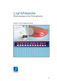
techniques. <strong>Laser</strong> needle acupuncture represents a new, painless method for primary optical<br />
stimulation of acupuncture points.1-14 <strong>Laser</strong> needles are not inserted in the skin; they are simply<br />
applied to the acupuncture point. This method allows the simultaneous stimulation of individually<br />
combined points.<br />
This study gives a current summary regarding scientific proof and innovative aspects of laser needle<br />
acupuncture. We discuss studies of the peripheral effects using registration of temperature and laser<br />
Doppler flowmetry8,9,13 as well as publications regarding the objectification of cerebral effects of laser<br />
needle acupuncture aided by functional multidirectional transcranial Doppler sonography,1-3,8,9,11,12<br />
functional magnetic resonance imaging (fMRI),11,12 and near infrared spectroscopy.2,4-6,8,9<br />
METHODS<br />
Temperature and Microcirculatory Monitoring<br />
The surface temperature of the skin and the measurement parameter Flux (= product of concentration<br />
and velocity of erythrocytes) were measured with the <strong>Laser</strong>-Doppler-Flowmetry Monitor DRT 4 (Moor<br />
Instruments, Millway, Axminster, England). A DPIT-probe (diameter, 8 mm; length, 7 mm) with a<br />
power of 1 mW was used. The edge frequencies were 20 Hz and 22.5 kHz.8,9,13<br />
Functional Multidirectional Transcranial Doppler Sonography<br />
The Multi-Dop T System (DWL Electronic Systems GmbH, Sipplin-gen, Germany) was used to<br />
measure the mean blood flow velocity in different cerebral arteries. A 4-MHz (ophthalmic artery), as<br />
well as 2-MHz probes (posterior cerebral artery, anterior cerebral artery , middle cerebral artery) were<br />
applied with a specially developed ultrasound probe-holding construction.<br />
Functional Magnetic Resonance Imaging (fMRI) The fMRI investigations were performed using a 1.5-T<br />
total body system (Intera, Philips Medical Systems, Best, Netherlands). The blood oxygen leveldependent<br />
contrast sensitive images were acquired with a T2-weighted gradient echo sequence<br />
(single shot planar readout, flip angle 90�, TE 50 ms, FOV 250 mm, matrix 96 x 96 interpolated at<br />
128 x 128, layer number 30, layer thickness 4 mm). A total of 144 volume images were registered<br />
continuously in succession, with a repetition time of 5 seconds.